KR20130028909A - 특정 해상도에서 적어도 하나의 해부학적 구조의 미세 영상을 제공하는 시스템, 방법 및 컴퓨터 접근 가능 매체 - Google Patents
특정 해상도에서 적어도 하나의 해부학적 구조의 미세 영상을 제공하는 시스템, 방법 및 컴퓨터 접근 가능 매체 Download PDFInfo
- Publication number
- KR20130028909A KR20130028909A KR1020127026132A KR20127026132A KR20130028909A KR 20130028909 A KR20130028909 A KR 20130028909A KR 1020127026132 A KR1020127026132 A KR 1020127026132A KR 20127026132 A KR20127026132 A KR 20127026132A KR 20130028909 A KR20130028909 A KR 20130028909A
- Authority
- KR
- South Korea
- Prior art keywords
- radiation
- arrangement
- wave
- transfer function
- guiding
- Prior art date
Links
Images


Classifications
-
- A—HUMAN NECESSITIES
- A61—MEDICAL OR VETERINARY SCIENCE; HYGIENE
- A61B—DIAGNOSIS; SURGERY; IDENTIFICATION
- A61B5/00—Measuring for diagnostic purposes; Identification of persons
- A61B5/06—Devices, other than using radiation, for detecting or locating foreign bodies ; determining position of probes within or on the body of the patient
-
- A—HUMAN NECESSITIES
- A61—MEDICAL OR VETERINARY SCIENCE; HYGIENE
- A61B—DIAGNOSIS; SURGERY; IDENTIFICATION
- A61B5/00—Measuring for diagnostic purposes; Identification of persons
- A61B5/0059—Measuring for diagnostic purposes; Identification of persons using light, e.g. diagnosis by transillumination, diascopy, fluorescence
- A61B5/0062—Arrangements for scanning
- A61B5/0066—Optical coherence imaging
-
- A—HUMAN NECESSITIES
- A61—MEDICAL OR VETERINARY SCIENCE; HYGIENE
- A61B—DIAGNOSIS; SURGERY; IDENTIFICATION
- A61B5/00—Measuring for diagnostic purposes; Identification of persons
- A61B5/0033—Features or image-related aspects of imaging apparatus classified in A61B5/00, e.g. for MRI, optical tomography or impedance tomography apparatus; arrangements of imaging apparatus in a room
- A61B5/004—Features or image-related aspects of imaging apparatus classified in A61B5/00, e.g. for MRI, optical tomography or impedance tomography apparatus; arrangements of imaging apparatus in a room adapted for image acquisition of a particular organ or body part
- A61B5/0044—Features or image-related aspects of imaging apparatus classified in A61B5/00, e.g. for MRI, optical tomography or impedance tomography apparatus; arrangements of imaging apparatus in a room adapted for image acquisition of a particular organ or body part for the heart
-
- A—HUMAN NECESSITIES
- A61—MEDICAL OR VETERINARY SCIENCE; HYGIENE
- A61B—DIAGNOSIS; SURGERY; IDENTIFICATION
- A61B5/00—Measuring for diagnostic purposes; Identification of persons
- A61B5/0059—Measuring for diagnostic purposes; Identification of persons using light, e.g. diagnosis by transillumination, diascopy, fluorescence
- A61B5/0075—Measuring for diagnostic purposes; Identification of persons using light, e.g. diagnosis by transillumination, diascopy, fluorescence by spectroscopy, i.e. measuring spectra, e.g. Raman spectroscopy, infrared absorption spectroscopy
-
- A—HUMAN NECESSITIES
- A61—MEDICAL OR VETERINARY SCIENCE; HYGIENE
- A61B—DIAGNOSIS; SURGERY; IDENTIFICATION
- A61B5/00—Measuring for diagnostic purposes; Identification of persons
- A61B5/0059—Measuring for diagnostic purposes; Identification of persons using light, e.g. diagnosis by transillumination, diascopy, fluorescence
- A61B5/0082—Measuring for diagnostic purposes; Identification of persons using light, e.g. diagnosis by transillumination, diascopy, fluorescence adapted for particular medical purposes
- A61B5/0084—Measuring for diagnostic purposes; Identification of persons using light, e.g. diagnosis by transillumination, diascopy, fluorescence adapted for particular medical purposes for introduction into the body, e.g. by catheters
-
- G—PHYSICS
- G01—MEASURING; TESTING
- G01B—MEASURING LENGTH, THICKNESS OR SIMILAR LINEAR DIMENSIONS; MEASURING ANGLES; MEASURING AREAS; MEASURING IRREGULARITIES OF SURFACES OR CONTOURS
- G01B9/00—Measuring instruments characterised by the use of optical techniques
- G01B9/02—Interferometers
-
- G—PHYSICS
- G01—MEASURING; TESTING
- G01B—MEASURING LENGTH, THICKNESS OR SIMILAR LINEAR DIMENSIONS; MEASURING ANGLES; MEASURING AREAS; MEASURING IRREGULARITIES OF SURFACES OR CONTOURS
- G01B9/00—Measuring instruments characterised by the use of optical techniques
- G01B9/02—Interferometers
- G01B9/0209—Low-coherence interferometers
- G01B9/02091—Tomographic interferometers, e.g. based on optical coherence
-
- G—PHYSICS
- G02—OPTICS
- G02B—OPTICAL ELEMENTS, SYSTEMS OR APPARATUS
- G02B6/00—Light guides; Structural details of arrangements comprising light guides and other optical elements, e.g. couplings
- G02B6/24—Coupling light guides
- G02B6/26—Optical coupling means
- G02B6/32—Optical coupling means having lens focusing means positioned between opposed fibre ends
Abstract
적어도 하나의 샘플에 적어도 하나의 전자기 방사선을 제공하기 위한 장치, 시스템 및 방법의 예시적인 실시예가 제공될 수 있다. 예를 들어, 복수의 웨이브-가이딩 배열체가 제공될 수 있으며, 상기 웨이브-가이딩 배열체는 i) 상기 적어도 하나의 전자기 방사선을 제공하며, ii) 각각의 상기 웨이브-가이딩 배열체의 방출 지점에서 각각의 상기 적어도 하나의 전자기 방사선의 위상이 미리 정해진 값을 갖게 하도록 구성된다. 상기 예시적인 장치는 프로브의 일부일 수 있다. 또한, 상기 예시적인 장치는 상기 프로브와 통신하도록 제공된 간섭계 배열체를 포함할 수 있으며/있거나 상기 프로브의 일부일 수 있다.
Description
관련 출원에 대한 교차 참조
본 출원은 모두 2010년 3월 5일자로 출원된 미국 특허 출원 제61/311,171호 및 제61/311,272호로부터의 우선권의 이익에 기반하고 그 우선권의 이익을 주장하며, 그들의 전체적인 개시 내용은 본 명세서에 참조로서 통합된다.
개시의 분야
본 발명은 촬영 시스템, 장치 및 방법의 예시적인 실시예에 관한 것이며, 특히 특정 해상도에서 적어도 하나의 해부학적 구조의 미세 영상을 제공하는 방법, 시스템 및 컴퓨터 접근 가능 매체에 관한 것이다.
심장 마비 또는 급성 심근 경색(acute myocardial infarction(AMI))을 포함하는 관상 동맥 질병(coronary artery disease(CAD)) 및 그에 대한 임상 징후는 일년에 거의 500,000 명의 생명을 앗아가며 대략 4천억불의 비용을 필요로 하는 미국에서의 첫째의 사망 원인이다. 따라서, 관상 동맥의 아테롬성 동맥 경화 병변(coronary atherosclerotic lesion), 플라크 파열(plaque rupture) 및 관상 동맥 혈전증(coronary thrombosis)의 발병 및 진전과 같은 CAD의 병리심리학 및 관상 동맥 장치 및 약리학 치료법에 대한 동맥 응답(arterial response)에 관련된 토픽이 오늘날 아주 중요하다. 이들 생물학적 프로세스는 미세 스케일로 발생하는 분자 및 세포 이벤트에 의해 중재될 수 있다. CAD를 이해하며 진단하고 치료하는 데 있어서의 소정의 발전이 세포-레벨 해상도로 체내에서 인간 관상 동맥 벽을 검사하는 것이 어렵거나 불가능했었다는 사실에 의해 저해되어 왔었다.
지난 십 년에 걸쳐, 관상 동맥 벽으로부터의 반사된 광의 단면 영상을 획득하는 카테터-기반 기법인 관상 내 광 간섭 단층촬영(intracoronary optical coherence tomography(intracoronary OCT))이 개발되었다. 관상 내 OCT는 10 ㎛의 공간 해상도를 가지며, 이는 이전의 관상 동맥 촬영 방법인 혈관 내 초음파(intravascular ultrasound(IVUS))의 공간 해상도보다 한 자릿수 더 양호하다. 부모 R01에서, 매우 높은 영상 획득률을 가지면서 관상 혈관(coronary vessel)의 고 해상도 3차원 촬영을 수행하는 것을 가능하게 하는 OCT의 제 2 세대 형태, 즉 광학 주파수 도메인 촬영(optical frequency domain imaging(OFDI))로 지칭되는 OCT의 제 2 세대 형태가 개발되었다. 또한, 높은 프레임률의 OFDI와 함께 OCT 신호를 이용하여 혈액 간섭(blood interference)의 장애 중 적어도 일부의 장애를 극복할 수 있는 플러싱 방법(flushing method)이 개발되었다. 직접적인 결과로서, 임상 설정에서 관상 내 OCT 과정을 수행하는 것이 바람직할 수 있다. 실제로, OCT에 대한 소정 인터벤션 심장 애플리케이션(interventional cardiology application)이 개발되었으며, 그 분야를 기하급수적으로 성장시키고 있다. OCT가 관상 인터벤션(coronary intervention)을 전세계적으로 안내하기 위한 중요한 촬영 양상(imaging modality)이 될 수 있다고 믿게 되었다.
부모 R01에서 개발된 기술이 상업적인 OFDI 촬영 시스템의 분포를 통해 임상 실험을 위해 번역되어 가능하게 되었으므로, CAD의 발병에 관련된 고분자 및 세포를 검토하는 것이 바람직할 수 있다.
예를 들어, OCT 과정 내의 횡방향 해상도가 카테터의 초점 스폿 크기(focal spot size)에 의해 결정될 수 있다. 해상도를 향상시키기 위해, 광을 샘플 내로 집속시키는 렌즈의 개구수(numerical aperture)를 증가시키는 것이 가능할 수 있다. 그러나, 이러한 통상적인 방법은 단면 OCT 영상 내의 필드의 깊이와 횡방향 해상도 사이의 본질적인 타협을 무시하며, 단지 좁은 깊이 범위만이 분해되는 영상을 야기한다.
대안적인 접근은 증가된 필드 깊이(depth-of-field)에 걸쳐 높은 횡방향 해상도를 생성하기 위해 베셀(Bessel) 또는 "비회절(non-diffracting)" 빔의 독특한 특징을 이용할 수 있다. 그러나, 샘플로부터 반사된 광의 베셀 빔 조명 및 검출은 콘트라스트 및 검출 효율에서의 상당한 감소를 겪을 수 있다. 따라서, 전술된 통상적인 배열체 및 방법에 연관된 결점 중 적어도 일부 결점을 극복할 필요가 존재할 수 있다.
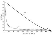
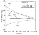
본 명세서에서 간단하게 전술된 바와 같이, 본 개시사항의 소정 예시적인 실시예는 예시적인 OCT 시스템의 간섭 전달 함수(coherent transfer function(CTF))의 분석 및 조작에 연관될 수 있으며/있거나 그 분석 및 조작을 이용할 수 있다. 대신에, 본 발명은 OCT 시스템의 간섭 전달 함수(CTF)의 분석 및 조작에 기반한다. CTF는 변조 전달 함수(modulation transfer function(MTF)) 및 광학 전달 함수(optical transfer function(OTF))의 간섭 확장(coherent extension)으로 간주될 수 있다. 따라서, 예를 들어 비간섭 시스템(non-interferometric system)에 대해, 소정 예시적인 실시예에 따라 MTF 또는 OTF가 조작되며 이용될 수 있다. 일반적으로, 광학 시스템의 품질은 그 전달 함수를 회절-제한형 광학 시스템(diffraction-limited optical system)의 전달 함수와 비교함으로써 평가될 수 있다. 도 1은 베셀 빔 조명 및 검출에 의해 생성된, 예를 들어 2.0 mm의 확장형 초점 범위(extended focal range)를 갖는 회절 제한형 2.5 ㎛ 직경 스폿 및 2.5 ㎛ 스폿에 대한 간섭 전달 함수(CTF)를 도시한다. 도 1에 예시된 바와 같이, 비록 베셀 빔 조명 및 검출(100)의 전달 함수가 낮은 범위의 그리고 중간 범위의 공간 주파수를 희생할 것 같기는 하지만, 베셀 빔 조명 및 검출(100)의 전달 함수는 회절 제한형 시스템(110)을 초과하는 공간 주파수를 가질 수 있으며, 아마도 감소된 콘트라스트 및 검출 민감도를 야기할 수 있다.
따라서, 전술된 통상적인 배열체 및 방법에 연관된 결점 중 적어도 일부의 결점을 극복할 필요가 존재할 수 있다.
이러한 결함을 처리하며/하거나 극복하기 위해서, 본 개시사항의 목적 중 하나는 특정 해상도에서 적어도 하나의 해부학적 구조의 미세 영상을 제공할 수 있는 본 개시사항에 따른 시스템, 방법 및 컴퓨터 접근 가능 매체의 예시적인 실시예를 제공하는 것이다. 본 개시사항의 또 다른 목적은 OCT 과정 및/또는 시스템 및 다른 형태의 확장된 초점 깊이 촬영을 위한 베셀 빔 시스템의 공간 주파수 손실 및 통상적인 가우시안 빔의 제한된 초점 깊이 제한을 극복하는 것이다.
본 개시사항의 또 다른 예시적인 실시예에 따르면, 두 개 초과의 촬영 채널이 상이한 베셀 및/또는 가우시안 빔을 조명/검출할 수 있다. 추가적인 예시적인 실시예에서, 상이한 전달 함수가 조명되며/되거나 검출될 수 있다. 이러한 추가 예시적인 빔을 이용하여 획득된 영상의 예시적인 조합은 μOCT CTF가 회절-제한형 경우에 제공되기에 용이하게 할 수 있으며, 또한 필드의 깊이 확장을 훨씬 더 용이하게 할 수 있다.
따라서, 적어도 하나의 샘플에 적어도 하나의 전자기 방사선을 제공하기 위한 장치, 시스템 및 방법의 예시적인 실시예가 제공될 수 있다. 예를 들어, 복수의 웨이브-가이딩 배열체가 제공될 수 있으며, 상기 웨이브-가이딩 배열체는 i) 상기 전자기 방사선을 제공하며, ii) 각각의 상기 웨이브-가이딩 배열체의 방출 지점에서 각각의 상기 적어도 하나의 전자기 방사선의 위상이 미리 정해진 값을 갖게 하도록 구성된다. 상기 예시적인 장치는 프로브의 일부일 수 있다. 또한, 상기 예시적인 장치는 상기 프로브와 통신하도록 제공된 간섭계 배열체를 포함할 수 있으며/있거나 상기 프로브의 일부일 수 있다.
본 개시사항의 또 다른 예시적인 실시예에서, 상기 웨이브-가이딩 배열체는 상기 방사선을 적어도 부분적으로 원형 패턴으로 제공할 수 있다. 상기 웨이브-가이딩 배열체로부터 상기 전자기 방사선을 수신하며 추가적인 초점-스폿 방사선을 생성하도록 구성되는 적어도 하나의 렌즈 배열체가 포함될 수 있다. 상기 렌즈 배열체는 상기 추가적인 초점-스폿 방사선이 (i) 확장된 초점 깊이를 갖게 하며/하거나 (ii) 상기 샘플 상의 또는 상기 샘플 내의 회절 제한 스폿(diffraction limited spot)보다 작은 직경을 갖게 하도록 구성될 수 있다. 상기 회절 제한 스폿은 3차원 스폿일 수 있다. 추가적으로 또는 대안적으로, 상기 렌즈 배열체는 그린 렌즈(grin lens)를 포함할 수 있다.
본 개시사항의 또 다른 예시적인 실시예에 따르면, 상기 웨이브-가이딩 배열체 중 적어도 하나의 웨이브-가이딩 배열체는 (i) 단일 모드 도파관(single-mode wave guide)일 수 있으며/있거나 (ii) 광중합체(photopolymer)로 구성될 수 있다. 추가적으로, 상기 샘플에 추가적인 전자기 방사선을 제공하도록 구성되는 추가적인 웨이브-가이딩 배열체가 제공될 수 있으며, 상기 전자기 방사선 및 상기 추가적인 전자기 방사선은 상기 샘플의 적어도 부분적으로 중첩하는 부분에 제공될 수 있다. 상기 웨이브-가이딩 배열체를 적어도 부분적으로 둘러싸는 하우징이 또한 제공될 수 있으며/있거나 상기 하우징을 둘러싸는 피복이 제공될 수 있다. 추가로, 상기 하우징을 회전시키며/회전시키거나 병진시키도록 구성되는 제어 배열체가 제공될 수 있다. 상기 렌즈 배열체는 광중합체 처리에 의해 형성되며/형성되거나 광중합체 처리를 받은 적어도 하나의 광학 엘리먼트를 포함할 수 있다. 상기 광중합체 처리는 상기 광학 엘리먼트를 형성하기 위해 광중합체를 조사(irradiating)하는 단계를 포함할 수 있다.
본 개시사항의 추가적인 예시적인 실시예에서, 샘플의 적어도 일부분에 연관된 데이터를 생성하기 위한 방법 및 시스템이 제공될 수 있다. 예를 들어, 광중합체 처리에 의해 형성되거나 상기 광중합체 처리를 받은 적어도 하나의 광 배열체를 통해 상기 샘플의 상기 부분으로 적어도 하나의 제 1 방사선을 전달될 수 있다. 상기 부분으로부터 상기 적어도 하나의 제 1 방사선에 기반하는 적어도 하나의 제 2 방사선이 수신될 수 있다. 상기 적어도 하나의 광 배열체와 상기 제 1 방사선 및/또는 상기 제 2 방사선 사이의 상호작용에 기반하여, 상기 광 배열체는 제 1 전달 함수를 가질 수 있다. 그런 다음, 상기 광 배열체를 통해 상기 부분으로 적어도 하나의 제 3 방사선이 전달될 수 있다. 상기 부분으로부터 상기 적어도 하나의 제 3 방사선에 기반할 수 있는 적어도 하나의 제 4 방사선이 수신될 수 있다. 상기 광 배열체와 상기 제 3 방사선 및/또는 상기 제 4 방사선 사이의 상호작용에 기반하여, 상기 광 배열체는 제 2 전달 함수를 가질 수 있으며, 상기 제 1 전달 함수는 상기 제 2 전달 함수와 적어도 부분적으로 상이할 수 있다. 추가로, 상기 제 2 방사선 및 상기 제 4 방사선에 기반하여 상기 부분에 연관된 상기 데이터가 생성될 수 있다. 상기 제 1 광 배열체 및/또는 상기 제 2 광 배열체는 광중합체 처리에 의해 형성될 수 있으며/있거나 상기 광중합체 처리를 받을 수 있다.
본 개시사항의 예시적인 실시예의 이들 목적, 특징 및 장점과 기타 목적, 특징 및 장점은 첨부된 특허청구범위와 함께 해석될 때 본 개시사항의 예시적인 실시예의 이하의 상세한 설명을 판독할 때에 명백해질 것이다.
본 발명은 특정 해상도에서 적어도 하나의 해부학적 구조의 미세 영상을 제공할 수 있는 본 개시사항에 따른 시스템, 방법 및 컴퓨터 접근 가능 매체의 예시적인 실시예를 제공하는 효과가 있다.
또한 본 발명은 OCT 과정 및/또는 시스템 및 다른 형태의 확장된 초점 깊이 촬영을 위한 베셀 빔 시스템의 공간 주파수 손실 및 통상적인 가우시안 빔의 제한된 초점 깊이 제한을 극복하는 효과가 있다.
더 나아가 본 발명은 두 개 초과의 촬영 채널이 상이한 베셀 및/또는 가우시안 빔을 조명/검출할 수 있고, 상이한 전달 함수가 조명되며/되거나 검출될 수 있는 효과가 있다. 이러한 추가 예시적인 빔을 이용하여 획득된 영상의 예시적인 조합은 μOCT CTF가 회절-제한형 경우에 제공되기에 용이하게 할 수 있으며, 또한 필드의 깊이 확장을 훨씬 더 용이하게 할 수 있다.
본 발명의 추가적인 목적, 특징 및 장점은 본 개시사항의 예시적인 실시예를 도시하는 첨부 도면과 함께 해석되는 다음의 상세한 설명으로부터 명백해질 것이다.
도 1은 종래의 베셀 빔 조명 및 검출에 의해 생성된 공간 주파수의 함수로서의 간섭 전달 함수(coherent transfer function(CTF))의 예시적인 그래프이다.
도 2는 본 개시사항에 따른 과정 및/또는 기법의 예시적인 실시예에 의해 생성된 공간 주파수의 함수로서의 간섭 전달 함수(CTF)의 예시적인 그래프이다.
도 3A는 본 개시사항의 예시적인 실시예에 따른 예시적인 과정/기법을 사용하여 획득된 사체 관상 동맥 플라크의 제 1 예시적인 OCT 영상이며, 예시적인 가우스-가우스 영상은 낮은 공간 주파수 정보를 포함한다.
도 3B는 본 개시사항의 예시적인 실시예에 따른 예시적인 과정/기법을 사용하여 획득된 사체 관상 동맥 플라크의 제 2 예시적인 OCT 영상이며, 예시적인 베셀-베셀 영상은 높은 해상도를 제공하지만 낮은 공간 주파수 및 중간 공간 주파수를 상실한다.
도 3C는 본 개시사항의 예시적인 실시예에 따른 예시적인 과정/기법을 사용하여 획득된 사체 관상 동맥 플라크의 제 3 예시적인 OCT 영상이며, 이는 조합 μOCT 영상(예를 들어, 가우스-가우스 + 가우스-베셀 + 베셀-베셀 영상)을 제공하며, 영상은 동일한 휘도/콘트라스트 값을 이용하여 정규화(normalization)되고 디스플레이된다.
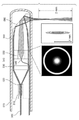
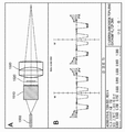
도 4A는 본 개시사항의 예시적인 실시예에 따른 OCT 카테터 시스템의 말단 광학의 다이어그램의 측부 절개도이다.
도 4B는 도 4A에 도시된 예시적인 실시예에 따른 시스템의 Y-정션 팬-아웃(Y-junction fan-out)을 사용하여 생성된 중합체 인덱스 프로파일(polymer index profile)의 예시적인 그래프이다.
도 4C는 도 4A에 도시된 예시적인 실시예에 따른 시스템의 Y-정션 팬-아웃을 사용하여 생성된 조명 프로파일 예시적인 그래프이다.
도 4D는 도 4A에 도시된 예시적인 실시예에 따른 시스템의 Y-정션 팬 아웃을 사용하여 생성된 시뮬레이션된 x-z PSF의 예시적인 그래프이다.
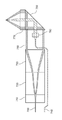
도 5A는 본 개시사항의 또 다른 예시적인 실시예에 따른 OCT 카테터 시스템의 말단 광학의 다이어그램의 측부 절개도이다.
도 5B는 도 5A에 도시된 예시적인 실시예에 따른 시스템의 말단 광학 구성을 사용하여 생성된 조명 프로파일의 예시적인 그래프이다.
도 5C는 도 5A에 도시된 예시적인 실시예에 따른 시스템의 말단 광학 구성을 사용하여 생성된 시뮬레이션된 x-z PSF의 예시적인 그래프이다.
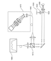
도 6은 본 개시사항의 추가적인 예시적인 실시예에 따른 하나 이상의 μOCT 영상을 생성하기 위한 시스템의 개략적인 다이어그램이다.
도 7은 말단 광학 구성의 가우시안 빔 및 링 빔의 라우팅 및 액시콘 쌍을 포함하는 본 개시사항의 또 다른 예시적인 실시예에 따른 OCT 카테터 시스템의 말단 광학의 다이어그램의 측부 절개도이다.
도 8은 단일 섬유 및 단일 액시콘 렌즈(axicon lens)를 사용하는 예시적인 광 경로길이 인코딩 프로브 구성을 포함하는 본 개시사항의 추가적인 예시적인 실시예에 따른 OCT 카테터 시스템의 다이어그램의 측부 절개도이다.
도 9은 단일 섬유 및 단일 액시콘 렌즈를 사용하는 추가적인 예시적인 광 경로길이 인코딩 프로브 구성을 포함하는 본 개시사항의 추가적인 예시적인 실시예에 따른 OCT 카테터 시스템의 다이어그램의 측부 절개도이다.
도 10은 단일 섬유 다초점 렌즈 프로브 구성을 포함하는 본 개시사항의 추가적인 예시적인 실시예에 따른 OCT 카테터 시스템의 말단 광학의 다이어그램의 개략도이다.
도 11은 미러 터널을 이용하는 본 개시사항의 추가적인 예시적인 실시예에 따른 OCT 카테터 시스템의 다이어그램의 측부 절개도이다.
도 12는 반사형 무색성 위상 마스크 및 볼 렌즈를 이용하는 본 개시사항의 추가적인 예시적인 실시예에 따른 OCT 카테터 시스템의 일부의 다이어그램의 측부 절개도이다.
도 13은 도 12의 예시적인 실시예에 기반하여 유리-금속 인터페이스에서의 반사 시에 색채 광의 위상 변이 스펙트럼의 그래프이다.
도 14A는 통상적인 집속을 갖는 호이겐스 회절 패턴의 예시이다.
도 14B는 도 13에 예시된 시스템의 예시적인 실시예에서 설명된 볼 렌즈 및 반사형 무색성 위상 마스크를 갖는 렌즈의 호이겐스 회절 패턴의 예시적인 예시이다.
도 15A는 본 개시사항의 예시적인 실시예에 따라 굴절형 무색성 위상 더블릿 마스크를 사용하는 집속 배열체의 예시적인 실시예의 개략적인 다이어그램이다.
도 15B는 도 15A에 예시된 예시적인 마스크의 횡방향 위상 프로파일의 예시적인 그래프이다.
도 16은 본 개시사항의 또 다른 예시적인 실시예에 따라 파면 빔 스플리터 및 공통 경로 간섭계를 포함하는 OCT 시스템의 개략적인 다이어그램이다.
도 17A는 단색성 광원 (예를 들어, λ=825 nm) 및 구면 수차 제거 대물 렌즈를 사용하는 도 16에 도시된 예시적인 OCT 시스템에 의해 생성된 예시적인 시뮬레이션된 PSF 예시이다.
도 17B는 단색성 광원 (예를 들어, λ=825 nm) 및 구면 수차와 파장 의존형 초점 변이를 갖는 대물 렌즈를 사용하는 도 16에 도시된 예시적인 OCT 시스템에 의해 생성된 예시적인 시뮬레이션된 PSF 예시이다.
도 17C는 광대역 소스 (예를 들어, 대략 600 nm 내지 1050 nm) 및 구면 수차와 파장 의존형 초점 변이를 갖는 대물 렌즈를 사용하는 도 16에 도시된 예시적인 OCT 시스템에 의해 생성된 예시적인 시뮬레이션된 PSF 예시이다.
도 17D는 광대역 소스 (예를 들어, 대략 600 nm 내지 1050 nm), 구면 수차와 파장 의존형 초점 변이를 갖는 대물 렌즈, 및 파면 빔 스플리터를 사용하는 도 16에 도시된 예시적인 OCT 시스템에 의해 생성된 예시적인 시뮬레이션된 PSF 예시이다.
도 18A는 복수의 백혈구(화살표)를 도시하는 관상 동맥 플라크의 예시적인 μOCT 영상이다.
도 18B는 두 개의 상이한 세포 유형의 복수의 백혈구(화살표)를 예시하는 관상 동맥 플라크의 예시적인 μOCT 영상이며, 하나의 유형은 림프구(L)와 일치하는 스캔트 세포질(scant cytoplasm)을 갖는 보다 작은 세포이고 또 다른 하나의 유형은 단핵구(M)를 나타내는 고산란 세포질을 갖는 보다 큰 세포이다.
도 18C는 단핵구의 들쑥날쑥한 콩 형상 핵(M) 특성을 갖는 세포를 예시하는 관상 동맥 플라크의 예시적인 μOCT 영상이다.
도 18D는 멀티-로비드 핵을 갖는 백혈구를 예시하는 관상 동맥 플라크의 예시적인 μOCT 영상이며, 멀티-로비드 핵은 내피면에 부착된 뉴트로필(N)을 나타낼 수 있다.
도 18E는 위족에 의해 내피면에 묶여진 복수의 백혈구를 예시하는 관상 동맥 플라크의 예시적인 μOCT 영상이다.
도 18F는 단면 및 삽화에서 내피를 통해 이주하는 단핵구(M)의 형태학을 갖는 세포를 예시하는 관상 동맥 플라크의 예시적인 μOCT 영상이다.
도 18G는 내피면 상에 분포된 복수의 백혈구의 예시적인 μOCT 영상이다.
도 19A는 뉴트로필(N)의 백혈구 특성에 인접한 혈소판(P)의 예시적인 μOCT 영상이며, 뉴트로필도 또한 작은 혈소판에 부착되어 있다.
도 19B는 관상 동맥 벽 내의 갭을 메우는 선형 스트랜드로서 보일 수 있는 피브린(F)의 예시적인 μOCT 영상이다.
도 19C는 도 19B에 도시된 것에 인접한 위치에 피브린에 인접한 한 무리의 백혈구(L)의 예시적인 μOCT 영상이다.
도 19D는 복수의 옭아매진 백혈구를 갖는 피브린 혈전(T)의 예시적인 μOCT 영상이다.
도 19E는 백혈구 및 피브린 스트랜드를 도시하는 보다 진전된 혈전(T)의 예시적인 μOCT 영상이다.
도 20A는 배양 조직 내의 내피 셀의 예시적인 μOCT 단면 영상이다.
도 20B는 배양 조직 내의 내피 셀의 예시적인 μOCT 정면 영상이다.
도 20C는 토종 돼지 관상 동맥 단면의 예시적인 μOCT 영상이다.
도 20D는 내피 "포상형성(pavementing)"을 입증하는 돼지 관상 동맥의 예시적인 3차원 렌더링이다.
도 21A는 섬유 캡(fibrous cap)의 μOCT 영상 내에 밝은 밀도로서 보여질 수 있는 미세석회화(microcalcification)의 예시적인 예시적인 μOCT 영상이다.
도 21B는 상응하는 히스톨로지(histology) 상에 어두운 밀도로서 보여질 수 있는 미세석회화의 예시적인 μOCT 영상이다.
도 22A는 손상된 내막/내피를 입증하는 대형 칼슘 결절(nodule)의 예시적인 μOCT 영상이다.
도 22B는 대치하는 분리된 내막에 비보호형 칼슘(백색 화살표)을 인접하면서 피브린(F)과 일치하는 미세 조직 스트랜드를 예시하는 박스에 의해 둘러싸인 구역의 확대도이다.
도 22C는 피브린(F, 흑색 화살표) 및 탈피된 석회화 면(회색 화살표)의 상응하는 히스톨로지의 예시이다.
도 23A는 상부면 및 하부면으로부터의 반사에 의해 특성화된 두꺼운 콜레스테롤 결정(cholesterol crystal(CC))을 입증하는 대형 괴사 코어(necrotic core(NC)) 섬유선종(fibroatheroma)의 예시적인 μOCT 영상이다.
도 23B는 삽화 내에 보다 상세하게 도시된 또 다른 괴사 코어 플라크(NC)의 캡을 관통하는 얇은 결정(CC, 회색 화살표)의 예시적인 μOCT 영상이다.
도 24A는 낮은 후방산란 스핀들 형상 세포(삽화)로서 나타낸 다양한 민무늬근 세포의 예시적인 μOCT 영상이다.
도 24B는 스핀들 형상이면서 높은 후방산란 인테리어(옅은 회색 화살표) 및 낮은 후방산란의 "후광(halo)"(백색 화살표)을 갖는, 콜라겐을 생성하는 민무늬근 세포의 예시적인 μOCT 영상이며, 이는 각각 세포체 및 콜라겐 매트릭스를 표시한다(히스톨로지 삽화).
도 25A는 중합체/약을 구비하거나/구비하지 않은 상태에서 탁수스 리베르테(Taxus Liberte) 스트럿의 예시적인 μOCT 영상이며, 즉, 중합체-코팅된 스트럿에 대해, 중합체 반사(polymer reflection(PR)), 스트럿 반사(strut reflection(SR)) 및 복수의 반사(MR1, MR2)가 보여질 수 있다.
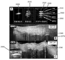
도 25B는 네오인티마에 의해 피복된 중합체가 없는 스트럿을 도시하는 임플란트형 BMS를 갖는 사체 관상 동맥 표본의 예시적인 μOCT 영상이다.
도 25C는 스트럿 반사(P, 삽화) 위에 중첩하는 중합체를 도시하는 또 다른 사체로부터 임플란트형 DES 스트럿을 갖는 사체 관상 동맥 표본의 예시적인 μOCT 영상이다.
도 26A는 조직(옅은 회색 화살표)이 스텐트 스트럿(stent strut)으로부터 중합체를 분리시켰고 중합체가 균열되었다는(백색 화살표) 것을 도시하는 예시적인 μOCT 영상이다.
도 26B는 중합체 균열의 위치 위에 중첩하는 표층 백혈구 클러스터(적색 화살표) 및 인접한 부착된 백혈구를 예시하는 예시적인 μOCT 영상이다.
도 26C는 또 다른 환자로부터의 스트럿(점선 구역)의 에지에서 염증을 예시하는 예시적인 μOCT 영상이다.
도 26D는 중첩하는 내피가 완전히 없는(삽화), 비피복형 스트럿을 예시하는 예시적인 μOCT 영상이다.
도 27a는 본 개시사항의 하나의 예시적인 실시예에 따른 공정의 흐름도이다.
도 27b는 본 개시사항의 또 다른 예시적인 실시예에 따른 공정의 흐름도이다.
도면을 통해, 동일한 참조 부호 및 문자는 달리 설명되지 않는 한 예시된 실시예의 유사한 특징, 엘리먼트, 컴포넌트 또는 일부분을 표시하기 위해 사용된다. 또한, 이제 이하의 개시사항이 도면을 참조하여 설명되지만, 그 개시사항은 예시적인 실시예와 관련하여 설명된다. 첨부된 특허청구범위에 의해 정의된 바와 같은 이하의 개시사항의 진정한 범위와 사상으로부터 벗어나지 않으면서 설명된 예시적인 실시예가 변경 및 수정될 수 있도록 의도된다.
도 1은 종래의 베셀 빔 조명 및 검출에 의해 생성된 공간 주파수의 함수로서의 간섭 전달 함수(coherent transfer function(CTF))의 예시적인 그래프이다.
도 2는 본 개시사항에 따른 과정 및/또는 기법의 예시적인 실시예에 의해 생성된 공간 주파수의 함수로서의 간섭 전달 함수(CTF)의 예시적인 그래프이다.
도 3A는 본 개시사항의 예시적인 실시예에 따른 예시적인 과정/기법을 사용하여 획득된 사체 관상 동맥 플라크의 제 1 예시적인 OCT 영상이며, 예시적인 가우스-가우스 영상은 낮은 공간 주파수 정보를 포함한다.
도 3B는 본 개시사항의 예시적인 실시예에 따른 예시적인 과정/기법을 사용하여 획득된 사체 관상 동맥 플라크의 제 2 예시적인 OCT 영상이며, 예시적인 베셀-베셀 영상은 높은 해상도를 제공하지만 낮은 공간 주파수 및 중간 공간 주파수를 상실한다.
도 3C는 본 개시사항의 예시적인 실시예에 따른 예시적인 과정/기법을 사용하여 획득된 사체 관상 동맥 플라크의 제 3 예시적인 OCT 영상이며, 이는 조합 μOCT 영상(예를 들어, 가우스-가우스 + 가우스-베셀 + 베셀-베셀 영상)을 제공하며, 영상은 동일한 휘도/콘트라스트 값을 이용하여 정규화(normalization)되고 디스플레이된다.
도 4A는 본 개시사항의 예시적인 실시예에 따른 OCT 카테터 시스템의 말단 광학의 다이어그램의 측부 절개도이다.
도 4B는 도 4A에 도시된 예시적인 실시예에 따른 시스템의 Y-정션 팬-아웃(Y-junction fan-out)을 사용하여 생성된 중합체 인덱스 프로파일(polymer index profile)의 예시적인 그래프이다.
도 4C는 도 4A에 도시된 예시적인 실시예에 따른 시스템의 Y-정션 팬-아웃을 사용하여 생성된 조명 프로파일 예시적인 그래프이다.
도 4D는 도 4A에 도시된 예시적인 실시예에 따른 시스템의 Y-정션 팬 아웃을 사용하여 생성된 시뮬레이션된 x-z PSF의 예시적인 그래프이다.
도 5A는 본 개시사항의 또 다른 예시적인 실시예에 따른 OCT 카테터 시스템의 말단 광학의 다이어그램의 측부 절개도이다.
도 5B는 도 5A에 도시된 예시적인 실시예에 따른 시스템의 말단 광학 구성을 사용하여 생성된 조명 프로파일의 예시적인 그래프이다.
도 5C는 도 5A에 도시된 예시적인 실시예에 따른 시스템의 말단 광학 구성을 사용하여 생성된 시뮬레이션된 x-z PSF의 예시적인 그래프이다.
도 6은 본 개시사항의 추가적인 예시적인 실시예에 따른 하나 이상의 μOCT 영상을 생성하기 위한 시스템의 개략적인 다이어그램이다.
도 7은 말단 광학 구성의 가우시안 빔 및 링 빔의 라우팅 및 액시콘 쌍을 포함하는 본 개시사항의 또 다른 예시적인 실시예에 따른 OCT 카테터 시스템의 말단 광학의 다이어그램의 측부 절개도이다.
도 8은 단일 섬유 및 단일 액시콘 렌즈(axicon lens)를 사용하는 예시적인 광 경로길이 인코딩 프로브 구성을 포함하는 본 개시사항의 추가적인 예시적인 실시예에 따른 OCT 카테터 시스템의 다이어그램의 측부 절개도이다.
도 9은 단일 섬유 및 단일 액시콘 렌즈를 사용하는 추가적인 예시적인 광 경로길이 인코딩 프로브 구성을 포함하는 본 개시사항의 추가적인 예시적인 실시예에 따른 OCT 카테터 시스템의 다이어그램의 측부 절개도이다.
도 10은 단일 섬유 다초점 렌즈 프로브 구성을 포함하는 본 개시사항의 추가적인 예시적인 실시예에 따른 OCT 카테터 시스템의 말단 광학의 다이어그램의 개략도이다.
도 11은 미러 터널을 이용하는 본 개시사항의 추가적인 예시적인 실시예에 따른 OCT 카테터 시스템의 다이어그램의 측부 절개도이다.
도 12는 반사형 무색성 위상 마스크 및 볼 렌즈를 이용하는 본 개시사항의 추가적인 예시적인 실시예에 따른 OCT 카테터 시스템의 일부의 다이어그램의 측부 절개도이다.
도 13은 도 12의 예시적인 실시예에 기반하여 유리-금속 인터페이스에서의 반사 시에 색채 광의 위상 변이 스펙트럼의 그래프이다.
도 14A는 통상적인 집속을 갖는 호이겐스 회절 패턴의 예시이다.
도 14B는 도 13에 예시된 시스템의 예시적인 실시예에서 설명된 볼 렌즈 및 반사형 무색성 위상 마스크를 갖는 렌즈의 호이겐스 회절 패턴의 예시적인 예시이다.
도 15A는 본 개시사항의 예시적인 실시예에 따라 굴절형 무색성 위상 더블릿 마스크를 사용하는 집속 배열체의 예시적인 실시예의 개략적인 다이어그램이다.
도 15B는 도 15A에 예시된 예시적인 마스크의 횡방향 위상 프로파일의 예시적인 그래프이다.
도 16은 본 개시사항의 또 다른 예시적인 실시예에 따라 파면 빔 스플리터 및 공통 경로 간섭계를 포함하는 OCT 시스템의 개략적인 다이어그램이다.
도 17A는 단색성 광원 (예를 들어, λ=825 nm) 및 구면 수차 제거 대물 렌즈를 사용하는 도 16에 도시된 예시적인 OCT 시스템에 의해 생성된 예시적인 시뮬레이션된 PSF 예시이다.
도 17B는 단색성 광원 (예를 들어, λ=825 nm) 및 구면 수차와 파장 의존형 초점 변이를 갖는 대물 렌즈를 사용하는 도 16에 도시된 예시적인 OCT 시스템에 의해 생성된 예시적인 시뮬레이션된 PSF 예시이다.
도 17C는 광대역 소스 (예를 들어, 대략 600 nm 내지 1050 nm) 및 구면 수차와 파장 의존형 초점 변이를 갖는 대물 렌즈를 사용하는 도 16에 도시된 예시적인 OCT 시스템에 의해 생성된 예시적인 시뮬레이션된 PSF 예시이다.
도 17D는 광대역 소스 (예를 들어, 대략 600 nm 내지 1050 nm), 구면 수차와 파장 의존형 초점 변이를 갖는 대물 렌즈, 및 파면 빔 스플리터를 사용하는 도 16에 도시된 예시적인 OCT 시스템에 의해 생성된 예시적인 시뮬레이션된 PSF 예시이다.
도 18A는 복수의 백혈구(화살표)를 도시하는 관상 동맥 플라크의 예시적인 μOCT 영상이다.
도 18B는 두 개의 상이한 세포 유형의 복수의 백혈구(화살표)를 예시하는 관상 동맥 플라크의 예시적인 μOCT 영상이며, 하나의 유형은 림프구(L)와 일치하는 스캔트 세포질(scant cytoplasm)을 갖는 보다 작은 세포이고 또 다른 하나의 유형은 단핵구(M)를 나타내는 고산란 세포질을 갖는 보다 큰 세포이다.
도 18C는 단핵구의 들쑥날쑥한 콩 형상 핵(M) 특성을 갖는 세포를 예시하는 관상 동맥 플라크의 예시적인 μOCT 영상이다.
도 18D는 멀티-로비드 핵을 갖는 백혈구를 예시하는 관상 동맥 플라크의 예시적인 μOCT 영상이며, 멀티-로비드 핵은 내피면에 부착된 뉴트로필(N)을 나타낼 수 있다.
도 18E는 위족에 의해 내피면에 묶여진 복수의 백혈구를 예시하는 관상 동맥 플라크의 예시적인 μOCT 영상이다.
도 18F는 단면 및 삽화에서 내피를 통해 이주하는 단핵구(M)의 형태학을 갖는 세포를 예시하는 관상 동맥 플라크의 예시적인 μOCT 영상이다.
도 18G는 내피면 상에 분포된 복수의 백혈구의 예시적인 μOCT 영상이다.
도 19A는 뉴트로필(N)의 백혈구 특성에 인접한 혈소판(P)의 예시적인 μOCT 영상이며, 뉴트로필도 또한 작은 혈소판에 부착되어 있다.
도 19B는 관상 동맥 벽 내의 갭을 메우는 선형 스트랜드로서 보일 수 있는 피브린(F)의 예시적인 μOCT 영상이다.
도 19C는 도 19B에 도시된 것에 인접한 위치에 피브린에 인접한 한 무리의 백혈구(L)의 예시적인 μOCT 영상이다.
도 19D는 복수의 옭아매진 백혈구를 갖는 피브린 혈전(T)의 예시적인 μOCT 영상이다.
도 19E는 백혈구 및 피브린 스트랜드를 도시하는 보다 진전된 혈전(T)의 예시적인 μOCT 영상이다.
도 20A는 배양 조직 내의 내피 셀의 예시적인 μOCT 단면 영상이다.
도 20B는 배양 조직 내의 내피 셀의 예시적인 μOCT 정면 영상이다.
도 20C는 토종 돼지 관상 동맥 단면의 예시적인 μOCT 영상이다.
도 20D는 내피 "포상형성(pavementing)"을 입증하는 돼지 관상 동맥의 예시적인 3차원 렌더링이다.
도 21A는 섬유 캡(fibrous cap)의 μOCT 영상 내에 밝은 밀도로서 보여질 수 있는 미세석회화(microcalcification)의 예시적인 예시적인 μOCT 영상이다.
도 21B는 상응하는 히스톨로지(histology) 상에 어두운 밀도로서 보여질 수 있는 미세석회화의 예시적인 μOCT 영상이다.
도 22A는 손상된 내막/내피를 입증하는 대형 칼슘 결절(nodule)의 예시적인 μOCT 영상이다.
도 22B는 대치하는 분리된 내막에 비보호형 칼슘(백색 화살표)을 인접하면서 피브린(F)과 일치하는 미세 조직 스트랜드를 예시하는 박스에 의해 둘러싸인 구역의 확대도이다.
도 22C는 피브린(F, 흑색 화살표) 및 탈피된 석회화 면(회색 화살표)의 상응하는 히스톨로지의 예시이다.
도 23A는 상부면 및 하부면으로부터의 반사에 의해 특성화된 두꺼운 콜레스테롤 결정(cholesterol crystal(CC))을 입증하는 대형 괴사 코어(necrotic core(NC)) 섬유선종(fibroatheroma)의 예시적인 μOCT 영상이다.
도 23B는 삽화 내에 보다 상세하게 도시된 또 다른 괴사 코어 플라크(NC)의 캡을 관통하는 얇은 결정(CC, 회색 화살표)의 예시적인 μOCT 영상이다.
도 24A는 낮은 후방산란 스핀들 형상 세포(삽화)로서 나타낸 다양한 민무늬근 세포의 예시적인 μOCT 영상이다.
도 24B는 스핀들 형상이면서 높은 후방산란 인테리어(옅은 회색 화살표) 및 낮은 후방산란의 "후광(halo)"(백색 화살표)을 갖는, 콜라겐을 생성하는 민무늬근 세포의 예시적인 μOCT 영상이며, 이는 각각 세포체 및 콜라겐 매트릭스를 표시한다(히스톨로지 삽화).
도 25A는 중합체/약을 구비하거나/구비하지 않은 상태에서 탁수스 리베르테(Taxus Liberte) 스트럿의 예시적인 μOCT 영상이며, 즉, 중합체-코팅된 스트럿에 대해, 중합체 반사(polymer reflection(PR)), 스트럿 반사(strut reflection(SR)) 및 복수의 반사(MR1, MR2)가 보여질 수 있다.
도 25B는 네오인티마에 의해 피복된 중합체가 없는 스트럿을 도시하는 임플란트형 BMS를 갖는 사체 관상 동맥 표본의 예시적인 μOCT 영상이다.
도 25C는 스트럿 반사(P, 삽화) 위에 중첩하는 중합체를 도시하는 또 다른 사체로부터 임플란트형 DES 스트럿을 갖는 사체 관상 동맥 표본의 예시적인 μOCT 영상이다.
도 26A는 조직(옅은 회색 화살표)이 스텐트 스트럿(stent strut)으로부터 중합체를 분리시켰고 중합체가 균열되었다는(백색 화살표) 것을 도시하는 예시적인 μOCT 영상이다.
도 26B는 중합체 균열의 위치 위에 중첩하는 표층 백혈구 클러스터(적색 화살표) 및 인접한 부착된 백혈구를 예시하는 예시적인 μOCT 영상이다.
도 26C는 또 다른 환자로부터의 스트럿(점선 구역)의 에지에서 염증을 예시하는 예시적인 μOCT 영상이다.
도 26D는 중첩하는 내피가 완전히 없는(삽화), 비피복형 스트럿을 예시하는 예시적인 μOCT 영상이다.
도 27a는 본 개시사항의 하나의 예시적인 실시예에 따른 공정의 흐름도이다.
도 27b는 본 개시사항의 또 다른 예시적인 실시예에 따른 공정의 흐름도이다.
도면을 통해, 동일한 참조 부호 및 문자는 달리 설명되지 않는 한 예시된 실시예의 유사한 특징, 엘리먼트, 컴포넌트 또는 일부분을 표시하기 위해 사용된다. 또한, 이제 이하의 개시사항이 도면을 참조하여 설명되지만, 그 개시사항은 예시적인 실시예와 관련하여 설명된다. 첨부된 특허청구범위에 의해 정의된 바와 같은 이하의 개시사항의 진정한 범위와 사상으로부터 벗어나지 않으면서 설명된 예시적인 실시예가 변경 및 수정될 수 있도록 의도된다.
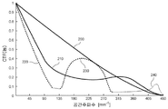
본 개시사항의 하나의 예시적인 실시예에 따르면, 두 개 이상의 촬영 채널이 사용될 수 있으며, 예를 들어 적어도 하나의 촬영 채널은 베셀 빔 조명 또는 검출을 제공하며 적어도 또 다른 하나의 촬영 채널은 가우시안 빔 조명 또는 검출을 제공한다. 이러한 예시적인 구성은 세 개 이상의 독특하고 분리 가능한 조명-검출 조합(예를 들어, 베셀-베셀, 베셀-가우시안, 가우시안-가우시안, 등)을 이용할 수 있으며, 각각의 조합은 상이한 OCT 영상에 상응할 수 있다. 도 2의 예시적인 그래프에 도시된 바와 같이, 2.5 ㎛ 직경 스폿에 대한 간섭 전달 함수(coherent transfer function(CTF))가 제공된다.
예를 들어, 도 2는 회절 제한(diffraction limit, 200), 예비 데이터에 사용된 0.15 mm의 확장형 초점 범위(210), 및 2.0 mm의 초점 범위를 갖는, 이하에서 μOCT로 지칭되는, 본 개시사항에 따른 과정 또는 기법의 예시적인 실시예의 예시적인 결과에 대한 그래프적 비교를 예시한다. 본 개시사항의 하나의 예시적인 실시예에 따르면, 예를 들어, 가우시안-가우시안 영상(220), 베셀-가우시안 영상(230), 및 베셀-베셀 영상(240)을 조합함으로써, μOCT CTF가 생성될 수 있다.
본 개시사항의 또 다른 예시적인 실시예에서, 예를 들어, (다른 것은 물론) 0.5 mm 초과, 1 mm 초과, 2 mm 초과 등일 수 있는 축방향 초점 범위에 걸쳐 예시적인 μOCT CTF 과정/기법이 사용될 수 있고/있거나 제공될 수 있다. 본 개시사항의 추가적인 예시적인 실시예에 따르면, 횡방향 FWHM 스폿 직경은 (다른 것은 물론) 5 ㎛ 미만, 2 ㎛ 미만, 1 ㎛ 미만 등일 수 있다. 본 개시사항의 또 다른 예시적인 실시예에서, 초점 깊이는 평면파 또는 가우시안 빔을 갖는 조명에 비해 예를 들어 대략 2배, 5배, 10배, 20배, 50배, 100배 등으로 (그리고 가능하다면 그 이상으로) 확장될 수 있다. 본 개시사항의 또 다른 예시적인 실시예에서, 영상 내의 높은, 낮은 그리고 중간인 공간 주파수 콘텐츠는 상이한 전달 함수와 영상을 조합함으로써 적어도 부분적으로 회복될 수 있다.
도 3A 내지 도 3C는 본 개시사항의 예시적인 실시예에 따른 예시적인 과정/기법을 사용하여 획득된 사체 관상 동맥 플라크의 예시적인 OCT 영상을 도시한다. 예를 들어, 도 3A에서, 예시적인 가우스-가우스 영상은 낮은 공간 주파수 정보를 포함한다. 도 3B에서, 예시적인 베셀-베셀 영상은 높은 해상도를 제공하지만 낮은 공간 주파수 및 중간 공간 주파수를 잃어버린다. 또한, 도 3C에서, 조합 μOCT 영상(예를 들어, 가우스-가우스 + 가우스-베셀 + 베셀-베셀)이 제공되며, 영상들은 동일한 휘도/콘트라스트 값을 이용하여 정규화되고 디스플레이된다.
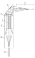
도 4A는 본 개시사항의 제 1 예시적인 실시예에 따라 OCT 카테터의 말단 광학(distal optics)을 포함하는 시스템의 다이어그램의 측부 절개도를 도시한다. 이러한 예시적인 시스템은 예시적인 말단 광학 설계 및/또는 구성의 환형(annulus)(예를 들어, 도 4A 내의 더 짙은 음영) 및 가우시안 빔(예를 들어, 도 4A 내의 더 밝은 음영)을 생성하기 위해 Y-정션 팬-아웃(Y-junction fan-out)을 포함한다. 도 4A의 이러한 예시적인 시스템은 회절-제한형 초점 깊이(diffraction-limited depth-of-focus)보다 예를 들어 대략 10배를 초과하여 길 수 있는 축방향 초점 범위(예를 들어, 초점 깊이) 및 회절 제한형 CTF(diffraction-limited CTF)를 생성하기 위해 제공된다. 도 4A에 도시된 바와 같이, 도파관(400)의 예시적인 출력은 y-정션 팬-아웃 엘리먼트(410)에 의해 (도 4C의 예시에 도시된 바와 같이) 원과 같은 패턴에 대응하는 하나의 어레이의 스폿으로 변형될 수 있다. (도 4B의 예시적인 그래프에서 도시된 바와 같이) 이러한 엘리먼트의 인덱스 프로파일(index profile)은 무손실이고 무색성이 되도록 구성될 수 있다. 각각의 스폿의 출력은 시준기 어레이(420) 내의 빔 시준기에 의해 개별적으로 시준될 수 있다.
도 4A에 도시된 바와 같이, 가우시안 빔은 환형 어레이의 중앙에 별개의 도파관(430)을 통해 라우팅될 수 있다. 도파관의 예시적인 출력은 시준기 어레이(420)의 중앙에 위치된 시준기(440)에 의해 시준될 수 있다. 예시적인 시준된 환형 및 가우시안 빔은 도 4A에 도시된 바와 같이 예를 들어 구배형 인덱스(gradient index(GRIN) 렌즈(450)를 포함하지만 그에 제한되지 않는 하나 이상의 렌즈를 사용하여 샘플 상으로 집속될 수 있다. 두 개의 빔을 집속하는 것에 추가하여, 이러한 예시적인 GRIN 렌즈(450)는 (도 4D의 예시에 도시된 바와 같이) 색 수차를 의도적으로 생성하도록 구성되고/되거나 구조화될 수 있는데, 이는 축방향 초점을 추가로 확장시킬 수 있으며, 그리고 가능하다면 투명한 외부 피복(460)에 의해 유도된 수차를 보상하도록 구성되고/되거나 구조화될 수 있다. 전자기 방사선(예를 들어, 광)은 디플렉터(470)에 의해 해부학적 구조(480)로 보내질 수 있다.
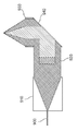
도 5A는 본 개시사항에 따른 OCT 카테터 시스템의 말단 광학의 제 2 예시적인 실시예를 도시한다. 예를 들어, 도 5A의 예시적인 시스템은 이러한 예시적인 실시예에 따른 말단 광학 설계의 (도 5A의 더 짙은 음영으로 도시된) 환형 및 (도 5A의 더 밝은 음영으로 도시된) 가우시안 빔의 라우팅 및 액시콘 배열체(예를 들어, 쌍)을 도시한다. 특히, 도 5A에 도시된 예시적인 시스템은 회절 제한형 초점 깊이보다 예를 들어 대략 10배를 초과하여 길 수 있는 축방향 초점 범위(예를 들어, 초점 깊이) 및 회절 제한형 CTF를 생성할 수 있다. 도파관(500)의 출력은 예시적인 카테터 시스템의 중앙에 위치된 시준기(510)에 의해 시준될 수 있다. 시준된 전자기 방사선(예를 들어, 광)은 두 개 이상의 액시콘(520, 530)을 사용하여 환형 빔으로 변환될 수 있다. 또 다른 예시적인 실시예에 따르면, 액시콘은 구배 인덱스를 사용하여 발생되거나 생성될 수 있다.
도 5A에 도시된 바와 같이, 별개의 도파관(540)이 환형의 중앙을 통해 라우팅될 수 있다. 도파관의 출력은 환형의 중앙에 위치된 시준기(550)에 의해 시준될 수 있다. 시준된 환형 및 가우시안 빔의 시뮬레이션된 횡방향 강도 프로파일은 도 5B의 예시에 도시되어 있다. 시준된 환형 및 가우시안 빔은 GRIN 렌즈(560)와 같은 하나 이상의 렌즈를 사용하여 샘플 상으로 집속될 수 있다. 두 개 이상의 빔을 집속하는 것에 추가하여, GRIN 렌즈(560)는 (도 5C의 예시에 도시된 바와 같이) 색 수차를 의도적으로 생성하도록 구성될 수 있는데, 이는 축방향 초점을 추가로 확장시킬 수 있으며, 그리고 투명한 외부 피복(570)에 의해 유도된 수차를 보상하도록 구성될 수 있다. 전자기 방사선(예를 들어, 광)은 디플렉터(580)에 의해 동맥 벽으로 보내질 수 있다.
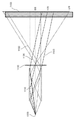
도 6은 본 개시사항의 예시적인 실시예에 따른 μOCT 영상을 생성하기 위한 촬영 시스템의 개략적인 다이어그램을 도시한다. 도 6의 예시적인 실시예에 제공된 바와 같이, 전자기 방사선(예를 들어, 광 방사선)을 제공하는 소스(600)의 출력은 선형 편광기(602)에 의해 선형적으로 편광될 수 있고, 빔 스플리터(604)에 의해 두 개 이상의 빔으로 분할될 수 있다. 빔 중 적어도 하나의 빔은 스위치(606)의 입력부로 다시 보내질 수 있다.
스위치(606)의 출력 중 적어도 하나의 출력은 빔 스플리터(610)를 통해 전송될 수 있으며, 제 1 광/전자기 방사선 가이드(612)로 커플링될 수 있다. 스위치(606)의 출력 중 또 다른 출력은 감쇠기(614)에 의해 감쇠될 수 있으며, 제 2 광/전자기 방사선 가이드(616)에 의해 제 3 빔 스플리터(618)로 안내될 수 있고, 감쇠기(622), 제 3 광/전자기 방사선 가이드(624) 및 분산 보상 배열체(626)를 통해 기준 반사기(606)로 다시 보내질 수 있다. 제 1 광 가이드(612)의 출력은 카테터(628)의 베셀 조명 및 베셀 검출 채널에 접속될 수 있다.
도 6에 도시된 바와 같이, 빔 스플리터(604)의 출력 중 또 다른 하나는 제 2 삼 포트식 스위치(second three-port switch)(630)의 입력부로 다시 보내질 수 있다. 스위치(630)의 출력 중 하나의 출력은 빔 스플리터(632)를 통해 전송될 수 있으며, 제 4 광/전자기 방사선 가이드(634)로 커플링될 수 있다. 스위치(630)의 출력 중 또 다른 출력은 감쇠기(635)에 의해 감쇠될 수 있으며, 제 5 광 가이드(636)에 의해 제 4 빔 스플리터(638)로 안내될 수 있고, 감쇠기(642), 제 5 광/전자기 방사선 가이드(644) 및 제 2 분산 보상 배열체(646)를 통해 기준 반사기(640)로 다시 보내질 수 있다. 광 가이드(634)의 출력은 카테터(628)의 가우시안 조명 및 가우시안 검출 채널에 접속될 수 있다.
스위치(606)의 상태가 1이고, 스위치(630)의 상태가 2일 때, 샘플이 베셀 조명 채널에 의해 조명되도록 예를 들어 단지 광/전자기 방사선 가이드(612)만이 조명될 수 있다(도 6의 표 1 참조). 샘플로부터의 후방산란된 광은 카테터(628)의 베셀 및 가우시안 검출 채널 중 양자, 일부 또는 모두에 의해 획득될 수 있다(도 6의 표 1 참조). 베셀 검출 채널에 의해 획득된 전자기 방사선/광의 일부는 제 1 광/전자기 방사선 가이드(612)에 의해 빔 스플리터(610)로 안내될 수 있으며, 이러한 방사선/광은 기준 반사기(606)로부터의 광과 조합될 수 있고 간섭될 수 있다.
또한, 도 6에 도시된 바와 같이, 간섭 신호의 적어도 일부분은 빔 스플리터(610)에 의해 핀홀(648)로 보내질 수 있다. 핀홀(648)의 출력은 편광 빔 스플리터(650)에 의해 시준되고 분할될 수 있다. 편광 빔 스플리터(650)의 출력 중 하나의 출력은 반파장판(652)을 통해 전송될 수 있으며, 분광계(654)에 의해 검출될 수 있다. 편광 빔 스플리터(650)의 출력 중 또 다른 출력은 제 2 분광계(656)에 의해 검출될 수 있다. 가우시안 검출 채널에 의해 획득된 전자기 방사선/광의 하나의 부분은 제 4 광 가이드(634)에 의해 빔 스플리터(632)로 안내될 수 있으며, 그것은 기준 반사기(640)로부터의 광과 조합되고 간섭된다. 간섭 신호의 적어도 일부는 빔 스플리터(632)에 의해 핀홀(658)로 보내질 수 있다. 핀홀(658)의 출력은 편광 빔 스플리터(660)에 의해 시준되고 분할될 수 있다. 편광 빔 스플리터(660)의 출력 중 적어도 하나의 출력은 반파장판(662)을 통해 전송될 수 있으며, 제 3 분광계(664)에 의해 검출될 수 있다. 편광 빔 스플리터(660)의 출력 중 또 다른 출력은 제 4 분광계(666)에 의해 검출될 수 있다.
스위치(606)의 상태가 2이고, 스위치(630)의 상태가 1일 때, 샘플이 (도 6의 표 1에 도시된) 가우시안 조명 채널에 의해 조명되도록 예를 들어 단지 제 4 광/전자기 방사선 가이드(634)만이 조명될 수 있다. 샘플로부터의 후방산란된 광은 (도 6의 표 1에 도시된) 카테터(630)의 베셀 및 가우시안 검출 채널 양자에 의해 획득될 수 있다. 베셀 검출 채널에 의해 획득된 전자기 방사선/광의 적어도 일부분은 제 1 광/전자기 방사선 가이드(612)에 의해 빔 스플리터(610)로 안내될 수 있으며, 그것은 기준 반사기(620)로부터의 광과 조합될 수 있고 간섭될 수 있다. 간섭 신호의 적어도 일부는 빔 스플리터(610)에 의해 핀홀(648)로 보내질 수 있다. 핀홀(648)의 출력은 편광 빔 스플리터(650)에 의해 시준되고 분할될 수 있다. 편광 빔 스플리터(650)의 출력 중 적어도 하나의 출력은 반파장판(652)을 통해 전송될 수 있으며, 분광계(654)에 의해 검출될 수 있다. 편광 빔 스플리터(650)의 출력 중 또 다른 출력은 제 2 분광계(656)에 의해 검출될 수 있다.
가우시안 검출 채널에 의해 획득된 광의 하나의 부분은 전자기 복사선/광 가이드(634)에 의해 빔 스플리터(632)로 안내될 수 있으며, 그것은 기준 반사기(640)로부터의 광과 조합되고 간섭된다. 간섭 신호의 적어도 일부는 빔 스플리터(632)에 의해 핀홀(658)로 보내질 수 있다. 핀홀(658)의 출력은 편광 빔 스플리터(660)에 의해 시준되고 분할될 수 있다. 편광 빔 스플리터(660)의 두 개의 출력 중 적어도 하나의 출력은 반파장판(662)을 통해 전송될 수 있으며, 제 3 분광계(664)에 의해 검출될 수 있다. 편광 빔 스플리터(660)의 출력 중 또 다른 출력은 제 4 분광계(666)에 의해 검출될 수 있다.
편광 빔 스플리터(650), 반파장판(652) 및 분광계(654, 656)의 조합 및/또는 편광 빔 스플리터(660), 반파장판(662) 및 분광계(664, 666)의 조합에 의해 구현된 도 6에 도시된 이러한 예시적인 편광-다양성 검출(polarization-diverse detection) 방안/구성은 조직 또는 광 섬유 복굴절에 연관된 아티팩트(artifact)를 감소시키고/감소시키거나 제거할 수 있다. 도 6에 예시된 본 개시사항에 따른 μOCT 카테터 시스템의 예시적인 실시예는 카테터로부터 도파관(612 및 632)으로 광/방사선을 예를 들어 독립적으로 전송하고/하거나 수신할 수 있는 복수의 도파관을 포함할 수 있다. 검출된 신호는 컴퓨터(668)에 의해 영상 획득판(670)을 통해 디지털화되고 전달될 수 있다. 데이터는 모니터(672) 상에 또는 모니터(672)를 통해 디지털로 디스플레이될 수 있으며/있거나 저장 장치(674)에 저장될 수 있다.
본 개시사항에 따라, μOCT 검출 기술은 하나의 예시적인 실시예에서 시간 도메인 OCT(TD-OCT) 시스템을 사용하여, 또 다른 예시적인 실시예에서 공간 도메인 OCT(SD-OCT) 시스템을 사용하여, 그리고 또 다른 예시적인 실시예에서 광학 주파수 도메인 간섭(optical frequency domain interferometry(OFDI)) 시스템을 사용하여 구현될 수 있다. 상이한 전달 함수 조명 및 검출 구성으로부터 복소 영상 및/또는 실제 영상이 본 개시사항에 따른 촬영 시스템의 예시적인 실시예를 사용하여 획득될 수 있다. 하나의 예시적인 실시예에서, 이러한 예시적인 영상은 회절 제한형 CTF를 보다 엄밀하게 근사시키는 하나의 CTF 및 개선된 품질을 갖는 신규 영상을 생성하기 위해 필터링되고 재조합될 수 있다. 상이한 전달 함수를 갖는 예시적인 영상은 회절 제한형 CTF 과정/기법을 보다 엄밀하게 근사시키는 하나의 CTF 과정/기법을 갖는 신규 영상을 생성하기 위해 필터링되거나 비간섭성으로 그리고/또는 간섭성으로 재조합될 수 있다.
도 7은 회절-제한형 초점 깊이보다 예를 들어 대략 10배를 초과하여 길 수 있는 축방향 초점 범위(예를 들어, 초점 깊이) 및 회절-제한형 CTF를 생성하기 위한 본 개시사항에 따른 OCT 카테터의 말단 광학 구성의 또 다른 예시적인 실시예를 도시한다.
예를 들어, 도파관(700)의 출력은 시준기(710)에 의해 시준될 수 있다. 실제로, 도파관(700)은 환형 빔을 통해 라우팅될 수 있으며 시준되고, 가우시안 빔은 환형의 중앙을 통해 라우팅될 것이다. 시준된 광은 예를 들어 GRIN 액시콘(720, 730)과 같은 두 개 이상의 액시콘을 통해 환형 빔으로 변환될 수 있다. 별개의 도파관(740)이 환형의 중앙을 통해 라우팅될 수 있다. 도파관(740)의 출력은 환형의 중앙에 위치된 시준기(750)에 의해 시준될 수 있다. 시준된 환형 및 가우시안 빔은 예를 들어 하나 이상의 GRIN 렌즈일 수 있는 하나 이상의 렌즈(760)를 사용하여 샘플 상으로 집속될 수 있다. 빔을 집속하는 것에 추가하여, GRIN 렌즈(760)는 색 수차를 의도적으로 생성하도록 구성되며/되거나 구조화될 수 있는데, 이는 축방향 초점을 추가로 확장시킬 수 있으며, 그리고 투명한 외부 피복에 의해 유도된 수차를 보상하도록 구성되며/되거나 구조화될 수 있다. 광/방사선은 디플렉터(770)에 의해 동맥 벽으로 보내질 수 있다.
도 8은 본 개시사항에 따른 OCT 카테터의 말단 광학 구성의 또 다른 예시적인 실시예를 도시한다. 이러한 예시적인 구성은 회절 제한형 초점 깊이보다 예를 들어 10배를 초과하여 긴 초점 깊이 및 회절 제한형 CTF를 생성하기 위해 사용될 수 있다. 도파관(800)의 출력은 시준기(810)에 의해 시준될 수 있다. 시준기(810)에 의해 생성된 동공 어퍼처(pupil aperture)는 두 개 이상의 빔, 즉, 중앙의 원형 빔 및 환형 빔으로 분할될 수 있다. 중앙 영역에 실질적으로 유사하거나 중앙 영역과 동일한 어퍼처를 갖는 대물 렌즈, 무색성 렌즈, 무수차 렌즈(aplanat lens) 또는 GRIN 렌즈와 같은 하나 이상의 렌즈(820)가 낮은 NA 가우시안 빔을 조직 또는 샘플 내로 집속시킬 수 있다.
환형 빔은 스페이서(830)을 통해 전송될 수 있으며, 환형 빔에 실질적으로 유사하거나 동일한 어퍼처를 갖는 환형 액시콘 렌즈(840)에 의해 샘플 내로 집속될 수 있다. 빔은 디플렉터(850)에 의해 샘플로 보내질 수 있다. 네 개의 채널로부터 생성된, 예를 들어 중앙 조명/중앙 검출, 중앙 조명/환형 검출, 환형 조명/환형 검출, 환형 조명/중앙 검출로부터 생성된 네 개의 영상이 존재할 수 있다. 렌즈(820)의 광학 경로 길이는 스페이서(830)의 광학 경로 길이와 상이하도록 구성될 수 있어서, 생성된 네 개의 영상 각각이 경로 길이 인코딩(path length encoding)될 수 있다. 이러한 예시적인 실시예에서, 상이한 영상이 검출될 수 있으며, 그들의 CTF가 본 명세서에서 설명된 예시적인 방법 및/또는 과정에 따라 조합될 수 있다.
도 9은 회절-제한형 초점 깊이보다 긴 초점 깊이 및 회절-제한형 CTF를 생성하기 위해 사용될 수 있는 본 개시사항에 따른 OCT 카테터 시스템의 말단 광학 구성의 또 다른 예시적인 실시예를 도시한다. 예를 들어, 도 9에 예시된 바와 같이, 도파관(900)의 출력은 시준기(910)에 의해 시준될 수 있다. 시준기(910)에 의해 생성된 동공 어퍼처(pupil aperture)는 대물 렌즈 어퍼처의 중앙에 위치된 원형 유리 윈도우(920)에 의해 두 개 이상의 영역, 예를 들어 (i) 원형 유리 윈도우(920)를 통해 전송된 중앙 원형 영역 및 (ii) 환형 영역으로 분할될 수 있다. 중앙 환형 빔은 낮은 NA 가우시안 빔으로서 조직 및/또는 샘플 내로 집속될 수 있으며, 환형 빔은 렌즈(930)에 의해 조직 내의 베셀 빔 초점 내로 집속될 수 있다. 유리 윈도우는 공기보다 높은 굴절률을 가질 수 있으며, 윈도우의 두께는 상이한 채널을 겪는 광/방사선 필드가 경로-길이 분리되며/되거나 인코딩될 수 있도록 선택될 수 있다. 각각의 A 라인에서, (예를 들어, 4 개의) 채널로부터 오는 신호의 세 개 이상의 세그먼트, 즉, 중앙 조명/중앙 검출, 중앙 조명/환형 검출, 환형 조명/환형 검출, 환형 조명/중앙 검출이 존재할 수 있다.
도 10은 회절-제한형 초점 깊이보다 길 수 있는 초점 깊이 및 회절-제한형 CTF를 생성하기 위한 OCT 카테터 시스템의 말단 광학 구성의 또 다른 예시적인 실시예를 도시한다. 도파관(1000)의 출력은 시준기(1010)에 의해 시준될 수 있다. 시준기(1010)에 의해 생성된 동공 어퍼처는 복수의 동심 영역(1020, 1030, 1040)으로 분할될 수 있다. 예를 들어 GRIN 렌즈와 같은 다초점 렌즈(multifocal lens)가 사용될 수 있어서, 각각의 영역 내의 빔이 상이한 축방향 초점 위치로 집속될 수 있다. 각각의 영역으로부터의 산란된 광/방사선은 이러한 산란된 빔이 서로 간섭되지 않도록 광학 경로 길이 인코딩될 수 있다. 이러한 예시적인 실시예에서, 상이한 영상이 검출될 수 있으며, 그들의 CTF는 본 명세서에서 설명된 예시적인 방법 및 과정에 따라 조합될 수 있다.
도 11은 회절-제한형 초점 깊이보다 긴 축방향 초점 범위(예를 들어, 초점 깊이) 및 회절-제한형 CTF를 생성하기 위한 OCT 카테터 시스템의 말단 광학 구성의 또 다른 예시적인 실시예를 도시한다. 예를 들어, 점 물체(point object)(1100)의 출력은 미러 터널 장치(1110)에 의해 복수의 차수의 광/방사선 빔으로, 예를 들어 0 차수 빔(1120), 1 차수 빔(1130), 및 2 차수 빔(1140) 등으로 변환될 수 있다. 대부분의 차수 또는 모든 차수의 광선이 샘플 내의 동일한 초점 위치에 집속되도록 집속 장치(1150)가 이용될 때, 각각의 차수의 광선은 집속 장치의 조명/검출 CTF의 독특한 공간 주파수 대역을 포함할 수 있다. 또 다른 예시적인 실시예에서, 이들 차수는 내부에서 생성된 영상이 검출될 수 있도록 경로 길이 인코딩될 수 있으며, 그들의 CTF는 본 명세서에서 설명된 예시적인 CTF 조합 방법 및/또는 과정에 따라 상이한 차수에 상응하는 상이한 영상을 사용하여 조합될 수 있다.
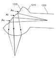
도 12는 회절-제한형 초점 깊이보다 긴 초점 깊이 및 회절-제한형 CTF를 생성하기 위한 본 개시사항에 따른 OCT 카테터 시스템의 말단 광학 구성의 또 다른 예시적인 실시예를 도시한다. 도 12에 예시된 바와 같이, 도파관(1200)의 출력은 하프 볼 렌즈(half ball lens)(1210)에 의해 집속될 수 있다. 하프 볼 렌즈(1210)의 평탄면은 이진 위상 패턴(1220)을 가질 수 있다. 또 다른 예시적인 실시예에서, 패턴의 깊이는 예를 들어 198 nm의 패턴 깊이(850 nm에서 π 위상 변이)와 같은 작은 위상 변이를 생성하도록 구성될 수 있다. 또 다른 예시적인 실시예에서, 상부면은 금과 같은 반사성 코팅을 사용하여 코팅될 수 있으며, 하부면은 동일한 코팅 및/또는 알루미늄과 같은 또 다른 코팅을 이용하여 코팅될 수 있고, 최종 위상 변이는 도 13의 그래프에 도시된 곡선(1300)에 의해 주어지는데, 이는 유리 마스크(예를 들어, 금속 코팅 없음) 및 전체 위상 변이(예를 들어, 마스크 + 코팅)의 광학 위상 길이 차이를 예시한다.
도 13의 그래프의 곡선(1310) 및 곡선(1320)은 45도의 입사각을 이용하여 각각 BK7-Al 및 BK7-Au에서의 반사 시에 p 편광된 광의 파장 의존형 위상 변화를 가질 수 있다. 곡선(1330)은 BK7-공기 인터페이스에서 45도 반사 시에 예를 들어 198 nm 높이 차이에 의해 야기된 광의 파장 의존형 위상 변이일 수 있다. (도 14A의 예시에 도시된 바와 같은) 회절 제한형 축방향 초점에 비해 (도 14B의 예시에 도시된 바와 같은) 확장형 축방향 초점을 생성하기 위해, 이진 위상 마스크가 최적화될 수 있다. 상이한 위상 변이를 갖는 표면으로부터 전송된 광/방사선은 상이한 전달 함수를 생성할 수 있으며, 이는 본 명세서에서 설명된 예시적인 방법 및/또는 과정에 따라 상이한 CTF를 갖는 신규 영상을 생성하기 위해 검출되고 조합될 수 있다.
도 15A는 회절-제한형 초점 깊이보다 긴 초점 깊이 및 회절-제한형 CTF를 생성하기 위한 OCT 카테터 시스템의 말단 광학 구성의 또 다른 예시적인 실시예의 다이어그램의 측부 절개도를 도시한다. 예를 들어, 도 15A의 시스템은 예를 들어 대략 2배, 5배, 10배, 20배, 50배, 100배만큼의 결과를 생성한다. 도파관(1500)의 출력은 하나 이상의 렌즈(1510)에 의해 시준될 수 있다. 시준된 빔은 위상 더블릿(1520)에 의해 공간적으로 변조될 수 있으며, 위상 더블릿(1520)은 동일하거나 유사한 위상 패턴을 갖는 포지티브 위상판 및 네가티브 위상판을 포함할 수 있다. 포지티브 위상판 및 네가티브 위상판의 아베 수(Abbe number)를 정합시킴으로써, 파장 의존형 위상 오차는 취소되거나 감소될 수 있다. 도 15B는 도 15A에 예시된 예시적인 마스크(예를 들어, BK7-SNPH2 위상 더블릿 마스크)의 횡방향 위상 프로파일의 예시적인 그래프를 도시한다. 예를 들어, 오하라(Ohara) S-NPH2(Vd=18.896912, Nd=1.922860) 및 쇼트(Schott) BK7(Vd=64.167336, Nd=1.5168)를 각각 깊이 7.2554 ㎛ 및 13.4668 ㎛와 함께 선택함으로써, 위상 프로파일은 도 15B에 도시되어 있다. 공간적으로 변조된 빔은 대물 렌즈(1530)에 의한 확장형 축방향 초점 내로 집속될 수 있다.
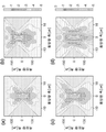
도 16은 회절-제한형 초점 깊이보다 바람직하게는 대략 2배, 5배, 10배, 20배, 50배, 100배만큼 긴 본 개시사항에 따른 초점 깊이 및 회절-제한형 CTF를 생성하기 위한 OCT 카테터 시스템의 말단 광학 구성의 또 다른 예시적인 실시예를 도시한다. 광원(1600)의 출력은 빔 스플리터(1610)에 의해 분할될 수 있다. 빔 스플리터의 출력 중 적어도 하나의 출력의 빔 어퍼처는 막대 미러(1620)에 의해 두 개 이상의 구역으로 분할되거나 분리될 수 있다. 예를 들어, 막대 미러(1620)는 빔의 중앙부를 대물 렌즈(1640)를 통해 기준 반사기(1630)로 다시 보낼 수 있다. 환형 빔은 하나 이상의 렌즈(1640)와 실질적으로 유사하거나 동일할 수 있는 제 2 대물 렌즈(1660)에 의해 샘플 내로 (도 18D의 예시적인 μOCT 영상에 도시된 바와 같이) 횡방향으로 확장형 축방향 초점 및 초고-해상도(super-resolution)를 특징으로 하는 베셀 초점 내로 집속될 수 있다. 샘플로부터 후방 산란된 광은 핀홀(1660)에서 막대 미러를 통해 기준 반사기로부터 반사된 광과 조합된다. 핀홀(1660)의 출력은 분광계(1670)에 의해 검출된다. 대물 렌즈(1650)는 색 수차 및 공간 수차를 의도적으로 생성하도록 구성되며, 이는 (도 18C 및 도 18D의 예시적인 μOCT 영상에 도시된 바와 같이) 축방향 초점을 추가로 확장시킨다. 도 18A는 복수의 백혈구(화살표)를 도시하는 관상 동맥 플라크의 예시적인 μOCT 영상을 도시한다. 또한, 도 18B는 두 개의 상이한 세포 유형의 복수의 백혈구(화살표)를 예시하는 관상 동맥 플라크의 예시적인 μOCT 영상을 도시하며, 하나의 유형은 림프구(L)와 일치하는 스캔트 세포질(scant cytoplasm)을 갖는 보다 작은 세포이고, 또 다른 하나의 유형은 단핵구(monocyte, M)를 나타내는 고산란 세포질(highly scattering cytoplasm)을 갖는 보다 큰 세포이다.
실제로, 도 18A는 본 개시사항에 따른 방법, 시스템 및 장치의 예시적인 실시예를 사용하여 생성되었던 복수의 백혈구(1800)를 도시하는 관상 동맥 플라크의 예시적인 μOCT 영상을 예시한다. 도 18B는 두 개의 상이한 세포 유형의 복수의 백혈구를 도시하는 관상 동맥 플라크의 예시적인 μOCT 영상을 예시하며, 하나의 유형은 림프구와 일치하는 스캔트 세포질을 갖는 보다 작은 세포(1810)이고, 또 다른 하나의 유형은 단핵구를 의미하는 고산란 세포질을 갖는 보다 큰 세포(1820)이다. 도 18C는 단핵구의 들쑥날쑥한 콩 형상 핵(indented, bean-shaped nucleus) 특성을 갖는 세포(1830)를 도시하는 관상 동맥 플라크의 예시적인 μOCT 영상을 예시한다. 도 18D는 내피면에 부착된 뉴트로필(neutrophil)을 의미하는 멀티-로비드 핵(multi-lobed nucleus)을 갖는 백혈구(1840)를 도시하는 관상 동맥 플라크의 예시적인 μOCT 영상을 예시한다. 도 18E는 위족(pseudopodia, 1860)에 의해 내피면에 묶여진 복수의 백혈구(1850)를 도시하는 관상 동맥 플라크의 예시적인 μOCT 영상을 예시한다. 도 18F는 이러한 단면 및 삽화에서 내피(1880)를 통해 이주하는 단핵구의 형태학(morphology)을 갖는 세포(1870)를 도시하는 관상 동맥 플라크의 예시적인 μOCT 영상을 예시한다. 또한, 도 18G는 내피면 상에 분포된 복수의 백혈구(1890)의 예시적인 μOCT 영상을 예시한다.
도 19A 내지 도 19E는 본 기재사항에 따른 방법, 시스템 및 장치의 예시적인 실시예를 사용하여 생성되었던 예시적인 영상을 도시한다. 예를 들어, 도 19A는 뉴트로필(1910)(N)의 백혈구 특성에 인접한 혈소판(1900)(P)의 예시적인 μOCT 영상을 예시하며, 뉴트로필도 또한 작은 혈소판(1920)(황색 화살표)에 부착되어 있다. 도 19B는 관상 동맥 벽 내의 갭을 메우는 선형 스트랜드(linear strand)로서 보일 수 있는 피브린(1930)(F)의 예시적인 μOCT 영상을 예시한다. 도 19C는 도 19B에 대한 인접한 위치에 피브린에 인접한 한 무리(cluster)의 백혈구(1940)(L)의 예시적인 μOCT 영상을 예시한다. 도 19D는 복수의 옭아매진 백혈구를 갖는 피브린 혈전(1950)(T)의 예시적인 μOCT 영상을 예시한다. 도 19E는 백혈구(1970)(화살표) 및 피브린 스트랜드(1980)(삽화, F)를 도시하는 보다 진전된 혈전(1960)(T)의 예시적인 μOCT 영상이다.
도 20A 내지 도 20D는 본 개시사항에 따른 방법, 시스템 및 장치의 예시적인 실시예를 사용하여 생성하였던 추가적인 예시적인 영상을 도시한다. 예를 들어, 도 20A는 배양 조직 내의 내피 셀(2000)의 예시적인 μOCT 단면 영상을 예시한다. 도 20B는 배양 조직 내의 내피 셀(2010)의 예시적인 μOCT 정면 영상을 도시한다. 도 20C는 토종 돼지 관상 동맥 단면(2020)의 예시적인 μOCT 영상을 예시한다. 도 20D는 내피 "포상형성(pavementing)"(2030)을 입증하는 돼지 관상 동맥의 예시적인 3차원 렌더링을 도시한다.
도 21A 및 도 21B는 본 개시사항에 따른 방법, 시스템 및 장치의 예시적인 실시예를 사용하여 생성하였던 추가적인 예시적인 영상을 도시한다. 도 21A는 섬유 캡(fibrous cap)(2100)의 μOCT 영상 내에 밝은 밀도로서 보여지는 미세석회화(microcalcification)의 예시적인 예시적인 μOCT 영상을 도시한다. 도 21B는 상응하는 히스톨로지(histology)(2110) 상에 자주색 밀도로서 보여지는 미세석회화의 예시적인 μOCT 영상을 예시한다.
또한, 도 22A 내지 도 22C는 본 개시사항에 따른 방법, 시스템 및 장치의 예시적인 실시예를 사용하여 생성하였던 추가적인 예시적인 영상을 예시한다. 도 22A는 손상된 내막/내피(2200)를 입증하는 대형 칼슘 결절(large calcium nodule)의 예시적인 μOCT 영상을 도시한다. 도 22B는 대치하는 분리된 내막(intima)에 비보호형 칼슘(2220)을 인접하면서 피브린(2210)과 일치하는 미세 조직 스트랜드를 도시하는 적색 박스에 의해 둘러싸인 예시적인 구역의 확대도를 도시한다. 도 22C는 피브린(2230) 및 탈피된 석회화 면(denuded calcific surface, 2240)을 예시하는 상응하는 히스톨로지를 도시한다.
또한, 도 23A 내지 도 26C는 본 개시사항에 따른 방법, 시스템 및 장치의 예시적인 실시예를 사용하여 생성하였던 추가적인 예시적인 영상을 예시한다. 예를 들어, 도 23A는 상부면 및 하부면으로부터의 반사에 의해 특성화된 두꺼운 콜레스테롤 결정(cholesterol crystal, 2310)을 입증하는 대형 괴사 코어(necrotic core, 2300) 섬유선종(fibroatheroma)의 예시적인 μOCT 영상을 도시한다. 도 23B는 삽화 내에 보다 상세하게 도시된 또 다른 괴사 코어 플라크(2330)의 캡을 관통하는 얇은 결정(2320)의 예시적인 μOCT 영상을 도시한다. 도 24A는 낮은 후방산란 스핀들 형상 세포(삽화)로서 나타낸 다양한 민무늬근 세포(2400)의 예시적인 μOCT 영상을 도시한다. 도 24B는 스핀들 형상이면서 높은 후방산란 인테리어(2410) 및 낮은 후방산란의 "후광(halo)"(2420)을 갖는, 콜라겐을 생성하는 민무늬근 세포의 예시적인 μOCT 영상을 도시하며, 이는 각각 세포체(2430) 및 콜라겐 매트릭스(2440)를 표시한다(예를 들어, 히스톨로지 삽화).
도 25A는 중합체를 구비하지 않은 상태(2500)에서, 중합체를 구비하고 약을 구비하지 않은 상태(2510)에서, 그리고 중합체와 약을 구비한 상태(2520)에서, 탁수스 리베르테(Taxus Liberte)(보스톤 사이언티픽, 내틱, 매세추세츠주) 스트럿의 예시적인 μOCT 영상을 도시한다. 중합체-코팅된 스트럿에 대해, 중합체 반사(polymer reflection(PR), 2530), 스트럿 반사(strut reflection(SR), 2540) 및 복수의 반사(2540 및 2560)가 보여질 수 있다. 도 25B는 네오인티마(neointima, 2580)에 의해 피복된 중합체가 없는 스트럿을 도시하는 임플란트형 BMS(implanted BMS, 2570)를 갖는 사체 관상 동맥 표본의 예시적인 μOCT 영상을 도시한다. 도 25C는 스트럿 반사(2595)(삽화) 위에 중첩하는 중합체를 도시하는 또 다른 사체로부터의 임플란트형 DES 스트럿(implanted DES strut, 2590)을 갖는 사체 관상 동맥 표본의 예시적인 μOCT 영상을 도시한다.
또한, 도 26A는 조직(2600)이 스텐트 스트럿(stent strut, 2620)으로부터 중합체(2610)를 분리시켰고 중합체가 균열되었다는(2630) 것을 도시하는 예시적인 μOCT 영상을 도시한다. 도 26B는 중합체 균열(2660)의 위치 위에 중첩하는 표층 백혈구 클러스터(2640) 및 인접한 부착된 백혈구(2650)를 도시하는 예시적인 μOCT 영상을 도시한다. 도 26C는 또 다른 환자로부터의 스트럿(2680)의 에지에서 염증(2670)을 도시하는 예시적인 μOCT 영상을 도시한다. 도 26도 26하는 내피가 완전히 없는, 비피복형 스트럿(2690)을 도시하는 예시적인 μOCT 영상을 도시한다.
본 개시사항의 또 다른 예시적인 실시예에서, 예시적인 μOCT 시스템/프로브를 위한 광학 엘리먼트는 꼭 맞게 집속된 빔을 이용하여 광중합체(photopolymer)를 조사함으로써 제조될 수 있으며, 그 위치는 nm 레벨 정확도로 3차원에서 제어될 수 있다. 광중합체는 복잡한 광학 기능을 구현하기 위해 아주 작은 고상 볼륨(miniature, solid volume)을 가능하게 하면서 예치된 광학 에너지에 비례할 수 있는 다양한 굴절률에 응답할 수 있다. (예를 들어, 설리반 AC(Sullivan AC), 그라보브스키 MW(Grabowski MW) 및 맥레오드 RR(McLeod RR), "광중합체 내로의 3차원 직접-기록 리소그라피(Three-dimensional direct-write lithography into photopolymer)", 어플라이드 옵틱스(Applied Optics) 2007; 46: 295-301, 및 스코트 TF(Scott TF), 코발스키 BA(Kowalski BA), 설리반 AC, 보우만 CN(Bowman CN) 및 맥레오드 RR, "서브회절 광리소그래피를 위한 2색 단일 광자 광개시 및 광저해(Two-Color Single-Photon Photoinitiation and Photoinhibition for Subdiffraction Photolithography)", 사이언스(Science) 2009; 324: 913-7 참조, 미국 특허 공개 제2009/0218519호 및 제2006/0193579호도 또한 참조)
이러한 예시적인 방법 및 과정은 예를 들어 모두 모롤리식 중합체 컴포넌트(monolithic, polymer component) 내에 있는 섬유 커플러(fiber coupler), 테이퍼링된 도파관, 도파관 어레이, 렌즈, 회절성 광학 엘리먼트, 및 복잡한 광학 어셈블리를 이미 생성했다. 이러한 예시적인 실시예는 예시적인 μOCT 프로브가 예를 들어 소형화된 μOCT 카테터 및 내시경 내로 통합될 수 있는 것보다 본 명세서에서 설명된 확장된 초점 깊이 기능을 제공할 수 있는 안정적인 모놀리식 엘리먼트인 것을 가능하게 한다. 이러한 예시적인 실시예의 하나의 장점은 광중합체-유도형 광학 엘리먼트/배열체가 아주 정밀하게 반복적으로 형성될 수 있고 아주 낮은 가격으로 대량 생산될 수 있다는 것이다.
도 27a는 본 개시사항의 하나의 예시적인 실시예에 따른 적어도 하나의 샘플의 적어도 일부분에 연관된 데이터를 제공하기 위한 방법의 흐름도를 도시한다. 예를 들어, 과정 2710에서, (예를 들어, 본 명세서에서 다양한 예시적인 실시예에서 설명된 바와 같이) 적어도 하나의 광 배열체를 통해 샘플의 적어도 일부분으로 적어도 하나의 제 1 방사선이 전달되고, 상기 부분으로부터 제 1 방사선에 기반하는 적어도 하나의 제 2 방사선이 수신된다. 상기 광 배열체와 상기 제 1 방사선 및/또는 상기 제 2 방사선 사이의 상호작용에 기반하여, 상기 광 배열체는 제 1 전달 함수를 갖는다. 그런 다음, 과정 2720에서, 이러한 광 배열체를 통해 상기 부분으로 적어도 하나의 제 3 방사선이 전달되고, 상기 부분으로부터 상기 제 3 방사선에 기반하는 적어도 하나의 제 4 방사선이 수신된다. 상기 광 배열체와 상기 제 3 방사선 및/또는 상기 제 4 방사선 사이의 상호작용에 기반하여, 상기 광 배열체는 제 2 전달 함수를 갖는다. 상기 제 1 전달 함수는 상기 제 2 전달 함수와 적어도 부분적으로 상이할 수 있다. 또한, 과정 2730에서, 상기 제 2 방사선 및 상기 제 4 방사선에 기반하여 상기 부분에 연관된 데이터가 생성될 수 있다.
도 27b는 본 개시사항의 또 다른 예시적인 실시예에 따라 적어도 하나의 샘플의 적어도 일부분에 연관된 데이터를 제공하기 위한 방법의 흐름도를 도시한다. 예를 들어, 과정 2760에서, (예를 들어, 본 명세서에서 다양한 예시적인 실시예에서 설명된 바와 같이) 적어도 하나의 제 1 광 배열체를 통해 샘플의 적어도 일부분으로 적어도 하나의 제 1 방사선이 전달되고, 상기 부분으로부터 상기 제 1 방사선에 기반하는 적어도 하나의 제 2 방사선이 수신된다. 상기 제 1 광 배열체와 상기 제 1 방사선 및/또는 상기 제 2 방사선 사이의 상호작용에 기반하여, 상기 제 1 광 배열체는 제 1 전달 함수를 갖는다. 그런 다음, 과정 2770에서, 적어도 하나의 제 2 광 배열체를 통해 상기 부분으로 적어도 하나의 제 3 방사선이 전달되고, 상기 부분으로부터 상기 제 3 방사선에 기반하는 적어도 하나의 제 4 방사선이 수신된다. 상기 제 2 광 배열체와 상기 제 3 방사선 및/또는 상기 제 4 방사선 사이의 상호작용에 기반하여, 상기 제 2 광 배열체는 제 2 전달 함수를 갖는다. 상기 제 1 전달 함수는 상기 제 2 전달 함수와 적어도 부분적으로 상이할 수 있다. 추가로, 단계 2780에서, 상기 제 2 방사선 및 상기 제 4 방사선에 기반하여 상기 부분에 연관된 데이터가 생성될 수 있다.
전술한 사항은 단지 본 개시사항의 원리를 예시한다. 설명된 실시예에 대한 다양한 수정 및 변경은 본 상세한 설명의 교시에 비추어 당업자에게 명백할 것이다. 예를 들어, 본 개시사항의 예시적인 실시예를 구현하기 위해, 설명된 예시적인 배열체, 방사선, 및/또는 시스템 중 하나를 초과하여 구현될 수 있다. 실제로, 본 발명의 예시적인 실시예에 따른 배열체, 시스템 및 방법은 임의의 OCT 시스템, OFDI 시스템, SD-OCT 시스템 또는 다른 촬영 시스템을 이용하여 사용될 수 있으며/있거나 그것을 구현할 수 있고, 예를 들어 2004년 9월 8일자로 출원된 국제 특허 출원 제PCT/US2004/029148호(이는 2005년 5월 26일자로 국제 특허 공개 제WO 2005/047813호로서 공개됨), 2005년 11월 2일자로 출원된 미국 특허 출원 제11/266,779호(이는 2006년 5월 4일자로 미국 특허 공개 제2006/0093276호로 공개됨), 2004년 6월 4일자로 출원된 미국 특허 출원 제10/861,179호(이는 2005년 1월 27일자로 미국 특허 공개 제2005/0018201호로 공개됨), 2006년 6월 1일자로 출원된 미국 특허 출원 제11/445,990호, 2007년 4월 5일자로 출원된 국제 특허 출원 제PCT/US2007/066017호, 및 2006년 8월 9일자로 출원된 미국 특허 출원 제11/502,330호에 설명된 것을 이용하여 사용될 수 있는데, 이들의 개시사항은 본 명세서에 전체적으로 참조로서 통합된다. 따라서, 당업자가 본 명세서에서 명시적으로 도시되거나 설명되지 않았더라도 본 개시사항을 구체화하며 따라서 본 개시사항의 사상 및 범위 내에 있는 다양한 시스템, 배열체 및 방법을 고안할 수 있을 것이라는 점이 이해될 것이다. 또한, 종래 기술 지식이 본 명세서에서 위에서와 같이 참조로서 명시적으로 통합되지 않은 범위까지, 종래 기술 지식은 명시적으로 본 명세서에 전체적으로 통합된다. 위에서와 같이 본 명세서에서 참조된 모든 공개 문헌은 본 명세서에 전체적으로 참조로서 통합된다.
Claims (21)
- 적어도 하나의 샘플에 적어도 하나의 전자기 방사선을 제공하기 위한 장치로서,
복수의 웨이브-가이딩 배열체를 포함하되, 상기 복수의 웨이브-가이딩 배열체는 i) 상기 적어도 하나의 전자기 방사선을 제공하며, ii) 각각의 상기 웨이브-가이딩 배열체의 방출 지점에서 각각의 상기 적어도 하나의 전자기 방사선의 위상이 미리 정해진 값을 갖게 하도록 구성되는 장치.
- 제 1 항에 있어서, 상기 웨이브-가이딩 배열체는 상기 적어도 하나의 방사선을 적어도 부분적으로 원형 패턴으로 제공하는 장치.
- 제 1 항에 있어서, 상기 웨이브-가이딩 배열체로부터 상기 적어도 하나의 전자기 방사선을 수신하며 추가적인 초점-스폿 방사선을 생성하도록 구성되는 적어도 하나의 렌즈 배열체를 더 포함하는 장치.
- 제 3 항에 있어서, 상기 적어도 하나의 렌즈 배열체는 상기 추가적인 초점-스폿 방사선이 확장된 초점 깊이를 갖게 하도록 구성되는 장치.
- 제 3 항에 있어서, 상기 적어도 하나의 렌즈 배열체는 상기 추가적인 초점-스폿 방사선이 상기 샘플 상의 또는 상기 샘플 내의 회절 제한 스폿보다 작은 직경을 갖게 하도록 구성되는 장치.
- 제 5 항에 있어서, 상기 회절 제한 스폿은 3차원 스폿인 장치.
- 제 3 항에 있어서, 상기 적어도 하나의 렌즈 배열체는 그린 렌즈(grin lens)를 포함하는 장치.
- 제 1 항에 있어서, 상기 웨이브-가이딩 배열체 중 적어도 하나의 웨이브-가이딩 배열체는 단일 모드 도파관인 장치.
- 제 1 항에 있어서, 상기 웨이브-가이딩 배열체 중 적어도 하나의 웨이브-가이딩 배열체는 광-중합체로 구성되는 장치.
- 제 1 항에 있어서, 상기 장치는 상기 샘플에 추가적인 전자기 방사선을 제공하도록 구성되는 추가적인 웨이브-가이딩 배열체를 더 포함하며, 상기 적어도 하나의 전자기 방사선 및 상기 추가적인 전자기 방사선은 상기 샘플의 적어도 부분적으로 중첩하는 부분에 제공되는 장치.
- 제 1 항에 있어서, 상기 웨이브-가이딩 배열체를 적어도 부분적으로 둘러싸는 하우징을 더 포함하는 장치.
- 제 11 항에 있어서, 상기 하우징을 둘러싸는 피복을 더 포함하는 장치.
- 제 11 항에 있어서, 상기 하우징을 회전시키거나 병진시키는 것 중 적어도 하나를 수행하도록 구성되는 제어 배열체를 더 포함하는 장치.
- 제 1 항에 있어서, 상기 적어도 하나의 렌즈 배열체는 광중합체 처리에 의해 형성되거나 또는 광중합체 처리를 받는 것 중 적어도 하나가 수행된 적어도 하나의 광학 엘리먼트를 포함하는 장치.
- 제 14 항에 있어서, 상기 광중합체 처리는 상기 적어도 하나의 광학 엘리먼트를 형성하기 위해 광중합체를 조사하는 것을 포함하는 장치.
- 적어도 하나의 샘플에 적어도 하나의 전자기 방사선을 제공하기 위한 프로브로서,
복수의 웨이브-가이딩 배열체를 포함하되, 상기 웨이브-가이딩 배열체는 i) 상기 적어도 하나의 전자기 방사선을 제공하며, ii) 각각의 상기 웨이브-가이딩 배열체의 방출 지점에서 각각의 상기 적어도 하나의 전자기 방사선의 위상이 미리 정해진 값을 갖게 하도록 구성되는 프로브.
- 적어도 하나의 샘플을 촬영하기 위한 시스템으로서,
i) 상기 적어도 하나의 샘플에 적어도 하나의 전자기 방사선을 제공하며, ii) 각각의 상기 웨이브-가이딩 배열체의 방출 지점에서 각각의 상기 적어도 하나의 전자기 방사선의 위상이 미리 정해진 값을 갖게 하도록 구성되는 복수의 웨이브-가이딩 배열체를 포함하는 프로브; 및
상기 프로브와 통신하도록 제공된 간섭계 배열체를 포함하는 시스템.
- 제 17 항에 있어서, 상기 간섭계 배열체는 상기 프로브의 일부인 시스템.
- 샘플의 적어도 일부분에 연관된 데이터를 생성하기 위한 방법으로서,
광중합체 처리에 의해 형성되거나 상기 광중합체 처리를 받은 적어도 하나의 광 배열체를 통해 상기 적어도 일부분으로 적어도 하나의 제 1 방사선을 전달하며, 상기 적어도 일부분으로부터 상기 적어도 하나의 제 1 방사선에 기반하는 적어도 하나의 제 2 방사선을 수신하되, 상기 적어도 하나의 광 배열체와 상기 제 1 방사선 또는 상기 제 2 방사선 중 적어도 하나의 방사선 사이의 상호작용에 기반하여 상기 적어도 하나의 광 배열체가 제 1 전달 함수를 갖는 단계;
상기 적어도 하나의 광 배열체를 통해 상기 적어도 일부분으로 적어도 하나의 제 3 방사선을 전달하며, 상기 적어도 일부분으로부터 상기 적어도 하나의 제 3 방사선에 기반하는 적어도 하나의 제 4 방사선을 수신하되, 상기 적어도 하나의 광 배열체와 상기 제 3 방사선 또는 상기 제 4 방사선 중 적어도 하나의 방사선 사이의 상호작용에 기반하여 상기 적어도 하나의 광 배열체가 제 2 전달 함수를 가지며, 상기 제 1 전달 함수가 상기 제 2 전달 함수와 적어도 부분적으로 상이한 단계; 및
상기 제 2 방사선 및 상기 제 4 방사선에 기반하여 상기 데이터를 생성하는 단계를 포함하는 방법.
- 하나의 샘플의 적어도 일부분에 연관된 데이터를 생성하기 위한 장치로서,
적어도 하나의 광 배열체, 및
적어도 하나의 추가 배열체를 포함하고,
상기 적어도 하나의 광 배열체는
(i) 광중합체 처리에 의해 형성되거나 상기 광중합체 처리를 받고,
(ii) i. 적어도 하나의 광 배열체를 통해 상기 적어도 일부분으로 적어도 하나의 제 1 방사선을 전달하며, 상기 적어도 일부분으로부터 상기 적어도 하나의 제 1 방사선에 기반하는 적어도 하나의 제 2 방사선을 수신하되, 상기 적어도 하나의 광 배열체와 상기 제 1 방사선 또는 상기 제 2 방사선 중 적어도 하나의 방사선 사이의 상호작용에 기반하여 상기 적어도 하나의 광 배열체가 제 1 전달 함수를 가지도록 구성되고,
ii. 상기 적어도 하나의 광 배열체를 통해 상기 적어도 일부분으로 적어도 하나의 제 3 방사선을 전달하며, 상기 적어도 일부분으로부터 상기 적어도 하나의 제 3 방사선에 기반하는 적어도 하나의 제 4 방사선을 수신하되, 상기 적어도 하나의 광 배열체와 상기 제 3 방사선 또는 상기 제 4 방사선 중 적어도 하나의 방사선 사이의 상호작용에 기반하여 상기 적어도 하나의 광 배열체가 제 2 전달 함수를 가지며, 상기 제 1 전달 함수가 상기 제 2 전달 함수와 적어도 부분적으로 상이하도록 구성되며,
상기 적어도 하나의 추가 배열체는 상기 제 2 방사선 및 제 4 방사선에 기반하여 상기 데이터를 생성하도록 구성되는 장치.
- 하나의 샘플의 적어도 일부분에 연관된 데이터를 생성하기 위한 장치이며, 상기 장치는,
적어도 하나의 제 1 광 배열체,
적어도 하나의 제 2 광 배열체, 및
적어도 하나의 제 3 광 배열체를 포함하며,
상기 적어도 하나의 제 1 광 배열체는 적어도 하나의 광 배열체를 통해 상기 적어도 일부분으로 적어도 하나의 제 1 방사선을 전달하며, 상기 적어도 일부분으로부터 상기 적어도 하나의 제 1 방사선에 기반하는 적어도 하나의 제 2 방사선을 수신하도록 구성되며, 상기 적어도 하나의 제 1 광 배열체와 상기 제 1 방사선 또는 상기 제 2 방사선 중 적어도 하나의 방사선 사이의 상호작용에 기반하여 상기 적어도 하나의 제 1 광 배열체가 제 1 전달 함수를 가지며,
상기 적어도 하나의 제 2 광 배열체는 상기 적어도 하나의 제 2 광 배열체를 통해 상기 적어도 일부분으로 적어도 하나의 제 3 방사선을 전달하며, 상기 적어도 일부분으로부터 상기 적어도 하나의 제 3 방사선에 기반하는 적어도 하나의 제 4 방사선을 수신하도록 구성되며, 상기 적어도 하나의 제 2 광 배열체와 상기 제 3 방사선 또는 상기 제 4 방사선 중 적어도 하나의 방사선 사이의 상호작용에 기반하여, 상기 적어도 하나의 제 2 광 배열체가 제 2 전달 함수를 가지며, 상기 제 1 전달 함수가 상기 제 2 전달 함수와 적어도 부분적으로 상이하고,
상기 적어도 하나의 제 3 배열체는 상기 제 2 방사선 및 상기 제 4 방사선에 기반하여 상기 데이터를 생성하도록 구성되며,
상기 적어도 하나의 제 1 광 배열체 또는 상기 적어도 하나의 제 2 광 배열체 중 적어도 하나의 광 배열체는 광중합체 처리에 의해 형성되거나 상기 광중합체 처리를 받은 장치.
Applications Claiming Priority (5)
| Application Number | Priority Date | Filing Date | Title |
|---|---|---|---|
| US31127210P | 2010-03-05 | 2010-03-05 | |
| US31117110P | 2010-03-05 | 2010-03-05 | |
| US61/311,272 | 2010-03-05 | ||
| US61/311,171 | 2010-03-05 | ||
| PCT/US2011/027421 WO2011109818A2 (en) | 2010-03-05 | 2011-03-07 | System, methods and computer- accessible medium which provide micoscopic images of at least one anatomical structure at a particular resolution |
Publications (1)
| Publication Number | Publication Date |
|---|---|
| KR20130028909A true KR20130028909A (ko) | 2013-03-20 |
Family
ID=44531913
Family Applications (3)
| Application Number | Title | Priority Date | Filing Date |
|---|---|---|---|
| KR1020127026132A KR20130028909A (ko) | 2010-03-05 | 2011-03-07 | 특정 해상도에서 적어도 하나의 해부학적 구조의 미세 영상을 제공하는 시스템, 방법 및 컴퓨터 접근 가능 매체 |
| KR1020127026133A KR20130035254A (ko) | 2010-03-05 | 2011-03-07 | 특정 해상도에서 적어도 하나의 해부학적 구조의 미세 영상을 제공하는 시스템, 방법 및 컴퓨터 접근 가능 매체 |
| KR1020127026131A KR20130004326A (ko) | 2010-03-05 | 2011-03-07 | 특정 해상도에서 적어도 하나의 해부학적 구조의 미세 영상을 제공하는 시스템, 방법 및 컴퓨터 접근 가능 매체 |
Family Applications After (2)
| Application Number | Title | Priority Date | Filing Date |
|---|---|---|---|
| KR1020127026133A KR20130035254A (ko) | 2010-03-05 | 2011-03-07 | 특정 해상도에서 적어도 하나의 해부학적 구조의 미세 영상을 제공하는 시스템, 방법 및 컴퓨터 접근 가능 매체 |
| KR1020127026131A KR20130004326A (ko) | 2010-03-05 | 2011-03-07 | 특정 해상도에서 적어도 하나의 해부학적 구조의 미세 영상을 제공하는 시스템, 방법 및 컴퓨터 접근 가능 매체 |
Country Status (15)
| Country | Link |
|---|---|
| US (7) | US8804126B2 (ko) |
| EP (4) | EP3753480A1 (ko) |
| JP (3) | JP5934121B2 (ko) |
| KR (3) | KR20130028909A (ko) |
| CY (2) | CY1123497T1 (ko) |
| DK (2) | DK2542145T3 (ko) |
| ES (2) | ES2828465T3 (ko) |
| HR (2) | HRP20201735T1 (ko) |
| HU (2) | HUE051135T2 (ko) |
| LT (2) | LT2542145T (ko) |
| PL (1) | PL2542154T3 (ko) |
| PT (2) | PT2542154T (ko) |
| RS (2) | RS61066B1 (ko) |
| SI (2) | SI2542145T1 (ko) |
| WO (3) | WO2011109818A2 (ko) |
Cited By (2)
| Publication number | Priority date | Publication date | Assignee | Title |
|---|---|---|---|---|
| KR20170004101U (ko) | 2016-05-27 | 2017-12-06 | 서동진 | 휴대폰 충전기구 |
| KR20180000372U (ko) | 2016-07-28 | 2018-02-07 | 서동진 | 휴대폰용 다중 충전장치 |
Families Citing this family (83)
| Publication number | Priority date | Publication date | Assignee | Title |
|---|---|---|---|---|
| WO2002036015A1 (en) | 2000-10-30 | 2002-05-10 | The General Hospital Corporation | Optical methods and systems for tissue analysis |
| WO2006014392A1 (en) | 2004-07-02 | 2006-02-09 | The General Hospital Corporation | Endoscopic imaging probe comprising dual clad fibre |
| WO2006024015A1 (en) | 2004-08-24 | 2006-03-02 | The General Hospital Corporation | Method and apparatus for imaging of vessel segments |
| EP1816949A1 (en) | 2004-11-29 | 2007-08-15 | The General Hospital Corporation | Arrangements, devices, endoscopes, catheters and methods for performing optical imaging by simultaneously illuminating and detecting multiple points on a sample |
| EP2085929A1 (en) | 2005-04-28 | 2009-08-05 | The General Hospital Corporation | Evaluating optical coherence tomography information for an anatomical structure |
| JP5702049B2 (ja) | 2005-06-01 | 2015-04-15 | ザ ジェネラル ホスピタル コーポレイション | 位相分解光学周波数領域画像化を行うための装置、方法及びシステム |
| EP2207008A1 (en) | 2005-08-09 | 2010-07-14 | The General Hospital Corporation | Apparatus and method for performing polarization-based quadrature demodulation in optical coherence tomography |
| EP1928306B1 (en) | 2005-09-29 | 2021-01-13 | General Hospital Corporation | Optical coherence tomography systems and methods including fluorescence microscopic imaging of one or more biological structures |
| US8145018B2 (en) | 2006-01-19 | 2012-03-27 | The General Hospital Corporation | Apparatus for obtaining information for a structure using spectrally-encoded endoscopy techniques and methods for producing one or more optical arrangements |
| EP2659851A3 (en) | 2006-02-01 | 2014-01-15 | The General Hospital Corporation | Apparatus for applying a plurality of electro-magnetic radiations to a sample |
| JP5524487B2 (ja) | 2006-02-01 | 2014-06-18 | ザ ジェネラル ホスピタル コーポレイション | コンフォーマルレーザ治療手順を用いてサンプルの少なくとも一部分に電磁放射を放射する方法及びシステム。 |
| EP1987318B1 (en) | 2006-02-24 | 2015-08-12 | The General Hospital Corporation | Methods and systems for performing angle-resolved fourier-domain optical coherence tomography |
| EP3150110B1 (en) | 2006-05-10 | 2020-09-02 | The General Hospital Corporation | Processes, arrangements and systems for providing frequency domain imaging of a sample |
| WO2008049118A2 (en) | 2006-10-19 | 2008-04-24 | The General Hospital Corporation | Apparatus and method for obtaining and providing imaging information associated with at least one portion of a sample and effecting such portion(s) |
| EP2309923B1 (en) | 2008-07-14 | 2020-11-25 | The General Hospital Corporation | Apparatus and methods for color endoscopy |
| US9615748B2 (en) | 2009-01-20 | 2017-04-11 | The General Hospital Corporation | Endoscopic biopsy apparatus, system and method |
| CN104134928A (zh) | 2009-02-04 | 2014-11-05 | 通用医疗公司 | 利用高速光学波长调谐源的设备和方法 |
| CN102469943A (zh) | 2009-07-14 | 2012-05-23 | 通用医疗公司 | 用于测量脉管内流动和压力的设备、系统和方法 |
| US9823127B2 (en) * | 2010-01-22 | 2017-11-21 | Duke University | Systems and methods for deep spectroscopic imaging of biological samples with use of an interferometer and spectrometer |
| PT2542154T (pt) * | 2010-03-05 | 2020-11-25 | Massachusetts Gen Hospital | Aparelho para proporcionar radiação eletromagnética a uma amostra |
| WO2011121523A2 (en) * | 2010-03-28 | 2011-10-06 | Ecole Polytechnique Federale De Lausanne (Epfl) | Complex index refraction tomography with sub √6-resolution |
| US9069130B2 (en) | 2010-05-03 | 2015-06-30 | The General Hospital Corporation | Apparatus, method and system for generating optical radiation from biological gain media |
| WO2011150069A2 (en) | 2010-05-25 | 2011-12-01 | The General Hospital Corporation | Apparatus, systems, methods and computer-accessible medium for spectral analysis of optical coherence tomography images |
| WO2011149972A2 (en) | 2010-05-25 | 2011-12-01 | The General Hospital Corporation | Systems, devices, methods, apparatus and computer-accessible media for providing optical imaging of structures and compositions |
| US10285568B2 (en) | 2010-06-03 | 2019-05-14 | The General Hospital Corporation | Apparatus and method for devices for imaging structures in or at one or more luminal organs |
| DE102011013613A1 (de) * | 2010-10-01 | 2012-04-05 | Carl Zeiss Microimaging Gmbh | Mikroskop und Mikroskopierverfahren |
| EP2632324A4 (en) | 2010-10-27 | 2015-04-22 | Gen Hospital Corp | DEVICES, SYSTEMS AND METHOD FOR MEASURING BLOOD PRESSURE IN AT LEAST ONE VESSEL |
| WO2012127880A1 (ja) * | 2011-03-24 | 2012-09-27 | 株式会社ニコン | 観察装置および観察方法 |
| WO2013013049A1 (en) | 2011-07-19 | 2013-01-24 | The General Hospital Corporation | Systems, methods, apparatus and computer-accessible-medium for providing polarization-mode dispersion compensation in optical coherence tomography |
| EP2762860A4 (en) | 2011-09-30 | 2015-05-27 | Olympus Corp | SHAPE MEASURING APPARATUS FOR INNER SURFACES, DETECTION HEAD AND ENDOSCOPE APPARATUS |
| WO2013066631A1 (en) | 2011-10-18 | 2013-05-10 | The General Hospital Corporation | Apparatus and methods for producing and/or providing recirculating optical delay(s) |
| US20130116552A1 (en) * | 2011-10-20 | 2013-05-09 | Air Liquide | Implantable imaging arrangement and method for using the same |
| US9036966B2 (en) * | 2012-03-28 | 2015-05-19 | Corning Incorporated | Monolithic beam-shaping optical systems and methods for an OCT probe |
| EP2833776A4 (en) | 2012-03-30 | 2015-12-09 | Gen Hospital Corp | IMAGING SYSTEM, METHOD AND DISTAL FIXATION FOR MULTIDIRECTIONAL FIELD ENDOSCOPY |
| US9192294B2 (en) * | 2012-05-10 | 2015-11-24 | Carl Zeiss Meditec, Inc. | Systems and methods for faster optical coherence tomography acquisition and processing |
| WO2013177154A1 (en) * | 2012-05-21 | 2013-11-28 | The General Hospital Corporation | Apparatus, device and method for capsule microscopy |
| EP2888616A4 (en) | 2012-08-22 | 2016-04-27 | Gen Hospital Corp | SYSTEM, METHOD AND COMPUTER-ACCESSIBLE MEDIA FOR MANUFACTURING MINIATURE ENDOSCOPES USING SOFT LITHOGRAPHY |
| EP2948758B1 (en) * | 2013-01-28 | 2024-03-13 | The General Hospital Corporation | Apparatus for providing diffuse spectroscopy co-registered with optical frequency domain imaging |
| WO2014120791A1 (en) | 2013-01-29 | 2014-08-07 | The General Hospital Corporation | Apparatus, systems and methods for providing information regarding the aortic valve |
| WO2014121082A1 (en) | 2013-02-01 | 2014-08-07 | The General Hospital Corporation | Objective lens arrangement for confocal endomicroscopy |
| US10478072B2 (en) | 2013-03-15 | 2019-11-19 | The General Hospital Corporation | Methods and system for characterizing an object |
| WO2014178514A1 (ko) * | 2013-04-29 | 2014-11-06 | 한국식품연구원 | 스캐닝 모듈, 베셀빔을 이용한 검출 장치, 검출용 프로브 및 프로브형 검출 장치 |
| WO2014186353A1 (en) * | 2013-05-13 | 2014-11-20 | The General Hospital Corporation | Detecting self-interefering fluorescence phase and amplitude |
| CN104297218B (zh) * | 2013-07-15 | 2016-09-14 | 中国科学院沈阳自动化研究所 | 远距离冶金液态金属成分的原位、在线检测装置及方法 |
| EP4349242A2 (en) | 2013-07-19 | 2024-04-10 | The General Hospital Corporation | Imaging apparatus and method which utilizes multidirectional field of view endoscopy |
| US10117576B2 (en) | 2013-07-19 | 2018-11-06 | The General Hospital Corporation | System, method and computer accessible medium for determining eye motion by imaging retina and providing feedback for acquisition of signals from the retina |
| WO2015013651A2 (en) | 2013-07-26 | 2015-01-29 | The General Hospital Corporation | System, apparatus and method utilizing optical dispersion for fourier-domain optical coherence tomography |
| US9605942B2 (en) * | 2013-07-31 | 2017-03-28 | Corning Incorporated | OCT probes and OCT optical probe component for use therein |
| WO2015105870A1 (en) | 2014-01-08 | 2015-07-16 | The General Hospital Corporation | Method and apparatus for microscopic imaging |
| WO2015116986A2 (en) | 2014-01-31 | 2015-08-06 | The General Hospital Corporation | System and method for facilitating manual and/or automatic volumetric imaging with real-time tension or force feedback using a tethered imaging device |
| KR102381930B1 (ko) * | 2014-03-13 | 2022-04-04 | 내셔널 유니버시티 오브 싱가포르 | 광학 간섭 장치 |
| WO2015153982A1 (en) | 2014-04-04 | 2015-10-08 | The General Hospital Corporation | Apparatus and method for controlling propagation and/or transmission of electromagnetic radiation in flexible waveguide(s) |
| KR20160001890A (ko) * | 2014-06-27 | 2016-01-07 | 연세대학교 원주산학협력단 | 항 노화를 위한 초음파와 oct를 결합한 피부 진단 및 치료 시스템 |
| ES2907287T3 (es) | 2014-07-25 | 2022-04-22 | Massachusetts Gen Hospital | Aparato para imagenología y diagnóstico in vivo |
| US20170219485A1 (en) * | 2014-10-01 | 2017-08-03 | Purdue Research Foundation | Organism Identification |
| US9869852B2 (en) * | 2015-01-26 | 2018-01-16 | Thorlabs, Inc. | Microscopy system with auto-focus adjustment by low-coherence interferometry |
| CN104688172A (zh) * | 2015-02-02 | 2015-06-10 | 深圳市中科微光医疗器械技术有限公司 | 一种微型光学相干断层成像探头 |
| EP3282921B1 (en) | 2015-04-16 | 2022-02-16 | Gentuity LLC | Micro-optic probes for neurology |
| KR101637832B1 (ko) * | 2015-05-12 | 2016-07-07 | 한양대학교 산학협력단 | 광 프로브 및 상기 광 프로브의 제작 방법 |
| US10542961B2 (en) | 2015-06-15 | 2020-01-28 | The Research Foundation For The State University Of New York | System and method for infrasonic cardiac monitoring |
| US9910276B2 (en) | 2015-06-30 | 2018-03-06 | Microsoft Technology Licensing, Llc | Diffractive optical elements with graded edges |
| US10670862B2 (en) | 2015-07-02 | 2020-06-02 | Microsoft Technology Licensing, Llc | Diffractive optical elements with asymmetric profiles |
| US10038840B2 (en) | 2015-07-30 | 2018-07-31 | Microsoft Technology Licensing, Llc | Diffractive optical element using crossed grating for pupil expansion |
| US9864208B2 (en) * | 2015-07-30 | 2018-01-09 | Microsoft Technology Licensing, Llc | Diffractive optical elements with varying direction for depth modulation |
| US10073278B2 (en) | 2015-08-27 | 2018-09-11 | Microsoft Technology Licensing, Llc | Diffractive optical element using polarization rotation grating for in-coupling |
| JP6981967B2 (ja) | 2015-08-31 | 2021-12-17 | ジェンテュイティ・リミテッド・ライアビリティ・カンパニーGentuity, LLC | 撮像プローブおよびデリバリデバイスを含む撮像システム |
| US11147503B2 (en) * | 2015-09-30 | 2021-10-19 | The General Hospital Corporation | Systems and methods for an actively controlled optical imaging device |
| US10429645B2 (en) | 2015-10-07 | 2019-10-01 | Microsoft Technology Licensing, Llc | Diffractive optical element with integrated in-coupling, exit pupil expansion, and out-coupling |
| US10241332B2 (en) | 2015-10-08 | 2019-03-26 | Microsoft Technology Licensing, Llc | Reducing stray light transmission in near eye display using resonant grating filter |
| US9946072B2 (en) * | 2015-10-29 | 2018-04-17 | Microsoft Technology Licensing, Llc | Diffractive optical element with uncoupled grating structures |
| US10234686B2 (en) | 2015-11-16 | 2019-03-19 | Microsoft Technology Licensing, Llc | Rainbow removal in near-eye display using polarization-sensitive grating |
| CN106501182B (zh) * | 2016-09-22 | 2019-06-04 | 南京大学 | 一种利用光声本征谱分析法无损测量弹性的方法 |
| US10108014B2 (en) * | 2017-01-10 | 2018-10-23 | Microsoft Technology Licensing, Llc | Waveguide display with multiple focal depths |
| CA3044169A1 (en) * | 2017-01-30 | 2018-08-02 | Novartis Ag | System and method for cutting a flap using polarization sensitive optical coherence tomography |
| KR20180133957A (ko) | 2017-05-26 | 2018-12-18 | 한양대학교 산학협력단 | 환형 빔 커플링 시스템 |
| CN110691545B (zh) * | 2017-06-02 | 2021-06-18 | 奥林巴斯株式会社 | 内窥镜光源装置 |
| DE102017115922C5 (de) * | 2017-07-14 | 2023-03-23 | Precitec Gmbh & Co. Kg | Verfahren und Vorrichtung zur Messung und Einstellung eines Abstands zwischen einem Bearbeitungskopf und einem Werkstück sowie dazugehöriges Verfahren zur Regelung |
| JP7160935B2 (ja) | 2017-11-28 | 2022-10-25 | ジェンテュイティ・リミテッド・ライアビリティ・カンパニー | 撮像システム |
| KR102275570B1 (ko) * | 2019-12-31 | 2021-07-09 | (주)윈어스 테크놀로지 | 결상 렌즈 배열체 및 이를 구비하는 광간섭 단층 영상 시스템 |
| US11879889B2 (en) * | 2020-05-04 | 2024-01-23 | Omachron Intellectual Property Inc. | Respiratory testing with multiple spectrometers |
| US20230341668A1 (en) * | 2020-07-23 | 2023-10-26 | The University Of Adelaide | An optical element |
| US11730548B2 (en) * | 2020-12-17 | 2023-08-22 | Industrial Technology Research Institute | Optical coherence tomography scanning probe |
| WO2023210793A1 (ja) * | 2022-04-27 | 2023-11-02 | 宏 小川 | ベッセルビーム発生装置及びそれを用いた光走査装置 |
Family Cites Families (711)
| Publication number | Priority date | Publication date | Assignee | Title |
|---|---|---|---|---|
| US2339754A (en) | 1941-03-04 | 1944-01-25 | Westinghouse Electric & Mfg Co | Supervisory apparatus |
| US3090753A (en) | 1960-08-02 | 1963-05-21 | Exxon Research Engineering Co | Ester oil compositions containing acid anhydride |
| GB1257778A (ko) | 1967-12-07 | 1971-12-22 | ||
| US3601480A (en) | 1968-07-10 | 1971-08-24 | Physics Int Co | Optical tunnel high-speed camera system |
| JPS4932484U (ko) | 1972-06-19 | 1974-03-20 | ||
| US3872407A (en) | 1972-09-01 | 1975-03-18 | Us Navy | Rapidly tunable laser |
| JPS584481Y2 (ja) | 1973-06-23 | 1983-01-26 | オリンパス光学工業株式会社 | ナイシキヨウシヤヘンカンコウガクケイ |
| FR2253410A5 (ko) | 1973-12-03 | 1975-06-27 | Inst Nat Sante Rech Med | |
| US3941121A (en) | 1974-12-20 | 1976-03-02 | The University Of Cincinnati | Focusing fiber-optic needle endoscope |
| US3983507A (en) | 1975-01-06 | 1976-09-28 | Research Corporation | Tunable laser systems and method |
| US3973219A (en) | 1975-04-24 | 1976-08-03 | Cornell Research Foundation, Inc. | Very rapidly tuned cw dye laser |
| US4030831A (en) | 1976-03-22 | 1977-06-21 | The United States Of America As Represented By The Secretary Of The Navy | Phase detector for optical figure sensing |
| US4141362A (en) | 1977-05-23 | 1979-02-27 | Richard Wolf Gmbh | Laser endoscope |
| US4224929A (en) | 1977-11-08 | 1980-09-30 | Olympus Optical Co., Ltd. | Endoscope with expansible cuff member and operation section |
| DE2964775D1 (en) | 1978-03-09 | 1983-03-24 | Nat Res Dev | Measurement of small movements |
| GB2030313A (en) | 1978-06-29 | 1980-04-02 | Wolf Gmbh Richard | Endoscopes |
| JPS559417A (en) | 1978-07-05 | 1980-01-23 | Seiko Epson Corp | Semiconductor integrated circuit |
| FR2448728A1 (fr) | 1979-02-07 | 1980-09-05 | Thomson Csf | Dispositif joint tournant pour liaison par conducteurs optiques et systeme comportant un tel dispositif |
| US4295738A (en) | 1979-08-30 | 1981-10-20 | United Technologies Corporation | Fiber optic strain sensor |
| US4300816A (en) | 1979-08-30 | 1981-11-17 | United Technologies Corporation | Wide band multicore optical fiber |
| JPS599923Y2 (ja) | 1980-04-28 | 1984-03-29 | 株式会社ヨコオ | スパイクタイヤ |
| JPS56158304A (en) * | 1980-05-10 | 1981-12-07 | Sumitomo Electric Ind Ltd | Image fiber with light guide |
| DE3041875C2 (de) * | 1980-11-06 | 1984-05-10 | Krautkrämer GmbH, 5000 Köln | Vorrichtung zur Erzeugung von Ultraschallwellen |
| US4428643A (en) | 1981-04-08 | 1984-01-31 | Xerox Corporation | Optical scanning system with wavelength shift correction |
| US5065331A (en) | 1981-05-18 | 1991-11-12 | Vachon Reginald I | Apparatus and method for determining the stress and strain in pipes, pressure vessels, structural members and other deformable bodies |
| US4409475A (en) * | 1981-07-29 | 1983-10-11 | Visidyne, Inc. | Spatial frequency filter |
| GB2106736B (en) | 1981-09-03 | 1985-06-12 | Standard Telephones Cables Ltd | Optical transmission system |
| US4479499A (en) | 1982-01-29 | 1984-10-30 | Alfano Robert R | Method and apparatus for detecting the presence of caries in teeth using visible light |
| JPS5926703A (ja) * | 1982-08-05 | 1984-02-13 | Olympus Optical Co Ltd | 光伝送装置 |
| US5302025A (en) | 1982-08-06 | 1994-04-12 | Kleinerman Marcos Y | Optical systems for sensing temperature and other physical parameters |
| US4601036A (en) | 1982-09-30 | 1986-07-15 | Honeywell Inc. | Rapidly tunable laser |
| HU187188B (en) | 1982-11-25 | 1985-11-28 | Koezponti Elelmiszeripari | Device for generating radiation of controllable spectral structure |
| CH663466A5 (fr) | 1983-09-12 | 1987-12-15 | Battelle Memorial Institute | Procede et dispositif pour determiner la position d'un objet par rapport a une reference. |
| JPS6140633A (ja) | 1984-08-02 | 1986-02-26 | Nec Corp | タブレツト装置 |
| US4639999A (en) | 1984-11-02 | 1987-02-03 | Xerox Corporation | High resolution, high efficiency I.R. LED printing array fabrication method |
| US4763977A (en) | 1985-01-09 | 1988-08-16 | Canadian Patents And Development Limited-Societe | Optical fiber coupler with tunable coupling ratio and method of making |
| US5318024A (en) | 1985-03-22 | 1994-06-07 | Massachusetts Institute Of Technology | Laser endoscope for spectroscopic imaging |
| EP0590268B1 (en) | 1985-03-22 | 1998-07-01 | Massachusetts Institute Of Technology | Fiber Optic Probe System for Spectrally Diagnosing Tissue |
| DE3610165A1 (de) | 1985-03-27 | 1986-10-02 | Olympus Optical Co., Ltd., Tokio/Tokyo | Optisches abtastmikroskop |
| US4607622A (en) | 1985-04-11 | 1986-08-26 | Charles D. Fritch | Fiber optic ocular endoscope |
| US4631498A (en) | 1985-04-26 | 1986-12-23 | Hewlett-Packard Company | CW Laser wavemeter/frequency locking technique |
| US4650327A (en) | 1985-10-28 | 1987-03-17 | Oximetrix, Inc. | Optical catheter calibrating assembly |
| US4744615A (en) * | 1986-01-29 | 1988-05-17 | International Business Machines Corporation | Laser beam homogenizer |
| JPH0664683B2 (ja) | 1986-02-13 | 1994-08-22 | 松下電器産業株式会社 | 回転磁気ヘツド記録装置 |
| JPS62188001U (ko) | 1986-05-20 | 1987-11-30 | ||
| US5040889A (en) | 1986-05-30 | 1991-08-20 | Pacific Scientific Company | Spectrometer with combined visible and ultraviolet sample illumination |
| CA1290019C (en) | 1986-06-20 | 1991-10-01 | Hideo Kuwahara | Dual balanced optical signal receiver |
| US4770492A (en) | 1986-10-28 | 1988-09-13 | Spectran Corporation | Pressure or strain sensitive optical fiber |
| JPH0824665B2 (ja) | 1986-11-28 | 1996-03-13 | オリンパス光学工業株式会社 | 内視鏡装置 |
| US4744656A (en) | 1986-12-08 | 1988-05-17 | Spectramed, Inc. | Disposable calibration boot for optical-type cardiovascular catheter |
| JPS63158363A (ja) | 1986-12-22 | 1988-07-01 | Daikin Mfg Co Ltd | エア回転継手のシ−ル装置 |
| US4751706A (en) | 1986-12-31 | 1988-06-14 | The United States Of America As Represented By The Secretary Of The Army | Laser for providing rapid sequence of different wavelengths |
| US4834111A (en) | 1987-01-12 | 1989-05-30 | The Trustees Of Columbia University In The City Of New York | Heterodyne interferometer |
| CA1339426C (en) | 1987-09-01 | 1997-09-02 | Michael R. Layton | Hydrophone demodulator circuit and method |
| US5202931A (en) | 1987-10-06 | 1993-04-13 | Cell Analysis Systems, Inc. | Methods and apparatus for the quantitation of nuclear protein |
| US4909631A (en) | 1987-12-18 | 1990-03-20 | Tan Raul Y | Method for film thickness and refractive index determination |
| US4890901A (en) | 1987-12-22 | 1990-01-02 | Hughes Aircraft Company | Color corrector for embedded prisms |
| US4892406A (en) | 1988-01-11 | 1990-01-09 | United Technologies Corporation | Method of and arrangement for measuring vibrations |
| FR2626367B1 (fr) | 1988-01-25 | 1990-05-11 | Thomson Csf | Capteur de temperature multipoints a fibre optique |
| FR2626383B1 (fr) | 1988-01-27 | 1991-10-25 | Commissariat Energie Atomique | Procede de microscopie optique confocale a balayage et en profondeur de champ etendue et dispositifs pour la mise en oeuvre du procede |
| US4925302A (en) | 1988-04-13 | 1990-05-15 | Hewlett-Packard Company | Frequency locking device |
| US4998972A (en) * | 1988-04-28 | 1991-03-12 | Thomas J. Fogarty | Real time angioscopy imaging system |
| US5730731A (en) * | 1988-04-28 | 1998-03-24 | Thomas J. Fogarty | Pressure-based irrigation accumulator |
| US4905169A (en) | 1988-06-02 | 1990-02-27 | The United States Of America As Represented By The United States Department Of Energy | Method and apparatus for simultaneously measuring a plurality of spectral wavelengths present in electromagnetic radiation |
| US5242437A (en) | 1988-06-10 | 1993-09-07 | Trimedyne Laser Systems, Inc. | Medical device applying localized high intensity light and heat, particularly for destruction of the endometrium |
| WO1990000754A1 (en) | 1988-07-13 | 1990-01-25 | Martin Russell Harris | Scanning confocal microscope |
| GB8817672D0 (en) | 1988-07-25 | 1988-09-01 | Sira Ltd | Optical apparatus |
| US5214538A (en) | 1988-07-25 | 1993-05-25 | Keymed (Medical And Industrial Equipment) Limited | Optical apparatus |
| US4868834A (en) | 1988-09-14 | 1989-09-19 | The United States Of America As Represented By The Secretary Of The Army | System for rapidly tuning a low pressure pulsed laser |
| DE3833602A1 (de) * | 1988-10-03 | 1990-02-15 | Krupp Gmbh | Spektrometer zur gleichzeitigen intensitaetsmessung in verschiedenen spektralbereichen |
| US4940328A (en) | 1988-11-04 | 1990-07-10 | Georgia Tech Research Corporation | Optical sensing apparatus and method |
| US4966589A (en) | 1988-11-14 | 1990-10-30 | Hemedix International, Inc. | Intravenous catheter placement device |
| EP0449883B1 (en) | 1988-12-21 | 1996-01-31 | Massachusetts Institute Of Technology | A method for laser induced fluorescence of tissue |
| US5046501A (en) | 1989-01-18 | 1991-09-10 | Wayne State University | Atherosclerotic identification |
| JPH02250016A (ja) * | 1989-03-23 | 1990-10-05 | Mitsutoyo Corp | 落射暗視野照明装置 |
| JPH02259617A (ja) | 1989-03-30 | 1990-10-22 | Sony Corp | レーザビーム偏向装置 |
| US5085496A (en) * | 1989-03-31 | 1992-02-04 | Sharp Kabushiki Kaisha | Optical element and optical pickup device comprising it |
| US5317389A (en) | 1989-06-12 | 1994-05-31 | California Institute Of Technology | Method and apparatus for white-light dispersed-fringe interferometric measurement of corneal topography |
| US4965599A (en) | 1989-11-13 | 1990-10-23 | Eastman Kodak Company | Scanning apparatus for halftone image screen writing |
| US5133035A (en) | 1989-11-14 | 1992-07-21 | Hicks John W | Multifiber endoscope with multiple scanning modes to produce an image free of fixed pattern noise |
| US4984888A (en) | 1989-12-13 | 1991-01-15 | Imo Industries, Inc. | Two-dimensional spectrometer |
| KR930003307B1 (ko) | 1989-12-14 | 1993-04-24 | 주식회사 금성사 | 입체용 프로젝터 |
| US5251009A (en) | 1990-01-22 | 1993-10-05 | Ciba-Geigy Corporation | Interferometric measuring arrangement for refractive index measurements in capillary tubes |
| DD293205B5 (de) | 1990-03-05 | 1995-06-29 | Zeiss Carl Jena Gmbh | Lichtleiterfuehrung fuer ein medizinisches Beobachtungsgeraet |
| US5039193A (en) | 1990-04-03 | 1991-08-13 | Focal Technologies Incorporated | Fibre optic single mode rotary joint |
| DD294805A5 (de) * | 1990-05-31 | 1991-10-10 | Carl Zeiss Jena Gmbh,De | Beleuchtungssystem fuer laser- und superstrahlung |
| JPH0456907A (ja) | 1990-06-26 | 1992-02-24 | Fujikura Ltd | 光ファイバカプラ |
| US5262644A (en) | 1990-06-29 | 1993-11-16 | Southwest Research Institute | Remote spectroscopy for raman and brillouin scattering |
| US5197470A (en) | 1990-07-16 | 1993-03-30 | Eastman Kodak Company | Near infrared diagnostic method and instrument |
| GB9015793D0 (en) | 1990-07-18 | 1990-09-05 | Medical Res Council | Confocal scanning optical microscope |
| US5127730A (en) | 1990-08-10 | 1992-07-07 | Regents Of The University Of Minnesota | Multi-color laser scanning confocal imaging system |
| US5845639A (en) | 1990-08-10 | 1998-12-08 | Board Of Regents Of The University Of Washington | Optical imaging methods |
| JPH04135551A (ja) | 1990-09-27 | 1992-05-11 | Olympus Optical Co Ltd | 光三次元像観察装置 |
| US5305759A (en) | 1990-09-26 | 1994-04-26 | Olympus Optical Co., Ltd. | Examined body interior information observing apparatus by using photo-pulses controlling gains for depths |
| JP3104984B2 (ja) | 1990-09-27 | 2000-10-30 | オリンパス光学工業株式会社 | 断層像観察用光走査装置 |
| US5241364A (en) | 1990-10-19 | 1993-08-31 | Fuji Photo Film Co., Ltd. | Confocal scanning type of phase contrast microscope and scanning microscope |
| US5250186A (en) | 1990-10-23 | 1993-10-05 | Cetus Corporation | HPLC light scattering detector for biopolymers |
| US5202745A (en) | 1990-11-07 | 1993-04-13 | Hewlett-Packard Company | Polarization independent optical coherence-domain reflectometry |
| US5275594A (en) | 1990-11-09 | 1994-01-04 | C. R. Bard, Inc. | Angioplasty system having means for identification of atherosclerotic plaque |
| JP3044778B2 (ja) * | 1990-11-14 | 2000-05-22 | 株式会社ニコン | 投影露光装置および投影露光方法 |
| JP3035336B2 (ja) * | 1990-11-27 | 2000-04-24 | 興和株式会社 | 血流測定装置 |
| US5228001A (en) | 1991-01-23 | 1993-07-13 | Syracuse University | Optical random access memory |
| US5784162A (en) | 1993-08-18 | 1998-07-21 | Applied Spectral Imaging Ltd. | Spectral bio-imaging methods for biological research, medical diagnostics and therapy |
| US6198532B1 (en) | 1991-02-22 | 2001-03-06 | Applied Spectral Imaging Ltd. | Spectral bio-imaging of the eye |
| US5293872A (en) * | 1991-04-03 | 1994-03-15 | Alfano Robert R | Method for distinguishing between calcified atherosclerotic tissue and fibrous atherosclerotic tissue or normal cardiovascular tissue using Raman spectroscopy |
| WO1992019930A1 (en) | 1991-04-29 | 1992-11-12 | Massachusetts Institute Of Technology | Method and apparatus for optical imaging and measurement |
| US6485413B1 (en) | 1991-04-29 | 2002-11-26 | The General Hospital Corporation | Methods and apparatus for forward-directed optical scanning instruments |
| US6501551B1 (en) | 1991-04-29 | 2002-12-31 | Massachusetts Institute Of Technology | Fiber optic imaging endoscope interferometer with at least one faraday rotator |
| US5465147A (en) | 1991-04-29 | 1995-11-07 | Massachusetts Institute Of Technology | Method and apparatus for acquiring images using a ccd detector array and no transverse scanner |
| US6564087B1 (en) | 1991-04-29 | 2003-05-13 | Massachusetts Institute Of Technology | Fiber optic needle probes for optical coherence tomography imaging |
| US5956355A (en) | 1991-04-29 | 1999-09-21 | Massachusetts Institute Of Technology | Method and apparatus for performing optical measurements using a rapidly frequency-tuned laser |
| US6111645A (en) | 1991-04-29 | 2000-08-29 | Massachusetts Institute Of Technology | Grating based phase control optical delay line |
| US6134003A (en) | 1991-04-29 | 2000-10-17 | Massachusetts Institute Of Technology | Method and apparatus for performing optical measurements using a fiber optic imaging guidewire, catheter or endoscope |
| US5748598A (en) | 1995-12-22 | 1998-05-05 | Massachusetts Institute Of Technology | Apparatus and methods for reading multilayer storage media using short coherence length sources |
| US5441053A (en) | 1991-05-03 | 1995-08-15 | University Of Kentucky Research Foundation | Apparatus and method for multiple wavelength of tissue |
| US5281811A (en) | 1991-06-17 | 1994-01-25 | Litton Systems, Inc. | Digital wavelength division multiplex optical transducer having an improved decoder |
| US5208651A (en) | 1991-07-16 | 1993-05-04 | The Regents Of The University Of California | Apparatus and method for measuring fluorescence intensities at a plurality of wavelengths and lifetimes |
| AU2519892A (en) | 1991-08-20 | 1993-03-16 | Douglas C.B. Redd | Optical histochemical analysis, in vivo detection and real-time guidance for ablation of abnormal tissues using a raman spectroscopic detection system |
| DE4128744C1 (ko) | 1991-08-29 | 1993-04-22 | Siemens Ag, 8000 Muenchen, De | |
| US5177488A (en) | 1991-10-08 | 1993-01-05 | Hughes Aircraft Company | Programmable fiber optic delay line, and radar target simulation system incorporating the same |
| ATE150573T1 (de) | 1991-12-30 | 1997-04-15 | Philips Electronics Nv | Optische einrichtung und mit einer solchen optischen einrichtung versehenes gerät zum abtasten einer informationsebene |
| US5353790A (en) | 1992-01-17 | 1994-10-11 | Board Of Regents, The University Of Texas System | Method and apparatus for optical measurement of bilirubin in tissue |
| US5334441A (en) | 1992-01-30 | 1994-08-02 | Gencorp Inc. | Composite comprising unsaturated polyester-flexible polymer block copolymer coated fiber structures in a polyester or vinyl ester resin matrix |
| US5212667A (en) | 1992-02-03 | 1993-05-18 | General Electric Company | Light imaging in a scattering medium, using ultrasonic probing and speckle image differencing |
| US5217456A (en) | 1992-02-24 | 1993-06-08 | Pdt Cardiovascular, Inc. | Device and method for intra-vascular optical radial imaging |
| US5248876A (en) | 1992-04-21 | 1993-09-28 | International Business Machines Corporation | Tandem linear scanning confocal imaging system with focal volumes at different heights |
| US5283795A (en) | 1992-04-21 | 1994-02-01 | Hughes Aircraft Company | Diffraction grating driven linear frequency chirped laser |
| US5486701A (en) * | 1992-06-16 | 1996-01-23 | Prometrix Corporation | Method and apparatus for measuring reflectance in two wavelength bands to enable determination of thin film thickness |
| US5411025A (en) | 1992-06-30 | 1995-05-02 | Cordis Webster, Inc. | Cardiovascular catheter with laterally stable basket-shaped electrode array |
| US5716324A (en) | 1992-08-25 | 1998-02-10 | Fuji Photo Film Co., Ltd. | Endoscope with surface and deep portion imaging systems |
| US5348003A (en) | 1992-09-03 | 1994-09-20 | Sirraya, Inc. | Method and apparatus for chemical analysis |
| EP0587514A1 (en) | 1992-09-11 | 1994-03-16 | Welch Allyn, Inc. | Processor module for video inspection probe |
| US5772597A (en) | 1992-09-14 | 1998-06-30 | Sextant Medical Corporation | Surgical tool end effector |
| US5698397A (en) | 1995-06-07 | 1997-12-16 | Sri International | Up-converting reporters for biological and other assays using laser excitation techniques |
| DE69333503T2 (de) | 1992-09-21 | 2004-11-18 | Institut National De La Santé Et De La Recherche Médicale (Inserm) | Intracorporale sonde zur genauen bestimmung der geschwindigkeit einer flüssigkeit, insbesondere des durchflusses durch die aorta |
| JP3290719B2 (ja) * | 1992-10-30 | 2002-06-10 | オリンパス光学工業株式会社 | ビデオマイクロスコープ用照明装置 |
| US5383467A (en) | 1992-11-18 | 1995-01-24 | Spectrascience, Inc. | Guidewire catheter and apparatus for diagnostic imaging |
| ES2102187T3 (es) * | 1992-11-18 | 1997-07-16 | Spectrascience Inc | Aparato para diagnostico por formacion de imagenes. |
| US5785663A (en) | 1992-12-21 | 1998-07-28 | Artann Corporation | Method and device for mechanical imaging of prostate |
| US5400771A (en) * | 1993-01-21 | 1995-03-28 | Pirak; Leon | Endotracheal intubation assembly and related method |
| JPH06222242A (ja) | 1993-01-27 | 1994-08-12 | Shin Etsu Chem Co Ltd | 光ファイバカプラおよびその製造方法 |
| US5987346A (en) | 1993-02-26 | 1999-11-16 | Benaron; David A. | Device and method for classification of tissue |
| US5414509A (en) | 1993-03-08 | 1995-05-09 | Associated Universities, Inc. | Optical pressure/density measuring means |
| JP3112595B2 (ja) | 1993-03-17 | 2000-11-27 | 安藤電気株式会社 | 光周波数シフタを用いる光ファイバ歪位置測定装置 |
| FI93781C (fi) | 1993-03-18 | 1995-05-26 | Wallac Oy | Biospesifinen multiparametrinen määritysmenetelmä |
| DE4309056B4 (de) | 1993-03-20 | 2006-05-24 | Häusler, Gerd, Prof. Dr. | Verfahren und Vorrichtung zur Ermittlung der Entfernung und Streuintensität von streuenden Punkten |
| US5485079A (en) | 1993-03-29 | 1996-01-16 | Matsushita Electric Industrial Co., Ltd. | Magneto-optical element and optical magnetic field sensor |
| DE4310209C2 (de) | 1993-03-29 | 1996-05-30 | Bruker Medizintech | Optische stationäre Bildgebung in stark streuenden Medien |
| SE501932C2 (sv) | 1993-04-30 | 1995-06-26 | Ericsson Telefon Ab L M | Anordning och förfarande för dispersionskompensering i ett fiberoptiskt transmissionssystem |
| DE4314189C1 (de) | 1993-04-30 | 1994-11-03 | Bodenseewerk Geraetetech | Vorrichtung zur Untersuchung von Lichtleitfasern aus Glas mittels Heterodyn-Brillouin-Spektroskopie |
| US5424827A (en) | 1993-04-30 | 1995-06-13 | Litton Systems, Inc. | Optical system and method for eliminating overlap of diffraction spectra |
| US5454807A (en) | 1993-05-14 | 1995-10-03 | Boston Scientific Corporation | Medical treatment of deeply seated tissue using optical radiation |
| EP0627643B1 (en) | 1993-06-03 | 1999-05-06 | Hamamatsu Photonics K.K. | Laser scanning optical system using axicon |
| JP2931268B2 (ja) * | 1993-06-03 | 1999-08-09 | 浜松ホトニクス株式会社 | レーザスキャン光学装置 |
| JP3234353B2 (ja) | 1993-06-15 | 2001-12-04 | 富士写真フイルム株式会社 | 断層情報読取装置 |
| US5840031A (en) | 1993-07-01 | 1998-11-24 | Boston Scientific Corporation | Catheters for imaging, sensing electrical potentials and ablating tissue |
| US5995645A (en) | 1993-08-18 | 1999-11-30 | Applied Spectral Imaging Ltd. | Method of cancer cell detection |
| US5398670A (en) * | 1993-08-31 | 1995-03-21 | Ethicon, Inc. | Lumen traversing device |
| US5803082A (en) | 1993-11-09 | 1998-09-08 | Staplevision Inc. | Omnispectramammography |
| US5983125A (en) | 1993-12-13 | 1999-11-09 | The Research Foundation Of City College Of New York | Method and apparatus for in vivo examination of subcutaneous tissues inside an organ of a body using optical spectroscopy |
| JPH07174976A (ja) * | 1993-12-17 | 1995-07-14 | Olympus Optical Co Ltd | ビデオマイクロスコープ用照明装置 |
| US5450203A (en) | 1993-12-22 | 1995-09-12 | Electroglas, Inc. | Method and apparatus for determining an objects position, topography and for imaging |
| US5411016A (en) | 1994-02-22 | 1995-05-02 | Scimed Life Systems, Inc. | Intravascular balloon catheter for use in combination with an angioscope |
| US5590660A (en) * | 1994-03-28 | 1997-01-07 | Xillix Technologies Corp. | Apparatus and method for imaging diseased tissue using integrated autofluorescence |
| DE4411017C2 (de) | 1994-03-30 | 1995-06-08 | Alexander Dr Knuettel | Optische stationäre spektroskopische Bildgebung in stark streuenden Objekten durch spezielle Lichtfokussierung und Signal-Detektion von Licht unterschiedlicher Wellenlängen |
| TW275570B (ko) * | 1994-05-05 | 1996-05-11 | Boehringer Mannheim Gmbh | |
| WO1996002184A1 (en) | 1994-07-14 | 1996-02-01 | Washington Research Foundation | Method and apparatus for detecting barrett's metaplasia of the esophagus |
| US5459325A (en) | 1994-07-19 | 1995-10-17 | Molecular Dynamics, Inc. | High-speed fluorescence scanner |
| US6159445A (en) | 1994-07-20 | 2000-12-12 | Nycomed Imaging As | Light imaging contrast agents |
| CA2172284C (en) | 1994-08-08 | 1999-09-28 | Richard J. Mammone | Processing of keratoscopic images using local spatial phase |
| DE69528024T2 (de) | 1994-08-18 | 2003-10-09 | Zeiss Carl | Mit optischer Kohärenz-Tomographie gesteuerter chirurgischer Apparat |
| US5491524A (en) | 1994-10-05 | 1996-02-13 | Carl Zeiss, Inc. | Optical coherence tomography corneal mapping apparatus |
| US5740808A (en) | 1996-10-28 | 1998-04-21 | Ep Technologies, Inc | Systems and methods for guilding diagnostic or therapeutic devices in interior tissue regions |
| US5817144A (en) | 1994-10-25 | 1998-10-06 | Latis, Inc. | Method for contemporaneous application OF laser energy and localized pharmacologic therapy |
| US6033721A (en) | 1994-10-26 | 2000-03-07 | Revise, Inc. | Image-based three-axis positioner for laser direct write microchemical reaction |
| JPH08136345A (ja) | 1994-11-10 | 1996-05-31 | Anritsu Corp | 複単色計 |
| JPH08160129A (ja) | 1994-12-05 | 1996-06-21 | Uniden Corp | 速度検出装置 |
| JPH08166514A (ja) | 1994-12-14 | 1996-06-25 | Nikon Corp | 斜光照明装置 |
| US5566267A (en) | 1994-12-15 | 1996-10-15 | Ceram Optec Industries Inc. | Flat surfaced optical fibers and diode laser medical delivery devices |
| US5600486A (en) | 1995-01-30 | 1997-02-04 | Lockheed Missiles And Space Company, Inc. | Color separation microlens |
| US5648848A (en) | 1995-02-01 | 1997-07-15 | Nikon Precision, Inc. | Beam delivery apparatus and method for interferometry using rotatable polarization chucks |
| DE19506484C2 (de) | 1995-02-24 | 1999-09-16 | Stiftung Fuer Lasertechnologie | Verfahren und Vorrichtung zur selektiven nichtinvasiven Lasermyographie (LMG) |
| RU2100787C1 (ru) | 1995-03-01 | 1997-12-27 | Геликонов Валентин Михайлович | Оптоволоконный интерферометр и оптоволоконный пьезоэлектрический преобразователь |
| WO1996028212A1 (en) | 1995-03-09 | 1996-09-19 | Innotech Usa, Inc. | Laser surgical device and method of its use |
| US5868731A (en) | 1996-03-04 | 1999-02-09 | Innotech Usa, Inc. | Laser surgical device and method of its use |
| US5526338A (en) | 1995-03-10 | 1996-06-11 | Yeda Research & Development Co. Ltd. | Method and apparatus for storage and retrieval with multilayer optical disks |
| US5697373A (en) | 1995-03-14 | 1997-12-16 | Board Of Regents, The University Of Texas System | Optical method and apparatus for the diagnosis of cervical precancers using raman and fluorescence spectroscopies |
| US5735276A (en) | 1995-03-21 | 1998-04-07 | Lemelson; Jerome | Method and apparatus for scanning and evaluating matter |
| CA2215975A1 (en) | 1995-03-24 | 1996-10-03 | Optiscan Pty. Ltd. | Optical fibre confocal imager with variable near-confocal control |
| US5565983A (en) | 1995-05-26 | 1996-10-15 | The Perkin-Elmer Corporation | Optical spectrometer for detecting spectra in separate ranges |
| US5785651A (en) | 1995-06-07 | 1998-07-28 | Keravision, Inc. | Distance measuring confocal microscope |
| US5621830A (en) | 1995-06-07 | 1997-04-15 | Smith & Nephew Dyonics Inc. | Rotatable fiber optic joint |
| WO1997001167A1 (en) | 1995-06-21 | 1997-01-09 | Massachusetts Institute Of Technology | Apparatus and method for accessing data on multilayered optical media |
| ATA107495A (de) | 1995-06-23 | 1996-06-15 | Fercher Adolf Friedrich Dr | Kohärenz-biometrie und -tomographie mit dynamischem kohärentem fokus |
| US5829439A (en) | 1995-06-28 | 1998-11-03 | Hitachi Medical Corporation | Needle-like ultrasonic probe for ultrasonic diagnosis apparatus, method of producing same, and ultrasonic diagnosis apparatus using same |
| JP3654309B2 (ja) | 1995-06-28 | 2005-06-02 | 株式会社日立メディコ | 針状超音波探触子 |
| US5621519A (en) * | 1995-07-31 | 1997-04-15 | Neopath, Inc. | Imaging system transfer function control method and apparatus |
| US6104945A (en) | 1995-08-01 | 2000-08-15 | Medispectra, Inc. | Spectral volume microprobe arrays |
| US5865754A (en) | 1995-08-24 | 1999-02-02 | Purdue Research Foundation Office Of Technology Transfer | Fluorescence imaging system and method |
| US6016197A (en) | 1995-08-25 | 2000-01-18 | Ceramoptec Industries Inc. | Compact, all-optical spectrum analyzer for chemical and biological fiber optic sensors |
| FR2738343B1 (fr) | 1995-08-30 | 1997-10-24 | Cohen Sabban Joseph | Dispositif de microstratigraphie optique |
| US6763261B2 (en) | 1995-09-20 | 2004-07-13 | Board Of Regents, The University Of Texas System | Method and apparatus for detecting vulnerable atherosclerotic plaque |
| US6615071B1 (en) | 1995-09-20 | 2003-09-02 | Board Of Regents, The University Of Texas System | Method and apparatus for detecting vulnerable atherosclerotic plaque |
| JP2000511786A (ja) | 1995-09-20 | 2000-09-12 | テキサス・ハート・インスティチュート | 管壁における温度差の検出 |
| US5742419A (en) | 1995-11-07 | 1998-04-21 | The Board Of Trustees Of The Leland Stanford Junior Universtiy | Miniature scanning confocal microscope |
| DE19542955C2 (de) | 1995-11-17 | 1999-02-18 | Schwind Gmbh & Co Kg Herbert | Endoskop |
| US5719399A (en) * | 1995-12-18 | 1998-02-17 | The Research Foundation Of City College Of New York | Imaging and characterization of tissue based upon the preservation of polarized light transmitted therethrough |
| JP3699761B2 (ja) | 1995-12-26 | 2005-09-28 | オリンパス株式会社 | 落射蛍光顕微鏡 |
| US5748318A (en) | 1996-01-23 | 1998-05-05 | Brown University Research Foundation | Optical stress generator and detector |
| US5840023A (en) | 1996-01-31 | 1998-11-24 | Oraevsky; Alexander A. | Optoacoustic imaging for medical diagnosis |
| US5642194A (en) | 1996-02-05 | 1997-06-24 | The Regents Of The University Of California | White light velocity interferometer |
| US5862273A (en) | 1996-02-23 | 1999-01-19 | Kaiser Optical Systems, Inc. | Fiber optic probe with integral optical filtering |
| US5843000A (en) | 1996-05-07 | 1998-12-01 | The General Hospital Corporation | Optical biopsy forceps and method of diagnosing tissue |
| ATA84696A (de) | 1996-05-14 | 1998-03-15 | Adolf Friedrich Dr Fercher | Verfahren und anordnungen zur kontrastanhebung in der optischen kohärenztomographie |
| JP3477314B2 (ja) * | 1996-05-17 | 2003-12-10 | オリンパス株式会社 | 内視鏡用照明系 |
| US6020963A (en) * | 1996-06-04 | 2000-02-01 | Northeastern University | Optical quadrature Interferometer |
| US5795295A (en) | 1996-06-25 | 1998-08-18 | Carl Zeiss, Inc. | OCT-assisted surgical microscope with multi-coordinate manipulator |
| US5842995A (en) | 1996-06-28 | 1998-12-01 | Board Of Regents, The Univerisity Of Texas System | Spectroscopic probe for in vivo measurement of raman signals |
| US6296608B1 (en) | 1996-07-08 | 2001-10-02 | Boston Scientific Corporation | Diagnosing and performing interventional procedures on tissue in vivo |
| US6245026B1 (en) | 1996-07-29 | 2001-06-12 | Farallon Medsystems, Inc. | Thermography catheter |
| JPH1062702A (ja) * | 1996-08-20 | 1998-03-06 | Asahi Optical Co Ltd | ラスターデータ描画装置 |
| US6396941B1 (en) | 1996-08-23 | 2002-05-28 | Bacus Research Laboratories, Inc. | Method and apparatus for internet, intranet, and local viewing of virtual microscope slides |
| US5840075A (en) | 1996-08-23 | 1998-11-24 | Eclipse Surgical Technologies, Inc. | Dual laser device for transmyocardial revascularization procedures |
| US6544193B2 (en) | 1996-09-04 | 2003-04-08 | Marcio Marc Abreu | Noninvasive measurement of chemical substances |
| JPH1090603A (ja) | 1996-09-18 | 1998-04-10 | Olympus Optical Co Ltd | 内視鏡光学系 |
| US5801831A (en) | 1996-09-20 | 1998-09-01 | Institute For Space And Terrestrial Science | Fabry-Perot spectrometer for detecting a spatially varying spectral signature of an extended source |
| RU2108122C1 (ru) | 1996-09-24 | 1998-04-10 | Владимир Павлович Жаров | Способ и устройство для физиотерапевтического облучения светом |
| EP0928433A1 (fr) | 1996-09-27 | 1999-07-14 | Vincent Lauer | Microscope generant une representation tridimensionnelle d'un objet |
| DE19640495C2 (de) | 1996-10-01 | 1999-12-16 | Leica Microsystems | Vorrichtung zur konfokalen Oberflächenvermessung |
| US5843052A (en) | 1996-10-04 | 1998-12-01 | Benja-Athon; Anuthep | Irrigation kit for application of fluids and chemicals for cleansing and sterilizing wounds |
| US5752518A (en) | 1996-10-28 | 1998-05-19 | Ep Technologies, Inc. | Systems and methods for visualizing interior regions of the body |
| US5904651A (en) | 1996-10-28 | 1999-05-18 | Ep Technologies, Inc. | Systems and methods for visualizing tissue during diagnostic or therapeutic procedures |
| US6044288A (en) * | 1996-11-08 | 2000-03-28 | Imaging Diagnostics Systems, Inc. | Apparatus and method for determining the perimeter of the surface of an object being scanned |
| US5872879A (en) | 1996-11-25 | 1999-02-16 | Boston Scientific Corporation | Rotatable connecting optical fibers |
| US6517532B1 (en) * | 1997-05-15 | 2003-02-11 | Palomar Medical Technologies, Inc. | Light energy delivery head |
| US6437867B2 (en) | 1996-12-04 | 2002-08-20 | The Research Foundation Of The City University Of New York | Performing selected optical measurements with optical coherence domain reflectometry |
| US6249630B1 (en) | 1996-12-13 | 2001-06-19 | Imra America, Inc. | Apparatus and method for delivery of dispersion-compensated ultrashort optical pulses with high peak power |
| US5906759A (en) | 1996-12-26 | 1999-05-25 | Medinol Ltd. | Stent forming apparatus with stent deforming blades |
| US5871449A (en) | 1996-12-27 | 1999-02-16 | Brown; David Lloyd | Device and method for locating inflamed plaque in an artery |
| US5991697A (en) | 1996-12-31 | 1999-11-23 | The Regents Of The University Of California | Method and apparatus for optical Doppler tomographic imaging of fluid flow velocity in highly scattering media |
| EP1018045A4 (en) | 1996-12-31 | 2001-01-31 | Corning Inc | OPTICAL COUPLERS WITH MULTI-LAYER FIBERS |
| US5760901A (en) | 1997-01-28 | 1998-06-02 | Zetetic Institute | Method and apparatus for confocal interference microscopy with background amplitude reduction and compensation |
| JP3213250B2 (ja) | 1997-01-29 | 2001-10-02 | 株式会社生体光情報研究所 | 光計測装置 |
| US5801826A (en) | 1997-02-18 | 1998-09-01 | Williams Family Trust B | Spectrometric device and method for recognizing atomic and molecular signatures |
| US5836877A (en) | 1997-02-24 | 1998-11-17 | Lucid Inc | System for facilitating pathological examination of a lesion in tissue |
| US6120516A (en) | 1997-02-28 | 2000-09-19 | Lumend, Inc. | Method for treating vascular occlusion |
| US6010449A (en) * | 1997-02-28 | 2000-01-04 | Lumend, Inc. | Intravascular catheter system for treating a vascular occlusion |
| US5968064A (en) | 1997-02-28 | 1999-10-19 | Lumend, Inc. | Catheter system for treating a vascular occlusion |
| WO1998038907A1 (en) | 1997-03-06 | 1998-09-11 | Massachusetts Institute Of Technology | Instrument for optically scanning of living tissue |
| US6201989B1 (en) | 1997-03-13 | 2001-03-13 | Biomax Technologies Inc. | Methods and apparatus for detecting the rejection of transplanted tissue |
| US6078047A (en) | 1997-03-14 | 2000-06-20 | Lucent Technologies Inc. | Method and apparatus for terahertz tomographic imaging |
| US5994690A (en) | 1997-03-17 | 1999-11-30 | Kulkarni; Manish D. | Image enhancement in optical coherence tomography using deconvolution |
| JPH10267830A (ja) | 1997-03-26 | 1998-10-09 | Kowa Co | 光学測定装置 |
| JPH10267631A (ja) | 1997-03-26 | 1998-10-09 | Kowa Co | 光学測定装置 |
| GB9707414D0 (en) | 1997-04-11 | 1997-05-28 | Imperial College | Anatomical probe |
| EP0979103B1 (en) | 1997-04-29 | 2004-01-02 | Amersham Health AS | Light imaging contrast agents |
| JP2001526650A (ja) | 1997-04-29 | 2001-12-18 | ニユコメド・イメージング・アクシエセルカペト | 光画像造影剤 |
| US6117128A (en) | 1997-04-30 | 2000-09-12 | Kenton W. Gregory | Energy delivery catheter and method for the use thereof |
| US5887009A (en) | 1997-05-22 | 1999-03-23 | Optical Biopsy Technologies, Inc. | Confocal optical scanning system employing a fiber laser |
| US6002480A (en) | 1997-06-02 | 1999-12-14 | Izatt; Joseph A. | Depth-resolved spectroscopic optical coherence tomography |
| AU7711498A (en) * | 1997-06-02 | 1998-12-21 | Joseph A. Izatt | Doppler flow imaging using optical coherence tomography |
| US6208415B1 (en) | 1997-06-12 | 2001-03-27 | The Regents Of The University Of California | Birefringence imaging in biological tissue using polarization sensitive optical coherent tomography |
| EP0989822A4 (en) | 1997-06-23 | 2004-07-28 | Focus Surgery Inc | ACOUSTIC HEMOSTASIS METHODS AND DEVICES |
| US5920390A (en) | 1997-06-26 | 1999-07-06 | University Of North Carolina | Fiberoptic interferometer and associated method for analyzing tissue |
| US6048349A (en) | 1997-07-09 | 2000-04-11 | Intraluminal Therapeutics, Inc. | Systems and methods for guiding a medical instrument through a body |
| US6058352A (en) | 1997-07-25 | 2000-05-02 | Physical Optics Corporation | Accurate tissue injury assessment using hybrid neural network analysis |
| US5921926A (en) | 1997-07-28 | 1999-07-13 | University Of Central Florida | Three dimensional optical imaging colposcopy |
| US6014214A (en) | 1997-08-21 | 2000-01-11 | Li; Ming-Chiang | High speed inspection of a sample using coherence processing of scattered superbroad radiation |
| US5892583A (en) | 1997-08-21 | 1999-04-06 | Li; Ming-Chiang | High speed inspection of a sample using superbroad radiation coherent interferometer |
| US6069698A (en) | 1997-08-28 | 2000-05-30 | Olympus Optical Co., Ltd. | Optical imaging apparatus which radiates a low coherence light beam onto a test object, receives optical information from light scattered by the object, and constructs therefrom a cross-sectional image of the object |
| JPH1176148A (ja) * | 1997-09-12 | 1999-03-23 | Shimadzu Corp | 内視鏡 |
| US6297018B1 (en) | 1998-04-17 | 2001-10-02 | Ljl Biosystems, Inc. | Methods and apparatus for detecting nucleic acid polymorphisms |
| US5920373A (en) | 1997-09-24 | 1999-07-06 | Heidelberg Engineering Optische Messysteme Gmbh | Method and apparatus for determining optical characteristics of a cornea |
| US6193676B1 (en) * | 1997-10-03 | 2001-02-27 | Intraluminal Therapeutics, Inc. | Guide wire assembly |
| US5951482A (en) | 1997-10-03 | 1999-09-14 | Intraluminal Therapeutics, Inc. | Assemblies and methods for advancing a guide wire through body tissue |
| US6091984A (en) | 1997-10-10 | 2000-07-18 | Massachusetts Institute Of Technology | Measuring tissue morphology |
| US5955737A (en) | 1997-10-27 | 1999-09-21 | Systems & Processes Engineering Corporation | Chemometric analysis for extraction of individual fluorescence spectrum and lifetimes from a target mixture |
| US6052186A (en) | 1997-11-05 | 2000-04-18 | Excel Precision, Inc. | Dual laser system for extended heterodyne interferometry |
| US6134010A (en) | 1997-11-07 | 2000-10-17 | Lucid, Inc. | Imaging system using polarization effects to enhance image quality |
| US6037579A (en) | 1997-11-13 | 2000-03-14 | Biophotonics Information Laboratories, Ltd. | Optical interferometer employing multiple detectors to detect spatially distorted wavefront in imaging of scattering media |
| US6107048A (en) | 1997-11-20 | 2000-08-22 | Medical College Of Georgia Research Institute, Inc. | Method of detecting and grading dysplasia in epithelial tissue |
| GB2349730B (en) | 1998-01-28 | 2003-04-09 | Ht Medical Systems Inc | Interface device and method for interfacing instruments to medical procedure simulation system |
| US6165170A (en) | 1998-01-29 | 2000-12-26 | International Business Machines Corporation | Laser dermablator and dermablation |
| US6048742A (en) | 1998-02-26 | 2000-04-11 | The United States Of America As Represented By The Secretary Of The Air Force | Process for measuring the thickness and composition of thin semiconductor films deposited on semiconductor wafers |
| US6831781B2 (en) | 1998-02-26 | 2004-12-14 | The General Hospital Corporation | Confocal microscopy with multi-spectral encoding and system and apparatus for spectroscopically encoded confocal microscopy |
| EP1057063A4 (en) * | 1998-02-26 | 2004-10-06 | Gen Hospital Corp | CONFOCAL MICROSCOPY WITH MULTISPECTRAL CODING |
| US6134033A (en) | 1998-02-26 | 2000-10-17 | Tyco Submarine Systems Ltd. | Method and apparatus for improving spectral efficiency in wavelength division multiplexed transmission systems |
| RU2148378C1 (ru) | 1998-03-06 | 2000-05-10 | Геликонов Валентин Михайлович | Устройство для оптической когерентной томографии, оптоволоконное сканирующее устройство и способ диагностики биоткани in vivo |
| US6174291B1 (en) | 1998-03-09 | 2001-01-16 | Spectrascience, Inc. | Optical biopsy system and methods for tissue diagnosis |
| US6066102A (en) | 1998-03-09 | 2000-05-23 | Spectrascience, Inc. | Optical biopsy forceps system and method of diagnosing tissue |
| US6151522A (en) | 1998-03-16 | 2000-11-21 | The Research Foundation Of Cuny | Method and system for examining biological materials using low power CW excitation raman spectroscopy |
| US6175669B1 (en) * | 1998-03-30 | 2001-01-16 | The Regents Of The Universtiy Of California | Optical coherence domain reflectometry guidewire |
| US6384915B1 (en) * | 1998-03-30 | 2002-05-07 | The Regents Of The University Of California | Catheter guided by optical coherence domain reflectometry |
| DE19814057B4 (de) | 1998-03-30 | 2009-01-02 | Carl Zeiss Meditec Ag | Anordnung zur optischen Kohärenztomographie und Kohärenztopographie |
| WO1999057507A1 (en) | 1998-05-01 | 1999-11-11 | Board Of Regents, The University Of Texas System | Method and apparatus for subsurface imaging |
| US6996549B2 (en) | 1998-05-01 | 2006-02-07 | Health Discovery Corporation | Computer-aided image analysis |
| JPH11326826A (ja) | 1998-05-13 | 1999-11-26 | Sony Corp | 照明方法及び照明装置 |
| US6053613A (en) | 1998-05-15 | 2000-04-25 | Carl Zeiss, Inc. | Optical coherence tomography with new interferometer |
| FR2778838A1 (fr) | 1998-05-19 | 1999-11-26 | Koninkl Philips Electronics Nv | Procede de detection de variations d'elasticite et appareil echographique pour mettre en oeuvre ce procede |
| US5995223A (en) | 1998-06-01 | 1999-11-30 | Power; Joan Fleurette | Apparatus for rapid phase imaging interferometry and method therefor |
| JPH11352409A (ja) | 1998-06-05 | 1999-12-24 | Olympus Optical Co Ltd | 蛍光検出装置 |
| US6549801B1 (en) | 1998-06-11 | 2003-04-15 | The Regents Of The University Of California | Phase-resolved optical coherence tomography and optical doppler tomography for imaging fluid flow in tissue with fast scanning speed and high velocity sensitivity |
| JP2000028519A (ja) * | 1998-07-07 | 2000-01-28 | Olympus Optical Co Ltd | 照明光学系 |
| EP1100392B1 (en) | 1998-07-15 | 2009-02-25 | Corazon Technologies, Inc. | devices for reducing the mineral content of vascular calcified lesions |
| US6166373A (en) | 1998-07-21 | 2000-12-26 | The Institute For Technology Development | Focal plane scanner with reciprocating spatial window |
| JP2000046729A (ja) | 1998-07-31 | 2000-02-18 | Takahisa Mitsui | 波長分散を用いた高速光断層像計測装置および計測方法 |
| US20040140130A1 (en) | 1998-08-31 | 2004-07-22 | Halliburton Energy Services, Inc., A Delaware Corporation | Roller-cone bits, systems, drilling methods, and design methods with optimization of tooth orientation |
| US6741884B1 (en) | 1998-09-03 | 2004-05-25 | Hypermed, Inc. | Infrared endoscopic balloon probes |
| US8024027B2 (en) | 1998-09-03 | 2011-09-20 | Hyperspectral Imaging, Inc. | Infrared endoscopic balloon probes |
| EP1112022A4 (en) | 1998-09-11 | 2004-08-04 | Spectrx Inc | MULTI-MODAL OPTICAL TISSUE DIAGNOSIS DEVICE |
| JP2000131222A (ja) | 1998-10-22 | 2000-05-12 | Olympus Optical Co Ltd | 光断層画像装置 |
| WO2000019889A1 (en) | 1998-10-08 | 2000-04-13 | University Of Kentucky Research Foundation | Methods and apparatus for in vivo identification and characterization of vulnerable atherosclerotic plaques |
| JP2000121961A (ja) | 1998-10-13 | 2000-04-28 | Olympus Optical Co Ltd | 共焦点光走査プローブシステム |
| US6274871B1 (en) | 1998-10-22 | 2001-08-14 | Vysis, Inc. | Method and system for performing infrared study on a biological sample |
| US6324419B1 (en) | 1998-10-27 | 2001-11-27 | Nejat Guzelsu | Apparatus and method for non-invasive measurement of stretch |
| JP2000126116A (ja) | 1998-10-28 | 2000-05-09 | Olympus Optical Co Ltd | 光診断システム |
| US6524249B2 (en) | 1998-11-11 | 2003-02-25 | Spentech, Inc. | Doppler ultrasound method and apparatus for monitoring blood flow and detecting emboli |
| WO2000030225A1 (en) | 1998-11-13 | 2000-05-25 | Research And Development Institute, Inc. | Programmable frequency reference for laser frequency stabilization, and arbitrary optical clock generator, using persistent spectral hole burning |
| DE69932485T2 (de) | 1998-11-20 | 2007-01-11 | Fuji Photo Film Co. Ltd., Minamiashigara | Blutgefäss Bilddarstellungssystem |
| US5975697A (en) | 1998-11-25 | 1999-11-02 | Oti Ophthalmic Technologies, Inc. | Optical mapping apparatus with adjustable depth resolution |
| US6352502B1 (en) | 1998-12-03 | 2002-03-05 | Lightouch Medical, Inc. | Methods for obtaining enhanced spectroscopic information from living tissue, noninvasive assessment of skin condition and detection of skin abnormalities |
| RU2149464C1 (ru) | 1999-01-19 | 2000-05-20 | Таганрогский государственный радиотехнический университет | Динамическое запоминающее устройство радиосигналов |
| US6191862B1 (en) | 1999-01-20 | 2001-02-20 | Lightlab Imaging, Llc | Methods and apparatus for high speed longitudinal scanning in imaging systems |
| US6272376B1 (en) | 1999-01-22 | 2001-08-07 | Cedars-Sinai Medical Center | Time-resolved, laser-induced fluorescence for the characterization of organic material |
| US6445944B1 (en) | 1999-02-01 | 2002-09-03 | Scimed Life Systems | Medical scanning system and related method of scanning |
| US6615072B1 (en) | 1999-02-04 | 2003-09-02 | Olympus Optical Co., Ltd. | Optical imaging device |
| US6185271B1 (en) | 1999-02-16 | 2001-02-06 | Richard Estyn Kinsinger | Helical computed tomography with feedback scan control |
| DE19908883A1 (de) | 1999-03-02 | 2000-09-07 | Rainer Heintzmann | Verfahren zur Erhöhung der Auflösung optischer Abbildung |
| US20070048818A1 (en) | 1999-03-12 | 2007-03-01 | Human Genome Sciences, Inc. | Human secreted proteins |
| US6263133B1 (en) | 1999-03-29 | 2001-07-17 | Scimed Life Systems, Inc. | Optical focusing, collimating and coupling systems for use with single mode optical fiber |
| US6859275B2 (en) | 1999-04-09 | 2005-02-22 | Plain Sight Systems, Inc. | System and method for encoded spatio-spectral information processing |
| US6264610B1 (en) | 1999-05-05 | 2001-07-24 | The University Of Connecticut | Combined ultrasound and near infrared diffused light imaging system |
| US6353693B1 (en) | 1999-05-31 | 2002-03-05 | Sanyo Electric Co., Ltd. | Optical communication device and slip ring unit for an electronic component-mounting apparatus |
| JP2001004447A (ja) | 1999-06-23 | 2001-01-12 | Yokogawa Electric Corp | 分光器 |
| US6993170B2 (en) | 1999-06-23 | 2006-01-31 | Icoria, Inc. | Method for quantitative analysis of blood vessel structure |
| US6611833B1 (en) | 1999-06-23 | 2003-08-26 | Tissueinformatics, Inc. | Methods for profiling and classifying tissue using a database that includes indices representative of a tissue population |
| US6208887B1 (en) | 1999-06-24 | 2001-03-27 | Richard H. Clarke | Catheter-delivered low resolution Raman scattering analyzing system for detecting lesions |
| US7426409B2 (en) | 1999-06-25 | 2008-09-16 | Board Of Regents, The University Of Texas System | Method and apparatus for detecting vulnerable atherosclerotic plaque |
| GB9915082D0 (en) | 1999-06-28 | 1999-08-25 | Univ London | Optical fibre probe |
| US6359692B1 (en) | 1999-07-09 | 2002-03-19 | Zygo Corporation | Method and system for profiling objects having multiple reflective surfaces using wavelength-tuning phase-shifting interferometry |
| WO2001004828A1 (en) | 1999-07-13 | 2001-01-18 | Chromavision Medical Systems, Inc. | Automated detection of objects in a biological sample |
| JP4624618B2 (ja) | 1999-07-30 | 2011-02-02 | ボストン サイエンティフィック リミテッド | カテーテル組立体の回転・並進駆動カップリング |
| ES2279767T3 (es) | 1999-07-30 | 2007-09-01 | Ceramoptec Gmbh | Sistema de laser de diodo medico de doble longitud de onda. |
| JP2001046321A (ja) | 1999-08-09 | 2001-02-20 | Asahi Optical Co Ltd | 内視鏡装置 |
| US6445939B1 (en) * | 1999-08-09 | 2002-09-03 | Lightlab Imaging, Llc | Ultra-small optical probes, imaging optics, and methods for using same |
| US6725073B1 (en) | 1999-08-17 | 2004-04-20 | Board Of Regents, The University Of Texas System | Methods for noninvasive analyte sensing |
| JP3631056B2 (ja) * | 1999-08-26 | 2005-03-23 | 独立行政法人科学技術振興機構 | 光波反射断層像観測装置 |
| JP3869589B2 (ja) | 1999-09-02 | 2007-01-17 | ペンタックス株式会社 | ファイババンドル及び内視鏡装置 |
| JP4464519B2 (ja) * | 2000-03-21 | 2010-05-19 | オリンパス株式会社 | 光イメージング装置 |
| US6687010B1 (en) | 1999-09-09 | 2004-02-03 | Olympus Corporation | Rapid depth scanning optical imaging device |
| US6198956B1 (en) * | 1999-09-30 | 2001-03-06 | Oti Ophthalmic Technologies Inc. | High speed sector scanning apparatus having digital electronic control |
| JP2001174744A (ja) | 1999-10-06 | 2001-06-29 | Olympus Optical Co Ltd | 光走査プローブ装置 |
| JP4363719B2 (ja) | 1999-10-08 | 2009-11-11 | オリンパス株式会社 | 超音波ガイド下穿刺システム装置 |
| US6393312B1 (en) | 1999-10-13 | 2002-05-21 | C. R. Bard, Inc. | Connector for coupling an optical fiber tissue localization device to a light source |
| US6308092B1 (en) | 1999-10-13 | 2001-10-23 | C. R. Bard Inc. | Optical fiber tissue localization device |
| AU1182401A (en) | 1999-10-15 | 2001-04-23 | Cellavision Ab | Microscope and method for manufacturing a composite image with a high resolution |
| US6538817B1 (en) | 1999-10-25 | 2003-03-25 | Aculight Corporation | Method and apparatus for optical coherence tomography with a multispectral laser source |
| JP2001125009A (ja) | 1999-10-28 | 2001-05-11 | Asahi Optical Co Ltd | 内視鏡装置 |
| IL132687A0 (en) | 1999-11-01 | 2001-03-19 | Keren Mechkarim Ichilov Pnimit | System and method for evaluating body fluid samples |
| CN1409818A (zh) | 1999-11-19 | 2003-04-09 | 乔宾伊冯公司 | 小型光谱荧光计 |
| US7236637B2 (en) | 1999-11-24 | 2007-06-26 | Ge Medical Systems Information Technologies, Inc. | Method and apparatus for transmission and display of a compressed digitized image |
| DE50005933D1 (de) | 1999-11-24 | 2004-05-06 | Haag Streit Ag Koeniz | Verfahren und vorrichtung zur messung optischer eigenschaften wenigstens zweier voneinander distanzierter bereiche in einem transparenten und/oder diffusiven gegenstand |
| AU1542700A (en) | 1999-12-09 | 2001-06-18 | Oti Ophthalmic Technologies Inc. | Optical mapping apparatus with adjustable depth resolution |
| JP2001174404A (ja) | 1999-12-15 | 2001-06-29 | Takahisa Mitsui | 光断層像計測装置および計測方法 |
| US6738144B1 (en) | 1999-12-17 | 2004-05-18 | University Of Central Florida | Non-invasive method and low-coherence apparatus system analysis and process control |
| US6680780B1 (en) | 1999-12-23 | 2004-01-20 | Agere Systems, Inc. | Interferometric probe stabilization relative to subject movement |
| US6445485B1 (en) | 2000-01-21 | 2002-09-03 | At&T Corp. | Micro-machine polarization-state controller |
| WO2001054580A1 (en) | 2000-01-27 | 2001-08-02 | National Research Council Of Canada | Visible-near infrared spectroscopy in burn injury assessment |
| JP3660185B2 (ja) | 2000-02-07 | 2005-06-15 | 独立行政法人科学技術振興機構 | 断層像形成方法及びそのための装置 |
| US6475210B1 (en) | 2000-02-11 | 2002-11-05 | Medventure Technology Corp | Light treatment of vulnerable atherosclerosis plaque |
| US6556305B1 (en) | 2000-02-17 | 2003-04-29 | Veeco Instruments, Inc. | Pulsed source scanning interferometer |
| US6618143B2 (en) | 2000-02-18 | 2003-09-09 | Idexx Laboratories, Inc. | High numerical aperture flow cytometer and method of using same |
| US6751490B2 (en) * | 2000-03-01 | 2004-06-15 | The Board Of Regents Of The University Of Texas System | Continuous optoacoustic monitoring of hemoglobin concentration and hematocrit |
| US6687013B2 (en) | 2000-03-28 | 2004-02-03 | Hitachi, Ltd. | Laser interferometer displacement measuring system, exposure apparatus, and electron beam lithography apparatus |
| WO2001072215A1 (en) | 2000-03-28 | 2001-10-04 | Board Of Regents, The University Of Texas System | Enhancing contrast in biological imaging |
| US6567585B2 (en) | 2000-04-04 | 2003-05-20 | Optiscan Pty Ltd | Z sharpening for fibre confocal microscopes |
| US6692430B2 (en) | 2000-04-10 | 2004-02-17 | C2Cure Inc. | Intra vascular imaging apparatus |
| WO2001080792A2 (en) | 2000-04-27 | 2001-11-01 | Iridex Corporation | Method and apparatus for real-time detection, control and recording of sub-clinical therapeutic laser lesions during ocular laser photocoagulation |
| US6711283B1 (en) | 2000-05-03 | 2004-03-23 | Aperio Technologies, Inc. | Fully automatic rapid microscope slide scanner |
| WO2001082786A2 (en) | 2000-05-03 | 2001-11-08 | Flock Stephen T | Optical imaging of subsurface anatomical structures and biomolecules |
| US6301048B1 (en) | 2000-05-19 | 2001-10-09 | Avanex Corporation | Tunable chromatic dispersion and dispersion slope compensator utilizing a virtually imaged phased array |
| US6441959B1 (en) | 2000-05-19 | 2002-08-27 | Avanex Corporation | Method and system for testing a tunable chromatic dispersion, dispersion slope, and polarization mode dispersion compensator utilizing a virtually imaged phased array |
| US6560259B1 (en) | 2000-05-31 | 2003-05-06 | Applied Optoelectronics, Inc. | Spatially coherent surface-emitting, grating coupled quantum cascade laser with unstable resonance cavity |
| US6975898B2 (en) | 2000-06-19 | 2005-12-13 | University Of Washington | Medical imaging, diagnosis, and therapy using a scanning single optical fiber system |
| JP4460117B2 (ja) | 2000-06-29 | 2010-05-12 | 独立行政法人理化学研究所 | グリズム |
| JP2002035005A (ja) | 2000-07-21 | 2002-02-05 | Olympus Optical Co Ltd | 治療装置 |
| US6757467B1 (en) | 2000-07-25 | 2004-06-29 | Optical Air Data Systems, Lp | Optical fiber system |
| US6441356B1 (en) | 2000-07-28 | 2002-08-27 | Optical Biopsy Technologies | Fiber-coupled, high-speed, angled-dual-axis optical coherence scanning microscopes |
| US6882432B2 (en) | 2000-08-08 | 2005-04-19 | Zygo Corporation | Frequency transform phase shifting interferometry |
| WO2002014944A1 (en) | 2000-08-11 | 2002-02-21 | Crystal Fibre A/S | Optical wavelength converter |
| US7625335B2 (en) | 2000-08-25 | 2009-12-01 | 3Shape Aps | Method and apparatus for three-dimensional optical scanning of interior surfaces |
| DE10042840A1 (de) | 2000-08-30 | 2002-03-14 | Leica Microsystems | Vorrichtung und Verfahren zur Anregung von Fluoreszenzmikroskopmarkern bei der Mehrphotonen-Rastermikroskopie |
| WO2002021170A1 (en) | 2000-09-05 | 2002-03-14 | Arroyo Optics, Inc. | System and method for fabricating components of precise optical path length |
| JP2002095663A (ja) | 2000-09-26 | 2002-04-02 | Fuji Photo Film Co Ltd | センチネルリンパ節光断層画像取得方法および装置 |
| JP2002113017A (ja) | 2000-10-05 | 2002-04-16 | Fuji Photo Film Co Ltd | レーザ治療装置 |
| WO2002036015A1 (en) | 2000-10-30 | 2002-05-10 | The General Hospital Corporation | Optical methods and systems for tissue analysis |
| JP3842101B2 (ja) | 2000-10-31 | 2006-11-08 | 富士写真フイルム株式会社 | 内視鏡装置 |
| CA2426714C (en) | 2000-10-31 | 2010-02-09 | Forskningscenter Riso | Optical amplification in coherent optical frequency modulated continuous wave reflectometry |
| US6687036B2 (en) * | 2000-11-03 | 2004-02-03 | Nuonics, Inc. | Multiplexed optical scanner technology |
| JP2002148185A (ja) | 2000-11-08 | 2002-05-22 | Fuji Photo Film Co Ltd | Oct装置 |
| US9295391B1 (en) | 2000-11-10 | 2016-03-29 | The General Hospital Corporation | Spectrally encoded miniature endoscopic imaging probe |
| WO2002038806A2 (de) | 2000-11-13 | 2002-05-16 | Gnothis Holding Sa | Nachweis von nukleinsäure-polymorphismen |
| US6665075B2 (en) | 2000-11-14 | 2003-12-16 | Wm. Marshurice University | Interferometric imaging system and method |
| DE10057539B4 (de) | 2000-11-20 | 2008-06-12 | Robert Bosch Gmbh | Interferometrische Messvorrichtung |
| US6558324B1 (en) | 2000-11-22 | 2003-05-06 | Siemens Medical Solutions, Inc., Usa | System and method for strain image display |
| US6856712B2 (en) | 2000-11-27 | 2005-02-15 | University Of Washington | Micro-fabricated optical waveguide for use in scanning fiber displays and scanned fiber image acquisition |
| US7027633B2 (en) | 2000-11-30 | 2006-04-11 | Foran David J | Collaborative diagnostic systems |
| JP4786027B2 (ja) | 2000-12-08 | 2011-10-05 | オリンパス株式会社 | 光学系及び光学装置 |
| US6687007B1 (en) * | 2000-12-14 | 2004-02-03 | Kestrel Corporation | Common path interferometer for spectral image generation |
| US6501878B2 (en) | 2000-12-14 | 2002-12-31 | Nortel Networks Limited | Optical fiber termination |
| US7230708B2 (en) | 2000-12-28 | 2007-06-12 | Dmitri Olegovich Lapotko | Method and device for photothermal examination of microinhomogeneities |
| US6515752B2 (en) | 2000-12-28 | 2003-02-04 | Coretek, Inc. | Wavelength monitoring system |
| EP1347711B1 (en) | 2000-12-28 | 2006-11-15 | Palomar Medical Technologies, Inc. | Apparatus for therapeutic emr treatment of the skin |
| EP1221581A1 (en) | 2001-01-04 | 2002-07-10 | Universität Stuttgart | Interferometer |
| JP2002205434A (ja) | 2001-01-10 | 2002-07-23 | Seiko Epson Corp | 画像出力装置及びプリンティングシステム |
| JP2004536620A (ja) | 2001-01-11 | 2004-12-09 | ザ ジョンズ ホプキンズ ユニバーシティ | レーザー超音波を使用した歯構造の診断 |
| US7177491B2 (en) | 2001-01-12 | 2007-02-13 | Board Of Regents The University Of Texas System | Fiber-based optical low coherence tomography |
| JP3628615B2 (ja) | 2001-01-16 | 2005-03-16 | 独立行政法人科学技術振興機構 | ヘテロダインビート画像同期測定装置 |
| US6697652B2 (en) | 2001-01-19 | 2004-02-24 | Massachusetts Institute Of Technology | Fluorescence, reflectance and light scattering spectroscopy for measuring tissue |
| US7826059B2 (en) | 2001-01-22 | 2010-11-02 | Roth Jonathan E | Method and apparatus for polarization-sensitive optical coherence tomography |
| US7973936B2 (en) | 2001-01-30 | 2011-07-05 | Board Of Trustees Of Michigan State University | Control system and apparatus for use with ultra-fast laser |
| US20020140942A1 (en) | 2001-02-17 | 2002-10-03 | Fee Michale Sean | Acousto-optic monitoring and imaging in a depth sensitive manner |
| GB0104378D0 (en) | 2001-02-22 | 2001-04-11 | Expro North Sea Ltd | Improved tubing coupling |
| US6654127B2 (en) | 2001-03-01 | 2003-11-25 | Carl Zeiss Ophthalmic Systems, Inc. | Optical delay line |
| US6721094B1 (en) | 2001-03-05 | 2004-04-13 | Sandia Corporation | Long working distance interference microscope |
| US7244232B2 (en) | 2001-03-07 | 2007-07-17 | Biomed Solutions, Llc | Process for identifying cancerous and/or metastatic cells of a living organism |
| IL142773A (en) | 2001-03-08 | 2007-10-31 | Xtellus Inc | Fiber optic damper |
| JP2002263055A (ja) | 2001-03-12 | 2002-09-17 | Olympus Optical Co Ltd | 内視鏡先端フード |
| US6563995B2 (en) | 2001-04-02 | 2003-05-13 | Lightwave Electronics | Optical wavelength filtering apparatus with depressed-index claddings |
| US6552796B2 (en) | 2001-04-06 | 2003-04-22 | Lightlab Imaging, Llc | Apparatus and method for selective data collection and signal to noise ratio enhancement using optical coherence tomography |
| US7139598B2 (en) | 2002-04-04 | 2006-11-21 | Veralight, Inc. | Determination of a measure of a glycation end-product or disease state using tissue fluorescence |
| WO2002083003A1 (en) | 2001-04-11 | 2002-10-24 | Clarke Dana S | Tissue structure identification in advance of instrument |
| US20020158211A1 (en) | 2001-04-16 | 2002-10-31 | Dakota Technologies, Inc. | Multi-dimensional fluorescence apparatus and method for rapid and highly sensitive quantitative analysis of mixtures |
| DE10118760A1 (de) | 2001-04-17 | 2002-10-31 | Med Laserzentrum Luebeck Gmbh | Verfahren zur Ermittlung der Laufzeitverteilung und Anordnung |
| JP2004528111A (ja) | 2001-04-30 | 2004-09-16 | ザ・ジェネラル・ホスピタル・コーポレイション | 焦点特性とコヒーレンス・ゲートを制御するために動的フィードバックを用いた、光干渉トモグラフィにおける写像性と感度を改善するための方法及び装置 |
| US7616986B2 (en) | 2001-05-07 | 2009-11-10 | University Of Washington | Optical fiber scanner for performing multimodal optical imaging |
| US6615062B2 (en) | 2001-05-31 | 2003-09-02 | Infraredx, Inc. | Referencing optical catheters |
| US6701181B2 (en) | 2001-05-31 | 2004-03-02 | Infraredx, Inc. | Multi-path optical catheter |
| EP1401479B1 (en) | 2001-06-04 | 2007-04-18 | The General Hospital Corporation | Detection and therapy of vulnerable plaque with photodynamic compounds |
| EP1191321B1 (en) | 2001-06-07 | 2002-12-11 | Agilent Technologies, Inc. (a Delaware corporation) | Determination of properties of an optical device |
| US6879851B2 (en) | 2001-06-07 | 2005-04-12 | Lightlab Imaging, Llc | Fiber optic endoscopic gastrointestinal probe |
| DE10129651B4 (de) | 2001-06-15 | 2010-07-08 | Carl Zeiss Jena Gmbh | Verfahren zur Kompensation der Dispersion in Signalen von Kurzkohärenz- und/oder OCT-Interferometern |
| JP3645198B2 (ja) * | 2001-06-15 | 2005-05-11 | 独立行政法人理化学研究所 | 電子顕微鏡及びその焦点位置制御方法 |
| US6702744B2 (en) | 2001-06-20 | 2004-03-09 | Advanced Cardiovascular Systems, Inc. | Agents that stimulate therapeutic angiogenesis and techniques and devices that enable their delivery |
| US6685885B2 (en) * | 2001-06-22 | 2004-02-03 | Purdue Research Foundation | Bio-optical compact dist system |
| US20040166593A1 (en) | 2001-06-22 | 2004-08-26 | Nolte David D. | Adaptive interferometric multi-analyte high-speed biosensor |
| WO2003003903A2 (en) | 2001-07-02 | 2003-01-16 | Palomar Medical Technologies, Inc. | Laser device for medical/cosmetic procedures |
| US6795199B2 (en) | 2001-07-18 | 2004-09-21 | Avraham Suhami | Method and apparatus for dispersion compensated reflected time-of-flight tomography |
| DE10137530A1 (de) | 2001-08-01 | 2003-02-13 | Presens Prec Sensing Gmbh | Anordnung und Verfahren zur Mehrfach-Fluoreszenzmessung |
| US7061622B2 (en) | 2001-08-03 | 2006-06-13 | Case Western Reserve University | Aspects of basic OCT engine technologies for high speed optical coherence tomography and light source and other improvements in optical coherence tomography |
| WO2003011764A2 (en) | 2001-08-03 | 2003-02-13 | Volker Westphal | Real-time imaging system and method |
| US20030030816A1 (en) * | 2001-08-11 | 2003-02-13 | Eom Tae Bong | Nonlinearity error correcting method and phase angle measuring method for displacement measurement in two-freqency laser interferometer and displacement measurement system using the same |
| US6900899B2 (en) | 2001-08-20 | 2005-05-31 | Agilent Technologies, Inc. | Interferometers with coated polarizing beam splitters that are rotated to optimize extinction ratios |
| US20030045798A1 (en) | 2001-09-04 | 2003-03-06 | Richard Hular | Multisensor probe for tissue identification |
| EP1293925A1 (en) | 2001-09-18 | 2003-03-19 | Agfa-Gevaert | Radiographic scoring method |
| US6961123B1 (en) | 2001-09-28 | 2005-11-01 | The Texas A&M University System | Method and apparatus for obtaining information from polarization-sensitive optical coherence tomography |
| JP2003102672A (ja) | 2001-10-01 | 2003-04-08 | Japan Science & Technology Corp | 病変等の対象部位を自動的に検知かつ治療または採取する方法およびその装置 |
| DE10150934A1 (de) | 2001-10-09 | 2003-04-10 | Zeiss Carl Jena Gmbh | Verfahren und Anordnung zur tiefenaufgelösten Erfassung von Proben |
| US7822470B2 (en) | 2001-10-11 | 2010-10-26 | Osypka Medical Gmbh | Method for determining the left-ventricular ejection time TLVE of a heart of a subject |
| US6980299B1 (en) | 2001-10-16 | 2005-12-27 | General Hospital Corporation | Systems and methods for imaging a sample |
| US6658278B2 (en) | 2001-10-17 | 2003-12-02 | Terumo Cardiovascular Systems Corporation | Steerable infrared imaging catheter having steering fins |
| US7006231B2 (en) | 2001-10-18 | 2006-02-28 | Scimed Life Systems, Inc. | Diffraction grating based interferometric systems and methods |
| US6749344B2 (en) | 2001-10-24 | 2004-06-15 | Scimed Life Systems, Inc. | Connection apparatus for optical coherence tomography catheters |
| US6661513B1 (en) | 2001-11-21 | 2003-12-09 | Roygbiv, Llc | Refractive-diffractive spectrometer |
| JP2002221486A (ja) | 2001-11-30 | 2002-08-09 | Olympus Optical Co Ltd | 光断層イメージング装置 |
| IL162420A0 (en) | 2001-12-11 | 2005-11-20 | C2Cure Inc | Apparatus, method and system for intravascular ph otographic imaging |
| US20030216719A1 (en) | 2001-12-12 | 2003-11-20 | Len Debenedictis | Method and apparatus for treating skin using patterns of optical energy |
| WO2003052883A2 (en) | 2001-12-14 | 2003-06-26 | Agilent Technologies, Inc. | Retro-reflecting device in particular for tunable lasers |
| US7736301B1 (en) | 2001-12-18 | 2010-06-15 | Advanced Cardiovascular Systems, Inc. | Rotatable ferrules and interfaces for use with an optical guidewire |
| US7365858B2 (en) | 2001-12-18 | 2008-04-29 | Massachusetts Institute Of Technology | Systems and methods for phase measurements |
| US6975891B2 (en) | 2001-12-21 | 2005-12-13 | Nir Diagnostics Inc. | Raman spectroscopic system with integrating cavity |
| US6947787B2 (en) | 2001-12-21 | 2005-09-20 | Advanced Cardiovascular Systems, Inc. | System and methods for imaging within a body lumen |
| EP1324051A1 (en) | 2001-12-26 | 2003-07-02 | Kevin R. Forrester | Motion measuring device |
| US20080154090A1 (en) | 2005-01-04 | 2008-06-26 | Dune Medical Devices Ltd. | Endoscopic System for In-Vivo Procedures |
| DE60336534D1 (de) | 2002-01-11 | 2011-05-12 | Gen Hospital Corp | Vorrichtung zur OCT Bildaufnahme mit axialem Linienfokus für verbesserte Auflösung und Tiefenschärfe |
| US7072045B2 (en) * | 2002-01-16 | 2006-07-04 | The Regents Of The University Of California | High resolution optical coherence tomography with an improved depth range using an axicon lens |
| US7355716B2 (en) | 2002-01-24 | 2008-04-08 | The General Hospital Corporation | Apparatus and method for ranging and noise reduction of low coherence interferometry LCI and optical coherence tomography OCT signals by parallel detection of spectral bands |
| CN1623085A (zh) | 2002-01-24 | 2005-06-01 | 通用医疗公司 | 使用光谱带并行检测的低相干干涉测量法(lci)和光学相干层析成像(oct)信号的测距和降噪的装置和方法 |
| JP4472991B2 (ja) | 2002-02-14 | 2010-06-02 | イマラックス・コーポレーション | 対象の研究方法およびその光学干渉計(変型) |
| US20030165263A1 (en) | 2002-02-19 | 2003-09-04 | Hamer Michael J. | Histological assessment |
| US7116887B2 (en) | 2002-03-19 | 2006-10-03 | Nufern | Optical fiber |
| US6704590B2 (en) | 2002-04-05 | 2004-03-09 | Cardiac Pacemakers, Inc. | Doppler guiding catheter using sensed blood turbulence levels |
| US7006232B2 (en) | 2002-04-05 | 2006-02-28 | Case Western Reserve University | Phase-referenced doppler optical coherence tomography |
| US7113818B2 (en) | 2002-04-08 | 2006-09-26 | Oti Ophthalmic Technologies Inc. | Apparatus for high resolution imaging of moving organs |
| US7016048B2 (en) | 2002-04-09 | 2006-03-21 | The Regents Of The University Of California | Phase-resolved functional optical coherence tomography: simultaneous imaging of the stokes vectors, structure, blood flow velocity, standard deviation and birefringence in biological samples |
| US20030236443A1 (en) | 2002-04-19 | 2003-12-25 | Cespedes Eduardo Ignacio | Methods and apparatus for the identification and stabilization of vulnerable plaque |
| US7503904B2 (en) | 2002-04-25 | 2009-03-17 | Cardiac Pacemakers, Inc. | Dual balloon telescoping guiding catheter |
| JP4135551B2 (ja) | 2002-05-07 | 2008-08-20 | 松下電工株式会社 | ポジションセンサ |
| US7312913B2 (en) * | 2002-05-17 | 2007-12-25 | Infocus Corporation | Imaging light source with polarization and color recovery |
| JP3834789B2 (ja) | 2002-05-17 | 2006-10-18 | 独立行政法人科学技術振興機構 | 自律型超短光パルス圧縮・位相補償・波形整形装置 |
| RU2242710C2 (ru) | 2002-06-07 | 2004-12-20 | Геликонов Григорий Валентинович | Способ получения изображения объекта, устройство для его осуществления и устройство доставки низкокогерентного оптического излучения |
| US7272252B2 (en) | 2002-06-12 | 2007-09-18 | Clarient, Inc. | Automated system for combining bright field and fluorescent microscopy |
| US7364296B2 (en) | 2002-06-12 | 2008-04-29 | University Of Rochester | Method and apparatus for improving both lateral and axial resolution in ophthalmoscopy |
| JP4045140B2 (ja) | 2002-06-21 | 2008-02-13 | 国立大学法人 筑波大学 | 偏光感受型光スペクトル干渉コヒーレンストモグラフィー装置及び該装置による試料内部の偏光情報の測定方法 |
| RU2213421C1 (ru) | 2002-06-21 | 2003-09-27 | Южно-Российский государственный университет экономики и сервиса | Динамическое запоминающее устройство радиосигналов |
| US20040039252A1 (en) | 2002-06-27 | 2004-02-26 | Koch Kenneth Elmon | Self-navigating endotracheal tube |
| JP3621693B2 (ja) | 2002-07-01 | 2005-02-16 | フジノン株式会社 | 干渉計装置 |
| US7072047B2 (en) | 2002-07-12 | 2006-07-04 | Case Western Reserve University | Method and system for quantitative image correction for optical coherence tomography |
| JP3950378B2 (ja) | 2002-07-19 | 2007-08-01 | 新日本製鐵株式会社 | 同期機 |
| JP4258015B2 (ja) | 2002-07-31 | 2009-04-30 | 毅 椎名 | 超音波診断システム、歪み分布表示方法及び弾性係数分布表示方法 |
| JP4373651B2 (ja) | 2002-09-03 | 2009-11-25 | Hoya株式会社 | 診断光照射装置 |
| JP2004113780A (ja) | 2002-09-06 | 2004-04-15 | Pentax Corp | 内視鏡、および光断層内視鏡装置 |
| US7283247B2 (en) | 2002-09-25 | 2007-10-16 | Olympus Corporation | Optical probe system |
| AU2003272667A1 (en) | 2002-09-26 | 2004-04-19 | Bio Techplex Corporation | Method and apparatus for screening using a waveform modulated led |
| US6842254B2 (en) | 2002-10-16 | 2005-01-11 | Fiso Technologies Inc. | System and method for measuring an optical path difference in a sensing interferometer |
| WO2004034869A2 (en) | 2002-10-18 | 2004-04-29 | Arieh Sher | Atherectomy system with imaging guidewire |
| US20040092829A1 (en) | 2002-11-07 | 2004-05-13 | Simon Furnish | Spectroscope with modified field-of-view |
| JP4246986B2 (ja) | 2002-11-18 | 2009-04-02 | 株式会社町田製作所 | 振動物体観察システム及び声帯観察用処理装置 |
| US6847449B2 (en) | 2002-11-27 | 2005-01-25 | The United States Of America As Represented By The Secretary Of The Navy | Method and apparatus for reducing speckle in optical coherence tomography images |
| EP1426799A3 (en) | 2002-11-29 | 2005-05-18 | Matsushita Electric Industrial Co., Ltd. | Optical demultiplexer, optical multi-/demultiplexer, and optical device |
| DE10257423A1 (de) * | 2002-12-09 | 2004-06-24 | Europäisches Laboratorium für Molekularbiologie (EMBL) | Mikroskop |
| EP1429190B1 (en) * | 2002-12-10 | 2012-05-09 | Canon Kabushiki Kaisha | Exposure apparatus and method |
| DE10260256B9 (de) | 2002-12-20 | 2007-03-01 | Carl Zeiss | Interferometersystem und Meß-/Bearbeitungswerkzeug |
| GB0229734D0 (en) | 2002-12-23 | 2003-01-29 | Qinetiq Ltd | Grading oestrogen and progesterone receptors expression |
| JP4148771B2 (ja) | 2002-12-27 | 2008-09-10 | 株式会社トプコン | 医療機械のレーザ装置 |
| US7123363B2 (en) | 2003-01-03 | 2006-10-17 | Rose-Hulman Institute Of Technology | Speckle pattern analysis method and system |
| US7075658B2 (en) | 2003-01-24 | 2006-07-11 | Duke University | Method for optical coherence tomography imaging with molecular contrast |
| US8054468B2 (en) | 2003-01-24 | 2011-11-08 | The General Hospital Corporation | Apparatus and method for ranging and noise reduction of low coherence interferometry LCI and optical coherence tomography OCT signals by parallel detection of spectral bands |
| EP1596716B1 (en) | 2003-01-24 | 2014-04-30 | The General Hospital Corporation | System and method for identifying tissue using low-coherence interferometry |
| US6943892B2 (en) | 2003-01-29 | 2005-09-13 | Sarnoff Corporation | Instrument having a multi-mode optical element and method |
| US7474407B2 (en) | 2003-02-20 | 2009-01-06 | Applied Science Innovations | Optical coherence tomography with 3d coherence scanning |
| JP4338412B2 (ja) | 2003-02-24 | 2009-10-07 | Hoya株式会社 | 共焦点プローブおよび共焦点顕微鏡 |
| US7271918B2 (en) | 2003-03-06 | 2007-09-18 | Zygo Corporation | Profiling complex surface structures using scanning interferometry |
| CA2519937C (en) | 2003-03-31 | 2012-11-20 | Guillermo J. Tearney | Speckle reduction in optical coherence tomography by path length encoded angular compounding |
| US7110109B2 (en) | 2003-04-18 | 2006-09-19 | Ahura Corporation | Raman spectroscopy system and method and specimen holder therefor |
| JP2004317437A (ja) | 2003-04-18 | 2004-11-11 | Olympus Corp | 光イメージング装置 |
| JP4135550B2 (ja) | 2003-04-18 | 2008-08-20 | 日立電線株式会社 | 半導体発光デバイス |
| US7347548B2 (en) | 2003-05-01 | 2008-03-25 | The Cleveland Clinic Foundation | Method and apparatus for measuring a retinal sublayer characteristic |
| AU2004237243B2 (en) | 2003-05-05 | 2010-11-11 | D4D Technologies, Llc | Optical coherence tomography imaging |
| CN100522043C (zh) | 2003-05-12 | 2009-08-05 | 富士能株式会社 | 气囊式内窥镜 |
| SE527164C2 (sv) | 2003-05-14 | 2006-01-10 | Spectracure Ab | Anordning och metod för terapi och diagnostik innefattande optiska komponenter för distribution av strålning |
| US7376455B2 (en) | 2003-05-22 | 2008-05-20 | Scimed Life Systems, Inc. | Systems and methods for dynamic optical imaging |
| US7697145B2 (en) | 2003-05-28 | 2010-04-13 | Duke University | System for fourier domain optical coherence tomography |
| US7362500B2 (en) * | 2003-05-29 | 2008-04-22 | The Regents Of The University Of Michigan | Double-clad fiber scanning microscope |
| WO2004111661A2 (en) | 2003-05-30 | 2004-12-23 | Duke University | System and method for low coherence broadband quadrature interferometry |
| US6943881B2 (en) | 2003-06-04 | 2005-09-13 | Tomophase Corporation | Measurements of optical inhomogeneity and other properties in substances using propagation modes of light |
| US7263394B2 (en) | 2003-06-04 | 2007-08-28 | Tomophase Corporation | Coherence-gated optical glucose monitor |
| EP2008579B1 (en) | 2003-06-06 | 2016-11-09 | The General Hospital Corporation | Process and apparatus for a wavelength tuned light source |
| US7458683B2 (en) | 2003-06-16 | 2008-12-02 | Amo Manufacturing Usa, Llc | Methods and devices for registering optical measurement datasets of an optical system |
| US7170913B2 (en) | 2003-06-19 | 2007-01-30 | Multiwave Photonics, Sa | Laser source with configurable output beam characteristics |
| US20040260182A1 (en) | 2003-06-23 | 2004-12-23 | Zuluaga Andres F. | Intraluminal spectroscope with wall contacting probe |
| US7317298B1 (en) | 2003-07-01 | 2008-01-08 | American Power Conversion Corporation | Discharging battery monitoring |
| JP4677208B2 (ja) | 2003-07-29 | 2011-04-27 | オリンパス株式会社 | 共焦点顕微鏡 |
| US20050038322A1 (en) | 2003-08-11 | 2005-02-17 | Scimed Life Systems | Imaging endoscope |
| US7307734B2 (en) | 2003-08-14 | 2007-12-11 | University Of Central Florida | Interferometric sensor for characterizing materials |
| US7539530B2 (en) | 2003-08-22 | 2009-05-26 | Infraredx, Inc. | Method and system for spectral examination of vascular walls through blood during cardiac motion |
| US20050083534A1 (en) | 2003-08-28 | 2005-04-21 | Riza Nabeel A. | Agile high sensitivity optical sensor |
| US7046444B2 (en) * | 2003-08-28 | 2006-05-16 | Sanyo Electric Oc., Ltd. | Wave plate and optical device using the same |
| JP4590171B2 (ja) | 2003-08-29 | 2010-12-01 | オリンパス株式会社 | カプセル型医療装置および当該カプセル型医療装置備えた医療装置 |
| JP2005077964A (ja) | 2003-09-03 | 2005-03-24 | Fujitsu Ltd | 分光装置 |
| US20050059894A1 (en) | 2003-09-16 | 2005-03-17 | Haishan Zeng | Automated endoscopy device, diagnostic method, and uses |
| US20050057680A1 (en) * | 2003-09-16 | 2005-03-17 | Agan Martin J. | Method and apparatus for controlling integration time in imagers |
| US7935055B2 (en) | 2003-09-19 | 2011-05-03 | Siemens Medical Solutions Usa, Inc. | System and method of measuring disease severity of a patient before, during and after treatment |
| JP4207124B2 (ja) * | 2003-09-19 | 2009-01-14 | ノーリツ鋼機株式会社 | 輝線描画処理方法及び輝線描画処理プログラム |
| US6949072B2 (en) | 2003-09-22 | 2005-09-27 | Infraredx, Inc. | Devices for vulnerable plaque detection |
| US8172747B2 (en) | 2003-09-25 | 2012-05-08 | Hansen Medical, Inc. | Balloon visualization for traversing a tissue wall |
| EP1677095A1 (en) | 2003-09-26 | 2006-07-05 | The Kitasato Gakuen Foundation | Variable-wavelength light generator and light interference tomograph |
| JP3796550B2 (ja) | 2003-09-26 | 2006-07-12 | 日本電信電話株式会社 | 光干渉トモグラフィ装置 |
| US7142835B2 (en) | 2003-09-29 | 2006-11-28 | Silicon Laboratories, Inc. | Apparatus and method for digital image correction in a receiver |
| US7292792B2 (en) | 2003-09-30 | 2007-11-06 | Lucent Technologies Inc. | High speed modulation of optical subcarriers |
| DE10349230A1 (de) | 2003-10-23 | 2005-07-07 | Carl Zeiss Meditec Ag | Gerät zur interferometrischen Augenlängenmessung mit erhöhter Empfindlichkeit |
| EP3009815B1 (en) | 2003-10-27 | 2022-09-07 | The General Hospital Corporation | Method and apparatus for performing optical imaging using frequency-domain interferometry |
| DE10351319B4 (de) | 2003-10-31 | 2005-10-20 | Med Laserzentrum Luebeck Gmbh | Interferometer für die optische Kohärenztomographie |
| US7130320B2 (en) | 2003-11-13 | 2006-10-31 | Mitutoyo Corporation | External cavity laser with rotary tuning element |
| WO2005054780A1 (en) | 2003-11-28 | 2005-06-16 | The General Hospital Corporation | Method and apparatus for three-dimensional spectrally encoded imaging |
| US7359062B2 (en) | 2003-12-09 | 2008-04-15 | The Regents Of The University Of California | High speed spectral domain functional optical coherence tomography and optical doppler tomography for in vivo blood flow dynamics and tissue structure |
| DE10358735B4 (de) | 2003-12-15 | 2011-04-21 | Siemens Ag | Kathetereinrichtung umfassend einen Katheter, insbesondere einen intravaskulären Katheter |
| JP4414771B2 (ja) | 2004-01-08 | 2010-02-10 | オリンパス株式会社 | 共焦点顕微分光装置 |
| RU2255426C1 (ru) | 2004-02-19 | 2005-06-27 | Южно-Российский государственный университет экономики и сервиса | Динамическое запоминающее устройство радиосигналов с последовательной бинарной волоконно-оптической структурой |
| JP4462959B2 (ja) | 2004-02-25 | 2010-05-12 | 富士通株式会社 | 顕微鏡画像撮影システム及び方法 |
| EP1722669A4 (en) | 2004-02-27 | 2009-05-27 | Optiscan Pty Ltd | OPTICAL ELEMENT |
| US7488117B2 (en) * | 2004-03-05 | 2009-02-10 | Avago Technologies Fiber Ip (Singapore) Pte. Ltd. | Large tolerance fiber optic transmitter and receiver |
| JP4949620B2 (ja) * | 2004-03-31 | 2012-06-13 | 富士通株式会社 | 光波形整形器 |
| US7242480B2 (en) | 2004-05-14 | 2007-07-10 | Medeikon Corporation | Low coherence interferometry for detecting and characterizing plaques |
| US20050254059A1 (en) | 2004-05-14 | 2005-11-17 | Alphonse Gerard A | Low coherence interferometric system for optical metrology |
| US7190464B2 (en) * | 2004-05-14 | 2007-03-13 | Medeikon Corporation | Low coherence interferometry for detecting and characterizing plaques |
| WO2005117534A2 (en) | 2004-05-29 | 2005-12-15 | The General Hospital Corporation | Process, system and software arrangement for a chromatic dispersion compensation using reflective layers in optical coherence tomography (oct) imaging |
| US7267930B2 (en) | 2004-06-04 | 2007-09-11 | National Semiconductor Corporation | Techniques for manufacturing a waveguide with a three-dimensional lens |
| WO2006014392A1 (en) | 2004-07-02 | 2006-02-09 | The General Hospital Corporation | Endoscopic imaging probe comprising dual clad fibre |
| DE102004035269A1 (de) | 2004-07-21 | 2006-02-16 | Rowiak Gmbh | Laryngoskop mit OCT |
| JP5053845B2 (ja) * | 2004-08-06 | 2012-10-24 | ザ ジェネラル ホスピタル コーポレイション | 光学コヒーレンス断層撮影法を使用して試料中の少なくとも1つの位置を決定するための方法、システムおよびソフトウェア装置 |
| WO2006020605A2 (en) | 2004-08-10 | 2006-02-23 | The Regents Of The University Of California | Device and method for the delivery and/or elimination of compounds in tissue |
| EP1787105A2 (en) | 2004-09-10 | 2007-05-23 | The General Hospital Corporation | System and method for optical coherence imaging |
| WO2006037132A1 (en) | 2004-09-29 | 2006-04-06 | The General Hospital Corporation | System and method for optical coherence imaging |
| US7113625B2 (en) | 2004-10-01 | 2006-09-26 | U.S. Pathology Labs, Inc. | System and method for image analysis of slides |
| SE0402435L (sv) | 2004-10-08 | 2006-04-09 | Trajan Badju | Förfarande och system för alstring av tredimensionella bilder |
| EP1810021A1 (en) | 2004-10-22 | 2007-07-25 | Fermiscan Australia Pty Limited | Analytical method and apparatus |
| FR2877103B1 (fr) * | 2004-10-22 | 2012-02-10 | Mauna Kea Technologies | Systeme et procede d'imagerie microscopique multiphotonique fibre d'un echantillon |
| US20080007734A1 (en) | 2004-10-29 | 2008-01-10 | The General Hospital Corporation | System and method for providing Jones matrix-based analysis to determine non-depolarizing polarization parameters using polarization-sensitive optical coherence tomography |
| EP1807722B1 (en) | 2004-11-02 | 2022-08-10 | The General Hospital Corporation | Fiber-optic rotational device, optical system for imaging a sample |
| US7417740B2 (en) | 2004-11-12 | 2008-08-26 | Medeikon Corporation | Single trace multi-channel low coherence interferometric sensor |
| DE102005045071A1 (de) | 2005-09-21 | 2007-04-12 | Siemens Ag | Kathetervorrichtung mit einem Positionssensorsystem zur Behandlung eines teilweisen und/oder vollständigen Gefäßverschlusses unter Bildüberwachung |
| US8617152B2 (en) | 2004-11-15 | 2013-12-31 | Medtronic Ablation Frontiers Llc | Ablation system with feedback |
| GB0425419D0 (en) | 2004-11-18 | 2004-12-22 | Sira Ltd | Interference apparatus and method and probe |
| WO2006058187A2 (en) | 2004-11-23 | 2006-06-01 | Robert Eric Betzig | Optical lattice microscopy |
| US7557930B2 (en) * | 2004-11-23 | 2009-07-07 | Lockheed Martin Corporation | Bessel beam interferometer and measurement method |
| EP1816949A1 (en) * | 2004-11-29 | 2007-08-15 | The General Hospital Corporation | Arrangements, devices, endoscopes, catheters and methods for performing optical imaging by simultaneously illuminating and detecting multiple points on a sample |
| GB0426609D0 (en) | 2004-12-03 | 2005-01-05 | Ic Innovations Ltd | Analysis |
| JP2006162366A (ja) * | 2004-12-06 | 2006-06-22 | Fujinon Corp | 光断層映像装置 |
| US7450242B2 (en) | 2004-12-10 | 2008-11-11 | Fujifilm Corporation | Optical tomography apparatus |
| US8315282B2 (en) | 2005-01-20 | 2012-11-20 | Massachusetts Institute Of Technology | Fourier domain mode locking: method and apparatus for control and improved performance |
| US7382464B2 (en) * | 2005-01-20 | 2008-06-03 | Carl Zeiss Meditec, Inc. | Apparatus and method for combined optical-coherence-tomographic and confocal detection |
| US7336366B2 (en) * | 2005-01-20 | 2008-02-26 | Duke University | Methods and systems for reducing complex conjugate ambiguity in interferometric data |
| US7330270B2 (en) | 2005-01-21 | 2008-02-12 | Carl Zeiss Meditec, Inc. | Method to suppress artifacts in frequency-domain optical coherence tomography |
| US7342659B2 (en) | 2005-01-21 | 2008-03-11 | Carl Zeiss Meditec, Inc. | Cross-dispersed spectrometer in a spectral domain optical coherence tomography system |
| HU227859B1 (en) | 2005-01-27 | 2012-05-02 | E Szilveszter Vizi | Real-time 3d nonlinear microscope measuring system and its application |
| US7267494B2 (en) | 2005-02-01 | 2007-09-11 | Finisar Corporation | Fiber stub for cladding mode coupling reduction |
| US7860555B2 (en) | 2005-02-02 | 2010-12-28 | Voyage Medical, Inc. | Tissue visualization and manipulation system |
| US7664300B2 (en) | 2005-02-03 | 2010-02-16 | Sti Medical Systems, Llc | Uterine cervical cancer computer-aided-diagnosis (CAD) |
| DE102005007574B3 (de) | 2005-02-18 | 2006-08-31 | Siemens Ag | Kathetereinrichtung |
| WO2006090320A1 (en) | 2005-02-23 | 2006-08-31 | Lyncee Tec S.A. | Wave front sensing method and apparatus |
| JP4628820B2 (ja) | 2005-02-25 | 2011-02-09 | サンテック株式会社 | 波長走査型ファイバレーザ光源 |
| US7530948B2 (en) | 2005-02-28 | 2009-05-12 | University Of Washington | Tethered capsule endoscope for Barrett's Esophagus screening |
| DE102005010790A1 (de) | 2005-03-09 | 2006-09-14 | Basf Ag | Photovoltaische Zelle mit einem darin enthaltenen photovoltaisch aktiven Halbleitermaterial |
| US20060224053A1 (en) | 2005-03-30 | 2006-10-05 | Skyline Biomedical, Inc. | Apparatus and method for non-invasive and minimally-invasive sensing of venous oxygen saturation and pH levels |
| JP2008538612A (ja) | 2005-04-22 | 2008-10-30 | ザ ジェネラル ホスピタル コーポレイション | スペクトルドメイン偏光感受型光コヒーレンストモグラフィを提供することの可能な構成、システム、及び方法 |
| WO2006116362A2 (en) | 2005-04-25 | 2006-11-02 | The Trustees Of Boston University | Structured substrates for optical surface profiling |
| JP2008541096A (ja) | 2005-05-13 | 2008-11-20 | ザ ジェネラル ホスピタル コーポレイション | 化学的試料および生体試料の高感度検出用スペクトル領域光コヒーレンス反射計測を実行可能な装置、システム、および方法 |
| JP4709278B2 (ja) | 2005-05-23 | 2011-06-22 | エフ. ヘスス ハラルド | 光変換可能な光学標識を用いる光学顕微鏡法 |
| EP1887926B1 (en) | 2005-05-31 | 2014-07-30 | The General Hospital Corporation | System and method which use spectral encoding heterodyne interferometry techniques for imaging |
| JP5702049B2 (ja) | 2005-06-01 | 2015-04-15 | ザ ジェネラル ホスピタル コーポレイション | 位相分解光学周波数領域画像化を行うための装置、方法及びシステム |
| US20070038123A1 (en) * | 2005-06-02 | 2007-02-15 | Newton Laboratories, Inc. | Optical probe for Raman scattering from arterial tissue |
| EP1887946A2 (en) * | 2005-06-06 | 2008-02-20 | The Board Of Regents, The University Of Texas System | Oct using spectrally resolved bandwidth |
| US20080208022A1 (en) | 2005-06-07 | 2008-08-28 | Koninklijke Philips Electronics, N.V. | Laser Optical Feedback Tomography Sensor and Method |
| JP2006015134A (ja) | 2005-06-20 | 2006-01-19 | Olympus Corp | 光走査装置 |
| US20080218696A1 (en) | 2005-07-01 | 2008-09-11 | Jose Mir | Non-Invasive Monitoring System |
| US7391520B2 (en) | 2005-07-01 | 2008-06-24 | Carl Zeiss Meditec, Inc. | Fourier domain optical coherence tomography employing a swept multi-wavelength laser and a multi-channel receiver |
| DE102005034443A1 (de) | 2005-07-22 | 2007-02-22 | Carl Zeiss Jena Gmbh | Auflösungsgesteigerte Lumineszenz-Mikroskopie |
| US7292347B2 (en) | 2005-08-01 | 2007-11-06 | Mitutoyo Corporation | Dual laser high precision interferometer |
| JP4376837B2 (ja) | 2005-08-05 | 2009-12-02 | サンテック株式会社 | 波長走査型レーザ光源 |
| EP2207008A1 (en) | 2005-08-09 | 2010-07-14 | The General Hospital Corporation | Apparatus and method for performing polarization-based quadrature demodulation in optical coherence tomography |
| US7668342B2 (en) | 2005-09-09 | 2010-02-23 | Carl Zeiss Meditec, Inc. | Method of bioimage data processing for revealing more meaningful anatomic features of diseased tissues |
| WO2007030835A2 (en) | 2005-09-10 | 2007-03-15 | Baer Stephen C | High resolution microscopy using an optically switchable fluorophore |
| JP4708937B2 (ja) | 2005-09-15 | 2011-06-22 | Hoya株式会社 | Oct観察器具、固定器具、及び、octシステム |
| WO2007035553A2 (en) | 2005-09-15 | 2007-03-29 | The Regents Of The University Of California | Methods and compositions for detecting neoplastic cells |
| KR100743591B1 (ko) * | 2005-09-23 | 2007-07-27 | 한국과학기술원 | 사이드 로브가 제거된 공초점 자가 간섭 현미경 |
| EP1928306B1 (en) | 2005-09-29 | 2021-01-13 | General Hospital Corporation | Optical coherence tomography systems and methods including fluorescence microscopic imaging of one or more biological structures |
| JP4642681B2 (ja) * | 2005-09-30 | 2011-03-02 | 富士フイルム株式会社 | 光断層画像化装置 |
| US7450241B2 (en) | 2005-09-30 | 2008-11-11 | Infraredx, Inc. | Detecting vulnerable plaque |
| US7400410B2 (en) | 2005-10-05 | 2008-07-15 | Carl Zeiss Meditec, Inc. | Optical coherence tomography for eye-length measurement |
| WO2007044612A2 (en) | 2005-10-07 | 2007-04-19 | Bioptigen, Inc. | Imaging systems using unpolarized light and related methods and controllers |
| ES2541851T3 (es) | 2005-10-11 | 2015-07-27 | Duke University | Sistemas y método para interferometría endoscópica de baja coherencia resuelta en ángulo basada en fibra |
| US7636168B2 (en) | 2005-10-11 | 2009-12-22 | Zygo Corporation | Interferometry method and system including spectral decomposition |
| US7408649B2 (en) | 2005-10-26 | 2008-08-05 | Kla-Tencor Technologies Corporation | Method and apparatus for optically analyzing a surface |
| EP1975931A1 (en) | 2005-12-28 | 2008-10-01 | Matsushita Electric Industrial Co., Ltd. | Objective lens, optical head and optical disc device |
| EP2289398A3 (en) | 2006-01-19 | 2011-04-06 | The General Hospital Corporation | Methods and systems for optical imaging of epithelial luminal organs by beam scanning thereof |
| US20070223006A1 (en) | 2006-01-19 | 2007-09-27 | The General Hospital Corporation | Systems and methods for performing rapid fluorescence lifetime, excitation and emission spectral measurements |
| US8145018B2 (en) | 2006-01-19 | 2012-03-27 | The General Hospital Corporation | Apparatus for obtaining information for a structure using spectrally-encoded endoscopy techniques and methods for producing one or more optical arrangements |
| WO2007084959A1 (en) * | 2006-01-20 | 2007-07-26 | The General Hospital Corporation | Systems and methods for providing mirror tunnel microscopy |
| US20070171433A1 (en) * | 2006-01-20 | 2007-07-26 | The General Hospital Corporation | Systems and processes for providing endogenous molecular imaging with mid-infrared light |
| GB0601183D0 (en) | 2006-01-20 | 2006-03-01 | Perkinelmer Ltd | Improvements in and relating to imaging |
| US7787129B2 (en) | 2006-01-31 | 2010-08-31 | The Board Of Trustees Of The University Of Illinois | Method and apparatus for measurement of optical properties in tissue |
| EP1988825B1 (en) | 2006-02-08 | 2016-12-21 | The General Hospital Corporation | Arrangements and systems for obtaining information associated with an anatomical sample using optical microscopy |
| US8184367B2 (en) | 2006-02-15 | 2012-05-22 | University Of Central Florida Research Foundation | Dynamically focused optical instrument |
| DE102006008990B4 (de) | 2006-02-23 | 2008-05-21 | Atmos Medizintechnik Gmbh & Co. Kg | Verfahren und Anordnung zur Erzeugung eines dem Öffnungszustand der Stimmlippen des Kehlkopfes entsprechenden Signals |
| TWI414543B (zh) | 2006-02-24 | 2013-11-11 | Toray Industries | 纖維強化熱可塑性樹脂成形體、成形材料及其製法 |
| EP1991314A2 (en) * | 2006-03-03 | 2008-11-19 | University of Washington | Multi-cladding optical fiber scanner |
| JP2007271761A (ja) | 2006-03-30 | 2007-10-18 | Fujitsu Ltd | 分光装置および波長分散制御装置 |
| CN101466298B (zh) | 2006-04-05 | 2011-08-31 | 通用医疗公司 | 用于样本的偏振敏感光频域成像的方法、装置和系统 |
| US20070253901A1 (en) | 2006-04-27 | 2007-11-01 | David Deng | Atherosclerosis genes and related reagents and methods of use thereof |
| US7719692B2 (en) | 2006-04-28 | 2010-05-18 | Bioptigen, Inc. | Methods, systems and computer program products for optical coherence tomography (OCT) using automatic dispersion compensation |
| US7782464B2 (en) | 2006-05-12 | 2010-08-24 | The General Hospital Corporation | Processes, arrangements and systems for providing a fiber layer thickness map based on optical coherence tomography images |
| US7460248B2 (en) | 2006-05-15 | 2008-12-02 | Carestream Health, Inc. | Tissue imaging system |
| EP1859727A1 (en) | 2006-05-26 | 2007-11-28 | Stichting voor de Technische Wetenschappen | optical triggering system for stroboscopy and a stroboscopic system |
| JP2007327904A (ja) * | 2006-06-09 | 2007-12-20 | Olympus Corp | 反射率測定装置 |
| US20080002927A1 (en) * | 2006-06-12 | 2008-01-03 | Prescient Medical, Inc. | Miniature fiber optic spectroscopy probes |
| US7599074B2 (en) * | 2006-06-19 | 2009-10-06 | The Board Of Trustees Of The Leland Stanford Junior University | Grating angle magnification enhanced angular sensor and scanner |
| US20070291277A1 (en) | 2006-06-20 | 2007-12-20 | Everett Matthew J | Spectral domain optical coherence tomography system |
| AU2007275720A1 (en) * | 2006-07-18 | 2008-01-24 | Boston Medical Center Corporation | Device with integrated multi-fiber optical probe and methods of use |
| US7496220B2 (en) | 2006-08-28 | 2009-02-24 | Thermo Electron Scientific Instruments Llc | Spectroscopic microscopy with image-driven analysis |
| WO2008049118A2 (en) | 2006-10-19 | 2008-04-24 | The General Hospital Corporation | Apparatus and method for obtaining and providing imaging information associated with at least one portion of a sample and effecting such portion(s) |
| US8554035B2 (en) | 2006-10-26 | 2013-10-08 | Cornell Research Foundation, Inc. | Production of optical pulses at a desired wavelength using soliton self-frequency shift in higher-order-mode fiber |
| JP2010508056A (ja) | 2006-10-30 | 2010-03-18 | エルフィ−テック リミテッド | 生物学的パラメータの体内での測定のためのシステム及び方法 |
| DE102006054556A1 (de) | 2006-11-20 | 2008-05-21 | Zimmer Medizinsysteme Gmbh | Vorrichtung und Verfahren zum nicht-invasiven, optischen Erfassen von chemischen und physikalischen Blutwerten und Körperinhaltsstoffen |
| JP5148118B2 (ja) * | 2007-01-15 | 2013-02-20 | 株式会社ジャパンユニックス | レーザー式はんだ付け装置 |
| US20080204762A1 (en) | 2007-01-17 | 2008-08-28 | Duke University | Methods, systems, and computer program products for removing undesired artifacts in fourier domain optical coherence tomography (FDOCT) systems using integrating buckets |
| EP2102583A2 (en) | 2007-01-19 | 2009-09-23 | The General Hospital Corporation | Apparatus and method for controlling ranging depth in optical frequency domain imaging |
| US20080226029A1 (en) | 2007-03-12 | 2008-09-18 | Weir Michael P | Medical device including scanned beam unit for imaging and therapy |
| JP5227525B2 (ja) | 2007-03-23 | 2013-07-03 | 株式会社日立製作所 | 生体光計測装置 |
| JP5509417B2 (ja) | 2007-03-26 | 2014-06-04 | 国立大学法人東京海洋大学 | 魚類のVasa遺伝子を用いた生殖細胞マーカー |
| WO2008124845A2 (en) | 2007-04-10 | 2008-10-16 | University Of Southern California | Methods and systems for blood flow measurement using doppler optical coherence tomography |
| US8115919B2 (en) | 2007-05-04 | 2012-02-14 | The General Hospital Corporation | Methods, arrangements and systems for obtaining information associated with a sample using optical microscopy |
| US7799558B1 (en) | 2007-05-22 | 2010-09-21 | Dultz Shane C | Ligand binding assays on microarrays in closed multiwell plates |
| CN101896643A (zh) | 2007-07-26 | 2010-11-24 | 宾州研究基金会 | 高度有序的二氧化钛纳米管阵列 |
| US8166967B2 (en) | 2007-08-15 | 2012-05-01 | Chunyuan Qiu | Systems and methods for intubation |
| WO2009033064A2 (en) | 2007-09-05 | 2009-03-12 | The General Hospital Corporation | Systems, methods and computer-accessible medium for providing spectral-domain optical coherence phase microscopy for cell and deep tissue imaging |
| US20090131801A1 (en) | 2007-10-12 | 2009-05-21 | The General Hospital Corporation | Systems and processes for optical imaging of luminal anatomic structures |
| WO2009070161A1 (en) * | 2007-11-27 | 2009-06-04 | University Of Washington | Adding imaging capability to distal tips of medical tools, catheters, and conduits |
| DE102007063274B8 (de) * | 2007-12-20 | 2022-12-15 | Albert-Ludwigs-Universität Freiburg | Mikroskop |
| US8115992B2 (en) * | 2007-12-31 | 2012-02-14 | Stc.Unm | Structural illumination and evanescent coupling for the extension of imaging interferometric microscopy |
| US9332942B2 (en) | 2008-01-28 | 2016-05-10 | The General Hospital Corporation | Systems, processes and computer-accessible medium for providing hybrid flourescence and optical coherence tomography imaging |
| JP5192247B2 (ja) | 2008-01-29 | 2013-05-08 | 並木精密宝石株式会社 | Octプローブ |
| JP2009223967A (ja) * | 2008-03-17 | 2009-10-01 | Fuji Xerox Co Ltd | 光再生装置、光記録再生装置、及び光再生方法 |
| US7898656B2 (en) | 2008-04-30 | 2011-03-01 | The General Hospital Corporation | Apparatus and method for cross axis parallel spectroscopy |
| US8184298B2 (en) | 2008-05-21 | 2012-05-22 | The Board Of Trustees Of The University Of Illinois | Spatial light interference microscopy and fourier transform light scattering for cell and tissue characterization |
| ES2517915T3 (es) | 2008-06-02 | 2014-11-04 | Lightlab Imaging, Inc. | Métodos cuantitativos para obtener características de un tejido a partir de imágenes de tomografía por coherencia óptica |
| JP5324839B2 (ja) * | 2008-06-19 | 2013-10-23 | 株式会社トプコン | 光画像計測装置 |
| JP5239545B2 (ja) * | 2008-06-20 | 2013-07-17 | 株式会社リコー | グルコース濃度測定装置・光学装置・リング状光束形成装置およびグルコース濃度測定方法 |
| JP5546112B2 (ja) * | 2008-07-07 | 2014-07-09 | キヤノン株式会社 | 眼科撮像装置および眼科撮像方法 |
| US8133127B1 (en) | 2008-07-21 | 2012-03-13 | Synder Terrance W | Sports training device and methods of use |
| JP5371315B2 (ja) | 2008-07-30 | 2013-12-18 | キヤノン株式会社 | 光干渉断層撮像方法および光干渉断層撮像装置 |
| JP2010127897A (ja) * | 2008-12-01 | 2010-06-10 | Tech Vision:Kk | リング型照明装置 |
| US20110160681A1 (en) | 2008-12-04 | 2011-06-30 | Searete Llc, A Limited Liability Corporation Of The State Of Delaware | Systems, devices, and methods including catheters having light removable coatings based on a sensed condition |
| CN101744601B (zh) | 2008-12-05 | 2013-04-24 | 德昌电机(深圳)有限公司 | 胶囊式成像装置和体内图像获取系统 |
| US8864669B2 (en) | 2008-12-29 | 2014-10-21 | Perseus-Biomed Inc. | Method and system for tissue imaging and analysis |
| US8457715B2 (en) | 2009-04-08 | 2013-06-04 | Covidien Lp | System and method for determining placement of a tracheal tube |
| US9089331B2 (en) | 2009-07-31 | 2015-07-28 | Case Western Reserve University | Characterizing ablation lesions using optical coherence tomography (OCT) |
| WO2011055376A1 (en) | 2009-11-09 | 2011-05-12 | Tata Institute Of Fundamental Research | Biological laser plasma x-ray point source |
| KR101522850B1 (ko) | 2010-01-14 | 2015-05-26 | 삼성전자주식회사 | 움직임 벡터를 부호화, 복호화하는 방법 및 장치 |
| PT2542154T (pt) * | 2010-03-05 | 2020-11-25 | Massachusetts Gen Hospital | Aparelho para proporcionar radiação eletromagnética a uma amostra |
| WO2017049085A1 (en) * | 2015-09-16 | 2017-03-23 | The General Hospital Corporation | Apparatus and methods for mirror tunnel imaging device and for providing pseudo-bessel beams in a miniaturized optical system for imaging |
-
2011
- 2011-03-07 PT PT117515205T patent/PT2542154T/pt unknown
- 2011-03-07 JP JP2012556285A patent/JP5934121B2/ja not_active Expired - Fee Related
- 2011-03-07 LT LTEP11751514.8T patent/LT2542145T/lt unknown
- 2011-03-07 PT PT117515148T patent/PT2542145T/pt unknown
- 2011-03-07 WO PCT/US2011/027421 patent/WO2011109818A2/en active Application Filing
- 2011-03-07 PL PL11751520T patent/PL2542154T3/pl unknown
- 2011-03-07 US US13/042,230 patent/US8804126B2/en active Active
- 2011-03-07 WO PCT/US2011/027450 patent/WO2011109835A2/en active Application Filing
- 2011-03-07 US US13/042,116 patent/US9081148B2/en not_active Expired - Fee Related
- 2011-03-07 US US13/042,278 patent/US8896838B2/en active Active
- 2011-03-07 WO PCT/US2011/027437 patent/WO2011109828A2/en active Application Filing
- 2011-03-07 ES ES11751514T patent/ES2828465T3/es active Active
- 2011-03-07 KR KR1020127026132A patent/KR20130028909A/ko not_active Application Discontinuation
- 2011-03-07 HU HUE11751514A patent/HUE051135T2/hu unknown
- 2011-03-07 LT LTEP11751520.5T patent/LT2542154T/lt unknown
- 2011-03-07 ES ES11751520T patent/ES2831223T3/es active Active
- 2011-03-07 RS RS20201270A patent/RS61066B1/sr unknown
- 2011-03-07 RS RS20201335A patent/RS61068B1/sr unknown
- 2011-03-07 HU HUE11751520A patent/HUE052561T2/hu unknown
- 2011-03-07 SI SI201131922T patent/SI2542145T1/sl unknown
- 2011-03-07 EP EP20183108.8A patent/EP3753480A1/en active Pending
- 2011-03-07 EP EP11751505.6A patent/EP2542153A4/en not_active Withdrawn
- 2011-03-07 JP JP2012556288A patent/JP5819864B2/ja active Active
- 2011-03-07 KR KR1020127026133A patent/KR20130035254A/ko not_active Application Discontinuation
- 2011-03-07 DK DK11751514.8T patent/DK2542145T3/da active
- 2011-03-07 KR KR1020127026131A patent/KR20130004326A/ko not_active Application Discontinuation
- 2011-03-07 SI SI201131929T patent/SI2542154T1/sl unknown
- 2011-03-07 DK DK11751520.5T patent/DK2542154T3/da active
- 2011-03-07 EP EP11751520.5A patent/EP2542154B1/en active Active
- 2011-03-07 JP JP2012556286A patent/JP6029983B2/ja active Active
- 2011-03-07 EP EP11751514.8A patent/EP2542145B1/en active Active
-
2014
- 2014-08-08 US US14/455,355 patent/US9642531B2/en active Active
-
2015
- 2015-03-06 US US14/640,998 patent/US9408539B2/en active Active
-
2017
- 2017-05-05 US US15/588,404 patent/US10463254B2/en active Active
-
2019
- 2019-09-20 US US16/577,435 patent/US11633104B2/en active Active
-
2020
- 2020-10-20 CY CY20201100991T patent/CY1123497T1/el unknown
- 2020-10-28 HR HRP20201735TT patent/HRP20201735T1/hr unknown
- 2020-11-13 HR HRP20201813TT patent/HRP20201813T1/hr unknown
- 2020-11-13 CY CY20201101078T patent/CY1123587T1/el unknown
Cited By (2)
| Publication number | Priority date | Publication date | Assignee | Title |
|---|---|---|---|---|
| KR20170004101U (ko) | 2016-05-27 | 2017-12-06 | 서동진 | 휴대폰 충전기구 |
| KR20180000372U (ko) | 2016-07-28 | 2018-02-07 | 서동진 | 휴대폰용 다중 충전장치 |
Also Published As
Similar Documents
| Publication | Publication Date | Title |
|---|---|---|
| US11633104B2 (en) | Optical fiber, light tunnel, and lens which provide extended focal depth of at least one anatomical structure at a particular resolution | |
| Yu et al. | Recent advances in wavefront shaping techniques for biomedical applications | |
| JP2010512912A (ja) | 2つのイメージング・モダリティを有するイメージング・システム | |
| US11112232B2 (en) | Apparatus and methods for mirror tunnel imaging device and for providing pseudobessel beams in a miniaturized optical system for imaging | |
| Yin et al. | 3D cellular-resolution imaging in arteries using few-mode interferometry | |
| US20140160482A1 (en) | Optical system for endoscopic internally-referenced interferometric imaging, and method for employing the same | |
| Fujimoto et al. | Optical coherence tomography | |
| Singh et al. | Astigmatism corrected common path probe for optical coherence tomography | |
| Murali | Design of a Dynamic Focusing Microscope Objective for OCT Imaging | |
| Hooper et al. | Optical design and simulation of a cervical scanning probe for polarization-sensitive optical coherence tomography using Ansys Zemax OpticStudio | |
| Kim | Versatile Scanning Optical Coherence Tomography for Volumetric Multi-Scale Imaging | |
| JP2022038847A (ja) | 光学測定装置 | |
| Depeursinge | Micro-optics and microengineering in biology and medicine | |
| Howlett | Endoscope Design for Volume Holographic Imaging | |
| Tishko et al. | Erythrocytes three-dimensional morphology by digital holographic interference microscopy |
Legal Events
| Date | Code | Title | Description |
|---|---|---|---|
| WITN | Application deemed withdrawn, e.g. because no request for examination was filed or no examination fee was paid |