JP5324839B2 - 光画像計測装置 - Google Patents
光画像計測装置 Download PDFInfo
- Publication number
- JP5324839B2 JP5324839B2 JP2008160548A JP2008160548A JP5324839B2 JP 5324839 B2 JP5324839 B2 JP 5324839B2 JP 2008160548 A JP2008160548 A JP 2008160548A JP 2008160548 A JP2008160548 A JP 2008160548A JP 5324839 B2 JP5324839 B2 JP 5324839B2
- Authority
- JP
- Japan
- Prior art keywords
- light
- optical
- image
- eye
- distance
- Prior art date
- Legal status (The legal status is an assumption and is not a legal conclusion. Google has not performed a legal analysis and makes no representation as to the accuracy of the status listed.)
- Active
Links
Images


Classifications
-
- A—HUMAN NECESSITIES
- A61—MEDICAL OR VETERINARY SCIENCE; HYGIENE
- A61B—DIAGNOSIS; SURGERY; IDENTIFICATION
- A61B3/00—Apparatus for testing the eyes; Instruments for examining the eyes
- A61B3/10—Objective types, i.e. instruments for examining the eyes independent of the patients' perceptions or reactions
- A61B3/1005—Objective types, i.e. instruments for examining the eyes independent of the patients' perceptions or reactions for measuring distances inside the eye, e.g. thickness of the cornea
-
- A—HUMAN NECESSITIES
- A61—MEDICAL OR VETERINARY SCIENCE; HYGIENE
- A61B—DIAGNOSIS; SURGERY; IDENTIFICATION
- A61B3/00—Apparatus for testing the eyes; Instruments for examining the eyes
- A61B3/10—Objective types, i.e. instruments for examining the eyes independent of the patients' perceptions or reactions
- A61B3/102—Objective types, i.e. instruments for examining the eyes independent of the patients' perceptions or reactions for optical coherence tomography [OCT]
-
- G—PHYSICS
- G01—MEASURING; TESTING
- G01B—MEASURING LENGTH, THICKNESS OR SIMILAR LINEAR DIMENSIONS; MEASURING ANGLES; MEASURING AREAS; MEASURING IRREGULARITIES OF SURFACES OR CONTOURS
- G01B11/00—Measuring arrangements characterised by the use of optical techniques
- G01B11/24—Measuring arrangements characterised by the use of optical techniques for measuring contours or curvatures
- G01B11/2441—Measuring arrangements characterised by the use of optical techniques for measuring contours or curvatures using interferometry
-
- G—PHYSICS
- G01—MEASURING; TESTING
- G01B—MEASURING LENGTH, THICKNESS OR SIMILAR LINEAR DIMENSIONS; MEASURING ANGLES; MEASURING AREAS; MEASURING IRREGULARITIES OF SURFACES OR CONTOURS
- G01B9/00—Measuring instruments characterised by the use of optical techniques
- G01B9/02—Interferometers
- G01B9/02083—Interferometers characterised by particular signal processing and presentation
- G01B9/02085—Combining two or more images of different regions
-
- G—PHYSICS
- G01—MEASURING; TESTING
- G01B—MEASURING LENGTH, THICKNESS OR SIMILAR LINEAR DIMENSIONS; MEASURING ANGLES; MEASURING AREAS; MEASURING IRREGULARITIES OF SURFACES OR CONTOURS
- G01B9/00—Measuring instruments characterised by the use of optical techniques
- G01B9/02—Interferometers
- G01B9/02015—Interferometers characterised by the beam path configuration
- G01B9/02027—Two or more interferometric channels or interferometers
- G01B9/02028—Two or more reference or object arms in one interferometer
-
- G—PHYSICS
- G01—MEASURING; TESTING
- G01B—MEASURING LENGTH, THICKNESS OR SIMILAR LINEAR DIMENSIONS; MEASURING ANGLES; MEASURING AREAS; MEASURING IRREGULARITIES OF SURFACES OR CONTOURS
- G01B9/00—Measuring instruments characterised by the use of optical techniques
- G01B9/02—Interferometers
- G01B9/0209—Low-coherence interferometers
- G01B9/02091—Tomographic interferometers, e.g. based on optical coherence
Description
光画像計測装置1は、図1に示すように、眼底カメラユニット1A、OCTユニット150及び演算制御装置200を含んで構成される。眼底カメラユニット1Aは、従来の眼底カメラとほぼ同様の光学系を有する。眼底カメラは、眼底を撮影して2次元画像を取得する装置である。また、眼底カメラは、眼底血管の形態の撮影に利用される。OCTユニット150は、被検眼のOCT画像を取得するための光学系を格納している。演算制御装置200は、各種の演算処理や制御処理等を実行するコンピュータを具備している。
眼底カメラユニット1Aは、眼底表面の形態を表す2次元画像を形成するための光学系を有する。ここで、眼底表面の2次元画像には、眼底表面を撮影したカラー画像やモノクロ画像、更には蛍光画像(フルオレセイン蛍光画像、インドシアニングリーン蛍光画像等)などが含まれる。
OCTユニット150の構成について図4を参照しつつ説明する。OCTユニット150は、従来のフーリエドメインタイプの光画像計測装置と同様の光学系を備えている。すなわち、OCTユニット150は、低コヒーレンス光を参照光と信号光に分割し、被検眼を経由した信号光と参照物体を経由した参照光とを干渉させて干渉光を生成する光学系と、この干渉光を検出する検出手段とを備えている。干渉光の検出結果(検出信号)は演算制御装置200に送られる。
演算制御装置200の構成について説明する。演算制御装置200は、CCD184から入力される検出信号を解析して被検眼EのOCT画像を形成する。OCT画像の形成対象部位としては、眼底Ef、角膜Ec、水晶体などがある。OCT画像を形成するための演算処理は、従来のフーリエドメインタイプの光画像計測装置と同様である。
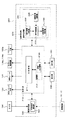
光画像計測装置1の制御系の構成について図5を参照しつつ説明する。
光画像計測装置1の制御系は、演算制御装置200の制御部210を中心に構成される。制御部210は、たとえば、前述のマイクロプロセッサ、RAM、ROM、ハードディスクドライブ、通信インターフェイス等を含んで構成される。
画像形成部220は、撮像装置10、12からの映像信号を受けて眼底画像Ef′の画像データを形成する。
画像形成部220には干渉成分抽出部221が設けられている。干渉成分抽出部221は、深度位置(z方向の位置)が異なる被検眼Eの複数の部位を同時に計測するときに動作する。
画像処理部230は、画像形成部220により形成された画像に対して各種の画像処理や解析処理を施す。たとえば、画像処理部230は、画像の輝度補正や分散補正等の各種補正処理などを実行する。
画像処理部230には解析処理部231が設けられている。解析処理部231は、被検眼Eの複数部位の同時計測により取得された複数の断層画像を解析することにより、被検眼Eの所定の物理量を求める。また、解析処理部231は、求められた被検眼Eの物理量に基づいて、信号光LSの走査態様を特定する。
眼内距離演算部232は、被検眼Eの複数の断層画像のうちの一の断層画像中の位置と他の断層画像中の位置との間の距離を求める。特に、この実施形態では、眼内距離演算部232は、眼底Efの断層画像(眼底断層画像)と角膜Ecの断層画像(角膜断層画像)を解析して、被検眼Eの角膜網膜間距離を求める。以下、眼内距離演算部232が実行する処理の例を説明する。
倍率演算部233は、眼内距離演算部232により求められた角膜網膜間距離に基づいて、被検眼Eの眼球光学系の倍率を求める。倍率演算部233は、この発明の「倍率演算手段」の一例である。以下、倍率演算部233が実行する処理の例を説明する。
走査態様特定部234は、求められた倍率と上記眼球モデルとに基づいて、被検眼Eの網膜の所定位置に信号光LSを照射させるような、走査ユニット141による信号光LSの走査態様を特定する。走査態様特定部234は、この発明の「特定手段」の一例である。
角膜曲率演算部235は、被検眼Eの角膜曲率半径(又は角膜曲率)を求める。なお、角膜曲率半径と角膜曲率は、それぞれ他方の逆数であるので、一方が求まれば他方も求まる。角膜曲率演算部235は、この発明の「角膜曲率半径演算手段」の一例である。以下、角膜曲率演算部235が実行する処理の例を説明する。
表示部240は、ディスプレイを含んで構成される。操作部250は、キーボードやマウス等の入力デバイスや操作デバイスを含んで構成される。又、操作部250には、光画像計測装置1の筐体や外部に設けられた各種のボタンやキーが含まれていてもよい。
ここで、信号光LSの走査及びOCT画像について説明しておく。
光画像計測装置1の動作について説明する。図6に示すフローチャートは、光画像計測装置1の動作の一例を表す。なお、眼球光学情報212aは既に記憶部212に記憶されているものとする。また、前述の事前計測における模型眼の角膜曲率半径の値と、角膜表面の画像の基準位置とは、既に記憶部212に記憶されているものとする。
主制御部211は、ステップ29で特定された走査態様に基づいて信号光を走査させつつ、本計測を実行させる(S30)。画像形成部220は、本計測で得られた検出信号に基づいて、網膜(眼底Ef)の新たな断層画像を形成する(S31)。以上で、図8に示す動作の説明を終了する。
以上のような光画像計測装置1の作用及び効果について説明する。
以上に説明した構成は、この発明を好適に実施するための一例に過ぎない。この発明を実施しようとする者は、この発明の要旨の範囲内における任意の変形を適宜に施すことが可能である。
1A 眼底カメラユニット
141 走査ユニット
150 OCTユニット
160 低コヒーレンス光源
174a、174b 参照ミラー
176a、176b 参照ミラー駆動機構
180 スペクトロメータ
184 CCD
190A アライメント光学系
200 演算制御装置
210 制御部
211 主制御部
212 記憶部
212a 眼球光学情報
220 画像形成部
221 干渉成分抽出部
230 画像処理部
231 解析処理部
232 眼内距離演算部
233 倍率演算部
234 走査態様特定部
235 角膜曲率演算部
240 表示部
250 操作部
Claims (14)
- 光源からの光を信号光と参照光とに分割し、前記参照光の光路を光路長の異なる複数の光路に分割することで前記参照光を複数の参照光に分割し、前記複数の光路をそれぞれ経由した前記複数の参照光と被測定物体を経由した前記信号光とを干渉させて、前記被測定物体の複数の深度位置のそれぞれにおける形態を反映した干渉光を生成する光学系と、
前記生成された干渉光を検出して検出信号を生成する検出手段と、
前記生成された検出信号に基づいて、前記複数の深度位置における前記被測定物体の形態を表す複数の断層画像をそれぞれ形成する画像形成手段と、
前記複数の断層画像を解析して前記被測定物体の所定の物理量を求める解析手段と、
を備え、
前記解析手段は、前記所定の物理量として、前記複数の断層画像のうちの一の断層画像中の位置と他の断層画像中の位置との間の距離を求める、
ことを特徴とする光画像計測装置。 - 前記光学系は、前記光源からの光から分割された参照光を前記複数の参照光に分割するビームスプリッタと、前記複数の参照光のそれぞれの光路に設けられた参照ミラーとを含み、
前記ビームスプリッタは、前記参照ミラーによりそれぞれ反射された前記複数の参照光を合成し、
前記光学系は、前記合成された前記複数の参照光を前記信号光に干渉させて前記干渉光を生成する、
ことを特徴とする請求項1に記載の光画像計測装置。 - 前記光学系は、前記光源からの光から分割された参照光の一部分の光路長を延長する光学部材と、前記光学部材により光路長が延長された前記参照光の一部分と、前記参照光の他の部分とを反射する参照ミラーとを含み、前記参照ミラーにより反射された前記参照光を前記信号光に干渉させて前記干渉光を生成する、
ことを特徴とする請求項1に記載の光画像計測装置。 - 前記被測定物体は生体眼であり、
前記複数の参照光は、前記生体眼の網膜に対応する光路長を有する第1の光路を経由する第1の参照光と、前記生体眼の角膜に対応する光路長を有する第2の光路を経由する第2の参照光とを含み、
前記画像形成手段は、前記第1の参照光と前記網膜で反射された前記信号光との干渉成分に相当する第1の信号成分を前記検出信号から抽出して前記網膜の形態を表す第1の断層画像を前記一の断層画像として形成し、かつ、前記第2の参照光と前記角膜で反射された前記信号光との干渉成分に相当する第2の信号成分を前記検出信号から抽出して前記角膜の形態を表す第2の断層画像を前記他の断層画像として形成し、
前記解析手段は、前記第1及び前記第2の断層画像を解析して、前記生体眼の角膜網膜間距離を求める、
ことを特徴とする請求項1〜請求項3のいずれか一項に記載の光画像計測装置。 - 前記第1の光路と前記第2の光路とは、角膜網膜間距離の標準値に略等しい光路長差を有し、
前記解析手段は、予め記憶された眼球光学情報に含まれる眼球光学系の屈折率の値で前記標準値を除算し、その商の値と前記第1及び前記第2の断層画像とに基づいて前記角膜網膜間距離を求める、
ことを特徴とする請求項4に記載の光画像計測装置。 - 前記解析手段は、前記所定の物理量として、前記求められた角膜網膜間距離に基づいて前記生体眼の眼球光学系の倍率を求める倍率演算手段を含む、
ことを特徴とする請求項4に記載の光画像計測装置。 - 前記倍率演算手段は、予め記憶された眼球光学情報に含まれる眼球光学系の光学情報と、前記求められた角膜網膜間距離とに基づいて前記倍率を求める、
ことを特徴とする請求項6に記載の光画像計測装置。 - 前記眼球光学情報は、角膜の前後面のそれぞれの曲率半径、角膜の厚さ、角膜の屈折率、水晶体の前後面のそれぞれの曲率半径、水晶体の厚さ、水晶体の屈折率、硝子体の屈折率、及び、角膜前面と水晶体後面との間の距離を表す前眼部距離のそれぞれの値を含み、
前記倍率演算手段は、前記角膜網膜間距離から前記前眼部距離の値を減算して水晶体後面と網膜表面との間の距離を表す後眼部距離を算出し、前記眼球光学情報及び前記後眼部距離とに基づいて眼球モデルを形成し、前記眼球モデルに基づいて前記倍率を求める、
ことを特徴とする請求項7に記載の光画像計測装置。 - 前記生体眼に対して前記光学系を位置合わせするアライメント手段を更に備え、
前記解析手段は、前記位置合わせが行われた後に前記光学系により生成された干渉光に基づく前記第2の断層画像のフレーム内における位置を特定し、該特定された位置に基づいて前記生体眼の角膜曲率半径を求める角膜曲率半径演算手段を含み、
前記倍率演算手段は、前記眼球光学情報に含まれる角膜の曲率半径の値の代わりに、前記求められた角膜曲率半径に基づいて前記眼球モデルを形成する、
ことを特徴とする請求項8に記載の光画像計測装置。 - 前記生体眼に対して前記光学系を位置合わせするアライメント手段を更に備え、
前記解析手段は、前記位置合わせが行われた後に前記光学系により干渉光が生成されたときの前記第2の光路の光路長に基づいて前記生体眼の角膜曲率半径を求める角膜曲率半径演算手段を含み、
前記倍率演算手段は、前記眼球光学情報に含まれる角膜の曲率半径の値の代わりに、前記求められた角膜曲率半径に基づいて前記眼球モデルを形成する、
ことを特徴とする請求項8に記載の光画像計測装置。 - 前記光学系は、前記生体眼に対する前記信号光の照射位置を走査する走査手段を含み、
前記解析手段は、前記眼球モデル及び前記求められた倍率に基づいて、前記網膜の所定位置に前記信号光を照射させるような前記走査手段による前記信号光の走査態様を特定する特定手段を含み、
前記光学系は、前記光源からの新たな光を信号光と参照光とに分割し、前記特定された走査態様に基づき前記走査手段により当該新たな信号光を走査しつつ、前記第1の光路を経由した当該新たな参照光と前記網膜を経由した当該新たな信号光とを干渉させて新たな干渉光を生成し、
前記検出手段は、前記新たな干渉光を検出して新たな検出信号を生成し、
前記画像形成手段は、前記新たな検出信号に基づいて、前記網膜の新たな断層画像を形成する、
ことを特徴とする請求項8〜請求項10のいずれか一項に記載の光画像計測装置。 - 前記特定手段は、前記眼球モデル及び前記求められた倍率に基づく光線追跡演算を行うことにより、前記眼球モデルの網膜の前記所定位置に信号光が照射されるような前記走査態様を特定する、
ことを特徴とする請求項11に記載の光画像計測装置。 - 前記特定手段は、前記網膜の視神経乳頭中心を中心とし所定半径を有する円形の軌跡に沿って前記信号光の照射位置を走査させるための前記走査態様を特定する、
ことを特徴とする請求項11に記載の光画像計測装置。 - 前記解析手段は、前記新たな断層画像に基づいて前記生体眼の網膜厚を求める、
ことを特徴とする請求項11に記載の光画像計測装置。
Priority Applications (4)
| Application Number | Priority Date | Filing Date | Title |
|---|---|---|---|
| JP2008160548A JP5324839B2 (ja) | 2008-06-19 | 2008-06-19 | 光画像計測装置 |
| EP09766377.7A EP2301423B1 (en) | 2008-06-19 | 2009-06-08 | Optical image measuring device |
| PCT/JP2009/002565 WO2009153929A1 (ja) | 2008-06-19 | 2009-06-08 | 光画像計測装置 |
| US12/996,935 US8801180B2 (en) | 2008-06-19 | 2009-06-08 | Ophthalmic tomographic imager with corneo-retinal image analysis |
Applications Claiming Priority (1)
| Application Number | Priority Date | Filing Date | Title |
|---|---|---|---|
| JP2008160548A JP5324839B2 (ja) | 2008-06-19 | 2008-06-19 | 光画像計測装置 |
Publications (3)
| Publication Number | Publication Date |
|---|---|
| JP2010000191A JP2010000191A (ja) | 2010-01-07 |
| JP2010000191A5 JP2010000191A5 (ja) | 2012-07-19 |
| JP5324839B2 true JP5324839B2 (ja) | 2013-10-23 |
Family
ID=41433855
Family Applications (1)
| Application Number | Title | Priority Date | Filing Date |
|---|---|---|---|
| JP2008160548A Active JP5324839B2 (ja) | 2008-06-19 | 2008-06-19 | 光画像計測装置 |
Country Status (4)
| Country | Link |
|---|---|
| US (1) | US8801180B2 (ja) |
| EP (1) | EP2301423B1 (ja) |
| JP (1) | JP5324839B2 (ja) |
| WO (1) | WO2009153929A1 (ja) |
Families Citing this family (118)
| Publication number | Priority date | Publication date | Assignee | Title |
|---|---|---|---|---|
| JP4241038B2 (ja) | 2000-10-30 | 2009-03-18 | ザ ジェネラル ホスピタル コーポレーション | 組織分析のための光学的な方法及びシステム |
| US9295391B1 (en) | 2000-11-10 | 2016-03-29 | The General Hospital Corporation | Spectrally encoded miniature endoscopic imaging probe |
| WO2004088361A2 (en) | 2003-03-31 | 2004-10-14 | The General Hospital Corporation | Speckle reduction in optical coherence tomography by path length encoded angular compounding |
| KR101386971B1 (ko) | 2003-06-06 | 2014-04-18 | 더 제너럴 하스피탈 코포레이션 | 파장 동조 소스용 방법 및 장치 |
| US7733497B2 (en) | 2003-10-27 | 2010-06-08 | The General Hospital Corporation | Method and apparatus for performing optical imaging using frequency-domain interferometry |
| WO2006014392A1 (en) | 2004-07-02 | 2006-02-09 | The General Hospital Corporation | Endoscopic imaging probe comprising dual clad fibre |
| US8081316B2 (en) | 2004-08-06 | 2011-12-20 | The General Hospital Corporation | Process, system and software arrangement for determining at least one location in a sample using an optical coherence tomography |
| WO2006024014A2 (en) | 2004-08-24 | 2006-03-02 | The General Hospital Corporation | Process, system and software arrangement for measuring a mechanical strain and elastic properties of a sample |
| US8208995B2 (en) | 2004-08-24 | 2012-06-26 | The General Hospital Corporation | Method and apparatus for imaging of vessel segments |
| EP2329759B1 (en) | 2004-09-29 | 2014-03-12 | The General Hospital Corporation | System and method for optical coherence imaging |
| JP2008521516A (ja) | 2004-11-29 | 2008-06-26 | ザ ジェネラル ホスピタル コーポレイション | サンプル上の複数の地点を同時に照射し検出することによって光学画像生成を実行する構成、装置、内視鏡、カテーテル、及び方法 |
| EP2325803A1 (en) | 2005-04-28 | 2011-05-25 | The General Hospital Corporation | Evaluating optical coherence tomography information for an anatomical structure |
| US9060689B2 (en) | 2005-06-01 | 2015-06-23 | The General Hospital Corporation | Apparatus, method and system for performing phase-resolved optical frequency domain imaging |
| ES2354287T3 (es) | 2005-08-09 | 2011-03-11 | The General Hospital Corporation | Aparato y método para realizar una desmodulación en cuadratura por polarización en tomografía de coherencia óptica. |
| CN101365375B (zh) | 2005-09-29 | 2013-09-11 | 通用医疗公司 | 用于经由谱编码进行光学成像的方法和设备 |
| US8145018B2 (en) | 2006-01-19 | 2012-03-27 | The General Hospital Corporation | Apparatus for obtaining information for a structure using spectrally-encoded endoscopy techniques and methods for producing one or more optical arrangements |
| PL1973466T3 (pl) | 2006-01-19 | 2021-07-05 | The General Hospital Corporation | Balonowy cewnik do obrazowania |
| WO2007149603A2 (en) | 2006-02-01 | 2007-12-27 | The General Hospital Corporation | Apparatus for applying a plurality of electro-magnetic radiations to a sample |
| JP5524487B2 (ja) | 2006-02-01 | 2014-06-18 | ザ ジェネラル ホスピタル コーポレイション | コンフォーマルレーザ治療手順を用いてサンプルの少なくとも一部分に電磁放射を放射する方法及びシステム。 |
| EP1987318B1 (en) | 2006-02-24 | 2015-08-12 | The General Hospital Corporation | Methods and systems for performing angle-resolved fourier-domain optical coherence tomography |
| EP2517616A3 (en) | 2006-05-10 | 2013-03-06 | The General Hospital Corporation | Processes, arrangements and systems for providing frequency domain imaging of a sample |
| WO2008049118A2 (en) | 2006-10-19 | 2008-04-24 | The General Hospital Corporation | Apparatus and method for obtaining and providing imaging information associated with at least one portion of a sample and effecting such portion(s) |
| US20110319875A1 (en) * | 2007-01-19 | 2011-12-29 | Frieder Loesel | Apparatus and Method for Morphing a Three-Dimensional Target Surface into a Two-Dimensional Image for Use in Guiding a Laser Beam in Ocular Surgery |
| US10534129B2 (en) | 2007-03-30 | 2020-01-14 | The General Hospital Corporation | System and method providing intracoronary laser speckle imaging for the detection of vulnerable plaque |
| EP2335030A4 (en) * | 2008-06-18 | 2014-05-07 | Eyelab Group Llc | SYSTEM AND METHOD FOR DETERMINING VOLUMETRIC PARAMETERS OF OCULAR TISSUES AND OTHER BIOLOGICAL TISSUES |
| WO2010009136A2 (en) | 2008-07-14 | 2010-01-21 | The General Hospital Corporation | Apparatus and methods for color endoscopy |
| DE102008044375A1 (de) * | 2008-12-05 | 2010-06-10 | Robert Bosch Gmbh | Optisches Messgerät |
| BR112012001042A2 (pt) | 2009-07-14 | 2016-11-22 | Gen Hospital Corp | equipamento e método de medição do fluxo de fluído dentro de estrutura anatômica. |
| JP5390371B2 (ja) | 2009-12-25 | 2014-01-15 | 株式会社トプコン | 光画像計測装置及び光アッテネータ |
| JP5351066B2 (ja) * | 2010-01-25 | 2013-11-27 | 浜松ホトニクス株式会社 | Oct装置 |
| ES2831223T3 (es) * | 2010-03-05 | 2021-06-07 | Massachusetts Gen Hospital | Aparato para proporcionar radiación electromagnética a una muestra |
| JP5569963B2 (ja) * | 2010-04-15 | 2014-08-13 | 国立大学法人宇都宮大学 | 表面形状測定装置 |
| US9069130B2 (en) | 2010-05-03 | 2015-06-30 | The General Hospital Corporation | Apparatus, method and system for generating optical radiation from biological gain media |
| US9795301B2 (en) | 2010-05-25 | 2017-10-24 | The General Hospital Corporation | Apparatus, systems, methods and computer-accessible medium for spectral analysis of optical coherence tomography images |
| US9557154B2 (en) | 2010-05-25 | 2017-01-31 | The General Hospital Corporation | Systems, devices, methods, apparatus and computer-accessible media for providing optical imaging of structures and compositions |
| US9339186B2 (en) * | 2010-06-01 | 2016-05-17 | Optovue, Inc. | Method and apparatus for enhanced eye measurements |
| EP2575591A4 (en) | 2010-06-03 | 2017-09-13 | The General Hospital Corporation | Apparatus and method for devices for imaging structures in or at one or more luminal organs |
| JP5685013B2 (ja) | 2010-06-30 | 2015-03-18 | キヤノン株式会社 | 光断層撮像装置及びその制御方法、プログラム |
| JP5601613B2 (ja) * | 2010-07-05 | 2014-10-08 | 株式会社ニデック | 眼底撮影装置 |
| JP5127897B2 (ja) | 2010-08-27 | 2013-01-23 | キヤノン株式会社 | 眼科用画像処理装置及びその方法 |
| US9510758B2 (en) | 2010-10-27 | 2016-12-06 | The General Hospital Corporation | Apparatus, systems and methods for measuring blood pressure within at least one vessel |
| JP5735789B2 (ja) * | 2010-12-02 | 2015-06-17 | 株式会社ニデック | 眼底撮影装置 |
| JP5794664B2 (ja) * | 2011-01-20 | 2015-10-14 | キヤノン株式会社 | 断層画像生成装置及び断層画像生成方法 |
| EP2670293B1 (de) * | 2011-02-04 | 2020-07-15 | Heidelberg Engineering GmbH | Verfahren und vorrichtung für die sequenzielle aufnahme von interferometrischen tiefenschnittbildern in verschiedenen tiefen, insbesondere zur analyse des auges |
| CA2826799C (en) * | 2011-02-15 | 2017-11-14 | Wavelight Gmbh | System and method for measuring internal dimensions of an object by optical coherence tomography |
| WO2012130818A1 (en) * | 2011-03-25 | 2012-10-04 | National Digital Research Centre | Apparatus for modelling ocular structures |
| JP5807371B2 (ja) * | 2011-04-21 | 2015-11-10 | 株式会社ニデック | 眼科撮影装置 |
| CN102283635B (zh) * | 2011-07-13 | 2016-03-09 | 广东福地新视野光电技术有限公司 | 双通道全眼光学相干层析成像系统及成像方法 |
| WO2013013049A1 (en) | 2011-07-19 | 2013-01-24 | The General Hospital Corporation | Systems, methods, apparatus and computer-accessible-medium for providing polarization-mode dispersion compensation in optical coherence tomography |
| US10241028B2 (en) | 2011-08-25 | 2019-03-26 | The General Hospital Corporation | Methods, systems, arrangements and computer-accessible medium for providing micro-optical coherence tomography procedures |
| JP5474011B2 (ja) * | 2011-09-05 | 2014-04-16 | キヤノン株式会社 | 眼科システム、眼科装置、断層像取得方法、およびプログラム |
| AT511935B1 (de) * | 2011-09-12 | 2015-09-15 | Ima Integrated Microsystems Austria Gmbh | Verfahren und vorrichtung zum räumlichen vermessen von gewebestrukturen |
| JP5912358B2 (ja) * | 2011-09-14 | 2016-04-27 | 株式会社トプコン | 眼底観察装置 |
| EP2769491A4 (en) | 2011-10-18 | 2015-07-22 | Gen Hospital Corp | DEVICE AND METHOD FOR PRODUCING AND / OR PROVIDING RECIRCULATING OPTICAL DELAY (DE) |
| US8632178B2 (en) * | 2011-10-19 | 2014-01-21 | Novartis Ag | Determining physical lengths in an eye using multiple refractive indices |
| JP5887839B2 (ja) * | 2011-10-31 | 2016-03-16 | 株式会社ニデック | 眼内レンズ度数決定装置及びプログラム |
| JP5374598B2 (ja) | 2012-01-26 | 2013-12-25 | キヤノン株式会社 | 光断層撮像装置 |
| US8807752B2 (en) | 2012-03-08 | 2014-08-19 | Technolas Perfect Vision Gmbh | System and method with refractive corrections for controlled placement of a laser beam's focal point |
| US20130237969A1 (en) * | 2012-03-08 | 2013-09-12 | Kristian Hohla | System and Method for Short Scan Interferometric Imaging |
| WO2013148306A1 (en) | 2012-03-30 | 2013-10-03 | The General Hospital Corporation | Imaging system, method and distal attachment for multidirectional field of view endoscopy |
| JP5989523B2 (ja) * | 2012-05-01 | 2016-09-07 | 株式会社トプコン | 眼科装置 |
| WO2013177154A1 (en) | 2012-05-21 | 2013-11-28 | The General Hospital Corporation | Apparatus, device and method for capsule microscopy |
| JP6227652B2 (ja) | 2012-08-22 | 2017-11-08 | ザ ジェネラル ホスピタル コーポレイション | ソフトリソグラフィを用いてミニチュア内視鏡を製作するためのシステム、方法、およびコンピュータ・アクセス可能媒体 |
| US10149615B2 (en) | 2012-11-30 | 2018-12-11 | Kabushiki Kaisha Topcon | Fundus imaging apparatus that determines a state of alignment |
| JP6338358B2 (ja) * | 2012-11-30 | 2018-06-06 | 株式会社トプコン | 眼底撮影システム |
| JP6108845B2 (ja) * | 2013-01-24 | 2017-04-05 | 株式会社トプコン | 眼科観察装置 |
| JP6560126B2 (ja) | 2013-01-28 | 2019-08-14 | ザ ジェネラル ホスピタル コーポレイション | 光周波数ドメインイメージングに重ね合わせされる拡散分光法を提供するための装置および方法 |
| WO2014120791A1 (en) | 2013-01-29 | 2014-08-07 | The General Hospital Corporation | Apparatus, systems and methods for providing information regarding the aortic valve |
| US11179028B2 (en) | 2013-02-01 | 2021-11-23 | The General Hospital Corporation | Objective lens arrangement for confocal endomicroscopy |
| US9398979B2 (en) | 2013-03-11 | 2016-07-26 | Technolas Perfect Vision Gmbh | Dimensional compensator for use with a patient interface |
| US9060710B2 (en) * | 2013-03-14 | 2015-06-23 | Amo Wavefront Sciences, Llc. | System and method for ocular tomography using plenoptic imaging |
| US9161688B2 (en) | 2013-03-15 | 2015-10-20 | Amo Wavefront Sciences, Llc | System and method for corneal pachymetry using plenoptic imaging |
| JP6378311B2 (ja) | 2013-03-15 | 2018-08-22 | ザ ジェネラル ホスピタル コーポレイション | 物体を特徴付ける方法とシステム |
| WO2014186353A1 (en) | 2013-05-13 | 2014-11-20 | The General Hospital Corporation | Detecting self-interefering fluorescence phase and amplitude |
| EP3003177B1 (en) | 2013-05-31 | 2021-03-10 | Covidien LP | Surgical device with an end-effector assembly for monitoring of tissue during a surgical procedure |
| WO2015009932A1 (en) | 2013-07-19 | 2015-01-22 | The General Hospital Corporation | Imaging apparatus and method which utilizes multidirectional field of view endoscopy |
| EP3021735A4 (en) | 2013-07-19 | 2017-04-19 | The General Hospital Corporation | Determining eye motion by imaging retina. with feedback |
| EP3025173B1 (en) | 2013-07-26 | 2021-07-07 | The General Hospital Corporation | Apparatus with a laser arrangement utilizing optical dispersion for applications in fourier-domain optical coherence tomography |
| US9733460B2 (en) | 2014-01-08 | 2017-08-15 | The General Hospital Corporation | Method and apparatus for microscopic imaging |
| WO2015116986A2 (en) | 2014-01-31 | 2015-08-06 | The General Hospital Corporation | System and method for facilitating manual and/or automatic volumetric imaging with real-time tension or force feedback using a tethered imaging device |
| JP2015195551A (ja) * | 2014-03-18 | 2015-11-05 | 株式会社東芝 | 画像表示装置及び画像表示方法 |
| WO2015152119A1 (ja) * | 2014-03-31 | 2015-10-08 | 興和株式会社 | 立体断層像の容積計測装置、容積計測方法及び容積計測プログラム |
| TWI494536B (zh) * | 2014-04-01 | 2015-08-01 | Crystalvue Medical Corp | 光學量測裝置及方法 |
| WO2015153982A1 (en) | 2014-04-04 | 2015-10-08 | The General Hospital Corporation | Apparatus and method for controlling propagation and/or transmission of electromagnetic radiation in flexible waveguide(s) |
| DE102014108789A1 (de) * | 2014-06-24 | 2016-01-07 | Byk-Gardner Gmbh | Mehrstufiges Verfahren zur Untersuchung von Oberflächen sowie entsprechende Vorrichtung |
| CN104116495B (zh) * | 2014-07-11 | 2016-02-10 | 北京理工大学 | 一种视网膜光学相干层析探测-显示系统 |
| JP5970682B2 (ja) * | 2014-07-14 | 2016-08-17 | 学校法人北里研究所 | 眼球計測装置、眼球計測方法 |
| WO2016015052A1 (en) | 2014-07-25 | 2016-01-28 | The General Hospital Corporation | Apparatus, devices and methods for in vivo imaging and diagnosis |
| WO2016086341A1 (en) | 2014-12-01 | 2016-06-09 | Dongguan Zkteco Electronic Technology Co., Ltd | System and method for acquiring multimodal biometric information |
| CN107209848B (zh) * | 2014-12-01 | 2020-10-13 | 厦门熵基科技有限公司 | 用于基于多模式生物识别信息的个人识别的系统和方法 |
| JP6588750B2 (ja) * | 2015-06-30 | 2019-10-09 | 株式会社トプコン | 眼科用顕微鏡システム |
| CN105147241B (zh) * | 2015-07-03 | 2017-06-16 | 南京航空航天大学 | 基于双空间载频技术拓展oct成像深度的方法与系统 |
| JP6001150B2 (ja) * | 2015-10-08 | 2016-10-05 | キヤノン株式会社 | 眼科情報処理システム、眼科情報処理方法及びプログラム |
| JP2017173305A (ja) * | 2016-02-10 | 2017-09-28 | 株式会社トーメーコーポレーション | 波長符号化マルチビーム光コヒーレンストモグラフィ |
| EP3448243B1 (en) * | 2016-04-29 | 2023-08-23 | Oncores Medical Pty Ltd | An optical coherence tomography system |
| JP6498162B2 (ja) * | 2016-08-24 | 2019-04-10 | キヤノン株式会社 | 光断層撮像装置 |
| EP3558091A4 (en) | 2016-12-21 | 2020-12-02 | Acucela, Inc. | MINIATURIZED AFFORDABLE OPTICAL COHERENCE TOMOGRAPHY SYSTEM FOR OPHTHALMIC APPLICATIONS IN THE HOME |
| JP6311045B2 (ja) * | 2017-03-06 | 2018-04-11 | 株式会社トプコン | 眼科観察装置 |
| CN106963337B (zh) * | 2017-03-29 | 2018-04-24 | 天津市索维电子技术有限公司 | 一种实现大景深眼前节分析系统 |
| KR102029768B1 (ko) * | 2018-01-23 | 2019-10-08 | 홍석우 | 안축장 산출을 위한 시신경 유두의 형태학적 변화 추출 시스템 및 방법 |
| US11589746B2 (en) * | 2018-01-24 | 2023-02-28 | Duke University | Optical coherence tomography imaging systems, handheld probes, and methods that use a field curvature to match a curved surface of tissue |
| US11213200B2 (en) * | 2018-03-22 | 2022-01-04 | Santec Corporation | Topographical imaging using combined sensing inputs |
| CN108498067A (zh) * | 2018-04-19 | 2018-09-07 | 长春理工大学 | 一种数字化角膜曲率测量的装置以及方法 |
| CA3103899A1 (en) | 2018-06-20 | 2019-12-26 | Acucela Inc. | Miniaturized mobile, low cost optical coherence tomography system for home based ophthalmic applications |
| JP2018149449A (ja) * | 2018-07-10 | 2018-09-27 | 株式会社トプコン | 眼科撮影装置および眼科情報処理装置 |
| JP7213048B2 (ja) * | 2018-09-25 | 2023-01-26 | 株式会社トプコン | 眼科情報処理装置、眼科装置、及び眼科情報処理方法 |
| JP7292072B2 (ja) * | 2019-03-19 | 2023-06-16 | 株式会社トプコン | 眼科装置 |
| JP7349807B2 (ja) * | 2019-03-19 | 2023-09-25 | 株式会社トプコン | 眼科装置 |
| JP2023508946A (ja) | 2019-12-26 | 2023-03-06 | アキュセラ インコーポレイテッド | 自宅ベースの眼科用途のための光干渉断層撮影患者整列システム |
| US11513228B2 (en) | 2020-03-05 | 2022-11-29 | Santec Corporation | Lidar sensing arrangements |
| JP6742550B1 (ja) * | 2020-03-31 | 2020-08-19 | ナガシマ工芸株式会社 | 表示装置、及び表示補助部材 |
| US11486792B2 (en) | 2020-06-05 | 2022-11-01 | Santec Corporation | Tunable light source for optical fiber proximity and testing |
| US10959613B1 (en) | 2020-08-04 | 2021-03-30 | Acucela Inc. | Scan pattern and signal processing for optical coherence tomography |
| US11393094B2 (en) | 2020-09-11 | 2022-07-19 | Acucela Inc. | Artificial intelligence for evaluation of optical coherence tomography images |
| JP2023544704A (ja) | 2020-09-30 | 2023-10-25 | アキュセラ インコーポレイテッド | 近視の予測、診断、計画、および監視デバイス |
| WO2022204622A1 (en) | 2021-03-24 | 2022-09-29 | Acucela Inc. | Axial length measurement monitor |
| DE102022113798A1 (de) * | 2022-06-01 | 2023-12-07 | Heidelberg Engineering Gmbh | Vorrichtung zur Durchführung einer optischen Kohärenztomografie |
| CN116369843B (zh) * | 2023-05-18 | 2023-12-19 | 苏州比格威医疗科技有限公司 | 一种测量人眼多参数的光学系统及其检测方法 |
Family Cites Families (14)
| Publication number | Priority date | Publication date | Assignee | Title |
|---|---|---|---|---|
| ATE286510T1 (de) | 1993-09-07 | 2005-01-15 | Smithkline Beecham Corp | In der behandlung von il4 auslösenden krankheiten nützliche rekombinante il4 antikörper |
| JPH09276232A (ja) | 1996-04-12 | 1997-10-28 | Nikon Corp | 眼底カメラ |
| WO1998043068A1 (fr) * | 1997-03-26 | 1998-10-01 | Kowa Company, Ltd. | Instrument de mesure optique |
| JPH10267830A (ja) * | 1997-03-26 | 1998-10-09 | Kowa Co | 光学測定装置 |
| JP3888771B2 (ja) | 1997-04-24 | 2007-03-07 | 株式会社トプコン | 眼底カメラ |
| DE19814057B4 (de) * | 1998-03-30 | 2009-01-02 | Carl Zeiss Meditec Ag | Anordnung zur optischen Kohärenztomographie und Kohärenztopographie |
| JP2002139421A (ja) | 2000-11-01 | 2002-05-17 | Fuji Photo Film Co Ltd | 光断層画像取得装置 |
| JP4409331B2 (ja) * | 2004-03-30 | 2010-02-03 | 株式会社トプコン | 光画像計測装置 |
| JP4505807B2 (ja) | 2004-08-09 | 2010-07-21 | 国立大学法人 筑波大学 | 多重化スペクトル干渉光コヒーレンストモグラフィー |
| JP4597744B2 (ja) * | 2004-11-08 | 2010-12-15 | 株式会社トプコン | 光画像計測装置及び光画像計測方法 |
| JP4804820B2 (ja) | 2005-07-15 | 2011-11-02 | サンテック株式会社 | 光断層画像表示システム |
| US7400410B2 (en) * | 2005-10-05 | 2008-07-15 | Carl Zeiss Meditec, Inc. | Optical coherence tomography for eye-length measurement |
| ATE516739T1 (de) * | 2005-12-06 | 2011-08-15 | Zeiss Carl Meditec Ag | Interferometrische probenmessung |
| JP5095167B2 (ja) * | 2006-09-19 | 2012-12-12 | 株式会社トプコン | 眼底観察装置、眼底画像表示装置及び眼底観察プログラム |
-
2008
- 2008-06-19 JP JP2008160548A patent/JP5324839B2/ja active Active
-
2009
- 2009-06-08 EP EP09766377.7A patent/EP2301423B1/en active Active
- 2009-06-08 US US12/996,935 patent/US8801180B2/en active Active
- 2009-06-08 WO PCT/JP2009/002565 patent/WO2009153929A1/ja active Application Filing
Also Published As
| Publication number | Publication date |
|---|---|
| EP2301423A1 (en) | 2011-03-30 |
| JP2010000191A (ja) | 2010-01-07 |
| WO2009153929A1 (ja) | 2009-12-23 |
| EP2301423B1 (en) | 2017-05-31 |
| US8801180B2 (en) | 2014-08-12 |
| US20110080561A1 (en) | 2011-04-07 |
| EP2301423A4 (en) | 2013-04-10 |
Similar Documents
| Publication | Publication Date | Title |
|---|---|---|
| JP5324839B2 (ja) | 光画像計測装置 | |
| JP4864516B2 (ja) | 眼科装置 | |
| JP6522827B2 (ja) | 眼科装置 | |
| JP5061380B2 (ja) | 眼底観察装置、眼科画像表示装置及びプログラム | |
| JP4971872B2 (ja) | 眼底観察装置及びそれを制御するプログラム | |
| JP6009935B2 (ja) | 眼科装置 | |
| WO2010125746A1 (ja) | 眼底観察装置 | |
| JP6624641B2 (ja) | 眼科装置 | |
| JP2022176282A (ja) | 眼科装置、及びその制御方法 | |
| JP2018198967A (ja) | 眼科装置 | |
| JP5514026B2 (ja) | 眼底画像処理装置及び眼底観察装置 | |
| JP5584345B2 (ja) | 光画像計測装置及び撮影装置 | |
| JP7134014B2 (ja) | 眼科装置、及びその制御方法 | |
| JP7199172B2 (ja) | 眼科装置、及びその制御方法 | |
| JP2020031827A (ja) | 眼科情報処理装置、眼科装置、及び眼科情報処理方法 | |
| JP2019154764A (ja) | 涙液層厚み測定装置及び方法 | |
| JP2022060588A (ja) | 眼科装置、及び眼科装置の制御方法 | |
| JP7202819B2 (ja) | 眼科装置、及びその制御方法 |
Legal Events
| Date | Code | Title | Description |
|---|---|---|---|
| A621 | Written request for application examination |
Free format text: JAPANESE INTERMEDIATE CODE: A621 Effective date: 20110511 |
|
| A521 | Request for written amendment filed |
Free format text: JAPANESE INTERMEDIATE CODE: A523 Effective date: 20120531 |
|
| A131 | Notification of reasons for refusal |
Free format text: JAPANESE INTERMEDIATE CODE: A131 Effective date: 20130507 |
|
| A521 | Request for written amendment filed |
Free format text: JAPANESE INTERMEDIATE CODE: A523 Effective date: 20130628 |
|
| TRDD | Decision of grant or rejection written | ||
| A01 | Written decision to grant a patent or to grant a registration (utility model) |
Free format text: JAPANESE INTERMEDIATE CODE: A01 Effective date: 20130716 |
|
| A61 | First payment of annual fees (during grant procedure) |
Free format text: JAPANESE INTERMEDIATE CODE: A61 Effective date: 20130719 |
|
| R150 | Certificate of patent or registration of utility model |
Ref document number: 5324839 Country of ref document: JP Free format text: JAPANESE INTERMEDIATE CODE: R150 Free format text: JAPANESE INTERMEDIATE CODE: R150 |
|
| R250 | Receipt of annual fees |
Free format text: JAPANESE INTERMEDIATE CODE: R250 |
|
| R250 | Receipt of annual fees |
Free format text: JAPANESE INTERMEDIATE CODE: R250 |
|
| R250 | Receipt of annual fees |
Free format text: JAPANESE INTERMEDIATE CODE: R250 |
|
| R250 | Receipt of annual fees |
Free format text: JAPANESE INTERMEDIATE CODE: R250 |
|
| R250 | Receipt of annual fees |
Free format text: JAPANESE INTERMEDIATE CODE: R250 |
|
| R250 | Receipt of annual fees |
Free format text: JAPANESE INTERMEDIATE CODE: R250 |
|
| R250 | Receipt of annual fees |
Free format text: JAPANESE INTERMEDIATE CODE: R250 |
|
| R250 | Receipt of annual fees |
Free format text: JAPANESE INTERMEDIATE CODE: R250 |