EP3753480A1 - Systèmes, procédés et support accessible par ordinateur fournissant des images microscopiques d'au moins une structure anatomique à une résolution particulière - Google Patents
Systèmes, procédés et support accessible par ordinateur fournissant des images microscopiques d'au moins une structure anatomique à une résolution particulière Download PDFInfo
- Publication number
- EP3753480A1 EP3753480A1 EP20183108.8A EP20183108A EP3753480A1 EP 3753480 A1 EP3753480 A1 EP 3753480A1 EP 20183108 A EP20183108 A EP 20183108A EP 3753480 A1 EP3753480 A1 EP 3753480A1
- Authority
- EP
- European Patent Office
- Prior art keywords
- wave
- guide
- exemplary
- electro
- radiation
- Prior art date
- Legal status (The legal status is an assumption and is not a legal conclusion. Google has not performed a legal analysis and makes no representation as to the accuracy of the status listed.)
- Pending
Links
Images


Classifications
-
- A—HUMAN NECESSITIES
- A61—MEDICAL OR VETERINARY SCIENCE; HYGIENE
- A61B—DIAGNOSIS; SURGERY; IDENTIFICATION
- A61B5/00—Measuring for diagnostic purposes; Identification of persons
- A61B5/0059—Measuring for diagnostic purposes; Identification of persons using light, e.g. diagnosis by transillumination, diascopy, fluorescence
- A61B5/0062—Arrangements for scanning
- A61B5/0066—Optical coherence imaging
-
- A—HUMAN NECESSITIES
- A61—MEDICAL OR VETERINARY SCIENCE; HYGIENE
- A61B—DIAGNOSIS; SURGERY; IDENTIFICATION
- A61B5/00—Measuring for diagnostic purposes; Identification of persons
- A61B5/06—Devices, other than using radiation, for detecting or locating foreign bodies ; determining position of probes within or on the body of the patient
-
- A—HUMAN NECESSITIES
- A61—MEDICAL OR VETERINARY SCIENCE; HYGIENE
- A61B—DIAGNOSIS; SURGERY; IDENTIFICATION
- A61B5/00—Measuring for diagnostic purposes; Identification of persons
- A61B5/0033—Features or image-related aspects of imaging apparatus classified in A61B5/00, e.g. for MRI, optical tomography or impedance tomography apparatus; arrangements of imaging apparatus in a room
- A61B5/004—Features or image-related aspects of imaging apparatus classified in A61B5/00, e.g. for MRI, optical tomography or impedance tomography apparatus; arrangements of imaging apparatus in a room adapted for image acquisition of a particular organ or body part
- A61B5/0044—Features or image-related aspects of imaging apparatus classified in A61B5/00, e.g. for MRI, optical tomography or impedance tomography apparatus; arrangements of imaging apparatus in a room adapted for image acquisition of a particular organ or body part for the heart
-
- A—HUMAN NECESSITIES
- A61—MEDICAL OR VETERINARY SCIENCE; HYGIENE
- A61B—DIAGNOSIS; SURGERY; IDENTIFICATION
- A61B5/00—Measuring for diagnostic purposes; Identification of persons
- A61B5/0059—Measuring for diagnostic purposes; Identification of persons using light, e.g. diagnosis by transillumination, diascopy, fluorescence
- A61B5/0075—Measuring for diagnostic purposes; Identification of persons using light, e.g. diagnosis by transillumination, diascopy, fluorescence by spectroscopy, i.e. measuring spectra, e.g. Raman spectroscopy, infrared absorption spectroscopy
-
- A—HUMAN NECESSITIES
- A61—MEDICAL OR VETERINARY SCIENCE; HYGIENE
- A61B—DIAGNOSIS; SURGERY; IDENTIFICATION
- A61B5/00—Measuring for diagnostic purposes; Identification of persons
- A61B5/0059—Measuring for diagnostic purposes; Identification of persons using light, e.g. diagnosis by transillumination, diascopy, fluorescence
- A61B5/0082—Measuring for diagnostic purposes; Identification of persons using light, e.g. diagnosis by transillumination, diascopy, fluorescence adapted for particular medical purposes
- A61B5/0084—Measuring for diagnostic purposes; Identification of persons using light, e.g. diagnosis by transillumination, diascopy, fluorescence adapted for particular medical purposes for introduction into the body, e.g. by catheters
-
- G—PHYSICS
- G01—MEASURING; TESTING
- G01B—MEASURING LENGTH, THICKNESS OR SIMILAR LINEAR DIMENSIONS; MEASURING ANGLES; MEASURING AREAS; MEASURING IRREGULARITIES OF SURFACES OR CONTOURS
- G01B9/00—Measuring instruments characterised by the use of optical techniques
- G01B9/02—Interferometers
-
- G—PHYSICS
- G01—MEASURING; TESTING
- G01B—MEASURING LENGTH, THICKNESS OR SIMILAR LINEAR DIMENSIONS; MEASURING ANGLES; MEASURING AREAS; MEASURING IRREGULARITIES OF SURFACES OR CONTOURS
- G01B9/00—Measuring instruments characterised by the use of optical techniques
- G01B9/02—Interferometers
- G01B9/0209—Low-coherence interferometers
- G01B9/02091—Tomographic interferometers, e.g. based on optical coherence
-
- G—PHYSICS
- G02—OPTICS
- G02B—OPTICAL ELEMENTS, SYSTEMS OR APPARATUS
- G02B6/00—Light guides; Structural details of arrangements comprising light guides and other optical elements, e.g. couplings
- G02B6/24—Coupling light guides
- G02B6/26—Optical coupling means
- G02B6/32—Optical coupling means having lens focusing means positioned between opposed fibre ends
Definitions
- the present disclosure relates to exemplary embodiments of imaging systems, apparatus and methods, and more specifically to methods, systems and computer-accessible medium which provide microscopic images of at least one anatomical structure at a particular resolution.
- Topics relevant to the pathophysiology of CAD such as the development and progression of coronary atherosclerotic lesions, plaque rupture and coronary thrombosis, and the arterial response to coronary device and pharmacologic therapies are therefore of great significance today.
- These biological processes can be mediated by molecular and cellular events that occur on a microscopic scale. Certain progress in understanding, diagnosing, and treating CAD has been hindered by the fact that it has been difficult or impossible to interrogate the human coronary wall at cellular-level resolution in vivo.
- OCT optical coherence tomography
- IVUS intravascular ultrasound
- OFDI optical frequency domain imaging
- a transverse resolution in OCT procedure(s) can be determined by the catheter's focal spot size.
- This conventional method neglects the intrinsic compromise between transverse resolution and depth of field in cross-sectional OCT images and results in images in which only a narrow depth range is resolved.
- Bessel beam illumination and detection of light reflected from the sample can suffer from a significant reduction in contrast and detection efficiency.
- certain exemplary embodiments of the present disclosure can be associated and/or utilize analysis and manipulation of a coherent transfer function (CTF) of an exemplary OCT system.
- the current invention is instead based on an analysis and manipulation of the coherent transfer function (CTF) of an OCT system.
- the CTF can be considered a coherent extension of a modulation transfer function (MTF) and an optical transfer function (OTF).
- MTF modulation transfer function
- OTF optical transfer function
- the MTF or OTF can be manipulated and utilized according to certain exemplary embodiments.
- the quality of an optical system can be assessed by comparing its transfer function to that of a diffraction-limited optical system.
- Figure 1 shows a graph of coherent transfer functions (CTFs) for, e.g., a diffraction limited 2.5 ⁇ m diameter spot and 2.5 ⁇ m spot with an extended focal range of 2.0 mm, produced by Bessel beam illumination and detection.
- CTFs coherent transfer functions
- the transfer function of a Bessel beam illumination and detection 100 can have spatial frequencies that exceed a diffraction-limited system 110, although it likely sacrifices low- and mid-range spatial frequencies, possibly resulting in reduced contrast and detection sensitivity.
- one of the objects of the present disclosure is to provide exemplary embodiments of systems, methods and computer- accessible medium according to the present disclosure, which can provide microscopic images of at least one anatomical structure at a particular resolution.
- Another object of the present disclosure is to overcome a limited depth of focus limitations of conventional Gaussian beam and spatial frequency loss of Bessel beam systems for OCT procedures and/or systems and other forms of extended focal depth imaging.
- more than two imaging channels can illuminate/detect different Bessel and/or Gaussian beams.
- different transfer functions can be illuminated and/or detected.
- the exemplary combination of images obtained with such additional exemplary beams can facilitate the ⁇ OCT CTF to be provided to the diffraction-limited case, and can also facilitate a depth-of-field extension even further.
- exemplary embodiments of probes, apparatus, systems and methods can be provided which provide at least one electro-magnetic radiation to at least one sample.
- a plurality of axicon lenses can be provided which are configured to provide the electro-magnetic radiation(s) having at least partially annulus shape.
- a housing arrangement can be provided which at least partially encloses the axicon lenses. The housing arrangement can be shaped and structured to be inserted into an anatomical structure and/or an endoscope.
- An optical arrangement can be provided which, when receiving the electro-magnetic radiation(s), generates a further radiation that generates a transfer function of the optical arrangement which is different from the transfer function of at least one of the axicon lenses.
- a plurality of wave-guiding arrangements can also be provided, one of which can be coupled to at least one of the axicon arrangements, and another one of which can be coupled to the optical arrangement.
- the resultant respective radiations can be at least partially focused to a depth of focus and/or a focal range that is greater than approximately Raleigh range of a full aperture of illumination.
- a spot diameter of focus can be less than 10 ⁇ m, and the depth of the focus or the focal range can be greater than approximately 1 mm, 0.5 mm, 2 mm, etc.
- further probes, apparatus, systems and methods can be provided which provide at least one electromagnetic radiation to at least one sample.
- at least one optical arrangement can be provided which is configured to forward at least one radiation to the sample therethrough having at least partially circularly-symmetric pattern.
- at least one first portion of the radiation transmitted through a circular section of the pattern can have an optical path- length that is different from an optical path- length of at least one second portion of the radiation transmitted through at least one other section of the pattern.
- the first and second portions of the radiation(s) can be associated with respective first and second transfer functions that are different from one another.
- An interferometric arrangement can be provided which includes at plurality of detectors, and each of the detectors can be configured to detect the first transfer function and the second transfer function.
- the resultant respective radiations can be at least partially focused to a depth of focus or a focal range that is greater than approximately Raleigh range of a full aperture of illumination.
- a spot diameter of focus can be less than 10 ⁇ m, and the depth of the focus or the focal range can be greater than approximately 1 mm, 0.5 mm, 2 mm, etc.
- the optical arrangement(s) can include a plurality of wave guiding arrangements, and, at a point of emission of each of the wave guiding arrangements, the optical arrangement(s) can causes a phase of each of the electro-magnetic radiation(s) to have a predetermined value.
- the optical arrangement(s) can include a plurality of axicon lenses which are configured to generate at least one radiation.
- two or more imaging channels can be utilized, e.g., at least one which providing the Bessel beam illumination or detection and at least another one of which providing a Gaussian beam illumination or detection.
- This exemplary configuration can facilitate three or more unique and separable illumination-detection combinations (e.g., Bessel-Bessel, Bessel-Gaussian, Gaussian-Gaussian, etc.), where each combination can correspond to a different OCT image.
- coherent transfer functions CTFs for 2.5 ⁇ m diameter spots are provided.
- Figure 2 illustrates a graphical comparison of a diffraction limit 200, extended focal range of 0.15 mm used in preliminary data 210, and the exemplary results of an exemplary embodiment of a procedure or technique according to the present disclosure, hereinafter termed ⁇ OCT, with a focal range of 2.0 mm.
- ⁇ OCT CTF can be generated, e.g., by combining Gaussian-Gaussian images 220, Bessel-Gaussian images 230, and Bessel-Bessel images 240.
- the exemplary ⁇ OCT CTF procedure/technique can be used and/or provided over an axial focus range that can be, e.g., more than 0.5 mm, 1 mm, 2 mm, etc. (as well as others).
- the transverse FWHM spot diameters can be less than 5 ⁇ m, 2 ⁇ m, 1 ⁇ m, etc. (as well as others).
- the depth of focus can be extended a factor of, e.g., approximately 2, 5, 10, 20, 50, 100, etc. (and possibly more) compared to the illumination with a plane wave or Gaussian beam.
- the high, low, and medium spatial frequency content in the image can be at least partially restored by combining images with different transfer functions.
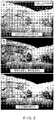
- Figures 3A-3C show exemplary OCT images of a cadaver coronary artery plaque obtained using an exemplary procedure/techniques according to exemplary embodiments of the present disclosure.
- an exemplary Gauss-Gauss image contains low spatial frequency information.
- an exemplary Bessel-Bessel image provides high-resolution but loses low and mid spatial frequencies.
- a combined ⁇ OCT image e.g., Gauss-Gauss+Gauss-Bessel+Bessel-Bessel
- images are normalized and displayed with the same brightness/contrast values.
- Figure 4 shows a second exemplary embodiment of distal optics of a OCT catheter system according to the present disclosure.
- the exemplary system of Figure 4 illustrates an axicon arrangement (e.g., pair) and a routing of the annulus (shown in a darker shade in Figure 4 ) and the Gaussian beam (shown in a darker shade in Figure 4 ) of the distal optics design according to this exemplary embodiment.
- the exemplary system illustrate din Figure 4 can generate a diffraction-limited CTF and an axial focus range (e.g., depth-of- focus) that can be more than, e.g., 10 times longer than the diffraction-limited depth-of-focus.
- the output of a waveguide 500 can be collimated by a collimator 510 located in a center of the exemplary catheter system.
- the collimated electromagnetic radiation e.g., light
- the axicons can be generated or produced using gradient index.
- a separate waveguide 540 can be routed through the center of the annulus.
- the output of the waveguide can be collimated by a collimator 550 located in the center of the annulus.
- Simulated transverse intensity profiles of the collimated annular and Gaussian beams are shown in an illustration of Figure 5A .
- Collimated annular and Gaussian beams can be focused onto the sample using one or more lens, such as a GRIN lens 560.
- the GRIN lens 560 can be configured to intentionally generate chromatic aberration, which can extend the axial focus further (as shown in an illustration of Figure 5B ), and to compensate the aberrations induced by the transparent outer sheath 570.
- the electro-magnetic radiation e.g., light
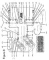
- Figure 6 shows a schematic diagram of an imaging system for generating ⁇ OCT images according to an exemplary embodiment of the present disclosure.
- an output of a source 600 providing electro-magnetic radiation(s) e.g., light radiation
- a linear polarizer 602 can be linearly polarized by a linear polarizer 602 and split into two or more beams by a beam splitter 604. At least one of the beams can be redirected to an input port of a switch 606.
- At least one of outputs of the switch 606 can be transmitted through a beam splitter 610, and coupled into a first light/electro-magnetic radiation guide 612. Another other of the outputs of the switch 606 can be attenuated by an attenuator 614, guided by a second light/electro-magnetic radiation guide 616 to a third beam splitter 618, and redirected to a reference reflector 620 through an attenuator 622, a third light/electro-magnetic radiation guide 624 and a dispersion compensation arrangement 626.
- An output of the light guide 612 can be connected to Bessel illumination and Bessel detection channel of a catheter 628.
- a further one of the outputs of the beam splitter 604 can be redirected to input port of a second three-port switch 630.
- One of the outputs of the switch 630 can be transmitted through a beam splitter 632, and coupled into a fourth light/electro- magnetic radiation guide 634.
- Another one of the outputs of the switch 630 can be attenuated by an attenuator 635 guided by a fifth light guide 636 to a fourth beam splitter 638, and redirected to a reference reflector 640 through an attenuator 642, a fifth light guide 644 and a second dispersion compensation arrangement 646.
- the output of the light guide 634 can be connected to a Gaussian illumination and Gaussian detection channel of the catheter 628.
- the state of the switch 606 is 1, and the state of a fourth beam splitter 638 is 2, e.g., only the light/electro-magnetic radiation guide 612 can be illuminated so that the sample is illuminated by the Bessel illumination channel (see Table 1 of Figure 6 ).
- the back- scattered light from the sample can picked up by both, some or all of the Bessel and Gaussian detection channels of the catheter 628 (see Table 1 of Figure 6 ).
- the portion of electromagnetic radiation/light picked up by the Bessel detection channel can be guided by the first electro-magnetic radiation/light guide 612 to the beam splitter 610, where such radiation/light can be combined and interfered with the light from the reference reflector 620.
- At least part of the interference signal can be directed by the beam splitter 610 to a pinhole 648.
- An output of the pinhole 648 can be collimated and split by a polarizing beam splitter 650.
- One of outputs of the polarizing beam splitters 650 can be transmitted through a half wave plate 652, and detected by a spectrometer 654.
- Another of the outputs of the polarizing beam splitters 650 can be detected by a second spectrometer 656.
- a portion of the electro-magnetic radiation/light picked up by the Gaussian detection channel can be guided by the light guide 634 to the beam splitter 632, where it is combined and interfered with the light from the reference reflector 640.
- At least part of the interference signal can be directed by the beam splitter 634 to a pinhole 658.
- An output of the pinhole 658 can be collimated and split by a polarizing beam splitter 660.
- At least one of outputs of the polarizing beam splitters 660 can be transmitted through a half wave plate 662, and detected by a third spectrometer 664.
- Another of the outputs of the polarizing beam splitters 660 can be detected by a fourth spectrometer 666.
- the fourth electro-magnetic radiation/light guide 634 can be illuminated, so that the sample is illuminated by Gaussian illumination channel (shown in Table 1 of Figure 6 ).
- the back- scattered electro-magnetic radiation/light from the sample can be picked up by both Bessel and Gaussian detection channels of the catheter 630 (shown in Table 1 of Figure 6 ).
- At least one portion of the electro-magnetic radiation/light picked up by the Bessel detection channel is guided by the electro-magnetic radiation/light guide 612 to the beam splitter 610, where it can be combined and interfered with the light from the reference reflector 620.
- At least part of the interference signal can be directed by the beam splitter 610 to a pinhole 648.
- An output of the pinhole 648 can be collimated and split by a polarizing beam splitter 650.
- At least one of outputs of the polarizing beam splitters 650 can be transmitted through a half wave plate 652, and detected by a spectrometer 654.
- Another of the outputs of the polarizing beam splitters 650 can be detected by a second spectrometer 656.
- the portion of light picked up by the Gaussian detection channel is guided by the electro-magnetic radiation/light guide 634 to the beam splitter 632, where it is combined and interfere with the light/radiation from the reference reflector 640. At least part of the interference signal can be directed by the fourth electro-magnetic radiation/light guide 634 to a pinhole 658.
- the output of pinhole 658 is collimated and split by a polarizing beam splitter 660. AT least one of the two outputs of the polarizing beam splitters 660 can be transmitted through a half wave plate 662, and detected by a third spectrometer 664. Another of the outputs of the polarizing beam splitters 660 can be detected by a fourth spectrometer 666.
- Such exemplary polarization-diverse detection scheme/configuration shown in Figure 6 implemented by the combination of the polarizing beam splitter 650, the half wave plate 652 and the spectrometers 654, 656, and/or a combination of the polarizing beam splitter 660, the half wave plate 662 and the spectrometers 664, 666 can reduce and/or eliminate artifacts associated with tissue or optical fiber birefringence.
- the exemplary embodiment of the ⁇ OCT catheter system according the present disclosure illustrated in Figure 6 can contain multiple waveguides that can, e.g., independently transmit and/or receive light/radiation from the catheter to waveguides 612 and 632.
- the detected signal can be digitized and transferred by a computer 668 via an image acquisition board 670. Data can be digitally displayed on or via a monitor 672, and/or stored in a storage device 674.
- the ⁇ OCT detection technology can be implemented using, in one exemplary embodiment, a time domain OCT (TD-OCT) system, in another exemplary embodiment, a spectral-domain (SD-OCT) system, and, in yet another exemplary embodiment, an optical frequency domain interferometry (OFDI) system.
- TD-OCT time domain OCT
- SD-OCT spectral-domain
- OFDI optical frequency domain interferometry
- Complex images and/or real images from the different transfer function illumination and detection configurations can be acquired using the exemplary embodiment of the imaging system according to the present disclosure.
- such exemplary images can be filtered and recombined to generate a new image with an improved quality and a CTF that more closely approximates the diffraction limited CTF.
- the exemplary images with different transfer functions can be filtered or recombined incoherently and/or coherently to generate a new image with a CTF procedure/technique that more closely approximates the diffraction limited CTF procedure/technique.
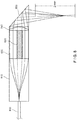
- Figure 7 shows another exemplary embodiment of distal optics configuration of a OCT catheter according to the present disclosure for generating a diffraction-limited CTF and an axial focus range (e.g., depth-of-focus) that can be more than, e.g., approximately 10 times longer than the diffraction-limited depth-of-focus.
- a diffraction-limited CTF and an axial focus range e.g., depth-of-focus
- an output of a waveguide 700 can be collimated by a collimator 710.
- the waveguide 700 can be routed through the annular beam and is collimated Gaussian beam will be routed through the center of the annulus.
- the collimated light can be transformed into an annular beam through two or more axicons, such as, e.g., GRIN axicons 720, 730.
- a separate waveguide 740 can be routed through a center of the annulus.
- An output of the waveguide 740 can be collimated by a collimator 750 located in the center of the annulus.
- the collimated annular and Gaussian beams can be focused onto the sample using one or more lens(es) 760, which can be, e.g., one or more GRIN lenses.
- the GRIN lens 760 can be configured and/or structured to intentionally generate chromatic aberration(s), which can extend the axial focus further and compensate for the aberrations induced by a transparent outer sheath.
- the light/radiation can be directed to the artery wall by a deflector 770.
- Figure 8 shows another exemplary embodiment of the distal optics configuration of the OCT catheter according to the present disclosure.
- Such exemplary configuration can be used to generate a diffraction-limited CTF and depth of focus that is, e.g., more than 10 times longer than the diffraction-limited depth-of-focus.
- An output of a waveguide 800 can be collimated by a collimator 810.
- a pupil aperture created by the collimator 810 can be split into two or more beams, i.e., central circular beam(s) and an annular beam.
- One or more lenses 820 such as an objective lens, achromat lens, aplanat lens, or GRIN lens, that has an aperture substantially the similar as or identical to a central zone can focus a low NA Gaussian beam into the tissue or the sample.
- the annular beam can be transmitted through a spacer 830, and focused into the sample by an annular axicon lens 840 with an aperture that is substantially similar or identical to the annular beam.
- the beams can be directed to the sample by a deflector 850.
- the optical pathlength of the lens 820 can be configured to be different from that of the spacer 830 so that each of, e.g., four images generated can be pathlength encoded.
- the different images can be detected, and their CTF can be combined as per the exemplary methods and/or procedures described herein.
- Figure 9 shows another exemplary embodiment of the distal optics configuration of the OCT catheter system according to the present disclosure, which can be used for generating a diffraction-limited CTF and a depth of focus that is longer than the diffraction- limited depth-of-focus.
- the output of a waveguide 900 can be collimated by a collimator 910.
- a pupil aperture created by the collimator 910 can be split into two or more zones by a circular glass window 920 positioned at the center of the objective lens aperture, e.g., (i) a central circular zone that is transmitted through the circular glass window 920, and (ii) an annular zone.
- the central circular beam can be focused as a low NA Gaussian beam into the tissue and/or sample, and the annular beam can be focused into a Bessel beam focus in the tissue by the lens 930.
- a glass window can have a higher refractive index than air, and the thickness of the window can be so chosen such that the light/radiation field that undergoes different channel can be path-length separated and/or encoded.
- Figure 10 shows a further exemplary embodiment of the distal optics configuration of the OCT catheter system for generating a diffraction-limited CTF and a depth of focus that can be longer than the diffraction-limited depth-of- focus.
- An output of a waveguide 1000 can be collimated by a collimator 1010.
- a pupil aperture created by the collimator 1010 can be split into a number of concentric zones 1020, 1030, 1040.
- a multifocal lens such as, e.g., a GRIN lens, can be used so that the beam in each zone can be focused to a different axial focal position.
- the scattered light/radiation from each zone can be optical pathlength-encoded so that such scattered beams do not interfere with each other.
- the different images can be detected, and their CTF combined pursuant to the exemplary methods and procedures described herein.
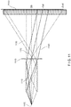
- Figure 11 shows yet another exemplary embodiment of the distal optics configuration of the OCT catheter system for generating a diffraction-limited CTF and an axial focus range (e.g., depth-of-focus) that is longer than the diffraction-limited depth-of- focus.
- an output of a point object 1100 can be transformed by a mirror tunnel device 1110 to multiple orders of light/radiation beams, e.g., zeroth order beam 1120, -1st order beam 1130, and -2nd order beam 1140, etc.
- each order of rays can contain a unique band of spatial frequency of the illumination/detection CTF of the focusing device.
- These orders can, in yet another exemplary embodiment, be path length-encoded so that images generated therein can be detected, and their CTF combined using the different images corresponding to the different orders as per the exemplary CTF combination methods and/or procedures described herein.
- Figures 12 shows another exemplary embodiment of the distal optics configuration of the OCT catheter system according to the present disclosure for generating a diffraction-limited CTF and a depth of focus that is longer than the diffraction-limited depth- of-focus.
- an output of a waveguide 1200 can be focused by a half ball lens 1210.
- a planar surface of the half ball lens 1210 can have a binary phase pattern 1220.
- the depth of the pattern can be configured to produce a small phase shift, e.g., such as a pattern depth of 198 nm ( ⁇ phase shift at 850 nm).
- the top surface can be coated with a reflecting coating, such as Au, and a bottom surface can be coated with the same and/or another coating such as Al, with the final phase shift being given by a curve 1300 shown in a graph of Figure 13 , which illustrates an optical phase length difference of the glass mask (e.g., no metal coating) and a total phase shift (e.g., mask + coating).
- a reflecting coating such as Au
- a bottom surface can be coated with the same and/or another coating such as Al, with the final phase shift being given by a curve 1300 shown in a graph of Figure 13 , which illustrates an optical phase length difference of the glass mask (e.g., no metal coating) and a total phase shift (e.g., mask + coating).
- a curve 1310 and a curve 1320 of the graph of Figure 13 can have a wavelength-dependent phase change of the p-polarized light upon reflection at BK7-A1 and BK7-Au, respectively, with an incident angle of 45 degrees.
- the curve 1330 can be the wavelength dependent phase shift of the light caused by, e.g., 198 nm height difference upon 45 degree reflection at BK7-air interface.
- a binary phase mask can be optimized to produce an extended axial focus (as shown in an illustration of Figure 14b ) compared with the diffraction limited axial focus (as shown in an illustration of Figure 14a ).
- the light/radiation transmitted from the surfaces with different phase shifts can generate different transfer functions, which can be detected and combined to create a new image with a different CTF pursuant to the exemplary methods and/or procedures described herein.
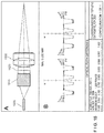
- Figure 15A shows a side-cut-away view of a diagram of another exemplary embodiment of the distal optics configuration of the OCT catheter system for generating a diffraction-limited CTF and an depth of focus longer than the diffraction-limited depth-of- focus.
- the system of Figure 15A generates the results by a factor of, e.g., approximately 2, 5, 10, 20, 10, 100, etc.
- An output of a waveguide 1500 can be collimated by one or more lens(es) 1510.
- the collimated beam can be spatially modulated by a phase doublet 1520, which can include a positive phase plate and a negative phase plate with the same or similar phase pattern.
- Figure 15B shows an exemplary graph of transverse phase profiles of an exemplary mask (e.g., BK7-SNPH2 phase doublet mask) illustrated in Figure 15A
- an exemplary mask e.g., BK7-SNPH2 phase doublet mask
- the spatially modulated beam can be focused into an extended axial focus by an objective lens 1530.
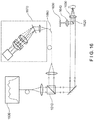
- Figure 16 shows still another exemplary embodiment of the distal optics configuration of the OCT catheter system for generating a diffraction-limited CTF and depth of focus according to the present disclosure that is longer than the diffraction-limited depth- of-focus, by a factor of preferably approximately 2, 5, 10, 20, 10, 100, etc..
- An output of a light source 1600 can be split by a beam splitter 1610.
- the beam aperture of at least one of the outputs of the beam splitter can be split or separated by a rod mirror 1620 into two or more regions.
- the rod mirror 1620 can redirect the central part of the beam to a reference reflector 1630 through an objective lens 1640.
- the annular beam can be focused into the sample by a second objective lens 1660 that can be substantially similar or identical to one or more lens(es) 1640 into a Bessel focus featured with extended axial focus and super-resolution in transverse direction (as shown in the exemplary ⁇ OCT images of Figure 18D ).
- the light back-scattered from the sample is combined with the light reflected from the reference reflector through the rod mirror at a pinhole 1660.
- the output of the pinhole 1660 is detected by a spectrometer 1670.
- the objective lens 1650 is configured to intentionally generate chromatic aberration and spherical aberration, which extend the axial focus further (as shown in the exemplary ⁇ OCT images of Figures 18C and 18D).
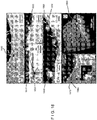
- Figure 18A shows an exemplary ⁇ OCT image of a coronary plaque showing multiple leukocytes (arrows).
- Figure 18B shows an exemplary ⁇ OCT image of a coronary plaque illustrating multiple leukocytes (arrows) of two different cell types, one smaller cell with scant cytoplasm, consistent with a lymphocyte (L) and another, larger cell with a highly scattering cytoplasm, indicative of a monocyte (M).
- L lymphocyte
- M monocyte
- Figure 18A illustrates an exemplary ⁇ OCT image of a coronary plaque showing multiple leukocytes 1800 which has been generated using the exemplary embodiment(s) of the methods, systems and apparatus according to the present disclosure.
- Figure 18B illustrates an exemplary ⁇ OCT image of a coronary plaque showing multiple leukocytes of two different cell types, one smaller cell 1810 with scant cytoplasm, consistent with a lymphocyte and another, larger cell 1820 with a highly scattering cytoplasm, suggestive of a monocyte.
- Figure 18C illustrates an exemplary ⁇ OCT image of a coronary plaque showing a cell 1830 with an indented, bean-shaped nucleus characteristic of a monocyte.
- Figure 18D illustrates an exemplary ⁇ OCT image of a coronary plaque showing a leukocyte 1840 with a multi-lobed nucleus, suggestive of a neutrophil attached to the endothelial surface.
- Figure 18E illustrates an exemplary ⁇ OCT image of a coronary plaque showing multiple leukocytes 1850, tethered to the endothelial surface by pseudopodia 1860.
- Figure 18F illustrates an exemplary ⁇ OCT image of a coronary plaque showing cells 1870 with the morphology of monocytes in this cross-section and inset transmigrating through the endothelium 1880.
- Figure 18G illustrates an exemplary ⁇ OCT image of multiple leukocytes 1890 distributed on the endothelial surface.
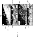
- Figure 19A-19E show exemplary images which have been generated using the exemplary embodiment(s) of the methods, systems and apparatus according to the present disclosure.
- Figure 19A illustrates an exemplary ⁇ OCT image of platelets 1900 (P) adjacent to a leukocyte characteristic of a neutrophil 1910 (N), which is also attached to a small platelet 1920 (yellow arrow).
- Figure 19B illustrates an exemplary ⁇ OCT image of fibrin 1930 (F) which is visible as linear strands bridging a gap in the coronary artery wall.
- Figure 19C illustrates an exemplary ⁇ OCT image of a cluster of leukocytes 1940 (L), adherent to the fibrin in an adjacent site to Figure 19B .
- Figure 19D illustrates an exemplary ⁇ OCT image of Fibrin thrombus 1950 (T) with multiple, entrapped leukocytes.
- Figure 19E an ⁇ OCT image of a more advanced thrombus 1960 (T) showing a leukocyte 1970 (arrow) and fibrin strands 1980(inset, F).
- Figures 20A-20D show further exemplary images which have been generated using the exemplary embodiment(s) of the methods, systems and apparatus according to the present disclosure.
- Figure 20A illustrates a cross-sectional exemplary ⁇ OCT image of endothelial cells 2000 in culture.
- Figure 20B shows an en face exemplary ⁇ OCT image of endothelial cells 2010 in culture.
- Figure 20C illustrates an exemplary ⁇ OCT image of native swine coronary artery cross-section 2020.
- Figure 20D shows a three-dimensional rendering of the swine coronary artery, demonstrating endothelial "pavementing" 2030.
- Figures 20A-20D show further exemplary images which have been generated using the exemplary embodiment(s) of the methods, systems and apparatus according to the present disclosure.
- Figure 21 A shows an exemplary ⁇ OCT image of microcalcifications which are seen as bright densities within the ⁇ OCT image of the fibrous cap 2100.
- Figure 21B illustrates an exemplary ⁇ OCT image of microcalcifications which are seen as purple densities on the corresponding histology 2110.
- Figures 20A-20D illustrate further exemplary images which have been generated using the exemplary embodiment(s) of the methods, systems and apparatus according to the present disclosure.
- Figure 22A shows an exemplary ⁇ OCT image of a large calcium nodule, demonstrating disrupted intima/endothelium 2200.
- Figure 22B shows an expanded view of an exemplary region enclosed by the red box shows microscopic tissue strands, consistent with fibrin 2210, adjoining the unprotected calcium 2220 to the opposing detached intima.
- Figure 22C shows a corresponding histology illustrating fibrin 2230 and denuded calcific surface 2240.
- Figures 23A-26C illustrate further exemplary images which have been generated using the exemplary embodiment(s) of the methods, systems and apparatus according to the present disclosure.
- Figure 23A shows an exemplary ⁇ OCT image of a large necrotic core 2300 fibroatheroma, demonstrating thick cholesterol crystals 2310, characterized by reflections from their top and bottom surfaces.
- Figure 23B shows an exemplary ⁇ OCT image of thin crystal 2320, piercing the cap of another necrotic core plaque 2330, shown in more detail in the inset.
- Figure 24 A shows an exemplary ⁇ OCT image of many smooth muscle cells 2400 appear as low backscattering spindle-shaped cells (inset).
- Figure 24B shows an exemplary ⁇ OCT image of smooth muscle cells producing collagen are spindle shaped, have a high backscattering interior 2410 and a "halo" of low backscattering 2420, which can represent the cell body 2430 and collagen matrix 2440, respectively (e.g., histology inset).
- Figure 25A shows an exemplary ⁇ OCT image of Taxus Liberie (Boston Scientific, Natick, MA) struts without polymer 2500, with polymer without drug 2510, and with polymer with drug 2520.
- Figure 25B shows an exemplary ⁇ OCT image of a cadaver coronary specimen with an implanted BMS 2570 shows struts devoid of polymer, covered by neointima 2580.
- Figure 25C shows an exemplary ⁇ OCT image of a cadaver coronary specimen with implanted DES struts 2590 from another cadaver showing polymer overlying the strut reflections 2595 (inset).
- Figure 26A shows an exemplary ⁇ OCT image showing tissue 2600 has separated the polymer 2610 off of the stent strut 2620 and the polymer has fractured 2630.
- Figure 26B shows an exemplary ⁇ OCT image showing superficial leukocyte cluster 2640 and adjacent attached leukocytes 2650 overlying the site of the polymer fracture 2660.
- Figure 26C shows an exemplary ⁇ OCT image showing inflammation 2670 at the edge of a strut 2680 from another patient.
- Figure 26D shows an exemplary ⁇ OCT image showing uncovered strut 2690, completely devoid of overlying endothelium.
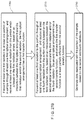
- Figure 27A shows a flow diagram of a method for providing data associated with at least one portion of at least one sample according to one exemplary embodiment of the present disclosure.
- procedure 2710 at least one first radiation is forwarded to at least one portion of the sample through at least one optical arrangement (e.g., as described in various exemplary embodiments herein), and at least one second radiation is received from the portion which is based on the first radiation.
- the optical arrangement Based on an interaction between the optical arrangement and the first radiation and/or the second radiation, the optical arrangement has a first transfer function.
- at least one third radiation is forwarded to the portion through such optical arrangement, and at least one fourth radiation is received from the portion which is based on the third radiation.
- the optical arrangement Based on an interaction between this optical arrangement and the third radiation and/or the fourth radiation, the optical arrangement has a second transfer function.
- the first transfer function can be at least partially different from the second transfer function.
- the data associated with the portion(s) can be generated based on the second and fourth radiations.
- Figure 27B shows a flow diagram of the method for providing data associated with at least one portion of at least one sample according to another exemplary embodiment of the present disclosure.
- procedure 2760 at least one first radiation is forwarded to at least one portion of the sample through at least one first optical arrangement (e.g., as described in various exemplary embodiments herein), and at least one second radiation is received from the portion which is based on the first radiation.
- the first optical arrangement Based on an interaction between the first optical arrangement and the first radiation and/or the second radiation, the first optical arrangement has a first transfer function.
- procedure 2770 at least one third radiation is forwarded to the portion through at least one second optical arrangement, and at least one fourth radiation is received from the portion which is based on the third radiation.
- the optical arrangement Based on an interaction between the second optical arrangement and the third radiation and/or the fourth radiation, the optical arrangement has a second transfer function.
- the first transfer function can be at least partially different from the second transfer function.
- the data associated with the portion(s) can be generated based on the second and fourth radiations.
Applications Claiming Priority (4)
| Application Number | Priority Date | Filing Date | Title |
|---|---|---|---|
| US31127210P | 2010-03-05 | 2010-03-05 | |
| US31117110P | 2010-03-05 | 2010-03-05 | |
| EP11751520.5A EP2542154B1 (fr) | 2010-03-05 | 2011-03-07 | Dispositif d'application d'un rayonnement électromagnétique sur un échantillon |
| PCT/US2011/027450 WO2011109835A2 (fr) | 2010-03-05 | 2011-03-07 | Systèmes, procédés et support accessible par ordinateur qui fournissent des images microscopiques d'au moins une structure anatomique à une résolution particulière |
Related Parent Applications (2)
| Application Number | Title | Priority Date | Filing Date |
|---|---|---|---|
| EP11751520.5A Division-Into EP2542154B1 (fr) | 2010-03-05 | 2011-03-07 | Dispositif d'application d'un rayonnement électromagnétique sur un échantillon |
| EP11751520.5A Division EP2542154B1 (fr) | 2010-03-05 | 2011-03-07 | Dispositif d'application d'un rayonnement électromagnétique sur un échantillon |
Publications (1)
| Publication Number | Publication Date |
|---|---|
| EP3753480A1 true EP3753480A1 (fr) | 2020-12-23 |
Family
ID=44531913
Family Applications (4)
| Application Number | Title | Priority Date | Filing Date |
|---|---|---|---|
| EP11751520.5A Active EP2542154B1 (fr) | 2010-03-05 | 2011-03-07 | Dispositif d'application d'un rayonnement électromagnétique sur un échantillon |
| EP20183108.8A Pending EP3753480A1 (fr) | 2010-03-05 | 2011-03-07 | Systèmes, procédés et support accessible par ordinateur fournissant des images microscopiques d'au moins une structure anatomique à une résolution particulière |
| EP11751514.8A Active EP2542145B1 (fr) | 2010-03-05 | 2011-03-07 | Systèmes qui procurent des images microscopiques d'au moins une structure anatomique à une résolution particulière |
| EP11751505.6A Withdrawn EP2542153A4 (fr) | 2010-03-05 | 2011-03-07 | Système, procédés et support accessible par ordinateur qui procurent des images microscopiques d'au moins une structure anatomique à une résolution particulière |
Family Applications Before (1)
| Application Number | Title | Priority Date | Filing Date |
|---|---|---|---|
| EP11751520.5A Active EP2542154B1 (fr) | 2010-03-05 | 2011-03-07 | Dispositif d'application d'un rayonnement électromagnétique sur un échantillon |
Family Applications After (2)
| Application Number | Title | Priority Date | Filing Date |
|---|---|---|---|
| EP11751514.8A Active EP2542145B1 (fr) | 2010-03-05 | 2011-03-07 | Systèmes qui procurent des images microscopiques d'au moins une structure anatomique à une résolution particulière |
| EP11751505.6A Withdrawn EP2542153A4 (fr) | 2010-03-05 | 2011-03-07 | Système, procédés et support accessible par ordinateur qui procurent des images microscopiques d'au moins une structure anatomique à une résolution particulière |
Country Status (15)
| Country | Link |
|---|---|
| US (7) | US8804126B2 (fr) |
| EP (4) | EP2542154B1 (fr) |
| JP (3) | JP5934121B2 (fr) |
| KR (3) | KR20130035254A (fr) |
| CY (2) | CY1123497T1 (fr) |
| DK (2) | DK2542145T3 (fr) |
| ES (2) | ES2828465T3 (fr) |
| HR (2) | HRP20201735T1 (fr) |
| HU (2) | HUE052561T2 (fr) |
| LT (2) | LT2542154T (fr) |
| PL (1) | PL2542154T3 (fr) |
| PT (2) | PT2542145T (fr) |
| RS (2) | RS61066B1 (fr) |
| SI (2) | SI2542145T1 (fr) |
| WO (3) | WO2011109818A2 (fr) |
Families Citing this family (85)
| Publication number | Priority date | Publication date | Assignee | Title |
|---|---|---|---|---|
| WO2002036015A1 (fr) | 2000-10-30 | 2002-05-10 | The General Hospital Corporation | Procedes et systemes optiques d'analyse de tissus |
| EP1771755B1 (fr) | 2004-07-02 | 2016-09-21 | The General Hospital Corporation | Sonde d'imagerie endoscopique comprenant des fibres double gaine |
| EP1793731B1 (fr) | 2004-08-24 | 2013-12-25 | The General Hospital Corporation | Appareil d'imagerie comprenant un dispositif de distribution de fluide et un dispositif de rétraction |
| JP2008521516A (ja) | 2004-11-29 | 2008-06-26 | ザ ジェネラル ホスピタル コーポレイション | サンプル上の複数の地点を同時に照射し検出することによって光学画像生成を実行する構成、装置、内視鏡、カテーテル、及び方法 |
| ES2337497T3 (es) | 2005-04-28 | 2010-04-26 | The General Hospital Corporation | Evaluacion de caracteristicas de la imagen de una estructura anatomica en imagenes de tomografia de coherencia optica. |
| JP5702049B2 (ja) | 2005-06-01 | 2015-04-15 | ザ ジェネラル ホスピタル コーポレイション | 位相分解光学周波数領域画像化を行うための装置、方法及びシステム |
| KR101387454B1 (ko) | 2005-08-09 | 2014-04-22 | 더 제너럴 하스피탈 코포레이션 | 광간섭 단층촬영법에서 편광 기반 직교 복조를 수행하기위한 장치, 방법 및 저장 매체 |
| CA2624109A1 (fr) | 2005-09-29 | 2007-04-12 | General Hospital Corporation | Procede et appareil destines a un procede pour visualiser et analyser un ou plusieurs echantillons biologiques avec des resolutions augmentant progressivement |
| WO2007084903A2 (fr) | 2006-01-19 | 2007-07-26 | The General Hospital Corporation | Dispositif de collecte d'information pour une structure utilisant des techniques d'endoscopie à codage spectral, et procédé d'élaboration correspondant |
| JP5680829B2 (ja) | 2006-02-01 | 2015-03-04 | ザ ジェネラル ホスピタル コーポレイション | 複数の電磁放射をサンプルに照射する装置 |
| US10426548B2 (en) | 2006-02-01 | 2019-10-01 | The General Hosppital Corporation | Methods and systems for providing electromagnetic radiation to at least one portion of a sample using conformal laser therapy procedures |
| EP2309221A1 (fr) | 2006-02-24 | 2011-04-13 | The General Hospital Corporation | Procédés et systèmes destinés à réaliser une tomographie par cohérence optique dans le domaine de fourier avec résolution angulaire |
| EP2015669A2 (fr) | 2006-05-10 | 2009-01-21 | The General Hospital Corporation | Processus, agencements et systèmes pour obtenir une imagerie de domaine de fréquence d'un échantillon |
| WO2008049118A2 (fr) | 2006-10-19 | 2008-04-24 | The General Hospital Corporation | Dispositif et procédé d'obtention et de fourniture d'informations d'image associées à au moins une portion d' échantillon et permettant de réaliser une telle portion |
| WO2010009136A2 (fr) | 2008-07-14 | 2010-01-21 | The General Hospital Corporation | Appareil et procédés d'endoscopie couleur |
| WO2010090837A2 (fr) | 2009-01-20 | 2010-08-12 | The General Hospital Corporation | Appareil, système et procédé de biopsie endoscopique |
| CN104134928A (zh) | 2009-02-04 | 2014-11-05 | 通用医疗公司 | 利用高速光学波长调谐源的设备和方法 |
| EP2453791B1 (fr) | 2009-07-14 | 2023-09-06 | The General Hospital Corporation | Appareil permettant de mesurer le débit et la pression à l intérieur d'une cuve |
| US9823127B2 (en) * | 2010-01-22 | 2017-11-21 | Duke University | Systems and methods for deep spectroscopic imaging of biological samples with use of an interferometer and spectrometer |
| JP5934121B2 (ja) * | 2010-03-05 | 2016-06-15 | ザ ジェネラル ホスピタル コーポレイション | 特定の分解能にて少なくとも1つの解剖構造の微細画像を提供するシステム、方法およびコンピュータがアクセス可能な媒体 |
| EP2553513B1 (fr) * | 2010-03-28 | 2020-10-28 | Ecole Polytechnique Federale de Lausanne (EPFL) | Tomographie par réfraction d'indice complexe avec une résolution améliorée |
| US9069130B2 (en) | 2010-05-03 | 2015-06-30 | The General Hospital Corporation | Apparatus, method and system for generating optical radiation from biological gain media |
| JP5778762B2 (ja) | 2010-05-25 | 2015-09-16 | ザ ジェネラル ホスピタル コーポレイション | 光コヒーレンストモグラフィー画像のスペクトル解析のための装置及び方法 |
| EP2575597B1 (fr) | 2010-05-25 | 2022-05-04 | The General Hospital Corporation | Appareil pour fournir une imagerie optique de structures et de compositions |
| JP6066901B2 (ja) | 2010-06-03 | 2017-01-25 | ザ ジェネラル ホスピタル コーポレイション | 1つまたは複数の管腔器官内または管腔器官にある構造を撮像するための装置およびデバイスのための方法 |
| DE102011013613A1 (de) * | 2010-10-01 | 2012-04-05 | Carl Zeiss Microimaging Gmbh | Mikroskop und Mikroskopierverfahren |
| EP2632324A4 (fr) | 2010-10-27 | 2015-04-22 | Gen Hospital Corp | Appareil, systèmes et méthodes de mesure de la pression sanguine dans au moins un vaisseau |
| JP5610063B2 (ja) * | 2011-03-24 | 2014-10-22 | 株式会社ニコン | 観察装置および観察方法 |
| US9330092B2 (en) | 2011-07-19 | 2016-05-03 | The General Hospital Corporation | Systems, methods, apparatus and computer-accessible-medium for providing polarization-mode dispersion compensation in optical coherence tomography |
| JP5829280B2 (ja) | 2011-09-30 | 2015-12-09 | オリンパス株式会社 | 内面形状測定装置、検出ヘッド及び内視鏡装置 |
| US9341783B2 (en) | 2011-10-18 | 2016-05-17 | The General Hospital Corporation | Apparatus and methods for producing and/or providing recirculating optical delay(s) |
| EP2769199A4 (fr) * | 2011-10-20 | 2015-06-24 | Gen Hospital Corp | Agencement d'imagerie implantable et son procédé d'utilisation |
| US9036966B2 (en) * | 2012-03-28 | 2015-05-19 | Corning Incorporated | Monolithic beam-shaping optical systems and methods for an OCT probe |
| EP2833776A4 (fr) | 2012-03-30 | 2015-12-09 | Gen Hospital Corp | Système d'imagerie, procédé et fixation distale permettant une endoscopie à champ de vision multidirectionnel |
| US9192294B2 (en) * | 2012-05-10 | 2015-11-24 | Carl Zeiss Meditec, Inc. | Systems and methods for faster optical coherence tomography acquisition and processing |
| JP2015517387A (ja) * | 2012-05-21 | 2015-06-22 | ザ ジェネラル ホスピタル コーポレイション | カプセル顕微鏡検査のための装置、デバイスおよび方法 |
| JP6227652B2 (ja) | 2012-08-22 | 2017-11-08 | ザ ジェネラル ホスピタル コーポレイション | ソフトリソグラフィを用いてミニチュア内視鏡を製作するためのシステム、方法、およびコンピュータ・アクセス可能媒体 |
| EP2948758B1 (fr) | 2013-01-28 | 2024-03-13 | The General Hospital Corporation | Appareil pour fournir une spectroscopie diffuse co-enregistrée avec imagerie de domaine de fréquence optique |
| WO2014120791A1 (fr) | 2013-01-29 | 2014-08-07 | The General Hospital Corporation | Appareil, systèmes et procédés pour donner des informations sur la valvule aortique |
| US11179028B2 (en) | 2013-02-01 | 2021-11-23 | The General Hospital Corporation | Objective lens arrangement for confocal endomicroscopy |
| JP6378311B2 (ja) | 2013-03-15 | 2018-08-22 | ザ ジェネラル ホスピタル コーポレイション | 物体を特徴付ける方法とシステム |
| US9752926B2 (en) * | 2013-04-29 | 2017-09-05 | Korea Food Research Institute | Scanning module, detection device using Bessel beam, detection probe, and probe type detection device |
| US9784681B2 (en) * | 2013-05-13 | 2017-10-10 | The General Hospital Corporation | System and method for efficient detection of the phase and amplitude of a periodic modulation associated with self-interfering fluorescence |
| CN104297218B (zh) * | 2013-07-15 | 2016-09-14 | 中国科学院沈阳自动化研究所 | 远距离冶金液态金属成分的原位、在线检测装置及方法 |
| EP3021734B1 (fr) | 2013-07-19 | 2020-04-08 | The General Hospital Corporation | Appareil d'imagerie utilisant une endoscopie à champ de vision multidirectionnel |
| US10117576B2 (en) | 2013-07-19 | 2018-11-06 | The General Hospital Corporation | System, method and computer accessible medium for determining eye motion by imaging retina and providing feedback for acquisition of signals from the retina |
| WO2015013651A2 (fr) | 2013-07-26 | 2015-01-29 | The General Hospital Corporation | Système, appareil et procédé utilisant une dispersion optique pour réaliser une tomographie en cohérence optique dans le domaine de fourier |
| US9605942B2 (en) * | 2013-07-31 | 2017-03-28 | Corning Incorporated | OCT probes and OCT optical probe component for use therein |
| WO2015105870A1 (fr) | 2014-01-08 | 2015-07-16 | The General Hospital Corporation | Procédé et appareil pour imagerie microscopique |
| US10736494B2 (en) | 2014-01-31 | 2020-08-11 | The General Hospital Corporation | System and method for facilitating manual and/or automatic volumetric imaging with real-time tension or force feedback using a tethered imaging device |
| SG11201607441TA (en) * | 2014-03-13 | 2016-10-28 | Univ Singapore | An optical interference device |
| US10228556B2 (en) | 2014-04-04 | 2019-03-12 | The General Hospital Corporation | Apparatus and method for controlling propagation and/or transmission of electromagnetic radiation in flexible waveguide(s) |
| KR20160001890A (ko) * | 2014-06-27 | 2016-01-07 | 연세대학교 원주산학협력단 | 항 노화를 위한 초음파와 oct를 결합한 피부 진단 및 치료 시스템 |
| US10912462B2 (en) | 2014-07-25 | 2021-02-09 | The General Hospital Corporation | Apparatus, devices and methods for in vivo imaging and diagnosis |
| US20170219485A1 (en) * | 2014-10-01 | 2017-08-03 | Purdue Research Foundation | Organism Identification |
| US9869852B2 (en) * | 2015-01-26 | 2018-01-16 | Thorlabs, Inc. | Microscopy system with auto-focus adjustment by low-coherence interferometry |
| CN104688172A (zh) * | 2015-02-02 | 2015-06-10 | 深圳市中科微光医疗器械技术有限公司 | 一种微型光学相干断层成像探头 |
| CN112998664A (zh) | 2015-04-16 | 2021-06-22 | Gentuity有限责任公司 | 用于神经病学的微光探针 |
| KR101637832B1 (ko) * | 2015-05-12 | 2016-07-07 | 한양대학교 산학협력단 | 광 프로브 및 상기 광 프로브의 제작 방법 |
| US10542961B2 (en) | 2015-06-15 | 2020-01-28 | The Research Foundation For The State University Of New York | System and method for infrasonic cardiac monitoring |
| US9910276B2 (en) | 2015-06-30 | 2018-03-06 | Microsoft Technology Licensing, Llc | Diffractive optical elements with graded edges |
| US10670862B2 (en) | 2015-07-02 | 2020-06-02 | Microsoft Technology Licensing, Llc | Diffractive optical elements with asymmetric profiles |
| US10038840B2 (en) | 2015-07-30 | 2018-07-31 | Microsoft Technology Licensing, Llc | Diffractive optical element using crossed grating for pupil expansion |
| US9864208B2 (en) * | 2015-07-30 | 2018-01-09 | Microsoft Technology Licensing, Llc | Diffractive optical elements with varying direction for depth modulation |
| US10073278B2 (en) | 2015-08-27 | 2018-09-11 | Microsoft Technology Licensing, Llc | Diffractive optical element using polarization rotation grating for in-coupling |
| WO2017040484A1 (fr) | 2015-08-31 | 2017-03-09 | Gentuity, Llc | Système d'imagerie comportant une sonde d'imagerie et des dispositifs d'administration |
| US11147503B2 (en) * | 2015-09-30 | 2021-10-19 | The General Hospital Corporation | Systems and methods for an actively controlled optical imaging device |
| US10429645B2 (en) | 2015-10-07 | 2019-10-01 | Microsoft Technology Licensing, Llc | Diffractive optical element with integrated in-coupling, exit pupil expansion, and out-coupling |
| US10241332B2 (en) | 2015-10-08 | 2019-03-26 | Microsoft Technology Licensing, Llc | Reducing stray light transmission in near eye display using resonant grating filter |
| US9946072B2 (en) * | 2015-10-29 | 2018-04-17 | Microsoft Technology Licensing, Llc | Diffractive optical element with uncoupled grating structures |
| US10234686B2 (en) | 2015-11-16 | 2019-03-19 | Microsoft Technology Licensing, Llc | Rainbow removal in near-eye display using polarization-sensitive grating |
| KR20170004101U (ko) | 2016-05-27 | 2017-12-06 | 서동진 | 휴대폰 충전기구 |
| KR20180000372U (ko) | 2016-07-28 | 2018-02-07 | 서동진 | 휴대폰용 다중 충전장치 |
| CN106501182B (zh) * | 2016-09-22 | 2019-06-04 | 南京大学 | 一种利用光声本征谱分析法无损测量弹性的方法 |
| US10108014B2 (en) * | 2017-01-10 | 2018-10-23 | Microsoft Technology Licensing, Llc | Waveguide display with multiple focal depths |
| JP7050787B2 (ja) * | 2017-01-30 | 2022-04-08 | アルコン インコーポレイティド | 偏光感受型光干渉断層計を用いてフラップを作成するためのシステムと方法 |
| KR20180133957A (ko) | 2017-05-26 | 2018-12-18 | 한양대학교 산학협력단 | 환형 빔 커플링 시스템 |
| CN110691545B (zh) * | 2017-06-02 | 2021-06-18 | 奥林巴斯株式会社 | 内窥镜光源装置 |
| DE102017115922C5 (de) * | 2017-07-14 | 2023-03-23 | Precitec Gmbh & Co. Kg | Verfahren und Vorrichtung zur Messung und Einstellung eines Abstands zwischen einem Bearbeitungskopf und einem Werkstück sowie dazugehöriges Verfahren zur Regelung |
| US11684242B2 (en) | 2017-11-28 | 2023-06-27 | Gentuity, Llc | Imaging system |
| KR102275570B1 (ko) * | 2019-12-31 | 2021-07-09 | (주)윈어스 테크놀로지 | 결상 렌즈 배열체 및 이를 구비하는 광간섭 단층 영상 시스템 |
| US11879889B2 (en) * | 2020-05-04 | 2024-01-23 | Omachron Intellectual Property Inc. | Respiratory testing with multiple spectrometers |
| US20230341668A1 (en) * | 2020-07-23 | 2023-10-26 | The University Of Adelaide | An optical element |
| US11730548B2 (en) * | 2020-12-17 | 2023-08-22 | Industrial Technology Research Institute | Optical coherence tomography scanning probe |
| WO2023210793A1 (fr) * | 2022-04-27 | 2023-11-02 | 宏 小川 | Dispositif de génération de faisceau de bessel et dispositif de balayage optique l'utilisant |
Citations (7)
| Publication number | Priority date | Publication date | Assignee | Title |
|---|---|---|---|---|
| US20030218756A1 (en) * | 2002-01-16 | 2003-11-27 | Zhongping Chen | High resolution optical coherence tomography with an improved depth range using an axicon lens |
| US20050018201A1 (en) | 2002-01-24 | 2005-01-27 | De Boer Johannes F | Apparatus and method for ranging and noise reduction of low coherence interferometry lci and optical coherence tomography oct signals by parallel detection of spectral bands |
| WO2005047813A1 (fr) | 2003-10-27 | 2005-05-26 | The General Hospital Corporation | Procede et appareil d'imagerie optique par interferometrie dans le domaine frequentiel |
| US20060093276A1 (en) | 2004-11-02 | 2006-05-04 | The General Hospital Corporation | Fiber-optic rotational device, optical system and method for imaging a sample |
| US20060146339A1 (en) * | 2004-12-06 | 2006-07-06 | Fujinon Corporation | Optical tomographic apparatus |
| US20060158655A1 (en) * | 2005-01-20 | 2006-07-20 | Everett Matthew J | Apparatus and method for combined optical-coherence-tomographic and confocal detection |
| US20070076220A1 (en) * | 2005-09-30 | 2007-04-05 | Fuji Photo Film Co., Ltd. | Optical tomography system |
Family Cites Families (704)
| Publication number | Priority date | Publication date | Assignee | Title |
|---|---|---|---|---|
| US2339754A (en) | 1941-03-04 | 1944-01-25 | Westinghouse Electric & Mfg Co | Supervisory apparatus |
| US3090753A (en) | 1960-08-02 | 1963-05-21 | Exxon Research Engineering Co | Ester oil compositions containing acid anhydride |
| GB1257778A (fr) | 1967-12-07 | 1971-12-22 | ||
| US3601480A (en) | 1968-07-10 | 1971-08-24 | Physics Int Co | Optical tunnel high-speed camera system |
| JPS4932484U (fr) | 1972-06-19 | 1974-03-20 | ||
| US3872407A (en) | 1972-09-01 | 1975-03-18 | Us Navy | Rapidly tunable laser |
| JPS584481Y2 (ja) | 1973-06-23 | 1983-01-26 | オリンパス光学工業株式会社 | ナイシキヨウシヤヘンカンコウガクケイ |
| FR2253410A5 (fr) | 1973-12-03 | 1975-06-27 | Inst Nat Sante Rech Med | |
| US3941121A (en) | 1974-12-20 | 1976-03-02 | The University Of Cincinnati | Focusing fiber-optic needle endoscope |
| US3983507A (en) | 1975-01-06 | 1976-09-28 | Research Corporation | Tunable laser systems and method |
| US3973219A (en) | 1975-04-24 | 1976-08-03 | Cornell Research Foundation, Inc. | Very rapidly tuned cw dye laser |
| US4030831A (en) | 1976-03-22 | 1977-06-21 | The United States Of America As Represented By The Secretary Of The Navy | Phase detector for optical figure sensing |
| US4141362A (en) | 1977-05-23 | 1979-02-27 | Richard Wolf Gmbh | Laser endoscope |
| US4224929A (en) | 1977-11-08 | 1980-09-30 | Olympus Optical Co., Ltd. | Endoscope with expansible cuff member and operation section |
| GB2047894B (en) | 1978-03-09 | 1982-11-03 | Nat Res Dev | Speckle interferometric measurement of small oscillatory movements |
| GB2030313A (en) | 1978-06-29 | 1980-04-02 | Wolf Gmbh Richard | Endoscopes |
| JPS559417A (en) | 1978-07-05 | 1980-01-23 | Seiko Epson Corp | Semiconductor integrated circuit |
| FR2448728A1 (fr) | 1979-02-07 | 1980-09-05 | Thomson Csf | Dispositif joint tournant pour liaison par conducteurs optiques et systeme comportant un tel dispositif |
| US4300816A (en) | 1979-08-30 | 1981-11-17 | United Technologies Corporation | Wide band multicore optical fiber |
| US4295738A (en) | 1979-08-30 | 1981-10-20 | United Technologies Corporation | Fiber optic strain sensor |
| JPS599923Y2 (ja) | 1980-04-28 | 1984-03-29 | 株式会社ヨコオ | スパイクタイヤ |
| JPS56158304A (en) * | 1980-05-10 | 1981-12-07 | Sumitomo Electric Ind Ltd | Image fiber with light guide |
| DE3041875C2 (de) * | 1980-11-06 | 1984-05-10 | Krautkrämer GmbH, 5000 Köln | Vorrichtung zur Erzeugung von Ultraschallwellen |
| US4428643A (en) | 1981-04-08 | 1984-01-31 | Xerox Corporation | Optical scanning system with wavelength shift correction |
| US5065331A (en) | 1981-05-18 | 1991-11-12 | Vachon Reginald I | Apparatus and method for determining the stress and strain in pipes, pressure vessels, structural members and other deformable bodies |
| US4409475A (en) * | 1981-07-29 | 1983-10-11 | Visidyne, Inc. | Spatial frequency filter |
| GB2106736B (en) | 1981-09-03 | 1985-06-12 | Standard Telephones Cables Ltd | Optical transmission system |
| US4479499A (en) | 1982-01-29 | 1984-10-30 | Alfano Robert R | Method and apparatus for detecting the presence of caries in teeth using visible light |
| JPS5926703A (ja) * | 1982-08-05 | 1984-02-13 | Olympus Optical Co Ltd | 光伝送装置 |
| US5302025A (en) | 1982-08-06 | 1994-04-12 | Kleinerman Marcos Y | Optical systems for sensing temperature and other physical parameters |
| US4601036A (en) | 1982-09-30 | 1986-07-15 | Honeywell Inc. | Rapidly tunable laser |
| HU187188B (en) | 1982-11-25 | 1985-11-28 | Koezponti Elelmiszeripari | Device for generating radiation of controllable spectral structure |
| CH663466A5 (fr) | 1983-09-12 | 1987-12-15 | Battelle Memorial Institute | Procede et dispositif pour determiner la position d'un objet par rapport a une reference. |
| JPS6140633A (ja) | 1984-08-02 | 1986-02-26 | Nec Corp | タブレツト装置 |
| US4639999A (en) | 1984-11-02 | 1987-02-03 | Xerox Corporation | High resolution, high efficiency I.R. LED printing array fabrication method |
| US4763977A (en) | 1985-01-09 | 1988-08-16 | Canadian Patents And Development Limited-Societe | Optical fiber coupler with tunable coupling ratio and method of making |
| US5318024A (en) | 1985-03-22 | 1994-06-07 | Massachusetts Institute Of Technology | Laser endoscope for spectroscopic imaging |
| EP0590268B1 (fr) | 1985-03-22 | 1998-07-01 | Massachusetts Institute Of Technology | Sonde comprenant des fibres optiques destiné à l'analyse spectrale de tissus |
| US4734578A (en) | 1985-03-27 | 1988-03-29 | Olympus Optical Co., Ltd. | Two-dimensional scanning photo-electric microscope |
| US4607622A (en) | 1985-04-11 | 1986-08-26 | Charles D. Fritch | Fiber optic ocular endoscope |
| US4631498A (en) | 1985-04-26 | 1986-12-23 | Hewlett-Packard Company | CW Laser wavemeter/frequency locking technique |
| US4650327A (en) * | 1985-10-28 | 1987-03-17 | Oximetrix, Inc. | Optical catheter calibrating assembly |
| US4744615A (en) * | 1986-01-29 | 1988-05-17 | International Business Machines Corporation | Laser beam homogenizer |
| JPH0664683B2 (ja) | 1986-02-13 | 1994-08-22 | 松下電器産業株式会社 | 回転磁気ヘツド記録装置 |
| JPS62188001U (fr) | 1986-05-20 | 1987-11-30 | ||
| US5040889A (en) | 1986-05-30 | 1991-08-20 | Pacific Scientific Company | Spectrometer with combined visible and ultraviolet sample illumination |
| CA1290019C (fr) | 1986-06-20 | 1991-10-01 | Hideo Kuwahara | Recepteur de signaux lumineux a dedoublement |
| US4770492A (en) | 1986-10-28 | 1988-09-13 | Spectran Corporation | Pressure or strain sensitive optical fiber |
| JPH0824665B2 (ja) | 1986-11-28 | 1996-03-13 | オリンパス光学工業株式会社 | 内視鏡装置 |
| US4744656A (en) | 1986-12-08 | 1988-05-17 | Spectramed, Inc. | Disposable calibration boot for optical-type cardiovascular catheter |
| JPS63158363A (ja) | 1986-12-22 | 1988-07-01 | Daikin Mfg Co Ltd | エア回転継手のシ−ル装置 |
| US4751706A (en) | 1986-12-31 | 1988-06-14 | The United States Of America As Represented By The Secretary Of The Army | Laser for providing rapid sequence of different wavelengths |
| US4834111A (en) | 1987-01-12 | 1989-05-30 | The Trustees Of Columbia University In The City Of New York | Heterodyne interferometer |
| GB2209221B (en) | 1987-09-01 | 1991-10-23 | Litton Systems Inc | Hydrophone demodulator circuit and method |
| US5202931A (en) | 1987-10-06 | 1993-04-13 | Cell Analysis Systems, Inc. | Methods and apparatus for the quantitation of nuclear protein |
| US4909631A (en) | 1987-12-18 | 1990-03-20 | Tan Raul Y | Method for film thickness and refractive index determination |
| US4890901A (en) | 1987-12-22 | 1990-01-02 | Hughes Aircraft Company | Color corrector for embedded prisms |
| US4892406A (en) | 1988-01-11 | 1990-01-09 | United Technologies Corporation | Method of and arrangement for measuring vibrations |
| FR2626367B1 (fr) | 1988-01-25 | 1990-05-11 | Thomson Csf | Capteur de temperature multipoints a fibre optique |
| FR2626383B1 (fr) | 1988-01-27 | 1991-10-25 | Commissariat Energie Atomique | Procede de microscopie optique confocale a balayage et en profondeur de champ etendue et dispositifs pour la mise en oeuvre du procede |
| US4925302A (en) | 1988-04-13 | 1990-05-15 | Hewlett-Packard Company | Frequency locking device |
| US5730731A (en) | 1988-04-28 | 1998-03-24 | Thomas J. Fogarty | Pressure-based irrigation accumulator |
| US4998972A (en) | 1988-04-28 | 1991-03-12 | Thomas J. Fogarty | Real time angioscopy imaging system |
| US4905169A (en) * | 1988-06-02 | 1990-02-27 | The United States Of America As Represented By The United States Department Of Energy | Method and apparatus for simultaneously measuring a plurality of spectral wavelengths present in electromagnetic radiation |
| US5242437A (en) | 1988-06-10 | 1993-09-07 | Trimedyne Laser Systems, Inc. | Medical device applying localized high intensity light and heat, particularly for destruction of the endometrium |
| WO1990000754A1 (fr) | 1988-07-13 | 1990-01-25 | Martin Russell Harris | Microscope a balayage a foyer commun |
| US5214538A (en) | 1988-07-25 | 1993-05-25 | Keymed (Medical And Industrial Equipment) Limited | Optical apparatus |
| GB8817672D0 (en) | 1988-07-25 | 1988-09-01 | Sira Ltd | Optical apparatus |
| US4868834A (en) | 1988-09-14 | 1989-09-19 | The United States Of America As Represented By The Secretary Of The Army | System for rapidly tuning a low pressure pulsed laser |
| DE3833602A1 (de) | 1988-10-03 | 1990-02-15 | Krupp Gmbh | Spektrometer zur gleichzeitigen intensitaetsmessung in verschiedenen spektralbereichen |
| US4940328A (en) | 1988-11-04 | 1990-07-10 | Georgia Tech Research Corporation | Optical sensing apparatus and method |
| US4966589A (en) | 1988-11-14 | 1990-10-30 | Hemedix International, Inc. | Intravenous catheter placement device |
| WO1990006718A1 (fr) | 1988-12-21 | 1990-06-28 | Massachusetts Institute Of Technology | Procede de fluorescence de tissus induite par laser |
| US5046501A (en) | 1989-01-18 | 1991-09-10 | Wayne State University | Atherosclerotic identification |
| JPH02250016A (ja) * | 1989-03-23 | 1990-10-05 | Mitsutoyo Corp | 落射暗視野照明装置 |
| JPH02259617A (ja) | 1989-03-30 | 1990-10-22 | Sony Corp | レーザビーム偏向装置 |
| US5085496A (en) * | 1989-03-31 | 1992-02-04 | Sharp Kabushiki Kaisha | Optical element and optical pickup device comprising it |
| US5317389A (en) | 1989-06-12 | 1994-05-31 | California Institute Of Technology | Method and apparatus for white-light dispersed-fringe interferometric measurement of corneal topography |
| US4965599A (en) | 1989-11-13 | 1990-10-23 | Eastman Kodak Company | Scanning apparatus for halftone image screen writing |
| US5133035A (en) | 1989-11-14 | 1992-07-21 | Hicks John W | Multifiber endoscope with multiple scanning modes to produce an image free of fixed pattern noise |
| US4984888A (en) | 1989-12-13 | 1991-01-15 | Imo Industries, Inc. | Two-dimensional spectrometer |
| KR930003307B1 (ko) | 1989-12-14 | 1993-04-24 | 주식회사 금성사 | 입체용 프로젝터 |
| US5251009A (en) | 1990-01-22 | 1993-10-05 | Ciba-Geigy Corporation | Interferometric measuring arrangement for refractive index measurements in capillary tubes |
| DD293205B5 (de) | 1990-03-05 | 1995-06-29 | Zeiss Carl Jena Gmbh | Lichtleiterfuehrung fuer ein medizinisches Beobachtungsgeraet |
| US5039193A (en) | 1990-04-03 | 1991-08-13 | Focal Technologies Incorporated | Fibre optic single mode rotary joint |
| DD294805A5 (de) * | 1990-05-31 | 1991-10-10 | Carl Zeiss Jena Gmbh,De | Beleuchtungssystem fuer laser- und superstrahlung |
| JPH0456907A (ja) | 1990-06-26 | 1992-02-24 | Fujikura Ltd | 光ファイバカプラ |
| US5262644A (en) | 1990-06-29 | 1993-11-16 | Southwest Research Institute | Remote spectroscopy for raman and brillouin scattering |
| US5197470A (en) * | 1990-07-16 | 1993-03-30 | Eastman Kodak Company | Near infrared diagnostic method and instrument |
| GB9015793D0 (en) | 1990-07-18 | 1990-09-05 | Medical Res Council | Confocal scanning optical microscope |
| US5127730A (en) | 1990-08-10 | 1992-07-07 | Regents Of The University Of Minnesota | Multi-color laser scanning confocal imaging system |
| US5845639A (en) | 1990-08-10 | 1998-12-08 | Board Of Regents Of The University Of Washington | Optical imaging methods |
| US5305759A (en) | 1990-09-26 | 1994-04-26 | Olympus Optical Co., Ltd. | Examined body interior information observing apparatus by using photo-pulses controlling gains for depths |
| JPH04135551A (ja) | 1990-09-27 | 1992-05-11 | Olympus Optical Co Ltd | 光三次元像観察装置 |
| JP3104984B2 (ja) | 1990-09-27 | 2000-10-30 | オリンパス光学工業株式会社 | 断層像観察用光走査装置 |
| US5241364A (en) | 1990-10-19 | 1993-08-31 | Fuji Photo Film Co., Ltd. | Confocal scanning type of phase contrast microscope and scanning microscope |
| US5250186A (en) | 1990-10-23 | 1993-10-05 | Cetus Corporation | HPLC light scattering detector for biopolymers |
| US5202745A (en) | 1990-11-07 | 1993-04-13 | Hewlett-Packard Company | Polarization independent optical coherence-domain reflectometry |
| US5275594A (en) | 1990-11-09 | 1994-01-04 | C. R. Bard, Inc. | Angioplasty system having means for identification of atherosclerotic plaque |
| JP3044778B2 (ja) * | 1990-11-14 | 2000-05-22 | 株式会社ニコン | 投影露光装置および投影露光方法 |
| JP3035336B2 (ja) | 1990-11-27 | 2000-04-24 | 興和株式会社 | 血流測定装置 |
| US5228001A (en) | 1991-01-23 | 1993-07-13 | Syracuse University | Optical random access memory |
| US5784162A (en) | 1993-08-18 | 1998-07-21 | Applied Spectral Imaging Ltd. | Spectral bio-imaging methods for biological research, medical diagnostics and therapy |
| US6198532B1 (en) | 1991-02-22 | 2001-03-06 | Applied Spectral Imaging Ltd. | Spectral bio-imaging of the eye |
| US5293872A (en) | 1991-04-03 | 1994-03-15 | Alfano Robert R | Method for distinguishing between calcified atherosclerotic tissue and fibrous atherosclerotic tissue or normal cardiovascular tissue using Raman spectroscopy |
| US6134003A (en) | 1991-04-29 | 2000-10-17 | Massachusetts Institute Of Technology | Method and apparatus for performing optical measurements using a fiber optic imaging guidewire, catheter or endoscope |
| US6564087B1 (en) | 1991-04-29 | 2003-05-13 | Massachusetts Institute Of Technology | Fiber optic needle probes for optical coherence tomography imaging |
| US6485413B1 (en) * | 1991-04-29 | 2002-11-26 | The General Hospital Corporation | Methods and apparatus for forward-directed optical scanning instruments |
| US5465147A (en) | 1991-04-29 | 1995-11-07 | Massachusetts Institute Of Technology | Method and apparatus for acquiring images using a ccd detector array and no transverse scanner |
| US6111645A (en) | 1991-04-29 | 2000-08-29 | Massachusetts Institute Of Technology | Grating based phase control optical delay line |
| US5956355A (en) | 1991-04-29 | 1999-09-21 | Massachusetts Institute Of Technology | Method and apparatus for performing optical measurements using a rapidly frequency-tuned laser |
| US6501551B1 (en) | 1991-04-29 | 2002-12-31 | Massachusetts Institute Of Technology | Fiber optic imaging endoscope interferometer with at least one faraday rotator |
| US5748598A (en) | 1995-12-22 | 1998-05-05 | Massachusetts Institute Of Technology | Apparatus and methods for reading multilayer storage media using short coherence length sources |
| US5321501A (en) | 1991-04-29 | 1994-06-14 | Massachusetts Institute Of Technology | Method and apparatus for optical imaging with means for controlling the longitudinal range of the sample |
| US5441053A (en) | 1991-05-03 | 1995-08-15 | University Of Kentucky Research Foundation | Apparatus and method for multiple wavelength of tissue |
| US5281811A (en) | 1991-06-17 | 1994-01-25 | Litton Systems, Inc. | Digital wavelength division multiplex optical transducer having an improved decoder |
| US5208651A (en) | 1991-07-16 | 1993-05-04 | The Regents Of The University Of California | Apparatus and method for measuring fluorescence intensities at a plurality of wavelengths and lifetimes |
| AU2519892A (en) | 1991-08-20 | 1993-03-16 | Douglas C.B. Redd | Optical histochemical analysis, in vivo detection and real-time guidance for ablation of abnormal tissues using a raman spectroscopic detection system |
| DE4128744C1 (fr) | 1991-08-29 | 1993-04-22 | Siemens Ag, 8000 Muenchen, De | |
| US5177488A (en) | 1991-10-08 | 1993-01-05 | Hughes Aircraft Company | Programmable fiber optic delay line, and radar target simulation system incorporating the same |
| ATE150573T1 (de) | 1991-12-30 | 1997-04-15 | Philips Electronics Nv | Optische einrichtung und mit einer solchen optischen einrichtung versehenes gerät zum abtasten einer informationsebene |
| US5353790A (en) | 1992-01-17 | 1994-10-11 | Board Of Regents, The University Of Texas System | Method and apparatus for optical measurement of bilirubin in tissue |
| US5334441A (en) | 1992-01-30 | 1994-08-02 | Gencorp Inc. | Composite comprising unsaturated polyester-flexible polymer block copolymer coated fiber structures in a polyester or vinyl ester resin matrix |
| US5212667A (en) | 1992-02-03 | 1993-05-18 | General Electric Company | Light imaging in a scattering medium, using ultrasonic probing and speckle image differencing |
| US5217456A (en) | 1992-02-24 | 1993-06-08 | Pdt Cardiovascular, Inc. | Device and method for intra-vascular optical radial imaging |
| US5283795A (en) | 1992-04-21 | 1994-02-01 | Hughes Aircraft Company | Diffraction grating driven linear frequency chirped laser |
| US5248876A (en) | 1992-04-21 | 1993-09-28 | International Business Machines Corporation | Tandem linear scanning confocal imaging system with focal volumes at different heights |
| US5486701A (en) * | 1992-06-16 | 1996-01-23 | Prometrix Corporation | Method and apparatus for measuring reflectance in two wavelength bands to enable determination of thin film thickness |
| US5411025A (en) | 1992-06-30 | 1995-05-02 | Cordis Webster, Inc. | Cardiovascular catheter with laterally stable basket-shaped electrode array |
| US5716324A (en) | 1992-08-25 | 1998-02-10 | Fuji Photo Film Co., Ltd. | Endoscope with surface and deep portion imaging systems |
| US5348003A (en) | 1992-09-03 | 1994-09-20 | Sirraya, Inc. | Method and apparatus for chemical analysis |
| EP0587514A1 (fr) | 1992-09-11 | 1994-03-16 | Welch Allyn, Inc. | Module processeur pour sonde d'inspection à vidéo |
| US5698397A (en) | 1995-06-07 | 1997-12-16 | Sri International | Up-converting reporters for biological and other assays using laser excitation techniques |
| US5772597A (en) | 1992-09-14 | 1998-06-30 | Sextant Medical Corporation | Surgical tool end effector |
| EP0787977B1 (fr) | 1992-09-21 | 2004-05-06 | Institut National De La Sante Et De La Recherche Medicale (Inserm) | Sonde intracorporelle pour déterminer avec précision la vitesse d'un milieu liquide et, en particulier, le débit aortique |
| JP3290719B2 (ja) * | 1992-10-30 | 2002-06-10 | オリンパス光学工業株式会社 | ビデオマイクロスコープ用照明装置 |
| US5383467A (en) | 1992-11-18 | 1995-01-24 | Spectrascience, Inc. | Guidewire catheter and apparatus for diagnostic imaging |
| ATE151615T1 (de) | 1992-11-18 | 1997-05-15 | Spectrascience Inc | Diagnosebildgerät |
| US5785663A (en) | 1992-12-21 | 1998-07-28 | Artann Corporation | Method and device for mechanical imaging of prostate |
| US5400771A (en) | 1993-01-21 | 1995-03-28 | Pirak; Leon | Endotracheal intubation assembly and related method |
| JPH06222242A (ja) | 1993-01-27 | 1994-08-12 | Shin Etsu Chem Co Ltd | 光ファイバカプラおよびその製造方法 |
| US5987346A (en) | 1993-02-26 | 1999-11-16 | Benaron; David A. | Device and method for classification of tissue |
| US5414509A (en) | 1993-03-08 | 1995-05-09 | Associated Universities, Inc. | Optical pressure/density measuring means |
| JP3112595B2 (ja) * | 1993-03-17 | 2000-11-27 | 安藤電気株式会社 | 光周波数シフタを用いる光ファイバ歪位置測定装置 |
| FI93781C (fi) | 1993-03-18 | 1995-05-26 | Wallac Oy | Biospesifinen multiparametrinen määritysmenetelmä |
| DE4309056B4 (de) | 1993-03-20 | 2006-05-24 | Häusler, Gerd, Prof. Dr. | Verfahren und Vorrichtung zur Ermittlung der Entfernung und Streuintensität von streuenden Punkten |
| DE4310209C2 (de) | 1993-03-29 | 1996-05-30 | Bruker Medizintech | Optische stationäre Bildgebung in stark streuenden Medien |
| US5485079A (en) | 1993-03-29 | 1996-01-16 | Matsushita Electric Industrial Co., Ltd. | Magneto-optical element and optical magnetic field sensor |
| US5424827A (en) | 1993-04-30 | 1995-06-13 | Litton Systems, Inc. | Optical system and method for eliminating overlap of diffraction spectra |
| DE4314189C1 (de) | 1993-04-30 | 1994-11-03 | Bodenseewerk Geraetetech | Vorrichtung zur Untersuchung von Lichtleitfasern aus Glas mittels Heterodyn-Brillouin-Spektroskopie |
| SE501932C2 (sv) | 1993-04-30 | 1995-06-26 | Ericsson Telefon Ab L M | Anordning och förfarande för dispersionskompensering i ett fiberoptiskt transmissionssystem |
| US5454807A (en) | 1993-05-14 | 1995-10-03 | Boston Scientific Corporation | Medical treatment of deeply seated tissue using optical radiation |
| DE69418248T2 (de) | 1993-06-03 | 1999-10-14 | Hamamatsu Photonics Kk | Optisches Laser-Abtastsystem mit Axikon |
| JP2931268B2 (ja) * | 1993-06-03 | 1999-08-09 | 浜松ホトニクス株式会社 | レーザスキャン光学装置 |
| JP3234353B2 (ja) | 1993-06-15 | 2001-12-04 | 富士写真フイルム株式会社 | 断層情報読取装置 |
| US5840031A (en) | 1993-07-01 | 1998-11-24 | Boston Scientific Corporation | Catheters for imaging, sensing electrical potentials and ablating tissue |
| US5995645A (en) | 1993-08-18 | 1999-11-30 | Applied Spectral Imaging Ltd. | Method of cancer cell detection |
| US5398670A (en) * | 1993-08-31 | 1995-03-21 | Ethicon, Inc. | Lumen traversing device |
| US5803082A (en) | 1993-11-09 | 1998-09-08 | Staplevision Inc. | Omnispectramammography |
| US5983125A (en) | 1993-12-13 | 1999-11-09 | The Research Foundation Of City College Of New York | Method and apparatus for in vivo examination of subcutaneous tissues inside an organ of a body using optical spectroscopy |
| JPH07174976A (ja) * | 1993-12-17 | 1995-07-14 | Olympus Optical Co Ltd | ビデオマイクロスコープ用照明装置 |
| US5450203A (en) | 1993-12-22 | 1995-09-12 | Electroglas, Inc. | Method and apparatus for determining an objects position, topography and for imaging |
| US5411016A (en) | 1994-02-22 | 1995-05-02 | Scimed Life Systems, Inc. | Intravascular balloon catheter for use in combination with an angioscope |
| US5590660A (en) * | 1994-03-28 | 1997-01-07 | Xillix Technologies Corp. | Apparatus and method for imaging diseased tissue using integrated autofluorescence |
| DE4411017C2 (de) | 1994-03-30 | 1995-06-08 | Alexander Dr Knuettel | Optische stationäre spektroskopische Bildgebung in stark streuenden Objekten durch spezielle Lichtfokussierung und Signal-Detektion von Licht unterschiedlicher Wellenlängen |
| TW275570B (fr) * | 1994-05-05 | 1996-05-11 | Boehringer Mannheim Gmbh | |
| CA2195075C (fr) | 1994-07-14 | 2000-12-12 | Washington Research Foundation | Procede et appareil pour detecter la metaplasie de barrett de l'oesophage |
| US5459325A (en) | 1994-07-19 | 1995-10-17 | Molecular Dynamics, Inc. | High-speed fluorescence scanner |
| US6159445A (en) | 1994-07-20 | 2000-12-12 | Nycomed Imaging As | Light imaging contrast agents |
| CA2172284C (fr) | 1994-08-08 | 1999-09-28 | Richard J. Mammone | Traitement d'images keratoscopiques par phase spatiale locale |
| EP1231496B1 (fr) | 1994-08-18 | 2004-12-29 | Carl Zeiss AG | Appareil chirurgical assisté par tomographie avec cohérence optique |
| US5491524A (en) | 1994-10-05 | 1996-02-13 | Carl Zeiss, Inc. | Optical coherence tomography corneal mapping apparatus |
| US5740808A (en) | 1996-10-28 | 1998-04-21 | Ep Technologies, Inc | Systems and methods for guilding diagnostic or therapeutic devices in interior tissue regions |
| US5817144A (en) | 1994-10-25 | 1998-10-06 | Latis, Inc. | Method for contemporaneous application OF laser energy and localized pharmacologic therapy |
| US6033721A (en) | 1994-10-26 | 2000-03-07 | Revise, Inc. | Image-based three-axis positioner for laser direct write microchemical reaction |
| JPH08136345A (ja) | 1994-11-10 | 1996-05-31 | Anritsu Corp | 複単色計 |
| JPH08160129A (ja) | 1994-12-05 | 1996-06-21 | Uniden Corp | 速度検出装置 |
| JPH08166514A (ja) * | 1994-12-14 | 1996-06-25 | Nikon Corp | 斜光照明装置 |
| US5566267A (en) | 1994-12-15 | 1996-10-15 | Ceram Optec Industries Inc. | Flat surfaced optical fibers and diode laser medical delivery devices |
| US5600486A (en) | 1995-01-30 | 1997-02-04 | Lockheed Missiles And Space Company, Inc. | Color separation microlens |
| US5648848A (en) | 1995-02-01 | 1997-07-15 | Nikon Precision, Inc. | Beam delivery apparatus and method for interferometry using rotatable polarization chucks |
| DE19506484C2 (de) | 1995-02-24 | 1999-09-16 | Stiftung Fuer Lasertechnologie | Verfahren und Vorrichtung zur selektiven nichtinvasiven Lasermyographie (LMG) |
| RU2100787C1 (ru) * | 1995-03-01 | 1997-12-27 | Геликонов Валентин Михайлович | Оптоволоконный интерферометр и оптоволоконный пьезоэлектрический преобразователь |
| WO1996028212A1 (fr) | 1995-03-09 | 1996-09-19 | Innotech Usa, Inc. | Instrument de chirurgie au laser et procede d'utilisation correspondant |
| US5868731A (en) | 1996-03-04 | 1999-02-09 | Innotech Usa, Inc. | Laser surgical device and method of its use |
| US5526338A (en) | 1995-03-10 | 1996-06-11 | Yeda Research & Development Co. Ltd. | Method and apparatus for storage and retrieval with multilayer optical disks |
| US5697373A (en) | 1995-03-14 | 1997-12-16 | Board Of Regents, The University Of Texas System | Optical method and apparatus for the diagnosis of cervical precancers using raman and fluorescence spectroscopies |
| US5735276A (en) | 1995-03-21 | 1998-04-07 | Lemelson; Jerome | Method and apparatus for scanning and evaluating matter |
| CA2215975A1 (fr) | 1995-03-24 | 1996-10-03 | Optiscan Pty. Ltd. | Imageur confocal a fibre optique a commande quasi confocale variable |
| US5565983A (en) | 1995-05-26 | 1996-10-15 | The Perkin-Elmer Corporation | Optical spectrometer for detecting spectra in separate ranges |
| US5785651A (en) | 1995-06-07 | 1998-07-28 | Keravision, Inc. | Distance measuring confocal microscope |
| US5621830A (en) | 1995-06-07 | 1997-04-15 | Smith & Nephew Dyonics Inc. | Rotatable fiber optic joint |
| WO1997001167A1 (fr) | 1995-06-21 | 1997-01-09 | Massachusetts Institute Of Technology | Appareil et procede permettant d'acceder a des donnees enregistrees sur des supports optiques multichouches |
| ATA107495A (de) | 1995-06-23 | 1996-06-15 | Fercher Adolf Friedrich Dr | Kohärenz-biometrie und -tomographie mit dynamischem kohärentem fokus |
| US5829439A (en) | 1995-06-28 | 1998-11-03 | Hitachi Medical Corporation | Needle-like ultrasonic probe for ultrasonic diagnosis apparatus, method of producing same, and ultrasonic diagnosis apparatus using same |
| JP3654309B2 (ja) | 1995-06-28 | 2005-06-02 | 株式会社日立メディコ | 針状超音波探触子 |
| US5621519A (en) * | 1995-07-31 | 1997-04-15 | Neopath, Inc. | Imaging system transfer function control method and apparatus |
| US6104945A (en) | 1995-08-01 | 2000-08-15 | Medispectra, Inc. | Spectral volume microprobe arrays |
| MX9801351A (es) | 1995-08-24 | 1998-07-31 | Purdue Research Foundation | Formacion de imagenes y espectroscopia en base a la duracion de vida de la fluorescencia en tejidos y otros medios aleatorios. |
| US6016197A (en) * | 1995-08-25 | 2000-01-18 | Ceramoptec Industries Inc. | Compact, all-optical spectrum analyzer for chemical and biological fiber optic sensors |
| FR2738343B1 (fr) | 1995-08-30 | 1997-10-24 | Cohen Sabban Joseph | Dispositif de microstratigraphie optique |
| US6763261B2 (en) | 1995-09-20 | 2004-07-13 | Board Of Regents, The University Of Texas System | Method and apparatus for detecting vulnerable atherosclerotic plaque |
| US6615071B1 (en) | 1995-09-20 | 2003-09-02 | Board Of Regents, The University Of Texas System | Method and apparatus for detecting vulnerable atherosclerotic plaque |
| CA2231425A1 (fr) | 1995-09-20 | 1997-03-27 | Texas Heart Institute | Detection d'ecarts thermiques dans les parois des vaisseaux |
| US5742419A (en) | 1995-11-07 | 1998-04-21 | The Board Of Trustees Of The Leland Stanford Junior Universtiy | Miniature scanning confocal microscope |
| DE19542955C2 (de) | 1995-11-17 | 1999-02-18 | Schwind Gmbh & Co Kg Herbert | Endoskop |
| US5719399A (en) | 1995-12-18 | 1998-02-17 | The Research Foundation Of City College Of New York | Imaging and characterization of tissue based upon the preservation of polarized light transmitted therethrough |
| JP3699761B2 (ja) | 1995-12-26 | 2005-09-28 | オリンパス株式会社 | 落射蛍光顕微鏡 |
| US5748318A (en) | 1996-01-23 | 1998-05-05 | Brown University Research Foundation | Optical stress generator and detector |
| US5840023A (en) | 1996-01-31 | 1998-11-24 | Oraevsky; Alexander A. | Optoacoustic imaging for medical diagnosis |
| US5642194A (en) | 1996-02-05 | 1997-06-24 | The Regents Of The University Of California | White light velocity interferometer |
| US5862273A (en) | 1996-02-23 | 1999-01-19 | Kaiser Optical Systems, Inc. | Fiber optic probe with integral optical filtering |
| US5843000A (en) | 1996-05-07 | 1998-12-01 | The General Hospital Corporation | Optical biopsy forceps and method of diagnosing tissue |
| ATA84696A (de) | 1996-05-14 | 1998-03-15 | Adolf Friedrich Dr Fercher | Verfahren und anordnungen zur kontrastanhebung in der optischen kohärenztomographie |
| JP3477314B2 (ja) * | 1996-05-17 | 2003-12-10 | オリンパス株式会社 | 内視鏡用照明系 |
| US6020963A (en) | 1996-06-04 | 2000-02-01 | Northeastern University | Optical quadrature Interferometer |
| US5795295A (en) | 1996-06-25 | 1998-08-18 | Carl Zeiss, Inc. | OCT-assisted surgical microscope with multi-coordinate manipulator |
| US5842995A (en) | 1996-06-28 | 1998-12-01 | Board Of Regents, The Univerisity Of Texas System | Spectroscopic probe for in vivo measurement of raman signals |
| US6296608B1 (en) | 1996-07-08 | 2001-10-02 | Boston Scientific Corporation | Diagnosing and performing interventional procedures on tissue in vivo |
| US6245026B1 (en) | 1996-07-29 | 2001-06-12 | Farallon Medsystems, Inc. | Thermography catheter |
| JPH1062702A (ja) * | 1996-08-20 | 1998-03-06 | Asahi Optical Co Ltd | ラスターデータ描画装置 |
| US6396941B1 (en) | 1996-08-23 | 2002-05-28 | Bacus Research Laboratories, Inc. | Method and apparatus for internet, intranet, and local viewing of virtual microscope slides |
| US5840075A (en) | 1996-08-23 | 1998-11-24 | Eclipse Surgical Technologies, Inc. | Dual laser device for transmyocardial revascularization procedures |
| US6544193B2 (en) | 1996-09-04 | 2003-04-08 | Marcio Marc Abreu | Noninvasive measurement of chemical substances |
| JPH1090603A (ja) | 1996-09-18 | 1998-04-10 | Olympus Optical Co Ltd | 内視鏡光学系 |
| US5801831A (en) | 1996-09-20 | 1998-09-01 | Institute For Space And Terrestrial Science | Fabry-Perot spectrometer for detecting a spatially varying spectral signature of an extended source |
| RU2108122C1 (ru) | 1996-09-24 | 1998-04-10 | Владимир Павлович Жаров | Способ и устройство для физиотерапевтического облучения светом |
| US6249349B1 (en) | 1996-09-27 | 2001-06-19 | Vincent Lauer | Microscope generating a three-dimensional representation of an object |
| DE19640495C2 (de) | 1996-10-01 | 1999-12-16 | Leica Microsystems | Vorrichtung zur konfokalen Oberflächenvermessung |
| US5843052A (en) | 1996-10-04 | 1998-12-01 | Benja-Athon; Anuthep | Irrigation kit for application of fluids and chemicals for cleansing and sterilizing wounds |
| US5752518A (en) | 1996-10-28 | 1998-05-19 | Ep Technologies, Inc. | Systems and methods for visualizing interior regions of the body |
| US5904651A (en) | 1996-10-28 | 1999-05-18 | Ep Technologies, Inc. | Systems and methods for visualizing tissue during diagnostic or therapeutic procedures |
| US6044288A (en) | 1996-11-08 | 2000-03-28 | Imaging Diagnostics Systems, Inc. | Apparatus and method for determining the perimeter of the surface of an object being scanned |
| US5872879A (en) | 1996-11-25 | 1999-02-16 | Boston Scientific Corporation | Rotatable connecting optical fibers |
| US6517532B1 (en) | 1997-05-15 | 2003-02-11 | Palomar Medical Technologies, Inc. | Light energy delivery head |
| US6437867B2 (en) | 1996-12-04 | 2002-08-20 | The Research Foundation Of The City University Of New York | Performing selected optical measurements with optical coherence domain reflectometry |
| US6249630B1 (en) | 1996-12-13 | 2001-06-19 | Imra America, Inc. | Apparatus and method for delivery of dispersion-compensated ultrashort optical pulses with high peak power |
| US5906759A (en) | 1996-12-26 | 1999-05-25 | Medinol Ltd. | Stent forming apparatus with stent deforming blades |
| US5871449A (en) | 1996-12-27 | 1999-02-16 | Brown; David Lloyd | Device and method for locating inflamed plaque in an artery |
| US5991697A (en) | 1996-12-31 | 1999-11-23 | The Regents Of The University Of California | Method and apparatus for optical Doppler tomographic imaging of fluid flow velocity in highly scattering media |
| CA2273863A1 (fr) | 1996-12-31 | 1998-07-09 | Corning Incorporated | Coupleurs optiques a fibres multicouches |
| US5760901A (en) | 1997-01-28 | 1998-06-02 | Zetetic Institute | Method and apparatus for confocal interference microscopy with background amplitude reduction and compensation |
| JP3213250B2 (ja) | 1997-01-29 | 2001-10-02 | 株式会社生体光情報研究所 | 光計測装置 |
| US5801826A (en) | 1997-02-18 | 1998-09-01 | Williams Family Trust B | Spectrometric device and method for recognizing atomic and molecular signatures |
| US5836877A (en) | 1997-02-24 | 1998-11-17 | Lucid Inc | System for facilitating pathological examination of a lesion in tissue |
| US5968064A (en) | 1997-02-28 | 1999-10-19 | Lumend, Inc. | Catheter system for treating a vascular occlusion |
| US6010449A (en) | 1997-02-28 | 2000-01-04 | Lumend, Inc. | Intravascular catheter system for treating a vascular occlusion |
| US6120516A (en) | 1997-02-28 | 2000-09-19 | Lumend, Inc. | Method for treating vascular occlusion |
| WO1998038907A1 (fr) | 1997-03-06 | 1998-09-11 | Massachusetts Institute Of Technology | Instrument d'analyse a balayage optique de tissu vivant |
| WO1998040007A1 (fr) | 1997-03-13 | 1998-09-17 | Biomax Technologies, Inc. | Procede et dispositif de detection du rejet d'un tissu greffe |
| US6078047A (en) | 1997-03-14 | 2000-06-20 | Lucent Technologies Inc. | Method and apparatus for terahertz tomographic imaging |
| US5994690A (en) | 1997-03-17 | 1999-11-30 | Kulkarni; Manish D. | Image enhancement in optical coherence tomography using deconvolution |
| JPH10267830A (ja) | 1997-03-26 | 1998-10-09 | Kowa Co | 光学測定装置 |
| JPH10267631A (ja) | 1997-03-26 | 1998-10-09 | Kowa Co | 光学測定装置 |
| GB9707414D0 (en) | 1997-04-11 | 1997-05-28 | Imperial College | Anatomical probe |
| AU7221398A (en) | 1997-04-29 | 1998-11-24 | Nycomed Imaging As | Method of demarcating tissue |
| EP0979107A1 (fr) | 1997-04-29 | 2000-02-16 | Nycomed Imaging As | Agents de contraste utilises dans des techniques d'imagerie basees sur la lumiere |
| US6117128A (en) | 1997-04-30 | 2000-09-12 | Kenton W. Gregory | Energy delivery catheter and method for the use thereof |
| US5887009A (en) | 1997-05-22 | 1999-03-23 | Optical Biopsy Technologies, Inc. | Confocal optical scanning system employing a fiber laser |
| US6006128A (en) | 1997-06-02 | 1999-12-21 | Izatt; Joseph A. | Doppler flow imaging using optical coherence tomography |
| US6002480A (en) | 1997-06-02 | 1999-12-14 | Izatt; Joseph A. | Depth-resolved spectroscopic optical coherence tomography |
| US6208415B1 (en) * | 1997-06-12 | 2001-03-27 | The Regents Of The University Of California | Birefringence imaging in biological tissue using polarization sensitive optical coherent tomography |
| WO1998058588A1 (fr) | 1997-06-23 | 1998-12-30 | Focus Surgery, Inc. | Procedes et dispositifs d'hemostase acoustique |
| US5920390A (en) | 1997-06-26 | 1999-07-06 | University Of North Carolina | Fiberoptic interferometer and associated method for analyzing tissue |
| US6048349A (en) | 1997-07-09 | 2000-04-11 | Intraluminal Therapeutics, Inc. | Systems and methods for guiding a medical instrument through a body |
| US6058352A (en) | 1997-07-25 | 2000-05-02 | Physical Optics Corporation | Accurate tissue injury assessment using hybrid neural network analysis |
| US5921926A (en) | 1997-07-28 | 1999-07-13 | University Of Central Florida | Three dimensional optical imaging colposcopy |
| US5892583A (en) | 1997-08-21 | 1999-04-06 | Li; Ming-Chiang | High speed inspection of a sample using superbroad radiation coherent interferometer |
| US6014214A (en) | 1997-08-21 | 2000-01-11 | Li; Ming-Chiang | High speed inspection of a sample using coherence processing of scattered superbroad radiation |
| US6069698A (en) | 1997-08-28 | 2000-05-30 | Olympus Optical Co., Ltd. | Optical imaging apparatus which radiates a low coherence light beam onto a test object, receives optical information from light scattered by the object, and constructs therefrom a cross-sectional image of the object |
| JPH1176148A (ja) * | 1997-09-12 | 1999-03-23 | Shimadzu Corp | 内視鏡 |
| US6297018B1 (en) | 1998-04-17 | 2001-10-02 | Ljl Biosystems, Inc. | Methods and apparatus for detecting nucleic acid polymorphisms |
| US5920373A (en) | 1997-09-24 | 1999-07-06 | Heidelberg Engineering Optische Messysteme Gmbh | Method and apparatus for determining optical characteristics of a cornea |
| US6193676B1 (en) | 1997-10-03 | 2001-02-27 | Intraluminal Therapeutics, Inc. | Guide wire assembly |
| US5951482A (en) | 1997-10-03 | 1999-09-14 | Intraluminal Therapeutics, Inc. | Assemblies and methods for advancing a guide wire through body tissue |
| US6091984A (en) | 1997-10-10 | 2000-07-18 | Massachusetts Institute Of Technology | Measuring tissue morphology |
| US5955737A (en) | 1997-10-27 | 1999-09-21 | Systems & Processes Engineering Corporation | Chemometric analysis for extraction of individual fluorescence spectrum and lifetimes from a target mixture |
| US6052186A (en) | 1997-11-05 | 2000-04-18 | Excel Precision, Inc. | Dual laser system for extended heterodyne interferometry |
| US6134010A (en) | 1997-11-07 | 2000-10-17 | Lucid, Inc. | Imaging system using polarization effects to enhance image quality |
| US6037579A (en) | 1997-11-13 | 2000-03-14 | Biophotonics Information Laboratories, Ltd. | Optical interferometer employing multiple detectors to detect spatially distorted wavefront in imaging of scattering media |
| US6107048A (en) | 1997-11-20 | 2000-08-22 | Medical College Of Georgia Research Institute, Inc. | Method of detecting and grading dysplasia in epithelial tissue |
| WO2000015101A1 (fr) | 1998-09-11 | 2000-03-23 | Spectrx, Inc. | Systeme optique multi-modal de diagnostic des tissus |
| GB2349730B (en) | 1998-01-28 | 2003-04-09 | Ht Medical Systems Inc | Interface device and method for interfacing instruments to medical procedure simulation system |
| US6165170A (en) | 1998-01-29 | 2000-12-26 | International Business Machines Corporation | Laser dermablator and dermablation |
| US6134033A (en) | 1998-02-26 | 2000-10-17 | Tyco Submarine Systems Ltd. | Method and apparatus for improving spectral efficiency in wavelength division multiplexed transmission systems |
| US6831781B2 (en) | 1998-02-26 | 2004-12-14 | The General Hospital Corporation | Confocal microscopy with multi-spectral encoding and system and apparatus for spectroscopically encoded confocal microscopy |
| US6048742A (en) | 1998-02-26 | 2000-04-11 | The United States Of America As Represented By The Secretary Of The Air Force | Process for measuring the thickness and composition of thin semiconductor films deposited on semiconductor wafers |
| US6341036B1 (en) | 1998-02-26 | 2002-01-22 | The General Hospital Corporation | Confocal microscopy with multi-spectral encoding |
| RU2148378C1 (ru) | 1998-03-06 | 2000-05-10 | Геликонов Валентин Михайлович | Устройство для оптической когерентной томографии, оптоволоконное сканирующее устройство и способ диагностики биоткани in vivo |
| US6066102A (en) | 1998-03-09 | 2000-05-23 | Spectrascience, Inc. | Optical biopsy forceps system and method of diagnosing tissue |
| US6174291B1 (en) | 1998-03-09 | 2001-01-16 | Spectrascience, Inc. | Optical biopsy system and methods for tissue diagnosis |
| US6151522A (en) | 1998-03-16 | 2000-11-21 | The Research Foundation Of Cuny | Method and system for examining biological materials using low power CW excitation raman spectroscopy |
| US6175669B1 (en) | 1998-03-30 | 2001-01-16 | The Regents Of The Universtiy Of California | Optical coherence domain reflectometry guidewire |
| US6384915B1 (en) | 1998-03-30 | 2002-05-07 | The Regents Of The University Of California | Catheter guided by optical coherence domain reflectometry |
| DE19814057B4 (de) | 1998-03-30 | 2009-01-02 | Carl Zeiss Meditec Ag | Anordnung zur optischen Kohärenztomographie und Kohärenztopographie |
| WO1999057507A1 (fr) | 1998-05-01 | 1999-11-11 | Board Of Regents, The University Of Texas System | Procede et dispositif d'imagerie sous-surfacique |
| US6996549B2 (en) | 1998-05-01 | 2006-02-07 | Health Discovery Corporation | Computer-aided image analysis |
| JPH11326826A (ja) | 1998-05-13 | 1999-11-26 | Sony Corp | 照明方法及び照明装置 |
| US6053613A (en) | 1998-05-15 | 2000-04-25 | Carl Zeiss, Inc. | Optical coherence tomography with new interferometer |
| FR2778838A1 (fr) | 1998-05-19 | 1999-11-26 | Koninkl Philips Electronics Nv | Procede de detection de variations d'elasticite et appareil echographique pour mettre en oeuvre ce procede |
| US5995223A (en) | 1998-06-01 | 1999-11-30 | Power; Joan Fleurette | Apparatus for rapid phase imaging interferometry and method therefor |
| JPH11352409A (ja) | 1998-06-05 | 1999-12-24 | Olympus Optical Co Ltd | 蛍光検出装置 |
| US6549801B1 (en) | 1998-06-11 | 2003-04-15 | The Regents Of The University Of California | Phase-resolved optical coherence tomography and optical doppler tomography for imaging fluid flow in tissue with fast scanning speed and high velocity sensitivity |
| JP2000028519A (ja) * | 1998-07-07 | 2000-01-28 | Olympus Optical Co Ltd | 照明光学系 |
| WO2000003651A1 (fr) | 1998-07-15 | 2000-01-27 | Corazon Technologies, Inc. | Procedes et dispositifs pour reduire la teneur en mineraux de lesions vasculaires calcifiees |
| US6166373A (en) | 1998-07-21 | 2000-12-26 | The Institute For Technology Development | Focal plane scanner with reciprocating spatial window |
| JP2000046729A (ja) | 1998-07-31 | 2000-02-18 | Takahisa Mitsui | 波長分散を用いた高速光断層像計測装置および計測方法 |
| US20040140130A1 (en) | 1998-08-31 | 2004-07-22 | Halliburton Energy Services, Inc., A Delaware Corporation | Roller-cone bits, systems, drilling methods, and design methods with optimization of tooth orientation |
| US6741884B1 (en) | 1998-09-03 | 2004-05-25 | Hypermed, Inc. | Infrared endoscopic balloon probes |
| US8024027B2 (en) | 1998-09-03 | 2011-09-20 | Hyperspectral Imaging, Inc. | Infrared endoscopic balloon probes |
| JP2000131222A (ja) | 1998-10-22 | 2000-05-12 | Olympus Optical Co Ltd | 光断層画像装置 |
| WO2000019889A1 (fr) | 1998-10-08 | 2000-04-13 | University Of Kentucky Research Foundation | Methodes et appareil d'identification et de caracterisation in vivo de plaques d'atherosclerose vulnerables |
| JP2000121961A (ja) | 1998-10-13 | 2000-04-28 | Olympus Optical Co Ltd | 共焦点光走査プローブシステム |
| US6274871B1 (en) | 1998-10-22 | 2001-08-14 | Vysis, Inc. | Method and system for performing infrared study on a biological sample |
| US6324419B1 (en) | 1998-10-27 | 2001-11-27 | Nejat Guzelsu | Apparatus and method for non-invasive measurement of stretch |
| JP2000126116A (ja) | 1998-10-28 | 2000-05-09 | Olympus Optical Co Ltd | 光診断システム |
| US6524249B2 (en) | 1998-11-11 | 2003-02-25 | Spentech, Inc. | Doppler ultrasound method and apparatus for monitoring blood flow and detecting emboli |
| WO2000030225A1 (fr) | 1998-11-13 | 2000-05-25 | Research And Development Institute, Inc. | Reference de frequence programmable permettant une stabilisation d'une frequence laser et generateur arbitraire d'horloge optique utilisant le marquage de trous spectraux constant |
| EP1002497B1 (fr) | 1998-11-20 | 2006-07-26 | Fuji Photo Film Co., Ltd. | Système d'imagerie de vaisseau sanguin |
| US5975697A (en) | 1998-11-25 | 1999-11-02 | Oti Ophthalmic Technologies, Inc. | Optical mapping apparatus with adjustable depth resolution |
| US6352502B1 (en) | 1998-12-03 | 2002-03-05 | Lightouch Medical, Inc. | Methods for obtaining enhanced spectroscopic information from living tissue, noninvasive assessment of skin condition and detection of skin abnormalities |
| RU2149464C1 (ru) | 1999-01-19 | 2000-05-20 | Таганрогский государственный радиотехнический университет | Динамическое запоминающее устройство радиосигналов |
| US6191862B1 (en) | 1999-01-20 | 2001-02-20 | Lightlab Imaging, Llc | Methods and apparatus for high speed longitudinal scanning in imaging systems |
| US6272376B1 (en) | 1999-01-22 | 2001-08-07 | Cedars-Sinai Medical Center | Time-resolved, laser-induced fluorescence for the characterization of organic material |
| US6445944B1 (en) | 1999-02-01 | 2002-09-03 | Scimed Life Systems | Medical scanning system and related method of scanning |
| US6615072B1 (en) | 1999-02-04 | 2003-09-02 | Olympus Optical Co., Ltd. | Optical imaging device |
| US6185271B1 (en) | 1999-02-16 | 2001-02-06 | Richard Estyn Kinsinger | Helical computed tomography with feedback scan control |
| DE19908883A1 (de) | 1999-03-02 | 2000-09-07 | Rainer Heintzmann | Verfahren zur Erhöhung der Auflösung optischer Abbildung |
| US20070048818A1 (en) | 1999-03-12 | 2007-03-01 | Human Genome Sciences, Inc. | Human secreted proteins |
| EP1181598A4 (fr) | 1999-03-29 | 2004-05-12 | Scimed Life Systems Inc | Systemes de couplage a fibre optique monomode |
| US6859275B2 (en) | 1999-04-09 | 2005-02-22 | Plain Sight Systems, Inc. | System and method for encoded spatio-spectral information processing |
| US6264610B1 (en) | 1999-05-05 | 2001-07-24 | The University Of Connecticut | Combined ultrasound and near infrared diffused light imaging system |
| US6353693B1 (en) * | 1999-05-31 | 2002-03-05 | Sanyo Electric Co., Ltd. | Optical communication device and slip ring unit for an electronic component-mounting apparatus |
| US6611833B1 (en) | 1999-06-23 | 2003-08-26 | Tissueinformatics, Inc. | Methods for profiling and classifying tissue using a database that includes indices representative of a tissue population |
| JP2001004447A (ja) | 1999-06-23 | 2001-01-12 | Yokogawa Electric Corp | 分光器 |
| US6993170B2 (en) | 1999-06-23 | 2006-01-31 | Icoria, Inc. | Method for quantitative analysis of blood vessel structure |
| US6208887B1 (en) | 1999-06-24 | 2001-03-27 | Richard H. Clarke | Catheter-delivered low resolution Raman scattering analyzing system for detecting lesions |
| US7426409B2 (en) | 1999-06-25 | 2008-09-16 | Board Of Regents, The University Of Texas System | Method and apparatus for detecting vulnerable atherosclerotic plaque |
| GB9915082D0 (en) * | 1999-06-28 | 1999-08-25 | Univ London | Optical fibre probe |
| US6359692B1 (en) | 1999-07-09 | 2002-03-19 | Zygo Corporation | Method and system for profiling objects having multiple reflective surfaces using wavelength-tuning phase-shifting interferometry |
| WO2001004828A1 (fr) | 1999-07-13 | 2001-01-18 | Chromavision Medical Systems, Inc. | Detection automatique d'objets dans un echantillon biologique |
| WO2001008561A1 (fr) | 1999-07-30 | 2001-02-08 | Boston Scientific Limited | Accouplement d'entrainement pouvant subir rotation et translation, destine a un ensemble catheter |
| WO2001008579A1 (fr) | 1999-07-30 | 2001-02-08 | Ceramoptec Industries, Inc. | Systeme laser medical a diode a double longueur d'onde |
| US6445939B1 (en) * | 1999-08-09 | 2002-09-03 | Lightlab Imaging, Llc | Ultra-small optical probes, imaging optics, and methods for using same |
| JP2001046321A (ja) | 1999-08-09 | 2001-02-20 | Asahi Optical Co Ltd | 内視鏡装置 |
| US6725073B1 (en) | 1999-08-17 | 2004-04-20 | Board Of Regents, The University Of Texas System | Methods for noninvasive analyte sensing |
| JP3631056B2 (ja) * | 1999-08-26 | 2005-03-23 | 独立行政法人科学技術振興機構 | 光波反射断層像観測装置 |
| JP3869589B2 (ja) | 1999-09-02 | 2007-01-17 | ペンタックス株式会社 | ファイババンドル及び内視鏡装置 |
| US6687010B1 (en) | 1999-09-09 | 2004-02-03 | Olympus Corporation | Rapid depth scanning optical imaging device |
| JP4464519B2 (ja) * | 2000-03-21 | 2010-05-19 | オリンパス株式会社 | 光イメージング装置 |
| US6198956B1 (en) | 1999-09-30 | 2001-03-06 | Oti Ophthalmic Technologies Inc. | High speed sector scanning apparatus having digital electronic control |
| JP2001174744A (ja) | 1999-10-06 | 2001-06-29 | Olympus Optical Co Ltd | 光走査プローブ装置 |
| JP4363719B2 (ja) | 1999-10-08 | 2009-11-11 | オリンパス株式会社 | 超音波ガイド下穿刺システム装置 |
| US6308092B1 (en) | 1999-10-13 | 2001-10-23 | C. R. Bard Inc. | Optical fiber tissue localization device |
| US6393312B1 (en) | 1999-10-13 | 2002-05-21 | C. R. Bard, Inc. | Connector for coupling an optical fiber tissue localization device to a light source |
| AU1182401A (en) | 1999-10-15 | 2001-04-23 | Cellavision Ab | Microscope and method for manufacturing a composite image with a high resolution |
| US6538817B1 (en) | 1999-10-25 | 2003-03-25 | Aculight Corporation | Method and apparatus for optical coherence tomography with a multispectral laser source |
| JP2001125009A (ja) | 1999-10-28 | 2001-05-11 | Asahi Optical Co Ltd | 内視鏡装置 |
| IL132687A0 (en) | 1999-11-01 | 2001-03-19 | Keren Mechkarim Ichilov Pnimit | System and method for evaluating body fluid samples |
| JP2003515129A (ja) | 1999-11-19 | 2003-04-22 | ジョビン イヴォン、インコーポレーテッド | コンパクトな分光蛍光計 |
| EP1232377B1 (fr) | 1999-11-24 | 2004-03-31 | Haag-Streit Ag | Procede et dispositif pour mesurer les proprietes optiques d'au moins deux secteurs distants l'un de l'autre dans un objet transparent et/ou diffusant |
| US7236637B2 (en) | 1999-11-24 | 2007-06-26 | Ge Medical Systems Information Technologies, Inc. | Method and apparatus for transmission and display of a compressed digitized image |
| JP2003516531A (ja) | 1999-12-09 | 2003-05-13 | オーティーアイ オフサルミック テクノロジーズ インク | 可変奥行き解像力を有する光学マッピング装置 |
| JP2001174404A (ja) | 1999-12-15 | 2001-06-29 | Takahisa Mitsui | 光断層像計測装置および計測方法 |
| US6738144B1 (en) | 1999-12-17 | 2004-05-18 | University Of Central Florida | Non-invasive method and low-coherence apparatus system analysis and process control |
| US6680780B1 (en) | 1999-12-23 | 2004-01-20 | Agere Systems, Inc. | Interferometric probe stabilization relative to subject movement |
| US6445485B1 (en) | 2000-01-21 | 2002-09-03 | At&T Corp. | Micro-machine polarization-state controller |
| AU2001229916A1 (en) | 2000-01-27 | 2001-08-07 | National Research Council Of Canada | Visible-near infrared spectroscopy in burn injury assessment |
| JP3660185B2 (ja) | 2000-02-07 | 2005-06-15 | 独立行政法人科学技術振興機構 | 断層像形成方法及びそのための装置 |
| US6475210B1 (en) | 2000-02-11 | 2002-11-05 | Medventure Technology Corp | Light treatment of vulnerable atherosclerosis plaque |
| US6556305B1 (en) | 2000-02-17 | 2003-04-29 | Veeco Instruments, Inc. | Pulsed source scanning interferometer |
| US6618143B2 (en) | 2000-02-18 | 2003-09-09 | Idexx Laboratories, Inc. | High numerical aperture flow cytometer and method of using same |
| US6751490B2 (en) * | 2000-03-01 | 2004-06-15 | The Board Of Regents Of The University Of Texas System | Continuous optoacoustic monitoring of hemoglobin concentration and hematocrit |
| US6593101B2 (en) | 2000-03-28 | 2003-07-15 | Board Of Regents, The University Of Texas System | Enhancing contrast in biological imaging |
| US6687013B2 (en) | 2000-03-28 | 2004-02-03 | Hitachi, Ltd. | Laser interferometer displacement measuring system, exposure apparatus, and electron beam lithography apparatus |
| US6567585B2 (en) | 2000-04-04 | 2003-05-20 | Optiscan Pty Ltd | Z sharpening for fibre confocal microscopes |
| US6692430B2 (en) | 2000-04-10 | 2004-02-17 | C2Cure Inc. | Intra vascular imaging apparatus |
| US6540391B2 (en) | 2000-04-27 | 2003-04-01 | Iridex Corporation | Method and apparatus for real-time detection, control and recording of sub-clinical therapeutic laser lesions during ocular laser photocoagulation |
| US6711283B1 (en) | 2000-05-03 | 2004-03-23 | Aperio Technologies, Inc. | Fully automatic rapid microscope slide scanner |
| US6889075B2 (en) | 2000-05-03 | 2005-05-03 | Rocky Mountain Biosystems, Inc. | Optical imaging of subsurface anatomical structures and biomolecules |
| US6301048B1 (en) | 2000-05-19 | 2001-10-09 | Avanex Corporation | Tunable chromatic dispersion and dispersion slope compensator utilizing a virtually imaged phased array |
| US6441959B1 (en) | 2000-05-19 | 2002-08-27 | Avanex Corporation | Method and system for testing a tunable chromatic dispersion, dispersion slope, and polarization mode dispersion compensator utilizing a virtually imaged phased array |
| US6560259B1 (en) | 2000-05-31 | 2003-05-06 | Applied Optoelectronics, Inc. | Spatially coherent surface-emitting, grating coupled quantum cascade laser with unstable resonance cavity |
| US6975898B2 (en) | 2000-06-19 | 2005-12-13 | University Of Washington | Medical imaging, diagnosis, and therapy using a scanning single optical fiber system |
| JP4460117B2 (ja) | 2000-06-29 | 2010-05-12 | 独立行政法人理化学研究所 | グリズム |
| JP2002035005A (ja) | 2000-07-21 | 2002-02-05 | Olympus Optical Co Ltd | 治療装置 |
| US6757467B1 (en) | 2000-07-25 | 2004-06-29 | Optical Air Data Systems, Lp | Optical fiber system |
| US6441356B1 (en) | 2000-07-28 | 2002-08-27 | Optical Biopsy Technologies | Fiber-coupled, high-speed, angled-dual-axis optical coherence scanning microscopes |
| US6882432B2 (en) | 2000-08-08 | 2005-04-19 | Zygo Corporation | Frequency transform phase shifting interferometry |
| US6972894B2 (en) | 2000-08-11 | 2005-12-06 | Crystal Fibre A/S | Optical wavelength converter |
| US7625335B2 (en) | 2000-08-25 | 2009-12-01 | 3Shape Aps | Method and apparatus for three-dimensional optical scanning of interior surfaces |
| DE10042840A1 (de) | 2000-08-30 | 2002-03-14 | Leica Microsystems | Vorrichtung und Verfahren zur Anregung von Fluoreszenzmikroskopmarkern bei der Mehrphotonen-Rastermikroskopie |
| AU2001288320A1 (en) | 2000-09-05 | 2002-03-22 | Arroyo Optics, Inc. | System and method for fabricating components of precise optical path length |
| JP2002095663A (ja) | 2000-09-26 | 2002-04-02 | Fuji Photo Film Co Ltd | センチネルリンパ節光断層画像取得方法および装置 |
| JP2002113017A (ja) | 2000-10-05 | 2002-04-16 | Fuji Photo Film Co Ltd | レーザ治療装置 |
| WO2002036015A1 (fr) | 2000-10-30 | 2002-05-10 | The General Hospital Corporation | Procedes et systemes optiques d'analyse de tissus |
| JP3842101B2 (ja) | 2000-10-31 | 2006-11-08 | 富士写真フイルム株式会社 | 内視鏡装置 |
| AU2002212105B2 (en) | 2000-10-31 | 2006-06-08 | Danmarks Tekniske Universitet | Optical amplification in coherent optical frequency modulated continuous wave reflectometry |
| US6687036B2 (en) | 2000-11-03 | 2004-02-03 | Nuonics, Inc. | Multiplexed optical scanner technology |
| JP2002148185A (ja) | 2000-11-08 | 2002-05-22 | Fuji Photo Film Co Ltd | Oct装置 |
| US9295391B1 (en) | 2000-11-10 | 2016-03-29 | The General Hospital Corporation | Spectrally encoded miniature endoscopic imaging probe |
| US20040072200A1 (en) | 2000-11-13 | 2004-04-15 | Rudolf Rigler | Detection of nucleic acid polymorphisms |
| US6665075B2 (en) | 2000-11-14 | 2003-12-16 | Wm. Marshurice University | Interferometric imaging system and method |
| DE10057539B4 (de) | 2000-11-20 | 2008-06-12 | Robert Bosch Gmbh | Interferometrische Messvorrichtung |
| US6558324B1 (en) | 2000-11-22 | 2003-05-06 | Siemens Medical Solutions, Inc., Usa | System and method for strain image display |
| US6856712B2 (en) | 2000-11-27 | 2005-02-15 | University Of Washington | Micro-fabricated optical waveguide for use in scanning fiber displays and scanned fiber image acquisition |
| US7027633B2 (en) | 2000-11-30 | 2006-04-11 | Foran David J | Collaborative diagnostic systems |
| JP4786027B2 (ja) | 2000-12-08 | 2011-10-05 | オリンパス株式会社 | 光学系及び光学装置 |
| US6501878B2 (en) | 2000-12-14 | 2002-12-31 | Nortel Networks Limited | Optical fiber termination |
| US6687007B1 (en) | 2000-12-14 | 2004-02-03 | Kestrel Corporation | Common path interferometer for spectral image generation |
| WO2002054046A1 (fr) | 2000-12-28 | 2002-07-11 | Dmitri Olegovich Lapotko | Procede et dispositif d'examen phototermique d'irregularites microscopique |
| US6515752B2 (en) | 2000-12-28 | 2003-02-04 | Coretek, Inc. | Wavelength monitoring system |
| CN103251453A (zh) | 2000-12-28 | 2013-08-21 | 帕洛玛医疗技术有限公司 | 用于皮肤的emr治疗处理的方法和装置 |
| EP1221581A1 (fr) | 2001-01-04 | 2002-07-10 | Universität Stuttgart | Interféromètre |
| JP2002205434A (ja) | 2001-01-10 | 2002-07-23 | Seiko Epson Corp | 画像出力装置及びプリンティングシステム |
| US7285091B2 (en) | 2001-01-11 | 2007-10-23 | The Johns Hopkins University | Assessment of tooth structure using laser based ultrasonics |
| US7177491B2 (en) | 2001-01-12 | 2007-02-13 | Board Of Regents The University Of Texas System | Fiber-based optical low coherence tomography |
| JP3628615B2 (ja) | 2001-01-16 | 2005-03-16 | 独立行政法人科学技術振興機構 | ヘテロダインビート画像同期測定装置 |
| US6697652B2 (en) | 2001-01-19 | 2004-02-24 | Massachusetts Institute Of Technology | Fluorescence, reflectance and light scattering spectroscopy for measuring tissue |
| EP1358443A2 (fr) | 2001-01-22 | 2003-11-05 | Jonathan E. Roth | Procede et appareil de tomographie par coherence optique sensible a la polarisation |
| US7973936B2 (en) | 2001-01-30 | 2011-07-05 | Board Of Trustees Of Michigan State University | Control system and apparatus for use with ultra-fast laser |
| US20020140942A1 (en) | 2001-02-17 | 2002-10-03 | Fee Michale Sean | Acousto-optic monitoring and imaging in a depth sensitive manner |
| GB0104378D0 (en) | 2001-02-22 | 2001-04-11 | Expro North Sea Ltd | Improved tubing coupling |
| US6654127B2 (en) | 2001-03-01 | 2003-11-25 | Carl Zeiss Ophthalmic Systems, Inc. | Optical delay line |
| US6721094B1 (en) | 2001-03-05 | 2004-04-13 | Sandia Corporation | Long working distance interference microscope |
| US7244232B2 (en) | 2001-03-07 | 2007-07-17 | Biomed Solutions, Llc | Process for identifying cancerous and/or metastatic cells of a living organism |
| IL142773A (en) | 2001-03-08 | 2007-10-31 | Xtellus Inc | Fiber optic damper |
| JP2002263055A (ja) | 2001-03-12 | 2002-09-17 | Olympus Optical Co Ltd | 内視鏡先端フード |
| US6563995B2 (en) | 2001-04-02 | 2003-05-13 | Lightwave Electronics | Optical wavelength filtering apparatus with depressed-index claddings |
| US6552796B2 (en) | 2001-04-06 | 2003-04-22 | Lightlab Imaging, Llc | Apparatus and method for selective data collection and signal to noise ratio enhancement using optical coherence tomography |
| US7139598B2 (en) | 2002-04-04 | 2006-11-21 | Veralight, Inc. | Determination of a measure of a glycation end-product or disease state using tissue fluorescence |
| WO2002083003A1 (fr) | 2001-04-11 | 2002-10-24 | Clarke Dana S | Identification de structures tissulaires lors de la progression d'un instrument |
| US20020158211A1 (en) | 2001-04-16 | 2002-10-31 | Dakota Technologies, Inc. | Multi-dimensional fluorescence apparatus and method for rapid and highly sensitive quantitative analysis of mixtures |
| DE10118760A1 (de) | 2001-04-17 | 2002-10-31 | Med Laserzentrum Luebeck Gmbh | Verfahren zur Ermittlung der Laufzeitverteilung und Anordnung |
| EP2333522A1 (fr) | 2001-04-30 | 2011-06-15 | The General Hospital Corporation | Procédé et appareil permettant d'améliorer la clarté et la sensibilité de l'image en tomographie à cohérence optique au moyen d'une interaction permettant de contrôler les propriétés focales et la synchronisation de cohérence |
| US7616986B2 (en) | 2001-05-07 | 2009-11-10 | University Of Washington | Optical fiber scanner for performing multimodal optical imaging |
| US6615062B2 (en) | 2001-05-31 | 2003-09-02 | Infraredx, Inc. | Referencing optical catheters |
| US6701181B2 (en) * | 2001-05-31 | 2004-03-02 | Infraredx, Inc. | Multi-path optical catheter |
| JP2005502618A (ja) | 2001-06-04 | 2005-01-27 | ザ・ジェネラル・ホスピタル・コーポレイション | 光力学的化合物を用いて脆弱なプラークを検出および治療する方法 |
| US6879851B2 (en) | 2001-06-07 | 2005-04-12 | Lightlab Imaging, Llc | Fiber optic endoscopic gastrointestinal probe |
| DE60100064T2 (de) | 2001-06-07 | 2003-04-17 | Agilent Technologies Inc | Bestimmung der Eigenschaften eines optischen Gerätes |
| DE10129651B4 (de) | 2001-06-15 | 2010-07-08 | Carl Zeiss Jena Gmbh | Verfahren zur Kompensation der Dispersion in Signalen von Kurzkohärenz- und/oder OCT-Interferometern |
| JP3645198B2 (ja) * | 2001-06-15 | 2005-05-11 | 独立行政法人理化学研究所 | 電子顕微鏡及びその焦点位置制御方法 |
| US6702744B2 (en) | 2001-06-20 | 2004-03-09 | Advanced Cardiovascular Systems, Inc. | Agents that stimulate therapeutic angiogenesis and techniques and devices that enable their delivery |
| US20040166593A1 (en) | 2001-06-22 | 2004-08-26 | Nolte David D. | Adaptive interferometric multi-analyte high-speed biosensor |
| US6685885B2 (en) | 2001-06-22 | 2004-02-03 | Purdue Research Foundation | Bio-optical compact dist system |
| AU2002316500A1 (en) | 2001-07-02 | 2003-01-21 | Palomar Medical Technologies, Inc. | Laser device for medical/cosmetic procedures |
| US6795199B2 (en) | 2001-07-18 | 2004-09-21 | Avraham Suhami | Method and apparatus for dispersion compensated reflected time-of-flight tomography |
| DE10137530A1 (de) | 2001-08-01 | 2003-02-13 | Presens Prec Sensing Gmbh | Anordnung und Verfahren zur Mehrfach-Fluoreszenzmessung |
| US7061622B2 (en) | 2001-08-03 | 2006-06-13 | Case Western Reserve University | Aspects of basic OCT engine technologies for high speed optical coherence tomography and light source and other improvements in optical coherence tomography |
| WO2003011764A2 (fr) | 2001-08-03 | 2003-02-13 | Volker Westphal | Systeme et procede d'imagerie en temps reel |
| US20030030816A1 (en) | 2001-08-11 | 2003-02-13 | Eom Tae Bong | Nonlinearity error correcting method and phase angle measuring method for displacement measurement in two-freqency laser interferometer and displacement measurement system using the same |
| US6900899B2 (en) | 2001-08-20 | 2005-05-31 | Agilent Technologies, Inc. | Interferometers with coated polarizing beam splitters that are rotated to optimize extinction ratios |
| US20030045798A1 (en) | 2001-09-04 | 2003-03-06 | Richard Hular | Multisensor probe for tissue identification |
| EP1293925A1 (fr) | 2001-09-18 | 2003-03-19 | Agfa-Gevaert | Méthode d'évaluation de radiographies |
| US6961123B1 (en) | 2001-09-28 | 2005-11-01 | The Texas A&M University System | Method and apparatus for obtaining information from polarization-sensitive optical coherence tomography |
| JP2003102672A (ja) | 2001-10-01 | 2003-04-08 | Japan Science & Technology Corp | 病変等の対象部位を自動的に検知かつ治療または採取する方法およびその装置 |
| DE10150934A1 (de) | 2001-10-09 | 2003-04-10 | Zeiss Carl Jena Gmbh | Verfahren und Anordnung zur tiefenaufgelösten Erfassung von Proben |
| US7822470B2 (en) | 2001-10-11 | 2010-10-26 | Osypka Medical Gmbh | Method for determining the left-ventricular ejection time TLVE of a heart of a subject |
| US6980299B1 (en) | 2001-10-16 | 2005-12-27 | General Hospital Corporation | Systems and methods for imaging a sample |
| US6658278B2 (en) | 2001-10-17 | 2003-12-02 | Terumo Cardiovascular Systems Corporation | Steerable infrared imaging catheter having steering fins |
| US7006231B2 (en) | 2001-10-18 | 2006-02-28 | Scimed Life Systems, Inc. | Diffraction grating based interferometric systems and methods |
| US6749344B2 (en) | 2001-10-24 | 2004-06-15 | Scimed Life Systems, Inc. | Connection apparatus for optical coherence tomography catheters |
| US6661513B1 (en) | 2001-11-21 | 2003-12-09 | Roygbiv, Llc | Refractive-diffractive spectrometer |
| JP2002221486A (ja) | 2001-11-30 | 2002-08-09 | Olympus Optical Co Ltd | 光断層イメージング装置 |
| CA2469773A1 (fr) | 2001-12-11 | 2003-07-03 | C2Cure Inc. | Appareil, procede et systeme d'imagerie photographique intravasculaire |
| US20030216719A1 (en) | 2001-12-12 | 2003-11-20 | Len Debenedictis | Method and apparatus for treating skin using patterns of optical energy |
| JP4068566B2 (ja) | 2001-12-14 | 2008-03-26 | アジレント・テクノロジーズ・インク | 特に波長可変レーザのための再帰反射デバイス |
| US7736301B1 (en) | 2001-12-18 | 2010-06-15 | Advanced Cardiovascular Systems, Inc. | Rotatable ferrules and interfaces for use with an optical guidewire |
| US7365858B2 (en) * | 2001-12-18 | 2008-04-29 | Massachusetts Institute Of Technology | Systems and methods for phase measurements |
| US6947787B2 (en) | 2001-12-21 | 2005-09-20 | Advanced Cardiovascular Systems, Inc. | System and methods for imaging within a body lumen |
| US6975891B2 (en) | 2001-12-21 | 2005-12-13 | Nir Diagnostics Inc. | Raman spectroscopic system with integrating cavity |
| EP1324051A1 (fr) | 2001-12-26 | 2003-07-02 | Kevin R. Forrester | Appareil pour la mesurage de mouvement |
| US20080154090A1 (en) | 2005-01-04 | 2008-06-26 | Dune Medical Devices Ltd. | Endoscopic System for In-Vivo Procedures |
| DE60336534D1 (de) | 2002-01-11 | 2011-05-12 | Gen Hospital Corp | Vorrichtung zur OCT Bildaufnahme mit axialem Linienfokus für verbesserte Auflösung und Tiefenschärfe |
| CN101793823A (zh) * | 2002-01-24 | 2010-08-04 | 通用医疗公司 | Lci和oct信号的测距和降噪的装置和方法 |
| RU2240502C1 (ru) | 2002-02-14 | 2004-11-20 | Феликс Исаакович Фельдштейн | Способ исследования объекта и оптический интерферометр для его осуществления |
| US20030165263A1 (en) | 2002-02-19 | 2003-09-04 | Hamer Michael J. | Histological assessment |
| US7116887B2 (en) | 2002-03-19 | 2006-10-03 | Nufern | Optical fiber |
| US7006232B2 (en) | 2002-04-05 | 2006-02-28 | Case Western Reserve University | Phase-referenced doppler optical coherence tomography |
| US6704590B2 (en) | 2002-04-05 | 2004-03-09 | Cardiac Pacemakers, Inc. | Doppler guiding catheter using sensed blood turbulence levels |
| US7113818B2 (en) | 2002-04-08 | 2006-09-26 | Oti Ophthalmic Technologies Inc. | Apparatus for high resolution imaging of moving organs |
| US7016048B2 (en) | 2002-04-09 | 2006-03-21 | The Regents Of The University Of California | Phase-resolved functional optical coherence tomography: simultaneous imaging of the stokes vectors, structure, blood flow velocity, standard deviation and birefringence in biological samples |
| US20030236443A1 (en) | 2002-04-19 | 2003-12-25 | Cespedes Eduardo Ignacio | Methods and apparatus for the identification and stabilization of vulnerable plaque |
| US7503904B2 (en) | 2002-04-25 | 2009-03-17 | Cardiac Pacemakers, Inc. | Dual balloon telescoping guiding catheter |
| JP4135551B2 (ja) | 2002-05-07 | 2008-08-20 | 松下電工株式会社 | ポジションセンサ |
| US7312913B2 (en) * | 2002-05-17 | 2007-12-25 | Infocus Corporation | Imaging light source with polarization and color recovery |
| JP3834789B2 (ja) | 2002-05-17 | 2006-10-18 | 独立行政法人科学技術振興機構 | 自律型超短光パルス圧縮・位相補償・波形整形装置 |
| RU2242710C2 (ru) | 2002-06-07 | 2004-12-20 | Геликонов Григорий Валентинович | Способ получения изображения объекта, устройство для его осуществления и устройство доставки низкокогерентного оптического излучения |
| AU2003245458A1 (en) | 2002-06-12 | 2003-12-31 | Advanced Research And Technology Institute, Inc. | Method and apparatus for improving both lateral and axial resolution in ophthalmoscopy |
| US7272252B2 (en) | 2002-06-12 | 2007-09-18 | Clarient, Inc. | Automated system for combining bright field and fluorescent microscopy |
| JP4045140B2 (ja) | 2002-06-21 | 2008-02-13 | 国立大学法人 筑波大学 | 偏光感受型光スペクトル干渉コヒーレンストモグラフィー装置及び該装置による試料内部の偏光情報の測定方法 |
| RU2213421C1 (ru) | 2002-06-21 | 2003-09-27 | Южно-Российский государственный университет экономики и сервиса | Динамическое запоминающее устройство радиосигналов |
| US20040039252A1 (en) * | 2002-06-27 | 2004-02-26 | Koch Kenneth Elmon | Self-navigating endotracheal tube |
| JP3621693B2 (ja) | 2002-07-01 | 2005-02-16 | フジノン株式会社 | 干渉計装置 |
| WO2004006751A2 (fr) | 2002-07-12 | 2004-01-22 | Volker Westphal | Procede et dispositif destines a la correction quantitative d'images a des fins de tomographie a coherence optique |
| JP3950378B2 (ja) | 2002-07-19 | 2007-08-01 | 新日本製鐵株式会社 | 同期機 |
| JP4258015B2 (ja) | 2002-07-31 | 2009-04-30 | 毅 椎名 | 超音波診断システム、歪み分布表示方法及び弾性係数分布表示方法 |
| JP4373651B2 (ja) | 2002-09-03 | 2009-11-25 | Hoya株式会社 | 診断光照射装置 |
| JP2004113780A (ja) | 2002-09-06 | 2004-04-15 | Pentax Corp | 内視鏡、および光断層内視鏡装置 |
| US7283247B2 (en) | 2002-09-25 | 2007-10-16 | Olympus Corporation | Optical probe system |
| WO2004029566A1 (fr) | 2002-09-26 | 2004-04-08 | Bio Techplex Corporation | Procede et dispositif de criblage comprenant une del a modulation de forme d'onde |
| US6842254B2 (en) | 2002-10-16 | 2005-01-11 | Fiso Technologies Inc. | System and method for measuring an optical path difference in a sensing interferometer |
| US7734332B2 (en) | 2002-10-18 | 2010-06-08 | Ariomedica Ltd. | Atherectomy system with imaging guidewire |
| US20040092829A1 (en) | 2002-11-07 | 2004-05-13 | Simon Furnish | Spectroscope with modified field-of-view |
| JP4246986B2 (ja) | 2002-11-18 | 2009-04-02 | 株式会社町田製作所 | 振動物体観察システム及び声帯観察用処理装置 |
| US6847449B2 (en) | 2002-11-27 | 2005-01-25 | The United States Of America As Represented By The Secretary Of The Navy | Method and apparatus for reducing speckle in optical coherence tomography images |
| EP1426799A3 (fr) | 2002-11-29 | 2005-05-18 | Matsushita Electric Industrial Co., Ltd. | Démultiplexeur optique, multiplexeur/démultiplexeur optique, et dispositif optique |
| DE10257423A1 (de) * | 2002-12-09 | 2004-06-24 | Europäisches Laboratorium für Molekularbiologie (EMBL) | Mikroskop |
| US6992750B2 (en) * | 2002-12-10 | 2006-01-31 | Canon Kabushiki Kaisha | Exposure apparatus and method |
| DE10260256B9 (de) | 2002-12-20 | 2007-03-01 | Carl Zeiss | Interferometersystem und Meß-/Bearbeitungswerkzeug |
| GB0229734D0 (en) | 2002-12-23 | 2003-01-29 | Qinetiq Ltd | Grading oestrogen and progesterone receptors expression |
| JP4148771B2 (ja) | 2002-12-27 | 2008-09-10 | 株式会社トプコン | 医療機械のレーザ装置 |
| US7123363B2 (en) | 2003-01-03 | 2006-10-17 | Rose-Hulman Institute Of Technology | Speckle pattern analysis method and system |
| US7761139B2 (en) | 2003-01-24 | 2010-07-20 | The General Hospital Corporation | System and method for identifying tissue using low-coherence interferometry |
| US8054468B2 (en) | 2003-01-24 | 2011-11-08 | The General Hospital Corporation | Apparatus and method for ranging and noise reduction of low coherence interferometry LCI and optical coherence tomography OCT signals by parallel detection of spectral bands |
| US7075658B2 (en) | 2003-01-24 | 2006-07-11 | Duke University | Method for optical coherence tomography imaging with molecular contrast |
| US6943892B2 (en) | 2003-01-29 | 2005-09-13 | Sarnoff Corporation | Instrument having a multi-mode optical element and method |
| US7474407B2 (en) | 2003-02-20 | 2009-01-06 | Applied Science Innovations | Optical coherence tomography with 3d coherence scanning |
| JP4338412B2 (ja) | 2003-02-24 | 2009-10-07 | Hoya株式会社 | 共焦点プローブおよび共焦点顕微鏡 |
| US7271918B2 (en) | 2003-03-06 | 2007-09-18 | Zygo Corporation | Profiling complex surface structures using scanning interferometry |
| JP4805142B2 (ja) | 2003-03-31 | 2011-11-02 | ザ ジェネラル ホスピタル コーポレイション | 光路長が変更された異なる角度の光の合成により光学的に干渉する断層撮影におけるスペックルの減少 |
| JP4135550B2 (ja) | 2003-04-18 | 2008-08-20 | 日立電線株式会社 | 半導体発光デバイス |
| JP2004317437A (ja) | 2003-04-18 | 2004-11-11 | Olympus Corp | 光イメージング装置 |
| US7110109B2 (en) | 2003-04-18 | 2006-09-19 | Ahura Corporation | Raman spectroscopy system and method and specimen holder therefor |
| US7347548B2 (en) | 2003-05-01 | 2008-03-25 | The Cleveland Clinic Foundation | Method and apparatus for measuring a retinal sublayer characteristic |
| AU2004237243B2 (en) | 2003-05-05 | 2010-11-11 | D4D Technologies, Llc | Optical coherence tomography imaging |
| CN101785656B (zh) | 2003-05-12 | 2012-08-15 | 富士胶片株式会社 | 气囊式内窥镜的气囊控制装置 |
| SE527164C2 (sv) | 2003-05-14 | 2006-01-10 | Spectracure Ab | Anordning och metod för terapi och diagnostik innefattande optiska komponenter för distribution av strålning |
| US7376455B2 (en) | 2003-05-22 | 2008-05-20 | Scimed Life Systems, Inc. | Systems and methods for dynamic optical imaging |
| US7697145B2 (en) | 2003-05-28 | 2010-04-13 | Duke University | System for fourier domain optical coherence tomography |
| KR20060033860A (ko) * | 2003-05-29 | 2006-04-20 | 리젠츠 오브 더 유니버시티 오브 미시간 | 이중으로 피복된 섬유 주사 현미경 |
| EP1644697A4 (fr) | 2003-05-30 | 2006-11-29 | Univ Duke | Systeme et procede d'interferometrie en quadrature a large bande et a faible coherence |
| US7263394B2 (en) | 2003-06-04 | 2007-08-28 | Tomophase Corporation | Coherence-gated optical glucose monitor |
| US6943881B2 (en) | 2003-06-04 | 2005-09-13 | Tomophase Corporation | Measurements of optical inhomogeneity and other properties in substances using propagation modes of light |
| US7519096B2 (en) | 2003-06-06 | 2009-04-14 | The General Hospital Corporation | Process and apparatus for a wavelength tuning source |
| US7458683B2 (en) | 2003-06-16 | 2008-12-02 | Amo Manufacturing Usa, Llc | Methods and devices for registering optical measurement datasets of an optical system |
| US7170913B2 (en) | 2003-06-19 | 2007-01-30 | Multiwave Photonics, Sa | Laser source with configurable output beam characteristics |
| US20040260182A1 (en) | 2003-06-23 | 2004-12-23 | Zuluaga Andres F. | Intraluminal spectroscope with wall contacting probe |
| US7317298B1 (en) | 2003-07-01 | 2008-01-08 | American Power Conversion Corporation | Discharging battery monitoring |
| JP4677208B2 (ja) | 2003-07-29 | 2011-04-27 | オリンパス株式会社 | 共焦点顕微鏡 |
| US20050038322A1 (en) | 2003-08-11 | 2005-02-17 | Scimed Life Systems | Imaging endoscope |
| WO2005017495A2 (fr) | 2003-08-14 | 2005-02-24 | University Of Central Florida | Capteur interferometrique permettant de caracteriser des matieres |
| US7539530B2 (en) | 2003-08-22 | 2009-05-26 | Infraredx, Inc. | Method and system for spectral examination of vascular walls through blood during cardiac motion |
| US7046444B2 (en) * | 2003-08-28 | 2006-05-16 | Sanyo Electric Oc., Ltd. | Wave plate and optical device using the same |
| US20050083534A1 (en) | 2003-08-28 | 2005-04-21 | Riza Nabeel A. | Agile high sensitivity optical sensor |
| JP4590171B2 (ja) | 2003-08-29 | 2010-12-01 | オリンパス株式会社 | カプセル型医療装置および当該カプセル型医療装置備えた医療装置 |
| JP2005077964A (ja) | 2003-09-03 | 2005-03-24 | Fujitsu Ltd | 分光装置 |
| US20050057680A1 (en) | 2003-09-16 | 2005-03-17 | Agan Martin J. | Method and apparatus for controlling integration time in imagers |
| US20050059894A1 (en) | 2003-09-16 | 2005-03-17 | Haishan Zeng | Automated endoscopy device, diagnostic method, and uses |
| JP4207124B2 (ja) * | 2003-09-19 | 2009-01-14 | ノーリツ鋼機株式会社 | 輝線描画処理方法及び輝線描画処理プログラム |
| US7935055B2 (en) | 2003-09-19 | 2011-05-03 | Siemens Medical Solutions Usa, Inc. | System and method of measuring disease severity of a patient before, during and after treatment |
| US6949072B2 (en) | 2003-09-22 | 2005-09-27 | Infraredx, Inc. | Devices for vulnerable plaque detection |
| US8172747B2 (en) | 2003-09-25 | 2012-05-08 | Hansen Medical, Inc. | Balloon visualization for traversing a tissue wall |
| WO2005031320A1 (fr) | 2003-09-26 | 2005-04-07 | The Kitasato Gakuen Foundation | Generateur de lumiere de longueur d'onde variable et tomographe a interferences lumineuses |
| JP3796550B2 (ja) | 2003-09-26 | 2006-07-12 | 日本電信電話株式会社 | 光干渉トモグラフィ装置 |
| US7142835B2 (en) | 2003-09-29 | 2006-11-28 | Silicon Laboratories, Inc. | Apparatus and method for digital image correction in a receiver |
| US7292792B2 (en) | 2003-09-30 | 2007-11-06 | Lucent Technologies Inc. | High speed modulation of optical subcarriers |
| DE10349230A1 (de) | 2003-10-23 | 2005-07-07 | Carl Zeiss Meditec Ag | Gerät zur interferometrischen Augenlängenmessung mit erhöhter Empfindlichkeit |
| DE10351319B4 (de) | 2003-10-31 | 2005-10-20 | Med Laserzentrum Luebeck Gmbh | Interferometer für die optische Kohärenztomographie |
| US7130320B2 (en) | 2003-11-13 | 2006-10-31 | Mitutoyo Corporation | External cavity laser with rotary tuning element |
| JP5214883B2 (ja) | 2003-11-28 | 2013-06-19 | ザ ジェネラル ホスピタル コーポレイション | 三次元分光的符号化撮像のための方法と装置 |
| US7359062B2 (en) | 2003-12-09 | 2008-04-15 | The Regents Of The University Of California | High speed spectral domain functional optical coherence tomography and optical doppler tomography for in vivo blood flow dynamics and tissue structure |
| DE10358735B4 (de) | 2003-12-15 | 2011-04-21 | Siemens Ag | Kathetereinrichtung umfassend einen Katheter, insbesondere einen intravaskulären Katheter |
| JP4414771B2 (ja) | 2004-01-08 | 2010-02-10 | オリンパス株式会社 | 共焦点顕微分光装置 |
| RU2255426C1 (ru) | 2004-02-19 | 2005-06-27 | Южно-Российский государственный университет экономики и сервиса | Динамическое запоминающее устройство радиосигналов с последовательной бинарной волоконно-оптической структурой |
| JP4462959B2 (ja) | 2004-02-25 | 2010-05-12 | 富士通株式会社 | 顕微鏡画像撮影システム及び方法 |
| EP1722669A4 (fr) | 2004-02-27 | 2009-05-27 | Optiscan Pty Ltd | Element optique |
| US7488117B2 (en) | 2004-03-05 | 2009-02-10 | Avago Technologies Fiber Ip (Singapore) Pte. Ltd. | Large tolerance fiber optic transmitter and receiver |
| JP4949620B2 (ja) * | 2004-03-31 | 2012-06-13 | 富士通株式会社 | 光波形整形器 |
| US7242480B2 (en) | 2004-05-14 | 2007-07-10 | Medeikon Corporation | Low coherence interferometry for detecting and characterizing plaques |
| US7190464B2 (en) | 2004-05-14 | 2007-03-13 | Medeikon Corporation | Low coherence interferometry for detecting and characterizing plaques |
| US20050254059A1 (en) | 2004-05-14 | 2005-11-17 | Alphonse Gerard A | Low coherence interferometric system for optical metrology |
| JP4750786B2 (ja) | 2004-05-29 | 2011-08-17 | ザ ジェネラル ホスピタル コーポレイション | 光コヒーレンストモグラフィ(oct)イメージングにおける屈折層を用いた色分散補償プロセス、システム及びソフトウェア構成 |
| US7267930B2 (en) | 2004-06-04 | 2007-09-11 | National Semiconductor Corporation | Techniques for manufacturing a waveguide with a three-dimensional lens |
| EP1771755B1 (fr) | 2004-07-02 | 2016-09-21 | The General Hospital Corporation | Sonde d'imagerie endoscopique comprenant des fibres double gaine |
| DE102004035269A1 (de) | 2004-07-21 | 2006-02-16 | Rowiak Gmbh | Laryngoskop mit OCT |
| JP5053845B2 (ja) * | 2004-08-06 | 2012-10-24 | ザ ジェネラル ホスピタル コーポレイション | 光学コヒーレンス断層撮影法を使用して試料中の少なくとも1つの位置を決定するための方法、システムおよびソフトウェア装置 |
| WO2006020605A2 (fr) | 2004-08-10 | 2006-02-23 | The Regents Of The University Of California | Dispositif et procede pour repartir et/ou eliminer des composes dans des tissus |
| US7365859B2 (en) | 2004-09-10 | 2008-04-29 | The General Hospital Corporation | System and method for optical coherence imaging |
| EP1804638B1 (fr) | 2004-09-29 | 2012-12-19 | The General Hospital Corporation | Systeme et procede d'imagerie a coherence optique |
| US7113625B2 (en) | 2004-10-01 | 2006-09-26 | U.S. Pathology Labs, Inc. | System and method for image analysis of slides |
| SE0402435L (sv) | 2004-10-08 | 2006-04-09 | Trajan Badju | Förfarande och system för alstring av tredimensionella bilder |
| JP2008517281A (ja) | 2004-10-22 | 2008-05-22 | ファーミスカン・オーストラリア・ピーティーワイ・リミテッド | 分析の方法および装置 |
| FR2877103B1 (fr) * | 2004-10-22 | 2012-02-10 | Mauna Kea Technologies | Systeme et procede d'imagerie microscopique multiphotonique fibre d'un echantillon |
| US20080007734A1 (en) | 2004-10-29 | 2008-01-10 | The General Hospital Corporation | System and method for providing Jones matrix-based analysis to determine non-depolarizing polarization parameters using polarization-sensitive optical coherence tomography |
| US7417740B2 (en) | 2004-11-12 | 2008-08-26 | Medeikon Corporation | Single trace multi-channel low coherence interferometric sensor |
| US8617152B2 (en) | 2004-11-15 | 2013-12-31 | Medtronic Ablation Frontiers Llc | Ablation system with feedback |
| DE102005045071A1 (de) | 2005-09-21 | 2007-04-12 | Siemens Ag | Kathetervorrichtung mit einem Positionssensorsystem zur Behandlung eines teilweisen und/oder vollständigen Gefäßverschlusses unter Bildüberwachung |
| GB0425419D0 (en) | 2004-11-18 | 2004-12-22 | Sira Ltd | Interference apparatus and method and probe |
| WO2006058187A2 (fr) | 2004-11-23 | 2006-06-01 | Robert Eric Betzig | Microscopie optique de reseau cristallin |
| US7557930B2 (en) * | 2004-11-23 | 2009-07-07 | Lockheed Martin Corporation | Bessel beam interferometer and measurement method |
| JP2008521516A (ja) * | 2004-11-29 | 2008-06-26 | ザ ジェネラル ホスピタル コーポレイション | サンプル上の複数の地点を同時に照射し検出することによって光学画像生成を実行する構成、装置、内視鏡、カテーテル、及び方法 |
| GB0426609D0 (en) | 2004-12-03 | 2005-01-05 | Ic Innovations Ltd | Analysis |
| US7450242B2 (en) | 2004-12-10 | 2008-11-11 | Fujifilm Corporation | Optical tomography apparatus |
| US8315282B2 (en) | 2005-01-20 | 2012-11-20 | Massachusetts Institute Of Technology | Fourier domain mode locking: method and apparatus for control and improved performance |
| US7336366B2 (en) | 2005-01-20 | 2008-02-26 | Duke University | Methods and systems for reducing complex conjugate ambiguity in interferometric data |
| US7342659B2 (en) | 2005-01-21 | 2008-03-11 | Carl Zeiss Meditec, Inc. | Cross-dispersed spectrometer in a spectral domain optical coherence tomography system |
| US7330270B2 (en) | 2005-01-21 | 2008-02-12 | Carl Zeiss Meditec, Inc. | Method to suppress artifacts in frequency-domain optical coherence tomography |
| HU227859B1 (en) | 2005-01-27 | 2012-05-02 | E Szilveszter Vizi | Real-time 3d nonlinear microscope measuring system and its application |
| US7267494B2 (en) | 2005-02-01 | 2007-09-11 | Finisar Corporation | Fiber stub for cladding mode coupling reduction |
| US7860555B2 (en) | 2005-02-02 | 2010-12-28 | Voyage Medical, Inc. | Tissue visualization and manipulation system |
| US7664300B2 (en) | 2005-02-03 | 2010-02-16 | Sti Medical Systems, Llc | Uterine cervical cancer computer-aided-diagnosis (CAD) |
| DE102005007574B3 (de) | 2005-02-18 | 2006-08-31 | Siemens Ag | Kathetereinrichtung |
| EP1910996A1 (fr) | 2005-02-23 | 2008-04-16 | Lyncee Tec S.A. | Procede et dispositif de detection de front d'onde |
| JP4628820B2 (ja) | 2005-02-25 | 2011-02-09 | サンテック株式会社 | 波長走査型ファイバレーザ光源 |
| US7530948B2 (en) | 2005-02-28 | 2009-05-12 | University Of Washington | Tethered capsule endoscope for Barrett's Esophagus screening |
| DE102005010790A1 (de) | 2005-03-09 | 2006-09-14 | Basf Ag | Photovoltaische Zelle mit einem darin enthaltenen photovoltaisch aktiven Halbleitermaterial |
| US20060224053A1 (en) | 2005-03-30 | 2006-10-05 | Skyline Biomedical, Inc. | Apparatus and method for non-invasive and minimally-invasive sensing of venous oxygen saturation and pH levels |
| WO2006116317A1 (fr) | 2005-04-22 | 2006-11-02 | The General Hospital Corporation | Agencements, systèmes et procédés permettant de réaliser une tomographie par cohérence optique sensible à la polarisation du domaine spectral |
| WO2006116362A2 (fr) | 2005-04-25 | 2006-11-02 | The Trustees Of Boston University | Substrats structures pour le profilage optique de surface |
| US20070009935A1 (en) | 2005-05-13 | 2007-01-11 | The General Hospital Corporation | Arrangements, systems and methods capable of providing spectral-domain optical coherence reflectometry for a sensitive detection of chemical and biological sample |
| WO2006127692A2 (fr) | 2005-05-23 | 2006-11-30 | Hess Harald F | Microscopie optique a etiquettes optiques phototransformables |
| US7859679B2 (en) | 2005-05-31 | 2010-12-28 | The General Hospital Corporation | System, method and arrangement which can use spectral encoding heterodyne interferometry techniques for imaging |
| JP5702049B2 (ja) | 2005-06-01 | 2015-04-15 | ザ ジェネラル ホスピタル コーポレイション | 位相分解光学周波数領域画像化を行うための装置、方法及びシステム |
| US20070038123A1 (en) * | 2005-06-02 | 2007-02-15 | Newton Laboratories, Inc. | Optical probe for Raman scattering from arterial tissue |
| CN101247753A (zh) | 2005-06-06 | 2008-08-20 | 德州系统大学董事会 | 使用光谱分辨带宽的光学相干层析成像(oct) |
| EP1903933A2 (fr) | 2005-06-07 | 2008-04-02 | Philips Intellectual Property & Standards GmbH | Capteur et procede de tomographie laser a retroaction optique |
| JP2006015134A (ja) | 2005-06-20 | 2006-01-19 | Olympus Corp | 光走査装置 |
| US20080218696A1 (en) | 2005-07-01 | 2008-09-11 | Jose Mir | Non-Invasive Monitoring System |
| US7391520B2 (en) | 2005-07-01 | 2008-06-24 | Carl Zeiss Meditec, Inc. | Fourier domain optical coherence tomography employing a swept multi-wavelength laser and a multi-channel receiver |
| DE102005034443A1 (de) | 2005-07-22 | 2007-02-22 | Carl Zeiss Jena Gmbh | Auflösungsgesteigerte Lumineszenz-Mikroskopie |
| US7292347B2 (en) | 2005-08-01 | 2007-11-06 | Mitutoyo Corporation | Dual laser high precision interferometer |
| JP4376837B2 (ja) | 2005-08-05 | 2009-12-02 | サンテック株式会社 | 波長走査型レーザ光源 |
| KR101387454B1 (ko) | 2005-08-09 | 2014-04-22 | 더 제너럴 하스피탈 코포레이션 | 광간섭 단층촬영법에서 편광 기반 직교 복조를 수행하기위한 장치, 방법 및 저장 매체 |
| US7668342B2 (en) | 2005-09-09 | 2010-02-23 | Carl Zeiss Meditec, Inc. | Method of bioimage data processing for revealing more meaningful anatomic features of diseased tissues |
| US8357917B2 (en) | 2005-09-10 | 2013-01-22 | Baer Stephen C | High resolution microscopy using an optically switchable fluorophore |
| JP4708937B2 (ja) | 2005-09-15 | 2011-06-22 | Hoya株式会社 | Oct観察器具、固定器具、及び、octシステム |
| WO2007035553A2 (fr) | 2005-09-15 | 2007-03-29 | The Regents Of The University Of California | Methodes et compositions pour detecter des cellules neoplastiques |
| KR100743591B1 (ko) * | 2005-09-23 | 2007-07-27 | 한국과학기술원 | 사이드 로브가 제거된 공초점 자가 간섭 현미경 |
| CA2624109A1 (fr) * | 2005-09-29 | 2007-04-12 | General Hospital Corporation | Procede et appareil destines a un procede pour visualiser et analyser un ou plusieurs echantillons biologiques avec des resolutions augmentant progressivement |
| US7450241B2 (en) * | 2005-09-30 | 2008-11-11 | Infraredx, Inc. | Detecting vulnerable plaque |
| US7400410B2 (en) | 2005-10-05 | 2008-07-15 | Carl Zeiss Meditec, Inc. | Optical coherence tomography for eye-length measurement |
| WO2007044612A2 (fr) | 2005-10-07 | 2007-04-19 | Bioptigen, Inc. | Systemes d'imagerie utilisant des lumieres non polarisees, et procedes et controleurs associes |
| US7636168B2 (en) | 2005-10-11 | 2009-12-22 | Zygo Corporation | Interferometry method and system including spectral decomposition |
| WO2007044821A1 (fr) | 2005-10-11 | 2007-04-19 | Duke University | Systemes et procede endoscopiques d'interferometrie a faible coherence et a resolution angulaire |
| US7408649B2 (en) | 2005-10-26 | 2008-08-05 | Kla-Tencor Technologies Corporation | Method and apparatus for optically analyzing a surface |
| KR20080087081A (ko) * | 2005-12-28 | 2008-09-30 | 마츠시타 덴끼 산교 가부시키가이샤 | 대물 렌즈, 광학 헤드 및 광 디스크 장치 |
| EP2289398A3 (fr) | 2006-01-19 | 2011-04-06 | The General Hospital Corporation | Procédés et systèmes pour visualiser de manière optique des organes intracavitaires épithéliaux lors d'un balayage au faisceau |
| WO2007084903A2 (fr) | 2006-01-19 | 2007-07-26 | The General Hospital Corporation | Dispositif de collecte d'information pour une structure utilisant des techniques d'endoscopie à codage spectral, et procédé d'élaboration correspondant |
| WO2007084945A1 (fr) | 2006-01-19 | 2007-07-26 | The General Hospital Corporation | Systemes et procedes pour realiser des mesures rapides de durees de vies de la fluorescence, de l'excitation et de l'emission spectrale |
| US20070171433A1 (en) * | 2006-01-20 | 2007-07-26 | The General Hospital Corporation | Systems and processes for providing endogenous molecular imaging with mid-infrared light |
| GB0601183D0 (en) | 2006-01-20 | 2006-03-01 | Perkinelmer Ltd | Improvements in and relating to imaging |
| US20070171430A1 (en) * | 2006-01-20 | 2007-07-26 | The General Hospital Corporation | Systems and methods for providing mirror tunnel micropscopy |
| US7787129B2 (en) | 2006-01-31 | 2010-08-31 | The Board Of Trustees Of The University Of Illinois | Method and apparatus for measurement of optical properties in tissue |
| EP1988825B1 (fr) | 2006-02-08 | 2016-12-21 | The General Hospital Corporation | Dispositifs et systèmes pour obtenir des informations associées à un prélévement anatomique utilisant la microscopie optique |
| US8184367B2 (en) | 2006-02-15 | 2012-05-22 | University Of Central Florida Research Foundation | Dynamically focused optical instrument |
| DE102006008990B4 (de) | 2006-02-23 | 2008-05-21 | Atmos Medizintechnik Gmbh & Co. Kg | Verfahren und Anordnung zur Erzeugung eines dem Öffnungszustand der Stimmlippen des Kehlkopfes entsprechenden Signals |
| TWI414543B (zh) | 2006-02-24 | 2013-11-11 | Toray Industries | 纖維強化熱可塑性樹脂成形體、成形材料及其製法 |
| WO2007106075A2 (fr) * | 2006-03-03 | 2007-09-20 | University Of Washington | Dispositif de balayage à fibre optique à gaines multiples |
| JP2007271761A (ja) | 2006-03-30 | 2007-10-18 | Fujitsu Ltd | 分光装置および波長分散制御装置 |
| EP2564769B1 (fr) | 2006-04-05 | 2015-06-03 | The General Hospital Corporation | Dispositif pour une imagerie de domaine de fréquence optique sensible à la polarisation d'un échantillon |
| US20070253901A1 (en) | 2006-04-27 | 2007-11-01 | David Deng | Atherosclerosis genes and related reagents and methods of use thereof |
| US7719692B2 (en) | 2006-04-28 | 2010-05-18 | Bioptigen, Inc. | Methods, systems and computer program products for optical coherence tomography (OCT) using automatic dispersion compensation |
| US7782464B2 (en) | 2006-05-12 | 2010-08-24 | The General Hospital Corporation | Processes, arrangements and systems for providing a fiber layer thickness map based on optical coherence tomography images |
| US7460248B2 (en) | 2006-05-15 | 2008-12-02 | Carestream Health, Inc. | Tissue imaging system |
| EP1859727A1 (fr) | 2006-05-26 | 2007-11-28 | Stichting voor de Technische Wetenschappen | Dispositif de déclenchement optique pour la stroboscopie et dispositif stroposcopique |
| JP2007327904A (ja) * | 2006-06-09 | 2007-12-20 | Olympus Corp | 反射率測定装置 |
| WO2007146254A2 (fr) * | 2006-06-12 | 2007-12-21 | Prescient Medical, Inc. | Sondes spectroscopiques à fibre optique miniatures |
| US7599074B2 (en) | 2006-06-19 | 2009-10-06 | The Board Of Trustees Of The Leland Stanford Junior University | Grating angle magnification enhanced angular sensor and scanner |
| US20070291277A1 (en) | 2006-06-20 | 2007-12-20 | Everett Matthew J | Spectral domain optical coherence tomography system |
| WO2008011056A2 (fr) * | 2006-07-18 | 2008-01-24 | Trustees Of Boston University, Et Al. | dispositif à sonde optique multifibre COMBINée et procédés pour son utilisation |
| US7496220B2 (en) | 2006-08-28 | 2009-02-24 | Thermo Electron Scientific Instruments Llc | Spectroscopic microscopy with image-driven analysis |
| WO2008049118A2 (fr) | 2006-10-19 | 2008-04-24 | The General Hospital Corporation | Dispositif et procédé d'obtention et de fourniture d'informations d'image associées à au moins une portion d' échantillon et permettant de réaliser une telle portion |
| US20080138011A1 (en) | 2006-10-26 | 2008-06-12 | Furukawa Electric North America, Inc. | Production of optical pulses at a desired wavelength utilizing higher-order-mode (HOM) fiber |
| EP2079363B1 (fr) | 2006-10-30 | 2020-06-10 | Elfi-Tech Ltd | Procédé de mesure in vivo de paramètres biologiques |
| DE102006054556A1 (de) | 2006-11-20 | 2008-05-21 | Zimmer Medizinsysteme Gmbh | Vorrichtung und Verfahren zum nicht-invasiven, optischen Erfassen von chemischen und physikalischen Blutwerten und Körperinhaltsstoffen |
| JP5148118B2 (ja) * | 2007-01-15 | 2013-02-20 | 株式会社ジャパンユニックス | レーザー式はんだ付け装置 |
| US20080204762A1 (en) | 2007-01-17 | 2008-08-28 | Duke University | Methods, systems, and computer program products for removing undesired artifacts in fourier domain optical coherence tomography (FDOCT) systems using integrating buckets |
| WO2008089406A2 (fr) | 2007-01-19 | 2008-07-24 | The General Hospital Corporation | Appareil et procédé pour contrôler la profondeur de télémétrie dans une imagerie de domaine de fréquence optique |
| US20080226029A1 (en) | 2007-03-12 | 2008-09-18 | Weir Michael P | Medical device including scanned beam unit for imaging and therapy |
| JP5227525B2 (ja) | 2007-03-23 | 2013-07-03 | 株式会社日立製作所 | 生体光計測装置 |
| US8222385B2 (en) | 2007-03-26 | 2012-07-17 | National University Corporation Tokyo University of Marine Science Technology | Germ cell marker using fish vasa gene |
| WO2008124845A2 (fr) | 2007-04-10 | 2008-10-16 | University Of Southern California | Procédés et systèmes pour la mesure du flux sanguin utilisant la tomographie à cohérence optique doppler |
| US8115919B2 (en) | 2007-05-04 | 2012-02-14 | The General Hospital Corporation | Methods, arrangements and systems for obtaining information associated with a sample using optical microscopy |
| US7799558B1 (en) | 2007-05-22 | 2010-09-21 | Dultz Shane C | Ligand binding assays on microarrays in closed multiwell plates |
| WO2009015329A2 (fr) | 2007-07-26 | 2009-01-29 | The Penn State Research Foundation | Réseaux de dioxyde de titane hautement ordonnés |
| US8166967B2 (en) * | 2007-08-15 | 2012-05-01 | Chunyuan Qiu | Systems and methods for intubation |
| US20090219544A1 (en) | 2007-09-05 | 2009-09-03 | The General Hospital Corporation | Systems, methods and computer-accessible medium for providing spectral-domain optical coherence phase microscopy for cell and deep tissue imaging |
| US20090131801A1 (en) | 2007-10-12 | 2009-05-21 | The General Hospital Corporation | Systems and processes for optical imaging of luminal anatomic structures |
| WO2009070161A1 (fr) * | 2007-11-27 | 2009-06-04 | University Of Washington | Ajout d'une capacité d'imagerie à des extrémités distales d'outils médicaux, de cathéters, et de conduits |
| DE102007063274B8 (de) * | 2007-12-20 | 2022-12-15 | Albert-Ludwigs-Universität Freiburg | Mikroskop |
| US8115992B2 (en) * | 2007-12-31 | 2012-02-14 | Stc.Unm | Structural illumination and evanescent coupling for the extension of imaging interferometric microscopy |
| US9332942B2 (en) | 2008-01-28 | 2016-05-10 | The General Hospital Corporation | Systems, processes and computer-accessible medium for providing hybrid flourescence and optical coherence tomography imaging |
| JP5192247B2 (ja) | 2008-01-29 | 2013-05-08 | 並木精密宝石株式会社 | Octプローブ |
| JP2009223967A (ja) * | 2008-03-17 | 2009-10-01 | Fuji Xerox Co Ltd | 光再生装置、光記録再生装置、及び光再生方法 |
| US7898656B2 (en) | 2008-04-30 | 2011-03-01 | The General Hospital Corporation | Apparatus and method for cross axis parallel spectroscopy |
| US8184298B2 (en) | 2008-05-21 | 2012-05-22 | The Board Of Trustees Of The University Of Illinois | Spatial light interference microscopy and fourier transform light scattering for cell and tissue characterization |
| WO2009149131A1 (fr) | 2008-06-02 | 2009-12-10 | Lightlab Imaging, Inc. | Procédés quantitatifs pour obtenir des caractéristiques de tissu à partir d’images de tomographie de cohérence optique |
| JP5324839B2 (ja) * | 2008-06-19 | 2013-10-23 | 株式会社トプコン | 光画像計測装置 |
| JP5239545B2 (ja) * | 2008-06-20 | 2013-07-17 | 株式会社リコー | グルコース濃度測定装置・光学装置・リング状光束形成装置およびグルコース濃度測定方法 |
| JP5546112B2 (ja) * | 2008-07-07 | 2014-07-09 | キヤノン株式会社 | 眼科撮像装置および眼科撮像方法 |
| US8133127B1 (en) | 2008-07-21 | 2012-03-13 | Synder Terrance W | Sports training device and methods of use |
| JP5371315B2 (ja) | 2008-07-30 | 2013-12-18 | キヤノン株式会社 | 光干渉断層撮像方法および光干渉断層撮像装置 |
| JP2010127897A (ja) * | 2008-12-01 | 2010-06-10 | Tech Vision:Kk | リング型照明装置 |
| US20110160681A1 (en) | 2008-12-04 | 2011-06-30 | Searete Llc, A Limited Liability Corporation Of The State Of Delaware | Systems, devices, and methods including catheters having light removable coatings based on a sensed condition |
| CN101744601B (zh) | 2008-12-05 | 2013-04-24 | 德昌电机(深圳)有限公司 | 胶囊式成像装置和体内图像获取系统 |
| US8864669B2 (en) | 2008-12-29 | 2014-10-21 | Perseus-Biomed Inc. | Method and system for tissue imaging and analysis |
| US8457715B2 (en) | 2009-04-08 | 2013-06-04 | Covidien Lp | System and method for determining placement of a tracheal tube |
| US9089331B2 (en) | 2009-07-31 | 2015-07-28 | Case Western Reserve University | Characterizing ablation lesions using optical coherence tomography (OCT) |
| US20120228523A1 (en) | 2009-11-09 | 2012-09-13 | Tata Institute Of Fundamental Research | Biological laser plasma x-ray point source |
| KR101522850B1 (ko) | 2010-01-14 | 2015-05-26 | 삼성전자주식회사 | 움직임 벡터를 부호화, 복호화하는 방법 및 장치 |
| JP5934121B2 (ja) * | 2010-03-05 | 2016-06-15 | ザ ジェネラル ホスピタル コーポレイション | 特定の分解能にて少なくとも1つの解剖構造の微細画像を提供するシステム、方法およびコンピュータがアクセス可能な媒体 |
| US10584954B2 (en) * | 2015-09-16 | 2020-03-10 | The General Hospital Corporation | Apparatus and methods for mirror tunnel imaging device and for providing pseudobessel beams in a miniaturized optical system for imaging |
-
2011
- 2011-03-07 JP JP2012556285A patent/JP5934121B2/ja not_active Expired - Fee Related
- 2011-03-07 SI SI201131922T patent/SI2542145T1/sl unknown
- 2011-03-07 US US13/042,230 patent/US8804126B2/en active Active
- 2011-03-07 EP EP11751520.5A patent/EP2542154B1/fr active Active
- 2011-03-07 PT PT117515148T patent/PT2542145T/pt unknown
- 2011-03-07 PT PT117515205T patent/PT2542154T/pt unknown
- 2011-03-07 DK DK11751514.8T patent/DK2542145T3/da active
- 2011-03-07 KR KR1020127026133A patent/KR20130035254A/ko not_active Application Discontinuation
- 2011-03-07 JP JP2012556288A patent/JP5819864B2/ja active Active
- 2011-03-07 HU HUE11751520A patent/HUE052561T2/hu unknown
- 2011-03-07 EP EP20183108.8A patent/EP3753480A1/fr active Pending
- 2011-03-07 ES ES11751514T patent/ES2828465T3/es active Active
- 2011-03-07 LT LTEP11751520.5T patent/LT2542154T/lt unknown
- 2011-03-07 SI SI201131929T patent/SI2542154T1/sl unknown
- 2011-03-07 WO PCT/US2011/027421 patent/WO2011109818A2/fr active Application Filing
- 2011-03-07 HU HUE11751514A patent/HUE051135T2/hu unknown
- 2011-03-07 ES ES11751520T patent/ES2831223T3/es active Active
- 2011-03-07 RS RS20201270A patent/RS61066B1/sr unknown
- 2011-03-07 EP EP11751514.8A patent/EP2542145B1/fr active Active
- 2011-03-07 RS RS20201335A patent/RS61068B1/sr unknown
- 2011-03-07 US US13/042,278 patent/US8896838B2/en active Active
- 2011-03-07 US US13/042,116 patent/US9081148B2/en not_active Expired - Fee Related
- 2011-03-07 WO PCT/US2011/027450 patent/WO2011109835A2/fr active Application Filing
- 2011-03-07 LT LTEP11751514.8T patent/LT2542145T/lt unknown
- 2011-03-07 KR KR1020127026132A patent/KR20130028909A/ko not_active Application Discontinuation
- 2011-03-07 DK DK11751520.5T patent/DK2542154T3/da active
- 2011-03-07 WO PCT/US2011/027437 patent/WO2011109828A2/fr active Application Filing
- 2011-03-07 EP EP11751505.6A patent/EP2542153A4/fr not_active Withdrawn
- 2011-03-07 KR KR1020127026131A patent/KR20130004326A/ko not_active Application Discontinuation
- 2011-03-07 PL PL11751520T patent/PL2542154T3/pl unknown
- 2011-03-07 JP JP2012556286A patent/JP6029983B2/ja active Active
-
2014
- 2014-08-08 US US14/455,355 patent/US9642531B2/en active Active
-
2015
- 2015-03-06 US US14/640,998 patent/US9408539B2/en active Active
-
2017
- 2017-05-05 US US15/588,404 patent/US10463254B2/en active Active
-
2019
- 2019-09-20 US US16/577,435 patent/US11633104B2/en active Active
-
2020
- 2020-10-20 CY CY20201100991T patent/CY1123497T1/el unknown
- 2020-10-28 HR HRP20201735TT patent/HRP20201735T1/hr unknown
- 2020-11-13 HR HRP20201813TT patent/HRP20201813T1/hr unknown
- 2020-11-13 CY CY20201101078T patent/CY1123587T1/el unknown
Patent Citations (7)
| Publication number | Priority date | Publication date | Assignee | Title |
|---|---|---|---|---|
| US20030218756A1 (en) * | 2002-01-16 | 2003-11-27 | Zhongping Chen | High resolution optical coherence tomography with an improved depth range using an axicon lens |
| US20050018201A1 (en) | 2002-01-24 | 2005-01-27 | De Boer Johannes F | Apparatus and method for ranging and noise reduction of low coherence interferometry lci and optical coherence tomography oct signals by parallel detection of spectral bands |
| WO2005047813A1 (fr) | 2003-10-27 | 2005-05-26 | The General Hospital Corporation | Procede et appareil d'imagerie optique par interferometrie dans le domaine frequentiel |
| US20060093276A1 (en) | 2004-11-02 | 2006-05-04 | The General Hospital Corporation | Fiber-optic rotational device, optical system and method for imaging a sample |
| US20060146339A1 (en) * | 2004-12-06 | 2006-07-06 | Fujinon Corporation | Optical tomographic apparatus |
| US20060158655A1 (en) * | 2005-01-20 | 2006-07-20 | Everett Matthew J | Apparatus and method for combined optical-coherence-tomographic and confocal detection |
| US20070076220A1 (en) * | 2005-09-30 | 2007-04-05 | Fuji Photo Film Co., Ltd. | Optical tomography system |
Non-Patent Citations (1)
| Title |
|---|
| VILLIGER M ET AL: "Coherent transfer functions and extended depth of field", PROCEEDINGS OF THE SPIE - THE INTERNATIONAL SOCIETY FOR OPTICAL ENGINEERING SPIE - THE INTERNATIONAL SOCIETY FOR OPTICAL ENGINEERING USA; CONFERENCE:OPTICAL COHERENCE TOMOGRAPHY AND COHERENCE DOMAIN OPTICAL METHODS IN BIOMEDICINE XIV; 25-27 JAN. 2010, vol. 7554, January 2010 (2010-01-01), XP002724520, ISSN: 0277-786X * |
Also Published As
Similar Documents
| Publication | Publication Date | Title |
|---|---|---|
| US11633104B2 (en) | Optical fiber, light tunnel, and lens which provide extended focal depth of at least one anatomical structure at a particular resolution | |
| Motz et al. | In vivo Raman spectral pathology of human atherosclerosis and vulnerable plaque | |
| US8922781B2 (en) | Arrangements, devices, endoscopes, catheters and methods for performing optical imaging by simultaneously illuminating and detecting multiple points on a sample | |
| JP2021020073A (ja) | 全方向視覚装置 | |
| US11112232B2 (en) | Apparatus and methods for mirror tunnel imaging device and for providing pseudobessel beams in a miniaturized optical system for imaging | |
| Yin et al. | μOCT imaging using depth of focus extension by self-imaging wavefront division in a common-path fiber optic probe | |
| Yin et al. | 3D cellular-resolution imaging in arteries using few-mode interferometry | |
| JP2010512912A (ja) | 2つのイメージング・モダリティを有するイメージング・システム | |
| Singh et al. | Astigmatism corrected common path probe for optical coherence tomography |
Legal Events
| Date | Code | Title | Description |
|---|---|---|---|
| PUAI | Public reference made under article 153(3) epc to a published international application that has entered the european phase |
Free format text: ORIGINAL CODE: 0009012 |
|
| STAA | Information on the status of an ep patent application or granted ep patent |
Free format text: STATUS: THE APPLICATION HAS BEEN PUBLISHED |
|
| AC | Divisional application: reference to earlier application |
Ref document number: 2542154 Country of ref document: EP Kind code of ref document: P |
|
| AK | Designated contracting states |
Kind code of ref document: A1 Designated state(s): AL AT BE BG CH CY CZ DE DK EE ES FI FR GB GR HR HU IE IS IT LI LT LU LV MC MK MT NL NO PL PT RO RS SE SI SK SM TR |
|
| STAA | Information on the status of an ep patent application or granted ep patent |
Free format text: STATUS: REQUEST FOR EXAMINATION WAS MADE |
|
| 17P | Request for examination filed |
Effective date: 20210623 |
|
| RBV | Designated contracting states (corrected) |
Designated state(s): AL AT BE BG CH CY CZ DE DK EE ES FI FR GB GR HR HU IE IS IT LI LT LU LV MC MK MT NL NO PL PT RO RS SE SI SK SM TR |
|
| REG | Reference to a national code |
Ref country code: HK Ref legal event code: DE Ref document number: 40043416 Country of ref document: HK |
|
| STAA | Information on the status of an ep patent application or granted ep patent |
Free format text: STATUS: EXAMINATION IS IN PROGRESS |
|
| 17Q | First examination report despatched |
Effective date: 20221221 |
|
| P01 | Opt-out of the competence of the unified patent court (upc) registered |
Effective date: 20230526 |