EP3981792B1 - Pd-1-binding molecules and methods of use thereof - Google Patents
Pd-1-binding molecules and methods of use thereof Download PDFInfo
- Publication number
- EP3981792B1 EP3981792B1 EP21191711.7A EP21191711A EP3981792B1 EP 3981792 B1 EP3981792 B1 EP 3981792B1 EP 21191711 A EP21191711 A EP 21191711A EP 3981792 B1 EP3981792 B1 EP 3981792B1
- Authority
- EP
- European Patent Office
- Prior art keywords
- seq
- mab
- domain
- cancer
- binding
- Prior art date
- Legal status (The legal status is an assumption and is not a legal conclusion. Google has not performed a legal analysis and makes no representation as to the accuracy of the status listed.)
- Active
Links
Images


Classifications
-
- C—CHEMISTRY; METALLURGY
- C07—ORGANIC CHEMISTRY
- C07K—PEPTIDES
- C07K16/00—Immunoglobulins [IG], e.g. monoclonal or polyclonal antibodies
- C07K16/18—Immunoglobulins [IG], e.g. monoclonal or polyclonal antibodies against material from animals or humans
- C07K16/28—Immunoglobulins [IG], e.g. monoclonal or polyclonal antibodies against material from animals or humans against receptors, cell surface antigens or cell surface determinants
- C07K16/2803—Immunoglobulins [IG], e.g. monoclonal or polyclonal antibodies against material from animals or humans against receptors, cell surface antigens or cell surface determinants against the immunoglobulin superfamily
- C07K16/2818—Immunoglobulins [IG], e.g. monoclonal or polyclonal antibodies against material from animals or humans against receptors, cell surface antigens or cell surface determinants against the immunoglobulin superfamily against CD28 or CD152
-
- A—HUMAN NECESSITIES
- A61—MEDICAL OR VETERINARY SCIENCE; HYGIENE
- A61P—SPECIFIC THERAPEUTIC ACTIVITY OF CHEMICAL COMPOUNDS OR MEDICINAL PREPARATIONS
- A61P1/00—Drugs for disorders of the alimentary tract or the digestive system
- A61P1/04—Drugs for disorders of the alimentary tract or the digestive system for ulcers, gastritis or reflux esophagitis, e.g. antacids, inhibitors of acid secretion, mucosal protectants
-
- A—HUMAN NECESSITIES
- A61—MEDICAL OR VETERINARY SCIENCE; HYGIENE
- A61P—SPECIFIC THERAPEUTIC ACTIVITY OF CHEMICAL COMPOUNDS OR MEDICINAL PREPARATIONS
- A61P1/00—Drugs for disorders of the alimentary tract or the digestive system
- A61P1/16—Drugs for disorders of the alimentary tract or the digestive system for liver or gallbladder disorders, e.g. hepatoprotective agents, cholagogues, litholytics
-
- A—HUMAN NECESSITIES
- A61—MEDICAL OR VETERINARY SCIENCE; HYGIENE
- A61P—SPECIFIC THERAPEUTIC ACTIVITY OF CHEMICAL COMPOUNDS OR MEDICINAL PREPARATIONS
- A61P1/00—Drugs for disorders of the alimentary tract or the digestive system
- A61P1/18—Drugs for disorders of the alimentary tract or the digestive system for pancreatic disorders, e.g. pancreatic enzymes
-
- A—HUMAN NECESSITIES
- A61—MEDICAL OR VETERINARY SCIENCE; HYGIENE
- A61P—SPECIFIC THERAPEUTIC ACTIVITY OF CHEMICAL COMPOUNDS OR MEDICINAL PREPARATIONS
- A61P11/00—Drugs for disorders of the respiratory system
-
- A—HUMAN NECESSITIES
- A61—MEDICAL OR VETERINARY SCIENCE; HYGIENE
- A61P—SPECIFIC THERAPEUTIC ACTIVITY OF CHEMICAL COMPOUNDS OR MEDICINAL PREPARATIONS
- A61P13/00—Drugs for disorders of the urinary system
- A61P13/08—Drugs for disorders of the urinary system of the prostate
-
- A—HUMAN NECESSITIES
- A61—MEDICAL OR VETERINARY SCIENCE; HYGIENE
- A61P—SPECIFIC THERAPEUTIC ACTIVITY OF CHEMICAL COMPOUNDS OR MEDICINAL PREPARATIONS
- A61P13/00—Drugs for disorders of the urinary system
- A61P13/10—Drugs for disorders of the urinary system of the bladder
-
- A—HUMAN NECESSITIES
- A61—MEDICAL OR VETERINARY SCIENCE; HYGIENE
- A61P—SPECIFIC THERAPEUTIC ACTIVITY OF CHEMICAL COMPOUNDS OR MEDICINAL PREPARATIONS
- A61P13/00—Drugs for disorders of the urinary system
- A61P13/12—Drugs for disorders of the urinary system of the kidneys
-
- A—HUMAN NECESSITIES
- A61—MEDICAL OR VETERINARY SCIENCE; HYGIENE
- A61P—SPECIFIC THERAPEUTIC ACTIVITY OF CHEMICAL COMPOUNDS OR MEDICINAL PREPARATIONS
- A61P15/00—Drugs for genital or sexual disorders; Contraceptives
-
- A—HUMAN NECESSITIES
- A61—MEDICAL OR VETERINARY SCIENCE; HYGIENE
- A61P—SPECIFIC THERAPEUTIC ACTIVITY OF CHEMICAL COMPOUNDS OR MEDICINAL PREPARATIONS
- A61P17/00—Drugs for dermatological disorders
-
- A—HUMAN NECESSITIES
- A61—MEDICAL OR VETERINARY SCIENCE; HYGIENE
- A61P—SPECIFIC THERAPEUTIC ACTIVITY OF CHEMICAL COMPOUNDS OR MEDICINAL PREPARATIONS
- A61P19/00—Drugs for skeletal disorders
-
- A—HUMAN NECESSITIES
- A61—MEDICAL OR VETERINARY SCIENCE; HYGIENE
- A61P—SPECIFIC THERAPEUTIC ACTIVITY OF CHEMICAL COMPOUNDS OR MEDICINAL PREPARATIONS
- A61P21/00—Drugs for disorders of the muscular or neuromuscular system
-
- A—HUMAN NECESSITIES
- A61—MEDICAL OR VETERINARY SCIENCE; HYGIENE
- A61P—SPECIFIC THERAPEUTIC ACTIVITY OF CHEMICAL COMPOUNDS OR MEDICINAL PREPARATIONS
- A61P25/00—Drugs for disorders of the nervous system
-
- A—HUMAN NECESSITIES
- A61—MEDICAL OR VETERINARY SCIENCE; HYGIENE
- A61P—SPECIFIC THERAPEUTIC ACTIVITY OF CHEMICAL COMPOUNDS OR MEDICINAL PREPARATIONS
- A61P31/00—Antiinfectives, i.e. antibiotics, antiseptics, chemotherapeutics
-
- A—HUMAN NECESSITIES
- A61—MEDICAL OR VETERINARY SCIENCE; HYGIENE
- A61P—SPECIFIC THERAPEUTIC ACTIVITY OF CHEMICAL COMPOUNDS OR MEDICINAL PREPARATIONS
- A61P31/00—Antiinfectives, i.e. antibiotics, antiseptics, chemotherapeutics
- A61P31/04—Antibacterial agents
-
- A—HUMAN NECESSITIES
- A61—MEDICAL OR VETERINARY SCIENCE; HYGIENE
- A61P—SPECIFIC THERAPEUTIC ACTIVITY OF CHEMICAL COMPOUNDS OR MEDICINAL PREPARATIONS
- A61P31/00—Antiinfectives, i.e. antibiotics, antiseptics, chemotherapeutics
- A61P31/10—Antimycotics
-
- A—HUMAN NECESSITIES
- A61—MEDICAL OR VETERINARY SCIENCE; HYGIENE
- A61P—SPECIFIC THERAPEUTIC ACTIVITY OF CHEMICAL COMPOUNDS OR MEDICINAL PREPARATIONS
- A61P31/00—Antiinfectives, i.e. antibiotics, antiseptics, chemotherapeutics
- A61P31/12—Antivirals
-
- A—HUMAN NECESSITIES
- A61—MEDICAL OR VETERINARY SCIENCE; HYGIENE
- A61P—SPECIFIC THERAPEUTIC ACTIVITY OF CHEMICAL COMPOUNDS OR MEDICINAL PREPARATIONS
- A61P33/00—Antiparasitic agents
-
- A—HUMAN NECESSITIES
- A61—MEDICAL OR VETERINARY SCIENCE; HYGIENE
- A61P—SPECIFIC THERAPEUTIC ACTIVITY OF CHEMICAL COMPOUNDS OR MEDICINAL PREPARATIONS
- A61P35/00—Antineoplastic agents
-
- A—HUMAN NECESSITIES
- A61—MEDICAL OR VETERINARY SCIENCE; HYGIENE
- A61P—SPECIFIC THERAPEUTIC ACTIVITY OF CHEMICAL COMPOUNDS OR MEDICINAL PREPARATIONS
- A61P35/00—Antineoplastic agents
- A61P35/02—Antineoplastic agents specific for leukemia
-
- A—HUMAN NECESSITIES
- A61—MEDICAL OR VETERINARY SCIENCE; HYGIENE
- A61P—SPECIFIC THERAPEUTIC ACTIVITY OF CHEMICAL COMPOUNDS OR MEDICINAL PREPARATIONS
- A61P35/00—Antineoplastic agents
- A61P35/04—Antineoplastic agents specific for metastasis
-
- A—HUMAN NECESSITIES
- A61—MEDICAL OR VETERINARY SCIENCE; HYGIENE
- A61P—SPECIFIC THERAPEUTIC ACTIVITY OF CHEMICAL COMPOUNDS OR MEDICINAL PREPARATIONS
- A61P37/00—Drugs for immunological or allergic disorders
- A61P37/02—Immunomodulators
- A61P37/04—Immunostimulants
-
- A—HUMAN NECESSITIES
- A61—MEDICAL OR VETERINARY SCIENCE; HYGIENE
- A61P—SPECIFIC THERAPEUTIC ACTIVITY OF CHEMICAL COMPOUNDS OR MEDICINAL PREPARATIONS
- A61P5/00—Drugs for disorders of the endocrine system
- A61P5/18—Drugs for disorders of the endocrine system of the parathyroid hormones
-
- A—HUMAN NECESSITIES
- A61—MEDICAL OR VETERINARY SCIENCE; HYGIENE
- A61P—SPECIFIC THERAPEUTIC ACTIVITY OF CHEMICAL COMPOUNDS OR MEDICINAL PREPARATIONS
- A61P5/00—Drugs for disorders of the endocrine system
- A61P5/38—Drugs for disorders of the endocrine system of the suprarenal hormones
-
- C—CHEMISTRY; METALLURGY
- C07—ORGANIC CHEMISTRY
- C07K—PEPTIDES
- C07K16/00—Immunoglobulins [IG], e.g. monoclonal or polyclonal antibodies
- C07K16/18—Immunoglobulins [IG], e.g. monoclonal or polyclonal antibodies against material from animals or humans
- C07K16/28—Immunoglobulins [IG], e.g. monoclonal or polyclonal antibodies against material from animals or humans against receptors, cell surface antigens or cell surface determinants
- C07K16/2803—Immunoglobulins [IG], e.g. monoclonal or polyclonal antibodies against material from animals or humans against receptors, cell surface antigens or cell surface determinants against the immunoglobulin superfamily
-
- G—PHYSICS
- G01—MEASURING; TESTING
- G01N—INVESTIGATING OR ANALYSING MATERIALS BY DETERMINING THEIR CHEMICAL OR PHYSICAL PROPERTIES
- G01N33/00—Investigating or analysing materials by specific methods not covered by groups G01N1/00 - G01N31/00
- G01N33/48—Biological material, e.g. blood, urine; Haemocytometers
- G01N33/50—Chemical analysis of biological material, e.g. blood, urine; Testing involving biospecific ligand binding methods; Immunological testing
- G01N33/53—Immunoassay; Biospecific binding assay; Materials therefor
- G01N33/575—Immunoassay; Biospecific binding assay; Materials therefor for cancer
-
- G—PHYSICS
- G01—MEASURING; TESTING
- G01N—INVESTIGATING OR ANALYSING MATERIALS BY DETERMINING THEIR CHEMICAL OR PHYSICAL PROPERTIES
- G01N33/00—Investigating or analysing materials by specific methods not covered by groups G01N1/00 - G01N31/00
- G01N33/48—Biological material, e.g. blood, urine; Haemocytometers
- G01N33/50—Chemical analysis of biological material, e.g. blood, urine; Testing involving biospecific ligand binding methods; Immunological testing
- G01N33/53—Immunoassay; Biospecific binding assay; Materials therefor
- G01N33/577—Immunoassay; Biospecific binding assay; Materials therefor involving monoclonal antibodies binding reaction mechanisms characterised by the use of monoclonal antibodies
-
- G—PHYSICS
- G01—MEASURING; TESTING
- G01N—INVESTIGATING OR ANALYSING MATERIALS BY DETERMINING THEIR CHEMICAL OR PHYSICAL PROPERTIES
- G01N33/00—Investigating or analysing materials by specific methods not covered by groups G01N1/00 - G01N31/00
- G01N33/48—Biological material, e.g. blood, urine; Haemocytometers
- G01N33/50—Chemical analysis of biological material, e.g. blood, urine; Testing involving biospecific ligand binding methods; Immunological testing
- G01N33/68—Chemical analysis of biological material, e.g. blood, urine; Testing involving biospecific ligand binding methods; Immunological testing involving proteins, peptides or amino acids
- G01N33/6872—Intracellular protein regulatory factors and their receptors, e.g. including ion channels
-
- A—HUMAN NECESSITIES
- A61—MEDICAL OR VETERINARY SCIENCE; HYGIENE
- A61K—PREPARATIONS FOR MEDICAL, DENTAL OR TOILETRY PURPOSES
- A61K39/00—Medicinal preparations containing antigens or antibodies
- A61K2039/505—Medicinal preparations containing antigens or antibodies comprising antibodies
-
- C—CHEMISTRY; METALLURGY
- C07—ORGANIC CHEMISTRY
- C07K—PEPTIDES
- C07K2317/00—Immunoglobulins specific features
- C07K2317/20—Immunoglobulins specific features characterized by taxonomic origin
- C07K2317/24—Immunoglobulins specific features characterized by taxonomic origin containing regions, domains or residues from different species, e.g. chimeric, humanized or veneered
-
- C—CHEMISTRY; METALLURGY
- C07—ORGANIC CHEMISTRY
- C07K—PEPTIDES
- C07K2317/00—Immunoglobulins specific features
- C07K2317/30—Immunoglobulins specific features characterized by aspects of specificity or valency
- C07K2317/31—Immunoglobulins specific features characterized by aspects of specificity or valency multispecific
-
- C—CHEMISTRY; METALLURGY
- C07—ORGANIC CHEMISTRY
- C07K—PEPTIDES
- C07K2317/00—Immunoglobulins specific features
- C07K2317/30—Immunoglobulins specific features characterized by aspects of specificity or valency
- C07K2317/33—Crossreactivity, e.g. for species or epitope, or lack of said crossreactivity
-
- C—CHEMISTRY; METALLURGY
- C07—ORGANIC CHEMISTRY
- C07K—PEPTIDES
- C07K2317/00—Immunoglobulins specific features
- C07K2317/50—Immunoglobulins specific features characterized by immunoglobulin fragments
- C07K2317/52—Constant or Fc region; Isotype
-
- C—CHEMISTRY; METALLURGY
- C07—ORGANIC CHEMISTRY
- C07K—PEPTIDES
- C07K2317/00—Immunoglobulins specific features
- C07K2317/50—Immunoglobulins specific features characterized by immunoglobulin fragments
- C07K2317/56—Immunoglobulins specific features characterized by immunoglobulin fragments variable (Fv) region, i.e. VH and/or VL
- C07K2317/565—Complementarity determining region [CDR]
-
- C—CHEMISTRY; METALLURGY
- C07—ORGANIC CHEMISTRY
- C07K—PEPTIDES
- C07K2317/00—Immunoglobulins specific features
- C07K2317/70—Immunoglobulins specific features characterized by effect upon binding to a cell or to an antigen
- C07K2317/76—Antagonist effect on antigen, e.g. neutralization or inhibition of binding
-
- C—CHEMISTRY; METALLURGY
- C07—ORGANIC CHEMISTRY
- C07K—PEPTIDES
- C07K2317/00—Immunoglobulins specific features
- C07K2317/90—Immunoglobulins specific features characterized by (pharmaco)kinetic aspects or by stability of the immunoglobulin
- C07K2317/92—Affinity (KD), association rate (Ka), dissociation rate (Kd) or EC50 value
-
- C—CHEMISTRY; METALLURGY
- C07—ORGANIC CHEMISTRY
- C07K—PEPTIDES
- C07K2317/00—Immunoglobulins specific features
- C07K2317/90—Immunoglobulins specific features characterized by (pharmaco)kinetic aspects or by stability of the immunoglobulin
- C07K2317/94—Stability, e.g. half-life, pH, temperature or enzyme-resistance
-
- G—PHYSICS
- G01—MEASURING; TESTING
- G01N—INVESTIGATING OR ANALYSING MATERIALS BY DETERMINING THEIR CHEMICAL OR PHYSICAL PROPERTIES
- G01N2333/00—Assays involving biological materials from specific organisms or of a specific nature
- G01N2333/435—Assays involving biological materials from specific organisms or of a specific nature from animals; from humans
- G01N2333/705—Assays involving receptors, cell surface antigens or cell surface determinants
- G01N2333/70503—Immunoglobulin superfamily, e.g. VCAMs, PECAM, LFA-3
- G01N2333/70521—CD28, CD152
Definitions
- PD-1 binding molecules that comprise the PD-1-binding domain of selected anti-PD-1 antibodies capable of binding to both cynomolgus monkey PD-1 and to human PD-1: PD-1 mAb 1, PD-1 mAb 2, PD-1 mAb 3, PD-1 mAb 4, PD-1 mAb 5, PD-1 mAb 6, PD-1 mAb 7, PD-1 mAb 8, PD-1 mAb 9, PD-1 mAb 10, PD-1 mAb 11, PD-1 mAb 12, PD-1 mAb 13, PD-1 mAb 14, or PD-1 mAb 15 are disclosed but not claimed herein.
- PD-1 binding molecules that are humanized or chimeric versions of such antibodies, or that comprise PD-1 binding-fragments of such anti-PD-1 antibodies (especially immunocongugates, diabodies, BiTEs, bispecific antibodies, etc.) are disclosed but not claimed herein.
- PD-1-binding molecules that are additionally capable of binding an epitope of a molecule involved in regulating an immune check point that is present on the surface of an immune cell are disclosed but not claimed herein.
- the present invention pertains to PD-1 binding molecules of the invention, as defined by claims 1-6, for use in methods to stimulate an immune response.
- the present invention pertains to PD-1 binding molecules of the invention, as defined by claims 1-6, for use in methods of combination therapy in which a PD-1-binding molecule that comprises one or more PD-1-binding domain(s) of such selected anti-PD-1 antibodies is administered in combination with one or more additional molecules that are effective in stimulating an immune response and/or in combination with one or more additional molecules that specifically bind a cancer antigen.
- the immune system of humans and other mammals is responsible for providing protection against infection and disease. Such protection is provided both by a humoral immune response and by a cell-mediated immune response.
- the humoral response results in the production of antibodies and other biomolecules that are capable of recognizing and neutralizing foreign targets (antigens).
- the cell-mediated immune response involves the activation of macrophages, Natural Killer cells (NK), and antigen specific cytotoxic T-lymphocytes by T-cells, and the release of various cytokines in response to the recognition of an antigen ( Dong, C. et al. (2003) "Immune Regulation by Novel Costimulatory Molecules," Immunolog. Res. 28(1):39-48 ).
- T-cells The ability of T-cells to optimally mediate an immune response against an antigen requires two distinct signaling interactions ( Viglietta, V. et al. (2007) “Modulating CoStimulation,” Neurotherapeutics 4:666-675 ; Korman, A.J. et al. (2007) “Checkpoint Blockade in Cancer Immunotherapy,” Adv. Immunol. 90:297-339 ).
- APC Antigen-Presenting Cells
- TCR T-Cell Receptor
- a series of costimulatory and inhibitory signals mediated through interactions between the APC and distinct T-cell surface molecules, triggers first the activation and proliferation of the T-cells and ultimately their inhibition.
- the first signal confers specificity to the immune response whereas the second signal serves to determine the nature, magnitude and duration of the response.
- the immune system is tightly controlled by costimulatory and co-inhibitory ligands and receptors. These molecules provide the second signal for T-cell activation and provide a balanced network of positive and negative signals to maximize immune responses against infection while limiting immunity to self ( Wang, L. et al. (March 7, 2011) "VISTA, A Novel Mouse Ig Superfamily Ligand That Negatively Regulates T-Cell Responses," J. Exp. Med. 10.1084/jem.20100619:1-16 ; Lepenies, B. et al. (2008) “The Role Of Negative Costimulators During Parasitic Infections," Endocrine, Metabolic & Immune Disorders - Drug Targets 8:279-288 ).
- Binding of B7.1 or of B7.2 to CD28 stimulates T-cell activation; binding of B7.1 or B7.2 to CTLA-4 inhibits such activation ( Dong, C. et al. (2003) "Immune Regulation by Novel Costimulatory Molecules," Immunolog. Res. 28(1):39-48 ; Lindley, P.S. etal. (2009) “The Clinical Utility Of Inhibiting CD28Mediated Costimulation,” Immunol. Rev. 229:307-321 ; Greenwald, R.J. et al. (2005) "The B7 Family Revisited,” Ann. Rev. Immunol. 23:515-548 ).
- CD28 is constitutively expressed on the surface of T-cells ( Gross, J., et al.
- CTLA-4 is the higher affinity receptor ( Sharpe, A.H. et al. (2002) “The B7-CD28 Superfamily,” Nature Rev. Immunol. 2:116-126 ), binding first initiates T-cell proliferation (via CD28) and then inhibits it (via nascent expression of CTLA-4), thereby dampening the effect when proliferation is no longer needed.
- B7 Superfamily a set of related B7 molecules (the "B7 Superfamily") ( Coyle, A.J. et al. (2001) "The Expanding B7 Superfamily: Increasing Complexity In Costimulatory Signals Regulating T-Cell Function," Nature Immunol. 2(3):203-209 ; Sharpe, A.H. et al. (2002) “The B7-CD28 Superfamily,” Nature Rev. Immunol. 2:116-126 ; Greenwald, R.J. et al. (2005) "The B7 Family Revisited,” Ann. Rev. Immunol. 23:515-548 ; Collins, M. et al.
- B7.1 CD80
- B7.2 CD86
- IVS-L inducible costimulator ligand
- PD-L1 programmed death-1 ligand
- PD-L2 programmed death-2 ligand
- B7-H3 B7-H4 and B7-H6
- Collins, M. et al. (2005) "The B7 Family Of Immune-Regulatory Ligands," Genome Biol. 6:223.1-223.7 ; Flajnik, M.F. et al.
- PD-1 Programmed Death-1
- CD279 is an approximately 31 kD type I membrane protein member of the extended CD28/CTLA-4 family of T-cell regulators that broadly negatively regulates immune responses
- PD-1 is expressed on activated T-cells, B-cells, and monocytes ( Agata, Y. et al. (1996) "Expression Of The PD-1 Antigen On The Surface Of Stimulated Mouse T And B Lymphocytes," Int. Immunol. 8(5):765-772 ; Yamazaki, T. et al. (2002) “Expression Of Programmed Death 1 Ligands By Murine T-Cells And APC,” J. Immunol. 169:5538-5545 ) and at low levels in natural killer (NK) T-cells ( Nishimura, H. et al. (2000) "Facilitation Of Beta Selection And Modification Of Positive Selection In The Thymus Of PD-1-Deficient Mice," J. Exp. Med. 191:891-898 ; Martin-Orozco, N. et al. (2007) “Inhibitory Costimulation And AntiTumor Immunity,” Semin. Cancer Biol. 17(4):288-298 ).
- the extracellular region of PD-1 consists of a single immunoglobulin (Ig)V domain with 23% identity to the equivalent domain in CTLA-4 ( Martin-Orozco, N. et al. (2007) "Inhibitory Costimulation And Anti-Tumor Immunity," Semin. Cancer Biol. 17(4):288-298 ).
- the extracellular IgV domain is followed by a transmembrane region and an intracellular tail.
- the intracellular tail contains two phosphorylation sites located in an immunoreceptor tyrosine-based inhibitory motif and an immunoreceptor tyrosine-based switch motif, which suggests that PD-1 negatively regulates TCR signals ( Ishida, Y. et al.
- PD-1 mediates its inhibition of the immune system by binding to B7-H1 and B7-DC ( Flies, D.B. et al. (2007) "The New B7s: Playing a Pivotal Role in Tumor Immunity," J. Immunother. 30(3):251-260 ; United States Patents Nos. 6,803,192 ; 7,794,710 ; United States Patent Application Publication Nos. 2005/0059051 ; 2009/0055944 ; 2009/0274666 ; 2009/0313687 ; PCT Publication Nos. WO 01/39722 ; WO 02/086083 ).
- B7-H1 and B7-DC are broadly expressed on the surfaces of human and murine tissues, such as heart, placenta, muscle, fetal liver, spleen, lymph nodes, and thymus as well as murine liver, lung, kidney, islets cells of the pancreas and small intestine ( Martin-Orozco, N. et al. (2007) “Inhibitory Costimulation And Anti-Tumor Immunity,” Semin. Cancer Biol. 17(4):288-298 ).
- B7-H1 protein expression has been found in human endothelial cells ( Chen, Y. et al. (2005) "Expression of B7-H1 in Inflammatory Renal Tubular Epithelial Cells," Nephron. Exp.
- compositions capable of more vigorously directing the body's immune system to attack cancer cells or pathogen-infected cells, especially at lower therapeutic concentrations.
- the adaptive immune system can be a potent defense mechanism against cancer and disease, it is often hampered by immune suppressive mechanisms in the tumor microenvironment, such as the expression of PD-1.
- co-inhibitory molecules expressed by tumor cells, immune cells, and stromal cells in the tumor milieu can dominantly attenuate T-cell responses against cancer cells.
- potent PD-1-binding molecules can be potent PD-1-binding molecules.
- WO 2014194302 relates to compositions and methods relating to or derived from anti-PD-1 antibodies.
- WO 2014179664 relates to an isolated immunoglobulin heavy chain polypeptide and an isolated immunoglobulin light chain polypeptide that bind to a programmed death-1 (PD-1) protein.
- the present invention is directed to an anti-human PD-1-binding monospecific monoclonal antibody that comprises a Variable Heavy Chain Domain and a Variable Light Chain Domain, wherein: said Variable Heavy Chain Domain comprises the amino acid sequence of SEQ ID NO: 147, and said Variable Light Chain Domain comprises the amino acid sequence of SEQ ID NO: 153 for use in combination with one or more additional molecules that are effective in stimulating an immune response, wherein said one or more additional molecules are antibodies, in stimulating a T-cell mediated immune response of a subject in need thereof.
- the present invention is also directed to one or more molecules that are effective in stimulating an immune response, wherein said one or more molecules are antibodies, for use in combination with an anti-human PD-1-binding monospecific monoclonal antibody that comprises a Variable Heavy Chain Domain and a Variable Light Chain Domain, wherein: said Variable Heavy Chain Domain comprises the amino acid sequence of SEQ ID NO: 147, and said Variable Light Chain Domain comprises the amino acid sequence of SEQ ID NO:153, in stimulating a T-cell mediated immune response of a subject in need thereof.
- the present invention is further directed to a combination of an anti-human PD-1-binding monospecific monoclonal antibody that comprises a Variable Heavy Chain Domain and a Variable Light Chain Domain, wherein: said Variable Heavy Chain Domain comprises the amino acid sequence of SEQ ID NO:147, and said Variable Light Chain Domain comprises the amino acid sequence of SEQ ID NO: 153, and one or more additional molecules that are effective in stimulating an immune response, wherein said one or more additional molecules are antibodies, for use in stimulating a T-cell mediated immune response of a subject in need thereof.
- the present invention is directed to an anti-human PD-1-binding monospecific monoclonal antibody that comprises a Variable Heavy Chain Domain and a Variable Light Chain Domain, wherein: said Variable Heavy Chain Domain comprises the amino acid sequence of SEQ ID NO: 147, and said Variable Light Chain Domain comprises the amino acid sequence of SEQ ID NO: 153 for use in combination with another anti-cancer agent, and/or one or more additional molecules that specifically bind a cancer antigen, in the treatment of cancer.
- the present invention is also directed to an anti-cancer agent, and/or one or more molecules that specifically bind a cancer antigen, for use in combination with an anti-human PD-1-binding monospecific monoclonal antibody that comprises a Variable Heavy Chain Domain and a Variable Light Chain Domain, wherein: said Variable Heavy Chain Domain comprises the amino acid sequence of SEQ ID NO:147, and said Variable Light Chain Domain comprises the amino acid sequence of SEQ ID NO: 153, in the treatment of cancer.
- the present invention is further directed to a combination of an anti-human PD-1-binding monospecific monoclonal antibody that comprises a Variable Heavy Chain Domain and a Variable Light Chain Domain, wherein: said Variable Heavy Chain Domain comprises the amino acid sequence of SEQ ID NO:147, and said Variable Light Chain Domain comprises the amino acid sequence of SEQ ID NO: 153, and an anti-cancer agent and/or one or more additional molecules that specifically bind a cancer antigen, for use in the treatment of cancer.
- said invention is defined by the claims and any other aspects, configurations, instances or embodiments set forth herein not falling within the scope of the claims are for information only.
- references in the description to methods of treatment (or diagnosis) refer to the compounds, pharmaceutical compositions and medicaments of the present invention for use in a method of treatment of the human (or animal) body by therapy (or for diagnosis).
- PD-1 binding molecules that comprise the PD-1-binding domain of selected anti-PD-1 antibodies capable of binding to both cynomolgus monkey PD-1 and to human PD-1: PD-1 mAb 1, PD-1 mAb 2, PD-1 mAb 3, PD-1 mAb 4, PD-1 mAb 5, PD-1 mAb 6, PD-1 mAb 7, PD-1 mAb 8, PD-1 mAb 9, PD-1 mAb 10, PD-1 mAb 11, PD-1 mAb 12, PD-1 mAb 13, PD-1 mAb 14, or PD-1 mAb 15 are disclosed but not claimed herein.
- PD-1 binding molecules that are humanized or chimeric versions of such antibodies, or that comprise PD-1 binding-fragments of such anti-PD-1 antibodies (especially immunocongugates, diabodies, BiTEs, bispecific antibodies, etc.) are disclosed but not claimed herein.
- PD-1-binding molecules that are additionally capable of binding an epitope of a molecule involved in regulating an immune check point that is present on the surface of an immune cell are disclosed but not claimed herein.
- the present invention pertains to PD-1 binding molecules of the invention, as defined by claims 1-6, for use in methods to stimulate an immune response.
- the present invention pertains to PD-1 binding molecules of the invention, as defined by claims 1-6, for use in methods of combination therapy in which a PD-1-binding molecule that comprises one or more PD-1-binding domain(s) of such selected anti-PD-1 antibodies is administered in combination with one or more additional molecules that are effective in stimulating an immune response and/or in combination with one or more additional molecules that specifically bind a cancer antigen.
- the invention provides an anti-human PD-1-binding molecule, as defined by and for use in accordance with claims 1-6, that comprises the three Heavy Chain CDR Domains, CDR H 1, CDR H 2 and CDR H 3 and the three Light Chain CDR Domains, CDR L 1, CDR L 2, and CDR L 3, wherein:
- the anti-human PD-1-binding molecule as defined by and for use in accordance with claims 1-6, is a humanized antibody.
- the anti-human PD-1-binding molecule as defined by and for use in accordance with claims 1-6, comprises a Heavy Chain Variable Domain that has the amino acid sequence of SEQ ID NO: 147.
- the anti-human PD-1-binding molecule as defined by and for use in accordance with claims 1-6, comprises a Light Chain Variable Domain that has the amino acid sequence of SEQ ID NO:153.
- the invention further concerns the embodiments of such anti-human PD-1-binding molecules, as defined by and for use in accordance with claims 1-6, wherein the molecule comprises an Fc Region, and wherein the Fc Region is a variant Fc Region that comprises one or more amino acid modifications that reduces the affinity of the variant Fc Region for an Fc ⁇ R and/or enhances the serum half-life, and more particularly, wherein the modifications comprise at least one amino acid substitution selected from the group consisting of:
- the invention further concerns the embodiments in which any of the above-described PD-1-binding molecules, as defined by and for use in accordance with claims 1-6, is used to stimulate a T-cell mediate immune response.
- the invention additionally concerns the embodiments in which any of the above-described PD-1-binding molecules is for use in the treatment of a disease or condition associated with a suppressed immune system, especially cancer or an infection.
- the invention particularly concerns such use in the treatment or diagnosis or prognosis of cancer, wherein the cancer is characterized by the presence of a cancer cell selected from the group consisting of a cell of: an adrenal gland tumor, an AIDS-associated cancer, an alveolar soft part sarcoma, an astrocytic tumor, bladder cancer, bone cancer, a brain and spinal cord cancer, a metastatic brain tumor, a breast cancer, a carotid body tumors, a cervical cancer, a chondrosarcoma, a chordoma, a chromophobe renal cell carcinoma, a clear cell carcinoma, a colon cancer, a colorectal cancer, a cutaneous benign fibrous histiocytoma, a desmoplastic small round cell tumor, an ependymoma, a Ewing's tumor, an extraskeletal myxoid chondrosarcoma, a fibrogenesis imperfecta ossium, a fibrous dysplasia of the bone,
- the invention particularly concerns such use in the treatment or diagnosis or prognosis of cancer, wherein the cancer is colorectal cancer, hepatocellular carcinoma, glioma, kidney cancer, breast cancer, multiple myeloma, bladder cancer, neuroblastoma; sarcoma, non-Hodgkin's lymphoma, non-small cell lung cancer, ovarian cancer, pancreatic cancer, a rectal cancer, acute myeloid leukemia (AML), chronic myelogenous leukemia (CML), acute B lymphoblastic leukemia (B-ALL), chronic lymphocytic leukemia (CLL), hairy cell leukemia (HCL), blastic plasmacytoid dendritic cell neoplasm (BPDCN), non-Hodgkin's lymphomas (NHL), including mantel cell leukemia (MCL), and small lymphocytic lymphoma (SLL), Hodgkin's lymphoma, systemic mastocytosis, or Burkitt
- Any of the above-described PD-1-binding molecules may be detectably labeled and used in the detection of PD-1.
- PD-1-binding molecules that comprise the PD-1-binding domain of selected anti-PD-1 antibodies capable of binding to both cynomolgus monkey PD-1 and to human PD-1: PD-1 mAb 1, PD-1 mAb 2, PD-1 mAb 3, PD-1 mAb 4, PD-1 mAb 5, PD-1 mAb 6, PD-1 mAb 7, PD-1 mAb 8, PD-1 mAb 9, PD-1 mAb 10, PD-1 mAb 11, PD-1 mAb 12, PD-1 mAb 13, PD-1 mAb 14, or PD-1 mAb 15 are disclosed but not claimed herein.
- the invention concerns PD-1-binding molecules that are humanized antibodies, wherein the anti-human PD-1 binding monospecific monoclonal antibody comprises a Variable Heavy Chain Domain that comprises the amino acid sequence of SEQ ID NO: 147 and a Variable Light Chain Domain that comprises the amino acid sequence of SEQ ID NO: 153 for use in accordance with claims 1-6.
- the present invention is directed to such PD-1-binding molecules for use in methods of stimulating an immune response.
- the present invention is directed to such PD-1-binding molecules for use in methods of combination therapy in which a PD-1-binding molecule that comprises one or more PD-1-binding domain(s) of such selected anti-PD-1 antibodies, as defined by and for use in accordance with claims 1-6, is administered in combination with one or more additional molecules that are effective in stimulating an immune response and/or in combination with one or more additional molecules that specifically bind a cancer antigen.
- Antibodies are immunoglobulin molecules capable of specific binding to a target, such as a carbohydrate, polynucleotide, lipid, polypeptide, etc., through at least one antigen recognition site, located in the Variable Domain of the immunoglobulin molecule.
- a target such as a carbohydrate, polynucleotide, lipid, polypeptide, etc.
- antigen recognition site located in the Variable Domain of the immunoglobulin molecule.
- the terms "antibody” and “antibodies” refer to monoclonal antibodies, multispecific antibodies, human antibodies, humanized antibodies, synthetic antibodies, chimeric antibodies, polyclonal antibodies, camelized antibodies, single-chain Fvs (scFv), single-chain antibodies, Fab fragments, F(ab') fragments, disulfide-linked bispecific Fvs (sdFv), intrabodies, and epitope-binding fragments of any of the above.
- antibodies include immunoglobulin molecules and immunologically active fragments of immunoglobulin molecules, i.e., molecules that contain an antigen-binding site.
- Immunoglobulin molecules can be of any type (e.g., IgG, IgE, IgM, IgD, IgA and IgY), class (e.g., IgG 1 , IgG 2 , IgG 3 , IgG 4 , IgA 1 and IgA 2 ) or subclass.
- immunoglobulin molecules can be of any type (e.g., IgG, IgE, IgM, IgD, IgA and IgY), class (e.g., IgG 1 , IgG 2 , IgG 3 , IgG 4 , IgA 1 and IgA 2 ) or subclass.
- antibodies have been shown to be useful as therapeutic agents.
- Antibodies are capable of immunospecifically binding to a polypeptide or protein or a non-protein molecule due to the presence on such molecule of a particular domain or moiety or conformation (an “epitope” ).
- An epitopecontaining molecule may have immunogenic activity, such that it elicits an antibody production response in an animal; such molecules are termed "antigens” ).
- antigens immunogenic activity
- the last few decades have seen a revival of interest in the therapeutic potential of antibodies, and antibodies have become one of the leading classes of biotechnology-derived drugs ( Chan, C.E. et al. (2009) "The Use Of Antibodies In The Treatment Of Infectious Diseases," Singapore Med. J. 50(7):663-666 ). Over 200 antibody-based drugs have been approved for use or are under development.
- monoclonal antibody refers to a homogeneous antibody population wherein the monoclonal antibody is comprised of amino acids (naturally occurring and nonnaturally occurring) that are involved in the selective binding of an antigen. Monoclonal antibodies are highly specific, being directed against a single epitope (or antigenic site).
- the term "monoclonal antibody” encompasses not only intact monoclonal antibodies and fulllength monoclonal antibodies, but also fragments thereof (such as Fab, Fab', F(ab') 2 Fv), single-chain (scFv), mutants thereof, fusion proteins comprising an antibody portion, humanized monoclonal antibodies, chimeric monoclonal antibodies, and any other modified configuration of the immunoglobulin molecule that comprises an antigen recognition site of the required specificity and the ability to bind to an antigen. It is not intended to be limited as regards to the source of the antibody or the manner in which it is made ( e.g ., by hybridoma, phage selection, recombinant expression, transgenic animals, etc .).
- the term includes whole immunoglobulins as well as the fragments etc. described above under the definition of "antibody.”
- Methods of making monoclonal antibodies are known in the art. One method which may be employed is the method of Kohler, G. et al. (1975) "Continuous Cultures Of Fused Cells Secreting Antibody Of Predefined Specificity," Nature 256:495-497 or a modification thereof.
- monoclonal antibodies are developed in mice, rats or rabbits.
- the antibodies are produced by immunizing an animal with an immunogenic amount of cells, cell extracts, or protein preparations that contain the desired epitope.
- the immunogen can be, but is not limited to, primary cells, cultured cell lines, cancerous cells, proteins, peptides, nucleic acids, or tissue.
- Cells used for immunization may be cultured for a period of time (e.g ., at least 24 hours) prior to their use as an immunogen.
- Cells may be used as immunogens by themselves or in combination with a non-denaturing adjuvant, such as Ribi (see, e.g ., Jennings, V.M. (1995) "Review of Selected Adjuvants Used in Antibody Production," ILAR J. 37(3): 119-125 ).
- a non-denaturing adjuvant such as Ribi (see, e.g ., Jennings, V.M. (1995) "Review of Selected Adjuvants Used in Antibody Production," ILAR J. 37(3): 119-125 ).
- Ribi see, e.g ., Jennings, V.M. (1995) "Review of Selected Adjuvants Used in Antibody Production," ILAR J. 37(3): 119-125 .
- cells should be kept intact and preferably viable when used as
- the immunogen may be administered multiple times at periodic intervals such as, bi-weekly, or weekly, or may be administered in such a way as to maintain viability in the animal ( e.g., in a tissue recombinant).
- existing monoclonal antibodies and any other equivalent antibodies that are immunospecific for a desired pathogenic epitope can be sequenced and produced recombinantly by any means known in the art.
- An antibody may be sequenced and the polynucleotide sequence is then cloned into a vector for expression or propagation.
- the sequence encoding the antibody of interest may be maintained in a vector in a host cell and the host cell can then be expanded and frozen for future use.
- the polynucleotide sequence of such antibodies may be used for genetic manipulation to generate the monospecific molecules of the invention, as defined by and for use in accordance with claims 1-6, as well as an affinity optimized, a chimeric antibody, a humanized antibody, and/or a caninized antibody, to improve the affinity, or other characteristics of the antibody.
- the general principle in humanizing an antibody involves retaining the basic sequence of the antigen-binding portion of the antibody, while swapping the non-human remainder of the antibody with human antibody sequences.
- Natural antibodies are composed of two Light Chains complexed with two Heavy Chains. Each light chain contains a Variable Domain (VL) and a Constant Domain (CL). Each heavy chain contains a Variable Domain (VH), three Constant Domains (CH1, CH2 and CH3), and a hinge domain located between the CH1 and CH2 Domains.
- the basic structural unit of naturally occurring immunoglobulins e.g ., IgG is thus a tetramer having two light chains and two heavy chains, usually expressed as a glycoprotein of about 150,000 Da.
- the amino-terminal (“N-terminal") portion of each chain includes a Variable Domain of about 100 to 110 or more amino acids primarily responsible for antigen recognition.
- the carboxy-terminal ("C-terminal") portion of each chain defines a constant region, with light chains having a single Constant Domain and heavy chains usually having three Constant Domains and a Hinge Domain.
- the structure of the light chains of an IgG molecule is n-VL-CL-c and the structure of the IgG heavy chains is n-VH-CH1-H-CH2-CH3-c (where H is the hinge domain, and n and c represent, respectively, the N-terminus and the C-terminus of the polypeptide).
- the Variable Domains of an IgG molecule consist of the complementarity determining regions (CDR), which contain the residues in contact with epitope, and non-CDR segments, referred to as framework segments (FR), which in general maintain the structure and determine the positioning of the CDR loops so as to permit such contacting (although certain framework residues may also contact antigen).
- CDR complementarity determining regions
- FR framework segments
- the VL and VH Domains have the structure n-FR1-CDR1-FR2-CDR2-FR3-CDR3-FR4-c.
- Polypeptides that are (or may serve as) the first, second and third CDR of an antibody Light Chain are herein respectively designated CDR L 1 Domain, CDR L 2 Domain, and CDR L 3 Domain.
- polypeptides that are (or may serve as) the first, second and third CDR of an antibody heavy chain are herein respectively designated CDR H 1 Domain, CDR H 2 Domain, and CDR H 3 Domain.
- CDR L 1 Domain, CDR L 2 Domain, CDR L 3 Domain, CDR H 1 Domain, CDR H 2 Domain, and CDR H 3 Domain are directed to polypeptides that when incorporated into a protein cause that protein to be able to bind to a specific epitope regardless of whether such protein is an antibody having light and heavy chains or a diabody or a single-chain binding molecule (e.g., an scFv, a BiTe, etc .), or is another type of protein.
- epitope-binding fragment means a fragment of an antibody capable of immunospecifically binding to an epitope
- epitope-binding site refers to that portion of a molecule comprising an epitope-binding fragment that is responsible for epitope binding.
- An epitope-binding site may contain 1, 2, 3, 4, 5 or all 6 of the CDR Domains of such antibody and, although capable of immunospecifically binding to such epitope, may exhibit an immunospecificity, affinity or selectivity toward such epitope that differs from that of such antibody.
- an epitope-binding fragment will contain all 6 of the CDR Domains of such antibody.
- An epitope-binding fragment of an antibody may be a single polypeptide chain (e.g., an scFv), or may comprise two or more polypeptide chains, each having an amino terminus and a carboxy terminus (e.g., a diabody, a Fab fragment, an F(ab') 2 fragment, etc. ).
- Single-chain Variable Domain fragments are made by linking Light and/or Heavy chain Variable Domain by using a short linking peptide.
- Bird et al. (1988) (“Single-Chain Antigen-Binding Proteins," Science 242:423-426 ) describes example of linking peptides which bridge approximately 3.5 nm between the carboxy terminus of one Variable Domain and the amino terminus of the other Variable Domain.
- Linkers of other sequences have been designed and used ( Bird et al. (1988) "Single-Chain Antigen-Binding Proteins," Science 242:423-426 ).
- Linkers can in turn be modified for additional functions, such as attachment of drugs or attachment to solid supports.
- the single-chain variants can be produced either recombinantly or synthetically.
- an automated synthesizer can be used for synthetic production of scFv.
- a suitable plasmid containing polynucleotide that encodes the scFv can be introduced into a suitable host cell, either eukaryotic, such as yeast, plant, insect or mammalian cells, or prokaryotic, such as E. coli.
- a suitable host cell either eukaryotic, such as yeast, plant, insect or mammalian cells, or prokaryotic, such as E. coli.
- Polynucleotides encoding the scFv of interest can be made by routine manipulations such as ligation of polynucleotides.
- the resultant scFv can be isolated using standard protein purification techniques known in the art.
- humanized antibody refers to a chimeric molecule, generally prepared using recombinant techniques, having an antigen-binding site of an immunoglobulin from a non-human species and a remaining immunoglobulin structure of the molecule that is based upon the structure and /or sequence of a human immunoglobulin.
- Anti-human PD-1 antibodies include humanized, chimeric or caninized variants of antibodies PD-1 mAb 1, PD-1 mAb 2, PD-1 mAb 3, PD-1 mAb 4, PD-1 mAb 5, PD-1 mAb 6, PD-1 mAb 7, PD-1 mAb 8, PD-1 mAb 9, PD-1 mAb 10, PD-1 mAb 11, PD-1 mAb 12, PD-1 mAb 13, PD-1 mAb 14, or PD-1 mAb 15.
- the polynucleotide sequence of the variable domains of such antibodies may be used for genetic manipulation to generate such derivatives and to improve the affinity, or other characteristics of such antibodies.
- the general principle in humanizing an antibody involves retaining the basic sequence of the antigen-binding portion of the antibody, while swapping the non-human remainder of the antibody with human antibody sequences.
- the antigen-binding site may comprise either a complete Variable Domain fused to a Constant Domain or only the complementarity determining regions (CDRs) of such Variable Domain grafted to appropriate framework regions.
- Antigen-binding sites may be wild-type or modified by one or more amino acid substitutions. This eliminates the constant region as an immunogen in human individuals, but the possibility of an immune response to the foreign variable domain remains ( LoBuglio, A.F. et al. (1989) "Mouse/Human Chimeric Monoclonal Antibody In Man: Kinetics And Immune Response," Proc. Natl. Acad. Sci. (U.S.A.) 86:4220-4224 ).
- variable domains of both heavy and light chains contain three complementarity determining regions (CDRs) which vary in response to the antigens in question and determine binding capability, flanked by four framework regions (FRs) which are relatively conserved in a given species and which putatively provide a scaffolding for the CDRs.
- CDRs complementarity determining regions
- FRs framework regions
- Humanized antibodies may preserve all CDR sequences (for example, a humanized mouse antibody which contains all six CDRs from the mouse antibodies). Alternatively, humanized antibodies may have one or more CDRs (one, two, three, four, five, or six) which differ in sequence relative to the original antibody.
- the CH2 and CH3 Domains of the two heavy chains interact to form the Fc Region, which is a domain that is recognized by cellular Fc Receptors, including but not limited to Fc gamma Receptors (Fc ⁇ Rs).
- Fc Region is used to define a C-terminal region of an IgG heavy chain.
- the amino acid sequence of the CH2-CH3 Domain of an exemplary human IgG1 is (SEQ ID NO:1): as numbered by the EU index as set forth in Kabat, wherein, x is a lysine (K) or is absent.
- amino acid sequence of the CH2-CH3 Domain of an exemplary human IgG2 is (SEQ ID NO:2): as numbered by the EU index as set forth in Kabat, wherein, x is a lysine (K) or is absent.
- amino acid sequence of the CH2-CH3 Domain of an exemplary human IgG3 is (SEQ ID NO:3): as numbered by the EU index as set forth in Kabat, wherein, x is a lysine (K) or is absent.
- amino acid sequence of the CH2-CH3 Domain of an exemplary human IgG4 is (SEQ ID NO:4): as numbered by the EU index as set forth in Kabat, wherein, x is a lysine (K) or is absent.
- the numbering of the residues in the constant region of an IgG heavy chain is that of the EU index as in Kabat et al., Sequences of Proteins of Immunological Interest, 5th Ed. Public Health Service, NH1, MD (1991 ) ("Kabat”), expressly.
- EU index as in Kabat refers to the numbering of the human IgG1 EU antibody.
- Amino acids from the Variable Domains of the mature heavy and light chains of immunoglobulins are designated by the position of an amino acid in the chain.
- Kabat described numerous amino acid sequences for antibodies, identified an amino acid consensus sequence for each subgroup, and assigned a residue number to each amino acid, and the CDRs are identified as defined by Kabat (it will be understood that CDR H 1 as defined by Chothia, C. & Lesk, A. M. ((1987) "Canonical structures for the hypervariable regions of immunoglobulins,". J. Mol. Biol. 196:901-917 ) begins five residues earlier). Kabat's numbering scheme is extendible to antibodies not included in his compendium by aligning the antibody in question with one of the consensus sequences in Kabat by reference to conserved amino acids.
- This method for assigning residue numbers has become standard in the field and readily identifies amino acids at equivalent positions in different antibodies, including chimeric or humanized variants. For example, an amino acid at position 50 of a human antibody light chain occupies the equivalent position to an amino acid at position 50 of a mouse antibody light chain.
- Polymorphisms have been observed at a number of different positions within antibody constant regions (e.g., CH1 positions, including but not limited to positions 192, 193, and 214; Fc positions, including but not limited to positions 270, 272, 312, 315, 356, and 358 as numbered by the EU index as set forth in Kabat), and thus slight differences between the presented sequence and sequences in the prior art can exist.
- Polymorphic forms of human immunoglobulins have been well-characterized.
- G1m (1, 2, 3, 17) or G1m (a, x, f, z), G2m (23) or G2m (n), G3m (5, 6, 10, 11, 13, 14, 15, 16, 21, 24, 26, 27, 28) or G3m (b1, c3, b3, b0, b3, b4, s, t, g1, c5, u, v, g5)
- Lefranc, et al. "The Human IgG Subclasses: Molecular Analysis Of Structure, Function And Regulation.” Pergamon, Oxford, pp. 43-78 (1990 ); Lefranc, G. et al., 1979, Hum. Genet.: 50, 199-211 ).
- the antibodies of the present invention may incorporate any allotype, isoallotype, or haplotype of any immunoglobulin gene, and are not limited to the allotype, isoallotype or haplotype of the sequences provided herein.
- the C-terminal amino acid residue (bolded above) of the CH3 Domain may be post-translationally removed.
- the C-terminal residue of the CH3 Domain is an optional amino acid residue in the PD-1-binding molecules of the invention, as defined by and for use in accordance with claims 1-6.
- PD-1-binding molecules lacking the C-terminal residue of the CH3 Domain.
- constructs comprising the C-terminal lysine residue of the CH3 Domain.
- Activating and inhibitory signals are transduced through the ligation of an Fc region to a cellular Fc gamma Receptor (FcyR).
- FcyR Fc gamma Receptor
- the ability of such ligation to result in diametrically opposing functions results from structural differences among the different FcyRs.
- Two distinct domains within the cytoplasmic signaling domains of the receptor called immunoreceptor tyrosine-based activation motifs (ITAMs) and immunoreceptor tyrosine-based inhibitory motifs (ITIMS) account for the different responses.
- ITAMs immunoreceptor tyrosine-based activation motifs
- ITIMS immunoreceptor tyrosine-based inhibitory motifs
- ITAM-containing FcyR complexes include FcyRI, FcyRIIA, Fc ⁇ RIIIA, whereas ITIM-containing complexes only include Fc ⁇ RIIB.
- Human neutrophils express the Fc ⁇ RIIA gene.
- FcyRIIA clustering via immune complexes or specific antibody cross-linking serves to aggregate ITAMs along with receptor-associated kinases which facilitate ITAM phosphorylation.
- ITAM phosphorylation serves as a docking site for Syk kinase, activation of which results in activation of downstream substrates (e.g., PI 3 K). Cellular activation leads to release of proinflammatory mediators.
- the Fc ⁇ RIIB gene is expressed on B lymphocytes; its extracellular domain is 96% identical to FcyRIIA and binds IgG complexes in an indistinguishable manner.
- the presence of an ITIM in the cytoplasmic domain of Fc ⁇ RIIB defines this inhibitory subclass of FcyR. Recently the molecular basis of this inhibition was established.
- the ITIM in Fc ⁇ RIIB becomes phosphorylated and attracts the SH2 domain of the inositol polyphosphate 5'-phosphatase (SHIP), which hydrolyzes phosphoinositol messengers released as a consequence of ITAM-containing FcyR- mediated tyrosine kinase activation, consequently preventing the influx of intracellular Ca ++ .
- SHIP inositol polyphosphate 5'-phosphatase
- cross-linking of Fc ⁇ RIIB dampens the activating response to FcyR ligation and inhibits cellular responsiveness. B-cell activation, B-cell proliferation and antibody secretion is thus aborted.
- an antibody to bind an epitope of an antigen depends upon the presence and amino acid sequence of the antibody's VL and VH Domains. Interaction of an antibody light chain and an antibody heavy chain and, in particular, interaction of its VL and VH Domains forms one of the two epitope-binding sites of a natural antibody. Natural antibodies are capable of binding to only one epitope species ( i.e., they are monospecific), although they can bind multiple copies of that species ( i.e., exhibiting bivalency or multivalency).
- an antibody, diabody or other epitope-binding molecule is said to "immunospecifically" bind a region of another molecule (i.e., an epitope) if it reacts or associates more frequently, more rapidly, with greater duration and/or with greater affinity with that epitope relative to alternative epitopes.
- an antibody that immunospecifically binds to a viral epitope is an antibody that binds this viral epitope with greater affinity, avidity, more readily, and/or with greater duration than it immunospecifically binds to other viral epitopes or non-viral epitopes.
- an antibody or moiety or epitope that immunospecifically binds to a first target may or may not specifically or preferentially bind to a second target.
- immunospecific binding does not necessarily require (although it can include) exclusive binding.
- reference to binding means "specific” binding. Two molecules are said to be capable of binding to one another in a “physiospecific” manner, if such binding exhibits the specificity with which receptors bind to their respective ligands.
- antibodies can be enhanced by generating multispecific antibody-based molecules that can simultaneously bind two separate and distinct antigens (or different epitopes of the same antigen) and/or by generating antibody-based molecule having higher valency (i.e., more than two binding sites) for the same epitope and/or antigen.
- bispecific antibody formats In order to provide molecules having greater capability than natural antibodies, a wide variety of recombinant bispecific antibody formats have been developed (see, e.g ., PCT Publication Nos. WO 2008/003116 , WO 2009/132876 , WO 2008/003103 , WO 2007/146968 , WO 2009/018386 , WO 2012/009544 , WO 2013/070565 ), most of which use linker peptides either to fuse a further epitope-binding fragment (e.g., an scFv, VL, VH, etc.) to, or within the antibody core (IgA, IgD, IgE, IgG or IgM), or to fuse multiple epitope-binding fragments (e.g., two Fab fragments or scFvs).
- linker peptides either to fuse a further epitope-binding fragment (e.g., an scFv, VL, V
- WO 2013/174873 discloses that the use of linkers may cause problems in therapeutic settings, and teaches a trispecific antibody in which the CL and CH1 Domains are switched from their respective natural positions and the VL and VH Domains have been diversified ( WO 2008/027236 ; WO 2010/108127 ) to allow them to bind to more than one antigen.
- the molecules disclosed in these documents trade binding specificity for the ability to bind additional antigen species.
- PCT Publications Nos. WO 2013/163427 and WO 2013/119903 disclose modifying the CH2 Domain to contain a fusion protein adduct comprising a binding domain.
- PCT Publications Nos. WO 2010/028797 , WO2010028796 and WO 2010/028795 disclose recombinant antibodies whose Fc Regions have been replaced with additional VL and VH Domains, so as to form trivalent binding molecules.
- PCT Publications Nos. WO 2003/025018 and WO2003012069 disclose recombinant diabodies whose individual chains contain scFv Domains.
- PCT Publications No. WO 2013/006544 discloses multivalent Fab molecules that are synthesized as a single polypeptide chain and then subjected to proteolysis to yield heterodimeric structures.
- the molecules disclosed in these documents trade all or some of the capability of mediating effector function for the ability to bind additional antigen species.
- PCT Publications Nos. WO 2014/022540 , WO 2013/003652 , WO 2012/162583 , WO 2012/156430 , WO 2011/086091 , WO 2008/024188 , WO 2007/024715 , WO 2007/075270 , WO 1998/002463 , WO 1992/022583 and WO 1991/003493 disclose adding additional binding domains or functional groups to an antibody or an antibody portion (e.g ., adding a diabody to the antibody's light chain, or adding additional VL and VH Domains to the antibody's light and heavy chains, or adding a heterologous fusion protein or chaining multiple Fab Domains to one another).
- the molecules disclosed in these documents trade native antibody structure for the ability to bind additional antigen species.
- the art has additionally noted the capability to produce diabodies that differ from such natural antibodies in being capable of binding two or more different epitope species (i.e., exhibiting bispecificity or multi specificity in addition to bivalency or multivalency) (see, e.g., Holliger et al. (1993) "'Diabodies': Small Bivalent And Bispecific Antibody Fragments," Proc. Natl. Acad. Sci. (U.S.A.) 90:6444-6448 ; US 2004/0058400 (Hollinger et al .); US 2004/0220388 / WO 02/02781 (Mertens et al .); Alt et al. (1999) FEBS Lett.
- a diabody is based on the antibody derivative known as a single-chain Variable Domain fragment (scFv ).
- scFv Single-chain Variable Domain fragment
- Such molecules are made by linking Light and/ or Heavy chain Variable Domains by using a short linking peptide.
- Bird et al. (1988) (“Single-Chain Antigen-Binding Proteins," Science 242:423-426 ) describes example of linking peptides which bridge approximately 3.5 nm between the carboxy terminus of one Variable Domain and the amino terminus of the other Variable Domain.
- Linkers of other sequences have been designed and used ( Bird et al. (1988) "Single-Chain Antigen-Binding Proteins," Science 242:423-426 ).
- Linkers can in turn be modified for additional functions, such as attachment of drugs or attachment to solid supports.
- the single-chain variants can be produced either recombinantly or synthetically.
- an automated synthesizer can be used for synthetic production of scFv.
- a suitable plasmid containing polynucleotide that encodes the scFv can be introduced into a suitable host cell, either eukaryotic, such as yeast, plant, insect or mammalian cells, or prokaryotic, such as E. coli.
- Polynucleotides encoding the scFv of interest can be made by routine manipulations such as ligation of polynucleotides.
- the resultant scFv can be isolated using standard protein purification techniques known in the art.
- non-monospecific diabodies provides a significant advantage over antibodies, including but not limited to, the capacity to co-ligate and co-localize cells that express different epitopes.
- Bispecific diabodies thus have wide-ranging applications including therapy and immunodiagnosis. Bispecificity allows for great flexibility in the design and engineering of the diabody in various applications, providing enhanced avidity to multimeric antigens, the cross-linking of differing antigens, and directed targeting to specific cell types relying on the presence of both target antigens.
- diabody molecules known in the art Due to their increased valency, low dissociation rates and rapid clearance from the circulation (for diabodies of small size, at or below ⁇ 50 kDa), diabody molecules known in the art have also shown particular use in the field of tumor imaging ( Fitzgerald et al. (1997) “Improved Tumour Targeting By Disulphide Stabilized Diabodies Expressed In Pichia pastoris, " Protein Eng. 10:1221 ).
- bispecific diabodies can be used to co-ligate receptors on the surface of different cells or on a single cell. Co-ligation of different cells and/or receptors is useful to modulation effector functions and/or immune cell signaling.
- Multispecific molecules e.g ., bispecific diabodies
- Multispecific molecules comprising epitope-binding sites may be directed to a surface determinant of any immune cell such as B7-H3 (CD276), B7-H4 (VTCN1), BTLA (CD272), CD3, CD8, CD16, CD27, CD32, CD40, CD40L, CD47, CD64, CD70 (CD27L), CD80 (B7-1), CD86 (B7-2), CD94 (KLRD1), CD137 (4-1BB), CD137L (4-1BBL), CD226, CTLA-4 (CD152), Galectin-9, GITR, GITRL, HHA2, ICOS (CD278), ICOSL (CD275), Killer Activation Receptor (KIR), L
- epitope-binding sites directed to a cell surface receptor that is involved in regulating an immune checkpoint are useful in the generation of bispecific or multispecific binding molecules which antagonize or block the inhibitory signaling of immune checkpoint molecules and thereby stimulate, upregulate or enhance, immune responses in a subject.
- Molecules involved in regulating immune checkpoints include, but are not limited to B7-H3, B7-H4, BTLA, CD40, CD40L, CD47, CD70, CD80, CD86, CD94, CD137, CD137L, CD226, CTLA-4, Galectin-9, GITR, GITRL, HHLA2, ICOS, ICOSL, KIR, LAG-3, LIGHT, MHC class I or II, NKG2a, NKG2d, OX40, OX40L, PD1H, PD-1, PD-L1, PD-L2, PVR, SIRPa, TCR, TIGIT, TIM-3 and/or VISTA.
- non-monospecific diabodies require the successful assembly of two or more distinct and different polypeptides (i.e ., such formation requires that the diabodies be formed through the heterodimerization of different polypeptide chain species). This fact is in contrast to monospecific diabodies, which are formed through the homodimerization of identical polypeptide chains. Because at least two dissimilar polypeptides (i.e., two polypeptide species) must be provided in order to form a non-monospecific diabody, and because homodimerization of such polypeptides leads to inactive molecules ( Takemura, S. et al.
- bispecific diabodies composed of noncovalently associated polypeptides are unstable and readily dissociate into non-functional monomers (see, e.g., Lu, D. et al. (2005) "A Fully Human Recombinant IgG-Like Bispecific Antibody To Both The Epidermal Growth Factor Receptor And The Insulin-Like Growth Factor Receptor For Enhanced Antitumor Activity," J. Biol. Chem. 280(20):19665-19672 ).
- DART ® D ual A ffinity R e T argeting R eagents
- DART ® D ual A ffinity R e T argeting R eagents
- European Patent Publication No. EP 2714079 European Patent Publication No. EP 2714079 ; EP 2601216 ; EP 2376109 ; EP 2158221 and PCT Publications No. WO 2012/162068 ; WO 2012/018687 ; WO 2010/080538 ; and Sloan, D.D. et al.
- Such diabodies comprise two or more covalently complexed polypeptides and involve engineering one or more cysteine residues into each of the employed polypeptide species that permit disulfide bonds to form and thereby covalently bond two polypeptide chains.
- cysteine residues For example, the addition of a cysteine residue to the C-terminus of such constructs has been shown to allow disulfide bonding between the polypeptide chains, stabilizing the resulting heterodimer without interfering with the binding characteristics of the bivalent molecule.
- the first polypeptide comprises (in the N-terminal to C-terminal direction): (i) a First Domain that comprises a binding region of a Light Chain Variable Domain of a first immunoglobulin (VL1), (ii) a Second Domain that comprises a binding region of a Heavy Chain Variable Domain of a second immunoglobulin (VH2), and (iii) a Third Domain that contains a cysteine residue (or a cysteine-containing domain) and a Heterodimer-Promoting Domain that serves to promote heterodimerization with the second polypeptide of the diabody and to covalently bond the diabody's first and second polypeptides to one another.
- the second polypeptide contains (in the N-terminal to C-terminal direction): (i) a First Domain that comprises a binding region of a Light Chain Variable Domain of the second immunoglobulin (VL2), (ii) a Second Domain that comprises a binding region of a Heavy Chain Variable Domain of the first immunoglobulin (VH1), and (iii) a Third Domain that contains a cysteine residue (or a cysteine-containing domain) and a complementary Heterodimer-Promoting Domain that complexes with the Heterodimer-Promoting Domain of the first polypeptide chain in order to promote heterodimerization with the first polypeptide chain.
- the cysteine residue (or a cysteine-containing domain) of the third domain of the second polypeptide chain serves to promote the covalent bonding of the second polypeptide chain to the first polypeptide chain of the diabody.
- Such molecules are stable, potent and have the ability to simultaneously bind two or more antigens.
- the Third Domains of the first and second polypeptides each contain a cysteine residue, which serves to bind the polypeptides together via a disulfide bond.
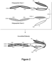
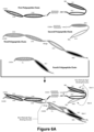
- Figure 1 provides a schematic of such a diabody, which utilizes E-coil/K-coil Heterodimer-Promoting domains and a cysteine containing linker for covalent bonding.
- one or both of the polypeptides may additionally possesses the sequence of a CH2-CH3 Domain, such that complexing between the two diabody polypeptides forms an Fc Region that is capable of binding to the Fc receptor of cells (such as B lymphocytes, dendritic cells, natural killer cells, macrophages, neutrophils, eosinophils, basophils and mast cells).
- the CH2 and/or CH3 Domains of such polypeptide chains need not be identical in sequence, and advantageously are modified to foster complexing between the two polypeptide chains.
- the first polypeptide chain comprises (in the N-terminal to C-terminal direction): (i) a First Domain that comprises a binding region of a Light Chain Variable Domain of a first immunoglobulin (VL1), (ii) a Second Domain that comprises a binding region of a Heavy Chain Variable Domain of a second immunoglobulin (VH2), (iii) a Third Domain that contains a cysteine residue (or a cysteine-containing domain) and a serves to promote heterodimerization with the second polypeptide of the diabody and to covalently bond the diabody's first and second polypeptides to one another, and (iv) a CH2-CH3 Domain.
- the second polypeptide contains (in the N-terminal to C-terminal direction): (i) a First Domain that comprises a binding region of a Light Chain Variable Domain of the second immunoglobulin (VL2), (ii) a Second Domain that comprises a binding region of a Heavy Chain Variable Domain of the first immunoglobulin (VH1), and (iii) ) a Third Domain that contains a cysteine residue (or a cysteine-containing domain) and a Heterodimer-Promoting Domain that promotes heterodimerization with the first polypeptide chain.
- VL2 Light Chain Variable Domain of the second immunoglobulin
- VH1 Heavy Chain Variable Domain of the first immunoglobulin
- a Third Domain that contains a cysteine residue (or a cysteine-containing domain) and a Heterodimer-Promoting Domain that promotes heterodimerization with the first polypeptide chain.
- two first polypeptides complex with each other to form an F
- Fc-Region-containing DART ® diabodies may comprise three polypeptide chains.
- the first polypeptide of such DART ® diabodies contains three domains: (i) a VL1-containing Domain, (ii) a VH2-containing Domain and (iii) a Domain containing a CH2-CH3 sequence.
- the second polypeptide of such DART ® diabodies contains: (i) a VL2-containing Domain, (ii) a VH1-containing Domain and (iii) a Domain that promotes heterodimerization and covalent bonding with the diabody's first polypeptide chain.
- the third polypeptide of such DART ® diabodies comprises a CH2-CH3 sequence.
- the first and second polypeptide chains of such DART ® diabodies associate together to form a VL1/VH1 binding site that is capable of binding to the epitope, as well as a VL2/VH2 binding site that is capable of binding to the second epitope.
- Such more complex DART ® molecules also possess cysteine-containing domains which function to form a covalently bonded complex.
- the first and second polypeptides are bonded to one another through a disulfide bond involving cysteine residues in their respective Third Domains.
- the first and third polypeptide chains complex with one another to form an Fc Region that is stabilized via a disulfide bond.
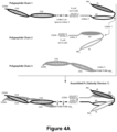
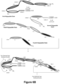
- Figures 4A-4B provide schematics of such diabodies comprising three polypeptide chains.
- Still other Fc-Region-containing DART ® diabodies may comprise five polypeptide chains which may comprise the binding regions from the Light and Heavy Chain Variable Domains of up to three different immunoglobulins (referred to as VL1/VH1, VL2/VH2 and VL3/VH3).
- the first polypeptide chain of such diabodies may contain: (i) a VH1-containing domain, (ii) a CH1-containing domain, and (iii) a Domain containing a CH2-CH3 sequence.
- the second and fifth polypeptide chains of such diabodies may contain: (i) a VL1-containing domain, and (ii) a CL-containing domain.
- the third polypeptide chain of such diabodies may contain: (i) a VH1-containing domain, (ii) a CH1-containing domain, (iii) a Domain containing a CH2-CH3 sequence, (iv) a VL2-containing Domain, (v) a VH3-containing Domain and (vi) a Heterodimer-Promoting Domain, where the Heterodimer-Promoting Domains promote the dimerization of the third chain with the fourth chain.
- the fourth polypeptide of such diabodies may contain: (i) a VL3-containing Domain, (ii) a VH2-containing Domain and (iii) a Domain that promotes heterodimerization and covalent bonding with the diabody's third polypeptide chain.
- the first and third polypeptides complex with each other to form an Fc Region.
- Such more complex DART ® molecules also possess cysteine-containing domains which function to form a covalently bonded complex, such that each polypeptide chain is bonded to at least one addition polypeptide chain through a disulfide bond involving cysteine residues.
- such domains are ordered in the N-terminal to C-terminal direction.
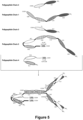
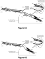
- Figure 5 provides schematics of such diabodies comprising five polypeptide chains.
- PD-1-binding molecules include antibodies, diabodies, BiTEs, etc. and are capable of binding to a continuous or discontinuous (e.g ., conformational) portion (epitope) of human PD-1 (CD279).
- PD-1-binding molecules preferably also exhibit the ability to bind to PD-1 molecules of one or more non-human species, in particular, primate species (and especially a primate species, such as cynomolgus monkey).
- a representative human PD-1 polypeptide (NCBI Sequence NP_005009.2; including a 20 amino acid residue signal sequence (shown underlined) and the 268 amino acid residue mature protein) has the amino acid sequence (SEQ ID NO:68):
- Anti-human PD-1-binding molecules may be characterized by any (one or more) of the following criteria:
- the term "antigen specific T-cell response" refers to responses by a T-cell that result from stimulation of the T-cell with the antigen for which the T-cell is specific.
- responses by a T-cell upon antigen specific stimulation include proliferation and cytokine production (e.g ., TNF- ⁇ , IFN- ⁇ production).
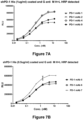
- the ability of a molecule to stimulate an antigen specific T-cell response may be determined, for example, using the Staphylococcus aureus Enterotoxin type B antigen ("SEB”)-stimulated PBMC assay described herein.
- SEB Staphylococcus aureus Enterotoxin type B antigen
- Anti-human PD-1-binding molecules disclosed but not claimed herein possess the VH and/or VL Domains of murine anti-human PD-1 monoclonal antibodies "PD-1 mAb 1," “PD-1 mAb 2 ,” “ PD-1 mAb 3 ,′′′' PD-1 mAb 4 ,” “ PD-1 mAb 5 ,” “ PD-1 mAb 6 ,” “ PD-1 mAb 7 ,” “ PD-1 mAb 8 ,” “ PD-1 mAb 9 ,” “ PD-1 mAb 10 ,” “ PD-1 mAb 11 ,” “ PD-1 mAb 12 ,” “ PD-1 mAb 13 ,” “ PD-1 mAb 14 ,” or “ PD-1 mAb 15 ,” and more preferably possess 1, 2 or all 3 of the CDR H s of the VH Domain and/or 1, 2 or all 3 of the CDR L s of the VL Domain of such anti-human PD-1 monoclonal antibodies.
- the invention relates to PD-1-binding molecules, as defined by and for use in accordance with claims 1-6, comprising a PD-1 binding domain that possesses:
- CDR H 1 of PD-1 mAb 1 (SEQ ID NO:71): NDYAWN
- CDR H 2 of PD-1 mAb 1 (SEQ ID NO:72): HITYSGSTSYNPSLKS
- CDR H 3 of PD-1 mAb 1 (SEQ ID NO:73): DYGSGYPYTLDY
- An exemplary polynucleotide that encodes the VH Domain of PD-1 mAb 1 is SEQ ID NO:70 (nucleotides encoding the CDR H residues are shown underlined):
- CDR L 1 of PD-1 mAb 1 SATSIVSYVY CDR L 2 of PD-1 mAb 1 (SEQ ID NO:77): LTSNLAS CDR L 3 of PD-1 mAb 1 (SEQ ID NO:78): QQWSDNPYT
- An exemplary polynucleotide that encodes the VL Domain of PD-1 mAb 1 is SEQ ID NO:75 (nucleotides encoding the CDR L residues are shown underlined):
- the above-described murine anti-human PD-1 antibody PD-1 mAb 1 was humanized and further deimmunized when antigenic epitopes were identified in order to demonstrate the capability of humanizing an anti-human PD-1 antibody so as to decrease its antigenicity upon administration to a human recipient.
- the humanization yielded one humanized VH Domain, designated herein as " hPD-1 mAb 1 VH1 ,” and one humanized VL Domain designated herein as " hPD-1 mAb 1 VL1.
- an antibody comprising the humanized VL Domains paired with the humanized VH Domain is referred to as " hPD-1 mAb 1 .”
- An exemplary polynucleotide that encodes hPD-1 mAb 1 VH1 is SEQ ID NO:80 (nucleotides encoding the CDR H residues are shown underlined):
- An exemplary polynucleotide that encodes hPD-1 mAb 1 VL1 is SEQ ID NO:82 (nucleotides encoding the CDR H residues are shown underlined):
- CDR H 1 of PD-1 mAb 2 SFGMH
- CDR H 2 of PD-1 mAb 2 SEQ ID NO:86
- YISSGSMSISYADTVKG CDR H 3 of PD-1 mAb 2 (SEQ ID NO:87): LSDYFDY
- An exemplary polynucleotide that encodes the VH Domain of PD-1 mAb 2 is SEQ ID NO:84 (nucleotides encoding the CDR H residues are shown underlined):
- CDR L 1 of PD-1 mAb 2 SEQ ID NO:90
- RSSQSLVHSTGNTYLH CDR L 2 of PD-1 mAb 2
- RVSNRFS CDR L 3 of PD-1 mAb 2
- An exemplary polynucleotide that encodes the VL Domain of PD-1 mAb 2 is SEQ ID NO:89 (nucleotides encoding the CDR L residues are shown underlined):
- the above-described murine anti-human PD-1 antibody PD-1 mAb 2 was humanized and further deimmunized when antigenic epitopes were identified in order to demonstrate the capability of humanizing an anti-human PD-1 antibody so as to decrease its antigenicity upon administration to a human recipient.
- the humanization yielded one humanized VH Domain, designated herein as "hPD-1 mAb 2 VH1,” and one humanized VL Domains designated herein as "hPD-1 mAb 1 VL1.” Accordingly, any antibody comprising the humanized VL Domains paired with the humanized VH Domain is referred to as "hPD-1 mAb 2.”
- An exemplary polynucleotide that encodes hPD-1 mAb 2 VH1 is SEQ ID NO:94 (nucleotides encoding the CDR H residues are shown underlined):
- An exemplary polynucleotide that encodes hPD-1 mAb 2 VL1 is SEQ ID NO:96 (nucleotides encoding the CDR H residues are shown underlined):
- CDR H 1 of PD-1 mAb 3 (SEQ ID NO:99): DYVMH CDR H 2 of PD-1 mAb 3 (SEQ ID NO:100): TIDPETGGTAYNQKFKG
- CDR H 3 of PD-1 mAb 3 (SEQ ID NO:101): EKITTIVEGTYWYFDV
- An exemplary polynucleotide that encodes the VH Domain of PD-1 mAb 3 is SEQ ID NO:98 (nucleotides encoding the CDR H residues are shown underlined):
- CDR L 1 of PD-1 mAb 3 SEQ ID NO:104
- RSSQNIVHSNGDTYLE CDR L 2 of PD-1 mAb 3
- KVSNRFS CDR L 3 of PD-1 mAb 3
- FQGSHLPYT FQGSHLPYT
- An exemplary polynucleotide that encodes the VL Domain of PD-1 mAb 3 is SEQ ID NO:103 (nucleotides encoding the CDR L residues are shown underlined):
- CDR H 1 of PD-1 mAb 4 SEQ ID NO:109: SFGMH
- CDR H 2 of PD-1 mAb 4 SEQ ID NO:110: YISSGSMSISYADTVKG
- CDR H 3 of PD-1 mAb 4 SEQ ID NO:111: LTDYFDY
- An exemplary polynucleotide that encodes the VH Domain of PD-1 mAb 4 is SEQ ID NO:108 (nucleotides encoding the CDR H residues are shown underlined):
- CDR L 1 of PD-1 mAb 4 SEQ ID NO:114
- RSSQSLVHSTGNTYFH CDR L 2 of PD-1 mAb 4
- RVSNRFS CDR L 3 of PD-1 mAb 4
- An exemplary polynucleotide that encodes the VL Domain of PD-1 mAb 4 is SEQ ID NO:113 (nucleotides encoding the CDR L residues are shown underlined):
- CDR H 1 of PD-1 mAb 5 (SEQ ID NO:119): AYWMN
- CDR H 2 of PD-1 mAb 5 (SEQ ID NO:120): VIHPSDSETWLNQKFKD
- CDR H 3 of PD-1 mAb 5 (SEQ ID NO:121): EHYGSSPFAY
- An exemplary polynucleotide that encodes the VH Domain of PD-1 mAb 5 is SEQ ID NO:118 (nucleotides encoding the CDR H residues are shown underlined):
- CDR L 1 of PD-1 mAb 5 (SEQ ID NO:124): RANESVDNYGMSFMN
- CDR L 2 of PD-1 mAb 5 (SEQ ID NO:125): AASNQGS
- CDR L 3 of PD-1 mAb 5 (SEQ ID NO:126): QQSKEVPYT
- An exemplary polynucleotide that encodes the VL Domain of PD-1 mAb 5 is SEQ ID NO:123 (nucleotides encoding the CDR L residues are shown underlined):
- CDR H 1 of PD-1 mAb 6 SEQ ID NO:129
- SYGMS SYGMS
- CDR H 2 of PD-1 mAb 6 SEQ ID NO:130
- TISGGGSDTYYPDSVKG CDR H 3 of PD-1 mAb 6 ( SEQ ID NO:131 ): QKATTWFAY
- An exemplary polynucleotide that encodes the VH Domain of PD-1 mAb 6 is SEQ ID NO:128 (nucleotides encoding the CDR H residues are shown underlined):
- CDR L 1 of PD-1 mAb 6 SEQ ID NO:134
- RASESVDNYGISFMN CDR L 2 of PD-1 mAb 6
- PASNQGS CDR L 3 of PD-1 mAb 6
- QQSKEVPWT QQSKEVPWT
- An exemplary polynucleotide that encodes the VL Domain of PD-1 mAb 6 is SEQ ID NO:133 (nucleotides encoding the CDR L residues are shown underlined):
- CDR H 1 of PD-1 mAb 7 SEQ ID NO:139
- SYWMN CDR H 2 of PD-1 mAb 7
- VIHPSDSETWLDQKFKD CDR H 3 of PD-1 mAb 7
- EHYGTSPFAY EHYGTSPFAY
- An exemplary polynucleotide that encodes the VH Domain of PD-1 mAb 7 is SEQ ID NO:138 (nucleotides encoding the CDR H residues are shown underlined):
- CDR L 1 of PD-1 mAb 7 (SEQ ID NO:144): RANESVDNYGMSFMN
- CDR L 2 of PD-1 mAb 7 (SEQ ID NO:145): AASNQGS
- CDR L 3 of PD-1 mAb 7 (SEQ ID NO:146): QQSKEVPYT
- An exemplary polynucleotide that encodes the VL Domain of PD-1 mAb 7 is SEQ ID NO:143 (nucleotides encoding the CDR L residues are shown underlined):
- the above-described murine anti-human PD-1 antibody PD-1 mAb 7 was humanized and further deimmunized when antigenic epitopes were identified in order to demonstrate the capability of humanizing an anti-human PD-1 antibody so as to decrease its antigenicity upon administration to a human recipient.
- the humanization yielded two humanized VH Domains, designated herein as "hPD-1 mAb 7 VH1,” and “hPD-1 mAb 7 VH2,” and three humanized VL Domains designated herein as "hPD-1 mAb 7 VL1," "hPD- 1 mAb 7 VL2,” and "hPD-1 mAb 7 VL3.” Any of the humanized VL Domains may be paired with either of the humanized VH Domains.
- any antibody comprising one of the humanized VL Domains paired with the humanized VH Domain is referred to generically as "hPD-1 mAb 7," and particular combinations of humanized VH/VL Domains are referred to by reference to the specific VH/VL Domains, for example a humanized antibody comprising hPD-1 mAb 7 VH1 and hPD-1 mAb 1 VL2 is specifically referred to as “hPD-1 mAb 7(1.2)."
- An exemplary polynucleotide that encodes hPD-1 mAb 7 VH1 is SEQ ID NO:148 (nucleotides encoding the CDR H residues are shown underlined):
- An exemplary polynucleotide that encodes hPD-1 mAb 7 VH2 is SEQ ID NO:150 (nucleotides encoding the CDR H residues are shown underlined):
- An exemplary polynucleotide that encodes hPD-1 mAb 7 VL1 is SEQ ID NO:152 (nucleotides encoding the CDR H residues are shown underlined):
- An exemplary polynucleotide that encodes hPD-1 mAb 7 VL2 is SEQ ID NO:154 (nucleotides encoding the CDR H residues are shown underlined):
- An exemplary polynucleotide that encodes hPD-1 mAb 7 VL3 is SEQ ID NO:156 (nucleotides encoding the CDR H residues are shown underlined):
- the CDR L 1 of the VL Domain of both hPD-1 mAb 7 VL2 and hPD-1 mAb 7 VL3 comprises an asparagine to serine amino acid substitution and has the amino acid sequence: RA S ESVDNYGMSFMN ((SEQ ID NO:157), the substituted serine is shown underlined). It is contemplated that a similar substitution may be incorporated into any of the PD-1 mAb 7 CDR L 1 Domains described above.
- the CDR L 2 of the VL Domain of hPD-1 mAb 7 VL3 comprises a glutamine to arginine amino acid substitution and has the amino acid sequence: AASNRGS (( SEQ ID NO:158 ), the substituted arginine is shown underlined). It is contemplated that a similar substitution may be incorporated into any of the PD-1 mAb 7 CDR L 2 Domains described above.
- CDR H 1 of PD-1 mAb 8 (SEQ ID NO:161): DYYMN
- CDR H 2 of PD-1 mAb 8 SEQ ID NO:162: DINPKNGDTHYNQKFKG
- CDR H 3 of PD-1 mAb 8 (SEQ ID NO:163): DFDY
- An exemplary polynucleotide that encodes the VH Domain of PD-1 mAb 8 is SEQ ID NO:160 (nucleotides encoding the CDR H residues are shown underlined):
- CDR L 1 of PD-1 mAb 8 SEQ ID NO:166
- RSSQTLVYSNGNTYLN CDR L 2 of PD-1 mAb 8
- KVSNRFS CDR L 3 of PD-1 mAb 8
- An exemplary polynucleotide that encodes the VL Domain of PD-1 mAb 8 is SEQ ID NO:165 (nucleotides encoding the CDR L residues are shown underlined):
- CDR H 1 of PD-1 mAb 9 SYLVS
- CDR H 2 of PD-1 mAb 9 SEQ ID NO:172
- CDR H 3 of PD-1 mAb 9 SEQ ID NO:173
- An exemplary polynucleotide that encodes the VH Domain of PD-1 mAb 9 is SEQ ID NO:170 (nucleotides encoding the CDR H residues are shown underlined):
- CDR L 1 of PD-1 mAb 9 SEQ ID NO:176
- RASENIYSYLA CDR L 2 of PD-1 mAb 9 SEQ ID NO:177
- NAKTLAA CDR L 3 of PD-1 mAb 9 SEQ ID NO:178
- An exemplary polynucleotide that encodes the VL Domain of PD-1 mAb 9 is SEQ ID NO:175 (nucleotides encoding the CDR L residues are shown underlined):
- the above-described murine anti-human PD-1 antibody PD-1 mAb 9 was humanized and further deimmunized when antigenic epitopes were identified in order to demonstrate the capability of humanizing an anti-human PD-1 antibody so as to decrease its antigenicity upon administration to a human recipient.
- the humanization yielded two humanized VH Domains, designated herein as "hPD-1 mAb 9 VH1,” and “hPD-1 mAb 9 VH2," and two humanized VL Domains designated herein as "hPD-1 mAb 9 VL1," and "hPD-1 mAb 9 VL2.” Any of the humanized VL Domains may be paired with the humanized VH Domains.
- any antibody comprising one of the humanized VL Domains paired with the humanized VH Domain is referred to generically as "hPD-1 mAb 9," and particular combinations of humanized VH/VL Domains are referred to by reference to the specific VH/VL Domains, for example a humanized antibody comprising hPD-1 mAb 9 VH1 and hPD-1 mAb 9 VL2 is specifically referred to as “hPD-1 mAb 9(1.2)."
- An exemplary polynucleotide that encodes hPD-1 mAb 9 VH1 is SEQ ID NO:180 (nucleotides encoding the CDR H residues are shown underlined):
- An exemplary polynucleotide that encodes hPD-1 mAb 9 VH2 is SEQ ID NO:182 (nucleotides encoding the CDR H residues are shown underlined):
- the CDR H 1 of the VH Domain of hPD-1 mAb 9 VH2 comprises a serine to glycine amino acid substitution and has the amino acid sequence: SYLVG ((SEQ ID NO:183), the substituted glycine is shown underlined). It is contemplated that a similar substitution may be incorporated into any of the PD-1 mAb 9 CDR H 1 Domains described above.
- An exemplary polynucleotide that encodes hPD-1 mAb 9 VL1 is SEQ ID NO:185 (nucleotides encoding the CDR H residues are shown underlined):
- An exemplary polynucleotide that encodes hPD-1 mAb 9 VL2 is SEQ ID NO:187 (nucleotides encoding the CDR H residues are shown underlined):
- the CDR L 1 of the VL Domain of hPD-1 mAb 9 VL2 comprises a serine to asparagine amino acid substitution and has the amino acid sequence: RASENIY N YLA (SEQ ID NO:188), the substituted asparagine is shown underlined). It is contemplated that a similar substitution may be incorporated into any of the PD-1 mAb 9 CDR L 1 Domains described above.
- the CDR L 2 of the VL Domain of hPD-1 mAb 9 VL2 comprises an asparagine to aspartate amino acid substitution and has the amino acid sequence: D AKTLAA ((SEQ ID NO:189 ), the substituted aspartate is shown underlined). It is contemplated that a similar substitution may be incorporated into any of the PD-1 mAb 7 CDR L 2 Domains described above.
- CDR H 1 of PD-1 mAb 10 (SEQ ID NO:192): NYLMS
- CDR H 2 of PD-1 mAb 10 (SEQ ID NO:193): SISGGGSNIYYPDSVKG
- CDR H 3 of PD-1 mAb 10 (SEQ ID NO:194): QELAFDY
- An exemplary polynucleotide that encodes the VH Domain of PD-1 mAb 10 is SEQ ID NO:191 (nucleotides encoding the CDR H residues are shown underlined):
- CDR L 1 of PD-1 mAb 10 SEQ ID NO:197
- RTSQDISNFLN CDR L 2 of PD-1 mAb 10
- YTSRLHS CDR L 3 of PD-1 mAb 10
- An exemplary polynucleotide that encodes the VL Domain of PD-1 mAb 10 is SEQ ID NO:196 (nucleotides encoding the CDR L residues are shown underlined):
- CDR H 1 of PD-1 mAb 11 SEQ ID NO:202: GYWMH
- CDR H 2 of PD-1 mAb 11 SEQ ID NO:203: AIYPGNSDTHYNQKFKG
- CDR H 3 of PD-1 mAb 11 SEQ ID NO:204: GTYSYFDV
- An exemplary polynucleotide that encodes the VH Domain of PD-1 mAb 11 is SEQ ID NO:201 (nucleotides encoding the CDR H residues are shown underlined):
- CDR L 1 of PD-1 mAb 11 SEQ ID NO:207
- RASQSIGTSIH CDR L 2 of PD-1 mAb 11
- YASESIS CDR L 3 of PD-1 mAb 11 (SEQ ID NO:209): QQSNSWLT
- An exemplary polynucleotide that encodes the VL Domain of PD-1 mAb 11 is SEQ ID NO:206 (nucleotides encoding the CDR L residues are shown underlined):
- CDR H 1 of PD-1 mAb 12 (SEQ ID NO:212): DYEMH CDR H 2 of PD-1 mAb 12 (SEQ ID NO:213): TIDPETGGTAYNQKFKG
- CDR H 3 of PD-1 mAb 12 (SEQ ID NO:214): ERITTVVEGAYWYFDV
- An exemplary polynucleotide that encodes the VH Domain of PD-1 mAb 12 is SEQ ID NO:211 (nucleotides encoding the CDR H residues are shown underlined):
- CDR L 1 of PD-1 mAb 12 SEQ ID NO:2157: RSSQNIVHSNGNTYLE CDR L 2 of PD-1 mAb 12 (SEQ ID NO:218): KVSTRFS CDR L 3 of PD-1 mAb 12 (SEQ ID NO:219): FQGSHVPYT
- An exemplary polynucleotide that encodes the VL Domain of PD-1 mAb 12 is SEQ ID NO:216 (nucleotides encoding the CDR L residues are shown underlined):
- CDR H 1 of PD-1 mAb 13 SEQ ID NO:222: SHTMS CDR H 2 of PD-1 mAb 13 (SEQ ID NO:223): TISGGGSNIYYPDSVKG CDR H 3 of PD-1 mAb 13 (SEQ ID NO:224): QAYYGNYWYFDV
- An exemplary polynucleotide that encodes the VH Domain of PD-1 mAb 13 is SEQ ID NO:221 (nucleotides encoding the CDR H residues are shown underlined):
- CDR L 1 of PD-1 mAb 13 SEQ ID NO:227): LASQTIGTWLA CDR L 2 of PD-1 mAb 13 (SEQ ID NO:228): AATSLAD CDR L 3 of PD-1 mAb 13 (SEQ ID NO:229): QQLDSIPWT
- An exemplary polynucleotide that encodes the VL Domain of PD-1 mAb 13 is SEQ ID NO:226 (nucleotides encoding the CDR L residues are shown underlined):
- CDR H 1 of PD-1 mAb 14 SYWIT
- CDR H 2 of PD-1 mAb 14 SEQ ID NO:233
- NIYPGTDGTTYNEKFKS CDR H 3 of PD-1 mAb 14 (SEQ ID NO:234): GLHWYFDV
- An exemplary polynucleotide that encodes the VH Domain of PD-1 mAb 14 is SEQ ID NO:231 (nucleotides encoding the CDR H residues are shown underlined):
- CDR L 1 of PD-1 mAb 14 SEQ ID NO:237): KASQSVGTNVA CDR L 2 of PD-1 mAb 14 (SEQ ID NO:238): SASSRFS CDR L 3 of PD-1 mAb 14 (SEQ ID NO:239): QQYNSYPYT
- An exemplary polynucleotide that encodes the VL Domain of PD-1 mAb 14 is SEQ ID NO:236 (nucleotides encoding the CDR L residues are shown underlined):
- CDR H 1 of PD-1 mAb 15 SEQ ID NO:242: SYLIS CDR H 2 of PD-1 mAb 15 (SEQ ID NO:243): AISGGGADTYYADSVKG CDR H 3 of PD-1 mAb 15 (SEQ ID NO:244): RGTYAMDY
- An exemplary polynucleotide that encodes the VH Domain of PD-1 mAb 15 is SEQ ID NO:241 (nucleotides encoding the CDR H residues are shown underlined):
- CDR L 1 of PD-1 mAb 15 SEQ ID NO:2457: LASQTIGTWLA CDR L 2 of PD-1 mAb 15 (SEQ ID NO:248): AATSLAD CDR L 3 of PD-1 mAb 15 (SEQ ID NO:249): QQLYSIPWT
- An exemplary polynucleotide that encodes the VL Domain of PD-1 mAb 15 is SEQ ID NO:246 (nucleotides encoding the CDR L residues are shown underlined):
- the above-described murine anti-human PD-1 antibody PD-1 mAb 15 was humanized and further deimmunized when antigenic epitopes were identified in order to demonstrate the capability of humanizing an anti-human PD-1 antibody so as to decrease its antigenicity upon administration to a human recipient.
- the humanization yielded one humanized VH Domain, designated herein as "hPD-1 mAb 2 VH1 ,” and one humanized VL Domains designated herein as " hPD-1 mAb 1 VL1 .”
- An antibody comprising the humanized VL Domain paired with the humanized VH Domain is referred to as "hPD-1 mAb 15.”
- An exemplary polynucleotide that encodes hPD-1 mAb 15 VH1 is SEQ ID NO:251 (nucleotides encoding the CDR H residues are shown underlined):
- An exemplary polynucleotide that encodes hPD-1 mAb 15 VL1 is SEQ ID NO:253 (nucleotides encoding the CDR H residues are shown underlined):
- FcyRI CD64
- FcyRIIA CD32A
- FcyRIII CD16
- FcyRIIB CD32B
- FcRn neonatal Fc Receptor
- Modification of the Fc Region normally leads to an altered phenotype, for example altered serum half-life, altered stability, altered susceptibility to cellular enzymes or altered effector function. It may be desirable to modify an antibody or other binding molecule of the present invention, as defined by and for use in accordance with claims 1-6, with respect to effector function, for example, so as to enhance the effectiveness of such molecule in treating cancer. Reduction or elimination of effector function is desirable in certain cases, for example in the case of antibodies whose mechanism of action involves blocking or antagonism, but not killing of the cells bearing a target antigen.
- Increased effector function is generally desirable when directed to undesirable cells, such as tumor and foreign cells, where the FcyRs are expressed at low levels, for example, tumor-specific B cells with low levels of FcyRIIB (e.g., non-Hodgkins lymphoma, CLL, and Burkitt's lymphoma).
- FcyRIIB e.g., non-Hodgkins lymphoma, CLL, and Burkitt's lymphoma.
- the PD-1-binding molecules of the present invention comprise an Fc Region that possesses one or more modifications (e.g., substitutions, deletions, or insertions) to the sequence of amino acids of a wild-type Fc Region (e.g., SEQ ID NO:1), which reduce the affinity and avidity of the Fc Region and, thus, the molecule of the invention, for one or more FcyR receptors.
- Fc Region that possesses one or more modifications (e.g., substitutions, deletions, or insertions) to the sequence of amino acids of a wild-type Fc Region (e.g., SEQ ID NO:1), which reduce the affinity and avidity of the Fc Region and, thus, the molecule of the invention, for one or more FcyR receptors.
- the molecules of the invention as defined by and for use in accordance with claims 1-6, comprise an Fc Region that possesses one or more modifications to the amino acids of the wild-type Fc Region, which increase the affinity and avidity of the Fc Region and, thus, the molecule of the invention, for one or more FcyR receptors.
- the molecules comprise a variant Fc Region wherein said variant confers or mediates increased antibody dependent cell mediated cytotoxicity (ADCC) activity and/or an increased binding to FcyRIIA, relative to a molecule comprising no Fc Region or comprising a wild-type Fc Region.
- ADCC antibody dependent cell mediated cytotoxicity
- the molecules comprise a variant Fc Region wherein said variant confers or mediates decreased ADCC activity (or other effector function) and/or an increased binding to FcyRIIB, relative to a molecule comprising no Fc Region or comprising a wild-type Fc Region.
- the invention as defined by and for use in accordance with claims 1-6, encompasses PD-1-binding molecules comprising a variant Fc Region, which variant Fc Region does not show a detectable binding to any FcyR, relative to a comparable molecule comprising the wild-type Fc Region.
- the invention as defined by and for use in accordance with claims 1-6, encompasses PD-1-binding molecules comprising a variant Fc Region, which variant Fc Region only binds a single FcyR, preferably one of FcyRIIA, FcyRIIB, or FcyRIIIA.
- any such increased affinity and/or avidity is preferably assessed by measuring in vitro the extent of detectable binding to the FcyR or Fc ⁇ R-related activity in cells that express low levels of the FcyR when binding activity of the parent molecule (without the modified Fc Region) cannot be detected in the cells, or in cells which express non-FcyR receptor target antigens at a density of 30,000 to 20,000 molecules/cell, at a density of 20,000 to 10,000 molecules/cell, at a density of 10,000 to 5,000 molecules/cell, at a density of 5,000 to 1,000 molecules/cell, at a density of 1,000 to 200 molecules/cell or at a density of 200 molecules/cell or less (but at least 10, 50, 100 or 150 molecules/cell).
- the PD-1-binding molecules of the present invention may comprise a variant Fc Region having altered affinities for an activating and/or inhibitory Fc ⁇ receptor.
- the PD-1-binding molecule comprises a variant Fc Region that has increased affinity for FcyRIIB and decreased affinity for FcyRIIIA and/or FcyRIIA, relative to a comparable molecule with a wild-type Fc Region.
- the PD-1-binding molecule of the present invention as defined by and for use in accordance with claims 1-6, comprise a variant Fc Region, which has decreased affinity for FcyRIIB and increased affinity for FcyRIIIA and/or FcyRIIA, relative to a comparable molecule with a wild-type Fc Region.
- the PD-1-binding molecules of the present invention as defined by and for use in accordance with claims 1-6, comprise a variant Fc Region that has decreased affinity for FcyRIIB and decreased affinity for FcyRIIIA and/or FcyRIIA, relative to a comparable molecule with a wild-type Fc Region.
- the PD-1-binding molecules of the present invention comprise a variant Fc Region, which has unchanged affinity for FcyRIIB and decreased (or increased) affinity for FcyRIIIA and/or FcyRIIA, relative to a comparable molecule with a wild-type Fc Region.
- the PD-1-binding molecules of the present invention comprise a variant Fc Region having an altered affinity for FcyRIIIA and/or FcyRIIA such that the immunoglobulin has an enhanced effector function.
- effector cell functions include antibody dependent cell mediated cytotoxicity, antibody dependent phagocytosis, phagocytosis, opsonization, opsonophagocytosis, cell binding, resetting, C1q binding, and complement dependent cell mediated cytotoxicity.
- the alteration in affinity or effector function is at least 2-fold, preferably at least 4-fold, at least 5-fold, at least 6-fold, at least 7-fold, at least 8-fold, at least 9-fold, at least 10-fold, at least 50-fold, or at least 100-fold, relative to a comparable molecule comprising a wild-type Fc Region.
- the variant Fc Region immunospecifically binds one or more FcRs with at least 65%, preferably at least 70%, 75%, 80%, 85%, 90%, 95%, 100%, 125%, 150%, 175%, 200%, 225%, or 250% greater affinity relative to a molecule comprising a wild-type Fc Region.
- Such measurements can be in vivo or in vitro assays, and in a preferred embodiment are in vitro assays such as ELISA or surface plasmon resonance assays.
- the PD-1-binding molecules of the present invention comprise a variant Fc Region wherein said variant agonizes at least one activity of an FcyR receptor, or antagonizes at least one activity of an FcyR receptor.
- the molecules comprise a variant that antagonizes one or more activities of FcyRIIB, for example, B-cell receptor-mediated signaling, activation of B-cells, B-cell proliferation, antibody production, intracellular calcium influx of B cells, cell cycle progression, FcyRIIB-mediated inhibition of Fc ⁇ RI signaling, phosphorylation of FcyRIIB, SHIP recruitment, SHIP phosphorylation and association with She, or activity of one or more downstream molecules (e.g., MAP kinase, JNK, p38, or Akt) in the FcyRIIB signal transduction pathway.
- B-cell receptor-mediated signaling for example, B-cell receptor-mediated signaling, activation of B-cells, B-cell proliferation, antibody production, intracellular calcium influx of B cells, cell cycle progression, FcyRIIB-mediated inhibition of Fc ⁇ RI signaling, phosphorylation of FcyRIIB, SHIP recruitment, SHIP phosphorylation and association with She, or activity of one
- the PD-1-binding molecules of the present invention comprise a variant that agonizes one or more activities of Fc ⁇ RI, for example, mast cell activation, calcium mobilization, degranulation, cytokine production, or serotonin release.
- the molecules comprise an Fc Region comprising regions from two or more IgG isotypes (e.g., IgG1, IgG2, IgG3 and IgG4).
- an Fc Region is said to be of a particular IgG isotype if its amino acid sequence is most homologous to that isotype relative to other IgG isotypes.
- the various IgG isotypes exhibit differing physical and functional properties including serum half-life, complement fixation, FcyR binding affinities and effector function activities (e.g., ADCC, CDC, etc.) due to differences in the amino acid sequences of their hinge and/or Fc Regions, for example as described in Flesch and Neppert (1999) J.
- the amino acid modification and IgG hinge/Fc Region may display similar functionality (e.g., increased affinity for FcyRIIA) and may act additively or, more preferably, synergistically to modify the effector functionality in the molecule of the invention, relative to a molecule of the invention comprising a wild-type Fc Region.
- the amino acid modification and IgG Fc Region may display opposite functionality (e.g., increased and decreased affinity for FcyRIIA, respectively) and may act to selectively temper or reduce a specific functionality in the molecule of the invention, relative to a molecule of the invention not comprising an Fc Region or comprising a wild-type Fc Region of the same isotype.
- the PD-1-binding molecules of the present invention comprise a variant Fc Region, wherein said variant Fc Region comprises at least one amino acid modification relative to a wild-type Fc Region, such that said molecule has an altered affinity for an FcR, provided that said variant Fc Region does not have a substitution at positions that make a direct contact with FcyR based on crystallographic and structural analysis of Fc-FcR interactions such as those disclosed by Sondermann et al. (2000) Nature 406:267-73 .
- the molecules of the invention as defined by and for use in accordance with claims 1-6, comprise variant Fc Regions comprise modification of at least one residue that does not make a direct contact with an FcyR based on structural and crystallographic analysis, e . g ., is not within the Fc-FcyR binding site.
- Variant Fc Regions are well known in the art, and any known variant Fc Region may be used in the present invention to confer or modify the effector function exhibited by a molecule of the invention comprising an Fc Region (or portion thereof) as functionally assayed, e . g ., in an NK dependent or macrophage dependent assay.
- Fc Region variants identified as altering effector function are disclosed in PCT Publications No. WO 04/063351 ; WO 06/088494 ; WO 07/024249 ; WO 06/113665 ; WO 07/021841 ; WO 07/106707 ; and WO 2008/140603 , and any suitable variant disclosed therein may be used in the present molecules.
- PD-1-binding molecules of the present invention that possess a variant Fc Region (relative to the wild-type Fc Region) in which the variant Fc Region has a Ratio of Affinities greater than 1.
- Such molecules have particular use in providing a therapeutic or prophylactic treatment of a disease, disorder, or infection, or the amelioration of a symptom thereof, where an enhanced efficacy of effector cell function (e.g., ADCC) mediated by FcyR is desired, e.g., cancer or infectious disease.
- a variant Fc Region having a Ratio of Affinities less than 1 mediates decreased efficacy of effector cell function.
- Table 1 lists exemplary single, double, triple, quadruple and quintuple mutations by whether their Ratio of Affinities is greater than or less than 1.
- Table 1 Exemplary Single and Multiple Mutations Listed by Ratio of Affinities Single Double Triple Quadruple Quintuple Ratio of Affinities > 1 F243L F243L & R292P F243L, P247L & N421K L234F, F243L, R292P & Y300L L235V, F243L, R292P, Y300L & P396L D270E F243L & Y300L F243L, R292P & Y300L L235I, F243L, R292P & Y300L L235P, F243L, R292P, Y300L & P396L R292G F243L & P396L F243L, R292P & V305I L235Q, F243L, R292P
- any amino acid modifications e.g., substitutions
- any amino acid modifications e.g., substitutions
- any amino acid modifications e.g., substitutions
- any amino acid modifications at any of positions 255, 256, 258, 267, 268, 269, 270, 272, 276, 278, 280, 283, 285, 286, 289, 290, 292, 293, 294, 295, 296, 298, 300, 301, 303, 305, 307, 309, 312, 320, 322, 326, 329, 330, 332, 331, 333, 334, 335, 337, 338, 339, 340, 359, 360, 373, 376, 416, 419, 430, 434, 435, 437, 438 or 439.
- the variant Fc Region has any of the following residues: A256, N268, Q272, D286, Q286, S286, A290, S290, A298, M301, A312, E320, M320, Q320, R320, E322, A326, D326, E326, N326, S326, K330, T339, A333, A334, E334, H334, L334, M334, Q334, V334, K335, Q335, A359, A360 or A430.
- any amino acid modifications e.g., substitutions
- any amino acid modifications e.g., substitutions
- any amino acid modifications e.g., substitutions
- any amino acid modifications e.g., substitutions
- any amino acid modifications e.g., substitutions
- any amino acid modifications e.g., substitutions
- any of the following residues A255, A256, A258, A267, A268, N268, A272, Q272, A276, A280, A283, A285, A286, D286, Q286, S286, A290, S290, M301, E320, M320, Q320, R320, E322, A326, D326, E326, S326, K330, A331, Q335, A337 or A430.
- Preferred variants include one or more modifications at any of positions: 228, 230, 231, 232, 233, 234, 235, 239, 240, 241, 243, 244, 245, 247, 262, 263, 264, 265, 266, 271, 273, 275, 281, 284, 291, 296, 297, 298, 299, 302, 304, 305, 313, 323, 325, 326, 328, 330 or 332.
- Particularly preferred variants include one or more modifications selected from groups A-AI: A 228E, 228K, 228Y or 228G; B 230A, 230E, 230Y or 230G; C 231E, 231K, 231Y, 231P or 231G; D 232E, 232K, 232Y, 232G; E 233D; F 234I or 234F; G 235D, 235Q, 235P, 235I or 235V; H 239D, 239E, 239N or 239Q; I 240A, 240I, 240M or 240T; J 243R, 243, 243Y, 243L, 243Q, 243W, 243H or 243I; K 244H; L 245A; M 247G, 247V or 247L; N 262A, 262E, 262I, 262T, 262E or 262F; O 263A, 263I, 263M or 263T
- a PD-1-binding molecule of the invention will comprise a variant Fc Region having at least one modification in the Fc Region.
- the variant Fc Region comprises at least one substitution selected from the group consisting of L235V, F243L, R292P, Y300L, V305I, and P396L.
- the variant Fc Region comprises:
- variant Fc Region comprises substitutions of:
- a PD-1-binding molecule of the invention comprises a variant Fc Region that exhibits decreased (or substantially no) binding to FcyRIA (CD64), FcyRIIA (CD32A), FcyRIIB (CD32B), FcyRIIIA (CD16a) or FcyRIIIB (CD16b) (relative to the binding exhibited by the wild-type IgG1 Fc Region (SEQ ID NO:1)).
- a PD-1-binding molecule of the invention will comprise a variant Fc Region that exhibits reduced (or substantially no) binding to an FcyR (e.g., FcyRIIIA) and reduced (or substantially no) ADCC effector function.
- the variant Fc Region comprises at least one substitution selected from the group consisting of L234A, L235A, D265A,N297Q, and N297G.
- the variant Fc Region comprises the substitution of L234A; L235A; L234A and L235A; D265A; N297Q, or N297G.
- a preferred IgG1 sequence for the CH2 and CH3 Domains of the PD-1-binding molecules of the invention will have the L234A/L235A substitutions (SEQ ID NO:5): wherein, x is a lysine (K) or is absent.
- a PD-1-binding molecule of the invention comprises an Fc Region which inherently exhibits decreased (or substantially no) binding to FcyRIIIA (CD16a) and/or reduced effector function (relative to the binding exhibited by the wild-type IgG1 Fc Region (SEQ ID NO:1)).
- a PD-1-binding molecule of the present invention comprises an IgG2 Fc Region (SEQ ID NO:2) or an IgG4 Fc Region (SEQ ID:NO:4).
- the instant invention also encompasses the introduction of a stabilizing mutation, such as the IgG4 hinge region S228P substitution (see, e.g., SEQ ID NO:13: ESKYGPPCP P CP, ( Lu et al., (2008) "The Effect Of A Point Mutation On The Stability Of Igg4 As Monitored By Analytical Ultracentrifugation,” J. Pharmaceutical Sciences 97:960-969 ) to reduce the incidence of strand exchange.
- a stabilizing mutation such as SEQ ID NO:13: ESKYGPPCP P CP
- the invention encompasses the use of any variant Fc Region known in the art, such as those disclosed in Jefferis, B.J. et al. (2002) "Interaction Sites On Human IgG-Fc For FcgammaR: Current Models," Immunol. Lett. 82:57-65 ; Presta, L.G. et al. (2002) “Engineering Therapeutic Antibodies For Improved Function," Biochem. Soc. Trans. 30:487-90 ; Idusogie, E.E. et al. (2001) "Engineered Antibodies With Increased Activity To recruit Complement," J. Immunol. 166:2571-75 ; Shields, R.L. et al.
- the molecules of the invention may further comprise one or more glycosylation sites, so that one or more carbohydrate moieties are covalently attached to the molecule.
- the molecules of the invention with one or more glycosylation sites and/or one or more modifications in the Fc Region confer or have an enhanced antibody mediated effector function, e . g ., enhanced ADCC activity, compared to the unmodified antibody.
- the invention further comprises molecules comprising one or more modifications of amino acids that are directly or indirectly known to interact with a carbohydrate moiety of the Fc Region, including but not limited to amino acids at positions 241, 243, 244, 245, 245, 249, 256, 258, 260, 262, 264, 265, 296, 299, and 301.
- Amino acids that directly or indirectly interact with a carbohydrate moiety of an Fc Region are known in the art, see, e.g., Jefferis et al., 1995 Immunology Letters, 44: 111-7 .
- the invention encompasses molecules that have been modified by introducing one or more glycosylation sites into one or more sites of the molecules, preferably without altering the functionality of the molecules, e . g ., binding activity to target antigen or FcyR.
- Glycosylation sites may be introduced into the variable and/or constant region of the molecules of the invention.
- "glycosylation sites” include any specific amino acid sequence in an antibody to which an oligosaccharide (i.e., carbohydrates containing two or more simple sugars linked together) will specifically and covalently attach. Oligosaccharide side chains are typically linked to the backbone of an antibody via either N-or O-linkages.
- N-linked glycosylation refers to the attachment of an oligosaccharide moiety to the side chain of an asparagine residue.
- O-linked glycosylation refers to the attachment of an oligosaccharide moiety to a hydroxyamino acid, e.g., serine, threonine.
- the molecules of the invention may comprise one or more glycosylation sites, including N-linked and O-linked glycosylation sites. Any glycosylation site for N-linked or O-linked glycosylation known in the art may be used in accordance with the instant invention.
- An exemplary N-linked glycosylation site that is useful in accordance with such methods is the amino acid sequence: Asn-X-Thr/Ser, wherein X may be any amino acid and Thr/Ser indicates a threonine or a serine.
- a site or sites may be introduced into a molecule of the invention using methods well known in the art (see for example, IN VITRO MUTAGENESIS, RECOMBINANT DNA: A SHORT COURSE, J. D. Watson, et al. W.H. Freeman and Company, New York, 1983, chapter 8, pp. 106-116 .)
- An exemplary method for introducing a glycosylation site into a molecule of the invention may comprise: modifying or mutating an amino acid sequence of the molecule so that the desired Asn-X-Thr/Ser sequence is obtained.
- Methods of modifying the carbohydrate content of a molecule of the invention include deleting one or more endogenous carbohydrate moieties of the molecule or shifting the glycosylation site of the Fc Region of an antibody, by modifying positions adjacent to 297.
- Position 296 may be modified so that position 296 and not position 297 is glycosylated.
- Effector function can also be modified by techniques such as by introducing one or more cysteine residues into the Fc Region, thereby allowing interchain disulfide bond formation in this region to occur, resulting in the generation of a homodimeric antibody that may have improved internalization capability and/or increased complement-mediated cell killing and ADCC ( Caron, P.C. et al. (1992) "Engineered Humanized Dimeric Forms Of IgG Are More Effective Antibodies," J. Exp. Med. 176:1191-1195 ; Shopes, B. (1992) "A Genetically Engineered Human IgG Mutant With Enhanced Cytolytic Activity,” J. Immunol. 148(9):2918-2922 .
- Homodimeric antibodies with enhanced antitumor activity may also be prepared using heterobifunctional cross-linkers as described in Wolff, E.A. et al. (1993) "Monoclonal Antibody Homodimers: Enhanced Antitumor Activity In Nude Mice," Cancer Research 53:2560-2565 .
- an antibody can be engineered which has dual Fc Regions and may thereby have enhanced complement lysis and ADCC capabilities ( Stevenson, G.T. et al. (1989) "A Chimeric Antibody With Dual Fc Regions (bisFabFc) Prepared By Manipulations At The IgG Hinge," Anti-Cancer Drug Design 3:219-230 ).
- the serum half-life of the molecules of the present invention may be increased by increasing the binding affinity of the Fc Region for FcRn.
- the term "half-life" as used herein means a pharmacokinetic property of a molecule that is a measure of the mean survival time of the molecules following their administration.
- Half-life can be expressed as the time required to eliminate fifty percent (50%) of a known quantity of the molecule from a subject's body (e.g., human patient or other mammal) or a specific compartment thereof, for example, as measured in serum, i.e., circulating half-life, or in other tissues.
- an increase in half-life results in an increase in mean residence time (MRT) in circulation for the molecule administered.
- MRT mean residence time
- the PD-1-binding molecules of the present invention comprise a variant Fc Region, wherein said variant Fc Region comprises at least one amino acid modification relative to a wild-type Fc Region, such that said molecule has an increased half-life (relative to a wild-type Fc Region).
- the PD-1-binding molecules of the present invention comprise a variant Fc Region, wherein said variant Fc Region comprises a half-live extending amino acid substitution at one or more positions selected from the group consisting of 238, 250, 252, 254, 256, 257, 256, 265, 272, 286, 288, 303, 305, 307, 308, 309, 311, 312, 317, 340, 356, 360, 362, 376, 378, 380, 382, 413, 424, 428, 433, 434, 435, and 436 .
- said variant Fc Region comprises a half-live extending amino acid substitution at one or more positions selected from the group consisting of 238, 250, 252, 254, 256, 257, 256, 265, 272, 286, 288, 303, 305, 307, 308, 309, 311, 312, 317, 340, 356, 360, 362, 376, 378, 380, 382, 413, 424, 428, 433, 434, 435, and 436
- Numerous specific mutations capable of increasing the half-life of an Fc Region-containing molecule are known in the art and include, for example M252Y, S254T, T256E, and combinations thereof. For example, see the mutations described in U.S. PatentNos. 6,277,375 , 7,083,784 ; 7,217,797 , 8,088,376 ; U.S. PublicationNos. 2002/0147311 ; 2007/0148164 ; and International Publication Nos. WO 98/23289 ; WO 2009/058492 ; and WO 2010/033279 .
- Fc Region-containing molecules with enhanced half-life also include those with substitutions at two or more of Fc Region residues 250, 252, 254, 256, 257, 288, 307, 308, 309, 311, 378, 428, 433, 434, 435 and 436.
- two or more substitutions selected from: T250Q, M252Y, S254T, T256E, K288D, T307Q, V308P, A378V, M428L, N434A, H435K, and Y436I.
- the variant Fc Region comprises substitutions of:
- Variant Fc Regions may comprise:
- bispecific binding molecules that are capable of binding to a "first epitope” and a "second epitope,” wherein the first epitope is an epitope of human PD-1 and the second epitope is the same or a different epitope of PD-1, or is an epitope of another molecule that is present on the surface of an immune cell (such as a T lymphocyte) and is involved in regulating an immune checkpoint.
- the second epitope is an epitope of B7-H3, B7-H4, BTLA, CD3, CD8, CD16, CD27, CD32, CD40, CD40L, CD47, CD64, CD70, CD80, CD86, CD94, CD137, CD137L, CD226, CTLA-4, Galectin-9, GITR, GITRL, HHLA2, ICOS, ICOSL, KIR, LAG-3, LIGHT, MHC class I or II, NKG2a, NKG2d, OX40, OX40L, PD1H, PD-1, PD-L1, PD-L2, PVR, SIRPa, TCR, TIGIT, TIM-3 or VISTA.
- the second epitope not an epitope of PD-1.
- the second epitope is CD137, CTLA-4, LAG-3, OX40, TIGIT, or TIM-3.
- a bispecific molecule comprises more than two epitope binding sites. Such bispecific molecules may bind two or more different epitopes of LAG-3 and at least one epitope of a molecule that is not LAG-3.
- Bispecific antibodies may be capable of simultaneously binding to PD-1 and the second epitope (e.g. B7-H3, B7-H4, BTLA, CD40, CD80, CD86, CD137, CTLA-4, ICOS, KIR, LAG-3, MHC class I or II, OX40, PD-L1, TCR, TIM-3, etc.).
- the bispecific antibody capable of simultaneously binding to PD-1 and the second epitope is produced using any of the methods described in PCT Publication Nos.
- bispecific diabodies that comprise, and most preferably are composed of, a first polypeptide chain and a second polypeptide chain, whose sequences permit the polypeptide chains to covalently bind to each other to form a covalently associated diabody that is capable of simultaneously binding to a first epitope and a second epitope, such epitopes not being identical to one another.
- Such bispecific diabodies thus comprise "VL1" / "VH1" domains that are capable of binding to the first epitope and "VL2" / "VH2" domains that are capable of binding to the second epitope.
- VL1 and VH1 denote respectively, the Variable Light Chain Domain and Variable Heavy Chain Domain that bind the "first" epitope of such bispecific diabody.
- VL2 and VH2 denote respectively, the Variable Light Chain Domain and Variable Heavy Chain Domain that bind the "second" epitope of such bispecific diabody. It is irrelevant whether a particular epitope is designated as the first vs. the second epitope; such notation having relevance only with respect to the presence and orientation of domains of the polypeptide chains of the binding moleucles.
- epitopes is an epitope of PD-1 and the other of such epitopes is not an epitope of PD-1 (for example, an epitope of B7-H3, B7-H4, BTLA, CD40, CD80, CD86, CD137, CTLA-4, ICOS, KIR, LAG-3, MHC class I or II, OX40, PD-L1, TCR, TIM-3, etc. ) .
- the VL Domain of the first polypeptide chain interacts with the VH Domain of the second polypeptide chain to form a first functional antigen-binding site that is specific for a first antigen (i.e., either PD-1 or an antigen that contains the second epitope).
- a first antigen i.e., either PD-1 or an antigen that contains the second epitope
- the VL Domain of the second polypeptide chain interacts with the VH Domain of the first polypeptide chain in order to form a second functional antigen-binding site that is specific for a second antigen (i.e., either an antigen that contains the second epitope or PD-1).
- the selection of the VL and VH Domains of the first and second polypeptide chains is coordinated, such that the two polypeptide chains of the diabody collectively comprise VL and VH Domains capable of binding to both an epitope of PD-1 and to the second epitope (i.e., they comprise VL PD-1 /VH PD-1 and VL2/VH2, wherein PD-1 is the "first" epitope, or VL1/VH1 and VL PD-1 /VH PD-1 , wherein PD-1 is the "second" epitope).
- the first polypeptide chain of an instance of such bispecific diabodies comprises, in the N-terminal to C-terminal direction, an N-terminus, the VL1 Domain of a monoclonal antibody capable of binding to either the first or second epitope (i.e., either VL PD-1 or VL Epitope 2 ), a first intervening spacer peptide (Linker 1), a VH2 Domain of a monoclonal antibody capable of binding to either the second epitope (if such first polypeptide chain contains VL PD-1 ) or the first epitope (if such first polypeptide chain contains VL Epitope 2 ), a second intervening spacer peptide (Linker 2) optionally containing a cysteine residue, a Heterodimer-Promoting Domain and a C-terminus ( Figure 1 ).
- the second polypeptide chain of this instance of bispecific diabodies comprises, in the N-terminal to C-terminal direction, an N-terminus, a VL2 Domain of a monoclonal antibody capable of binding to either PD-1 or the second epitope (i.e., either VL PD-1 or VL Epitope 2 , and being the VL Domain not selected for inclusion in the first polypeptide chain of the diabody), an intervening linker peptide (Linker 1), a VH1 Domain of a monoclonal antibody capable of binding to either the second epitope (if such second polypeptide chain contains VL PD-1 ) or to PD-1 (if such second polypeptide chain contains VL Epitope 2 ), a second intervening spacer peptide (Linker 2) optionally containing a cysteine residue, a Heterodimer-Promoting Domain, and a C-terminus ( Figure 1 ).
- Linker 1 an intervening linker peptide
- Linker 2 optionally
- the length of the intervening linker peptide (e.g., Linker 1) that separates such VL and VH Domains is selected to substantially or completely prevent the VL and VH Domains of the polypeptide chain from binding to one another.
- the VL and VH Domains of the first polypeptide chain are substantially or completely incapable of binding to one another.
- the VL and VH Domains of the second polypeptide chain are substantially or completely incapable of binding to one another.
- a preferred intervening spacer peptide (Linker 1) has the sequence (SEQ ID NO:14): GGGSGGGG.
- the length and composition of the second intervening linker peptide (Linker 2) is selected based on the choice of heterodimer-promoting domains. Typically, the second intervening linker peptide (Linker 2) will comprise 3-20 amino acid residues. In particular, where the heterodimer-promoting domains do not comprise a cysteine residue a cysteine-containing second intervening linker peptide (Linker 2) is utilized. A cysteine-containing second intervening spacer peptide (Linker 2) will contain 1, 2, 3 or more cysteines. A preferred cysteine-containing spacer peptide (Linker 2) has the sequence is SEQ ID NO:15: GGCGGG. Alternatively, Linker 2 does not comprise a cysteine ( e .
- the Heterodimer-Promoting Domains may be GVEPKSC (SEQ ID NO:16) or VEPKSC (SEQ ID NO:17) or AEPKSC (SEQ ID NO:18) on one polypeptide chain and GFNRGEC (SEQ ID NO:19) or FNRGEC (SEQ ID NO:20) on the other polypeptide chain ( US2007/0004909 ).
- the Heterodimer-Promoting Domains of such diabodies are formed from one, two, three or four tandemly repeated coil domains of opposing charge that comprise a sequence of at least six, at least seven or at least eight amino acid residues such that the Heterodimer-Promoting Domain possesses a net charge
- the Heterodimer-Promoting Domain possesses a net charge
- Helix-stabilized Fv (hsFv) Antibody Fragments Substituting the Constant Domains of a Fab Fragment for a Heterodimeric Coiled-coil Domain," J. Molec. Biol. 312:221-228 ; Arndt, K.M. et al. (2002) “Comparison of In Vivo Selection and Rational Design of Heterodimeric Coiled Coils," Structure 10:1235-1248 ; Boucher, C. et al. (2010) “Protein Detection By Western Blot Via Coiled-Coil Interactions," Analytical Biochemistry 399:138-140 ; Cachia, P.J. et al.
- Such repeated coil domains may be exact repeats or may have substitutions.
- the coil domain of the Heterodimer-Promoting Domain of the first polypeptide chain may comprise a sequence of eight amino acid residues selected to confer a negative charge to such Heterodimer-Promoting Domain
- the coil domain of the Heterodimer-Promoting Domain of the second polypeptide chain may comprise a sequence of eight amino acid residues selected to confer a positive charge to such Heterodimer-Promoting Domain.
- the positively charged amino acid may be lysine, arginine, histidine, etc.
- the negatively charged amino acid may be glutamic acid, aspartic acid, etc.
- the positively charged amino acid is preferably lysine and/or the negatively charged amino acid is preferably glutamic acid. It is possible for only a single Heterodimer-Promoting Domain to be employed (since such domain will inhibit homodimerization and thereby promote heterodimerization), however, it is preferred for both the first and second polypeptide chains of the diabodies to contain Heterodimer-Promoting Domains.
- one of the Heterodimer-Promoting Domains will comprise four tandem "E-coil” helical domains ( SEQ ID NO:21: E VAAL E K- E VAAL E K- E VAAL E K- E VAAL E K- E VAAL E K), whose glutamate residues will form a negative charge at pH 7, while the other of the Heterodimer-Promoting Domains will comprise four tandem "K-coil” domains (SEQ ID NO:22: K VAAL K E- K VAAL K E- K VAAL K E- K VAAL K E- K VAAL K E), whose lysine residues will form a positive charge at pH 7.
- the presence of such charged domains promotes association between the first and second polypeptides, and thus fosters heterodimer formation.
- a Heterodimer-Promoting Domain in which one of the four tandem "E-coil" helical domains of SEQ ID NO:21 has been modified to contain a cysteine residue: E VAAC E K- E VAAL E K- E VAAL E K- E VAAL E K (SEQ ID NO:23).
- a Heterodimer-Promoting Domain in which one of the four tandem "K-coil" helical domains of SEQ ID NO:22 has been modified to contain a cysteine residue: K VAAC K E- K VAAL K E- K VAAL K E- K VAAL K E (SEQ ID NO:24).
- a diabody in order to improve the in vivo pharmacokinetic properties of diabodies, may be modified to contain a polypeptide portion of a serum-binding protein at one or more of the termini of the diabody. Most preferably, such polypeptide portion of a serum-binding protein will be installed at the C-terminus of the diabody.
- Albumin is the most abundant protein in plasma and has a half-life of 19 days in humans. Albumin possesses several small molecule binding sites that permit it to non-covalently bind to other proteins and thereby extend their serum half-lives.
- the Albumin-Binding Domain 3 (ABD3) of protein G of Streptococcus strain G148 consists of 46 amino acid residues forming a stable three-helix bundle and has broad albumin-binding specificity ( Johansson, M.U. et al. (2002) “Structure, Specificity, And Mode Of Interaction For Bacterial Albumin-Binding Modules," J. Biol. Chem. 277(10):8114-8120 .
- a particularly preferred polypeptide portion of a serum-binding protein for improving the in vivo pharmacokinetic properties of a diabody is the Albumin-Binding Domain (ABD) from streptococcal protein G, and more preferably, the Albumin-Binding Domain 3 (ABD3) of protein G of Streptococcus dysgalactiae strain G148 (SEQ ID NO:25): LAEAKVLANR ELDKYGVSDY YKNLIDNAKS AEGVKALIDE ILAALP.
- deimmunized variants of SEQ ID NO:25 have the ability to attenuate or eliminate MHC class II binding. Based on combinational mutation results, the following combinations of substitutions are considered to be preferred substitutions for forming such a deimmunized ABD: 66D/70S +71A; 66S/70S +71A; 66S/70S +79A; 64A/65A/71A; 64A/65A/71A+66S; 64A/65A/71A+66D; 64A/65A/71A+66E; 64A/65A/79A+66S; 64A/65A/79A+66D; 64A/65A/79A+66E.
- Variant ABDs having the modifications L64A, I65A and D79A or the modifications N66S, T70S and D79A.
- Variant deimmunized ABD having the amino acid sequence: LAEAKVLANR ELDKYGVSDY YKNLI D 66 NAK S 70 A 71 EGVKALIDE ILAALP (SEQ ID NO:26), or the amino acid sequence: LAEAKVLANR ELDKYGVSDY YKN A 64 A 65 NNAKT VEGVKALI A 79 E ILAALP (SEQ ID NO:27), or the amino acid sequence: LAEAKVLANR ELDKYGVSDY YKNLI S 66 NAK S 70 VEGVKALI A 79 E ILAALP (SEQ ID NO:28), are particularly preferred as such deimmunized ABD exhibit substantially wild-type binding while providing attenuated MHC class II binding.
- the first polypeptide chain of such a diabody having an ABD contains a peptide linker preferably positioned C-terminally to the E-coil (or K-coil) Domain of such polypeptide chain so as to intervene between the E-coil (or K-coil) Domain and the ABD (which is preferably a deimmunized ABD).
- a preferred sequence for such a peptide linker is SEQ ID NO:29: GGGS.
- bispecific diabodies comprising an Fc Region capable of simultaneously binding to PD-1 and a second epitope (e.g. B7-H3, B7-H4, BTLA, CD40, CD80, CD86, CD137, CTLA-4, ICOS, KIR, LAG-3, MHC class I or II, OX40, PD-1, PD-L1, TCR, TIM-3, etc.).
- a second epitope e.g. B7-H3, B7-H4, BTLA, CD40, CD80, CD86, CD137, CTLA-4, ICOS, KIR, LAG-3, MHC class I or II, OX40, PD-1, PD-L1, TCR, TIM-3, etc.
- Figures 3A-3C shows a representative four-chain diabody possessing the Constant Light (CL) Domain and the Constant Heavy CH1 Domain, however fragments of such domains as well as other polypeptides may alternatively be employed (see, e.g., Figures 3A and 3B , United States Patent Publications No. 2013-0295121 ; 2010-0174053 and 2009-0060910 ; European Patent Publication No. EP 2714079 ; EP 2601216 ; EP 2376109 ; EP 2158221 and PCT Publications No.
- WO 2012/162068 WO 2012/018687 ; WO 2010/080538 .
- GVEPKSC SEQ ID NO:16
- VEPKSC SEQ ID NO:17
- AEPKSC SEQ ID NO:18
- derived from the hinge domain of a human IgG and in lieu of the CL Domain, one may employ the C-terminal 6 amino acids of the human kappa light chain, GFNRGEC (SEQ ID NO:19) or FNRGEC (SEQ ID NO:20).
- GFNRGEC SEQ ID NO:19
- FNRGEC SEQ ID NO:20
- a peptide comprising tandem coil domains of opposing charge such as the "E-coil” helical domains ( SEQ ID NO:21: E VAAL E K- E VAAL E K- E VAAL E K- E VAAL E K or SEQ ID NO:23: E VAAC E K- E VAAL E K- E VAAL E K- E VAAL E K); and the "K-coil” domains (SEQ ID NO:22: K VAAL K E- K VAAL K E- K VAAL K E- K VAAL K E or SEQ ID NO:24: K VAAC K E- K VAAL K E- K VAAL K E- K VAAL K E).
- a representative coil domain containing four-chain diabody is shown in Figure 3B .
- the Fc Region-containing diabody molecules generally include additional intervening linker peptides (Linkers). Typically, the additional Linkers will comprise 3-20 amino acid residues. Additional or alternative linkers that may be employed in the Fc Region-containing diabody molecules include: GGGS (SEQ ID NO:29), LGGGSG (SEQ ID NO:261), GGGSGGGSGGG (SEQ ID NO:262), ASTKG (SEQ ID NO:30), DKTHTCPPCP (SEQ ID NO:31), EPKSCDKTHTCPPCP (SEQ ID NO:32), LEPKSS (SEQ ID NO:33), APSSS (SEQ ID NO:34), and APSSSPME (SEQ ID NO:35), LEPKSADKTHTCPPC SEQ ID NO:36), GGC, and GGG.
- Linkers will comprise 3-20 amino acid residues. Additional or alternative linkers that may be employed in the Fc Region-containing diabody molecules include: GGGS (SEQ ID NO:29), LGGGSG (SEQ ID NO
- SEQ ID NO:33 may be used in lieu of GGG or GGC for ease of cloning. Additionally, the amino acids GGG, or SEQ ID NO:33 may be immediately followed by SEQ ID NO:31 to form the alternate linkers: GGGDKTHTCPPCP (SEQ ID NO:263); and LEPKSSDKTHTCPPCP (SEQ ID NO:37). Fc Region-containing diabody molecule may incorporate an IgG hinge region in addition to or in place of a linker.
- Exemplary hinge regions include: EPKSCDKTHTCPPCP (SEQ ID NO:32) from IgG1, ERKCCVECPPCP (SEQ ID NO:11) from IgG2, ESKYGPPCPSCP (SEQ ID NO:12) from IgG4, and ESKYGPPCPPCP (SEQ ID NO:13) an IgG4 hinge variant comprising a stabilizing substitute to reduce strand exchange.
- diabodies may comprise four different chains.
- the first and third polypeptide chains of such a diabody contain three domains: (i) a VL1-containing Domain, (ii) a VH2-containing Domain, (iii) Heterodimer-Promoting Domain and (iv) a Domain containing a CH2-CH3 sequence.
- the second and fourth polypeptide chains contain: (i) a VL2-containing Domain, (ii) a VH1-containing Domain and (iii) a Heterodimer-Promoting Domain, where the Heterodimer-Promoting Domains promote the dimerization of the first/third polypeptide chains with the second/fourth polypeptide chains.
- VL and/or VH Domains of the third and fourth polypeptide chains, and VL and/or VH Domains of the first and second polypeptide chains may be the same or different so as to permit tetravalent binding that is either monospecific, bispecific or tetraspecific.
- the notation "VL3" and “VH3” denote respectively, the Variable Light Chain Domain and Variable Heavy Chain Domain that bind the "third" epitope of such diabody.
- VL4" and VH4 denote respectively, the Variable Light Chain Domain and Variable Heavy Chain Domain that bind the "fourth" epitope of such diabody.
- diabodies are bispecific, tetravalent (i.e ., possess four epitope-binding sites), Fc-containing diabodies ( Figures 3A-3C ) that are composed of four total polypeptide chains.
- the bispecific, tetravalent, Fc-containing diabodies comprise two epitope-binding sites immunospecific for PD-1 (which may be capable of binding to the same epitope of PD-1 or to different epitopes of PD-1), and two epitope-binding sites specific for a second epitope (e.g., B7-H3, B7-H4, BTLA, CD40, CD80, CD86, CD137, CTLA-4, ICOS, KIR, LAG-3 MHC class I or II, OX40, PD-L1, TCR, TIM-3, etc.).
- the bispecific Fc Region-containing diabodies may comprise three polypeptide chains.
- the first polypeptide of such a diabody contains three domains: (i) a VL1-containing Domain, (ii) a VH2-containing Domain and (iii) a Domain containing a CH2-CH3 sequence.
- the second polypeptide of such diabodies contains: (i) a VL2-containing Domain, (ii) a VH1-containing Domain and (iii) a Domain that promotes heterodimerization and covalent bonding with the diabody's first polypeptide chain.
- the third polypeptide of such diabodies comprises a CH2-CH3 sequence.
- the first and second polypeptide chains of such diabodies associate together to form a VL1/VH1 binding site that is capable of binding to the first epitope, as well as a VL2/VH2 binding site that is capable of binding to the second epitope.
- the first and second polypeptides are bonded to one another through a disulfide bond involving cysteine residues in their respective Third Domains.
- the first and third polypeptide chains complex with one another to form an Fc Region that is stabilized via a disulfide bond.
- Such diabodies have enhanced potency.
- Figures 4A and 4B illustrate the structures of such diabodies.
- diabodies are bispecific, bivalent (i.e ., possess two epitope-binding sites), Fc-containing diabodies ( Figures 4A-4B ) that are composed of three total polypeptide chains.
- the bispecific, bivalent Fc-containing diabodies of the disclosure comprise one epitope-binding site immunospecific for PD-1, and one epitope-binding site specific for a second epitope (e.g., B7-H3, B7-H4, BTLA, CD40, CD80, CD86, CD137, CTLA-4, ICOS, KIR, LAG-3 MHC class I or II, OX40, PD-L1, TCR, TIM-3, etc.).
- the bispecific Fc Region-containing diabodies may comprise a total of five polypeptide chains.
- two of said five polypeptide chains have the same amino acid sequence.
- the first polypeptide chain of such diabodies contains: (i) a VH1-containing domain, (ii) a CH1-containing domain, and (iii) a Domain containing a CH2-CH3 sequence.
- the first polypeptide chain may be the heavy chain of an antibody that contains a VH1 and a heavy chain constant region.
- the second and fifth polypeptide chains of such diabodies contain: (i) a VL1-containing domain, and (ii) a CL-containing domain.
- the second and/or fifth polypeptide chains of such diabodies may be light chains of an antibody that contains a VL1 complementary to the VH1 of the first/third polypeptide chain.
- the first, second and/or fifth polypeptide chains may be isolated from naturally occurring antibodies. Alternatively, they may be constructed recombinantly.
- the third polypeptide chain of such diabodies contains: (i) a VH1-containing domain, (ii) a CH1-containing domain, (iii) a Domain containing a CH2-CH3 sequence, (iv) a VL2-containing Domain, (v) a VH3-containing Domain and (vi) a Heterodimer-Promoting Domain, where the Heterodimer-Promoting Domains promote the dimerization of the third chain with the fourth chain.
- the fourth polypeptide of such diabodies contains: (i) a VL3-containing Domain, (ii) a VH2-containing Domain and (iii) a Domain that promotes heterodimerization and covalent bonding with the diabody's third polypeptide chain.
- the first and second, and the third and fifth, polypeptide chains of such diabodies associate together to form two VL1/VH1 binding sites capable of binding a first epitope.
- the third and fourth polypeptide chains of such diabodies associate together to form a VL2/VH2 binding site that is capable of binding to a second epitope, as well as a VL3/VH3 binding site that is capable of binding to a third epitope.
- the first and third polypeptides are bonded to one another through a disulfide bond involving cysteine residues in their respective constant regions.
- the first and third polypeptide chains complex with one another to form an Fc Region.
- Such diabodies have enhanced potency.
- Figure 5 illustrates the structure of such diabodies.
- VL1/VH1, VL2/VH2, and VL3/VH3 Domains may be the same or different so as to permit binding that is monospecific, bispecific or trispecific. However, as provided herein, these domains are preferably selected so as to bind PD-1 and a second epitope (e.g., B7-H3, B7-H4, BTLA, CD40, CD80, CD86, CD137, CTLA-4, ICOS, KIR, LAG-3 MHC class I or II, OX40, PD-L1, TCR, TIM-3, etc.).
- a second epitope e.g., B7-H3, B7-H4, BTLA, CD40, CD80, CD86, CD137, CTLA-4, ICOS, KIR, LAG-3 MHC class I or II, OX40, PD-L1, TCR, TIM-3, etc.
- VL and VH Domains of the polypeptide chains are selected so as to form VL/VH binding sites specific for a desired epitope.
- the VL/VH binding sites formed by the association of the polypeptide chains may be the same or different so as to permit tetravalent binding that is monospecific, bispecific, trispecific or tetraspecific.
- VL and VH Domains may be selected such that a bispecific diabody may comprise two binding sites for a first epitope and two binding sites for a second epitope, or three binding sites for a first epitope and one binding site for a second epitope, or two binding sites for a first epitope, one binding site for a second epitope and one binding site for a third epitope (as depicted in Figure 5 ).
- diabodies are bispecific, tetravalent (i . e ., possess four epitope-binding sites), Fc-containing diabodies that are composed of five total polypeptide chains having two binding sites for a first epitope and two binding sites for a second epitope.
- the bispecific, tetravalent, Fc-containing diabodies comprise two epitope-binding sites immunospecific for PD-1 (which may be capable of binding to the same epitope of PD-1 or to different epitopes of PD-1), and two epitope-binding sites specific for a second epitope (e.g., B7-H3, B7-H4, BTLA, CD40, CD80, CD86, CD137, CTLA-4, ICOS, KIR, LAG-3 MHC class I or II, OX40, PD-L1, TCR, TIM-3, etc.).
- two epitope-binding sites immunospecific for PD-1 which may be capable of binding to the same epitope of PD-1 or to different epitopes of PD-1
- two epitope-binding sites specific for a second epitope e.g., B7-H3, B7-H4, BTLA, CD40, CD80, CD86, CD137, CTLA-4, ICOS, KIR,
- the bispecific, tetravalent, Fc-containing diabodies comprise three epitope-binding sites immunospecific for PD-1 which may be capable of binding to the same epitope of PD-1 or to different epitopes of PD-1), and one epitope-binding sites specific for a second epitope (e . g ., B7-H3, B7-H4, BTLA, CD40, CD80, CD86, CD137, CTLA-4, ICOS, KIR, LAG-3 MHC class I or II, OX40, PD-L1, TCR, TIM-3, etc.).
- a second epitope e . g ., B7-H3, B7-H4, BTLA, CD40, CD80, CD86, CD137, CTLA-4, ICOS, KIR, LAG-3 MHC class I or II, OX40, PD-L1, TCR, TIM-3, etc.
- the bispecific, tetravalent, Fc-containing diabodies comprise one epitope-binding sites immunospecific for PD-1, and three epitope-binding sites specific for a second epitope (e.g., B7-H3, B7-H4, BTLA, CD40, CD80, CD86, CD137, CTLA-4, ICOS, KIR, LAG-3 MHC class I or II, OX40, PD-L1, TCR, TIM-3, etc.).
- a second epitope e.g., B7-H3, B7-H4, BTLA, CD40, CD80, CD86, CD137, CTLA-4, ICOS, KIR, LAG-3 MHC class I or II, OX40, PD-L1, TCR, TIM-3, etc.
- bispecific, trivalent binding molecules comprising an Fc Region, and being capable of simultaneously binding to a first epitope, a second epitope and a third epitope, wherein at least one of such epitopes is not identical to another.
- Such bispecific diabodies thus comprise "VL1" / "VH1" domains that are capable of binding to the first epitope, "VL2” / "VH2” domains that are capable of binding to the second epitope and "VL3" / "VH3" domains that are capable of binding to the third epitope.
- one or two of such epitopes is an epitope of PD-1 and another (or the other) of such epitopes is not an epitope of PD-1 (for example, an epitope of B7-H3, B7-H4, BTLA, CD40, CD80, CD86, CD137, CTLA-4, ICOS, KIR, LAG-3, MHC class I or II, OX40, PD-1, PD-L1, TCR, TIM-3, etc.).
- Such bispecific trivalent binding molecules comprise three epitope-binding sites, two of which are diabody-type binding domains, which provide binding Site A and binding Site B, and one of which is a non-diabody-type binding domain, which provides binding Site C (see, e.g., Figures 6A-6F , and PCT Application No: PCT/US15/33081 ; and PCT/US15/33076 ).
- the trivalent binding molecules will comprise four different polypeptide chains (see Figures 6A-6B ), however, the molecules may comprise fewer or greater numbers of polypeptide chains, for example by fusing such polypeptide chains to one another ( e . g ., via a peptide bond) or by dividing such polypeptide chains to form additional polypeptide chains, or by associating fewer or additional polypeptide chains via disulfide bonds.
- Figures 6B-6F illustrate this aspect by schematically depicting such molecules having three polypeptide chains.
- the trivalent binding molecules may have alternative orientations in which the diabody-type binding domains are N-terminal ( Figures 6A , 6C and 6D ) or C-terminal ( Figures 6B , 6E and 6F ) to an Fc Region.
- the first polypeptide chain of such trivalent binding molecules contains: (i) a VL1-containing Domain , (ii) a VH2 -containing Domain, (iii) a Heterodimer-Promoting Domain, and (iv) a Domain containing a CH2-CH3 sequence.
- the VL1 and VL2 Domains are located N-terminal or C-terminal to the CH2-CH3-containing domain as presented in Table 5 ( Figures 6A and 6B ).
- the second polypeptide chain of such instances contains: (i) a VL2-containing Domain, (ii) a VH1-containing Domain, and (iii) a Heterodimer-Promoting Domain.
- the third polypeptide chain of such instances contains: (i) a VH3-containing Domain, (ii) a CH1-containing Domain and (iii) a Domain containing a CH2-CH3 sequence.
- the third polypeptide chain may be the heavy chain of an antibody that contains a VH3 and a heavy chain constant region.
- the fourth polypeptide of such instances contains: (i) a VL3-containing Domain and (ii) a CL-containing Domain.
- the fourth polypeptide chains may be a light chain of an antibody that contains a VL3 complementary to the VH3 of the third polypeptide chain.
- the third or fourth polypeptide chains may be isolated from naturally occurring antibodies. Alternatively, they may be constructed recombinantly, synthetically or by other means.
- the Variable Light Chain Domain of the first and second polypeptide chains are separated from the Variable Heavy Chain Domains of such polypeptide chains by an intervening spacer linker having a length that is too short to permit their VL1/VH2 (or their VL2/VH1) domains to associate together to form epitope-binding site capable of binding to either the first or second epitope.
- a preferred intervening spacer peptide (Linker 1) for this purpose has the sequence (SEQ ID NO:14): GGGSGGGG.
- Other Domains of the trivalent binding molecules may be separated by one or more intervening spacer peptides, optionally comprising a cysteine residue.
- Exemplary linkers useful for the generation of trivalent binding molecules are provided herein and are also provided in PCT Application Nos: PCT/US15/33081 ; and PCT/US15/33076 .
- the first and second polypeptide chains of such trivalent binding molecules associate together to form a VL1/VH1 binding site capable of binding a first epitope, as well as a VL2/VH2 binding site that is capable of binding to a second epitope.
- the third and fourth polypeptide chains of such trivalent binding molecules associate together to form a VL3/VH3 binding site that is capable of binding to a third epitope.
- the VL1/VH1, VL2/VH2, and VL3/VH3 Domains may be the same or different so as to permit binding that is monospecific, bispecific or trispecific.
- the trivalent binding molecules may comprise three polypeptides. Trivalent binding molecules comprising three polypeptide chains may be obtained by linking the domains of the fourth polypeptide N-terminal to the VH3-containing Domain of the third polypeptide.
- a third polypeptide chain of a trivalent binding molecule containing the following three domains is utilized: (i) a VL3-containing Domain, (ii) a VH3-containing Domain, and (iii) a Domain containing a CH2-CH3 sequence, wherein the VL3 and VH3 are spaced apart from one another by an intervening spacer peptide that is sufficiently long (at least 9 or more amino acid residues) so as to allow the association of these domains to form an epitope-binding site.
- VL1/VH1, VL2/VH2, and VL3/VH3 Domains may be the same or different so as to permit binding that is monospecific, bispecific or trispecific.
- these domains are preferably selected so as to bind PD-1 and a second epitope (or a second and third epitope) (preferably, such epitopes are epitopes of B7-H3, B7-H4, BTLA, CD40, CD80, CD86, CD137, CTLA-4, ICOS, KIR, LAG-3 MHC class I or II, OX40, PD-L1, TCR, TIM-3, etc.).
- VL and VH Domains may be selected such that a trivalent binding molecule comprises two binding sites for a first epitope and one binding sites for a second epitope, or one binding site for a first epitope and two binding sites for a second epitope, or one binding site for a first epitope, one binding site for a second epitope and one binding site for a third epitope.
- One instance relates to bispecific trivalent binding molecules that comprise two epitope-binding sites for PD-1 and one epitope-binding site for the second epitope present on a molecule other than PD-1 (e.g. B7-H3, B7-H4, BTLA, CD40, CD80, CD86, CD137, CTLA-4, ICOS, KIR, LAG-3, MHC class I or II, OX40, PD-L1, TCR, TIM-3, etc.).
- the two epitope-binding sites for PD-1 may bind the same epitope or different epitopes.
- bispecific trivalent binding molecules that comprise, one epitope-binding site for PD-1 and two epitope-binding sites that bind a second antigen present on a molecule other than PD-1 (e.g. B7-H3, B7-H4, BTLA, CD40, CD80, CD86, CD137, CTLA-4, ICOS, KIR, LAG-3, MHC class I or II, OX40, PD-L1, TCR, TIM-3, etc.).
- the two epitope-binding sites for the second antigen may bind the same epitope or different epitopes of the antigen (e.g., the same or different epitopes of LAG-3).
- such bispecific trivalent binding molecules may comprise three or four polypeptide chains.
- antibody Constant Domains useful in the generation of the PD-1-binding molecules e.g., antibodies, diabodies, trivalent binding molecules, etc..
- a preferred CL Domain is a human IgG CL Kappa Domain.
- the amino acid sequence of an exemplary human CL Kappa Domain is (SEQ ID NO:8):
- an exemplary CL Domain is a human IgG CL Lambda Domain.
- the amino acid sequence of an exemplary human CL Kappa Domain is (SEQ ID NO:9):
- the PD-1-binding molecules of the invention may comprise an Fc Region.
- the Fc Region of such molecules of the invention may be of any isotype (e.g. , IgG1, IgG2, IgG3, or IgG4).
- the PD-1-binding molecules of the invention, as defined by and for use in accordance with claims 1-6, may further comprise a CH1 Domain and/or a hinge region.
- the CH1 Domain and/or hinge region may be of any isotype (e.g. , IgG1, IgG2, IgG3, or IgG4), and is preferably of the same isotype as the desired Fc Region.
- An exemplary CH1 Domain is a human IgG1 CH1 Domain.
- the amino acid sequence of an exemplary human IgG1 CH1 Domain is (SEQ ID NO:10):
- An exemplary CH1 Domain is a human IgG2 CH1 Domain.
- the amino acid sequence of an exemplary human IgG2 CH1 Domain is (SEQ ID NO:257):
- An exemplary CH1 Domain is a human IgG4 CH1 Domain.
- the amino acid sequence of an exemplary human IgG4 CH1 Domain is (SEQ ID NO:254):
- One exemplary hinge region is a human IgG1 hinge region.
- the amino acid sequence of an exemplary human IgG1 hinge region is (SEQ ID NO:32): EPKSCDKTHTCPPCP .
- Another exemplary hinge region is a human IgG2 hinge region.
- the amino acid sequence of an exemplary human IgG2 hinge region is (SEQ ID NO:11): ERKCCVECPPCP .
- Another exemplary hinge region is a human IgG4 hinge region.
- the amino acid sequence of an exemplary human IgG4 hinge region is (SEQ ID NO:12): ESKYGPPCPSCP .
- an IgG4 hinge region may comprise a stabilizing mutation such as the S228P substitution.
- the amino acid sequence of an exemplary stabilized IgG4 hinge region is (SEQ ID NO:13): ESKYGPPCP P CP .
- the Fc Region of the Fc Region-containing molecules may be either a complete Fc Region (e.g., a complete IgG Fc Region) or only a fragment of an Fc Region.
- the Fc Region of the Fc Region-containing molecules of the present invention as defined by and for use in accordance with claims 1-6, lacks the C-terminal lysine amino acid residue.
- the Fc Region of the Fc Region-containing molecules of the present invention may be an engineered variant Fc Region Fc Regions of the bispecific Fc Region-containing molecules may possess the ability to bind to one or more Fc receptors (e.g., FcyR(s)), more preferably such variant Fc Region have altered binding to FcyRIA (CD64), FcyRIIA (CD32A), FcyRIIB (CD32B), FcyRIIIA (CD16a) or FcyRIIIB (CD16b) (relative to the binding exhibited by a wild-type Fc Region) or will have substantially reduced or no ability to bind to inhibitory receptor(s).
- FcyR(s) FcyR(s)
- the Fc Region of the Fc Region-containing molecules of the present invention may include some or all of the CH2 Domain and/or some or all of the CH3 Domain of a complete Fc Region, or may comprise a variant CH2 and/or a variant CH3 sequence (that may include, for example, one or more insertions and/or one or more deletions with respect to the CH2 or CH3 domains of a complete Fc Region).
- Such Fc Regions may comprise non-Fc polypeptide portions, or may comprise portions of non-naturally complete Fc Regions, or may comprise non-naturally occurring orientations of CH2 and/or CH3 Domains (such as, for example, two CH2 domains or two CH3 domains, or in the N-terminal to C-terminal direction, a CH3 Domain linked to a CH2 Domain, etc.).
- Fc Region modifications identified as altering effector function are known in the art, including modifications that increase binding to activating receptors (e.g ., FcyRIIA (CD16A) and reduce binding to inhibitory receptors (e.g ., FcyRIIB (CD32B) (see, e.g ., Stavenhagen, J.B. et al. (2007) "Fc Optimization Of Therapeutic Antibodies Enhances Their Ability To Kill Tumor Cells In Vitro And Controls Tumor Expansion In Vivo Via Low-Affinity Activating Fcgamma Receptors," Cancer Res. 57(18):8882-8890 ).
- Exemplary variants of human IgG1 Fc Regions with reduced binding to CD32B and/or increased binding to CD16A contain F243L, R292P, Y300L, V305I or P296L substitutions. These amino acid substitutions may be present in a human IgG1 Fc Region in any combination or sub-combination.
- the human IgG1 Fc Region variant contains a F243L, R292P and Y300L substitution.
- the human IgG1 Fc Region variant contains F243L, R292P, Y300L, V305I and P296L substitutions.
- the Fc Regions of the polypeptide chains of the Fc Region-containing molecules of the present invention as defined by and for use in accordance with claims 1-6, to exhibit decreased (or substantially no) binding to FcyRIA (CD64), FcyRIIA (CD32A), FcyRIIB (CD32B), FcyRIIIA (CD16a) or FcyRIIIB (CD16b) (relative to the binding exhibited by the wild-type IgG1 Fc Region (SEQ ID NO:1).
- Variant Fc Regions and mutant forms capable of mediating such altered binding are described above.
- the Fc Region-containing molecules of the present invention comprise an IgG Fc Region that exhibits reduced ADCC effector function.
- the CH2-CH3 Domain of the first and/or third polypeptide chains of such Fc Region-containing molecules include any 1, 2, or 3, of the substitutions: L234A, L235A, N297Q, and N297G.
- the human IgG Fc Region variant contains an N297Q substitution, an N297G substitution, L234A and L235A substitutions or a D265A substitution, as these mutations abolish FcR binding.
- the Fc Region-containing molecules of the present invention as defined by and for use in accordance with claims 1-6, comprise an IgG2 Fc Region (SEQ ID NO:2) or an IgG4 Fc Region ( SEQ ID:NO:4 ).
- the instant invention also encompasses the introduction of a stabilizing mutation, such as the hinge region S228P substitution described above (see, e.g., SEQ ID NO:13). Since the N297G, N297Q, L234A, L235A and D265A substitutions abolish effector function, in circumstances in which effector function is desired, these substitutions would preferably not be employed.
- a stabilizing mutation such as the hinge region S228P substitution described above (see, e.g., SEQ ID NO:13). Since the N297G, N297Q, L234A, L235A and D265A substitutions abolish effector function, in circumstances in which effector function is desired, these substitutions would preferably not be employed.
- the Fc Regions of the polypeptide chains of the Fc Region-containing molecules of the present invention are preferred for the Fc Regions of the polypeptide chains of the Fc Region-containing molecules of the present invention, as defined by and for use in accordance with claims 1-6, to exhibit increased serum half-life (relative to the half-life exhibited by the corresponding wild-type Fc).
- Variant Fc Regions and mutant forms exhibiting extended serum half-life are described above.
- the CH2-CH3 Domain of the first and/or third polypeptide chains of such Fc Region-containing molecules include any 1, 2, or 3, of the substitutions: M252Y, S254T and T256E.
- the invention further encompasses Fc Region-containing molecules of the present invention comprising variant Fc Regions comprising:
- a preferred IgG1 sequence for the CH2 and CH3 Domains of the Fc Region-containing molecules of the present invention will comprise the substitutions L234A/L235A/M252Y/S254T/T256E (SEQ ID NO:258): wherein, X is a lysine (K) or is absent.
- a preferred IgG4 sequence for the CH2 and CH3 Domains of the Fc Region-containing molecules of the present invention will comprise the M252Y/S254T/T256E substitutions (SEQ ID NO:259): wherein, X is a lysine (K) or is absent.
- first and third polypeptide chains are not identical
- the CH2 and/or CH3 Domains of such polypeptide chains need not be identical in sequence, and advantageously are modified to foster complexing between the two polypeptide chains.
- an amino acid substitution (preferably a substitution with an amino acid comprising a bulky side group forming a "knob", e.g., tryptophan) can be introduced into the CH2 or CH3 Domain such that steric interference will prevent interaction with a similarly mutated domain and will obligate the mutated domain to pair with a domain into which a complementary, or accommodating mutation has been engineered, i.e., "the hole” (e.g., a substitution with glycine).
- the hole e.g., a substitution with glycine
- the "knob” is engineered into the CH2-CH3 Domains of the first polypeptide chain and the "hole” is engineered into the CH2-CH3 Domains of the third polypeptide chain of diabodies comprising three polypeptide chains.
- the "knob” will help in preventing the first polypeptide chain from homodimerizing via its CH2 and/or CH3 Domains.
- the third polypeptide chain preferably contains the "hole” substitution it will heterodimerize with the first polypeptide chain as well as homodimerize with itself.
- This strategy may be utilized for diabodies and trivalent binding molecules comprising three, four or five chains as detailed above, where the "knob” is engineered into the CH2-CH3 Domains of the first polypeptide chain and the "hole” is engineered into the CH2-CH3 Domains the third polypeptide chain.
- a preferred knob is created by modifying an IgG Fc Region to contain the modification T366W.
- a preferred hole is created by modifying an IgG Fc Region to contain the modification T366S, L368A and Y407V.
- the protein A binding site of the hole-bearing CH2 and CH3 Domains of the third polypeptide chain is preferably mutated by amino acid substitution at position 435 (H435R).
- the hole-bearing third polypeptide chain homodimer will not bind to protein A, whereas the bispecific heterodimer will retain its ability to bind protein A via the protein A binding site on the first polypeptide chain.
- the hole-bearing third polypeptide chain may incorporate amino acid substitutions at positions 434 and 435 (N434A/N435K).
- a preferred IgG1 amino acid sequence for the CH2 and CH3 Domains of the first polypeptide chain of an Fc Region-containing molecule may have the "knob-bearing" sequence (SEQ ID NO:6): wherein, x is a lysine (K) or is absent.
- a preferred IgG1 amino acid sequence for the CH2 and CH3 Domains of the second polypeptide chain of an Fc Region-containing molecule having two polypeptide chains (or the third polypeptide chain of an Fc Region-containing molecule having three, four, or five polypeptide chains) will have the "hole-bearing" sequence (SEQ ID NO:7): wherein, x is a lysine (K) or is absent.
- CH2-CH3 Domains of SEQ ID NO:6, and SEQ ID NO:7 include a substitution at position 234 with alanine and 235 with alanine, and thus form an Fc Region exhibit decreased (or substantially no) binding to FcyRIA (CD64), FcyRIIA (CD32A), FcyRIIB (CD32B), FcyRIIIA (CD16a) or FcyRIIIB (CD16b) (relative to the binding exhibited by the wild-type Fc Region (SEQ ID NO:1).
- CH2-CH3 Domains may comprise alternative and/or additional substitutions which modify effector function and/or FyR binding activity of the Fc region.
- CH2-CH3 Domains may further comprise one or more half-live extending amino acid substitutions.
- the hole-bearing and knob-bearing CH2-CH3 Domains may further comprise the M252Y/S254T/T256E.
- the first polypeptide chain will have a "knob-bearing" CH2-CH3 sequence, such as that of SEQ ID NO:6.
- a "hole-bearing" CH2-CH3 Domain e.g ., SEQ ID NO:7
- a "knob-bearing” CH2-CH3 Domain e.g ., SEQ ID NO:6
- a "knob-bearing” CH2-CH3 Domain e.g ., SEQ ID NO:6
- Fc Region-containing molecules e . g ., antibodies and Fc Region-containing diabodies having wild type CH2 and CH3 Domains, or having CH2 and CH3 Domains may comprisecombinations of the substitutions described above.
- An exemplary amino acid sequence of an IgG1 CH2-CH3 Domain encompassing such variations is (SEQ ID NO:260): wherein:
- PD-1-binding molecules may comprise CH2 and/or CH3 Domains that have been engineered to favor heterodimerization over homodimerization using mutations known in the art, such as those disclosed in PCT Publication No. WO 2007/110205 ; WO 2011/143545 ; WO 2012/058768 ; WO 2013/06867 .
- PD-1 x LAG-3 bispecific binding molecules e.g ., bispecific antibodies, bispecific diabodies, etc.
- PD-1 x LAG-3 bispecific binding molecules comprising an epitope-binding fragment of an anti-PD-1 antibody, and preferably one of the novel anti-human PD-1 antibodies provided herein, and an epitope-binding fragment of an anti-human LAG-3 antibody, preferably one of the novel anti-human LAG-3 antibodies provded herein.
- the preferred PD-1 x LAG-3 bispecific binding molecules possess epitope-binding fragments of antibodies that enable them to be able to coordinately bind to two different epitopes: an epitope of PD-1 and an epitope of LAG-3, so as to attenuate the inhibitory activities of such molecules.
- attenuation refers to a decrease of at least 20%, a decrease of at least 50%, a decrease of at least 80%, or a decrease of at least 90% in detectable PD-1 and/or LAG-3 inhibitory activity, or the complete elimination of detectable PD-1 and/or LAG-3 inhibitory activity.
- the epitope-binding fragments e.g., VL and VH Domains
- the polypeptides chains that make up such PD-1 x LAG-3 bispecific binding molecules assemble to form at least one functional antigen binding site that is specific for the first antigen (i.e ., either PD-1 or LAG-3) and at least one functional antigen binding site that is specific for the second antigen (i.e ., either PD-1 or LAG-3, depending upon the identity of the first antigen).
- a PD-1 x LAG-3 bispecific binding molecule is a bispecific diabody, which preferably comprises two, three, four, or five polypeptide chains as described herein.
- a PD-1 x LAG-3 bispecific binding molecule is a bispecific antibody, which preferably comprises two, three, or four polypeptide chains as described herein (also see, e.g., WO 2007/024715 ; WO2007/110205 ; WO 2009/080251 ; WO 2009/080254 ; WO 2009/089004 ; WO 2011/069104 ; WO 2011/117329 ; WO 2011/131746 ; WO 2011/133886 ; WO 2011/143545 ; WO 2012/023053 ; WO 2013/060867 ).
- LAG-3 mAb A The anti-human LAG-3 antibody BMS-986016 (25F7; Medarex/BMS), designated herein as "LAG-3 mAb A,” and variants thereof have been described (see, e.g ., WO 2014/008218 ).
- the amino acid sequence of the Heavy Chain Variable Domain of LAG-3 mAb A has the amino acid sequence (SEQ ID NO:39) (CDRs are shown underlined):
- the amino acid sequence of the Light Chain Variable Domain of LAG-3 mAb A has the amino acid sequence (SEQ ID NO:40) (CDRs are shown underlined):
- Preferred PD-1 x LAG-3 bispecific binding molecules comprise the epitope-binding fragments of the anti-human LAG-3 antibody LAG-3 mAb 1 or LAG-3 mAb 6, antibodies, which bind a novel epitope and do not compete with BMS-986016 for LAG-3 binding.
- CDR H 1 of LAG-3 mAb 1 (SEQ ID NO:42): RNYGMN
- CDR H 2 of LAG-3 mAb 1 (SEQ ID NO43): WINTYTGESTYADDFEG
- CDR H 3 of LAG-3 mAb 1 (SEQ ID NO:44): ESLYDYYSMDY
- CDR L 1 of LAG-3 mAb 1 SEQ ID NO:46
- KSSQSLLHSDGKTYLN CDR L 2 of LAG-3 mAb 1
- LVSELDS CDR L 3 of LAG-3 mAb 1
- exemplary humanized VH Domains of LAG-3 mAb 1 designated herein as "hLAG-3 mAb 1 VH1," and “hLAG-3 mAb 1 VH2," and four exemplary humanized VL Domains of LAG-3 mAb 1 "hLAG-3 mAb 1 VL1,” "hLAG-3 mAb 1 VL2,” “hLAG-3 mAb 1 VL3,” and “hLAG-3 mAb 1 VL4,” are provided below. Any of the humanized VL Domains may be paired with any of the humanized VH Domains to generate a LAG-3 binding domain.
- any antibody comprising one of the humanized VL Domains paired with the humanized VH Domain is referred to generically as "hLAG-3 mAb 1," and particular combinations of humanized VH/VL Domains are referred to by reference to the specific VH/VL Domains, for example a humanized antibody comprising hLAG-3 mAb 1 VH1 and hLAG-3 mAb 1 VL2 is specifically referred to as “hLAG-3 mAb 1(1.2)."
- the CDR L 1 of the VL Domain of hLAG-3 mAb 1 VL4 comprises an glycine to alanine amino acid substitution and has the amino acid sequence: KSSQSLLHSDAKTYLN (SEQ ID NO:55), the substituted alanine is shown underlined). It is contemplated that a similar substitution may be incorporated into any of the LAG-3 mAb 1 CDR L 1 Domains described above.
- CDR L 1 of LAG-3 mAb 6 SEQ ID NO:61
- KASQDVSSWA CDR L 2 of LAG-3 mAb 6
- SASYRYT CDR L 3 of LAG-3 mAb 6
- HYSTPWT HYSTPWT
- any antibody comprising one of the humanized VL Domains paired with the humanized VH Domain is referred to generically as "hLAG-3 mAb 6," and particular combinations of humanized VH/VL Domains are referred to by reference to the specific VH/VL Domains, for example a humanized antibody comprising hLAG-3 mAb 6 VH1 and hLAG-3 mAb 6 VL2 is specifically referred to as “hLAG-3 mAb 6(1.2)."
- the CDR L 1 of the VL Domain of hLAG-3 mAb 6 VL1 and VL2 comprises a lysine to arginine amino acid substitution and has the amino acid sequence: RASQDVSSWA (SEQ ID NO:298), the substituted arginine is shown underlined). It is contemplated that a similar substitution may be incorporated into any of the LAG-3 mAb 6 CDR L 1 Domains described above.
- the structure of these Fc Region-containing diabodies is detailed below.
- DART A is a bispecific, four chain, Fc Region-containing diabody having two binding sites specific for PD-1, two binding sites specific for LAG-3, a variant IgG4 Fc Region engineered for extended half-life, and cysteine-containing E/K-coil Heterodimer-Promoting Domains.
- the first and third polypeptide chains of DART A comprise, in the N-terminal to C-terminal direction: an N-terminus, a VL Domain of a monoclonal antibody capable of binding to LAG-3 (VL LAG-3 hLAG-3 mAb 1 VL4) (SEQ ID NO:54); an intervening linker peptide (Linker 1: GGGSGGGG (SEQ ID NO:14)); a VH Domain of a monoclonal antibody capable of binding to PD-1 (VH PD-1 hPD-1 mAb 7 VH1) (SEQ ID NO:147); a cysteine-containing intervening linker peptide (Linker 2: GGCGGG (SEQ ID NO:15)); a cysteine-containing Heterodimer-Promoting (E-coil) Domain (EVAACEK-EVAALEK-EVAALEK-EVAALEK (SEQ ID NO:23)); a stabilized IgG4 hinge region (SEQ ID NO:13);
- the amino acid sequence of the first and third polypeptide chains of DART A is a variant of SEQ ID NO:267: wherein X 1 , X 2 , X 3 and X 4 are independently selected, and wherein X 1 is A or G; X 2 is Y or M; X 3 is T or S; and X 4 is E or T.
- amino acid sequences of the first and third polypeptide chains of DART A is SEQ ID NO:267, wherein X 1 is A; X 2 is Y; X 3 is T; and X 4 is E.
- the second and fourth polypeptide chains of DART A comprise, in the N-terminal to C-terminal direction: an N-terminus, a VL Domain of a monoclonal antibody capable of binding to PD-1 (VL PD-1 hPD-1 mAb 7 VL2) (SEQ ID NO:153); an intervening linker peptide (Linker 1: GGGSGGGG (SEQ ID NO:14)); a VH Domain of a monoclonal antibody capable of binding LAG-3 (VH LAG-3 hLAG-3 mAb 1 VH1) (SEQ ID NO:49); a cysteine-containing intervening linker peptide (Linker 2: GGCGGG (SEQ ID NO:15)); a cysteine-containing Heterodimer-Promoting (K-coil) Domain (KVAACKE- KVAALKE- KVAALKE- KVAALKE (SEQ ID NO:24); and a C-terminus.
- amino acid sequence of the second and fourth polypeptide chains of DART A is (SEQ ID NO:268):
- DART B is identical to DART A, except that the first and third polypeptide chains of DART B comprise the VL Domain of hLAG-3 mAb 1 VL3 (SEQ ID NO:53), which comprises an amino acid substitution in CDR L 1.
- the first and third polypeptide chains of DART B comprise, in the N-terminal to C-terminal direction: an N-terminus; a VL Domain of a monoclonal antibody capable of binding to LAG-3 (VL LAG-3 hLAG-3 mAb 1 VL3) (SEQ ID NO:53); an intervening linker peptide (Linker 1: GGGSGGGG (SEQ ID NO:14)); a VH Domain of a monoclonal antibody capable of binding to PD-1 (VH PD-1 hPD-1 mAb 7 VH1) (SEQ ID NO:147); an intervening linker peptide (Linker 2: GGCGGG (SEQ ID NO:15)); a cysteine-containing Heterodimer-
- the amino acid sequence of the first and third polypeptide chains of DART B is SEQ ID NO:267, wherein X 1 is G; X 2 is Y; X 3 is T; and X 4 is E.
- amino acid sequence of the second and fourth polypeptide chains of DART B is SEQ ID NO:268.
- DART C is identical to DART B, except that the first and third polypeptide chains of DART B comprise a wild type IgG4 CH2-CH3 Domain lacking the C-terminal residue (SEQ ID NO:4).
- the first and third polypeptide chains of DART C comprise, in the N-terminal to C-terminal direction: an N-terminus, a VL Domain of a monoclonal antibody capable of binding to LAG-3 (VL LAG-3 hLAG-3 mAb 1 VL3) (SEQ ID NO:53); an intervening linker peptide (Linker 1: GGGSGGGG (SEQ ID NO:14)); a VH Domain of a monoclonal antibody capable of binding to PD-1 (VH PD-1 hPD-1 mAb 7 VH1) (SEQ ID NO:147); an intervening linker peptide (Linker 2: GGCGGG (SEQ ID NO:15)); a cysteine-containing Heterodimer-Promoting (E-coil
- the amino acid sequence of the first and third polypeptide chains of DART C is SEQ ID NO:267, wherein X 1 is G; X 2 is M; X 3 is S; and X 4 is T.
- amino acid sequence of the second and fourth polypeptide chains of DART C is SEQ ID NO:268.
- DART I is a bispecific, four chain, Fc Region-containing diabody having two binding sites specific for PD-1, two binding sites specific for LAG-3, a variant IgG4 Fc Region engineered for extended half-life, and cysteine-containing E/K-coil Heterodimer-Promoting Domains.
- the first and third polypeptide chains of DART I comprise, in the N-terminal to C-terminal direction: an N-terminus, a VL Domain of a monoclonal antibody capable of binding to LAG-3 (VL LAG-3 hLAG-3 mAb 6 VL1) (SEQ ID NO:296); an intervening linker peptide (Linker 1: GGGSGGGG (SEQ ID NO:14)); a VH Domain of a monoclonal antibody capable of binding to PD-1 (VH PD-1 hPD-1 mAb 7 VH1) (SEQ ID NO:147); a cysteine-containing intervening linker peptide (Linker 2: GGCGGG (SEQ ID NO:15)); a cysteine-containing Heterodimer-Promoting (E-coil) Domain (EVAACEK-EVAALEK-EVAALEK-EVAALEK (SEQ ID NO:23)); a stabilized IgG4 hinge region (SEQ ID NO:13
- amino acid sequence of the first and third polypeptide chains of DART I is (SEQ ID NO:290):
- the second and fourth polypeptide chains of DART I comprise, in the N-terminal to C-terminal direction: an N-terminus, a VL Domain of a monoclonal antibody capable of binding to PD-1 (VL PD-1 hPD-1 mAb 7 VL2) (SEQ ID NO:153); an intervening linker peptide (Linker 1: GGGSGGGG (SEQ ID NO:14)); a VH Domain of a monoclonal antibody capable of binding LAG-3 (VH LAG-3 hLAG-3 mAb 6 VH1) (SEQ ID NO:294); a cysteine-containing intervening linker peptide (Linker 2: GGCGGG (SEQ ID NO:15)); a cysteine-containing Heterodimer-Promoting (K-coil) Domain (KVAACKE- KVAALKE- KVAALKE- KVAALKE (SEQ ID NO:24); and a C-terminus.
- amino acid sequence of the second and fourth polypeptide chains of DART I is (SEQ ID NO:291):
- DART D is a bispecific, four chain, Fc Region-containing diabody having two binding sites specific for PD-1, two binding sites specific for LAG-3, CL/CH1 Domains, and a variant IgG4 Fc Region engineered for extended half-life.
- the first and third polypeptide chains of DART D comprise, in the N-terminal to C-terminal direction: an N-terminus; a VL Domain of a monoclonal antibody capable of binding to PD-1 (VL PD-1 hPD-1 mAb 7 VL2) (SEQ ID NO:153); an intervening linker peptide (Linker 1: GGGSGGGG (SEQ ID NO:14)); a VH Domain of a monoclonal antibody capable of binding to LAG-3 (VH LAG-3 hLAG-3 mAb 1 VH1) (SEQ ID NO:49); an intervening linker peptide (Linker 2: LGGGSG (SEQ ID NO:261)); an IgG4 CH1 Domain (SEQ ID NO:254); a stabilized IgG4 hinge region (SEQ ID NO: 13); a variant of an IgG4 CH2-CH3 Domain comprising substitutions M252Y/S254T/T256E and lacking the C-terminal residue (SEQ ID
- amino acid sequence of the first and third polypeptide chains of DART D is (SEQ ID NO:269):
- the second and fourth polypeptide chains of DART D comprise, in the N-terminal to C-terminal direction: an N-terminus; a VL Domain of a monoclonal antibody capable of binding to LAG-3 (VL LAG-3 hLAG-3 mAb 1 VL4) (SEQ ID NO:54); an intervening linker peptide (Linker 1: GGGSGGGG (SEQ ID NO:14)); a VH Domain of a monoclonal antibody capable of binding PD-1 (VH PD-1 hPD-1 mAb 7 VH1) (SEQ ID NO:147); an intervening linker peptide (Linker 2: LGGGSG (SEQ ID NO:261)); a Kappa CL Domain (SEQ ID NO:8); and a C-terminus.
- amino acid sequence of the second and fourth polypeptide chains of DART D is (SEQ ID NO:270):
- DART E is another bispecific, four chain, Fc Region-containing diabody having two binding sites specific for PD-1, two binding sites specific for LAG-3, CL/CH1 Domains, and a variant IgG4 Fc Region engineered for extended half-life.
- the position of the PD-1 and LAG-3 binding sites of DART E is reversed as compared to DART D.
- the first and third polypeptide chains of DART E comprise, in the N-terminal to C-terminal direction: an N-terminus, a VL Domain of a monoclonal antibody capable of binding to LAG-3 (VL LAG-3 hLAG-3 mAb 1 VL4) (SEQ ID NO:54); an intervening linker peptide (Linker 1: GGGSGGGG (SEQ ID NO:14)); a VH Domain of a monoclonal antibody capable of binding PD-1 (VH PD-1 hPD-1 mAb 7 VH1) (SEQ ID NO:147); an intervening linker peptide (Linker 2: LGGGSG (SEQ ID NO:261)); an IgG4 CH1 Domain (SEQ ID NO:254); a stabilized IgG4 hinge region (SEQ ID NO: 13); a variant of an IgG4 CH2-CH3 Domain comprising substitutions M252Y/S254T/T256E and lacking the C-terminal residue (SEQ ID NO
- amino acid sequence of the first and third polypeptide chains of DART E is (SEQ ID NO:271):
- the second and fourth polypeptide chains of DART E comprise, in the N-terminal to C-terminal direction: an N-terminus; a VL Domain of a monoclonal antibody capable of binding to PD-1 (VL PD-1 hPD-1 mAb 7 VL2) (SEQ ID NO:153); an intervening linker peptide (Linker 1: GGGSGGGG (SEQ ID NO:14)); a VH Domain of a monoclonal antibody capable of binding to LAG-3 (VH LAG-3 hLAG-3 mAb 1 VH1) (SEQ ID NO:49); an intervening linker peptide (Linker 2: LGGGSG (SEQ ID NO:261)); a Kappa CL Domain (SEQ ID NO:8), and a C-terminus.
- amino acid sequence of the second and fourth polypeptide chains of DART E is (SEQ ID NO:272):
- DART J is a bispecific, four chain, Fc Region-containing diabody having two binding sites specific for PD-1, two binding sites specific for LAG-3, CL/CH1 Domains, and a variant IgG4 Fc Region engineered for extended half-life.
- the first and third polypeptide chains of DART J comprise, in the N-terminal to C-terminal direction: an N-terminus; a VL Domain of a monoclonal antibody capable of binding to LAG-3 (VL LAG-3 hLAG-3 mAb 6 VL1) (SEQ ID NO:296); an intervening linker peptide (Linker 1: GGGSGGGG (SEQ ID NO:14)); a VH Domain of a monoclonal antibody capable of binding PD-1 (VH PD-1 hPD-1 mAb 7 VH1) (SEQ ID NO:147); an intervening linker peptide (Linker 2: LGGGSG (SEQ ID NO:261)); an IgG4 CH1 Domain (SEQ ID NO:254); a stabilized IgG4 hinge region (SEQ ID NO: 13); a variant of an IgG4 CH2-CH3 Domain comprising substitutions M252Y/S254T/T256E and lacking the C-terminal residue (SEQ ID
- amino acid sequence of the first and third polypeptide chains of DART J is (SEQ ID NO:292):
- the second and fourth polypeptide chains of DART J comprise, in the N-terminal to C-terminal direction: an N-terminus; a VL Domain of a monoclonal antibody capable of binding to PD-1 (VL PD-1 hPD-1 mAb 7 VL2) (SEQ ID NO:153); an intervening linker peptide (Linker 1: GGGSGGGG (SEQ ID NO:14)); a VH Domain of a monoclonal antibody capable of binding to LAG-3 (VH LAG-3 hLAG-3 mAb 6 VH1) (SEQ ID NO:294); an intervening linker peptide (Linker 2: LGGGSG (SEQ ID NO:261)); a Kappa CL Domain (SEQ ID NO:8); and a C-terminus.
- amino acid sequence of the second and fourth polypeptide chains of DART J is (SEQ ID NO:293):
- DART 1 is a bispecific, four chain, Fc Region-containing diabody having two binding sites specific for PD-1, two binding sites specific for LAG-3, CL/CH1 Domains, and a variant IgG1 Fc Region engineered for reduced Fc ⁇ R binding.
- the first and third polypeptide chains of DART 1 comprise, in the N-terminal to C-terminal direction: an N-terminus; a VL Domain of a monoclonal antibody capable of binding to PD-1 (VL PD-1 PD-1 mAb A VL) (SEQ ID NO:65); an intervening linker peptide (Linker 1: GGGSGGGG (SEQ ID NO:14)); a VH Domain of a monoclonal antibody capable of binding to LAG-3 (VH LAG-3 LAG-3 mAb A VH1) (SEQ ID NO:39); an intervening linker peptide (Linker 2: LGGGSG (SEQ ID NO:261)); an IgG1 CH1 Domain (SEQ ID NO:10); an IgG1 hinge region (SEQ ID NO: 32); a variant of an IgG1 CH2-CH3 Domain comprising substitutions L234A/L235A and lacking the C-terminal residue (SEQ ID NO:5); and a C-terminus.
- amino acid sequence of the first and third polypeptide chains of DART 1 is (SEQ ID NO:284):
- the second and fourth polypeptide chains of DART 1 comprise, in the N-terminal to C-terminal direction: an N-terminus; a VL Domain of a monoclonal antibody capable of binding to LAG-3 (VL LAG-3 LAG-3 mAb A VL) (SEQ ID NO:40); an intervening linker peptide (Linker 1: GGGSGGGG (SEQ ID NO:14)); a VH Domain of a monoclonal antibody capable of binding PD-1 (VH PD-1 PD-1 mAb A VH) (SEQ ID NO:64); an intervening linker peptide (Linker 2: LGGGSG (SEQ ID NO:261)); a Kappa CL Domain (SEQ ID NO:8); and a C-terminus.
- amino acid sequence of the second and fourth polypeptide chains of DART 1 is (SEQ ID NO:285):
- DART F is a bispecific, five chain, Fc Region-containing diabody having three binding sites specific for PD-1, one binding site specific for LAG-3, CL/CH1 Domains, a variant knob/hole-bearing IgG1 Fc Region engineered for reduced FcyR binding and extended half-life, and E/K-coil Heterodimer-Promoting Domains.
- the first polypeptide chain of DART F comprises, in the N-terminal to C-terminal direction: an N-terminus; a VH Domain of a monoclonal antibody capable of binding PD-1 (VH PD-1 hPD-1 mAb 7 VH1) (SEQ ID NO:147); an IgG1 CH1 Domain (SEQ ID NO:10); an IgG1 hinge region (SEQ ID NO:32); a hole-bearing IgG1 CH2-CH3 Domain comprising substitutions L234A/L235A/M252Y/S254T/T256E/N434A/H435K and lacking the C-terminal residue (SEQ ID NO:260, wherein X 1 is A, X 2 is A; X 3 is Y, X 4 is T, X 5 is E, X 6 is S, X 7 is A, X 8 is V, X 9 is A, X 10 is K, and X 11 is absent); and a C-terminus.
- amino acid sequence of the first polypeptide chain of DART F is (SEQ ID NO:273):
- the second and fifth polypeptide chains of DART F comprise, in the N-terminal to C-terminal direction: an N-terminus; a VL Domain of a monoclonal antibody capable of binding to PD-1 (VL PD-1 hPD-1 mAb 7 VL2) (SEQ ID NO:153), a Kappa CL Domain (SEQ ID NO:8), and a C-terminus.
- amino acid sequence of the second and fifth polypeptide chain of DART F is (SEQ ID NO:274):
- the third polypeptide chain of DART F comprises, in the N-terminal to C-terminal direction: an N-terminus; a VH Domain of a monoclonal antibody capable of binding PD-1 (VH PD-1 hPD-1 mAb 7 VH1) (SEQ ID NO:147); an IgG1 CH1 Domain (SEQ ID NO:10); an IgG1 hinge region (SEQ ID NO:32); a knob-bearing IgG1 CH2-CH3 Domain comprising substitutions L234A/L235A/M252Y/S254T/T256E and lacking the C-terminal residue (SEQ ID NO:260, wherein X 1 is A, X 2 is A; X 3 is Y, X 4 is T, X 5 is E, X 6 is W, X 7 is L, X 8 is Y, X 9 is N, X 10 is H, and X 11 is absent); an intervening linker peptide (GGGSGGGSGGG (SEQ ID
- amino acid sequence of the third polypeptide chain of DART F is (SEQ ID NO:275):
- the fourth polypeptide chain of DART F comprises, in the N-terminal to C-terminal direction: an N-terminus; a VL Domain of a monoclonal antibody capable of binding to PD-1 (VL PD-1 hPD-1 mAb 7 VL2) (SEQ ID NO:153); an intervening linker peptide (Linker 1: GGGSGGGG (SEQ ID NO:14)); a VH Domain of a monoclonal antibody capable of binding to LAG-3 (VH LAG-3 hLAG-3 mAb 1 VH1) (SEQ ID NO:49); a cysteine-containing intervening linker peptide (Linker 2: GGCGGG (SEQ ID NO:15)); a Heterodimer-Promoting (K-coil) Domain (KVAALKE-KVAALKE-KVAALKE-KVAALKE (SEQ ID NO:22)); and a C-terminus.
- amino acid sequence of the fourth polypeptide chains of DART F is (SEQ ID NO:276):
- DART G is a bispecific, five chain, Fc Region-containing diabody having two binding sites specific for PD-1, two binding sites specific for LAG-3, CL/CH1 Domains, a variant knob/hole-bearing IgG1 Fc Region engineered for reduced FcyR binding and extended half-life, and E/K-coil Heterodimer-Promoting Domains.
- the first polypeptide chain of DART G comprises, in the N-terminal to C-terminal direction: an N-terminus; a VH Domain of a monoclonal antibody capable of binding to LAG-3 (VH LAG-3 hLAG-3 mAb 1 VH1) (SEQ ID NO:49); an IgG1 CH1 Domain (SEQ ID NO:10); an IgG1 hinge region (SEQ ID NO:32); a hole-bearing IgG1 CH2-CH3 Domain comprising substitutions L234A/L235A/M252Y/S254T/T256E/N434A/H435K and lacking the C-terminal residue (SEQ ID NO:260, wherein X 1 is A, X 2 is A; X 3 is Y, X 4 is T, X 5 is E, X 6 is S, X 7 is A, X 8 is V, X 9 is A, X 10 is K, and X 11 is absent); and a C-terminus.
- amino acid sequence of the first polypeptide chain of DART G is (SEQ ID NO:277):
- the second and fifth polypeptide chains of DART G comprise, in the N-terminal to C-terminal direction: an N-terminus; a VL Domain of a monoclonal antibody capable of binding to LAG-3 (VL LAG-3 hLAG-3 mAb 1 VL4) (SEQ ID NO:54), a Kappa CL Domain (SEQ ID NO:8), and a C-terminus.
- amino acid sequence of the second and fifth polypeptide chain of DART G is (SEQ ID NO:278):
- the third polypeptide chain of DART G comprises, in the N-terminal to C-terminal direction: an N-terminus; a VH Domain of a monoclonal antibody capable of binding to LAG-3 (VH LAG-3 hLAG-3 mAb 1 VH1) (SEQ ID NO:49); an IgG1 CH1 Domain (SEQ ID NO:10); an IgG1 hinge region (SEQ ID NO:32); a knob-bearing IgG1 CH2-CH3 Domain comprising substitutions L234A/L235A/M252Y/S254T/T256E and lacking the C-terminal residue (SEQ ID NO:260, wherein X 1 is A, X 2 is A; X 3 is Y, X 4 is T, X 5 is E, X 6 is W, X 7 is L, X 8 is Y, X 9 is N, X 10 is H, and X 11 is absent); an intervening linker peptide (GGGSGGGSGGG (S
- amino acid sequence of the third polypeptide chain of DART G is (SEQ ID NO:279):
- the fourth polypeptide chain of DART G comprises, in the N-terminal to C-terminal direction: an N-terminus; a VL Domain of a monoclonal antibody capable of binding to PD-1 (VL PD-1 hPD-1 mAb 7 VL2) (SEQ ID NO:153); an intervening linker peptide (Linker 1: GGGSGGGG (SEQ ID NO:14)); a VH Domain of a monoclonal antibody capable of binding PD-1 (VH PD-1 hPD-1 mAb 7 VH1) (SEQ ID NO:147); a cysteine-containing intervening linker peptide (Linker 2: GGCGGG (SEQ ID NO:15)); a Heterodimer-Promoting (K-coil) Domain (KVAALKE-KVAALKE-KVAALKE-KVAALKE (SEQ ID NO:22); and a C-terminus.
- amino acid sequence of the fourth polypeptide chains of DART G is (SEQ ID NO:280):
- PD-1 X LAG-3 bispecific, three chain Fc Region-containing diabodiesy comprising E/K-coil Heterodimer-Promoting Domains An exemplary PD-1 X LAG-3 bispecific, three chain Fc Region-containing diabody comprising E/K-coil Heterodimer-Promoting Domains designated "DART H" was generated. The structure of this Fc Region-containing diabodies is detailed below.
- DART H is a bispecific, three chain, Fc Region-containing diabody having one binding site specific for PD-1, one binding site specific for LAG-3, a variant knob/hole-bearing IgG1 Fc Region engineered for reduced FcyR binding, and E/K-coil Heterodimer-Promoting Domains.
- the first polypeptide chain of DART H comprises, in the N-terminal to C-terminal direction: an N-terminus; a VL Domain of a monoclonal antibody capable of binding to PD-1 (VL PD-1 hPD-1 mAb 7 VL2) (SEQ ID NO:153); an intervening linker peptide (Linker 1: GGGSGGGG (SEQ ID NO:14)); a VH Domain of a monoclonal antibody capable of binding to LAG-3 (VH LAG-3 hLAG-3 mAb 1 VH1) (SEQ ID NO:49); a cysteine-containing intervening linker peptide (Linker 2: GGCGGG (SEQ ID NO:15)); a Heterodimer-Promoting (E-coil) Domain (EVAALEK-EVAALEK-EVAALEK (SEQ ID NO:21)); an intervening linker (Spacer-Linker 3: GGGDKTHTCPPCP (SEQ ID NO:150); an
- amino acid sequence of the first polypeptide chain of DART H is (SEQ ID NO:281):
- the second polypeptide chain of DART H comprises, in the N-terminal to C-terminal direction: an N-terminus; a VL Domain of a monoclonal antibody capable of binding to LAG-3 (VL LAG-3 hLAG-3 mAb 1 VL4) (SEQ ID NO:54); an intervening linker peptide (Linker 1: GGGSGGGG (SEQ ID NO:14)); a VH Domain of a monoclonal antibody capable of binding to PD-1 (VH PD-1 hPD-1 mAb 7 VH1) (SEQ ID NO:147); a cysteine-containing intervening linker peptide (Linker 2: GGCGGG (SEQ ID NO:15)); a Heterodimer-Promoting (K-coil) Domain ( K VAAL K E- K VAAL K E- K VAAL K E (SEQ ID NO:22)); and a C-terminus.
- amino acid sequence of the second polypeptide chain of DART H is (SEQ ID NO:282):
- the third polypeptide chain of DART H comprises, in the N-terminal to C-terminal direction: an N-terminus; a hinge region (DKTHTCPPCP (SEQ ID NO:31); a hole-bearing IgG1 CH2-CH3 Domain comprising substitutions L234A/L235A and having the C-terminal lysine residue (SEQ ID NO:7); and a C-terminus.
- amino acid sequence of the third polypeptide chain of DART H is (SEQ ID NO:283):
- BSAB A four chain bispecific antibody
- BSAB A is a bispecific antibody having one binding site specific for PD-1, one binding site specific for LAG-3, a variant IgG1 Fc Region engineered to reduce FcyR binding and to foster complexing between the two different heavy chain polypeptides (see, e . g ., WO 2011/143545 ).
- the first polypeptide chain of BSAB A comprises, in the N-terminal to C-terminal direction: an N-terminus; a VH Domain of a monoclonal antibody capable of binding to PD-1 (VH PD-1 hPD-1 mAb 7 VH1) (SEQ ID NO:147); an IgG1 CH1 Domain (SEQ ID NO:10); a variant IgG1 hinge region comprising substitutions D221E/P228E (numbered by the EU index as in Kabat and underlined in SEQ ID NO:286, below); a variant IgG1 CH2-CH3 Domain comprising substitutions L234A/L235A/L368E (underlined in SEQ ID NO:286, below) and lacking the C-terminal residue; and a C-terminus.
- amino acid sequence of the first polypeptide chain of BSAB A is (SEQ ID NO:286):
- the second polypeptide chain of BSAB comprises, in the N-terminal to C-terminal direction: an N-terminus; a VL Domain of a monoclonal antibody capable of binding to PD-1 (VL PD-1 hPD-1 mAb 7 VL2) (SEQ ID NO:153); a Kappa CL Domain (SEQ ID NO:8), and a C-terminus.
- amino acid sequence of the second polypeptide chain of BSAB is (SEQ ID NO:287):
- the third polypeptide chain of BSAB A comprises, in the N-terminal to C-terminal direction: an N-terminus; a VH Domain of a monoclonal antibody capable of binding to LAG-3 (VH LAG-3 hLAG-3 mAb 1 VH1) (SEQ ID NO:49); an IgG1 CH1 Domain (SEQ ID NO:10); a variant IgG1 hinge region comprising substitutions D221R/P228R (underlined in SEQ ID NO:288, below); a variant IgG1 CH2-CH3 Domain comprising substitutions L234A/L235A/L409R (underlined in SEQ ID NO:288, below) and lacking the C-terminal residue; and a C-terminus.
- amino acid sequence of the third polypeptide chain of BSAB A is (SEQ ID NO:288):
- the fourth polypeptide chain of BSAB A comprises, in the N-terminal to C-terminal direction: an N-terminus; a VL Domain of a monoclonal antibody capable of binding to LAG-3 (VL LAG-3 hLAG-3 mAb 1 VL4) (SEQ ID NO:54); a Kappa CL Domain (SEQ ID NO:8), and a C-terminus.
- amino acid sequence of the fourth polypeptide chain of BSAB A is (SEQ ID NO:289):
- nivolumab also known as 5C4, BMS-936558, ONO-4538, MDX-1106, and marketed as OPDIVO ® by Bristol-Myers Squibb
- PD-1 mAb A a human IgG4 antibody designated herein as "PD-1 mAb A
- pembrolizumab formerly known as lambrolizumab, also known as MK-3475, SCH-900475, and marketed as KEYTRUDA ® by Merck
- PD-1 mAb B a humanized IgG4 antibody designated herein as "PD-1 mAb B.
- the amino acid sequence of the Heavy Chain Variable Domain of PD-1 mAb A has the amino acid sequence (SEQ ID NO:64) (CDR H residues are shown underlined):
- the amino acid sequence of the Light Chain Variable Domain of PD-1 mAb A has the amino acid sequence (SEQ ID NO:65) (CDR L residues are shown underlined):
- the amino acid sequence of the Heavy Chain Variable Domain of PD-1 mAb B has the amino acid sequence (SEQ ID NO:66) (CDR H residues are shown underlined):
- the amino acid sequence of the Light Chain Variable Domain of PD-1 mAb B has the amino acid sequence (SEQ ID NO:67) (CDR L residues are shown underlined):
- An anti-human PD-1 polypeptide, and other PD-1 agonists, antagonists and modulators can be created from the polynucleotides and/or sequences of the anti-PD-1 antibodies PD-1 mAb 1-15 by methods known in the art, for example, synthetically or recombinantly.
- One method of producing such peptide agonists, antagonists and modulators involves chemical synthesis of the polypeptide, followed by treatment under oxidizing conditions appropriate to obtain the native conformation, that is, the correct disulfide bond linkages. This can be accomplished using methodologies well known to those skilled in the art (see, e.g., Kelley, R. F. et al.
- Polypeptides may be conveniently prepared using solid phase peptide synthesis ( Merrifield, B. (1986) "Solid Phase Synthesis," Science 232(4748):341-347 ; Houghten, R.A. (1985) "General Method For The Rapid Solid-Phase Synthesis Of Large Numbers Of Peptides: Specificity Of Antigen-Antibody Interaction At The Level Of Individual Amino Acids," Proc. Natl. Acad. Sci. (U.S.A.) 82(15):5131-5135 ; Ganesan, A. (2006) “Solid Phase Synthesis In The Twenty-First Century," Mini Rev. Med. Chem. 6(1):3-10 ).
- fully human antibodies having one or more of the CDRs of PD-1 mAb 1, PD-1 mAb 2, PD-1 mAb 3, PD-1 mAb 4, PD-1 mAb 5, PD-1 mAb 6, PD-1 mAb 7, PD-1 mAb 8, PD-1 mAb 9, PD-1 mAb 10, PD-1 mAb 11, PD-1 mAb 12, PD-1 mAb 13, PD-1 mAb 14, or PD-1 mAb 15, or which compete with PD-1 mAb 1, PD-1 mAb 2, PD-1 mAb 3, PD-1 mAb 4, PD-1 mAb 5, PD-1 mAb 6, PD-1 mAb 7, PD-1 mAb 8, PD-1 mAb 9, PD-1 mAb 10, PD-1 mAb 11, PD-1 mAb 12, PD-1 mAb 13, PD-1 mAb 14, or PD-1 mAb 15, for binding to human PD-1 or a soluble form thereof may be obtained through the use of
- Transgenic animals that are designed to produce a more desirable (e.g., fully human antibodies) or more robust immune response may also be used for generation of humanized or human antibodies.
- Examples of such technology are XENOMOUSE TM (Abgenix, Inc., Fremont, CA) and HUMAB-MOUSE ® and TC MOUSE TM (both from Medarex, Inc., Princeton, NJ).
- antibodies may be made recombinantly and expressed using any method known in the art.
- Antibodies may be made recombinantly by first isolating the antibodies made from host animals, obtaining the gene sequence, and using the gene sequence to express the antibody recombinantly in host cells ( e . g ., CHO cells). Another method that may be employed is to express the antibody sequence in plants ⁇ e . g ., tobacco) or transgenic milk. Suitable methods for expressing antibodies recombinantly in plants or milk have been disclosed (see, for example, Peeters et al. (2001) "Production Of Antibodies And Antibody Fragments In Plants," Vaccine 19:2756 ; Lonberg, N.
- the antibodies or protein of interest may be subjected to sequencing by Edman degradation, which is well known to those of skill in the art.
- Edman degradation which is well known to those of skill in the art.
- the peptide information generated from mass spectrometry or Edman degradation can be used to design probes or primers that are used to clone the protein of interest.
- An alternative method of cloning the protein of interest is by "panning" using purified PD-1 or portions thereof for cells expressing an antibody or protein of interest that possesses one or more of the CDRs of PD-1 mAb 1, PD-1 mAb 2, PD-1 mAb 3, PD-1 mAb 4, PD-1 mAb 5, PD-1 mAb 6, PD-1 mAb 7, PD-1 mAb 8, PD-1 mAb 9, PD-1 mAb 10, PD-1 mAb 11, PD-1 mAb 12, PD-1 mAb 13, PD-1 mAb 14, or PD-1 mAb 15, or of an antibody that competes with PD-1 mAb 1, PD-1 mAb 2, PD-1 mAb 3, PD-1 mAb 4, PD-1 mAb 5, PD-1 mAb 6, PD-1 mAb 7, PD-1 mAb 8, PD-1 mAb 9, PD-1 mAb 10, PD-1 mAb 11, PD-1 mAb 12, PD-1 mAb
- the "panning" procedure may be conducted by obtaining a cDNA library from tissues or cells that express PD-1, overexpressing the cDNAs in a second cell type, and screening the transfected cells of the second cell type for a specific binding to PD-1 in the presence or absence of PD-1 mAb 1, PD-1 mAb 2, PD-1 mAb 3, PD-1 mAb 4, PD-1 mAb 5, PD-1 mAb 6, PD-1 mAb 7, PD-1 mAb 8, PD-1 mAb 9, PD-1 mAb 10, PD-1 mAb 11, PD-1 mAb 12, PD-1 mAb 13, PD-1 mAb 14, or PD-1 mAb 15.
- Vectors containing polynucleotides of interest can be introduced into the host cell by any of a number of appropriate means, including electroporation, transfection employing calcium chloride, rubidium chloride, calcium phosphate, DEAE- dextran, or other substances; microprojectile bombardment; lipofection; and infection ( e . g ., where the vector is an infectious agent such as vaccinia virus).
- electroporation employing calcium chloride, rubidium chloride, calcium phosphate, DEAE- dextran, or other substances
- microprojectile bombardment e. g ., where the vector is an infectious agent such as vaccinia virus.
- infection e . g ., where the vector is an infectious agent such as vaccinia virus.
- the choice of introducing vectors or polynucleotides will often depend on features of the host cell.
- Any host cell capable of overexpressing heterologous DNAs can be used for the purpose of isolating the genes encoding the antibody, polypeptide or protein of interest.
- suitable mammalian host cells include but are not limited to COS, HeLa, and CHO cells.
- the host cells express the cDNAs at a level of about 5-fold higher, more preferably 10-fold higher, even more preferably 20-fold higher than that of the corresponding endogenous antibody or protein of interest, if present, in the host cells.
- Screening the host cells for a specific binding to PD-1 is effected by an immunoassay or FACS. A cell overexpressing the antibody or protein of interest can be identified.
- polypeptides of this invention can be made by procedures known in the art.
- the polypeptides can be produced by proteolytic or other degradation of the antibodies, by recombinant methods (i.e ., single or fusion polypeptides) as described above or by chemical synthesis.
- Polypeptides of the antibodies, especially shorter polypeptides up to about 50 amino acids, are conveniently made by chemical synthesis. Methods of chemical synthesis are known in the art and are commercially available.
- an anti-human PD-1 polypeptide could be produced by an automated polypeptide synthesizer employing the solid phase method.
- PD-1 mAb 1 PD-1 mAb 2, PD-1 mAb 3, PD-1 mAb 4, PD-1 mAb 5, PD-1 mAb 6, PD-1 mAb 7, PD-1 mAb 8, PD-1 mAb 9, PD-1 mAb 10, PD-1 mAb 11, PD-1 mAb 12, PD-1 mAb 13, PD-1 mAb 14, or PD-1 mAb 15 antibodies and their polypeptide fragments that bind to PD-1, including functionally equivalent antibodies and fusion polypeptides that do not significantly affect the properties of such molecules as well as variants that have enhanced or decreased activity. Modification of polypeptides is routine practice in the art and need not be described in detail herein.
- modified polypeptides include polypeptides with conservative substitutions of amino acid residues, one or more deletions or additions of amino acids which do not significantly deleteriously change the functional activity, or use of chemical analogs.
- Amino acid residues that can be conservatively substituted for one another include but are not limited to: glycine/alanine; serine/threonine; valine/isoleucine/leucine; asparagine/glutamine; aspartic acid/glutamic acid; lysine/arginine; and phenylalanine/tyrosine.
- polypeptides also include glycosylated and non-glycosylated polypeptides, as well as polypeptides with other post-translational modifications, such as, for example, glycosylation with different sugars, acetylation, and phosphorylation.
- amino acid substitutions would be conservative, i.e., the substituted amino acid would possess similar chemical properties as that of the original amino acid. Such conservative substitutions are known in the art, and examples have been provided above.
- Amino acid modifications can range from changing or modifying one or more amino acids to complete redesign of a region, such as the Variable Domain. Changes in the Variable Domain can alter binding affinity and/or specificity.
- Modifications include using coupling techniques known in the art, including, but not limited to, enzymatic means, oxidative substitution and chelation. Modifications can be used, for example, for attachment of labels for immunoassay, such as the attachment of radioactive moieties for radioimmunoassay. Modified polypeptides are made using established procedures in the art and can be screened using standard assays known in the art.
- fusion proteins comprising one or more of the polypeptides or PD-1 mAb 1, PD-1 mAb 2, PD-1 mAb 3, PD-1 mAb 4, PD-1 mAb 5, PD-1 mAb 6, PD-1 mAb 7, PD-1 mAb 8, PD-1 mAb 9, PD-1 mAb 10, PD-1 mAb 11, PD-1 mAb 12, PD-1 mAb 13, PD-1 mAb 14, or PD-1 mAb 15 antibodies.
- a fusion polypeptide is provided that comprises a light chain, a heavy chain or both a light and heavy chain.
- the fusion polypeptide contains a heterologous immunoglobulin constant region.
- the fusion polypeptide contains a Light Chain Variable Domain and a Heavy Chain Variable Domain of an antibody produced from a publicly-deposited hybridoma.
- an antibody fusion protein contains one or more polypeptide domains that specifically bind to PD-1 and another amino acid sequence to which it is not attached in the native molecule, for example, a heterologous sequence or a homologous sequence from another region.
- PD-1 plays an important role in negatively regulating T-cell proliferation, function and homeostasis.
- the PD-1-binding molecules of the present invention as defined by and for use in accordance with claims 1-6, have the ability to inhibit PD-1 function, and thus reverse the PD-1-mediated immune system inhibition.
- bispecific antibodies including, but not limited to, DART-A, DART-B, DART-C, DART-D, DART-E, DART-F, DART-G, DART-H, DART-I, and DART-J), etc.
- bispecific diabodies including, but not limited to, DART-A, DART-B, DART-C, DART-D, DART-E, DART-F, DART-G, DART-H, DART-I, and DART-J), etc.
- Bispecific PD-1-binding molecules disclosed but not claimed herein that bind to PD-1 and another molecule involved in regulating an immune check point present on the cell surface augment the immune system by blocking immune system inhibition mediated by PD-1 and such immune check point molecules.
- PD-1-binding molecules are useful for augmenting an immune response (e . g ., the T-cell mediated immune response) of a subject.
- such PD-1-binding molecules may be used to treat any disease or condition associated with an undesirably suppressed immune system, including cancer and diseases that are associated with the presence of a pathogen (e . g ., a bacterial, fungal, viral or protozoan infection).
- the cancers that may be treated by such PD-1-binding molecules of the present invention include cancers characterized by the presence of a cancer cell selected from the group consisting of a cell of: an adrenal gland tumor, an AIDS-associated cancer, an alveolar soft part sarcoma, an astrocytic tumor, bladder cancer, bone cancer, a brain and spinal cord cancer, a metastatic brain tumor, a breast cancer, a carotid body tumors, a cervical cancer, a chondrosarcoma, a chordoma, a chromophobe renal cell carcinoma, a clear cell carcinoma, a colon cancer, a colorectal cancer, a cutaneous benign fibrous histiocytoma, a desmoplastic small round cell tumor, an ependymoma, a Ewing's tumor, an extraskeletal myxoid chondrosarcoma, a fibrogenesis imperfecta ossium,
- a cancer cell selected from the group consisting of
- such PD-1-binding molecules of the present invention may be used in the treatment of colorectal cancer, hepatocellular carcinoma, glioma, kidney cancer, breast cancer, multiple myeloma, bladder cancer, neuroblastoma; sarcoma, non-Hodgkin's lymphoma, non-small cell lung cancer, ovarian cancer, pancreatic cancer and rectal cancer.
- Pathogen-associated diseases that may be treated by such PD-1-binding molecules include chronic viral, bacterial, fungal and parasitic infections.
- Chronic infections that may be treated by the PD-1-binding molecules include Epstein Barr virus, Hepatitis A Virus (HAV); Hepatitis B Virus (HBV); Hepatitis C Virus (HCV); herpes viruses ( e . g .
- HSV-1, HSV-2, HHV-6, CMV Human Immunodeficiency Virus
- HIV Human Immunodeficiency Virus
- VSV Vesicular Stomatitis Virus
- Bacilli Citrobacter, Cholera, Diphtheria, Enterobacter, Gonococci, Helicobacter pylori, Klebsiella, Legionella, Meningococci, mycobacteria, Pseudomonas, Pneumonococci, rickettsia bacteria, Salmonella, Serratia, Staphylococci, Streptococci, Tetanus, Aspergillus (fumigatus, niger, etc.
- PD-1-binding molecules of the invention can be combined with other anti-cancer agents, in particular, molecules that specifically bind a cancer antigen (e . g ., antibodies, diabodies).
- Anti-cancer therapies that may be combined with the PD-1-binding molecules of the invention include molecules which specifically bind one more cancer antigens including: 19.9 as found in colon cancer, gastric cancer mucins; 4.2; A33 (a colorectal carcinoma antigen; Almqvist, Y. 2006, Nucl Med Biol. Nov;33(8):991-998 ); ADAM-9 ( United States Patent Publication No. 2006/0172350 ; PCT Publication No.
- AH6 as found in gastric cancer
- ALCAM PCT Publication No. WO 03/093443
- APO-1 malignant human lymphocyte antigen
- B1 Egloff, A.M. et al. 2006, Cancer Res. 66(1):6-9
- B7-H3 Collins, M. et al. (2005) “The B7 Family Of Immune-Regulatory Ligands," Genome Biol. 6:223.1-223.7 ).
- CD19 Ghetie et al. (1994) "Anti-CD19 Inhibits The Growth Of Human B-Cell Tumor Lines In Vitro And Of Daudi Cells In SCID Mice By Inducing Cell Cycle Arrest, " Blood 83:1329-1336 ; Troussard, X. et al. 1998 Hematol Cell Ther. 40(4):139-48 ); CD20 ( Reff et al. (1994) "Depletion Of B Cells In Vivo By A Chimeric Mouse Human Monoclonal Antibody To CD20, " Blood 83:435-445 ; Thomas, D.A. et al.
- CD22 Kreitman, R.J. 2006 AAPS J. 18;8(3):E532-51
- CD23 Rosati, S. et al. 2005 Curr Top Microbiol Immunol. 5;294:91-107
- CD25 Troussard, X. et al. 1998 Hematol Cell Ther. 40(4):139-48
- CD27 Bataille, R. 2006 Haematologica 91(9):1234-40
- CD28 Bataille, R. 2006 Haematologica 91(9):1234-40
- CD33 Sgouros et al.
- CD52 Eketorp, S.S. et al. (2014) "Alemtuzumab (Anti-CD52 Monoclonal Antibody) As Single-Agent Therapy In Patients With Relapsed/Refractory Chronic Lymphocytic Leukaemia (CLL)-A Single Region Experience On Consecutive Patients," Ann Hematol. 93(10):1725-1733 ; Suresh, T. et al. (2014) “New Antibody Approaches To Lymphoma Therapy," J. Hematol. Oncol. 7:58 ; Hoelzer, D. (2013) “Targeted Therapy With Monoclonal Antibodies In Acute Lymphoblastic Leukemia,” Curr. Opin. Oncol.
- CD56 Bataille, R. 2006 Haematologica 91(9):1234-40
- CD79a/CD79b Troussard, X. et al. 1998 Hematol Cell Ther. 40(4):139-48 ; Chu, P.G. et al. 2001 Appl Immunohistochem Mol Morphol. 9(2):97-106
- CD103 Troussard, X. et al. 1998 Hematol Cell Ther. 40(4):139-48
- CD317 Kawai, S. et al.
- GAGE GAGE-1; GAGE-2; Akcakanat, A. et al. 2006 Int J Cancer. 118(1):123-128 ); GD2/GD3/GM2 ( Livingston, P.O. et al. 2005 Cancer Immunol Immunother. 54(10):1018-1025 ); ganglioside GD2 (G D2 ; Saleh et al. (1993) "Generation Of A Human Anti-Idiotypic Antibody That Mimics The GD2 Antigen, " J.Immunol., 151, 3390-3398 ); ganglioside GD3 (G D3 ; Shitara et al.
- GICA 19-9 Herlyn et al. (1982) "Monoclonal Antibody Detection Of A Circulating Tumor-Associated Antigen. I. Presence Of Antigen In Sera Of Patients With Colorectal, Gastric, And Pancreatic Carcinoma, " J. Clin. Immunol. 2:135-140 ); gp100 ( Lotem, M. et al. 2006 J Immunother. 29(6):616-27 ); Gp37 (human leukemia T cell antigen; Bhattacharya-Chatterjee et al. (1988) "Idiotype Vaccines against Human T Cell Leukemia. II.
- HMFG human milk fat globule antigen
- WO1995015171 human papillomavirus-E6/human papillomavirus-E7
- HMW-MAA high molecular weight melanoma antigen
- Natali et al. (1987) "Immunohistochemical Detection Of Antigen In Human Primary And Metastatic Melanomas By The Monoclonal Antibody 140.240 And Its Possible Prognostic Significance, " Cancer 59:55-63 ; Mittelman et al.
- KS 1/4 pan-carcinoma antigen Perez et al. (1989) "Isolation And Characterization Of A cDNA Encoding The Ks1/4 Epithelial Carcinoma Marker, " J. Immunol. 142:3662-3667 ; Möller et al. (1991) "Bi-specific Monoclonal Antibody-Directed Lysis Of Ovarian Carcinoma Cells By Activated Human T Lymphocytes, " Cancer Immunol. Immunother. 33(4):210-216 ; Ragupathi, G. 2005 Cancer TreatRes. 123:157-80 ); L6 and L20 (human lung carcinoma antigens; Hellström et al.
- neoglycoprotein NS-10 as found in adenocarcinomas; OFA-1; OFA-2; Oncostatin M (Oncostatin Receptor Beta; United States Patent No. 7,572,896 ; PCT Publication No. WO 06/084092 ); p15 ( Gil, J. et al. 2006 Nat Rev Mol Cell Biol. 7(9):667-77 ); p97 (melanoma-associated antigen; Estin et al. (1989) "Transfected Mouse Melanoma Lines That Express Various Levels Of Human Melanoma-Associated Antigen p97," J. Natl. Cancer Instit.
- PEM polymorphic epithelial mucin
- Hilkens et al. (1992) Cell Membrane-Associated Mucins And Their Adhesion-Modulating Property, " Trends in Biochem. Sci. 17:359-363 );
- PEMA polymorphic epithelial mucin antigen
- PIPA United States Patent No. 7,405,061 ; PCT Publication No. WO 04/043239
- PSA prostate-specific antigen; Henttu et al. (1989) "cDNA Coding For The Entire Human Prostate Specific Antigen Shows High Homologies To The Human Tissue Kallikrein Genes, " Biochem. Biophys. Res. Comm.
- 5T4 TPBG, trophoblast glycoprotein
- TSTA tumor-specific transplantation antigen
- TSTA tumor-specific transplantation antigen
- virally-induced tumor antigens including T-antigen DNA tumor viruses and envelope antigens of RNA tumor viruses
- oncofetal antigen-alpha-fetoprotein such as CEA of colon
- bladder tumor oncofetal antigen Hellström et al. (1985) "Monoclonal Antibodies To Cell Surface Antigens Shared By Chemically Induced Mouse Bladder Carcinomas, " Cancer. Res. 45:2210-2188 )
- VEGF Pietrantonio, F. et al.
- anti-PD-1-binding molecules of the invention are used in combination with one or more molecules that specifically bind 5T4, B7H3, CD19, CD20, CD51, CD123, DR5, EGFR, EpCam, GD2, gpA33, HER2, ROR-1, TAG-72, VEGF-A antibody, and/or VEGFR2.
- Such PD-1-binding molecules of the invention can be combined with an immunogenic agent such as a tumor vaccine.
- a tumor vaccine may comprise purified tumor antigens (including recombinant proteins, peptides, and carbohydrate molecules), autologous or allogeneic tumor cells.
- tumor vaccine strategies have been described (see for example, Palena, C., et al., (2006) “Cancer vaccines: preclinical studies and novel strategies," Adv. Cancer Res. 95, 115-145 ; Mellman, I., et al. (2011) “Cancer immunotherapy comes of age," Nature 480, 480-489 ; Zhang, X. M. et al.
- Such PD-1-binding molecules of the invention can be combined with chemotherapeutic regimes. In these instances, it may be possible to reduce the dose of chemotherapeutic reagent administered ( Mokyr et al. (1998) Cancer Research 58: 5301-5304 ).
- PD-1-binding molecules of the invention can be combined with other immunostimulatory molecules such as antibodies which activate host immune responsiveness to provide for increased levels of T-cell activation.
- immunostimulatory molecules such as antibodies which activate host immune responsiveness to provide for increased levels of T-cell activation.
- anti-PD-1 antibodies, anti-PD-L1 antibodies and/or an anti-CTLA-4 antibodies have been demonstrated to active the immune system (see, e.g., del Rio, M-L. et al. (2005) "Antibody-Mediated Signaling Through PD-1 Costimulates T Cells And Enhances CD28-Dependent Proliferation," Eur. J. Immunol 35:3545-3560 ; Barber, D. L. et al.
- Additional immunostimulatory molecules that may be combined with the PD-1-binding molecules of the invention include antibodies to molecules on the surface of dendritic cells that activate dendritic cell (DC) function and antigen presentation, anti-CD40 antibodies able to substitute for T-cell helper activity, and activating antibodies to T-cell costimulatory molecules such as PD-L1, CTLA-4, OX-40 4-1BB, and ICOS (see, for example, Ito et al. (2000) "Effective Priming Of Cytotoxic T Lymphocyte Precursors By Subcutaneous Administration Of Peptide Antigens In Liposomes Accompanied By Anti-CD40 And Anti-CTLA-4 Antibodies,"Immunobiology 201:527-40 ; U.S. Pat. No.
- CARs Chimeric Antigen Receptors
- CD28, 4-1BB, ICOS, OX40, etc. costimulatory protein receptors
- PD-1-binding molecules of the invention can be combined with inhibitory Chimeric Antigen Receptors (iCARs) to divert off target immunotherapy responses.
- iCARs an antigen binding domain directed against a disease antigen fused to one or more intracellular signaling domains from various inhibitory protein receptors (e.g., CTLA-4, PD-1, etc. ) which serve to constrain T-cell responses upon antigen binding (see, for example, Fedorov V.D. (2013) "PD-1-and CTLA-4-Based Inhibitory Chimeric Antigen Receptors (iCARs) Divert Off-Target Immunotherapy Responses," Sci. Tranl. Med. 5:215ra172 doi:10.1126/ scitranslmed.3006597 .
- inhibitory protein receptors e.g., CTLA-4, PD-1, etc.
- anti-PD-1-binding molecules of the invention are used in combination with an anti-CD137 antibody, an anti-CTLA-4 antibody, an anti-OX40 antibody, an anti-LAG-3 antibody, an anti-PD-L1 antibody, an anti-TIGIT antibody, and/or an anti TIM-3 antibody.
- antibodies PD-1 mAb 2 and PD-1 mAb 4 and molecules comprising their PD-1-binding fragments (e.g., bispecific diabodies, etc. ) or that compete for binding with such antibodies may be detectably labeled (e . g ., with radioactive, enzymatic, fluorescent, chemiluminescent, paramagnetic, diamagnetic, or other labelling moieties) and used in the detection of PD-1 in samples or in the imaging of PD-1 on cells. Since such molecules do not affect the biological activity of PD-1, they are particularly useful in methods of determining the extent, location and change in PD-1 expression in subjects ( e . g ., subjects being treated for cancer associated with the expression or targeting of PD-1).
- compositions include bulk drug compositions useful in the manufacture of pharmaceutical compositions (e . g ., impure or non-sterile compositions) and pharmaceutical compositions ( i . e ., compositions that are suitable for administration to a subject or patient) that can be used in the preparation of unit dosage forms.
- Such compositions may comprise a prophylactically or therapeutically effective amount of the PD-1-binding molecules of the present invention, as defined by and for use in accordance with claims 1-6, or a combination of such agents and a pharmaceutically acceptable carrier.
- compositions may comprise a prophylactically or therapeutically effective amount of the PD-1-binding molecules of the present invention and a pharmaceutically acceptable carrier.
- Such pharmaceutical compositions may additionally include a second therapeutic antibody (e.g., tumor-specific monoclonal antibody) that is specific for a particular cancer antigen, and a pharmaceutically acceptable carrier.
- a second therapeutic antibody e.g., tumor-specific monoclonal antibody
- carrier refers to a diluent, adjuvant (e . g ., Freund's adjuvant (complete and incomplete), excipient, or vehicle with which the therapeutic is administered.
- adjuvant e . g ., Freund's adjuvant (complete and incomplete)
- excipient or vehicle with which the therapeutic is administered.
- Such pharmaceutical carriers can be sterile liquids, such as water and oils, including those of petroleum, animal, vegetable or synthetic origin, such as peanut oil, soybean oil, mineral oil, sesame oil and the like. Water is a preferred carrier when the pharmaceutical composition is administered intravenously.
- Saline solutions and aqueous dextrose and glycerol solutions can also be employed as liquid carriers, particularly for injectable solutions.
- suitable pharmaceutical excipients include starch, glucose, lactose, sucrose, gelatin, malt, rice, flour, chalk, silica gel, sodium stearate, glycerol monostearate, talc, sodium chloride, dried skim milk, glycerol, propylene, glycol, water, ethanol and the like.
- the composition if desired, can also contain minor amounts of wetting or emulsifying agents, or pH buffering agents. These compositions can take the form of solutions, suspensions, emulsion, tablets, pills, capsules, powders, sustained-release formulations and the like.
- compositions are supplied either separately or mixed together in unit dosage form, for example, as a dry lyophilized powder or water free concentrate in a hermetically sealed container such as an ampoule or sachette indicating the quantity of active agent.
- a hermetically sealed container such as an ampoule or sachette indicating the quantity of active agent.
- the composition is to be administered by infusion, it can be dispensed with an infusion bottle containing sterile pharmaceutical grade water or saline.
- an ampoule of sterile water for injection or saline can be provided so that the ingredients may be mixed prior to administration.
- compositions can be formulated as neutral or salt forms.
- Pharmaceutically acceptable salts include, but are not limited to those formed with anions such as those derived from hydrochloric, phosphoric, acetic, oxalic, tartaric acids, etc., and those formed with cations such as those derived from sodium, potassium, ammonium, calcium, ferric hydroxides, isopropylamine, triethylamine, 2-ethylamino ethanol, histidine, procaine, etc.
- a kit can comprise any of the PD-1-binding molecules of the present invention, as defined by and for use in accordance with claims 1-6.
- the kit can further comprise one or more other prophylactic and/or therapeutic agents useful for the treatment of cancer, in one or more containers; and/or the kit can further comprise one or more cytotoxic antibodies that bind one or more cancer antigens associated with cancer.
- the other prophylactic or therapeutic agent is a chemotherapeutic.
- the prophylactic or therapeutic agent is a biological or hormonal therapeutic.
- compositions disclosed herein e . g ., encapsulation in liposomes, microparticles, microcapsules, recombinant cells capable of expressing the antibody or fusion protein, receptor-mediated endocytosis (See, e.g., Wu et al. (1987) "Receptor Mediated In Vitro Gene Transformation By A Soluble DNA Carrier System, " J. Biol. Chem. 262:4429-4432 ), construction of a nucleic acid as part of a retroviral or other vector, etc.
- Methods of administering a molecule of the invention include, but are not limited to, parenteral administration (e . g ., intradermal, intramuscular, intraperitoneal, intravenous and subcutaneous), epidural, and mucosal ( e . g ., intranasal and oral routes).
- parenteral administration e . g ., intradermal, intramuscular, intraperitoneal, intravenous and subcutaneous
- epidural e . g ., intranasal and oral routes
- mucosal e . g ., intranasal and oral routes
- the PD-1-binding molecules of the present invention are administered intramuscularly, intravenously, or subcutaneously.
- the compositions may be administered by any convenient route, for example, by infusion or bolus injection, by absorption through epithelial or mucocutaneous linings ( e .
- WO 92/19244 WO 97/32572 ; WO 97/44013 ; WO 98/31346 ; and WO 99/66903 .
- the PD-1-binding molecules of the present invention may be packaged in a hermetically sealed container such as an ampoule or sachette indicating the quantity of the molecule.
- a hermetically sealed container such as an ampoule or sachette indicating the quantity of the molecule.
- Such molecules may be supplied as a dry sterilized lyophilized powder or water free concentrate in a hermetically sealed container and can be reconstituted, e . g ., with water or saline to the appropriate concentration for administration to a subject.
- the PD-1-binding molecules of the present invention are supplied as a dry sterile lyophilized powder in a hermetically sealed container.
- the lyophilized PD-1-binding molecules of the present invention should be stored at between 2°C and 8°C in their original container and the molecules should be administered within 12 hours, preferably within 6 hours, within 5 hours, within 3 hours, or within 1 hour after being reconstituted.
- such molecules are supplied in liquid form in a hermetically sealed container indicating the quantity and concentration of the molecule, fusion protein, or conjugated molecule.
- such PD-1-binding molecules when provided in liquid form are supplied in a hermetically sealed container.
- the amount of the composition which will be effective in the treatment, prevention or amelioration of one or more symptoms associated with a disorder can be determined by standard clinical techniques.
- the precise dose to be employed in the formulation will also depend on the route of administration, and the seriousness of the condition, and should be decided according to the judgment of the practitioner and each patient's circumstances. Effective doses may be extrapolated from dose-response curves derived from in vitro or animal model test systems.
- an "effective amount" of a pharmaceutical composition is an amount sufficient to effect beneficial or desired results including, without limitation, clinical results such as decreasing symptoms resulting from the disease, attenuating a symptom of infection (e . g ., viral load, fever, pain, sepsis, etc. ) or a symptom of cancer (e.g., the proliferation, of cancer cells, tumor presence, tumor metastases, etc. ) , thereby increasing the quality of life of those suffering from the disease, decreasing the dose of other medications required to treat the disease, enhancing the effect of another medication such as via targeting and/or internalization, delaying the progression of the disease, and/ or prolonging survival of individuals.
- clinical results such as decreasing symptoms resulting from the disease, attenuating a symptom of infection (e . g ., viral load, fever, pain, sepsis, etc. ) or a symptom of cancer (e.g., the proliferation, of cancer cells, tumor presence, tumor metastases, etc. ) , thereby increasing the quality
- An effective amount can be administered in one or more administrations.
- An effective amount of drug, compound, or pharmaceutical composition is an amount sufficient to reduce the proliferation of (or the effect of) viral presence and to reduce and /or delay the development of the viral disease, either directly or indirectly.
- an effective amount of a drug, compound, or pharmaceutical composition may or may not be achieved in conjunction with another drug, compound, or pharmaceutical composition.
- an "effective amount" may be considered in the context of administering one or more chemotherapeutic agents, and a single agent may be considered to be given in an effective amount if, in conjunction with one or more other agents, a desirable result may be or is achieved. While individual needs vary, determination of optimal ranges of effective amounts of each component is within the skill of the art.
- the dosage administered to a patient is preferably determined based upon the body weight (kg) of the recipient subject.
- the dosage administered to a patient is typically at least about 0.01 ⁇ g/kg, at least about 0.05 ⁇ g/kg, at least about 0.1 ⁇ g/kg, at least about 0.2 ⁇ g/kg, at least about 0.5 ⁇ g/kg, at least about 1 ⁇ g/kg, at least about 2 ⁇ g/kg, at least about 5 ⁇ g/kg, at least about 10 ⁇ g/kg, at least about 20 ⁇ g/kg, at least about 50 ⁇ g/kg, at least about 0.1 mg/kg, at least about 1 mg/kg, at least about 3 mg/kg, at least about 5 mg/kg, at least about 10 mg/kg, at least about 30 mg/kg, at least about 50 mg/kg, at least about
- the dosage and frequency of administration of a PD-1-binding molecule of the present invention may be reduced or altered by enhancing uptake and tissue penetration of the molecule by modifications such as, for example, lipidation.
- the dosage of a PD-1-binding molecule of the invention, as defined by and for use in accordance with claims 1-6, administered to a patient may be calculated for use as a single agent therapy.
- the molecule may be used in combination with other therapeutic compositions and the dosage administered to a patient are lower than when said molecules are used as a single agent therapy.
- compositions disclosed herein may be administered locally to the area in need of treatment; this may be achieved by, for example, and not by way of limitation, local infusion, by injection, or by means of an implant, said implant being of a porous, non-porous, or gelatinous material, including membranes, such as sialastic membranes, or fibers.
- an implant being of a porous, non-porous, or gelatinous material, including membranes, such as sialastic membranes, or fibers.
- care must be taken to use materials to which the molecule does not absorb.
- compositions disclosed herein can be delivered in a vesicle, in particular a liposome (see Langer (1990) "New Methods Of Drug Delivery,” Science 249:1527-1533 ); Treat et al., in LIPOSOMES IN THE THERAPY OF INFECTIOUS DISEASE AND CANCER, Lopez-Berestein and Fidler (eds.), Liss, New York, pp. 353- 365 (1989 ); Lopez-Berestein, ibid., pp. 3 17-327).
- compositions disclosed herein can be delivered in a controlled-release or sustained-release system. Any technique known to one of skill in the art can be used to produce sustained-release formulations comprising one or more of the PD-1-binding molecule(s) of the invention, as defined by and for use in accordance with claims 1-6. See, e.g., U.S. Patent No. 4,526,938 ; PCT publication WO 91/05548 ; PCT publication WO 96/20698 ; Ning et al. (1996) "Intratumoral Radioimmunotheraphy Of A Human Colon Cancer Xenograft Using A Sustained-Release Gel, " Radiotherapy & Oncology 39:179-189 , Song et al.
- a pump may be used in a controlled-release system (See Langer, supra; Sefton, (1987) "Implantable Pumps, " CRC Crit. Rev. Biomed. Eng. 14:201-240 ; Buchwald et al. (1980) "Long-Term, Continuous Intravenous Heparin Administration By An Implantable Infusion Pump In Ambulatory Patients With Recurrent Venous Thrombosis, " Surgery 88:507-516 ; and Saudek et al. (1989) "A Preliminary Trial Of The Programmable Implantable Medication System For Insulin Delivery, "N. Engl. J. Med. 321:574-579 ).
- Polymeric materials can be used to achieve controlled-release of the molecules (see e.g., MEDICAL APPLICATIONS OF CONTROLLED RELEASE, Langer and Wise (eds.), CRC Pres., Boca Raton, Florida (1974 ); CONTROLLED DRUG BIOAVAILABILITY, DRUG PRODUCT DESIGN AND PERFORMANCE, Smolen and Ball (eds.), Wiley, New York (1984 ); Levy et al. (1985) "Inhibition Of Calcification Of Bioprosthetic Heart Valves By Local Controlled-Release Diphosphonate, " Science 228:190-192 ; During et al.
- polymers used in sustained-release formulations include, but are not limited to, poly(2-hydroxy ethyl methacrylate), poly(methyl methacrylate), poly(acrylic acid), poly(ethylene-co-vinyl acetate), poly(methacrylic acid), polyglycolides (PLG), polyanhydrides, poly(N-vinyl pyrrolidone), poly(vinyl alcohol), polyacrylamide, poly(ethylene glycol), polylactides (PLA), poly(lactide-co-glycolides) (PLGA), and polyorthoesters.
- a controlled-release system can be placed in proximity of the therapeutic target (e.g., the lungs), thus requiring only a fraction of the systemic dose (see, e.g., Goodson, in MEDICAL APPLICATIONS OF CONTROLLED RELEASE, supra, vol. 2, pp. 115-138 (1984 )).
- Polymeric compositions useful as controlled-release implants can be used according to Dunn et al. (See U.S. 5,945,155 ). This particular method is based upon the therapeutic effect of the in situ controlled-release of the bioactive material from the polymer system.
- the implantation can generally occur anywhere within the body of the patient in need of therapeutic treatment.
- a non-polymeric sustained delivery system can be used, whereby a non-polymeric implant in the body of the subject is used as a drug delivery system.
- the organic solvent of the implant Upon implantation in the body, the organic solvent of the implant will dissipate, disperse, or leach from the composition into surrounding tissue fluid, and the non-polymeric material will gradually coagulate or precipitate to form a solid, microporous matrix (See U.S. 5,888,533 ).
- Controlled-release systems are discussed in the review by Langer (1990, “New Methods Of Drug Delivery, “ Science 249:1527-1533 ). Any technique known to one of skill in the art can be used to produce sustained-release formulations comprising one or more therapeutic agents of the invention referred to herein. See, e.g., U.S. Patent No. 4,526,938 ; International Publication Nos. WO 91/05548 and WO 96/20698 ; Ning et al. (1996) "Intratumoral Radioimmunotheraphy Of A Human Colon Cancer Xenograft Using A Sustained-Release Gel, " Radiotherapy & Oncology 39:179-189 , Song et al.
- Treatment of a subject with a therapeutically or prophylactically effective amount of a PD-1-binding molecule of the present invention can include a single treatment or, preferably, can include a series of treatments.
- the pharmaceutical compositions disclosed herein can be administered once a day, twice a day, or three times a day. Alternatively, the pharmaceutical compositions can be administered once a week, twice a week, once every two weeks, once a month, once every six weeks, once every two months, twice a year or once per year. It will also be appreciated that the effective dosage of the molecules used for treatment may increase or decrease over the course of a particular treatment.
- PD-1 mAb 1 Fifteen murine monoclonal antibodies were isolated as being capable specifically binding to both human and cynomolgus monkey PD-1, and accorded the designations "PD-1 mAb 1,” “PD-1 mAb 2,” “PD-1 mAb 3,” “PD-1 mAb 4,” “PD-1 mAb 5 ,” “PD-1 mAb 6 ,” “PD-1 mAb 7,” “PD-1 mAb 8,” “PD-1 mAb 9,” “PD-1 mAb 10 > “ “PD-1 mAb 11 ,” “PD-1 mAb 12,” “PD-1 mAb 13,” “PD-1 mAb 14,” and “PD-1 mAb 15.” The CDRs of these antibodies were found to differ and are provided above.
- Binding to the extracellular domain of human and cynomologus monkey PD-1 was evaluated as follows, flat bottom maxisorb 96-well plates were coated with soluble human or cynomolgus monkey PD-1 (the extracellular domain of human PD-1 fused to a His tag (shPD-1 His) or to a human Fc Region (shPD-1 hFc), or the extracellular domain of cynomolgus monkey PD-1 fused to a human Fc Region (scyno-PD1 Fc)) each at 0.5 or 1 ⁇ g/mL, the plates were washed and incubated with one of the isolated anti-PD-1 antibodies PD-1 mAb 1-15.
- the anti-PD-1 antibodies were utilized at 3, 1.0, 0.3333, 0.1111, 0.0370, 0.0123, or 0.0041 ⁇ g/mL (three fold serial dilutions).
- the amount of antibody binding to the immobilized PD-1 was assessed using a goat anti-mouse IgG-HRP secondary antibody. All samples were analyzed on a plate reader (Victor 2 Wallac, Perkin Elmers). Representative binding curves for soluble human and soluble cynomolgus PD-1 are shown in Figures 7A-7D and Figures 8A-8C , respectively.
- each of the anti-PD-1 antibodies PD-1 mAb 1-15, or a reference anti-PD-1 antibody (PD-1 mAb A) was mixed with shPD-1 His fusion protein, (at 2.5 ⁇ g/mL) and was separately incubated with biotin labeled soluble human PD-L1 (the extracellular domain of PD-L1 fused to human Fc (sPD-L1)) at 1 ⁇ g/mL immobilized on a streptavidin coated plate.
- the anti-PD-1 antibodies were utilized at 10, 5.0, 2.5, 1.25, 0.625, 0.3125, or 0.1563 ⁇ g/mL (two fold serial dilutions).
- the ability of the murine anti-PD-1 antibodies PD-1 mAb 1-15 to block binding of PD-1 ligand ( i . e ., human PD-L1 or human PD-L2) to PD-1 expressed on the surface of NSO cells was examined.
- each of the anti-PD-1 antibodies PD-1 mAb 1-15, or a reference anti-PD-1 antibody (PD-1 mAb A or PD-1 mAb B) was separately mixed with a biotinylated-soluble human PD-L1 (shPD-L1 fusion protein) or biotinylated-soluble human PD-L2-muIgFc fusion protein (shPD-L2; Ancell Cat# 573-030), each at 0.1 ⁇ g/test, and incubated with NSO cells expressing human PD-1 ( ⁇ 250,000 cells/well) in blocking buffer (FACS + 10% human serum albumin).
- the anti-PD-1 antibodies were utilized at 4.0, 1.0, 2.5 ⁇ 10 -1 , 6.25 ⁇ 10 -2 , 1.56 ⁇ 10 -2 , 3.90 ⁇ 10 -3 , 9.76 ⁇ 10 -4 , 2.4 ⁇ 10 -4 , 0.6 ⁇ 10 -4 ⁇ g/test (four fold serial dilutions).
- the amount of shPD-L1 (or shPD-L2) binding to the surface of the NSO cells was determined using a PE-conjugated Streptavidin secondary antibody by FACS analysis.
- the IC50 values for inhibition of PD-1/PD-L1 binding were determined and the sample mean ( x ) of at least two experiments are provided (except where indicated) in Table 6.
- Anti-PD-1 Antibody IC50 ( ⁇ g/test) Anti-PD-1 Antibody IC50 ( ⁇ g/test) PD-1 mAb A 0.0044 PD-1 mAb 8 0.6611 ⁇ PD-1 mAb B 0.0064 PD-1 mAb 9 0.0154 PD-1 mAb 1 0.0048 PD-1 mAb 10 0.0057 PD-1 mAb 2 0.0110 PD-1 mAb 11 0.0259 ⁇ PD-1 mAb 3 0.0361 ⁇ PD-1 mAb 12 0.0238 ⁇ PD-1 mAb 4 0.0156 ⁇ PD-1 mAb 13 0.0117 PD-1 mAb 5 0.0039 PD-1 mAb 14 0.0149 ⁇ PD-1 mAb 6 0.0051 PD-1 mAb 15 0.0060 PD-1 mAb 7 0.0024 ⁇ Results from a single experiment
- the results of the shPD-L1 inhibition assays show that the anti-PD-1 antibodies PD-1 mAb 1, PD-1 mAb 2, PD-1 mAb 3, PD-1 mAb 4, PD-1 mAb 5, PD-1 mAb 6, PD-1 mAb 7, PD-1 mAb 9, PD-1 mAb 10, PD-1 mAb 11, PD-1 mAb 12, PD-1 mAb 13, PD-1 mAb 14, and PD-1 mAb 15, were able to block the binding of human PD-L1 to human PD-1 expressed on the surface of NSO cells.
- PD-1 mAb 1, PD-1 mAb 5, PD-1 mAb 6, PD-1 mAb 7, PD-1 mAb 10, and PD-1 mAb 15 blocked shPD-L1 binding as well as or better than the reference PD-1 antibodies (PD-1 mAb A, PD-1 mAb B), while PD-1 mAb 8 was essentially non-blocking in this assay format. Both PD-1 mAb 2 and PD-1 mAb 4 were able to block PD-1/PD-L1 binding in this assay format.
- the anti-PD-1 antibodies PD-1 mAb 1, PD-1 mAb 2, and PD-1 mAb 3, PD-1 mAb 4, PD-1 mAb 5, PD-1 mAb 6, PD-1 mAb 7, PD-1 mAb 9, PD-1 mAb 10, PD-1 mAb 12, PD-1 mAb 13, PD-1 mAb 14, were able to block the binding of human PD-L2 to human PD-1 expressed on the surface of NSO cells, while PD-1 mAb 8 was essentially non-blocking in this assay format.
- PD-1 mAb 1, PD-1 mAb 5, PD-1 mAb 6, PD-1 mAb 7, and PD-1 mAb 10 blocked shPD-L2 binding as well as, or better than the reference PD-1 antibodies (PD-1 mAb A, PD-1 mAb B).
- the PD-1 antibodies PD-1 mAb 11 and PD-1 mAb 15 were not tested in this assay.
- the results for several humanized anti-PD-1 antibodies including hPD-1 mAb 15 are provided below.
- the Variable Domains of the anti-PD-1 antibodies PD-1 mAb 1, PD-1 mAb 2, PD-1 mAb 7, PD-1 mAb 9, and PD-1 mAb 15 were humanized, where antigenic epitopes were identified the antibodies were further deimmunized to generate the final humanized Variable Domains.
- Humanization of PD-1 mAb 1, PD-1 mAb 2, and PD-1 mAb 15 yielded one humanized VH Domain and one humanized VL Domain for each antibody designated herein as "hPD-1 mAb 1 VH1,” and “hPD-1 mAb 1 VL1;” “hPD-1 mAb 2 VH1,” and “hPD-1 mAb 2 VL1;” and “hPD-1 mAb 15 VH1,” and “hPD-1 mAb 15 VL1.”
- Humanization of PD-1 mAb 7 yielded two humanized VH Domains, designated herein as “hPD-1 mAb 7 VH1,” and “hPD-1 mAb 7 VH2,” and three humanized VL Domains designated herein as “hPD-1 mAb 1 VL1,” "hPD-1 mAb 7 VL2,” and “hPD-1 mAb 7 VL3.”
- Humanization of PD-1 mAb 9 yielded two humanized VH Domains, designated herein as
- IgG1 humanized antibody heavy chains were constructed as follows: the C-terminus of the humanized VH Domain was fused to the N-terminus of a human IgG1 Constant Region having a variant CH2-CH3 Domain (comprising the L234A/L235A (AA) substitutions) and lacking the C-terminal lysine residue (SEQ ID NO:255):
- amino acid residues 1-98 correspond to the IgG1 CH1 Domain (SEQ ID NO: 10)
- amino acid residues 99-113 correspond to the IgG1 hinge region (SEQ ID NO: 32)
- amino acid residues 114-329 correspond to the IgG1 CH2-CH3 Domain comprising the L234A/L235A substitutions (underlined) (SEQ ID NO:5) but lacking the C-terminal lysine residue.
- amino acid sequence of a heavy chain of an exemplary humanized antibody ((hPD-1 mAb 7(1.2)) having an IgG1 heavy chain constant region comprising the L234A/L235A mutation and lacking the C-terminal lysine residue is (SEQ ID NO:265):
- amino acid residues 1-119 correspond to the VH Domain of hPD-1 mAb 7 VH1 (SEQ ID NO: 147)
- amino acid residues 120-217 correspond to the IgG1 CH1 Domain (SEQ ID NO: 10)
- residues 218-232 correspond to the IgG1 hinge region (SEQ ID NO: 32)
- residues 233-448 correspond to the IgG1 CH2-CH3 Domain comprising the L234A/L235A substitutions (underlined) (SEQ ID NO:5) but lacking the C-terminal lysine residue.
- IgG4 humanized antibody heavy chains were constructed as follows: the C-terminus of the humanized VH Domain was fused to the N-terminus of a human IgG4 Constant Region having a stabilized hinge region (comprising the S228P substitution) and lacking the C-terminal lysine residue (SEQ ID NO:256):
- amino acid residues 1-98 correspond to the IgG4 CH1 Domain (SEQ ID NO:254)
- amino acid residues 99-110 correspond to the stabilized IgG4 hinge region comprising the S228P substitutions (underlined) (SEQ ID NO: 13)
- amino acid residues 111-326 correspond to the IgG4 CH2-CH3 Domain (SEQ ID NO:4) but lacking the C-terminal lysine residue.
- amino acid sequence of a heavy chain of an exemplary humanized antibody ((hPD-1 mAb 7(1.2)) having an IgG4 heavy chain constant region comprising a stabilized hinge region having the S228P mutation and lacking the C-terminal lysine residue is (SEQ ID NO:266):
- amino acid residues 1-119 correspond to the VH Domain of hPD-1 mAb 7 VH1 (SEQ ID NO:147)
- amino acid residues 120-217 correspond to the IgG4 CH1 Domain (SEQ ID NO:254)
- amino acid residues 218-229 correspond to the stabilized IgG4 hinge region comprising the S228P substitutions (underlined) (SEQ ID NO:13)
- amino acid residues 230-445 correspond to the IgG4 CH2-CH3 Domain (SEQ ID NO:4) but lacking the C-terminal lysine residue.
- Full length humanized antibody light chains were constructed as follows: the C-terminus of the humanized VL Domain was fused to the N-terminus of a human light chain kappa region (SEQ ID NO:8). The same light chain is paired with the IgG1 (AA) and the IgG4 (P) heavy chains.
- amino acid sequence of a light chain of an exemplary humanized PD-1 antibody (hPD-1 mAb 7(1.2)) having a kappa constant region is (SEQ ID NO:264):
- amino acid residues 1-111 correspond to the VL Domain of hPD-1 mAb 7 VL2 (SEQ ID NO:153), and amino acid residues 112-218 correspond to the light chain kappa constant region (SEQ ID NO: 8).
- Anti-PD-1 antibodies having alternative Constant Regions are readily generated by incorporating different Constant Regions and/or by introducing one or more amino acid substitutions, additions or deletions.
- a bispecific antibody is desired knob-bearing and hole-bearing CH2-CH3 domains are used to facilitate heterodimerization.
- Chimeric anti-PD-1 antibodies comprising the murine Variable Domains and human Constant Regions are generated as described above.
- the humanized antibodies (IgG1 (AA) and/or IgG4 (P)) were tested for binding and blocking activity as described in above.
- the binding to human PD-1 (shPD-1 His, and shPD-1 hFc) and cynomolgus monkey PD-1 (shPD-L1 hFc) of the humanized antibodies was comparable to that of the corresponding murine antibody.
- the humanized antibodies retained the ability to block the binding of human PD-L1 to human PD-1 in an ELISA assay.
- the binding kinetics of the murine antibodies PD-1 mAb 2, PD-1 mAb 7, PD-1 mAb 9, PD-1 mAb 15, the humanized antibodies hPD-1 mAb 2, hPD-1 mAb 7(1.2), hPD-1 mAb 9(1.1), hPD-1 mAb 15, and the reference anti-PD-1 antibodies PD-1 mAb A and PD-1 mAb B was investigated using Biacore analysis.
- the anti-PD-1 antibodies were captured on immobilized Protein A and were incubated with His-tagged soluble human PD-1 (shPD-1-His) or soluble human cynomolgus monkey PD-1 Fc fusion (scyno PD-1 hFc) cleaved to remove Fc portion, and the kinetics of binding was determined via Biacore analysis.
- PD-1 mAb 7 and the humanized hPD-1 mAb 7(1.2) exhibit better binding kinetics relative to the reference anti-PD-1 antibodies PD-1 mAb A and PD-1 mAb B.
- PD-1 mAb 2, and hPD-1 mAb 2 exhibit binding kinetics within about two fold of the reference anti-PD-1 antibodies while PD-1 mAb 9, hPD-1 mAb 9(1.1), PD-1 mAb 15, and hPD-1 mAb 15 exhibit binding kinetics within about 2-6 fold of the reference anti-PD-1 antibodies.
- the tissue specificity of the anti-human PD-1 antibody PD-1 mAb 7 was investigated. Normal tissue was contacted with PD-1 mAb 7 or with an isotype control (0.313 ⁇ g/mL) and the extent of staining was visualized. Bloxall used for endogenous enzyme block to reduce non-specific mucin staining in the colon tissue. As shown in Figure 10A , Panels i-xii, PD-1 mAb 7 and the isotype control both failed to label cells of normal colon, liver, lung, pancreas, kidney and heart tissue. In addition, PD-1 mAb 7 and the isotype control failed to stain normal skin ( Figure 10B , Panels i-ii).
- PD-1 mAb 7 was found to strongly label lymphocytes present in normal tonsil tissue and PDCD1 transfected NSO cells expressing PD-1 ( Figure 10B , Panels iii and v), while the isotype control failed to label either ( Figure 10B , Panels iv and vi).
- the results presented in Figures 10A-10B thus indicate that PD-1 mAb 7 was capable of specifically binding to lymphocytes and cells expressing PD-1.
- each of the anti-PD-1 antibodies, PD-1 mAb 1-15, or the reference anti-PD-1 antibodies was mixed with NSO cells expressing human PD-1 ( ⁇ 250,000 cells/well) in blocking buffer (FACS + 10% human serum albumin).
- the anti-PD-1 antibodies were utilized at 50, 12.5, 3.13, 2.0 ⁇ 10 -1 , 4.9 ⁇ 10 -2 , 1.2 ⁇ 10 -2 , 3.0 ⁇ 10 -3 , 1.9 ⁇ 10 -4 , 7.6 ⁇ 10 -4 , 4.75 ⁇ 10 -5 , or 1.19 ⁇ 10 -5 ⁇ g/test (four fold serial dilutions).
- the amount of antibody binding to the surface of the NSO cells was determined using goat anti-human-APC secondary antibody by FACS analysis. Representative saturation curves are shown in Figure 11 .
- the EC50 and EC90 values were determined and the sample mean (SM) and standard deviation (SD ⁇ ) from four separate experiments are provided in Table 8.
- humanized versions of PD-1 mAb 2, PD-1 mAb 7, PD-1 mAb 9, and PD-1 mAb 15 have favorable profile for binding to cell surface PD-1.
- humanized PD-1 mAb 7 hPD-1 mAb 7(1.1), and hPD-1 mAb 7(1.2) having either an IgG1 (AA) or an IgG4 (P) Fc Region
- AA IgG1
- P IgG4
- humanized versions of PD-1 mAb 2, PD-1 mAb 7, PD-1 mAb 9, and PD-1 mAb 15 are able to inhibit the binding of sPD-L1 and sPD-L2 to PD-1 on the cell surface.
- humanized PD-1 mAb 7 hPD-1 mAb 7(1.1), and hPD-1 mAb 7(1.2) have the lowest IC90 values of all the antibodies examined.
- hPD-1 mAb 2 IgG1 (AA), hPD-1 mAb 7(1.1) IgG1 (AA), hPD-1 mAb 7(1.2) IgG1, (AA), hPD-1 mAb 7(1.2) IgG4 (P), hPD-1 mAb 9(1.1) IgG1 (AA), hPD-1 mAb 9(1.1) IgG4 (P), hPD-1 mAb 15 IgG1 (AA), and the reference anti-PD-1 antibodies PD-1 mAb A and PD-1 mAb B to antagonize the PD-1/PD-L1 axis (i.e., block the PD-1/PD-L1 interaction and prevent down-regulation of T-cell responses) was examined in a Jurkat-luc-NFAT / CHO-PD-L1 luciferase reporter assay.
- CHO cells expressing PD-L1 were plated at 40,000/well in 100 ⁇ L of culture medium (RPMI + 10% FBS + 100 ⁇ g/mL Hygromycine B + 100 ⁇ g/mL G418) and incubated overnight.
- humanized versions of PD-1 mAb 2, PD-1 mAb 7, PD-1 mAb 9, and PD-1 mAb 15 can block the PD-1/PD-L1 axis and will prevent down-regulation of T-cell responses.
- humanized PD-1 mAb 7 hPD-1 mAb 7(1.1), and hPD-1 mAb 7(1.2) having either an IgG1 (AA) or an IgG4 (P) Fc Region
- Staphylococcus aureus enterotoxin type B is a microbial superantigen capable of activating a large proportion of T-cells (5-30%) in SEB-responsive donors.
- SEB binds to MHC II outside the peptide binding grove and thus is MHC II dependent, but unrestricted and TCR mediated.
- SEB-stimulation of T-cells results in oligoclonal T-cell proliferation and cytokine production (although donor variability may be observed and some donors will not respond).
- PMBCs upregulate PD-1 and LAG-3 with a further enhancement see at day 5, post-secondary culture in 96-well plate with SEB-stimulation.
- PBMCs were purified using the Ficoll-Paque Plus (GE Healthcare) density gradient centrifugation method according to manufacturer's instructions from whole blood obtained under informed consent from healthy donors (Biological Specialty Corporation) and T cells were then purified using the Dynabeads ® Untouched Human T Cells Kit (Life Technologies) according to manufacturer's instructions. Purified PBMCs were cultured in RPMI-media + 10% heat inactivated FBS + 1% Penicillin/Streptomycin in T-25 bulk flasks for 2-3 days alone or with SEB (Sigma-Aldrich) at 0.1 ng/mL (primary stimulation).
- SEB Sigma-Aldrich
- PBMCs were washed twice with PBS and immediately plated in 96-well tissue culture plates at a concentration of 1-5 ⁇ 10 5 cells/well in media alone, media with a control or an anti-PD-1 antibody, media with SEB at 0.1 ng/mL (secondary stimulation) and no antibody, or media with SEB and a control IgG or an anti-PD-1 antibody +/- an anti-LAG-3 mAb, and cultured for an additional 2-3 days.
- Figure 14 shows the IFNy secretion profiles from SEB-stimulated (0.1 ng/mL) PBMCs from a representative responding donor (D:38941), treated with: no antibody; isotype control antibody; PD-1 mAb 7 and/or LAG-3 mAb 7; PD-1 mAb 9 and/or LAG-3 mAb 1; PD-1 mAb 15 and/or LAG-3 mAb 1; PD-1 mAb 2 and/or LAG-3 mAb 1; or the reference anti-PD-1 antibodies PD-1 mAb B and/or LAG-3 mAb A (antibodies were used at 10 ⁇ g/mL).
- Figures 15A-15B shows the IFNy ( Figure 15A ) and TNF ⁇ ( Figure 15B ), secretion profiles from SEB-stimulated (0.2 ng/mL) PBMCs from a representative responding donor (D:57709), treated with no antibody or one of the following antibodies: isotype control; hPD-1 mAb 2 IgG1 (AA); hPD-1 mAb 7(1.2) IgG1 (AA); hPD-1 mAb 7(1.2) IgG4 (P); hPD-1 mAb 9(1.1) IgG1 (AA); hPD-1 mAb 9(1.1) IgG4 (P); hPD-1 mAb 15 IgG1 (AA); or the reference anti-PD-1 antibodies PD-1 mAb A IgG1 (AA), PD-1 mAb A IgG4 (P), PD-1 mAb B IgG1 (AA), PD-1 mAb B IgG4 (P).
- the total pg/mg of IFNy in samples treated with SEB+Ab were determined for the samples treated with the anti-PD-1 antibodies at 0.625, 2.5 and 10 ⁇ g/mL and the sample mean (SM) and standard deviation (SD ⁇ ) from 3 different responding donors (except where noted) are provided in Table 11 .
- the ratio of IFNy secreted in sample treated with the humanized versions of PD-1 mAb 2, PD-1 mAb 7, PD-1 mAb 9, and PD-1 mAb 15 (comprising a human IgG1 (AA) or a human IgG4 (P) Fc Region) over the reference anti-PD-1 antibodies PD-1 mAb A and PD-1 mAb B ( i.e.
- a number of PD-1 ⁇ LAG-3 bispecific molecules were generated, including Fc Region-containing diabodies comprising three, four, and five chains and a bispecific antibody.
- Four diabodies having four chains and comprising E/K-coil Heterodimer-Promoting Domains were generated and accorded the designations " DART A ,” “ DART B ,” “ DART C , and " DART I.
- Additional PD-1 ⁇ LAG-3 bispecific molecules comprising alternative PD-1 and/or LAG-3 epitope-binding sites may be readily generated by incorporating different VH and VL Domains.
- molecules binding an antigen other than LAG-3 may be generated by incorporating the VH and VL having the desired specificity.
- the binding saturation profiles of the PD-1 ⁇ LAG-3 bispecific diabody constructs DART A, DART B, DART D, DART E, DART F, DART G, DART H, DART I, and DART 1; the anti-PD-1 antibodies: hPD-1 mAb 7(1.2) IgG4 (P), hPD-1 mAb 7(1.2) IgG1 (AA), PD-1 mAb A IgG1 (AA) and PD-1 mAb A IgG4 (P); and the anti-LAG-3 antibodies: hLAG-3 mAb 1(1.4) IgG4 (P), LAG-3 mAb A IgG4 (P), hLAG-3 mAb 1(1.4) IgG1 (AA), and LAG-3 mAb A IgG1 (AA) were examined essentially as described above.
- the PD-1 ⁇ LAG-3 bispecific diabody constructs were tested for both PD-1 and LAG-3 binding, while the anti-PD-1 and anti-LAG-3 antibodies were only tested for binding to their respective antigens.
- NSO cells expressing PD-1 or LAG-3 were utilized.
- the diabodies and antibodies were utilized (170.0-0.013 ⁇ M or 85.0-0.0021 ⁇ M (four fold serial dilutions).
- the EC50 and EC90 values were determined and are presented in Tables 15-16 .
- the sample mean (SM) and standard deviation (SD ⁇ ) are provided where 2 or more separate experiments were performed.
- the binding saturation studies demonstrate that the PD-1 ⁇ LAG-3 bispecific diabody constructs retain binding to PD-1 and have binding profiles that are similar to the binding profiles of the parental anti-PD-1 antibodies. Similarly, the PD-1 ⁇ LAG-3 bispecific diabody constructs retain binding to LAG-3 and, with the exception of DART 1, have binding profiles that are similar to the binding profiles of the parental anti-LAG-3 antibodies.
- the ligand binding inhibition studies demonstrate that the PD-1 ⁇ LAG-3 bispecific diabody constructs retain the ability to inhibit the binding of sPD-L1 and sPD-L2 to PD-1 on the cell surface.
- each PD-1 ⁇ LAG-3 bispecific molecule and control anti-LAG-3 antibody was mixed with a biotinylated-soluble human LAG-3-Fc fusion protein (shLAG-3), (at 0.5 ⁇ g/ml) and were separately incubated with MHC II-positive Daudi cells (2.5 ⁇ 10 6 cells).
- the amount of LAG-3 binding to the surface of the Daudi cells was determined using a PE-conjugated Streptavidin secondary antibody by FACS analysis.
- the diabodies and antibodies were utilized at 27.5-0.026 ⁇ M (two fold serial dilutions) or 107.5-0.0001 ⁇ M (four fold serial dilutions), or 35-0.002 ⁇ M (four fold serial dilutions).
- the ligand binding inhibition studies demonstrate that the PD-1 ⁇ LAG-3 bispecific diabody constructs retain the ability to inhibit the binding of a shLAG-3-Fc fusion protein to MHC class II on the cell surface. With the exception of DART 1 the PD-1 ⁇ LAG-3 bispecific molecules have similar inhibition profiles as the parental anti-LAG-3 antibodies.
- IC50 and IC90 values were determined and are presented in Table 19 .
- the sample mean (SM) and standard deviation (SD ⁇ ) are provided where 2 or more separate experiments were performed.
- Table 19 Molecule Reporter Signaling IC50 ( ⁇ M)
- IC90 ( ⁇ M) SM SD ⁇ SM SD ⁇ DART A 0.8804 0.1949 7.9115 1.3232 DART B 1.079 0.1535 7.5413 3.1483 DART D 1.4044 0.2584 12.0786 3.6616 DART E 1.4060 0.1222 13.7867 1.4981 DART F 0.3404 0.0103 1.8710 0.481 DART G 0.6914 0.0206 4.2090 0.7331 DART H 36.6167 20.8078 968.300 811.8471 DART I 1.3335 0.3641 12.146 6.8787 DART 1 11.8807 3.4905 1048.2000 1508.9992 BSAB A 9.7825 1.0288 113.3350 22.2951 hPD-1 mAb 7(1.2) IgG
- the reporter signaling studies demonstrate that the majority of the PD-1 ⁇ LAG-3 bispecific diabody constructs retain the ability to inhibit the binding of sPD-L1 to PD-1 on the cell surface.
- the tetravalent PD-1 ⁇ LAG-3 bispecific diabody constructs, DART A, DART B, DART D, DART-E, DART F, DART G and DART I were the strongest inhibitors in this assay. Similar results were obtained for several of these bispecific constructs examined in a PD-L2 reporter assay.
- PD-1 ⁇ LAG-3 bispecific molecules DART A, DART D, DART E, DART F, DART G, DART H; and the anti-PD-1 and anti-LAG antibodies: PD-1 mAb A IgG4 (P) and LAG-3 mAb A IgG4 (P), alone or in combination to enhance cytokine release through checkpoint inhibition was examined.
- the PD-1 ⁇ LAG-3 bispecific molecules and antibodies were used at a total concentration of 3.125, 12.5, or 50 nM, and the PBMCs were stimulated with 0.2 ng/mL of SEB (previous studies used 0.1 ng/mL).
- FIG. 16A and 16B shows the IFNy secretion profiles from SEB-stimulated PBMCs from two representative responding donors, D: 35644 and D: 59697, respectively.
- SEB As noted, not all donors respond to SEB at 0.1 or 0.2 ng/mL. To enhance SEB stimulation of PBMCs from a wider number of donors SEB was used at a high concentration of 85 ng/mL, or a middle concentration of 0.5 ng/mL in additional studies. At these concentrations SEB stimulation is more robust across more donors, although donor to donor variability may still be seen.
- FIGS. 17A and 17B show the IFNy secretion profiles from SEB-stimulated PBMCs from two representative donors, D: 55515 and D: 54024, respectively.
- the PD-1 ⁇ LAG-3 bispecific molecules DART A, DART B, DART C; the anti-PD-1 antibody: hPD-1 mAb 7(1.2) IgG4(P); the anti-LAG-3 antibody: LAG-3 mAb 1(1.4) IgG4(P); and the combination of: PD-1 mAb A IgG4 (P) and LAG-3 mAb A IgG4 (P), to enhance cytokine release through checkpoint inhibition was examined.
- the release of the cytokine IL-2 was examined.
- the PD-1 ⁇ LAG-3 bispecific molecules DART D, DART H; the anti-PD-1 antibodies: PD-1 mAb A IgG4 (P), hPD-1 mAb 7(1.2) IgG4(P); the anti-LAG-3 antibodies: LAG-3 mAb A IgG4 (P) and LAG-3 mAb 1(1.4) IgG4(P); and the combination of: PD-1 mAb A IgG4 (P) and LAG-3 mAb A IgG4 (P), and hPD-1 mAb 7(1.2) IgG4(P) and LAG-3 mAb 1(1.4) IgG4(P), to enhance IL-2 release through checkpoint inhibition was examined.
- FIG. 19 shows the IL-2 secretion profile from SEB-stimulated PBMCs from a representative donor (D: 54024).
- the PD-1 ⁇ LAG-3 bispecific molecules DART B, and DART I; the anti-PD-1 antibodies: PD-1 mAb A IgG4 (P), and hPD-1 mAb 7(1.2) IgG4(P); the anti-LAG-3 antibodies: LAG-3 mAb A IgG4 (P), hLAG-3 mAb 1(1.4) IgG4(P), and hLAG-3 mAb 6(1.1) IgG4 (P); and the combinations of: PD-1 mAb A IgG4 (P) and LAG-3 mAb A IgG4 (P), hPD-1 mAb 7(1.2) IgG4(P) and hLAG-3 mAb 1(1.4) IgG4(P), and hPD-1 mAb 7(1.2) IgG4(P) and hLAG-3 mAb 6(1.1) IgG4 (P) to enhance cytokine release through checkpoint inhibition was examined.
- the ability of the PD-1 ⁇ LAG-3 bispecific molecule DART I; the combination of the anti-PD-1 antibody PD-1 mAb A IgG4 and the anti-LAG-3 antibody LAG-3 mAb A IgG4 (P); and a negative control antibody to enhance antigen-specific T cell responses was examined using a Tetanus-Toxoid Recall Assay.
- the response of antigen-specific enhanced secretion of cytokines was measured using tetanus toxoid as a recall antigen in coculture assay system.
- CD4 memory T cells 0.5 - 1.0 X10 5 cells/well
- negative selection isolation kits Miltenyi Biotec, San Diego, CA and Invitrogen, Carlsbad, CA
- monocytes 0.01 - 0.05X10 5 cells/well, 3500 rads
- TTd recall antigen tetanus toxoid
- dilution starting at 25nM of DART I, PD-1 mAb A IgG4 + LAG-3 mAb A IgG4(P), or an isotype control.
- FIGS. 21A-D show the IFN ⁇ ( Figure 21A , 21C ) and IL-2 ( Figure 21B , 21D ) secretion profiles at day 7, from two representative donors (D50702 and D54267).
- PD-1 mAb A The pharmacokinetics of a representative PD-1 ⁇ LAG-3 bispecific molecule, DART I and a representative anti-PD-1 antibody, PD-1 mAb A were examined in Cynomolgus monkeys. Briefly, two cynomolgus monkeys (one male and one female) were infused with a single dose of DART I (5 mg/kg) or PD-1 mAb A (10 mg/kg) and the serum concentration of the molecules was monitored over time using a sandwich ELISA assay. Briefly, maxisorb 96-well assay plates were coated with soluble human PD-1 (shPD-1), blocked with bovine serum albumin, washed and incubated with calibration standards, quality control standards and diluted serum samples.
- the amount of captured DART I and PD-1 mAb A was assessed by the sequential addition of a goat anti-human IgG Fc-biotin secondary and streptavidin-horseradish peroxidase (SA-HRP). HRP activity was detected using TMB substrate. All samples were analyzed a microplate reader (SpectraMax M2e, Molecular Device, Sunnyvale, CA) and the OD signals produced by the standard calibrators were used in the four-parameter logistic model using SoftMax Pro software (Version 5.4, Molecular Devices). The concentrations of PD-1 mAb A, or DART I were determined from the interpolation of the samples' OD signal data with the equation describing the standard curve. The lower limit of quantitation (LLOQ) for this assay was estimated at 9.775 ng/mL.
- LLOQ lower limit of quantitation
- Figure 22 shows the serum concentration over time
- the lines represents the mean of both male (filled symbols) and female (open symbols) monkeys infused with DART I (solid line, triangles) or PD-1 mAb A (dashed line, circles).
- Groups 2B and 3B were dosed beginning on Day 1 and necropsied 7 days following their last (third) dose on Day 22.
- c Group 4 was dosed beginning on Day 1 and followed for 28 days post single dose administration (to Day 29); animals were then returned to colony.
- the serum concentration-time profiles for animals given hPD-1 mAb 7 (1.2) IgG4 (P) show the profile expected for an antibody in this species, with a few exceptions.
- the slopes of the curves after the third dose dropped more sharply than after the first dose for two animals in the 1 mg/kg dose group and two animals in the 100 mg/kg dose group, indicating the possible emergence of anti-drug antibodies (ADA) at the later cycles.
- Analysis showed that 2/4 animals developed ADA in the 1 mg/kg group and 1/4 animals developed ADA in the 100 mg/kg group.
- hPD-1 mAb 7 (1.2) IgG4 (P) intravenous infusion once weekly for 3 weeks (Days 1, 8, and 15) was well-tolerated in cynomolgus monkeys at levels of 1 and 100 mg/kg.
- DART I-related changes in clinical chemistry parameters included non-adverse, transient elevations in aspartate aminotransferase (AST) and lactate dehydrogenase (LDH) on Day 2.
- AST aspartate aminotransferase
- LDH lactate dehydrogenase
- the average AST change was 3.2x vehicle-treated control animals and 7.8x prestudy levels, with levels above the control reference range2.
- the average LDH change was 2.5x vehicle-treated control animals and 6.9x prestudy levels. Both parameters returned to near baseline levels on Day 8.
- single administration of DART-I by intravenous infusion was well tolerated in cynomolgus monkeys at a level of 5 mg/kg.
- hPD-1 mAb 7 (1.2) IgG4 (P) was compared to two other anti-PD1 IgG4 (P), ⁇ mAbs: PD-1 mAb A IgG4 (P) and PD-1 mAb B IgG4 (P).
- Each antibody was administered at 10 mg/kg by 1-hour intravenous infusion to 2 monkeys (1M, 1F) and animals were monitored for 65 days.
- Anti-PD-1 antibody binding to PD-1 on the surface of T cells was determined by flow cytometry using a competition method in which the mean fluorescence intensity (MFI) of fluorescently labeled hPD-1 mAb 7 (1.2) IgG4 (P) binding to T cells in the absence (PBS control) or presence of excess competitor (unlabeled hPD-1 mAb 7 (1.2) IgG4 (P)) for the full time course of blood samples collected from the cynomolgus monkeys treated with hPD-1 mAb 7 (1.2) IgG4 (P), PD-1 mAb A IgG4 (P) or PD-1 mAb B IgG4 (P).
- MFI mean fluorescence intensity
- hPD-1 mAb 7 (1.2) IgG4 (P) and PD-1 mAb B IgG4 (P) demonstrated prolonged binding to PD-1 on the surface of CD4+ and CD8+ T cells (PD-1 binding maintained at ⁇ 80% for 28 days or more) ( Figures 23A and 23C , respectively) compared to PD-1 mAb A IgG4 (P) (PD-1 binding maintained at ⁇ 80% for 21 days or less) ( Figure 23B ).
- the T-cell PD-1 binding data correlated with their serum concentrations.
- hPD-1 mAb 7 (1.2) IgG4 (P) was administered to cynomolgus monkeys and a GLP (Good Laboratory Practice) dosing study was performed.
- hPD-1 mAb 7 (1.2) IgG4 (P) was administered to cynomolgus monkeys and a GLP (Good Laboratory Practice) dosing study was performed.
- four groups of animals (10 per group, 5 males, and 5 females) were treated with hPD-1 mAb 7 (1.2) IgG4 (P) or a control article, once weekly by infusion at 3 dose levels.
- the animals were evaluated for any potential toxicity during a 4-week drug dosing period followed by monitoring during an additional 10-week drug-free period.
- the experimental design of this study is presented in Table 21.
- IV intravenous infusions of hPD-1 mAb 7 (1.2) IgG4 (P) at 0, 10, 40, and 150 mg/kg in cynomolgus monkeys were well tolerated and all animals survived to their scheduled euthanasia on Days 25 or 95.
- hPD-1 mAb 7 (1.2) IgG4 (P)-related changes in hematology parameters included transient decreases in lymphocyte titers. Lymphocyte titers were moderately decreased compared to pre-study (Day 1 predose) on Day 2 (23 hours post infusion) in males and females at ⁇ 10 mg/kg, statistically significant for males at 10 and 40 mg/kg and females at 40 and 150 mg/kg compared to controls. Lymphocyte titers returned to near prestudy levels on Day 8 predose but were mildly decreased for some individual males and females at all dose levels (0.47x to 0.68x prestudy) on Day 9 (23 hours post infusion). Lymphocyte titers increased prior to dosing on Days 15 and 22, but decreased for some individual males and females (0.36x to 0.54x prestudy) on Days 16 and 23 (23 hours post infusion).
- a dose-independent, transient decline in circulating immune cell populations was observed 23 hours following the end of infusion in hPD-1 mAb 7 (1.2) IgG4 (P)-treated animals compared with the control group.
- the largest magnitude in change was observed following the first dose administration on Day 1; smaller magnitude changes were transiently observed following subsequent doses on Days 8, 15, or 22.
- Immune cell populations generally recovered to at or near baseline values by 72 hours post-EOI and throughout the recovery phase. No changes in circulating monocytes were observed in hPD-1 mAb 7 (1.2) IgG4 (P)-treated animals compared with the control group.
- hPD-1 mAb 7 (1.2) IgG4 (P) There were minimal sex-associated differences in the pharmacokinetic responses of hPD-1 mAb 7 (1.2) IgG4 (P), which were linear across the dose range evaluated (10 to 150 mg/kg).
- the gender combined mean C max was 240 ⁇ g/mL (0.240 mg/mL), 1078 ⁇ g/mL (1.08 mg/mL), and 3938 ⁇ g/mL
- Mean steady-state volume of distribution by NCA of the first cycle of hPD-1 mAb 7 (1.2) IgG4 (P) was 68 mL/kg, approximately 1.5 times the serum volume, but less than the extracellular water space. This suggests that hPD-1 mAb 7 (1.2) IgG4 (P) extravasates from the vascular compartment into the tissue extracellular space, but that not all of the extracellular space was accessible to this molecule.
- the average value of the mean residence time (MRT) by NCA of the first cycle of hPD-1 mAb 7 (1.2) IgG4 (P) was 335 hours or approximately 14 days. Emergence of ADA decreased the concentrations of hPD-1 mAb 7 (1.2) IgG4 (P) in Cycles 2 to 4.
- hPD-1 mAb 7 (1.2) IgG4 (P)-related minimal multifocal perivascular mononuclear cell infiltrates were present in the superficial dermis of the IV injection site in males at ⁇ 40 mg/kg and in females at ⁇ 10 mg/kg and were an expected reaction to repeated injection of a foreign protein (monoclonal antibody).
- hPD-1 mAb 7 (1.2) IgG4 (P)-related microscopic changes noted, indicating recovery of the test article-related change present on Day 25.
- hPD-1 mAb 7 (1.2) IgG4 (P) via intravenous infusion once weekly (Days 1, 8, 15, and 22) was clinically well tolerated in cynomolgus monkeys at levels of 10, 40, or 150 mg/kg. Effects observed were limited to transient decreases in circulating lymphocytes and minimal injection-site changes related to injection of a foreign protein. Based on these results, the no-observed-adverse-effect level (NOAEL) was considered to be 150 mg/kg (gender combined mean C max of 3.94 mg/mL and AUC of 746 h•mg/mL).
Landscapes
- Health & Medical Sciences (AREA)
- Chemical & Material Sciences (AREA)
- Life Sciences & Earth Sciences (AREA)
- Organic Chemistry (AREA)
- General Health & Medical Sciences (AREA)
- Medicinal Chemistry (AREA)
- Veterinary Medicine (AREA)
- Chemical Kinetics & Catalysis (AREA)
- General Chemical & Material Sciences (AREA)
- Pharmacology & Pharmacy (AREA)
- Nuclear Medicine, Radiotherapy & Molecular Imaging (AREA)
- Public Health (AREA)
- Animal Behavior & Ethology (AREA)
- Immunology (AREA)
- Engineering & Computer Science (AREA)
- Bioinformatics & Cheminformatics (AREA)
- Molecular Biology (AREA)
- Urology & Nephrology (AREA)
- Oncology (AREA)
- Biochemistry (AREA)
- Hematology (AREA)
- Proteomics, Peptides & Aminoacids (AREA)
- Biomedical Technology (AREA)
- Genetics & Genomics (AREA)
- Biophysics (AREA)
- Communicable Diseases (AREA)
- Endocrinology (AREA)
- Cell Biology (AREA)
- Food Science & Technology (AREA)
- Biotechnology (AREA)
- General Physics & Mathematics (AREA)
- Analytical Chemistry (AREA)
- Physics & Mathematics (AREA)
- Microbiology (AREA)
- Pathology (AREA)
- Diabetes (AREA)
- Neurology (AREA)
- Physical Education & Sports Medicine (AREA)
- Pulmonology (AREA)
- Dermatology (AREA)
Priority Applications (6)
| Application Number | Priority Date | Filing Date | Title |
|---|---|---|---|
| SI201631850T SI3981792T1 (sl) | 2015-07-30 | 2016-07-28 | Pd-1 vezavne molekule in postopki za njihovo uporabo |
| EP24194716.7A EP4450088A3 (en) | 2015-07-30 | 2016-07-28 | Pd-1-binding molecules and methods of use thereof |
| MA58293A MA58293B1 (fr) | 2015-07-30 | 2016-07-28 | Molécules de liaison pd-1 et leurs procédés d'utilisation |
| RS20241291A RS66208B1 (sr) | 2015-07-30 | 2016-07-28 | Pd-1-vezujući molekuli i metode njihove upotrebe |
| SM20240381T SMT202400381T1 (it) | 2015-07-30 | 2016-07-28 | Molecole di legame a pd-1 e loro metodi d'uso |
| HRP20241289TT HRP20241289T1 (hr) | 2015-07-30 | 2016-07-28 | Molekule za vezanje pd-1 i postupci za njihovu uporabu |
Applications Claiming Priority (6)
| Application Number | Priority Date | Filing Date | Title |
|---|---|---|---|
| US201562198867P | 2015-07-30 | 2015-07-30 | |
| US201562239559P | 2015-10-09 | 2015-10-09 | |
| US201562255140P | 2015-11-13 | 2015-11-13 | |
| US201662322974P | 2016-04-15 | 2016-04-15 | |
| PCT/US2016/044430 WO2017019846A1 (en) | 2015-07-30 | 2016-07-28 | Pd-1-binding molecules and methods use thereof |
| EP16831339.3A EP3328419B1 (en) | 2015-07-30 | 2016-07-28 | Pd-1-binding molecules and methods of use thereof |
Related Parent Applications (1)
| Application Number | Title | Priority Date | Filing Date |
|---|---|---|---|
| EP16831339.3A Division EP3328419B1 (en) | 2015-07-30 | 2016-07-28 | Pd-1-binding molecules and methods of use thereof |
Related Child Applications (1)
| Application Number | Title | Priority Date | Filing Date |
|---|---|---|---|
| EP24194716.7A Division EP4450088A3 (en) | 2015-07-30 | 2016-07-28 | Pd-1-binding molecules and methods of use thereof |
Publications (2)
| Publication Number | Publication Date |
|---|---|
| EP3981792A1 EP3981792A1 (en) | 2022-04-13 |
| EP3981792B1 true EP3981792B1 (en) | 2024-08-21 |
Family
ID=57884977
Family Applications (4)
| Application Number | Title | Priority Date | Filing Date |
|---|---|---|---|
| EP21191711.7A Active EP3981792B1 (en) | 2015-07-30 | 2016-07-28 | Pd-1-binding molecules and methods of use thereof |
| EP16831339.3A Active EP3328419B1 (en) | 2015-07-30 | 2016-07-28 | Pd-1-binding molecules and methods of use thereof |
| EP24194716.7A Pending EP4450088A3 (en) | 2015-07-30 | 2016-07-28 | Pd-1-binding molecules and methods of use thereof |
| EP18199685.1A Active EP3456346B1 (en) | 2015-07-30 | 2016-07-28 | Pd-1 and lag-3 binding molecules and methods of use thereof |
Family Applications After (3)
| Application Number | Title | Priority Date | Filing Date |
|---|---|---|---|
| EP16831339.3A Active EP3328419B1 (en) | 2015-07-30 | 2016-07-28 | Pd-1-binding molecules and methods of use thereof |
| EP24194716.7A Pending EP4450088A3 (en) | 2015-07-30 | 2016-07-28 | Pd-1-binding molecules and methods of use thereof |
| EP18199685.1A Active EP3456346B1 (en) | 2015-07-30 | 2016-07-28 | Pd-1 and lag-3 binding molecules and methods of use thereof |
Country Status (39)
Cited By (1)
| Publication number | Priority date | Publication date | Assignee | Title |
|---|---|---|---|---|
| US12534531B2 (en) | 2015-07-30 | 2026-01-27 | Macrogenics, Inc. | PD-1-binding molecules and methods of use thereof |
Families Citing this family (351)
| Publication number | Priority date | Publication date | Assignee | Title |
|---|---|---|---|---|
| US9963510B2 (en) | 2005-04-15 | 2018-05-08 | Macrogenics, Inc. | Covalent diabodies and uses thereof |
| PL1879573T3 (pl) | 2005-05-10 | 2013-05-31 | Incyte Holdings Corp | Modulatory 2,3-dioksygenazy indoloaminy i sposoby ich zastosowania |
| JP5465720B2 (ja) | 2008-07-08 | 2014-04-09 | インサイト・コーポレイション | インドールアミン2,3−ジオキシゲナーゼの阻害剤としての1,2,5−オキサジアゾール |
| US12466897B2 (en) | 2011-10-10 | 2025-11-11 | Xencor, Inc. | Heterodimeric human IgG1 polypeptides with isoelectric point modifications |
| RS58514B1 (sr) | 2012-06-13 | 2019-04-30 | Incyte Holdings Corp | Supstituisana triciklična jedinjenja kao inhibitori fgfr |
| US11053316B2 (en) | 2013-01-14 | 2021-07-06 | Xencor, Inc. | Optimized antibody variable regions |
| US10968276B2 (en) | 2013-03-12 | 2021-04-06 | Xencor, Inc. | Optimized anti-CD3 variable regions |
| KR102211837B1 (ko) | 2013-01-14 | 2021-02-03 | 젠코어 인코포레이티드 | 신규한 이형이량체 단백질 |
| US10487155B2 (en) | 2013-01-14 | 2019-11-26 | Xencor, Inc. | Heterodimeric proteins |
| US9605084B2 (en) | 2013-03-15 | 2017-03-28 | Xencor, Inc. | Heterodimeric proteins |
| US10858417B2 (en) | 2013-03-15 | 2020-12-08 | Xencor, Inc. | Heterodimeric proteins |
| PH12015502383B1 (en) | 2013-04-19 | 2023-02-03 | Incyte Holdings Corp | Bicyclic heterocycles as fgfr inhibitors |
| EP3699195A3 (en) | 2014-03-28 | 2020-11-04 | Xencor, Inc. | Bispecific antibodies that bind to cd38 and cd3 |
| EA035419B9 (ru) * | 2014-05-29 | 2020-08-07 | Мэкроудженикс, Инк. | Триспецифичные связывающие молекулы и способы их применения |
| TWI693232B (zh) * | 2014-06-26 | 2020-05-11 | 美商宏觀基因股份有限公司 | 與pd-1和lag-3具有免疫反應性的共價結合的雙抗體和其使用方法 |
| US10259887B2 (en) | 2014-11-26 | 2019-04-16 | Xencor, Inc. | Heterodimeric antibodies that bind CD3 and tumor antigens |
| EP3223907A2 (en) | 2014-11-26 | 2017-10-04 | Xencor, Inc. | Heterodimeric antibodies that bind cd3 and cd38 |
| PE20171324A1 (es) | 2014-11-26 | 2017-09-11 | Xencor Inc | Anticuerpos heterodimericos que se unen a cd3 y a antigenos tumorales |
| WO2016105450A2 (en) | 2014-12-22 | 2016-06-30 | Xencor, Inc. | Trispecific antibodies |
| MA41551A (fr) | 2015-02-20 | 2017-12-26 | Incyte Corp | Hétérocycles bicycliques utilisés en tant qu'inhibiteurs de fgfr4 |
| TWI712601B (zh) | 2015-02-20 | 2020-12-11 | 美商英塞特公司 | 作為fgfr抑制劑之雙環雜環 |
| CN121159719A (zh) | 2015-04-17 | 2025-12-19 | 高山免疫科学股份有限公司 | 具有可调的亲和力的免疫调节蛋白 |
| TWI773646B (zh) * | 2015-06-08 | 2022-08-11 | 美商宏觀基因股份有限公司 | 結合lag-3的分子和其使用方法 |
| TWI870335B (zh) * | 2015-06-12 | 2025-01-21 | 美商宏觀基因股份有限公司 | 變異的嵌合4d5抗體及其與抗pd-1抗體聯合用於治療癌症的應用 |
| MX383464B (es) | 2015-07-13 | 2025-03-14 | Cytomx Therapeutics Inc | Anticuerpos anti-pd-1, anticuerpos anti-pd-1 activables, y métodos de uso de los mismos. |
| AU2016317915B2 (en) | 2015-09-01 | 2021-02-18 | Agenus Inc. | Anti-PD-1 antibodies and methods of use thereof |
| BR112018006237A2 (pt) | 2015-09-29 | 2018-10-09 | Celgene Corp | proteínas de ligação a pd-1 e métodos de uso das mesmas |
| US10273281B2 (en) | 2015-11-02 | 2019-04-30 | Five Prime Therapeutics, Inc. | CD80 extracellular domain polypeptides and their use in cancer treatment |
| PE20181326A1 (es) | 2015-11-03 | 2018-08-20 | Janssen Biotech Inc | Anticuerpos que se unen especificamente a pd-1 y sus usos |
| CA3007030A1 (en) | 2015-12-07 | 2017-06-15 | Xencor, Inc. | Heterodimeric antibodies that bind cd3 and psma |
| IL260021B (en) | 2015-12-14 | 2022-09-01 | Macrogenics Inc | Bispecific molecules that are immunoreactive for pd1 and ctla4 and methods for using them |
| KR102483020B1 (ko) | 2016-03-28 | 2023-01-04 | 인사이트 코포레이션 | Tam 저해제로서의 피롤로트리아진 화합물 |
| WO2017181152A2 (en) | 2016-04-15 | 2017-10-19 | Alpine Immune Sciences, Inc. | Cd80 variant immunomodulatory proteins and uses thereof |
| MX2018012472A (es) | 2016-04-15 | 2019-08-12 | Alpine Immune Sciences Inc | Proteinas inmunomoduladoras variantes de ligando icos y sus usos. |
| JP7010854B2 (ja) | 2016-06-14 | 2022-01-26 | ゼンコア インコーポレイテッド | 二重特異性チェックポイント阻害剤抗体 |
| SG11201811184UA (en) | 2016-06-20 | 2019-01-30 | F Star Beta Ltd | Lag -3 binding members |
| RU2769282C2 (ru) | 2016-06-20 | 2022-03-30 | Кимаб Лимитед | Анти-PD-L1 и IL-2 цитокины |
| CN109715663B (zh) | 2016-06-28 | 2022-11-25 | Xencor股份有限公司 | 结合生长抑素受体2的异源二聚抗体 |
| WO2018022945A1 (en) | 2016-07-28 | 2018-02-01 | Alpine Immune Sciences, Inc. | Cd112 variant immunomodulatory proteins and uses thereof |
| US11471488B2 (en) | 2016-07-28 | 2022-10-18 | Alpine Immune Sciences, Inc. | CD155 variant immunomodulatory proteins and uses thereof |
| US10793632B2 (en) | 2016-08-30 | 2020-10-06 | Xencor, Inc. | Bispecific immunomodulatory antibodies that bind costimulatory and checkpoint receptors |
| MX2019002867A (es) | 2016-09-19 | 2019-11-12 | Celgene Corp | Metodos de tratamiento de trastornos inmunologicos usando proteinas de union a pd-1. |
| JP2019534859A (ja) | 2016-09-19 | 2019-12-05 | セルジーン コーポレイション | Pd−1結合タンパク質を使用して白斑を治療する方法 |
| MY203000A (en) | 2016-10-14 | 2024-06-01 | Xencor Inc | Il15/il15r� heterodimeric fc-fusion proteins |
| CN110300599B (zh) | 2016-12-07 | 2024-07-02 | 艾吉纳斯公司 | 抗体和其使用方法 |
| US11299530B2 (en) | 2017-01-05 | 2022-04-12 | Kahr Medical Ltd. | SIRP alpha-CD70 fusion protein and methods of use thereof |
| US11566060B2 (en) | 2017-01-05 | 2023-01-31 | Kahr Medical Ltd. | PD1-CD70 fusion protein and methods of use thereof |
| RU2769769C2 (ru) | 2017-01-05 | 2022-04-05 | Кахр Медикал Лтд. | СЛИТЫЙ БЕЛОК SIRPα-4-1BBL И СПОСОБЫ ЕГО ПРИМЕНЕНИЯ |
| CN110536693B (zh) | 2017-01-05 | 2023-12-22 | 卡尔医学有限公司 | Pd1-41bbl融合蛋白及使用其的方法 |
| EP3565841A1 (en) | 2017-01-06 | 2019-11-13 | Crescendo Biologics Limited | Single domain antibodies to programmed cell death (pd-1) |
| EP3569616B1 (en) * | 2017-01-13 | 2021-09-29 | Taizhou Hanzhong Biopharmaceutics, Inc. | Monoclonal antibody against pd-1 and applications thereof |
| JP7275030B2 (ja) | 2017-01-20 | 2023-05-17 | タユー ファシャ バイオテック メディカル グループ カンパニー, リミテッド | 抗pd-1抗体およびその使用 |
| CN108341871A (zh) * | 2017-01-24 | 2018-07-31 | 三生国健药业(上海)股份有限公司 | 抗pd-1单克隆抗体及其制备方法和应用 |
| US10577421B2 (en) * | 2017-02-22 | 2020-03-03 | I-Mab | Anti-LAG-3 antibodies and uses thereof |
| AU2018224094B2 (en) | 2017-02-24 | 2025-04-17 | Macrogenics, Inc. | Bispecific binding molecules that are capable of binding CD137 and tumor antigens, and uses thereof |
| TW202428301A (zh) | 2017-02-28 | 2024-07-16 | 法商賽諾菲公司 | 治療性rna |
| SG11201907769XA (en) | 2017-03-16 | 2019-09-27 | Alpine Immune Sciences Inc | Cd80 variant immunomodulatory proteins and uses thereof |
| KR20190141146A (ko) | 2017-03-16 | 2019-12-23 | 알파인 이뮨 사이언시즈, 인코포레이티드 | Pd-l2 변이체 면역조절 단백질 및 그의 용도 |
| EP3601355A1 (en) | 2017-03-31 | 2020-02-05 | Bristol-Myers Squibb Company | Methods of treating tumor |
| TWI788340B (zh) | 2017-04-07 | 2023-01-01 | 美商必治妥美雅史谷比公司 | 抗icos促效劑抗體及其用途 |
| MX2019012295A (es) | 2017-04-14 | 2020-02-07 | Tollnine Inc | Polinucleotidos inmunomoduladores, conjugados de anticuerpos de los mismos y metodos para su uso. |
| JOP20190248A1 (ar) | 2017-04-21 | 2019-10-20 | Amgen Inc | بروتينات ربط مولد ضد trem2 واستخداماته |
| KR20190139216A (ko) | 2017-04-28 | 2019-12-17 | 파이브 프라임 테라퓨틱스, 인크. | Cd80 세포외 도메인 폴리펩티드를 이용한 치료 방법 |
| WO2018217944A1 (en) * | 2017-05-24 | 2018-11-29 | Sutro Biopharma, Inc. | Pd-1/lag3 bi-specific antibodies, compositions thereof, and methods of making and using the same |
| AR111960A1 (es) | 2017-05-26 | 2019-09-04 | Incyte Corp | Formas cristalinas de un inhibidor de fgfr y procesos para su preparación |
| ES2965352T3 (es) | 2017-05-30 | 2024-04-12 | Bristol Myers Squibb Co | Tratamiento de tumores positivos a gen 3 de activación de linfocitos (LAG-3) |
| JP2020522495A (ja) | 2017-05-30 | 2020-07-30 | ブリストル−マイヤーズ スクイブ カンパニーBristol−Myers Squibb Company | 抗lag−3抗体、pd−1経路阻害剤および免疫療法剤の組み合わせを含む組成物 |
| CN110678200B (zh) | 2017-05-30 | 2024-05-17 | 百时美施贵宝公司 | 包含抗lag-3抗体或抗lag-3抗体和抗pd-1或抗pd-l1抗体的组合物 |
| KR20240149982A (ko) | 2017-06-01 | 2024-10-15 | 브리스톨-마이어스 스큅 컴퍼니 | 항-pd-1 항체를 사용하여 종양을 치료하는 방법 |
| CR20190550A (es) | 2017-06-05 | 2020-04-05 | Janssen Biotech Inc | Anticuerpos que se unen específicamente a pd-1 y métodos de uso |
| WO2019005635A2 (en) * | 2017-06-25 | 2019-01-03 | Systimmune, Inc. | ANTI-PD-1 ANTIBODIES AND METHODS OF PREPARATION AND USE |
| CN118027197A (zh) * | 2017-07-06 | 2024-05-14 | 美勒斯公司 | 调节由细胞表达的生物活性的抗体 |
| EP4029877B1 (en) | 2017-08-03 | 2024-01-17 | Amgen Inc. | Interleukin-21 muteins and methods of treatment |
| SG11202000198QA (en) | 2017-08-04 | 2020-02-27 | Genmab As | Binding agents binding to pd-l1 and cd137 and use thereof |
| US11306144B2 (en) | 2017-08-25 | 2022-04-19 | Five Prime Therapeutics, Inc. | B7-H4 antibodies and methods of use thereof |
| MA50655B1 (fr) | 2017-09-27 | 2021-11-30 | Incyte Corp | Sels de dérivés de pyrrolotriazine utiles en tant qu'inhibiteurs de tam |
| JP7749319B2 (ja) | 2017-10-10 | 2025-10-06 | アルパイン イミューン サイエンシズ インコーポレイテッド | Ctla-4変異型免疫調節タンパク質およびそれらの使用 |
| CN111247169A (zh) | 2017-10-15 | 2020-06-05 | 百时美施贵宝公司 | 治疗肿瘤的方法 |
| BR112020007542A2 (pt) | 2017-10-18 | 2020-12-01 | Alpine Immune Sciences, Inc. | proteínas imunomoduladoras ligantes de icos variantes e composições e métodos relacionados |
| WO2019090330A1 (en) | 2017-11-06 | 2019-05-09 | Bristol-Myers Squibb Company | Methods of treating a tumor |
| US10981992B2 (en) | 2017-11-08 | 2021-04-20 | Xencor, Inc. | Bispecific immunomodulatory antibodies that bind costimulatory and checkpoint receptors |
| JP2021502100A (ja) * | 2017-11-08 | 2021-01-28 | ゼンコア インコーポレイテッド | 新規抗pd−1配列を用いた二重特異性および単一特異性抗体 |
| EP3710478A1 (en) | 2017-11-13 | 2020-09-23 | Crescendo Biologics Limited | Molecules that bind to cd137 and psma |
| CN109897111B (zh) * | 2017-12-08 | 2021-02-23 | 杭州翰思生物医药有限公司 | 抗pd-1/cd47的双特异性抗体及其应用 |
| IL275426B2 (en) | 2017-12-19 | 2025-03-01 | Xencor Inc | Engineered il-2 fc fusion proteins |
| CN109971714B (zh) * | 2017-12-28 | 2023-06-20 | 上海细胞治疗研究院 | 自表达pd-1抗体并靶向间皮素的嵌合抗原受体修饰t细胞及其用途 |
| CN109971712B (zh) * | 2017-12-28 | 2023-06-20 | 上海细胞治疗研究院 | 特异性靶向cd19抗原且高水平稳定表达pd-1抗体的car-t细胞及用途 |
| AU2019205273B2 (en) * | 2018-01-03 | 2024-04-04 | Alpine Immune Sciences, Inc. | Multi-domain immunomodulatory proteins and methods of use thereof |
| TW201930344A (zh) | 2018-01-12 | 2019-08-01 | 美商安進公司 | 抗pd-1抗體及治療方法 |
| CA3096287A1 (en) | 2018-01-22 | 2019-07-25 | Pascal Biosciences Inc. | Cannabinoids and derivatives for promoting immunogenicity of tumor and infected cells |
| US12398209B2 (en) | 2018-01-22 | 2025-08-26 | Janssen Biotech, Inc. | Methods of treating cancers with antagonistic anti-PD-1 antibodies |
| WO2019148410A1 (en) | 2018-02-01 | 2019-08-08 | Merck Sharp & Dohme Corp. | Anti-pd-1 antibodies |
| WO2019148412A1 (en) * | 2018-02-01 | 2019-08-08 | Merck Sharp & Dohme Corp. | Anti-pd-1/lag3 bispecific antibodies |
| GB201802573D0 (en) * | 2018-02-16 | 2018-04-04 | Crescendo Biologics Ltd | Therapeutic molecules that bind to LAG3 |
| BR112020016986A2 (pt) | 2018-02-21 | 2021-03-02 | Five Prime Therapeutics, Inc. | formulações de anticorpo contra b7-h4 |
| EP3755716A4 (en) * | 2018-02-23 | 2021-08-04 | Eucure (Beijing) Biopharma Co., Ltd | ANTI-PD-1 ANTIBODIES AND USES THEREOF |
| TWI877770B (zh) | 2018-02-27 | 2025-03-21 | 美商英塞特公司 | 作為a2a / a2b抑制劑之咪唑并嘧啶及三唑并嘧啶 |
| KR20200144094A (ko) | 2018-03-02 | 2020-12-28 | 파이브 프라임 테라퓨틱스, 인크. | B7-h4 항체 및 이의 사용 방법 |
| US20210115138A1 (en) * | 2018-03-20 | 2021-04-22 | WuXi Biologics Ireland Limited | Novel bispecific pd-1/lag-3 antibody molecules |
| TWI841554B (zh) | 2018-03-21 | 2024-05-11 | 丹麥商珍美寶股份有限公司 | 以鉑為主之劑與抗組織因子抗體-藥物共軛物的組合治療癌症之方法 |
| EP3768715A1 (en) | 2018-03-23 | 2021-01-27 | Bristol-Myers Squibb Company | Antibodies against mica and/or micb and uses thereof |
| CN108546297B (zh) * | 2018-03-29 | 2019-08-02 | 中国人民解放军军事科学院军事医学研究院 | 针对pd-1的单克隆抗体及其应用 |
| KR20200139724A (ko) | 2018-03-30 | 2020-12-14 | 브리스톨-마이어스 스큅 컴퍼니 | 종양을 치료하는 방법 |
| CN112584901A (zh) * | 2018-04-04 | 2021-03-30 | 真和制药有限公司 | 用于阻断非糖基化的pd-1多肽之间的相互作用的方法和组合物 |
| CA3096052A1 (en) | 2018-04-04 | 2019-10-10 | Xencor, Inc. | Heterodimeric antibodies that bind fibroblast activation protein |
| AU2019254215A1 (en) * | 2018-04-15 | 2020-10-22 | Salubris (Chengdu) Biotech Co., Ltd | Antibodies binding PD-1 and uses thereof |
| EP3781598A1 (en) | 2018-04-18 | 2021-02-24 | Xencor, Inc. | Tim-3 targeted heterodimeric fusion proteins containing il-15/il-15ra fc-fusion proteins and tim-3 antigen binding domains |
| SG11202010163QA (en) | 2018-04-18 | 2020-11-27 | Xencor Inc | Pd-1 targeted heterodimeric fusion proteins containing il-15/il-15ra fc-fusion proteins and pd-1 antigen binding domains and uses thereof |
| EP3788079A4 (en) * | 2018-05-03 | 2022-12-21 | Shanghai Epimab Biotherapeutics Co., Ltd. | ANTIBODIES WITH HIGH AFFINITY TO PD-1 AND LAG-3 AND BI-SPECIFIC BINDING PROTEINS PRODUCTION THEREOF |
| AU2019262579B2 (en) | 2018-05-04 | 2024-09-12 | Incyte Corporation | Salts of an FGFR inhibitor |
| HRP20241288T1 (hr) | 2018-05-04 | 2024-12-06 | Incyte Corporation | Čvrsti oblici fgfr inhibitora i postupci za njihovu proizvodnju |
| TW202506732A (zh) | 2018-05-07 | 2025-02-16 | 丹麥商珍美寶股份有限公司 | 使用抗pd-1抗體與抗組織因子抗體-藥物共軛體之組合以治療癌症之方法 |
| UA129352C2 (uk) | 2018-05-07 | 2025-03-26 | Генмаб А/С | Способи лікування раку за допомогою комбінації антитіла до pd-1 і кон'югата антитіла до тканинного фактора та лікарського засобу |
| EP3810610A1 (en) | 2018-05-18 | 2021-04-28 | Incyte Corporation | Fused pyrimidine derivatives as a2a / a2b inhibitors |
| WO2019227490A1 (en) | 2018-06-01 | 2019-12-05 | Tayu Huaxia Biotech Medical Group Co., Ltd. | Compositions and methods for imaging |
| EP3818085A4 (en) | 2018-06-01 | 2022-03-09 | Tayu Huaxia Biotech Medical Group Co., Ltd. | COMPOSITIONS AND THEIR USES FOR THE TREATMENT OF DISEASES OR DISORDERS |
| US12448448B2 (en) * | 2018-06-20 | 2025-10-21 | Incyte Corporation | Anti-PD-1 antibodies and uses thereof |
| AR117600A1 (es) | 2018-06-29 | 2021-08-18 | Incyte Corp | Formulaciones de un inhibidor de axl / mer |
| WO2020010197A1 (en) | 2018-07-05 | 2020-01-09 | Incyte Corporation | Fused pyrazine derivatives as a2a / a2b inhibitors |
| CN110684113A (zh) * | 2018-07-06 | 2020-01-14 | 复旦大学 | 一种抗PD-1抗原和抗c-Met抗原的双特异性抗体及其构建方法 |
| EP3820887A4 (en) | 2018-07-11 | 2022-04-20 | KAHR Medical Ltd. | Pd1-4-1bbl variant fusion protein and methods of use thereof |
| KR102945860B1 (ko) | 2018-07-11 | 2026-03-31 | 카 메디컬 리미티드 | SIRPalpha-4-1BBL 변이체 융합 단백질 및 이의 사용 방법 |
| GB201811450D0 (en) | 2018-07-12 | 2018-08-29 | F Star Delta Ltd | Mesothelin and CD137 binding molecules |
| PT3820569T (pt) | 2018-07-12 | 2025-01-14 | Invox Pharma Ltd | Moléculas de anticorpo que se ligam a pd-l1 e cd137 |
| GB201811410D0 (en) | 2018-07-12 | 2018-08-29 | F Star Beta Ltd | OX40 Binding molecules |
| GB201811415D0 (en) | 2018-07-12 | 2018-08-29 | F Star Beta Ltd | Anti-Mesothelin Anti bodies |
| GB201811404D0 (en) | 2018-07-12 | 2018-08-29 | F Star Beta Ltd | Anti-CD137 Antibodies |
| GB201811403D0 (en) | 2018-07-12 | 2018-08-29 | F Star Beta Ltd | Antibody molecules |
| GB201811408D0 (en) | 2018-07-12 | 2018-08-29 | F Star Beta Ltd | CD137 Binding Molecules |
| ES3044118T3 (en) | 2018-07-12 | 2025-11-26 | Invox Pharma Ltd | Antibody molecules that bind cd137 and ox40 |
| US12258407B2 (en) | 2018-07-19 | 2025-03-25 | Tayu Huaxia Biotech Medical Group Co., Ltd. | Anti-PD-1 antibodies, dosages and uses thereof |
| CN112739371A (zh) | 2018-07-26 | 2021-04-30 | 百时美施贵宝公司 | 用于治疗癌症的lag-3组合疗法 |
| BR112021004553A2 (pt) | 2018-09-17 | 2021-06-08 | Abcuro, Inc. | anticorpos anti-klrg1 |
| BR112021005585A2 (pt) | 2018-09-27 | 2021-06-29 | Celgene Corporation | proteínas de ligação a sirpa e métodos de uso das mesmas |
| US11591390B2 (en) | 2018-09-27 | 2023-02-28 | Celgene Corporation | SIRP-α binding proteins and methods of use thereof |
| AU2019355971B2 (en) | 2018-10-03 | 2025-05-08 | Xencor, Inc. | IL-12 heterodimeric Fc-fusion proteins |
| US11066404B2 (en) | 2018-10-11 | 2021-07-20 | Incyte Corporation | Dihydropyrido[2,3-d]pyrimidinone compounds as CDK2 inhibitors |
| LT4445958T (lt) | 2018-10-19 | 2025-12-10 | Bristol-Myers Squibb Company | Kompleksinė terapija, skirta melanomai gydyti |
| WO2020086724A1 (en) | 2018-10-23 | 2020-04-30 | Bristol-Myers Squibb Company | Methods of treating tumor |
| TWI844571B (zh) | 2018-10-30 | 2024-06-11 | 丹麥商珍美寶股份有限公司 | 使用抗血管內皮生長因子(vegf)抗體與抗組織因子(tf)抗體-藥物共軛體之組合以治療癌症之方法 |
| JP7575100B2 (ja) | 2018-11-13 | 2024-10-29 | ジェイエヌ バイオサイエンシーズ エルエルシー | 免疫細胞活性化のための二重特異性抗体 |
| CN113316590B (zh) | 2018-11-16 | 2025-02-28 | 百时美施贵宝公司 | 抗nkg2a抗体及其用途 |
| CA3120868A1 (en) | 2018-11-30 | 2020-06-04 | Alpine Immune Sciences, Inc. | Cd86 variant immunomodulatory proteins and uses thereof |
| KR102835299B1 (ko) | 2018-12-04 | 2025-07-18 | 브리스톨-마이어스 스큅 컴퍼니 | 다중 동위원소체 반응 모니터링에 의한 샘플내 보정 곡선을 사용한 분석 방법 |
| EP3666905A1 (en) | 2018-12-11 | 2020-06-17 | Sanofi | E. coli positive for pks island as marker of positive response to anti-pd1 therapy in colorectal cancer |
| MX2021007274A (es) | 2018-12-21 | 2021-07-15 | Ose Immunotherapeutics | Molecula bifuncional anti-pd-1/sirpa. |
| KR20210108978A (ko) | 2018-12-21 | 2021-09-03 | 오제 이뮈노테라프틱스 | 이작용성 항-pd-1/il-7 분자 |
| CN111349162A (zh) * | 2018-12-21 | 2020-06-30 | 神州细胞工程有限公司 | 人源化抗pd-1抗体及其用途 |
| CN111423510B (zh) * | 2019-01-10 | 2024-02-06 | 迈威(上海)生物科技股份有限公司 | 重组抗人pd-1抗体及其应用 |
| AU2020210614A1 (en) | 2019-01-21 | 2021-08-26 | Sanofi | Therapeutic RNA and anti-PD1 antibodies for advanced stage solid tumor cancers |
| CN113795511B (zh) | 2019-01-23 | 2024-07-23 | 大有华夏生物医药集团有限公司 | 抗pd-l1双抗体及其用途 |
| TWI829857B (zh) | 2019-01-29 | 2024-01-21 | 美商英塞特公司 | 作為a2a / a2b抑制劑之吡唑并吡啶及三唑并吡啶 |
| EP3922647A4 (en) | 2019-02-03 | 2023-05-24 | Jiangsu Hengrui Medicine Co., Ltd. | ANTI-PD-1 ANTIBODIES, ANTIGEN-BINDING FRAGMENT THEREOF AND PHARMACEUTICAL USE THEREOF |
| WO2020165374A1 (en) | 2019-02-14 | 2020-08-20 | Ose Immunotherapeutics | Bifunctional molecule comprising il-15ra |
| PH12021551976A1 (en) | 2019-02-15 | 2022-07-04 | Incyte Corp | Cyclin-dependent kinase 2 biomarkers and uses thereof |
| US11384083B2 (en) | 2019-02-15 | 2022-07-12 | Incyte Corporation | Substituted spiro[cyclopropane-1,5′-pyrrolo[2,3-d]pyrimidin]-6′(7′h)-ones as CDK2 inhibitors |
| US12589132B2 (en) | 2019-02-22 | 2026-03-31 | Five Prime Therapeutics, Inc. | CD80 extracellular domain Fc fusion proteins for treating PD-L1 negative tumors |
| CA3132185A1 (en) | 2019-03-01 | 2020-09-10 | Xencor, Inc. | Heterodimeric antibodies that bind enpp3 and cd3 |
| TW202100520A (zh) | 2019-03-05 | 2021-01-01 | 美商英塞特公司 | 作為cdk2 抑制劑之吡唑基嘧啶基胺化合物 |
| US11628162B2 (en) | 2019-03-08 | 2023-04-18 | Incyte Corporation | Methods of treating cancer with an FGFR inhibitor |
| KR20210146348A (ko) | 2019-03-28 | 2021-12-03 | 브리스톨-마이어스 스큅 컴퍼니 | 종양을 치료하는 방법 |
| WO2020198676A1 (en) | 2019-03-28 | 2020-10-01 | Bristol-Myers Squibb Company | Methods of treating tumor |
| WO2020205560A1 (en) | 2019-03-29 | 2020-10-08 | Incyte Corporation | Sulfonylamide compounds as cdk2 inhibitors |
| WO2020223469A1 (en) | 2019-05-01 | 2020-11-05 | Incyte Corporation | N-(1-(methylsulfonyl)piperidin-4-yl)-4,5-di hydro-1h-imidazo[4,5-h]quinazolin-8-amine derivatives and related compounds as cyclin-dependent kinase 2 (cdk2) inhibitors for treating cancer |
| US11447494B2 (en) | 2019-05-01 | 2022-09-20 | Incyte Corporation | Tricyclic amine compounds as CDK2 inhibitors |
| WO2020239558A1 (en) | 2019-05-24 | 2020-12-03 | Pfizer Inc. | Combination therapies using cdk inhibitors |
| WO2020243570A1 (en) | 2019-05-30 | 2020-12-03 | Bristol-Myers Squibb Company | Cell localization signature and combination therapy |
| KR20220016155A (ko) | 2019-05-30 | 2022-02-08 | 브리스톨-마이어스 스큅 컴퍼니 | 면역-종양학 (i-o) 요법에 적합한 대상체를 확인하는 방법 |
| US20220363760A1 (en) | 2019-05-30 | 2022-11-17 | Bristol-Myers Squibb Company | Multi-tumor gene signature for suitability to immuno-oncology therapy |
| CN112225809B (zh) * | 2019-06-30 | 2023-09-05 | 福州创方医药科技有限公司 | 一种靶向pd-1的单克隆抗体及其应用 |
| CN117447596A (zh) * | 2019-06-30 | 2024-01-26 | 福州创方医药科技有限公司 | 一种靶向pd-1的单克隆抗体及其应用 |
| JP6881658B2 (ja) | 2019-07-05 | 2021-06-02 | 小野薬品工業株式会社 | Pd−1/cd3二重特異性タンパク質による血液がん治療 |
| US11591329B2 (en) | 2019-07-09 | 2023-02-28 | Incyte Corporation | Bicyclic heterocycles as FGFR inhibitors |
| JP2022547650A (ja) * | 2019-07-09 | 2022-11-15 | カディラ・ヘルスケア・リミテッド | ヒトプログラム細胞死受容体pd-1に対する抗体 |
| CN112239506B (zh) * | 2019-07-16 | 2023-09-26 | 复旦大学 | 一种抗PD-1及c-Met抗原的小分子双特异性抗体Diabody |
| KR20220044527A (ko) | 2019-08-01 | 2022-04-08 | 인사이트 코포레이션 | Ido 억제제의 투여 요법 |
| BR112022002028A2 (pt) * | 2019-08-02 | 2022-04-12 | Cttq Akeso Shanghai Biomed Tech Co Ltd | Anticorpo anti-pd-1 e uso farmacêutico do mesmo |
| WO2021024020A1 (en) | 2019-08-06 | 2021-02-11 | Astellas Pharma Inc. | Combination therapy involving antibodies against claudin 18.2 and immune checkpoint inhibitors for treatment of cancer |
| US20220332825A1 (en) | 2019-08-08 | 2022-10-20 | Ono Pharmaceutical Co., Ltd. | Bispecific protein |
| CA3150681A1 (en) | 2019-08-14 | 2021-02-18 | Incyte Corporation | Imidazolyl pyrimidinylamine compounds as cdk2 inhibitors |
| WO2021055994A1 (en) | 2019-09-22 | 2021-03-25 | Bristol-Myers Squibb Company | Quantitative spatial profiling for lag-3 antagonist therapy |
| KR20220066950A (ko) | 2019-09-25 | 2022-05-24 | 브리스톨-마이어스 스큅 컴퍼니 | 암 요법을 위한 복합 바이오마커 |
| WO2021063201A1 (zh) * | 2019-09-30 | 2021-04-08 | 四川科伦博泰生物医药股份有限公司 | 抗pd-1抗体及其用途 |
| WO2021067374A1 (en) | 2019-10-01 | 2021-04-08 | Incyte Corporation | Bicyclic heterocycles as fgfr inhibitors |
| US11851426B2 (en) | 2019-10-11 | 2023-12-26 | Incyte Corporation | Bicyclic amines as CDK2 inhibitors |
| CA3157361A1 (en) | 2019-10-14 | 2021-04-22 | Incyte Corporation | Bicyclic heterocycles as fgfr inhibitors |
| WO2021076728A1 (en) | 2019-10-16 | 2021-04-22 | Incyte Corporation | Bicyclic heterocycles as fgfr inhibitors |
| US20220390455A1 (en) | 2019-11-05 | 2022-12-08 | Bristol-Myers Squibb Company | M-protein assays and uses thereof |
| WO2021092221A1 (en) | 2019-11-06 | 2021-05-14 | Bristol-Myers Squibb Company | Methods of identifying a subject with a tumor suitable for a checkpoint inhibitor therapy |
| WO2021092220A1 (en) | 2019-11-06 | 2021-05-14 | Bristol-Myers Squibb Company | Methods of identifying a subject with a tumor suitable for a checkpoint inhibitor therapy |
| US20220411499A1 (en) | 2019-11-08 | 2022-12-29 | Bristol-Myers Squibb Company | LAG-3 Antagonist Therapy for Melanoma |
| WO2021097256A1 (en) | 2019-11-14 | 2021-05-20 | Cohbar, Inc. | Cxcr4 antagonist peptides |
| WO2021097800A1 (en) * | 2019-11-22 | 2021-05-27 | Abl Bio Inc. | Anti-pd-l1/anti-b7-h3 multispecific antibodies and uses thereof |
| CA3163875A1 (en) | 2019-12-04 | 2021-06-10 | Incyte Corporation | Tricyclic heterocycles as fgfr inhibitors |
| JP7832891B2 (ja) | 2019-12-04 | 2026-03-18 | インサイト・コーポレイション | Fgfr阻害剤の誘導体 |
| WO2021122866A1 (en) | 2019-12-17 | 2021-06-24 | Ose Immunotherapeutics | Bifunctional molecules comprising an il-7 variant |
| IL294085A (en) | 2019-12-19 | 2022-08-01 | Bristol Myers Squibb Co | Combinations of dgk inhibitors and checkpoint antagonists |
| EP4079763A4 (en) * | 2019-12-20 | 2023-10-11 | Guangdong Feipeng Pharmaceutical Co., Ltd | MONOCLONAL ANTIBODY AGAINST HUMAN PROGRAMMED DEATH-1 (PD-1) |
| CN114901306A (zh) * | 2019-12-23 | 2022-08-12 | 宏观基因有限公司 | 用于治疗癌症的疗法 |
| US20210205311A1 (en) * | 2020-01-03 | 2021-07-08 | Incyte Corporation | Combination Therapy Comprising A2A/A2B and PD-1/PD-L1 Inhibitors |
| IL294557A (en) | 2020-01-07 | 2022-09-01 | Univ Texas | Improved human methylthioadenosine/adenosine depleting enzyme variants for cancer therapy |
| US12012409B2 (en) | 2020-01-15 | 2024-06-18 | Incyte Corporation | Bicyclic heterocycles as FGFR inhibitors |
| WO2021152005A1 (en) | 2020-01-28 | 2021-08-05 | Universite De Strasbourg | Antisense oligonucleotide targeting linc00518 for treating melanoma |
| CN115362270A (zh) | 2020-01-29 | 2022-11-18 | 得克萨斯州大学系统董事会 | Egfr/her2酪氨酸激酶抑制剂和/或her2/her3抗体在具有nrg1融合的癌症的治疗中的用途 |
| JP7777533B2 (ja) | 2020-01-29 | 2025-11-28 | ボード オブ リージェンツ,ザ ユニバーシティ オブ テキサス システム | Nrg1融合体を有するがんの治療のためのポジオチニブの使用 |
| CA3168923A1 (en) | 2020-01-30 | 2021-08-05 | ONA Therapeutics S.L. | Combination therapy for treatment of cancer and cancer metastasis |
| JP2023514152A (ja) | 2020-02-06 | 2023-04-05 | ブリストル-マイヤーズ スクイブ カンパニー | Il-10およびその使用 |
| IL295434A (en) | 2020-02-21 | 2022-10-01 | Macrogenics Inc | cd137 binding molecules and their uses |
| US20230090014A1 (en) * | 2020-02-28 | 2023-03-23 | Nanjing Sanhome Pharmaceutical Co., Ltd. | Anti-cd47/anti-pd-l1 antibody and applications thereof |
| CA3169523A1 (en) | 2020-02-28 | 2021-09-02 | Jaume Pons | Transglutaminase-mediated conjugation |
| AU2021232158A1 (en) | 2020-03-06 | 2022-09-29 | Ona Therapeutics, S.L. | Anti-CD36 antibodies and their use to treat cancer |
| WO2021178779A1 (en) * | 2020-03-06 | 2021-09-10 | Incyte Corporation | Combination therapy comprising axl/mer and pd-1/pd-l1 inhibitors |
| EP4114398A1 (en) | 2020-03-06 | 2023-01-11 | Celgene Quanticel Research, Inc. | Combination of an lsd-1 inhibitor and nivolumab for use in treating sclc or sqnsclc |
| AU2021254794A1 (en) | 2020-04-16 | 2022-12-15 | Incyte Corporation | Fused tricyclic KRAS inhibitors |
| CN113583127A (zh) * | 2020-04-30 | 2021-11-02 | 迈威(上海)生物科技股份有限公司 | 一种靶向nkg2a和pd-l1的双特异性抗体及应用 |
| CN115943312A (zh) | 2020-05-07 | 2023-04-07 | 法国居里学院 | 免疫抑制性成纤维细胞群体的生物标志物antxr1及其在预测对免疫疗法的反应中的用途 |
| EP4146684A2 (en) | 2020-05-08 | 2023-03-15 | Alpine Immune Sciences, Inc. | April and baff inhibitory immunomodulatory proteins with and without a t cell inhibitory protein and methods of use thereof |
| US11739102B2 (en) | 2020-05-13 | 2023-08-29 | Incyte Corporation | Fused pyrimidine compounds as KRAS inhibitors |
| WO2021231976A1 (en) | 2020-05-14 | 2021-11-18 | Xencor, Inc. | Heterodimeric antibodies that bind prostate specific membrane antigen (psma) and cd3 |
| KR20230035576A (ko) | 2020-07-07 | 2023-03-14 | 비온테크 에스이 | Hpv 양성 암 치료용 rna |
| CA3192204A1 (en) | 2020-08-19 | 2022-02-24 | Xencor, Inc. | Anti-cd28 and/or anti-b7h3 compositions |
| CA3188732A1 (en) * | 2020-08-19 | 2022-02-24 | Nathan HIGGINSON-SCOTT | Multi-paratopic anti-pd-1 antibodies and uses thereof |
| US11999752B2 (en) | 2020-08-28 | 2024-06-04 | Incyte Corporation | Vinyl imidazole compounds as inhibitors of KRAS |
| IL300813A (en) | 2020-08-28 | 2023-04-01 | Bristol Myers Squibb Co | LAG-3 antagonist therapy for hepatocellular carcinoma |
| AU2021334361A1 (en) | 2020-08-31 | 2023-05-11 | Bristol-Myers Squibb Company | Cell localization signature and immunotherapy |
| US11767320B2 (en) | 2020-10-02 | 2023-09-26 | Incyte Corporation | Bicyclic dione compounds as inhibitors of KRAS |
| CN116406369A (zh) | 2020-10-05 | 2023-07-07 | 百时美施贵宝公司 | 用于浓缩蛋白质的方法 |
| US20230364127A1 (en) | 2020-10-06 | 2023-11-16 | Codiak Biosciences, Inc. | Extracellular vesicle-aso constructs targeting stat6 |
| CA3193688A1 (en) | 2020-10-09 | 2022-04-14 | Vladimir Lazar | Novel prediction method and gene signatures for the treatment of cancer |
| KR20230093282A (ko) | 2020-10-23 | 2023-06-27 | 브리스톨-마이어스 스큅 컴퍼니 | 폐암에 대한 lag-3 길항제 요법 |
| MX2023004847A (es) | 2020-10-28 | 2023-07-11 | Ikena Oncology Inc | Combinación de un inhibidor del receptor de hidrocarburos de arilo (ahr) con un inhibidor de pdx o doxorrubicina. |
| AU2021383611A1 (en) | 2020-11-17 | 2023-06-29 | Peter Maccallum Cancer Institute | Methods of treating cancer with a combination of tucatinib and an anti-pd-1/anti-pd-l1 antibody |
| WO2022112198A1 (en) | 2020-11-24 | 2022-06-02 | Worldwide Innovative Network | Method to select the optimal immune checkpoint therapies |
| US20220168293A1 (en) | 2020-12-02 | 2022-06-02 | Pfizer Inc. | Time to resolution of axitinib-related adverse events |
| WO2022120179A1 (en) | 2020-12-03 | 2022-06-09 | Bristol-Myers Squibb Company | Multi-tumor gene signatures and uses thereof |
| KR20220082775A (ko) * | 2020-12-10 | 2022-06-17 | 주식회사 유틸렉스 | 항-pd-1 항체 및 이의 용도 |
| US20240067727A1 (en) | 2020-12-17 | 2024-02-29 | Ose Immunotherapeutics | Bifunctional anti-pd1/il-7 molecules |
| TW202245808A (zh) | 2020-12-21 | 2022-12-01 | 德商拜恩迪克公司 | 用於治療癌症之治療性rna |
| WO2022135667A1 (en) | 2020-12-21 | 2022-06-30 | BioNTech SE | Therapeutic rna for treating cancer |
| WO2022135666A1 (en) | 2020-12-21 | 2022-06-30 | BioNTech SE | Treatment schedule for cytokine proteins |
| US20220233693A1 (en) | 2020-12-28 | 2022-07-28 | Bristol-Myers Squibb Company | Antibody Compositions and Methods of Use Thereof |
| JP2024501029A (ja) | 2020-12-28 | 2024-01-10 | ブリストル-マイヤーズ スクイブ カンパニー | Pd1/pd-l1抗体の皮下投与 |
| TW202241441A (zh) | 2020-12-29 | 2022-11-01 | 美商英塞特公司 | 包含a2a/a2b抑制劑、pd-1/pd-l1抑制劑及抗cd73抗體之組合療法 |
| WO2022148781A1 (en) | 2021-01-05 | 2022-07-14 | Institut Curie | Combination of mcoln activators and immune checkpoint inhibitors |
| KR102618718B1 (ko) * | 2021-01-11 | 2023-12-29 | 주식회사 유틸렉스 | 항-4-1bb 항체 및 pd-1 단백질 또는 이의 단편을 포함하는 이중특이적 에피토프 결합 단백질 및 이의 용도 |
| US20240384350A1 (en) | 2021-01-29 | 2024-11-21 | Board Of Regents, The University Of Texas System | Methods of treating cancer with kinase inhibitors |
| CN113336848B (zh) * | 2021-02-03 | 2022-08-19 | 上海莱馥医疗科技有限公司 | 一种抗pd-1抗体及其用途 |
| US11739144B2 (en) | 2021-03-09 | 2023-08-29 | Xencor, Inc. | Heterodimeric antibodies that bind CD3 and CLDN6 |
| EP4305065A1 (en) | 2021-03-10 | 2024-01-17 | Xencor, Inc. | Heterodimeric antibodies that bind cd3 and gpc3 |
| TW202304506A (zh) | 2021-03-25 | 2023-02-01 | 日商安斯泰來製藥公司 | 涉及抗claudin 18.2抗體的組合治療以治療癌症 |
| CN117858719A (zh) | 2021-03-29 | 2024-04-09 | 朱诺治疗学股份有限公司 | 使用检查点抑制剂疗法和car t细胞疗法的组合进行给药和治疗的方法 |
| EP4314068A1 (en) | 2021-04-02 | 2024-02-07 | The Regents Of The University Of California | Antibodies against cleaved cdcp1 and uses thereof |
| EP4319755A4 (en) | 2021-04-08 | 2025-03-26 | Board of Regents, The University of Texas System | Compounds and methods for theranostic targeting of parp activity |
| MX2023011964A (es) | 2021-04-09 | 2024-01-08 | Ose Immunotherapeutics | Nuevo andamio para moléculas bifuncionales con propiedades mejoradas. |
| EP4320156A1 (en) | 2021-04-09 | 2024-02-14 | Ose Immunotherapeutics | Scaffold for bifunctioanl molecules comprising pd-1 or cd28 and sirp binding domains |
| WO2022221170A1 (en) | 2021-04-12 | 2022-10-20 | Incyte Corporation | Combination therapy comprising an fgfr inhibitor and a nectin-4 targeting agent |
| EP4337694A1 (en) | 2021-05-12 | 2024-03-20 | Dana-Farber Cancer Institute, Inc. | Lag3 and gal3 inhibitory agents, xbp1, cs1, and cd138 peptides, and methods of use thereof |
| GB202107994D0 (en) | 2021-06-04 | 2021-07-21 | Kymab Ltd | Treatment of cancer |
| CA3220155A1 (en) | 2021-06-09 | 2022-12-15 | Incyte Corporation | Tricyclic heterocycles as fgfr inhibitors |
| US11939331B2 (en) | 2021-06-09 | 2024-03-26 | Incyte Corporation | Tricyclic heterocycles as FGFR inhibitors |
| US11981671B2 (en) | 2021-06-21 | 2024-05-14 | Incyte Corporation | Bicyclic pyrazolyl amines as CDK2 inhibitors |
| MX2024000357A (es) | 2021-07-07 | 2024-02-12 | Incyte Corp | Compuestos triciclicos como inhibidores de homologo de oncogen viral de sarcoma de rata kirsten (kras). |
| CA3224640A1 (en) * | 2021-07-09 | 2023-01-12 | Krishnan SAMPATHKUMAR | Pharmaceutical compositions of a pd-1 antibody and use of the same |
| CA3225254A1 (en) | 2021-07-13 | 2023-01-19 | BioNTech SE | Multispecific binding agents against cd40 and cd137 in combination therapy for cancer |
| WO2023287896A1 (en) | 2021-07-14 | 2023-01-19 | Incyte Corporation | Tricyclic compounds as inhibitors of kras |
| KR20240042476A (ko) | 2021-07-30 | 2024-04-02 | 오엔에이 테라퓨틱스 에스.엘. | 항-cd36 항체 및 암을 치료하기 위한 이의 용도 |
| US12441742B2 (en) | 2021-08-31 | 2025-10-14 | Incyte Corporation | Naphthyridine compounds as inhibitors of KRAS |
| WO2023049697A1 (en) | 2021-09-21 | 2023-03-30 | Incyte Corporation | Hetero-tricyclic compounds as inhibitors of kras |
| WO2023056361A1 (en) | 2021-09-29 | 2023-04-06 | Board Of Regents, The University Of Texas System | Anti-hsp70 antibodies and therapeutic uses thereof |
| WO2023051926A1 (en) | 2021-09-30 | 2023-04-06 | BioNTech SE | Treatment involving non-immunogenic rna for antigen vaccination and pd-1 axis binding antagonists |
| CA3234375A1 (en) | 2021-10-01 | 2023-04-06 | Incyte Corporation | Pyrazoloquinoline kras inhibitors |
| TW202327595A (zh) | 2021-10-05 | 2023-07-16 | 美商輝瑞大藥廠 | 用於治療癌症之氮雜內醯胺化合物的組合 |
| EP4413040A1 (en) | 2021-10-06 | 2024-08-14 | Genmab A/S | Multispecific binding agents against pd-l1 and cd137 in combination |
| TW202333802A (zh) | 2021-10-11 | 2023-09-01 | 德商拜恩迪克公司 | 用於肺癌之治療性rna(二) |
| CA3235146A1 (en) | 2021-10-14 | 2023-04-20 | Incyte Corporation | Quinoline compounds as inhibitors of kras |
| WO2023070100A1 (en) * | 2021-10-21 | 2023-04-27 | Seismic Therapeutic, Inc. | Dual targeted immune regulating compositions |
| CA3224890A1 (en) | 2021-10-29 | 2023-05-04 | Bristol-Myers Squibb Company | Lag-3 antagonist therapy for hematological cancer |
| WO2023079428A1 (en) | 2021-11-03 | 2023-05-11 | Pfizer Inc. | Combination therapies using tlr7/8 agonist |
| WO2023083439A1 (en) | 2021-11-09 | 2023-05-19 | BioNTech SE | Tlr7 agonist and combinations for cancer treatment |
| MX2024006113A (es) | 2021-11-22 | 2024-07-29 | Incyte Corp | Terapia de combinación que comprende un inhibidor del receptor del factor de crecimiento de fibroblastos (fgfr) y un inhibidor del sarcoma de rata de kirsten (kras). |
| US11976073B2 (en) | 2021-12-10 | 2024-05-07 | Incyte Corporation | Bicyclic amines as CDK2 inhibitors |
| EP4452982A1 (en) | 2021-12-22 | 2024-10-30 | Incyte Corporation | Salts and solid forms of an fgfr inhibitor and processes of preparing thereof |
| KR20240135661A (ko) | 2022-01-26 | 2024-09-11 | 브리스톨-마이어스 스큅 컴퍼니 | 간세포성 암종에 대한 조합 요법 |
| WO2023164638A1 (en) | 2022-02-25 | 2023-08-31 | Bristol-Myers Squibb Company | Combination therapy for colorectal carcinoma |
| WO2023168404A1 (en) | 2022-03-04 | 2023-09-07 | Bristol-Myers Squibb Company | Methods of treating a tumor |
| JP2025508076A (ja) | 2022-03-08 | 2025-03-21 | アレンティス・セラピューティクス・アクチェンゲゼルシャフト | T細胞の利用能を向上させるための抗クローディン-1抗体の使用 |
| WO2023178329A1 (en) | 2022-03-18 | 2023-09-21 | Bristol-Myers Squibb Company | Methods of isolating polypeptides |
| WO2023196987A1 (en) | 2022-04-07 | 2023-10-12 | Bristol-Myers Squibb Company | Methods of treating tumor |
| JP7851417B2 (ja) | 2022-04-08 | 2026-04-24 | ブリストル-マイヤーズ スクイブ カンパニー | 三次リンパ組織様構造の機械学習識別、分類、及び定量化 |
| EP4522657A1 (en) | 2022-05-12 | 2025-03-19 | Genmab A/S | Binding agents capable of binding to cd27 in combination therapy |
| KR20250022071A (ko) | 2022-06-02 | 2025-02-14 | 브리스톨-마이어스 스큅 컴퍼니 | 항체 조성물 및 이의 이용 방법 |
| KR20250035054A (ko) * | 2022-06-07 | 2025-03-11 | 인사이트 코포레이션 | 암을 치료하기 위한 항-pd-1 활성제, 항-tim-3 활성제, 및 항-lag-3 활성제의 조합 요법 |
| AU2023284958A1 (en) | 2022-06-08 | 2025-01-02 | Incyte Corporation | Tricyclic triazolo compounds as dgk inhibitors |
| WO2024003360A1 (en) | 2022-07-01 | 2024-01-04 | Institut Curie | Biomarkers and uses thereof for the treatment of neuroblastoma |
| US20240101557A1 (en) | 2022-07-11 | 2024-03-28 | Incyte Corporation | Fused tricyclic compounds as inhibitors of kras g12v mutants |
| US12600723B2 (en) | 2022-07-18 | 2026-04-14 | Incyte Corporation | Tetracyclic compounds as DGK inhibitors |
| US12600722B2 (en) | 2022-07-18 | 2026-04-14 | Incyte Corporation | Tetracyclic compounds as DGK inhibitors |
| EP4310197A1 (en) | 2022-07-21 | 2024-01-24 | Fundación para la Investigación Biomédica del Hospital Universitario Puerta de Hierro Majadahonda | Method for identifying lung cancer patients for a combination treatment of immuno- and chemotherapy |
| CN119923267A (zh) | 2022-07-27 | 2025-05-02 | 阿斯利康(瑞典)有限公司 | 表达白介素-12的重组病毒与pd-1/pd-l1抑制剂的组合 |
| JP2025525886A (ja) | 2022-08-02 | 2025-08-07 | オーエスイー・イミュノセラピューティクス | Cd28に対する多機能性分子 |
| US20240101718A1 (en) * | 2022-09-28 | 2024-03-28 | Incyte Corporation | Anti-pd-1/lag-3 bispecific antibodies and uses thereof |
| US20260015416A1 (en) | 2022-09-30 | 2026-01-15 | Alentis Therapeutics Ag | Treatment of drug-resistant hepatocellular carcinoma |
| JP2026502764A (ja) * | 2022-10-25 | 2026-01-27 | ザ ブリガム アンド ウィメンズ ホスピタル インコーポレイテッド | Klrb1結合剤及びその使用方法 |
| KR20250122541A (ko) | 2022-10-25 | 2025-08-13 | 사이즈믹 테라퓨틱, 인코포레이티드. | 변이체 IgG FC 폴리펩티드 및 그 용도 |
| TW202421194A (zh) * | 2022-11-03 | 2024-06-01 | 美商英塞特公司 | 包含抗gitr抗體之用於治療癌症的組合療法 |
| WO2024107731A2 (en) * | 2022-11-14 | 2024-05-23 | Ablexis, Llc | Anti-pd-l1 antibodies |
| EP4619435A1 (en) | 2022-11-16 | 2025-09-24 | Boehringer Ingelheim International GmbH | Predictive efficacy biomarkers for anti-sirpa antibodies |
| US20240217989A1 (en) | 2022-11-18 | 2024-07-04 | Incyte Corporation | Heteroaryl Fluoroalkenes As DGK Inhibitors |
| CN120302979A (zh) | 2022-12-01 | 2025-07-11 | 生物技术公司 | 针对CD40和CD137的多特异性抗体与抗PD1 Ab和化学治疗的组合治疗 |
| EP4626552A1 (en) | 2022-12-01 | 2025-10-08 | MedImmune Limited | Combination therapy for treatment of cancer comprising anti-pd-l1 and anti-cd73 antibodies |
| JP2026501506A (ja) | 2022-12-14 | 2026-01-16 | アステラス・ファーマ・ヨーロッパ・ベスローデン・フェンノートシャップ | Cldn18.2及びcd3に結合する二重特異性結合剤並びに免疫チェックポイント阻害剤を含む併用療法 |
| EP4638503A1 (en) | 2022-12-21 | 2025-10-29 | Bristol-Myers Squibb Company | Combination therapy for lung cancer |
| CN117482246B (zh) * | 2022-12-30 | 2024-10-15 | 英百瑞(杭州)生物医药有限公司 | 抗cd33/cll1双特异性抗体-自然杀伤细胞偶联物及其用途 |
| WO2024150177A1 (en) | 2023-01-11 | 2024-07-18 | Advesya | Treatment methods for solid tumors |
| WO2024151093A1 (ko) * | 2023-01-11 | 2024-07-18 | 주식회사 녹십자 | 혈전성 질환의 예방 또는 치료를 위한 신규한 액상 제형물 |
| TW202428575A (zh) | 2023-01-12 | 2024-07-16 | 美商英塞特公司 | 作為dgk抑制劑之雜芳基氟代烯烴 |
| CN120813375A (zh) | 2023-01-30 | 2025-10-17 | 凯玛布有限公司 | 抗体 |
| IL322506A (en) | 2023-02-23 | 2025-10-01 | Imcheck Therapeutics | Combinations of BTN3A activating antibody and immune checkpoint inhibitors |
| WO2024196952A1 (en) | 2023-03-20 | 2024-09-26 | Bristol-Myers Squibb Company | Tumor subtype assessment for cancer therapy |
| WO2024200820A1 (en) | 2023-03-30 | 2024-10-03 | Ose Immunotherapeutics | Method of synthesis of targeted lipid nanoparticle and uses thereof |
| EP4687991A1 (en) | 2023-03-30 | 2026-02-11 | Ose Immunotherapeutics | Lipid-based nanoparticle targeted at activated immune cells for the expression of immune cell enhancing molecule and use thereof |
| CN121620391A (zh) | 2023-04-06 | 2026-03-06 | 金麦安博股份有限公司 | 用于治疗癌症的针对pd-l1和cd137的多特异性结合剂 |
| US20240390340A1 (en) | 2023-04-18 | 2024-11-28 | Incyte Corporation | Pyrrolidine kras inhibitors |
| TW202509071A (zh) | 2023-05-12 | 2025-03-01 | 丹麥商珍美寶股份有限公司 | 能夠與ox40結合之抗體、其變異體及其用途 |
| WO2024236048A1 (en) | 2023-05-16 | 2024-11-21 | Nh Theraguix | Combination therapy for treating tumors with radiotherapy |
| EP4731681A1 (en) | 2023-06-23 | 2026-04-29 | Imcheck Therapeutics | Bispecific antibodies targeting btn3a and the pd-1/pd-l1 inhibitory axis |
| WO2025038763A1 (en) | 2023-08-15 | 2025-02-20 | Bristol-Myers Squibb Company | Ceramic hydroxyapatite chromatography flow through method |
| WO2025043151A2 (en) | 2023-08-24 | 2025-02-27 | Incyte Corporation | Bicyclic dgk inhibitors |
| WO2025056180A1 (en) | 2023-09-15 | 2025-03-20 | BioNTech SE | Methods of treatment using agents binding to epcam and cd137 in combination with pd-1 axis binding antagonists |
| WO2025068461A1 (en) | 2023-09-29 | 2025-04-03 | Negio Therapeutics | Guanfacine derivatives and their use in treating cancer |
| WO2025068452A1 (en) | 2023-09-29 | 2025-04-03 | Negio Therapeutics | Guanfacine derivatives and their use in treating cancer |
| US20250114346A1 (en) | 2023-10-09 | 2025-04-10 | Incyte Corporation | Combination therapy using a kras g12d inhibitor and pd-1 inhibitor or pd-l1 inhibitor |
| WO2025096738A1 (en) | 2023-11-01 | 2025-05-08 | Incyte Corporation | Kras inhibitors |
| TW202540189A (zh) | 2023-11-30 | 2025-10-16 | 德商生物新技術公司 | 在組合療法中能夠結合ox40之抗體 |
| TW202523667A (zh) | 2023-12-05 | 2025-06-16 | 美商英塞特公司 | 作為dgk抑制劑之三環三唑并化合物 |
| TW202523304A (zh) | 2023-12-06 | 2025-06-16 | 美商英塞特公司 | 包含dgk抑制劑及pd—1/pd—l1抑制劑之組合療法 |
| WO2026033885A1 (en) | 2024-08-08 | 2026-02-12 | Astellas Pharma Inc. | Combination therapy involving bispecific binding agents binding to cldn18.2 and cd3 and agents stabilizing or increasing expression of cldn18.2 |
| TW202541837A (zh) | 2023-12-08 | 2025-11-01 | 日商安斯泰來製藥公司 | 含有結合至cldn18.2和cd3之雙特異性結合劑和穩定或增加cldn18.2表現之藥劑之組合療法 |
| WO2025120867A1 (en) | 2023-12-08 | 2025-06-12 | Astellas Pharma Inc. | Combination therapy involving bispecific binding agents binding to cldn18.2 and cd3 and anti-vegfr2 antibodies |
| WO2025120866A1 (en) | 2023-12-08 | 2025-06-12 | Astellas Pharma Inc. | Combination therapy involving bispecific binding agents binding to cldn18.2 and cd3 and agents stabilizing or increasing expression of cldn18.2 |
| US20250195536A1 (en) | 2023-12-13 | 2025-06-19 | Incyte Corporation | Bicyclooctane kras inhibitors |
| WO2025132831A1 (en) | 2023-12-19 | 2025-06-26 | Universite D'aix-Marseille | N-heteroaryl derivatives and uses thereof for treating cancer |
| WO2025133115A1 (en) | 2023-12-21 | 2025-06-26 | Ose Immunotherapeutics | Lipid-based nanoparticles comprising il-35 |
| WO2025145207A1 (en) | 2023-12-29 | 2025-07-03 | Bristol-Myers Squibb Company | Combination therapy of kras inhibitor and treg-depleting agent |
| US12521446B2 (en) | 2024-02-27 | 2026-01-13 | Bristol-Myers Squibb Company | Anti-CEACAM5 antibody drug conjugates |
| WO2025184208A1 (en) | 2024-02-27 | 2025-09-04 | Bristol-Myers Squibb Company | Anti-ceacam5 antibodies and uses thereof |
| WO2025242835A1 (en) | 2024-05-22 | 2025-11-27 | Ose Immunotherapeutics | Molecules comprising masking linkers and uses thereof for the treatment of cancer |
| WO2025245489A1 (en) | 2024-05-24 | 2025-11-27 | Bristol-Myers Squibb Company | Treatment of tumors in subjects having fgl-1 positive samples |
| WO2025262250A1 (en) | 2024-06-20 | 2025-12-26 | Negio Therapeutics | Guanfacine derivatives and their uses |
| US20260069605A1 (en) | 2024-09-11 | 2026-03-12 | Incyte Corporation | Kras inhibitors |
| WO2026068705A1 (en) | 2024-09-26 | 2026-04-02 | Ose Immunotherapeutics | Lipid-based nanoparticles comprising non-glycosylated fc domains and uses thereof |
Family Cites Families (249)
| Publication number | Priority date | Publication date | Assignee | Title |
|---|---|---|---|---|
| US5985A (en) | 1848-12-26 | Pianoforte-action | ||
| US320A (en) | 1837-07-31 | John dainty | ||
| US3862925A (en) | 1973-07-05 | 1975-01-28 | American Home Prod | Preparation of somatotropin release inhibiting factor and intermediates therefor |
| US3842067A (en) | 1973-07-27 | 1974-10-15 | American Home Prod | Synthesis of(des-asn5)-srif and intermediates |
| JPS5726506B2 (https=) | 1974-03-08 | 1982-06-04 | ||
| US4105603A (en) | 1977-03-28 | 1978-08-08 | The Salk Institute For Biological Studies | Peptides which effect release of hormones |
| EP0092918B1 (en) | 1982-04-22 | 1988-10-19 | Imperial Chemical Industries Plc | Continuous release formulations |
| US4816567A (en) | 1983-04-08 | 1989-03-28 | Genentech, Inc. | Recombinant immunoglobin preparations |
| US5807715A (en) | 1984-08-27 | 1998-09-15 | The Board Of Trustees Of The Leland Stanford Junior University | Methods and transformed mammalian lymphocyte cells for producing functional antigen-binding protein including chimeric immunoglobulin |
| US5128326A (en) | 1984-12-06 | 1992-07-07 | Biomatrix, Inc. | Drug delivery systems based on hyaluronans derivatives thereof and their salts and methods of producing same |
| US4980286A (en) | 1985-07-05 | 1990-12-25 | Whitehead Institute For Biomedical Research | In vivo introduction and expression of foreign genetic material in epithelial cells |
| JP3101690B2 (ja) | 1987-03-18 | 2000-10-23 | エス・ビィ・2・インコーポレイテッド | 変性抗体の、または変性抗体に関する改良 |
| US4880078A (en) | 1987-06-29 | 1989-11-14 | Honda Giken Kogyo Kabushiki Kaisha | Exhaust muffler |
| DE68928427T2 (de) | 1988-09-15 | 1998-06-04 | Univ Columbia | Antikörper mit modifiziertem Kohlenhydratgehalt und Verfahren zur Herstellung und Verwendung |
| WO1991003493A1 (en) | 1989-08-29 | 1991-03-21 | The University Of Southampton | Bi-or trispecific (fab)3 or (fab)4 conjugates |
| AU6430190A (en) | 1989-10-10 | 1991-05-16 | Pitman-Moore, Inc. | Sustained release composition for macromolecular proteins |
| CA2071867A1 (en) | 1989-11-06 | 1991-05-07 | Edith Mathiowitz | Method for producing protein microspheres |
| FR2656800B1 (fr) | 1990-01-08 | 1992-05-15 | Roussy Inst Gustave | Nouvelles proteines produits par les lymphocytes humains, sequence d'adn codant pour ces proteines et applications pharmaceutiques et biologiques. |
| US5427908A (en) | 1990-05-01 | 1995-06-27 | Affymax Technologies N.V. | Recombinant library screening methods |
| FR2664500B1 (fr) | 1990-07-13 | 1994-10-28 | Lille Ii Universite Droit Sant | Procede de preparation d'une lipoproteine modifiee par incorporation d'une substance active lipophile, lipoproteine modifiee ainsi obtenue et composition pharmaceutique ou cosmetique la contenant. |
| US5972337A (en) | 1990-11-01 | 1999-10-26 | Cancer Research Fund Of Contra Costa | 46 kilodalton human milk fat globule (HMFG) antigen, fragments and fusion protein |
| CA2109528A1 (en) | 1991-05-01 | 1992-11-02 | Gregory A. Prince | A method for treating infectious respiratory diseases |
| EP0519596B1 (en) | 1991-05-17 | 2005-02-23 | Merck & Co. Inc. | A method for reducing the immunogenicity of antibody variable domains |
| GB9112536D0 (en) | 1991-06-11 | 1991-07-31 | Celltech Ltd | Chemical compounds |
| WO1994004679A1 (en) | 1991-06-14 | 1994-03-03 | Genentech, Inc. | Method for making humanized antibodies |
| GB9115364D0 (en) | 1991-07-16 | 1991-08-28 | Wellcome Found | Antibody |
| US5843749A (en) | 1991-07-26 | 1998-12-01 | Regeneron Pharmaceuticals, Inc. | Ehk and Ror tyrosine kinases |
| AU669124B2 (en) | 1991-09-18 | 1996-05-30 | Kyowa Hakko Kirin Co., Ltd. | Process for producing humanized chimera antibody |
| US5565332A (en) | 1991-09-23 | 1996-10-15 | Medical Research Council | Production of chimeric antibodies - a combinatorial approach |
| US5912015A (en) | 1992-03-12 | 1999-06-15 | Alkermes Controlled Therapeutics, Inc. | Modulated release from biocompatible polymers |
| US5733743A (en) | 1992-03-24 | 1998-03-31 | Cambridge Antibody Technology Limited | Methods for producing members of specific binding pairs |
| GB9225453D0 (en) | 1992-12-04 | 1993-01-27 | Medical Res Council | Binding proteins |
| US5934272A (en) | 1993-01-29 | 1999-08-10 | Aradigm Corporation | Device and method of creating aerosolized mist of respiratory drug |
| US5885573A (en) | 1993-06-01 | 1999-03-23 | Arch Development Corporation | Methods and materials for modulation of the immunosuppressive activity and toxicity of monoclonal antibodies |
| US6180377B1 (en) | 1993-06-16 | 2001-01-30 | Celltech Therapeutics Limited | Humanized antibodies |
| IL108501A (en) | 1994-01-31 | 1998-10-30 | Mor Research Applic Ltd | Antibodies and pharmaceutical compositions containing them |
| CA2189657C (fr) | 1994-05-06 | 2002-03-12 | Florence Faure | Fractions polypeptidiques solubles de la proteine lag-3; procede de production; composition therapeutique; anticorps anti-idiotype |
| ATE252894T1 (de) | 1995-01-05 | 2003-11-15 | Univ Michigan | Oberflächen-modifizierte nanopartikel und verfahren für ihre herstellung und verwendung |
| US6019968A (en) | 1995-04-14 | 2000-02-01 | Inhale Therapeutic Systems, Inc. | Dispersible antibody compositions and methods for their preparation and use |
| US6265150B1 (en) | 1995-06-07 | 2001-07-24 | Becton Dickinson & Company | Phage antibodies |
| US5811097A (en) | 1995-07-25 | 1998-09-22 | The Regents Of The University Of California | Blockade of T lymphocyte down-regulation associated with CTLA-4 signaling |
| WO1997007788A2 (en) | 1995-08-31 | 1997-03-06 | Alkermes Controlled Therapeutics, Inc. | Composition for sustained release of an agent |
| US5736152A (en) | 1995-10-27 | 1998-04-07 | Atrix Laboratories, Inc. | Non-polymeric sustained release delivery system |
| US5942328A (en) | 1996-02-29 | 1999-08-24 | International Business Machines Corporation | Low dielectric constant amorphous fluorinated carbon and method of preparation |
| ATE508733T1 (de) | 1996-03-04 | 2011-05-15 | Penn State Res Found | Materialien und verfahren zur steigerung der zellulären internalisierung |
| US5874064A (en) | 1996-05-24 | 1999-02-23 | Massachusetts Institute Of Technology | Aerodynamically light particles for pulmonary drug delivery |
| US5855913A (en) | 1997-01-16 | 1999-01-05 | Massachusetts Instite Of Technology | Particles incorporating surfactants for pulmonary drug delivery |
| US5985309A (en) | 1996-05-24 | 1999-11-16 | Massachusetts Institute Of Technology | Preparation of particles for inhalation |
| US5922845A (en) | 1996-07-11 | 1999-07-13 | Medarex, Inc. | Therapeutic multispecific compounds comprised of anti-Fcα receptor antibodies |
| WO1998023289A1 (en) | 1996-11-27 | 1998-06-04 | The General Hospital Corporation | MODULATION OF IgG BINDING TO FcRn |
| IL130123A (en) | 1996-11-28 | 2007-07-24 | Roussy Inst Gustave | LAG-3 protein mutants, their expression, use and method of production |
| CA2277801C (en) | 1997-01-16 | 2002-10-15 | Massachusetts Institute Of Technology | Preparation of particles for inhalation |
| WO1998034957A1 (en) | 1997-02-11 | 1998-08-13 | Immunomedics, Inc. | STIMULATION OF AN IMMUNE RESPONSE WITH ANTIBODIES LABELED WITH THE α-GALACTOSYL EPITOPE |
| US6277375B1 (en) | 1997-03-03 | 2001-08-21 | Board Of Regents, The University Of Texas System | Immunoglobulin-like domains with increased half-lives |
| EP0900841A1 (en) | 1997-06-18 | 1999-03-10 | Institut National De La Sante Et De La Recherche Medicale (Inserm) | LAG-3 splice variants |
| US5989463A (en) | 1997-09-24 | 1999-11-23 | Alkermes Controlled Therapeutics, Inc. | Methods for fabricating polymer-based controlled release devices |
| SE512663C2 (sv) | 1997-10-23 | 2000-04-17 | Biogram Ab | Inkapslingsförfarande för aktiv substans i en bionedbrytbar polymer |
| US6194551B1 (en) | 1998-04-02 | 2001-02-27 | Genentech, Inc. | Polypeptide variants |
| DE19819846B4 (de) | 1998-05-05 | 2016-11-24 | Deutsches Krebsforschungszentrum Stiftung des öffentlichen Rechts | Multivalente Antikörper-Konstrukte |
| GB9809951D0 (en) | 1998-05-08 | 1998-07-08 | Univ Cambridge Tech | Binding molecules |
| WO1999066903A2 (en) | 1998-06-24 | 1999-12-29 | Advanced Inhalation Research, Inc. | Large porous particles emitted from an inhaler |
| US20030009013A1 (en) | 1998-12-30 | 2003-01-09 | Genentech, Inc. | Secreted and transmembrane polypeptides and nucleic acids encoding the same |
| PL209392B1 (pl) | 1999-01-15 | 2011-08-31 | Genentech Inc | Przeciwciało, komórka gospodarza, sposób wytwarzania przeciwciała oraz zastosowanie przeciwciała |
| JP5004390B2 (ja) | 1999-08-23 | 2012-08-22 | デイナ ファーバー キャンサー インスティチュート,インコーポレイテッド | 新規b7−4分子およびその用途 |
| PL354286A1 (en) | 1999-08-23 | 2003-12-29 | Dana-Farber Cancer Institutedana-Farber Cancer Institute | Pd-1, a receptor for b7-4, and uses therefor |
| DK1234031T3 (en) | 1999-11-30 | 2017-07-03 | Mayo Foundation | B7-H1, AN UNKNOWN IMMUNE REGULATORY MOLECULE |
| US6803192B1 (en) | 1999-11-30 | 2004-10-12 | Mayo Foundation For Medical Education And Research | B7-H1, a novel immunoregulatory molecule |
| HUP0204475A2 (en) | 2000-02-11 | 2003-04-28 | Merck Patent Gmbh | Enhancing the circulating half-life of antibody-based fusion proteins |
| CA2410551A1 (en) | 2000-06-30 | 2002-01-10 | Vlaams Interuniversitair Instituut Voor Biotechnologie Vzw (Vib) | Heterodimeric fusion proteins |
| US6946292B2 (en) | 2000-10-06 | 2005-09-20 | Kyowa Hakko Kogyo Co., Ltd. | Cells producing antibody compositions with increased antibody dependent cytotoxic activity |
| US7083784B2 (en) | 2000-12-12 | 2006-08-01 | Medimmune, Inc. | Molecules with extended half-lives, compositions and uses thereof |
| US7794710B2 (en) | 2001-04-20 | 2010-09-14 | Mayo Foundation For Medical Education And Research | Methods of enhancing T cell responsiveness |
| US20040241745A1 (en) | 2001-07-31 | 2004-12-02 | Tasuku Honjo | Substance specific to pd-1 |
| US6833441B2 (en) | 2001-08-01 | 2004-12-21 | Abmaxis, Inc. | Compositions and methods for generating chimeric heteromultimers |
| EP1293514B1 (en) | 2001-09-14 | 2006-11-29 | Affimed Therapeutics AG | Multimeric single chain tandem Fv-antibodies |
| US20030138425A1 (en) | 2001-09-21 | 2003-07-24 | Mather Jennie Powell | Antibodies that bind to cancer-associated antigen cytokeratin 8 and methods of use thereof |
| US7148038B2 (en) | 2001-10-16 | 2006-12-12 | Raven Biotechnologies, Inc. | Antibodies that bind to cancer-associated antigen CD46 and methods of use thereof |
| CA2463879C (en) | 2001-10-25 | 2012-12-04 | Genentech, Inc. | Glycoprotein compositions |
| CA2466279A1 (en) | 2001-11-13 | 2003-05-22 | Dana-Farber Cancer Institute, Inc. | Agents that modulate immune cell activation and methods of use thereof |
| US7317091B2 (en) | 2002-03-01 | 2008-01-08 | Xencor, Inc. | Optimized Fc variants |
| CA2481922A1 (en) | 2002-04-12 | 2003-10-23 | Raven Biotechnologies, Inc. | Antibodies that bind to integrin alpha-v-beta-6 and methods of use thereof |
| JP2005536190A (ja) | 2002-04-16 | 2005-12-02 | ジェネンテック・インコーポレーテッド | 腫瘍の診断と治療のための組成物と方法 |
| US20040048319A1 (en) | 2002-05-03 | 2004-03-11 | Mather Jennie P. | ALCAM and ALCAM modulators |
| IL149820A0 (en) | 2002-05-23 | 2002-11-10 | Curetech Ltd | Humanized immunomodulatory monoclonal antibodies for the treatment of neoplastic disease or immunodeficiency |
| AU2003281200A1 (en) | 2002-07-03 | 2004-01-23 | Tasuku Honjo | Immunopotentiating compositions |
| US7217797B2 (en) | 2002-10-15 | 2007-05-15 | Pdl Biopharma, Inc. | Alteration of FcRn binding affinities or serum half-lives of antibodies by mutagenesis |
| US7405061B2 (en) | 2002-11-13 | 2008-07-29 | Raven Biotechnologies, Inc. | Antigen PIPA and antibodies that bind thereto |
| CN101899114A (zh) | 2002-12-23 | 2010-12-01 | 惠氏公司 | 抗pd-1抗体及其用途 |
| US7960512B2 (en) | 2003-01-09 | 2011-06-14 | Macrogenics, Inc. | Identification and engineering of antibodies with variant Fc regions and methods of using same |
| CA2512729C (en) | 2003-01-09 | 2014-09-16 | Macrogenics, Inc. | Identification and engineering of antibodies with variant fc regions and methods of using same |
| US7563869B2 (en) | 2003-01-23 | 2009-07-21 | Ono Pharmaceutical Co., Ltd. | Substance specific to human PD-1 |
| PL1897548T5 (pl) | 2003-02-28 | 2025-03-17 | The Johns Hopkins University | Regulacja komórek T |
| EP1649004A4 (en) | 2003-07-07 | 2008-04-09 | Scripps Research Inst | COMPOSITIONS WITH PAIRS OF ORTHOGONAL LYSYL-TRNA AND AMINOACYL-TRNA-SYNTHETASE AND USES THEREOF |
| WO2005019258A2 (en) | 2003-08-11 | 2005-03-03 | Genentech, Inc. | Compositions and methods for the treatment of immune related diseases |
| CN101048425B (zh) | 2003-09-18 | 2012-12-26 | 雷文生物技术公司 | Kid3及与其结合的kid3抗体 |
| WO2005047327A2 (en) | 2003-11-12 | 2005-05-26 | Biogen Idec Ma Inc. | NEONATAL Fc RECEPTOR (FcRn)-BINDING POLYPEPTIDE VARIANTS, DIMERIC Fc BINDING PROTEINS AND METHODS RELATED THERETO |
| DK1709081T3 (da) | 2004-01-16 | 2011-06-06 | Regeneron Pharma | Fusionspolypeptider, der kan aktivere receptorer |
| KR20050082389A (ko) | 2004-02-18 | 2005-08-23 | 메덱스젠 주식회사 | 직렬 연쇄체를 갖는 면역접합체를 포함하는 장기이식합병증 치료용 약제학적 조성물 |
| JP4824025B2 (ja) | 2004-06-07 | 2011-11-24 | マクロジェニックス ウエスト,インコーポレイテッド | トランスフェリンレセプター抗体 |
| US20070166281A1 (en) | 2004-08-21 | 2007-07-19 | Kosak Kenneth M | Chloroquine coupled antibodies and other proteins with methods for their synthesis |
| US20090123413A1 (en) | 2004-08-23 | 2009-05-14 | Britta Hardy | Use of bat monoclonal antibody for immunotherapy |
| CA2583741C (en) | 2004-10-15 | 2014-10-14 | Verisign, Inc. | An algorithm to create and validate a one time password |
| AU2005335714B2 (en) | 2004-11-10 | 2012-07-26 | Macrogenics, Inc. | Engineering Fc antibody regions to confer effector function |
| US8367805B2 (en) | 2004-11-12 | 2013-02-05 | Xencor, Inc. | Fc variants with altered binding to FcRn |
| US8802820B2 (en) | 2004-11-12 | 2014-08-12 | Xencor, Inc. | Fc variants with altered binding to FcRn |
| US7687242B2 (en) | 2005-01-12 | 2010-03-30 | Raven Biotechnologies, Inc. | KID31 and antibodies that bind thereto |
| US7847070B2 (en) | 2005-01-31 | 2010-12-07 | Raven Biotechnologies, Inc. | LUCA2 and antibodies that bind thereto |
| WO2006084075A2 (en) | 2005-02-02 | 2006-08-10 | Raven Biotechnologies, Inc. | Adam-9 modulators |
| US20060171952A1 (en) | 2005-02-02 | 2006-08-03 | Mather Jennie P | JAM-3 and antibodies that bind thereto |
| US7572896B2 (en) | 2005-02-03 | 2009-08-11 | Raven Biotechnologies, Inc. | Antibodies to oncostatin M receptor |
| CN101155830B (zh) | 2005-02-04 | 2014-06-18 | 雷文生物技术公司 | 结合epha2的抗体及其使用方法 |
| AU2006232310B9 (en) | 2005-04-06 | 2011-07-21 | Ibc Pharmaceuticals, Inc. | Improved stably tethered structures of defined compositions with multiple functions or binding specificities |
| CA2604032C (en) | 2005-04-06 | 2017-08-22 | Ibc Pharmaceuticals, Inc. | Methods for generating stably linked complexes composed of homodimers, homotetramers or dimers of dimers and uses |
| US9284375B2 (en) | 2005-04-15 | 2016-03-15 | Macrogenics, Inc. | Covalent diabodies and uses thereof |
| ES2971647T3 (es) | 2005-04-15 | 2024-06-06 | Macrogenics Inc | Diacuerpos covalentes y usos de los mismos |
| US9963510B2 (en) | 2005-04-15 | 2018-05-08 | Macrogenics, Inc. | Covalent diabodies and uses thereof |
| DK2439273T3 (da) | 2005-05-09 | 2019-06-03 | Ono Pharmaceutical Co | Humane monoklonale antistoffer til programmeret død-1(pd-1) og fremgangsmåder til behandling af cancer ved anvendelse af anti-pd-1- antistoffer alene eller i kombination med andre immunterapeutika |
| CA2611861C (en) | 2005-06-08 | 2017-11-28 | The Brigham And Women's Hospital, Inc. | Methods and compositions for the treatment of persistent infections |
| JP2006345852A (ja) | 2005-06-16 | 2006-12-28 | Virxsys Corp | 抗体複合体 |
| PT1907424E (pt) | 2005-07-01 | 2015-10-09 | Squibb & Sons Llc | Anticorpos monoclonais humanos para o ligando 1 de morte programada (pd-l1) |
| PL2573114T3 (pl) | 2005-08-10 | 2016-10-31 | Identyfikacja i inżynieria przeciwciał z wariantami regionów FC oraz sposoby ich stosowania | |
| NZ612578A (en) | 2005-08-19 | 2014-11-28 | Abbvie Inc | Dual variable domain immunoglobin and uses thereof |
| US7612181B2 (en) | 2005-08-19 | 2009-11-03 | Abbott Laboratories | Dual variable domain immunoglobulin and uses thereof |
| CA2607056C (en) | 2005-10-19 | 2015-11-24 | Ibc Pharmaceuticals, Inc. | Methods and compositions for generating bioactive assemblies of increased complexity and uses |
| SG153825A1 (en) | 2005-12-16 | 2009-07-29 | Ibc Pharmaceuticals Inc | Multivalent immunoglobulin-based bioactive assemblies |
| EP1999470A4 (en) | 2006-03-10 | 2009-08-19 | Macrogenics Inc | IDENTIFICATION AND GENETIC MODIFICATION OF ANTIBODIES WITH HEAVY CHAINS OF VARIANTS AND METHODS OF USE THEREOF |
| PT1999154E (pt) | 2006-03-24 | 2013-01-24 | Merck Patent Gmbh | Domínios proteicos heterodiméricos modificados |
| NZ573646A (en) | 2006-06-12 | 2012-04-27 | Wyeth Llc | Single-chain multivalent binding proteins with effector function |
| AT503902B1 (de) | 2006-07-05 | 2008-06-15 | F Star Biotech Forsch & Entw | Verfahren zur manipulation von immunglobulinen |
| AT503889B1 (de) | 2006-07-05 | 2011-12-15 | Star Biotechnologische Forschungs Und Entwicklungsges M B H F | Multivalente immunglobuline |
| WO2008027236A2 (en) | 2006-08-30 | 2008-03-06 | Genentech, Inc. | Multispecific antibodies |
| US8652466B2 (en) | 2006-12-08 | 2014-02-18 | Macrogenics, Inc. | Methods for the treatment of disease using immunoglobulins having Fc regions with altered affinities for FcγRactivating and FcγRinhibiting |
| KR101523391B1 (ko) | 2006-12-27 | 2015-05-27 | 에모리 유니버시티 | 감염 및 종양 치료를 위한 조성물 및 방법 |
| EP1987839A1 (en) | 2007-04-30 | 2008-11-05 | I.N.S.E.R.M. Institut National de la Sante et de la Recherche Medicale | Cytotoxic anti-LAG-3 monoclonal antibody and its use in the treatment or prevention of organ transplant rejection and autoimmune disease |
| JP6071165B2 (ja) | 2007-05-31 | 2017-02-01 | ゲンマブ エー/エス | 安定なIgG4抗体 |
| JP2010190572A (ja) | 2007-06-01 | 2010-09-02 | Sapporo Medical Univ | IL13Ra2に対する抗体およびこれを含む診断・治療薬 |
| BR122017025062B8 (pt) * | 2007-06-18 | 2021-07-27 | Merck Sharp & Dohme | anticorpo monoclonal ou fragmento de anticorpo para o receptor de morte programada humano pd-1, polinucleotídeo e composição compreendendo o referido anticorpo ou fragmento |
| EP2158221B1 (en) | 2007-06-21 | 2018-08-29 | MacroGenics, Inc. | Covalent diabodies and uses thereof |
| WO2009014708A2 (en) | 2007-07-23 | 2009-01-29 | Cell Genesys, Inc. | Pd-1 antibodies in combination with a cytokine-secreting cell and methods of use thereof |
| KR20100058509A (ko) | 2007-07-31 | 2010-06-03 | 메디뮨 엘엘씨 | 다중특이적 에피토프 결합 단백질 및 이의 용도 |
| US8062852B2 (en) | 2007-10-01 | 2011-11-22 | The Children's Hospital And Regional Medical Center | Detection and treatment of autoimmune disorders |
| CL2008003526A1 (es) | 2007-11-30 | 2010-01-11 | Medarex Inc | Conjugado de anticuerpo-molécula asociada que comprende un anticuerpo monoclonal humano anti b7-h4/08e/b7x/b7s1/bl-cam/b3/leu-14/lyb-8 humana; composición que lo comprende; método in vitro de inhibición de célula tumoral que expresa b7-h4/08e/b7x/b7s1/bl-cam/b3/leu-14/lyb-8; y uso para tratar cancer. |
| US8227577B2 (en) | 2007-12-21 | 2012-07-24 | Hoffman-La Roche Inc. | Bivalent, bispecific antibodies |
| US9266967B2 (en) | 2007-12-21 | 2016-02-23 | Hoffmann-La Roche, Inc. | Bivalent, bispecific antibodies |
| JP6157046B2 (ja) | 2008-01-07 | 2017-07-05 | アムジェン インコーポレイテッド | 静電的ステアリング(electrostaticsteering)効果を用いた抗体Fcヘテロ二量体分子を作製するための方法 |
| CN101970499B (zh) | 2008-02-11 | 2014-12-31 | 治疗科技公司 | 用于肿瘤治疗的单克隆抗体 |
| EP2113255A1 (en) | 2008-05-02 | 2009-11-04 | f-star Biotechnologische Forschungs- und Entwicklungsges.m.b.H. | Cytotoxic immunoglobulin |
| US7565048B1 (en) | 2008-05-30 | 2009-07-21 | Ofs Fitel Llc | Undersea optical fiber transmission systems |
| US20110081347A1 (en) | 2008-06-04 | 2011-04-07 | Macrogenics, Inc. | Antibodies with Altered Binding to FcRn and Methods of Using Same |
| WO2010001617A1 (en) | 2008-07-04 | 2010-01-07 | Ono Pharmaceutical Co., Ltd. | Use of an efficacy marker for optimizing therapeutic efficacy of an anti-human pd-1 antibody on cancers |
| AR072999A1 (es) | 2008-08-11 | 2010-10-06 | Medarex Inc | Anticuerpos humanos que se unen al gen 3 de activacion linfocitaria (lag-3) y los usos de estos |
| AU2009288730B2 (en) | 2008-08-25 | 2013-06-20 | Amplimmune, Inc. | Compositions of PD-1 antagonists and methods of use |
| WO2010028796A1 (en) | 2008-09-10 | 2010-03-18 | F. Hoffmann-La Roche Ag | Trispecific hexavalent antibodies |
| WO2010028797A1 (en) | 2008-09-10 | 2010-03-18 | F. Hoffmann-La Roche Ag | Multivalent antibodies |
| WO2010028795A1 (en) | 2008-09-10 | 2010-03-18 | F. Hoffmann-La Roche Ag | Multivalent antibodies |
| US8552154B2 (en) | 2008-09-26 | 2013-10-08 | Emory University | Anti-PD-L1 antibodies and uses therefor |
| DK2786762T3 (da) * | 2008-12-19 | 2019-05-06 | Macrogenics Inc | Kovalente diabodies og anvendelser deraf |
| RU2011131071A (ru) * | 2008-12-26 | 2013-02-10 | Дзе Юниверсити Оф Токио | Диагностика и лечение рака с помощью антитела против lgr7 |
| JP5844159B2 (ja) | 2009-02-09 | 2016-01-13 | ユニヴェルシテ デクス−マルセイユUniversite D’Aix−Marseille | Pd−1抗体およびpd−l1抗体ならびにその使用 |
| MY152068A (en) | 2009-03-20 | 2014-08-15 | Genentech Inc | Bispecific anti-her antibodies |
| PE20120540A1 (es) | 2009-05-27 | 2012-05-09 | Hoffmann La Roche | Anticuerpos triespecificos o tetraespecificos |
| US8277919B2 (en) | 2009-07-23 | 2012-10-02 | VMO Systems, Inc. | Reflective coating for an optical disc |
| JP5898082B2 (ja) | 2009-10-07 | 2016-04-06 | マクロジェニクス,インコーポレーテッド | フコシル化程度の変更により改良されたエフェクター機能を示すFc領域含有ポリペプチドおよびその使用法 |
| WO2011066342A2 (en) | 2009-11-24 | 2011-06-03 | Amplimmune, Inc. | Simultaneous inhibition of pd-l1/pd-l2 |
| CN102770456B (zh) | 2009-12-04 | 2018-04-06 | 弗·哈夫曼-拉罗切有限公司 | 多特异性抗体、抗体类似物、组合物和方法 |
| GB201000467D0 (en) | 2010-01-12 | 2010-02-24 | Ucb Pharma Sa | Antibodies |
| US20110206672A1 (en) | 2010-02-25 | 2011-08-25 | Melvyn Little | Antigen-Binding Molecule And Uses Thereof |
| EP2361936B1 (en) | 2010-02-25 | 2016-04-20 | Affimed GmbH | Antigen-binding molecule and uses thereof |
| EP2542256B1 (en) * | 2010-03-04 | 2019-05-22 | MacroGenics, Inc. | Antibodies reactive with b7-h3, immunologically active fragments thereof and uses thereof |
| EP2545078A1 (en) | 2010-03-11 | 2013-01-16 | UCB Pharma, S.A. | Pd-1 antibody |
| TW201134488A (en) * | 2010-03-11 | 2011-10-16 | Ucb Pharma Sa | PD-1 antibodies |
| AR080794A1 (es) | 2010-03-26 | 2012-05-09 | Hoffmann La Roche | Anticuerpos bivalentes biespecificos anti- vegf/ anti-ang-2 |
| HRP20241208T1 (hr) | 2010-04-20 | 2024-11-22 | Genmab A/S | Heterodimerni proteini koji sadrže fc fragment protutijela i postupci za njihovu proizvodnju |
| US8876892B2 (en) | 2010-04-21 | 2014-11-04 | Medtronic, Inc. | Prosthetic heart valve delivery system with spacing |
| KR101860963B1 (ko) | 2010-04-23 | 2018-05-24 | 제넨테크, 인크. | 이종다량체 단백질의 생산 |
| US9527926B2 (en) | 2010-05-14 | 2016-12-27 | Rinat Neuroscience Corp. | Heterodimeric proteins and methods for producing and purifying them |
| WO2011159877A2 (en) | 2010-06-18 | 2011-12-22 | The Brigham And Women's Hospital, Inc. | Bi-specific antibodies against tim-3 and pd-1 for immunotherapy in chronic immune conditions |
| EP2596114A4 (en) | 2010-07-14 | 2014-01-08 | Amgen Inc | IMMUNOGLOBULIN WITH DOMAIN INSERTION |
| AU2011286024B2 (en) | 2010-08-02 | 2014-08-07 | Macrogenics, Inc. | Covalent diabodies and uses thereof |
| CN103261220B (zh) | 2010-08-16 | 2016-06-15 | 诺夫免疫股份有限公司 | 用于生成多特异性和多价抗体的方法 |
| ES2758994T3 (es) | 2010-11-05 | 2020-05-07 | Zymeworks Inc | Diseño anticuerpo heterodimérico estable con mutaciones en el dominio Fc |
| US9803210B2 (en) | 2011-02-11 | 2017-10-31 | Research Corporation Technologies, Inc. | Fusion proteins comprising immunoglobulin constant domain-derived scaffolds |
| PT2691112T (pt) * | 2011-03-31 | 2018-07-10 | Merck Sharp & Dohme | Formulações estáveis de anticorpos para o recetor pd-1 humano de morte programada e tratamentos relacionados |
| JP6072771B2 (ja) | 2011-04-20 | 2017-02-01 | メディミューン,エルエルシー | B7−h1およびpd−1に結合する抗体およびその他の分子 |
| ES2703780T3 (es) | 2011-05-17 | 2019-03-12 | Trion Res Gmbh | Preparación de una vacuna que contiene anticuerpos trifuncionales con propiedades potenciadoras de la inmunogenicidad del antígeno |
| KR102060389B1 (ko) | 2011-05-21 | 2019-12-31 | 마크로제닉스, 인크. | 사람 및 비-사람 cd3에 결합할 수 있는 cd3-결합 분자 |
| BR112013029892A2 (pt) | 2011-05-21 | 2016-12-20 | Macrogenics Inc | polipeptídeo, molécula de ligação a antígeno, diacorpo e uso de uma porção polipeptídica de uma proteína de ligação a soro desimunizada |
| WO2012162583A1 (en) | 2011-05-26 | 2012-11-29 | Ibc Pharmaceuticals, Inc. | Design and construction of novel multivalent antibodies |
| WO2013003647A2 (en) | 2011-06-28 | 2013-01-03 | Sea Lane Biotechnologies, Llc | Multispecific stacked variable domain binding proteins |
| CN103796681B (zh) | 2011-06-30 | 2018-07-20 | 建新公司 | T细胞活化的抑制剂 |
| US20140155581A1 (en) | 2011-07-06 | 2014-06-05 | Medimmune, Llc | Methods For Making Multimeric Polypeptides |
| US9920438B2 (en) | 2011-07-07 | 2018-03-20 | Massachusetts Institute Of Technology | Methods and apparatus for ultrathin catalyst layer for photoelectrode |
| WO2013013700A1 (en) | 2011-07-22 | 2013-01-31 | Affimed Therapeutics Ag | Multivalent antigen-binding fv molecule |
| CA2840018C (en) | 2011-07-24 | 2019-07-16 | Curetech Ltd. | Variants of humanized immunomodulatory monoclonal antibodies |
| SG11201401422VA (en) | 2011-10-27 | 2014-09-26 | Genmab As | Production of heterodimeric proteins |
| US9580509B2 (en) | 2011-11-07 | 2017-02-28 | Medimmune, Llc | Multispecific and multivalent binding proteins and uses thereof |
| WO2013163427A1 (en) | 2012-04-25 | 2013-10-31 | The United States Of America, As Represented By The Secretary, Department Of Health & Human Services | Antibodies to treat hiv-1 infection |
| HK1203971A1 (en) | 2012-05-15 | 2015-11-06 | Bristol-Myers Squibb Company | Cancer immunotherapy by disrupting pd-1/pd-l1 signaling |
| MX2014014162A (es) | 2012-05-24 | 2015-02-04 | Hoffmann La Roche | Anticuerpos multiespecificos. |
| UY34887A (es) * | 2012-07-02 | 2013-12-31 | Bristol Myers Squibb Company Una Corporacion Del Estado De Delaware | Optimización de anticuerpos que se fijan al gen de activación de linfocitos 3 (lag-3) y sus usos |
| US20150203591A1 (en) | 2012-08-02 | 2015-07-23 | Regeneron Pharmaceuticals, Inc. | Mutivalent antigen-binding proteins |
| CN111499755A (zh) | 2012-08-03 | 2020-08-07 | 丹娜法伯癌症研究院 | 抗-pd-l1和pd-l2双结合抗体单一试剂及其使用方法 |
| AU2013315019B2 (en) | 2012-09-17 | 2017-06-01 | Galectin Therapeutics, Inc. | Method for enhancing specific immunotherapies in cancer treatment |
| PL2904011T3 (pl) | 2012-10-02 | 2018-01-31 | Bristol Myers Squibb Co | Połączenie przeciwciał anty-kir i przeciwciał anty-pd-1 w leczeniu raka |
| HK1213788A1 (zh) | 2012-10-12 | 2016-07-15 | 布里格姆及妇女医院股份有限公司 | 免疫應答的增强 |
| EP2912063A1 (en) | 2012-10-23 | 2015-09-02 | Bristol-Myers Squibb Company | Combination of anti-kir and anti-ctla-4 antibodies to treat cancer |
| CA2889182A1 (en) | 2012-10-26 | 2014-05-01 | The University Of Chicago | Synergistic combination of immunologic inhibitors for the treatment of cancer |
| US9487587B2 (en) | 2013-03-05 | 2016-11-08 | Macrogenics, Inc. | Bispecific molecules that are immunoreactive with immune effector cells of a companion animal that express an activating receptor and cells that express B7-H3 and uses thereof |
| US10344088B2 (en) | 2013-03-15 | 2019-07-09 | Glaxosmithkline Intellectual Property Development Limited | Antigen binding proteins |
| SMT202100065T1 (it) * | 2013-05-02 | 2021-03-15 | Anaptysbio Inc | Anticorpi diretti contro la proteina della morte programmata (pd-1) |
| WO2014194302A2 (en) * | 2013-05-31 | 2014-12-04 | Sorrento Therapeutics, Inc. | Antigen binding proteins that bind pd-1 |
| US20160145355A1 (en) | 2013-06-24 | 2016-05-26 | Biomed Valley Discoveries, Inc. | Bispecific antibodies |
| DK177790B1 (en) | 2013-08-08 | 2014-07-07 | Liqtech Internat A S | A METHOD OF PRODUCING A CERAMIC FILTER MEMBRANE, A METHOD OF IMPROVING A CERAMIC FILTER MEMBRANE AND THE CERAMIC FILTER MEMBRANE OBTAINED BY THE METHOD |
| AR097306A1 (es) | 2013-08-20 | 2016-03-02 | Merck Sharp & Dohme | Modulación de la inmunidad tumoral |
| EP2840091A1 (en) * | 2013-08-23 | 2015-02-25 | MacroGenics, Inc. | Bi-specific diabodies that are capable of binding gpA33 and CD3 and uses thereof |
| EP2839842A1 (en) * | 2013-08-23 | 2015-02-25 | MacroGenics, Inc. | Bi-specific monovalent diabodies that are capable of binding CD123 and CD3 and uses thereof |
| US10077305B2 (en) | 2013-09-10 | 2018-09-18 | Medimmune Limited | Antibodies against PD-1 and uses thereof |
| CN112552401B (zh) * | 2013-09-13 | 2023-08-25 | 广州百济神州生物制药有限公司 | 抗pd1抗体及其作为治疗剂与诊断剂的用途 |
| JP6595458B2 (ja) | 2013-09-20 | 2019-10-23 | ブリストル−マイヤーズ スクイブ カンパニー | 腫瘍を処置するための抗lag−3抗体と抗pd−1抗体との組合せ |
| WO2015048312A1 (en) * | 2013-09-26 | 2015-04-02 | Costim Pharmaceuticals Inc. | Methods for treating hematologic cancers |
| SG10201804945WA (en) * | 2013-12-12 | 2018-07-30 | Shanghai hengrui pharmaceutical co ltd | Pd-1 antibody, antigen-binding fragment thereof, and medical application thereof |
| US10519251B2 (en) | 2013-12-30 | 2019-12-31 | Epimab Biotherapeutics, Inc. | Fabs-in-tandem immunoglobulin and uses thereof |
| CA2936244A1 (en) | 2014-01-21 | 2015-07-30 | Medimmune, Llc | Compositions and methods for modulating and redirecting immune responses |
| TWI681969B (zh) | 2014-01-23 | 2020-01-11 | 美商再生元醫藥公司 | 針對pd-1的人類抗體 |
| PE20170255A1 (es) | 2014-01-24 | 2017-03-22 | Dana Farber Cancer Inst Inc | Moleculas de anticuerpo que se unen a pd-1 y usos de las mismas |
| HUE057817T2 (hu) | 2014-01-28 | 2022-06-28 | Bristol Myers Squibb Co | Anti-LAG-3 antitestek haematológiai malignitások kezelésére |
| KR102442436B1 (ko) | 2014-03-14 | 2022-09-15 | 노파르티스 아게 | Lag-3에 대한 항체 분자 및 그의 용도 |
| US20150307620A1 (en) | 2014-04-16 | 2015-10-29 | University Of Connecticut | Linked immunotherapeutic agonists that costimulate multiple pathways |
| WO2015176033A1 (en) | 2014-05-15 | 2015-11-19 | Bristol-Myers Squibb Company | Treatment of lung cancer using a combination of an anti-pd-1 antibody and another anti-cancer agent |
| EA035419B9 (ru) | 2014-05-29 | 2020-08-07 | Мэкроудженикс, Инк. | Триспецифичные связывающие молекулы и способы их применения |
| WO2015195163A1 (en) | 2014-06-20 | 2015-12-23 | R-Pharm Overseas, Inc. | Pd-l1 antagonist fully human antibody |
| TWI693232B (zh) | 2014-06-26 | 2020-05-11 | 美商宏觀基因股份有限公司 | 與pd-1和lag-3具有免疫反應性的共價結合的雙抗體和其使用方法 |
| EP3160497A4 (en) | 2014-06-27 | 2018-01-17 | H. Lee Moffitt Cancer Center And Research Institute, Inc. | Conjugates for immunotherapy |
| BR112017001385B1 (pt) | 2014-07-22 | 2023-12-05 | Cb Therapeutics, Inc. | Anticorpo isolado ou fragmento do mesmo que liga a pd-1, uso deste, composição, polinucleotídeo isolado e vetor de expressão |
| CN105330740B (zh) | 2014-07-30 | 2018-08-17 | 珠海市丽珠单抗生物技术有限公司 | 抗pd-1抗体及其应用 |
| KR102476226B1 (ko) | 2014-08-05 | 2022-12-12 | 아폴로믹스 인코포레이티드 | 항-pd-l1 항체 |
| KR102357893B1 (ko) | 2014-08-05 | 2022-02-04 | 맵퀘스트 에스아 | Pd-1 에 결합하는 면역학적 시약 |
| JO3663B1 (ar) | 2014-08-19 | 2020-08-27 | Merck Sharp & Dohme | الأجسام المضادة لمضاد lag3 وأجزاء ربط الأنتيجين |
| GB201419084D0 (en) | 2014-10-27 | 2014-12-10 | Agency Science Tech & Res | Anti-PD-1 antibodies |
| EP3218409A2 (en) | 2014-11-11 | 2017-09-20 | Sutro Biopharma, Inc. | Anti-pd-1 antibodies, compositions comprising anti-pd-1 antibodies and methods of using anti-pd-1 antibodies |
| TWI595006B (zh) | 2014-12-09 | 2017-08-11 | 禮納特神經系統科學公司 | 抗pd-1抗體類和使用彼等之方法 |
| DK3237446T3 (en) | 2014-12-22 | 2021-07-26 | Pd 1 Acquisition Group Llc | Anti-PD-1-antistoffer |
| EA201791768A1 (ru) | 2015-02-06 | 2018-07-31 | КАДМОН КОРПОРЕЙШН, ЭлЭлСи | Иммуномодулирующие агенты |
| KR20250004095A (ko) | 2015-04-17 | 2025-01-07 | 브리스톨-마이어스 스큅 컴퍼니 | 항-pd-1 항체 및 또 다른 항체의 조합물을 포함하는 조성물 |
| TWI773646B (zh) | 2015-06-08 | 2022-08-11 | 美商宏觀基因股份有限公司 | 結合lag-3的分子和其使用方法 |
| TWI870335B (zh) * | 2015-06-12 | 2025-01-21 | 美商宏觀基因股份有限公司 | 變異的嵌合4d5抗體及其與抗pd-1抗體聯合用於治療癌症的應用 |
| HUE068868T2 (hu) | 2015-07-30 | 2025-02-28 | Macrogenics Inc | PD-1-hez kötõdõ molekulák és alkalmazásukra szolgáló eljárások |
| BR112018006817A2 (pt) | 2015-10-08 | 2018-10-23 | Macrogenics Inc | método de tratamento do câncer |
| PE20181326A1 (es) | 2015-11-03 | 2018-08-20 | Janssen Biotech Inc | Anticuerpos que se unen especificamente a pd-1 y sus usos |
-
2016
- 2016-07-28 HU HUE21191711A patent/HUE068868T2/hu unknown
- 2016-07-28 TW TW111110951A patent/TWI833183B/zh active
- 2016-07-28 EP EP21191711.7A patent/EP3981792B1/en active Active
- 2016-07-28 WO PCT/US2016/044430 patent/WO2017019846A1/en not_active Ceased
- 2016-07-28 PL PL18199685T patent/PL3456346T3/pl unknown
- 2016-07-28 DK DK21191711.7T patent/DK3981792T3/da active
- 2016-07-28 GE GEAP202215554A patent/GEAP202215554A/en unknown
- 2016-07-28 SM SM20240381T patent/SMT202400381T1/it unknown
- 2016-07-28 CR CR20200423A patent/CR20200423A/es unknown
- 2016-07-28 TW TW109107547A patent/TWI762879B/zh active
- 2016-07-28 TW TW105123965A patent/TWI691509B/zh active
- 2016-07-28 RS RS20241291A patent/RS66208B1/sr unknown
- 2016-07-28 SM SM20210610T patent/SMT202100610T1/it unknown
- 2016-07-28 CR CR20220194A patent/CR20220194A/es unknown
- 2016-07-28 PE PE2023001374A patent/PE20240111A1/es unknown
- 2016-07-28 AU AU2016298227A patent/AU2016298227B9/en active Active
- 2016-07-28 DK DK18199685.1T patent/DK3456346T3/da active
- 2016-07-28 EP EP16831339.3A patent/EP3328419B1/en active Active
- 2016-07-28 IL IL290571A patent/IL290571B2/en unknown
- 2016-07-28 KR KR1020257002826A patent/KR20250020718A/ko active Pending
- 2016-07-28 IL IL287916A patent/IL287916B2/en unknown
- 2016-07-28 SI SI201631367T patent/SI3328419T1/sl unknown
- 2016-07-28 MX MX2018001227A patent/MX2018001227A/es unknown
- 2016-07-28 HR HRP20211645TT patent/HRP20211645T1/hr unknown
- 2016-07-28 RS RS20211102A patent/RS62374B1/sr unknown
- 2016-07-28 LT LTEP18199685.1T patent/LT3456346T/lt unknown
- 2016-07-28 US US15/748,458 patent/US10577422B2/en active Active
- 2016-07-28 MA MA42542A patent/MA42542B1/fr unknown
- 2016-07-28 CN CN201810940625.4A patent/CN108976300B/zh active Active
- 2016-07-28 UA UAA201801414A patent/UA127372C2/uk unknown
- 2016-07-28 LT LTEP21191711.7T patent/LT3981792T/lt unknown
- 2016-07-28 EP EP24194716.7A patent/EP4450088A3/en active Pending
- 2016-07-28 SI SI201631850T patent/SI3981792T1/sl unknown
- 2016-07-28 PL PL21191711.7T patent/PL3981792T3/pl unknown
- 2016-07-28 FI FIEP21191711.7T patent/FI3981792T3/fi active
- 2016-07-28 JP JP2018504643A patent/JP6959907B2/ja active Active
- 2016-07-28 MD MDE20180572T patent/MD3328419T2/ro unknown
- 2016-07-28 SM SM20210527T patent/SMT202100527T1/it unknown
- 2016-07-28 MD MDE20221254T patent/MD3981792T2/ro unknown
- 2016-07-28 PE PE2018000138A patent/PE20181151A1/es unknown
- 2016-07-28 DK DK16831339.3T patent/DK3328419T3/da active
- 2016-07-28 HU HUE16831339A patent/HUE056201T2/hu unknown
- 2016-07-28 MD MDE20191256T patent/MD3456346T2/ro unknown
- 2016-07-28 GE GEAP201614712A patent/GEP20227438B/en unknown
- 2016-07-28 GE GEAP201615864A patent/GEP20237538B/en unknown
- 2016-07-28 PT PT181996851T patent/PT3456346T/pt unknown
- 2016-07-28 HU HUE18199685A patent/HUE055207T2/hu unknown
- 2016-07-28 MA MA58293A patent/MA58293B1/fr unknown
- 2016-07-28 GE GEAP202315864A patent/GEAP202315864A/en unknown
- 2016-07-28 CA CA2993948A patent/CA2993948A1/en active Pending
- 2016-07-28 GE GEAP202214712A patent/GEAP202214712A/en unknown
- 2016-07-28 CN CN202210614282.9A patent/CN114773475B/zh active Active
- 2016-07-28 CN CN201680044392.9A patent/CN107847574B/zh active Active
- 2016-07-28 ES ES21191711T patent/ES2994122T3/es active Active
- 2016-07-28 HR HRP20241289TT patent/HRP20241289T1/hr unknown
- 2016-07-28 SG SG10201906059VA patent/SG10201906059VA/en unknown
- 2016-07-28 GE GEAP201615554A patent/GEP20227419B/en unknown
- 2016-07-28 ES ES16831339T patent/ES2898511T3/es active Active
- 2016-07-28 KR KR1020187023163A patent/KR102762075B1/ko active Active
- 2016-07-28 ES ES18199685T patent/ES2898958T3/es active Active
- 2016-07-28 PL PL16831339T patent/PL3328419T3/pl unknown
- 2016-07-28 TW TW114117234A patent/TW202535469A/zh unknown
- 2016-07-28 CN CN202310501230.5A patent/CN116333138A/zh active Pending
- 2016-07-28 TW TW113102094A patent/TWI886756B/zh active
- 2016-07-28 RS RS20211320A patent/RS62568B1/sr unknown
- 2016-07-28 EP EP18199685.1A patent/EP3456346B1/en active Active
- 2016-07-28 PE PE2023001364A patent/PE20231958A1/es unknown
- 2016-07-28 JO JOP/2016/0160A patent/JO3736B1/ar active
- 2016-07-28 KR KR1020187005653A patent/KR102761886B1/ko active Active
- 2016-07-28 PT PT168313393T patent/PT3328419T/pt unknown
- 2016-07-28 CR CR20180062A patent/CR20180062A/es unknown
- 2016-07-28 IL IL297090A patent/IL297090B2/en unknown
- 2016-07-28 PT PT211917117T patent/PT3981792T/pt unknown
- 2016-07-28 SI SI201631326T patent/SI3456346T1/sl unknown
- 2016-07-28 LT LTEPPCT/US2016/044430T patent/LT3328419T/lt unknown
- 2016-07-28 SG SG10202010506TA patent/SG10202010506TA/en unknown
- 2016-07-28 EA EA201890296A patent/EA201890296A1/ru unknown
-
2018
- 2018-01-24 ZA ZA2018/00500A patent/ZA201800500B/en unknown
- 2018-01-29 MX MX2019001161A patent/MX2019001161A/es unknown
- 2018-01-29 IL IL257216A patent/IL257216B/en unknown
- 2018-01-29 MX MX2022014645A patent/MX2022014645A/es unknown
- 2018-01-29 EC ECIEPI20186831A patent/ECSP18006831A/es unknown
- 2018-01-29 CO CONC2018/0000867A patent/CO2018000867A2/es unknown
- 2018-01-29 PH PH12018500232A patent/PH12018500232A1/en unknown
- 2018-01-29 CL CL2018000254A patent/CL2018000254A1/es unknown
- 2018-08-10 AU AU2018214151A patent/AU2018214151B2/en active Active
- 2018-09-21 JP JP2018177320A patent/JP6959897B2/ja active Active
- 2018-11-21 ZA ZA2018/07856A patent/ZA201807856B/en unknown
-
2020
- 2020-01-03 AU AU2020200054A patent/AU2020200054B2/en active Active
- 2020-01-24 US US16/752,464 patent/US11623959B2/en active Active
- 2020-09-02 JO JOJO/P/2020/0213A patent/JOP20200213B1/ar active
-
2021
- 2021-08-19 HR HRP20211333TT patent/HRP20211333T1/hr unknown
- 2021-09-28 CY CY20211100853T patent/CY1124634T1/el unknown
- 2021-10-08 JP JP2021165884A patent/JP7245304B2/ja active Active
- 2021-11-05 CY CY20211100956T patent/CY1124723T1/el unknown
-
2022
- 2022-01-12 AU AU2022200168A patent/AU2022200168B2/en active Active
-
2023
- 2023-02-24 US US18/114,109 patent/US12534531B2/en active Active
- 2023-03-10 JP JP2023037192A patent/JP7557567B2/ja active Active
-
2024
- 2024-07-29 FR FR24C1033C patent/FR24C1033I2/fr active Active
- 2024-07-29 HU HUS2400028C patent/HUS2400028I1/hu unknown
- 2024-07-30 CY CY2024026C patent/CY2024026I1/el unknown
- 2024-07-31 LT LTPA2024525C patent/LTPA2024525I1/lt unknown
- 2024-08-05 FI FIC20240028C patent/FIC20240028I1/fi unknown
- 2024-08-07 NL NL301287C patent/NL301287I2/nl unknown
- 2024-09-13 JP JP2024158828A patent/JP2024178244A/ja active Pending
-
2025
- 2025-04-02 AU AU2025202334A patent/AU2025202334A1/en active Pending
Cited By (1)
| Publication number | Priority date | Publication date | Assignee | Title |
|---|---|---|---|---|
| US12534531B2 (en) | 2015-07-30 | 2026-01-27 | Macrogenics, Inc. | PD-1-binding molecules and methods of use thereof |
Also Published As
Similar Documents
| Publication | Publication Date | Title |
|---|---|---|
| US12534531B2 (en) | PD-1-binding molecules and methods of use thereof | |
| US20240124582A1 (en) | LAG-3-Binding Molecules and Methods of Use Thereof | |
| EP3148580B1 (en) | Tri-specific binding molecules that specifically bind to multiple cancer antigens and methods of use thereof | |
| HK40117494A (en) | Pd-1-binding molecules and methods of use thereof | |
| HK40070346B (en) | Pd-1-binding molecules and methods of use thereof | |
| HK40070346A (en) | Pd-1-binding molecules and methods of use thereof | |
| HK40105492A (en) | Lag-3-binding moleculkes and methods of use thereof | |
| HK40006310B (en) | Pd-1 and lag-3 binding molecules and methods of use thereof | |
| HK40006310A (en) | Pd-1 and lag-3 binding molecules and methods of use thereof | |
| HK1248106B (en) | Pd-1-binding molecules and methods of use thereof | |
| HK1235681B (en) | Tri-specific binding molecules that specifically bind to multiple cancer antigens and methods of use thereof | |
| EA042076B1 (ru) | Pd-1-связывающие молекулы и способы их применения | |
| EA047726B1 (ru) | Мультиспецифичные молекулы, связывающие pd-1, и способы их применения |
Legal Events
| Date | Code | Title | Description |
|---|---|---|---|
| REG | Reference to a national code |
Ref country code: HR Ref legal event code: TUEP Ref document number: P20241289T Country of ref document: HR |
|
| PUAI | Public reference made under article 153(3) epc to a published international application that has entered the european phase |
Free format text: ORIGINAL CODE: 0009012 |
|
| STAA | Information on the status of an ep patent application or granted ep patent |
Free format text: STATUS: THE APPLICATION HAS BEEN PUBLISHED |
|
| AC | Divisional application: reference to earlier application |
Ref document number: 3328419 Country of ref document: EP Kind code of ref document: P |
|
| AK | Designated contracting states |
Kind code of ref document: A1 Designated state(s): AL AT BE BG CH CY CZ DE DK EE ES FI FR GB GR HR HU IE IS IT LI LT LU LV MC MK MT NL NO PL PT RO RS SE SI SK SM TR |
|
| STAA | Information on the status of an ep patent application or granted ep patent |
Free format text: STATUS: REQUEST FOR EXAMINATION WAS MADE |
|
| REG | Reference to a national code |
Ref country code: HK Ref legal event code: DE Ref document number: 40070346 Country of ref document: HK |
|
| 17P | Request for examination filed |
Effective date: 20221012 |
|
| RAV | Requested validation state of the european patent: fee paid |
Extension state: MD Effective date: 20221012 Extension state: MA Effective date: 20221012 |
|
| RAX | Requested extension states of the european patent have changed |
Extension state: ME Payment date: 20221012 Extension state: BA Payment date: 20221012 |
|
| RBV | Designated contracting states (corrected) |
Designated state(s): AL AT BE BG CH CY CZ DE DK EE ES FI FR GB GR HR HU IE IS IT LI LT LU LV MC MK MT NL NO PL PT RO RS SE SI SK SM TR |
|
| GRAP | Despatch of communication of intention to grant a patent |
Free format text: ORIGINAL CODE: EPIDOSNIGR1 |
|
| STAA | Information on the status of an ep patent application or granted ep patent |
Free format text: STATUS: GRANT OF PATENT IS INTENDED |
|
| INTG | Intention to grant announced |
Effective date: 20240321 |
|
| GRAS | Grant fee paid |
Free format text: ORIGINAL CODE: EPIDOSNIGR3 |
|
| GRAA | (expected) grant |
Free format text: ORIGINAL CODE: 0009210 |
|
| STAA | Information on the status of an ep patent application or granted ep patent |
Free format text: STATUS: THE PATENT HAS BEEN GRANTED |
|
| AC | Divisional application: reference to earlier application |
Ref document number: 3328419 Country of ref document: EP Kind code of ref document: P |
|
| AK | Designated contracting states |
Kind code of ref document: B1 Designated state(s): AL AT BE BG CH CY CZ DE DK EE ES FI FR GB GR HR HU IE IS IT LI LT LU LV MC MK MT NL NO PL PT RO RS SE SI SK SM TR |
|
| REG | Reference to a national code |
Ref country code: GB Ref legal event code: FG4D |
|
| REG | Reference to a national code |
Ref country code: CH Ref legal event code: EP |
|
| REG | Reference to a national code |
Ref country code: DK Ref legal event code: T3 Effective date: 20240829 |
|
| REG | Reference to a national code |
Ref country code: DE Ref legal event code: R096 Ref document number: 602016089073 Country of ref document: DE |
|
| P01 | Opt-out of the competence of the unified patent court (upc) registered |
Free format text: CASE NUMBER: APP_44700/2024 Effective date: 20240801 |
|
| REG | Reference to a national code |
Ref country code: IE Ref legal event code: FG4D Ref country code: NL Ref legal event code: FP |
|
| REG | Reference to a national code |
Ref country code: FI Ref legal event code: FGE |
|
| REG | Reference to a national code |
Ref country code: SE Ref legal event code: TRGR |
|
| REG | Reference to a national code |
Ref country code: PT Ref legal event code: SC4A Ref document number: 3981792 Country of ref document: PT Date of ref document: 20241108 Kind code of ref document: T Free format text: AVAILABILITY OF NATIONAL TRANSLATION Effective date: 20241031 |
|
| REG | Reference to a national code |
Ref country code: MA Ref legal event code: VAGR Ref document number: 58293 Country of ref document: MA Kind code of ref document: B1 |
|
| REG | Reference to a national code |
Ref country code: EE Ref legal event code: FG4A Ref document number: E024700 Country of ref document: EE Effective date: 20241022 |
|
| REG | Reference to a national code |
Ref country code: SK Ref legal event code: T3 Ref document number: E 45253 Country of ref document: SK |
|
| REG | Reference to a national code |
Ref country code: HR Ref legal event code: T1PR Ref document number: P20241289 Country of ref document: HR |
|
| REG | Reference to a national code |
Ref country code: GR Ref legal event code: EP Ref document number: 20240402569 Country of ref document: GR Effective date: 20241209 |
|
| REG | Reference to a national code |
Ref country code: ES Ref legal event code: FG2A Ref document number: 2994122 Country of ref document: ES Kind code of ref document: T3 Effective date: 20250117 |
|
| REG | Reference to a national code |
Ref country code: MD Ref legal event code: VAGR Ref document number: 3981792 Country of ref document: MD Date of ref document: 20250228 Kind code of ref document: T2 Ref country code: HU Ref legal event code: AG4A Ref document number: E068868 Country of ref document: HU |
|
| REG | Reference to a national code |
Ref country code: AT Ref legal event code: UEP Ref document number: 1715431 Country of ref document: AT Kind code of ref document: T Effective date: 20240821 |
|
| REG | Reference to a national code |
Ref country code: DE Ref legal event code: R097 Ref document number: 602016089073 Country of ref document: DE |
|
| PLBE | No opposition filed within time limit |
Free format text: ORIGINAL CODE: 0009261 |
|
| STAA | Information on the status of an ep patent application or granted ep patent |
Free format text: STATUS: NO OPPOSITION FILED WITHIN TIME LIMIT |
|
| 26N | No opposition filed |
Effective date: 20250522 |
|
| PGFP | Annual fee paid to national office [announced via postgrant information from national office to epo] |
Ref country code: SI Payment date: 20250715 Year of fee payment: 10 |
|
| PGFP | Annual fee paid to national office [announced via postgrant information from national office to epo] |
Ref country code: HU Payment date: 20250728 Year of fee payment: 10 |
|
| PGFP | Annual fee paid to national office [announced via postgrant information from national office to epo] |
Ref country code: NL Payment date: 20250716 Year of fee payment: 10 Ref country code: LU Payment date: 20250714 Year of fee payment: 10 |
|
| REG | Reference to a national code |
Ref country code: HR Ref legal event code: ODRP Ref document number: P20241289 Country of ref document: HR Payment date: 20250710 Year of fee payment: 10 |
|
| PGFP | Annual fee paid to national office [announced via postgrant information from national office to epo] |
Ref country code: ES Payment date: 20250806 Year of fee payment: 10 Ref country code: PT Payment date: 20250709 Year of fee payment: 10 Ref country code: FI Payment date: 20250715 Year of fee payment: 10 |
|
| PGFP | Annual fee paid to national office [announced via postgrant information from national office to epo] |
Ref country code: DE Payment date: 20250708 Year of fee payment: 10 Ref country code: LT Payment date: 20250703 Year of fee payment: 10 Ref country code: DK Payment date: 20250714 Year of fee payment: 10 |
|
| PGFP | Annual fee paid to national office [announced via postgrant information from national office to epo] |
Ref country code: MC Payment date: 20250709 Year of fee payment: 10 Ref country code: NO Payment date: 20250709 Year of fee payment: 10 Ref country code: GR Payment date: 20250716 Year of fee payment: 10 |
|
| PGFP | Annual fee paid to national office [announced via postgrant information from national office to epo] |
Ref country code: TR Payment date: 20250723 Year of fee payment: 10 Ref country code: IT Payment date: 20250722 Year of fee payment: 10 Ref country code: PL Payment date: 20250707 Year of fee payment: 10 |
|
| VSFP | Annual fee paid to validation state [announced via postgrant information from national office to epo] |
Ref country code: MD Payment date: 20250714 Year of fee payment: 10 |
|
| PGFP | Annual fee paid to national office [announced via postgrant information from national office to epo] |
Ref country code: BG Payment date: 20250715 Year of fee payment: 10 Ref country code: GB Payment date: 20250703 Year of fee payment: 10 Ref country code: BE Payment date: 20250716 Year of fee payment: 10 |
|
| PGFP | Annual fee paid to national office [announced via postgrant information from national office to epo] |
Ref country code: HR Payment date: 20250710 Year of fee payment: 10 |
|
| PGFP | Annual fee paid to national office [announced via postgrant information from national office to epo] |
Ref country code: AL Payment date: 20250724 Year of fee payment: 10 Ref country code: AT Payment date: 20250709 Year of fee payment: 10 Ref country code: FR Payment date: 20250708 Year of fee payment: 10 |
|
| PGFP | Annual fee paid to national office [announced via postgrant information from national office to epo] |
Ref country code: MT Payment date: 20250722 Year of fee payment: 10 Ref country code: CH Payment date: 20250801 Year of fee payment: 10 Ref country code: SE Payment date: 20250710 Year of fee payment: 10 |
|
| PGFP | Annual fee paid to national office [announced via postgrant information from national office to epo] |
Ref country code: IE Payment date: 20250709 Year of fee payment: 10 Ref country code: CZ Payment date: 20250718 Year of fee payment: 10 Ref country code: RS Payment date: 20250710 Year of fee payment: 10 Ref country code: EE Payment date: 20250818 Year of fee payment: 10 |
|
| PGFP | Annual fee paid to national office [announced via postgrant information from national office to epo] |
Ref country code: RO Payment date: 20250710 Year of fee payment: 10 |
|
| PGFP | Annual fee paid to national office [announced via postgrant information from national office to epo] |
Ref country code: SK Payment date: 20250714 Year of fee payment: 10 |
|
| PGFP | Annual fee paid to national office [announced via postgrant information from national office to epo] |
Ref country code: IS Payment date: 20250714 Year of fee payment: 10 |
|
| PGFP | Annual fee paid to national office [announced via postgrant information from national office to epo] |
Ref country code: LV Payment date: 20250703 Year of fee payment: 10 |
|
| PGFP | Annual fee paid to national office [announced via postgrant information from national office to epo] |
Ref country code: MK Payment date: 20250718 Year of fee payment: 10 |
|
| VSFP | Annual fee paid to validation state [announced via postgrant information from national office to epo] |
Ref country code: MA Payment date: 20250805 Year of fee payment: 10 |
|
| PGFP | Annual fee paid to national office [announced via postgrant information from national office to epo] |
Ref country code: SM Payment date: 20251222 Year of fee payment: 10 |
|
| PGFP | Annual fee paid to national office [announced via postgrant information from national office to epo] |
Ref country code: CY Payment date: 20250708 Year of fee payment: 10 |