EP4029877B1 - Interleukin-21 muteins and methods of treatment - Google Patents
Interleukin-21 muteins and methods of treatment Download PDFInfo
- Publication number
- EP4029877B1 EP4029877B1 EP21217086.4A EP21217086A EP4029877B1 EP 4029877 B1 EP4029877 B1 EP 4029877B1 EP 21217086 A EP21217086 A EP 21217086A EP 4029877 B1 EP4029877 B1 EP 4029877B1
- Authority
- EP
- European Patent Office
- Prior art keywords
- seq
- amino acid
- mutein
- antibody
- nos
- Prior art date
- Legal status (The legal status is an assumption and is not a legal conclusion. Google has not performed a legal analysis and makes no representation as to the accuracy of the status listed.)
- Active
Links
- 108010074108 interleukin-21 Proteins 0.000 title claims description 586
- 238000000034 method Methods 0.000 title claims description 31
- 102100030704 Interleukin-21 Human genes 0.000 title description 549
- 238000011282 treatment Methods 0.000 title description 7
- 125000003275 alpha amino acid group Chemical group 0.000 claims description 526
- 235000001014 amino acid Nutrition 0.000 claims description 347
- 108020001507 fusion proteins Proteins 0.000 claims description 244
- 102000037865 fusion proteins Human genes 0.000 claims description 244
- 150000001413 amino acids Chemical class 0.000 claims description 175
- 230000035772 mutation Effects 0.000 claims description 158
- 230000027455 binding Effects 0.000 claims description 157
- 239000000427 antigen Substances 0.000 claims description 143
- 108091007433 antigens Proteins 0.000 claims description 142
- 102000036639 antigens Human genes 0.000 claims description 142
- 238000006467 substitution reaction Methods 0.000 claims description 140
- 108010021625 Immunoglobulin Fragments Proteins 0.000 claims description 105
- 102000008394 Immunoglobulin Fragments Human genes 0.000 claims description 105
- -1 aliphatic amino acid Chemical class 0.000 claims description 101
- 108090000765 processed proteins & peptides Proteins 0.000 claims description 68
- 108010017411 Interleukin-21 Receptors Proteins 0.000 claims description 51
- 102000004527 Interleukin-21 Receptors Human genes 0.000 claims description 51
- 102000039446 nucleic acids Human genes 0.000 claims description 51
- 108020004707 nucleic acids Proteins 0.000 claims description 51
- 150000007523 nucleic acids Chemical class 0.000 claims description 51
- 230000004927 fusion Effects 0.000 claims description 50
- 239000013598 vector Substances 0.000 claims description 46
- 102000025171 antigen binding proteins Human genes 0.000 claims description 43
- 108091000831 antigen binding proteins Proteins 0.000 claims description 43
- 206010028980 Neoplasm Diseases 0.000 claims description 37
- 102000004196 processed proteins & peptides Human genes 0.000 claims description 35
- 229920001184 polypeptide Polymers 0.000 claims description 33
- 101001010621 Homo sapiens Interleukin-21 Proteins 0.000 claims description 32
- 239000008194 pharmaceutical composition Substances 0.000 claims description 22
- 101001010593 Homo sapiens Interleukin-21 receptor Proteins 0.000 claims description 17
- 230000002829 reductive effect Effects 0.000 claims description 17
- FWMNVWWHGCHHJJ-SKKKGAJSSA-N 4-amino-1-[(2r)-6-amino-2-[[(2r)-2-[[(2r)-2-[[(2r)-2-amino-3-phenylpropanoyl]amino]-3-phenylpropanoyl]amino]-4-methylpentanoyl]amino]hexanoyl]piperidine-4-carboxylic acid Chemical compound C([C@H](C(=O)N[C@H](CC(C)C)C(=O)N[C@H](CCCCN)C(=O)N1CCC(N)(CC1)C(O)=O)NC(=O)[C@H](N)CC=1C=CC=CC=1)C1=CC=CC=C1 FWMNVWWHGCHHJJ-SKKKGAJSSA-N 0.000 claims description 16
- 230000002378 acidificating effect Effects 0.000 claims description 13
- 239000000546 pharmaceutical excipient Substances 0.000 claims description 10
- 108060003951 Immunoglobulin Proteins 0.000 claims description 9
- 102000018358 immunoglobulin Human genes 0.000 claims description 9
- 229910052740 iodine Inorganic materials 0.000 claims description 9
- 229910052757 nitrogen Inorganic materials 0.000 claims description 9
- 150000001408 amides Chemical class 0.000 claims description 8
- 239000003085 diluting agent Substances 0.000 claims description 8
- 239000003937 drug carrier Substances 0.000 claims description 8
- 229910052700 potassium Inorganic materials 0.000 claims description 8
- 229910052717 sulfur Inorganic materials 0.000 claims description 8
- 239000002253 acid Substances 0.000 claims description 7
- 229910052720 vanadium Inorganic materials 0.000 claims description 7
- WHUUTDBJXJRKMK-UHFFFAOYSA-N Glutamic acid Natural products OC(=O)C(N)CCC(O)=O WHUUTDBJXJRKMK-UHFFFAOYSA-N 0.000 claims description 6
- ZDXPYRJPNDTMRX-UHFFFAOYSA-N glutamine Natural products OC(=O)C(N)CCC(N)=O ZDXPYRJPNDTMRX-UHFFFAOYSA-N 0.000 claims description 5
- 125000002887 hydroxy group Chemical group [H]O* 0.000 claims description 5
- 229910052698 phosphorus Inorganic materials 0.000 claims description 5
- 235000013922 glutamic acid Nutrition 0.000 claims description 4
- 239000004220 glutamic acid Substances 0.000 claims description 4
- 102000012937 Interleukin-21 Receptor alpha Subunit Human genes 0.000 claims description 3
- QNAYBMKLOCPYGJ-REOHCLBHSA-N L-alanine Chemical compound C[C@H](N)C(O)=O QNAYBMKLOCPYGJ-REOHCLBHSA-N 0.000 claims description 3
- NINIDFKCEFEMDL-UHFFFAOYSA-N Sulfur Chemical compound [S] NINIDFKCEFEMDL-UHFFFAOYSA-N 0.000 claims description 3
- 235000004279 alanine Nutrition 0.000 claims description 3
- 125000001841 imino group Chemical group [H]N=* 0.000 claims description 3
- 238000004519 manufacturing process Methods 0.000 claims description 3
- 239000011593 sulfur Substances 0.000 claims description 3
- 238000012258 culturing Methods 0.000 claims description 2
- 125000000404 glutamine group Chemical group N[C@@H](CCC(N)=O)C(=O)* 0.000 claims description 2
- 238000003306 harvesting Methods 0.000 claims description 2
- 108010079728 Interleukin-21 Receptor alpha Subunit Proteins 0.000 claims 2
- HKZAAJSTFUZYTO-LURJTMIESA-N (2s)-2-[[2-[[2-[[2-[(2-aminoacetyl)amino]acetyl]amino]acetyl]amino]acetyl]amino]-3-hydroxypropanoic acid Chemical compound NCC(=O)NCC(=O)NCC(=O)NCC(=O)N[C@@H](CO)C(O)=O HKZAAJSTFUZYTO-LURJTMIESA-N 0.000 claims 1
- GOJUJUVQIVIZAV-UHFFFAOYSA-N 2-amino-4,6-dichloropyrimidine-5-carbaldehyde Chemical group NC1=NC(Cl)=C(C=O)C(Cl)=N1 GOJUJUVQIVIZAV-UHFFFAOYSA-N 0.000 claims 1
- 229940024606 amino acid Drugs 0.000 description 157
- 239000000178 monomer Substances 0.000 description 123
- 102100040678 Programmed cell death protein 1 Human genes 0.000 description 115
- 101710089372 Programmed cell death protein 1 Proteins 0.000 description 115
- 210000004027 cell Anatomy 0.000 description 108
- 108010047041 Complementarity Determining Regions Proteins 0.000 description 105
- 108090000623 proteins and genes Proteins 0.000 description 104
- 235000018102 proteins Nutrition 0.000 description 94
- 102000004169 proteins and genes Human genes 0.000 description 94
- 239000000710 homodimer Substances 0.000 description 80
- 239000000047 product Substances 0.000 description 76
- 125000005647 linker group Chemical group 0.000 description 53
- 102220575173 Gap junction alpha-3 protein_R76K_mutation Human genes 0.000 description 52
- 125000003729 nucleotide group Chemical group 0.000 description 37
- 239000002773 nucleotide Substances 0.000 description 36
- 102000004127 Cytokines Human genes 0.000 description 33
- 108090000695 Cytokines Proteins 0.000 description 33
- KDXKERNSBIXSRK-UHFFFAOYSA-N Lysine Natural products NCCCCC(N)C(O)=O KDXKERNSBIXSRK-UHFFFAOYSA-N 0.000 description 31
- 210000001744 T-lymphocyte Anatomy 0.000 description 31
- 239000003795 chemical substances by application Substances 0.000 description 31
- 230000000694 effects Effects 0.000 description 31
- 210000004899 c-terminal region Anatomy 0.000 description 28
- 241000282414 Homo sapiens Species 0.000 description 25
- 230000006870 function Effects 0.000 description 24
- 239000000203 mixture Substances 0.000 description 24
- 230000008859 change Effects 0.000 description 19
- 239000010432 diamond Substances 0.000 description 19
- 239000012634 fragment Substances 0.000 description 19
- 210000000988 bone and bone Anatomy 0.000 description 17
- 230000004048 modification Effects 0.000 description 17
- 238000012986 modification Methods 0.000 description 17
- 230000000921 morphogenic effect Effects 0.000 description 17
- 108010074708 B7-H1 Antigen Proteins 0.000 description 16
- 102000008096 B7-H1 Antigen Human genes 0.000 description 16
- 239000013543 active substance Substances 0.000 description 16
- 108010017324 STAT3 Transcription Factor Proteins 0.000 description 14
- 102000004495 STAT3 Transcription Factor Human genes 0.000 description 14
- 239000003814 drug Substances 0.000 description 14
- 230000011664 signaling Effects 0.000 description 14
- 241001465754 Metazoa Species 0.000 description 13
- 101000611936 Homo sapiens Programmed cell death protein 1 Proteins 0.000 description 12
- 201000011510 cancer Diseases 0.000 description 12
- 102000048362 human PDCD1 Human genes 0.000 description 12
- 230000003993 interaction Effects 0.000 description 12
- 102000005962 receptors Human genes 0.000 description 12
- 108020003175 receptors Proteins 0.000 description 12
- 150000003839 salts Chemical class 0.000 description 12
- 239000005557 antagonist Substances 0.000 description 11
- 150000001720 carbohydrates Chemical class 0.000 description 11
- 238000009472 formulation Methods 0.000 description 11
- 230000037361 pathway Effects 0.000 description 11
- 229920000642 polymer Polymers 0.000 description 11
- 101100407308 Mus musculus Pdcd1lg2 gene Proteins 0.000 description 10
- 108700030875 Programmed Cell Death 1 Ligand 2 Proteins 0.000 description 10
- 102100024213 Programmed cell death 1 ligand 2 Human genes 0.000 description 10
- 108010029485 Protein Isoforms Proteins 0.000 description 10
- 102000001708 Protein Isoforms Human genes 0.000 description 10
- 238000002835 absorbance Methods 0.000 description 10
- 238000003556 assay Methods 0.000 description 10
- 238000006243 chemical reaction Methods 0.000 description 10
- 238000007799 mixed lymphocyte reaction assay Methods 0.000 description 10
- 230000009467 reduction Effects 0.000 description 10
- 102220469884 CTP synthase 1_K73Q_mutation Human genes 0.000 description 9
- 241000699666 Mus <mouse, genus> Species 0.000 description 9
- DNIAPMSPPWPWGF-UHFFFAOYSA-N Propylene glycol Chemical compound CC(O)CO DNIAPMSPPWPWGF-UHFFFAOYSA-N 0.000 description 9
- 125000004429 atom Chemical group 0.000 description 9
- 239000000816 peptidomimetic Substances 0.000 description 9
- 241000699670 Mus sp. Species 0.000 description 8
- 235000014113 dietary fatty acids Nutrition 0.000 description 8
- 229930195729 fatty acid Natural products 0.000 description 8
- 239000000194 fatty acid Substances 0.000 description 8
- 230000014509 gene expression Effects 0.000 description 8
- 238000009169 immunotherapy Methods 0.000 description 8
- 239000003921 oil Substances 0.000 description 8
- 235000019198 oils Nutrition 0.000 description 8
- 229920001223 polyethylene glycol Polymers 0.000 description 8
- 238000012360 testing method Methods 0.000 description 8
- 206010009944 Colon cancer Diseases 0.000 description 7
- 241000282567 Macaca fascicularis Species 0.000 description 7
- 239000003599 detergent Substances 0.000 description 7
- 150000004676 glycans Chemical class 0.000 description 7
- 229960003301 nivolumab Drugs 0.000 description 7
- 239000003755 preservative agent Substances 0.000 description 7
- 230000010076 replication Effects 0.000 description 7
- 230000019491 signal transduction Effects 0.000 description 7
- 239000004094 surface-active agent Substances 0.000 description 7
- 230000008685 targeting Effects 0.000 description 7
- 229940124597 therapeutic agent Drugs 0.000 description 7
- XLYOFNOQVPJJNP-UHFFFAOYSA-N water Substances O XLYOFNOQVPJJNP-UHFFFAOYSA-N 0.000 description 7
- 102220541526 Alpha-actinin-2_K72A_mutation Human genes 0.000 description 6
- 102000004190 Enzymes Human genes 0.000 description 6
- 108090000790 Enzymes Proteins 0.000 description 6
- DHMQDGOQFOQNFH-UHFFFAOYSA-N Glycine Chemical compound NCC(O)=O DHMQDGOQFOQNFH-UHFFFAOYSA-N 0.000 description 6
- 102000037982 Immune checkpoint proteins Human genes 0.000 description 6
- 108091008036 Immune checkpoint proteins Proteins 0.000 description 6
- 108010002350 Interleukin-2 Proteins 0.000 description 6
- 102000000588 Interleukin-2 Human genes 0.000 description 6
- WHUUTDBJXJRKMK-VKHMYHEASA-N L-glutamic acid Chemical compound OC(=O)[C@@H](N)CCC(O)=O WHUUTDBJXJRKMK-VKHMYHEASA-N 0.000 description 6
- ZDXPYRJPNDTMRX-VKHMYHEASA-N L-glutamine Chemical compound OC(=O)[C@@H](N)CCC(N)=O ZDXPYRJPNDTMRX-VKHMYHEASA-N 0.000 description 6
- KDXKERNSBIXSRK-YFKPBYRVSA-N L-lysine Chemical compound NCCCC[C@H](N)C(O)=O KDXKERNSBIXSRK-YFKPBYRVSA-N 0.000 description 6
- 239000004472 Lysine Substances 0.000 description 6
- 239000002202 Polyethylene glycol Substances 0.000 description 6
- 235000014633 carbohydrates Nutrition 0.000 description 6
- HVYWMOMLDIMFJA-DPAQBDIFSA-N cholesterol Chemical compound C1C=C2C[C@@H](O)CC[C@]2(C)[C@@H]2[C@@H]1[C@@H]1CC[C@H]([C@H](C)CCCC(C)C)[C@@]1(C)CC2 HVYWMOMLDIMFJA-DPAQBDIFSA-N 0.000 description 6
- 239000012636 effector Substances 0.000 description 6
- 229940088598 enzyme Drugs 0.000 description 6
- 239000013604 expression vector Substances 0.000 description 6
- 239000003102 growth factor Substances 0.000 description 6
- 239000007924 injection Substances 0.000 description 6
- 238000002347 injection Methods 0.000 description 6
- 102220087250 rs587781862 Human genes 0.000 description 6
- 102220089470 rs879255591 Human genes 0.000 description 6
- 230000004083 survival effect Effects 0.000 description 6
- MTCFGRXMJLQNBG-REOHCLBHSA-N (2S)-2-Amino-3-hydroxypropansäure Chemical compound OC[C@H](N)C(O)=O MTCFGRXMJLQNBG-REOHCLBHSA-N 0.000 description 5
- 238000011725 BALB/c mouse Methods 0.000 description 5
- PEDCQBHIVMGVHV-UHFFFAOYSA-N Glycerine Chemical compound OCC(O)CO PEDCQBHIVMGVHV-UHFFFAOYSA-N 0.000 description 5
- 102220642762 Inosine-5'-monophosphate dehydrogenase 1_R76D_mutation Human genes 0.000 description 5
- 230000004913 activation Effects 0.000 description 5
- 239000003963 antioxidant agent Substances 0.000 description 5
- 235000006708 antioxidants Nutrition 0.000 description 5
- 239000002585 base Substances 0.000 description 5
- 229920002678 cellulose Polymers 0.000 description 5
- 230000012010 growth Effects 0.000 description 5
- 230000003394 haemopoietic effect Effects 0.000 description 5
- 230000028993 immune response Effects 0.000 description 5
- 230000002401 inhibitory effect Effects 0.000 description 5
- 125000003588 lysine group Chemical group [H]N([H])C([H])([H])C([H])([H])C([H])([H])C([H])([H])C([H])(N([H])[H])C(*)=O 0.000 description 5
- 230000003389 potentiating effect Effects 0.000 description 5
- 230000004044 response Effects 0.000 description 5
- 239000000243 solution Substances 0.000 description 5
- 241000894007 species Species 0.000 description 5
- 230000001225 therapeutic effect Effects 0.000 description 5
- 230000001988 toxicity Effects 0.000 description 5
- 231100000419 toxicity Toxicity 0.000 description 5
- QGZKDVFQNNGYKY-UHFFFAOYSA-N Ammonia Chemical compound N QGZKDVFQNNGYKY-UHFFFAOYSA-N 0.000 description 4
- 239000004475 Arginine Substances 0.000 description 4
- 210000001266 CD8-positive T-lymphocyte Anatomy 0.000 description 4
- 229940045513 CTLA4 antagonist Drugs 0.000 description 4
- LFQSCWFLJHTTHZ-UHFFFAOYSA-N Ethanol Chemical compound CCO LFQSCWFLJHTTHZ-UHFFFAOYSA-N 0.000 description 4
- 102220637406 Glutathione S-transferase A3_I71L_mutation Human genes 0.000 description 4
- 101001137987 Homo sapiens Lymphocyte activation gene 3 protein Proteins 0.000 description 4
- 101000831007 Homo sapiens T-cell immunoreceptor with Ig and ITIM domains Proteins 0.000 description 4
- 229940076838 Immune checkpoint inhibitor Drugs 0.000 description 4
- 108010054477 Immunoglobulin Fab Fragments Proteins 0.000 description 4
- 102000001706 Immunoglobulin Fab Fragments Human genes 0.000 description 4
- ODKSFYDXXFIFQN-BYPYZUCNSA-P L-argininium(2+) Chemical compound NC(=[NH2+])NCCC[C@H]([NH3+])C(O)=O ODKSFYDXXFIFQN-BYPYZUCNSA-P 0.000 description 4
- DCXYFEDJOCDNAF-REOHCLBHSA-N L-asparagine Chemical compound OC(=O)[C@@H](N)CC(N)=O DCXYFEDJOCDNAF-REOHCLBHSA-N 0.000 description 4
- CKLJMWTZIZZHCS-REOHCLBHSA-N L-aspartic acid Chemical compound OC(=O)[C@@H](N)CC(O)=O CKLJMWTZIZZHCS-REOHCLBHSA-N 0.000 description 4
- AYFVYJQAPQTCCC-GBXIJSLDSA-N L-threonine Chemical compound C[C@@H](O)[C@H](N)C(O)=O AYFVYJQAPQTCCC-GBXIJSLDSA-N 0.000 description 4
- 102000017578 LAG3 Human genes 0.000 description 4
- MUBZPKHOEPUJKR-UHFFFAOYSA-N Oxalic acid Chemical compound OC(=O)C(O)=O MUBZPKHOEPUJKR-UHFFFAOYSA-N 0.000 description 4
- 102100024834 T-cell immunoreceptor with Ig and ITIM domains Human genes 0.000 description 4
- 150000001412 amines Chemical class 0.000 description 4
- ODKSFYDXXFIFQN-UHFFFAOYSA-N arginine Natural products OC(=O)C(N)CCCNC(N)=N ODKSFYDXXFIFQN-UHFFFAOYSA-N 0.000 description 4
- 210000003719 b-lymphocyte Anatomy 0.000 description 4
- WQZGKKKJIJFFOK-VFUOTHLCSA-N beta-D-glucose Chemical compound OC[C@H]1O[C@@H](O)[C@H](O)[C@@H](O)[C@@H]1O WQZGKKKJIJFFOK-VFUOTHLCSA-N 0.000 description 4
- 239000000872 buffer Substances 0.000 description 4
- 239000000969 carrier Substances 0.000 description 4
- 235000010980 cellulose Nutrition 0.000 description 4
- 239000002738 chelating agent Substances 0.000 description 4
- 239000003153 chemical reaction reagent Substances 0.000 description 4
- 239000003086 colorant Substances 0.000 description 4
- 230000009089 cytolysis Effects 0.000 description 4
- 210000001151 cytotoxic T lymphocyte Anatomy 0.000 description 4
- 230000007423 decrease Effects 0.000 description 4
- 229940079593 drug Drugs 0.000 description 4
- 239000000839 emulsion Substances 0.000 description 4
- 230000002708 enhancing effect Effects 0.000 description 4
- 150000004665 fatty acids Chemical class 0.000 description 4
- 210000000987 immune system Anatomy 0.000 description 4
- 239000012274 immune-checkpoint protein inhibitor Substances 0.000 description 4
- 238000007912 intraperitoneal administration Methods 0.000 description 4
- 150000002632 lipids Chemical class 0.000 description 4
- 210000004962 mammalian cell Anatomy 0.000 description 4
- 230000001404 mediated effect Effects 0.000 description 4
- 108020004999 messenger RNA Proteins 0.000 description 4
- 210000000822 natural killer cell Anatomy 0.000 description 4
- 229960002621 pembrolizumab Drugs 0.000 description 4
- 238000000746 purification Methods 0.000 description 4
- 210000002966 serum Anatomy 0.000 description 4
- 239000002904 solvent Substances 0.000 description 4
- 235000000346 sugar Nutrition 0.000 description 4
- 238000002198 surface plasmon resonance spectroscopy Methods 0.000 description 4
- 239000000375 suspending agent Substances 0.000 description 4
- 238000002560 therapeutic procedure Methods 0.000 description 4
- HZAXFHJVJLSVMW-UHFFFAOYSA-N 2-Aminoethan-1-ol Chemical compound NCCO HZAXFHJVJLSVMW-UHFFFAOYSA-N 0.000 description 3
- DCXYFEDJOCDNAF-UHFFFAOYSA-N Asparagine Natural products OC(=O)C(N)CC(N)=O DCXYFEDJOCDNAF-UHFFFAOYSA-N 0.000 description 3
- BVKZGUZCCUSVTD-UHFFFAOYSA-M Bicarbonate Chemical compound OC([O-])=O BVKZGUZCCUSVTD-UHFFFAOYSA-M 0.000 description 3
- 102100038078 CD276 antigen Human genes 0.000 description 3
- 101710185679 CD276 antigen Proteins 0.000 description 3
- 108010021064 CTLA-4 Antigen Proteins 0.000 description 3
- 102000008203 CTLA-4 Antigen Human genes 0.000 description 3
- 241000699800 Cricetinae Species 0.000 description 3
- FBPFZTCFMRRESA-FSIIMWSLSA-N D-Glucitol Natural products OC[C@H](O)[C@H](O)[C@@H](O)[C@H](O)CO FBPFZTCFMRRESA-FSIIMWSLSA-N 0.000 description 3
- FBPFZTCFMRRESA-KVTDHHQDSA-N D-Mannitol Chemical compound OC[C@@H](O)[C@@H](O)[C@H](O)[C@H](O)CO FBPFZTCFMRRESA-KVTDHHQDSA-N 0.000 description 3
- FBPFZTCFMRRESA-JGWLITMVSA-N D-glucitol Chemical compound OC[C@H](O)[C@@H](O)[C@H](O)[C@H](O)CO FBPFZTCFMRRESA-JGWLITMVSA-N 0.000 description 3
- WQZGKKKJIJFFOK-QTVWNMPRSA-N D-mannopyranose Chemical group OC[C@H]1OC(O)[C@@H](O)[C@@H](O)[C@@H]1O WQZGKKKJIJFFOK-QTVWNMPRSA-N 0.000 description 3
- WQZGKKKJIJFFOK-GASJEMHNSA-N Glucose Natural products OC[C@H]1OC(O)[C@H](O)[C@@H](O)[C@@H]1O WQZGKKKJIJFFOK-GASJEMHNSA-N 0.000 description 3
- 102100034458 Hepatitis A virus cellular receptor 2 Human genes 0.000 description 3
- 241000282412 Homo Species 0.000 description 3
- 101001068133 Homo sapiens Hepatitis A virus cellular receptor 2 Proteins 0.000 description 3
- 101000617830 Homo sapiens Sterol O-acyltransferase 1 Proteins 0.000 description 3
- VEXZGXHMUGYJMC-UHFFFAOYSA-N Hydrochloric acid Chemical compound Cl VEXZGXHMUGYJMC-UHFFFAOYSA-N 0.000 description 3
- DGAQECJNVWCQMB-PUAWFVPOSA-M Ilexoside XXIX Chemical compound C[C@@H]1CC[C@@]2(CC[C@@]3(C(=CC[C@H]4[C@]3(CC[C@@H]5[C@@]4(CC[C@@H](C5(C)C)OS(=O)(=O)[O-])C)C)[C@@H]2[C@]1(C)O)C)C(=O)O[C@H]6[C@@H]([C@H]([C@@H]([C@H](O6)CO)O)O)O.[Na+] DGAQECJNVWCQMB-PUAWFVPOSA-M 0.000 description 3
- 102000018682 Interleukin Receptor Common gamma Subunit Human genes 0.000 description 3
- 108010066719 Interleukin Receptor Common gamma Subunit Proteins 0.000 description 3
- 108090000174 Interleukin-10 Proteins 0.000 description 3
- 102000003814 Interleukin-10 Human genes 0.000 description 3
- 102000013462 Interleukin-12 Human genes 0.000 description 3
- 108010065805 Interleukin-12 Proteins 0.000 description 3
- 102000003812 Interleukin-15 Human genes 0.000 description 3
- 108090000172 Interleukin-15 Proteins 0.000 description 3
- 108010002586 Interleukin-7 Proteins 0.000 description 3
- LRQKBLKVPFOOQJ-YFKPBYRVSA-N L-norleucine Chemical compound CCCC[C@H]([NH3+])C([O-])=O LRQKBLKVPFOOQJ-YFKPBYRVSA-N 0.000 description 3
- OUYCCCASQSFEME-QMMMGPOBSA-N L-tyrosine Chemical compound OC(=O)[C@@H](N)CC1=CC=C(O)C=C1 OUYCCCASQSFEME-QMMMGPOBSA-N 0.000 description 3
- 229930195725 Mannitol Natural products 0.000 description 3
- 108091034117 Oligonucleotide Proteins 0.000 description 3
- 108010043958 Peptoids Proteins 0.000 description 3
- 239000004365 Protease Substances 0.000 description 3
- 108010076504 Protein Sorting Signals Proteins 0.000 description 3
- 102100021993 Sterol O-acyltransferase 1 Human genes 0.000 description 3
- 101000697584 Streptomyces lavendulae Streptothricin acetyltransferase Proteins 0.000 description 3
- 102100038929 V-set domain-containing T-cell activation inhibitor 1 Human genes 0.000 description 3
- 239000002671 adjuvant Substances 0.000 description 3
- 125000000217 alkyl group Chemical group 0.000 description 3
- 239000003242 anti bacterial agent Substances 0.000 description 3
- 229940088710 antibiotic agent Drugs 0.000 description 3
- 238000013459 approach Methods 0.000 description 3
- 125000003118 aryl group Chemical group 0.000 description 3
- 235000009582 asparagine Nutrition 0.000 description 3
- 229960001230 asparagine Drugs 0.000 description 3
- WPYMKLBDIGXBTP-UHFFFAOYSA-N benzoic acid Chemical compound OC(=O)C1=CC=CC=C1 WPYMKLBDIGXBTP-UHFFFAOYSA-N 0.000 description 3
- 230000000903 blocking effect Effects 0.000 description 3
- 239000004067 bulking agent Substances 0.000 description 3
- 239000001913 cellulose Substances 0.000 description 3
- 229940107161 cholesterol Drugs 0.000 description 3
- 235000012000 cholesterol Nutrition 0.000 description 3
- KRKNYBCHXYNGOX-UHFFFAOYSA-N citric acid Chemical compound OC(=O)CC(O)(C(O)=O)CC(O)=O KRKNYBCHXYNGOX-UHFFFAOYSA-N 0.000 description 3
- 229920001577 copolymer Polymers 0.000 description 3
- 125000000151 cysteine group Chemical group N[C@@H](CS)C(=O)* 0.000 description 3
- 210000004443 dendritic cell Anatomy 0.000 description 3
- 238000011161 development Methods 0.000 description 3
- 230000018109 developmental process Effects 0.000 description 3
- 239000000032 diagnostic agent Substances 0.000 description 3
- 229940039227 diagnostic agent Drugs 0.000 description 3
- 230000004069 differentiation Effects 0.000 description 3
- 201000010099 disease Diseases 0.000 description 3
- 208000037265 diseases, disorders, signs and symptoms Diseases 0.000 description 3
- 239000003995 emulsifying agent Substances 0.000 description 3
- 210000002472 endoplasmic reticulum Anatomy 0.000 description 3
- 210000003527 eukaryotic cell Anatomy 0.000 description 3
- 230000001747 exhibiting effect Effects 0.000 description 3
- 239000000833 heterodimer Substances 0.000 description 3
- 210000002865 immune cell Anatomy 0.000 description 3
- 230000005746 immune checkpoint blockade Effects 0.000 description 3
- 238000001727 in vivo Methods 0.000 description 3
- 230000001965 increasing effect Effects 0.000 description 3
- 229960005386 ipilimumab Drugs 0.000 description 3
- 239000007788 liquid Substances 0.000 description 3
- 239000000594 mannitol Substances 0.000 description 3
- 235000010355 mannitol Nutrition 0.000 description 3
- 239000003550 marker Substances 0.000 description 3
- 239000000463 material Substances 0.000 description 3
- 201000001441 melanoma Diseases 0.000 description 3
- 210000000066 myeloid cell Anatomy 0.000 description 3
- 208000002154 non-small cell lung carcinoma Diseases 0.000 description 3
- 239000012071 phase Substances 0.000 description 3
- 238000003566 phosphorylation assay Methods 0.000 description 3
- 229920005862 polyol Polymers 0.000 description 3
- 150000003077 polyols Chemical class 0.000 description 3
- 239000005017 polysaccharide Substances 0.000 description 3
- 229920000036 polyvinylpyrrolidone Polymers 0.000 description 3
- 239000001267 polyvinylpyrrolidone Substances 0.000 description 3
- 235000013855 polyvinylpyrrolidone Nutrition 0.000 description 3
- 102200010670 rs137853066 Human genes 0.000 description 3
- 235000004400 serine Nutrition 0.000 description 3
- 235000015424 sodium Nutrition 0.000 description 3
- 239000000600 sorbitol Substances 0.000 description 3
- 239000003381 stabilizer Substances 0.000 description 3
- 210000000130 stem cell Anatomy 0.000 description 3
- 239000000725 suspension Substances 0.000 description 3
- 210000001519 tissue Anatomy 0.000 description 3
- VZCYOOQTPOCHFL-UHFFFAOYSA-N trans-butenedioic acid Natural products OC(=O)C=CC(O)=O VZCYOOQTPOCHFL-UHFFFAOYSA-N 0.000 description 3
- 102000029947 transforming growth factor beta binding proteins Human genes 0.000 description 3
- 108091014793 transforming growth factor beta binding proteins Proteins 0.000 description 3
- 208000029729 tumor suppressor gene on chromosome 11 Diseases 0.000 description 3
- 230000003612 virological effect Effects 0.000 description 3
- 239000000080 wetting agent Substances 0.000 description 3
- 229910052727 yttrium Inorganic materials 0.000 description 3
- 108091032973 (ribonucleotides)n+m Proteins 0.000 description 2
- RFLVMTUMFYRZCB-UHFFFAOYSA-N 1-methylguanine Chemical compound O=C1N(C)C(N)=NC2=C1N=CN2 RFLVMTUMFYRZCB-UHFFFAOYSA-N 0.000 description 2
- XDOFQFKRPWOURC-UHFFFAOYSA-N 16-methylheptadecanoic acid Chemical compound CC(C)CCCCCCCCCCCCCCC(O)=O XDOFQFKRPWOURC-UHFFFAOYSA-N 0.000 description 2
- FZWGECJQACGGTI-UHFFFAOYSA-N 2-amino-7-methyl-1,7-dihydro-6H-purin-6-one Chemical compound NC1=NC(O)=C2N(C)C=NC2=N1 FZWGECJQACGGTI-UHFFFAOYSA-N 0.000 description 2
- WRMNZCZEMHIOCP-UHFFFAOYSA-N 2-phenylethanol Chemical compound OCCC1=CC=CC=C1 WRMNZCZEMHIOCP-UHFFFAOYSA-N 0.000 description 2
- QCHPKSFMDHPSNR-UHFFFAOYSA-N 3-aminoisobutyric acid Chemical compound NCC(C)C(O)=O QCHPKSFMDHPSNR-UHFFFAOYSA-N 0.000 description 2
- OVONXEQGWXGFJD-UHFFFAOYSA-N 4-sulfanylidene-1h-pyrimidin-2-one Chemical compound SC=1C=CNC(=O)N=1 OVONXEQGWXGFJD-UHFFFAOYSA-N 0.000 description 2
- OIVLITBTBDPEFK-UHFFFAOYSA-N 5,6-dihydrouracil Chemical compound O=C1CCNC(=O)N1 OIVLITBTBDPEFK-UHFFFAOYSA-N 0.000 description 2
- LRFVTYWOQMYALW-UHFFFAOYSA-N 9H-xanthine Chemical compound O=C1NC(=O)NC2=C1NC=N2 LRFVTYWOQMYALW-UHFFFAOYSA-N 0.000 description 2
- QGZKDVFQNNGYKY-UHFFFAOYSA-O Ammonium Chemical compound [NH4+] QGZKDVFQNNGYKY-UHFFFAOYSA-O 0.000 description 2
- CIWBSHSKHKDKBQ-JLAZNSOCSA-N Ascorbic acid Chemical compound OC[C@H](O)[C@H]1OC(=O)C(O)=C1O CIWBSHSKHKDKBQ-JLAZNSOCSA-N 0.000 description 2
- 208000023275 Autoimmune disease Diseases 0.000 description 2
- 102100029822 B- and T-lymphocyte attenuator Human genes 0.000 description 2
- 206010006187 Breast cancer Diseases 0.000 description 2
- 208000026310 Breast neoplasm Diseases 0.000 description 2
- 241000283707 Capra Species 0.000 description 2
- 102000005367 Carboxypeptidases Human genes 0.000 description 2
- 108010006303 Carboxypeptidases Proteins 0.000 description 2
- 241000701022 Cytomegalovirus Species 0.000 description 2
- 102100039498 Cytotoxic T-lymphocyte protein 4 Human genes 0.000 description 2
- 108020004414 DNA Proteins 0.000 description 2
- QSJXEFYPDANLFS-UHFFFAOYSA-N Diacetyl Chemical compound CC(=O)C(C)=O QSJXEFYPDANLFS-UHFFFAOYSA-N 0.000 description 2
- RTZKZFJDLAIYFH-UHFFFAOYSA-N Diethyl ether Chemical compound CCOCC RTZKZFJDLAIYFH-UHFFFAOYSA-N 0.000 description 2
- ROSDSFDQCJNGOL-UHFFFAOYSA-N Dimethylamine Chemical compound CNC ROSDSFDQCJNGOL-UHFFFAOYSA-N 0.000 description 2
- IAZDPXIOMUYVGZ-UHFFFAOYSA-N Dimethylsulphoxide Chemical compound CS(C)=O IAZDPXIOMUYVGZ-UHFFFAOYSA-N 0.000 description 2
- KCXVZYZYPLLWCC-UHFFFAOYSA-N EDTA Chemical compound OC(=O)CN(CC(O)=O)CCN(CC(O)=O)CC(O)=O KCXVZYZYPLLWCC-UHFFFAOYSA-N 0.000 description 2
- 241000196324 Embryophyta Species 0.000 description 2
- 241000283073 Equus caballus Species 0.000 description 2
- LYCAIKOWRPUZTN-UHFFFAOYSA-N Ethylene glycol Chemical compound OCCO LYCAIKOWRPUZTN-UHFFFAOYSA-N 0.000 description 2
- 108010087819 Fc receptors Proteins 0.000 description 2
- 102000009109 Fc receptors Human genes 0.000 description 2
- 241000287828 Gallus gallus Species 0.000 description 2
- 239000004471 Glycine Substances 0.000 description 2
- 108010017213 Granulocyte-Macrophage Colony-Stimulating Factor Proteins 0.000 description 2
- 102100039620 Granulocyte-macrophage colony-stimulating factor Human genes 0.000 description 2
- 101000864344 Homo sapiens B- and T-lymphocyte attenuator Proteins 0.000 description 2
- 101000889276 Homo sapiens Cytotoxic T-lymphocyte protein 4 Proteins 0.000 description 2
- 101000801234 Homo sapiens Tumor necrosis factor receptor superfamily member 18 Proteins 0.000 description 2
- MHAJPDPJQMAIIY-UHFFFAOYSA-N Hydrogen peroxide Chemical compound OO MHAJPDPJQMAIIY-UHFFFAOYSA-N 0.000 description 2
- 108010067060 Immunoglobulin Variable Region Proteins 0.000 description 2
- 102000017727 Immunoglobulin Variable Region Human genes 0.000 description 2
- 102000000704 Interleukin-7 Human genes 0.000 description 2
- 108090001007 Interleukin-8 Proteins 0.000 description 2
- 102000004890 Interleukin-8 Human genes 0.000 description 2
- 102000015696 Interleukins Human genes 0.000 description 2
- 108010063738 Interleukins Proteins 0.000 description 2
- AHLPHDHHMVZTML-BYPYZUCNSA-N L-Ornithine Chemical compound NCCC[C@H](N)C(O)=O AHLPHDHHMVZTML-BYPYZUCNSA-N 0.000 description 2
- COLNVLDHVKWLRT-QMMMGPOBSA-N L-phenylalanine Chemical compound OC(=O)[C@@H](N)CC1=CC=CC=C1 COLNVLDHVKWLRT-QMMMGPOBSA-N 0.000 description 2
- 102000008072 Lymphokines Human genes 0.000 description 2
- 108010074338 Lymphokines Proteins 0.000 description 2
- 241000124008 Mammalia Species 0.000 description 2
- 102100028389 Melanoma antigen recognized by T-cells 1 Human genes 0.000 description 2
- 206010027476 Metastases Diseases 0.000 description 2
- 241001529936 Murinae Species 0.000 description 2
- 230000004988 N-glycosylation Effects 0.000 description 2
- KSPIYJQBLVDRRI-UHFFFAOYSA-N N-methylisoleucine Chemical compound CCC(C)C(NC)C(O)=O KSPIYJQBLVDRRI-UHFFFAOYSA-N 0.000 description 2
- 102100035488 Nectin-2 Human genes 0.000 description 2
- 108091028043 Nucleic acid sequence Proteins 0.000 description 2
- AHLPHDHHMVZTML-UHFFFAOYSA-N Orn-delta-NH2 Natural products NCCCC(N)C(O)=O AHLPHDHHMVZTML-UHFFFAOYSA-N 0.000 description 2
- UTJLXEIPEHZYQJ-UHFFFAOYSA-N Ornithine Natural products OC(=O)C(C)CCCN UTJLXEIPEHZYQJ-UHFFFAOYSA-N 0.000 description 2
- 241000283973 Oryctolagus cuniculus Species 0.000 description 2
- 206010033128 Ovarian cancer Diseases 0.000 description 2
- 206010061535 Ovarian neoplasm Diseases 0.000 description 2
- 229910019142 PO4 Inorganic materials 0.000 description 2
- 108090000526 Papain Proteins 0.000 description 2
- 102000057297 Pepsin A Human genes 0.000 description 2
- 108090000284 Pepsin A Proteins 0.000 description 2
- KHGNFPUMBJSZSM-UHFFFAOYSA-N Perforine Natural products COC1=C2CCC(O)C(CCC(C)(C)O)(OC)C2=NC2=C1C=CO2 KHGNFPUMBJSZSM-UHFFFAOYSA-N 0.000 description 2
- NBIIXXVUZAFLBC-UHFFFAOYSA-N Phosphoric acid Chemical compound OP(O)(O)=O NBIIXXVUZAFLBC-UHFFFAOYSA-N 0.000 description 2
- GLUUGHFHXGJENI-UHFFFAOYSA-N Piperazine Chemical compound C1CNCCN1 GLUUGHFHXGJENI-UHFFFAOYSA-N 0.000 description 2
- NQRYJNQNLNOLGT-UHFFFAOYSA-N Piperidine Chemical compound C1CCNCC1 NQRYJNQNLNOLGT-UHFFFAOYSA-N 0.000 description 2
- 206010035226 Plasma cell myeloma Diseases 0.000 description 2
- WCUXLLCKKVVCTQ-UHFFFAOYSA-M Potassium chloride Chemical compound [Cl-].[K+] WCUXLLCKKVVCTQ-UHFFFAOYSA-M 0.000 description 2
- ONIBWKKTOPOVIA-UHFFFAOYSA-N Proline Natural products OC(=O)C1CCCN1 ONIBWKKTOPOVIA-UHFFFAOYSA-N 0.000 description 2
- RADKZDMFGJYCBB-UHFFFAOYSA-N Pyridoxal Chemical compound CC1=NC=C(CO)C(C=O)=C1O RADKZDMFGJYCBB-UHFFFAOYSA-N 0.000 description 2
- 201000000582 Retinoblastoma Diseases 0.000 description 2
- MTCFGRXMJLQNBG-UHFFFAOYSA-N Serine Natural products OCC(N)C(O)=O MTCFGRXMJLQNBG-UHFFFAOYSA-N 0.000 description 2
- 206010041067 Small cell lung cancer Diseases 0.000 description 2
- 229930182558 Sterol Natural products 0.000 description 2
- CZMRCDWAGMRECN-UGDNZRGBSA-N Sucrose Chemical compound O[C@H]1[C@H](O)[C@@H](CO)O[C@@]1(CO)O[C@@H]1[C@H](O)[C@@H](O)[C@H](O)[C@@H](CO)O1 CZMRCDWAGMRECN-UGDNZRGBSA-N 0.000 description 2
- 229930006000 Sucrose Natural products 0.000 description 2
- 230000005867 T cell response Effects 0.000 description 2
- NYTOUQBROMCLBJ-UHFFFAOYSA-N Tetranitromethane Chemical compound [O-][N+](=O)C([N+]([O-])=O)([N+]([O-])=O)[N+]([O-])=O NYTOUQBROMCLBJ-UHFFFAOYSA-N 0.000 description 2
- AYFVYJQAPQTCCC-UHFFFAOYSA-N Threonine Natural products CC(O)C(N)C(O)=O AYFVYJQAPQTCCC-UHFFFAOYSA-N 0.000 description 2
- 239000004473 Threonine Substances 0.000 description 2
- 108010022394 Threonine synthase Proteins 0.000 description 2
- 102000046299 Transforming Growth Factor beta1 Human genes 0.000 description 2
- 101800002279 Transforming growth factor beta-1 Proteins 0.000 description 2
- 102100033728 Tumor necrosis factor receptor superfamily member 18 Human genes 0.000 description 2
- 102100022153 Tumor necrosis factor receptor superfamily member 4 Human genes 0.000 description 2
- 101710165473 Tumor necrosis factor receptor superfamily member 4 Proteins 0.000 description 2
- ISAKRJDGNUQOIC-UHFFFAOYSA-N Uracil Chemical compound O=C1C=CNC(=O)N1 ISAKRJDGNUQOIC-UHFFFAOYSA-N 0.000 description 2
- 108010079206 V-Set Domain-Containing T-Cell Activation Inhibitor 1 Proteins 0.000 description 2
- 241000700605 Viruses Species 0.000 description 2
- YRKCREAYFQTBPV-UHFFFAOYSA-N acetylacetone Chemical compound CC(=O)CC(C)=O YRKCREAYFQTBPV-UHFFFAOYSA-N 0.000 description 2
- 230000009471 action Effects 0.000 description 2
- 230000003044 adaptive effect Effects 0.000 description 2
- 125000001931 aliphatic group Chemical group 0.000 description 2
- 229910052783 alkali metal Inorganic materials 0.000 description 2
- 150000001340 alkali metals Chemical class 0.000 description 2
- 125000000539 amino acid group Chemical group 0.000 description 2
- 229910021529 ammonia Inorganic materials 0.000 description 2
- AVKUERGKIZMTKX-NJBDSQKTSA-N ampicillin Chemical compound C1([C@@H](N)C(=O)N[C@H]2[C@H]3SC([C@@H](N3C2=O)C(O)=O)(C)C)=CC=CC=C1 AVKUERGKIZMTKX-NJBDSQKTSA-N 0.000 description 2
- 229940035676 analgesics Drugs 0.000 description 2
- 239000000730 antalgic agent Substances 0.000 description 2
- 230000001857 anti-mycotic effect Effects 0.000 description 2
- 239000002518 antifoaming agent Substances 0.000 description 2
- 239000002543 antimycotic Substances 0.000 description 2
- 125000000637 arginyl group Chemical group N[C@@H](CCCNC(N)=N)C(=O)* 0.000 description 2
- 125000000613 asparagine group Chemical group N[C@@H](CC(N)=O)C(=O)* 0.000 description 2
- 239000013602 bacteriophage vector Substances 0.000 description 2
- UCMIRNVEIXFBKS-UHFFFAOYSA-N beta-alanine Chemical compound NCCC(O)=O UCMIRNVEIXFBKS-UHFFFAOYSA-N 0.000 description 2
- 150000001576 beta-amino acids Chemical class 0.000 description 2
- 230000015572 biosynthetic process Effects 0.000 description 2
- 150000001649 bromium compounds Chemical class 0.000 description 2
- RYYVLZVUVIJVGH-UHFFFAOYSA-N caffeine Chemical compound CN1C(=O)N(C)C(=O)C2=C1N=CN2C RYYVLZVUVIJVGH-UHFFFAOYSA-N 0.000 description 2
- 150000001718 carbodiimides Chemical class 0.000 description 2
- 229910052799 carbon Inorganic materials 0.000 description 2
- 125000003178 carboxy group Chemical group [H]OC(*)=O 0.000 description 2
- 230000015556 catabolic process Effects 0.000 description 2
- 150000001768 cations Chemical class 0.000 description 2
- 230000001886 ciliary effect Effects 0.000 description 2
- 238000003776 cleavage reaction Methods 0.000 description 2
- 108091008034 costimulatory receptors Proteins 0.000 description 2
- 230000008878 coupling Effects 0.000 description 2
- 238000010168 coupling process Methods 0.000 description 2
- 238000005859 coupling reaction Methods 0.000 description 2
- XVOYSCVBGLVSOL-UHFFFAOYSA-N cysteic acid Chemical compound OC(=O)C(N)CS(O)(=O)=O XVOYSCVBGLVSOL-UHFFFAOYSA-N 0.000 description 2
- 102000003675 cytokine receptors Human genes 0.000 description 2
- 108010057085 cytokine receptors Proteins 0.000 description 2
- 238000006731 degradation reaction Methods 0.000 description 2
- 238000012217 deletion Methods 0.000 description 2
- 230000037430 deletion Effects 0.000 description 2
- 238000001212 derivatisation Methods 0.000 description 2
- 238000013461 design Methods 0.000 description 2
- 102000004419 dihydrofolate reductase Human genes 0.000 description 2
- XBDQKXXYIPTUBI-UHFFFAOYSA-N dimethylselenoniopropionate Natural products CCC(O)=O XBDQKXXYIPTUBI-UHFFFAOYSA-N 0.000 description 2
- 150000002016 disaccharides Chemical class 0.000 description 2
- 238000004090 dissolution Methods 0.000 description 2
- 230000002255 enzymatic effect Effects 0.000 description 2
- 210000002919 epithelial cell Anatomy 0.000 description 2
- 150000002148 esters Chemical class 0.000 description 2
- 235000019441 ethanol Nutrition 0.000 description 2
- 210000002950 fibroblast Anatomy 0.000 description 2
- 239000000945 filler Substances 0.000 description 2
- 239000000796 flavoring agent Substances 0.000 description 2
- 239000008103 glucose Substances 0.000 description 2
- 125000000291 glutamic acid group Chemical group N[C@@H](CCC(O)=O)C(=O)* 0.000 description 2
- 235000011187 glycerol Nutrition 0.000 description 2
- 230000036541 health Effects 0.000 description 2
- BXWNKGSJHAJOGX-UHFFFAOYSA-N hexadecan-1-ol Chemical compound CCCCCCCCCCCCCCCCO BXWNKGSJHAJOGX-UHFFFAOYSA-N 0.000 description 2
- 125000000487 histidyl group Chemical group [H]N([H])C(C(=O)O*)C([H])([H])C1=C([H])N([H])C([H])=N1 0.000 description 2
- ZTVZLYBCZNMWCF-UHFFFAOYSA-N homocystine Chemical compound [O-]C(=O)C([NH3+])CCSSCCC([NH3+])C([O-])=O ZTVZLYBCZNMWCF-UHFFFAOYSA-N 0.000 description 2
- 230000002209 hydrophobic effect Effects 0.000 description 2
- 235000010979 hydroxypropyl methyl cellulose Nutrition 0.000 description 2
- 239000001866 hydroxypropyl methyl cellulose Substances 0.000 description 2
- 229920003088 hydroxypropyl methyl cellulose Polymers 0.000 description 2
- UFVKGYZPFZQRLF-UHFFFAOYSA-N hydroxypropyl methyl cellulose Chemical compound OC1C(O)C(OC)OC(CO)C1OC1C(O)C(O)C(OC2C(C(O)C(OC3C(C(O)C(O)C(CO)O3)O)C(CO)O2)O)C(CO)O1 UFVKGYZPFZQRLF-UHFFFAOYSA-N 0.000 description 2
- FDGQSTZJBFJUBT-UHFFFAOYSA-N hypoxanthine Chemical compound O=C1NC=NC2=C1NC=N2 FDGQSTZJBFJUBT-UHFFFAOYSA-N 0.000 description 2
- 230000008629 immune suppression Effects 0.000 description 2
- 230000016784 immunoglobulin production Effects 0.000 description 2
- 229940072221 immunoglobulins Drugs 0.000 description 2
- 238000000338 in vitro Methods 0.000 description 2
- 238000011065 in-situ storage Methods 0.000 description 2
- 230000001939 inductive effect Effects 0.000 description 2
- 239000003112 inhibitor Substances 0.000 description 2
- 239000007972 injectable composition Substances 0.000 description 2
- 229910052500 inorganic mineral Inorganic materials 0.000 description 2
- 238000003780 insertion Methods 0.000 description 2
- 230000037431 insertion Effects 0.000 description 2
- 239000007928 intraperitoneal injection Substances 0.000 description 2
- 150000004694 iodide salts Chemical class 0.000 description 2
- BBJIPMIXTXKYLZ-UHFFFAOYSA-N isoglutamic acid Chemical compound OC(=O)CC(N)CC(O)=O BBJIPMIXTXKYLZ-UHFFFAOYSA-N 0.000 description 2
- 238000002955 isolation Methods 0.000 description 2
- 210000003734 kidney Anatomy 0.000 description 2
- 239000003446 ligand Substances 0.000 description 2
- 201000007270 liver cancer Diseases 0.000 description 2
- 208000014018 liver neoplasm Diseases 0.000 description 2
- VZCYOOQTPOCHFL-UPHRSURJSA-N maleic acid Chemical compound OC(=O)\C=C/C(O)=O VZCYOOQTPOCHFL-UPHRSURJSA-N 0.000 description 2
- 230000009401 metastasis Effects 0.000 description 2
- 229920000609 methyl cellulose Polymers 0.000 description 2
- 235000010981 methylcellulose Nutrition 0.000 description 2
- 239000001923 methylcellulose Substances 0.000 description 2
- LXCFILQKKLGQFO-UHFFFAOYSA-N methylparaben Chemical compound COC(=O)C1=CC=C(O)C=C1 LXCFILQKKLGQFO-UHFFFAOYSA-N 0.000 description 2
- 235000010755 mineral Nutrition 0.000 description 2
- 239000011707 mineral Substances 0.000 description 2
- 150000002772 monosaccharides Chemical class 0.000 description 2
- 201000000050 myeloid neoplasm Diseases 0.000 description 2
- 210000004498 neuroglial cell Anatomy 0.000 description 2
- 210000000440 neutrophil Anatomy 0.000 description 2
- 231100000252 nontoxic Toxicity 0.000 description 2
- 230000003000 nontoxic effect Effects 0.000 description 2
- JRZJOMJEPLMPRA-UHFFFAOYSA-N olefin Natural products CCCCCCCC=C JRZJOMJEPLMPRA-UHFFFAOYSA-N 0.000 description 2
- 150000007524 organic acids Chemical class 0.000 description 2
- 229960003104 ornithine Drugs 0.000 description 2
- 230000003204 osmotic effect Effects 0.000 description 2
- 235000019834 papain Nutrition 0.000 description 2
- 229940055729 papain Drugs 0.000 description 2
- 229940111202 pepsin Drugs 0.000 description 2
- 229930192851 perforin Natural products 0.000 description 2
- OJUGVDODNPJEEC-UHFFFAOYSA-N phenylglyoxal Chemical compound O=CC(=O)C1=CC=CC=C1 OJUGVDODNPJEEC-UHFFFAOYSA-N 0.000 description 2
- 235000021317 phosphate Nutrition 0.000 description 2
- 239000013612 plasmid Substances 0.000 description 2
- 229920001983 poloxamer Polymers 0.000 description 2
- 229920001281 polyalkylene Polymers 0.000 description 2
- 229920001515 polyalkylene glycol Polymers 0.000 description 2
- 102000040430 polynucleotide Human genes 0.000 description 2
- 108091033319 polynucleotide Proteins 0.000 description 2
- 239000002157 polynucleotide Substances 0.000 description 2
- 229920001282 polysaccharide Polymers 0.000 description 2
- QELSKZZBTMNZEB-UHFFFAOYSA-N propylparaben Chemical compound CCCOC(=O)C1=CC=C(O)C=C1 QELSKZZBTMNZEB-UHFFFAOYSA-N 0.000 description 2
- 238000000159 protein binding assay Methods 0.000 description 2
- NGVDGCNFYWLIFO-UHFFFAOYSA-N pyridoxal 5'-phosphate Chemical compound CC1=NC=C(COP(O)(O)=O)C(C=O)=C1O NGVDGCNFYWLIFO-UHFFFAOYSA-N 0.000 description 2
- 239000002096 quantum dot Substances 0.000 description 2
- 238000003127 radioimmunoassay Methods 0.000 description 2
- 229940044551 receptor antagonist Drugs 0.000 description 2
- 239000002464 receptor antagonist Substances 0.000 description 2
- 238000003259 recombinant expression Methods 0.000 description 2
- 210000003289 regulatory T cell Anatomy 0.000 description 2
- 230000001105 regulatory effect Effects 0.000 description 2
- 238000003571 reporter gene assay Methods 0.000 description 2
- YGSDEFSMJLZEOE-UHFFFAOYSA-N salicylic acid Chemical compound OC(=O)C1=CC=CC=C1O YGSDEFSMJLZEOE-UHFFFAOYSA-N 0.000 description 2
- 230000007017 scission Effects 0.000 description 2
- 238000009097 single-agent therapy Methods 0.000 description 2
- 208000000587 small cell lung carcinoma Diseases 0.000 description 2
- 150000003384 small molecules Chemical class 0.000 description 2
- 239000000344 soap Substances 0.000 description 2
- 229910052708 sodium Inorganic materials 0.000 description 2
- 239000011734 sodium Substances 0.000 description 2
- FAPWRFPIFSIZLT-UHFFFAOYSA-M sodium chloride Inorganic materials [Na+].[Cl-] FAPWRFPIFSIZLT-UHFFFAOYSA-M 0.000 description 2
- 239000011780 sodium chloride Substances 0.000 description 2
- GEHJYWRUCIMESM-UHFFFAOYSA-L sodium sulfite Chemical compound [Na+].[Na+].[O-]S([O-])=O GEHJYWRUCIMESM-UHFFFAOYSA-L 0.000 description 2
- 235000003702 sterols Nutrition 0.000 description 2
- 239000000126 substance Substances 0.000 description 2
- 239000005720 sucrose Substances 0.000 description 2
- 238000003786 synthesis reaction Methods 0.000 description 2
- 239000002562 thickening agent Substances 0.000 description 2
- RWQNBRDOKXIBIV-UHFFFAOYSA-N thymine Chemical compound CC1=CNC(=O)NC1=O RWQNBRDOKXIBIV-UHFFFAOYSA-N 0.000 description 2
- 239000003053 toxin Substances 0.000 description 2
- 231100000765 toxin Toxicity 0.000 description 2
- 238000013518 transcription Methods 0.000 description 2
- 229940099456 transforming growth factor beta 1 Drugs 0.000 description 2
- 230000014616 translation Effects 0.000 description 2
- 125000001493 tyrosinyl group Chemical group [H]OC1=C([H])C([H])=C(C([H])=C1[H])C([H])([H])C([H])(N([H])[H])C(*)=O 0.000 description 2
- 239000003981 vehicle Substances 0.000 description 2
- LSPHULWDVZXLIL-UHFFFAOYSA-N (+/-)-Camphoric acid Chemical compound CC1(C)C(C(O)=O)CCC1(C)C(O)=O LSPHULWDVZXLIL-UHFFFAOYSA-N 0.000 description 1
- WWUZIQQURGPMPG-UHFFFAOYSA-N (-)-D-erythro-Sphingosine Natural products CCCCCCCCCCCCCC=CC(O)C(N)CO WWUZIQQURGPMPG-UHFFFAOYSA-N 0.000 description 1
- FDKWRPBBCBCIGA-REOHCLBHSA-N (2r)-2-azaniumyl-3-$l^{1}-selanylpropanoate Chemical compound [Se]C[C@H](N)C(O)=O FDKWRPBBCBCIGA-REOHCLBHSA-N 0.000 description 1
- JNYAEWCLZODPBN-JGWLITMVSA-N (2r,3r,4s)-2-[(1r)-1,2-dihydroxyethyl]oxolane-3,4-diol Chemical class OC[C@@H](O)[C@H]1OC[C@H](O)[C@H]1O JNYAEWCLZODPBN-JGWLITMVSA-N 0.000 description 1
- OMDQUFIYNPYJFM-XKDAHURESA-N (2r,3r,4s,5r,6s)-2-(hydroxymethyl)-6-[[(2r,3s,4r,5s,6r)-4,5,6-trihydroxy-3-[(2s,3s,4s,5s,6r)-3,4,5-trihydroxy-6-(hydroxymethyl)oxan-2-yl]oxyoxan-2-yl]methoxy]oxane-3,4,5-triol Chemical compound O[C@@H]1[C@@H](O)[C@@H](O)[C@@H](CO)O[C@@H]1OC[C@@H]1[C@@H](O[C@H]2[C@H]([C@@H](O)[C@H](O)[C@@H](CO)O2)O)[C@H](O)[C@H](O)[C@H](O)O1 OMDQUFIYNPYJFM-XKDAHURESA-N 0.000 description 1
- LNAZSHAWQACDHT-XIYTZBAFSA-N (2r,3r,4s,5r,6s)-4,5-dimethoxy-2-(methoxymethyl)-3-[(2s,3r,4s,5r,6r)-3,4,5-trimethoxy-6-(methoxymethyl)oxan-2-yl]oxy-6-[(2r,3r,4s,5r,6r)-4,5,6-trimethoxy-2-(methoxymethyl)oxan-3-yl]oxyoxane Chemical compound CO[C@@H]1[C@@H](OC)[C@H](OC)[C@@H](COC)O[C@H]1O[C@H]1[C@H](OC)[C@@H](OC)[C@H](O[C@H]2[C@@H]([C@@H](OC)[C@H](OC)O[C@@H]2COC)OC)O[C@@H]1COC LNAZSHAWQACDHT-XIYTZBAFSA-N 0.000 description 1
- XJODGRWDFZVTKW-LURJTMIESA-N (2s)-4-methyl-2-(methylamino)pentanoic acid Chemical compound CN[C@H](C(O)=O)CC(C)C XJODGRWDFZVTKW-LURJTMIESA-N 0.000 description 1
- DBTMGCOVALSLOR-DEVYUCJPSA-N (2s,3r,4s,5r,6r)-4-[(2s,3r,4s,5r,6r)-3,5-dihydroxy-6-(hydroxymethyl)-4-[(2s,3r,4s,5s,6r)-3,4,5-trihydroxy-6-(hydroxymethyl)oxan-2-yl]oxyoxan-2-yl]oxy-6-(hydroxymethyl)oxane-2,3,5-triol Chemical compound O[C@@H]1[C@@H](O)[C@H](O)[C@@H](CO)O[C@H]1O[C@@H]1[C@@H](O)[C@H](O[C@H]2[C@@H]([C@@H](CO)O[C@H](O)[C@@H]2O)O)O[C@H](CO)[C@H]1O DBTMGCOVALSLOR-DEVYUCJPSA-N 0.000 description 1
- ASWBNKHCZGQVJV-UHFFFAOYSA-N (3-hexadecanoyloxy-2-hydroxypropyl) 2-(trimethylazaniumyl)ethyl phosphate Chemical compound CCCCCCCCCCCCCCCC(=O)OCC(O)COP([O-])(=O)OCC[N+](C)(C)C ASWBNKHCZGQVJV-UHFFFAOYSA-N 0.000 description 1
- PJDINCOFOROBQW-LURJTMIESA-N (3S)-3,7-diaminoheptanoic acid Chemical compound NCCCC[C@H](N)CC(O)=O PJDINCOFOROBQW-LURJTMIESA-N 0.000 description 1
- DUVVFMLAHWNDJD-VIFPVBQESA-N (3S)-3-Amino-4-(1H-indol-3-yl)butanoic acid Chemical compound C1=CC=C2C(C[C@@H](CC(O)=O)N)=CNC2=C1 DUVVFMLAHWNDJD-VIFPVBQESA-N 0.000 description 1
- OFVBLKINTLPEGH-VIFPVBQESA-N (3S)-3-Amino-4-phenylbutanoic acid Chemical compound OC(=O)C[C@@H](N)CC1=CC=CC=C1 OFVBLKINTLPEGH-VIFPVBQESA-N 0.000 description 1
- XOYSDPUJMJWCBH-VKHMYHEASA-N (3s)-3,5-diamino-5-oxopentanoic acid Chemical compound NC(=O)C[C@H](N)CC(O)=O XOYSDPUJMJWCBH-VKHMYHEASA-N 0.000 description 1
- NMWKYTGJWUAZPZ-WWHBDHEGSA-N (4S)-4-[[(4R,7S,10S,16S,19S,25S,28S,31R)-31-[[(2S)-2-[[(1R,6R,9S,12S,18S,21S,24S,27S,30S,33S,36S,39S,42R,47R,53S,56S,59S,62S,65S,68S,71S,76S,79S,85S)-47-[[(2S)-2-[[(2S)-4-amino-2-[[(2S)-2-[[(2S)-2-[[(2S)-2-[[(2S)-2-[[(2S)-2-amino-3-methylbutanoyl]amino]-3-methylbutanoyl]amino]-3-hydroxypropanoyl]amino]-3-(1H-imidazol-4-yl)propanoyl]amino]-3-phenylpropanoyl]amino]-4-oxobutanoyl]amino]-3-carboxypropanoyl]amino]-18-(4-aminobutyl)-27,68-bis(3-amino-3-oxopropyl)-36,71,76-tribenzyl-39-(3-carbamimidamidopropyl)-24-(2-carboxyethyl)-21,56-bis(carboxymethyl)-65,85-bis[(1R)-1-hydroxyethyl]-59-(hydroxymethyl)-62,79-bis(1H-imidazol-4-ylmethyl)-9-methyl-33-(2-methylpropyl)-8,11,17,20,23,26,29,32,35,38,41,48,54,57,60,63,66,69,72,74,77,80,83,86-tetracosaoxo-30-propan-2-yl-3,4,44,45-tetrathia-7,10,16,19,22,25,28,31,34,37,40,49,55,58,61,64,67,70,73,75,78,81,84,87-tetracosazatetracyclo[40.31.14.012,16.049,53]heptaoctacontane-6-carbonyl]amino]-3-methylbutanoyl]amino]-7-(3-carbamimidamidopropyl)-25-(hydroxymethyl)-19-[(4-hydroxyphenyl)methyl]-28-(1H-imidazol-4-ylmethyl)-10-methyl-6,9,12,15,18,21,24,27,30-nonaoxo-16-propan-2-yl-1,2-dithia-5,8,11,14,17,20,23,26,29-nonazacyclodotriacontane-4-carbonyl]amino]-5-[[(2S)-1-[[(2S)-1-[[(2S)-3-carboxy-1-[[(2S)-1-[[(2S)-1-[[(1S)-1-carboxyethyl]amino]-4-methyl-1-oxopentan-2-yl]amino]-4-methyl-1-oxopentan-2-yl]amino]-1-oxopropan-2-yl]amino]-1-oxopropan-2-yl]amino]-3-(1H-imidazol-4-yl)-1-oxopropan-2-yl]amino]-5-oxopentanoic acid Chemical compound CC(C)C[C@H](NC(=O)[C@H](CC(C)C)NC(=O)[C@H](CC(O)=O)NC(=O)[C@H](C)NC(=O)[C@H](Cc1c[nH]cn1)NC(=O)[C@H](CCC(O)=O)NC(=O)[C@@H]1CSSC[C@H](NC(=O)[C@@H](NC(=O)[C@@H]2CSSC[C@@H]3NC(=O)[C@H](Cc4ccccc4)NC(=O)[C@H](CCC(N)=O)NC(=O)[C@@H](NC(=O)[C@H](Cc4c[nH]cn4)NC(=O)[C@H](CO)NC(=O)[C@H](CC(O)=O)NC(=O)[C@@H]4CCCN4C(=O)[C@H](CSSC[C@H](NC(=O)[C@@H](NC(=O)CNC(=O)[C@H](Cc4c[nH]cn4)NC(=O)[C@H](Cc4ccccc4)NC3=O)[C@@H](C)O)C(=O)N[C@@H](CCCNC(N)=N)C(=O)N[C@@H](Cc3ccccc3)C(=O)N[C@@H](CC(C)C)C(=O)N[C@@H](C(C)C)C(=O)N[C@@H](CCC(N)=O)C(=O)N[C@@H](CCC(O)=O)C(=O)N[C@@H](CC(O)=O)C(=O)N[C@@H](CCCCN)C(=O)N3CCC[C@H]3C(=O)N[C@@H](C)C(=O)N2)NC(=O)[C@H](CC(O)=O)NC(=O)[C@H](CC(N)=O)NC(=O)[C@H](Cc2ccccc2)NC(=O)[C@H](Cc2c[nH]cn2)NC(=O)[C@H](CO)NC(=O)[C@@H](NC(=O)[C@@H](N)C(C)C)C(C)C)[C@@H](C)O)C(C)C)C(=O)N[C@@H](Cc2c[nH]cn2)C(=O)N[C@@H](CO)C(=O)NCC(=O)N[C@@H](Cc2ccc(O)cc2)C(=O)N[C@@H](C(C)C)C(=O)NCC(=O)N[C@@H](C)C(=O)N[C@@H](CCCNC(N)=N)C(=O)N1)C(=O)N[C@@H](C)C(O)=O NMWKYTGJWUAZPZ-WWHBDHEGSA-N 0.000 description 1
- WRIDQFICGBMAFQ-UHFFFAOYSA-N (E)-8-Octadecenoic acid Natural products CCCCCCCCCC=CCCCCCCC(O)=O WRIDQFICGBMAFQ-UHFFFAOYSA-N 0.000 description 1
- JOLPMPPNHIACPD-ZZXKWVIFSA-N (e)-3-(4-aminophenyl)prop-2-enoic acid Chemical compound NC1=CC=C(\C=C\C(O)=O)C=C1 JOLPMPPNHIACPD-ZZXKWVIFSA-N 0.000 description 1
- UKAUYVFTDYCKQA-UHFFFAOYSA-N -2-Amino-4-hydroxybutanoic acid Natural products OC(=O)C(N)CCO UKAUYVFTDYCKQA-UHFFFAOYSA-N 0.000 description 1
- ZORQXIQZAOLNGE-UHFFFAOYSA-N 1,1-difluorocyclohexane Chemical compound FC1(F)CCCCC1 ZORQXIQZAOLNGE-UHFFFAOYSA-N 0.000 description 1
- TZCPCKNHXULUIY-RGULYWFUSA-N 1,2-distearoyl-sn-glycero-3-phosphoserine Chemical compound CCCCCCCCCCCCCCCCCC(=O)OC[C@H](COP(O)(=O)OC[C@H](N)C(O)=O)OC(=O)CCCCCCCCCCCCCCCCC TZCPCKNHXULUIY-RGULYWFUSA-N 0.000 description 1
- LDVVTQMJQSCDMK-UHFFFAOYSA-N 1,3-dihydroxypropan-2-yl formate Chemical compound OCC(CO)OC=O LDVVTQMJQSCDMK-UHFFFAOYSA-N 0.000 description 1
- VFWCMGCRMGJXDK-UHFFFAOYSA-N 1-chlorobutane Chemical class CCCCCl VFWCMGCRMGJXDK-UHFFFAOYSA-N 0.000 description 1
- VUQPJRPDRDVQMN-UHFFFAOYSA-N 1-chlorooctadecane Chemical class CCCCCCCCCCCCCCCCCCCl VUQPJRPDRDVQMN-UHFFFAOYSA-N 0.000 description 1
- WJNGQIYEQLPJMN-IOSLPCCCSA-N 1-methylinosine Chemical compound C1=NC=2C(=O)N(C)C=NC=2N1[C@@H]1O[C@H](CO)[C@@H](O)[C@H]1O WJNGQIYEQLPJMN-IOSLPCCCSA-N 0.000 description 1
- IIZPXYDJLKNOIY-JXPKJXOSSA-N 1-palmitoyl-2-arachidonoyl-sn-glycero-3-phosphocholine Chemical compound CCCCCCCCCCCCCCCC(=O)OC[C@H](COP([O-])(=O)OCC[N+](C)(C)C)OC(=O)CCC\C=C/C\C=C/C\C=C/C\C=C/CCCCC IIZPXYDJLKNOIY-JXPKJXOSSA-N 0.000 description 1
- OWEGMIWEEQEYGQ-UHFFFAOYSA-N 100676-05-9 Natural products OC1C(O)C(O)C(CO)OC1OCC1C(O)C(O)C(O)C(OC2C(OC(O)C(O)C2O)CO)O1 OWEGMIWEEQEYGQ-UHFFFAOYSA-N 0.000 description 1
- 125000003287 1H-imidazol-4-ylmethyl group Chemical group [H]N1C([H])=NC(C([H])([H])[*])=C1[H] 0.000 description 1
- RPZANUYHRMRTTE-UHFFFAOYSA-N 2,3,4-trimethoxy-6-(methoxymethyl)-5-[3,4,5-trimethoxy-6-(methoxymethyl)oxan-2-yl]oxyoxane;1-[[3,4,5-tris(2-hydroxybutoxy)-6-[4,5,6-tris(2-hydroxybutoxy)-2-(2-hydroxybutoxymethyl)oxan-3-yl]oxyoxan-2-yl]methoxy]butan-2-ol Chemical compound COC1C(OC)C(OC)C(COC)OC1OC1C(OC)C(OC)C(OC)OC1COC.CCC(O)COC1C(OCC(O)CC)C(OCC(O)CC)C(COCC(O)CC)OC1OC1C(OCC(O)CC)C(OCC(O)CC)C(OCC(O)CC)OC1COCC(O)CC RPZANUYHRMRTTE-UHFFFAOYSA-N 0.000 description 1
- NHJVRSWLHSJWIN-UHFFFAOYSA-N 2,4,6-trinitrobenzenesulfonic acid Chemical compound OS(=O)(=O)C1=C([N+]([O-])=O)C=C([N+]([O-])=O)C=C1[N+]([O-])=O NHJVRSWLHSJWIN-UHFFFAOYSA-N 0.000 description 1
- UFBJCMHMOXMLKC-UHFFFAOYSA-N 2,4-dinitrophenol Chemical compound OC1=CC=C([N+]([O-])=O)C=C1[N+]([O-])=O UFBJCMHMOXMLKC-UHFFFAOYSA-N 0.000 description 1
- HLYBTPMYFWWNJN-UHFFFAOYSA-N 2-(2,4-dioxo-1h-pyrimidin-5-yl)-2-hydroxyacetic acid Chemical compound OC(=O)C(O)C1=CNC(=O)NC1=O HLYBTPMYFWWNJN-UHFFFAOYSA-N 0.000 description 1
- XUSKZLBLGHBCLD-UHFFFAOYSA-N 2-(3-aminophenyl)acetic acid Chemical compound NC1=CC=CC(CC(O)=O)=C1 XUSKZLBLGHBCLD-UHFFFAOYSA-N 0.000 description 1
- FALRKNHUBBKYCC-UHFFFAOYSA-N 2-(chloromethyl)pyridine-3-carbonitrile Chemical compound ClCC1=NC=CC=C1C#N FALRKNHUBBKYCC-UHFFFAOYSA-N 0.000 description 1
- BHANCCMWYDZQOR-UHFFFAOYSA-N 2-(methyldisulfanyl)pyridine Chemical compound CSSC1=CC=CC=N1 BHANCCMWYDZQOR-UHFFFAOYSA-N 0.000 description 1
- SGAKLDIYNFXTCK-UHFFFAOYSA-N 2-[(2,4-dioxo-1h-pyrimidin-5-yl)methylamino]acetic acid Chemical compound OC(=O)CNCC1=CNC(=O)NC1=O SGAKLDIYNFXTCK-UHFFFAOYSA-N 0.000 description 1
- YSAJFXWTVFGPAX-UHFFFAOYSA-N 2-[(2,4-dioxo-1h-pyrimidin-5-yl)oxy]acetic acid Chemical compound OC(=O)COC1=CNC(=O)NC1=O YSAJFXWTVFGPAX-UHFFFAOYSA-N 0.000 description 1
- QKNYBSVHEMOAJP-UHFFFAOYSA-N 2-amino-2-(hydroxymethyl)propane-1,3-diol;hydron;chloride Chemical compound Cl.OCC(N)(CO)CO QKNYBSVHEMOAJP-UHFFFAOYSA-N 0.000 description 1
- AKVBCGQVQXPRLD-UHFFFAOYSA-N 2-aminooctanoic acid Chemical compound CCCCCCC(N)C(O)=O AKVBCGQVQXPRLD-UHFFFAOYSA-N 0.000 description 1
- ORQXBVXKBGUSBA-UHFFFAOYSA-N 2-azaniumyl-3-cyclohexylpropanoate Chemical compound OC(=O)C(N)CC1CCCCC1 ORQXBVXKBGUSBA-UHFFFAOYSA-N 0.000 description 1
- FKJSFKCZZIXQIP-UHFFFAOYSA-N 2-bromo-1-(4-bromophenyl)ethanone Chemical compound BrCC(=O)C1=CC=C(Br)C=C1 FKJSFKCZZIXQIP-UHFFFAOYSA-N 0.000 description 1
- XMSMHKMPBNTBOD-UHFFFAOYSA-N 2-dimethylamino-6-hydroxypurine Chemical compound N1C(N(C)C)=NC(=O)C2=C1N=CN2 XMSMHKMPBNTBOD-UHFFFAOYSA-N 0.000 description 1
- JQPFYXFVUKHERX-UHFFFAOYSA-N 2-hydroxy-2-cyclohexen-1-one Natural products OC1=CCCCC1=O JQPFYXFVUKHERX-UHFFFAOYSA-N 0.000 description 1
- SMADWRYCYBUIKH-UHFFFAOYSA-N 2-methyl-7h-purin-6-amine Chemical compound CC1=NC(N)=C2NC=NC2=N1 SMADWRYCYBUIKH-UHFFFAOYSA-N 0.000 description 1
- 229940080296 2-naphthalenesulfonate Drugs 0.000 description 1
- WMPPDTMATNBGJN-UHFFFAOYSA-N 2-phenylethylbromide Chemical class BrCCC1=CC=CC=C1 WMPPDTMATNBGJN-UHFFFAOYSA-N 0.000 description 1
- LQJBNNIYVWPHFW-UHFFFAOYSA-N 20:1omega9c fatty acid Natural products CCCCCCCCCCC=CCCCCCCCC(O)=O LQJBNNIYVWPHFW-UHFFFAOYSA-N 0.000 description 1
- VJINKBZUJYGZGP-UHFFFAOYSA-N 3-(1-aminopropylideneamino)propyl-trimethylazanium Chemical compound CCC(N)=NCCC[N+](C)(C)C VJINKBZUJYGZGP-UHFFFAOYSA-N 0.000 description 1
- LKKMLIBUAXYLOY-UHFFFAOYSA-N 3-Amino-1-methyl-5H-pyrido[4,3-b]indole Chemical compound N1C2=CC=CC=C2C2=C1C=C(N)N=C2C LKKMLIBUAXYLOY-UHFFFAOYSA-N 0.000 description 1
- BMYNFMYTOJXKLE-UHFFFAOYSA-N 3-azaniumyl-2-hydroxypropanoate Chemical compound NCC(O)C(O)=O BMYNFMYTOJXKLE-UHFFFAOYSA-N 0.000 description 1
- ONZQYZKCUHFORE-UHFFFAOYSA-N 3-bromo-1,1,1-trifluoropropan-2-one Chemical compound FC(F)(F)C(=O)CBr ONZQYZKCUHFORE-UHFFFAOYSA-N 0.000 description 1
- KOLPWZCZXAMXKS-UHFFFAOYSA-N 3-methylcytosine Chemical compound CN1C(N)=CC=NC1=O KOLPWZCZXAMXKS-UHFFFAOYSA-N 0.000 description 1
- QHSXWDVVFHXHHB-UHFFFAOYSA-N 3-nitro-2-[(3-nitropyridin-2-yl)disulfanyl]pyridine Chemical compound [O-][N+](=O)C1=CC=CN=C1SSC1=NC=CC=C1[N+]([O-])=O QHSXWDVVFHXHHB-UHFFFAOYSA-N 0.000 description 1
- XMIIGOLPHOKFCH-UHFFFAOYSA-M 3-phenylpropionate Chemical compound [O-]C(=O)CCC1=CC=CC=C1 XMIIGOLPHOKFCH-UHFFFAOYSA-M 0.000 description 1
- GJAKJCICANKRFD-UHFFFAOYSA-N 4-acetyl-4-amino-1,3-dihydropyrimidin-2-one Chemical compound CC(=O)C1(N)NC(=O)NC=C1 GJAKJCICANKRFD-UHFFFAOYSA-N 0.000 description 1
- IASBMUIXBJNMDW-UHFFFAOYSA-N 4-aminonicotinic acid Chemical compound NC1=CC=NC=C1C(O)=O IASBMUIXBJNMDW-UHFFFAOYSA-N 0.000 description 1
- HIQIXEFWDLTDED-UHFFFAOYSA-N 4-hydroxy-1-piperidin-4-ylpyrrolidin-2-one Chemical compound O=C1CC(O)CN1C1CCNCC1 HIQIXEFWDLTDED-UHFFFAOYSA-N 0.000 description 1
- LJCWONGJFPCTTL-UHFFFAOYSA-N 4-hydroxyphenylglycine Chemical compound OC(=O)C(N)C1=CC=C(O)C=C1 LJCWONGJFPCTTL-UHFFFAOYSA-N 0.000 description 1
- 101710163881 5,6-dihydroxyindole-2-carboxylic acid oxidase Proteins 0.000 description 1
- WPYRHVXCOQLYLY-UHFFFAOYSA-N 5-[(methoxyamino)methyl]-2-sulfanylidene-1h-pyrimidin-4-one Chemical compound CONCC1=CNC(=S)NC1=O WPYRHVXCOQLYLY-UHFFFAOYSA-N 0.000 description 1
- LQLQRFGHAALLLE-UHFFFAOYSA-N 5-bromouracil Chemical compound BrC1=CNC(=O)NC1=O LQLQRFGHAALLLE-UHFFFAOYSA-N 0.000 description 1
- KSNXJLQDQOIRIP-UHFFFAOYSA-N 5-iodouracil Chemical compound IC1=CNC(=O)NC1=O KSNXJLQDQOIRIP-UHFFFAOYSA-N 0.000 description 1
- KELXHQACBIUYSE-UHFFFAOYSA-N 5-methoxy-1h-pyrimidine-2,4-dione Chemical compound COC1=CNC(=O)NC1=O KELXHQACBIUYSE-UHFFFAOYSA-N 0.000 description 1
- ZLAQATDNGLKIEV-UHFFFAOYSA-N 5-methyl-2-sulfanylidene-1h-pyrimidin-4-one Chemical compound CC1=CNC(=S)NC1=O ZLAQATDNGLKIEV-UHFFFAOYSA-N 0.000 description 1
- LRSASMSXMSNRBT-UHFFFAOYSA-N 5-methylcytosine Chemical compound CC1=CNC(=O)N=C1N LRSASMSXMSNRBT-UHFFFAOYSA-N 0.000 description 1
- DCPSTSVLRXOYGS-UHFFFAOYSA-N 6-amino-1h-pyrimidine-2-thione Chemical compound NC1=CC=NC(S)=N1 DCPSTSVLRXOYGS-UHFFFAOYSA-N 0.000 description 1
- FHVDTGUDJYJELY-UHFFFAOYSA-N 6-{[2-carboxy-4,5-dihydroxy-6-(phosphanyloxy)oxan-3-yl]oxy}-4,5-dihydroxy-3-phosphanyloxane-2-carboxylic acid Chemical compound O1C(C(O)=O)C(P)C(O)C(O)C1OC1C(C(O)=O)OC(OP)C(O)C1O FHVDTGUDJYJELY-UHFFFAOYSA-N 0.000 description 1
- HSHGZXNAXBPPDL-HZGVNTEJSA-N 7beta-aminocephalosporanic acid Chemical compound S1CC(COC(=O)C)=C(C([O-])=O)N2C(=O)[C@@H]([NH3+])[C@@H]12 HSHGZXNAXBPPDL-HZGVNTEJSA-N 0.000 description 1
- QSBYPNXLFMSGKH-UHFFFAOYSA-N 9-Heptadecensaeure Natural products CCCCCCCC=CCCCCCCCC(O)=O QSBYPNXLFMSGKH-UHFFFAOYSA-N 0.000 description 1
- MSSXOMSJDRHRMC-UHFFFAOYSA-N 9H-purine-2,6-diamine Chemical compound NC1=NC(N)=C2NC=NC2=N1 MSSXOMSJDRHRMC-UHFFFAOYSA-N 0.000 description 1
- 101710129138 ATP synthase subunit 9, mitochondrial Proteins 0.000 description 1
- 101710168506 ATP synthase subunit C, plastid Proteins 0.000 description 1
- 101710114069 ATP synthase subunit c Proteins 0.000 description 1
- 101710197943 ATP synthase subunit c, chloroplastic Proteins 0.000 description 1
- 101710187091 ATP synthase subunit c, sodium ion specific Proteins 0.000 description 1
- QTBSBXVTEAMEQO-UHFFFAOYSA-M Acetate Chemical compound CC([O-])=O QTBSBXVTEAMEQO-UHFFFAOYSA-M 0.000 description 1
- 208000031261 Acute myeloid leukaemia Diseases 0.000 description 1
- GUBGYTABKSRVRQ-XLOQQCSPSA-N Alpha-Lactose Chemical compound O[C@@H]1[C@@H](O)[C@@H](O)[C@@H](CO)O[C@H]1O[C@@H]1[C@@H](CO)O[C@H](O)[C@H](O)[C@H]1O GUBGYTABKSRVRQ-XLOQQCSPSA-N 0.000 description 1
- 239000004382 Amylase Substances 0.000 description 1
- 102000013142 Amylases Human genes 0.000 description 1
- 108010065511 Amylases Proteins 0.000 description 1
- 229920000945 Amylopectin Polymers 0.000 description 1
- 102100022987 Angiogenin Human genes 0.000 description 1
- 101001010152 Aplysia californica Probable glutathione transferase Proteins 0.000 description 1
- 235000017060 Arachis glabrata Nutrition 0.000 description 1
- 244000105624 Arachis hypogaea Species 0.000 description 1
- 235000010777 Arachis hypogaea Nutrition 0.000 description 1
- 235000018262 Arachis monticola Nutrition 0.000 description 1
- 102100035526 B melanoma antigen 1 Human genes 0.000 description 1
- 241000894006 Bacteria Species 0.000 description 1
- 239000005711 Benzoic acid Substances 0.000 description 1
- 102100023995 Beta-nerve growth factor Human genes 0.000 description 1
- 206010005003 Bladder cancer Diseases 0.000 description 1
- 102000004506 Blood Proteins Human genes 0.000 description 1
- 108010017384 Blood Proteins Proteins 0.000 description 1
- BTBUEUYNUDRHOZ-UHFFFAOYSA-N Borate Chemical compound [O-]B([O-])[O-] BTBUEUYNUDRHOZ-UHFFFAOYSA-N 0.000 description 1
- 241000701822 Bovine papillomavirus Species 0.000 description 1
- 208000003174 Brain Neoplasms Diseases 0.000 description 1
- 102000004219 Brain-derived neurotrophic factor Human genes 0.000 description 1
- 108090000715 Brain-derived neurotrophic factor Proteins 0.000 description 1
- FERIUCNNQQJTOY-UHFFFAOYSA-M Butyrate Chemical compound CCCC([O-])=O FERIUCNNQQJTOY-UHFFFAOYSA-M 0.000 description 1
- FERIUCNNQQJTOY-UHFFFAOYSA-N Butyric acid Natural products CCCC(O)=O FERIUCNNQQJTOY-UHFFFAOYSA-N 0.000 description 1
- 125000001433 C-terminal amino-acid group Chemical group 0.000 description 1
- YDNKGFDKKRUKPY-JHOUSYSJSA-N C16 ceramide Natural products CCCCCCCCCCCCCCCC(=O)N[C@@H](CO)[C@H](O)C=CCCCCCCCCCCCCC YDNKGFDKKRUKPY-JHOUSYSJSA-N 0.000 description 1
- 102100027207 CD27 antigen Human genes 0.000 description 1
- 101100013695 Caenorhabditis elegans fut-8 gene Proteins 0.000 description 1
- OYPRJOBELJOOCE-UHFFFAOYSA-N Calcium Chemical compound [Ca] OYPRJOBELJOOCE-UHFFFAOYSA-N 0.000 description 1
- 229920000018 Callose Polymers 0.000 description 1
- OKTJSMMVPCPJKN-UHFFFAOYSA-N Carbon Chemical compound [C] OKTJSMMVPCPJKN-UHFFFAOYSA-N 0.000 description 1
- BVKZGUZCCUSVTD-UHFFFAOYSA-L Carbonate Chemical compound [O-]C([O-])=O BVKZGUZCCUSVTD-UHFFFAOYSA-L 0.000 description 1
- 229920002134 Carboxymethyl cellulose Polymers 0.000 description 1
- 201000009030 Carcinoma Diseases 0.000 description 1
- 229920000623 Cellulose acetate phthalate Polymers 0.000 description 1
- DQEFEBPAPFSJLV-UHFFFAOYSA-N Cellulose propionate Chemical compound CCC(=O)OCC1OC(OC(=O)CC)C(OC(=O)CC)C(OC(=O)CC)C1OC1C(OC(=O)CC)C(OC(=O)CC)C(OC(=O)CC)C(COC(=O)CC)O1 DQEFEBPAPFSJLV-UHFFFAOYSA-N 0.000 description 1
- 229920002284 Cellulose triacetate Polymers 0.000 description 1
- 241000282693 Cercopithecidae Species 0.000 description 1
- 206010008342 Cervix carcinoma Diseases 0.000 description 1
- 108010008951 Chemokine CXCL12 Proteins 0.000 description 1
- 229920002101 Chitin Polymers 0.000 description 1
- GHXZTYHSJHQHIJ-UHFFFAOYSA-N Chlorhexidine Chemical compound C=1C=C(Cl)C=CC=1NC(N)=NC(N)=NCCCCCCN=C(N)N=C(N)NC1=CC=C(Cl)C=C1 GHXZTYHSJHQHIJ-UHFFFAOYSA-N 0.000 description 1
- VEXZGXHMUGYJMC-UHFFFAOYSA-M Chloride anion Chemical compound [Cl-] VEXZGXHMUGYJMC-UHFFFAOYSA-M 0.000 description 1
- KRKNYBCHXYNGOX-UHFFFAOYSA-K Citrate Chemical compound [O-]C(=O)CC(O)(CC([O-])=O)C([O-])=O KRKNYBCHXYNGOX-UHFFFAOYSA-K 0.000 description 1
- 108020004705 Codon Proteins 0.000 description 1
- 208000001333 Colorectal Neoplasms Diseases 0.000 description 1
- 241000186216 Corynebacterium Species 0.000 description 1
- 241000699802 Cricetulus griseus Species 0.000 description 1
- 208000011231 Crohn disease Diseases 0.000 description 1
- 241000195493 Cryptophyta Species 0.000 description 1
- 229920000858 Cyclodextrin Polymers 0.000 description 1
- FDKWRPBBCBCIGA-UWTATZPHSA-N D-Selenocysteine Natural products [Se]C[C@@H](N)C(O)=O FDKWRPBBCBCIGA-UWTATZPHSA-N 0.000 description 1
- 229920001353 Dextrin Polymers 0.000 description 1
- 239000004375 Dextrin Substances 0.000 description 1
- FEWJPZIEWOKRBE-JCYAYHJZSA-N Dextrotartaric acid Chemical compound OC(=O)[C@H](O)[C@@H](O)C(O)=O FEWJPZIEWOKRBE-JCYAYHJZSA-N 0.000 description 1
- 101000958391 Drosophila melanogaster Mannosyl-oligosaccharide alpha-1,2-mannosidase IA Proteins 0.000 description 1
- 238000002965 ELISA Methods 0.000 description 1
- 239000001692 EU approved anti-caking agent Substances 0.000 description 1
- 239000004097 EU approved flavor enhancer Substances 0.000 description 1
- LVGKNOAMLMIIKO-UHFFFAOYSA-N Elaidinsaeure-aethylester Natural products CCCCCCCCC=CCCCCCCCC(=O)OCC LVGKNOAMLMIIKO-UHFFFAOYSA-N 0.000 description 1
- 102220517220 Endothelin receptor type B_R76M_mutation Human genes 0.000 description 1
- 102400000686 Endothelin-1 Human genes 0.000 description 1
- 101800004490 Endothelin-1 Proteins 0.000 description 1
- YQYJSBFKSSDGFO-UHFFFAOYSA-N Epihygromycin Natural products OC1C(O)C(C(=O)C)OC1OC(C(=C1)O)=CC=C1C=C(C)C(=O)NC1C(O)C(O)C2OCOC2C1O YQYJSBFKSSDGFO-UHFFFAOYSA-N 0.000 description 1
- 102000003951 Erythropoietin Human genes 0.000 description 1
- 108090000394 Erythropoietin Proteins 0.000 description 1
- 239000001856 Ethyl cellulose Substances 0.000 description 1
- ZZSNKZQZMQGXPY-UHFFFAOYSA-N Ethyl cellulose Chemical compound CCOCC1OC(OC)C(OCC)C(OCC)C1OC1C(O)C(O)C(OC)C(CO)O1 ZZSNKZQZMQGXPY-UHFFFAOYSA-N 0.000 description 1
- 239000004398 Ethyl lauroyl arginate Substances 0.000 description 1
- IAYPIBMASNFSPL-UHFFFAOYSA-N Ethylene oxide Chemical compound C1CO1 IAYPIBMASNFSPL-UHFFFAOYSA-N 0.000 description 1
- PIICEJLVQHRZGT-UHFFFAOYSA-N Ethylenediamine Chemical compound NCCN PIICEJLVQHRZGT-UHFFFAOYSA-N 0.000 description 1
- 241000206602 Eukaryota Species 0.000 description 1
- 239000001116 FEMA 4028 Substances 0.000 description 1
- 108090000386 Fibroblast Growth Factor 1 Proteins 0.000 description 1
- 102100031706 Fibroblast growth factor 1 Human genes 0.000 description 1
- 108090000385 Fibroblast growth factor 7 Proteins 0.000 description 1
- 102000003972 Fibroblast growth factor 7 Human genes 0.000 description 1
- 201000008808 Fibrosarcoma Diseases 0.000 description 1
- GHASVSINZRGABV-UHFFFAOYSA-N Fluorouracil Chemical compound FC1=CNC(=O)NC1=O GHASVSINZRGABV-UHFFFAOYSA-N 0.000 description 1
- 229930091371 Fructose Natural products 0.000 description 1
- 239000005715 Fructose Substances 0.000 description 1
- RFSUNEUAIZKAJO-ARQDHWQXSA-N Fructose Chemical compound OC[C@H]1O[C@](O)(CO)[C@@H](O)[C@@H]1O RFSUNEUAIZKAJO-ARQDHWQXSA-N 0.000 description 1
- 229920000855 Fucoidan Polymers 0.000 description 1
- 102000006471 Fucosyltransferases Human genes 0.000 description 1
- 108010019236 Fucosyltransferases Proteins 0.000 description 1
- VZCYOOQTPOCHFL-OWOJBTEDSA-N Fumaric acid Chemical compound OC(=O)\C=C\C(O)=O VZCYOOQTPOCHFL-OWOJBTEDSA-N 0.000 description 1
- 241000233866 Fungi Species 0.000 description 1
- 102220621889 G-protein coupled estrogen receptor 1_D15V_mutation Human genes 0.000 description 1
- 102100039788 GTPase NRas Human genes 0.000 description 1
- 229920000926 Galactomannan Polymers 0.000 description 1
- 102220573057 Gap junction alpha-3 protein_R76G_mutation Human genes 0.000 description 1
- 108010010803 Gelatin Proteins 0.000 description 1
- SXRSQZLOMIGNAQ-UHFFFAOYSA-N Glutaraldehyde Chemical compound O=CCCCC=O SXRSQZLOMIGNAQ-UHFFFAOYSA-N 0.000 description 1
- JZNWSCPGTDBMEW-UHFFFAOYSA-N Glycerophosphorylethanolamin Natural products NCCOP(O)(=O)OCC(O)CO JZNWSCPGTDBMEW-UHFFFAOYSA-N 0.000 description 1
- ZWZWYGMENQVNFU-UHFFFAOYSA-N Glycerophosphorylserin Natural products OC(=O)C(N)COP(O)(=O)OCC(O)CO ZWZWYGMENQVNFU-UHFFFAOYSA-N 0.000 description 1
- 244000068988 Glycine max Species 0.000 description 1
- 235000010469 Glycine max Nutrition 0.000 description 1
- 108010017080 Granulocyte Colony-Stimulating Factor Proteins 0.000 description 1
- 102000004269 Granulocyte Colony-Stimulating Factor Human genes 0.000 description 1
- 206010066476 Haematological malignancy Diseases 0.000 description 1
- 208000002250 Hematologic Neoplasms Diseases 0.000 description 1
- 102000003745 Hepatocyte Growth Factor Human genes 0.000 description 1
- 108090000100 Hepatocyte Growth Factor Proteins 0.000 description 1
- 241000238631 Hexapoda Species 0.000 description 1
- 102220527456 Histone deacetylase complex subunit SAP18_D15M_mutation Human genes 0.000 description 1
- 101000874316 Homo sapiens B melanoma antigen 1 Proteins 0.000 description 1
- 101000914511 Homo sapiens CD27 antigen Proteins 0.000 description 1
- 101000914324 Homo sapiens Carcinoembryonic antigen-related cell adhesion molecule 5 Proteins 0.000 description 1
- 101000914321 Homo sapiens Carcinoembryonic antigen-related cell adhesion molecule 7 Proteins 0.000 description 1
- 101000744505 Homo sapiens GTPase NRas Proteins 0.000 description 1
- 101001014223 Homo sapiens MAPK/MAK/MRK overlapping kinase Proteins 0.000 description 1
- 101000578784 Homo sapiens Melanoma antigen recognized by T-cells 1 Proteins 0.000 description 1
- 101000617725 Homo sapiens Pregnancy-specific beta-1-glycoprotein 2 Proteins 0.000 description 1
- 101000871708 Homo sapiens Proheparin-binding EGF-like growth factor Proteins 0.000 description 1
- 101001012157 Homo sapiens Receptor tyrosine-protein kinase erbB-2 Proteins 0.000 description 1
- 101000596234 Homo sapiens T-cell surface protein tactile Proteins 0.000 description 1
- 101001102797 Homo sapiens Transmembrane protein PVRIG Proteins 0.000 description 1
- 101000611183 Homo sapiens Tumor necrosis factor Proteins 0.000 description 1
- 101000955999 Homo sapiens V-set domain-containing T-cell activation inhibitor 1 Proteins 0.000 description 1
- CPELXLSAUQHCOX-UHFFFAOYSA-N Hydrogen bromide Chemical compound Br CPELXLSAUQHCOX-UHFFFAOYSA-N 0.000 description 1
- PMMYEEVYMWASQN-DMTCNVIQSA-N Hydroxyproline Chemical compound O[C@H]1CN[C@H](C(O)=O)C1 PMMYEEVYMWASQN-DMTCNVIQSA-N 0.000 description 1
- 229920002153 Hydroxypropyl cellulose Polymers 0.000 description 1
- UGQMRVRMYYASKQ-UHFFFAOYSA-N Hypoxanthine nucleoside Natural products OC1C(O)C(CO)OC1N1C(NC=NC2=O)=C2N=C1 UGQMRVRMYYASKQ-UHFFFAOYSA-N 0.000 description 1
- 102000018071 Immunoglobulin Fc Fragments Human genes 0.000 description 1
- 108010091135 Immunoglobulin Fc Fragments Proteins 0.000 description 1
- 108700005091 Immunoglobulin Genes Proteins 0.000 description 1
- 102220631173 Immunoglobulin kappa variable 1-5_K72D_mutation Human genes 0.000 description 1
- 101000668058 Infectious salmon anemia virus (isolate Atlantic salmon/Norway/810/9/99) RNA-directed RNA polymerase catalytic subunit Proteins 0.000 description 1
- 102000037984 Inhibitory immune checkpoint proteins Human genes 0.000 description 1
- 108091008026 Inhibitory immune checkpoint proteins Proteins 0.000 description 1
- UGQMRVRMYYASKQ-KQYNXXCUSA-N Inosine Chemical compound O[C@@H]1[C@H](O)[C@@H](CO)O[C@H]1N1C2=NC=NC(O)=C2N=C1 UGQMRVRMYYASKQ-KQYNXXCUSA-N 0.000 description 1
- 229930010555 Inosine Natural products 0.000 description 1
- 102220642761 Inosine-5'-monophosphate dehydrogenase 1_R76Q_mutation Human genes 0.000 description 1
- 108090000723 Insulin-Like Growth Factor I Proteins 0.000 description 1
- 108090001117 Insulin-Like Growth Factor II Proteins 0.000 description 1
- 102100037852 Insulin-like growth factor I Human genes 0.000 description 1
- 102100025947 Insulin-like growth factor II Human genes 0.000 description 1
- 102100040019 Interferon alpha-1/13 Human genes 0.000 description 1
- 108010002352 Interleukin-1 Proteins 0.000 description 1
- 102000000589 Interleukin-1 Human genes 0.000 description 1
- 108090000177 Interleukin-11 Proteins 0.000 description 1
- 108090000176 Interleukin-13 Proteins 0.000 description 1
- 102000004556 Interleukin-15 Receptors Human genes 0.000 description 1
- 108010017535 Interleukin-15 Receptors Proteins 0.000 description 1
- 102000049772 Interleukin-16 Human genes 0.000 description 1
- 102000013691 Interleukin-17 Human genes 0.000 description 1
- 108050003558 Interleukin-17 Proteins 0.000 description 1
- 102000003810 Interleukin-18 Human genes 0.000 description 1
- 102000010789 Interleukin-2 Receptors Human genes 0.000 description 1
- 108010038453 Interleukin-2 Receptors Proteins 0.000 description 1
- 108010002386 Interleukin-3 Proteins 0.000 description 1
- 108090000978 Interleukin-4 Proteins 0.000 description 1
- 108010002616 Interleukin-5 Proteins 0.000 description 1
- 108090001005 Interleukin-6 Proteins 0.000 description 1
- 108010002335 Interleukin-9 Proteins 0.000 description 1
- LPHGQDQBBGAPDZ-UHFFFAOYSA-N Isocaffeine Natural products CN1C(=O)N(C)C(=O)C2=C1N(C)C=N2 LPHGQDQBBGAPDZ-UHFFFAOYSA-N 0.000 description 1
- 208000008839 Kidney Neoplasms Diseases 0.000 description 1
- CZCIKBSVHDNIDH-LLVKDONJSA-N L-Abrine Natural products C1=CC=C2C(C[C@@H](NC)C(O)=O)=CNC2=C1 CZCIKBSVHDNIDH-LLVKDONJSA-N 0.000 description 1
- ZQISRDCJNBUVMM-UHFFFAOYSA-N L-Histidinol Natural products OCC(N)CC1=CN=CN1 ZQISRDCJNBUVMM-UHFFFAOYSA-N 0.000 description 1
- QUOGESRFPZDMMT-UHFFFAOYSA-N L-Homoarginine Natural products OC(=O)C(N)CCCCNC(N)=N QUOGESRFPZDMMT-UHFFFAOYSA-N 0.000 description 1
- 125000000998 L-alanino group Chemical group [H]N([*])[C@](C([H])([H])[H])([H])C(=O)O[H] 0.000 description 1
- 102100031413 L-dopachrome tautomerase Human genes 0.000 description 1
- 101710093778 L-dopachrome tautomerase Proteins 0.000 description 1
- HNDVDQJCIGZPNO-YFKPBYRVSA-N L-histidine Chemical compound OC(=O)[C@@H](N)CC1=CN=CN1 HNDVDQJCIGZPNO-YFKPBYRVSA-N 0.000 description 1
- ZQISRDCJNBUVMM-YFKPBYRVSA-N L-histidinol Chemical compound OC[C@@H](N)CC1=CNC=N1 ZQISRDCJNBUVMM-YFKPBYRVSA-N 0.000 description 1
- QUOGESRFPZDMMT-YFKPBYRVSA-N L-homoarginine Chemical compound OC(=O)[C@@H](N)CCCCNC(N)=N QUOGESRFPZDMMT-YFKPBYRVSA-N 0.000 description 1
- VBOQYPQEPHKASR-VKHMYHEASA-N L-homocysteic acid Chemical compound OC(=O)[C@@H](N)CCS(O)(=O)=O VBOQYPQEPHKASR-VKHMYHEASA-N 0.000 description 1
- FFFHZYDWPBMWHY-VKHMYHEASA-N L-homocysteine Chemical compound OC(=O)[C@@H](N)CCS FFFHZYDWPBMWHY-VKHMYHEASA-N 0.000 description 1
- JTTHKOPSMAVJFE-VIFPVBQESA-N L-homophenylalanine Chemical compound OC(=O)[C@@H](N)CCC1=CC=CC=C1 JTTHKOPSMAVJFE-VIFPVBQESA-N 0.000 description 1
- UKAUYVFTDYCKQA-VKHMYHEASA-N L-homoserine Chemical compound OC(=O)[C@@H](N)CCO UKAUYVFTDYCKQA-VKHMYHEASA-N 0.000 description 1
- 125000000174 L-prolyl group Chemical group [H]N1C([H])([H])C([H])([H])C([H])([H])[C@@]1([H])C(*)=O 0.000 description 1
- ZFOMKMMPBOQKMC-KXUCPTDWSA-N L-pyrrolysine Chemical compound C[C@@H]1CC=N[C@H]1C(=O)NCCCC[C@H]([NH3+])C([O-])=O ZFOMKMMPBOQKMC-KXUCPTDWSA-N 0.000 description 1
- QIVBCDIJIAJPQS-VIFPVBQESA-N L-tryptophane Chemical compound C1=CC=C2C(C[C@H](N)C(O)=O)=CNC2=C1 QIVBCDIJIAJPQS-VIFPVBQESA-N 0.000 description 1
- 125000000510 L-tryptophano group Chemical group [H]C1=C([H])C([H])=C2N([H])C([H])=C(C([H])([H])[C@@]([H])(C(O[H])=O)N([H])[*])C2=C1[H] 0.000 description 1
- JVTAAEKCZFNVCJ-UHFFFAOYSA-M Lactate Chemical compound CC(O)C([O-])=O JVTAAEKCZFNVCJ-UHFFFAOYSA-M 0.000 description 1
- GUBGYTABKSRVRQ-QKKXKWKRSA-N Lactose Natural products OC[C@H]1O[C@@H](O[C@H]2[C@H](O)[C@@H](O)C(O)O[C@@H]2CO)[C@H](O)[C@@H](O)[C@H]1O GUBGYTABKSRVRQ-QKKXKWKRSA-N 0.000 description 1
- 229920001543 Laminarin Polymers 0.000 description 1
- 239000005717 Laminarin Substances 0.000 description 1
- 102000004058 Leukemia inhibitory factor Human genes 0.000 description 1
- 108090000581 Leukemia inhibitory factor Proteins 0.000 description 1
- 102100021747 Leukemia inhibitory factor receptor Human genes 0.000 description 1
- 101710142062 Leukemia inhibitory factor receptor Proteins 0.000 description 1
- WHXSMMKQMYFTQS-UHFFFAOYSA-N Lithium Chemical compound [Li] WHXSMMKQMYFTQS-UHFFFAOYSA-N 0.000 description 1
- 108060001084 Luciferase Proteins 0.000 description 1
- 239000005089 Luciferase Substances 0.000 description 1
- 206010025323 Lymphomas Diseases 0.000 description 1
- 102100031520 MAPK/MAK/MRK overlapping kinase Human genes 0.000 description 1
- 108010010995 MART-1 Antigen Proteins 0.000 description 1
- 108010046938 Macrophage Colony-Stimulating Factor Proteins 0.000 description 1
- 102000007651 Macrophage Colony-Stimulating Factor Human genes 0.000 description 1
- FYYHWMGAXLPEAU-UHFFFAOYSA-N Magnesium Chemical compound [Mg] FYYHWMGAXLPEAU-UHFFFAOYSA-N 0.000 description 1
- GUBGYTABKSRVRQ-PICCSMPSSA-N Maltose Natural products O[C@@H]1[C@@H](O)[C@H](O)[C@@H](CO)O[C@@H]1O[C@@H]1[C@@H](CO)OC(O)[C@H](O)[C@H]1O GUBGYTABKSRVRQ-PICCSMPSSA-N 0.000 description 1
- 229920000057 Mannan Polymers 0.000 description 1
- 102000018697 Membrane Proteins Human genes 0.000 description 1
- 108010052285 Membrane Proteins Proteins 0.000 description 1
- 206010027480 Metastatic malignant melanoma Diseases 0.000 description 1
- AFVFQIVMOAPDHO-UHFFFAOYSA-N Methanesulfonic acid Chemical compound CS(O)(=O)=O AFVFQIVMOAPDHO-UHFFFAOYSA-N 0.000 description 1
- BAVYZALUXZFZLV-UHFFFAOYSA-O Methylammonium ion Chemical compound [NH3+]C BAVYZALUXZFZLV-UHFFFAOYSA-O 0.000 description 1
- 208000032818 Microsatellite Instability Diseases 0.000 description 1
- 241000699660 Mus musculus Species 0.000 description 1
- 208000033776 Myeloid Acute Leukemia Diseases 0.000 description 1
- SGSSKEDGVONRGC-UHFFFAOYSA-N N(2)-methylguanine Chemical compound O=C1NC(NC)=NC2=C1N=CN2 SGSSKEDGVONRGC-UHFFFAOYSA-N 0.000 description 1
- HYVABZIGRDEKCD-UHFFFAOYSA-N N(6)-dimethylallyladenine Chemical compound CC(C)=CCNC1=NC=NC2=C1N=CN2 HYVABZIGRDEKCD-UHFFFAOYSA-N 0.000 description 1
- CZCIKBSVHDNIDH-NSHDSACASA-N N(alpha)-methyl-L-tryptophan Chemical compound C1=CC=C2C(C[C@H]([NH2+]C)C([O-])=O)=CNC2=C1 CZCIKBSVHDNIDH-NSHDSACASA-N 0.000 description 1
- NQTADLQHYWFPDB-UHFFFAOYSA-N N-Hydroxysuccinimide Chemical compound ON1C(=O)CCC1=O NQTADLQHYWFPDB-UHFFFAOYSA-N 0.000 description 1
- OVRNDRQMDRJTHS-RTRLPJTCSA-N N-acetyl-D-glucosamine Chemical group CC(=O)N[C@H]1C(O)O[C@H](CO)[C@@H](O)[C@@H]1O OVRNDRQMDRJTHS-RTRLPJTCSA-N 0.000 description 1
- CBQJSKKFNMDLON-JTQLQIEISA-N N-acetyl-L-phenylalanine Chemical compound CC(=O)N[C@H](C(O)=O)CC1=CC=CC=C1 CBQJSKKFNMDLON-JTQLQIEISA-N 0.000 description 1
- CRJGESKKUOMBCT-VQTJNVASSA-N N-acetylsphinganine Chemical compound CCCCCCCCCCCCCCC[C@@H](O)[C@H](CO)NC(C)=O CRJGESKKUOMBCT-VQTJNVASSA-N 0.000 description 1
- GDFAOVXKHJXLEI-VKHMYHEASA-N N-methyl-L-alanine Chemical compound C[NH2+][C@@H](C)C([O-])=O GDFAOVXKHJXLEI-VKHMYHEASA-N 0.000 description 1
- 125000000729 N-terminal amino-acid group Chemical group 0.000 description 1
- CZCIKBSVHDNIDH-UHFFFAOYSA-N Nalpha-methyl-DL-tryptophan Natural products C1=CC=C2C(CC(NC)C(O)=O)=CNC2=C1 CZCIKBSVHDNIDH-UHFFFAOYSA-N 0.000 description 1
- 229930193140 Neomycin Natural products 0.000 description 1
- 108010025020 Nerve Growth Factor Proteins 0.000 description 1
- 102000007339 Nerve Growth Factor Receptors Human genes 0.000 description 1
- 108010032605 Nerve Growth Factor Receptors Proteins 0.000 description 1
- 206010029260 Neuroblastoma Diseases 0.000 description 1
- 108090000742 Neurotrophin 3 Proteins 0.000 description 1
- 102100029268 Neurotrophin-3 Human genes 0.000 description 1
- 102000003683 Neurotrophin-4 Human genes 0.000 description 1
- 108090000099 Neurotrophin-4 Proteins 0.000 description 1
- PVNIIMVLHYAWGP-UHFFFAOYSA-N Niacin Chemical compound OC(=O)C1=CC=CN=C1 PVNIIMVLHYAWGP-UHFFFAOYSA-N 0.000 description 1
- 208000015914 Non-Hodgkin lymphomas Diseases 0.000 description 1
- 102220501005 Nuclear cap-binding protein subunit 2_Y23F_mutation Human genes 0.000 description 1
- 230000004989 O-glycosylation Effects 0.000 description 1
- 240000007817 Olea europaea Species 0.000 description 1
- 239000005642 Oleic acid Substances 0.000 description 1
- ZQPPMHVWECSIRJ-UHFFFAOYSA-N Oleic acid Natural products CCCCCCCCC=CCCCCCCCC(O)=O ZQPPMHVWECSIRJ-UHFFFAOYSA-N 0.000 description 1
- 108010038807 Oligopeptides Proteins 0.000 description 1
- 102000015636 Oligopeptides Human genes 0.000 description 1
- 239000004218 Orcein Substances 0.000 description 1
- 206010061902 Pancreatic neoplasm Diseases 0.000 description 1
- 102000035195 Peptidases Human genes 0.000 description 1
- 108091005804 Peptidases Proteins 0.000 description 1
- 239000004264 Petrolatum Substances 0.000 description 1
- 229920003171 Poly (ethylene oxide) Polymers 0.000 description 1
- 229920001305 Poly(isodecyl(meth)acrylate) Polymers 0.000 description 1
- 229920002319 Poly(methyl acrylate) Polymers 0.000 description 1
- 239000004952 Polyamide Substances 0.000 description 1
- 229920000805 Polyaspartic acid Polymers 0.000 description 1
- 239000004698 Polyethylene Substances 0.000 description 1
- 108010020346 Polyglutamic Acid Proteins 0.000 description 1
- 229920000954 Polyglycolide Polymers 0.000 description 1
- 108010039918 Polylysine Proteins 0.000 description 1
- 239000004743 Polypropylene Substances 0.000 description 1
- 229920001231 Polysaccharide peptide Polymers 0.000 description 1
- 229920001213 Polysorbate 20 Polymers 0.000 description 1
- 239000004793 Polystyrene Substances 0.000 description 1
- 239000004237 Ponceau 6R Substances 0.000 description 1
- ZLMJMSJWJFRBEC-UHFFFAOYSA-N Potassium Chemical compound [K] ZLMJMSJWJFRBEC-UHFFFAOYSA-N 0.000 description 1
- 102100022019 Pregnancy-specific beta-1-glycoprotein 2 Human genes 0.000 description 1
- 102100033762 Proheparin-binding EGF-like growth factor Human genes 0.000 description 1
- OFOBLEOULBTSOW-UHFFFAOYSA-N Propanedioic acid Natural products OC(=O)CC(O)=O OFOBLEOULBTSOW-UHFFFAOYSA-N 0.000 description 1
- XBDQKXXYIPTUBI-UHFFFAOYSA-M Propionate Chemical compound CCC([O-])=O XBDQKXXYIPTUBI-UHFFFAOYSA-M 0.000 description 1
- GOOHAUXETOMSMM-UHFFFAOYSA-N Propylene oxide Chemical compound CC1CO1 GOOHAUXETOMSMM-UHFFFAOYSA-N 0.000 description 1
- 206010060862 Prostate cancer Diseases 0.000 description 1
- 208000000236 Prostatic Neoplasms Diseases 0.000 description 1
- 101710151715 Protein 7 Proteins 0.000 description 1
- 101710150593 Protein beta Proteins 0.000 description 1
- 101710179016 Protein gamma Proteins 0.000 description 1
- 102000016971 Proto-Oncogene Proteins c-kit Human genes 0.000 description 1
- 108010014608 Proto-Oncogene Proteins c-kit Proteins 0.000 description 1
- 102000008022 Proto-Oncogene Proteins c-met Human genes 0.000 description 1
- 108010089836 Proto-Oncogene Proteins c-met Proteins 0.000 description 1
- MUPFEKGTMRGPLJ-RMMQSMQOSA-N Raffinose Natural products O(C[C@H]1[C@@H](O)[C@H](O)[C@@H](O)[C@@H](O[C@@]2(CO)[C@H](O)[C@@H](O)[C@@H](CO)O2)O1)[C@@H]1[C@H](O)[C@@H](O)[C@@H](O)[C@@H](CO)O1 MUPFEKGTMRGPLJ-RMMQSMQOSA-N 0.000 description 1
- 101500026849 Rattus norvegicus C3a anaphylatoxin Proteins 0.000 description 1
- 102100030086 Receptor tyrosine-protein kinase erbB-2 Human genes 0.000 description 1
- 108020004511 Recombinant DNA Proteins 0.000 description 1
- 102000007056 Recombinant Fusion Proteins Human genes 0.000 description 1
- 108010008281 Recombinant Fusion Proteins Proteins 0.000 description 1
- 206010038389 Renal cancer Diseases 0.000 description 1
- 208000006265 Renal cell carcinoma Diseases 0.000 description 1
- 102220467128 Runt-related transcription factor 1_L13Y_mutation Human genes 0.000 description 1
- 108010071390 Serum Albumin Proteins 0.000 description 1
- 102000007562 Serum Albumin Human genes 0.000 description 1
- 244000000231 Sesamum indicum Species 0.000 description 1
- 235000003434 Sesamum indicum Nutrition 0.000 description 1
- 102220562984 Sex-determining region Y protein_R76P_mutation Human genes 0.000 description 1
- 102220612903 Small EDRK-rich factor 1_Q12A_mutation Human genes 0.000 description 1
- PMZURENOXWZQFD-UHFFFAOYSA-L Sodium Sulfate Chemical compound [Na+].[Na+].[O-]S([O-])(=O)=O PMZURENOXWZQFD-UHFFFAOYSA-L 0.000 description 1
- DWAQJAXMDSEUJJ-UHFFFAOYSA-M Sodium bisulfite Chemical compound [Na+].OS([O-])=O DWAQJAXMDSEUJJ-UHFFFAOYSA-M 0.000 description 1
- 239000004280 Sodium formate Substances 0.000 description 1
- 229920002125 Sokalan® Polymers 0.000 description 1
- 102000004584 Somatomedin Receptors Human genes 0.000 description 1
- 108010017622 Somatomedin Receptors Proteins 0.000 description 1
- 102220514100 Spastin_R65G_mutation Human genes 0.000 description 1
- UQZIYBXSHAGNOE-USOSMYMVSA-N Stachyose Natural products O(C[C@H]1[C@@H](O)[C@H](O)[C@H](O)[C@@H](O[C@@]2(CO)[C@H](O)[C@@H](O)[C@@H](CO)O2)O1)[C@@H]1[C@H](O)[C@@H](O)[C@@H](O)[C@H](CO[C@@H]2[C@@H](O)[C@@H](O)[C@@H](O)[C@H](CO)O2)O1 UQZIYBXSHAGNOE-USOSMYMVSA-N 0.000 description 1
- 229920002472 Starch Polymers 0.000 description 1
- 235000021355 Stearic acid Nutrition 0.000 description 1
- 208000005718 Stomach Neoplasms Diseases 0.000 description 1
- 102220630035 Stress-responsive DNAJB4-interacting membrane protein 1_N68Q_mutation Human genes 0.000 description 1
- 102100021669 Stromal cell-derived factor 1 Human genes 0.000 description 1
- KDYFGRWQOYBRFD-UHFFFAOYSA-N Succinic acid Natural products OC(=O)CCC(O)=O KDYFGRWQOYBRFD-UHFFFAOYSA-N 0.000 description 1
- ULUAUXLGCMPNKK-UHFFFAOYSA-N Sulfobutanedioic acid Chemical class OC(=O)CC(C(O)=O)S(O)(=O)=O ULUAUXLGCMPNKK-UHFFFAOYSA-N 0.000 description 1
- QAOWNCQODCNURD-UHFFFAOYSA-N Sulfuric acid Chemical compound OS(O)(=O)=O QAOWNCQODCNURD-UHFFFAOYSA-N 0.000 description 1
- 102220578461 Suppressor of fused homolog_V69D_mutation Human genes 0.000 description 1
- 102220493247 Syntaxin-binding protein 1_S80P_mutation Human genes 0.000 description 1
- 230000037453 T cell priming Effects 0.000 description 1
- 102100035268 T-cell surface protein tactile Human genes 0.000 description 1
- 102000003627 TRPC1 Human genes 0.000 description 1
- 108020005038 Terminator Codon Proteins 0.000 description 1
- 239000004098 Tetracycline Substances 0.000 description 1
- ZMZDMBWJUHKJPS-UHFFFAOYSA-M Thiocyanate anion Chemical compound [S-]C#N ZMZDMBWJUHKJPS-UHFFFAOYSA-M 0.000 description 1
- RYYWUUFWQRZTIU-UHFFFAOYSA-N Thiophosphoric acid Chemical group OP(O)(S)=O RYYWUUFWQRZTIU-UHFFFAOYSA-N 0.000 description 1
- 102000036693 Thrombopoietin Human genes 0.000 description 1
- 108010041111 Thrombopoietin Proteins 0.000 description 1
- 108090001012 Transforming Growth Factor beta Proteins 0.000 description 1
- 102000004887 Transforming Growth Factor beta Human genes 0.000 description 1
- 102000011117 Transforming Growth Factor beta2 Human genes 0.000 description 1
- 102000009618 Transforming Growth Factors Human genes 0.000 description 1
- 108010009583 Transforming Growth Factors Proteins 0.000 description 1
- 102400001320 Transforming growth factor alpha Human genes 0.000 description 1
- 101800004564 Transforming growth factor alpha Proteins 0.000 description 1
- 101800000304 Transforming growth factor beta-2 Proteins 0.000 description 1
- 108090000097 Transforming growth factor beta-3 Proteins 0.000 description 1
- 102000056172 Transforming growth factor beta-3 Human genes 0.000 description 1
- 102100039630 Transmembrane protein PVRIG Human genes 0.000 description 1
- GSEJCLTVZPLZKY-UHFFFAOYSA-N Triethanolamine Chemical class OCCN(CCO)CCO GSEJCLTVZPLZKY-UHFFFAOYSA-N 0.000 description 1
- QIVBCDIJIAJPQS-UHFFFAOYSA-N Tryptophan Natural products C1=CC=C2C(CC(N)C(O)=O)=CNC2=C1 QIVBCDIJIAJPQS-UHFFFAOYSA-N 0.000 description 1
- 108060008682 Tumor Necrosis Factor Proteins 0.000 description 1
- 102000000852 Tumor Necrosis Factor-alpha Human genes 0.000 description 1
- 102100040247 Tumor necrosis factor Human genes 0.000 description 1
- 102100033732 Tumor necrosis factor receptor superfamily member 1A Human genes 0.000 description 1
- 101710187743 Tumor necrosis factor receptor superfamily member 1A Proteins 0.000 description 1
- 101710187830 Tumor necrosis factor receptor superfamily member 1B Proteins 0.000 description 1
- 102100033733 Tumor necrosis factor receptor superfamily member 1B Human genes 0.000 description 1
- 102000003425 Tyrosinase Human genes 0.000 description 1
- 108060008724 Tyrosinase Proteins 0.000 description 1
- MUPFEKGTMRGPLJ-UHFFFAOYSA-N UNPD196149 Natural products OC1C(O)C(CO)OC1(CO)OC1C(O)C(O)C(O)C(COC2C(C(O)C(O)C(CO)O2)O)O1 MUPFEKGTMRGPLJ-UHFFFAOYSA-N 0.000 description 1
- 102220499889 Uncharacterized protein DOCK8-AS1_D15S_mutation Human genes 0.000 description 1
- 208000007097 Urinary Bladder Neoplasms Diseases 0.000 description 1
- 108010042352 Urokinase Plasminogen Activator Receptors Proteins 0.000 description 1
- 102000004504 Urokinase Plasminogen Activator Receptors Human genes 0.000 description 1
- 208000006105 Uterine Cervical Neoplasms Diseases 0.000 description 1
- 102220472990 Vesicle transport through interaction with t-SNAREs homolog 1A_K77G_mutation Human genes 0.000 description 1
- 108010067390 Viral Proteins Proteins 0.000 description 1
- 102220477449 YY1-associated factor 2_D15E_mutation Human genes 0.000 description 1
- 240000008042 Zea mays Species 0.000 description 1
- 235000005824 Zea mays ssp. parviglumis Nutrition 0.000 description 1
- 235000002017 Zea mays subsp mays Nutrition 0.000 description 1
- NNLVGZFZQQXQNW-ADJNRHBOSA-N [(2r,3r,4s,5r,6s)-4,5-diacetyloxy-3-[(2s,3r,4s,5r,6r)-3,4,5-triacetyloxy-6-(acetyloxymethyl)oxan-2-yl]oxy-6-[(2r,3r,4s,5r,6s)-4,5,6-triacetyloxy-2-(acetyloxymethyl)oxan-3-yl]oxyoxan-2-yl]methyl acetate Chemical compound O([C@@H]1O[C@@H]([C@H]([C@H](OC(C)=O)[C@H]1OC(C)=O)O[C@H]1[C@@H]([C@@H](OC(C)=O)[C@H](OC(C)=O)[C@@H](COC(C)=O)O1)OC(C)=O)COC(=O)C)[C@@H]1[C@@H](COC(C)=O)O[C@@H](OC(C)=O)[C@H](OC(C)=O)[C@H]1OC(C)=O NNLVGZFZQQXQNW-ADJNRHBOSA-N 0.000 description 1
- 239000006096 absorbing agent Substances 0.000 description 1
- 230000021736 acetylation Effects 0.000 description 1
- 238000006640 acetylation reaction Methods 0.000 description 1
- 239000002535 acidifier Substances 0.000 description 1
- 150000007513 acids Chemical class 0.000 description 1
- 125000000641 acridinyl group Chemical group C1(=CC=CC2=NC3=CC=CC=C3C=C12)* 0.000 description 1
- NIXOWILDQLNWCW-UHFFFAOYSA-N acrylic acid group Chemical group C(C=C)(=O)O NIXOWILDQLNWCW-UHFFFAOYSA-N 0.000 description 1
- 230000003213 activating effect Effects 0.000 description 1
- 239000000654 additive Substances 0.000 description 1
- 230000001464 adherent effect Effects 0.000 description 1
- WNLRTRBMVRJNCN-UHFFFAOYSA-L adipate(2-) Chemical compound [O-]C(=O)CCCCC([O-])=O WNLRTRBMVRJNCN-UHFFFAOYSA-L 0.000 description 1
- 239000003463 adsorbent Substances 0.000 description 1
- 239000003570 air Substances 0.000 description 1
- 150000001299 aldehydes Chemical class 0.000 description 1
- 229940072056 alginate Drugs 0.000 description 1
- 235000010443 alginic acid Nutrition 0.000 description 1
- 229920000615 alginic acid Polymers 0.000 description 1
- 229910001508 alkali metal halide Inorganic materials 0.000 description 1
- 150000008045 alkali metal halides Chemical class 0.000 description 1
- 229910052784 alkaline earth metal Inorganic materials 0.000 description 1
- 150000001342 alkaline earth metals Chemical class 0.000 description 1
- 230000003113 alkalizing effect Effects 0.000 description 1
- 150000001336 alkenes Chemical class 0.000 description 1
- 229920013820 alkyl cellulose Polymers 0.000 description 1
- 150000001350 alkyl halides Chemical class 0.000 description 1
- WQZGKKKJIJFFOK-PHYPRBDBSA-N alpha-D-galactose Chemical compound OC[C@H]1O[C@H](O)[C@H](O)[C@@H](O)[C@H]1O WQZGKKKJIJFFOK-PHYPRBDBSA-N 0.000 description 1
- 150000001371 alpha-amino acids Chemical class 0.000 description 1
- 235000008206 alpha-amino acids Nutrition 0.000 description 1
- AWUCVROLDVIAJX-UHFFFAOYSA-N alpha-glycerophosphate Natural products OCC(O)COP(O)(O)=O AWUCVROLDVIAJX-UHFFFAOYSA-N 0.000 description 1
- AZDRQVAHHNSJOQ-UHFFFAOYSA-N alumane Chemical class [AlH3] AZDRQVAHHNSJOQ-UHFFFAOYSA-N 0.000 description 1
- WNROFYMDJYEPJX-UHFFFAOYSA-K aluminium hydroxide Chemical compound [OH-].[OH-].[OH-].[Al+3] WNROFYMDJYEPJX-UHFFFAOYSA-K 0.000 description 1
- 229910000147 aluminium phosphate Inorganic materials 0.000 description 1
- 230000009435 amidation Effects 0.000 description 1
- 238000007112 amidation reaction Methods 0.000 description 1
- 125000003368 amide group Chemical group 0.000 description 1
- 125000003277 amino group Chemical group 0.000 description 1
- 229960000723 ampicillin Drugs 0.000 description 1
- 235000019418 amylase Nutrition 0.000 description 1
- 108010072788 angiogenin Proteins 0.000 description 1
- 230000000845 anti-microbial effect Effects 0.000 description 1
- 230000001028 anti-proliverative effect Effects 0.000 description 1
- 230000002421 anti-septic effect Effects 0.000 description 1
- 230000006023 anti-tumor response Effects 0.000 description 1
- 239000003146 anticoagulant agent Substances 0.000 description 1
- 229940127219 anticoagulant drug Drugs 0.000 description 1
- 230000030741 antigen processing and presentation Effects 0.000 description 1
- 239000004599 antimicrobial Substances 0.000 description 1
- 229940064004 antiseptic throat preparations Drugs 0.000 description 1
- 230000006907 apoptotic process Effects 0.000 description 1
- 235000010323 ascorbic acid Nutrition 0.000 description 1
- 229960005070 ascorbic acid Drugs 0.000 description 1
- 239000011668 ascorbic acid Substances 0.000 description 1
- 229940009098 aspartate Drugs 0.000 description 1
- 235000003704 aspartic acid Nutrition 0.000 description 1
- 229960003852 atezolizumab Drugs 0.000 description 1
- 230000002238 attenuated effect Effects 0.000 description 1
- 239000005667 attractant Substances 0.000 description 1
- 208000037979 autoimmune inflammatory disease Diseases 0.000 description 1
- 229950002916 avelumab Drugs 0.000 description 1
- 230000001580 bacterial effect Effects 0.000 description 1
- 244000052616 bacterial pathogen Species 0.000 description 1
- 230000004888 barrier function Effects 0.000 description 1
- 238000010009 beating Methods 0.000 description 1
- SRSXLGNVWSONIS-UHFFFAOYSA-M benzenesulfonate Chemical compound [O-]S(=O)(=O)C1=CC=CC=C1 SRSXLGNVWSONIS-UHFFFAOYSA-M 0.000 description 1
- 229940077388 benzenesulfonate Drugs 0.000 description 1
- 229940050390 benzoate Drugs 0.000 description 1
- 235000010233 benzoic acid Nutrition 0.000 description 1
- 229960004365 benzoic acid Drugs 0.000 description 1
- DZBUGLKDJFMEHC-UHFFFAOYSA-N benzoquinolinylidene Chemical group C1=CC=CC2=CC3=CC=CC=C3N=C21 DZBUGLKDJFMEHC-UHFFFAOYSA-N 0.000 description 1
- 125000001797 benzyl group Chemical group [H]C1=C([H])C([H])=C(C([H])=C1[H])C([H])([H])* 0.000 description 1
- 229940000635 beta-alanine Drugs 0.000 description 1
- OQFSQFPPLPISGP-UHFFFAOYSA-N beta-carboxyaspartic acid Natural products OC(=O)C(N)C(C(O)=O)C(O)=O OQFSQFPPLPISGP-UHFFFAOYSA-N 0.000 description 1
- WHGYBXFWUBPSRW-FOUAGVGXSA-N beta-cyclodextrin Chemical compound OC[C@H]([C@H]([C@@H]([C@H]1O)O)O[C@H]2O[C@@H]([C@@H](O[C@H]3O[C@H](CO)[C@H]([C@@H]([C@H]3O)O)O[C@H]3O[C@H](CO)[C@H]([C@@H]([C@H]3O)O)O[C@H]3O[C@H](CO)[C@H]([C@@H]([C@H]3O)O)O[C@H]3O[C@H](CO)[C@H]([C@@H]([C@H]3O)O)O3)[C@H](O)[C@H]2O)CO)O[C@@H]1O[C@H]1[C@H](O)[C@@H](O)[C@@H]3O[C@@H]1CO WHGYBXFWUBPSRW-FOUAGVGXSA-N 0.000 description 1
- 235000011175 beta-cyclodextrine Nutrition 0.000 description 1
- GLUJNGJDHCTUJY-UHFFFAOYSA-N beta-leucine Chemical compound CC(C)C(N)CC(O)=O GLUJNGJDHCTUJY-UHFFFAOYSA-N 0.000 description 1
- GUBGYTABKSRVRQ-QUYVBRFLSA-N beta-maltose Chemical compound OC[C@H]1O[C@H](O[C@H]2[C@H](O)[C@@H](O)[C@H](O)O[C@@H]2CO)[C@H](O)[C@@H](O)[C@@H]1O GUBGYTABKSRVRQ-QUYVBRFLSA-N 0.000 description 1
- XMIIGOLPHOKFCH-UHFFFAOYSA-N beta-phenylpropanoic acid Natural products OC(=O)CCC1=CC=CC=C1 XMIIGOLPHOKFCH-UHFFFAOYSA-N 0.000 description 1
- 229960004853 betadex Drugs 0.000 description 1
- 230000001588 bifunctional effect Effects 0.000 description 1
- 239000011230 binding agent Substances 0.000 description 1
- 230000003115 biocidal effect Effects 0.000 description 1
- 239000003139 biocide Substances 0.000 description 1
- 230000008827 biological function Effects 0.000 description 1
- 229960000074 biopharmaceutical Drugs 0.000 description 1
- 210000004369 blood Anatomy 0.000 description 1
- 239000008280 blood Substances 0.000 description 1
- 201000008873 bone osteosarcoma Diseases 0.000 description 1
- 229940077737 brain-derived neurotrophic factor Drugs 0.000 description 1
- 239000006172 buffering agent Substances 0.000 description 1
- 244000309464 bull Species 0.000 description 1
- KDYFGRWQOYBRFD-NUQCWPJISA-N butanedioic acid Chemical compound O[14C](=O)CC[14C](O)=O KDYFGRWQOYBRFD-NUQCWPJISA-N 0.000 description 1
- 102220422197 c.209C>A Human genes 0.000 description 1
- 102220348022 c.217A>G Human genes 0.000 description 1
- 102220389089 c.33G>T Human genes 0.000 description 1
- 229960001948 caffeine Drugs 0.000 description 1
- VJEONQKOZGKCAK-UHFFFAOYSA-N caffeine Natural products CN1C(=O)N(C)C(=O)C2=C1C=CN2C VJEONQKOZGKCAK-UHFFFAOYSA-N 0.000 description 1
- 239000011575 calcium Substances 0.000 description 1
- 229910052791 calcium Inorganic materials 0.000 description 1
- MIOPJNTWMNEORI-UHFFFAOYSA-N camphorsulfonic acid Chemical compound C1CC2(CS(O)(=O)=O)C(=O)CC1C2(C)C MIOPJNTWMNEORI-UHFFFAOYSA-N 0.000 description 1
- 229940022399 cancer vaccine Drugs 0.000 description 1
- 238000009566 cancer vaccine Methods 0.000 description 1
- 125000001314 canonical amino-acid group Chemical group 0.000 description 1
- 125000004432 carbon atom Chemical group C* 0.000 description 1
- 235000010948 carboxy methyl cellulose Nutrition 0.000 description 1
- 239000001768 carboxy methyl cellulose Substances 0.000 description 1
- 150000001244 carboxylic acid anhydrides Chemical class 0.000 description 1
- 150000001732 carboxylic acid derivatives Chemical class 0.000 description 1
- 125000002843 carboxylic acid group Chemical group 0.000 description 1
- 125000002057 carboxymethyl group Chemical group [H]OC(=O)C([H])([H])[*] 0.000 description 1
- 239000008112 carboxymethyl-cellulose Substances 0.000 description 1
- 239000003054 catalyst Substances 0.000 description 1
- 238000006555 catalytic reaction Methods 0.000 description 1
- 125000002091 cationic group Chemical group 0.000 description 1
- 230000001413 cellular effect Effects 0.000 description 1
- 229920002301 cellulose acetate Polymers 0.000 description 1
- 229920006217 cellulose acetate butyrate Polymers 0.000 description 1
- 229940081734 cellulose acetate phthalate Drugs 0.000 description 1
- 229920003086 cellulose ether Polymers 0.000 description 1
- 229920006218 cellulose propionate Polymers 0.000 description 1
- 229940106189 ceramide Drugs 0.000 description 1
- ZVEQCJWYRWKARO-UHFFFAOYSA-N ceramide Natural products CCCCCCCCCCCCCCC(O)C(=O)NC(CO)C(O)C=CCCC=C(C)CCCCCCCCC ZVEQCJWYRWKARO-UHFFFAOYSA-N 0.000 description 1
- 201000010881 cervical cancer Diseases 0.000 description 1
- 230000031902 chemoattractant activity Effects 0.000 description 1
- 239000005482 chemotactic factor Substances 0.000 description 1
- 210000004978 chinese hamster ovary cell Anatomy 0.000 description 1
- 229960003260 chlorhexidine Drugs 0.000 description 1
- VIMWCINSBRXAQH-UHFFFAOYSA-M chloro-(2-hydroxy-5-nitrophenyl)mercury Chemical compound OC1=CC=C([N+]([O-])=O)C=C1[Hg]Cl VIMWCINSBRXAQH-UHFFFAOYSA-M 0.000 description 1
- VXIVSQZSERGHQP-UHFFFAOYSA-N chloroacetamide Chemical compound NC(=O)CCl VXIVSQZSERGHQP-UHFFFAOYSA-N 0.000 description 1
- FOCAUTSVDIKZOP-UHFFFAOYSA-N chloroacetic acid Chemical compound OC(=O)CCl FOCAUTSVDIKZOP-UHFFFAOYSA-N 0.000 description 1
- 229940106681 chloroacetic acid Drugs 0.000 description 1
- 229940001468 citrate Drugs 0.000 description 1
- 150000001860 citric acid derivatives Chemical class 0.000 description 1
- 239000011248 coating agent Substances 0.000 description 1
- 208000029742 colonic neoplasm Diseases 0.000 description 1
- 238000004040 coloring Methods 0.000 description 1
- 238000012875 competitive assay Methods 0.000 description 1
- 230000006957 competitive inhibition Effects 0.000 description 1
- 230000000295 complement effect Effects 0.000 description 1
- 239000008139 complexing agent Substances 0.000 description 1
- 150000001875 compounds Chemical class 0.000 description 1
- 238000009833 condensation Methods 0.000 description 1
- 230000005494 condensation Effects 0.000 description 1
- 230000021615 conjugation Effects 0.000 description 1
- 235000005822 corn Nutrition 0.000 description 1
- 230000000875 corresponding effect Effects 0.000 description 1
- 239000006184 cosolvent Substances 0.000 description 1
- 235000012343 cottonseed oil Nutrition 0.000 description 1
- 230000001517 counterregulatory effect Effects 0.000 description 1
- 210000004748 cultured cell Anatomy 0.000 description 1
- OILAIQUEIWYQPH-UHFFFAOYSA-N cyclohexane-1,2-dione Chemical compound O=C1CCCCC1=O OILAIQUEIWYQPH-UHFFFAOYSA-N 0.000 description 1
- 235000018417 cysteine Nutrition 0.000 description 1
- XUJNEKJLAYXESH-UHFFFAOYSA-N cysteine Natural products SCC(N)C(O)=O XUJNEKJLAYXESH-UHFFFAOYSA-N 0.000 description 1
- 229940124698 cytokine therapeutics Drugs 0.000 description 1
- 230000006240 deamidation Effects 0.000 description 1
- 125000002704 decyl group Chemical group [H]C([H])([H])C([H])([H])C([H])([H])C([H])([H])C([H])([H])C([H])([H])C([H])([H])C([H])([H])C([H])([H])C([H])([H])* 0.000 description 1
- 230000005860 defense response to virus Effects 0.000 description 1
- 230000002950 deficient Effects 0.000 description 1
- 239000002274 desiccant Substances 0.000 description 1
- 239000000645 desinfectant Substances 0.000 description 1
- 238000001514 detection method Methods 0.000 description 1
- 235000019425 dextrin Nutrition 0.000 description 1
- 239000008121 dextrose Substances 0.000 description 1
- ZBCBWPMODOFKDW-UHFFFAOYSA-N diethanolamine Chemical compound OCCNCCO ZBCBWPMODOFKDW-UHFFFAOYSA-N 0.000 description 1
- FFYPMLJYZAEMQB-UHFFFAOYSA-N diethyl pyrocarbonate Chemical compound CCOC(=O)OC(=O)OCC FFYPMLJYZAEMQB-UHFFFAOYSA-N 0.000 description 1
- HPNMFZURTQLUMO-UHFFFAOYSA-O diethylammonium Chemical compound CC[NH2+]CC HPNMFZURTQLUMO-UHFFFAOYSA-O 0.000 description 1
- 239000000539 dimer Substances 0.000 description 1
- SPCNPOWOBZQWJK-UHFFFAOYSA-N dimethoxy-(2-propan-2-ylsulfanylethylsulfanyl)-sulfanylidene-$l^{5}-phosphane Chemical compound COP(=S)(OC)SCCSC(C)C SPCNPOWOBZQWJK-UHFFFAOYSA-N 0.000 description 1
- LOKCTEFSRHRXRJ-UHFFFAOYSA-I dipotassium trisodium dihydrogen phosphate hydrogen phosphate dichloride Chemical compound P(=O)(O)(O)[O-].[K+].P(=O)(O)([O-])[O-].[Na+].[Na+].[Cl-].[K+].[Cl-].[Na+] LOKCTEFSRHRXRJ-UHFFFAOYSA-I 0.000 description 1
- 239000007884 disintegrant Substances 0.000 description 1
- 239000002270 dispersing agent Substances 0.000 description 1
- 238000006073 displacement reaction Methods 0.000 description 1
- 238000010494 dissociation reaction Methods 0.000 description 1
- 230000005593 dissociations Effects 0.000 description 1
- PMMYEEVYMWASQN-UHFFFAOYSA-N dl-hydroxyproline Natural products OC1C[NH2+]C(C([O-])=O)C1 PMMYEEVYMWASQN-UHFFFAOYSA-N 0.000 description 1
- 125000003438 dodecyl group Chemical group [H]C([H])([H])C([H])([H])C([H])([H])C([H])([H])C([H])([H])C([H])([H])C([H])([H])C([H])([H])C([H])([H])C([H])([H])C([H])([H])C([H])([H])* 0.000 description 1
- 238000012377 drug delivery Methods 0.000 description 1
- 239000000975 dye Substances 0.000 description 1
- 229940056913 eftilagimod alfa Drugs 0.000 description 1
- 150000002066 eicosanoids Chemical class 0.000 description 1
- 239000003974 emollient agent Substances 0.000 description 1
- 239000003623 enhancer Substances 0.000 description 1
- 229940105423 erythropoietin Drugs 0.000 description 1
- 230000032050 esterification Effects 0.000 description 1
- 238000005886 esterification reaction Methods 0.000 description 1
- BEFDCLMNVWHSGT-UHFFFAOYSA-N ethenylcyclopentane Chemical compound C=CC1CCCC1 BEFDCLMNVWHSGT-UHFFFAOYSA-N 0.000 description 1
- 150000002170 ethers Chemical class 0.000 description 1
- 235000019325 ethyl cellulose Nutrition 0.000 description 1
- 229920001249 ethyl cellulose Polymers 0.000 description 1
- 125000001495 ethyl group Chemical group [H]C([H])([H])C([H])([H])* 0.000 description 1
- LVGKNOAMLMIIKO-QXMHVHEDSA-N ethyl oleate Chemical compound CCCCCCCC\C=C/CCCCCCCC(=O)OCC LVGKNOAMLMIIKO-QXMHVHEDSA-N 0.000 description 1
- 229940093471 ethyl oleate Drugs 0.000 description 1
- QUSNBJAOOMFDIB-UHFFFAOYSA-O ethylaminium Chemical compound CC[NH3+] QUSNBJAOOMFDIB-UHFFFAOYSA-O 0.000 description 1
- 238000011156 evaluation Methods 0.000 description 1
- 238000002474 experimental method Methods 0.000 description 1
- 210000005254 filamentous fungi cell Anatomy 0.000 description 1
- 239000010408 film Substances 0.000 description 1
- 229960002949 fluorouracil Drugs 0.000 description 1
- 230000003325 follicular Effects 0.000 description 1
- 235000013355 food flavoring agent Nutrition 0.000 description 1
- 235000019264 food flavour enhancer Nutrition 0.000 description 1
- 235000003599 food sweetener Nutrition 0.000 description 1
- 239000012458 free base Substances 0.000 description 1
- 125000000524 functional group Chemical group 0.000 description 1
- 229930182830 galactose Natural products 0.000 description 1
- 206010017758 gastric cancer Diseases 0.000 description 1
- 239000000499 gel Substances 0.000 description 1
- 239000008273 gelatin Substances 0.000 description 1
- 229920000159 gelatin Polymers 0.000 description 1
- 235000019322 gelatine Nutrition 0.000 description 1
- 235000011852 gelatine desserts Nutrition 0.000 description 1
- 239000003349 gelling agent Substances 0.000 description 1
- 230000002068 genetic effect Effects 0.000 description 1
- BRZYSWJRSDMWLG-CAXSIQPQSA-N geneticin Natural products O1C[C@@](O)(C)[C@H](NC)[C@@H](O)[C@H]1O[C@@H]1[C@@H](O)[C@H](O[C@@H]2[C@@H]([C@@H](O)[C@H](O)[C@@H](C(C)O)O2)N)[C@@H](N)C[C@H]1N BRZYSWJRSDMWLG-CAXSIQPQSA-N 0.000 description 1
- 229930195712 glutamate Natural products 0.000 description 1
- 125000005456 glyceride group Chemical group 0.000 description 1
- 150000002313 glycerolipids Chemical class 0.000 description 1
- 150000002327 glycerophospholipids Chemical class 0.000 description 1
- 229930182470 glycoside Natural products 0.000 description 1
- 150000002338 glycosides Chemical class 0.000 description 1
- 238000006206 glycosylation reaction Methods 0.000 description 1
- HHLFWLYXYJOTON-UHFFFAOYSA-N glyoxylic acid Chemical compound OC(=O)C=O HHLFWLYXYJOTON-UHFFFAOYSA-N 0.000 description 1
- 210000002288 golgi apparatus Anatomy 0.000 description 1
- 239000008187 granular material Substances 0.000 description 1
- 239000003979 granulating agent Substances 0.000 description 1
- ZRALSGWEFCBTJO-UHFFFAOYSA-N guanidine group Chemical group NC(=N)N ZRALSGWEFCBTJO-UHFFFAOYSA-N 0.000 description 1
- 229940093915 gynecological organic acid Drugs 0.000 description 1
- 150000004820 halides Chemical class 0.000 description 1
- 201000010536 head and neck cancer Diseases 0.000 description 1
- 208000014829 head and neck neoplasm Diseases 0.000 description 1
- 229910001385 heavy metal Inorganic materials 0.000 description 1
- 210000002443 helper t lymphocyte Anatomy 0.000 description 1
- 210000003958 hematopoietic stem cell Anatomy 0.000 description 1
- 206010073071 hepatocellular carcinoma Diseases 0.000 description 1
- 231100000844 hepatocellular carcinoma Toxicity 0.000 description 1
- MNWFXJYAOYHMED-UHFFFAOYSA-N heptanoic acid Chemical compound CCCCCCC(O)=O MNWFXJYAOYHMED-UHFFFAOYSA-N 0.000 description 1
- 238000005734 heterodimerization reaction Methods 0.000 description 1
- IPCSVZSSVZVIGE-UHFFFAOYSA-N hexadecanoic acid Chemical compound CCCCCCCCCCCCCCCC(O)=O IPCSVZSSVZVIGE-UHFFFAOYSA-N 0.000 description 1
- 239000004312 hexamethylene tetramine Substances 0.000 description 1
- 235000010299 hexamethylene tetramine Nutrition 0.000 description 1
- FUZZWVXGSFPDMH-UHFFFAOYSA-N hexanoic acid Chemical compound CCCCCC(O)=O FUZZWVXGSFPDMH-UHFFFAOYSA-N 0.000 description 1
- HNDVDQJCIGZPNO-UHFFFAOYSA-N histidine Natural products OC(=O)C(N)CC1=CN=CN1 HNDVDQJCIGZPNO-UHFFFAOYSA-N 0.000 description 1
- 239000003906 humectant Substances 0.000 description 1
- 238000009396 hybridization Methods 0.000 description 1
- 210000004408 hybridoma Anatomy 0.000 description 1
- 125000000717 hydrazino group Chemical group [H]N([*])N([H])[H] 0.000 description 1
- 150000002431 hydrogen Chemical class 0.000 description 1
- 229910052739 hydrogen Inorganic materials 0.000 description 1
- 239000001257 hydrogen Substances 0.000 description 1
- XMBWDFGMSWQBCA-UHFFFAOYSA-N hydrogen iodide Chemical compound I XMBWDFGMSWQBCA-UHFFFAOYSA-N 0.000 description 1
- 229960002163 hydrogen peroxide Drugs 0.000 description 1
- ZMZDMBWJUHKJPS-UHFFFAOYSA-N hydrogen thiocyanate Natural products SC#N ZMZDMBWJUHKJPS-UHFFFAOYSA-N 0.000 description 1
- QAOWNCQODCNURD-UHFFFAOYSA-M hydrogensulfate Chemical compound OS([O-])(=O)=O QAOWNCQODCNURD-UHFFFAOYSA-M 0.000 description 1
- 230000003301 hydrolyzing effect Effects 0.000 description 1
- 229920001477 hydrophilic polymer Polymers 0.000 description 1
- XLYOFNOQVPJJNP-UHFFFAOYSA-M hydroxide Chemical compound [OH-] XLYOFNOQVPJJNP-UHFFFAOYSA-M 0.000 description 1
- 229920013821 hydroxy alkyl cellulose Polymers 0.000 description 1
- WGCNASOHLSPBMP-UHFFFAOYSA-N hydroxyacetaldehyde Natural products OCC=O WGCNASOHLSPBMP-UHFFFAOYSA-N 0.000 description 1
- 230000033444 hydroxylation Effects 0.000 description 1
- 238000005805 hydroxylation reaction Methods 0.000 description 1
- 229960002591 hydroxyproline Drugs 0.000 description 1
- 235000010977 hydroxypropyl cellulose Nutrition 0.000 description 1
- 239000001863 hydroxypropyl cellulose Substances 0.000 description 1
- 150000002463 imidates Chemical class 0.000 description 1
- RAXXELZNTBOGNW-UHFFFAOYSA-N imidazole Substances C1=CNC=N1 RAXXELZNTBOGNW-UHFFFAOYSA-N 0.000 description 1
- 230000001900 immune effect Effects 0.000 description 1
- 230000036737 immune function Effects 0.000 description 1
- 230000036039 immunity Effects 0.000 description 1
- 238000001114 immunoprecipitation Methods 0.000 description 1
- 230000001976 improved effect Effects 0.000 description 1
- 230000006698 induction Effects 0.000 description 1
- 230000036512 infertility Effects 0.000 description 1
- 208000027866 inflammatory disease Diseases 0.000 description 1
- 230000028709 inflammatory response Effects 0.000 description 1
- 239000004615 ingredient Substances 0.000 description 1
- 230000005764 inhibitory process Effects 0.000 description 1
- 229960003786 inosine Drugs 0.000 description 1
- 102000028416 insulin-like growth factor binding Human genes 0.000 description 1
- 108091022911 insulin-like growth factor binding Proteins 0.000 description 1
- 238000007918 intramuscular administration Methods 0.000 description 1
- 238000001990 intravenous administration Methods 0.000 description 1
- 230000007794 irritation Effects 0.000 description 1
- SUMDYPCJJOFFON-UHFFFAOYSA-N isethionic acid Chemical compound OCCS(O)(=O)=O SUMDYPCJJOFFON-UHFFFAOYSA-N 0.000 description 1
- QXJSBBXBKPUZAA-UHFFFAOYSA-N isooleic acid Natural products CCCCCCCC=CCCCCCCCCC(O)=O QXJSBBXBKPUZAA-UHFFFAOYSA-N 0.000 description 1
- 238000005304 joining Methods 0.000 description 1
- 108010045069 keyhole-limpet hemocyanin Proteins 0.000 description 1
- 201000010982 kidney cancer Diseases 0.000 description 1
- 210000003292 kidney cell Anatomy 0.000 description 1
- 239000008101 lactose Substances 0.000 description 1
- 235000010445 lecithin Nutrition 0.000 description 1
- 239000000787 lecithin Substances 0.000 description 1
- 229940067606 lecithin Drugs 0.000 description 1
- 208000032839 leukemia Diseases 0.000 description 1
- 210000000265 leukocyte Anatomy 0.000 description 1
- 150000002617 leukotrienes Chemical class 0.000 description 1
- 230000000670 limiting effect Effects 0.000 description 1
- 102000019758 lipid binding proteins Human genes 0.000 description 1
- 229910052744 lithium Inorganic materials 0.000 description 1
- 239000000314 lubricant Substances 0.000 description 1
- 210000004698 lymphocyte Anatomy 0.000 description 1
- 239000011777 magnesium Substances 0.000 description 1
- 229910052749 magnesium Inorganic materials 0.000 description 1
- 239000011976 maleic acid Substances 0.000 description 1
- 230000036210 malignancy Effects 0.000 description 1
- 208000015486 malignant pancreatic neoplasm Diseases 0.000 description 1
- 210000001161 mammalian embryo Anatomy 0.000 description 1
- 230000035800 maturation Effects 0.000 description 1
- 230000010534 mechanism of action Effects 0.000 description 1
- 229910052751 metal Inorganic materials 0.000 description 1
- 239000002184 metal Substances 0.000 description 1
- 208000021039 metastatic melanoma Diseases 0.000 description 1
- 125000005395 methacrylic acid group Chemical group 0.000 description 1
- OKKJLVBELUTLKV-UHFFFAOYSA-N methanol Substances OC OKKJLVBELUTLKV-UHFFFAOYSA-N 0.000 description 1
- IZAGSTRIDUNNOY-UHFFFAOYSA-N methyl 2-[(2,4-dioxo-1h-pyrimidin-5-yl)oxy]acetate Chemical compound COC(=O)COC1=CNC(=O)NC1=O IZAGSTRIDUNNOY-UHFFFAOYSA-N 0.000 description 1
- RMAHPRNLQIRHIJ-UHFFFAOYSA-N methyl carbamimidate Chemical compound COC(N)=N RMAHPRNLQIRHIJ-UHFFFAOYSA-N 0.000 description 1
- 125000002496 methyl group Chemical group [H]C([H])([H])* 0.000 description 1
- 235000010270 methyl p-hydroxybenzoate Nutrition 0.000 description 1
- 239000004292 methyl p-hydroxybenzoate Substances 0.000 description 1
- NEGQCMNHXHSFGU-UHFFFAOYSA-N methyl pyridine-2-carboximidate Chemical compound COC(=N)C1=CC=CC=N1 NEGQCMNHXHSFGU-UHFFFAOYSA-N 0.000 description 1
- 125000000250 methylamino group Chemical group [H]N(*)C([H])([H])[H] 0.000 description 1
- 230000011987 methylation Effects 0.000 description 1
- 238000007069 methylation reaction Methods 0.000 description 1
- 229960002216 methylparaben Drugs 0.000 description 1
- 230000003278 mimic effect Effects 0.000 description 1
- 150000007522 mineralic acids Chemical class 0.000 description 1
- 239000003226 mitogen Substances 0.000 description 1
- 230000003232 mucoadhesive effect Effects 0.000 description 1
- 125000001421 myristyl group Chemical group [H]C([*])([H])C([H])([H])C([H])([H])C([H])([H])C([H])([H])C([H])([H])C([H])([H])C([H])([H])C([H])([H])C([H])([H])C([H])([H])C([H])([H])C([H])([H])C([H])([H])[H] 0.000 description 1
- XJVXMWNLQRTRGH-UHFFFAOYSA-N n-(3-methylbut-3-enyl)-2-methylsulfanyl-7h-purin-6-amine Chemical compound CSC1=NC(NCCC(C)=C)=C2NC=NC2=N1 XJVXMWNLQRTRGH-UHFFFAOYSA-N 0.000 description 1
- 210000004897 n-terminal region Anatomy 0.000 description 1
- KVBGVZZKJNLNJU-UHFFFAOYSA-M naphthalene-2-sulfonate Chemical compound C1=CC=CC2=CC(S(=O)(=O)[O-])=CC=C21 KVBGVZZKJNLNJU-UHFFFAOYSA-M 0.000 description 1
- 239000004311 natamycin Substances 0.000 description 1
- 235000010298 natamycin Nutrition 0.000 description 1
- 210000000581 natural killer T-cell Anatomy 0.000 description 1
- 229960004927 neomycin Drugs 0.000 description 1
- 229940053128 nerve growth factor Drugs 0.000 description 1
- 229940032018 neurotrophin 3 Drugs 0.000 description 1
- 229940097998 neurotrophin 4 Drugs 0.000 description 1
- VVGIYYKRAMHVLU-UHFFFAOYSA-N newbouldiamide Natural products CCCCCCCCCCCCCCCCCCCC(O)C(O)C(O)C(CO)NC(=O)CCCCCCCCCCCCCCCCC VVGIYYKRAMHVLU-UHFFFAOYSA-N 0.000 description 1
- 235000001968 nicotinic acid Nutrition 0.000 description 1
- 239000011664 nicotinic acid Substances 0.000 description 1
- FEMOMIGRRWSMCU-UHFFFAOYSA-N ninhydrin Chemical compound C1=CC=C2C(=O)C(O)(O)C(=O)C2=C1 FEMOMIGRRWSMCU-UHFFFAOYSA-N 0.000 description 1
- 229920001220 nitrocellulos Polymers 0.000 description 1
- 125000004433 nitrogen atom Chemical group N* 0.000 description 1
- QJGQUHMNIGDVPM-UHFFFAOYSA-N nitrogen group Chemical group [N] QJGQUHMNIGDVPM-UHFFFAOYSA-N 0.000 description 1
- 239000002736 nonionic surfactant Substances 0.000 description 1
- 102000044158 nucleic acid binding protein Human genes 0.000 description 1
- 108700020942 nucleic acid binding protein Proteins 0.000 description 1
- QIQXTHQIDYTFRH-UHFFFAOYSA-N octadecanoic acid Chemical compound CCCCCCCCCCCCCCCCCC(O)=O QIQXTHQIDYTFRH-UHFFFAOYSA-N 0.000 description 1
- OQCDKBAXFALNLD-UHFFFAOYSA-N octadecanoic acid Natural products CCCCCCCC(C)CCCCCCCCC(O)=O OQCDKBAXFALNLD-UHFFFAOYSA-N 0.000 description 1
- 239000002674 ointment Substances 0.000 description 1
- 239000003883 ointment base Substances 0.000 description 1
- ZQPPMHVWECSIRJ-KTKRTIGZSA-N oleic acid Chemical compound CCCCCCCC\C=C/CCCCCCCC(O)=O ZQPPMHVWECSIRJ-KTKRTIGZSA-N 0.000 description 1
- 238000006384 oligomerization reaction Methods 0.000 description 1
- 229920001542 oligosaccharide Polymers 0.000 description 1
- 150000002482 oligosaccharides Chemical class 0.000 description 1
- 210000000056 organ Anatomy 0.000 description 1
- 235000005985 organic acids Nutrition 0.000 description 1
- 150000007530 organic bases Chemical class 0.000 description 1
- 210000001672 ovary Anatomy 0.000 description 1
- 235000006408 oxalic acid Nutrition 0.000 description 1
- 230000003647 oxidation Effects 0.000 description 1
- 238000007254 oxidation reaction Methods 0.000 description 1
- 229910052760 oxygen Inorganic materials 0.000 description 1
- YFZOUMNUDGGHIW-UHFFFAOYSA-M p-chloromercuribenzoic acid Chemical compound OC(=O)C1=CC=C([Hg]Cl)C=C1 YFZOUMNUDGGHIW-UHFFFAOYSA-M 0.000 description 1
- 201000002528 pancreatic cancer Diseases 0.000 description 1
- 208000008443 pancreatic carcinoma Diseases 0.000 description 1
- FJKROLUGYXJWQN-UHFFFAOYSA-N papa-hydroxy-benzoic acid Natural products OC(=O)C1=CC=C(O)C=C1 FJKROLUGYXJWQN-UHFFFAOYSA-N 0.000 description 1
- 238000007911 parenteral administration Methods 0.000 description 1
- 235000010603 pastilles Nutrition 0.000 description 1
- 235000020232 peanut Nutrition 0.000 description 1
- 235000010987 pectin Nutrition 0.000 description 1
- 239000001814 pectin Substances 0.000 description 1
- 229920001277 pectin Polymers 0.000 description 1
- 230000035515 penetration Effects 0.000 description 1
- JRKICGRDRMAZLK-UHFFFAOYSA-L peroxydisulfate Chemical compound [O-]S(=O)(=O)OOS([O-])(=O)=O JRKICGRDRMAZLK-UHFFFAOYSA-L 0.000 description 1
- 230000002085 persistent effect Effects 0.000 description 1
- 229940066842 petrolatum Drugs 0.000 description 1
- 235000019271 petrolatum Nutrition 0.000 description 1
- 239000003208 petroleum Substances 0.000 description 1
- 238000002823 phage display Methods 0.000 description 1
- 239000008177 pharmaceutical agent Substances 0.000 description 1
- 229940124531 pharmaceutical excipient Drugs 0.000 description 1
- 239000008180 pharmaceutical surfactant Substances 0.000 description 1
- 230000003285 pharmacodynamic effect Effects 0.000 description 1
- 238000009522 phase III clinical trial Methods 0.000 description 1
- NBIIXXVUZAFLBC-UHFFFAOYSA-K phosphate Chemical compound [O-]P([O-])([O-])=O NBIIXXVUZAFLBC-UHFFFAOYSA-K 0.000 description 1
- 239000010452 phosphate Substances 0.000 description 1
- 239000002953 phosphate buffered saline Substances 0.000 description 1
- WTJKGGKOPKCXLL-RRHRGVEJSA-N phosphatidylcholine Chemical compound CCCCCCCCCCCCCCCC(=O)OC[C@H](COP([O-])(=O)OCC[N+](C)(C)C)OC(=O)CCCCCCCC=CCCCCCCCC WTJKGGKOPKCXLL-RRHRGVEJSA-N 0.000 description 1
- 150000008104 phosphatidylethanolamines Chemical class 0.000 description 1
- 150000003905 phosphatidylinositols Chemical class 0.000 description 1
- 150000004713 phosphodiesters Chemical class 0.000 description 1
- 150000003904 phospholipids Chemical class 0.000 description 1
- HMFAQQIORZDPJG-UHFFFAOYSA-N phosphono 2-chloroacetate Chemical compound OP(O)(=O)OC(=O)CCl HMFAQQIORZDPJG-UHFFFAOYSA-N 0.000 description 1
- PTMHPRAIXMAOOB-UHFFFAOYSA-N phosphoramidic acid Chemical compound NP(O)(O)=O PTMHPRAIXMAOOB-UHFFFAOYSA-N 0.000 description 1
- 150000003013 phosphoric acid derivatives Chemical class 0.000 description 1
- 230000026731 phosphorylation Effects 0.000 description 1
- 238000006366 phosphorylation reaction Methods 0.000 description 1
- 229940075930 picrate Drugs 0.000 description 1
- OXNIZHLAWKMVMX-UHFFFAOYSA-M picrate anion Chemical compound [O-]C1=C([N+]([O-])=O)C=C([N+]([O-])=O)C=C1[N+]([O-])=O OXNIZHLAWKMVMX-UHFFFAOYSA-M 0.000 description 1
- 229950010773 pidilizumab Drugs 0.000 description 1
- 239000000049 pigment Substances 0.000 description 1
- 229950010765 pivalate Drugs 0.000 description 1
- IUGYQRQAERSCNH-UHFFFAOYSA-N pivalic acid Chemical compound CC(C)(C)C(O)=O IUGYQRQAERSCNH-UHFFFAOYSA-N 0.000 description 1
- 239000004014 plasticizer Substances 0.000 description 1
- 230000004983 pleiotropic effect Effects 0.000 description 1
- 238000005498 polishing Methods 0.000 description 1
- 229920000233 poly(alkylene oxides) Polymers 0.000 description 1
- 229920001490 poly(butyl methacrylate) polymer Polymers 0.000 description 1
- 229920001483 poly(ethyl methacrylate) polymer Polymers 0.000 description 1
- 229920000212 poly(isobutyl acrylate) Polymers 0.000 description 1
- 229920000205 poly(isobutyl methacrylate) Polymers 0.000 description 1
- 229920000196 poly(lauryl methacrylate) Polymers 0.000 description 1
- 229920003229 poly(methyl methacrylate) Polymers 0.000 description 1
- 229920000184 poly(octadecyl acrylate) Polymers 0.000 description 1
- 229920002647 polyamide Polymers 0.000 description 1
- 229920000447 polyanionic polymer Polymers 0.000 description 1
- 108010064470 polyaspartate Proteins 0.000 description 1
- 229920000515 polycarbonate Polymers 0.000 description 1
- 239000004417 polycarbonate Substances 0.000 description 1
- 229920000573 polyethylene Polymers 0.000 description 1
- 229920000139 polyethylene terephthalate Polymers 0.000 description 1
- 239000005020 polyethylene terephthalate Substances 0.000 description 1
- 229920002643 polyglutamic acid Polymers 0.000 description 1
- 229920000129 polyhexylmethacrylate Polymers 0.000 description 1
- 229920000197 polyisopropyl acrylate Polymers 0.000 description 1
- 229930001119 polyketide Natural products 0.000 description 1
- 150000003881 polyketide derivatives Chemical class 0.000 description 1
- 229920000656 polylysine Polymers 0.000 description 1
- 239000004926 polymethyl methacrylate Substances 0.000 description 1
- 235000010486 polyoxyethylene sorbitan monolaurate Nutrition 0.000 description 1
- 239000000256 polyoxyethylene sorbitan monolaurate Substances 0.000 description 1
- 229920000182 polyphenyl methacrylate Polymers 0.000 description 1
- 229920001155 polypropylene Polymers 0.000 description 1
- 108010022457 polysaccharide peptide Proteins 0.000 description 1
- 229920001296 polysiloxane Polymers 0.000 description 1
- 229920000136 polysorbate Polymers 0.000 description 1
- 229940068977 polysorbate 20 Drugs 0.000 description 1
- 229940068965 polysorbates Drugs 0.000 description 1
- 229920002223 polystyrene Polymers 0.000 description 1
- 229920002635 polyurethane Polymers 0.000 description 1
- 239000004814 polyurethane Substances 0.000 description 1
- 239000011118 polyvinyl acetate Substances 0.000 description 1
- 229920002689 polyvinyl acetate Polymers 0.000 description 1
- 229920002451 polyvinyl alcohol Polymers 0.000 description 1
- 235000019422 polyvinyl alcohol Nutrition 0.000 description 1
- 229920001290 polyvinyl ester Polymers 0.000 description 1
- 229920001289 polyvinyl ether Polymers 0.000 description 1
- 229920001291 polyvinyl halide Polymers 0.000 description 1
- 239000011591 potassium Substances 0.000 description 1
- 239000001103 potassium chloride Substances 0.000 description 1
- 235000011164 potassium chloride Nutrition 0.000 description 1
- OXCMYAYHXIHQOA-UHFFFAOYSA-N potassium;[2-butyl-5-chloro-3-[[4-[2-(1,2,4-triaza-3-azanidacyclopenta-1,4-dien-5-yl)phenyl]phenyl]methyl]imidazol-4-yl]methanol Chemical compound [K+].CCCCC1=NC(Cl)=C(CO)N1CC1=CC=C(C=2C(=CC=CC=2)C2=N[N-]N=N2)C=C1 OXCMYAYHXIHQOA-UHFFFAOYSA-N 0.000 description 1
- 239000000843 powder Substances 0.000 description 1
- 239000002243 precursor Substances 0.000 description 1
- 150000003135 prenol lipids Chemical class 0.000 description 1
- 150000003141 primary amines Chemical class 0.000 description 1
- 125000002924 primary amino group Chemical group [H]N([H])* 0.000 description 1
- 230000037452 priming Effects 0.000 description 1
- 210000001236 prokaryotic cell Anatomy 0.000 description 1
- 239000003380 propellant Substances 0.000 description 1
- 230000000069 prophylactic effect Effects 0.000 description 1
- 235000019260 propionic acid Nutrition 0.000 description 1
- 125000001436 propyl group Chemical group [H]C([*])([H])C([H])([H])C([H])([H])[H] 0.000 description 1
- 235000010232 propyl p-hydroxybenzoate Nutrition 0.000 description 1
- 239000004405 propyl p-hydroxybenzoate Substances 0.000 description 1
- 229960003415 propylparaben Drugs 0.000 description 1
- 150000003180 prostaglandins Chemical class 0.000 description 1
- 235000019419 proteases Nutrition 0.000 description 1
- 230000001681 protective effect Effects 0.000 description 1
- 230000005180 public health Effects 0.000 description 1
- 230000002685 pulmonary effect Effects 0.000 description 1
- 229960003581 pyridoxal Drugs 0.000 description 1
- 235000008164 pyridoxal Nutrition 0.000 description 1
- 239000011674 pyridoxal Substances 0.000 description 1
- 235000007682 pyridoxal 5'-phosphate Nutrition 0.000 description 1
- 239000011589 pyridoxal 5'-phosphate Substances 0.000 description 1
- 229960001327 pyridoxal phosphate Drugs 0.000 description 1
- IUVKMZGDUIUOCP-BTNSXGMBSA-N quinbolone Chemical compound O([C@H]1CC[C@H]2[C@H]3[C@@H]([C@]4(C=CC(=O)C=C4CC3)C)CC[C@@]21C)C1=CCCC1 IUVKMZGDUIUOCP-BTNSXGMBSA-N 0.000 description 1
- MUPFEKGTMRGPLJ-ZQSKZDJDSA-N raffinose Chemical compound O[C@H]1[C@H](O)[C@@H](CO)O[C@@]1(CO)O[C@@H]1[C@H](O)[C@@H](O)[C@H](O)[C@@H](CO[C@@H]2[C@@H]([C@@H](O)[C@@H](O)[C@@H](CO)O2)O)O1 MUPFEKGTMRGPLJ-ZQSKZDJDSA-N 0.000 description 1
- 239000000018 receptor agonist Substances 0.000 description 1
- 229940044601 receptor agonist Drugs 0.000 description 1
- 230000003362 replicative effect Effects 0.000 description 1
- 238000011160 research Methods 0.000 description 1
- 230000001177 retroviral effect Effects 0.000 description 1
- 230000002441 reversible effect Effects 0.000 description 1
- 238000012552 review Methods 0.000 description 1
- 206010039073 rheumatoid arthritis Diseases 0.000 description 1
- XMVJITFPVVRMHC-UHFFFAOYSA-N roxarsone Chemical group OC1=CC=C([As](O)(O)=O)C=C1[N+]([O-])=O XMVJITFPVVRMHC-UHFFFAOYSA-N 0.000 description 1
- 102220193245 rs1057516146 Human genes 0.000 description 1
- 102220211155 rs1057519627 Human genes 0.000 description 1
- 102200017853 rs121917827 Human genes 0.000 description 1
- 102220239465 rs199854790 Human genes 0.000 description 1
- 102200069962 rs267606845 Human genes 0.000 description 1
- 102220204539 rs41296438 Human genes 0.000 description 1
- 102220045148 rs587781862 Human genes 0.000 description 1
- 102220184791 rs77265655 Human genes 0.000 description 1
- 102220059220 rs786202787 Human genes 0.000 description 1
- 102220121593 rs886042878 Human genes 0.000 description 1
- 102220126814 rs886044376 Human genes 0.000 description 1
- 150000003313 saccharo lipids Chemical class 0.000 description 1
- 229960004889 salicylic acid Drugs 0.000 description 1
- 238000005070 sampling Methods 0.000 description 1
- 238000012216 screening Methods 0.000 description 1
- 150000003335 secondary amines Chemical class 0.000 description 1
- 229940055619 selenocysteine Drugs 0.000 description 1
- 235000016491 selenocysteine Nutrition 0.000 description 1
- ZKZBPNGNEQAJSX-UHFFFAOYSA-N selenocysteine Natural products [SeH]CC(N)C(O)=O ZKZBPNGNEQAJSX-UHFFFAOYSA-N 0.000 description 1
- 230000035945 sensitivity Effects 0.000 description 1
- 239000003352 sequestering agent Substances 0.000 description 1
- 150000003355 serines Chemical class 0.000 description 1
- 125000003607 serino group Chemical group [H]N([H])[C@]([H])(C(=O)[*])C(O[H])([H])[H] 0.000 description 1
- 210000003491 skin Anatomy 0.000 description 1
- AWUCVROLDVIAJX-GSVOUGTGSA-N sn-glycerol 3-phosphate Chemical compound OC[C@@H](O)COP(O)(O)=O AWUCVROLDVIAJX-GSVOUGTGSA-N 0.000 description 1
- IHQKEDIOMGYHEB-UHFFFAOYSA-M sodium dimethylarsinate Chemical compound [Na+].C[As](C)([O-])=O IHQKEDIOMGYHEB-UHFFFAOYSA-M 0.000 description 1
- 229940079827 sodium hydrogen sulfite Drugs 0.000 description 1
- 235000010267 sodium hydrogen sulphite Nutrition 0.000 description 1
- LPXPTNMVRIOKMN-UHFFFAOYSA-M sodium nitrite Substances [Na+].[O-]N=O LPXPTNMVRIOKMN-UHFFFAOYSA-M 0.000 description 1
- 235000010288 sodium nitrite Nutrition 0.000 description 1
- 239000004307 sodium orthophenyl phenol Substances 0.000 description 1
- 235000010294 sodium orthophenyl phenol Nutrition 0.000 description 1
- 229940001482 sodium sulfite Drugs 0.000 description 1
- 235000010265 sodium sulphite Nutrition 0.000 description 1
- 235000010199 sorbic acid Nutrition 0.000 description 1
- 239000004334 sorbic acid Substances 0.000 description 1
- 229940075582 sorbic acid Drugs 0.000 description 1
- 235000011069 sorbitan monooleate Nutrition 0.000 description 1
- 239000001593 sorbitan monooleate Substances 0.000 description 1
- 229940035049 sorbitan monooleate Drugs 0.000 description 1
- 238000001179 sorption measurement Methods 0.000 description 1
- 230000009870 specific binding Effects 0.000 description 1
- 230000003595 spectral effect Effects 0.000 description 1
- 150000003408 sphingolipids Chemical class 0.000 description 1
- WWUZIQQURGPMPG-KRWOKUGFSA-N sphingosine Chemical compound CCCCCCCCCCCCC\C=C\[C@@H](O)[C@@H](N)CO WWUZIQQURGPMPG-KRWOKUGFSA-N 0.000 description 1
- 230000006641 stabilisation Effects 0.000 description 1
- 238000011105 stabilization Methods 0.000 description 1
- 230000000087 stabilizing effect Effects 0.000 description 1
- UQZIYBXSHAGNOE-XNSRJBNMSA-N stachyose Chemical compound O[C@H]1[C@H](O)[C@@H](CO)O[C@@]1(CO)O[C@@H]1[C@H](O)[C@@H](O)[C@H](O)[C@@H](CO[C@@H]2[C@@H]([C@@H](O)[C@@H](O)[C@@H](CO[C@@H]3[C@@H]([C@@H](O)[C@@H](O)[C@@H](CO)O3)O)O2)O)O1 UQZIYBXSHAGNOE-XNSRJBNMSA-N 0.000 description 1
- 239000008107 starch Substances 0.000 description 1
- 235000019698 starch Nutrition 0.000 description 1
- 239000008117 stearic acid Substances 0.000 description 1
- 150000003431 steroids Chemical class 0.000 description 1
- 150000003432 sterols Chemical class 0.000 description 1
- 201000011549 stomach cancer Diseases 0.000 description 1
- 238000007920 subcutaneous administration Methods 0.000 description 1
- 239000000758 substrate Substances 0.000 description 1
- KDYFGRWQOYBRFD-UHFFFAOYSA-L succinate(2-) Chemical compound [O-]C(=O)CCC([O-])=O KDYFGRWQOYBRFD-UHFFFAOYSA-L 0.000 description 1
- 229940014800 succinic anhydride Drugs 0.000 description 1
- 150000005846 sugar alcohols Chemical class 0.000 description 1
- 239000001117 sulphuric acid Substances 0.000 description 1
- 235000011149 sulphuric acid Nutrition 0.000 description 1
- 239000002511 suppository base Substances 0.000 description 1
- 230000001629 suppression Effects 0.000 description 1
- 239000003765 sweetening agent Substances 0.000 description 1
- 201000000596 systemic lupus erythematosus Diseases 0.000 description 1
- 239000003826 tablet Substances 0.000 description 1
- 229940095064 tartrate Drugs 0.000 description 1
- 150000003512 tertiary amines Chemical class 0.000 description 1
- 229960002180 tetracycline Drugs 0.000 description 1
- 229930101283 tetracycline Natural products 0.000 description 1
- 235000019364 tetracycline Nutrition 0.000 description 1
- 150000003522 tetracyclines Chemical class 0.000 description 1
- CBXCPBUEXACCNR-UHFFFAOYSA-N tetraethylammonium Chemical compound CC[N+](CC)(CC)CC CBXCPBUEXACCNR-UHFFFAOYSA-N 0.000 description 1
- QEMXHQIAXOOASZ-UHFFFAOYSA-N tetramethylammonium Chemical compound C[N+](C)(C)C QEMXHQIAXOOASZ-UHFFFAOYSA-N 0.000 description 1
- ZRKFYGHZFMAOKI-QMGMOQQFSA-N tgfbeta Chemical compound C([C@H](NC(=O)[C@H](C(C)C)NC(=O)CNC(=O)[C@H](CCC(O)=O)NC(=O)[C@H](CCCNC(N)=N)NC(=O)[C@H](CC(N)=O)NC(=O)[C@H](CC(C)C)NC(=O)[C@H]([C@@H](C)O)NC(=O)[C@H](CCC(O)=O)NC(=O)[C@H]([C@@H](C)O)NC(=O)[C@H](CC(C)C)NC(=O)CNC(=O)[C@H](C)NC(=O)[C@H](CO)NC(=O)[C@H](CCC(N)=O)NC(=O)[C@@H](NC(=O)[C@H](C)NC(=O)[C@H](C)NC(=O)[C@@H](NC(=O)[C@H](CC(C)C)NC(=O)[C@@H](N)CCSC)C(C)C)[C@@H](C)CC)C(=O)N[C@@H]([C@@H](C)O)C(=O)N[C@@H](C(C)C)C(=O)N[C@@H](CC=1C=CC=CC=1)C(=O)N[C@@H](C)C(=O)N1[C@@H](CCC1)C(=O)N[C@@H]([C@@H](C)O)C(=O)N[C@@H](CC(N)=O)C(=O)N[C@@H](CCC(O)=O)C(=O)N[C@@H](C)C(=O)N[C@@H](CC=1C=CC=CC=1)C(=O)N[C@@H](CCCNC(N)=N)C(=O)N[C@@H](C)C(=O)N[C@@H](CC(C)C)C(=O)N1[C@@H](CCC1)C(=O)N1[C@@H](CCC1)C(=O)N[C@@H](CCCNC(N)=N)C(=O)N[C@@H](CCC(O)=O)C(=O)N[C@@H](CCCNC(N)=N)C(=O)N[C@@H](CO)C(=O)N[C@@H](CCCNC(N)=N)C(=O)N[C@@H](CC(C)C)C(=O)N[C@@H](CC(C)C)C(O)=O)C1=CC=C(O)C=C1 ZRKFYGHZFMAOKI-QMGMOQQFSA-N 0.000 description 1
- RTKIYNMVFMVABJ-UHFFFAOYSA-L thimerosal Chemical compound [Na+].CC[Hg]SC1=CC=CC=C1C([O-])=O RTKIYNMVFMVABJ-UHFFFAOYSA-L 0.000 description 1
- 229940033663 thimerosal Drugs 0.000 description 1
- 125000003396 thiol group Chemical group [H]S* 0.000 description 1
- 150000003573 thiols Chemical class 0.000 description 1
- RYYWUUFWQRZTIU-UHFFFAOYSA-K thiophosphate Chemical compound [O-]P([O-])([O-])=S RYYWUUFWQRZTIU-UHFFFAOYSA-K 0.000 description 1
- 125000000341 threoninyl group Chemical group [H]OC([H])(C([H])([H])[H])C([H])(N([H])[H])C(*)=O 0.000 description 1
- RZWIIPASKMUIAC-VQTJNVASSA-N thromboxane Chemical compound CCCCCCCC[C@H]1OCCC[C@@H]1CCCCCCC RZWIIPASKMUIAC-VQTJNVASSA-N 0.000 description 1
- JOXIMZWYDAKGHI-UHFFFAOYSA-N toluene-4-sulfonic acid Chemical compound CC1=CC=C(S(O)(=O)=O)C=C1 JOXIMZWYDAKGHI-UHFFFAOYSA-N 0.000 description 1
- 239000012443 tonicity enhancing agent Substances 0.000 description 1
- 230000000699 topical effect Effects 0.000 description 1
- FGMPLJWBKKVCDB-UHFFFAOYSA-N trans-L-hydroxy-proline Natural products ON1CCCC1C(O)=O FGMPLJWBKKVCDB-UHFFFAOYSA-N 0.000 description 1
- 230000035897 transcription Effects 0.000 description 1
- 230000005026 transcription initiation Effects 0.000 description 1
- 229940072041 transforming growth factor beta 2 Drugs 0.000 description 1
- 108010033898 transforming growth factor beta1.2 Proteins 0.000 description 1
- 238000011830 transgenic mouse model Methods 0.000 description 1
- 238000013519 translation Methods 0.000 description 1
- 230000014621 translational initiation Effects 0.000 description 1
- ODLHGICHYURWBS-LKONHMLTSA-N trappsol cyclo Chemical compound CC(O)COC[C@H]([C@H]([C@@H]([C@H]1O)O)O[C@H]2O[C@@H]([C@@H](O[C@H]3O[C@H](COCC(C)O)[C@H]([C@@H]([C@H]3O)O)O[C@H]3O[C@H](COCC(C)O)[C@H]([C@@H]([C@H]3O)O)O[C@H]3O[C@H](COCC(C)O)[C@H]([C@@H]([C@H]3O)O)O[C@H]3O[C@H](COCC(C)O)[C@H]([C@@H]([C@H]3O)O)O3)[C@H](O)[C@H]2O)COCC(O)C)O[C@@H]1O[C@H]1[C@H](O)[C@@H](O)[C@@H]3O[C@@H]1COCC(C)O ODLHGICHYURWBS-LKONHMLTSA-N 0.000 description 1
- 238000011277 treatment modality Methods 0.000 description 1
- 229950007217 tremelimumab Drugs 0.000 description 1
- ZMANZCXQSJIPKH-UHFFFAOYSA-O triethylammonium ion Chemical compound CC[NH+](CC)CC ZMANZCXQSJIPKH-UHFFFAOYSA-O 0.000 description 1
- UFTFJSFQGQCHQW-UHFFFAOYSA-N triformin Chemical compound O=COCC(OC=O)COC=O UFTFJSFQGQCHQW-UHFFFAOYSA-N 0.000 description 1
- GETQZCLCWQTVFV-UHFFFAOYSA-N trimethylamine Chemical compound CN(C)C GETQZCLCWQTVFV-UHFFFAOYSA-N 0.000 description 1
- LENZDBCJOHFCAS-UHFFFAOYSA-N tris Chemical compound OCC(N)(CO)CO LENZDBCJOHFCAS-UHFFFAOYSA-N 0.000 description 1
- GPRLSGONYQIRFK-MNYXATJNSA-N triton Chemical compound [3H+] GPRLSGONYQIRFK-MNYXATJNSA-N 0.000 description 1
- 229960000281 trometamol Drugs 0.000 description 1
- 210000004881 tumor cell Anatomy 0.000 description 1
- OUYCCCASQSFEME-UHFFFAOYSA-N tyrosine Natural products OC(=O)C(N)CC1=CC=C(O)C=C1 OUYCCCASQSFEME-UHFFFAOYSA-N 0.000 description 1
- ZDPHROOEEOARMN-UHFFFAOYSA-N undecanoic acid Chemical compound CCCCCCCCCCC(O)=O ZDPHROOEEOARMN-UHFFFAOYSA-N 0.000 description 1
- 229940035893 uracil Drugs 0.000 description 1
- 201000005112 urinary bladder cancer Diseases 0.000 description 1
- 235000013311 vegetables Nutrition 0.000 description 1
- 210000003501 vero cell Anatomy 0.000 description 1
- 229920002554 vinyl polymer Polymers 0.000 description 1
- 239000013603 viral vector Substances 0.000 description 1
- 239000011782 vitamin Substances 0.000 description 1
- 235000013343 vitamin Nutrition 0.000 description 1
- 229940088594 vitamin Drugs 0.000 description 1
- 229930003231 vitamin Natural products 0.000 description 1
- 150000003722 vitamin derivatives Chemical class 0.000 description 1
- 229920003169 water-soluble polymer Polymers 0.000 description 1
- 238000001262 western blot Methods 0.000 description 1
- WCNMEQDMUYVWMJ-JPZHCBQBSA-N wybutoxosine Chemical compound C1=NC=2C(=O)N3C(CC([C@H](NC(=O)OC)C(=O)OC)OO)=C(C)N=C3N(C)C=2N1[C@@H]1O[C@H](CO)[C@@H](O)[C@H]1O WCNMEQDMUYVWMJ-JPZHCBQBSA-N 0.000 description 1
- 229940075420 xanthine Drugs 0.000 description 1
- 229920001221 xylan Polymers 0.000 description 1
- 150000004823 xylans Chemical class 0.000 description 1
- 210000005253 yeast cell Anatomy 0.000 description 1
Images


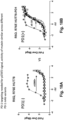


Classifications
-
- C—CHEMISTRY; METALLURGY
- C07—ORGANIC CHEMISTRY
- C07K—PEPTIDES
- C07K14/00—Peptides having more than 20 amino acids; Gastrins; Somatostatins; Melanotropins; Derivatives thereof
- C07K14/435—Peptides having more than 20 amino acids; Gastrins; Somatostatins; Melanotropins; Derivatives thereof from animals; from humans
- C07K14/52—Cytokines; Lymphokines; Interferons
- C07K14/54—Interleukins [IL]
-
- A—HUMAN NECESSITIES
- A61—MEDICAL OR VETERINARY SCIENCE; HYGIENE
- A61K—PREPARATIONS FOR MEDICAL, DENTAL OR TOILETRY PURPOSES
- A61K38/00—Medicinal preparations containing peptides
- A61K38/16—Peptides having more than 20 amino acids; Gastrins; Somatostatins; Melanotropins; Derivatives thereof
- A61K38/17—Peptides having more than 20 amino acids; Gastrins; Somatostatins; Melanotropins; Derivatives thereof from animals; from humans
- A61K38/19—Cytokines; Lymphokines; Interferons
- A61K38/20—Interleukins [IL]
-
- A—HUMAN NECESSITIES
- A61—MEDICAL OR VETERINARY SCIENCE; HYGIENE
- A61K—PREPARATIONS FOR MEDICAL, DENTAL OR TOILETRY PURPOSES
- A61K47/00—Medicinal preparations characterised by the non-active ingredients used, e.g. carriers or inert additives; Targeting or modifying agents chemically bound to the active ingredient
- A61K47/50—Medicinal preparations characterised by the non-active ingredients used, e.g. carriers or inert additives; Targeting or modifying agents chemically bound to the active ingredient the non-active ingredient being chemically bound to the active ingredient, e.g. polymer-drug conjugates
- A61K47/51—Medicinal preparations characterised by the non-active ingredients used, e.g. carriers or inert additives; Targeting or modifying agents chemically bound to the active ingredient the non-active ingredient being chemically bound to the active ingredient, e.g. polymer-drug conjugates the non-active ingredient being a modifying agent
- A61K47/68—Medicinal preparations characterised by the non-active ingredients used, e.g. carriers or inert additives; Targeting or modifying agents chemically bound to the active ingredient the non-active ingredient being chemically bound to the active ingredient, e.g. polymer-drug conjugates the non-active ingredient being a modifying agent the modifying agent being an antibody, an immunoglobulin or a fragment thereof, e.g. an Fc-fragment
- A61K47/6801—Drug-antibody or immunoglobulin conjugates defined by the pharmacologically or therapeutically active agent
- A61K47/6803—Drugs conjugated to an antibody or immunoglobulin, e.g. cisplatin-antibody conjugates
- A61K47/6811—Drugs conjugated to an antibody or immunoglobulin, e.g. cisplatin-antibody conjugates the drug being a protein or peptide, e.g. transferrin or bleomycin
- A61K47/6813—Drugs conjugated to an antibody or immunoglobulin, e.g. cisplatin-antibody conjugates the drug being a protein or peptide, e.g. transferrin or bleomycin the drug being a peptidic cytokine, e.g. an interleukin or interferon
-
- A—HUMAN NECESSITIES
- A61—MEDICAL OR VETERINARY SCIENCE; HYGIENE
- A61K—PREPARATIONS FOR MEDICAL, DENTAL OR TOILETRY PURPOSES
- A61K47/00—Medicinal preparations characterised by the non-active ingredients used, e.g. carriers or inert additives; Targeting or modifying agents chemically bound to the active ingredient
- A61K47/50—Medicinal preparations characterised by the non-active ingredients used, e.g. carriers or inert additives; Targeting or modifying agents chemically bound to the active ingredient the non-active ingredient being chemically bound to the active ingredient, e.g. polymer-drug conjugates
- A61K47/51—Medicinal preparations characterised by the non-active ingredients used, e.g. carriers or inert additives; Targeting or modifying agents chemically bound to the active ingredient the non-active ingredient being chemically bound to the active ingredient, e.g. polymer-drug conjugates the non-active ingredient being a modifying agent
- A61K47/68—Medicinal preparations characterised by the non-active ingredients used, e.g. carriers or inert additives; Targeting or modifying agents chemically bound to the active ingredient the non-active ingredient being chemically bound to the active ingredient, e.g. polymer-drug conjugates the non-active ingredient being a modifying agent the modifying agent being an antibody, an immunoglobulin or a fragment thereof, e.g. an Fc-fragment
- A61K47/6835—Medicinal preparations characterised by the non-active ingredients used, e.g. carriers or inert additives; Targeting or modifying agents chemically bound to the active ingredient the non-active ingredient being chemically bound to the active ingredient, e.g. polymer-drug conjugates the non-active ingredient being a modifying agent the modifying agent being an antibody, an immunoglobulin or a fragment thereof, e.g. an Fc-fragment the modifying agent being an antibody or an immunoglobulin bearing at least one antigen-binding site
- A61K47/6849—Medicinal preparations characterised by the non-active ingredients used, e.g. carriers or inert additives; Targeting or modifying agents chemically bound to the active ingredient the non-active ingredient being chemically bound to the active ingredient, e.g. polymer-drug conjugates the non-active ingredient being a modifying agent the modifying agent being an antibody, an immunoglobulin or a fragment thereof, e.g. an Fc-fragment the modifying agent being an antibody or an immunoglobulin bearing at least one antigen-binding site the antibody targeting a receptor, a cell surface antigen or a cell surface determinant
-
- A—HUMAN NECESSITIES
- A61—MEDICAL OR VETERINARY SCIENCE; HYGIENE
- A61P—SPECIFIC THERAPEUTIC ACTIVITY OF CHEMICAL COMPOUNDS OR MEDICINAL PREPARATIONS
- A61P35/00—Antineoplastic agents
-
- C—CHEMISTRY; METALLURGY
- C07—ORGANIC CHEMISTRY
- C07K—PEPTIDES
- C07K16/00—Immunoglobulins [IGs], e.g. monoclonal or polyclonal antibodies
- C07K16/18—Immunoglobulins [IGs], e.g. monoclonal or polyclonal antibodies against material from animals or humans
- C07K16/28—Immunoglobulins [IGs], e.g. monoclonal or polyclonal antibodies against material from animals or humans against receptors, cell surface antigens or cell surface determinants
- C07K16/2803—Immunoglobulins [IGs], e.g. monoclonal or polyclonal antibodies against material from animals or humans against receptors, cell surface antigens or cell surface determinants against the immunoglobulin superfamily
- C07K16/2818—Immunoglobulins [IGs], e.g. monoclonal or polyclonal antibodies against material from animals or humans against receptors, cell surface antigens or cell surface determinants against the immunoglobulin superfamily against CD28 or CD152
-
- C—CHEMISTRY; METALLURGY
- C07—ORGANIC CHEMISTRY
- C07K—PEPTIDES
- C07K16/00—Immunoglobulins [IGs], e.g. monoclonal or polyclonal antibodies
- C07K16/18—Immunoglobulins [IGs], e.g. monoclonal or polyclonal antibodies against material from animals or humans
- C07K16/28—Immunoglobulins [IGs], e.g. monoclonal or polyclonal antibodies against material from animals or humans against receptors, cell surface antigens or cell surface determinants
- C07K16/2803—Immunoglobulins [IGs], e.g. monoclonal or polyclonal antibodies against material from animals or humans against receptors, cell surface antigens or cell surface determinants against the immunoglobulin superfamily
- C07K16/283—Immunoglobulins [IGs], e.g. monoclonal or polyclonal antibodies against material from animals or humans against receptors, cell surface antigens or cell surface determinants against the immunoglobulin superfamily against Fc-receptors, e.g. CD16, CD32, CD64
-
- C—CHEMISTRY; METALLURGY
- C12—BIOCHEMISTRY; BEER; SPIRITS; WINE; VINEGAR; MICROBIOLOGY; ENZYMOLOGY; MUTATION OR GENETIC ENGINEERING
- C12N—MICROORGANISMS OR ENZYMES; COMPOSITIONS THEREOF; PROPAGATING, PRESERVING, OR MAINTAINING MICROORGANISMS; MUTATION OR GENETIC ENGINEERING; CULTURE MEDIA
- C12N15/00—Mutation or genetic engineering; DNA or RNA concerning genetic engineering, vectors, e.g. plasmids, or their isolation, preparation or purification; Use of hosts therefor
- C12N15/09—Recombinant DNA-technology
- C12N15/63—Introduction of foreign genetic material using vectors; Vectors; Use of hosts therefor; Regulation of expression
-
- C—CHEMISTRY; METALLURGY
- C07—ORGANIC CHEMISTRY
- C07K—PEPTIDES
- C07K2319/00—Fusion polypeptide
- C07K2319/30—Non-immunoglobulin-derived peptide or protein having an immunoglobulin constant or Fc region, or a fragment thereof, attached thereto
-
- C—CHEMISTRY; METALLURGY
- C12—BIOCHEMISTRY; BEER; SPIRITS; WINE; VINEGAR; MICROBIOLOGY; ENZYMOLOGY; MUTATION OR GENETIC ENGINEERING
- C12N—MICROORGANISMS OR ENZYMES; COMPOSITIONS THEREOF; PROPAGATING, PRESERVING, OR MAINTAINING MICROORGANISMS; MUTATION OR GENETIC ENGINEERING; CULTURE MEDIA
- C12N2740/00—Reverse transcribing RNA viruses
- C12N2740/00011—Details
- C12N2740/10011—Retroviridae
- C12N2740/10041—Use of virus, viral particle or viral elements as a vector
Landscapes
- Health & Medical Sciences (AREA)
- Life Sciences & Earth Sciences (AREA)
- Chemical & Material Sciences (AREA)
- Engineering & Computer Science (AREA)
- Bioinformatics & Cheminformatics (AREA)
- General Health & Medical Sciences (AREA)
- Medicinal Chemistry (AREA)
- Organic Chemistry (AREA)
- Immunology (AREA)
- Public Health (AREA)
- Pharmacology & Pharmacy (AREA)
- Veterinary Medicine (AREA)
- Animal Behavior & Ethology (AREA)
- Genetics & Genomics (AREA)
- Molecular Biology (AREA)
- Epidemiology (AREA)
- Proteomics, Peptides & Aminoacids (AREA)
- Zoology (AREA)
- Biophysics (AREA)
- Biochemistry (AREA)
- Gastroenterology & Hepatology (AREA)
- Nuclear Medicine, Radiotherapy & Molecular Imaging (AREA)
- General Chemical & Material Sciences (AREA)
- Chemical Kinetics & Catalysis (AREA)
- Toxicology (AREA)
- Cell Biology (AREA)
- Biomedical Technology (AREA)
- Biotechnology (AREA)
- General Engineering & Computer Science (AREA)
- Wood Science & Technology (AREA)
- Physics & Mathematics (AREA)
- Plant Pathology (AREA)
- Microbiology (AREA)
- Peptides Or Proteins (AREA)
- Preparation Of Compounds By Using Micro-Organisms (AREA)
- Medicines That Contain Protein Lipid Enzymes And Other Medicines (AREA)
- Micro-Organisms Or Cultivation Processes Thereof (AREA)
- Medicinal Preparation (AREA)
- Medicines Containing Antibodies Or Antigens For Use As Internal Diagnostic Agents (AREA)
- Pharmaceuticals Containing Other Organic And Inorganic Compounds (AREA)
Description
- The PD-1/PD-L1 axis is involved in the suppression of T cell immune responses in cancer. Antagonists of this pathway have been clinically validated across a number of solid tumor indications. Nivolumab and pembrolizumab are two such inhibitors that target the PD-1 pathway, and each has been approved by the U.S. Food and Drug Administration (FDA) for the treatment of metastatic melanoma. Recently, researchers have tested the paradigm of checkpoint inhibition in the setting of other tumor types. While some advances have been made, checkpoint inhibition therapy still remains in the shadows of other cancer treatment options.
- Studies of checkpoint inhibitors in combination with other agents are underway or recently have been completed. The combination of nivolumab and ipilimumab, a CTLA-4 receptor blocking antibody, for example, was tested in a Phase III clinical trial on patients with unresectable stage III or IV melanoma. In this study, the percentage of patients achieving a complete response was the highest among those that received the combination of nivolumab and ipilimumab, beating the outcome exhibited by those in the group receiving either drug alone. Other combinations are also currently being explored.
- Interleukin-21 (IL-21) is a T cell derived pleiotropic cytokine that regulates the activity of both innate and adaptive immune cells. IL-21 can augment T cell survival and effector function. Because it plays a key role in anti-tumor and anti-viral responses, in addition to exerting major effects on inflammatory responses that lead to the development of autoimmune diseases and inflammatory diseases, IL-21 has been an attractive target for several therapies.
- However, the development of IL-21-based therapies has been complex. The research has been complicated by studies showing that enhancing, or confusingly, inhibiting IL-21 action leads to a therapeutic effect. Additional challenges exist due to the broad expression of the receptor for IL-21 (IL-21R). IL-21R is expressed not just on T cells, but also on B cells, NK cells and myeloid cells. Accordingly, care must be taken to limit broad IL-21 activation in leukocytes and avoid the potential for toxicity. Restriction of IL-21 signaling must be balanced and selective. Triggering the effects of IL-21 must be designed to occur at the right time and place.
- Indeed, any success, especially clinical success with IL-21 moieties as a monotherapy or in combination with checkpoint inhibitors, has been muted. Thus, there remains a need for treatment modalities utilizing IL-21, including modalities combining IL-21 moieties with checkpoint inhibitors. There also remains a need for IL-21 therapies combined with immune checkpoint inhibition.
- The present invention provides an IL-21 mutein comprising the amino acid sequence of SEQ ID NO: 2,

- wherein "X" represents any amino acid, and wherein said IL-21 mutein amino acid sequence differs from the amino acid sequence of human IL-21 (SEQ ID NO: 1) by at least 1 amino acid;
- wherein the difference(s) occur(s) at
amino acids - The present disclosure provides IL-21 muteins comprising the amino acid sequence of SEQ ID NO: 2, wherein SEQ ID NO: 2 is
QGQDX HMXXM XXXXX XVDXL KNXVN DLVPE FLPAP EDVET NCEWS AFSCF QKAQL KSANT GNNEX XIXXX XXXLX XXXXX TNAGR RQKHR LTCPS CDSYE KKPPK EFLXX FXXLL XXMXX QHXSS RTHGS EDS (SEQ ID NO: 2), and X is any amino acid, and wherein the IL-21 mutein amino acid sequence differs from the amino acid sequence of human IL-21 (SEQ ID NO: 1) by at least 1 amino acid; wherein the difference(s) occur(s) atamino acids - Thus, in one aspect, the present disclosure also provides IL-21 muteins comprising only one amino acid substitution, relative to the wild-type IL-21 amino acid sequence, which is provided herein as SEQ ID NO: 1, as defined in the claims. The amino acid substitution may be located at an amino acid position selected from the group consisting of: 5, 8, 9, 11, 12, 13, 14, 15, 16, 19, 23, 65, 66, 68, 69, 70, 72, 73, 75, 76, 77, 78, 79, 80, 109, 110, 112, 113, 116, 117, 119, 120, or 123, according to the amino acid position numbering of SEQ ID NO: 1.
- The present disclosure further provides IL-21 muteins comprising only two amino acid substitutions, relative to SEQ ID NO: 1, as defined in the claims. The amino acid substitutions are located at two amino acid positions selected from the group consisting of: 5, 9, 15, 70, 71, 72, 73, and 76, according to the amino acid position numbering of SEQ ID NO: 1.
- The IL-21 muteins may bind to the IL-21 receptor (IL-21R) with a reduced affinity, relative to the affinity of wild-type IL-21 for the IL-21 receptor. The IL-21 mutein may bind to the human IL-21R with a KD that is greater than or is about 0.04 nM. The IL-21 mutein may bind to the cynomolgus monkey IL-21R with a KD that is greater than or is about 0.055 nM.
- The present disclosure also provides conjugates comprising an IL-21 mutein of the present disclosure linked to a heterologous moiety, as defined in the claims. In exemplary aspects, the heterologous moiety is a polypeptide, such that the conjugate is a fusion protein, as defined in the claims. Therefore, the present disclosure provides fusion proteins comprising an IL-21 mutein of the present disclosure, as defined in the claims. In exemplary aspects, the fusion protein comprises an IL-21 mutein of the present disclosure linked to an antigen-binding protein, such as an antibody, or an antigen binding antibody fragment thereof, as defined in the claims.
- The fusion protein comprises an IL-21 mutein linked to a PD-1 antigen-binding antibody as described herein.
- Also described are PD-1 antigen-binding proteins and conjugates and fusion proteins comprising a PD-1 antigen-binding protein.
- The present disclosure further provides nucleic acids comprising a nucleotide sequence encoding an IL-21 mutein, as defined in the claims. Also described are nucleic acids comprising a nucleotide sequence encoding a PD-1 antigen-binding protein (e.g., a PD-1 antigen-binding antibody), or a fusion protein comprising an IL-21 mutein and a PD-1 antigen-binding protein (e.g., a PD-1 antigen-binding antibody) as described herein. The nucleic acid molecule may comprise a nucleotide sequence encoding a conjugate or fusion protein as described herein. Vectors comprising the nucleic acids as described herein and host cells comprising the nucleic acids as described herein are furthermore described herein.
- The present disclosure additionally provides kits comprising an IL-21 mutein, a conjugate, fusion protein (e.g., a fusion protein comprising an IL-21 mutein, nucleic acid, vector, or host cell as described herein, or a combination thereof, as defined in the claims.
- Pharmaceutical compositions comprising an IL-21 mutein, a conjugate, fusion protein, nucleic acid, vector, or host cell of the present disclosure, or a combination thereof, as defined in the claims, are provided herein.
- Methods of making an IL-21 mutein, PD-1 antigen-binding protein (e.g., a PD-1 antigen-binding antibody), and a fusion protein comprising an IL-21 mutein and a PD-1 antigen-binding protein (e.g., a PD-1 antigen-binding antibody) are described herein. The method may comprise culturing a host cell as described herein to express the IL-21 mutein, PD-1 antigen-binding protein (e.g., a PD-1 antigen-binding antibody), or a fusion protein comprising an IL-21 mutein and a PD-1 antigen-binding protein (e.g., a PD-1 antigen-binding antibody) and harvesting the expressed IL-21 mutein, PD-1 antigen-binding protein (e.g., a PD-1 antigen-binding antibody), or fusion protein comprising an IL-21 mutein and a PD-1 antigen-binding protein (e.g., a PD-1 antigen-binding antibody).
- Methods of treatment are additionally described herein. The method may be a method of treating a subject in need thereof, comprising administering to the subject in need thereof a pharmaceutical composition as described herein in an amount effective to treat the subject. The subject may have a tumor (e.g., a solid tumor, a hematological malignancy, or a lymphoid malignancy) and the pharmaceutical composition may be administered to the subject in an amount effective to treat the tumor in the subject. The tumor may be non-small cell lung cancer (NSCLC) (e.g., Stage III or IV NSCLC), small cell lung cancer (SCLC), head and neck cancer, renal cancer, breast cancer, melanoma, ovarian cancer, liver cancer, pancreatic cancer, colon cancer, prostate cancer, gastric cancer, bladder cancer, hepatocellular carcinoma, cancers with high microsatellite instability (i.e., MSI-high cancers), lymphoma or leukemia.
-
-
Figure 1A represents a graph of the tumor volume (mm3) of BALB/c mice implanted with CT26/3E5 colon carcinoma cells as a function of time (days). OnDay 12, tumors were measured, and the mice were given an intraperitoneal (IP) injection of 300 µg isotype control antibody (mIgG1) onday -
Figure 1B represents a graph of the tumor volume (mm3) of BALB/c mice implanted with CT26/3E5 colon carcinoma cells as a function of time (days). OnDay 12, tumors were measured, and the mice were given an IP injection of 300 µg of an anti-PD-1 antibody onday -
Figure 1C represents a graph of the tumor volume (mm3) of BALB/c mice implanted with CT26/3E5 colon carcinoma cells as a function of time (days). OnDay 12, tumors were measured, and the mice were given 50 µg recombinant murine IL-21 (rmIL-21) three times per week for 3 weeks. Dosing ended on Day 33. -
Figure 1D represents a graph of the tumor volume (mm3) of BALB/c mice implanted with CT26/3E5 colon carcinoma cells as a function of time (days). OnDay 12, tumors were measured and the mice were given 300 µg of an anti-PD-1 antibody onday -
Figure 2 represents a graph of percent survival of the four groups of BALB/c mice implanted with CT26/3E5 colon carcinoma cells.Group 1 mice were given an intraperitoneal (IP) injection of 300 µg isotype control antibody (mIgG1) onday Group 2 mice were given an IP injection of 300 µg of an anti-PD-1 antibody onday Group 3 mice were given 50 µg rmIL-21 three times per week for 3 weeks.Group 4 mice were given 300 µg of an anti-PD-1 antibody onday -
Figure 3 is an illustration of a hypothesis of the mechanism of action of a fusion protein comprising a blocking PD-1 antibody fused to an IL-21 mutein (αPD-1:IL-21 mutein). Without being bound by a particular theory, it is believed that the fusion binds to IL-21R on CD8+ T cells while simultaneously blocking the signal transduction between PD-1 and PD-L1. -
Figure 4A is an illustration of a fusion protein comprising an anti-PD-1 antibody fused to an IL-21 mutein homodimer. The fusion protein does not have a linker. The antibody may comprise constant regions that reduce or eliminate Fc-associated effector binding and functions (e.g., lack the ability to interact with Fcy receptors (e.g., SEFL2-2)). -
Figure 4B is an illustration of a fusion protein comprising an anti-PD-1 antibody fused to an IL-21 mutein homodimer. The fusion protein may comprise a GGGGS (G4S) linker between the heavy chain constant region of the antibody and the IL-21 mutein. The antibody may also comprise SEFL2-2 modifications. -
Figure 4C is an illustration of a fusion protein comprising an anti-PD-1 antibody fused to an IL-21 mutein monomer. The fusion protein may comprise a G4S linker between the heavy chain constant region of the antibody and the IL-21 mutein. The antibody heavy chains comprise charge pair mutations (cpm; e.g., V1, V4, V103, or V131) to aid in preferential association of heterodimer Fc regions. The antibody may also comprise SEFL2-2 modifications. -
Figure 5A represents a graph of the STAT3 signaling in PD-1-ve Hut78 T cells exposed to (i) recombinant human IL-21 (rhIL-21) alone (solid line with closed circles), (ii) anti-PD-1 mAb alone (solid line with closed diamonds), (iii) anti-PD-1 mAb fused to an IL-21 homodimer without linker (solid line with closed triangles), (iv) anti-PD-1 mAb fused to an IL-21 homodimer with linker (dashed line with open triangles), (v) anti-PD-1 mAb fused to an IL-21 monomer without linker (solid line with closed squares), or (vi) anti-PD-1 mAb fused to an IL-21 monomer with a linker (dashed line with open squares). -
Figure 5B represents a graph of the STAT3 signaling in PD-1+ve Hut78 T cells exposed to (i) recombinant human IL-21 (rhIL-21) alone (solid line with closed circles), (ii) anti-PD-1 mAb alone (solid line with closed diamonds), (iii) anti-PD-1 mAb fused to an IL-21 homodimer without linker (solid line with closed triangles), (iv) anti-PD-1 mAb fused to an IL-21 homodimer with a linker (dashed line with open triangles), (v) anti-PD-1 mAb fused to an IL-21 monomer without a linker (solid line with closed squares), or (vi) anti-PD-1 mAb fused to an IL-21 monomer with a linker (dashed line with open squares). -
Figure 6 represents a graph of the serum concentrations of a homodimer fusion protein comprising WT IL-21 fused to an anti-PD-1 mAb that was intravenously administered to 6 animals at either a low dose (250 µg/kg) or a high dose (1000 µg/kg). An IgG antibody domain (150 µg/kg) was run as a control. -
Figure 7 represents a graph of the fold reduction in IL-21 activity (relative to rhIL-21 activity) by PD-1-ve Hut78 cells (open bars) or PD-1+ve Hut78 cells (closed bars) exposed to IL-21 muteins with reduced affinity for IL-21Rα. -
Figure 8 represents a graph of the fold reduction in IL-21 activity (relative to rhIL-21 activity) by PD-1-ve Hut78 cells (open bars) or PD-1+ve Hut78 cells (closed bars) exposed to IL-21 muteins with reduced affinity for IL-21Rγ. -
Figure 9A represents a graph of the STAT3 signaling in PD-1-ve Hut78 T cells exposed to (i) recombinant human IL-21 (rhIL-21) alone (solid line with closed circles), (ii) anti-PD-1 mAb fused to a WT IL-21 homodimer (dashed line with open circles), (iii) anti-PD-1 mAb fused to a WT IL-21 monomer (dashed line with closed circles), and (iv) anti-PD-1 mAb fused to an IL-21mutein 51 homodimer (R65P) (dotted line with closed triangles). -
Figure 9B represents a graph of the STAT3 signaling in PD-1+ve Hut78 T cells exposed to (i) recombinant human IL-21 (rhIL-21) alone (solid line with closed circles), (ii) anti-PD-1 mAb fused to a WT IL-21 homodimer (dashed line with open circles), (iii) anti-PD-1 mAb fused to a WT IL-21 monomer (dashed line with closed circles), and (iv) anti-PD-1 mAb fused to an IL-21mutein 51 homodimer (R65P) (dotted line with closed triangles). -
Figure 10 represents a graph of the serum concentrations of a fusion protein comprising (i) an anti-PD-1 mAb fused to an IL-21 R76E mutein homodimer (dotted line with closed circles), (ii) an anti-PD-1 mAb fused to an IL-21 R76A mutein homodimer (dashed line with closed triangles), (iii) an anti-PD-1 mAb fused to an IL-21 D15N mutein homodimer (dashed line with X's), (iv) an anti-PD-1 antibody (8.25 mg/kg; dashed line with open diamonds), and (v) an anti-PD-1 mAb fused to a WT IL-21 mutein homodimer (solid line with closed squares). -
Figure 11 represents a graph of the IL-2 (pg/mL) secreted by cells of a mixed lymphocyte reaction as a function of antibody concentration of (i) an anti-PD-1 antibody (solid line with closed circles), (ii) a fusion protein comprising IL-21 R5Q/R76E double mutein homodimer (dashed line with open circles), (iii) a combination of anti-PD-1 mAb and rhIL-21 (dotted line with open squares), (iv) a fusion protein comprising IL-21 R76E single mutein homodimer (closed squares), (v) IgG control (dotted line with open diamonds), and (vi) a rhIL-21 (broken line with open triangles). -
Figure 12A represents a graph of the fold change of over mean fluorescence intensity (MFI) of STAT3 activity of cells exposed to (i) a fusion protein comprising an IL-21 R5E/R76A double mutein (line with open triangles), (ii) an IgG1 control (dotted line with closed diamonds), (iii) rhIL-21 (dashed line with open squares), (iv) an anti-PD-1 mAb (solid line with open ovals), (v) a combination of rhIL-21 and anti-PD-1 mAb (dashed line with closed ovals), or (vi) a fusion protein comprising an IL-21 R76E single mutein (line with open diamonds). -
Figure 12B represents a graph of the fold change of over MFI of STAT3 activity of cells exposed to (i) a fusion protein comprising an IL-21 R5Q/R76E double mutein (line with open triangles), (ii) an IgG1 control (dotted line with closed diamonds), (iii) rhIL-21 (dashed line with open squares), (iv) an anti-PD-1 mAb (solid line with open ovals), (v) a combination of rhIL-21 and anti-PD-1 mAb (dashed line with closed ovals), or (vi) a fusion protein comprising an IL-21 R76E single mutein (line with open diamonds). -
Figure 12C represents a graph of the fold change of over MFI of STAT3 activity of cells exposed to (i) a fusion protein comprising an IL-21 R9E/R76A double mutein (line with open triangles), (ii) an IgG1 control (dotted line with closed diamonds), (iii) rhIL-21 (dashed line with open squares), (iv) an anti-PD-1 mAb (solid line with open ovals), (v) a combination of rhIL-21 and anti-PD-1 mAb (dashed line with closed ovals), or (vi) a fusion protein comprising an IL-21 R76E single mutein (line with open diamonds). -
Figure 13A represents a graph of the % specific lysis by CTLs (y-axis) vs effector to target cell ratios (x-axis) exposed to (i) rhIL-21 (solid line with closed circles), (ii) hIgG4 control antibody (dashed line with open circles), (iii) anti-PD-1 mAb (dashed line with closed triangles), (iv) a combination of rhIL-21 and anti-PD-1 mAb (dotted line with open triangles), or (v) a fusion protein comprising IL-21 R5E/R76A (broken line with X's). -
Figure 13B represents a graph of the % specific lysis by CTLs exposed to represents a graph of the % specific lysis by CTLs exposed to (i) rhIL-21 (solid line with closed circles), (ii) hIgG4 control antibody (dashed line with open circles), (iii) anti-PD-1 mAb (dashed line with closed triangles), (iv) a combination of rhIL-21 and anti-PD-1 mAb (dotted line with open triangles), or (v) a fusion protein comprising IL-21 R5Q/R76E (broken line with X's). -
Figure 13C represents a graph of the % specific lysis by CTLs exposed to (i) rhIL-21 (solid line with closed circles), (ii) hIgG4 control antibody (dahsed line with open circles), (iii) anti-PD-1 mAb (dashed line with closed triangles), (iv) a combination of rhIL-21 and anti-PD-1 mAb (dotted line with open triangles), or (v) a fusion protein comprising IL-21 R9E/R76A (broken line with X's). -
Figure 14 represents serum concentrations of (i) anti-PD-1 mAb (solid line with closed squares), (ii) a fusion protein comprising anti-PD-1 mAb and an IL-21 R5Q/R76E homodimer (solid line with closed circles), (iii) a fusion protein comprising anti-PD-1 mAb and an IL-21 R76E homodimer (solid line), (iv) a fusion protein comprising anti-PD-1 mAb and an IL-21 R5E/R76A homodimer (dashed line with X's), (v) a fusion protein comprising anti-PD-1 mAb and an IL-21 R76A homodimer (dashed line with closed diamonds), or (vi) a fusion protein comprising anti-PD-1 mAb and an IL-21 R76E monomer (solid line with closed triangles). -
Figure 15A represents a timeline of the cell sampling (open arrows) and of the administrations (closed arrows). -
Figure 15B represents a graph of the fold change in PD-1+/CD4+ cells (relative to Day -5) as measured onDay 7 in animals given a dose of (i) a fusion protein comprising anti-PD-1 mAb and an IL-21 R76E mutein (bar with horizontal lines), (ii) a fusion protein comprising anti-PD-1 mAb and an IL-21 R76E single mutein monomer (bar with vertical lines), or (iii) a fusion protein comprising anti-PD-1 mAb and an IL-21 R5Q/R76E double mutein homodimer (open bar) (each administered on Day 0). -
Figure 15C represents a graph of the fold change in PD-1+/CD8+ cells (relative to Day -5) as measured onDay 7 in animals given a dose of (i) a fusion protein comprising anti-PD-1 mAb and an IL-21 R76E mutein (bar with horizontal lines), (ii) a fusion protein comprising anti-PD-1 mAb and an IL-21 R76E single mutein monomer (bar with vertical lines), or (iii) a fusion protein comprising anti-PD-1 mAb and an IL-21 R5Q/R76E double mutein homodimer (open bar) (each administered on Day 0). -
Figure 15D represents a graph of the fold change in PD-1+/CD8+ cells (relative to Day -5) as measured onDay 21 in animals given a first dose of (i) a fusion protein comprising anti-PD-1 mAb and an IL-21 R76E mutein (bar with horizontal lines), (ii) a fusion protein comprising anti-PD-1 mAb and an IL-21 R76E single mutein monomer (bar with vertical lines), or (iii) a fusion protein comprising anti-PD-1 mAb and an IL-21 R5Q/R76E double mutein homodimer (open bar) (first dose administered onDay 0 and second dose administered on Day 8). -
Figure 16 represents a graph of the expansion of PD-1+/CD8+ cells (relative to Day -5) as a function of PD-1 receptor occupancy (RO) on PD-1+/CD8+ cells. IL-21 single mutants (801, 802, 807, and 808) are shown within the blue circle and IL-21 double mutants (803, 804, 805, and 806) are shown in the pink circle. These data demonstrate that superior pharmacokinetic properties of the double mutants allow for better target coverage and correlates with better pharmacodynamics responses, as measured by expansion of the PD-1+ target population. The blue line indicates the general delineation between single and double mutants. -
Figure 17 represents flow plots of individual animals treated with either single mutant homodimer constructs (animals 801 and 802), double mutant homodimer constructs (animals 803 - 806), or single mutant monomeric constructs (animals 807 and 808). PD-1+ T cells were detected by non-competing or competing detection antibody. Percent target coverage is calculated as a percentage of the ratio between competing and non-competing PD-1 antibody. The data show that all constructs demonstrate robust target coverage after repeat dosing onDay 21. -
Figure 18A represents a graph of the fold change (relative to rhIL-21 (solid line with circled X's)) of STAT3 activity of PD-1-ve Hut78 T cells exposed to a fusion protein comprising one of ten different anti-PD-1 mAbs and an IL-21 R5Q/R76E mutein homodimer. The ten anti-PD-1 mAbs included 20A2.003 (line with diamonds), 20C1.006 (red square), 20C1.009 (line with triangles), and 22D4.006 (line with open circles). -
Figure 18B represents a graph of the fold change (relative to rhIL-21 (solid line with circled X's)) of STAT3 activity of PD-1+ve Hut78 T cells exposed to a fusion protein comprising one of ten different anti-PD-1 mAbs and an IL-21 R5Q/R76E mutein homodimer. The ten anti-PD-1 mAbs included 20A2.003 (line with diamonds), 20C1.006 (red square), 20C1.009 (line with triangles), and 22D4.006 (line with open circles). Comparison ofFigure 18A to Figure 18B indicates that PD-1 targeting needed for pSTAT3 signaling and that the activity of the muteins is similar when fused to different anti-PD-1 mAbs. -
Figure 19A represents a graph of the fold change (relative to rhIL-21 (solid line with circled X's)) of STAT3 activity of PD-1-ve Hut78 T cells exposed to a fusion protein comprising one of seven different anti-PD-1 mAbs and an IL-21 R9E/R76A mutein homodimer. The seven anti-PD-1 mAbs included 20A2.003 (line with open triangles), 20C1.006 (line with open squares), 20C1.009 (line with open diamonds), and 22D4.006 (line with open circles). -
Figure 19B represents a graph of the fold change (relative to rhIL-21 (solid line with circled X's)) of STAT3 activity of PD-1+ve Hut78 T cells exposed to a fusion protein comprising one of seven different anti-PD-1 mAbs and an IL-21 R9E/R76A mutein homodimer. Comparison ofFigure 19A to Figure 19B indicates that PD-1 targeting needed for pSTAT3 signaling and that the activity of the muteins is similar when fused to different anti-PD-1 mAbs. The seven anti-PD-1 mAbs included 20A2.003 (line with open triangles), 20C1.006 (line with open squares), 20C1.009 (line with open diamonds), and 22D4.006 (line with open circles). -
Figures 20A-20D represent the amount of pSTAT3 signaling observed with several anti-PD-1 mAb - IL-21 monomeric or dimeric double mutein fusions. Solid line with closed circles(top of graphs) is rhIL-21; dashed line with open circles (bottom of graphs) is IgG1 control; line with X's (bottom of graphs) is IgG2 control; dotted line with closed squares (bottom of graphs) is the 22D4.006 anti-PD-1 mAb (present as mAb; i.e., not as a fusion) used in the IL-21 mutein fusions; dashed line with open squares and dotted line with open diamonds(bottom of graphs) are control anti-PD-1 mAbs; remaining lines are anti-PD-1 mAb (22D4.006) - IL-21 monomeric or dimeric double mutein fusions (with various charge pair mutations) wherein the double mutants are R5E/R76A; R9E/R76A; R5A/R76E or R5Q/R76E. rhIL-21 demonstrates activity in both PD-1-ve and PD-1+ve cells, monomeric and homodimeric double mutein fusions are unable to demonstrate pSTAT3 (IL-21-based) activity in PD-1-ve cells, and monomeric and homodimeric double mutein fusions are able to demonstrate pSTAT3 (IL-21-based) activity in PD-1+ve cells. Thus, monomeric fusions with IL-21 double mutants exhibit similar levels of IL-21 activity attenuation in PD-1-ve cells and IL-21 activity rescue in PD-1+ve cells as their counterpart dimeric fusions.Figures 20A and 20B are duplicate runs of the pSTAT3 assay on PD-1-ve cells.Figures 20C and 20D are duplicate runs of the pSTAT3 assay on PD-1+ve cells. -
Figures 21A-21D represent the results of a PD-1 reporter gene assay (RGA;Figures 21A and 21B ) and MLR assay (Figures 21C and 21D ) with the same anti-PD-1 mAb (22D4.006) - IL-21 monomeric or dimeric double mutein fusions evaluated inFigures 20A-20D .Figures 21A-21D demonstrate that anti-PD-1 mAb (22D4.006) - IL-21 monomeric and dimeric double mutein fusions are able to induce PD-1 activity. Solid line with closed circles (bottom of graphs) is rhIL-21; line with open circles (bottom of graphs) is IgG1 control; line with X's (bottom of graphs) is IgG2 control; dashed line with closed squares (top of graphs) is the 22D4.006 anti-PD-1 mAb (present as mAb; i.e., not as a fusion) used in the IL-21 mutein fusions; dashed line with open squares and dotted line with open diamonds (top of graphs) are control anti-PD-1 mAbs; remaining lines are anti-PD-1 mAb - IL-21 monomeric or dimeric double mutein fusions.Figures 21A and 21B are duplicate runs of the PD-1 RGA assay.Figures 20C and 20D are duplicate runs of the MLR assay. -
Figures 22A-22D represent the results of pSTAT3 assays testing the same constructs as those inFigures 20A-20D , except that a different anti-PD-1 mAb (20A2.003) is used in the anti-PD-1 mAb - IL-21 monomeric and dimeric double mutein fusions. The results inFigures 22A-22D are similar to those seen inFigures 20A-20D . Solid line with closed circles (top of graphs) is rhIL-21; dashed line with open circles (bottom of graphs) is IgG1 control; line with X's (bottom of graphs) is IgG2 control; dotted line with closed squares (bottom of graphs) is the 20A2.003 anti-PD-1 mAb (present as mAb; i.e., not as a fusion) used in the IL-21 mutein fusions; dashed line with open squares and dotted line with open diamonds (bottom of graphs) are control anti-PD-1 mAbs; remaining lines are anti-PD-1 mAb (20A2.003) - IL-21 monomeric or dimeric double mutein fusions (with various charge pair mutations) wherein the double mutants are R5E/R76A; R9E/R76A; R5A/R76E or R5Q/R76E.Figures 22A and 22B are duplicate runs of the pSTAT3 assay on PD-1-ve cells.Figures 22C and 22D are duplicate runs of the pSTAT3 assay on PD-1+ve cells. -
Figures 23A-23D represent the results of PD-1 reporter gene assays (Figures 23A and 23B ) and MLR assays (Figures 23C and 23D ) testing the same constructs as those inFigures 21A-21D , except that a different anti-PD-1 mAb (20A2.003) is used in the anti-PD-1 mAb - IL-21 monomeric and dimeric double mutein fusions. The results inFigures 23A-23D are similar to those seen inFigures 21A-21D . Solid line with closed circles (bottom of graphs) is rhIL-21; dashedline with open circles (bottom of graphs) is IgG1 control; line with X's (bottom of graphs) is IgG2 control; dotted line (top of graphs) is the 20A2.003 anti-PD-1 mAb (present as mAb; i.e., not as a fusion) used in the IL-21 mutein fusions; dashed line with open squares and dotted line with open diamonds (top of graphs) are control anti-PD-1 mAbs; remaining lines are anti-PD-1 mAb (20A2.003) - IL-21 monomeric or dimeric double mutein fusions.Figures 23A and 23B are duplicate runs of the PD-1 RGA assay.Figures 23C and 23D are duplicate runs of the MLR assay. -
Figure 24 represents a graph of the NFAT/luciferase activity of purified anti-PD-1 antibodies as a function of mAb concentration. -
Figures 25A is a graph of the fold change in the number of Ki67+/CD3+/CD4+ cells relative to baseline upon exposure to the fusion protein [22D4.017]-[R9E:R76A] (monomer) or to the anti-PD-1 antibody [22D4.017]. -
Figures 25B is a graph of the fold change in the number of Ki67+/CD3+/CD8+ cells relative to baseline upon exposure to the fusion protein [22D4.017]-[R9E:R76A] (monomer) or to the anti-PD-1 antibody [22D4.017]. -
Figures 25C is a graph of the fold change in the number of pSTAT3+/CD3+/CD4+ cells relative to baseline upon exposure to the fusion protein [22D4.017]-[R9E:R76A] (monomer) or to the anti-PD-1 antibody [22D4.017]. -
Figures 25D is a graph of the fold change in the number of pSTAT3+/CD3+/CD8+ cells relative to baseline upon exposure to the fusion protein [22D4.017]-[R9E:R76A] (monomer) or to the anti-PD-1 antibody [22D4.017]. -
Figures 25E is a graph of the fold change in the number of CD3+/CD4+ cells relative to baseline upon exposure to the fusion protein [22D4.017]-[R9E:R76A] (monomer) or to the anti-PD-1 antibody [22D4.017]. -
Figures 25F is a graph of the fold change in the number of CD3+/CD8+ cells relative to baseline upon exposure to the fusion protein [22D4.017]-[R9E:R76A] (monomer) or to the anti-PD-1 antibody [22D4.017]. -
Figures 25G is a graph of the fold change in the number of PD-1+/CD3+/CD4+ cells relative to baseline upon exposure to the fusion protein [22D4.017]-[R9E:R76A] (monomer) or to the anti-PD-1 antibody [22D4.017]. -
Figures 25H is a graph of the fold change in the number of PD-1+/CD3+/C84+ cells relative to baseline upon exposure to the fusion protein [22D4.017]-[R9E:R76A] (monomer) or to the anti-PD-1 antibody [22D4.017]. -
Figures 25I is a graph of the fold change in the amount of serum perforin relative to baseline upon 72 hours of exposure to the fusion protein [22D4.017]-[R9E:R76A] (monomer) or to the anti-PD-1 antibody [22D4.017]. -
Figures 25J is a graph of the % Ki67+ cells relative to baseline as a function of fold increase in perforin upon 72 hour exposure to the fusion protein [22D4.017]-[R9E:R76A] (monomer) or to the anti-PD-1 antibody [22D4.017]. -
Figure 26A is a graph of the absorbance (nm) as a function of time (sec) used to determine the indicated KD of antibody 22D4.017 for human PD-1 antigen. -
Figure 26B is a graph of the absorbance (nm) as a function of time (sec) used to determine the indicated KD of antibody 20C1.009 for human PD-1 antigen. -
Figure 26C is a graph of the absorbance (nm) as a function of time (sec) used to determine the indicated KD of antibody 20A2.003 for human PD-1 antigen. -
Figure 26D is a graph of the absorbance (nm) as a function of time (sec) used to determine the indicated KD of an IgG1 anti-PD-1 mAb, for human PD-1 antigen. -
Figure 26E is a graph of the absorbance (nm) as a function of time (sec) used to determine the indicated KD of an IgG4 PD-1 mAb for human PD-1 antigen. -
Figure 26F is a graph of the absorbance (nm) as a function of time (sec) used to determine the indicated KD of 22D4.017 for cyno PD-1 antigen. -
Figure 26G is a graph of the absorbance (nm) as a function of time (sec) used to determine the indicated KD of antibody 20C1.009 for cyno PD-1 antigen. -
Figure 26H is a graph of the absorbance (nm) as a function of time (sec) used to determine the indicated KD of antibody20A2.003 for cyno PD-1 antigen. -
Figure 26I is a graph of the absorbance (nm) as a function of time (sec) used to determine the indicated KD of an IgG1 anti-PD-1 mAb for cyno PD-1 antigen. -
Figure 26J is a graph of the absorbance (nm) as a function of time (sec) used to determine the indicated KD of an IgG4 anti-PD-1 mAb for cyno PD-1 antigen. -
Figure 27 is a graph of the Cp (kcal/mole/° C) as a function of temperature for anti-PD-1 antibodies 22D4.017 and 20C1.009. -
Figure 28 is a graph of the viscosity plotted against shear rate for anti-PD-1 antibodies 22D4.017 and 20C1.009. -
Figures 29A-29D is a series of graphs plotting the signal as a function of antibody concentration in (Fig. 29A ) a variant Hut78 T cell line that is PD-1 positive, (Fig. 29B ) a variant Hut78 T cell line that is TIGIT positive, (Fig. 29C ) a variant Hut78 T cell line that is LAG3 positive, and (Fig. 29D ) the parental Hut78 T cell line which does not endogenously express PD-1, TIGIT or LAG3. - There remains a need for novel immune potentiating approaches that can deploy the immune system against cancer cells in a safe and efficacious manner, especially in light of the fact that current immunotherapy approaches are efficacious in only a minority of patients and can have significant and often unpredictable toxicities. Described herein is a novel class of bifunctional fusion molecules comprising a PD-1 targeting antibody that can block PD-1/PD-L1 interaction, fused to an engineered affinity attenuated interleukin-21 mutein which addresses this need. The antibody/cytokine fusions described herein overcome significant barriers associated with cytokine therapeutics, allowing for, inter alia, antibody-like dosing and selective delivery of the IL-21 cytokine in a PD-1 targeted manner. When fused to an anti-PD-1 antibody, IL-21 muteins can selectively activate and expand PD-1 expressing T cells in vivo. Accordingly, the antibody/cytokine fusions described herein may improve upon and extend the utility of anti-PD-1 therapeutics currently under testing in the clinic.
- The combination of cytokine and co-inhibitory receptor agonists or antagonists remains challenging because of the risks of incremental toxicity and the need for complex clinical trial design (see, e.g., Ott et al., ); and Hermel et al., )). With respect to cytokines, there is also the potential for the activation of inhibitory feedback pathways that can lead to immune suppression (see, e.g., Portielje et al, ); Wan et al., ); and Mooradian et al., )). Interleukin-21 (IL-21) is a type I cytokine and a member of the common cytokine receptor gamma-chain (cg-chain) cytokine family that has emerged as a promising immune therapeutic for the treatment of cancer. IL-21 is produced by activated CD4 T cells and natural killer T (NKT) cells and signals via a heterodimeric receptor complex comprised of a discrete IL-21 receptor (IL-21R) subunit that associates with the common gamma-chain (see, e.g., Spolski et al., Nat Rev Drug Discov 13, 379-395 (2014)). Activation of the IL-21R complex leads to the activation of the JAK/STAT signaling pathway. IL-21R is broadly expressed in hematopoietic cells including T and B lymphocytes, natural killer (NK) cells and myeloid cells. Although not an essential growth or differentiation factor, IL-21 is a potent mitogen and survival factor for NK cells and activated T cells. IL-21 can support the differentiation of CD4 (+) T helper 17 (Th17) as well follicular helper T cells (Tfh) and can antagonize regulatory T cell (Treg) differentiation. Moreover, IL-21 can augment the survival of CD8 T cells and preserves a less activated but more persistent T cell phenotype, which allows for enhanced tumor and viral control.
- A challenging aspect of cytokine immunotherapy is that, in addition to activating immune cells to potentiate immune responses, the same cytokine can also activate counter-regulatory pathways. For example, IL-2 and IFNγ which can activate protective immune responses as well as regulatory T cell responses and inhibitory pathways (such as PD-L1) respectively. In dendritic cells (DCs), IL-21 can inhibit both DC maturation and activation, can induce the apoptosis of conventional DCs, can potently inhibit the priming of T cells in mixed cultures, and may play a role in the induction of tolerance. In humans, IL-21 has been tested as a non-targeted free cytokine in several cancer indications, but despite the promising preclinical data and early phase I clinical data, development of this approach has not progressed further than phase II testing (see, e.g., Thompson et al., J Clin Oncol 26, 2034-2039 (2008); and Davis et al., )). In more recent preclinical models, the combination of recombinant IL-21 cytokine with co-inhibitory receptor antagonists (e.g., anti-CTLA-4 and anti-PD-1) have demonstrated that IL-21 can extend the efficacy of these treatments. Such combinations are now under testing in the clinic, though clinical efficacy has yet to be demonstrated (Lewis et al., )).
- Without being bound by a theory, the antibody/cytokine fusions described herein are designed to utilize the immune potentiating activity of IL-21 (which may be prerequisite to address toxicity and off-target immune suppression), to maximize efficacy, and improve the feasibility of dosing in the clinic.
- Interleukin-21 (IL-21) is a cytokine expressed by T cells, B cells, NK cells and myeloid cells, and regulates the activity of both innate and adaptive immune cells and improves T cell survival and effector function. Several Phase I and II clinical trials include IL-21 as the investigational product for the treatment of cancers, inflammatory diseases, and autoimmune diseases, including, melanoma, renal cell carcinoma, acute myeloid leukemia, non-Hodgkin's lymphoma, ovarian cancer, colorectal cancer, systemic lupus erythematosus, Crohn's disease and rheumatoid arthritis.
- IL-21 has a four-helix bundle structure and exists as a monomer. In humans, two isoforms of IL-21 are known, each of which are derived from a precursor molecule. The first IL-21 isoform comprises 162 amino acids (aa), the first 29 of which make up the signal peptide; and the second IL-21 isoform comprises 153 aa, the first 29 of which make up the signal peptide as in the first isoform. The amino acid sequences of the first and second IL-21 isoforms (including the signal peptide) are provided herein as SEQ ID NO: 258 and SEQ ID NO: 259, respectively.
- IL-21 binds to the heterodimeric IL-21 receptor (IL-21R) expressed on the surface of T, B, and NK cells. IL-21R is similar in structure to the IL-2 receptor and the IL-15 receptor, in that each of these cytokine receptors comprises a common gamma chain (yc). In addition to the yc, the IL-21R comprises an alpha chain which is important for binding to IL-21. There are two isoforms of the human IL-21 receptor alpha chain:
isoform 1 andisoform 2. The amino acid sequences ofisoform 1 andisoform 2 are provided herein as SEQ ID NOs: 256 and 261, respectively. The amino acid sequence of the human common gamma chain is provided herein as SEQ ID NO: 257. - When IL-21 binds to IL-21R, the Jak/STAT signaling pathway is activated to activate target genes. While IL-21-induced signaling may be therapeutically desirable, careful consideration of the timing and the location of the signaling is needed, given IL-21's broad expression profile and due to the fact that IL-21 has the ability to potentiate CD8 T cell responses as well as to suppress antigen presentation and T cell priming. The data presented herein for the first time supports the use of carefully designed IL-21 muteins to achieve IL-21 signaling at the appropriate time and place.
- The present disclosure provides IL-21 muteins comprising at least one amino acid substitution, relative to the wild-type IL-21 amino acid sequence, which is provided herein as SEQ ID NO: 1, as defined in the claims. For example, the IL-21 mutein comprises at least one and not more than 34 amino acid substitutions. The IL-21 mutein may comprise at least one and not more than X amino acid substitutions, wherein X is 2, 3, 4, 5, 6, 7, 8, 9, 10, 11, 12, 13, 14, 15, 16, 17, 18, 19, 20, 21, 22, 23, 24, 25, 26, 27, 28, 29, 30, 31, 32, 33, or 34. The IL-21 mutein may comprise an amino acid sequence which differs from the amino acid sequence of human IL-21 (SEQ ID NO: 1) by no more than 10 amino acids, 15 amino acids, 20 amino acids, or 25 amino acids. The IL-21 mutein may comprise an amino acid sequence which differs from the amino acid sequence of human IL-21 (SEQ ID NO: 1) by no more than 7 amino acids or no more than 5 amino acids. The IL-21 mutein may comprise an amino acid sequence which differs from the amino acid sequence of human IL-21 (SEQ ID NO: 1) by 3, 4, 5, or 6 amino acids. The IL-21 mutein may comprise an amino acid sequence which differs from the amino acid sequence of human IL-21 (SEQ ID NO: 1) by 3 to 6 amino acids or 1 to 5 amino acids. In exemplary embodiments, the IL-21 mutein comprises an amino acid sequence which differs from the amino acid sequence of human IL-21 (SEQ ID NO: 1) by one or two amino acids.
- In exemplary aspects, the IL-21 mutein comprises the amino acid sequence of SEQ ID NO: 2, wherein SEQ ID NO: 2 is
QGQDX HMXXM XXXXX XVDXL KNXVN DLVPE FLPAP EDVET NCEWS AFSCF QKAQL KSANT GNNEX XIXXX XXXLX XXXXX TNAGR RQKHR LTCPS CDSYE KKPPK EFLXX FXXLL XXMXX QHXSS RTHGS EDS (SEQ ID NO: 2), wherein X represents any amino acid, and wherein the IL-21 mutein amino acid sequence differs from the amino acid sequence of human IL-21 (SEQ ID NO: 1) by at least 1 amino acid, wherein the difference(s) occur(s) atamino acids - Thus, in exemplary aspects, the IL-21 mutein comprises the sequence of SEQ ID NO: 2, wherein SEQ ID NO: 2 differs from SEQ ID NO: 1 by at least one amino acid at a position designated by X in SEQ ID NO: 2, as defined in the claims. The IL-21 mutein comprising SEQ ID NO: 2 may have at least about 30%, at least about 40%, at least about 50%, at least about 60%, at least about 70%, at least about 80%, at least about 85%, at least about 90%, or has greater than about 90% (e.g., about 91%, about 92%, about 93%, about 94%, about 95%, about 96%, about 97%, about 98%, or about 99%) sequence identity to SEQ ID NO: 1. The IL-21 mutein may comprise an amino acid sequence which is at least about 30%, at least about 40%, at least about 50%, at least about 60%, at least about 70%, at least about 80%, at least about 85%, at least about 90%, or has greater than about 90% (e.g., about 91%, about 92%, about 93%, about 94%, about 95%, about 96%, about 97%, about 98%, or about 99%) sequence identity to SEQ ID NO: 1.
- In exemplary embodiments, the IL-21 mutein comprises an amino acid sequence comprising at least one amino acid substitution relative to the wild-type IL-21 amino acid sequence, and the amino acid substitution(s) occur(s) within the N-terminal half of the amino acid sequence, as defined in the claims. The amino acid substitution(s) may occur(s) at a position within positions 5-25 or 8-23 (both inclusive), according to the amino acid position numbering of SEQ ID NO: 1.
- The IL-21 mutein may comprise an amino acid sequence comprising at least one amino acid substitution relative to the wild-type IL-21 amino acid sequence, and the amino acid substitution(s) may occur within the C-terminal half of the amino acid sequence. For example, the amino acid substitution(s) occur(s) at a position within positions 100-133 or 109-123 (both inclusive), according to the amino acid position numbering of SEQ ID NO: 1.
- In exemplary embodiments, the IL-21 mutein comprises an amino acid sequence comprising at least one amino acid substitution relative to the wild-type IL-21 amino acid sequence, and the amino acid substitution(s) occur(s) in the middle third of the amino acid sequence, as defined in the claims. The amino acid substitution(s) occur(s) at a position within positions 55-85 or 65-80 (both inclusive), according to the amino acid position numbering of SEQ ID NO: 1.
- Also described are IL-21 muteins comprising only one amino acid substitution, relative to the wild-type IL-21 amino acid sequence, which is provided herein as SEQ ID NO: 1. The amino acid substitution may be located at an amino acid position selected from the group consisting of: 5, 8, 9, 11, 12, 13, 14, 15, 16, 19, 23, 65, 66, 68, 69, 70, 71, 72, 73, 75, 76, 77, 78, 79, 80, 109, 110, 112, 113, 116, 117, 119, 120, or 123, according to the amino acid position numbering of SEQ ID NO: 1. The amino acid substitution may be located at an amino acid position selected from the group consisting of: 5, 8, 9, 11, 12, 13, 14, 15, 16, 19, 23, 65, 66, 68, 69, 70, 72, 73, 75, 76, 77, 78, 79, 80, 109, 110, 112, 113, 116, 117, 119, 120, or 123, according to the amino acid position numbering of SEQ ID NO: 1. The IL-21 mutein may comprise any one of the amino acid sequences of SEQ ID NOs: 3-21 and 23-37.
- Also described are IL-21 muteins comprising only two amino acid substitutions, relative to SEQ ID NO: 1. The amino acid substitutions may be located at two amino acid positions selected from the group consisting of: 5, 8, 9, 11, 12, 13, 14, 15, 16, 19, 23, 65, 66, 68, 69, 70, 71, 72, 73, 75, 76, 77, 78, 79, 80, 109, 110, 112, 113, 116, 117, 119, 120, or 123, according to the amino acid position numbering of SEQ ID NO: 1. The amino acid substitutions may be located at two amino acid positions selected from the group consisting of: 5, 9, 15, 70, 71, 72, 73, and 76, according to the amino acid position numbering of SEQ ID NO: 1. The amino acid substitutions may be located at two amino acid positions selected from the group consisting of: 5, 9, 73, and 76, according to the amino acid position numbering of SEQ ID NO: 1. At least one of the two amino acid substitutions may be located at position 76, according to the amino acid position numbering of SEQ ID NO: 1. The IL-21 mutein may comprise any one of the amino acid sequences of SEQ ID NOs: 199-208 and 210-212.
- The IL-21 mutein may comprise an amino acid sequence comprising at least one amino acid substitution relative to the wild-type IL-21 amino acid sequence, as defined in the claims, and the amino acid substitution(s) may be conservative amino acid substitution(s). As used herein, the term "conservative amino acid substitution" refers to the substitution of one amino acid with another amino acid having similar properties, e.g., size, charge, hydrophobicity, hydrophilicity, and/or aromaticity, and includes exchanges within one of the following five groups:
- I. Small aliphatic, nonpolar or slightly polar residues: Ala, Ser, Thr, Pro, Gly;
- II. Polar, negatively charged residues and their amides and esters: Asp, Asn, Glu, Gln, cysteic acid and homocysteic acid;
- III. Polar, positively charged residues: His, Arg, Lys; Ornithine (Orn)
- IV. Large, aliphatic, nonpolar residues: Met, Leu, Ile, Val, Cys, Norleucine (Nle), homocysteine
- V. Large, aromatic residues: Phe, Tyr, Trp, acetyl phenylalanine.
- In exemplary embodiments, the IL-21 mutein comprises an amino acid sequence comprising at least one amino acid substitution relative to the wild-type IL-21 amino acid sequence, as defined in the claims, and the amino acid substitution(s) is/are non-conservative amino acid substitution(s). As used herein, the term "non-conservative amino acid substitution" is defined herein as the substitution of one amino acid with another amino acid having different properties, e.g., size, charge, hydrophobicity, hydrophilicity, and/or aromaticity, and includes exchanges outside the above five groups.
- In exemplary aspects, the IL-21 mutein comprises an amino acid sequence comprising at least one amino acid substitution relative to the wild-type IL-21 amino acid sequence, as defined in the claims, and the substitute amino acid is a naturally-occurring amino acid. By "naturally-occurring amino acid" or "standard amino acid" or "canonical amino acid" is meant one of the 20 alpha amino acids found in eukaryotes encoded directly by the codons of the universal genetic code (Ala, Val, Ile, Leu, Met, Phe, Tyr, Trp, Ser, Thr, Asn, Gln, Cys, Gly, Pro, Arg, His, Lys, Asp, Glu). In exemplary aspects, the IL-21 mutein comprises an amino acid sequence comprising at least one amino acid substitution relative to the wild-type IL-21 amino acid sequence, as defined in the claims, and the substitute amino acid may be a non-standard amino acid, or an amino acid which is not incorporated into proteins during translation. Non-standard amino acids include, but are not limited to: selenocysteine, pyrrolysine, ornithine, norleucine, β-amino acids (e.g., β-alanine, β-aminoisobutyric acid, β-phenlyalanine, β-homophenylalanine, β-glutamic acid, β-glutamine, β-homotryptophan, β-leucine, β-lysine), homo-amino acids (e.g., homophenylalanine, homoserine, homoarginine, monocysteine, homocystine), N-methyl amino acids (e.g., L-abrine, N-methyl-alanine, N-methyl-isoleucine, N-methyl-leucine), 2-aminocaprylic acid, 7-aminocephalosporanic acid, 4-aminocinnamic acid, alpha-aminocyclohexanepropionic acid, amino-(4-hydroxyphenyl)acetic acid, 4-amino-nicotinic acid, 3-aminophenylacetic acid, and the like.
- The IL-21 mutein of the present disclosure may comprise an amino acid sequence with at least one amino acid substitution relative to the amino acid sequence of human IL-21 (SEQ ID NO: 1), the amino acid substitution may be at one or more of
positions position - The IL-21 mutein of the present disclosure may comprise an amino acid sequence with at least one amino acid substitution relative to the amino acid sequence of human IL-21 (SEQ ID NO: 1), the amino acid substitution may be at one or more of
positions position - The IL-21 mutein as described herein may comprise an amino acid sequence with at least one amino acid substitution relative to the amino acid sequence of human IL-21 (SEQ ID NO: 1), the amino acid substitution may be at one or more of
positions position - The IL-21 mutein as described herein may comprise an amino acid sequence with at least one amino acid substitution relative to the amino acid sequence of human IL-21 (SEQ ID NO: 1), the amino acid substitution may be at one or more of
positions position - The IL-21 mutein as described herein may comprise an amino acid sequence with at least one amino acid substitution relative to the amino acid sequence of human IL-21 (SEQ ID NO: 1), the amino acid substitution may be at one or more of
positions position - The IL-21 mutein as described herein may comprise an amino acid sequence with at least one amino acid substitution relative to the amino acid sequence of human IL-21 (SEQ ID NO: 1), the amino acid substitution may be at one or more of
positions position - The IL-21 mutein as described herein may comprise an amino acid sequence with at least one amino acid substitution relative to the amino acid sequence of human IL-21 (SEQ ID NO: 1), the amino acid substitution may be at one or more of
positions position - The IL-21 mutein as described herein comprises an amino acid sequence with at least one amino acid substitution relative to the amino acid sequence of human IL-21 (SEQ ID NO: 1), the amino acid substitution at one or more of
positions position - The IL-21 mutein as described herein may comprise an amino acid sequence with at least one amino acid substitution, relative to the amino acid sequence of human IL-21 (SEQ ID NO: 1), wherein the at least one amino acid substitution is shown in Table A.
TABLE A Amino Acid position of SEQ ID NO: 1 Examples of Substitute Amino Acids (in single letter code) Exemplary SEQ ID NO: 5 A, D, E, G, H, I, K, L, M, N, Q, S, T, V, Y 38 8 A, D, E, G, N, S 39 9 A, D, E, G, H, I, K, L, M, N, Q, S, T, V, Y 40 11 D, S 41 12 A, D, E, N, S, T, V 42 13 D 43 14 A, D, S 44 15 A, E, I, M, N, Q, S, T, V 45 16 D, E 46 19 D 47 23 D 48 65 D, G, P 49 66 D, G, P 50 68 Q 51 69 D, G, P 52 70 E, G, P, Y, T 53 71 L 54 72 A, D, G, P 55 73 A, D, E, G, H, I, N, P, Q, S, V 56 75 D, G, P 58 76 A, D, E, G, H, I, K, L, M, N, P, Q, S, T, V, Y 59 77 D, G, P 60 78 D 61 79 D 62 80 G, P 63 109 K 64 110 D 65 112 D 66 113 K 67 116 A, D, E, I, K, L, M, N, S, T, V 68 117 D 69 119 A, D, E, M, N, Q, S, T 70 120 D 71 123 D 72 - The IL-21 mutein as described herein may comprise an amino acid sequence with only one amino acid substitution, relative to SEQ ID NO: 1, and the amino acid substitution may be one shown in Table A. The IL-21 mutein as described herein may comprise an amino acid sequence with two amino acid substitutions, relative to SEQ ID NO: 1, and the amino acid substitutions may be two of those shown in Table A.
- The IL-21 mutein as described herein may comprise an amino acid sequence shown in Table B.
TABLE B Amino Acid Substitution Amino Acid position of SEQ ID NO: 1 Substitute Amino Acid SEQ ID NO: Amino Acid Substitution Amino Acid position of SEQ ID NO: 1 Substitute Amino Acid SEQ ID NO: R5A 5 A 73 S70E 70 E 136 R5D 5 D 74 S70G 70 G 137 R5E 5 E 75 S70P 70 P 138 R5G 5 G 76 S70Y 70 Y 139 R5H 5 H 77 S70T 70 T 140 R5I 5 I 78 K72D 72 D 141 R5K 5 K 79 K72G 72 G 142 R5L 5 L 80 K72P 72 P 143 R5M 5 M 81 K72A 72 A 144 R5N 5 N 82 K73A 73 A 145 R5Q 5 Q 83 K73D 73 D 146 R5S 5 S 84 K73E 73 E 147 R5T 5 T 85 K73G 73 G 148 R5V 5 V 86 K73H 73 H 149 R5Y 5 Y 87 K731 73 I 150 18A 8 A 88 K73N 73 N 151 18D 8 D 89 K73P 73 P 152 18E 8 E 90 K73Q 73 Q 153 I8G 8 G 91 K73S 73 S 154 18N 8 N 92 K73V 73 V 155 18S 8 S 93 K75D 75 D 156 R9A 9 A 94 K75G 75 G 157 R9D 9 D 95 K75P 75 P 158 R9E 9 E 96 R76A 76 A 159 R9G 9 G 97 R76D 76 D 160 R9H 9 H 98 R76E 76 E 161 R91 9 I 99 R76G 76 G 162 R9K 9 K 100 R76H 76 H 163 R9L 9 L 101 R76I 76 I 164 R9M 9 M 102 R76K 76 K 165 R9N 9 N 103 R76L 76 L 166 R9Q 9 Q 104 R76M 76 M 167 R9S 9 S 105 R76N 76 N 168 R9T 9 T 106 R76P 76 P 169 R9V 9 V 107 R76Q 76 Q 170 R9Y 9 Y 108 R76S 76 S 171 R11D 11 D 109 R76T 76 T 172 R11S 11 S 110 R76V 76 V 173 Q12A 12 A 111 R76Y 76 Y 174 Q12D 12 D 249 K77D 77 D 175 Q12E 12 E 250 K77G 77 G 176 Q12N 12 N 251 K77P 77 P 177 Q12S 12 S 252 P78D 78 D 61 Q12T 12 T 253 P79D 79 D 62 Q12V 12 V 254 S80G 80 G 178 L13D 13 D 112 S80P 80 P 179 114A 14 A 114 E109K 109 K 64 114D 14 D 115 R110D 110 D 65 I14S 14 S 116 K112D 112 D 66 D15A 15 A 117 S113K 113 K 67 D15E 15 E 118 Q116A 116 A 180 D15I 15 I 119 Q116D 116 D 181 D15M 15 M 120 Q116E 116 E 182 D15N 15 N 121 Q1161 116 I 183 D15Q 15 Q 122 Q116K 116 K 184 D15S 15 S 123 Q116L 116 L 185 D15T 15 T 283 Q116M 116 M 186 D15V 15 V 124 Q116N 116 N 187 116D 16 D 125 Q116S 116 S 188 116E 16 E 126 Q116T 116 T 189 Q19D 19 D 47 Q116V 116 V 190 Y23D 23 D 48 K117D 117 D 69 R65D 65 D 127 I119A 119 A 191 R65G 65 G 128 I119D 119 D 192 R65P 65 P 129 I119E 119 E 193 166D 66 D 130 I119M 119 M 194 166G 66 G 131 I119N 119 N 195 166P 66 P 132 I119Q 119 Q 196 N68Q 68 Q 51 I119S 119 S 197 V69D 69 D 133 I119T 119 T 198 V69G 69 G 134 H120D 120 D 71 V69P 69 P 135 L123D 123 D 72 - The IL-21 mutein as described herein may comprise an amino acid sequence of any one of SEQ ID NOs: 47, 48, 51, 61, 62, 64-67, 69, 71-112, 114-198, 249-254, or 283. The IL-21 mutein may comprise an amino acid sequence which is at least at least about 30%, at least about 40%, at least about 50%, at least about 60%, at least about 70%, at least about 80%, at least about 85%, at least about 90%, or has greater than about 90% (e.g., about 91%, about 92%, about 93%, about 94%, about 95%, about 96%, about 97%, about 98%, or about 99%) sequence identity to one of SEQ ID NOs: 47, 48, 51, 61, 62, 64-67, 69, 71-112, 114-198, 249-254, or 283.
- Also described are IL-21 muteins comprising only two amino acid substitutions, relative to SEQ ID NO: 1, and the two amino acid substitutions occur at two of
positions positions position position position 5 or 9 (according to the numbering of SEQ ID NO: 1). - The IL-21 mutein as described herein may comprise an amino acid sequence of any one of the SEQ ID NOs: shown in Table C.
TABLE C Amino Acid Substitutions 1st Amino Acid position of SEQ ID NO: 1 1st Substitute Amino Acid 2nd Amino Acid position of SEQ ID NO: 1 2nd Substitute Amino Acid SEQ ID NO: R5E, R76E 5 E 76 E 239 R5E, R76A 5 E 76 A 238 R5A, R76A 5 A 76 A 236 R5Q, R76A 5 Q 76 A 240 R5A, R76E 5 A 76 E 237 R5Q, R76E 5 Q 76 E 241 R9E, R76E 9 E 76 E 245 R9A, R76E 9 A 76 E 243 R9E, R76A 9 E 76 A 244 R9A, R76A 9 A 76 A 242 D15N, S70T 15 N 70 T 213 D15N, I71L 15 N 71 L 214 D15N, K72A 15 N 72 A 215 D15N, K73A 15 N 73 A 216 S70T, K73Q 70 T 73 Q 219 S70T, R76A 70 T 76 A 246 S70T, R76D 70 T 76 D 247 S70T, R76E 70 T 76 E 248 I71L, K73Q 71 L 73 Q 217 I71L, R76A 71 L 76 A 227 171L, R76D 71 L 76 D 228 I71L, R76E 71 L 76 E 229 K72A, K73Q 72 A 73 Q 218 K72A, R76A 72 A 76 A 230 K72A, R76D 72 A 76 D 231 K72A, R76E 72 A 76 E 232 K73A, R76A 73 A 76 A 233 K73A, R76D 73 A 76 D 234 K73A, R76E 73 A 76 E 235 - The IL-21 mutein may comprise an amino acid sequence selected from the group consisting of SEQ ID NOs: 213-219 and 227-248. The IL-21 mutein may comprise an amino acid sequence which is at least at least about 30%, at least about 40%, at least about 50%, at least about 60%, at least about 70%, at least about 80%, at least about 85%, at least about 90%, or has greater than about 90% (e.g., about 91%, about 92%, about 93%, about 94%, about 95%, about 96%, about 97%, about 98%, or about 99%) sequence identity to one of SEQ ID NOs: 213-219 and 227-248.
- The IL-21 muteins described herein may comprise a peptide backbone of any number of amino acids, i.e., can be of any peptide length. The peptides described herein may be the about same length as SEQ ID NO: 1, i.e., are 133 (± about 1 to about 20, ± about 1 to about 15, ± about 1 to about 10, or ± about 1 to about 5) amino acids in length. The presently disclosed peptide may be longer than 133 amino acids in length by virtue of being fused to another polypeptide chain, e.g., an antibody heavy chain comprising about 400 to about 600 amino acids, an antibody light chain comprising about 150 to about 300 amino acids, as further described herein.
- Alternatively or additionally, the IL-21 mutein may be lipidated (e.g., myritoylated, palmitoylated), glycosylated, amidated, carboxylated, phosphorylated, esterified, acylated, acetylated, cyclized, or converted into an acid addition salt and/or optionally dimerized or polymerized, or conjugated, as further described herein.
- The IL-21 mutein may be in the form of a salt, e.g., a pharmaceutically acceptable salt. Such salts can be prepared in situ during the final isolation and purification of the IL-21 mutein or separately prepared by reacting a free base function with a suitable acid. Examples of acids which can be employed to form pharmaceutically acceptable acid addition salts include, for example, an inorganic acid, e.g., hydrochloric acid, hydrobromic acid, sulphuric acid, and phosphoric acid, and an organic acid, e.g., oxalic acid, maleic acid, succinic acid, and citric acid.
- Representative acid addition salts include, but are not limited to acetate, adipate, alginate, citrate, aspartate, benzoate, benzenesulfonate, bisulfate, butyrate, camphorate, camphor sulfonate, digluconate, glycerophosphate, hemisulfate, heptanoate, hexanoate, fumarate, hydrochloride, hydrobromide, hydroiodide, 2-hydroxyethansulfonate (isothionate), lactate, maleate, methane sulfonate, nicotinate, 2-naphthalene sulfonate, oxalate, palmitoate, pectinate, persulfate, 3-phenylpropionate, picrate, pivalate, propionate, succinate, tartrate, thiocyanate, phosphate, glutamate, bicarbonate, p-toluenesulfonate, and undecanoate.
- Basic addition salts also can be prepared in situ during the final isolation and purification of the IL-21 mutein, or by reacting a carboxylic acid-containing moiety with a suitable base such as the hydroxide, carbonate, or bicarbonate of a pharmaceutically acceptable metal cation or with ammonia or an organic primary, secondary, or tertiary amine. Pharmaceutically acceptable salts include, but are not limited to, cations based on alkali metals or alkaline earth metals such as lithium, sodium, potassium, calcium, magnesium, and aluminum salts, and the like, and nontoxic quaternary ammonia and amine cations including ammonium, tetramethylammonium, tetraethylammonium, methylammonium, dimethylammonium, trimethylammonium, triethylammonium, diethylammonium, and ethylammonium, amongst others. Other representative organic amines useful for the formation of base addition salts include, for example, ethylenediamine, ethanolamine, diethanolamine, piperidine, piperazine, and the like.
- Further, basic nitrogen-containing groups can be quaternized with such active agents as lower alkyl halides such as methyl, ethyl, propyl, and butyl chlorides, bromides, and iodides; long chain halides such as decyl, lauryl, myristyl, and stearyl chlorides, bromides, and iodides; arylalkyl halides like benzyl and phenethyl bromides and others. Water or oil-soluble or dispersible products are thereby obtained.
- The IL-21 muteins as described herein can be purified. The term "purified" as used herein means having been increased in purity, wherein "purity" is a relative term, and not to be necessarily construed as absolute purity. The purity of the compound (e.g., in the composition) may be at least or about 50%, at least or about 60%, at least or about 70%, at least or about 80%, at least or about 90%, at least or about 95%, or at least or about 98% or may be about 100%.
- The IL-21 mutein may be a peptidomimetic, or at least a portion of the mutein may be peptidomimetic. Peptidomimetics as well as methods of making the same are known in the art. See, for example, Advances in Amino Acid Mimetics and Peptidomimetics, . The peptidomimetic may be a D-peptide peptidomimetic comprising D-isomer amino acids. The peptidomimetic may be a peptoid in which the side chain of an amino acid is connected to the alpha nitrogen atom of the peptide backbone. Methods of making peptoids are known in the art. See, e.g., Zuckermann et al., JACS 114(26): 10646-10647 (1992) and Design, Synthesis, and Evaluation of Novel Peptoids, Fowler, Sarah, University of Wisconsin-Madison, 2008. The peptidomimetic may be a β-peptide comprising β amino acids which have their amino group bonded to the β-cargon rather than the alpha carbon. Methods of making β-peptides are known in the art. See, for example, Seebach et al., Helvetica Chimica Acta 79(4): 913-941 (1996).
- The IL-21 muteins may bind to the IL-21 receptor (IL-21R) with a reduced affinity, relative to the affinity of wild-type IL-21 for the IL-21R. The IL-21 muteins may bind to the human IL-21R with a reduced affinity, relative to the affinity of wild-type human IL-21 for the human IL-21R. The IL-21 muteins may bind to the alpha chain of the human IL-21R with a reduced affinity, relative to the affinity of wild-type human IL-21 for the alpha chain of the human IL-21R. IL-21 muteins which bind to the alpha chain of the human IL-21R with a reduced affinity, relative to the affinity of wild-type human IL-21 for the alpha chain of the human IL-21R may contain one, two, or more substitutions located at an amino acid position selected from the group consisting of: 5, 8, 9, 12, 13, 16, 19, 23, 65, 66, 69, 70, 72, 73, 75, 76, 77, 78, 79, and 80, according to the amino acid position numbering of SEQ ID NO: 1. Specific amino acid substitutions that can be made at such positions are discussed herein (see, e.g., Tables A, B, and C).
- The IL-21 muteins may bind to the gamma chain of human IL-21R with a reduced affinity, relative to the affinity of wild-type human IL-21 for the gamma chain of the human IL-21R. IL-21 muteins which bind to the gamma chain of human IL-21R with a reduced affinity, relative to the affinity of wild-type human IL-21 for the gamma chain of the human IL-21R may contain one, two, or more substitutions located at an amino acid position selected from the group consisting of: 11, 14, 15, 109, 110, 112, 113, 116, 117, 119, 120 and 123, according to the amino acid position numbering of SEQ ID NO: 1. Specific amino acid substitutions that can be made at such positions are discussed herein (see, e.g., Tables A, B, and C).
- The IL-21 muteins may bind to the gamma chain of human IL-21R with a reduced affinity, relative to the affinity of wild-type human IL-21 for the alpha chain of the human IL-21R. The IL-21 muteins may bind to the cynomolgus monkey IL-21R with a reduced affinity, relative to the affinity of wild-type cynomolgus IL-21 for the cynomolgus IL-21R. The IL-21 muteins may bind to the alpha chain of the cynomolgus monkey IL-21R with a reduced affinity, relative to the affinity of wild-type cynomolgus IL-21 for the alpha chain of the cynomolgus IL-21R. The IL-21 muteins may bind to the gamma chain of cynomolgus monkey IL-21R with a reduced affinity, relative to the affinity of wild-type cynomolgus IL-21 for the gamma chain of the cynomolgus IL-21R. The IL-21 muteins may bind to the gamma chain of cynomolgus monkey IL-21R with a reduced affinity, relative to the affinity of wild-type cynomolgus IL-21 for the alpha chain of the cynomolgus IL-21R.
- The IL-21 muteins described herein bind to IL-21R in a non-covalent and reversible manner. The binding strength of the muteins to IL-21R may be described in terms of its affinity, a measure of the strength of interaction between the binding site of the mutein and the IL-21R. The IL-21 muteins described herein may have high-affinity for IL-21R and thus will bind a greater amount of IL-21R in a shorter period of time than low-affinity IL-21 muteins. The IL-21 muteins described herein may have low-affinity for IL-21R and thus will bind a lesser amount of IL-21R in a longer period of time than high-affinity IL-21 muteins. The IL-21 mutein may have an equilibrium association constant, KA, which is at least 105 M-1, at least 106 M-1, at least 107 M-1, at least 108 M-1, at least 109 M-1, or at least 1010 M-1. As understood by the artisan of ordinary skill, KA can be influenced by factors including pH, temperature and buffer composition.
- The binding strength of the IL-21 mutein to IL-21R may be described in terms of its sensitivity. KD is the equilibrium dissociation constant, a ratio of koff/kon, between the IL-21 mutein and IL-21R. KD and KA are inversely related. The KD value relates to the concentration of the mutein (the amount of mutein needed for a particular experiment) and so the lower the KD value (lower concentration needed) the higher the affinity of the mutein. The binding strength of the IL-21 mutein to IL-21R may be described in terms of KD. The KD of the IL-21 muteins described herein may be about 10-1 M, about 10-2 M, about 10-3 M, about 10-4 M, about 10-5 M, about 10-6 M, or less. The KD of the IL-21 muteins described herein may be micromolar, nanomolar, picomolar or femtomolar. The KD of the IL-21 muteins described herein may be within a range of about 10-4 to 10-6 M, or 10-7 to 10-9 M, or 10-10 to 10-12 M, or 10-11 to 10-15 M. The IL-21 mutein may bind to the human IL-21R with a KD that is greater than or is about 0.04 nM. The IL-21 mutein may bind to the human IL-21R with a KD of about 0.01 nM to about 20 nM, 0.02 nM to 20 nM, 0.05 nM to 20 nM, 0.05 nM to 15 nM, 0.1 nM to 15 nM, 0.1 nM to 10 nM, 1 nM to 10 nM, or 5 nM to 10 nM. The IL-21 mutein may bind to the cynomolgus monkey IL-21R with a KD that is greater than or is about 0.055 nM. The IL-21 mutein may bind to the cynomolgus monkey IL-21R with a KD of about 0.01 nM to about 20 nM, 0.02 nM to 20 nM, 0.05 nM to 20 nM, 0.05 nM to 15 nM, 0.1 nM to 15 nM, 0.1 nM to 10 nM, 1 nM to 10 nM, or 5 nM to 10 nM.
- The IL-21 mutein may exhibit a reduction in binding affinity for IL-21R α-chain. The IL-21 mutein may be a mutein (e.g., single or double) that exhibits about a 2-, 5-, 10-, 15-, 20-, 25-, 30-, 35-, 40-, 45-, 50-, 55-, 60-, 65-, 70-, 75-, 80-, 85-, 90-, 95-, 100-, 105-, 110-, 115-, 120-, 125-, 130-, 135-, 140-, 145-, 150-, 175-, 200-, 225-, 250-, 275-, 300-, 325-, 350-, 375-, 400-, 425-, 450-, 475-, 500-, 525-, 550-, 575-, 600-, 625-, 650-, 675-, 700-, 725-, 750-, 775-, 800-, 825-, 850-, 875-, 900-, 925-, 950-, 975-fold, 1000-fold, or more reduction in binding affinity for IL-21R α-chain. The IL-21 mutein may be a double mutein exhibiting the reduction in binding affinity for IL-21R α-chain.
- The above reduction in binding affinity of the IL-21 mutein (e.g., single or double mutein) may result in a reduced affinity for IL-21R α-chain compared to the affinity of about 0.025 nM of wild-type human IL-21 for IL-21R α-chain. Accordingly, a 2-fold reduction in affinity as discussed above would result in an IL-21 mutein with an affinity of about 0.05 nM for IL-21R α-chain. Thus, the IL-21 mutein (e.g., single or double) may have an affinity of about 0.05, 0.125, 0.25, 0.375, 0.5, 0.625, 0.75, 0.875, 1.0, 1.125, 1.25, 1.375, 1.5, 1.625, 1.75, 1.875, 2.0, 2.125, 2.25, 2.375, 2.5, 2.625, 2.75, 2.875, 3.0, 3.125, 3.25, 3.375, 3.5, 3.625, 3.75, 4.375, 5, 5.625, 6.25, 6.875, 7.5, 8.125, 8.75, 9.375, 10.0, 10.625, 11.25, 11.875, 12.5, 13.125, 13.75, 14.375, 15.0, 15.625, 16.25, 16.875, 17.5, 18.125, 18.75, 19.375, 20.0, 20.625, 21.25, 21.875, 22.5, 23.125, 23.75, 24.375, 25 nM, or more for IL-21R α-chain. The IL-21 mutein may be a double mutein exhibiting a reduced binding affinity for IL-21R α-chain.
- The IL-21 mutein may exhibit a reduction in activity as measured by an in vitro STAT3 phosphorylation assay. The IL-21 mutein may be a mutein (e.g., single or double) that exhibits about a 2-, 5-, 10-, 15-, 20-, 25-, 30-, 35-, 40-, 45-, 50-, 55-, 60-, 65-, 70-, 75-, 80-, 85-, 90-, 95-, 100-, 105-, 110-, 115-, 120-, 125-, 130-, 135-, 140-, 145-, 150-, 175-, 200-, 225-, 250-, 275-, 300-, 325-, 350-, 375-, 400-, 425-, 450-, 475-, 500-, 525-, 550-, 575-, 600-, 625-, 650-, 675-, 700-, 725-, 750-, 775-, 800-, 825-, 850-, 875-, 900-, 925-, 950-, 975-fold, 1000-fold, or more reduction in activity as measured by a STAT3 phosphorylation assay. The IL-21 mutein may be a double mutein exhibiting the reduction in in activity as measured by a STAT3 phosphorylation assay.
- The present disclosure also provides conjugates comprising one or more of the IL-21 muteins of the present disclosure linked to a heterologous moiety, as defined in the claims. As used herein, the term "heterologous moiety" is synonymous with the term "conjugate moiety" and refers to any molecule (chemical or biochemical, naturally-occurring or non-coded) which is different from the IL-21 muteins described herein. Exemplary conjugate moieties that can be linked to any of the IL-21 muteins described herein include but are not limited to a heterologous peptide or polypeptide (including for example, an immunoglobulin or portion thereof (e.g., variable region, CDR, or Fc region)), a targeting agent, a diagnostic label such as a radioisotope, fluorophore or enzymatic label, a polymer including water soluble polymers, or other therapeutic or diagnostic agents. In some embodiments, a conjugate is provided comprising an IL-21 mutein of the present disclosure and an immunoglobulin. The conjugate in some embodiments comprises one or more of the IL-21 muteins described herein and one or more of: a peptide (which is distinct from the IL-21 muteins described herein), a polypeptide, a nucleic acid molecule, an antibody or fragment thereof, a polymer, a quantum dot, a small molecule, a toxin, a diagnostic agent, a carbohydrate, an amino acid, as defined in the claims.
- In exemplary embodiments, the conjugate of the present disclosure comprises an IL-21 mutein as described herein and a heterologous moiety which is a polypeptide (e.g., a polypeptide distinct from any of the IL-21 muteins described herein), and the conjugate is a fusion polypeptide or fusion protein or a chimeric protein or chimeric polypeptide, as defined in the claims. Additional descriptions of such conjugates are provided herein under "Fusion proteins".
- In some embodiments, the heterologous moiety is attached via non-covalent or covalent bonding to the IL-21 mutein of the present disclosure. In exemplary aspects, the linkage between the IL-21 mutein and the heterologous moiety is achieved via covalent chemical bonds, e.g., peptide bonds, disulfide bonds, and the like, or via physical forces, such as electrostatic, hydrogen, ionic, van der Waals, or hydrophobic or hydrophilic interactions. A variety of non-covalent coupling systems may be used, including, e.g., biotin-avidin, ligand/receptor, enzyme/substrate, nucleic acid/nucleic acid binding protein, lipid/lipid binding protein, cellular adhesion molecule partners; or any binding partners or fragments thereof which have affinity for each other.
- The IL-21 mutein in exemplary embodiments is linked to a conjugate moiety via direct covalent linkage by reacting targeted amino acid residues of the IL-21 mutein with an organic derivatizing agent that is capable of reacting with selected side chains or the N- or C-terminal residues of these targeted amino acids. Reactive groups on the IL-21 mutein or conjugate moiety include, e.g., an aldehyde, amino, ester, thiol, α-haloacetyl, maleimido or hydrazino group. Derivatizing agents include, for example, maleimidobenzoyl sulfosuccinimide ester (conjugation through cysteine residues), N-hydroxysuccinimide (through lysine residues), glutaraldehyde, succinic anhydride or other agents known in the art. Alternatively, the conjugate moieties can be linked to the IL-21 mutein indirectly through intermediate carriers, such as polysaccharide or polypeptide carriers. Examples of polysaccharide carriers include aminodextran. Examples of suitable polypeptide carriers include polylysine, polyglutamic acid, polyaspartic acid, co-polymers thereof, and mixed polymers of these amino acids and others, e.g., serines, to confer desirable solubility properties on the resultant loaded carrier.
- Cysteinyl residues are most commonly reacted with α-haloacetates (and corresponding amines), such as chloroacetic acid, chloroacetamide to give carboxymethyl or carboxyamidomethyl derivatives. Cysteinyl residues also are derivatized by reaction with bromotrifluoroacetone, alpha-bromo-β-(5-imidozoyl)propionic acid, chloroacetyl phosphate, N-alkylmaleimides, 3-nitro-2-pyridyl disulfide, methyl 2-pyridyl disulfide, p-chloromercuribenzoate, 2-chloromercuri-4-nitrophenol, or chloro-7-nitrobenzo-2-oxa-1,3-diazole.
- Histidyl residues are derivatized by reaction with diethylpyrocarbonate at pH 5.5-7.0 because this agent is relatively specific for the histidyl side chain. Para-bromophenacyl bromide also is useful; the reaction is preferably performed in 0.1 M sodium cacodylate at pH 6.0.
- Lysinyl and amino-terminal residues are reacted with succinic or other carboxylic acid anhydrides. Derivatization with these agents has the effect of reversing the charge of the lysinyl residues. Other suitable reagents for derivatizing alpha-amino-containing residues include imidoesters such as methyl picolinimidate, pyridoxal phosphate, pyridoxal, chloroborohydride, trinitrobenzenesulfonic acid, O-methylisourea, 2,4-pentanedione, and transaminase-catalyzed reaction with glyoxylate.
- Arginyl residues are modified by reaction with one or several conventional reagents, among them phenylglyoxal, 2,3-butanedione, 1,2-cyclohexanedione, and ninhydrin. Derivatization of arginine residues requires that the reaction be performed in alkaline conditions because of the high pKa of the guanidine functional group. Furthermore, these reagents may react with the groups of lysine as well as the arginine epsilon-amino group.
- The specific modification of tyrosyl residues may be made, with particular interest in introducing spectral labels into tyrosyl residues by reaction with aromatic diazonium compounds or tetranitromethane. Most commonly, N-acetylimidizole and tetranitromethane are used to form O-acetyl tyrosyl species and 3-nitro derivatives, respectively.
- Carboxyl side groups (aspartyl or glutamyl) are selectively modified by reaction with carbodiimides (R-N=C=N-R'), where R and R' are different alkyl groups, such as 1-cyclohexyl-3-(2-morpholinyl-4-ethyl) carbodiimide or 1-ethyl-3-(4-azonia-4,4-dimethylpentyl) carbodiimide. Furthermore, aspartyl and glutamyl residues are converted to asparaginyl and glutaminyl residues by reaction with ammonium ions.
- Other modifications include hydroxylation of proline and lysine, phosphorylation of hydroxyl groups of seryl or threonyl residues, methylation of the alpha-amino groups of lysine, arginine, and histidine side chains (T. E. Creighton, Proteins: Structure and Molecular Properties, W.H. Freeman & Co., San Francisco, pp. 79-86 (1983)), deamidation of asparagine or glutamine, acetylation of the N-terminal amine, and/or amidation or esterification of the C-terminal carboxylic acid group.
- Another type of covalent modification involves chemically or enzymatically coupling glycosides to the IL-21 mutein. Sugar(s) may be attached to (a) arginine and histidine, (b) free carboxyl groups, (c) free sulfhydryl groups such as those of cysteine, (d) free hydroxyl groups such as those of serine, threonine, or hydroxyproline, (e) aromatic residues such as those of tyrosine, or tryptophan, or (f) the amide group of glutamine. These methods are described in
WO87/05330 published 11 Sep. 1987 - In exemplary aspects, the heterologous moiety is attached to the IL-21 mutein of the present disclosure via a linker. In some aspects, the linker comprises a chain of atoms from 1 to about 60, or 1 to 30 atoms or longer, 2 to 5 atoms, 2 to 10 atoms, 5 to 10 atoms, or 10 to 20 atoms long. In some embodiments, the chain atoms are all carbon atoms. In some embodiments, the chain atoms in the backbone of the linker are selected from the group consisting of C, O, N, and S. Chain atoms and linkers may be selected according to their expected solubility (hydrophilicity) so as to provide a more soluble conjugate. In some embodiments, the linker provides a functional group that is subject to cleavage by an enzyme or other catalyst or hydrolytic conditions found in the target tissue or organ or cell. In some embodiments, the length of the linker is long enough to reduce the potential for steric hindrance. If the linker is a covalent bond or a peptidyl bond and the conjugate is a polypeptide, the entire conjugate can be a fusion protein. Such peptidyl linkers may be any length. Exemplary peptidyl linkers are from about 1 to 50 amino acids in length, 5 to 50, 3 to 5, 5 to 10, 5 to 15, or 10 to 30 amino acids in length, and are flexible or rigid. In exemplary aspects, the linker is a peptide comprising about 2 to about 20 amino acids. In exemplary aspects, the linker is a peptide comprising about 2 to about 15 amino acid, about 2 to about 10 amino acids, or about 2 to about 5 amino acids. Suitable peptide linkers are known in the art. See, e.g., Chen et al., Adv Drug Delivery Reviews 65(10): 1357-1369 (2013); Arai et al., Protein Eng Des Sel 14(8): 529-532 (2001); and Wriggers et al., Curr Trends in Peptide Science 80(6): 736-746 (2005). In exemplary aspects, the linker is a peptide comprising the amino acid sequence GGGGS (SEQ ID NO: 262).
- In exemplary embodiments, the IL-21 mutein is linked to a polypeptide which is distinct from any of the IL-21 muteins described herein, and the conjugate is a fusion polypeptide or fusion protein or a chimeric protein or chimeric polypeptide. Accordingly, the present disclosure provides fusion polypeptides or fusion proteins comprising an IL-21 mutein of the present disclosure and a heterologous polypeptide or peptide. In exemplary aspects, the fusion protein may comprise an IL-21 mutein as defined in the claimsn linked to an antigen-binding protein. The antigen-binding protein may be an antibody or immunoglobulin, or an antigen binding antibody fragment thereof, or an antibody protein product.
- Collectively, antibodies form a family of plasma proteins known as immunoglobulins and comprise of immunoglobulin domains. (Janeway et al., Immunobiology: The Immune System in Health and Disease, 4th ed., Elsevier Science Ltd./Garland Publishing, 1999. As used herein, the term "antibody" refers to a protein having a conventional immunoglobulin format, comprising heavy and light chains, and comprising variable and constant regions. For example, an antibody may be an IgG which is a "Y-shaped" structure of two identical pairs of polypeptide chains, each pair having one "light" (typically having a molecular weight of about 25 kDa) and one "heavy" chain (typically having a molecular weight of about 50-70 kDa). An antibody has a variable region and a constant region. In IgG formats, the variable region is generally about 100-110 or more amino acids, comprises three complementarity determining regions (CDRs), is primarily responsible for antigen recognition, and substantially varies among other antibodies that bind to different antigens. The constant region allows the antibody to recruit cells and molecules of the immune system. The variable region is made of the N-terminal regions of each light chain and heavy chain, while the constant region is made of the C-terminal portions of each of the heavy and light chains. (Janeway et al., "Structure of the Antibody Molecule and the Immunoglobulin Genes", Immunobiology: The Immune System in Health and Disease, 4th ed. Elsevier Science Ltd./Garland Publishing, (1999)).
- The general structure and properties of CDRs of antibodies have been described in the art. Briefly, in an antibody scaffold, the CDRs are embedded within a framework in the heavy and light chain variable region where they constitute the regions largely responsible for antigen binding and recognition. A variable region typically comprises at least three heavy or light chain CDRs (Kabat et al., 1991, Sequences of Proteins of Immunological Interest, Public Health Service N.I.H., Bethesda, Md.; see also Chothia and Lesk, 1987, J. Mol. Biol. 196:901-917; Chothia et al., 1989, Nature 342: 877-883), within a framework region (designated framework regions 1-4, FR1, FR2, FR3, and FR4, by Kabat et al., 1991; see also Chothia and Lesk, 1987, supra).
- Antibodies can comprise any constant region known in the art. Human light chains are classified as kappa and lambda light chains. Heavy chains are classified as mu, delta, gamma, alpha, or epsilon, and define the antibody's isotype as IgM, IgD, IgG, IgA, and IgE, respectively. IgG has several subclasses, including, but not limited to IgG1, IgG2, IgG3, and IgG4. IgM has subclasses, including, but not limited to, IgM1 and IgM2. Embodiments of the present disclosure include all such classes or isotypes of antibodies. The light chain constant region can be, for example, a kappa- or lambda-type light chain constant region, e.g., a human kappa- or lambda-type light chain constant region. The heavy chain constant region can be, for example, an alpha-, delta-, epsilon-, gamma-, or mu-type heavy chain constant regions, e.g., a human alpha-, delta-, epsilon-, gamma-, or mu-type heavy chain constant region. Accordingly, in exemplary embodiments, the antibody is an antibody of isotype IgA, IgD, IgE, IgG, or IgM, including any one of IgG1, IgG2, IgG3 or IgG4.
- The antibody can be a monoclonal antibody or a polyclonal antibody. In some embodiments, the antibody comprises a sequence that is substantially similar to a naturally-occurring antibody produced by a mammal, e.g., mouse, rabbit, goat, horse, chicken, hamster, human, and the like. In this regard, the antibody can be considered as a mammalian antibody, e.g., a mouse antibody, rabbit antibody, goat antibody, horse antibody, chicken antibody, hamster antibody, human antibody, and the like. In certain aspects, the antibody is a human antibody. In certain aspects, the antibody is a chimeric antibody or a humanized antibody. The term "chimeric antibody" refers to an antibody containing domains from two or more different antibodies. A chimeric antibody can, for example, contain the constant domains from one species and the variable domains from a second, or more generally, can contain stretches of amino acid sequence from at least two species. A chimeric antibody also can contain domains of two or more different antibodies within the same species. The term "humanized" when used in relation to antibodies refers to antibodies having at least CDR regions from a non-human source which are engineered to have a structure and immunological function more similar to true human antibodies than the original source antibodies. For example, humanizing can involve grafting a CDR from a non-human antibody, such as a mouse antibody, into a human antibody. Humanizing also can involve select amino acid substitutions to make a non-human sequence more similar to a human sequence.
- An antibody can be cleaved into fragments by enzymes, such as, e.g., papain and pepsin. Papain cleaves an antibody to produce two Fab fragments and a single Fc fragment. Pepsin cleaves an antibody to produce a F(ab')2 fragment and a pFc' fragment. In exemplary aspects of the present disclosure, the fusion protein of the present disclosure comprises an antigen binding antibody fragment. As used herein, the term "antigen binding antibody fragment refers to a portion of an antibody molecule that is capable of binding to the antigen of the antibody and is also known as "antigen-binding fragment" or "antigen-binding portion". In exemplary instances, the antigen binding antibody fragment is a Fab fragment or a F(ab')2 fragment.
- The architecture of antibodies has been exploited to create a growing range of alternative formats that span a molecular-weight range of at least about 12-150 kDa and has a valency (n) range from monomeric (n = 1), to dimeric (n = 2), to trimeric (n = 3), to tetrameric (n = 4), and potentially higher; such alternative formats are referred to herein as "antibody protein products". Antibody protein products include those based on the full antibody structure and those that mimic antibody fragments which retain full antigen-binding capacity, e.g., scFvs, Fabs and VHH/VH (discussed below). The smallest antigen binding antibody fragment that retains its complete antigen binding site is the Fv fragment, which consists entirely of variable (V) regions. A soluble, flexible amino acid peptide linker is used to connect the V regions to a scFv (single chain fragment variable) fragment for stabilization of the molecule, or the constant (C) domains are added to the V regions to generate a Fab fragment [fragment, antigen-binding]. Both scFv and Fab fragments can be easily produced in host cells, e.g., prokaryotic host cells. Other antibody protein products include disulfide-bond stabilized scFv (ds-scFv), single chain Fab (scFab), as well as di- and multimeric antibody formats like dia-, tria- and tetra-bodies, or minibodies (miniAbs) that comprise different formats consisting of scFvs linked to oligomerization domains. The smallest fragments are VHH/VH of camelid heavy chain Abs as well as single domain Abs (sdAb). The building block that is most frequently used to create novel antibody formats is the single-chain variable (V)-domain antibody fragment (scFv), which comprises V domains from the heavy and light chain (VH and VL domain) linked by a peptide linker of ~15 amino acid residues. A peptibody or peptide-Fc fusion is yet another antibody protein product. The structure of a peptibody consists of a biologically active peptide grafted onto an Fc domain. Peptibodies are well-described in the art. See, e.g., Shimamoto et al., mAbs 4(5): 586-591 (2012).
- Other antibody protein products include a single chain antibody (SCA); a diabody; a triabody; a tetrabody; bispecific or trispecific antibodies, and the like. Bispecific antibodies can be divided into five major classes: BsIgG, appended IgG, BsAb fragments, bispecific fusion proteins and BsAb conjugates. See, e.g., Spiess et al., Molecular Immunology 67(2) Part A: 97-106 (2015).
- The fusion protein as described herein may comprise any one of these antibody protein products. The fusion protein as described herein may comprise any one of an scFv, Fab VHH/VH, Fv fragment, ds-scFv, scFab, dimeric antibody, multimeric antibody (e.g., a diabody, triabody, tetrabody), miniAb, peptibody VHH/VH of camelid heavy chain antibody, sdAb, diabody; a triabody; a tetrabody; a bispecific or trispecific antibody, BsIgG, appended IgG, BsAb fragment, bispecific fusion protein, and BsAb conjugate.
- In exemplary instances, the fusion protein as defined in the claims may comprise an antibody protein product in monomeric form, or polymeric, oligomeric, or multimeric form. In certain embodiments in which the antibody comprises two or more distinct antigen binding regions fragments, the antibody is considered bispecific, trispecific, or multi-specific, or bivalent, trivalent, or multivalent, depending on the number of distinct epitopes that are recognized and bound by the antibody.
- In exemplary embodiments, the antibody, antigen binding antibody fragment or antibody protein product binds to a tumor antigen. In exemplary aspects, the tumor antigen is an antigen derived from a viral protein, an antigen derived from point mutations, or an antigen encoded by a cancer-germline gene. In exemplary aspects, the tumor antigen is p53, KRAS, NRAS, MAGEA, MAGEB, MAGEC, BAGE, GAGE, LAGE/NY-ESO1, SSX, tyrosinase, gp100/pmel17, Melan-A/MART-1, gp75/TRP1, TRP2, CEA, RAGE-1, HER2/NEU, WT1. In exemplary aspects, the antibody, antigen binding antibody fragment or antibody protein product of the fusion protein of the present disclosure binds to an immunotherapy agent or is an immunotherapy agent, as described herein. In exemplary aspects, the antibody, antigen binding antibody fragment or antibody protein product of the fusion protein of the present disclosure binds to a cytokine, lymphokine, growth factor, or hematopoietic factor, as described herein.
- In exemplary embodiments, the fusion protein as defined in the claims may comprise a cytokine (e.g., an IL-21 mutein described herein) and an antibody, antigen binding antibody fragment thereof or antibody protein product, which binds to a protein of the immune checkpoint pathway, a tumor antigen, a cytokine, lymphokine, growth factor, or other hematopoietic factor, including but not limited to any of those described herein. In exemplary embodiments, the fusion protein of the present disclosure comprises a cytokine (e.g., an IL-21 mutein described herein) and an antibody, antigen binding antibody fragment thereof or antibody protein product, which binds to a protein of the immune checkpoint pathway selected from the group consisting of: CTLA-4, PD-1, PD-L1, PD-L2, B7-H3, B7-H4, CEACAM-1, TIGIT, LAG3, CD112, CD112R, CD96, TIM3, BTLA, or co-stimulatory receptor: ICOS, OX40, 41BB, CD27, GITR.
- In other embodiments, the fusion protein of the present disclosure may comprise a cytokine and an antibody (or antigen binding antibody fragment thereof) which binds to a protein of the immune checkpoint pathway. Suitable cytokines include, for example, cytokines that enhance TH-1-type responses; and cytokines that activate
STAT - In other embodiments, the fusion protein as defined in the claims may be a multispecific fusion protein which comprises a cytokine, an antibody (or antigen binding antibody fragment thereof), and at least one additional targeting moiety. For example, the fusion protein as defined in the claims may be a trispecific fusion protein which comprises a cytokine, an antibody (or antigen binding antibody fragment thereof), and one additional targeting moiety.
- The fusion protein as described herein may comprise an IL-21 mutein described herein and a PD-1 binding antagonist. The term "PD-1 binding antagonist" refers to a molecule that decreases, blocks, inhibits, abrogates or interferes with signal transduction resulting from the interaction of PD-1 with one or more of its binding partners, such as PD-L1, PD-L2. The PD-1 binding antagonist may be a molecule that inhibits the binding of PD-1 to one or more of its binding partners. The PD-1 binding antagonist may inhibit the binding of PD-1 to PD-L1 and/or PD-L2. For example, PD-1 binding antagonists include anti-PD-1 antibodies, antigen binding antibody fragments thereof, immunoadhesins, fusion proteins, oligopeptides and other molecules that decrease, block, inhibit, abrogate or interfere with signal transduction resulting from the interaction of PD-1 with PD-L1 and/or PD-L2. A PD-1 binding antagonist may reduce the negative co-stimulatory signal mediated by or through cell surface proteins expressed on T lymphocytes mediated signaling through PD-1 so as render a dysfunctional T-cell less dysfunctional (e.g., enhancing effector responses to antigen recognition). The PD-1 binding antagonist may be an anti-PD-1 antibody. Examples of anti-PD-1 antibodies include nivolumab (BMS-936558), pembrolizumab (MK-3475), BMS 936558, BMS- 936559, TSR-042 (Tesaro), ePDR001 (Novartis), and pidilizumab (CT-011). Additional specific examples of PD-1 binding antagonists are provided infra.
- The PD-1 binding antagonist may comprise, consist essentially of, or consist of an antigen-binding protein which binds to PD-1. The antigen-binding protein may be an antibody, antigen binding antibody fragment thereof, or an antibody protein product, which binds to PD-1.
- The fusion protein as described herein may comprise an IL-21 mutein, as described herein, and an anti-PD-1 antibody (as described herein), an antigen binding antibody fragment thereof, or an anti-PD-1 antibody protein product. The anti-PD-1 antibody may be a monoclonal IgG. The anti-PD-1 antibody, antigen binding antibody fragment thereof, or anti-PD-1 antibody protein product may be a monovalent or bivalent. The anti-PD-1 antibody, antigen binding antibody fragment thereof, or anti-PD-1 antibody protein product may bind to human PD-1, which has the amino acid sequence of SEQ ID NO: 263. The anti-PD-1 antibody, antigen binding antibody fragment thereof, or anti-PD-1 antibody protein product may bind to cynomolgus PD-1, which has the amino acid sequence of SEQ ID NO: 264. The anti-PD-1 antibody, antigen binding antibody fragment thereof, or anti-PD-1 antibody protein product may bind to both human PD-1 and cynomolgus PD-1. The fusion protein comprises an IL-21 mutein (as described herein) and an anti-PD-1 antibody (as described herein).
- The binding strength of the anti-PD-1 antibody, antigen binding antibody fragment thereof, or anti-PD-1 antibody protein product to PD-1 may be described in terms of KD. The KD of the anti-PD-1 antibody, antigen binding antibody fragment thereof, or anti-PD-1 antibody protein product provided herein may be about 10-1 M, about 10-2 M, about 10-3 M, about 10-4 M, about 10-5 M, about 10-6 M, about 10-7 M, about 10-8 M, about 10-9 M, or less. The KD of the anti-PD-1 antibody, antigen binding antibody fragment thereof, or anti-PD-1 antibody protein product as described herein may be micromolar, nanomolar, picomolar or femtomolar. The KD of the anti-PD-1 antibody, antigen binding antibody fragment thereof, or anti-PD-1 antibody protein product as described herein may be within a range of about 10-4 to 10-6 M, or 10-7 to 10-9 M, or 10-10 to 10-12 M, or 10-13 to 10-15 M. The anti-PD-1 antibody, antigen binding antibody fragment thereof, or anti-PD-1 antibody protein product may have high affinity for human PD-1, cynomolgus PD-1, or both. The anti-PD-1 antibody, antigen binding antibody fragment thereof, or anti-PD-1 antibody protein product may have a KD for human PD-1 of less than 100 pM, optionally, about 1 pM to about 50 pM. The anti-PD-1 antibody, antigen binding antibody fragment thereof, or anti-PD-1 antibody protein product may have a KD for human PD-1 within about 1 pM to about 20 pM or less than about 10 pM. The anti-PD-1 antibody, a antigen binding ntibody fragment thereof, or anti-PD-1 antibody protein product may have a KD for cynomolgus PD-1 of less than 100 pM, optionally, about 1 pM to about 75 pM. The anti-PD-1 antibody, antigen binding antibody fragment thereof, or anti-PD-1 antibody protein product may have a KD for cynomolgus PD-1 within about 1 pM to about 20 pM or less than 10 pM.
- The anti-PD-1 antibody, antigen binding antibody fragment thereof, or anti-PD-1 antibody protein product may be a PD-1 binding antagonist that decreases, blocks, inhibits, abrogates or interferes with signal transduction resulting from the interaction of PD-1 with one or more of its binding partners, such as PD-L1, PD-L2. The anti-PD-1 antibody, antigen binding antibody fragment thereof, or anti-PD-1 antibody protein product may block PD-1 from binding to its ligand PD-L1 or PD-L2. The anti-PD-1 antibody, antigen binding antibody fragment thereof, or anti-PD-1 antibody protein product may inhibit at least 50% of the binding interactions between PD-1 and PD-L1 or PD-L2. The anti-PD-1 antibody, antigen binding antibody fragment thereof, or anti-PD-1 antibody protein product may exhibit at least about 50%, at least about 60%, or at least about 70% inhibition of the binding interaction between PD-1 and PD-L1 or PD-L2.
- The anti-PD-1 antibody, antigen binding antibody fragment thereof, or anti-PD-1 antibody protein product may inhibit PD-1-mediated production of IL-2 by T cells in a mixed lymphocyte reaction (MLR). The IC50 of the anti-PD-1 antibody, antigen binding antibody fragment thereof, or anti-PD-1 antibody protein product in the MLR may be within about 0.1 nM to about 5 nM. The IC50 of the anti-PD-1 antibody, antigen binding antibody fragment thereof, or anti-PD-1 antibody protein product in the MLR may be less than 2 nM or less than 1 nM. The IC50 of the anti-PD-1 antibody, antigen binding antibody fragment thereof, or anti-PD-1 antibody protein product in the MLR may be about 0.5 nM to about 2 nM.
- The anti-PD-1 antibody (or antigen binding antibody fragment thereof) may comprise (a) a heavy chain (HC) complementarity-determining region (CDR) 1 amino acid sequence selected from the group consisting of: SEQ ID NOs: 312, 322, 332, 342, 352, 362, 372, and 382, (see Table D) or a variant sequence thereof which differs by only one or two amino acids or which has at least or about 70% sequence identity; (b) an HC CDR2 amino acid sequence selected from the group consisting of: SEQ ID NOs: 313, 323, 333, 343, 353, 363, 373, and 383, (see Table D) or a variant sequence thereof which differs by only one or two amino acids or which has at least or about 70% sequence identity; (c) an HC CDR3 amino acid sequence selected from the group consisting of: SEQ ID NOs: 314, 324, 334, 344, 3545, 364, 374, and 384, (see Table D) or a variant sequence thereof which differs by only one or two amino acids or which has at least or about 70% sequence identity; (d) a light chain (LC) CDR1 amino acid sequence selected from the group consisting of: 315, 325, 335, 345, 355, 365, 375, and 385, (see Table D) or a variant sequence thereof which differs by only one or two amino acids or which has at least or about 70% sequence identity; (e) an LC CDR2 amino acid sequence selected from the group consisting of: 316, 326, 336, 346, 356, 366, 376, and 386, (see Table D) or a variant sequence thereof which differs by only one or two amino acids or which has at least or about 70% sequence identity; (f) an LC CDR3 amino acid sequence selected from the group consisting of: 317, 327, 337, 347, 357, 367, 377, and 387, (see Table D) or a variant sequence thereof which differs by only one or two amino acids or which has at least or about 70% sequence identity; or (g) a combination of any two, three, four, five, or six of (a)-(f). The anti-PD-1 antibody protein product may comprise such CDRs.
TABLE D 20A2 20C1 22D4 20C1.006 20C1.009 20A2.003 22D4.006 22D4.017 HC CDR1 312 322 332 342 352 362 372 382 HC CDR2 313 323 333 343 353 363 373 383 HC CDR3 314 324 334 344 354 364 374 384 LC CDR1 315 325 335 345 355 365 375 385 LC CDR2 316 326 336 346 356 366 376 386 LC CDR3 317 327 337 347 357 367 377 387 Number represents the relevant SEQ ID NO. - The anti-PD-1 antibody (or antigen binding antibody fragment thereof) may comprise a LC CDR1 amino acid sequence, a LC CDR2 amino acid sequence, and a LC CDR3 amino acid sequence set forth in Table D and at least 1 or 2 of the HC CDR amino acid sequences set forth in Table D. The anti-PD-1 antibody (or antigen binding antibody fragment thereof) may comprise a HC CDR1 amino acid sequence, a HC CDR2 amino acid sequence, and a HC CDR3 amino acid sequence set forth in Table D and at least 1 or 2 of the LC CDR amino acid sequences set forth in Table D. The anti-PD-1 antibody protein product may comprise such CDRs.
- The anti-PD-1 antibody (or antigen binding antibody fragment thereof) may comprise 3, 4, 5, or all 6 of the amino acid sequences designated by the SEQ ID NOs: in a single column of Table D. The anti-PD-1 antibody (or antigen binding antibody fragment thereof) may comprise each of the LC CDR amino acid sequences designated by the SEQ ID NOs: of a single column of Table D and at least 1 or 2 of the HC CDR amino acid sequences designated by the SEQ ID NOs: in the same single column or another single column of Table D. The anti-PD-1 antibody (or antigen binding antibody fragment thereof) may comprise each of the HC CDR amino acid sequences designated by the SEQ ID NOs: of a single column of Table D and at least 1 or 2 of the LC CDR amino acid sequences designated by the SEQ ID NOs: in the same single column or another single column of Table D. The anti-PD-1 antibody (or antigen binding antibody fragment thereof) may comprise six CDR amino acid sequences selected from the group consisting of: (a) SEQ ID NOs: 312-317; (b) SEQ ID NOs: 322-327; (c) SEQ ID NOs: 332-337; (d) SEQ ID NOs: 342-347; (e) SEQ ID NOs: 352-357; (f) SEQ ID NOs: 362-367; (g) SEQ ID NOs: 372-377; and (h) SEQ ID NOs: 382-387. The anti-PD-1 antibody (or antigen binding antibody fragment thereof) may comprise all 6 of the CDR amino acid sequences in Table D for any one of the 20A2, 20C1, 22D4, 20C1.006, 20C1.009, 20A2.003, 22D4.006, or 22D4.017 antibodies. The anti-PD-1 antibody protein product may comprise such CDRs.
- The amino acid sequences of Table D may be separated by at least one or more (e.g., at least 2, 3, 4, 5, 6, 7, 8, 9, 10, or more) intervening amino acid(s). There may be about 10 to about 20 amino acids between the sequences of the LC CDR1 and the LC CDR2 and about 25 to about 40 amino acids between the sequences of the LC CDR2 and the LC CDR3. There may be about 14 to about 16 amino acids between the sequences of the LC CDR1 and the LC CDR2 and about 30 to about 35 amino acids between the sequences of LC CDR2 and the LC CDR3. There may be about 10 to about 20 amino acids between the sequences of the HC CDR1 and HC CDR2 and about 25 to about 40 amino acids between the sequences of the HC CDR2 and the HC CDR3. There may be about 14 to about 16 amino acids between the sequences of the HC CDR1 and HC CDR2 and about 30 to about 35 amino acids between the sequences of the HC CDR2 and HC CDR3.
- The anti-PD-1 antibody (or antigen binding antibody fragment thereof) may comprise (a) a heavy chain variable region amino acid sequence selected from the group consisting of: 318, 328, 338, 348, 358, 368, 378, and 388, (see Table E) or a variant sequence thereof which differs by only one or two amino acids or which has at least or about 70% sequence identity; or (b) a light chain variable region amino acid sequence selected from the group consisting of: 319, 329, 339, 349, 359, 369, 379, and 389, (see Table E) or a variant sequence thereof which differs by only one or two amino acids or which has at least or about 70% sequence identity; or (c) both (a) and (b). The anti-PD-1 antibody protein product may comprise such variable regions.
TABLE E 20A2 20C1 22D4 20C1.006 20C1.009 20A2.003 22D4.006 22D4.017 HC VARIABLE 318 328 338 348 358 368 378 388 LC VARIABLE 319 329 339 349 359 369 379 389 HC (full length) 320 330 340 350 360 370 380 390 LC (full length) 321 331 341 351 361 371 381 391 Number represents the relevant SEQ ID NO. - The anti-PD-1 antibody (or antigen binding antibody fragment thereof) may comprise a pair of amino acid sequences selected from the group consisting of: (a) SEQ ID NOs: 318 and 319; (b) SEQ ID NOs: 328 and 329; (c) SEQ ID NOs: 338 and 339; (d) SEQ ID NOs: 348 and 349; (e) SEQ ID NOs: 358 and 359; (f) SEQ ID NOs: 368 and 369; (g) SEQ ID NOs: 378 and 379; and (h) SEQ ID NOs: 388 and 389.
- The anti-PD-1 antibody (or antigen binding antibody fragment thereof) may comprise (a) a heavy chain amino acid sequence selected from the group consisting of: 320, 330, 340, 350, 360, 370, 380, and 390, (see Table E) or a variant sequence thereof which differs by only one or two amino acids or which has at least or about 70% sequence identity; or (b) a light chain amino acid sequence selected from the group consisting of: 321, 331, 341, 351, 361, 371, 381, and 391, (see Table E) or a variant sequence thereof which differs by only one or two amino acids or which has at least or about 70% sequence identity; or (c) both (a) and (b). The anti-PD-1 antibody protein product may comprise such variable regions.
- The anti-PD-1 antibody (or antigen binding antibody fragment thereof) may comprise a pair of amino acid sequences selected from the group consisting of: (a) SEQ ID NOs: 320 and 321; (b) SEQ ID NOs: 330 and 331; (c) SEQ ID NOs: 340 and 341; (d) SEQ ID NOs: 350 and 351; (e) SEQ ID NOs: 360 and 361; (f) SEQ ID NOs: 370 and 371; (g) SEQ ID NOs: 380 and 381; and (h) SEQ ID NOs: 390 and 391. The anti-PD-1 antibody protein product may comprise such regions.
- The anti-PD-1 antibody (or antigen binding antibody fragment thereof) heavy chain amino acid sequences may comprise a set of charge pair mutations, as described herein. The anti-PD-1 antibody (or antigen binding antibody fragment thereof) heavy chain amino acid sequences may comprise charge pair mutations selected from V1, V103, and V131 charge pair mutations.
- The anti-PD-1 antibody, antigen binding antibody fragment thereof, or anti-PD-1 antibody protein product may comprise an amino acid sequence which is similar to an above-referenced amino acid sequence, yet the antigen-binding protein substantially retains its biological function, e.g., its ability to bind to PD-1, e.g., human PD-1, cynomolgus PD-1, or to decrease, block, inhibit, abrogate or interfere with signal transduction resulting from the interaction of PD-1 with one or more of its binding partners, such as PD-L1 or PD-L2.
- The anti-PD-1 antibody, antigen binding antibody fragment thereof, or anti-PD-1 antibody protein product may comprise an amino acid sequence which differs by only 1, 2, 3, 4, 5, 6, or more amino acids, relative to the above-referenced amino acid sequence(s). The anti-PD-1 antibody, antigen binding antibody fragment thereof, or anti-PD-1 antibody protein product may comprise a variant sequence of the referenced sequence, which variant sequence differs by only one or two amino acids, relative to the referenced sequence. The antigen-binding protein may comprise one or more amino acid substitutions that occur outside of the CDRs, e.g., the one or more amino acid substitutions occur within the framework region(s) of the heavy or light chain. The anti-PD-1 antibody, antigen binding antibody fragment thereof, or anti-PD-1 antibody protein product may comprise one or more amino acid substitutions yet the antigen-binding protein retains the amino acid sequences of the six CDRs. The anti-PD-1 antibody, antigen binding antibody fragment thereof, or anti-PD-1 antibody protein product may comprise an amino acid sequence having only 1, 2, 3, 4, 5, 6, or more conservative amino acid substitutions, relative to the above-referenced amino acid sequence(s).
- The anti-PD-1 antibody, antigen binding antibody fragment thereof, or anti-PD-1 antibody protein product may comprise an amino acid sequence which has greater than or about 30%, greater than or about 50%, or greater than or about 70% sequence identity to the above-referenced amino acid sequence(s). The antigen-binding protein may comprise an amino acid sequence which has at least 30%, at least 40%, at least 50%, at least 60%, at least 70%, at least 80%, at least 85%, at least 90% or has greater than 90% sequence identity to the above-referenced amino acid sequence. The antigen-binding protein may comprise an amino acid sequence that has at least 70%, at least 80%, at least 85%, at least 90% or has greater than 90% sequence identity along the full-length of the above-referenced amino acid sequence. The antigen-binding protein may comprise an amino acid sequence having at least 95%, 96%, 97%, 98% or 99% sequence identity along the full-length of the above-referenced amino acid sequence.
- The anti-PD-1 antibody, antigen binding antibody fragment thereof, or anti-PD-1 antibody protein product may comprise a variant sequence of the referenced sequence, which variant sequence has at least or about 70% sequence identity, relative to the above-referenced sequence. The anti-PD-1 antibody, antigen binding antibody fragment thereof, or anti-PD-1 antibody protein product may comprise a variant sequence of the referenced sequence, which variant sequence has at least or about 80% sequence identity, relative to the above-referenced sequence. The anti-PD-1 antibody, antigen binding antibody fragment thereof, or anti-PD-1 antibody protein product may comprise a variant sequence of the referenced sequence, which variant sequence has at least or about 90% sequence identity, relative to the above-referenced sequence. The anti-PD-1 antibody, antigen binding antibody fragment thereof, or anti-PD-1 antibody protein product may comprise a variant sequence of the referenced sequence, which variant sequence has at least or about 95% sequence identity, relative to the above-referenced sequence.
- The anti-PD-1 antibody (or antigen binding antibody fragment thereof) may comprise one, two, three, four, or five sequences of the SEQ ID NOs. in a single column of Table D and at least one variant sequence having at least or about 70% (e.g., at least about 80%, at least about 90%, at least about 95%) sequence identity to any of SEQ ID NOs: 312-387. The anti-PD-1 antibody (or antigen binding antibody fragment thereof) may comprise one, two, three, four, or five sequences of a set of sequences selected from: (a) SEQ ID NOs: 312-317; (b) SEQ ID NOs: 322-327; (c) SEQ ID NOs: 332-337; (d) SEQ ID NOs: 342-347; (e) SEQ ID NOs: 352-357; (f) SEQ ID NOs: 362-367; (g) SEQ ID NOs: 372-377; and (h) SEQ ID NOs: 382-387, wherein the antibody or fragment thereof may further comprise at least one variant sequence having at least or about 70% (e.g., at least about 80%, at least about 90%, at least about 95%) sequence identity to at least one of the sequences of the set. For instance, the anti-PD-1 antibody (or antigen binding antibody fragment thereof) may comprise four sequences of SEQ ID NOs: 312-317, namely, SEQ ID NOs: 312-315, wherein the antibody or fragment thereof may comprise two variant sequences: one variant sequence having at least or about 70% (e.g., at least about 80%, at least about 90%) sequence identity to SEQ ID NO: 316 and another variant sequence having at least or about 70% (e.g., at least about 80%, at least about 90%, at least about 95%) sequence identity to SEQ ID NO: 317. The anti-PD-1 antibody protein product may comprise such regions.
- The anti-PD-1 antibody (or antigen binding antibody fragment thereof) product may comprise a pair of variant sequences having at least or about 70% (e.g., at least about 80%, at least about 90%, at least about 95%) sequence identity to any of SEQ ID NOs: 318, 319, 328, 329, 338, 339, 348, 349, 358, 359, 368, 369, 378, 379, 388, and 389. The antibody or fragment thereof may comprise a pair of variant sequences which have at least or about 70% (e.g., at least about 80%, at least about 90%, at least about 95%) sequence identity to (a) SEQ ID NOs: 318 and 319; (b) SEQ ID NOs: 328 and 329; (c) SEQ ID NOs: 338 and 339; (d) SEQ ID NOs: 348 and 349; (e) SEQ ID NOs: 358 and 359; (f) SEQ ID NOs: 368 and 369; (g) SEQ ID NOs: 378 and 379; and (h) SEQ ID NOs: 388 and 389. The anti-PD-1 antibody (or antigen binding antibody fragment thereof) may comprise a pair of sequences: one sequence of Table E and another sequence which is a variant sequence having at least or about 70% (e.g., at least about 80%, at least about 90%, at least about 95%) sequence identity to any of SEQ ID NOs: 318, 319, 328, 329, 338, 339, 348, 349, 358, 359, 368, 369, 378, 379, 388, and 389. The anti-PD-1 antibody (or antigen binding antibody fragment thereof) may comprise a pair of sequences: one sequence selected from (a) SEQ ID NOs: 318 and 319; (b) SEQ ID NOs: 328 and 329; (c) SEQ ID NOs: 338 and 339; (d) SEQ ID NOs: 348 and 349; (e) SEQ ID NOs: 358 and 359; (f) SEQ ID NOs: 368 and 369; (g) SEQ ID NOs: 378 and 379; and (h) SEQ ID NOs: 388 and 389, and another sequence which may be a variant sequence having at least or about 70% (e.g., at least about 80%, at least about 90%, at least about 95%) sequence identity to a sequence of (a) - (u). For instance, the anti-PD-1 antibody (or antigen binding antibody fragment thereof) may comprise the sequence of SEQ ID NO: 318 and may further comprise a variant sequence having at least or about 70% (e.g., at least about 80%, at least about 90%, at least about 95%) sequence identity to SEQ ID NO 319. The anti-PD-1 antibody protein product may comprise such regions.
- The anti-PD-1 antibody (or antigen binding antibody fragment thereof may comprise a pair of variant sequences having at least or about 70% (e.g., at least about 80%, at least about 90%, at least about 95%) sequence identity to any of SEQ ID NOs: 320, 321, 330, 331, 340, 341, 350, 351, 360, 361, 370, 371, 380, 381, 390, and 391. The anti-PD-1 antibody (or antigen binding antibody fragment thereof) may comprise a pair of variant sequences which have at least or about 70% (e.g., at least about 80%, at least about 90%, at least about 95%) sequence identity to (a) SEQ ID NOs: 320 and 321; (b) SEQ ID NOs: 330 and 331; (c) SEQ ID NOs: 340 and 341; (d) SEQ ID NOs: 350 and 351; (e) SEQ ID NOs: 360 and 361; (f) SEQ ID NOs: 370 and 371; (g) SEQ ID NOs: 380 and 381; and (h) SEQ ID NOs: 390 and 391. The anti-PD-1 antibody (or antigen binding antibody fragment thereof) may comprise a pair of sequences: one sequence of Table E and another sequence which may be a variant sequence having at least or about 70% (e.g., at least about 80%, at least about 90%, at least about 95%) sequence identity to any of SEQ ID NOs: 320, 321, 330, 331, 340, 341, 350, 351, 360, 361, 370, 371, 380, 381, 390, and 391. The anti-PD-1 antibody (or antigen binding antibody fragment thereof) may comprise a pair of sequences: one sequence selected from (a) SEQ ID NOs: 320 and 321; (b) SEQ ID NOs: 330 and 331; (c) SEQ ID NOs: 340 and 341; (d) SEQ ID NOs: 350 and 351; (e) SEQ ID NOs: 360 and 361; (f) SEQ ID NOs: 370 and 371; (g) SEQ ID NOs: 380 and 381; and (h) SEQ ID NOs: 390 and 391, and another sequence which may be a variant sequence having at least or about 70% (e.g., at least about 80%, at least about 90%, at least about 95%) sequence identity to a sequence of (a) - (u). For instance, the anti-PD-1 antibody (or antigen binding antibody fragment thereof) may comprise the sequence of SEQ ID NO: 320 and may further comprise a variant sequence having at least or about 70% (e.g., at least about 80%, at least about 90%, at least about 95%) sequence identity to SEQ ID NO 321. The anti-PD-1 antibody protein product may comprise such regions.
- The anti-PD-1 antibody, antigen binding antibody fragment thereof, or anti-PD-1 antibody protein product may comprise one or more amino acid modifications, relative to the naturally-occurring counterpart, in order to improve half-life/stability or to render the antibody more suitable for expression/manufacturability (e.g., as a fusion protein with the IL-21 mutein). The anti-PD-1 antibody may be designed to prevent or reduce interaction between the anti-PD-1 antibody and Fc receptors. The anti-PD-1 antibody may be a Stable Effector Functionless (SEFL) antibody comprising a constant region that lacks the ability to interact with Fcy receptors. SEFL antibodies are known in the art. See, e.g., Liu et al., J Biol Chem 292: 1876-1883 (2016); and Jacobsen et al., J. Biol. Chem. 292: 1865-1875 (2017). The SEFL antibody may comprise one or more of the following mutations, numbered according to the EU system: L242C, A287C, R292C, N297G, V302C, L306C, and/or K334C. The SEFL antibody may comprise N297G. The SEFL antibody may comprise A287C, N297G, and L306C. The SEFL antibody may comprise R292C, N297G, and V302C (i.e., SEFL2-2).
- The anti-PD-1 antibody, antigen binding antibody fragment thereof, or anti-PD-1 antibody protein product may comprise other half-life extension (HLE) modifications. The HLE modification may occur in the heavy chain constant region and may comprise one or more of the following mutations, numbered according to the EU system: M252Y, S254T, and T256E. The anti-PD-1 antibody, antigen binding antibody fragment thereof, or anti-PD-1 antibody protein product may comprise one or two of M252Y, S254T, and T256E. The anti-PD-1 antibody, antigen binding antibody fragment thereof, or anti-PD-1 antibody protein product may comprise all three of M252Y, S254T, and T256E. The heavy chain constant region may comprise an amino acid sequence of SEQ ID NO: 545 or SEQ ID NO: 547 or SEQ ID NO: 549 or an amino acid sequence which has at least about 50%, at least about 60%, at least about 70%, at least about 80%, at least about 85%, at least about 90%, or has greater than about 90% (e.g., about 91%, about 92%, about 93%, about 94%, about 95%, about 96%, about 97%, about 98%, or about 99%) sequence identity to SEQ ID NO: 545 or SEQ ID NO: 547 or SEQ ID NO: 549. The HLE modification may occur in the heavy chain constant region and comprises one or more of the following mutations, numbered according to the EU system: L309D, Q311H, and N434S. The anti-PD-1 antibody, antigen binding antibody fragment thereof, or anti-PD-1 antibody protein product may comprise one, two or all three of L309D, Q311H, and N434S. The anti-PD-1 antibody, antigen binding antibody fragment thereof, or anti-PD-1 antibody protein product may comprise all three of L309D, Q311H, and N434S. The heavy chain constant region may comprise an amino acid sequence of SEQ ID NO: 544 or SEQ ID NO: 546 or SEQ ID NO: 548 or an amino acid sequence which has at least about 50%, at least about 60%, at least about 70%, at least about 80%, at least about 85%, at least about 90%, or has greater than about 90% (e.g., about 91%, about 92%, about 93%, about 94%, about 95%, about 96%, about 97%, about 98%, or about 99%) sequence identity to SEQ ID NO: 544 or SEQ ID NO: 546 or SEQ ID NO: 548.
- The anti-PD-1 antibody, antigen binding antibody fragment thereof, or anti-PD-1 antibody protein product may comprise SEFL2-2 modifications and HLE modifications. The HLE modifications may comprise one or two or all three of M252Y, S254T, and T256E. The heavy chain constant region may comprise an amino acid sequence of SEQ ID NO: 551 or SEQ ID NO: 553 or SEQ ID NO: 555 or an amino acid sequence which has at least about 50%, at least about 60%, at least about 70%, at least about 80%, at least about 85%, at least about 90%, or has greater than about 90% (e.g., about 91%, about 92%, about 93%, about 94%, about 95%, about 96%, about 97%, about 98%, or about 99%) sequence identity to SEQ ID NO: 551 or SEQ ID NO: 553 or SEQ ID NO: 555. The HLE modifications may comprise one or two or all three of L309D, Q311H, and N434S. The heavy chain constant region may comprise an amino acid sequence of SEQ ID NO: 550 or SEQ ID NO: 552 or SEQ ID NO: 554 or an amino acid sequence which has at least about 50%, at least about 60%, at least about 70%, at least about 80%, at least about 85%, at least about 90%, or has greater than about 90% (e.g., about 91%, about 92%, about 93%, about 94%, about 95%, about 96%, about 97%, about 98%, or about 99%) sequence identity to SEQ ID NO: 550 or SEQ ID NO: 552 or SEQ ID NO: 554. The heavy chain additionally may comprise charge pair mutations as described below.
- In eukaryotic cells, two types of glycosylation reactions occur: (1) N-linked glycosylation, in which glycans are attached to the asparagine of the recognition sequence Asn-X-Thr/Ser, where "X" is any amino acid except proline, and (2) O-linked glycosylation in which glycans are attached to serine or threonine. N-linked glycosylation begins in the Endoplasmic Reticulum (ER), where a complex set of reactions result in the attachment of a core glycan structure made essentially of two GlcNAc residues and three Man residues. The glycan complex formed in the ER is modified by action of enzymes in the Golgi apparatus. If the saccharide is relatively inaccessible to the enzymes, it typically stays in the original HM form. If enzymes can access the saccharide, then many of the Man residues are cleaved off and the saccharide is further modified, resulting in the complex type N-glycans structure. For example, mannosidase-1 located in the cis-Golgi, can cleave or hydrolyze a HM glycan, while fucosyltransferase FUT-8, located in the medial-Golgi, fucosylates the glycan (Hanrue Imai- Nishiya (2007), BMC Biotechnology, 7:84). In exemplary aspects, the anti-PD-1 antibody is N-glycosylated, e.g., comprises one or more sugar moieties (e.g., glycans, saccharides) covalently attached to a specific amino acid of the heavy chain. In alternative aspects, the anti-PD-1 antibody is not glycosylated or does not comprise any sugar moieties (e.g., glycans, saccharides) covalently attached to a specific amino acid of the heavy chain.
- The anti-PD-1 antibody may comprise a heavy chain constant region comprising an amino acid sequence of SEQ ID NO: 284, or an amino acid sequence which has at least about 50%, at least about 60%, at least about 70%, at least about 80%, at least about 85%, at least about 90%, or has greater than about 90% (e.g., about 91%, about 92%, about 93%, about 94%, about 95%, about 96%, about 97%, about 98%, or about 99%) sequence identity to SEQ ID NO: 284. The anti-PD-1 antibody may comprise a heavy chain constant region comprising an amino acid sequence of SEQ ID NO: 284, or an amino acid sequence which has at least about 50%, at least about 60%, at least about 70%, at least about 80%, at least about 85%, at least about 90%, or has greater than about 90% (e.g., about 91%, about 92%, about 93%, about 94%, about 95%, about 96%, about 97%, about 98%, or about 99%) sequence identity to SEQ ID NO: 284, and further comprises a linker. The linker may comprise the amino acid sequence of SEQ ID NO: 262. Thus, the anti-PD-1 antibody may comprise a heavy chain constant region amino acid sequence of SEQ ID NO: 287, or an amino acid sequence which has at least about 50%, at least about 60%, at least about 70%, at least about 80%, at least about 85%, at least about 90%, or has greater than about 90% (e.g., about 91%, about 92%, about 93%, about 94%, about 95%, about 96%, about 97%, about 98%, or about 99%) sequence identity to SEQ ID NO: 287. The anti-PD-1 antibody may comprise a heavy chain constant region comprising an amino acid sequence of SEQ ID NO: 284, or an amino acid sequence which has at least about 50%, at least about 60%, at least about 70%, at least about 80%, at least about 85%, at least about 90%, or has greater than about 90% (e.g., about 91%, about 92%, about 93%, about 94%, about 95%, about 96%, about 97%, about 98%, or about 99%) sequence identity to SEQ ID NO: 284, with the C-terminal Lys clipped or removed. In this regard, the anti-PD-1 antibody may comprise a heavy chain constant region lacking the C-terminal Lys and may comprise the amino acid sequence of SEQ ID NO: 285, or an amino acid sequence which has at least about 50%, at least about 60%, at least about 70%, at least about 80%, at least about 85%, at least about 90%, or has greater than about 90% (e.g., about 91%, about 92%, about 93%, about 94%, about 95%, about 96%, about 97%, about 98%, or about 99%) sequence identity to SEQ ID NO: 285. In general, the C-terminal lysine of an antibody undergoes cleavage by carboxypeptidase during expression. A heavy chain constant region lacking the C-terminal Lys advantageously prevents carboxypeptidase to act on the heavy chain of the anti-PD-1 antibody. The anti-PD-1 antibody may comprise a heavy chain constant region lacking the C-terminal Lys and further may comprise a linker. The linker may comprise the amino acid sequence of SEQ ID NO: 262. Thus, the anti-PD-1 antibody may comprise a heavy chain constant region amino acid sequence of SEQ ID NO: 286, or an amino acid sequence which has at least about 50%, at least about 60%, at least about 70%, at least about 80%, at least about 85%, at least about 90%, or has greater than about 90% (e.g., about 91%, about 92%, about 93%, about 94%, about 95%, about 96%, about 97%, about 98%, or about 99%) sequence identity to SEQ ID NO: 286. The anti-PD-1 antibody may comprise a heavy chain constant region comprising an amino acid sequence of SEQ ID NO: 284, or an amino acid sequence which has at least about 50%, at least about 60%, at least about 70%, at least about 80%, at least about 85%, at least about 90%, or has greater than about 90% (e.g., about 91%, about 92%, about 93%, about 94%, about 95%, about 96%, about 97%, about 98%, or about 99%) sequence identity to SEQ ID NO: 284, and with one or more SEFL mutations which prevent or reduce interaction between the anti-PD-1 antibody and Fc receptors, including but not limited to L242C, A287C, R292C, N297G, V302C, L306C, and/or K334C. The SEFL mutations may be SEFL2-2 mutations: R292C, N297G, and V302C, such that the anti-PD-1 antibody may comprise a heavy chain constant region amino acid sequence of SEQ ID NO: 265, or an amino acid sequence which has at least about 50%, at least about 60%, at least about 70%, at least about 80%, at least about 85%, at least about 90%, or has greater than about 90% (e.g., about 91%, about 92%, about 93%, about 94%, about 95%, about 96%, about 97%, about 98%, or about 99%) sequence identity to SEQ ID NO: 265. The anti-PD-1 antibody may comprise a heavy chain constant region with SEFL2-2 mutations and with the C-terminal Lys clipped or removed. Such a heavy chain constant region may comprise the sequence of SEQ ID NO: 266. The anti-PD-1 antibody may comprise a heavy chain constant region with SEFL2-2 mutations, the C-terminal Lys clipped or removed, and a linker. Such a heavy chain constant region may comprise the sequence of SEQ ID NO: 267. The anti-PD-1 antibody may comprise a heavy chain constant region with SEFL2-2 mutations and a linker without the C-terminal Lys clipped. Such a heavy chain constant region may comprise the sequence of SEQ ID NO: 282.
- The IL-21 mutein may be attached to the Fc of the anti-PD-1 antibody. The IL-21 mutein may be attached to one of the two heavy chains of the antibody. The IL-21 mutein may be attached to the C-terminus of one of the two heavy chains of the antibody.
- The fusion protein may comprise only one IL-21 mutein (i.e., the fusion protein comprises an IL-21 mutein monomer). The IL-21 mutein may be attached to the C-terminus of one of the two heavy chains of the antibody. When the fusion protein comprises only one IL-21 mutein, the Fc of the antibody may comprise modifications designed to drive heterodimerization of the two heavy chains (one heavy chain fused to the IL-21 mutein and one heavy chain lacking the IL-21 mutein). Such modifications include Fc mutations such as knobs-into-holes, DuoBodies, Azymetric, charge pair, HA-TF, SEEDbody, and modifications with differential protein A affinity. See, e.g., Spiess et al., Molecular Immunology, 67(2, Part A), 2015, pp. 95-106. Knobs-into-holes mutations include T366W in the first heavy chain, and T366S, L368A, and/or Y407V in the second heavy chain. See, e.g., Ridgway et al., Protein Eng., 9 (1996), pp. 617-621; and Atwell et al., J. Mol. Biol., 270 (1997), pp. 26-35. DuoBody mutations include F405L in the first heavy chain and K409R in the second heavy chain. See, e.g., Labrijn et al., Proc. Natl. Acad. Sci. U.S.A., 110 (2013), pp. 5145-5150. Azymetric mutations include T350V, L351Y, F405A, and/or Y407V in the first heavy chain, and T350V, T366L, K392L, and/or T394W in the second heavy chain. See, e.g., Von Kreudenstein et al., mAbs, 5 (2013), pp. 646-654. HA-TF mutations include S364H and/or F405A in the first heavy chain, and Y349T and/or T394F in the second heavy chain. See, e.g., Moore et al., mAbs, 3 (2011), pp. 546-557. SEEDbody mutations include IgG/A chimera mutations in the first heavy chain and IgG/A chimera mutations in the second heavy chain. See, e.g., Davis et al., Protein Eng. Des. Sel., 23 (2010), pp. 195-202. Differential protein A affinity mutations include H435R in one heavy chain and no mutations in the other heavy chain. See, e.g.,
US Patent No. 8,586,713 . - In a particular example, the mutations are charge pair mutations. The following are examples of such charge pair mutations, numbered according to the EU system. Charge pair mutations include K409D in the first heavy chain and D399K in the second heavy chain; K392D in the first heavy chain and E356K in the second heavy chain; or both K409D and K392D in the first heavy chain and both D399K and E356K in the second heavy chain (the latter denoted as "V1" herein). See, e.g., Gunasekaran et al., J Biol Chem 285: 19637-19646 (2010). In another particular example, the chair pair mutations include K439D, K392D, and K409D in the first heavy chain; and E356K and D399K in the second heavy chain (denoted as "V103" herein). In yet another particular example, the charge pair mutations include K360E, K370E, K392E, and K409D in the first heavy chain; and E357K and D399K in the second heavy chain (denoted as "V131" herein). Charge pair mutations may also include K370D in the first heavy chain and E357K in the second heavy chain; or all three of K409D, K392D, and K370D in the first heavy chain and all three of D399K, E357K, and E356K in the second heavy chain (the latter denoted as "V4" herein). Additional charge pair mutations also include D221E, P228E, and/or L368E in the first heavy chain and D221R, P228R, and/or K409R in the second heavy chain. See, e.g., Strop et al., J. Mol. Biol., 420 (2012), pp. 204-219.
- Where the fusion protein comprises only one IL-21 mutein (i.e., the fusion protein comprises an IL-21 mutein monomer) and the heavy chain contains the V1 charge pair mutations, the IL-21 mutein may be attached to the heavy chain containing the K409D and K392D mutations (e.g., the IL-21 mutein may be attached to a heavy chain comprising SEQ ID NO: 294, 296, or 298), or the heavy chain containing the D399K and E356K mutations (e.g., the IL-21 mutein may be attached to a heavy chain comprising SEQ ID NO: 295, 297, or 299. The IL-21 mutein may be attached to the heavy chain containing the D399K and E356K mutations.
- Where the fusion protein comprises only one IL-21 mutein (i.e., the fusion protein comprises an IL-21 mutein monomer) and the heavy chain contains the V4 charge pair mutations, the IL-21 mutein may be attached to the heavy chain containing the K409D, K392D, and K370D mutations (e.g., the IL-21 mutein is attached to a heavy chain comprising SEQ ID NO: 288, 290, or 292), or the heavy chain containing the D399K, E357K, and E356K mutations (e.g., the IL-21 mutein is attached to a heavy chain comprising SEQ ID NO: 289, 291. or 293). The IL-21 mutein may be attached to the heavy chain containing the D399K, E357K, and E356K mutations.
- Where the fusion protein comprises only one IL-21 mutein (i.e., the fusion protein comprises an IL-21 mutein monomer) and the heavy chain contains the V103 charge pair mutations, the IL-21 mutein may be attached to the heavy chain containing the K439D, K392D, and K409D mutations (e.g., the IL-21 mutein is attached to a heavy chain comprising SEQ ID NO: 472, 474, or 476), or the heavy chain containing the E356K and D399K mutations (e.g., the IL-21 mutein is attached to a heavy chain comprising SEQ ID NO: 473, 475, or 477). The IL-21 mutein may be attached to the heavy chain containing the E356K and D399K mutations.
- Where the fusion protein comprises only one IL-21 mutein (i.e., the fusion protein comprises an IL-21 mutein monomer) and the heavy chain contains the V131 charge pair mutations, the IL-21 mutein may be attached to the heavy chain containing the K360E, K370E, K392E, and K409D mutations (e.g., the IL-21 mutein is attached to a heavy chain comprising SEQ ID NO: 478, 480, or 482), or the heavy chain containing the E357K and D399K mutations (e.g., the IL-21 mutein is attached to a heavy chain comprising SEQ ID NO: 479, 481, or 483). The IL-21 mutein may be attached to the heavy chain containing the E357K and D399K mutations.
- Thus, the anti-PD-1 antibody (or antigen binding antibody fragment thereof) may comprise a set of charge pair mutations, as described herein. The anti-PD-1 antibody (or antigen binding antibody fragment thereof) may comprise charge pair mutations selected from V1, V103, and V131 charge pair mutations.
- The anti-PD-1 antibody may comprise a heavy chain constant region comprising an amino acid sequence of SEQ ID NO: 284, or an amino acid sequence which has at least about 50%, at least about 60%, at least about 70%, at least about 80%, at least about 85%, at least about 90%, or has greater than about 90% (e.g., about 91%, about 92%, about 93%, about 94%, about 95%, about 96%, about 97%, about 98%, or about 99%) sequence identity to SEQ ID NO: 284, with one or more charge pair mutations, e.g., the V1, V4, V103, or V131 charge pair mutations. The anti-PD-1 antibody may comprise a heavy chain constant region comprising an amino acid sequence of SEQ ID NO: 284, or an amino acid sequence which has at least about 50%, at least about 60%, at least about 70%, at least about 80%, at least about 85%, at least about 90%, or has greater than about 90% (e.g., about 91%, about 92%, about 93%, about 94%, about 95%, about 96%, about 97%, about 98%, or about 99%) sequence identity to SEQ ID NO: 284, with V1 charge pair mutations, wherein a first heavy chain constant region comprises K409 and K392D mutations and a second heavy chain constant region comprises D399K and E356K mutations. Such a first heavy chain constant region may comprise the sequence of SEQ ID NO: 294 and such a second heavy chain constant region may comprise the sequence of SEQ ID NO: 295. Such first and second heavy chain constant regions may have the C-terminal Lys clipped or removed such that the first and second heavy chain constant regions may comprise SEQ ID NO: 296 and 297, respectively. Such first and second heavy chain constant regions may have the C-terminal Lys clipped or removed and a linker such that the first and second heavy chain constant regions may comprise SEQ ID NO: 298 and 299, respectively. The anti-PD-1 antibody may comprise a heavy chain constant region comprising V1 charge pair mutations and SEFL2-2 mutations. Such a first heavy chain constant region comprising the V1 charge pair mutations and SEFL2-2 mutations may comprise a sequence of SEQ ID NO: 306 and such a second heavy chain constant region comprising the V1 charge pair mutations and SEFL2-2 mutations may comprise a sequence of SEQ ID NO: 307. Additional variations of such first and second heavy chains, including, e.g., heavy chains with the C-terminal Lys clipped or removed (SEQ ID NOs: 308 and 309) and with the C-terminal Lys clipped or removed with a linker (SEQ ID NOs: 310 and 311) are contemplated.
- The anti-PD-1 antibody may comprise a heavy chain constant region comprising an amino acid sequence of SEQ ID NO: 284, or an amino acid sequence which has at least about 50%, at least about 60%, at least about 70%, at least about 80%, at least about 85%, at least about 90%, or has greater than about 90% (e.g., about 91%, about 92%, about 93%, about 94%, about 95%, about 96%, about 97%, about 98%, or about 99%) sequence identity to SEQ ID NO: 284, with V4 charge pair mutations, wherein a first heavy chain constant region comprises K409, K392D, and K370D mutations and a second heavy chain constant region comprises D399K, E356K, and E357K mutations. Such a first heavy chain constant region may comprise the sequence of SEQ ID NO: 288 and such a second heavy chain constant region may comprise the sequence of SEQ ID NO: 289. Such first and second heavy chain constant regions may have the C-terminal Lys clipped or removed such that the first and second heavy chain constant regions may comprise SEQ ID NO: 290 and 291, respectively. Such first and second heavy chain constant regions may have the C-terminal Lys clipped or removed and a linker such that the first and second heavy chain constant regions may comprise SEQ ID NO: 292 and 293, respectively. The anti-PD-1 antibody may comprise a heavy chain constant region comprising V4 charge pair mutations and SEFL2-2 mutations. Such a first heavy chain constant region comprising the V4 charge pair mutations and SEFL2-2 mutations may comprise a sequence of SEQ ID NO: 300 and such a second heavy chain constant region comprising the V4 charge pair mutations and SEFL2-2 mutations may comprise a sequence of SEQ ID NO: 301. Additional variations of such first and second heavy chains, including, e.g., heavy chains with the C-terminal Lys clipped (SEQ ID NOs: 302 and 303) and with the C-terminal Lys clipped or removed with a linker (SEQ ID NOs: 304 and 305) are contemplated.
- The anti-PD-1 antibody may comprise a heavy chain constant region comprising an amino acid sequence of SEQ ID NO: 284, or an amino acid sequence which has at least about 50%, at least about 60%, at least about 70%, at least about 80%, at least about 85%, at least about 90%, or has greater than about 90% (e.g., about 91%, about 92%, about 93%, about 94%, about 95%, about 96%, about 97%, about 98%, or about 99%) sequence identity to SEQ ID NO: 284, with V103 charge pair mutations, wherein a first heavy chain constant region comprises the sequence of SEQ ID NO: 484 and a second heavy chain constant region comprises the sequence of SEQ ID NO: 485. Such first and second heavy chain constant regions may have the C-terminal Lys clipped or removed such that the first and second heavy chain constant regions may comprise SEQ ID NO: 486 and 487, respectively. Such first and second heavy chain constant regions may have the C-terminal Lys clipped or removed and a linker such that the first and second heavy chain constant regions may comprise SEQ ID NO: 488 and 489, respectively. The anti-PD-1 antibody may comprise a heavy chain constant region comprising V103 charge pair mutations and SEFL2-2 mutations. Such a first heavy chain constant region comprising the V103 charge pair mutations and SEFL2-2 mutations may comprise a sequence of SEQ ID NO: 484 and such a second heavy chain constant region comprising the V103 charge pair mutations and SEFL2-2 mutations may comprise a sequence of SEQ ID NO: 485. Additional variations of such first and second heavy chains, including, e.g., heavy chains with the C-terminal Lys clipped (SEQ ID NOs: 486 and 487) and with the C-terminal Lys clipped or removed with a linker (SEQ ID NOs: 488 and 489) are contemplated.
- The anti-PD-1 antibody may comprise a heavy chain constant region comprising an amino acid sequence of SEQ ID NO: 284, or an amino acid sequence which has at least about 50%, at least about 60%, at least about 70%, at least about 80%, at least about 85%, at least about 90%, or has greater than about 90% (e.g., about 91%, about 92%, about 93%, about 94%, about 95%, about 96%, about 97%, about 98%, or about 99%) sequence identity to SEQ ID NO: 284, with V131 charge pair mutations, wherein a first heavy chain constant region comprises the sequence of SEQ ID NO: 478 and a second heavy chain constant region comprises the sequence of SEQ ID NO: 479. Such first and second heavy chain constant regions may have the C-terminal Lys clipped or removed such that the first and second heavy chain constant regions may comprise SEQ ID NO: 480 and 481, respectively. Such first and second heavy chain constant regions may have the C-terminal Lys clipped or removed and a linker such that the first and second heavy chain constant regions may comprise SEQ ID NO: 482 and 483, respectively. The anti-PD-1 antibody may comprise a heavy chain constant region comprising V131 charge pair mutations and SEFL2-2 mutations. Such a first heavy chain constant region comprising the V131 charge pair mutations and SEFL2-2 mutations may comprise a sequence of SEQ ID NO: 490 and such a second heavy chain constant region comprising the V131 charge pair mutations and SEFL2-2 mutations may comprise a sequence of SEQ ID NO: 491. Additional variations of such first and second heavy chains, including, e.g., heavy chains with the C-terminal Lys clipped (SEQ ID NOs: 492 and 493) and with the C-terminal Lys clipped or removed with a linker (SEQ ID NOs: 494 and 495) are contemplated.
- The fusion protein may comprise more than one IL-21 mutein (i.e., the fusion protein comprises an IL-21 mutein dimer or IL-21 mutein multimer). The fusion protein may comprise 2, 3, or 4 (or more) IL-21 muteins. When the fusion protein comprises more than one IL-21 mutein, each IL-21 mutein may comprise the same structure, e.g., the same amino acid sequence. The fusion protein may comprise an IL-21 homodimer or IL-21 homomultimer. Each IL-21 mutein of the fusion protein may comprise a different structure, e.g., a different amino acid sequence. The fusion protein may comprise an IL-21 heterodimer or IL-21 heteromultimer. The fusion protein may comprise two IL-21 muteins, wherein the first IL-21 mutein is linked to the C-terminus of the first antibody heavy chain, and the second IL-21 mutein is linked to the C-terminus of the second antibody heavy chain. Each IL-21 mutein may have the same amino acid sequence (e.g., is an IL-21 mutein homodimer). The first IL-21 mutein may have a different amino acid sequence relative to the second IL-21 mutein (e.g., is an IL-21 mutein heterodimer).
- With regard to the fusion proteins comprising one or more IL-21 muteins, each IL-21 mutein may be attached to one of the heavy chains of the antibody with or without a linker. The IL-21 mutein may be attached to the C-terminus of one of the antibody heavy chains via a linker and the linker is a peptide. The peptide may comprise the amino acid sequence of GGGGS (SEQ ID NO: 262). The IL-21 mutein may be directly attached to the C-terminus of one of the heavy chains of the antibody without a linker.
- The fusion protein may comprise only one IL-21 mutein which is directly attached to the C-terminus of one of the heavy chains of the anti-PD-1 antibody. The IL-21 mutein may comprise an amino acid substitution listed in Table 4 or a sequence of a SEQ ID NO: listed in Table 4. The IL-21 mutein may comprise an amino acid substitution listed in Table 5 or a sequence of a SEQ ID NO: listed in Table 5. The IL-21 mutein may comprise the amino acid substitutions listed in Table 7 or a sequence of a SEQ ID NO: listed in Table 7. The IL-21 mutein may comprise the amino acid substitutions listed in any one of Tables 6 and 8-14 or a sequence of a SEQ ID NO: listed in these Tables. The IL-21 mutein may comprise an amino acid sequence of any of SEQ ID NOs: 159, 161, 238, 241, 242, or 244. The IL-21 mutein may be directly attached to the anti-PD-1 antibody and does not comprise a peptide linker.
- The fusion protein may comprise two IL-21 muteins, each of which is directly attached to the C-terminus of a heavy chain of the anti-PD-1 antibody and each of which have the same amino acid sequence. The IL-21 mutein may comprise an amino acid substitution listed in Table 4 or a sequence of a SEQ ID NO: listed in Table 4. The IL-21 mutein may comprise an amino acid substitution listed in Table 5 or a sequence of a SEQ ID NO: listed in Table 5. The IL-21 mutein may comprise the amino acid substitutions listed in Table 7 or a sequence of a SEQ ID NO: listed in Table 7. The IL-21 mutein may comprise the amino acid substitutions listed in any one of Tables 6 and 8-14 or a sequence of a SEQ ID NO: listed in these Tables. The IL-21 mutein may comprise an amino acid sequence of any of SEQ ID NOs: 159, 161, 237, 238, 241, and 244. The IL-21 mutein may be directly attached to the anti-PD-1 antibody and does not comprise a peptide linker.
- The fusion protein may comprise an amino acid sequence of an antibody constant region described herein fused to an amino acid sequence of any IL-21 mutein described herein. The fusion protein may comprise an amino acid sequence of an antibody constant region described herein, which is not glycosylated, fused to an amino acid sequence of any IL-21 mutein described herein. The fusion protein may comprise a constant region comprising an amino acid sequence of any one of SEQ ID NOs: 265-267, 282, 284-311, 472-495, and 544-555, or an amino acid sequence which has at least about 50%, at least about 60%, at least about 70%, at least about 80%, at least about 85%, at least about 90%, or has greater than about 90% (e.g., about 91%, about 92%, about 93%, about 94%, about 95%, about 96%, about 97%, about 98%, or about 99%) sequence identity to any one of SEQ ID NOs: 265-267, 282, 284-311, 472-495, and 544-555, fused to an IL-21 mutein comprising any one of SEQ ID NOs: 3-21, 23-56, 58-112, 114-208, 210-222, 224-255, and 283, or an amino acid sequence which has at least about 50%, at least about 60%, at least about 70%, at least about 80%, at least about 85%, at least about 90%, or has greater than about 90% (e.g., about 91%, about 92%, about 93%, about 94%, about 95%, about 96%, about 97%, about 98%, or about 99%) sequence identity to SEQ ID NO: 3-21, 23-56, 58-112, 114-208, 210-222, 224-255, and 283. The fusion protein may comprise an amino acid sequence of any one of SEQ ID NOs: 268-281, or an amino acid sequence which has at least about 50%, at least about 60%, at least about 70%, at least about 80%, at least about 85%, at least about 90%, or has greater than about 90% (e.g., about 91%, about 92%, about 93%, about 94%, about 95%, about 96%, about 97%, about 98%, or about 99%) sequence identity to SEQ ID NO: 268-281.
- The fusion protein may comprise a construct as described in
Figure 4A, 4B, or 4C . The fusion protein may comprise (i) an anti-PD-1 antibody (or antigen binding antibody fragment thereof) described herein; and (ii) an IL-21 mutein described herein. The fusion protein may comprise (i) an anti-PD-1 antibody (or antigen binding antibody fragment thereof) described herein; (ii) a charge pair mutation described herein; and (iii) an IL-21 mutein described herein (see, e.g.,Figure 4C ). The fusion protein may comprise (i) an anti-PD-1 antibody (or antigen binding antibody fragment thereof) described herein, wherein the heavy chain sequences of said anti-PD-1 antibody (or antigen binding antibody fragment thereof) do not comprise a C-terminal lysine; (ii) a charge pair mutation described herein; and (iii) an IL-21 mutein described herein. - The anti-PD-1 antibody (or antigen binding antibody fragment thereof) may comprise (a) a heavy chain (HC) complementarity-determining region (CDR) 1 amino acid sequence set forth in Table D or a sequence selected from the group consisting of: SEQ ID NOs: 312, 322, 332, 342, 352, 362, 372, and 382, or a variant sequence thereof which differs by only one or two amino acids or which has at least or about 70% sequence identity; (b) an HC CDR2 amino acid sequence set forth in Table D or a sequence selected from the group consisting of: SEQ ID NOs: 313, 323, 333, 343, 353, 363, 373, and 383, or a variant sequence thereof which differs by only one or two amino acids or which has at least or about 70% sequence identity; (c) an HC CDR3 amino acid sequence set forth in Table D or a sequence selected from the group consisting of: SEQ ID NOs: 314, 324, 334, 344, 3545, 364, 374, and 384, or a variant sequence thereof which differs by only one or two amino acids or which has at least or about 70% sequence identity; (d) a light chain (LC) CDR1 amino acid sequence set forth in Table D or a sequence selected from the group consisting of: 315, 325, 335, 345, 355, 365, 375, and 385, or a variant sequence thereof which differs by only one or two amino acids or which has at least or about 70% sequence identity; (e) an LC CDR2 amino acid sequence set forth in Table D or a sequence selected from the group consisting of: 316, 326, 336, 346, 356, 366, 376, and 386, or a variant sequence thereof which differs by only one or two amino acids or which has at least or about 70% sequence identity; (f) an LC CDR3 amino acid sequence set forth in Table D or a sequence selected from the group consisting of: 317, 327, 337, 347, 357, 367, 377, and 387, or a variant sequence thereof which differs by only one or two amino acids or which has at least or about 70% sequence identity; or (g) a combination of any two or more of (a)-(f). The anti-PD-1 antibody (or antigen binding antibody fragment thereof) may comprise a LC CDR1 amino acid sequence, a LC CDR2 amino acid sequence, and a LC CDR3 amino acid sequence set forth in Table D and at least 1 or 2 of the HC CDR amino acid sequences set forth in Table D. The antigen-binding protein may comprise 3, 4, 5, or 6 of the amino acid sequences designated by the SEQ ID NOs: in a single column of Table D. The anti-PD-1 antibody, antigen binding antibody fragment thereof, or anti-PD-1 antibody protein product may comprise six CDR amino acid sequences selected from the group consisting of: (a) SEQ ID NOs: 312-317; (b) SEQ ID NOs: 322-327; (c) SEQ ID NOs: 332-337; (d) SEQ ID NOs: 342-347; (e) SEQ ID NOs: 352-357; (f) SEQ ID NOs: 362-367; (g) SEQ ID NOs: 372-377; and (h) SEQ ID NOs: 382-387. The anti-PD-1 antibody protein product may comprise such regions.
- The anti-PD-1 antibody (or antigen binding antibody fragment thereof) may comprise (a) a heavy chain variable region amino acid sequence set forth in in Table E or a sequence selected from the group consisting of: 318, 328, 338, 348, 358, 368, 378, and 388, or a variant sequence thereof which differs by only one or two amino acids or which has at least or about 70% sequence identity; or (b) a light chain variable region amino acid sequence set forth in Table E or a sequence selected from the group consisting of: 319, 329, 339, 349, 359, 369, 379, and 389, or a variant sequence thereof which differs by only one or two amino acids or which has at least or about 70% sequence identity; or (c) both (a) and (b). The anti-PD-1 antibody (or antigen binding antibody fragment thereof) may comprise a pair of amino acid sequences selected from the group consisting of: (a) SEQ ID NOs: 318 and 319; (b) SEQ ID NOs: 328 and 329; (c) SEQ ID NOs: 338 and 339; (d) SEQ ID NOs: 348 and 349; (e) SEQ ID NOs: 358 and 359; (f) SEQ ID NOs: 368 and 369; (g) SEQ ID NOs: 378 and 379; and (h) SEQ ID NOs: 388 and 389. The antibody constant region may comprise an amino acid sequence of any one of SEQ ID NOs: 265-267, 282, and 284-311, or an amino acid sequence which has at least about 50%, at least about 60%, at least about 70%, at least about 80%, at least about 85%, at least about 90%, or has greater than about 90% (e.g., about 91%, about 92%, about 93%, about 94%, about 95%, about 96%, about 97%, about 98%, or about 99%) sequence identity to any one of SEQ ID NOs: 265-267, 282, and 284-311, fused to an IL-21 mutein comprising any one of SEQ ID NOs: 3-21, 23-56, 58-112, 114-208, 210-222, 224-255, and 283, or an amino acid sequence which has at least about 50%, at least about 60%, at least about 70%, at least about 80%, at least about 85%, at least about 90%, or has greater than about 90% (e.g., about 91%, about 92%, about 93%, about 94%, about 95%, about 96%, about 97%, about 98%, or about 99%) sequence identity to SEQ ID NO: 3-21, 23-56, 58-112, 114-208, 210-222, 224-255, and 283. The antigen-binding protein may comprise a pair of amino acid sequences selected from the group consisting of: (a) SEQ ID NOs: 320 and 321; (b) SEQ ID NOs: 330 and 331; (c) SEQ ID NOs: 340 and 341; (d) SEQ ID NOs: 350 and 351; (e) SEQ ID NOs: 360 and 361; (f) SEQ ID NOs: 370 and 371; (g) SEQ ID NOs: 380 and 381; and (h) SEQ ID NOs: 390 and 391. The fusion protein may comprise (I) a pair of amino acid sequences selected from the group consisting of: (a) SEQ ID NOs: 320 and 321; (b) SEQ ID NOs: 330 and 331; (c) SEQ ID NOs: 340 and 341; (d) SEQ ID NOs: 350 and 351; (e) SEQ ID NOs: 360 and 361; (f) SEQ ID NOs: 370 and 371; (g) SEQ ID NOs: 380 and 381; and (h) SEQ ID NOs: 390 and 391, and (II) an IL-21 mutein comprising any one of SEQ ID NOs: 3-21, 23-56, 58-112, 114-208, 210-222, 224-255, and 283, or an amino acid sequence which has at least about 50%, at least about 60%, at least about 70%, at least about 80%, at least about 85%, at least about 90%, or has greater than about 90% (e.g., about 91%, about 92%, about 93%, about 94%, about 95%, about 96%, about 97%, about 98%, or about 99%) sequence identity to SEQ ID NO: 3-21, 23-56, 58-112, 114-208, 210-222, 224-255, and 283.
- The fusion protein may comprise a homodimer as shown in
Figure 4A , wherein the anti-PD-1 antibody comprises the three light chain CDRs of antibody 20C1.009 (SEQ ID NOs: 355-357), the three heavy chain CDRs of antibody 20C1.009 (SEQ ID NOs: 352-354), and each heavy chain comprises a constant region sequence comprising SEFL2-2 mutations (e.g., SEQ ID NO: 265 or 266), wherein each of the two IL-21 muteins comprises amino acid substitutions R9E and R76A (i.e., each comprises the sequence of SEQ ID NO: 244). The anti-PD-1 antibody may comprise a heavy chain variable region of SEQ ID NO: 358 and a light chain variable region of SEQ ID NO: 359. The anti-PD-1 antibody may comprise the heavy chain of SEQ ID NO: 360 or the light chain of SEQ ID NO: 361. The fusion protein may comprise a homodimer comprising the amino acid sequences of SEQ ID NOs: 361 and 562 or SEQ ID NOs: 361 and 563. The fusion protein may comprise a homodimer comprising two antibody light chains (each comprising the amino acid sequence of SEQ ID NO: 361) and two antibody heavy chains (each of which is fused to an IL-21 mutein, and each heavy chain-IL-21 mutein fusion comprises the amino acid sequence of SEQ ID NO: 562). The fusion protein may comprise a homodimer comprising two antibody light chains (each comprising the amino acid sequence of SEQ ID NO: 361) and two antibody heavy chains (each of which is fused to an IL-21 mutein, and each heavy chain-IL-21 mutein fusion comprises the amino acid sequence of SEQ ID NO: 563). - The fusion protein may comprise a homodimer as shown in
Figure 4A , wherein the anti-PD-1 antibody comprises the three light chain CDRs of antibody 20C1.009 (SEQ ID NOs: 355-357), the three heavy chain CDRs of antibody 20C1.009 (SEQ ID NOs: 352-354), and each heavy chain comprises a constant region sequence comprising SEFL2-2 mutations (e.g., SEQ ID NO: 265 or 266), wherein each of the two IL-21 muteins comprises amino acid substitutions R9E and R76E (i.e., each comprises the sequence of SEQ ID NO: 245). The anti-PD-1 antibody may comprise a heavy chain variable region of SEQ ID NO: 358 and a light chain variable region of SEQ ID NO: 359. The anti-PD-1 antibody may comprise the heavy chain of SEQ ID NO: 360 or the light chain of SEQ ID NO: 361. The fusion protein may comprise a homodimer comprising the amino acid sequences of SEQ ID NOs: 361 and 564 or SEQ ID NOs: 361 and 565. The fusion protein may comprise a homodimer comprising two antibody light chains (each comprising the amino acid sequence of SEQ ID NO: 361) and two antibody heavy chains (each of which is fused to an IL-21 mutein, and each heavy chain-IL-21 mutein fusion comprises the amino acid sequence of SEQ ID NO: 564). The fusion protein may comprise a homodimer comprising two antibody light chains (each comprising the amino acid sequence of SEQ ID NO: 361) and two antibody heavy chains (each of which is fused to an IL-21 mutein, and each heavy chain-IL-21 mutein fusion comprises the amino acid sequence of SEQ ID NO: 565). - The fusion protein may comprise a homodimer as shown in
Figure 4B , wherein the anti-PD-1 antibody comprises the three light chain CDRs of antibody 20C1.009 (SEQ ID NOs: 355-357), the three heavy chain CDRs of antibody 20C1.009 (SEQ ID NOs: 352-354), and each heavy chain comprises a constant region sequence comprising SEFL2-2 mutations and a linker (e.g., SEQ ID NO: 267), wherein each of the two IL-21 muteins comprises amino acid substitutions R9E and R76A (i.e., each comprises the sequence of SEQ ID NO: 244). The anti-PD-1 antibody may comprise a heavy chain variable region of SEQ ID NO: 358 and a light chain variable region of SEQ ID NO: 359. The anti-PD-1 antibody may comprise the heavy chain of SEQ ID NO: 360 or the light chain of SEQ ID NO: 361. The fusion protein may comprise a homodimer comprising the amino acid sequences of SEQ ID NOs: 361 and 566. The fusion protein may comprise a homodimer comprising two antibody light chains (each comprising the amino acid sequence of SEQ ID NO: 361) and two antibody heavy chains (each of which is fused to an IL-21 mutein, and each heavy chain-IL-21 mutein fusion comprises the amino acid sequence of SEQ ID NO: 566). - The fusion protein may comprise a homodimer as shown in
Figure 4B , wherein the anti-PD-1 antibody comprises the three light chain CDRs of antibody 20C1.009 (SEQ ID NOs: 355-357), the three heavy chain CDRs of antibody 20C1.009 (SEQ ID NOs: 352-354), and each heavy chain comprises a constant region sequence comprising SEFL2-2 mutations and a linker (e.g., SEQ ID NO: 267), wherein each of the two IL-21 muteins comprises amino acid substitutions R9E and R76E (i.e., each comprises the sequence of SEQ ID NO: 245). The anti-PD-1 antibody may comprise a heavy chain variable region of SEQ ID NO: 358 and a light chain variable region of SEQ ID NO: 359. The anti-PD-1 antibody may comprise the heavy chain of SEQ ID NO: 360 or the light chain of SEQ ID NO: 361. The fusion protein may comprise a homodimer comprising the amino acid sequences of SEQ ID NOs: 361 and 567. The fusion protein may comprise a homodimer comprising two antibody light chains (each comprising the amino acid sequence of SEQ ID NO: 361) and two antibody heavy chains (each of which is fused to an IL-21 mutein, and each heavy chain-IL-21 mutein fusion comprises the amino acid sequence of SEQ ID NO: 567). - The fusion protein may comprise an IL-21 mutein monomer as shown in
Figure 4C , wherein the anti-PD-1 antibody comprises the three light chain CDRs of antibody 20C1.009 (SEQ ID NOs: 355-357), the three heavy chain CDRs of antibody 20C1.009 (SEQ ID NOs: 352-354), and a pair of heavy chains comprising constant region sequences comprising SEFL2-2 mutations and V1 charge pair mutations (e.g., SEQ ID NOs: 306 and 307 or SEQ ID NOs: 308 and 309), wherein the IL-21 mutein monomer is attached to the heavy chain which contains the E356K and D399K V1 charge pair mutations (e.g., the IL-21 mutein monomer is attached to a heavy chain comprising any one of SEQ ID NOs: 309), and wherein the IL-21 mutein comprises amino acid substitutions R9E and R76A (i.e., comprises the sequence of SEQ ID NO: 244). The anti-PD-1 antibody may comprise a heavy chain variable region of SEQ ID NO: 358 and a light chain variable region of SEQ ID NO: 359. The anti-PD-1 antibody may comprise the heavy chain of SEQ ID NO: 360 or the light chain of SEQ ID NO: 361. The fusion protein may comprise a monomer comprising the amino acid sequences of SEQ ID NOs: 361 and 568. The fusion protein may comprise a monomer comprising two antibody light chains (each comprising the amino acid sequence of SEQ ID NO: 361) and two different antibody heavy chains (one of which is fused to an IL-21 mutein and comprises the amino acid sequence of SEQ ID NO: 568 and one of which is not fused to an IL-21 mutein and comprises the amino acid sequence of SEQ ID NO: 574). - The fusion protein may comprise an IL-21 mutein monomer as shown in
Figure 4C , wherein the anti-PD-1 antibody comprises the three light chain CDRs of antibody 20C1.009 (SEQ ID NOs: 355-357), the three heavy chain CDRs of antibody 20C1.009 (SEQ ID NOs: 352-354), and a pair of heavy chains comprising constant region sequences comprising SEFL2-2 mutations and V103 charge pair mutations (e.g., SEQ ID NOs: 484 and 485 or SEQ ID NOs: 486 and 487), wherein the IL-21 mutein monomer is attached to the heavy chain which contains the E356K and D399K V103 charge pair mutations (e.g.i.e., the IL-21 mutein monomer is attached to a heavy chain comprising SEQ ID NOs: 487), and wherein the IL-21 mutein comprises amino acid substitutions R9E and R76A (i.e., comprises the sequence of SEQ ID NO: 244). The anti-PD-1 antibody may comprise a heavy chain variable region of SEQ ID NO: 358 and a light chain variable region of SEQ ID NO: 359. The anti-PD-1 antibody may comprise the heavy chain of SEQ ID NO: 360 or the light chain of SEQ ID NO: 361. The fusion protein may comprise a monomer comprising the amino acid sequences of SEQ ID NOs: 361 and 569. The fusion protein may comprise a monomer comprising two antibody light chains (each comprising the amino acid sequence of SEQ ID NO: 361) and two different antibody heavy chains (one of which is fused to an IL-21 mutein and comprises the amino acid sequence of SEQ ID NO: 569 and one of which is not fused to an IL-21 mutein and comprises the amino acid sequence of SEQ ID NO: 575). - The fusion protein may comprise an IL-21 mutein monomer as shown in
Figure 4C , wherein the anti-PD-1 antibody comprises the three light chain CDRs of antibody 20C1.009 (SEQ ID NOs: 355-357), the three heavy chain CDRs of antibody 20C1.009 (SEQ ID NOs: 352-354), and a pair of heavy chains comprising constant region sequences comprising SEFL2-2 mutations and V131 charge pair mutations (e.g., SEQ ID NOs: 490 and 491 or SEQ ID NOs: 492 and 493), wherein the IL-21 mutein monomer is attached to the heavy chain which contains the E357K and D399K V131 charge pair mutations (e.g., the IL-21 mutein monomer is attached to a heavy chain comprising SEQ ID NOs: 493), and wherein the IL-21 mutein comprises amino acid substitutions R9E and R76A (i.e., comprises the sequence of SEQ ID NO: 244). The anti-PD-1 antibody may comprise a heavy chain variable region of SEQ ID NO: 358 and a light chain variable region of SEQ ID NO: 359. The anti-PD-1 antibody may comprise the heavy chain of SEQ ID NO: 360 or the light chain of SEQ ID NO: 361. The fusion protein may comprise a monomer comprising the amino acid sequences of SEQ ID NOs: 361 and 570. The fusion protein may comprise a monomer comprising two antibody light chains (each comprising the amino acid sequence of SEQ ID NO: 361) and two different antibody heavy chains (one of which is fused to an IL-21 mutein and comprises the amino acid sequence of SEQ ID NO: 570 and one of which is not fused to an IL-21 mutein and comprises the amino acid sequence of SEQ ID NO: 576). - The fusion protein may comprise an IL-21 mutein monomer as shown in
Figure 4C , wherein the anti-PD-1 antibody comprises the three light chain CDRs of antibody 20C1.009 (SEQ ID NOs: 355-357), the three heavy chain CDRs of antibody 20C1.009 (SEQ ID NOs: 352-354), and a pair of heavy chains comprising constant region sequences comprising SEFL2-2 mutations and V1 charge pair mutations (e.g., SEQ ID NOs: 306 and 307 or SEQ ID NOs: 308 and 309), wherein the IL-21 mutein monomer is attached to the heavy chain which contains the E356K and D399K V1 charge pair mutations (e.g., the IL-21 mutein monomer is attached to a heavy chain comprising SEQ ID NOs: 309), and wherein the IL-21 mutein comprises amino acid substitutions R9E and R76E (i.e., comprises the sequence of SEQ ID NO: 245). The anti-PD-1 antibody may comprise a heavy chain variable region of SEQ ID NO: 358 and a light chain variable region of SEQ ID NO: 359. The anti-PD-1 antibody may comprise the heavy chain of SEQ ID NO: 360 or the light chain of SEQ ID NO: 361. The fusion protein may comprise a monomer comprising the amino acid sequences of SEQ ID NOs: 361 and 571. The fusion protein may comprise a monomer comprising two antibody light chains (each comprising the amino acid sequence of SEQ ID NO: 361) and two different antibody heavy chains (one of which is fused to an IL-21 mutein and comprises the amino acid sequence of SEQ ID NO: 571 and one of which is not fused to an IL-21 mutein and comprises the amino acid sequence of SEQ ID NO: 574). - The fusion protein may comprise an IL-21 mutein monomer as shown in
Figure 4C , wherein the anti-PD-1 antibody comprises the three light chain CDRs of antibody 20C1.009 (SEQ ID NOs: 355-357), the three heavy chain CDRs of antibody 20C1.009 (SEQ ID NOs: 352-354), and a pair of heavy chains comprising constant region sequences comprising SEFL2-2 mutations and V103 charge pair mutations (e.g., SEQ ID NOs: 484 and 485 or SEQ ID NOs: 486 and 487), wherein the IL-21 mutein monomer is attached to the heavy chain which contains the E356K and D399K V103 charge pair mutations (e.g., the IL-21 mutein monomer is attached to a heavy chain comprising SEQ ID NOs: 487), and wherein the IL-21 mutein comprises amino acid substitutions R9E and R76E (i.e., comprises the sequence of SEQ ID NO: 245). The anti-PD-1 antibody may comprise a heavy chain variable region of SEQ ID NO: 358 and a light chain variable region of SEQ ID NO: 359. The anti-PD-1 antibody may comprise the heavy chain of SEQ ID NO: 360 or the light chain of SEQ ID NO: 361. The fusion protein may comprise a monomer comprising the amino acid sequences of SEQ ID NOs: 361 and 572. The fusion protein may comprise a monomer comprising two antibody light chains (each comprising the amino acid sequence of SEQ ID NO: 361) and two different antibody heavy chains (one of which is fused to an IL-21 mutein and comprises the amino acid sequence of SEQ ID NO: 572 and one of which is not fused to an IL-21 mutein and comprises the amino acid sequence of SEQ ID NO: 575). - The fusion protein may comprise an IL-21 mutein monomer as shown in
Figure 4C , wherein the anti-PD-1 antibody comprises the three light chain CDRs of antibody 20C1.009 (SEQ ID NOs: 355-357), the three heavy chain CDRs of antibody 20C1.009 (SEQ ID NOs: 352-354), and a pair of heavy chains comprising constant region sequences comprising SEFL2-2 mutations and V131 charge pair mutations (e.g., SEQ ID NOs: 490 and 491 or SEQ ID NOs: 492 and 493), wherein the IL-21 mutein monomer is attached to the heavy chain which contains the E357K and D399K V1 charge pair mutations (e.g., the IL-21 mutein monomer is attached to a heavy chain comprising SEQ ID NOs: 493), and wherein the IL-21 mutein comprises amino acid substitutions R9E and R76E (i.e., comprises the sequence of SEQ ID NO: 245). The anti-PD-1 antibody may comprise a heavy chain variable region of SEQ ID NO: 358 and a light chain variable region of SEQ ID NO: 359. The anti-PD-1 antibody may comprise the heavy chain of SEQ ID NO: 360 or the light chain of SEQ ID NO: 361. The fusion protein may comprise a monomer comprising the amino acid sequences of SEQ ID NOs: 361 and 573. The fusion protein may comprise a monomer comprising two antibody light chains (each comprising the amino acid sequence of SEQ ID NO: 371) and two different antibody heavy chains (one of which is fused to an IL-21 mutein and comprises the amino acid sequence of SEQ ID NO: 573 and one of which is not fused to an IL-21 mutein and comprises the amino acid sequence of SEQ ID NO: 576). - The fusion protein may comprise a homodimer as shown in
Figure 4A , wherein the anti-PD-1 antibody comprises the three light chain CDRs of antibody 22D4.017 (SEQ ID NOs: 385-387), the three heavy chain CDRs of antibody 22D4.017 (SEQ ID NOs: 382-384), and each heavy chain comprises a constant region sequence comprising SEFL2-2 mutations (e.g., SEQ ID NO: 265 or 266), wherein each of the two IL-21 muteins comprises amino acid substitutions R9E and R76A (i.e., each comprises the sequence of SEQ ID NO: 244). The anti-PD-1 antibody may comprise a heavy chain variable region of SEQ ID NO: 388 and a light chain variable region of SEQ ID NO: 389. The anti-PD-1 antibody may comprise the heavy chain of SEQ ID NO: 390 or the light chain of SEQ ID NO: 391. The fusion protein may comprise a homodimer comprising the amino acid sequences of SEQ ID NOs: 389 and 496 or SEQ ID NOs: 389 and 519. The fusion protein may comprise a homodimer comprising two antibody light chains (each comprising the amino acid sequence of SEQ ID NO: 391) and two antibody heavy chains (each of which is fused to an IL-21 mutein, and each heavy chain-IL-21 mutein fusion comprises the amino acid sequence of SEQ ID NO: 496). The fusion protein may comprise a homodimer comprising two antibody light chains (each comprising the amino acid sequence of SEQ ID NO: 391) and two antibody heavy chains (each of which is fused to an IL-21 mutein, and each heavy chain-IL-21 mutein fusion comprises the amino acid sequence of SEQ ID NO: 519). - The fusion protein may comprise a homodimer as shown in
Figure 4A , wherein the anti-PD-1 antibody comprises the three light chain CDRs of antibody 22D4.017 (SEQ ID NOs: 385-387), the three heavy chain CDRs of antibody 22D4.017 (SEQ ID NOs: 382-384), and each heavy chain comprises a constant region sequence comprising SEFL2-2 mutations (e.g., SEQ ID NO: 265 or 266), wherein each of the two IL-21 muteins comprises amino acid substitutions R9E and R76E (i.e., each comprises the sequence of SEQ ID NO: 245). The anti-PD-1 antibody may comprise a heavy chain variable region of SEQ ID NO: 388 and a light chain variable region of SEQ ID NO: 389. The anti-PD-1 antibody may comprise the heavy chain of SEQ ID NO: 390 or the light chain of SEQ ID NO: 391. The fusion protein may comprise a homodimer comprising the amino acid sequences of SEQ ID NOs: 389 and 497 or SEQ ID NOs: 389 and 498. The fusion protein may comprise a homodimer comprising two antibody light chains (each comprising the amino acid sequence of SEQ ID NO: 391) and two antibody heavy chains (each of which is fused to an IL-21 mutein, and the fused heavy chain-IL-21 mutein comprises the amino acid sequence of SEQ ID NO: 497). The fusion protein may comprise a homodimer comprising two antibody light chains (each comprising the amino acid sequence of SEQ ID NO: 391) and two antibody heavy chains (each of which is fused to an IL-21 mutein, and each heavy chain-IL-21 mutein fusion comprises the amino acid sequence of SEQ ID NO: 498). - The fusion protein may comprise a homodimer as shown in
Figure 4B , wherein the anti-PD-1 antibody comprises the three light chain CDRs of antibody 22D4.017 (SEQ ID NOs: 385-387), the three heavy chain CDRs of antibody 22D4.017 (SEQ ID NOs: 382-384), and each heavy chain comprises a constant region sequence comprising SEFL2-2 mutations and a linker (e.g., SEQ ID NO: 267), wherein each of the two IL-21 muteins comprises amino acid substitutions R9E and R76A (i.e., each comprises the sequence of SEQ ID NO: 244). The anti-PD-1 antibody may comprise a heavy chain variable region of SEQ ID NO: 388 and a light chain variable region of SEQ ID NO: 389. The anti-PD-1 antibody may comprise the heavy chain of SEQ ID NO: 390 or the light chain of SEQ ID NO: 391. The fusion protein may comprise a homodimer comprising the amino acid sequences of SEQ ID NOs: 389 and 499. The fusion protein may comprise a homodimer comprising two antibody light chains (each comprising the amino acid sequence of SEQ ID NO: 391) and two antibody heavy chains (each of which is fused to an IL-21 mutein, and each heavy chain-IL-21 mutein fusion comprises the amino acid sequence of SEQ ID NO: 499). - The fusion protein may comprise a homodimer as shown in
Figure 4B , wherein the anti-PD-1 antibody comprises the three light chain CDRs of antibody 22D4.017 (SEQ ID NOs: 385-387), the three heavy chain CDRs of antibody 22D4.017 (SEQ ID NOs: 382-384), and each heavy chain comprises a constant region sequence comprising SEFL2-2 mutations and a linker (e.g., SEQ ID NO: 267), wherein each of the two IL-21 muteins comprises amino acid substitutions R9E and R76E (i.e., each comprises the sequence of SEQ ID NO: 245). The anti-PD-1 antibody may comprise a heavy chain variable region of SEQ ID NO: 388 and a light chain variable region of SEQ ID NO: 389. The anti-PD-1 antibody may comprise the heavy chain of SEQ ID NO: 390 or the light chain of SEQ ID NO: 391. The fusion protein may comprise a homodimer comprising the amino acid sequences of SEQ ID NOs: 389 and 500. The fusion protein may comprise a homodimer comprising two antibody light chains (each comprising the amino acid sequence of SEQ ID NO: 391) and two antibody heavy chains (each of which is fused to an IL-21 mutein, and each heavy chain-IL-21 mutein fusion comprises the amino acid sequence of SEQ ID NO: 500). - The fusion protein may comprise an IL-21 mutein monomer as shown in
Figure 4C , wherein the anti-PD-1 antibody comprises the three light chain CDRs of antibody 22D4.017 (SEQ ID NOs: 385-387), the three heavy chain CDRs of antibody 22D4.017 (SEQ ID NOs: 382-384), and a pair of heavy chains comprising constant region sequences comprising SEFL2-2 mutations and V1 charge pair mutations (e.g., SEQ ID NOs: 306 and 307 or SEQ ID NOs: 308 and 309), wherein the IL-21 mutein monomer is attached to the heavy chain which contains the E356K and D399K V1 charge pair mutations (e.g., the IL-21 mutein monomer is attached to a heavy chain comprising SEQ ID NOs: 309), and wherein the IL-21 mutein comprises amino acid substitutions R9E and R76A (i.e., comprises the sequence of SEQ ID NO: 244). The anti-PD-1 antibody may comprise a heavy chain variable region of SEQ ID NO: 388 and a light chain variable region of SEQ ID NO: 389. The anti-PD-1 antibody may comprise the heavy chain of SEQ ID NO: 390 or the light chain of SEQ ID NO: 391. The fusion protein may comprise a monomer comprising the amino acid sequences of SEQ ID NOs: 389 and 501. The fusion protein may comprise a monomer comprising two antibody light chains (each comprising the amino acid sequence of SEQ ID NO: 391) and two different antibody heavy chains (one of which is fused to an IL-21 mutein and comprises the amino acid sequence of SEQ ID NO: 501, and one of which is not fused to an IL-21 mutein and comprises the amino acid sequence of SEQ ID NO: 556). - The fusion protein may comprise an IL-21 mutein monomer as shown in
Figure 4C , wherein the anti-PD-1 antibody comprises the three light chain CDRs of antibody 22D4.017 (SEQ ID NOs: 385-387), the three heavy chain CDRs of antibody 22D4.017 (SEQ ID NOs: 382-384), and a pair of heavy chains comprising constant region sequences comprising SEFL2-2 mutations and V103 charge pair mutations (e.g., SEQ ID NOs: 484 and 485 or SEQ ID NOs: 486 and 487), wherein the IL-21 mutein monomer is attached to the heavy chain which contains the E356K and D399K V103 charge pair mutations (e.g., the IL-21 mutein monomer is attached to a heavy chain comprising SEQ ID NOs: 487), and wherein the IL-21 mutein comprises amino acid substitutions R9E and R76A (i.e., comprises the sequence of SEQ ID NO: 244). The anti-PD-1 antibody may comprise a heavy chain variable region of SEQ ID NO: 388 and a light chain variable region of SEQ ID NO: 389. The anti-PD-1 antibody may comprise the heavy chain of SEQ ID NO: 390 or the light chain of SEQ ID NO: 391. The fusion protein may comprise a monomer comprising the amino acid sequences of SEQ ID NOs: 389 and 502. The fusion protein may comprise a monomer comprising two antibody light chains (each comprising the amino acid sequence of SEQ ID NO: 391) and two different antibody heavy chains (one of which is fused to an IL-21 mutein and comprises the amino acid sequence of SEQ ID NO: 502 and one of which is not fused to an IL-21 mutein and comprises the amino acid sequence of SEQ ID NO: 557). - The fusion protein may comprise an IL-21 mutein monomer as shown in
Figure 4C , wherein the anti-PD-1 antibody comprises the three light chain CDRs of antibody 22D4.017 (SEQ ID NOs: 385-387), the three heavy chain CDRs of antibody 22D4.017 (SEQ ID NOs: 382-384), and a pair of heavy chains comprising constant region sequences comprising SEFL2-2 mutations and V131 charge pair mutations (e.g., SEQ ID NOs: 490 and 491 or SEQ ID NOs: 492 and 493), wherein the IL-21 mutein monomer is attached to the heavy chain which contains the E357K and D399K V131 charge pair mutations (e.g., the IL-21 mutein monomer is attached to a heavy chain comprising SEQ ID NOs: 493), and wherein the IL-21 mutein comprises amino acid substitutions R9E and R76A (i.e., comprises the sequence of SEQ ID NO: 244). The anti-PD-1 antibody may comprise a heavy chain variable region of SEQ ID NO: 388 and a light chain variable region of SEQ ID NO: 389. The anti-PD-1 antibody may comprise the heavy chain of SEQ ID NO: 390 or the light chain of SEQ ID NO: 391. The fusion protein may comprise a monomer comprising the amino acid sequences of SEQ ID NOs: 389 and 503. The fusion protein may comprise a monomer comprising two antibody light chains (each comprising the amino acid sequence of SEQ ID NO: 391) and two different antibody heavy chains (one of which is fused to an IL-21 mutein and comprises the amino acid sequence of SEQ ID NO: 503 and one of which is not fused to an IL-21 mutein and comprises the amino acid sequence of SEQ ID NO: 558). - The fusion protein may comprise an IL-21 mutein monomer as shown in
Figure 4C , wherein the anti-PD-1 antibody comprises the three light chain CDRs of antibody 22D4.017 (SEQ ID NOs: 385-387), the three heavy chain CDRs of antibody 22D4.017 (SEQ ID NOs: 382-384), and a pair of heavy chains comprising constant region sequences comprising SEFL2-2 mutations and V1 charge pair mutations (e.g., SEQ ID NOs: 306 and 307 or SEQ ID NOs: 308 and 309), wherein the IL-21 mutein monomer is attached to the heavy chain which contains the E356K and D399K V1 charge pair mutations (e.g., the IL-21 mutein monomer is attached to a heavy chain comprising SEQ ID NOs: 309), and wherein the IL-21 mutein comprises amino acid substitutions R9E and R76E (i.e., comprises the sequence of SEQ ID NO: 245). The anti-PD-1 antibody may comprise a heavy chain variable region of SEQ ID NO: 388 and a light chain variable region of SEQ ID NO: 389. The anti-PD-1 antibody may comprise the heavy chain of SEQ ID NO: 390 or the light chain of SEQ ID NO: 391. The fusion protein may comprise a monomer comprising the amino acid sequences of SEQ ID NOs: 389 and 504. The fusion protein may comprise a monomer comprising two antibody light chains (each comprising the amino acid sequence of SEQ ID NO: 391) and two different antibody heavy chains (one of which is fused to an IL-21 mutein and comprises the amino acid sequence of SEQ ID NO: 504 and one of which is not fused to an IL-21 mutein and comprises the amino acid sequence of SEQ ID NO: 556). - The fusion protein may comprise an IL-21 mutein monomer as shown in
Figure 4C , wherein the anti-PD-1 antibody comprises the three light chain CDRs of antibody 22D4.017 (SEQ ID NOs: 385-387), the three heavy chain CDRs of antibody 22D4.017 (SEQ ID NOs: 382-384), and a pair of heavy chains comprising constant region sequences comprising SEFL2-2 mutations and V103 charge pair mutations (e.g., SEQ ID NOs: 484 and 485 or SEQ ID NOs: 486 and 487), wherein the IL-21 mutein monomer is attached to the heavy chain which contains the E356K and D399K V103 charge pair mutations (e.g., the IL-21 mutein monomer is attached to a heavy chain comprising SEQ ID NOs: 487), and wherein the IL-21 mutein comprises amino acid substitutions R9E and R76E (i.e., comprises the sequence of SEQ ID NO: 245). The anti-PD-1 antibody may comprise a heavy chain variable region of SEQ ID NO: 388 and a light chain variable region of SEQ ID NO: 389. The anti-PD-1 antibody may comprise the heavy chain of SEQ ID NO: 390 or the light chain of SEQ ID NO: 391. The fusion protein may comprise a monomer comprising the amino acid sequences of SEQ ID NOs: 389 and 505. The fusion protein may comprise a monomer comprising two antibody light chains (each comprising the amino acid sequence of SEQ ID NO: 391) and two different antibody heavy chains (one of which is fused to an IL-21 mutein and comprises the amino acid sequence of SEQ ID NO: 505 and one of which is not fused to an IL-21 mutein and comprises the amino acid sequence of SEQ ID NO: 557). - The fusion protein may comprise an IL-21 mutein monomer as shown in
Figure 4C , wherein the anti-PD-1 antibody comprises the three light chain CDRs of antibody 22D4.017 (SEQ ID NOs: 385-387), the three heavy chain CDRs of antibody 22D4.017 (SEQ ID NOs: 382-384), and a pair of heavy chains comprising constant region sequences comprising SEFL2-2 mutations and V131 charge pair mutations (e.g., SEQ ID NOs: 490 and 491 or SEQ ID NOs: 492 and 493), wherein the IL-21 mutein monomer is attached to the heavy chain which contains the E357K and D399K V131 charge pair mutations (e.g., the IL-21 mutein monomer is attached to a heavy chain comprising any one of SEQ ID NOs: 493), and wherein the IL-21 mutein comprises amino acid substitutions R9E and R76E (i.e., comprises the sequence of SEQ ID NO: 245). The anti-PD-1 antibody may comprise a heavy chain variable region of SEQ ID NO: 388 and a light chain variable region of SEQ ID NO: 389. The anti-PD-1 antibody may comprise the heavy chain of SEQ ID NO: 390 or the light chain of SEQ ID NO: 391. The fusion protein may comprise a monomer comprising the amino acid sequences of SEQ ID NOs: 389 and 506. The fusion protein may comprise a monomer comprising two antibody light chains (each comprising the amino acid sequence of SEQ ID NO: 391) and two different antibody heavy chains (one of which is fused to an IL-21 mutein and comprises the amino acid sequence of SEQ ID NO: 506 and one of which is not fused to an IL-21 mutein and comprises the amino acid sequence of SEQ ID NO: 558). - The fusion protein may comprise a homodimer as shown in
Figure 4A , wherein the anti-PD-1 antibody comprises the three light chain CDRs of antibody 20A2.003 (SEQ ID NOs: 365-367), the three heavy chain CDRs of antibody 20A2.003 (SEQ ID NOs: 362-364), and each heavy chain comprises a constant region sequence comprising SEFL2-2 mutations (e.g., SEQ ID NO: 265 or 266), wherein each of the two IL-21 muteins comprises amino acid substitutions R9E and R76A (i.e., each comprises the sequence of SEQ ID NO: 244). The anti-PD-1 antibody may comprise a heavy chain variable region of SEQ ID NO: 368 and a light chain variable region of SEQ ID NO: 369. The anti-PD-1 antibody may comprise the heavy chain of SEQ ID NO: 370 or the light chain of SEQ ID NO: 371. The fusion protein may comprise a homodimer comprising the amino acid sequences of SEQ ID NOs: 369 and 507 or SEQ ID NOs: 369 and 508. The fusion protein may comprise a homodimer comprising two antibody light chains (each comprising the amino acid sequence of SEQ ID NO: 371) and two antibody heavy chains (each of which is fused to an IL-21 mutein, and each heavy chain-IL-21 mutein fusion comprises the amino acid sequence of SEQ ID NO: 507). The fusion protein may comprise a homodimer comprising two antibody light chains (each comprising the amino acid sequence of SEQ ID NO: 371) and two antibody heavy chains (each of which is fused to an IL-21 mutein, and each heavy chain-IL-21 mutein fusion comprises the amino acid sequence of SEQ ID NO: 508). - The fusion protein may comprise a homodimer as shown in
Figure 4A , wherein the anti-PD-1 antibody comprises the three light chain CDRs of antibody 20A2.003 (SEQ ID NOs: 365-367), the three heavy chain CDRs of antibody 20A2.003 (SEQ ID NOs: 362-364), and each heavy chain comprises a constant region sequence comprising SEFL2-2 mutations (e.g., SEQ ID NO: 265 or 266), wherein each of the two IL-21 muteins comprises amino acid substitutions R9E and R76E (i.e., each comprises the sequence of SEQ ID NO: 245). The anti-PD-1 antibody may comprise a heavy chain variable region of SEQ ID NO: 368 and a light chain variable region of SEQ ID NO: 369. The anti-PD-1 antibody may comprise the heavy chain of SEQ ID NO: 370 or the light chain of SEQ ID NO: 371. The fusion protein may comprise a homodimer comprising the amino acid sequences of SEQ ID NOs: 369 and 509 or SEQ ID NOs: 369 and 510. The fusion protein may comprise a homodimer comprising two antibody light chains (each comprising the amino acid sequence of SEQ ID NO: 371) and two antibody heavy chains (each of which is fused to an IL-21 mutein, and each heavy chain-IL-21 mutein fusion comprises the amino acid sequence of SEQ ID NO: 509). The fusion protein may comprise a homodimer comprising two antibody light chains (each comprising the amino acid sequence of SEQ ID NO: 371) and two antibody heavy chains (each of which is fused to an IL-21 mutein, and each heavy chain-IL-21 mutein fusion comprises the amino acid sequence of SEQ ID NO: 510). - The fusion protein may comprise a homodimer as shown in
Figure 4B , wherein the anti-PD-1 antibody comprises the three light chain CDRs of antibody 20A2.003 (SEQ ID NOs: 365-367), the three heavy chain CDRs of antibody 20A2.003 (SEQ ID NOs: 362-364), and each heavy chain comprises a constant region sequence comprising SEFL2-2 mutations and a linker (e.g., SEQ ID NO: 267), wherein each of the two IL-21 muteins comprises amino acid substitutions R9E and R76A (i.e., each comprises the sequence of SEQ ID NO: 244). The anti-PD-1 antibody may comprise a heavy chain variable region of SEQ ID NO: 368 and a light chain variable region of SEQ ID NO: 369. The anti-PD-1 antibody may comprise the heavy chain of SEQ ID NO: 370 or the light chain of SEQ ID NO: 371. The fusion protein may comprise a homodimer comprising the amino acid sequences of SEQ ID NOs: 369 and 511. The fusion protein may comprise a homodimer comprising two antibody light chains (each comprising the amino acid sequence of SEQ ID NO: 371) and two antibody heavy chains (each of which is fused to an IL-21 mutein, and each heavy chain-IL-21 mutein fusion comprises the amino acid sequence of SEQ ID NO: 511). - The fusion protein may comprise a homodimer as shown in
Figure 4B , wherein the anti-PD-1 antibody comprises the three light chain CDRs of antibody 20A2.003 (SEQ ID NOs: 365-367), the three heavy chain CDRs of antibody 20A2.003 (SEQ ID NOs: 362-364), and each heavy chain comprises a constant region sequence comprising SEFL2-2 mutations and a linker (e.g., SEQ ID NO: 267), wherein each of the two IL-21 muteins comprises amino acid substitutions R9E and R76E (i.e., each comprises the sequence of SEQ ID NO: 245). The anti-PD-1 antibody may comprise a heavy chain variable region of SEQ ID NO: 368 and a light chain variable region of SEQ ID NO: 369. The anti-PD-1 antibody may comprise the heavy chain of SEQ ID NO: 370 or the light chain of SEQ ID NO: 371. The fusion protein may comprise a homodimer comprising the amino acid sequences of SEQ ID NOs: 369 and 512. The fusion protein may comprise a homodimer comprising two antibody light chains (each comprising the amino acid sequence of SEQ ID NO: 371) and two antibody heavy chains (each of which is fused to an IL-21 mutein, and each heavy chain-IL-21 mutein fusion comprises the amino acid sequence of SEQ ID NO: 512). - The fusion protein may comprise an IL-21 mutein monomer as shown in
Figure 4C , wherein the anti-PD-1 antibody comprises the three light chain CDRs of antibody 20A2.003 (SEQ ID NOs: 365-367), the three heavy chain CDRs of antibody 20A2.003 (SEQ ID NOs: 362-364), and a pair of heavy chains comprising constant region sequences comprising SEFL2-2 mutations and V1 charge pair mutations (e.g., SEQ ID NOs: 306 and 307 or SEQ ID NOs: 308 and 309), wherein the IL-21 mutein monomer is attached to the heavy chain which contains the E356K and D399K V1 charge pair mutations (e.g., the IL-21 mutein monomer is attached to a heavy chain comprising any one of SEQ ID NOs: 309), and wherein the IL-21 mutein comprises amino acid substitutions R9E and R76A (i.e., comprises the sequence of SEQ ID NO: 244). The anti-PD-1 antibody may comprise a heavy chain variable region of SEQ ID NO: 368 and a light chain variable region of SEQ ID NO: 369. The anti-PD-1 antibody may comprise the heavy chain of SEQ ID NO: 370 or the light chain of SEQ ID NO: 371. The fusion protein may comprise a monomer comprising the amino acid sequences of SEQ ID NOs: 369 and 513. The fusion protein may comprise a monomer comprising two antibody light chains (each comprising the amino acid sequence of SEQ ID NO: 371) and two different antibody heavy chains (one of which is fused to an IL-21 mutein and comprises the amino acid sequence of SEQ ID NO: 513 and one of which is not fused to an IL-21 mutein and comprises the amino acid sequence of SEQ ID NO: 559). - The fusion protein may comprise an IL-21 mutein monomer as shown in
Figure 4C , wherein the anti-PD-1 antibody comprises the three light chain CDRs of antibody 20A2.003 (SEQ ID NOs: 365-367), the three heavy chain CDRs of antibody 20A2.003 (SEQ ID NOs: 362-364), and a pair of heavy chains comprising constant region sequences comprising SEFL2-2 mutations and V103 charge pair mutations (e.g., SEQ ID NOs: 484 and 485 or SEQ ID NOs: 486 and 487), wherein the IL-21 mutein monomer is attached to the heavy chain which contains the E356K and D399K V103 charge pair mutations (e.g., the IL-21 mutein monomer is attached to a heavy chain comprising SEQ ID NOs: 487), and wherein the IL-21 mutein comprises amino acid substitutions R9E and R76A (i.e., comprises the sequence of SEQ ID NO: 244). The anti-PD-1 antibody may comprise a heavy chain variable region of SEQ ID NO: 368 and a light chain variable region of SEQ ID NO: 369. The anti-PD-1 antibody may comprise the heavy chain of SEQ ID NO: 370 or the light chain of SEQ ID NO: 371. The fusion protein may comprise a monomer comprising the amino acid sequences of SEQ ID NOs: 369 and 514. The fusion protein may comprise a monomer comprising two antibody light chains (each comprising the amino acid sequence of SEQ ID NO: 371) and two different antibody heavy chains (one of which is fused to an IL-21 mutein and comprises the amino acid sequence of SEQ ID NO: 514 and one of which is not fused to an IL-21 mutein and comprises the amino acid sequence of SEQ ID NO: 560). - The fusion protein may comprise an IL-21 mutein monomer as shown in
Figure 4C , wherein the anti-PD-1 antibody comprises the three light chain CDRs of antibody 20A2.003 (SEQ ID NOs: 365-367), the three heavy chain CDRs of antibody 20A2.003 (SEQ ID NOs: 362-364), and a pair of heavy chains comprising constant region sequences comprising SEFL2-2 mutations and V131 charge pair mutations (e.g., SEQ ID NOs: 490 and 491 or SEQ ID NOs: 492 and 493), wherein the IL-21 mutein monomer is attached to the heavy chain which contains the E357K and D399K V131 charge pair mutations (e.g., the IL-21 mutein monomer is attached to a heavy chain comprising SEQ ID NOs: 493), and wherein the IL-21 mutein comprises amino acid substitutions R9E and R76A (i.e., comprises the sequence of SEQ ID NO: 244). The anti-PD-1 antibody may comprise a heavy chain variable region of SEQ ID NO: 368 and a light chain variable region of SEQ ID NO: 369. The anti-PD-1 antibody may comprise the heavy chain of SEQ ID NO: 370 or the light chain of SEQ ID NO: 371. The fusion protein may comprise a monomer comprising the amino acid sequences of SEQ ID NOs: 369 and 515. In exemplary aspects, the fusion protein comprises a monomer comprising two antibody light chains (each comprising the amino acid sequence of SEQ ID NO: 371) and two different antibody heavy chains (one of which is fused to an IL-21 mutein and comprises the amino acid sequence of SEQ ID NO: 515 and one of which is not fused to an IL-21 mutein and comprises the amino acid sequence of SEQ ID NO: 561). - The fusion protein may comprise an IL-21 mutein monomer as shown in
Figure 4C , wherein the anti-PD-1 antibody comprises the three light chain CDRs of antibody 20A2.003 (SEQ ID NOs: 365-367), the three heavy chain CDRs of antibody 20A2.003 (SEQ ID NOs: 362-364), and a pair of heavy chains comprising constant region sequences comprising SEFL2-2 mutations and V1 charge pair mutations (e.g., SEQ ID NOs: 306 and 307 or SEQ ID NOs: 308 and 309), wherein the IL-21 mutein monomer is attached to the heavy chain which contains the E356K and D399K V1 charge pair mutations (e.g., the IL-21 mutein monomer is attached to a heavy chain comprising SEQ ID NOs: 309), and wherein the IL-21 mutein comprises amino acid substitutions R9E and R76E (i.e., comprises the sequence of SEQ ID NO: 245). The anti-PD-1 antibody may comprise a heavy chain variable region of SEQ ID NO: 368 and a light chain variable region of SEQ ID NO: 369. The anti-PD-1 antibody may comprise the heavy chain of SEQ ID NO: 370 or the light chain of SEQ ID NO: 371. The fusion protein may comprise a monomer comprising the amino acid sequences of SEQ ID NOs: 369 and 516. The fusion protein may comprise a monomer comprising two antibody light chains (each comprising the amino acid sequence of SEQ ID NO: 371) and two different antibody heavy chains (one of which is fused to an IL-21 mutein and comprises the amino acid sequence of SEQ ID NO: 516 and one of which is not fused to an IL-21 mutein and comprises the amino acid sequence of SEQ ID NO: 559). - The fusion protein may comprise an IL-21 mutein monomer as shown in
Figure 4C , wherein the anti-PD-1 antibody comprises the three light chain CDRs of antibody 20A2.003 (SEQ ID NOs: 365-367), the three heavy chain CDRs of antibody 20A2.003 (SEQ ID NOs: 362-364), and a pair of heavy chains comprising constant region sequences comprising SEFL2-2 mutations and V103 charge pair mutations (e.g., SEQ ID NOs: 484 and 485 or SEQ ID NOs: 486 and 487), wherein the IL-21 mutein monomer is attached to the heavy chain which contains the E356K and D399K V103 charge pair mutations (e.g., the IL-21 mutein monomer is attached to a heavy chain comprising SEQ ID NOs: 487), and wherein the IL-21 mutein comprises amino acid substitutions R9E and R76E (i.e., comprises the sequence of SEQ ID NO: 245). The anti-PD-1 antibody may comprise a heavy chain variable region of SEQ ID NO: 368 and a light chain variable region of SEQ ID NO: 369. The anti-PD-1 antibody may comprise the heavy chain of SEQ ID NO: 370 or the light chain of SEQ ID NO: 371. The fusion protein may comprise a monomer comprising the amino acid sequences of SEQ ID NOs: 369 and 517. The fusion protein may comprise a monomer comprising two antibody light chains (each comprising the amino acid sequence of SEQ ID NO: 371) and two different antibody heavy chains (one of which is fused to an IL-21 mutein and comprises the amino acid sequence of SEQ ID NO: 517 and one of which is not fused to an IL-21 mutein and comprises the amino acid sequence of SEQ ID NO: 560). - The fusion protein may comprise an IL-21 mutein monomer as shown in
Figure 4C , wherein the anti-PD-1 antibody comprises the three light chain CDRs of antibody 20A2.003 (SEQ ID NOs: 365-367), the three heavy chain CDRs of antibody 20A2.003 (SEQ ID NOs: 362-364), and a pair of heavy chains comprising constant region sequences comprising SEFL2-2 mutations and V131 charge pair mutations (e.g., SEQ ID NOs: 490 and 491 or SEQ ID NOs: 492 and 493), wherein the IL-21 mutein monomer is attached to the heavy chain which contains the E357K and D399K V1 charge pair mutations (e.g., the IL-21 mutein monomer is attached to a heavy chain comprising SEQ ID NOs: 493), and wherein the IL-21 mutein comprises amino acid substitutions R9E and R76E (i.e., comprises the sequence of SEQ ID NO: 245). The anti-PD-1 antibody may comprise a heavy chain variable region of SEQ ID NO: 368 and a light chain variable region of SEQ ID NO: 369. The anti-PD-1 antibody may comprise the heavy chain of SEQ ID NO: 370 or the light chain of SEQ ID NO: 371. The fusion protein may comprise a monomer comprising the amino acid sequences of SEQ ID NOs: 369 and 518. The fusion protein may comprise a monomer comprising two antibody light chains (each comprising the amino acid sequence of SEQ ID NO: 371) and two different antibody heavy chains (one of which is fused to an IL-21 mutein and comprises the amino acid sequence of SEQ ID NO: 518 and one of which is not fused to an IL-21 mutein and comprises the amino acid sequence of SEQ ID NO: 561). - The fusion protein may comprise an IL-21 mutein homodimer as shown in
Figure 4A or 4B , or IL-21 mutein monomer as shown inFigure 4C , and an anti-PD-1 antibody comprising the three light chain CDRs of antibody 20A2.003 (SEQ ID NOs: 365-367), the three heavy chain CDRs of antibody 20A2.003 (SEQ ID NOs: 362-364), and a heavy chain constant region sequence comprising any one of SEQ ID NOs 544-555. The fusion protein may comprise an IL-21 mutein homodimer as shown inFigure 4A or 4B , or IL-21 mutein monomer as shown inFigure 4C , wherein the anti-PD-1 antibody comprises the three light chain CDRs of antibody 20A2.003 (SEQ ID NOs: 365-367), the three heavy chain CDRs of antibody 20A2.003 (SEQ ID NOs: 362-364), and a heavy chain constant region sequence of SEQ ID NO: 525 or 527. - The fusion protein may comprise an IL-21 mutein homodimer as shown in
Figure 4A or 4B , or IL-21 mutein monomer as shown inFigure 4C , wherein the anti-PD-1 antibody comprises the three light chain CDRs of antibody 22D4.017 (SEQ ID NOs: 385-387), the three heavy chain CDRs of antibody 22D4.017 (SEQ ID NOs: 382-384), and a heavy chain constant region sequence comprising any one of SEQ ID NOs 544-555. The fusion protein may comprise an IL-21 mutein homodimer as shown inFigure 4A or 4B , or IL-21 mutein monomer as shown inFigure 4 , wherein the anti-PD-1 antibody comprises the three light chain CDRs of antibody 22D4.017 (SEQ ID NOs: 385-387), the three heavy chain CDRs of antibody 22D4.017 (SEQ ID NOs: 382-384), and a heavy chain constant region sequence of SEQ ID NO 529 or 531. - The fusion protein may comprise an IL-21 mutein homodimer as shown in
Figure 4A or 4B , or IL-21 mutein monomer as shown inFigure 4C , wherein the anti-PD-1 antibody comprises the three light chain CDRs of antibody 20C1.009 (SEQ ID NOs: 355-357), the three heavy chain CDRs of antibody 20C1.009 (SEQ ID NOs: 352-354), and a heavy chain constant region sequence comprising any one of SEQ ID NOs 544-555. The fusion protein may comprise an IL-21 mutein homodimer as shown inFigure 4A or 4B , or IL-21 mutein monomer as shown inFigure 4C , wherein the anti-PD-1 antibody comprises the three light chain CDRs of antibody 20C1.009 (SEQ ID NOs: 355-357), the three heavy chain CDRs of antibody 20C1.009 (SEQ ID NOs: 352-354), and a heavy chain constant region sequence of SEQ ID NO 521 or 523. - Described herein are PD-1 antigen binding proteins. The PD-1 antigen-binding protein may be an anti-PD-1 antibody, antigen binding antibody fragment thereof, or anti-PD-1 antibody protein product described herein. The anti-PD-1 antibody, antigen binding antibody fragment thereof, or anti-PD-1 antibody protein product may comprise (a) a heavy chain (HC) complementarity-determining region (CDR) 1 amino acid sequence set forth in Table D or a sequence selected from the group consisting of: SEQ ID NOs: 312, 322, 332, 342, 352, 362, 372, and 382, or a variant sequence thereof which differs by only one or two amino acids or which has at least or about 70% sequence identity; (b) an HC CDR2 amino acid sequence set forth in Table D or a sequence selected from the group consisting of: SEQ ID NOs: 313, 323, 333, 343, 353, 363, 373, and 383, or a variant sequence thereof which differs by only one or two amino acids or which has at least or about 70% sequence identity; (c) an HC CDR3 amino acid sequence set forth in Table D or a sequence selected from the group consisting of: SEQ ID NOs: 314, 324, 334, 344, 3545, 364, 374, and 384, or a variant sequence thereof which differs by only one or two amino acids or which has at least or about 70% sequence identity; (d) a light chain (LC) CDR1 amino acid sequence set forth in Table D or a sequence selected from the group consisting of: 315, 325, 335, 345, 355, 365, 375, and 385, or a variant sequence thereof which differs by only one or two amino acids or which has at least or about 70% sequence identity; (e) an LC CDR2 amino acid sequence set forth in Table D or a sequence selected from the group consisting of: 316, 326, 336, 346, 356, 366, 376, and 386, or a variant sequence thereof which differs by only one or two amino acids or which has at least or about 70% sequence identity; (f) an LC CDR3 amino acid sequence set forth in Table D or a sequence selected from the group consisting of: 317, 327, 337, 347, 357, 367, 377, and 387, or a variant sequence thereof which differs by only one or two amino acids or which has at least or about 70% sequence identity; or (g) a combination of any two or more of (a)-(f). The anti-PD-1 antibody, antigen binding antibody fragment thereof, or anti-PD-1 antibody protein product may comprise a LC CDR1 amino acid sequence, a LC CDR2 amino acid sequence, and a LC CDR3 amino acid sequence set forth in Table D and at least 1 or 2 of the HC CDR amino acid sequences set forth in Table D. The antigen-binding protein may comprise at least 3, 4, or 5 of the amino acid sequences designated by the SEQ ID NOs: in a single column of Table D.
- The anti-PD-1 antibody, antigen binding antibody fragment thereof, or anti-PD-1 antibody protein product may comprise six CDR amino acid sequences selected from the group consisting of: (a) SEQ ID NOs: 312-317; (b) SEQ ID NOs: 322-327; (c) SEQ ID NOs: 332-337; (d) SEQ ID NOs: 342-347; (e) SEQ ID NOs: 352-357; (f) SEQ ID NOs: 362-367; (g) SEQ ID NOs: 372-377; and (h) SEQ ID NOs: 382-387. The amino acid sequences of Table D may be separated by at least one or more (e.g., at least 2, 3, 4, 5, 6, 7, 8, 9, 10, or more) intervening amino acid(s). The anti-PD-1 antibody, antigen binding antibody fragment thereof, or anti-PD-1 antibody protein product may comprise (a) a heavy chain variable region amino acid sequence set forth in in Table E or a sequence selected from the group consisting of: 318, 328, 338, 348, 358, 368, 378, and 388, or a variant sequence thereof which differs by only one or two amino acids or which has at least or about 70% sequence identity; or (b) a light chain variable region amino acid sequence set forth in Table E or a sequence selected from the group consisting of: 319, 329, 339, 349, 359, 369, 379, and 389, or a variant sequence thereof which differs by only one or two amino acids or which has at least or about 70% sequence identity; or (c) both (a) and (b). The anti-PD-1 antibody, antigen binding antibody fragment thereof, or anti-PD-1 antibody protein product may comprise a pair of amino acid sequences selected from the group consisting of: (a) SEQ ID NOs: 318 and 319; (b) SEQ ID NOs: 328 and 329; (c) SEQ ID NOs: 338 and 339; (d) SEQ ID NOs: 348 and 349; (e) SEQ ID NOs: 358 and 359; (f) SEQ ID NOs: 368 and 369; (g) SEQ ID NOs: 378 and 379; and (h) SEQ ID NOs: 388 and 389. The anti-PD-1 antibody, antigen binding antibody fragment thereof, or anti-PD-1 antibody protein product may comprise (I) a pair of amino acid sequences selected from the group consisting of: (a) SEQ ID NOs: 318 and 319; (b) SEQ ID NOs: 328 and 329; (c) SEQ ID NOs: 338 and 339; (d) SEQ ID NOs: 348 and 349; (e) SEQ ID NOs: 358 and 359; (f) SEQ ID NOs: 368 and 369; (g) SEQ ID NOs: 378 and 379; and (h) SEQ ID NOs: 388 and 389 and (II) a constant region comprising any one of SEQ ID NOs: 265-267, 282, 284-311, 472-495, and 544-555. The antigen-binding protein may comprise a pair of amino acid sequences selected from the group consisting of: (a) SEQ ID NOs: 320 and 321; (b) SEQ ID NOs: 330 and 331; (c) SEQ ID NOs: 340 and 341; (d) SEQ ID NOs: 350 and 351; (e) SEQ ID NOs: 360 and 361; (f) SEQ ID NOs: 370 and 371; (g) SEQ ID NOs: 380 and 381; and (h) SEQ ID NOs: 390 and 391. The antigen-binding protein may comprise an amino acid sequence which has at least about 50%, at least about 60%, at least about 70%, at least about 80%, at least about 85%, at least about 90%, or has greater than about 90% (e.g., about 91%, about 92%, about 93%, about 94%, about 95%, about 96%, about 97%, about 98%, or about 99%) sequence identity to one or more of the SEQ ID NOs: above.
- The antigen binding protein described above may be an anti-PD-1 antibody, or an antigen binding antibody fragment thereof.
- Additionally described are conjugates comprising a PD-1 antigen binding protein described herein and a heterologous moiety. The heterologous moiety may be any molecule which is different from the PD-1 antigen-binding protein described herein. The heterologous moiety may be a heterologous peptide or polypeptide, a targeting agent, a diagnostic label, a polymer, a nucleic acid, a quantum dot, a small molecule, a toxin, a carbohydrate, an amino acid, or other therapeutic or diagnostic agent. The heterologous moiety may be an IL-21 mutein as described herein.
- Additionally described are fusion proteins comprising a PD-1 antigen binding protein described herein and a heterologous polypeptide or peptide. The heterologous polypeptide may be an IL-21 mutein as described herein.
- Suitable methods of making antibodies, antigen binding antibody fragments, and antibody protein products are known in the art. For instance, standard hybridoma methods for producing antibodies are described in, e.g., Harlow and Lane (eds.), Antibodies: A Laboratory Manual, CSH Press (1988), and CA. Janeway et al. (eds.), Immunobiology, 5th Ed., Garland Publishing, New York, NY (2001)). An exemplary method of preparing anti-PD-1 monoclonal antibodies of the present disclosure is provided herein in EXAMPLES.
- Depending on the host species, various adjuvants can be used to increase the immunological response leading to greater antibody production by the host. Such adjuvants include but are not limited to Freund's, mineral gels such as aluminum hydroxide, and surface active substances such as lysolecithin, pluronic polyols, polyanions, peptides, oil emulsions, keyhole limpet hemocyanin, and dinitrophenol. BCG (bacilli Calmette-Guerin) and Corynebacterium parvum are potentially useful human adjuvants.
- Other methods of antibody production are summarized in Table F.
TABLE F Technique Exemplary references EBV-hybridoma methods and Bacteriophage vector expression systems Haskard and Archer, J. Immunol. Methods, 74(2), 361-67 (1984), Roder et al., Methods Enzymol., 121, 140-67 (1986), and Huse et al., Science, 246, 1275-81 (1989)). methods of producing antibodies in non-human animals U.S. Patents 5,545,806 ,5,569,825 , and5,714,352 , andU.S. Patent Application Publication No. 2002/0197266 inducing in vivo production in the lymphocyte population or by screening recombinant immunoglobulin libraries or panels of highly specific binding reagents Orlandi et al (Proc Natl Acad Sci 86: 3833-3837; 1989), and Winter G and Milstein C (Nature 349: 293-299, 1991). methods of producing recombinant proteins Protein production and purification" Nat Methods 5(2): 135-146 (2008). Phage display Janeway et al., supra, Huse et al., supra, and U.S. Patent 6,265,150 ). Related methods also are described inU.S. Patent No. 5,403,484 ;U.S. Patent No. 5,571,698 ;U.S. Patent No. 5,837,500 ;U.S. Patent No. 5,702,892 . The techniques described inU.S. Patent No. 5,780,279 ;U.S. Patent No. 5,821,047 ;U.S. Patent No. 5,824,520 ;U.S. Patent No. 5,855,885 ;U.S. Patent No. 5,858,657 ;U.S. Patent No. 5,871,907 ;U.S. Patent No. 5,969,108 ;U.S. Patent No. 6,057,098 ; andU.S. Patent No. 6,225,447 Antibodies can be produced by transgenic mice U.S. Patent Nos. 5,545,806 and5,569,825 , and Janeway et al., supra. - Methods of testing antibodies for the ability to bind to PD-1 regardless of how the antibodies are produced are known in the art and include any antibody-antigen binding assay, such as, for example, radioimmunoassay (RIA), ELISA, Western blot, immunoprecipitation, SPR, and competitive inhibition assays (see, e.g., Janeway et al., infra, and
U.S. Patent Application Publication No. 2002/0197266 , and the above section relating to competition assays). Other binding assays, e.g., competitive binding assays or competition assays, which test the ability of an antibody to compete with a second antibody for binding to an antigen, or to an epitope thereof, are known in the art and can be used to test the ability of an antibody to bind to PD-1. See, e.g., U.S. Patent Application Publication No.US20140178905 , Chand et al., Biologicals 46: 168-171 (2017); Liu et al., Anal Biochem 525: 89-91 (2017); and Goolia et al., J Vet Diagn Invest 29(2): 250-253 (2017). Also, other methods of comparing two antibodies are known in the art, and include, for example, surface plasmon resonance (SPR). SPR can be used to determine the binding constants of the antibody and second antibody and the two binding constants can be compared. - In exemplary embodiments, the conjugate as definedin the claims may comprise an IL-21 mutein linked to a polymer. In some embodiments, the polymer may be selected from the group consisting of: polyamides, polycarbonates, polyalkylenes and derivatives thereof including, polyalkylene glycols, polyalkylene oxides, polyalkylene terepthalates, polymers of acrylic and methacrylic esters, including poly(methyl methacrylate), poly(ethyl methacrylate), poly(butylmethacrylate), poly(isobutyl methacrylate), poly(hexylmethacrylate), poly(isodecyl methacrylate), poly(lauryl methacrylate), poly(phenyl methacrylate), poly(methyl acrylate), poly(isopropyl acrylate), poly(isobutyl acrylate), and poly(octadecyl acrylate), polyvinyl polymers including polyvinyl alcohols, polyvinyl ethers, polyvinyl esters, polyvinyl halides, poly(vinyl acetate), and polyvinylpyrrolidone, polyglycolides, polysiloxanes, polyurethanes and co-polymers thereof, celluloses including alkyl cellulose, hydroxyalkyl celluloses, cellulose ethers, cellulose esters, nitro celluloses, methyl cellulose, ethyl cellulose, hydroxypropyl cellulose, hydroxy-propyl methyl cellulose, hydroxybutyl methyl cellulose, cellulose acetate, cellulose propionate, cellulose acetate butyrate, cellulose acetate phthalate, carboxylethyl cellulose, cellulose triacetate, and cellulose sulphate sodium salt, polypropylene, polyethylenes including poly(ethylene glycol), poly(ethylene oxide), and poly(ethylene terephthalate), and polystyrene. In specific embodiments, the polymer is a polyalkylene glycol, including, for example, polyethylene glycol (PEG).
- In exemplary embodiments, the conjugate as defined in the claims may comprise an IL-21 mutein linked to a carbohydrate. In some embodiments, the carbohydrate is a monosaccharide (e.g., glucose, galactose, fructose), a disaccharide (e.g., sucrose, lactose, maltose), an oligosaccharide (e.g., raffinose, stachyose), or a polysaccharide (e.g., starch, amylase, amylopectin, cellulose, chitin, callose, laminarin, xylan, mannan, fucoidan, or galactomannan).
- In some embodiments, the heterologous moiety is a lipid. The lipid, in some embodiments, is a fatty acid, eicosanoid, prostaglandin, leukotriene, thromboxane, N-acyl ethanolamine), glycerolipid (e.g., mono-, di-, tri-substituted glycerols), glycerophospholipid (e.g., phosphatidylcholine, phosphatidylinositol, phosphatidylethanolamine, phosphatidylserine), sphingolipid (e.g., sphingosine, ceramide), sterol lipid (e.g., steroid, cholesterol), prenol lipid, saccharolipid, or a polyketide, oil, wax, cholesterol, sterol, fat-soluble vitamin, monoglyceride, diglyceride, triglyceride, a phospholipid.
- In exemplary embodiments, the conjugate defined in the claims may comprise an IL-21 mutein linked to a therapeutic agent. The therapeutic agent can be any of those known in the art. In exemplary aspects, the therapeutic agent is an immunotherapy agent insofar as the agent stimulates an immune response. In exemplary aspects, the immunotherapy agent is cancer vaccine. In exemplary aspects, the immunotherapy agent is a monoclonal antibody. In exemplary aspects, the immunotherapy agent is an immune checkpoint inhibitor, e.g., an inhibitor of CTLA4, PD-1, PD-L1. In exemplary instances, the monoclonal antibody is specific for a protein in an immune-checkpoint pathway. The protein of the immune-checkpoint pathway can be, for example, CTLA4, PD-1, PD-L1, B7-H3, B7H4, or TIM3. For instance, the antigen-binding proteins of the present disclosure can be conjugated to atezolizumab, avelumab, ipilimumab, tremelimumab, BMS-936558, MK3475, CT-011, AM-224, MDX-1105, IMP321, MGA271.
- In exemplary aspects, the therapeutic agent is a cytokine, lymphokine, growth factor, or hematopoietic factor effective in inhibiting tumor metastasis and/or having an antiproliferative effect on at least one cell population. Such cytokines, lymphokines, growth factors, or other hematopoietic factors include, but are not limited to: M-CSF, GM-CSF, TNF, IL-1, IL-2, IL-3, IL-4, IL-5, IL-6, IL-7, IL-8, IL-9, IL-10, IL-11, IL-12, IL-13, IL-14, IL-15, IL-16, IL-17, IL-18, IFN, TNFα, TNF1, TNF2, G-CSF, Meg-CSF, GM-CSF, thrombopoietin, stem cell factor, and erythropoietin. Additional growth factors for use herein include angiogenin, bone morphogenic protein-1, bone morphogenic protein-2, bone morphogenic protein-3, bone morphogenic protein-4, bone morphogenic protein-5, bone morphogenic protein-6, bone morphogenic protein-7, bone morphogenic protein-8, bone morphogenic protein-9, bone morphogenic protein-10, bone morphogenic protein-11, bone morphogenic protein-12, bone morphogenic protein-13, bone morphogenic protein-14, bone morphogenic protein-15, bone morphogenic protein receptor IA, bone morphogenic protein receptor IB, brain derived neurotrophic factor, ciliary neutrophic factor, ciliary neutrophic factor receptor α, cytokine-induced neutrophil chemotactic factor 1, cytokine-induced neutrophil, chemotactic factor 2 α, cytokine-induced neutrophil chemotactic factor 2β, β endothelial cell growth factor, endothelin 1, epithelial-derived neutrophil attractant, glial cell line-derived neutrophic factor receptor α 1, glial cell line-derived neutrophic factor receptor α 2, growth related protein, growth related protein α, growth related protein β, growth related protein γ, heparin binding epidermal growth factor, hepatocyte growth factor, hepatocyte growth factor receptor, insulin-like growth factor I, insulin-like growth factor receptor, insulin-like growth factor II, insulin-like growth factor binding protein, keratinocyte growth factor, leukemia inhibitory factor, leukemia inhibitory factor receptor α, nerve growth factor nerve growth factor receptor, neurotrophin-3, neurotrophin-4, pre-B cell growth stimulating factor, stem cell factor, stem cell factor receptor, transforming growth factor α, transforming growth factor β, transforming growth factor β1, transforming growth factor β1.2, transforming growth factor β2, transforming growth factor β3, transforming growth factor β5, latent transforming growth factor β1, transforming growth factor β binding protein I, transforming growth factor β binding protein II, transforming growth factor β binding protein III, tumor necrosis factor receptor type I, tumor necrosis factor receptor type II, urokinase-type plasminogen activator receptor, and chimeric proteins and biologically or immunologically active fragments thereof. In exemplary embodiments, the therapeutic agent comprises an antibody specific for any one of the aforementioned cytokines, lymphokines, growth factors, or other hematopoietic factors.
- The present disclosure further provides nucleic acids comprising a nucleotide sequence encoding an IL-21 mutein of the present disclosure, a conjugate comprising an IL-21 mutein, or a fusion protein comprising an IL-21 mutein, as defined in the claims. For example, tThe nucleic acid may comprise a nucleotide sequence encoding a heavy chain of an anti-PD-1 antibody followed by a nucleotide sequence encoding an IL-21 mutein of the present disclosure. The nucleotide sequence encoding a heavy chain and the nucleotide sequence encoding the IL-21 mutein may flank a nucleotide sequence encoding a peptide linker comprising the amino acid sequence of GGGGS (SEQ ID NO: 262). Alternatively, the nucleic acid may not comprise a nucleotide sequence encoding a peptide linker and the nucleotide sequence encoding a heavy chain of an anti-PD-1 antibody is tandem to the nucleotide sequence encoding an IL-21 mutein as described herein.
- The nucleic acid may comprise a nucleotide sequence encoding an IL-21 mutein comprising an amino acid sequence of SEQ ID NOs: 3-21, 23-56, 58-112, 114-208, 210-222, 224-255, and 283, or an amino acid sequence which has at least about 50%, at least about 60%, at least about 70%, at least about 80%, at least about 85%, at least about 90%, or has greater than about 90% (e.g., about 91%, about 92%, about 93%, about 94%, about 95%, about 96%, about 97%, about 98%, or about 99%) sequence identity to an amino acid sequence of SEQ ID NOs: 3-21, 23-56, 58-112, 114-208, 210-222, 224-255, and 283.
- The nucleic acid may comprise a nucleotide sequence encoding a peptide linker of SEQ ID NO: 262 or an amino acid sequence which has at least about 50%, at least about 60%, at least about 70%, at least about 80%, at least about 85%, at least about 90%, or has greater than about 90% (e.g., about 91%, about 92%, about 93%, about 94%, about 95%, about 96%, about 97%, about 98%, or about 99%) sequence identity to SEQ ID NO: 262.
- The nucleic acid may comprise a nucleotide sequence encoding a fusion protein comprising an amino acid sequence of an antibody constant region described herein fused to an amino acid sequence of any IL-21 mutein described herein. The nucleic acid may comprise a nucleotide sequence encoding a fusion protein comprising an amino acid sequence of any one of SEQ ID NOs: 265-267, and 282, or an amino acid sequence which has at least about 50%, at least about 60%, at least about 70%, at least about 80%, at least about 85%, at least about 90%, or has greater than about 90% (e.g., about 91%, about 92%, about 93%, about 94%, about 95%, about 96%, about 97%, about 98%, or about 99%) sequence identity to any one of SEQ ID NOs: 265-267, and 282, fused to any one of SEQ ID NOs: 3-21, 23-56, 58-112, 114-208, 210-222, 224-255, and 283, or an amino acid sequence which has at least about 50%, at least about 60%, at least about 70%, at least about 80%, at least about 85%, at least about 90%, or has greater than about 90% (e.g., about 91%, about 92%, about 93%, about 94%, about 95%, about 96%, about 97%, about 98%, or about 99%) sequence identity to SEQ ID NO: 3-21, 23-56, 58-112, 114-208, 210-222, 224-255, and 283. The nucleic acid may comprise a nucleotide sequence encoding a fusion protein comprising an amino acid sequence of any one of SEQ ID NOs: 268-281, or an amino acid sequence which has at least about 50%, at least about 60%, at least about 70%, at least about 80%, at least about 85%, at least about 90%, or has greater than about 90% (e.g., about 91%, about 92%, about 93%, about 94%, about 95%, about 96%, about 97%, about 98%, or about 99%) sequence identity to SEQ ID NO: 268-281.
- The nucleic acid may comprise a nucleotide sequence encoding an anti-PD-1 antibody comprising a heavy chain constant region amino acid sequence of any one of SEQ ID NOs: 265-267, 282, 284-311, 472-495 and 544-555, or an amino acid sequence which has at least about 50%, at least about 60%, at least about 70%, at least about 80%, at least about 85%, at least about 90%, or has greater than about 90% (e.g., about 91%, about 92%, about 93%, about 94%, about 95%, about 96%, about 97%, about 98%, or about 99%) sequence identity to any one of SEQ ID NO: 265-267, 282, 284-311, 472-495 and 544-555.
- Further described are nucleic acids comprising a nucleotide sequence encoding a PD-1 antigen binding protein of the present disclosure. The nucleotide sequence may comprise a sequence encoding a heavy chain CDR or light chain CDR, a heavy chain variable region or light chain variable region or heavy chain sequence or light chain sequence. See Table G below. The nucleotide sequence may comprise any one of SEQ ID NOs: 392-471. Further described are pairs of nucleotide sequences comprising (a) SEQ ID NOs: 398 and 399, (b) SEQ ID NOs: 408 and 409, (c) SEQ ID NOs: 418 and 419, (d) SEQ ID NOs: 428 and 429, (e) SEQ ID NOs: 438 and 439, (f) SEQ ID NOs: 448 and 449, (g) SEQ ID NOs: 458 and 459, or (h) SEQ ID NOs: 468 and 469. Additionally described are pairs of nucleotide sequences comprising (a) SEQ ID NOs: 400 and 401, (b) SEQ ID NOs: 410 and 411, (c) SEQ ID NOs: 420 and 421, (d) SEQ ID NOs: 430 and 431, (e) SEQ ID NOs: 440 and 441, (f) SEQ ID NOs: 450 and 451, (g) SEQ ID NOs: 460 and 461, or (h) SEQ ID NOs: 470 and 471.
TABLE G 20A2 20C1 22D4 20C1.006 20C1.009 20A2.003 22D4.006 22D4.017 HC CDR1 392 402 412 422 432 442 452 462 HC CDR2 393 403 413 423 433 443 453 463 HC CDR3 394 404 414 424 434 444 454 464 LC CDR1 395 405 415 425 435 445 455 465 LC CDR2 396 406 416 426 436 446 456 466 LC CDR3 397 407 417 427 437 447 457 467 HC VARIABLE 398 408 418 428 438 448 458 468 LC VARIABLE 399 409 419 429 439 449 459 469 HC FULL LENGTH 400 410 420 430 440 450 460 470 LC FULL LENGTH 401 411 421 431 441 451 461 471 - The nucleic acid molecule may comprise a nucleotide sequence encoding a conjugate or fusion protein of the present disclosure. By "nucleic acid" as used herein includes "polynucleotide," "oligonucleotide," and "nucleic acid molecule," and generally means a polymer of DNA or RNA, or modified forms thereof, which can be single-stranded or double- stranded, synthesized or obtained (e.g., isolated and/or purified) from natural sources, which can contain natural, non-natural or altered nucleotides, and which can contain a natural, non-natural or altered inter-nucleotide linkage, such as a phosphoroamidate linkage or a phosphorothioate linkage, instead of the phosphodiester found between the nucleotides of an unmodified oligonucleotide. The nucleic acid can comprise any nucleotide sequence which encodes any of the antigen-binding proteins or polypeptides of the present disclosure. The nucleic acid may not comprise any insertions, deletions, inversions, and/or substitutions. The nucleic acid may comprise one or more insertions, deletions, inversions, and/or substitutions.
- The nucleic acids as described herein may be recombinant. As used herein, the term "recombinant" refers to (i) molecules that are constructed outside living cells by joining natural or synthetic nucleic acid segments to nucleic acid molecules that can replicate in a living cell, or (ii) molecules that result from the replication of those described in (i) above. For purposes herein, the replication can be in vitro replication or in vivo replication.
- The nucleic acids may be constructed based on chemical synthesis and/or enzymatic ligation reactions using procedures known in the art. See, for example, Sambrook et al., supra; and Ausubel et al., supra. For example, a nucleic acid can be chemically synthesized using naturally occurring nucleotides or variously modified nucleotides designed to increase the biological stability of the molecules or to increase the physical stability of the duplex formed upon hybridization (e.g., phosphorothioate derivatives and acridine substituted nucleotides). Examples of modified nucleotides that can be used to generate the nucleic acids include, but are not limited to, 5-fluorouracil, 5-bromouracil, 5-chIorouracil, 5-iodouracil, hypoxanthine, xanthine, 4-acetylcytosine, 5-(carboxyhydroxymethyl) uracil, 5-carboxymethylaminomethyl-2-thiouridme, 5-carboxymethylaminomethyluracil, dihydrouracil, beta-D-galactosylqueosine, inosine, N6-isopentenyladenine, 1-methylguanine, 1-methylinosine, 2,2-dimethylguanine, 2-methyladenine, 2-methylguanine, 3-methylcytosine, 5-methylcytosine, N -substituted adenine, 7-methylguanine, 5-methylammomethyluracil, 5- methoxyaminomethyl-2-thiouracil, beta-D-mannosylqueosine, 5'- methoxycarboxymethyluracil, 5-methoxyuracil, 2-methylthio-N6-isopentenyladenine, uracil- 5-oxyacetic acid (v), wybutoxosine, pseudouratil, queosine, 2-thiocytosine, 5-methyl-2- thiouracil, 2-thiouracil, 4-thiouracil, 5-methyluracil, uracil-5-oxyacetic acid methylester, 3- (3-amino-3-N-2-carboxypropyl) uracil, and 2,6-diaminopurine. Alternatively, one or more of the nucleic acids of the present disclosure can be purchased from companies, such as Macromolecular Resources (Fort Collins, CO) and Synthegen (Houston, TX).
- The nucleic acids as described herein may be incorporated into a vector. In this regard, described herein are vectors comprising any of the presently disclosed nucleic acids. The vector may be a recombinant expression vector. For purposes herein, the term "recombinant expression vector" means a genetically-modified oligonucleotide or polynucleotide construct that permits the expression of an mRNA, protein, polypeptide, or peptide by a host cell, when the construct comprises a nucleotide sequence encoding the mRNA, protein, polypeptide, or peptide, and the vector is contacted with the cell under conditions sufficient to have the mRNA, protein, polypeptide, or peptide expressed within the cell. The vectors as described herein are not naturally-occurring as a whole. However, parts of the vectors can be naturally-occurring. The presently described vectors can comprise any type of nucleotides, including, but not limited to DNA and RNA, which can be single- stranded or double-stranded, synthesized or obtained in part from natural sources, and which can contain natural, non-natural or altered nucleotides. The vectors can comprise naturally-occurring or non-naturally-occurring internucleotide linkages, or both types of linkages. The altered nucleotides or non-naturally occurring internucleotide linkages may not hinder the transcription or replication of the vector.
- The vector as described herein can be any suitable vector, and can be used to transform or transfect any suitable host. Suitable vectors include those designed for propagation and expansion or for expression or both, such as plasmids and viruses. The vector can be selected from the group consisting of the pUC series (Fermentas Life Sciences), the pBluescript series (Stratagene, LaJoIIa, CA), the pET series (Novagen, Madison, WI), the pGEX series (Pharmacia Biotech, Uppsala, Sweden), and the pEX series (Clontech, Palo Alto, CA). Bacteriophage vectors, such as λGTIO,
λGT1 1, λZapII (Stratagene), λEMBL4, and λNM1 149, also can be used. Examples of plant expression vectors include pBIOl, pBI101.2, pBI101.3, pBI121 and pBIN19 (Clontech). Examples of animal expression vectors include pEUK-Cl, pMAM and pMAMneo (Clontech). The vector may be a viral vector, e.g., a retroviral vector. - The vectors as described herein can be prepared using standard recombinant DNA techniques described in, for example, Sambrook et al., supra, and Ausubel et al., supra. Constructs of expression vectors, which are circular or linear, can be prepared to contain a replication system functional in a prokaryotic or eukaryotic host cell. Replication systems can be derived, e.g., from CoIEl, 2 µ plasmid, λ, SV40, bovine papilloma virus, and the like.
- The vector may comprise regulatory sequences, such as transcription and translation initiation and termination codons, which are specific to the type of host (e.g., bacterium, fungus, plant, or animal) into which the vector is to be introduced, as appropriate and taking into consideration whether the vector is DNA- or RNA- based.
- The vector can include one or more marker genes, which allow for selection of transformed or transfected hosts. Marker genes include biocide resistance, e.g., resistance to antibiotics, heavy metals, etc., complementation in an auxotrophic host to provide prototrophy, and the like. Suitable marker genes for the presently disclosed expression vectors include, for instance, neomycin/G418 resistance genes, hygromycin resistance genes, histidinol resistance genes, tetracycline resistance genes, and ampicillin resistance genes.
- The vector can comprise a native or normative promoter operably linked to the nucleotide sequence encoding the polypeptide (including functional portions and functional variants thereof), or to the nucleotide sequence which is complementary to or which hybridizes to the nucleotide sequence encoding the IL-21, conjugate, or fusion protein. The selection of promoters, e.g., strong, weak, inducible, tissue-specific and developmental- specific, is within the ordinary skill of the artisan. Similarly, the combining of a nucleotide sequence with a promoter is also within the skill of the artisan. The promoter can be a non-viral promoter or a viral promoter, e.g., a cytomegalovirus (CMV) promoter, an SV40 promoter, an RSV promoter, and a promoter found in the long-terminal repeat of the murine stem cell virus.
- Described herein are host cells comprising a nucleic acid or vector as described herein. As used herein, the term "host cell" refers to any type of cell that can contain the presently disclosed vector and is capable of producing an expression product encoded by the nucleic acid (e.g., mRNA, protein). The host cell may be an adherent cell or a suspended cell, i.e., a cell that grows in suspension. The host cell may be a cultured cell or a primary cell, i.e., isolated directly from an organism, e.g., a human. The host cell can be of any cell type, can originate from any type of tissue, and can be of any developmental stage.
- The cell may be a eukaryotic cell, including, but not limited to, a yeast cell, filamentous fungi cell, protozoa cell, algae cell, insect cell, or mammalian cell. Such host cells are described in the art. See, e.g., Frenzel, et al., Front Immunol 4: 217 (2013). The eukaryotic cells may be mammalian cells. The mammalian cells may be non-human mammalian cells. The cells may be Chinese Hamster Ovary (CHO) cells and derivatives thereof (e.g., CHO-K1, CHO pro-3, CS9), mouse myeloma cells (e.g., NS0, GS-NS0, Sp2/0), cells engineered to be deficient in dihydrofolatereductase (DHFR) activity (e.g., DUKX-X11, DG44), human embryonic kidney 293 (HEK293) cells or derivatives thereof (e.g., HEK293T, HEK293-EBNA), green African monkey kidney cells (e.g., COS cells, VERO cells), human cervical cancer cells (e.g., HeLa), human bone osteosarcoma epithelial cells U2-OS, adenocarcinomic human alveolar basal epithelial cells A549, human fibrosarcoma cells HT1080, mouse brain tumor cells CAD, embryonic carcinoma cells P19, mouse embryo fibroblast cells NIH 3T3, mouse fibroblast cells L929, mouse neuroblastoma cells N2a, human breast cancer cells MCF-7, retinoblastoma cells Y79, human retinoblastoma cells SO-Rb50, human liver cancer cells Hep G2, mouse B myeloma cells J558L, or baby hamster kidney (BHK) cells (Gaillet et al. 2007; Khan, Adv Pharm Bull 3(2): 257-263 (2013)). In a particular embodiment, the host cell is CS9 (a CHO cell line).
- For purposes of amplifying or replicating the vector, the host cell may be a prokaryotic cell, e.g., a bacterial cell.
- Also described herein is a population of cells comprising at least one host cell described herein. The population of cells may be a heterogeneous population comprising the host cell comprising vectors described, in addition to at least one other cell, which does not comprise any of the vectors. Alternatively, the population of cells may be a substantially homogeneous population, in which the population comprises mainly host cells (e.g., consisting essentially of) comprising the vector. The population may be a clonal population of cells, in which all cells of the population are clones of a single host cell comprising a vector, such that all cells of the population comprise the vector. The population of cells may be a clonal population comprising host cells comprising a vector as described herein.
- Compositions comprising an IL-21 mutein, a conjugate comprising the IL-21 mutein, a fusion protein comprising the IL-21 mutein and a polypeptide a nucleic acid, vector, or host cell, as defined in the claims, or a combination thereof, are provided herein. The compositions may comprise the IL-21 mutein, PD-1 antigen-binding protein (e.g., an anti-PD-1 antibody), a conjugate, fusion protein, nucleic acid, vector, or host cell of the present disclosure, or a combination thereof, in isolated and/or purified form. The composition may comprise a single type (e.g., structure) of an IL-21 mutein, PD-1 antigen-binding protein (e.g., an anti-PD-1 antibody), a conjugate, fusion protein, nucleic acid, vector, or host cell as described herein, or comprises a combination of two or more different types (e.g., different structures) of IL-21 muteins, PD-1 antigen-binding proteins, conjugates, fusion proteins, nucleic acids, vectors or host cells as described herein.
- The composition may comprise agents which enhance the chemico-physico features of the IL-21 mutein, PD-1 antigen-binding protein (e.g., an anti-PD-1 antibody), a conjugate, fusion protein, nucleic acid, vector, or host cell, e.g., via stabilizing, for example, the IL-21 mutein or fusion protein at certain temperatures (e.g., room temperature), increasing shelf life, reducing degradation, e.g., oxidation protease mediated degradation, increasing half-life of, for example, the IL-21 mutein or fusion protein, etc. The composition may comprise any of the agents disclosed herein as a heterologous moiety or conjugate moiety, optionally, in admixture with the IL-21 muteins, conjugates, fusion proteins, nucleic acids, vectors, or host cells as described herein.
- In exemplary aspects of the present disclosure, the composition additionally comprises a pharmaceutically acceptable carrier, diluents, or excipient. The IL-21 muteins, PD-1 antigen-binding proteins (e.g., an anti-PD-1 antibodies), conjugates, fusion proteins, nucleic acids, vectors, or host cells as presently described (hereinafter referred to as "active agents") may be formulated into a pharmaceutical composition comprising the active agent, along with a pharmaceutically acceptable carrier, diluent, or excipient. In this regard, further described are pharmaceutical compositions comprising an active agent (i.e., any of the IL-21 muteins, PD-1 antigen-binding proteins (e.g., an anti-PD-1 antibodies), conjugates, fusion proteins, nucleic acids, vectors, or host cells of the present disclosure), which pharmaceutical composition may be intended for administration to a subject, e.g., a mammal.
- In some embodiments, the active agent is present in the pharmaceutical composition at a purity level suitable for administration to a patient. In some embodiments, the active agent has a purity level of at least about 90%, about 91%, about 92%, about 93%, about 94%, about 95%, about 96%, about 97%, about 98% or about 99%, and a pharmaceutically acceptable diluent, carrier or excipient. In some embodiments, the compositions contain an active agent at a concentration of about 0.001 to about 30.0 mg/ml.
- In exemplary aspects, the pharmaceutical compositions comprise a pharmaceutically acceptable carrier. As used herein, the term "pharmaceutically acceptable carrier" includes any of the standard pharmaceutical carriers, such as a phosphate buffered saline solution, water, emulsions such as an oil/water or water/oil emulsion, and various types of wetting agents. The term also encompasses any of the agents approved by a regulatory agency of the US Federal government or listed in the US Pharmacopeia for use in animals, including humans.
- The pharmaceutical composition can comprise any pharmaceutically acceptable ingredients, including, for example, acidifying agents, additives, adsorbents, aerosol propellants, air displacement agents, alkalizing agents, anticaking agents, anticoagulants, antimicrobial preservatives, antioxidants, antiseptics, bases, binders, buffering agents, chelating agents, coating agents, coloring agents, desiccants, detergents, diluents, disinfectants, disintegrants, dispersing agents, dissolution enhancing agents, dyes, emollients, emulsifying agents, emulsion stabilizers, fillers, film forming agents, flavor enhancers, flavoring agents, flow enhancers, gelling agents, granulating agents, humectants, lubricants, mucoadhesives, ointment bases, ointments, oleaginous vehicles, organic bases, pastille bases, pigments, plasticizers, polishing agents, preservatives, sequestering agents, skin penetrants, solubilizing agents, solvents, stabilizing agents, suppository bases, surface active agents, surfactants, suspending agents, sweetening agents, therapeutic agents, thickening agents, tonicity agents, toxicity agents, viscosityincreasing agents, water-absorbing agents, water-miscible cosolvents, water softeners, or wetting agents. See, e.g., the Handbook of Pharmaceutical Excipients, Third Edition, A. H. Kibbe (Pharmaceutical Press, London, UK, 2000). Remington's Pharmaceutical Sciences, Sixteenth Edition, E. W. Martin (Mack Publishing Co., Easton, Pa., 1980).
- In exemplary aspects, the pharmaceutical composition comprises formulation materials that are nontoxic to recipients at the dosages and concentrations employed. In specific embodiments, pharmaceutical compositions comprising an active agent and one or more pharmaceutically acceptable salts; polyols; surfactants; osmotic balancing agents; tonicity agents; anti-oxidants; antibiotics; antimycotics; bulking agents; lyoprotectants; anti-foaming agents; chelating agents; preservatives; colorants; analgesics; or additional pharmaceutical agents. In exemplary aspects, the pharmaceutical composition comprises one or more polyols and/or one or more surfactants, optionally, in addition to one or more excipients, including but not limited to, pharmaceutically acceptable salts; osmotic balancing agents (tonicity agents); anti-oxidants; antibiotics; antimycotics; bulking agents; lyoprotectants; anti-foaming agents; chelating agents; preservatives; colorants; and analgesics.
- In certain embodiments, the pharmaceutical composition can contain formulation materials for modifying, maintaining or preserving, for example, the pH, osmolarity, viscosity, clarity, color, isotonicity, odor, sterility, stability, rate of dissolution or release, adsorption or penetration of the composition. In such embodiments, suitable formulation materials include, but are not limited to, amino acids (such as glycine, glutamine, asparagine, arginine or lysine); antimicrobials; antioxidants (such as ascorbic acid, sodium sulfite or sodium hydrogen-sulfite); buffers (such as borate, bicarbonate, Tris-HCl, citrates, phosphates or other organic acids); bulking agents (such as mannitol or glycine); chelating agents (such as ethylenediamine tetraacetic acid (EDTA)); complexing agents (such as caffeine, polyvinylpyrrolidone, beta-cyclodextrin or hydroxypropyl-beta-cyclodextrin); fillers; monosaccharides; disaccharides; and other carbohydrates (such as glucose, mannose or dextrins); proteins (such as serum albumin, gelatin or immunoglobulins); coloring, flavoring and diluting agents; emulsifying agents; hydrophilic polymers (such as polyvinylpyrrolidone); low molecular weight polypeptides; salt-forming counterions (such as sodium); preservatives (such as bcnzalkonium chloride, benzoic acid, salicylic acid, thimerosal, phenethyl alcohol, methylparaben, propylparaben, chlorhexidine, sorbic acid or hydrogen peroxide); solvents (such as glycerin, propylene glycol or polyethylene glycol); sugar alcohols (such as mannitol or sorbitol); suspending agents; surfactants or wetting agents (such as pluronics, PEG, sorbitan esters, polysorbates such as polysorbate 20, polysorbatc, triton, tromethamine, lecithin, cholesterol, tyloxapal); stability enhancing agents (such as sucrose or sorbitol); tonicity enhancing agents (such as alkali metal halides, preferably sodium or potassium chloride, mannitol sorbitol); delivery vehicles; diluents; excipients and/or pharmaceutical adjuvants. See, REMINGTON'S PHARMACEUTICAL SCIENCES, 18" Edition, (A. R. Genrmo, ed.), 1990, Mack Publishing Company.
- The pharmaceutical compositions can be formulated to achieve a physiologically compatible pH. In some embodiments, the pH of the pharmaceutical composition can be for example between about 4 or about 5 and about 8.0 or about 4.5 and about 7.5 or about 5.0 to about 7.5. In exemplary embodiments, the pH of the pharmaceutical composition is between 5.5 and 7.5.
- With regard to the present disclosure, the active agent, or pharmaceutical composition comprising the same, can be administered to the subject via any suitable route of administration. For example, the active agent can be administered to a subject via parenteral, nasal, oral, pulmonary, topical, vaginal, or rectal administration. The following discussion on routes of administration is merely provided to illustrate exemplary embodiments and should not be construed as limiting the scope in any way.
- Formulations suitable for parenteral administration include aqueous and non-aqueous, isotonic sterile injection solutions, which can contain anti-oxidants, buffers, bacteriostats, and solutes that render the formulation isotonic with the blood of the intended recipient, and aqueous and non-aqueous sterile suspensions that can include suspending agents, solubilizers, thickening agents, stabilizers, and preservatives. The term, "parenteral" means not through the alimentary canal but by some other route such as subcutaneous, intramuscular, intraspinal, or intravenous. The active agent of the present disclosure can be administered with a physiologically acceptable diluent in a pharmaceutical carrier, such as a sterile liquid or mixture of liquids, including water, saline, aqueous dextrose and related sugar solutions, an alcohol, such as ethanol or hexadecyl alcohol, a glycol, such as propylene glycol or polyethylene glycol, dimethylsulfoxide, glycerol, ketals such as 2,2-dimethyl-l53-dioxolane-4-methanol, ethers, poly(ethyleneglycol) 400, oils, fatty acids, fatty acid esters or glycerides, or acetylated fatty acid glycerides with or without the addition of a pharmaceutically acceptable surfactant, such as a soap or a detergent, suspending agent, such as pectin, carbomers, methylcellulose, hydroxypropylmethylcellulose, or carboxymethylcellulose, or emulsifying agents and other pharmaceutical adjuvants.
- Oils, which can be used in parenteral formulations include petroleum, animal, vegetable, or synthetic oils. Specific examples of oils include peanut, soybean, sesame, cottonseed, corn, olive, petrolatum, and mineral. Suitable fatty acids for use in parenteral formulations include oleic acid, stearic acid, and isostearic acid. Ethyl oleate and isopropyl myristate are examples of suitable fatty acid esters.
- Suitable soaps for use in parenteral formulations include fatty alkali metal, ammonium, and triethanolamine salts, and suitable detergents include (a) cationic detergents such as, for example, dimethyl dialkyl ammonium halides, and alkyl pyridinium halides, (b) anionic detergents such as, for example, alkyl, aryl, and olefin sulfonates, alkyl, olefin, ether, and monoglyceride sulfates, and sulfosuccinates, (c) nonionic detergents such as, for example, fatty amine oxides, fatty acid alkanolamides, and polyoxyethylenepolypropylene copolymers, (d) amphoteric detergents such as, for example, alkyl-β-aminopropionates, and 2-alkyl -imidazoline quaternary ammonium salts, and (e) mixtures thereof.
- The parenteral formulations may contain from about 0.5% to about 25% by weight of the active agent of the present disclosure in solution. Preservatives and buffers can be used. In order to minimize or eliminate irritation at the site of injection, such compositions can contain one or more nonionic surfactants having a hydrophile-lipophile balance (HLB) of from about 12 to about 17. The quantity of surfactant in such formulations will typically range from about 5% to about 15% by weight. Suitable surfactants include polyethylene glycol sorbitan fatty acid esters, such as sorbitan monooleate and the high molecular weight adducts of ethylene oxide with a hydrophobic base, formed by the condensation of propylene oxide with propylene glycol. The parenteral formulations may be presented in unit-dose or multi-dose sealed containers, such as ampoules and vials, and can be stored in a freeze-dried (lyophilized) condition requiring only the addition of the sterile liquid excipient, for example, water, for injections, immediately prior to use. Extemporaneous injection solutions and suspensions may be prepared from sterile powders, granules, and tablets of the kind previously described.
- Injectable formulations are in accordance with the present disclosure. The requirements for effective pharmaceutical carriers for injectable compositions are well-known to those of ordinary skill in the art (see, e.g., Pharmaceutics and Pharmacy Practice, J. B. Lippincott Company, Philadelphia, PA, Banker and Chalmers, eds., pages 238-250 (1982), and ASHP Handbook on Injectable Drugs, Toissel, 4th ed., pages 622-630 (1986)).
- The active agents as defined in the claims are believed to be useful in methods of inhibiting a PD-1 signaling, while providing IL-21 signaling, as described herein, and are thus believed to be useful in methods of treating or preventing one or more diseases, e.g., cancer. The amount or dose of the active agent administered should be sufficient to effect, e.g., a therapeutic or prophylactic response, in the subject or animal over a reasonable time frame. For example, the dose of the active agent of the present disclosure should be sufficient to treat cancer as described herein in a period of from about 1 to 4 minutes, 1 to 4 hours or 1 to 4 weeks or longer, e.g., 5 to 20 or more weeks, from the time of administration. The time period could be even longer. The dose will be determined by the efficacy of the particular active agent and the condition of the animal (e.g., human), as well as the body weight of the animal (e.g., human) to be treated.
- Many assays for determining an administered dose are known in the art. For purposes herein, an assay, which comprises comparing the extent to which cancer is treated upon administration of a given dose of the active agent of the present disclosure to a mammal among a set of mammals, each set of which is given a different dose of the active agent, could be used to determine a starting dose to be administered to a mammal. The extent to which cancer is treated upon administration of a certain dose can be represented by, for example, the cytotoxicity of the active agent or the extent of tumor regression achieved with the active agent in a mouse xenograft model. Methods of measuring cytotoxicity of the fusion proteins and methods of assaying tumor regression are known in the art.
- The dose of the active agent of the present disclosure also will be determined by the existence, nature and extent of any adverse side effects that might accompany the administration of a particular active agent of the present disclosure. Typically, the attending physician will decide the dosage of the active agent of the present disclosure with which to treat each individual patient, taking into consideration a variety of factors, such as age, body weight, general health, diet, sex, active agent of the present disclosure to be administered, route of administration, and the severity of the condition being treated. By way of example and not intending to limit the present disclosure, the dose of the active agent of the present disclosure can be about 0.0001 to about 1 g/kg body weight of the subject being treated/day, from about 0.0001 to about 0.001 g/kg body weight/day, or about 0.01 mg to about 1 g/kg body weight/day.
- The active agents described herein can be modified into a depot form, such that the manner in which the active agent as described herein is released into the body to which it is administered is controlled with respect to time and location within the body (see, for example,
U.S. Patent No. 4,450,150 ). Depot forms of active agents as described herein can be, for example, an implantable composition comprising the active agents and a porous or non-porous material, such as a polymer, wherein the active agent is encapsulated by or diffused throughout the material and/or degradation of the non-porous material. The depot is then implanted into the desired location within the body of the subject and the active agent is released from the implant at a predetermined rate. - The pharmaceutical composition comprising the active agent may be modified to have any type of in vivo release profile. The pharmaceutical composition may be an immediate release, controlled release, sustained release, extended release, delayed release, or bi-phasic release formulation. Methods of formulating peptides for controlled release are known in the art. See, for example, Qian et al., J Pharm 374: 46-52 (2009) and International Patent Application Publication Nos.
WO 2008/130158 ,WO2004/033036 ;WO2000/032218 ; andWO 1999/040942 . - The instant compositions can further comprise, for example, micelles or liposomes, or some other encapsulated form, or can be administered in an extended release form to provide a prolonged storage and/or delivery effect.
- In some embodiments, the fusion proteins or antigen-binding proteins as defined in the claimsmay be administered alone, and alternatively, may be administered in combination with another therapeutic agent, e.g., another active agent of different type (e.g., structure). In some aspects, the other therapeutic aims to treat or prevent cancer. In some embodiments, the other therapeutic is a chemotherapeutic agent. In some embodiments, the other therapeutic is an agent used in radiation therapy for the treatment of cancer. Accordingly, in some aspects, the fusion proteins or antigen-binding proteins (e.g., anti-PD-1 antibody, antigen binding antibody fragment thereof, or anti-PD-1 antibody protein product) described herein are administered in combination with one or more of platinum coordination compounds, topoisomerase inhibitors, antibiotics, antimitotic alkaloids and difluoronucleosides. In exemplary aspects, an IL-21 fusion protein described herein (e.g., an anti-PD-1 antibody fused to an IL-21 mutein) may be combined with an antigen-binding protein (e.g., anti-PD-1 antibody, antigen binding antibody fragment thereof, or anti-PD-1 antibody protein product).
- Any of antibodies 20A2, 20C1, 22D4, 20C1.006, 20C1.009, 20A2.003, 22D4.006, 22D4.017 may be administered in combination with an IL-21 fusion protein described herein including, for example: a fusion protein comprising a homodimer or monomer selected from:
- a homodimer comprising two antibody light chains (each comprising the amino acid sequence of SEQ ID NO: 391) and two antibody heavy chains (each of which is fused to an IL-21 mutein, and each heavy chain-IL-21 mutein fusion comprises the amino acid sequence of SEQ ID NO: 496);
- a homodimer comprising two antibody light chains (each comprising the amino acid sequence of SEQ ID NO: 391) and two antibody heavy chains (each of which is fused to an IL-21 mutein, and the fused heavy chain-IL-21 mutein comprises the amino acid sequence of SEQ ID NO: 497);
- a homodimer comprising two antibody light chains (each comprising the amino acid sequence of SEQ ID NO: 391) and two antibody heavy chains (each of which is fused to an IL-21 mutein, and each heavy chain-IL-21 mutein fusion comprises the amino acid sequence of SEQ ID NO: 498);
- a homodimer comprising two antibody light chains (each comprising the amino acid sequence of SEQ ID NO: 391) and two antibody heavy chains (each of which is fused to an IL-21 mutein, and each heavy chain-IL-21 mutein fusion comprises the amino acid sequence of SEQ ID NO: 499);
- a homodimer comprising two antibody light chains (each comprising the amino acid sequence of SEQ ID NO: 391) and two antibody heavy chains (each of which is fused to an IL-21 mutein, and each heavy chain-IL-21 mutein fusion comprises the amino acid sequence of SEQ ID NO: 500);
- a monomer comprising two antibody light chains (each comprising the amino acid sequence of SEQ ID NO: 391) and two different antibody heavy chains (one of which is fused to an IL-21 mutein and comprises the amino acid sequence of SEQ ID NO: 501 and one of which is not fused to an IL-21 mutein and comprises the amino acid sequence of SEQ ID NO: 555);
- a monomer comprising two antibody light chains (each comprising the amino acid sequence of SEQ ID NO: 391) and two different antibody heavy chains (one of which is fused to an IL-21 mutein and comprises the amino acid sequence of SEQ ID NO: 502 and one of which is not fused to an IL-21 mutein and comprises the amino acid sequence of SEQ ID NO: 556);
- a monomer comprising two antibody light chains (each comprising the amino acid sequence of SEQ ID NO: 391) and two different antibody heavy chains (one of which is fused to an IL-21 mutein and comprises the amino acid sequence of SEQ ID NO: 503 and one of which is not fused to an IL-21 mutein and comprises the amino acid sequence of SEQ ID NO: 557);
- a monomer comprising two antibody light chains (each comprising the amino acid sequence of SEQ ID NO: 391) and two different antibody heavy chains (one of which is fused to an IL-21 mutein and comprises the amino acid sequence of SEQ ID NO: 504 and one of which is not fused to an IL-21 mutein and comprises the amino acid sequence of SEQ ID NO: 555);
- a monomer comprising two antibody light chains (each comprising the amino acid sequence of SEQ ID NO: 391) and two different antibody heavy chains (one of which is fused to an IL-21 mutein and comprises the amino acid sequence of SEQ ID NO: 505 and one of which is not fused to an IL-21 mutein and comprises the amino acid sequence of SEQ ID NO: 556);
- a monomer comprising two antibody light chains (each comprising the amino acid sequence of SEQ ID NO: 391) and two different antibody heavy chains (one of which is fused to an IL-21 mutein and comprises the amino acid sequence of SEQ ID NO: 506 and one of which is not fused to an IL-21 mutein and comprises the amino acid sequence of SEQ ID NO: 557);
- a homodimer comprising two antibody light chains (each comprising the amino acid sequence of SEQ ID NO: 371) and two antibody heavy chains (each of which is fused to an IL-21 mutein, and each heavy chain-IL-21 mutein fusion comprises the amino acid sequence of SEQ ID NO: 507);
- a homodimer comprising two antibody light chains (each comprising the amino acid sequence of SEQ ID NO: 371) and two antibody heavy chains (each of which is fused to an IL-21 mutein, and each heavy chain-IL-21 mutein fusion comprises the amino acid sequence of SEQ ID NO: 508);
- a homodimer comprising two antibody light chains (each comprising the amino acid sequence of SEQ ID NO: 371) and two antibody heavy chains (each of which is fused to an IL-21 mutein, and each heavy chain-IL-21 mutein fusion comprises the amino acid sequence of SEQ ID NO: 509);
- a homodimer comprising two antibody light chains (each comprising the amino acid sequence of SEQ ID NO: 371) and two antibody heavy chains (each of which is fused to an IL-21 mutein, and each heavy chain-IL-21 mutein fusion comprises the amino acid sequence of SEQ ID NO: 510);
- a homodimer comprising two antibody light chains (each comprising the amino acid sequence of SEQ ID NO: 371) and two antibody heavy chains (each of which is fused to an IL-21 mutein, and each heavy chain-IL-21 mutein fusion comprises the amino acid sequence of SEQ ID NO: 511);
- a homodimer comprising two antibody light chains (each comprising the amino acid sequence of SEQ ID NO: 371) and two antibody heavy chains (each of which is fused to an IL-21 mutein, and each heavy chain-IL-21 mutein fusion comprises the amino acid sequence of SEQ ID NO: 512);
- a monomer comprising two antibody light chains (each comprising the amino acid sequence of SEQ ID NO: 371) and two different antibody heavy chains (one of which is fused to an IL-21 mutein and comprises the amino acid sequence of SEQ ID NO: 513 and one of which is not fused to an IL-21 mutein and comprises the amino acid sequence of SEQ ID NO: 558);
- a monomer comprising two antibody light chains (each comprising the amino acid sequence of SEQ ID NO: 371) and two different antibody heavy chains (one of which is fused to an IL-21 mutein and comprises the amino acid sequence of SEQ ID NO: 514 and one of which is not fused to an IL-21 mutein and comprises the amino acid sequence of SEQ ID NO: 559);
- a monomer comprising two antibody light chains (each comprising the amino acid sequence of SEQ ID NO: 371) and two different antibody heavy chains (one of which is fused to an IL-21 mutein and comprises the amino acid sequence of SEQ ID NO: 515 and one of which is not fused to an IL-21 mutein and comprises the amino acid sequence of SEQ ID NO: 560);
- a monomer comprising two antibody light chains (each comprising the amino acid sequence of SEQ ID NO: 371) and two different antibody heavy chains (one of which is fused to an IL-21 mutein and comprises the amino acid sequence of SEQ ID NO: 516 and one of which is not fused to an IL-21 mutein and comprises the amino acid sequence of SEQ ID NO: 558);
- a monomer comprising two antibody light chains (each comprising the amino acid sequence of SEQ ID NO: 371) and two different antibody heavy chains (one of which is fused to an IL-21 mutein and comprises the amino acid sequence of SEQ ID NO: 517 and one of which is not fused to an IL-21 mutein and comprises the amino acid sequence of SEQ ID NO: 559); or
- a monomer comprising two antibody light chains (each comprising the amino acid sequence of SEQ ID NO: 371) and two different antibody heavy chains (one of which is fused to an IL-21 mutein and comprises the amino acid sequence of SEQ ID NO: 518 and one of which is not fused to an IL-21 mutein and comprises the amino acid sequence of SEQ ID NO: 560).
- The present disclosure additionally provides kits comprising an IL-21 mutein, a conjugate, fusion protein, nucleic acid, vector, or host cell of the present disclosure, or a combination thereof, as defined in the claims. The kit in exemplary aspects comprise at least one IL-21 mutein, a conjugate, fusion protein, nucleic acid, vector, or host cell as defined in the claims, or a combination thereof, in a container. In exemplary aspects, the at least one IL-21 mutein, a conjugate, fusion protein, nucleic acid, vector, or host cell as defined in the claims may be provided in the kit as a unit dose. For purposes herein "unit dose" refers to a discrete amount dispersed in a suitable carrier. In exemplary aspects, the unit dose is the amount sufficient to provide a subject with a desired effect, e.g., treatment of cancer. In exemplary aspects, the kit comprises several unit doses, e.g., a week or month supply of unit doses, optionally, each of which is individually packaged or otherwise separated from other unit doses. In some embodiments, the components of the kit/unit dose are packaged with instructions for administration to a patient. In some embodiments, the kit comprises one or more devices for administration to a patient, e.g., a needle and syringe, and the like. In some aspects, the at least one IL-21 mutein, a conjugate, fusion protein, nucleic acid, vector, or host cell of the present disclosure as defined in the claims, or a combination thereof, is/are pre-packaged in a ready to use form, e.g., a syringe, an intravenous bag, etc. In exemplary aspects, the ready to use form is for a single use. In exemplary aspects, the kit comprises multiple single use, ready to use forms of the at least one IL-21 mutein, a conjugate, fusion protein, nucleic acid, vector, or host cell as defined in the claims. In some aspects, the kit further comprises other therapeutic or diagnostic agents or pharmaceutically acceptable carriers (e.g., solvents, buffers, diluents, etc.), including any of those described herein.
- The IL-21 muteins as described herein may be obtained by methods known in the art. Suitable methods of de novo synthesizing polypeptides are described in, for example, Chan et al., Fmoc Solid Phase Peptide Synthesis, Oxford University Press, Oxford, United Kingdom, 2005; Peptide and Protein Drug Analysis, ed. Reid, R., Marcel Dekker, Inc., 2000; Epitope Mapping, ed. Westwood et al., Oxford University Press, Oxford, United Kingdom, 2000; and
U.S. Patent No. 5,449,752 . Additional exemplary methods of making the peptides described herein are set forth herein. - The IL-21 muteins described herein may be commercially synthesized by companies, such as Synpep (Dublin, CA), Peptide Technologies Corp. (Gaithersburg, MD), Multiple Peptide Systems (San Diego, CA), Peptide 2.0 Inc. (Chantilly, VA), and American Peptide Co. (Sunnyvale, CA). In this respect, the IL-21 muteins can be synthetic, recombinant, isolated, and/or purified.
- Also, the IL-21 muteins may be recombinantly produced using a nucleic acid encoding the amino acid sequence of the peptide using standard recombinant methods. See, for instance, Sambrook et al., Molecular Cloning: A Laboratory Manual. 3rd ed., Cold Spring Harbor Press, Cold Spring Harbor, ; and Ausubel et al., Current Protocols in Molecular Biology, Greene Publishing Associates and John Wiley & Sons, NY, 1994.
- Methods of making an IL-21 mutein are described herein. The method may comprise culturing a host cell as described herein to express the IL-21 mutein and harvesting the expressed IL-21 mutein.
- Methods of making fusion protein comprising an IL-21 mutein are also described herein. The method may comprise culturing a host cell as described herein to express the fusion protein and harvesting the expressed fusion protein.
- The method may comprise culturing a host cell comprising a nucleic acid encoding the IL-21 mutein or fusion protein as described herein so as to express the IL-21 mutein or fusion protein. The host cell can be any of the host cells described herein. The host cell may be selected from the group consisting of: CHO cells, NS0 cells, COS cells, VERO cells, and BHK cells. The step of culturing a host cell may comprise culturing the host cell in a growth medium to support the growth and expansion of the host cell. The growth medium may increase cell density, culture viability and productivity in a timely manner. The growth medium may comprise amino acids, vitamins, inorganic salts, glucose, and serum as a source of growth factors, hormones, and attachment factors. The growth medium may be a fully chemically defined media consisting of amino acids, vitamins, trace elements, inorganic salts, lipids and insulin or insulin-like growth factors. In addition to nutrients, the growth medium also helps maintain pH and osmolality. Several growth media are commercially available and are described in the art. See, e.g., Arora, "Cell Culture Media: A Review" MATER METHODS 3:175 (2013).
- The method of making an IL-21 mutein or fusion protein as described herein may comprise culturing the host cell in a feed medium. The method may comprise culturing in a feed medium in a fedbatch mode. Methods of recombinant protein production are known in the art. See, e.g., Li et al., "Cell culture processes for monoclonal antibody production" MAbs 2(5): 466-477 (2010).
- The method making an IL-21 mutein or fusion protein can comprise one or more steps for purifying the mutein or protein from a cell culture or the supernatant thereof and preferably recovering the purified protein. The method may comprise one or more chromatography steps, e.g., affinity chromatography (e.g., protein A affinity chromatography), ion exchange chromatography, hydrophobic interaction chromatography. The method may comprise purifying the protein using a Protein A affinity chromatography resin.
- The method may further comprise steps for formulating the purified protein, etc., thereby obtaining a formulation comprising the purified protein. Such steps are described in Formulation and Process Development Strategies for Manufacturing, eds. Jameel and Hershenson, John Wiley & Sons, Inc. (Hoboken, NJ), 2010.
- Methods of treatment are additionally described herein. The method may be a method of treating a subject in need thereof, comprising administering to the subject in need thereof a pharmaceutical composition of the present disclosure in an amount effective to treat the subject.
- The pharmaceutical compositions of the present disclosure are useful for inhibiting PD-1 signaling and/or activating IL-21 signaling. Without being bound to a particular theory, [1] the PD-1 inhibiting activity of the compositions provided herein allow such entities to be useful in methods of enhancing T cell activity and enhancing an immune response, and, in particular, an immune response against a tumor or cancer; and/or [2] the IL-21 activating activity of the compositions provided herein allow such entities to enhance T cell survival and effector function, restrict terminal differentiation and loss of replicative potential, promote T cell longevity by shifting activated effector cells towards a more naive T cell phenotype (e.g., by enhancing CCR7 expression) , and enhance cytotoxicity against target (e.g., cancer) cell (e.g., by increasing IFNγ and granzyme B production.
- Accordingly, described herein are methods of enhancing T cell activity in a subject, enhancing T cell survival and effector function, restricting terminal differentiation and loss of replicative potential, promoting T cell longevity, and enhancing cytotoxicity against target (e.g., cancer) cells. The methods may comprise administering to the subject the pharmaceutical composition of the present disclosure in an effective amount. The T cell activity or immune response may be directed against a cancer cell or cancer tissue or a tumor cell or tumor. The immune response may be a humoral immune response. The immune response may be an innate immune response. The immune response which is enhanced may be a T-cell mediated immune response.
- As used herein, the term "enhance" and words stemming therefrom may not be a 100% or complete enhancement or increase. Rather, there are varying degrees of enhancement of which one of ordinary skill in the art recognizes as having a potential benefit or therapeutic effect. In this respect, the pharmaceutical compositions as described herein may enhance, e.g., T cell activity or enhance an immune response, to any amount or level. The enhancement provided by the methods as described herein may be at least or about a 10% enhancement (e.g., at least or about a 20% enhancement, at least or about a 30% enhancement, at least or about a 40% enhancement, at least or about a 50% enhancement, at least or about a 60% enhancement, at least or about a 70% enhancement, at least or about a 80% enhancement, at least or about a 90% enhancement, at least or about a 95% enhancement, at least or about a 98% enhancement).
- Methods of measuring T cell activity and immune responses are known in the art. T cell activity can be measured by, for example, a cytotoxicity assay, such as those described in Fu et al., PLoS ONE 5(7): e11867 (2010). Other T cell activity assays are described in Bercovici et al., Clin Diagn Lab Immunol. 7(6): 859-864 (2000). Methods of measuring immune responses are described in e.g., Macatangay et al., Clin Vaccine Immunol 17(9): 1452-1459 (2010), and Clay et al., Clin Cancer Res.7(5):1127-35 (2001).
- Additionally described herein are methods of treating a subject with cancer and methods of treating a subject with a solid tumor. The method may comprise administering to the subject the pharmaceutical composition as described herein in an amount effective for treating the cancer or the solid tumor in the subject. The cancer treatable by the methods described herein can be any cancer, e.g., any malignant growth or tumor caused by abnormal and uncontrolled cell division that may spread to other parts of the body through the lymphatic system or the blood stream. The cancer may be one selected from the group consisting of acute lymphocytic cancer, acute myeloid leukemia, alveolar rhabdomyosarcoma, bone cancer, brain cancer, breast cancer, cancer of the anus, anal canal, or anorectum, cancer of the eye, cancer of the intrahepatic bile duct, cancer of the joints, cancer of the neck, gallbladder, or pleura, cancer of the nose, nasal cavity, or middle ear, cancer of the oral cavity, cancer of the vulva, chronic lymphocytic leukemia, chronic myeloid cancer, colon cancer, esophageal cancer, cervical cancer, gastrointestinal carcinoid tumor, Hodgkin lymphoma, hypopharynx cancer, kidney cancer, larynx cancer, liver cancer, lung cancer, malignant mesothelioma, melanoma, multiple myeloma, nasopharynx cancer, non-Hodgkin lymphoma, ovarian cancer, pancreatic cancer, peritoneum, omentum, and mesentery cancer, pharynx cancer, prostate cancer, rectal cancer, renal cancer (e.g., renal cell carcinoma (RCC)), small intestine cancer, soft tissue cancer, stomach cancer, testicular cancer, thyroid cancer, ureter cancer, and urinary bladder cancer. In particular aspects, the cancer is selected from the group consisting of: head and neck, ovarian, cervical, bladder and oesophageal cancers, pancreatic, gastrointestinal cancer, gastric, breast, endometrial and colorectal cancers, hepatocellular carcinoma, glioblastoma, bladder, lung cancer, e.g., non-small cell lung cancer (NSCLC), bronchioloalveolar carcinoma. The tumor may be non-small cell lung cancer (NSCLC), head and neck cancer, renal cancer, triple negative breast cancer, and gastric cancer. In exemplary aspects, the subject has a tumor (e.g., a solid tumor, a hematological malignancy, or a lymphoid malignancy) and the pharmaceutical composition is administered to the subject in an amount effective to treat the tumor in the subject. In other exemplary aspects, the tumor is non-small cell lung cancer (NSCLC), small cell lung cancer (SCLC), head and neck cancer, renal cancer, breast cancer, melanoma, ovarian cancer, liver cancer, pancreatic cancer, colon cancer, prostate cancer, gastric cancer, lymphoma or leukemia, and the pharmaceutical composition is administered to the subject in an amount effective to treat the tumor in the subject.
- As used herein, the term "treat," as well as words related thereto, do not necessarily imply 100% or complete treatment. Rather, there are varying degrees of treatment of which one of ordinary skill in the art recognizes as having a potential benefit or therapeutic effect. In this respect, the methods of treating cancer described herein can provide any amount or any level of treatment. Furthermore, the treatment provided by the method described herein can include treatment of one or more conditions or symptoms or signs of the cancer being treated. Also, the treatment provided by the methods described herein can encompass slowing the progression of the cancer. For example, the methods can treat cancer by virtue of enhancing the T cell activity or an immune response against the cancer, reducing tumor or cancer growth, reducing metastasis of tumor cells, increasing cell death of tumor or cancer cells, and the like. The methods may treat by way of delaying the onset or recurrence of the cancer by 1 day, 2 days, 4 days, 6 days, 8 days, 10 days, 15 days, 30 days, two months, 4 months, 6 months, 1 year, 2 years, 4 years, or more. The methods may treat by way increasing the survival of the subject.
- The subject may be a mammal, including, but not limited to, mammals of the order Rodentia, such as mice and hamsters, and mammals of the order Logomorpha, such as rabbits, mammals from the order Carnivora, including Felines (cats) and Canines (dogs), mammals from the order Artiodactyla, including Bovines (cows) and Swines (pigs) or of the order Perssodactyla, including Equines (horses). The mammals may be of the order Primates, Ceboids, or Simoids (monkeys) or of the order Anthropoids (humans and apes). The mammal may be a human.
- In exemplary embodiments, the present disclosure provides an IL-21 mutein comprising the amino acid sequence of SEQ ID NO: 2,
QGQDX HMXXM XXXXX XVDXL KNXVN DLVPE FLPAP EDVET NCEWS AFSCF QKAQL KSANT GNNEX XIXXX XXXLX XXXXX TNAGR RQKHR LTCPS CDSYE KKPPK EFLXX FXXLL XXMXX QHXSS RTHGS EDS (SEQ ID NO: 2), wherein "X" represents any amino acid, and wherein the IL-21 mutein amino acid sequence differs from the amino acid sequence of human IL-21 (SEQ ID NO: 1) by at least 1 amino acid, wherein the difference(s) occur(s) atamino acids - The IL-21 mutein may comprise an amino acid sequence which differs from the amino acid sequence of human IL-21 (SEQ ID NO: 1) by no more than 7 amino acidsThe IL-21 mutein may comprise an amino acid sequence which differs from the amino acid sequence of human IL-21 (SEQ ID NO: 1) by 3, 4, 5, or 6 amino acids. The IL-21 mutein may comprise an amino acid sequence which differs from the amino acid sequence of human IL-21 (SEQ ID NO: 1) by 1 or 2 amino acids. The difference(s) between the amino acid sequence of the IL-21 mutein and the amino acid sequence of SEQ ID NO: 1 may be within amino acids 10-15, inclusive, or amino acids 105-123, inclusive, of SEQ ID NO: 2, optionally, wherein the difference(s) occur(s) at
amino acids amino acids - The IL-21 mutein may comprise an amino acid sequence with one amino acid substitution relative to the amino acid sequence of human IL-21 (SEQ ID NO: 1). The amino acid substitution may occur at
position position - a. at
position - b. at
position - c. at
position - d. at
position - e. at
position - f. at
position - g. at
position - h. at
position - i. or a combination thereof.
- The substitute amino acid may be a naturally occurring amino acid. The IL-21 mutein may comprise an amino acid substitution with an amino acid at the position according to TABLE A. Table A is shown below.
TABLE A Amino Acid position of SEQ ID NO: 1 Amino Acid (in single letter code) Amino Acid position of SEQ ID NO: 1 Amino Acid (in single letter code) 5 A, D, E, G, H, I, K, L, M, N, Q, S, T, V, or Y 72 D, G, or P 8 A, D, E, N, S, T, V, or Y 73 A, D, E, G, H, I, N, P, Q, S, or V 9 A, D, E, G, H, I, K, L, M, N, Q, S, T, V, or Y 75 D, G, or P 11 D or S 76 A, D, E, G, H, I, K, L, M, N, P, Q, S, T, V, or Y 12 A, D, E, N, S, T, or V 77 D, G, or P 13 D 78 D 14 A, D, or S 79 D 15 A, E, I, M, N, Q, S, T, or V 80 G, or P 16 D or E 109 K 19 D 110 D 23 D 112 D 65 D, G, or P 113 K 66 D, G, or P 116 A, D, E, I, K, L, M, N, S, T, or V 68 Q 117 D 69 D, G, or P 119 A, D, E, M, N, Q, S, or T 70 E, G, P, or Y 120 D 71 L 123 D - Described herein is an IL-21 mutein comprising an amino acid sequence of any one of SEQ ID NOs: 3-21, 23-56, 58-112, 114-208, 210-222, 224-255, and 283.
- The IL-21 mutein may comprise an amino acid sequence with two amino acid substitutions relative to the amino acid sequence of human IL-21 (SEQ ID NO: 1). The amino acid substitution may occur at two of
positions positions positions position position 5 of SEQ ID NO: 1, and the substitute amino acid may be an amino acid with a side chain amide. The aliphatic amino acid may be alanine, the acidic amino acid is glutamic acid, or the amino acid with a side chain amide may be glutamine. Described herein is an IL-21 mutein comprising an amino acid sequence selected from the group consisting of SEQ ID NOs: 208, 210 to 222, 224 to 248, and 255. - The IL-21 mutein may bind to the IL-21 receptor with a reduced affinity, relative to the affinity of wild-type IL-21 for the IL-21 receptor. The IL-21 receptor may have an amino acid sequence of SEQ ID NO: 256 or 261. The IL-21 mutein may bind to a IL-21 receptor gamma chain having an amino acid sequence of SEQ ID NO: 257. The IL-21 mutein may bind to the human IL-21 receptor of with a Kd that is greater than or is about 0.04 nM.
- Further described are conjugates. The conjugate may comprise an IL-21 mutein of any one of the preceding paragraphs and a heterologous moiety. The IL-21 may be directly attached to the heterologous moiety. The IL-21 may be attached to the heterologous moiety via a linker. The linker may comprise a peptide, e.g., comprising an amino acid sequence of Gly-Gly-Gly-Gly-Ser (SEQ ID NO: 262). The heterologous moiety may be a polypeptide, optionally, wherein the polypeptide is an antigen-binding protein. The heterologous polypeptide may be an antibody or an antigen binding antibody fragment thereof. The antibody may be an anti-PD-1 antibody. The IL-21 mutein may be directly attached to the Fc of the antibody. The IL-21 mutein may be attached to the Fc of the antibody via a linker. The conjugate may comprise a single IL-21 mutein, wherein said single IL-21 mutein is linked to the C-terminus of one of the two antibody heavy chains. The conjugate may comprise two IL-21 muteins, wherein the first IL-21 mutein is linked to the C-terminus of the first antibody heavy chain, and the second IL-21 mutein is linked to the C-terminus of the second antibody heavy chain. Optionally, the first IL-21 has the same amino acid sequence as the second IL-21 mutein. Alternatively, the first IL-21 has a different amino acid sequence than the second IL-21 mutein. The antibody heavy chains may comprise charge pair mutations (e.g., the V1, V4, V103, or V131 mutations). In the conjugate of any one of the preceding paragraphs, the IL-21 mutein may comprise amino acid substitutions at two of
positions positions - Also described herein is a fusion polypeptide or fusion protein comprising an IL-21 mutein described herein and a heterologous polypeptide or peptide. The fusion polypeptide or fusion protein may comprise an immunoglobulin or an antigen binding antibody fragment thereof. Also described herein is a nucleic acid comprising a nucleotide sequence encoding an IL-21 mutein described herein. Also described is a vector comprising the nucleic acid described herein. Also described is a host cell comprising the nucleic acid or the vector described herein. Also described is a kit comprising an IL-21 mutein, a nucleic acid, vector, host cell, conjugate, fusion protein, or a combination thereof, as described herein, and a container.
- Also described are pharmaceutical compositions comprising an IL-21 mutein, a nucleic acid, vector, host cell, conjugate, fusion protein, or a combination thereof, as described herein, and a pharmaceutically acceptable carrier, excipient, or diluent.
- Also described are methods of making an IL-21 mutein comprising culturing the host cell as descriebd herein so as to express the IL-21 mutein and harvesting the expressed IL-21 mutein. A method of treating a subject in need thereof is further described. The method comprises administering to the subject in need thereof a pharmaceutical composition of the present disclosure in an amount effective to treat the subject. The subject may have a solid tumor and the pharmaceutical composition may be administered to the subject in an amount effective to treat the solid tumor in the subject. Optionally, the the solid tumor is selected from the group consisting of: head and neck, ovarian, cervical, bladder and oesophageal cancers, pancreatic, gastrointestinal cancer, gastric, breast, endometrial and colorectal cancers, hepatocellular carcinoma, glioblastoma, bladder, lung cancer, e.g., non-small cell lung cancer (NSCLC), bronchioloalveolar carcinoma.
- Also described is a PD-1 antigen-binding protein comprising (a) a heavy chain (HC) complementarity-determining region (CDR) 1 amino acid sequence set forth in Table D or a sequence selected from the group consisting of: SEQ ID NOs: 312, 322, 332, 342, 352, 362, 372, and 382, or a variant sequence thereof which differs by only one or two amino acids or which has at least or about 70% sequence identity; (b) an HC CDR2 amino acid sequence set forth in Table D or a sequence selected from the group consisting of: SEQ ID NOs: 313, 323, 333, 343, 353, 363, 373, and 383, or a variant sequence thereof which differs by only one or two amino acids or which has at least or about 70% sequence identity; (c) an HC CDR3 amino acid sequence set forth in Table D or a sequence selected from the group consisting of: SEQ ID NOs: 314, 324, 334, 344, 3545, 364, 374, and 384, or a variant sequence thereof which differs by only one or two amino acids or which has at least or about 70% sequence identity; (d) a light chain (LC) CDR1 amino acid sequence set forth in Table D or a sequence selected from the group consisting of: 315, 325, 335, 345, 355, 365, 375, and 385, or a variant sequence thereof which differs by only one or two amino acids or which has at least or about 70% sequence identity; (e) an LC CDR2 amino acid sequence set forth in Table D or a sequence selected from the group consisting of: 316, 326, 336, 346, 356, 366, 376, and 386, or a variant sequence thereof which differs by only one or two amino acids or which has at least or about 70% sequence identity; (f) an LC CDR3 amino acid sequence set forth in Table D or a sequence selected from the group consisting of: 317, 327, 337, 347, 357, 367, 377, and 387, or a variant sequence thereof which differs by only one or two amino acids or which has at least or about 70% sequence identity; or (g) a combination of any two or more of (a)-(f). The PD-1 antigen-binding protein may comprise six CDR amino acid sequences selected from the group consisting of: (a) SEQ ID NOs: 312-317; (b) SEQ ID NOs: 322-327; (c) SEQ ID NOs: 332-337; (d) SEQ ID NOs: 342-347; (e) SEQ ID NOs: 352-357; (f) SEQ ID NOs: 362-367; (g) SEQ ID NOs: 372-377; and (h) SEQ ID NOs: 382-387. The PD-1 antigen-binding protein may comprise a pair of amino acid sequences selected from the group consisting of: (a) SEQ ID NOs: 318 and 319; (b) SEQ ID NOs: 328 and 329; (c) SEQ ID NOs: 338 and 339; (d) SEQ ID NOs: 348 and 349; (e) SEQ ID NOs: 358 and 359; (f) SEQ ID NOs: 368 and 369; (g) SEQ ID NOs: 378 and 379; and (h) SEQ ID NOs: 388 and 389. The PD-1 antigen-binding protein may comprise a constant region comprising an amino acid sequence of any one of SEQ ID NOs: 265-267, 282, 284-311. The PD-1 antigen-binding protein may comprise a pair of amino acid sequences selected from the group consisting of: (a) SEQ ID NOs: 320 and 321; (b) SEQ ID NOs: 330 and 331; (c) SEQ ID NOs: 340 and 341; (d) SEQ ID NOs: 350 and 351; (e) SEQ ID NOs: 360 and 361; (f) SEQ ID NOs: 370 and 371; (g) SEQ ID NOs: 380 and 381; and (h) SEQ ID NOs: 390 and 391.
- Also described is a conjugate comprising a PD-1 antigen-binding protein as described in the foregoing paragraphs and a heterologous moiety. Also described is a fusion polypeptide or fusion protein comprising a PD-1 antigen-binding protein as described in the foregoing paragraphs and a heterologous polypeptide or peptide. Also described is a nucleic acid comprising a nucleotide sequence encoding the PD-1 antigen-binding protein, the conjugate, or the fusion polypeptide or fusion protein, as described in the foregoing paragraphs. The nucleic acid may comprise the sequence of any one of SEQ ID NOs: 392-471. Also describes is a vector comprising the nucleic acid, as described above, as well as a host cell comprising the nucleic acid or the vector, as described above. Also described is a kit comprising a PD-1 antigen-binding protein as described in the foregoing paragraphs, or a conjugate, fusion polypeptide, fusion protein, nucleic acid, vector, or host cell, or a combination thereof, and a container. Also described is a pharmaceutical composition that may comprise a PD-1 antigen-binding protein as described in the foregoing paragraphs, or a conjugate, fusion polypeptide, fusion protein, nucleic acid, vector, or host cell, or a combination thereof, and a pharmaceutically acceptable carrier, excipient, or diluent. Further described is a method of making a PD-1 antigen-binding protein, wherein the method may comprise culturing the host cell as described in this paragraph, so as to express the PD-1 antigen-binding protein and harvesting the expressed PD-1 antigen-binding protein. Additionally described is a method of treating a subject in need thereof, wherein the method may comprise administering to the subject in need thereof a pharmaceutical composition as described in this paragraph in an amount effective to treat the subject. The subject may have a solid tumor and the pharmaceutical composition may be administered to the subject in an amount effective to treat the solid tumor in the subject. Optionally, the the solid tumor is selected from the group consisting of: head and neck, ovarian, cervical, bladder and oesophageal cancers, pancreatic, gastrointestinal cancer, gastric, breast, endometrial and colorectal cancers, hepatocellular carcinoma, glioblastoma, bladder, lung cancer, e.g., non-small cell lung cancer (NSCLC), bronchioloalveolar carcinoma.
- The following examples are given merely to illustrate the present disclosure and not in any way to limit its scope.
- This example demonstrates that combination therapy comprising a PD-1 blocking antibody and recombinant IL-21 is superior to a corresponding monotherapy.
- In a preclinical study, the effects of a combination treatment of a monoclonal PD-1 blocking antibody and recombinant murine IL-21 (rmIL-21) was compared to the effects of monotherapy treatment of either the PD-1 blocking antibody or the rmIL-21.
- On
Day 1, CT26/3E5 colon carcinoma cells were implanted into BALB/cmice to initiate tumor growth. OnDay 12, tumors were measured and the mice were randomized into 4 groups (10 mice per group):Group 1 received an intraperitoneal (IP) injection of 300 µg isotype control antibody (mIgG1),Group 2 received an IP injection of 300 µg of a blocking PD-1 antibody,Group 3 received 50 µg rmIL-21, andGroup 4 received both of the blocking PD-1 antibody (300 µg) and rmIL-21 (50 µg).Groups Groups - Tumor volume was monitored throughout the study. As shown in
Figures 1A-1D , tumor size increased to the greatest extent forGroup 1 and to the least extent forGroup 4. - Survival, as measured by the Kaplan Meier log rank mantel cox analysis was the primary endpoint of this study. As shown in
Figure 2 and Table 1, the percent survival and the median survival was greatest forGroup 4. Notably, two subjects were tumor free in Group 4 (Table 1).TABLE 1 Group Median Survival (Days) Subjects Tumor Free 1 25 0 2 29 0 3 27 0 4 37.5 2 - These results demonstrate that a combination of a monoclonal PD-1 blocking antibody and recombinant mIL-21 provides superior survival advantage versus either component administered alone as monotherapy.
- This example demonstrates the design and construction of multiple platforms aimed at providing a combination of PD-1 inhibition and IL-21 signaling.
- The results obtained in Example 1 demonstrate the advantages of a combinatorial approach of IL-21 signaling and PD-1 inhibition for the treatment of subjects with tumors. Careful consideration of how IL-21 should be delivered was required, however, because of IL-21's ability to both potentiate CD8 T cell responses and suppress antigen presentation and T cell priming. In addition, IL-21R is broadly expressed in human tissues (e.g., by antigen presenting cells (APCs), NK, B, and T cells), thus requiring careful consideration to avoid off target effects (e.g., IL-21 activity outside a tumor environment) and clearance of IL-21 as it binds its receptor in different tissues.
- A two pronged approach to address the above considerations was devised. First, IL-21 muteins with attenuated activity - via reduced binding of the IL-21 muteins to IL-21R - were generated. It was envisioned that such IL-21 muteins would have reduced activity when diffused throughout the body, but that such activity would be "rescued" if the IL-21 muteins could be concentrated in target (e.g., cancer) T cells. That is, the IL-21 muteins, once present and concentrated in target cells would, in the aggregate, exhibit therapeutic IL-21 activity. Second, the IL-21 muteins were fused to a targeting arm, such as a monoclonal antibody, in order to target the IL-21 muteins to relevant cells (e.g., cancer cells). In order to deliver the IL-21 muteins to a target cancer cell, they were fused to an anti-PD-1 mAb. The use of an anti-PD-1 mAb had the additional benefit of preventing signaling through PD-1/PD-L1 (thus acting as a checkpoint inhibitor).
- Without being bound by any particular theory, it was hypothesized that a fusion protein comprising an anti-PD-1 antibody fused to an IL-21 mutein would provide a more durable response against cells targeted for destruction. For example, [1] CD8+ T cells expressing PD-1 would be bound by the anti-PD-1 mAb of the fusion protein, and [2] simultaneous binding of the IL-21 mutein of the fusion protein to the IL-21 receptor expressed by the CD8+ T cells would lead to increased proliferation of the T cells as well as greater IFNγ production and secretion by the T cells, which would improve the overall cytotoxicity against the target cells. It was further hypothesized that intracellular signaling pathways activated by IL-21 would prevent terminal differentiation and associated loss of effector function and apoptosis. See
Figure 3 . - Additional design considerations included the valency of the mAb (e.g., monovalent or bivalent mAb), requirement for Fc-effector function, site for cytokine fusion (C or N terminus of mAb), inclusion of a linker, and remediation of predicted secondary modification or clipping sites.
- Several fusion protein formats were considered and four fusion constructs were cloned and expressed for proof of concept experiments. Wild-type (WT) IL-21 was fused to the C-terminus of an IgG of a bivalent PD-1 mAb as (1) an IL-21 homodimer without any linker; (2) an IL-21 homodimer with a GGGGS (SEQ ID NO: 262) linker; (3) an IL-21 monomer without any linker, but with charge pair mutations in the IgG Fc to drive heterodimerization; and (4) an IL-21 monomer with both a GGGGS (SEQ ID NO: 262) linker and charge pair mutations. All four fusion protein constructs included the following modifications in the mAb Fc-region, numbered according to the EU system: N297G, R292C, V302C (SEFL2-2 mutations). The charge pair mutations used were the V1 mutations (i.e., K409D & K392D in one heavy chain and D399K & E356K in the other heavy chain). All four fusion protein constructs were designed to have a C-terminal lysine removed to prevent clipping.
- The fusion constructs were screened for IL-21 activity in cell assays using two variants (PD-1+ve and PD-1-ve) of an IL-21R-positive (IL-21R+) Hut78 T cell line. In each cell line, STAT3 phosphorylation was measured as a surrogate measure of IL-21 activity. Both Hut78 cell variants was exposed to (i) recombinant human IL-21 (rhIL-21) alone, (ii) anti-PD-1 mAb alone, (iii) anti-PD-1 mAb fused to an IL-21 homodimer with a linker, (iv) anti-PD-1 mAb fused to an IL-21 homodimer without a linker, (v) anti-PD-1 mAb fused to an IL-21 monomer with a linker or (vi) anti-PD-1 mAb fused to an IL-21 monomer without a linker. The results of the STAT3 phosphorylation assay and the EC50s of each molecule for STAT signaling are shown in
Figures 5A and 5B and Table 2, respectively.TABLE 2 Molecule PD-1 -ve PD-1 +ve EC50 (pM) EC50 (pM) rhIL-21 153 204 Anti-PD-1 mAb - - Fusion of Anti-PD-1 mAb + IL-21 homodimer, no linker 1184 97 Fusion of Anti-PD-1 mAb + IL-21 homodimer, + linker 1822 60 Fusion of Anti-PD-1 mAb + IL-21 monomer, no linker 523 70 Fusion of Anti-PD-1 mAb + IL-21 monomer, + linker 392 58 - As shown in
Figure 5A and Table 2 (middle column), the fusion protein comprising an anti-PD-1 mAb with an IL-21 homodimer exhibited 10-fold less potency relative to rhIL-21 in PD-1-ve cells. rhIL-21 was also more potent as compared to the fusion protein comprising the IL-21 monomer. Despite having only one IL-21 moiety, the monomeric fusion protein showed greater potency relative to the homodimer having two IL-21 moieties. These results suggest that the monomer exhibits higher IL-21 activity in PD-1-ve cells and/or suggest that the homodimer fusion protein format may confer partial attenuation of IL-21 activity (e.g., possibly through steric interactions between IL-21 moieties). - This study also allowed for evaluation of the linker between the IL-21 portion and the IgG C-terminus. As shown in Table 2 (middle column) and
Figures 5A and 5B (solid line = no linker; dotted line = linker), the presence of a linker does not seem to affect IL-21 activity. Consequently, all future constructs were made without the linker to reduce the amount of non-native sequences in the fusion proteins. - The effect of PD-1 expression on IL-21 activity was evaluated by comparing the results of the STAT3 phosphorylation assay among the PD-1-ve cells and the PD-1+ve cells. As shown in
Figure 5B , the IL-21 activity of each of the IL-21 homodimer and monomer fusion proteins in PD-1+ve cells was essentially the same. - Together, these results demonstrate that IL-21 signaling, in the absence of PD-1 expression in a target cell, can be attenuated by fusing IL-21 to an anti-PD-1 mAb. In addition, IL-21 signaling of an IL-21 fused to an anti-PD-1 mAb is rescued in cells expressing PD-1.
- This example demonstrates the design, construction, and characterization of multiple IL-21 muteins.
- To gain an understanding of the pharmacokinetic/pharmacodynamic (PK/PD) profile of the fusion protein, a fusion protein comprising an IgG fused to a homodimer of WT IL-21 was tested in vivo in a cynomologous monkeys.
- The homodimer fusion protein comprising WT IL-21 fused to an anti-PD-1 mAb was intravenously administered to 6 animals at either a low dose (250 µg/kg) or a high dose (1000 µg/kg). An IgG antibody domain (150 µg/kg) was run as a control. Serum concentrations were measured over time, and Cmax, AUClast, half-life (t1/2), Vss, and Cl were determined. The results are shown in
Figure 6 and Table 3.TABLE 3 Animal No. Dose (µg/kg) Cmax (µg/mL) AUClast (hr*µg/mL) half-life (t1/2) (hr) Vss (mL/kg) CL (mL/hr/kg) 979 250 78.5 331 9.99 7400 635 980 250 72.4 463 10.6 7136 521 981 250 101 429 19.6 8904 366 Mean Low Dose 250 84.0 ± 15.1 408 ± 69 13.4 ± 5.4 7813 ± 954 507 ± 135 982 1000 401 3500 43 6943 281 983 1000 414 2746 21.2 6953 361 984 1000 486 4345 25.0 4996 226 Mean High Dose 1000 434 ± 45.8 3530 ± 800 29.7 ± 11.6 6297 ± 1127 289 ± 67.9 - Relative to the IgG control (data not shown), the homodimer fusion protein comprising an anti-PD-1 mAb and WT IL-21 exhibited increased clearance and lower exposures. It was thus determined that, in order to enhance exposure of the fusion protein and to minimize off-target IL-21 effects (i.e., minimize IL-21 signaling at immune cells which do not express the PD-1 receptor), mutagenesis of IL-21 to attenuate signaling through the IL-21R would be needed. Accordingly, a fusion protein comprising a mutated IL-21 was designed to bind less strongly to IL-21R on cells that did not express PD-1. It was hypothesized that the fusion protein would first bind PD-1 target (by binding anti-PD-1 antibody binding) with high affinity which would then drive the second lower affinity interaction between the IL-21 mutein and IL-21R (alpha chain). In the absence of PD-1 receptor it was hypothesized that fusion proteins would not bind to cells not expressing PD-1 receptor, irrespective of IL-21R expression.
- Structure guided engineering was used to create a panel of 141 IL-21 muteins, each having a single amino acid substitution compared to the WT IL-21 amino acid sequence. Each mutein was designed to have reduced affinity for either the alpha subunit of the IL-21R (101 muteins) or the gamma subunit of IL-21R (40 muteins). Each mutein of the panel was expressed as a fusion protein with a PD-1 mAb (PD-1 × IL-21 mutein), and subsequently assessed for its ability to bind to IL-21R using the FortéBio Octet® system (Pall FortéBio; Fremont, CA) and screened for activity using the IL-21R+ T cell line (Hut78) using STAT3 phosphorylation as a surrogate for IL-21 activity. To select muteins which have the least potential for off-target activity, selection criteria was set at a greater than 20-fold attenuation in IL-21 signaling vs rhIL-21 at 3.7 nM on PD-1-ve cells.
- A complete list of the 141 muteins are shown in Table 4.
TABLE 4 mutant IL-21 AA substitution SEQ ID NO: IL-21 α chain or γ chain mutein IL-21R-Fc Kd (nM) Fold Affinity Reduction 1 R5D 74 α NB 2 R5E 75 α NB 3 R5G 76 α NB 4 R5H 77 α WB 5 R5I 78 α WB 6 R5K 79 α WB 7 R5L 80 α WB 8 R5M 81 α WB 9 R5N 82 α NB 11 R5S 84 α WB 12 R5T 85 α WB 13 R5V 86 α WB 14 R5Y 87 α WB 15 I8A 88 α WB 16 I8D 89 α WB 17 I8E 90 α NB 18 I8G 91 α WB 19 I8N 92 α WB 22 R9D 95 α >100 23 R9E 96 α NB 29 R9M 102 α WB 30 R9N 103 α WB 31 R9Q 104 α WB 32 R9S 105 α WB 33 R9T 106 α WB 34 R9V 107 α WB 24 R9G 97 α ∼40 74 K75D 156 α 19.86 79 R76E 161 α 18.47 738.8 86 R76N 168 α 15.31 612.4 78 R76D 160 α ∼12 77 R76A 159 α ∼11 440 43 L13D 112 α 11 440 59 S70P 138 α ∼10 62 K72P 143 α 8.958 53 I66P 132 α 7.11 21 R9A 94 α 6.836 20 I8S 93 α -4 160 52 I66G 131 α 4.552 95 K77P 177 α 2.056 26 R9I 99 α 2.2 42 Q12V 249 α <2.2 88 70 K73P 152 α ∼2 80 80 R76G 162 α ∼2 80 81 R76H 163 α ∼2 80 56 V69P 135 α 1.989 10 R5Q 83 α 2.1 27 R9K 100 α 2 91 R76V 173 α 1.765 47 Y23D 48 α 1.7 68 28 R9L 101 α 1.6 96 P78D 61 α 1.228 73 K73V 155 α 1.071 89 R76S 171 α 1.05 42 76 K75P 158 α ∼1 40 57 S70E 136 α 0.9481 50 R65P 129 α 0.9 88 R76Q 170 α 0.7702 51 I66D 130 α 0.6773 94 K77G 176 α 0.658 85 R76M 167 α ∼0.6 24 35 R9Y 108 α 0.63 58 S70G 137 α 0.5181 c6 D15A 117 γ 0.4389 64 K73D 146 α 0.4354 c1 R11D 109 γ 0.4113 87 R76P 169 α ∼0.4 16 37 Q12D 249 α 0.42 39 Q12N 251 α 0.38 82 R76I 164 α 0.3159 12.636 97 P79D 62 α 0.3152 99 S80P 179 α 0.3089 40 Q12S 252 α 0.32 92 R76Y 174 α 0.2651 98 S80G 178 α 0.265 66 K73G 148 α 0.2528 61 K72G 142 α 0.2502 c7 D15E 118 0.2492 41 Q12T 253 α <0.26 100 R5A 73 α 0.2375 101 S70Y 139 α 0.2366 60 K72D 141 α 0.235 c3 I14A 114 γ 0.2153 c8 D15I 119 γ 0.2151 55 V69G 134 α 0.2148 36 Q12A 111 α 0.23 c4 I14D 115 γ 0.2022 83 R76K 165 α ∼0.2 8 84 R76L 166 α ∼0.2 8 67 K73H 149 α 0.1901 72 K73S 154 α 0.1667 c5 I14S 116 γ 0.1658 c9 D15M 120 γ 0.1633 75 K75G 157 α 0.1627 46 Q19D 47 α 0.17 c2 R11S 110 γ 0.1346 49 R65G 128 α 0.13 90 R76T 172 α 0.105 44 I16D 125 α 0.094 Anti-PD-1 Ab* - ∼0.07-0.092 48 R65D 127 α 0.088 25 R9H 98 α 0.084 69 K73N 151 α 0.07368 65 K73E 147 α 0.07262 45 I16E 126 α 0.076 71 K73Q 153 α 0.06919 63 K73A 145 α 0.0525 68 K73I 150 α 0.1724 54 V69D 133 α 0.03952 38 Q12E 250 α 0.031 huIL-21** - 0.025-0.027 PD-1 Ab*** - NB NB = no detectable binding
WB = weak binding (below reliable quantification limit)
IL-21 AA substitutions are according to the amino acid position numbering of SEQ ID NO: 1.
* anti-PD-1 mAb fused to human IL-21 (no mutations)
** Not in a fusion; present as human IL-21 only
*** Not in a fusion; present as PD-1 Ab only - Exemplary results of the STAT3 phosphorylation assays using IL-21 muteins are shown in
Figure 7 (muteins with reduced affinity for IL-21Rα) andFigure 8 (muteins with reduced affinity for IL-21Ry). As shown in these figures, several muteins demonstrated more than a 20-fold attenuation in IL-21 activity in PD-1-ve cells but retained activity in PD-1+ve cells. As shown inFigures 9A-9B , mutant 51 (R65P) (blue circles) was one such mutant that demonstrated a greater than 20x reduction in IL-21 activity in PD-ve cells but exhibited a level of IL-21 activity that was similar to that achieved by rhIL-21 in cells expressing PD-1. A listing of exemplary muteins selected for greater than 20x attenuation of STAT3 signal on PD-1 low/negative cells is provided in Table 5.TABLE 5 Mutant No. AA Sub. SEQ ID NO: Affinity for IL-21R (Kn (nM)) Fold Affinity Reduction1 N1 N2 PD-1-ve fold reduction (rhIL-21) PD-1+ve fold reduction (rhIL-21) PD-1-ve fold reduction (rhIL-21) PD-1+ve fold reduction (rhIL-21) 20 I8S 93 -4 160 38.1 4.4 58.8 5.7 42 Q12V 249 <2.2 88 80 2.4 44 6.9 43 L13D 112 11 440 83.8 2.3 25.7 2.6 47 Y23D 48 1.7 68 35.6 1.4 13.7 1.4 70 K73P 152 ∼2 80 46.4 1.8 21 1.7 76 K75P 158 ∼1 40 25.1 1.7 36 1.6 77 R76A 159 ∼11 440 29.9 2.6 20 3 79 R76E 161 18.47 738.8 40.2 2.6 23.2 2.5 80 R76G 162 ∼2 80 31.4 1.5 17 1.6 81 R76H 163 ∼2 80 23.4 1.5 38.6 1.8 82 R76I 164 0.3159 12.636 25.1 1.5 28.2 1.6 83 R76K 165 ∼0.2 8 19.3 1.7 27.4 1.4 84 R76L 166 ∼0.2 8 24.4 1.6 29.1 1.5 85 R76M 167 ∼0.6 24 21.8 1.4 28.2 1.4 86 R76N 168 15.31 612.4 30.1 3 21.1 4.2 87 R76P 169 ∼0.4 16 26.5 1.7 34.4 1.6 89 R76S 171 1.05 42 32.7 1.3 28.9 1.1 C10 D15N 121 11 87.3 16.9 4.1 39.7 4.7 C12 D15S 123 6.6 52.4 16.4 3.2 36.4 3.5 C14 D15V 124 5.471 43.4 20.1 4.7 35 5.7 C18 S113K 67 8.098 64.3 20.8 3.2 42 3.9 C21 Q116E 182 7.949 63.1 26.9 5.4 52.1 6.5 1 vs. rhIL-21 - This example demonstrates the selection of candidate IL-21 muteins for use in fusion protein construction and the in vivo testing thereof.
- The top 22 performing IL-21 muteins were scaled up for further testing. Based on this further testing (in Hut78 pSTAT3 assays as discussed herein), four candidate fusion proteins were selected so that the collection of four would display a range of attenuation (potency shift, relative to rhIL-21, in Hut78 pSTAT3 assay). Mutant C10 (D15N) represented the low attenuation mutein, Mutant 77 (R76A) represented the intermediate attenuation mutein, and Mutants 79 and 78 (R76E and R76D, respectively) represented the high attenuation mutein. Of the four, three were scaled up for an in vivo study in cynomolgus monkeys to evaluate pharmacokinetic properties of the mutein fusion protein constructs. Single doses (10 mg/kg) of Mutant 79 (R76E), Mutant 77 (R76A), or Mutant C10 (D15N) were administered intravenously (bolus) to naive cynomolgus monkeys and serum concentrations were collected over a ten day period. For comparison, an anti-PD-1 mAb and a fusion protein comprising the anti-PD-1 mAb and WT IL-21 were dosed (individually) to cynomolgus monkeys by intravenous bolus administration. The study was run with two controls: the anti-PD-1 mAb alone and the fusion protein construct comprising the anti-PD-1 mAb and WT IL-21. As shown in
Figure 10 , each of the anti-PD-1 mAb-IL-21 mutein fusion proteins exhibited an altered pharmacokinetic profile, compared to the parent anti-PD-1 mAb (black triangles). Table 6 provides the pharmacokinetic properties fit by noncompartmental analysis.TABLE 6 Construct Dose (mg/kg) C15min (µg/mL) AUClast (hr*µg/mL) Vss (mL/kg) CL (mL/hr/kg) Half-life (hr) Anti-PD-1 mAb:IL-21(R76E)a,b 10 53 ± 7 309 ± 50 210 ± 30 33 ± 5 26 ± 13 Anti-PD-1 mAb:IL-21(R76A)a,b 10 4.7 ± 1.4 28.2 ± 9.3 2900 ± 700 380 ± 110 21 ± 14 Anti-PD-1 mAb:IL-21(D15N)a,b 10 2.4 ± 1 8.0 ± 2.8 10000 ± 4000 1400 ± 600 15 ± 9 Anti-PD-1 mAb a,c 8.25 200 ± 9 23600 ± 1500 56 ± 5 0.20 ± 0.01 200 ± 18 Anti-PD-1 mAb:IL-21c,d 1.0 0.27 ± 0.02 0.32 ± .02 5600 ± 800 2900 ± 200 2.8 ± 0.2 a Nonhuman primate study
b Human IL21 capture with human Fc detection
c Human Fc capture with human Fc detection
d Nonhuman primate study - This example demonstrates the generation of an IL-21 double mutant panel and the expression and characterization of fusion proteins comprising IL-21 double mutant homodimers (unless otherwise specified).
- Three muteins having a single amino acid substitution, including (Mutant No. 77 (R76A) and Mutant No. 79 (R76E), were selected for additional engineering based on cell activity data (e.g., highest attenuation of activity on PD-1-ve Hut78 T cells) and manufacturability. Structure guided engineering was utilized to generate an additional mutation within the IL-21 single mutant sequence (i.e., to generate a double mutant) to further attenuate cytokine binding to the alpha chain of IL-21R (IL-21Rα). The double mutant sequence was fused to the sequence of an anti-PD-1 mAb, and the fusion proteins were expressed and tested in cell assays. A listing of the double mutant fusion proteins made and tested is provided in Table 7.
TABLE 7 Double Mutein No. Amino acid substitutions SEQ ID NO: 1 D15N, I71L 214 2 D15N, K72A 215 3 D15N, K73A 216 4 D15N, S70T 213 5 171L, K73Q 217 6 171L, R76A 227 7 171L, R76D 228 8 171L, R76E 229 9 K72A, K73Q 218 10 K72A, R76A 230 11 K72A, R76D 231 12 K72A, R76E 232 13 K73A, R76A 233 14 K73A, R76D 234 15 K73A, R76E 235 17 S70T, K73Q 255 18 S70T, R76A 246 19 S70T, R76D 247 20 S70T, R76E 248 21 R9E, R76E 245 22 R9A, R76E 243 23 R5E, R76E 239 24 R5A, R76E 237 25 R5Q, R76E 241 26 R9E, R76A 244 27 R9A, R76A 242 28 R5E, R76A 238 29 R5A, R76A 236 30 R5Q, R76A 240 - As double mutant fusion proteins with high attenuation properties were desired, the selection criterion was set at a potency (EC50) greater than 1000 nM on IL-21R-positive, PD-1-negative (PD-1-ve) Hut78 T cells, relative to the potency of rhIL-21. In addition, as with the muteins containing a single amino acid substitution, double mutant fusion proteins were characterized for IL-21R binding (ForteBio Octet). Finally, the double mutant fusion proteins were characterized for PD-1 activity using a Jurkat PD-1 reporter gene assay. The results of these assays are shown in Table 8.
TABLE 8 Fusion protein tested pSTAT3 EC50 (nM) in Hut78 PD-1+ T cells (n1) pSTAT3 EC50 (nM) in Hut78 PD-1+ T cells (n2) pSTAT3 EC50 (nM) in Hut78 PD-1- T cells (n1) pSTAT3 EC50 (nM) in Hut78 PD-1- T cells (n2) KD (nM) Hu IL-21R KD (nM) Cyno IL-21R KD (nM) Hu PD-1 KD (nM) Cyno PD-1 rhIL-21 0.085 0.053 0.056 0.050 0.028-0.037 0.019-0.051 - - R76A 0.341 0.147 100.277 35.477 >10 (biphasi c) >10 (biphasi c) 7.5 6.4 R76E 0.744 0.376 194.523 122.87 >10 (biphasi c) >10 (biphasi c) 7.2 6.0 R76E monomeric 0.197 0.136 23.516 26.965 >10 (biphasi c) >10 (biphasi c) 5.8 7.3 R5E:R76A 1.979 1.487 >1000 >1000 > 300 >300 7.0 5.9 R5Q:R76E 2.211 1.663 >1000 >1000 > 300 >300 6.3 6.3 R9E:R76A 16.6 - >1000 >1000 > 300 >300 8.9 5.9 Anti-PD-1 mAb - - - - - - 13.4 6.3 Fusion protein tested hPD1 reporter N1 (nM) hPD1 reporter N2 (nM) R76A 1.44 2.17 R76E 0.85 1.68 R76E monomeric 0.685 0.637 R5Q:R76E 0.954 0.942 Anti-PD-1 mAb 1.39 1.77 Hu = Human; Cyno = cynomolgus monkey
pSTAT3 EC50 (2.5-fold change)
Octet: Binding to immobilized monovalent Human and Cyno IL-21R-his - As shown in Table 8, the double mutant fusion proteins exhibited very low IL-21 activity on T cells expressing low levels of PD-1 (e.g., below detectable levels of PD-1) but retained significant IL-21 activity on PD-1-expressing cells. The double mutant fusion proteins exhibited weak binding to IL-21R (Kd > 300nM), but retained the ability to bind to PD-1 and block PD-1 signaling.
- This example demonstrates an in vitro primary cell assay comparing a fusion protein comprising an anti-PD-1 mAb and either an IL-21 double mutant homodimer or an IL-21 single mutant homodimer.
- A fusion protein comprising an anti-PD-1 mAb and IL-21 with R5Q and R76E amino acid substitutions was tested in an in vitro primary cell assay (a mixed lymphocyte reaction (MLR)). The MLR comprised a mixed population of IL-21R+ cells, including dendritic cells (DCs) expressing IL-21R but not expressing PD-1, T cells expressing PD-1, and T cells not expressing PD-1. The MLR was exposed to (i) the fusion protein comprising R5Q and R76E amino acid substitutions, (ii) the fusion protein comprising only the R76E amino acid substitution, (iii) recombinant human IL-21 (rHu IL-21), (iv) anti-PD-1 mAb, (v) a combination of rHu IL-21 and anti-PD-1 mAb, or (vi) a control IgG.
- As shown in
Figure 11 , rhIL-21 suppressed DC function and is dominant over the PD-1 response when co-dosed. The fusion protein comprising the IL-21 double mutein with attenuated activity exhibited reduced off-target activity. Also, each of rhIL-21 and the fusion protein comprising only R76E amino acid substitution exhibited significant off-target activity on DCs and suppressed cytokine production. In contrast, the PD-1 mAb and fusion protein comprising R5Q and R76E amino acid substitutions only delivered IL-21 signals to target PD-1+ve T cells (and not PD-1-ve dendritic cells) and retained the ability (through the PD-1 arm) to augment cytokine production. Accordingly, these results suggest that fusion proteins comprising the IL-21 double mutant have reduced immune-suppression and detrimental impacts on DC priming and may allow for the recruitment of new T cell clones and more durable antitumor immune responses. - This example demonstrates the activity of different fusion proteins comprising IL-21 double muteins (homodimers) and an anti-PD-1 mAb on primary cytotoxic T lymphocytes (CTLs). Endogenous STAT3 phosphorylation in primary CTLs (CMV-reactive CTL lines from a human donor) was measured by FACs and was used as a measure of IL-21 signaling. After resting CTLs for 48 hours (in order to reduce expression of PD-1), the cells were exposed (for 10 min) to (i) a fusion protein comprising an IL-21 R5E/R76A double mutein (red line in left graph), (ii) a fusion protein comprising an IL-21 R5Q/R76E double mutein (red line in middle graph), (iii) a fusion protein comprising an IL-21 R9E/R76A double mutein (red line in right graph), (iv) an IgG1 control (grey line in each graph), (v) rhIL-21 (blue line in each graph), (vi) an anti-PD-1 mAb (solid green line in each graph), (vii) a combination of rhIL-21 and anti-PD-1 mAb (dotted green line in each graph), or (viii) a fusion protein comprising an IL-21 R76E single mutein (purple line in each graph). The FACs results are shown in
Figures 12A-12C . The IL-21 activity of each fusion protein comprising an IL-21 double mutein is plotted against the activity achieved with (iv) an IgG1 control, (v) rhIL-21, (vi) anti-PD-1 mAb, (vii) a combination of rhIL-21 and anti-PD-1 mAb, and (viii) a fusion protein comprising an IL-21 R76E single mutein. This primary cell assay demonstrates that the double mutant fusions weakly stimulate IL-21-induced STAT3 signaling in resting CTLs (which express low levels of PD-1). IL-21 activity is most attenuated with the double mutein fusions (and is similar to that seen with the anti-PD-1 mAb and IgG1 control), with the single mutant fusion exhibiting moderate IL-21 activity attenuation, and the combination of rhIL-21 and anti-PD-1 mAb having similar activity to rhIL-21. - The effect of fusion proteins comprising an anti-PD-1 mAb and IL-21 double mutein homodimers on CTL-mediated cell cytotoxicity function upon chronic stimulation was also explored. CTLs were activated with CD3/CD28 and were exposed to (i) a fusion protein comprising IL-21 R5E/R76A double mutein, (ii) a fusion protein comprising an IL-21 R5Q/R76E double mutein, (iii) a fusion protein comprising an IL-21 R9E/R76A double mutein, (iv) an IgG4 control, (v) rhIL-21, (vi) anti-PD-1 mAb, or (vii) a combination of rhIL-21 and anti-PD-1 mAb. After seven days of stimulation the cells were co-cultured with a CMV peptide pulsed melanoma cancer cell line and cytotoxicity was measured. The results of this assay are shown in
Figures 13A-13C . - As shown in
Figures 13A-13C , the fusion proteins are able to sustain CTL function in a superior manner compared to anti-PD-1 mAb monotherapy. IL-21 muteins fusion proteins can sustain CTL mediated cell cytotoxicity function upon chronic stimulation. These results support the idea that IL-21 can sustain CTL function under conditions of chronic activation as is observed in cancer. Delivery of IL-21 to PD-1+ antigen specific T cells can selectively sustain the function of a population of T cells which drives therapeutic efficacy. - This example demonstrates the in vivo pharmacokinetics with the double mutein constructs.
- Fusion proteins comprising an anti-PD-1 mAb and either an IL-21 double mutein or an IL-21 single mutein were administered to naive cynomolgus monkeys by intravenous bolus administration to characterize drug exposure. Table 9 shows the dose of each construct administered to the animals. The single mutein variant (IL-21 R76E) was tested in vivo as a homodimer and monomer to better understand how exposure can be further improved.
- Serum concentration-time profiles for PD-1 mAb and PD-1:IL-21 fusions following intravenous bolus administration to naive cynomolgus monkeys are shown in
Figure 14 with drug exposures observed the first week after administration listed in Table 9.TABLE 9 Animal Number: Construct: Dose (mg/kg) AUC0-168/Dose (hr*kg*µg/mL/mg) PD-1 mAb a,b 8.25 2260 ± 150 PD-1:IL-21(R76A) homodimer a,c10 2.82 ± 0.93 803 & 804 PD-1:IL-21(R5E/R76A) homodimer d,e5 39.1 ± 14.0 801 & 802 PD-1:IL-21(R76E) homodimer d,e5 38.0 ± 19.6 807 & 808 PD-1:IL-21(R76E) monovalent d,e 5 195 ± 4.8 805 & 806 PD-1:IL-21(R5Q/R76E) homodimer c,d5 114 ± 14 a Nonhuman primate study
b Human Fc capture with human Fc detection
c Human IL-21 capture with human Fc detection
d Nonhuman primate study
e PD-1 capture with human IL21 detection - As shown in Table 9, the monomeric R76E fusion protein exhibited superior exposure upon intravenous dosing as compared to the homodimer R76E fusion protein. Fusion proteins comprising the IL-21 double muteins (R5Q/R76E and R5E/R76A) observed greater exposures than parental single mutein homodimer fusion protein constructs (R76E and R76A, respectively) (
Figure 14 ; Table 9). - Pharmacodynamic (PD) parameters were also tested in vivo in cynomolgus monkeys. PD-1 expression was determined using non-competing antibody for PD-1 and this PD-1 sampling occurred at Day -5,
Day 7, andDay 21. Animals were given a first dose of (i) a fusion protein comprising an anti-PD-1 mAb and a homodimer of an IL-21 R76E single mutein, (ii) a fusion protein comprising an anti-PD-1 mAb and a monomer of an IL-21 R76E single mutein, or (iii) a fusion protein comprising an anti-PD-1 mAb and an IL-21 R5Q/R76E double mutein onDay 1, and a second dose onDay 8. SeeFigure 15A . -
Figures 15B-15D show the fold change in PD-1-positive/CD4-positive cells (relative to Day - 5) as measured on Day 7 (Figure 15B ), the fold change in PD-1-positive/CD8-positive cells (relative to Day -5) as measured on Day 7 (Figure 15C ), and the fold change in PD-1-positive/CD8-positive cells (relative to Day -5) as measured on Day 21 (Figure 15D ). - As shown in
Figures 15B-15D , even a single dose of the fusion protein comprising an anti-PD-1 mAb and an IL-21 double mutant expands peripheral PD-1+/CD8+ T cells. This study demonstrated that double mutant fusion proteins are able to significantly expand PD-1+ CD8 T cells in superior manner to single mutein parental variants. - PD biomarkers were also examined for differences and the data suggest that monomeric construct has lower target coverage and PD responses upon single dose administration but equivalent changes in PD and target coverage upon multi dose administration. These data suggest that PK properties of fusion proteins may be further improved with a monomeric format.
- As shown in
Figure 16 , receptor occupancy (RO) on CD8+ cells generally correlated with PD-1+ CD8 T cell expansion for the double mutants (Animals Animals 801, 802) failed to expand PD-1+ CD8 T cells, likely due to their relatively poor pharmacokinetic properties. Notably, double mutein constructs, which have more desirable pharmacokinetic properties, expanded PD-1+ CD8 T cells and PD responses correlated with target coverage. The data suggest that double mutant fusion proteins can expand a relevant T cell population known to be involved in protective anti-tumor immunity. -
Figure 17 shows that PD-1 target coverage in CD8+ T cells upon repeat dosing is similar to what is observed for an anti-PD-1 mAb (anti-PD-1 mAb data not shown). Collectively, with respect to the double mutants,Figures 16 and17 support the idea that modulation of the target population (PD-1+ CD8 T cells) requires sufficient target coverage and that improved target coverage is correlated with improved pharmacodynamics responses. - This example demonstrates the generation of a panel of fusion proteins comprising different anti-PD-1 mAbs fused to varying homodimeric IL-21 double muteins.
- A panel of anti-PD-1 mAbs were generated and tested as described in Example 12 and lead mAbs were fused to certain IL-21 double muteins. Twelve fusion proteins comprising homodimeric IL-21 double muteins and anti-PD-1 mAbs were tested for IL-21 activity in both PD-1-ve and PD-1+ve Hut78 T-cells using the STAT3 phosphorylation assay, IL-21R binding and PD-1 binding using the ForteBio Octet assay, PD-1 activity using the PD-1 Jurkat reporter assay, and in vitro activity using the MLR (mixed lymphocyte reaction) assay. These experiments were carried out as essentially described in the previous examples.
- A list of the twelve fusion proteins comprising an anti-PD-1 mAb and an IL-21 double mutein and their activities as measured in these assays are provided in Tables 10-12.
TABLE 10 IL-21 activity as measured by STAT3 phosphorylation assay PD-1 mAb Amino Acid Subs of IL-21 Double mutein Hut78 (n1) PD-1+ve EC50 (nM) Hut78 (n2) PD-1+ve EC50 (nM) Hut78 (n1) PD-1-ve EC50 (nM) Hut78 (n2) PD-1-ve EC50 (nM) A-1-003 (20A2.003) (R5A, R76E) 7.55 14.74 >1000 >1000 A-1-003 (20A2.003) (R5Q, R76E) 3.37 16.9 >1000 >1000 A-1-003 (20A2.003) (R5E, R76A) 5.70 25.07 618.28 >1000 A-1-003 (20A2.003) (R9E, R76A) 25.26 59.10 >1000 >1000 A-4-006 (20C1.006) (R5A, R76E) 7.40 15.33 >1000 >1000 A-3-009 (20C1.009) (R5Q, R76E) 3.75 16.39 >1000 >1000 A-3-009 (20C1.009) (R5E, R76A) 5.27 10.71 471.56 >1000 A-3-009 (20C1.009) (R9E, R76A) 19.44 56.43 >1000 >1000 A-2-006 (22D4.006) (R5A, R76E) 6.90 8.04 >1000 >1000 A-2-006 (22D4.006) (R5Q, R76E) 4.45 10.32 >1000 >1000 A-2-006 (22D4.006) (R5E, R76A) 5.34 13.11 652.02 826.11 A-2-006 (22D4.006) (R9E, R76A) 17.41 45.63 >1000 >1000 rhIL-21(av) - 0.15 0.625 0.22 0.715 Anti-PD-1 mAb (IgG4 isotype) - - - - Anti-PD-1 mAb (IgG1 isotype) - - - - 2nd Anti-PD-1 mAb (IgG4 isotype) - - - - TABLE 11 IL-21R binding and PD-1 binding PD-1 mAb Amino Acid Subs of IL-21 Double mutein IL-21R Hu Kd (nM) IL-21R Cy Kd (nM) hPD-1 Kd (nM) CyPd-1 Kd (nM) A-1-003 (20A2.003) (R5A, R76E) >300 >300 1.9 3.1 A-1-003 (20A2.003) (R5Q, R76E) >300 >300 2.4 2.6 A-1-003 (20A2.003) (R5E, R76A) >300 >300 4.4 4.7 A-1-003 (20A2.003) (R9E, R76A) >300 >300 1.5 3.3 A-4-006 (20C1.006) (R5A, R76E) >300 >300 0.8 1.6 A-3-009 (20C1.009) (R5Q,R76E) >300 >300 0.7 1.4 A-3-009 (20C1.009) (R5E, R76A) >300 >300 0.9 2.3 A-3-009 (20C1.009) (R9E, R76A) >300 >300 0.7 1.7 A-2-006 (22D4.006) (R5A, R76E) >300 >300 1.0 1.5 A-2-006 (22D4.006) (R5Q, R76E) >300 >300 1.2 1.4 A-2-006 (22D4.006) (R5E, R76A) >300 >300 1.2 1.0 A-2-006 (22D4.006) (R9E, R76A) >300 >300 1.3 1.3 rhIL-21(av) - 0.038 <0.010 - - Anti-PD-1 mAb (IgG4 isotype) - - ~9 -9 Anti-PD-1 mAb (IgG1 isotype) - - ~6 -4 2nd Anti-PD-1 mAb (IgG4 isotype) - - 2.4 1.0 TABLE 12 PD-1 activity and MLR PD-1 mAb Amino Acid Subs of IL-21 Double mutein hPD-1 reporter N1 (nM) hPD-1 reporter N1 (nM) MLR N1 IL-2 (nM) MLR N2 IL-2 (nM) A-1-003 (20A2.003) (R5A, R76E) 0.748 0.622 3.438 2.198 A-1-003 (20A2.003) (R5Q, R76E) 0.997 0.805 4.021 1.821 A-1-003 (20A2.003) (R5E, R76A) 0.344 0.47 2.472 1.397 A-1-003 (20A2.003) (R9E, R76A) 0.788 0.712 2.872 1.252 A-4-006 (20C1.006) (R5A, R76E) - - - - A-3-009 (20C1.009) (R5Q, R76E) 0.472 0.738 1.04 1.186 A-3-009 (20C1.009) (R5E, R76A) 0.445 0.46 0.345 0.77 A-3-009 (20C1.009) (R9E, R76A) 0.669 0.409 1.191 0.68 A-2-006 (22D4.006) (R5A, R76E) 0.331 0.269 0.469 0.88 A-2-006 (22D4.006) (R5Q, R76E) 0.369 0.255 0.769 0.458 A-2-006 (22D4.006) (R5E, R76A) 0.281 0.177 0.59 0.31 A-2-006 (22D4.006) (R9E, R76A) 0.429 0.112 1.042 0.446 rhIL-21(av) - - - - - Anti-PD-1 mAb (IgG4 isotype) 1.915 2.905 1.354 3.216 Anti-PD-1 mAb (IgG1 isotype) - - - - 2nd Anti-PD-1 mAb (IgG4 isotype) 0.555 0.695 0.627 0.708 - Most if not all candidates performed as well if not better than two anti-PD-1 mAbs. Several demonstrated potencies for PD-1 activity and in the MLR assay that were greater than or equal to the potency of the anti-PD-1 mAb.
- Of the 12 candidates, two were selected as for further studies. One of the two had the IL-21 double mutein comprising R5Q/R76E mutations and the second had the IL-21 double mutein comprising R9E/R76A mutations. Different anti-PD-1 mAbs from the PD-1 mAb panel were used to make a fusion protein with one of the two IL-21 muteins. Ten anti-PD-1 mAbs were used in the R5Q/R76E fusion proteins, including the 20A2.003 (orange diamond), 20C1.006 (red square), 20C1.009 (green triangle), and 22D4.006 (dark purple circle) anti-PD-1 mAbs. Seven anti-PD-1 mAbs were used in the R9E/R76A fusion proteins, including the 20A2.003 (purple triangle), 20C1.006 (red square), 20C1.009 (green triangle), and 22D4.006 (black circle) anti-PD-1 mAbs. The fusion proteins were tested for IL-21 activity using the STAT3 phosphorylation assay, as essentially described herein.
Figures 18A-18B and19A-19B show the activities in PD-1-ve and PD-1+ve HUT78 T cells, relative to rhIL-21 signals (hot pink line). As shown in these figures, the fusion proteins exhibited >1000x attenuation in PD-1-ve HUT78 T cells but retained potency in PD-1+ve HUT78 T cells. - This example demonstrates the generation of a panel of fusion proteins comprising different anti-PD-1 mAbs fused to varying monomeric and homodimeric IL-21 double muteins.
- A panel of fusion proteins comprising an anti-PD-1 mAb and either a monomeric or homodimeric IL-21 double mutein was generated. A list of the fusion proteins comprising an anti-PD-1 mAb and an IL-21 double mutein are provided in Table 13.
TABLE 13 PD-1 mAb Amino Acid Subs of IL-21 Double mutein IL-21 Mutein: Homodimer of Monomer SEQ ID NO: of IL-21 mutein portion of fusion protein A-2-006 V4 (22D4.006 V4) R5E/R76A Monomer 238 A-2-006 V4 (22D4.006 V4) R9E/R76A Monomer 244 A-2-006 V4 (22D4.006 V4) R5A/R76E Monomer 237 A-2-006 V4 (22D4.006 V4) R5Q/R76E Monomer 241 A-2-006 (22D4.006) R5E/R76A Homodimer 238 A-2-006 (22D4.006) R9E/R76A Homodimer 244 A-2-006 (22D4.006) R5A/R76E Homodimer 237 A-2-006 (22D4.006) R5Q/R76E Homodimer 241 A-2-006 V106 (22D4.006 V106) R5Q/R76E Monomer 241 PD-1 mAb Amino Acid Subs of IL-21 Double mutein IL-21 Mutein: Homodimer or Monomer SEQ ID NO: of IL-21 mutein portion of fusion protein A-2-006 V112 R5Q/R76E Monomer 241 A-2-006 V106 (22D4.006 V106) R9E/R76A Monomer 244 A-2-006 V112 ((22D4.006 V112) R9E/R76A Monomer 244 A-1-003 V4 (20A2.003 V4) R5E/R76A Monomer 238 A-1-003 V4 (20A2.003 V4) R9E/R76A Monomer 244 A-1-003 V4 (20A2.003 V4) R5A/R76E Monomer 237 A-1-003 V4 (20A2.003 V4) R5Q/R76E Monomer 241 A-1-003 (20A2.003) R5E/R76A Homodimer 238 A-1-003 (20A2.003) R9E/R76A Homodimer 244 A-1-003 (20A2.003) R5A/R76E Homodimer 237 A-1-003 (20A2.003) R5Q/R76E Homodimer 241 A-1-003 V106 (20A2.003 V106) R5Q/R76E Monomer 241 A-1-003 V112 (20A2.003 V112) R5Q/R76E Monomer 241 A-1-003 V106 (20A2.003 V106) R9E/R76A Monomer 244 A-1-003 V112 (20A2.003 V112) R9E/R76A Monomer 244 rhIL-21(av) - Anti-PD-1 mAb (IgG4 isotype) Anti-PD-1 mAb (IgG1 isotype) 2nd Anti-PD-1 mAb (IgG4 isotype) - The fusion proteins were tested for IL-21 activity in both PD-1-ve and PD-1+ve Hut78 T-cells using the STAT3 phosphorylation assay, IL-21R binding and PD-1 binding using the ForteBio Octet assay, PD-1 activity using the PD-1 Jurkat reporter assay, and in vitro activity using the MLR assay. These experiments were carried out as essentially described in the previous examples. The activities as measured in these in vitro assays is shown
Figures 20-23 . -
Figures 20A-20D represent the amount of pSTAT3 signaling observed with several anti-PD-1 mAb - IL-21 monomeric or homodimeric double mutein fusions. pSTAT3 signaling stimulated with rhIL-21 is shown in red at the top of the graphs, while pSTAT3 signaling stimulated with an IgG1 control is shown in grey (bottom of graphs) and with an IgG2 control is shown in orange (bottom of graphs). Anti-PD-1 mAb-(present as mAb; i.e., not as a fusion) stimulated pSTAT3 signaling is shown in green (bottom of graphs). pSTAT3 signaling stimulated with control anti-PD-1 mAbs is shown in orange and yellow (bottom of graphs), while the remaining lines represent the pSTAT3 signaling achieved upon stimulation with anti-PD-1 mAb - IL-21 monomeric or dimeric double mutein fusions (with various charge pair mutations) wherein the double mutants are R5E/R76A; R9E/R76A; R5A/R76E or R5Q/R76E. As shown in this set of figures, rhIL-21 demonstrates activity in both PD-1-ve and PD-1+ve cells. In contrast, monomeric and homodimeric double mutein fusions are able to demonstrate pSTAT3 (IL-21-based) activity only in PD-1+ve cells and not in PD-1-ve cells. Thus, monomeric fusions with IL-21 double mutants exhibit similar levels of IL-21 activity attenuation in PD-1-ve cells and IL-21 activity rescue in PD-1+ve cells as their counterpart dimeric fusions. - The fusion proteins evaluated in
Figures 20A-20D were tested in a PD-1 reporter gene assay (RGA;Figures 21A and 21B ) and an MLR assay (Figures 21C and 21D ). The results shown inFigures 21A-21D demonstrate that anti-PD-1 mAb - IL-21 monomeric and dimeric double mutein fusions are able to induce PD-1 activity. - The results of pSTAT3 assays testing the same anti-PD-1 mAb - IL-21 monomeric and dimeric double mutein fusions constructs as those in
Figures 20A-20D , except with a different anti-PD-1 mAb, are shown inFigures 22A-22D . The results inFigures 22A-22D are similar to those seen inFigures 20A-20D . - The results of PD-1 reporter gene assays and MLR assays testing the same anti-PD-1 mAb - IL-21 monomeric and dimeric double mutein fusions constructs as those in
Figures 21A-21D , except with a different anti-PD-1 mAb, are shown inFigures 23A-23D . - These data demonstrate that a fusion protein comprising an IL-21 double mutein may not require a homodimer configuration for partial attenuation. The following double muteins (fused as monomers or homodimers) to an anti-PD-1 mAb (Table 14) were selected for further evaluation.
TABLE 14 . IL-21 mutein Format A-2-017 (22D4.017) R5Q:R76E homodimer A-2-017 (22D4.017) R5Q:R76E V1 monomer A-2-017 (22D4.017) R9E:R76A Homodimer A-2-017 (22D4.017) R9E:R76A V1 monomer - The cell based data suggest that these muteins when fused to a PD-1 antibody can selectively target T cells (demonstrated using T cell lines) expressing PD-1 receptor. These bifunctional PD-1 × IL-21 molecules have unique properties acquired from each arm of the fusion molecule (anti PD-1 mAb and IL-21 mutein).
- The following materials and methods were used in the examples.
- Recombinant anti-PD-1 Ab - IL-21 mutein sequences were cloned into pTT5 for transient expression in HEK293-6E or a vector containing an antibiotic selection cassette for stable expression in CHO-K1 cells. Expression productions were performed for 5-7 days at 36°C and the supernatant was harvested for purification. All protein lots were purified by Protein A affinity chromatography (Mab Select SuRe) followed by cation exchange (SP Sepharose HP) and buffer exchange (UF/DF) into A5.2Su buffer. All lots were >95% main peak by size exclusion chromatography with endotoxin < 0.2 EU/mg (Endosafe LAL, Charles River).
- GloResponse Jurkat NFAT-luc2/PD-1 stable effector cells (Promega, #CS187102) and the CHO PD-L1 stable cell line (Promega, #CS178103) were co-cultured at a ratio of 1.25:1 in the presence of serially diluted antibodies in triplicate for 6 hours at 370C., 5% CO2. Luminescence was measured using Bio-Glo Luciferase Assay System (Promega, #G7940).
- pSTAT3 AlphaScreen. HuT 78 parental and HuT 78 PD-1 stable cell lines were then seeded onto separate plates at 40,000 cells per well in the presence of serially diluted antibodies in triplicate for 40 minutes at 370C., 5% CO2. Pstat3 Tyr705 levels were measured using AlphaLisa Surefire Ultra Pstat3 (Tyr705) Assay Kit (Perkin Elmer, #ALSU-PST3-A10K).
- HTRF phospho-STAT3 assay. Cells were serum starved overnight in RPMI 1860 media supplemented with 1% L-glutamine (HyClone Cat# SH30034.01). Cells were then resuspended in phenol red-free Hanks' Balanced Salt Solution without calcium and magnesium (HBSS; ThermoFisher Cat# 14175095) at 2.5 × 10⁶ cells/mL and 8 uL/well was plated onto 384-well small volume white plate (Perkin Elmer Cat# 6008289). Cells were then stimulated with 4 uL/well of IL-21 mutein molecules diluted in HBSS at 37 °C for 40 minutes. STAT3 phosphorylation was detected using HTRF® phosphor-STAT3 (Tyr705) assay kit accordingly to manufacturer's recommendation (Cisbio Cat# 64NT3PEH). FRET signal from the assay was detected using EnVision Multilable Plate Reader (Perkin Elmer). Data were analyzed by first determining the HTRF ratio as recommended by Cisbio and then calculating fold over background values using data from unstimulated cells. For each experiment, dosing curves were plotted and potency of a given molecule is represented as the concentration at which a defined fold over background value was observed.
- Mismatched donor pair leukopaks were obtained from AllCells Inc. Donor's T cells were isolated using Pan T-cell Isolation Kit (Milteny Biotec, # 130-096-535) and a mismatched donor's monocytes were isolated using Pan Monocyte Isolation Kit (Miltenyi Biotec, #130-096-537). Monocytes were further matured for 10 days using CellXVivo Human Monocyte-Derived Dendritic Cell Differentiation Kit (R&D Systems, #CDK004). Pan-T cells were co-cultured with matured monocytes at a ratio of 10:1 in the presence of serially diluted antibodies in triplicate for 72 hours at 37°C., 5% Supernatant IL-2 levels were measured by ELISA (Mesoscale Discoveries, #K151QQD-4).
- Human and cynomolgus monkey IL-21R binding affinity: Both monovalent IL-21R-FLAG-His and bivalent IL-21R-Fc recombinant reagents were tested but produced very similar results (within ~2-3 fold). The recombinant soluble IL-21R reagents were minimally biotinylated and captured on Streptavidin SAX tips to a 2.0 nm loading level. The tips were then incubated in wells where the PD-1 antibody-IL-21 samples were 3-fold serially diluted. For wildtype IL-21 fusions, the top sample concentration was 10 nM, while for IL-21 mutein fusions the top sample concentration was 300 nM. An association time of 20 minutes and a dissociation time of 1.5 hour was used to maximize curvature in the binding graphs in order to get accurate kinetic fits.
- Human and cynomolgus monkey PD-1 binding affinity: Human and cynomolgus monkey PD-1 binding affinity were tested by first capturing the PD-1 antibody-IL-21 samples through EDC-NHS amine coupling to AR2G tips; sample loading was typically at
pH 6 for 2000 seconds followed by quenching with 1 M Enthanolamine in order to immobilize at least a 2 nm level. Once samples were immobilized, the tips were then incubated in wells containing a 3-fold serial dilution of soluble, recombinant receptors human PD-1(1-170)-FLAG-His or cynomolgus monkey PD-1(1-167)-FLAG-His. In both cases, top PD-1 concentration was 30 nM. Association for 300 seconds and dissociation for 500 seconds were used since they produced enough curvature for accurate kinetic fits. - The above human/cynomolgus monkey IL-21R and human/cynomolgus monkey PD-1 binding affinities were quantitated with ForteBio Octet HTX and RED384 instruments. In all cases, standard Octet sample buffer was used for sample dilution and for all binding baseline, association and dissociation steps (10 mM Tris, pH 7.5, 150 mM NaCl, 1 mM CaCl2, 0.10 mg.ml BSA, 0.13% (v/v) Triton X-100).
- All ForteBio raw data was processed in the following manner using the standard instrument data analysis software (v9 and v10): (a) two reference tip curves which had immobilized target but no interaction (i.e. buffer only) were averaged and subtracted from the remaining sample tips curves in the same column; (b) the association and dissociation curves were isolated and aligned to the Y axis; (c) the association and dissociation interstep were aligned; (d) Savitzky-Golay filtering was implemented to reduce the signal noise and (e) the resulting set of association and dissociation curves for each sample-target interaction were globally fit with a single 1:1 binding model to determine the measured values of the association rate constant ka and the dissociation rates constants kd; the equilibrium dissociation constant KD was calculated as a ration of the dissociation and association rates constants (= kd/ka).
- This example describes the generation of anti-PD-1 mAbs for use in the fusion proteins comprising IL-21 muteins.
- Fully human antibodies to human PD-1 were generated by immunizing XENOMOUSE® transgenic mice (
U.S. Pat. Nos. 6,114,598 ;6,162,963 ;6,833,268 ;7,049,426 ;7,064,244 ; Green et al., 1994, Nature Genetics 7:13-21; Mendez et al., 1997, Nature Genetics 15:146-156; Green and Jakobovits, 1998, J. Ex. Med, 188:483-495; Kellerman and Green, Current Opinion in Biotechnology 13, 593-597, 2002). Animals from the XMG4-K and XMG4-KL XENOMOUSE® strains were used for these immunizations. - A cell-based route of immunization was used to generate anti-human PD-1 immune responses. CHO-S cells were transiently transfected with either human PD-1 fused through a Gly-Ser-Ser linker to an E3K T cell epitope tag or cynomolgus PD-1 as a source of immunogen. Animals were immunized with either of these transiently transfected CHO cells mixed Alum with CpG-ODN, 13 times over 8 weeks using TIP (base of tail and intraperitoneal) injections. The initial boost was comprised of 4 million cells expressing human PD-1 while subsequent boosts contained 2 million cells expressing human or cynomolgus PD-1. A total of 9 immunizations were performed with human PD-1 (1-6, 8, 10 and 13) and the remaining 4 immunizations were performed with cynomolgus PD-1. Animals were bled after the 10th boost to assess PD-1-specific titers.
- PD-1-specific serum titers were monitored by live-cell FACS analysis on an Accuri flow cytometer. Briefly, HEK293 cells were mock-transfected or transiently transfected with either human or cynomolgus PD-1. Sera from immunized animals was diluted 100-fold and incubated on the transfected cells for 1 hour on ice. The cells were then washed to remove unbound antibodies and a secondary anti-human Fc specific antibody labeled with Cy5 was incubated on the cells for an additional 15 minutes at 4 degrees. The cells were washed once to remove unbound secondary antibody and fluorescent signal on the cells was quantitated by FACS. Animals with the highest antigen-specific serum native titers directed against human and cynomolgus PD-1 were used for hybridoma generation (Kohler and Milstein, 1975).

- Animals exhibiting suitable serum titers were identified and lymphocytes were obtained from spleen and/or draining lymph nodes. Pooled lymphocytes (from each harvest) were dissociated from lymphoid tissue by grinding in a suitable medium (for example, Dulbecco's Modified Eagle Medium (DMEM); Invitrogen, Carlsbad, CA). B cells were selected and/or expanded using standard methods, and fused with a suitable fusion partner using techniques that were known in the art.
- 20 million 293T cells were transfected with pTT5-mini4:huPD-1::GSS:E3K using 293fectin™ Transfection Reagent (Thermo Fisher, Cat: 12347019). 24 hours after transfection, the 293T cells were biotinylated by incubating them in pH8.6 PBS containing EZ-Link™ NHS-LC-LC-Biotin (Thermo Fisher Cat: 21343) at 400 µg/mL for 30 minutes. The cells were then washed in neutral pH PBS and then resuspended in hypotonic buffer containing EDTA free protease inhibitor and 10% triton X100. Cells were broken up by repeatedly pumping them through a syringe with 26 gauge needle. Cell fragments were pelleted by centrifuging at 12000G for 20 minutes. The supernatant containing membrane particles was then collected and washed 3 times with PBS in Amicon Ultracel 100k centrifugal column (Millipore, Cat# UFC810024) to remove detergent. The membrane preps were then tested on tracking hits (positive control hybridoma cells with specificity to PD-1) to check for IgG correlated membrane prep binding. Membrane prep were then aliquoted and frozen down at -20 degrees Celsius until use.
- Hybridoma cells were removed from the flask and washed in sterile FACS buffer (2% FBS PBS). Cells were then mixed with PD-1 membrane prep (diluted 1:10 in FACS buffer, 1 mL reaction volume, e.g. 100 µL membrane prep in 900 µL FACS buffer) and incubated at 4 degrees Celsius for 1 hour. Cells were washed again in FACS buffer and stained with 1 mL of detection cocktail containing 5 µg/mL of Alexa Fluor 488 conjugated F(ab')2 fragment goat anti-human IgG Fc (Jackson, Cat: 109-546-098) and Alexa Fluor 647 conjugated streptavidin (Jackson, Cat: 016-600-084) then incubated at 4 degrees Celsius for 30 minutes in the dark. Cells were washed again in FACS buffer, resuspended in media and then put through a 40 micron cell strainer to remove aggregated cells. Antigen specific cells were sorted using
BD FACSAria 3 by gating on population exhibiting both Alexa Fluor 488 and Alexa Fluor 647 fluorescence (IgG+ and antigen binding cells). - The sorted cells were allowed to culture for a few days in hybridoma media. A small sample of the enriched cells were taken out and tested for binding to PD-1 membrane prep using same staining conditions as mentioned above. After confirming the successful enrichment of PD-1 specific cells, the hybridomas were then single cell sorted into 384-well microtiter plates using
BD FACSAria 3. After 2 weeks of culture, supernatants from the microtiter plates were collected and screened for PD-1 binding. - Exhausted hybridoma supernatants were tested for binding to human PD-1 transiently expressed on HEK293 cells by Cell Insight. Briefly, HEK293 cells were transiently transfected with a mammalian expression construct encoding PD-1 using 293Fectin. The following day, 15 µL of exhausted hybridoma media was added to each well of a 384 well FMAT plate. Then, the transfected HEK293 cells (0.27 million/mL), the nuclear stain Hoechst 33342 (7.5 µg/mL) and a secondary detection antibody (0.75 µg/mL - Goat anti Human IgG (H+L) Alexa 488 (Jackson ImmunoResearch)) were mixed and 30 µL of this mixture was added to each well of a 384 well FMAT plate. After ~3 hours, the supernatant was aspirated using an AquaMax plate reader and 30 µL of FACS buffer was added to each well using a multidrop instrument. The plates were placed on a Big Bear Plate shaker to evenly distribute the cells in the well and then read on the Cell Insight platform using the Cell Health Bio-App. This analysis led to the identification of 383 antigen-specific antibodies from this harvest.
- Jurkat cells stably expressing human PD-1 and NFAT-luciferase reporter (Promega) were cultured in RPMI 1640 medium (Sigma) supplemented with 10% fetal bovine serum (Sigma), 2 mM L-glutamine (Sigma), 10 mM HEPES (Hyclone, GE Healthcare Life Sciences), 500 µg/mL geneticin (Gibco Life Technologies), 100 µg/mL hygromycin B (Invitrogen) at 37 °C/5 % CO2. Jurkat cells stably expressing cynomolgus PD-1 and NFAT-luciferase reporter (Promega) were cultured in RPMI 1640 medium (Sigma) supplemented with 10 % fetal bovine serum (Sigma), 2 mM L-glutamine (Sigma), 10 mM HEPES (Hyclone, GE Healthcare Life Sciences), 200 µg/mL hygromycin B (Invitrogen), 300 µg/mL zeocin (Invitrogen) at 37 °C/5 % CO2. Chinese Hamster Ovary (CHO) clonal cell line 99 stably expressing human PD-L1 (Promega) were cultured in Nutrient Mixture F12 HAM (Sigma), 10 % fetal bovine serum, 10 mM HEPES, 250 µg/mL geneticin, 200 µg/mL hygromycin B at 37 °C/5 % CO2. On the day of experiment, the Jurkat NFAT-luciferase/ PD-1 cells and the CHO Clone 99 PD-L1 cells (detached with trypsin) were centrifuged at 200 × g for 5 minutes, and resuspended in assay medium (RPMI 1640 medium, 2 % fetal bovine serum, 15 mM HEPES). Test molecules were diluted and titrated using the assay buffer in 384-well black/clear bottom assay plates (Corning). The prepared cells were seeded at 40,000 cells/well total by first mixing the prepared cells at a 1:1 ratio, and then adding the cell mixture to the assay plates. The plates were incubated for 18 to 24 hours at 37 °C/5 % CO2. The amount of luciferase produced was measured by Bio-Glo Luciferase Assay System reagent (Promega), after which the plates were incubated for 20 minutes at room temperature, and luminescence detected with EnVision plate reader (PerkinElmer). For single point assay, ESN samples were first quantitated, normalized and tested at 0.5 µg/mL. For potency determination, ESN samples or purified antibodies were serially titrated 3-fold in assay media and used to treat human or cynomolgus PD-1 reporter cells. The number of antibodies showing desired activity during single concentration screening and potency ranking are show in Table 15.
TABLE 15 Single point Potency ranking Unique sequences 86/383 12/86 4/12 - The activity of purified anti-PD-1 antibodies (n=1 for human PD-1 assay shown) is shown in
Figure 24 and the potency of purified anti-PD-1 antibodies in human and cynomolgus PD-1 reporter assays are listed in Table 16.TABLE 16 Ab ID Potency (nM) Human PD-1 n=1 Human PD-1 n=2 Cynomolgus PD-1 n=1 Cynomolgus PD-1 n=2 A-1 (20A2) 3.10 1.27 2.34 ND A-3 (20C1) 5.51 1.88 5.17 4.16 A-2 (22D4) 2.43 0.71 2.72 2.89 - RNA (total or mRNA) was purified from wells containing the PD-1 antagonist antibody-producing hybridoma cells using a Qiagen RNeasy mini or the Invitrogen mRNA catcher plus kit. Purified RNA was used to amplify the antibody heavy and light chain variable region (V) genes using cDNA synthesis via reverse transcription, followed by a polymerase chain reaction (RT-PCR). The fully human antibody gamma heavy chain was obtained using the Qiagen One Step Reverse Transcriptase PCR kit (Qiagen). This method was used to generate the first strand cDNA from the RNA template and then to amplify the variable region of the gamma heavy chain using multiplex PCR. The 5' gamma chain-specific primer annealed to the signal sequence of the antibody heavy chain, while the 3' primer annealed to a region of the gamma constant domain. The fully human kappa light chain was obtained using the Qiagen One Step Reverse Transcriptase PCR kit (Qiagen). This method was used to generate the first strand cDNA from the RNA template and then to amplify the variable region of the kappa light chain using multiplex PCR. The 5' kappa light chain-specific primer annealed to the signal sequence of the antibody light chain while the 3' primer annealed to a region of the kappa constant domain. The fully human lambda light chain was obtained using the Qiagen One Step Reverse Transcriptase PCR kit (Qiagen). This method was used to generate the first strand cDNA from the RNA template and then to amplify the variable region of the lambda light chain using multiplex PCR. The 5' lambda light chain-specific primer annealed to the signal sequence of light chain while the 3' primer annealed to a region of the lambda constant domain.
- The amplified cDNA was purified enzymatically using exonuclease I and alkaline phosphatase and the purified PCR product was sequenced directly. Amino acid sequences were deduced from the corresponding nucleic acid sequences bioinformatically. Two additional, independent RT-PCR amplification and sequencing cycles were completed for each hybridoma sample in order to confirm that any mutations observed were not a consequence of the PCR. The derived amino acid sequences were then analyzed to determine the germline sequence origin of the antibodies and to identify deviations from the germline sequence. A comparison of each of the heavy and light chain sequences to their original germline sequences are indicated. The amino acid sequences corresponding to complementary determining regions (CDRs) of the sequenced antibodies were aligned and these alignments were used to group the clones by similarity.
- The binding of hybridoma supernatants to PD-1 expressed by primary human and cynomolgus monkey cells were tested by flow cytometry. For human primary cell binding assay, purified human T cells (Biological Specialty Corp.) were thawed and suspended at a concentration of 2.5×10⁶ cells/mL. T cells were stimulated with 5 ug/mL of anti-human CD3 clone OKT3 (eBioscience) and 1 µg/mL of anti-human CD28 (BD Pharmingen) for 72 hours at 37 °C/5% CO2 in a plate that had been pre-coated with 5 µg/mL anti mouse IgG Fc (Pierce). After 72 hours, cells were removed, washed and suspended at a concentration of 0.5×10⁶ cells/mL with 10 ng/mL of IL-2 (Pepro Tech). Cells were then incubated for another 48 hours at 37 °C/5 % CO2. For cynomolgus primary cell binding assay, cynomolgus PBMCs (SNBL) were thawed and suspended in a concentration between 4×10⁶ and 5×10⁶ cells/mL. PBMCs were stimulated with 1 µg/mL of anti-human CD3 clone SP34 (BD Pharmingen) and 1 µg/mL of anti-human CD28 (BD Pharmingen) for 72 hours at 37 °C/5 % CO2 in a plate that had been pre-coated with 5 µg/mL anti-mouse IgG Fc (Pierce). After 72 hours, cells were removed, washed and suspended at a concentration of 0.5×10⁶ cells/mL with 20 ng/mL of IL-2 (Pepro Tech). Cells were then incubated for another 48 hours at 37 °C/5 % CO2. After the final incubation, cells were prepared for flow cytometry by incubation with normalized hybridoma supernatants, positive control antibodies and isotype control antibodies at 1 µg/mL final concentration. Alexa Fluor 647 AffiniPure F(ab')2 Fragment Goat Anti-Human IgG (H+L) (Jackson ImmunoReserach) at 5 µg/mL was used for secondary detection and 8.25 nM YoPro1 (Invitrogen) was used for a live/dead cell stain. Cells were then ran on BD FACSCanto II flow cytometer to detect anti-PD-1 antibody binding. Results are expressed as FACS geomean of PD-1 expressing cells and data are shown in Table 17.
TABLE 17 Antibody ID Primary Cyno (FACS Geomean) Primary Human (FACS Geomean) A-1 (20A2) 194 326 A-3 (20C1) 187 312 A-2 (22D4) 188 329 - PD-1-binding hybridoma supernatants were then tested for their ability to block PD-1 from binding ligand. Competitive binding assays were performed on the antigen-specific hybridoma supernatant samples using FACS on HEK293 cells transiently expressing human PD-1 as follows. HEK293 cells expressing human PD-1 were mixed with the antibody sample (hybridoma supernatants specific for PD-1) and incubated for 1 hour at 4°C, and then washed twice. Cells with bound sample were then incubated with huPD-L1-Fc-Alexa647 or huPD-L2-Fc-Alexa 647 (R&D systems, Minneapolis, MN) for 45 minutes at 4°C. The 7-AAD cell viability stain was then added and the cells incubated for a further 15 minutes at 4°C, washed twice and resuspended in FACS buffer. Samples were analyzed using a BD Accuri™ Flow Cytometer and an Intellicyt HyperCyt autoSampler. The data in the Table 18 reflects that percent inhibition of human PD-L1 of PD-L2 binding to human PD-1 at 1 ug/mL.
TABLE 18 Competition analysis of anti-PD-1 antibodies with PD-L1 or PD-L2 binding to PD-1 Antibody ID R-L PD-L1 (% inhibition) R-L PD-L2 (% inhibition) A-1 (20A2) 72% 71% A-3 (20C1) 64% 65% A-2 (22D4) 71% 72% - Kinetic measurements of several of the antibodies were evaluated using the KinExA® method. This method involves solution-based determination of formal affinity measurements at equilibrium.
- Poly(methyl methacrylate) or PMMA beads were coated with biotinylated human PD-1 by first adsorption coating the PMMA beads with biotinylated BSA, Neutravidin and then with biotinylated PD-1.
- KinExA experiments were performed using an automated flow immunoassay system, KinExA 3200, in which beads coupled with PD-1 served as the solid phase. Briefly, a constant amount of anti-hPD-1 mAbs (3nM or 1nM or 100pM) was incubated with titrating concentrations of h-PD-1 or cy-PD-1 starting at 100 nM in sample buffer (PBS with 0.1% BSA to reduce nonspecific binding). Antigen/antibody complexes were incubated at RT for 48 hrs to 72 hrs to allow equilibrium to be reached. The mixture was drawn through the PD-1-coupled beads to accumulate unbound antibody. The volumes and flow rates of the mixture were varied· depending upon the specific signal obtained in each experiment.
- The captured mAb was detected using solutions containing a secondary Ab Goat anti-Hu IgG (H+L)-Alexa647 antibody in sample buffer. The bound signals were converted into relative values as a proportion of control in the absence of hu- or cy-PD-1. Two replicates of each sample were measured for all equilibrium experiments. The equilibrium dissociation constant (Kd) was obtained from nonlinear regression analysis of the data using a one-site homogeneous binding model contained within the KinExA n-curve analysis software. The software calculates the Kd and determines the 95% confidence interval by fitting the data points to a theoretical Kd curve. The 95% confidence interval is given as Kd low and Kd high.
TABLE 19 Affinity of purified anti-PD-1 antibodies for recombinant human and cynomolgus PD-1 Affinity for human PD-1 Affinity for Cyno PD-1 mAb ID KD (pM) 95% CI (pM) % active mAb KD (pM) 95% CI (pM) % active mAb Hu:Cy Fold difference A-1 (20A2) 17 13-23 25 15 12-19 21 0.9 A-3 (20C1) 16 13-21 17 60 52-68 18 3.8 A-2 (22D4) 4.5 3.6-5.5 27 3.7 2.7-4.8 23 0.8 Kd calculated by taking PD-1 concentration as known concentration and letting software to calculate Kd and the mAb concentration. - Human immature monocyte-derived dendritic cells (Astarte) were thawed and differentiated into mature dendritic cells through culture in IL-4, GM-CSF and TNF-a for 72 hours using CellXVivo Human Monocyte-Derived Dendritic Cell Differentiation Kit (R&D Systems #CDK004). The non-adherent and loosely adherent cells were removed, combined and centrifuged to pellet the cells. After the media was removed, the cells were resuspended in X-Vivo-15 media at 400×10^3 cells/mL and dendritic cells were added to each cell of a 96-well plate (20k cells in 50 µL). Human T cells (Astarte) were quickly thawed and washed in X-Vivo-15 media. The cells were re-suspended at 2 × 10^6 cells/mL and 100 µL was added to each well (200k cells / 100 µL). Antibodies were diluted and added to each well in a 50 µL total volume. The mixture was incubated for three days at 37 degrees. At this time, the cells were spun down and 175 µL were used to measure IL2 production as a measure of T cell proliferation using the IL-2 V-Plex Kit (MSD) as per manufacturers recommendations.
TABLE 20 Antibody ID MLR IC50 (nM) MLR IC50 (nM) A-1 (20A2) 0.73 0.80 A-3 (20C1) 0.97 1.65 A-2 (22D4) 0.65 0.58 - When possible, the A-1 (20A2), A-3 (20C1), and A-2 (22D4) anti-PD-1 variable domain sequences were engineered to remove motifs having a risk of sidechain degradation. Such amino acid motifs include: (1) CDR 'NG' and 'NT' sequences prone to asparagine deamidation, (2) CDR 'DG,' 'DH', 'DS,' and 'DT' sequences prone to aspartic acid isomerization, (3) and CDR3 tryptophans prone to oxidation. Typically, substitution identities were derived from germline sequences or from sequence-related PD-1-binding mAbs. For cases in which the bioinformatics or structural analyses did not provide a clear substitution identity, residue types chemically similar to the parent residue were selected.
- Variable domain sequence motifs violating multiple sequence alignment-based pair-wise residue covariance trends were also removed. Remediation of pair-wise covariance violators through substitution with germline or germline-related residue types can lead to better manufacturability due to increased mAb expression levels and biophysical stabilities. See, Kannan, G. Method of Correlated Mutational Analysis to Improve Therapeutic Antibodies . US Patent Application
PCT/US2012/028596 filed March 9, 2012 . Substitution identities for covariance violators were selected using approached similar to that used to remediate degradation sites, as discussed above. - Engineering of 20A2 led to 20A2.003. Engineering of 20C1 led to 20C1.006 and 20C1.009. Engineering of 22D4 led to 22D4.006 and 22D4.017.
- The in vivo activity of the fusion protein comprising the anti-PD-1 antibody 22D4.017 fused to a monomer IL-21 double mutein comprising R9E and R76A mutations ("[22D4.017]-[R9E:R76A] (monomer)") was evaluated in naive non-human cynomolgus monkeys. A first group of monkeys received the anti-PD-1 antibody 22D4.017 alone (not fused to an IL-21 mutein), and a second group of monkeys received the fusion protein [22D4.017]-[R9E:R76A] (monomer). Pharmacodynamic (PD) parameters were monitored using FACS in peripheral blood and included immune cell dynamics and STAT3 transcription factor phosphorylation (pSTAT3) in lymphocytes. Serum cytokines and perforin were also examined by Millipore Milliplex® multi-analyte profiling (MAP) multiplex assay. Pre-dose analysis of peripheral blood parameters was conducted to allow normalization of datasets to baseline. Dosing began on
Day 1. Blood and serum were drawn at predetermined fixed time points. - Despite the activation of Ki67 [
Figures 25A and 25B ] and STAT3 [Figures 25C and 25D ] in T cells, no significant increase in the bulk T cell population was observed in the groups administered either the 22D4.017 antibody or the [22D4.017]-[R9E:R76A] (monomer) fusion protein [Figures 25A and 25B ], suggesting that both of these treatments were insufficient by themselves to induce expansion of the bulk T cell population [Figure 25E and 25F ]. These data support the notion that systemic blockade of PD-1 signaling may have a more global impact on the bulk T cell population including the activation of STAT3 signaling and Ki67, but that these changes may be insufficient by themselves to manifest significant functional output (as also evidenced by the failure of anti-PD-1 monotherapy or fusion protein treatments to induce a more generalized T cell expansion). - To better understand how changes in proximal signaling might specifically impact PD-1 expressing T cells, and because PD-1(+) T cells reflect only a small fraction of the bulk T cell population in the peripheral blood, these cells were examined more directly by gating on PD-1(+) CD4 and CD8 T cells using a non-competing PD-1 detection mAb [
Figures 16 and17 ]. After an initial mild reduction in absolute numbers of circulating PD-1(+) cells, this population remained stable in the [22D4.017] antibody group. In contrast, after an initial reduction in the numbers of peripheral blood PD-1(+) CD4 and CD8 T cells, there was a significant rebound (above baseline) in the number of PD-1(+) cells observed at 336h post-dosing in the [22D4.017]-[R9E:R76A] (monomer) fusion protein treatment group population [Figure 25G ]. Thus, despite the lack of significant expansion of the bulk population, these data suggest that PD-1(+) T cell numbers are increased selectively upon administration of the [22D4.017]-[R9E:R76A] (monomer) fusion protein [Figure ]. - To determine a possible functional impact of the expansion of PD-1(+) T cells, a relationship between the expansion of PD-1(+) CD4/8 T cells and serum perforin was examined. Indeed, the data suggest that there is a positive relationship between these two parameters: serum perforin is highest in animals administered the [22D4.017]-[R9E:R76A] (monomer) fusion protein, which experienced significant increase in peripheral blood PD-1(+) T cells [
Figure 25I ]. - Taken together these data suggest that, although systemic exposure of the [22D4.017]-[R9E:R76A] (monomer) fusion protein failed to manifest an increase in the total bulk T cell population, blockade of PD-1 and concurrent delivery of IL-21 signal on the same cell (expressing PD-1) is sufficient to induce population expansion. This also correlates with an increase in serum perforin [
Figure 25I ]. - The following example demonstrates the binding affinities of various anti-PD-1 antibodies.
- The anti-PD-1 antibody::PD-1 Octet binding affinities where characterized as follows. To quantitate the K D binding affinity (equilibrium dissociation constant) between the anti-PD-1 antibodies and recombinant, soluble human and cynomolgus macaque PD-1, association and dissociation rate constants were measured using either a Pall® ForteBio® Octet® RED384 instrument in 16 tip mode or a Pall® Octet® HTX instrument in 96 tip mode. In all cases, second generation amine reactive fiber optic biosensor tips (AR2G) were used to covalently capture the antibodies to final loading levels between 2.5 and 4 nm. The binding assay method used the following immobilization steps: (1) equilibration in water, 60 seconds; (2) activation with fresh 20 mM 1-Ethyl-3-[3-dimethylaminopropyl] carbodiimide hydrochloride (EDC) mixed with 10 mM N-hydroxysulfosuccinimide (sulfo-NHS), 600 seconds; (3) immobilization of 20 nM antibodies diluted in 10 mM acetate pH 6.0 buffer, 2000 seconds; (4) quenching with 1M ethanolamine, 300 seconds; and finally (5) a baseline incubation in Octet running buffer (10 mM TRIS pH 7.5, 150 mM NaCl, 1 mM CaCl2, 0.13% (v/v) Triton X-100 and 0.10 mg/ml Bovine Serum Albumin(BSA), 60 seconds. All experiments were run with the manufacture-suggested 384 well black sample plates (100 µl volume per well) at 27°C and 1000 RPM.
- For each Ab::PD-1 interaction, a column of eight tips were equally immobilized as described above with the same antibody. Three tips were used to bind a three-point dilution series of soluble human PD-1(1-170)-FLAG-His, three more tips were used to bind a three-point dilution series of soluble cyno PD-1(1-167)-FLAG-His and then the remaining two tips (with Ab immobilized like the rest of the tips in the column) were exposed to Octet® buffer so they could be reference tips. All fiber optic tips were used once and then disposed; i.e. no regeneration. Human and cyno PD-1 binding curves were generated by creating a 1:3 fold serial dilution series of the soluble PD-1 receptor in Octet running buffer; final PD-1 concentrations were 30, 10 and 3.3 nM (except for the IgG4 anti-PD-1 mAb which was PD-1 concentration 33, 11, 3.7 nM). Fiber optic biosensors with the immobilized antibodies were incubated in the wells containing the PD-1 serial dilution series for 300 seconds (association step) and then moved to wells with just the running buffer for 500 seconds (dissociation step).
- The raw data was processed with the instrument data analysis software (v10). For each column of sensors, the binding signal of the two reference sensors was averaged and subtracted from the remaining six sample sensors. Reference-subtracted data was then processed with the default software options: Y-axis aligned to the baseline, inter-step correction to the dissociation and finally processed with a Savitzky-Golay filter. Final processed data for each antibody binding to either three human or to three cyno PD-1 concentrations was then globally fit to a 1:1 binding model and graphed. All graphs show both processed data as well as the fit to the 1:1 binding model. The 1:1 binding model fit was used to determine the association rate constant (ka ; units M-1sec-1) and the dissociation rate constant (k d; units sec 1). The equilibrium dissociation constant (K D; units nanomolar (nM) = 1 × 10⁻⁹ Mol/L) was then calculated the as a ratio of k d / k a.
-
Figure 26 shows the binding profiles for anti-PD-1 antibodies 22D4.017, 20C1.009, and 20A2.003, tested side by side with two anti-PD-1 mAbs (an IgG1 anti-PD-1 mAb and an IgG4 anti-PD-1 mAb) . The binding profiles were determined using the FortéBio Octet® system. The binding is shown against human and cyno PD-1 receptors. - As shown in
Figures 26A-26E , the 22D4.017, 20C1.009 and 20A2.003 PD-1 antibodies exhibited KD values that were 2- to-14 fold greater than commercially available antibodies when tested against the human PD-1 protein. In addition, analysis of the cross-reactivity of cyno PD-1 protein with 22D4.017, 20C1.009 and 20A2.003 antibodies showed an overall similar affinity, whereas commercially available antibodies showed around 2-fold difference in affinity (Figure 26F-26J ). - The following example demonstrates the stability of various anti-PD-1 antibodies.
- Thermal conformational stability of the anti-PD-1 antibodies was characterized as follows. Antibodies 20C1.009 and 22D4.017 were evaluated for thermal stability by differential scanning calorimetry (DSC). DSC is a technique that measures heat capacities a function of temperature. The signal from the sample cell is compared to a reference cell lacking the protein. As the temperature of the cells are raised the enthalpy and melting temperature, peak width is measured for each unfolding transition. This provides information on the thermal stability and higher order structure of the protein, including thermal stability of the protein domains.
Figure 27 represents a DSC Thermogram of each anti-PD-1 antibody at 1 mg/mL in A52SuT and Table 21 provides the Tm for each antibody tested.TABLE 21 Antibody 20C1.009 22D4.017 Tm by DSC 75.1 °C 66.1 °C - Viscosity was additionally determined using a cone and plate (TA Instruments, New Castle, DE) by measuring the flow resistance due to the frictional forces between molecules. A flow sweep procedure was applied from 100 to 1000 s-1 using a 20mm 1.988° cone plate and Peltier plate Steel - 990918. Viscosity was measured in Pa▪s, where 1 m Pa▪s = 1 cP at 1000 s-1. Viscosity was measured for each antibody at 70 and 150 mg/mL with 0.01% surfactant added to the formulation buffer (A52Su). All samples were measured at room temperature. Viscosity data (1000 Shear Rate) is shown in
Figure 28 and provided in Table 22.TABLE 22 Antibody cP 70 mg/ mL cP 150 mg/mL 22D4.017 3.4 44.7 20C1.009 2.5 7.1 - An antibody's stability properties are important factors considered during the development of therapeutic candidates. For example, an antibody's propensity to aggregate (formation of large complexes in solution that can lead to precipitation) can impact shelf life, administration mode (e.g., i.v. vs subcutaneous), and molecule activity. Typically, an antibody's thermostability and viscosity properties are good indicators of an antibody's ability to maintain structural integrity at high temperatures and high concentrations. As the data above demonstrate, 20C1.009 exhibits stability properties that make it particularly suitable as a therapeutic which is amenable to both i.v. and subcutaneous (and high concentration) administration.
- This example demonstrates IL-21 activity elicited by various fusion proteins.
- Various fusion proteins comprising an IL-21 mutein were made and tested for IL-21 activity using the pSTAT3 AlphaLISAO assay. One comprised an anti-TIGIT monoclonal antibody (mAb), while a second comprised an anti-LAG3 mAb. Four cell lines were generated for use in these experiments: (A) a variant Hut78 T cell line that is PD-1 positive, (B) a variant Hut78 T cell line that is TIGIT positive, (C) a variant Hut78 T cell line that is LAG3 positive, and (D) the parental Hut78 T cell line which does not endogenously express PD-1, TIGIT or LAG3. All four cell lines were exposed to (i) rhIL-21 alone, (ii) anti-PD-1 mAb alone, (iii) anti-TIGIT mAb alone, (iv) anti-LAG3 mAb alone, (v) anti-PD-1 mAb fused to an IL-21 (R5Q:R76E) mutein (monomer), (vi) anti-TIGIT mAb fused to an IL-21 (R5Q:R76E) mutein (dimer), and (vii) anti-LAG3 mAb fused to an IL-21 (R5Q:R76E) mutein (dimer).
- The results of the STAT3 phosphorylation assay and the EC50s of each molecule for STAT signaling are shown in
Figures 29A-29D and Table 23, respectively.TABLE 23 Molecule Hut78 Parental Hut78/PD-1 Hut78/TIGIT Hut78/Lag3 EC50 (pM) EC50 (pM) EC50 (pM) EC50 (pM) rhIL-21 65 59 58 68 Anti-PD-1 mAb - - - - Anti-TIGIT mAb - - - - Anti-LAG3 mAb - - - - Fusion of Anti-PD-1 mAb + IL-21 mutein - 341 - - Fusion of Anti-TIGIT mAb + IL-21 mutein - - 1390 - Fusion of Anti-LAG3 mAb + IL-21 mutein - - - - - As shown in
Figures 29A-29D and Table 23, the anti-TIGIT and anti-LAG3 fusion proteins exhibited significantly reduced potency (anti-TIGIT) or no measurable potency (anti-LAG3) compared to rhIL-21. - The use of the terms "a" and "an" and "the" and similar referents in the context of describing the disclosure (especially in the context of the following claims) are to be construed to cover both the singular and the plural, unless otherwise indicated herein or clearly contradicted by context. The terms "comprising," "having," "including," and "containing" are to be construed as open-ended terms (i.e., meaning "including, but not limited to,") unless otherwise noted.
- Recitation of ranges of values herein are merely intended to serve as a shorthand method of referring individually to each separate value falling within the range and each endpoint, unless otherwise indicated herein, and each separate value and endpoint is incorporated into the specification as if it were individually recited herein.
- All methods described herein can be performed in any suitable order unless otherwise indicated herein or otherwise clearly contradicted by context. The use of any and all examples, or exemplary language (e.g., "such as") provided herein, is intended merely to better illuminate the disclosure and does not pose a limitation on the scope of the disclosure unless otherwise claimed. No language in the specification should be construed as indicating any non-claimed element as essential to the practice of the disclosure.
Claims (23)
- An IL-21 mutein comprising the amino acid sequence of SEQ ID NO: 2,
 wherein "X" represents any amino acid, andwherein said IL-21 mutein amino acid sequence differs from the amino acid sequence of human IL-21 (SEQ ID NO: 1) by at least 1 amino acid;wherein the difference(s) occur(s) at amino acids 5, 9, 73, and/or 76 of SEQ ID NO: 2.
 - The IL-21 mutein of claim 1, comprising an amino acid sequence which differs from the amino acid sequence of human IL-21 (SEQ ID NO: 1) by 1 or 2 amino acids.
- The IL-21 mutein of claim 1 or 2, comprising an amino acid sequence with one amino acid substitution relative to the amino acid sequence of human IL-21 (SEQ ID NO: 1).
- The IL-21 mutein of claim 3, comprising an amino acid substitution:a. at position 5, 9, 73 or 76 of SEQ ID NO: 1, wherein the substitute amino acid is an aliphatic amino acidb. at position 5, 9, 73 or 76 of SEQ ID NO: 1, wherein the substitute amino acid is an acidic amino acid;c. at position 5, 9, 73 or 76 of SEQ ID NO: 1, wherein the substitute amino acid is a basic amino acid;d. at position 5, 9 or 76 of SEQ ID NO: 1, wherein the substitute amino acid is an aromatic amino acid;e. at position 5, 9, 73 or 76 of SEQ ID NO: 1, wherein the substitute amino acid is an amino acid comprising a side chain amide;f. at position 5, 9, 73 or 76 of SEQ ID NO: 1, wherein the substitute amino acid is a non-aromatic amino acid comprising a side chain hydroxyl;g. at position 73 or 76 of SEQ ID NO: 1, wherein the substitute amino acid is an imino acid;h. at position 5, 9 or 76 of SEQ ID NO: 1, wherein the substitute amino acid is an amino acid comprising a sulfur-containing side chain; ori. a combination thereof.
- The IL-21 mutein of claim 3, comprising an amino acid substitution with an amino acid at the position according to the following table:
Amino Acid position of SEQ ID NO: 1 Amino Acid (in single letter code) 5 A, D, E, G, H, I, K, L, M, N, Q, S, T, V, or Y 9 A, D, E, G, H, I, K, L, M, N, Q, S, T, V, or Y 73 A, D, E, G, H, I, N, P, Q, S, or V 76 A, D, E, G, H, I, K, L, M, N, P, Q, S, T, V, or Y - The IL-21 mutein of claim 1, wherein the IL-21 mutein comprises an amino acid sequence of any one of SEQ ID NOs: 73-87, 94-108, 145-155, and 159-174.
- The IL-21 mutein of claim 1 or 2, comprising an amino acid sequence with two amino acid substitutions relative to the amino acid sequence of human IL-21 (SEQ ID NO: 1); wherein the amino acid substitutions occur at two of positions 5, 9, 73, and 76 of SEQ ID NO: 1.
- The IL-21 mutein of claim 7, wherein one of the substitutions occurs at position 76 of SEQ ID NO: 1; optionally wherein the substitute amino acid at position 76 of SEQ ID NO: 1 is an aliphatic amino acid or an acidic amino acid.
- The IL-21 mutein of claim 7 or 8, comprising:(a) an amino acid substitution at position 5, 9, or 73 of SEQ ID NO: 1, wherein the substitute amino acid is an aliphatic amino acid or acidic amino acid; or(b) an amino acid substitution at position 5 of SEQ ID NO: 1, wherein the substitute amino acid is an amino acid with a side chain amide;optionally wherein:(i) the aliphatic amino acid is alanine;(ii) the acidic amino acid is glutamic acid; and/or(iii) the amino acid with a side chain amide is glutamine.
- The IL-21 mutein of claim 7, wherein the IL-21 mutein comprises an amino acid sequence selected from the group consisting of SEQ ID NOs: 208, 210, 211, 222, 224, 225, and 233-245.
- An IL-21 mutein comprising an amino acid sequence of SEQ ID NO: 244.
- The IL-21 mutein of any one of the preceding claims:(a) wherein the IL-21 mutein binds to the IL-21 receptor with a reduced affinity, relative to the affinity of wild-type IL-21 for the IL-21 receptor; and/or(b) which binds to an IL-21 receptor alpha chain, wherein the IL-21 receptor alpha chain comprises the amino acid sequence of SEQ ID NO: 256; and/or(c) which binds to an IL-21 receptor gamma chain, wherein the IL-21 receptor gamma chain comprises the amino acid sequence of SEQ ID NO: 257; and/or(d) which binds to the human IL-21 receptor with a Kd that is greater than or is about 0.04 nM.
- A conjugate comprising:an IL-21 mutein of any one of the preceding claims anda heterologous moiety.
- The conjugate of claim 13, wherein the IL-21 mutein is:(a) directly attached to the heterologous moiety; or(b) attached to the heterologous moiety via a linker; optionally wherein the linker comprises a peptide; optionally wherein the peptide comprises an amino acid sequence of Gly-Gly-Gly-Gly-Ser (SEQ ID NO: 262).
- The conjugate of claim 13 or 14, wherein the heterologous moiety is:(a) a polypeptide;(b) an antigen-binding protein; or(c) an antibody.
- The conjugate of any one of claims 13-15, wherein:(a) the IL-21 mutein is directly attached to the Fc of the antibody or is attached via a linker to the Fc of the antibody; and/or(b) the antibody heavy chains comprise charge pair mutations.
- The conjugate of claim 15 or 16, wherein said conjugate:(a) comprises a single IL-21 mutein, wherein said single IL-21 mutein is linked to the C-terminus of one of the two antibody heavy chains; or(b) comprises two IL-21 muteins, wherein the first IL-21 mutein is linked to the C-terminus of the first antibody heavy chain, and the second IL-21 mutein is linked to the C-terminus of the second antibody heavy chain; wherein said first IL-21 mutein:(i) has the same amino acid sequence as the second IL-21 mutein; or(ii) has a different amino acid sequence than the second IL-21 mutein.
- A fusion polypeptide or fusion protein comprising an IL-21 mutein of any one of claims 1-12 and a heterologous polypeptide or peptide; optionally wherein the heterologous polypeptide or peptide comprises an immunoglobulin or an antigen binding antibody fragment thereof.
- A nucleic acid encoding an IL-21 mutein of any one of claims 1-12; or a vector comprising said nucleic acid; or a host cell comprising said nucleic acid or said vector.
- A kit comprising an IL-21 mutein, a nucleic acid, vector, host cell, conjugate, fusion polypeptide or fusion protein, or a combination thereof, of any one of the preceding claims, and a container.
- A pharmaceutical composition comprising an IL-21 mutein, a nucleic acid, vector, host cell, conjugate, fusion polypeptide or fusion protein, or a combination thereof, of any one of the previous claims, and a pharmaceutically acceptable carrier, excipient, or diluent.
- A method of making an IL-21 mutein comprising culturing the host cell of claim 19 so as to express the IL-21 mutein and harvesting the expressed IL-21 mutein.
- The pharmaceutical composition of claim 21 for use in a method of treating a subject in need thereof, comprising administering the pharmaceutical composition to the subject in an amount effective to treat the subject;
optionally wherein the subject has a solid tumor and the pharmaceutical composition is administered to the subject in an amount effective to treat the solid tumor in the subject.
Applications Claiming Priority (4)
| Application Number | Priority Date | Filing Date | Title |
|---|---|---|---|
| US201762540692P | 2017-08-03 | 2017-08-03 | |
| US201862616733P | 2018-01-12 | 2018-01-12 | |
| EP18759207.6A EP3661954B1 (en) | 2017-08-03 | 2018-08-03 | Interleukin-21 muteins and methods of treatment |
| PCT/US2018/045105 WO2019028316A1 (en) | 2017-08-03 | 2018-08-03 | Interleukin-21 muteins and methods of treatment |
Related Parent Applications (2)
| Application Number | Title | Priority Date | Filing Date |
|---|---|---|---|
| EP18759207.6A Division-Into EP3661954B1 (en) | 2017-08-03 | 2018-08-03 | Interleukin-21 muteins and methods of treatment |
| EP18759207.6A Division EP3661954B1 (en) | 2017-08-03 | 2018-08-03 | Interleukin-21 muteins and methods of treatment |
Publications (2)
| Publication Number | Publication Date |
|---|---|
| EP4029877A1 EP4029877A1 (en) | 2022-07-20 |
| EP4029877B1 true EP4029877B1 (en) | 2024-01-17 |
Family
ID=63350603
Family Applications (2)
| Application Number | Title | Priority Date | Filing Date |
|---|---|---|---|
| EP21217086.4A Active EP4029877B1 (en) | 2017-08-03 | 2018-08-03 | Interleukin-21 muteins and methods of treatment |
| EP18759207.6A Active EP3661954B1 (en) | 2017-08-03 | 2018-08-03 | Interleukin-21 muteins and methods of treatment |
Family Applications After (1)
| Application Number | Title | Priority Date | Filing Date |
|---|---|---|---|
| EP18759207.6A Active EP3661954B1 (en) | 2017-08-03 | 2018-08-03 | Interleukin-21 muteins and methods of treatment |
Country Status (32)
| Country | Link |
|---|---|
| US (2) | US11541103B2 (en) |
| EP (2) | EP4029877B1 (en) |
| JP (2) | JP7079171B2 (en) |
| KR (2) | KR20220092652A (en) |
| CN (1) | CN111164100B (en) |
| AU (2) | AU2018311079B2 (en) |
| BR (2) | BR122021015266B1 (en) |
| CA (1) | CA3071376A1 (en) |
| CL (2) | CL2020000252A1 (en) |
| CO (1) | CO2020001113A2 (en) |
| CR (1) | CR20200099A (en) |
| DK (1) | DK3661954T3 (en) |
| ES (1) | ES2910969T3 (en) |
| HR (1) | HRP20220404T1 (en) |
| HU (1) | HUE058233T2 (en) |
| IL (1) | IL272137A (en) |
| JO (1) | JOP20200020B1 (en) |
| LT (1) | LT3661954T (en) |
| MA (1) | MA56289B1 (en) |
| MX (1) | MX2020001328A (en) |
| MY (1) | MY195974A (en) |
| PH (1) | PH12020500231A1 (en) |
| PL (1) | PL3661954T3 (en) |
| PT (1) | PT3661954T (en) |
| RS (1) | RS63101B1 (en) |
| SA (1) | SA520411230B1 (en) |
| SG (1) | SG11202000821SA (en) |
| SI (1) | SI3661954T1 (en) |
| TN (1) | TN2020000015A1 (en) |
| TW (3) | TWI798245B (en) |
| UY (1) | UY37829A (en) |
| WO (1) | WO2019028316A1 (en) |
Families Citing this family (26)
| Publication number | Priority date | Publication date | Assignee | Title |
|---|---|---|---|---|
| EP3665195A4 (en) * | 2017-08-11 | 2021-05-19 | Research Development Foundation | Engineered antibody fc variants for enhanced serum half life |
| WO2019207942A1 (en) * | 2018-04-27 | 2019-10-31 | OTA Takayo | Biomarker for evaluating efficacy of cancer therapy with immune checkpoint blockade |
| AU2020266560A1 (en) * | 2019-04-30 | 2021-11-25 | Absci Corporation | Cancer associated antibody compositions and methods of use |
| WO2021092355A1 (en) * | 2019-11-08 | 2021-05-14 | Amgen Inc. | Engineering charge pair mutations for pairing of hetero-igg molecules |
| WO2021092719A1 (en) * | 2019-11-11 | 2021-05-20 | 王盛典 | Fusion protein that targets antigen-specific t cells to induce differentiation thereof into memory stem cells |
| WO2021102063A1 (en) | 2019-11-20 | 2021-05-27 | Anwita Biosciences, Inc. | Cytokine fusion proteins, and their pharmaceutical compositions and therapeutic applications |
| CN111087473B (en) * | 2019-12-11 | 2022-06-14 | 上海百英生物科技有限公司 | SIRPa-Fc-IL21 fusion protein and application thereof |
| CN111925990B (en) * | 2020-09-02 | 2022-08-26 | 北京立康生命科技有限公司 | Anti-MUC16 CAR-T cell aiming at ovarian cancer and preparation method thereof |
| EP4232072A1 (en) * | 2020-10-26 | 2023-08-30 | Neptune Biosciences LLC | Orthogonal il-21 receptor/cytokine systems |
| WO2022135469A1 (en) * | 2020-12-23 | 2022-06-30 | 信达生物制药(苏州)有限公司 | Interleukin-21 mutant and use thereof |
| WO2022200525A1 (en) | 2021-03-26 | 2022-09-29 | Innate Pharma | Multispecific proteins comprising an nkp46-binding site, a cancer antgienge binding site fused to a cytokine for nk cell engaging |
| IL307419A (en) * | 2021-04-09 | 2023-12-01 | Ose Immunotherapeutics | New scaffold for bifunctional molecules with improved properties |
| WO2022232083A1 (en) | 2021-04-27 | 2022-11-03 | Amgen Inc. | Modulating product quality of asymmetric multispecific antibodies through the use of temperature |
| EP4340866A1 (en) * | 2021-05-19 | 2024-03-27 | Asher Biotherapeutics, Inc. | Il-21 polypeptides and targeted constructs |
| WO2022258691A1 (en) | 2021-06-09 | 2022-12-15 | Innate Pharma | Multispecific proteins binding to nkg2d, a cytokine receptor, a tumour antigen and cd16a |
| CN117616050A (en) | 2021-06-09 | 2024-02-27 | 先天制药公司 | Multispecific proteins that bind to NKP46, cytokine receptor, tumor antigen and CD16A |
| WO2022258678A1 (en) | 2021-06-09 | 2022-12-15 | Innate Pharma | Multispecific proteins binding to nkp30, a cytokine receptor, a tumour antigen and cd16a |
| WO2023015298A1 (en) * | 2021-08-06 | 2023-02-09 | Amgen Inc. | Isolation of therapeutic protein |
| KR20230044130A (en) * | 2021-09-24 | 2023-04-03 | 바이오엔시스템스 주식회사 | Fusion protein dimer comprising pd-1 and il-21 and use thereof |
| WO2023052846A2 (en) * | 2021-09-30 | 2023-04-06 | Ildong Pharmaceutical Co., Ltd. | Immunocytokine containing il-21r mutein |
| WO2023205738A2 (en) * | 2022-04-20 | 2023-10-26 | Neptune Biosciences Llc | Orthogonal il-21 receptor/cytokine systems |
| WO2023215829A1 (en) * | 2022-05-05 | 2023-11-09 | Amgen Inc. | Heteromultimer binding dll3 and cd3 |
| KR20230157760A (en) * | 2022-05-10 | 2023-11-17 | 아주대학교산학협력단 | Interleukin21 Variant and Fusion Protein Comprising the Same and Uses Thereof |
| WO2024002170A1 (en) * | 2022-06-29 | 2024-01-04 | Beijing Neox Biotech Limited | Il-21 polypeptides and methods of use |
| CN117645669A (en) * | 2022-08-29 | 2024-03-05 | 星奕昂(上海)生物科技有限公司 | Immune cell with membrane-bound IL-21 and preparation method and application thereof |
| CN117645661A (en) * | 2022-09-02 | 2024-03-05 | 北京志道生物科技有限公司 | Polyethylene glycol modified IL-21 derivative and application thereof |
Family Cites Families (153)
| Publication number | Priority date | Publication date | Assignee | Title |
|---|---|---|---|---|
| US4450150A (en) | 1973-05-17 | 1984-05-22 | Arthur D. Little, Inc. | Biodegradable, implantable drug delivery depots, and method for preparing and using the same |
| EP0272253A4 (en) | 1986-03-07 | 1990-02-05 | Massachusetts Inst Technology | Method for enhancing glycoprotein stability. |
| US4946778A (en) | 1987-09-21 | 1990-08-07 | Genex Corporation | Single polypeptide chain binding molecules |
| ATE120761T1 (en) | 1987-05-21 | 1995-04-15 | Creative Biomolecules Inc | MULTIFUNCTIONAL PROTEINS WITH PREDEFINED TARGET. |
| US5223409A (en) | 1988-09-02 | 1993-06-29 | Protein Engineering Corp. | Directed evolution of novel binding proteins |
| DE69120146T2 (en) | 1990-01-12 | 1996-12-12 | Cell Genesys Inc | GENERATION OF XENOGENIC ANTIBODIES |
| US6673986B1 (en) | 1990-01-12 | 2004-01-06 | Abgenix, Inc. | Generation of xenogeneic antibodies |
| GB9015198D0 (en) | 1990-07-10 | 1990-08-29 | Brien Caroline J O | Binding substance |
| US5545806A (en) | 1990-08-29 | 1996-08-13 | Genpharm International, Inc. | Ransgenic non-human animals for producing heterologous antibodies |
| ES2246502T3 (en) | 1990-08-29 | 2006-02-16 | Genpharm International, Inc. | TRANSGENIC NON-HUMAN ANIMALS ABLE TO PRODUCE HETEROLOGICAL ANTIBODIES. |
| US6770274B1 (en) | 1990-09-14 | 2004-08-03 | The General Hospital Corporation | Viral mutant HSV mediated destruction of neoplastic cells |
| US5780279A (en) | 1990-12-03 | 1998-07-14 | Genentech, Inc. | Method of selection of proteolytic cleavage sites by directed evolution and phagemid display |
| ES2113940T3 (en) | 1990-12-03 | 1998-05-16 | Genentech Inc | ENRICHMENT METHOD FOR PROTEIN VARIANTS WITH ALTERED UNION PROPERTIES. |
| JP3266311B2 (en) | 1991-05-02 | 2002-03-18 | 生化学工業株式会社 | Novel polypeptide and anti-HIV agent using the same |
| US5858657A (en) | 1992-05-15 | 1999-01-12 | Medical Research Council | Methods for producing members of specific binding pairs |
| US6225447B1 (en) | 1991-05-15 | 2001-05-01 | Cambridge Antibody Technology Ltd. | Methods for producing members of specific binding pairs |
| DE69230142T2 (en) | 1991-05-15 | 2000-03-09 | Cambridge Antibody Tech | METHOD FOR PRODUCING SPECIFIC BINDING PAIRS |
| GB9206422D0 (en) | 1992-03-24 | 1992-05-06 | Bolt Sarah L | Antibody preparation |
| US7381803B1 (en) | 1992-03-27 | 2008-06-03 | Pdl Biopharma, Inc. | Humanized antibodies against CD3 |
| US5855885A (en) | 1993-01-22 | 1999-01-05 | Smith; Rodger | Isolation and production of catalytic antibodies using phage technology |
| US6699468B1 (en) | 1994-06-23 | 2004-03-02 | Georgetown University | Replication-competent herpes simplex virus mediates destruction of neoplastic cells |
| US5585096A (en) | 1994-06-23 | 1996-12-17 | Georgetown University | Replication-competent herpes simplex virus mediates destruction of neoplastic cells |
| US5728379A (en) | 1994-06-23 | 1998-03-17 | Georgetown University | Tumor- or cell-specific herpes simplex virus replication |
| US5702892A (en) | 1995-05-09 | 1997-12-30 | The United States Of America As Represented By The Department Of Health And Human Services | Phage-display of immunoglobulin heavy chain libraries |
| US6265150B1 (en) | 1995-06-07 | 2001-07-24 | Becton Dickinson & Company | Phage antibodies |
| US5714352A (en) | 1996-03-20 | 1998-02-03 | Xenotech Incorporated | Directed switch-mediated DNA recombination |
| US5824318A (en) | 1996-07-24 | 1998-10-20 | American Cyanamid Company | Avirulent herpetic viruses useful as tumoricidal agents and vaccines |
| DK1500329T3 (en) | 1996-12-03 | 2012-07-09 | Amgen Fremont Inc | Human antibodies that specifically bind TNF-alpha |
| US6057098A (en) | 1997-04-04 | 2000-05-02 | Biosite Diagnostics, Inc. | Polyvalent display libraries |
| US6379674B1 (en) | 1997-08-12 | 2002-04-30 | Georgetown University | Use of herpes vectors for tumor therapy |
| US6303144B1 (en) | 1998-02-10 | 2001-10-16 | Welfide Corporation | Preparations with controlled release |
| PT1071752E (en) | 1998-04-21 | 2003-11-28 | Micromet Ag | SPECIFIC POLYMPEPTIDES FOR CD19XCD3 AND ITS USES |
| GB9815909D0 (en) | 1998-07-21 | 1998-09-16 | Btg Int Ltd | Antibody preparation |
| GB2344287A (en) | 1998-12-03 | 2000-06-07 | Ferring Bv | Controlled release pharmaceutical formulation |
| EP1141338A4 (en) | 1998-12-31 | 2002-09-25 | Arch Dev Corp | Recombinant herpes simplex virus useful for treating neoplastic disease |
| US6307024B1 (en) | 1999-03-09 | 2001-10-23 | Zymogenetics, Inc. | Cytokine zalpha11 Ligand |
| WO2000054795A1 (en) | 1999-03-15 | 2000-09-21 | The Trustees Of The University Of Pennsylvania | Combined therapy with a chemotherapeutic agent and an oncolytic virus for killing tumor cells in a subject |
| US6764675B1 (en) | 1999-06-08 | 2004-07-20 | The Uab Research Foundation | Herpes simplex virus expressing foreign genes and method for treating cancers therewith |
| US6833268B1 (en) | 1999-06-10 | 2004-12-21 | Abgenix, Inc. | Transgenic animals for producing specific isotypes of human antibodies via non-cognate switch regions |
| DE60107203T3 (en) | 2000-01-21 | 2009-07-23 | Biovex Ltd. | HERPES VIRUSES FOR GENE THERAPY |
| US20020197266A1 (en) | 2000-02-08 | 2002-12-26 | Waldemar Debinski | Immunotherapy using interleukin 13 receptor subunit alpha 2 |
| US7749745B2 (en) | 2001-03-27 | 2010-07-06 | Georgetown University | Viral vectors and their use in therapeutic methods |
| AR036993A1 (en) | 2001-04-02 | 2004-10-20 | Wyeth Corp | USE OF AGENTS THAT MODULATE THE INTERACTION BETWEEN PD-1 AND ITS LINKS IN THE SUBMODULATION OF IMMUNOLOGICAL ANSWERS |
| WO2003040313A2 (en) | 2001-11-05 | 2003-05-15 | Zymogenetics, Inc | Il-21 antagonists |
| US7771951B2 (en) | 2001-12-03 | 2010-08-10 | Amgen Fremont Inc. | Antibody categorization based on binding characteristics |
| US7494804B2 (en) | 2002-01-18 | 2009-02-24 | Zymogenetics, Inc. | Polynucleotide encoding cytokine receptor zcytor17 multimer |
| US7736652B2 (en) | 2002-03-21 | 2010-06-15 | The Regents Of The University Of California | Antibody fusion proteins: effective adjuvants of protein vaccination |
| EP1531850B1 (en) | 2002-06-07 | 2012-02-22 | ZymoGenetics, Inc. | Use of IL-21 and monoclonal antibody for treating solid cancers |
| AU2003279842A1 (en) | 2002-10-04 | 2004-05-04 | Microchips, Inc. | Medical device for controlled drug delivery and cardiac monitoring and/or stimulation |
| CN1513993A (en) | 2002-12-31 | 2004-07-21 | 北京博泰迪生物工程科技开发有限公司 | Coding gene sequence of chinese genom cDNA library interleukin 21 and its amino acid sequence of protein |
| US7635472B2 (en) | 2003-05-31 | 2009-12-22 | Micromet Ag | Pharmaceutical compositions comprising bispecific anti-cd3, anti-cd19 antibody constructs for the treatment of b-cell related disorders |
| JP2007506789A (en) | 2003-09-25 | 2007-03-22 | ザイモジェネティクス, インコーポレイテッド | Method for treating autoimmune disease using IL-21 |
| KR101229731B1 (en) | 2003-10-16 | 2013-03-07 | 암젠 리서치 (뮌헨) 게엠베하 | Multispecific deimmunized cd3-binders |
| US7728114B2 (en) | 2004-06-03 | 2010-06-01 | Novimmune S.A. | Anti-CD3 antibodies and methods of use thereof |
| US7731952B2 (en) | 2004-06-24 | 2010-06-08 | New York University | Avirulent oncolytic herpes simplex virus strains engineered to counter the innate host response |
| CA2599925A1 (en) | 2005-03-14 | 2006-09-21 | Merck & Co., Inc. | Cgrp receptor antagonists |
| RU2412199C2 (en) | 2005-04-18 | 2011-02-20 | Ново Нордиск А/С | Versions of il-21 |
| LT2439273T (en) | 2005-05-09 | 2019-05-10 | Ono Pharmaceutical Co., Ltd. | Human monoclonal antibodies to programmed death 1(PD-1) and methods for treating cancer using anti-PD-1 antibodies alone or in combination with other immunotherapeutics |
| CA2632218A1 (en) | 2005-11-28 | 2007-10-11 | Zymogenetics, Inc. | Il-21 receptor antagonists |
| EP2805971A1 (en) | 2005-11-28 | 2014-11-26 | ZymoGenetics, Inc. | IL-21 antagonists |
| TW200745163A (en) | 2006-02-17 | 2007-12-16 | Syntonix Pharmaceuticals Inc | Peptides that block the binding of IgG to FcRn |
| BRPI0709787A2 (en) | 2006-05-08 | 2011-03-29 | Philogen Spa | cytokines with targeted antibodies for therapy |
| CN101553501A (en) | 2006-10-26 | 2009-10-07 | 诺沃-诺迪斯克有限公司 | IL-21 variants |
| EP2109620B1 (en) | 2006-12-21 | 2012-08-22 | Novo Nordisk A/S | Interleukin-21 variants with altered binding to the il-21 receptor |
| CN101230334B (en) | 2007-01-22 | 2011-06-01 | 北京奥源和力生物技术有限公司 | Herpes simplex virus and recombinant virus as well as host cell and medicinal combination thereof |
| CN101230335B (en) | 2007-01-22 | 2010-08-11 | 北京奥源和力生物技术有限公司 | Herpes simplex virus and recombinant virus as well as host cell, and medicinal combination thereof |
| CN109456410B (en) | 2007-04-03 | 2022-01-28 | 安进研发(慕尼黑)股份有限公司 | Cross-species specific CD 3-epsilon binding domains |
| LT2520590T (en) | 2007-04-03 | 2018-09-10 | Amgen Research (Munich) Gmbh | Cross-species-specific binding domain |
| JP5302952B2 (en) | 2007-04-19 | 2013-10-02 | ドン・ア・ファーム・カンパニー・リミテッド | Biodegradable microsphere composition suitable for controlled release of glycoregulatory peptide and method for producing the same |
| US7883700B2 (en) | 2007-12-07 | 2011-02-08 | Zymogenetics, Inc. | Anti-human IL-21 monoclonal antibodies |
| EP2262837A4 (en) | 2008-03-12 | 2011-04-06 | Merck Sharp & Dohme | Pd-1 binding proteins |
| WO2009127691A1 (en) | 2008-04-17 | 2009-10-22 | Ablynx N.V. | Peptides capable of binding to serum proteins and compounds, constructs and polypeptides comprising the same |
| DK2356153T3 (en) | 2008-10-01 | 2016-07-04 | Amgen Res (Munich) Gmbh | Bispecific single CHAIN PSMAXCD3 ANTIBODY THAT ARE SPECIFICALLY TO ALL SPECIES |
| WO2010103038A1 (en) * | 2009-03-11 | 2010-09-16 | Novo Nordisk A/S | Interleukin-21 variants having antagonistic binding to the il-21 receptor |
| CA2756244A1 (en) | 2009-04-02 | 2010-10-07 | Roche Glycart Ag | Multispecific antibodies comprising full length antibodies and single chain fab fragments |
| AU2010265933B2 (en) | 2009-06-26 | 2015-05-14 | Regeneron Pharmaceuticals, Inc. | Readily isolated bispecific antibodies with native immunoglobulin format |
| JP5764127B2 (en) | 2009-08-17 | 2015-08-12 | ロシュ グリクアート アーゲー | Targeted immunoconjugate |
| CN105567699A (en) | 2009-10-30 | 2016-05-11 | 诺维信生物制药丹麦公司 | Albumin variants |
| CA3083324A1 (en) | 2010-03-05 | 2011-09-09 | The Johns Hopkins University | Compositions and methods for targeted immunomodulatory antibodies and fusion proteins |
| WO2012059486A1 (en) | 2010-11-01 | 2012-05-10 | Novozymes Biopharma Dk A/S | Albumin variants |
| CA2824252A1 (en) | 2011-02-10 | 2012-08-16 | Roche Glycart Ag | Improved immunotherapy |
| MX2013010172A (en) | 2011-03-11 | 2013-10-25 | Amgen Inc | Method of correlated mutational analysis to improve therapeutic antibodies. |
| EA201892619A1 (en) | 2011-04-29 | 2019-04-30 | Роше Гликарт Аг | IMMUNOCONJUGATES CONTAINING INTERLEUKIN-2 MUTANT POLYPETIPS |
| US8822417B2 (en) | 2011-05-05 | 2014-09-02 | Novozymes Biopharma DIC A/S | Albumin variants |
| EP3626739A1 (en) | 2011-06-24 | 2020-03-25 | Stephen D. Gillies | Light chain immunoglobulin fusion proteins and methods of use thereof |
| WO2013006795A2 (en) | 2011-07-07 | 2013-01-10 | Humanitas International Foundation | Antiviral compositions and methods of their use |
| EP2753355B1 (en) | 2011-09-08 | 2018-10-24 | New York University | Oncolytic herpes simplex virus and therapeutic uses thereof |
| EP3559049A4 (en) * | 2011-10-28 | 2019-12-04 | Teva Pharmaceuticals Australia Pty Ltd | Polypeptide constructs and uses thereof |
| TWI679212B (en) | 2011-11-15 | 2019-12-11 | 美商安進股份有限公司 | Binding molecules for e3 of bcma and cd3 |
| WO2013075066A2 (en) | 2011-11-18 | 2013-05-23 | Eleven Biotherapeutics, Inc. | Proteins with improved half-life and other properties |
| WO2013096732A2 (en) | 2011-12-23 | 2013-06-27 | The United States Of America, As Represented By The Secretary, Department Of Health And Human Services | Methods of treating or preventing viral diseases by blocking interleukin-21 |
| WO2013109904A1 (en) | 2012-01-19 | 2013-07-25 | University Of Miami | Compositions, methods and kits for treatment of cancer and autoimmune diseases |
| BR112014018679A2 (en) | 2012-03-16 | 2017-07-04 | Novozymes Biopharma Dk As | albumin variants |
| EP2844667B1 (en) | 2012-04-30 | 2018-05-30 | Biocon Limited | Targeted/immunomodulatory fusion proteins and methods for making same |
| US9441039B2 (en) | 2012-05-07 | 2016-09-13 | Amgen Inc. | Anti-erythropoietin antibodies |
| WO2013169693A1 (en) | 2012-05-09 | 2013-11-14 | Bristol-Myers Squibb Company | Methods of treating cancer using an il-21 polypeptide and an anti-pd-1 antibody |
| SG10201800535XA (en) | 2012-08-07 | 2018-02-27 | Roche Glycart Ag | Composition comprising two antibodies engineered to have reduced and increased effector function |
| MX2015005363A (en) | 2012-11-08 | 2015-11-06 | Novozymes Biopharma Dk As | Albumin variants. |
| WO2014139468A1 (en) | 2013-03-15 | 2014-09-18 | Admark Healthcare, Llc | Fusion protein molecules and method of use |
| RS61400B1 (en) | 2013-05-02 | 2021-02-26 | Anaptysbio Inc | Antibodies directed against programmed death-1 (pd-1) |
| WO2015000585A1 (en) | 2013-07-02 | 2015-01-08 | Walter Sebald | Muteins of cytokines of the gamma-chain receptor family conjugated to a non-protein group |
| EP3693397A1 (en) | 2013-07-11 | 2020-08-12 | The Scripps Research Institute | Coiled coil immunoglobulin fusion proteins and compositions thereof |
| RS59480B1 (en) | 2013-12-12 | 2019-12-31 | Shanghai hengrui pharmaceutical co ltd | Pd-1 antibody, antigen-binding fragment thereof, and medical application thereof |
| TWI681969B (en) | 2014-01-23 | 2020-01-11 | 美商再生元醫藥公司 | Human antibodies to pd-1 |
| JOP20200094A1 (en) | 2014-01-24 | 2017-06-16 | Dana Farber Cancer Inst Inc | Antibody molecules to pd-1 and uses thereof |
| RU2016135788A (en) | 2014-02-06 | 2018-03-07 | Ф.Хоффманн-Ля Рош Аг | INTERLEUKIN 10 IMMUNO CONJUGATES |
| ES2703826T3 (en) | 2014-02-19 | 2019-03-12 | Merck Patent Gmbh | Immunotherapy with IL-12 for cancer |
| KR102546611B1 (en) | 2014-04-08 | 2023-06-22 | 보스턴 파마슈티칼즈, 아이엔씨. | Binding molecules specific for il-21 and uses of thereof |
| CN104403004B (en) | 2014-11-24 | 2017-10-13 | 苏州丁孚靶点生物技术有限公司 | The preparation and use of antibody interferon heterodimer |
| PL3233192T3 (en) | 2014-12-15 | 2021-11-02 | Washington University | Compositions and methods for targeted cytokine delivery |
| WO2016096858A1 (en) | 2014-12-19 | 2016-06-23 | Mabtech Ab | Composition, kit and method for inhibition of il-21 mediated activation of human cells |
| WO2016094962A1 (en) | 2014-12-19 | 2016-06-23 | Monash University | Il-21 antibodies |
| KR20160113452A (en) | 2015-03-20 | 2016-09-29 | (주)엘지하우시스 | Core material for vacuum insulation panel and vacuum insulation panel |
| KR20180034588A (en) | 2015-07-30 | 2018-04-04 | 마크로제닉스, 인크. | PD-1-binding molecules and methods for their use |
| TWI793062B (en) | 2015-07-31 | 2023-02-21 | 德商安美基研究(慕尼黑)公司 | Antibody constructs for dll3 and cd3 |
| AU2016303497A1 (en) | 2015-07-31 | 2018-03-01 | Tarveda Therapeutics, Inc. | Compositions and methods for immuno-oncology therapies |
| TWI744242B (en) | 2015-07-31 | 2021-11-01 | 德商安美基研究(慕尼黑)公司 | Antibody constructs for egfrviii and cd3 |
| TWI829617B (en) | 2015-07-31 | 2024-01-21 | 德商安美基研究(慕尼黑)公司 | Antibody constructs for flt3 and cd3 |
| CA2993276A1 (en) | 2015-08-11 | 2017-02-16 | Yong Zheng | Novel anti-pd-1 antibodies |
| EA201890630A1 (en) | 2015-09-01 | 2018-10-31 | Эйдженус Инк. | ANTIBODIES AGAINST PD-1 AND METHODS OF THEIR APPLICATION |
| AU2016329251B2 (en) | 2015-10-02 | 2023-02-02 | F. Hoffmann-La Roche Ag | Anti-PD1 antibodies and methods of use |
| BR112018008904A2 (en) | 2015-11-03 | 2018-11-27 | Janssen Biotech Inc | antibodies specifically binding to tim-3 and their uses |
| EP3371311B1 (en) | 2015-11-06 | 2021-07-21 | Orionis Biosciences BV | Bi-functional chimeric proteins and uses thereof |
| ES2954153T3 (en) | 2015-12-22 | 2023-11-20 | Regeneron Pharma | Combination of anti-PD-1 antibodies and anti-CD20/anti-CD3 bispecific antibodies to treat cancer |
| EP3400292B1 (en) | 2016-01-08 | 2020-08-26 | Replimune Limited | Use of an oncolytic virus for the treatment of cancer |
| US20210000921A1 (en) | 2016-01-11 | 2021-01-07 | Flagship Pioneering Innovations V. Inc. | Methods and compositions for modulating thymic function |
| EA039859B1 (en) | 2016-02-03 | 2022-03-21 | Эмджен Рисерч (Мюник) Гмбх | Bispecific antibody constructs binding egfrviii and cd3 |
| KR20180103084A (en) | 2016-02-03 | 2018-09-18 | 암젠 리서치 (뮌헨) 게엠베하 | BCMA and CD3 bispecific T cell engrafting antibody constructs |
| EP3411404B1 (en) | 2016-02-03 | 2022-11-09 | Amgen Research (Munich) GmbH | Psma and cd3 bispecific t cell engaging antibody constructs |
| JP7166923B2 (en) | 2016-02-05 | 2022-11-08 | オリオニス バイオサイエンシズ ビーブイ | Targeted therapeutic agents and their uses |
| US11053293B2 (en) | 2016-02-05 | 2021-07-06 | Washington University | Compositions and methods for targeted cytokine delivery |
| US11186641B2 (en) | 2016-03-17 | 2021-11-30 | Oslo Universitetssykehus Hf | Fusion proteins targeting tumour associated macrophages for treating cancer |
| CN109153728A (en) | 2016-03-21 | 2019-01-04 | 埃尔斯塔治疗公司 | Polyspecific and polyfunctional molecule and application thereof |
| MA44507A (en) | 2016-04-01 | 2019-02-06 | Amgen Inc | FLT3 CHIMERA RECEIVERS AND PROCEDURES FOR USE |
| WO2017181420A1 (en) | 2016-04-22 | 2017-10-26 | Immuno Vir Co., Limited | CONSTRUCTION OF ONCOLYTIC HERPES SIMPLEX VIRUSES (oHSV) OBLIGATE VECTOR AND CONSTRUCTS FOR CANCER THERAPY |
| RU2758007C2 (en) | 2016-06-30 | 2021-10-25 | Онкорус, Инк. | Delivery of therapeutic polypeptides via pseudotyped oncolytic viruses |
| RU2656181C1 (en) | 2016-07-13 | 2018-05-31 | Закрытое Акционерное Общество "Биокад" | Anti-pd-1 antibodies, method for their production, and method of application |
| AU2017305335B2 (en) | 2016-08-01 | 2021-11-18 | Virogin Biotech Canada Ltd | Oncolytic herpes simplex virus vectors expressing immune system-stimulatory molecules |
| KR101928981B1 (en) | 2016-09-02 | 2018-12-13 | 고려대학교 산학협력단 | Heterodimeric Fc-fused IL-21 and Pharmaceutical Composition Comprising the Same |
| TN2019000081A1 (en) | 2016-09-14 | 2020-07-15 | Beijing hanmi pharm co ltd | Antibody specifically binding to pd-1 and functional fragment thereof |
| CA3037407C (en) | 2016-09-21 | 2022-10-18 | Cstone Pharmaceuticals | The novel monoclonal antibodies to programmed death 1 (pd-1) |
| AU2017331275A1 (en) | 2016-09-23 | 2019-05-16 | Oncosec Medical Incorporated | Modulating responses to checkpoint inhibitor therapy |
| AU2017355401A1 (en) | 2016-11-02 | 2019-05-02 | Jounce Therapeutics, Inc. | Antibodies to PD-1 and uses thereof |
| GB201700350D0 (en) | 2017-01-09 | 2017-02-22 | Replimune Ltd | Altered virus |
| CN107082812B (en) | 2017-03-29 | 2018-11-13 | 上海科医联创生物科技有限公司 | It is a kind of restore debilitating immune cell function fusion protein and its application |
| LT3606946T (en) | 2017-04-03 | 2022-10-25 | F. Hoffmann-La Roche Ag | Immunoconjugates of an anti-pd-1 antibody with a mutant il-2 or with il-15 |
| EP3648786A4 (en) | 2017-07-03 | 2021-12-15 | Torque Therapeutics, Inc. | Fusion molecules targeting immune regulatory cells and uses thereof |
| JP2021515599A (en) | 2018-03-09 | 2021-06-24 | アスクジーン・ファーマ・インコーポレイテッドAskGene Pharma, Inc. | New cytokine prodrug |
| JP2021519089A (en) | 2018-03-28 | 2021-08-10 | オリオニス バイオサイエンシーズ,インコーポレイテッド | Bifunctional protein and its preparation |
| CN112334193A (en) | 2018-04-10 | 2021-02-05 | 美国安进公司 | Chimeric receptors for DLL3 and methods of use thereof |
| KR20210022004A (en) | 2018-06-18 | 2021-03-02 | 안위타 바이오사이언시스, 인코포레이티드 | Anti-mesothelin constructs and uses thereof |
| CA3114179A1 (en) | 2018-09-28 | 2020-04-02 | Pierre Fabre Medicament | New immunocytokines for the treatment of cancer |
| US20220025050A1 (en) | 2018-12-21 | 2022-01-27 | Ose Immunotherapeutics | Bifunctional molecule directed against human pd-1 |
-
2018
- 2018-08-03 CR CR20200099A patent/CR20200099A/en unknown
- 2018-08-03 SG SG11202000821SA patent/SG11202000821SA/en unknown
- 2018-08-03 PL PL18759207T patent/PL3661954T3/en unknown
- 2018-08-03 AU AU2018311079A patent/AU2018311079B2/en active Active
- 2018-08-03 WO PCT/US2018/045105 patent/WO2019028316A1/en active Application Filing
- 2018-08-03 SI SI201830619T patent/SI3661954T1/en unknown
- 2018-08-03 HU HUE18759207A patent/HUE058233T2/en unknown
- 2018-08-03 US US16/054,035 patent/US11541103B2/en active Active
- 2018-08-03 JO JOP/2020/0020A patent/JOP20200020B1/en active
- 2018-08-03 DK DK18759207.6T patent/DK3661954T3/en active
- 2018-08-03 LT LTEPPCT/US2018/045105T patent/LT3661954T/en unknown
- 2018-08-03 MY MYPI2020000420A patent/MY195974A/en unknown
- 2018-08-03 RS RS20220350A patent/RS63101B1/en unknown
- 2018-08-03 MX MX2020001328A patent/MX2020001328A/en unknown
- 2018-08-03 TN TNP/2020/000015A patent/TN2020000015A1/en unknown
- 2018-08-03 EP EP21217086.4A patent/EP4029877B1/en active Active
- 2018-08-03 TW TW107127071A patent/TWI798245B/en active
- 2018-08-03 TW TW111130464A patent/TW202246308A/en unknown
- 2018-08-03 ES ES18759207T patent/ES2910969T3/en active Active
- 2018-08-03 KR KR1020227021497A patent/KR20220092652A/en active Application Filing
- 2018-08-03 BR BR122021015266-7A patent/BR122021015266B1/en active IP Right Grant
- 2018-08-03 MA MA56289A patent/MA56289B1/en unknown
- 2018-08-03 PT PT187592076T patent/PT3661954T/en unknown
- 2018-08-03 CA CA3071376A patent/CA3071376A1/en active Pending
- 2018-08-03 EP EP18759207.6A patent/EP3661954B1/en active Active
- 2018-08-03 TW TW112109111A patent/TW202342501A/en unknown
- 2018-08-03 JP JP2018146505A patent/JP7079171B2/en active Active
- 2018-08-03 BR BR112020002013-3A patent/BR112020002013B1/en active IP Right Grant
- 2018-08-03 CN CN201880064079.0A patent/CN111164100B/en active Active
- 2018-08-03 KR KR1020207005850A patent/KR102414120B1/en active IP Right Grant
- 2018-08-03 HR HRP20220404TT patent/HRP20220404T1/en unknown
- 2018-08-03 UY UY0001037829A patent/UY37829A/en not_active Application Discontinuation
-
2020
- 2020-01-20 IL IL272137A patent/IL272137A/en unknown
- 2020-01-29 CL CL2020000252A patent/CL2020000252A1/en unknown
- 2020-01-30 CO CONC2020/0001113A patent/CO2020001113A2/en unknown
- 2020-01-30 PH PH12020500231A patent/PH12020500231A1/en unknown
- 2020-02-03 SA SA520411230A patent/SA520411230B1/en unknown
-
2022
- 2022-02-04 CL CL2022000300A patent/CL2022000300A1/en unknown
- 2022-05-19 JP JP2022082089A patent/JP2022116099A/en active Pending
- 2022-09-07 AU AU2022228122A patent/AU2022228122A1/en active Pending
- 2022-12-01 US US18/072,959 patent/US20230381276A1/en active Pending
Also Published As
Similar Documents
| Publication | Publication Date | Title |
|---|---|---|
| EP4029877B1 (en) | Interleukin-21 muteins and methods of treatment | |
| EP3737694B1 (en) | Anti-pd-1 antibodies and methods of treatment | |
| RU2725811C1 (en) | Anti-human 4-1bb antibodies and use thereof | |
| EP4182347A2 (en) | Tigit and cd112r blockade | |
| RU2777573C2 (en) | Antibodies against human 4-1bb and their use | |
| WO2023137161A1 (en) | Triple blockade of tigit, cd112r, and pd-l1 |
Legal Events
| Date | Code | Title | Description |
|---|---|---|---|
| PUAI | Public reference made under article 153(3) epc to a published international application that has entered the european phase |
Free format text: ORIGINAL CODE: 0009012 |
|
| STAA | Information on the status of an ep patent application or granted ep patent |
Free format text: STATUS: THE APPLICATION HAS BEEN PUBLISHED |
|
| AC | Divisional application: reference to earlier application |
Ref document number: 3661954 Country of ref document: EP Kind code of ref document: P |
|
| AK | Designated contracting states |
Kind code of ref document: A1 Designated state(s): AL AT BE BG CH CY CZ DE DK EE ES FI FR GB GR HR HU IE IS IT LI LT LU LV MC MK MT NL NO PL PT RO RS SE SI SK SM TR |
|
| STAA | Information on the status of an ep patent application or granted ep patent |
Free format text: STATUS: REQUEST FOR EXAMINATION WAS MADE |
|
| REG | Reference to a national code |
Ref country code: HK Ref legal event code: DE Ref document number: 40076582 Country of ref document: HK |
|
| 17P | Request for examination filed |
Effective date: 20230116 |
|
| RBV | Designated contracting states (corrected) |
Designated state(s): AL AT BE BG CH CY CZ DE DK EE ES FI FR GB GR HR HU IE IS IT LI LT LU LV MC MK MT NL NO PL PT RO RS SE SI SK SM TR |
|
| GRAP | Despatch of communication of intention to grant a patent |
Free format text: ORIGINAL CODE: EPIDOSNIGR1 |
|
| STAA | Information on the status of an ep patent application or granted ep patent |
Free format text: STATUS: GRANT OF PATENT IS INTENDED |
|
| INTG | Intention to grant announced |
Effective date: 20230523 |
|
| GRAJ | Information related to disapproval of communication of intention to grant by the applicant or resumption of examination proceedings by the epo deleted |
Free format text: ORIGINAL CODE: EPIDOSDIGR1 |
|
| STAA | Information on the status of an ep patent application or granted ep patent |
Free format text: STATUS: REQUEST FOR EXAMINATION WAS MADE |
|
| INTC | Intention to grant announced (deleted) | ||
| GRAP | Despatch of communication of intention to grant a patent |
Free format text: ORIGINAL CODE: EPIDOSNIGR1 |
|
| STAA | Information on the status of an ep patent application or granted ep patent |
Free format text: STATUS: GRANT OF PATENT IS INTENDED |
|
| INTG | Intention to grant announced |
Effective date: 20231031 |
|
| GRAS | Grant fee paid |
Free format text: ORIGINAL CODE: EPIDOSNIGR3 |
|
| GRAA | (expected) grant |
Free format text: ORIGINAL CODE: 0009210 |
|
| STAA | Information on the status of an ep patent application or granted ep patent |
Free format text: STATUS: THE PATENT HAS BEEN GRANTED |
|
| P01 | Opt-out of the competence of the unified patent court (upc) registered |
Effective date: 20231117 |
|
| RAP3 | Party data changed (applicant data changed or rights of an application transferred) |
Owner name: AMGEN INC. |
|
| AC | Divisional application: reference to earlier application |
Ref document number: 3661954 Country of ref document: EP Kind code of ref document: P |
|
| AK | Designated contracting states |
Kind code of ref document: B1 Designated state(s): AL AT BE BG CH CY CZ DE DK EE ES FI FR GB GR HR HU IE IS IT LI LT LU LV MC MK MT NL NO PL PT RO RS SE SI SK SM TR |
|
| REG | Reference to a national code |
Ref country code: GB Ref legal event code: FG4D |
|
| REG | Reference to a national code |
Ref country code: CH Ref legal event code: EP |
|
| REG | Reference to a national code |
Ref country code: DE Ref legal event code: R096 Ref document number: 602018064400 Country of ref document: DE |
|
| REG | Reference to a national code |
Ref country code: IE Ref legal event code: FG4D |