EP3201607B1 - Bildgebung eines zielfluorophors in einem biologischen material in anwesenheit von autofluoreszenz - Google Patents
Bildgebung eines zielfluorophors in einem biologischen material in anwesenheit von autofluoreszenz Download PDFInfo
- Publication number
- EP3201607B1 EP3201607B1 EP15846111.1A EP15846111A EP3201607B1 EP 3201607 B1 EP3201607 B1 EP 3201607B1 EP 15846111 A EP15846111 A EP 15846111A EP 3201607 B1 EP3201607 B1 EP 3201607B1
- Authority
- EP
- European Patent Office
- Prior art keywords
- fluorescence
- biological material
- emission
- porphyrin
- fluorophore
- Prior art date
- Legal status (The legal status is an assumption and is not a legal conclusion. Google has not performed a legal analysis and makes no representation as to the accuracy of the status listed.)
- Active
Links
- 239000012620 biological material Substances 0.000 title claims description 96
- 238000003384 imaging method Methods 0.000 title claims description 10
- 230000005284 excitation Effects 0.000 claims description 100
- 238000002073 fluorescence micrograph Methods 0.000 claims description 77
- 238000000034 method Methods 0.000 claims description 56
- 150000004032 porphyrins Chemical group 0.000 claims description 53
- 238000005286 illumination Methods 0.000 claims description 21
- 238000012545 processing Methods 0.000 claims description 21
- 230000003287 optical effect Effects 0.000 claims description 13
- 238000002189 fluorescence spectrum Methods 0.000 claims description 8
- 230000001939 inductive effect Effects 0.000 claims description 7
- 239000007850 fluorescent dye Substances 0.000 claims description 6
- 239000003795 chemical substances by application Substances 0.000 claims description 5
- 241000124008 Mammalia Species 0.000 claims description 4
- MOTVYDVWODTRDF-UHFFFAOYSA-N 3-[7,12,17-tris(2-carboxyethyl)-3,8,13,18-tetrakis(carboxymethyl)-21,22-dihydroporphyrin-2-yl]propanoic acid Chemical compound N1C(C=C2C(=C(CC(O)=O)C(=CC=3C(=C(CC(O)=O)C(=C4)N=3)CCC(O)=O)N2)CCC(O)=O)=C(CC(O)=O)C(CCC(O)=O)=C1C=C1C(CC(O)=O)=C(CCC(=O)O)C4=N1 MOTVYDVWODTRDF-UHFFFAOYSA-N 0.000 claims description 3
- KSFOVUSSGSKXFI-GAQDCDSVSA-N CC1=C/2NC(\C=C3/N=C(/C=C4\N\C(=C/C5=N/C(=C\2)/C(C=C)=C5C)C(C=C)=C4C)C(C)=C3CCC(O)=O)=C1CCC(O)=O Chemical compound CC1=C/2NC(\C=C3/N=C(/C=C4\N\C(=C/C5=N/C(=C\2)/C(C=C)=C5C)C(C=C)=C4C)C(C)=C3CCC(O)=O)=C1CCC(O)=O KSFOVUSSGSKXFI-GAQDCDSVSA-N 0.000 claims description 3
- 239000002502 liposome Substances 0.000 claims description 3
- 230000003211 malignant effect Effects 0.000 claims description 3
- 150000004033 porphyrin derivatives Chemical class 0.000 claims description 3
- 229950003776 protoporphyrin Drugs 0.000 claims description 3
- 239000000470 constituent Substances 0.000 claims description 2
- 238000003119 immunoblot Methods 0.000 claims description 2
- 238000010166 immunofluorescence Methods 0.000 claims description 2
- 238000003364 immunohistochemistry Methods 0.000 claims description 2
- 210000000056 organ Anatomy 0.000 claims description 2
- OOOQNKMJLOLMHC-UHFFFAOYSA-N 5-[[3,4-diethyl-5-[[5-formyl-3-(3-hydroxypropyl)-4-methyl-1h-pyrrol-2-yl]methyl]-1h-pyrrol-2-yl]methyl]-4-(3-hydroxypropyl)-3-methyl-1h-pyrrole-2-carbaldehyde Chemical compound N1C(CC2=C(C(C)=C(C=O)N2)CCCO)=C(CC)C(CC)=C1CC=1NC(C=O)=C(C)C=1CCCO OOOQNKMJLOLMHC-UHFFFAOYSA-N 0.000 claims 2
- 239000013060 biological fluid Substances 0.000 claims 1
- 210000002700 urine Anatomy 0.000 description 18
- 210000001519 tissue Anatomy 0.000 description 10
- 238000000799 fluorescence microscopy Methods 0.000 description 8
- 238000013459 approach Methods 0.000 description 7
- 210000000245 forearm Anatomy 0.000 description 7
- 238000001228 spectrum Methods 0.000 description 6
- IAZDPXIOMUYVGZ-UHFFFAOYSA-N Dimethylsulphoxide Chemical compound CS(C)=O IAZDPXIOMUYVGZ-UHFFFAOYSA-N 0.000 description 5
- 210000004027 cell Anatomy 0.000 description 5
- 238000001514 detection method Methods 0.000 description 5
- 238000001727 in vivo Methods 0.000 description 5
- 230000003595 spectral effect Effects 0.000 description 5
- BAWFJGJZGIEFAR-NNYOXOHSSA-N NAD zwitterion Chemical compound NC(=O)C1=CC=C[N+]([C@H]2[C@@H]([C@H](O)[C@@H](COP([O-])(=O)OP(O)(=O)OC[C@@H]3[C@H]([C@@H](O)[C@@H](O3)N3C4=NC=NC(N)=C4N=C3)O)O2)O)=C1 BAWFJGJZGIEFAR-NNYOXOHSSA-N 0.000 description 4
- 102000008186 Collagen Human genes 0.000 description 3
- 108010035532 Collagen Proteins 0.000 description 3
- 102000016942 Elastin Human genes 0.000 description 3
- 108010014258 Elastin Proteins 0.000 description 3
- 150000001413 amino acids Chemical class 0.000 description 3
- 238000004364 calculation method Methods 0.000 description 3
- 229920001436 collagen Polymers 0.000 description 3
- 230000009977 dual effect Effects 0.000 description 3
- 229920002549 elastin Polymers 0.000 description 3
- 150000002211 flavins Chemical class 0.000 description 3
- 150000003278 haem Chemical class 0.000 description 3
- 239000000463 material Substances 0.000 description 3
- 230000004048 modification Effects 0.000 description 3
- 238000012986 modification Methods 0.000 description 3
- 229940101270 nicotinamide adenine dinucleotide (nad) Drugs 0.000 description 3
- 238000012805 post-processing Methods 0.000 description 3
- 238000002203 pretreatment Methods 0.000 description 3
- 239000000523 sample Substances 0.000 description 3
- 230000035945 sensitivity Effects 0.000 description 3
- 241000894006 Bacteria Species 0.000 description 2
- 241000282412 Homo Species 0.000 description 2
- 206010028980 Neoplasm Diseases 0.000 description 2
- 238000010521 absorption reaction Methods 0.000 description 2
- 239000002671 adjuvant Substances 0.000 description 2
- 230000003321 amplification Effects 0.000 description 2
- 239000012472 biological sample Substances 0.000 description 2
- 230000015572 biosynthetic process Effects 0.000 description 2
- 238000004891 communication Methods 0.000 description 2
- 230000003247 decreasing effect Effects 0.000 description 2
- 238000000295 emission spectrum Methods 0.000 description 2
- VWWQXMAJTJZDQX-UYBVJOGSSA-N flavin adenine dinucleotide Chemical compound C1=NC2=C(N)N=CN=C2N1[C@@H]([C@H](O)[C@@H]1O)O[C@@H]1CO[P@](O)(=O)O[P@@](O)(=O)OC[C@@H](O)[C@@H](O)[C@@H](O)CN1C2=NC(=O)NC(=O)C2=NC2=C1C=C(C)C(C)=C2 VWWQXMAJTJZDQX-UYBVJOGSSA-N 0.000 description 2
- 235000019162 flavin adenine dinucleotide Nutrition 0.000 description 2
- 239000011714 flavin adenine dinucleotide Substances 0.000 description 2
- 229940093632 flavin-adenine dinucleotide Drugs 0.000 description 2
- 239000012530 fluid Substances 0.000 description 2
- MHMNJMPURVTYEJ-UHFFFAOYSA-N fluorescein-5-isothiocyanate Chemical compound O1C(=O)C2=CC(N=C=S)=CC=C2C21C1=CC=C(O)C=C1OC1=CC(O)=CC=C21 MHMNJMPURVTYEJ-UHFFFAOYSA-N 0.000 description 2
- 235000013305 food Nutrition 0.000 description 2
- 238000000338 in vitro Methods 0.000 description 2
- 238000011065 in-situ storage Methods 0.000 description 2
- 239000003112 inhibitor Substances 0.000 description 2
- 150000002632 lipids Chemical class 0.000 description 2
- 229930027945 nicotinamide-adenine dinucleotide Natural products 0.000 description 2
- 238000003199 nucleic acid amplification method Methods 0.000 description 2
- 239000002243 precursor Substances 0.000 description 2
- 230000008569 process Effects 0.000 description 2
- 108090000623 proteins and genes Proteins 0.000 description 2
- 230000009467 reduction Effects 0.000 description 2
- 230000004044 response Effects 0.000 description 2
- 238000003860 storage Methods 0.000 description 2
- FWBHETKCLVMNFS-UHFFFAOYSA-N 4',6-Diamino-2-phenylindol Chemical compound C1=CC(C(=N)N)=CC=C1C1=CC2=CC=C(C(N)=N)C=C2N1 FWBHETKCLVMNFS-UHFFFAOYSA-N 0.000 description 1
- ZGXJTSGNIOSYLO-UHFFFAOYSA-N 88755TAZ87 Chemical compound NCC(=O)CCC(O)=O ZGXJTSGNIOSYLO-UHFFFAOYSA-N 0.000 description 1
- 108010005094 Advanced Glycation End Products Proteins 0.000 description 1
- 102000004190 Enzymes Human genes 0.000 description 1
- 108090000790 Enzymes Proteins 0.000 description 1
- 108010043121 Green Fluorescent Proteins Proteins 0.000 description 1
- 102000004144 Green Fluorescent Proteins Human genes 0.000 description 1
- 241001465754 Metazoa Species 0.000 description 1
- 238000000862 absorption spectrum Methods 0.000 description 1
- 238000009825 accumulation Methods 0.000 description 1
- 239000000654 additive Substances 0.000 description 1
- 230000000996 additive effect Effects 0.000 description 1
- 229960002749 aminolevulinic acid Drugs 0.000 description 1
- 238000004458 analytical method Methods 0.000 description 1
- 230000002238 attenuated effect Effects 0.000 description 1
- 230000009286 beneficial effect Effects 0.000 description 1
- 238000004166 bioassay Methods 0.000 description 1
- 210000005068 bladder tissue Anatomy 0.000 description 1
- 230000000903 blocking effect Effects 0.000 description 1
- 210000004369 blood Anatomy 0.000 description 1
- 239000008280 blood Substances 0.000 description 1
- 210000004204 blood vessel Anatomy 0.000 description 1
- 210000000988 bone and bone Anatomy 0.000 description 1
- 201000011510 cancer Diseases 0.000 description 1
- 210000003850 cellular structure Anatomy 0.000 description 1
- 210000000349 chromosome Anatomy 0.000 description 1
- 230000000295 complement effect Effects 0.000 description 1
- 210000002808 connective tissue Anatomy 0.000 description 1
- 238000012937 correction Methods 0.000 description 1
- 238000013500 data storage Methods 0.000 description 1
- 238000000354 decomposition reaction Methods 0.000 description 1
- 238000011161 development Methods 0.000 description 1
- 238000003745 diagnosis Methods 0.000 description 1
- 238000009826 distribution Methods 0.000 description 1
- 230000008030 elimination Effects 0.000 description 1
- 238000003379 elimination reaction Methods 0.000 description 1
- 238000005516 engineering process Methods 0.000 description 1
- YQGOJNYOYNNSMM-UHFFFAOYSA-N eosin Chemical compound [Na+].OC(=O)C1=CC=CC=C1C1=C2C=C(Br)C(=O)C(Br)=C2OC2=C(Br)C(O)=C(Br)C=C21 YQGOJNYOYNNSMM-UHFFFAOYSA-N 0.000 description 1
- 210000000981 epithelium Anatomy 0.000 description 1
- 150000002148 esters Chemical class 0.000 description 1
- 239000000835 fiber Substances 0.000 description 1
- 238000001914 filtration Methods 0.000 description 1
- GNBHRKFJIUUOQI-UHFFFAOYSA-N fluorescein Chemical compound O1C(=O)C2=CC=CC=C2C21C1=CC=C(O)C=C1OC1=CC(O)=CC=C21 GNBHRKFJIUUOQI-UHFFFAOYSA-N 0.000 description 1
- 108091006047 fluorescent proteins Proteins 0.000 description 1
- 102000034287 fluorescent proteins Human genes 0.000 description 1
- 239000005090 green fluorescent protein Substances 0.000 description 1
- 239000005337 ground glass Substances 0.000 description 1
- 238000011503 in vivo imaging Methods 0.000 description 1
- 230000000415 inactivating effect Effects 0.000 description 1
- 239000000543 intermediate Substances 0.000 description 1
- 238000004519 manufacturing process Methods 0.000 description 1
- 230000000873 masking effect Effects 0.000 description 1
- 210000003205 muscle Anatomy 0.000 description 1
- 229950006238 nadide Drugs 0.000 description 1
- 210000000944 nerve tissue Anatomy 0.000 description 1
- BOPGDPNILDQYTO-NNYOXOHSSA-N nicotinamide-adenine dinucleotide Chemical compound C1=CCC(C(=O)N)=CN1[C@H]1[C@H](O)[C@H](O)[C@@H](COP(O)(=O)OP(O)(=O)OC[C@@H]2[C@H]([C@@H](O)[C@@H](O2)N2C3=NC=NC(N)=C3N=C2)O)O1 BOPGDPNILDQYTO-NNYOXOHSSA-N 0.000 description 1
- 238000010606 normalization Methods 0.000 description 1
- 102000039446 nucleic acids Human genes 0.000 description 1
- 108020004707 nucleic acids Proteins 0.000 description 1
- 150000007523 nucleic acids Chemical class 0.000 description 1
- 239000013307 optical fiber Substances 0.000 description 1
- 150000002894 organic compounds Chemical class 0.000 description 1
- 230000001575 pathological effect Effects 0.000 description 1
- 230000037361 pathway Effects 0.000 description 1
- 210000002381 plasma Anatomy 0.000 description 1
- 230000002117 porphyrinogenic effect Effects 0.000 description 1
- 102000004169 proteins and genes Human genes 0.000 description 1
- 238000000926 separation method Methods 0.000 description 1
- 210000002966 serum Anatomy 0.000 description 1
- 238000007493 shaping process Methods 0.000 description 1
- 150000003384 small molecules Chemical class 0.000 description 1
- 239000007787 solid Substances 0.000 description 1
- 241000894007 species Species 0.000 description 1
- 210000000130 stem cell Anatomy 0.000 description 1
- 239000000126 substance Substances 0.000 description 1
- 238000003786 synthesis reaction Methods 0.000 description 1
- 230000002123 temporal effect Effects 0.000 description 1
- 230000001225 therapeutic effect Effects 0.000 description 1
Images


Classifications
-
- G—PHYSICS
- G01—MEASURING; TESTING
- G01N—INVESTIGATING OR ANALYSING MATERIALS BY DETERMINING THEIR CHEMICAL OR PHYSICAL PROPERTIES
- G01N21/00—Investigating or analysing materials by the use of optical means, i.e. using sub-millimetre waves, infrared, visible or ultraviolet light
- G01N21/62—Systems in which the material investigated is excited whereby it emits light or causes a change in wavelength of the incident light
- G01N21/63—Systems in which the material investigated is excited whereby it emits light or causes a change in wavelength of the incident light optically excited
- G01N21/64—Fluorescence; Phosphorescence
- G01N21/645—Specially adapted constructive features of fluorimeters
- G01N21/6456—Spatial resolved fluorescence measurements; Imaging
-
- A—HUMAN NECESSITIES
- A61—MEDICAL OR VETERINARY SCIENCE; HYGIENE
- A61B—DIAGNOSIS; SURGERY; IDENTIFICATION
- A61B1/00—Instruments for performing medical examinations of the interior of cavities or tubes of the body by visual or photographical inspection, e.g. endoscopes; Illuminating arrangements therefor
- A61B1/00002—Operational features of endoscopes
- A61B1/00004—Operational features of endoscopes characterised by electronic signal processing
- A61B1/00009—Operational features of endoscopes characterised by electronic signal processing of image signals during a use of endoscope
-
- A—HUMAN NECESSITIES
- A61—MEDICAL OR VETERINARY SCIENCE; HYGIENE
- A61B—DIAGNOSIS; SURGERY; IDENTIFICATION
- A61B1/00—Instruments for performing medical examinations of the interior of cavities or tubes of the body by visual or photographical inspection, e.g. endoscopes; Illuminating arrangements therefor
- A61B1/04—Instruments for performing medical examinations of the interior of cavities or tubes of the body by visual or photographical inspection, e.g. endoscopes; Illuminating arrangements therefor combined with photographic or television appliances
- A61B1/043—Instruments for performing medical examinations of the interior of cavities or tubes of the body by visual or photographical inspection, e.g. endoscopes; Illuminating arrangements therefor combined with photographic or television appliances for fluorescence imaging
-
- A—HUMAN NECESSITIES
- A61—MEDICAL OR VETERINARY SCIENCE; HYGIENE
- A61B—DIAGNOSIS; SURGERY; IDENTIFICATION
- A61B1/00—Instruments for performing medical examinations of the interior of cavities or tubes of the body by visual or photographical inspection, e.g. endoscopes; Illuminating arrangements therefor
- A61B1/06—Instruments for performing medical examinations of the interior of cavities or tubes of the body by visual or photographical inspection, e.g. endoscopes; Illuminating arrangements therefor with illuminating arrangements
- A61B1/0638—Instruments for performing medical examinations of the interior of cavities or tubes of the body by visual or photographical inspection, e.g. endoscopes; Illuminating arrangements therefor with illuminating arrangements providing two or more wavelengths
-
- A—HUMAN NECESSITIES
- A61—MEDICAL OR VETERINARY SCIENCE; HYGIENE
- A61B—DIAGNOSIS; SURGERY; IDENTIFICATION
- A61B5/00—Measuring for diagnostic purposes; Identification of persons
- A61B5/0059—Measuring for diagnostic purposes; Identification of persons using light, e.g. diagnosis by transillumination, diascopy, fluorescence
- A61B5/0071—Measuring for diagnostic purposes; Identification of persons using light, e.g. diagnosis by transillumination, diascopy, fluorescence by measuring fluorescence emission
-
- G—PHYSICS
- G01—MEASURING; TESTING
- G01N—INVESTIGATING OR ANALYSING MATERIALS BY DETERMINING THEIR CHEMICAL OR PHYSICAL PROPERTIES
- G01N21/00—Investigating or analysing materials by the use of optical means, i.e. using sub-millimetre waves, infrared, visible or ultraviolet light
- G01N21/62—Systems in which the material investigated is excited whereby it emits light or causes a change in wavelength of the incident light
- G01N21/63—Systems in which the material investigated is excited whereby it emits light or causes a change in wavelength of the incident light optically excited
- G01N21/64—Fluorescence; Phosphorescence
- G01N21/6408—Fluorescence; Phosphorescence with measurement of decay time, time resolved fluorescence
-
- G—PHYSICS
- G01—MEASURING; TESTING
- G01N—INVESTIGATING OR ANALYSING MATERIALS BY DETERMINING THEIR CHEMICAL OR PHYSICAL PROPERTIES
- G01N21/00—Investigating or analysing materials by the use of optical means, i.e. using sub-millimetre waves, infrared, visible or ultraviolet light
- G01N21/62—Systems in which the material investigated is excited whereby it emits light or causes a change in wavelength of the incident light
- G01N21/63—Systems in which the material investigated is excited whereby it emits light or causes a change in wavelength of the incident light optically excited
- G01N21/64—Fluorescence; Phosphorescence
- G01N21/6428—Measuring fluorescence of fluorescent products of reactions or of fluorochrome labelled reactive substances, e.g. measuring quenching effects, using measuring "optrodes"
-
- G—PHYSICS
- G01—MEASURING; TESTING
- G01N—INVESTIGATING OR ANALYSING MATERIALS BY DETERMINING THEIR CHEMICAL OR PHYSICAL PROPERTIES
- G01N21/00—Investigating or analysing materials by the use of optical means, i.e. using sub-millimetre waves, infrared, visible or ultraviolet light
- G01N21/62—Systems in which the material investigated is excited whereby it emits light or causes a change in wavelength of the incident light
- G01N21/63—Systems in which the material investigated is excited whereby it emits light or causes a change in wavelength of the incident light optically excited
- G01N21/64—Fluorescence; Phosphorescence
- G01N21/645—Specially adapted constructive features of fluorimeters
- G01N21/6456—Spatial resolved fluorescence measurements; Imaging
- G01N21/6458—Fluorescence microscopy
-
- G—PHYSICS
- G01—MEASURING; TESTING
- G01N—INVESTIGATING OR ANALYSING MATERIALS BY DETERMINING THEIR CHEMICAL OR PHYSICAL PROPERTIES
- G01N21/00—Investigating or analysing materials by the use of optical means, i.e. using sub-millimetre waves, infrared, visible or ultraviolet light
- G01N21/62—Systems in which the material investigated is excited whereby it emits light or causes a change in wavelength of the incident light
- G01N21/63—Systems in which the material investigated is excited whereby it emits light or causes a change in wavelength of the incident light optically excited
- G01N21/64—Fluorescence; Phosphorescence
- G01N21/6486—Measuring fluorescence of biological material, e.g. DNA, RNA, cells
-
- G—PHYSICS
- G06—COMPUTING; CALCULATING OR COUNTING
- G06T—IMAGE DATA PROCESSING OR GENERATION, IN GENERAL
- G06T5/00—Image enhancement or restoration
- G06T5/50—Image enhancement or restoration by the use of more than one image, e.g. averaging, subtraction
-
- G—PHYSICS
- G06—COMPUTING; CALCULATING OR COUNTING
- G06T—IMAGE DATA PROCESSING OR GENERATION, IN GENERAL
- G06T7/00—Image analysis
- G06T7/0002—Inspection of images, e.g. flaw detection
- G06T7/0012—Biomedical image inspection
- G06T7/0014—Biomedical image inspection using an image reference approach
- G06T7/0016—Biomedical image inspection using an image reference approach involving temporal comparison
-
- A—HUMAN NECESSITIES
- A61—MEDICAL OR VETERINARY SCIENCE; HYGIENE
- A61B—DIAGNOSIS; SURGERY; IDENTIFICATION
- A61B5/00—Measuring for diagnostic purposes; Identification of persons
- A61B5/72—Signal processing specially adapted for physiological signals or for diagnostic purposes
- A61B5/7203—Signal processing specially adapted for physiological signals or for diagnostic purposes for noise prevention, reduction or removal
-
- G—PHYSICS
- G01—MEASURING; TESTING
- G01N—INVESTIGATING OR ANALYSING MATERIALS BY DETERMINING THEIR CHEMICAL OR PHYSICAL PROPERTIES
- G01N21/00—Investigating or analysing materials by the use of optical means, i.e. using sub-millimetre waves, infrared, visible or ultraviolet light
- G01N21/62—Systems in which the material investigated is excited whereby it emits light or causes a change in wavelength of the incident light
- G01N21/63—Systems in which the material investigated is excited whereby it emits light or causes a change in wavelength of the incident light optically excited
- G01N21/64—Fluorescence; Phosphorescence
- G01N2021/6417—Spectrofluorimetric devices
- G01N2021/6419—Excitation at two or more wavelengths
-
- G—PHYSICS
- G01—MEASURING; TESTING
- G01N—INVESTIGATING OR ANALYSING MATERIALS BY DETERMINING THEIR CHEMICAL OR PHYSICAL PROPERTIES
- G01N21/00—Investigating or analysing materials by the use of optical means, i.e. using sub-millimetre waves, infrared, visible or ultraviolet light
- G01N21/62—Systems in which the material investigated is excited whereby it emits light or causes a change in wavelength of the incident light
- G01N21/63—Systems in which the material investigated is excited whereby it emits light or causes a change in wavelength of the incident light optically excited
- G01N21/64—Fluorescence; Phosphorescence
- G01N21/6428—Measuring fluorescence of fluorescent products of reactions or of fluorochrome labelled reactive substances, e.g. measuring quenching effects, using measuring "optrodes"
- G01N2021/6439—Measuring fluorescence of fluorescent products of reactions or of fluorochrome labelled reactive substances, e.g. measuring quenching effects, using measuring "optrodes" with indicators, stains, dyes, tags, labels, marks
-
- G—PHYSICS
- G06—COMPUTING; CALCULATING OR COUNTING
- G06T—IMAGE DATA PROCESSING OR GENERATION, IN GENERAL
- G06T2207/00—Indexing scheme for image analysis or image enhancement
- G06T2207/10—Image acquisition modality
- G06T2207/10064—Fluorescence image
-
- G—PHYSICS
- G06—COMPUTING; CALCULATING OR COUNTING
- G06T—IMAGE DATA PROCESSING OR GENERATION, IN GENERAL
- G06T2207/00—Indexing scheme for image analysis or image enhancement
- G06T2207/10—Image acquisition modality
- G06T2207/10141—Special mode during image acquisition
- G06T2207/10152—Varying illumination
-
- G—PHYSICS
- G06—COMPUTING; CALCULATING OR COUNTING
- G06T—IMAGE DATA PROCESSING OR GENERATION, IN GENERAL
- G06T2207/00—Indexing scheme for image analysis or image enhancement
- G06T2207/20—Special algorithmic details
- G06T2207/20212—Image combination
- G06T2207/20224—Image subtraction
-
- G—PHYSICS
- G06—COMPUTING; CALCULATING OR COUNTING
- G06T—IMAGE DATA PROCESSING OR GENERATION, IN GENERAL
- G06T2207/00—Indexing scheme for image analysis or image enhancement
- G06T2207/30—Subject of image; Context of image processing
- G06T2207/30004—Biomedical image processing
- G06T2207/30024—Cell structures in vitro; Tissue sections in vitro
Definitions
- porphyrins examples include porphyrins, nicotinamide adenine dinucleotide (NAD), elastin, collagen, flavins, and amino acids.
- NAD nicotinamide adenine dinucleotide
- the porphyrin includes a class of organic compounds that are in relevant biological systems and are formed as precursor intermediates in the biosynthesis of heme. For example, in humans and other mammals, porphyrins with 8-, 7-, 6-, 5- and 4-carboxyl groups are commonly formed in excess for heme synthesis, and thus are excreted in urine.
- Modulation of the relative intensities prior to image acquisition comprises identifying a wavelength region in the first and second fluorescence emissions, wherein the wavelength region is a region where emission arising from the fluorophore is present in the first fluorescence emission and absent in the second fluorescence emission, selecting a waveband outside the wavelength region, calculating at the selected waveband a ratio of relative intensities of the first and second fluorescence emissions, and adjusting the relative intensities of the first and second excitation lights to adjust the corresponding first fluorescence emission, second fluorescence emission or both until a suitable calculated ratio is achieved.
- a waveband includes a wavelength.
- the ratio was calculated at the 600 nm waveband by dividing an area-under-the curve value corresponding to the first fluorescence emission (i.e., the emission arising from excitation at about 405 nm) by an area-under-the curve value corresponding to the second fluorescence emission (i.e., the emission arising from excitation at about 450 nm).
- the ratio may be calculated by dividing the intensity at the selected waveband (e.g., a selected wavelength) of the first fluorescence emission by the intensity at the selected waveband (e.g., a selected wavelength) of the second fluorescence emission.
- other methods may be used for calculation of the ratio. For example, one or more intensity points in the spectra arising from the respective emissions at 405 nm and 450 nm at the selected waveband (e.g., 600 nm) rather than areas may be used for such a calculation.
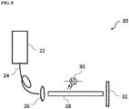
- the fluorescence excitation source 22 may comprise two excitation sources (not shown), one for providing the first excitation light and the other for providing the second excitation light.
- the fluorescence excitation source 22 includes, for example, a laser diode (which may comprise, for example, one or more fiber-coupled diode lasers), one or more LEDs, arc lamps, or other illuminant technologies of sufficient intensity and appropriate wavelength for providing the first and second excitation lights.
- the first and second excitation light from the fluorescence excitation source 22 may be projected through an optical element (i.e., one or more optical elements) to shape and guide the output being used to illuminate the biological sample.
- the means for illuminating 12 may also be configured to provide an additional functionality such as white light illumination.
- the method and system of the present invention may further comprise acquiring and combining the third fluorescence image representing the target fluorophore with a white light image of the biological material. In this manner, the location of the targeted fluorophore can be visualized within the context of the biological material. This is useful in instances in which the biological material cannot be viewed directly with the human eye.
Claims (34)
- Verfahren zum Extrahieren eines Bildes eines Zielfluorophors in einem biologischen Material, wobei ein Wellenband für die Zielfluorophoremission ein Wellenband für die Autofluoreszenzemission in dem biologischen Material überlappt, wobei das Verfahren umfasst:Beleuchten des biologischen Materials mit einem ersten Anregungslicht, um eine erste Fluoreszenzemission zu induzieren, die sowohl aus der Autofluoreszenz des biologischen Materials als auch aus der Fluoreszenz des Zielfluorophors entsteht, und mit einem zweiten Anregungslicht, um eine zweite Fluoreszenzemission zu induzieren, die nur aus der Autofluoreszenz des biologischen Materials entsteht;Erfassen eines ersten Fluoreszenzbildes von der ersten Fluoreszenzemission und eines zweiten Fluoreszenzbildes von der zweiten Fluoreszenzemission; undVerarbeiten des ersten und zweiten Fluoreszenzbildes, um ein drittes Fluoreszenzbild zu extrahieren, das das Zielfluorophor darstellt;wobei die relativen Intensitäten des ersten und des zweiten Anregungslichts in Bezug aufeinander vor dem Erfassen des ersten und des zweiten Fluoreszenzbildes moduliert werden, wobeidie Modulation der relativen Intensitäten umfasst:Identifizieren eines Wellenlängenbereichs in der ersten und zweiten Fluoreszenzemission, wobei der Wellenlängenbereich ein Bereich ist, in dem die vom Fluorophor entstehende Emission in der ersten Fluoreszenzemission vorhanden ist und in der zweiten Fluoreszenzemission fehlt;Auswählen eines Wellenbandes außerhalb des Wellenlängenbereichs;Berechnen eines Verhältnisses der relativen Intensitäten der ersten und zweiten Fluoreszenzemissionen in Bezug aufeinander im ausgewählten Wellenbereich; undAnpassen der relativen Intensitäten des ersten und des zweiten Anregungslichts in Bezug aufeinander, um die entsprechende erste Fluoreszenzemission, zweite Fluoreszenzemission oder beide anzupassen, bis ein geeignetes berechnetes Verhältnis erreicht ist.
- Verfahren nach Anspruch 1, wobei das Wellenband außerhalb des Wellenlängenbereichs eine oder mehrere Wellenlängen in Fluoreszenzspektren umfasst, die aus der ersten und zweiten Fluoreszenzemission entstehen.
- Verfahren nach Anspruch 1 oder 2, wobei das Berechnen des Verhältnisses der relativen Intensitäten der ersten und zweiten Fluoreszenzemission das Teilen eines Bereichs unter dem Kurvenwert, der der ersten Fluoreszenzemission entspricht, durch einen Bereich unter dem Kurvenwert, der der zweiten Fluoreszenzemission entspricht, umfasst.
- Verfahren nach einem der Ansprüche 1 bis 3, wobei das erste Anregungslicht eine Wellenlänge von etwa 405 nm und das zweite Anregungslicht eine Wellenlänge von etwa 450 nm hat, wenn das Zielfluorophor Porphyrin ist.
- Verfahren nach einem der Ansprüche 1 bis 4, wobei das ausgewählte Wellenband etwa 600 nm beträgt, und wobei das berechnete Verhältnis etwa 1 beträgt.
- Verfahren nach einem der Ansprüche 1 bis 5, wobei das Verarbeiten des ersten und des zweiten Fluoreszenzbildes zum Extrahieren des dritten Fluoreszenzbildes, das das Zielfluorophor darstellt, das Subtrahieren des zweiten Fluoreszenzbildes von dem ersten Fluoreszenzbild umfasst.
- Verfahren nach einem der Ansprüche 1 bis 6, wobei das biologische Material durch Photobleichen vorbehandelt wird.
- Verfahren nach einem der Ansprüche 1 bis 7, wobei das Zielfluorophor endogen, exogen oder eine Kombination davon ist.
- Verfahren nach Anspruch 8, wobei das endogene Fluorophor Porphyrin, ein Porphyrin-Vorläufer, ein Porphyrin-Analogon, ein Porphyrin-Derivat, ein Porphyrin-Konjugat, ein Porphyrin-Liposom, ein Porphyrin-Nanovesikel oder eine Kombination davon ist.
- Verfahren nach Anspruch 9, wobei das Porphyrin ein Coproporphyrin, ein Uroporphyrin, ein Protoporphyrin oder eine Kombination davon umfasst.
- Verfahren nach Anspruch 8, wobei das exogene Fluorophor ein fluoreszierender Farbstoff, ein fluoreszenzinduzierendes Mittel oder eine Kombination davon ist.
- Verwendung des Verfahrens nach einem der Ansprüche 1 bis 11 in der Hystochemie, Zytochemie oder einer Kombination davon.
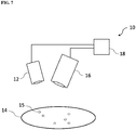
- System (10) zum Extrahieren eines Bildes eines Zielfluorophors (15) in einem biologischen Material (14), wobei ein Wellenband für die Zielfluorophoremission ein Wellenband für die Autofluoreszenzemission in dem biologischen Material überlappt, wobei das System umfasst:Mittel zum Beleuchten (12) des biologischen Materials mit einem ersten Anregungslicht, um eine erste Fluoreszenzemission zu induzieren, die sowohl aus der Autofluoreszenz des biologischen Materials als auch aus der Fluoreszenz des Zielfluorophors entsteht, und mit einem zweiten Anregungslicht, um eine zweite Fluoreszenzemission zu induzieren, die nur aus dem Autofluoreszenz des biologischen Materials entsteht;Mittel zum Erfassen (1) eines ersten Fluoreszenzbildes von der ersten Fluoreszenzemission und eines zweiten Fluoreszenzbildes von der zweiten Fluoreszenzemission;Mittel zum Modulieren der relativen Intensitäten des ersten und des zweiten Anregungslichts in Bezug aufeinander vor der Aufnahme des ersten und des zweiten Fluoreszenzbildes; undMittel zum Verarbeiten (18) des ersten und zweiten Fluoreszenzbildes zum Extrahieren eines dritten Fluoreszenzbildes, das das Zielfluorophor darstellt, wobei das Mittel zum Modulieren der relativen Intensitäten umfasst:Mittel zum Identifizieren eines Wellenlängenbereichs in der ersten und zweiten Fluoreszenzemission, wobei der Wellenlängenbereich ein Bereich ist, in dem die vom Fluorophor entstehende Emission in der ersten Fluoreszenzemission vorhanden ist und in der zweiten Fluoreszenzemission fehlt;Mittel zum Auswählen eines Wellenbandes außerhalb des Wellenlängenbereichs;Mittel zum Berechnen eines Verhältnisses der relativen Intensitäten der ersten und zweiten Fluoreszenzemission in Bezug aufeinander im ausgewählten Wellenbereich; undMittel zum Anpassen der relativen Intensitäten der ersten und zweiten Anregung in Bezug aufeinander, um die entsprechende erste Fluoreszenzemission, zweite Fluoreszenzemission oder beides anzupassen, bis ein geeignetes berechnetes Verhältnis erreicht ist.
- System nach Anspruch 13, wobei das Wellenband außerhalb des Wellenlängenbereichs eine oder mehrere Wellenlängen in Fluoreszenzspektren umfasst, die aus der ersten und zweiten Fluoreszenzemission entstehen.
- System nach Anspruch 13 oder 14, wobei Mittel zum Berechnen des Verhältnisses der relativen Intensitäten der ersten und zweiten Fluoreszenzemission Mittel zum Teilen eines Bereichs unter dem Kurvenwert, der der ersten Fluoreszenzemission entspricht, durch einen Bereich unter dem Kurvenwert, der der zweiten Fluoreszenzemission entspricht, umfasst.
- System nach einem der Ansprüche 13 bis 15, wobei das erste Anregungslicht eine Wellenlänge von etwa 405 nm und das zweite Anregungslicht eine Wellenlänge von etwa 450 nm hat, wenn das Zielfluorophor Porphyrin ist.
- System nach einem der Ansprüche 13 bis 16, wobei das ausgewählte Wellenband etwa 600 nm beträgt und wobei das berechnete Verhältnis ungefähr 1 beträgt.
- System nach einem der Ansprüche 13 bis 17, wobei das Mittel zum Verarbeiten des ersten und des zweiten Fluoreszenzbildes zum Extrahieren des dritten Fluoreszenzbildes, das das Zielfluorophor darstellt, Mittel zum Subtrahieren des zweiten Fluoreszenzbildes vom ersten Fluoreszenzbild umfasst.
- System nach einem der Ansprüche 13 bis 18, wobei das Mittel zum Beleuchten ein Beleuchtungsmodul umfasst, das eine Fluoreszenzanregungsquelle umfasst, wobei die Fluoreszenzanregungsquelle betriebsmäßig konfiguriert ist, um das erste und das zweite Anregungslicht zu erzeugen.
- System nach Anspruch 19, wobei das Beleuchtungsmodul ferner ein optisches Element umfasst, das betriebsmäßig konfiguriert ist, um das erste und das zweite Anregungslicht, das aus dem Beleuchtungsmodul austritt, zu formen und zu führen.
- System nach Anspruch 20, wobei das optische Element eine Linse, einen Lichtleiter, einen Diffusor oder eine Kombination davon umfasst.
- System nach einem der Ansprüche 13 bis 21, wobei das Mittel zum Erfassen ein Fluoreszenzemissionserfassungsmodul umfasst, wobei das Fluoreszenzemissionserfassungsmodul einen Bildsensor umfasst.
- System nach Anspruch 22, wobei das Fluoreszenzemissionserfassungsmodul ferner ein optisches Element umfasst, das vor dem Bildsensor angeordnet ist, der betriebsmäßig konfiguriert ist, um die erste und zweite Fluoreszenzemission zu erfassen, zu filtern und zu lenken.
- System nach einem der Ansprüche 13 bis 23, wobei das Mittel zur Verarbeitung ein Prozessormodul umfasst.
- System nach Anspruch 24, wobei das Prozessormodul betriebsmäßig konfiguriert ist, um einen Betrieb des Mittels zum Beleuchten, einen Betrieb des Mittels zum Erfassen oder eine Kombination davon zu steuern.
- System nach einem der Ansprüche 13 bis 25, wobei das biologische Material durch Photobleichen vorbehandelt wird.
- System nach einem der Ansprüche 13 bis 26, wobei das Zielfluorophor endogen, exogen oder eine Kombination davon ist.
- System nach Anspruch 27, wobei das endogene Fluorophor Porphyrin, ein Porphyrin-Vorläufer, ein Porphyrin-Analogon, ein Porphyrin-Derivat, ein Porphyrin-Konjugat, ein Porphyrin-Liposom, ein Porphyrin-Nanovesikel oder eine Kombination davon ist.
- System nach Anspruch 28, wobei das Porphyrin ein Coproporphyrin, ein Uroporphyrin, ein Protoporphyrin oder eine Kombination davon umfasst.
- System nach Anspruch 27, wobei das exogene Fluorophor ein fluoreszierender Farbstoff, ein fluoreszenzinduzierendes Mittel oder eine Kombination davon ist.
- Verfahren nach einem der Ansprüche 1 bis 11, Verwendung nach Anspruch 12 oder System nach einem der Ansprüche 13 bis 30, wobei das biologische Material ein biologisches Gewebe, eine biologische Flüssigkeit oder einen Bruchteil davon umfasst.
- Verfahren nach einem der Ansprüche 1 bis 11, Verwendung nach Anspruch 12 oder System nach einem der Ansprüche 13 bis 30, wobei das biologische Material ein Organ, eine Zelle, eine Zelllinie, einen Zellbestandteil umfasst, der von einem Säugetier abgeleitet ist oder sich darin befindet.
- Verfahren nach einem der Ansprüche 1 bis 11, Verwendung nach Anspruch 12 oder System nach einem der Ansprüche 13 bis 30, wobei das biologische Material gesundes, krankes oder bösartiges Gewebe umfasst.
- Verfahren nach einem der Ansprüche 1 bis 11, Verwendung nach Anspruch 12 oder System nach einem der Ansprüche 13 bis 30, wobei das biologische Material einen Gewebeschnitt zur Verwendung in der Histochemie, Immunhistochemie, Zytochemie, Immunfluoreszenz, Immunblotting oder einer fluoreszenzbezogenen Bildgebungsanwendung umfasst.
Applications Claiming Priority (2)
| Application Number | Priority Date | Filing Date | Title |
|---|---|---|---|
| US201462056830P | 2014-09-29 | 2014-09-29 | |
| PCT/CA2015/050973 WO2016049756A1 (en) | 2014-09-29 | 2015-09-28 | Imaging a target fluorophore in a biological material in the presence of autofluorescence |
Publications (3)
| Publication Number | Publication Date |
|---|---|
| EP3201607A1 EP3201607A1 (de) | 2017-08-09 |
| EP3201607A4 EP3201607A4 (de) | 2018-08-15 |
| EP3201607B1 true EP3201607B1 (de) | 2020-12-30 |
Family
ID=55629196
Family Applications (1)
| Application Number | Title | Priority Date | Filing Date |
|---|---|---|---|
| EP15846111.1A Active EP3201607B1 (de) | 2014-09-29 | 2015-09-28 | Bildgebung eines zielfluorophors in einem biologischen material in anwesenheit von autofluoreszenz |
Country Status (9)
| Country | Link |
|---|---|
| US (2) | US9816930B2 (de) |
| EP (1) | EP3201607B1 (de) |
| JP (1) | JP6549705B2 (de) |
| KR (2) | KR102068776B1 (de) |
| CN (1) | CN107209118B (de) |
| AU (1) | AU2015327665B2 (de) |
| CA (1) | CA2963987A1 (de) |
| HK (1) | HK1244873A1 (de) |
| WO (1) | WO2016049756A1 (de) |
Families Citing this family (27)
| Publication number | Priority date | Publication date | Assignee | Title |
|---|---|---|---|---|
| US20070122344A1 (en) | 2005-09-02 | 2007-05-31 | University Of Rochester Medical Center Office Of Technology Transfer | Intraoperative determination of nerve location |
| US20080161744A1 (en) * | 2006-09-07 | 2008-07-03 | University Of Rochester Medical Center | Pre-And Intra-Operative Localization of Penile Sentinel Nodes |
| US8406860B2 (en) | 2008-01-25 | 2013-03-26 | Novadaq Technologies Inc. | Method for evaluating blush in myocardial tissue |
| US10219742B2 (en) | 2008-04-14 | 2019-03-05 | Novadaq Technologies ULC | Locating and analyzing perforator flaps for plastic and reconstructive surgery |
| ES2671710T3 (es) | 2008-05-02 | 2018-06-08 | Novadaq Technologies ULC | Métodos para la producción y uso de eritrocitos cargados con sustancias para la observación y el tratamiento de la hemodinámica microvascular |
| US10492671B2 (en) | 2009-05-08 | 2019-12-03 | Novadaq Technologies ULC | Near infra red fluorescence imaging for visualization of blood vessels during endoscopic harvest |
| CA2914778A1 (en) | 2012-06-21 | 2013-12-27 | Novadaq Technologies Inc. | Quantification and analysis of angiography and perfusion |
| AU2015327665B2 (en) * | 2014-09-29 | 2018-09-27 | Stryker European Operations Limited | Imaging a target fluorophore in a biological material in the presence of autofluorescence |
| KR101955134B1 (ko) | 2014-10-09 | 2019-03-06 | 노바다크 테크놀러지즈 유엘씨 | 형광-조정 광전용적맥파 측정기를 사용한 조직 내의 절대적인 혈류의 정량화 |
| CN108369734B (zh) * | 2015-12-10 | 2022-04-26 | 凯杰有限公司 | 数字图像中的对象分类方法、系统及计算机可读介质 |
| TWI583952B (zh) * | 2016-01-21 | 2017-05-21 | 威力暘電子股份有限公司 | 排泄物之潛血檢測方法及潛血檢測裝置 |
| JP6931705B2 (ja) | 2017-02-10 | 2021-09-08 | ノバダック テクノロジーズ ユーエルシー | オープンフィールドハンドヘルド蛍光イメージングシステムおよび方法 |
| EA201992252A1 (ru) * | 2017-04-07 | 2020-04-02 | Авелас Байосайенсиз, Инк. | Логометрические способы визуализации флуоресценции |
| US10969384B2 (en) | 2017-05-04 | 2021-04-06 | Vector Laboratories, Inc. | Immunofluorescence assays |
| WO2019012771A1 (ja) * | 2017-07-11 | 2019-01-17 | 浜松ホトニクス株式会社 | 試料観察装置及び試料観察方法 |
| EP3731728A4 (de) * | 2017-12-27 | 2021-08-25 | Ethicon LLC | Hyperspektrale bildgebung mit werkzeugverfolgung in einer lichtschwachen umgebung |
| EP3788345A1 (de) | 2018-05-03 | 2021-03-10 | Akoya Biosciences, Inc. | Multispektrale probenabbildung |
| US10753875B1 (en) * | 2019-01-31 | 2020-08-25 | Rarecyte, Inc. | Spectral unmixing of spectroscopic emission images |
| US10724956B1 (en) * | 2019-02-01 | 2020-07-28 | Essen Instruments, Inc. | Spectral unmixing |
| EP3924722A1 (de) * | 2019-02-13 | 2021-12-22 | Ventana Medical Systems, Inc. | Systeme und verfahren zur berechnung der beiträge von autofluoreszenz bei mehrkanalbildern |
| TWI726614B (zh) * | 2020-02-12 | 2021-05-01 | 財團法人國家實驗研究院 | 生物螢光影像的檢測方法 |
| WO2022010519A1 (en) * | 2020-07-09 | 2022-01-13 | Axon Imaging, Llc | Tissue imaging system |
| WO2022011276A1 (en) | 2020-07-09 | 2022-01-13 | Axon Imaging, Llc | Advanced nervous tissue imaging system |
| EP3961194B1 (de) * | 2020-08-25 | 2023-11-08 | Korea Advanced Institute of Science and Technology | Verfahren und vorrichtung zur multiplexierten bildgebung von biomolekülen durch iteratives entmischen von fluorophorsignalen |
| US11408825B2 (en) * | 2020-08-28 | 2022-08-09 | Hong Kong Applied Science and Technology Research Institute Company Limited | Forensic detector and the system thereof |
| US20240125701A1 (en) * | 2021-01-22 | 2024-04-18 | Toho University | Autofluorescence quenching device |
| WO2023059956A1 (en) * | 2021-10-05 | 2023-04-13 | Mazel Charles H | Method and system for distinguishing a fluorescent subject of interest from other fluorescent subjects or fluorescent background |
Family Cites Families (366)
| Publication number | Priority date | Publication date | Assignee | Title |
|---|---|---|---|---|
| JPS5854822B2 (ja) | 1975-09-11 | 1983-12-06 | ミノルタ株式会社 | コウデンシキセイタイケイソクキ |
| US4109647A (en) | 1977-03-16 | 1978-08-29 | The United States Of America As Represented By The Secretary Of The Department Of Health, Education And Welfare | Method of and apparatus for measurement of blood flow using coherent light |
| US4263916A (en) | 1978-03-27 | 1981-04-28 | University Of Southern California | Image averaging for angiography by registration and combination of serial images |
| US4162405A (en) | 1978-05-23 | 1979-07-24 | Britton Chance | Flying spot fluoro-meter for oxidized flavoprotein and reduced pyridine nucleotide |
| US4200801A (en) | 1979-03-28 | 1980-04-29 | The United States Of America As Represented By The United States Department Of Energy | Portable spotter for fluorescent contaminants on surfaces |
| US4394199A (en) | 1981-09-08 | 1983-07-19 | Agnus Chemical Company | Explosive emulsion composition |
| JPS58141135A (ja) | 1981-10-20 | 1983-08-22 | 富士写真フイルム株式会社 | 固体イメ−ジセンサを用いた内視鏡の画像伝送方式 |
| EP0091805B1 (de) | 1982-04-08 | 1989-07-26 | Olympus Optical Co., Ltd. | Fokussiererfassungsvorrichtung für Endoskop |
| JPS58222331A (ja) | 1982-06-21 | 1983-12-24 | Sony Corp | 文字情報再生装置 |
| JPS5940830A (ja) | 1982-08-31 | 1984-03-06 | 浜松ホトニクス株式会社 | レ−ザ光パルスを用いた癌の診断装置 |
| JPS5969721A (ja) | 1982-10-15 | 1984-04-20 | Olympus Optical Co Ltd | 内視鏡計測装置 |
| JPS5970903A (ja) | 1982-10-15 | 1984-04-21 | Olympus Optical Co Ltd | 内視鏡自動計測装置 |
| US4541438A (en) | 1983-06-02 | 1985-09-17 | The Johns Hopkins University | Localization of cancerous tissue by monitoring infrared fluorescence emitted by intravenously injected porphyrin tumor-specific markers excited by long wavelength light |
| US4532918A (en) | 1983-10-07 | 1985-08-06 | Welch Allyn Inc. | Endoscope signal level control |
| JPS60256443A (ja) | 1984-05-31 | 1985-12-18 | オムロン株式会社 | 画像計測装置 |
| US4559557A (en) | 1984-06-01 | 1985-12-17 | General Electric Company | Region-of-interest digital subtraction angiography |
| SE455646B (sv) | 1984-10-22 | 1988-07-25 | Radians Innova Ab | Fluorescensanordning |
| US4718417A (en) | 1985-03-22 | 1988-01-12 | Massachusetts Institute Of Technology | Visible fluorescence spectral diagnostic for laser angiosurgery |
| US5318024A (en) | 1985-03-22 | 1994-06-07 | Massachusetts Institute Of Technology | Laser endoscope for spectroscopic imaging |
| US5125404A (en) | 1985-03-22 | 1992-06-30 | Massachusetts Institute Of Technology | Apparatus and method for obtaining spectrally resolved spatial images of tissue |
| DE3511255A1 (de) | 1985-03-28 | 1986-10-02 | Grün Optik Wetzlar GmbH, 6330 Wetzlar | Anordnung zur individuellen regelung der intensitaet mehrer spektrallampen |
| CN85100424B (zh) | 1985-04-01 | 1986-10-29 | 上海医疗器械研究所 | 恶性肿瘤固有荧光诊断仪 |
| US4619249A (en) | 1985-07-24 | 1986-10-28 | Kim Landry | Transcutaneous intravenous illuminator |
| AT387860B (de) | 1985-09-16 | 1989-03-28 | Optical Sensing Technology | Verfahren und vorrichtung zur tumordiagnose mittels sera |
| US4719508A (en) | 1985-10-02 | 1988-01-12 | Olympus Optical Co., Ltd. | Endoscopic photographing apparatus |
| US5134662A (en) | 1985-11-04 | 1992-07-28 | Cell Analysis Systems, Inc. | Dual color camera microscope and methodology for cell staining and analysis |
| US4930516B1 (en) | 1985-11-13 | 1998-08-04 | Laser Diagnostic Instr Inc | Method for detecting cancerous tissue using visible native luminescence |
| US5042494A (en) | 1985-11-13 | 1991-08-27 | Alfano Robert R | Method and apparatus for detecting cancerous tissue using luminescence excitation spectra |
| US4774568A (en) | 1986-01-27 | 1988-09-27 | Kabushiki Kaisha Toshiba | Endoscopic apparatus |
| JPS62247232A (ja) | 1986-04-21 | 1987-10-28 | Agency Of Ind Science & Technol | 蛍光測定装置 |
| GB2203831B (en) | 1986-07-07 | 1991-02-06 | Academy Of Applied Sciences | Apparatus and method for the diagnosis of malignant tumours |
| JPH0763443B2 (ja) | 1986-09-30 | 1995-07-12 | 株式会社東芝 | 電子内視鏡 |
| JPS63122421A (ja) | 1986-11-12 | 1988-05-26 | 株式会社東芝 | 内視鏡装置 |
| US5255087A (en) | 1986-11-29 | 1993-10-19 | Olympus Optical Co., Ltd. | Imaging apparatus and endoscope apparatus using the same |
| JP2572394B2 (ja) | 1987-03-19 | 1997-01-16 | オリンパス光学工業株式会社 | 電子内視鏡 |
| JPH0783B2 (ja) | 1987-03-30 | 1995-01-11 | 株式会社東芝 | 電子内視鏡装置 |
| US4986262A (en) | 1987-03-31 | 1991-01-22 | Kabushiki Kaisha Toshiba | Measuring endoscope |
| US4852579A (en) | 1987-04-20 | 1989-08-01 | Karl Storz Endoscopy Gmbh And Company | Photocharacterization and treatment of normal abnormal and ectopic endometrium |
| US4900934A (en) | 1987-07-15 | 1990-02-13 | University Of Utah | Apparatus for simultaneous visualization and measurement of fluorescence from fluorescent dye-treated cell preparations and solutions |
| JPH0824668B2 (ja) | 1987-09-14 | 1996-03-13 | オリンパス光学工業株式会社 | 電子内視鏡装置 |
| US4858001A (en) | 1987-10-08 | 1989-08-15 | High-Tech Medical Instrumentation, Inc. | Modular endoscopic apparatus with image rotation |
| JPH01160525A (ja) | 1987-12-17 | 1989-06-23 | Olympus Optical Co Ltd | 内視鏡 |
| DE3906860A1 (de) | 1988-03-08 | 1989-09-28 | Fraunhofer Ges Forschung | Vorrichtung zum herstellen einer angiographie |
| JPH01236879A (ja) | 1988-03-17 | 1989-09-21 | Canon Inc | 画像符号化装置 |
| JPH06105190B2 (ja) | 1988-03-31 | 1994-12-21 | 工業技術院長 | 信号解析装置 |
| US4998972A (en) | 1988-04-28 | 1991-03-12 | Thomas J. Fogarty | Real time angioscopy imaging system |
| US5078150A (en) | 1988-05-02 | 1992-01-07 | Olympus Optical Co., Ltd. | Spectral diagnosing apparatus with endoscope |
| IL90188A0 (en) | 1988-05-18 | 1989-12-15 | Cryopharm Corp | Process and medium for the lyophilization of erythrocytes |
| US4938205A (en) | 1988-05-27 | 1990-07-03 | The University Of Connecticut | Endoscope with traced raster and elemental photodetectors |
| US5178616A (en) | 1988-06-06 | 1993-01-12 | Sumitomo Electric Industries, Ltd. | Method and apparatus for intravascular laser surgery |
| US4995396A (en) | 1988-12-08 | 1991-02-26 | Olympus Optical Co., Ltd. | Radioactive ray detecting endoscope |
| US5419323A (en) | 1988-12-21 | 1995-05-30 | Massachusetts Institute Of Technology | Method for laser induced fluorescence of tissue |
| US5353799A (en) | 1991-01-22 | 1994-10-11 | Non Invasive Technology, Inc. | Examination of subjects using photon migration with high directionality techniques |
| JP2987816B2 (ja) | 1989-01-30 | 1999-12-06 | オリンパス光学工業株式会社 | 蛍光観察装置 |
| DE3903019A1 (de) | 1989-02-02 | 1990-08-09 | Hell Rudolf Dr Ing Gmbh | Optische farbteiler-anordnung |
| SE8900612D0 (sv) * | 1989-02-22 | 1989-02-22 | Jonas Johansson | Vaevnadskarakterisering utnyttjande ett blodfritt fluorescenskriterium |
| US5421337A (en) | 1989-04-14 | 1995-06-06 | Massachusetts Institute Of Technology | Spectral diagnosis of diseased tissue |
| EP0466828A1 (de) | 1989-04-14 | 1992-01-22 | Massachusetts Institute Of Technology | Spektraldiagnose von erkranktem gewebe |
| KR100190423B1 (ko) | 1989-06-06 | 1999-06-01 | 기타지마 요시도시 | 에멀젼마스크 등의결함 수정방법 및 장치 |
| US4993404A (en) | 1989-06-26 | 1991-02-19 | Lane Timothy G | Fluoroscopy switching device |
| CN1049781A (zh) | 1989-09-02 | 1991-03-13 | 住友电气工业株式会社 | 用于血管外科的激光手术器械 |
| JPH0614921B2 (ja) | 1989-09-29 | 1994-03-02 | 浜松ホトニクス株式会社 | 生体組織螢光観察装置 |
| US5150292A (en) | 1989-10-27 | 1992-09-22 | Arch Development Corporation | Method and system for determination of instantaneous and average blood flow rates from digital angiograms |
| DE69114314T2 (de) | 1990-01-08 | 1996-04-18 | Ernest M D Feiler | Diagnostisches Verfahren zur Messung der Blutströmung. |
| US5091652A (en) | 1990-01-12 | 1992-02-25 | The Regents Of The University Of California | Laser excited confocal microscope fluorescence scanner and method |
| US5420628A (en) | 1990-01-16 | 1995-05-30 | Research Development Foundation | Video densitometer with determination of color composition |
| US5131398A (en) | 1990-01-22 | 1992-07-21 | Mediscience Technology Corp. | Method and apparatus for distinguishing cancerous tissue from benign tumor tissue, benign tissue or normal tissue using native fluorescence |
| US4995398A (en) | 1990-04-30 | 1991-02-26 | Turnidge Patrick A | Coronary angiography imaging system |
| US5071417A (en) | 1990-06-15 | 1991-12-10 | Rare Earth Medical Lasers, Inc. | Laser fusion of biological materials |
| US5845639A (en) | 1990-08-10 | 1998-12-08 | Board Of Regents Of The University Of Washington | Optical imaging methods |
| US5465718A (en) | 1990-08-10 | 1995-11-14 | Hochman; Daryl | Solid tumor, cortical function, and nerve tissue imaging methods and device |
| US6671540B1 (en) | 1990-08-10 | 2003-12-30 | Daryl W. Hochman | Methods and systems for detecting abnormal tissue using spectroscopic techniques |
| US6196226B1 (en) | 1990-08-10 | 2001-03-06 | University Of Washington | Methods and apparatus for optically imaging neuronal tissue and activity |
| US5699798A (en) | 1990-08-10 | 1997-12-23 | University Of Washington | Method for optically imaging solid tumor tissue |
| US5438989A (en) | 1990-08-10 | 1995-08-08 | Hochman; Darryl | Solid tumor, cortical function, and nerve tissue imaging methods and device |
| US5997844A (en) | 1991-02-08 | 1999-12-07 | Diatide, Inc. | Technetium-99m labeled peptides for imaging |
| JPH04297236A (ja) | 1991-03-26 | 1992-10-21 | Toshiba Corp | ディジタルフルオログラフィ装置 |
| JPH04307024A (ja) | 1991-04-02 | 1992-10-29 | Olympus Optical Co Ltd | 電子内視鏡装置 |
| US5318023A (en) | 1991-04-03 | 1994-06-07 | Cedars-Sinai Medical Center | Apparatus and method of use for a photosensitizer enhanced fluorescence based biopsy needle |
| US5377676A (en) | 1991-04-03 | 1995-01-03 | Cedars-Sinai Medical Center | Method for determining the biodistribution of substances using fluorescence spectroscopy |
| US6485413B1 (en) | 1991-04-29 | 2002-11-26 | The General Hospital Corporation | Methods and apparatus for forward-directed optical scanning instruments |
| US5117466A (en) | 1991-04-30 | 1992-05-26 | The United States Of America As Represented By The United States Department Of Energy | Integrated fluorescence analysis system |
| CA2042075C (en) | 1991-05-08 | 2001-01-23 | Branko Palcic | Endoscopic imaging system |
| US5225883A (en) | 1991-06-05 | 1993-07-06 | The Babcock & Wilcox Company | Video temperature monitor |
| SE468925B (sv) | 1991-08-22 | 1993-04-19 | Gert Nilsson | En metod och en anordning foer att reducera den avstaandsberoende foerstaerkningsfaktorn vid maetning av stroemningsroerelser med en bildgivande laser-doppler teknik, i synnerhet vid maetning av blodperfusion genom en vaevnad |
| US5377686A (en) | 1991-10-11 | 1995-01-03 | The University Of Connecticut | Apparatus for detecting leakage from vascular tissue |
| JP3297725B2 (ja) | 1991-12-09 | 2002-07-02 | 国立循環器病センター総長 | 造影血管高精度管径計測装置 |
| US5214503A (en) | 1992-01-31 | 1993-05-25 | The United States Of America As Represented By The Secretary Of The Army | Color night vision camera system |
| US5235984A (en) | 1992-03-30 | 1993-08-17 | Hewlett-Packard Company | On-line acoustic densitometry tool for use with an ultrasonic imaging system |
| US6096289A (en) | 1992-05-06 | 2000-08-01 | Immunomedics, Inc. | Intraoperative, intravascular, and endoscopic tumor and lesion detection, biopsy and therapy |
| DE4220633C1 (de) | 1992-06-24 | 1994-02-03 | Wolf Gmbh Richard | Vorrichtung zur Lichtversorgung von Endoskopen |
| US5733721A (en) * | 1992-11-20 | 1998-03-31 | The Board Of Regents Of The University Of Oklahoma | Cell analysis method using quantitative fluorescence image analysis |
| US5279298A (en) | 1992-11-20 | 1994-01-18 | The Johns Hopkins University | Method and apparatus to identify and treat neovascular membranes in the eye |
| US5514127A (en) | 1993-02-18 | 1996-05-07 | Central Research Laboratories Limited | Apparatus for irradiating an area with a controllable pattern of light |
| US5437274A (en) | 1993-02-25 | 1995-08-01 | Gholam A. Peyman | Method of visualizing submicron-size vesicles and particles in blood circulation |
| JP3228627B2 (ja) | 1993-03-19 | 2001-11-12 | オリンパス光学工業株式会社 | 内視鏡用画像処理装置 |
| US5431161A (en) | 1993-04-15 | 1995-07-11 | Adac Laboratories | Method and apparatus for information acquistion, processing, and display within a medical camera system |
| US5421339A (en) | 1993-05-12 | 1995-06-06 | Board Of Regents, The University Of Texas System | Diagnosis of dysplasia using laser induced fluoroescence |
| US5394199A (en) | 1993-05-17 | 1995-02-28 | The Johns Hopkins University | Methods and apparatus for improved visualization of choroidal blood flow and aberrant vascular structures in the eye using fluorescent dye angiography |
| WO1996009792A1 (en) | 1993-05-17 | 1996-04-04 | The Johns Hopkins University | Improved visualization of choroidal blood flow and aberrant vascular structures in the eye |
| US5424841A (en) | 1993-05-28 | 1995-06-13 | Molecular Dynamics | Apparatus for measuring spatial distribution of fluorescence on a substrate |
| US5673701A (en) | 1994-10-07 | 1997-10-07 | Non Invasive Technology, Inc. | Optical techniques for examination of biological tissue |
| DK75593D0 (de) | 1993-06-25 | 1993-06-25 | Novo Nordisk As | |
| US5365057A (en) | 1993-07-02 | 1994-11-15 | Litton Systems, Inc. | Light-weight night vision device |
| JP3224640B2 (ja) * | 1993-07-30 | 2001-11-05 | 三菱重工業株式会社 | Lifによる濃度計測装置及び方法 |
| US5371355A (en) | 1993-07-30 | 1994-12-06 | Litton Systems, Inc. | Night vision device with separable modular image intensifier assembly |
| JPH0765154A (ja) | 1993-08-31 | 1995-03-10 | Toshiba Corp | 血管像の定量解析装置及びその定量解析方法 |
| JPH0779955A (ja) | 1993-09-14 | 1995-03-28 | Toshiba Corp | X線撮影装置 |
| JPH07155291A (ja) | 1993-12-03 | 1995-06-20 | Olympus Optical Co Ltd | 蛍光観察装置 |
| JPH07222712A (ja) | 1994-02-10 | 1995-08-22 | Olympus Optical Co Ltd | 蛍光内視鏡装置 |
| JP3194660B2 (ja) | 1993-12-03 | 2001-07-30 | オリンパス光学工業株式会社 | 蛍光観察装置 |
| JP3285265B2 (ja) | 1993-12-03 | 2002-05-27 | オリンパス光学工業株式会社 | 蛍光観察装置 |
| JP3487933B2 (ja) | 1993-12-03 | 2004-01-19 | オリンパス株式会社 | 蛍光観察装置 |
| JP3283128B2 (ja) | 1993-12-03 | 2002-05-20 | オリンパス光学工業株式会社 | 蛍光観察内視鏡装置 |
| JPH07155290A (ja) | 1993-12-03 | 1995-06-20 | Olympus Optical Co Ltd | 内視鏡装置 |
| US5453448A (en) | 1993-12-09 | 1995-09-26 | Pdt Cardiovascular, Inc. | In vivo method for estimating the lipid contant of an atheromatous lesion |
| JP3275159B2 (ja) | 1993-12-17 | 2002-04-15 | 日本光電工業株式会社 | 循環血液量測定装置 |
| US5496369A (en) | 1994-02-09 | 1996-03-05 | University Of Iowa Research Foundation | Human cerebral cortex neural prosthetic |
| US5656498A (en) | 1994-02-22 | 1997-08-12 | Nippon Telegraph And Telephone Corporation | Freeze-dried blood cells, stem cells and platelets, and manufacturing method for the same |
| US5707986A (en) | 1994-03-14 | 1998-01-13 | Miller; Joan W. | Angiographic method using green porphyrins in primate eyes |
| JPH07250804A (ja) | 1994-03-15 | 1995-10-03 | Olympus Optical Co Ltd | 蛍光観察装置 |
| JPH07250812A (ja) | 1994-03-15 | 1995-10-03 | Olympus Optical Co Ltd | 蛍光診断装置 |
| US5491343A (en) | 1994-03-25 | 1996-02-13 | Brooker; Gary | High-speed multiple wavelength illumination source, apparatus containing the same, and applications thereof to methods of irradiating luminescent samples and of quantitative luminescence ratio microscopy |
| US5590660A (en) | 1994-03-28 | 1997-01-07 | Xillix Technologies Corp. | Apparatus and method for imaging diseased tissue using integrated autofluorescence |
| AU2432795A (en) | 1994-05-03 | 1995-11-29 | Molecular Biosystems, Inc. | Composition for ultrasonically quantitating myocardial perfusion |
| US5519534A (en) | 1994-05-25 | 1996-05-21 | The Government Of The United States Of America As Represented By The Secretary Of The Department Of Health And Human Services | Irradiance attachment for an optical fiber to provide a uniform level of illumination across a plane |
| JP3641495B2 (ja) | 1994-07-19 | 2005-04-20 | 株式会社日立メディコ | 医用画像診断装置 |
| CA2141181A1 (en) | 1994-09-21 | 1996-03-22 | Kimberly-Clark Worldwide, Inc. | Wet-resilient webs |
| EP0801534B1 (de) | 1994-09-26 | 2003-12-10 | The Johns Hopkins University | Optische darstellung des blutkreislaufs und von abnormalen gefässstrukturen in der aderhaut des auges |
| US5627907A (en) | 1994-12-01 | 1997-05-06 | University Of Pittsburgh | Computerized detection of masses and microcalcifications in digital mammograms |
| US5935942A (en) | 1994-12-14 | 1999-08-10 | Zeimer; Ran | Selective and non-invasive visualization or treatment of vasculature |
| US5951980A (en) | 1995-01-06 | 1999-09-14 | Leuven Research & Development Vzw | Identification, production and use of new staphylokinase derivatives with reduced immunogenicity |
| GB9502065D0 (en) | 1995-02-02 | 1995-03-22 | Nycomed Imaging As | Contrast media |
| JPH08224240A (ja) | 1995-02-22 | 1996-09-03 | Olympus Optical Co Ltd | 蛍光診断装置 |
| JPH08224208A (ja) | 1995-02-22 | 1996-09-03 | Olympus Optical Co Ltd | 蛍光観察内視鏡装置 |
| JP3560671B2 (ja) | 1995-02-23 | 2004-09-02 | オリンパス株式会社 | 蛍光観察装置 |
| JP3411737B2 (ja) | 1995-03-03 | 2003-06-03 | ペンタックス株式会社 | 生体の蛍光診断装置 |
| US7236815B2 (en) | 1995-03-14 | 2007-06-26 | The Board Of Regents Of The University Of Texas System | Method for probabilistically classifying tissue in vitro and in vivo using fluorescence spectroscopy |
| US5576013A (en) | 1995-03-21 | 1996-11-19 | Eastern Virginia Medical School | Treating vascular and neoplastic tissues |
| CA2215978A1 (en) | 1995-04-04 | 1996-10-10 | Wound Healing Of Oklahoma | Cancer treatment by photodynamic therapy, in combination with an immunoadjuvant |
| US5689241A (en) | 1995-04-24 | 1997-11-18 | Clarke, Sr.; James Russell | Sleep detection and driver alert apparatus |
| US5743266A (en) | 1995-04-25 | 1998-04-28 | Molecular Biosystems, Inc. | Method for processing real-time contrast enhanced ultrasonic images |
| US5623930A (en) | 1995-05-02 | 1997-04-29 | Acuson Corporation | Ultrasound system for flow measurement |
| US6032070A (en) | 1995-06-07 | 2000-02-29 | University Of Arkansas | Method and apparatus for detecting electro-magnetic reflection from biological tissue |
| JP3819032B2 (ja) | 1995-08-24 | 2006-09-06 | ザ・テキサス・エイ・アンド・エム・ユニバーシティ・システム | 組織およびその他のランダム媒体における蛍光寿命に基づく撮像および分光分析 |
| US5836311A (en) | 1995-09-20 | 1998-11-17 | Medtronic, Inc. | Method and apparatus for temporarily immobilizing a local area of tissue |
| US5647368A (en) | 1996-02-28 | 1997-07-15 | Xillix Technologies Corp. | Imaging system for detecting diseased tissue using native fluorsecence in the gastrointestinal and respiratory tract |
| US5756541A (en) | 1996-03-11 | 1998-05-26 | Qlt Phototherapeutics Inc | Vision through photodynamic therapy of the eye |
| DE19613342A1 (de) | 1996-04-03 | 1997-10-09 | Philips Patentverwaltung | Automatisches Bildauswertungsverfahren |
| US5766127A (en) | 1996-04-15 | 1998-06-16 | Ohmeda Inc. | Method and apparatus for improved photoplethysmographic perfusion-index monitoring |
| JPH09305845A (ja) | 1996-05-13 | 1997-11-28 | Shibaura Eng Works Co Ltd | 温蔵自動販売機 |
| US5662644A (en) | 1996-05-14 | 1997-09-02 | Mdlt, Inc. | Dermatological laser apparatus and method |
| US5785965A (en) | 1996-05-15 | 1998-07-28 | The Board Of Trustees Of The Leland Stanford Junior Univ. | VEGF gene transfer into endothelial cells for vascular prosthesis |
| JP3896176B2 (ja) | 1996-05-21 | 2007-03-22 | 浜松ホトニクス株式会社 | 近赤外線蛍光トレーサーおよび蛍光イメージング方法 |
| GB9610700D0 (en) | 1996-05-22 | 1996-07-31 | Moor Instr Ltd | Apparatus for imaging microvascular blood flow |
| JPH09308609A (ja) | 1996-05-24 | 1997-12-02 | Canon Inc | 眼科用画像処理装置 |
| DE19635038A1 (de) | 1996-08-29 | 1998-03-12 | Pulsion Verwaltungs Gmbh & Co | Verfahren zur nicht invasiven Bestimmung des zerebralen Blutflusses mittels Nah-Infrarot-Spektroskopie |
| US5851181A (en) | 1996-08-30 | 1998-12-22 | Esc Medical Systems Ltd. | Apparatus for simultaneously viewing and spectrally analyzing a portion of skin |
| JP3177635B2 (ja) | 1996-09-30 | 2001-06-18 | 株式会社応用光電研究室 | 周波数標準器および選択標準周波数生成方法 |
| JP2793989B2 (ja) | 1996-09-30 | 1998-09-03 | オリンパス光学工業株式会社 | 内視鏡用光源装置の回転フィルタ |
| US6013265A (en) | 1996-10-22 | 2000-01-11 | University Of Maryland, Baltimore | Vaccine composition for herpes simplex virus and methods of using |
| US6293911B1 (en) | 1996-11-20 | 2001-09-25 | Olympus Optical Co., Ltd. | Fluorescent endoscope system enabling simultaneous normal light observation and fluorescence observation in infrared spectrum |
| JP3962122B2 (ja) | 1996-11-20 | 2007-08-22 | オリンパス株式会社 | 内視鏡装置 |
| JP3713347B2 (ja) | 1996-11-25 | 2005-11-09 | オリンパス株式会社 | 蛍光内視鏡装置 |
| DE19648935B4 (de) | 1996-11-26 | 2008-05-15 | IMEDOS Intelligente Optische Systeme der Medizin- und Messtechnik GmbH | Vorrichtung und Verfahren zur Untersuchung von Gefäßen |
| CA2192036A1 (en) | 1996-12-04 | 1998-06-04 | Harvey Lui | Fluorescence scope system for dermatologic diagnosis |
| US6086539A (en) | 1996-12-04 | 2000-07-11 | Acuson Corporation | Methods and apparatus for ultrasound image quantification |
| US6200310B1 (en) | 1997-01-08 | 2001-03-13 | Biosense, Inc. | Monitoring of myocardial revascularization |
| JPH10210367A (ja) | 1997-01-20 | 1998-08-07 | Olympus Optical Co Ltd | 電子的撮像装置 |
| JP3771985B2 (ja) | 1997-01-20 | 2006-05-10 | オリンパス株式会社 | 蛍光観察内視鏡装置 |
| US5965356A (en) | 1997-01-31 | 1999-10-12 | University Of Maryland, Baltimore | Herpes simplex virus type specific seroassay |
| US6122042A (en) | 1997-02-07 | 2000-09-19 | Wunderman; Irwin | Devices and methods for optically identifying characteristics of material objects |
| US6466687B1 (en) | 1997-02-12 | 2002-10-15 | The University Of Iowa Research Foundation | Method and apparatus for analyzing CT images to determine the presence of pulmonary tissue pathology |
| US6081612A (en) | 1997-02-28 | 2000-06-27 | Electro Optical Sciences Inc. | Systems and methods for the multispectral imaging and characterization of skin tissue |
| US6008889A (en) | 1997-04-16 | 1999-12-28 | Zeng; Haishan | Spectrometer system for diagnosis of skin disease |
| AU737530B2 (en) | 1997-04-17 | 2001-08-23 | Avimo Group Limited | Ocular microcirculation examination and treatment apparatus |
| GB9710049D0 (en) | 1997-05-19 | 1997-07-09 | Nycomed Imaging As | Method |
| US6280386B1 (en) | 1997-06-16 | 2001-08-28 | The Research Foundation Of The City University Of New York | Apparatus for enhancing the visibility of a luminous object inside tissue and methods for same |
| AU7934498A (en) | 1997-06-27 | 1999-01-19 | Toa Medical Electronics Co., Ltd. | Living body inspecting apparatus and noninvasive blood analyzer using the same |
| US6342611B1 (en) | 1997-10-10 | 2002-01-29 | Cytovia, Inc. | Fluorogenic or fluorescent reporter molecules and their applications for whole-cell fluorescence screening assays for capsases and other enzymes and the use thereof |
| DE19747172C2 (de) | 1997-10-24 | 2000-04-13 | Pulsion Verwaltungs Gmbh & Co | Vorrichtung zur Feststellung eines Perikardergusses |
| JP3370912B2 (ja) | 1997-11-14 | 2003-01-27 | 松下電器産業株式会社 | 撮像装置 |
| US6306642B1 (en) | 1997-11-24 | 2001-10-23 | Quidel Corporation | Enzyme substrate delivery and product registration in one step enzyme immunoassays |
| JPH11155812A (ja) | 1997-12-02 | 1999-06-15 | Olympus Optical Co Ltd | 蛍光観察装置 |
| US5919616A (en) | 1997-12-12 | 1999-07-06 | Aurx, Inc. | Serological assay for herpes |
| JPH11183358A (ja) | 1997-12-25 | 1999-07-09 | Kowa Co | 蛍光粒子撮像用容器 |
| DE19800312A1 (de) | 1998-01-07 | 1999-07-08 | Wolf Gmbh Richard | Diagnosegerät zur bildgebenden Aufnahme fluoreszierender biologischer Gewebebereiche |
| US6054131A (en) | 1998-01-16 | 2000-04-25 | University Of Maryland Baltimore | Vaccine composition for herpes simplex virus and method of using |
| WO1999040840A1 (en) | 1998-02-11 | 1999-08-19 | Non-Invasive Technology, Inc. | Detection, imaging and characterization of breast tumors |
| US6113588A (en) | 1998-03-13 | 2000-09-05 | Corvascular, Inc. | Transillumination catheter and method |
| AU756615B2 (en) | 1998-03-18 | 2003-01-16 | Medi-Physics, Inc. | MR methods for imaging pulmonary and cardiac vasculature and evaluating blood flow using dissolved polarized 129Xe |
| US6462770B1 (en) | 1998-04-20 | 2002-10-08 | Xillix Technologies Corp. | Imaging system with automatic gain control for reflectance and fluorescence endoscopy |
| US6399354B1 (en) | 1998-07-31 | 2002-06-04 | President And Fellows Of Harvard College | Replication-competent virus expressing a detectable fusion protein |
| US6178340B1 (en) | 1998-08-24 | 2001-01-23 | Eduardo Svetliza | Three-dimensional infrared imager for subcutaneous puncture and study of vascular network |
| CA2413033A1 (en) | 1998-09-18 | 2000-03-30 | Schering Aktiengesellschaft | Near infrared fluorescent contrast agent and fluorescence imaging |
| US6162242A (en) | 1999-01-21 | 2000-12-19 | Peyman; Gholam A. | Selective photodynamic treatment |
| AU3349200A (en) | 1999-01-26 | 2000-08-07 | Newton Laboratories, Inc. | Autofluorescence imaging system for endoscopy |
| GB9903394D0 (en) | 1999-02-15 | 1999-04-07 | Avimo Group Limited | Treatment of neovascularization and other eye diseases |
| US6331703B1 (en) | 1999-03-12 | 2001-12-18 | Ethicon Endo-Surgery, Inc. | Guidance method for radiation detection |
| US6167297A (en) | 1999-05-05 | 2000-12-26 | Benaron; David A. | Detecting, localizing, and targeting internal sites in vivo using optical contrast agents |
| US6217848B1 (en) | 1999-05-20 | 2001-04-17 | Mallinckrodt Inc. | Cyanine and indocyanine dye bioconjugates for biomedical applications |
| US6186628B1 (en) | 1999-05-23 | 2001-02-13 | Jozek F. Van de Velde | Scanning laser ophthalmoscope for selective therapeutic laser |
| AU6754900A (en) | 1999-08-03 | 2001-02-19 | Biophysica, Llc | Spectroscopic systems and methods for detecting tissue properties |
| US6351663B1 (en) | 1999-09-10 | 2002-02-26 | Akorn, Inc. | Methods for diagnosing and treating conditions associated with abnormal vasculature using fluorescent dye angiography and dye-enhanced photocoagulation |
| WO2001017561A1 (en) | 1999-09-10 | 2001-03-15 | Akorn, Inc. | Fluorescent dye angiography and dye-enhanced photocoagulation |
| US6944493B2 (en) | 1999-09-10 | 2005-09-13 | Akora, Inc. | Indocyanine green (ICG) compositions and related methods of use |
| EP1852063B1 (de) | 1999-09-24 | 2011-04-20 | National Research Council Of Canada | Vorrichtung zur Durchführung einer intraoperativen Angiographie |
| US20050182434A1 (en) | 2000-08-11 | 2005-08-18 | National Research Council Of Canada | Method and apparatus for performing intra-operative angiography |
| US6915154B1 (en) | 1999-09-24 | 2005-07-05 | National Research Council Of Canada | Method and apparatus for performing intra-operative angiography |
| US7581191B2 (en) | 1999-11-15 | 2009-08-25 | Xenogen Corporation | Graphical user interface for 3-D in-vivo imaging |
| JP2001147387A (ja) | 1999-11-22 | 2001-05-29 | Asahi Optical Co Ltd | 走査光学装置 |
| AU1547101A (en) | 1999-11-26 | 2001-06-04 | Applied Spectral Imaging Ltd. | System and method for functional brain mapping and an oxygen saturation difference map algorithm for effecting same |
| US6443976B1 (en) | 1999-11-30 | 2002-09-03 | Akorn, Inc. | Methods for treating conditions and illnesses associated with abnormal vasculature |
| AT409451B (de) | 1999-12-14 | 2002-08-26 | Hoffmann La Roche | Vorrichtung zur bestimmung der örtlichen verteilung einer messgrösse |
| US6319273B1 (en) | 1999-12-16 | 2001-11-20 | Light Sciences Corporation | Illuminating device for treating eye disease |
| US6603552B1 (en) | 1999-12-22 | 2003-08-05 | Xillix Technologies Corp. | Portable system for detecting skin abnormalities based on characteristic autofluorescence |
| AUPQ514600A0 (en) | 2000-01-18 | 2000-02-10 | James Cook University | Brain injury treatment |
| JP2001198079A (ja) * | 2000-01-19 | 2001-07-24 | Fuji Photo Film Co Ltd | 蛍光診断装置 |
| US6447443B1 (en) | 2001-01-13 | 2002-09-10 | Medtronic, Inc. | Method for organ positioning and stabilization |
| CA2401270A1 (en) | 2000-03-10 | 2001-09-20 | Jeff W. Lichtman | Method for labeling individual cells |
| JP2001299676A (ja) | 2000-04-25 | 2001-10-30 | Fuji Photo Film Co Ltd | センチネルリンパ節検出方法および検出システム |
| GB0010123D0 (en) | 2000-04-27 | 2000-06-14 | Univ Nottingham | Planar light sheet anemometers |
| US6889075B2 (en) | 2000-05-03 | 2005-05-03 | Rocky Mountain Biosystems, Inc. | Optical imaging of subsurface anatomical structures and biomolecules |
| GB0011278D0 (en) | 2000-05-10 | 2000-06-28 | Univ London | Repair of nerve damage |
| DE10028233A1 (de) | 2000-06-07 | 2002-01-24 | Cobra Electronic Gmbh | Farbkameraanordnung mit einem photosensitiven,Iv ladungsgekoppelten Bildwandler (CCD) |
| CA2418179A1 (en) | 2000-07-26 | 2002-01-31 | University Of Maryland, Baltimore | The protein kinase domain of the large subunit of herpes simplex type 2 ribonucleotide reductase (icp10pk) has anti-apoptopic activity |
| US6669926B1 (en) | 2000-10-16 | 2003-12-30 | Mallinckrodt, Inc. | Hydrophilic light absorbing indole compounds for determination of physiological function in critically ill patients |
| US6869437B1 (en) | 2000-11-13 | 2005-03-22 | Cardica, Inc. | Method and system for performing closed-chest bypass |
| DE10059070C1 (de) | 2000-11-28 | 2002-02-14 | Pulsion Medical Sys Ag | Vorrichtung zur Bestimmung von Gewebeperfusion und intraoperative Verwendung |
| DE60236068D1 (de) | 2001-01-31 | 2010-06-02 | Mayo Foundation | Nachweis von herpex-simplex-virus |
| US20020181752A1 (en) | 2001-03-14 | 2002-12-05 | Warren Wallo | Method for measuring changes in portions of a human body |
| EP1377938B1 (de) | 2001-04-02 | 2018-12-26 | Koninklijke Philips N.V. | Herzmodelierung unter verwendung eines templates |
| AU2002305148A1 (en) | 2001-04-05 | 2002-10-21 | Johns Hopkins University | Imaging systems for in vivo protocols |
| DE10120980B4 (de) | 2001-05-01 | 2009-12-03 | Pulsion Medical Systems Ag | Verfahren, Vorrichtung und Computerprogramm zur Bestimmung des Blutflusses in einer Gewebe- oder Organregion |
| US6757554B2 (en) | 2001-05-22 | 2004-06-29 | Alfred E. Mann Institute For Biomedical Engineering At The University Of Southern California | Measurement of cardiac output and blood volume by non-invasive detection of indicator dilution |
| US6582079B2 (en) | 2001-06-05 | 2003-06-24 | Metrologic Instruments, Inc. | Modular adaptive optical subsystem for integration with a fundus camera body and CCD camera unit and improved fundus camera employing same |
| EP1406081A4 (de) | 2001-07-03 | 2011-10-05 | Hitachi Ltd | Verfahren und vorrichtung zur optischen messung biologischer proben |
| WO2003006658A1 (en) | 2001-07-13 | 2003-01-23 | The General Hospital Corporation | Mutant herpes simplex virus that expresses yeast cytosine deaminase |
| US6544183B2 (en) | 2001-08-02 | 2003-04-08 | Unilever Home & Personal Care Usa, Division Of Conopco, Inc. | Method for imaging skin surface intercellular and intracellular structure using a compound to enhance contrast |
| DE10143995A1 (de) | 2001-09-07 | 2003-04-03 | Pulsion Medical Sys Ag | System und Computerprogramm zur Bestimmung von Kreislaufgrößen eines Patienten |
| US7113817B1 (en) | 2001-10-04 | 2006-09-26 | Wintec, Llc | Optical imaging of blood circulation velocities |
| AU2002363315A1 (en) | 2001-10-19 | 2003-05-19 | Innovative Retinal Products Llc | Macula cover and method |
| US6942655B2 (en) | 2001-11-13 | 2005-09-13 | Minu, Llc | Method to treat age-related macular degeneration |
| US6936043B2 (en) | 2001-11-13 | 2005-08-30 | Minu, Llc | Method to treat age-related macular degeneration |
| JP3753650B2 (ja) | 2001-11-14 | 2006-03-08 | 株式会社島津製作所 | 血流測定装置 |
| SE0103827D0 (sv) | 2001-11-16 | 2001-11-16 | Mikael Wiberg | Nerve repair unit and method of producing it |
| JP3820979B2 (ja) | 2001-12-17 | 2006-09-13 | ブラザー工業株式会社 | パッチ形成装置およびプログラム |
| US6899675B2 (en) | 2002-01-15 | 2005-05-31 | Xillix Technologies Corp. | Fluorescence endoscopy video systems with no moving parts in the camera |
| EP1332718A1 (de) | 2002-02-01 | 2003-08-06 | Stichting Voor De Technische Wetenschappen | Laser-Doppler Perfusionsdarstellung mit einem CMOS Bildsensor |
| WO2003068959A1 (fr) | 2002-02-14 | 2003-08-21 | Takeda Chemical Industries, Ltd. | Nouveau procede de criblage |
| AU2003218116A1 (en) | 2002-03-12 | 2003-09-29 | Beth Israel Deaconess Medical Center | Medical imaging systems |
| US20030187349A1 (en) | 2002-03-29 | 2003-10-02 | Olympus Optical Co., Ltd. | Sentinel lymph node detecting method |
| US20030232016A1 (en) | 2002-04-17 | 2003-12-18 | Russell Heinrich | Nerve identification and sparing method |
| US7404640B2 (en) | 2002-06-14 | 2008-07-29 | Physical Sciences, Inc. | Monitoring blood flow in the retina using a line-scanning laser ophthalmoscope |
| JP4515721B2 (ja) | 2002-06-21 | 2010-08-04 | コーニンクレッカ フィリップス エレクトロニクス エヌ ヴィ | 灌流画像を分析するための方法、装置及びソフトウェア |
| WO2004006963A1 (en) | 2002-07-12 | 2004-01-22 | Beth Israel Deaconess Medical Center | Conjugated infrared fluorescent substances for detection of cell death |
| CA2499469C (en) | 2002-07-17 | 2013-06-04 | Novadaq Technologies Inc. | Combined photocoagulation and photodynamic therapy |
| US7515953B2 (en) * | 2002-08-01 | 2009-04-07 | The Johns Hopkins University | Techniques for identifying molecular structures and treating cell types lining a body lumen using fluorescence |
| US7897336B2 (en) | 2002-08-16 | 2011-03-01 | John Wayne Cancer Institute | Molecular lymphatic mapping of sentinel lymph nodes |
| DE10339784B4 (de) | 2002-08-28 | 2021-09-16 | Carl Zeiss Meditec Ag | Mikroskopiesystem und Mikroskopieverfahren |
| US20040077952A1 (en) | 2002-10-21 | 2004-04-22 | Rafter Patrick G. | System and method for improved diagnostic image displays |
| US7005518B2 (en) | 2002-10-25 | 2006-02-28 | Li-Cor, Inc. | Phthalocyanine dyes |
| JP2006519032A (ja) | 2002-12-02 | 2006-08-24 | エダ リサーチ アンド ディベロップメント カンパニー リミティド | 光学的な画像化による動脈硬化症の特徴付け |
| DE10257743B4 (de) | 2002-12-10 | 2006-11-23 | Irmgard Zerrle | Vorrichtung zur Bestimmung der Perfusion in einem Gewebebereich und des Blutflusses durch einzelne Blutgefäße |
| US20040156782A1 (en) | 2003-02-12 | 2004-08-12 | Akorn, Inc. | Methods of using indocyanine green (ICG) dye |
| JP2004325200A (ja) * | 2003-04-24 | 2004-11-18 | Hitachi Ltd | 組織内物質濃度測定装置 |
| WO2005002425A2 (en) | 2003-07-02 | 2005-01-13 | U.S. Government As Represented By The Secretary Of The Army | Wearable tissue viability diagnostic unit |
| US20050019744A1 (en) | 2003-07-25 | 2005-01-27 | La Jolla Bioengineering Institute | Ultrasound-assisted ischemic reperfusion |
| US20070105220A1 (en) | 2003-08-06 | 2007-05-10 | The Regents Of The University Of California | Erythrocytic cells and method for loading solutes |
| WO2005034747A1 (en) | 2003-09-15 | 2005-04-21 | Beth Israel Deaconess Medical Center | Medical imaging systems |
| WO2005036143A1 (ja) | 2003-10-10 | 2005-04-21 | Hamamatsu Photonics K.K. | 蛍光色素の濃度を定量する方法およびシステム |
| US9750449B2 (en) * | 2003-12-12 | 2017-09-05 | Johnson & Johnson Consumer Inc. | Method of assessing skin |
| US7512436B2 (en) | 2004-02-12 | 2009-03-31 | The Regents Of The University Of Michigan | Method of evaluating metabolism of the eye |
| WO2006014334A2 (en) | 2004-07-06 | 2006-02-09 | Advanced Biophotonics, Inc. | Systems and methods for localizing vascular architecture, and evaluation and monitoring of functional behavior of same |
| US7381400B2 (en) | 2004-07-13 | 2008-06-03 | Board Of Supervisors Of Louisiana State University And Agricultural And Mechanical College | Injection of a radioactive dye for sentinel lymph node identification |
| US7394053B2 (en) | 2004-09-09 | 2008-07-01 | Beth Israel Deaconess Medical Center, Inc. | Systems and methods for multi-modal imaging having a spatial relationship in three dimensions between first and second image data |
| US7899271B1 (en) | 2004-09-15 | 2011-03-01 | Raytheon Company | System and method of moving target based calibration of non-uniformity compensation for optical imagers |
| US7477931B2 (en) * | 2004-12-06 | 2009-01-13 | Cambridge Research & Instruments, Inc. | Systems and methods for in-vivo and optical imaging and measurement |
| JP4937991B2 (ja) * | 2004-12-08 | 2012-05-23 | オリンパス株式会社 | 蛍光内視鏡装置及びそれに用いる撮像ユニット |
| JP5028008B2 (ja) | 2004-12-08 | 2012-09-19 | オリンパス株式会社 | 蛍光内視鏡装置 |
| WO2006069379A2 (en) | 2004-12-22 | 2006-06-29 | Bio-Tree Systems, Inc. | Medical imaging methods and apparatus for diagnosis and monitoring of diseases and uses therefor |
| US7729750B2 (en) | 2005-01-20 | 2010-06-01 | The Regents Of The University Of California | Method and apparatus for high resolution spatially modulated fluorescence imaging and tomography |
| FR2882147B1 (fr) * | 2005-02-14 | 2007-10-05 | Commissariat Energie Atomique | Dispositif d'imagerie de fluorescence par reflexion a deux longueurs d'onde |
| DE202005003411U1 (de) | 2005-02-24 | 2006-07-06 | Karl Storz Gmbh & Co. Kg | Multifunktionales Fluoreszenzdiagnosesystem |
| EP1898792B1 (de) | 2005-04-14 | 2010-08-11 | Bracco Suisse SA | Perfusionsbeurteilung auf basis von animierter perfusionsdarstellung |
| US20130296715A1 (en) | 2005-04-20 | 2013-11-07 | Ecole Polytechnique Federale De Lausanne (Epfl) | Instrument and method for high-speed perfusion imaging |
| WO2006111909A1 (en) | 2005-04-20 | 2006-10-26 | Cvl Cosmetics S.A. | Instrument and method for high-speed perfusion imaging |
| US20060239921A1 (en) | 2005-04-26 | 2006-10-26 | Novadaq Technologies Inc. | Real time vascular imaging during solid organ transplant |
| WO2006116634A2 (en) | 2005-04-26 | 2006-11-02 | Novadaq Technologies, Inc. | Method and apparatus for vasculature visualization with applications in neurosurgery and neurology |
| US7918559B2 (en) | 2005-04-29 | 2011-04-05 | Novadaq Technologies Inc. | Choroid and retinal imaging and treatment system |
| RU2288633C1 (ru) | 2005-04-29 | 2006-12-10 | ГОУ ВПО "Красноярская государственная медицинская академия Федерального агентства по здравоохранению и социальному развитию" | Способ определения границ резекции при закрытой травме поджелудочной железы с разрывом главного панкреатического протока |
| US8460191B2 (en) | 2005-05-20 | 2013-06-11 | Hitachi Medical Corporation | Ultrasonic medical diagnostic device for imaging changes with time |
| US7400755B2 (en) | 2005-06-02 | 2008-07-15 | Accuray Incorporated | Inverse planning using optimization constraints derived from image intensity |
| JP2007021006A (ja) | 2005-07-20 | 2007-02-01 | Hitachi Medical Corp | X線ct装置 |
| DE102005036410A1 (de) * | 2005-07-29 | 2007-02-01 | Biocam Gmbh | Verfahren zur Ermittlung der Sauerstoffpartialdruckverteilung in zumindest einem Gewebeflächenabschnitt, insbesondere Hautgewebeflächenabschnitt |
| CA2618443A1 (en) | 2005-08-10 | 2007-02-15 | Novadaq Technologies, Inc. | Intra-operative head & neck nerve mapping |
| US20070122344A1 (en) | 2005-09-02 | 2007-05-31 | University Of Rochester Medical Center Office Of Technology Transfer | Intraoperative determination of nerve location |
| DE102005044531A1 (de) | 2005-09-16 | 2007-03-22 | Myrenne Gmbh | Verfahren zur Bestimmung einer Perfusionsverteilung |
| US8340744B2 (en) | 2005-12-15 | 2012-12-25 | Koninklijke Philips Electronics N.V. | System, apparatus, and method for reproducible and comparable flow acquisitions |
| US20070254276A1 (en) | 2006-04-26 | 2007-11-01 | Seng Enterprises Ltd. | Method and system for measuring membrane potential based on fluorescence polarization |
| DE102006025423A1 (de) | 2006-05-31 | 2007-12-06 | Siemens Ag | Röntgenanlage mit dual energy Betrieb und Auswertungsverfahren für im dual energy Betrieb erfasste Projektionsbilder |
| US9220411B2 (en) * | 2006-06-01 | 2015-12-29 | The General Hospital Corporation | In-vivo optical imaging method including analysis of dynamic images |
| US20080025918A1 (en) | 2006-07-03 | 2008-01-31 | Beth Israel Deaconess Medical Center, Inc. | Invisible light fluorescent platelets for intraoperative detection of vascular thrombosis |
| WO2008076467A2 (en) | 2006-07-03 | 2008-06-26 | Beth Israel Deaconess Medical Center, Inc. | Intraoperative imaging methods |
| DE602007008571D1 (de) * | 2006-07-07 | 2010-09-30 | Philips Intellectual Property | Optisches abbildungssystem und entsprechendes verfahren |
| US9089601B2 (en) | 2006-07-10 | 2015-07-28 | University Of Rochester | Pre- and intra-operative imaging of bladder cancer |
| US7450243B2 (en) | 2006-07-10 | 2008-11-11 | The Board Of Trustees Of The University Of Illinois | Volumetric endoscopic coherence microscopy using a coherent fiber bundle |
| US8078265B2 (en) | 2006-07-11 | 2011-12-13 | The General Hospital Corporation | Systems and methods for generating fluorescent light images |
| JP4936812B2 (ja) | 2006-07-21 | 2012-05-23 | 株式会社日本色材工業研究所 | 化粧料の成型装置および成型方法 |
| US8725225B2 (en) | 2006-08-10 | 2014-05-13 | University Of Rochester | Intraoperative imaging of renal cortical tumors and cysts |
| US20080161744A1 (en) | 2006-09-07 | 2008-07-03 | University Of Rochester Medical Center | Pre-And Intra-Operative Localization of Penile Sentinel Nodes |
| US20080081990A1 (en) | 2006-09-28 | 2008-04-03 | The Research Foundation Of State University Of New York | Apparatus, system, kit and method for heart mapping |
| US11540720B2 (en) | 2006-10-06 | 2023-01-03 | Stryker European Operations Limited | Methods, software and systems for imaging |
| KR100867977B1 (ko) | 2006-10-11 | 2008-11-10 | 한국과학기술원 | 인도시아닌 그린 혈중 농도 역학을 이용한 조직 관류 분석장치 및 그를 이용한 조직 관류 분석방법 |
| US8498695B2 (en) * | 2006-12-22 | 2013-07-30 | Novadaq Technologies Inc. | Imaging system with a single color image sensor for simultaneous fluorescence and color video endoscopy |
| JP4954699B2 (ja) * | 2006-12-28 | 2012-06-20 | オリンパス株式会社 | 蛍光内視鏡システム |
| EP2096429A4 (de) | 2007-01-16 | 2009-12-16 | Olympus Corp | Vorrichtung zur analyse fluoreszierender signale und verfahren zur analyse fluoreszierender signale |
| KR100818669B1 (ko) | 2007-03-09 | 2008-04-02 | 한국과학기술원 | 하지 관류정도 측정장치 |
| EP2129283A1 (de) | 2007-03-23 | 2009-12-09 | Enverdis GmbH | Verfahren zum ermitteln mikrovaskulärer schädigungen |
| DE102007014133B4 (de) | 2007-03-23 | 2015-10-29 | Siemens Aktiengesellschaft | Verfahren zur Visualisierung einer Sequenz tomographischer Volumendatensätze der medizinischen Bildgebung |
| DE102007063626A1 (de) | 2007-04-19 | 2009-09-10 | Carl Zeiss Surgical Gmbh | Verfahren und Vorrichtung zum Darstellen eines Feldes eines Gehirns eines Patienten und Navigationssystem für Gehirnoperationen |
| CN101784227B (zh) | 2007-07-06 | 2013-12-04 | 工业研究有限公司 | 激光散斑成像系统和方法 |
| US8073224B2 (en) | 2007-07-09 | 2011-12-06 | Siemens Aktiengesellschaft | System and method for two-dimensional visualization of temporal phenomena and three dimensional vessel reconstruction |
| DE102007048362B9 (de) | 2007-10-09 | 2017-08-17 | Carl Zeiss Meditec Ag | System und Verfahren zum Untersuchen eines Objekts |
| AU2009204461A1 (en) | 2008-01-10 | 2009-07-16 | Pacific Biosciences Of California, Inc. | Methods and systems for analysis of fluorescent reactions with modulated excitation |
| US8406860B2 (en) | 2008-01-25 | 2013-03-26 | Novadaq Technologies Inc. | Method for evaluating blush in myocardial tissue |
| JP5476317B2 (ja) | 2008-01-25 | 2014-04-23 | ノバダック テクノロジーズ インコーポレイテッド | 心筋組織におけるブラッシュを評価する方法及びシステム |
| US20140316262A1 (en) | 2008-02-26 | 2014-10-23 | Novadaq Technologies Inc. | Preoperative identification of perforator vessels in flaps to be used in reconstructive surgery |
| JP5231625B2 (ja) * | 2008-03-18 | 2013-07-10 | ノヴァダク テクノロジーズ インコーポレイテッド | Nir画像およびフルカラー画像を獲得するための画像化システムおよびその作動方法 |
| JP5074256B2 (ja) * | 2008-03-24 | 2012-11-14 | オリンパス株式会社 | 蛍光観察装置 |
| US10219742B2 (en) | 2008-04-14 | 2019-03-05 | Novadaq Technologies ULC | Locating and analyzing perforator flaps for plastic and reconstructive surgery |
| EP2268201B1 (de) | 2008-04-14 | 2017-05-17 | Novadaq Technologies Inc. | BEWERTUNG DER PERFUSION VON PERFORANSGEFÄßEN |
| ES2671710T3 (es) | 2008-05-02 | 2018-06-08 | Novadaq Technologies ULC | Métodos para la producción y uso de eritrocitos cargados con sustancias para la observación y el tratamiento de la hemodinámica microvascular |
| MX2010012035A (es) | 2008-05-14 | 2011-02-23 | Novadaq Technologies Inc | Metodos de formacion de imagenes y composiciones que comprenden colorantes fluorescentes asociados con componentes virales para representacion mediante imagenes de los nervios. |
| US8150127B2 (en) | 2008-05-28 | 2012-04-03 | Siemens Medical Solutions Usa, Inc. | Method for automatically synchronizing the review of two DSA scenes |
| JP5389382B2 (ja) * | 2008-06-09 | 2014-01-15 | オリンパス株式会社 | 蛍光内視鏡装置 |
| DE102008040803A1 (de) | 2008-07-28 | 2010-02-04 | Carl Zeiss Surgical Gmbh | Verfahren zur quantitativen Darstellung des Blutflusses |
| DE102008040804B4 (de) | 2008-07-28 | 2021-07-29 | Carl Zeiss Meditec Ag | Verfahren, Operationsmikroskop und Analysesystem zur quantitativen Darstellung des Blutflusses |
| US8144958B2 (en) | 2008-09-11 | 2012-03-27 | Carl Zeiss Meditec Ag | Medical systems and methods |
| CN101451953B (zh) * | 2008-12-31 | 2011-03-30 | 聚光科技(杭州)股份有限公司 | 一种生物毒性的检测方法 |
| EP2383564A1 (de) * | 2009-01-09 | 2011-11-02 | Mitsui Engineering & Shipbuilding Co., Ltd. | Fluoreszenzerkennungsvorrichtung und fluoreszenzerkennungsverfahren |
| WO2010096584A1 (en) | 2009-02-18 | 2010-08-26 | Cornell University | Coupled recognition/detection system for in vivo and in vitro use |
| US10492671B2 (en) | 2009-05-08 | 2019-12-03 | Novadaq Technologies ULC | Near infra red fluorescence imaging for visualization of blood vessels during endoscopic harvest |
| FR2944104A1 (fr) * | 2009-08-25 | 2010-10-08 | Commissariat Energie Atomique | Extinction de l'autofluorescence des tissus biologiques en tomographie resolue en temps |
| US20110071403A1 (en) | 2009-09-21 | 2011-03-24 | Board Of Regents Of The University Of Texas System | Functional near-infrared fluorescence lymphatic mapping for diagnosing, accessing, monitoring and directing therapy of lymphatic disorders |
| US9091903B2 (en) | 2010-07-29 | 2015-07-28 | Logitech Europe S.A. | Optimized movable IR filter in cameras |
| WO2012029269A1 (ja) * | 2010-09-02 | 2012-03-08 | 国立大学法人東北大学 | がん発症又はがん発症リスクの判定方法 |
| JP5682976B2 (ja) * | 2010-09-17 | 2015-03-11 | 国立大学法人東北大学 | 抗体を成分として含む医薬品の有効性の判定方法 |
| JP5371946B2 (ja) * | 2010-12-24 | 2013-12-18 | 富士フイルム株式会社 | 内視鏡診断装置 |
| US9226673B2 (en) | 2011-01-10 | 2016-01-05 | East Carolina University | Methods, systems and computer program products for non-invasive determination of blood flow distribution using speckle imaging techniques and hemodynamic modeling |
| EP2663222B1 (de) | 2011-01-10 | 2021-10-27 | East Carolina University | Verfahren und systeme zur nichtinvasiven bestimmung der blutflussverteilung unter verwendung von specklebildgebungsverfahren und hämodynamischer modellierung |
| US9179843B2 (en) | 2011-04-21 | 2015-11-10 | Hassan Ghaderi MOGHADDAM | Method and system for optically evaluating proximity to the inferior alveolar nerve in situ |
| CN102288589B (zh) * | 2011-06-23 | 2013-05-08 | 深圳大学 | 单个荧光探针的定位方法、装置及超分辨成像方法、系统 |
| JP5991975B2 (ja) * | 2011-06-29 | 2016-09-14 | 京都府公立大学法人 | 腫瘍部位の識別装置及び識別方法 |
| CN102436648B (zh) * | 2011-08-11 | 2014-01-01 | 上海交通大学 | 基于背景荧光消除的目标荧光光谱解混方法 |
| KR101325054B1 (ko) * | 2011-10-19 | 2013-11-20 | 유인성 | 형광 영상을 촬영하는 장치 및 방법 |
| CA2914778A1 (en) | 2012-06-21 | 2013-12-27 | Novadaq Technologies Inc. | Quantification and analysis of angiography and perfusion |
| US9575304B2 (en) * | 2012-06-25 | 2017-02-21 | Huron Technologies International Inc. | Pathology slide scanners for fluorescence and brightfield imaging and method of operation |
| US9449377B2 (en) * | 2012-10-09 | 2016-09-20 | The United States Of America As Represented By The Secretary Of The Department Of Health And Human Services | Imaging methods and computer-readable media |
| JP6198426B2 (ja) * | 2013-03-29 | 2017-09-20 | 浜松ホトニクス株式会社 | 蛍光観察装置及び蛍光観察方法 |
| WO2015001427A2 (en) | 2013-06-14 | 2015-01-08 | Novadaq Technologies Inc. | Quantification of absolute blood flow in tissue using fluorescence mediated photoplethysmography |
| US9779500B2 (en) * | 2013-07-03 | 2017-10-03 | Konica Minolta, Inc. | Image processing device, pathological diagnosis support system, image processing program, and pathological diagnosis support method |
| WO2015186397A1 (ja) * | 2014-06-05 | 2015-12-10 | オリンパスメディカルシステムズ株式会社 | 処理装置、内視鏡システム、内視鏡装置、画像処理方法および画像処理プログラム |
| AU2015327665B2 (en) * | 2014-09-29 | 2018-09-27 | Stryker European Operations Limited | Imaging a target fluorophore in a biological material in the presence of autofluorescence |
| KR101955134B1 (ko) | 2014-10-09 | 2019-03-06 | 노바다크 테크놀러지즈 유엘씨 | 형광-조정 광전용적맥파 측정기를 사용한 조직 내의 절대적인 혈류의 정량화 |
| JP6931705B2 (ja) | 2017-02-10 | 2021-09-08 | ノバダック テクノロジーズ ユーエルシー | オープンフィールドハンドヘルド蛍光イメージングシステムおよび方法 |
-
2015
- 2015-09-28 AU AU2015327665A patent/AU2015327665B2/en not_active Ceased
- 2015-09-28 KR KR1020197031200A patent/KR102068776B1/ko active IP Right Grant
- 2015-09-28 KR KR1020177011565A patent/KR20170067803A/ko active Application Filing
- 2015-09-28 WO PCT/CA2015/050973 patent/WO2016049756A1/en active Application Filing
- 2015-09-28 CN CN201580064648.8A patent/CN107209118B/zh active Active
- 2015-09-28 JP JP2017516925A patent/JP6549705B2/ja active Active
- 2015-09-28 CA CA2963987A patent/CA2963987A1/en not_active Abandoned
- 2015-09-28 EP EP15846111.1A patent/EP3201607B1/de active Active
- 2015-09-28 US US14/868,369 patent/US9816930B2/en active Active
-
2017
- 2017-10-31 US US15/799,727 patent/US10488340B2/en active Active
-
2018
- 2018-03-23 HK HK18104021.5A patent/HK1244873A1/zh unknown
Non-Patent Citations (1)
| Title |
|---|
| None * |
Also Published As
| Publication number | Publication date |
|---|---|
| EP3201607A4 (de) | 2018-08-15 |
| AU2015327665A1 (en) | 2017-04-13 |
| US20180120230A1 (en) | 2018-05-03 |
| AU2015327665B2 (en) | 2018-09-27 |
| KR20170067803A (ko) | 2017-06-16 |
| JP2017533415A (ja) | 2017-11-09 |
| EP3201607A1 (de) | 2017-08-09 |
| KR20190122890A (ko) | 2019-10-30 |
| CA2963987A1 (en) | 2016-04-07 |
| US20160110870A1 (en) | 2016-04-21 |
| CN107209118B (zh) | 2021-05-28 |
| HK1244873A1 (zh) | 2018-08-17 |
| WO2016049756A1 (en) | 2016-04-07 |
| CN107209118A (zh) | 2017-09-26 |
| KR102068776B1 (ko) | 2020-01-21 |
| JP6549705B2 (ja) | 2019-07-24 |
| US10488340B2 (en) | 2019-11-26 |
| US9816930B2 (en) | 2017-11-14 |
Similar Documents
| Publication | Publication Date | Title |
|---|---|---|
| US10488340B2 (en) | Imaging a target fluorophore in a biological material in the presence of autofluorescence | |
| US10111614B2 (en) | Apparatus and method for detecting NIR fluorescence at sentinel lymph node | |
| US9241636B2 (en) | Tumor site or parathyroid gland identification device and method | |
| EP3911919B1 (de) | Systeme, verfahren und vorrichtungen zur dreidimensionalen bildgebung, messung und anzeige von wunden und gewebeproben | |
| US20030162234A1 (en) | Folate targeted enhanced tumor and folate receptor positive tissue optical imaging technology | |
| EP2347703B1 (de) | Verfahren und Vorrichtung zur Darstellung von krebsartigem oder vorkrebsartigem Gewebe | |
| JP2009148568A (ja) | 蛍光画像の分析処理方法 | |
| JPH10225437A (ja) | 蛍光検出装置 | |
| Yang et al. | Target-to-background enhancement in multispectral endoscopy with background autofluorescence mitigation for quantitative molecular imaging | |
| JP2022526650A (ja) | 組織における術中造影剤の検出のための撮像システム | |
| JP5467632B2 (ja) | 腫瘍自動識別装置及び腫瘍部位の自動識別方法 | |
| JP2006340796A (ja) | センチネルリンパ節検出システム | |
| Koizumi et al. | Highly sensitive fluorescence detection of metastatic lymph nodes of gastric cancer with photo-oxidation of protoporphyrin IX | |
| Sherman et al. | Normalized fluorescence lifetime imaging for tumor identification and margin delineation | |
| Banerjee et al. | Detection of murine intestinal adenomas using targeted molecular autofluorescence | |
| Sanchez et al. | Fluorescence lifetime imaging for diagnostic and therapeutic intravital microscopy | |
| Arendt | Detection of early cancerous changes and cancer in bladder tissue by autofluorescence and reflectance | |
| CN116106275A (zh) | 肿瘤部位的判定装置、肿瘤部位的判定方法以及存储介质 | |
| Kajimoto et al. | Fiber-optic spectroscopic detection of neoplasm by intraoperative fluorescence labeling | |
| Haramis | Developments in Fluorescence Measurements of NADH | |
| Russell et al. | Characterization of time-domain fluorescence properties of typical photosensitizers for photodynamic therapy | |
| Jichlinski et al. | Georges Wagnières |
Legal Events
| Date | Code | Title | Description |
|---|---|---|---|
| STAA | Information on the status of an ep patent application or granted ep patent |
Free format text: STATUS: THE INTERNATIONAL PUBLICATION HAS BEEN MADE |
|
| PUAI | Public reference made under article 153(3) epc to a published international application that has entered the european phase |
Free format text: ORIGINAL CODE: 0009012 |
|
| STAA | Information on the status of an ep patent application or granted ep patent |
Free format text: STATUS: REQUEST FOR EXAMINATION WAS MADE |
|
| 17P | Request for examination filed |
Effective date: 20170425 |
|
| AK | Designated contracting states |
Kind code of ref document: A1 Designated state(s): AL AT BE BG CH CY CZ DE DK EE ES FI FR GB GR HR HU IE IS IT LI LT LU LV MC MK MT NL NO PL PT RO RS SE SI SK SM TR |
|
| AX | Request for extension of the european patent |
Extension state: BA ME |
|
| DAV | Request for validation of the european patent (deleted) | ||
| DAX | Request for extension of the european patent (deleted) | ||
| RAP1 | Party data changed (applicant data changed or rights of an application transferred) |
Owner name: NOVADAQ TECHNOLOGIES ULC |
|
| A4 | Supplementary search report drawn up and despatched |
Effective date: 20180716 |
|
| RIC1 | Information provided on ipc code assigned before grant |
Ipc: G01N 21/64 20060101AFI20180710BHEP Ipc: A61B 6/00 20060101ALI20180710BHEP Ipc: A61B 5/00 20060101ALI20180710BHEP Ipc: A61B 1/04 20060101ALI20180710BHEP |
|
| STAA | Information on the status of an ep patent application or granted ep patent |
Free format text: STATUS: EXAMINATION IS IN PROGRESS |
|
| 17Q | First examination report despatched |
Effective date: 20190730 |
|
| GRAP | Despatch of communication of intention to grant a patent |
Free format text: ORIGINAL CODE: EPIDOSNIGR1 |
|
| STAA | Information on the status of an ep patent application or granted ep patent |
Free format text: STATUS: GRANT OF PATENT IS INTENDED |
|
| INTG | Intention to grant announced |
Effective date: 20200604 |
|
| GRAJ | Information related to disapproval of communication of intention to grant by the applicant or resumption of examination proceedings by the epo deleted |
Free format text: ORIGINAL CODE: EPIDOSDIGR1 |
|
| STAA | Information on the status of an ep patent application or granted ep patent |
Free format text: STATUS: EXAMINATION IS IN PROGRESS |
|
| INTC | Intention to grant announced (deleted) | ||
| GRAR | Information related to intention to grant a patent recorded |
Free format text: ORIGINAL CODE: EPIDOSNIGR71 |
|
| GRAS | Grant fee paid |
Free format text: ORIGINAL CODE: EPIDOSNIGR3 |
|
| STAA | Information on the status of an ep patent application or granted ep patent |
Free format text: STATUS: GRANT OF PATENT IS INTENDED |
|
| GRAA | (expected) grant |
Free format text: ORIGINAL CODE: 0009210 |
|
| STAA | Information on the status of an ep patent application or granted ep patent |
Free format text: STATUS: THE PATENT HAS BEEN GRANTED |
|
| AK | Designated contracting states |
Kind code of ref document: B1 Designated state(s): AL AT BE BG CH CY CZ DE DK EE ES FI FR GB GR HR HU IE IS IT LI LT LU LV MC MK MT NL NO PL PT RO RS SE SI SK SM TR |
|
| INTG | Intention to grant announced |
Effective date: 20201123 |
|
| REG | Reference to a national code |
Ref country code: GB Ref legal event code: FG4D |
|
| REG | Reference to a national code |
Ref country code: AT Ref legal event code: REF Ref document number: 1350418 Country of ref document: AT Kind code of ref document: T Effective date: 20210115 |
|
| REG | Reference to a national code |
Ref country code: DE Ref legal event code: R096 Ref document number: 602015064250 Country of ref document: DE |
|
| REG | Reference to a national code |
Ref country code: IE Ref legal event code: FG4D |
|
| PG25 | Lapsed in a contracting state [announced via postgrant information from national office to epo] |
Ref country code: GR Free format text: LAPSE BECAUSE OF FAILURE TO SUBMIT A TRANSLATION OF THE DESCRIPTION OR TO PAY THE FEE WITHIN THE PRESCRIBED TIME-LIMIT Effective date: 20210331 Ref country code: NO Free format text: LAPSE BECAUSE OF FAILURE TO SUBMIT A TRANSLATION OF THE DESCRIPTION OR TO PAY THE FEE WITHIN THE PRESCRIBED TIME-LIMIT Effective date: 20210330 Ref country code: FI Free format text: LAPSE BECAUSE OF FAILURE TO SUBMIT A TRANSLATION OF THE DESCRIPTION OR TO PAY THE FEE WITHIN THE PRESCRIBED TIME-LIMIT Effective date: 20201230 Ref country code: RS Free format text: LAPSE BECAUSE OF FAILURE TO SUBMIT A TRANSLATION OF THE DESCRIPTION OR TO PAY THE FEE WITHIN THE PRESCRIBED TIME-LIMIT Effective date: 20201230 |
|
| REG | Reference to a national code |
Ref country code: AT Ref legal event code: MK05 Ref document number: 1350418 Country of ref document: AT Kind code of ref document: T Effective date: 20201230 |
|
| PG25 | Lapsed in a contracting state [announced via postgrant information from national office to epo] |
Ref country code: BG Free format text: LAPSE BECAUSE OF FAILURE TO SUBMIT A TRANSLATION OF THE DESCRIPTION OR TO PAY THE FEE WITHIN THE PRESCRIBED TIME-LIMIT Effective date: 20210330 Ref country code: SE Free format text: LAPSE BECAUSE OF FAILURE TO SUBMIT A TRANSLATION OF THE DESCRIPTION OR TO PAY THE FEE WITHIN THE PRESCRIBED TIME-LIMIT Effective date: 20201230 Ref country code: LV Free format text: LAPSE BECAUSE OF FAILURE TO SUBMIT A TRANSLATION OF THE DESCRIPTION OR TO PAY THE FEE WITHIN THE PRESCRIBED TIME-LIMIT Effective date: 20201230 |
|
| REG | Reference to a national code |
Ref country code: NL Ref legal event code: MP Effective date: 20201230 |
|
| PG25 | Lapsed in a contracting state [announced via postgrant information from national office to epo] |
Ref country code: HR Free format text: LAPSE BECAUSE OF FAILURE TO SUBMIT A TRANSLATION OF THE DESCRIPTION OR TO PAY THE FEE WITHIN THE PRESCRIBED TIME-LIMIT Effective date: 20201230 |
|
| REG | Reference to a national code |
Ref country code: LT Ref legal event code: MG9D |
|
| PG25 | Lapsed in a contracting state [announced via postgrant information from national office to epo] |
Ref country code: PT Free format text: LAPSE BECAUSE OF FAILURE TO SUBMIT A TRANSLATION OF THE DESCRIPTION OR TO PAY THE FEE WITHIN THE PRESCRIBED TIME-LIMIT Effective date: 20210430 Ref country code: SK Free format text: LAPSE BECAUSE OF FAILURE TO SUBMIT A TRANSLATION OF THE DESCRIPTION OR TO PAY THE FEE WITHIN THE PRESCRIBED TIME-LIMIT Effective date: 20201230 Ref country code: RO Free format text: LAPSE BECAUSE OF FAILURE TO SUBMIT A TRANSLATION OF THE DESCRIPTION OR TO PAY THE FEE WITHIN THE PRESCRIBED TIME-LIMIT Effective date: 20201230 Ref country code: LT Free format text: LAPSE BECAUSE OF FAILURE TO SUBMIT A TRANSLATION OF THE DESCRIPTION OR TO PAY THE FEE WITHIN THE PRESCRIBED TIME-LIMIT Effective date: 20201230 Ref country code: EE Free format text: LAPSE BECAUSE OF FAILURE TO SUBMIT A TRANSLATION OF THE DESCRIPTION OR TO PAY THE FEE WITHIN THE PRESCRIBED TIME-LIMIT Effective date: 20201230 Ref country code: CZ Free format text: LAPSE BECAUSE OF FAILURE TO SUBMIT A TRANSLATION OF THE DESCRIPTION OR TO PAY THE FEE WITHIN THE PRESCRIBED TIME-LIMIT Effective date: 20201230 |
|
| PG25 | Lapsed in a contracting state [announced via postgrant information from national office to epo] |
Ref country code: PL Free format text: LAPSE BECAUSE OF FAILURE TO SUBMIT A TRANSLATION OF THE DESCRIPTION OR TO PAY THE FEE WITHIN THE PRESCRIBED TIME-LIMIT Effective date: 20201230 Ref country code: AT Free format text: LAPSE BECAUSE OF FAILURE TO SUBMIT A TRANSLATION OF THE DESCRIPTION OR TO PAY THE FEE WITHIN THE PRESCRIBED TIME-LIMIT Effective date: 20201230 |
|
| PG25 | Lapsed in a contracting state [announced via postgrant information from national office to epo] |
Ref country code: IS Free format text: LAPSE BECAUSE OF FAILURE TO SUBMIT A TRANSLATION OF THE DESCRIPTION OR TO PAY THE FEE WITHIN THE PRESCRIBED TIME-LIMIT Effective date: 20210430 |
|
| REG | Reference to a national code |
Ref country code: DE Ref legal event code: R097 Ref document number: 602015064250 Country of ref document: DE |
|
| REG | Reference to a national code |
Ref country code: GB Ref legal event code: 732E Free format text: REGISTERED BETWEEN 20210916 AND 20210922 |
|
| PG25 | Lapsed in a contracting state [announced via postgrant information from national office to epo] |
Ref country code: IT Free format text: LAPSE BECAUSE OF FAILURE TO SUBMIT A TRANSLATION OF THE DESCRIPTION OR TO PAY THE FEE WITHIN THE PRESCRIBED TIME-LIMIT Effective date: 20201230 Ref country code: AL Free format text: LAPSE BECAUSE OF FAILURE TO SUBMIT A TRANSLATION OF THE DESCRIPTION OR TO PAY THE FEE WITHIN THE PRESCRIBED TIME-LIMIT Effective date: 20201230 |
|
| REG | Reference to a national code |
Ref country code: DE Ref legal event code: R081 Ref document number: 602015064250 Country of ref document: DE Owner name: STRYKER EUROPEAN OPERATIONS LIMITED, CARRIGTWO, IE Free format text: FORMER OWNER: NOVADAQ TECHNOLOGIES ULC, BURNABY, CA |
|
| PLBE | No opposition filed within time limit |
Free format text: ORIGINAL CODE: 0009261 |
|
| STAA | Information on the status of an ep patent application or granted ep patent |
Free format text: STATUS: NO OPPOSITION FILED WITHIN TIME LIMIT |
|
| PG25 | Lapsed in a contracting state [announced via postgrant information from national office to epo] |
Ref country code: DK Free format text: LAPSE BECAUSE OF FAILURE TO SUBMIT A TRANSLATION OF THE DESCRIPTION OR TO PAY THE FEE WITHIN THE PRESCRIBED TIME-LIMIT Effective date: 20201230 |
|
| 26N | No opposition filed |
Effective date: 20211001 |
|
| PG25 | Lapsed in a contracting state [announced via postgrant information from national office to epo] |
Ref country code: ES Free format text: LAPSE BECAUSE OF FAILURE TO SUBMIT A TRANSLATION OF THE DESCRIPTION OR TO PAY THE FEE WITHIN THE PRESCRIBED TIME-LIMIT Effective date: 20201230 |
|
| PG25 | Lapsed in a contracting state [announced via postgrant information from national office to epo] |
Ref country code: SI Free format text: LAPSE BECAUSE OF FAILURE TO SUBMIT A TRANSLATION OF THE DESCRIPTION OR TO PAY THE FEE WITHIN THE PRESCRIBED TIME-LIMIT Effective date: 20201230 |
|
| REG | Reference to a national code |
Ref country code: CH Ref legal event code: PL |
|
| REG | Reference to a national code |
Ref country code: BE Ref legal event code: MM Effective date: 20210930 |
|
| PG25 | Lapsed in a contracting state [announced via postgrant information from national office to epo] |
Ref country code: IS Free format text: LAPSE BECAUSE OF FAILURE TO SUBMIT A TRANSLATION OF THE DESCRIPTION OR TO PAY THE FEE WITHIN THE PRESCRIBED TIME-LIMIT Effective date: 20210430 Ref country code: MC Free format text: LAPSE BECAUSE OF FAILURE TO SUBMIT A TRANSLATION OF THE DESCRIPTION OR TO PAY THE FEE WITHIN THE PRESCRIBED TIME-LIMIT Effective date: 20201230 |
|
| PG25 | Lapsed in a contracting state [announced via postgrant information from national office to epo] |
Ref country code: LU Free format text: LAPSE BECAUSE OF NON-PAYMENT OF DUE FEES Effective date: 20210928 Ref country code: IE Free format text: LAPSE BECAUSE OF NON-PAYMENT OF DUE FEES Effective date: 20210928 Ref country code: BE Free format text: LAPSE BECAUSE OF NON-PAYMENT OF DUE FEES Effective date: 20210930 |
|
| PG25 | Lapsed in a contracting state [announced via postgrant information from national office to epo] |
Ref country code: LI Free format text: LAPSE BECAUSE OF NON-PAYMENT OF DUE FEES Effective date: 20210930 Ref country code: CH Free format text: LAPSE BECAUSE OF NON-PAYMENT OF DUE FEES Effective date: 20210930 |
|
| PG25 | Lapsed in a contracting state [announced via postgrant information from national office to epo] |
Ref country code: HU Free format text: LAPSE BECAUSE OF FAILURE TO SUBMIT A TRANSLATION OF THE DESCRIPTION OR TO PAY THE FEE WITHIN THE PRESCRIBED TIME-LIMIT; INVALID AB INITIO Effective date: 20150928 |
|
| P01 | Opt-out of the competence of the unified patent court (upc) registered |
Effective date: 20230522 |
|
| PG25 | Lapsed in a contracting state [announced via postgrant information from national office to epo] |
Ref country code: NL Free format text: LAPSE BECAUSE OF NON-PAYMENT OF DUE FEES Effective date: 20201230 Ref country code: CY Free format text: LAPSE BECAUSE OF FAILURE TO SUBMIT A TRANSLATION OF THE DESCRIPTION OR TO PAY THE FEE WITHIN THE PRESCRIBED TIME-LIMIT Effective date: 20201230 |
|
| PG25 | Lapsed in a contracting state [announced via postgrant information from national office to epo] |
Ref country code: SM Free format text: LAPSE BECAUSE OF FAILURE TO SUBMIT A TRANSLATION OF THE DESCRIPTION OR TO PAY THE FEE WITHIN THE PRESCRIBED TIME-LIMIT Effective date: 20201230 |
|
| PGFP | Annual fee paid to national office [announced via postgrant information from national office to epo] |
Ref country code: GB Payment date: 20230810 Year of fee payment: 9 |
|
| PGFP | Annual fee paid to national office [announced via postgrant information from national office to epo] |
Ref country code: FR Payment date: 20230808 Year of fee payment: 9 Ref country code: DE Payment date: 20230802 Year of fee payment: 9 |