EP3842542A1 - Analyse de cellule unique massivement parallèle - Google Patents
Analyse de cellule unique massivement parallèle Download PDFInfo
- Publication number
- EP3842542A1 EP3842542A1 EP20209777.0A EP20209777A EP3842542A1 EP 3842542 A1 EP3842542 A1 EP 3842542A1 EP 20209777 A EP20209777 A EP 20209777A EP 3842542 A1 EP3842542 A1 EP 3842542A1
- Authority
- EP
- European Patent Office
- Prior art keywords
- label
- molecular
- sample
- cell
- cells
- Prior art date
- Legal status (The legal status is an assumption and is not a legal conclusion. Google has not performed a legal analysis and makes no representation as to the accuracy of the status listed.)
- Pending
Links
- 238000004458 analytical method Methods 0.000 title abstract description 28
- 150000007523 nucleic acids Chemical class 0.000 claims abstract description 492
- 102000039446 nucleic acids Human genes 0.000 claims abstract description 470
- 108020004707 nucleic acids Proteins 0.000 claims abstract description 470
- 239000000203 mixture Substances 0.000 claims abstract description 36
- 210000004027 cell Anatomy 0.000 claims description 494
- 108091034117 Oligonucleotide Proteins 0.000 claims description 273
- 239000011324 bead Substances 0.000 claims description 223
- 239000007787 solid Substances 0.000 claims description 218
- 108090000623 proteins and genes Proteins 0.000 claims description 142
- JLCPHMBAVCMARE-UHFFFAOYSA-N [3-[[3-[[3-[[3-[[3-[[3-[[3-[[3-[[3-[[3-[[3-[[5-(2-amino-6-oxo-1H-purin-9-yl)-3-[[3-[[3-[[3-[[3-[[3-[[5-(2-amino-6-oxo-1H-purin-9-yl)-3-[[5-(2-amino-6-oxo-1H-purin-9-yl)-3-hydroxyoxolan-2-yl]methoxy-hydroxyphosphoryl]oxyoxolan-2-yl]methoxy-hydroxyphosphoryl]oxy-5-(5-methyl-2,4-dioxopyrimidin-1-yl)oxolan-2-yl]methoxy-hydroxyphosphoryl]oxy-5-(6-aminopurin-9-yl)oxolan-2-yl]methoxy-hydroxyphosphoryl]oxy-5-(6-aminopurin-9-yl)oxolan-2-yl]methoxy-hydroxyphosphoryl]oxy-5-(6-aminopurin-9-yl)oxolan-2-yl]methoxy-hydroxyphosphoryl]oxy-5-(6-aminopurin-9-yl)oxolan-2-yl]methoxy-hydroxyphosphoryl]oxyoxolan-2-yl]methoxy-hydroxyphosphoryl]oxy-5-(5-methyl-2,4-dioxopyrimidin-1-yl)oxolan-2-yl]methoxy-hydroxyphosphoryl]oxy-5-(4-amino-2-oxopyrimidin-1-yl)oxolan-2-yl]methoxy-hydroxyphosphoryl]oxy-5-(5-methyl-2,4-dioxopyrimidin-1-yl)oxolan-2-yl]methoxy-hydroxyphosphoryl]oxy-5-(5-methyl-2,4-dioxopyrimidin-1-yl)oxolan-2-yl]methoxy-hydroxyphosphoryl]oxy-5-(6-aminopurin-9-yl)oxolan-2-yl]methoxy-hydroxyphosphoryl]oxy-5-(6-aminopurin-9-yl)oxolan-2-yl]methoxy-hydroxyphosphoryl]oxy-5-(4-amino-2-oxopyrimidin-1-yl)oxolan-2-yl]methoxy-hydroxyphosphoryl]oxy-5-(4-amino-2-oxopyrimidin-1-yl)oxolan-2-yl]methoxy-hydroxyphosphoryl]oxy-5-(4-amino-2-oxopyrimidin-1-yl)oxolan-2-yl]methoxy-hydroxyphosphoryl]oxy-5-(6-aminopurin-9-yl)oxolan-2-yl]methoxy-hydroxyphosphoryl]oxy-5-(4-amino-2-oxopyrimidin-1-yl)oxolan-2-yl]methyl [5-(6-aminopurin-9-yl)-2-(hydroxymethyl)oxolan-3-yl] hydrogen phosphate Polymers Cc1cn(C2CC(OP(O)(=O)OCC3OC(CC3OP(O)(=O)OCC3OC(CC3O)n3cnc4c3nc(N)[nH]c4=O)n3cnc4c3nc(N)[nH]c4=O)C(COP(O)(=O)OC3CC(OC3COP(O)(=O)OC3CC(OC3COP(O)(=O)OC3CC(OC3COP(O)(=O)OC3CC(OC3COP(O)(=O)OC3CC(OC3COP(O)(=O)OC3CC(OC3COP(O)(=O)OC3CC(OC3COP(O)(=O)OC3CC(OC3COP(O)(=O)OC3CC(OC3COP(O)(=O)OC3CC(OC3COP(O)(=O)OC3CC(OC3COP(O)(=O)OC3CC(OC3COP(O)(=O)OC3CC(OC3COP(O)(=O)OC3CC(OC3COP(O)(=O)OC3CC(OC3COP(O)(=O)OC3CC(OC3COP(O)(=O)OC3CC(OC3CO)n3cnc4c(N)ncnc34)n3ccc(N)nc3=O)n3cnc4c(N)ncnc34)n3ccc(N)nc3=O)n3ccc(N)nc3=O)n3ccc(N)nc3=O)n3cnc4c(N)ncnc34)n3cnc4c(N)ncnc34)n3cc(C)c(=O)[nH]c3=O)n3cc(C)c(=O)[nH]c3=O)n3ccc(N)nc3=O)n3cc(C)c(=O)[nH]c3=O)n3cnc4c3nc(N)[nH]c4=O)n3cnc4c(N)ncnc34)n3cnc4c(N)ncnc34)n3cnc4c(N)ncnc34)n3cnc4c(N)ncnc34)O2)c(=O)[nH]c1=O JLCPHMBAVCMARE-UHFFFAOYSA-N 0.000 claims description 127
- 230000001413 cellular effect Effects 0.000 claims description 120
- 125000003729 nucleotide group Chemical group 0.000 claims description 111
- 239000002773 nucleotide Substances 0.000 claims description 108
- 238000006243 chemical reaction Methods 0.000 claims description 84
- 230000027455 binding Effects 0.000 claims description 69
- 238000003556 assay Methods 0.000 claims description 56
- 238000002372 labelling Methods 0.000 claims description 46
- 108020004414 DNA Proteins 0.000 claims description 45
- 108090000765 processed proteins & peptides Proteins 0.000 claims description 37
- 239000002245 particle Substances 0.000 claims description 34
- 102000004196 processed proteins & peptides Human genes 0.000 claims description 32
- 229920001184 polypeptide Polymers 0.000 claims description 27
- 125000000524 functional group Chemical group 0.000 claims description 25
- 230000005291 magnetic effect Effects 0.000 claims description 22
- 238000010839 reverse transcription Methods 0.000 claims description 21
- 241000700605 Viruses Species 0.000 claims description 19
- 241000282414 Homo sapiens Species 0.000 claims description 15
- 239000002105 nanoparticle Substances 0.000 claims description 14
- 206010028980 Neoplasm Diseases 0.000 claims description 13
- 239000000017 hydrogel Substances 0.000 claims description 9
- 201000011510 cancer Diseases 0.000 claims description 8
- 241000894007 species Species 0.000 claims description 7
- 102000053602 DNA Human genes 0.000 claims description 5
- 210000002865 immune cell Anatomy 0.000 claims description 5
- 210000004881 tumor cell Anatomy 0.000 claims description 5
- 239000011859 microparticle Substances 0.000 claims description 4
- 239000006166 lysate Substances 0.000 claims 5
- 238000000034 method Methods 0.000 abstract description 233
- 238000012163 sequencing technique Methods 0.000 abstract description 109
- 239000000523 sample Substances 0.000 description 636
- 239000013615 primer Substances 0.000 description 186
- 230000003321 amplification Effects 0.000 description 110
- 238000003199 nucleic acid amplification method Methods 0.000 description 110
- 230000000295 complement effect Effects 0.000 description 72
- 238000003752 polymerase chain reaction Methods 0.000 description 64
- 108091032973 (ribonucleotides)n+m Proteins 0.000 description 59
- 239000003153 chemical reaction reagent Substances 0.000 description 49
- 239000012530 fluid Substances 0.000 description 49
- 239000000463 material Substances 0.000 description 49
- 238000000513 principal component analysis Methods 0.000 description 49
- 108091093088 Amplicon Proteins 0.000 description 48
- -1 complement thereof Substances 0.000 description 47
- 125000005647 linker group Chemical group 0.000 description 45
- 239000000758 substrate Substances 0.000 description 43
- 238000003491 array Methods 0.000 description 42
- 239000000047 product Substances 0.000 description 42
- 238000009396 hybridization Methods 0.000 description 40
- 230000014509 gene expression Effects 0.000 description 37
- 238000001514 detection method Methods 0.000 description 35
- 230000015572 biosynthetic process Effects 0.000 description 33
- 239000002299 complementary DNA Substances 0.000 description 32
- 239000012634 fragment Substances 0.000 description 31
- 239000012139 lysis buffer Substances 0.000 description 28
- 239000000839 emulsion Substances 0.000 description 27
- 239000011521 glass Substances 0.000 description 27
- 229920000642 polymer Polymers 0.000 description 27
- YBJHBAHKTGYVGT-ZKWXMUAHSA-N (+)-Biotin Chemical compound N1C(=O)N[C@@H]2[C@H](CCCCC(=O)O)SC[C@@H]21 YBJHBAHKTGYVGT-ZKWXMUAHSA-N 0.000 description 26
- 239000000412 dendrimer Substances 0.000 description 26
- 229920000736 dendritic polymer Polymers 0.000 description 25
- 102000040430 polynucleotide Human genes 0.000 description 25
- 108091033319 polynucleotide Proteins 0.000 description 25
- 239000002157 polynucleotide Substances 0.000 description 25
- 108010014303 DNA-directed DNA polymerase Proteins 0.000 description 24
- 102000016928 DNA-directed DNA polymerase Human genes 0.000 description 24
- 239000004205 dimethyl polysiloxane Substances 0.000 description 23
- 229920000435 poly(dimethylsiloxane) Polymers 0.000 description 23
- 238000012545 processing Methods 0.000 description 21
- 239000000872 buffer Substances 0.000 description 20
- 230000009089 cytolysis Effects 0.000 description 20
- 238000013461 design Methods 0.000 description 20
- 230000003287 optical effect Effects 0.000 description 20
- 238000003786 synthesis reaction Methods 0.000 description 20
- 239000000499 gel Substances 0.000 description 19
- 108020004999 messenger RNA Proteins 0.000 description 19
- 229920000089 Cyclic olefin copolymer Polymers 0.000 description 18
- 102000004190 Enzymes Human genes 0.000 description 18
- 108090000790 Enzymes Proteins 0.000 description 18
- 108700011259 MicroRNAs Proteins 0.000 description 18
- 229940088598 enzyme Drugs 0.000 description 18
- 238000003384 imaging method Methods 0.000 description 18
- 229920000936 Agarose Polymers 0.000 description 17
- 239000003795 chemical substances by application Substances 0.000 description 17
- 238000002493 microarray Methods 0.000 description 17
- 230000007246 mechanism Effects 0.000 description 16
- 108091028043 Nucleic acid sequence Proteins 0.000 description 15
- 238000005259 measurement Methods 0.000 description 15
- 239000002679 microRNA Substances 0.000 description 15
- 230000008569 process Effects 0.000 description 15
- 208000037265 diseases, disorders, signs and symptoms Diseases 0.000 description 14
- 238000009826 distribution Methods 0.000 description 14
- 238000007857 nested PCR Methods 0.000 description 14
- 102000004169 proteins and genes Human genes 0.000 description 14
- 210000001519 tissue Anatomy 0.000 description 14
- 210000001744 T-lymphocyte Anatomy 0.000 description 13
- 229960002685 biotin Drugs 0.000 description 13
- 235000020958 biotin Nutrition 0.000 description 13
- 239000011616 biotin Substances 0.000 description 13
- 201000010099 disease Diseases 0.000 description 13
- 238000003860 storage Methods 0.000 description 13
- 241000894006 Bacteria Species 0.000 description 12
- 102100031780 Endonuclease Human genes 0.000 description 12
- 239000004743 Polypropylene Substances 0.000 description 12
- XUIMIQQOPSSXEZ-UHFFFAOYSA-N Silicon Chemical compound [Si] XUIMIQQOPSSXEZ-UHFFFAOYSA-N 0.000 description 12
- 239000010410 layer Substances 0.000 description 12
- 210000004379 membrane Anatomy 0.000 description 12
- 239000012528 membrane Substances 0.000 description 12
- 229920001155 polypropylene Polymers 0.000 description 12
- 230000002441 reversible effect Effects 0.000 description 12
- 229910052710 silicon Inorganic materials 0.000 description 12
- 239000010703 silicon Substances 0.000 description 12
- VYPSYNLAJGMNEJ-UHFFFAOYSA-N Silicium dioxide Chemical compound O=[Si]=O VYPSYNLAJGMNEJ-UHFFFAOYSA-N 0.000 description 11
- 239000007788 liquid Substances 0.000 description 11
- 238000007403 mPCR Methods 0.000 description 11
- 238000004519 manufacturing process Methods 0.000 description 11
- 229920003023 plastic Polymers 0.000 description 11
- 239000004033 plastic Substances 0.000 description 11
- 229920003229 poly(methyl methacrylate) Polymers 0.000 description 11
- 239000004926 polymethyl methacrylate Substances 0.000 description 11
- 108091008146 restriction endonucleases Proteins 0.000 description 11
- 239000000243 solution Substances 0.000 description 11
- PXHVJJICTQNCMI-UHFFFAOYSA-N Nickel Chemical compound [Ni] PXHVJJICTQNCMI-UHFFFAOYSA-N 0.000 description 10
- 108010092799 RNA-directed DNA polymerase Proteins 0.000 description 10
- 229910052782 aluminium Inorganic materials 0.000 description 10
- XAGFODPZIPBFFR-UHFFFAOYSA-N aluminium Chemical compound [Al] XAGFODPZIPBFFR-UHFFFAOYSA-N 0.000 description 10
- 239000000539 dimer Substances 0.000 description 10
- 229920001296 polysiloxane Polymers 0.000 description 10
- 239000000725 suspension Substances 0.000 description 10
- 239000004713 Cyclic olefin copolymer Substances 0.000 description 9
- PEDCQBHIVMGVHV-UHFFFAOYSA-N Glycerine Chemical compound OCC(O)CO PEDCQBHIVMGVHV-UHFFFAOYSA-N 0.000 description 9
- 239000004698 Polyethylene Substances 0.000 description 9
- 239000000969 carrier Substances 0.000 description 9
- 239000012636 effector Substances 0.000 description 9
- 238000002474 experimental method Methods 0.000 description 9
- 230000002209 hydrophobic effect Effects 0.000 description 9
- 229920002521 macromolecule Polymers 0.000 description 9
- 229920000573 polyethylene Polymers 0.000 description 9
- 239000000126 substance Substances 0.000 description 9
- XLYOFNOQVPJJNP-UHFFFAOYSA-N water Substances O XLYOFNOQVPJJNP-UHFFFAOYSA-N 0.000 description 9
- 102000012410 DNA Ligases Human genes 0.000 description 8
- 108010061982 DNA Ligases Proteins 0.000 description 8
- 241000233866 Fungi Species 0.000 description 8
- 101000599940 Homo sapiens Interferon gamma Proteins 0.000 description 8
- 102100037850 Interferon gamma Human genes 0.000 description 8
- 238000012408 PCR amplification Methods 0.000 description 8
- 108010090804 Streptavidin Proteins 0.000 description 8
- 239000007795 chemical reaction product Substances 0.000 description 8
- 238000004891 communication Methods 0.000 description 8
- 239000003599 detergent Substances 0.000 description 8
- 230000006870 function Effects 0.000 description 8
- 210000002443 helper t lymphocyte Anatomy 0.000 description 8
- 239000011159 matrix material Substances 0.000 description 8
- 229910052751 metal Inorganic materials 0.000 description 8
- 239000002184 metal Substances 0.000 description 8
- 239000002062 molecular scaffold Substances 0.000 description 8
- 238000013518 transcription Methods 0.000 description 8
- 230000035897 transcription Effects 0.000 description 8
- 239000002699 waste material Substances 0.000 description 8
- 210000001266 CD8-positive T-lymphocyte Anatomy 0.000 description 7
- RYGMFSIKBFXOCR-UHFFFAOYSA-N Copper Chemical compound [Cu] RYGMFSIKBFXOCR-UHFFFAOYSA-N 0.000 description 7
- 102000004127 Cytokines Human genes 0.000 description 7
- 108090000695 Cytokines Proteins 0.000 description 7
- 101710086015 RNA ligase Proteins 0.000 description 7
- 229920005654 Sephadex Polymers 0.000 description 7
- 239000012507 Sephadex™ Substances 0.000 description 7
- 239000000853 adhesive Substances 0.000 description 7
- 230000001070 adhesive effect Effects 0.000 description 7
- 210000003719 b-lymphocyte Anatomy 0.000 description 7
- 125000003178 carboxy group Chemical group [H]OC(*)=O 0.000 description 7
- 229910052802 copper Inorganic materials 0.000 description 7
- 239000010949 copper Substances 0.000 description 7
- 210000001151 cytotoxic T lymphocyte Anatomy 0.000 description 7
- 238000010586 diagram Methods 0.000 description 7
- 239000003999 initiator Substances 0.000 description 7
- 230000002093 peripheral effect Effects 0.000 description 7
- 229920000139 polyethylene terephthalate Polymers 0.000 description 7
- 239000005020 polyethylene terephthalate Substances 0.000 description 7
- 102000008158 DNA Ligase ATP Human genes 0.000 description 6
- 108010060248 DNA Ligase ATP Proteins 0.000 description 6
- 229920002307 Dextran Polymers 0.000 description 6
- 241000588724 Escherichia coli Species 0.000 description 6
- RTAQQCXQSZGOHL-UHFFFAOYSA-N Titanium Chemical compound [Ti] RTAQQCXQSZGOHL-UHFFFAOYSA-N 0.000 description 6
- 230000004888 barrier function Effects 0.000 description 6
- 238000004422 calculation algorithm Methods 0.000 description 6
- 238000013500 data storage Methods 0.000 description 6
- 229920001971 elastomer Polymers 0.000 description 6
- 230000001605 fetal effect Effects 0.000 description 6
- 229920001903 high density polyethylene Polymers 0.000 description 6
- 239000004700 high-density polyethylene Substances 0.000 description 6
- 238000003754 machining Methods 0.000 description 6
- 239000003550 marker Substances 0.000 description 6
- 239000002609 medium Substances 0.000 description 6
- 238000012986 modification Methods 0.000 description 6
- 210000000056 organ Anatomy 0.000 description 6
- UEZVMMHDMIWARA-UHFFFAOYSA-M phosphonate Chemical group [O-]P(=O)=O UEZVMMHDMIWARA-UHFFFAOYSA-M 0.000 description 6
- 239000004417 polycarbonate Substances 0.000 description 6
- 229920000515 polycarbonate Polymers 0.000 description 6
- 238000003753 real-time PCR Methods 0.000 description 6
- 238000005096 rolling process Methods 0.000 description 6
- 150000003384 small molecules Chemical class 0.000 description 6
- 239000010936 titanium Substances 0.000 description 6
- 229910052719 titanium Inorganic materials 0.000 description 6
- 235000012431 wafers Nutrition 0.000 description 6
- PTBDIHRZYDMNKB-UHFFFAOYSA-N 2,2-Bis(hydroxymethyl)propionic acid Chemical compound OCC(C)(CO)C(O)=O PTBDIHRZYDMNKB-UHFFFAOYSA-N 0.000 description 5
- QKNYBSVHEMOAJP-UHFFFAOYSA-N 2-amino-2-(hydroxymethyl)propane-1,3-diol;hydron;chloride Chemical compound Cl.OCC(N)(CO)CO QKNYBSVHEMOAJP-UHFFFAOYSA-N 0.000 description 5
- 238000010146 3D printing Methods 0.000 description 5
- 241000701022 Cytomegalovirus Species 0.000 description 5
- 102100031181 Glyceraldehyde-3-phosphate dehydrogenase Human genes 0.000 description 5
- 101000946863 Homo sapiens T-cell surface glycoprotein CD3 delta chain Proteins 0.000 description 5
- 101000716102 Homo sapiens T-cell surface glycoprotein CD4 Proteins 0.000 description 5
- 229910019142 PO4 Inorganic materials 0.000 description 5
- 239000004793 Polystyrene Substances 0.000 description 5
- 102100035891 T-cell surface glycoprotein CD3 delta chain Human genes 0.000 description 5
- 102100036011 T-cell surface glycoprotein CD4 Human genes 0.000 description 5
- 238000000137 annealing Methods 0.000 description 5
- 230000002457 bidirectional effect Effects 0.000 description 5
- 238000007405 data analysis Methods 0.000 description 5
- 238000006073 displacement reaction Methods 0.000 description 5
- 239000000806 elastomer Substances 0.000 description 5
- 238000005516 engineering process Methods 0.000 description 5
- 238000005530 etching Methods 0.000 description 5
- 230000005284 excitation Effects 0.000 description 5
- 239000010408 film Substances 0.000 description 5
- 108020004445 glyceraldehyde-3-phosphate dehydrogenase Proteins 0.000 description 5
- 238000010438 heat treatment Methods 0.000 description 5
- 229910052759 nickel Inorganic materials 0.000 description 5
- NBIIXXVUZAFLBC-UHFFFAOYSA-K phosphate Chemical group [O-]P([O-])([O-])=O NBIIXXVUZAFLBC-UHFFFAOYSA-K 0.000 description 5
- 239000010452 phosphate Substances 0.000 description 5
- 229920002223 polystyrene Polymers 0.000 description 5
- 150000003839 salts Chemical class 0.000 description 5
- 229910001220 stainless steel Inorganic materials 0.000 description 5
- 239000010935 stainless steel Substances 0.000 description 5
- 210000000130 stem cell Anatomy 0.000 description 5
- 230000002194 synthesizing effect Effects 0.000 description 5
- 238000011282 treatment Methods 0.000 description 5
- JWDFQMWEFLOOED-UHFFFAOYSA-N (2,5-dioxopyrrolidin-1-yl) 3-(pyridin-2-yldisulfanyl)propanoate Chemical compound O=C1CCC(=O)N1OC(=O)CCSSC1=CC=CC=N1 JWDFQMWEFLOOED-UHFFFAOYSA-N 0.000 description 4
- BQWBEDSJTMWJAE-UHFFFAOYSA-N (2,5-dioxopyrrolidin-1-yl) 4-[(2-iodoacetyl)amino]benzoate Chemical compound C1=CC(NC(=O)CI)=CC=C1C(=O)ON1C(=O)CCC1=O BQWBEDSJTMWJAE-UHFFFAOYSA-N 0.000 description 4
- KIUMMUBSPKGMOY-UHFFFAOYSA-N 3,3'-Dithiobis(6-nitrobenzoic acid) Chemical compound C1=C([N+]([O-])=O)C(C(=O)O)=CC(SSC=2C=C(C(=CC=2)[N+]([O-])=O)C(O)=O)=C1 KIUMMUBSPKGMOY-UHFFFAOYSA-N 0.000 description 4
- VYZAMTAEIAYCRO-UHFFFAOYSA-N Chromium Chemical compound [Cr] VYZAMTAEIAYCRO-UHFFFAOYSA-N 0.000 description 4
- 108090000626 DNA-directed RNA polymerases Proteins 0.000 description 4
- 102000004163 DNA-directed RNA polymerases Human genes 0.000 description 4
- 108010010803 Gelatin Proteins 0.000 description 4
- 101000946843 Homo sapiens T-cell surface glycoprotein CD8 alpha chain Proteins 0.000 description 4
- 102000003960 Ligases Human genes 0.000 description 4
- 108090000364 Ligases Proteins 0.000 description 4
- 239000004677 Nylon Substances 0.000 description 4
- 108091093037 Peptide nucleic acid Proteins 0.000 description 4
- 239000004642 Polyimide Substances 0.000 description 4
- 240000004808 Saccharomyces cerevisiae Species 0.000 description 4
- 102100034922 T-cell surface glycoprotein CD8 alpha chain Human genes 0.000 description 4
- 108010006785 Taq Polymerase Proteins 0.000 description 4
- 208000036142 Viral infection Diseases 0.000 description 4
- 230000004913 activation Effects 0.000 description 4
- 238000007836 assay cartridge Methods 0.000 description 4
- 239000012620 biological material Substances 0.000 description 4
- 239000000090 biomarker Substances 0.000 description 4
- 238000010804 cDNA synthesis Methods 0.000 description 4
- 230000006037 cell lysis Effects 0.000 description 4
- 239000002738 chelating agent Substances 0.000 description 4
- 229910052804 chromium Inorganic materials 0.000 description 4
- 239000011651 chromium Substances 0.000 description 4
- 238000003776 cleavage reaction Methods 0.000 description 4
- 150000001875 compounds Chemical class 0.000 description 4
- 238000001816 cooling Methods 0.000 description 4
- 230000002596 correlated effect Effects 0.000 description 4
- 230000000875 corresponding effect Effects 0.000 description 4
- 238000009792 diffusion process Methods 0.000 description 4
- 238000002866 fluorescence resonance energy transfer Methods 0.000 description 4
- 239000005350 fused silica glass Substances 0.000 description 4
- 229920000159 gelatin Polymers 0.000 description 4
- 239000008273 gelatin Substances 0.000 description 4
- 235000019322 gelatine Nutrition 0.000 description 4
- 235000011852 gelatine desserts Nutrition 0.000 description 4
- PCHJSUWPFVWCPO-UHFFFAOYSA-N gold Chemical compound [Au] PCHJSUWPFVWCPO-UHFFFAOYSA-N 0.000 description 4
- 229910052737 gold Inorganic materials 0.000 description 4
- 239000010931 gold Substances 0.000 description 4
- UYTPUPDQBNUYGX-UHFFFAOYSA-N guanine Chemical class O=C1NC(N)=NC2=C1N=CN2 UYTPUPDQBNUYGX-UHFFFAOYSA-N 0.000 description 4
- 210000005260 human cell Anatomy 0.000 description 4
- 238000011534 incubation Methods 0.000 description 4
- NLYAJNPCOHFWQQ-UHFFFAOYSA-N kaolin Chemical compound O.O.O=[Al]O[Si](=O)O[Si](=O)O[Al]=O NLYAJNPCOHFWQQ-UHFFFAOYSA-N 0.000 description 4
- 238000007834 ligase chain reaction Methods 0.000 description 4
- 230000000670 limiting effect Effects 0.000 description 4
- KWGKDLIKAYFUFQ-UHFFFAOYSA-M lithium chloride Chemical compound [Li+].[Cl-] KWGKDLIKAYFUFQ-UHFFFAOYSA-M 0.000 description 4
- 210000004072 lung Anatomy 0.000 description 4
- 230000003211 malignant effect Effects 0.000 description 4
- 238000002844 melting Methods 0.000 description 4
- 230000008018 melting Effects 0.000 description 4
- 239000011325 microbead Substances 0.000 description 4
- 230000004048 modification Effects 0.000 description 4
- 210000001616 monocyte Anatomy 0.000 description 4
- 238000001668 nucleic acid synthesis Methods 0.000 description 4
- 229920001778 nylon Polymers 0.000 description 4
- 239000003921 oil Substances 0.000 description 4
- 210000000496 pancreas Anatomy 0.000 description 4
- 229920001721 polyimide Polymers 0.000 description 4
- 238000000746 purification Methods 0.000 description 4
- 230000007017 scission Effects 0.000 description 4
- 238000007789 sealing Methods 0.000 description 4
- 230000000638 stimulation Effects 0.000 description 4
- 210000002784 stomach Anatomy 0.000 description 4
- 230000009385 viral infection Effects 0.000 description 4
- 208000009115 Anorectal Malformations Diseases 0.000 description 3
- 108091008875 B cell receptors Proteins 0.000 description 3
- 102100036301 C-C chemokine receptor type 7 Human genes 0.000 description 3
- 238000000018 DNA microarray Methods 0.000 description 3
- 239000004593 Epoxy Substances 0.000 description 3
- 241000283073 Equus caballus Species 0.000 description 3
- 108060002716 Exonuclease Proteins 0.000 description 3
- 102000001398 Granzyme Human genes 0.000 description 3
- 108060005986 Granzyme Proteins 0.000 description 3
- 101000716065 Homo sapiens C-C chemokine receptor type 7 Proteins 0.000 description 3
- 101001018097 Homo sapiens L-selectin Proteins 0.000 description 3
- 102100033467 L-selectin Human genes 0.000 description 3
- 241000124008 Mammalia Species 0.000 description 3
- 241001465754 Metazoa Species 0.000 description 3
- 108010010677 Phosphodiesterase I Proteins 0.000 description 3
- BQCADISMDOOEFD-UHFFFAOYSA-N Silver Chemical compound [Ag] BQCADISMDOOEFD-UHFFFAOYSA-N 0.000 description 3
- 108091008874 T cell receptors Proteins 0.000 description 3
- 102000016266 T-Cell Antigen Receptors Human genes 0.000 description 3
- 239000004809 Teflon Substances 0.000 description 3
- 229920006362 Teflon® Polymers 0.000 description 3
- 108020004566 Transfer RNA Proteins 0.000 description 3
- 238000002835 absorbance Methods 0.000 description 3
- 239000011358 absorbing material Substances 0.000 description 3
- 239000002253 acid Substances 0.000 description 3
- NIXOWILDQLNWCW-UHFFFAOYSA-N acrylic acid group Chemical group C(C=C)(=O)O NIXOWILDQLNWCW-UHFFFAOYSA-N 0.000 description 3
- 230000003044 adaptive effect Effects 0.000 description 3
- 238000013019 agitation Methods 0.000 description 3
- 150000001299 aldehydes Chemical class 0.000 description 3
- 125000003277 amino group Chemical group 0.000 description 3
- 238000000149 argon plasma sintering Methods 0.000 description 3
- QVGXLLKOCUKJST-UHFFFAOYSA-N atomic oxygen Chemical compound [O] QVGXLLKOCUKJST-UHFFFAOYSA-N 0.000 description 3
- 230000008901 benefit Effects 0.000 description 3
- 210000004369 blood Anatomy 0.000 description 3
- 239000008280 blood Substances 0.000 description 3
- 210000004556 brain Anatomy 0.000 description 3
- 230000015556 catabolic process Effects 0.000 description 3
- 230000024245 cell differentiation Effects 0.000 description 3
- 239000001913 cellulose Substances 0.000 description 3
- 229920002678 cellulose Polymers 0.000 description 3
- 125000003636 chemical group Chemical group 0.000 description 3
- 238000004737 colorimetric analysis Methods 0.000 description 3
- 230000001268 conjugating effect Effects 0.000 description 3
- 230000021615 conjugation Effects 0.000 description 3
- 238000011109 contamination Methods 0.000 description 3
- 230000001276 controlling effect Effects 0.000 description 3
- 239000003431 cross linking reagent Substances 0.000 description 3
- 238000006731 degradation reaction Methods 0.000 description 3
- 230000001419 dependent effect Effects 0.000 description 3
- 238000000151 deposition Methods 0.000 description 3
- 238000002405 diagnostic procedure Methods 0.000 description 3
- 230000029087 digestion Effects 0.000 description 3
- 239000003814 drug Substances 0.000 description 3
- 239000003822 epoxy resin Substances 0.000 description 3
- 102000013165 exonuclease Human genes 0.000 description 3
- LNEPOXFFQSENCJ-UHFFFAOYSA-N haloperidol Chemical compound C1CC(O)(C=2C=CC(Cl)=CC=2)CCN1CCCC(=O)C1=CC=C(F)C=C1 LNEPOXFFQSENCJ-UHFFFAOYSA-N 0.000 description 3
- 210000002216 heart Anatomy 0.000 description 3
- 238000012165 high-throughput sequencing Methods 0.000 description 3
- 229920000587 hyperbranched polymer Polymers 0.000 description 3
- 230000001965 increasing effect Effects 0.000 description 3
- 238000001746 injection moulding Methods 0.000 description 3
- 210000000936 intestine Anatomy 0.000 description 3
- 150000002576 ketones Chemical class 0.000 description 3
- 210000003734 kidney Anatomy 0.000 description 3
- 210000004185 liver Anatomy 0.000 description 3
- 238000004020 luminiscence type Methods 0.000 description 3
- 238000004949 mass spectrometry Methods 0.000 description 3
- 150000002739 metals Chemical class 0.000 description 3
- 108091070501 miRNA Proteins 0.000 description 3
- 238000005459 micromachining Methods 0.000 description 3
- 210000000214 mouth Anatomy 0.000 description 3
- 230000035772 mutation Effects 0.000 description 3
- 239000002086 nanomaterial Substances 0.000 description 3
- 239000002777 nucleoside Substances 0.000 description 3
- 210000001672 ovary Anatomy 0.000 description 3
- 239000001301 oxygen Substances 0.000 description 3
- 229910052760 oxygen Inorganic materials 0.000 description 3
- 210000003819 peripheral blood mononuclear cell Anatomy 0.000 description 3
- 239000012071 phase Substances 0.000 description 3
- 238000009832 plasma treatment Methods 0.000 description 3
- 229920000647 polyepoxide Polymers 0.000 description 3
- 229920001223 polyethylene glycol Polymers 0.000 description 3
- 102000054765 polymorphisms of proteins Human genes 0.000 description 3
- 238000002360 preparation method Methods 0.000 description 3
- 210000002307 prostate Anatomy 0.000 description 3
- 238000012175 pyrosequencing Methods 0.000 description 3
- 238000011002 quantification Methods 0.000 description 3
- 230000002829 reductive effect Effects 0.000 description 3
- 238000003757 reverse transcription PCR Methods 0.000 description 3
- 239000000377 silicon dioxide Substances 0.000 description 3
- 229910052709 silver Inorganic materials 0.000 description 3
- 239000004332 silver Substances 0.000 description 3
- 210000000952 spleen Anatomy 0.000 description 3
- 238000012360 testing method Methods 0.000 description 3
- 210000001685 thyroid gland Anatomy 0.000 description 3
- 230000003612 virological effect Effects 0.000 description 3
- FLCQLSRLQIPNLM-UHFFFAOYSA-N (2,5-dioxopyrrolidin-1-yl) 2-acetylsulfanylacetate Chemical compound CC(=O)SCC(=O)ON1C(=O)CCC1=O FLCQLSRLQIPNLM-UHFFFAOYSA-N 0.000 description 2
- GUAHPAJOXVYFON-ZETCQYMHSA-N (8S)-8-amino-7-oxononanoic acid zwitterion Chemical compound C[C@H](N)C(=O)CCCCCC(O)=O GUAHPAJOXVYFON-ZETCQYMHSA-N 0.000 description 2
- 102000040650 (ribonucleotides)n+m Human genes 0.000 description 2
- LMDZBCPBFSXMTL-UHFFFAOYSA-N 1-ethyl-3-(3-dimethylaminopropyl)carbodiimide Chemical compound CCN=C=NCCCN(C)C LMDZBCPBFSXMTL-UHFFFAOYSA-N 0.000 description 2
- KDCGOANMDULRCW-UHFFFAOYSA-N 7H-purine Chemical compound N1=CNC2=NC=NC2=C1 KDCGOANMDULRCW-UHFFFAOYSA-N 0.000 description 2
- PNEYBMLMFCGWSK-UHFFFAOYSA-N Alumina Chemical class [O-2].[O-2].[O-2].[Al+3].[Al+3] PNEYBMLMFCGWSK-UHFFFAOYSA-N 0.000 description 2
- 108090001008 Avidin Proteins 0.000 description 2
- 102100022005 B-lymphocyte antigen CD20 Human genes 0.000 description 2
- 241000283707 Capra Species 0.000 description 2
- OKTJSMMVPCPJKN-UHFFFAOYSA-N Carbon Chemical compound [C] OKTJSMMVPCPJKN-UHFFFAOYSA-N 0.000 description 2
- 241000242722 Cestoda Species 0.000 description 2
- 229920001651 Cyanoacrylate Polymers 0.000 description 2
- 108010001132 DNA Polymerase beta Proteins 0.000 description 2
- 102000001996 DNA Polymerase beta Human genes 0.000 description 2
- 238000001712 DNA sequencing Methods 0.000 description 2
- 230000006820 DNA synthesis Effects 0.000 description 2
- KCXVZYZYPLLWCC-UHFFFAOYSA-N EDTA Chemical compound OC(=O)CN(CC(O)=O)CCN(CC(O)=O)CC(O)=O KCXVZYZYPLLWCC-UHFFFAOYSA-N 0.000 description 2
- 108010042407 Endonucleases Proteins 0.000 description 2
- 102100030751 Eomesodermin homolog Human genes 0.000 description 2
- 108700039887 Essential Genes Proteins 0.000 description 2
- 108091093094 Glycol nucleic acid Proteins 0.000 description 2
- 102100040505 HLA class II histocompatibility antigen, DR alpha chain Human genes 0.000 description 2
- 108010067802 HLA-DR alpha-Chains Proteins 0.000 description 2
- 101000897405 Homo sapiens B-lymphocyte antigen CD20 Proteins 0.000 description 2
- 101001064167 Homo sapiens Eomesodermin homolog Proteins 0.000 description 2
- 101100125778 Homo sapiens IGHM gene Proteins 0.000 description 2
- 101001011441 Homo sapiens Interferon regulatory factor 4 Proteins 0.000 description 2
- 101001033249 Homo sapiens Interleukin-1 beta Proteins 0.000 description 2
- 101000917858 Homo sapiens Low affinity immunoglobulin gamma Fc region receptor III-A Proteins 0.000 description 2
- 101000917839 Homo sapiens Low affinity immunoglobulin gamma Fc region receptor III-B Proteins 0.000 description 2
- 101001030211 Homo sapiens Myc proto-oncogene protein Proteins 0.000 description 2
- 101000837401 Homo sapiens T-cell leukemia/lymphoma protein 1A Proteins 0.000 description 2
- 241000598436 Human T-cell lymphotropic virus Species 0.000 description 2
- 241000725303 Human immunodeficiency virus Species 0.000 description 2
- 102000026633 IL6 Human genes 0.000 description 2
- 102100039352 Immunoglobulin heavy constant mu Human genes 0.000 description 2
- 102100030126 Interferon regulatory factor 4 Human genes 0.000 description 2
- 108010074328 Interferon-gamma Proteins 0.000 description 2
- 102000008070 Interferon-gamma Human genes 0.000 description 2
- 102100039065 Interleukin-1 beta Human genes 0.000 description 2
- XEEYBQQBJWHFJM-UHFFFAOYSA-N Iron Chemical compound [Fe] XEEYBQQBJWHFJM-UHFFFAOYSA-N 0.000 description 2
- 241000270322 Lepidosauria Species 0.000 description 2
- 102100029185 Low affinity immunoglobulin gamma Fc region receptor III-B Human genes 0.000 description 2
- MWCLLHOVUTZFKS-UHFFFAOYSA-N Methyl cyanoacrylate Chemical compound COC(=O)C(=C)C#N MWCLLHOVUTZFKS-UHFFFAOYSA-N 0.000 description 2
- 102100038895 Myc proto-oncogene protein Human genes 0.000 description 2
- 241000244206 Nematoda Species 0.000 description 2
- 101710163270 Nuclease Proteins 0.000 description 2
- 108020005187 Oligonucleotide Probes Proteins 0.000 description 2
- 241000283973 Oryctolagus cuniculus Species 0.000 description 2
- 241001494479 Pecora Species 0.000 description 2
- 241000150350 Peribunyaviridae Species 0.000 description 2
- 108010002747 Pfu DNA polymerase Proteins 0.000 description 2
- 229920003171 Poly (ethylene oxide) Polymers 0.000 description 2
- 108010066717 Q beta Replicase Proteins 0.000 description 2
- 108091028664 Ribonucleotide Proteins 0.000 description 2
- 229920002684 Sepharose Polymers 0.000 description 2
- BLRPTPMANUNPDV-UHFFFAOYSA-N Silane Chemical compound [SiH4] BLRPTPMANUNPDV-UHFFFAOYSA-N 0.000 description 2
- 241000193998 Streptococcus pneumoniae Species 0.000 description 2
- 102100028676 T-cell leukemia/lymphoma protein 1A Human genes 0.000 description 2
- 108091046915 Threose nucleic acid Proteins 0.000 description 2
- 102000002689 Toll-like receptor Human genes 0.000 description 2
- 108020000411 Toll-like receptor Proteins 0.000 description 2
- 241000251539 Vertebrata <Metazoa> Species 0.000 description 2
- 108020005202 Viral DNA Proteins 0.000 description 2
- XLOMVQKBTHCTTD-UHFFFAOYSA-N Zinc monoxide Chemical compound [Zn]=O XLOMVQKBTHCTTD-UHFFFAOYSA-N 0.000 description 2
- 238000010521 absorption reaction Methods 0.000 description 2
- GFFGJBXGBJISGV-UHFFFAOYSA-N adenyl group Chemical group N1=CN=C2N=CNC2=C1N GFFGJBXGBJISGV-UHFFFAOYSA-N 0.000 description 2
- 239000002313 adhesive film Substances 0.000 description 2
- 239000011543 agarose gel Substances 0.000 description 2
- 239000000427 antigen Substances 0.000 description 2
- 108091007433 antigens Proteins 0.000 description 2
- 102000036639 antigens Human genes 0.000 description 2
- 238000007845 assembly PCR Methods 0.000 description 2
- 239000012472 biological sample Substances 0.000 description 2
- 210000002459 blastocyst Anatomy 0.000 description 2
- 210000001124 body fluid Anatomy 0.000 description 2
- 210000004958 brain cell Anatomy 0.000 description 2
- 229910052799 carbon Inorganic materials 0.000 description 2
- 238000005266 casting Methods 0.000 description 2
- 238000004113 cell culture Methods 0.000 description 2
- 230000022131 cell cycle Effects 0.000 description 2
- 239000006285 cell suspension Substances 0.000 description 2
- 238000005119 centrifugation Methods 0.000 description 2
- 239000000919 ceramic Substances 0.000 description 2
- 230000008859 change Effects 0.000 description 2
- 239000003086 colorant Substances 0.000 description 2
- 238000004590 computer program Methods 0.000 description 2
- 238000004132 cross linking Methods 0.000 description 2
- 238000000708 deep reactive-ion etching Methods 0.000 description 2
- 238000011161 development Methods 0.000 description 2
- 230000018109 developmental process Effects 0.000 description 2
- 238000000502 dialysis Methods 0.000 description 2
- 238000007847 digital PCR Methods 0.000 description 2
- 238000005315 distribution function Methods 0.000 description 2
- 229940079593 drug Drugs 0.000 description 2
- 238000001312 dry etching Methods 0.000 description 2
- 230000000694 effects Effects 0.000 description 2
- 210000003238 esophagus Anatomy 0.000 description 2
- 210000000887 face Anatomy 0.000 description 2
- 210000003754 fetus Anatomy 0.000 description 2
- 238000011049 filling Methods 0.000 description 2
- 238000000684 flow cytometry Methods 0.000 description 2
- 238000000799 fluorescence microscopy Methods 0.000 description 2
- 239000007850 fluorescent dye Substances 0.000 description 2
- 229940044627 gamma-interferon Drugs 0.000 description 2
- 230000008826 genomic mutation Effects 0.000 description 2
- 238000003205 genotyping method Methods 0.000 description 2
- 244000000013 helminth Species 0.000 description 2
- 125000002887 hydroxy group Chemical group [H]O* 0.000 description 2
- AMWRITDGCCNYAT-UHFFFAOYSA-L hydroxy(oxo)manganese;manganese Chemical compound [Mn].O[Mn]=O.O[Mn]=O AMWRITDGCCNYAT-UHFFFAOYSA-L 0.000 description 2
- 238000005286 illumination Methods 0.000 description 2
- 208000015181 infectious disease Diseases 0.000 description 2
- 150000002500 ions Chemical class 0.000 description 2
- 238000002955 isolation Methods 0.000 description 2
- 210000000867 larynx Anatomy 0.000 description 2
- 210000001165 lymph node Anatomy 0.000 description 2
- 239000004005 microsphere Substances 0.000 description 2
- 229920005615 natural polymer Polymers 0.000 description 2
- 150000003833 nucleoside derivatives Chemical class 0.000 description 2
- 239000002751 oligonucleotide probe Substances 0.000 description 2
- 238000012634 optical imaging Methods 0.000 description 2
- 238000005457 optimization Methods 0.000 description 2
- 210000003463 organelle Anatomy 0.000 description 2
- 239000000123 paper Substances 0.000 description 2
- 244000045947 parasite Species 0.000 description 2
- 230000037361 pathway Effects 0.000 description 2
- 125000002467 phosphate group Chemical group [H]OP(=O)(O[H])O[*] 0.000 description 2
- 238000000206 photolithography Methods 0.000 description 2
- 239000002985 plastic film Substances 0.000 description 2
- 229920006255 plastic film Polymers 0.000 description 2
- 229920000962 poly(amidoamine) Polymers 0.000 description 2
- 229920001084 poly(chloroprene) Polymers 0.000 description 2
- 229920005597 polymer membrane Polymers 0.000 description 2
- 229920002635 polyurethane Polymers 0.000 description 2
- 239000004814 polyurethane Substances 0.000 description 2
- 239000004800 polyvinyl chloride Substances 0.000 description 2
- 229920000915 polyvinyl chloride Polymers 0.000 description 2
- 238000011176 pooling Methods 0.000 description 2
- 150000003141 primary amines Chemical class 0.000 description 2
- 125000005581 pyrene group Chemical group 0.000 description 2
- 239000011541 reaction mixture Substances 0.000 description 2
- 229920005989 resin Polymers 0.000 description 2
- 239000011347 resin Substances 0.000 description 2
- 239000002336 ribonucleotide Substances 0.000 description 2
- 125000002652 ribonucleotide group Chemical group 0.000 description 2
- 238000007841 sequencing by ligation Methods 0.000 description 2
- 229910000077 silane Inorganic materials 0.000 description 2
- 239000002356 single layer Substances 0.000 description 2
- 210000002027 skeletal muscle Anatomy 0.000 description 2
- 238000002174 soft lithography Methods 0.000 description 2
- 238000000527 sonication Methods 0.000 description 2
- 238000001179 sorption measurement Methods 0.000 description 2
- 230000003595 spectral effect Effects 0.000 description 2
- 108010068698 spleen exonuclease Proteins 0.000 description 2
- 238000007619 statistical method Methods 0.000 description 2
- 235000000346 sugar Nutrition 0.000 description 2
- 238000004381 surface treatment Methods 0.000 description 2
- 229920001059 synthetic polymer Polymers 0.000 description 2
- 238000005382 thermal cycling Methods 0.000 description 2
- 239000010409 thin film Substances 0.000 description 2
- RWQNBRDOKXIBIV-UHFFFAOYSA-N thymine Chemical class CC1=CNC(=O)NC1=O RWQNBRDOKXIBIV-UHFFFAOYSA-N 0.000 description 2
- 210000001541 thymus gland Anatomy 0.000 description 2
- 239000013638 trimer Substances 0.000 description 2
- 238000012176 true single molecule sequencing Methods 0.000 description 2
- 210000002700 urine Anatomy 0.000 description 2
- 230000035899 viability Effects 0.000 description 2
- 239000011534 wash buffer Substances 0.000 description 2
- 238000003466 welding Methods 0.000 description 2
- 238000003631 wet chemical etching Methods 0.000 description 2
- 238000001039 wet etching Methods 0.000 description 2
- 239000011701 zinc Substances 0.000 description 2
- GVJXGCIPWAVXJP-UHFFFAOYSA-N 2,5-dioxo-1-oxoniopyrrolidine-3-sulfonate Chemical compound ON1C(=O)CC(S(O)(=O)=O)C1=O GVJXGCIPWAVXJP-UHFFFAOYSA-N 0.000 description 1
- JKMHFZQWWAIEOD-UHFFFAOYSA-N 2-[4-(2-hydroxyethyl)piperazin-1-yl]ethanesulfonic acid Chemical compound OCC[NH+]1CCN(CCS([O-])(=O)=O)CC1 JKMHFZQWWAIEOD-UHFFFAOYSA-N 0.000 description 1
- HBAHZZVIEFRTEY-UHFFFAOYSA-N 2-heptylcyclohex-2-en-1-one Chemical compound CCCCCCCC1=CCCCC1=O HBAHZZVIEFRTEY-UHFFFAOYSA-N 0.000 description 1
- KXGFMDJXCMQABM-UHFFFAOYSA-N 2-methoxy-6-methylphenol Chemical compound [CH]OC1=CC=CC([CH])=C1O KXGFMDJXCMQABM-UHFFFAOYSA-N 0.000 description 1
- JLBJTVDPSNHSKJ-UHFFFAOYSA-N 4-Methylstyrene Chemical compound CC1=CC=C(C=C)C=C1 JLBJTVDPSNHSKJ-UHFFFAOYSA-N 0.000 description 1
- FHVDTGUDJYJELY-UHFFFAOYSA-N 6-{[2-carboxy-4,5-dihydroxy-6-(phosphanyloxy)oxan-3-yl]oxy}-4,5-dihydroxy-3-phosphanyloxane-2-carboxylic acid Chemical compound O1C(C(O)=O)C(P)C(O)C(O)C1OC1C(C(O)=O)OC(OP)C(O)C1O FHVDTGUDJYJELY-UHFFFAOYSA-N 0.000 description 1
- 102100021206 60S ribosomal protein L19 Human genes 0.000 description 1
- 102100031585 ADP-ribosyl cyclase/cyclic ADP-ribose hydrolase 1 Human genes 0.000 description 1
- 108010029988 AICDA (activation-induced cytidine deaminase) Proteins 0.000 description 1
- 101150070510 AOX3 gene Proteins 0.000 description 1
- 241000186041 Actinomyces israelii Species 0.000 description 1
- 241000251468 Actinopterygii Species 0.000 description 1
- 208000003200 Adenoma Diseases 0.000 description 1
- 206010001233 Adenoma benign Diseases 0.000 description 1
- 241000701242 Adenoviridae Species 0.000 description 1
- 241000190796 Afipia felis Species 0.000 description 1
- 108700028369 Alleles Proteins 0.000 description 1
- 241000224489 Amoeba Species 0.000 description 1
- 241001465677 Ancylostomatoidea Species 0.000 description 1
- 108091023037 Aptamer Proteins 0.000 description 1
- 241000712892 Arenaviridae Species 0.000 description 1
- 102100032306 Aurora kinase B Human genes 0.000 description 1
- 238000012935 Averaging Methods 0.000 description 1
- 241000271566 Aves Species 0.000 description 1
- 102100035634 B-cell linker protein Human genes 0.000 description 1
- 102100021631 B-cell lymphoma 6 protein Human genes 0.000 description 1
- 102100038080 B-cell receptor CD22 Human genes 0.000 description 1
- 241000193738 Bacillus anthracis Species 0.000 description 1
- 241000193755 Bacillus cereus Species 0.000 description 1
- 244000063299 Bacillus subtilis Species 0.000 description 1
- 235000014469 Bacillus subtilis Nutrition 0.000 description 1
- 208000035143 Bacterial infection Diseases 0.000 description 1
- 229920001342 Bakelite® Polymers 0.000 description 1
- 241001235572 Balantioides coli Species 0.000 description 1
- 241000606660 Bartonella Species 0.000 description 1
- 241000606685 Bartonella bacilliformis Species 0.000 description 1
- 206010004146 Basal cell carcinoma Diseases 0.000 description 1
- 241000180135 Borrelia recurrentis Species 0.000 description 1
- 241000589969 Borreliella burgdorferi Species 0.000 description 1
- 241000283690 Bos taurus Species 0.000 description 1
- 208000003174 Brain Neoplasms Diseases 0.000 description 1
- 206010006187 Breast cancer Diseases 0.000 description 1
- 208000026310 Breast neoplasm Diseases 0.000 description 1
- 241000589562 Brucella Species 0.000 description 1
- 102100031658 C-X-C chemokine receptor type 5 Human genes 0.000 description 1
- 102100021992 CD209 antigen Human genes 0.000 description 1
- 102100027207 CD27 antigen Human genes 0.000 description 1
- 101150013553 CD40 gene Proteins 0.000 description 1
- 102100032937 CD40 ligand Human genes 0.000 description 1
- 102100027221 CD81 antigen Human genes 0.000 description 1
- 241000589876 Campylobacter Species 0.000 description 1
- 241000222122 Candida albicans Species 0.000 description 1
- 241000393548 Candidae Species 0.000 description 1
- 201000009030 Carcinoma Diseases 0.000 description 1
- 241000700199 Cavia porcellus Species 0.000 description 1
- 102100025064 Cellular tumor antigen p53 Human genes 0.000 description 1
- 241000282693 Cercopithecidae Species 0.000 description 1
- 241001647378 Chlamydia psittaci Species 0.000 description 1
- 241000606153 Chlamydia trachomatis Species 0.000 description 1
- 208000005243 Chondrosarcoma Diseases 0.000 description 1
- 208000005443 Circulating Neoplastic Cells Diseases 0.000 description 1
- 241000193163 Clostridioides difficile Species 0.000 description 1
- 241000193155 Clostridium botulinum Species 0.000 description 1
- 241000193468 Clostridium perfringens Species 0.000 description 1
- 241000193449 Clostridium tetani Species 0.000 description 1
- 241000223205 Coccidioides immitis Species 0.000 description 1
- 206010009944 Colon cancer Diseases 0.000 description 1
- 108091035707 Consensus sequence Proteins 0.000 description 1
- 108010079362 Core Binding Factor Alpha 3 Subunit Proteins 0.000 description 1
- 241000711573 Coronaviridae Species 0.000 description 1
- 241000186216 Corynebacterium Species 0.000 description 1
- 241000606678 Coxiella burnetii Species 0.000 description 1
- 241000223936 Cryptosporidium parvum Species 0.000 description 1
- 241000186427 Cutibacterium acnes Species 0.000 description 1
- 241000179197 Cyclospora Species 0.000 description 1
- HMFHBZSHGGEWLO-SOOFDHNKSA-N D-ribofuranose Chemical compound OC[C@H]1OC(O)[C@H](O)[C@@H]1O HMFHBZSHGGEWLO-SOOFDHNKSA-N 0.000 description 1
- 108010017826 DNA Polymerase I Proteins 0.000 description 1
- 102000004594 DNA Polymerase I Human genes 0.000 description 1
- 108010063113 DNA Polymerase II Proteins 0.000 description 1
- 102000010567 DNA Polymerase II Human genes 0.000 description 1
- 108010071146 DNA Polymerase III Proteins 0.000 description 1
- 102000007528 DNA Polymerase III Human genes 0.000 description 1
- 108010008286 DNA nucleotidylexotransferase Proteins 0.000 description 1
- 239000003155 DNA primer Substances 0.000 description 1
- 102100029764 DNA-directed DNA/RNA polymerase mu Human genes 0.000 description 1
- 102100024350 Dedicator of cytokinesis protein 8 Human genes 0.000 description 1
- 229920004943 Delrin® Polymers 0.000 description 1
- 108010053770 Deoxyribonucleases Proteins 0.000 description 1
- 102000016911 Deoxyribonucleases Human genes 0.000 description 1
- AHCYMLUZIRLXAA-SHYZEUOFSA-N Deoxyuridine 5'-triphosphate Chemical compound O1[C@H](COP(O)(=O)OP(O)(=O)OP(O)(O)=O)[C@@H](O)C[C@@H]1N1C(=O)NC(=O)C=C1 AHCYMLUZIRLXAA-SHYZEUOFSA-N 0.000 description 1
- 238000005698 Diels-Alder reaction Methods 0.000 description 1
- SNRUBQQJIBEYMU-UHFFFAOYSA-N Dodecane Natural products CCCCCCCCCCCC SNRUBQQJIBEYMU-UHFFFAOYSA-N 0.000 description 1
- 101100284769 Drosophila melanogaster hemo gene Proteins 0.000 description 1
- 102100025137 Early activation antigen CD69 Human genes 0.000 description 1
- 241000605314 Ehrlichia Species 0.000 description 1
- 241000196324 Embryophyta Species 0.000 description 1
- 108010067770 Endopeptidase K Proteins 0.000 description 1
- 241000224432 Entamoeba histolytica Species 0.000 description 1
- 241000709744 Enterobacterio phage MS2 Species 0.000 description 1
- 241000498255 Enterobius vermicularis Species 0.000 description 1
- 241000194032 Enterococcus faecalis Species 0.000 description 1
- 241000194031 Enterococcus faecium Species 0.000 description 1
- 241001442406 Enterocytozoon bieneusi Species 0.000 description 1
- 241000991587 Enterovirus C Species 0.000 description 1
- 108010007577 Exodeoxyribonuclease I Proteins 0.000 description 1
- 108700024394 Exon Proteins 0.000 description 1
- 108010037362 Extracellular Matrix Proteins Proteins 0.000 description 1
- 102000010834 Extracellular Matrix Proteins Human genes 0.000 description 1
- 241000282326 Felis catus Species 0.000 description 1
- 201000008808 Fibrosarcoma Diseases 0.000 description 1
- 241000711950 Filoviridae Species 0.000 description 1
- 241000710781 Flaviviridae Species 0.000 description 1
- 241000589602 Francisella tularensis Species 0.000 description 1
- 102100037859 G1/S-specific cyclin-D3 Human genes 0.000 description 1
- 229910000530 Gallium indium arsenide Inorganic materials 0.000 description 1
- 241000287828 Gallus gallus Species 0.000 description 1
- 241000207201 Gardnerella vaginalis Species 0.000 description 1
- 241000224467 Giardia intestinalis Species 0.000 description 1
- 229930186217 Glycolipid Natural products 0.000 description 1
- 102100030385 Granzyme B Human genes 0.000 description 1
- 102100034154 Guanine nucleotide-binding protein G(i) subunit alpha-2 Human genes 0.000 description 1
- 239000007995 HEPES buffer Substances 0.000 description 1
- 208000007514 Herpes zoster Diseases 0.000 description 1
- 241000700586 Herpesviridae Species 0.000 description 1
- 244000043261 Hevea brasiliensis Species 0.000 description 1
- 108010025076 Holoenzymes Proteins 0.000 description 1
- 101001105789 Homo sapiens 60S ribosomal protein L19 Proteins 0.000 description 1
- 101000777636 Homo sapiens ADP-ribosyl cyclase/cyclic ADP-ribose hydrolase 1 Proteins 0.000 description 1
- 101000798306 Homo sapiens Aurora kinase B Proteins 0.000 description 1
- 101000803266 Homo sapiens B-cell linker protein Proteins 0.000 description 1
- 101000971234 Homo sapiens B-cell lymphoma 6 protein Proteins 0.000 description 1
- 101000884305 Homo sapiens B-cell receptor CD22 Proteins 0.000 description 1
- 101000922405 Homo sapiens C-X-C chemokine receptor type 5 Proteins 0.000 description 1
- 101000914511 Homo sapiens CD27 antigen Proteins 0.000 description 1
- 101000868215 Homo sapiens CD40 ligand Proteins 0.000 description 1
- 101000914479 Homo sapiens CD81 antigen Proteins 0.000 description 1
- 101000721661 Homo sapiens Cellular tumor antigen p53 Proteins 0.000 description 1
- 101001052946 Homo sapiens Dedicator of cytokinesis protein 8 Proteins 0.000 description 1
- 101000934374 Homo sapiens Early activation antigen CD69 Proteins 0.000 description 1
- 101000738559 Homo sapiens G1/S-specific cyclin-D3 Proteins 0.000 description 1
- 101001009603 Homo sapiens Granzyme B Proteins 0.000 description 1
- 101001070508 Homo sapiens Guanine nucleotide-binding protein G(i) subunit alpha-2 Proteins 0.000 description 1
- 101001055307 Homo sapiens Immunoglobulin heavy constant delta Proteins 0.000 description 1
- 101001002508 Homo sapiens Immunoglobulin-binding protein 1 Proteins 0.000 description 1
- 101001056180 Homo sapiens Induced myeloid leukemia cell differentiation protein Mcl-1 Proteins 0.000 description 1
- 101001001420 Homo sapiens Interferon gamma receptor 1 Proteins 0.000 description 1
- 101001033312 Homo sapiens Interleukin-4 receptor subunit alpha Proteins 0.000 description 1
- 101000945346 Homo sapiens Killer cell immunoglobulin-like receptor 2DS5 Proteins 0.000 description 1
- 101000972291 Homo sapiens Lymphoid enhancer-binding factor 1 Proteins 0.000 description 1
- 101000946889 Homo sapiens Monocyte differentiation antigen CD14 Proteins 0.000 description 1
- 101001109501 Homo sapiens NKG2-D type II integral membrane protein Proteins 0.000 description 1
- 101000601724 Homo sapiens Paired box protein Pax-5 Proteins 0.000 description 1
- 101000945496 Homo sapiens Proliferation marker protein Ki-67 Proteins 0.000 description 1
- 101001026854 Homo sapiens Protein kinase C delta type Proteins 0.000 description 1
- 101000884271 Homo sapiens Signal transducer CD24 Proteins 0.000 description 1
- 101000946860 Homo sapiens T-cell surface glycoprotein CD3 epsilon chain Proteins 0.000 description 1
- 101000946833 Homo sapiens T-cell surface glycoprotein CD8 beta chain Proteins 0.000 description 1
- 101000611183 Homo sapiens Tumor necrosis factor Proteins 0.000 description 1
- 241000701044 Human gammaherpesvirus 4 Species 0.000 description 1
- 101900297506 Human immunodeficiency virus type 1 group M subtype B Reverse transcriptase/ribonuclease H Proteins 0.000 description 1
- 102100026211 Immunoglobulin heavy constant delta Human genes 0.000 description 1
- 102100021042 Immunoglobulin-binding protein 1 Human genes 0.000 description 1
- 102100026539 Induced myeloid leukemia cell differentiation protein Mcl-1 Human genes 0.000 description 1
- 102100035678 Interferon gamma receptor 1 Human genes 0.000 description 1
- 102100039078 Interleukin-4 receptor subunit alpha Human genes 0.000 description 1
- 108091092195 Intron Proteins 0.000 description 1
- 102100033626 Killer cell immunoglobulin-like receptor 2DS5 Human genes 0.000 description 1
- 241001534216 Klebsiella granulomatis Species 0.000 description 1
- 241000589242 Legionella pneumophila Species 0.000 description 1
- 241000589929 Leptospira interrogans Species 0.000 description 1
- 241000186779 Listeria monocytogenes Species 0.000 description 1
- 206010058467 Lung neoplasm malignant Diseases 0.000 description 1
- 102100022699 Lymphoid enhancer-binding factor 1 Human genes 0.000 description 1
- 241000721701 Lynx Species 0.000 description 1
- KDXKERNSBIXSRK-UHFFFAOYSA-N Lysine Natural products NCCCCC(N)C(O)=O KDXKERNSBIXSRK-UHFFFAOYSA-N 0.000 description 1
- PEEHTFAAVSWFBL-UHFFFAOYSA-N Maleimide Chemical compound O=C1NC(=O)C=C1 PEEHTFAAVSWFBL-UHFFFAOYSA-N 0.000 description 1
- 241000712079 Measles morbillivirus Species 0.000 description 1
- 229920001367 Merrifield resin Polymers 0.000 description 1
- 108060004795 Methyltransferase Proteins 0.000 description 1
- 238000006957 Michael reaction Methods 0.000 description 1
- 238000012179 MicroRNA sequencing Methods 0.000 description 1
- 102100035877 Monocyte differentiation antigen CD14 Human genes 0.000 description 1
- 241000711386 Mumps virus Species 0.000 description 1
- 241000699666 Mus <mouse, genus> Species 0.000 description 1
- 241000202934 Mycoplasma pneumoniae Species 0.000 description 1
- 208000031888 Mycoses Diseases 0.000 description 1
- 102100028903 Myristoylated alanine-rich C-kinase substrate Human genes 0.000 description 1
- NQTADLQHYWFPDB-UHFFFAOYSA-N N-Hydroxysuccinimide Chemical compound ON1C(=O)CCC1=O NQTADLQHYWFPDB-UHFFFAOYSA-N 0.000 description 1
- 102100022680 NKG2-D type II integral membrane protein Human genes 0.000 description 1
- 241000588653 Neisseria Species 0.000 description 1
- 108090000028 Neprilysin Proteins 0.000 description 1
- 102000003729 Neprilysin Human genes 0.000 description 1
- 108700015679 Nested Genes Proteins 0.000 description 1
- 241000187654 Nocardia Species 0.000 description 1
- 102100030569 Nuclear receptor corepressor 2 Human genes 0.000 description 1
- 101710153660 Nuclear receptor corepressor 2 Proteins 0.000 description 1
- 108020004711 Nucleic Acid Probes Proteins 0.000 description 1
- 241000606693 Orientia tsutsugamushi Species 0.000 description 1
- 241000712464 Orthomyxoviridae Species 0.000 description 1
- BPQQTUXANYXVAA-UHFFFAOYSA-N Orthosilicate Chemical compound [O-][Si]([O-])([O-])[O-] BPQQTUXANYXVAA-UHFFFAOYSA-N 0.000 description 1
- 206010033128 Ovarian cancer Diseases 0.000 description 1
- 206010061535 Ovarian neoplasm Diseases 0.000 description 1
- 239000012807 PCR reagent Substances 0.000 description 1
- 102100037504 Paired box protein Pax-5 Human genes 0.000 description 1
- 241001631646 Papillomaviridae Species 0.000 description 1
- 241000711504 Paramyxoviridae Species 0.000 description 1
- 208000002606 Paramyxoviridae Infections Diseases 0.000 description 1
- 108090000284 Pepsin A Proteins 0.000 description 1
- 102000057297 Pepsin A Human genes 0.000 description 1
- 102000035195 Peptidases Human genes 0.000 description 1
- 108091005804 Peptidases Proteins 0.000 description 1
- KHGNFPUMBJSZSM-UHFFFAOYSA-N Perforine Natural products COC1=C2CCC(O)C(CCC(C)(C)O)(OC)C2=NC2=C1C=CO2 KHGNFPUMBJSZSM-UHFFFAOYSA-N 0.000 description 1
- 241000009328 Perro Species 0.000 description 1
- 201000005702 Pertussis Diseases 0.000 description 1
- 241000709664 Picornaviridae Species 0.000 description 1
- 101710124239 Poly(A) polymerase Proteins 0.000 description 1
- 239000004952 Polyamide Substances 0.000 description 1
- 239000004697 Polyetherimide Substances 0.000 description 1
- 239000002202 Polyethylene glycol Substances 0.000 description 1
- 108010039918 Polylysine Proteins 0.000 description 1
- 108010021757 Polynucleotide 5'-Hydroxyl-Kinase Proteins 0.000 description 1
- 102000008422 Polynucleotide 5'-hydroxyl-kinase Human genes 0.000 description 1
- 241001505332 Polyomavirus sp. Species 0.000 description 1
- 241000605862 Porphyromonas gingivalis Species 0.000 description 1
- 241000700625 Poxviridae Species 0.000 description 1
- 102100034836 Proliferation marker protein Ki-67 Human genes 0.000 description 1
- 206010060862 Prostate cancer Diseases 0.000 description 1
- 208000000236 Prostatic Neoplasms Diseases 0.000 description 1
- 239000004365 Protease Substances 0.000 description 1
- 102000001253 Protein Kinase Human genes 0.000 description 1
- 102100029812 Protein S100-A12 Human genes 0.000 description 1
- 102100037340 Protein kinase C delta type Human genes 0.000 description 1
- 241000588768 Providencia Species 0.000 description 1
- 108091008109 Pseudogenes Proteins 0.000 description 1
- 102000057361 Pseudogenes Human genes 0.000 description 1
- 241000589517 Pseudomonas aeruginosa Species 0.000 description 1
- CZPWVGJYEJSRLH-UHFFFAOYSA-N Pyrimidine Chemical compound C1=CN=CN=C1 CZPWVGJYEJSRLH-UHFFFAOYSA-N 0.000 description 1
- 108091034057 RNA (poly(A)) Proteins 0.000 description 1
- 108010013845 RNA Polymerase I Proteins 0.000 description 1
- 102000017143 RNA Polymerase I Human genes 0.000 description 1
- 108010009460 RNA Polymerase II Proteins 0.000 description 1
- 102000009572 RNA Polymerase II Human genes 0.000 description 1
- 108010078067 RNA Polymerase III Proteins 0.000 description 1
- 102000014450 RNA Polymerase III Human genes 0.000 description 1
- 108010065868 RNA polymerase SP6 Proteins 0.000 description 1
- 230000006819 RNA synthesis Effects 0.000 description 1
- 238000003559 RNA-seq method Methods 0.000 description 1
- 241000711798 Rabies lyssavirus Species 0.000 description 1
- 238000001069 Raman spectroscopy Methods 0.000 description 1
- 241000700159 Rattus Species 0.000 description 1
- 101000916532 Rattus norvegicus Zinc finger and BTB domain-containing protein 38 Proteins 0.000 description 1
- 102100021269 Regulator of G-protein signaling 1 Human genes 0.000 description 1
- 101710140408 Regulator of G-protein signaling 1 Proteins 0.000 description 1
- 241000702247 Reoviridae Species 0.000 description 1
- 241000725643 Respiratory syncytial virus Species 0.000 description 1
- 241000712907 Retroviridae Species 0.000 description 1
- 241000711931 Rhabdoviridae Species 0.000 description 1
- 101100273253 Rhizopus niveus RNAP gene Proteins 0.000 description 1
- 108010083644 Ribonucleases Proteins 0.000 description 1
- 102000006382 Ribonucleases Human genes 0.000 description 1
- PYMYPHUHKUWMLA-LMVFSUKVSA-N Ribose Natural products OC[C@@H](O)[C@@H](O)[C@@H](O)C=O PYMYPHUHKUWMLA-LMVFSUKVSA-N 0.000 description 1
- 241000606723 Rickettsia akari Species 0.000 description 1
- 241000606697 Rickettsia prowazekii Species 0.000 description 1
- 241000606695 Rickettsia rickettsii Species 0.000 description 1
- 241000606726 Rickettsia typhi Species 0.000 description 1
- 241000702670 Rotavirus Species 0.000 description 1
- 241000710799 Rubella virus Species 0.000 description 1
- 102100025369 Runt-related transcription factor 3 Human genes 0.000 description 1
- 229920001486 SU-8 photoresist Polymers 0.000 description 1
- 241000607142 Salmonella Species 0.000 description 1
- 241000293871 Salmonella enterica subsp. enterica serovar Typhi Species 0.000 description 1
- 206010039491 Sarcoma Diseases 0.000 description 1
- 238000012300 Sequence Analysis Methods 0.000 description 1
- 241000607715 Serratia marcescens Species 0.000 description 1
- 229920001800 Shellac Polymers 0.000 description 1
- 241000607766 Shigella boydii Species 0.000 description 1
- 102100038081 Signal transducer CD24 Human genes 0.000 description 1
- 241000700584 Simplexvirus Species 0.000 description 1
- 102100022433 Single-stranded DNA cytosine deaminase Human genes 0.000 description 1
- 108020004459 Small interfering RNA Proteins 0.000 description 1
- FAPWRFPIFSIZLT-UHFFFAOYSA-M Sodium chloride Chemical compound [Na+].[Cl-] FAPWRFPIFSIZLT-UHFFFAOYSA-M 0.000 description 1
- 241000191940 Staphylococcus Species 0.000 description 1
- 241000191967 Staphylococcus aureus Species 0.000 description 1
- 229910000831 Steel Inorganic materials 0.000 description 1
- 241001478880 Streptobacillus moniliformis Species 0.000 description 1
- 241000194019 Streptococcus mutans Species 0.000 description 1
- 241000193996 Streptococcus pyogenes Species 0.000 description 1
- 208000037065 Subacute sclerosing leukoencephalitis Diseases 0.000 description 1
- 206010042297 Subacute sclerosing panencephalitis Diseases 0.000 description 1
- 241000205101 Sulfolobus Species 0.000 description 1
- 241000282898 Sus scrofa Species 0.000 description 1
- 102100035794 T-cell surface glycoprotein CD3 epsilon chain Human genes 0.000 description 1
- 102100034928 T-cell surface glycoprotein CD8 beta chain Human genes 0.000 description 1
- 101710137500 T7 RNA polymerase Proteins 0.000 description 1
- 108010017842 Telomerase Proteins 0.000 description 1
- 102100032938 Telomerase reverse transcriptase Human genes 0.000 description 1
- 241000255588 Tephritidae Species 0.000 description 1
- IQFYYKKMVGJFEH-XLPZGREQSA-N Thymidine Chemical class O=C1NC(=O)C(C)=CN1[C@@H]1O[C@H](CO)[C@@H](O)C1 IQFYYKKMVGJFEH-XLPZGREQSA-N 0.000 description 1
- 241000710924 Togaviridae Species 0.000 description 1
- 101710120037 Toxin CcdB Proteins 0.000 description 1
- 241000223997 Toxoplasma gondii Species 0.000 description 1
- 102000040945 Transcription factor Human genes 0.000 description 1
- 108091023040 Transcription factor Proteins 0.000 description 1
- 241000589884 Treponema pallidum Species 0.000 description 1
- 239000007983 Tris buffer Substances 0.000 description 1
- GLNADSQYFUSGOU-GPTZEZBUSA-J Trypan blue Chemical compound [Na+].[Na+].[Na+].[Na+].C1=C(S([O-])(=O)=O)C=C2C=C(S([O-])(=O)=O)C(/N=N/C3=CC=C(C=C3C)C=3C=C(C(=CC=3)\N=N\C=3C(=CC4=CC(=CC(N)=C4C=3O)S([O-])(=O)=O)S([O-])(=O)=O)C)=C(O)C2=C1N GLNADSQYFUSGOU-GPTZEZBUSA-J 0.000 description 1
- 108090000631 Trypsin Proteins 0.000 description 1
- 102000004142 Trypsin Human genes 0.000 description 1
- 102100040247 Tumor necrosis factor Human genes 0.000 description 1
- 102100040245 Tumor necrosis factor receptor superfamily member 5 Human genes 0.000 description 1
- 229920004738 ULTEM® Polymers 0.000 description 1
- 108020004417 Untranslated RNA Proteins 0.000 description 1
- 102000039634 Untranslated RNA Human genes 0.000 description 1
- 108091023045 Untranslated Region Proteins 0.000 description 1
- 229910052770 Uranium Inorganic materials 0.000 description 1
- 241000202921 Ureaplasma urealyticum Species 0.000 description 1
- 241000700647 Variola virus Species 0.000 description 1
- 241000607626 Vibrio cholerae Species 0.000 description 1
- BZHJMEDXRYGGRV-UHFFFAOYSA-N Vinyl chloride Chemical compound ClC=C BZHJMEDXRYGGRV-UHFFFAOYSA-N 0.000 description 1
- 239000003875 Wang resin Substances 0.000 description 1
- 241000710772 Yellow fever virus Species 0.000 description 1
- 241000607447 Yersinia enterocolitica Species 0.000 description 1
- 241000607479 Yersinia pestis Species 0.000 description 1
- NERFNHBZJXXFGY-UHFFFAOYSA-N [4-[(4-methylphenyl)methoxy]phenyl]methanol Chemical compound C1=CC(C)=CC=C1COC1=CC=C(CO)C=C1 NERFNHBZJXXFGY-UHFFFAOYSA-N 0.000 description 1
- CLBRCZAHAHECKY-UHFFFAOYSA-N [Co].[Pt] Chemical compound [Co].[Pt] CLBRCZAHAHECKY-UHFFFAOYSA-N 0.000 description 1
- OTXOHOIOFJSIFX-POYBYMJQSA-N [[(2s,5r)-5-(2,4-dioxopyrimidin-1-yl)oxolan-2-yl]methoxy-hydroxyphosphoryl] phosphono hydrogen phosphate Chemical compound O1[C@H](COP(O)(=O)OP(O)(=O)OP(O)(=O)O)CC[C@@H]1N1C(=O)NC(=O)C=C1 OTXOHOIOFJSIFX-POYBYMJQSA-N 0.000 description 1
- 230000001133 acceleration Effects 0.000 description 1
- 239000000370 acceptor Substances 0.000 description 1
- 150000007513 acids Chemical class 0.000 description 1
- 208000009956 adenocarcinoma Diseases 0.000 description 1
- 108700010877 adenoviridae proteins Proteins 0.000 description 1
- 210000001789 adipocyte Anatomy 0.000 description 1
- 210000004100 adrenal gland Anatomy 0.000 description 1
- 229940072056 alginate Drugs 0.000 description 1
- 235000010443 alginic acid Nutrition 0.000 description 1
- 229920000615 alginic acid Polymers 0.000 description 1
- 239000002168 alkylating agent Substances 0.000 description 1
- 229940100198 alkylating agent Drugs 0.000 description 1
- 238000007844 allele-specific PCR Methods 0.000 description 1
- 230000004057 allelic distribution Effects 0.000 description 1
- HMFHBZSHGGEWLO-UHFFFAOYSA-N alpha-D-Furanose-Ribose Natural products OCC1OC(O)C(O)C1O HMFHBZSHGGEWLO-UHFFFAOYSA-N 0.000 description 1
- 239000004411 aluminium Substances 0.000 description 1
- 150000001412 amines Chemical class 0.000 description 1
- 150000001413 amino acids Chemical class 0.000 description 1
- 230000001745 anti-biotin effect Effects 0.000 description 1
- 238000013459 approach Methods 0.000 description 1
- 239000008346 aqueous phase Substances 0.000 description 1
- 210000001367 artery Anatomy 0.000 description 1
- 239000012131 assay buffer Substances 0.000 description 1
- 230000000712 assembly Effects 0.000 description 1
- 238000000429 assembly Methods 0.000 description 1
- 238000007846 asymmetric PCR Methods 0.000 description 1
- 229940065181 bacillus anthracis Drugs 0.000 description 1
- 230000001580 bacterial effect Effects 0.000 description 1
- 208000022362 bacterial infectious disease Diseases 0.000 description 1
- 239000004637 bakelite Substances 0.000 description 1
- 208000007456 balantidiasis Diseases 0.000 description 1
- 239000011805 ball Substances 0.000 description 1
- 229940092528 bartonella bacilliformis Drugs 0.000 description 1
- 230000033590 base-excision repair Effects 0.000 description 1
- 210000003443 bladder cell Anatomy 0.000 description 1
- 210000001772 blood platelet Anatomy 0.000 description 1
- 210000004204 blood vessel Anatomy 0.000 description 1
- 210000000988 bone and bone Anatomy 0.000 description 1
- 210000002798 bone marrow cell Anatomy 0.000 description 1
- 238000004364 calculation method Methods 0.000 description 1
- 229940095731 candida albicans Drugs 0.000 description 1
- 150000001720 carbohydrates Chemical class 0.000 description 1
- 235000014633 carbohydrates Nutrition 0.000 description 1
- 230000036755 cellular response Effects 0.000 description 1
- 230000004715 cellular signal transduction Effects 0.000 description 1
- 210000004978 chinese hamster ovary cell Anatomy 0.000 description 1
- 229940038705 chlamydia trachomatis Drugs 0.000 description 1
- 210000001612 chondrocyte Anatomy 0.000 description 1
- 238000013375 chromatographic separation Methods 0.000 description 1
- 210000003040 circulating cell Anatomy 0.000 description 1
- 210000002358 circulating endothelial cell Anatomy 0.000 description 1
- 230000004186 co-expression Effects 0.000 description 1
- 239000011248 coating agent Substances 0.000 description 1
- 238000000576 coating method Methods 0.000 description 1
- 230000001427 coherent effect Effects 0.000 description 1
- 210000001072 colon Anatomy 0.000 description 1
- 208000029742 colonic neoplasm Diseases 0.000 description 1
- 238000007398 colorimetric assay Methods 0.000 description 1
- 210000001520 comb Anatomy 0.000 description 1
- 208000030499 combat disease Diseases 0.000 description 1
- 230000006835 compression Effects 0.000 description 1
- 238000007906 compression Methods 0.000 description 1
- 210000001608 connective tissue cell Anatomy 0.000 description 1
- 239000005289 controlled pore glass Substances 0.000 description 1
- 238000010219 correlation analysis Methods 0.000 description 1
- 230000008878 coupling Effects 0.000 description 1
- 238000010168 coupling process Methods 0.000 description 1
- 238000005859 coupling reaction Methods 0.000 description 1
- 238000012864 cross contamination Methods 0.000 description 1
- 125000004122 cyclic group Chemical group 0.000 description 1
- 231100000433 cytotoxic Toxicity 0.000 description 1
- 230000001472 cytotoxic effect Effects 0.000 description 1
- 238000013499 data model Methods 0.000 description 1
- 230000003412 degenerative effect Effects 0.000 description 1
- 238000012217 deletion Methods 0.000 description 1
- 230000037430 deletion Effects 0.000 description 1
- 210000004443 dendritic cell Anatomy 0.000 description 1
- 239000005547 deoxyribonucleotide Substances 0.000 description 1
- 125000002637 deoxyribonucleotide group Chemical group 0.000 description 1
- 102000038379 digestive enzymes Human genes 0.000 description 1
- 108091007734 digestive enzymes Proteins 0.000 description 1
- 238000007865 diluting Methods 0.000 description 1
- 238000010790 dilution Methods 0.000 description 1
- 239000012895 dilution Substances 0.000 description 1
- 238000006471 dimerization reaction Methods 0.000 description 1
- 235000011180 diphosphates Nutrition 0.000 description 1
- 208000035475 disorder Diseases 0.000 description 1
- 125000003438 dodecyl group Chemical group [H]C([H])([H])C([H])([H])C([H])([H])C([H])([H])C([H])([H])C([H])([H])C([H])([H])C([H])([H])C([H])([H])C([H])([H])C([H])([H])C([H])([H])* 0.000 description 1
- MOTZDAYCYVMXPC-UHFFFAOYSA-N dodecyl hydrogen sulfate Chemical compound CCCCCCCCCCCCOS(O)(=O)=O MOTZDAYCYVMXPC-UHFFFAOYSA-N 0.000 description 1
- 229940043264 dodecyl sulfate Drugs 0.000 description 1
- 230000009977 dual effect Effects 0.000 description 1
- 239000000975 dye Substances 0.000 description 1
- 238000002296 dynamic light scattering Methods 0.000 description 1
- 239000013536 elastomeric material Substances 0.000 description 1
- 238000001493 electron microscopy Methods 0.000 description 1
- 238000001962 electrophoresis Methods 0.000 description 1
- 238000004049 embossing Methods 0.000 description 1
- 206010014599 encephalitis Diseases 0.000 description 1
- 210000005168 endometrial cell Anatomy 0.000 description 1
- 210000002889 endothelial cell Anatomy 0.000 description 1
- 230000002708 enhancing effect Effects 0.000 description 1
- 229940007078 entamoeba histolytica Drugs 0.000 description 1
- 206010014881 enterobiasis Diseases 0.000 description 1
- 229940032049 enterococcus faecalis Drugs 0.000 description 1
- 230000006862 enzymatic digestion Effects 0.000 description 1
- 230000002255 enzymatic effect Effects 0.000 description 1
- 238000006911 enzymatic reaction Methods 0.000 description 1
- 210000002919 epithelial cell Anatomy 0.000 description 1
- 210000003743 erythrocyte Anatomy 0.000 description 1
- DEFVIWRASFVYLL-UHFFFAOYSA-N ethylene glycol bis(2-aminoethyl)tetraacetic acid Chemical compound OC(=O)CN(CC(O)=O)CCOCCOCCN(CC(O)=O)CC(O)=O DEFVIWRASFVYLL-UHFFFAOYSA-N 0.000 description 1
- 230000007717 exclusion Effects 0.000 description 1
- 210000002744 extracellular matrix Anatomy 0.000 description 1
- 210000001508 eye Anatomy 0.000 description 1
- 230000005669 field effect Effects 0.000 description 1
- 238000001914 filtration Methods 0.000 description 1
- 230000009969 flowable effect Effects 0.000 description 1
- 238000001917 fluorescence detection Methods 0.000 description 1
- 210000000497 foam cell Anatomy 0.000 description 1
- 201000003444 follicular lymphoma Diseases 0.000 description 1
- 239000000576 food coloring agent Substances 0.000 description 1
- 238000007672 fourth generation sequencing Methods 0.000 description 1
- 229940118764 francisella tularensis Drugs 0.000 description 1
- 210000000232 gallbladder Anatomy 0.000 description 1
- 239000007789 gas Substances 0.000 description 1
- 229910052732 germanium Inorganic materials 0.000 description 1
- GNPVGFCGXDBREM-UHFFFAOYSA-N germanium atom Chemical compound [Ge] GNPVGFCGXDBREM-UHFFFAOYSA-N 0.000 description 1
- 229940085435 giardia lamblia Drugs 0.000 description 1
- 210000004907 gland Anatomy 0.000 description 1
- 239000003365 glass fiber Substances 0.000 description 1
- 150000004676 glycans Chemical class 0.000 description 1
- PQTCMBYFWMFIGM-UHFFFAOYSA-N gold silver Chemical compound [Ag].[Au] PQTCMBYFWMFIGM-UHFFFAOYSA-N 0.000 description 1
- 230000005484 gravity Effects 0.000 description 1
- 238000009499 grossing Methods 0.000 description 1
- 229910052736 halogen Inorganic materials 0.000 description 1
- 210000002064 heart cell Anatomy 0.000 description 1
- 210000005003 heart tissue Anatomy 0.000 description 1
- 239000005556 hormone Substances 0.000 description 1
- 229940088597 hormone Drugs 0.000 description 1
- 238000007849 hot-start PCR Methods 0.000 description 1
- 210000004408 hybridoma Anatomy 0.000 description 1
- BHEPBYXIRTUNPN-UHFFFAOYSA-N hydridophosphorus(.) (triplet) Chemical compound [PH] BHEPBYXIRTUNPN-UHFFFAOYSA-N 0.000 description 1
- 238000010191 image analysis Methods 0.000 description 1
- 210000000987 immune system Anatomy 0.000 description 1
- 238000003018 immunoassay Methods 0.000 description 1
- 238000010166 immunofluorescence Methods 0.000 description 1
- 238000001114 immunoprecipitation Methods 0.000 description 1
- 238000000338 in vitro Methods 0.000 description 1
- 238000011065 in-situ storage Methods 0.000 description 1
- 238000010348 incorporation Methods 0.000 description 1
- 208000027866 inflammatory disease Diseases 0.000 description 1
- 230000000977 initiatory effect Effects 0.000 description 1
- 239000002198 insoluble material Substances 0.000 description 1
- 238000007689 inspection Methods 0.000 description 1
- 238000007852 inverse PCR Methods 0.000 description 1
- 229910052742 iron Inorganic materials 0.000 description 1
- 230000001788 irregular Effects 0.000 description 1
- 210000003292 kidney cell Anatomy 0.000 description 1
- 238000003475 lamination Methods 0.000 description 1
- 150000002605 large molecules Chemical class 0.000 description 1
- 229920000126 latex Polymers 0.000 description 1
- 239000004816 latex Substances 0.000 description 1
- 238000002386 leaching Methods 0.000 description 1
- 229940115932 legionella pneumophila Drugs 0.000 description 1
- 208000032839 leukemia Diseases 0.000 description 1
- 210000000265 leukocyte Anatomy 0.000 description 1
- 239000003446 ligand Substances 0.000 description 1
- 150000002632 lipids Chemical class 0.000 description 1
- 229910052744 lithium Inorganic materials 0.000 description 1
- YFVGRULMIQXYNE-UHFFFAOYSA-M lithium;dodecyl sulfate Chemical compound [Li+].CCCCCCCCCCCCOS([O-])(=O)=O YFVGRULMIQXYNE-UHFFFAOYSA-M 0.000 description 1
- 210000005229 liver cell Anatomy 0.000 description 1
- 238000011068 loading method Methods 0.000 description 1
- 230000033001 locomotion Effects 0.000 description 1
- 235000019689 luncheon sausage Nutrition 0.000 description 1
- 201000005202 lung cancer Diseases 0.000 description 1
- 210000005265 lung cell Anatomy 0.000 description 1
- 208000020816 lung neoplasm Diseases 0.000 description 1
- 230000001926 lymphatic effect Effects 0.000 description 1
- 108010026228 mRNA guanylyltransferase Proteins 0.000 description 1
- 210000002540 macrophage Anatomy 0.000 description 1
- 239000002122 magnetic nanoparticle Substances 0.000 description 1
- 239000006249 magnetic particle Substances 0.000 description 1
- 238000002826 magnetic-activated cell sorting Methods 0.000 description 1
- 210000005075 mammary gland Anatomy 0.000 description 1
- 238000013178 mathematical model Methods 0.000 description 1
- 230000013011 mating Effects 0.000 description 1
- 230000001404 mediated effect Effects 0.000 description 1
- 150000002736 metal compounds Chemical class 0.000 description 1
- 230000001394 metastastic effect Effects 0.000 description 1
- 206010061289 metastatic neoplasm Diseases 0.000 description 1
- 238000001053 micromoulding Methods 0.000 description 1
- 238000000386 microscopy Methods 0.000 description 1
- 210000003470 mitochondria Anatomy 0.000 description 1
- 238000002156 mixing Methods 0.000 description 1
- 239000000178 monomer Substances 0.000 description 1
- 125000004573 morpholin-4-yl group Chemical group N1(CCOCC1)* 0.000 description 1
- 210000003205 muscle Anatomy 0.000 description 1
- 210000003098 myoblast Anatomy 0.000 description 1
- 239000011807 nanoball Substances 0.000 description 1
- 210000003928 nasal cavity Anatomy 0.000 description 1
- 229920003052 natural elastomer Polymers 0.000 description 1
- 210000000822 natural killer cell Anatomy 0.000 description 1
- 229920001194 natural rubber Polymers 0.000 description 1
- 239000013642 negative control Substances 0.000 description 1
- 210000000441 neoplastic stem cell Anatomy 0.000 description 1
- 210000003061 neural cell Anatomy 0.000 description 1
- 230000004770 neurodegeneration Effects 0.000 description 1
- 208000015122 neurodegenerative disease Diseases 0.000 description 1
- 210000004498 neuroglial cell Anatomy 0.000 description 1
- 108010087904 neutravidin Proteins 0.000 description 1
- 210000000440 neutrophil Anatomy 0.000 description 1
- 244000309711 non-enveloped viruses Species 0.000 description 1
- 230000009871 nonspecific binding Effects 0.000 description 1
- 238000003499 nucleic acid array Methods 0.000 description 1
- 102000044158 nucleic acid binding protein Human genes 0.000 description 1
- 108700020942 nucleic acid binding protein Proteins 0.000 description 1
- 239000002853 nucleic acid probe Substances 0.000 description 1
- 238000001821 nucleic acid purification Methods 0.000 description 1
- 125000003835 nucleoside group Chemical group 0.000 description 1
- 230000005257 nucleotidylation Effects 0.000 description 1
- 210000004940 nucleus Anatomy 0.000 description 1
- 229920002113 octoxynol Polymers 0.000 description 1
- 238000002515 oligonucleotide synthesis Methods 0.000 description 1
- 229920001542 oligosaccharide Polymers 0.000 description 1
- 150000002482 oligosaccharides Chemical class 0.000 description 1
- 239000013307 optical fiber Substances 0.000 description 1
- 210000002997 osteoclast Anatomy 0.000 description 1
- 230000003647 oxidation Effects 0.000 description 1
- 238000007254 oxidation reaction Methods 0.000 description 1
- 108700025694 p53 Genes Proteins 0.000 description 1
- 238000004806 packaging method and process Methods 0.000 description 1
- 239000002907 paramagnetic material Substances 0.000 description 1
- 230000036961 partial effect Effects 0.000 description 1
- 244000052769 pathogen Species 0.000 description 1
- 238000000059 patterning Methods 0.000 description 1
- 230000035515 penetration Effects 0.000 description 1
- 229940111202 pepsin Drugs 0.000 description 1
- 238000000955 peptide mass fingerprinting Methods 0.000 description 1
- 229930192851 perforin Natural products 0.000 description 1
- 230000002572 peristaltic effect Effects 0.000 description 1
- 239000012782 phase change material Substances 0.000 description 1
- 229920001568 phenolic resin Polymers 0.000 description 1
- 150000004713 phosphodiesters Chemical group 0.000 description 1
- 150000003904 phospholipids Chemical class 0.000 description 1
- 229920002120 photoresistant polymer Polymers 0.000 description 1
- 238000001020 plasma etching Methods 0.000 description 1
- 239000013612 plasmid Substances 0.000 description 1
- 229920001983 poloxamer Polymers 0.000 description 1
- 229920001992 poloxamer 407 Polymers 0.000 description 1
- 229920002037 poly(vinyl butyral) polymer Polymers 0.000 description 1
- 229920002401 polyacrylamide Polymers 0.000 description 1
- 229920000058 polyacrylate Polymers 0.000 description 1
- 229920002239 polyacrylonitrile Polymers 0.000 description 1
- 230000008488 polyadenylation Effects 0.000 description 1
- 229920002647 polyamide Polymers 0.000 description 1
- 229920000728 polyester Polymers 0.000 description 1
- 229920001601 polyetherimide Polymers 0.000 description 1
- 229920000656 polylysine Polymers 0.000 description 1
- 229920006254 polymer film Polymers 0.000 description 1
- 239000002861 polymer material Substances 0.000 description 1
- 238000006116 polymerization reaction Methods 0.000 description 1
- 229920001282 polysaccharide Polymers 0.000 description 1
- 239000005017 polysaccharide Substances 0.000 description 1
- 229920000136 polysorbate Polymers 0.000 description 1
- 229920002981 polyvinylidene fluoride Polymers 0.000 description 1
- 239000013641 positive control Substances 0.000 description 1
- 125000002924 primary amino group Chemical group [H]N([H])* 0.000 description 1
- 230000002035 prolonged effect Effects 0.000 description 1
- 239000003380 propellant Substances 0.000 description 1
- 229940055019 propionibacterium acne Drugs 0.000 description 1
- 235000019419 proteases Nutrition 0.000 description 1
- 108060006633 protein kinase Proteins 0.000 description 1
- 238000000734 protein sequencing Methods 0.000 description 1
- 238000010379 pull-down assay Methods 0.000 description 1
- 230000000541 pulsatile effect Effects 0.000 description 1
- 238000000275 quality assurance Methods 0.000 description 1
- 238000010791 quenching Methods 0.000 description 1
- 230000000171 quenching effect Effects 0.000 description 1
- 230000002285 radioactive effect Effects 0.000 description 1
- 230000009257 reactivity Effects 0.000 description 1
- 102000005962 receptors Human genes 0.000 description 1
- 108020003175 receptors Proteins 0.000 description 1
- 230000003134 recirculating effect Effects 0.000 description 1
- 230000009467 reduction Effects 0.000 description 1
- 238000006722 reduction reaction Methods 0.000 description 1
- 230000010076 replication Effects 0.000 description 1
- 230000001850 reproductive effect Effects 0.000 description 1
- 238000012552 review Methods 0.000 description 1
- 229940046939 rickettsia prowazekii Drugs 0.000 description 1
- 229940075118 rickettsia rickettsii Drugs 0.000 description 1
- 239000005060 rubber Substances 0.000 description 1
- 210000003296 saliva Anatomy 0.000 description 1
- 238000010187 selection method Methods 0.000 description 1
- 239000004065 semiconductor Substances 0.000 description 1
- 239000004054 semiconductor nanocrystal Substances 0.000 description 1
- 230000035945 sensitivity Effects 0.000 description 1
- 238000000926 separation method Methods 0.000 description 1
- 238000002864 sequence alignment Methods 0.000 description 1
- 238000007493 shaping process Methods 0.000 description 1
- 229940113147 shellac Drugs 0.000 description 1
- 239000004208 shellac Substances 0.000 description 1
- ZLGIYFNHBLSMPS-ATJNOEHPSA-N shellac Chemical compound OCCCCCC(O)C(O)CCCCCCCC(O)=O.C1C23[C@H](C(O)=O)CCC2[C@](C)(CO)[C@@H]1C(C(O)=O)=C[C@@H]3O ZLGIYFNHBLSMPS-ATJNOEHPSA-N 0.000 description 1
- 235000013874 shellac Nutrition 0.000 description 1
- 239000000741 silica gel Substances 0.000 description 1
- 229910002027 silica gel Inorganic materials 0.000 description 1
- 235000012239 silicon dioxide Nutrition 0.000 description 1
- 238000007860 single-cell PCR Methods 0.000 description 1
- 210000003491 skin Anatomy 0.000 description 1
- 210000004927 skin cell Anatomy 0.000 description 1
- 239000004055 small Interfering RNA Substances 0.000 description 1
- 208000000649 small cell carcinoma Diseases 0.000 description 1
- 210000000813 small intestine Anatomy 0.000 description 1
- 210000000329 smooth muscle myocyte Anatomy 0.000 description 1
- 239000011780 sodium chloride Substances 0.000 description 1
- 239000002195 soluble material Substances 0.000 description 1
- 125000006850 spacer group Chemical group 0.000 description 1
- 238000012306 spectroscopic technique Methods 0.000 description 1
- 238000004611 spectroscopical analysis Methods 0.000 description 1
- 210000004989 spleen cell Anatomy 0.000 description 1
- 206010041823 squamous cell carcinoma Diseases 0.000 description 1
- 238000001370 static light scattering Methods 0.000 description 1
- 239000010959 steel Substances 0.000 description 1
- 229940031000 streptococcus pneumoniae Drugs 0.000 description 1
- 150000008163 sugars Chemical class 0.000 description 1
- 230000001502 supplementing effect Effects 0.000 description 1
- 239000002344 surface layer Substances 0.000 description 1
- 230000003746 surface roughness Effects 0.000 description 1
- 208000024891 symptom Diseases 0.000 description 1
- 229920003051 synthetic elastomer Polymers 0.000 description 1
- 239000005061 synthetic rubber Substances 0.000 description 1
- 210000001550 testis Anatomy 0.000 description 1
- 238000011285 therapeutic regimen Methods 0.000 description 1
- 239000002470 thermal conductor Substances 0.000 description 1
- 238000003856 thermoforming Methods 0.000 description 1
- 238000007862 touchdown PCR Methods 0.000 description 1
- 238000012546 transfer Methods 0.000 description 1
- LENZDBCJOHFCAS-UHFFFAOYSA-N tris Chemical compound OCC(N)(CO)CO LENZDBCJOHFCAS-UHFFFAOYSA-N 0.000 description 1
- 210000002993 trophoblast Anatomy 0.000 description 1
- 239000012588 trypsin Substances 0.000 description 1
- 201000008827 tuberculosis Diseases 0.000 description 1
- WFKWXMTUELFFGS-UHFFFAOYSA-N tungsten Chemical compound [W] WFKWXMTUELFFGS-UHFFFAOYSA-N 0.000 description 1
- 229910052721 tungsten Inorganic materials 0.000 description 1
- 239000010937 tungsten Substances 0.000 description 1
- 238000012036 ultra high throughput screening Methods 0.000 description 1
- 208000010576 undifferentiated carcinoma Diseases 0.000 description 1
- 241000701161 unidentified adenovirus Species 0.000 description 1
- 241001529453 unidentified herpesvirus Species 0.000 description 1
- 241000712461 unidentified influenza virus Species 0.000 description 1
- 238000009827 uniform distribution Methods 0.000 description 1
- 238000011144 upstream manufacturing Methods 0.000 description 1
- 239000013598 vector Substances 0.000 description 1
- 238000013022 venting Methods 0.000 description 1
- 229940118696 vibrio cholerae Drugs 0.000 description 1
- 125000000391 vinyl group Chemical group [H]C([*])=C([H])[H] 0.000 description 1
- 229920002554 vinyl polymer Polymers 0.000 description 1
- 239000007762 w/o emulsion Substances 0.000 description 1
- 238000009736 wetting Methods 0.000 description 1
- 210000002268 wool Anatomy 0.000 description 1
- 229940051021 yellow-fever virus Drugs 0.000 description 1
- 229940098232 yersinia enterocolitica Drugs 0.000 description 1
- 239000011787 zinc oxide Substances 0.000 description 1
- DGVVWUTYPXICAM-UHFFFAOYSA-N β‐Mercaptoethanol Chemical compound OCCS DGVVWUTYPXICAM-UHFFFAOYSA-N 0.000 description 1
Images

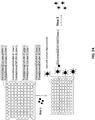
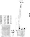
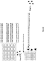


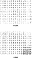


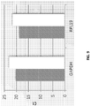


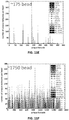
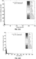


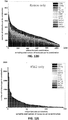
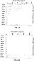
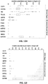

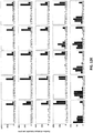
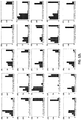
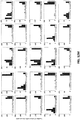
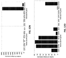
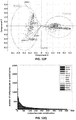
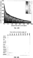
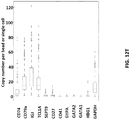
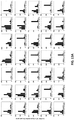

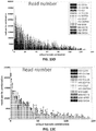
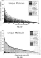


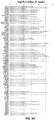

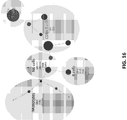
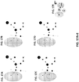


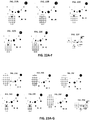

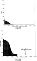
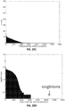
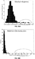
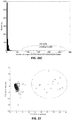
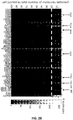
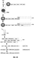


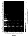


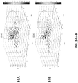

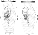
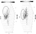

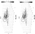

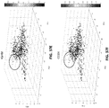
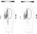
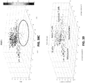
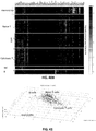

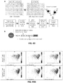
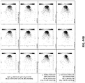
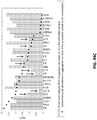
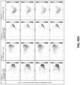
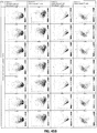


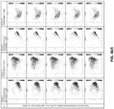

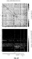
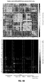
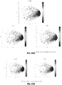
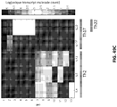

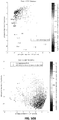
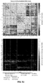
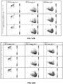
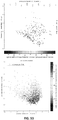

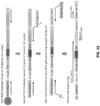
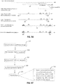

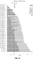
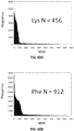
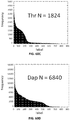
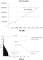
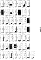


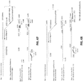

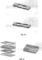
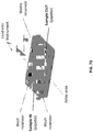
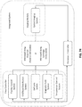
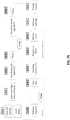
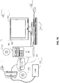
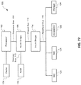

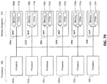

Classifications
-
- C—CHEMISTRY; METALLURGY
- C12—BIOCHEMISTRY; BEER; SPIRITS; WINE; VINEGAR; MICROBIOLOGY; ENZYMOLOGY; MUTATION OR GENETIC ENGINEERING
- C12Q—MEASURING OR TESTING PROCESSES INVOLVING ENZYMES, NUCLEIC ACIDS OR MICROORGANISMS; COMPOSITIONS OR TEST PAPERS THEREFOR; PROCESSES OF PREPARING SUCH COMPOSITIONS; CONDITION-RESPONSIVE CONTROL IN MICROBIOLOGICAL OR ENZYMOLOGICAL PROCESSES
- C12Q1/00—Measuring or testing processes involving enzymes, nucleic acids or microorganisms; Compositions therefor; Processes of preparing such compositions
- C12Q1/68—Measuring or testing processes involving enzymes, nucleic acids or microorganisms; Compositions therefor; Processes of preparing such compositions involving nucleic acids
- C12Q1/6869—Methods for sequencing
- C12Q1/6874—Methods for sequencing involving nucleic acid arrays, e.g. sequencing by hybridisation
-
- C—CHEMISTRY; METALLURGY
- C12—BIOCHEMISTRY; BEER; SPIRITS; WINE; VINEGAR; MICROBIOLOGY; ENZYMOLOGY; MUTATION OR GENETIC ENGINEERING
- C12Q—MEASURING OR TESTING PROCESSES INVOLVING ENZYMES, NUCLEIC ACIDS OR MICROORGANISMS; COMPOSITIONS OR TEST PAPERS THEREFOR; PROCESSES OF PREPARING SUCH COMPOSITIONS; CONDITION-RESPONSIVE CONTROL IN MICROBIOLOGICAL OR ENZYMOLOGICAL PROCESSES
- C12Q1/00—Measuring or testing processes involving enzymes, nucleic acids or microorganisms; Compositions therefor; Processes of preparing such compositions
- C12Q1/68—Measuring or testing processes involving enzymes, nucleic acids or microorganisms; Compositions therefor; Processes of preparing such compositions involving nucleic acids
- C12Q1/6876—Nucleic acid products used in the analysis of nucleic acids, e.g. primers or probes
- C12Q1/6888—Nucleic acid products used in the analysis of nucleic acids, e.g. primers or probes for detection or identification of organisms
-
- B—PERFORMING OPERATIONS; TRANSPORTING
- B01—PHYSICAL OR CHEMICAL PROCESSES OR APPARATUS IN GENERAL
- B01L—CHEMICAL OR PHYSICAL LABORATORY APPARATUS FOR GENERAL USE
- B01L3/00—Containers or dishes for laboratory use, e.g. laboratory glassware; Droppers
- B01L3/50—Containers for the purpose of retaining a material to be analysed, e.g. test tubes
- B01L3/508—Containers for the purpose of retaining a material to be analysed, e.g. test tubes rigid containers not provided for above
- B01L3/5085—Containers for the purpose of retaining a material to be analysed, e.g. test tubes rigid containers not provided for above for multiple samples, e.g. microtitration plates
- B01L3/50857—Containers for the purpose of retaining a material to be analysed, e.g. test tubes rigid containers not provided for above for multiple samples, e.g. microtitration plates using arrays or bundles of open capillaries for holding samples
-
- B—PERFORMING OPERATIONS; TRANSPORTING
- B01—PHYSICAL OR CHEMICAL PROCESSES OR APPARATUS IN GENERAL
- B01L—CHEMICAL OR PHYSICAL LABORATORY APPARATUS FOR GENERAL USE
- B01L7/00—Heating or cooling apparatus; Heat insulating devices
- B01L7/52—Heating or cooling apparatus; Heat insulating devices with provision for submitting samples to a predetermined sequence of different temperatures, e.g. for treating nucleic acid samples
-
- C—CHEMISTRY; METALLURGY
- C12—BIOCHEMISTRY; BEER; SPIRITS; WINE; VINEGAR; MICROBIOLOGY; ENZYMOLOGY; MUTATION OR GENETIC ENGINEERING
- C12N—MICROORGANISMS OR ENZYMES; COMPOSITIONS THEREOF; PROPAGATING, PRESERVING, OR MAINTAINING MICROORGANISMS; MUTATION OR GENETIC ENGINEERING; CULTURE MEDIA
- C12N15/00—Mutation or genetic engineering; DNA or RNA concerning genetic engineering, vectors, e.g. plasmids, or their isolation, preparation or purification; Use of hosts therefor
- C12N15/09—Recombinant DNA-technology
- C12N15/10—Processes for the isolation, preparation or purification of DNA or RNA
- C12N15/1034—Isolating an individual clone by screening libraries
- C12N15/1075—Isolating an individual clone by screening libraries by coupling phenotype to genotype, not provided for in other groups of this subclass
-
- C—CHEMISTRY; METALLURGY
- C12—BIOCHEMISTRY; BEER; SPIRITS; WINE; VINEGAR; MICROBIOLOGY; ENZYMOLOGY; MUTATION OR GENETIC ENGINEERING
- C12Q—MEASURING OR TESTING PROCESSES INVOLVING ENZYMES, NUCLEIC ACIDS OR MICROORGANISMS; COMPOSITIONS OR TEST PAPERS THEREFOR; PROCESSES OF PREPARING SUCH COMPOSITIONS; CONDITION-RESPONSIVE CONTROL IN MICROBIOLOGICAL OR ENZYMOLOGICAL PROCESSES
- C12Q1/00—Measuring or testing processes involving enzymes, nucleic acids or microorganisms; Compositions therefor; Processes of preparing such compositions
- C12Q1/68—Measuring or testing processes involving enzymes, nucleic acids or microorganisms; Compositions therefor; Processes of preparing such compositions involving nucleic acids
- C12Q1/6844—Nucleic acid amplification reactions
- C12Q1/686—Polymerase chain reaction [PCR]
-
- C—CHEMISTRY; METALLURGY
- C12—BIOCHEMISTRY; BEER; SPIRITS; WINE; VINEGAR; MICROBIOLOGY; ENZYMOLOGY; MUTATION OR GENETIC ENGINEERING
- C12Q—MEASURING OR TESTING PROCESSES INVOLVING ENZYMES, NUCLEIC ACIDS OR MICROORGANISMS; COMPOSITIONS OR TEST PAPERS THEREFOR; PROCESSES OF PREPARING SUCH COMPOSITIONS; CONDITION-RESPONSIVE CONTROL IN MICROBIOLOGICAL OR ENZYMOLOGICAL PROCESSES
- C12Q1/00—Measuring or testing processes involving enzymes, nucleic acids or microorganisms; Compositions therefor; Processes of preparing such compositions
- C12Q1/68—Measuring or testing processes involving enzymes, nucleic acids or microorganisms; Compositions therefor; Processes of preparing such compositions involving nucleic acids
- C12Q1/6876—Nucleic acid products used in the analysis of nucleic acids, e.g. primers or probes
- C12Q1/6881—Nucleic acid products used in the analysis of nucleic acids, e.g. primers or probes for tissue or cell typing, e.g. human leukocyte antigen [HLA] probes
-
- B—PERFORMING OPERATIONS; TRANSPORTING
- B01—PHYSICAL OR CHEMICAL PROCESSES OR APPARATUS IN GENERAL
- B01L—CHEMICAL OR PHYSICAL LABORATORY APPARATUS FOR GENERAL USE
- B01L2300/00—Additional constructional details
- B01L2300/02—Identification, exchange or storage of information
- B01L2300/023—Sending and receiving of information, e.g. using bluetooth
-
- B—PERFORMING OPERATIONS; TRANSPORTING
- B01—PHYSICAL OR CHEMICAL PROCESSES OR APPARATUS IN GENERAL
- B01L—CHEMICAL OR PHYSICAL LABORATORY APPARATUS FOR GENERAL USE
- B01L2300/00—Additional constructional details
- B01L2300/06—Auxiliary integrated devices, integrated components
- B01L2300/0627—Sensor or part of a sensor is integrated
-
- B—PERFORMING OPERATIONS; TRANSPORTING
- B01—PHYSICAL OR CHEMICAL PROCESSES OR APPARATUS IN GENERAL
- B01L—CHEMICAL OR PHYSICAL LABORATORY APPARATUS FOR GENERAL USE
- B01L2300/00—Additional constructional details
- B01L2300/08—Geometry, shape and general structure
- B01L2300/0809—Geometry, shape and general structure rectangular shaped
- B01L2300/0829—Multi-well plates; Microtitration plates
-
- C—CHEMISTRY; METALLURGY
- C12—BIOCHEMISTRY; BEER; SPIRITS; WINE; VINEGAR; MICROBIOLOGY; ENZYMOLOGY; MUTATION OR GENETIC ENGINEERING
- C12Q—MEASURING OR TESTING PROCESSES INVOLVING ENZYMES, NUCLEIC ACIDS OR MICROORGANISMS; COMPOSITIONS OR TEST PAPERS THEREFOR; PROCESSES OF PREPARING SUCH COMPOSITIONS; CONDITION-RESPONSIVE CONTROL IN MICROBIOLOGICAL OR ENZYMOLOGICAL PROCESSES
- C12Q2535/00—Reactions characterised by the assay type for determining the identity of a nucleotide base or a sequence of oligonucleotides
- C12Q2535/101—Sanger sequencing method, i.e. oligonucleotide sequencing using primer elongation and dideoxynucleotides as chain terminators
-
- C—CHEMISTRY; METALLURGY
- C12—BIOCHEMISTRY; BEER; SPIRITS; WINE; VINEGAR; MICROBIOLOGY; ENZYMOLOGY; MUTATION OR GENETIC ENGINEERING
- C12Q—MEASURING OR TESTING PROCESSES INVOLVING ENZYMES, NUCLEIC ACIDS OR MICROORGANISMS; COMPOSITIONS OR TEST PAPERS THEREFOR; PROCESSES OF PREPARING SUCH COMPOSITIONS; CONDITION-RESPONSIVE CONTROL IN MICROBIOLOGICAL OR ENZYMOLOGICAL PROCESSES
- C12Q2563/00—Nucleic acid detection characterized by the use of physical, structural and functional properties
- C12Q2563/149—Particles, e.g. beads
-
- C—CHEMISTRY; METALLURGY
- C12—BIOCHEMISTRY; BEER; SPIRITS; WINE; VINEGAR; MICROBIOLOGY; ENZYMOLOGY; MUTATION OR GENETIC ENGINEERING
- C12Q—MEASURING OR TESTING PROCESSES INVOLVING ENZYMES, NUCLEIC ACIDS OR MICROORGANISMS; COMPOSITIONS OR TEST PAPERS THEREFOR; PROCESSES OF PREPARING SUCH COMPOSITIONS; CONDITION-RESPONSIVE CONTROL IN MICROBIOLOGICAL OR ENZYMOLOGICAL PROCESSES
- C12Q2563/00—Nucleic acid detection characterized by the use of physical, structural and functional properties
- C12Q2563/159—Microreactors, e.g. emulsion PCR or sequencing, droplet PCR, microcapsules, i.e. non-liquid containers with a range of different permeability's for different reaction components
-
- C—CHEMISTRY; METALLURGY
- C12—BIOCHEMISTRY; BEER; SPIRITS; WINE; VINEGAR; MICROBIOLOGY; ENZYMOLOGY; MUTATION OR GENETIC ENGINEERING
- C12Q—MEASURING OR TESTING PROCESSES INVOLVING ENZYMES, NUCLEIC ACIDS OR MICROORGANISMS; COMPOSITIONS OR TEST PAPERS THEREFOR; PROCESSES OF PREPARING SUCH COMPOSITIONS; CONDITION-RESPONSIVE CONTROL IN MICROBIOLOGICAL OR ENZYMOLOGICAL PROCESSES
- C12Q2563/00—Nucleic acid detection characterized by the use of physical, structural and functional properties
- C12Q2563/185—Nucleic acid dedicated to use as a hidden marker/bar code, e.g. inclusion of nucleic acids to mark art objects or animals
-
- C—CHEMISTRY; METALLURGY
- C12—BIOCHEMISTRY; BEER; SPIRITS; WINE; VINEGAR; MICROBIOLOGY; ENZYMOLOGY; MUTATION OR GENETIC ENGINEERING
- C12Q—MEASURING OR TESTING PROCESSES INVOLVING ENZYMES, NUCLEIC ACIDS OR MICROORGANISMS; COMPOSITIONS OR TEST PAPERS THEREFOR; PROCESSES OF PREPARING SUCH COMPOSITIONS; CONDITION-RESPONSIVE CONTROL IN MICROBIOLOGICAL OR ENZYMOLOGICAL PROCESSES
- C12Q2600/00—Oligonucleotides characterized by their use
- C12Q2600/158—Expression markers
-
- C—CHEMISTRY; METALLURGY
- C12—BIOCHEMISTRY; BEER; SPIRITS; WINE; VINEGAR; MICROBIOLOGY; ENZYMOLOGY; MUTATION OR GENETIC ENGINEERING
- C12Q—MEASURING OR TESTING PROCESSES INVOLVING ENZYMES, NUCLEIC ACIDS OR MICROORGANISMS; COMPOSITIONS OR TEST PAPERS THEREFOR; PROCESSES OF PREPARING SUCH COMPOSITIONS; CONDITION-RESPONSIVE CONTROL IN MICROBIOLOGICAL OR ENZYMOLOGICAL PROCESSES
- C12Q2600/00—Oligonucleotides characterized by their use
- C12Q2600/16—Primer sets for multiplex assays
Definitions
- Multicellular masses such as tissues and tumors, may comprise a heterogeneous cellular milieu. These complex cellular environments may often display multiple phenoytpes, which may be indicative of multiple genotypes. Distilling multicellular complexity down to single cell variability is an important facet of understanding multicellular heterogeneity. This understanding may be important in the development of therapeutic regimens to combat diseases with multiple resistance genotypes.
- One aspect provided is a method, comprising obtaining a sample comprising a plurality of cells; labeling at least a portion of two or more polynucleotide molecules, complements thereof, or reaction products therefrom, from a first cell of the plurality and a second cell of the plurality with a first same cell label specific to the first cell and a second same cell label specific to the second cell; and a molecular label specific to each of the two or more polynucleotide molecules, complements thereof, or reaction products therefrom, wherein each molecular label of the two or more polynucleotide molecules, complements thereof, or reaction products therefrom, from the first cell are unique with respect to each other, and wherein each molecular label of the two or more polynucleotide molecules, complements thereof, or reaction products therefrom, from the second cell are unique with respect to each other.
- the method further comprises sequencing the at least a portion of two or more polynucleotide molecules, complements thereof, or reaction products therefrom. In some embodiments, the method further comprises analyzing sequence data from the sequencing to identify a number of individual molecules of the polynucleotides in a specific one of the cells.
- the cells are cancer cells. In some embodiments, the cells are infected with viral polynucleotides. In some embodiments, the cells are bacteria or fungi.
- the sequencing comprises sequencing with read lengths of at least 100 bases. In some embodiments, the sequencing comprises sequencing with read lengths of at least 500 bases.
- the polynucleotide molecules are mRNAs or micro RNAs, and the complements thereof and reaction products thereof are complements of and reaction products therefrom the mRNAs or micro RNAs.
- the molecular labels are on a bead.
- the label specific to an individual cell is on a bead.
- the label specific to an individual cell and the molecular labels are on beads.
- the method is performed at least in part in an emulsion. In some embodiments, the method is performed at least in part in a well or microwell of an array. In some embodiments, the presence of a polynucleotide that is associated with a disease or condition is detected.
- the disease or condition is a cancer. In some embodiments, at least a portion of a microRNA, complement thereof, or reaction product therefrom is detected. In some embodiments, the disease or condition is a viral infection. In some embodiments, the viral infection is from an enveloped virus. In some embodiments, the viral infection is from a non-enveloped virus. In some embodiments, the virus contains viral DNA that is double stranded. In some embodiments, the virus contains viral DNA that is single stranded.
- the virus is selected from the group consisting of a pox virus, a herpes virus, a vericella zoster virus, a cytomegalovirus, an Epstein-Barr virus, a hepadnavirus, a papovavirus, polyomavirus, and any combination thereof.
- the first cell is from a person not having a disease or condition and the second cell is from a person having the disease or condition.
- the persons are different.
- the persons are the same but cells are taken at different time points.
- the first cell is from a person having the disease or condition and the second cell is from the same person.
- the cells in the sample comprise cells from a tissue or organ.
- the cells in the sample comprise cells from a thymus, white blood cells, red blood cells, liver cells, spleen cells, lung cells, heart cells, brain cells, skin cells, pancreas cells, stomach cells, cells from the oral cavity, cells from the nasal cavity, colon cells, small intestine cells, kidney cells, cells from a gland, brain cells, neural cells, glial cells, eye cells, reproductive organ cells, bladder cells, gamete cells, human cells, fetal cells, amniotic cells, or any combination thereof.
- a solid support comprising a plurality of oligonucleotides each comprising a cellular label and a molecular label, wherein each cellular label of the plurality of oligonucleotides are the same, and each molecular label of the plurality of oligonucleotides are different; and wherein the solid support is a bead,the cellular label is specific to the solid support, the solid support, when placed at the center of a three dimensional Cartesian coordinate system, has oligonucleotides extending into at least seven of eight octants, or any combination thereof.
- the plurality of oligonucleotides further comprises at least one of a sample label; a universal label; and a target nucleic acid binding region.
- the solid support comprises the target nucleic acid binding region, wherein the target nucleic acid binding region comprises a sequence selected from the group consisting of a gene-specific sequence, an oligo-dT sequence, a random multimer, and any combination thereof.
- the solid support further comprises a target nucleic acid or complement thereof.
- the solid support comprises a plurality of target nucleic acids or complements thereof comprising from about 0.01% to about 100% of transcripts of a transcriptome of an organism or complements thereof, or from about 0.01% to about 100% of genes of a genome of an organism or complements thereof.
- the cellular labels of the plurality of oligonucleotides comprise a first random sequence connected to a second random sequence by a first label linking sequence; and the molecular labels of the plurality of oligonucleotides comprise random sequences.
- the solid support is selected from the group consisting of a polydimethylsiloxane (PDMS) solid support, a polystyrene solid support, a glass solid support, a polypropylene solid support, an agarose solid support, a gelatin solid support, a magnetic solid support, a pluronic solid support, and any combination thereof.
- the plurality of oligonucleotides comprise a linker comprising a linker functional group, and the solid support comprises a solid support functional group; wherein the solid support functional group and linker functional group connect to each other.
- the linker functional group and the solid support functional group are individually selected from the group consisting of C6, biotin, streptavidin, primary amine(s), aldehyde(s), ketone(s), and any combination thereof.
- molecular labels of the plurality of oligonucleotides comprise at least 15 nucleotides.
- kits comprising any of the solid supports described herein, and instructions for use.
- the kit further comprises a well.
- the well is comprised in an array.
- the well is a microwell.
- the kit further comprises a buffer.
- the kit is contained in a package.
- the package is a box.
- the package or box has a volume of 2 cubic feet or less. In some embodiments, the package or box has a volume of 1 cubic foot or less.
- One aspect provided is an emulsion comprising any of the solid supports described herein.
- composition comprising a well and any of the solid supports described herein.
- composition comprising a cell and any of the solid supports described herein.
- the emulsion or composition further comprises a cell.
- the cell is a single cell.
- the well is a microwell.
- the microwell has a volume ranging from about 1,000 ⁇ m 3 to about 120,000 ⁇ m 3 .
- One aspect provided is a method, comprising contacting a sample with any solid support disclosed herein, hybridizing a target nucleic acid from the sample to an oligonucleotide of the plurality of oligonucleotides.
- the method further comprises amplifying the target nucleic acid or complement thereof.
- the method further comprises sequencing the target nucleic acid or complement thereof, wherein the sequencing comprises sequencing the molecular label of the oligonucleotide to which the target nucleic acid or complement thereof is bound.
- the method further comprises determining an amount of the target nucleic acid or complement thereof, wherein the determining comprises quantifying levels of the target nucleic acid or complement thereof; counting a number of sequences comprising the same molecular label; or a combination thereof. In some embodiments, the method does not comprise aligning any same molecular labels or any same cellular labels.
- the amplifying comprises reverse transcribing the target nucleic acid. In some embodiments, the amplifying employs a method selected from the group consisting of: PCR, nested PCR, quantitative PCR, real time PCR, digital PCR, and any combination thereof.
- the amplifying is performed directly on the solid support; on a template transcribed from the solid support; or a combination thereof.
- the sample comprises a cell.
- the cell is a single cell.
- the contacting occurs in a well.
- the well is a microwell and is contained in an array of microwells.
- a device comprising a plurality of microwells, wherein each microwell of the plurality of microwells has a volume ranging from about 1,000 ⁇ m 3 to about 120,000 ⁇ m 3 . In some embodiments, each microwell of the plurality of microwells has a volume of about 20,000 ⁇ m 3 . In some embodiments, the plurality of microwells comprises from about 96 to about 200,000 microwells. In some embodiments, the microwells are comprised in a layer of a material. In some embodiments, at least about 10 % of the microwells further comprise a cell. In some embodiments, the device further comprises any of the solid supports described herein.
- the liquid handler delivers liquid to the plurality of microwells in about one second. In some embodiments, the liquid handler delivers liquid to the plurality of microwells from a single input port. In some embodiments, the apparatus further comprises a magnet. In some embodiments, the apparatus further comprises at least one of: an inlet port, an outlet port, a pump, a valve, a vent, a reservoir, a sample collection chamber, a temperature control apparatus, or any combination thereof. In some embodiments, the apparatus comprises the sample collection chamber, wherein the sample collection chamber is removable from the apparatus. In some embodiments, the apparatus further comprises an optical imager. In some embodiments, the optical imager produces an output signal which is used to control the liquid handler. In some embodiments, the apparatus further comprises a thermal cycling mechanism configured to perform a polymerase chain reaction (PCR) amplification of oligonucleotides.
- PCR polymerase chain reaction
- One aspect provided is a method of producing a clinical diagnostic test result, comprising producing the clinical diagnostic test result with any device or apparatus described herein; any solid support described herein; any method described herein; or any combination thereof.
- the clinical diagnostic test result is transmitted via a communication medium.
- One aspect provided is a method of making any of the solid supports described herein, comprising attaching to a solid support: a first polynucleotide comprising a first portion of the cellular label, and a first linker; and contacting a second polynucleotide comprising a second portion of the cellular label, a sequence complementary to the first liker, and the molecular label.
- the third polynucleotide further comprises a target nucleic acid binding region.
- an emulsion, microwell, or well contains only one cell. In some embodiments, from 1 to 2,000,000 emulsions, microwells, or wells each contain only one cell. In some embodiments, the method comprises distributing at most one cell into each emulsion, microwell, or well. In some embodiments, a single solid support and a single cell are distributed to an emulsion, microwell, or well. In some embodiments, from 1 to 2,000,000 emulsions, microwells, or wells each have distributed thereto one cell and one solid support. In some embodiments, the method comprises distributing at most one solid support per emulsion, microwell, or well.
- the method comprises distributing one solid support and one cell to each of from 1 to 2,000,000 microwells, emulsions, or wells.
- cell distribution is random or non-random.
- cell distribution is stochastic.
- a cell is distributed by a cell sorter.
- a cell is distributed by contacting one or more wells, microwells, or emulsions with a dilute solution of cells diluted so that at most one cell is distributed to the one or more wells, microwells, or emulsions.
- the target specific regions, target specific regions of the plurality of oligonucleotides, or the target specific region of the two or more polynucleotide molecules comprise sequences complementary to two or more targets of a target panel.
- the two or more targets of the target panel are biomarkers.
- the biomarkers are biomarkers for a disease or condition.
- the disease or condition is a cancer, an infection, a viral infection, an inflammatory disease, a neurodegenerative disease, a fungal disease, a bacterial infection, or any combination thereof.
- the panel comprises from: 2-50,000, 2-40,000, 2-30,000, 2-20,000, 2-10,000, 2-9000, 2-8,000, 2-7,000, 2-6,000, 2-5,000, 2-1,000, 2-800, 2-700, 2-600, 2-500, 2-400, 2-300, 2-200, 2-100, 2-75, 2-50, 2-40, 2-30, 2-20, 2-10, or 2-5 biomarkers.
- the methods, kits, and compositions comprise (a) stochastically labeling molecules in two or more samples with molecular barcodes to produce labeled molecules; and (b) detecting the labeled molecules.
- the molecular barcodes may comprise one or more target specific regions, label regions, sample index regions, universal PCR regions, adaptors, linkers, or a combination thereof.
- the labeled molecules may comprise a) a molecule region; b) a sample index region; and c) a label region.
- the molecule region may comprise at least a portion of the molecule from the molecular barcode was originally attached to.
- the molecule region may comprise a fragment of the molecule from the molecular barcode was originally attached to.
- the sample index region may be used to determine the source of the molecule region.
- the sample index region may be used to determine from which sample the molecule region originated from.
- the sample index region may be used to differentiate molecule regions from two or more different samples.
- the label region may be used to confer a unique identity to identical molecule regions originating from the same source.
- the label region may be used to confer a unique identity to identical molecule regions originating from the same sample.
- the method for analyzing molecules in a plurality of samples may comprise: a) producing a plurality of sample-tagged nucleic acids by: i) contacting a first sample comprising a plurality of nucleic acids with a plurality of first sample tags to produce a plurality of first sample-tagged nucleic acids; and ii) contacting a second sample comprising a plurality of nucleic acids with a plurality of second sample tags to produce a plurality of second sample-tagged nucleic acids, wherein the plurality of second sample tags are different from the first sample tags; b) contacting the plurality of sample-tagged nucleic acids with a plurality of molecular identifier labels to produce a plurality of labeled nucleic acids; and c) detecting at least a portion of the labeled nucleic acids, thereby determining a count of a plurality of nucleic acids in a plurality of samples.
- the plurality of samples may comprise a single cell.
- the method for analyzing molecules in a plurality of samples may comprise: a) producing a plurality of labeled nucleic acids comprising: i) contacting a first sample with a first plurality of sample tags, wherein the first plurality of sample tags comprises identical nucleic acid sequences; ii) contacting the first sample with a first plurality of molecular identifier labels may comprise different nucleic acid sequences, wherein contacting the first sample with the first plurality of sample tags or first plurality of molecular identifier labels occurs simultaneously or sequentially to produce a plurality of first-labeled nucleic acids; iii) contacting a second sample with a second plurality of sample tags, wherein the second plurality of sample tags may comprise identical nucleic acid sequences; iv) contacting the second sample with a second plurality of molecular identifier labels may comprise different nucleic acid sequences, wherein contacting the second sample with the second plurality of sample tags or second plurality of molecular identifier labels
- the method for analyzing molecules in a plurality of samples may comprise: a) contacting a plurality of samples may comprise two or more different nucleic acids with a plurality of sample tags and a plurality of molecular identifier labels to produce a plurality of labeled nucleic acids, wherein: i) the plurality of labeled nucleic acids may comprise two or more nucleic acids attached to two or more sample tags and two or more molecular identifier labels; ii) the sample tags attached to nucleic acids from a first sample of the plurality of samples are different from the sample tags attached to nucleic acid molecules from a second sample of the plurality of samples; and iii) two or more identical nucleic acids in the same sample are attached to two or more different molecular identifier labels; and b) detecting at least a portion of the labeled nucleic acids, thereby determining a count of two or more different nucleic acids in the plurality of samples.
- FIG. 56 depicts an exemplary workflow for the quantification of RNA molecules in a sample.
- RNA molecules (110) may be reverse transcribed to produce cDNA molecules (105) by the stochastic hybridization of a set of molecular identifier labels (115) to the polyA tail region of the RNA molecules.
- the molecular identifier labels (115) may comprise an oligodT region (120), label region (125), and universal PCR region (130).
- the set of molecular identifier labels may contain 960 different types of label regions.
- the labeled cDNA molecules (170) may be purified to remove excess molecular identifier labels (115).
- the labeled cDNA molecules (170) may be amplified to produce a labeled amplicon (180).
- Amplification may comprise multiplex PCR amplification.
- Amplification may comprise a multiplex PCR amplification with 96 multiplex primers in a single reaction volume.
- Amplification may comprise a custom primer (135) and a universal primer (140).
- the custom primer (135) may hybridize to a region within the cDNA (105) portion of the labeled cDNA molecule (170).
- the universal primer (140) may hybridize to the universal PCR region (130) of the labeled cDNA molecule (170).
- the labeled amplicons (180) may be further amplified by nested PCR.
- the nested PCR may comprise multiplex PCR with 96 multiplex primers in a single reaction volume.
- Nested PCR may comprise a custom primer (145) and a universal primer (140).
- the custom primer (135) may hybridize to a region within the cDNA (105) portion of the labeled amplicon (180).
- the universal primer (140) may hybridize to the universal PCR region (130) of the labeled amplicon (180).
- one or more adaptors (150, 155) may be attached to the labeled amplicon (180) to produce an adaptor-labeled amplicon (190).
- the one or more adaptors may be attached to the labeled amplicon (180) via ligation. As shown in Step 6, the one or more adaptors (150, 155) may be used to conduct one or more additional assays on the adaptor-labeled amplicon (190).
- the one or more adaptors (150, 155) may be hybridized to one or more primers (160, 165).
- the one or more primers (160, 165) be PCR amplification primers.
- the one or more primers (160, 165) may be sequencing primers.
- the one or more adaptors (150, 155) may be used for further amplification of the adaptor-labeled amplicons.
- the one or more adaptors (150, 155) may be used for sequencing the adaptor-labeled amplicon.
- FIG. 57 depicts an exemplary schematic of a workflow for analyzing nucleic acids from two or more samples.
- a method for analyzing nucleic acids from two or more samples may comprise selecting two or more genes for analysis and designing custom primers based on the selected genes (210). The method may further comprise supplementing one or more samples comprising nucleic acids (e.g. , RNA) with one or more spike-in controls (220).
- the nucleic acids in the sample may be amplified by multiplex RT-PCR (230) with molecular barcodes (or sample tags or molecular identifier labels) and the custom primers to produce labeled amplicons.
- the labeled amplicons may further treated with one or more sequencing adaptors to produce adaptor labeled amplicons (240).
- the adaptor labeled amplicons can be analyzed (250). As shown in FIG. 57 , analysis of the labeled amplicons (250) may comprise one or more of (1) detection of a universal PCR primer seq, polyA and/or molecular barcode (or sample tag, molecular identifier label); (2) map read on the end of the adaptor labeled amplicons (e.g.
- adaptor and/or barcode e.g ., molecular barcode, sample tag, molecular identifier label
- count and/or summarize the number of different adaptor labeled amplicons e.g ., molecular barcode, sample tag, molecular identifier label
- FIG. 67 shows a schematic of a workflow for stochastically labeling nucleic acids with molecular barcodes (1220).
- RNA molecules may be stochastically labeled with a set of molecular barcodes (1220).
- the molecular barcodes (1220) may comprise a target binding region (1221), label region (1222), sample index region (1223) and universal PCR region (1224).
- the target binding region comprises an oligodT sequence that hybridizes to a polyA sequence in the RNA molecules.
- the label region (1222) may contain a unique sequence that may be used to distinguish two or more different molecular barcodes.
- the label region When the molecular barcode hybridizes to an RNA molecule, the label region may be used to confer a unique identity to identical RNA molecules.
- the sample index region (1223) may be identical for a set of molecular barcodes. The sample index region (1223) may be used to distinguish labeled nucleic acids from different samples.
- the universal PCR region (1224) may serve as a primer binding site for amplification of the labeled molecules.
- the RNA molecules Once the RNA molecules are labeled with the molecular barcodes, the RNA molecules may be reverse transcribed to produce labeled cDNA molecules (1230) containing a cDNA copy of the RNA molecule (1210) and the molecular barcode (1220).
- the labeled cDNA molecules may be amplified by multiplex PCR. Multiplex PCR of the labeled cDNA molecules may be performed by using a first set of forward primers (F1, 1235 in FIG. 67 ) and universal primers (1240) in a single reaction volume to produce labeled amplicons (1245). As shown in Step 4 of FIG. 67 , the labeled amplicons may be further amplified by multiplex PCR using nested primers.
- Nested primer amplification of the labeled amplicons may be performed by using a second set of forward primers (F2, 1250 in FIG. 67 ) and universal primers (1240) in a single reaction volume to produce labeled nested PCR amplicons.
- the F2 primers (1250) contain an adaptor (1251) and a target binding region (1252).
- the target binding region (1252) of the F2 primers may hybridize to the labeled amplicons and may prime amplification of the labeled amplicons.
- the adaptor (1251) and the universal PCR region (1224) of the nested PCR amplicons may be used in the sequencing of the labeled nested PCR amplicons.
- the amplicons may be sequenced by MiSeq. Alternatively, the amplicons may be sequenced by HiSeq.
- FIG. 68 shows a schematic of a workflow for stochastically labeling nucleic acids.
- RNA molecules (1305) may be stochastically labeled with a set molecular barcodes (1320).
- the molecular barcodes may comprise a target binding region (1321), label region (1322), and universal PCR region (1323).
- the RNA molecules (1305) may be reverse transcribed to produce labeled cDNA molecules (1325) comprising a cDNA copy of the RNA molecule (1310) and the molecular barcode (1320).
- the labeled cDNA molecules may be purified by Ampure bead purification to remove excess oligos (e.g. , molecular barcodes).
- the labeled amplicons may be amplified by multiplex PCR. Multiplex PCR of the labeled cDNA molecules may be performed by using a first set of forward primers (F1, 1330 in FIG. 68 ) and universal primers (1335) in a single reaction volume to produce labeled amplicons (1360).
- the labeled amplicons may be further amplified by multiplex PCR using nested primers.
- Nested primer amplification of the labeled amplicons may be performed by using a second set of forward primers (F2, 1340 in FIG. 68 ) and sample index primers (1350) in a single reaction volume to produce labeled nested PCR amplicons.
- the F2 primers (1340) contain an adaptor (1341) and a target binding region (1342).
- the target binding region (1342) of the F2 primers may hybridize to the labeled amplicons and may prime amplification of the labeled amplicons.
- the sample index primers (1350) may comprise a universal primer region (1351), sample index region (1352), and adaptor region (1353). As shown in Step 4 of FIG.
- the universal primer region (1351) of the sample index primer may hybridize to the universal PCR region of the labeled amplicons.
- the sample index region (1352) of the sample index primer may be used to distinguish two or more samples.
- the adaptor regions (1341, 1353) may be used to sequence the labeled nested PCR amplicons.
- the amplicons may be sequenced by MiSeq. Alternatively, the amplicons may be sequenced by HiSeq.
- the one or more libraries may comprise a plurality of labeled molecules.
- the one or more libraries may comprise a plurality of labeled amplicons.
- the one or more libraries may comprise a plurality of enriched molecules or a derivative thereof ( e.g. , labeled molecules, labeled amplicons).
- the method of producing one or more libraries comprises (a) stochastically labeling a plurality of molecules from two or more samples to produce a plurality of labeled molecules, wherein the labeled molecules comprise a molecule region, a sample index region, and label region; and (b) producing one or more libraries from the plurality of labeled molecules, wherein (i) the one or more libraries comprise two or more different labeled molecules, (ii) the two or more different labeled molecules differ by the molecule region, sample index region, label region, or a combination thereof.
- the method for producing one or more libraries may comprise: a) producing a plurality of sample-tagged nucleic acids by: i) contacting a first sample comprising a plurality of nucleic acids with a plurality of first sample tags to produce a plurality of first sample-tagged nucleic acids; and ii) contacting a second sample comprising a plurality of nucleic acids with a plurality of second sample tags to produce a plurality of second sample-tagged nucleic acids, wherein the plurality of first sample tags are different from the second sample tags; and b) contacting the plurality of sample-tagged nucleic acids with a plurality of molecular identifier labels to produce a plurality of labeled nucleic acids, thereby producing a labeled nucleic acid library.
- the contacting to a sample can be random or non-random.
- the contacting of a sample with sample tags can be a random or non-random contacting.
- the sample is contacted with sample tags randomly.
- the sample is contacted with sample tags non-randomly.
- the contacting to a plurality of nucleic acids can be random or non-random.
- the contacting of a plurality of nucleic acids with sample tags can be a random or non-random contacting.
- the plurality of nucleic acids is contacted with sample tags randomly.
- the plurality of nucleic acids is contacted with sample tags non-randomly.
- the method of producing the one or more sets of labeled beads may comprise attaching one or more nucleic acids to one or more beads, thereby producing one or more sets of labeled beads.
- the one or more nucleic acids may comprise one or more molecular barcodes.
- the one or more nucleic acids may comprise one or more sample tags.
- the one or more nucleic acids may comprise one or more molecular identifier labels.
- the one or more nucleic acids may comprise a) a primer region; b) a sample index region; and c) a linker or adaptor region.
- the one or more nucleic acids may comprise a) a primer region; b) a label region; and c) a linker or adaptor region.
- the one or more nucleic acids may comprise a) a sample index region; and b) a label region.
- the one or more nucleic acids may further comprise a primer region.
- the one or more nucleic acids may further comprise a target specific region.
- the one or more nucleic acids may further comprise a linker region.
- the one or more nucleic acids may further comprise an adaptor region.
- the one or more nucleic acids may further comprise a sample index region.
- the one or more nucleic acids may further comprise a label region.
- the method of selecting a custom primer for analyzing molecules in a plurality of samples may comprise: a) a first pass, wherein primers chosen may comprise: i) no more than three sequential guanines, no more than three sequential cytosines, no more than four sequential adenines, and no more than four sequential thymines; ii) at least 3, 4, 5, or 6 nucleotides that are guanines or cytosines; and iii) a sequence that does not easily form a hairpin structure; b) a second pass, comprising: i) a first round of choosing a plurality of sequences that have high coverage of all transcripts; and ii) one or more subsequent rounds, selecting a sequence that has the highest coverage of remaining transcripts and a complementary score with other chosen sequences no more than 4; and c) adding sequences to a picked set until coverage saturates or total number of customer primers is less than or equal to about 96
- kits for use in analyzing two or more molecules from two or more samples may comprise (a) a first container comprising a first set of molecular barcodes, wherein (i) a molecular barcode of the first set of molecular barcodes comprise a sample index region and a label region; (ii) the sample index region of two or more barcodes of the first set of molecular barcodes are the same; and (iii) the label region of two or more barcodes of the first set of molecular barcodes are different; and (b) a second container comprising a second set of molecular barcodes, wherein (i) a molecular barcode of the second set of molecular barcodes comprise a sample index region and a label region; (ii) the sample index region of two or more barcodes of the second set of molecular barcodes are the same; (iii) the label region of two or more barcodes of the second set of molecular barcodes are are
- the kit comprises: a) a plurality of beads, wherein one or more beads of the plurality of beads may comprise at least one of a plurality of nucleic acids, wherein at least one of a plurality nucleic acids may comprise: i) at least one primer sequence, wherein the primer sequence of at least one of the plurality of nucleic acids is the same for the plurality of beads; ii) a bead-specific sequence, wherein the bead-specific sequence of any one of the plurality of nucleic acids is the same, and wherein the bead-specific sequence is different for any one of the plurality of beads; and iii) a stochastic sequence, wherein the stochastic sequence is different for any one of the plurality of nucleic acids; b) a primer may comprise a sequence complementary to the primer sequence; and c) one or more amplification agents suitable for nucleic acid amplification.
- the kit comprises: a) a first container comprising a first set of sample tags, wherein(i) a sample tag of the first set of sample tags comprises a sample index region; and (ii) the sample index regions of the sample tags of the first set of sample tags are at least about 80% identical; and b) a second container comprising a first set of molecular identifier labels, wherein (i) a molecular identifier label of the first set of molecular identifier labels comprises a label region; and (ii) at least about 30% of the label regions of the total molecular identifier labels of the first set of molecular identifier labels are different
- kits and compositions are provided for stochastic labeling of nucleic acids in a plurality of samples or in a complex nucleic acid preparation. These methods, kits and compositions find use in unraveling mechanisms of cellular response, differentiation or signal transduction and in performing a wide variety of clinical measurements.
- the methods disclosed herein comprise attaching one or more molecular barcodes, sample tags, and/or molecular identifier labels to two or more molecules from two or more samples.
- the molecular barcodes, sample tags and/or molecular identifier labels may comprise one or more oligonucleotides.
- attachment of molecular barcodes, sample tags, and/or molecular identifier labels to the molecules comprises stochastic labeling of the molecules. Methods for stochastically labeling molecules may be found, for example, in U.S. Serial Numbers 12/969,581 and 13/327,526 .
- the stochastic labeling method comprises the random attachment of a plurality of the tag and label oligonucleotides to one or more molecules.
- the molecular barcodes, sample tags, and/or molecular identifier labels are provided in excess of the one or more molecules to be labeled.
- each individual molecule to be labeled has an individual probability of attaching to the plurality of the molecular barcodes, sample tags, and/or molecular identifier labels.
- the probability of each individual molecule to be labeled attaching to a particular molecular barcodes, sample tags, and/or molecular identifier labels may be about the same as any other individual molecule to be labeled.
- the probability of any of the molecules in a sample finding any of the tags and labels is assumed to be equal, an assumption that may be used in mathematical calculations to estimate the number of molecules in the sample.
- the probability of attaching may be manipulated by, for example electing tags and labels with different properties that would increase or decrease the binding efficiency of that molecular barcodes, sample tags, and/or molecular identifier labels with an individual molecule.
- the tags and labels may also be varied in numbers to alter the probability that a particular molecular barcodes, sample tags, and/or molecular identifier labels will find a binding partner during the stochastic labeling. For example, one label is overrepresented in a pool of labels, thereby increasing the chances that the overrepresented label finds at least one binding partner.
- the methods disclosed herein may further comprise combining two or more samples.
- the methods disclosed herein may further comprise combining one or more molecules from two or more samples.
- the methods disclosed herein comprise combining a first sample and a second sample.
- the two or more samples may be combined after conducting one or more stochastic labeling procedures.
- the two or more samples may be combined after attachment of one or more sets of molecular barcodes to two or more molecules from the two or more samples.
- the two or more samples may be combined after attachment of one or more sets of sample tags to two or more molecules from the two or more samples.
- the two or more samples may be combined after attachment of one or more sets of molecular identifier labels to two or more molecules from the two or more samples.
- the first and second samples are combined prior to contact with the plurality of molecular identifier labels.
- the two or more samples may be combined prior to conducting one or more stochastic labeling procedures.
- the two or more samples may be combined prior to attachment of one or more sets of molecular barcodes to two or more molecules from the two or more samples.
- the two or more samples may be combined prior to attachment of one or more sets of sample tags to two or more molecules from the two or more samples.
- the two or more samples may be combined prior to attachment of one or more sets of molecular identifier labels to two or more molecules from the two or more samples.
- the two or more samples may be combined after conducting one or more assays on two or more molecules or derivatives thereof (e.g. , labeled molecules, amplicons) from the two or more samples.
- the one or more assays may comprise one or more amplification reactions.
- the one or more assays may comprise one or more enrichment assays.
- the one or more assays may comprise one or more detection assays.
- the first and second samples are combined after detecting the labeled nucleic acids.
- the two or more samples may be combined prior to conducting one or more assays on two or more molecules or derivatives thereof (e.g ., labeled molecules, amplicons) from the two or more samples.
- the one or more assays may comprise one or more amplification reactions.
- the one or more assays may comprise one or more enrichment assays.
- the one or more assays may comprise one or more detection assays.
- the first and second samples are combined prior to detecting the labeled nucleic acids.
- compositions and methods for multiplex sequence analysis from single cells comprising compositions and methods for multiplex sequence analysis from single cells.
- the methods and compositions of the present disclosure provide for the use of solid supports.
- the methods, kits, and compositions disclosed herein comprise a support.
- support may be used interchangeably and refer to a material or group of materials having a rigid or semi-rigid surface or surfaces.
- a support may refer to any surface that is transferable from solution to solution or forms a structure for conducting oligonucleotide-based assays.
- the support or substrate may be a solid support. Alternatively, the support is a non-solid support.
- a support may refer to an insoluble, semi-soluble, or insoluble material.
- a support may be referred to as “functionalized” when it includes a linker, a scaffold, a building block, or other reactive moiety attached thereto, whereas a solid support may be “nonfunctionalized” when it lack such a reactive moiety attached thereto.
- the support may be employed free in solution, such as in a microtiter well format; in a flow-through format, such as in a column; or in a dipstick.
- the support or substrate may comprise a membrane, paper, plastic, coated surface, flat surface, glass, slide, chip, or any combination thereof.
- at least one surface of the support may be substantially flat, although in some embodiments it may be desirable to physically separate synthesis regions for different compounds with, for example, wells, raised regions, pins, etched trenches, or the like.
- the solid support(s) may take the form of resins, gels, microspheres, or other geometric configurations.
- the solid support(s) comprises silica chips, microparticles, nanoparticles, plates, and arrays.
- Solid supports may include beads (e.g ., silica gel, controlled pore glass, magnetic beads, Dynabeads, Wang resin; Merrifield resin, Sephadex/Sepharose beads, cellulose beads, polystyrene beads etc.), capillaries, flat supports such as glass fiber filters, glass surfaces, metal surfaces (steel, gold silver, aluminum, silicon and copper), glass supports, plastic supports, silicon supports, chips, filters, membranes, microwell plates, slides, or the like.
- plastic materials including multiwell plates or membranes (e.g. , formed of polyethylene, polypropylene, polyamide, polyvinylidenedifluoride), wafers, combs, pins or needles ( e.g. , arrays of pins suitable for combinatorial synthesis or analysis) or beads in an array of pits or nanoliter wells of flat surfaces such as wafers (e.g., silicon wafers), wafers with pits with or without filter bottoms.
- multiwell plates or membranes e.g.
- Patents that describe synthesis techniques in specific embodiments include U.S. Patent Nos. 5,412,087 , 6,147,205 , 6,262,216 , 6,310,189 , 5,889,165 , and 5,959,098 .
- Nucleic acid arrays are described in many of the above patents, but many of the same techniques may be applied to polypeptide arrays. Additional exemplary substrates are disclosed in U.S. Patent No. 5,744,305 and US Patent Pub. Nos. 20090149340 and 20080038559 .
- a solid support may comprise a molecular scaffold.
- exemplary molecular scaffolds may include antibodies, antigens, affinity reagents, polypeptides, nucleic acids, cellular organelles, and the like.
- Molecular scaffolds may be linked together (e.g. , a solid support may comprise a plurality of connected molecular scaffolds).
- Molecular scaffolds may be linked together by an amino acid linker, a nucleic acid linker, a small molecule linkage (e.g. , biotin and avidin), and/or a matrix linkage ( e.g. , PEG or glycerol). Linkages may be non-covalent. Linkages may be covalent. In some instances, molecular scaffolds may not be linked. A plurality of individual molecular scaffolds may be used in the methods of the disclosure.
- a support may comprise a nanoparticle.
- the nanoparticle may be a nickel, gold, silver, carbon, copper, silicate, platinum cobalt, zinc oxide, silicon dioxide crystalline, and/or silver nanoparticle.
- the nanoparticle may be a gold nanoparticle embedded in a porous manganese oxide.
- the nanoparticle may be an iron nanoparticle.
- the nanoparticle may be a nanotetrapod studded with nanoparticles of carbon.
- a support may comprise a polymer.
- a polymer may comprise a matrix.
- a matrix may further comprise one or more beads.
- a polymer may comprise PEG, glycerol, polysaccharide, or a combination thereof.
- a polymer may be a plastic, rubber, nylon, silicone, neoprene, and/or polystyrene.
- a polymer may be a natural polymer. Examples of natural polymers include, but are not limited to, shellac, amber, wool, silk, cellulose, and natural rubber.
- a polymer may be a synthetic polymer.
- synthetic polymers include, but are not limited to, synthetic rubber, phenol formaldehyde resin (or Bakelite), neoprene, nylon, polyvinyl chloride (PVC or vinyl), polystyrene, polyethylene, polypropylene, polyacrylonitrile, PVB, and silicone.
- a support may be a semi-solid support.
- a support may comprise a gel (e.g. , a hydrogel).
- the terms "hydrogel”, “gel” and the like, are used interchangeably herein and may refer to a material which is not a readily flowable liquid and not a solid but a gel which gel is comprised of from 0.5% or more and preferably less than 40% by weight of gel forming solute material and from 95% or less and preferably more than 55% water.
- the gels of the invention may be formed by the use of a solute which is preferably a synthetic solute (but could be a natural solute, e.g.
- the gel may be the basic structure of the hydrogel patch of the invention will include additional components beyond the gel forming solute material and water such as an enzyme and a salt which components are further described herein.
- the gel may be a polymer gel.
- a solid support may comprise a structured nanostructure.
- the structured nanostructure may comprise capture containers (e.g. , a miniaturized honeycomb) which may comprise the oligonucleotides to capture the cell and/or contents of the cell.
- structured nanostructures may not need the addition of exogenous reagents.
- the support comprises a bead.
- a bead may encompass any type of solid or hollow sphere, ball, bearing, cylinder, or other similar configuration composed of plastic, ceramic, metal, or polymeric material onto which a nucleic acid may be immobilized ( e.g. , covalently or non-covalently).
- a bead may comprise nylon string or strings.
- a bead may be spherical in shape.
- a bead may be non-spherical in shape. Beads may be unpolished or, if polished, the polished bead may be roughened before treating, ( e.g. , with an alkylating agent).
- a bead may comprise a discrete particle that may be spherical ( e.g.
- Beads may comprise a variety of materials including, but not limited to, paramagnetic materials, ceramic, plastic, glass, polystyrene, methylstyrene, acrylic polymers, titanium, latex, sepharose, cellulose, nylon and the like.
- a bead may be attached to or embedded into one or more supports.
- a bead may be attached to a gel or hydrogel.
- a bead may be embedded into a gel or hydrogel.
- a bead may be attached to a matrix.
- a bead may be embedded into a matrix.
- a bead may be attached to a polymer.
- a bead may be embedded into a polymer.
- the spatial position of a bead within the support e.g.
- beads include, but are not limited to, streptavidin beads, agarose beads, magnetic beads, Dynabeads®, MACS® microbeads, antibody conjugated beads (e.g. , anti-immunoglobulin microbead), protein A conjugated beads, protein G conjugated beads, protein A/G conjugated beads, protein L conjugated beads, oligodT conjugated beads, silica beads, silica-like beads, anti-biotin microbead, anti-fluorochrome microbead, and BcMagTM Carboxy-Terminated Magnetic Beads.
- the diameter of the beads may be about 5 ⁇ m, 10 ⁇ m, 20 ⁇ m, 25 ⁇ m, 30 ⁇ m, 35 ⁇ m, 40 ⁇ m, 45 ⁇ m or 50 ⁇ m.
- a bead may refer to any three dimensional structure that may provide an increased surface area for immobilization of biological particles and macromolecules, such as DNA and RNA.
- a support may be porous.
- a support may be permeable or semi-permeable.
- a support may be solid.
- a support may be semi-solid.
- a support may be malleable.
- a support may be flexible.
- a support may be molded into a shape. For example, a support may be placed over an object and the support may take the shape of the object. In some instances, the support is placed over an organ and takes the shape of the organ. In some instances, the support is produced by 3D-printing.
- the support e.g ., beads, nanoparticles
- the support may be at least about 0.1, 0.5, 1, 2, 3, 4, 5, 6, 7, 8, 9, 10, 20, 30, 100, 500, 1000, or 2000 or more micrometers in diameter.
- the solid supports e.g ., beads
- the diameter of the bead may be about 20 microns.
- a solid support comprises a dendrimer.
- a dendrimer may be smaller than a bead.
- a dendrimer may be subcellular.
- a dendrimer may be less than 1, 0.9, 0.8, 0.7, 0.6, 0.5, 0.4, 0.3, 0.2, or 0.1 micron in diameter.
- a dendrimer may be less than 0.09, 0.08, 0.07, 0.06, 0.05, 0.04, 0.03, or 0.01 micron in diameter
- a dendrimer may comprise three major portions, a core, an inner shell, and an outer shell.
- a dendrimer may be synthesized to have different functionality in each of these portions.
- a dendrimer may be synthetically processed.
- a dendrimer may be synthesized by divergent synthesis.
- Divergent synthesis may comprise assembling a dendrimer from a multifunctional core, which is extended outward by a series of reactions.
- Divergent synthesis may comprise a series of Michael reactions.
- a dendrimer may be synthesized by convergent synthesis.
- Convergent synthesis may comprise building dendrimers from small molecules that end up at the surface of the sphere, and reactions may proceed inward and are eventually attached to a core. Dendrimers may also be prepared by click chemistry.
- Click chemistry may comprise Diels-Alder reactions, thiol-yne reactions, azide-alkyne reactions, or a combination thereof.
- dendrimers include, but are not limited to, poly(amidoamine) (PAMAM) dendrimer, PEG-core denderimer, phosphorous dendrimer, polypropylenimine dendrimer, and polylysine dendrimer.
- PAMAM poly(amidoamine)
- a dendrimer may be a chiral dendrimer.
- a dendrimer may be an achiral dendrimer.
- a solid support may comprise a portion of a dendrimer.
- the portion of the dendrimer may comprise a dendron.
- a dendron may comprise monodisperse wedge-shaped dendrimer sections with multiple terminal groups and a single reaction function at the focal point.
- a solid support may comprise a polyester dendrom.
- dendrons include, but are not limited to, polyester-8-hydroxyl-1-acetylene bis-MPA dendron, polyester-16-hydroxyl-1-acetylene bis-MPA dendron, polyester-32-hydroxyl-1-acetylene bis-MPA dendron, polyester-8-hydroxyl-1-carboxyl bis-MPA dendron, polyester-16-hydroxyl-1-carboxyl bis-MPA dendron, and polyester-32-hydroxyl-1-carboxyl bis-MPA dendron.
- a solid support may comprise a hyberbranched polymer.
- a hyperbranched polymer may comprise polydisperse dendritic macromolecules that possess dendrimer-like properties. Often, hyberbranched polymers are prepared in a single synthetic polymerization step. The hyperbranched polymer may be based on 2,2-bis(hydroxymethyl)propanoic acid (bis-MPA) monomer. Examples of hyperbranched polymers include, but are not limited to, hyperbranched bis-MPA polyester-16-hydroxyl, hyperbranched bis-MPA polyester-32-hydroxyl, and hyperbranched bis-MPA polyester-64-hydroxyl.
- the solid support may be an array or microarray.
- the solid support may comprise discrete regions.
- the solid support may be an addressable array.
- the array comprises a plurality of probes fixed onto a solid surface.
- the plurality of probes enables hybridization of the labeled-molecule and/or labeled-amplicon to the solid surface.
- the plurality of probes comprises a sequence that is complementary to at least a portion of the labeled-molecule and/or labeled-amplicon.
- the plurality of probes comprises a sequence that is complementary to at least a portion of the sample tag, molecular identifier label, nucleic acid, or a combination thereof.
- the plurality of probes comprises a sequence that is complementary to the junction formed by the attachment of the sample tag or molecular identifier label to the nucleic acid.
- the array may comprise one or more probes.
- the probes may be in a variety of formats.
- the array may comprise a probe comprising a sequence that is complementary to at least a portion of the target nucleic acid and a sequence that is complementary to the unique identifier region of a sample tag or molecular identifier label, wherein the sample tag or molecular identifier label comprises an oligonucleotide.
- the sequence that is complementary to at least a portion of the target nucleic acid may be attached to the array.
- the sequence that is complementary to the unique identifier region may be attached to the array.
- the array may comprise a first probe comprising a sequence that is complementary to at least a portion of the target nucleic acid and a second probe that is complementary to the unique identifier region.
- a stochastically labeled nucleic acid may hybridize to the arrays.
- the junction of the unique identifier region and the target nucleic acid of the stochastically labeled nucleic acid may hybridize to the probe on the array.
- Different regions of the stochastically labeled nucleic acid may hybridize to two or more probes on the array.
- the array probes may be in many different formats.
- the array probes may comprise a sequence that is complementary to a unique identifier region, a sequence that is complementary to the target nucleic acid, or a combination thereof.
- Hybridization of the stochastically labeled nucleic acid to the array may occur by a variety of ways.
- two or more nucleotides of the stochastically labeled nucleic acid may hybridize to one or more probes on the array.
- the two or more nucleotides of the stochastically labeled nucleic acid that hybridize to the probes may be consecutive nucleotides, non-consecutive nucleotides, or a combination thereof.
- the stochastically labeled nucleic acid that is hybridized to the probe may be detected by any method known in the art.
- the stochastically labeled nucleic acids may be directly detected.
- Directly detecting the stochastically labeled nucleic acid may comprise detection of a fluorophore, hapten, or detectable label.
- the stochastically labeled molecules may be indirectly detected.
- Indirect detection of the stochastically labeled nucleic acid may comprise ligation or other enzymatic or non-enzymatic methods.
- the array may be in a variety of formats.
- the array may be in a 16-, 32-, 48-, 64-, 80-, 96-, 112-, 128-, 144-, 160-, 176-, 192-, 208-, 224-, 240-, 256-, 272-, 288-, 304-, 320-, 336-, 352-, 368-, 384-, or 400-format.
- the array is in an 8x60K, 4x180K, 2x400K, 1x1M format.
- the array is in an 8x15K, 4x44K, 2x10⁵K, 1x244K format.
- the array may comprise a single array.
- the single array may be on a single substrate.
- the array is on multiple substrates.
- the array may comprise multiple formats.
- the array may comprise a plurality of arrays.
- the plurality of arrays may comprise two or more arrays.
- the plurality of arrays may comprise at least about 2, 3, 4, 5, 6, 7, 8, 9, 10, 11, 12, 13, 14, 15, 16, 17, 18, 19, 20, 25, 30, 35, 40, 45, 50, 55, 60, 65, 70, 75, 80, 85, 90, 95, or 100 arrays.
- at least two arrays of the plurality of arrays are identical.
- at least two arrays of the plurality of arrays are different.
- the array comprises symmetrical chambered areas.
- the array comprises 0.5 x 0.5 millimeters (mm), 1 x 1 mm, 1.5 x 1.5 mm, 2 x 2 mm, 2.5 x 2.5 mm, 3 x 3 mm, 3.5 x 3.5 mm, 4 x 4 mm, 4.5 x 4.5 mm, 5 x 5 mm, 5.5 x 5.5 mm, 6 x 6 mm, 6.5 x 6.5 mm, 7 x 7 mm, 7.5 x 7.5 mm, 8 x 8 mm, 8.5 x 8.5 mm, 9 x 9 mm, 9.5 x 9.5 mm, 10 x 10 mm, 10.5 x 10.5 mm, 11x11 mm, 11.5 x 11.5 mm, 12 x 12 mm, 12.5 x 12.5 mm, 13 x 13 mm, 13.5 x 13.5 mm, 14 x 14 mm, 14.5 x 14.5 mm, 15
- the array comprises 6.5 x 6.5 mm chambered areas.
- the array comprises asymmetrical chambered areas.
- the array comprises 6.5 x 0.5 mm, 6.5 x 1 mm, 6.5 x 1.5 mm, 6.5 x 2 mm, 6.5 x 2.5 mm, 6.5 x 3 mm, 6.5 x 3.5 mm, 6.5 x 4 mm, 6.5 x 4.5 mm, 6.5 x 5 mm, 6.5 x 5.5 mm, 6.5 x 6 mm, 6.5 x 6.5 mm, 6.5 x 7 mm, 6.5 x 7.5 mm, 6.5 x 8 mm, 6.5 x 8.5 mm, 6.5 x 9 mm, 6.5 x 9.5 mm, 6.5 x 10 mm, 6.5 x 10.5 mm, 6.5 x 11 mm, 6.5 x 11.5 mm, 6.5 x 12 mm, 6.5 x 12.5 mm, 6.5 x 10 mm,
- the array may comprise at least about 1 micron ( ⁇ m), 2 ⁇ m, 3 ⁇ m, 4 ⁇ m, 5 ⁇ m, 6 ⁇ m, 7 ⁇ m, 8 ⁇ m, 9 ⁇ m, 10 ⁇ m, 15 ⁇ m, 20 ⁇ m, 25 ⁇ m, 30 ⁇ m, 35 ⁇ m, 40 ⁇ m, 45 ⁇ m, 50 ⁇ m, 55 ⁇ m, 60 ⁇ m, 65 ⁇ m, 70 ⁇ m, 75 ⁇ m, 80 ⁇ m, 85 ⁇ m, 90 ⁇ m, 95 ⁇ m, 100 ⁇ m, 125 ⁇ m, 150 ⁇ m, 175 ⁇ m, 200 p,m , 225 ⁇ m, 250 ⁇ m, 275 ⁇ m, 300 ⁇ m, 325 ⁇ m, 350 ⁇ m, 375 ⁇ m, 400 ⁇ m, 425 ⁇ m, 450 ⁇ m, 475 ⁇ m, or 500 ⁇ m spots. In some instances, the array comprises 70 ⁇ m spots
- the array may comprise at least about 1 ⁇ m, 2 ⁇ m, 3 ⁇ m, 4 ⁇ m, 5 ⁇ m, 6 ⁇ m, 7 ⁇ m, 8 ⁇ m, 9 ⁇ m, 10 ⁇ m, 15 ⁇ m, 20 ⁇ m, 25 ⁇ m, 30 ⁇ m, 35 ⁇ m, 40 ⁇ m, 45 ⁇ m, 50 ⁇ m, 55 ⁇ m, 60 ⁇ m, 65 ⁇ m, 70 ⁇ m, 75 ⁇ m, 80 ⁇ m, 85 ⁇ m, 90 ⁇ m, 95 ⁇ m, 100 ⁇ m, 125 ⁇ m, 150 ⁇ m, 175 ⁇ m, 200 ⁇ m, 225 ⁇ m, 250 ⁇ m, 275 ⁇ m, 300 ⁇ m, 325 ⁇ m, 350 ⁇ m, 375 ⁇ m, 400 ⁇ m, 425 ⁇ m, 450 ⁇ m, 475 ⁇ m, 500 ⁇ m, 525 ⁇ m, 550 ⁇ m, 575 ⁇ m,
- the array may comprise one or more probes. In some instances, the array comprises at least about 5, 10, 15, 20, 25, 30, 40, 50, 60, 70, 80, 90, or 100 probes. Alternatively, the array comprises at least about 200, 300, 400, 500, 600, 700, 800, 900, 1000, 1100, 1200, 1300, 1400, 1500, 1600, 1700, 1800, 1900, 2000, 2100, 2200, 2300, 2400, 2500, 2600, 2700, 2800, 2900, or 3000 probes. The array may comprise at least about 3500, 4000, 4500, 5000, 5500, 6000, 6500, 7000, 7500, 8000, 8500, 9000, 9500, or 10000 probes.
- the array comprises at least about 960 probes.
- the array comprises at least about 2780 probes.
- the probes may be specific for the plurality of oligonucleotide tags.
- the probes may be specific for at least a portion of the plurality of oligonucleotide tags.
- the probes may be specific for at least about 5%, 10%, 20%, 30%, 40%, 50%, 60%, 70%, 80%, 90%, 95%, 97% or 100% of the total number of the plurality of oligonucleotide tags.
- the probes are specific for at least about 5%, 10%, 20%, 30%, 40%, 50%, 60%, 70%, 80%, 90%, 95%, 97% or 100% of the total number of different oligonucleotide tags of the plurality of oligonucleotide tags.
- the probes may be oligonucleotides.
- the oligonucleotides may be at least about 1, 2, 5, 10, 20, 30, 40, 50, 60, 70, 80, 90, 100 nucleotides long.
- the probes are non-specific probes.
- the probes may be specific for a detectable label that is attached to the labeled-molecule.
- the probe may be streptavidin.
- the array may be a printed array.
- the printed array comprises one or more oligonucleotides attached to a substrate.
- the printed array comprises 5' amine modified oligonucleotides attached to an epoxy silane substrate.
- the array comprises a slide with one or more wells.
- the slide may comprise at least about 2, 3, 4, 5, 6, 7, 8, 9, 10, 11, 12, 13, 14, 15, 16, 17, 18, 19, 20, 25, 30, 35, 40, 45, 50, 55, 60, 65, 70, 75, 80, 85, 90, 95, or 100 wells.
- the slide comprises at least about 125, 150, 175, 200, 250, 300, 350, 400, 450, 500, 650, 700, 750, 800, 850, 900, 950, or 1000 wells.
- the slide comprises 16 wells.
- the slide comprises 96 wells.
- the slide comprises at least about 80, 160, 240, 320, 400, 480, 560, 640, 720, 800, 880, or 960 wells.
- the solid support is an Affymetrix 3K tag array, Arrayjet noncontact printed array, or Applied Microarrays Inc (AMI) array.
- the support comprises a contact printer, impact printer, dot printer, or pin printer.
- the solid support may comprise the use of beads that self-assemble in microwells.
- the solid support comprises Illumina's BeadArray Technology.
- the solid support comprises Abbott Molecular's Bead Array technology, and Applied Microarray's FlexiPlexTM system.
- the solid support is a plate.
- plates include, but are not limited to, MSD multi-array plates, MSD Multi-Spot® plates, microplate, ProteOn microplate, AlphaPlate, DELFIA plate, IsoPlate, and LumaPlate.
- the method may further comprise attaching at least one of a plurality of labeled nucleic acids to a support.
- the support may comprise a plurality of beads.
- the support may comprise an array.
- the support may comprise a glass slide.
- the glass slide may comprise one or more wells.
- the one or more wells may be etched on the glass slide.
- the one or more wells may comprise at least 960 wells.
- the glass slide may comprise one or more probes.
- the one or more probes may be printed onto the glass slide.
- the one or more wells may further comprise one or more probes.
- the one or more probes may be printed within the one or more wells.
- the one or more probes may comprise 960 nucleic acids.
- the methods and kits disclosed herein may further comprise distributing the plurality of first sample tags, the plurality of second sample tags, the plurality of molecular identifier labels, or any combination thereof in a microwell plate.
- the methods and kits disclosed herein may further comprise distributing one or more beads in the microwell plate.
- the methods and kits disclosed herein may further comprise distributing the plurality of samples in a plurality of wells of a microwell plate.
- the one or more of the plurality of samples may comprise a plurality of cells.
- One or more of the plurality of samples may comprise a plurality of nucleic acids.
- the method may further comprise distributing one or fewer cells to the plurality of wells.
- the plurality of cells may be lysed in the microwell plate.
- the method may further comprise synthesizing cDNA in the microwell plate.
- Synthesizing cDNA may comprise reverse transcription of mRNA.
- the microwell plate may comprise a microwell plate fabricated on PDMS by soft lithography, etched on a silicon wafer, etched on a glass slide, patterned photoresist on a glass slide, or a combination thereof.
- the microwell may comprise a hole on a microcapillary plate.
- the microwell plate may comprise a water-in-oil emulsion.
- the microwell plate may comprise at least one or more wells.
- the microwell plate may comprise at least about 6 wells, 12 wells, 48 wells, 96 wells, 384 wells, 960 wells or 1000 wells.
- the methods and kits may further comprise a chip.
- the microwell plate may be attached to the chip.
- the chip may comprise at least about 6 wells, 12 wells, 48 wells, 96 wells, 384 wells, 960 wells, 1000 wells, 2000 wells, 3000 wells, 4000 wells, 5000 wells, 6000 wells, 7000 wells, 8000 wells, 9000 wells, 10,000 wells, 20,000 wells, 30,000 wells, 40,000 wells, 50,000 wells, 60,000 wells, 70,000 wells, 80, 000 wells, 90,000 wells, 100,000 wells, 200,000 wells, 500,000 wells, or a million wells.
- the wells may comprise an area of at least about 300 microns 2 , 400 microns 2 , 500 microns 2 , 600 microns 2 , 700 microns 2 , 800 microns 2 , 900 microns 2 , 1000 microns 2 , 1100 microns 2 , 1200 microns 2 , 1300 microns 2 , 1400 microns 2 , 1500 microns 2 .
- the method may further comprise distributing between about 10,000 and 30,000 samples on the chip.
- the bead may comprise a functionalized surface.
- a functionalized surface may refer to the surface of the solid support comprising a functional group.
- a functional group may be a group capable of forming an attachment with another functional group.
- a functional group may be biotin, which may form an attachment with streptavidin, another functional group.
- Exemplary functional groups may include, but are not limited to, aldehydes, ketones, carboxy groups, amino groups, biotin, streptavidin, nucleic acids, small molecules ( e.g ., for click chemistry), homo- and hetero-bifunctional reagents (e.g.
- N-succinimidyl(4-iodoacetyl) aminobenzoate (SIAB), dimaleimide, dithio-bis-nitrobenzoic acid (DTNB), N-succinimidyl-S-acetyl-thioacetate (SATA), N-succinimidyl-3-(2-pyridyldithio) propionate (SPDP), succinimidyl 4-(N-mafeimidomethyl)-cyclohexane-1-carboxylate (SMCC) and 6-hydrazinonicotimide (HYNIC), and antibodies.
- the functional group is a carboxy group (e.g. , COOH).
- Oligonucleotides may be attached to functionalized solid supports.
- the immobilized oligonucleotides on solid supports or similar structures may serve as nucleic acid probes, and hybridization assays may be conducted wherein specific target nucleic acids may be detected in complex biological samples.
- the solid support e.g ., beads
- the solid support may be functionalized for the immobilization of oligonucleotides.
- An oligonucleotide may be conjugated to a solid support through a covalent amide bond formed between the solid support and the oligonucleotide.
- a support may be conjugated to at least about 10, 20, 30, 40, 50, 60, 70, 80, 90, 100, 200, 300, 400, 500, 600, 700, 800, 900 or 1000 or more oligonucleotides.
- a support may be conjugated to at least about 100000, 200000, 300000, 400000, 500000, 600000, 700000, 800000, 900000, 1000000, 2000000, 3000000, 4000000, 5000000, 6000000, 7000000, 8000000, 9000000 or 10000000, 100000000, 500000000, 1000000 or more oligonucleotides.
- a support may be conjugated to at least about 100000, 200000, 300000, 400000, 500000, 600000, 700000, 800000, 900000, 1000000, 2000000, 3000000, 4000000, 5000000, 6000000, 7000000, 8000000, 9000000 or 10000000, 100000000, 500000000, 1000000000 or more oligonucleotides.
- a support may be conjugated to at least 1 million oligonucleotides.
- a support may be conjugated to at least 10 million oligonucleotides.
- a support may be conjugated to at least 25 million oligonucleotides.
- a support may be conjugated to at least 50 million oligonucleotides.
- a support may be conjugated to at least 100 million oligonucleotides.
- a support may be conjugated to at least 250 million oligonucleotides.
- a support may be conjugated to at least 500 million oligonucleotides.
- a support may be conjugated to at least 750 million oligonucleotides.
- a support may be conjugated to at least about 1, 2, 3 4, 5, 6, 7, 8, 9, 10, 11, 12 13, 14, or 15 billion oligonucleotides.
- a support may be conjugated to at least 1 billion oligonucleotides.
- a support may be conjugated to at least 5 billion oligonucleotides.
- the oligonucleotides may be attached to the support (e.g ., beads, polymers, gels) via a linker. Conjugation may comprise covalent or non-covalent attachment. Conjugation may introduce a variable spacer between the beads and the nucleic acids.
- the linker between the support and the oligonucleotide may be cleavable (e.g. , photocleavable linkage, acid labile linker, heat sensitive linker, and enzymatically cleavable linker).
- Cross-linking agents for use for conjugating molecules to supports may include agents capable of reacting with a functional group present on a surface of the solid support and with a functional group present in the molecule.
- Reagents capable of such reactivity may include aldehydes, ketones, carboxy groups, amino groups, biotin, streptavidin, nucleic acids, small molecules ( e.g. , for click chemistry), homo- and hetero-bifunctional reagents (e.g.
- N-succinimidyl(4-iodoacetyl) aminobenzoate (SIAB), dimaleimide, dithio-bis-nitrobenzoic acid (DTNB), N-succinimidyl-S-acetyl-thioacetate (SATA), N-succinimidyl-3-(2-pyridyldithio) propionate (SPDP), succinimidyl 4-(N-mafeimidomethyl)-cyclohexane-1-carboxylate (SMCC) and 6-hydrazinonicotimide (HYNIC).
- SATA N-succinimidyl-S-acetyl-thioacetate
- SPDP N-succinimidyl-3-(2-pyridyldithio) propionate
- SPDP succinimidyl 4-(N-mafeimidomethyl)-cyclohexane-1-carboxylate
- HYNIC 6-hydrazinonicotim
- a bead may be functionalized with a carboxy functional group and an oligonucleotide may be functionalized with an amino functional group.
- a support may be smooth. Alternatively, or additionally, a support may comprise divets, ridges, or wells.
- a support may comprise a microwell array.
- a microwell array may be functionalized with functional groups that facilitate the attachment of oligonucleotides. The functional groups on the microwell array may be different for different positions on the microwell array. The functional groups on the microwell array may be the same for all regions of the microwell array.
- microwell arrays are used to entrap single cells and beads (one bead per cell) within a small reaction chamber of defined volume. Each bead comprises a library of oligonucleotide probes for use in stochastic labeling and digital counting of the entire complement of cellular mRNA molecules, which are released upon lysis of the cell.
- the microwell arrays are a consumable component of the assay system.
- the microwell arrays may be reusable. In either case, they may be configured to be used as a stand-alone device for use in performing assays manually, or they may be configured to comprise a removable or fixed component of an instrument that provides for full or partial automation of the assay procedure.
- the microwells of the array can be fabricated in a variety of shapes and sizes, which are chosen to optimize the efficiency of trapping a single cell and bead in each well.
- Appropriate well geometries include, but are not limited to, cylindrical, conical, hemispherical, rectangular, or polyhedral ( e.g. , three dimensional geometries comprised of several planar faces, for example, hexagonal columns, octagonal columns, inverted triangular pyramids, inverted square pyramids, inverted pentagonal pyramids, inverted hexagonal pyramids, or inverted truncated pyramids).
- the microwells may comprise a shape that combines two or more of these geometries.
- each microwell may be located at an upper surface of the microwell array, but in some embodiments the openings may be located at a lower surface of the array.
- the closed end (or bottom) of the microwell will be flat, but curved surfaces ( e.g ., convex or concave) are also possible.
- the shape (and size) of the microwells will be determined based on the types of cells and/or beads to be trapped in the microwells.
- Microwell dimensions may be characterized in terms of the diameter and depth of the well.
- the diameter of the microwell refers to the largest circle that can be inscribed within the planar cross-section of the microwell geometry.
- the diameter of the microwells may range from about 0.1 to about 5-fold the diameter of the cells and/or beads to be trapped within the microwells.
- the microwell diameter is at least 0.1-fold, at least 0.5-fold, at least 1-fold, at least 2-fold, at least 3-fold, at least 4-fold, or at least 5-fold the diameter of the cells and/or beads to be trapped within the microwells.
- the microwell diameter is at most 5-fold, at most 4-fold, at most 3-fold, at most 2-fold, at most 1-fold, at most 0.5-fold, or at most 0.1-fold the diameter of the cells and/or beads to be trapped within the microwells. In one embodiment, the microwell diameter is about 2.5-fold the diameter of the cells and/or beads to be trapped within the microwells. Those of skill in the art will appreciate that the microwell diameter may fall within any range bounded by any of these values ( e.g. , from about 0.2-fold to about 3.5-fold the diameter of the cells and/or beads to be trapped within the microwells). Alternatively, the diameter of the microwells can be specified in terms of absolute dimensions.
- the diameter of the microwells may range from about 5 to about 50 microns.
- the microwell diameter is at least 5 microns, at least 10 microns, at least 15 microns, at least 20 microns, at least 25 microns, at least 30 microns, at least 35 microns, at least 40 microns, at least 45 microns, or at least 50 microns.
- the microwell diameter is at most 50 microns, at most 45 microns, at most 40 microns, at most 35 microns, at most 30 microns, at most 25 microns, at most 20 microns, at most 15 microns, at most 10 microns, or at most 5 microns.
- the microwell diameter is about 30 microns. Those of skill in the art will appreciate that the microwell diameter may fall within any range bounded by any of these values ( e.g., from about 28 microns to about 34 microns).
- the microwell depth is chosen to optimize cell and bead trapping efficiency while also providing efficient exchange of assay buffers and other reagents contained within the wells.
- the depth of the microwells may range from about 0.1 to about 5-fold the diameter of the cells and/or beads to be trapped within the microwells.
- the microwell depth is at least 0.1-fold, at least 0.5-fold, at least 1-fold, at least 2-fold, at least 3-fold, at least 4-fold, or at least 5-fold the diameter of the cells and/or beads to be trapped within the microwells.
- the microwell depth is at most 5-fold, at most 4-fold, at most 3-fold, at most 2-fold, at most 1-fold, at most 0.5-fold, or at most 0.1-fold the diameter of the cells and/or beads to be trapped within the microwells. In one embodiment, the microwell depth is about 2.5-fold the diameter of the cells and/or beads to be trapped within the microwells. Those of skill in the art will appreciate that the microwell depth may fall within any range bounded by any of these values ( e.g., from about 0.2-fold to about 3.5-fold the diameter of the cells and/or beads to be trapped within the microwells). Alternatively, the diameter of the microwells can be specified in terms of absolute dimensions.
- the depth of the microwells may range from about 10 to about 60 microns.
- the microwell depth is at least 10 microns, at least 20 microns, at least 25 microns, at least 30 microns, at least 35 microns, at least 40 microns, at least 50 microns, or at least 60 microns.
- the microwell depth is at most 60 microns, at most 50 microns, at most 40 microns, at most 35 microns, at most 30 microns, at most 25 microns, at most 20 microns, or at most 10 microns.
- the microwell depth is about 30 microns. Those of skill in the art will appreciate that the microwell depth may fall within any range bounded by any of these values ( e.g., from about 24 microns to about 36 microns).
- the wells of the microwell array are arranged in a one dimensional, two dimensional, or three dimensional array, where three dimensional arrays may be achieved, for example, by stacking a series of two or more two dimensional arrays (that is, by stacking two or more substrates comprising microwell arrays).
- the pattern and spacing between wells is chosen to optimize the efficiency of trapping a single cell and bead in each well, as well as to maximize the number of wells per unit area of the array.
- the wells may be distributed according to a variety of random or non-random patterns, for example, they may be distributed entirely randomly across the surface of the array substrate, or they may be arranged in a square grid, rectangular grid, or hexagonal grid.
- the center-to-center distance (or spacing) between wells may vary from about 15 microns to about 75 microns.
- the spacing between wells is at least 15 microns, at least 20 microns, at least 25 microns, at least 30 microns, at least 35 microns, at least 40 microns, at least 45 microns, at least 50 microns, at least 55 microns, at least 60 microns, at least 65 microns, at least 70 microns, or at least 75 microns.
- the microwell spacing is at most 75 microns, at most 70 microns, at most 65 microns, at most 60 microns, at most 55 microns, at most 50 microns, at most 45 microns, at most 40 microns, at most 35 microns, at most 30 microns, at most 25 microns, at most 20 microns, or at most 15 microns. In one embodiment, the microwell spacing is about 55 microns. Those of skill in the art will appreciate that the microwell depth may fall within any range bounded by any of these values ( e.g., from about 18 microns to about 72 microns).
- the microwell array may comprise surface features between the microwells that are designed to help guide cells and beads into the wells and/or prevent them from settling on the surfaces between wells.
- suitable surface features include, but are not limited to, domed, ridged, or peaked surface features that encircle the wells and/or straddle the surface between wells.
- the total number of wells in the microwell array is determined by the pattern and spacing of the wells and the overall dimensions of the array.
- the number of microwells in the array may range from about 96 to about 5,000,000 or more.
- the number of microwells in the array is at least 96, at least 384, at least 1,536, at least 5,000, at least 10,000, at least 25,000, at least 50,000, at least 75,000, at least 100,000, at least 500,000, at least 1,000,000, or at least 5,000,000.
- the number of microwells in the array is at most 5,000,000, at most 1,000,000, at most 75,000, at most 50,000, at most 25,000, at most 10,000, at most 5,000, at most 1,536, at most 384, or at most 96 wells. In one embodiment, the number of microwells in the array is about 96. In another embodiment, the number of microwells is about 150,000. Those of skill in the art will appreciate that the number of microwells in the array may fall within any range bounded by any of these values ( e.g., from about 100 to 325,000).
- Microwell arrays may be fabricated using any of a number of fabrication techniques known to those of skill in the art. Examples of fabrication methods that may be used include, but are not limited to, bulk micromachining techniques such as photolithography and wet chemical etching, plasma etching, or deep reactive ion etching; micro-molding and micro-embossing; laser micromachining; 3D printing or other direct write fabrication processes using curable materials; and similar techniques.
- fabrication methods include, but are not limited to, bulk micromachining techniques such as photolithography and wet chemical etching, plasma etching, or deep reactive ion etching; micro-molding and micro-embossing; laser micromachining; 3D printing or other direct write fabrication processes using curable materials; and similar techniques.
- Microwell arrays may be fabricated from any of a number of substrate materials known to those of skill in the art, where the choice of material typically depends on the choice of fabrication technique, and vice versa.
- suitable materials include, but are not limited to, silicon, fused-silica, glass, polymers (e.g., agarose, gelatin, hydrogels, polydimethylsiloxane (PDMS; elastomer), polymethylmethacrylate (PMMA), polycarbonate (PC), polypropylene (PP), polyethylene (PE), high density polyethylene (HDPE), polyimide, cyclic olefin polymers (COP), cyclic olefin copolymers (COC), polyethylene terephthalate (PET), and epoxy resins), metals or metal films (e.g., aluminum, stainless steel, copper, nickel, chromium, and titanium), and the like.
- PDMS polydimethylsiloxane
- PMMA polymethylmethacrylate
- PC polycarbonate
- a hydrophilic material is desirable for fabrication of the microwell arrays (to enhance wettability and minimize non-specific binding of cells and other biological material), but hydrophobic materials that can be treated or coated ( e.g. , by oxygen plasma treatment, or grafting of a polyethylene oxide surface layer) can also be used.
- the use of porous, hydrophilic materials for the fabrication of the microwell array may be desirable in order to facilitate capillary wicking/venting of entrapped air bubbles in the device.
- the microwell array is fabricated with an optical adhesive.
- the microwell array is fabricated with a plasma or corona treated material. The use of plasma or corona treated materials can make the material hydrophillic.
- plasma or corona treated materials can be more stable than non-treated materials.
- the microwell array is fabricated from a single material. In other embodiments, the microwell array may comprise two or more different materials that have been bonded together or mechanically joined.
- a variety of surface treatments and surface modification techniques may be used to alter the properties of microwell array surfaces. Examples include, but are not limited to, oxygen plasma treatments to render hydrophobic material surfaces more hydrophilic, the use of wet or dry etching techniques to smooth (or roughen) glass and silicon surfaces, adsorption and/or grafting of polyethylene oxide or other polymer layers to substrate surfaces to render them more hydrophilic and less prone to non-specific adsorption of biomolecules and cells, the use of silane reactions to graft chemically-reactive functional groups to otherwise inert silicon and glass surfaces, etc.
- Photodeprotection techniques can be used to selectively activate chemically-reactive functional groups at specific locations in the array structure, for example, the selective addition or activation of chemically-reactive functional groups such as primary amines or carboxyl groups on the inner walls of the microwells may be used to covalently couple oligonucleotide probes, peptides, proteins, or other biomolecules to the walls of the microwells.
- chemically-reactive functional groups such as primary amines or carboxyl groups
- the choice of surface treatment or surface modification utilized will depend both on the type of surface property that is desired and on the type of material from which the microwell array is made.
- a microwell may be sealed using a cap such as a solid support or a bead, where the diameter of the bead is larger than the diameter of the microwell.
- a bead used as a cap can be at least about 10, 20, 30, 40, 50, 60, 70, 80 or 90% larger than the diameter of the microwell.
- a cap may be at most about 10, 20, 30, 40, 50, 60, 70, 80 or 90% larger than the diameter of the microwell.
- a bead used as a cap may comprise cross-linked dextran beads (e.g. , Sephadex).
- Cross-linked dextran can range from about 10 micrometers to about 80 micrometers.
- the cross-linked dextran of the bead cap can be from 20 micrometers to about 50 micrometers.
- a cap can comprise, for example, inorganic nanopore membranes (e.g ., aluminum oxides), dialysis membranes, glass slides, coverslips,and/or hydrophilic plastic film (e.g. , film coated with a thin film of agarose hydrated with lysis buffer).
- the cap may allow buffer to pass into and out of the microwell, while preventing macromolecules (e.g ., nucleic acids) from migrating out of the well.
- macromolecules e.g ., nucleic acids
- a macromolecule of at least about 1, 2, 3, 4, 5, 6, 7, 8, 9, 10, 12, 13, 14, 15, 16, 17, 18, 19, or 20 or more nucleotides can be blocked from migrating into or out of the microwell by the cap.
- a macromolecule of at most about 1, 2, 3, 4, 5, 6, 7, 8, 9, 10, 12, 13, 14, 15, 16, 17, 18, 19, or 20 or more nucleotides can be blocked from migrating into or out of the microwell by the cap.
- a sealed microwell array can comprise a single layer of beads on top of the microwells. In some embodiments, a sealed microwell array can comprise multiple layers of beads on top of the microwells. A sealed microwell array can comprise about 1, 2, 3, 4, 5, or 6 or more layers of beads.
- microwell array When performing multiplexed, single cell stochastic labeling/molecular indexing assays manually, it is convenient to mount the microwell array in a mechanical fixture to create a reaction chamber and facilitate the pipetting or dispensing of cell suspensions and assay reagents onto the array ( FIGs. 69 and 70 ). In the example illustrated in FIG.
- the fixture accepts a microwell array fabricated on a 1 mm thick substrate, and provides mechanical support in the form of a silicone gasket to confine the assay reagents to a reaction chamber that is 16 mm wide x 35 mm long x approximately 4 mm deep, thereby enabling the use of 800 microliters to 1 milliliter of cell suspension and bead suspension (comprising bead-based oligonucleotide labels) to perform the assay.
- the fixture consists of rigid, machined top and bottom plates (e.g., aluminum) and a compressible (e.g., silicone, polydimethylsiloxane) gasket for creating the walls of the chamber or well.
- Design features include: (i) Chamfered aperture edges and clearance for rotating microscope objectives in and out of position as needed (for viewing the microwell array at different magnifications), (ii) Controlled compression of the silicone gasket to ensure uniform, repeatable formation of a leak-proof seal with the microwell array substrate.
- Captive fasteners for convenient operation.
- a locating clamp mechanism for secure and repeatable positioning of the array.
- Convenient disassembly for removal of the array during rinse steps.
- the top and bottom plates may be fabricated using any of a variety of techniques (e.g., conventional machining, CNC machining, injection molding, 3D printing, etc.) using a variety of materials (e.g., aluminum, anodized aluminum, stainless steel, teflon, polymethylmethacrylate (PMMA), polycarbonate (PC), or similar rigid polymer materials).
- a variety of materials e.g., aluminum, anodized aluminum, stainless steel, teflon, polymethylmethacrylate (PMMA), polycarbonate (PC), or similar rigid polymer materials.
- the silicone (polydimethylsiloxane; PDMS) gasket may be configured to create multiple chambers (see FIG. 71 ) in order to run controls and experiments (or replicate experiments, or multiple independent experiments) in parallel.
- the gasket is molded from PDMS or similar elastomeric material using a Teflon mold that includes draft angles for the vertical gasket walls to provide for good release characteristics.
- molds can be machined from aluminum or other materials (e.g ., black delrin, polyetherimide (ultem), etc.), and coated with Teflon if necessary to provide for good release characteristics.
- the gasket mold designs are inverted, i.e. so that the top surface of the molded part (i.e.
- the surface at the interface with a glass slide or silicon wafer used to cover the mold during casting becomes the surface for creating a seal with the microwell array substrate during use, thereby avoiding potential problems with mold surface roughness and surface contamination in creating a smooth gasket surface (to ensure a leak-proof seal with the array substrate), and also providing for a flexible choice of substrate materials and the option of pre-assembly by using the microwell array substrate as a base during casting.
- the gasket mold designs may also include force focusing ridges at the boundaries of the well areas, i.e.
- the central mesa(s) in the mold (which form the well(s)) have raised ridges at the locations which become the perimeter of the well(s), so that a cover placed on top of the mold after filling rests on a small contact area at the precise location where good edge profile is critical for forming a leak-proof seal between the gasket and substrate during use.
- the present disclosure also includes instrument systems and consumables to support the automation of multiplexed, single cell stochastic labeling/molecular indexing assays.
- Such systems may include consumable cartridges that incorporate microwell arrays integrated with flow cells, as well as the instrumentation necessary to provide control and analysis functionality such as (i) fluidics control, (ii) temperature control, (iii) cell and/or bead distribution and collection mechanisms, (iv) cell lysis mechanisms, (v) imaging capability, and (vi) image processing.
- the input for the system comprises a cell sample and the output comprises a bead suspension comprising beads having attached oligonucleotides that incorporate sample tags, cell tags, and molecular indexing tags.
- the system may include additional functionality, such as thermal cycling capability for performing PCR amplification, in which case the input for the system comprises a cell sample and the output comprises an oligonucleotide library resulting from amplification of the oligonucleotides incorporating sample tags, cell tags, and molecular indexing tags that were originally attached to beads.
- the system may also include sequencing capability, with or without the need for oligonucleotide amplification, in which case the input for the system is a cell sample and the output comprises a dataset further comprising the sequences of all sample tag, cell tag, and molecular indexing tags associated with the target sequences of interest.
- the microwell array substrate will be packaged within a flow cell that provides for convenient interfacing with the rest of the fluid handling system and facilitates the exchange of fluids, e.g., cell and bead suspensions, lysis buffers, rinse buffers, etc., that are delivered to the microwell array.
- Design features may include: (i) one or more inlet ports for introducing cell samples, bead suspensions, and/or other assay reagents, (ii) one or more microwell array chambers designed to provide for uniform filling and efficient fluid-exchange while minimizing back eddies or dead zones, and (iii) one or more outlet ports for delivery of fluids to a sample collection point and/or a waste reservoir.
- the design of the flow cell may include a plurality of microarray chambers that interface with a plurality of microwell arrays such that one or more cell samples may be processed in parallel.
- the design of the flow cell may further include features for creating uniform flow velocity profiles, i.e. "plug flow", across the width of the array chamber to provide for more uniform delivery of cells and beads to the microwells, for example, by using a porous barrier located near the chamber inlet and upstream of the microwell array as a "flow diffuser", or by dividing each array chamber into several subsections that collectively cover the same total array area, but through which the divided inlet fluid stream flows in parallel.
- the flow cell may enclose or incorporate more than one microwell array substrate.
- the integrated microwell array / flow cell assembly may constitute a fixed component of the system.
- the microwell array / flow cell assembly may be removable from the instrument.
- the dimensions of fluid channels and the array chamber(s) in flow cell designs will be optimized to (i) provide uniform delivery of cells and beads to the microwell array, and (ii) to minimize sample and reagent consumption.
- the width of fluid channels will be between 50 microns and 20 mm. In other embodiments, the width of fluid channels may be at least 50 microns, at least 100 microns, at least 200 microns, at least 300 microns, at least 400 microns, at least 500 microns, at least 750 microns, at least 1 mm, at least 2.5 mm, at least 5 mm, at least 10 mm, or at least 20 mm.
- the width of fluid channels may at most 20 mm, at most 10 mm, at most 5 mm, at most 2.5 mm, at most 1 mm, at most 750 microns, at most 500 microns, at most 400 microns, at most 300 microns, at most 200 microns, at most 100 microns, or at most 50 microns. In one embodiment, the width of fluid channels is about 2 mm. Those of skill in the art will appreciate that the width of the fluid channels may fall within any range bounded by any of these values ( e.g., from about 250 microns to about 3 mm).
- the depth of the fluid channels will be between 50 microns and 10 mm. In other embodiments, the depth of fluid channels may be at least 50 microns, at least 100 microns, at least 200 microns, at least 300 microns, at least 400 microns, at least 500 microns, at least 750 microns, at least 1 mm, at least 1.25 mm, at least 1.5 mm, at least 1.75 mm, at least 2 mm, at least 2.5 mm, at least 3 mm, at least 3.5 mm, at least 4 mm, at least 4.5 mm, at least 5 mm, at least 5.5 mm, at least 6 mm, at least 6.5 mm, at least 7 mm, at least 7.5 mm, at least 8 mm, at least 8.5 mm, at least 9 mm, or at least 9.5 mm.
- the depth of fluid channels may be at most 10 mm, at most 9.5 mm, at most 9 mm, at most 8.5 mm, at most 8 mm, at most 7.5 mm, at most 7 mm, at most 6.5 mm, at most 6 mm, at most 5.5 mm, at most 5 mm, at most 4.5 mm, at most 4 mm, at most 3.5 mm, at most 3 mm, at most 2 mm, at most 1.75 mm, at most 1.5 mm, at most 1.25 mm, at most 1 mm, at most 750 microns, at most 500 microns, at most 400 microns, at most 300 microns, at most 200 microns, at most 100 microns, or at most 50 microns.
- the depth of the fluid channels is about 1 mm. Those of skill in the art will appreciate that the depth of the fluid channels may fall within any range bounded by any of these values ( e.g., from about 800 microns to about 1 mm).
- Flow cells may be fabricated using a variety of techniques and materials known to those of skill in the art. In general, the flow cell will be fabricated as a separate part and subsequently either mechanically clamped or permanently bonded to the microwell array substrate. Examples of suitable fabrication techniques include conventional machining, CNC machining, injection molding, 3D printing, alignment and lamination of one or more layers of laser or die-cut polymer films, or any of a number of microfabrication techniques such as photolithography and wet chemical etching, dry etching, deep reactive ion etching, or laser micromachining.
- the flow cell part may be attached to the microwell array substrate mechanically, e.g., by clamping it against the microwell array substrate (with or without the use of a gasket), or it may be bonded directly to the microwell array substrate using any of a variety of techniques (depending on the choice of materials used) known to those of skill in the art, for example, through the use of anodic bonding, thermal bonding, ultrasonic welding, or any of a variety of adhesives or adhesive films, including epoxy-based, acrylic-based, silicone-based, UV curable, polyurethane-based, or cyanoacrylate-based adhesives.
- Flow cells may be fabricated using a variety of materials known to those of skill in the art.
- suitable materials include, but are not limited to, silicon, fused-silica, glass, any of a variety of polymers, e.g., polydimethylsiloxane (PDMS; elastomer), polymethylmethacrylate (PMMA), polycarbonate (PC), polypropylene (PP), polyethylene (PE), high density polyethylene (HDPE), polyimide, cyclic olefin polymers (COP), cyclic olefin copolymers (COC), polyethylene terephthalate (PET), epoxy resins, metals ( e.g., aluminum, stainless steel, copper, nickel, chromium, and titanium), or a combination of these materials.
- PDMS polydimethylsiloxane
- PMMA polymethylmethacrylate
- PC polycarbonate
- PP polypropylene
- PE polyethylene
- HDPE high density polyethylene
- COC cyclic
- the microwell array with or without an attached flow cell, will be packaged within a consumable cartridge that interfaces with the instrument system and which may incorporate additional functionality.
- Design features of cartridges may include (i) one or more inlet ports for creating fluid connections with the instrument and/or manually introducing cell samples, bead suspensions, and/or other assay reagents into the cartridge, (ii) one or more bypass channels, i.e.
- the cartridge is designed to process more than one sample in parallel.
- the cartridge may further comprise one or more removable sample collection chamber(s) that are suitable for interfacing with stand-alone PCR thermal cyclers and/or sequencing instruments.
- the cartridge itself is suitable for interfacing with stand-alone PCR thermal cyclers and/or sequencing instruments.
- the cartridge may further comprise components that are designed to create physical and/or chemical barriers that prevent diffusion of (or increase path lengths and diffusion times for) large molecules in order to minimize cross-contamination between microwells.
- barriers include, but are not limited to, a pattern of serpentine channels used for delivery of cells and beads to the microwell array, a retractable platen or deformable membrane that is pressed into contact with the surface of the microwell array substrate during lysis or incubation steps, the use of larger beads, e.g., Sephadex beads as described previously, to block the openings of the microwells, or the release of an immiscible, hydrophobic fluid from a reservoir within the cartridge during lysis or incubation steps, to effectively separate and compartmentalize each microwell in the array.
- Any or all of these barriers, or an embodiment without such barriers may be combined with raising the viscosity of the solution in and adjacent to the microwells, e.g., through the addition of solution components such as glycerol or poly
- the dimensions of fluid channels and the array chamber(s) in cartridge designs will be optimized to (i) provide uniform delivery of cells and beads to the microwell array, and (ii) to minimize sample and reagent consumption.
- the width of fluid channels will be between 50 microns and 20 mm. In other embodiments, the width of fluid channels may be at least 50 microns, at least 100 microns, at least 200 microns, at least 300 microns, at least 400 microns, at least 500 microns, at least 750 microns, at least 1 mm, at least 2.5 mm, at least 5 mm, at least 10 mm, or at least 20 mm.
- the width of fluid channels may at most 20 mm, at most 10 mm, at most 5 mm, at most 2.5 mm, at most 1 mm, at most 750 microns, at most 500 microns, at most 400 microns, at most 300 microns, at most 200 microns, at most 100 microns, or at most 50 microns. In one embodiment, the width of fluid channels is about 2 mm. Those of skill in the art will appreciate that the width of the fluid channels may fall within any range bounded by any of these values ( e.g., from about 250 microns to about 3 mm).
- the depth of the fluid channels in cartridge designs will be between 50 microns and 10 mm.
- the depth of fluid channels may be at least 50 microns, at least 100 microns, at least 200 microns, at least 300 microns, at least 400 microns, at least 500 microns, at least 750 microns, at least 1 mm, at least 1.25 mm, at least 1.5 mm, at least 1.75 mm, at least 2 mm, at least 2.5 mm, at least 3 mm, at least 3.5 mm, at least 4 mm, at least 4.5 mm, at least 5 mm, at least 5.5 mm, at least 6 mm, at least 6.5 mm, at least 7 mm, at least 7.5 mm, at least 8 mm, at least 8.5 mm, at least 9 mm, or at least 9.5 mm.
- the depth of fluid channels may be at most 10 mm, at most 9.5 mm, at most 9 mm, at most 8.5 mm, at most 8 mm, at most 7.5 mm, at most 7 mm, at most 6.5 mm, at most 6 mm, at most 5.5 mm, at most 5 mm, at most 4.5 mm, at most 4 mm, at most 3.5 mm, at most 3 mm, at most 2 mm, at most 1.75 mm, at most 1.5 mm, at most 1.25 mm, at most 1 mm, at most 750 microns, at most 500 microns, at most 400 microns, at most 300 microns, at most 200 microns, at most 100 microns, or at most 50 microns.
- the depth of the fluid channels is about 1 mm. Those of skill in the art will appreciate that the depth of the fluid channels may fall within any range bounded by any of these values ( e.g., from about 800 microns to about 1 mm).
- Cartridges may be fabricated using a variety of techniques and materials known to those of skill in the art. In general, the cartridges will be fabricated as a series of separate component parts ( FIG. 72 ) and subsequently assembled ( FIGs. 72 and 73 ) using any of a number of mechanical assembly or bonding techniques. Examples of suitable fabrication techniques include, but are not limited to, conventional machining, CNC machining, injection molding, thermoforming, and 3D printing.
- cartridge components may be mechanically assembled using screws, clips, and the like, or permanently bonded using any of a variety of techniques (depending on the choice of materials used), for example, through the use of thermal or ultrasonic bonding/welding or any of a variety of adhesives or adhesive films, including epoxy-based, acrylic-based, silicone-based, UV curable, polyurethane-based, or cyanoacrylate-based adhesives.
- Cartridge components may be fabricated using any of a number of suitable materials, including but not limited to silicon, fused-silica, glass, any of a variety of polymers, e.g., polydimethylsiloxane (PDMS; elastomer), polymethylmethacrylate (PMMA), polycarbonate (PC), polypropylene (PP), polyethylene (PE), high density polyethylene (HDPE), polyimide, cyclic olefin polymers (COP), cyclic olefin copolymers (COC), polyethylene terephthalate (PET), epoxy resins, or metals ( e.g., aluminum, stainless steel, copper, nickel, chromium, and titanium).
- PDMS polydimethylsiloxane
- PMMA polymethylmethacrylate
- PC polycarbonate
- PP polypropylene
- PE polyethylene
- HDPE high density polyethylene
- COC cyclic olefin copolymers
- PET polyethylene terephthalate
- the inlet and outlet features of the cartridge may be designed to provide convenient and leak-proof fluid connections with the instrument, or may serve as open reservoirs for manual pipetting of samples and reagents into or out of the cartridge.
- Examples of convenient mechanical designs for the inlet and outlet port connectors include, but are not limited to, threaded connectors, swaged connectors, Luer lock connectors, Luer slip or "slip tip” connectors, press fit connectors, and the like.
- the inlet and outlet ports of the cartridge may further comprise caps, spring-loaded covers or closures, phase change materials, or polymer membranes that may be opened or punctured when the cartridge is positioned in the instrument, and which serve to prevent contamination of internal cartridge surfaces during storage and/or which prevent fluids from spilling when the cartridge is removed from the instrument.
- the one or more outlet ports of the cartridge may further comprise a removable sample collection chamber that is suitable for interfacing with stand-alone PCR thermal cyclers and/or sequencing instruments.
- the cartridge may include integrated miniature pumps or other fluid actuation mechanisms for control of fluid flow through the device.
- suitable miniature pumps or fluid actuation mechanisms include, but are not limited to, electromechanically- or pneumatically-actuated miniature syringe or plunger mechanisms, chemical propellants, membrane diaphragm pumps actuated pneumatically or by an external piston, pneumatically-actuated reagent pouches or bladders, or electro-osmotic pumps.
- the cartridge may include miniature valves for compartmentalizing pre-loaded reagents and/or controlling fluid flow through the device.
- suitable miniature valves include, but are not limited to, one-shot "valves” fabricated using wax or polymer plugs that can be melted or dissolved, or polymer membranes that can be punctured; pinch valves constructed using a deformable membrane and pneumatic, hydraulic, magnetic, electromagnetic, or electromechanical (solenoid) acutation, one-way valves constructed using deformable membrane flaps, and miniature gate valves.
- the cartridge may include vents for providing an escape path for trapped air.
- Vents may be constructed according to a variety of techniques known to those of skill in the art, for example, using a porous plug of polydimethylsiloxane (PDMS) or other hydrophobic material that allows for capillary wicking of air but blocks penetration by water. Vents may also be constructed as apertures through hydrophobic barrier materials, such that wetting to the aperture walls does not occur at the pressures used during operation.
- PDMS polydimethylsiloxane
- the mechanical interface features of the cartridge provide for easily removable but highly precise and repeatable positioning of the cartridge relative to the instrument system.
- Suitable mechanical interface features include, but are not limited to, alignment pins, alignment guides, mechanical stops, and the like.
- the mechanical design features will include relief features for bringing external apparatus, e.g., magnets or optical components, into close proximity with the microwell array chamber ( FIG. 72 ).
- the cartridge will also include temperature control components or thermal interface features for mating to external temperature control modules.
- suitable temperature control elements include, but are not limited to, resistive heating elements, miniature infrared-emitting light sources, Peltier heating or cooling devices, heat sinks, thermistors, thermocouples, and the like.
- Thermal interface features will typically be fabricated from materials that are good thermal conductors (e.g., copper, gold, silver, aluminium, etc.) and will typically comprise one or more flat surfaces capable of making good thermal contact with external heating blocks or cooling blocks.
- the cartridge will include optical interface features for use in optical imaging or spectroscopic interrogation of the microwell array.
- the cartridge will include an optically transparent window, e.g., the microwell substrate itself or the side of the flow cell or microarray chamber that is opposite the microwell array, fabricated from a material that meets the spectral requirements for the imaging or spectroscopic technique used to probe the microwell array.
- suitable optical window materials include, but are not limited to, glass, fused-silica, polymethylmethacrylate (PMMA), polycarbonate (PC), cyclic olefin polymers (COP), or cyclic olefin copolymers (COC).
- the cartridge will include a second optically transparent or translucent window or region which can be used to illuminate the microwell array in transverse, reflected, or oblique illumination orientations.
- the present disclosure also includes instruments for use in the automation of multiplexed, single cell stochastic labeling/molecular indexing assays.
- these instruments may provide control and analysis functionality such as (i) fluidics control, (ii) temperature control, (iii) cell and/or bead distribution and collection mechanisms, (iv) cell lysis mechanisms, (v) magnetic field control, (vi) imaging capability, and (vii) image processing.
- the instrument system may comprise one or more modules (one possible embodiment of which is illustrated schematically in FIG. 74 ), where each module provides one or more specific functional feature sets to the system.
- the instrument system may be packaged such that all system functionality resides within the same package. FIG.
- the system may comprise additional functional units, either as integrated components or as modular components of the system, that expand the functional capabilities of the system to include PCR amplification (or other types of oligonucleotide amplification techniques) and oligonucleotide sequencing.
- the instrument system will provide fluidics capability for delivering samples and/or reagents to the one or more microarray chamber(s) or flow cell(s) within one or more assay cartridge(s) connected to the system.
- Assay reagents and buffers may be stored in bottles, reagent and buffer cartridges, or other suitable containers that are connected to the cartridge inlets.
- the system may also include waste reservoirs in the form of bottles, waste cartridges, or other suitable waste containers for collecting fluids downstream of the assay cartridge(s). Control of fluid flow through the system will typically be performed through the use of pumps (or other fluid actuation mechanisms) and valves.
- fluid flow through the system may be controlled by means of applying positive pneumatic pressure at the one or more inlets of the reagent and buffer containers, or at the inlets of the assay cartridge(s). In some embodiments, fluid flow through the system may be controlled by means of drawing a vacuum at the one or more outlets of the waste reservoirs, or at the outlets of the assay cartridge(s).
- suitable valves include, but are not limited to, check valves, electromechanical two-way or three-way valves, pneumatic two-way and three-way valves, and the like.
- pulsatile flow may be applied during assay wash/rinse steps to facilitate complete and efficient exchange of fluids within the one or more microwell array flow cell(s) or chamber(s).
- the instrument system may include mechanisms for further facilitating the uniform distribution of cells and beads over the microwell array.
- mechanisms for further facilitating the uniform distribution of cells and beads over the microwell array include, but are not limited to, rocking, shaking, swirling, recirculating flow, low frequency agitation (for example, using a rocker plate or through pulsing of a flexible (e.g., silicone) membrane that forms a wall of the chamber or nearby fluid channel), or high frequency agitation (for example, through the use of piezoelectric transducers).
- one or more of these mechanisms is utilized in combination with physical structures or features on the interior walls of the flow cell or array chamber, e.g., mezzanine/top hat structures, chevrons, or ridge arrays, to facilitate mixing and/or to help prevent pooling of cells or beads within the array chamber.
- Flow-enhancing ribs on upper or lower surfaces of the flow cell or array chamber may be used to control flow velocity profiles and reduce shear across the microwell openings (i.e. to prevent cells or beads from being pulled out of the microwells during reagent exchange and rinse steps).
- the instrument system may include mechanical cell lysis capability as an alternative to the use of detergents or other reagents. Sonication using a high frequency piezoelectric transducer is one example of a suitable technique.
- the instrument system will include temperature control functionality for the purpose of facilitating the accuracy and reproducibility of assay results, for example, cooling of the microwell array flow cell or chamber may be advantageous for minimizing molecular diffusion between microwells.
- temperature control components that may be incorporated into the instrument system design include, but are not limited to, resistive heating elements, infrared light sources, Peltier heating or cooling devices, heat sinks, thermistors, thermocouples, and the like.
- the temperature controller may provide for programmable changes in temperature over specified time intervals.
- the instrument system may further comprise use of magnetic fields for transporting beads into or out of the microwell array flow cell or chamber.
- suitable means for providing control of magnetic fields include, but are not limited to, use of electromagnets in fixed position(s) relative to the cartridge, or the use of permanent magnets that are mechanically repositioned as necessary.
- the strength of the applied magnetic field(s) will be varied by varying the amount of current applied to one or more electromagnets.
- the strength of the applied magnetic fields will be varied by changing the position of one or more permanent magnets relative to the position of the microarray chamber(s) using, for example, stepper motor-driven linear actuators, servo motor-driven linear actuators, or cam shaft mechanisms.
- the use of pulsed magnetic fields may be advantageous, for example, to prevent clustering of magnetic beads.
- a magnet in close proximity to the array or chamber may be moved, once or multiple times, between at least two positions relative to the microwell array. Motion of the magnets can serve to agitate beads within microwells, to facilitate removal of beads from microwells, or to collect magnetic beads at a desired location.
- the instrument system will include optical imaging and/or other spectroscopic capabilities. Such functionality may be useful, for example, for inspection of the microwell array(s) to determine whether or not the array has been uniformly and optimally populated with cells and/or beads.
- imaging modes including but not limited to, bright-field, dark-field, and fluorescence/luminescence imaging. The choice of imaging mode will impact the design of microwell arrays, flow cells, and cartridge chambers in that the array substrate and/or opposing wall of the flow cell or array chamber will necessarily need to be transparent or translucent over the spectral range of interest.
- each microwell array may be imaged in its entirety within a single image.
- a series of images may be "tiled" to create a high resolution image of the entire array.
- a single image that represents a subsection of the array may be used to evaluate properties, e.g., cell or bead distributions, for the array as a whole.
- dual wavelength excitation and emission (or multi-wavelength excitation and/or emission) imaging may be performed. Any of a variety of light sources may be used to provide the imaging and/or excitation light, including but not limited to, tungsten lamps, tungsten-halogen lamps, arc lamps, lasers, light emitting diodes (LEDs), or laser diodes.
- any of a variety of image sensors may be used for imaging purposes, including but not limited to, photodiode arrays, charge-coupled device (CCD) cameras, or CMOS image sensors.
- the optical system will typically include a variety of optical components for steering, shaping, filtering, and/or focusing light beams through the system. Examples of suitable optical components include, but are not limited to, lenses, mirrors, prisms, diffraction gratings, colored glass filters, narrowband interference filters, broadband interference filters, dichroic reflectors, optical fibers, optical waveguides, and the like.
- the instrument system may use an optically transparent microarray substrate as a waveguide for delivering excitation light to the microwell array.
- the choice of imaging mode may also enable the use of other types of assays to be run in parallel with stochastic labeling / molecular indexing assays, for example, the use of trypan blue live cell / dead cell assays with bright field imaging, the use of fluorescence-based live cell / dead cell assays with fluorescence imaging, etc.
- Correlation of viability data for individual cells with the cell tag associated with each bead in the associated microwell may provide an additional level of discrimination in analyzing the data from multiplexed, single cell assays.
- viability data in the form of statistics for multiple cells may be employed for enhancing the analytical capabilities and quality assurance of the assay.
- the system may comprise non-imaging and/or non-optical capabilities for probing the microwell array.
- non-imaging and/or non-optical techniques for detecting trapped air bubbles, determining the cell and/or bead distribution over the array, etc. include but are not limited to measurements of light scattering, ultraviolet/visible/infrared absorption measurements (e.g ., using stained cells and/or beads that incorporate dyes), coherent raman scattering, and conductance measurements (e.g. , using microfabricated arrays of electrodes in register with the microwell arrays).
- instrument systems designed to support the automation of multiplexed, single cell stochastic labeling/molecular indexing assays will include a processor or computer, along with software to provide (i) instrument control functionality, (ii) image processing and analysis capability, and (iii) data storage, analysis, and display functionality.
- the instrument system will comprise a computer (or processor) and computer-readable media that includes code for providing a user interface as well as manual, semi-automated, or fully-automated control of all system functions, i.e. control of the fluidics system, the temperature control system, cell and/or bead distribution functions, magnetic bead manipulation functions, and the imaging system.
- fluid control functions provided by the instrument control software include, but are not limited to, volumetric fluid flow rates, fluid flow velocities, the timing and duration for sample and bead addition, reagent addition, and rinse steps.
- Examples of temperature control functions provided by the instrument control software include, but are not limited to, specifying temperature set point(s) and control of the timing, duration, and ramp rates for temperature changes.
- Examples of cell and/or bead distribution functions provided by the instrument control software include, but are not limited to, control of agitation parameters such as amplitude, frequency, and duration.
- Examples of magnetic field functions provided by the instrument control software include, but are not limited to, the timing and duration of the applied magnetic field(s), and in the case of electromagnets, the strength of the magnetic field as well.
- Examples of imaging system control functions provided by the instrument control software include, but are not limited to, autofocus capability, control of illumination and/or excitation light exposure times and intensities, control of image acquisition rate, exposure time, and data storage options.
- the system will further comprise computer-readable media that includes code for providing image processing and analysis capability.
- image processing and analysis capability provided by the software include, but are not limited to, manual, semi-automated, or fully-automated image exposure adjustment (e.g ., white balance, contrast adjustment, signal-averaging and other noise reduction capability, etc.), automated object identification (i.e. for identifying cells and beads in the image), automated statistical analysis (i.e. for determining the number of cells and/or beads identified per unit area of the microwell array, or for identifying wells that contain more than one cell or more than one bead), and manual measurement capabilities (e.g ., for measuring distances between objects, etc.).
- the instrument control and image processing/analysis software will be written as separate software modules. In some embodiments, the instrument control and image processing/analysis software will be incorporated into an integrated package. In some embodiments, the system software may provide integrated real-time image analysis and instrument control, so that cell and bead sample loading steps can be prolonged or repeated until optimal cell/bead distributions are achieved.
- the system will comprise computer-readable media that includes code for providing sequence data analysis.
- sequence data analysis functionality that may be provided by the data analysis software includes, but is not limited to, (i) algorithms for determining the number of reads per gene per cell, and the number of unique transcript molecules per gene per cell, based on the data provided by sequencing the oligonucleotide library created by running the assay, (ii) statistical analysis of the sequencing data, e.g ., principal component analysis, for predicting confidence intervals for determinations of the number of transcript molecules per gene per cell, etc., (iii) sequence alignment capabilities for alignment of gene sequence data with known reference sequences, (iv) decoding/demultiplexing of sample barcodes, cell barcodes, and molecular barcodes, and (v) automated clustering of molecular labels to compensate for amplification or sequencing errors.
- the computer or processor included in the presently disclosed instrument systems may be further understood as a logical apparatus that can read instructions from media 511 and/or a network port 505, which can optionally be connected to server 509 having fixed media 512.
- the system 500 such as shown in FIG. 76 can include a CPU 501, disk drives 503, optional input devices such as keyboard 515 and/or mouse 516 and optional monitor 507.
- Data communication can be achieved through the indicated communication medium to a server at a local or a remote location.
- the communication medium can include any means of transmitting and/or receiving data.
- the communication medium can be a network connection, a wireless connection or an internet connection. Such a connection can provide for communication over the World Wide Web. It is envisioned that data relating to the present disclosure can be transmitted over such networks or connections for reception and/or review by a party 522 as illustrated in FIG. 76 .
- FIG. 77 is a block diagram illustrating a first example architecture of a computer system 100 that can be used in connection with example embodiments of the present disclosure.
- the example computer system can include a processor 102 for processing instructions.
- processors include: Intel XeonTM processor, AMD OpteronTM processor, Samsung 32-bit RISC ARM 1176JZ(F)-S v1.0TM processor, ARM Cortex-A8 Samsung S5PC100TM processor, ARM Cortex-A8 Apple A4TM processor, Marvell PXA 930TM processor, or a functionally-equivalent processor. Multiple threads of execution can be used for parallel processing.
- multiple processors or processors with multiple cores can also be used, whether in a single computer system, in a cluster, or distributed across systems over a network comprising a plurality of computers, cell phones, and/or personal data assistant devices.
- a high speed cache 104 can be connected to, or incorporated in, the processor 102 to provide a high speed memory for instructions or data that have been recently, or are frequently, used by processor 102.
- the processor 102 is connected to a north bridge 106 by a processor bus 108.
- the north bridge 106 is connected to random access memory (RAM) 110 by a memory bus 112 and manages access to the RAM 110 by the processor 102.
- the north bridge 106 is also connected to a south bridge 114 by a chipset bus 116.
- the south bridge 114 is, in turn, connected to a peripheral bus 118.
- the peripheral bus can be, for example, PCI, PCI-X, PCI Express, or other peripheral bus.
- the north bridge and south bridge are often referred to as a processor chipset and manage data transfer between the processor, RAM, and peripheral components on the peripheral bus 118.
- the functionality of the north bridge can be incorporated into the processor instead of using a separate north bridge chip.
- system 100 can include an accelerator card 122 attached to the peripheral bus 118.
- the accelerator can include field programmable gate arrays (FPGAs) or other hardware for accelerating certain processing.
- FPGAs field programmable gate arrays
- an accelerator can be used for adaptive data restructuring or to evaluate algebraic expressions used in extended set processing.
- the system 100 includes an operating system for managing system resources; non-limiting examples of operating systems include: Linux, WindowsTM, MACOSTM, BlackBerry OSTM, iOSTM, and other functionally-equivalent operating systems, as well as application software running on top of the operating system for managing data storage and optimization in accordance with example embodiments of the present invention.
- system 100 also includes network interface cards (NICs) 120 and 121 connected to the peripheral bus for providing network interfaces to external storage, such as Network Attached Storage (NAS) and other computer systems that can be used for distributed parallel processing.
- NICs network interface cards
- NAS Network Attached Storage
- FIG. 78 is a diagram showing a network 200 with a plurality of computer systems 202a, and 202b, a plurality of cell phones and personal data assistants 202c, and Network Attached Storage (NAS) 204a, and 204b.
- systems 212a, 212b, and 212c can manage data storage and optimize data access for data stored in Network Attached Storage (NAS) 214a and 214b.
- a mathematical model can be used for the data and be evaluated using distributed parallel processing across computer systems 212a, and 212b, and cell phone and personal data assistant systems 212c.
- Computer systems 212a, and 212b, and cell phone and personal data assistant systems 212c can also provide parallel processing for adaptive data restructuring of the data stored in Network Attached Storage (NAS) 214a and 214b.
- FIG. 78 illustrates an example only, and a wide variety of other computer architectures and systems can be used in conjunction with the various embodiments of the present invention.
- a blade server can be used to provide parallel processing.
- Processor blades can be connected through a back plane to provide parallel processing.
- Storage can also be connected to the back plane or as Network Attached Storage (NAS) through a separate network interface.
- NAS Network Attached Storage
- processors can maintain separate memory spaces and transmit data through network interfaces, back plane or other connectors for parallel processing by other processors. In other embodiments, some or all of the processors can use a shared virtual address memory space.
- FIG. 79 is a block diagram of a multiprocessor computer system 300 using a shared virtual address memory space in accordance with an example embodiment.
- the system includes a plurality of processors 302a-f that can access a shared memory subsystem 304.
- the system incorporates a plurality of programmable hardware memory algorithm processors (MAPs) 306a-f in the memory subsystem 304.
- MAPs programmable hardware memory algorithm processors
- Each MAP 306a-f can comprise a memory 308a-f and one or more field programmable gate arrays (FPGAs) 310a-f.
- the MAP provides a configurable functional unit and particular algorithms or portions of algorithms can be provided to the FPGAs 310a-f for processing in close coordination with a respective processor.
- the MAPs can be used to evaluate algebraic expressions regarding the data model and to perform adaptive data restructuring in example embodiments.
- each MAP is globally accessible by all of the processors for these purposes.
- each MAP can use Direct Memory Access (DMA) to access an associated memory 308a-f, allowing it to execute tasks independently of, and asynchronously from, the respective microprocessor 302a-f.
- DMA Direct Memory Access
- a MAP can feed results directly to another MAP for pipelining and parallel execution of algorithms.
- the above computer architectures and systems are examples only, and a wide variety of other computer, cell phone, and personal data assistant architectures and systems can be used in connection with example embodiments, including systems using any combination of general processors, co-processors, FPGAs and other programmable logic devices, system on chips (SOCs), application specific integrated circuits (ASICs), and other processing and logic elements.
- all or part of the computer system can be implemented in software or hardware.
- Any variety of data storage media can be used in connection with example embodiments, including random access memory, hard drives, flash memory, tape drives, disk arrays, Network Attached Storage (NAS) and other local or distributed data storage devices and systems.
- NAS Network Attached Storage
- the computer subsystem of the present disclosure can be implemented using software modules executing on any of the above or other computer architectures and systems.
- the functions of the system can be implemented partially or completely in firmware, programmable logic devices such as field programmable gate arrays (FPGAs) as referenced in FIG. 79 , system on chips (SOCs), application specific integrated circuits (ASICs), or other processing and logic elements.
- FPGAs field programmable gate arrays
- SOCs system on chips
- ASICs application specific integrated circuits
- the Set Processor and Optimizer can be implemented with hardware acceleration through the use of a hardware accelerator card, such as accelerator card 122 illustrated in FIG. 77 .
- Oligonucleotides e.g ., Molecular Barcodes
- the methods and kits disclosed herein may comprise one or more oligonucleotides or uses thereof.
- the oligonucleotides may be attached to a solid support disclosed herein. Attachment of the oligonucleotide to the solid support may occur through functional group pairs on the solid support and the oligonucleotide.
- the oligonucleotide may be referred to as a molecular bar code.
- the oligonucleotide may be referred to as a label (e.g ., molecular label, cellular label) or tag ( e.g ., sample tag).
- Oligonucleotides may comprise a universal label.
- a universal label may be the same for all oligonucleotides in a sample.
- a universal label may be the same for oligonucleotides in a set of oligonucleotides.
- a universal label may be the same for two or more sets of oligonucleotides.
- a universal label may comprise a sequence of nucleic acids that may hybridize to a sequencing primer.
- Sequencing primers may be used for sequencing oligonucleotides comprising a universal label.
- Sequencing primers (e.g ., universal sequencing primers) may comprise sequencing primers associated with high-throughput sequencing platforms.
- a universal label may comprise a sequence of nucleic acids that may hybridize to a PCR primer.
- a universal label may comprise a sequence of nucleic acids that may hybridize to a sequencing primer and a PCR primer.
- the sequence of nucleic acids of the universal label that may hybridize to a sequencing and/or PCR primer may be referred to as a primer binding site.
- a universal label may comprise a sequence that may be used to initiate transcription of the oligonucleotide.
- a universal label may comprise a sequence that may be used for extension of the oligonucleotide or a region within the oligonucleotide.
- a universal label may be at least about 1, 2, 3, 4, 5, 10, 15, 20, 25, 30, 35, 40, 45, 50 or more nucleotides in length.
- a universal label may comprise at least about 10 nucleotides.
- a universal label may be at most about 1, 2, 3, 4, 5, 10, 15, 20, 25, 30, 35, 40, 45, 50 or more nucleotides in length.
- Oligonucleotides may comprise a cellular label.
- a cellular label may comprise a nucleic acid sequence that may provide information for which cell the oligonucleotide is contacted to ( e.g ., determining which nucleic acid originated from which cell).
- At least 60%, 70%, 80%, 85%, 90%, 95%, 97%, 99% or 100% of oligonucleotides on the same solid support may comprise the same cellular label.
- At least 60% of oligonucleotides on the same solid support may comprise the same cellular label.
- At least 95% of oligonucleotides on the same solid support may comprise the same cellular label.
- All the oligonucleotides on a same solid support may comprise the same cellular label.
- the cellular label of the oligonucleotides on a first solid support may be different than the cellular labels of the oligonucleotides on the second solid support.
- a cellular label may be at least about 1, 2, 3, 4, 5, 10, 15, 20, 25, 30, 35, 40, 45, 50 or more nucleotides in length.
- a cellular label may be at most about 300, 200, 100, 90, 80, 70, 60, 50, 40, 30, 20, 15, 12, 10, 9, 8, 7, 6, 5, 4 or fewer or more nucleotides in length.
- a cellular label may comprise between about 5 to about 200 nucleotides.
- a cellular label may comprise between about 10 to about 150 nucleotides.
- a cellular label may comprise between about 20 to about 125 nucleotides in length.
- Oligonucleotides may comprise a molecular label.
- a molecular label may comprise a nucleic acid sequence that may provide identifying information for the specific nucleic acid species hybridized to the oligonucleotide.
- Oligonucleotides conjugated to a same solid support may comprise different molecular labels. In this way, the molecular label may distinguish the types of target nucleic acids ( e.g. , genes), that hybridize to the different oligonucleotides.
- a molecular label may be at least about 1, 2, 3, 4, 5, 10, 15, 20, 25, 30, 35, 40, 45, 50 or more nucleotides in length.
- a molecular label may be at most about 300, 200, 100, 90, 80, 70, 60, 50, 40, 30, 20, 15, 12, 10, 9, 8, 7, 6, 5, 4 or fewer nucleotides in length.
- Oligonucleotides may comprise a sample label (e.g ., sample index).
- a sample label may comprise a nucleic acid sequence that may provide information about from where a target nucleic acid originated.
- a sample label may be different on different solid supports used in different experiments.
- a sample label may be at least about 1, 2, 3, 4, 5, 10, 15, 20, 25, 30, 35, 40, 45, 50 or more nucleotides in length.
- a sample label may be at most about 300, 200, 100, 90, 80, 70, 60, 50, 40, 30, 20, 15, 12, 10, 9, 8, 7, 6, 5, 4 or fewer nucleotides in length.
- An oligonucleotide may comprise a universal label, a cellular label, a molecular label and a sample label, or any combination thereof.
- the sample label may be used to distinguish target nucleic acids between samples
- the cellular label may be used to distinguish target nucleic acids from different cells in the sample
- the molecular label may be used to distinguish the different target nucleic acids in the cell ( e.g., different copies of the same target nucleic acid)
- the universal label may be used to amplify and sequence the target nucleic acids.
- a universal label, a molecular label, a cellular label, linker label and/or a sample label may comprise a random sequence of nucleotides.
- a random sequence of nucleotides may be computer generated.
- a random sequence of nucleotides may have no pattern associated with it.
- a universal label, a molecular label, a cellular label, linker label and/or a sample label may comprise a non-random ( e.g. , the nucleotides comprise a pattern) sequence of nucleotides. Sequences of the universal label, a molecular label, a cellular label, linker label and/or a sample label may be commercially available sequences.
- Sequences of the universal label, a molecular label, a cellular label, linker label and/or a sample label may be comprise randomer sequences. Randomer sequences may refer to oligonucleotide sequences composed of all possible sequences for a given length of the randomer. Alternatively, or additionally, a universal label, a molecular label, a cellular label, linker label and/or a sample label may comprise a predetermined sequence of nucleotides.
- FIG. 1 shows an exemplary oligonucleotide of the disclosure comprising a universal label, a cellular label and a molecular label.
- FIG. 3 shows an exemplary oligonucleotide coupled solid support comprising a solid support (301) coupled to an oligonucleotide (312).
- the oligonucleotide (312) comprises a chemical group (5' amine, 302), a universal label (303), a cellular label (311), a molecular label (Molecular BC, 311), and a target binding region (oligodT, 310).
- the cellular label (311) comprises a first cell label (CL Part 1, 304), a first linker (Linkerl, 305), a second cell label (CL Part 2, 306), a second linker (Linker2, 307), a third cell label (CL Part 3, 308).
- the cellular label (311) is common for each oligonucleotide on the solid support.
- the cellular labels (311) for two or more beads may be different.
- the cellular labels (311) for two or more beads may differ by the cell labels ( e.g., CL Part 1 (304), CL Part 2 (306), CL Part 3 (308)).
- the cellular labels (311) for two or more beads may differ by the first cell label (304), second cell label (306), third cell label (308), or a combination thereof.
- the first and second linkers (303, 305) of the cellular labels (311) may be identical for two or more oligonucleotide coupled solid supports.
- the universal label (303) may be identical for two or more oligonucleotide coupled solid supports.
- the universal label (303) may be identical for two or more oligonucleotides on the same solid support.
- the molecular label (311) may be different for at least two or more oligonucleotides on the solid support.
- the solid support may comprise 100 or more oligonucleotides.
- the solid support may comprise 1000 or more oligonucleotides.
- the solid support may comprise 10000 or more oligonucleotides.
- the solid support may comprise 100000 or more oligonucleotides.
- an oligonucleotide may comprise a target binding region.
- a target binding region may comprise a nucleic acid sequence that may bind to a target nucleic acid (e.g., a cellular nucleic acid to be analyzed).
- a target binding region may be a gene specific sequence.
- a target binding region may comprise a nucleic acid sequence that may attach ( e.g ., hybridize) to a specific location of a specific target nucleic acid.
- a target binding region may comprise a non-specific target nucleic acid sequence.
- a non-specific target nucleic acid sequence may refer to a sequence that may bind to multiple target nucleic acids, independent of the specific sequence of the target nucleic acid.
- target binding region may comprise a random multimer sequence or an oligo dT sequence (e.g ., a stretch of thymidine nucleotides that may hybridize to a poly-adenylation tail on mRNAs).
- a random multimer sequence can be, for example, a random dimer, trimer, quatramer, pentamer, hexamer, septamer, octamer, nonamer, decamer, or higher multimer sequence of any length.
- a target binding region may be at least about 5, 10, 15, 20, 25, 30, 35, 40, 45, 50 or more nucleotides in length.
- a target binding region may be at most about 5, 10, 15, 20, 25, 30, 35, 40, 45, 50 or more nucleotides in length.
- An oligonucleotide may comprise a plurality of labels.
- an oligonucleotide may comprise at least about 1, 2, 3, 4, 5, 6, 7, or 8 or more universal labels.
- An oligonucleotide may comprise at most about 1, 2, 3, 4, 5, 6, 7, or 8 or more universal labels.
- An oligonucleotide may comprise at least about 1, 2, 3, 4, 5, 6, 7, or 8 or more cellular labels.
- An oligonucleotide may comprise at most about 1, 2, 3, 4, 5, 6, 7, or 8 or more cellular labels.
- An oligonucleotide may comprise at least about 1, 2, 3, 4, 5, 6, 7, or 8 or more molecular labels.
- An oligonucleotide may comprise at most about 1, 2, 3, 4, 5, 6, 7, or 8 or more molecular labels.
- An oligonucleotide may comprise at least about 1, 2, 3, 4, 5, 6, 7, or 8 or more sample labels.
- An oligonucleotide may comprise at most about 1, 2, 3, 4, 5, 6, 7, or 8 or more sample labels.
- An oligonucleotide may comprise at least about 1, 2, 3, 4, 5, 6, 7, or 8 or more target binding regions.
- An oligonucleotide may comprise at most about 1, 2, 3, 4, 5, 6, 7, or 8 or more target binding regions.
- an oligonucleotide comprises more than one of a type of label (e.g., more than one cellular label or more than one molecular label)
- the labels may be interspersed with a linker label sequence.
- a linker label sequence may be at least about 5, 10, 15, 20, 25, 30, 35, 40, 45, 50 or more nucleotides in length.
- a linker label sequence may be at most about 5, 10, 15, 20, 25, 30, 35, 40, 45, 50 or more nucleotides in length. In some instances, a linker label sequence is 12 nucleotides in length.
- a linker label sequence may be used to facilitate the synthesis of the oligonucleotide, such as diagrammed in FIG. 2A .
- the number of oligonucleotides conjugated to a solid support may be 1, 2, 3, 4, 5, 6, 7, 8, 9, or 10-fold more than the number of target nucleic acids in a cell.
- at least 10, 20, 30, 40, 50, 60, 70, 80, 90 or 100% of the oligonucleotides are bound by a target nucleic acid.
- at most 10, 20, 30, 40, 50, 60, 70, 80, 90 or 100% of the oligonucleotides are bound by a target nucleic acid.
- at least 1, 2, 3, 4, 5, 6, 7, 8, 9, 10, 20, 30, 40, 50, 60, 70, 80, 90 or 100 or more different target nucleic acids are captured by the oligonucleotides on a solid support.
- at most 1, 2, 3, 4, 5, 6, 7, 8, 9, 10, 20, 30, 40, 50, 60, 70, 80, 90 or 100 or more different target nucleic acids are captured by the oligonucleotides on a solid support.
- a polymer may comprise additional solid supports.
- a polymer may be dotted with beads.
- the beads may be spatially located at different regions of the polymer.
- the beads or supports comprising oligonucleotides of the disclosure may be spatially addressed.
- the beads or supports may comprise a barcode corresponding to a spatial address on the polymer.
- each bead or support of a plurality of beads or supports may comprise barcode that corresponds to a position on a polymer, such as a position on an array or a particular microwall of a plurality of microwells.
- the spatial address can be decoded to determine the location from which a bead or support was positioned.
- a spatial address such as a barcode
- a barcode can be decoded by hybridization of an oligonucleotide to the barcode or by sequencing the barcode.
- beads or supports can bear other types of barcodes, such as graphical features, chemical groups, colors, fluorescence, or combinations any combination thereof, for spatial address decoding purposes.
- the methods and kits disclosed herein may comprise one or more sets of molecular barcodes.
- One or more molecular barcodes may comprise a sample index region and a label region.
- Two or more molecular barcodes of a set of molecular barcodes may comprise the same sample index region and two or more different label regions.
- Two or more molecular barcodes of two or more sets of molecular barcodes may comprise two or more different sample index regions.
- Two or more molecular barcodes from a set of molecular barcodes may comprise different label regions.
- Two or more molecular barcodes of two or more sets of molecular barcodes may comprise the same label region.
- Molecular barcodes from two or more sets of molecular barcodes may differ by their sample index regions.
- Molecular barcodes from two or more sets of molecular barcodes may be similar based on their label regions.
- the molecular barcodes may further comprise a target specific region, an adapter region, a universal PCR region, a target specific region or any combination thereof.
- the molecular barcode may comprise a universal PCR region and a target specific region.
- the molecular barcode may comprise one or more secondary structures.
- the molecular barcode may comprise a hairpin structure.
- the molecular barcode may comprise a target specific region and a cleavable stem.
- the methods and kits disclosed herein may comprise one or more sets of sample tags.
- One or more sample tags may comprise a sample index region.
- One or more sample tags may comprise a sample index region.
- Two or more sample tags of a set of sample tags may comprise the same sample index region.
- Two or more sample tags of two or more sets of sample tags may comprise two or more different sample index regions.
- the sample tags may further comprise a target specific region, an adapter region, a universal PCR region, a target specific region or any combination thereof.
- the sample tag may comprise a universal PCR region and a target specific region.
- the sample tag may comprise one or more secondary structures.
- the sample tag may comprise a hairpin structure.
- the sample tag may comprise a target specific region and a cleavable stem.
- the methods and kits disclosed herein may comprise one or more sets of, molecular identifier labels.
- One or more molecular identifier labels may comprise a label region.
- One or more molecular identifier labels may comprise a label region.
- Two or more molecular identifier labels of a set of molecular identifier labels may comprise two or more different label regions.
- Two or more molecular identifier labels of two or more sets of molecular identifier labels may comprise two or more identical label regions.
- the molecular identifier labels may further comprise a target specific region, an adapter region, a universal PCR region, a target specific region or any combination thereof.
- the molecular identifier label may comprise a universal PCR region and a target specific region.
- the molecular identifier label may comprise one or more secondary structures.
- the molecular identifier label may comprise a hairpin structure.
- the molecular identifier label may comprise a target specific region and a cleav
- the molecular barcode, sample tag or molecular identifier label may comprise at least about 1, 2, 3, 4, 5, 6, 7, 8, 9, 10, 11, 12, 13, 14, 15, 16, 17, 18, 19, 20, 30, 40, 50, 60, 70, 80, 90, 100, 200, 300, 400, 500, 600, 700, 800, 900, or 1000 nucleotides or base pairs.
- the sample tag or molecular identifier label comprises at least about 1500, 2,000; 2500, 3,000; 3500, 4,000; 4500, 5,000; 5500, 6,000; 6500, 7,000; 7500, 8,000; 8500, 9,000; 9500, or 10,000 nucleotides or base pairs.
- the molecular barcodes, sample tags or molecular identifier labels may be multimers, e.g., random multimers.
- a multimer sequence can be, for example, a non-random or random dimer, trimer, quatramer, pentamer, hexamer, septamer, octamer, nonamer, decamer, or higher multimer sequence of any length.
- the tags may be randomly generated from a set of mononucleotides.
- the tags may be assembled by randomly incorporating mononucleotides.
- the molecular barcodes, sample tags or molecular identifier labels may also be assembled without randomness, to generate a library of different tags which are not randomly generated but which includes sufficient numbers of different tags to practice the methods.
- a molecular barcode, sample tag or molecular identifier label may comprise a cutback in a target nucleic acid.
- the cutback may be, for example, an enzymatic digestion of one or both ends of a target nucleic acid.
- the cutback may be used in conjunction with the addition of added molecular barcodes, sample tags (e.g., sample index region, sample label), cellular label, and molecular identifier labels (e.g., molecular label).
- the combination of the cutback and the added tags may contain information related to the particular starting molecule.
- the molecular barcode, sample tag or molecular identifier label may comprise a target specific region.
- the target specific region may comprise a sequence that is complementary to the molecule.
- the molecule is an mRNA molecule and the target specific region comprises an oligodT sequence that is complementary to the polyA tail of the mRNA molecule.
- the target specific region may also act as a primer for DNA and/or RNA synthesis.
- the oligodT sequence of the target specific region may act as a primer for first strand synthesis of a cDNA copy of the mRNA molecule.
- the target specific region comprises a sequence that is complementary to any portion of the molecule.
- the target specific region comprises a random sequence that may be hybridized or ligated to the molecule.
- the target specific region may enable attachment of the sample tag or molecular identifier label to the molecule. Attachment of the sample tag or molecular identifier label may occur by any of the methods disclosed herein ( e.g. , hybridization, ligation).
- the target specific region comprises a sequence that is recognized by one or more restriction enzymes.
- the target specific region may comprise at least about 1, 2, 3, 4, 5, 6, 7, 8, 9, 10, 11, 12, 13, 14, 15, 16, 17, 18, 19, 20, 30, 40, 50, 60, 70, 80, 90, 100, 200, 300, 400, 500, 600, 700, 800, 900, or 1000 nucleotides or base pairs.
- the target specific region comprises at least about 1500, 2000, 2500, 3000, 3500, 4000, 4500, 5000, 5500, 6000, 6500, 7000, 7500, 8000, 8500, 9000, 9500, or 10000 nucleotides or base pairs.
- the target specific region comprises at least about 5-10, 10-15, 10-20, 10-30, 15-30, or 20-30 nucleotides or base pairs.
- the target specific region is specific for a particular gene or gene product.
- the target specific region comprises a sequence complementary to a region of a p53 gene or gene product. Therefore, the sample tags and molecular identifier labels may only attach to molecules comprising the p53-specific sequence.
- the target specific region is specific for a plurality of different genes or gene products.
- the target specific region comprises an oligodT sequence. Therefore, the sample tags and molecular identifier labels may attach to any molecule comprising a polyA sequence.
- the target specific region comprises a random sequence that is complementary to a plurality of different genes or gene products.
- the sample tag or molecular identifier label may attach to any molecule with a sequence that is complementary to the target specific region.
- the target specific region comprises a restriction site overhang (e.g ., EcoRI sticky-end overhang).
- the sample tag or molecular identifier label may ligate to any molecule comprising a sequence complementary to the restriction site overhang.
- the target specific region is specific for a particular microRNA or microRNA product.
- the target specific region comprises a sequence complementary to a region of a specific microRNA or microRNA product.
- the target specific regions comprise sequences complementary to regions of a specific panel of microRNAs or panel of microRNA products. Therefore, the sample tags and molecular identifier labels may only attach to molecules comprising the micoRNA-specific sequence.
- the target specific region is specific for a plurality of different micoRNAs or micoRNA products.
- the target specific region comprises a sequence complimentary to a region comprised in two or more microRNAs, such as a panel of microRNAs containing a common sequence.
- the sample tags and molecular identifier labels may attach to any molecule comprising the common microRNA sequence.
- the target specific region comprises a random sequence that is complementary to a plurality of different microRNAs or microRNA products.
- the sample tag or molecular identifier label may attach to any microRNA molecule with a sequence that is complementary to the target specific region.
- the target specific region comprises a restriction site overhang (e.g ., EcoRI sticky-end overhang).
- the sample tag or molecular identifier label may ligate to any microRNA molecule comprising a sequence complementary to the restriction site overhang.
- the molecular barcode or molecular identifier label disclosed herein often comprises a label region.
- the label region may be used to uniquely identify occurrences of target species thereby marking each species with an identifier that may be used to distinguish between two otherwise identical or nearly identical targets.
- the label region of the plurality of sample tags and molecular identifier labels may comprise a collection of different semiconductor nanocrystals, metal compounds, peptides, oligonucleotides, antibodies, small molecules, isotopes, particles or structures having different shapes, colors, barcodes or diffraction patterns associated therewith or embedded therein, strings of numbers, random fragments of proteins or nucleic acids, different isotopes, or any combination thereof.
- the label region may comprise a degenerative sequence.
- the label region may comprise at least about 1, 2, 3, 4, 5, 6, 7, 8, 9, 10, 11, 12, 13, 14, 15, 16, 17, 18, 19, 20, 30, 40, 50, 60, 70, 80, 90, 100, 200, 300, 400, 500, 600, 700, 800, 900, or 1000 nucleotides or base pairs.
- the label region comprises at least about 1500; 2,000; 2500, 3,000; 3500, 4,000; 4500, 5,000; 5500, 6,000; 6500, 7,000; 7500, 8,000; 8500, 9,000; 9500, or 10,000 nucleotides or base pairs.
- the label region comprises at least about 10-30, 15-40, or 20-50 nucleotides or base pairs.
- the molecular barcode, sample tag or molecular identifier label comprises a universal primer binding site.
- the universal primer binding site allows the attachment of a universal primer to the labeled-molecule and/or labeled-amplicon.
- Universal primers are well known in the art and include, but are not limited to, -47F (M13F), alfaMF, AOX3', AOX5', BGH_r, CMV_-30, CMV_-50, CVM_f, LACrmt, lamgda gt10F, lambda gt 10R, lambda gt11F, lambda gt11R, M13 rev, M13Forward(-20), M13Reverse, male, p10SEQP_pQE, pA_-120, pet 4, pGAP Forward, pGL_RVpr3, pGLpr2_R, pKLAC1_4, pQE_FS, pQE_RS, puc_
- the universal primer binding site may comprise at least about 1, 2, 3, 4, 5, 6, 7, 8, 9, 10, 11, 12, 13, 14, 15, 16, 17, 18, 19, 20, 30, 40, 50, 60, 70, 80, 90, 100, 200, 300, 400, 500, 600, 700, 800, 900, or 1000 nucleotides or base pairs.
- the universal primer binding site comprises at least about 1500; 2,000; 2500, 3,000; 3500, 4,000; 4500, 5,000; 5500, 6,000; 6500, 7,000; 7500, 8,000; 8500, 9,000; 9500, or 10,000 nucleotides or base pairs.
- the universal primer binding site comprises 10-30 nucleotides or base pairs.
- the molecular barcode, sample tag or molecular identifier label may comprise an adapter region.
- the adapter region may enable hybridization of one or more probes.
- the adapter region may enable hybridization of one or more HCR probes.
- the molecular barcode, sample tag or molecular identifier label may comprise one or more detectable labels.
- the molecular barcode, sample tag or molecular identifier label may act as an initiator for a hybridization chain reaction (HCR).
- the adapter region of the sample tag or molecular identifier label may act as an initiation for HCR.
- the universal primer binding site may act as an initiator for HCR.
- the molecular barcode, sample tag or molecular identifier label is single-stranded. In other instances, the molecular barcode, sample tag or molecular identifier label is double-stranded.
- the molecular barcode, sample tag or molecular identifier label may be linear.
- the molecular barcode, sample tag or molecular identifier label comprises a secondary structure.
- "secondary structure" includes tertiary, quaternary, etc... structures. In some instances, the secondary structure is a hairpin, a stem-loop structure, an internal loop, a bulge loop, a branched structure or a pseudoknot, multiple stem loop structures, cloverleaf type structures or any three dimensional structure.
- the secondary structure is a hairpin.
- the hairpin may comprise an overhang sequence.
- the overhang sequence of the hairpin may act as a primer for a polymerase chain reaction and/or reverse transcription reaction.
- the overhang sequence comprises a sequence that is complementary to the molecule to which the sample tag or molecular identifier label is attached and the overhang sequence hybridizes to the molecule.
- the overhang sequence may be ligated to the molecule and acts as a template for a polymerase chain reaction and/or reverse transcription reaction.
- molecular barcode, the sample tag, or molecular identifier label comprises nucleic acids and/or synthetic nucleic acids and/or modified nucleic acids.
- the plurality of molecular barcodes, sample tags (e.g., sample index region, sample label), cellular label, and molecular identifier labels (e.g., molecular label) comprises at least about 2, 3, 4, 5, 6, 7, 8, 9, 10, 15, 20, 25, 30, 35, 40, 45, 50, 55, 60, 65, 70, 75, 80, 85, 90, 95, or 100 different molecular barcodes, sample tags (e.g., sample index region, sample label), cellular label, and molecular identifier labels (e.g ., molecular label).
- the plurality of molecular barcodes, sample tags (e.g ., sample index region, sample label), cellular label, and molecular identifier labels (e.g ., molecular label) comprises at least about 200; 300; 400; 500; 600; 700; 800; 900; 1,000; 2,000; 3,000; 4,000; 5,000; 6,000; 7,000; 8,000; 9,000; or 10000 different molecular barcodes, sample tags ( e.g., sample index region, sample label), cellular label, and molecular identifier labels (e.g., molecular label).
- the plurality of molecular barcodes, sample tags (e.g ., sample index region, sample label), cellular label, and molecular identifier labels (e.g. , molecular label) comprises at least about 20,000; 30,000; 40,000; 50,000; 60,000; 70,000; 80,000; 90,000; or 100,000 different molecular barcodes, sample tags (e.g. , sample index region, sample label), cellular label, and molecular identifier labels (e.g. , molecular label).
- the number of molecular barcodes, sample tags (e.g. , sample index region, sample label), cellular label, and molecular identifier labels (e.g. , molecular label) in the plurality of molecular barcodes, sample tags (e.g. , sample index region, sample label), cellular label, and molecular identifier labels (e.g. , molecular label) is often in excess of the number of molecules to be labeled.
- the number of molecular barcodes, sample tags (e.g., sample index region, sample label), cellular label, and molecular identifier labels (e.g., molecular label) in the plurality of molecular barcodes, sample tags (e.g. , sample index region, sample label), cellular label, and molecular identifier labels (e.g. , molecular label) is at least about 2, 3, 4, 5, 6, 7, 8, 9, 10, 15, 20, 30, 40, 50, 60, 70, 80, 90, or 100 times greater than the number of molecules to be labeled.
- the number of different molecular barcodes, sample tags (e.g. , sample index region, sample label), cellular label, and molecular identifier labels (e.g. , molecular label) in the plurality of molecular barcodes, sample tags (e.g. , sample index region, sample label), cellular label, and molecular identifier labels (e.g. , molecular label) is often in excess of the number of different molecules to be labeled.
- the number of different molecular barcodes, sample tags (e.g. , sample index region, sample label), cellular label, and molecular identifier labels e.g.
- molecular label in the plurality of molecular barcodes, sample tags (e.g. , sample index region, sample label), cellular label, and molecular identifier labels (e.g. , molecular label) is at least about 1, 1.5, 2, 2.5, 3, 3.5, 4, 4.5, 5, 6, 7, 8, 9, 10, 15, 20, 30, 40, 50, 60, 70, 80, 90, or 100 times greater than the number of different molecules to be labeled.
- stochastic labeling of a molecule comprises a plurality of molecular barcodes, sample tags (e.g., sample index region, sample label), cellular label, and molecular identifier labels ( e.g. , molecular label), wherein the concentration of the different molecular barcodes, sample tags ( e.g. , sample index region, sample label), cellular label, and molecular identifier labels ( e.g. , molecular label) in the plurality of molecular barcodes, sample tags ( e.g. , sample index region, sample label), cellular label, and molecular identifier labels ( e.g. , molecular label) is the same.
- the plurality of molecular barcodes, sample tags (e.g. , sample index region, sample label), cellular label, and molecular identifier labels (e.g. , molecular label) comprises equal numbers of each different molecular barcode, sample tag or molecular identifier label.
- stochastic labeling of a molecule comprises a plurality of molecular barcodes, sample tags (e.g. , sample index region, sample label), cellular label, and molecular identifier labels ( e.g. , molecular label), wherein the concentration of the different molecular barcodes, sample tags ( e.g. , sample index region, sample label), cellular label, and molecular identifier labels ( e.g. , molecular label) in the plurality of molecular barcodes, sample tags ( e.g. , sample index region, sample label), cellular label, and molecular identifier labels ( e.g. , molecular label) is different.
- the plurality of molecular barcodes, sample tags (e.g. , sample index region, sample label), cellular label, and molecular identifier labels (e.g. , molecular label) comprises different numbers of each different molecular barcode, sample tag or molecular identifier label.
- some molecular barcodes, sample tags (e.g. , sample index region, sample label), cellular label, and molecular identifier labels (e.g. , molecular label) are present at higher concentrations than other molecular barcodes, sample tags ( e.g. , sample index region, sample label), cellular label, and molecular identifier labels ( e.g. , molecular label) in the plurality of molecular barcodes, sample tags ( e.g. , sample index region, sample label), cellular label, and molecular identifier labels ( e.g. , molecular label).
- stochastic labeling with different concentrations of molecular barcodes, sample tags (e.g., sample index region, sample label), cellular label, and molecular identifier labels (e.g., molecular label) extends the sample measurement dynamic range without increasing the number of different labels used. For example, consider stochastically labeling 3 nucleic acid sample molecules with 10 different molecular barcodes, sample tags ( e.g ., sample index region, sample label), cellular label, and molecular identifier labels (e.g ., molecular label) all at equal concentration. We expect to observe 3 different labels. Now instead of 3 nucleic acid molecules, consider 30 nucleic acid molecules, and we expect to observe all 10 labels.
- the relative ratios of the different molecular barcodes, sample tags (e.g ., sample index region, sample label), cellular label, and molecular identifier labels (e.g ., molecular label) in the plurality of molecular barcodes, sample tags (e.g. , sample index region, sample label), cellular label, and molecular identifier labels (e.g ., molecular label) may be 1:X, where X is at least about 1, 2, 3, 4, 5, 6, 7, 8, 9, 10, 15, 20, 25, 30, 35, 40, 45, 50, 55, 60, 65, 70, 75, 80, 85, 90, 95, or 100.
- the relative ratios of "n" different molecular barcodes, sample tags (e.g ., sample index region, sample label), cellular label, and molecular identifier labels (e.g., molecular label) in the plurality of molecular barcodes, sample tags (e.g., sample index region, sample label), cellular label, and molecular identifier labels (e.g. , molecular label) is 1:A:B:C:...Zn, where A, B, C...Zn is at least about 1, 2, 3, 4, 5, 6, 7, 8, 9, 10, 15, 20, 25, 30, 35, 40, 45, 50, 55, 60, 65, 70, 75, 80, 85, 90, 95, or 100.
- the concentration of two or more different molecular barcodes, sample tags (e.g ., sample index region, sample label), cellular label, and molecular identifier labels (e.g., molecular label) in the plurality of molecular barcodes, sample tags (e.g., sample index region, sample label), cellular label, and molecular identifier labels (e.g ., molecular label) is the same.
- sample tags e.g ., sample index region, sample label
- cellular label e.g., cell label
- molecular identifier labels e.g ., molecular label
- concentration of at least 2, 3, 4, ...n different molecular barcodes, sample tags (e.g ., sample index region, sample label), cellular label, and molecular identifier labels (e.g ., molecular label) is the same.
- the concentration of two or more different molecular barcodes, sample tags (e.g ., sample index region, sample label), cellular label, and molecular identifier labels (e.g ., molecular label) in the plurality of molecular barcodes, sample tags (e.g., sample index region, sample label), cellular label, and molecular identifier labels (e.g., molecular label) is different.
- sample tags e.g., sample index region, sample label
- cellular label e.g., sample index region, sample label
- molecular identifier labels e.g., molecular label
- sample index region e.g., sample label
- cellular label e.g., sample label
- molecular identifier labels e.g ., molecular label
- concentration of at least 2, 3, 4,...n different molecular barcodes, sample tags (e.g., sample index region, sample label), cellular label, and molecular identifier labels (e.g., molecular label) is different.
- sample tags e.g ., sample index region, sample label
- cellular label e.g., cellular label
- molecular identifier labels e.g ., molecular label
- the difference in concentration for at least 2, 3, 4,...n different molecular barcodes, sample tags (e.g ., sample index region, sample label), cellular label, and molecular identifier labels (e.g ., molecular label) is at least about 0.1, 0.2, 0.3, 0.4, 0.5, 0.6, 0.7, 0.8, 0.9, 1, 1.25, 1.5, 1.75, 2, 2.25, 2.5, 2.75, 3, 4, 5, 6, 7, 8, 9, 10, 15, 20, 30, 40, 50, 60, 70, 80, 90, 100, 200, 300, 400, 500, 600, 700, 800, 900, or 1000-fold.
- sample tags e.g ., sample index region, sample label
- cellular label e.g., sample index region, sample label
- molecular identifier labels e.g ., molecular label
- molecular barcodes (1004) may be synthesized separately.
- the molecular barcodes (1004) may comprise a universal PCR region (1001), one or more identifier regions (1002), and a target specific region.
- the one or more identifier regions may comprise a sample index region, label region, or a combination thereof.
- the one or more identifier regions may be adjacent.
- the one or more identifier regions may be non-adjacent.
- the individual molecular barcodes may be pooled to produce a plurality of molecular barcodes (1005) comprising a plurality of different identifier regions.
- Sample tags may be synthesized in a similar manner as depicted in FIG. 65 , wherein the one or more identifier regions comprise a sample index region.
- Molecular identifier labels may be synthesized in a similar manner as depicted in FIG. 65 , wherein the one or more identifier regions comprises a label region.
- the target specific region may be ligated to the identifier region to produce a molecular barcode comprising a target specific region.
- 5' and 3' exonucleases may be added to the reaction to remove non-ligated products.
- the molecular barcode may comprise the universal primer binding site, label region and target specific region and may be resistant to 5' and 3' exonucleases.
- the terms "universal primer binding site” and "universal PCR region” may be used interchangeably and refer to a sequence that can be used to prime an amplification reaction.
- the 3' phosphate group from the ligated identifier region may be removed to produce a molecular barcode without a 3' phosphate group.
- the 3' phosphate group may be removed enzymatically.
- a T4 polynucleotide kinase may be used to remove the 3' phosphate group.
- a molecular barcode (1128) may be synthesized by ligating two or more oligonucleotide fragments (1121 and 1127).
- One oligonucleotide fragment (1121) may comprise a universal primer binding site (1122), identifier region (1123) and a first splint (1123).
- the other oligonucleotide fragment (1128) may comprise a second splint (1125) and a target specific region (1126).
- a ligase (e.g., T4 DNA ligase) may be used to join the two oligonucleotide fragments (1121 and 1127) to produce a molecular barcode (1128). Double stranded ligation of the first splint (1124) and second splint (1125) may produce a molecular barcode (1128) with a bridge splint (1129).
- FIG. 66B An alternative method of synthesizing a molecular barcode by ligating two oligonucleotide fragments is depicted in FIG. 66B .
- a molecular barcode (1158) is synthesized by ligating two oligonucleotide fragments (1150 and 1158).
- One oligonucleotide fragment (1150) may comprise a universal primer binding site (1151), one or more identifier region (1152), and a ligation sequence (1153).
- the other oligonucleotide fragment (1158) may comprise a ligation sequence (1154) that is complementary to the ligation sequence (1153) of the first oligonucleotide fragment (1150), a complement of a target specific region (1155), and a label (1156).
- the oligonucleotide fragment (1159) may also comprise a 3' phosphate which prevents extension of the oligonucleotide fragment.
- the ligation sequences (1153 and 1154) of the two oligonucleotide fragments may anneal and a polymerase may be used to extend the 3' end of the first oligonucleotide fragment (1150) to produce molecular barcode (1158).
- the molecular barcode (1158) may comprise a universal primer binding site (1151), one or more identifier regions (1152), ligation sequence (1153), and a target specific sequence (1157).
- the target specific sequence (1157) of the molecular barcode (1158) may be the complement of the complement of the target specific region (1155) of the second oligonucleotide fragment (1159).
- the oligonucleotide fragment comprising the label (1156) may be removed from the molecular barcode (1158).
- the label (1156) may comprise biotin and oligonucleotide fragments (1159) comprising the biotin label (1156) may be removed via streptavidin capture.
- the label (1156) may comprise a 5' phosphate and oligonucleotide fragments (1159) comprising the 5' phosphate (1156) may be removed via an exonuclease (e.g. , Lambda exonuclease).
- an exonuclease e.g. , Lambda exonuclease
- a first oligonucleotide fragment (1170) comprising a universal primer binding site (1171), one or more identifier regions (1172), a first ligation sequence (1173) is annealed to a second oligonucleotide fragment (1176) comprising a second ligation sequence (1174) and an RNA complement of the target sequence (1175).
- Step 1 may comprise annealing the first and second ligation sequences (1173 and 1174) followed by reverse transcription of the RNA complement of the target sequence (1175) to produce molecular barcode (1177) comprising a universal primer binding site (1171), one or more identifier regions (1172), a first ligation sequence (1173), and a target specific region (1178).
- the oligonucleotide fragments comprising the RNA complement of the target sequence may be selectively degraded by RNAse treatment.
- the sequences of the molecular barcodes, sample tags (e.g. , sample index region, sample label), cellular label, and molecular identifier labels (e.g. , molecular label) may be optimized to minimize dimerization of molecular barcodes, sample tags (e.g. , sample index region, sample label), cellular label, and molecular identifier labels ( e.g. , molecular label).
- the molecular barcode, sample tag or molecular identifier label dimer may be amplified and result in the formation of an amplicon comprising two universal primer binding sites on each end of the amplicon and a target specific region and a unique identifier region.
- sample tags e.g. , sample index region, sample label
- cellular label e.g. , cellular label
- molecular identifier labels e.g. , molecular label
- molecular barcode, sample tag or molecular identifier label dimers may outcompete the labeled DNA molecules in an amplification reaction. Unamplified DNAs lead to false negatives, and amplified molecular barcode, sample tag or molecular identifier label dimers lead to high false positives.
- the molecular barcodes, sample tags e.g. , sample index region, sample label
- cellular label e.g.
- molecular label may be optimized to minimize molecular barcode, sample tag or molecular identifier label dimer formation.
- molecular barcodes, sample tags (e.g., sample index region, sample label), cellular label, and molecular identifier labels (e.g., molecular label) that dimerize are discarded, thereby eliminating molecular barcode, sample tag or molecular identifier label dimer formation.
- molecular barcode, sample tag or molecular identifier label dimer formation may be eliminated or reduced by incorporating one or more modifications into the molecular barcode, sample tag or molecular identifier label sequence.
- a molecular barcode, sample tag or molecular identifier label comprising a universal primer binding site, unique identifier region, and target specific region comprising uracils and a 3' phosphate group is annealed to a target nucleic acid.
- the target nucleic acid may be a restriction endonuclease digested fragment.
- the restriction endonuclease may recognize the recognition site.
- PCR amplification may comprise one or more forward primers and one or more reverse primers.
- PCR amplification may comprise nested PCR with a forward primer specific for the universal primer binding site of the molecular barcode, sample tag or molecular identifier label and a forward primer specific for the target specific region of the molecular barcode, sample tag or molecular identifier label and reverse primers that are specific for the target nucleic acid.
- the target nucleic acid may be amplified using a Pfu DNA polymerase, which cannot amplify template comprising one or more uracils.
- any dimerized molecular barcodes, sample tags (e.g ., sample index region, sample label), cellular label, and molecular identifier labels (e.g. , molecular label) cannot be amplified by Pfu DNA polymerase.
- An oligonucleotide may be synthesized.
- An oligonucleotide may be synthesized, for example, by coupling ( e.g ., by 1-Ethyl-3-(3-dimethylaminopropyl) carbodiimide) of a 5' amino group on the oligonucleotide to the carboxyl group of the functionalized solid support.
- Uncoupled oligonucleotides may be removed from the reaction mixture by multiple washes.
- the solid supports may be split into wells ( e.g., 96 wells). Each solid support may be split into a different well.
- Oligonucleotide synthesis may be performed using the split/pool method of synthesis.
- the split/pool method may utilize a pool of solid supports comprising reactive moieties (e.g ., oligonucleotides to be synthesized). This pool may be split into a number of individual pools of solid supports.
- Each pool may be subjected to a first reaction that may result in a different modification to the solid supports in each of the pools (e.g., a different nucleic acid sequence added to the oligonucleotide).
- the pools of solid supports may be combined, mixed, and split again.
- Each split pool may be subjected to a second reaction or randomization that again is different for each of the pools. The process may be continued until a library of target
- the nucleic acid sequence to be added to the oligonucleotide may be incorporated by primer extension (e.g. , Klenow extension).
- the nucleic acid sequence to be added to the oligonucleotide may be referred to as a primer fragment.
- Each primer fragment for each individual pool may comprise a different sequence ( e.g., either in the cellular label, the molecular label, the sample label, or any combination thereof).
- the primer fragment may comprise a sequence that may hybridize to the linker label sequence of the oligonucleotide ( e.g. , the oligonucleotide coupled to the solid support).
- the primer fragment may further comprise a second cell label and a second linker label sequence.
- Primer extension may be used to introduce the second cell label sequence and the second linker label sequence onto the oligonucleotide coupled to the solid support (See FIG. 2B ).
- the solid supports may be combined.
- the combined solid supports may be heated to denature the enzyme.
- the combined solid supports may be heated to disrupt hybridization.
- the combined solid supports may be split into wells again. The process may be repeated to add additional sequences to the solid support-conjugated oligonucleotide.
- the split/pool process may lead to the creation of at least about 1000, 10000, 100000, 500000, or 1000000 or more different oligonucleotides.
- the process may lead to the creation of at most about 1000, 10000, 100000, 500000, or 1000000 or more different oligonucleotides.
- Split pool synthesis may comprise chemical synthesis. Different oligonucleotides may be synthesized using DMT chemistry on solid supports in individual reactions, then pooled into reactions for synthesis.
- the split/pool process may be repeated 1, 2, 3, 4, 5, 6, 7, 8, 9, 10 or more times.
- the split/pool process may be repeated 11, 12, 13, 14, 15, 16, 17, 18, 19, 20, 25, 30, 35, 40, 45, 50, 55, 60, 65, 70, 75, 80, 85, 90, 95, 100 or more times.
- the split/pool process may be repeated 2 or more times.
- the split/pool process may be repeated 3 or more times.
- the split/pool process may be repeated 5 or more times.
- the split/pool process may be repeated 10 or more times.
- the method of producing the one or more sets of labeled beads may comprise attaching one or more nucleic acids to one or more beads, thereby producing one or more sets of labeled beads.
- the one or more nucleic acids may comprise one or more molecular barcodes.
- the one or more nucleic acids may comprise one or more sample tags ( e.g ., sample labels, sample index regions).
- the one or more nucleic acids may comprise one or more cellular labels.
- the one or more nucleic acids may comprise one or more molecular identifier labels (e.g. , molecular labels).
- the one or more nucleic acids may comprise a) a primer region; b) a sample index region; and c) a linker or adaptor region.
- the one or more nucleic acids may comprise a) a primer region; b) a label region ( e.g., molecular label); and c) a linker or adaptor region.
- the one or more nucleic acids may comprise a) a sample index region ( e.g., sample tag); and b) a label region ( e.g., molecular label).
- the one or more nucleic acids may comprise a) a sample index region; and b) a cellular label.
- the one or more nucleic acids may comprise a) a cellular label; and b) a molecular label.
- the one or more nucleic acids may comprise a) a sample index region; b) cellular label; and c) a molecular label.
- the one or more nucleic acids may further comprise a primer region.
- the one or more nucleic acids may further comprise a target specific region.
- the one or more nucleic acids may further comprise a linker region.
- the one or more nucleic acids may further comprise an adaptor region.
- the one or more nucleic acids may further comprise a sample index region.
- the one or more nucleic acids may further comprise a label region.
- the method comprises: a) depositing a plurality of first nucleic acids into a plurality of wells, wherein two or more different wells of the plurality of wells may comprise two or more different nucleic acids of the plurality of nucleic acids; b) contacting one or more wells of the plurality of wells with one or fewer beads to produce a plurality of single label beads, wherein a single label bead of the plurality of first labeled beads comprises a bead attached to a nucleic acid of the plurality of first nucleic acids; c) pooling the plurality of first labeled beads from the plurality of wells to produce a pool of first labeled beads; d) distributing the pool of first labeled beads to a subsequent plurality of wells, wherein two or more wells of the subsequent plurality of wells comprise two or more different nucleic acids of a plurality of subsequent nucleic acids; and e) attaching one or more nucleic acids of the plurality of subsequent
- the method may comprise: (a) stochastically labeling two or more molecules from two or more samples to produce labeled molecules, wherein the labeled molecules comprise (i) a molecule region based on or derived from the two or more molecules, (ii) a sample index region for use in differentiating two or more molecules from two or more samples; and (iii) a label region for use in differentiating two or more molecules from a single sample.
- Stochastic labeling may comprise the use of one or more sets of molecular barcodes.
- Stochastic labeling may comprise the use of one or more sets of sample tags.
- Stochastic labeling may comprise the use of one or more sets of molecular identifier labels.
- Stochastically labeling the two or more molecules may comprise contacting the two or more samples with a plurality of sample tags and the plurality of molecule specific labels to produce the plurality of labeled nucleic acids.
- the contacting can be random.
- the method may further comprise amplifying one or more of the labeled molecules, thereby producing an enriched population of labeled molecules of the library.
- the method may further comprise conducting one or more assays on the two or more molecules from the two or more samples.
- the method may further comprise conducting one or more pull-down assays.
- the method of producing a labeled nucleic acid library may further comprise adding one or more controls to the two or more of samples.
- the one or more controls may be stochastically labeled to produce labeled controls.
- the one or more controls may be used to measure an efficiency of producing the labeled molecules.
- the libraries disclosed herein may be used in a variety of applications.
- the library could be used for sequencing applications.
- the library may be stored and used multiple times to generate samples for analysis.
- Some applications include, for example, genotyping polymorphisms, studying RNA processing, and selecting clonal representatives to do sequencing.
- the oligonucleotides e.g. , molecular bar code, sample tag, molecular label, cellular label
- the oligonucleotides may be used in a variety of methods.
- the oligonucleotides may be in methods for nucleic acid analysis.
- Nucleic acid analysis may include, but is not limited to, genotyping, gene expression, copy number variation, and molecular counting.
- the disclosure provides for methods of multiplex nucleic acid analysis.
- the method may comprise (a) contacting one or more oligonucleotides from a cell with one or more oligonucleotides attached to a support, wherein the one or more oligonucleotides attached to the support comprise (i) a cell label region comprising two or more randomer sequences connected by a non-random sequence; and (ii) a molecular label region; and (b) conducting one or more assays on the one or more oligonucleotides from the cell.
- the method may comprise (a) contacting one or more oligonucleotides from a cell with one or more oligonucleotides attached to a support, wherein the one or more oligonucleotides attached to the support comprise (i) a cell label region comprising two or more randomer sequences connected by a non-random sequence; and (ii) a molecular label region; and (b) conducting one or more assays on the one or more oligonucleotides from the cell.
- the method comprises adding a one or more cells onto a microwell array.
- the number of cells to be added may be determined from counting. Excess or unbound cells may be washed away using a buffer (e.g. , phosphobuffered saline buffer, HEPES, Tris).
- the number of cells that may be captured by the wells of the microwell array may be related to the size of the cell. For example, depending on the design of the microwell, larger cells may be more easily captured than smaller cells, as depicted in FIG. 6 . Different microwells (e.g ., different dimensions) may be used for capturing different cell types.
- the methods described here allow for the addition of sequences that can nucleic acids for sequencing or other molecular analyses. These methods can allow detection of nucleic acid variants, mutants, polymorphisms, inversions, deletions, reversions and other qualitative events found in a population of RNA or DNA molecules. For example, the methods can allow for identification of target frequencies (e.g ., gene expression or allelic distribution). For example, the methods also allow for identification of mutations or SNPs in a genome or transcriptome, such as from a diseased or non-diseased subject. The methods also allow for determining the presence or absence of contamination or infections in a biological sample from a subject, such as foreign organisms or viruses, such as a bacteria or a fungus.
- Cells can be added into microwells by any method. In some embodiments, cells are added to microwells as a diluted cell sample. In some embodiments, cells are added to microwells and allowed to settle in the microwells by gravity. In some embodiments, cells are added to microwells and centrifugatiion is used to settle the cells in the microwells. In some embodiments, cells are added to microwells by injecting one or more cells into one or more microwells. For example, a single cell can be added to a microwell by injecting the single cell in to a microwell. The injecting of a cell can be through the use of any device or method, such as through the use of a micro manipulator. In some embodiments, cell can be added to microwells using a magnet. For example, cells can coated on their surface with magnetic particles, such as magnetic microparticles or magnetic nanoparticles and added to microwells using a magnet or a magnetic field.
- the microwell array comprising cells may be contacted with an oligonucleotide conjugated solid support (e.g. , bead). Uncaptured oligonucleotide conjugated solid supports may be removed ( e.g. , washed away with buffer).
- FIG. 5 depicts a microwell array with captured solid supports.
- a microwell may comprise at least one solid support.
- a microwell may comprise at least two solid supports.
- a microwell may comprise at most one solid support.
- a microwell may comprise at most two solid supports.
- a microwell may comprise at least about 1, 2, 3, 4, 5, 6, 7, 8, 9, or 10 or more solid supports.
- a microwell may comprise at most about 1, 2, 3, 4, 5, 6, 7, 8, 9, or 10 or more solid supports.
- microwells of the microwell array may comprise one solid support and some of the microwells of the microwell array may comprise two or more solid supports, as shown in FIG. 5 .
- the microwell may not need to be covered for any of the methods of the disclosure. In other words, microwells may not need to be sealed during the method. When the microwells are not covered ( e.g. , sealed), the wells may be spaced apart such that the contents of one microwell may not diffuse into another microwell.
- cells may be captured and/or purified prior to being contacted with an oligonucleotide conjugated support.
- Methods to capture and/or purify cells may comprise use of antibodies, molecular scaffolds, and/or beads.
- Cells may be purified by flow cytometry.
- Commercially available kits may be used to capture or purify cells.
- Dynabeads(R) may be used to isolate cells.
- Magnetic isolation may be used to purify cells.
- Cells may be purified by centrifugation.
- Cells may be contacted with oligonucleotide conjugated supports by creating a suspension comprising cells and the supports.
- the suspension may comprise a gel.
- Cells may be immobilized on a support or in a solution prior to contact with the oligonucleotide conjugated supports.
- cells may be added to a suspension comprising the oligonucleotide conjugated support.
- cells may be added to a hydrogel that is embedded with oligonucleotide conjugated supports.
- a single cell may be contacted with a single oligonucleotide coupled solid support.
- a single cell may be contacted with multiple oligonucleotide conjugated solid supports. Multiple cells may interact with a single oligonucleotide conjugated solid support. Multiple cells may interact with multiple oligonucleotide conjugated solid supports.
- the oligonucleotide conjugated solid supports may be cell-type specific. Alternatively, the oligonucleotide conjugated support may interact with two or more different cell types.
- Lysis may be performed by mechanical lysis, heat lysis, optical lysis, and/or chemical lysis. Chemical lysis may include the use of digestive enzymes such as proteinase K, pepsin, and trypsin. Lysis may be performed by the addition of a lysis buffer to the microwells.
- a lysis buffer may comprise Tris HCl.
- a lysis buffer may comprise at least about 0.01, 0.05, 0.1, 0.5, or 1M or more Tris HCl.
- a lysis buffer may comprise at most about 0.01, 0.05, 0.1, 0.5, or 1M or more Tris HCL.
- a lysis buffer may comprise about 0.1 M Tris HCl.
- the pH of the lysis buffer may be at least about 1, 2, 3, 4, 5, 6, 7, 8, 9, or 10 or more.
- the pH of the lysis buffer may be at most about 1, 2, 3, 4, 5, 6, 7, 8, 9, or 10 or more. In some instances, the pH of the lysis buffer is about 7.5.
- the lysis buffer may comprise a salt (e.g., LiCl).
- the concentration of salt in the lysis buffer may be at least about 0.1, 0.5, or 1M or more.
- the concentration of salt in the lysis buffer may be at most about 0.1, 0.5, or 1M or more. In some instances, the concentration of salt in the lysis buffer is about 0.5M.
- the lysis buffer may comprise a detergent (e.g ., SDS, Li dodecyl sufate, triton X, tween, NP-40).
- concentration of the detergent in the lysis buffer may be at least about 0.0001, 0.0005, 0.001, 0.005, 0.01, 0.05, 0.1, 0.5, 1, 2, 3, 4, 5, 6, or 7% or more.
- concentration of the detergent in the lysis buffer may be at most about 0.0001, 0.0005, 0.001, 0.005, 0.01, 0.05, 0.1, 0.5, 1, 2, 3, 4, 5, 6, or 7% or more. In some instances, the concentration of the detergent in the lysis buffer is about 1% Li dodecyl sulfate.
- the time used in the method for lysis may be dependent on the amount of detergent used. In some instances, the more detergent used, the less time needed for lysis.
- the lysis buffer may comprise a chelating agent (e.g ., EDTA, EGTA).
- the concentration of a chelating agent in the lysis buffer may be at least about 1, 5, 10, 15, 20, 25, or 30mM or more.
- the concentration of a chelating agent in the lysis buffer may be at most about 1, 5, 10, 15, 20, 25, or 30mM or more. In some instances, the concentration of chelating agent in the lysis buffer is about 10 mM.
- the lysis buffer may comprise a reducing reagent (e.g ., betamercaptoethanol, DTT).
- the concentration of the reducing reagent in the lysis buffer may be at least about 1,5, 10, 15, or 20 mM or more.
- the concentration of the reducing reagent in the lysis buffer may be at most about 1,5, 10, 15, or 20 mM or more.
- the concentration of reducing reagent in the lysis buffer is about 5 mM.
- a lysis buffer may comprise about 0.1M TrisHCl, about pH 7.5, about 0.5M LiCl, about 1% lithium dodecyl sulfate, about 10mM EDTA, and about 5mM DTT.
- Lysis may be performed at a temperature of about 4, 10, 15, 20, 25, or 30 C. Lysis may be performed for about 1, 5, 10, 15, or 20 or more minutes.
- a lysed cell may comprise at least about 100000, 200000, 300000, 400000, 500000, 600000, or 700000 or more target nucleic acid molecules.
- a lysed cell may comprise at most about 100000, 200000, 300000, 400000, 500000, 600000, or 700000 or more target nucleic acid molecules.
- FIG. 7 illustrates exemplary statistics about the concentration of target nucleic acid (i.e., mRNA) that may be obtained from lysis.
- target nucleic acid i.e., mRNA
- the microwells of the microwell array may be sealed during lysis. Sealing may be useful for preventing cross hybridization of target nucleic acid between adjacent microwells.
- a microwell may be sealed using a cap as shown in FIG. 8A and B .
- a cap may be a solid support.
- a cap may comprise a bead. The diameter of the bead may be larger than the diameter of the microwell.
- a cap may be at least about 10, 20, 30, 40, 50, 60, 70, 80 or 90% larger than the diameter of the microwell.
- a cap may be at most about 10, 20, 30, 40, 50, 60, 70, 80 or 90% larger than the diameter of the microwell.
- a cap may comprise cross-linked dextran beads (e.g ., Sephadex).
- Cross-linked dextran may range from about 10 micrometers to about 80 micrometers.
- the cross-linked dextran of the cap may be from 20 micrometers to about 50 micrometers.
- a cap may comprise, for example, anopore inorganic membranes (e.g ., aluminum oxides), dialysis membranes, glass slides, coverslips, and/or hydrophilic plastic film ( e.g., film coated with a thin film of agarose hydrated with lysis buffer).
- the cap may allow buffer to pass through into and out of the microwell, but may prevent macromolecules (e.g ., nucleic acid) from migrating out of the well.
- macromolecules e.g ., nucleic acid
- a macromolecule of at least about 1, 2, 3, 4, 5, 6, 7, 8, 9, 10, 12, 13, 14, 15, 16, 17, 18, 19, or 20 or more nucleotides may be blocked from migrating into or out of the microwell by the cap.
- a macromolecule of at most about 1, 2, 3, 4, 5, 6, 7, 8, 9, 10, 12, 13, 14, 15, 16, 17, 18, 19, or 20 or more nucleotides may be blocked from migrating into or out of the microwell by the cap.
- a sealed microwell array may comprise a single layer of beads on top of the microwells.
- a sealed microwell array may comprise multiple layers of beads on top of the microwells.
- a sealed microwell array may comprise about 1, 2, 3, 4, 5, or 6 or more layers of beads.
- Depositing a bead, or plurality of beads, onto a solid support can be random or non-random.
- contacting a bead with a microwell array can be a random or non-random contacting.
- the bead is contacted with a microwell array randomly.
- the bead is contacted with a microwell array non-randomly.
- Depositing of a plurality of beads to a microwell array can be random or non-random.
- the contacting of a plurality of beads to a microwell array can be a random or non-random contacting.
- the plurality of beads is contacted to a microwell array randomly.
- the plurality of beads is contacted to a microwell array non-randomly.
- the sample tag or molecular identifier label is an oligonucleotide
- attachment of the oligonucleotide to a nucleic acid may occur by a variety of methods, including, but not limited to, hybridization of the oligonucleotide to the nucleic acid.
- the oligonucleotide comprises a target specific region.
- the target specific region may comprise a sequence that is complementary to at least a portion of the molecule to be labeled.
- the target specific region may hybridize to the molecule, thereby producing a labeled nucleic acid.
- Hybridization of the oligonucleotide to the nucleic acid may be followed by a nucleic acid extension reaction.
- the nucleic acid extension reaction may be reverse transcription.
- Attaching, alternatively referred to as contacting, the plurality of nucleic acids with the sample tag may comprise hybridizing the sample tag to one or more of the plurality of nucleic acids.
- Contacting the plurality of nucleic acids with the sample tag may comprise performing a nucleic acid extension reaction.
- the nucleic acid extension reaction may be a reverse transcription reaction.
- Contacting the plurality of nucleic acids with the molecular identifier label may comprise hybridizing the molecular identifier label to one or more of the plurality of nucleic acids. Contacting the plurality of nucleic acids with the molecular identifier label may comprise performing a nucleic acid extension reaction. The nucleic acid extension reaction may comprise reverse transcription.
- Contacting the plurality of nucleic acids with the molecular identifier label may comprise hybridizing the sample tag to one or more of the plurality of nucleic acids. Contacting the plurality of nucleic acids with the molecular identifier label may comprise hybridizing the molecular identifier label to the sample tag.
- Attachment of the sample tag and/or the molecular identifier label to a nucleic acid may occur by ligation.
- Contacting the plurality of nucleic acids with the sample tag may comprise ligating the sample tag to any one of the plurality of nucleic acids.
- Contacting the plurality of nucleic acids with the molecular identifier label may comprise ligating the molecular identifier label to one or more of the plurality of nucleic acids.
- Contacting the plurality of nucleic acids with the sample tag may comprise ligating the molecular identifier label one or more the nucleic acids.
- Contacting the plurality of nucleic acids with the molecular identifier label may comprise ligating the sample tag to one or more of the nucleic acids.
- Ligation techniques comprise blunt-end ligation and sticky-end ligation.
- Ligation reactions may include DNA ligases such as DNA ligase I, DNA ligase III, DNA ligase IV, and T4 DNA ligase.
- Ligation reactions may include RNA ligases such as T4 RNA ligase I and T4 RNA ligase II.
- Methods of ligation include using T4 DNA Ligase which catalyzes the formation of a phosphodiester bond between juxtaposed 5' phosphate and 3' hydroxyl termini in duplex DNA or RNA with blunt and sticky ends; Taq DNA Ligase which catalyzes the formation of a phosphodiester bond between juxtaposed 5' phosphate and 3' hydroxyl termini of two adjacent oligonucleotides which are hybridized to a complementary target DNA; E.
- coli DNA ligase which catalyzes the formation of a phosphodiester bond between juxtaposed 5'-phosphate and 3'-hydroxyl termini in duplex DNA containing cohesive ends; and T4 RNA ligase which catalyzes ligation of a 5' phosphoryl-terminated nucleic acid donor to a 3' hydroxyl-terminated nucleic acid acceptor through the formation of a 3 ' ⁇ 5' phosphodiester bond
- substrates include single-stranded RNA and DNA as well as dinucleoside pyrophosphates; or any other methods described in the art.
- Fragmented DNA may be treated with one or more enzymes, for example, an endonuclease, prior to ligation of adaptors to one or both ends to facilitate ligation by generating ends that are compatible with ligation.
- both ends of the oligonucleotide are attached to the molecule.
- both ends of the oligonucleotide may be hybridized and/or ligated to one or more ends of the molecule.
- attachment of both ends of the oligonucleotide to both ends of the molecule results in the formation of a circularized labeled nucleic acid.
- Both ends of the oligonucleotide may also be attached to the same end of the molecule.
- the 5' end of the oligonucleotide is ligated to the 3' end of the molecule and the 3' end of the oligonucleotide is hybridized to the 3'end of the molecule, resulting in a labeled nucleic acid with a hairpin structure at one end.
- the oligonucleotide is attached to the middle of the molecule.
- attachment of the oligonucleotide to the nucleic acid comprises attaching one or more oligonucleotide linkers to the plurality of nucleic acids.
- the method may further comprise attaching one or more oligonucleotide linkers to the sample-tagged nucleic acids.
- the method may further comprise attaching one or more oligonucleotide linkers to the labeled nucleic acids.
- Attaching one or more oligonucleotide linkers to a nucleic acid, sample tag or molecular identifier label may comprise ligating one or more oligonucleotide linkers to a nucleic acid, sample tag or molecular identifier label.
- the one or more linkers may comprise at least about 1, 2, 3, 4, 5, 6, 7, 8, 9, 10, 11, 12, 13, 14, 15, 16, 17, 18, 19, 20, 25, 30, 35, 40, 45, 50, 55, 60, 70, 80, 90, 100 nucleotides. In some instances, the linker may comprise at least about 1000 nucleotides.
- attachment of the molecular barcode to the molecule comprises the use of one or more adaptors.
- adaptors and “adaptor regions” may be used interchangeably.
- Adaptors may comprise a target specific region, which allows the attachment of the adaptor to the molecule, and an oligonucleotide specific region, which allows attachment of the molecular barcode to the adaptor.
- Adaptors may further comprise a universal primer.
- Adaptors may further comprise a universal PCR region.
- Adaptors may be attached to the molecule and/or molecular barcodes by methods including, but not limited to, hybridization and/or ligation.
- Adaptors may be double-stranded, single-stranded or partially single-stranded.
- adaptors are formed from two oligonucleotides that have a region of complementarity, for example, about 10 to 30, or about 15 to 40 bases of perfect complementarity; so that when the two oligonucleotides are hybridized together they form a double stranded region.
- either or both of the oligonucleotides may have a region that is not complementary to the other oligonucleotide and forms a single stranded overhang at one or both ends of the adaptor.
- Single-stranded overhangs may be about 1 to about 8 bases, or about 2 to about 4.
- the overhang may be complementary to the overhang created by cleavage with a restriction enzyme to facilitate "sticky-end" ligation.
- Adaptors may include other features, such as primer binding sites and restriction sites.
- the restriction site may be for a Type IIS restriction enzyme or another enzyme that cuts outside of its recognition sequence, such as EcoP151 (see, Mucke et al. J Mol Biol 2001, 312(4):687-698 and US 5,710,000 which is incorporated herein by reference in its entirety).
- stochastically counting the number of copies of a nucleic acid in a plurality of samples comprises detecting the adaptor, a complement of the adaptor, a reverse complement of the adaptor or a portion thereof to determine the number of different labeled nucleic acids.
- Detecting the adaptor, a complement of the adaptor, a reverse complement of the adaptor or a portion thereof may comprise sequencing the adaptor, a complement of the adaptor, a reverse complement of the adaptor or a portion thereof.
- the molecular barcode may be attached to any region of a molecule.
- the molecular barcode may be attached to the 5' or 3' end of a polynucleotide (e.g., DNA, RNA).
- the target-specific region of the molecular barcode comprises a sequence that is complementary to a sequence in the 5' region of the molecule.
- the target-specific region of the molecular barcode may also comprise a sequence that is complementary to a sequence in the 3' region of the molecule.
- the molecular barcode is attached a region within a gene or gene product. For example, genomic DNA is fragmented and a sample tag or molecular identifier label is attached to the fragmented DNA.
- an RNA molecule is alternatively spliced and the molecular barcode is attached to the alternatively spliced variants.
- the polynucleotide is digested and the molecular barcode is attached to the digested polynucleotide.
- the target-specific region of the molecular barcode comprises a sequence that is complementary to a sequence within the molecule.
- a molecular barcode, sample tag (e.g. , sample index), cellular label, or molecular identifier label (e.g. , molecular label) comprising a hairpin may act as a probe for a hybridization chain reaction (HCR), and, thus, may be referred to as an HCR probe.
- the HCR probe may comprise a molecular barcode comprising a hairpin structure.
- the HCR probe may comprise a sample tag comprising a hairpin structure.
- the HCR probe may comprise a molecular identifier label comprising a hairpin structure.
- HCR stochastic label-based hybridization chain reaction
- HCR stochastic label-based hybridization chain reaction
- the HCR probe may comprise a hairpin with an overhang region.
- the overhang region of the hairpin may comprise a target specific region.
- the overhang region may comprise an oligodT sequence.
- the sample comprising the one or more nucleic acid molecules may be treated with one or more restriction nucleases prior to stochastic labeling.
- the overhang region may comprise a restriction enzyme recognition sequence.
- the sample comprising the one or more nucleic acid molecules may be contacted with one or more adapters prior to stochastic labeling to produce an adapter-nucleic acid molecule hybrid.
- the overhang region and the stem may be complementary to the one or more adapters.
- the HCR probe may comprise a hairpin with a loop.
- the loop of the HCR probe may comprise a label region and/or sample index region.
- Hybridization of a first HCR probe to the nucleic acid molecules may result in the formation of a labeled nucleic acid, wherein the first HCR probe is linearized to produce a first linearized HCR probe.
- the first linearized HCR probe of the labeled nucleic acid may act as an initiator for hybridization of a second HCR probe to the labeled nucleic acid to produce a labeled nucleic acid with two linearized HCR probes.
- the second linearized HCR probe may act as an initiator for another hybridization reaction. This process may be repeated multiple times to produce a labeled nucleic acid with multiple linearized HCR probes.
- the detectable labels on the HCR probe may enable detection of the labeled nucleic acid.
- the detectable labels may be any type of label (e.g. , fluorphore, chromophore, small molecule, nanoparticle, hapten, enzyme, antibody, magnet).
- the detectable labels may comprise fragments of a single label.
- the detectable labels may generate a detectable signal when they are in close proximity. When the HCR probe is a hairpin, the detectable labels may be too far away to produce a detectable signal.
- the detectable labels may be in close enough proximity to generate a detectable signal.
- a HCR probe may comprise two pyrene moieties as detectable labels.
- the detectable labels may be nanoparticles.
- the stochastic label-based HCR method may enable attachment of multiple hairpin HCR probes to a labeled nucleic acid, which may result in signal amplification.
- Stochastic label-based HCR may increase the sensitivity of detection, analysis and/or quantification of the nucleic acid molecules.
- Stochastic label-based HCR may increase the accuracy of detection, analysis, and/or quantification of one or more nucleic acid molecules.
- the target nucleic acid of the cells may hybridize to the oligonucleotide conjugated to the solid support.
- the target nucleic acid may hybridize to the target binding region of the oligonucleotide.
- the nucleic acid may hybridize to any region of the olignucleotide.
- not all oligonucleotides may bind a target nucleic acid. This is because in some instances, the number of oligonucleotides is larger than the number of target nucleic acids.
- the number of oligonucleotides conjugated to a solid support may be 1, 2, 3, 4, 5, 6, 7, 8, 9, or 10-fold more than the number of target nucleic acids in a cell. At least 10, 20, 30, 40, 50, 60, 70, 80, 90 or 100% of the oligonucleotides may be bound by a target nucleic acid. At most 10, 20, 30, 40, 50, 60, 70, 80, 90 or 100% of the oligonucleotides may be bound by a target nucleic acid.
- At least 1, 2, 3, 4, 5, 6, 7, 8, 9, 10, 20, 30, 40, 50, 60, 70, 80, 90 or 100 or more different target nucleic acids may be captured by the oligonucleotides on a solid support. In some instances, at most 1, 2, 3, 4, 5, 6, 7, 8, 9, 10, 20, 30, 40, 50, 60, 70, 80, 90 or 100 or more different target nucleic acids may be captured by the oligonucleotides on a solid support.
- At least about 40, 50, 60, 70, 80, 90, 95, 96, 97, 98, 99, or 100% of the number of copies of a target nucleic acid are bound to oligonucleotides on a solid support. In some instances, at most about 40, 50, 60, 70, 80, 90, 95, 96, 97, 98, 99, or 100% of the number of copies of a target nucleic acid are bound to oligonucleotides on a solid support.
- the solid supports may be retrieved.
- Retrieval of the solid supports may be performed by using a magnet.
- Retrieval of the solid supports may be performed by melting the microwell array and/or sonication.
- Retrieval of the solid supports may comprise centrifugation.
- Retrieval of the solid supports may comprise size exclusion. In some instances, at least about 50, 60, 70, 80, 90, 95, or 100% of the solid supports are recovered from the microwells. In some instances, at most about 50, 60, 70, 80, 90, 95, or 100% of the solid supports are recovered from the microwells.
- the methods disclosed herein may further comprise reverse transcription of a labeled-RNA molecule to produce a labeled-cDNA molecule.
- at least a portion of the oligonucleotide acts as a primer for the reverse transcription reaction.
- the oligodT portion of the oligonucleotide may act as a primer for first strand synthesis of the cDNA molecule.
- the labeled cDNA molecule may be used as a molecule for a new stochastic labeling reaction.
- the labeled cDNA may have a first tag or set of tags from attachment to the RNA prior to reverse transcription and a second tag or set of tags attached to the cDNA molecule.
- These multiple labeling reactions can, for example, be used to determine the efficiency of events that occur between the attachment of the first and second tags, e.g., an optional amplification reaction or the reverse transcription reaction.
- an oligonucleotide is attached to the 5' end of an RNA molecule to produce a labeled-RNA molecule.
- Reverse transcription of the labeled-RNA molecule may occur by the addition of a reverse transcription primer.
- the reverse transcription primer is an oligodT primer, random hexanucleotide primer, or a target-specific oligonucleotide primer.
- oligodT primers are 12-18 nucleotides in length and bind to the endogenous poly(A)+ tail at the 3' end of mammalian mRNA. Random hexanucleotide primers may bind to mRNA at a variety of complementary sites.
- Target-specific oligonucleotide primers typically selectively prime the mRNA of interest.
- the method comprises repeatedly reverse transcribing the labeled-RNA molecule to produce multiple labeled-cDNA molecules.
- the methods disclosed herein may comprise conducting at least about 1, 2, 3, 4, 5, 6, 7, 8, 9, 10, 11, 12, 13, 14, 15, 16, 17, 18, 19, or 20 reverse transcription reactions.
- the method may comprise conducting at least about 25, 30, 35, 40, 45, 50, 55, 60, 65, 70, 75, 80, 85, 90, 95, or 100 reverse transcription reactions.
- Nucleic acid synthesis may be performed on the retrieved solid supports. Nucleic acid synthesis may be performed in a tube and/or on a rotor to keep the solid supports suspended. The resulting synthesized nucleic acid may be used in subsequent nucleic acid amplification and/or sequencing technologies. Nucleic acid synthesis may comprise generating cDNA copies on a RNA attached to the oligonucleotide on the solid support. Generating cDNA copies may comprise using a reverse transcriptase (RT) or DNA polymerases having RT activity. This may result in the production of single-stranded cDNA molecules. After nucleic acid synthesis, unused oligonucleotides may be removed from the solid support. Removal of the oligonucleotides may occur by exonuclease treatment ( e.g. , by ExoI).
- exonuclease treatment e.g. , by ExoI
- nucleic acids can be removed from the solid support using chemical cleavage.
- a chemical group or a modified base present in a nucleic acid can be used to facilitate its removal from a solid support.
- an ezyme can be used to remove a nucleic acid from a solid support.
- a nucleic acid can be removed from a solid support through a restriction endonucelase digestion.
- treatment of a nucleic acid containing a dUTP or ddUTP with uracil-d-glycosylase (UDG) can be used to remove a nucleic acid from a solid support.
- UDG uracil-d-glycosylase
- a nucleic acid can be removed from a solid support using an enyme that performs nucleotide excision, such as a base excision repair enzyme, such as an apurinic/apyrimidinic (AP) endonuclease.
- a nucleic acid can be removed from a solid support using a photocleavable group and light.
- a cleavable linker can be used to remove a nucleic acid from the solid support.
- the cleavable linker can comprise at least one of biotin/avidin, biotin/streptavidin, biotin/neutravidin, Ig-protein A, a photo-labile linker, acid or base labile linker group, or an aptamer.
- nucleic acids are not amplified. In some embodiments, nucleic acids are not amplified prior to sequencing the nucelic acids. In some embodiments, nucleic acids not attached to a solid support can be directly sequenced without prior amplification. In some embodiments, nucleic acids can be directly sequenced without performing amplification when attached to a solid support, for example, nucleic acids attached to a solid support can be directly sequenced while attached to the solid support. In some embodiments, a nucleic acid that has been removed from a solid support can be directly sequenced. For example, a nucleic acid that has been removed from a solid support can be directly sequenced without performing amplification. Any sequencing platform conducive to sequencing without amplification can be used to perform the sequencing.
- PCR polymerase chain reaction
- PCR may refer to a reaction for the in vitro amplification of specific DNA sequences by the simultaneous primer extension of complementary strands of DNA. PCR may encompass derivative forms of the reaction, including but not limited to, RT-PCR, real-time PCR, nested PCR, quantitative PCR, multiplexed PCR, digital PCR, and assembly PCR.
- the method may further comprise conducting one or more amplification reactions to produce labeled nucleic acid amplicons.
- the labeled nucleic acids may be amplified prior to detecting the labeled nucleic acids.
- the method may further comprise combining the first and second samples prior to conducting the one or more amplification reactions.
- the amplification reactions may comprise amplifying at least a portion of the sample tag.
- the amplification reactions may comprise amplifying at least a portion of the label.
- the amplification reactions may comprise amplifying at least a portion of the sample tag, label, nucleic acid, or a combination thereof.
- the amplification reactions may comprise amplifying at least 1%, 2%, 3%, 4%, 5%, 6%, 7%, 8%, 9%, 10%, 15%, 20%, 25%, 30%, 35%, 40%, 45%, 50%, 55%, 60%, 65%, 70%, 75%, 80%, 85%, 90%, 95%, 97%, or 100% of the plurality of nucleic acids.
- the method may further comprise conducting one or more cDNA synthesis reactions to produce one or more cDNA copies of the sample-tagged nucleic acids or molecular identifier labeled nucleic acids.
- Amplification of the labeled nucleic acids may comprise PCR-based methods or non-PCR based methods. Amplification of the labeled nucleic acids may comprise exponential amplification of the labeled nucleic acids. Amplification of the labeled nucleic acids may comprise linear amplification of the labeled nucleic acids.
- amplification of the labeled nucleic acids comprises non-PCR based methods.
- non-PCR based methods include, but are not limited to, multiple displacement amplification (MDA), transcription-mediated amplification (TMA), nucleic acid sequence-based amplification (NASBA), strand displacement amplification (SDA), real-time SDA, rolling circle amplification, or circle-to-circle amplification.
- MDA multiple displacement amplification
- TMA transcription-mediated amplification
- NASBA nucleic acid sequence-based amplification
- SDA strand displacement amplification
- real-time SDA rolling circle amplification
- rolling circle-to-circle amplification or circle-to-circle amplification.
- Other non-PCR-based amplification methods include multiple cycles of DNA-dependent RNA polymerase-driven RNA transcription amplification or RNA-directed DNA synthesis and transcription to amplify DNA or RNA targets ( WO 89/01050 ; WO 88/10315 ; and
- Amplification of the labeled nucleic acids may comprise hybridization chain reaction (HCR) based methods ( Dirks and Pierce, PNAS, 2004 ; Zhang et al., Anal Chem, 2012 ).
- HCR based methods may comprise DNA-based HCR.
- HCR based methods may comprise one or more labeled probes.
- the one or more labeled probes may comprise one or more sample tags or molecular identifier labels, or the complement thereof, disclosed herein.
- the methods disclosed herein further comprise conducting a polymerase chain reaction on the labeled nucleic acid (e.g. , labeled-RNA, labeled-DNA, labeled-cDNA) to produce a labeled-amplicon.
- the labeled-amplicon may be double-stranded molecule.
- the double-stranded molecule may comprise a double-stranded RNA molecule, a double-stranded DNA molecule, or a RNA molecule hybridized to a DNA molecule.
- One or both of the strands of the double-stranded molecule may comprise the sample tag or molecular identifier label.
- the labeled-amplicon is a single-stranded molecule.
- the single-stranded molecule may comprise DNA, RNA, or a combination thereof.
- the nucleic acids of the present invention may comprise synthetic or altered nucleic acids.
- the polymerase chain reaction may be performed by methods such as PCR, HD-PCR, Next Gen PCR, digital RTA, or any combination thereof.
- Additional PCR methods include, but are not limited to, allele-specific PCR, Alu PCR, assembly PCR, asymmetric PCR, droplet PCR, emulsion PCR, helicase dependent amplification HDA, hot start PCR, inverse PCR, linear-after-the-exponential (LATE)-PCR, long PCR, multiplex PCR, nested PCR, hemi-nested PCR, quantitative PCR, RT-PCR, real time PCR, single cell PCR, touchdown PCR or combinations thereof.
- LATE linear-after-the-exponential
- Multiplex PCR reactions may comprise nested PCR reactions.
- the method may comprise a pair of primers wherein a first primer that anneals to any one of the plurality of nucleic acids at least 300 to 400 nucleotides from the 3' end of any one of the plurality of nucleic acids and a second primer that anneals to any one of the plurality of nucleic acids at least 200 to 300 nucleotides from the 3' end of any one of the plurality of nucleic acids, wherein the first primer and second primer generate complementary DNA synthesis towards the 3' end of any one of the plurality of nucleic acids.
- conducting a polymerase chain reaction comprises annealing a first target specific primer to the labeled nucleic acid.
- conducting a polymerase chain reaction further comprises annealing a universal primer to a universal primer binding site region of the sample tag or molecular identifier label, wherein the sample tag or molecular identifier label is on a labeled nucleic acid or labeled-amplicon.
- the methods disclosed herein may further comprise annealing a second target specific primer to the labeled nucleic acid and/or labeled-amplicon.
- the method comprises repeatedly amplifying the labeled nucleic acid to produce multiple labeled-amplicons.
- the methods disclosed herein may comprise conducting at least about 1, 2, 3, 4, 5, 6, 7, 8, 9, 10, 11, 12, 13, 14, 15, 16, 17, 18, 19, or 20 amplification reactions.
- the method comprises conducting at least about 25, 30, 35, 40, 45, 50, 55, 60, 65, 70, 75, 80, 85, 90, 95, or 100 amplification reactions.
- LCR ligase chain reaction
- LCR ligase chain reaction
- DNA for example, Wu and Wallace, Genomics 4, 560 (1989 ), Landegren et al., Science 241, 1077 (1988 ) and Barringer et al. Gene 89:117 (1990 )
- transcription amplification Kwoh et al., Proc. Natl. Acad. Sci. USA 86, 1173 (1989 ) and WO88/10315
- self-sustained sequence replication Guatelli et al., Proc. Nat. Acad. Sci. USA, 87, 1874 (1990 ) and WO90/06995
- selective amplification of target polynucleotide sequences U.S. Patent No.
- DNA may also be amplified by multiplex locus-specific PCR or using adaptor-ligation and single primer PCR (See Kinzler and Vogelstein, NAR (1989) 17:3645-53 .
- Other available methods of amplification such as balanced PCR ( Makrigiorgos, et al. (2002), Nat Biotechnol, Vol. 20, pp.936-9 ), may also be used.
- MIPs Molecular inversion probes
- MIPs may also be used for amplification of selected targets.
- MIPs may be generated so that the ends of the pre-circle probe are complementary to regions that flank the region to be amplified.
- the gap may be closed by extension of the end of the probe so that the complement of the target is incorporated into the MIP prior to ligation of the ends to form a closed circle.
- the closed circle may be amplified and detected by sequencing or hybridization as previously disclosed in Hardenbol et al., Genome Res. 15:269-275 (2005 ) and in U.S. Patent No. 6,858,412 .
- Amplification may further comprise adding one or more control nucleic acids to one or more samples comprising a plurality of nucleic acids. Amplification may further comprise adding one or more control nucleic acids to a plurality of nucleic acids.
- the control nucleic acids may comprise a control label.
- Amplification may comprise use of one or more non-natural nucleotides.
- Non-natural nucleotides may comprise photolabile and/or triggerable nucleotides.
- Examples of non-natural nucleotides include, but are not limited to, peptide nucleic acid (PNA), morpholino and locked nucleic acid (LNA), as well as glycol nucleic acid (GNA) and threose nucleic acid (TNA).
- PNA peptide nucleic acid
- LNA morpholino and locked nucleic acid
- GMA glycol nucleic acid
- TAA threose nucleic acid
- Non-natural nucleotides may be added to one or more cycles of an amplification reaction. The addition of the non-natural nucleotides may be used to identify products as specific cycles or time points in the amplification reaction.
- Conducting the one or more amplification reactions may comprise the use of one or more primers.
- the one or more primers may comprise one or more oligonucleotides.
- the one or more oligonucleotides may comprise at least about 7-9 nucleotides.
- the one or more oligonucleotides may comprise less than 12-15 nucleotides.
- the one or more primers may anneal to at least a portion of the plurality of labeled nucleic acids.
- the one or more primers may anneal to the 3' end and/or 5' end of the plurality of labeled nucleic acids.
- the one or more primers may anneal to an internal region of the plurality of labeled nucleic acids.
- the internal region may be at least about 50, 100, 150, 200, 220, 230, 240, 250, 260, 270, 280, 290, 300, 310, 320, 330, 340, 350, 360, 370, 380, 390, 400, 410, 420, 430, 440, 450, 460, 470, 480, 490, 500, 510, 520, 530, 540, 550, 560, 570, 580, 590, 600, 650, 700, 750, 800, 850, 900 or 1000 nucleotides from the 3' ends the plurality of labeled nucleic acids.
- the one or more primers may comprise a fixed panel of primers.
- the one or more primers may comprise at least one or more custom primers.
- the one or more primers may comprise at least one or more control primers.
- the one or more primers may comprise at least one or more housekeeping gene primers.
- the one or more oligonucleotides may comprise a sequence selected from a group consisting of sequences in Table 23.
- the one or more primers may comprise a universal primer.
- the universal primer may anneal to a universal primer binding site.
- the one or more custom primers may anneal to the first sample tag, the second sample tag, the molecular identifier label, the nucleic acid or a product thereof.
- the one or more primers may comprise a universal primer and a custom primer.
- the custom primer may be designed to amplify one or more target nucleic acids.
- the target nucleic acids may comprise a subset of the total nucleic acids in one or more samples.
- the target nucleic acids may comprise a subset of the total labeled nucleic acids in one or more samples.
- the one or more primers may comprise at least 96 or more custom primers.
- the one or more primers may comprise at least 960 or more custom primers.
- the one or more primers may comprise at least 9600 or more custom primers.
- the one or more custom primers may anneal to two or more different labeled nucleic acids.
- the two or more different labeled nucleic acids may correspond to one or more genes.
- a method of selecting a custom primer comprising: a) a first pass, wherein primers chosen may comprise: i) no more than three sequential guanines, no more than three sequential cytosines, no more than four sequential adenines, and no more than four sequential thymines; ii) at least 3, 4, 5, or 6 nucleotides that are guanines or cytosines; and iii) a sequence that does not easily form a hairpin structure; b) a second pass, comprising: i) a first round of choosing a plurality of sequences that have high coverage of all transcripts; and ii) one or more subsequent rounds, selecting a sequence that has the highest coverage of remaining transcripts and a complementary score with other chosen sequences no more than 4; and c) adding sequences to a picked set until coverage saturates or total number of customer primers is less than or equal to about 96.
- the method of selecting the custom primer may further comprise selecting the at least one common primer based on one or more mRNA transcripts, non-coding transcripts including structural RNAs, transcribed pseudogenes, model mRNA provided by a genome annotation process, sequences corresponding to the genomic contig, or any combination thereof.
- the method of selecting the custom primer may further comprise a primer selection method that enriches for one or more subsets of nucleic acids.
- the one or more subsets may comprise low abundance mRNAs.
- the method of selecting the custom primer may further comprise a computational algorithm.
- Primers used in the method may be designed with the use of the Primer 3, a computer program which suggests primer sequences based on a user defined input sequence. Other primer designs may also be used, or primers may be selected by eye without the aid of computer programs. There are many options available with the program to tailor the primer design to most applications.
- Primer3 may consider many factors, including, but not limited to, oligo melting temperature, length, GC content, 3' stability, estimated secondary structure, the likelihood of annealing to or amplifying undesirable sequences (for example interspersed repeats) and the likelihood of primer-dimer formation between two copies of the same primer. In the design of primer pairs, Primer3 may consider product size and melting temperature, the likelihood of primer- dimer formation between the two primers in the pair, the difference between primer melting temperatures, and primer location relative to particular regions of interest to be avoided.
- compositions and kits disclosed herein may comprise one or more primers disclosed in Tables 23-24.
- determining the number of different labeled nucleic acids may comprise determining the sequence of the labeled nucleic acid or any product thereof (e.g., labeled-amplicons, labeled-cDNA molecules).
- only the sample tag or a portion of the sample tag is sequenced.
- only the molecular identifier label or a portion of the molecular identifier label is sequenced.
- Determining the sequence of the labeled nucleic acid or any product thereof may be performed by sequencing methods such as HelioscopeTM single molecule sequencing, Nanopore DNA sequencing, Lynx Therapeutics' Massively Parallel Signature Sequencing (MPSS), 454 pyrosequencing, Single Molecule real time (RNAP) sequencing, Illumina (Solexa) sequencing, SOLiD sequencing, Ion TorrentTM, Ion semiconductor sequencing, Single Molecule SMRT(TM) sequencing, Polony sequencing, DNA nanoball sequencing, and VisiGen Biotechnologies approach.
- sequencing methods such as HelioscopeTM single molecule sequencing, Nanopore DNA sequencing, Lynx Therapeutics' Massively Parallel Signature Sequencing (MPSS), 454 pyrosequencing, Single Molecule real time (RNAP) sequencing, Illumina (Solexa) sequencing, SOLiD sequencing, Ion TorrentTM, Ion semiconductor sequencing, Single Molecule SMRT(TM) sequencing, Polony sequencing, DNA nanoball sequencing, and VisiGen Biotechnologies
- determining the sequence of the labeled nucleic acid or any product thereof may use sequencing platforms, including, but not limited to, Genome Analyzer IIx, HiSeq, and MiSeq offered by Illumina, Single Molecule Real Time (SMRTTM) technology, such as the PacBio RS system offered by Pacific Biosciences (California) and the Solexa Sequencer, True Single Molecule Sequencing (tSMSTM) technology such as the HeliScopeTM Sequencer offered by Helicos Inc. (Cambridge, MA).
- SMRTTM Single Molecule Real Time
- PacBio RS system offered by Pacific Biosciences (California) and the Solexa Sequencer
- tSMSTM True Single Molecule Sequencing
- HeliScopeTM Sequencer offered by Helicos Inc. (Cambridge, MA).
- the labeled nucleic acids comprise nucleic acids representing from about 0.01% of the genes of an organism's genome to about 100% of the genes of an organism's genome. For example, about 0.01% of the genes of an organism's genome to about 100% of the genes of an organism's genome can be sequenced using a target complimentary region comprising a plurality of multimers by capturing the genes containing a complimentary sequence from the sample.
- the labeled nucleic acids comprise nucleic acids representing from about 0.01% of the transcripts of an organism's transcriptome to about 100% of the transcripts of an organism's transcriptome. For example, about 0.501% of the transcripts of an organism's transcriptome to about 100% of the transcripts of an organism's transcriptome can be sequenced using a target complimentary region comprising a poly-T tail by capturing the mRNAs from the sample.
- determining the sequence of the labeled nucleic acid or any product thereof comprises paired-end sequencing, nanopore sequencing, high-throughput sequencing, shotgun sequencing, dye-terminator sequencing, multiple-primer DNA sequencing, primer walking, Sanger dideoxy sequencing, Maxim-Gilbert sequencing, pyrosequencing, true single molecule sequencing, or any combination thereof.
- the sequence of the labeled nucleic acid or any product thereof may be determined by electron microscopy or a chemical-sensitive field effect transistor (chemFET) array.
- chemFET chemical-sensitive field effect transistor
- Determination of the sequence of a nucleic acid may be performed using variety of sequencing methods including, but not limited to, sequencing by hybridization (SBH), sequencing by ligation (SBL), quantitative incremental fluorescent nucleotide addition sequencing (QIFNAS), stepwise ligation and cleavage, fluorescence resonance energy transfer (FRET), molecular beacons, TaqMan reporter probe digestion, pyrosequencing, fluorescent in situ sequencing (FISSEQ), FISSEQ beads, wobble sequencing, multiplex sequencing, polymerized colony (POLONY) sequencing; nanogrid rolling circle sequencing (ROLONY), allele-specific oligo ligation assays (e.g.
- SBH sequencing by hybridization
- SBL sequencing by ligation
- QIFNAS quantitative incremental fluorescent nucleotide addition sequencing
- FRET fluorescence resonance energy transfer
- molecular beacons TaqMan reporter probe digestion, pyrosequencing, fluorescent in situ sequencing (FISSEQ), FISSEQ beads, wobble sequencing, multiplex sequencing, polymerized colony
- oligo ligation assay single template molecule OLA using a ligated linear probe and a rolling circle amplification (RCA) readout
- ligated padlock probes and/or single template molecule OLA using a ligated circular padlock probe and a rolling circle amplification (RCA) readout
- High-throughput sequencing methods such as cyclic array sequencing using platforms such as Roche 454, Illumina Solexa, ABI-SOLiD, ION Torrents, Complete Genomics, Pacific Bioscience, Helicos, Polonator platforms, may also be utilized.
- Sequencing may comprise MiSeq sequencing.
- Sequencing may comprise HiSeq sequencing. Sequencing may read the cell label, the molecular label and/or the gene that was on the original oligonucleotide.
- determining the sequence of labeled nucleic acids or any product thereof comprises RNA-Seq or microRNA sequencing.
- determining the sequence of labeled nucleic acids or any products thereof comprises protein sequencing techniques such as Edman degradation, peptide mass fingerprinting, mass spectrometry, or protease digestion.
- the sequencing reaction can, in certain embodiments, occur on a solid or semisolid support, in a gel, in an emulsion, on a surface, on a bead, in a drop, in a continuous follow, in a dilution, or in one or more physically separate volumes.
- Sequencing may comprise sequencing at least about 10, 20, 30, 40, 50, 60, 70, 80, 90, 100 or more nucleotides or base pairs of the labeled nucleic acid. In some instances, sequencing comprises sequencing at least about 200, 300, 400, 500, 600, 700, 800, 900, 1000 or more nucleotides or base pairs of the labeled nucleic acid. In other instances, sequencing comprises sequencing at least about 1500; 2,000; 3,000; 4,000; 5,000; 6,000; 7,000; 8,000; 9,000; or 10,000 or more nucleotides or base pairs of the labeled nucleic acid.
- Sequencing may comprise at least about 200, 300, 400, 500, 600, 700, 800, 900, 1000 or more sequencing reads per run. In some instances, sequencing comprises sequencing at least about 1500; 2,000; 3,000; 4,000; 5,000; 6,000; 7,000; 8,000; 9,000; or 10,000 or more sequencing reads per run. Sequencing may comprise less than or equal to about 1,600,000,000 sequencing reads per run. Sequencing may comprise less than or equal to about 200,000,000 reads per run.
- Determining the number of different labeled nucleic acids may comprise one or more arrays.
- Determining the number of different labeled nucleic acids may comprise contacting the labeled nucleic acids with the one or more probes.
- Probes may comprise a sequence that is complementary to at least a portion of the labeled nucleic acid or labeled-amplicon.
- the plurality of probes may be arranged on the solid support in discrete regions, wherein a discrete region on the solid support comprises probes of identical or near-identical sequences.
- two or more discrete regions on the solid support comprise two different probes comprising sequences complementary to the sequence of two different unique identifier regions of the oligonucleotide tag.
- the plurality of probes is hybridized to the array.
- the plurality of probes may allow hybridization of the labeled-molecule to the array.
- the plurality of probes may comprise a sequence that is complementary to the stochastic label oligo dT.
- the plurality of probes comprises a sequence that is complementary to the molecule.
- Determining the number of different labeled nucleic acids may comprise labeled probe hybridization, target-specific amplification, target-specific sequencing, sequencing with labeled nucleotides specific for target small nucleotide polymorphism, sequencing with labeled nucleotides specific for restriction enzyme digest patterns, sequencing with labeled nucleotides specific for mutations, or a combination thereof.
- Determining the number of different labeled nucleic acids may comprise counting the plurality of labeled nucleic acids by fluorescence resonance energy transfer (FRET), between a target-specific probe and a labeled nucleic acid or a target-specific labeled probe.
- FRET fluorescence resonance energy transfer
- the methods disclosed herein may further comprise detection of the labeled nucleic acids and/or labeled-amplicons. Detection of the labeled nucleic acids and/or labeled-amplicons may comprise hybridization of the labeled nucleic acids to surface, e.g., a solid support. The method may further comprise immunoprecipitation of a target sequence with a nucleic-acid binding protein. Detection of the labeled nucleic acids and/or labeled amplicons may enable or assist in determining the number of different labeled nucleic acids.
- the method further comprises contacting the labeled nucleic acids and/or labeled-amplicons with a detectable label to produce a detectable-label conjugated labeled nucleic acid.
- the methods disclosed herein may further comprise detecting the detectable-label conjugated labeled nucleic acid. Detection of the labeled nucleic acids or any products thereof ( e.g. , labeled-amplicons, detectable-label conjugated labeled nucleic acid) may comprise detection of at least a portion of the sample tag or molecular identifier label, molecule, detectable label, a complement of the sample tag or molecular identifier label, a complement of the molecule, or any combination thereof.
- Detection of the labeled nucleic acids or any products thereof may comprise an emulsion or a droplet.
- the labeled nucleic acids or any products thereof may be in an emulsion or droplet.
- a droplet can be a small volume of a first liquid that is encapsulated by an immiscible second liquid, such as a continuous phase of an emulsion (and/or by a larger droplet).
- the volume of a droplet, and/or the average volume of droplets in an emulsion can, for example, be less than about one microliter (or between about one microliter and one nanoliter or between about one microliter and one picoliter), less than about one nanoliter (or between about one nanoliter and one picoliter), or less than about one picoliter (or between about one picoliter and one femtoliter), among others.
- a droplet (or droplets of an emulsion) can have a diameter (or an average diameter) of less than about 1000, 100, or 10 micrometers, or about 1000 to 10 micrometers, among others.
- a droplet can be spherical or nonspherical.
- Droplets can be generated having an average diameter of about, less than about, or more than about 0.001, 0.01, 0.05, 0.1, 1, 5, 10, 20, 30, 40, 50, 60, 70, 80, 100, 120, 130, 140, 150, 160, 180, 200, 300, 400, or 500 microns. Droplets can have an average diameter of about 0.001 to about 500, about 0.01 to about 500, about 0.1 to about 500, about 0.1 to about 100, about 0.01 to about 100, or about 1 to about 100 microns.
- a droplet can be a simple droplet or a compound droplet.
- the term emulsion, as used herein, can refer to a mixture of immiscible liquids (such as oil and water).
- Oil-phase and/or water-in-oil emulsions allow for the compartmentalization of reaction mixtures within aqueous droplets.
- the emulsions can comprise aqueous droplets within a continuous oil phase.
- the emulsions provided herein can be oil-in-water emulsions, wherein the droplets are oil droplets within a continuous aqueous phase.
- a solid support may not be used.
- the nucleic acids to be tagged and analyzed may not be bound to a solid support and in such instances; a cellular label can correspond to the single cell or population of cells present in the emulsion or droplet when tagged.
- the emulsion or droplet can thus effectively isolate the tagging or labeling steps with a single cell or plurality of cells and the cellular label can be used to identify the nucleic acids that came from the single cell or plurality of cells.
- droplets can be applied to microwells, for example, similarly to application of beads to microwell arrays.
- detection of the labeled nucleic acids or any products thereof comprises one or more solutions.
- detection of the labeled nucleic acids comprises one or more containers.
- Detection of the labeled nucleic acids or any products thereof may comprise detecting each labeled nucleic acid or products thereof.
- the methods disclosed herein comprise sequencing at least a portion of each labeled nucleic acid, thereby detecting each labeled nucleic acid.
- detection of the labeled nucleic acids and/or labeled-amplicons comprises electrophoresis, spectroscopy, microscopy, chemiluminescence, luminescence, fluorescence, immunofluorescence, colorimetry, or electrochemiluminescence methods.
- the method comprises detection of a fluorescent dye.
- Detection of the labeled nucleic acid or any products thereof may comprise colorimetric methods.
- the colorimetric method comprises the use of a colorimeter or a colorimetric reader.
- colorimeters and colorimetric readers include Sensovation's Colorimetric Array Imaging Reader (CLAIR), ESEQuant Lateral Flow Immunoassay Reader, SpectraMax 340PC 38, SpectraMax Plus 384, SpectraMax 190, VersaMax, VMax, and EMax.
- CLAIR Sensovation's Colorimetric Array Imaging Reader
- ESEQuant Lateral Flow Immunoassay Reader SpectraMax 340PC 38
- SpectraMax Plus 384 SpectraMax 190
- VersaMax VersaMax
- VMax and EMax.
- Additional methods used alone or in combination with other methods to detect the labeled nucleic acids and/or amplicons may comprise the use of an array detector, fluorescence reader, non-fluorescent detector, CR reader, luminometer, or scanner.
- detecting the labeled nucleic acids and/or labeled-amplicons comprises the use of an array detector.
- array detectors include, but are not limited to, diode-array detectors, photodiode array detectors, HLPC photodiode array detectors, array detectors, Germanium array detectors, CMOS and CCD array detectors, Gated linear CCD array detectors, InGaAs photodiode array systems, and TE cooled CCD systems.
- the array detector may be a microarray detector.
- Non-limiting examples of microarray detectors include microelectrode array detectors, optical DNA microarray detection platforms, DNA microarray detectors, RNA microarray detectors, and protein microarray detectors.
- a fluorescence reader is used to detect the labeled nucleic acid and/or labeled-amplicons.
- the fluorescence reader may read 1, 2, 3, 4, 5, or more color fluorescence microarrays or other structures on biochips, on slides, or in microplates.
- the fluorescence reader is a Sensovation Fluorescence Array imaging Reader (FLAIR).
- the fluorescence reader is a fluorescence microplate reader such as the Gemini XPS Fluorescence microplate reader, Gemini EM Fluorescence microplate reader, Finstruments® Fluoroskan filter based fluorescence microplate reader, PHERAstar microplate reader, FlUOstar microplate reader, POLARstar Omega microplate reader, FLUOstar OPTIMA multi-mode microplate reader and POLARstar OPTIMA multi-mode microplate reader. Additional examples of fluorescence readers include PharosFXTM and PharosFX Plus systems.
- detection of the labeled nucleic acid and/or labeled-amplicon comprises the use of a microplate reader.
- the microplate reader is an xMarkTM microplate absorbance spectrophotometer, iMark microplate absorbance reader, EnSpire® Multimode plate reader, EnVision Multilabel plate reader, VICTOR X Multilabel plate reader, FlexStation, SpectraMax Paradigm, SpectraMax M5e, SpectraMax M5, SpectraMax M4, SpectraMax M3, SpectraMax M2-M2e, FilterMax F series, Fluoroskan Ascent FL Microplate Fluoremeter and Luminometer, Fluoroskan Ascent Microplate Fluoremeter, Luminoskan Ascent Microplate Luminometer, Multiskan EX Microplate Photometer, Muliskan FC Microplate Photometer, and Muliskan GO Microplate Photometer.
- the microplate reader detects absorbance, fluorescence, luminescence, time-resolved fluorescence, light scattering, or any combination thereof. In some embodiments, the microplate reader detects dynamic light scattering. The microplate reader, may in some instances, detect static light scattering. In some instances, detection of the labeled nucleic acids and/or labeled-amplicons comprises the use of a microplate imager. In some instances, the microplate imager comprises ViewLux uHTS microplate imager and BioRad microplate imaging system.
- Detection of labeled nucleic acids and/or products thereof may comprise the use of a luminometer.
- luminometers include, but are not limited to, SpectraMax L, GloMax®-96 microplate luminometer, GloMax®-20/20 single-tube luminometer, GloMax®-Multi+ with InstinctTM software, GloMax®-Multi Jr single tube multimode reader, LUMIstar OPTIMA, LEADER HC+ luminometer, LEADER 450i luminometer, and LEADER 50i luminometer.
- detection of the labeled nucleic acids and/or labeled-amplicons comprises the use of a scanner.
- Scanners include flatbed scanners such as those provided by Cannon, Epson, HP, Fujitsu, and Xerox. Additional examples of flatbed scanners include the FMBIO® fluorescence imaging scanners ( e.g. , FMBIO® II, III, and III Plus systems). Scanners may include microplate scanners such as the Arrayit ArrayPixTM microarray microplate scanner. In some instances, the scanner is a Personal Molecular ImagerTM (PMI) system provided by Bio-rad.
- PMI Personal Molecular ImagerTM
- Detection of the labeled nucleic acid may comprise the use of an analytical technique that measures the mass-to-charge ratio of charged particles, e.g. , mass spectrometry.
- mass-to-charge ratio of charged particles is measured in combination with chromatographic separation techniques.
- sequencing reactions are used in combination with mass-to-charge ratio of charged particle measurements.
- the tags comprise isotopes.
- the isotope type or ratio is controlled or manipulated in the tag library.
- Detection of the labeled nucleic acids or any products thereof comprises the use of small particles and/or light scattering.
- the amplified molecules e.g. , labeled-amplicons
- the small particles may be in the nanometer to micrometer range in size. The particles may be detected when light is scattered off of its surface.
- a colorimetric assay may be used where the small particles are colored, or haptens may be stained with colorimetric detection systems.
- a flatbed scanner may be used to detect the light scattered from particles, or the development of colored materials.
- the methods disclosed herein may further comprise the use of a light absorbing material.
- the light absorbing material may be used to block undesirable light scatter or reflection.
- the light absorbing material may be a food coloring or other material.
- detection of the labeled nucleic acid or any products thereof comprises contacting the labeled nucleic acids with an off-axis white light.
- two or more different types of biological materials from a sample can be detected simultaneously.
- two or more different types of biological materials selected from the group consisiting of DNA, RNA (e.g. , microRNA, mRNA, etc.), nucleotide, protein, and carbohydrate, from a sample can be detected simultaneously.
- DNA and RNA from a sample can be detected simultaneously using the methods described herein.
- the sequencing data may be used to count the number of target nucleic acid molecules in a cell.
- a plurality of copies of a target nucleic acid in a cell may bind to a different oligonucleotide on the solid support.
- the plurality of target nucleic acids may comprise different molecular labels.
- the sequencing data may be used to genotype a subject. By comparing target nucleic acids with different cellular labels, the copy number variation and/or concentration of the target nucleic acid may be determined. By comparing concentrations of target nucleic acids with different cellular labels, the sequencing data may be used to determine cellular genotype heterogeneity. For example, a first cell of a sample may comprise a target nucleic acid at high concentrations, whereas a second cell of the sample may not comprise the target nucleic acid, or may comprise the target nucleic acid at low concentrations, thereby indicating the heterogeneity of the cellular sample.
- Determining cellular genotype heterogeneity may be useful for diagnosing, prognosing, and determining a course of treatment of a disease. For example, if a first cell of a sample comprises the target nucleic acid, but a second cell of the sample does not comprise the target nucleic acid, but comprises a second target nucleic acid, then a course of a treatment may include an agent (e.g., drug) to target the first genotype and an agent (e.g., drug) to target the second genotype.
- an agent e.g., drug
- certain sequence types can be linked to a DNA or RNA profile.
- T-cell receptor and/or B-cell receptor sequences can be linked to a transcription profile, microRNA profile, or genomic mutation profile of a sample, such as a single cell.
- certain sequence types can be linked to an antigenicity or protein expression profile.
- T-cell receptor and/or B-cell receptor sequences can be linked to to an antigenicity or protein expression profile via binding antibodies to a surface, such as a surface comprising proteins, such as protein targets of antibodies comprising the T-cell receptor and/or B-cell receptor sequences.
- the presence or absence of a sequence, such as a viral sequence can be linked to a DNA or RNA profile.
- the presence or absence of a sequence, such as a viral sequence can be linked to a transcription profile, microRNA profile, or genomic mutation profile of a sample, such as a single cell.
- kits for carrying out the methods of the disclosure.
- a kit may comprise one or more of: a microwell array, an oligonucleotide, and a solid support.
- a kit may comprise a reagent for reconstituting and/or diluting the oligonucleotides and/or solid support.
- a kit may comprise reagents for conjugating the oligonucleotides to the solid support.
- a kit may further comprise one or more additional reagents, where such additional reagents may be selected from: a wash buffer; a control reagent, an amplification agent for amplifying (e.g ., performing cDNA synthesis and PCR) a target nucleic acid, and a conjugation agent for conjugating an oligonucleotide to the solid support.
- additional reagents may be selected from: a wash buffer; a control reagent, an amplification agent for amplifying (e.g ., performing cDNA synthesis and PCR) a target nucleic acid, and a conjugation agent for conjugating an oligonucleotide to the solid support.
- Components of a subject kit may be in separate containers, or may be combined in a single container.
- a kit may comprise instructions for using the components of the kit to practice the subject methods.
- the instructions for practicing the subject methods may be recorded on a suitable recording medium.
- the instructions may be printed on a substrate, such as paper or plastic, etc.
- the instructions may be present in the kits as a package insert, in the labeling of the container of the kit or components thereof (i.e., associated with the packaging or subpackaging) etc.
- the instructions may be present as an electronic storage data file present on a suitable computer readable storage medium, e.g ., CD-ROM, diskette, flash drive, etc.
- the actual instructions may not be present in the kit, but means for obtaining the instructions from a remote source, e.g ., via the internet, are provided.
- a kit may comprise a web address where the instructions may be viewed and/or from which the instructions may be downloaded. As with the instructions, this means for obtaining the instructions is recorded on a suitable substrate.
- kits for use in analyzing two or more molecules from two or more samples may comprise a plurality of beads, a primer and amplification agents sufficient to process at least about 384 samples. Any one of the samples may comprise a single cell.
- the nucleic acid amplification may result in a measurement of about 1, 2, 5, 10, 15, 20, 25, 30, 35, 40, 45, 50, 55, 60, 65, 70, 75, 80, 85, 90, 95, 100, 200, 300, 400, 500 , 600, 700, 800, 900 or 1000 targeted nucleic acids in a sample.
- the nucleic acid amplification may result in a measurement of about 1000 targeted nucleic acids in a sample.
- the nucleic acid amplification may result in a measurement of about 100 targeted nucleic acids in a sample.
- the nucleic acid amplification may result in a measurement of about 5%, 10%, 15%, 20%, 25%, 30%, 35%, 40%, 45%, 50%, 55%, 60%, 65%, 70%, 75%, 80%, 85%, 90%, 95%, 100% of total nucleic acids in single cells.
- the nucleic acid amplification may result in a global measurement of all nucleic acid sequences in single cells.
- the nucleic acid amplification may result in a measurement of targeted nucleic acid sequences in single cells by sequencing.
- the nucleic acid amplification may result in a measurement of targeted nucleic acid sequences in single cells by an array.
- the amplification agents may comprise a fixed panel of primers.
- the amplification agents may comprise at least one pair of custom primers.
- the amplification agents may comprise at least one pair of control primers.
- the amplification agents may comprise at least one pair of housekeeping gene primers.
- the amplification agents suitable may comprise a PCR master mix.
- the kit may further comprise instructions for primer design and optimization.
- the kit may further comprise a microwell plate, wherein the microwell plate may comprise at least one well in which no more than one bead is distributed.
- the kit may further comprise one or more additional containers.
- the one or more additional containers may comprise one or more additional plurality of sample tags.
- the plurality of one or more additional sample tags in the one or more additional containers are different from the first plurality of sample tags in the first container.
- the one or more additional containers may comprise one or more additional molecular identifier labels.
- the one or more additional molecular identifier labels of the one or more additional containers are the same as the
- the methods and kits disclosed herein may comprise the use of one or more pipette tips and/or containers (e.g ., tubes, vials, multiwell plates, microwell plates, eppendorf tubes, glass slides, beads).
- the pipet tips are low binding pipet tips.
- the containers may be low binding containers.
- Low binding pipet tips and low binding containers may have reduced leaching and/or subsequent sample degradation associated with silicone-based tips and non-low binding containers.
- Low binding pipet tips and low binding containers may have reduced sample binding as compared to non-low binding pipet tips and containers.
- Examples of low binding tips include, but are not limited to, Corning® DeckWorksTM low binding tips and Avant Premium low binding graduated tips.
- a non-limiting list of low-binding containers include Corning® Costar® low binding microcentrifuge tubes and Cosmobrand low binding PCR tubes and microcentrifuge tubes.
- kits disclosed herein can further comprise software.
- a kit can comprise software for analyzing sequences, such as barcodes or target sequences.
- a kit can comprise software for analyzing sequences, such as barcodes or target sequences for counting unique target molecules, such as unique target molecules from a single cell.
- a kit can comprise software for analyzing sequences, such as barcodes or target sequences for counting unique target molecules, such as unique target molecules from a gene, such as a gene from a single cell.
- the methods of the disclosure provide for contacting a solid support comprising a conjugated oligonucleotide with a cell.
- the contacting step may be performed on a surface.
- Exemplary surfaces may include a microwell, a tube, a flask, and chip.
- the surface comprises a microwell.
- the microwell is part of a microwell array.
- the microwells of a microwell array may be of a size and shape capable of containing at least 1, 2, 3, 4, 5, 6, 7, 8, 9, or 10 or more cells per microwell.
- the microwells may be of a size and shape capable of containing at most 1, 2, 3, 4, 5, 6, 7, 8, 9, or 10 or more cells per microwell.
- the microwells of a microwell array may be of a size and shape capable of containing at least 1, 2, 3, 4, 5, 6, 7, 8, 9, or 10 or more solid supports per microwell.
- the microwells may be of a size and shape capable of containing at most 1, 2, 3, 4, 5, 6, 7, 8, 9, or 10 or more solid supports per microwell.
- a microwell may comprise at most one cell and one solid support.
- a microwell may comprise at most one cell and two solid supports.
- a microwell may comprise at least one cell and at most one solid support.
- a microwell may comprise at least one cell and at most two solid supports.
- Microwells on the microwell array may be arranged horizontally.
- the microwells may be arranged vertically.
- the microwells may be arranged with equal or near equal spacing.
- the microwell array may have markers associated with one or more microwells.
- the microwells of the microwell array may be divided into groups each comprised of a prescribed number of microwells. These groups may be provided on the principal surface of the substrate. Markers may be provided so that the position of each group may be determined.
- a marker may be detectable by the naked eye.
- a marker may be a marker that requires optics to see ( e.g ., fluorescent marker, emission marker, UV marker).
- a microwell array may comprise at least about 96, 384, 1000, 5000, 10000, 15000, 100000, 150000, 500000, 1000000, or 5000000 or more microwells.
- a microwell array may comprise at most about 96, 384, 1000, 5000, 10000, 15000, 100000, 150000 500000, 1000000, or 5000000 or more microwells.
- the shape of the microwell may be cylindrical.
- the shape of the microwell may be noncylindrical, such as a polyhedron comprised of multiple faces (for example, a parallelepiped, hexagonal column, or octagonal column), an inverted cone, an inverted pyramid (inverted triangular pyramid, inverted square pyramid, inverted pentagonal pyramid, inverted hexagonal pyramid, or an inverted polygonal pyramid with seven or more angles).
- the microwell may comprise a shape combining two or more of these shapes. For example, it may be partly cylindrical, with the remainder having the shape of an inverted cone.
- the shape of the microwell may be one in which a portion of the top of an inverted cone or inverted pyramid is cut off.
- the mouth of the microwell may be on the top of the microwell or the bottom of the microwell.
- the bottom of the microwell may be flat, but curved surfaces (e.g ., convex or concave) are also possible.
- the shape and size of the microwell may be determined in consideration of the type of cell and/or solid substrate (e.g ., shape, size) to be stored in the microwell.
- the diameter of the microwell may refer to the largest circle that may be inscribed in the planar shape of the microwell.
- the diameter of the microwell may be at least about 0.1, 0.5, 1, 2, or 3-fold or more the diameter of the cell and/or solid support to be contained in the microwell.
- the diameter of the microwell may be at most about 0.1, 0.5, 1, 2, or 3-fold or more the diameter of the cell and/or solid support to be contained in the microwell.
- the diameter of the microwell may be at least about 10, 20, 30, 40, or 50% or more the diameter of the solid support.
- the diameter of the microwell may be at most about 10, 20, 30, 40, or 50% or more the diameter of the solid support.
- the diameter of the microwell may be at least about 5, 10, 15, 20, 25, 30, 35, 40, 45, or 50 or more micrometers.
- the diameter of the microwell may be at most about 5, 10, 15, 20, 25, 30, 35, 40, 45, or 50 or more micrometers.
- the diameter of the microwell is about 25 micrometers. In some instances, the diameter of the microwell is about 30 micrometers. In some instances, the diameter of the microwell is about 28 micrometers.
- the difference between the microwell volume and the solid support volume may be at least about 1 x10 (-14) m 3 , 1.5 x10 (-14) m 3 , 1.7 x10 (-14) m 3 , 2.0 x10 (-14) m 3 , 2.5 x10 (-14) m 3 , or 3.0 x10 (-14) m 3 or more.
- the difference between the microwell volume and the solid support volume may be at most about 1 x10 (-14) m 3 , 1.5 x10 (-14) m 3 , 1.7 x10 (-14) m 3 , 2.0 x10 (-14) m 3 , 2.5 x10 (-14) m 3 , or 3.0 x10 (-14) m 3 or more.
- the difference between the microwell volume and the solid support volume may be at least about 1 x10 (-11) L, 1.5 x10 (-11) L, 1.7 x10 (-11) L, 2.0 x10 (-11) L, 2.5 x10 (-11) L, or 3.0 x10 (-11) L or more.
- the difference between the microwell volume and the solid support volume may be at most about 1 x10 (-11) L, 1.5 x10 (-11) L, 1.7 x10 (-11) L, 2.0 x10 (-11) L, 2.5 x10 (-11) L, or 3.0 x10 (-11) L or more.
- FIG. 7 illustrates exemplary statistics about the volume of the microwell, the solid support, and the differences between the microwell and the solid support volumes.
- the depth of the microwell may be at least about 0.1, 0.5, 1, 2, 3, 4, or 5-fold or more the diameter of the cell and/or solid support to be contained in the microwell.
- the depth of the microwell may be at most about 0.1, 0.5, 1, 2, 3, 4, or 5-fold or more the diameter of the cell and/or solid support to be contained in the microwell.
- the depth of the microwell may be at least about 10, 20, 30, 40, or 50% or more the depth of the solid support.
- the depth of the microwell may be at most about 10, 20, 30, 40, or 50% or more the depth of the solid support.
- the depth of the microwell may be at least about 5, 10, 15, 20, 25, 30, 35, 40, 45, or 50 or more micrometers.
- the depth of the microwell may be at most about 5, 10, 15, 20, 25, 30, 35, 40, 45, or 50 or more micrometers.
- the depth of the microwell may be about 30 micrometers.
- the depth of the microwell may be about 28 micrometers.
- the microwell may be flat, or substantially flat.
- a microwell array may comprise spacing between the wells.
- the spacing between the wells may be at least about 5, 10, 25, 20, 25, 30, 35, 40, 45, or 50 or more micrometers.
- the spacing between the wells may be at most about 5, 10, 25, 20, 25, 30, 35, 40, 45, or 50 or more micrometers.
- the spacing between the wells may be about 15 micrometers.
- the spacing between the wells may be about 25 micrometers.
- the inner wall of a microwell may be smoothed by etching.
- the degree of vacuum in the etching device, the type of etching gas, the etching steps, and the like may be suitably selected.
- smoothing of the inner wall of a microwell may be conducted by wet etching or by combining a hot oxidation step with oxide film etching.
- the inner wall of the microwell may be functionalized ( e.g ., functionalized with an oligonucleotide, a reactive group, a functional group).
- the microwell array may be made of silicon, metal (e.g ., aluminum, stainless steel, copper, nickel, chromium, and titanium), PDMS (elastomer), glass, polypropylene, agarose, gelatin, pluoronic (e.g., pluronic F127), plastics (e.g., plastics that are naturally hydrophilic, such as PMMA), plastics (e.g., PP, COP, COC) and elastomer ( e.g., PDMS) that are hydrophobic but may be treated to be made hydrophilic), hydrogels ( e.g ., polyacrylamide, alginate), or resin ( e.g ., polyimide, polyethylene, vinyl chloride, polypropylene, polycarbonate, acrylic, and polyethylene terephthalate).
- metal e.g ., aluminum, stainless steel, copper, nickel, chromium, and titanium
- PDMS elastomer
- glass e.g., polypropylene
- the microwell array may be made of a material that is hydrophobic.
- the microwell array may be made of a material that is hydrophobic but coated to be made hydrophilic ( e.g ., by oxygen plasma treatment).
- the microwell array may be made of a material that is hydrophilic but coated to be made hydrophobic.
- a microwell array may be assembled.
- Microwell array assembly may comprise obtaining a silicon wafter with patterning (e.g ., patterned posts made with SU8 photoresist) and incubating it with PDMS material to create arrays of wells through soft lithography (e.g ., at 80 C for a few hours).
- patterning e.g ., patterned posts made with SU8 photoresist
- PDMS material e.g patterned posts made with SU8 photoresist
- uncured PDMS may be liquid.
- Uncured PDMS may fill gaps between posts. When PDMS is cured by heat, it may be come solid, thereby generating the array of wells.
- An optical adhesive (e.g ., NOA81/NOA63) may be applied to the PDMS material (e.g., using UV light) to create an array of posts ( e.g., a plurality of arrays).
- the application may be performed for at least about 1 second, 2 seconds, 3 seconds, 4 seconds, 5 seconds, 6 seconds, 7 seconds, 8 seconds, 9 seconds, 10 seconds or 1 minute, 2 minutes, 3 minutes, 4 minutes, or 5 or more minutes.
- a layer comprising agarose may be applied to the optical adhesive.
- the agarose layer may be at least about 1, 2, 3, 4, 5, 6, 7, 8, 9, 10% or more agarose.
- the agarose layer may be most about 1, 2,3 ,4 5, 6, 7, 8, 9, 10% or more agarose.
- the agarose layer may be about 5% agarose.
- the agarose layer may be set on Gelbond film, or any hydrophilic substrate that the agarose may adhere to.
- the incubation of the agarose layer on the optical surface may be at least about 1, 2, 3, 4, 5, 6, 7, 8, 9, 10 or more minutes.
- the incubation of the agarose layer on the optical surface may be at most about 1, 2, 3, 4, 5, 6, 7, 8, 9, 10 or more minutes.
- the methods of the disclosure may use a surface that may not comprise microwells.
- the surface may be glass, plastic, metal.
- the surface may be coated with solid supports, extracellular matrix, polymers.
- the surface may not comprise wells.
- the surface may comprise solid supports spatially arranged to limit molecular diffusion.
- the methods of the disclosure of capturing cells and/or cell contents may occur on a flat surface.
- the methods of the disclosure of capturing cells and/or cell contents may occur in a suspension.
- the cell and of the disclosure may be a cell from an animal (e.g ., human, rat, pig, horse, cow, dog, mouse). In some instances, the cell is a human cell.
- the cell may be a fetal human cell.
- the fetal human cell may be obtained from a mother pregnant with the fetus.
- the cell may be a cell from a pregnant mother.
- the cell may be a cell from a vertebrate, invertebrate, fungi, archae, or bacteria.
- the cell may be from a multicellular tissue (e.g., an organ (e.g., brain, liver, lung, kidney, prostate, ovary, spleen, lymph node, thyroid, pancreas, heart, skeletal muscle, intestine, larynx, esophagus, and stomach), a blastocyst).
- the cell may be a cell from a cell culture.
- the cell may be a HeLa cell, a K562 cell, a Ramos cell, a hybridoma, a stem cell, an undifferentiated cell, a differentiated cell, a circulating cell, a CHO cell, a 3T3 cell, and the like.
- the cell is a cancerous cell.
- cancer cells may include a prostate cancer cell, a breast cancer cell, a colon cancer cell, a lung cancer cell, a brain cancer cell, and an ovarian cancer cell.
- the cell is from a cancer (e.g., a circulating tumor cell).
- cancers may include, adenoma, adenocarcinoma, squamous cell carcinoma, basal cell carcinoma, small cell carcinoma, large cell undifferentiated carcinoma, chondrosarcoma, and fibrosarcoma.
- the cell is a rare cell.
- a rare cell can be a circulatiing tumor cell (CTC), circulating epithelial cell (CEC), circulating stem cell (CSC), stem cells, undifferentiated stem cells, cancer stem cells, bone marrow cells, progenitor cells, foam cells, fetal cells, mesenchymal cells, circulating endothelial cells, circulating endometrial cells, trophoblasts, immune system cells (host or graft), connective tissue cells, bacteria, fungi, or pathogens (for example, bacterial or protozoa), microparticles, cellular fragments, proteins and nucleic acids, cellular organelles, other cellular components (for example, mitochondria and nuclei), and viruses.
- CTC Ciring tumor cell
- CEC circulating epithelial cell
- CSC circulating stem cell
- stem cells undifferentiated stem cells
- cancer stem cells bone marrow cells
- progenitor cells progenitor cells
- foam cells fetal cells
- the cell is from a tumor.
- the tumor is benign or malignant.
- the tumor cell may comprise a metastatic cell.
- the cell is from a solid tissue that comprises a plurality of different cell types (e.g ., different genotypes).
- the cell may comprise a virus, bacterium, fungus, and parasite.
- Viruses may include, but are not limited to, DNA or RNA animal viruses (e.g., Picornaviridae ( e.g., polioviruses), Reoviridae ( e.g., rotaviruses), Togaviridae ( e.g., encephalitis viruses, yellow fever virus, rubella virus), Orthomyxoviridae ( e.g., influenza viruses), Paramyxoviridae ( e.g., respiratory syncytial virus, measles virus, mumps virus, parainfluenza virus), Rhabdoviridae ( e.g ., rabies virus), Coronaviridae, Bunyaviridae, Flaviviridae, Filoviridae, Arenaviridae, Bunyaviridae and Retroviridae (e.g ., human T cell lymphotropic viruses (HTLV), human immuno
- Exemplary bacteria may include Actinomedurae, Actinomyces israelii, Bacillus anthracis, Bacillus cereus, Clostridium botulinum, Clostridium difficile, Clostridium perfringens, Clostridium tetani, Corynebacterium, Enterococcus faecalis, Listeria monocytogenes, Nocardia, Propionibacterium acnes, Staphylococcus aureus, Staphylococcus epiderm, Streptococcus mutans, Streptococcus pneumoniae and the like.
- Gram negative bacteria include, but are not limited to, Afipia felis, Bacteriodes, Bartonella bacilliformis, Bortadella pertussis, Borrelia burgdorferi, Borrelia recurrentis, Brucella, Calymmatobacterium granulomatis, Campylobacter, Escherichia coli, Francisella tularensis, Gardnerella vaginalis, Haemophilius aegyptius, Haemophilius ducreyi, Haemophilius influenziae, Heliobacter pylori, Legionella pneumophila, Leptospira interrogans, Neisseria meningitidia, Porphyromonas gingivalis, Providencia sturti, Pseudomonas aeruginosa, Salmonella enteridis, Salmonella typhi, Serratia marcescens, Shigella boydii, Streptobac
- Other bacteria may include Myobacterium avium, Myobacterium leprae, Myobacterium tuberculosis, Bartonella henseiae, Chlamydia psittaci, Chlamydia trachomatis, Coxiella burnetii, Mycoplasma pneumoniae, Rickettsia akari, Rickettsia prowazekii, Rickettsia rickettsii, Rickettsia tsutsugamushi, Rickettsia typhi, Ureaplasma urealyticum, Diplococcus pneumoniae, Ehrlichia chafensis, Enterococcus faecium, Meningococci and the like.
- Exemplary fungi to be used in the methods of the disclosure may include, but are not limited to Aspergilli, Candidae, Candida albicans, Coccidioides immitis, Cryptococci, and combinations thereof.
- Exemplary parasites to be used in the methods of the disclosure may include, but are not limited to, Balantidium coli, Cryptosporidium parvum, Cyclospora cayatanensis, Encephalitozoa, Entamoeba histolytica, Enterocytozoon bieneusi, Giardia lamblia, Leishmaniae, Plasmodii, Toxoplasma gondii, Trypanosomae, trapezoidal amoeba, worms (e.g., helminthes), particularly parasitic worms including, but not limited to, Nematoda (roundworms, e.g ., whipworms, hookworms, pinworms, ascarids, filarids and the like), Cestoda ( e.g ., tapeworms).
- Nematoda roundworms, e.g ., whipworms, hookworms, pinworms, ascarids, filarids and the like
- the sample of the disclosure may be a sample from an animal (e.g ., human, rat, pig, horse, cow, dog, mouse). In some instances, the sample is a human sample.
- the sample may be a fetal human sample.
- the fetal human sample may be obtained from a mother pregnant with the fetus.
- the sample may be a sample from a pregnant mother.
- the sample may be a sample from a vertebrate, invertebrate, fungi, archae, or bacteria.
- the sample may be from a multicellular tissue (e.g., an organ (e.g., brain, liver, lung, kidney, prostate, ovary, spleen, lymph node, thyroid, pancreas, heart, skeletal muscle, intestine, larynx, esophagus, and stomach), a blastocyst).
- a multicellular tissue e.g., an organ (e.g., brain, liver, lung, kidney, prostate, ovary, spleen, lymph node, thyroid, pancreas, heart, skeletal muscle, intestine, larynx, esophagus, and stomach), a blastocyst).
- the sample may be a cell from a cell culture.
- the sample may comprise a plurality of cells.
- the sample may comprise a plurality of the same type of cell.
- the sample may comprise a plurality of different types of cells.
- the sample may comprise a plurality of cells at the same point in the cell cycle and/or differentiation pathway.
- the sample may comprise a plurality of cells at different points in the cell cycle and/or differentiation pathway.
- a sample may comprise a plurality of samples.
- the plurality of samples may comprise at least 5, 10, 20, 30, 40, 50, 60, 70, 80, 90 or 100 or more samples.
- the plurality of samples may comprise at least about 100, 200, 300, 400, 500, 600, 700, 800, 900 or 1000 or more samples.
- the plurality of samples may comprise at least about 1000, 2000, 3000, 4000, 5000, 6000, 7000, 8000 samples, 9000, or 10,000 samples, or 100,000 samples, or 1,000,000 or more samples.
- the plurality of samples may comprise at least about 10,000 samples.
- the one or more nucleic acids in the first sample may be different from one or more nucleic acids in the second sample.
- the one or more nucleic acids in the first sample may be different from one or more nucleic acids in a plurality of samples.
- the one or more nucleic acids may comprise a length of at least about 1 nucleotide, 2 nucleotides, 5 nucleotides, 10 nucleotides, 20 nucleotides, 50 nucleotides, 100 nucleotides, 200 nucleotides, 300 nucleotides, 500 nucleotides, 1000 nucleotides, 2000 nucleotides, 3000 nucleotides, 4000 nucleotides, 5000 nucleotides, 10,000 nucleotides, 100,000 nucleotides, 1,000,000 nucleotides.
- the first sample may comprise one or more cells and the second sample may comprise one or more cells.
- the one or more cells of the first sample may be of the same cell type as the one or more cells of the second sample.
- the one or more cells of the first sample may be of a different cell type as one or more different cells of the plurality of samples.
- the cell type may be chondrocyte, osteoclast, adipocyte, myoblast, stem cell, endothelial cell or smooth muscle cell.
- the cell type may be an immune cell type.
- the immune cell type may be a T cell, B cell, thrombocyte, dendritic cell, neutrophil, macrophage or monocyte.
- the plurality of samples may comprise one or more malignant cell.
- the one or more malignant cells may be derived from a tumor, sarcoma or leukemia.
- the plurality of samples may comprise at least one bodily fluid.
- the bodily fluid may comprise blood, urine, lymphatic fluid, saliva.
- the plurality of samples may comprise at least one blood sample.
- the plurality of samples may comprise at least one cell from one or more biological tissues.
- the one or more biological tissues may be a bone, heart, thymus, artery, blood vessel, lung, muscle, stomach, intestine, liver, pancreas, spleen, kidney, gall bladder, thyroid gland, adrenal gland, mammary gland, ovary, prostate gland, testicle, skin, adipose, eye or brain.
- the biological tissue may comprise an infected tissue, diseased tissue, malignant tissue, calcified tissue or healthy tissue.
- the plurality of samples may be from one or more sources.
- the plurality of samples may be from two or more sources.
- the plurality of samples may be from one or more subjects.
- the plurality of samples may be from two or more subjects.
- the plurality of samples may be from the same subject.
- the one or more subjects may be from the same species.
- the one or more subjects may be from different species.
- the one or more subjects may be healthy.
- the one or more subjects may be affected by a disease, disorder or condition.
- the plurality of samples may comprise cells of an origin selected from a mammal, bacteria, virus, fungus or plant.
- the one or more samples may be from a human, horse, cow, chicken, pig, rat, mouse, monkey, rabbit, guinea pig, sheep, goat, dog, cat, bird, fish, frog and fruit fly.
- the plurality of samples may be obtained concurrently.
- the plurality of samples may be obtained at the same time.
- the plurality of samples may be obtained sequentially.
- the plurality of samples may be obtained over a course of years, 100 years, 10 years, 5 years, 4 years, 3 years, 2 years or 1 year of obtaining one or more different samples.
- One or more samples may be obtained within about one year of obtaining one or more different samples.
- One or more samples may be obtained within 12 months, 11 months, 10 months, 9 months, 8 months, 7 months, 6 months, 4 months, 3 months, 2 months or 1 month of obtaining one or more different samples.
- One or more samples may be obtained within 30 days, 28 days, 26 days, 24 days, 21 days, 20 days, 18 days, 17 days, 16 days, 15 days, 14 days, 13 days, 12 days, 11 days, 10 days, 9 days, 8 days, 7 days, 6 days, 5 days, 4 days, 3 days, 2 days or one day of obtaining one or more different samples.
- One or more samples may be obtained within about 24 hours, 22 hours, 20 hours, 18 hours, 16 hours, 14 hours, 12 hours, 10 hours, 8 hours, 6 hours, 4 hours, 2 hours or 1 hour of obtaining one or more different samples.
- One or more samples maybe obtained within about 60sec, 45sec, 30sec, 20sec, 10sec, 5 sec, 2sec or 1 sec of obtaining one or more different samples.
- One or more samples may be obtained within less than one second of obtaining one or more different samples.
- polynucleotide and “nucleic acid molecule” refers to a polymeric form of nucleotides of any length, either ribonucleotides, deoxyribonucleotides, locked nucleic acids (LNA) or peptide nucleic acids (PNAs), that comprise purine and pyrimidine bases, or other natural, chemically or biochemically modified, non-natural, or derivatized nucleotide bases.
- LNA locked nucleic acids
- PNAs peptide nucleic acids
- a “polynucleotide” or “nucleic acid molecule” may consist of a single nucleotide or base pair.
- the "polynucleotide” or “nucleic acid molecule” comprises two or more nucleotides or base pairs.
- the "polynucleotide” or “nucleic acid molecule” comprises at least about 2, 3, 4, 5, 6, 7, 8, 9, 10, 11, 12, 13, 14, 15, 16, 17, 18, 19, 20, 30, 40, 50, 60, 70, 80, 90, 100, 200, 300, 400, 500, 600, 700, 800, 900, or 1000 nucleotides or base pairs.
- the polynucleotide comprises at least about 1500, 2000, 2500, 3000, 3500, 4000, 4500, 5000, 5500, 6000, 6500, 7000, 7500, 8000, 8500, 9000, 9500, or 10000 nucleotides or base pairs.
- the backbone of the polynucleotide may comprise sugars and phosphate groups, as may typically be found in RNA or DNA, or modified or substituted sugar or phosphate groups.
- a polynucleotide may comprise modified nucleotides, such as methylated nucleotides and nucleotide analogs. The sequence of nucleotides may be interrupted by non-nucleotide components.
- nucleoside, nucleotide, deoxynucleoside and deoxynucleotide generally include analogs such as those described herein. These analogs are those molecules having some structural features in common with a naturally occurring nucleoside or nucleotide such that when incorporated into a nucleic acid or oligonucleoside sequence, they allow hybridization with a naturally occurring nucleic acid sequence in solution. Typically, these analogs are derived from naturally occurring nucleosides and nucleotides by replacing and/or modifying the base, the ribose or the phosphodiester moiety. The changes may be tailor made to stabilize or destabilize hybrid formation or enhance the specificity of hybridization with a complementary nucleic acid sequence as desired.
- the molecules are DNA, RNA, or DNA-RNA hybrids.
- the molecules may be single-stranded or double-stranded.
- the molecules are RNA molecules, such as mRNA, rRNA, tRNA, ncRNA, IncRNA, siRNA, microRNA or miRNA.
- the RNA molecules may be polyadenylated.
- the mRNA molecules are not polyadenylated.
- the molecules are DNA molecules.
- the DNA molecules may be genomic DNA.
- the DNA molecules may comprise exons, introns, untranslated regions, or any combination thereof. In some instances, the molecules are a panel of molecules.
- the methods and kits disclosed herein may be used to stochastically label individual occurrences of identical or nearly identical molecules and/or different molecules.
- the methods and kits disclosed herein may be used to stochastically label identical or nearly identical molecules (e.g ., molecules comprise identical or nearly identical sequences).
- the molecules to be labeled comprise at least about 80%, 85%, 90%, 95%, 96%, 97%, 98%, 99%, or 100% sequence identity.
- the nearly identical molecules may differ by less than about 100, 90, 80, 70, 60, 50, 40, 30, 25, 20, 25, 10, 9, 8, 7, 6, 5, 4, 3, 2, or 1 nucleotide or base pair.
- the plurality of nucleic acids in one or more samples of the plurality of samples may comprise two or more identical sequences.
- At least about 1%, 2%, 3%, 4%, 5%, 6%, 7%, 8%, 9%, 10%, 15%, 20%, 25%, 30%, 35%, 40%, 45%, 50%, 55%, 60%, 65%, 70%, 75%, 80%, 85%, 90%, 95%, 97%, or 100% of the total nucleic acids in one or more of the plurality of samples may comprise the same sequence.
- the plurality of nucleic acids in one or more samples of the plurality of samples may comprise at least two different sequences.
- the molecules to be labeled are variants of each other.
- the molecules to be labeled may contain single nucleotide polymorphisms or other types of mutations.
- the molecules to be labeled are splice variants.
- at least one molecule is stochastically labeled.
- at least 2, 3, 4, 5, 6, 7, 8, 9, or 10 identical or nearly identical molecules are stochastically labeled.
- at least 20, 30, 40, 50, 60, 70, 80, 90, 100, 200, 300, 400, 500, 600, 700, 800, 900, or 1000 identical or nearly identical molecules are stochastically labeled.
- at least 1500; 2,000; 2500; 3,000; 3500; 4,000; 4500; 5,000; 6,000; 7,000; 8,000; 9,000; or 10000 identical or nearly identical molecules are stochastically labeled.
- at least 15,000; 20,000; 25,000; 30,000; 35,000; 40,000; 45,000; 50,000; 60,000; 70,000; 80,000; 90,000; or 100,000 identical or nearly identical molecules are stochastically labeled.
- the methods and kits disclosed herein may be used to stochastically label different molecules.
- the molecules to be labeled comprise less than 75%, 70%, 65%, 60%, 55%, 50%, 45%, 40%, 35%, 30%, 25%, 20%, 15%, 10%, 5%, 4%, 3%, 2%, 1% sequence identity.
- the different molecules may differ by at least about 1, 2, 3, 4, 5, 6, 7, 8, 9, 10, 15, 20, 25, 30, 40, 50, 60, 70, 80, 90, 100 or more nucleotides or base pairs.
- at least one molecule is stochastically labeled.
- at least 2, 3, 4, 5, 6, 7, 8, 9, or 10 different molecules are stochastically labeled.
- At least 20, 30, 40, 50, 60, 70, 80, 90, 100, 200, 300, 400, 500, 600, 700, 800, 900, or 1000 different molecules are stochastically labeled.
- at least 1500; 2,000; 2500; 3,000; 3500; 4,000; 4500; 5,000; 6,000; 7,000; 8,000; 9,000; or 10000 different molecules are stochastically labeled.
- at least 15,000; 20,000; 25,000; 30,000; 35,000; 40,000; 45,000; 50,000; 60,000; 70,000; 80,000; 90,000; or 100,000 different molecules are stochastically labeled.
- the different molecules to be labeled may be present in the sample at different concentrations or amounts.
- the concentration or amount of one molecule is greater than the concentration or amount of another molecule in the sample.
- the concentration or amount of at least one molecule in the sample is at least about 1.5, 2, 3, 4, 5, 6, 7, 8, 9, 10, 11, 12, 13, 14, 15, 20, 25, 30, 35, 40, 45, 50, 60, 70, 80, 90, or 100 or more times greater than the concentration or amount of at least one other molecule in the sample.
- the concentration or amount of at least one molecule in the sample is at least about 1000 or more times greater than the concentration or amount of at least one other molecule in the sample.
- the concentration or amount of one molecule is less than the concentration or amount of another molecule in the sample.
- the concentration or amount of at least one molecule in the sample may be at least about 1.5, 2, 3, 4, 5, 6, 7, 8, 9, 10, 11, 12, 13, 14, 15, 20, 25, 30, 35, 40, 45, 50, 60, 70, 80, 90, or 100 or more times less than the concentration or amount of at least one other molecule in the sample.
- the concentration or amount of at least one molecule in the sample may be at least about 1000 or more times less than the concentration or amount of at least one other molecule in the sample.
- the molecules to be labeled are in one or more samples.
- the molecules to be labeled may be in two or more samples.
- the two or more samples may contain different amounts or concentrations of the molecules to be labeled.
- the concentration or amount of one molecule in one sample may be greater than the concentration or amount of the same molecule in a different sample.
- a blood sample might contain a higher amount of a particular molecule than a urine sample.
- a single sample is divided into two or more subsamples. The subsamples may contain different amounts or concentrations of the same molecule.
- the concentration or amount of at least one molecule in one sample may be at least about 1.5, 2, 3, 4, 5, 6, 7, 8, 9, 10, 11, 12, 13, 14, 15, 20, 25, 30, 35, 40, 45, 50, 60, 70, 80, 90, or 100 or more times greater than the concentration or amount of the same molecule in another sample.
- the concentration or amount of one molecule in one sample may be less than the concentration or amount of the same molecule in a different sample.
- a heart tissue sample might contain a higher amount of a particular molecule than a lung tissue sample.
- the concentration or amount of at least one molecule in one sample may be at least about 1.5, 2, 3, 4, 5, 6, 7, 8, 9, 10, 11, 12, 13, 14, 15, 20, 25, 30, 35, 40, 45, 50, 60, 70, 80, 90, or 100 or more times less than the concentration or amount of the same molecule in another sample.
- sample bias the different concentrations or amounts of a molecule in two or more different samples is referred to as sample bias.
- the methods and kits disclosed herein may be used for the analysis of two or more molecules from two or more samples.
- the two or more molecules may comprise two or more polypeptides.
- the method may comprise determining the identity of two or more labeled polypeptides. Determining the identity of two or more labeled polypeptides may comprise mass spectrometry.
- the method may further comprise combining the labeled polypeptides of the first sample with the labeled polypeptides of the second sample.
- the labeled polypeptides may be combined prior to determining the number of different labeled polypeptides.
- the method may further comprise combining the first sample-tagged polypeptides and the second sample-tagged polypeptides.
- polypeptide refers to a molecule comprising at least one peptide.
- the polypeptide consists of a single peptide.
- the polypeptide comprises two or more peptides.
- the polypeptide comprises at least about 2, 3, 4, 5, 6, 7, 8, 9, 10, 11, 12, 13, 14, 15, 16, 17, 18, 19, 20, 30, 40, 50, 60, 70, 80, 90, 100, 200, 300, 400, 500, 600, 700, 800, 900, or 1000 peptides.
- polypeptides include, but are not limited to, amino acids, proteins, peptides, hormones, oligosaccharides, lipids, glycolipids, phospholipids, antibodies, enzymes, kinases, receptors, transcription factors, and ligands.
- the methods and kits disclosed herein may comprise use of a cell or sample from one or more subjects.
- a subject may be a human or a non-human subject.
- a subject may be living.
- a subject may be dead.
- a subject may be a human that is under the care of a caregiver (e.g., medical professional).
- a subject may be suspected of having a disease.
- a subject may have a disease.
- a subject may have symptoms of a disease.
- a subject may be a subject that provides one or more samples.
- a subject may be a mammal, reptile, amphibian, and/or bird.
- a subject may be a non-human primate.
- the methods and kits disclosed herein may comprise one or more enzymes.
- enzymes include, but are not limited to ligases, reverse transcriptases, polymerases, and restriction nucleases.
- attachment of the oligonucleotide tag to the molecules comprises the use of one or more ligases.
- ligases include, but are not limited to, DNA ligases such as DNA ligase I, DNA ligase III, DNA ligase IV, and T4 DNA ligase, and RNA ligases such as T4 RNA ligase I and T4 RNA ligase II.
- the methods and kits disclosed herein may further comprise the use of one or more reverse transcriptases.
- the reverse transcriptase is a HIV-1 reverse transcriptase, M-MLV reverse transcriptase, AMV reverse transcriptase, and telomerase reverse transcriptase.
- the reverse transcriptase is M-MLV reverse transcriptase.
- the methods and kits disclosed herein comprise the use of one or more polymerases.
- polymerases include, but are not limited to, DNA polymerases and RNA polymerases.
- the DNA polymerase is a DNA polymerase I, DNA polymerase II, DNA polymerase III holoenzyme, and DNA polymerase IV.
- DNA polymerases include, but are not limited to, Bst 2.0 DNA Polymerase, Bst 2.0 WarmStartTM DNA Polymerase, Bst DNA Polymerase, Sulfolobus DNA Polymerase IV, Taq DNA Polymerase, 9°NTMm DNA Polymerase, Deep VentRTM (exo-) DNA Polymerase, Deep VentRTM DNA Polymerase, Hemo KlenTaqTM, LongAmp® Taq DNA Polymerase, OneTaq® DNA Polymerase, Phusion® DNA Polymerase, Q5TM High-Fidelity DNA Polymerase, TherminatorTM ⁇ DNA Polymerase, TherminatorTM DNA Polymerase, TherminatorTM II DNA Polymerase, TherminatorTM III DNA Polymerase, VentR® DNA Polymerase, VentR® (exo-) DNA Polymerase, Bsu DNA Polymerase, phi29 DNA Polymerase, T4 DNA Polymerase, T7 DNA Polymerase, Terminal Transferase, Titanium®
- the polymerase is an RNA polymerases such as RNA polymerase I, RNA polymerase II, RNA polymerase III, E. coli Poly(A) polymerase, phi6 RNA polymerase (RdRP), Poly(U) polymerase, SP6 RNA polymerase, and T7 RNA polymerase.
- the methods and kits disclosed herein comprise one or more restriction enzymes.
- Restriction enzymes include type I, type II, type III, and type IV restriction enzymes.
- Type I enzymes are complex, multi-subunit, combination restriction-and-modification enzymes that cut DNA at random far from their recognition sequences.
- type II enzymes cut DNA at defined positions close to or within their recognition sequences. They may produce discrete restriction fragments and distinct gel banding patterns.
- Type III enzymes are also large combination restriction-and-modification enzymes. They often cleave outside of their recognition sequences and may require two such sequences in opposite orientations within the same DNA molecule to accomplish cleavage; they rarely give complete digests.
- type IV enzymes recognize modified, typically methylated DNA and may be exemplified by the McrBC and Mrr systems of E. coli.
- reagents include, but are not limited to, PCR reagents, ligation reagents, reverse transcription reagents, enzyme reagents, hybridization reagents, sample preparation reagents, and reagents for nucleic acid purification and/or isolation.
- the methods and kits disclosed herein may comprise the use of one or more buffers.
- buffers include, but are not limited to, wash buffers, ligation buffers, hybridization buffers, amplification buffers, and reverse transcription buffers.
- the hybridization buffer is a commercially available buffer, such as TMAC Hyb solution, SSPE hybridization solution, and ECONOTM hybridization buffer.
- the buffers disclosed herein may comprise one or more detergents.
- the methods and kits disclosed herein may comprise the use of one or more carriers.
- Carriers may enhance or improve the efficiency of one or more reactions disclosed herein (e.g., ligation reaction, reverse transcription, amplification, hybridization).
- Carriers may decrease or prevent non-specific loss of the molecules or any products thereof (e.g., labeled-molecule, labeled-cDNA molecule, labeled-amplicon).
- the carrier may decrease non-specific loss of a labeled-molecule through absorption to surfaces.
- the carrier may decrease the affinity of the molecule, labeled-molecule, or any product thereof to a surface or substrate (e.g ., container, eppendorf tube, pipet tip).
- the carrier may increase the affinity of the molecule or any product thereof to a surface or substrate (e.g ., bead, array, glass, slide, chip).
- Carriers may protect the molecule or any product thereof from degradation.
- carriers may protect an RNA molecule or any product thereof from ribonucleases.
- carriers may protect a DNA molecule or any product thereof from a DNase.
- Examples of carriers include, but are not limited to, nucleic acid molecules such as DNA and/or RNA, or polypeptides.
- DNA carriers include plasmids, vectors, polyadenylated DNA, and DNA oligonucleotides.
- RNA carriers examples include polyadenylated RNA, phage RNA, phage MS2 RNA, E.coli RNA, yeast RNA, yeast tRNA, mammalian RNA, mammalian tRNA, short polyadenylated synthetic ribonucleotides and RNA oligonucleotides.
- the RNA carrier may be a polyadenylated RNA.
- the RNA carrier may be a non-polyadenylated RNA.
- the carrier is from a bacteria, yeast, or virus.
- the carrier may be a nucleic acid molecule or a polypeptide derived from a bacteria, yeast or virus.
- the carrier is a protein from Bacillus subtilis.
- the carrier is a nucleic acid molecule from Escherichia coli.
- the carrier is a nucleic acid molecule or peptide from a mammal (e.g ., human, mouse, goat, rat, cow, sheep, pig, dog, or rabbit), avian, amphibian, or reptile.
- control agents may include control oligos, inactive enzymes, non-specific competitors.
- the control agents comprise bright hybridization, bright probe controls, nucleic acid templates, spike-in controls, PCR amplification controls.
- the PCR amplification controls may be positive controls. In other instances, the PCR amplification controls are negative controls.
- the nucleic acid template controls may be of known concentrations.
- the control agents may comprise one or more labels.
- Spike-in controls may be templates that are added to a reaction or sample.
- a spike-in template may be added to an amplification reaction.
- the spike-in template may be added to the amplification reaction any time after the first amplification cycle.
- the spike-in template is added to the amplification reaction after the 2nd, 3rd, 4th, 5th, 6th, 7th, 8th, 9th, 10th, 11th, 12th, 13th, 14th, 15th, 20th, 25th, 30th, 35th, 40th, 45th, or 50th amplification cycle.
- the spike-in template may be added to the amplification reaction any time before the last amplification cycle.
- the spike-in template may comprise one or more nucleotides or nucleic acid base pairs.
- the spike-in template may comprise DNA, RNA, or any combination thereof.
- the spike-in template may comprise one or more labels.
- the methods, kits, and compositions disclosed herein may further comprise a detectable label.
- detectable label may be used interchangeably and refer to any chemical moiety attached to a molecule (e.g ., nucleotide, nucleotide polymer, or nucleic acid binding factor, molecular barcode).
- the chemical moiety may be covalently attached the molecule.
- the chemical moiety may be non-covalently attached to the molecule.
- the molecular barcodes, sample tags and molecular identifier labels may further comprise a detectable label, tag or label.
- the label is detectable and renders the nucleotide or nucleotide polymer detectable to the practitioner of the invention.
- Detectable labels that may be used in combination with the methods disclosed herein include, for example, a fluorescent label, a chemiluminescent label, a quencher, a radioactive label, biotin, pyrene moiety, gold, or combinations thereof.
- detectable labels include luminescent molecules, fluorochromes, fluorescent quenching agents, colored molecules, radioisotopes or scintillants.
- the methods disclosed herein further comprise attaching one or more detectable labels to the molecular barcode, molecular identifier label, the sample tag, the labeled nucleic acid or any product thereof (e.g., labeled-amplicon).
- the methods may comprise attaching two or more detectable labels to the molecular barcode, molecular identifier label, the sample tag or the labeled nucleic acid.
- the method comprises attaching at least about 3, 4, 5, 6, 7, 8, 9, or 10 detectable labels to the molecular barcode, molecular identifier label, the sample tag or the labeled nucleic acid.
- the detectable label is a CyTM label.
- the CyTM label is a Cy3 label.
- the detectable label is biotin.
- the detectable label is attached to a probe which binds to the molecular barcode, molecular identifier label, the sample tag or the labeled nucleic acid. This may occur, for example, after the nucleic acid or labeled nucleic acid has been hybridized to an array.
- the nucleic acid or labeled nucleic acid is bound to partners on an array. After the binding, a probe which may bind the labeled nucleic acid is bound to the molecules on the array. This process may be repeated with multiple probes and labels to decrease the likelihood that a signal is the result of nonspecific binding of a label or nonspecific binding of the molecule to the array.
- a donor acceptor pair may be used as the detectable labels. Either the donor or acceptor may be attached to a probe that binds a nucleic acid.
- the probe may be, for example, a nucleic acid probe that may bind to a nucleic acid or the labeled nucleic acid.
- the corresponding donor or acceptor may be added to cause a signal.
- the detectable label is a Freedom dye, Alexa Fluor® dye, CyTM dye, fluorescein dye, or LI-COR IRDyes®.
- the Freedom dye is fluorescein (6-FAMTM, 6-carboxyfluoroscein), MAX (NHS Ester), TYETM 563, TEX 615, TYETM 665, TYE 705.
- the detectable label may be an Alexa Fluor dye.
- Alexa Fluor® dyes include Alexa Fluor® 488 (NHS Ester), Alexa Fluor® 532 (NHS Ester), Alexa Fluor® 546 (NHS Ester), Alexa Fluor® 594 (NHS Ester), Alexa Fluor® 647 (NHS Ester), Alexa Fluor® 660 (NHS Ester), or Alexa Fluor® 750 (NHS Ester).
- the detectable label is a CyTM dye.
- CyTM dyes include, but are not limited to, Cy2, Cy3, Cy3B, Cy3.5, Cy5, Cy5.5, and Cy7. In some instances, the detectable label is a fluorescein dye.
- Non-limiting examples of fluorescein dyes include 6-FAMTM (Azide), 6-FAMTM (NHS Ester), Fluorescein dT, JOE (NHS Ester), TETTM, and HEXTM.
- the detectable label is a LI-COR IRDyes®, such as 5' IRDye® 700, 5' IRDye® 800, or IRDye® 800CW (NHS Ester).
- the detectable label is TYETM 563.
- the detectable label is Cy3.
- the detectable label may be Rhodamine dye.
- rhodamine dyes include, but are not limited to, Rhodamine GreenTM-X (NHS Ester), TAMRATM, TAMRATM (NHS Ester), Rhodamine RedTM-X(NHS Ester), ROXTM (NHS Ester), and5'TAMRATM (Azide).
- the detectable label is a WellRED Dye.
- WellRED Dyes include, but are not limited to, WellRED D4 dye, WellRED D3 dye, and WellRED D2 dye.
- the detectable label is Texas Red®-X (NHS Ester), Lightcycler® 640 (NHS Ester), or Dy 750 (NHS Ester).
- detectable labels include a linker molecule.
- linker molecules include, but are not limited to, biotin, avidin, streptavidin, HRP, protein A, protein G, antibodies or fragments thereof, Grb2, polyhistidine, Ni2+, FLAG tags, myc tags.
- detectable labels include heavy metals, electron donors/acceptors, acridinium esters, dyes and calorimetric substrates.
- detectable labels include enzymes such as alkaline phosphatase, peroxidase and luciferase.
- a change in mass may be considered a detectable label, as is the case of surface plasmon resonance detection.
- detectable labels that are not mentioned herein, which may be employed in the operation of the present invention.
- detectable labels are used with primers.
- the universal primer is a labeled with the detectable label (e.g., Cy3 labeled universal primer, fluorophore labeled universal primer).
- the target specific primer is labeled with the detectable label (e.g., TYE 563-labeled target specific primer).
- detectable labels are used with the sample tags or molecular identifier labels.
- the oligonucleotide tag is labeled with a detectable label (e.g ., biotin-labeled oligonucleotide tag).
- detectable labels are used with the nucleic acid template molecule. Detectable labels may be used to detect the labeled-molecules or labeled-amplicons. Alternatively, detectable labels are used to detect the nucleic acid template molecule.
- the detectable label is attached to the primer, molecular barcode, sample tag, molecular identifier label, labeled-molecule, labeled-amplicon, probe, HCR probe, and/or non-labeled nucleic acid.
- Methods for attaching the detectable label to the primer, oligonucleotide tag, labeled-molecule, labeled-amplicon, and/or non-labeled nucleic acid include, but are not limited to, chemical labeling and enzymatic labeling.
- the detectable label is attached by chemical labeling.
- chemical labeling techniques comprise a chemically reactive group.
- Non-limiting examples of reactive groups include amine-reactive succinimidyl esters such as NHS-fluorescein or NHS-rhodamine, amine-reactive isothiocyanate derivatives including FITC, and sulfhydryl-reactive maleimide-activated fluors such as fluorescein-5-maleimide.
- reaction of any of these reactive dyes with another molecule results in a stable covalent bond formed between a fluorophore and the linker and/or agent.
- the reactive group is isothiocyanates.
- a label is attached to an agent through the primary amines of lysine side chains.
- chemical labeling comprises a NHS-ester chemistry method.
- the detectable label is attached by enzymatic labeling.
- Enzymatic labeling methods may include, but are not limited to, a biotin acceptor peptide/biotin ligase (AP/Bir A), acyl carrier protein/phosphopantetheine transferase (ACP/PPTase), human O6-alkylguanine transferase (hAGT), Q-tag/transglutaminase (TGase), aldehyde tag/formylglycine-generating enzyme, mutated prokaryotic dehalogenase (HaloTagTM), and farnesylation motif/protein farnesyltransferase (PFTase) methods.
- API/Bir A biotin acceptor peptide/biotin ligase
- ACP/PPTase acyl carrier protein/phosphopantetheine transferase
- hAGT human O6-alkylguanine transferase
- Affinity labeling may include, but is not limited to, noncovalent methods utilizing dihydrofolate reductase (DHFR) and Phe36Val mutant of FK506-binding protein 12 (FKBP12(F36V)), and metal-chelation methods.
- DHFR dihydrofolate reductase
- FKBP12(F36V) Phe36Val mutant of FK506-binding protein 12
- Crosslinking reagents may be used to attach a detectable label to the primer, oligonucleotide tag, labeled-molecule, labeled-amplicon, and/or non-labeled nucleic acid.
- the crosslinking reagent is glutaraldehyde.
- Glutaraldehyde may react with amine groups to create crosslinks by several routes. For example, under reducing conditions, the aldehydes on both ends of glutaraldehyde couple with amines to form secondary amine linkages.
- attachment of the detectable label to the primer, oligonucleotide tag, labeled-molecule, labeled-amplicon, and/or non-labeled nucleic acid comprises periodate-activation followed by reductive amination.
- Sulfo-SMCC or other heterobifunctional crosslinkers are used to conjugate the detectable to the primer, oligonucleotide tag, labeled-molecule, labeled-amplicon, and/or non-labeled nucleic acid.
- Sulfo-SMCC is used to conjugate an enzyme to a drug.
- the enzyme is activated and purified in one step and then conjugated to the drug in a second step.
- the directionality of crosslinking is limited to one specific orientation (e.g., amines on the enzyme to sulfhydryl groups on the antibody).
- the method comprises (a) stochastically labeling two or more molecules from two or more samples to produce two or more labeled nucleic acids; (b) detecting and/or quantifying the two or more labeled nucleic acids; and (c) diagnosing, monitoring, and/or prognosing a status or outcome of a disease or condition in a subject based on the detecting and/or quantifying of the two or more labeled nucleic acids.
- the method may further comprise determining a therapeutic regimen.
- the two or more of samples may comprise one or more samples from a subject suffering from a disease or condition.
- the two or more samples may comprise one or more samples from a healthy subject.
- the two or more samples may comprise one or more samples from a control.
- Monitoring a disease or condition may further comprise monitoring a therapeutic regimen.
- Monitoring a therapeutic regimen may comprise determining the efficacy of a therapeutic regimen.
- monitoring a therapeutic regimen comprises administrating, terminating, adding, or altering a therapeutic regimen.
- Altering a therapeutic regimen may comprise increasing or reducing the dosage, dosing frequency, or mode of administration of a therapeutic regimen.
- a therapeutic regimen may comprise one or more therapeutic drugs.
- the therapeutic drugs may be an anticancer drug, antiviral drug, antibacterial drug, antipathogenic drug, or any combination thereof.
- the disease or condition is a cancer.
- the molecules to be stochastically labeled may be from a cancerous cell or tissue.
- the cancer is a sarcoma, carcinoma, lymphoma or leukemia.
- Sarcomas are cancers of the bone, cartilage, fat, muscle, blood vessels, or other connective or supportive tissue.
- Sarcomas include, but are not limited to, bone cancer, fibrosarcoma, chondrosarcoma, Ewing's sarcoma, malignant hemangioendothelioma, malignant schwannoma, bilateral vestibular schwannoma, osteosarcoma, soft tissue sarcomas (e.g ., alveolar soft part sarcoma, angiosarcoma, cystosarcoma phylloides, dermatofibrosarcoma, desmoid tumor, epithelioid sarcoma, extraskeletal osteosarcoma, fibrosarcoma, hemangiopericytoma, hemangiosarcoma, Kaposi's sarcoma, leiomyosarcoma, liposarcoma, lymphangiosarcoma, lymphosarcoma, malignant fibrous histiocytoma, neurofibrosarcoma, rhabdomyosarcoma, and
- Carcinomas are cancers that begin in the epithelial cells, which are cells that cover the surface of the body, produce hormones, and make up glands.
- carcinomas include breast cancer, pancreatic cancer, lung cancer, colon cancer, colorectal cancer, rectal cancer, kidney cancer, bladder cancer, stomach cancer, prostate cancer, liver cancer, ovarian cancer, brain cancer, vaginal cancer, vulvar cancer, uterine cancer, oral cancer, penile cancer, testicular cancer, esophageal cancer, skin cancer, cancer of the fallopian tubes, head and neck cancer, gastrointestinal stromal cancer, adenocarcinoma, cutaneous or intraocular melanoma, cancer of the anal region, cancer of the small intestine, cancer of the endocrine system, cancer of the thyroid gland, cancer of the parathyroid gland, cancer of the adrenal gland, cancer of the urethra, cancer of the renal pelvis, cancer of the ureter, cancer of the endometrium, cancer of the cervi
- the cancer is a lung cancer.
- Lung cancer may start in the airways that branch off the trachea to supply the lungs (bronchi) or the small air sacs of the lung (the alveoli).
- Lung cancers include non-small cell lung carcinoma (NSCLC), small cell lung carcinoma, and mesotheliomia.
- NSCLC non-small cell lung carcinoma
- Examples of NSCLC include squamous cell carcinoma, adenocarcinoma, and large cell carcinoma.
- the mesothelioma may be a cancerous tumor of the lining of the lung and chest cavity (pleura) or lining of the abdomen (peritoneum). The mesothelioma may be due to asbestos exposure.
- the cancer may be a brain cancer, such as a glioblastoma.
- the cancer may be a central nervous system (CNS) tumor.
- CNS tumors may be classified as gliomas or nongliomas.
- the glioma may be malignant glioma, high grade glioma, diffuse intrinsic pontine glioma. Examples of gliomas include astrocytomas, oligodendrogliomas (or mixtures of oligodendroglioma and astocytoma elements), and ependymomas.
- Astrocytomas include, but are not limited to, low-grade astrocytomas, anaplastic astrocytomas, glioblastoma multiforme, pilocytic astrocytoma, pleomorphic xanthoastrocytoma, and subependymal giant cell astrocytoma.
- Oligodendrogliomas include low-grade oligodendrogliomas (or oligoastrocytomas) and anaplastic oligodendriogliomas.
- Nongliomas include meningiomas, pituitary adenomas, primary CNS lymphomas, and medulloblastomas. In some instances, the cancer is a meningioma.
- the leukemia may be an acute lymphocytic leukemia, acute myelocytic leukemia, chronic lymphocytic leukemia, or chronic myelocytic leukemia. Additional types of leukemias include hairy cell leukemia, chronic myelomonocytic leukemia, and juvenile myelomonocytic leukemia.
- Lymphomas are cancers of the lymphocytes and may develop from either B or T lymphocytes.
- the two major types of lymphoma are Hodgkin's lymphoma, previously known as Hodgkin's disease, and non-Hodgkin's lymphoma.
- Hodgkin's lymphoma is marked by the presence of the Reed-Sternberg cell.
- Non-Hodgkin's lymphomas are all lymphomas which are not Hodgkin's lymphoma.
- Non-Hodgkin lymphomas may be indolent lymphomas and aggressive lymphomas.
- Non-Hodgkin's lymphomas include, but are not limited to, diffuse large B cell lymphoma, follicular lymphoma, mucosa-associated lymphatic tissue lymphoma (MALT), small cell lymphocytic lymphoma, mantle cell lymphoma, Burkitt's lymphoma, mediastinal large B cell lymphoma, Waldenstrom macroglobulinemia, nodal marginal zone B cell lymphoma (NMZL), splenic marginal zone lymphoma (SMZL), extranodal marginal zone B cell lymphoma, intravascular large B cell lymphoma, primary effusion lymphoma, and lymphomatoid granulomatosis.
- MALT mucosa-associated lymphatic tissue lymphoma
- MALT mucosa-associated lymphatic tissue lymphoma
- small cell lymphocytic lymphoma mantle cell lymphoma
- Burkitt's lymphoma mediastinal large B cell
- the disease or condition is a pathogenic infection.
- the molecules to be stochastically labeled may be from a pathogen.
- the pathogen may be a virus, bacterium, fungi, or protozoan.
- the pathogen may be a protozoan, such as Acanthamoeba ( e.g ., A. astronyxis, A. castellanii, A. culbertsoni, A. hatchetti, A. polyphaga, A. rhysodes, A. healyi, A. divionensis), Brachiola ( e.g ., B connori, B. vesicularum), Cryptosporidium ( e.g., C.
- Acanthamoeba e.g ., A. astronyxis, A. castellanii, A. culbertsoni, A. hatchetti, A. polyphaga, A. rhysodes, A. healyi, A. divionens
- Cyclospora e.g., C. cayetanensis
- Encephalitozoon e.g., E. cuniculi, E. hellem, E. intestinalis
- Entamoeba e.g., E. histolytica
- Enterocytozoon e.g., E. bieneusi
- Giardia e.g., G. lamblia
- Isospora e.g., I. belli
- Microsporidium e.g., M. africanum, M. ceylonensis
- Naegleria e.g., N. fowleri
- Nosema e.g., N.
- the pathogen may be a fungus, such as, Candida, Aspergillus, Cryptococcus, Histoplasma, Pneumocystis, and Stachybotrys.
- the pathogen may be a bacterium.
- Exemplary bacteria include, but are not limited to, Bordetella, Borrelia, Brucella, Campylobacter, Chlamydia, Chlamydophila, Clostridium, Corynebacterium, Enterococcus, Escherichia, Francisella, Haemophilus, Helicobacter, Legionella, Leptospira, Listeria, Mycobacterium, Mycoplasma, Neisseria, Pseudomonas, Rickettsia, Salmonella, Shigella, Staphylococcus, Streptococcus, Treponema, Vibrio, or Yersinia.
- the virus may be a reverse transcribing virus.
- reverse transcribing viruses include, but are not limited to, single stranded RNA-RT (ssRNA-RT) virus and double-stranded DNA -RT (dsDNA-RT) virus.
- ssRNA-RT viruses include retroviruses, alpharetrovirus, betaretrovirus, gammaretrovirus, deltaretrovirus, epsilonretrovirus, lentivirus, spuma virus, metavirirus, and pseudoviruses.
- Non-limiting examples of dsDNA-RT viruses include hepadenovirus and caulimovirus.
- the virus can be a DNA virus.
- the virus can be a RNA virus.
- the DNA virus may be a double-stranded DNA (dsDNA) virus.
- the dsDNA virus is an adenovirus, herpes virus, or pox virus.
- adenoviruses include, but are not limited to, adenovirus and infectious canine hepatitis virus.
- herpes viruses include, but are not limited to, herpes simplex virus, varicella-zoster virus, cytomegalovirus, and Epstein-Barr virus.
- pox viruses includes smallpox virus, cow pox virus, sheep pox virus, monkey pox virus, and vaccinia virus.
- the DNA virus may be a single-stranded DNA (ssDNA) virus.
- the ssDNA virus may be a parvovirus.
- parvoviruses include, but are not limited to, parvovirus B19, canine parvovirus, mouse parvovirus, porcine parvovirus, feline panleukopenia, and Mink enteritis virus.
- the virus can be a RNA virus.
- the RNA virus may be a double-stranded RNA (dsRNA) virus, (+) sense single-stranded RNA virus ((+)ssRNA) virus, or (-) sense single-stranded ((-) ssRNA) virus.
- dsRNA viruses include reovirus, orthoreovirus, cypovirus, rotavirus, bluetongue virus, and phytoreovirus.
- (+) ssRNA viruses include, but are not limited to, picornavirus and togavirus.
- picornaviruses include, but are not limited to, enterovirus, rhinovirus, hepatovirus, cardiovirus, aphthovirus, poliovirus, parechovirus, erbovirus, kobuvirus, teschovirus, and coxsackie.
- the togavirus is a rubella virus, Sindbis virus, Eastern equine encephalitis virus, Western equine encephalitis virus, Venezuelan equine encephalitis virus, Ross River virus, O'nyong'nyong virus, Chikungunya, or Semliki Forest virus.
- (-) ssRNA viruses include orthomyxovirus and rhabdovirus.
- orthomyxoviruses include, but are not limited to, influenzavirus a, influenzavirus B, influenzavirus C, isavirus, and thogotovirus.
- rhabdoviruses include, but are not limited to, cytorhabdovirus, dichorhabdovirus, ephemerovirus, lyssavirus, novirhabdovirus, and vesiculovirus.
- the disease or condition is pregnancy.
- the methods and kits disclosed herein may comprise diagnosing a fetal condition in a pregnant subject.
- the methods and kits disclosed herein may comprise identifying fetal mutations or genetic abnormalities.
- the molecules to be stochastically labeled may be from a fetal cell or tissue. Alternatively, or additionally, the molecules to be labeled may be from the pregnant subject.
- the methods and kits disclosed herein may be used in the diagnosis, prediction or monitoring of autosomal trisomies (e.g ., Trisomy 13, 15, 16, 18, 21, or 22).
- the trisomy may be associated with an increased chance of miscarriage (e.g ., Trisomy 15, 16, or 22).
- the trisomy that is detected is a liveborn trisomy that may indicate that an infant will be born with birth defects (e.g., Trisomy 13 (Patau Syndrome), Trisomy 18 (Edwards Syndrome), and Trisomy 21 (Down Syndrome)).
- the abnormality may also be of a sex chromosome (e.g ., XXY (Klinefelter's Syndrome), XYY (Jacobs Syndrome), or XXX (Trisomy X).
- the molecule(s) to be labeled may be on one or more of the following chromosomes: 13, 18, 21, X, or Y.
- the molecule is on chromosome 21 and/or on chromosome 18, and/or on chromosome 13.
- fetal conditions that may be determined based on the methods and kits disclosed herein include monosomy of one or more chromosomes (X chromosome monosomy, also known as Turner's syndrome), trisomy of one or more chromosomes (13, 18, 21, and X), tetrasomy and pentasomy of one or more chromosomes (which in humans is most commonly observed in the sex chromosomes, e.g., XXXX, XXYY, XXXY, XYYY, XXXXX, XXXYY, XYYYY and XXYYY), monoploidy, triploidy (three of every chromosome, e.g., 69 chromosomes in humans), tetraploidy (four of every chromosome, e.g., 92 chromosomes in humans), pentaploidy and multiploidy.
- X chromosome monosomy also
- DNA profiling comprises measuring and comparing the lengths of variable sections of repetitive DNA, such as short tandem repeats and minisatellites, in various samples and people.
- This method is usually an extremely reliable technique for matching a DNA sample from a person with DNA in a sample found at the crime scene.
- identification may be complicated if the scene is contaminated with DNA from several people. In this instance, as well as in other forensic applications, it may be advantageous to obtain absolute quantification of nucleic acids from a single cell or small number of cells.
- the disease or condition is an immune disorder.
- An immune diorder can be an inflammatory disorder, an autoimmune disorder, irritable bowel syndrome or ulcerative colitis. Examples of autoimmune diseases include Chrohn's disease, lupus, and Graves' disease.
- the disease or disorder is a neorlogical condition or disorder.
- a neorlogical condition or disorder can be Acquired Epileptiform Aphasia, Acute Disseminated Encephalomyelitis, Adrenoleukodystrophy, Agenesis of the corpus callosum, Agnosia, Aicardi syndrome, Alexander disease, Alpers' disease, Alternating hemiplegia, Alzheimer's disease, Amyotrophic lateral sclerosis (see Motor Neuron Disease), Anencephaly, Angelman syndrome, Angiomatosis, Anoxia, Aphasia, Apraxia, Arachnoid cysts, Arachnoiditis, Arnold-Chiari malformation, Arteriovenous malformation, Asperger's syndrome, Ataxia Telangiectasia, Attention Deficit Hyperactivity Disorder, Autism, Auditory processing disorder, Autonomic Dysfunction, , Back Pain, Batten disease, Behcet's disease, Bell's palsy, Benign Essential Blepharospasm,
- label may refer to a unique oligonucleotide sequence that may allow a corresponding nucleic acid base and/or nucleic acid sequence to be identified.
- the nucleic acid base and/or nucleic acid sequence may be located at a specific position on a larger polynucleotide sequence (e.g ., a polynucleotide attached to a bead).
- hybridization may refer to the process in which two single-stranded polynucleotides bind non-covalently to form a stable double-stranded polynucleotide.
- hybridization may also refer to triple-stranded hybridization. The resulting (usually) double-stranded polynucleotide is a "hybrid” or "duplex.”
- nucleoside may include natural nucleosides, such as 2'-deoxy and 2'-hydroxyl forms.
- “Analogs” in reference to nucleosides may include synthetic nucleosides comprising modified base moieties and/or modified sugar moieties, or the like. Analogs may be capable of hybridization. Analogs may include synthetic nucleosides designed to enhance binding properties, reduce complexity, increase specificity, and the like.
- Exemplary types of analogs may include oligonucleotide phosphoramidates (referred to herein as “amidates”), peptide nucleic acids (referred to herein as “PNAs”), oligo-2'-O-alkylribonucleotides, polynucleotides containing C-5 propynylpyrimidines, and locked nucleic acids (LNAs).
- aminodates oligonucleotide phosphoramidates
- PNAs peptide nucleic acids
- LNAs locked nucleic acids
- nucleic acid molecule As used herein, the terms “nucleic acid molecule,” “nucleic acid sequence,” “nucleic acid fragment,” “oligonucleotide,” “oligonucleotide fragment” and “polynucleotide” may be used interchangeably and may be intended to include, but are not limited to, polymeric forms of nucleotides that may have various lengths, either deoxyribonucleotides or ribonucleotides, or analogs thereof. Nucleic acid molecules may include single stranded DNA (ssDNA), double stranded DNA (dsDNA), single stranded RNA (ssRNA) and double stranded RNA (dsRNA).
- ssDNA single stranded DNA
- dsDNA double stranded DNA
- ssRNA single stranded RNA
- dsRNA double stranded RNA
- nucleic acid molecules may have different three-dimensional structures, and may perform various functions.
- Non-limiting examples of nucleic acid molecules may include a gene, a gene fragment, a genomic gap, an exon, an intron, intergenic DNA (including, without limitation, heterochromatic DNA), messenger RNA (mRNA), transfer RNA, ribosomal RNA, ribozymes, small interfering RNA (siRNA), miRNA, small nucleolar RNA (snoRNA), cDNA, recombinant polynucleotides, branched polynucleotides, plasmids, vectors, isolated DNA of a sequence, isolated RNA of a sequence, nucleic acid probes, and primers.
- Oligonucleotides may refer to a linear polymer of natural or modified nucleosidic monomers linked by phosphodiester bonds or analogs thereof.
- An "oligonucleotide fragment" refers to an oligonucleotide sequence that has been cleaved into two or more smaller oligonucleotide sequences.
- Oligonucleotides may be natural or synthetic.
- Oligonucleotides may include deoxyribonucleosides, ribonucleosides, and non-natural analogs thereof, such as anomeric forms thereof, peptide nucleic acids (PNAs), and the like.
- Oligonucleotides may be capable of specifically binding to a target genome by way of a regular pattern of monomer-to-monomer interactions, such as Watson-Crick type of base pairing, base stacking, Hoogsteen or reverse Hoogsteen types of base pairing, or the like. Oligonucleotides and the term "polynucleotides" may be used interchangeably herein.
- oligonucleotide is represented by a sequence of letters, such as "ATGCCTG,” it may be understood that the nucleotides are in 5' to 3' order from left to right and that "A” denotes deoxyadenosine, “C” denotes deoxycytidine, “G” denotes deoxyguanosine, “T” denotes deoxythymidine, and “U” denotes the ribonucleoside, uridine, unless otherwise noted.
- A denotes deoxyadenosine
- C denotes deoxycytidine
- G denotes deoxyguanosine
- T denotes deoxythymidine
- U denotes the ribonucleoside, uridine, unless otherwise noted.
- Oligonucleotides may include one or more non-standard nucleotide(s), nucleotide analog(s) and/or modified nucleotides.
- modified nucleotides may include, but are not limited to diaminopurine, 5-fluorouracil, 5-bromouracil, 5-chlorouracil, 5-iodouracil, hypoxanthine, xantine, 4-acetylcytosine, 5-(carboxyhydroxylmethyl)uracil, 5-carboxymethylaminomethyl-2-thiouridine, 5 -carboxymethylaminomethyluracil, dihydrouracil, beta-D-galactosylqueosine, inosine, N6-isopentenyladenine, 1-methylguanine, 1-methylinosine, 2,2-dimethylguanine, 2-methyladenine, 2-methylguanine, 3-methylcytosine, 5-methylcytosine, N6-adenine, 7-methylguanine, 5-methyl
- Nucleic acid molecules may also be modified at the base moiety (e.g., at one or more atoms that typically are available to form a hydrogen bond with a complementary nucleotide and/or at one or more atoms that are not typically capable of forming a hydrogen bond with a complementary nucleotide), sugar moiety or phosphate backbone.
- sample may refer to a single cell or many cells. Nucleic acid molecules may be obtained from one or more samples. A sample may comprise a single cell type or a combination of two or more cell types. A sample may include a collection of cells that perform a similar function such as those found, for example, in a tissue. A sample may comprise one or more tissues.
- tissues may include, but are not limited to, epithelial tissue (e.g., skin, the lining of glands, bowel, skin and organs such as the liver, lung, kidney), endothelium (e.g., the lining of blood and lymphatic vessels), mesothelium (e.g., the lining of pleural, peritoneal and pericardial spaces), mesenchyme (e.g ., cells filling the spaces between the organs, including fat, muscle, bone, cartilage and tendon cells), blood cells (e.g., erythrocytes, granulocytes, neutrophils, eosinophils, basophils, monocytes, T-lymphocytes (also known as T-cells), B-lymphocytes (also known as B-cells), plasma cells, megakaryocytes and the like), neurons, germ cells (e.g ., spermatozoa, oocytes), amniotic fluid cells, placenta
- the term "organism” may include, but is not limited to, a human, a non-human primate, a cow, a horse, a sheep, a goat, a pig, a dog, a cat, a rabbit, a mouse, a rat, a gerbil, a frog, a toad, a fish ( e.g., Danio rerio ) a roundworm ( e.g., C. elegans ) and any transgenic species thereof.
- the term "organism” may also include, but is not limited to, a yeast ( e.g., S. cerevisiae ) cell, a yeast tetrad, a yeast colony, a bacterium, a bacterial colony, a virion, virosome, virus-like particle and/or cultures thereof, and the like.
- attach As used herein, the term “attach,” “conjugate,” and “couple” may be used interchangeably and may refer to both covalent interactions and noncovalent interactions.
- an enzymatic split-pool synthesis method was used to produce oligonucleotide coupled beads.
- a set of oligonucleotides was added to each well of a first plate.
- An oligonucleotide in a set of oligonucleotides comprises a 5'amine, universal sequence, cell label and a linker.
- the 5' amine, universal sequence and linker are the same for each set of oligonucleotides.
- the universal sequence and linker are different from each other.
- the cell label is different for each set of oligonucleotides.
- each well has a different cell label.
- oligonucleotide-coupled beads were synthesized by adding a single bead to each well and performing 1-Ethyl-3-(3-dimethylaminopropyl)carbodiimide (EDC) coupling reactions.
- the oligonucleotides beads resulting from Step 1 comprise a bead coupled to multiple oligonucleotides.
- the oligonucleotide comprises a 5'-amine, universal sequence, cellular label 1, and linker 1 (see FIG. 2A ).
- the oligonucleotides on the same bead are the same. However, oligonucleotides on a first bead are different from oligonucleotides on a second bead.
- Step 2 of the enzymatic split-pool synthesis multiple washes were performed to remove uncoupled oligonucleotides. Once the uncoupled oligonucleotides were removed, the oligonucleotide-coupled beads were pooled (see FIG. 2A ).
- the oligonucleotide coupled beads resulting from Step 2 comprise a bead coupled to multiple single stranded oligonucleotides.
- the single stranded oligonucleotide comprises a 5' amine, universal sequence, cell label 1 and linker 1.
- Each oligonucleotide on a bead is identical. However, each bead comprises a different oligonucleotide.
- the oligonucleotides coupled to the different beads differ by the cell label 1 sequence.
- An oligonucleotide in a set of oligonucleotides comprises a first linker, cell label, and a second linker.
- the first and second linkers are the same for each set of oligonucleotides.
- the first and second linkers are different from each other.
- the cell label is different for each set of oligonucleotides.
- each well has a different cell label.
- Step 3 of the enzymatic split-pool synthesis the oligonucleotide coupled beads that were pooled in Step 2 were split into the wells of the second plate. Because the first linker of the oligonucleotides in the wells of the second plate are complementary to the linker of the oligonucleotides coupled to the beads, primer extension using Klenow large fragment was performed to couple the oligonucleotides from the second plate to the oligonucleotide coupled beads from Step 2.
- the oligonucleotides coupled beads resulting from Step 3 comprise a bead coupled to multiple double stranded oligonucleotides.
- the double stranded oligonucleotide comprises a 5' amine, universal sequence, cell label 1, linker 1, cell label 2, and linker 2 (see FIG. 2B ).
- Step 4 of the enzymatic split-pool synthesis multiple washes were performed to remove uncoupled oligonucleotides and the Klenow large fragment enzymes.
- the second plate was heated to denature the double stranded oligonucleotides, and the oligonucleotide coupled beads were pooled (see FIG. 2B ).
- the oligonucleotide coupled beads resulting from Step 4 comprise a bead coupled to multiple single stranded oligonucleotides.
- the single stranded oligonucleotide comprises a 5' amine, universal sequence, cell label 1, linker 1, cell label 2, and linker 2. Each oligonucleotide on a bead is identical.
- each bead comprises a different oligonucleotide.
- the oligonucleotides coupled to the different beads differ by the combined cell label sequences.
- a first bead may comprise oligonucleotides comprising a first cell label of cell label A and second cell label of cell label C and a second bead may comprise oligonucleotides comprising a first cell label of cell label C and a second cell label of cell label D.
- the first bead and the second bead may comprise the same cell label (in this case, cell label C), however, the combined cell label sequences of the first bead and the second bead are different ( e.g ., for the first bead, the combined cell label sequence is cell label A + cell label C; for the second bead, the combined cell label sequence is cell label C + cell label A).
- two beads may comprise oligonucleotides comprising different cell labels.
- a first bead may comprise oligonucleotides comprising cell label A and cell label B and a second bead may comprise oligonucleotides comprising cell label C and cell label D.
- both of the cell labels of the first bead are different from both of the cell labels of the second bead.
- An oligonucleotide in a set of oligonucleotides comprises a linker, cell label, molecular label, and an oligodT.
- the linker and oligodT sequences are the same for each set of oligonucleotides.
- the cell label is different for each set of oligonucleotides.
- each well has a different cell label.
- the molecular label is different for oligonucleotides within a set.
- a single well contains a plurality of oligonucleotides with the same cell label, but different molecular labels.
- the oligonucleotides from different wells may contain the same molecular label.
- Step 5 of the enzymatic split-pool synthesis the oligonucleotide coupled beads that were pooled in Step 4 were split into the wells of the third plate. Because the linker of the oligonucleotides in the wells of the second plate are complementary to the second linker of the oligonucleotides coupled to the beads, primer extension using Klenow large fragment was performed to couple the oligonucleotides from the third plate to the oligonucleotide coupled beads from Step 4.
- the oligonucleotides coupled beads resulting from Step 5 comprise a bead coupled to multiple double stranded oligonucleotides.
- the double stranded oligonucleotide comprises a 5' amine, universal sequence, cell label 1, linker 1, cell label 2, linker 2, cell label 3, molecular label and oligodT (see FIG. 2C ).
- Step 6 of the enzymatic split-pool synthesis multiple washes were performed to remove uncoupled oligonucleotides and the Klenow large fragment enzymes.
- the third plate was heated to denature the double stranded oligonucleotides, and the oligonucleotide coupled beads were pooled (see FIG. 2C ).
- the oligonucleotide coupled beads resulting from Step 4 comprise a bead coupled to multiple single stranded oligonucleotides.
- the single stranded oligonucleotide comprises a 5' amine, universal sequence, cell label 1, linker 1, cell label 2, linker 2, cell label 3, molecular label and oligodT.
- the multiple single stranded oligonucleotides on a single bead may be differentiated by the molecular label.
- the cell label portions of the multiple oligonucleotides on a single bead are identical. Each bead comprises different oligonucleotides.
- the oligonucleotides coupled to the different beads differ by the cell label sequences.
- the molecular label on the oligonucleotides from different beads may be the same.
- the molecular label on the oligonucleotides from different beads may be different. Two or more beads may differ by the combined cell label sequences.
- a first bead may comprise an oligonucleotide comprising cell label A, cell label B and cell label C and a second bead may comprise an oligonucleotide comprising cell label B, cell label D and cell label A.
- the first and second bead both contain cell label B, however the two other cell labels are different.
- two or more beads may comprise oligonucleotides differing by at least one cell label.
- Two or more beads may comprise oligonucleotides differing by at least two cell labels.
- Two or more beads may comprise oligonucleotides differing by at least three cell labels.
- a bead may comprise an oligonucleotide comprising two or more identical cell labels.
- a bead may comprise an oligonucleotide comprising cell label A, cell label A and cell label D.
- a bead may comprise oligonucleotides comprising at least three identical cell labels.
- a bead may comprise an oligonucleotide comprising cell label A, cell label A and cell label A.
- a bead may comprise oligonucleotides comprising three non-identical cell labels.
- a bead may comprise an oligonucleotide comprising cell label A, cell label D and cell label E.
- a bead may comprise at least two oligonucleotides comprising at least two different molecular labels.
- a bead may comprise a first oligonucleotide comprising molecular label A and a second oligonucleotide comprising molecular label D.
- a bead may comprise multiple copies of an oligonucleotide comprising a first molecular label.
- a bead may comprise at least two oligonucleotides comprising the same molecular label.
- a bead may comprise a first oligonucleotide comprising molecular label A and a second oligonucleotide comprising molecular label A. At least 30% of the oligonucleotides on a bead may comprise different molecular labels.
- At least 40% of the oligonucleotides on a bead may comprise different molecular labels. At least 50% of the oligonucleotides on a bead may comprise different molecular labels. At least 60% of the oligonucleotides on a bead may comprise different molecular labels. Less than 30% of the oligonucleotides on a bead may comprise the same molecular label. Less than 20% of the oligonucleotides on a bead may comprise the same molecular label. Less than 15% of the oligonucleotides on a bead may comprise the same molecular label. Less than 10% of the oligonucleotides on a bead may comprise the same molecular label. Less than 5% of the oligonucleotides on a bead may comprise the same molecular label.
- the enzymatic split-pool synthesis technique may be performed on multiple plates or plates with a greater number of wells to produce a larger number of oligonucleotide coupled beads.
- the use of three separate cell label portions may increase the diversity of the total cell label portions on the beads. With 96 different sequence options for each cell label portion, 884,736 different cell label combinations may be created.
- the disclosure provides a method for capturing cells. About 5,000 Ramos cells were captured on a microwell array comprising microwells of about 30 micron in diameter. Some cells were not captured. The control for the experiment was an equivalent number of cells captured in a tube. Both the cells in the tube and the cells in the microwell array were lysed. The nucleic acid was allowed to hybridize to a conjugated bead. Real time PCR of GAPDH and RPL 19 genes was performed.
- FIG. 9 shows the results of the real time PCR amplification.
- the yield from the microwell was larger than the yield from the nucleic acid in the tube, indicating that the hybridization of the nucleic acid to the oligonucleotide was more effective in the microwell than the tube (compare grey bar and white bar, respectively).
- FIG. 10 shows, amplification directly on the solid supports ( FIG. 10 ) yielded less off-target amplification than amplification not directly off a solid support.
- GAPDH and TUBB amplifications produced correctly sized products regardless of method (the left lane of each triplet in FIG. 10 corresponds to solid support plus lysate in tube format, the middle lane of each triplet corresponds to solid supports from the microwell, and the right lane of each triplet corresponds to solid supports plus purified nucleic acid).
- the RPL19 product had minimal off-target amplification products, but only produced a strong product when purified nucleic acid was used with the solid support.
- Target nucleic acids are hybridized to the solid support comprising oligonucleotides.
- a plurality of copies of the target nucleic acid are hybridized to a target binding region comprising an oligodT sequence.
- the plurality of copies of the target nucleic acid are reverse transcribed using reverse transcriptase. Reverse transcription incorporates the features of the oligonucleotide to which the copy of the target nucleic acid was hybridized ( e.g. , the molecular label, the cellular label, and the universal label).
- the plurality of copies of the target nucleic acid are amplified using PCR.
- the amplified copies of the target nucleic acid are sequenced.
- the sequenced target nucleic acids are counted to determine the copy number of the target nucleic acid in the cell.
- the counting is performed by counting the number of different molecular labels for each of the same sequence read of target nucleic acid. In this way, amplification bias may be diminished.
- Oligonucleotide coupled beads were synthesized by the enzymatic split-pool synthesis method as described in Example 1. 250 ng of total RNA was purified from Ramos cells, which is equivalent to RNA from 25,000 cells. The total RNA was contacted with 35,000 oligonucleotide coupled beads, resulting in hybridization of mRNA to the oligonucleotide coupled beads. cDNA synthesis was performed on the mRNA hybridized to the oligonucleotide coupled beads. Samples comprising 18, 175, and 1750 beads were used for further analysis.
- FIG. 11A-I show graphical representations of the sequencing results.
- the number of reads per bead is plotted on the y-axis and the unique barcode (e.g., cell label combination) is plotted on the x-axis for the 18-bead, 175-bead and 1750-bead samples, respectively.
- the unique barcode e.g., cell label combination
- the number of unique molecules per bead is plotted on the y-axis and the unique barcode (e.g., cell label combination) is plotted on the x-axis for the 18-bead, 175-bead and 1750-bead samples, respectively.
- the number of unique molecules per bead is plotted on the y-axis and the unique barcode is plotted on the x-axis for the 18-bead, 175-bead and 1750-bead samples, respectively.
- the results for FIG. 11G-I are sorted by the total number of molecules.
- the median number of unique molecules per bead for the various samples is shown in Table 1.
- Numerical values for the sequencing results are shown in Table 2.
- the number of unique barcode (bc) combination using the index is plotted on the y-axis and the barcode (bc) segment index is plotted on the x-axis for the cell label 1, cell label 2, and cell label 3 for the 1750-bead sample, respectively.
- FIG. 11J-L the presence of almost all 96 barcodes within each segment was detected by sequencing.
- oligonucleotide coupled beads as prepared by the enzymatic split-pool synthesis method described in Example 1, were added to the microwell array.
- the oligonucleotide coupled bead comprises a magnetic bead with a plurality of oligonucleotides. Each oligonucleotide on the bead comprises a 5'amine, universal sequence, cell label 1, linker 1, cell label 2, linker 2, cell label 3, molecular label, and oligodT.
- the sequences of the oligonucleotides are identical except for the molecular label.
- the cell label 1, 2, and 3 combinations are different. Approximately 5-6 beads were added per well of the microwell array. In some instances, for every 10 wells, 50 beads may be deposited on the array, with 0-2 beads falling into each well. The beads were allowed to settle into the wells and uncaptured beads were washed away in a PBS bath. A magnet was placed underneath the microwell array. Cells were lysed by the addition of cold lysis buffer. The array and magnet were placed on a cold aluminum block for 5 minutes.
- RNA from the lysed cells were hybridized to the oligonucleotides coupled to the beads.
- the array was washed with excess lysis buffer to remove unbound mRNA.
- the beads were retrieved from the wells by placing a magnet on top of the microwell array. The retrieved beads were washed.
- cDNA synthesis was performed on the beads using Superscript III at 50 °C for 50 minutes on a rotor. Non-extended oligodT from the oligonucleotides on the beads were removed by ExoI treatment conducted at 37 °C for 30 minutes on a rotor.
- Gene-specific PCR amplification was conducted on the cDNA.
- the genes selected for the gene-specific PCR were cell-type specific and are shown in Table 3.
- FIG. 12A-C show a histogram of the sequencing results for the K562-only sample, Ramos-only sample, and K562 + Ramos mixture sample, respectively.
- the unique molecule per barcode plotted on the y-axis and the unique bc combination index, sorted by read per bc plotted on the x-axis.
- FIG. 12D-E shows a graph of the copy number for genes listed in Table 3 for the Ramos-only cell sample and K562-only cell sample, respectively.
- the number of molecules per barcode (bc) combination is plotted on the y-axis and the unique barcode combination, sorted by total number of molecules per bc combination is plotted on the x-axis.
- the results shown in Figures 12D-E were based on sequencing data from beads with > 30 total number of unique molecules. These results demonstrate that the proportion of molecules per amplicon per bead matches expectations for the cell type.
- the skew of the number of molecules is more severe and it appears that HBG1, which is highly abundant in this cell type, has a variable copy number.
- GAPDH copy number appears to be constant even though the total number of molecules per bead is skewed.
- the copy number for the individual genes are shown in FIG. 12F-I .
- the copy number is represented as copy per bead or single cell for Ramos-only cells and K562-only cells, respectively.
- the copy number is represented as relative abundance per bead or single cell for Ramos-only cells and K562-only cells, respectively.
- FIG. 12J-M show graphs of the number of unique molecules per gene (y-axis) for the beads with the 100 unique barcode combinations. The numbers on the x-axis refer to the gene (see Table 3).
- FIG. 12J-M clearly depict general gene expression patterns for the K562 and Ramos cells.
- FIG. 12N-O show enlarged graphs of two beads that depict the general pattern of gene expression profiles for the two cell types.
- FIG. 12N shows the general pattern of gene expression profile for K562-like cells and FIG. 12O shows the general pattern of gene expression profile for Ramos-like cells.
- FIG. 12P shows a scatter plot of results based on principal component analysis of gene expression profile of 768 beads with > 30 molecules per bead from the K562+Ramos mixture sample. Component 1, which is plotted on the x-axis, separates the two cell types. Component 2, which is plotted on the y-axis, separates K562 cells with high and low HBG1 copy number. Each dot on the scatter plot represents one unique barcode combination, which is equivalent to one bead or one cell. Based on the principal component analysis, 409 beads corresponded to K562 cells and 347 beads corresponded to Ramos cells.
- FIG. 12Q-R show histograms of the copy number per amplicon per bead for the K562-like cells (beads on the left of the first principal component based on FIG. 12P ) and Ramos-like cells (beads on the right of the first principal component based on FIG. 12P ), respectively.
- number of per bc combination is on the y-axis and unique barcode combination, sorted by total number of molecules per bc combination is on the x-axis.
- 12S-T show the copy number per bead or single cell of the individual genes for the K562-like cells (beads on the left of the first principal component based on FIG. 12P ) and Ramos-like cells (beads on the right of the first principal component based on FIG. 12P ), respectively.
- Table 5 shows the mean copy number per bead for the single cell and mixture samples.
- Samples comprising mixtures of mouse EL4 cells and Ramos cells were prepared as follows: High density Low density Number of microwells ⁇ 10000 ⁇ 10000 Number of mouse EL4 cells 2500 1500 Number of Ramos cells 3750 1500
- oligonucleotide coupled beads as prepared by the enzymatic split-pool synthesis method described in Example 1, were added to the microwell array.
- the oligonucleotide coupled bead comprises a magnetic bead with a plurality of oligonucleotides. Each oligonucleotide on the bead comprises a 5'amine, universal sequence, cell label 1, linker 1, cell label 2, linker 2, cell label 3, molecular label, and oligodT.
- the sequences of the oligonucleotides are identical except for the molecular label.
- the cell label 1, 2, and 3 combinations are different. Approximately 5-6 beads were added per well of the microwell array. The beads were allowed to settle into the wells and uncaptured beads were washed away in a PBS bath. A magnet was placed underneath the microwell array. Cells were lysed by the addition of cold lysis buffer. The array and magnet were placed on a cold aluminum block for 5 minutes. mRNA from the lysed cells were hybridized to the oligonucleotides coupled to the beads.
- the array was washed with excess lysis buffer to remove unbound mRNA.
- the beads were retrieved from the wells by placing a magnet on top of the microwell array. The retrieved beads were washed.
- cDNA synthesis was performed on the beads using Superscript III at 50 °C for 50 minutes on a rotor. Non-extended oligodT from the oligonucleotides on the beads were removed by ExoI treatment conducted at 37 °C for 30 minutes on a rotor.
- Gene-specific PCR amplification was conducted on the cDNA.
- the genes selected for the gene-specific PCR were cell-type specific and are shown in Table 6.
- FIG. 13A shows graphs of the gene expression profile for 35 of the 100 unique barcode combinations from the high density sample.
- the number of unique molecules is on the y-axis and the gene reference number is on the x-axis (see Table 6 for genes corresponding to the gene reference number).
- FIG. 13A clearly depicts general gene expression patterns for the mouse and Ramos cells.
- FIG. 13B-C show scatter plots of results based on principal component analysis of gene expression profile of the high density sample and low density sample, respectively. Component 1, which is plotted on the x-axis, separates the two cell types.
- Component 2 which is plotted on the y-axis, indicates variability in gene expression within the Ramos cell population. Each dot on the scatter plot represents one unique barcode combination, which is equivalent to one bead or one cell. Based on the principal component analysis of the high density sample, 144 beads corresponded to the mouse cells and 132 beads corresponded to Ramos cells. Based on the principal component analysis of the low density sample, 52 beads corresponded to the mouse cells and 27 beads corresponded to Ramos cells.
- FIG. 13D-E depict graphs of the read per barcode (bc) combination (y-axis) versus the unique barcode combination, sorted by the total number of molecules per bc combination (x-axis) for Ramos-like cells and mouse-like cells from the high density sample, respectively.
- FIG. 13F-G depict graphs of the number of molecules per bc combination (y-axis) versus the unique barcode combination, sorted by the total number of molecules per bc combination (x-axis) for Ramos-like cells and mouse-like cells from the high density sample, respectively.
- FIG. 13D-E depict graphs of the read per barcode (bc) combination (y-axis) versus the unique barcode combination, sorted by the total number of molecules per bc combination (x-axis) for Ramos-like cells and mouse-like cells from the high density sample, respectively.
- 13H-I depict graphs of the read per barcode (bc) combination (y-axis) versus the unique barcode combination, sorted by the total number of molecules per bc combination (x-axis) for Ramos-like cells and mouse-like cells from the low density sample, respectively.
- FIG. 13J-K depict graphs of the number of molecules per bc combination (y-axis) versus the unique barcode combination, sorted by the total number of molecules per bc combination (x-axis) for Ramos-like cells and mouse-like cells from the low density sample, respectively.
- Table 8 shows the average fold coverage or read redundancy per unique molecule for the low and high density samples.
- FIG. 4XL shows a graphical representation of the sequencing results. As expected, a large number of unique barcode (bc) combinations was observed, and most beads only had one to two copies total.
- the oligonucleotide conjugated supports disclosed herein may be used to produce single cell nucleic acid libraries.
- single cell nucleic acid libraries are produced by adding a cell sample to a surface (e.g., grid) that has the oligonucleotide conjugated supports.
- An oligonucleotide conjugated support comprises a plurality of oligonucleotides conjugated to a bead.
- An oligonucleotide comprises (a) a cell label region comprising at least two distinct regions connected by a linker; and (b) a molecular label region. Two or more oligonucleotides on a bead comprise identical cell label regions.
- Two or more oligonucleotides on a bead comprise two or more different molecular label regions.
- Two or more oligonucleotides on two or more different beads comprise two or more different cell label regions.
- each cell associated with an oligonucleotide conjugated support has a different cell label region.
- the concentration of cells in the cell sample is sufficiently dilute to enable association of one or fewer cells to one oligonucleotide conjugated support on the surface.
- Cells are lysed using a lysis buffer.
- mRNAs from a cell are hybridized to the oligonucleotides of the oligonucleotide conjugated support.
- all mRNAs from a cell are labeled with oligonucleotides comprising identical cell label regions.
- Two or more mRNAs from a cell are labeled with two or more oligonucleotides comprising two or more different molecular label regions.
- a magnet is applied to the surface to purify the oligonucleotide conjugated solid supports from the surface.
- the oligonucleotide conjugated solid supports may be individually purified from the surface.
- the mRNAs hybridized to the oligonucleotides on the oligonucleotide conjugated solid support are reverse transcribed to produce labeled cDNA.
- the labeled cDNA comprise a reverse complement of the mRNA and a copy of the oligonucleotide that the mRNA was hybridized to.
- the labeled cDNA are amplified by PCR to produce labeled amplicons.
- the labeled cDNA and/or labeled amplicons may be removed from the bead by restriction enzyme digestion.
- a library of nucleic acids from the single cell is produced from the labeled amplicons.
- the oligonucleotide conjugated solid supports are purified together. Reverse transcription of the mRNA may be performed on the combined oligonucleotide conjugated solid supports. Because mRNAs from different cells are labeled with oligonucleotides comprising different cell label regions, the cell label regions may be used to determine which cell the labeled cDNA or labeled amplicons originated from. Thus, a library of nucleic acids from a plurality cells may be produced, wherein the identity of the cell from which the labeled amplicon originated from may be determined by the cell label region.
- Single cell nucleic acid libraries may also be produced by contacting the cells with an agent prior to lysing the cell.
- the agent may be an antigen, drug, cell, toxin, etc.
- specialized single cell nucleic libraries may be produced.
- Analysis of the nucleic acid libraries may be used to generate single cell drug expression profiles. Signal transduction pathways on a single cell level may also be determined from these nucleic acid libraries.
- the nucleic acid libraries may also be used to determine the effects of antigens on specific cell types.
- the oligonucleotide conjugated supports disclosed herein may be used to determine the expression profile of single cells.
- a cell sample comprising a mixture of cells is contacted with a plurality of antibodies.
- a subset of the cells is purified using flow cytometry.
- the subset of cells is added to a microwell array.
- a plurality of oligonucleotide conjugated supports is added to the microwell array.
- An oligonucleotide conjugated support comprises a plurality of oligonucleotides coupled to a nanoparticle.
- An oligonucleotide comprises (a) a cell label region comprising three distinct sequences connected by two predetermined sequences; and (b) a molecular label region.
- Two or more oligonucleotides on a nanoparticle comprise identical cell label regions. Two or more oligonucleotides on a nanoparticle comprise two or more different molecular label regions. Two or more oligonucleotides on two or more different nanoparticles comprise two or more different cell label regions. Thus, each cell associated with an oligonucleotide conjugated support has a different cell label region.
- a magnet is applied to the microwell array and the cells that are not associated with an oligonucleotide conjugated support are washed away.
- a sponge comprising a lysis buffer is placed on top of the microwell array, thereby lysing the cells.
- mRNAs from the lysed cells hybridize to the oligonucleotides on the bead.
- the mRNAs are reverse transcribed to produce labeled cDNA.
- the labeled cDNA comprise a reverse complement of the mRNA and a copy of the oligonucleotide that the mRNA was hybridized to.
- the labeled cDNA are amplified by PCR to produce labeled amplicons.
- the labeled amplicons are sequenced. Because each mRNA from a cell is labeled with the same cell label and mRNAs from different cells are labeled with different cell labels, the sequence information of the labeled amplicons is used to generate single cell expression profiles.
- EXAMPLE 10 Immunophenotyping by single cell sequencing
- PMBCs peripheral blood mononuclear cells
- Each oligonucleotide attached to the bead contained a 5' amine, universal sequence, three-part cellular label (e.g., three cell label sections connected by two linkers), molecular label, and oligodT.
- Each bead contained a unique three-part cellular label, which is a result of the unique combination of the three cell label sections.
- All of the oligonucleotides on a single bead contained the same three-part cellular label.
- Oligonucleotides from different beads contained different three-part cellular labels.
- Each well contained 1 or fewer oligonucleotide-conjugated bead.
- a cell lysis reagent was applied to the microwell array, resulting in lysis of the cells.
- Polyadenylated molecules e.g ., mRNA
- the polyadenylated molecules that were hybridized to the oligonucleotides from the oligonucleotide-conjugated bead were reverse transcribed with SuperScript II at 42°C at 90 minutes on a rotor.
- the oligonucleotide from the oligonucleotide-conjugated bead served as a primer for first strand cDNA synthesis.
- a SMART oligo was incorporated in the cDNA synthesis such that the superscript II may add the complement of the SMART oligo sequence to the 3' end of the cDNA when it reaches the end.
- the cDNA synthesis reaction produces a bead conjugated to unextended oligonucleotides (e.g ., oligonucleotides that were not attached to the polyadenylated molecule from the cell) and the extended oligonucleotides (e.g ., oligonucleotides that were attached to the polyadenylated molecule and comprise a polyadenylated molecule/cDNA hybrid).
- the beads are combined and the oligonucleotides comprising the polyadenylated molecule/cDNA hybrid were amplified.
- Multiplex PCR was performed to amplify a panel of 98 genes (see Table 9) from the cDNA on the beads.
- Primers for the multiplexed PCR comprised a first gene specific primer that was designed to sit approximately 500 base pairs from the 3' end of the mRNA and a nested gene-specific primer that was designed to sit approximately 300 base pairs from the 3' end of the mRNA.
- Primers for the multiplex PCR were designed to require no significant complementarity in the last 6 bases of the primers in the panel. If complementarity was detected in the multiplex PCR primers, then the primers were manually replaced.
- the multiplex PCR reaction comprised the following steps: 1) 15 cycles of first gene specific PCR (KAPA multiplex mix, 50nM of each primer -first gene specific primer and universal primer that is complementary to the universal sequence of the oligonucleotide-conjugated bead), Ampure clean up (0.7x bead to template ratio), 15 cycles of nested gene specific PCR (KAPA multiplex mix, 50nM of each primer -nested gene-specific primer and universal primer that is complementary to the universal sequence of the oligonucleotide-conjugated bead), Ampure clean up (0.7x bead to template ratio), 8 cycles of final PCR to add full length Illumina adaptor (KAPA HiFi ReadyMix), and Ampure clean up (1x bead to template ratio).
- the amplified products were sequence.
- the sequence reads with 150bp were aligned to entire mRNA sequences of the 98 genes listed in Table 9 using Bowtie2.
- the results of the sequence alignment demonstrate that the multiplex PCR reaction resulted in highly specific products.
- FIG. 14 shows a graph depicting the genes on the X-axis and the log10 of the number of reads. 16 genes of the 98 genes were not present. Absence of these genes may be due to the fact that some of the genes target rare cells that may not be present in this blood sample. Overall, approximately, 84% of the genes from the 98 gene panel were detected.
- Table 10 total 6357075 aligned 0 times 703616 aligned exactly 1 time 5584201 aligned > 1 time 69258 % aligned exactly once 88%
- Table 11 shows the results of the overall sequencing statistics.
- the total read1 match criteria required a perfect match to the three-part cellular label (e.g., cell barcode) and at most 1 mismatch to the linkers.
- Table 11 total num read 6357075 total read1 match criteria 4384245 read2 also align 3943667 % read2 align 89.95% number of unique cell bc 31129 read count per unique bc > 100 3228 read count per unique bc > 50 3721 % useful reads 62.04%
- FIG. 15A shows a graph of the distribution of genes detected per three-part cell label (e.g., cell barcode).
- FIG. 15B shows a graph of the distribution of unique molecules detected per bead (expressing the gene panel).
- Cell clustering analysis was performed to determine whether the sequencing results could be used to analyze cell populations based on the cell barcode.
- SPADE a minimum spanning tree algorithm developed by the Nolan lab for CyTOF data
- sequence filtering there were approximately 500 unique cell barcodes (e.g., cell labels) associated with greater than 20 unique molecules. Each unique cell barcode corresponds to a single cell. Based on the genes that were associated with a unique cell barcode, cells were clustered into cell types. Table 12 shows a list of genes that may be used to definitively identify a cell type.
- FIG. 16 depicts the cell clusters based on the genes associated with a cell barcode. The size of the cluster is proportionate to the number of cells that were assigned to the cluster. The results shown in FIG. 16 demonstrate that the combination of cell and molecular barcoding may be used to uniquely label copies of molecules from a single cell, which may enable immunophenotyping by single cell sequencing.
- the 98 gene panel may also be used to identify clusters of sub-types of the major cell types.
- Table 13 shows the frequency of each major cell type detected by single cell sequencing. As shown in Table 13, with the exception of CD8+ T cells, the percentage of each cell type corresponded to the normal cell percentage range. A slightly higher percentage of CD8+ T cells was observed in the PMBC sample.
- expression profiles of additional genes from the 98 gene panel were used to further analyze the cell clusters.
- Table 12 Major cell types Genes B cells CD20, IGHM, TCL1A, CD24 T cells CD8A, CD3D, CD3E, CD4, CD62L NKT cells ZBTB16 Dendritic cells CD209 Natural Killer cells KIR2DS5, KIR2DS2, CD16 Monocytes CD16, CD14, CCR2, S100A12, CD62L Table 13 Cell type # cells percentage normal range monocytes 67 13.3% 10-30% NK 85 16.9% up to 15% B 47 9.3% up to 15% CD8 210 41.7% 5-30% CD4 94 18.7% 25-60% total assigned cluster 503 100.0%
- FIG. 17A-D show the analysis of monocyte specific markers.
- FIG. 17E shows the cell cluster depicted in FIG. 16 .
- FIG. 17A shows the cell expression profile for CD14, which is a monocyte specific marker. "Hot colors” (e.g., red) represent high gene expression and "cool colors” ( e.g., blue) represent low gene expression.
- CD14 is highly expressed in the monocyte population and had low to no expression in the other cell types.
- the cell expression profile for CD16 which is known to be present in both monocytes and NK is shown in FIG. 17B .
- the monocyte and NK cell clusters had high expression of CD16, whereas the other cell types had low to no expression.
- CCR2 and S100A12 are known to be highly expressed in monocytes.
- the CCR2 and S100A12 monocyte-specific expression was also demonstrated in the cell expression profiles shown in FIG. 17C and D , respectively.
- the expression of CCR2 and S100A12 separated into two branches of monocyte cells.
- the other cell types exhibited low to no expression of CCR2 and S100A12.
- FIG. 18A-B show the analysis of the T cell specific markers.
- FIG. 18C shows the cell cluster depicted in FIG. 16 .
- FIG. 18A shows the cell expression profile for CD3D which is a chain of the CD3 molecule.
- CD3 is a pan T cell marker.
- FIG. 18A shows that CD3D is highly expressed in two branches of CD8+ T cells and moderately expressed in a third branch of CD8+ T cells. However, CD3D is not highly expressed in CD4+ T cells. Also, the other cell types have low to no expression of CD3D.
- FIG. 18B shows the cell expression profile for CD3E which is a chain of the CD3 molecule.
- FIG. 18B shows that CD3D is highly expressed in CD4+ T cells. Different branches of CD8+ T cells exhibit high to moderate expression of CD3D. Little to no expression of CD3D is observed in the other cell types.
- FIG. 19A-B show the analysis of the CD8+ T cell specific markers.
- FIG. 19C shows the cell cluster depicted in FIG. 16 .
- FIG. 19A shows the cell expression profile for CD8A which is a chain of the CD8 molecule.
- different branches of CD8+ T cells have various levels of CD8A expression, with some branches having high expression, other branches having moderate expression and one branch exhibiting low to no expression of CD8A.
- High CD8A expression was observed in a branch of the CD16+ NK cells. It has been reported in the literature that up to 80% of NK cells express CD8. Little to no CD8A expression was observed in the other cell types.
- FIG. 19B shows the cell expression profile for CD8B which is a chain of the CD8 model.
- CD8+ T cells have various levels of CD8B expression, with one branch having high expression, some branches having moderate expression and two branches exhibiting low to no expression of CD8B. High CD8B expression was also observed in a branch of the CD16+ NK cells. Little to no CD8B expression was observed in the other cell types.
- FIG. 20A shows the analysis of CD4+ T cell specific markers.
- FIG. 20B shows the cell cluster depicted in FIG. 16 .
- FIG. 20A shows the expression profile for CD4. Moderate expression of CD4 was observed in a subset of cells in the CD4+ T cell cluster and high expression of CD4 was observed in a branch of the monocyte cluster. It has previously been documented in the literature that monocytes also express CD4. Moderate to low expression of CD4 was observed in a branch of CD8+ T-cells and in NK cells. Low to no expression of CD4 was observed in the other cell types.
- FIG. 21A-D show the analysis of Natural Killer (NK) cell specific markers.
- FIG. 20E shows the cell cluster depicted in FIG. 16 .
- FIG. 20A shows the expression profile for KIR2DS2. All of the cell types exhibited little to no KIR2DS2 expression.
- FIG. 20B shows the expression profile for KIR2DS5.
- Killer immunoglobulin receptors KIRs are known to be expressed in NK cells and a subset of T cells. High expression of KIR2DS5 was observed in 2 branches of NK cells and moderate to low expression of KIR2DS5 was observed in one branch of NK cells. Moderate to high expression of KIR2DS5 was observed in 2 branches of CD8+ T cells. Low to no expression of KIR2DS5 was observed in all other cell types.
- FIG. 20C shows the expression profile for OSBPL5.
- OSBPL5 was highly expressed in one branch of NK cells. Moderate to low expression of OSBPL5 was observed in a branch of B cells. Low to no expression of OSBPL5 was observed in all other cell types.
- FIG. 20D shows the expression profile for IGFPBP7. High expression of IGFPBP7 was observed in two branches of NK cells and one branch of monocytes. Moderate expression of IGFPBP7 was observed in one branch of B cells. Low to no expression of IGFPBP7 was observed in all other cell types.
- FIG. 22A-E show the analysis of B cell specific markers.
- FIG. 22F shows the cell cluster depicted in FIG. 16 .
- FIG. 22A shows the expression profile for IGHM CH4. IGHM CH4was highly expressed in one branch of B cells and moderately expressed in the second branch of B cells. Low to no expression of IGHM CH4 was observed in all other cell types.
- FIG. 22B shows the expression profile for PAX5. PAX5 was highly expressed in one branch of B cells. Low to no expression of PAX5 was observed in all other cell types.
- FIG. 22C shows the expression profile for CD20. CD20 was highly expressed in one branch of B cells. Low to no expression of CD20 was observed in all other cell types.
- FIG. 22D shows the expression profile for TCL1A.
- FIG. 22E shows the expression profile for IGHD CH2.
- IGHD CH2 was highly expressed in one branch of B cells. Low to no expression of IGHD CH2 was observed in all other cell types.
- FIG. 23A-F show the analysis of Toll-like receptors. Toll-like receptors are mainly expressed by monocytes and some B cells.
- FIG. 23G shows the cell cluster depicted in FIG. 16 .
- FIG. 23A shows the expression profile for TLR1. One branch of monocytes exhibited high expression of TLR1 and two branches of monocytes exhibited moderate expression of TLR1. Low to no expression of TLR1 was observed in all other cell types.
- FIG. 23B shows the expression profile for TLR4. One branch of monocytes exhibited high expression of TLR4. Moderate TLR4 expression was observed in two branches of monocytes and one branch of NK cells. Low to no expression of TLR4 was observed in all other cell types.
- FIG. 23C shows the expression profile for TLR7.
- FIG. 23D shows the expression profile for TLR2. High expression of TLR2 was observed in one branch of B cells. Low to no expression of TLR2 was observed in all other cell types.
- FIG. 23E shows the expression profile for TLR3. High expression of TLR3 was observed in one branch of B cells. Low to no expression of TLR3 was observed in all other cell types.
- FIG. 23F shows the expression profile for TLR8. High expression of TLR8 was observed in three branches of monocytes. Moderate to low expression of TLR8 was observed in two branches of monocytes and one branch of NK cells. Low to no expression of TLR8 was observed in all other cell types.
- Ramos Burkitt lymphoma
- CD19+ B cells CD19+ B cells that were isolated from a healthy individual.
- concentration of the Ramos cells in the mixed population was about 4-5%.
- Approximately 7000 normal B cells and 300 Ramos cells were deposited on a microwell array with 25,200 wells. Thus, most wells on the microwell array contained no cells and some wells on the cell contained only 1 cell. Oligonucleotide-conjugated beads were applied to the microwell array.
- Each oligonucleotide-conjugated bead contained approximately 1 billion oligonucleotides attached to a bead.
- Each oligonucleotide attached to the bead contained a 5' amine, universal sequence, three-part cellular label (e.g., three cell label sections connected by two linkers), molecular label, and oligodT.
- Each bead contained a unique three-part cellular label, which is a result of the unique combination of the three cell label sections. All of the oligonucleotides on a single bead contained the same three-part cellular label. Oligonucleotides from different beads contained different three-part cellular labels.
- Each well contained 1 or fewer oligonucleotide-conjugated bead.
- a cell lysis reagent was applied to the microwell array, resulting in lysis of the cells.
- Polyadenylated molecules e.g ., mRNA
- the polyadenylated molecules that were hybridized to the oligonucleotides from the oligonucleotide-conjugated bead were reverse transcribed with SuperScript II at 42°C at 90 minutes on a rotor.
- the oligonucleotide from the oligonucleotide-conjugated bead served as a primer for first strand cDNA synthesis.
- a SMART oligo was incorporated in the cDNA synthesis such that the superscript II may add the complement of the SMART oligo sequence to the 3' end of the cDNA when it reaches the end.
- the cDNA synthesis reaction produces a bead conjugated to unextended oligonucleotides (e.g ., oligonucleotides that were not attached to the polyadenylated molecule from the cell) and the extended oligonucleotides (e.g ., oligonucleotides that were attached to the polyadenylated molecule and comprise a polyadenylated molecule/cDNA hybrid).
- unextended oligonucleotides e.g ., oligonucleotides that were not attached to the polyadenylated molecule from the cell
- the extended oligonucleotides e.g ., oligonucleotides that were attached to the polyadenylated molecule and comprise a polyadenylated molecule/cDNA hybrid.
- the beads are combined and the oligonucleotides comprising the polyadenylated molecule/cDNA hybrid were amplified.
- Multiplex PCR was performed to amplify a panel of 111 genes from the cDNA on the beads.
- the 111 genes represent markers for different subsets of B cells.
- Primers for the multiplexed PCR comprised a first gene specific primer that was designed to sit approximately 500 base pairs from the 3' end of the mRNA and a nested gene-specific primer that was designed to sit approximately 300 base pairs from the 3' end of the mRNA.
- Primers for the multiplex PCR were designed to require no significant complementarity in the last 6 bases of the primers in the panel.
- the multiplex PCR reaction comprised the following steps: 1) 15 cycles of first gene specific PCR (KAPA multiplex mix, 50nM of each primer -first gene specific primer and universal primer that is complementary to the universal sequence of the oligonucleotide-conjugated bead), Ampure clean up (0.7x bead to template ratio), 15 cycles of nested gene specific PCR (KAPA multiplex mix, 50nM of each primer -nested gene-specific primer and universal primer that is complementary to the universal sequence of the oligonucleotide-conjugated bead), Ampure clean up (0.7x bead to template ratio), 8 cycles of final PCR to add full length Illumina adaptor (KAPA HiFi ReadyMix), and Ampure clean up (1x bead to template ratio).
- FIG. 24 depicts a graph of the genes versus the log10 of the number of reads. 24 of the 111 genes were not present. At least two of the genes, RAG1 and RAG2 which are involved in VDJ recombination and should be present only in pre-B cells, should not be present. A few of the absent genes are specific for plasma cells, which are very rarely preserved in frozen cells. Table 17.
- Table 15 shows the results of the overall sequencing statistics.
- the total read1 match criteria required a perfect match to the three-part cellular label (e.g., cell barcode) and at most 1 mismatch to the linkers.
- Table 15 total num read 5711013 total read1 match criteria 3795915 read2 also align 3495392 % read2 align 92% number of unique cell bc 40764 read count per unique bc > 100 3313 read count per unique bc > 50 4154 % useful reads 61%
- FIG. 25A-D shows graphs of the molecular barcode versus the number of reads or log10 of the number of reads for two genes.
- FIG. 25A shows a graph of the molecular barcode (sorted by abundance) versus the number of reads for CD79.
- FIG. 25B shows a graph of the molecular barcode (sorted by abundance) versus the log10 of the number of reads for CD79.
- FIG. 25C shows a graph of the molecular barcode (sorted by abundance) versus the number of reads for GAPDH.
- FIG. 25D shows a graph of the molecular barcode (sorted by abundance) versus the log 10 of the number of reads for GAPDH.
- FIG. 26A shows a graph of the number of genes in the panel expressed per cell barcode versus the number of unique cell barcodes/single cell.
- FIG. 26B shows a histogram of the number of unique molecules detected per bead versus frequency of the number of cells per unique cell barcode carrying a given number of molecules. A small subset of cells showed distinctly higher number of mRNA molecules and number of genes expressed from the 111 gene panel (see circled sections in FIG. 26A-B ).
- FIG. 26C shows a histogram of the number of unique GAPDH molecules detected per bead versus frequency of the number of cells/unique cell barcode carrying a given number of molecules.
- PCA Principal component analysis
- Ramos cells are derived from follicular B cells and strongly express B cell differentiation markers CD20, CD22, CD19, CD10 and BCL6. Ramos cells also express IgM and overexpress c-myc. FIG. 28 shows a heat map of expression of the top 100 (in terms of the total number of molecules detected). The subset of cells (18 cells) that express much higher levels of mRNA also strongly express genes that are known markers for Ramos cells (e.g., CD10, Bcl-6, CD22, C-my, and IgM).
- Massively parallel single cell sequencing may be used in cancer diagnostics (e.g., biopsy/circulating tumor cells). Since cancer cells are larger in size and carry more mRNA, they may be easily differentiated from normal cells.
- EXAMPLE 12 Massively parallel single cell whole genome and multiplex amplification of gDNA targets using RESOLVE
- FIG. 29 shows a workflow for this example.
- a cell suspension is applied to a microwell array (2901).
- the number of cells in the cell suspension is less than the number of wells in the microwell array, such that application of the cell suspension to the microwell array results in a well in the microwell array containing 1 or fewer cells.
- Oligonucleotide-conjugated beads (2905) are applied to the microwell array.
- An oligonucleotide-conjugated bead (2905) contains a bead (2910) attached to an oligonucleotide comprising a 5' amine (2915), universal primer sequence (2920), cell label (2925), molecular label (2930) and randomer (2935).
- the oligonucleotide-conjugated bead contains approximately 1 billion oligonucleotides.
- An oligonucleotide contains a 5' amine, universal primer sequence, cell label, molecular label, and randomer.
- Each oligonucleotide on a single bead contains the same cell label.
- two or more oligonucleotides on a single bead may contain two or more different molecular labels.
- a bead may contain multiple copies of oligonucleotides containing the same molecular label.
- a cell lysis buffer is applied to the array surface.
- the genomic DNA (2945) from the cell hybridizes to the randomer sequence (2935) of the oligonucleotide-conjugated beads (2940).
- a neutralization buffer is added to the array surface.
- a DNA polymerase e.g., Phi29
- dNTPs are added to the array surface.
- the randomer sequence (2935) acts as a primer for amplification of the genomic DNA, thereby produce a gDNA-conjugated bead (2555).
- the gDNA-conjugated bead (2955) contains an oligonucleotide comprising a 5' amine (2915), universal primer sequence (2920), cell label (2925), molecular label (2925), randomer (2935) and copy of the genomic DNA (2955).
- the original genomic DNA (2945) is hybridized to the randomer (2935) and the copy of the genomic DNA (2955).
- the gDNA-conjugated beads (2950) from the wells are combined into an eppendorf tube (2960).
- the genomic DNA on the gDNA MDA mix containing randomers, dNTPs and a DNA polymerase (e.g., Phi29) is added to the eppendorf tube containing the combined gDNA-conjugated beads.
- the labeled genomic DNA is further amplified to yield labeled amplicons (2965) in solution.
- a labeled amplicon (2965) comprises a universal primer sequence (2920), cell label (2925), molecular label (2930), randomer (2935), and copy of the genomic DNA (2955).
- the labeled amplicons are sheared to smaller pieces of approximately 1kb or less.
- the labeled amplicons may be fragmented by to Tagmentation (Nextera). Shearing or fragmenting the labeled amplicons results in labeled-fragments (2980) and unlabeled fragments (2985).
- the labeled fragment (2980) contains the universal primer sequence (2920), cell label (2925), molecular label (2930), randomer (2935), and fragment of the copy of the genomic DNA (2955).
- Adaptors (2970, 7975) are added to the fragments.
- the universal primer sequence may be used to select for labeled fragments (2980) via hybridization pulldown or PCR using the universal primer sequence and a primer against one of the adaptors (2970, 2975).
- the labeled fragments may be sequenced. Sequence reads comprising a sequence of the cell label, molecular label and genomic fragment may be used to identify cell populations from the cell suspension. Principal component analysis may be used to generate scatterplots of the cells based on known cell markers. Alternatively, or additionally, SPADE may be used to produce cell cluster plots. A computer software program may be used to generate a list comprising a cell label and the molecular labels and genomic fragments associated with the cell label.
- EXAMPLE 13 Massively parallel sequencing to identify cells in a heterogeneous population
- the experimental workflow for this example is shown in FIG. 30 .
- a mixed population of cells was stochastically dispersed onto a microwell array.
- the mixed population of cells comprises a mixture of Ramos cells and K562 cells.
- the cell suspension comprises a low concentration of cells such that each microwell in the array contains 1 or fewer cells.
- a plurality of oligonucleotide conjugated beads was stochastically dispersed onto the microwell array.
- the oligonucleotide bead contains a plurality of oligonucleotides comprising a 5' amine, universal primer sequence, cell label, molecular label, and oligodT.
- the cell labels of the plurality of oligonucleotides from a single bead are identical.
- a single bead may comprise multiple oligonucleotides comprising the same molecular label.
- a single bead may comprise multiple oligonucleotides comprising different molecular labels.
- a cell label of an oligonucleotide conjugated to a first bead is different from a cell label of an oligonucleotide conjugated to a second bead.
- the cell label may be used to differentiate two or more oligonucleotide conjugated beads.
- the cells were lysed and the RNA molecules from a single cell were attached to the oligonucleotide conjugated beads in the same well.
- FIG. 30 shows the attachment of the polyA sequence of a RNA to the oligodT sequence of the oligonucleotide. After attachment of the RNA molecules from the individual cells to the oligonucleotide conjugated beads in the same well, the beads were combined into a single sample. A cDNA synthesis reaction was carried out on the beads in the single sample.
- FIG. 30 shows the product of the cDNA synthesis comprises a bead attached to an oligonucleotide, the oligonucleotide comprising the 5' amine, universal primer sequence, cell label, molecular label, oligodT and a copy of the RNA molecule. For simplicity, only one oligonucleotide is depicted in FIG.
- each oligonucleotide conjugated bead comprises approximately 1 billion oligonucleotides.
- multiplexed PCR was performed with the beads in the single sample using a universal primer that hybridized to the universal primer sequence and a gene-specific primer that hybridized to the copy of the RNA molecule.
- the gene-specific primers were designed to bind to Ramos-specific genes or K562-specific genes from the gene panel shown in Table 16.
- a GAPDH gene-specific primer was also used in the multiplexed PCR reaction.
- next-generation sequencing was used to sequence the amplified products. The sequencing reads included information pertaining to the cell label, molecular label and the gene.
- a scatter plot of the cells was constructed based on the sequencing information pertaining to the cell label, molecular label and the gene. Analogous to how FACs is used to sort cells and scatter plots based on the surface markers is used to group cells, the cell label is used to identify genes from a single cell and the molecular label is used to determine the quantity of the genes. This combined information is then used to relate the gene expression profile individual cells. As shown in FIG. 31A , massively parallel single cell sequencing with cell and molecular labels was able to successfully identify the two cell populations (K562 and Ramos cells) in the mixed cell population.
- PBMCs were isolated from blood and frozen at 80°C in RPMI1640 plus FBS and DMSO. The PMBCs were thawed and washed three times with PBS.
- a PBMC sample comprising a mixture of cell types (4000 total cells) was stochastically applied to an agarose microwell array.
- the agarose microwell array contained 37,500 cells.
- a mixture of 150,000 oligonucleotide conjugated beads was stochastically applied to the microwell array via a PDMS gasket that surrounded the microwell array.
- the oligonucleotide conjugated bead is depicted in FIG. 1 . For simplicity, only one oligonucleotide is shown to be attached to the bead, however, the oligonucleotide conjugated beads contained approximately 1 billion oligonucleotides.
- Cells were lysed by placing the microwell array on a cold block for 10 minutes and by applying lysis buffer to the array surface. Once the cells in the wells were lysed, the mRNA molecules from the single cells were attached to the oligonucleotide conjugated bead via the oligodT sequence. A magnet was applied to the array and the array was washed twice with wash buffer.
- the beads with the attached mRNA molecules were combined into an eppendorf tube.
- the mRNA molecules attached to the beads were reverse transcribed to produce cDNA.
- the following cDNA synthesis mixture was prepared as follows: Component Volume (uL) Water 8 dNTP (10mM) 2 5x first strand buffer 4 MgCl2 2.4 SuperRase In 1 SMART oligo (50uM) 0.4 0.1M DTT 1 100x BSA 0.2 SSII 1 total 20
- the cDNA synthesis mixture was added to the eppendorf tube containing the beads with the attached mRNA molecules.
- the eppendorf tube was incubated at 40°C for 90 minutes on a rotor.
- the cDNA synthesis reaction occurred on the beads.
- a magnet was applied to the tube and the cDNA mix was removed and replaced with the following ExoI reaction mixture: Component Volume (uL) ExoI buffer 2 water 17 ExoI 1
- the tubes were incubated at 37°C for 30 minutes on a rotor. The tubes were then transferred to a thermal cycler for 15 minutes at 80°C. After incubating the tube at 80oC for 15 minutes, 70 microliters of TE+Tween20 was added to the tube. A magnet was applied to the tube and the buffer was removed. The beads were then resuspended in 50 microliters TE+Tween20.
- the cDNA attached to the beads were amplified by real-time PCR using the following amplification mixture: Component Volume (uL) 2x iTaq mix 10 GAPDH ILMN (10uM) 0.6 ILR2 (10uM) 0.6 bead 2 water 6.8 total 20
- the labeled cDNA amplicons were sequenced to detect the cell label, molecular index, and gene. Sequencing reads were aligned to the cell label, then the gene, and lastly the molecular label. A cell label associated with 4 or more genes or associated with 10 or more unique transcript molecules, with each unique transcript molecule sequenced more than once, was designated a cell. Principal component analysis with all of genes from Table 9 detected was used to identify the set of genes that had the greatest contribution to the variation in data. 632 single cells were used in the principal component analysis. Based on the sequencing results, 81 out of the 98 genes were detected.
- FIG. 32 shows a principal component analysis plot for GAPDH expression. As shown in FIG. 32 , two cell clusters were observed based on the location of the principal component space.
- FIG. 33A-F shows the principal component analysis (PCA) for monocyte associated genes.
- FIG. 33A shows the PCA for CD16.
- FIG. 33B shows the PCA for CCRvarA.
- FIG. 33C shows the PCA for CD14.
- FIG. 33D shows the PCA for S100A12.
- FIG. 33E shows the PCA for CD209.
- FIG. 33F shows the PCA for IFNGR1.
- FIG. 34A-B shows the principal component analysis (PCA) for pan-T cell markers (CD3).
- PCA principal component analysis
- FIG. 34A shows the PCA for CD3D
- FIG. 34B shows the PCA for CD3E.
- FIG. 35A-E shows the principal component analysis (PCA) for CD8 T cell associated genes.
- FIG. 35A shows the PCA for CD8A.
- FIG. 35B shows the PCA for EOMES.
- FIG. 35C shows the PCA for CD8B.
- FIG. 35D shows the PCA for PRF1.
- FIG. 35E shows the PCA for RUNX3.
- FIG. 36A-C shows the principal component analysis (PCA) for CD4 T cell associated genes.
- FIG. 36A shows the PCA for CD4.
- FIG. 36B shows the PCA for CCR7.
- FIG. 36C shows the PCA for CD62L.
- FIG. 37A-F shows the principal component analysis (PCA) for B cell associated genes.
- FIG. 37A shows the PCA for CD20.
- FIG. 37B shows the PCA for IGHD.
- FIG. 37C shows the PCA for PAX5.
- FIG. 37D shows the PCA for TCL1A.
- FIG. 37E shows the PCA for IGHM.
- FIG. 37F shows the PCA for CD24.
- FIG. 38A-C shows the principal component analysis (PCA) for Natural Killer cell associated genes.
- FIG. 38A shows the PCA for KIR2DS5.
- FIG. 38B shows the PCA for CD16.
- FIG. 38C shows the PCA for CD62L.
- FIG. 39 shows the PCA analysis of GAPDH expression with annotations for the cell types and cell subtypes.
- FIG. 40 depicts a heat map that shows the correlation in gene expression profile between cells. Along the diagonal starting with the left upper corner, the cells are monocytes, naive CD4 T cells, naive CD8 T cells, cytotoxic CD8 T cells, NK cells, and B cells.
- FIG. 41 shows another version of a heat map demonstrating the correlation between gene expression and cell type.
- FIG. 42 shows a heat map demonstrating the correlation in gene expression profile between genes.
- EXAMPLE 15 Uncovering cellular heterogeneity by digital gene expression cytometry
- FIG. 43A The procedure was outlined in FIG. 43A .
- a cell suspension was first loaded onto a microfabricated surface with up to 150,000 microwells. Each 30 micron diameter microwell has a volume of ⁇ 20 picoliters. The number of cells was adjusted so that only ⁇ 1 out of 10 or more wells receives a cell. The cells settled within the wells by gravity.
- Magnetic beads were loaded onto the microwell array to saturation, such that a bead sat partially on top of, or adjacent to, each cell within a well.
- the dimension of the bead was chosen such that each microwell may hold only one bead.
- Each magnetic bead carried approximately one billion oligonucleotide templates with the structure outlined in FIG. 43B .
- Each oligonucleotide displayed a universal priming site, followed by a cell label, a molecular label, and a capture sequence of oligo(dT). All the oligonucleotides on each bead have the same cell label but contain a diversity of molecular labels.
- Lysis buffer was applied onto the surface of the microwell array and diffuses into the microwells.
- the poly(dA) tailed mRNA molecules released from a cell hybridize to the oligo(dT) on the 3' end of the oligonucleotides on the bead. Because the cell was adjacent to the bead, under the high salt conditions of the lysis buffer and high local concentration of mRNA (tens of nanomolar), mRNA molecules were captured on the bead.
- a ⁇ 1:1 mixture of K562 and Ramos cells was loaded onto the microwell array with 10,000 wells. Approximately 6000 cells were used to capture ⁇ 1000 cells. A panel of 12 genes was selected and amplified from the beads. The panel consists of 5 genes specific for K562 (myelogenous leukemia) cells, 6 genes specific for Ramos (follicular lymphoma) cells, and the housekeeping gene GAPDH (Table 18). With approximately 1000 cells captured on a 10,000-well array each with a single bead, only 10% of the beads should carry mRNA and one should in theory observe only a maximum of 1000 unique cell labels in the sequencing data.
- K562 myelogenous leukemia
- Ramos follicular lymphoma
- PCA principal component analysis
- the gene expression profile of each of the 768 single cells was clustered using principal component analysis (PCA) ( FIG. 31A ).
- the first principal component (PC) clearly separated the single cells into two major clusters based on the cell type.
- the genes that contributed to the positive side of the first principal component were those that are specific to Ramos, while the genes that contributed to the negative side of the same principal component were those that are specific to K562.
- This successful clustering of cells into groups based on their specific expression showed that inter-well contamination, if any, was negligible.
- the second principal component highlighted the high degree of variability in fetal hemoglobin (HBG1) within the K562 cells, which had been observed previously (7).
- the genes that were preferentially expressed by this group are known to be associated with Burkitt lymphoma, such as MYC and IgM, as well as B cell differentiation markers (CD10, CD20, CD22, BCL6) that are expressed specifically by follicular B cells, which are the subset of B cells that Burkitt lymphoma originates ( FIG. 31C and 31D ).
- this group of cells carried higher level of CCND3 and GAPDH, as well as an overall higher mRNA content, as determined by the total number of unique mRNA molecules detected based on analyzing the molecular indices ( FIG. 31B ). This finding was consistent with the fact that lymphoma cells are physically larger than the primary B cells in normal individuals, and that they are rapidly proliferating and producing larger amount of transcripts.
- the first principal component clearly separated monocytes and lymphocytes into two orthogonal clusters, as evidenced by the expression of CD16a, CD14, S100A12, and CCR2 in one cluster, and lymphocyte associated genes in the other.
- the different subtypes of lymphocytes lay in a continuum along the second principal component, with B cells (expressing IgM, IgD, TCL1A, CD20, CD24, PAX5) at one end, naive T cells (expressing CD4, CCR7, CD62L) in the middle, and cytotoxic T cells (expressing CD8A, CD8B, EOMES, PRF1) at the other end.
- GAPDH an indicator of cellular metabolism, was expressed at highest levels in monocytes and lowest in B cells, which are presumably mostly resting.
- CD3+ T cells by negative selection from a blood donor and stimulated them with anti-CD28/anti-CD3 beads for 6 hours, and performed experiments with the stimulated and a separate aliquot of unstimulated cells.
- PCA analysis revealed two major subsets of cells. A closer look at the genes enriched in each subset showed that one subset represented CD8+ cells with expression of CD8A, CD8B, NKG2D, GZMA, GZMH, GZMK, and EOMES, and the other subset represented CD4+ cells with expression of CD4, CCR7 and SELL ( FIG. 44A and FIG. 45 ).
- the first principal component represented the degree of response of individual cells to stimulant in terms of varying level of expression of IFNG, TNF, CD69, and GAPDH.
- FIG. 46A-D Other genes that have been known to be upregulated in activated T cells, including ZBED2, IL4R, PRDM1, TBX21, MYC, FOSL1, CSF2, TNFRSF9, BCL2 and FASLG, were expressed in various degrees in a smaller number of cells ( FIG. 46A-D ). Most of these cytokines, effector molecules, and transcription factors were not expressed or were expressed at very low levels by cells in the unstimulated sample. While most of the cells that responded within this short period of stimulation were presumably memory cells, we observed a small population of cells that produced lower level of IL2 and not other cytokines nor effector molecules, and may represent naive cells ( FIG. 44B , arrow).
- CD8+ T cell population We demonstrated the utility of our platform to identify rare cells using the model of antigen specific cells in CD8+ T cell population.
- CMV cytomegalovirus
- a separate untreated blood aliquot of each donor served as negative control.
- CD8+ T cells We subsequently isolated CD8+ T cells and analyzed the response of stimulated and unstimulated cells on our platform.
- IFNG a signature cytokine for activated antigen specific cell. Most of the IFNG expressing cells were also among those cells carried the most total detected transcript molecules in the gene panel, an indication of active cell state, and belong to the effector memory/effector cell cluster ( FIG. 50B and 53 ). We identified 5 out of 581 (0.86%) and 2 out of 2274 (0.09%) cells in donors 1 and 2 respectively that were likely to be CMV specific based on IFNG expression and overall transcription level.
- CytoSeq presents a major advance over existing approaches involving microtiter plates or microfluidic chips for sequencing based measurement of gene expression of single cells. Because the experimental procedure is simple and reagent consumption per cell is low (in the nanoliter range), it enables one to readily carry out single cell analysis for large number of cells across multiple conditions. In this study alone, we performed gene expression profiling of a total of ⁇ 14,600 single cell across 12 experiments, which would be costly and time-consuming if carried out by existing approaches. The number of cells measured by CytoSeq may be further scaled up simply by increasing the size of the microwell array and the library size of the barcoded beads, which is readily achieved by combinatorial synthesis. In addition, there is no restriction on the uniformity of cell sizes, thus allowing direct analysis of complex samples containing cells with a variety of cell sizes and shapes, such as PBMCs shown in this example, without any pre-sorting.
- CytoSeq data resembled those of flow cyometry (FC), but with important differences.
- CytoSeq offers more versatility in terms of the number and type of gene products studied. Unlike flow cytometry that is confined mostly to a handful of surface proteins and requires optimally binding antibodies, CytoSeq allowed measurement of any transcribed mRNAs via nucleic acid amplification techniques. Optimal primer design and assay conditions enable us to routinely achieve ⁇ 88% mapped rate via multiplex PCR for an arbitrarily chosen panel of 100 or more genes (Table 21).
- the entire transcriptome of each single cell in the sample may also be measured via universal amplification of the bead bound cDNA, although one has to be mindful with the relatively low efficiency of commonly used universal amplification techniques (7) and the high sequencing depth required for measuring the whole transcriptome across thousands of cells.
- CytoSeq provides digital, absolute readout of gene expression level through molecular indexing. It has higher sensitivity and specificity to a single rare cell event because the detection was achieved by the co-expression of large number of genes specific to the rare cells. It therefore consumes much smaller amount of sample as compared to flow cytometry that requires certain number of events in order to form reliable clusters for gating.
- Beads were manufactured by Cellular Research, Inc. using a split-pool combinatorial approach. Briefly, twenty-micron magnetic beads functionalized with carboxyl groups were distributed into a 96 tubes containing oligos with 5' amine, followed by a universal sequence, first part of the cell label that is different for different tubes, and a linker sequence. The oligos were covalently coupled onto the beads by carbodiimide chemistry. Beads were pooled and split into a second set of 96 tubes containing oligos with a second linker sequence on the 5' end, followed by the second part of the cell label that is different for different tubes, and complementary sequence to the first linker.
- Oligos on the beads were extended by DNA polymerase upon hybridization to oligos in solution via the first linker. Beads were pooled and split into a third set of 96 tubes containing oligos with oligo(dA) on the 5' end, followed by a randomer sequence that serves as the molecular label, the third part of the cell label, and a complementary sequence to the second linker. Oligos on the beads were extended by DNA polymerase upon hybridization to oligos in solution via the second linker.
- the final bead library has a size of 96x96x96 (884,736) cell labels.
- Microwell arrays were fabricated using standard photolithography. Arrays of pillars were patterned on photoresist on silicon wafer. PDMS was poured onto the wafer to create arrays of microwells. Replicas of the wafer were made with NOA63 optical adhesive using PDMS microwell array as template. Agarose (5%, type IX-A, Sigma) microwell arrays were casted from the NOA63 replica before each experiment.
- K562 and Ramos cells were cultured in RPMI-1640 with 10% FBS and 1x antibiotic-antimycotic.
- Primary B cells from a healthy donor were purchased from Sanguine Biosciences.
- PBMCs from a healthy donor were isolated from fresh whole blood in sodium heparin tube acquired from the Stanford Blood Center using Lymphoprep solution (StemCell).
- Heparinized whole blood of two CMV seropositive blood donors was obtained from the Stanford Blood Center.
- CMV stimulation 1ml of whole blood was stimulated with CMV pp65 peptide pool diluted in PBS (Miltenyi Biotec) at a final concentration of 1.81 ⁇ g/ml for 6 hours at 37C.
- PBS Pantenyi Biotec
- a separate aliquot of whole blood of each donor was incubated with PBS as negative controls.
- CD8+ T cells were isolated using RosetteSep cocktail (StemCell) and subsequently deposited onto microwell arrays.
- T cells from the same two donors were isolated from whole blood using RosetteSep T cell enrichment cocktail and resusupended in RPMI-1640 with 10% FBS and 1x antibiotic-antimycotic.
- One aliquot of cells from each donor was incubated with Dynabeads Human T-Activator CD3/CD28 (Life Technologies) at ⁇ 1:1 bead to cell ratio at 37C for 6 hours.
- a separate aliquot of cells from each donor were placed in incubator with no stimulation and served as negative control.
- Single cell suspension was pipetted on to the microwell array at a density of ⁇ 1 cell per 10 microwells. After washing to remove uncaptured cells, magnetic beads were loaded at a density of ⁇ 5 beads per well to saturate the microwell array. After washing to remove excess beads, cold lysis buffer (0.1M Tris-HCl pH 7.5, 0.5M LiCl, 1% LiSDS, 10mM EDTA, 5mM DTT) was pipetted over the surface of the microwell array. After 10 minutes of incubation on a slide magnet, beads were retrieved from the microwell array.
- cold lysis buffer 0.1M Tris-HCl pH 7.5, 0.5M LiCl, 1% LiSDS, 10mM EDTA, 5mM DTT
- Washed beads were resupsended in 40 ⁇ L RT mix (1x First Strand buffer, 1 ⁇ L SuperRase Inhibitor, 1 ⁇ L SuperScript II or SuperScript III, 3mM additional MgCl2, 1mM dNTP, 0.2ug/ ⁇ L BSA) in a microcentrifuge tube placed on a rotor in a hybridization oven at temperatures 50C for 50 minutes (when using SuperScript III for the early experiment with K562 and Ramos cells) or 42C for 90 minutes (when using Superscript II for the rest of the experiments). Beads were treated with 1 ⁇ L of ExoI (NEB) in 20 ⁇ L of 1x ExoI buffer at 37 °C for 30 minutes, and 80 °C for 15 minutes.
- NEB ExoI
- Each gene panel contained two sets of gene specific primers designed by Primer3.
- a custom MATLAB script was written to select PCR primers such that there was minimal 3' end complementarity across the primers within the set. Primers in each panel are listed in Table 21.
- the amplification scheme is shown in FIG. 55 .
- PCR were performed with the beads with KAPA Fast Multiplex Kit, with 50nM of each gene specific primer in the first primer set and 400nM universal primer, in a volume of 100 ⁇ L or 200 ⁇ L, with the following cycling protocol: 3min at 95C; 15 cycles of 15s at 95C, 60s at 60C, 90s at 72C; 5min at 72C. Magnetic beads were recovered and PCR products were purified with 0.7x Ampure XP.
- Sequencing of library was performed on Illumina MiSeq instrument with 150x2 bp chemistry at a median depth of 1.6 million reads per sample. Sequencing revealed the cell label, the molecular label, and the gene of each read ( FIG. 55 ). The assignment of gene of each read was done with the alignment software 'bowtie' (ref). The cell and molecular labels of each read were analyzed using custom MATLAB scripts. Reads were grouped first by cell label, then by gene and molecular label. To calculate the number of unique molecules per gene per cell, the molecular labels of reads with the same cell label and gene assignment were clustered. Edit distance greater than 1 base was considered as a unique cluster, and thus a unique transcript molecule.
- a table containing digital gene expression information of each cell was constructed for each sample - each row in the table represented a unique cell label, each column represented a gene, and each entry in the table represented the count of unique molecules within a gene per cell label.
- the filtered table was then used for clustering analysis. Principal component analysis and hierarchical clustering was performed on log-transformed transcript count (with pseudocount of 1 added) with built-in functions in MATLAB.
- FIG. 56 depicts a general workflow for the quantification of RNA molecules in a sample.
- the total number of RNA molecules in the sample was equivalent to the total number of RNA molecules in a single cell.
- RNA molecules (110) were reverse transcribed to produce cDNA molecules (105) by the stochastic hybridization of a set of molecular identifier labels (115) to the polyA tail region of the RNA molecules.
- the molecular identifier labels (115) comprised an oligodT region (120), label region (125), and universal PCR region (130).
- the set of molecular identifier labels contained 960 different types of label regions.
- RNA sample was prepared by mixing the following: Genes number of RNA molecules Lys (spike-in control) 456 Phe (spike-in control) 912 Thr (spike-in control) 1824 Dap (spike-in control) 6840 Kan (spike-in control) 7352 Lymphocyte cell line RNA 10 pg (1 cell equivalent) MS2 carrier (no polyA) 6x10 11
- RNA molecules were labeled by preparing in an eppendorf tube a labeling mix as follows: Amount ( ⁇ L) RNA sample 2 ms2 RNA 1 ⁇ g/ ⁇ L 1 10mM dNTP 1 960 dT oligos pool (set#4) 10 ⁇ M 0.4 water 9.1
- dT oligos pool (set #4) refers to the set of molecular identifier labels.
- the molecular identifier labels were hybridized to the RNA molecules by incubation at 65°C for 5 minutes. The labeling mix was stored on ice for at least 1 minute.
- RNA molecules were reverse transcribed by the addition of the reverse transcription mix as described below: Amount ( ⁇ L) 5X first strand buffer 4 0.1M DTT 1 superase-in 20u/ ⁇ L 0.5 superscript III RT 1
- the reverse transcription reaction was conducted by incubating the sample at 37°C for 5 minutes, followed by incubation at 50°C for 30 minutes, and lastly incubation at 75°C for 15 minutes. Reverse transcription of the labeled RNA molecules produced labeled cDNA molecules (170).
- RNA molecules were reverse transcribed and labeled, excess oligos were removed from the sample by Ampure bead purification (Step 2 of FIG. 1 ).
- Ampure bead purification was performed by adding 20 ⁇ L of ampure beads to the eppendorf tube containing the reverse transcribed and labeled RNA molecules and incubating the tube at room temperature for 5 minutes, The beads were washed twice with 70% ethanol to remove the excess oligos. Once the excess oligos were removed by the ethanol washes, 20 ⁇ L of 10mM Tris was added to the tube containing the bead-bound labeled cDNA molecules.
- the labeled cDNA molecules (170) were amplified by multiplex PCR.
- Custom amplification of the labeled cDNA molecules was performed by using a custom forward primer (F1, 135 in FIG. 1 ) and a universal PCR primer (140).
- Table 23 lists the 96 different custom forward primers that were used to amplify 96 different genes to produce labeled amplicons (180) in a single reaction volume.
- Multiplex PCR reaction 1 was prepared as follows: Reaction 1 Amount ( ⁇ L) 10X titanium 5 10mM dNTP 1.5 water 35.5 1 ⁇ M each F1 primer pool 5 PCR004 10 ⁇ M 1 purified cDNA 1 Titanium polymerase 1
- the reaction condition for Multiplex PCR reaction was 1 cycle at 94°C for 2min, followed by 25 cycles of 94°C for 30 sec, 57°C for 60 sec, and 68°C for 1 min, then 1 cycle of 68°C for 7 min and 1 hold cycle at 4°C.
- the multiplex PCR reaction condition for Reactions 2 and 3 was 1 cycle at 95°C for 15min, followed by 25 cycles of 94°C for 30 sec, 57°C for 90 sec, and 72°C for 1 min, then 1 cycle of 68°C for 7 min and 1 hold cycle at 4°C.
- the F1 primer pools contained the following primers: F1 PCR Primers Sequence 100611KanF2 CTGCCTCGGTGAGTTTTCTC Lys_L_269 CTTCCCGTTACGGTTTTGAC phe_L_177 AAAACCGGATTAGGCCATTA thr_L_332 TCTCGTCATGACCGAAAAAG dap_L_276 CAACGCCTACAAAAGCCAGT
- Kan, Phe and Dap control genes were selectively amplified by nested PCR. Nested PCR amplification reactions were prepared as follows: Multiplex PCR Rxn # ⁇ 1 2 3 1 2 3 1 2 3 PCR Rxn # ⁇ 1 2 3 4 5 6 7 8 9 ⁇ L ⁇ L ⁇ L ⁇ L ⁇ L ⁇ L ⁇ L 10x Taq 2.5 2.5 2.5 2.5 2.5 2.5 2.5 2.5 2.5 2.5 10mM dNTP 0.5 0.5 0.5 0.5 0.5 0.5 0.5 0.5 0.5 0.5 0.5 water 22.25 22.25 22.25 22.25 22.25 22.25 22.25 22.25 22.25 22.25 22.25 22.25 22.25 22.25 22.25 Cy3 PCR004 10 ⁇ M 0.5 0.5 0.5 0.5 0.5 0.5 0.5 0.5 0.5 0.5 KanF3_5P 5 ⁇ M 1 1 1 1 Phe_L_215 5 ⁇ M 1 1 1 1 Dap_L_290 5 ⁇ M 1 1 1 1
- the multiplex PCR reaction used for PCR reactions 1, 4, and 7 was multiplex PCR reaction # 1.
- the multiplex PCR reaction used for PCR reactions 2, 5 and 8 was multiplex PCR reaction # 2.
- the multiplex PCR reaction used for PCR reactions 3, 6 and 9 was multiplex PCR reaction # 3.
- the primers used for nested PCR are disclosed as follows: Nested PCR primer Sequence KanF4_5P /5Phos/GTGGCAAAGCAAAAGTTCAA Phe_L_215 TGAGAAAGCGTTTGATGATGTA Dap_L_290 GCCAGTTTATCCCGTCAAAG
- the PCR amplification reaction condition for Reactions 1-9 was 1 cycle of 94 °C for 2 min, 30 cycles of 94 °C for 20 sec, 55 °C for 20 sec, and 72 °C for 20 sec, then 1 cycle at 72 °C for 4 min and 1 hold cycle at 4 °C.
- PCR products from PCR reactions 1-9 were prepared for hybridization to an Applied Microarray Inc. (AMI) array.
- Hybridization mixtures were prepared as follows: ⁇ L PCR product 20 Wash A (6X SSPE+0.01% Triton X-100) 55 Cy3 Oligo (760pM) 1
- hybridization mixtures 1-9 corresponding to the mixtures containing PCR products from PCR reactions 1-9, respectively, were denatured at 95°C for 5 minutes and then placed at 4°C. The hybridization mixtures were transferred to an AMI array slide and incubated overnight at 37°C.
- PCR products from Reaction 2 were purified by Ampure purification. Ampure purification was performed as follows: ⁇ L F1 PCR products from X01 sample 2 30 Ampure beads 30
- PCR products purified from the X01 sample 2 were used to prepare a DNA library.
- An F2 primer pool was created from mixing the following primers: F2 PCR Primers Sequence Lys_L_269 CTTCCCGTTACGGTTTTGAC phe_L_177 AAAACCGGATTAGGCCATTA thr_L_332 TCTCGTCATGACCGAAAAAG dap_L_276 CAACGCCTACAAAAGCCAGT
- An F2 primer mix was prepared by mixing the following F2 primer mix ⁇ L water 750 F2 primer pool 1uM each/100uM total 100
- the F2 primer mix was incubated at 95°C for 3 min and then stored on ice.
- the following ligation mix was added to the F2 primer mix to produce an F2 primer ligation mix:
- Ligation mix ⁇ L 10X DNA ligase buffer NEB 100 T4 PNK USB 50
- the F2 primer ligation mix was incubated at 37°C for 1 hour, followed by an incubation at 65°C for 20 min.
- the F2 PCR primers were ethanol precipitated and the concentration of the primer pool was determined by a Nanodrop spectrophotometer.
- the F2 primer pool was resuspended to produce a final concentration of 1uM each/100uM total.
- the labeled amplicons (180) were amplified by multiplex PCR.
- 96 different custom forward primers (F2, 145 in FIG. 1 ) and a universal PCR primer (140) were used to amplify the labeled amplicons (X01 sample 2 from Example 1) in a single reaction volume.
- Table 24 lists the 96 different custom forward primers.
- the multiplex PCR reaction was prepared as follows: Multiplex PCR mix ⁇ L 2X Qiagen Multiplex mix 25 1 ⁇ M each F2 primer pool kinase 5 PCR004 5'P 10 ⁇ M 1 water 18 purified first PCR X01 sample 2 1
- the multiplex PCR condition was 1 cycle at 95°C for 15min, followed by 18 cycles of 94°C for 30 sec, 57°C for 90 sec, and 72°C for 1 min, then 1 cycle of 68°C for 7 min and 1 hold cycle at 4°C.
- the multiplexed amplicons were purified by Ampure purification and eluted with 50 ⁇ L of water. The concentration of the amplicons was determined to be 30 ng/ ⁇ L by a Nanodrop spectrophotometer. 5 ⁇ L of the amplicons was run on an agarose gel ( FIG. 58B ). As shown in Step 5 of FIG.
- adaptors 150, 155) were ligated to the labeled amplicons (180) to produce adaptor labeled amplicons (190).
- Adaptor labeled amplicons were produced as follows: Adaptor mix ⁇ L 10X T4 ligase USB 10 water 60 purified nested PCR product 10 annealed, pooled 96 ABC adaptors 50 ⁇ M 10 T4 DNA ligase (3 ⁇ l neb hc, 7 ⁇ l usb) 10
- the adaptor mix was incubated at 16°C for 4 hours.
- the adaptor labeled amplicons were purified by Ampure purification and eluted in 20 ⁇ L of 10mM Tris.
- the purified adaptor labeled amplicons were gap-repaired and PCR amplified as follows: Fill-in and PCR mix ⁇ L 10x thermoPol buffer 5 10mM dNTP 1.5 water 32 CRP1 10 ⁇ M 3 CR IDX D1 10 ⁇ M 3 purified adaptor labeled amplicons 5 Vent exo- 2u/ ⁇ L 0.5
- the PCR condition was 1 cycle of 72°C for 2 min, followed by 94°C for 1 min, 12 cycles of 94°C for 15 sec, 60°C for 15 sec and 72°C for 30 sec, 1 cycle of 72°C for 4 min and 1 hold cycle at 4°C.
- the PCR products were purified by Ampure purification and eluted in 30 ⁇ L of TE.
- the concentration of the purified PCR product was 22ng/ ⁇ L (83nM) as determined by Nanodrop spectroscopy. 5 ⁇ L of the PCR purified products were run on a 1% agarose gel ( FIG. 58B )
- the adaptor labeled amplicon library was sequenced using a MiSeq Sequencer.
- FIG. 59 shows the reads and counts across all detected genes.
- FIG. 61-62 depict a plot of the reads observed per label detected (RPLD) for various genes. Conventional rpkm values are also shown in the plots depicted in FIG. 61-62 .
- FIG. 59 summarizes a comparison of RPLD and RPKM for various genes.
- FIG. 63 depicts a plot of total reads (labels) versus rpld for various genes.
- FIG. 64 depicts a plot of RPKM for undetected genes.
- the quantity of the spike-in controls in the adaptor labeled amplicon library was determined by MiSeq sequencing. Results from MiSeq sequencing of the spike-in controls are shown in the table below.
- Spike-in Control input N (mfg) Reads Labels (K) Dap 6840 1,920,503 893 Phe 912 470,738 859 Thr 1824 410,664 847 Lys 456 282,174 847 Kan 7352 24 23
- FIG. 60A-D depicts a plot of the reads observed per label detected (RPLD) for Lys, Phe, Thr, and Dap spike-in controls, respectively.
- FIG. 60E depicts a plot of Reads versus Input.
- the method may comprise: a) producing a plurality of sample-tagged nucleic acids by: i) contacting a first sample comprising a plurality of nucleic acids with a plurality of first sample tags to produce a plurality of first sample-tagged nucleic acids; and ii) contacting a second sample comprising a plurality of nucleic acids with a plurality of second sample tags to produce a plurality of second sample-tagged nucleic acids, wherein the plurality of second sample tags are different from the first sample tags; b) contacting the plurality of sample-tagged nucleic acids with a plurality of molecular identifier labels to produce a plurality of labeled nucleic acids; and c) detecting at least one of the labeled nucleic acids, thereby determining a count of a plurality of nucleic acids in a plurality of samples.
- One or more of the plurality of samples may comprise a single cell or cell lysate.
- One or more of the plurality of samples may consist of a single cell.
- the sample tag may comprise a cellular label that identifies the cell from which the labeled nucleic acids originated from.
- the plurality of samples consisting of a single cell may be from one or more sources.
- the sample tag may comprise a sample index region that identifies the source of the single cell.
- the molecular identifier labels may be referred to as a molecular label.
- One or more of the plurality of samples may comprise fewer than 1,000,000 cells.
- One or more of the plurality of samples may comprise fewer than 100,000 cells.
- One or more of the plurality of samples may comprise fewer than 10,000 cells.
- One or more of the plurality of samples may comprise fewer than 1,000 cells.
- One or more of the plurality of the samples may comprise fewer than 100 cells.
- One or more of the plurality of samples may comprise a cell
- the method for analyzing molecules in a plurality of samples may comprise: a) producing a plurality of labeled nucleic acids comprising: i) contacting a first sample with a first plurality of sample tags, wherein the first plurality of sample tags comprise identical nucleic acid sequences; ii) contacting the first sample with a first plurality of molecular identifier labels comprising different nucleic acid sequences, thereby producing a plurality of first-labeled nucleic acids; iii) contacting a second sample with a second plurality of sample tags, wherein the second plurality of sample tags comprise identical nucleic acid sequences; iv) contacting the second sample with a second plurality of molecular identifier labels comprising different nucleic acid sequences, thereby producing a plurality of second-labeled nucleic acids, wherein the plurality of labeled nucleic acids comprises the plurality of first-labeled nucleic acids and the second-labeled nucleic acids;
- the sample tag may comprise a cellular label that identifies the cell from which the labeled nucleic acids originated from.
- the sample tag may comprise a sample index region that identifies the source of the single cell.
- the molecular identifier labels may be referred to as a molecular label.
- the method for analyzing molecules in a plurality of samples may comprise: a) contacting a plurality of samples comprising two or more different nucleic acids with a plurality of sample tags and a plurality of molecular identifier labels to produce a plurality of labeled nucleic acids, wherein: i) the plurality of labeled nucleic acids comprise two or more nucleic acids attached to two or more sample tags and two or more molecular identifier labels; ii) the sample tags attached to nucleic acids from a first sample of the plurality of samples are different from the sample tags attached to nucleic acid molecules from a second sample of the plurality of samples; and iii) two or more identical nucleic acids in the same sample are attached to two or more different molecular identifier labels; and b) detecting at least a portion of the labeled nucleic acids, thereby determining a count of two or more different nucleic acids in the plurality of samples.
- the sample tag may comprise a cellular label that identifies the cell from which the labeled nucleic acids originated from.
- the sample tag may comprise a sample index region that identifies the source of the single cell.
- the molecular identifier labels may be referred to as a molecular label.
- the first plurality of molecules may comprise nucleic acid molecules.
- the second plurality of molecules may comprise nucleic acid molecules.
- the label region may be referred to as a molecular label.
- the molecular barcode may further comprise a cellular label. In instances in which a sample of the plurality of samples consists of a single cell, the sample index region may refer to the cellular label.
- a method of selecting a custom primer comprising: a) a first pass, wherein primers chosen comprise: i) no more than three sequential guanines, no more than three sequential cytosines, no more than four sequential adenines, and no more than four sequential thymines; ii) at least 3, 4, 5, or 6 nucleotides that are guanines or cytosines; and iii) a sequence that does not easily form a hairpin structure; b) a second pass, comprising: i) a first round of choosing a plurality of sequences that have high coverage of all transcripts; and ii) one or more subsequent rounds, selecting a sequence that has the highest coverage of remaining transcripts and a complementary score with other chosen sequences of no more than 4; and c) adding sequences to a picked set until a coverage saturates or a total number of customer primers is less than or equal to about 96.
- a method for producing a labeled molecule library comprising: a) producing a plurality of sample-tagged nucleic acids by: i) contacting a first sample comprising a plurality of nucleic acids with a plurality of first sample tags to produce a plurality of first sample-tagged nucleic acids; and ii) contacting a second sample comprising a plurality of nucleic acids with a plurality of second sample tags to produce a plurality of second sample-tagged nucleic acids, wherein the plurality of first sample tags are different from the second sample tags; and b) contacting the plurality of sample-tagged nucleic acids with a plurality of molecular identifier labels to produce a plurality of labeled nucleic acids, thereby producing a labeled nucleic acid library.
- kits for use in analyzing molecules in a plurality of samples may comprise: a) two or more sets of molecular barcodes, wherein a molecular barcode of the set of one or more molecular barcodes comprise a sample index region and a label region, wherein (i) the sample index regions of the molecular barcodes of a set of molecular barcodes is the same; and (ii) the sample index regions of a first set of molecular barcodes are different from the sample index regions of a second set of molecular barcodes; and b) a plurality of beads.
- the two or more sets of molecular barcodes may be attached to the plurality of beads.
- the two or more sets of molecular barcodes may be conjugated to the bead.
- the label region may be referred to as a molecular label.
- the molecular barcode may further comprise a cellular label.
- the sample index region may refer to a cellular label.
- the kit for analyzing molecules in a plurality of samples may comprise: a) a first container comprising a first plurality of molecular barcodes, wherein: (i) a molecular barcode comprises a sample index region and a label region; (ii) the sample index regions of at least about 80% of the total number of molecular barcodes of the first plurality of molecular barcodes are identical; and (iii) the label regions of two or more barcodes of the first plurality of molecular barcodes are different; and (b) a second container comprising a second plurality of molecular barcodes, wherein: (i) a molecular barcode comprises a sample index region and a label region; (ii) the sample index regions of at least about 80% of the total number of molecular barcodes of the first plurality of molecular barcodes are identical; and (iii) the label regions of two or more barcodes of the first plurality of molecular barcodes are different; where
- the label region may be referred to as a molecular label.
- the molecular barcode may further comprise a cellular label.
- the sample index region may refer to a cellular label.
- the kit for analyzing molecules in a plurality of samples comprises: a) a first container comprising a first plurality of sample tags, wherein the plurality of sample tags comprises the same nucleic acid sequence; and b) a second container comprising a first plurality of molecular identifier labels, wherein the plurality of molecular identifier labels comprises two or more different nucleic acid sequences.
- the label region may be referred to as a molecular label.
- the sample tag may refer to a cellular label.
- the kit may further comprise a third container comprising a first plurality of cellular labels, wherein the plurality of cellular labels comprises two or more different nucleic acid sequences.
- kits and methods disclosed herein may comprise one or more sets of molecular barcodes.
- the kits and methods disclosed herein may comprise one or more molecular barcodes.
- the molecular barcodes may comprise a sample index region, molecular label region, cellular label region, or a combination thereof. At least two molecular barcodes of a set of molecular barcodes may comprise two or more different label regions. Label regions of two or more molecular barcodes of two or more sets of molecular barcodes may be identical. Two or more sets of molecular barcodes may comprise molecular barcodes comprising the same label region. In instances in which a sample of the plurality of samples consists of a single cell, the sample tag may refer to a cellular label.
- the molecular barcodes disclosed herein may comprise a sample index region.
- the sample index region of molecular barcodes of two or more sets of molecular barcodes may be different.
- the sample index region may comprise one or more nucleotides.
- Two or more sequences of sample index regions of two or more different sets of molecular barcodes may be less than about 90%, 85%, 80%, 75%, 70%, 65%, 60%, 55%, 50%, 45%, 40%, 35%, 30%, 25%, 20%, 15%, 10%, or 5% homologous.
- Two or more sequences of sample index regions of two or more different sets of molecular barcodes may be less than about 80% homologous.
- Two or more sequences of sample index regions of two or more different sets of molecular barcodes may be less than about 60% homologous. Two or more sequences of sample index regions of two or more different sets of molecular barcodes may be less than about 40% homologous. Two or more sequences of sample index regions of two or more different sets of molecular barcodes may be less than about 20% homologous.
- the molecular barcodes disclosed herein may comprise a cellular label.
- the cellular label of molecular barcodes of two or more sets of molecular barcodes may be different.
- the cellular label may comprise one or more nucleotides.
- Two or more sequences of cellular labels of two or more different sets of molecular barcodes may be less than about 90%, 85%, 80%, 75%, 70%, 65%, 60%, 55%, 50%, 45%, 40%, 35%, 30%, 25%, 20%, 15%, 10%, or 5% homologous.
- Two or more sequences of cellular labels of two or more different sets of molecular barcodes may be less than about 80% homologous.
- Two or more sequences of cellular labels of two or more different sets of molecular barcodes may be less than about 60% homologous. Two or more sequences of cellular labels of two or more different sets of molecular barcodes may be less than about 40% homologous. Two or more sequences of cellular labels of two or more different sets of molecular barcodes may be less than about 20% homologous.
- the molecular barcode disclosed herein may further comprise a universal PCR region.
- the molecular barcode may further comprise a target-specific region.
- the molecular barcode may comprise one or more nucleotides.
- the label region may comprise one or more nucleotides.
- the sample index region may comprise one or more nucleotides.
- the universal PCR region may comprise one or more nucleotides.
- the target-specific region may comprise one or more nucleotides.
- kits and methods disclosed herein may comprise one or more sets of sample tags.
- the kits and methods disclosed herein may comprise one or more sample tags.
- the sample tags may comprise a sample index region.
- the sample index region of the sample tags of a first set of sample tags may be different from the sample index region of the sample tags of a second set of sample tags.
- the sample index region may comprise one or more nucleotides.
- Two or more sequences of sample index regions of two or more different sets of sample tags may be less than about 90%, 85%, 80%, 75%, 70%, 65%, 60%, 55%, 50%, 45%, 40%, 35%, 30%, 25%, 20%, 15%, 10%, or 5% homologous.
- Two or more sequences of sample index regions of two or more different sets of sample tags may be less than about 80% homologous. Two or more sequences of sample index regions of two or more different sets of sample tags may be less than about 60% homologous. Two or more sequences of sample index regions of two or more different sets of sample tags may be less than about 40% homologous. Two or more sequences of sample index regions of two or more different sets of sample tags may be less than about 20% homologous.
- kits and methods disclosed herein may comprise one or more sets of molecular identifier labels.
- the kits and methods disclosed herein may comprise one or more molecular identifier labels.
- the molecular identifier labels may comprise a label region.
- the label regions of two or more molecular identifier labels of a set of molecular identifier labels may be different.
- the label region may comprise one or more nucleotides.
- a sequence of label regions of two or more molecular identifier labels of a set of molecular identifier labels may be less than about 90%, 85%, 80%, 75%, 70%, 65%, 60%, 55%, 50%, 45%, 40%, 35%, 30%, 25%, 20%, 15%, 10%, or 5% homologous.
- a sequence of label regions of two or more molecular identifier labels of a set of molecular identifier labels may be less than about 80% homologous.
- a sequence of label regions of two or more molecular identifier labels of a set of molecular identifier labels may be less than about 60% homologous.
- a sequence of label regions of two or more molecular identifier labels of a set of molecular identifier labels may be less than about 40% homologous.
- a sequence of label regions of two or more molecular identifier labels of a set of molecular identifier labels may be less than about 20% homologous.
- a label region may be referred to as a cellular label region.
- kits and methods disclosed herein may further comprise one or more primers.
- the one or more primers may comprise a sequence that is at least partially complementary to the universal PCR region.
- the one or more primers may comprise a sequence that is at least about 50% complementary to the universal PCR region.
- the one or more primers may comprise a sequence that is at least about 80% complementary to the universal PCR region.
- kits and methods disclosed herein may further comprise one or more amplification agents.
- the amplification agents may comprise a fixed panel of primers.
- the amplification agents may comprise one or more custom primers.
- the amplification agents may comprise one or more control primers.
- the amplification agents may comprise one or more housekeeping gene primers.
- the amplification agents may comprise one or more PCR reagents.
- the one or more PCR reagents may comprise polymerases, deoxyribonucleotide triphosphates (dNTPs), buffers, or a combination thereof.
- kits and methods disclosed herein may further comprise one or more beads.
- the molecular barcodes may be attached to the one or more beads.
- the sample tags may be attached to the one or more beads.
- the molecular identifier labels may be attached to the one or more beads.
- the method may comprise: a) depositing a plurality of first nucleic acids into a plurality of wells, wherein two or more different wells of the plurality of wells may comprise two or more different nucleic acids of the plurality of nucleic acids; b) contacting one or more wells of the plurality of wells with one or fewer beads to produce a plurality of single label beads, wherein a single label bead of the plurality of first labeled beads comprises a bead attached to a nucleic acid of the plurality of first nucleic acids; c) pooling the plurality of first labeled beads from the plurality of wells to produce a pool of first labeled beads; d) distributing the pool of first labeled beads to a subsequent plurality of wells, wherein two or more wells of the subsequent plurality of wells comprise two or more different nucleic acids of a plurality of subsequent nucleic acids; and e) attach
- the methods and kits disclosed herein may be used to analyze a plurality of nucleic acids.
- the methods and kits disclosed herein may be used to analyze less than about 100,000,000 nucleic acids.
- the methods and kits disclosed herein may be used to analyze less than about 10,000,000 nucleic acids.
- the methods and kits disclosed herein may be used to analyze less than about 1,000,000 nucleic acids. Further disclosed herein are methods of analyzing a plurality of proteins.
- the method may comprise: a) producing a plurality of sample-tagged polypeptides by: i) contacting a first sample comprising a plurality of polypeptides with a plurality of first sample tags to produce a plurality of first sample-tagged polypeptides; and ii) contacting a second sample comprising a plurality of polypeptides with a plurality of second sample tags to produce a plurality of second sample-tagged polypeptides, wherein the plurality of first sample tags are different from the plurality of second sample tags; b) contacting the plurality of sample-tagged polypeptides with a plurality of molecular identifier labels to produce a plurality of labeled polypeptides; and c) detecting at least a portion of the labeled polypeptides, thereby determining a count of the plurality of polypeptides in the plurality of samples.
- the methods of analyzing polypeptides in a plurality of samples may further comprise determining the identity of one or more labeled polypeptides. Determining the identity of the one or more labeled polypeptides may comprise mass spectrometry.
- the method may further comprise combining the labeled polypeptides of the first sample with the labeled polypeptides of the second sample. The labeled polypeptides may be combined prior to determining the number of different labeled polypeptides.
- the method may further comprise combining the first sample-tagged polypeptides and the second sample-tagged polypeptides. The first sample-tagged polypeptides and the second sample-tagged polypeptides may be combined prior to contact with the plurality of molecular identifier labels.
- Determining the number of different labeled polypeptides may comprise detecting at least a portion of the tagged labeled polypeptide. Detecting at least a portion of the tagged labeled polypeptide may comprise detecting at least a portion of the sample tag, molecule-specific tag, polypeptide, or a combination thereof.
- the methods disclosed herein may comprise contacting a plurality of samples with a plurality of sample tags and a plurality of molecular identifier labels. Contacting the plurality of samples with the plurality of sample tags and the plurality of molecular identifier labels may occur simultaneously. Contacting the plurality of samples with the plurality of sample tags and the plurality of molecular identifier labels may occur concurrently. Contacting the plurality of samples with the plurality of sample tags and the plurality of molecular identifier labels may occur sequentially. Contacting the plurality of samples with the plurality of sample tags may occur prior to contacting the plurality of samples with the plurality of molecular identifier labels. Contacting the plurality of samples with the plurality of sample tags may occur after contacting the plurality of samples with the plurality of molecular identifier labels.
- the methods disclosed herein may comprise contacting a first sample with a first plurality of sample tags and a first plurality of molecular identifier labels. Contacting the first sample with the first plurality of sample tags and the first plurality of molecular identifier labels may occur simultaneously. Contacting the first sample with the first plurality of sample tags and the first plurality of molecular identifier labels may occur concurrently. Contacting the first sample with the first plurality of sample tags and the first plurality of molecular identifier labels may occur sequentially. Contacting the first sample with the first plurality of sample tags may occur prior to contacting the first sample with the first plurality of molecular identifier labels. Contacting the first sample with the first plurality of sample tags may occur after contacting the first sample with the first plurality of molecular identifier labels.
- the methods disclosed herein may comprise contacting a second sample with a second plurality of sample tags and a second plurality of molecular identifier labels. Contacting the second sample with the second plurality of sample tags and the second plurality of molecular identifier labels may occur simultaneously. Contacting the second sample with the second plurality of sample tags and the second plurality of molecular identifier labels may occur concurrently. Contacting the second sample with the second plurality of sample tags and the second plurality of molecular identifier labels may occur sequentially. Contacting the second sample with the second plurality of sample tags may occur prior to contacting the second sample with the second plurality of molecular identifier labels. Contacting the second sample with the second plurality of sample tags may occur after contacting the second sample with the second plurality of molecular identifier labels.
- the methods and kits disclosed herein may further comprise combining two or more samples.
- the methods and kits disclosed herein may further comprise combining the first sample and the second sample.
- the first and second samples may be combined prior to contact with the plurality of molecular identifier labels.
- the first and second samples may be combined prior to detecting the labeled nucleic acids.
- the two or more samples may be combined prior to stochastically labeling two or more molecules in the two or more samples.
- the two or more samples may be combined after stochastically labeling two or more molecules in the two or more samples.
- the two or more samples may be combined prior to detecting two or more molecules in the two or more samples.
- the two or more samples may be combined after detecting two or more molecules in the two or more samples.
- the two or more samples may be combined prior to analyzing two or more molecules in the two or more samples.
- the two or more samples may be combined after analyzing two or more molecules in the two or more samples.
- the two or more samples may be combined prior to conducting one or more assays on two or more molecules in the two or more samples.
- the two or more samples may be combined after conducting one or more assays on two or more molecules in the two or more samples.
- the methods and kits disclosed herein may comprise conducting one or more assays on two or more molecules in a sample.
- the one or more assays may comprise one or more amplification reactions.
- the methods and kits disclosed herein may further comprise conducting one or more amplification reactions to produce labeled nucleic acid amplicons.
- the labeled nucleic acids may be amplified prior to detecting the labeled nucleic acids.
- the method may further comprise combining the first and second samples prior to conducting the one or more amplification reactions.
- the amplification reactions may comprise amplifying at least a portion of the sample tag.
- the amplification reactions may comprise amplifying at least a portion of the label.
- the amplification reactions may comprise amplifying at least a portion of the sample tag, label, nucleic acid, or a combination thereof.
- the amplification reactions may comprise amplifying at least about 1%, 5%, 10%, 15%, 20%, 25%, 30%, 35%, 40%, 45%, 50%, 55%, 60%, 65%, 70%, 75%, 80%, 85%, 90%, 95%, or 100% of the total number of nucleic acids of the plurality of nucleic acids.
- the amplification reactions may comprise amplifying at least about 1% of the total number of nucleic acids of the plurality of nucleic acids.
- the amplification reactions may comprise amplifying at least about 5% of the total number of nucleic acids of the plurality of nucleic acids.
- the amplification reactions may comprise amplifying at least about 1%, 5%, 10%, 15%, 20%, 25%, 30%, 35%, 40%, 45%, 50%, 55%, 60%, 65%, 70%, 75%, 80%, 85%, 90%, 95%, or 100% of the total number of labeled nucleic acids of the plurality of labeled nucleic acids.
- the amplification reactions may comprise amplifying at least about 1% of the total number of labeled nucleic acids of the plurality of labeled nucleic acids.
- the amplification reactions may comprise amplifying at least about 5% of the total number of labeled nucleic acids of the plurality of labeled nucleic acids.
- the amplification reactions may comprise amplifying at least about 10% of the total number of labeled nucleic acids of the plurality of labeled nucleic acids.
- the amplification reactions may comprise amplifying less than about 95%, 90%, 80%, 70%, 60% or 50% of the total number of nucleic acids of the plurality of nucleic acids.
- the amplification reactions may comprise amplifying less than about 50% of the total number of nucleic acids of the plurality of nucleic acids.
- the amplification reactions may comprise amplifying less than about 20% of the total number of nucleic acids of the plurality of nucleic acids.
- the amplification reactions may comprise amplifying less than about 10% of the total number of nucleic acids of the plurality of nucleic acids.
- the amplification reactions may comprise amplifying less than about 95%, 90%, 80%, 70%, 60% or 50% of the total number of labeled nucleic acids of the plurality of labeled nucleic acids.
- the amplification reactions may comprise amplifying less than about 40% of the total number of labeled nucleic acids of the plurality of labeled nucleic acids.
- the amplification reactions may comprise amplifying less than about 25% of the total number of labeled nucleic acids of the plurality of labeled nucleic acids.
- the amplification reactions may comprise amplifying less than about 10% of the total number of labeled nucleic acids of the plurality of labeled nucleic acids.
- the one or more amplification reactions may result in amplification of about 1, 2, 5, 10, 15, 20, 25, 30, 35, 40, 45, 50, 55, 60, 65, 70, 75, 80, 85, 90, 95, 100, 200, 300, 400, 500 , 600, 700, 800, 900 or 1000 targeted nucleic acids in a sample.
- the one or more amplification reactions may result in amplification of about 2000 targeted nucleic acids in a sample.
- the one or more amplification reactions may result in amplification of about 1000 targeted nucleic acids in a sample.
- the one or more amplification reactions may result in amplification of about 2000 targeted molecules.
- the one or more amplification reactions may result in amplification of about 100 targeted nucleic acids in a sample.
- the amplification reactions may comprise one or more polymerase chain reactions (PCRs).
- the one or more polymerase chain reactions may comprise multiplex PCR, nested PCR, absolute PCR, HD-PCR, Next Gen PCR, digital RTA, or any combination thereof.
- the one or more polymerase chain reactions may comprise multiplex PCR.
- the one or more polymerase chain reactions may comprise nested PCR.
- Conducting the one or more amplification reactions may comprise the use of one or more primers.
- the one or more primers may comprise one or more oligonucleotides.
- the one or more oligonucleotides may comprise at least about 7-9 nucleotides.
- the one or more oligonucleotides may comprise less than 12-15 nucleotides.
- the one or more primers may anneal to at least a portion of the plurality of labeled nucleic acids.
- the one or more primers may anneal to the 3' end and/or 5' end of the plurality of labeled nucleic acids.
- the one or more primers may anneal to an internal region of the plurality of labeled nucleic acids.
- the internal region may be at least about 50, 100, 150, 200, 220, 230, 240, 250, 260, 270, 280, 290, 300, 310, 320, 330, 340, 350, 360, 370, 380, 390, 400, 410, 420, 430, 440, 450, 460, 470, 480, 490, 500, 510, 520, 530, 540, 550, 560, 570, 580, 590, 600, 650, 700, 750, 800, 850, 900 or 1000 nucleotides from the 3' ends the plurality of labeled nucleic acids.
- the internal region may be at least about 2000 nucleotides from the 3' ends the plurality of labeled nucleic acids.
- the one or more primers may comprise a fixed panel of primers.
- the one or more primers may comprise at least one or more custom primers.
- the one or more primers may comprise at least one or more control primers.
- the one or more primers may comprise at least one or more housekeeping gene primers.
- the one or more oligonucleotides may comprise a sequence selected from a group consisting of sequences in Table 1.
- the one or more primers may comprise a universal primer.
- the universal primer may anneal to a universal primer binding site.
- the universal primer may anneal to a universal PCR region.
- the one or more custom primers may anneal to at least a portion of a sample tag.
- the one or more custom primers may anneal to at least a portion of a molecular identifier label.
- the one or more custom primers may anneal to at least a portion of a molecular barcode.
- the one or more custom primers may anneal to the first sample tag, the second sample tag, the molecular identifier label, the nucleic acid or a product thereof.
- the one or more primers may comprise a universal primer and a custom primer.
- the one or more primers may comprise at least about 96 or more custom primers.
- the one or more primers may comprise at least about 960 or more custom primers.
- the one or more primers may comprise at least about 9600 or more custom primers.
- the one or more custom primers may anneal to two or more different labeled nucleic acids.
- the two or more different labeled nucleic acids may correspond to one or more
- Multiplex PCR reactions may comprise a nested PCR reaction.
- the nested PCR reaction may comprise a pair of primers comprising a first primer and a second primer.
- the first primer may anneal to a region of one or more nucleic acids of the plurality of nucleic acids.
- the region of the one or more nucleic acids may be at least about 300 to 400 nucleotides from the 3' end of the one or more nucleic acids.
- the second primer may anneal to a region of one or more nucleic acids of the plurality of nucleic acids.
- the region of the one or more nucleic acids may be at least 200 to 300 nucleotides from the 3' end of the one or more nucleic acids.
- the methods and kits disclosed herein may further comprise conducting one or more cDNA synthesis reactions to produce one or more cDNA copies of the molecules or derivatives thereof (e.g., labeled molecules).
- the one or more cDNA synthesis reactions may comprise one or more reverse transcription reactions.
- the methods and kits disclosed herein may comprise one or more samples.
- the methods and kits disclosed herein may comprise a plurality of samples.
- the plurality of samples may comprise at least about 2, 3, 4, 5, 10, 20, 30, 40, 50, 60, 70, 80, 90 or 100 or more samples.
- the plurality of samples may comprise at least about 100, 200, 300, 400, 500, 600, 700, 800, 900 or 1000 or more samples.
- the plurality of samples may comprise at least about 1000, 2000, 3000, 4000, 5000, 6000, 7000, 8000 samples, 9000, or 10,000 samples, or 100,000 samples, or 1,000,000 or more samples.
- the plurality of samples may comprise at least about 10,000 samples.
- the plurality of samples may comprise at least about 2 samples.
- the plurality of samples may comprise at least about 5 samples.
- the plurality of samples may comprise at least about 10 samples.
- the plurality of samples may comprise at least about 50 samples.
- the plurality of samples may comprise at least about 100 samples.
- the methods and kits disclosed herein may comprise one or more samples comprising one or more cells.
- the methods and kits disclosed herein may comprise two or more samples comprising one or more cells.
- a first sample may comprise one or more cells.
- a second sample may comprise one or more cells.
- the one or more cells of the first sample may be of the same cell type as the one or more cells of the second sample.
- the methods and kits disclosed herein may comprise a plurality of samples.
- the plurality of samples may be from one or more subjects.
- the plurality of samples may be from two or more subjects.
- the plurality of samples may be from the same subject.
- the two or more subjects may be from the same species.
- the two or more subjects may be from different species.
- the plurality of samples may be from one or more sources.
- the plurality of samples may be from two or more sources.
- the plurality of samples may be from the same subject.
- the two or more sources may be from the same species.
- the two or more sources may be from different species.
- the plurality of samples may be obtained concurrently.
- the plurality of samples may be obtained sequentially.
- the plurality of samples may be obtained over two or more time periods.
- the two or more time periods may be one or more hours apart.
- the two or more time periods may be one or more days apart.
- the two or more time periods may be one or more weeks apart.
- the two or more time periods may be one or more months apart.
- the two or more time periods may be one or more years apart.
- the plurality of samples may be from one or more bodily fluids, tissues, cells, organs, or muscles.
- the plurality of samples may comprise one or more blood samples.
- the methods and kits disclosed herein may comprise one or more samples comprising one or more nucleic acids. Two or more samples may comprise one or more nucleic acids. Two or more samples may comprise two or more nucleic acids. The one or more nucleic acids of a first sample may be different from one or more nucleic acids of a second sample. The nucleic acids in a first sample may be at least about 50% identical to the nucleic acids in a second sample. The nucleic acids in a first sample may be at least about 70% identical to the nucleic acids in a second sample. The nucleic acids in a first sample may be at least about 80% identical to the nucleic acids in a second sample.
- the plurality of nucleic acids in the one or more samples may comprise two or more identical sequences. At least about 5%, 10 %, 15%, 20%, 25%, 30%, 35%, 40%, 45%, 50%, 55%, 60%, 65%, 70%, 75%, 80%, 85%, 90%, 95%, 100% of the total nucleic acids in the one or more samples may comprise the same sequence.
- the plurality of nucleic acids in one or more samples may comprise at least two different sequences. At least about 5%, 10 %, 15%, 20%, 25%, 30%, 35%, 40%, 45%, 50%, 55%, 60%, 65%, 70%, 75%, 80%, 85%, 90%, 95%, 100% of the total nucleic acids in the one or more samples may comprise different sequences.
- the plurality of nucleic acids may comprise RNA, DNA, cDNA, mRNA, genomic DNA, small RNA, non-coding RNA, or other nucleic acid contents of a cell.
- the plurality of nucleic acids may comprise mRNA.
- the plurality of nucleic acids may comprise RNA.
- the plurality of nucleic acids may comprise mRNA.
- the plurality of nucleic acids may comprise DNA.
- the methods and kits disclosed herein may comprise one or more sample tags.
- the methods and kits disclosed herein may comprise one or more pluralities of sample tags.
- the sample tags may comprise a sample index region.
- the sample index region of a first plurality of sample tags may be different from the sample index region of a second plurality of sample tags.
- the sample tags may comprise one or more nucleotides.
- the sample tags may comprise at least about 5, 10, 15, 20, 25, 30, 35, 40, 45, 50, 55, 60, 65, 70, 75, 80, 85, 90, 95 or 100 nucleotides.
- the sample tags may comprise at least about 5 or more nucleotides.
- the sample tags may comprise at least about 10 or more nucleotides.
- the sample tags may comprise less than about 200 nucleotides.
- the sample tags may comprise less than about 100 nucleotides.
- the sample tags may comprise less than about 60 nucleotides.
- the sample tags may further comprise a universal primer binding site.
- the sample tags may further comprise a universal PCR region.
- the sample tags may further comprise one or more adaptor regions.
- the sample tags may further comprise one or more target-specific regions.
- the methods and kits disclosed herein may comprise one or more molecular identifier labels.
- the methods and kits disclosed herein may comprise one or more pluralities of molecular identifier labels.
- the one or more pluralities of molecular identifier labels may comprise two or more different molecular identifier labels.
- the one or more pluralities of molecular identifier labels may comprise 50 or more different molecular identifier labels.
- the one or more pluralities of molecular identifier labels may comprise 90 or more different molecular identifier labels.
- the one or more pluralities of molecular identifier labels may comprise 100 or more different molecular identifier labels.
- the one or more pluralities of molecular identifier labels may comprise 300 or more different molecular identifier labels.
- the one or more pluralities of molecular identifier labels may comprise 500 or more different molecular identifier labels.
- the one or more pluralities of molecular identifier labels may comprise 960 or more different molecular identifier labels.
- the one or more pluralities of molecular identifier labels may comprise multiple copies of one or more molecular identifier labels.
- Two or more pluralities of molecular identifier labels may comprise one or more identical molecular identifier labels.
- Two or more pluralities of molecular identifier labels may comprise 10 or more identical molecular identifier labels.
- the molecular identifier labels of a first plurality of molecular identifier labels may be at least about 30% identical to the molecular identifier labels of a second plurality of molecular identifier labels.
- the molecular identifier labels of a first plurality of molecular identifier labels may be at least about 50% identical to the molecular identifier labels of a second plurality of molecular identifier labels.
- the molecular identifier labels of a first plurality of molecular identifier labels may be at least about 80% identical to the molecular identifier labels of a second plurality of molecular identifier labels.
- the molecular identifier labels may comprise a label region (e.g., molecular label region, molecular index region).
- the label region of two or more molecular identifier labels of a first plurality of molecular identifier labels may be different.
- One or more pluralities of molecular identifier labels may comprise at least about 20 different label regions.
- One or more pluralities of molecular identifier labels may comprise at least about 50 different label regions.
- One or more pluralities of molecular identifier labels may comprise at least about 96 different label regions.
- One or more pluralities of molecular identifier labels may comprise at least about 200 different label regions.
- One or more pluralities of molecular identifier labels may comprise at least about 500 different label regions.
- One or more pluralities of molecular identifier labels may comprise at least about 960 different label regions.
- the molecular identifier labels may comprise at least about 1, 2, 3, 4, 5, 6, 7, 8, 9, 10 or more nucleotides.
- the molecular identifier labels may comprise at least about 20, 30, 40, 50 or more nucleotides.
- the molecular identifier labels may comprise at least about 21 nucleotides.
- the molecular identifier labels may further comprise a target-specific region.
- the target-specific region may comprise an oligodT sequence.
- the molecular identifier labels may further comprise one or more dye labels.
- the molecular identifier labels may further comprise a Cy3 dye.
- the molecular identifier labels may further comprise a Tye563 dye.
- the methods and kits disclosed herein may comprise one or more labeled molecules.
- the one or more labeled molecules may be produced by contacting a plurality of molecules with a plurality of sample tags.
- the one or more labeled molecules may be produced by contacting a plurality of nucleic acids with a plurality of sample tags.
- Contacting the plurality of nucleic acids with the plurality of sample tags may comprise ligating one or more sample tags to one or more nucleic acids.
- Contacting the plurality of nucleic acids with the plurality of sample tag may comprise hybridizing one or more sample tags to one or more nucleic acids.
- Contacting the plurality of nucleic acids with the plurality of sample tag may comprise performing one or more nucleic acid extension reactions.
- the one or more nucleic acid extension reactions may comprise reverse transcription.
- the methods and kits disclosed herein may further comprise attaching one or more oligonucleotide linkers to the plurality of nucleic acids.
- the method and kits may further comprise attaching one or more oligonucleotide linkers to the sample tagged nucleic acids.
- the methods and kits may further comprise attaching one or more oligonucleotide linkers to the labeled nucleic acids.
- the one or more linkers may comprise at least about 10 nucleotides.
- the methods and kits disclosed herein may further comprise attaching one or more labeled nucleic acids to a support.
- the support may comprise a solid support.
- the support may comprise a bead.
- the support may comprise an array.
- the support may comprise a glass slide.
- the support may comprise one or more beads.
- the one or more beads may be a coated bead.
- the coated bead may be coated with streptavadin.
- the support may comprise an array.
- the array may comprise one or more probes.
- the labeled nucleic acids may be attached to the one or more probes.
- the one or more probes may comprise one or more oligonucleotides.
- the one or more probes may be attached to at least a portion of the labeled nucleic acids.
- the portion of the labeled nucleic acids attached to the one or more probes may comprise at least a portion of the sample tag, molecular identifier label, molecular barcode, nucleic acid, or a combination thereof.
- the support may comprise a glass slide.
- the glass slide may comprise one or more wells.
- the one or more wells may be etched on the glass slide.
- the one or more wells may comprise at least 960 wells.
- the glass slide may comprise one or more probes.
- the one or more probes may be printed onto the glass slide.
- the one or more wells may further comprise one or more probes.
- the one or more probes may be printed within the one or more wells.
- the one or more probes may comprise 960 nucleic acids.
- the nucleic acids may be different.
- the nucleic acids may be the same.
- Determining the number of different labeled nucleic acids may comprise sequencing. Sequencing may comprise MiSeq sequencing. Sequencing may comprise HiSeq sequencing. Determining the number of different labeled nucleic acids may comprise an array. Determining the number of different labeled nucleic acids may comprise contacting the labeled nucleic acids with the one or more probes.
- Determining the number of different labeled nucleic acids may comprise contacting the labeled nucleic acids with an array.
- the array may comprise a plurality of probes.
- Determining the number of different labeled nucleic acids may comprise contacting the labeled nucleic acids with a glass slide of a plurality of probes.
- Determining the number of different labeled nucleic acids may comprise labeled probe hybridization, target-specific amplification, target-specific sequencing, sequencing with labeled nucleotides specific for target small nucleotide polymorphism, sequencing with labeled nucleotides specific for restriction enzyme digest patterns, sequencing with labeled nucleotides specific for mutations, or a combination thereof.
- FRET fluorescence resonance energy transfer
- the methods and kits disclosed herein may further comprise immunoprecipitation of a target sequence with a nucleic-acid binding protein.
- the methods and kits disclosed herein may further comprise distributing the plurality of samples into a plurality of wells of a microwell plate.
- One or more of the plurality of samples may comprise a plurality of cells.
- One or more of the plurality of samples may comprise a plurality of nucleic acids.
- the methods and kits disclosed herein may further comprise distributing one or fewer cells to the plurality of wells.
- the plurality of cells may be lysed in the microwell plate.
- the methods and kits disclosed herein may further comprise synthesizing cDNA in the microwell plate. Synthesizing cDNA may comprise reverse transcription of mRNA.
- the methods and kits disclosed herein may further comprise distributing the plurality of first sample tags, the plurality of second sample tags, the plurality of molecular identifier labels, or any combination thereof into a microwell plate.
- the methods and kits disclosed herein may further comprise distributing one or more beads in the microwell plate.
- the microwell plate may comprise a microwell plate fabricated on PDMS by soft lithography, etched on a silicon wafer, etched on a glass slide, patterned photoresist on a glass slide, or a combination thereof.
- the microwell may comprise a hole on a micro capillary plate.
- the microwell plate may comprise a water-in-oil emulsion.
- the microwell plate may comprise at least one or more wells.
- the microwell plate may comprise at least about 6 wells, 12 wells, 48 wells, 96 wells, 384 wells, 960 wells or 1000 wells.
- the methods and kits disclosed herein may further comprise a chip.
- the microwell plate may be attached to the chip.
- the chip may comprise at least about 6 wells, 12 wells, 48 wells, 96 wells, 384 wells, 960 wells, 1000 wells, 2000 wells, 3000 wells, 4000 wells, 5000 wells, 6000 wells, 7000 wells, 8000 wells, 9000 wells, 10,000 wells, 20,000 wells, 30,000 wells, 40,000 wells, 50,000 wells, 60,000 wells, 70,000 wells, 80,000 wells, 90,000 wells, 100,000 wells, 200,000 wells, 500,000 wells, or a million wells.
- the wells may comprise an area of at least about 300 microns 2 , 400 microns 2 , 500 microns 2 , 600 microns 2 , 700 microns 2 , 800 microns 2 , 900 microns 2 , 1000 microns 2 , 1100 microns 2 , 1200 microns 2 , 1300 microns 2 , 1400 microns 2 , 1500 microns 2 .
- the methods and kits disclosed herein may further comprise distributing between about 10,000 and 30,000 samples on the chip.
- the methods and kits disclosed herein may further comprise diagnosing a condition, disease, or disorder in a subject in need thereof.
- the methods and kits disclosed herein may further comprise prognosing a condition, disease, or disorder in a subject in need thereof.
- the methods and kits disclosed herein may further comprise determining a treatment for a condition, disease, or disorder in a subject in need thereof.
- the plurality of samples may comprise one or more samples from a subject suffering from a disease or condition.
- the plurality of samples may comprise one or more samples from a healthy subject.
- a method of forensic analysis comprising: a) stochastically labeling two or more molecules in two or more samples to produce two or more labeled molecules; and b) detecting the two or more labeled molecules.
- the method of selecting the custom primer may further comprise selecting the custom primer based on one or more nucleic acids.
- the one or more nucleic acids may comprise mRNA transcripts, non-coding transcripts including structural RNAs, transcribed pseudogenes, model mRNA provided by a genome annotation process, sequences corresponding to a genomic contig, or any combination thereof.
- the one or more nucleic acids may be RNA.
- the one or more nucleic acids may be mRNA.
- the one or more nucleic acids may comprise one or more exons.
- the method of selecting the custom primer may further comprise enriching for one or more subsets of nucleic acids.
- the one or more subsets comprise low abundance mRNAs.
- the method of selecting the custom primer may further comprise a computational algorithm.
- the methods and kits disclosed herein may comprise the use of one or more controls.
- the one or more controls may be spiked in controls.
- the one or more controls may comprise nucleic acids.
- the one or more samples comprising a plurality of nucleic acids may be spiked with one or more control nucleic acids.
- the one or more control nucleic acids may be used to measure an efficiency of producing the labeled nucleic acid library.
- the methods and kits disclosed herein may be used in the production of one or more nucleic acid libraries.
- the one or more nucleic acid libraries may comprise a plurality of labeled nucleic acids or derivatives thereof (e.g., labeled amplicons).
- the method of producing the labeled nucleic acid library may comprise stochastically labeling two or more nucleic acids in two or more samples with two or more sets of molecular barcodes to produce a plurality of labeled nucleic acids.
- the method of producing a labeled nucleic acid library may comprise contacting two or more samples with a plurality of sample tags and a plurality of molecule specific labels to produce a plurality of labeled nucleic acids.
- the labeled nucleic acids may comprise a sample index region, a label region and a nucleic acid region.
- the sample index region may be used to confer a sample or sub-sample identity to the nucleic acid.
- the sample index region may be used to determine the source of the nucleic acid.
- the label region may be used to confer a unique identity to the nucleic acid, thereby enabling differentiation of two or more identical nucleic acids in the same sample or sub-sample.
- the method of producing a nucleic acid library may further comprise amplifying one or more labeled nucleic acids to produce one or more enriched labeled nucleic acids.
- the method may further comprise conducting one or more pull-down assays of the one or more enriched labeled nucleic acids.
- the method may further comprise purifying the one or more enriched labeled nucleic acids.
- kits disclosed herein may comprise a plurality of beads, a primer and/or amplification agents.
- One or more kits may be used in the analysis of at least about 10, 20, 30, 40, 50, 60, 70, 80, 90, or 100 or more samples or sub-samples.
- One or more kits may be used in the analysis of at least about 96 samples.
- One or more kits may be used in the analysis of at least about 384 samples.
- the kit may further comprise instructions for primer design and optimization.
- the kit may further comprise one or more microwell plates.
- the one or more microwell plates may be used for the distribution of one or more beads.
- the one or more microwell plates may be used for the distribution of one or more molecules or derivatives thereof (e.g., labeled molecules, labeled amplicons) from one or more samples.
- the kit may further comprise one or more additional containers.
- the one or more additional containers may comprise one or more additional pluralities of sample tags.
- the one or more additional pluralities of sample tags in the one or more additional containers may be different from the first plurality of sample tags in the first container.
- the one or more additional containers may comprise one or more additional pluralities of molecular identifier labels.
- the one or more additional pluralities of molecular identifier labels of the one or more additional containers may be at least about 50% identical to the one or more additional molecular identifier labels of the second container.
- the one or more additional pluralities of molecular identifier labels of the one or more additional containers may be at least about 80% identical to the one or more additional molecular identifier labels of the second container.
- the one or more additional pluralities of molecular identifier labels of the one or more additional containers may be at least about 90% identical to the one or more additional molecular identifier labels of the second container.
- the method of producing the one or more sets of labeled beads may comprise attaching one or more nucleic acids to one or more beads, thereby producing one or more sets of labeled beads.
- the one or more nucleic acids may comprise one or more molecular barcodes.
- the one or more nucleic acids may comprise one or more sample tags.
- the one or more nucleic acids may comprise one or more molecular identifier labels.
- the one or more nucleic acids may comprise a) a primer region; b) a sample index region; and c) a linker or adaptor region.
- the one or more nucleic acids may comprise a) a primer region; b) a label region; and c) a linker or adaptor region.
- the one or more nucleic acids may comprise a) a sample index region; and b) a label region.
- the one or more nucleic acids may further comprise a primer region.
- the one or more nucleic acids may further comprise a target specific region.
- the one or more nucleic acids may further comprise a linker region.
- the one or more nucleic acids may further comprise an adaptor region.
- the one or more nucleic acids may further comprise a sample index region.
- the one or more nucleic acids may further comprise a label region.
- the primer region of the nucleic acids for a set of labeled beads may be at least about 70% identical.
- the primer region of the nucleic acids for a set of labeled beads may be at least about 90% identical.
- the primer region of the nucleic acids for a set of labeled beads may be the same.
- the sample index region of the nucleic acids for a set of labeled beads may be at least about 70% identical.
- the sample index region of the nucleic acids for a set of labeled beads may be at least about 90% identical.
- the sample index region of the nucleic acids for a set of labeled beads may be the same.
- the sample index region of the nucleic acids for two or more sets of sample indexed beads may be less than about 40% identical.
- the sample index region of the nucleic acids for two or more sets of sample indexed beads may be less than about 50% identical.
- the sample index region of the nucleic acids for two or more sets of sample indexed beads may be less than about 60% identical.
- the sample index region of nucleic acids for two or more sets of sample indexed beads may be different.
Applications Claiming Priority (6)
| Application Number | Priority Date | Filing Date | Title |
|---|---|---|---|
| US201361871232P | 2013-08-28 | 2013-08-28 | |
| US201461952036P | 2014-03-12 | 2014-03-12 | |
| US201462012237P | 2014-06-13 | 2014-06-13 | |
| EP14761937.3A EP3039158B1 (fr) | 2013-08-28 | 2014-08-28 | Analyse massivement parallèle de cellules uniques |
| EP18195513.9A EP3480321B8 (fr) | 2013-08-28 | 2014-08-28 | Analyse de cellule unique massivement parallèle |
| PCT/US2014/053301 WO2015031691A1 (fr) | 2013-08-28 | 2014-08-28 | Analyse massivement parallèle de cellules uniques |
Related Parent Applications (3)
| Application Number | Title | Priority Date | Filing Date |
|---|---|---|---|
| EP14761937.3A Division EP3039158B1 (fr) | 2013-08-28 | 2014-08-28 | Analyse massivement parallèle de cellules uniques |
| EP18195513.9A Division-Into EP3480321B8 (fr) | 2013-08-28 | 2014-08-28 | Analyse de cellule unique massivement parallèle |
| EP18195513.9A Division EP3480321B8 (fr) | 2013-08-28 | 2014-08-28 | Analyse de cellule unique massivement parallèle |
Publications (1)
| Publication Number | Publication Date |
|---|---|
| EP3842542A1 true EP3842542A1 (fr) | 2021-06-30 |
Family
ID=51519182
Family Applications (3)
| Application Number | Title | Priority Date | Filing Date |
|---|---|---|---|
| EP20209777.0A Pending EP3842542A1 (fr) | 2013-08-28 | 2014-08-28 | Analyse de cellule unique massivement parallèle |
| EP18195513.9A Active EP3480321B8 (fr) | 2013-08-28 | 2014-08-28 | Analyse de cellule unique massivement parallèle |
| EP14761937.3A Active EP3039158B1 (fr) | 2013-08-28 | 2014-08-28 | Analyse massivement parallèle de cellules uniques |
Family Applications After (2)
| Application Number | Title | Priority Date | Filing Date |
|---|---|---|---|
| EP18195513.9A Active EP3480321B8 (fr) | 2013-08-28 | 2014-08-28 | Analyse de cellule unique massivement parallèle |
| EP14761937.3A Active EP3039158B1 (fr) | 2013-08-28 | 2014-08-28 | Analyse massivement parallèle de cellules uniques |
Country Status (11)
| Country | Link |
|---|---|
| US (13) | US9567645B2 (fr) |
| EP (3) | EP3842542A1 (fr) |
| JP (2) | JP6545682B2 (fr) |
| KR (4) | KR102402446B1 (fr) |
| CN (2) | CN110964796B (fr) |
| AU (3) | AU2014312208B2 (fr) |
| DK (1) | DK3039158T3 (fr) |
| ES (2) | ES2857908T3 (fr) |
| GB (2) | GB2546833B (fr) |
| SG (2) | SG11201601188TA (fr) |
| WO (1) | WO2015031691A1 (fr) |
Families Citing this family (324)
| Publication number | Priority date | Publication date | Assignee | Title |
|---|---|---|---|---|
| US20060030037A1 (en) | 2004-05-28 | 2006-02-09 | Victor Joseph | Thermo-controllable high-density chips for multiplex analyses |
| CA3018687C (fr) | 2009-04-02 | 2021-07-13 | Fluidigm Corporation | Procede d'amplification a amorce multiple pour codage a barres d'acides nucleiques cibles |
| US8835358B2 (en) | 2009-12-15 | 2014-09-16 | Cellular Research, Inc. | Digital counting of individual molecules by stochastic attachment of diverse labels |
| US9315857B2 (en) | 2009-12-15 | 2016-04-19 | Cellular Research, Inc. | Digital counting of individual molecules by stochastic attachment of diverse label-tags |
| JP6017458B2 (ja) | 2011-02-02 | 2016-11-02 | ユニヴァーシティ・オブ・ワシントン・スルー・イッツ・センター・フォー・コマーシャリゼーション | 大量並列連続性マッピング |
| US9074204B2 (en) * | 2011-05-20 | 2015-07-07 | Fluidigm Corporation | Nucleic acid encoding reactions |
| US9404864B2 (en) | 2013-03-13 | 2016-08-02 | Denovo Sciences, Inc. | System for imaging captured cells |
| CN103998394B (zh) | 2011-08-01 | 2016-08-17 | 德诺弗科学公司 | 细胞捕获系统和使用方法 |
| US10466160B2 (en) | 2011-08-01 | 2019-11-05 | Celsee Diagnostics, Inc. | System and method for retrieving and analyzing particles |
| US11177020B2 (en) | 2012-02-27 | 2021-11-16 | The University Of North Carolina At Chapel Hill | Methods and uses for molecular tags |
| CA2865575C (fr) | 2012-02-27 | 2024-01-16 | Cellular Research, Inc. | Compositions et trousses pour le comptage moleculaire |
| LT3305918T (lt) | 2012-03-05 | 2020-09-25 | President And Fellows Of Harvard College | Būdai, skirti epigenetinių sekų sudarymui |
| US20150011396A1 (en) | 2012-07-09 | 2015-01-08 | Benjamin G. Schroeder | Methods for creating directional bisulfite-converted nucleic acid libraries for next generation sequencing |
| US10161007B2 (en) | 2012-08-13 | 2018-12-25 | The Regents Of The University Of California | Methods and systems for detecting biological components |
| US9701998B2 (en) | 2012-12-14 | 2017-07-11 | 10X Genomics, Inc. | Methods and systems for processing polynucleotides |
| US10752949B2 (en) | 2012-08-14 | 2020-08-25 | 10X Genomics, Inc. | Methods and systems for processing polynucleotides |
| US9951386B2 (en) * | 2014-06-26 | 2018-04-24 | 10X Genomics, Inc. | Methods and systems for processing polynucleotides |
| US10273541B2 (en) * | 2012-08-14 | 2019-04-30 | 10X Genomics, Inc. | Methods and systems for processing polynucleotides |
| US10323279B2 (en) | 2012-08-14 | 2019-06-18 | 10X Genomics, Inc. | Methods and systems for processing polynucleotides |
| AU2013302756C1 (en) | 2012-08-14 | 2018-05-17 | 10X Genomics, Inc. | Microcapsule compositions and methods |
| US10400280B2 (en) | 2012-08-14 | 2019-09-03 | 10X Genomics, Inc. | Methods and systems for processing polynucleotides |
| US11591637B2 (en) | 2012-08-14 | 2023-02-28 | 10X Genomics, Inc. | Compositions and methods for sample processing |
| US10221442B2 (en) | 2012-08-14 | 2019-03-05 | 10X Genomics, Inc. | Compositions and methods for sample processing |
| US10533221B2 (en) | 2012-12-14 | 2020-01-14 | 10X Genomics, Inc. | Methods and systems for processing polynucleotides |
| EP3567116A1 (fr) | 2012-12-14 | 2019-11-13 | 10X Genomics, Inc. | Procédés et systèmes de traitement de polynucléotides |
| US9606102B2 (en) | 2013-01-26 | 2017-03-28 | Denovo Sciences, Inc. | System and method for capturing and analyzing cells |
| KR102190198B1 (ko) | 2013-02-08 | 2020-12-14 | 10엑스 제노믹스, 인크. | 폴리뉴클레오티드 바코드 생성 |
| US9707562B2 (en) | 2013-03-13 | 2017-07-18 | Denovo Sciences, Inc. | System for capturing and analyzing cells |
| EP3919617A1 (fr) | 2013-03-13 | 2021-12-08 | Illumina, Inc. | Procédés et compositions de séquençage d'acide nucléique |
| EP2971130A4 (fr) | 2013-03-15 | 2016-10-05 | Nugen Technologies Inc | Séquençage séquentiel |
| US11859171B2 (en) | 2013-04-17 | 2024-01-02 | Agency For Science, Technology And Research | Method for generating extended sequence reads |
| US10391490B2 (en) | 2013-05-31 | 2019-08-27 | Celsee Diagnostics, Inc. | System and method for isolating and analyzing cells |
| US9856535B2 (en) | 2013-05-31 | 2018-01-02 | Denovo Sciences, Inc. | System for isolating cells |
| WO2014210225A1 (fr) | 2013-06-25 | 2014-12-31 | Prognosys Biosciences, Inc. | Procédés et systèmes pour déterminer des motifs spatiales de cibles biologiques dans un échantillon |
| AU2014312208B2 (en) | 2013-08-28 | 2019-07-25 | Becton, Dickinson And Company | Massively parallel single cell analysis |
| WO2015054292A1 (fr) | 2013-10-07 | 2015-04-16 | Cellular Research, Inc. | Procédés et systèmes de comptage numérique de zones caractéristiques sur des jeux ordonnés d'échantillons |
| JP6525473B2 (ja) | 2013-11-13 | 2019-06-05 | ニューゲン テクノロジーズ, インコーポレイテッド | 複製物配列決定リードを同定するための組成物および方法 |
| JP6502349B2 (ja) * | 2013-11-29 | 2019-04-17 | コーニンクレッカ フィリップス エヌ ヴェKoninklijke Philips N.V. | 化学反応の光学制御 |
| US9824068B2 (en) | 2013-12-16 | 2017-11-21 | 10X Genomics, Inc. | Methods and apparatus for sorting data |
| EP3083994B1 (fr) | 2013-12-20 | 2021-08-18 | Illumina, Inc. | Conservation des informations de connectivité génomique dans des échantillons d'adn génomiques fragmentés |
| DE202015009494U1 (de) | 2014-04-10 | 2018-02-08 | 10X Genomics, Inc. | Fluidische Vorrichtungen und Systeme zur Einkapselung und Partitionierung von Reagenzien, und deren Anwendungen |
| US20150298091A1 (en) | 2014-04-21 | 2015-10-22 | President And Fellows Of Harvard College | Systems and methods for barcoding nucleic acids |
| CA2947426C (fr) * | 2014-04-29 | 2020-01-07 | Illumina, Inc. | Analyse de l'expression de genes de cellules isolees multiplexees par commutation de matrice et fragmentation et etiquetage (tagmentation) |
| US10975371B2 (en) | 2014-04-29 | 2021-04-13 | Illumina, Inc. | Nucleic acid sequence analysis from single cells |
| WO2015166768A1 (fr) * | 2014-05-02 | 2015-11-05 | 国立大学法人金沢大学 | Procédé d'analyse d'un acide nucléique dérivé d'une cellule unique |
| MX2016016902A (es) | 2014-06-26 | 2017-03-27 | 10X Genomics Inc | Metodos para analizar acidos nucleicos de celulas individuales o poblaciones de celulas. |
| EP3160654A4 (fr) | 2014-06-27 | 2017-11-15 | The Regents of The University of California | Tri activé par pcr (pas) |
| CA2957633A1 (fr) | 2014-08-06 | 2016-02-11 | Nugen Technologies, Inc. | Mesures numeriques a partir de sequencage cible |
| WO2018176007A1 (fr) * | 2017-03-23 | 2018-09-27 | BioSpyder Technologies, Inc. | Modulation de cibles dans des voies cellulaires identifiées par résolution d'expression génique stochastique |
| EP4105337A1 (fr) * | 2014-09-09 | 2022-12-21 | The Broad Institute, Inc. | Procédé et appareil à base de gouttelettes pour l'analyse composite d'acide nucléique de cellules uniques |
| PT3207134T (pt) * | 2014-10-17 | 2019-09-17 | Illumina Cambridge Ltd | Transposição que preserva a contiguidade |
| AU2015331739B2 (en) | 2014-10-17 | 2021-12-02 | Illumina Cambridge Limited | Contiguity preserving transposition |
| EP3209419A4 (fr) | 2014-10-22 | 2018-10-03 | The Regents of The University of California | Imprimante à microgouttelettes haute définition |
| CN107002128A (zh) | 2014-10-29 | 2017-08-01 | 10X 基因组学有限公司 | 用于靶核酸测序的方法和组合物 |
| US9975122B2 (en) | 2014-11-05 | 2018-05-22 | 10X Genomics, Inc. | Instrument systems for integrated sample processing |
| CN112782140A (zh) | 2014-12-03 | 2021-05-11 | 伊索普莱西斯公司 | 细胞分泌特征的分析和筛选 |
| US20160163235A1 (en) * | 2014-12-09 | 2016-06-09 | Avery Dennison Corporation | Can End Label |
| US10616219B2 (en) | 2014-12-11 | 2020-04-07 | FlowJo, LLC | Single cell data management and analysis systems and methods |
| WO2016098080A1 (fr) * | 2014-12-19 | 2016-06-23 | The University Of Ottawa | Intégration de capteurs à nanopores à l'intérieur de réseaux de canaux microfluidiques à l'aide du claquage contrôlé |
| SG11201705615UA (en) | 2015-01-12 | 2017-08-30 | 10X Genomics Inc | Processes and systems for preparing nucleic acid sequencing libraries and libraries prepared using same |
| CA2974306A1 (fr) | 2015-02-04 | 2016-08-11 | The Regents Of The University Of California | Sequencage d'acides nucleiques contenus dans des entites individuelles par barcoding |
| GB201501907D0 (en) * | 2015-02-05 | 2015-03-25 | Technion Res & Dev Foundation | System and method for single cell genetic analysis |
| US10697010B2 (en) | 2015-02-19 | 2020-06-30 | Becton, Dickinson And Company | High-throughput single-cell analysis combining proteomic and genomic information |
| CN107407685B (zh) | 2015-02-20 | 2021-08-03 | 宝生物工程(美国)有限公司 | 快速精确分配、可视化和分析单个细胞的方法 |
| EP3262407B1 (fr) | 2015-02-24 | 2023-08-30 | 10X Genomics, Inc. | Procédés et systèmes de traitement de cloisonnement |
| AU2016222719B2 (en) | 2015-02-24 | 2022-03-31 | 10X Genomics, Inc. | Methods for targeted nucleic acid sequence coverage |
| CN107208157B (zh) * | 2015-02-27 | 2022-04-05 | 贝克顿迪金森公司 | 用于条形编码核酸以用于测序的方法和组合物 |
| CN107208158B (zh) * | 2015-02-27 | 2022-01-28 | 贝克顿迪金森公司 | 空间上可寻址的分子条形编码 |
| WO2016145416A2 (fr) * | 2015-03-11 | 2016-09-15 | The Broad Institute, Inc. | Analyse protéomique avec des identificateurs d'acide nucléique |
| US11873483B2 (en) | 2015-03-11 | 2024-01-16 | The Broad Institute, Inc. | Proteomic analysis with nucleic acid identifiers |
| US20180073073A1 (en) * | 2015-03-18 | 2018-03-15 | Cellular Research, Inc. | Methods and compositions for labeling targets and haplotype phasing |
| EP3835431B1 (fr) * | 2015-03-30 | 2022-11-02 | Becton, Dickinson and Company | Procédés pour codage à barres combinatoire |
| EP3530752B1 (fr) | 2015-04-10 | 2021-03-24 | Spatial Transcriptomics AB | Analyse de plusieurs acides nucléiques spatialement différenciés de spécimens biologiques |
| US11746367B2 (en) | 2015-04-17 | 2023-09-05 | President And Fellows Of Harvard College | Barcoding systems and methods for gene sequencing and other applications |
| CA3184363C (fr) * | 2015-04-21 | 2024-01-02 | Isolation Bio Inc. | Systemes a haute resolution, trousses, appareil, et procedes pour applications de microbiologie a rendement eleve |
| US10677793B2 (en) * | 2015-04-21 | 2020-06-09 | General Automation Lab Technologies Inc. | High resolution systems, kits, apparatus, and methods using lateral flow for high throughput microbiology applications |
| US10788452B2 (en) * | 2015-04-21 | 2020-09-29 | General Automation Lab Technologies Inc. | High resolution systems, kits, apparatus, and methods for bacterial community relationship determination and other high throughput microbiology applications |
| EP3286326A1 (fr) | 2015-04-23 | 2018-02-28 | Cellular Research, Inc. | Procédés et compositions pour l'amplification de transcriptome entier |
| JP6894380B2 (ja) * | 2015-04-27 | 2021-06-30 | アブビトロ, エルエルシー | 治療用薬剤および疾患特異的抗原をシーケンシング、判定、ペアリングおよび検証する方法 |
| ES2826880T3 (es) | 2015-05-11 | 2021-05-19 | Illumina Inc | Plataforma para el descubrimiento y análisis de agentes terapéuticos |
| US10900071B2 (en) * | 2015-05-12 | 2021-01-26 | Wake Forest University Health Sciences | Identification of genetic modifications |
| WO2016191533A1 (fr) * | 2015-05-26 | 2016-12-01 | The Trustees Of Columbia University In The City Of New York | Dispositifs, procédés et systèmes d'impression et de séquençage d'arn |
| US11124823B2 (en) | 2015-06-01 | 2021-09-21 | Becton, Dickinson And Company | Methods for RNA quantification |
| ES2888626T3 (es) * | 2015-07-27 | 2022-01-05 | Illumina Inc | Cartografiado espacial de información de secuencia de ácidos nucleicos |
| EP3337907A4 (fr) * | 2015-08-17 | 2019-01-02 | The Regents of The University of California | Procédés d'amplification par déplacement multiple (mda) à base de microgouttelettes et compositions associées |
| SG10201912283RA (en) * | 2015-08-28 | 2020-02-27 | Illumina Inc | Nucleic acid sequence analysis from single cells |
| JP6940484B2 (ja) * | 2015-09-11 | 2021-09-29 | セルラー リサーチ, インコーポレイテッド | ライブラリー正規化のための方法および組成物 |
| WO2017075297A1 (fr) | 2015-10-28 | 2017-05-04 | The Broad Institute Inc. | Système de distribution de réactif dynamique à rendement élevé |
| US11092607B2 (en) * | 2015-10-28 | 2021-08-17 | The Board Institute, Inc. | Multiplex analysis of single cell constituents |
| JP7064439B2 (ja) * | 2015-11-04 | 2022-05-10 | アトレカ インコーポレイテッド | 単一細胞に関連する核酸の解析のための、核酸バーコードの組み合わせセット |
| KR101745590B1 (ko) * | 2015-11-13 | 2017-06-09 | 고려대학교 산학협력단 | 산란 강도, 국소 표면 플라즈몬 공명 및 표면 증강 라만 산란을 이용한 단일 분자에서의 rna 스플라이싱을 검지하는 광학 현미경 |
| US11371094B2 (en) | 2015-11-19 | 2022-06-28 | 10X Genomics, Inc. | Systems and methods for nucleic acid processing using degenerate nucleotides |
| DK3882357T3 (da) | 2015-12-04 | 2022-08-29 | 10X Genomics Inc | Fremgangsmåder og sammensætninger til analyse af nukleinsyrer |
| WO2017096110A1 (fr) * | 2015-12-04 | 2017-06-08 | Siemens Healthcare Diagnostics Inc. | Procédé et dispositif pour optimiser le processus d'identification de pathogènes |
| EP3263715B1 (fr) * | 2016-06-28 | 2020-01-08 | Hifibio | Procédé pour l'analyse du transcriptome de cellules uniques |
| WO2017106777A1 (fr) | 2015-12-16 | 2017-06-22 | Fluidigm Corporation | Amplification multiplex de haut niveau |
| US20190144936A1 (en) * | 2016-01-15 | 2019-05-16 | Massachusetts Institute Of Technology | Semi-permeable arrays for analyzing biological systems and methods of using same |
| US11254974B2 (en) * | 2016-02-10 | 2022-02-22 | The Board Of Trustees Of The Leland Stanford Junior University | RNA fixation and detection in clarity-based hydrogel tissue |
| JP6735348B2 (ja) | 2016-02-11 | 2020-08-05 | 10エックス ジェノミクス, インコーポレイテッド | 全ゲノム配列データのデノボアセンブリのためのシステム、方法及び媒体 |
| AU2017228401B2 (en) * | 2016-03-04 | 2023-07-27 | Abbott Diagnostics Scarborough, Inc. | Automated nested recombinase polymerase amplification |
| JP6552720B2 (ja) * | 2016-03-28 | 2019-07-31 | 株式会社日立製作所 | 一細胞由来生体分子捕捉用システム |
| WO2017180420A1 (fr) * | 2016-04-11 | 2017-10-19 | Board Of Regents, The University Of Texas System | Procédés et compositions pour détecter la séquence et l'affinité vis-à-vis d'un récepteur unique de lymphocyte t |
| WO2018057820A1 (fr) * | 2016-09-21 | 2018-03-29 | Predicine, Inc. | Systèmes et procédés de détection combinée d'altérations génétiques |
| US11702702B2 (en) | 2016-04-15 | 2023-07-18 | Predicine, Inc. | Systems and methods for detecting genetic alterations |
| AU2017261189B2 (en) * | 2016-05-02 | 2023-02-09 | Becton, Dickinson And Company | Accurate molecular barcoding |
| WO2017197338A1 (fr) | 2016-05-13 | 2017-11-16 | 10X Genomics, Inc. | Systèmes microfluidiques et procédés d'utilisation |
| US10301677B2 (en) | 2016-05-25 | 2019-05-28 | Cellular Research, Inc. | Normalization of nucleic acid libraries |
| WO2017205691A1 (fr) | 2016-05-26 | 2017-11-30 | Cellular Research, Inc. | Procédés de réglage de comptage d'étiquettes moléculaires |
| US10640763B2 (en) | 2016-05-31 | 2020-05-05 | Cellular Research, Inc. | Molecular indexing of internal sequences |
| JP6231709B1 (ja) | 2016-05-31 | 2017-11-15 | シスメックス株式会社 | 蛍光画像分析装置および分析方法 |
| US10202641B2 (en) | 2016-05-31 | 2019-02-12 | Cellular Research, Inc. | Error correction in amplification of samples |
| US10640824B1 (en) * | 2016-06-03 | 2020-05-05 | Verily Life Sciences Llc | Methods of identifying nucleic acids from a single cell using an electrical cell-trapping array |
| EP3472359B1 (fr) | 2016-06-21 | 2022-03-16 | 10X Genomics, Inc. | Séquençage d'acide nucléique |
| JP7075394B2 (ja) | 2016-07-21 | 2022-05-25 | タカラ バイオ ユーエスエー, インコーポレイテッド | マルチウェルデバイスを用いたマルチz撮像及び分注 |
| WO2018031691A1 (fr) | 2016-08-10 | 2018-02-15 | The Regents Of The University Of California | Amplification à déplacement multiple et pcr combinées dans une microgouttelette d'émulsion |
| CN107058310A (zh) * | 2016-08-12 | 2017-08-18 | 艾吉泰康生物科技(北京)有限公司 | 一种提高基因低频突变检测灵敏度的扩增子文库构建方法 |
| CN109791157B (zh) | 2016-09-26 | 2022-06-07 | 贝克顿迪金森公司 | 使用具有条形码化的寡核苷酸序列的试剂测量蛋白质表达 |
| AU2017335897A1 (en) * | 2016-09-28 | 2019-04-04 | General Automation Lab Technologies, Inc. | High resolution systems, kits, apparatus, and methods for bacterial community relationship determination and other high throughput microbiology applications |
| US10190155B2 (en) | 2016-10-14 | 2019-01-29 | Nugen Technologies, Inc. | Molecular tag attachment and transfer |
| CA3042036A1 (fr) * | 2016-10-26 | 2018-05-03 | Integrated Nano-Technologies, Inc. | Systemes et procedes de detection optique de cibles biomoleculaires |
| CN109952612B (zh) * | 2016-11-08 | 2023-12-01 | 贝克顿迪金森公司 | 用于表达谱分类的方法 |
| JP7228510B2 (ja) | 2016-11-08 | 2023-02-24 | ベクトン・ディキンソン・アンド・カンパニー | 細胞標識分類の方法 |
| DK3538891T3 (da) | 2016-11-11 | 2022-03-28 | Isoplexis Corp | Sammensætninger og fremgangsmåder til samtidig genomisk, transkriptomisk og proteomisk analyse af enkeltceller |
| EP4205852A1 (fr) * | 2016-11-17 | 2023-07-05 | Bio-Rad Laboratories, Inc. | Système et procédé de récupération et d'analyse de particules |
| CN110226084A (zh) | 2016-11-22 | 2019-09-10 | 伊索普莱克西斯公司 | 用于细胞捕获的系统、装置和方法,以及其制造方法 |
| US20180165414A1 (en) | 2016-12-14 | 2018-06-14 | FlowJo, LLC | Applied Computer Technology for Management, Synthesis, Visualization, and Exploration of Parameters in Large Multi-Parameter Data Sets |
| WO2018119301A1 (fr) | 2016-12-21 | 2018-06-28 | The Regents Of The University Of California | Séquençage génomique de cellules uniques à l'aide de gouttelettes à base d'hydrogel |
| US10815525B2 (en) | 2016-12-22 | 2020-10-27 | 10X Genomics, Inc. | Methods and systems for processing polynucleotides |
| US10011872B1 (en) | 2016-12-22 | 2018-07-03 | 10X Genomics, Inc. | Methods and systems for processing polynucleotides |
| RU2765075C2 (ru) | 2016-12-22 | 2022-01-25 | Иллюмина, Инк. | Биочип, включающий праймер секвенирования и несеквенирующую единицу |
| US10550429B2 (en) | 2016-12-22 | 2020-02-04 | 10X Genomics, Inc. | Methods and systems for processing polynucleotides |
| KR101903255B1 (ko) * | 2016-12-29 | 2018-10-02 | 한국표준과학연구원 | 방적한 탄소나노튜브 시트를 이용한 보튤리늄 독소 검출용 센서 |
| CN110461244A (zh) * | 2017-01-04 | 2019-11-15 | 卡洛斯·盖蒂 | 用于处理、检测和多重分析完整固定的石蜡或塑料包埋(ifpe)的生物材料的多孔装置 |
| EP3568234B1 (fr) | 2017-01-13 | 2023-09-06 | Cellular Research, Inc. | Revêtement hydrophile des canaux fluidiques |
| EP4029939B1 (fr) | 2017-01-30 | 2023-06-28 | 10X Genomics, Inc. | Procédés et systèmes de codage à barres de cellules individuelles sur la base de gouttelettes |
| EP3577232A1 (fr) | 2017-02-01 | 2019-12-11 | Cellular Research, Inc. | Amplification sélective au moyen d'oligonucléotides de blocage |
| US10995333B2 (en) | 2017-02-06 | 2021-05-04 | 10X Genomics, Inc. | Systems and methods for nucleic acid preparation |
| EP3838268B1 (fr) | 2017-02-24 | 2023-05-10 | The Regents of the University of California | Structures de particules-gouttes et procédé pour les préparer et les utiliser |
| WO2018175500A1 (fr) * | 2017-03-21 | 2018-09-27 | Hexanomics, Inc. | Dosage de micropuits scellés |
| SG11201907762RA (en) * | 2017-03-24 | 2019-10-30 | Nat Univ Singapore | Methods for multiplex detection of molecules |
| JP7169290B2 (ja) * | 2017-03-24 | 2022-11-10 | ベクトン・ディキンソン・アンド・カンパニー | マルチプレットを決定するための合成マルチプレット |
| WO2018203576A1 (fr) | 2017-05-02 | 2018-11-08 | 国立大学法人 東京大学 | Méthode de détection intégrale d'informations de mesure non destructive et d'informations relatives au génome d'une cellule |
| WO2018203575A1 (fr) * | 2017-05-02 | 2018-11-08 | 国立大学法人 東京大学 | Procédé de surveillance de changements dynamiques dans des cellules ou une substance dérivée de ces dernières, et procédé de classification de cellule utilisant ce procédé de surveillance |
| US11072816B2 (en) | 2017-05-03 | 2021-07-27 | The Broad Institute, Inc. | Single-cell proteomic assay using aptamers |
| US20180320173A1 (en) | 2017-05-05 | 2018-11-08 | Scipio Bioscience | Methods for trapping and barcoding discrete biological units in hydrogel |
| WO2018217933A1 (fr) | 2017-05-25 | 2018-11-29 | FlowJo, LLC | Visualisation, analyse comparative et détection de différences automatisée pour ensembles de données multi-paramètres de grande taille |
| US10844372B2 (en) | 2017-05-26 | 2020-11-24 | 10X Genomics, Inc. | Single cell analysis of transposase accessible chromatin |
| CN109526228B (zh) | 2017-05-26 | 2022-11-25 | 10X基因组学有限公司 | 转座酶可接近性染色质的单细胞分析 |
| CA3059559A1 (fr) | 2017-06-05 | 2018-12-13 | Becton, Dickinson And Company | Indexation d'echantillon pour des cellules uniques |
| US11029242B2 (en) | 2017-06-12 | 2021-06-08 | Becton, Dickinson And Company | Index sorting systems and methods |
| US10636182B2 (en) | 2017-07-18 | 2020-04-28 | Becton, Dickinson And Company | Dynamic interactive display of multi-parameter quantitative biological data |
| US10593082B2 (en) | 2017-07-18 | 2020-03-17 | Becton, Dickinson And Company | Dynamic display of multi-parameter quantitative biological data |
| EP3655753A1 (fr) | 2017-07-18 | 2020-05-27 | Becton, Dickinson and Company | Affichage dynamique interactif de données biologiques quantitatives multi-paramètres |
| CN107177693B (zh) * | 2017-07-19 | 2020-11-06 | 中山大学 | 一种多倍pcr扩增方法 |
| US11331019B2 (en) | 2017-08-07 | 2022-05-17 | The Research Foundation For The State University Of New York | Nanoparticle sensor having a nanofibrous membrane scaffold |
| EP3651903A4 (fr) | 2017-08-29 | 2021-06-16 | Bio-Rad Laboratories, Inc. | Système et procédé pour isoler et analyser des cellules |
| US20190095578A1 (en) | 2017-09-25 | 2019-03-28 | Cellular Research, Inc. | Immune receptor-barcode error correction |
| US10837047B2 (en) | 2017-10-04 | 2020-11-17 | 10X Genomics, Inc. | Compositions, methods, and systems for bead formation using improved polymers |
| EP3696275A4 (fr) * | 2017-10-11 | 2021-05-26 | MGI Tech Co., Ltd. | Procédé d'amélioration du chargement et de la stabilité d'acide nucléique sur un support solide |
| US10501739B2 (en) | 2017-10-18 | 2019-12-10 | Mission Bio, Inc. | Method, systems and apparatus for single cell analysis |
| US11099202B2 (en) | 2017-10-20 | 2021-08-24 | Tecan Genomics, Inc. | Reagent delivery system |
| WO2019084043A1 (fr) | 2017-10-26 | 2019-05-02 | 10X Genomics, Inc. | Méthodes et systèmes de préparation d'acide nucléique et d'analyse de chromatine |
| EP3700672B1 (fr) * | 2017-10-27 | 2022-12-28 | 10X Genomics, Inc. | Procédés de préparation et d'analyse d'échantillons |
| EP3625361A1 (fr) | 2017-11-15 | 2020-03-25 | 10X Genomics, Inc. | Perles de gel fonctionnalisées |
| US10829815B2 (en) | 2017-11-17 | 2020-11-10 | 10X Genomics, Inc. | Methods and systems for associating physical and genetic properties of biological particles |
| WO2019099908A1 (fr) * | 2017-11-17 | 2019-05-23 | 10X Genomics, Inc. | Procédés et systèmes pour associer des propriétés physiques et génétiques de particules biologiques |
| WO2019100024A1 (fr) * | 2017-11-20 | 2019-05-23 | Freenome Holdings, Inc. | Méthodes de réduction du matériau requis pour le séquençage aléatoire type shotgun |
| US11788120B2 (en) | 2017-11-27 | 2023-10-17 | The Trustees Of Columbia University In The City Of New York | RNA printing and sequencing devices, methods, and systems |
| WO2019108851A1 (fr) | 2017-11-30 | 2019-06-06 | 10X Genomics, Inc. | Systèmes et procédés de préparation et d'analyse d'acides nucléiques |
| CN111699042A (zh) * | 2017-12-07 | 2020-09-22 | 麻省理工学院 | 单细胞分析 |
| DK3498865T3 (da) * | 2017-12-14 | 2020-12-21 | Univ Muenchen Ludwig Maximilians | Påvisning og kvantificering af enkeltmolekyler ved hjælp af DNA-nanoteknologi i microwells |
| EP3728636A1 (fr) * | 2017-12-19 | 2020-10-28 | Becton, Dickinson and Company | Particules associées à des oligonucléotides |
| CN115161157A (zh) * | 2018-01-11 | 2022-10-11 | 纳诺卡夫公司 | 微流体细胞装置及其使用方法 |
| US20200340915A1 (en) * | 2018-01-19 | 2020-10-29 | Nitto Denko Corporation | Flow path, measurement tape, and measurement device |
| WO2019157529A1 (fr) | 2018-02-12 | 2019-08-15 | 10X Genomics, Inc. | Procédés de caractérisation d'analytes multiples à partir de cellules individuelles ou de populations cellulaires |
| US11639928B2 (en) | 2018-02-22 | 2023-05-02 | 10X Genomics, Inc. | Methods and systems for characterizing analytes from individual cells or cell populations |
| US20190276820A1 (en) * | 2018-03-12 | 2019-09-12 | Mantra Bio, Inc. | Single extracellular vesicle multiplexed protein and rna analysis |
| JP7003763B2 (ja) * | 2018-03-19 | 2022-02-04 | 凸版印刷株式会社 | 試薬カートリッジ |
| US11519027B2 (en) * | 2018-03-30 | 2022-12-06 | Massachusetts Institute Of Technology | Single-cell RNA sequencing using click-chemistry |
| EP3775271A1 (fr) | 2018-04-06 | 2021-02-17 | 10X Genomics, Inc. | Systèmes et procédés de contrôle de qualité dans un traitement de cellules uniques |
| EP3788170A1 (fr) | 2018-05-03 | 2021-03-10 | Becton, Dickinson and Company | Codage à barres moléculaire sur des extrémités de transcrit en regard |
| EP3788171B1 (fr) | 2018-05-03 | 2023-04-05 | Becton, Dickinson and Company | Analyse multi-omique d'échantillons à haut débit |
| US11254975B2 (en) * | 2018-05-24 | 2022-02-22 | National Center For Child Health And Development | Method of amplifying a polynucleotide of interest |
| US10883912B2 (en) | 2018-06-04 | 2021-01-05 | Becton, Dickinson And Company | Biexponential transformation for graphics display |
| US11932899B2 (en) | 2018-06-07 | 2024-03-19 | 10X Genomics, Inc. | Methods and systems for characterizing nucleic acid molecules |
| WO2019241249A1 (fr) * | 2018-06-11 | 2019-12-19 | The Brigham And Women's Hospital, Inc. | Approches de clonage de cellule unique pour des études biologiques |
| US11703427B2 (en) | 2018-06-25 | 2023-07-18 | 10X Genomics, Inc. | Methods and systems for cell and bead processing |
| US20230026916A1 (en) * | 2018-07-05 | 2023-01-26 | Active Genomes Expressed Diagnostics, Inc | Viral Oncogene Influences and Gene Expression Patterns as Indicators of Early Tumorigenesis |
| WO2020012057A1 (fr) * | 2018-07-13 | 2020-01-16 | Universidad De Granada | Préparation de bibliothèques d'acides nucléiques ou de génotèques |
| US20200032335A1 (en) | 2018-07-27 | 2020-01-30 | 10X Genomics, Inc. | Systems and methods for metabolome analysis |
| JP7413351B2 (ja) | 2018-08-03 | 2024-01-15 | ベクトン・ディキンソン・アンド・カンパニー | 単一細胞における核バーコード化および捕捉 |
| JP2021533781A (ja) | 2018-08-17 | 2021-12-09 | ベクトン・ディキンソン・アンド・カンパニーBecton, Dickinson And Company | アプタマーバーコーディング |
| EP3844299A1 (fr) * | 2018-08-28 | 2021-07-07 | Becton Dickinson and Company | Multiplexage d'échantillons à l'aide de réactifs de liaison aux hydrates de carbone et perméables à la membrane |
| CN109112182A (zh) * | 2018-09-04 | 2019-01-01 | 安徽农业大学 | 一种猪单个卵母细胞基因定量表达的方法 |
| WO2020061123A1 (fr) * | 2018-09-18 | 2020-03-26 | California Institute Of Technology | Systèmes et méthodes d'analyse de populations hétérogènes de cellules |
| EP3861134A1 (fr) | 2018-10-01 | 2021-08-11 | Becton, Dickinson and Company | Détermination de séquences de transcripts 5 |
| JP2022506546A (ja) | 2018-11-08 | 2022-01-17 | ベクトン・ディキンソン・アンド・カンパニー | ランダムプライミングを使用した単一細胞の全トランスクリプトーム解析 |
| CA3120107A1 (fr) * | 2018-11-30 | 2020-06-04 | Geneinfosec, Inc. | Procede de generation d'oligonucleotides aleatoires et de determination de leur sequence |
| US11459607B1 (en) | 2018-12-10 | 2022-10-04 | 10X Genomics, Inc. | Systems and methods for processing-nucleic acid molecules from a single cell using sequential co-partitioning and composite barcodes |
| EP3894552A1 (fr) | 2018-12-13 | 2021-10-20 | Becton, Dickinson and Company | Extension sélective dans une analyse de transcriptome complet de cellule unique |
| TWI725686B (zh) | 2018-12-26 | 2021-04-21 | 財團法人工業技術研究院 | 用於產生液珠的管狀結構及液珠產生方法 |
| US11649485B2 (en) | 2019-01-06 | 2023-05-16 | 10X Genomics, Inc. | Generating capture probes for spatial analysis |
| US11926867B2 (en) | 2019-01-06 | 2024-03-12 | 10X Genomics, Inc. | Generating capture probes for spatial analysis |
| US11845983B1 (en) | 2019-01-09 | 2023-12-19 | 10X Genomics, Inc. | Methods and systems for multiplexing of droplet based assays |
| WO2020150356A1 (fr) | 2019-01-16 | 2020-07-23 | Becton, Dickinson And Company | Normalisation de réaction en chaîne de la polymérase par titrage d'amorce |
| ES2945227T3 (es) | 2019-01-23 | 2023-06-29 | Becton Dickinson Co | Oligonucleótidos asociados con anticuerpos |
| US11851683B1 (en) | 2019-02-12 | 2023-12-26 | 10X Genomics, Inc. | Methods and systems for selective analysis of cellular samples |
| WO2020168013A1 (fr) | 2019-02-12 | 2020-08-20 | 10X Genomics, Inc. | Procédés de traitement de molécules d'acides nucléiques |
| WO2020167830A1 (fr) | 2019-02-12 | 2020-08-20 | Becton, Dickinson And Company | Détermination d'expressions de variants de transcrit et de sites de polyadénylation |
| US11467153B2 (en) | 2019-02-12 | 2022-10-11 | 10X Genomics, Inc. | Methods for processing nucleic acid molecules |
| WO2020167920A1 (fr) | 2019-02-14 | 2020-08-20 | Cellular Research, Inc. | Amplification de transcriptome entier et ciblé hybride |
| US11655499B1 (en) | 2019-02-25 | 2023-05-23 | 10X Genomics, Inc. | Detection of sequence elements in nucleic acid molecules |
| US11920183B2 (en) | 2019-03-11 | 2024-03-05 | 10X Genomics, Inc. | Systems and methods for processing optically tagged beads |
| US11579217B2 (en) | 2019-04-12 | 2023-02-14 | Western Digital Technologies, Inc. | Devices and methods for frequency- and phase-based detection of magnetically-labeled molecules using spin torque oscillator (STO) sensors |
| US11738336B2 (en) | 2019-04-12 | 2023-08-29 | Western Digital Technologies, Inc. | Spin torque oscillator (STO) sensors used in nucleic acid sequencing arrays and detection schemes for nucleic acid sequencing |
| US11609208B2 (en) * | 2019-04-12 | 2023-03-21 | Western Digital Technologies, Inc. | Devices and methods for molecule detection based on thermal stabilities of magnetic nanoparticles |
| US10633693B1 (en) | 2019-04-16 | 2020-04-28 | Celsee Diagnostics, Inc. | System and method for leakage control in a particle capture system |
| WO2020214642A1 (fr) | 2019-04-19 | 2020-10-22 | Becton, Dickinson And Company | Procédés d'association de données phénotypiques et de données de séquençage monocellule |
| US11273439B2 (en) | 2019-05-07 | 2022-03-15 | Bio-Rad Laboratories, Inc. | System and method for target material retrieval from microwells |
| CA3138881A1 (fr) | 2019-05-07 | 2020-11-12 | Bio-Rad Laboratories, Inc. | Systeme et procede de traitement automatique de cellules individuelles |
| CA3138806A1 (fr) | 2019-05-22 | 2020-11-26 | Dalia Dhingra | Methode et appareil de sequencage cible simultane d'adn, d'arn et de proteine |
| WO2020243579A1 (fr) | 2019-05-30 | 2020-12-03 | 10X Genomics, Inc. | Procédés de détection de l'hétérogénéité spatiale d'un échantillon biologique |
| US11724256B2 (en) | 2019-06-14 | 2023-08-15 | Bio-Rad Laboratories, Inc. | System and method for automated single cell processing and analyses |
| US20230212556A1 (en) * | 2019-06-27 | 2023-07-06 | Cell Microsystems, Inc. | Systems and methods for associating single cell imaging with rna transcriptomics |
| WO2021003255A1 (fr) | 2019-07-01 | 2021-01-07 | Mission Bio | Procédé et appareil pour normaliser des lectures quantitatives dans des expériences à cellule unique |
| JPWO2021006353A1 (fr) * | 2019-07-11 | 2021-01-14 | ||
| WO2021011944A2 (fr) * | 2019-07-18 | 2021-01-21 | Essenlix Corporation | Dosage homogène faisant appel à l'imagerie |
| US11939622B2 (en) | 2019-07-22 | 2024-03-26 | Becton, Dickinson And Company | Single cell chromatin immunoprecipitation sequencing assay |
| CN114667353A (zh) * | 2019-08-19 | 2022-06-24 | 通用测序技术公司 | 用于核酸测序用于追踪核酸片段起源的方法和组合物 |
| US11208682B2 (en) | 2019-09-13 | 2021-12-28 | Western Digital Technologies, Inc. | Enhanced optical detection for nucleic acid sequencing using thermally-dependent fluorophore tags |
| CN110951580B (zh) * | 2019-09-29 | 2022-05-20 | 中国科学院苏州生物医学工程技术研究所 | 高通量单细胞转录组与基因突变整合分析一体化装置 |
| WO2021072306A1 (fr) | 2019-10-10 | 2021-04-15 | 1859, Inc. | Procédés et systèmes de criblage microfluidique |
| EP4025711A2 (fr) | 2019-11-08 | 2022-07-13 | 10X Genomics, Inc. | Amélioration de la spécificité de la liaison d'un analyte |
| WO2021091611A1 (fr) | 2019-11-08 | 2021-05-14 | 10X Genomics, Inc. | Agents de capture d'analytes marqués spatialement pour le multiplexage d'analytes |
| US11773436B2 (en) | 2019-11-08 | 2023-10-03 | Becton, Dickinson And Company | Using random priming to obtain full-length V(D)J information for immune repertoire sequencing |
| US11747329B2 (en) | 2019-11-22 | 2023-09-05 | Western Digital Technologies, Inc. | Magnetic gradient concentrator/reluctance detector for molecule detection |
| WO2021113065A1 (fr) | 2019-12-04 | 2021-06-10 | Becton, Dickinson And Company | Code à barres moléculaire à médiation par billes de capture magnétique de cibles d'acide nucléique dans des particules uniques et compositions destinées à être utilisées dans ce dernier |
| EP3891300B1 (fr) | 2019-12-23 | 2023-03-29 | 10X Genomics, Inc. | Procédés d'analyse spatiale utilisant une ligature à matrice d'arn |
| CN115244184A (zh) | 2020-01-13 | 2022-10-25 | 贝克顿迪金森公司 | 用于定量蛋白和rna的方法和组合物 |
| EP4090328A4 (fr) | 2020-01-13 | 2024-02-14 | Fluent Biosciences Inc | Criblage de médicament par émulsion |
| JP2023511278A (ja) | 2020-01-13 | 2023-03-17 | フルーエント バイオサイエンシーズ インコーポレイテッド | 単一細胞遺伝子プロファイリングのための方法およびシステム |
| EP4090764A1 (fr) | 2020-01-13 | 2022-11-23 | Becton, Dickinson and Company | Capture de cellules à l'aide d'oligonucléotides contenant de la du |
| CN115698282A (zh) | 2020-01-13 | 2023-02-03 | 福路伦特生物科学公司 | 单细胞测序 |
| EP4090762A1 (fr) | 2020-01-17 | 2022-11-23 | Becton, Dickinson and Company | Procédés et compositions pour sécrétomique unicellulaire |
| US11702693B2 (en) | 2020-01-21 | 2023-07-18 | 10X Genomics, Inc. | Methods for printing cells and generating arrays of barcoded cells |
| US11732299B2 (en) | 2020-01-21 | 2023-08-22 | 10X Genomics, Inc. | Spatial assays with perturbed cells |
| CN114901831A (zh) * | 2020-01-23 | 2022-08-12 | 青岛华大智造普惠科技有限公司 | 基于液滴微流控的单细胞测序及应用 |
| US20210230583A1 (en) | 2020-01-29 | 2021-07-29 | Becton, Dickinson And Company | Barcoded wells for spatial mapping of single cells through sequencing |
| EP4097248A1 (fr) | 2020-01-31 | 2022-12-07 | Becton, Dickinson and Company | Inhibiteurs d'extension de polymérase d'adn mésophile |
| US11898205B2 (en) | 2020-02-03 | 2024-02-13 | 10X Genomics, Inc. | Increasing capture efficiency of spatial assays |
| US11732300B2 (en) | 2020-02-05 | 2023-08-22 | 10X Genomics, Inc. | Increasing efficiency of spatial analysis in a biological sample |
| US11835462B2 (en) | 2020-02-11 | 2023-12-05 | 10X Genomics, Inc. | Methods and compositions for partitioning a biological sample |
| CN115087746A (zh) | 2020-02-12 | 2022-09-20 | 贝克顿迪金森公司 | 细胞内AbSeq |
| US11891654B2 (en) | 2020-02-24 | 2024-02-06 | 10X Genomics, Inc. | Methods of making gene expression libraries |
| US11926863B1 (en) | 2020-02-27 | 2024-03-12 | 10X Genomics, Inc. | Solid state single cell method for analyzing fixed biological cells |
| US11504719B2 (en) | 2020-03-12 | 2022-11-22 | Bio-Rad Laboratories, Inc. | System and method for receiving and delivering a fluid for sample processing |
| US11866782B2 (en) | 2020-03-16 | 2024-01-09 | Fluent Biosciences Inc. | Multi-omic analysis in monodisperse droplets |
| WO2021216708A1 (fr) | 2020-04-22 | 2021-10-28 | 10X Genomics, Inc. | Procédés d'analyse spatiale utilisant un appauvrissement d'arn ciblée |
| WO2021222302A1 (fr) * | 2020-04-27 | 2021-11-04 | 10X Genomics, Inc. | Procédés et systèmes pour augmenter l'efficacité de la récupération des cellules |
| CN113567690A (zh) * | 2020-04-28 | 2021-10-29 | 深圳迎凯生物科技有限公司 | 孵育组件、孵育装置和自动分析装置 |
| US11851700B1 (en) | 2020-05-13 | 2023-12-26 | 10X Genomics, Inc. | Methods, kits, and compositions for processing extracellular molecules |
| WO2021231779A1 (fr) | 2020-05-14 | 2021-11-18 | Becton, Dickinson And Company | Amorces pour profilage de répertoire immunitaire |
| WO2021236929A1 (fr) | 2020-05-22 | 2021-11-25 | 10X Genomics, Inc. | Mesure spatio-temporelle simultanée de l'expression génique et de l'activité cellulaire |
| WO2021237087A1 (fr) | 2020-05-22 | 2021-11-25 | 10X Genomics, Inc. | Analyse spatiale pour détecter des variants de séquence |
| US20210371909A1 (en) | 2020-06-02 | 2021-12-02 | Becton, Dickinson And Company | Oligonucleotides and beads for 5 prime gene expression assay |
| AU2021283174A1 (en) | 2020-06-02 | 2023-01-05 | 10X Genomics, Inc. | Nucleic acid library methods |
| WO2021247568A1 (fr) | 2020-06-02 | 2021-12-09 | 10X Genomics, Inc. | Trancriptomique spatiale pour les récepteurs d'antigènes |
| EP4162074B1 (fr) | 2020-06-08 | 2024-04-24 | 10X Genomics, Inc. | Méthodes de détermination de marge chirurgicale et méthodes d'utilisation associées |
| US20210395804A1 (en) * | 2020-06-19 | 2021-12-23 | California Institute Of Technology | Sensitive and multiplexed detection of nucleic acids and proteins for large scale serological testing |
| US11761038B1 (en) | 2020-07-06 | 2023-09-19 | 10X Genomics, Inc. | Methods for identifying a location of an RNA in a biological sample |
| US20220010362A1 (en) | 2020-07-13 | 2022-01-13 | Becton, Dickinson And Company | cDNA SPIKE-IN CONTROL FOR SINGLE CELL ANALYSIS |
| US11932901B2 (en) * | 2020-07-13 | 2024-03-19 | Becton, Dickinson And Company | Target enrichment using nucleic acid probes for scRNAseq |
| EP4189112A1 (fr) | 2020-07-31 | 2023-06-07 | Becton Dickinson and Company | Dosage à cellule unique pour chromatine accessible par transposase |
| US11926822B1 (en) * | 2020-09-23 | 2024-03-12 | 10X Genomics, Inc. | Three-dimensional spatial analysis |
| WO2022067494A1 (fr) * | 2020-09-29 | 2022-04-07 | Singleron (Nanjing) Biotechnologies, Ltd. | Procédé de détection de transcriptome entier dans des cellules individuelles |
| WO2022081766A1 (fr) * | 2020-10-13 | 2022-04-21 | Arizona Board Of Regents On Behalf Of Arizona State University | Capteurs transmembranaires et amplificateurs moléculaires pour la détection sans lyse de cibles intracellulaires |
| US20230384290A1 (en) * | 2020-10-16 | 2023-11-30 | Biomems Analytics Llc | Cellular Response Analysis Method |
| JP2023545555A (ja) | 2020-10-19 | 2023-10-30 | バイオ-ラド ラボラトリーズ,インコーポレイティド | 核酸増幅アッセイへの応用を伴う迅速多重化サンプル処理を行うシステム及び方法 |
| CN114480592A (zh) * | 2020-10-27 | 2022-05-13 | 重庆医科大学 | 基于UDG介导PCR、磁性纳米富集和链置换反应的SNPs检测技术 |
| EP3995178A1 (fr) * | 2020-11-09 | 2022-05-11 | Université de Genève | Procédé |
| US20220154288A1 (en) | 2020-11-17 | 2022-05-19 | Becton, Dickinson And Company | Combined analysis of cell-free nucleic acids and single cells for oncology diagnostics |
| US11827935B1 (en) | 2020-11-19 | 2023-11-28 | 10X Genomics, Inc. | Methods for spatial analysis using rolling circle amplification and detection probes |
| EP4247968A1 (fr) | 2020-11-20 | 2023-09-27 | Becton, Dickinson and Company | Utilisation de dextramer dans l'analyse d'une seule cellule |
| EP4247967A1 (fr) | 2020-11-20 | 2023-09-27 | Becton, Dickinson and Company | Profilage de protéines hautement exprimées et faiblement exprimées |
| EP4260063A1 (fr) | 2020-12-09 | 2023-10-18 | Becton, Dickinson and Company | Dosage immunologique à cellule unique multiplexé |
| CN116685850A (zh) | 2020-12-15 | 2023-09-01 | 贝克顿迪金森公司 | 单细胞分泌组分析 |
| AU2021409136A1 (en) | 2020-12-21 | 2023-06-29 | 10X Genomics, Inc. | Methods, compositions, and systems for capturing probes and/or barcodes |
| EP4278005A1 (fr) * | 2021-01-14 | 2023-11-22 | Integrated DNA Technologies, Inc. | Procédés de production et de quantification de billes marquées par un identifiant moléculaire unique |
| WO2022178137A1 (fr) * | 2021-02-19 | 2022-08-25 | Twist Bioscience Corporation | Bibliothèques pour l'identification de variants génomiques |
| WO2022182682A1 (fr) | 2021-02-23 | 2022-09-01 | 10X Genomics, Inc. | Analyse à base de sonde d'acides nucléiques et de protéines |
| US11884977B2 (en) * | 2021-03-12 | 2024-01-30 | Singular Genomics Systems, Inc. | Nanoarrays and methods of use thereof |
| EP4263868A1 (fr) * | 2021-03-12 | 2023-10-25 | Singular Genomics Systems, Inc. | Nanoréseaux et leurs procédés d'utilisation |
| EP4301870A1 (fr) | 2021-03-18 | 2024-01-10 | 10X Genomics, Inc. | Capture multiplex de gène et expression de protéines à partir d'un échantillon biologique |
| WO2022226234A1 (fr) * | 2021-04-21 | 2022-10-27 | Rutgers, The State University Of New Jersey | Identification de signatures microbiennes et de signatures d'expression génique |
| CN113358866B (zh) * | 2021-04-22 | 2023-06-23 | 四川大学华西医院 | 基于三重并联杂交链式反应的破伤风抗原的均相超灵敏二维可视化和荧光分析方法及应用 |
| CN113150989A (zh) * | 2021-05-06 | 2021-07-23 | 上海交通大学医学院附属第九人民医院 | 一种生物力学仿生研究的支架 |
| EP4348266A1 (fr) * | 2021-05-26 | 2024-04-10 | Board of Regents, The University of Texas System | Procédés et systèmes pour une analyse de protéines unicellulaires |
| US20220380856A1 (en) * | 2021-05-28 | 2022-12-01 | Fluent Biosciences Inc. | Spatial transcriptomics in pips |
| US11236388B1 (en) | 2021-06-17 | 2022-02-01 | Element Biosciences, Inc. | Compositions and methods for pairwise sequencing |
| US11859241B2 (en) | 2021-06-17 | 2024-01-02 | Element Biosciences, Inc. | Compositions and methods for pairwise sequencing |
| CN113450336B (zh) * | 2021-07-01 | 2022-10-25 | 维柯基科技(上海)有限公司 | 一种多孔荧光微阵列图像的处理方法、装置、计算机设备及计算机可读存储介质 |
| WO2023034739A1 (fr) | 2021-08-30 | 2023-03-09 | Becton, Dickinson And Company | Oligonucléotide de commutation de matrice (tso) pour l'analyse de l'arnm en 5' |
| WO2023034794A1 (fr) | 2021-08-31 | 2023-03-09 | Becton, Dickinson And Company | Préservation et récupération de l'arn à partir de cellules fixées |
| WO2023034790A1 (fr) | 2021-08-31 | 2023-03-09 | Becton, Dickinson And Company | Utilisation de polynucléotides leurres dans la multiomique unicellulaire |
| WO2023034872A1 (fr) | 2021-09-01 | 2023-03-09 | Becton, Dickinson And Company | Multiomique spatiale utilisant la transcription inverse in situ |
| EP4196605A1 (fr) | 2021-09-01 | 2023-06-21 | 10X Genomics, Inc. | Procédés, compositions et kits pour bloquer une sonde de capture sur un réseau spatial |
| WO2023039433A1 (fr) | 2021-09-08 | 2023-03-16 | Becton, Dickinson And Company | Procédé basé sur pcr sans séquençage pour la détection d'oligonucléotides conjugués à des anticorps |
| WO2023044307A1 (fr) | 2021-09-14 | 2023-03-23 | Becton, Dickinson And Company | Séquençage d'arn monocellulaire pleine longueur |
| CN114277091B (zh) * | 2021-09-17 | 2024-02-27 | 广东省人民医院 | 一种构建高质量免疫组库文库的方法 |
| US20230151357A1 (en) * | 2021-11-16 | 2023-05-18 | Bio-Rad Laboratories, Inc. | Methods and compositions for genotyping and phenotyping cells |
| WO2023122041A1 (fr) * | 2021-12-20 | 2023-06-29 | Enumerix, Inc. | Détection et quantification numérique de cibles multiples |
| US11834714B2 (en) | 2021-12-20 | 2023-12-05 | Enumerix, Inc. | Detection and digital quantitation of multiple targets |
| CN114214395A (zh) * | 2021-12-31 | 2022-03-22 | 上海星耀医学科技发展有限公司 | 一种提高基因检测准确性的核酸扩增方法与试剂盒 |
| CN114507743B (zh) * | 2022-01-28 | 2022-11-25 | 中国农业科学院植物保护研究所 | 一种快速检测菲利普孢囊线虫的rpa引物和探针、试剂盒及应用 |
| WO2023150763A1 (fr) | 2022-02-07 | 2023-08-10 | Becton, Dickinson And Company | Préparation d'acides nucléiques pour l'analyse ultérieure de leur séquence |
| WO2023150764A1 (fr) | 2022-02-07 | 2023-08-10 | Becton, Dickinson And Company | Tri d'arnm et d'abseq contenant des billes à code-barres par écoulement |
| WO2023154694A1 (fr) | 2022-02-08 | 2023-08-17 | Becton, Dickinson And Company | Billes de référence pour lier les résultats de l'imagerie et du séquençage avec une résolution unicellulaire |
| WO2023172977A1 (fr) | 2022-03-09 | 2023-09-14 | Becton, Dickinson And Company | Dosage de substitut en flux modifié préalable à l'analyse cite-seq unicellulaire |
| EP4272764A1 (fr) | 2022-05-03 | 2023-11-08 | Scipio Bioscience | Procédé de complexion d'unités biologiques avec des particules |
| WO2023235317A1 (fr) * | 2022-05-31 | 2023-12-07 | Bio-Rad Laboratories, Inc. | Procédés et compositions d'hydrogel pour séparer des échantillons biologiques |
| WO2023250283A1 (fr) * | 2022-06-23 | 2023-12-28 | The Trustees Of The University Of Pennsylvania | Formation de motifs de mouillabilité dans des canaux microfluidiques complexes pour la génération à très grande échelle d'émulsions doubles |
| WO2024035789A1 (fr) * | 2022-08-10 | 2024-02-15 | Becton, Dickinson And Company | Chargement par séparation très efficace de cellules uniques |
| WO2024073442A1 (fr) * | 2022-09-27 | 2024-04-04 | Fred Hutchinson Cancer Center | Méthodes d'isolement, de détection et de quantification de protéines intercellulaires |
| US11802870B1 (en) | 2022-12-13 | 2023-10-31 | Panazee | Kinetic immunoassay systems and methods |
Citations (65)
| Publication number | Priority date | Publication date | Assignee | Title |
|---|---|---|---|---|
| US4437975A (en) | 1977-07-20 | 1984-03-20 | Mobil Oil Corporation | Manufacture of lube base stock oil |
| US4786600A (en) | 1984-05-25 | 1988-11-22 | The Trustees Of Columbia University In The City Of New York | Autocatalytic replication of recombinant RNA |
| WO1988010315A1 (fr) | 1987-06-19 | 1988-12-29 | Siska Diagnostics, Inc. | Systemes d'amplification/detection d'acides nucleiques a base de transcription |
| WO1989001050A1 (fr) | 1987-07-31 | 1989-02-09 | The Board Of Trustees Of The Leland Stanford Junior University | Accroissement selectif de sequences de polynucleotides cibles |
| WO1990006995A1 (fr) | 1988-12-16 | 1990-06-28 | Siska Diagnostics, Inc. | Systeme auto-entretenu de replication sequentielle |
| US4988617A (en) | 1988-03-25 | 1991-01-29 | California Institute Of Technology | Method of detecting a nucleotide change in nucleic acids |
| US5130238A (en) | 1988-06-24 | 1992-07-14 | Cangene Corporation | Enhanced nucleic acid amplification process |
| US5143854A (en) | 1989-06-07 | 1992-09-01 | Affymax Technologies N.V. | Large scale photolithographic solid phase synthesis of polypeptides and receptor binding screening thereof |
| US5242794A (en) | 1984-12-13 | 1993-09-07 | Applied Biosystems, Inc. | Detection of specific sequences in nucleic acids |
| US5242974A (en) | 1991-11-22 | 1993-09-07 | Affymax Technologies N.V. | Polymer reversal on solid surfaces |
| US5252743A (en) | 1989-11-13 | 1993-10-12 | Affymax Technologies N.V. | Spatially-addressable immobilization of anti-ligands on surfaces |
| US5324633A (en) | 1991-11-22 | 1994-06-28 | Affymax Technologies N.V. | Method and apparatus for measuring binding affinity |
| US5384261A (en) | 1991-11-22 | 1995-01-24 | Affymax Technologies N.V. | Very large scale immobilized polymer synthesis using mechanically directed flow paths |
| US5409818A (en) | 1988-02-24 | 1995-04-25 | Cangene Corporation | Nucleic acid amplification process |
| US5412087A (en) | 1992-04-24 | 1995-05-02 | Affymax Technologies N.V. | Spatially-addressable immobilization of oligonucleotides and other biological polymers on surfaces |
| US5413909A (en) | 1990-08-24 | 1995-05-09 | The University Of Tennessee Research Corp. | Method for profiling nucleic acids of unknown sequence using arbitrary oligonucleotide primers |
| US5424186A (en) | 1989-06-07 | 1995-06-13 | Affymax Technologies N.V. | Very large scale immobilized polymer synthesis |
| US5466586A (en) | 1989-05-10 | 1995-11-14 | Akzo Nobel N.V. | Method for the synthesis of ribonucleic acid (RNA) |
| US5491074A (en) | 1993-04-01 | 1996-02-13 | Affymax Technologies Nv | Association peptides |
| US5494810A (en) | 1990-05-03 | 1996-02-27 | Cornell Research Foundation, Inc. | Thermostable ligase-mediated DNA amplifications system for the detection of genetic disease |
| US5514545A (en) | 1992-06-11 | 1996-05-07 | Trustees Of The University Of Pennsylvania | Method for characterizing single cells based on RNA amplification for diagnostics and therapeutics |
| US5527681A (en) | 1989-06-07 | 1996-06-18 | Affymax Technologies N.V. | Immobilized molecular synthesis of systematically substituted compounds |
| US5550215A (en) | 1991-11-22 | 1996-08-27 | Holmes; Christopher P. | Polymer reversal on solid surfaces |
| US5554517A (en) | 1988-02-24 | 1996-09-10 | Akzo Nobel N.V. | Nucleic acid amplification process |
| US5571639A (en) | 1994-05-24 | 1996-11-05 | Affymax Technologies N.V. | Computer-aided engineering system for design of sequence arrays and lithographic masks |
| US5578832A (en) | 1994-09-02 | 1996-11-26 | Affymetrix, Inc. | Method and apparatus for imaging a sample on a device |
| US5599695A (en) | 1995-02-27 | 1997-02-04 | Affymetrix, Inc. | Printing molecular library arrays using deprotection agents solely in the vapor phase |
| US5624711A (en) | 1995-04-27 | 1997-04-29 | Affymax Technologies, N.V. | Derivatization of solid supports and methods for oligomer synthesis |
| US5631734A (en) | 1994-02-10 | 1997-05-20 | Affymetrix, Inc. | Method and apparatus for detection of fluorescently labeled materials |
| US5648245A (en) | 1995-05-09 | 1997-07-15 | Carnegie Institution Of Washington | Method for constructing an oligonucleotide concatamer library by rolling circle replication |
| US5710000A (en) | 1994-09-16 | 1998-01-20 | Affymetrix, Inc. | Capturing sequences adjacent to Type-IIs restriction sites for genomic library mapping |
| US5744305A (en) | 1989-06-07 | 1998-04-28 | Affymetrix, Inc. | Arrays of materials attached to a substrate |
| US5795716A (en) | 1994-10-21 | 1998-08-18 | Chee; Mark S. | Computer-aided visualization and analysis system for sequence evaluation |
| US5837832A (en) | 1993-06-25 | 1998-11-17 | Affymetrix, Inc. | Arrays of nucleic acid probes on biological chips |
| US5858659A (en) | 1995-11-29 | 1999-01-12 | Affymetrix, Inc. | Polymorphism detection |
| US5861245A (en) | 1990-10-15 | 1999-01-19 | Stratagene & California Institute Of Biological Research | Arbitrarily primed polymerase chain reaction method for fingerprinting genomes |
| US5888779A (en) | 1989-07-11 | 1999-03-30 | Gen-Probe Incorporated | Kits for nucleic acid sequence amplification methods |
| WO1999036760A1 (fr) | 1998-01-13 | 1999-07-22 | Genetic Microsystems, Inc. | Depot de specimens de fluides sur des substrats, matrices ordonnees ainsi obtenues, techniques d'analyse des matrices obtenues par depot |
| US5936324A (en) | 1998-03-30 | 1999-08-10 | Genetic Microsystems Inc. | Moving magnet scanner |
| US5942391A (en) | 1994-06-22 | 1999-08-24 | Mount Sinai School Of Medicine | Nucleic acid amplification method: ramification-extension amplification method (RAM) |
| US5959098A (en) | 1996-04-17 | 1999-09-28 | Affymetrix, Inc. | Substrate preparation process |
| US5968740A (en) | 1995-07-24 | 1999-10-19 | Affymetrix, Inc. | Method of Identifying a Base in a Nucleic Acid |
| US5981956A (en) | 1996-05-16 | 1999-11-09 | Affymetrix, Inc. | Systems and methods for detection of labeled materials |
| US5981185A (en) | 1994-05-05 | 1999-11-09 | Beckman Coulter, Inc. | Oligonucleotide repeat arrays |
| US6033860A (en) | 1997-10-31 | 2000-03-07 | Affymetrix, Inc. | Expression profiles in adult and fetal organs |
| US6040193A (en) | 1991-11-22 | 2000-03-21 | Affymetrix, Inc. | Combinatorial strategies for polymer synthesis |
| US6090555A (en) | 1997-12-11 | 2000-07-18 | Affymetrix, Inc. | Scanned image alignment systems and methods |
| WO2000058516A2 (fr) | 1999-03-26 | 2000-10-05 | Whitehead Institute For Biomedical Research | Reseaux universels |
| US6147205A (en) | 1995-12-15 | 2000-11-14 | Affymetrix, Inc. | Photocleavable protecting groups and methods for their use |
| US6197554B1 (en) | 1998-11-20 | 2001-03-06 | Shi-Lung Lin | Method for generating full-length cDNA library from single cells |
| US6214587B1 (en) | 1994-03-16 | 2001-04-10 | Gen-Probe Incorporated | Isothermal strand displacement nucleic acid amplification |
| US6262216B1 (en) | 1998-10-13 | 2001-07-17 | Affymetrix, Inc. | Functionalized silicon compounds and methods for their synthesis and use |
| WO2001058593A1 (fr) | 2000-02-09 | 2001-08-16 | Affymetrix, Inc. | Dispositifs de nettoyage de depot produisant des jeux ordonnes de microechantillons |
| US6310189B1 (en) | 1989-06-07 | 2001-10-30 | Affymetrix, Inc. | Nucleotides and analogs having photoremoveable protecting groups |
| US6582938B1 (en) | 2001-05-11 | 2003-06-24 | Affymetrix, Inc. | Amplification of nucleic acids |
| US20030143599A1 (en) | 2001-11-13 | 2003-07-31 | Rubicon Genomics Inc. | DNA amplification and sequencing using DNA molecules generated by random fragmentation |
| US6858412B2 (en) | 2000-10-24 | 2005-02-22 | The Board Of Trustees Of The Leland Stanford Junior University | Direct multiplex characterization of genomic DNA |
| US20080038559A1 (en) | 2005-09-13 | 2008-02-14 | Randall True | Miniaturized microparticles |
| US20090149340A1 (en) | 2007-06-25 | 2009-06-11 | True Materials, Inc. | Encoded microparticles |
| WO2010117620A2 (fr) * | 2009-03-30 | 2010-10-14 | Illumina, Inc. | Analyse de l'expression génétique dans des cellules individuelles |
| WO2012048341A1 (fr) * | 2010-10-08 | 2012-04-12 | President And Fellows Of Harvard College | Établissement à haut débit d'un code-barres de cellules simples |
| WO2012083225A2 (fr) * | 2010-12-16 | 2012-06-21 | Gigagen, Inc. | Système et procédés pour l'analyse parallèle massive des acides nucléiques contenus dans des cellules individuelles |
| US20130116130A1 (en) * | 2009-12-15 | 2013-05-09 | Affymetrix, Inc. | Digital Counting of Individual Molecules by Stochastic Attachment of Diverse Label-Tags |
| WO2014018460A1 (fr) * | 2012-07-24 | 2014-01-30 | Sequenta, Inc. | Analyse d'organismes unicellulaires au moyen d'étiquettes de séquence |
| US8700880B2 (en) | 2008-10-10 | 2014-04-15 | Apple Inc. | Dynamic trampoline and structured code generation in a signed code environment |
Family Cites Families (533)
| Publication number | Priority date | Publication date | Assignee | Title |
|---|---|---|---|---|
| US4510244A (en) | 1980-04-17 | 1985-04-09 | The Board Of Trustees Of The Leland Stanford Jr. University | Cell labeling with antigen-coupled microspheres |
| US4725536A (en) | 1985-09-19 | 1988-02-16 | Genetics Institute, Inc. | Reagent polynucleotide complex with multiple target binding regions, and kit and methods |
| US6150517A (en) | 1986-11-24 | 2000-11-21 | Gen-Probe | Methods for making oligonucleotide probes for the detection and/or quantitation of non-viral organisms |
| US5149625A (en) | 1987-08-11 | 1992-09-22 | President And Fellows Of Harvard College | Multiplex analysis of DNA |
| US5656731A (en) | 1987-10-15 | 1997-08-12 | Chiron Corporation | Nucleic acid-amplified immunoassay probes |
| US5124246A (en) | 1987-10-15 | 1992-06-23 | Chiron Corporation | Nucleic acid multimers and amplified nucleic acid hybridization assays using same |
| AU632760B2 (en) | 1988-07-26 | 1993-01-14 | Genelabs Technologies, Inc. | Rna and dna amplification techniques |
| US6040138A (en) | 1995-09-15 | 2000-03-21 | Affymetrix, Inc. | Expression monitoring by hybridization to high density oligonucleotide arrays |
| US5925525A (en) | 1989-06-07 | 1999-07-20 | Affymetrix, Inc. | Method of identifying nucleotide differences |
| US5871928A (en) | 1989-06-07 | 1999-02-16 | Fodor; Stephen P. A. | Methods for nucleic acid analysis |
| US6309822B1 (en) | 1989-06-07 | 2001-10-30 | Affymetrix, Inc. | Method for comparing copy number of nucleic acid sequences |
| US6551784B2 (en) | 1989-06-07 | 2003-04-22 | Affymetrix Inc | Method of comparing nucleic acid sequences |
| US5800992A (en) | 1989-06-07 | 1998-09-01 | Fodor; Stephen P.A. | Method of detecting nucleic acids |
| US5200314A (en) | 1990-03-23 | 1993-04-06 | Chiron Corporation | Polynucleotide capture assay employing in vitro amplification |
| JP3164814B2 (ja) | 1990-08-27 | 2001-05-14 | 科学技術振興事業団 | 均一化cDNAライブラリーの作製法 |
| EP0834576B1 (fr) | 1990-12-06 | 2002-01-16 | Affymetrix, Inc. (a Delaware Corporation) | Détection de séquences d'acides nucléiques |
| JPH04337446A (ja) | 1991-05-15 | 1992-11-25 | Hitachi Ltd | 微粒子計測方法、定量方法および微粒子計測装置 |
| US5981179A (en) | 1991-11-14 | 1999-11-09 | Digene Diagnostics, Inc. | Continuous amplification reaction |
| US5424413A (en) | 1992-01-22 | 1995-06-13 | Gen-Probe Incorporated | Branched nucleic acid probes |
| US5573905A (en) | 1992-03-30 | 1996-11-12 | The Scripps Research Institute | Encoded combinatorial chemical libraries |
| US5981176A (en) | 1992-06-17 | 1999-11-09 | City Of Hope | Method of detecting and discriminating between nucleic acid sequences |
| DE69431719T2 (de) | 1993-06-25 | 2003-09-18 | Affymetrix Inc N D Ges D Staat | Hybridisierung und sequenzierung von nukleinsäuren |
| US6087186A (en) | 1993-07-16 | 2000-07-11 | Irori | Methods and apparatus for synthesizing labeled combinatorial chemistry libraries |
| US5500356A (en) | 1993-08-10 | 1996-03-19 | Life Technologies, Inc. | Method of nucleic acid sequence selection |
| US6309823B1 (en) | 1993-10-26 | 2001-10-30 | Affymetrix, Inc. | Arrays of nucleic acid probes for analyzing biotransformation genes and methods of using the same |
| US5681697A (en) | 1993-12-08 | 1997-10-28 | Chiron Corporation | Solution phase nucleic acid sandwich assays having reduced background noise and kits therefor |
| US5714330A (en) | 1994-04-04 | 1998-02-03 | Lynx Therapeutics, Inc. | DNA sequencing by stepwise ligation and cleavage |
| US6495518B1 (en) | 1994-06-13 | 2002-12-17 | Vanderbilt University | Method for importing biologically active molecules into cells |
| GB2293238A (en) | 1994-09-13 | 1996-03-20 | Inceltec Ltd | Primers for replication and/or amplification reactions |
| US5846719A (en) | 1994-10-13 | 1998-12-08 | Lynx Therapeutics, Inc. | Oligonucleotide tags for sorting and identification |
| US6013445A (en) | 1996-06-06 | 2000-01-11 | Lynx Therapeutics, Inc. | Massively parallel signature sequencing by ligation of encoded adaptors |
| US5604097A (en) | 1994-10-13 | 1997-02-18 | Spectragen, Inc. | Methods for sorting polynucleotides using oligonucleotide tags |
| US5695934A (en) | 1994-10-13 | 1997-12-09 | Lynx Therapeutics, Inc. | Massively parallel sequencing of sorted polynucleotides |
| US6600996B2 (en) | 1994-10-21 | 2003-07-29 | Affymetrix, Inc. | Computer-aided techniques for analyzing biological sequences |
| ATE340866T1 (de) | 1994-10-28 | 2006-10-15 | Gen Probe Inc | Zusammensetzungen und verfahren für die gleichzeitige detektion und quantifizierung von einer mehrheit spezifischer nuklein säure sequenzen |
| CN1181720A (zh) | 1995-04-25 | 1998-05-13 | 伊萝莉公司 | 可远程编程的存储器材料及其应用 |
| US5763175A (en) | 1995-11-17 | 1998-06-09 | Lynx Therapeutics, Inc. | Simultaneous sequencing of tagged polynucleotides |
| US5854033A (en) | 1995-11-21 | 1998-12-29 | Yale University | Rolling circle replication reporter systems |
| US6613544B1 (en) | 1995-12-22 | 2003-09-02 | Amgen Inc. | Osteoprotegerin |
| US5962271A (en) | 1996-01-03 | 1999-10-05 | Cloutech Laboratories, Inc. | Methods and compositions for generating full-length cDNA having arbitrary nucleotide sequence at the 3'-end |
| US5830712A (en) | 1996-02-06 | 1998-11-03 | Allelix Biopharmaceuticals Inc. | Selective template deletion method |
| US6458530B1 (en) | 1996-04-04 | 2002-10-01 | Affymetrix Inc. | Selecting tag nucleic acids |
| EP0912761A4 (fr) | 1996-05-29 | 2004-06-09 | Cornell Res Foundation Inc | Detection de differences dans des sequences d'acides nucleiques utilisant une combinaison de la detection par ligase et de reactions d'amplification en chaine par polymerase |
| JPH1021373A (ja) | 1996-07-05 | 1998-01-23 | Fuji Photo Film Co Ltd | 画像解析装置 |
| US20020172953A1 (en) | 1996-07-29 | 2002-11-21 | Mirkin Chad A. | Movement of biomolecule-coated nanoparticles in an electric field |
| US7169556B2 (en) | 1996-07-29 | 2007-01-30 | Nanosphere, Inc. | Nanoparticles having oligonucleotides attached thereto and uses therefor |
| US6984491B2 (en) | 1996-07-29 | 2006-01-10 | Nanosphere, Inc. | Nanoparticles having oligonucleotides attached thereto and uses therefor |
| US6100029A (en) | 1996-08-14 | 2000-08-08 | Exact Laboratories, Inc. | Methods for the detection of chromosomal aberrations |
| US5935793A (en) | 1996-09-27 | 1999-08-10 | The Chinese University Of Hong Kong | Parallel polynucleotide sequencing method using tagged primers |
| US6124092A (en) | 1996-10-04 | 2000-09-26 | The Perkin-Elmer Corporation | Multiplex polynucleotide capture methods and compositions |
| US6117631A (en) | 1996-10-29 | 2000-09-12 | Polyprobe, Inc. | Detection of antigens via oligonucleotide antibody conjugates |
| US6046005A (en) | 1997-01-15 | 2000-04-04 | Incyte Pharmaceuticals, Inc. | Nucleic acid sequencing with solid phase capturable terminators comprising a cleavable linking group |
| EP0985142A4 (fr) | 1997-05-23 | 2006-09-13 | Lynx Therapeutics Inc | Systeme et appareil destines au traitement sequentiel des analytes |
| US6974669B2 (en) | 2000-03-28 | 2005-12-13 | Nanosphere, Inc. | Bio-barcodes based on oligonucleotide-modified nanoparticles |
| US6399334B1 (en) | 1997-09-24 | 2002-06-04 | Invitrogen Corporation | Normalized nucleic acid libraries and methods of production thereof |
| AU1603199A (en) | 1997-12-03 | 1999-06-16 | Curagen Corporation | Methods and devices for measuring differential gene expression |
| US6235433B1 (en) | 1997-12-19 | 2001-05-22 | Nec Corporation | High molecular gel electrolyte and secondary battery using the same |
| US6265163B1 (en) | 1998-01-09 | 2001-07-24 | Lynx Therapeutics, Inc. | Solid phase selection of differentially expressed genes |
| US6787308B2 (en) | 1998-07-30 | 2004-09-07 | Solexa Ltd. | Arrayed biomolecules and their use in sequencing |
| US6653077B1 (en) | 1998-09-04 | 2003-11-25 | Lynx Therapeutics, Inc. | Method of screening for genetic polymorphism |
| US6480791B1 (en) | 1998-10-28 | 2002-11-12 | Michael P. Strathmann | Parallel methods for genomic analysis |
| US6436675B1 (en) * | 1999-09-28 | 2002-08-20 | Maxygen, Inc. | Use of codon-varied oligonucleotide synthesis for synthetic shuffling |
| US20020019005A1 (en) | 1999-02-18 | 2002-02-14 | Arcaris, Inc. | Process for identification of genes encoding proteins having cell proliferation-promoting activity |
| US6629040B1 (en) | 1999-03-19 | 2003-09-30 | University Of Washington | Isotope distribution encoded tags for protein identification |
| DE60045059D1 (de) | 1999-04-20 | 2010-11-18 | Nat Inst Of Advanced Ind Scien | Verfahren und Sonden zur Bestimmung der Konzentration von Nukleinsäure-Molekülen und Verfahren zur Analyse der gewonnenen Daten |
| US6355431B1 (en) | 1999-04-20 | 2002-03-12 | Illumina, Inc. | Detection of nucleic acid amplification reactions using bead arrays |
| US6372813B1 (en) | 1999-06-25 | 2002-04-16 | Motorola | Methods and compositions for attachment of biomolecules to solid supports, hydrogels, and hydrogel arrays |
| US6326148B1 (en) | 1999-07-12 | 2001-12-04 | The Regents Of The University Of California | Detection of copy number changes in colon cancer |
| US6440706B1 (en) | 1999-08-02 | 2002-08-27 | Johns Hopkins University | Digital amplification |
| JP2001078768A (ja) | 1999-09-16 | 2001-03-27 | Otsuka Pharmaceut Co Ltd | 遺伝子発現の測定法 |
| SE9903988D0 (sv) | 1999-11-03 | 1999-11-03 | Amersham Pharm Biotech Ab | Method of analysis |
| US6489114B2 (en) | 1999-12-17 | 2002-12-03 | Bio Merieux | Process for labeling a ribonucleic acid, and labeled RNA fragments which are obtained thereby |
| EP1255860A2 (fr) | 1999-12-29 | 2002-11-13 | Mergen Ltd. | Procedes d'amplification et de detection de plusieurs polynucleotides sur un support en phase solide |
| CA2396810A1 (fr) | 2000-01-24 | 2001-07-26 | Phylos, Inc. | Methodes diagnostiques multiplexees et sensibles destines a l'analyse des proteines |
| US7022479B2 (en) | 2000-01-24 | 2006-04-04 | Compound Therapeutics, Inc. | Sensitive, multiplexed diagnostic assays for protein analysis |
| US6235483B1 (en) | 2000-01-31 | 2001-05-22 | Agilent Technologies, Inc. | Methods and kits for indirect labeling of nucleic acids |
| ATE411397T1 (de) | 2000-02-07 | 2008-10-15 | Illumina Inc | Nukleinsäure-nachweisverfahren mit universellem priming |
| US20050214825A1 (en) | 2000-02-07 | 2005-09-29 | John Stuelpnagel | Multiplex sample analysis on universal arrays |
| DE10011579B4 (de) | 2000-03-09 | 2007-06-06 | BüHLER GMBH | Rührwerksmühle |
| GB2364054B (en) | 2000-03-24 | 2002-05-29 | Smithkline Beecham Corp | Method of amplifying quinolone-resistance-determining-regions and identifying polymorphic variants thereof |
| ATE334228T1 (de) | 2000-03-29 | 2006-08-15 | Lgc Ltd | Hybridisierungsprobe und methode zum schnellen nachweis und zur schnellen unterscheidung von sequenzen |
| DK2206791T3 (en) | 2000-04-10 | 2016-10-24 | Taxon Biosciences Inc | Methods of study and genetic analysis of populations |
| US20030207300A1 (en) | 2000-04-28 | 2003-11-06 | Matray Tracy J. | Multiplex analytical platform using molecular tags |
| US7630063B2 (en) | 2000-08-02 | 2009-12-08 | Honeywell International Inc. | Miniaturized cytometer for detecting multiple species in a sample |
| US6511809B2 (en) | 2000-06-13 | 2003-01-28 | E. I. Du Pont De Nemours And Company | Method for the detection of an analyte by means of a nucleic acid reporter |
| DE60131194T2 (de) | 2000-07-07 | 2008-08-07 | Visigen Biotechnologies, Inc., Bellaire | Sequenzbestimmung in echtzeit |
| JP2004525607A (ja) | 2000-08-11 | 2004-08-26 | ナノスフェアー インコーポレイテッド | オリゴヌクレオチドを付着させたナノ粒子とその使用方法 |
| DE60124363T2 (de) | 2000-08-25 | 2007-09-06 | Riken, Wako | Methode zur Herstellung von genormten und/oder subtrahierten cDNA |
| US6934408B2 (en) | 2000-08-25 | 2005-08-23 | Amnis Corporation | Method and apparatus for reading reporter labeled beads |
| JP4915492B2 (ja) | 2000-08-25 | 2012-04-11 | 独立行政法人理化学研究所 | ノーマライズ/サブトラクトされたcDNAライブラリーの作成方法 |
| EP1356109A2 (fr) | 2000-12-08 | 2003-10-29 | Nanosphere, Inc. | Nanoparticules auxquelles sont attaches des oligonucleotides et utilisations de ces dernieres |
| US20020142345A1 (en) | 2000-12-22 | 2002-10-03 | Nelsen Anita J. | Methods for encoding and decoding complex mixtures in arrayed assays |
| US20030049616A1 (en) | 2001-01-08 | 2003-03-13 | Sydney Brenner | Enzymatic synthesis of oligonucleotide tags |
| AU2002249116A1 (en) | 2001-01-11 | 2002-09-19 | Lion Bioscience Ag | Gene library for screening methods |
| EP1356117A2 (fr) | 2001-01-25 | 2003-10-29 | TM Bioscience Corporation | Polynucleotides pouvant etre utilises comme marqueurs ou complements de marqueurs, production et utilisation desdits polynucleotides |
| MXPA02012739A (es) | 2001-03-09 | 2004-04-20 | Nugen Technologies Inc | Metodos y composiciones para amplificacion de secuencias de arn. |
| JP2005233974A (ja) | 2001-03-21 | 2005-09-02 | Olympus Corp | 生化学的検査方法 |
| US7027932B2 (en) | 2001-03-21 | 2006-04-11 | Olympus Optical Co., Ltd. | Biochemical examination method |
| JP4146239B2 (ja) | 2001-03-28 | 2008-09-10 | ナノスフェアー インコーポレイテッド | オリゴヌクレオチド修飾粒子をベースとするバイオバーコード |
| US20030170675A1 (en) | 2001-04-11 | 2003-09-11 | The Gov't Of The U.S Of America As Represented By The Secretary Of The Dept. Of Health & Human Serv. | Methods of manipulating nucleic acids |
| EP1377683A4 (fr) | 2001-04-11 | 2004-09-08 | Us Gov Health & Human Serv | Amorces aleatoires modifiees pour marquage de sonde |
| AUPR480901A0 (en) | 2001-05-04 | 2001-05-31 | Genomics Research Partners Pty Ltd | Diagnostic method for assessing a condition of a performance animal |
| CA2344599C (fr) | 2001-05-07 | 2011-07-12 | Bioneer Corporation | Amplification selective d'adn dont la sequence nucleotidique est totalement inconnue par reaction en chaine de la polymerase |
| US6905827B2 (en) | 2001-06-08 | 2005-06-14 | Expression Diagnostics, Inc. | Methods and compositions for diagnosing or monitoring auto immune and chronic inflammatory diseases |
| WO2002101358A2 (fr) | 2001-06-11 | 2002-12-19 | Illumina, Inc. | Techniques de detection multiplexees |
| JP4244534B2 (ja) | 2001-06-12 | 2009-03-25 | 横河電機株式会社 | バイオチップ |
| US7473767B2 (en) | 2001-07-03 | 2009-01-06 | The Institute For Systems Biology | Methods for detection and quantification of analytes in complex mixtures |
| US6830931B2 (en) | 2001-07-12 | 2004-12-14 | Automated Cell, Inc. | Method and apparatus for monitoring of proteins and cells |
| EP1478774A4 (fr) | 2001-10-09 | 2006-11-29 | Nanosphere Inc | Detection electrique d'adn basee sur un ensemble et mettant en application des sondes sous forme de nanoparticules |
| US20030148335A1 (en) | 2001-10-10 | 2003-08-07 | Li Shen | Detecting targets by unique identifier nucleotide tags |
| US20030077611A1 (en) | 2001-10-24 | 2003-04-24 | Sention | Methods and systems for dynamic gene expression profiling |
| CA2466821A1 (fr) | 2001-11-21 | 2003-06-05 | Applera Corporation | Dosage biologique numerique |
| US20030175749A1 (en) | 2001-12-08 | 2003-09-18 | Jong-Yoon Chun | Annealing control primer and its uses |
| US8110351B2 (en) | 2002-01-16 | 2012-02-07 | Invitrogen Dynal As | Method for isolating nucleic acids and protein from a single sample |
| CA2474864A1 (fr) | 2002-01-29 | 2003-08-07 | Patrik Ernfors | Methodes et moyens permettant de manipuler l'acide nucleique |
| WO2003069421A2 (fr) | 2002-02-14 | 2003-08-21 | Immunivest Corporation | Procedes et algorithmes pour le comptage de cellules dans un cytometre peu onereux |
| US20030186251A1 (en) | 2002-04-01 | 2003-10-02 | Brookhaven Science Associates, Llc | Genome sequence tags |
| US20050175993A1 (en) | 2002-04-12 | 2005-08-11 | Chia-Lin Wei | Method for making full-length coding sequence cDNA libraries |
| US6808906B2 (en) | 2002-05-08 | 2004-10-26 | Rigel Pharmaceuticals, Inc. | Directionally cloned random cDNA expression vector libraries, compositions and methods of use |
| US20070178478A1 (en) | 2002-05-08 | 2007-08-02 | Dhallan Ravinder S | Methods for detection of genetic disorders |
| GB0212391D0 (en) | 2002-05-29 | 2002-07-10 | Axis Shield Asa | Assay |
| US20050019776A1 (en) | 2002-06-28 | 2005-01-27 | Callow Matthew James | Universal selective genome amplification and universal genotyping system |
| AU2003256285A1 (en) | 2002-06-28 | 2004-01-19 | Igen International, Inc. | Improved assay systems and components |
| US7192700B2 (en) | 2002-12-20 | 2007-03-20 | Orchid Cellmark Inc. | Methods and compositions for conducting primer extension and polymorphism detection reactions |
| WO2004018623A2 (fr) | 2002-08-16 | 2004-03-04 | Clinical Microarrays, Inc. | Substrats d'isolation, de reaction et d'analyse microscopique de matieres |
| WO2004021986A2 (fr) | 2002-09-03 | 2004-03-18 | Quanta Biosciences, Inc. | Compositions ameliorees et methodes de synthese de l'adnc |
| US7361821B2 (en) | 2002-09-20 | 2008-04-22 | Intel Corporation | Controlled alignment of nanobarcodes encoding specific information for scanning probe microscopy (SPM) reading |
| ATE414177T1 (de) | 2002-09-30 | 2008-11-15 | Hoffmann La Roche | Oligonukleotide zur genotypisierung des thymidylat-synthase gens |
| US8871446B2 (en) | 2002-10-02 | 2014-10-28 | California Institute Of Technology | Microfluidic nucleic acid analysis |
| US7424368B2 (en) | 2002-11-11 | 2008-09-09 | Affymetix, Inc. | Methods for identifying DNA copy number changes |
| US7822555B2 (en) | 2002-11-11 | 2010-10-26 | Affymetrix, Inc. | Methods for identifying DNA copy number changes |
| US7704687B2 (en) | 2002-11-15 | 2010-04-27 | The Johns Hopkins University | Digital karyotyping |
| JP4395133B2 (ja) | 2002-12-20 | 2010-01-06 | カリパー・ライフ・サイエンシズ・インク. | Dnaの単一分子増幅および検出 |
| DE602004015408D1 (de) | 2003-01-23 | 2008-09-11 | Us Genomics Inc | Verfahren zur analyse von polymer-populationen |
| US7575865B2 (en) | 2003-01-29 | 2009-08-18 | 454 Life Sciences Corporation | Methods of amplifying and sequencing nucleic acids |
| US7718403B2 (en) | 2003-03-07 | 2010-05-18 | Rubicon Genomics, Inc. | Amplification and analysis of whole genome and whole transcriptome libraries generated by a DNA polymerization process |
| US8206913B1 (en) | 2003-03-07 | 2012-06-26 | Rubicon Genomics, Inc. | Amplification and analysis of whole genome and whole transcriptome libraries generated by a DNA polymerization process |
| US7269518B2 (en) | 2003-04-30 | 2007-09-11 | Agilent Technologies, Inc. | Chemical array reading |
| US20040259118A1 (en) | 2003-06-23 | 2004-12-23 | Macevicz Stephen C. | Methods and compositions for nucleic acid sequence analysis |
| JP5183063B2 (ja) | 2003-07-05 | 2013-04-17 | ザ ジョンズ ホプキンス ユニバーシティ | 遺伝的変異の検出および列挙のための方法ならびに組成物 |
| US20060281092A1 (en) | 2003-07-24 | 2006-12-14 | Tanja Wille | Method for the reverse transcription and/or amplification of nucleic acids |
| US20050106591A1 (en) | 2003-08-13 | 2005-05-19 | Affymetrix, Inc. | Methods and kits for preparing nucleic acid samples |
| GB0319332D0 (en) | 2003-08-16 | 2003-09-17 | Astrazeneca Ab | Amplification |
| US20050048498A1 (en) | 2003-08-29 | 2005-03-03 | Applera Corporation | Compositions, methods, and kits for assembling probes |
| US7341837B2 (en) | 2003-09-02 | 2008-03-11 | Lawton Robert L | Soluble analyte detection and amplification |
| US7354706B2 (en) | 2003-09-09 | 2008-04-08 | The Regents Of The University Of Colorado, A Body Corporate | Use of photopolymerization for amplification and detection of a molecular recognition event |
| US7824856B2 (en) | 2003-09-10 | 2010-11-02 | Althea Technologies, Inc. | Expression profiling using microarrays |
| EP1694869A2 (fr) | 2003-11-10 | 2006-08-30 | Investigen, Inc. | Procedes de preparation d'acides nucleiques en vue de leur detection |
| WO2005071110A2 (fr) | 2004-01-23 | 2005-08-04 | Lingvitae As | Amelioration des reactions de ligature de polynucleotides |
| EP1713936B1 (fr) | 2004-02-12 | 2009-12-09 | Population Genetics Technologies Ltd Corporation of Great Britain | Analyse genetique par tri specifique de sequences |
| US20100216153A1 (en) | 2004-02-27 | 2010-08-26 | Helicos Biosciences Corporation | Methods for detecting fetal nucleic acids and diagnosing fetal abnormalities |
| US7432055B2 (en) | 2004-03-05 | 2008-10-07 | Uchicago Argonne Llc | Dual phase multiplex polymerase chain reaction |
| JP4951505B2 (ja) | 2004-05-07 | 2012-06-13 | セファイド | 生物学的因子の複合検出 |
| WO2005111242A2 (fr) | 2004-05-10 | 2005-11-24 | Parallele Bioscience, Inc. | Etablissement numerique de profils pour des populations de polynucleotides |
| AU2005252242A1 (en) | 2004-06-07 | 2005-12-22 | Irm Llc | Dispensing systems, software, and related methods |
| US20060035258A1 (en) | 2004-08-06 | 2006-02-16 | Affymetrix, Inc. | Methods for identifying DNA copy number changes |
| US20060041385A1 (en) | 2004-08-18 | 2006-02-23 | Bauer Kenneth D | Method of quantitating proteins and genes in cells using a combination of immunohistochemistry and in situ hybridization |
| US7713697B2 (en) | 2004-08-27 | 2010-05-11 | Gen-Probe Incorporated | Methods and kits for amplifying DNA |
| DK1794173T3 (da) | 2004-09-10 | 2010-10-25 | Human Genetic Signatures Pty | Amplifikationsblokker indeholdende interkalaterende nukleinsyrer (INA) indeholdende interkalaterende pseudonukleotider (IPN) |
| US20060073506A1 (en) | 2004-09-17 | 2006-04-06 | Affymetrix, Inc. | Methods for identifying biological samples |
| US7435084B2 (en) | 2004-12-14 | 2008-10-14 | Align Technology, Inc. | System and methods for casting physical tooth model |
| CN101137758B (zh) | 2004-11-03 | 2012-10-10 | 意力速分子诊断股份有限公司 | 均相分析物检测 |
| EP2418018B1 (fr) | 2004-12-23 | 2013-05-22 | Abbott Point of Care Inc. | Procédés de séparation d'acides nucléiques |
| US20080003602A1 (en) | 2004-12-23 | 2008-01-03 | Ge Healthcare Bio-Sciences Corp. | Ligation-Based Rna Amplification |
| US7407757B2 (en) | 2005-02-10 | 2008-08-05 | Population Genetics Technologies | Genetic analysis by sequence-specific sorting |
| EP1845160A4 (fr) | 2005-02-10 | 2009-07-01 | Riken | Procedepour l'amplification d'une sequence de nucleotide |
| US7393665B2 (en) | 2005-02-10 | 2008-07-01 | Population Genetics Technologies Ltd | Methods and compositions for tagging and identifying polynucleotides |
| US20060211030A1 (en) | 2005-03-16 | 2006-09-21 | Sydney Brenner | Methods and compositions for assay readouts on multiple analytical platforms |
| EP2518161A1 (fr) | 2005-03-18 | 2012-10-31 | Fluidigm Corporation | Procédé de detection d'allèles mutants |
| EP1871913A2 (fr) | 2005-03-25 | 2008-01-02 | Ambion, Inc. | Methodes et compositions pour la depletion de transcrits d'arn abondants |
| US7695886B2 (en) | 2005-05-19 | 2010-04-13 | Fuji Xerox Co., Ltd. | Process for producing resin particle liquid dispersion for electrostatic image developing toner, electrostatic image developing toner and production process thereof |
| US20060263789A1 (en) | 2005-05-19 | 2006-11-23 | Robert Kincaid | Unique identifiers for indicating properties associated with entities to which they are attached, and methods for using |
| US8407013B2 (en) | 2005-06-07 | 2013-03-26 | Peter K. Rogan | AB initio generation of single copy genomic probes |
| US20070065844A1 (en) | 2005-06-08 | 2007-03-22 | Massachusetts Institute Of Technology | Solution-based methods for RNA expression profiling |
| US7796815B2 (en) | 2005-06-10 | 2010-09-14 | The Cleveland Clinic Foundation | Image analysis of biological objects |
| US20070020640A1 (en) | 2005-07-21 | 2007-01-25 | Mccloskey Megan L | Molecular encoding of nucleic acid templates for PCR and other forms of sequence analysis |
| US8486621B2 (en) | 2005-08-11 | 2013-07-16 | Cornell Research Foundation, Inc. | Nucleic acid-based matrixes |
| WO2007035742A2 (fr) | 2005-09-16 | 2007-03-29 | 454 Life Sciences Corporation | Preparation d'une bibliotheque d'adnc |
| WO2007044974A2 (fr) | 2005-10-12 | 2007-04-19 | The Research Foundation Of State University Of New York | Quantification pcr absolue |
| EP1949070A4 (fr) | 2005-11-01 | 2009-11-18 | Symyx Technologies Inc | Distribution de liquides pour experimentation a fort rendement |
| US7375211B2 (en) | 2005-11-18 | 2008-05-20 | Kou Zhong C | Method for detection and quantification of T-cell receptor Vβ repertoire |
| US20080070303A1 (en) * | 2005-11-21 | 2008-03-20 | West Michael D | Methods to accelerate the isolation of novel cell strains from pluripotent stem cells and cells obtained thereby |
| US7636465B2 (en) | 2005-12-09 | 2009-12-22 | Cytyc Corporation | Cross-frame object reconstruction for image-based cytology applications |
| US7544473B2 (en) | 2006-01-23 | 2009-06-09 | Population Genetics Technologies Ltd. | Nucleic acid analysis using sequence tokens |
| WO2007087312A2 (fr) | 2006-01-23 | 2007-08-02 | Population Genetics Technologies Ltd. | Décompte moléculaire |
| US8460878B2 (en) | 2006-02-21 | 2013-06-11 | The Trustees Of Tufts College | Methods and arrays for detecting cells and cellular components in small defined volumes |
| US20080038727A1 (en) | 2006-03-10 | 2008-02-14 | Applera Corporation | MicroRNA and Messenger RNA Detection on Arrays |
| EP2002246A1 (fr) | 2006-04-04 | 2008-12-17 | Sidec Technologies AB | Tomographie électronique étendue |
| US20080194414A1 (en) | 2006-04-24 | 2008-08-14 | Albert Thomas J | Enrichment and sequence analysis of genomic regions |
| WO2007136874A2 (fr) | 2006-05-18 | 2007-11-29 | President And Fellows Of Harvard College | Construction de bibliothèque génomique |
| US7833716B2 (en) | 2006-06-06 | 2010-11-16 | Gen-Probe Incorporated | Tagged oligonucleotides and their use in nucleic acid amplification methods |
| AU2007260676A1 (en) | 2006-06-14 | 2007-12-21 | Artemis Health, Inc. | Rare cell analysis using sample splitting and DNA tags |
| US9012390B2 (en) | 2006-08-07 | 2015-04-21 | Raindance Technologies, Inc. | Fluorocarbon emulsion stabilizing surfactants |
| JP5244103B2 (ja) | 2006-08-09 | 2013-07-24 | ホームステッド クリニカル コーポレイション | 器官特異的蛋白質およびその使用方法 |
| US20100159590A1 (en) | 2006-10-05 | 2010-06-24 | Nanopoint, Inc. | Systems and methods for active microfluidic cell handling |
| WO2008057163A2 (fr) * | 2006-10-09 | 2008-05-15 | Carnegie Mellon University | Préparation de particules de gel fonctionnelles avant un réseau réticulé double |
| JP4896150B2 (ja) | 2006-10-18 | 2012-03-14 | 大崎電気工業株式会社 | 電子式電力量計 |
| WO2008051928A2 (fr) | 2006-10-23 | 2008-05-02 | The Salk Institute For Biological Studies | Amplification ciblée d'un génome entier d'acides nucléiques |
| CA2669728C (fr) | 2006-11-15 | 2017-04-11 | Biospherex Llc | Sequencage multietiquette et analyse ecogenomique |
| US8050476B2 (en) | 2006-11-22 | 2011-11-01 | General Electric Company | Heart rate demodulation of periodic movement in ultrasound data for object identification |
| JP5622392B2 (ja) | 2006-12-14 | 2014-11-12 | ライフ テクノロジーズ コーポレーション | 大規模fetアレイを用いた分析物測定のための方法および装置 |
| JP5489469B2 (ja) | 2007-01-16 | 2014-05-14 | オリンパス株式会社 | 蛍光信号解析装置および蛍光信号解析方法 |
| WO2008109207A2 (fr) | 2007-01-30 | 2008-09-12 | The Regents Of The University Of California | Procédés et dispositifs pour réseaux biomoléculaires |
| US20080269068A1 (en) | 2007-02-06 | 2008-10-30 | President And Fellows Of Harvard College | Multiplex decoding of sequence tags in barcodes |
| WO2008096318A2 (fr) | 2007-02-09 | 2008-08-14 | Koninklijke Philips Electronics N.V. | Système d'identification |
| US8003312B2 (en) | 2007-02-16 | 2011-08-23 | The Board Of Trustees Of The Leland Stanford Junior University | Multiplex cellular assays using detectable cell barcodes |
| DK2472264T3 (en) | 2007-02-27 | 2016-02-29 | Sentoclone Internat Ab | Multiplekspåvisning of tumor cells using a panel of agents which bind to extracellular markers |
| US9029085B2 (en) | 2007-03-07 | 2015-05-12 | President And Fellows Of Harvard College | Assays and other reactions involving droplets |
| KR100882711B1 (ko) | 2007-03-12 | 2009-02-06 | 성균관대학교산학협력단 | 사이크로박터 스피시스 hj147 균주 유래의 우라실-dna글리코실라제 및 이의 용도 |
| JP2008256428A (ja) | 2007-04-03 | 2008-10-23 | Intec Systems Institute Inc | マイクロアレイ画像のブロック位置検出方法 |
| US20080268508A1 (en) | 2007-04-30 | 2008-10-30 | Sowlay Mohankumar R | Methods and kits for negative selection of desired nucleic acid sequences |
| US20080274458A1 (en) | 2007-05-01 | 2008-11-06 | Latham Gary J | Nucleic acid quantitation methods |
| US20090061513A1 (en) | 2007-05-15 | 2009-03-05 | Picovitro Ab | Cell sorting and cell cultivation methods |
| US20090105959A1 (en) | 2007-06-01 | 2009-04-23 | Braverman Michael S | System and method for identification of individual samples from a multiplex mixture |
| US7635566B2 (en) | 2007-06-29 | 2009-12-22 | Population Genetics Technologies Ltd. | Methods and compositions for isolating nucleic acid sequence variants |
| ES2391906T3 (es) | 2007-09-12 | 2012-11-30 | Institut Pasteur | Polinucleótido apto para un ensayo indicador basado en una sola célula para controlar los patrones de expresión génica con una resolución espacio-temporal elevada |
| EP2203749B1 (fr) | 2007-10-05 | 2012-08-29 | Affymetrix, Inc. | Dosages à base de particules hautement multiplexées |
| WO2009105670A2 (fr) | 2008-02-21 | 2009-08-27 | Gentel Biosciences, Inc. | Subtrats pour des dosages multiplexés et utilisations de ceux-ci |
| US8687189B2 (en) | 2008-03-03 | 2014-04-01 | Ajjer, Llc | Analysis of arrays by laser induced breakdown spectroscopy |
| US8206925B2 (en) | 2008-04-14 | 2012-06-26 | Medical Diagnostic Laboratories, Llc | Splice variants of human IL-23 receptor (IL-23R) mRNA and use of a delta 9 isoform in predicting inflammatory bowel diseases |
| DE102008025656B4 (de) | 2008-05-28 | 2016-07-28 | Genxpro Gmbh | Verfahren zur quantitativen Analyse von Nukleinsäuren, Marker dafür und deren Verwendung |
| US20090298709A1 (en) | 2008-05-28 | 2009-12-03 | Affymetrix, Inc. | Assays for determining telomere length and repeated sequence copy number |
| WO2009148560A2 (fr) | 2008-05-30 | 2009-12-10 | Board Of Regents, The Universtiy Of Texas System | Procédés et compositions pour un séquençage d'acide nucléique |
| WO2010021936A1 (fr) | 2008-08-16 | 2010-02-25 | The Board Of Trustees Of The Leland Stanford Junior University | Étalonnage par pcr numérique pour un séquençage à rendement élevé |
| US8476013B2 (en) | 2008-09-16 | 2013-07-02 | Sequenom, Inc. | Processes and compositions for methylation-based acid enrichment of fetal nucleic acid from a maternal sample useful for non-invasive prenatal diagnoses |
| US9080211B2 (en) | 2008-10-24 | 2015-07-14 | Epicentre Technologies Corporation | Transposon end compositions and methods for modifying nucleic acids |
| US8835110B2 (en) | 2008-11-04 | 2014-09-16 | The Johns Hopkins University | DNA integrity assay (DIA) for cancer diagnostics, using confocal fluorescence spectroscopy |
| EP2350657A4 (fr) | 2008-11-21 | 2013-06-12 | Univ Saint Louis | Biocapteur pour la détection de multiples épitopes sur une cible |
| US20100159533A1 (en) | 2008-11-24 | 2010-06-24 | Helicos Biosciences Corporation | Simplified sample preparation for rna analysis |
| US8492096B2 (en) | 2008-12-24 | 2013-07-23 | Boris Pasche | TGFBR1 expression modifies risk for colorectal cancer |
| EP3020830A1 (fr) | 2009-01-20 | 2016-05-18 | The Board Of Trustees Of The Leland Stanford Junior University | Expression du gène de cellule unique pour le diagnostic, pronostic et identification de cibles de médicaments |
| US20100323348A1 (en) | 2009-01-31 | 2010-12-23 | The Regents Of The University Of Colorado, A Body Corporate | Methods and Compositions for Using Error-Detecting and/or Error-Correcting Barcodes in Nucleic Acid Amplification Process |
| ES2617191T3 (es) | 2009-03-05 | 2017-06-15 | Daiichi Sankyo Company, Limited | Derivado de piridina como inhibidor de PPARY |
| EP2230312A1 (fr) | 2009-03-19 | 2010-09-22 | Helmholtz-Zentrum für Infektionsforschung GmbH | Composé de sonde pour détecter et isoler les enzymes et supports et procédés d'utilisation associés |
| US8479125B2 (en) | 2009-03-31 | 2013-07-02 | Christophe Pierrat | Lithography modeling and applications |
| CA3018687C (fr) | 2009-04-02 | 2021-07-13 | Fluidigm Corporation | Procede d'amplification a amorce multiple pour codage a barres d'acides nucleiques cibles |
| WO2010127186A1 (fr) | 2009-04-30 | 2010-11-04 | Prognosys Biosciences, Inc. | Produits de construction d'acide nucléique et leurs procédés d'utilisation |
| WO2010131645A1 (fr) | 2009-05-14 | 2010-11-18 | 和光純薬工業株式会社 | Procédé pour la synthèse d'adn bicaténaire correspondant à un arn, et procédé pour l'amplification de l'adn |
| FR2945545B1 (fr) | 2009-05-14 | 2011-08-05 | Univ Aix Marseille Ii | Methode de detection d'adn procaryote extrait d'un echantillon de selles |
| US8673627B2 (en) | 2009-05-29 | 2014-03-18 | Life Technologies Corporation | Apparatus and methods for performing electrochemical reactions |
| GB0909923D0 (en) | 2009-06-09 | 2009-07-22 | Oxford Gene Tech Ip Ltd | Picowell capture devices for analysing single cells or other particles |
| US20120058902A1 (en) | 2009-06-25 | 2012-03-08 | Livingston Robert J | Method of measuring adaptive immunity |
| US8330957B2 (en) | 2009-06-29 | 2012-12-11 | Hager Enviromental and Atmospheric Technologies, LLC | Device and method for quantification of gases in plumes by remote sensing |
| JP5785942B2 (ja) | 2009-06-29 | 2015-09-30 | ルミネックス コーポレーション | ヘアピンコンフォメーションを有するキメラプライマーおよびその使用法 |
| EP2467479B1 (fr) | 2009-08-20 | 2016-01-06 | Population Genetics Technologies Ltd | Compositions et procédés de réarrangement d'acide nucléique intramoléculaire |
| RU2552215C2 (ru) | 2009-09-01 | 2015-06-10 | Конинклейке Филипс Электроникс Н.В. | Устройство и способ отбора нуклеиновых кислот с помощью микроматриц |
| WO2011028818A2 (fr) * | 2009-09-01 | 2011-03-10 | Trustees Of Boston University | Lecteur de canaux multiples à haut débit et utilisation de celui-ci |
| US9625454B2 (en) | 2009-09-04 | 2017-04-18 | The Research Foundation For The State University Of New York | Rapid and continuous analyte processing in droplet microfluidic devices |
| US9488656B2 (en) | 2009-09-30 | 2016-11-08 | Quest Diagnostics Investments Incorporated | BCR-ABL truncation mutations |
| HUE036684T2 (hu) * | 2009-12-07 | 2018-07-30 | Univ Pennsylvania | Tisztított, módosított RNS-t tartalmazó RNS készítmények sejtek visszaprogramozásához |
| US8835358B2 (en) | 2009-12-15 | 2014-09-16 | Cellular Research, Inc. | Digital counting of individual molecules by stochastic attachment of diverse labels |
| WO2011091046A1 (fr) | 2010-01-19 | 2011-07-28 | Verinata Health, Inc. | Identification de cellules polymorphes dans des mélanges d'adn génomique par séquençage du génome entier |
| ES2704701T3 (es) | 2010-01-19 | 2019-03-19 | Verinata Health Inc | Nuevo protocolo de preparación de bibliotecas de secuenciación |
| EP2529026B1 (fr) | 2010-01-25 | 2013-11-13 | Rd Biosciences Inc. | Amplification par autorepliement d'acide nucléique cible |
| WO2011100604A2 (fr) | 2010-02-12 | 2011-08-18 | Raindance Technologies, Inc. | Analyse numérique d'analytes |
| JP5901046B2 (ja) | 2010-02-19 | 2016-04-06 | 国立大学法人 千葉大学 | OATP1B3mRNAの新規な選択的スプライシングバリアント |
| WO2011106738A2 (fr) | 2010-02-25 | 2011-09-01 | Fred Hutchinson Cancer Research Center | Utilisation de clonotypes tcr en tant que biomarqueurs de maladie |
| US8951940B2 (en) * | 2010-04-01 | 2015-02-10 | Illumina, Inc. | Solid-phase clonal amplification and related methods |
| WO2011127099A1 (fr) | 2010-04-05 | 2011-10-13 | Prognosys Biosciences, Inc. | Tests biologiques à codage spatial |
| US20140227691A1 (en) | 2010-05-14 | 2014-08-14 | Fluidigm, Inc. | Nucleic acid isolation methods |
| US8828688B2 (en) | 2010-05-27 | 2014-09-09 | Affymetrix, Inc. | Multiplex amplification methods |
| EP2580351B1 (fr) | 2010-06-09 | 2018-08-29 | Keygene N.V. | Code-barres de séquences combinatoires pour criblage à haut débit |
| US20120004132A1 (en) | 2010-07-02 | 2012-01-05 | Affymetrix, Inc. | Detection of Nucleic Acids and Proteins |
| KR101374465B1 (ko) | 2010-07-07 | 2014-03-18 | 가부시키가이샤 어드밴티스트 | 시험 장치 및 시험 방법 |
| EP2407242A1 (fr) * | 2010-07-13 | 2012-01-18 | Dublin City University | Analyse de clone directe et technologie de sélection |
| US20120040843A1 (en) | 2010-08-12 | 2012-02-16 | Dublin City University | Centrifugal capture system |
| DK2623613T3 (en) | 2010-09-21 | 2016-10-03 | Population Genetics Tech Ltd | Increasing the reliability of the allele-indications by molecular counting |
| EP2436766A1 (fr) | 2010-09-29 | 2012-04-04 | Deutsches Krebsforschungszentrum | Moyens et procédés pour une analyse améliorée de l'interaction de protéines |
| WO2012042374A2 (fr) | 2010-10-01 | 2012-04-05 | Anssi Jussi Nikolai Taipale | Procédé de détermination du nombre ou de la concentration de molécules |
| US20120088691A1 (en) | 2010-10-08 | 2012-04-12 | Gao Chen | Highly multiplexed real-time pcr using encoded microbeads |
| US20130225623A1 (en) | 2010-10-27 | 2013-08-29 | Mount Sinai School Of Medicine | Methods of Treating Psychiatric or Neurological Disorders with MGLUR Antagonists |
| EP2646795B1 (fr) | 2010-11-29 | 2019-02-20 | Dako Denmark A/S | Procédés d'analyse d'images d'échantillons traités par un dosage quantitatif programmable |
| US9394511B2 (en) * | 2010-12-05 | 2016-07-19 | Wenbin Jiang | Rapid single cell based parallel biological cell sorter |
| CA2858608C (fr) | 2010-12-09 | 2020-03-10 | Akonni Biosystems | Systeme d'analyse d'echantillon |
| CA2821906C (fr) | 2010-12-22 | 2020-08-25 | Natera, Inc. | Procedes de recherche de paternite prenatale, non invasive |
| US10241075B2 (en) | 2010-12-30 | 2019-03-26 | Life Technologies Corporation | Methods, systems, and computer readable media for nucleic acid sequencing |
| ES2728743T3 (es) | 2011-01-17 | 2019-10-28 | Life Technologies Corp | Flujo de trabajo para la detección de ligandos utilizando ácidos nucleicos |
| WO2012103154A1 (fr) | 2011-01-24 | 2012-08-02 | Nugen Technologies, Inc. | Amorces/adaptateurs arn/adn composites à boucle-tige : compositions et méthodes de génération de bibliothèques, d'amplification et autres manipulations en aval |
| WO2012103031A2 (fr) | 2011-01-25 | 2012-08-02 | Ariosa Diagnostics, Inc. | Détection d'anomalies génétiques |
| WO2016100976A2 (fr) | 2014-12-19 | 2016-06-23 | Apprise Bio, Inc. | Procédés d'identification de multiples épitopes dans des sous-populations sélectionnées de cellules |
| JP6069224B2 (ja) | 2011-01-31 | 2017-02-01 | アプライズ バイオ, インコーポレイテッド | 細胞において複数のエピトープを同定する方法 |
| US9365897B2 (en) | 2011-02-08 | 2016-06-14 | Illumina, Inc. | Selective enrichment of nucleic acids |
| US9150852B2 (en) | 2011-02-18 | 2015-10-06 | Raindance Technologies, Inc. | Compositions and methods for molecular labeling |
| CN104114702B (zh) | 2011-02-23 | 2018-11-09 | 得克萨斯系统大学评议会 | 模板转换用于dna合成的用途 |
| US9260753B2 (en) * | 2011-03-24 | 2016-02-16 | President And Fellows Of Harvard College | Single cell nucleic acid detection and analysis |
| KR20120110431A (ko) | 2011-03-29 | 2012-10-10 | 에스케이하이닉스 주식회사 | 반도체 메모리 장치 |
| GB201106254D0 (en) | 2011-04-13 | 2011-05-25 | Frisen Jonas | Method and product |
| AU2012242847B2 (en) | 2011-04-15 | 2017-01-19 | The Johns Hopkins University | Safe sequencing system |
| EP2702175B1 (fr) | 2011-04-25 | 2018-08-08 | Bio-Rad Laboratories, Inc. | Procédés et compositions pour l'analyse d'acide nucléique |
| US8946389B2 (en) | 2011-04-25 | 2015-02-03 | University Of Washington | Compositions and methods for multiplex biomarker profiling |
| BR112013027540A2 (pt) | 2011-04-28 | 2016-09-20 | Us Dept Veterans Affairs | banco de composição de polinucleotídeo, método para produzir um ou mais polinucletídeos de interesse e método para analisar e utilizar dados de sequenciamento |
| GB201108259D0 (en) | 2011-05-17 | 2011-06-29 | Cambridge Entpr Ltd | Gel beads in microfluidic droplets |
| US9074204B2 (en) | 2011-05-20 | 2015-07-07 | Fluidigm Corporation | Nucleic acid encoding reactions |
| HUE062102T2 (hu) | 2011-05-24 | 2023-09-28 | BioNTech SE | Individualizált vakcinák a rák ellen |
| WO2012162621A1 (fr) | 2011-05-26 | 2012-11-29 | Brandeis University | Procédés pour la suppression de pcr |
| US8841071B2 (en) | 2011-06-02 | 2014-09-23 | Raindance Technologies, Inc. | Sample multiplexing |
| US9752176B2 (en) | 2011-06-15 | 2017-09-05 | Ginkgo Bioworks, Inc. | Methods for preparative in vitro cloning |
| WO2012177639A2 (fr) | 2011-06-22 | 2012-12-27 | Alnylam Pharmaceuticals, Inc. | Biotraitement et bioproduction à l'aide de lignées de cellules aviaires |
| EP2724160A2 (fr) | 2011-06-27 | 2014-04-30 | Life Technologies Corporation | Procédés et protocoles de cytométrie acoustique |
| KR101454886B1 (ko) | 2011-08-01 | 2014-11-03 | 주식회사 셀레믹스 | 핵산분자의 제조방법 |
| WO2013022961A1 (fr) | 2011-08-08 | 2013-02-14 | 3The Broad Institute | Compositions et procédés pour la co-amplification de sous-séquences d'une séquence de fragment d'acide nucléique |
| JP5816486B2 (ja) | 2011-08-18 | 2015-11-18 | オリンパス株式会社 | 蛍光観察装置および蛍光観察システム並びに蛍光観察装置の蛍光画像処理方法 |
| CA2848240C (fr) | 2011-09-16 | 2020-09-29 | Lexogen Gmbh | Procede de transcription d'acide nucleique |
| WO2013062931A2 (fr) | 2011-10-24 | 2013-05-02 | Furchak Jennifer | Dosage à base de balises moléculaires pour la détection de biomarqueurs de métastases d'un cancer du sein |
| JP6300727B2 (ja) | 2011-11-11 | 2018-03-28 | イーライ・エヌ・グレザー | コバインダー補助アッセイ法 |
| KR101337094B1 (ko) | 2011-11-30 | 2013-12-05 | 삼성에스디에스 주식회사 | 염기 서열 정렬 장치 및 그 방법 |
| EP2794927B1 (fr) | 2011-12-22 | 2017-04-12 | Ibis Biosciences, Inc. | Amorces d'amplification et procédés associés |
| WO2013117595A2 (fr) | 2012-02-07 | 2013-08-15 | Illumina Cambridge Limited | Enrichissement et amplification ciblés d'acides nucléiques sur un support |
| EP3495817A1 (fr) | 2012-02-10 | 2019-06-12 | Raindance Technologies, Inc. | Essai de criblage diagnostique moléculaire |
| WO2013123125A1 (fr) | 2012-02-17 | 2013-08-22 | President And Fellows Of Harvard College | Assemblage de séquences d'acide nucléique dans des émulsions |
| EP2817418B1 (fr) | 2012-02-24 | 2017-10-11 | Raindance Technologies, Inc. | Marquage et préparation d'échantillon pour le séquençage |
| CA2865575C (fr) | 2012-02-27 | 2024-01-16 | Cellular Research, Inc. | Compositions et trousses pour le comptage moleculaire |
| US11177020B2 (en) | 2012-02-27 | 2021-11-16 | The University Of North Carolina At Chapel Hill | Methods and uses for molecular tags |
| WO2013137737A1 (fr) | 2012-03-16 | 2013-09-19 | Flexgen B.V. | Procédés de création et de criblage de bibliothèques combinatoires d'anticorps chimiques codées par adn |
| US9803239B2 (en) | 2012-03-29 | 2017-10-31 | Complete Genomics, Inc. | Flow cells for high density array chips |
| EP2647426A1 (fr) | 2012-04-03 | 2013-10-09 | Max-Planck-Gesellschaft zur Förderung der Wissenschaften e.V. | Réplication de molécules d'acide nucléique distribuées avec conservation de leur distribution relative par liaison à base d'hybridation |
| CN107586832B (zh) | 2012-05-08 | 2021-03-30 | 适应生物技术公司 | 用于测量和校准多重pcr反应中的扩增偏倚的组合物和方法 |
| PT2850211T (pt) | 2012-05-14 | 2021-11-29 | Irepertoire Inc | Método para aumentar a precisão na deteção quantitativa de polinucleótidos |
| SG11201407901PA (en) | 2012-05-21 | 2015-01-29 | Fluidigm Corp | Single-particle analysis of particle populations |
| JP6200948B2 (ja) * | 2012-05-25 | 2017-09-20 | ザ・ユニヴァーシティ・オヴ・ノース・キャロライナ・アト・チャペル・ヒル | マイクロ流体デバイス |
| JP6558830B2 (ja) * | 2012-06-15 | 2019-08-14 | ボード・オブ・リージエンツ,ザ・ユニバーシテイ・オブ・テキサス・システム | 複数転写産物のハイスループットシークエンシング |
| JP2015519909A (ja) | 2012-06-15 | 2015-07-16 | アダプティブ バイオテクノロジーズ コーポレイション | 複合遺伝子セットにおける固有にタグ付加された再構成適応性免疫受容体遺伝子 |
| GB2518078B (en) | 2012-06-18 | 2015-04-29 | Nugen Technologies Inc | Compositions and methods for negative selection of non-desired nucleic acid sequences |
| US20150011396A1 (en) | 2012-07-09 | 2015-01-08 | Benjamin G. Schroeder | Methods for creating directional bisulfite-converted nucleic acid libraries for next generation sequencing |
| ES2637538T3 (es) | 2012-07-17 | 2017-10-13 | Counsyl, Inc. | Sistema y métodos para la detección de una variación genética |
| WO2014015098A1 (fr) | 2012-07-18 | 2014-01-23 | Siemens Healthcare Diagnostics Inc. | Procédé de normalisation d'échantillons biologiques |
| ES2917400T3 (es) | 2012-07-26 | 2022-07-08 | Illumina Inc | Composiciones y métodos para la amplificación de ácidos nucleicos |
| JP6525872B2 (ja) | 2012-08-08 | 2019-06-05 | エフ.ホフマン−ラ ロシュ アーゲーF. Hoffmann−La Roche Aktiengesellschaft | 細胞中の複数のエピトープを同定するためのダイナミックレンジを高めること |
| US10221442B2 (en) | 2012-08-14 | 2019-03-05 | 10X Genomics, Inc. | Compositions and methods for sample processing |
| US9951386B2 (en) | 2014-06-26 | 2018-04-24 | 10X Genomics, Inc. | Methods and systems for processing polynucleotides |
| US20140378345A1 (en) | 2012-08-14 | 2014-12-25 | 10X Technologies, Inc. | Compositions and methods for sample processing |
| US10273541B2 (en) | 2012-08-14 | 2019-04-30 | 10X Genomics, Inc. | Methods and systems for processing polynucleotides |
| US20140378349A1 (en) | 2012-08-14 | 2014-12-25 | 10X Technologies, Inc. | Compositions and methods for sample processing |
| US10752949B2 (en) | 2012-08-14 | 2020-08-25 | 10X Genomics, Inc. | Methods and systems for processing polynucleotides |
| AU2013302756C1 (en) | 2012-08-14 | 2018-05-17 | 10X Genomics, Inc. | Microcapsule compositions and methods |
| US10400280B2 (en) | 2012-08-14 | 2019-09-03 | 10X Genomics, Inc. | Methods and systems for processing polynucleotides |
| US9701998B2 (en) * | 2012-12-14 | 2017-07-11 | 10X Genomics, Inc. | Methods and systems for processing polynucleotides |
| US20140378322A1 (en) | 2012-08-14 | 2014-12-25 | 10X Technologies, Inc. | Compositions and methods for sample processing |
| US20150005200A1 (en) | 2012-08-14 | 2015-01-01 | 10X Technologies, Inc. | Compositions and methods for sample processing |
| US20150005199A1 (en) | 2012-08-14 | 2015-01-01 | 10X Technologies, Inc. | Compositions and methods for sample processing |
| EP2888349B1 (fr) | 2012-08-24 | 2022-02-09 | Yale University | Système, dispositif et procédé de détection à haut rendement d'analytes multiples |
| EP3511423B1 (fr) | 2012-10-17 | 2021-03-24 | Spatial Transcriptomics AB | Procédés et produits pour optimiser la détection localisée ou spatiale de l'expression génique dans un échantillon de tissu |
| GB201218909D0 (en) | 2012-10-22 | 2012-12-05 | Univ Singapore | Assay for the parallel detection of biological material based on PCR |
| US10155942B2 (en) | 2012-11-05 | 2018-12-18 | Takara Bio Usa, Inc. | Barcoding nucleic acids |
| EP3567116A1 (fr) | 2012-12-14 | 2019-11-13 | 10X Genomics, Inc. | Procédés et systèmes de traitement de polynucléotides |
| US9683230B2 (en) | 2013-01-09 | 2017-06-20 | Illumina Cambridge Limited | Sample preparation on a solid support |
| WO2014108850A2 (fr) | 2013-01-09 | 2014-07-17 | Yeda Research And Development Co. Ltd. | Analyse de transcriptome à haut débit |
| WO2014116729A2 (fr) * | 2013-01-22 | 2014-07-31 | The Board Of Trustees Of The Leland Stanford Junior University | Haplotypage de loci hla par séquençage ultra-profond à l'aveugle |
| CN104955576A (zh) | 2013-01-24 | 2015-09-30 | 沙特基础全球技术有限公司 | 由聚酯-聚碳酸酯制成的微孔板 |
| US10017761B2 (en) | 2013-01-28 | 2018-07-10 | Yale University | Methods for preparing cDNA from low quantities of cells |
| WO2014121126A2 (fr) | 2013-02-01 | 2014-08-07 | Becton, Dickinson And Company | Procédés et systèmes d'évaluation du comportement d'un échantillon dans un cytomètre en flux |
| US9850515B2 (en) | 2013-02-08 | 2017-12-26 | Bio-Rad Laboratories, Inc. | Affinity-based partition assay for detection of target molecules |
| KR102190198B1 (ko) | 2013-02-08 | 2020-12-14 | 10엑스 제노믹스, 인크. | 폴리뉴클레오티드 바코드 생성 |
| WO2014126937A1 (fr) | 2013-02-14 | 2014-08-21 | The Board Of Trustees Of The Leland Stanford Junior University | Réseaux de suspension et essais multiplexes en fonction de ceux-ci |
| US9621600B2 (en) | 2013-02-26 | 2017-04-11 | PortAura Group | Method and system for providing recommendations using location information |
| US20140274811A1 (en) | 2013-03-14 | 2014-09-18 | Lyle J. Arnold | Methods for Amplifying a Complete Genome or Transcriptome |
| EP3415626B1 (fr) | 2013-03-15 | 2020-01-22 | Lineage Biosciences, Inc. | Procédés et compositions d'étiquetage et d'analyse d'échantillons |
| US9328382B2 (en) | 2013-03-15 | 2016-05-03 | Complete Genomics, Inc. | Multiple tagging of individual long DNA fragments |
| US10119134B2 (en) | 2013-03-15 | 2018-11-06 | Abvitro Llc | Single cell bar-coding for antibody discovery |
| US9938523B2 (en) | 2013-03-15 | 2018-04-10 | Arizona Board Of Regents On Behalf Of Arizona State University | Nucleic acid-tagged compositions and methods for multiplexed protein-protein interaction profiling |
| US9255265B2 (en) | 2013-03-15 | 2016-02-09 | Illumina, Inc. | Methods for producing stranded cDNA libraries |
| US20140303005A1 (en) | 2013-04-05 | 2014-10-09 | Raindance Technologies, Inc. | Rare cell analysis after negative selection |
| JP6633514B2 (ja) | 2013-04-25 | 2020-01-22 | ファイアフライ バイオワークス, インコーポレイテッド | 標的核酸の多重化分析 |
| WO2014179735A1 (fr) | 2013-05-03 | 2014-11-06 | Cambrian Genomics, Inc. | Procédé et appareil pour obtenir de l'adn à séquence vérifiée |
| EP2805769A1 (fr) | 2013-05-24 | 2014-11-26 | European Molecular Biology Laboratory | Procédés d'analyse de cellule unique à l'échelle nanométrique |
| WO2014200767A1 (fr) | 2013-06-12 | 2014-12-18 | The General Hospital Corporation | Procédés, nécessaires et systèmes pour détection multiplexe de molécules cibles et leurs utilisations |
| WO2014201273A1 (fr) | 2013-06-12 | 2014-12-18 | The Broad Institute, Inc. | Séquençage à haut rendement de l'arn |
| US20150011398A1 (en) | 2013-06-17 | 2015-01-08 | Kim Lewis | Methods for quantitative determination of protein-nucleic acid interactions in complex mixtures |
| WO2014210225A1 (fr) | 2013-06-25 | 2014-12-31 | Prognosys Biosciences, Inc. | Procédés et systèmes pour déterminer des motifs spatiales de cibles biologiques dans un échantillon |
| MX361481B (es) | 2013-06-27 | 2018-12-06 | 10X Genomics Inc | Composiciones y metodos para procesamiento de muestras. |
| US9708657B2 (en) | 2013-07-01 | 2017-07-18 | Adaptive Biotechnologies Corp. | Method for generating clonotype profiles using sequence tags |
| WO2015006503A1 (fr) | 2013-07-09 | 2015-01-15 | Smith Lucas David | Dispositif et procédé d'immuno-essais rapides basés sur une étiquette à médiation par liant |
| AU2014312208B2 (en) | 2013-08-28 | 2019-07-25 | Becton, Dickinson And Company | Massively parallel single cell analysis |
| US10395758B2 (en) | 2013-08-30 | 2019-08-27 | 10X Genomics, Inc. | Sequencing methods |
| SG10201908167YA (en) | 2013-09-04 | 2019-10-30 | Fluidigm Corp | Proximity assays for detecting nucleic acids and proteins in a single cell |
| US10633691B2 (en) | 2013-09-30 | 2020-04-28 | Di Wu | Methods to profile molecular complexes or single cells via proximity dependant barcoding |
| GB201317301D0 (en) | 2013-09-30 | 2013-11-13 | Linnarsson Sten | Method for capturing and encoding nucleic acid from a plurality of single cells |
| WO2015054292A1 (fr) | 2013-10-07 | 2015-04-16 | Cellular Research, Inc. | Procédés et systèmes de comptage numérique de zones caractéristiques sur des jeux ordonnés d'échantillons |
| US10294511B2 (en) | 2013-10-17 | 2019-05-21 | Illumina, Inc. | Methods and compositions for preparing nucleic acid libraries |
| WO2015061719A1 (fr) | 2013-10-25 | 2015-04-30 | Massachusetts Institute Of Technology | Procédé et appareil de suivi d'identité de cellule |
| WO2015061844A1 (fr) | 2013-10-30 | 2015-05-07 | The University Of Sydney | Dosage pour stratifier des patients atteints du cancer |
| WO2015070037A2 (fr) | 2013-11-08 | 2015-05-14 | Prognosys Biosciences, Inc. | Conjugués de polynucléotides et procédés de détection d'analytes |
| CA2935122C (fr) | 2013-12-30 | 2023-09-19 | Atreca, Inc. | Analyse d'acides nucleiques associes a des cellules individuelles a l'aide de codes-barres d'acides nucleiques |
| JP7016215B2 (ja) | 2014-01-27 | 2022-02-04 | ザ ジェネラル ホスピタル コーポレイション | 配列決定のために核酸を調製する方法 |
| WO2015117163A2 (fr) | 2014-02-03 | 2015-08-06 | Integrated Dna Technologies, Inc. | Procédés pour capturer et/ou éliminer des arn très abondants dans un échantillon d'arn hétérogène |
| WO2015123608A1 (fr) | 2014-02-13 | 2015-08-20 | Cellular Research | Procédés et compositions pour comptage basé sur une matrice |
| CA2940764C (fr) | 2014-02-26 | 2019-10-29 | Nelson Alexander | Procede photo-selectif utilisable dans le domaine de l'analyse d'echantillons biologiques |
| US20170292149A1 (en) | 2014-03-05 | 2017-10-12 | Adaptive Biotechnologies Corporation | Methods using randomer-containing synthetic molecules |
| ES2777529T3 (es) | 2014-04-17 | 2020-08-05 | Adaptive Biotechnologies Corp | Cuantificación de genomas de células inmunitarias adaptativas en una mezcla compleja de células |
| US20150298091A1 (en) * | 2014-04-21 | 2015-10-22 | President And Fellows Of Harvard College | Systems and methods for barcoding nucleic acids |
| CA2947426C (fr) | 2014-04-29 | 2020-01-07 | Illumina, Inc. | Analyse de l'expression de genes de cellules isolees multiplexees par commutation de matrice et fragmentation et etiquetage (tagmentation) |
| US10975371B2 (en) | 2014-04-29 | 2021-04-13 | Illumina, Inc. | Nucleic acid sequence analysis from single cells |
| EP3901278A1 (fr) | 2014-05-19 | 2021-10-27 | William Marsh Rice University | Amplification spécifique d'un allèle au moyen d'une composition d'amorce non spécifique d'allèle de chevauchement et d'oligonucléotides bloqueurs spécifiques d'un allèle |
| US11155809B2 (en) | 2014-06-24 | 2021-10-26 | Bio-Rad Laboratories, Inc. | Digital PCR barcoding |
| MX2016016902A (es) | 2014-06-26 | 2017-03-27 | 10X Genomics Inc | Metodos para analizar acidos nucleicos de celulas individuales o poblaciones de celulas. |
| EP3161162A4 (fr) | 2014-06-26 | 2018-01-10 | 10X Genomics, Inc. | Analyse de séquences d'acides nucléiques |
| EP3169799B1 (fr) | 2014-07-15 | 2019-09-04 | Qiagen Sciences, LLC | Codes à barres semi-aléatoires pour l'analyse d'acides nucléiques |
| US11408024B2 (en) | 2014-09-10 | 2022-08-09 | Molecular Loop Biosciences, Inc. | Methods for selectively suppressing non-target sequences |
| ES2727656T3 (es) | 2014-09-15 | 2019-10-17 | Abvitro Llc | Secuenciación de alto rendimiento de banco de nucleótidos |
| US9529672B2 (en) | 2014-09-25 | 2016-12-27 | Everspin Technologies Inc. | ECC word configuration for system-level ECC compatibility |
| AU2015331739B2 (en) | 2014-10-17 | 2021-12-02 | Illumina Cambridge Limited | Contiguity preserving transposition |
| WO2016118915A1 (fr) | 2015-01-22 | 2016-07-28 | Becton, Dickinson And Company | Dispositifs et systèmes permettant le codage moléculaire à barres d'acides nucléiques cibles dans des cellules individuelles |
| WO2016118719A1 (fr) | 2015-01-23 | 2016-07-28 | Qiagen Sciences, Llc | Pcr en multiplex élevé à l'aide de marquage par code-barres moléculaire |
| CA2974306A1 (fr) | 2015-02-04 | 2016-08-11 | The Regents Of The University Of California | Sequencage d'acides nucleiques contenus dans des entites individuelles par barcoding |
| MX2017010142A (es) | 2015-02-09 | 2017-12-11 | 10X Genomics Inc | Sistemas y metodos para determinar variacion estructural y ajuste de fases con datos de recuperacion de variantes. |
| US10697010B2 (en) | 2015-02-19 | 2020-06-30 | Becton, Dickinson And Company | High-throughput single-cell analysis combining proteomic and genomic information |
| CN107208157B (zh) | 2015-02-27 | 2022-04-05 | 贝克顿迪金森公司 | 用于条形编码核酸以用于测序的方法和组合物 |
| CN107208158B (zh) | 2015-02-27 | 2022-01-28 | 贝克顿迪金森公司 | 空间上可寻址的分子条形编码 |
| US11873483B2 (en) | 2015-03-11 | 2024-01-16 | The Broad Institute, Inc. | Proteomic analysis with nucleic acid identifiers |
| US20160266094A1 (en) | 2015-03-13 | 2016-09-15 | University Of Iowa Research Foundation | Cellular barcode |
| US20180073073A1 (en) | 2015-03-18 | 2018-03-15 | Cellular Research, Inc. | Methods and compositions for labeling targets and haplotype phasing |
| WO2016160965A1 (fr) | 2015-03-30 | 2016-10-06 | Rubicon Genomics, Inc. | Procédés et compositions permettant la réparation des extrémités de l'adn par de multiples activités enzymatiques |
| EP3835431B1 (fr) | 2015-03-30 | 2022-11-02 | Becton, Dickinson and Company | Procédés pour codage à barres combinatoire |
| US20180057873A1 (en) | 2015-04-17 | 2018-03-01 | Centrillion Technology Holdings Corporation | Methods for performing spatial profiling of biological materials |
| EP3286326A1 (fr) | 2015-04-23 | 2018-02-28 | Cellular Research, Inc. | Procédés et compositions pour l'amplification de transcriptome entier |
| US9618871B2 (en) | 2015-04-28 | 2017-04-11 | Kyocera Document Solutions Inc. | Image forming apparatus |
| EP3298171A4 (fr) | 2015-05-22 | 2018-10-31 | Sigma Aldrich Co. LLC | Procédés de séquençage du génome de nouvelle génération et compositions et kits associés |
| US20180142290A1 (en) | 2015-05-28 | 2018-05-24 | Kaarel Krjutskov | Blocking oligonucleotides |
| US11124823B2 (en) | 2015-06-01 | 2021-09-21 | Becton, Dickinson And Company | Methods for RNA quantification |
| KR20180036712A (ko) | 2015-08-24 | 2018-04-09 | 키아겐 게엠베하 | Rna-서열결정 라이브러리를 생성하는 방법 |
| SG10201912283RA (en) | 2015-08-28 | 2020-02-27 | Illumina Inc | Nucleic acid sequence analysis from single cells |
| JP6940484B2 (ja) | 2015-09-11 | 2021-09-29 | セルラー リサーチ, インコーポレイテッド | ライブラリー正規化のための方法および組成物 |
| KR20180085717A (ko) | 2015-09-24 | 2018-07-27 | 에이비비트로, 엘엘씨 | 친화성-올리고뉴클레오타이드 콘쥬게이트 및 그것의 사용 |
| US11092607B2 (en) | 2015-10-28 | 2021-08-17 | The Board Institute, Inc. | Multiplex analysis of single cell constituents |
| JP7064439B2 (ja) | 2015-11-04 | 2022-05-10 | アトレカ インコーポレイテッド | 単一細胞に関連する核酸の解析のための、核酸バーコードの組み合わせセット |
| CN108449933A (zh) | 2015-11-18 | 2018-08-24 | 宝生物工程(美国)有限公司 | 用于汇集来自多孔装置的样品的系统和方法 |
| US20190040381A1 (en) | 2015-12-04 | 2019-02-07 | St. Jude Children's Research Hospital, Inc. | Cloning and expression system for t-cell receptors |
| EP3263715B1 (fr) | 2016-06-28 | 2020-01-08 | Hifibio | Procédé pour l'analyse du transcriptome de cellules uniques |
| US11965891B2 (en) | 2015-12-30 | 2024-04-23 | Bio-Rad Laboratories, Inc. | Digital protein quantification |
| US20170205404A1 (en) | 2016-01-19 | 2017-07-20 | General Electric Company | Multifunctional beads and methods of use for capturing rare cells |
| WO2017139690A1 (fr) | 2016-02-11 | 2017-08-17 | 10X Genomics, Inc. | Analyse de population cellulaire utilisant des polymorphismes de nucléotide simple à partir de transcriptomes monocellulaires |
| WO2017164936A1 (fr) | 2016-03-21 | 2017-09-28 | The Broad Institute, Inc. | Procédés de détermination de la dynamique d'expression génique spatiale et temporelle dans des cellules uniques |
| WO2017173328A1 (fr) | 2016-04-01 | 2017-10-05 | Baylor College Of Medicine | Procédés d'amplification de transcriptome entier |
| AU2017261189B2 (en) | 2016-05-02 | 2023-02-09 | Becton, Dickinson And Company | Accurate molecular barcoding |
| US20190161743A1 (en) | 2016-05-09 | 2019-05-30 | President And Fellows Of Harvard College | Self-Targeting Guide RNAs in CRISPR System |
| US10301677B2 (en) | 2016-05-25 | 2019-05-28 | Cellular Research, Inc. | Normalization of nucleic acid libraries |
| WO2017205691A1 (fr) | 2016-05-26 | 2017-11-30 | Cellular Research, Inc. | Procédés de réglage de comptage d'étiquettes moléculaires |
| TWI600309B (zh) | 2016-05-28 | 2017-09-21 | Hon Hai Prec Ind Co Ltd | 角度調整機構 |
| US10202641B2 (en) | 2016-05-31 | 2019-02-12 | Cellular Research, Inc. | Error correction in amplification of samples |
| US10640763B2 (en) | 2016-05-31 | 2020-05-05 | Cellular Research, Inc. | Molecular indexing of internal sequences |
| WO2018017949A1 (fr) | 2016-07-22 | 2018-01-25 | Verily Life Sciences Llc | Protéomique quantitative parallèle massive |
| WO2018020489A1 (fr) | 2016-07-24 | 2018-02-01 | Yeda Research And Development Co. Ltd. | Méthodes et kits pour analyser des fragments de liaison à l'adn liés à l'adn |
| US20180037942A1 (en) | 2016-08-03 | 2018-02-08 | Cellular Research, Inc. | Enzyme-independent molecular indexing |
| AU2017311306A1 (en) | 2016-08-10 | 2019-03-14 | President And Fellows Of Harvard College | Methods of de novo assembly of barcoded genomic DNA fragments |
| CN109791157B (zh) | 2016-09-26 | 2022-06-07 | 贝克顿迪金森公司 | 使用具有条形码化的寡核苷酸序列的试剂测量蛋白质表达 |
| JP6768815B2 (ja) | 2016-09-29 | 2020-10-14 | 富士フイルム株式会社 | マルチプレックスpcrに供するプライマーの設計方法 |
| WO2018061699A1 (fr) | 2016-09-29 | 2018-04-05 | 富士フイルム株式会社 | Procédé de conception d'amorces pour pcr multiplex |
| CN110114520B (zh) | 2016-10-01 | 2023-08-08 | 伯克利之光生命科技公司 | Dna条形码组合物和在微流体装置中原位识别的方法 |
| CN114875125A (zh) | 2016-10-19 | 2022-08-09 | 10X基因组学有限公司 | 用于条形码化单个细胞或细胞群的核酸分子的方法和系统 |
| CN108473975A (zh) | 2016-11-17 | 2018-08-31 | 领星生物科技(上海)有限公司 | 检测肿瘤发展的系统和方法 |
| WO2018111872A1 (fr) | 2016-12-12 | 2018-06-21 | Grail, Inc. | Procédés de marquage et d'amplification de molécules matrices d'arn destinés à préparer des bibliothèques de séquençage |
| US10550429B2 (en) * | 2016-12-22 | 2020-02-04 | 10X Genomics, Inc. | Methods and systems for processing polynucleotides |
| US20190177800A1 (en) | 2017-12-08 | 2019-06-13 | 10X Genomics, Inc. | Methods and compositions for labeling cells |
| CN110325650A (zh) | 2016-12-22 | 2019-10-11 | 夸登特健康公司 | 用于分析核酸分子的方法和系统 |
| US10011872B1 (en) | 2016-12-22 | 2018-07-03 | 10X Genomics, Inc. | Methods and systems for processing polynucleotides |
| GB201622222D0 (en) | 2016-12-23 | 2017-02-08 | Cs Genetics Ltd | Reagents and methods for molecular barcoding of nucleic acids of single cells |
| US20210087610A9 (en) | 2017-01-12 | 2021-03-25 | Massachusetts Institute Of Technology | Methods for analyzing t cell receptors and b cell receptors |
| US20180208975A1 (en) | 2017-01-20 | 2018-07-26 | Merck Sharp & Dohme Corp. | Assay for simultaneous genomic and proteomic analysis |
| EP4029939B1 (fr) | 2017-01-30 | 2023-06-28 | 10X Genomics, Inc. | Procédés et systèmes de codage à barres de cellules individuelles sur la base de gouttelettes |
| EP3577232A1 (fr) | 2017-02-01 | 2019-12-11 | Cellular Research, Inc. | Amplification sélective au moyen d'oligonucléotides de blocage |
| EP4324931A2 (fr) | 2017-02-02 | 2024-02-21 | New York Genome Center, Inc. | Procédés et compositions pour identifier ou quantifier des cibles dans un échantillon biologique |
| US20200115753A1 (en) | 2017-03-17 | 2020-04-16 | Massachusetts Institute Of Technology | Methods for identifying and modulating co-occurant cellular phenotypes |
| SG11201907762RA (en) | 2017-03-24 | 2019-10-30 | Nat Univ Singapore | Methods for multiplex detection of molecules |
| JP7169290B2 (ja) | 2017-03-24 | 2022-11-10 | ベクトン・ディキンソン・アンド・カンパニー | マルチプレットを決定するための合成マルチプレット |
| CN111051526A (zh) | 2017-05-23 | 2020-04-21 | 生捷科技控股公司 | 用于实施生物分子空间分布分析的方法 |
| RU2019142713A (ru) | 2017-05-23 | 2021-06-24 | Президент Энд Фэллоуз Оф Харвард Коллидж | Мультиплексная амплификация нуклеиновых кислот с введением концевых меток |
| US10844372B2 (en) | 2017-05-26 | 2020-11-24 | 10X Genomics, Inc. | Single cell analysis of transposase accessible chromatin |
| WO2018218222A1 (fr) | 2017-05-26 | 2018-11-29 | Goldfless Stephen Jacob | Séquençage de bibliothèque de polynucléotides à haut rendement et analyse de transcriptome |
| JP2020521486A (ja) | 2017-05-29 | 2020-07-27 | プレジデント アンド フェローズ オブ ハーバード カレッジ | シングルセルトランスクリプトームの増幅法 |
| CA3059559A1 (fr) | 2017-06-05 | 2018-12-13 | Becton, Dickinson And Company | Indexation d'echantillon pour des cellules uniques |
| WO2019055852A1 (fr) | 2017-09-15 | 2019-03-21 | Apton Biosystems, Inc. | Profilage d'organismes unicellulaires hétérogènes à l'aide d'un codage par codes-barres moléculaires |
| US20190095578A1 (en) | 2017-09-25 | 2019-03-28 | Cellular Research, Inc. | Immune receptor-barcode error correction |
| DK4060041T3 (da) | 2017-09-25 | 2024-04-02 | Fred Hutchinson Cancer Center | High efficiency targeted in situ genome-wide profiling |
| WO2019076768A1 (fr) | 2017-10-16 | 2019-04-25 | Tervisetehnoloogiate Arenduskeskus As | Procédé et kit de préparation de banque d'adn |
| WO2019084046A1 (fr) | 2017-10-23 | 2019-05-02 | The Broad Institute, Inc. | Enrichissement de constituant cellulaire à cellule unique de bibliothèque de séquençage à code à barres |
| EP3710594B1 (fr) | 2017-11-16 | 2023-07-19 | The Regents of The University of Colorado, A Body Corporate | Procédés de mesure de l'hétérogénéité fonctionnelle entre des cellules individuelles |
| CN111699042A (zh) | 2017-12-07 | 2020-09-22 | 麻省理工学院 | 单细胞分析 |
| WO2019113499A1 (fr) | 2017-12-07 | 2019-06-13 | The Broad Institute, Inc. | Procédés à haut rendement pour identifier des interactions et des réseaux de gènes |
| US11332736B2 (en) | 2017-12-07 | 2022-05-17 | The Broad Institute, Inc. | Methods and compositions for multiplexing single cell and single nuclei sequencing |
| CN114807306A (zh) | 2017-12-08 | 2022-07-29 | 10X基因组学有限公司 | 用于标记细胞的方法和组合物 |
| WO2019118355A1 (fr) | 2017-12-12 | 2019-06-20 | 10X Genomics, Inc. | Systèmes et procédés de traitement de cellule unique |
| WO2019126789A1 (fr) | 2017-12-22 | 2019-06-27 | 10X Genomics, Inc. | Systèmes et procédés de traitement de molécules d'acide nucléique provenant d'une ou de plusieurs cellules |
| AU2018399524B2 (en) | 2018-01-05 | 2022-05-26 | Billiontoone, Inc. | Quality control templates for ensuring validity of sequencing-based assays |
| US20210213413A1 (en) | 2018-02-05 | 2021-07-15 | The Board Of Trustees Of The Leland Stanford Junior University | Systems and methods for multiplexed measurements in single and ensemble cells |
| WO2019157529A1 (fr) | 2018-02-12 | 2019-08-15 | 10X Genomics, Inc. | Procédés de caractérisation d'analytes multiples à partir de cellules individuelles ou de populations cellulaires |
| EP3765596A4 (fr) | 2018-03-12 | 2022-04-06 | Silicon Valley Scientific, Inc. | Procédé et appareil de traitement de tissu et d'autres échantillons codant des informations de position spatiale cellulaire |
| US20210123044A1 (en) | 2018-04-18 | 2021-04-29 | The Regents Of The University Of California | Method to Connect Chromatin Accessibility and Transcriptome |
| WO2019210049A1 (fr) | 2018-04-27 | 2019-10-31 | X Gen Us Co. | Méthodes et compositions de préparation de polynucléotides |
| EP3788170A1 (fr) | 2018-05-03 | 2021-03-10 | Becton, Dickinson and Company | Codage à barres moléculaire sur des extrémités de transcrit en regard |
| EP3788171B1 (fr) | 2018-05-03 | 2023-04-05 | Becton, Dickinson and Company | Analyse multi-omique d'échantillons à haut débit |
| CN110499251A (zh) | 2018-05-17 | 2019-11-26 | 中国科学院大连化学物理研究所 | 高通量多参数的单细胞源性细胞外囊泡分析芯片及应用 |
| WO2020014303A1 (fr) | 2018-07-11 | 2020-01-16 | X Gen Us Co. | Séquençage d'adn/arn activé par transposome (séq-arn ted) |
| JP7413351B2 (ja) | 2018-08-03 | 2024-01-15 | ベクトン・ディキンソン・アンド・カンパニー | 単一細胞における核バーコード化および捕捉 |
| CA3108770A1 (fr) | 2018-08-08 | 2020-02-13 | Vanderbilt University | Systemes et methodes de detection simultanee d'antigenes et d'anticorps specifiques d'antigenes |
| JP2021533781A (ja) | 2018-08-17 | 2021-12-09 | ベクトン・ディキンソン・アンド・カンパニーBecton, Dickinson And Company | アプタマーバーコーディング |
| EP3844299A1 (fr) | 2018-08-28 | 2021-07-07 | Becton Dickinson and Company | Multiplexage d'échantillons à l'aide de réactifs de liaison aux hydrates de carbone et perméables à la membrane |
| EP3861134A1 (fr) | 2018-10-01 | 2021-08-11 | Becton, Dickinson and Company | Détermination de séquences de transcripts 5 |
| JP2022506546A (ja) | 2018-11-08 | 2022-01-17 | ベクトン・ディキンソン・アンド・カンパニー | ランダムプライミングを使用した単一細胞の全トランスクリプトーム解析 |
| EP3894552A1 (fr) | 2018-12-13 | 2021-10-20 | Becton, Dickinson and Company | Extension sélective dans une analyse de transcriptome complet de cellule unique |
| US20220056534A1 (en) | 2018-12-17 | 2022-02-24 | Natera, Inc. | Methods for analysis of circulating cells |
| ES2945227T3 (es) | 2019-01-23 | 2023-06-29 | Becton Dickinson Co | Oligonucleótidos asociados con anticuerpos |
| WO2020159757A1 (fr) | 2019-01-28 | 2020-08-06 | Becton, Dickinson And Company | Réactifs de liaison à un composant cellulaire comprenant des oligonucléotides et leurs méthodes d'utilisation |
| WO2020167920A1 (fr) | 2019-02-14 | 2020-08-20 | Cellular Research, Inc. | Amplification de transcriptome entier et ciblé hybride |
| WO2020214642A1 (fr) | 2019-04-19 | 2020-10-22 | Becton, Dickinson And Company | Procédés d'association de données phénotypiques et de données de séquençage monocellule |
| US20220218847A1 (en) | 2019-04-23 | 2022-07-14 | The Broad Institute, Inc. | Compositions and methods characterizing metastasis |
| SG10201904897UA (en) | 2019-05-30 | 2020-12-30 | Nat Univ Singapore | Method, structures and system for nucleic acid sequence topology assembly for multiplexed profiling of proteins. |
| US20220267759A1 (en) | 2019-07-12 | 2022-08-25 | New York Genome Center, Inc. | Methods and compositions for scalable pooled rna screens with single cell chromatin accessibility profiling |
| US20210039582A1 (en) | 2019-08-07 | 2021-02-11 | ZF Passive Safety Systems US Inc. | Airbag package with improved packaging |
| US11773436B2 (en) | 2019-11-08 | 2023-10-03 | Becton, Dickinson And Company | Using random priming to obtain full-length V(D)J information for immune repertoire sequencing |
| CN115715329A (zh) | 2020-01-10 | 2023-02-24 | 10X基因组学有限公司 | 确定生物样品中靶核酸位置的方法 |
| CN115244184A (zh) | 2020-01-13 | 2022-10-25 | 贝克顿迪金森公司 | 用于定量蛋白和rna的方法和组合物 |
| EP4090764A1 (fr) | 2020-01-13 | 2022-11-23 | Becton, Dickinson and Company | Capture de cellules à l'aide d'oligonucléotides contenant de la du |
| EP4090762A1 (fr) | 2020-01-17 | 2022-11-23 | Becton, Dickinson and Company | Procédés et compositions pour sécrétomique unicellulaire |
| US20210230583A1 (en) | 2020-01-29 | 2021-07-29 | Becton, Dickinson And Company | Barcoded wells for spatial mapping of single cells through sequencing |
| EP4097248A1 (fr) | 2020-01-31 | 2022-12-07 | Becton, Dickinson and Company | Inhibiteurs d'extension de polymérase d'adn mésophile |
| CN115087746A (zh) | 2020-02-12 | 2022-09-20 | 贝克顿迪金森公司 | 细胞内AbSeq |
| EP4106769A4 (fr) | 2020-02-17 | 2024-03-27 | Universal Sequencing Tech Corporation | Procédés de codage à barres d'acide nucléique pour la détection et le séquençage |
| AU2021224760A1 (en) | 2020-02-21 | 2022-09-15 | 10X Genomics, Inc. | Capturing genetic targets using a hybridization approach |
| CN115151810A (zh) | 2020-02-25 | 2022-10-04 | 贝克顿迪金森公司 | 实现使用单细胞样品作为单色补偿对照的双特异性探针 |
| WO2021178199A1 (fr) | 2020-03-05 | 2021-09-10 | BioLegend, Inc. | Procédés et compositions pour détecter des cibles |
| WO2021231779A1 (fr) | 2020-05-14 | 2021-11-18 | Becton, Dickinson And Company | Amorces pour profilage de répertoire immunitaire |
| US20210371909A1 (en) | 2020-06-02 | 2021-12-02 | Becton, Dickinson And Company | Oligonucleotides and beads for 5 prime gene expression assay |
| US20220010362A1 (en) | 2020-07-13 | 2022-01-13 | Becton, Dickinson And Company | cDNA SPIKE-IN CONTROL FOR SINGLE CELL ANALYSIS |
| US11932901B2 (en) | 2020-07-13 | 2024-03-19 | Becton, Dickinson And Company | Target enrichment using nucleic acid probes for scRNAseq |
| EP4189112A1 (fr) | 2020-07-31 | 2023-06-07 | Becton Dickinson and Company | Dosage à cellule unique pour chromatine accessible par transposase |
| EP4200435A1 (fr) | 2020-08-20 | 2023-06-28 | Becton Dickinson and Company | Agent de lyse de cellules sanguines pour isoler des bactéries d'une hémoculture |
| US20220154288A1 (en) | 2020-11-17 | 2022-05-19 | Becton, Dickinson And Company | Combined analysis of cell-free nucleic acids and single cells for oncology diagnostics |
| EP4247967A1 (fr) | 2020-11-20 | 2023-09-27 | Becton, Dickinson and Company | Profilage de protéines hautement exprimées et faiblement exprimées |
| EP4247968A1 (fr) | 2020-11-20 | 2023-09-27 | Becton, Dickinson and Company | Utilisation de dextramer dans l'analyse d'une seule cellule |
| EP4260063A1 (fr) | 2020-12-09 | 2023-10-18 | Becton, Dickinson and Company | Dosage immunologique à cellule unique multiplexé |
| WO2022143221A1 (fr) | 2020-12-31 | 2022-07-07 | 中国科学院北京基因组研究所(国家生物信息中心) | Procédé et kit de marquage de molécules d'acide nucléique |
| JP2024502618A (ja) | 2021-01-08 | 2024-01-22 | セラノーム, インコーポレイテッド | 生体試料を分析するためのデバイスおよび方法 |
| WO2022256324A1 (fr) | 2021-06-01 | 2022-12-08 | 10X Genomics, Inc. | Procédés et compositions pour la détection d'analytes et la résolution de sondes |
-
2014
- 2014-08-28 AU AU2014312208A patent/AU2014312208B2/en active Active
- 2014-08-28 ES ES18195513T patent/ES2857908T3/es active Active
- 2014-08-28 EP EP20209777.0A patent/EP3842542A1/fr active Pending
- 2014-08-28 KR KR1020207033213A patent/KR102402446B1/ko active IP Right Grant
- 2014-08-28 EP EP18195513.9A patent/EP3480321B8/fr active Active
- 2014-08-28 WO PCT/US2014/053301 patent/WO2015031691A1/fr active Application Filing
- 2014-08-28 GB GB1609740.4A patent/GB2546833B/en active Active
- 2014-08-28 CN CN201911165393.0A patent/CN110964796B/zh active Active
- 2014-08-28 KR KR1020227017261A patent/KR102536833B1/ko active IP Right Grant
- 2014-08-28 EP EP14761937.3A patent/EP3039158B1/fr active Active
- 2014-08-28 KR KR1020167008144A patent/KR102182834B1/ko active IP Right Grant
- 2014-08-28 SG SG11201601188TA patent/SG11201601188TA/en unknown
- 2014-08-28 CN CN201480059505.3A patent/CN105705659B/zh active Active
- 2014-08-28 ES ES14761937T patent/ES2711168T3/es active Active
- 2014-08-28 KR KR1020237017312A patent/KR20230074639A/ko not_active Application Discontinuation
- 2014-08-28 US US14/472,363 patent/US9567645B2/en active Active
- 2014-08-28 GB GB1511591.8A patent/GB2525104B/en active Active
- 2014-08-28 JP JP2016537867A patent/JP6545682B2/ja active Active
- 2014-08-28 DK DK14761937.3T patent/DK3039158T3/en active
- 2014-08-28 SG SG10201806890VA patent/SG10201806890VA/en unknown
-
2015
- 2015-07-15 US US14/800,526 patent/US9567646B2/en active Active
- 2015-10-01 US US14/872,377 patent/US9637799B2/en active Active
-
2016
- 2016-05-27 US US15/167,807 patent/US9598736B2/en active Active
-
2017
- 2017-03-15 US US15/459,977 patent/US10954570B2/en active Active
-
2018
- 2018-05-23 US US15/987,851 patent/US10927419B2/en active Active
- 2018-06-19 US US16/012,584 patent/US11618929B2/en active Active
- 2018-07-18 US US16/038,979 patent/US10151003B2/en active Active
- 2018-07-18 US US16/038,887 patent/US10131958B1/en active Active
- 2018-10-16 US US16/161,981 patent/US10208356B1/en active Active
- 2018-10-16 US US16/162,161 patent/US10253375B1/en active Active
-
2019
- 2019-01-30 JP JP2019014564A patent/JP6891199B2/ja active Active
- 2019-07-09 AU AU2019204928A patent/AU2019204928B2/en active Active
-
2021
- 2021-03-04 US US17/192,814 patent/US11702706B2/en active Active
-
2022
- 2022-12-22 AU AU2022291534A patent/AU2022291534A1/en active Pending
-
2023
- 2023-03-31 US US18/194,590 patent/US20230295742A1/en active Pending
Patent Citations (80)
| Publication number | Priority date | Publication date | Assignee | Title |
|---|---|---|---|---|
| US4437975A (en) | 1977-07-20 | 1984-03-20 | Mobil Oil Corporation | Manufacture of lube base stock oil |
| US4786600A (en) | 1984-05-25 | 1988-11-22 | The Trustees Of Columbia University In The City Of New York | Autocatalytic replication of recombinant RNA |
| US5242794A (en) | 1984-12-13 | 1993-09-07 | Applied Biosystems, Inc. | Detection of specific sequences in nucleic acids |
| WO1988010315A1 (fr) | 1987-06-19 | 1988-12-29 | Siska Diagnostics, Inc. | Systemes d'amplification/detection d'acides nucleiques a base de transcription |
| WO1989001050A1 (fr) | 1987-07-31 | 1989-02-09 | The Board Of Trustees Of The Leland Stanford Junior University | Accroissement selectif de sequences de polynucleotides cibles |
| US6410276B1 (en) | 1987-07-31 | 2002-06-25 | The Board Of Trustees Of The Leland Stanford Junior University | Selective amplification of target polynucleotide sequences |
| US5554517A (en) | 1988-02-24 | 1996-09-10 | Akzo Nobel N.V. | Nucleic acid amplification process |
| US6063603A (en) | 1988-02-24 | 2000-05-16 | Akzo Nobel N.V. | Nucleic acid amplification process |
| US5409818A (en) | 1988-02-24 | 1995-04-25 | Cangene Corporation | Nucleic acid amplification process |
| US4988617A (en) | 1988-03-25 | 1991-01-29 | California Institute Of Technology | Method of detecting a nucleotide change in nucleic acids |
| US5130238A (en) | 1988-06-24 | 1992-07-14 | Cangene Corporation | Enhanced nucleic acid amplification process |
| WO1990006995A1 (fr) | 1988-12-16 | 1990-06-28 | Siska Diagnostics, Inc. | Systeme auto-entretenu de replication sequentielle |
| US5466586A (en) | 1989-05-10 | 1995-11-14 | Akzo Nobel N.V. | Method for the synthesis of ribonucleic acid (RNA) |
| US5889165A (en) | 1989-06-07 | 1999-03-30 | Affymetrix, Inc. | Photolabile nucleoside protecting groups |
| US5744305A (en) | 1989-06-07 | 1998-04-28 | Affymetrix, Inc. | Arrays of materials attached to a substrate |
| US5405783A (en) | 1989-06-07 | 1995-04-11 | Affymax Technologies N.V. | Large scale photolithographic solid phase synthesis of an array of polymers |
| US5143854A (en) | 1989-06-07 | 1992-09-01 | Affymax Technologies N.V. | Large scale photolithographic solid phase synthesis of polypeptides and receptor binding screening thereof |
| US6310189B1 (en) | 1989-06-07 | 2001-10-30 | Affymetrix, Inc. | Nucleotides and analogs having photoremoveable protecting groups |
| US5424186A (en) | 1989-06-07 | 1995-06-13 | Affymax Technologies N.V. | Very large scale immobilized polymer synthesis |
| US5527681A (en) | 1989-06-07 | 1996-06-18 | Affymax Technologies N.V. | Immobilized molecular synthesis of systematically substituted compounds |
| US5888779A (en) | 1989-07-11 | 1999-03-30 | Gen-Probe Incorporated | Kits for nucleic acid sequence amplification methods |
| US5482867A (en) | 1989-11-13 | 1996-01-09 | Affymax Technologies N.V. | Spatially-addressable immobilization of anti-ligands on surfaces |
| US5451683A (en) | 1989-11-13 | 1995-09-19 | Affymax Technologies N.V. | Spatially-addressable immobilization of anti-ligands on surfaces |
| US5252743A (en) | 1989-11-13 | 1993-10-12 | Affymax Technologies N.V. | Spatially-addressable immobilization of anti-ligands on surfaces |
| US5494810A (en) | 1990-05-03 | 1996-02-27 | Cornell Research Foundation, Inc. | Thermostable ligase-mediated DNA amplifications system for the detection of genetic disease |
| US5413909A (en) | 1990-08-24 | 1995-05-09 | The University Of Tennessee Research Corp. | Method for profiling nucleic acids of unknown sequence using arbitrary oligonucleotide primers |
| US5861245A (en) | 1990-10-15 | 1999-01-19 | Stratagene & California Institute Of Biological Research | Arbitrarily primed polymerase chain reaction method for fingerprinting genomes |
| US6136269A (en) | 1991-11-22 | 2000-10-24 | Affymetrix, Inc. | Combinatorial kit for polymer synthesis |
| US5550215A (en) | 1991-11-22 | 1996-08-27 | Holmes; Christopher P. | Polymer reversal on solid surfaces |
| US6040193A (en) | 1991-11-22 | 2000-03-21 | Affymetrix, Inc. | Combinatorial strategies for polymer synthesis |
| US5384261A (en) | 1991-11-22 | 1995-01-24 | Affymax Technologies N.V. | Very large scale immobilized polymer synthesis using mechanically directed flow paths |
| US5324633A (en) | 1991-11-22 | 1994-06-28 | Affymax Technologies N.V. | Method and apparatus for measuring binding affinity |
| US5242974A (en) | 1991-11-22 | 1993-09-07 | Affymax Technologies N.V. | Polymer reversal on solid surfaces |
| US5412087A (en) | 1992-04-24 | 1995-05-02 | Affymax Technologies N.V. | Spatially-addressable immobilization of oligonucleotides and other biological polymers on surfaces |
| US5514545A (en) | 1992-06-11 | 1996-05-07 | Trustees Of The University Of Pennsylvania | Method for characterizing single cells based on RNA amplification for diagnostics and therapeutics |
| US5491074A (en) | 1993-04-01 | 1996-02-13 | Affymax Technologies Nv | Association peptides |
| US5837832A (en) | 1993-06-25 | 1998-11-17 | Affymetrix, Inc. | Arrays of nucleic acid probes on biological chips |
| US5631734A (en) | 1994-02-10 | 1997-05-20 | Affymetrix, Inc. | Method and apparatus for detection of fluorescently labeled materials |
| US6214587B1 (en) | 1994-03-16 | 2001-04-10 | Gen-Probe Incorporated | Isothermal strand displacement nucleic acid amplification |
| US5981185A (en) | 1994-05-05 | 1999-11-09 | Beckman Coulter, Inc. | Oligonucleotide repeat arrays |
| US5856101A (en) | 1994-05-24 | 1999-01-05 | Affymetrix, Inc. | Computer-aided engineering system for design of sequence arrays and lithographic masks |
| US5571639A (en) | 1994-05-24 | 1996-11-05 | Affymax Technologies N.V. | Computer-aided engineering system for design of sequence arrays and lithographic masks |
| US5593839A (en) | 1994-05-24 | 1997-01-14 | Affymetrix, Inc. | Computer-aided engineering system for design of sequence arrays and lithographic masks |
| US5942391A (en) | 1994-06-22 | 1999-08-24 | Mount Sinai School Of Medicine | Nucleic acid amplification method: ramification-extension amplification method (RAM) |
| US5578832A (en) | 1994-09-02 | 1996-11-26 | Affymetrix, Inc. | Method and apparatus for imaging a sample on a device |
| US6025601A (en) | 1994-09-02 | 2000-02-15 | Affymetrix, Inc. | Method and apparatus for imaging a sample on a device |
| US5710000A (en) | 1994-09-16 | 1998-01-20 | Affymetrix, Inc. | Capturing sequences adjacent to Type-IIs restriction sites for genomic library mapping |
| US5795716A (en) | 1994-10-21 | 1998-08-18 | Chee; Mark S. | Computer-aided visualization and analysis system for sequence evaluation |
| US5974164A (en) | 1994-10-21 | 1999-10-26 | Affymetrix, Inc. | Computer-aided visualization and analysis system for sequence evaluation |
| US5599695A (en) | 1995-02-27 | 1997-02-04 | Affymetrix, Inc. | Printing molecular library arrays using deprotection agents solely in the vapor phase |
| US5831070A (en) | 1995-02-27 | 1998-11-03 | Affymetrix, Inc. | Printing oligonucleotide arrays using deprotection agents solely in the vapor phase |
| US5624711A (en) | 1995-04-27 | 1997-04-29 | Affymax Technologies, N.V. | Derivatization of solid supports and methods for oligomer synthesis |
| US5648245A (en) | 1995-05-09 | 1997-07-15 | Carnegie Institution Of Washington | Method for constructing an oligonucleotide concatamer library by rolling circle replication |
| US5968740A (en) | 1995-07-24 | 1999-10-19 | Affymetrix, Inc. | Method of Identifying a Base in a Nucleic Acid |
| US5858659A (en) | 1995-11-29 | 1999-01-12 | Affymetrix, Inc. | Polymorphism detection |
| US6147205A (en) | 1995-12-15 | 2000-11-14 | Affymetrix, Inc. | Photocleavable protecting groups and methods for their use |
| US5959098A (en) | 1996-04-17 | 1999-09-28 | Affymetrix, Inc. | Substrate preparation process |
| US5981956A (en) | 1996-05-16 | 1999-11-09 | Affymetrix, Inc. | Systems and methods for detection of labeled materials |
| US6033860A (en) | 1997-10-31 | 2000-03-07 | Affymetrix, Inc. | Expression profiles in adult and fetal organs |
| US6090555A (en) | 1997-12-11 | 2000-07-18 | Affymetrix, Inc. | Scanned image alignment systems and methods |
| US6269846B1 (en) | 1998-01-13 | 2001-08-07 | Genetic Microsystems, Inc. | Depositing fluid specimens on substrates, resulting ordered arrays, techniques for deposition of arrays |
| WO1999036760A1 (fr) | 1998-01-13 | 1999-07-22 | Genetic Microsystems, Inc. | Depot de specimens de fluides sur des substrats, matrices ordonnees ainsi obtenues, techniques d'analyse des matrices obtenues par depot |
| US5936324A (en) | 1998-03-30 | 1999-08-10 | Genetic Microsystems Inc. | Moving magnet scanner |
| US6428752B1 (en) | 1998-05-14 | 2002-08-06 | Affymetrix, Inc. | Cleaning deposit devices that form microarrays and the like |
| US6262216B1 (en) | 1998-10-13 | 2001-07-17 | Affymetrix, Inc. | Functionalized silicon compounds and methods for their synthesis and use |
| US6197554B1 (en) | 1998-11-20 | 2001-03-06 | Shi-Lung Lin | Method for generating full-length cDNA library from single cells |
| WO2000058516A2 (fr) | 1999-03-26 | 2000-10-05 | Whitehead Institute For Biomedical Research | Reseaux universels |
| US20050074787A1 (en) | 1999-03-26 | 2005-04-07 | Whitehead Institute For Biomedical Research | Universal arrays |
| WO2001058593A1 (fr) | 2000-02-09 | 2001-08-16 | Affymetrix, Inc. | Dispositifs de nettoyage de depot produisant des jeux ordonnes de microechantillons |
| US6858412B2 (en) | 2000-10-24 | 2005-02-22 | The Board Of Trustees Of The Leland Stanford Junior University | Direct multiplex characterization of genomic DNA |
| US6582938B1 (en) | 2001-05-11 | 2003-06-24 | Affymetrix, Inc. | Amplification of nucleic acids |
| US20030143599A1 (en) | 2001-11-13 | 2003-07-31 | Rubicon Genomics Inc. | DNA amplification and sequencing using DNA molecules generated by random fragmentation |
| US20080038559A1 (en) | 2005-09-13 | 2008-02-14 | Randall True | Miniaturized microparticles |
| US20090149340A1 (en) | 2007-06-25 | 2009-06-11 | True Materials, Inc. | Encoded microparticles |
| US8700880B2 (en) | 2008-10-10 | 2014-04-15 | Apple Inc. | Dynamic trampoline and structured code generation in a signed code environment |
| WO2010117620A2 (fr) * | 2009-03-30 | 2010-10-14 | Illumina, Inc. | Analyse de l'expression génétique dans des cellules individuelles |
| US20130116130A1 (en) * | 2009-12-15 | 2013-05-09 | Affymetrix, Inc. | Digital Counting of Individual Molecules by Stochastic Attachment of Diverse Label-Tags |
| WO2012048341A1 (fr) * | 2010-10-08 | 2012-04-12 | President And Fellows Of Harvard College | Établissement à haut débit d'un code-barres de cellules simples |
| WO2012083225A2 (fr) * | 2010-12-16 | 2012-06-21 | Gigagen, Inc. | Système et procédés pour l'analyse parallèle massive des acides nucléiques contenus dans des cellules individuelles |
| WO2014018460A1 (fr) * | 2012-07-24 | 2014-01-30 | Sequenta, Inc. | Analyse d'organismes unicellulaires au moyen d'étiquettes de séquence |
Non-Patent Citations (30)
| Title |
|---|
| A. K. SHALEK ET AL.: "Single-cell transcriptomics reveals bimodality in expression and splicing in immune cells", NATURE, vol. 498, 13 June 2013 (2013-06-13), pages 236, XP055619821, DOI: 10.1038/nature12172 |
| A. R. WU ET AL.: "Quantitative assessment of single-cell RNA-sequencing methods", NATURE METHODS, vol. 11, January 2014 (2014-01-01), pages 41, XP055590154, DOI: 10.1038/nmeth.2694 |
| B. TREUTLEIN ET AL.: "Reconstructing lineage hierarchies of the distal lung epithelium using single-cell RNA-seq", NATURE, vol. 509, 15 May 2014 (2014-05-15), pages 371 |
| BARKER ET AL., GENOME RES, vol. 14, no. 5, May 2004 (2004-05-01), pages 901 - 7 |
| BARRINGER ET AL., GENE, vol. 89, 1990, pages 117 |
| DEAN ET AL., PROC NATL ACAD SCI USA, vol. 99, no. 8, 2002, pages 5261 - 6 |
| DIRKSPIERCE, PNAS, 2004 |
| E. W. NEWELLN. SIGALS. C. BENDALLG. P. NOLANM. M. DAVIS: "Cytometry by time-of-flight shows combinatorial cytokine expression and virus-specific cell niches within a continuum of CD8+ T cell phenotypes", IMMUNITY, vol. 36, 27 January 2012 (2012-01-27), pages 142 |
| FIREXU, PNAS, vol. 92, 1995, pages 4641 |
| G. K. FU ET AL: "Counting individual DNA molecules by the stochastic attachment of diverse labels", PROCEEDINGS OF THE NATIONAL ACADEMY OF SCIENCES, vol. 108, no. 22, 31 May 2011 (2011-05-31), pages 9026 - 9031, XP055164127, ISSN: 0027-8424, DOI: 10.1073/pnas.1017621108 * |
| G. K. FUJ. HUP. H. WANGS. P. FODOR: "Counting individual DNA molecules by the stochastic attachment of diverse labels", PROCEEDINGS OF THE NATIONAL ACADEMY OF SCIENCES OF THE UNITED STATES OF AMERICA, vol. 108, 31 May 2011 (2011-05-31), pages 9026 |
| G. K. FUJ. WILHELMYD. STERNH. C. FANS. P. FODOR: "Digital encoding of cellular mRNAs enabling precise and absolute gene expression measurement by single-molecule counting", ANALYTICAL CHEMISTRY, vol. 86, 18 March 2014 (2014-03-18), pages 2867, XP055285640, DOI: 10.1021/ac500459p |
| GUATELLI ET AL., PROC. NAT. ACAD. SCI. USA, vol. 87, 1990, pages 1874 |
| HARDENBOL ET AL., GENOME RES, vol. 15, 2005, pages 269 - 275 |
| I. KINDE ET AL: "Detection and quantification of rare mutations with massively parallel sequencing", PROCEEDINGS OF THE NATIONAL ACADEMY OF SCIENCES, vol. 108, no. 23, 17 May 2011 (2011-05-17), pages 9530 - 9535, XP055164202, ISSN: 0027-8424, DOI: 10.1073/pnas.1105422108 * |
| K. BRATKEM. KUEPPERB. BADEJ. C. VIRCHOW, JR.W. LUTTMANN: "Differential expression of human granzymes A, B, and K in natural killer cells and during CD8+ T cell differentiation in peripheral blood", EUROPEANJOURNAL OF IMMUNOLOGY, vol. 35, September 2005 (2005-09-01), pages 2608, XP055444582, DOI: 10.1002/eji.200526122 |
| KINZLERVOGELSTEIN, NAR, vol. 17, 1989, pages 3645 - 53 |
| KWOH ET AL., PROC. NATL. ACAD. SCI. USA, vol. 86, 1989, pages 1173 |
| LANDEGREN ET AL., SCIENCE, vol. 241, 1988, pages 1077 |
| LASKENEGHOLM, TRENDS BIOTECHNOL, vol. 21, no. 12, 2003, pages 531 - 5 |
| LIU ET AL., J. AM. CHEM. SOC., vol. 118, 1996, pages 1587 |
| MAKRIGIORGOS ET AL., NAT BIOTECHNOL, vol. 20, 2002, pages 936 - 9 |
| MUCKE ET AL., J MOL BIOL, vol. 312, no. 4, 2001, pages 687 - 698 |
| PAEZ ET AL., NUCLEIC ACIDS RES, vol. 32, no. 9, 2004, pages e71 |
| S. C. BENDALL ET AL.: "Single-cell mass cytometry of differential immune and drug responses across a human hematopoietic continuum", SCIENCE, vol. 332, 6 May 2011 (2011-05-06), pages 687, XP055188486, DOI: 10.1126/science.1198704 |
| S. ISLAM ET AL.: "Characterization of the single-cell transcriptional landscape by highly multiplex RNA-seq", GENOME RESEARCH, vol. 21, July 2011 (2011-07-01), pages 1160, XP002682367, DOI: 10.1101/GR.110882.110 |
| SAMBROOK ET AL., NEW ENGLAND BIOLABS CATALOG, 2001 |
| WALKER ET AL., NUCLEIC ACIDS RES, vol. 20, no. 7, 1992, pages 1691 - 6 |
| WUWALLACE, GENOMICS, vol. 4, 1989, pages 560 |
| ZHANG ET AL., ANAL CHEM, 2012 |
Also Published As
Similar Documents
| Publication | Publication Date | Title |
|---|---|---|
| US11702706B2 (en) | Massively parallel single cell analysis |
Legal Events
| Date | Code | Title | Description |
|---|---|---|---|
| PUAI | Public reference made under article 153(3) epc to a published international application that has entered the european phase |
Free format text: ORIGINAL CODE: 0009012 |
|
| STAA | Information on the status of an ep patent application or granted ep patent |
Free format text: STATUS: THE APPLICATION HAS BEEN PUBLISHED |
|
| AC | Divisional application: reference to earlier application |
Ref document number: 3480321 Country of ref document: EP Kind code of ref document: P Ref document number: 3039158 Country of ref document: EP Kind code of ref document: P |
|
| AK | Designated contracting states |
Kind code of ref document: A1 Designated state(s): AL AT BE BG CH CY CZ DE DK EE ES FI FR GB GR HR HU IE IS IT LI LT LU LV MC MK MT NL NO PL PT RO RS SE SI SK SM TR |
|
| STAA | Information on the status of an ep patent application or granted ep patent |
Free format text: STATUS: REQUEST FOR EXAMINATION WAS MADE |
|
| 17P | Request for examination filed |
Effective date: 20211123 |
|
| RBV | Designated contracting states (corrected) |
Designated state(s): AL AT BE BG CH CY CZ DE DK EE ES FI FR GB GR HR HU IE IS IT LI LT LU LV MC MK MT NL NO PL PT RO RS SE SI SK SM TR |
|
| STAA | Information on the status of an ep patent application or granted ep patent |
Free format text: STATUS: EXAMINATION IS IN PROGRESS |
|
| 17Q | First examination report despatched |
Effective date: 20231124 |