EP2575597B1 - Vorrichtung zur bereitstellung einer optischen bildgebung für strukturen und zusammensetzungen - Google Patents
Vorrichtung zur bereitstellung einer optischen bildgebung für strukturen und zusammensetzungen Download PDFInfo
- Publication number
- EP2575597B1 EP2575597B1 EP11787272.1A EP11787272A EP2575597B1 EP 2575597 B1 EP2575597 B1 EP 2575597B1 EP 11787272 A EP11787272 A EP 11787272A EP 2575597 B1 EP2575597 B1 EP 2575597B1
- Authority
- EP
- European Patent Office
- Prior art keywords
- modality
- imaging
- anatomical structure
- data
- optical
- Prior art date
- Legal status (The legal status is an assumption and is not a legal conclusion. Google has not performed a legal analysis and makes no representation as to the accuracy of the status listed.)
- Active
Links
Images


Classifications
-
- A—HUMAN NECESSITIES
- A61—MEDICAL OR VETERINARY SCIENCE; HYGIENE
- A61B—DIAGNOSIS; SURGERY; IDENTIFICATION
- A61B5/00—Measuring for diagnostic purposes; Identification of persons
- A61B5/0059—Measuring for diagnostic purposes; Identification of persons using light, e.g. diagnosis by transillumination, diascopy, fluorescence
- A61B5/0082—Measuring for diagnostic purposes; Identification of persons using light, e.g. diagnosis by transillumination, diascopy, fluorescence adapted for particular medical purposes
- A61B5/0084—Measuring for diagnostic purposes; Identification of persons using light, e.g. diagnosis by transillumination, diascopy, fluorescence adapted for particular medical purposes for introduction into the body, e.g. by catheters
-
- A—HUMAN NECESSITIES
- A61—MEDICAL OR VETERINARY SCIENCE; HYGIENE
- A61B—DIAGNOSIS; SURGERY; IDENTIFICATION
- A61B5/00—Measuring for diagnostic purposes; Identification of persons
- A61B5/0059—Measuring for diagnostic purposes; Identification of persons using light, e.g. diagnosis by transillumination, diascopy, fluorescence
- A61B5/0062—Arrangements for scanning
- A61B5/0066—Optical coherence imaging
-
- A—HUMAN NECESSITIES
- A61—MEDICAL OR VETERINARY SCIENCE; HYGIENE
- A61B—DIAGNOSIS; SURGERY; IDENTIFICATION
- A61B5/00—Measuring for diagnostic purposes; Identification of persons
- A61B5/0059—Measuring for diagnostic purposes; Identification of persons using light, e.g. diagnosis by transillumination, diascopy, fluorescence
- A61B5/0071—Measuring for diagnostic purposes; Identification of persons using light, e.g. diagnosis by transillumination, diascopy, fluorescence by measuring fluorescence emission
-
- A—HUMAN NECESSITIES
- A61—MEDICAL OR VETERINARY SCIENCE; HYGIENE
- A61B—DIAGNOSIS; SURGERY; IDENTIFICATION
- A61B5/00—Measuring for diagnostic purposes; Identification of persons
- A61B5/68—Arrangements of detecting, measuring or recording means, e.g. sensors, in relation to patient
- A61B5/6846—Arrangements of detecting, measuring or recording means, e.g. sensors, in relation to patient specially adapted to be brought in contact with an internal body part, i.e. invasive
- A61B5/6847—Arrangements of detecting, measuring or recording means, e.g. sensors, in relation to patient specially adapted to be brought in contact with an internal body part, i.e. invasive mounted on an invasive device
- A61B5/6852—Catheters
-
- G—PHYSICS
- G01—MEASURING; TESTING
- G01B—MEASURING LENGTH, THICKNESS OR SIMILAR LINEAR DIMENSIONS; MEASURING ANGLES; MEASURING AREAS; MEASURING IRREGULARITIES OF SURFACES OR CONTOURS
- G01B9/00—Measuring instruments characterised by the use of optical techniques
- G01B9/02—Interferometers
- G01B9/02015—Interferometers characterised by the beam path configuration
- G01B9/02029—Combination with non-interferometric systems, i.e. for measuring the object
-
- G—PHYSICS
- G01—MEASURING; TESTING
- G01B—MEASURING LENGTH, THICKNESS OR SIMILAR LINEAR DIMENSIONS; MEASURING ANGLES; MEASURING AREAS; MEASURING IRREGULARITIES OF SURFACES OR CONTOURS
- G01B9/00—Measuring instruments characterised by the use of optical techniques
- G01B9/02—Interferometers
- G01B9/02049—Interferometers characterised by particular mechanical design details
- G01B9/0205—Interferometers characterised by particular mechanical design details of probe head
-
- G—PHYSICS
- G01—MEASURING; TESTING
- G01B—MEASURING LENGTH, THICKNESS OR SIMILAR LINEAR DIMENSIONS; MEASURING ANGLES; MEASURING AREAS; MEASURING IRREGULARITIES OF SURFACES OR CONTOURS
- G01B9/00—Measuring instruments characterised by the use of optical techniques
- G01B9/02—Interferometers
- G01B9/0209—Low-coherence interferometers
- G01B9/02091—Tomographic interferometers, e.g. based on optical coherence
-
- G—PHYSICS
- G01—MEASURING; TESTING
- G01N—INVESTIGATING OR ANALYSING MATERIALS BY DETERMINING THEIR CHEMICAL OR PHYSICAL PROPERTIES
- G01N21/00—Investigating or analysing materials by the use of optical means, i.e. using sub-millimetre waves, infrared, visible or ultraviolet light
- G01N21/62—Systems in which the material investigated is excited whereby it emits light or causes a change in wavelength of the incident light
- G01N21/63—Systems in which the material investigated is excited whereby it emits light or causes a change in wavelength of the incident light optically excited
- G01N21/64—Fluorescence; Phosphorescence
- G01N21/645—Specially adapted constructive features of fluorimeters
- G01N21/6456—Spatial resolved fluorescence measurements; Imaging
-
- G—PHYSICS
- G01—MEASURING; TESTING
- G01N—INVESTIGATING OR ANALYSING MATERIALS BY DETERMINING THEIR CHEMICAL OR PHYSICAL PROPERTIES
- G01N21/00—Investigating or analysing materials by the use of optical means, i.e. using sub-millimetre waves, infrared, visible or ultraviolet light
- G01N21/62—Systems in which the material investigated is excited whereby it emits light or causes a change in wavelength of the incident light
- G01N21/63—Systems in which the material investigated is excited whereby it emits light or causes a change in wavelength of the incident light optically excited
- G01N21/65—Raman scattering
-
- G—PHYSICS
- G01—MEASURING; TESTING
- G01N—INVESTIGATING OR ANALYSING MATERIALS BY DETERMINING THEIR CHEMICAL OR PHYSICAL PROPERTIES
- G01N21/00—Investigating or analysing materials by the use of optical means, i.e. using sub-millimetre waves, infrared, visible or ultraviolet light
- G01N21/17—Systems in which incident light is modified in accordance with the properties of the material investigated
- G01N21/47—Scattering, i.e. diffuse reflection
- G01N21/4795—Scattering, i.e. diffuse reflection spatially resolved investigating of object in scattering medium
-
- G—PHYSICS
- G01—MEASURING; TESTING
- G01N—INVESTIGATING OR ANALYSING MATERIALS BY DETERMINING THEIR CHEMICAL OR PHYSICAL PROPERTIES
- G01N21/00—Investigating or analysing materials by the use of optical means, i.e. using sub-millimetre waves, infrared, visible or ultraviolet light
- G01N21/62—Systems in which the material investigated is excited whereby it emits light or causes a change in wavelength of the incident light
- G01N21/63—Systems in which the material investigated is excited whereby it emits light or causes a change in wavelength of the incident light optically excited
- G01N21/64—Fluorescence; Phosphorescence
-
- G—PHYSICS
- G01—MEASURING; TESTING
- G01N—INVESTIGATING OR ANALYSING MATERIALS BY DETERMINING THEIR CHEMICAL OR PHYSICAL PROPERTIES
- G01N2201/00—Features of devices classified in G01N21/00
- G01N2201/08—Optical fibres; light guides
- G01N2201/0866—Use of GRIN elements
Definitions
- the present disclosure relates generally to exemplary systems, devices, methods, apparatus and computer-accessible media for providing optical imaging catheter of structures and compositions, and more particularly to systems, devices, methods, apparatus and computer-accessible media for providing and/or utilizing optical frequency domain imaging (OFDI) and fluorescence of structures and compositions and, e.g., multimodality imaging using OFDI techniques and fluorescence imaging techniques.
- OFDI optical frequency domain imaging
- optical imaging catheter has become an important tool to assess and diagnose diseases arising from luminal organs. This is the case, since many of the mechanisms involving diseases occur on a microscopic scale, high-resolution imaging techniques can be very important. Thus, optical imaging techniques, which are also considered to be relatively safe and non-toxic, are can be used for in vivo imaging.
- Optical frequency domain imaging (OFDI) or optical coherence tomography (OCT) imaging using rotationally scanning catheters can be used for studying cross-sectional and three-dimensional microstructure of luminal tissues.
- OFDI optical frequency domain imaging
- OCT optical coherence tomography
- NIRF near-infrared fluorescence
- TRLIFS time-resolved lifetime spectroscopy
- NIRS near-infrared spectroscopy
- Multimodality imaging techniques have been developed for cellular imaging under a microscope or small animal imaging due to relatively easy accessibility.
- luminal organs such as coronary arteries, gastrointestinal tract, and respiratory system has not been appropriately described.
- multimodality imaging techniques in a catheter form have been utilized, such techniques could not provide comprehensive three-dimensional (3D) information from the luminal organs yet, due to the lack of fast rotation, high speed acquisition, and/or small catheter for in vivo data collection.
- US 2008/304074 A1 provides apparatuses and methods for sample analysis, such as tissue analysis, which integrate high wavenumber (HW) Raman spectroscopy for chemical composition analysis and optical coherence tomography (OCT) to provide depth and morphological information.
- sample analysis such as tissue analysis
- HW high wavenumber
- OCT optical coherence tomography
- WO 2007/041382 A1 provides arrangements and methods for acquiring multimodality microscopic data. For example, a combination of unique broad bandwidth or rapid wavelength swept sources and optics interposed between a scanning mechanism and an imaging lens is used. Data obtained from different modalities can be co-registered so that it can be displayed side-by-side and/or overlaid on top of each other. Quantitative information can be obtained from all of the datasets in a complementary manner.
- WO 2009/121067 A1 provides a method and apparatus for simultaneous hemoglobin reflectivity measurement and OCT measurement, thrombus detection and treatment, and OCT flushing.
- a first optical energy for OCT and a second optical energy for hemoglobin may be used.
- WO 00/42906 A2 provides a fiber optic needle probe for measuring or imaging the internal structure of a specimen that includes a needle defining a bore, an optical fiber substantially positioned within the bore, and a beam director in optical communication with the optical fiber.
- the fiber optic needle probe allows imaging inside a solid tissue or organ without intraluminal insertion.
- the fiber optic needle probe When used in conjunction with an OCT imaging system, the fiber optic needle probe enables tomographic imaging of the microstructure of internal organs and tissues which were previously impossible to image in a living subject.
- exemplary systems, devices, methods, apparatus and computer-accessible media can be provided which facilitate a simultaneous multimodality imaging of biological tissues, such as, e.g., luminal organs in vivo, using optical techniques.
- a device/apparatus can be provided which can include a multimodality catheter that illuminates the tissues and collects signals from the inside of the lumen, a multimodality system which generates light sources, detects returning lights, and processes signals, and a multimodality rotary junction which rotates and pulls back the catheter and connects the moving catheter to the stationary system.
- a dual-modality catheter system is provided, according to the invention, for simultaneous microstructural and molecular imaging of arteries in vivo.
- an arrangement provides at least one electro-magnetic radiation to an anatomical structure.
- Such exemplary arrangement includes at least one optical core and at least one cladding at least partially surrounding the fiber(s).
- a region between the optical core(s) and the cladding(s) has an index that is different from indexes of the optical core(s) and the cladding(s).
- the arrangement also includes at least one apparatus which is configured to transmit the radiation(s) via the optical core(s) and the cladding(s) to the anatomical structure.
- the exemplary apparatus can be provided in an optical coherence tomography system. Further, a system can be provided which obtains information regarding the anatomical structure and/or molecular/chemical/biological compositions based on the radiation(s) using a fluorescence modality (e.g., exogenous and/or endogenous).
- a fluorescence modality e.g., exogenous and/or endogenous
- the system can obtain information regarding the anatomical structure and/or molecular/chemical/biological compositions based on the radiation(s) using optical frequency domain interferometry (OFDI) modality, spectral domain optical coherence tomography (SD-OCT) modality, time domain optical coherence tomography (TD-OCT) modality, near-infrared modality, Raman modality, photo acoustics modality, confocal modality, ablation modality, and/or lifetime modality.
- OFDI optical frequency domain interferometry
- SD-OCT spectral domain optical coherence tomography
- TD-OCT time domain optical coherence tomography
- near-infrared modality Raman modality
- photo acoustics modality confocal modality
- ablation modality ablation modality
- lifetime modality can be a near infrared spectroscopy modality.
- the exemplary apparatus can also be provided in a probe, a catheter, an eye box, an endoscope, etc. Further, at least one additional cladding can at least partially surround the cladding(s). In addition, at least one further cladding can at least partially surround the additional cladding(s).
- method and computer-accessible medium can be provided for determining at least one characteristic of at least one structure or composition.
- first data include information which facilitates a correction of a physical parameter associated with the structure(s).
- Second data associated with the at least one structure or composition can be received which is different from the first data.
- the first and second data can be obtained from substantially the same location on or in the structure(s). Further information associated with the second data can be ascertained based on the physical parameter. Then, the characteristic(s) of at least one structure or composition can be determined based on the further data.
- the first data can include optical coherence tomography data.
- the second data can include optical florescence data.
- the physical parameter can be a distance from a catheter to an artery wall, a tissue, lumen, etc. of the structure(s).
- the further information can include a concentration of a molecule, cell, or a chemical, either resident in tissue or injected or administered subcutaneously or orally.
- the computer-accessible medium can include instructions. When the instructions are executed by a computer arrangement, the computer arrangement is configured to perform the above described exemplary procedures.
- an arrangement can be provided for transmitting at least one electro-magnetic radiation between at least two separate waveguides in an optical fiber.
- Such exemplary arrangement can include at least one first waveguide, such as, e.g., a core, and at least one second waveguide, such as, e.g., a cladding, where the optical fiber, which contains the first waveguide(s) and/or the second waveguide(s), can be rotatable.
- At least one first optical arrangement can be provided which communicates with the first waveguide and/or the second waveguide to transmit the at least one electro-magnetic radiation therethrough.
- At least one second arrangement can be provided which is configured to rotate the first optical fiber which contains the first waveguide and/or the second waveguide.
- at least one third arrangement can be provided which is configured to separate at least one portion of the electro-magnetic radiation(s) into at least one first radiation and at least one second radiation having different wavelengths.
- such third arrangement(s) can include a beam splitter, a pinhole mirror, and/or a dichroic mirror arrangement.
- the first radiation and/or the second radiation can, e.g., (i) have a wavelength that changes over time, or (ii) be a fluorescence radiation.
- the first radiation and/or the second can be a pulsed radiation and/or a continuous radiation.
- At least one fourth arrangement can also be provided which is configured to generate at least one image of a sample as a function of the first optical coherence tomography radiation and the second florescence radiation.
- the generated image(s) can be provided for an anatomical structure (e.g., a lumen).
- the third arrangement(s) can include a double clad fiber coupler.
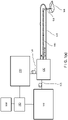
- FIG. 1(a) A schematic block diagram of an exemplary embodiment of multi modality optical imaging catheter system according to the present disclosure is shown in Figure 1(a) .
- This exemplary apparatus of Figure 1(a) can include a microstructural imaging system 110 (which can generate images using one or more processors), a single mode optical fiber 115, a molecular imaging system 120, a multimode optical fiber 125, a dual-modality rotary junction 130, a transparent imaging sheath 140, a dual-modality optical imaging catheter 150, a data acquisition system 160 and a data processing and storage unit 170.
- a microstructural imaging system 110 which can generate images using one or more processors
- a single mode optical fiber 115 e.g., a single mode optical fiber 115
- a molecular imaging system 120 e.g., a multimode optical fiber 125
- a dual-modality rotary junction 130 e.g., a dual-modality optical imaging catheter 150
- a data acquisition system 160 e.
- the microstructural imaging system 110 (e.g., one or more systems implementing one or more of optical frequency domain imaging, optical coherence tomography, etc. modalities) can detect a back-reflected light from a tissue 180 to acquire information and signals regarding tissue microstructures.
- the molecular imaging system 120 (e.g., one or more systems implementing one or more of near infra-red fluorescence imaging, fluorescence spectroscopy, Raman spectroscopy, fluorescence lifetime imaging, etc. modalities) can detect specific molecular information from the tissue 180, e.g., possibly but not necessarily using contrast agents. While the microstructural imaging system 110 can be connected to the dual-modality rotary junction 130 by the single mode fiber 115, the multi mode fiber 125 can be used for, e.g., the molecular imaging modality to achieve a high light efficiency.
- the dual modality rotary junction 130 can combine two different optical beams, and serve as the interface between the stationary imaging systems to the rotating and translating imaging catheter 150.
- the transparent imaging sheath 140 can be used to protect the imaging catheter 150 and the tissue 180, while the imaging catheter 150 rotates and translates and performs a helical scanning of the tissue.
- the optical imaging beam 155 can be focused by the dual-modality optical imaging catheter 150 onto the tissue 180.
- Returning light signals from the tissue 180 are detected by the microstructural imaging system 110 and the molecular imaging system 120. Both systems 110, 120 can be synchronized, and the signals can be acquired simultaneously by the data acquisition system 160.
- the data processing and storage arrangement/apparatus 170 can save and process the data in a real-time for the proper operation, and for subsequent visualization and analysis.
- a centering balloon 145 can be, e.g., provided and/or manufactured on the transparent imaging sheath 140 for imaging large luminal organs, such as esophagus, colon, etc.
- the molecular imaging system 120 can facilitate the implementation of fluorescence imaging, multi-photon imaging, near infra-red fluorescence imaging, fluorescence spectroscopy, fluorescence lifetime imaging, Raman spectroscopy, near infra-red reflectance spectroscopy, etc.
- fluorescence imaging fluorescent contrast agents can be used to target specific molecules, cells, proteins, or enzymes, associated with diseases. By using the targeted contrast agents, specific information can be obtained with high contrast.
- endogenous auto-fluorescence can be obtained with the catheter system. Since auto-fluorescence imaging techniques do not need any administration of exogenous contrast agents, such exemplary technique can be used in diagnostic applications, as well as in research, without a significant concern of the toxicity.
- UV/Visible illumination is used for imaging collagen, elastin, NADH, and etc.
- near infra-red light can be used in order to detect auto-fluorescence signal from lesions.
- multi-photon imaging can be used as a molecular imaging modality using a pulsed laser with and without the exogenous contrast agents.
- Near infra-red fluorescence imaging procedures can illuminate and/or detect fluorescent emission(s) from the fluorochromes in the near infra-red region. Since the auto-fluorescence of the tissue is reduced and/or minimized in near infra-red region, exogenous fluorescent contrast agents can be imaged with high contrast with minimized back-ground noise. Due to the high contrast, a lower dose of the contrast agent can be used for the detection.
- fluorescence lifetime imaging can be used. By measuring the difference of the lifetimes of the fluorochromes, molecular composition of lesions can be identified.
- Raman spectroscopy is another procedure that can be used to provide the chemical composition of biological tissues without exogenous contrast agents.
- chemical composition of the tissue such as elastin, collagen, cholesterol, cholesterol esters, triglycerides, phospholipids, and calcium salts can be measured with a high accuracy.
- Near infra-red spectroscopy is another technique for identifying tissue components. For example, due to the different absorption and scattering property, different tissue types can be identified and quantified by measuring and analyzing optical spectrums of the reflected signal from the tissue.
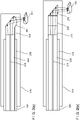
- Figure 2(a) shows a schematic diagram of a multi modality optical imaging catheter with a side-viewing ball lens according to an exemplary embodiment of the present disclosure.
- the double-clad fiber 200 can be used for multi-modality imaging.
- the microstructural imaging modality can work with a single-mode fiber to facilitate the coherent interference between the light from the sample and one from the reference (not shown).
- a single-mode core 210 can be used to guide a single-mode light of the microstructural imaging modality.
- the molecular imaging modality can operate with a multi-mode fiber to facilitate the high collection efficiency for better signal-to-noise ratio. Since a multi-mode fiber have a larger diameter and a larger accepting angle than a single-mode fiber, the light efficiency is usually high.
- a multi-mode second clad 220 can be used to guide the multi-mode light of the molecular imaging modality.
- a ultra low index coating 230 can facilitate the multi-mode second clad 220 to function or operate as a multi-mode light guiding channel.
- a glass spacer 250 and a side-viewing ball lens 260 can be provided in a configuration and/or shape to focus the beams onto the tissue 180 with a proper resolution, focal length, confocal parameter, etc. according to the size and the properties of luminal organs. The light from the fiber expands while traveling through the glass spacer 250, then reflected by the polished surface, and focused by a curvature surface of the side-viewing ball lens 260.
- the curvature surface of the side-viewing ball lens 260 can be designed to eliminate the aberration induced by the transparent imaging sheath 140 and the centering balloon 145. Since the optical fiber 200, the glass spacer 250, and the side-viewing ball lens 260 can be manufactured in one body, the imaging catheter can be reliable and durable.
- the imaging fiber can be contained within a protective metal coil 240.
- the metal coil 240 can also facilitate fast rotation and/or translation of the imaging catheter, by transmitting the torque from the rotary junction to the imaging probe.
- Micro optics such as a glass spacer 255, the GRIN lens and a prism 270, can be used instead of the side-viewing ball lens 260, as shown in Figure 2(b) .
- micro-lens assembly 275 instead of or in addition to the side-viewing ball lens 260 or the GRIN lens for possibly improved optical performances as shown in Figure 2(c) . Since the micro-lens assembly can have a large number of components, such exemplary assembly can provide an improved optical performance with less aberrations.
- the double-clad fiber can be important for the multi modal imaging catheter since such exemplary fiber can have independent multiple light guiding channels, e.g., optimized for each of the imaging modalities.
- a concentric arrangement of the double-clad fiber facilitates a continuous rotation of the imaging catheter which can transmit multiple optical signals as well as precise co-registration between different imaging modalities.
- the exemplary double-clad fiber with ultra low index coating can include a single-mode core 210, a multi-mode second clad 220, and an ultra low index coating 230, as shown in Figure 3(a) .
- the single-mode core 210 can be designed to be the same as or similar to the standard single-mode fiber, so that the multi modality catheter can be at least approximately, and preferably substantially matched well with the microstructural imaging system that can be provided with or from the standard single mode fiber.
- the multi-mode second clad 220 can be same as or similar to the standard multi-mode fiber, so that the multi modality catheter can be at least approximately, and preferably substantially matched well with the molecular imaging system that can be provided from or with the standard multi mode fiber.
- the ultra low index coating 230 can facilitate the second clad 220 to work as a multi-mode fiber.
- such coating 230 can mechanically protect the imaging channels.
- the cross-talk between the channels can deteriorate imaging quality of one or more of the imaging modalities. Thus, reducing and/or minimizing the cross-talk is preferable.
- a cross-talk barrier 215 is provided for a better separation of the imaging channels. This is facilitated by applying a low-index trench between the single-mode core 210 and the multi-mode second clad 220, as shown in Figure 3(b) .
- Figure 3(c) Another exemplary embodiment of a configuration of the double-clad fiber with two claddings is shown in Figure 3(c) .
- the first clad 250 and the protective coating 260 can be provided for improved optical performance and mechanical protection.
- the cross-talk barrier 215 is used to reduce and/or minimize the cross-talk between the channels, as shown in Figure 3(d) .
- the dual-modality rotary junction 130 can be used for the rapid helical scanning that facilitates a comprehensive three-dimensional scanning of the luminal organs.
- a schematic diagram of an exemplary embodiment of dual-modality rotary-junction according to the present disclosure is shown in Figure 4(a) .
- This exemplary apparatus can include, e.g., a single mode fiber 400, a multi mode fiber 410, a collimating lens for microstructural imaging modality 420, a collimating lens for molecular imaging modality 430, an achromatic collimating lens for both microstructural and molecular imaging modality 440, a dichroic mirror, a beam splitter, or a hole-mirror 450, a rotary motor 460, and a translation motor 470.
- a single mode fiber 400 e.g., a single mode fiber 400, a multi mode fiber 410, a collimating lens for microstructural imaging modality 420, a collimating lens for molecular imaging modality 430, an achromatic collimating lens for both microstructural and molecular imaging modality 440, a dichroic mirror, a beam splitter, or a hole-mirror 450, a rotary motor 460, and a translation motor 470.
- the single mode fiber 400 can deliver the single-mode light from the microstructural imaging system 110.
- the multi mode fiber 410 can deliver the multi-mode light from the molecular imaging system 120.
- the collimating lens 420 can be designed and/or arranged such that the microstructural imaging beam can be collimated with minimized optical aberration.
- the other collimating lens 430 can be designed to facilitate a reduction and/or minimization of optical aberrations for the molecular imaging beam.
- One or more light sources can be provided with different wavelength bands so that two or more different beams can be combined and divided by the dichroic mirror 450 with a high light throughput. If the wavelength ranges of the two imaging modalities are overlapped, they can be combined and divided by the beam splitter 450.
- the achromatic collimating lens 440 can be provided to reduce and/or minimize optical aberration over the large range of the wavelength, so that both the microstructural imaging beam and the molecular imaging beam can be efficiently coupled into the catheter 150.
- the rotary motor 460 can rotate the achromatic collimating lens 440 and the dual-modality optical imaging catheter 150 for the circumferential scanning.
- the rotating torque can be transmitted through the protective metal coil 240.
- the rotating motor 460 can located on or near the translating motor 470.
- the translating motor 470 can longitudinally move the optical imaging catheter 150.
- a rotation of the rotary motor 460 and a translation of the translating motor 470 can facilitate a three-dimensional (3D) scanning of the luminal organ in a helical pull-back fashion.
- FIG. 4(c) A schematic diagram of a dual-modality rotary-junction according to another exemplary embodiment of the present disclosure is shown in Figure 4(c).
- the double-clad fiber from the multi-modality imaging system can be coupled into the double-clad fiber of the multi-modality imaging catheter's double-clad fiber by the achromatic collimating lens pair 440.
- the beam splitting and combining procedure can be achieved at the proximal end of the multimodality imaging system's double-clad fiber using a dichroic mirror, a beam splitter, or a hole-mirror.
- multi-modality imaging data can be processed and fused for further visualization and analysis.
- Figure 5 shows an image fusion process for data obtained from the multi-modality catheter system and/or arrangement according to an exemplary embodiment of the present disclosure.
- the simultaneous acquisition with rapid helical pull-back scanning as provided in procedure 510, can be achieved by the exemplary multi-modality imaging system.
- 3D microstructural information (procedure 520) can be obtained by OFDI and/or OCT procedures
- 2D molecular information (procedure 530) can be obtained by near infra-red fluorescence, fluorescence spectroscopy, Raman spectroscopy, and/or fluorescence lifetime imaging.
- 3D microstructural information obtained in procedure 520 can be processed to form 3D images, then each biological components can be segmented according to their structural features. Thereafter the segmented images can be color-coded (procedure 560) for the 3D visualization.
- the segmented images can be extracted from the segmented images. While some molecular imaging modalities, such as confocal microscopy, diffuse optical tomography, photo-acoustic tomography, etc. can obtain depth information with a mechanical depth scanning or a post processing, many other molecular imaging modalities may not provide depth information, e.g., in a catheter form due to its space limitation.
- the distance information from the center to the luminal surface can be calculated in the segmented images. This information can be utilized to restore the distance information in the molecular information. Most of the molecular signal detected by the imaging catheter can be assumed to be provided from the luminal surface, by mapping the distance information (procedure 550) to the 2D molecular information.
- 3D molecular images obtained in procedure 570 can be restored.
- image fusion procedure 580 co-registered 3D microstructural and molecular images can be calculated in procedure 590.
- These exemplary multi-modality images can be used for the qualitative and quantitative analysis of disease progresses of the luminal organs. Since the two imaging modalities can be obtained simultaneously and the imaging beams can be inherently co-registered due to the concentric double-clad fiber, two or more imaging modalities can be co-registered without spatial markers.
- Figure 6 shows an exemplary flow diagram of an image reconstruction process for fluorescence data using 3D microstructural images obtained from a multi-modality catheter system according to another exemplary embodiment of the present disclosure.
- the signal strength can depend on the distance.
- the fluorescence signal strength is weaker when the distance from the imaging catheter to the tissue is farther.
- the distance information can facilitate a restoration of the true signal strength.
- the relation between the distance and fluorescence intensity or energy can be (e.g., experimentally or theoretically) obtained in procedure 610.
- the intensity distribution of 3D fluorescence data can be restored in procedure 620.
- This quantitatively restored 3D molecular image then can be fused (in procedure 630) with the 3D microstructural image for the further analysis by the image fusion algorithm obtained in procedure 640.
- the co-registered 3D microstructural and quantitative molecular image can provide accurate quantitative information of the luminal organs.
- Figures 7(a)-7(g) show exemplary experimental results on estimating concentration and distance compensation of the contrast agents for quantitative analysis provided by the exemplary systems, devices, methods, apparatus and computer-accessible media according to the present disclosure.
- Figure 7(a) shows a measured fluorescence signal while immersing the catheter in the solution repeatedly.
- Figures 7(b) and 7(c) show the measured fluorescence signal as a function of the concentration of the contrast agent, which showed a linear relationship.
- the fluorescence signal intensity was measured as a function of sample distance from the catheter in order to characterize the relationship between the NIRF intensity and the distance, as shown in Figure 7(d) .
- the measurements were fitted using an exponential function, then the calibration function for distance correction was generated, as shown in Figure 7(e) .
- FIGS 7(f) and 7(g) show the 2D fluorescence image of the capillary tube filled with the fluorescent contrast agent, before and after the distance calibration, respectively.
- Figures 8(a)-8(d) are images providing experiment results due to an acquisition of multi-modality images of rabbit arteries in vivo with fluorescently labeled thrombus and a coronary stent provided by the exemplary systems, devices, methods, apparatus and computer-accessible media according to the present disclosure .
- NIR near infra-red
- the coronary stent was rendered fluorescent in the NIR by incubating the stent with human fresh frozen plasma and a fibrin-targeted peptide derivatized with the NIR fluorochrome Cy7.
- FIG. 8(a) shows representative cross-sectional OFDI image and Figure 8(b) shows the corresponding calibrated fluorescence signal mapped on top of the OFDI luminal surface.
- Figure 8(c) shows the fused image of the microstructural OFDI image and the molecular NIRF image.
- Such 2D cross-sectional images were obtained by the helical scanning at different optical imaging catheter's positions with a specific interval.
- the 3D information then can be segmented and color-coded for the further 3D visualization as shown in Figure 8(d) .
- These imaging results show the feasibility of this technique and support the strength of multimodality imaging catheter for applications of the luminal organs, such as coronary artery imaging for accessing and interrogating coronary heart diseases.
Landscapes
- Health & Medical Sciences (AREA)
- Life Sciences & Earth Sciences (AREA)
- Physics & Mathematics (AREA)
- General Health & Medical Sciences (AREA)
- Pathology (AREA)
- General Physics & Mathematics (AREA)
- Nuclear Medicine, Radiotherapy & Molecular Imaging (AREA)
- Surgery (AREA)
- Veterinary Medicine (AREA)
- Heart & Thoracic Surgery (AREA)
- Medical Informatics (AREA)
- Molecular Biology (AREA)
- Engineering & Computer Science (AREA)
- Animal Behavior & Ethology (AREA)
- Biophysics (AREA)
- Public Health (AREA)
- Biomedical Technology (AREA)
- Radiology & Medical Imaging (AREA)
- Chemical & Material Sciences (AREA)
- Analytical Chemistry (AREA)
- Biochemistry (AREA)
- Immunology (AREA)
- Endoscopes (AREA)
- Investigating, Analyzing Materials By Fluorescence Or Luminescence (AREA)
Claims (13)
- Eine multimodale Bildgebungsanordnung zum Bereitstellen zumindest einer elektromagnetischen Strahlung an einer anatomischen Struktur, aufweisend:ein multimodales Bildgebungssystem, welches ein mikrostrukturelles Bildgebungssystem (110) und ein komplementäres Bildgebungssystem (120) aufweist;einen multimodalen Katheter (150), aufweisend:- zumindest einen optischen Kern (210) zum Bereitstellen eines ersten Bildgebungskanals für eine mikrostrukturelle Bildgebungsmodalität- zumindest eine Ummantelung (220), welche den zumindest einen Kern zumindest teilweise umgibt, um einen zweiten Bildgebungskanal für eine komplementäre Bildgebungsmodalität bereitzustellen; und- dazwischen einen niedrig-Index-Übersprech-Barriere-Graben (215), um ein Übersprechen zwischen dem ersten und dem zweiten Bildgebungskanal zu verringern;zumindest eine optische Vorrichtung (250, 260, 430, 460), welche eine Linsen Konfiguration umfasst, welche so konfiguriert ist, die zu mindestens eine Strahlung über den zumindest einen optischen Kern auf die anatomische Struktur zu richten; undwobei das multimodale Bildgebungssystem konfiguriert ist, über die Linsen Konfiguration zumindest zwei Strahlungen von der anatomischen Struktur über den zumindest einen optischen Kern und die mindestens eine Ummantelung zu empfangen.
- Die Anordnung gemäß Anspruch 1, wobei das mikrostrukturelle Bildgebungssystem ein Optisches Kohärenz Tomografie System umfasst.
- Die Anordnung gemäß Anspruch 1, ferner aufweisend:
ein Computersystem (160), welches konfiguriert ist Information betreffend die anatomische Struktur zu bestimmen unter Verwendung der zumindest einen Strahlung mit einer Prozedur basierend auf zumindest einem von einer Fluoreszenz Modalität, optischen Frequenzbereich Interferometrie (OFDI) Modalität, Spektralbereich optische Kohärenz Tomografie (SD-OCT) Modalität, Zeitbereich optische Kohärenz Tomografie (TD-OCT) Modalität, Nah-Infrarot Modalität, Raman Modalität, Fotoakustische Modalität, Konfokale Modalität, Ablationsmodalität, oder Lebenszeit Modalität. - Die Anordnung gemäß Anspruch 3, wobei, wenn die Prozedur die Fluoreszenz Modalität ist, die Fluoreszenz Modalität zumindest eine der folgenden exogen oder endogen ist.
- Die Anordnung gemäß Anspruch 3, wobei, wenn die Prozedur die Nah-Infrarot Modalität ist, die Nah-Infrarot Modalität zumindest eine der folgenden NA-Infrarot Spektroskopie Modalität oder Nah-Infrarot Fluoreszenz Modalität ist.
- Die Anordnung gemäß Anspruch 1, ferner aufweisend zumindest eine zusätzliche Ummantelung, welche zumindest teilweise die zumindest eine Ummantelung umgibt.
- Die Anordnung gemäß Anspruch 6, ferner weisend zumindest eine weitere Ummantelung, welche zumindest teilweise die zumindest eine zusätzliche Ummantelung umgibt.
- Die Anordnung gemäß Anspruch 1, wobei die Linsen Konfiguration konfiguriert ist, um die zumindest eine Strahlung über den zumindest einen optischen Kern auf die anatomische Struktur zu richten; und wobei das zumindest eine optische System ferner konfiguriert ist, um über die Linsen Konfiguration (i) eine erste Strahlung von den zumindest zwei Strahlungen von der anatomischen Struktur über den zumindest einen Kern zu empfangen und zu erhalten; und (ii) eine zweite Strahlung von den zumindest zwei Strahlungen von der anatomischen Struktur über die zumindest eine Ummantelung zu empfangen und zu erhalten.
- Die Anordnung gemäß Anspruch 1, wobei eine erste von den zumindest zwei empfangenen Strahlungen rück-reflektiertes Licht ist, und eine zweite von den zumindest zwei empfangenen Strahlungen ein Fluoreszenz Licht ist.
- Die Anordnung gemäß Anspruch 1, ferner aufweisend zumindest eine Rotationsvorrichtung (240, 460), welche konfiguriert ist, um den multimodalen Katheter zu rotieren.
- Die Anordnung gemäß Anspruch 1, ferner aufweisend eine Datenverarbeitungs- und Speicher Einheit (170), welche konfiguriert zum:Empfangen von ersten Daten, die mit der anatomischen Struktur verknüpft sind, wobei die ersten Daten Informationen umfassen, die einen Abstand zu der anatomischen Struktur anzeigen;Empfangen von zweiten Daten, die mit der anatomischen Struktur verknüpft sind, welche sich von den ersten Daten unterscheiden, wobei die zweiten Daten Fluoreszenz Daten sind und wobei die ersten Daten und die zweiten Daten von im Wesentlichen gleichen Positionen an oder in der anatomischen Struktur erhalten werden;Anwenden einer Korrektur auf die zweiten Daten basierend auf einer Kalibrierungsfunktion, welche die Abhängigkeit einer Fluoreszenz Signalintensität von dem Abstand zu der anatomischen Struktur beschreibt; undBestimmen von zumindest einer Charakteristik der anatomischen Struktur basierend auf den korrigierten zweiten Daten.
- Die Anordnung gemäß Anspruch 11, wobei die ersten Daten optische Kohärenz Tomografie Daten umfassen.
- Die Anordnung gemäß Anspruch 11, wobei der Abstand von einem Katheter zu einer Arterienwand von der anatomischen Struktur ist.
Applications Claiming Priority (3)
| Application Number | Priority Date | Filing Date | Title |
|---|---|---|---|
| US34807110P | 2010-05-25 | 2010-05-25 | |
| US35342410P | 2010-06-10 | 2010-06-10 | |
| PCT/US2011/037785 WO2011149972A2 (en) | 2010-05-25 | 2011-05-24 | Systems, devices, methods, apparatus and computer-accessible media for providing optical imaging of structures and compositions |
Publications (3)
| Publication Number | Publication Date |
|---|---|
| EP2575597A2 EP2575597A2 (de) | 2013-04-10 |
| EP2575597A4 EP2575597A4 (de) | 2017-01-04 |
| EP2575597B1 true EP2575597B1 (de) | 2022-05-04 |
Family
ID=45004726
Family Applications (1)
| Application Number | Title | Priority Date | Filing Date |
|---|---|---|---|
| EP11787272.1A Active EP2575597B1 (de) | 2010-05-25 | 2011-05-24 | Vorrichtung zur bereitstellung einer optischen bildgebung für strukturen und zusammensetzungen |
Country Status (3)
| Country | Link |
|---|---|
| US (3) | US9557154B2 (de) |
| EP (1) | EP2575597B1 (de) |
| WO (1) | WO2011149972A2 (de) |
Cited By (1)
| Publication number | Priority date | Publication date | Assignee | Title |
|---|---|---|---|---|
| EP4432904A1 (de) * | 2021-11-19 | 2024-09-25 | Spectrawave, Inc. | Multimodale sonden zur gewebeabfrage |
Families Citing this family (88)
| Publication number | Priority date | Publication date | Assignee | Title |
|---|---|---|---|---|
| US8774903B2 (en) * | 2010-03-26 | 2014-07-08 | Headwater Partners Ii Llc | Medical imaging apparatus and method |
| CN113397455B (zh) * | 2011-11-14 | 2024-08-16 | 皇家飞利浦有限公司 | 用于相关联的对象的扫描显微镜检查的光学显微镜检查探头 |
| KR101397272B1 (ko) * | 2012-07-17 | 2014-05-20 | 한양대학교 산학협력단 | 포괄적인 영상화 카테터 시스템 및 영상 처리 시스템 |
| EP3571977A1 (de) * | 2013-01-07 | 2019-11-27 | Securus Medical Group, Inc. | Temperaturmesssystem |
| EP2948758B1 (de) * | 2013-01-28 | 2024-03-13 | The General Hospital Corporation | Vorrichtung zur bereitstellung von gemeinsam mit optischer frequenzdomänenbildgebung aufgezeichneter diffuser spektroskopie |
| EP2983579B1 (de) * | 2013-04-12 | 2025-07-09 | NinePoint Medical, Inc. | Optische systeme und verfahren mit mehreren öffnungen und mehreren modi |
| US10722292B2 (en) | 2013-05-31 | 2020-07-28 | Covidien Lp | Surgical device with an end-effector assembly and system for monitoring of tissue during a surgical procedure |
| US10413359B2 (en) | 2013-07-18 | 2019-09-17 | International Business Machines Corporation | Laser-assisted transdermal delivery of nanoparticulates and hydrogels |
| WO2015054684A1 (en) * | 2013-10-11 | 2015-04-16 | The Trustees Of Columbia University In The City Of New York | System, method and computer-accessible medium for characterization of tissue |
| KR102381930B1 (ko) * | 2014-03-13 | 2022-04-04 | 내셔널 유니버시티 오브 싱가포르 | 광학 간섭 장치 |
| US20150265157A1 (en) * | 2014-03-24 | 2015-09-24 | Ecole Polytechnique Federale De Lausanne (Epfl) | Method and apparatus for a photoacoustic probe using a multimode fiber |
| US20150355413A1 (en) * | 2014-06-04 | 2015-12-10 | Corning Incorporated | Integrated torque jacket systems and methods for oct |
| CN113069204A (zh) * | 2014-11-14 | 2021-07-06 | 波士顿科学医学有限公司 | 手术激光系统和激光装置 |
| US10194981B2 (en) * | 2015-07-29 | 2019-02-05 | Medlumics S.L. | Radiofrequency ablation catheter with optical tissue evaluation |
| WO2020206362A1 (en) * | 2019-04-04 | 2020-10-08 | Inscopix, Inc. | Multi-modal microscopic imaging |
| US10996402B2 (en) | 2016-03-24 | 2021-05-04 | Canon U.S.A., Inc. | Multi-channel optical fiber rotary junction |
| US10578422B2 (en) | 2016-06-08 | 2020-03-03 | Canon U.S.A., Inc. | Devices, systems, methods and storage mediums using full range optical coherence tomography |
| US10952702B2 (en) | 2016-06-21 | 2021-03-23 | Canon U.S.A., Inc. | Non-uniform rotational distortion detection catheter system |
| KR102560803B1 (ko) * | 2016-07-05 | 2023-07-31 | 더 제너럴 하스피탈 코포레이션 | 능동적으로 제어되는 광학 이미징 장치를 위한 시스템들 및 방법들 |
| WO2018031462A1 (en) | 2016-08-12 | 2018-02-15 | Canon U.S.A. Inc. | Coherence range imaging using common path interference |
| US10602989B2 (en) | 2016-09-02 | 2020-03-31 | Canon U.S.A., Inc. | Capacitive sensing and encoding for imaging probes |
| US20190234869A1 (en) * | 2016-09-13 | 2019-08-01 | The General Hospital Corporation | Systems and methods for characterizing biological material using near-infrared spectroscopy |
| JP2019534069A (ja) | 2016-09-23 | 2019-11-28 | キヤノン ユーエスエイ, インコーポレイテッドCanon U.S.A., Inc | スペクトル符号化内視鏡検査装置および方法 |
| JP2018094395A (ja) * | 2016-11-03 | 2018-06-21 | キヤノン ユーエスエイ, インコーポレイテッドCanon U.S.A., Inc | 診断用スペクトル符号化内視鏡検査装置およびシステム、ならびにこれらと共に使用するための方法 |
| US10222607B2 (en) | 2016-12-14 | 2019-03-05 | Canon U.S.A., Inc. | Three-dimensional endoscope |
| WO2018132490A1 (en) | 2017-01-12 | 2018-07-19 | Canon U.S.A., Inc. | Spectrally encoded forward view endoscope and spectrally encoded multi-view endoscope, probe, and imaging apparatus |
| US11010877B2 (en) | 2017-01-27 | 2021-05-18 | Canon U.S.A., Inc. | Apparatus, system and method for dynamic in-line spectrum compensation of an image |
| US12064183B2 (en) | 2017-03-21 | 2024-08-20 | Canon U.S.A., Inc. | Methods, apparatuses and storage mediums for ablation planning and performance |
| US12569140B2 (en) * | 2017-04-13 | 2026-03-10 | The Regents Of The University Of California | Fiber-based multimodal biophotonic imaging and spectroscopy system |
| US10895692B2 (en) | 2017-06-01 | 2021-01-19 | Canon U.S.A., Inc. | Fiber optic rotary joints and methods of using and manufacturing same |
| US10323926B2 (en) * | 2017-06-21 | 2019-06-18 | Canon U.S.A., Inc. | Crosstalk elimination or mitigation in optical coherence tomography |
| US10678044B2 (en) | 2017-08-23 | 2020-06-09 | Canon U.S.A., Inc. | Beam-steering devices employing electrowetting prisms |
| US11259702B2 (en) | 2017-08-29 | 2022-03-01 | Canon U.S.A., Inc. | Fiber optic imaging probe having cladding mode pullback trigger, and control method therefor |
| US10825152B2 (en) | 2017-09-14 | 2020-11-03 | Canon U.S.A., Inc. | Distortion measurement and correction for spectrally encoded endoscopy |
| US11147453B2 (en) | 2017-10-03 | 2021-10-19 | Canon U.S.A., Inc. | Calibration for OCT-NIRAF multimodality probe |
| US10357160B2 (en) | 2017-10-05 | 2019-07-23 | Canon U.S.A., Inc. | Image acquiring apparatus, systems, and methods |
| US11408770B2 (en) * | 2017-10-30 | 2022-08-09 | University Of Maryland, College Park | Brillouin imaging devices, and systems and methods employing such devices |
| US11224336B2 (en) | 2017-11-17 | 2022-01-18 | Canon U.S.A., Inc. | Rotational extender and/or repeater for rotating fiber based optical imaging systems, and methods and storage mediums for use therewith |
| US10809538B2 (en) | 2017-11-27 | 2020-10-20 | Canon U.S.A., Inc. | Image acquisition apparatus, spectral apparatus, methods, and storage medium for use with same |
| US10606064B2 (en) * | 2018-01-24 | 2020-03-31 | Canon U.S.A., Inc. | Optical probes with astigmatism correction |
| US10806329B2 (en) | 2018-01-24 | 2020-10-20 | Canon U.S.A., Inc. | Optical probes with optical-correction components |
| US10816789B2 (en) | 2018-01-24 | 2020-10-27 | Canon U.S.A., Inc. | Optical probes that include optical-correction components for astigmatism correction |
| US10234676B1 (en) | 2018-01-24 | 2019-03-19 | Canon U.S.A., Inc. | Optical probes with reflecting components for astigmatism correction |
| US10561303B2 (en) | 2018-01-24 | 2020-02-18 | Canon U.S.A., Inc. | Optical probes with correction components for astigmatism correction |
| US10952616B2 (en) | 2018-03-30 | 2021-03-23 | Canon U.S.A., Inc. | Fluorescence imaging apparatus |
| US10506922B2 (en) | 2018-04-06 | 2019-12-17 | Canon U.S.A., Inc. | Spectrometer for color spectrally-encoded endoscopy |
| US11406327B2 (en) | 2018-04-17 | 2022-08-09 | Canon U.S.A., Inc. | Imaging catheter assembly |
| US11534058B2 (en) | 2018-05-03 | 2022-12-27 | The General Hospital Corporation | Systems, methods, and media for capsule-based multimode endoscopy |
| JP7075371B2 (ja) | 2018-05-03 | 2022-05-25 | キヤノン ユーエスエイ,インコーポレイテッド | マルチプルイメージングモダリティにわたって関心領域を強調するためのデバイス、システム、および方法 |
| US11382516B2 (en) | 2018-06-08 | 2022-07-12 | Canon U.S.A., Inc. | Apparatuses, methods, and storage mediums for lumen and artifacts detection in one or more images, such as in optical coherence tomography images |
| US12560423B2 (en) | 2018-08-28 | 2026-02-24 | Koninklijke Philips N.V. | Integrated fiber for optical shape sensing and spectral tissue sensing |
| US10743749B2 (en) | 2018-09-14 | 2020-08-18 | Canon U.S.A., Inc. | System and method for detecting optical probe connection |
| US10791923B2 (en) | 2018-09-24 | 2020-10-06 | Canon U.S.A., Inc. | Ball lens for optical probe and methods therefor |
| US11867627B2 (en) * | 2018-10-12 | 2024-01-09 | Washington University | Compact guided diffuse optical tomography system for imaging a lesion region |
| US10794732B2 (en) | 2018-11-08 | 2020-10-06 | Canon U.S.A., Inc. | Apparatus, system and method for correcting nonuniform rotational distortion in an image comprising at least two stationary light transmitted fibers with predetermined position relative to an axis of rotation of at least one rotating fiber |
| US12076177B2 (en) | 2019-01-30 | 2024-09-03 | Canon U.S.A., Inc. | Apparatuses, systems, methods and storage mediums for performance of co-registration |
| US11259694B2 (en) * | 2019-01-31 | 2022-03-01 | Canon U.S.A., Inc. | Window assembly for endoscopic probe |
| WO2020163449A1 (en) | 2019-02-05 | 2020-08-13 | Canon U.S.A., Inc. | Endoscope observation window cleaning |
| US11175126B2 (en) | 2019-04-08 | 2021-11-16 | Canon U.S.A., Inc. | Automated polarization control |
| KR102279322B1 (ko) * | 2019-04-08 | 2021-07-21 | 한양대학교 산학협력단 | 다중 진단 및 치료 카테터와 이를 포함하는 카테터 시스템 |
| US11707186B2 (en) | 2019-06-14 | 2023-07-25 | Canon U.S.A., Inc. | Fluorescence or auto-fluorescence trigger or triggers |
| WO2020257619A1 (en) * | 2019-06-20 | 2020-12-24 | Canon U.S.A., Inc. | Range finder for oct luminal clearance |
| US12109056B2 (en) | 2019-09-17 | 2024-10-08 | Canon U.S.A., Inc. | Constructing or reconstructing 3D structure(s) |
| EP4030996A4 (de) | 2019-09-20 | 2023-10-25 | Canon U.S.A. Inc. | Co-registrierung und markerdetektion mit künstlicher intelligenz, einschliesslich maschinenlernen und verwendung der ergebnisse davon |
| US20210121132A1 (en) * | 2019-10-24 | 2021-04-29 | Canon U.S.A., Inc. | Apparatus, methods and systems for fluorescence imaging |
| US11963740B2 (en) | 2019-12-05 | 2024-04-23 | Canon U.S.A., Inc. | Lumen, stent, and/or artifact detection in one or more images, such as in optical coherence tomography images |
| DE102020111376A1 (de) * | 2020-04-27 | 2021-10-28 | Carl Zeiss Meditec Ag | Medizinisch optisches System, Datenverarbeitungssystem, Computerprogramm und nichtflüchtiges computerlesbares Speichermedium |
| WO2021221569A1 (en) * | 2020-04-30 | 2021-11-04 | Agency For Science, Technology And Research | Rapid diagnostics for analyte/biomarker detection by raman technology with non-spectrometer raman measurement system |
| US11922633B2 (en) | 2020-06-30 | 2024-03-05 | Canon U.S.A., Inc. | Real-time lumen distance calculation based on three-dimensional (3D) A-line signal data |
| ES3037203T3 (en) * | 2020-07-23 | 2025-09-30 | Univ Adelaide | An optical element |
| US11920929B2 (en) | 2020-08-06 | 2024-03-05 | Canon U.S.A., Inc. | Detecting and guiding optical connection(s) for one or more imaging modalities, such as in optical coherence tomography |
| JP7667852B2 (ja) | 2020-08-06 | 2025-04-23 | キヤノン ユーエスエイ,インコーポレイテッド | カテーテルベースのマルチモーダル画像における潜在的な偽陽性及び盲点の位置を特定するためのシステム及び方法 |
| US11972561B2 (en) | 2020-08-06 | 2024-04-30 | Canon U.S.A., Inc. | Auto-pullback triggering method for intracoronary imaging apparatuses or systems using blood clearing |
| US11944778B2 (en) | 2020-08-06 | 2024-04-02 | Canon U.S.A., Inc. | Methods and systems for automatic pullback trigger |
| US12366441B2 (en) | 2020-08-06 | 2025-07-22 | Canon U.S.A., Inc. | Detector or photomultiplier tube (PMT) gain control over time |
| US11925321B2 (en) | 2020-08-06 | 2024-03-12 | Canon U.S.A., Inc. | Anti-twist tip for steerable catheter |
| US12112488B2 (en) | 2020-08-06 | 2024-10-08 | Canon U.S.A., Inc. | Methods and systems for image synchronization |
| KR102527241B1 (ko) * | 2021-03-30 | 2023-04-28 | 한국과학기술원 | 다중 모달 융합 영상을 이용한 동맥경화반 조직분석 방법 및 장치 |
| US12292376B2 (en) * | 2021-05-28 | 2025-05-06 | Lightsense Technology, Inc. | Miniature multispectral detection system having multiple spectrometers for enhanced photodetection spectroscopy for detection of pathogens, biomarkers, or any compound |
| US12076118B2 (en) | 2021-10-01 | 2024-09-03 | Canon U.S.A., Inc. | Devices, systems, and methods for detecting external elastic lamina (EEL) from intravascular OCT images |
| US12121205B2 (en) | 2021-10-04 | 2024-10-22 | Canon U.S.A., Inc. | Fluorescence calibration based on manual lumen detection |
| US12112472B2 (en) | 2021-10-13 | 2024-10-08 | Canon U.S.A., Inc. | Artifact removal from multimodality OCT images |
| CN117100198A (zh) * | 2022-05-17 | 2023-11-24 | 北京航空航天大学 | 三模态成像装置 |
| US12277731B2 (en) | 2022-07-21 | 2025-04-15 | Canon U.S.A., Inc. | Methods and systems for system self-diagnosis |
| US12551108B2 (en) | 2022-09-29 | 2026-02-17 | Canon U.S.A., Inc. | Angiography image/video synchronization with pullback and angio delay measurement |
| US20240319083A1 (en) * | 2023-03-20 | 2024-09-26 | The Board Of Regents Of The University Of Oklahoma | Apparatus for time-resolved fluorescence measurement using bandwidth-limited digital-pulse light modulation and methods |
| CN118078192B (zh) * | 2024-02-26 | 2025-11-28 | 上海交通大学 | 多模态成像导管以及多模态成像系统 |
| JP2025160114A (ja) | 2024-03-27 | 2025-10-22 | キヤノン ユーエスエイ,インコーポレイテッド | フォトブリーチされたイメージング装置若しくはカテーテル、及び、それらの使用方法、又は、それらにフォトブリーチを実施する方法 |
Citations (1)
| Publication number | Priority date | Publication date | Assignee | Title |
|---|---|---|---|---|
| US20060093276A1 (en) * | 2004-11-02 | 2006-05-04 | The General Hospital Corporation | Fiber-optic rotational device, optical system and method for imaging a sample |
Family Cites Families (667)
| Publication number | Priority date | Publication date | Assignee | Title |
|---|---|---|---|---|
| US2339754A (en) | 1941-03-04 | 1944-01-25 | Westinghouse Electric & Mfg Co | Supervisory apparatus |
| US3090753A (en) | 1960-08-02 | 1963-05-21 | Exxon Research Engineering Co | Ester oil compositions containing acid anhydride |
| GB1257778A (de) | 1967-12-07 | 1971-12-22 | ||
| US3601480A (en) | 1968-07-10 | 1971-08-24 | Physics Int Co | Optical tunnel high-speed camera system |
| JPS559417B2 (de) | 1971-10-09 | 1980-03-10 | ||
| JPS4932484U (de) | 1972-06-19 | 1974-03-20 | ||
| US3872407A (en) | 1972-09-01 | 1975-03-18 | Us Navy | Rapidly tunable laser |
| JPS584481Y2 (ja) | 1973-06-23 | 1983-01-26 | オリンパス光学工業株式会社 | ナイシキヨウシヤヘンカンコウガクケイ |
| FR2253410A5 (de) | 1973-12-03 | 1975-06-27 | Inst Nat Sante Rech Med | |
| US3941121A (en) | 1974-12-20 | 1976-03-02 | The University Of Cincinnati | Focusing fiber-optic needle endoscope |
| US3983507A (en) | 1975-01-06 | 1976-09-28 | Research Corporation | Tunable laser systems and method |
| US3973219A (en) | 1975-04-24 | 1976-08-03 | Cornell Research Foundation, Inc. | Very rapidly tuned cw dye laser |
| US4030831A (en) | 1976-03-22 | 1977-06-21 | The United States Of America As Represented By The Secretary Of The Navy | Phase detector for optical figure sensing |
| US4141362A (en) | 1977-05-23 | 1979-02-27 | Richard Wolf Gmbh | Laser endoscope |
| US4224929A (en) | 1977-11-08 | 1980-09-30 | Olympus Optical Co., Ltd. | Endoscope with expansible cuff member and operation section |
| US4339954A (en) | 1978-03-09 | 1982-07-20 | National Research Development Corporation | Measurement of small movements |
| GB2030313A (en) | 1978-06-29 | 1980-04-02 | Wolf Gmbh Richard | Endoscopes |
| FR2448728A1 (fr) | 1979-02-07 | 1980-09-05 | Thomson Csf | Dispositif joint tournant pour liaison par conducteurs optiques et systeme comportant un tel dispositif |
| US4295738A (en) | 1979-08-30 | 1981-10-20 | United Technologies Corporation | Fiber optic strain sensor |
| US4300816A (en) | 1979-08-30 | 1981-11-17 | United Technologies Corporation | Wide band multicore optical fiber |
| US4428643A (en) | 1981-04-08 | 1984-01-31 | Xerox Corporation | Optical scanning system with wavelength shift correction |
| US5065331A (en) | 1981-05-18 | 1991-11-12 | Vachon Reginald I | Apparatus and method for determining the stress and strain in pipes, pressure vessels, structural members and other deformable bodies |
| GB2106736B (en) | 1981-09-03 | 1985-06-12 | Standard Telephones Cables Ltd | Optical transmission system |
| US4479499A (en) | 1982-01-29 | 1984-10-30 | Alfano Robert R | Method and apparatus for detecting the presence of caries in teeth using visible light |
| US5302025A (en) | 1982-08-06 | 1994-04-12 | Kleinerman Marcos Y | Optical systems for sensing temperature and other physical parameters |
| US4601036A (en) | 1982-09-30 | 1986-07-15 | Honeywell Inc. | Rapidly tunable laser |
| HU187188B (en) | 1982-11-25 | 1985-11-28 | Koezponti Elelmiszeripari | Device for generating radiation of controllable spectral structure |
| CH663466A5 (fr) | 1983-09-12 | 1987-12-15 | Battelle Memorial Institute | Procede et dispositif pour determiner la position d'un objet par rapport a une reference. |
| JPS6140633A (ja) | 1984-08-02 | 1986-02-26 | Nec Corp | タブレツト装置 |
| US4639999A (en) | 1984-11-02 | 1987-02-03 | Xerox Corporation | High resolution, high efficiency I.R. LED printing array fabrication method |
| US4763977A (en) | 1985-01-09 | 1988-08-16 | Canadian Patents And Development Limited-Societe | Optical fiber coupler with tunable coupling ratio and method of making |
| EP0590268B1 (de) | 1985-03-22 | 1998-07-01 | Massachusetts Institute Of Technology | Faseroptisches Sondensystem zur spektralen Diagnose von Gewebe |
| US5318024A (en) | 1985-03-22 | 1994-06-07 | Massachusetts Institute Of Technology | Laser endoscope for spectroscopic imaging |
| DE3610165A1 (de) | 1985-03-27 | 1986-10-02 | Olympus Optical Co., Ltd., Tokio/Tokyo | Optisches abtastmikroskop |
| US4607622A (en) | 1985-04-11 | 1986-08-26 | Charles D. Fritch | Fiber optic ocular endoscope |
| US4631498A (en) | 1985-04-26 | 1986-12-23 | Hewlett-Packard Company | CW Laser wavemeter/frequency locking technique |
| US4650327A (en) | 1985-10-28 | 1987-03-17 | Oximetrix, Inc. | Optical catheter calibrating assembly |
| JPH0664683B2 (ja) | 1986-02-13 | 1994-08-22 | 松下電器産業株式会社 | 回転磁気ヘツド記録装置 |
| JPS62188001U (de) | 1986-05-20 | 1987-11-30 | ||
| US5040889A (en) | 1986-05-30 | 1991-08-20 | Pacific Scientific Company | Spectrometer with combined visible and ultraviolet sample illumination |
| CA1290019C (en) | 1986-06-20 | 1991-10-01 | Hideo Kuwahara | Dual balanced optical signal receiver |
| US4770492A (en) | 1986-10-28 | 1988-09-13 | Spectran Corporation | Pressure or strain sensitive optical fiber |
| JPH0824665B2 (ja) | 1986-11-28 | 1996-03-13 | オリンパス光学工業株式会社 | 内視鏡装置 |
| US4744656A (en) | 1986-12-08 | 1988-05-17 | Spectramed, Inc. | Disposable calibration boot for optical-type cardiovascular catheter |
| JPS63158363A (ja) | 1986-12-22 | 1988-07-01 | Daikin Mfg Co Ltd | エア回転継手のシ−ル装置 |
| US4751706A (en) | 1986-12-31 | 1988-06-14 | The United States Of America As Represented By The Secretary Of The Army | Laser for providing rapid sequence of different wavelengths |
| US4834111A (en) | 1987-01-12 | 1989-05-30 | The Trustees Of Columbia University In The City Of New York | Heterodyne interferometer |
| GB2209221B (en) | 1987-09-01 | 1991-10-23 | Litton Systems Inc | Hydrophone demodulator circuit and method |
| US5202931A (en) | 1987-10-06 | 1993-04-13 | Cell Analysis Systems, Inc. | Methods and apparatus for the quantitation of nuclear protein |
| US4909631A (en) | 1987-12-18 | 1990-03-20 | Tan Raul Y | Method for film thickness and refractive index determination |
| US4890901A (en) | 1987-12-22 | 1990-01-02 | Hughes Aircraft Company | Color corrector for embedded prisms |
| US4892406A (en) | 1988-01-11 | 1990-01-09 | United Technologies Corporation | Method of and arrangement for measuring vibrations |
| FR2626367B1 (fr) | 1988-01-25 | 1990-05-11 | Thomson Csf | Capteur de temperature multipoints a fibre optique |
| FR2626383B1 (fr) | 1988-01-27 | 1991-10-25 | Commissariat Energie Atomique | Procede de microscopie optique confocale a balayage et en profondeur de champ etendue et dispositifs pour la mise en oeuvre du procede |
| US4925302A (en) | 1988-04-13 | 1990-05-15 | Hewlett-Packard Company | Frequency locking device |
| US5030217A (en) * | 1988-04-14 | 1991-07-09 | Heraeus Lasersonics, Inc. | Medical laser probe and method of delivering CO2 radiation |
| US5730731A (en) | 1988-04-28 | 1998-03-24 | Thomas J. Fogarty | Pressure-based irrigation accumulator |
| US4998972A (en) | 1988-04-28 | 1991-03-12 | Thomas J. Fogarty | Real time angioscopy imaging system |
| US4905169A (en) | 1988-06-02 | 1990-02-27 | The United States Of America As Represented By The United States Department Of Energy | Method and apparatus for simultaneously measuring a plurality of spectral wavelengths present in electromagnetic radiation |
| US5242437A (en) | 1988-06-10 | 1993-09-07 | Trimedyne Laser Systems, Inc. | Medical device applying localized high intensity light and heat, particularly for destruction of the endometrium |
| WO1990000754A1 (en) | 1988-07-13 | 1990-01-25 | Martin Russell Harris | Scanning confocal microscope |
| GB8817672D0 (en) | 1988-07-25 | 1988-09-01 | Sira Ltd | Optical apparatus |
| US5214538A (en) | 1988-07-25 | 1993-05-25 | Keymed (Medical And Industrial Equipment) Limited | Optical apparatus |
| US4868834A (en) | 1988-09-14 | 1989-09-19 | The United States Of America As Represented By The Secretary Of The Army | System for rapidly tuning a low pressure pulsed laser |
| DE3833602A1 (de) | 1988-10-03 | 1990-02-15 | Krupp Gmbh | Spektrometer zur gleichzeitigen intensitaetsmessung in verschiedenen spektralbereichen |
| US4940328A (en) | 1988-11-04 | 1990-07-10 | Georgia Tech Research Corporation | Optical sensing apparatus and method |
| US4966589A (en) | 1988-11-14 | 1990-10-30 | Hemedix International, Inc. | Intravenous catheter placement device |
| US5419323A (en) | 1988-12-21 | 1995-05-30 | Massachusetts Institute Of Technology | Method for laser induced fluorescence of tissue |
| US5046501A (en) | 1989-01-18 | 1991-09-10 | Wayne State University | Atherosclerotic identification |
| US5085496A (en) | 1989-03-31 | 1992-02-04 | Sharp Kabushiki Kaisha | Optical element and optical pickup device comprising it |
| US5317389A (en) | 1989-06-12 | 1994-05-31 | California Institute Of Technology | Method and apparatus for white-light dispersed-fringe interferometric measurement of corneal topography |
| US4965599A (en) | 1989-11-13 | 1990-10-23 | Eastman Kodak Company | Scanning apparatus for halftone image screen writing |
| US5133035A (en) | 1989-11-14 | 1992-07-21 | Hicks John W | Multifiber endoscope with multiple scanning modes to produce an image free of fixed pattern noise |
| US4984888A (en) | 1989-12-13 | 1991-01-15 | Imo Industries, Inc. | Two-dimensional spectrometer |
| KR930003307B1 (ko) | 1989-12-14 | 1993-04-24 | 주식회사 금성사 | 입체용 프로젝터 |
| US5251009A (en) | 1990-01-22 | 1993-10-05 | Ciba-Geigy Corporation | Interferometric measuring arrangement for refractive index measurements in capillary tubes |
| DD293205B5 (de) | 1990-03-05 | 1995-06-29 | Zeiss Carl Jena Gmbh | Lichtleiterfuehrung fuer ein medizinisches Beobachtungsgeraet |
| US5039193A (en) | 1990-04-03 | 1991-08-13 | Focal Technologies Incorporated | Fibre optic single mode rotary joint |
| JPH0456907A (ja) | 1990-06-26 | 1992-02-24 | Fujikura Ltd | 光ファイバカプラ |
| US5262644A (en) | 1990-06-29 | 1993-11-16 | Southwest Research Institute | Remote spectroscopy for raman and brillouin scattering |
| US5197470A (en) | 1990-07-16 | 1993-03-30 | Eastman Kodak Company | Near infrared diagnostic method and instrument |
| GB9015793D0 (en) | 1990-07-18 | 1990-09-05 | Medical Res Council | Confocal scanning optical microscope |
| US5845639A (en) | 1990-08-10 | 1998-12-08 | Board Of Regents Of The University Of Washington | Optical imaging methods |
| US5127730A (en) | 1990-08-10 | 1992-07-07 | Regents Of The University Of Minnesota | Multi-color laser scanning confocal imaging system |
| US5305759A (en) | 1990-09-26 | 1994-04-26 | Olympus Optical Co., Ltd. | Examined body interior information observing apparatus by using photo-pulses controlling gains for depths |
| JP3104984B2 (ja) | 1990-09-27 | 2000-10-30 | オリンパス光学工業株式会社 | 断層像観察用光走査装置 |
| JPH04135551A (ja) | 1990-09-27 | 1992-05-11 | Olympus Optical Co Ltd | 光三次元像観察装置 |
| US5241364A (en) | 1990-10-19 | 1993-08-31 | Fuji Photo Film Co., Ltd. | Confocal scanning type of phase contrast microscope and scanning microscope |
| US5250186A (en) | 1990-10-23 | 1993-10-05 | Cetus Corporation | HPLC light scattering detector for biopolymers |
| US5202745A (en) | 1990-11-07 | 1993-04-13 | Hewlett-Packard Company | Polarization independent optical coherence-domain reflectometry |
| US5275594A (en) | 1990-11-09 | 1994-01-04 | C. R. Bard, Inc. | Angioplasty system having means for identification of atherosclerotic plaque |
| JP3035336B2 (ja) | 1990-11-27 | 2000-04-24 | 興和株式会社 | 血流測定装置 |
| US5228001A (en) | 1991-01-23 | 1993-07-13 | Syracuse University | Optical random access memory |
| US5784162A (en) | 1993-08-18 | 1998-07-21 | Applied Spectral Imaging Ltd. | Spectral bio-imaging methods for biological research, medical diagnostics and therapy |
| US6198532B1 (en) | 1991-02-22 | 2001-03-06 | Applied Spectral Imaging Ltd. | Spectral bio-imaging of the eye |
| US5293872A (en) | 1991-04-03 | 1994-03-15 | Alfano Robert R | Method for distinguishing between calcified atherosclerotic tissue and fibrous atherosclerotic tissue or normal cardiovascular tissue using Raman spectroscopy |
| US6111645A (en) | 1991-04-29 | 2000-08-29 | Massachusetts Institute Of Technology | Grating based phase control optical delay line |
| US6564087B1 (en) | 1991-04-29 | 2003-05-13 | Massachusetts Institute Of Technology | Fiber optic needle probes for optical coherence tomography imaging |
| US5956355A (en) | 1991-04-29 | 1999-09-21 | Massachusetts Institute Of Technology | Method and apparatus for performing optical measurements using a rapidly frequency-tuned laser |
| US6485413B1 (en) | 1991-04-29 | 2002-11-26 | The General Hospital Corporation | Methods and apparatus for forward-directed optical scanning instruments |
| US5748598A (en) | 1995-12-22 | 1998-05-05 | Massachusetts Institute Of Technology | Apparatus and methods for reading multilayer storage media using short coherence length sources |
| US6134003A (en) | 1991-04-29 | 2000-10-17 | Massachusetts Institute Of Technology | Method and apparatus for performing optical measurements using a fiber optic imaging guidewire, catheter or endoscope |
| WO1992019930A1 (en) | 1991-04-29 | 1992-11-12 | Massachusetts Institute Of Technology | Method and apparatus for optical imaging and measurement |
| US6501551B1 (en) | 1991-04-29 | 2002-12-31 | Massachusetts Institute Of Technology | Fiber optic imaging endoscope interferometer with at least one faraday rotator |
| US5465147A (en) | 1991-04-29 | 1995-11-07 | Massachusetts Institute Of Technology | Method and apparatus for acquiring images using a ccd detector array and no transverse scanner |
| US5441053A (en) | 1991-05-03 | 1995-08-15 | University Of Kentucky Research Foundation | Apparatus and method for multiple wavelength of tissue |
| US5281811A (en) | 1991-06-17 | 1994-01-25 | Litton Systems, Inc. | Digital wavelength division multiplex optical transducer having an improved decoder |
| US5208651A (en) | 1991-07-16 | 1993-05-04 | The Regents Of The University Of California | Apparatus and method for measuring fluorescence intensities at a plurality of wavelengths and lifetimes |
| AU2519892A (en) | 1991-08-20 | 1993-03-16 | Douglas C.B. Redd | Optical histochemical analysis, in vivo detection and real-time guidance for ablation of abnormal tissues using a raman spectroscopic detection system |
| DE4128744C1 (de) | 1991-08-29 | 1993-04-22 | Siemens Ag, 8000 Muenchen, De | |
| US5177488A (en) | 1991-10-08 | 1993-01-05 | Hughes Aircraft Company | Programmable fiber optic delay line, and radar target simulation system incorporating the same |
| ATE150573T1 (de) | 1991-12-30 | 1997-04-15 | Philips Electronics Nv | Optische einrichtung und mit einer solchen optischen einrichtung versehenes gerät zum abtasten einer informationsebene |
| US5353790A (en) | 1992-01-17 | 1994-10-11 | Board Of Regents, The University Of Texas System | Method and apparatus for optical measurement of bilirubin in tissue |
| US5212667A (en) | 1992-02-03 | 1993-05-18 | General Electric Company | Light imaging in a scattering medium, using ultrasonic probing and speckle image differencing |
| US5217456A (en) | 1992-02-24 | 1993-06-08 | Pdt Cardiovascular, Inc. | Device and method for intra-vascular optical radial imaging |
| US5248876A (en) | 1992-04-21 | 1993-09-28 | International Business Machines Corporation | Tandem linear scanning confocal imaging system with focal volumes at different heights |
| US5283795A (en) | 1992-04-21 | 1994-02-01 | Hughes Aircraft Company | Diffraction grating driven linear frequency chirped laser |
| US5486701A (en) | 1992-06-16 | 1996-01-23 | Prometrix Corporation | Method and apparatus for measuring reflectance in two wavelength bands to enable determination of thin film thickness |
| US5411025A (en) | 1992-06-30 | 1995-05-02 | Cordis Webster, Inc. | Cardiovascular catheter with laterally stable basket-shaped electrode array |
| US5716324A (en) | 1992-08-25 | 1998-02-10 | Fuji Photo Film Co., Ltd. | Endoscope with surface and deep portion imaging systems |
| US5348003A (en) | 1992-09-03 | 1994-09-20 | Sirraya, Inc. | Method and apparatus for chemical analysis |
| EP0587514A1 (de) | 1992-09-11 | 1994-03-16 | Welch Allyn, Inc. | Prozessormodul für Videoprüfsonde |
| US5772597A (en) | 1992-09-14 | 1998-06-30 | Sextant Medical Corporation | Surgical tool end effector |
| US5698397A (en) | 1995-06-07 | 1997-12-16 | Sri International | Up-converting reporters for biological and other assays using laser excitation techniques |
| EP0595666B1 (de) | 1992-09-21 | 1999-12-01 | Institut National De La Sante Et De La Recherche Medicale (Inserm) | Sonde und Verfahren zur genauen Bestimmung der Geschwindigkeit oder des Durchflusses einer Flüssigkeit |
| AU5672194A (en) | 1992-11-18 | 1994-06-22 | Spectrascience, Inc. | Apparatus for diagnostic imaging |
| US5383467A (en) | 1992-11-18 | 1995-01-24 | Spectrascience, Inc. | Guidewire catheter and apparatus for diagnostic imaging |
| US5785663A (en) | 1992-12-21 | 1998-07-28 | Artann Corporation | Method and device for mechanical imaging of prostate |
| US5400771A (en) | 1993-01-21 | 1995-03-28 | Pirak; Leon | Endotracheal intubation assembly and related method |
| JPH06222242A (ja) | 1993-01-27 | 1994-08-12 | Shin Etsu Chem Co Ltd | 光ファイバカプラおよびその製造方法 |
| US5987346A (en) | 1993-02-26 | 1999-11-16 | Benaron; David A. | Device and method for classification of tissue |
| US5414509A (en) | 1993-03-08 | 1995-05-09 | Associated Universities, Inc. | Optical pressure/density measuring means |
| JP3112595B2 (ja) | 1993-03-17 | 2000-11-27 | 安藤電気株式会社 | 光周波数シフタを用いる光ファイバ歪位置測定装置 |
| FI93781C (fi) | 1993-03-18 | 1995-05-26 | Wallac Oy | Biospesifinen multiparametrinen määritysmenetelmä |
| DE4309056B4 (de) | 1993-03-20 | 2006-05-24 | Häusler, Gerd, Prof. Dr. | Verfahren und Vorrichtung zur Ermittlung der Entfernung und Streuintensität von streuenden Punkten |
| US5485079A (en) | 1993-03-29 | 1996-01-16 | Matsushita Electric Industrial Co., Ltd. | Magneto-optical element and optical magnetic field sensor |
| DE4310209C2 (de) | 1993-03-29 | 1996-05-30 | Bruker Medizintech | Optische stationäre Bildgebung in stark streuenden Medien |
| DE4314189C1 (de) | 1993-04-30 | 1994-11-03 | Bodenseewerk Geraetetech | Vorrichtung zur Untersuchung von Lichtleitfasern aus Glas mittels Heterodyn-Brillouin-Spektroskopie |
| US5424827A (en) | 1993-04-30 | 1995-06-13 | Litton Systems, Inc. | Optical system and method for eliminating overlap of diffraction spectra |
| SE501932C2 (sv) | 1993-04-30 | 1995-06-26 | Ericsson Telefon Ab L M | Anordning och förfarande för dispersionskompensering i ett fiberoptiskt transmissionssystem |
| US5454807A (en) | 1993-05-14 | 1995-10-03 | Boston Scientific Corporation | Medical treatment of deeply seated tissue using optical radiation |
| DE69418248T2 (de) | 1993-06-03 | 1999-10-14 | Hamamatsu Photonics Kk | Optisches Laser-Abtastsystem mit Axikon |
| JP3234353B2 (ja) | 1993-06-15 | 2001-12-04 | 富士写真フイルム株式会社 | 断層情報読取装置 |
| US5840031A (en) | 1993-07-01 | 1998-11-24 | Boston Scientific Corporation | Catheters for imaging, sensing electrical potentials and ablating tissue |
| US5995645A (en) | 1993-08-18 | 1999-11-30 | Applied Spectral Imaging Ltd. | Method of cancer cell detection |
| US5803082A (en) | 1993-11-09 | 1998-09-08 | Staplevision Inc. | Omnispectramammography |
| US5983125A (en) | 1993-12-13 | 1999-11-09 | The Research Foundation Of City College Of New York | Method and apparatus for in vivo examination of subcutaneous tissues inside an organ of a body using optical spectroscopy |
| US5450203A (en) | 1993-12-22 | 1995-09-12 | Electroglas, Inc. | Method and apparatus for determining an objects position, topography and for imaging |
| US5411016A (en) | 1994-02-22 | 1995-05-02 | Scimed Life Systems, Inc. | Intravascular balloon catheter for use in combination with an angioscope |
| US5590660A (en) | 1994-03-28 | 1997-01-07 | Xillix Technologies Corp. | Apparatus and method for imaging diseased tissue using integrated autofluorescence |
| DE4411017C2 (de) | 1994-03-30 | 1995-06-08 | Alexander Dr Knuettel | Optische stationäre spektroskopische Bildgebung in stark streuenden Objekten durch spezielle Lichtfokussierung und Signal-Detektion von Licht unterschiedlicher Wellenlängen |
| TW275570B (de) | 1994-05-05 | 1996-05-11 | Boehringer Mannheim Gmbh | |
| WO1996002184A1 (en) | 1994-07-14 | 1996-02-01 | Washington Research Foundation | Method and apparatus for detecting barrett's metaplasia of the esophagus |
| US5459325A (en) | 1994-07-19 | 1995-10-17 | Molecular Dynamics, Inc. | High-speed fluorescence scanner |
| US6159445A (en) | 1994-07-20 | 2000-12-12 | Nycomed Imaging As | Light imaging contrast agents |
| CA2172284C (en) | 1994-08-08 | 1999-09-28 | Richard J. Mammone | Processing of keratoscopic images using local spatial phase |
| ES2180597T4 (es) | 1994-08-18 | 2003-07-01 | Zeiss Carl | Aparato quirurgico asistido por tomografia de coherencia optica. |
| US5491524A (en) | 1994-10-05 | 1996-02-13 | Carl Zeiss, Inc. | Optical coherence tomography corneal mapping apparatus |
| US5740808A (en) | 1996-10-28 | 1998-04-21 | Ep Technologies, Inc | Systems and methods for guilding diagnostic or therapeutic devices in interior tissue regions |
| US5817144A (en) | 1994-10-25 | 1998-10-06 | Latis, Inc. | Method for contemporaneous application OF laser energy and localized pharmacologic therapy |
| US6033721A (en) | 1994-10-26 | 2000-03-07 | Revise, Inc. | Image-based three-axis positioner for laser direct write microchemical reaction |
| JPH08136345A (ja) | 1994-11-10 | 1996-05-31 | Anritsu Corp | 複単色計 |
| JPH08160129A (ja) | 1994-12-05 | 1996-06-21 | Uniden Corp | 速度検出装置 |
| US5566267A (en) | 1994-12-15 | 1996-10-15 | Ceram Optec Industries Inc. | Flat surfaced optical fibers and diode laser medical delivery devices |
| US5600486A (en) | 1995-01-30 | 1997-02-04 | Lockheed Missiles And Space Company, Inc. | Color separation microlens |
| US5648848A (en) | 1995-02-01 | 1997-07-15 | Nikon Precision, Inc. | Beam delivery apparatus and method for interferometry using rotatable polarization chucks |
| DE19506484C2 (de) | 1995-02-24 | 1999-09-16 | Stiftung Fuer Lasertechnologie | Verfahren und Vorrichtung zur selektiven nichtinvasiven Lasermyographie (LMG) |
| RU2100787C1 (ru) | 1995-03-01 | 1997-12-27 | Геликонов Валентин Михайлович | Оптоволоконный интерферометр и оптоволоконный пьезоэлектрический преобразователь |
| WO1996028212A1 (en) | 1995-03-09 | 1996-09-19 | Innotech Usa, Inc. | Laser surgical device and method of its use |
| US5868731A (en) | 1996-03-04 | 1999-02-09 | Innotech Usa, Inc. | Laser surgical device and method of its use |
| US5526338A (en) | 1995-03-10 | 1996-06-11 | Yeda Research & Development Co. Ltd. | Method and apparatus for storage and retrieval with multilayer optical disks |
| US5697373A (en) | 1995-03-14 | 1997-12-16 | Board Of Regents, The University Of Texas System | Optical method and apparatus for the diagnosis of cervical precancers using raman and fluorescence spectroscopies |
| US5735276A (en) | 1995-03-21 | 1998-04-07 | Lemelson; Jerome | Method and apparatus for scanning and evaluating matter |
| US5926592A (en) | 1995-03-24 | 1999-07-20 | Optiscan Pty Ltd | Optical fibre confocal imager with variable near-confocal control |
| US5565983A (en) | 1995-05-26 | 1996-10-15 | The Perkin-Elmer Corporation | Optical spectrometer for detecting spectra in separate ranges |
| US5785651A (en) | 1995-06-07 | 1998-07-28 | Keravision, Inc. | Distance measuring confocal microscope |
| US5621830A (en) | 1995-06-07 | 1997-04-15 | Smith & Nephew Dyonics Inc. | Rotatable fiber optic joint |
| WO1997001167A1 (en) | 1995-06-21 | 1997-01-09 | Massachusetts Institute Of Technology | Apparatus and method for accessing data on multilayered optical media |
| ATA107495A (de) | 1995-06-23 | 1996-06-15 | Fercher Adolf Friedrich Dr | Kohärenz-biometrie und -tomographie mit dynamischem kohärentem fokus |
| US5829439A (en) | 1995-06-28 | 1998-11-03 | Hitachi Medical Corporation | Needle-like ultrasonic probe for ultrasonic diagnosis apparatus, method of producing same, and ultrasonic diagnosis apparatus using same |
| JP3654309B2 (ja) | 1995-06-28 | 2005-06-02 | 株式会社日立メディコ | 針状超音波探触子 |
| US6104945A (en) | 1995-08-01 | 2000-08-15 | Medispectra, Inc. | Spectral volume microprobe arrays |
| JP3819032B2 (ja) | 1995-08-24 | 2006-09-06 | ザ・テキサス・エイ・アンド・エム・ユニバーシティ・システム | 組織およびその他のランダム媒体における蛍光寿命に基づく撮像および分光分析 |
| US6016197A (en) | 1995-08-25 | 2000-01-18 | Ceramoptec Industries Inc. | Compact, all-optical spectrum analyzer for chemical and biological fiber optic sensors |
| FR2738343B1 (fr) | 1995-08-30 | 1997-10-24 | Cohen Sabban Joseph | Dispositif de microstratigraphie optique |
| US6763261B2 (en) | 1995-09-20 | 2004-07-13 | Board Of Regents, The University Of Texas System | Method and apparatus for detecting vulnerable atherosclerotic plaque |
| US6615071B1 (en) | 1995-09-20 | 2003-09-02 | Board Of Regents, The University Of Texas System | Method and apparatus for detecting vulnerable atherosclerotic plaque |
| CA2231425A1 (en) | 1995-09-20 | 1997-03-27 | Texas Heart Institute | Detecting thermal discrepancies in vessel walls |
| US5742419A (en) | 1995-11-07 | 1998-04-21 | The Board Of Trustees Of The Leland Stanford Junior Universtiy | Miniature scanning confocal microscope |
| DE19542955C2 (de) | 1995-11-17 | 1999-02-18 | Schwind Gmbh & Co Kg Herbert | Endoskop |
| US5719399A (en) | 1995-12-18 | 1998-02-17 | The Research Foundation Of City College Of New York | Imaging and characterization of tissue based upon the preservation of polarized light transmitted therethrough |
| JP3699761B2 (ja) | 1995-12-26 | 2005-09-28 | オリンパス株式会社 | 落射蛍光顕微鏡 |
| US5748318A (en) | 1996-01-23 | 1998-05-05 | Brown University Research Foundation | Optical stress generator and detector |
| US5840023A (en) | 1996-01-31 | 1998-11-24 | Oraevsky; Alexander A. | Optoacoustic imaging for medical diagnosis |
| US5642194A (en) | 1996-02-05 | 1997-06-24 | The Regents Of The University Of California | White light velocity interferometer |
| US5862273A (en) | 1996-02-23 | 1999-01-19 | Kaiser Optical Systems, Inc. | Fiber optic probe with integral optical filtering |
| US5843000A (en) | 1996-05-07 | 1998-12-01 | The General Hospital Corporation | Optical biopsy forceps and method of diagnosing tissue |
| ATA84696A (de) | 1996-05-14 | 1998-03-15 | Adolf Friedrich Dr Fercher | Verfahren und anordnungen zur kontrastanhebung in der optischen kohärenztomographie |
| US6020963A (en) | 1996-06-04 | 2000-02-01 | Northeastern University | Optical quadrature Interferometer |
| US5795295A (en) | 1996-06-25 | 1998-08-18 | Carl Zeiss, Inc. | OCT-assisted surgical microscope with multi-coordinate manipulator |
| US5842995A (en) | 1996-06-28 | 1998-12-01 | Board Of Regents, The Univerisity Of Texas System | Spectroscopic probe for in vivo measurement of raman signals |
| US6296608B1 (en) | 1996-07-08 | 2001-10-02 | Boston Scientific Corporation | Diagnosing and performing interventional procedures on tissue in vivo |
| US6245026B1 (en) | 1996-07-29 | 2001-06-12 | Farallon Medsystems, Inc. | Thermography catheter |
| US6396941B1 (en) | 1996-08-23 | 2002-05-28 | Bacus Research Laboratories, Inc. | Method and apparatus for internet, intranet, and local viewing of virtual microscope slides |
| US5840075A (en) | 1996-08-23 | 1998-11-24 | Eclipse Surgical Technologies, Inc. | Dual laser device for transmyocardial revascularization procedures |
| US6544193B2 (en) | 1996-09-04 | 2003-04-08 | Marcio Marc Abreu | Noninvasive measurement of chemical substances |
| JPH1090603A (ja) | 1996-09-18 | 1998-04-10 | Olympus Optical Co Ltd | 内視鏡光学系 |
| US5801831A (en) | 1996-09-20 | 1998-09-01 | Institute For Space And Terrestrial Science | Fabry-Perot spectrometer for detecting a spatially varying spectral signature of an extended source |
| RU2108122C1 (ru) | 1996-09-24 | 1998-04-10 | Владимир Павлович Жаров | Способ и устройство для физиотерапевтического облучения светом |
| US6249349B1 (en) | 1996-09-27 | 2001-06-19 | Vincent Lauer | Microscope generating a three-dimensional representation of an object |
| DE19640495C2 (de) | 1996-10-01 | 1999-12-16 | Leica Microsystems | Vorrichtung zur konfokalen Oberflächenvermessung |
| US5843052A (en) | 1996-10-04 | 1998-12-01 | Benja-Athon; Anuthep | Irrigation kit for application of fluids and chemicals for cleansing and sterilizing wounds |
| US5904651A (en) | 1996-10-28 | 1999-05-18 | Ep Technologies, Inc. | Systems and methods for visualizing tissue during diagnostic or therapeutic procedures |
| US5752518A (en) | 1996-10-28 | 1998-05-19 | Ep Technologies, Inc. | Systems and methods for visualizing interior regions of the body |
| US6044288A (en) | 1996-11-08 | 2000-03-28 | Imaging Diagnostics Systems, Inc. | Apparatus and method for determining the perimeter of the surface of an object being scanned |
| US5872879A (en) | 1996-11-25 | 1999-02-16 | Boston Scientific Corporation | Rotatable connecting optical fibers |
| US6517532B1 (en) | 1997-05-15 | 2003-02-11 | Palomar Medical Technologies, Inc. | Light energy delivery head |
| US6437867B2 (en) | 1996-12-04 | 2002-08-20 | The Research Foundation Of The City University Of New York | Performing selected optical measurements with optical coherence domain reflectometry |
| US6249630B1 (en) | 1996-12-13 | 2001-06-19 | Imra America, Inc. | Apparatus and method for delivery of dispersion-compensated ultrashort optical pulses with high peak power |
| US5906759A (en) | 1996-12-26 | 1999-05-25 | Medinol Ltd. | Stent forming apparatus with stent deforming blades |
| US5871449A (en) | 1996-12-27 | 1999-02-16 | Brown; David Lloyd | Device and method for locating inflamed plaque in an artery |
| JP2001508554A (ja) | 1996-12-31 | 2001-06-26 | コーニング インコーポレイテッド | 多層ファイバを備えた光カプラ |
| US5991697A (en) | 1996-12-31 | 1999-11-23 | The Regents Of The University Of California | Method and apparatus for optical Doppler tomographic imaging of fluid flow velocity in highly scattering media |
| US5760901A (en) | 1997-01-28 | 1998-06-02 | Zetetic Institute | Method and apparatus for confocal interference microscopy with background amplitude reduction and compensation |
| EP0858167A1 (de) | 1997-01-29 | 1998-08-12 | Hewlett-Packard Company | Feldprogrammierbarer Prozessor |
| JP3213250B2 (ja) | 1997-01-29 | 2001-10-02 | 株式会社生体光情報研究所 | 光計測装置 |
| US5801826A (en) | 1997-02-18 | 1998-09-01 | Williams Family Trust B | Spectrometric device and method for recognizing atomic and molecular signatures |
| US5836877A (en) | 1997-02-24 | 1998-11-17 | Lucid Inc | System for facilitating pathological examination of a lesion in tissue |
| US6120516A (en) | 1997-02-28 | 2000-09-19 | Lumend, Inc. | Method for treating vascular occlusion |
| US6010449A (en) | 1997-02-28 | 2000-01-04 | Lumend, Inc. | Intravascular catheter system for treating a vascular occlusion |
| US5968064A (en) | 1997-02-28 | 1999-10-19 | Lumend, Inc. | Catheter system for treating a vascular occlusion |
| JP2001515382A (ja) | 1997-03-06 | 2001-09-18 | マサチューセッツ インスティチュート オブ テクノロジー | 生体組織の光学走査用機器 |
| CA2283949A1 (en) | 1997-03-13 | 1998-09-17 | Haishan Zeng | Methods and apparatus for detecting the rejection of transplanted tissue |
| US6078047A (en) | 1997-03-14 | 2000-06-20 | Lucent Technologies Inc. | Method and apparatus for terahertz tomographic imaging |
| US5994690A (en) | 1997-03-17 | 1999-11-30 | Kulkarni; Manish D. | Image enhancement in optical coherence tomography using deconvolution |
| JPH10267830A (ja) | 1997-03-26 | 1998-10-09 | Kowa Co | 光学測定装置 |
| JPH10267631A (ja) | 1997-03-26 | 1998-10-09 | Kowa Co | 光学測定装置 |
| GB9707414D0 (en) | 1997-04-11 | 1997-05-28 | Imperial College | Anatomical probe |
| ATE257014T1 (de) | 1997-04-29 | 2004-01-15 | Amersham Health As | Lichtbilderzeugungskontrastmitteln |
| EP0979107A1 (de) | 1997-04-29 | 2000-02-16 | Nycomed Imaging As | Lichtbilderzeugungskontrastmitteln |
| US6117128A (en) | 1997-04-30 | 2000-09-12 | Kenton W. Gregory | Energy delivery catheter and method for the use thereof |
| US5887009A (en) | 1997-05-22 | 1999-03-23 | Optical Biopsy Technologies, Inc. | Confocal optical scanning system employing a fiber laser |
| DE69840791D1 (de) | 1997-06-02 | 2009-06-10 | Joseph A Izatt | Doppler-abbildung einer strömung mittels optischer kohaerenztomografie |
| US6002480A (en) | 1997-06-02 | 1999-12-14 | Izatt; Joseph A. | Depth-resolved spectroscopic optical coherence tomography |
| US6208415B1 (en) | 1997-06-12 | 2001-03-27 | The Regents Of The University Of California | Birefringence imaging in biological tissue using polarization sensitive optical coherent tomography |
| EP0989822A4 (de) | 1997-06-23 | 2004-07-28 | Focus Surgery Inc | Methoden und vorrichtungen zur verschaffung akustischer hemostasis |
| US5920390A (en) | 1997-06-26 | 1999-07-06 | University Of North Carolina | Fiberoptic interferometer and associated method for analyzing tissue |
| US6048349A (en) | 1997-07-09 | 2000-04-11 | Intraluminal Therapeutics, Inc. | Systems and methods for guiding a medical instrument through a body |
| US6058352A (en) | 1997-07-25 | 2000-05-02 | Physical Optics Corporation | Accurate tissue injury assessment using hybrid neural network analysis |
| US5921926A (en) | 1997-07-28 | 1999-07-13 | University Of Central Florida | Three dimensional optical imaging colposcopy |
| US6014214A (en) | 1997-08-21 | 2000-01-11 | Li; Ming-Chiang | High speed inspection of a sample using coherence processing of scattered superbroad radiation |
| US5892583A (en) | 1997-08-21 | 1999-04-06 | Li; Ming-Chiang | High speed inspection of a sample using superbroad radiation coherent interferometer |
| US6069698A (en) | 1997-08-28 | 2000-05-30 | Olympus Optical Co., Ltd. | Optical imaging apparatus which radiates a low coherence light beam onto a test object, receives optical information from light scattered by the object, and constructs therefrom a cross-sectional image of the object |
| US6297018B1 (en) | 1998-04-17 | 2001-10-02 | Ljl Biosystems, Inc. | Methods and apparatus for detecting nucleic acid polymorphisms |
| US5920373A (en) | 1997-09-24 | 1999-07-06 | Heidelberg Engineering Optische Messysteme Gmbh | Method and apparatus for determining optical characteristics of a cornea |
| US5951482A (en) | 1997-10-03 | 1999-09-14 | Intraluminal Therapeutics, Inc. | Assemblies and methods for advancing a guide wire through body tissue |
| US6193676B1 (en) | 1997-10-03 | 2001-02-27 | Intraluminal Therapeutics, Inc. | Guide wire assembly |
| US6091984A (en) | 1997-10-10 | 2000-07-18 | Massachusetts Institute Of Technology | Measuring tissue morphology |
| US5955737A (en) | 1997-10-27 | 1999-09-21 | Systems & Processes Engineering Corporation | Chemometric analysis for extraction of individual fluorescence spectrum and lifetimes from a target mixture |
| US6052186A (en) | 1997-11-05 | 2000-04-18 | Excel Precision, Inc. | Dual laser system for extended heterodyne interferometry |
| US6134010A (en) | 1997-11-07 | 2000-10-17 | Lucid, Inc. | Imaging system using polarization effects to enhance image quality |
| US6037579A (en) | 1997-11-13 | 2000-03-14 | Biophotonics Information Laboratories, Ltd. | Optical interferometer employing multiple detectors to detect spatially distorted wavefront in imaging of scattering media |
| US6107048A (en) | 1997-11-20 | 2000-08-22 | Medical College Of Georgia Research Institute, Inc. | Method of detecting and grading dysplasia in epithelial tissue |
| AU2242099A (en) | 1998-01-28 | 1999-08-16 | Ht Medical Systems, Inc. | Interface device and method for interfacing instruments to medical procedure simulation system |
| US6165170A (en) | 1998-01-29 | 2000-12-26 | International Business Machines Corporation | Laser dermablator and dermablation |
| US6831781B2 (en) | 1998-02-26 | 2004-12-14 | The General Hospital Corporation | Confocal microscopy with multi-spectral encoding and system and apparatus for spectroscopically encoded confocal microscopy |
| US6134033A (en) | 1998-02-26 | 2000-10-17 | Tyco Submarine Systems Ltd. | Method and apparatus for improving spectral efficiency in wavelength division multiplexed transmission systems |
| EP2267507A3 (de) | 1998-02-26 | 2011-08-17 | The General Hospital Corporation | Konfokale Mikroskopie mit multispektraler Kodierung |
| US6048742A (en) | 1998-02-26 | 2000-04-11 | The United States Of America As Represented By The Secretary Of The Air Force | Process for measuring the thickness and composition of thin semiconductor films deposited on semiconductor wafers |
| ZA991635B (en) | 1998-03-02 | 1999-09-02 | Procter & Gamble | Concentrated, stable, translucent or clear, fabric softening compositions. |
| RU2148378C1 (ru) | 1998-03-06 | 2000-05-10 | Геликонов Валентин Михайлович | Устройство для оптической когерентной томографии, оптоволоконное сканирующее устройство и способ диагностики биоткани in vivo |
| US6174291B1 (en) | 1998-03-09 | 2001-01-16 | Spectrascience, Inc. | Optical biopsy system and methods for tissue diagnosis |
| US6066102A (en) | 1998-03-09 | 2000-05-23 | Spectrascience, Inc. | Optical biopsy forceps system and method of diagnosing tissue |
| JPH11259617A (ja) | 1998-03-12 | 1999-09-24 | Alps Electric Co Ltd | ソケット付きicカード |
| US6151522A (en) | 1998-03-16 | 2000-11-21 | The Research Foundation Of Cuny | Method and system for examining biological materials using low power CW excitation raman spectroscopy |
| US6175669B1 (en) | 1998-03-30 | 2001-01-16 | The Regents Of The Universtiy Of California | Optical coherence domain reflectometry guidewire |
| DE19814057B4 (de) | 1998-03-30 | 2009-01-02 | Carl Zeiss Meditec Ag | Anordnung zur optischen Kohärenztomographie und Kohärenztopographie |
| US6384915B1 (en) | 1998-03-30 | 2002-05-07 | The Regents Of The University Of California | Catheter guided by optical coherence domain reflectometry |
| AU3781799A (en) | 1998-05-01 | 1999-11-23 | Board Of Regents, The University Of Texas System | Method and apparatus for subsurface imaging |
| US6996549B2 (en) | 1998-05-01 | 2006-02-07 | Health Discovery Corporation | Computer-aided image analysis |
| JPH11326826A (ja) | 1998-05-13 | 1999-11-26 | Sony Corp | 照明方法及び照明装置 |
| US6053613A (en) | 1998-05-15 | 2000-04-25 | Carl Zeiss, Inc. | Optical coherence tomography with new interferometer |
| FR2778838A1 (fr) | 1998-05-19 | 1999-11-26 | Koninkl Philips Electronics Nv | Procede de detection de variations d'elasticite et appareil echographique pour mettre en oeuvre ce procede |
| US5995223A (en) | 1998-06-01 | 1999-11-30 | Power; Joan Fleurette | Apparatus for rapid phase imaging interferometry and method therefor |
| JPH11352409A (ja) | 1998-06-05 | 1999-12-24 | Olympus Optical Co Ltd | 蛍光検出装置 |
| US6549801B1 (en) | 1998-06-11 | 2003-04-15 | The Regents Of The University Of California | Phase-resolved optical coherence tomography and optical doppler tomography for imaging fluid flow in tissue with fast scanning speed and high velocity sensitivity |
| AU5101699A (en) | 1998-07-15 | 2000-02-07 | Corazon Technologies, Inc. | Methods and devices for reducing the mineral content of vascular calcified lesions |
| US6166373A (en) | 1998-07-21 | 2000-12-26 | The Institute For Technology Development | Focal plane scanner with reciprocating spatial window |
| JP2000046729A (ja) | 1998-07-31 | 2000-02-18 | Takahisa Mitsui | 波長分散を用いた高速光断層像計測装置および計測方法 |
| US20040140130A1 (en) | 1998-08-31 | 2004-07-22 | Halliburton Energy Services, Inc., A Delaware Corporation | Roller-cone bits, systems, drilling methods, and design methods with optimization of tooth orientation |
| US8024027B2 (en) | 1998-09-03 | 2011-09-20 | Hyperspectral Imaging, Inc. | Infrared endoscopic balloon probes |
| US6741884B1 (en) | 1998-09-03 | 2004-05-25 | Hypermed, Inc. | Infrared endoscopic balloon probes |
| CA2343401C (en) | 1998-09-11 | 2009-01-27 | Spectrx, Inc. | Multi-modal optical tissue diagnostic system |
| JP2000131222A (ja) | 1998-10-22 | 2000-05-12 | Olympus Optical Co Ltd | 光断層画像装置 |
| AU6417599A (en) | 1998-10-08 | 2000-04-26 | University Of Kentucky Research Foundation, The | Methods and apparatus for (in vivo) identification and characterization of vulnerable atherosclerotic plaques |
| JP2000121961A (ja) | 1998-10-13 | 2000-04-28 | Olympus Optical Co Ltd | 共焦点光走査プローブシステム |
| US6274871B1 (en) | 1998-10-22 | 2001-08-14 | Vysis, Inc. | Method and system for performing infrared study on a biological sample |
| US6324419B1 (en) | 1998-10-27 | 2001-11-27 | Nejat Guzelsu | Apparatus and method for non-invasive measurement of stretch |
| JP2000126116A (ja) | 1998-10-28 | 2000-05-09 | Olympus Optical Co Ltd | 光診断システム |
| US6524249B2 (en) | 1998-11-11 | 2003-02-25 | Spentech, Inc. | Doppler ultrasound method and apparatus for monitoring blood flow and detecting emboli |
| WO2000030225A1 (en) | 1998-11-13 | 2000-05-25 | Research And Development Institute, Inc. | Programmable frequency reference for laser frequency stabilization, and arbitrary optical clock generator, using persistent spectral hole burning |
| DE69932485T2 (de) | 1998-11-20 | 2007-01-11 | Fuji Photo Film Co. Ltd., Minamiashigara | Blutgefäss Bilddarstellungssystem |
| US5975697A (en) | 1998-11-25 | 1999-11-02 | Oti Ophthalmic Technologies, Inc. | Optical mapping apparatus with adjustable depth resolution |
| US6352502B1 (en) | 1998-12-03 | 2002-03-05 | Lightouch Medical, Inc. | Methods for obtaining enhanced spectroscopic information from living tissue, noninvasive assessment of skin condition and detection of skin abnormalities |
| RU2149464C1 (ru) | 1999-01-19 | 2000-05-20 | Таганрогский государственный радиотехнический университет | Динамическое запоминающее устройство радиосигналов |
| US6191862B1 (en) | 1999-01-20 | 2001-02-20 | Lightlab Imaging, Llc | Methods and apparatus for high speed longitudinal scanning in imaging systems |
| US6272376B1 (en) | 1999-01-22 | 2001-08-07 | Cedars-Sinai Medical Center | Time-resolved, laser-induced fluorescence for the characterization of organic material |
| US6445944B1 (en) | 1999-02-01 | 2002-09-03 | Scimed Life Systems | Medical scanning system and related method of scanning |
| US6615072B1 (en) | 1999-02-04 | 2003-09-02 | Olympus Optical Co., Ltd. | Optical imaging device |
| US6185271B1 (en) | 1999-02-16 | 2001-02-06 | Richard Estyn Kinsinger | Helical computed tomography with feedback scan control |
| DE19908883A1 (de) | 1999-03-02 | 2000-09-07 | Rainer Heintzmann | Verfahren zur Erhöhung der Auflösung optischer Abbildung |
| US20070048818A1 (en) | 1999-03-12 | 2007-03-01 | Human Genome Sciences, Inc. | Human secreted proteins |
| WO2000058766A1 (en) | 1999-03-29 | 2000-10-05 | Scimed Life Systems, Inc. | Single mode optical fiber coupling systems |
| US6859275B2 (en) | 1999-04-09 | 2005-02-22 | Plain Sight Systems, Inc. | System and method for encoded spatio-spectral information processing |
| US6264610B1 (en) | 1999-05-05 | 2001-07-24 | The University Of Connecticut | Combined ultrasound and near infrared diffused light imaging system |
| US6353693B1 (en) | 1999-05-31 | 2002-03-05 | Sanyo Electric Co., Ltd. | Optical communication device and slip ring unit for an electronic component-mounting apparatus |
| US6611833B1 (en) | 1999-06-23 | 2003-08-26 | Tissueinformatics, Inc. | Methods for profiling and classifying tissue using a database that includes indices representative of a tissue population |
| US6993170B2 (en) | 1999-06-23 | 2006-01-31 | Icoria, Inc. | Method for quantitative analysis of blood vessel structure |
| JP2001004447A (ja) | 1999-06-23 | 2001-01-12 | Yokogawa Electric Corp | 分光器 |
| US6208887B1 (en) | 1999-06-24 | 2001-03-27 | Richard H. Clarke | Catheter-delivered low resolution Raman scattering analyzing system for detecting lesions |
| US7426409B2 (en) | 1999-06-25 | 2008-09-16 | Board Of Regents, The University Of Texas System | Method and apparatus for detecting vulnerable atherosclerotic plaque |
| GB9915082D0 (en) | 1999-06-28 | 1999-08-25 | Univ London | Optical fibre probe |
| US6359692B1 (en) | 1999-07-09 | 2002-03-19 | Zygo Corporation | Method and system for profiling objects having multiple reflective surfaces using wavelength-tuning phase-shifting interferometry |
| AU6093400A (en) | 1999-07-13 | 2001-01-30 | Chromavision Medical Systems, Inc. | Automated detection of objects in a biological sample |
| US6485482B1 (en) | 1999-07-30 | 2002-11-26 | Scimed Life Systems, Inc. | Rotational and translational drive coupling for catheter assembly |
| EP1284672B1 (de) | 1999-07-30 | 2006-12-27 | CeramOptec GmbH | Medizinisches diodenlasersystem mit zwei wellenlängen |
| US6445939B1 (en) | 1999-08-09 | 2002-09-03 | Lightlab Imaging, Llc | Ultra-small optical probes, imaging optics, and methods for using same |
| JP2001046321A (ja) | 1999-08-09 | 2001-02-20 | Asahi Optical Co Ltd | 内視鏡装置 |
| US6725073B1 (en) | 1999-08-17 | 2004-04-20 | Board Of Regents, The University Of Texas System | Methods for noninvasive analyte sensing |
| JP3869589B2 (ja) | 1999-09-02 | 2007-01-17 | ペンタックス株式会社 | ファイババンドル及び内視鏡装置 |
| JP4464519B2 (ja) | 2000-03-21 | 2010-05-19 | オリンパス株式会社 | 光イメージング装置 |
| US6687010B1 (en) | 1999-09-09 | 2004-02-03 | Olympus Corporation | Rapid depth scanning optical imaging device |
| US6198956B1 (en) | 1999-09-30 | 2001-03-06 | Oti Ophthalmic Technologies Inc. | High speed sector scanning apparatus having digital electronic control |
| JP2001174744A (ja) | 1999-10-06 | 2001-06-29 | Olympus Optical Co Ltd | 光走査プローブ装置 |
| JP4363719B2 (ja) | 1999-10-08 | 2009-11-11 | オリンパス株式会社 | 超音波ガイド下穿刺システム装置 |
| US6308092B1 (en) | 1999-10-13 | 2001-10-23 | C. R. Bard Inc. | Optical fiber tissue localization device |
| US6393312B1 (en) | 1999-10-13 | 2002-05-21 | C. R. Bard, Inc. | Connector for coupling an optical fiber tissue localization device to a light source |
| AU1182401A (en) | 1999-10-15 | 2001-04-23 | Cellavision Ab | Microscope and method for manufacturing a composite image with a high resolution |
| US6538817B1 (en) | 1999-10-25 | 2003-03-25 | Aculight Corporation | Method and apparatus for optical coherence tomography with a multispectral laser source |
| JP2001125009A (ja) | 1999-10-28 | 2001-05-11 | Asahi Optical Co Ltd | 内視鏡装置 |
| IL132687A0 (en) | 1999-11-01 | 2001-03-19 | Keren Mechkarim Ichilov Pnimit | System and method for evaluating body fluid samples |
| EP1232387A4 (de) | 1999-11-19 | 2008-10-22 | Jobin Yvon Inc | Kompaktes spektrofluorometer |
| US7236637B2 (en) | 1999-11-24 | 2007-06-26 | Ge Medical Systems Information Technologies, Inc. | Method and apparatus for transmission and display of a compressed digitized image |
| EP1232377B1 (de) | 1999-11-24 | 2004-03-31 | Haag-Streit Ag | Verfahren und vorrichtung zur messung optischer eigenschaften wenigstens zweier voneinander distanzierter bereiche in einem transparenten und/oder diffusiven gegenstand |
| JP2003516531A (ja) | 1999-12-09 | 2003-05-13 | オーティーアイ オフサルミック テクノロジーズ インク | 可変奥行き解像力を有する光学マッピング装置 |
| JP2001174404A (ja) | 1999-12-15 | 2001-06-29 | Takahisa Mitsui | 光断層像計測装置および計測方法 |
| US6738144B1 (en) | 1999-12-17 | 2004-05-18 | University Of Central Florida | Non-invasive method and low-coherence apparatus system analysis and process control |
| US6680780B1 (en) | 1999-12-23 | 2004-01-20 | Agere Systems, Inc. | Interferometric probe stabilization relative to subject movement |
| US6445485B1 (en) | 2000-01-21 | 2002-09-03 | At&T Corp. | Micro-machine polarization-state controller |
| CA2398278C (en) | 2000-01-27 | 2012-05-15 | National Research Council Of Canada | Visible-near infrared spectroscopy in burn injury assessment |
| JP3660185B2 (ja) | 2000-02-07 | 2005-06-15 | 独立行政法人科学技術振興機構 | 断層像形成方法及びそのための装置 |
| US6475210B1 (en) | 2000-02-11 | 2002-11-05 | Medventure Technology Corp | Light treatment of vulnerable atherosclerosis plaque |
| US6556305B1 (en) | 2000-02-17 | 2003-04-29 | Veeco Instruments, Inc. | Pulsed source scanning interferometer |
| US6618143B2 (en) | 2000-02-18 | 2003-09-09 | Idexx Laboratories, Inc. | High numerical aperture flow cytometer and method of using same |
| US6751490B2 (en) | 2000-03-01 | 2004-06-15 | The Board Of Regents Of The University Of Texas System | Continuous optoacoustic monitoring of hemoglobin concentration and hematocrit |
| WO2001072215A1 (en) | 2000-03-28 | 2001-10-04 | Board Of Regents, The University Of Texas System | Enhancing contrast in biological imaging |
| US6687013B2 (en) | 2000-03-28 | 2004-02-03 | Hitachi, Ltd. | Laser interferometer displacement measuring system, exposure apparatus, and electron beam lithography apparatus |
| US6567585B2 (en) | 2000-04-04 | 2003-05-20 | Optiscan Pty Ltd | Z sharpening for fibre confocal microscopes |
| US6692430B2 (en) | 2000-04-10 | 2004-02-17 | C2Cure Inc. | Intra vascular imaging apparatus |
| EP1299057A2 (de) | 2000-04-27 | 2003-04-09 | Iridex Corporation | Methode und gerät zur echtzeiterkennung, -steuerung und -aufnahme von subklinischen therapielaserläsionen während okularer laserphotokoagulation |
| US6711283B1 (en) | 2000-05-03 | 2004-03-23 | Aperio Technologies, Inc. | Fully automatic rapid microscope slide scanner |
| AU2001259435A1 (en) | 2000-05-03 | 2001-11-12 | Stephen T Flock | Optical imaging of subsurface anatomical structures and biomolecules |
| US6441959B1 (en) | 2000-05-19 | 2002-08-27 | Avanex Corporation | Method and system for testing a tunable chromatic dispersion, dispersion slope, and polarization mode dispersion compensator utilizing a virtually imaged phased array |
| US6301048B1 (en) | 2000-05-19 | 2001-10-09 | Avanex Corporation | Tunable chromatic dispersion and dispersion slope compensator utilizing a virtually imaged phased array |
| US6560259B1 (en) | 2000-05-31 | 2003-05-06 | Applied Optoelectronics, Inc. | Spatially coherent surface-emitting, grating coupled quantum cascade laser with unstable resonance cavity |
| US6975898B2 (en) | 2000-06-19 | 2005-12-13 | University Of Washington | Medical imaging, diagnosis, and therapy using a scanning single optical fiber system |
| JP4460117B2 (ja) | 2000-06-29 | 2010-05-12 | 独立行政法人理化学研究所 | グリズム |
| JP2002035005A (ja) | 2000-07-21 | 2002-02-05 | Olympus Optical Co Ltd | 治療装置 |
| US6757467B1 (en) | 2000-07-25 | 2004-06-29 | Optical Air Data Systems, Lp | Optical fiber system |
| US6441356B1 (en) | 2000-07-28 | 2002-08-27 | Optical Biopsy Technologies | Fiber-coupled, high-speed, angled-dual-axis optical coherence scanning microscopes |
| US6882432B2 (en) | 2000-08-08 | 2005-04-19 | Zygo Corporation | Frequency transform phase shifting interferometry |
| US6972894B2 (en) | 2000-08-11 | 2005-12-06 | Crystal Fibre A/S | Optical wavelength converter |
| US7625335B2 (en) | 2000-08-25 | 2009-12-01 | 3Shape Aps | Method and apparatus for three-dimensional optical scanning of interior surfaces |
| DE10042840A1 (de) | 2000-08-30 | 2002-03-14 | Leica Microsystems | Vorrichtung und Verfahren zur Anregung von Fluoreszenzmikroskopmarkern bei der Mehrphotonen-Rastermikroskopie |
| AU2001288320A1 (en) | 2000-09-05 | 2002-03-22 | Arroyo Optics, Inc. | System and method for fabricating components of precise optical path length |
| JP2002095663A (ja) | 2000-09-26 | 2002-04-02 | Fuji Photo Film Co Ltd | センチネルリンパ節光断層画像取得方法および装置 |
| JP2002113017A (ja) | 2000-10-05 | 2002-04-16 | Fuji Photo Film Co Ltd | レーザ治療装置 |
| DE60141090D1 (de) | 2000-10-30 | 2010-03-04 | Gen Hospital Corp | Optische systeme zur gewebeanalyse |
| AU1210502A (en) | 2000-10-31 | 2002-05-15 | Forskningsct Riso | Optical amplification in coherent optical frequency modulated continuous wave reflectometry |
| JP3842101B2 (ja) | 2000-10-31 | 2006-11-08 | 富士写真フイルム株式会社 | 内視鏡装置 |
| US6687036B2 (en) | 2000-11-03 | 2004-02-03 | Nuonics, Inc. | Multiplexed optical scanner technology |
| JP2002148185A (ja) | 2000-11-08 | 2002-05-22 | Fuji Photo Film Co Ltd | Oct装置 |
| US9295391B1 (en) | 2000-11-10 | 2016-03-29 | The General Hospital Corporation | Spectrally encoded miniature endoscopic imaging probe |
| US20040072200A1 (en) | 2000-11-13 | 2004-04-15 | Rudolf Rigler | Detection of nucleic acid polymorphisms |
| US6665075B2 (en) | 2000-11-14 | 2003-12-16 | Wm. Marshurice University | Interferometric imaging system and method |
| DE10057539B4 (de) | 2000-11-20 | 2008-06-12 | Robert Bosch Gmbh | Interferometrische Messvorrichtung |
| US6558324B1 (en) | 2000-11-22 | 2003-05-06 | Siemens Medical Solutions, Inc., Usa | System and method for strain image display |
| US6856712B2 (en) | 2000-11-27 | 2005-02-15 | University Of Washington | Micro-fabricated optical waveguide for use in scanning fiber displays and scanned fiber image acquisition |
| US7027633B2 (en) | 2000-11-30 | 2006-04-11 | Foran David J | Collaborative diagnostic systems |
| JP4786027B2 (ja) | 2000-12-08 | 2011-10-05 | オリンパス株式会社 | 光学系及び光学装置 |
| US6687007B1 (en) | 2000-12-14 | 2004-02-03 | Kestrel Corporation | Common path interferometer for spectral image generation |
| US6501878B2 (en) | 2000-12-14 | 2002-12-31 | Nortel Networks Limited | Optical fiber termination |
| US6630680B2 (en) * | 2000-12-25 | 2003-10-07 | Fuji Photo Film Co., Ltd. | Scanner having confocal optical system, method for producing focus position data of confocal optical system of scanner having confocal optical system and method for producing digital data of scanner having confocal optical system |
| US6515752B2 (en) | 2000-12-28 | 2003-02-04 | Coretek, Inc. | Wavelength monitoring system |
| US7230708B2 (en) | 2000-12-28 | 2007-06-12 | Dmitri Olegovich Lapotko | Method and device for photothermal examination of microinhomogeneities |
| EP1347711B1 (de) | 2000-12-28 | 2006-11-15 | Palomar Medical Technologies, Inc. | Apparat zur therapeutischen elektromagnetischen strahlen therapie von der haut |
| EP1221581A1 (de) | 2001-01-04 | 2002-07-10 | Universität Stuttgart | Interferometer |
| JP2002205434A (ja) | 2001-01-10 | 2002-07-23 | Seiko Epson Corp | 画像出力装置及びプリンティングシステム |
| CA2433797A1 (en) | 2001-01-11 | 2002-07-18 | The Johns Hopkins University | Assessment of tooth structure using laser based ultrasonics |
| US7177491B2 (en) | 2001-01-12 | 2007-02-13 | Board Of Regents The University Of Texas System | Fiber-based optical low coherence tomography |
| JP3628615B2 (ja) | 2001-01-16 | 2005-03-16 | 独立行政法人科学技術振興機構 | ヘテロダインビート画像同期測定装置 |
| US6697652B2 (en) | 2001-01-19 | 2004-02-24 | Massachusetts Institute Of Technology | Fluorescence, reflectance and light scattering spectroscopy for measuring tissue |
| US7826059B2 (en) | 2001-01-22 | 2010-11-02 | Roth Jonathan E | Method and apparatus for polarization-sensitive optical coherence tomography |
| US7973936B2 (en) | 2001-01-30 | 2011-07-05 | Board Of Trustees Of Michigan State University | Control system and apparatus for use with ultra-fast laser |
| US20020140942A1 (en) | 2001-02-17 | 2002-10-03 | Fee Michale Sean | Acousto-optic monitoring and imaging in a depth sensitive manner |
| GB0104378D0 (en) | 2001-02-22 | 2001-04-11 | Expro North Sea Ltd | Improved tubing coupling |
| US6654127B2 (en) | 2001-03-01 | 2003-11-25 | Carl Zeiss Ophthalmic Systems, Inc. | Optical delay line |
| US6721094B1 (en) | 2001-03-05 | 2004-04-13 | Sandia Corporation | Long working distance interference microscope |
| US7244232B2 (en) | 2001-03-07 | 2007-07-17 | Biomed Solutions, Llc | Process for identifying cancerous and/or metastatic cells of a living organism |
| IL142773A (en) | 2001-03-08 | 2007-10-31 | Xtellus Inc | Fiber optic damper |
| JP2002263055A (ja) | 2001-03-12 | 2002-09-17 | Olympus Optical Co Ltd | 内視鏡先端フード |
| US6563995B2 (en) | 2001-04-02 | 2003-05-13 | Lightwave Electronics | Optical wavelength filtering apparatus with depressed-index claddings |
| US6552796B2 (en) | 2001-04-06 | 2003-04-22 | Lightlab Imaging, Llc | Apparatus and method for selective data collection and signal to noise ratio enhancement using optical coherence tomography |
| US7139598B2 (en) | 2002-04-04 | 2006-11-21 | Veralight, Inc. | Determination of a measure of a glycation end-product or disease state using tissue fluorescence |
| US8046057B2 (en) | 2001-04-11 | 2011-10-25 | Clarke Dana S | Tissue structure identification in advance of instrument |
| US20020158211A1 (en) | 2001-04-16 | 2002-10-31 | Dakota Technologies, Inc. | Multi-dimensional fluorescence apparatus and method for rapid and highly sensitive quantitative analysis of mixtures |
| DE10118760A1 (de) | 2001-04-17 | 2002-10-31 | Med Laserzentrum Luebeck Gmbh | Verfahren zur Ermittlung der Laufzeitverteilung und Anordnung |
| EP2333521B1 (de) | 2001-04-30 | 2019-12-04 | The General Hospital Corporation | Verfahren und vorrichtung zur verbesserung der bildklarheit und empfindlichkeit bei der optischen kohärenz-tomographie unter verwendung von dynamischer rückkopplung zur kontrolle der fokussierungseigenschaften und der kohärenzsteuerung |
| US7616986B2 (en) | 2001-05-07 | 2009-11-10 | University Of Washington | Optical fiber scanner for performing multimodal optical imaging |
| US6611648B2 (en) * | 2001-05-09 | 2003-08-26 | Corning Incorporated | Optical fibers having cores with different propagation constants, and methods of manufacturing same |
| US6701181B2 (en) | 2001-05-31 | 2004-03-02 | Infraredx, Inc. | Multi-path optical catheter |
| US6615062B2 (en) | 2001-05-31 | 2003-09-02 | Infraredx, Inc. | Referencing optical catheters |
| US20030103995A1 (en) | 2001-06-04 | 2003-06-05 | Hamblin Michael R. | Detection and therapy of vulnerable plaque with photodynamic compounds |
| DE60100064T2 (de) | 2001-06-07 | 2003-04-17 | Agilent Technologies, Inc. (N.D.Ges.D.Staates Delaware) | Bestimmung der Eigenschaften eines optischen Gerätes |
| US6879851B2 (en) | 2001-06-07 | 2005-04-12 | Lightlab Imaging, Llc | Fiber optic endoscopic gastrointestinal probe |
| DE10129651B4 (de) | 2001-06-15 | 2010-07-08 | Carl Zeiss Jena Gmbh | Verfahren zur Kompensation der Dispersion in Signalen von Kurzkohärenz- und/oder OCT-Interferometern |
| US6702744B2 (en) | 2001-06-20 | 2004-03-09 | Advanced Cardiovascular Systems, Inc. | Agents that stimulate therapeutic angiogenesis and techniques and devices that enable their delivery |
| US20040166593A1 (en) | 2001-06-22 | 2004-08-26 | Nolte David D. | Adaptive interferometric multi-analyte high-speed biosensor |
| US6685885B2 (en) | 2001-06-22 | 2004-02-03 | Purdue Research Foundation | Bio-optical compact dist system |
| US6723090B2 (en) | 2001-07-02 | 2004-04-20 | Palomar Medical Technologies, Inc. | Fiber laser device for medical/cosmetic procedures |
| US6795199B2 (en) | 2001-07-18 | 2004-09-21 | Avraham Suhami | Method and apparatus for dispersion compensated reflected time-of-flight tomography |
| DE10137530A1 (de) | 2001-08-01 | 2003-02-13 | Presens Prec Sensing Gmbh | Anordnung und Verfahren zur Mehrfach-Fluoreszenzmessung |
| WO2003011764A2 (en) | 2001-08-03 | 2003-02-13 | Volker Westphal | Real-time imaging system and method |
| AU2002337666A1 (en) | 2001-08-03 | 2003-02-17 | Joseph A. Izatt | Aspects of basic oct engine technologies for high speed optical coherence tomography and light source and other improvements in oct |
| US20030030816A1 (en) | 2001-08-11 | 2003-02-13 | Eom Tae Bong | Nonlinearity error correcting method and phase angle measuring method for displacement measurement in two-freqency laser interferometer and displacement measurement system using the same |
| US6900899B2 (en) | 2001-08-20 | 2005-05-31 | Agilent Technologies, Inc. | Interferometers with coated polarizing beam splitters that are rotated to optimize extinction ratios |
| US20030045798A1 (en) | 2001-09-04 | 2003-03-06 | Richard Hular | Multisensor probe for tissue identification |
| EP1293925A1 (de) | 2001-09-18 | 2003-03-19 | Agfa-Gevaert | Verfahren zur Bestimmung von RÖntgenbildern |
| US6961123B1 (en) | 2001-09-28 | 2005-11-01 | The Texas A&M University System | Method and apparatus for obtaining information from polarization-sensitive optical coherence tomography |
| JP2003102672A (ja) | 2001-10-01 | 2003-04-08 | Japan Science & Technology Corp | 病変等の対象部位を自動的に検知かつ治療または採取する方法およびその装置 |
| DE10150934A1 (de) | 2001-10-09 | 2003-04-10 | Zeiss Carl Jena Gmbh | Verfahren und Anordnung zur tiefenaufgelösten Erfassung von Proben |
| US7822470B2 (en) | 2001-10-11 | 2010-10-26 | Osypka Medical Gmbh | Method for determining the left-ventricular ejection time TLVE of a heart of a subject |
| US6980299B1 (en) | 2001-10-16 | 2005-12-27 | General Hospital Corporation | Systems and methods for imaging a sample |
| US6658278B2 (en) | 2001-10-17 | 2003-12-02 | Terumo Cardiovascular Systems Corporation | Steerable infrared imaging catheter having steering fins |
| US7006231B2 (en) | 2001-10-18 | 2006-02-28 | Scimed Life Systems, Inc. | Diffraction grating based interferometric systems and methods |
| US6749344B2 (en) | 2001-10-24 | 2004-06-15 | Scimed Life Systems, Inc. | Connection apparatus for optical coherence tomography catheters |
| US6661513B1 (en) | 2001-11-21 | 2003-12-09 | Roygbiv, Llc | Refractive-diffractive spectrometer |
| AU2002360198A1 (en) | 2001-12-11 | 2003-07-09 | C2Cure Inc. | Apparatus, method and system for intravascular photographic imaging |
| US20030216719A1 (en) | 2001-12-12 | 2003-11-20 | Len Debenedictis | Method and apparatus for treating skin using patterns of optical energy |
| DE60220541T2 (de) | 2001-12-14 | 2007-10-04 | Agilent Technologies, Inc. (n.d.Ges.d. Staates Delaware), Santa Clara | Externer resonator mit retro-reflektierender vorrichtung insbesondere für abstimmbare laser |
| US7736301B1 (en) | 2001-12-18 | 2010-06-15 | Advanced Cardiovascular Systems, Inc. | Rotatable ferrules and interfaces for use with an optical guidewire |
| US7365858B2 (en) | 2001-12-18 | 2008-04-29 | Massachusetts Institute Of Technology | Systems and methods for phase measurements |
| US6975891B2 (en) | 2001-12-21 | 2005-12-13 | Nir Diagnostics Inc. | Raman spectroscopic system with integrating cavity |
| US6947787B2 (en) | 2001-12-21 | 2005-09-20 | Advanced Cardiovascular Systems, Inc. | System and methods for imaging within a body lumen |
| EP1324051A1 (de) | 2001-12-26 | 2003-07-02 | Kevin R. Forrester | Bewegungsmessgerät |
| US20080154090A1 (en) | 2005-01-04 | 2008-06-26 | Dune Medical Devices Ltd. | Endoscopic System for In-Vivo Procedures |
| EP2327954A1 (de) | 2002-01-11 | 2011-06-01 | The General Hospital Corporation | OCT Einrichtung mit axialem Linienfokus für verbesserte Auflösung und Tiefenschärfe |
| US7072045B2 (en) | 2002-01-16 | 2006-07-04 | The Regents Of The University Of California | High resolution optical coherence tomography with an improved depth range using an axicon lens |
| US7355716B2 (en) | 2002-01-24 | 2008-04-08 | The General Hospital Corporation | Apparatus and method for ranging and noise reduction of low coherence interferometry LCI and optical coherence tomography OCT signals by parallel detection of spectral bands |
| CN1623085A (zh) | 2002-01-24 | 2005-06-01 | 通用医疗公司 | 使用光谱带并行检测的低相干干涉测量法(lci)和光学相干层析成像(oct)信号的测距和降噪的装置和方法 |
| WO2003069272A1 (en) | 2002-02-14 | 2003-08-21 | Imalux Corporation | Method for studying an object and an optical interferometer for carrying out said method |
| US20030165263A1 (en) | 2002-02-19 | 2003-09-04 | Hamer Michael J. | Histological assessment |
| US7116887B2 (en) | 2002-03-19 | 2006-10-03 | Nufern | Optical fiber |
| US6704590B2 (en) | 2002-04-05 | 2004-03-09 | Cardiac Pacemakers, Inc. | Doppler guiding catheter using sensed blood turbulence levels |
| US7006232B2 (en) | 2002-04-05 | 2006-02-28 | Case Western Reserve University | Phase-referenced doppler optical coherence tomography |
| US7113818B2 (en) | 2002-04-08 | 2006-09-26 | Oti Ophthalmic Technologies Inc. | Apparatus for high resolution imaging of moving organs |
| US7016048B2 (en) | 2002-04-09 | 2006-03-21 | The Regents Of The University Of California | Phase-resolved functional optical coherence tomography: simultaneous imaging of the stokes vectors, structure, blood flow velocity, standard deviation and birefringence in biological samples |
| US20030236443A1 (en) | 2002-04-19 | 2003-12-25 | Cespedes Eduardo Ignacio | Methods and apparatus for the identification and stabilization of vulnerable plaque |
| US7503904B2 (en) | 2002-04-25 | 2009-03-17 | Cardiac Pacemakers, Inc. | Dual balloon telescoping guiding catheter |
| JP4135551B2 (ja) | 2002-05-07 | 2008-08-20 | 松下電工株式会社 | ポジションセンサ |
| JP3834789B2 (ja) | 2002-05-17 | 2006-10-18 | 独立行政法人科学技術振興機構 | 自律型超短光パルス圧縮・位相補償・波形整形装置 |
| RU2242710C2 (ru) | 2002-06-07 | 2004-12-20 | Геликонов Григорий Валентинович | Способ получения изображения объекта, устройство для его осуществления и устройство доставки низкокогерентного оптического излучения |
| AU2003245458A1 (en) | 2002-06-12 | 2003-12-31 | Advanced Research And Technology Institute, Inc. | Method and apparatus for improving both lateral and axial resolution in ophthalmoscopy |
| US7272252B2 (en) | 2002-06-12 | 2007-09-18 | Clarient, Inc. | Automated system for combining bright field and fluorescent microscopy |
| JP4045140B2 (ja) | 2002-06-21 | 2008-02-13 | 国立大学法人 筑波大学 | 偏光感受型光スペクトル干渉コヒーレンストモグラフィー装置及び該装置による試料内部の偏光情報の測定方法 |
| RU2213421C1 (ru) | 2002-06-21 | 2003-09-27 | Южно-Российский государственный университет экономики и сервиса | Динамическое запоминающее устройство радиосигналов |
| US20040039252A1 (en) | 2002-06-27 | 2004-02-26 | Koch Kenneth Elmon | Self-navigating endotracheal tube |
| JP3621693B2 (ja) | 2002-07-01 | 2005-02-16 | フジノン株式会社 | 干渉計装置 |
| AU2003261158A1 (en) | 2002-07-12 | 2004-02-02 | Joe Izatt | Method and device for quantitative image correction for optical coherence tomography |
| JP3950378B2 (ja) | 2002-07-19 | 2007-08-01 | 新日本製鐵株式会社 | 同期機 |
| JP4258015B2 (ja) | 2002-07-31 | 2009-04-30 | 毅 椎名 | 超音波診断システム、歪み分布表示方法及び弾性係数分布表示方法 |
| JP4373651B2 (ja) | 2002-09-03 | 2009-11-25 | Hoya株式会社 | 診断光照射装置 |
| JP2004113780A (ja) | 2002-09-06 | 2004-04-15 | Pentax Corp | 内視鏡、および光断層内視鏡装置 |
| US7283247B2 (en) | 2002-09-25 | 2007-10-16 | Olympus Corporation | Optical probe system |
| WO2004029566A1 (en) | 2002-09-26 | 2004-04-08 | Bio Techplex Corporation | Method and apparatus for screening using a waveform modulated led |
| US6842254B2 (en) | 2002-10-16 | 2005-01-11 | Fiso Technologies Inc. | System and method for measuring an optical path difference in a sensing interferometer |
| AU2003269460A1 (en) | 2002-10-18 | 2004-05-04 | Arieh Sher | Atherectomy system with imaging guidewire |
| US20040092829A1 (en) | 2002-11-07 | 2004-05-13 | Simon Furnish | Spectroscope with modified field-of-view |
| JP4246986B2 (ja) | 2002-11-18 | 2009-04-02 | 株式会社町田製作所 | 振動物体観察システム及び声帯観察用処理装置 |
| US6847449B2 (en) | 2002-11-27 | 2005-01-25 | The United States Of America As Represented By The Secretary Of The Navy | Method and apparatus for reducing speckle in optical coherence tomography images |
| EP1426799A3 (de) | 2002-11-29 | 2005-05-18 | Matsushita Electric Industrial Co., Ltd. | Optischer Demultiplexer, optischer Multiplexer/Demultiplexer, und optische Vorrichtung |
| DE10260256B9 (de) | 2002-12-20 | 2007-03-01 | Carl Zeiss | Interferometersystem und Meß-/Bearbeitungswerkzeug |
| GB0229734D0 (en) | 2002-12-23 | 2003-01-29 | Qinetiq Ltd | Grading oestrogen and progesterone receptors expression |
| JP4148771B2 (ja) | 2002-12-27 | 2008-09-10 | 株式会社トプコン | 医療機械のレーザ装置 |
| US7123363B2 (en) | 2003-01-03 | 2006-10-17 | Rose-Hulman Institute Of Technology | Speckle pattern analysis method and system |
| AU2004206998B2 (en) | 2003-01-24 | 2009-12-17 | The General Hospital Corporation | System and method for identifying tissue using low-coherence interferometry |
| US8054468B2 (en) | 2003-01-24 | 2011-11-08 | The General Hospital Corporation | Apparatus and method for ranging and noise reduction of low coherence interferometry LCI and optical coherence tomography OCT signals by parallel detection of spectral bands |
| US7075658B2 (en) | 2003-01-24 | 2006-07-11 | Duke University | Method for optical coherence tomography imaging with molecular contrast |
| US6943892B2 (en) | 2003-01-29 | 2005-09-13 | Sarnoff Corporation | Instrument having a multi-mode optical element and method |
| WO2004073501A2 (en) | 2003-02-20 | 2004-09-02 | Gutin Mikhail | Optical coherence tomography with 3d coherence scanning |
| JP4338412B2 (ja) | 2003-02-24 | 2009-10-07 | Hoya株式会社 | 共焦点プローブおよび共焦点顕微鏡 |
| US7271918B2 (en) | 2003-03-06 | 2007-09-18 | Zygo Corporation | Profiling complex surface structures using scanning interferometry |
| CA2519937C (en) | 2003-03-31 | 2012-11-20 | Guillermo J. Tearney | Speckle reduction in optical coherence tomography by path length encoded angular compounding |
| US7110109B2 (en) | 2003-04-18 | 2006-09-19 | Ahura Corporation | Raman spectroscopy system and method and specimen holder therefor |
| JP2004317437A (ja) | 2003-04-18 | 2004-11-11 | Olympus Corp | 光イメージング装置 |
| JP4135550B2 (ja) | 2003-04-18 | 2008-08-20 | 日立電線株式会社 | 半導体発光デバイス |
| US7347548B2 (en) | 2003-05-01 | 2008-03-25 | The Cleveland Clinic Foundation | Method and apparatus for measuring a retinal sublayer characteristic |
| US7355721B2 (en) | 2003-05-05 | 2008-04-08 | D4D Technologies, Llc | Optical coherence tomography imaging |
| CN100522043C (zh) | 2003-05-12 | 2009-08-05 | 富士能株式会社 | 气囊式内窥镜 |
| SE527164C2 (sv) | 2003-05-14 | 2006-01-10 | Spectracure Ab | Anordning och metod för terapi och diagnostik innefattande optiska komponenter för distribution av strålning |
| US7376455B2 (en) | 2003-05-22 | 2008-05-20 | Scimed Life Systems, Inc. | Systems and methods for dynamic optical imaging |
| FR2855608B1 (fr) | 2003-05-28 | 2005-07-08 | Onera (Off Nat Aerospatiale) | Spectrometre statique par transformee de fourier |
| US7697145B2 (en) | 2003-05-28 | 2010-04-13 | Duke University | System for fourier domain optical coherence tomography |
| KR20060033860A (ko) | 2003-05-29 | 2006-04-20 | 리젠츠 오브 더 유니버시티 오브 미시간 | 이중으로 피복된 섬유 주사 현미경 |
| WO2004111661A2 (en) | 2003-05-30 | 2004-12-23 | Duke University | System and method for low coherence broadband quadrature interferometry |
| US7263394B2 (en) | 2003-06-04 | 2007-08-28 | Tomophase Corporation | Coherence-gated optical glucose monitor |
| US6943881B2 (en) | 2003-06-04 | 2005-09-13 | Tomophase Corporation | Measurements of optical inhomogeneity and other properties in substances using propagation modes of light |
| EP2280260B1 (de) | 2003-06-06 | 2017-03-08 | The General Hospital Corporation | Verfahren und Vorrichtung für eine Lichtquelle mit Abstimmung der Wellenlänge |
| US7458683B2 (en) | 2003-06-16 | 2008-12-02 | Amo Manufacturing Usa, Llc | Methods and devices for registering optical measurement datasets of an optical system |
| US7170913B2 (en) | 2003-06-19 | 2007-01-30 | Multiwave Photonics, Sa | Laser source with configurable output beam characteristics |
| US20040260182A1 (en) | 2003-06-23 | 2004-12-23 | Zuluaga Andres F. | Intraluminal spectroscope with wall contacting probe |
| JP4677208B2 (ja) | 2003-07-29 | 2011-04-27 | オリンパス株式会社 | 共焦点顕微鏡 |
| US20050038322A1 (en) | 2003-08-11 | 2005-02-17 | Scimed Life Systems | Imaging endoscope |
| WO2005017495A2 (en) | 2003-08-14 | 2005-02-24 | University Of Central Florida | Interferometric sensor for characterizing materials |
| US7539530B2 (en) | 2003-08-22 | 2009-05-26 | Infraredx, Inc. | Method and system for spectral examination of vascular walls through blood during cardiac motion |
| US20050083534A1 (en) | 2003-08-28 | 2005-04-21 | Riza Nabeel A. | Agile high sensitivity optical sensor |
| JP4590171B2 (ja) | 2003-08-29 | 2010-12-01 | オリンパス株式会社 | カプセル型医療装置および当該カプセル型医療装置備えた医療装置 |
| JP2005077964A (ja) | 2003-09-03 | 2005-03-24 | Fujitsu Ltd | 分光装置 |
| US20050059894A1 (en) | 2003-09-16 | 2005-03-17 | Haishan Zeng | Automated endoscopy device, diagnostic method, and uses |
| US20050057680A1 (en) | 2003-09-16 | 2005-03-17 | Agan Martin J. | Method and apparatus for controlling integration time in imagers |
| US7935055B2 (en) | 2003-09-19 | 2011-05-03 | Siemens Medical Solutions Usa, Inc. | System and method of measuring disease severity of a patient before, during and after treatment |
| US6949072B2 (en) | 2003-09-22 | 2005-09-27 | Infraredx, Inc. | Devices for vulnerable plaque detection |
| US8172747B2 (en) | 2003-09-25 | 2012-05-08 | Hansen Medical, Inc. | Balloon visualization for traversing a tissue wall |
| JP3796550B2 (ja) | 2003-09-26 | 2006-07-12 | 日本電信電話株式会社 | 光干渉トモグラフィ装置 |
| EP1677095A1 (de) | 2003-09-26 | 2006-07-05 | The Kitasato Gakuen Foundation | Generator für licht mit variabler wellenlänge und lichtinterferenztomograph |
| US7142835B2 (en) | 2003-09-29 | 2006-11-28 | Silicon Laboratories, Inc. | Apparatus and method for digital image correction in a receiver |
| US7292792B2 (en) | 2003-09-30 | 2007-11-06 | Lucent Technologies Inc. | High speed modulation of optical subcarriers |
| DE10349230A1 (de) | 2003-10-23 | 2005-07-07 | Carl Zeiss Meditec Ag | Gerät zur interferometrischen Augenlängenmessung mit erhöhter Empfindlichkeit |
| JP5567246B2 (ja) | 2003-10-27 | 2014-08-06 | ザ ジェネラル ホスピタル コーポレイション | 周波数ドメイン干渉測定を利用して光学撮像を実行する方法および装置 |
| DE10351319B4 (de) | 2003-10-31 | 2005-10-20 | Med Laserzentrum Luebeck Gmbh | Interferometer für die optische Kohärenztomographie |
| US7130320B2 (en) | 2003-11-13 | 2006-10-31 | Mitutoyo Corporation | External cavity laser with rotary tuning element |
| JP5214883B2 (ja) | 2003-11-28 | 2013-06-19 | ザ ジェネラル ホスピタル コーポレイション | 三次元分光的符号化撮像のための方法と装置 |
| US7359062B2 (en) | 2003-12-09 | 2008-04-15 | The Regents Of The University Of California | High speed spectral domain functional optical coherence tomography and optical doppler tomography for in vivo blood flow dynamics and tissue structure |
| DE10358735B4 (de) | 2003-12-15 | 2011-04-21 | Siemens Ag | Kathetereinrichtung umfassend einen Katheter, insbesondere einen intravaskulären Katheter |
| JP4414771B2 (ja) | 2004-01-08 | 2010-02-10 | オリンパス株式会社 | 共焦点顕微分光装置 |
| RU2255426C1 (ru) | 2004-02-19 | 2005-06-27 | Южно-Российский государственный университет экономики и сервиса | Динамическое запоминающее устройство радиосигналов с последовательной бинарной волоконно-оптической структурой |
| JP4462959B2 (ja) | 2004-02-25 | 2010-05-12 | 富士通株式会社 | 顕微鏡画像撮影システム及び方法 |
| US20110178409A1 (en) | 2004-02-27 | 2011-07-21 | Optiscan Pty Ltd | Optical Element |
| US7190464B2 (en) | 2004-05-14 | 2007-03-13 | Medeikon Corporation | Low coherence interferometry for detecting and characterizing plaques |
| US20050254059A1 (en) | 2004-05-14 | 2005-11-17 | Alphonse Gerard A | Low coherence interferometric system for optical metrology |
| US7242480B2 (en) | 2004-05-14 | 2007-07-10 | Medeikon Corporation | Low coherence interferometry for detecting and characterizing plaques |
| WO2005117534A2 (en) | 2004-05-29 | 2005-12-15 | The General Hospital Corporation | Process, system and software arrangement for a chromatic dispersion compensation using reflective layers in optical coherence tomography (oct) imaging |
| AU2005270037B2 (en) | 2004-07-02 | 2012-02-09 | The General Hospital Corporation | Endoscopic imaging probe comprising dual clad fibre |
| DE102004035269A1 (de) | 2004-07-21 | 2006-02-16 | Rowiak Gmbh | Laryngoskop mit OCT |
| EP1782020B1 (de) | 2004-08-06 | 2012-10-03 | The General Hospital Corporation | Prozess, system und softwareanordnung zur bestimmung mindestens einer position in einer probe unter verwendung von optischer kohärenztomographie |
| US20090227994A1 (en) | 2004-08-10 | 2009-09-10 | The Regents Of The University Of California | Device and method for the delivery and/or elimination of compounds in tissue |
| JP5215664B2 (ja) | 2004-09-10 | 2013-06-19 | ザ ジェネラル ホスピタル コーポレイション | 光学コヒーレンス撮像のシステムおよび方法 |
| JP4997112B2 (ja) | 2004-09-29 | 2012-08-08 | ザ ジェネラル ホスピタル コーポレイション | 少なくとも1つの電磁放射を伝送させるための装置およびその製造方法 |
| US7113625B2 (en) | 2004-10-01 | 2006-09-26 | U.S. Pathology Labs, Inc. | System and method for image analysis of slides |
| SE0402435L (sv) | 2004-10-08 | 2006-04-09 | Trajan Badju | Förfarande och system för alstring av tredimensionella bilder |
| US20080201081A1 (en) | 2004-10-22 | 2008-08-21 | Fermiscan Australia Pty Ltd | Analytical Method and Apparatus |
| US20080007734A1 (en) | 2004-10-29 | 2008-01-10 | The General Hospital Corporation | System and method for providing Jones matrix-based analysis to determine non-depolarizing polarization parameters using polarization-sensitive optical coherence tomography |
| US7417740B2 (en) | 2004-11-12 | 2008-08-26 | Medeikon Corporation | Single trace multi-channel low coherence interferometric sensor |
| DE102005045071A1 (de) | 2005-09-21 | 2007-04-12 | Siemens Ag | Kathetervorrichtung mit einem Positionssensorsystem zur Behandlung eines teilweisen und/oder vollständigen Gefäßverschlusses unter Bildüberwachung |
| US8617152B2 (en) | 2004-11-15 | 2013-12-31 | Medtronic Ablation Frontiers Llc | Ablation system with feedback |
| WO2006060214A2 (en) * | 2004-11-18 | 2006-06-08 | The Regents Of The University Of California | Apparatus and methods for manipulation and optimization of biological systems |
| GB0425419D0 (en) | 2004-11-18 | 2004-12-22 | Sira Ltd | Interference apparatus and method and probe |
| WO2006058187A2 (en) | 2004-11-23 | 2006-06-01 | Robert Eric Betzig | Optical lattice microscopy |
| GB0426609D0 (en) | 2004-12-03 | 2005-01-05 | Ic Innovations Ltd | Analysis |
| JP2006162366A (ja) | 2004-12-06 | 2006-06-22 | Fujinon Corp | 光断層映像装置 |
| US7450242B2 (en) | 2004-12-10 | 2008-11-11 | Fujifilm Corporation | Optical tomography apparatus |
| US8315282B2 (en) | 2005-01-20 | 2012-11-20 | Massachusetts Institute Of Technology | Fourier domain mode locking: method and apparatus for control and improved performance |
| US7336366B2 (en) | 2005-01-20 | 2008-02-26 | Duke University | Methods and systems for reducing complex conjugate ambiguity in interferometric data |
| US7342659B2 (en) | 2005-01-21 | 2008-03-11 | Carl Zeiss Meditec, Inc. | Cross-dispersed spectrometer in a spectral domain optical coherence tomography system |
| US7330270B2 (en) | 2005-01-21 | 2008-02-12 | Carl Zeiss Meditec, Inc. | Method to suppress artifacts in frequency-domain optical coherence tomography |
| HU227859B1 (en) | 2005-01-27 | 2012-05-02 | E Szilveszter Vizi | Real-time 3d nonlinear microscope measuring system and its application |
| US7267494B2 (en) | 2005-02-01 | 2007-09-11 | Finisar Corporation | Fiber stub for cladding mode coupling reduction |
| US7860555B2 (en) | 2005-02-02 | 2010-12-28 | Voyage Medical, Inc. | Tissue visualization and manipulation system |
| US7664300B2 (en) | 2005-02-03 | 2010-02-16 | Sti Medical Systems, Llc | Uterine cervical cancer computer-aided-diagnosis (CAD) |
| DE102005007574B3 (de) | 2005-02-18 | 2006-08-31 | Siemens Ag | Kathetereinrichtung |
| WO2006090320A1 (en) | 2005-02-23 | 2006-08-31 | Lyncee Tec S.A. | Wave front sensing method and apparatus |
| JP4628820B2 (ja) | 2005-02-25 | 2011-02-09 | サンテック株式会社 | 波長走査型ファイバレーザ光源 |
| US7530948B2 (en) | 2005-02-28 | 2009-05-12 | University Of Washington | Tethered capsule endoscope for Barrett's Esophagus screening |
| DE102005010790A1 (de) | 2005-03-09 | 2006-09-14 | Basf Ag | Photovoltaische Zelle mit einem darin enthaltenen photovoltaisch aktiven Halbleitermaterial |
| US20060224053A1 (en) | 2005-03-30 | 2006-10-05 | Skyline Biomedical, Inc. | Apparatus and method for non-invasive and minimally-invasive sensing of venous oxygen saturation and pH levels |
| KR20080013919A (ko) | 2005-04-22 | 2008-02-13 | 더 제너럴 하스피탈 코포레이션 | 스펙트럼 도메인 편광 민감형 광간섭 단층촬영을 제공할 수있는 장치, 시스템 및 방법 |
| WO2006116362A2 (en) | 2005-04-25 | 2006-11-02 | The Trustees Of Boston University | Structured substrates for optical surface profiling |
| WO2006124860A1 (en) | 2005-05-13 | 2006-11-23 | The General Hospital Corporation | Arrangements, systems and methods capable of providing spectral-domain optical coherence reflectometry for a sensitive detection of chemical and biological sample |
| EP2453238B1 (de) | 2005-05-23 | 2016-12-21 | Harald F. Hess | Optische Mikroskopie mit phototransformierbaren optischen Markern |
| WO2006130797A2 (en) | 2005-05-31 | 2006-12-07 | The General Hospital Corporation | Spectral encoding heterodyne interferometry techniques for imaging |
| EP1889037A2 (de) | 2005-06-01 | 2008-02-20 | The General Hospital Corporation | Vorrichtung, verfahren und system zur abbildung phasenaufgelöster optischer frequenzdomänen |
| EP1903933A2 (de) | 2005-06-07 | 2008-04-02 | Philips Intellectual Property & Standards GmbH | Sensor für tomografie mit laseroptischer rückkopplung und verfahren |
| US7391520B2 (en) | 2005-07-01 | 2008-06-24 | Carl Zeiss Meditec, Inc. | Fourier domain optical coherence tomography employing a swept multi-wavelength laser and a multi-channel receiver |
| WO2007005913A2 (en) | 2005-07-01 | 2007-01-11 | Infotonics Technology Center, Inc. | Non-invasive monitoring system |
| DE102005034443A1 (de) | 2005-07-22 | 2007-02-22 | Carl Zeiss Jena Gmbh | Auflösungsgesteigerte Lumineszenz-Mikroskopie |
| US7292347B2 (en) | 2005-08-01 | 2007-11-06 | Mitutoyo Corporation | Dual laser high precision interferometer |
| JP4376837B2 (ja) | 2005-08-05 | 2009-12-02 | サンテック株式会社 | 波長走査型レーザ光源 |
| ATE484727T1 (de) | 2005-08-09 | 2010-10-15 | Gen Hospital Corp | Gerät und verfahren zur durchführung von polarisationsbasierter quadraturdemodulation bei optischer kohärenztomographie |
| US7668342B2 (en) | 2005-09-09 | 2010-02-23 | Carl Zeiss Meditec, Inc. | Method of bioimage data processing for revealing more meaningful anatomic features of diseased tissues |
| WO2007030835A2 (en) | 2005-09-10 | 2007-03-15 | Baer Stephen C | High resolution microscopy using an optically switchable fluorophore |
| WO2007035553A2 (en) | 2005-09-15 | 2007-03-29 | The Regents Of The University Of California | Methods and compositions for detecting neoplastic cells |
| JP4708937B2 (ja) | 2005-09-15 | 2011-06-22 | Hoya株式会社 | Oct観察器具、固定器具、及び、octシステム |
| KR100743591B1 (ko) | 2005-09-23 | 2007-07-27 | 한국과학기술원 | 사이드 로브가 제거된 공초점 자가 간섭 현미경 |
| AU2006299659A1 (en) | 2005-09-29 | 2007-04-12 | General Hospital Corporation | Method and apparatus for method for viewing and analyzing of one or more biological samples with progressively increasing resolutions |
| US7450241B2 (en) | 2005-09-30 | 2008-11-11 | Infraredx, Inc. | Detecting vulnerable plaque |
| US7400410B2 (en) | 2005-10-05 | 2008-07-15 | Carl Zeiss Meditec, Inc. | Optical coherence tomography for eye-length measurement |
| WO2007044612A2 (en) | 2005-10-07 | 2007-04-19 | Bioptigen, Inc. | Imaging systems using unpolarized light and related methods and controllers |
| CN101326428B (zh) | 2005-10-11 | 2011-05-18 | 杜克大学 | 用于内窥镜的角度分辨低相干干涉测量的系统和方法 |
| US7636168B2 (en) | 2005-10-11 | 2009-12-22 | Zygo Corporation | Interferometry method and system including spectral decomposition |
| US7408649B2 (en) | 2005-10-26 | 2008-08-05 | Kla-Tencor Technologies Corporation | Method and apparatus for optically analyzing a surface |
| WO2007084915A2 (en) * | 2006-01-17 | 2007-07-26 | University Of Washington | Scanning fiber-optic nonlinear optical imaging and spectroscopy endoscope |
| US8145018B2 (en) | 2006-01-19 | 2012-03-27 | The General Hospital Corporation | Apparatus for obtaining information for a structure using spectrally-encoded endoscopy techniques and methods for producing one or more optical arrangements |
| US20070223006A1 (en) | 2006-01-19 | 2007-09-27 | The General Hospital Corporation | Systems and methods for performing rapid fluorescence lifetime, excitation and emission spectral measurements |
| ES2847854T3 (es) | 2006-01-19 | 2021-08-04 | Massachusetts Gen Hospital | Catéter de globo de obtención de imágenes |
| GB0601183D0 (en) | 2006-01-20 | 2006-03-01 | Perkinelmer Ltd | Improvements in and relating to imaging |
| WO2007090147A2 (en) | 2006-01-31 | 2007-08-09 | The Board Of Trustees Of The University Of Illinois | Method and apparatus for measurement of optical properties in tissue |
| EP3143926B1 (de) | 2006-02-08 | 2020-07-01 | The General Hospital Corporation | Verfahren, anordnungen und systeme zum abrufen von informationen im zusammenhang mit einer anatomischen probe mithilfe eines optischen mikroskops |
| US8184367B2 (en) | 2006-02-15 | 2012-05-22 | University Of Central Florida Research Foundation | Dynamically focused optical instrument |
| DE102006008990B4 (de) | 2006-02-23 | 2008-05-21 | Atmos Medizintechnik Gmbh & Co. Kg | Verfahren und Anordnung zur Erzeugung eines dem Öffnungszustand der Stimmlippen des Kehlkopfes entsprechenden Signals |
| TWI414543B (zh) | 2006-02-24 | 2013-11-11 | Toray Industries | 纖維強化熱可塑性樹脂成形體、成形材料及其製法 |
| JP2009528128A (ja) * | 2006-03-03 | 2009-08-06 | ユニヴァーシティ オブ ワシントン | 多クラッド光ファイバ走査器 |
| JP2007271761A (ja) | 2006-03-30 | 2007-10-18 | Fujitsu Ltd | 分光装置および波長分散制御装置 |
| US7742173B2 (en) | 2006-04-05 | 2010-06-22 | The General Hospital Corporation | Methods, arrangements and systems for polarization-sensitive optical frequency domain imaging of a sample |
| US20070253901A1 (en) | 2006-04-27 | 2007-11-01 | David Deng | Atherosclerosis genes and related reagents and methods of use thereof |
| WO2007127395A2 (en) | 2006-04-28 | 2007-11-08 | Bioptigen, Inc. | Methods, systems and computer program products for optical coherence tomography (oct) using automatic dispersion compensation |
| US7782464B2 (en) | 2006-05-12 | 2010-08-24 | The General Hospital Corporation | Processes, arrangements and systems for providing a fiber layer thickness map based on optical coherence tomography images |
| US7460248B2 (en) | 2006-05-15 | 2008-12-02 | Carestream Health, Inc. | Tissue imaging system |
| EP1859727A1 (de) | 2006-05-26 | 2007-11-28 | Stichting voor de Technische Wetenschappen | Optische Triggervorrichtung für Stroboskopie und stroboskopische Vorrichtung |
| US7599074B2 (en) | 2006-06-19 | 2009-10-06 | The Board Of Trustees Of The Leland Stanford Junior University | Grating angle magnification enhanced angular sensor and scanner |
| US20070291277A1 (en) | 2006-06-20 | 2007-12-20 | Everett Matthew J | Spectral domain optical coherence tomography system |
| WO2008027927A2 (en) | 2006-08-28 | 2008-03-06 | Thermo Electron Scientific Instruments Llc | Spectroscopic microscopy with image -driven analysis |
| US8838213B2 (en) | 2006-10-19 | 2014-09-16 | The General Hospital Corporation | Apparatus and method for obtaining and providing imaging information associated with at least one portion of a sample, and effecting such portion(s) |
| WO2008052153A2 (en) | 2006-10-26 | 2008-05-02 | Cornell Research Foundation, Inc. | Production of optical pulses at a desired wavelength using soliton self-frequency shift |
| EP2079363B1 (de) | 2006-10-30 | 2020-06-10 | Elfi-Tech Ltd | Verfahren zur in-vivo-messung von biologischen parametern |
| DE102006054556A1 (de) | 2006-11-20 | 2008-05-21 | Zimmer Medizinsysteme Gmbh | Vorrichtung und Verfahren zum nicht-invasiven, optischen Erfassen von chemischen und physikalischen Blutwerten und Körperinhaltsstoffen |
| US20080204762A1 (en) | 2007-01-17 | 2008-08-28 | Duke University | Methods, systems, and computer program products for removing undesired artifacts in fourier domain optical coherence tomography (FDOCT) systems using integrating buckets |
| WO2008089406A2 (en) | 2007-01-19 | 2008-07-24 | The General Hospital Corporation | Apparatus and method for simultaneous inspection at different depths based on the principle of frequency domain optical coherence tomography |
| US20080226029A1 (en) | 2007-03-12 | 2008-09-18 | Weir Michael P | Medical device including scanned beam unit for imaging and therapy |
| JP5227525B2 (ja) | 2007-03-23 | 2013-07-03 | 株式会社日立製作所 | 生体光計測装置 |
| MX2009010331A (es) | 2007-03-26 | 2009-10-16 | Univ Tokyo Nat Univ Corp | Marcador de celula germinal utilizando el gen vasa de pescado. |
| EP2144566B1 (de) | 2007-04-10 | 2014-06-11 | University Of Southern California USC Stevens | Verfahren und systeme zur blutflussmessung mithilfe optischer doppler-kohärenztomographie |
| WO2008137637A2 (en) | 2007-05-04 | 2008-11-13 | The General Hospital Corporation | Methods, arrangements and systems for obtaining information associated with a sample using brillouin microscopy |
| US7799558B1 (en) | 2007-05-22 | 2010-09-21 | Dultz Shane C | Ligand binding assays on microarrays in closed multiwell plates |
| JP2010529465A (ja) * | 2007-06-08 | 2010-08-26 | プレサイエント メディカル, インコーポレイテッド | ラマン分光法を光ファイバ系低コヒーレンスリフレクトメトリと結合した光カテーテル構成 |
| US8166967B2 (en) | 2007-08-15 | 2012-05-01 | Chunyuan Qiu | Systems and methods for intubation |
| US20090219544A1 (en) | 2007-09-05 | 2009-09-03 | The General Hospital Corporation | Systems, methods and computer-accessible medium for providing spectral-domain optical coherence phase microscopy for cell and deep tissue imaging |
| US20090131801A1 (en) | 2007-10-12 | 2009-05-21 | The General Hospital Corporation | Systems and processes for optical imaging of luminal anatomic structures |
| US20090131800A1 (en) * | 2007-11-15 | 2009-05-21 | Carestream Health, Inc. | Multimodal imaging system for tissue imaging |
| US9332942B2 (en) | 2008-01-28 | 2016-05-10 | The General Hospital Corporation | Systems, processes and computer-accessible medium for providing hybrid flourescence and optical coherence tomography imaging |
| JP5192247B2 (ja) | 2008-01-29 | 2013-05-08 | 並木精密宝石株式会社 | Octプローブ |
| WO2009121067A1 (en) | 2008-03-28 | 2009-10-01 | Volcano Corporation | Method and apparatus for simultaneous hemoglobin reflectivity measurement |
| US9713448B2 (en) * | 2008-04-03 | 2017-07-25 | Infraredx, Inc. | System and method for intravascular structural analysis compensation of chemical analysis modality |
| US7898656B2 (en) | 2008-04-30 | 2011-03-01 | The General Hospital Corporation | Apparatus and method for cross axis parallel spectroscopy |
| US8184298B2 (en) | 2008-05-21 | 2012-05-22 | The Board Of Trustees Of The University Of Illinois | Spatial light interference microscopy and fourier transform light scattering for cell and tissue characterization |
| WO2009149131A1 (en) | 2008-06-02 | 2009-12-10 | Lightlab Imaging, Inc. | Quantitative methods for obtaining tissue characteristics from optical coherence tomography images |
| JP5324839B2 (ja) | 2008-06-19 | 2013-10-23 | 株式会社トプコン | 光画像計測装置 |
| JP5546112B2 (ja) | 2008-07-07 | 2014-07-09 | キヤノン株式会社 | 眼科撮像装置および眼科撮像方法 |
| US8133127B1 (en) | 2008-07-21 | 2012-03-13 | Synder Terrance W | Sports training device and methods of use |
| JP5371315B2 (ja) | 2008-07-30 | 2013-12-18 | キヤノン株式会社 | 光干渉断層撮像方法および光干渉断層撮像装置 |
| US20110160681A1 (en) | 2008-12-04 | 2011-06-30 | Searete Llc, A Limited Liability Corporation Of The State Of Delaware | Systems, devices, and methods including catheters having light removable coatings based on a sensed condition |
| CN101744601B (zh) | 2008-12-05 | 2013-04-24 | 德昌电机(深圳)有限公司 | 胶囊式成像装置和体内图像获取系统 |
| US8864669B2 (en) | 2008-12-29 | 2014-10-21 | Perseus-Biomed Inc. | Method and system for tissue imaging and analysis |
| US8457715B2 (en) | 2009-04-08 | 2013-06-04 | Covidien Lp | System and method for determining placement of a tracheal tube |
| US9089331B2 (en) | 2009-07-31 | 2015-07-28 | Case Western Reserve University | Characterizing ablation lesions using optical coherence tomography (OCT) |
| WO2011055376A1 (en) | 2009-11-09 | 2011-05-12 | Tata Institute Of Fundamental Research | Biological laser plasma x-ray point source |
| KR101522850B1 (ko) | 2010-01-14 | 2015-05-26 | 삼성전자주식회사 | 움직임 벡터를 부호화, 복호화하는 방법 및 장치 |
| HRP20201813T1 (hr) | 2010-03-05 | 2021-03-19 | The General Hospital Corporation | Uređaj za pružanje elektromagnetnog zračenja na uzorak |
-
2011
- 2011-05-24 EP EP11787272.1A patent/EP2575597B1/de active Active
- 2011-05-24 WO PCT/US2011/037785 patent/WO2011149972A2/en not_active Ceased
- 2011-05-24 US US13/114,845 patent/US9557154B2/en active Active
-
2016
- 2016-11-22 US US15/359,279 patent/US10939825B2/en active Active
-
2021
- 2021-03-03 US US17/191,343 patent/US11553841B2/en active Active
Patent Citations (1)
| Publication number | Priority date | Publication date | Assignee | Title |
|---|---|---|---|---|
| US20060093276A1 (en) * | 2004-11-02 | 2006-05-04 | The General Hospital Corporation | Fiber-optic rotational device, optical system and method for imaging a sample |
Non-Patent Citations (1)
| Title |
|---|
| SEON YOUNG RYU ET AL: "Combined system of optical coherence tomography and fluorescence spectroscopy based on double-cladding fiber", OPTICS LETTERS, OPTICAL SOCIETY OF AMERICA, US, vol. 33, no. 20, 15 October 2008 (2008-10-15), pages 2347 - 2349, XP001519479, ISSN: 0146-9592, DOI: 10.1364/OL.33.002347 * |
Cited By (1)
| Publication number | Priority date | Publication date | Assignee | Title |
|---|---|---|---|---|
| EP4432904A1 (de) * | 2021-11-19 | 2024-09-25 | Spectrawave, Inc. | Multimodale sonden zur gewebeabfrage |
Also Published As
| Publication number | Publication date |
|---|---|
| US20120101374A1 (en) | 2012-04-26 |
| WO2011149972A2 (en) | 2011-12-01 |
| US11553841B2 (en) | 2023-01-17 |
| WO2011149972A3 (en) | 2012-01-19 |
| US9557154B2 (en) | 2017-01-31 |
| EP2575597A4 (de) | 2017-01-04 |
| US20210267460A1 (en) | 2021-09-02 |
| US20170135584A1 (en) | 2017-05-18 |
| US10939825B2 (en) | 2021-03-09 |
| EP2575597A2 (de) | 2013-04-10 |
Similar Documents
| Publication | Publication Date | Title |
|---|---|---|
| US11553841B2 (en) | Systems, devices, methods, apparatus and computer-accessible media for providing optical imaging of structures and compositions | |
| US11890077B2 (en) | Apparatus, devices and methods for in vivo imaging and diagnosis | |
| US9332942B2 (en) | Systems, processes and computer-accessible medium for providing hybrid flourescence and optical coherence tomography imaging | |
| US10076248B2 (en) | Hybrid catheter system | |
| US11172827B2 (en) | Apparatus and method for providing mesoscopic spectroscopy co-registered with optical frequency domain imaging | |
| JP6968775B2 (ja) | Oct−nirafマルチモダリティプローブの較正 | |
| US9513276B2 (en) | Method and apparatus for optical imaging via spectral encoding | |
| JP2023054764A (ja) | 手動管腔検出に基づく蛍光較正 | |
| Yuan et al. | Combining optical coherence tomography with fluorescence imaging | |
| Liang et al. | Combining OCT and a fluorescence intensity imaging method for atherosclerosis detection |
Legal Events
| Date | Code | Title | Description |
|---|---|---|---|
| PUAI | Public reference made under article 153(3) epc to a published international application that has entered the european phase |
Free format text: ORIGINAL CODE: 0009012 |
|
| 17P | Request for examination filed |
Effective date: 20121105 |
|
| AK | Designated contracting states |
Kind code of ref document: A2 Designated state(s): AL AT BE BG CH CY CZ DE DK EE ES FI FR GB GR HR HU IE IS IT LI LT LU LV MC MK MT NL NO PL PT RO RS SE SI SK SM TR |
|
| DAX | Request for extension of the european patent (deleted) | ||
| A4 | Supplementary search report drawn up and despatched |
Effective date: 20161201 |
|
| RIC1 | Information provided on ipc code assigned before grant |
Ipc: G01N 21/64 20060101ALI20161125BHEP Ipc: G01B 9/02 20060101ALI20161125BHEP Ipc: A61B 5/00 20060101AFI20161125BHEP |
|
| STAA | Information on the status of an ep patent application or granted ep patent |
Free format text: STATUS: EXAMINATION IS IN PROGRESS |
|
| 17Q | First examination report despatched |
Effective date: 20190403 |
|
| GRAP | Despatch of communication of intention to grant a patent |
Free format text: ORIGINAL CODE: EPIDOSNIGR1 |
|
| STAA | Information on the status of an ep patent application or granted ep patent |
Free format text: STATUS: GRANT OF PATENT IS INTENDED |
|
| INTG | Intention to grant announced |
Effective date: 20211115 |
|
| GRAS | Grant fee paid |
Free format text: ORIGINAL CODE: EPIDOSNIGR3 |
|
| GRAA | (expected) grant |
Free format text: ORIGINAL CODE: 0009210 |
|
| STAA | Information on the status of an ep patent application or granted ep patent |
Free format text: STATUS: THE PATENT HAS BEEN GRANTED |
|
| AK | Designated contracting states |
Kind code of ref document: B1 Designated state(s): AL AT BE BG CH CY CZ DE DK EE ES FI FR GB GR HR HU IE IS IT LI LT LU LV MC MK MT NL NO PL PT RO RS SE SI SK SM TR |
|
| REG | Reference to a national code |
Ref country code: GB Ref legal event code: FG4D |
|
| REG | Reference to a national code |
Ref country code: CH Ref legal event code: EP |
|
| REG | Reference to a national code |
Ref country code: AT Ref legal event code: REF Ref document number: 1488147 Country of ref document: AT Kind code of ref document: T Effective date: 20220515 |
|
| REG | Reference to a national code |
Ref country code: DE Ref legal event code: R096 Ref document number: 602011072848 Country of ref document: DE |
|
| REG | Reference to a national code |
Ref country code: IE Ref legal event code: FG4D |
|
| REG | Reference to a national code |
Ref country code: LT Ref legal event code: MG9D |
|
| REG | Reference to a national code |
Ref country code: NL Ref legal event code: MP Effective date: 20220504 |
|
| REG | Reference to a national code |
Ref country code: AT Ref legal event code: MK05 Ref document number: 1488147 Country of ref document: AT Kind code of ref document: T Effective date: 20220504 |
|
| PG25 | Lapsed in a contracting state [announced via postgrant information from national office to epo] |
Ref country code: SE Free format text: LAPSE BECAUSE OF FAILURE TO SUBMIT A TRANSLATION OF THE DESCRIPTION OR TO PAY THE FEE WITHIN THE PRESCRIBED TIME-LIMIT Effective date: 20220504 Ref country code: PT Free format text: LAPSE BECAUSE OF FAILURE TO SUBMIT A TRANSLATION OF THE DESCRIPTION OR TO PAY THE FEE WITHIN THE PRESCRIBED TIME-LIMIT Effective date: 20220905 Ref country code: NO Free format text: LAPSE BECAUSE OF FAILURE TO SUBMIT A TRANSLATION OF THE DESCRIPTION OR TO PAY THE FEE WITHIN THE PRESCRIBED TIME-LIMIT Effective date: 20220804 Ref country code: NL Free format text: LAPSE BECAUSE OF FAILURE TO SUBMIT A TRANSLATION OF THE DESCRIPTION OR TO PAY THE FEE WITHIN THE PRESCRIBED TIME-LIMIT Effective date: 20220504 Ref country code: LT Free format text: LAPSE BECAUSE OF FAILURE TO SUBMIT A TRANSLATION OF THE DESCRIPTION OR TO PAY THE FEE WITHIN THE PRESCRIBED TIME-LIMIT Effective date: 20220504 Ref country code: HR Free format text: LAPSE BECAUSE OF FAILURE TO SUBMIT A TRANSLATION OF THE DESCRIPTION OR TO PAY THE FEE WITHIN THE PRESCRIBED TIME-LIMIT Effective date: 20220504 Ref country code: GR Free format text: LAPSE BECAUSE OF FAILURE TO SUBMIT A TRANSLATION OF THE DESCRIPTION OR TO PAY THE FEE WITHIN THE PRESCRIBED TIME-LIMIT Effective date: 20220805 Ref country code: FI Free format text: LAPSE BECAUSE OF FAILURE TO SUBMIT A TRANSLATION OF THE DESCRIPTION OR TO PAY THE FEE WITHIN THE PRESCRIBED TIME-LIMIT Effective date: 20220504 Ref country code: ES Free format text: LAPSE BECAUSE OF FAILURE TO SUBMIT A TRANSLATION OF THE DESCRIPTION OR TO PAY THE FEE WITHIN THE PRESCRIBED TIME-LIMIT Effective date: 20220504 Ref country code: BG Free format text: LAPSE BECAUSE OF FAILURE TO SUBMIT A TRANSLATION OF THE DESCRIPTION OR TO PAY THE FEE WITHIN THE PRESCRIBED TIME-LIMIT Effective date: 20220804 Ref country code: AT Free format text: LAPSE BECAUSE OF FAILURE TO SUBMIT A TRANSLATION OF THE DESCRIPTION OR TO PAY THE FEE WITHIN THE PRESCRIBED TIME-LIMIT Effective date: 20220504 |
|
| PG25 | Lapsed in a contracting state [announced via postgrant information from national office to epo] |
Ref country code: RS Free format text: LAPSE BECAUSE OF FAILURE TO SUBMIT A TRANSLATION OF THE DESCRIPTION OR TO PAY THE FEE WITHIN THE PRESCRIBED TIME-LIMIT Effective date: 20220504 Ref country code: PL Free format text: LAPSE BECAUSE OF FAILURE TO SUBMIT A TRANSLATION OF THE DESCRIPTION OR TO PAY THE FEE WITHIN THE PRESCRIBED TIME-LIMIT Effective date: 20220504 Ref country code: LV Free format text: LAPSE BECAUSE OF FAILURE TO SUBMIT A TRANSLATION OF THE DESCRIPTION OR TO PAY THE FEE WITHIN THE PRESCRIBED TIME-LIMIT Effective date: 20220504 Ref country code: IS Free format text: LAPSE BECAUSE OF FAILURE TO SUBMIT A TRANSLATION OF THE DESCRIPTION OR TO PAY THE FEE WITHIN THE PRESCRIBED TIME-LIMIT Effective date: 20220904 |
|
| REG | Reference to a national code |
Ref country code: CH Ref legal event code: PL |
|
| REG | Reference to a national code |
Ref country code: BE Ref legal event code: MM Effective date: 20220531 |
|
| PG25 | Lapsed in a contracting state [announced via postgrant information from national office to epo] |
Ref country code: SM Free format text: LAPSE BECAUSE OF FAILURE TO SUBMIT A TRANSLATION OF THE DESCRIPTION OR TO PAY THE FEE WITHIN THE PRESCRIBED TIME-LIMIT Effective date: 20220504 Ref country code: SK Free format text: LAPSE BECAUSE OF FAILURE TO SUBMIT A TRANSLATION OF THE DESCRIPTION OR TO PAY THE FEE WITHIN THE PRESCRIBED TIME-LIMIT Effective date: 20220504 Ref country code: RO Free format text: LAPSE BECAUSE OF FAILURE TO SUBMIT A TRANSLATION OF THE DESCRIPTION OR TO PAY THE FEE WITHIN THE PRESCRIBED TIME-LIMIT Effective date: 20220504 Ref country code: LU Free format text: LAPSE BECAUSE OF NON-PAYMENT OF DUE FEES Effective date: 20220524 Ref country code: LI Free format text: LAPSE BECAUSE OF NON-PAYMENT OF DUE FEES Effective date: 20220531 Ref country code: EE Free format text: LAPSE BECAUSE OF FAILURE TO SUBMIT A TRANSLATION OF THE DESCRIPTION OR TO PAY THE FEE WITHIN THE PRESCRIBED TIME-LIMIT Effective date: 20220504 Ref country code: DK Free format text: LAPSE BECAUSE OF FAILURE TO SUBMIT A TRANSLATION OF THE DESCRIPTION OR TO PAY THE FEE WITHIN THE PRESCRIBED TIME-LIMIT Effective date: 20220504 Ref country code: CZ Free format text: LAPSE BECAUSE OF FAILURE TO SUBMIT A TRANSLATION OF THE DESCRIPTION OR TO PAY THE FEE WITHIN THE PRESCRIBED TIME-LIMIT Effective date: 20220504 Ref country code: CH Free format text: LAPSE BECAUSE OF NON-PAYMENT OF DUE FEES Effective date: 20220531 |
|
| REG | Reference to a national code |
Ref country code: DE Ref legal event code: R097 Ref document number: 602011072848 Country of ref document: DE |
|
| PG25 | Lapsed in a contracting state [announced via postgrant information from national office to epo] |
Ref country code: MC Free format text: LAPSE BECAUSE OF FAILURE TO SUBMIT A TRANSLATION OF THE DESCRIPTION OR TO PAY THE FEE WITHIN THE PRESCRIBED TIME-LIMIT Effective date: 20220504 |
|
| PLBE | No opposition filed within time limit |
Free format text: ORIGINAL CODE: 0009261 |
|
| STAA | Information on the status of an ep patent application or granted ep patent |
Free format text: STATUS: NO OPPOSITION FILED WITHIN TIME LIMIT |
|
| PG25 | Lapsed in a contracting state [announced via postgrant information from national office to epo] |
Ref country code: AL Free format text: LAPSE BECAUSE OF FAILURE TO SUBMIT A TRANSLATION OF THE DESCRIPTION OR TO PAY THE FEE WITHIN THE PRESCRIBED TIME-LIMIT Effective date: 20220504 |
|
| 26N | No opposition filed |
Effective date: 20230207 |
|
| PG25 | Lapsed in a contracting state [announced via postgrant information from national office to epo] |
Ref country code: IE Free format text: LAPSE BECAUSE OF NON-PAYMENT OF DUE FEES Effective date: 20220524 |
|
| PG25 | Lapsed in a contracting state [announced via postgrant information from national office to epo] |
Ref country code: SI Free format text: LAPSE BECAUSE OF FAILURE TO SUBMIT A TRANSLATION OF THE DESCRIPTION OR TO PAY THE FEE WITHIN THE PRESCRIBED TIME-LIMIT Effective date: 20220504 Ref country code: BE Free format text: LAPSE BECAUSE OF NON-PAYMENT OF DUE FEES Effective date: 20220531 |
|
| P01 | Opt-out of the competence of the unified patent court (upc) registered |
Effective date: 20230526 |
|
| PG25 | Lapsed in a contracting state [announced via postgrant information from national office to epo] |
Ref country code: IT Free format text: LAPSE BECAUSE OF FAILURE TO SUBMIT A TRANSLATION OF THE DESCRIPTION OR TO PAY THE FEE WITHIN THE PRESCRIBED TIME-LIMIT Effective date: 20220504 |
|
| PG25 | Lapsed in a contracting state [announced via postgrant information from national office to epo] |
Ref country code: HU Free format text: LAPSE BECAUSE OF FAILURE TO SUBMIT A TRANSLATION OF THE DESCRIPTION OR TO PAY THE FEE WITHIN THE PRESCRIBED TIME-LIMIT; INVALID AB INITIO Effective date: 20110524 |
|
| PG25 | Lapsed in a contracting state [announced via postgrant information from national office to epo] |
Ref country code: MK Free format text: LAPSE BECAUSE OF FAILURE TO SUBMIT A TRANSLATION OF THE DESCRIPTION OR TO PAY THE FEE WITHIN THE PRESCRIBED TIME-LIMIT Effective date: 20220504 Ref country code: CY Free format text: LAPSE BECAUSE OF FAILURE TO SUBMIT A TRANSLATION OF THE DESCRIPTION OR TO PAY THE FEE WITHIN THE PRESCRIBED TIME-LIMIT Effective date: 20220504 |
|
| PG25 | Lapsed in a contracting state [announced via postgrant information from national office to epo] |
Ref country code: MT Free format text: LAPSE BECAUSE OF FAILURE TO SUBMIT A TRANSLATION OF THE DESCRIPTION OR TO PAY THE FEE WITHIN THE PRESCRIBED TIME-LIMIT Effective date: 20220504 |
|
| PG25 | Lapsed in a contracting state [announced via postgrant information from national office to epo] |
Ref country code: BG Free format text: LAPSE BECAUSE OF FAILURE TO SUBMIT A TRANSLATION OF THE DESCRIPTION OR TO PAY THE FEE WITHIN THE PRESCRIBED TIME-LIMIT Effective date: 20220504 |
|
| PG25 | Lapsed in a contracting state [announced via postgrant information from national office to epo] |
Ref country code: BG Free format text: LAPSE BECAUSE OF FAILURE TO SUBMIT A TRANSLATION OF THE DESCRIPTION OR TO PAY THE FEE WITHIN THE PRESCRIBED TIME-LIMIT Effective date: 20220504 |
|
| PGFP | Annual fee paid to national office [announced via postgrant information from national office to epo] |
Ref country code: DE Payment date: 20250529 Year of fee payment: 15 |
|
| PGFP | Annual fee paid to national office [announced via postgrant information from national office to epo] |
Ref country code: GB Payment date: 20250527 Year of fee payment: 15 |
|
| PGFP | Annual fee paid to national office [announced via postgrant information from national office to epo] |
Ref country code: FR Payment date: 20250526 Year of fee payment: 15 |
|
| PG25 | Lapsed in a contracting state [announced via postgrant information from national office to epo] |
Ref country code: TR Free format text: LAPSE BECAUSE OF FAILURE TO SUBMIT A TRANSLATION OF THE DESCRIPTION OR TO PAY THE FEE WITHIN THE PRESCRIBED TIME-LIMIT Effective date: 20220504 |