EP3297515B1 - Endoskopische bildverbesserung mit in einem prozessor implementierter kontrastbegrenzter adaptiver histogrammentzerrung - Google Patents
Endoskopische bildverbesserung mit in einem prozessor implementierter kontrastbegrenzter adaptiver histogrammentzerrung Download PDFInfo
- Publication number
- EP3297515B1 EP3297515B1 EP16797096.1A EP16797096A EP3297515B1 EP 3297515 B1 EP3297515 B1 EP 3297515B1 EP 16797096 A EP16797096 A EP 16797096A EP 3297515 B1 EP3297515 B1 EP 3297515B1
- Authority
- EP
- European Patent Office
- Prior art keywords
- lab
- color space
- streams
- ycbcr
- fpga
- Prior art date
- Legal status (The legal status is an assumption and is not a legal conclusion. Google has not performed a legal analysis and makes no representation as to the accuracy of the status listed.)
- Active
Links
Images


Classifications
-
- H—ELECTRICITY
- H04—ELECTRIC COMMUNICATION TECHNIQUE
- H04N—PICTORIAL COMMUNICATION, e.g. TELEVISION
- H04N9/00—Details of colour television systems
- H04N9/64—Circuits for processing colour signals
- H04N9/646—Circuits for processing colour signals for image enhancement, e.g. vertical detail restoration, cross-colour elimination, contour correction, chrominance trapping filters
-
- A—HUMAN NECESSITIES
- A61—MEDICAL OR VETERINARY SCIENCE; HYGIENE
- A61B—DIAGNOSIS; SURGERY; IDENTIFICATION
- A61B1/00—Instruments for performing medical examinations of the interior of cavities or tubes of the body by visual or photographical inspection, e.g. endoscopes; Illuminating arrangements therefor
- A61B1/00002—Operational features of endoscopes
- A61B1/00004—Operational features of endoscopes characterised by electronic signal processing
- A61B1/00009—Operational features of endoscopes characterised by electronic signal processing of image signals during a use of endoscope
- A61B1/000095—Operational features of endoscopes characterised by electronic signal processing of image signals during a use of endoscope for image enhancement
-
- G—PHYSICS
- G06—COMPUTING OR CALCULATING; COUNTING
- G06T—IMAGE DATA PROCESSING OR GENERATION, IN GENERAL
- G06T5/00—Image enhancement or restoration
- G06T5/20—Image enhancement or restoration using local operators
-
- G—PHYSICS
- G06—COMPUTING OR CALCULATING; COUNTING
- G06T—IMAGE DATA PROCESSING OR GENERATION, IN GENERAL
- G06T5/00—Image enhancement or restoration
- G06T5/40—Image enhancement or restoration using histogram techniques
-
- G—PHYSICS
- G06—COMPUTING OR CALCULATING; COUNTING
- G06T—IMAGE DATA PROCESSING OR GENERATION, IN GENERAL
- G06T5/00—Image enhancement or restoration
- G06T5/90—Dynamic range modification of images or parts thereof
- G06T5/92—Dynamic range modification of images or parts thereof based on global image properties
-
- A—HUMAN NECESSITIES
- A61—MEDICAL OR VETERINARY SCIENCE; HYGIENE
- A61B—DIAGNOSIS; SURGERY; IDENTIFICATION
- A61B1/00—Instruments for performing medical examinations of the interior of cavities or tubes of the body by visual or photographical inspection, e.g. endoscopes; Illuminating arrangements therefor
- A61B1/00002—Operational features of endoscopes
- A61B1/00043—Operational features of endoscopes provided with output arrangements
- A61B1/00045—Display arrangement
- A61B1/0005—Display arrangement combining images e.g. side-by-side, superimposed or tiled
-
- G—PHYSICS
- G06—COMPUTING OR CALCULATING; COUNTING
- G06T—IMAGE DATA PROCESSING OR GENERATION, IN GENERAL
- G06T2200/00—Indexing scheme for image data processing or generation, in general
- G06T2200/28—Indexing scheme for image data processing or generation, in general involving image processing hardware
Definitions
- the present specification relates generally to endoscopy systems and more particularly, to a multiple viewing elements endoscopy system that enhances imaging by implementing a contrast limited adaptive histogram equalization (CLAHE) algorithm in a processor, preferably a field programmable gate array (FPGA).
- CLAHE contrast limited adaptive histogram equalization
- FPGA field programmable gate array
- Endoscopes have attained great acceptance within the medical community since they provide a means for performing procedures with minimal patient trauma while enabling the physician to view the internal anatomy of the patient. Over the years, numerous endoscopes have been developed and categorized according to specific applications, such as cystoscopy, colonoscopy, laparoscopy, and upper GI endoscopy and others. Endoscopes may be inserted into the body's natural orifices or through an incision in the skin.
- An endoscope is usually an elongated tubular shaft, rigid or flexible, having a video camera or a fiber optic lens assembly at its distal end.
- the shaft is connected to a handle which sometimes includes an ocular for direct viewing. Viewing is also usually possible via an external screen.
- Various surgical tools may be inserted through a working channel in the endoscope for performing different surgical procedures.
- Endoscopes such as colonoscopes
- a front camera for viewing the internal organ, such as the colon
- an illuminator for viewing the internal organ, such as the colon
- a fluid injector for cleaning the camera lens and sometimes also the illuminator
- a working channel for insertion of surgical tools, for example, for removing polyps found in the colon.
- endoscopes also have fluid injectors ("jet") for cleaning a body cavity, such as the colon, into which they are inserted.
- the illuminators commonly used are fiber optics which transmit light, generated remotely, to the endoscope tip section.
- the use of light-emitting diodes (LEDs) for illumination is also known.
- Contrast Limited Adaptive Histogram Equalization is an image processing algorithm that is used for intensifying the contrast of both luminance and color in image regions depending upon a user defined processing threshold. As a result of the intensification, fine details are enhanced, and thus, may be better detected and diagnosed by a physician.
- US 2012/209287 A1 discloses a local contrast enhancement method which transforms a first plurality of color components of a first visual color image into a modified brightness component by using a first transformation.
- the first plurality of color components are in a first color space.
- the modified brightness component is a brightness component of a second color space.
- the second color space also includes a plurality of chromatic components.
- the method transforms all the color components of the first color space into the chromatic components of the second color space.
- the method then transforms the modified brightness component and the chromatic components of the second color space into a plurality of new color components, in the first color space, of a second visual color image.
- the method transmits the plurality of new color components to a device such as a display device.
- the present specification discloses a controller for a multiple viewing elements endoscope system, comprising: a base board module; a field programmable gate array (FPGA) configured to implement a contrast limited adaptive histogram equalization (CLAHE) algorithm to enhance images obtained by said multiple viewing elements system.
- FPGA field programmable gate array
- CLAHE contrast limited adaptive histogram equalization
- said FPGA comprises an adapter having conversion modules. Still optionally, said FPGA comprises a frame grabber. Optionally, said FPGA comprises a fetching module. Still optionally, said FPGA comprises an interpolation algorithm. Still optionally, said FPGA further comprises a histogram controller.
- said base board module further comprises DDR3 memory in communication with said frame grabber.
- the present specification discloses a method of enhancing images obtained by a multiple viewing elements endoscope system using a contrast limited adaptive histogram equalization (CLAHE) algorithm wherein said endoscope system includes a controller having a baseboard module comprising a processor, such as a general processing unit or field programmable gate array (FPGA), configured to implement said algorithm, said method comprising the steps of: inputting YCbCr color space video streams to an adapter on said processor; converting said YCbCr color space video streams to Lab color space video streams within said adapter; pushing said Lab color space video streams to a frame grabber on said processor; grabbing said Lab color space video streams as Lab color space frames within said frame grabber; pulling said Lab color space frames from the frame grabber using a fetching module on said processor; rearranging Lab color space frame data within said fetching module to generate delayed Lab streams; pushing said delayed Lab streams to separate modules within an interpolation algorithm on said processor; and processing said delayed Lab streams with said CLAHE algorithm within said separate modules to
- said method further comprises converting said contrast enhanced Lab streams to contrast enhanced YCbCr streams.
- the present specification discloses a method of enhancing images obtained by at least two viewing elements in an endoscope system using a contrast limited adaptive histogram equalization (CLAHE) process wherein said endoscope system includes a controller having a baseboard module comprising a field programmable gate array (FPGA) configured to implement said process, said method comprising the steps of: inputting two YCbCr color space video streams from at the least two viewing elements to an adapter on said FPGA, each viewing element providing one YCbCr color space video stream; converting the at least two YCbCr color space video streams to corresponding at least two Lab color space video streams within said adapter; pushing the at least two Lab color space video streams to a frame grabber on said FPGA; grabbing the at least two Lab color space video streams as corresponding at least two Lab color space frames within said frame grabber; pulling the at least two Lab color space frames from the frame grabber using a fetching module on said FPGA; rearranging Lab color space frame data within said fetching module
- the method of enhancing images obtained by at least two viewing elements further comprises converting each of the at least two contrast enhanced Lab streams to corresponding at least two contrast enhanced YCbCr streams.
- the method of enhancing images wherein the images are obtained from three viewing elements comprises the steps of: inputting three YCbCr color space video streams from the three viewing elements to an adapter on said FPGA; converting the three YCbCr color space video streams to corresponding three Lab color space video streams within said adapter; pushing the three Lab color space video streams to a frame grabber on said FPGA in form of a video stream obtained from a single viewing element, a Y input of the frame grabber being fed by a first viewing element's Lab color space video stream, a Cb input of the frame grabber being fed by a second viewing element's Lab color space video stream, and a Cr input of the frame grabber being fed by a third viewing element's Lab color space video stream; grabbing the three Lab color space video streams as corresponding three Lab color space frames within said frame grabber; pulling the three Lab color space frames from the frame grabber using a fetching module on said FPGA; rearranging Lab color space frame data of the three Lab color space frames within said fetching module
- the first viewing element is a left viewing element of the endoscope
- the second viewing element is a central viewing element of the endoscope
- the third viewing element is a right viewing element of the endoscope.
- the first of the three contrast enhanced Lab streams is displayed as a first image on one or more display screens coupled with the controller
- the second of the three contrast enhanced Lab streams is displayed as a second image on one or more display screens coupled with the controller
- the third of the three contrast enhanced Lab streams is displayed as a third image on one or more display screens coupled with the controller.
- each contrast enhanced Lab stream is displayed as a contrast enhanced image on a display device coupled with the controller, each contrast enhanced image comprising a plurality of frames having a higher degree of contrast as compared to the corresponding plurality of frames in the corresponding image obtained from a viewing element of the endoscope before being processed using the image enhancement method of the present specification.
- the present specification discloses systems and methods for enhancing images by using a contrast limited adaptive histogram equalization (CLAHE) algorithm in a processor, such as a field programmable gate array (FPGA).
- CLAHE contrast limited adaptive histogram equalization
- FPGA field programmable gate array
- Embodiments of methods and/or devices of the specification may involve performing or completing selected tasks manually, automatically, or a combination thereof.
- Some embodiments of the specification are implemented with the use of components that comprise hardware, software, firmware or combinations thereof.
- some components are general-purpose components such as general purpose computers or oscilloscopes.
- some components are dedicated or custom components such as circuits, integrated circuits or software.
- the methods may be implemented as a plurality of software instructions executed by a data processor, which may be part of a general-purpose or custom computer.
- the data processor or computer comprises volatile memory for storing instructions and/or data and/or a non-volatile storage, for example, a magnetic hard-disk and/or removable media, for storing instructions and/or data.
- implementation includes a network connection.
- implementation includes a user interface, generally comprising one or more input devices (e.g., allowing input of commands and/or parameters) and output devices (e.g., allowing reporting parameters of operation and results).
- endoscope may refer particularly to a colonoscope, according to some embodiments, but is not limited only to colonoscopes.
- endoscope may refer to any instrument used to examine the interior of a hollow organ or cavity of the body.
- a camera in some embodiments, comprises at least one optical lens assembly.
- the term “camera' is used to describe an optical lens assembly and its associated image sensor.
- the term “camera” is used to describe an optical imaging system, such as a lens assembly or assemblies and associated solid state detector arrays.
- the terms “viewing element” and “camera” may be used interchangeably.
- optical assembly is used to describe a set of components that allows the endoscopic device to capture light and transform that light into at least one image.
- lenses/optical elements are employed to capture light and image capturing devices, such as sensors, are employed to transform that light into at least one image.
- Image capturing devices may be Charged Coupled Devices (CCD's) or Complementary Metal Oxide Semiconductor (CMOS) image sensors, or other suitable devices having a light sensitive surface usable for capturing an image.
- CMOS Complementary Metal Oxide Semiconductor
- a sensor such as a Charge Coupled Device (CCD) or a Complementary Metal Oxide Semiconductor (CMOS) image sensor (for detecting the reflected light received by an optical element), is employed.
- an optical element comprises a plurality of optics such as lens assemblies, lenses and protective glass, and is configured to receive reflected light from target objects.
- An optical assembly comprises at least one lens assembly, its associated sensor(s), and its associated circuit board.
- an "optical assembly” may comprise more than one viewing element or camera, associated sensor(s), and associated circuit board(s).
- an "optical assembly” may comprise a front viewing element, its associated sensor, and its associated circuit board.
- an "optical assembly” may comprise a front viewing element, its associated sensors, and its associated circuit board and/or at least one side viewing element, its associated sensors and its associated circuit boards.
- the optical assembly typically is associated with at least one illuminator for illuminating the field of view.
- a front-pointing optical assembly includes a front-pointing viewing element with associated sensor, associated circuit board and is associated with at least one illuminator.
- Endoscopes that are currently being used typically have a front and side viewing elements for viewing the internal organs, illuminators, a fluid injector for cleaning the lens of the viewing elements, and sometimes also illuminators and a working channel for insertion of surgical tools.
- the illuminators commonly used are fiber optics that transmit light, generated remotely, to the endoscope tip section.
- the use of light-emitting diodes (LEDs) for illumination is also known.
- a tip section of the endoscope assembly may be inserted into a patient's body through a natural body orifice, such as the mouth, nose, urethra, vagina, or anus.
- a tip cover may house the tip section.
- the tip section, with the tip cover, may be turned or maneuvered by way of a flexible shaft, which may also be referred to as a bending section, for example, a vertebra mechanism.
- Tip cover may be configured to fit over the inner parts of the tip section, including an electronic circuit board assembly and a fluid channeling component, and to provide protection to the internal components in the inner parts, such as a body cavity.
- the endoscope can then perform diagnostic or surgical procedures inside the body cavity.
- the tip section carries one or more viewing elements, such as cameras, to view areas inside body cavities that are the target of these procedures.
- Tip cover may include panels having a transparent surface, window or opening for optical lens assemblies of viewing elements.
- the panels and viewing elements may be located at the front and sides of the tip section.
- Optical lens assemblies may include a plurality of lenses, static or movable, providing different fields of view.
- An electronic circuit board assembly may be configured to carry the viewing elements, which may view through openings on the panels.
- Viewing elements may include an image sensor, such as but not limited to a Charge Coupled Device (CCD) or a Complementary Metal Oxide Semiconductor (CMOS) image sensor.
- CCD Charge Coupled Device
- CMOS Complementary Metal Oxide Semiconductor
- the electronic circuit board assembly may be configured to carry illuminators that are able to provide illumination through illuminator optical windows.
- the illuminators may be associated with viewing elements, and may be positioned to illuminate the viewing elements' fields of view.
- One or more illuminators may illuminate the viewing fields of the viewing elements.
- the illuminators may be fiber optic illuminators that carry light from remote sources.
- the optical fibers are light carriers that carry light from a remotely located light source to the illuminators.
- the optical fibers extend along an insertion tube between the tip section at a distal end of the endoscope, and a handle at a proximal end.
- An umbilical/utility tube connects the handle to a main control unit.
- the main control unit enables control of several functions of the endoscope assembly, including power delivered and communication of signals between the endoscope and its display, among others.
- System 100 may include a multi-viewing elements endoscope 102.
- Multi-viewing elements endoscope 102 may include a handle 104, from which an elongated shaft 106 emerges. Elongated shaft 106 terminates with a tip section 108 which is turnable by way of a bending section 110.
- Handle 104 may be used for maneuvering elongated shaft 106 within a body cavity.
- the handle may include one or more buttons and/or knobs and/or switches 105 which control bending section 110 as well as functions such as fluid injection and suction.
- Handle 104 may further include at least one, and in some embodiments, one or more working channel openings 112 through which surgical tools may be inserted as well as one and more side service channel openings.
- a utility cable 114 may connect between handle 104 and a Main Control Unit 199.
- Utility cable 114 may include therein one or more fluid channels and one or more electrical channels.
- the electrical channel(s) may include at least one data cable for receiving video signals from the front and side-pointing viewing elements, as well as at least one power cable for providing electrical power to the viewing elements and to the discrete illuminators.
- the main control unit 199 contains the controls required for displaying the images of internal organs captured by the endoscope 102.
- the main control unit 199 may govern power transmission to the endoscope's 102 tip section 108, such as for the tip section's viewing elements and illuminators.
- the main control unit 199 may further control one or more fluid, liquid and/or suction pump(s) which supply corresponding functionalities to the endoscope 102.
- One or more input devices 118 such as a keyboard, a touch screen and the like may be connected to the main control unit 199 for the purpose of human interaction with the main control unit 199.
- the main control unit 199 comprises a screen/display 120 for displaying operation information concerning an endoscopy procedure when the endoscope 102 is in use.
- the screen 120 may be configured to display images and/or video streams received from the viewing elements of the multi-viewing element endoscope 102.
- the screen 120 may further be operative to display a user interface for allowing a human operator to set various features of the endoscopy system.
- the video streams received from the different viewing elements of the multi-viewing element endoscope 102 may be displayed separately on at least one monitor (not seen) by uploading information from the main control unit 199, either side-by-side or interchangeably (namely, the operator may switch between views from the different viewing elements manually).
- these video streams may be processed by the main control unit 199 to combine them into a single, panoramic video frame, based on an overlap between fields of view of the viewing elements.
- two or more displays may be connected to the main control unit 199, each for displaying a video stream from a different viewing element of the multi-viewing element endoscope 102.
- the main control unit 199 is described in United States Patent Application Number 14/263,896 , entitled "Video Processing in a Compact Multi-Viewing Element Endoscope System" and filed on April 28, 2014.
- Figure 2 schematically depicts a layout of an endoscope system 210 and an associated interface unit 2900 deployed in an operating room, according to some embodiments.
- a patient 280 is supported on a bed 282 and a physician 284 may employ an endoscope 220 of endoscope system 210 in an endoscopic procedure.
- An assistant 286 assists physician 284 on the other side of bed 282 across from physician 284.
- Endoscope 220 is connected to a main controller 230 by a utility cable 232.
- endoscope 220 provides three simultaneous endoscopic views using three cameras housed in the tip of endoscope 220.
- Main controller 230 is connected to three display screens, 240a, 240b, and 240c, wherein each display screen may be configured to display a corresponding view of the three endoscopic views provided by endoscope system 210, substantially as described above.
- Display screens 240a, 240b, and 240c are positioned facing physician 284 and possibly elevated so that physician 284 may conduct the endoscopic procedure by looking at the screen displays and having an undisturbed line of site thereto.
- display screens 240a, 240b, and 240c are in the form of a single large screen.
- Interface unit 2900 comprises an image processor encased with main controller 230, and an interface unit display 2920 functionally associated with the image processor.
- the image processor simultaneously receives image data associated with the three views provided by endoscope 220 from three respective imaging channels and generates images comprising image data from the three views, wherein the images are displayable on interface unit display 2920.
- the three cameras of endoscope 220 may provide three incoming video streams, respectively, and the image processor may then generate a single video stream comprising image data from the three incoming video streams, substantially as described above.
- interface unit display 2920 is functionally associated with the image processor encased with main controller 230 by a cable. In some embodiments, interface unit display 2920 is wirelessly associated with the image processor. According to some embodiments, interface unit display 2920 is substantially portable and may be deployed in a multitude of positions within the operating room. Moreover, according to some embodiments, interface unit display 2920 may be easily displaced from position to position within the operating room during a procedure. For example, interface unit display 2920b or 2920c may be positioned so that both physician 284 and assistant 286 can watch the screen thereof, or interface unit display 2920a may be positioned facing assistant 286.
- interface unit 2900 comprises an interface unit computer, functionally associated with main controller 230 and with the image processor encased therewith.
- interface unit 2900 comprises a user interface module 2922 associated with interface unit display 2920
- assistant 286 may employ user interface module 2922 to command interface unit 2900 and/or interface unit computer, and/or endoscope system 210.
- assistant 286 may employ user interface module 2922 to input and store, in the interface unit computer, patient-related textual information, such as relevant biographical data, before or during an endoscopic procedure.
- user interface module 2922 comprises a touch screen 2924.
- interface unit computer may communicate with a computer network, substantially as described above and using an access point 290 installed in the operating room and allowing access to such a computer network.
- Access point 290 may comprise a LAN connector to which the interface unit computer is connected through a LAN cable.
- access point 290 may be a Wi-Fi modem with which the interface unit computer may communicate wirelessly.
- FIG 3 is a circuit/component diagram detailing the operative connection between a video controller or a controller circuit board 320 of the main control unit 199 of Figure 1 and endoscope 310 and display units 350.
- video controller/controller circuit board 320 comprises a camera board 321 that controls the power supplies to LEDs 311, transmits controls for the operation of image sensor(s) 312 (corresponding to one or more cameras) in the endoscope, and converts pre-video signals 313 from image sensors 312 to standard video signals.
- Image sensor(s) 312 may be charge coupled devices (CCD) or complementary metal oxide semiconductor (CMOS) imagers.
- Camera board 321 receives pre-video signal(s) 313 generated by the CCD imagers and also other remote commands 314 from the endoscope 310.
- the controller circuit board 320 further comprises elements for processing video obtained from image sensors 312 through camera board 321, as well as other elements for system monitoring and control. All these elements are connected with a Base Board Module 352, which is a printed circuit board (PCB). In various embodiments, some of these elements are integrated circuits (ICs) that are connected by soldering, an element 326 (SOM or System on Module) is connected by mounting, while all other elements are connected by means of cables.
- ICs integrated circuits
- SOM System on Module
- Base Board Module 352 Various elements connected with the Base Board Module 352 are described as follows:
- An FPGA 323 is a programmable logic device that may be customized for the system requirements and performs tasks that may be categorized by two types: logic tasks which are preferably implemented by hardware (as opposed to software), and logic tasks related to video image processing.
- Base Board Module 352 may include one or more double data rate type three synchronous dynamic random access memory modules (DDR3) 333 in communication with FPGA 323.
- DDR3 synchronous dynamic random access memory modules
- Logic tasks that are preferably implemented by hardware may include, but are not limited to:
- Logic tasks related to video image processing include, and may not be limited to:
- the video input to Auxiliary Video Input Interface 325 may comprise analog video, such as in color, video, blanking, sync (CVBS), S-Video or YP B P R format or digital video (DVI), and may be displayed as such.
- analog video such as in color, video, blanking, sync (CVBS), S-Video or YP B P R format or digital video (DVI), and may be displayed as such.
- the SOM 326 may provide an interface to input devices such as keyboard, mouse, and touchscreen via Touch I/F 327. Through these input devices, together with buttons 340 in Front Panel 335, the user may control the system's functionality and operational parameters.
- a peripheral component interconnect express (PCIe) bus connects SOM 326 with FPGA 323. Types of data traffic over the PCIe may include:
- Controller circuit board 320 may further control one or more fluid, liquid and/or suction pump(s) which supply corresponding functionalities to the endoscope through a pneumatic I/F 328, a pump 329 and a check valve 330. Controller circuit board 320 may further comprise an on-board power supply 345 and a front panel 335 that may provide operational buttons 340 for the user.
- Camera board 321 may receive video signal(s) 313 which, in one embodiment, comprises three video feeds, corresponding to video pickups by three endoscopic tip viewing elements (one front and two side-looking viewing elements), as generated by the corresponding image sensor(s) 312.
- the three video feed pickups, corresponding to the three viewing elements (the front-looking, left-side looking and right-side looking viewing elements) of an endoscopic tip are displayed on three respective monitors.
- FPGA 323 includes logic modules for various purposes, in accordance with embodiments of the specification.
- FPGA 323 implements a contrast limited adaptive histogram equalization (CLAHE) algorithm in order to enhance imaging.
- CLAHE is an image processing algorithm to intensify the contrast of both luminance and color in image regions where differences between neighboring pixels are small. As a result, fine details are enhanced that may be better detected and diagnosed by a physician.
- an endoscope using CLAHE may provide enhanced images of polyps or blood vessels on an intestine wall. In embodiments, these images are real-time video images captured by one or more cameras of the endoscope.
- Figure 4A illustrates components of an FPGA 402, which may be similar to FPGA 323 of Figure 3 , in accordance with some embodiments of the present specification.
- Figure 4A also illustrates a DDR3 memory device 470 in communication with FPGA 402.
- FPGA 402 and DDR3 memory device 470 are located in a base board module 401 within an endoscope system.
- FPGA 402 receives images captured by all viewing elements in the endoscope system.
- each viewing element may be in communication with a FPGA, similar to FPGA 402.
- YCbCr input 403 from each viewing element is input through pipelines 404 to an adapter 405.
- each YCbCr input is 8 bit, 10 bit, or of any other length per component.
- the YCbCr input for adapter 405 are of 10 bit per component.
- the adapter 405 includes "Lab, 10b" modules 406.
- the adapter 405 converts YCbCr input 403 used as a part of the color image pipeline to a Lab color space, in accordance with some embodiments.
- a "Lab" color space is a color space with dimension 'L' for lightness and 'a' and 'b' for the color-opponent dimensions, based on nonlinearly compressed coordinates.
- the Lab color space includes all perceivable colors. Lab color space is designed to be perceptually uniform, and allows correlation of image contrast enhancement of an algorithm used herein and described subsequently, with its perceptual quality.
- a bitmap image represented as Lab requires more data per pixel to obtain the same precision as its counterpart YCbCr bitmap.
- each YCbCr input 403 including 10 bit per color component is converted by the adapter 405 to a 12 bit per component Lab color space video stream, of which all 12 bits are used for the L component, whereas 10 bits are used for each of a and b color components, totaling 32 bits per Lab pixel.
- the video stream, i.e. set of frames, generated by the adapter 405 may be driven to two different destinations, and thus may have at least two purposes.
- the first purpose is, in some embodiments, to drive a delay line comprising on-board DDR3 470 and an FPGA-internal frame grabber 450.
- one frame delay (if video is progressive, when interlaced, as is the case with current NTSC sensors where the delay is one video field) may be introduced through the delay line.
- the delay line may be implemented as an FPGA-internal memory.
- DDR3 470 delay line path, fed from adapter 405, uses 12 bits for L component and 10 bits for each of a and b components (32 bit in total for one Lab pixel).
- DDR3 470 supports eight banks per memory, of which two banks are used for the frame delay line.
- the FPGA 402 comprises a single FPGA-internal frame grabber 450, common to all viewing elements. It should be appreciated that the FPGA 402, in some embodiments, comprises a plurality of FPGA-internal frame grabbers, similar to the frame grabber 450. In some embodiments, there are at least three FPGA-internal frame grabbers, one corresponding to each of the three viewing elements or cameras of the endoscope. Also, each of the plurality of frame grabbers has an onboard DDR3 memory (similar to the DDR3 470). Thus, base board 401 includes a plurality of DDR3 memory components associated with the plurality of frame grabbers. In still further embodiments, use of the on-board DDR3 memory, for the delay line, is optional in order to reduce latency and spare hardware.
- frame grabber 450 is fed with three video streams (one per viewing element), each comprising a Lab color space. Individual frames from the streams are converted by adapter 405 to form compatible digital video streams. One of the functions implemented by adapter 405 is to make the video stream feeding the frame grabber 450 appear as if it is input from a single viewing element, and not multiple viewing elements (such as the three viewing elements used for the purpose of this description).
- frame grabber's 450 Y input is fed by a left viewing element's Lab, Cb input by a central viewing element's Lab, and Cr input by a right viewing element's Lab, meaning that frame grabber 450 is fed by three viewing elements. This mode of operation may be advantageous in using a single frame grabber (for use with one camera endoscopes in accordance with some embodiments) with a multiple camera endoscope.
- Lab color space frames are fetched from the frame grabber 450 by a fetching module 460.
- the fetching module 460 may keep track of the ingress video frame timing (for example, line count).
- fetching module 460 may instruct frame grabber 450 on which rows to fetch from DDR3 470. These rows are calculated from the video lines that may be needed to be displayed, and which are at constant phase shift relative to the ingress frame.
- Fetching module 460 also rearranges the data from fetched rows to a format understood by an interpolation algorithm 420.
- the data from the fetched rows may include three video streams (one per each viewing element) where each video stream comprises Lab components.
- the delayed Lab streams, corresponding to each viewing element are fed to separate modules within interpolation algorithm 420 where they are similarly processed in accordance with the CLAHE algorithm of the present specification.
- the video stream generated by the adapter 405 may be driven to two different destinations, and thus may have at least two purposes.
- a second path output from the adapter 405 uses 6 bit per each Lab component to send to elements 410 that build and store histogram information in accordance with various embodiments of the present specification.
- the image information output from adapter 405 may be perceived to be split into several tiles by elements 410. Units of the tile heights are in video lines and the tile widths are in pixels. Tiles may be described by attributes, such as key vertical coordinates including start, center, and end. In embodiments, each tile overlaps with its neighboring tiles. Overlapping may reduce the tiling or bordering effect.
- an image may appear to contain vertical and/or horizontal line(s) going through the tile centers.
- the vertical/horizontal lines may appear to divide the image into distinct rectangular regions, corresponding to the tiles.
- the rectangular regions may differ in their luminance and/or chrominance.
- overlapping tiles to the maximum may mitigate the tiling effect to a reasonable extent.
- using bi-cubic instead of bi-linear interpolation for inter-tile interpolation may further reduce the tiling effect.
- reducing the percentage overlap may increase the level of local enhancement of the image since the tiles can become smaller.
- both the locality of enhancement of the image and tiling effect improve as number of tiles increases.
- 64 tiles are used/arranged as an 8x8 array, that is, an 8 vertical x 8 horizontal tile structure.
- Latency is necessary in any real-time CLAHE implementation, regardless of the type of platform running the algorithm. Practical latency is between 30% to 100% frame - meaning it is unnoticeable to the human eye (under normal circumstances of 24 frames per seconds or higher), provided the accumulative latency due to other elements in the system does not reach a critical limit.
- the CLAHE latency is caused due to the fact that before the interpolation algorithm 420 can fetch CDF (Cumulative Distribution Function) values from a pair (pair, due to the bilinear interpolation in some embodiments) of histogram (tile) rows, sufficient time must be allowed for all the pixels covered by said two rows of tiles to flow into the FPGA 402 and build the relevant histograms.
- CDF Cumulative Distribution Function
- the step of transforming a histogram to a CDF by clipping, integration and normalization (as well as other processes) adds more latency. For example, if there are four histogram (tiles) rows, and a pair of rows is needed for interpolation, the order of magnitude of latency will approach 50% frame. A preferred practical latency would be 100% frame (if video is progressive) or 100% field (if video is interlaced) - this is because gamut fitting (i.e.
- minimization of out-of-gamut problem requires first grabbing an entire frame (or field) to extract its ingress (I) and enhanced (E) statistics (min, average., max) as part of building the CDF functions. It should be noted that a 100% delay also covers the case of fetching of four tile rows as is required by the bi-cubic interpolation.
- Per ingress video line specific actions are performed by the CLAHE algorithm. For example, at a row 0, video lines covered by the tiles of row 0 are 0-70. Therefore, sometime before the start of these lines (line 238, for example, the end of previous frame), the histograms are reset. In lines 0-70, the histograms are being built, therefore immediately afterwards, to reduce latency, lines 71-75 (76-79 are safety margins or spare) convert the histograms to CDFs.
- FIG. 4A illustrates a group of memory elements incorporated into elements 410 that correspond to one viewing element. Elements 410 may describe 384 histograms (1[viewing element] x 3 [color components of lab] x 8[rows of histograms] x 8[histograms per row] x 2[buffers]).
- each histogram is based on one FPGA-internal 9K-bit memory module providing 512 bins of 18 bit each.
- the numbers of histograms may vary, for example the number could be 8x8 histograms per color component.
- number of bins per histograms (and, CDF) may be reduced from 512 to, for example, 64 with said one 9K-bit memory module supporting 8 histograms instead of 1.
- the FPGA uses bi-linear interpolation between two adjacent CDF bins, to improve accuracy to a level comparable with that of higher order CDFs (256 bins, 512 bins etc.).
- a threshold may be set by a user, for the histogram bins.
- the threshold is given by the user through a SOM-run GUI aided with an input interface (keyboard, mouse, touchscreen).
- the user-defined thresholds are automatically bypassed by the FPGA 402 to be 0, so the pattern generator's appearance will seem natural to the user (threshold of 0 is equivalent to CLAHE deactivation). Once an endoscope is connected again, the bypass is cancelled; hence original user-defined thresholds are restored.
- An alternate means of bypassing comprises usage of the original Lab pixels at the output of the delay lines as the output of the CLAHE, without replacing them with the CDF values - hence the CDFs are bypassed altogether.
- the bins may be clipped beyond the set threshold level. Additionally, excess samples after clipping may be equally distributed over all the histogram's bins. As a result, the user may be able to control contrast and/or noise of an image that is subsequently displayed.
- the user-defined thresholds are preferably determined by a user based upon what he or she considers to be a pleasant contrast.
- the theoretical threshold range is [0,1], where zero means no enhancement at all, or deactivation of the CLAHE process, and one means maximum enhancement, which is the maximum color enhancement of CLAHE that is equal to the maximum of CDF bins difference in the tile plus the interpolation.
- the maximum value can be normalized to other ranges as well according to the bit depth of the image and the tile size.
- Figure 4B illustrates derivation of a CDF by clipping a histogram, using a low threshold, in accordance with an embodiment of the present specification.
- histogram 4002 is clipped to firstly obtain an intermediary chart 4004 which upon computation results in CDF 4008. Due to application of a lower threshold during clipping, the resulting CDF 4008 resembles an identity function (straight line, constant slope).
- Figure 4C illustrates derivation of a CDF by clipping a histogram, using a higher threshold, in accordance with an embodiment of the present specification.
- histogram 4010 is clipped to firstly obtain an intermediary chart 4012 which upon computation results in CDF 4014.
- a clipping threshold level is determined empirically, and may be based on a physician's preference. For a given measure of contrast enhancement, clipping threshold may be lowered as number of histograms (per color component) increase. This is caused because increasing number of histograms for the same resolution results in fewer pixels covered by a histogram. Inversely, clipping threshold may be increased as image resolution increases for the same number of tiles. This is caused because there are now more pixels covered by a histogram.
- the clipping threshold can be set per a specific color component (L/a/ b) of a specific camera (left, front, right) of the endoscopes. This is useful, for example, in cases where side cameras (left and right side viewing cameras) may have different CLAHE requirements from the front camera, or where the luminance (L) may have different CLAHE requirements from the color (a, b).
- the modified histograms may be converted into Cumulative Distribution Functions (CDFs) and normalized, so that CDF bin content corresponding to a highest bin number may represent full-scale value of an L, a, or b component.
- CDFs Cumulative Distribution Functions
- the resulting normalized CDFs may be used as Look-Up Tables (LUTs) whose input may be the L, a, and b pixel values. Output of the LUTs may be corresponding L, a, and b pixel values of modified contrast.
- output of elements 410 and 10 bit delayed Lab stream 425 may simultaneously be fed to interpolation algorithm module 420.
- Stream 425 provides information related to the original Lab values of the ingress pixels to interpolation algorithm module 420.
- the information is provided on a per-pixel basis.
- Module 420 may utilize the Lab values as input corresponding to relevant histograms, which may be in the form of CDFs as explained above.
- module 420 may interpolate from a minimal case of four up to a maximal case of thirty two (32) CDFs per color component per camera to obtain a final single contrast-enhanced Lab pixel.
- the former, minimal case applies where inter-tile interpolation is bi-linear, and, CDFs are wide (i.e.
- each output Lab pixel four or sixteen respective tiles may be selected.
- the Lab pixel may reside in a rectangle formed by center points of the selected four (or sixteen) tiles.
- the pixel's Lab values feed the LUTs of its respective four (or sixteen) tiles.
- the final Lab values of the output pixel may be a bi-linear interpolation of the four sets of Lab values (or bi-cubic interpolation of the sixteen sets of Lab values).
- module 420 may utilize the Lab values to apply the interpolation algorithm in order to generate contrast-enhanced Lab values, in accordance with the embodiments discussed above. Contrast enhanced Lab pixels are fed to modules 430 that convert them to contrast enhanced YCbCr 4:2:2 10 bit video outputs 440.
- modules 430 may be implemented with successive steps of matrix multiplication and vector addition (MAD) and non-linear functions (implemented as Look-Up Tables, i.e. LUTs).
- Outputs 440 may subsequently be fed to the next stages of the video pipeline. Formatting output 440 to YCbCr 4:2:2 10 bit may maintain the compatibility between the existing modules within FPGA 402 and may reduce design efforts.
- each component within FPGA 402 may include a combination of one or more of the following elements whose properties may vary depending on the FPGA vendor and FPGA family:
- Embodiments of the present specification are implemented as firmware, i.e. a logic module instantiated in FPGA 402.
- the implementation may operate simultaneously on live video from multiple viewing elements, where each video may be processed independently.
- the embodiments may be implemented in the video processing pipeline between the de-mosaic stage (sensors' DSPs outputs) and the frame grabber input (one of four, used for rescaling, zoom, etc.).
- the implementation in accordance with various embodiments may be optionally bypassed by activating an FPGA-internal logic switch 480 or by setting clipping thresholds to zero, or by setting interpolator to bypass the CDFs.
- FIG. 5A is a flowchart illustrating the steps of enhancing images obtained by a multiple viewing elements endoscope system using a contrast limited adaptive histogram equalization (CLAHE) algorithm, in accordance with an embodiment of the present specification.
- CLAHE contrast limited adaptive histogram equalization
- step 502 one or more YCbCr color space video streams each obtained from one or more viewing elements of an endoscope are input to an adapter of an FPGA within the endoscope's main controller circuit.
- YCbCr input from each of at least two viewing elements of the endoscope is input to the adapter.
- each YCbCr input is 8 bit, 10 bit, or of any other length per component. In one embodiment, the YCbCr input is of 10 bit per component.
- each of the one or more YCbCr color space video streams are converted to corresponding number of Lab color space video streams within said adapter.
- each YCbCr input including 10 bit per color component is converted by the adapter to a 12 bit per component Lab color space video stream, of which all 12 bits are used for the L component, whereas 10 bits are used for each of a and b color components, totaling 32 bits per Lab pixel.
- each of the said one or more Lab color space video streams are pushed to a frame grabber on said FPGA and at step 508 said one or more Lab color space video streams are grabbed as corresponding one or more Lab color space frames within said frame grabber.
- the frame grabber is fed by two Lab color space video streams, each coming from a distinct viewing element of the endoscope.
- said one or more Lab color space frames are fetched from the frame grabber using a fetching module on said FPGA.
- the fetching module may keep track of the ingress video frame timing (i.e. line count) and may instruct the frame grabber regarding which rows to fetch from an associated memory device. These rows are calculated from the video lines that may be needed to be displayed, and which are at constant phase shift relative to the ingress frame.
- Lab color space frame data of the one or more Lab color space frames within said fetching module is rearranged to generate one or more delayed Lab streams corresponding to the one or more Lab color space frames pulled by the fetching module.
- said one or more delayed Lab streams are pushed to one or more separate modules within an interpolation algorithm on said FPGA, each delayed Lab stream being fed to a distinct module within the interpolation algorithm.
- the one or more delayed Lab streams are processed with said CLAHE algorithm within said separate modules to generate corresponding on eor more contrast enhanced Lab streams.
- data from fetched rows is rearranged by the fetching module to a format understood by the interpolation algorithm.
- the data from the fetched rows may include two video streams (one per each viewing element) where each video stream comprises Lab components.
- the delayed Lab streams, corresponding to each viewing element are fed to separate modules within the interpolation algorithm where they are similarly processed in accordance with the CLAHE algorithm as described in the present specification.
- FIG. 5B is a flowchart illustrating the steps of enhancing images obtained by an endoscope system having three viewing elements, by using a contrast limited adaptive histogram equalization (CLAHE) algorithm, in accordance with an embodiment of the present specification.
- CLAHE contrast limited adaptive histogram equalization
- step 520 three YCbCr color space video streams, each obtained from one of the three viewing elements of the endoscope, are input to an adapter of an FPGA within the endoscope's main controller circuit.
- each YCbCr input is 8 bit, 10 bit, or of any other length per component. In one embodiment, the YCbCr input is of 10 bit per component.
- each of the three YCbCr color space video streams are converted to three Lab color space video streams respectively within said adapter.
- each YCbCr input including 10 bit per color component is converted by the adapter to a 12 bit per component Lab color space video stream, of which all 12 bits are used for the L component, whereas 10 bits are used for each of a and b color components, totaling 32 bits per Lab pixel.
- each of the three Lab color space video streams are pushed to a frame grabber on said FPGA and at step 526 the three Lab color space video streams are grabbed as corresponding three Lab color space frames within said frame grabber.
- the frame grabber's Y input is fed by a left viewing element's Lab
- Cb input by a central viewing element's Lab
- Cr input by a right viewing element's Lab - i.e., frame grabber is fed by three viewing elements.
- This mode of operation may be advantageous in using a single frame grabber (for use with one camera endoscopes in accordance with some embodiments) with a multiple camera endoscope.
- the three Lab color space frames are fetched from the frame grabber using a fetching module on said FPGA.
- the fetching module may keep track of the ingress video frame timing (i.e. line count) and may instruct the frame grabber regarding which rows to fetch from an associated memory device. These rows are calculated from the video lines that may be needed to be displayed, and which are at constant phase shift relative to the ingress frame.
- Lab color space frame data of the three Lab color space frames within said fetching module is rearranged to generate three delayed Lab streams, each corresponding to one of the three Lab color space frames pulled by the fetching module.
- the three delayed Lab streams are pushed to three separate modules within an interpolation algorithm on said FPGA, each delayed Lab stream being fed to a distinct module within the interpolation algorithm.
- the three delayed Lab streams are processed with said CLAHE algorithm within said separate modules to generate corresponding three contrast enhanced Lab streams.
- data from fetched rows is rearranged by the fetching module to a format understood by the interpolation algorithm.
- the data from the fetched rows may include three video streams (one per each viewing element) where each video stream comprises Lab components.
- the delayed Lab streams, corresponding to each viewing element are fed to separate modules within the interpolation algorithm where they are similarly processed in accordance with the CLAHE algorithm as described in the present specification.
- each contrast enhanced Lab stream is displayed as a contrast enhanced image on one or more display devices coupled with the controller.
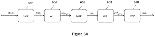
- Figure 6A illustrates a process pipeline that converts YCbCr color space to Lab color space, in accordance with embodiments of the specification. This is done in the Lab, 10b (12b, in some embodiments) modules 406 within the adapter 405 shown in Figure 4A .
- YCbCr components, or any other color space components, originating from the camera undergo these processes using linear and non-linear operators.
- the MADs multiply a three-component vector by a designated 3x3 matrix, then add another 3-component designated vector.
- the matrices and vectors may be unique for each MAD instantiation.
- LUTs may operate independently and simultaneously on the three components of the input vector, to perform non-linear functions.
- the non-linear functions may be unique for each LUT instantiation. Within a LUT, the same function may be applied to all three components.
- a MAD instantiation performs YCbCr to RGB conversion.
- a MAD instantiation performs RGB to XYZ conversion.
- a MAD instantiation performs XYZ to Lab conversion excluding the non-linear function, performed already in previous step 608.
- FIG. 6B is a flowchart illustrating the steps performed by the process pipeline shown in Figure 6A .
- a YCbCr color space video stream is input to a MAD instantiation for conversion to a RGB format.
- an inverse Gama correction is applied to the output RGB data by using a LUT.
- the corrected RGB data output at step 6104 is input to a MAD instantiation for conversion to a XYZ format.
- a correction function is applied to the output XYZ data by using a LUT.
- the corrected XYZ data output at step 6108 is input to a MAD instantiation and converted to a LAB format.
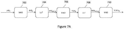
- Figure 7A illustrates a process pipeline that converts Lab color space to YCbCr (or any other) color space, in accordance with embodiments of the specification. This is done in modules 430 shown in Figure 4A .
- the pipeline shown in Figure 7A is the inverse of the pipeline shown in Figure 6A .
- a Lab format may not enable screens to display video images effectively, therefore they are converted back to YCbCr (or any other color space) that may be compatible with one or more displays used with the endoscope system.
- a MAD instantiation performs Lab to XYZ conversion excluding the non-linear function, performed in the next step and is known to those skilled in the art.
- a MAD instantiation performs XYZ to RGB conversion.
- a MAD instantiation performs RGB to YCbCr conversion.
- modules 430 of Figure 4A may include a decimator for converting the YCbCr 4:4:4 at the output of step 520, to YCbCr 4:2:2.
- FIG. 7B is a flowchart illustrating the steps performed by the process pipeline shown in Figure 7A .
- a LAB color space video stream is input to a MAD instantiation for conversion to a XYZ format.
- the corrected XYZ data output at step 7104 is input to a MAD instantiation for conversion to a RGB format.
- an inverse Gama correction function is applied to the output RGB data by using a LUT.
- the corrected RGB data, output at step 7108 is input to a MAD instantiation and converted to a to YCbCr format.
- Figure 8A illustrates a traditional image 802 captured by an endoscope placed inside a body cavity.
- Figure 8B illustrates the image of Figure 8A enhanced by using the method of the present specification.
- Image 804 presents a highly enhanced image as compared to image 802 resulting from user's control over contrast and noise in accordance with the embodiments of the present specification.
- Embodiments of the present specification allow image enhancement during real time video capture and display, in addition to enabling image enhancement of still images. Moreover, embodiments of the specification operate on Lab color space, thus allowing greater contrast enhancement abilities. Additionally, tile overlapping, as described in embodiments above further enhance traditional CLAHE implementations. Yet another step for enhancement is the use of bi-cubic interpolation for inter-tile interpolation. The various advantages are applicable simultaneously to videos captured by multiple viewing elements in an endoscope system.
Landscapes
- Engineering & Computer Science (AREA)
- Physics & Mathematics (AREA)
- Health & Medical Sciences (AREA)
- Life Sciences & Earth Sciences (AREA)
- Theoretical Computer Science (AREA)
- General Physics & Mathematics (AREA)
- Surgery (AREA)
- Signal Processing (AREA)
- Heart & Thoracic Surgery (AREA)
- Public Health (AREA)
- Pathology (AREA)
- Biomedical Technology (AREA)
- Optics & Photonics (AREA)
- Medical Informatics (AREA)
- Molecular Biology (AREA)
- Animal Behavior & Ethology (AREA)
- General Health & Medical Sciences (AREA)
- Radiology & Medical Imaging (AREA)
- Veterinary Medicine (AREA)
- Nuclear Medicine, Radiotherapy & Molecular Imaging (AREA)
- Biophysics (AREA)
- Multimedia (AREA)
- Endoscopes (AREA)
- Closed-Circuit Television Systems (AREA)
- Instruments For Viewing The Inside Of Hollow Bodies (AREA)
- Image Processing (AREA)
Claims (11)
- Verfahren zum Verbessern von Bildern, die durch mindestens zwei Betrachtungselemente in einem Endoskopsystem (100, 210) mit einem Algorithmus zur kontrastbegrenzten adaptiven Histogrammentzerrung (CLAHE) erhalten wurden, wobei das Endoskopsystem (100, 210) eine Steuerung (230, 320) mit einem Baseboard-Modul aufweist, das ein Field Programmable Gate Array (FPGA) (323, 402) aufweist, die zum Umsetzen des Algorithmus ausgebildet ist, wobei das Verfahren die folgenden Schritte aufweist:Eingeben von YCbCr-Farbraum-Videodatenströmen in einen Adapter auf dem FPGA;Umwandeln der YCbCr-Farbraum-Videodatenströme in Lab-Farbraum-Videodatenströme in dem Adapter;Verschieben von Lab-Farbraum-Videodatenströmen zu einem Frame Grabber auf dem FPGA;Aufnehmen der Lab-Farbraum-Videodatenströme als Lab-Farbraum-Frames in dem Frame Grabber;Abziehen der Lab-Farbraum-Frames vom Frame Grabber mit einem Rückholmodul auf dem FPGA;Umordnen der Lab-Farbraum-Framedaten im Rückholmodul zum Erzeugen von verzögerten Lab-Datenströmen;Verschieben der verzögerten Lab-Datenströme an separate Module innerhalb eines Interpolationsalgorithmus auf dem FPGA; undBearbeiten der verzögerten Lab-Datenströme mit dem CLAHE-Algorithmus in den separaten Modulen, um kontrastverstärkte Lab-Datenströme zu erzeugen.
- Verfahren nach Anspruch 1, wobei das Umwandeln der YCbCr-Farbraum-Datenströme in Lab-Farbraum-Videodatenströme die folgenden Schritte aufweist:Durchführen einer Umwandlung der YCbCr-Datenströme von YCbCr zu RGB mittels Schritten zur Matrixmultiplikation und Vektoraddition (MAD), die auf dem FPGA instantiiert sind;Anwenden einer inversen Gammakorrekturfunktion Y(x) = x^2,40 auf die RGB-Daten mit einer Referenztabelle (LUT);Durchführen einer Umwandlung der gammakorrigierten RGB-Daten von RGB zu YXZ mit den auf dem FPGA instantiierten MAD-Schritten;Ausführen einer Korrekturfunktion Y(x) = x^0,33 für die XYZ-Daten mit einer LUT; undDurchführen einer Umwandlung der korrigierten XYZ-Daten von XYZ zu Lab mit den auf dem FPGA instantiierten MAD-Schritten.
- Verfahren nach Anspruch 1, ferner aufweisend das Umwandeln der kontrastverstärkten Lab-Datenströme in kontrastverstärkte YCbCr-Datenströme.
- Verfahren nach Anspruch 3, wobei das Umwandeln der kontrastverstärkten Lab-Datenströme in kontrastverstärkte YCbCr-Datenströme die folgenden Schritte aufweist:Durchführen einer Umwandlung der Lab-Datenströme von Lab zu XYZ mittels Schritten zur Multiplikation und Vektoraddition (MAD), die auf dem FPGA instantiiert sind;Ausführen einer Korrekturfunktion Y(x) = x^0,33 für die XYZ-Daten mit einer Referenztabelle (LUT);Durchführen einer Umwandlung der korrigierten XYZ-Daten von XYZ zu RGB mit den auf dem FPGA instantiierten MAD-Schritten;Anwenden einer inversen Gammakorrekturfunktion Y(x) = x^(1/2,40) auf die RGB-Daten mit einer LUT;Durchführen einer Umwandlung der gammakorrigierten RGB-Daten von RGB zu YCbCr mit den auf dem FPGA instantiierten MAD-Schritten.
- Verfahren zum Verbessern von Bildern, die durch mindestens zwei Betrachtungselemente in einem Endoskopsystem (100, 210) mit einem Algorithmus zur kontrastbegrenzten adaptiven Histogrammentzerrung (CLAHE) erhalten wurden, wobei das Endoskopsystem (100, 210) eine Steuerung (230, 320) mit einem Baseboard-Modul aufweist, das einen Prozessor aufweist, der zum Umsetzen des Algorithmus ausgebildet ist, wobei das Verfahren die folgenden Schritte aufweist:Eingeben zweier YCbCr-Farbraum-Videodatenströme von den mindestens zwei Betrachtungselementen in einen Adapter auf dem Prozessor, wobei jedes Betrachtungselement einen der zwei YCbCr-Farbraum-Videodatenströme bereitstellt;Umwandeln jedes der mindestens zwei YCbCr-Farbraum-Videodatenströme in entsprechende mindestens zwei Lab-Farbraum-Videodatenströme in dem Adapter;Übertragen der mindestens zwei Lab-Farbraum-Videodatenströme an ein Frame Grabber auf dem Prozessor;Erfassen der mindestens zwei Lab-Farbraum-Videodatenströme als entsprechende mindestens zwei Lab-Farbraum-Frames in dem Frame Grabber;Erfassen der mindestens zwei Lab-Farbraum-Frames vom Frame Grabber mit einem Rückholmodul auf dem Prozessor;Umordnen von Lab-Farbraum-Framedaten im Rückholmodul zum Erzeugen von mindestens zwei verzögerten Lab-Datenströmen entsprechend den mindestens zwei Lab-Farbraum-Frames, die vom Rückholmodul erfasst wurden;Übertragen der mindestens zwei verzögerten Lab-Datenströme an entsprechende mindestens zwei separate Module, die einen Interpolationsalgorithmus auf dem Prozessor ausführen; undBearbeiten jedes verzögerten Lab-Datenstroms mit dem Interpolationsalgorithmus in den separaten Modulen zum Erzeugen von mindestens zwei kontrastverstärkten Lab-Datenströmen, wobei der Interpolationsalgorithmus ein CLAHE-Algorithmus ist.
- Verfahren nach Anspruch 5, wobei das Umwandeln eines YCbCr-Farbraum-Datenstroms in einen Lab-Farbraum-Videodatenstrom die folgenden Schritte aufweist:Durchführen einer Umwandlung des YCbCr-Datenstroms von YCbCr zu RGB mittels Schritten zur Matrixmultiplikation und Vektoraddition (MAD), die auf dem Prozessor instantiiert sind;Anwenden einer inversen Gammakorrekturfunktion Y(x) = x^2,40 auf die RGB-Daten mit einer Referenztabelle (LUT);Durchführen einer Umwandlung der gammakorrigierten RGB-Daten von RGB zu XYZ mit den auf dem Prozessor instantiierten MAD-Schritten;Ausführen einer Korrekturfunktion Y(x) = x^0,33 für die XYZ-Daten mit einer LUT; undDurchführen einer Umwandlung der korrigierten XYZ-Daten von XYZ zu Lab mit den auf dem Prozessor instantiierten MAD-Schritten.
- Verfahren nach Anspruch 5, ferner umfassend das Umwandeln jedes der mindestens zwei kontrastverstärkten Lab-Datenströme in mindestens zwei kontrastverstärkte YCbCr-Datenströme.
- Verfahren nach Anspruch 7, wobei das Umwandeln jedes kontrastverstärkten Lab-Datenstroms in einen kontrastverstärkten YCbCr-Datenstrom die folgenden Schritte aufweist:Durchführen einer Umwandlung des Lab-Datenstroms von Lab zu XYZ mittels Schritten zur Matrixmultiplikation und Vektoraddition (MAD), die auf dem Prozessor instantiiert sind;Ausführen einer Korrekturfunktion Y(x) = x^0,33 mit einer Referenztabelle (LUT);Durchführen einer Umwandlung der korrigierten XYZ-Daten von XYZ zu RGB mit den auf dem Prozessor instantiierten MAD-Schritten;Anwenden einer inversen Gammakorrekturfunktion Y(x) = x^(1/2,40) auf die RGB-Daten mit einer LUT; undDurchführen einer Umwandlung der korrigierten RGB-Daten von RGB zu YCbCr mit den auf dem Prozessor instantiierten MAD-Schritten.
- Verfahren zum Verbessern von Bildern nach Anspruch 5, wobei die Bilder von drei Betrachtungselementen erhalten werden.
- Verfahren nach Anspruch 9, umfassend die folgenden Schritte:Eingeben dreier YCbCr-Farbraum-Videodatenströme aus den drei Betrachtungselementen in einen Adapter auf dem Prozessor;Umwandeln der drei YCbCr-Farbraum-Videodatenströme in entsprechende drei Lab-Farbraum-Videodatenströme in dem Adapter;Übertragen der drei Lab-Farbraum-Videodatenströme an ein Frame Grabber auf dem Prozessor in Form eines Videodatenstroms, der aus einem einzigen Betrachtungselement erhalten wird, wobei eine Y-Eingabe des Frame Grabbers durch einen Lab-Farbraum-Videodatenstrom eines ersten Betrachtungselements gespeist wird, eine Cb-Eingabe des Frame Grabbers durch einen Lab-Farbraum-Videodatenstrom eines zweiten Betrachtungselements gespeist wird, und eine Cr-Eingabe des Frame Grabbers durch einen Lab-Farbraum-Videodatenstrom eines dritten Betrachtungselements gespeist wird;Erfassen der drei Lab-Farbraum-Videodatenströme als entsprechende drei Lab-Farbraum-Frames in dem Frame Grabber;Übertragen der drei Lab-Farbraum-Frames vom Frame Grabber mit einem Rückholmodul auf dem Prozessor;Umordnen der Lab-Farbraum-Framedaten der drei Lab-Farbraum-Frames im Rückholmodul zum Erzeugen von drei verzögerten Lab-Datenströmen entsprechend den drei vom Rückholmodul erfassten Lab-Farbraum-Frames;Übertragen der drei verzögerten Lab-Datenströme an entsprechende drei separate Module, die einen Interpolationsalgorithmus auf dem Prozessor ausführen; undBearbeiten jedes verzögerten Lab-Datenstroms mit dem Interpolationsalgorithmus in den separaten Modulen zum Erzeugen von drei kontrastverstärkten Lab-Datenströmen, bei denen es sich um einen ersten, ein zweiten und einen dritten kontrastverstärkten Lab-Datenstrom handelt, wobei jeder kontrastverstärkte Lab-Datenstrom einem Betrachtungselement des Endoskops entspricht.
- Verfahren nach Anspruch 10, wobei das erste Betrachtungselement ein linksseitiges Betrachtungselement des Endoskops, das zweite Betrachtungselement ein vorderes Betrachtungselement des Endoskops und das dritte Betrachtungselement ein rechtsseitiges Betrachtungselement des Endoskops ist; und/oder wobei der erste der drei kontrastverstärkten Lab-Datenströme als erstes Bild auf einem oder mehreren mit der Steuerung (230, 320) gekoppelten Anzeigebildschirmen (120, 240a, 240b, 240c) angezeigt wird, der zweite der drei kontrastverstärkten Lab-Datenströme als zweites Bild auf einem oder mehreren mit der Steuerung (230, 320) gekoppelten Anzeigebildschirmen angezeigt wird, und der dritte der drei kontrastverstärkten Lab-Datenströme als drittes Bild auf einem oder mehreren mit der Steuerung (230, 320) gekoppelten Anzeigebildschirmen angezeigt wird, und/oder wobei jeder kontrastverstärkte Lab-Datenstrom als kontrastverstärktes Bild auf einem mit der Steuerung (230, 320) gekoppelten Anzeigegerät angezeigt wird, jedes kontrastverstärkte Bild mehrere Frames mit einem stärkeren Grad von Kontrast aufweist als die entsprechenden mehreren Frames im entsprechenden, von einem Betrachtungselement des Endoskops erhaltenen Bild, bevor dieses mit dem Bildverbesserungsverfahren nach Anspruch 5 bearbeitet wurde.
Priority Applications (1)
| Application Number | Priority Date | Filing Date | Title |
|---|---|---|---|
| EP20182853.0A EP3747349A1 (de) | 2015-05-17 | 2016-05-16 | Endoskopische bildverbesserung mit in einem prozessor implementierter kontrastbegrenzter adaptiver histogrammentzerrung |
Applications Claiming Priority (3)
| Application Number | Priority Date | Filing Date | Title |
|---|---|---|---|
| US201562162788P | 2015-05-17 | 2015-05-17 | |
| US201562259683P | 2015-11-25 | 2015-11-25 | |
| PCT/US2016/032715 WO2016187124A1 (en) | 2015-05-17 | 2016-05-16 | Endoscopic image enhancement using contrast limited adaptive histogram equalization (clahe) implemented in a processor |
Related Child Applications (1)
| Application Number | Title | Priority Date | Filing Date |
|---|---|---|---|
| EP20182853.0A Division EP3747349A1 (de) | 2015-05-17 | 2016-05-16 | Endoskopische bildverbesserung mit in einem prozessor implementierter kontrastbegrenzter adaptiver histogrammentzerrung |
Publications (3)
| Publication Number | Publication Date |
|---|---|
| EP3297515A1 EP3297515A1 (de) | 2018-03-28 |
| EP3297515A4 EP3297515A4 (de) | 2019-01-23 |
| EP3297515B1 true EP3297515B1 (de) | 2020-07-01 |
Family
ID=57277717
Family Applications (2)
| Application Number | Title | Priority Date | Filing Date |
|---|---|---|---|
| EP20182853.0A Pending EP3747349A1 (de) | 2015-05-17 | 2016-05-16 | Endoskopische bildverbesserung mit in einem prozessor implementierter kontrastbegrenzter adaptiver histogrammentzerrung |
| EP16797096.1A Active EP3297515B1 (de) | 2015-05-17 | 2016-05-16 | Endoskopische bildverbesserung mit in einem prozessor implementierter kontrastbegrenzter adaptiver histogrammentzerrung |
Family Applications Before (1)
| Application Number | Title | Priority Date | Filing Date |
|---|---|---|---|
| EP20182853.0A Pending EP3747349A1 (de) | 2015-05-17 | 2016-05-16 | Endoskopische bildverbesserung mit in einem prozessor implementierter kontrastbegrenzter adaptiver histogrammentzerrung |
Country Status (4)
| Country | Link |
|---|---|
| US (6) | US10516865B2 (de) |
| EP (2) | EP3747349A1 (de) |
| ES (1) | ES2818174T3 (de) |
| WO (1) | WO2016187124A1 (de) |
Families Citing this family (40)
| Publication number | Priority date | Publication date | Assignee | Title |
|---|---|---|---|---|
| ES2818174T3 (es) | 2015-05-17 | 2021-04-09 | Endochoice Inc | Mejora de imagen endoscópica usando ecualización de histograma adaptativo limitado por contraste (CLAHE) implementada en un procesador |
| WO2018088498A1 (ja) * | 2016-11-10 | 2018-05-17 | 京セラオプテック株式会社 | 体腔内観察システム、トロカール装置、及び体腔内観察システムの作動方法 |
| JP7048628B2 (ja) | 2016-11-28 | 2022-04-05 | アダプティブエンドウ エルエルシー | 分離可能使い捨てシャフト付き内視鏡 |
| CN107481187B (zh) * | 2017-09-29 | 2022-04-19 | 康佳集团股份有限公司 | 一种视频图像处理方法、智能终端及存储介质 |
| KR102524671B1 (ko) * | 2018-01-24 | 2023-04-24 | 삼성전자주식회사 | 전자 장치 및 그의 제어 방법 |
| US10955657B2 (en) * | 2018-12-20 | 2021-03-23 | Acclarent, Inc. | Endoscope with dual image sensors |
| US10534948B1 (en) | 2019-03-18 | 2020-01-14 | Capital One Services, Llc | Optimizing detection of images in relation to targets based on colorspace transformation techniques |
| US10496911B1 (en) | 2019-03-18 | 2019-12-03 | Capital One Services, Llc | Detection of images in relation to targets based on colorspace transformation techniques and utilizing ultraviolet and infrared light |
| US10509991B1 (en) | 2019-03-18 | 2019-12-17 | Capital One Services, Llc | Detection of images in relation to targets based on colorspace transformation techniques and utilizing infrared light |
| US10496862B1 (en) | 2019-03-18 | 2019-12-03 | Capital One Services, Llc | Detection of images in relation to targets based on colorspace transformation techniques and utilizing ultraviolet light |
| US10523420B1 (en) | 2019-04-18 | 2019-12-31 | Capital One Services, Llc | Transmitting encoded data along transmission mediums based on colorspace schemes |
| US10504013B1 (en) | 2019-04-24 | 2019-12-10 | Capital One Services, Llc | Colorspace encoding multimedia data on a physical page |
| US10529300B1 (en) | 2019-06-20 | 2020-01-07 | Capital One Services, Llc | Adaptive image display based on colorspace conversions |
| US10614635B1 (en) | 2019-07-25 | 2020-04-07 | Capital One Services, Llc | Augmented reality system with color-based fiducial marker |
| WO2021024245A1 (en) * | 2019-08-05 | 2021-02-11 | 270 Surgical Ltd | System and method for displaying images of a medical imaging endoscope |
| US10833852B1 (en) | 2019-10-03 | 2020-11-10 | Capital One Services, Llc | Encoded data along tape based on colorspace schemes |
| US10715183B1 (en) | 2019-10-25 | 2020-07-14 | Capital One Services, Llc | Data encoding with error-correcting code pursuant to colorspace schemes |
| US10867226B1 (en) * | 2019-11-04 | 2020-12-15 | Capital One Services, Llc | Programmable logic array and colorspace conversions |
| US10762371B1 (en) | 2019-11-14 | 2020-09-01 | Capital One Services, Llc | Object detection techniques using colorspace conversions |
| CN111062897B (zh) * | 2019-12-06 | 2023-09-29 | Oppo广东移动通信有限公司 | 图像均衡方法、终端及存储介质 |
| US10878600B1 (en) | 2019-12-10 | 2020-12-29 | Capital One Services, Llc | Augmented reality system with color-based fiducial marker utilizing local adaptive technology |
| EP4076130A1 (de) | 2019-12-19 | 2022-10-26 | Ambu A/S | Bilderfassungsauswahl |
| CN111010497A (zh) * | 2019-12-26 | 2020-04-14 | 苏州羿景睿图信息科技有限公司 | 一种基于fpga的clahe透雾增强并行运算方法 |
| USD1018844S1 (en) | 2020-01-09 | 2024-03-19 | Adaptivendo Llc | Endoscope handle |
| US11302036B2 (en) | 2020-08-19 | 2022-04-12 | Capital One Services, Llc | Color conversion between color spaces using reduced dimension embeddings |
| TR202013601A1 (tr) * | 2020-08-28 | 2022-03-21 | Aselsan Elektronik Sanayi Ve Ticaret As | CLAHE algoritması için değişken kıpma seviyesi hesaplama yöntemi |
| WO2022051775A1 (en) | 2020-09-04 | 2022-03-10 | Abova, Inc. | Method for x-ray dental image enhancement |
| CN116471974A (zh) | 2020-09-22 | 2023-07-21 | 波士顿科学国际有限公司 | 图像处理系统及其使用方法 |
| AU2021349199A1 (en) * | 2020-09-25 | 2023-04-06 | Boston Scientific Scimed, Inc. | Color extrapolation from monochrome image sensor |
| USD1051380S1 (en) | 2020-11-17 | 2024-11-12 | Adaptivendo Llc | Endoscope handle |
| CN112365424B (zh) * | 2020-11-17 | 2023-06-06 | 昆明物理研究所 | 基于局部自适应clahe的红外图像去噪增强方法、装置、系统与计算机可读存储介质 |
| DE102021100124A1 (de) * | 2021-01-07 | 2022-07-07 | Schölly Fiberoptic GmbH | Verfahren zum Übertragen eines Rohbilddatenstromes eines Bildsensors |
| CN117157666A (zh) | 2021-02-17 | 2023-12-01 | 波士顿科学国际有限公司 | 图像处理系统及其使用方法 |
| USD1031035S1 (en) | 2021-04-29 | 2024-06-11 | Adaptivendo Llc | Endoscope handle |
| USD1070082S1 (en) | 2021-04-29 | 2025-04-08 | Adaptivendo Llc | Endoscope handle |
| USD1066659S1 (en) | 2021-09-24 | 2025-03-11 | Adaptivendo Llc | Endoscope handle |
| CN115018737B (zh) * | 2022-08-04 | 2023-02-21 | 四川迪晟新达类脑智能技术有限公司 | 一种红外热像增强方法及装置 |
| TWI821146B (zh) * | 2023-04-26 | 2023-11-01 | 國立中正大學 | 用於偵測組織出血之影像分析方法 |
| CN117152029B (zh) * | 2023-10-30 | 2024-03-08 | 浦江三思光电技术有限公司 | 一种基于fpga的clahe图像增强实时处理方法及系统 |
| CN119809911A (zh) * | 2024-12-19 | 2025-04-11 | 电子科技大学 | 一种clahe算法的图像增强硬件加速器 |
Family Cites Families (405)
| Publication number | Priority date | Publication date | Assignee | Title |
|---|---|---|---|---|
| US4084401A (en) | 1975-07-09 | 1978-04-18 | Hughes Aircraft Company | Digital watch with two buttons and improved setting and display control |
| US4027697A (en) | 1975-11-19 | 1977-06-07 | Bonney Roland W | Rotary valve |
| US4027897A (en) | 1975-11-25 | 1977-06-07 | Kurt Hildebrand | Safety ski binding |
| JPS54144787A (en) | 1978-05-02 | 1979-11-12 | Ouchi Teruo | Device for curving endoscope |
| JPS5675133A (en) | 1979-11-22 | 1981-06-22 | Olympus Optical Co | Light source apparatus for endoscope |
| JPS57192547A (en) | 1981-05-21 | 1982-11-26 | Olympus Optical Co | Ultrasonic diagnostic apparatus for body cavity |
| US4532918A (en) | 1983-10-07 | 1985-08-06 | Welch Allyn Inc. | Endoscope signal level control |
| GR80917B (en) | 1983-11-14 | 1985-03-13 | Dow Chemical Co | Novel substituted 1,2,4-triazolo (1,5-a) pyrimidine-2-sulfonamides and compositions and methods of controlling undesired vegetation and suppressing the nitrification of ammonium nitrogen in soil |
| JPS60263916A (ja) | 1984-06-13 | 1985-12-27 | Olympus Optical Co Ltd | 内焦式内視鏡対物レンズ |
| US4588294A (en) | 1984-06-27 | 1986-05-13 | Warner-Lambert Technologies, Inc. | Searching and measuring endoscope |
| US4641635A (en) | 1984-08-15 | 1987-02-10 | Olympus Optical Co., Ltd. | Endoscope apparatus |
| JPS62220918A (ja) | 1986-03-22 | 1987-09-29 | Olympus Optical Co Ltd | 内視鏡光学系 |
| JPH07119893B2 (ja) | 1986-09-22 | 1995-12-20 | オリンパス光学工業株式会社 | 内視鏡光学系 |
| US4727859A (en) | 1986-12-29 | 1988-03-01 | Welch Allyn, Inc. | Right angle detachable prism assembly for borescope |
| JP2697822B2 (ja) | 1987-05-25 | 1998-01-14 | オリンパス光学工業株式会社 | 内視鏡対物レンズ |
| JP2804267B2 (ja) | 1988-05-02 | 1998-09-24 | オリンパス光学工業株式会社 | 内視鏡対物レンズ |
| US4825850A (en) | 1988-05-13 | 1989-05-02 | Opielab, Inc. | Contamination protection system for endoscope control handles |
| JPH0223931A (ja) | 1988-07-13 | 1990-01-26 | Asahi Optical Co Ltd | 内視鏡の湾曲装置のブレーキ装置 |
| JPH07122692B2 (ja) | 1988-09-29 | 1995-12-25 | 富士写真光機株式会社 | 内視鏡用対物レンズ |
| US5007406A (en) | 1988-11-04 | 1991-04-16 | Asahi Kogaku Kogyo Kabushiki Kaisha | Bending control device of endoscope |
| US5193525A (en) | 1990-11-30 | 1993-03-16 | Vision Sciences | Antiglare tip in a sheath for an endoscope |
| US5224929A (en) | 1990-12-21 | 1993-07-06 | C. R. Bard, Inc. | Irrigation/aspiration cannula and valve assembly |
| JPH0827429B2 (ja) | 1991-03-04 | 1996-03-21 | オリンパス光学工業株式会社 | 内視鏡用対物レンズ |
| US5359456A (en) | 1991-10-15 | 1994-10-25 | Olympus Optical Co., Ltd. | Objective lens system for endoscopes |
| US5630782A (en) | 1992-09-01 | 1997-05-20 | Adair; Edwin L. | Sterilizable endoscope with separable auxiliary assembly |
| CA2143639C (en) | 1992-09-01 | 2004-07-20 | Edwin L. Adair | Sterilizable endoscope with separable disposable tube assembly |
| US5298971A (en) | 1992-09-23 | 1994-03-29 | Industrial Technology Research Institute | Lateral shear interferometer for testing aspheric surfaces |
| US5836894A (en) | 1992-12-21 | 1998-11-17 | Artann Laboratories | Apparatus for measuring mechanical parameters of the prostate and for imaging the prostate using such parameters |
| JP3372980B2 (ja) | 1993-01-22 | 2003-02-04 | オリンパス光学工業株式会社 | 内視鏡 |
| US5863286A (en) | 1993-01-27 | 1999-01-26 | Olympus Optical Company, Ltd. | Endoscope system including endoscope and disposable protection cover |
| US5674182A (en) | 1993-02-26 | 1997-10-07 | Olympus Optical Co., Ltd. | Endoscope system including endoscope and protection cover |
| US5460167A (en) | 1993-03-04 | 1995-10-24 | Olympus Optical Co., Ltd. | Endoscope cover with channel |
| US5569157A (en) | 1993-05-07 | 1996-10-29 | Olympus Optical Co., Ltd. | Endoscope |
| US5447148A (en) | 1993-07-08 | 1995-09-05 | Vision Sciences, Inc. | Endoscopic contamination protection system to facilitate cleaning of endoscopes |
| JP3392920B2 (ja) | 1993-11-26 | 2003-03-31 | ペンタックス株式会社 | 内視鏡の先端部 |
| US5725477A (en) | 1993-11-18 | 1998-03-10 | Asahi Kogaku Kogyo Kabushiki Kaisha | Front end structure of endoscope |
| JP3272516B2 (ja) | 1993-11-18 | 2002-04-08 | 旭光学工業株式会社 | 内視鏡の先端部 |
| US5395329A (en) | 1994-01-19 | 1995-03-07 | Daig Corporation | Control handle for steerable catheter |
| US5575755A (en) | 1994-02-23 | 1996-11-19 | Welch Allyn, Inc. | Fluid insensitive braking for an endoscope |
| US5464007A (en) | 1994-02-23 | 1995-11-07 | Welch Allyn, Inc. | Fluid insensitive braking for an endoscope |
| US5547455A (en) | 1994-03-30 | 1996-08-20 | Medical Media Systems | Electronically steerable endoscope |
| US5685823A (en) | 1994-03-30 | 1997-11-11 | Asahi Kogaku Kogyo Kabushiki Kaisha | End structure of endoscope |
| US5782751A (en) | 1994-05-26 | 1998-07-21 | Asahi Kogaku Kogyo Kabushiki Kaisha | Side view endoscope |
| JP3379821B2 (ja) | 1994-05-31 | 2003-02-24 | オリンパス光学工業株式会社 | 内視鏡 |
| US5518502A (en) | 1994-06-08 | 1996-05-21 | The United States Surgical Corporation | Compositions, methods and apparatus for inhibiting fogging of endoscope lenses |
| JP3051035B2 (ja) | 1994-10-18 | 2000-06-12 | 富士写真光機株式会社 | 内視鏡用対物レンズ |
| US5940126A (en) | 1994-10-25 | 1999-08-17 | Kabushiki Kaisha Toshiba | Multiple image video camera apparatus |
| US5683538A (en) | 1994-12-23 | 1997-11-04 | International Business Machines Corporation | Control of etch selectivity |
| JPH0910170A (ja) | 1995-06-29 | 1997-01-14 | Olympus Optical Co Ltd | 内視鏡対物光学系 |
| JPH0980305A (ja) | 1995-09-11 | 1997-03-28 | Fuji Photo Optical Co Ltd | 内視鏡対物レンズ |
| US5810715A (en) | 1995-09-29 | 1998-09-22 | Olympus Optical Co., Ltd. | Endoscope provided with function of being locked to flexibility of insertion part which is set by flexibility modifying operation member |
| US6117068A (en) | 1995-10-19 | 2000-09-12 | Elite Genetics, Inc | Artificial insemination system |
| US5725478A (en) | 1996-03-14 | 1998-03-10 | Saad; Saad A. | Methods and apparatus for providing suction and/or irrigation in a rigid endoscope while maintaining visual contact with a target area through the endoscope |
| US5860913A (en) | 1996-05-16 | 1999-01-19 | Olympus Optical Co., Ltd. | Endoscope whose distal cover can be freely detachably attached to main distal part thereof with high positioning precision |
| JPH1043129A (ja) | 1996-08-02 | 1998-02-17 | Olympus Optical Co Ltd | 内視鏡 |
| DE19636152C2 (de) | 1996-09-06 | 1999-07-01 | Schneider Co Optische Werke | Kompaktes Weitwinkelobjektiv |
| JP4054094B2 (ja) | 1996-12-27 | 2008-02-27 | オリンパス株式会社 | 電子内視鏡 |
| AUPO478397A0 (en) | 1997-01-31 | 1997-02-20 | Fairmont Medical Products Pty Ltd | Endoscopic drape |
| US6095970A (en) | 1997-02-19 | 2000-08-01 | Asahi Kogaku Kogyo Kabushiki Kaisha | Endoscope |
| JPH10239740A (ja) | 1997-02-28 | 1998-09-11 | Toshiba Corp | 内視鏡装置 |
| US5924976A (en) | 1997-08-21 | 1999-07-20 | Stelzer; Paul | Minimally invasive surgery device |
| US20040052076A1 (en) | 1997-08-26 | 2004-03-18 | Mueller George G. | Controlled lighting methods and apparatus |
| BR9812021A (pt) | 1997-08-28 | 2000-09-26 | Qualia Computing Inc | Processos para detecção automatizada de microcalcificações, para segmentar uma área de uma imagem de mamograma digital, para detecção automatizada de microcalcifição agrupada, para incorporar as detecções de saìda de um sistema de detecção auxiliado por computador para detectar microcalcificações agrupadas em um mamograma, e, para coleta automatizada de imagem, aparelhos para detecção automatizada de microcalcificações, e, para detectar microcalcificações agrupadas em uma imagem de mamograma digital |
| US20110034769A1 (en) | 1997-10-06 | 2011-02-10 | Micro-Imaging Solutions Llc | Reduced area imaging device incorporated within wireless endoscopic devices |
| JPH11137512A (ja) | 1997-11-07 | 1999-05-25 | Toshiba Corp | 内視鏡装置 |
| US7598686B2 (en) | 1997-12-17 | 2009-10-06 | Philips Solid-State Lighting Solutions, Inc. | Organic light emitting diode methods and apparatus |
| US6277064B1 (en) | 1997-12-30 | 2001-08-21 | Inbae Yoon | Surgical instrument with rotatably mounted offset endoscope |
| US6196967B1 (en) | 1998-03-18 | 2001-03-06 | Linvatec Corporation | Arthroscopic component joining system |
| US6419626B1 (en) | 1998-08-12 | 2002-07-16 | Inbae Yoon | Surgical instrument endoscope with CMOS image sensor and physical parameter sensor |
| JP4014186B2 (ja) | 1998-11-30 | 2007-11-28 | フジノン株式会社 | 内視鏡用対物レンズ |
| JP3704248B2 (ja) | 1999-02-04 | 2005-10-12 | ペンタックス株式会社 | 内視鏡用操作ワイヤの連結構造 |
| US7015950B1 (en) | 1999-05-11 | 2006-03-21 | Pryor Timothy R | Picture taking method and apparatus |
| JP4245731B2 (ja) | 1999-06-08 | 2009-04-02 | オリンパス株式会社 | 液晶レンズユニットと液晶レンズアッセンブリー |
| IL143258A0 (en) | 2001-05-20 | 2002-04-21 | Given Imaging Ltd | A method for in vivo imaging of the gastrointestinal tract in unmodified conditions |
| US20020087047A1 (en) | 1999-09-13 | 2002-07-04 | Visionscope, Inc. | Miniature endoscope system |
| US6486889B1 (en) * | 1999-12-17 | 2002-11-26 | Xerox Corporation | Methods and apparatus for transforming RGB video |
| JP2001177824A (ja) | 1999-12-17 | 2001-06-29 | Asahi Optical Co Ltd | 電子内視鏡用信号切替装置 |
| US20010036322A1 (en) | 2000-03-10 | 2001-11-01 | Bloomfield John F. | Image processing system using an array processor |
| US6837846B2 (en) | 2000-04-03 | 2005-01-04 | Neo Guide Systems, Inc. | Endoscope having a guide tube |
| US6712760B2 (en) | 2000-04-10 | 2004-03-30 | Pentax Corporation | Television device of portable endoscope |
| US6673012B2 (en) | 2000-04-19 | 2004-01-06 | Pentax Corporation | Control device for an endoscope |
| US20010055062A1 (en) | 2000-04-20 | 2001-12-27 | Keiji Shioda | Operation microscope |
| EP1301118B1 (de) | 2000-07-14 | 2006-09-06 | Xillix Technologies Corp. | Kompaktes fluorezenz endoskopisches video system |
| JP3945133B2 (ja) | 2000-08-02 | 2007-07-18 | フジノン株式会社 | 内視鏡の観察窓洗浄装置 |
| US6717092B2 (en) | 2000-08-11 | 2004-04-06 | Pentax Corporation | Method of manufacturing treatment instrument of endoscope |
| US6927769B2 (en) * | 2000-11-21 | 2005-08-09 | Vrex, Inc. | Stereoscopic image processing on a computer system |
| IL156961A0 (en) | 2001-01-16 | 2004-02-08 | Given Imaging Ltd | System and method for wide field imaging of body lumens |
| JP3939158B2 (ja) | 2001-02-06 | 2007-07-04 | オリンパス株式会社 | 内視鏡装置 |
| JP2002263055A (ja) | 2001-03-12 | 2002-09-17 | Olympus Optical Co Ltd | 内視鏡先端フード |
| US6711056B2 (en) | 2001-03-12 | 2004-03-23 | Micron Technology, Inc. | Memory with row redundancy |
| DE10121450A1 (de) | 2001-04-27 | 2002-11-21 | Storz Endoskop Gmbh Schaffhaus | Optisches Instrument, insbesondere Endoskop, mit Wechselkopf |
| US6618205B2 (en) | 2001-05-14 | 2003-09-09 | Pentax Corporation | Endoscope objective optical system |
| DE60228165D1 (de) | 2001-05-16 | 2008-09-25 | Olympus Corp | Endoskop mit Bildverarbeitungseinrichtung |
| US7231135B2 (en) | 2001-05-18 | 2007-06-12 | Pentax Of American, Inc. | Computer-based video recording and management system for medical diagnostic equipment |
| US6795879B2 (en) | 2001-08-08 | 2004-09-21 | Texas Instruments Incorporated | Apparatus and method for wait state analysis in a digital signal processing system |
| US7123288B2 (en) | 2001-09-28 | 2006-10-17 | Fujinon Corporation | Electronic endoscope eliminating influence of light distribution in optical zooming |
| US6980227B2 (en) | 2001-10-01 | 2005-12-27 | Pentax Corporation | Electronic endoscope with light-amount adjustment apparatus |
| US20070167681A1 (en) | 2001-10-19 | 2007-07-19 | Gill Thomas J | Portable imaging system employing a miniature endoscope |
| US8038602B2 (en) | 2001-10-19 | 2011-10-18 | Visionscope Llc | Portable imaging system employing a miniature endoscope |
| US6863651B2 (en) | 2001-10-19 | 2005-03-08 | Visionscope, Llc | Miniature endoscope with imaging fiber system |
| EP1313066B1 (de) | 2001-11-19 | 2008-08-27 | STMicroelectronics S.r.l. | Verfahren zur Mischung von digitalen Bildern zur Erzeugung eines digitalen Bildes mit erweitertem Dynamikbereich |
| US6891964B2 (en) * | 2001-11-23 | 2005-05-10 | University Of Chicago | Computerized method for determination of the likelihood of malignancy for pulmonary nodules on low-dose CT |
| US20030153897A1 (en) | 2002-02-12 | 2003-08-14 | Russo Ronald D. | Closed system drainage and infusion connector valve |
| CA2380105A1 (en) * | 2002-04-09 | 2003-10-09 | Nicholas Routhier | Process and system for encoding and playback of stereoscopic video sequences |
| US8988599B2 (en) | 2010-08-31 | 2015-03-24 | University Of Southern California | Illumination sphere with intelligent LED lighting units in scalable daisy chain with interchangeable filters |
| JP4170685B2 (ja) | 2002-07-05 | 2008-10-22 | フジノン株式会社 | 内視鏡の湾曲操作機構 |
| AU2003274984A1 (en) | 2002-09-13 | 2004-04-30 | Karl Storz Imaging, Inc. | Video recording and image capture device |
| US20040061780A1 (en) | 2002-09-13 | 2004-04-01 | Huffman David A. | Solid-state video surveillance system |
| US7097632B2 (en) | 2002-10-28 | 2006-08-29 | Sherwood Services Ag | Automatic valve |
| US7537561B2 (en) | 2002-11-27 | 2009-05-26 | Olympus Corporation | Endoscope apparatus |
| FR2857200B1 (fr) | 2003-07-01 | 2005-09-30 | Tokendo | Processeur video pour l'endoscopie |
| US7267647B2 (en) | 2003-02-10 | 2007-09-11 | Pentax Corporation | Endoscope |
| US7027231B2 (en) | 2003-02-14 | 2006-04-11 | Fujinon Corporation | Endoscope objective lens |
| US7160249B2 (en) | 2003-03-28 | 2007-01-09 | Olympus Corporation | Endoscope image pickup unit for picking up magnified images of an object, a focus adjustment apparatus and method, and a focus range check apparatus and method for the same |
| EP1462076B1 (de) | 2003-03-28 | 2011-07-20 | Medical Technology Transfer Holding B.V. | Operationseinheit |
| US8118732B2 (en) | 2003-04-01 | 2012-02-21 | Boston Scientific Scimed, Inc. | Force feedback control system for video endoscope |
| JP3914168B2 (ja) * | 2003-04-08 | 2007-05-16 | オリンパス株式会社 | 撮像システム、画像処理プログラム |
| KR20040087905A (ko) | 2003-04-09 | 2004-10-15 | 파이오니아 가부시키가이샤 | 플라즈마 디스플레이 패널 |
| EP1620012B1 (de) | 2003-05-01 | 2012-04-18 | Given Imaging Ltd. | Panorama-gesichtsfeld-darstellungsvorrichtung |
| JP4533695B2 (ja) | 2003-09-23 | 2010-09-01 | オリンパス株式会社 | 処置用内視鏡 |
| US20050251127A1 (en) | 2003-10-15 | 2005-11-10 | Jared Brosch | Miniature ultrasonic transducer with focusing lens for intracardiac and intracavity applications |
| CN1878497A (zh) | 2003-11-14 | 2006-12-13 | 杏股份有限公司 | 内窥镜装置及使用它的摄影方法 |
| EP1690497B1 (de) | 2003-12-02 | 2017-02-22 | Olympus Corporation | Ultraschallgerät |
| EP1707102B1 (de) | 2004-01-19 | 2010-05-05 | Olympus Corporation | Medizinische behandlungsvorrichtung vom kapseltyp |
| EP2572626B1 (de) | 2004-01-19 | 2016-03-23 | Olympus Corporation | Endoskop vom kapseltyp |
| JP4448348B2 (ja) | 2004-03-10 | 2010-04-07 | Hoya株式会社 | 内視鏡の送水路 |
| US7922650B2 (en) | 2004-03-23 | 2011-04-12 | Boston Scientific Scimed, Inc. | Medical visualization system with endoscope and mounted catheter |
| JP2005278762A (ja) | 2004-03-29 | 2005-10-13 | Fujinon Corp | 穿刺型内視鏡用プローブ |
| US7976462B2 (en) | 2004-04-06 | 2011-07-12 | Integrated Endoscopy, Inc. | Endoscope designs and methods of manufacture |
| US20050272977A1 (en) | 2004-04-14 | 2005-12-08 | Usgi Medical Inc. | Methods and apparatus for performing endoluminal procedures |
| US8512229B2 (en) | 2004-04-14 | 2013-08-20 | Usgi Medical Inc. | Method and apparatus for obtaining endoluminal access |
| US8277373B2 (en) | 2004-04-14 | 2012-10-02 | Usgi Medical, Inc. | Methods and apparaus for off-axis visualization |
| JP4500096B2 (ja) | 2004-04-27 | 2010-07-14 | オリンパス株式会社 | 内視鏡及び内視鏡システム |
| JP3967337B2 (ja) | 2004-05-14 | 2007-08-29 | オリンパス株式会社 | 内視鏡および内視鏡装置 |
| EP1752083A4 (de) | 2004-05-24 | 2013-10-16 | Olympus Corp | Lichtquellenvorrichtung für ein endoskop |
| IL162390A0 (en) | 2004-06-07 | 2005-11-20 | Medigus Ltd | Multipurpose endoscopy suite |
| US7922655B2 (en) | 2004-07-02 | 2011-04-12 | Osaka University | Endoscope attachment and endoscope |
| JP4544924B2 (ja) | 2004-07-12 | 2010-09-15 | フォルテ グロウ メディカル株式会社 | 内視鏡用送水チューブ |
| US7335159B2 (en) | 2004-08-26 | 2008-02-26 | Scimed Life Systems, Inc. | Endoscope having auto-insufflation and exsufflation |
| JP4649606B2 (ja) | 2004-08-31 | 2011-03-16 | 国立大学法人 名古屋工業大学 | 球状カプセル型全方位内視鏡 |
| US8449457B2 (en) | 2004-09-03 | 2013-05-28 | Stryker Gi Services C.V. | Optical head for endoscope |
| WO2006041426A2 (en) | 2004-09-15 | 2006-04-20 | Adobe Systems Incorporated | Locating a feature in a digital image |
| US8480566B2 (en) | 2004-09-24 | 2013-07-09 | Vivid Medical, Inc. | Solid state illumination for endoscopy |
| JP4636840B2 (ja) | 2004-09-27 | 2011-02-23 | アクア化学株式会社 | 洗浄方法、及び、洗浄装置 |
| US8199187B2 (en) | 2004-09-30 | 2012-06-12 | Boston Scientific Scimed, Inc. | Adapter for use with digital imaging medical device |
| US7621869B2 (en) | 2005-05-06 | 2009-11-24 | Nitesh Ratnakar | Next generation colonoscope |
| US11653816B2 (en) | 2004-10-11 | 2023-05-23 | Nitesh Ratnakar | Next generation endoscope |
| US20090023998A1 (en) | 2007-07-17 | 2009-01-22 | Nitesh Ratnakar | Rear view endoscope sheath |
| US20080275298A1 (en) | 2004-10-11 | 2008-11-06 | Novation Science, Llc | Dual View Endoscope |
| US8585584B2 (en) | 2004-10-11 | 2013-11-19 | Nitesh Ratnakar | Dual view endoscope |
| US9131861B2 (en) | 2004-11-30 | 2015-09-15 | Academisch Medisch Centrum | Pulsed lighting imaging systems and methods |
| US20060149127A1 (en) | 2004-12-30 | 2006-07-06 | Seddiqui Fred R | Disposable multi-lumen catheter with reusable stylet |
| US8289381B2 (en) | 2005-01-05 | 2012-10-16 | Avantis Medical Systems, Inc. | Endoscope with an imaging catheter assembly and method of configuring an endoscope |
| US8235887B2 (en) | 2006-01-23 | 2012-08-07 | Avantis Medical Systems, Inc. | Endoscope assembly with retroscope |
| US20070293720A1 (en) | 2005-01-05 | 2007-12-20 | Avantis Medical Systems, Inc. | Endoscope assembly and method of viewing an area inside a cavity |
| US8797392B2 (en) | 2005-01-05 | 2014-08-05 | Avantis Medical Sytems, Inc. | Endoscope assembly with a polarizing filter |
| US20060149129A1 (en) | 2005-01-05 | 2006-07-06 | Watts H D | Catheter with multiple visual elements |
| US8182422B2 (en) | 2005-12-13 | 2012-05-22 | Avantis Medical Systems, Inc. | Endoscope having detachable imaging device and method of using |
| US20080021274A1 (en) | 2005-01-05 | 2008-01-24 | Avantis Medical Systems, Inc. | Endoscopic medical device with locking mechanism and method |
| US8872906B2 (en) | 2005-01-05 | 2014-10-28 | Avantis Medical Systems, Inc. | Endoscope assembly with a polarizing filter |
| US7668450B2 (en) | 2005-01-28 | 2010-02-23 | Stryker Corporation | Endoscope with integrated light source |
| US8029439B2 (en) | 2005-01-28 | 2011-10-04 | Stryker Corporation | Disposable attachable light source unit for an endoscope |
| US7827032B2 (en) | 2005-02-04 | 2010-11-02 | Vocollect, Inc. | Methods and systems for adapting a model for a speech recognition system |
| US20060215406A1 (en) | 2005-02-18 | 2006-09-28 | William Thrailkill | Medical diagnostic instrument with highly efficient, tunable light emitting diode light source |
| EP1859723B1 (de) | 2005-03-15 | 2012-07-11 | Olympus Medical Systems Corp. | Einführabschnitt für ein endoskop |
| US8388523B2 (en) | 2005-04-01 | 2013-03-05 | Welch Allyn, Inc. | Medical diagnostic instrument having portable illuminator |
| JP4754871B2 (ja) | 2005-05-11 | 2011-08-24 | オリンパスメディカルシステムズ株式会社 | 内視鏡の先端部 |
| US20060293556A1 (en) | 2005-05-16 | 2006-12-28 | Garner David M | Endoscope with remote control module or camera |
| JP4987257B2 (ja) | 2005-06-29 | 2012-07-25 | オリンパス株式会社 | 内視鏡 |
| US20070015989A1 (en) | 2005-07-01 | 2007-01-18 | Avantis Medical Systems, Inc. | Endoscope Image Recognition System and Method |
| RU2008106934A (ru) | 2005-09-06 | 2009-10-20 | 8 Хаешель Ст., Бизнес энд Индастриал Парк (IL) | Сменный наконечник для эндоскопа |
| WO2007034664A1 (ja) | 2005-09-22 | 2007-03-29 | Olympus Corporation | 内視鏡挿入部 |
| GB0519769D0 (en) | 2005-09-28 | 2005-11-09 | Imp College Innovations Ltd | Imaging system |
| US7400709B2 (en) | 2005-09-30 | 2008-07-15 | Mettler-Toledo Safeline X-Ray Limited | X-ray inspection system |
| US7430682B2 (en) | 2005-09-30 | 2008-09-30 | Symbol Technologies, Inc. | Processing image data from multiple sources |
| US7817997B2 (en) | 2005-10-04 | 2010-10-19 | Telefonaktiebolaget Lm Ericsson (Publ) | Redirection of IP-connected radio base station to correct control node |
| US20070162095A1 (en) | 2006-01-06 | 2007-07-12 | Ezc Medical Llc | Modular visualization stylet apparatus and methods of use |
| EP1986541A2 (de) | 2006-01-23 | 2008-11-05 | Avantis Medical Systems, Inc. | Endoskop |
| CN101374452B (zh) | 2006-01-30 | 2012-10-10 | 新浪潮外科器械有限公司 | 在医疗程序前对医用内窥镜白平衡以及应用防雾剂的装置 |
| EP1988812B1 (de) | 2006-02-06 | 2012-11-21 | Avantis Medical Systems, Inc. | Endoskop mit einer bildgebenden katheteranordnung und verfahren zur einstellung eines endoskops |
| JP5502329B2 (ja) | 2006-02-09 | 2014-05-28 | アヴァンティス メディカル システムズ インコーポレイテッド | 偏光フィルタを有する内視鏡アセンブリ |
| DE102007038859B4 (de) | 2006-02-23 | 2020-03-12 | Atmos Medizintechnik Gmbh & Co. Kg | Verfahren und Anordnung zur Erzeugung eines dem Öffnungszustand der Stimmlippen des Kehlkopfes entsprechenden Signals nach Patent DE 10 2006 008 990 B4 |
| US20070203396A1 (en) | 2006-02-28 | 2007-08-30 | Mccutcheon John G | Endoscopic Tool |
| US7834910B2 (en) | 2006-03-01 | 2010-11-16 | David M. DeLorme | Method and apparatus for panoramic imaging |
| US9814372B2 (en) | 2007-06-27 | 2017-11-14 | Syntheon, Llc | Torque-transmitting, variably-flexible, locking insertion device and method for operating the insertion device |
| US20070225562A1 (en) | 2006-03-23 | 2007-09-27 | Ethicon Endo-Surgery, Inc. | Articulating endoscopic accessory channel |
| US8063933B2 (en) | 2006-03-27 | 2011-11-22 | Given Imaging Ltd. | Battery contacts for an in-vivo imaging device |
| US7794393B2 (en) | 2006-04-13 | 2010-09-14 | Larsen Dane M | Resectoscopic device and method |
| US8287446B2 (en) | 2006-04-18 | 2012-10-16 | Avantis Medical Systems, Inc. | Vibratory device, endoscope having such a device, method for configuring an endoscope, and method of reducing looping of an endoscope |
| US20070247867A1 (en) | 2006-04-21 | 2007-10-25 | Sunoptic Technologies Llc | Portable LED Light Source for an Endoscope or Boroscope |
| JP4928969B2 (ja) | 2006-04-26 | 2012-05-09 | Hoya株式会社 | 内視鏡の湾曲保持機構 |
| FR2900741B1 (fr) | 2006-05-05 | 2008-07-18 | Mauna Kea Technologies Soc Par | Tete optique miniaturisee a haute resolution spatiale et haute sensibilite, notamment pour l'imagerie de fluorescence confocale fibree |
| EP2023794A2 (de) | 2006-05-19 | 2009-02-18 | Avantis Medical Systems, Inc. | System und verfahren zur erzeugung und verbesserung von bildern |
| US7725214B2 (en) | 2006-06-13 | 2010-05-25 | Intuitive Surgical Operations, Inc. | Minimally invasive surgical system |
| US7621369B2 (en) | 2006-06-16 | 2009-11-24 | Graber Curtis E | Acoustic energy projection system |
| US20080025413A1 (en) | 2006-07-28 | 2008-01-31 | John Apostolopoulos | Selecting bit rates for encoding multiple data streams |
| EP1884805A1 (de) | 2006-08-01 | 2008-02-06 | Varioptic | Flüssiglinse mit vier Flüssigkeiten |
| US7927272B2 (en) | 2006-08-04 | 2011-04-19 | Avantis Medical Systems, Inc. | Surgical port with embedded imaging device |
| US20080036864A1 (en) | 2006-08-09 | 2008-02-14 | Mccubbrey David | System and method for capturing and transmitting image data streams |
| JP2008048160A (ja) | 2006-08-16 | 2008-02-28 | Fujitsu Ltd | 基地局装置及び移動端末の故障検出装置並びにそれらの故障検出方法 |
| JP4868976B2 (ja) * | 2006-08-18 | 2012-02-01 | オリンパスメディカルシステムズ株式会社 | 内視鏡装置 |
| DE602007007340D1 (de) * | 2006-08-21 | 2010-08-05 | Sti Medical Systems Llc | Computergestützte analyse mit hilfe von videodaten aus endoskopen |
| US7967745B2 (en) | 2006-09-28 | 2011-06-28 | Given Imaging, Ltd. | In vivo imaging device and method of manufacture thereof |
| KR101089395B1 (ko) | 2006-11-24 | 2011-12-07 | 올림푸스 메디칼 시스템즈 가부시키가이샤 | 캡슐형 내시경 |
| WO2008065955A1 (en) | 2006-11-28 | 2008-06-05 | Olympus Corporation | Endoscope device |
| US7813047B2 (en) | 2006-12-15 | 2010-10-12 | Hand Held Products, Inc. | Apparatus and method comprising deformable lens element |
| JP4912141B2 (ja) | 2006-12-26 | 2012-04-11 | キヤノン株式会社 | 撮像表示装置、撮像表示制御方法及びシステム |
| US20130317295A1 (en) | 2006-12-29 | 2013-11-28 | GE Inspection Technologies | Light assembly for remote visual inspection apparatus |
| US7870765B2 (en) | 2007-01-04 | 2011-01-18 | Scot Incorporated | Safing lock mechanism |
| US8154663B2 (en) * | 2007-01-16 | 2012-04-10 | Sigma Designs, Inc. | System and method for adaptive contrast enhancement of video signals |
| JP5086648B2 (ja) | 2007-01-19 | 2012-11-28 | オリンパスメディカルシステムズ株式会社 | 処置具 |
| CN104367300B (zh) | 2007-01-19 | 2017-05-31 | 桑尼布鲁克健康科学中心 | 具有组合的超声和光学成像装置的成像探头 |
| DE102007015492B4 (de) | 2007-01-30 | 2011-03-24 | Fraunhofer-Gesellschaft zur Förderung der angewandten Forschung e.V. | Beleuchtungsvorrichtung für eine Bilderfassungseinrichtung am distalen Ende eines Endoskops |
| US8672836B2 (en) | 2007-01-31 | 2014-03-18 | The Penn State Research Foundation | Method and apparatus for continuous guidance of endoscopy |
| WO2008093322A2 (en) | 2007-01-31 | 2008-08-07 | Stryker Gi Ltd. | Endoscope stand |
| US20090231419A1 (en) | 2007-02-06 | 2009-09-17 | Avantis Medical Systems, Inc. | Endoscope Assembly and Method of Performing a Medical Procedure |
| JP5235901B2 (ja) * | 2007-02-13 | 2013-07-10 | 三星ディスプレイ株式會社 | 指向性表示装置用のサブピクセルレイアウト及びサブピクセルレンダリング方法及びシステム |
| US20080221388A1 (en) | 2007-03-09 | 2008-09-11 | University Of Washington | Side viewing optical fiber endoscope |
| WO2008115410A2 (en) * | 2007-03-16 | 2008-09-25 | Sti Medical Systems, Llc | A method to provide automated quality feedback to imaging devices to achieve standardized imaging data |
| GB0705730D0 (en) | 2007-03-26 | 2007-05-02 | Micromass Ltd | Mass spectrometer |
| US8064666B2 (en) | 2007-04-10 | 2011-11-22 | Avantis Medical Systems, Inc. | Method and device for examining or imaging an interior surface of a cavity |
| US7813538B2 (en) | 2007-04-17 | 2010-10-12 | University Of Washington | Shadowing pipe mosaicing algorithms with application to esophageal endoscopy |
| JP2008301968A (ja) | 2007-06-06 | 2008-12-18 | Olympus Medical Systems Corp | 内視鏡画像処理装置 |
| WO2009002390A1 (en) | 2007-06-08 | 2008-12-31 | Thomas Hsu | Devices and methods for closure of wounds |
| JP4806665B2 (ja) | 2007-06-19 | 2011-11-02 | 株式会社エヌ・ティ・ティ・ドコモ | 基地局装置、送信方法、及び通信システム |
| WO2009001666A1 (ja) | 2007-06-22 | 2008-12-31 | Olympus Medical Systems Corp. | カプセル型医療装置およびこれを用いた食道・胃内部観察方法、胃内部観察方法ならびに胃洗浄方法 |
| JP5167705B2 (ja) | 2007-07-02 | 2013-03-21 | 富士電機株式会社 | スイッチング電源装置 |
| JP2010534531A (ja) | 2007-07-26 | 2010-11-11 | アヴァンティス メディカル システムズ インコーポレイテッド | 内視鏡システム |
| JP5041925B2 (ja) | 2007-08-31 | 2012-10-03 | オリンパスメディカルシステムズ株式会社 | 撮像ユニット |
| JP2009047947A (ja) | 2007-08-21 | 2009-03-05 | Fujinon Corp | 撮像レンズおよび撮像装置 |
| CN100582855C (zh) | 2007-08-23 | 2010-01-20 | 鸿富锦精密工业(深圳)有限公司 | 内视镜头及内视镜装置 |
| JP2009056105A (ja) | 2007-08-31 | 2009-03-19 | Olympus Medical Systems Corp | 焦点距離可変付き内視鏡 |
| JP2009080413A (ja) | 2007-09-27 | 2009-04-16 | Fujinon Corp | 撮像光学系、内視鏡の撮像装置 |
| JP2009082504A (ja) | 2007-09-28 | 2009-04-23 | Fujifilm Corp | 撮像装置及びその撮像装置を備えた内視鏡 |
| KR101373727B1 (ko) | 2007-10-02 | 2014-03-13 | 타이코에이엠피(유) | 차량용 로우 프로파일 퓨즈의 결속장치 |
| US20090213211A1 (en) | 2007-10-11 | 2009-08-27 | Avantis Medical Systems, Inc. | Method and Device for Reducing the Fixed Pattern Noise of a Digital Image |
| EP2211683A2 (de) | 2007-10-11 | 2010-08-04 | Avantis Medical Systems, Inc. | Endoskopanordnung mit einer retrograden bildgebungsvorrichtung und instrumentenkanal |
| US8437519B2 (en) * | 2007-10-19 | 2013-05-07 | Siemens Medical Solutions Usa, Inc. | Automated image data subtraction system suitable for use in angiography |
| US9118850B2 (en) | 2007-11-27 | 2015-08-25 | Capso Vision, Inc. | Camera system with multiple pixel arrays on a chip |
| EP2067433B1 (de) | 2007-12-03 | 2013-05-01 | Olympus Medical Systems Corp. | Medizinische Vorrichtung, Endoskop und Endoskopvorrichtung |
| US8360964B2 (en) | 2007-12-10 | 2013-01-29 | Stryker Corporation | Wide angle HDTV endoscope |
| US7885010B1 (en) | 2008-01-14 | 2011-02-08 | Integrated Medical Systems International | Endoscope objective lens and method of assembly |
| US8723756B2 (en) | 2008-01-15 | 2014-05-13 | Synaptics Incorporated | System having capability for daisy-chained serial distribution of video display data |
| US8529441B2 (en) | 2008-02-12 | 2013-09-10 | Innurvation, Inc. | Ingestible endoscopic optical scanning device |
| US8414478B2 (en) | 2008-02-26 | 2013-04-09 | Fujifilm Corporation | Endoscopic aspiration device |
| JP2009213673A (ja) | 2008-03-11 | 2009-09-24 | Fujinon Corp | 内視鏡システム及び内視鏡の検査方法 |
| JP5231625B2 (ja) | 2008-03-18 | 2013-07-10 | ノヴァダク テクノロジーズ インコーポレイテッド | Nir画像およびフルカラー画像を獲得するための画像化システムおよびその作動方法 |
| US8641604B2 (en) | 2008-05-13 | 2014-02-04 | Boston Scientific Scimed, Inc. | Steering system with locking mechanism |
| ATE506000T1 (de) | 2008-06-04 | 2011-05-15 | Fujifilm Corp | Beleuchtungsvorrichtung zur verwendung in endoskopen |
| US7630148B1 (en) | 2008-06-11 | 2009-12-08 | Ge Inspection Technologies, Lp | System for providing zoom, focus and aperture control in a video inspection device |
| DE102008027905A1 (de) * | 2008-06-12 | 2009-12-17 | Olympus Winter & Ibe Gmbh | Verfahren und Endoskop zur Verbesserung von Endoskopbildern |
| JP2010017483A (ja) | 2008-07-14 | 2010-01-28 | Olympus Corp | 内視鏡湾曲管及び湾曲管を有する内視鏡 |
| JP5435916B2 (ja) | 2008-09-18 | 2014-03-05 | 富士フイルム株式会社 | 電子内視鏡システム |
| JP5558058B2 (ja) | 2008-09-19 | 2014-07-23 | オリンパスメディカルシステムズ株式会社 | 斜視用内視鏡 |
| JP2010069231A (ja) | 2008-09-22 | 2010-04-02 | Fujifilm Corp | 撮像装置及び内視鏡 |
| NZ590965A (en) | 2008-09-24 | 2013-03-28 | Code 3 Inc | Warning light assembly constructed from a plurality of light heads each having an optical collector, concentrator, collimator and diffuser |
| WO2010034107A1 (en) | 2008-09-24 | 2010-04-01 | Dentsply International Inc. | Imaging device for dental instruments and methods for intra-oral viewing |
| US20110282144A1 (en) | 2008-11-17 | 2011-11-17 | Mayo Foundation For Medical Education And Research | Diagnostic capsules, delivery/retrieval systems, kits and methods |
| US8107170B2 (en) | 2008-11-19 | 2012-01-31 | Olympus Corporation | Objective optical system |
| JP2010136032A (ja) | 2008-12-04 | 2010-06-17 | Hitachi Ltd | 映像監視システム |
| US8425406B2 (en) | 2008-12-19 | 2013-04-23 | Boston Scientific Scimed, Inc. | Systems and methods for directing instruments to varying positions at the distal end of a guide tube |
| JP5235706B2 (ja) | 2009-02-03 | 2013-07-10 | Hoya株式会社 | 処置用内視鏡 |
| ES2899623T3 (es) | 2009-03-31 | 2022-03-14 | Dilon Tech Inc | Sistema de laringoscopio |
| US8939894B2 (en) | 2009-03-31 | 2015-01-27 | Intuitive Surgical Operations, Inc. | Three-dimensional target devices, assemblies and methods for calibrating an endoscopic camera |
| JP4819969B2 (ja) | 2009-04-16 | 2011-11-24 | オリンパスメディカルシステムズ株式会社 | 対物光学系 |
| US10524645B2 (en) | 2009-06-18 | 2020-01-07 | Endochoice, Inc. | Method and system for eliminating image motion blur in a multiple viewing elements endoscope |
| US9872609B2 (en) | 2009-06-18 | 2018-01-23 | Endochoice Innovation Center Ltd. | Multi-camera endoscope |
| US9706903B2 (en) | 2009-06-18 | 2017-07-18 | Endochoice, Inc. | Multiple viewing elements endoscope system with modular imaging units |
| US9101287B2 (en) | 2011-03-07 | 2015-08-11 | Endochoice Innovation Center Ltd. | Multi camera endoscope assembly having multiple working channels |
| US9642513B2 (en) | 2009-06-18 | 2017-05-09 | Endochoice Inc. | Compact multi-viewing element endoscope system |
| US9402533B2 (en) | 2011-03-07 | 2016-08-02 | Endochoice Innovation Center Ltd. | Endoscope circuit board assembly |
| US9713417B2 (en) | 2009-06-18 | 2017-07-25 | Endochoice, Inc. | Image capture assembly for use in a multi-viewing elements endoscope |
| US20140296866A1 (en) | 2009-06-18 | 2014-10-02 | Endochoice, Inc. | Multiple Viewing Elements Endoscope Having Two Front Service Channels |
| US9492063B2 (en) | 2009-06-18 | 2016-11-15 | Endochoice Innovation Center Ltd. | Multi-viewing element endoscope |
| AU2010261381A1 (en) | 2009-06-18 | 2012-02-02 | Bulk Box Logistics (Pty) Limited | Container handling arrangement |
| US9474440B2 (en) | 2009-06-18 | 2016-10-25 | Endochoice, Inc. | Endoscope tip position visual indicator and heat management system |
| US9901244B2 (en) | 2009-06-18 | 2018-02-27 | Endochoice, Inc. | Circuit board assembly of a multiple viewing elements endoscope |
| US9101268B2 (en) | 2009-06-18 | 2015-08-11 | Endochoice Innovation Center Ltd. | Multi-camera endoscope |
| US20120232345A1 (en) | 2011-03-07 | 2012-09-13 | Peer Medical Ltd. | Camera assembly for medical probes |
| EP2442706B1 (de) | 2009-06-18 | 2014-11-12 | EndoChoice Innovation Center Ltd. | Mehrkamera-endoskop |
| US8926502B2 (en) | 2011-03-07 | 2015-01-06 | Endochoice, Inc. | Multi camera endoscope having a side service channel |
| US8551036B2 (en) | 2009-08-10 | 2013-10-08 | Aerosurgical Limited | Insufflation system |
| US8783082B2 (en) | 2009-08-12 | 2014-07-22 | Formtek, Inc. | Rotary stamper |
| US8447132B1 (en) | 2009-12-09 | 2013-05-21 | CSR Technology, Inc. | Dynamic range correction based on image content |
| WO2011070930A1 (ja) | 2009-12-11 | 2011-06-16 | オリンパスメディカルシステムズ株式会社 | 対物光学系 |
| DE102009058660A1 (de) | 2009-12-16 | 2011-06-22 | Karl Storz GmbH & Co. KG, 78532 | Prüfvorrichtung für ein optisches Untersuchungssystem |
| US20110184243A1 (en) | 2009-12-22 | 2011-07-28 | Integrated Endoscopy, Inc. | Endoscope with different color light sources |
| US10179202B2 (en) | 2010-01-11 | 2019-01-15 | Motus Gi Medical Technologies Ltd. | Systems and methods for cleaning body cavities and for endoscopic steering |
| US20110169931A1 (en) | 2010-01-12 | 2011-07-14 | Amit Pascal | In-vivo imaging device with double field of view and method for use |
| JP2011143029A (ja) | 2010-01-13 | 2011-07-28 | Olympus Corp | 内視鏡湾曲操作装置 |
| US9434505B2 (en) | 2010-01-26 | 2016-09-06 | Rehrig Pacific Company | Plastic beer keg |
| JP4902033B1 (ja) | 2010-04-07 | 2012-03-21 | オリンパスメディカルシステムズ株式会社 | 対物レンズ及びそれを用いた内視鏡 |
| KR101113483B1 (ko) * | 2010-04-09 | 2012-03-06 | 동아대학교 산학협력단 | 컬러 영상의 가시성 향상 장치 및 방법 |
| JP5467924B2 (ja) | 2010-05-07 | 2014-04-09 | 株式会社Pfu | 画像読取装置 |
| US20110292258A1 (en) | 2010-05-28 | 2011-12-01 | C2Cure, Inc. | Two sensor imaging systems |
| BR112013005122A2 (pt) * | 2010-09-03 | 2016-05-03 | Dolby Lab Licensing Corp | método e sistema para compensação e transição de iluminação para codificação e processamento de vídeo |
| US10080486B2 (en) | 2010-09-20 | 2018-09-25 | Endochoice Innovation Center Ltd. | Multi-camera endoscope having fluid channels |
| US20150208900A1 (en) | 2010-09-20 | 2015-07-30 | Endochoice, Inc. | Interface Unit In A Multiple Viewing Elements Endoscope System |
| US9560953B2 (en) | 2010-09-20 | 2017-02-07 | Endochoice, Inc. | Operational interface in a multi-viewing element endoscope |
| CN103124515B (zh) | 2010-09-30 | 2015-11-25 | 奥林巴斯株式会社 | 摄像装置 |
| US20140276207A1 (en) | 2010-10-25 | 2014-09-18 | Endosee Corporation | Method and appartus for hysteroscopy and endometrial biopsy |
| US9706908B2 (en) | 2010-10-28 | 2017-07-18 | Endochoice, Inc. | Image capture and video processing systems and methods for multiple viewing element endoscopes |
| US20150320300A1 (en) | 2010-10-28 | 2015-11-12 | Endochoice, Inc. | Systems and Methods of Distributing Illumination for Multiple Viewing Element and Multiple Illuminator Endoscopes |
| JP5944912B2 (ja) | 2010-10-28 | 2016-07-05 | エンドチョイス イノベーション センター リミテッド | マルチセンサ内視鏡のための光学系 |
| WO2012066453A1 (en) | 2010-11-16 | 2012-05-24 | Koninklijke Philips Electronics N.V. | Control of air treatment device with filter |
| WO2012070321A1 (ja) | 2010-11-25 | 2012-05-31 | オリンパスメディカルシステムズ株式会社 | 内視鏡の湾曲操作装置、内視鏡 |
| CN103391791A (zh) | 2010-11-26 | 2013-11-13 | 伯尔尼大学校长 | 抽吸冲洗装置 |
| JP5728211B2 (ja) | 2010-11-29 | 2015-06-03 | 矢崎総業株式会社 | コネクタ |
| JP2013545556A (ja) | 2010-12-03 | 2013-12-26 | リサーチ・トライアングル・インスティチュート | 超音波デバイスおよび関連するケーブル組立体 |
| EP2648802B1 (de) | 2010-12-07 | 2015-01-07 | Cardiac Pacemakers, Inc. | Systeme und verfahren zur erhöhung einer stimulationsdosis |
| EP3522215A1 (de) | 2010-12-09 | 2019-08-07 | EndoChoice Innovation Center Ltd. | Flexible elektronische leiterplatte für ein mehrkamera-endoskop |
| EP2648602B1 (de) | 2010-12-09 | 2018-07-18 | EndoChoice Innovation Center Ltd. | Flexible elektronische leiterplatte für ein mehrkamera-endoskop |
| DE202010016900U1 (de) | 2010-12-22 | 2011-05-19 | Peer Medical Ltd. | Mehrfachkameraendoskop |
| JP2012135432A (ja) | 2010-12-27 | 2012-07-19 | Fujifilm Corp | 内視鏡 |
| WO2012096102A1 (ja) | 2011-01-14 | 2012-07-19 | コニカミノルタオプト株式会社 | プローブ、光学測定システム、提供方法、及び診断用包装物 |
| CN103491854B (zh) | 2011-02-07 | 2016-08-24 | 恩多卓斯创新中心有限公司 | 用于多摄影机内窥镜的多元件罩 |
| US8712151B2 (en) * | 2011-02-14 | 2014-04-29 | Intuitive Surgical Operations, Inc. | Method and structure for image local contrast enhancement |
| EP2675387B1 (de) | 2011-02-15 | 2018-04-25 | Intuitive Surgical Operations, Inc. | Dichtungen und dichtungsverfahren für ein chirurgisches instrument mit einem über eine antriebswelle betätigten gelenkigen endeffektor |
| US9113145B2 (en) * | 2011-03-25 | 2015-08-18 | Semiconductor Components Industries, Llc | Contrast matching for stereo image |
| JP5318142B2 (ja) | 2011-03-31 | 2013-10-16 | 富士フイルム株式会社 | 電子内視鏡 |
| JP5954661B2 (ja) | 2011-08-26 | 2016-07-20 | パナソニックIpマネジメント株式会社 | 撮像素子、及び撮像装置 |
| JP5303015B2 (ja) | 2011-08-29 | 2013-10-02 | 富士フイルム株式会社 | 内視鏡診断装置 |
| US9332193B2 (en) | 2011-11-14 | 2016-05-03 | Omnivision Technologies, Inc. | Synchronization of image acquisition in multiple image sensors with a synchronization clock signal |
| US8890945B2 (en) | 2011-11-14 | 2014-11-18 | Omnivision Technologies, Inc. | Shared terminal of an image sensor system for transferring image data and control signals |
| JP6456691B2 (ja) | 2011-11-21 | 2019-01-23 | ボストン サイエンティフィック サイムド,インコーポレイテッドBoston Scientific Scimed,Inc. | 最適化された可視化のための内視鏡装置及び同内視鏡装置のためのキャップ |
| KR20130059150A (ko) | 2011-11-28 | 2013-06-05 | 삼성전자주식회사 | 내시경용 대물 렌즈, 포커싱을 위한 액츄에이터 및 내시경 시스템 |
| JP5863428B2 (ja) | 2011-12-05 | 2016-02-16 | Hoya株式会社 | 電子内視鏡スコープ、ホワイトバランス調整方法、電子内視鏡システム、ホワイトバランス調整治具 |
| CA2798729A1 (en) | 2011-12-13 | 2013-06-13 | Peermedical Ltd. | Rotatable connector for an endoscope |
| CA2798716A1 (en) | 2011-12-13 | 2013-06-13 | Peermedical Ltd. | Removable tip endoscope |
| JP2013123848A (ja) | 2011-12-14 | 2013-06-24 | Mitsubishi Paper Mills Ltd | インクジェット記録材料の製造方法 |
| TWI483612B (zh) * | 2011-12-22 | 2015-05-01 | Nat Univ Chung Cheng | Converting the video plane is a perspective view of the video system |
| EP2797685A2 (de) | 2011-12-28 | 2014-11-05 | Furanix Technologies B.V | Verfahren zur durchführung einer reaktion mit einem flüssigen reaktionsgemisch |
| CA2765550A1 (en) | 2012-01-17 | 2013-07-17 | Chrystal Bouvier | Guide, comfort dry rubber dam liner |
| US20130197309A1 (en) | 2012-01-31 | 2013-08-01 | Olympus Medical Systems Corp. | Endoscope |
| US9480497B2 (en) | 2012-02-01 | 2016-11-01 | Angiodynamics, Inc. | Skin nicking device, method and assembly |
| US20150049177A1 (en) * | 2012-02-06 | 2015-02-19 | Biooptico Ab | Camera Arrangement and Image Processing Method for Quantifying Tissue Structure and Degeneration |
| KR101905648B1 (ko) | 2012-02-27 | 2018-10-11 | 삼성전자 주식회사 | 카메라 장치의 동영상 촬영방법 및 장치 |
| DE102012205598A1 (de) | 2012-04-04 | 2013-10-10 | Henke-Sass, Wolf Gmbh | Schutzhülse für ein ein Endoskoprohr aufweisendes Endoskop |
| US8702647B2 (en) | 2012-04-19 | 2014-04-22 | Medtronic Ablation Frontiers Llc | Catheter deflection anchor |
| US9134004B2 (en) | 2012-04-27 | 2015-09-15 | Cerno Llc | Lighting system for art works |
| KR101695251B1 (ko) * | 2012-05-22 | 2017-01-12 | 한화테크윈 주식회사 | 원격으로 카메라 fpga 배열을 변경하기 위한 시스템 및 카메라 제어 방법 |
| EP2877410A4 (de) | 2012-07-24 | 2016-03-02 | B3 Plastics Llc | Tropffreier müllsack |
| US9560954B2 (en) | 2012-07-24 | 2017-02-07 | Endochoice, Inc. | Connector for use with endoscope |
| JP6195334B2 (ja) | 2012-08-30 | 2017-09-13 | キヤノン株式会社 | 撮像装置、撮像方法およびプログラム |
| WO2014059011A1 (en) | 2012-10-09 | 2014-04-17 | Advandx, Inc. | Devices, compositions and methods pertaining to microscopic analysis of microorganisms and other analytes of interest |
| EP2908714A4 (de) | 2012-10-18 | 2016-06-29 | Endochoice Innovation Ct Ltd | Mehrkamera-endoskop |
| CA2793716A1 (fr) | 2012-10-26 | 2014-04-26 | Daniel Therriault | Moteur a maree |
| US9841280B2 (en) | 2012-12-31 | 2017-12-12 | Karl Storz Imaging, Inc. | Modular medical imaging system |
| JP6077340B2 (ja) * | 2013-03-06 | 2017-02-08 | 富士フイルム株式会社 | 画像処理装置及び内視鏡システムの作動方法 |
| US20140316204A1 (en) | 2013-03-28 | 2014-10-23 | Endochoice Inc. | Removably Attachable Endoscopic Water Bottle Assembly and A Flushing Adaptor Thereof |
| US20140364691A1 (en) | 2013-03-28 | 2014-12-11 | Endochoice, Inc. | Circuit Board Assembly of A Multiple Viewing Elements Endoscope |
| CN105246393A (zh) | 2013-03-28 | 2016-01-13 | 恩多巧爱思股份有限公司 | 紧凑的多观察元件内窥镜系统 |
| US9636003B2 (en) | 2013-06-28 | 2017-05-02 | Endochoice, Inc. | Multi-jet distributor for an endoscope |
| US9986899B2 (en) | 2013-03-28 | 2018-06-05 | Endochoice, Inc. | Manifold for a multiple viewing elements endoscope |
| US9993142B2 (en) | 2013-03-28 | 2018-06-12 | Endochoice, Inc. | Fluid distribution device for a multiple viewing elements endoscope |
| DE102013006621A1 (de) | 2013-04-17 | 2014-10-23 | Giesecke & Devrient Gmbh | Verfahren zum Bereitstellen einer Applikation auf einem Sicherheitsmodul sowie ein solches Sicherheitsmodul |
| US20140320617A1 (en) | 2013-04-29 | 2014-10-30 | Endochoice Inc. | Video Processing In A Compact Multi-Viewing Element Endoscope System |
| JP2016525378A (ja) | 2013-05-06 | 2016-08-25 | エンドチョイス インコーポレイテッドEndochoice, Inc. | マルチビュー素子内視鏡で使用するための画像撮像アセンブリ |
| US9667935B2 (en) | 2013-05-07 | 2017-05-30 | Endochoice, Inc. | White balance enclosure for use with a multi-viewing elements endoscope |
| EP4324382A3 (de) | 2013-05-09 | 2024-04-24 | EndoChoice, Inc. | Betriebsschnittstelle in einem endoskop mit mehreren ansichtselementen |
| CN105358042B (zh) | 2013-05-13 | 2018-05-29 | 恩多巧爱思股份有限公司 | 内窥镜端头位置视觉指示器和热量管理系统 |
| WO2014186621A1 (en) | 2013-05-15 | 2014-11-20 | Ripple Llc | Systems and methods for electrical stimulation of neural tissue |
| US20140343361A1 (en) | 2013-05-16 | 2014-11-20 | Endochoice, Inc. | Tip Protector for a Multi-Viewing Elements Endoscope |
| US9949623B2 (en) | 2013-05-17 | 2018-04-24 | Endochoice, Inc. | Endoscope control unit with braking system |
| US20140343489A1 (en) | 2013-05-17 | 2014-11-20 | Endochoice, Inc. | Endoscope Control Unit with Braking System |
| WO2014186775A1 (en) | 2013-05-17 | 2014-11-20 | Avantis Medical Systems, Inc. | Secondary imaging endoscopic device |
| JP6466412B2 (ja) | 2013-05-17 | 2019-02-06 | エンドチョイス インコーポレイテッドEndochoice, Inc. | 2つの前方サービスチャンネルを有する複数ビュー素子内視鏡 |
| US20150313450A1 (en) | 2013-05-17 | 2015-11-05 | Endochoice, Inc. | Stopper for the Cables of a Bending Section of An Endoscope |
| EP2996541B1 (de) | 2013-05-17 | 2020-07-08 | EndoChoice, Inc. | Schnittstelleneinheit in einem endoskopsystem mit mehreren ansichtselementen |
| US9144726B2 (en) | 2013-06-26 | 2015-09-29 | Swingrite, Llc. | Bat swing training device |
| CN107822586A (zh) | 2013-06-28 | 2018-03-23 | 恩多巧爱思股份有限公司 | 具有模块化成像单元的多观察元件内窥镜系统 |
| EP3827728B1 (de) | 2013-07-01 | 2023-06-07 | EndoChoice, Inc. | Leiterplattenanordnung eines endoskops mit mehreren ansichtelementen |
| US10210599B2 (en) * | 2013-08-09 | 2019-02-19 | Intuitive Surgical Operations, Inc. | Efficient image demosaicing and local contrast enhancement |
| US10064541B2 (en) | 2013-08-12 | 2018-09-04 | Endochoice, Inc. | Endoscope connector cover detection and warning system |
| US20150057500A1 (en) | 2013-08-26 | 2015-02-26 | Endochoice Inc. | System for Connecting and Disconnecting A Main Connector and A Main Control Unit of An Endoscope |
| WO2015047631A1 (en) | 2013-09-24 | 2015-04-02 | Endochoice, Inc. | Circuit board assembly of a multiple viewing elements endoscope |
| US9292454B2 (en) | 2013-09-27 | 2016-03-22 | Microsoft Technology Licensing, Llc | Data caching policy in multiple tenant enterprise resource planning system |
| DE102013110860A1 (de) | 2013-10-01 | 2015-04-02 | Endochoice Gmbh | Endoskop mit einem am Endoskop befestigten Versorgungskabel |
| US9943218B2 (en) | 2013-10-01 | 2018-04-17 | Endochoice, Inc. | Endoscope having a supply cable attached thereto |
| US20150099925A1 (en) | 2013-10-03 | 2015-04-09 | Endochoice, Inc. | Endoscope with Integrated Sensors |
| US9982094B2 (en) | 2013-10-22 | 2018-05-29 | Empire Technology Development Llc | Compounds and methods for producing nylon 6 |
| EP3076852B1 (de) | 2013-12-02 | 2019-04-10 | EndoChoice, Inc. | Flüssigkeitsverteilungsvorrichtung für ein sichtendoskop |
| KR101537174B1 (ko) * | 2013-12-17 | 2015-07-15 | 가톨릭대학교 산학협력단 | 스테레오스코픽 영상에서의 주요 객체 검출 방법 |
| US9968242B2 (en) | 2013-12-18 | 2018-05-15 | Endochoice, Inc. | Suction control unit for an endoscope having two working channels |
| KR101510020B1 (ko) | 2013-12-18 | 2015-04-07 | 현대자동차주식회사 | 로봇팔 |
| US9360035B2 (en) | 2014-01-13 | 2016-06-07 | Jeffrey D. Carnevali | Rod-to-tube adapter |
| WO2015112747A2 (en) | 2014-01-22 | 2015-07-30 | Endochoice, Inc. | Image capture and video processing systems and methods for multiple viewing element endoscopes |
| EP3096675B1 (de) | 2014-01-25 | 2020-09-02 | EndoChoice, Inc. | System zur beseitigung von bildbewegungsunschärfe in einem endoskop mit mehreren ansichtselementen |
| JP5968928B2 (ja) * | 2014-02-14 | 2016-08-10 | 富士フイルム株式会社 | 医用画像表示制御装置およびその作動方法並びに医用画像表示制御プログラム |
| WO2015134060A1 (en) | 2014-03-04 | 2015-09-11 | Endochoice, Inc. | Manifold for a multiple viewing elements endoscope |
| US20150313445A1 (en) | 2014-05-01 | 2015-11-05 | Endochoice, Inc. | System and Method of Scanning a Body Cavity Using a Multiple Viewing Elements Endoscope |
| WO2015168664A1 (en) | 2014-05-02 | 2015-11-05 | Endochoice, Inc. | Stopper for the cables of a bending section of an endoscope |
| US11234581B2 (en) | 2014-05-02 | 2022-02-01 | Endochoice, Inc. | Elevator for directing medical tool |
| WO2015171732A1 (en) | 2014-05-07 | 2015-11-12 | Endochoice, Inc. | Endoscope illumination with multiple viewing elements and illuminators |
| US9532870B2 (en) | 2014-06-06 | 2017-01-03 | Edwards Lifesciences Corporation | Prosthetic valve for replacing a mitral valve |
| US20150374206A1 (en) | 2014-06-26 | 2015-12-31 | Endochoice, Inc. | Methods and Systems for Managing Information Generated From and Transmitted To An Endoscopic System |
| JP2016016050A (ja) * | 2014-07-07 | 2016-02-01 | ソニー株式会社 | 画像処理装置および方法、プログラム、並びに内視鏡システム |
| CN106659368B (zh) | 2014-07-21 | 2020-04-17 | 恩多巧爱思股份有限公司 | 多焦、多相机内窥镜系统 |
| JP6665164B2 (ja) | 2014-08-29 | 2020-03-13 | エンドチョイス インコーポレイテッドEndochoice, Inc. | 内視鏡アセンブリ |
| CH710098B1 (de) | 2014-09-15 | 2018-05-31 | Killer Andreas | Fahrzeug mit tetraederförmigem Fahrgestell. |
| US10316753B2 (en) | 2014-09-19 | 2019-06-11 | The Boeing Company | Pre-cooler inlet ducts that utilize active flow-control and systems and methods including the same |
| AU2014277853A1 (en) * | 2014-12-22 | 2016-07-07 | Canon Kabushiki Kaisha | Object re-identification using self-dissimilarity |
| ES2818174T3 (es) * | 2015-05-17 | 2021-04-09 | Endochoice Inc | Mejora de imagen endoscópica usando ecualización de histograma adaptativo limitado por contraste (CLAHE) implementada en un procesador |
| KR101718868B1 (ko) * | 2015-09-21 | 2017-03-22 | 한국과학기술연구원 | 자동 의료영상 분할에 의한 3차원 악안면 모델 형성 방법, 이를 수행하는 자동 영상 분할과 모델 형성 서버 및 이를 저장하는 기록매체 |
| CN105353042B (zh) | 2015-10-09 | 2019-04-12 | 西京学院 | 一种基于saft的单晶硅内部缺陷时域检测成像方法 |
-
2016
- 2016-05-16 ES ES16797096T patent/ES2818174T3/es active Active
- 2016-05-16 WO PCT/US2016/032715 patent/WO2016187124A1/en not_active Ceased
- 2016-05-16 EP EP20182853.0A patent/EP3747349A1/de active Pending
- 2016-05-16 US US15/155,814 patent/US10516865B2/en active Active
- 2016-05-16 EP EP16797096.1A patent/EP3297515B1/de active Active
-
2019
- 2019-11-15 US US16/685,299 patent/US10791308B2/en active Active
-
2020
- 2020-08-13 US US16/992,312 patent/US11330238B2/en active Active
-
2022
- 2022-04-11 US US17/658,689 patent/US11750782B2/en active Active
-
2023
- 2023-07-17 US US18/353,271 patent/US12231829B2/en active Active
-
2025
- 2025-01-14 US US19/019,836 patent/US20250159121A1/en active Pending
Non-Patent Citations (1)
| Title |
|---|
| None * |
Also Published As
| Publication number | Publication date |
|---|---|
| EP3747349A1 (de) | 2020-12-09 |
| US20250159121A1 (en) | 2025-05-15 |
| US11330238B2 (en) | 2022-05-10 |
| US10516865B2 (en) | 2019-12-24 |
| EP3297515A4 (de) | 2019-01-23 |
| US12231829B2 (en) | 2025-02-18 |
| US20160335751A1 (en) | 2016-11-17 |
| ES2818174T3 (es) | 2021-04-09 |
| US11750782B2 (en) | 2023-09-05 |
| US20200374499A1 (en) | 2020-11-26 |
| US20220239878A1 (en) | 2022-07-28 |
| EP3297515A1 (de) | 2018-03-28 |
| US20200092526A1 (en) | 2020-03-19 |
| US10791308B2 (en) | 2020-09-29 |
| US20230362340A1 (en) | 2023-11-09 |
| WO2016187124A1 (en) | 2016-11-24 |
Similar Documents
| Publication | Publication Date | Title |
|---|---|---|
| US12231829B2 (en) | Endoscopic image enhancement using contrast limited adaptive histogram equalization (CLAHE) implemented in a processor | |
| US11263984B2 (en) | Image signal luminance processing method, device and display apparatus | |
| US8189041B2 (en) | Endoscope signal processing apparatus | |
| EP3534620B1 (de) | Signalverarbeitungsvorrichtung und -verfahren und programm | |
| US10123684B2 (en) | System and method for processing video images generated by a multiple viewing elements endoscope | |
| JP4435029B2 (ja) | 内視鏡装置 | |
| JP2001070241A (ja) | 画像処理装置 | |
| JP4424811B2 (ja) | 医療画像切替装置、および医療画像切替装置を備えた医療画像表示システム | |
| US7338438B2 (en) | Signal output apparatus of endoscope system | |
| JP2001078174A (ja) | 画像処理装置 | |
| US20090189907A1 (en) | Medical support control system | |
| JP4493426B2 (ja) | 電子内視鏡システム |
Legal Events
| Date | Code | Title | Description |
|---|---|---|---|
| STAA | Information on the status of an ep patent application or granted ep patent |
Free format text: STATUS: THE INTERNATIONAL PUBLICATION HAS BEEN MADE |
|
| PUAI | Public reference made under article 153(3) epc to a published international application that has entered the european phase |
Free format text: ORIGINAL CODE: 0009012 |
|
| STAA | Information on the status of an ep patent application or granted ep patent |
Free format text: STATUS: REQUEST FOR EXAMINATION WAS MADE |
|
| 17P | Request for examination filed |
Effective date: 20171123 |
|
| AK | Designated contracting states |
Kind code of ref document: A1 Designated state(s): AL AT BE BG CH CY CZ DE DK EE ES FI FR GB GR HR HU IE IS IT LI LT LU LV MC MK MT NL NO PL PT RO RS SE SI SK SM TR |
|
| AX | Request for extension of the european patent |
Extension state: BA ME |
|
| DAV | Request for validation of the european patent (deleted) | ||
| DAX | Request for extension of the european patent (deleted) | ||
| A4 | Supplementary search report drawn up and despatched |
Effective date: 20181221 |
|
| RIC1 | Information provided on ipc code assigned before grant |
Ipc: G06F 21/76 20130101ALI20181217BHEP Ipc: A61B 10/04 20060101ALI20181217BHEP Ipc: G03B 7/28 20060101ALI20181217BHEP Ipc: G06T 5/00 20060101ALI20181217BHEP Ipc: A61B 1/04 20060101AFI20181217BHEP Ipc: A61B 1/00 20060101ALI20181217BHEP |
|
| GRAP | Despatch of communication of intention to grant a patent |
Free format text: ORIGINAL CODE: EPIDOSNIGR1 |
|
| STAA | Information on the status of an ep patent application or granted ep patent |
Free format text: STATUS: GRANT OF PATENT IS INTENDED |
|
| INTG | Intention to grant announced |
Effective date: 20200109 |
|
| GRAS | Grant fee paid |
Free format text: ORIGINAL CODE: EPIDOSNIGR3 |
|
| GRAA | (expected) grant |
Free format text: ORIGINAL CODE: 0009210 |
|
| STAA | Information on the status of an ep patent application or granted ep patent |
Free format text: STATUS: THE PATENT HAS BEEN GRANTED |
|
| AK | Designated contracting states |
Kind code of ref document: B1 Designated state(s): AL AT BE BG CH CY CZ DE DK EE ES FI FR GB GR HR HU IE IS IT LI LT LU LV MC MK MT NL NO PL PT RO RS SE SI SK SM TR |
|
| REG | Reference to a national code |
Ref country code: CH Ref legal event code: EP Ref country code: AT Ref legal event code: REF Ref document number: 1285329 Country of ref document: AT Kind code of ref document: T Effective date: 20200715 |
|
| REG | Reference to a national code |
Ref country code: IE Ref legal event code: FG4D |
|
| REG | Reference to a national code |
Ref country code: DE Ref legal event code: R096 Ref document number: 602016039205 Country of ref document: DE |
|
| REG | Reference to a national code |
Ref country code: NL Ref legal event code: FP |
|
| REG | Reference to a national code |
Ref country code: LT Ref legal event code: MG4D |
|
| PG25 | Lapsed in a contracting state [announced via postgrant information from national office to epo] |
Ref country code: BG Free format text: LAPSE BECAUSE OF FAILURE TO SUBMIT A TRANSLATION OF THE DESCRIPTION OR TO PAY THE FEE WITHIN THE PRESCRIBED TIME-LIMIT Effective date: 20201001 |
|
| REG | Reference to a national code |
Ref country code: AT Ref legal event code: MK05 Ref document number: 1285329 Country of ref document: AT Kind code of ref document: T Effective date: 20200701 |
|
| PG25 | Lapsed in a contracting state [announced via postgrant information from national office to epo] |
Ref country code: SE Free format text: LAPSE BECAUSE OF FAILURE TO SUBMIT A TRANSLATION OF THE DESCRIPTION OR TO PAY THE FEE WITHIN THE PRESCRIBED TIME-LIMIT Effective date: 20200701 Ref country code: AT Free format text: LAPSE BECAUSE OF FAILURE TO SUBMIT A TRANSLATION OF THE DESCRIPTION OR TO PAY THE FEE WITHIN THE PRESCRIBED TIME-LIMIT Effective date: 20200701 Ref country code: GR Free format text: LAPSE BECAUSE OF FAILURE TO SUBMIT A TRANSLATION OF THE DESCRIPTION OR TO PAY THE FEE WITHIN THE PRESCRIBED TIME-LIMIT Effective date: 20201002 Ref country code: HR Free format text: LAPSE BECAUSE OF FAILURE TO SUBMIT A TRANSLATION OF THE DESCRIPTION OR TO PAY THE FEE WITHIN THE PRESCRIBED TIME-LIMIT Effective date: 20200701 Ref country code: NO Free format text: LAPSE BECAUSE OF FAILURE TO SUBMIT A TRANSLATION OF THE DESCRIPTION OR TO PAY THE FEE WITHIN THE PRESCRIBED TIME-LIMIT Effective date: 20201001 Ref country code: LT Free format text: LAPSE BECAUSE OF FAILURE TO SUBMIT A TRANSLATION OF THE DESCRIPTION OR TO PAY THE FEE WITHIN THE PRESCRIBED TIME-LIMIT Effective date: 20200701 Ref country code: PT Free format text: LAPSE BECAUSE OF FAILURE TO SUBMIT A TRANSLATION OF THE DESCRIPTION OR TO PAY THE FEE WITHIN THE PRESCRIBED TIME-LIMIT Effective date: 20201102 Ref country code: CZ Free format text: LAPSE BECAUSE OF FAILURE TO SUBMIT A TRANSLATION OF THE DESCRIPTION OR TO PAY THE FEE WITHIN THE PRESCRIBED TIME-LIMIT Effective date: 20200701 Ref country code: FI Free format text: LAPSE BECAUSE OF FAILURE TO SUBMIT A TRANSLATION OF THE DESCRIPTION OR TO PAY THE FEE WITHIN THE PRESCRIBED TIME-LIMIT Effective date: 20200701 |
|
| PG25 | Lapsed in a contracting state [announced via postgrant information from national office to epo] |
Ref country code: LV Free format text: LAPSE BECAUSE OF FAILURE TO SUBMIT A TRANSLATION OF THE DESCRIPTION OR TO PAY THE FEE WITHIN THE PRESCRIBED TIME-LIMIT Effective date: 20200701 Ref country code: RS Free format text: LAPSE BECAUSE OF FAILURE TO SUBMIT A TRANSLATION OF THE DESCRIPTION OR TO PAY THE FEE WITHIN THE PRESCRIBED TIME-LIMIT Effective date: 20200701 Ref country code: PL Free format text: LAPSE BECAUSE OF FAILURE TO SUBMIT A TRANSLATION OF THE DESCRIPTION OR TO PAY THE FEE WITHIN THE PRESCRIBED TIME-LIMIT Effective date: 20200701 Ref country code: IS Free format text: LAPSE BECAUSE OF FAILURE TO SUBMIT A TRANSLATION OF THE DESCRIPTION OR TO PAY THE FEE WITHIN THE PRESCRIBED TIME-LIMIT Effective date: 20201101 |
|
| REG | Reference to a national code |
Ref country code: DE Ref legal event code: R097 Ref document number: 602016039205 Country of ref document: DE |
|
| REG | Reference to a national code |
Ref country code: ES Ref legal event code: FG2A Ref document number: 2818174 Country of ref document: ES Kind code of ref document: T3 Effective date: 20210409 |
|
| PG25 | Lapsed in a contracting state [announced via postgrant information from national office to epo] |
Ref country code: EE Free format text: LAPSE BECAUSE OF FAILURE TO SUBMIT A TRANSLATION OF THE DESCRIPTION OR TO PAY THE FEE WITHIN THE PRESCRIBED TIME-LIMIT Effective date: 20200701 Ref country code: SM Free format text: LAPSE BECAUSE OF FAILURE TO SUBMIT A TRANSLATION OF THE DESCRIPTION OR TO PAY THE FEE WITHIN THE PRESCRIBED TIME-LIMIT Effective date: 20200701 Ref country code: DK Free format text: LAPSE BECAUSE OF FAILURE TO SUBMIT A TRANSLATION OF THE DESCRIPTION OR TO PAY THE FEE WITHIN THE PRESCRIBED TIME-LIMIT Effective date: 20200701 Ref country code: RO Free format text: LAPSE BECAUSE OF FAILURE TO SUBMIT A TRANSLATION OF THE DESCRIPTION OR TO PAY THE FEE WITHIN THE PRESCRIBED TIME-LIMIT Effective date: 20200701 |
|
| PLBE | No opposition filed within time limit |
Free format text: ORIGINAL CODE: 0009261 |
|
| STAA | Information on the status of an ep patent application or granted ep patent |
Free format text: STATUS: NO OPPOSITION FILED WITHIN TIME LIMIT |
|
| PG25 | Lapsed in a contracting state [announced via postgrant information from national office to epo] |
Ref country code: AL Free format text: LAPSE BECAUSE OF FAILURE TO SUBMIT A TRANSLATION OF THE DESCRIPTION OR TO PAY THE FEE WITHIN THE PRESCRIBED TIME-LIMIT Effective date: 20200701 |
|
| 26N | No opposition filed |
Effective date: 20210406 |
|
| PG25 | Lapsed in a contracting state [announced via postgrant information from national office to epo] |
Ref country code: SK Free format text: LAPSE BECAUSE OF FAILURE TO SUBMIT A TRANSLATION OF THE DESCRIPTION OR TO PAY THE FEE WITHIN THE PRESCRIBED TIME-LIMIT Effective date: 20200701 |
|
| PG25 | Lapsed in a contracting state [announced via postgrant information from national office to epo] |
Ref country code: SI Free format text: LAPSE BECAUSE OF FAILURE TO SUBMIT A TRANSLATION OF THE DESCRIPTION OR TO PAY THE FEE WITHIN THE PRESCRIBED TIME-LIMIT Effective date: 20200701 |
|
| REG | Reference to a national code |
Ref country code: CH Ref legal event code: PL |
|
| PG25 | Lapsed in a contracting state [announced via postgrant information from national office to epo] |
Ref country code: MC Free format text: LAPSE BECAUSE OF FAILURE TO SUBMIT A TRANSLATION OF THE DESCRIPTION OR TO PAY THE FEE WITHIN THE PRESCRIBED TIME-LIMIT Effective date: 20200701 Ref country code: LU Free format text: LAPSE BECAUSE OF NON-PAYMENT OF DUE FEES Effective date: 20210516 Ref country code: LI Free format text: LAPSE BECAUSE OF NON-PAYMENT OF DUE FEES Effective date: 20210531 Ref country code: CH Free format text: LAPSE BECAUSE OF NON-PAYMENT OF DUE FEES Effective date: 20210531 |
|
| REG | Reference to a national code |
Ref country code: BE Ref legal event code: MM Effective date: 20210531 |
|
| PG25 | Lapsed in a contracting state [announced via postgrant information from national office to epo] |
Ref country code: BE Free format text: LAPSE BECAUSE OF NON-PAYMENT OF DUE FEES Effective date: 20210531 |
|
| PG25 | Lapsed in a contracting state [announced via postgrant information from national office to epo] |
Ref country code: HU Free format text: LAPSE BECAUSE OF FAILURE TO SUBMIT A TRANSLATION OF THE DESCRIPTION OR TO PAY THE FEE WITHIN THE PRESCRIBED TIME-LIMIT; INVALID AB INITIO Effective date: 20160516 |
|
| PG25 | Lapsed in a contracting state [announced via postgrant information from national office to epo] |
Ref country code: CY Free format text: LAPSE BECAUSE OF FAILURE TO SUBMIT A TRANSLATION OF THE DESCRIPTION OR TO PAY THE FEE WITHIN THE PRESCRIBED TIME-LIMIT Effective date: 20200701 |
|
| P01 | Opt-out of the competence of the unified patent court (upc) registered |
Effective date: 20230529 |
|
| PGFP | Annual fee paid to national office [announced via postgrant information from national office to epo] |
Ref country code: IT Payment date: 20230420 Year of fee payment: 8 Ref country code: FR Payment date: 20230420 Year of fee payment: 8 Ref country code: ES Payment date: 20230601 Year of fee payment: 8 |
|
| PG25 | Lapsed in a contracting state [announced via postgrant information from national office to epo] |
Ref country code: MK Free format text: LAPSE BECAUSE OF FAILURE TO SUBMIT A TRANSLATION OF THE DESCRIPTION OR TO PAY THE FEE WITHIN THE PRESCRIBED TIME-LIMIT Effective date: 20200701 |
|
| PG25 | Lapsed in a contracting state [announced via postgrant information from national office to epo] |
Ref country code: TR Free format text: LAPSE BECAUSE OF FAILURE TO SUBMIT A TRANSLATION OF THE DESCRIPTION OR TO PAY THE FEE WITHIN THE PRESCRIBED TIME-LIMIT Effective date: 20200701 |
|
| PG25 | Lapsed in a contracting state [announced via postgrant information from national office to epo] |
Ref country code: MT Free format text: LAPSE BECAUSE OF FAILURE TO SUBMIT A TRANSLATION OF THE DESCRIPTION OR TO PAY THE FEE WITHIN THE PRESCRIBED TIME-LIMIT Effective date: 20200701 |
|
| PG25 | Lapsed in a contracting state [announced via postgrant information from national office to epo] |
Ref country code: FR Free format text: LAPSE BECAUSE OF NON-PAYMENT OF DUE FEES Effective date: 20240531 |
|
| PG25 | Lapsed in a contracting state [announced via postgrant information from national office to epo] |
Ref country code: IT Free format text: LAPSE BECAUSE OF NON-PAYMENT OF DUE FEES Effective date: 20240516 |
|
| PGFP | Annual fee paid to national office [announced via postgrant information from national office to epo] |
Ref country code: NL Payment date: 20250423 Year of fee payment: 10 |
|
| REG | Reference to a national code |
Ref country code: ES Ref legal event code: FD2A Effective date: 20250701 |
|
| PGFP | Annual fee paid to national office [announced via postgrant information from national office to epo] |
Ref country code: DE Payment date: 20250423 Year of fee payment: 10 |
|
| PG25 | Lapsed in a contracting state [announced via postgrant information from national office to epo] |
Ref country code: ES Free format text: LAPSE BECAUSE OF NON-PAYMENT OF DUE FEES Effective date: 20240517 |
|
| PGFP | Annual fee paid to national office [announced via postgrant information from national office to epo] |
Ref country code: GB Payment date: 20250423 Year of fee payment: 10 |
|
| PGFP | Annual fee paid to national office [announced via postgrant information from national office to epo] |
Ref country code: IE Payment date: 20250424 Year of fee payment: 10 |