EP1845854B1 - Tissue visualization and manipulation system - Google Patents
Tissue visualization and manipulation system Download PDFInfo
- Publication number
- EP1845854B1 EP1845854B1 EP06734083A EP06734083A EP1845854B1 EP 1845854 B1 EP1845854 B1 EP 1845854B1 EP 06734083 A EP06734083 A EP 06734083A EP 06734083 A EP06734083 A EP 06734083A EP 1845854 B1 EP1845854 B1 EP 1845854B1
- Authority
- EP
- European Patent Office
- Prior art keywords
- hood
- tissue
- imaging
- fluid
- deployment catheter
- Prior art date
- Legal status (The legal status is an assumption and is not a legal conclusion. Google has not performed a legal analysis and makes no representation as to the accuracy of the status listed.)
- Expired - Lifetime
Links
Images


Classifications
-
- A—HUMAN NECESSITIES
- A61—MEDICAL OR VETERINARY SCIENCE; HYGIENE
- A61B—DIAGNOSIS; SURGERY; IDENTIFICATION
- A61B1/00—Instruments for performing medical examinations of the interior of cavities or tubes of the body by visual or photographical inspection, e.g. endoscopes; Illuminating arrangements therefor
- A61B1/005—Flexible endoscopes
-
- A—HUMAN NECESSITIES
- A61—MEDICAL OR VETERINARY SCIENCE; HYGIENE
- A61B—DIAGNOSIS; SURGERY; IDENTIFICATION
- A61B1/00—Instruments for performing medical examinations of the interior of cavities or tubes of the body by visual or photographical inspection, e.g. endoscopes; Illuminating arrangements therefor
- A61B1/00064—Constructional details of the endoscope body
- A61B1/00071—Insertion part of the endoscope body
- A61B1/0008—Insertion part of the endoscope body characterised by distal tip features
- A61B1/00082—Balloons
-
- A—HUMAN NECESSITIES
- A61—MEDICAL OR VETERINARY SCIENCE; HYGIENE
- A61B—DIAGNOSIS; SURGERY; IDENTIFICATION
- A61B1/00—Instruments for performing medical examinations of the interior of cavities or tubes of the body by visual or photographical inspection, e.g. endoscopes; Illuminating arrangements therefor
- A61B1/00064—Constructional details of the endoscope body
- A61B1/00071—Insertion part of the endoscope body
- A61B1/0008—Insertion part of the endoscope body characterised by distal tip features
- A61B1/00085—Baskets
-
- A—HUMAN NECESSITIES
- A61—MEDICAL OR VETERINARY SCIENCE; HYGIENE
- A61B—DIAGNOSIS; SURGERY; IDENTIFICATION
- A61B1/00—Instruments for performing medical examinations of the interior of cavities or tubes of the body by visual or photographical inspection, e.g. endoscopes; Illuminating arrangements therefor
- A61B1/00064—Constructional details of the endoscope body
- A61B1/00071—Insertion part of the endoscope body
- A61B1/0008—Insertion part of the endoscope body characterised by distal tip features
- A61B1/00089—Hoods
-
- A—HUMAN NECESSITIES
- A61—MEDICAL OR VETERINARY SCIENCE; HYGIENE
- A61B—DIAGNOSIS; SURGERY; IDENTIFICATION
- A61B1/00—Instruments for performing medical examinations of the interior of cavities or tubes of the body by visual or photographical inspection, e.g. endoscopes; Illuminating arrangements therefor
- A61B1/00064—Constructional details of the endoscope body
- A61B1/00071—Insertion part of the endoscope body
- A61B1/0008—Insertion part of the endoscope body characterised by distal tip features
- A61B1/00096—Optical elements
-
- A—HUMAN NECESSITIES
- A61—MEDICAL OR VETERINARY SCIENCE; HYGIENE
- A61B—DIAGNOSIS; SURGERY; IDENTIFICATION
- A61B1/00—Instruments for performing medical examinations of the interior of cavities or tubes of the body by visual or photographical inspection, e.g. endoscopes; Illuminating arrangements therefor
- A61B1/012—Instruments for performing medical examinations of the interior of cavities or tubes of the body by visual or photographical inspection, e.g. endoscopes; Illuminating arrangements therefor characterised by internal passages or accessories therefor
- A61B1/015—Control of fluid supply or evacuation
-
- A—HUMAN NECESSITIES
- A61—MEDICAL OR VETERINARY SCIENCE; HYGIENE
- A61B—DIAGNOSIS; SURGERY; IDENTIFICATION
- A61B1/00—Instruments for performing medical examinations of the interior of cavities or tubes of the body by visual or photographical inspection, e.g. endoscopes; Illuminating arrangements therefor
- A61B1/012—Instruments for performing medical examinations of the interior of cavities or tubes of the body by visual or photographical inspection, e.g. endoscopes; Illuminating arrangements therefor characterised by internal passages or accessories therefor
- A61B1/018—Instruments for performing medical examinations of the interior of cavities or tubes of the body by visual or photographical inspection, e.g. endoscopes; Illuminating arrangements therefor characterised by internal passages or accessories therefor for receiving instruments
-
- A—HUMAN NECESSITIES
- A61—MEDICAL OR VETERINARY SCIENCE; HYGIENE
- A61B—DIAGNOSIS; SURGERY; IDENTIFICATION
- A61B1/00—Instruments for performing medical examinations of the interior of cavities or tubes of the body by visual or photographical inspection, e.g. endoscopes; Illuminating arrangements therefor
- A61B1/04—Instruments for performing medical examinations of the interior of cavities or tubes of the body by visual or photographical inspection, e.g. endoscopes; Illuminating arrangements therefor combined with photographic or television appliances
-
- A—HUMAN NECESSITIES
- A61—MEDICAL OR VETERINARY SCIENCE; HYGIENE
- A61B—DIAGNOSIS; SURGERY; IDENTIFICATION
- A61B1/00—Instruments for performing medical examinations of the interior of cavities or tubes of the body by visual or photographical inspection, e.g. endoscopes; Illuminating arrangements therefor
- A61B1/313—Instruments for performing medical examinations of the interior of cavities or tubes of the body by visual or photographical inspection, e.g. endoscopes; Illuminating arrangements therefor for introducing through surgical openings, e.g. laparoscopes
- A61B1/3137—Instruments for performing medical examinations of the interior of cavities or tubes of the body by visual or photographical inspection, e.g. endoscopes; Illuminating arrangements therefor for introducing through surgical openings, e.g. laparoscopes for examination of the interior of blood vessels
-
- A—HUMAN NECESSITIES
- A61—MEDICAL OR VETERINARY SCIENCE; HYGIENE
- A61B—DIAGNOSIS; SURGERY; IDENTIFICATION
- A61B5/00—Measuring for diagnostic purposes; Identification of persons
- A61B5/02—Detecting, measuring or recording for evaluating the cardiovascular system, e.g. pulse, heart rate, blood pressure or blood flow
- A61B5/02007—Evaluating blood vessel condition, e.g. elasticity, compliance
-
- A—HUMAN NECESSITIES
- A61—MEDICAL OR VETERINARY SCIENCE; HYGIENE
- A61B—DIAGNOSIS; SURGERY; IDENTIFICATION
- A61B5/00—Measuring for diagnostic purposes; Identification of persons
- A61B5/68—Arrangements of detecting, measuring or recording means, e.g. sensors, in relation to patient
- A61B5/6846—Arrangements of detecting, measuring or recording means, e.g. sensors, in relation to patient specially adapted to be brought in contact with an internal body part, i.e. invasive
- A61B5/6879—Means for maintaining contact with the body
- A61B5/6882—Anchoring means
-
- A—HUMAN NECESSITIES
- A61—MEDICAL OR VETERINARY SCIENCE; HYGIENE
- A61B—DIAGNOSIS; SURGERY; IDENTIFICATION
- A61B18/00—Surgical instruments, devices or methods for transferring non-mechanical forms of energy to or from the body
- A61B18/02—Surgical instruments, devices or methods for transferring non-mechanical forms of energy to or from the body by cooling, e.g. cryogenic techniques
- A61B2018/0212—Surgical instruments, devices or methods for transferring non-mechanical forms of energy to or from the body by cooling, e.g. cryogenic techniques using an instrument inserted into a body lumen, e.g. catheter
-
- A—HUMAN NECESSITIES
- A61—MEDICAL OR VETERINARY SCIENCE; HYGIENE
- A61B—DIAGNOSIS; SURGERY; IDENTIFICATION
- A61B5/00—Measuring for diagnostic purposes; Identification of persons
- A61B5/0002—Remote monitoring of patients using telemetry, e.g. transmission of vital signals via a communication network
- A61B5/0031—Implanted circuitry
-
- A—HUMAN NECESSITIES
- A61—MEDICAL OR VETERINARY SCIENCE; HYGIENE
- A61B—DIAGNOSIS; SURGERY; IDENTIFICATION
- A61B8/00—Diagnosis using ultrasonic, sonic or infrasonic waves
- A61B8/12—Diagnosis using ultrasonic, sonic or infrasonic waves in body cavities or body tracts, e.g. by using catheters
Definitions
- the present invention relates generally to medical devices used for visualizing and/or manipulating regions of tissue within a body. More particularly, the present invention relates to apparatus and methods for visualizing and/or manipulating tissue regions within a body lumen, e.g., tissue surrounding or adjacent to valves within a heart, which are generally difficult to image because of surrounding opaque bodily fluids such as blood or the tissue of the inter-atrial septum for trans-septal procedures.
- a body lumen e.g., tissue surrounding or adjacent to valves within a heart
- ultrasound devices have been used to produce images from within a body in vivo.
- Ultrasound has been used both with and without contrast agents, which typically enhance ultrasound-derived images.
- catheters or probes having position sensors deployed within the body lumen such as the interior of a cardiac chamber.
- positional sensors are typically used to determine the movement of a cardiac tissue surface or the electrical activity within the cardiac tissue. When a sufficient number of points have been sampled by the sensors, a "map" of the cardiac tissue may be generated.
- Another conventional device utilizes an inflatable balloon which is typically introduced intravascularly in a deflated state and then inflated against the tissue region to be examined. Imaging is typically accomplished by an optical fiber or other apparatus such as electronic chips for viewing the tissue through the membrane(s) of the inflated balloon. Moreover, the balloon must generally be inflated for imaging.
- Other conventional balloons utilize a cavity or depression formed at a distal end of the inflated balloon. This cavity or depression is pressed against the tissue to be examined and is flushed with a clear fluid to provide a clear pathway through the blood.
- such imaging balloons have many inherent disadvantages. For instance, such balloons generally require that the balloon be inflated to a relatively large size which may undesirably displace surrounding tissue and interfere with fine positioning of the imaging system against the tissue. Moreover, the working area created by such inflatable balloons are generally cramped and limited in size. Furthermore, inflated balloons may be susceptible to pressure changes in the surrounding fluid. For example, if the environment surrounding the inflated balloon undergoes pressure changes, e.g., during systolic and diastolic pressure cycles in a beating heart, the constant pressure change may affect the inflated balloon volume and its positioning to produce unsteady or undesirable conditions for optimal tissue imaging.
- these types of imaging modalities are generally unable to provide desirable images useful for sufficient diagnosis and therapy of the endoluminal structure, due in part to factors such as dynamic forces generated by the natural movement of the heart.
- anatomic structures within the body can occlude or obstruct the image acquisition process.
- the presence and movement of opaque bodily fluids such as blood generally make in vivo imaging of tissue regions within the heart difficult.
- CT computed tomography
- MRI magnetic resonance imaging
- fluoroscopic imaging is widely used to identify anatomic landmarks within the heart and other regions of the body.
- fluoroscopy fails to provide an accurate image of the tissue quality or surface and also fails to provide for instrumentation for performing tissue manipulation or other therapeutic procedures upon the visualized tissue regions.
- fluoroscopy provides a shadow of the intervening tissue onto a plate or sensor when it may be desirable to view the intraluminal surface of the tissue to diagnose pathologies or to perform some form of therapy on it.
- tissue imaging system which is able to provide real-time in vivo images of tissue regions within body lumens such as the heart through opaque media such as blood and which also provide instruments for therapeutic procedures upon the visualized tissue are desirable.
- US 2004 097 788 A1 discloses an apparatus for locating morphological features within a body cavity.
- the apparatus includes a catheter including proximal and distal ends, a transparent balloon carried on the distal end, and an optical imaging assembly carried on the distal end for imaging through the balloon.
- the balloon includes a channel extending therethrough to a lumen extending through the catheter.
- a guidewire or other localization member is received in the lumen that is extendabe through the channel.
- the catheter is inserted into a right atrium of a heart, and the balloon is expanded and placed against the wall of the heart to locate the coronary sinus. Sufficient force is applied to clear blood between the surface and the wall and clear the field of view of the imaging assembly.
- the catheter is manipulated to locate the coronary sinus, whereupon the localization member is advanced into the coronary sinus.
- US-A-6 047 218 discloses an imaging element that characterizes tissue morphology by analyzing perfusion patterns of a contrast media in tissue visualized by the imaging element, to identify infarcted tissue.
- a catheter tube introduced into a heart region carries the imaging element, as well as a support structure spaced from the imaging element, which contacts endocardial tissue.
- the imaging element is moved as the imaging element visualizes tissue.
- a selected electrical event is sensed in surrounding myocardial tissue, which regulates movement of the imaging element.
- the support element stabilizes the moving imaging element as it visualizes tissue, providing resistance to dislodgment or disorientation despite the presence of dynamic forces.
- US 3 903 877 discloses an endosope comprising a nozzle disposed near the view window provided at the distal end so as to wash the surface of said window.
- the endoscope has a protective cylindrical rubber member, which projectively surrounds the tip of the distal end of the endoscope.
- the tissue imaging and manipulation system of claim 1 According to the present invention there is provided the tissue imaging and manipulation system of claim 1.
- tissue imaging and manipulation apparatus may be utilized for procedures within a body lumen, such as the heart, in which visualization of the surrounding tissue is made difficult, if not impossible, by medium contained within the lumen such as blood, is described below.
- a tissue imaging and manipulation apparatus comprises an optional delivery catheter or sheath through which a deployment catheter and imaging hood may be advanced for placement against or adjacent to the tissue to be imaged.
- the deployment catheter may define a fluid delivery lumen therethrough as well as an imaging lumen within which an optical imaging fiber or assembly may be disposed for imaging tissue.
- the imaging hood When deployed, the imaging hood may be expanded into any number of shapes, e.g., cylindrical, conical as shown, semi-spherical, etc., provided that an open area or field is defined by the imaging hood.
- the open area is the area within which the tissue region of interest may be imaged.
- the imaging hood may also define an atraumatic contact lip or edge for placement or abutment against the tissue region of interest.
- the distal end of the deployment catheter or separate manipulatable catheters may be articulated through various controlling mechanisms such as push-pull wires manually or via computer control
- the deployment catheter may also be stabilized relative to the tissue surface through various methods. For instance, inflatable stabilizing balloons positioned along a length of the catheter may be utilized, or tissue engagement anchors may be passed through or along the deployment catheter for temporary engagement of the underlying tissue.
- fluid may be pumped at a positive pressure through the fluid delivery lumen until the fluid fills the open area completely and displaces any blood from within the open area.
- the fluid may comprise any biocompatible fluid, e.g., saline, water, plasma, FluorinertTM, etc., which is sufficiently transparent to allow for relatively undistorted visualization through the fluid.
- the fluid may be pumped continuously or intermittently to allow for image capture by an optional processor which may be in communication with the assembly.
- the imaging hood may be formed into any number of configurations and the imaging assembly may also be utilized with any number of therapeutic tools which may be deployed through the deployment catheter.
- Fig. 1A shows a side view of one variation of a tissue imaging apparatus during deployment from a sheath or delivery catheter.
- Fig. 1B shows the deployed tissue imaging apparatus of Fig. 1A having an optionally expandable hood or sheath attached to an imaging and/or diagnostic catheter.
- Fig. 1C shows an end view of a deployed imaging apparatus.
- Figs. 1D to 1F show the apparatus of Figs. 1A to 1C with an additional lumen, e.g., for passage of a guidewire therethrough.
- Figs. 2A and 2B show one example of a deployed tissue imager positioned against or adjacent to the tissue to be imaged and a flow of fluid, such as saline, displacing blood from within the expandable hood.
- a flow of fluid such as saline
- Fig. 3A shows an articulatable imaging assembly which may be manipulated via push-pull wires or by computer control.
- Figs. 3B and 3C show steerable instruments, respectively, where an articulatable delivery catheter may be steered within the imaging hood or a distal portion of the deployment catheter itself may be steered.
- Figs. 4A to 4C show side and cross-sectional end views, respectively, of another variation having an off-axis imaging capability.
- Fig. 5 shows an illustrative view of an example of a tissue imager advanced intravascularly within a heart for imaging tissue regions within an atrial chamber.
- Figs. 6A to 6C illustrate deployment catheters having one or more optional inflatable balloons or anchors for stabilizing the device during a procedure.
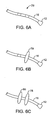
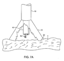
- Figs. 7A and 7B illustrate a variation of an anchoring mechanism such as a helical tissue piercing device for temporarily stabilizing the imaging hood relative to a tissue surface.
- an anchoring mechanism such as a helical tissue piercing device for temporarily stabilizing the imaging hood relative to a tissue surface.
- Fig. 7C shows another variation for anchoring the imaging hood having one or more tubular support members integrated with the imaging hood; each support members may define a lumen therethrough for advancing a helical tissue anchor within.
- Fig. 8A shows an illustrative example of one variation of how a tissue imager may be utilized with an imaging device.
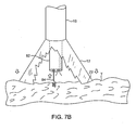
- Fig. 8B shows a further illustration of a hand-held variation of the fluid delivery and tissue manipulation system.
- Figs. 9A to 9C illustrate an example of capturing several images of the tissue at multiple regions.
- Figs. 10A and 10B show charts illustrating how fluid pressure within the imaging hood may be coordinated with the surrounding blood pressure; the fluid pressure in the imaging hood may be coordinated with the blood pressure or it may be regulated based upon pressure feedback from the blood.
- Fig. 11A shows a side view of another variation of a tissue imager having an imaging balloon within an expandable hood.
- Fig. 11B shows another variation of a tissue imager utilizing a translucent or transparent imaging balloon.
- Fig. 12A shows another variation in which a flexible expandable or distensible membrane may be incorporated within the imaging hood to alter the volume of fluid dispensed.
- Figs. 12B and 12C show another variation in which the imaging hood may be partially or selectively deployed from the catheter to alter the area of the tissue being visualized as well as the volume of the dispensed fluid.
- Figs. 13A and 13B show exemplary side and cross-sectional views, respectively, of another variation in which the injected fluid may be drawn back into the device for minimizing fluid input into a body being treated.
- Figs. 14A to 14D show various configurations and methods for configuring an imaging hood into a low-profile for delivery and/or deployment.
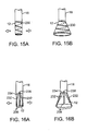
- Figs. 15A and 15B show an imaging hood having an helically expanding frame or support.
- Figs. 16A and 16B show another imaging hood having one or more hood support members, which are pivotably attached at their proximal ends to deployment catheter, integrated with a hood membrane.
- Figs. 17A and 17B show yet another variation of the imaging hood having at least two or more longitudinally positioned support members supporting the imaging hood membrane where the support members are movable relative to one another via a torquing or pulling or pushing force.
- Figs. 18A and 18B show another variation where a distal portion of the deployment catheter may have several pivoting members which form a tubular shape in its low profile configuration.
- Figs. 19A and 19B show another variation where the distal portion of deployment catheter may be fabricated from a flexible metallic or polymeric material to form a radially expanding hood.
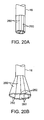
- Figs. 20A and 20B show another variation where the imaging hood may be formed from a plurality of overlapping hood members which overlie one another in an overlapping pattern.
- Figs. 21A and 21B show another example of an expandable hood which is highly conformable against tissue anatomy with varying geography.
- Fig. 22A shows yet another example of an expandable hood having a number of optional electrodes placed about the contact edge or lip of the hood for sensing tissue contact or detecting arrhythmias.
- Fig. 22B shows another variation for conforming the imaging hood against the underlying tissue where an inflatable contact edge may be disposed around the circumference of the imaging hood.
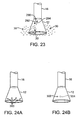
- Fig. 23 shows a variation of the system which may be instrumented with a transducer for detecting the presence of blood seeping back into the imaging hood.
- Figs. 24A and 24B show variations of the imaging hood instrumented with sensors for detecting various physical parameters; the sensors may be instrumented around the outer surface of the imaging hood and also within the imaging hood.
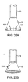
- Figs. 25A and 25B show a variation where the imaging hood may have one or more LEDs over the hood itself for providing illumination of the tissue to be visualized.
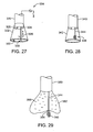
- Figs. 26A and 26B show another variation in which a separate illumination tool having one or more LEDs mounted thereon may be utilized within the imaging hood.
- Fig. 27 shows one example of how a therapeutic tool may be advanced through the tissue imager for treating a tissue region of interest.
- Fig. 28 shows another example of a helical therapeutic tool for treating the tissue region of interest.
- Fig. 29 shows a variation of how a therapeutic tool may be utilized with an expandable imaging balloon.
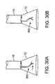
- Figs. 30A and 30B show alternative configurations for therapeutic instruments which may be utilized; one variation is shown having an angled instrument arm and another variation is shown with an off-axis instrument arm.
- Figs. 31A to 31C show side and end views, respectively, of an imaging system which may be utilized with an ablation probe.
- Figs. 32A and 32B show side and end views, respectively, of another variation of the imaging hood with an ablation probe, where the imaging hood may be enclosed for regulating a temperature of the underlying tissue.
- Figs. 33A and 33B show an example in which the imaging fluid itself may be altered in temperature to facilitate various procedures upon the underlying tissue.
- Figs. 34A and 34B show an example of a laser ring generator which may be utilized with the imaging system and an example for applying the laser ring generator within the left atrium of a heart for treating atrial fibrillation.
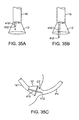
- Figs. 35A to 35C show an example of an extendible cannula generally comprising an elongate tubular member which may be positioned within the deployment catheter during delivery and then projected distally through the imaging hood and optionally beyond.
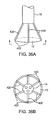
- Figs. 36A and 36B show side and end views, respectively, of an imaging hood having one or more tubular support members integrated with the hood for passing instruments or tools therethrough for treatment upon the underlying tissue.
- Figs. 37A and 37B illustrate how an imaging device may be guided within a heart chamber to a region of interest utilizing a lighted probe positioned temporarily within, e.g., a lumen of the coronary sinus.
- Figs. 38A and 38B show an imaging hood having a removable disk-shaped member for implantation upon the tissue surface.
- Figs. 39A to 39C show one method for implanting the removable disk of Figs. 38A and 38B .
- Figs. 40A and 40B illustrate an imaging hood having a deployable anchor assembly attached to the tissue contact edge and an assembly view of the anchors and the suture or wire connected to the anchors, respectively
- Figs. 41A to 41D show one method for deploying the anchor assembly of Figs. 40A and 40B for closing an opening or wound.
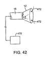
- Fig. 42 shows another variation in which the imaging system may be fluidly coupled to a dialysis unit for filtering a patient's blood.
- Figs. 43A and 43B show a variation of the deployment catheter having a first deployable hood and a second deployable hood positioned distal to the first hood; the deployment catheter may also have a side-viewing imaging element positioned between the first and second hoods for imaging tissue between the expanded hoods.
- Figs. 44A and 44B show side and end views, respectively, of a deployment catheter having a side-imaging balloon in an un-inflated low-profile configuration.
- Figs. 45A to 45C show side, top, and end views, respectively, of the inflated balloon of Figs. 44A and 44B defining a visualization field in the inflated balloon.
- Figs. 46A and 46B show side and cross-sectional end views, respectively, for one method of use in visualizing a lesion upon a vessel wall within the visualization field of the inflated balloon from Figs. 45A to 45C .
- a tissue-imaging and manipulation apparatus described below is able to provide real-time images in vivo of tissue regions within a body lumen such as a heart, which is filled with blood flowing dynamically therethrough and is also able to provide intravascular tools and instruments for performing various procedures upon the imaged tissue regions.
- Such an apparatus may be utilized for many procedures, e.g., facilitating trans-septal access to the left atrium, cannulating the coronary sinus, diagnosis of valve regurgitation/stenosis, valvuloplasty, atrial appendage closure, arrhythmogenic focus ablation, among other procedures.
- tissue imaging and manipulation assembly 10 may be delivered intravascularly through the patient's body in a low-profile configuration via a delivery catheter or sheath 14 .
- tissue imaging and manipulation assembly 10 may be delivered intravascularly through the patient's body in a low-profile configuration via a delivery catheter or sheath 14 .
- tissue such as the mitral valve located at the outflow tract of the left atrium of the heart
- it is generally desirable to enter or access the left atrium while minimizing trauma to the patient.
- tissue such as the mitral valve located at the outflow tract of the left atrium of the heart
- trans-septal procedure or septostomy To non-operatively effect such access, one conventional approach involves puncturing the intra-atrial septum from the right atrial chamber to the left atrial chamber in a procedure commonly called a trans-septal procedure or septostomy.
- trans-septal access to the left atrial chamber of the heart may allow for larger devices to be introduced into the venous system than can generally be introduced percutaneously into the arterial system
- imaging hood 12 When the imaging and manipulation assembly 10 is ready to be utilized for imaging tissue, imaging hood 12 may be advanced relative to catheter 14 and deployed from a distal opening of catheter 14 , as shown by the arrow. Upon deployment, imaging hood 12 may be unconstrained to expand or open into a deployed imaging configuration, as shown in Fig. 1B . Imaging hood 12 may be fabricated from a variety of pliable or conformable biocompatible material including but not limited to, e.g., polymeric, plastic, or woven materials. One example of a woven material is Kevlar® (E. I.
- imaging hood 12 may be fabricated from a translucent or opaque material and in a variety of different colors to optimize or attenuate any reflected lighting from surrounding fluids or structures, i.e., anatomical or mechanical structures or instruments. In either case, imaging hood 12 may be fabricated into a uniform structure or a scaffold-supported structure, in which case a scaffold made of a shape memory alloy, such as Nitinol, or a spring steel, or plastic, etc., may be fabricated and covered with the polymeric, plastic, or woven material.
- a shape memory alloy such as Nitinol, or a spring steel, or plastic, etc.
- Imaging hood 12 may be attached at interface 24 to a deployment catheter 16 which may be translated independently of deployment catheter or sheath 14 . Attachment of interface 24 may be accomplished through any number of conventional methods.
- Deployment catheter 16 may define a fluid delivery lumen 18 as well as an imaging lumen 20 within which an optical imaging fiber or assembly may be disposed for imaging tissue.
- imaging hood 12 When deployed, imaging hood 12 may expand into any number of shapes, e.g., cylindrical, conical as shown, semi-spherical, etc., provided that an open area or field 26 is defined by imaging hood 12 . The open area 26 is the area within which the tissue region of interest may be imaged.
- Imaging hood 12 may also define an atraumatic contact lip or edge 22 for placement or abutment against the tissue region of interest.
- the diameter of imaging hood 12 at its maximum fully deployed diameter is typically greater relative to a diameter of the deployment catheter 16 (although a diameter of contact lip or edge 22 may be made to have a smaller or equal diameter of deployment catheter 16 ).
- the contact edge diameter may range anywhere from 1 to 5 times (or even greater, as practicable) a diameter of deployment catheter 16.
- Fig. 1C shows an end view of the imaging hood 12 in its deployed configuration. Also shown are the contact lip or edge 22 and fluid delivery lumen 18 and imaging lumen 20 .
- the imaging and manipulation assembly 10 may additionally define a guidewire lumen therethrough, e.g., a concentric or eccentric lumen, as shown in the side and end views, respectively, of Figs. 1D to 1F .
- the deployment catheter 16 may define guidewire lumen 19 for facilitating the passage of the system over or along a guidewire 17 , which may be advanced intravascularly within a body lumen. The deployment catheter 16 may then be advanced over the guidewire 17 , as generally known in the art.
- the displacing fluid may be pumped at positive pressure through fluid delivery lumen 18 until the fluid fills open area 26 completely and displaces any fluid 28 from within open area 26 .
- the displacing fluid flow may be laminarized to improve its clearing effect and to help prevent blood from re-entering the imaging hood 12 .
- fluid flow may be started before the deployment takes place.
- the displacing fluid, also described herein as imaging fluid may comprise any biocompatible fluid, e.g., saline, water, plasma, etc., which is sufficiently transparent to allow for relatively undistorted visualization through the fluid.
- any number of therapeutic drugs may be suspended within the fluid or may comprise the fluid itself which is pumped into open area 26 and which is subsequently passed into and through the heart and the patient body.
- deployment catheter 16 may be manipulated to position deployed imaging hood 12 against or near the underlying tissue region of interest to be imaged, in this example a portion of annulus A of mitral valve MV within the left atrial chamber.
- the surrounding blood 30 flows around imaging hood 12 and within open area 26 defined within imaging hood 12 , as seen in Fig. 2A , the underlying annulus A is obstructed by the opaque blood 30 and is difficult to view through the imaging lumen 20.
- the translucent fluid 28 such as saline, may then be pumped through fluid delivery lumen 18 , intermittently or continuously, until the blood 30 is at least partially, and preferably completely, displaced from within open area 26 by fluid 28 , as shown in Fig. 2B .
- contact edge 22 need not directly contact the underlying tissue, it is at least preferably brought into close proximity to the tissue such that the flow of clear fluid 28 from open area 26 may be maintained to inhibit significant backflow of blood 30 back into open area 26.
- Contact edge 22 may also be made of a soft elastomeric material such as certain soft grades of silicone or polyurethane, as typically known, to help contact edge 22 conform to an uneven or rough underlying anatomical tissue surface.
- the fluid 28 may be pumped temporarily or sporadically only until a clear view of the tissue is available to be imaged and recorded, at which point the fluid flow 28 may cease and blood 30 may be allowed to seep or flow back into imaging hood 12 . This process may be repeated a number of times at the same tissue region or at multiple tissue regions.
- a number of articulation and manipulation controls may be utilized.
- one or more push-pull wires 42 may be routed through deployment catheter 16 for steering the distal end portion of the device in various directions 46 to desirably position the imaging hood 12 adjacent to a region of tissue to be visualized.
- deployment catheter 16 and imaging hood 12 may be articulated into any number of configurations 44 .
- the push-pull wire or wires 42 may be articulated via their proximal ends from outside the patient body manually utilizing one or more controls.
- deployment catheter 16 may be articulated by computer control, as further described below.
- an articulatable delivery catheter 48 which may be articulated via one or more push-pull wires and having an imaging lumen and one or more working lumens, may be delivered through the deployment catheter 16 and into imaging hood 12. With a distal portion of articulatable delivery catheter 48 within imaging hood 12 , the clear displacing fluid may be pumped through delivery catheter 48 or deployment catheter 16 to clear the field within imaging hood 12 . As shown in Fig. 3B , the articulatable delivery catheter 48 may be articulated within the imaging hood to obtain a better image of tissue adjacent to the imaging hood 12 .
- articulatable delivery catheter 48 may be articulated to direct an instrument or tool passed through the catheter 48 , as described in detail below, to specific areas of tissue imaged through imaging hood 12 without having to reposition deployment catheter 16 and re-clear the imaging field within hood 12 .
- a distal portion of the deployment catheter 16 itself may comprise a distal end 49 which is articulatable within imaging hood 12 , as shown in Fig. 3C .
- Directed imaging, instrument delivery, etc. may be accomplished directly through one or more lumens within deployment catheter 16 to specific regions of the underlying tissue imaged within imaging hood 12 .
- Visualization within the imaging hood 12 may be accomplished through an imaging lumen 20 defined through deployment catheter 16 , as described above. In such a configuration, visualization is available in a straight-line manner, i.e., images are generated from the field distally along a longitudinal axis defined by the deployment catheter 16 .
- an articulatable imaging assembly having a pivotable support member 50 may be connected to, mounted to, or otherwise passed through deployment catheter 16 to provide for visualization off-axis relative to the longitudinal axis defined by deployment catheter 16 , as shown in Fig. 4A .
- Support member 50 may have an imaging element 52 , e.g., a CCD or CMOS imager or optical fiber, attached at its distal end with its proximal end connected to deployment catheter 16 via a pivoting connection 54 .
- the optical fibers 58 may be passed through deployment catheter 16 , as shown in the cross-section of Fig. 4B , and routed through the support member 50 .
- the use of optical fibers 58 may provide for increased diameter sizes of the one or several lumens 56 through deployment catheter 16 for the passage of diagnostic and/or therapeutic tools therethrough.
- electronic chips such as a charge coupled device (CCD) or a CMOS imager, which are typically known, may be utilized in place of the optical fibers 58 , in which case the electronic imager may be positioned in the distal portion of the deployment catheter 16 with electric wires being routed proximally through the deployment catheter 16.
- CCD charge coupled device
- CMOS imager which are typically known
- the electronic imagers may be wirelessly coupled to a receiver for the wireless transmission of images.
- Additional optical fibers or light emitting diodes (LEDs) can be used to provide lighting for the image or operative theater, as described below in further detail.
- Support member 50 may be pivoted via connection 54 such that the member 50 can be positioned in a low-profile configuration within channel or groove 60 defined in a distal portion of catheter 16 , as shown in the cross-section of Fig. 4C .
- support member 50 can be positioned within channel or groove 60 with imaging hood 12 also in its low-profile configuration.
- imaging hood 12 may be expanded into its deployed configuration and support member 50 may be deployed into its off-axis configuration for imaging the tissue adjacent to hood 12 , as in Fig. 4A .
- Other configurations for support member 50 for off-axis visualization may be utilized, as desired.
- Fig. 5 shows an illustrative cross-sectional view of a heart H having tissue regions of interest being viewed via an imaging assembly 10 .
- delivery catheter assembly 70 may be introduced percutaneously into the patient's vasculature and advanced through the superior vena cava SVC and into the right atrium RA .
- the delivery catheter or sheath 72 may be articulated through the atrial septum AS and into the left atrium LA for viewing or treating the tissue, e.g., the annulus A , surrounding the mitral valve MV .
- deployment catheter 16 and imaging hood 12 may be advanced out of delivery catheter 72 and brought into contact or in proximity to the tissue region of interest.
- delivery catheter assembly 70 may be advanced through the inferior vena cava IVC , if so desired.
- other regions of the heart H e.g., the right ventricle RV or left ventricle LV , may also be accessed and imaged or treated by imaging assembly 10 .
- the delivery catheter or sheath 14 may comprise a conventional intra-vascular catheter or an endoluminal delivery device.
- robotically-controlled delivery catheters may also be optionally utilized with the imaging assembly described herein, in which case a computer-controller 74 may be used to control the articulation and positioning of the delivery catheter 14 .
- An example of a robotically-controlled delivery catheter which may be utilized is described in further detail in US Pat. Pub. 2002/0087169 A1 to Brock et al. entitled "Flexible Instrument”.
- Other robotically-controlled delivery catheters manufactured by Hansen Medical, Inc. may also be utilized with the delivery catheter 14 .
- one or more inflatable balloons or anchors 76 may be positioned along the length of catheter 16 , as shown in Fig. 6A .
- the inflatable balloons 76 may be inflated from a low-profile into their expanded configuration to temporarily anchor or stabilize the catheter 16 position relative to the heart H .
- Fig. 6B shows a first balloon 78 inflated while Fig. 6C also shows a second balloon 80 inflated proximal to the first balloon 78 .
- the septal wall AS may be wedged or sandwiched between the balloons 78 , 80 to temporarily stabilize the catheter 16 and imaging hood 12 .
- a single balloon 78 or both balloons 78 , 80 may be used.
- Other alternatives may utilize expandable mesh members, malecots, or any other temporary expandable structure.
- the balloon assembly 76 may be deflated or re-configured into a low-profile for removal of the deployment catheter 16 .
- various anchoring mechanisms may be optionally employed for temporarily holding the imaging hood 12 against the tissue.
- Such anchoring mechanisms may be particularly useful for imaging tissue which is subject to movement, e.g., when imaging tissue within the chambers of a beating heart.
- a tool delivery catheter 82 having at least one instrument lumen and an optional visualization lumen may be delivered through deployment catheter 16 and into an expanded imaging hood 12 .
- an anchoring mechanisms such as a helical tissue piercing device 84 may be passed through the tool delivery catheter 82 , as shown in Fig. 7A , and into imaging hood 12 .
- the helical tissue engaging device 84 may be torqued from its proximal end outside the patient body to temporarily anchor itself into the underlying tissue surface T . Once embedded within the tissue T , the helical tissue engaging device 84 may be pulled proximally relative to deployment catheter 16 while the deployment catheter 16 and imaging hood 12 are pushed distally, as indicated by the arrows in Fig. 7B , to gently force the contact edge or lip 22 of imaging hood against the tissue T . The positioning of the tissue engaging device 84 may be locked temporarily relative to the deployment catheter 16 to ensure secure positioning of the imaging hood 12 during a diagnostic or therapeutic procedure within the imaging hood 12 .
- tissue engaging device 84 may be disengaged from the tissue by torquing its proximal end in the opposite direction to remove the anchor form the tissue T and the deployment catheter 16 may be repositioned to another region of tissue where the anchoring process may be repeated or removed from the patient body.
- the tissue engaging device 84 may also be constructed from other known tissue engaging devices such as vacuum-assisted engagement or grasper-assisted engagement tools, among others.
- helical anchor 84 is shown, this is intended to be illustrative and other types of temporary anchors may be utilized, e.g., hooked or barbed anchors, graspers, etc.
- the tool delivery catheter 82 may be omitted entirely and the anchoring device may be delivered directly through a lumen defined through the deployment catheter 16 .
- FIG. 7C shows an imaging hood 12 having one or more tubular support members 86 , e.g., four support members 86 as shown, integrated with the imaging hood 12 .
- the tubular support members 86 may define lumens therethrough each having helical tissue engaging devices 88 positioned within.
- the helical tissue engaging devices 88 may be urged distally to extend from imaging hood 12 and each may be torqued from its proximal end to engage the underlying tissue T .
- Each of the helical tissue engaging devices 88 may be advanced through the length of deployment catheter 16 or they may be positioned within tubular support members 86 during the delivery and deployment of imaging hood 12 . Once the procedure within imaging hood 12 is finished, each of the tissue engaging devices 88 may be disengaged from the tissue and the imaging hood 12 may be repositioned to another region of tissue or removed from the patient body.
- FIG. 8A An illustrative example is shown in Fig. 8A of a tissue imaging assembly connected to a fluid delivery system 90 and to an optional processor 98 and image recorder and/or viewer 100.
- the fluid delivery system 90 may generally comprise a pump 92 and an optional valve 94 for controlling the flow rate of the fluid into the system.
- a fluid reservoir 96 fluidly connected to pump 92 , may hold the fluid to be pumped through imaging hood 12 .
- An optional central processing unit or processor 98 may be in electrical communication with fluid delivery system 90 for controlling flow parameters such as the flow rate and/or velocity of the pumped fluid.
- the processor 98 may also be in electrical communication with an image recorder and/or viewer 100 for directly viewing the images of tissue received from within imaging hood 12 .
- Imager recorder and/or viewer 100 may also be used not only to record the image but also the location of the viewed tissue region, if so desired.
- processor 98 may also be utilized to coordinate the fluid flow and the image capture.
- processor 98 may be programmed to provide for fluid flow from reservoir 96 until the tissue area has been displaced of blood to obtain a clear image. Once the image has been determined to be sufficiently clear, either visually by a practitioner or by computer, an image of the tissue may be captured automatically by recorder 100 and pump 92 may be automatically stopped or slowed by processor 98 to cease the fluid flow into the patient.
- Other variations for fluid delivery and image capture are, of course, possible and the aforementioned configuration is intended only to be illustrative and not limiting.
- Fig. 8B shows a further illustration of a hand-held variation of the fluid delivery and tissue manipulation system 110 .
- system 110 may have a housing or handle assembly 112 which can be held or manipulated by the physician from outside the patient body.
- the fluid reservoir 114 shown in this variation as a syringe, can be fluidly coupled to the handle assembly 112 and actuated via a pumping mechanism 116 , e.g., lead screw.
- Fluid reservoir 114 may be a simple reservoir separated from the handle assembly 112 and fluidly coupled to handle assembly 112 via one or more tubes. The fluid flow rate and other mechanisms may be metered by the electronic controller 118 .
- Deployment of imaging hood 12 may be actuated by a hood deployment switch 120 located on the handle assembly 112 while dispensation of the fluid from reservoir 114 may be actuated by a fluid deployment switch 122 , which can be electrically coupled to the controller 118 .
- Controller 118 may also be electrically coupled to a wired or wireless antenna 124 optionally integrated with the handle assembly 112 , as shown in the figure.
- the wireless antenna 124 can be used to wirelessly transmit images captured from the imaging hood 12 to a receiver, e.g., via Bluetooth® wireless technology (Bluetooth SIG, Inc., Bellevue, WA), RF, etc., for viewing on a monitor 128 or for recording for later viewing.
- Articulation control of the deployment catheter 16 , or a delivery catheter or sheath 14 through which the deployment catheter 16 may be delivered may be accomplished by computer control, as described above, in which case an additional controller may be utilized with handle assembly 112 .
- handle assembly 112 may incorporate one or more articulation controls 126 for manual manipulation of the position of deployment catheter 16 .
- Handle assembly 112 may also define one or more instrument ports 130 through which a number of intravascular tools may be passed for tissue manipulation and treatment within imaging hood 12 , as described further below.
- fluid or debris may be sucked into imaging hood 12 for evacuation from the patient body by optionally fluidly coupling a suction pump 132 to handle assembly 112 or directly to deployment catheter 16 .
- fluid may be pumped continuously into imaging hood 12 to provide for clear viewing of the underlying tissue.
- fluid may be pumped temporarily or sporadically only until a clear view of the tissue is available to be imaged and recorded, at which point the fluid flow may cease and the blood may be allowed to seep or flow back into imaging hood 12 .
- Figs. 9A to 9C illustrate an example of capturing several images of the tissue at multiple regions.
- Deployment catheter 16 may be desirably positioned and imaging hood 12 deployed and brought into position against a region of tissue to be imaged, in this example the tissue surrounding a mitral valve MV within the left atrium of a patient's heart.
- the imaging hood 12 may be optionally anchored to the tissue, as described above, and then cleared by pumping the imaging fluid into the hood 12 . Once sufficiently clear, the tissue may be visualized and the image captured by control electronics 118 .
- the first captured image 140 may be stored and/or transmitted wirelessly 124 to a monitor 128 for viewing by the physician, as shown in Fig. 9A .
- the deployment catheter 16 may be then repositioned to an adjacent portion of mitral valve MV , as shown in Fig. 9B , where the process may be repeated to capture a second image 142 for viewing and/or recording.
- the deployment catheter 16 may again be repositioned to another region of tissue, as shown in Fig. 9C , where a third image 144 may be captured for viewing and/or recording. This procedure may be repeated as many times as necessary for capturing a comprehensive image of the tissue surrounding mitral valve MV , or any other tissue region.
- the pump may be stopped during positioning and blood or surrounding fluid may be allowed to enter within imaging hood 12 until the tissue is to be imaged, where the imaging hood 12 may be cleared, as above.
- the fluid when the imaging hood 12 is cleared by pumping the imaging fluid within for clearing the blood or other bodily fluid, the fluid may be pumped continuously to maintain the imaging fluid within the hood 12 at a positive pressure or it may be pumped under computer control for slowing or stopping the fluid flow into the hood 12 upon detection of various parameters or until a clear image of the underlying tissue is obtained.
- the control electronics 118 may also be programmed to coordinate the fluid flow into the imaging hood 12 with various physical parameters to maintain a clear image within imaging hood 12 .
- FIG. 10A shows a chart 150 illustrating how fluid pressure within the imaging hood 12 may be coordinated with the surrounding blood pressure.
- Chart 150 shows the cyclical blood pressure 156 alternating between diastolic pressure 152 and systolic pressure 154 over time T due to the beating motion of the patient heart.
- the fluid pressure of the imaging fluid, indicated by plot 160 within imaging hood 12 may be automatically timed to correspond to the blood pressure changes 160 such that an increased pressure is maintained within imaging hood 12 which is consistently above the blood pressure 156 by a slight increase ⁇ P, as illustrated by the pressure difference at the peak systolic pressure 158 .
- This pressure difference, ⁇ P may be maintained within imaging hood 12 over the pressure variance of the surrounding blood pressure to maintain a positive imaging fluid pressure within imaging hood 12 to maintain a clear view of the underlying tissue.
- One benefit of maintaining a constant ⁇ P is a constant flow and maintenance of a clear field.
- Fig. 10B shows a chart 162 illustrating another variation for maintaining a clear view of the underlying tissue
- one or more sensors within the imaging hood 12 may be configured to sense pressure changes within the imaging hood 12 and to correspondingly increase the imaging fluid pressure within imaging hood 12 .
- This may result in a time delay, ⁇ T, as illustrated by the shifted fluid pressure 160 relative to the cycling blood pressure 156 , although the time delay ⁇ T may be negligible in maintaining the clear image of the underlying tissue.
- Predictive software algorithms can also be used to substantially eliminate this time delay by predicting when the next pressure wave peak will arrive and by increasing the pressure ahead of the pressure wave's arrival by an amount of time equal to the aforementioned time delay to essentially cancel the time delay out.
- imaging hood 12 The variations in fluid pressure within imaging hood 12 may be accomplished in part due to the nature of imaging hood 12 .
- An inflatable balloon which is conventionally utilized for imaging tissue, may be affected by the surrounding blood pressure changes.
- an imaging hood 12 retains a constant volume therewithin and is structurally unaffected by the surrounding blood pressure changes, thus allowing for pressure increases therewithin.
- the material that hood 12 is made from may also contribute to the manner in which the pressure is modulated within this hood 12 .
- a stiffer hood material such as high durometer polyurethane or Nylon, may facilitate the maintaining of an open hood when deployed.
- a relatively lower durometer or softer material such as a low durometer PVC or polyurethane, may collapse from the surrounding fluid pressure and may not adequately maintain a deployed or expanded hood.
- FIG. 11A shows another variation comprising an additional imaging balloon 172 within an imaging hood 174 .
- an expandable balloon 172 having a translucent skin may be positioned within imaging hood 174 .
- Balloon 172 may be made from any distensible biocompatible material having sufficient translucent properties which allow for visualization therethrough.
- the balloon 172 can also be filled with contrast media to allow it to be viewed on fluoroscopy to aid in its positioning.
- the imager e.g., fiber optic, positioned within deployment catheter 170 may then be utilized to view the tissue region through the balloon 172 and any additional fluid which may be pumped into imaging hood 174 via one or more optional fluid ports 176 , which may be positioned proximally of balloon 172 along a portion of deployment catheter 170 .
- balloon 172 may define one or more holes over its surface which allow for seepage or passage of the fluid contained therein to escape and displace the blood from within imaging hood 174 .
- Fig. 11B shows another alternative in which balloon 180 may be utilized alone.
- Balloon 180 attached to deployment catheter 178 , may be filled with fluid, such as saline or contrast media, and is preferably allowed to come into direct contact with the tissue region to be imaged.
- Fig. 12A shows another alternative in which deployment catheter 16 incorporates imaging hood 12 , as above, and includes an additional flexible membrane 182 within imaging hood 12 .
- Flexible membrane 182 may be attached at a distal end of catheter 16 and optionally at contact edge 22 .
- Imaging hood 12 may be utilized, as above, and membrane 182 may be deployed from catheter 16 in vivo or prior to placing catheter 16 within a patient to reduce the volume within imaging hood 12 . The volume may be reduced or minimized to reduce the amount of fluid dispensed for visualization or simply be to reduced depending upon the area of tissue to be visualized.
- FIGs. 12B and 12C show yet another alternative in which imaging hood 186 may be withdrawn proximally within deployment catheter 184 or deployed distally from catheter 186 , as shown, to vary the volume of imaging hood 186 and thus the volume of dispensed fluid.
- Imaging hood 186 may be seen in Fig. 12B as being partially deployed from, e.g., a circumferentially defined lumen within catheter 184 , such as annular lumen 188 .
- the underlying tissue may be visualized with imaging hood 186 only partially deployed.
- imaging hood 186' may be fully deployed, as shown in Fig. 12C , by urging hood 186' distally out from annular lumen 188 . In this expanded configuration, the area of tissue to be visualized may be increased as hood 186' is expanded circumferentially.
- Figs. 13A and 13B show perspective and cross-sectional side views, respectively, of yet another variation of imaging assembly which may utilize a fluid suction system for minimizing the amount of fluid injected into the patient's heart or other body lumen during tissue visualization.
- Deployment catheter 190 in this variation may define an inner tubular member 196 which may be integrated with deployment catheter 190 or independently translatable.
- Fluid delivery lumen 198 defined through member 196 may be fluidly connected to imaging hood 192 , which may also define one or more open channels 194 over its contact lip region. Fluid pumped through fluid delivery lumen 198 may thus fill open area 202 to displace any blood or other fluids or objects therewithin.
- Tubular member 196 may also define one or more additional working channels 200 for the passage of any tools or visualization devices.
- the imaging hood may take on any number of configurations when positioned or configured for a low-profile delivery within the delivery catheter, as shown in the examples of Figs. 14A to 14D . These examples are intended to be illustrative and are not intended to be limiting in scope.
- Fig. 14A shows one example in which imaging hood 212 may be compressed within catheter 210 by folding hood 212 along a plurality of pleats.
- Hood 212 may also comprise scaffolding or frame 214 made of a super-elastic or shape memory material or alloy, e.g., Nitinol, Elgiloy, shape memory polymers, electroactive polymers, or a spring stainless steel.
- the shape memory material may act to expand or deploy imaging hood 212 into its expanded configuration when urged in the direction of the arrow from the constraints of catheter 210 .
- Fig. 14B shows another example in which imaging hood 216 may be expanded or deployed from catheter 210 from a folded and overlapping configuration. Frame or scaffolding 214 may also be utilized in this example.
- Fig. 14C shows yet another example in which imaging hood 218 may be rolled, inverted, or everted upon itself for deployment.
- Fig. 14D shows a configuration in which imaging hood 220 may be fabricated from an extremely compliant material which allows for hood 220 to be simply compressed into a low-profile shape.
- hood 220 may allow for it to expand into its deployed configuration, especially if a scaffold or frame of a shape memory or superelastic material, e.g., Nitinol, is utilized in its construction.
- a scaffold or frame of a shape memory or superelastic material e.g., Nitinol
- FIGs. 15A and 15B illustrates an helically expanding frame or support 230 .
- helical frame 230 may be integrated with the imaging hood 12 membrane.
- helical frame 230 may expand into a conical or tapered shape.
- Helical frame 230 may alternatively be made out of heat-activated Nitinol to allow it to expand upon application of a current.
- imaging hood 12 may comprise one or more hood support members 232 integrated with the hood membrane. These longitudinally attached support members 232 may be pivotably attached at their proximal ends to deployment catheter 16 .
- One or more pullwires 234 may be routed through the length of deployment catheter 16 and extend through one or more openings 238 defined in deployment catheter 16 proximally to imaging hood 12 into attachment with a corresponding support member 232 at a pullwire attachment point 236 .
- the support members 232 may be fabricated from a plastic or metal, such as stainless steel.
- the support members 232 may be made from a superelastic or shape memory alloy, such as Nitinol, which may self-expand into its deployed configuration without the use or need of pullwires. A heat-activated Nitinol may also be used which expands upon the application of thermal energy or electrical energy.
- support members 232 may also be constructed as inflatable lumens utilizing, e.g., PET balloons. From its low-profile delivery configuration shown in Fig. 16A , the one or more pullwires 234 may be tensioned from their proximal ends outside the patient body to pull a corresponding support member 232 into a deployed configuration, as shown in Fig. 16B , to expand imaging hood 12 . To reconfigure imaging hood 12 back into its low profile, deployment catheter 16 may be pulled proximally into a constraining catheter or the pullwires 234 may be simply pushed distally to collapse imaging hood 12 .
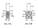
- FIGs. 17A and 17B show yet another variation of imaging hood 240 having at least two or more longitudinally positioned support members 242 supporting the imaging hood membrane.
- the support members 242 each have cross-support members 244 which extend diagonally between and are pivotably attached to the support members 242 .
- Each of the cross-support members 244 may be pivotably attached to one another where they intersect between the support members 242 .
- a jack or screw member 246 may be coupled to each cross-support member 244 at this intersection point and a torquing member, such as a torqueable wire 248 , may be coupled to each jack or screw member 246 and extend proximally through deployment catheter 16 to outside the patient body.
- the torqueable wires 248 may be torqued to turn the jack or screw member 246 which in turn urges the cross-support members 244 to angle relative to one another and thereby urge the support members 242 away from one another.
- the imaging hood 240 may be transitioned from its low-profile, shown in Fig. 17A , to its expanded profile, shown in Fig. 17B , and back into its low-profile by torquing wires 248 .
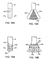
- Figs. 18A and 18B show yet another variation on the imaging hood and its deployment.
- a distal portion of deployment catheter 16 may have several pivoting members 250 , e.g., two to four sections, which form a tubular shape in its low profile configuration, as shown in Fig. 18A .
- pivoting members 250 When pivoted radially about deployment catheter 16 , pivoting members 250 may open into a deployed configuration having distensible or expanding membranes 252 extending over the gaps in-between the pivoting members 250 , as shown in Fig. 18B .
- the distensible membrane 252 may be attached to the pivoting members 250 through various methods, e.g., adhesives, such that when the pivoting members 250 are fully extended into a conical shape, the pivoting members 250 and membrane 252 form a conical shape for use as an imaging hood.
- the distensible membrane 252 may be made out of a porous material such as a mesh or PTFE or out of a translucent or transparent polymer such as polyurethane, PVC, Nylon, etc.
- Figs. 19A and 19B show yet another variation where the distal portion of deployment catheter 16 may be fabricated from a flexible metallic or polymeric material to form a radially expanding hood 254 .
- a plurality of slots 256 may be formed in a uniform pattern over the distal portion of deployment catheter 16 , as shown in Fig. 19A .
- the slots 256 may be formed in a pattern such that when the distal portion is urged radially open, utilizing any of the methods described above, a radially expanded and conically-shaped hood 254 may be formed by each of the slots 256 expanding into an opening, as shown in Fig. 19B .
- a distensible membrane 258 may overlie the exterior surface or the interior surface of the hood 254 to form a fluid-impermeable hood 254 such that the hood 254 may be utilized as an imaging hood.
- the distensible membrane 258 may alternatively be formed in each opening 258 to form the fluid-impermeable hood 254 .
- the imaging hood may be formed from a plurality of overlapping hood members 260 which overlie one another in an overlapping pattern.
- each of the hood members 260 may extend radially outward relative to deployment catheter 16 to form a conically-shaped imaging hood, as shown in Fig. 20B .
- Adjacent hood members 260 may overlap one another along an overlapping interface 262 to form a fluid-retaining surface within the imaging hood.
- the hood members 260 may be made from any number ofbiocompatible materials, e.g., Nitinol, stainless steel, polymers, etc., which are sufficiently strong to optionally retract surrounding tissue from the tissue region of interest.
- the imaging hood may be alternatively configured to contact the tissue surface at an acute angle.
- An imaging hood configured for such contact against tissue may also be especially suitable for contact against tissue surfaces having an unpredictable or uneven anatomical geography.
- deployment catheter 270 may have an imaging hood 272 that is configured to be especially compliant.
- imaging hood 272 may be comprised of one or more sections 274 that are configured to fold or collapse, e.g., by utilizing a pleated surface.
- sections 274 are able to conform closely against the tissue. These sections 274 may be individually collapsible by utilizing an accordion style construction to allow conformation, e.g., to the trabeculae in the heart or the uneven anatomy that may be found inside the various body lumens.
- Fig. 22A shows another variation in which an imaging hood 282 is attached to deployment catheter 280.
- the contact lip or edge 284 may comprise one or more electrical contacts 286 positioned circumferentially around contact edge 284 .
- the electrical contacts 286 may be configured to contact the tissue and indicate affirmatively whether tissue contact was achieved, e.g., by measuring the differential impedance between blood and tissue.
- a processor e.g., processor 98
- in electrical communication with contacts 286 may be configured to determine what type of tissue is in contact with electrical contacts 286 .
- the processor 98 may be configured to measure any electrical activity that may be occurring in the underlying tissue, e.g., accessory pathways, for the purposes of electrically mapping the cardiac tissue and subsequently treating, as described below, any arrhythmias which may be detected.
- FIG. 22B Another variation for ensuring contact between imaging hood 282 and the underlying tissue may be seen in Fig. 22B .
- This variation may have an inflatable contact edge 288 around the circumference of imaging hood 282 .
- the inflatable contact edge 288 may be inflated with a fluid or gas through inflation lumen 289 when the imaging hood 282 is to be placed against a tissue surface having an uneven or varied anatomy.
- the inflated circumferential surface 288 may provide for continuous contact over the hood edge by conforming against the tissue surface and facilitating imaging fluid retention within hood 282 .
- various instrumentation may be utilized with the imaging and manipulation system. For instance, after the field within imaging hood 12 has been cleared of the opaque blood and the underlying tissue is visualized through the clear fluid, blood may seep back into the imaging hood 12 and obstruct the view.
- One method for automatically maintaining a clear imaging field may utilize a transducer, e.g., an ultrasonic transducer 290 , positioned at the distal end of deployment catheter within the imaging hood 12 , as shown in Fig. 23 .
- the transducer 290 may send an energy pulse 292 into the imaging hood 12 and wait to detect back-scattered energy 294 reflected from debris or blood within the imaging hood 12. If back-scattered energy is detected, the pump may be actuated automatically to dispense more fluid into the imaging hood until the debris or blood is no longer detected.
- sensors 300 may be positioned on the imaging hood 12 itself, as shown in Fig. 24A , to detect a number of different parameters.
- sensors 300 may be configured to detect for the presence of oxygen in the surrounding blood, blood and/or imaging fluid pressure, color of the fluid within the imaging hood, etc. Fluid color may be particularly useful in detecting the presence of blood within the imaging hood 12 by utilizing a reflective type sensor to detect back reflection from blood. Any reflected light from blood which may be present within imaging hood 12 may be optically or electrically transmitted through deployment catheter 16 and to a red colored filter within control electronics 118 . Any red color which may be detected may indicate the presence of blood and trigger a signal to the physician or automatically actuate the pump to dispense more fluid into the imaging hood 12 to clear the blood.
- Alternative methods for detecting the presence of blood within the hood 12 may include detecting transmitted light through the imaging fluid within imaging hood 12 . If a source of white light, e.g., utilizing LEDs or optical fibers, is illuminated inside imaging hood 12 , the presence of blood may cause the color red to be filtered through this fluid. The degree or intensity of the red color detected may correspond to the amount of blood present within imaging hood 12 .
- a red color sensor can simply comprise, in one variation, a phototransistor with a red transmitting filter over it which can establish how much red light is detected, which in turn can indicate the presence of blood within imaging hood 12 . Once blood is detected, the system may pump more clearing fluid through and enable closed loop feedback control of the clearing fluid pressure and flow level.
- Any number of sensors may be positioned along the exterior 302 of imaging hood 12 or within the interior 304 of imaging hood 12 to detect parameters not only exteriorly to imaging hood 12 but also within imaging hood 12 .
- Such a configuration as shown in Fig. 24B , may be particularly useful for automatically maintaining a clear imaging field based upon physical parameters such as blood pressure, as described above for Figs. 10A and 10B .
- one or more light emitting diodes may be utilized to provide lighting within the imaging hood 12 .
- illumination may be provided by optical fibers routed through deployment catheter 16
- the use of LEDs over the imaging hood 12 may eliminate the need for additional optical fibers for providing illumination.
- the electrical wires connected to the one or more LEDs may be routed through or over the hood 12 and along an exterior surface or extruded within deployment catheter 16 .
- One or more LEDs may be positioned in a circumferential pattern 306 around imaging hood 12 , as shown in Fig. 25A , or in a linear longitudinal pattern 308 along imaging hood 12 , as shown in Fig. 25B .
- Other patterns, such as a helical or spiral pattern may also be utilized.
- LEDs may be positioned along a support member forming part of imaging hood 12.
- a separate illumination tool 310 may be utilized, as shown in Fig. 26A .
- An example of such a tool may comprise a flexible intravascular delivery member 312 having a carrier member 314 pivotably connected 316 to a distal end of delivery member 312 .
- One or more LEDs 318 may be mounted along carrier member 314 .
- delivery member 312 may be advanced through deployment catheter 16 until carrier member 314 is positioned within imaging hood 12 .
- carrier member 314 may be pivoted in any number of directions to facilitate or optimize the illumination within the imaging hood 12 , as shown in Fig. 26B .
- the LEDs may comprise a single LED color, e.g., white light.
- LEDs of other colors e.g., red, blue, yellow, etc.
- sources of infrared or ultraviolet light may be employed to enable imaging beneath the tissue surface or cause fluorescence of tissue for use in system guidance, diagnosis, or therapy.
- the imaging assembly may also be utilized to provide a therapeutic platform for treating tissue being visualized.
- deployment catheter 320 may have imaging hood 322, as described above, and fluid delivery lumen 324 and imaging lumen 326 .
- a therapeutic tool such as needle 328 may be delivered through fluid delivery lumen 324 or in another working lumen and advanced through open area 332 for treating the tissue which is visualized.
- needle 328 may define one or several ports 330 for delivering drugs therethrough.
- needle 328 may be advanced and pierced into the underlying tissue where a therapeutic agent may be delivered through ports 330 .
- needle 328 may be in electrical communication with a power source 334 , e.g., radio-frequency, microwave, etc., for ablating the underlying tissue area of interest.
- Fig. 28 shows another alternative in which deployment catheter 340 may have imaging hood 342 attached thereto, as above, but with a therapeutic tool 344 in the configuration of a helical tissue piercing device 344 . Also shown and described above in Figs. 7A and 7B for use in stabilizing the imaging hood relative to the underlying tissue, the helical tissue piercing device 344 may also be utilized to manipulate the tissue for a variety of therapeutic procedures.
- the helical portion 346 may also define one or several ports for delivery of therapeutic agents therethrough.
- FIG. 29 shows a deployment catheter 350 having an expandable imaging balloon 352 filled with, e.g., saline 356 .
- a therapeutic tool 344 may be translatable relative to balloon 352.
- a stop 354 may be formed on balloon 352 to prevent the proximal passage of portion 346 past stop 354 .
- FIG. 30A shows one variation of an angled instrument 360 , such as a tissue grasper, which may be configured to have an elongate shaft for intravascular delivery through deployment catheter 16 with a distal end which may be angled relative to its elongate shaft upon deployment into imaging hood 12 .
- the elongate shaft may be configured to angle itself automatically, e.g., by the elongate shaft being made at least partially from a shape memory alloy, or upon actuation, e.g., by tensioning a pullwire.
- Fig. 30A shows one variation of an angled instrument 360 , such as a tissue grasper, which may be configured to have an elongate shaft for intravascular delivery through deployment catheter 16 with a distal end which may be angled relative to its elongate shaft upon deployment into imaging hood 12 .
- the elongate shaft may be configured to angle itself automatically, e.g., by the elongate shaft being made at least partially from a shape memory alloy, or upon actuation,
- FIG. 30B shows another configuration for an instrument 362 being configured to reconfigure its distal portion into an off-axis configuration within imaging hood 12 .
- the instruments 360 , 362 may be reconfigured into a low-profile shape upon withdrawing them proximally back into deployment catheter 16 .
- Fig. 31A shows a probe 370 having a distal end effector 372 , which may be reconfigured from a low-profile shape to a curved profile.
- the end effector 372 may be configured as an ablation probe utilizing radio-frequency energy, microwave energy, ultrasound energy, laser energy or even cryo-ablation.
- the end effector 372 may have several electrodes upon it for detecting or mapping electrical signals transmitted through the underlying tissue.
- an additional temperature sensor such as a thermocouple or thermistor 374 positioned upon an elongate member 376 may be advanced into the imaging hood 12 adjacent to the distal end effector 372 for contacting and monitoring a temperature of the ablated tissue.
- Fig. 31B shows an example in the end view of one configuration for the distal end effector 372 which may be simply angled into a perpendicular configuration for contacting the tissue.
- Fig. 31C shows another example where the end effector may be reconfigured into a curved end effector 378 for increased tissue contact.
- Figs. 32A and 32B show another variation of an ablation tool utilized with an imaging hood 12 having an enclosed bottom portion.
- an ablation probe such as a cryo-ablation probe 380 having a distal end effector 382
- a cryo-ablation probe 380 having a distal end effector 382
- the shaft of probe 380 may pass through an opening 386 defined through the membrane 384 .
- the clear fluid may be pumped into imaging hood 12 , as described above, and the distal end effector 382 may be placed against a tissue region to be ablated with the imaging hood 12 and the membrane 384 positioned atop or adjacent to the ablated tissue.
- the imaging fluid may be warmed prior to dispensing into the imaging hood 12 such that the tissue contacted by the membrane 384 may be warmed during the cryo-ablation procedure.
- the fluid dispensed into the imaging hood 12 may be cooled such that the tissue contacted by the membrane 384 and adjacent to the ablation probe during the ablation procedure is likewise cooled.
- the imaging fluid may be varied in its temperature to facilitate various procedures to be performed upon the tissue.
- the imaging fluid itself may be altered to facilitate various procedures.
- a deployment catheter 16 and imaging hood 12 may be advanced within a hollow body organ, such as a bladder filled with urine 394 , towards a lesion or tumor 392 on the bladder wall.
- the imaging hood 12 may be placed entirely over the lesion 392 , or over a portion of the lesion.
- a cryo-fluid i.e., a fluid which has been cooled to below freezing temperatures of, e.g., water or blood, may be pumped into the imaging hood 12 to cryo-ablate the lesion 390 , as shown in Fig. 33B while avoiding the creation of ice on the instrument or surface of tissue.
- the cryo-fluid may be warmed naturally by the patient body and ultimately removed.
- the cryo-fluid may be a colorless and translucent fluid which enables visualization therethrough of the underlying tissue.
- An example of such a fluid is FluorinertTM (3M, St. Paul, MN), which is a colorless and odorless perfluorinated liquid.
- FluorinertTM 3M, St. Paul, MN
- the use of a liquid such as FluorinertTM enables the cryo-ablation procedure without the formation of ice within or outside of the imaging hood 12 .
- hyperthermic treatments may also be effected by heating the FluorinertTM liquid to elevated temperatures for ablating the lesion 392 within the imaging hood 12 .
- FluorinertTM may be utilized in various other parts of the body, such as within the heart.
- Fig. 34A shows another variation of an instrument which may be utilized with the imaging system.
- a laser ring generator 400 may be passed through the deployment catheter 16 and partially into imaging hood 12 .
- a laser ring generator 400 is typically used to create a circular ring of laser energy 402 for generating a conduction block around the pulmonary veins typically in the treatment of atrial fibrillation.
- the circular ring of laser energy 402 may be generated such that a diameter of the ring 402 is contained within a diameter of the imaging hood 12 to allow for tissue ablation directly upon tissue being imaged.
- Signals which cause atrial fibrillation typically come from the entry area of the pulmonary veins into the left atrium and treatments may sometimes include delivering ablation energy to the ostia of the pulmonary veins within the atrium.
- the ablated areas of the tissue may produce a circular scar which blocks the impulses for atrial fibrillation.
- the imaging fluid may be generally desirable to maintain the integrity and health of the tissue overlying the surface while ablating the underlying tissue. This may be accomplished, for example, by cooling the imaging fluid to a temperature below the body temperature of the patient but which is above the freezing point of blood (e.g., 2° C to 35° C).
- the cooled imaging fluid may thus maintain the surface tissue at the cooled fluid temperature while the deeper underlying tissue remains at the patient body temperature.
- the laser energy or other types of energy such as radio frequency energy, microwave energy, ultrasound energy, etc.
- both the cooled tissue surface as well as the deeper underlying tissue will rise in temperature uniformly.
- the deeper underlying tissue, which was maintained at the body temperature will increase to temperatures which are sufficiently high to destroy the underlying tissue. Meanwhile, the temperature of the cooled surface tissue will also rise but only to temperatures that are near body temperature or slightly above.
- one example for treatment may include passing deployment catheter 16 across the atrial septum AS and into the left atrium LA of the patient's heart H .
- Other methods of accessing the left atrium LA may also be utilized.
- the imaging hood 12 and laser ring generator 400 may be positioned adjacent to or over one or more of the ostium OT of the pulmonary veins PV and the laser generator 400 may ablate the tissue around the ostium OT with the circular ring of laser energy 402 to create a conduction block.
- the imaging hood . 12 may be reconfigured into a low profile for removal from the patient heart H .
- an extendible cannula 410 having a cannula lumen 412 defined therethrough, as shown in Fig. 35A .
- the extendible cannula 410 may generally comprise an elongate tubular member which may be positioned within the deployment catheter 16 during delivery and then projected distally through the imaging hood 12 and optionally beyond, as shown in Fig. 35B .
- the extendible cannula 410 may be projected distally from the deployment catheter 16 while optionally imaging the tissue through the imaging hood 12 , as described above.
- the extendible cannula 410 may be projected distally until its distal end is extended at least partially into the ostium OT .
- an instrument or energy ablation device may be extended through and out of the cannula lumen 412 for treatment within the ostium OT .
- the cannula 410 may be withdrawn proximally and removed from the patient body.
- the extendible cannula 410 may also include an inflatable occlusion balloon at or near its distal end to block the blood flow out of the PV to maintain a clear view of the tissue region.
- the extendible cannula 410 may define a lumen therethrough beyond the occlusion balloon to bypass at least a portion of the blood that normally exits the pulmonary vein PV by directing the blood through the cannula 410 to exit proximal of the imaging hood.
- imaging hood 12 may have one or more tubular support members 420 integrated with the hood 12 .
- Each of the tubular support members 420 may define an access lumen 422 through which one or more instruments or tools may be delivered for treatment upon the underlying tissue.
- One particular example is shown and described above for Fig. 7C .
- one method may include facilitating the initial delivery and placement of a device into the patient's heart.
- a separate guiding probe 430 may be utilized, as shown in Figs. 37A and 37B .
- Guiding probe 430 may, for example, comprise an optical fiber through which a light source 434 may be used to illuminate a distal tip portion 432 .
- the tip portion 432 may be advanced into the heart through, e.g., the coronary sinus CS, until the tip is positioned adjacent to the mitral valve MV .
- the tip 432 may be illuminated, as shown in Fig. 37A , and imaging assembly 10 may then be guided towards the illuminated tip 432 , which is visible from within the atrial chamber, towards mitral valve MV .
- the imaging system may be utilized to facilitate various other procedures.
- the imaging hood of the device in particular may be utilized.
- a collapsible membrane or disk-shaped member 440 may be temporarily secured around the contact edge or lip of imaging hood 12 .
- the imaging hood 12 and the attached member 440 may both be in a collapsed configuration to maintain a low profile for delivery.
- both the imaging hood 12 and the member 440 may extend into their expanded configurations.
- the disk-shaped member 440 may be comprised of a variety of materials depending upon the application.
- member 440 may be fabricated from a porous polymeric material infused with a drug eluting medicament 442 for implantation against a tissue surface for slow infusion of the medicament into the underlying tissue.
- the member 440 may be fabricated from a nonporous material, e.g., metal or polymer, for implantation and closure of a wound or over a cavity to prevent fluid leakage.
- the member 440 may be made from a distensible material which is secured to imaging hood 12 in an expanded condition. Once implanted or secured on a tissue surface or wound, the expanded member 440 may be released from imaging hood 12 . Upon release, the expanded member 440 may shrink to a smaller size while approximating the attached underlying tissue, e.g., to close a wound or opening.
- One method for securing the disk-shaped member 440 to a tissue surface may include a plurality of tissue anchors 444 , e.g., barbs, hooks, projections, etc., which are attached to a surface of the member 440 .
- Other methods of attachments may include adhesives, suturing, etc.
- the imaging hood 12 may be deployed in its expanded configuration with member 440 attached thereto with the plurality of tissue anchors 444 projecting distally.
- the tissue anchors 444 may be urged into a tissue region to be treated 446 , as seen in Fig.
- a pullwire may be actuated to release the member 440 from the imaging hood 12 and deployment catheter 16 may be withdrawn proximally to leave member 440 secured against the tissue 446 .
- FIG. 40A illustrates an imaging hood 12 having a deployable anchor assembly 450 attached to the tissue contact edge 22 .
- Fig. 40B illustrates the anchor assembly 450 detached from the imaging hood 12 for clarity.
- the anchor assembly 450 may be seen as having a plurality of discrete tissue anchors 456 , e.g., barbs, hooks, projections, etc., each having a suture retaining end, e.g., an eyelet or opening 458 in a proximal end of the anchors 456 .
- a suture member or wire 452 may be slidingly connected to each anchor 456 through the openings 458 and through a cinching element 454 , which may be configured to slide uni-directionally over the suture or wire 452 to approximate each of the anchors 456 towards one another.
- Each of the anchors 456 may be temporarily attached to the imaging hood 12 through a variety of methods. For instance, a pullwire or retaining wire may hold each of the anchors within a receiving ring around the circumference of the imaging hood 12 . When the anchors 456 are released, the pullwire or retaining wire may be tensioned from its proximal end outside the patient body to thereby free the anchors 456 from the imaging hood 12 .
- FIGs. 41A to 41D One example for use of the anchor assembly 450 is shown in Figs. 41A to 41D for closure of an opening or wound 460 , e.g., patent foramen ovale (PFO).
- the deployment catheter 16 and imaging hood 12 may be delivered intravascularly into, e.g., a patient heart. As the imaging hood 12 is deployed into its expanded configuration, the imaging hood 12 may be positioned adjacent to the opening or wound 460 , as shown in Fig. 41A . With the anchor assembly 450 positioned upon the expanded imaging hood 12 , deployment catheter 16 may be directed to urge the contact edge of imaging hood 12 and anchor assembly 450 into the region surrounding the tissue opening 460 , as shown in Fig. 41B .
- the anchors may be released from imaging hood 12 leaving the anchor assembly 450 and suture member 452 trailing from the anchors, as shown in Fig. 41C .
- the suture or wire member 452 may be tightened by pulling it proximally from outside the patient body to approximate the anchors of anchor assembly 450 towards one another in a purse-string manner to close the tissue opening 462 , as shown in Fig. 41D .
- the cinching element 454 may also be pushed distally over the suture or wire member 452 to prevent the approximated anchor assembly 450 from loosening or widening.
- FIG. 42 Another example for an alternative use is shown in Fig. 42 , where the deployment catheter 16 and deployed imaging hood 12 may be positioned within a patient body for drawing blood 472 into deployment catheter 16 .
- the drawn blood 472 may be pumped through a dialysis unit 470 located externally of the patient body for filtering the drawn blood 472 and the filtered blood may be reintroduced back into the patient.
- FIGs. 43A and 43B show a variation of the deployment catheter 480 having a first deployable hood 482 and a second deployable hood 484 positioned distal to the first hood 482 .
- the deployment catheter 480 may also have a side-viewing imaging element 486 positioned between the first and second hoods 482 , 484 along the length of the deployment catheter 480 .
- such a device may be introduced through a lumen 488 of a vessel VS , where one or both hoods 482 , 484 may be expanded to gently contact the surrounding walls of vessel VS.
- the clear imaging fluid may be pumped in the space defined between the hoods 482 , 484 to displace any blood and to create an imaging space 490 , as shown in Fig. 43B .
- the imaging element 486 may be used to view the surrounding tissue surface contained between hoods 482 , 484 .
- Other instruments or tools may be passed through deployment catheter 480 and through one or more openings defined along the catheter 480 for additionally performing therapeutic procedures upon the vessel wall.
- FIGs. 44A to 45B show side and end views of deployment catheter 500 having a side-imaging balloon 502 in an un-inflated low-profile configuration.
- a side-imaging element 504 may be positioned within a distal portion of the catheter 500 where the balloon 502 is disposed.
- balloon 502 When balloon 502 is inflated, it may expand radially to contact the surrounding tissue, but where the imaging element 504 is located, a visualization field 506 may be created by the balloon 502, as shown in the side, top, and end views of Figs. 45A to 45B , respectively.
- the visualization field 506 may simply be a cavity or channel which is defined within the inflated balloon 502 such that the visualization element 504 is provided an image of the area within field 506 which is clear and unobstructed by balloon 502.
- deployment catheter 500 may be advanced intravascularly through vessel lumen 488 towards a lesion or tumor 508 to be visualized and/or treated.
- deployment catheter 500 may be positioned adjacently to the lesion 508 and balloon 502 may be inflated such that the lesion 508 is contained within the visualization field 506 .
- clear fluid may be pumped into visualization field 506 through deployment catheter 500 to displace any blood or opaque fluids from the field 506 , as shown in the side and end views of Figs. 46A and 46B , respectively.
- the lesion 508 may then be visually inspected and treated by passing any number of instruments through deployment catheter 500 and into field 506.
Landscapes
- Health & Medical Sciences (AREA)
- Life Sciences & Earth Sciences (AREA)
- Surgery (AREA)
- Medical Informatics (AREA)
- Animal Behavior & Ethology (AREA)
- Veterinary Medicine (AREA)
- Pathology (AREA)
- Public Health (AREA)
- Biophysics (AREA)
- Engineering & Computer Science (AREA)
- Biomedical Technology (AREA)
- Heart & Thoracic Surgery (AREA)
- Physics & Mathematics (AREA)
- Molecular Biology (AREA)
- General Health & Medical Sciences (AREA)
- Nuclear Medicine, Radiotherapy & Molecular Imaging (AREA)
- Radiology & Medical Imaging (AREA)
- Optics & Photonics (AREA)
- Vascular Medicine (AREA)
- Cardiology (AREA)
- Physiology (AREA)
- Surgical Instruments (AREA)
- Endoscopes (AREA)
- Media Introduction/Drainage Providing Device (AREA)
- Laser Surgery Devices (AREA)
Applications Claiming Priority (3)
| Application Number | Priority Date | Filing Date | Title |
|---|---|---|---|
| US64924605P | 2005-02-02 | 2005-02-02 | |
| US11/259,498 US7860555B2 (en) | 2005-02-02 | 2005-10-25 | Tissue visualization and manipulation system |
| PCT/US2006/003288 WO2006083794A2 (en) | 2005-02-02 | 2006-01-30 | Tissue visualization and manipulation system |
Publications (3)
| Publication Number | Publication Date |
|---|---|
| EP1845854A2 EP1845854A2 (en) | 2007-10-24 |
| EP1845854A4 EP1845854A4 (en) | 2009-07-29 |
| EP1845854B1 true EP1845854B1 (en) | 2012-09-19 |
Family
ID=36777808
Family Applications (1)
| Application Number | Title | Priority Date | Filing Date |
|---|---|---|---|
| EP06734083A Expired - Lifetime EP1845854B1 (en) | 2005-02-02 | 2006-01-30 | Tissue visualization and manipulation system |
Country Status (4)
| Country | Link |
|---|---|
| US (5) | US7860555B2 (enExample) |
| EP (1) | EP1845854B1 (enExample) |
| JP (2) | JP4769256B2 (enExample) |
| WO (1) | WO2006083794A2 (enExample) |
Families Citing this family (333)
| Publication number | Priority date | Publication date | Assignee | Title |
|---|---|---|---|---|
| ATE454845T1 (de) | 2000-10-30 | 2010-01-15 | Gen Hospital Corp | Optische systeme zur gewebeanalyse |
| WO2002088705A2 (en) | 2001-05-01 | 2002-11-07 | The General Hospital Corporation | Method and apparatus for determination of atherosclerotic plaque type by measurement of tissue optical properties |
| US20070167681A1 (en) | 2001-10-19 | 2007-07-19 | Gill Thomas J | Portable imaging system employing a miniature endoscope |
| US7355716B2 (en) | 2002-01-24 | 2008-04-08 | The General Hospital Corporation | Apparatus and method for ranging and noise reduction of low coherence interferometry LCI and optical coherence tomography OCT signals by parallel detection of spectral bands |
| US7643153B2 (en) | 2003-01-24 | 2010-01-05 | The General Hospital Corporation | Apparatus and method for ranging and noise reduction of low coherence interferometry LCI and optical coherence tomography OCT signals by parallel detection of spectral bands |
| CA2519937C (en) | 2003-03-31 | 2012-11-20 | Guillermo J. Tearney | Speckle reduction in optical coherence tomography by path length encoded angular compounding |
| ES2310744T3 (es) | 2003-06-06 | 2009-01-16 | The General Hospital Corporation | Fuente de luz sintonizable en longitudes de onda. |
| EP2278287B1 (en) | 2003-10-27 | 2016-09-07 | The General Hospital Corporation | Method and apparatus for performing optical imaging using frequency-domain interferometry |
| US8100822B2 (en) | 2004-03-16 | 2012-01-24 | Macroplata Systems, Llc | Anoscope for treating hemorrhoids without the trauma of cutting or the use of an endoscope |
| US8018598B2 (en) | 2004-05-29 | 2011-09-13 | The General Hospital Corporation | Process, system and software arrangement for a chromatic dispersion compensation using reflective layers in optical coherence tomography (OCT) imaging |
| AU2005270037B2 (en) | 2004-07-02 | 2012-02-09 | The General Hospital Corporation | Endoscopic imaging probe comprising dual clad fibre |
| JP5053845B2 (ja) | 2004-08-06 | 2012-10-24 | ザ ジェネラル ホスピタル コーポレイション | 光学コヒーレンス断層撮影法を使用して試料中の少なくとも1つの位置を決定するための方法、システムおよびソフトウェア装置 |
| JP5324095B2 (ja) | 2004-08-24 | 2013-10-23 | ザ ジェネラル ホスピタル コーポレイション | 血管セグメントを画像化する方法および装置 |
| EP1989997A1 (en) | 2004-08-24 | 2008-11-12 | The General Hospital Corporation | Process, System and Software Arrangement for Measuring a Mechanical Strain and Elastic Properties of a Sample |
| US7365859B2 (en) | 2004-09-10 | 2008-04-29 | The General Hospital Corporation | System and method for optical coherence imaging |
| US8480566B2 (en) * | 2004-09-24 | 2013-07-09 | Vivid Medical, Inc. | Solid state illumination for endoscopy |
| EP1804638B1 (en) | 2004-09-29 | 2012-12-19 | The General Hospital Corporation | System and method for optical coherence imaging |
| WO2006058346A1 (en) | 2004-11-29 | 2006-06-01 | The General Hospital Corporation | Arrangements, devices, endoscopes, catheters and methods for performing optical imaging by simultaneously illuminating and detecting multiple points on a sample |
| US20080015569A1 (en) | 2005-02-02 | 2008-01-17 | Voyage Medical, Inc. | Methods and apparatus for treatment of atrial fibrillation |
| US8078266B2 (en) | 2005-10-25 | 2011-12-13 | Voyage Medical, Inc. | Flow reduction hood systems |
| US7860555B2 (en) | 2005-02-02 | 2010-12-28 | Voyage Medical, Inc. | Tissue visualization and manipulation system |
| US7918787B2 (en) * | 2005-02-02 | 2011-04-05 | Voyage Medical, Inc. | Tissue visualization and manipulation systems |
| US10064540B2 (en) | 2005-02-02 | 2018-09-04 | Intuitive Surgical Operations, Inc. | Visualization apparatus for transseptal access |
| US7860556B2 (en) | 2005-02-02 | 2010-12-28 | Voyage Medical, Inc. | Tissue imaging and extraction systems |
| US9510732B2 (en) | 2005-10-25 | 2016-12-06 | Intuitive Surgical Operations, Inc. | Methods and apparatus for efficient purging |
| US11478152B2 (en) | 2005-02-02 | 2022-10-25 | Intuitive Surgical Operations, Inc. | Electrophysiology mapping and visualization system |
| US8137333B2 (en) | 2005-10-25 | 2012-03-20 | Voyage Medical, Inc. | Delivery of biological compounds to ischemic and/or infarcted tissue |
| US20080009747A1 (en) * | 2005-02-02 | 2008-01-10 | Voyage Medical, Inc. | Transmural subsurface interrogation and ablation |
| US8050746B2 (en) | 2005-02-02 | 2011-11-01 | Voyage Medical, Inc. | Tissue visualization device and method variations |
| US7930016B1 (en) | 2005-02-02 | 2011-04-19 | Voyage Medical, Inc. | Tissue closure system |
| US20060206178A1 (en) * | 2005-03-11 | 2006-09-14 | Kim Daniel H | Percutaneous endoscopic access tools for the spinal epidural space and related methods of treatment |
| EP1889037A2 (en) | 2005-06-01 | 2008-02-20 | The General Hospital Corporation | Apparatus, method and system for performing phase-resolved optical frequency domain imaging |
| DE602006017558D1 (de) | 2005-08-09 | 2010-11-25 | Gen Hospital Corp | Gerät und verfahren zur durchführung von polarisationsbasierter quadraturdemodulation bei optischer kohärenztomographie |
| EP1940286A1 (en) | 2005-09-29 | 2008-07-09 | General Hospital Corporation | Method and apparatus for method for viewing and analyzing of one or more biological samples with progressively increasing resolutions |
| US8221310B2 (en) | 2005-10-25 | 2012-07-17 | Voyage Medical, Inc. | Tissue visualization device and method variations |
| US7796270B2 (en) | 2006-01-10 | 2010-09-14 | The General Hospital Corporation | Systems and methods for generating data based on one or more spectrally-encoded endoscopy techniques |
| WO2007084903A2 (en) | 2006-01-19 | 2007-07-26 | The General Hospital Corporation | Apparatus for obtaining information for a structure using spectrally-encoded endoscopy techniques and method for producing one or more optical arrangements |
| US9186066B2 (en) | 2006-02-01 | 2015-11-17 | The General Hospital Corporation | Apparatus for applying a plurality of electro-magnetic radiations to a sample |
| US10426548B2 (en) | 2006-02-01 | 2019-10-01 | The General Hosppital Corporation | Methods and systems for providing electromagnetic radiation to at least one portion of a sample using conformal laser therapy procedures |
| JP2009527770A (ja) | 2006-02-24 | 2009-07-30 | ザ ジェネラル ホスピタル コーポレイション | 角度分解型のフーリエドメイン光干渉断層撮影法を遂行する方法及びシステム |
| US20070213583A1 (en) * | 2006-03-10 | 2007-09-13 | Kim Daniel H | Percutaneous access and visualization of the spine |
| WO2007106079A2 (en) * | 2006-03-10 | 2007-09-20 | The Board Of Trustees Of The Leland Stanford Junior University | Percutaneous access and visualization of the spine |
| EP1996065A4 (en) * | 2006-03-17 | 2011-03-30 | Voyage Medical Inc | SYSTEMS FOR TISSUE VISUALIZATION AND TREATMENT |
| IL174858A0 (en) * | 2006-04-06 | 2006-08-20 | David Gertz Md Phd S | Catheter for laparoscopic perivascular or latravascular delivery of low level laser lrradiation for retardation of progreession and/or rupture of abdominal aortic aneurysm |
| WO2007123518A1 (en) * | 2006-04-21 | 2007-11-01 | Cedars-Sinai Medical Center | Multiple imaging and/or spectroscopic modality probe |
| EP3150110B1 (en) | 2006-05-10 | 2020-09-02 | The General Hospital Corporation | Processes, arrangements and systems for providing frequency domain imaging of a sample |
| US9220402B2 (en) * | 2006-06-07 | 2015-12-29 | Intuitive Surgical Operations, Inc. | Visualization and treatment via percutaneous methods and devices |
| US9055906B2 (en) | 2006-06-14 | 2015-06-16 | Intuitive Surgical Operations, Inc. | In-vivo visualization systems |
| WO2007147058A2 (en) * | 2006-06-14 | 2007-12-21 | Cornova, Inc. | Method and apparatus for identifying and treating myocardial infarction |
| IE20070428A1 (en) * | 2006-06-15 | 2007-12-21 | Mednua Ltd | A medical device suitable for use in treatment of a valve |
| US8211084B2 (en) | 2006-06-30 | 2012-07-03 | Cvdevices, Llc | Devices, systems, and methods for accessing the epicardial surface of the heart |
| US9867530B2 (en) | 2006-08-14 | 2018-01-16 | Volcano Corporation | Telescopic side port catheter device with imaging system and method for accessing side branch occlusions |
| US10004388B2 (en) | 2006-09-01 | 2018-06-26 | Intuitive Surgical Operations, Inc. | Coronary sinus cannulation |
| EP2056707A4 (en) | 2006-09-01 | 2010-05-26 | Nidus Medical Llc | TISSUE VISUALIZATION DEVICE WITH MULTIPLE FRAME |
| WO2008028149A2 (en) * | 2006-09-01 | 2008-03-06 | Voyage Medical, Inc. | Electrophysiology mapping and visualization system |
| US20080097476A1 (en) | 2006-09-01 | 2008-04-24 | Voyage Medical, Inc. | Precision control systems for tissue visualization and manipulation assemblies |
| WO2008033728A2 (en) * | 2006-09-11 | 2008-03-20 | Pluromed, Inc. | Atraumatic occlusion balloons and skirts, and methods of use thereof |
| US8403858B2 (en) * | 2006-10-12 | 2013-03-26 | Perceptive Navigation Llc | Image guided catheters and methods of use |
| US9855021B2 (en) | 2006-10-12 | 2018-01-02 | Perceptive Navigation, LLC | Image guided catheters and methods of use |
| US10772600B2 (en) | 2015-09-25 | 2020-09-15 | Perceptive Navigation Llc | Image guided catheters and methods of use |
| US8147413B2 (en) * | 2006-10-12 | 2012-04-03 | Innoscion, Llc | Image guided catheter having deployable balloons and pericardial access procedure |
| WO2008049118A2 (en) | 2006-10-19 | 2008-04-24 | The General Hospital Corporation | Apparatus and method for obtaining and providing imaging information associated with at least one portion of a sample and effecting such portion(s) |
| US10335131B2 (en) | 2006-10-23 | 2019-07-02 | Intuitive Surgical Operations, Inc. | Methods for preventing tissue migration |
| JP2008104620A (ja) * | 2006-10-25 | 2008-05-08 | Naoki Suzuki | 内視鏡型手術ロボット |
| US8152786B2 (en) * | 2006-11-07 | 2012-04-10 | Osprey Medical, Inc. | Collection catheter and kit |
| US20080183036A1 (en) | 2006-12-18 | 2008-07-31 | Voyage Medical, Inc. | Systems and methods for unobstructed visualization and ablation |
| US8131350B2 (en) | 2006-12-21 | 2012-03-06 | Voyage Medical, Inc. | Stabilization of visualization catheters |
| US9226648B2 (en) | 2006-12-21 | 2016-01-05 | Intuitive Surgical Operations, Inc. | Off-axis visualization systems |
| WO2008086376A2 (en) * | 2007-01-08 | 2008-07-17 | Light Sciences Oncology, Inc. | Therapeutic devices for the treatment of varicosity |
| WO2008089342A1 (en) | 2007-01-19 | 2008-07-24 | The General Hospital Corporation | Rotating disk reflection for fast wavelength scanning of dispersed broadband light |
| US10534129B2 (en) | 2007-03-30 | 2020-01-14 | The General Hospital Corporation | System and method providing intracoronary laser speckle imaging for the detection of vulnerable plaque |
| US9591965B2 (en) * | 2007-04-10 | 2017-03-14 | Boston Scientific Scimed, Inc. | Endoscopes including distal chamber and related methods of use |
| US8045177B2 (en) | 2007-04-17 | 2011-10-25 | The General Hospital Corporation | Apparatus and methods for measuring vibrations using spectrally-encoded endoscopy |
| EP2142071B1 (en) * | 2007-04-20 | 2018-04-04 | Cook Medical Technologies LLC | Steerable overtube |
| EP2148608A4 (en) * | 2007-04-27 | 2010-04-28 | Voyage Medical Inc | CATHETER FOR VISUALIZATION AND HANDLING OF MANUFACTURED FABRICS OF COMPLEX SHAPES |
| US8540674B2 (en) * | 2007-04-27 | 2013-09-24 | Cvdevices, Llc | Devices, systems, and methods for transeptal atrial puncture using an engagement catheter platform |
| JP5174891B2 (ja) | 2007-04-27 | 2013-04-03 | シーヴィ デヴァイシズ,エルエルシー | 心臓の心外膜表面にアクセスするための装置、システム、および方法 |
| US8657805B2 (en) | 2007-05-08 | 2014-02-25 | Intuitive Surgical Operations, Inc. | Complex shape steerable tissue visualization and manipulation catheter |
| EP3025636B1 (en) * | 2007-05-11 | 2017-11-01 | Intuitive Surgical Operations, Inc. | Visual electrode ablation systems |
| WO2009009799A1 (en) | 2007-07-12 | 2009-01-15 | Volcano Corporation | Catheter for in vivo imaging |
| US9596993B2 (en) | 2007-07-12 | 2017-03-21 | Volcano Corporation | Automatic calibration systems and methods of use |
| US10219780B2 (en) | 2007-07-12 | 2019-03-05 | Volcano Corporation | OCT-IVUS catheter for concurrent luminal imaging |
| WO2009018456A2 (en) | 2007-07-31 | 2009-02-05 | The General Hospital Corporation | Systems and methods for providing beam scan patterns for high speed doppler optical frequency domain imaging |
| US8152836B2 (en) * | 2007-08-17 | 2012-04-10 | Cook Medical Technologies Llc | Visceral staples for purse-string closure of perforations |
| CN101815476A (zh) * | 2007-08-27 | 2010-08-25 | 脊柱诊察公司 | 用于进入和察看脊柱的囊袋套管系统及相关方法 |
| US8235985B2 (en) | 2007-08-31 | 2012-08-07 | Voyage Medical, Inc. | Visualization and ablation system variations |
| US20090062790A1 (en) * | 2007-08-31 | 2009-03-05 | Voyage Medical, Inc. | Direct visualization bipolar ablation systems |
| US8613753B2 (en) * | 2007-08-31 | 2013-12-24 | BiO2 Medical, Inc. | Multi-lumen central access vena cava filter apparatus and method of using same |
| US8307414B2 (en) * | 2007-09-07 | 2012-11-06 | Deutsche Telekom Ag | Method and system for distributed, localized authentication in the framework of 802.11 |
| EP2040059A3 (en) * | 2007-09-19 | 2013-09-04 | FUJIFILM Corporation | Optical tomography imaging system, contact area detecting method and image processing method using the same, and optical tomographic image obtaining method |
| JP2010540160A (ja) | 2007-10-05 | 2010-12-24 | マッケ カーディオバスキュラー,エルエルシー | 最小限に侵襲的な外科的処置のための装置および方法 |
| JP2011500173A (ja) * | 2007-10-12 | 2011-01-06 | ザ ジェネラル ホスピタル コーポレイション | 管腔解剖構造の光学イメージングのためのシステムおよびプロセス |
| US8226709B2 (en) | 2007-10-19 | 2012-07-24 | Cordis Corporation | Method and system for plicating tissue in a minimally invasive medical procedure for the treatment of mitral valve regurgitation |
| US8197464B2 (en) * | 2007-10-19 | 2012-06-12 | Cordis Corporation | Deflecting guide catheter for use in a minimally invasive medical procedure for the treatment of mitral valve regurgitation |
| WO2009059034A1 (en) | 2007-10-30 | 2009-05-07 | The General Hospital Corporation | System and method for cladding mode detection |
| WO2009089539A1 (en) * | 2008-01-10 | 2009-07-16 | Power Medical Interventions, Inc. | Imaging system for a surgical device |
| US8460181B2 (en) * | 2008-01-17 | 2013-06-11 | Nidus Medical, Llc | Epicardial access and treatment systems |
| NZ587007A (en) * | 2008-02-05 | 2013-03-28 | Cvdevices Llc | Steering engagement catheter devices, systems and methods |
| US8858609B2 (en) | 2008-02-07 | 2014-10-14 | Intuitive Surgical Operations, Inc. | Stent delivery under direct visualization |
| US20120022360A1 (en) * | 2008-03-28 | 2012-01-26 | Volcano Corporation | Methods for intravascular imaging and flushing |
| JP2009247696A (ja) * | 2008-04-08 | 2009-10-29 | Olympus Medical Systems Corp | 高周波処置具 |
| US9364634B2 (en) | 2008-04-22 | 2016-06-14 | Becton, Dickinson And Company | Systems and methods for improving catheter hole array efficiency |
| US8496629B2 (en) | 2008-04-22 | 2013-07-30 | Becton, Dickinson And Company | Catheter hole having a flow breaking feature |
| US8403911B2 (en) * | 2008-04-22 | 2013-03-26 | Becton, Dickinson And Company | Systems and methods for improving catheter hole array efficiency |
| US9399112B2 (en) * | 2008-04-22 | 2016-07-26 | Becton, Dickinson And Company | Catheter hole having an inclined trailing edge |
| US8593619B2 (en) | 2008-05-07 | 2013-11-26 | The General Hospital Corporation | System, method and computer-accessible medium for tracking vessel motion during three-dimensional coronary artery microscopy |
| JP2011519689A (ja) | 2008-05-07 | 2011-07-14 | インフラレデックス, インコーポレイテッド | 脈管内分析のためのマルチモーダルカテーテルシステム |
| JP5489418B2 (ja) * | 2008-05-08 | 2014-05-14 | オリンパスメディカルシステムズ株式会社 | 超音波プローブ用フード及び超音波プローブ |
| US20090287045A1 (en) * | 2008-05-15 | 2009-11-19 | Vladimir Mitelberg | Access Systems and Methods of Intra-Abdominal Surgery |
| US8764630B2 (en) * | 2008-05-19 | 2014-07-01 | Olympus Medical Systems Corp. | Endoscopic surgical procedure and surgical apparatus |
| US8974411B2 (en) * | 2008-05-21 | 2015-03-10 | Becton, Dickinson And Company | Conical diffuser tip |
| JP5795531B2 (ja) | 2008-06-20 | 2015-10-14 | ザ ジェネラル ホスピタル コーポレイション | フューズドファイバオプティックカプラ構造、及びその使用方法 |
| US9101735B2 (en) | 2008-07-07 | 2015-08-11 | Intuitive Surgical Operations, Inc. | Catheter control systems |
| WO2010009136A2 (en) | 2008-07-14 | 2010-01-21 | The General Hospital Corporation | Apparatus and methods for color endoscopy |
| US20100145306A1 (en) * | 2008-07-24 | 2010-06-10 | Boston Scientific Scimed, Inc. | Various Catheter Devices for Myocardial Injections or Other Uses |
| ES2782331T3 (es) * | 2008-07-30 | 2020-09-14 | Univ Cornell | Un aparato para enderezar y aplanar la pared lateral de un lumen corporal o una cavidad corporal, a fin de proporcionar la exposición tridimensional de una lesión o anormalidad dentro del lumen corporal o la cavidad corporal, y/o para estabilizar un instrumento en relación con los mismos |
| US20110224655A1 (en) * | 2008-09-11 | 2011-09-15 | Asirvatham Samuel J | Central core multifunctional cardiac devices |
| US20100069760A1 (en) * | 2008-09-17 | 2010-03-18 | Cornova, Inc. | Methods and apparatus for analyzing and locally treating a body lumen |
| US20100094082A1 (en) * | 2008-10-09 | 2010-04-15 | Jay Iinuma | Medical examining device with an angularly offset fiber optic channel |
| US20100094167A1 (en) * | 2008-10-09 | 2010-04-15 | Jay Iinuma | Medical examining device with fiber optic receiving channel and sampling channel |
| US8894643B2 (en) | 2008-10-10 | 2014-11-25 | Intuitive Surgical Operations, Inc. | Integral electrode placement and connection systems |
| WO2010041548A1 (ja) * | 2008-10-10 | 2010-04-15 | オリンパスメディカルシステムズ株式会社 | 医療機器 |
| US8333012B2 (en) | 2008-10-10 | 2012-12-18 | Voyage Medical, Inc. | Method of forming electrode placement and connection systems |
| US8095212B2 (en) * | 2008-10-24 | 2012-01-10 | Olympus Medical Systems Corp. | High-frequency surgical apparatus and high-frequency surgical method for closure of patent foramen ovale |
| US9468364B2 (en) | 2008-11-14 | 2016-10-18 | Intuitive Surgical Operations, Inc. | Intravascular catheter with hood and image processing systems |
| US8382746B2 (en) | 2008-11-21 | 2013-02-26 | C2 Therapeutics, Inc. | Cryogenic ablation system and method |
| EP2359121A4 (en) | 2008-12-10 | 2013-08-14 | Gen Hospital Corp | SYSTEMS, DEVICE AND METHOD FOR EXPANDING THE IMAGING DEPTH RANGE IN OPTICAL COHERENCE TOMOPOMAGRAPH BY OPTICAL SUB-TESTING |
| WO2010071810A1 (en) | 2008-12-19 | 2010-06-24 | Andy Christopher Kiser | Methods and devices for endoscopic access to the heart |
| US20100168568A1 (en) * | 2008-12-30 | 2010-07-01 | St. Jude Medical, Atrial Fibrillation Division Inc. | Combined Diagnostic and Therapeutic Device Using Aligned Energy Beams |
| US8465479B2 (en) | 2009-01-09 | 2013-06-18 | Ncontact Surgical, Inc. | Method and devices for performing biatrial coagulation |
| WO2010090837A2 (en) | 2009-01-20 | 2010-08-12 | The General Hospital Corporation | Endoscopic biopsy apparatus, system and method |
| JP2012515930A (ja) | 2009-01-26 | 2012-07-12 | ザ ジェネラル ホスピタル コーポレーション | 広視野の超解像顕微鏡を提供するためのシステム、方法及びコンピューターがアクセス可能な媒体 |
| US20100204561A1 (en) * | 2009-02-11 | 2010-08-12 | Voyage Medical, Inc. | Imaging catheters having irrigation |
| JP2010187744A (ja) * | 2009-02-16 | 2010-09-02 | Hoya Corp | 内視鏡アクセサリおよび内視鏡 |
| WO2010095370A1 (ja) * | 2009-02-19 | 2010-08-26 | テルモ株式会社 | 画像診断装置及びその制御方法 |
| CN102341032A (zh) * | 2009-03-04 | 2012-02-01 | 史密夫和内修有限公司 | 用于在手术过程中使用的系统 |
| US9351642B2 (en) | 2009-03-12 | 2016-05-31 | The General Hospital Corporation | Non-contact optical system, computer-accessible medium and method for measurement at least one mechanical property of tissue using coherent speckle technique(s) |
| JP5172873B2 (ja) * | 2009-03-16 | 2013-03-27 | 富士システムズ株式会社 | 弁機能評価装置 |
| US8906037B2 (en) * | 2009-03-18 | 2014-12-09 | Endogastric Solutions, Inc. | Methods and devices for forming a tissue fold |
| DE102009029904A1 (de) * | 2009-06-19 | 2010-12-23 | Olympus Winter & Ibe Gmbh | Laparoskopischer Port mit Rohrstück |
| BR112012001042A2 (pt) | 2009-07-14 | 2016-11-22 | Gen Hospital Corp | equipamento e método de medição do fluxo de fluído dentro de estrutura anatômica. |
| US20110144576A1 (en) * | 2009-12-14 | 2011-06-16 | Voyage Medical, Inc. | Catheter orientation control system mechanisms |
| USRE48850E1 (en) | 2009-12-16 | 2021-12-14 | Boston Scientific Scimed, Inc. | Multi-lumen-catheter retractor system for a minimally-invasive, operative gastrointestinal treatment |
| US10531869B2 (en) | 2009-12-16 | 2020-01-14 | Boston Scientific Scimed, Inc. | Tissue retractor for minimally invasive surgery |
| US10758116B2 (en) | 2009-12-16 | 2020-09-01 | Boston Scientific Scimed, Inc. | System for a minimally-invasive, operative gastrointestinal treatment |
| US10595711B2 (en) | 2009-12-16 | 2020-03-24 | Boston Scientific Scimed, Inc. | System for a minimally-invasive, operative gastrointestinal treatment |
| US9186131B2 (en) | 2009-12-16 | 2015-11-17 | Macroplata, Inc. | Multi-lumen-catheter retractor system for a minimally-invasive, operative gastrointestinal treatment |
| US8932211B2 (en) | 2012-06-22 | 2015-01-13 | Macroplata, Inc. | Floating, multi-lumen-catheter retractor system for a minimally-invasive, operative gastrointestinal treatment |
| US9565998B2 (en) | 2009-12-16 | 2017-02-14 | Boston Scientific Scimed, Inc. | Multi-lumen-catheter retractor system for a minimally-invasive, operative gastrointestinal treatment |
| US12376737B1 (en) | 2009-12-16 | 2025-08-05 | Boston Scientific Scimed, Inc. | Tissue retractor for minimally invasive surgery |
| ES2874194T3 (es) | 2009-12-16 | 2021-11-04 | Boston Scient Scimed Inc | Disposiciones para realizar una estructura anatómica endoluminal |
| US10966701B2 (en) | 2009-12-16 | 2021-04-06 | Boston Scientific Scimed, Inc. | Tissue retractor for minimally invasive surgery |
| US11344285B2 (en) | 2009-12-16 | 2022-05-31 | Boston Scientific Scimed, Inc. | Multi-lumen-catheter retractor system for a minimally-invasive, operative gastrointestinal treatment |
| KR101021842B1 (ko) * | 2009-12-29 | 2011-03-21 | 서울대학교병원 (분사무소) | 모니터링용 중공부를 포함하는 자궁경부 검사용 도관 및 이를 포함하는 검출기 |
| US8694071B2 (en) | 2010-02-12 | 2014-04-08 | Intuitive Surgical Operations, Inc. | Image stabilization techniques and methods |
| WO2011103370A1 (en) * | 2010-02-17 | 2011-08-25 | Reprise Technologies, Llc | System and method for image-guided arthroscopy |
| EP2542145B1 (en) | 2010-03-05 | 2020-08-12 | The General Hospital Corporation | Systems which provide microscopic images of at least one anatomical structure at a particular resolution |
| US20110218400A1 (en) * | 2010-03-05 | 2011-09-08 | Tyco Healthcare Group Lp | Surgical instrument with integrated wireless camera |
| US8728067B2 (en) * | 2010-03-08 | 2014-05-20 | Covidien Lp | Microwave antenna probe having a deployable ground plane |
| US9814522B2 (en) | 2010-04-06 | 2017-11-14 | Intuitive Surgical Operations, Inc. | Apparatus and methods for ablation efficacy |
| US10220134B2 (en) | 2010-04-23 | 2019-03-05 | Mark D. Wieczorek | Transseptal access device and method of use |
| US11419632B2 (en) | 2010-04-23 | 2022-08-23 | Mark D. Wieczorek, P.C. | Transseptal access device and method of use |
| US8940008B2 (en) | 2010-04-23 | 2015-01-27 | Assist Medical Llc | Transseptal access device and method of use |
| US9069130B2 (en) | 2010-05-03 | 2015-06-30 | The General Hospital Corporation | Apparatus, method and system for generating optical radiation from biological gain media |
| US8956346B2 (en) | 2010-05-14 | 2015-02-17 | Rainbow Medical, Ltd. | Reflectance-facilitated ultrasound treatment and monitoring |
| US8617150B2 (en) | 2010-05-14 | 2013-12-31 | Liat Tsoref | Reflectance-facilitated ultrasound treatment |
| US9242122B2 (en) | 2010-05-14 | 2016-01-26 | Liat Tsoref | Reflectance-facilitated ultrasound treatment and monitoring |
| US9795301B2 (en) | 2010-05-25 | 2017-10-24 | The General Hospital Corporation | Apparatus, systems, methods and computer-accessible medium for spectral analysis of optical coherence tomography images |
| EP2575597B1 (en) | 2010-05-25 | 2022-05-04 | The General Hospital Corporation | Apparatus for providing optical imaging of structures and compositions |
| WO2011153434A2 (en) | 2010-06-03 | 2011-12-08 | The General Hospital Corporation | Apparatus and method for devices for imaging structures in or at one or more luminal organs |
| JP5431270B2 (ja) * | 2010-08-17 | 2014-03-05 | 株式会社モリタ製作所 | 内視鏡先端カバーおよび内視鏡 |
| CA2941578A1 (en) | 2010-09-08 | 2012-03-15 | Covidien Lp | Catheter with imaging assembly |
| JP5065538B2 (ja) | 2010-09-14 | 2012-11-07 | オリンパスメディカルシステムズ株式会社 | 内視鏡システム及び視界不良判定システムの動作方法 |
| JP5604247B2 (ja) * | 2010-09-28 | 2014-10-08 | 富士フイルム株式会社 | 内視鏡装置 |
| US9254090B2 (en) * | 2010-10-22 | 2016-02-09 | Intuitive Surgical Operations, Inc. | Tissue contrast imaging systems |
| JP5883018B2 (ja) * | 2010-10-27 | 2016-03-09 | ザ ジェネラル ホスピタル コーポレイション | 少なくとも1つの血管内部の血圧を測定するための装置、システム、および方法 |
| US9211140B2 (en) * | 2010-11-24 | 2015-12-15 | Kyphon Sarl | Dynamically expandable cannulae and systems and methods for performing percutaneous surgical procedures employing same |
| US11141063B2 (en) | 2010-12-23 | 2021-10-12 | Philips Image Guided Therapy Corporation | Integrated system architectures and methods of use |
| US20120220866A1 (en) | 2010-12-31 | 2012-08-30 | Volcano Corporation | Deep Vein Thrombosis Therapeutic Methods Using Therapeutic Inflatable Devices and Systems |
| US11040140B2 (en) | 2010-12-31 | 2021-06-22 | Philips Image Guided Therapy Corporation | Deep vein thrombosis therapeutic methods |
| DK2675335T3 (da) * | 2011-02-16 | 2022-01-03 | Massachusetts Gen Hospital | Optisk kobler til et endoskop |
| FR2977135B1 (fr) * | 2011-06-29 | 2014-10-10 | Univ Paris Curie | Instrument endoscopique a pied d'appui |
| US20130018366A1 (en) * | 2011-07-11 | 2013-01-17 | C2 Therapeutics | Focal Ablation Assembly |
| WO2013013049A1 (en) | 2011-07-19 | 2013-01-24 | The General Hospital Corporation | Systems, methods, apparatus and computer-accessible-medium for providing polarization-mode dispersion compensation in optical coherence tomography |
| US9211372B2 (en) | 2011-08-11 | 2015-12-15 | Osprey Medical, Inc. | Systems and methods for limb treatment |
| US9056182B2 (en) | 2011-08-23 | 2015-06-16 | Becton, Dickinson And Company | Catheter having a pressure activated splittable feature |
| EP2748587B1 (en) | 2011-08-25 | 2021-01-13 | The General Hospital Corporation | Methods and arrangements for providing micro-optical coherence tomography procedures |
| US9402975B2 (en) | 2011-08-31 | 2016-08-02 | Becton, Dickinson And Company | Systems and methods to increase rigidity and snag-resistance of catheter tip |
| US9360630B2 (en) | 2011-08-31 | 2016-06-07 | Volcano Corporation | Optical-electrical rotary joint and methods of use |
| CN104066368B (zh) | 2011-09-22 | 2017-02-22 | 乔治华盛顿大学 | 用于使经消融组织可视化的系统和方法 |
| ES2727868T3 (es) | 2011-09-22 | 2019-10-21 | Univ George Washington | Sistemas para visualizar el tejido ablacionado |
| US9341783B2 (en) | 2011-10-18 | 2016-05-17 | The General Hospital Corporation | Apparatus and methods for producing and/or providing recirculating optical delay(s) |
| US9668643B2 (en) | 2011-12-29 | 2017-06-06 | Cook Medical Technologies Llc | Space-optimized visualization catheter with oblong shape |
| WO2013101912A1 (en) | 2011-12-29 | 2013-07-04 | Cook Medical Technoloies Llc | Space-optimized visualization catheter with camera train holder |
| US10076414B2 (en) | 2012-02-13 | 2018-09-18 | Mitraspan, Inc. | Method and apparatus for repairing a mitral valve |
| CA2900930A1 (en) | 2012-02-13 | 2013-08-22 | Mitraspan, Inc. | Method and apparatus for repairing a mitral valve |
| US9707414B2 (en) | 2012-02-14 | 2017-07-18 | Rainbow Medical Ltd. | Reflectance-facilitated ultrasound treatment and monitoring |
| JP5918577B2 (ja) * | 2012-03-08 | 2016-05-18 | 株式会社リバーセイコー | 体腔内観察視野確保装置 |
| JP5975257B2 (ja) * | 2012-03-08 | 2016-08-23 | 国立大学法人 香川大学 | 領域確保用器具および領域確保用器具を備えた内視鏡 |
| US20130253266A1 (en) | 2012-03-22 | 2013-09-26 | Codman & Shurtleff, Inc. | Fluid management catheter and methods of using same |
| US9629528B2 (en) | 2012-03-30 | 2017-04-25 | The General Hospital Corporation | Imaging system, method and distal attachment for multidirectional field of view endoscopy |
| EP2852315A4 (en) | 2012-05-21 | 2016-06-08 | Gen Hospital Corp | DEVICE, APPARATUS AND METHOD FOR CAPSULE MICROSCOPY |
| CA3184524A1 (en) | 2012-06-14 | 2013-12-19 | Autonomix Medical, Inc. | Devices, systems, and methods for diagnosis and treatment of overactive bladder |
| USD735343S1 (en) | 2012-09-07 | 2015-07-28 | Covidien Lp | Console |
| US9517184B2 (en) | 2012-09-07 | 2016-12-13 | Covidien Lp | Feeding tube with insufflation device and related methods therefor |
| USD717340S1 (en) | 2012-09-07 | 2014-11-11 | Covidien Lp | Display screen with enteral feeding icon |
| US9198835B2 (en) | 2012-09-07 | 2015-12-01 | Covidien Lp | Catheter with imaging assembly with placement aid and related methods therefor |
| USD716841S1 (en) | 2012-09-07 | 2014-11-04 | Covidien Lp | Display screen with annotate file icon |
| US10543088B2 (en) | 2012-09-14 | 2020-01-28 | Boston Scientific Scimed, Inc. | Mitral valve inversion prostheses |
| US10849755B2 (en) | 2012-09-14 | 2020-12-01 | Boston Scientific Scimed, Inc. | Mitral valve inversion prostheses |
| US9858668B2 (en) | 2012-10-05 | 2018-01-02 | Volcano Corporation | Guidewire artifact removal in images |
| US10568586B2 (en) | 2012-10-05 | 2020-02-25 | Volcano Corporation | Systems for indicating parameters in an imaging data set and methods of use |
| US9307926B2 (en) | 2012-10-05 | 2016-04-12 | Volcano Corporation | Automatic stent detection |
| US9324141B2 (en) | 2012-10-05 | 2016-04-26 | Volcano Corporation | Removal of A-scan streaking artifact |
| US9367965B2 (en) | 2012-10-05 | 2016-06-14 | Volcano Corporation | Systems and methods for generating images of tissue |
| US9292918B2 (en) | 2012-10-05 | 2016-03-22 | Volcano Corporation | Methods and systems for transforming luminal images |
| US20140100454A1 (en) | 2012-10-05 | 2014-04-10 | Volcano Corporation | Methods and systems for establishing parameters for three-dimensional imaging |
| US10070827B2 (en) | 2012-10-05 | 2018-09-11 | Volcano Corporation | Automatic image playback |
| US11272845B2 (en) | 2012-10-05 | 2022-03-15 | Philips Image Guided Therapy Corporation | System and method for instant and automatic border detection |
| US9286673B2 (en) | 2012-10-05 | 2016-03-15 | Volcano Corporation | Systems for correcting distortions in a medical image and methods of use thereof |
| EP2904671B1 (en) | 2012-10-05 | 2022-05-04 | David Welford | Systems and methods for amplifying light |
| US9840734B2 (en) | 2012-10-22 | 2017-12-12 | Raindance Technologies, Inc. | Methods for analyzing DNA |
| CN104902836B (zh) | 2012-11-05 | 2017-08-08 | 毕达哥拉斯医疗有限公司 | 受控组织消融 |
| US9770593B2 (en) | 2012-11-05 | 2017-09-26 | Pythagoras Medical Ltd. | Patient selection using a transluminally-applied electric current |
| WO2014078301A1 (en) * | 2012-11-13 | 2014-05-22 | Silk Road Medical, Inc. | Devices and methods for endoluminal delivery of either fluid or energy for denervation |
| US10098692B2 (en) | 2012-11-30 | 2018-10-16 | Intuitive Surgical Operations, Inc. | Apparatus and method for delivery and monitoring of ablation therapy |
| EP2931132B1 (en) | 2012-12-13 | 2023-07-05 | Philips Image Guided Therapy Corporation | System for targeted cannulation |
| US11406498B2 (en) | 2012-12-20 | 2022-08-09 | Philips Image Guided Therapy Corporation | Implant delivery system and implants |
| US10939826B2 (en) | 2012-12-20 | 2021-03-09 | Philips Image Guided Therapy Corporation | Aspirating and removing biological material |
| JP6785554B2 (ja) | 2012-12-20 | 2020-11-18 | ボルケーノ コーポレイション | 平滑遷移カテーテル |
| US10942022B2 (en) | 2012-12-20 | 2021-03-09 | Philips Image Guided Therapy Corporation | Manual calibration of imaging system |
| US9730613B2 (en) | 2012-12-20 | 2017-08-15 | Volcano Corporation | Locating intravascular images |
| US9709379B2 (en) | 2012-12-20 | 2017-07-18 | Volcano Corporation | Optical coherence tomography system that is reconfigurable between different imaging modes |
| US10413317B2 (en) | 2012-12-21 | 2019-09-17 | Volcano Corporation | System and method for catheter steering and operation |
| US9612105B2 (en) | 2012-12-21 | 2017-04-04 | Volcano Corporation | Polarization sensitive optical coherence tomography system |
| EP2936626A4 (en) | 2012-12-21 | 2016-08-17 | David Welford | SYSTEMS AND METHOD FOR REDUCING A WAVELENGTH LIGHT EMISSION |
| US10058284B2 (en) | 2012-12-21 | 2018-08-28 | Volcano Corporation | Simultaneous imaging, monitoring, and therapy |
| CA2896004A1 (en) | 2012-12-21 | 2014-06-26 | Nathaniel J. Kemp | Power-efficient optical buffering using optical switch |
| US10332228B2 (en) | 2012-12-21 | 2019-06-25 | Volcano Corporation | System and method for graphical processing of medical data |
| EP2934653B1 (en) | 2012-12-21 | 2018-09-19 | Douglas Meyer | Rotational ultrasound imaging catheter with extended catheter body telescope |
| EP2934280B1 (en) | 2012-12-21 | 2022-10-19 | Mai, Jerome | Ultrasound imaging with variable line density |
| US9486143B2 (en) | 2012-12-21 | 2016-11-08 | Volcano Corporation | Intravascular forward imaging device |
| WO2014099672A1 (en) | 2012-12-21 | 2014-06-26 | Andrew Hancock | System and method for multipath processing of image signals |
| US9968261B2 (en) | 2013-01-28 | 2018-05-15 | The General Hospital Corporation | Apparatus and method for providing diffuse spectroscopy co-registered with optical frequency domain imaging |
| US10893806B2 (en) | 2013-01-29 | 2021-01-19 | The General Hospital Corporation | Apparatus, systems and methods for providing information regarding the aortic valve |
| WO2014121082A1 (en) | 2013-02-01 | 2014-08-07 | The General Hospital Corporation | Objective lens arrangement for confocal endomicroscopy |
| EP2965263B1 (en) | 2013-03-07 | 2022-07-20 | Bernhard Sturm | Multimodal segmentation in intravascular images |
| US10226597B2 (en) | 2013-03-07 | 2019-03-12 | Volcano Corporation | Guidewire with centering mechanism |
| US20140276923A1 (en) | 2013-03-12 | 2014-09-18 | Volcano Corporation | Vibrating catheter and methods of use |
| JP2016521138A (ja) | 2013-03-12 | 2016-07-21 | コリンズ,ドナ | 冠動脈微小血管疾患を診断するためのシステム及び方法 |
| US9301687B2 (en) | 2013-03-13 | 2016-04-05 | Volcano Corporation | System and method for OCT depth calibration |
| US11026591B2 (en) | 2013-03-13 | 2021-06-08 | Philips Image Guided Therapy Corporation | Intravascular pressure sensor calibration |
| WO2014159819A1 (en) | 2013-03-13 | 2014-10-02 | Jinhyoung Park | System and methods for producing an image from a rotational intravascular ultrasound device |
| US20140275951A1 (en) * | 2013-03-13 | 2014-09-18 | Volcano Corporation | Guidewire concierge |
| US10292677B2 (en) | 2013-03-14 | 2019-05-21 | Volcano Corporation | Endoluminal filter having enhanced echogenic properties |
| US10219887B2 (en) | 2013-03-14 | 2019-03-05 | Volcano Corporation | Filters with echogenic characteristics |
| US12343198B2 (en) * | 2013-03-14 | 2025-07-01 | Philips Image Guided Therapy Corporation | Delivery catheter having imaging capabilities |
| CN105208947B (zh) | 2013-03-14 | 2018-10-12 | 火山公司 | 具有回声特性的过滤器 |
| WO2014144709A2 (en) | 2013-03-15 | 2014-09-18 | The General Hospital Corporation | Methods and systems for characterizing an object |
| EP2997354A4 (en) | 2013-05-13 | 2017-01-18 | The General Hospital Corporation | Detecting self-interefering fluorescence phase and amplitude |
| JP6470270B2 (ja) * | 2013-06-09 | 2019-02-13 | マクロプラタ、インコーポレイテッドMacroplata,Inc. | 最小侵襲手術による胃腸治療のためのマルチルーメンカテーテル・リトラクタシステム |
| EP3021735A4 (en) | 2013-07-19 | 2017-04-19 | The General Hospital Corporation | Determining eye motion by imaging retina. with feedback |
| WO2015009932A1 (en) | 2013-07-19 | 2015-01-22 | The General Hospital Corporation | Imaging apparatus and method which utilizes multidirectional field of view endoscopy |
| WO2015013651A2 (en) | 2013-07-26 | 2015-01-29 | The General Hospital Corporation | System, apparatus and method utilizing optical dispersion for fourier-domain optical coherence tomography |
| TWI539930B (zh) * | 2013-10-03 | 2016-07-01 | 國立臺灣科技大學 | 可自動調整量測位置之生理訊號量測裝置及其方法 |
| CN107669332B (zh) * | 2013-11-01 | 2020-09-18 | 美国宾得公司 | 低温消融导管、手柄组件以及低温球囊消融系统 |
| JP6737705B2 (ja) | 2013-11-14 | 2020-08-12 | ザ・ジョージ・ワシントン・ユニバーシティThe George Washingtonuniversity | 損傷部位の深さを決定するシステムの動作方法及び心臓組織の画像を生成するシステム |
| US20150141847A1 (en) | 2013-11-20 | 2015-05-21 | The George Washington University | Systems and methods for hyperspectral analysis of cardiac tissue |
| WO2015105870A1 (en) | 2014-01-08 | 2015-07-16 | The General Hospital Corporation | Method and apparatus for microscopic imaging |
| US10736494B2 (en) | 2014-01-31 | 2020-08-11 | The General Hospital Corporation | System and method for facilitating manual and/or automatic volumetric imaging with real-time tension or force feedback using a tethered imaging device |
| AU2015218223B2 (en) * | 2014-02-17 | 2019-05-16 | Children's National Medical Center | Delivery tool and method for devices in the pericardial space |
| US10228556B2 (en) | 2014-04-04 | 2019-03-12 | The General Hospital Corporation | Apparatus and method for controlling propagation and/or transmission of electromagnetic radiation in flexible waveguide(s) |
| CN106659531A (zh) | 2014-05-07 | 2017-05-10 | 毕达哥拉斯医疗有限公司 | 受控组织消融技术 |
| US9180005B1 (en) | 2014-07-17 | 2015-11-10 | Millipede, Inc. | Adjustable endolumenal mitral valve ring |
| JP2017525435A (ja) | 2014-07-25 | 2017-09-07 | ザ ジェネラル ホスピタル コーポレイション | インビボ・イメージングおよび診断のための機器、デバイスならびに方法 |
| JP6147710B2 (ja) * | 2014-09-12 | 2017-06-14 | 富士フイルム株式会社 | 内視鏡用フード及び内視鏡システム |
| US20160089172A1 (en) * | 2014-09-30 | 2016-03-31 | Boston Scientific Scimed, Inc. | Devices and methods for applying suction |
| EP3215002B1 (en) | 2014-11-03 | 2024-03-20 | The George Washington University | Systems for lesion assessment |
| JP6771731B2 (ja) | 2014-11-03 | 2020-10-21 | 460メディカル・インコーポレイテッド460Medical, Inc. | 接触性評価システム及び方法 |
| CN105763761A (zh) * | 2014-12-16 | 2016-07-13 | 广州市动景计算机科技有限公司 | 一种图片数据传输方法及装置 |
| CN107530166B (zh) | 2015-02-13 | 2020-01-31 | 魅尔皮德股份有限公司 | 使用旋转锚固件的瓣膜置换 |
| US10383685B2 (en) | 2015-05-07 | 2019-08-20 | Pythagoras Medical Ltd. | Techniques for use with nerve tissue |
| US9414878B1 (en) | 2015-05-15 | 2016-08-16 | C2 Therapeutics, Inc. | Cryogenic balloon ablation system |
| US10779904B2 (en) | 2015-07-19 | 2020-09-22 | 460Medical, Inc. | Systems and methods for lesion formation and assessment |
| US9913570B2 (en) | 2015-08-07 | 2018-03-13 | Enlightenvue Llc | Endoscope with variable profile tip |
| US11027141B2 (en) | 2015-09-25 | 2021-06-08 | Innoscion Llc | Pericardial implantable cardioverter defibrillator |
| US10335275B2 (en) | 2015-09-29 | 2019-07-02 | Millipede, Inc. | Methods for delivery of heart valve devices using intravascular ultrasound imaging |
| JP6884144B2 (ja) * | 2015-10-23 | 2021-06-09 | Hoya株式会社 | 内視鏡頂部の取付け装置 |
| WO2017098631A1 (ja) * | 2015-12-10 | 2017-06-15 | 三菱電機株式会社 | 電力制御装置、運転計画立案方法、及び、プログラム |
| US11478150B2 (en) | 2016-03-28 | 2022-10-25 | Becton, Dickinson And Company | Optical fiber sensor |
| US10835718B2 (en) | 2016-03-28 | 2020-11-17 | Becton, Dickinson And Company | Cannula with light-emitting optical fiber |
| US10850046B2 (en) | 2016-03-28 | 2020-12-01 | Becton, Dickinson And Company | Cannula locator device |
| WO2017184605A1 (en) * | 2016-04-19 | 2017-10-26 | Boston Scientific Scimed, Inc. | Balloon catheter visualization device including a reinforcement element |
| US11678932B2 (en) | 2016-05-18 | 2023-06-20 | Symap Medical (Suzhou) Limited | Electrode catheter with incremental advancement |
| US10251693B2 (en) | 2016-05-20 | 2019-04-09 | Pentax Of America, Inc. | Cryogenic ablation system with rotatable and translatable catheter |
| WO2017203582A1 (ja) | 2016-05-23 | 2017-11-30 | オリンパス株式会社 | 内視鏡用デバイスおよび内視鏡システム |
| US10610668B2 (en) | 2016-10-05 | 2020-04-07 | Becton, Dickinson And Company | Catheter with an asymmetric tip |
| CN109982625A (zh) * | 2016-11-29 | 2019-07-05 | 奥林巴斯株式会社 | 内窥镜系统 |
| EP3562374A1 (en) | 2016-12-30 | 2019-11-06 | Boston Scientific Scimed, Inc. | System for a minimally-invasive treatment within a body lumen |
| JP6788746B2 (ja) | 2017-02-10 | 2020-11-25 | ボストン サイエンティフィック サイムド,インコーポレイテッドBoston Scientific Scimed,Inc. | 心臓弁輪を再形成するための埋め込み可能な機器および送達システム |
| DE102017002527A1 (de) * | 2017-03-16 | 2018-09-20 | Joimax Gmbh | Vorrichtung zum Zugang in das Innere eines Körpers |
| CN110913773B (zh) | 2017-03-18 | 2023-03-28 | 波士顿科学国际有限公司 | 用于体腔内的微创治疗的系统 |
| JP6688937B2 (ja) | 2017-04-19 | 2020-04-28 | Hoya株式会社 | 内視鏡頂部の取り付け装置 |
| US10709576B2 (en) * | 2017-04-28 | 2020-07-14 | Warsaw Orthopedic, Inc. | Bone material dispensing apparatus and methods |
| US20180333042A1 (en) * | 2017-05-18 | 2018-11-22 | Loubert S. Suddaby | Self-anchoring endoscopy sheath |
| US20190000558A1 (en) | 2017-06-28 | 2019-01-03 | Theodore P. Abraham | Devices and methods for image-guided percutaneous cardiac valve implantation and repair |
| AU2018290908A1 (en) | 2017-06-30 | 2020-02-20 | Enlightenvue Llc | Endoscopy systems and methods of use thereof |
| CN111295126B (zh) * | 2017-09-11 | 2023-11-17 | 艾拉姆有限公司 | 一次性微型内窥镜检查系统 |
| US12329433B2 (en) * | 2018-06-26 | 2025-06-17 | Arrinex, Inc. | Systems, devices, and methods for treatment of nasal disorders |
| EP3845255B1 (en) | 2018-08-30 | 2023-03-15 | Cyberdyne Inc. | Cardiac pumping assistance device |
| US10687698B2 (en) * | 2018-09-12 | 2020-06-23 | Enlightenvue Llc | Direct endoluminal- and/or endovascular-illumination systems and methods of use thereof |
| CN113015554B (zh) | 2018-11-19 | 2023-07-28 | 奥林巴斯株式会社 | 内窥镜罩及内窥镜系统 |
| WO2020123715A1 (en) * | 2018-12-11 | 2020-06-18 | Vine Medical LLC | Validating continual probe contact with tissue during bioelectric testing |
| US20200178764A1 (en) * | 2018-12-11 | 2020-06-11 | Atricure, Inc. | Visualization and spacemaking devices |
| US11583403B2 (en) | 2019-11-15 | 2023-02-21 | Warsaw Orthopedic, Inc. | Bone implant holding and shaping tray |
| US20230000669A1 (en) * | 2019-11-20 | 2023-01-05 | B2M Medical, Inc. | Systems and methods for treatment of visceral fat |
| US11832789B2 (en) | 2019-12-13 | 2023-12-05 | Boston Scientific Scimed, Inc. | Devices, systems, and methods for minimally invasive surgery in a body lumen |
| JP2023507553A (ja) * | 2019-12-18 | 2023-02-24 | インパラティブ、ケア、インク. | 静脈血栓塞栓症を治療するための方法及びシステム |
| US12076081B2 (en) | 2020-01-08 | 2024-09-03 | 460Medical, Inc. | Systems and methods for optical interrogation of ablation lesions |
| WO2021243061A1 (en) | 2020-05-27 | 2021-12-02 | The George Washington University | Lesion visualization using dual wavelength approach |
| CN116322870A (zh) * | 2020-08-28 | 2023-06-23 | 波士顿科学国际有限公司 | 稳定和撬动装置、系统及方法 |
| JP7449832B2 (ja) * | 2020-09-23 | 2024-03-14 | 古河電気工業株式会社 | アブレーション用医療器具 |
| WO2022086445A1 (en) * | 2020-10-21 | 2022-04-28 | Magnext Life Science Pte. Ltd. | Medical device |
| JP7644241B2 (ja) * | 2020-12-18 | 2025-03-11 | ジャイラス エーシーエムアイ インク ディー/ビー/エー オリンパス サージカル テクノロジーズ アメリカ | 胆道鏡システムの案内シースおよび係留ワイヤ |
| AU2022239357A1 (en) * | 2021-03-16 | 2023-09-21 | Atricure, Inc. | Delivery devices and related methods |
| EP4370039B1 (en) | 2021-07-16 | 2025-06-04 | Angiomed GmbH & Co. Medizintechnik KG | Puncture device |
| US12144744B2 (en) | 2021-07-19 | 2024-11-19 | Warsaw Orthopedic, Inc. | Bone material dispensing device with distal frame |
| US12329430B2 (en) | 2021-07-19 | 2025-06-17 | Warsaw Orthopedic, Inc. | Bone material dispensing system and methods of use |
| US11969194B2 (en) | 2021-07-19 | 2024-04-30 | Warsaw Orthopedic, Inc. | Bone material dispensing system and methods of use |
| US20230130759A1 (en) * | 2021-10-22 | 2023-04-27 | Gyrus Acmi, Inc. D/B/A Olympus Surgical Technologies America | Embedded laser fiber for aspirated stone ablation |
| EP4452379A4 (en) * | 2021-12-20 | 2025-12-31 | Gravity Medical Tech Inc | SUCTION CATHETER |
| US12433657B2 (en) | 2022-01-04 | 2025-10-07 | B2M Medical, Inc. | Planar-shaped thermal balloon for mesenteric fat reduction |
| CN117481575B (zh) * | 2023-12-29 | 2024-04-16 | 生一科技(北京)有限公司 | 一种柔性管口变径控制结构及内窥镜组件 |
Family Cites Families (575)
| Publication number | Priority date | Publication date | Assignee | Title |
|---|---|---|---|---|
| US623022A (en) * | 1899-04-11 | johnson | ||
| US623002A (en) | 1899-04-11 | Gerald | ||
| US2305462A (en) | 1940-06-20 | 1942-12-15 | Wolf Richard | Cystoscopic instrument |
| US2453862A (en) | 1947-06-02 | 1948-11-16 | Salisbury Peter Frederic | Gastroscope |
| US3559651A (en) | 1968-10-14 | 1971-02-02 | David H Moss | Body-worn all disposable urinal |
| US3831587A (en) | 1973-02-08 | 1974-08-27 | Mc Anally R | Multipurpose vaginal and cervical device |
| US3874388A (en) | 1973-02-12 | 1975-04-01 | Ochsner Med Found Alton | Shunt defect closure system |
| US3903877A (en) | 1973-06-13 | 1975-09-09 | Olympus Optical Co | Endoscope |
| US4175545A (en) | 1977-03-10 | 1979-11-27 | Zafmedico Corp. | Method and apparatus for fiber-optic cardiovascular endoscopy |
| DE2853466C2 (de) | 1977-12-11 | 1983-03-24 | Kabushiki Kaisha Medos Kenkyusho, Tokyo | Endoskop |
| US4198981A (en) | 1978-03-27 | 1980-04-22 | Manfred Sinnreich | Intrauterine surgical device |
| US4326529A (en) | 1978-05-26 | 1982-04-27 | The United States Of America As Represented By The United States Department Of Energy | Corneal-shaping electrode |
| US4403612A (en) | 1980-10-20 | 1983-09-13 | Fogarty Thomas J | Dilatation method |
| JPS5869527A (ja) | 1981-10-20 | 1983-04-25 | 富士写真フイルム株式会社 | 高周波メスおよび高周波メスを用いた内視鏡 |
| US4470407A (en) | 1982-03-11 | 1984-09-11 | Laserscope, Inc. | Endoscopic device |
| US4445892A (en) | 1982-05-06 | 1984-05-01 | Laserscope, Inc. | Dual balloon catheter device |
| US5435805A (en) | 1992-08-12 | 1995-07-25 | Vidamed, Inc. | Medical probe device with optical viewing capability |
| JPS5993413A (ja) | 1982-11-18 | 1984-05-29 | Olympus Optical Co Ltd | 内視鏡 |
| US4681093A (en) | 1982-12-13 | 1987-07-21 | Sumitomo Electric Industries, Ltd. | Endoscope |
| JPS5994315U (ja) * | 1982-12-13 | 1984-06-27 | 住友電気工業株式会社 | イメ−ジフアイバ装置 |
| CA1244889A (en) | 1983-01-24 | 1988-11-15 | Kureha Chemical Ind Co Ltd | HYPERTHERMIA DEVICE |
| JPS59172621A (ja) * | 1983-03-22 | 1984-09-29 | Sumitomo Electric Ind Ltd | フアイバスコ−プ |
| JPS59181315A (ja) | 1983-03-31 | 1984-10-15 | Kiyoshi Inoue | フアイバスコ−プ |
| US4619247A (en) | 1983-03-31 | 1986-10-28 | Sumitomo Electric Industries, Ltd. | Catheter |
| US4569335A (en) * | 1983-04-12 | 1986-02-11 | Sumitomo Electric Industries, Ltd. | Fiberscope |
| JPS60125610U (ja) | 1984-02-03 | 1985-08-24 | オリンパス光学工業株式会社 | 斜視型の硬性内視鏡 |
| US4960411A (en) | 1984-09-18 | 1990-10-02 | Medtronic Versaflex, Inc. | Low profile sterrable soft-tip catheter |
| JPS626212A (ja) * | 1985-07-02 | 1987-01-13 | Olympus Optical Co Ltd | 内視鏡装置 |
| US4696668A (en) | 1985-07-17 | 1987-09-29 | Wilcox Gilbert M | Double balloon nasobiliary occlusion catheter for treating gallstones and method of using the same |
| US4917084A (en) | 1985-07-31 | 1990-04-17 | C. R. Bard, Inc. | Infrared laser catheter system |
| EP0214712B1 (en) | 1985-07-31 | 1992-09-02 | C.R. Bard, Inc. | Infrared laser catheter apparatus |
| US4710192A (en) | 1985-12-30 | 1987-12-01 | Liotta Domingo S | Diaphragm and method for occlusion of the descending thoracic aorta |
| US4772260A (en) | 1986-05-02 | 1988-09-20 | Heyden Eugene L | Rectal catheter |
| US4709698A (en) | 1986-05-14 | 1987-12-01 | Thomas J. Fogarty | Heatable dilation catheter |
| US4838246A (en) | 1986-08-13 | 1989-06-13 | Messerschmitt-Bolkow-Blohm Gmbh | Application part for an endoscope |
| US4976710A (en) | 1987-01-28 | 1990-12-11 | Mackin Robert A | Working well balloon method |
| US4961738A (en) | 1987-01-28 | 1990-10-09 | Mackin Robert A | Angioplasty catheter with illumination and visualization within angioplasty balloon |
| US4784133A (en) | 1987-01-28 | 1988-11-15 | Mackin Robert A | Working well balloon angioscope and method |
| NL8700329A (nl) | 1987-02-11 | 1988-09-01 | Hoed Daniel Stichting | Inrichting en werkwijze voor het onderzoeken en/of belichten van een holle ruimte in een lichaam. |
| US5090959A (en) | 1987-04-30 | 1992-02-25 | Advanced Cardiovascular Systems, Inc. | Imaging balloon dilatation catheter |
| US4943290A (en) | 1987-06-23 | 1990-07-24 | Concept Inc. | Electrolyte purging electrode tip |
| IT1235460B (it) | 1987-07-31 | 1992-07-30 | Confida Spa | Endoscopio flessibile. |
| US5372138A (en) | 1988-03-21 | 1994-12-13 | Boston Scientific Corporation | Acousting imaging catheters and the like |
| US4998972A (en) | 1988-04-28 | 1991-03-12 | Thomas J. Fogarty | Real time angioscopy imaging system |
| EP0415997A4 (en) | 1988-05-18 | 1992-04-08 | Kasevich Associates, Inc. | Microwave balloon angioplasty |
| US4880015A (en) | 1988-06-03 | 1989-11-14 | Nierman David M | Biopsy forceps |
| US6120437A (en) * | 1988-07-22 | 2000-09-19 | Inbae Yoon | Methods for creating spaces at obstructed sites endoscopically and methods therefor |
| US4957484A (en) | 1988-07-26 | 1990-09-18 | Automedix Sciences, Inc. | Lymph access catheters and methods of administration |
| US5123428A (en) | 1988-10-11 | 1992-06-23 | Schwarz Gerald R | Laparoscopically implanting bladder control apparatus |
| US4994069A (en) | 1988-11-02 | 1991-02-19 | Target Therapeutics | Vaso-occlusion coil and method |
| US4998916A (en) | 1989-01-09 | 1991-03-12 | Hammerslag Julius G | Steerable medical device |
| US4914521A (en) | 1989-02-03 | 1990-04-03 | Adair Edwin Lloyd | Sterilizable video camera cover |
| USRE34002E (en) | 1989-02-03 | 1992-07-21 | Sterilizable video camera cover | |
| US4911148A (en) * | 1989-03-14 | 1990-03-27 | Intramed Laboratories, Inc. | Deflectable-end endoscope with detachable flexible shaft assembly |
| US4991578A (en) * | 1989-04-04 | 1991-02-12 | Siemens-Pacesetter, Inc. | Method and system for implanting self-anchoring epicardial defibrillation electrodes |
| DE3915636C1 (enExample) | 1989-05-12 | 1990-04-26 | Sass, Wolfgang, Dr. | |
| NL8901350A (nl) | 1989-05-29 | 1990-12-17 | Wouter Matthijs Muijs Van De M | Afsluitsamenstel. |
| US4950285A (en) | 1989-11-27 | 1990-08-21 | Wilk Peter J | Suture device |
| US5514153A (en) | 1990-03-02 | 1996-05-07 | General Surgical Innovations, Inc. | Method of dissecting tissue layers |
| US5345927A (en) | 1990-03-02 | 1994-09-13 | Bonutti Peter M | Arthroscopic retractors |
| US5025778A (en) | 1990-03-26 | 1991-06-25 | Opielab, Inc. | Endoscope with potential channels and method of using the same |
| JP2893833B2 (ja) * | 1990-03-30 | 1999-05-24 | 東レ株式会社 | 内視鏡用バルーンカテーテル |
| CA2057018C (en) | 1990-04-02 | 1997-12-09 | Kanji Inoue | Device for nonoperatively occluding a defect |
| US5236413B1 (en) | 1990-05-07 | 1996-06-18 | Andrew J Feiring | Method and apparatus for inducing the permeation of medication into internal tissue |
| US5197457A (en) | 1990-09-12 | 1993-03-30 | Adair Edwin Lloyd | Deformable and removable sheath for optical catheter |
| US5370647A (en) | 1991-01-23 | 1994-12-06 | Surgical Innovations, Inc. | Tissue and organ extractor |
| JP2953079B2 (ja) * | 1991-02-14 | 1999-09-27 | 富士写真光機株式会社 | 電子内視鏡装置 |
| US5156141A (en) | 1991-03-11 | 1992-10-20 | Helmut Krebs | Connector for coupling an endoscope to a video camera |
| JP3065702B2 (ja) | 1991-04-23 | 2000-07-17 | オリンパス光学工業株式会社 | 内視鏡システム |
| US5330496A (en) | 1991-05-06 | 1994-07-19 | Alferness Clifton A | Vascular catheter assembly for tissue penetration and for cardiac stimulation and methods thereof |
| DK0637223T3 (da) | 1991-05-29 | 1999-04-26 | Origin Medsystems Inc | Tilbageholdelsesanordning til endoskopisk kirurgi |
| US5865728A (en) | 1991-05-29 | 1999-02-02 | Origin Medsystems, Inc. | Method of using an endoscopic inflatable lifting apparatus to create an anatomic working space |
| US5697281A (en) | 1991-10-09 | 1997-12-16 | Arthrocare Corporation | System and method for electrosurgical cutting and ablation |
| JPH05103746A (ja) * | 1991-10-18 | 1993-04-27 | Olympus Optical Co Ltd | 代謝情報測定装置 |
| US5282827A (en) | 1991-11-08 | 1994-02-01 | Kensey Nash Corporation | Hemostatic puncture closure system and method of use |
| US5281238A (en) | 1991-11-22 | 1994-01-25 | Chin Albert K | Endoscopic ligation instrument |
| US5697882A (en) | 1992-01-07 | 1997-12-16 | Arthrocare Corporation | System and method for electrosurgical cutting and ablation |
| US6190381B1 (en) | 1995-06-07 | 2001-02-20 | Arthrocare Corporation | Methods for tissue resection, ablation and aspiration |
| CA2089999A1 (en) | 1992-02-24 | 1993-08-25 | H. Jonathan Tovey | Resilient arm mesh deployer |
| US5334159A (en) | 1992-03-30 | 1994-08-02 | Symbiosis Corporation | Thoracentesis needle assembly utilizing check valve |
| FR2689388B1 (fr) | 1992-04-07 | 1999-07-16 | Celsa Lg | Filtre sanguin perfectionne eventuellement resorbable. |
| DE4214283A1 (de) | 1992-04-30 | 1993-11-04 | Schneider Co Optische Werke | Messkamera |
| US5305121A (en) | 1992-06-08 | 1994-04-19 | Origin Medsystems, Inc. | Stereoscopic endoscope system |
| US5336252A (en) | 1992-06-22 | 1994-08-09 | Cohen Donald M | System and method for implanting cardiac electrical leads |
| US5672153A (en) | 1992-08-12 | 1997-09-30 | Vidamed, Inc. | Medical probe device and method |
| US5527338A (en) | 1992-09-02 | 1996-06-18 | Board Of Regents, The University Of Texas System | Intravascular device |
| US5339800A (en) | 1992-09-10 | 1994-08-23 | Devmed Group Inc. | Lens cleaning means for invasive viewing medical instruments with anti-contamination means |
| US5313934A (en) | 1992-09-10 | 1994-05-24 | Deumed Group Inc. | Lens cleaning means for invasive viewing medical instruments |
| AT397458B (de) | 1992-09-25 | 1994-04-25 | Avl Verbrennungskraft Messtech | Sensoranordnung |
| US5313943A (en) | 1992-09-25 | 1994-05-24 | Ep Technologies, Inc. | Catheters and methods for performing cardiac diagnosis and treatment |
| US5373840A (en) | 1992-10-02 | 1994-12-20 | Knighton; David R. | Endoscope and method for vein removal |
| NL9201965A (nl) | 1992-11-10 | 1994-06-01 | Draeger Med Electronics Bv | Invasieve MRI transducer. |
| US6923805B1 (en) | 1992-11-13 | 2005-08-02 | Scimed Life Systems, Inc. | Electrophysiology energy treatment devices and methods of use |
| DE4338758C2 (de) | 1992-11-13 | 2001-08-09 | Scimed Life Systems Inc | Katheteranordnung |
| US6068653A (en) | 1992-11-13 | 2000-05-30 | Scimed Life Systems, Inc. | Electrophysiology catheter device |
| US5334193A (en) | 1992-11-13 | 1994-08-02 | American Cardiac Ablation Co., Inc. | Fluid cooled ablation catheter |
| US5676693A (en) | 1992-11-13 | 1997-10-14 | Scimed Life Systems, Inc. | Electrophysiology device |
| US5348554A (en) | 1992-12-01 | 1994-09-20 | Cardiac Pathways Corporation | Catheter for RF ablation with cooled electrode |
| US5417699A (en) | 1992-12-10 | 1995-05-23 | Perclose Incorporated | Device and method for the percutaneous suturing of a vascular puncture site |
| US5385146A (en) | 1993-01-08 | 1995-01-31 | Goldreyer; Bruce N. | Orthogonal sensing for use in clinical electrophysiology |
| US5409483A (en) | 1993-01-22 | 1995-04-25 | Jeffrey H. Reese | Direct visualization surgical probe |
| US5403326A (en) | 1993-02-01 | 1995-04-04 | The Regents Of The University Of California | Method for performing a gastric wrap of the esophagus for use in the treatment of esophageal reflux |
| US6161543A (en) | 1993-02-22 | 2000-12-19 | Epicor, Inc. | Methods of epicardial ablation for creating a lesion around the pulmonary veins |
| US6346074B1 (en) | 1993-02-22 | 2002-02-12 | Heartport, Inc. | Devices for less invasive intracardiac interventions |
| US5797960A (en) | 1993-02-22 | 1998-08-25 | Stevens; John H. | Method and apparatus for thoracoscopic intracardiac procedures |
| US5306234A (en) | 1993-03-23 | 1994-04-26 | Johnson W Dudley | Method for closing an atrial appendage |
| US5403311A (en) | 1993-03-29 | 1995-04-04 | Boston Scientific Corporation | Electro-coagulation and ablation and other electrotherapeutic treatments of body tissue |
| US5985307A (en) | 1993-04-14 | 1999-11-16 | Emory University | Device and method for non-occlusive localized drug delivery |
| US5549553A (en) | 1993-04-29 | 1996-08-27 | Scimed Life Systems, Inc. | Dilation ballon for a single operator exchange intravascular catheter or similar device |
| US5860974A (en) | 1993-07-01 | 1999-01-19 | Boston Scientific Corporation | Heart ablation catheter with expandable electrode and method of coupling energy to an electrode on a catheter shaft |
| CA2165829A1 (en) | 1993-07-01 | 1995-01-19 | John E. Abele | Imaging, electrical potential sensing, and ablation catheters |
| US5571088A (en) | 1993-07-01 | 1996-11-05 | Boston Scientific Corporation | Ablation catheters |
| US6285898B1 (en) | 1993-07-20 | 2001-09-04 | Biosense, Inc. | Cardiac electromechanics |
| US5391199A (en) | 1993-07-20 | 1995-02-21 | Biosense, Inc. | Apparatus and method for treating cardiac arrhythmias |
| US5385148A (en) | 1993-07-30 | 1995-01-31 | The Regents Of The University Of California | Cardiac imaging and ablation catheter |
| WO1995003843A1 (en) | 1993-07-30 | 1995-02-09 | The Regents Of The University Of California | Endocardial infusion catheter |
| US5391182A (en) | 1993-08-03 | 1995-02-21 | Origin Medsystems, Inc. | Apparatus and method for closing puncture wounds |
| US5575756A (en) | 1993-08-16 | 1996-11-19 | Olympus Optical Co., Ltd. | Endoscope apparatus |
| US5405376A (en) | 1993-08-27 | 1995-04-11 | Medtronic, Inc. | Method and apparatus for ablation |
| US5431649A (en) | 1993-08-27 | 1995-07-11 | Medtronic, Inc. | Method and apparatus for R-F ablation |
| US6129724A (en) | 1993-10-14 | 2000-10-10 | Ep Technologies, Inc. | Systems and methods for forming elongated lesion patterns in body tissue using straight or curvilinear electrode elements |
| US5575810A (en) | 1993-10-15 | 1996-11-19 | Ep Technologies, Inc. | Composite structures and methods for ablating tissue to form complex lesion patterns in the treatment of cardiac conditions and the like |
| US5797903A (en) | 1996-04-12 | 1998-08-25 | Ep Technologies, Inc. | Tissue heating and ablation systems and methods using porous electrode structures with electrically conductive surfaces |
| US5462521A (en) | 1993-12-21 | 1995-10-31 | Angeion Corporation | Fluid cooled and perfused tip for a catheter |
| US5458612A (en) | 1994-01-06 | 1995-10-17 | Origin Medsystems, Inc. | Prostatic ablation method and apparatus for perineal approach |
| US5471515A (en) | 1994-01-28 | 1995-11-28 | California Institute Of Technology | Active pixel sensor with intra-pixel charge transfer |
| GB9401913D0 (en) | 1994-02-01 | 1994-03-30 | Watkins David L | Bag sealing apparatus |
| US5411016A (en) | 1994-02-22 | 1995-05-02 | Scimed Life Systems, Inc. | Intravascular balloon catheter for use in combination with an angioscope |
| US5547455A (en) | 1994-03-30 | 1996-08-20 | Medical Media Systems | Electronically steerable endoscope |
| US5653677A (en) | 1994-04-12 | 1997-08-05 | Fuji Photo Optical Co. Ltd | Electronic endoscope apparatus with imaging unit separable therefrom |
| US5746747A (en) | 1994-05-13 | 1998-05-05 | Mckeating; John A. | Polypectomy instrument |
| US5842973A (en) | 1994-05-17 | 1998-12-01 | Bullard; James Roger | Nasal intubation apparatus |
| US6056744A (en) | 1994-06-24 | 2000-05-02 | Conway Stuart Medical, Inc. | Sphincter treatment apparatus |
| US5681308A (en) | 1994-06-24 | 1997-10-28 | Stuart D. Edwards | Ablation apparatus for cardiac chambers |
| US5575788A (en) | 1994-06-24 | 1996-11-19 | Stuart D. Edwards | Thin layer ablation apparatus |
| US5505730A (en) | 1994-06-24 | 1996-04-09 | Stuart D. Edwards | Thin layer ablation apparatus |
| US5593405A (en) | 1994-07-16 | 1997-01-14 | Osypka; Peter | Fiber optic endoscope |
| US5593424A (en) | 1994-08-10 | 1997-01-14 | Segmed, Inc. | Apparatus and method for reducing and stabilizing the circumference of a vascular structure |
| US5643282A (en) | 1994-08-22 | 1997-07-01 | Kieturakis; Maciej J. | Surgical instrument and method for removing tissue from an endoscopic workspace |
| JP2802244B2 (ja) | 1994-08-29 | 1998-09-24 | オリンパス光学工業株式会社 | 内視鏡用シース |
| US6676656B2 (en) | 1994-09-09 | 2004-01-13 | Cardiofocus, Inc. | Surgical ablation with radiant energy |
| US6558375B1 (en) | 2000-07-14 | 2003-05-06 | Cardiofocus, Inc. | Cardiac ablation instrument |
| US6572609B1 (en) | 1999-07-14 | 2003-06-03 | Cardiofocus, Inc. | Phototherapeutic waveguide apparatus |
| US6423055B1 (en) | 1999-07-14 | 2002-07-23 | Cardiofocus, Inc. | Phototherapeutic wave guide apparatus |
| US6270492B1 (en) | 1994-09-09 | 2001-08-07 | Cardiofocus, Inc. | Phototherapeutic apparatus with diffusive tip assembly |
| US6579285B2 (en) | 1994-09-09 | 2003-06-17 | Cardiofocus, Inc. | Photoablation with infrared radiation |
| US8025661B2 (en) | 1994-09-09 | 2011-09-27 | Cardiofocus, Inc. | Coaxial catheter instruments for ablation with radiant energy |
| US6102905A (en) | 1994-09-09 | 2000-08-15 | Cardiofocus, Inc. | Phototherapy device including housing for an optical element and method of making |
| US6168591B1 (en) | 1994-09-09 | 2001-01-02 | Cardiofocus, Inc. | Guide for penetrating phototherapy |
| US5792045A (en) | 1994-10-03 | 1998-08-11 | Adair; Edwin L. | Sterile surgical coupler and drape |
| US5591119A (en) | 1994-12-07 | 1997-01-07 | Adair; Edwin L. | Sterile surgical coupler and drape |
| US5498230A (en) | 1994-10-03 | 1996-03-12 | Adair; Edwin L. | Sterile connector and video camera cover for sterile endoscope |
| US5879366A (en) | 1996-12-20 | 1999-03-09 | W.L. Gore & Associates, Inc. | Self-expanding defect closure device and method of making and using |
| GB2313310B (en) | 1995-01-19 | 1999-03-17 | Cate Folkert Jan Ten | Local delivery and monitoring of drugs |
| US5665062A (en) | 1995-01-23 | 1997-09-09 | Houser; Russell A. | Atherectomy catheter and RF cutting method |
| US6690963B2 (en) | 1995-01-24 | 2004-02-10 | Biosense, Inc. | System for determining the location and orientation of an invasive medical instrument |
| US6063081A (en) | 1995-02-22 | 2000-05-16 | Medtronic, Inc. | Fluid-assisted electrocautery device |
| US6409722B1 (en) | 1998-07-07 | 2002-06-25 | Medtronic, Inc. | Apparatus and method for creating, maintaining, and controlling a virtual electrode used for the ablation of tissue |
| US5897553A (en) | 1995-11-02 | 1999-04-27 | Medtronic, Inc. | Ball point fluid-assisted electrocautery device |
| US5515853A (en) | 1995-03-28 | 1996-05-14 | Sonometrics Corporation | Three-dimensional digital ultrasound tracking system |
| US5709224A (en) | 1995-06-07 | 1998-01-20 | Radiotherapeutics Corporation | Method and device for permanent vessel occlusion |
| US6132438A (en) | 1995-06-07 | 2000-10-17 | Ep Technologies, Inc. | Devices for installing stasis reducing means in body tissue |
| ES2154824T5 (es) | 1995-06-23 | 2005-04-01 | Gyrus Medical Limited | Instrumento electroquirurgico. |
| US5713907A (en) | 1995-07-20 | 1998-02-03 | Endotex Interventional Systems, Inc. | Apparatus and method for dilating a lumen and for inserting an intraluminal graft |
| JP3134726B2 (ja) * | 1995-08-14 | 2001-02-13 | 富士写真光機株式会社 | 超音波診断装置 |
| JP3151153B2 (ja) | 1995-09-20 | 2001-04-03 | 定夫 尾股 | 周波数偏差検出回路及びそれを利用した測定器 |
| US5716321A (en) * | 1995-10-10 | 1998-02-10 | Conceptus, Inc. | Method for maintaining separation between a falloposcope and a tubal wall |
| US6726677B1 (en) | 1995-10-13 | 2004-04-27 | Transvascular, Inc. | Stabilized tissue penetrating catheters |
| US5860953A (en) | 1995-11-21 | 1999-01-19 | Catheter Imaging Systems, Inc. | Steerable catheter having disposable module and sterilizable handle and method of connecting same |
| AU690862B2 (en) | 1995-12-04 | 1998-04-30 | Target Therapeutics, Inc. | Fibered micro vaso-occlusive devices |
| US5925038A (en) | 1996-01-19 | 1999-07-20 | Ep Technologies, Inc. | Expandable-collapsible electrode structures for capacitive coupling to tissue |
| US5846239A (en) | 1996-04-12 | 1998-12-08 | Ep Technologies, Inc. | Tissue heating and ablation systems and methods using segmented porous electrode structures |
| US5749889A (en) | 1996-02-13 | 1998-05-12 | Imagyn Medical, Inc. | Method and apparatus for performing biopsy |
| WO1997029678A2 (en) | 1996-02-15 | 1997-08-21 | Biosense Inc. | Catheter calibration and usage monitoring system |
| US5895417A (en) | 1996-03-06 | 1999-04-20 | Cardiac Pathways Corporation | Deflectable loop design for a linear lesion ablation apparatus |
| US6258083B1 (en) * | 1996-03-29 | 2001-07-10 | Eclipse Surgical Technologies, Inc. | Viewing surgical scope for minimally invasive procedures |
| US5725523A (en) | 1996-03-29 | 1998-03-10 | Mueller; Richard L. | Lateral-and posterior-aspect method and apparatus for laser-assisted transmyocardial revascularization and other surgical applications |
| US6063077A (en) | 1996-04-08 | 2000-05-16 | Cardima, Inc. | Linear ablation device and assembly |
| US6549800B1 (en) | 1996-04-25 | 2003-04-15 | Johns Hopkins Unversity School Of Medicine | Methods for in vivo magnetic resonance imaging |
| US5713867A (en) | 1996-04-29 | 1998-02-03 | Medtronic, Inc. | Introducer system having kink resistant splittable sheath |
| US6270477B1 (en) | 1996-05-20 | 2001-08-07 | Percusurge, Inc. | Catheter for emboli containment |
| US5662671A (en) * | 1996-07-17 | 1997-09-02 | Embol-X, Inc. | Atherectomy device having trapping and excising means for removal of plaque from the aorta and other arteries |
| US5754313A (en) | 1996-07-17 | 1998-05-19 | Welch Allyn, Inc. | Imager assembly |
| US6905505B2 (en) | 1996-07-26 | 2005-06-14 | Kensey Nash Corporation | System and method of use for agent delivery and revascularizing of grafts and vessels |
| US6830577B2 (en) | 1996-07-26 | 2004-12-14 | Kensey Nash Corporation | System and method of use for treating occluded vessels and diseased tissue |
| US5826576A (en) | 1996-08-08 | 1998-10-27 | Medtronic, Inc. | Electrophysiology catheter with multifunction wire and method for making |
| US6126682A (en) | 1996-08-13 | 2000-10-03 | Oratec Interventions, Inc. | Method for treating annular fissures in intervertebral discs |
| US5827175A (en) | 1996-09-30 | 1998-10-27 | Fuji Photo Optical Co., Ltd. | Endoscopically inserting ultrasound probe |
| JP4065327B2 (ja) | 1996-10-08 | 2008-03-26 | 株式会社日立メディコ | 投影像表示方法および装置 |
| US6464697B1 (en) | 1998-02-19 | 2002-10-15 | Curon Medical, Inc. | Stomach and adjoining tissue regions in the esophagus |
| US6719755B2 (en) | 1996-10-22 | 2004-04-13 | Epicor Medical, Inc. | Methods and devices for ablation |
| US6805128B1 (en) | 1996-10-22 | 2004-10-19 | Epicor Medical, Inc. | Apparatus and method for ablating tissue |
| US7052493B2 (en) | 1996-10-22 | 2006-05-30 | Epicor Medical, Inc. | Methods and devices for ablation |
| US6237605B1 (en) | 1996-10-22 | 2001-05-29 | Epicor, Inc. | Methods of epicardial ablation |
| US6311692B1 (en) | 1996-10-22 | 2001-11-06 | Epicor, Inc. | Apparatus and method for diagnosis and therapy of electrophysiological disease |
| US6840936B2 (en) | 1996-10-22 | 2005-01-11 | Epicor Medical, Inc. | Methods and devices for ablation |
| US5848969A (en) | 1996-10-28 | 1998-12-15 | Ep Technologies, Inc. | Systems and methods for visualizing interior tissue regions using expandable imaging structures |
| US5722403A (en) | 1996-10-28 | 1998-03-03 | Ep Technologies, Inc. | Systems and methods using a porous electrode for ablating and visualizing interior tissue regions |
| US5904651A (en) | 1996-10-28 | 1999-05-18 | Ep Technologies, Inc. | Systems and methods for visualizing tissue during diagnostic or therapeutic procedures |
| US5752518A (en) * | 1996-10-28 | 1998-05-19 | Ep Technologies, Inc. | Systems and methods for visualizing interior regions of the body |
| US5908445A (en) | 1996-10-28 | 1999-06-01 | Ep Technologies, Inc. | Systems for visualizing interior tissue regions including an actuator to move imaging element |
| US5827268A (en) | 1996-10-30 | 1998-10-27 | Hearten Medical, Inc. | Device for the treatment of patent ductus arteriosus and method of using the device |
| US6002955A (en) | 1996-11-08 | 1999-12-14 | Medtronic, Inc. | Stabilized electrophysiology catheter and method for use |
| US5749890A (en) | 1996-12-03 | 1998-05-12 | Shaknovich; Alexander | Method and system for stent placement in ostial lesions |
| US6071279A (en) | 1996-12-19 | 2000-06-06 | Ep Technologies, Inc. | Branched structures for supporting multiple electrode elements |
| US6007521A (en) | 1997-01-07 | 1999-12-28 | Bidwell; Robert E. | Drainage catheter system |
| US6013024A (en) | 1997-01-20 | 2000-01-11 | Suzuki Motor Corporation | Hybrid operation system |
| JP3134287B2 (ja) | 1997-01-30 | 2001-02-13 | 株式会社ニッショー | 心腔内縫合手術用カテーテル組立体 |
| US5968053A (en) | 1997-01-31 | 1999-10-19 | Cardiac Assist Technologies, Inc. | Method and apparatus for implanting a graft in a vessel of a patient |
| US6295989B1 (en) | 1997-02-06 | 2001-10-02 | Arteria Medical Science, Inc. | ICA angioplasty with cerebral protection |
| US20020026145A1 (en) * | 1997-03-06 | 2002-02-28 | Bagaoisan Celso J. | Method and apparatus for emboli containment |
| US6086534A (en) | 1997-03-07 | 2000-07-11 | Cardiogenesis Corporation | Apparatus and method of myocardial revascularization using ultrasonic pulse-echo distance ranging |
| AU6673498A (en) | 1997-03-10 | 1998-09-29 | Robin Medical Inc. | Method and apparatus for the assessment and display of variability in mechanicalactivity of the heart, and enhancement of ultrasound contrast imaging by variab ility analysis |
| US6086582A (en) | 1997-03-13 | 2000-07-11 | Altman; Peter A. | Cardiac drug delivery system |
| US5944690A (en) | 1997-03-17 | 1999-08-31 | C.R. Bard, Inc. | Slidable control mechanism for steerable catheter |
| US5897487A (en) | 1997-04-15 | 1999-04-27 | Asahi Kogaku Kogyo Kabushiki Kaisha | Front end hood for endoscope |
| US6081740A (en) | 1997-04-23 | 2000-06-27 | Accumed International, Inc. | Method and apparatus for imaging and sampling diseased tissue |
| US6012457A (en) | 1997-07-08 | 2000-01-11 | The Regents Of The University Of California | Device and method for forming a circumferential conduction block in a pulmonary vein |
| US5971983A (en) | 1997-05-09 | 1999-10-26 | The Regents Of The University Of California | Tissue ablation device and method of use |
| US6024740A (en) | 1997-07-08 | 2000-02-15 | The Regents Of The University Of California | Circumferential ablation device assembly |
| US5941845A (en) | 1997-08-05 | 1999-08-24 | Irvine Biomedical, Inc. | Catheter having multiple-needle electrode and methods thereof |
| US6251109B1 (en) | 1997-06-27 | 2001-06-26 | Daig Corporation | Process and device for the treatment of atrial arrhythmia |
| US6997925B2 (en) | 1997-07-08 | 2006-02-14 | Atrionx, Inc. | Tissue ablation device assembly and method for electrically isolating a pulmonary vein ostium from an atrial wall |
| US6514249B1 (en) | 1997-07-08 | 2003-02-04 | Atrionix, Inc. | Positioning system and method for orienting an ablation element within a pulmonary vein ostium |
| US6164283A (en) | 1997-07-08 | 2000-12-26 | The Regents Of The University Of California | Device and method for forming a circumferential conduction block in a pulmonary vein |
| US6500174B1 (en) | 1997-07-08 | 2002-12-31 | Atrionix, Inc. | Circumferential ablation device assembly and methods of use and manufacture providing an ablative circumferential band along an expandable member |
| EP0893137B1 (en) | 1997-07-22 | 2004-03-31 | Terumo Kabushiki Kaisha | Assembly for an indwelling catheter and method of making it |
| WO1999004704A2 (en) | 1997-07-24 | 1999-02-04 | Mcguckin James F Jr | Breast surgery method and apparatus |
| US5902299A (en) * | 1997-07-29 | 1999-05-11 | Jayaraman; Swaminathan | Cryotherapy method for reducing tissue injury after balloon angioplasty or stent implantation |
| US6459919B1 (en) | 1997-08-26 | 2002-10-01 | Color Kinetics, Incorporated | Precision illumination methods and systems |
| US6015414A (en) | 1997-08-29 | 2000-01-18 | Stereotaxis, Inc. | Method and apparatus for magnetically controlling motion direction of a mechanically pushed catheter |
| US6179832B1 (en) | 1997-09-11 | 2001-01-30 | Vnus Medical Technologies, Inc. | Expandable catheter having two sets of electrodes |
| US6211904B1 (en) | 1997-09-11 | 2001-04-03 | Edwin L. Adair | Surgical devices incorporating reduced area imaging devices |
| US6043839A (en) | 1997-10-06 | 2000-03-28 | Adair; Edwin L. | Reduced area imaging devices |
| US6086528A (en) | 1997-09-11 | 2000-07-11 | Adair; Edwin L. | Surgical devices with removable imaging capability and methods of employing same |
| US5929901A (en) | 1997-10-06 | 1999-07-27 | Adair; Edwin L. | Reduced area imaging devices incorporated within surgical instruments |
| US6401719B1 (en) | 1997-09-11 | 2002-06-11 | Vnus Medical Technologies, Inc. | Method of ligating hollow anatomical structures |
| US5916147A (en) | 1997-09-22 | 1999-06-29 | Boury; Harb N. | Selectively manipulable catheter |
| US6310642B1 (en) | 1997-11-24 | 2001-10-30 | Micro-Medical Devices, Inc. | Reduced area imaging devices incorporated within surgical instruments |
| US5986693A (en) | 1997-10-06 | 1999-11-16 | Adair; Edwin L. | Reduced area imaging devices incorporated within surgical instruments |
| US7030904B2 (en) | 1997-10-06 | 2006-04-18 | Micro-Medical Devices, Inc. | Reduced area imaging device incorporated within wireless endoscopic devices |
| US6240312B1 (en) | 1997-10-23 | 2001-05-29 | Robert R. Alfano | Remote-controllable, micro-scale device for use in in vivo medical diagnosis and/or treatment |
| US6749617B1 (en) | 1997-11-04 | 2004-06-15 | Scimed Life Systems, Inc. | Catheter and implants for the delivery of therapeutic agents to tissues |
| US6234995B1 (en) | 1998-11-12 | 2001-05-22 | Advanced Interventional Technologies, Inc. | Apparatus and method for selectively isolating a proximal anastomosis site from blood in an aorta |
| US6982740B2 (en) | 1997-11-24 | 2006-01-03 | Micro-Medical Devices, Inc. | Reduced area imaging devices utilizing selected charge integration periods |
| US5997571A (en) | 1997-12-17 | 1999-12-07 | Cardiofocus, Inc. | Non-occluding phototherapy probe stabilizers |
| US6632171B2 (en) | 1997-12-22 | 2003-10-14 | Given Imaging Ltd. | Method for in vivo delivery of autonomous capsule |
| US6071302A (en) | 1997-12-31 | 2000-06-06 | Cardiofocus, Inc. | Phototherapeutic apparatus for wide-angle diffusion |
| US6423058B1 (en) | 1998-02-19 | 2002-07-23 | Curon Medical, Inc. | Assemblies to visualize and treat sphincters and adjoining tissue regions |
| US7090683B2 (en) | 1998-02-24 | 2006-08-15 | Hansen Medical, Inc. | Flexible instrument |
| US20020095175A1 (en) | 1998-02-24 | 2002-07-18 | Brock David L. | Flexible instrument |
| US6142993A (en) | 1998-02-27 | 2000-11-07 | Ep Technologies, Inc. | Collapsible spline structure using a balloon as an expanding actuator |
| US5997509A (en) | 1998-03-06 | 1999-12-07 | Cornell Research Foundation, Inc. | Minimally invasive gene therapy delivery device and method |
| US6115626A (en) | 1998-03-26 | 2000-09-05 | Scimed Life Systems, Inc. | Systems and methods using annotated images for controlling the use of diagnostic or therapeutic instruments in instruments in interior body regions |
| US6383195B1 (en) | 1998-04-13 | 2002-05-07 | Endoline, Inc. | Laparoscopic specimen removal apparatus |
| JPH11299725A (ja) * | 1998-04-21 | 1999-11-02 | Olympus Optical Co Ltd | 内視鏡用フード |
| US6522930B1 (en) | 1998-05-06 | 2003-02-18 | Atrionix, Inc. | Irrigated ablation device assembly |
| US6908474B2 (en) | 1998-05-13 | 2005-06-21 | Gore Enterprise Holdings, Inc. | Apparatus and methods for reducing embolization during treatment of carotid artery disease |
| AU3893299A (en) | 1998-05-13 | 1999-11-29 | Inbae Yoon | Penetrating endoscope and endoscopic surgical instrument with cmos image sensor and display |
| US7263397B2 (en) | 1998-06-30 | 2007-08-28 | St. Jude Medical, Atrial Fibrillation Division, Inc. | Method and apparatus for catheter navigation and location and mapping in the heart |
| US6537272B2 (en) | 1998-07-07 | 2003-03-25 | Medtronic, Inc. | Apparatus and method for creating, maintaining, and controlling a virtual electrode used for the ablation of tissue |
| US6706039B2 (en) | 1998-07-07 | 2004-03-16 | Medtronic, Inc. | Method and apparatus for creating a bi-polar virtual electrode used for the ablation of tissue |
| US6494902B2 (en) | 1998-07-07 | 2002-12-17 | Medtronic, Inc. | Method for creating a virtual electrode for the ablation of tissue and for selected protection of tissue during an ablation |
| US6238393B1 (en) | 1998-07-07 | 2001-05-29 | Medtronic, Inc. | Method and apparatus for creating a bi-polar virtual electrode used for the ablation of tissue |
| US6315777B1 (en) | 1998-07-07 | 2001-11-13 | Medtronic, Inc. | Method and apparatus for creating a virtual electrode used for the ablation of tissue |
| AU5101699A (en) | 1998-07-15 | 2000-02-07 | Corazon Technologies, Inc. | Methods and devices for reducing the mineral content of vascular calcified lesions |
| US6562020B1 (en) | 1998-07-15 | 2003-05-13 | Corazon Technologies, Inc. | Kits for use in the treatment of vascular calcified lesions |
| US6290689B1 (en) | 1999-10-22 | 2001-09-18 | Corazón Technologies, Inc. | Catheter devices and methods for their use in the treatment of calcified vascular occlusions |
| US6394096B1 (en) | 1998-07-15 | 2002-05-28 | Corazon Technologies, Inc. | Method and apparatus for treatment of cardiovascular tissue mineralization |
| US6527979B2 (en) | 1999-08-27 | 2003-03-04 | Corazon Technologies, Inc. | Catheter systems and methods for their use in the treatment of calcified vascular occlusions |
| US6112123A (en) | 1998-07-28 | 2000-08-29 | Endonetics, Inc. | Device and method for ablation of tissue |
| US6593884B1 (en) | 1998-08-02 | 2003-07-15 | Super Dimension Ltd. | Intrabody navigation system for medical applications |
| US6139508A (en) | 1998-08-04 | 2000-10-31 | Endonetics, Inc. | Articulated medical device |
| US6461327B1 (en) | 1998-08-07 | 2002-10-08 | Embol-X, Inc. | Atrial isolator and method of use |
| US6099498A (en) | 1998-09-02 | 2000-08-08 | Embol-X, Inc | Cardioplegia access view probe and methods of use |
| US6123703A (en) | 1998-09-19 | 2000-09-26 | Tu; Lily Chen | Ablation catheter and methods for treating tissues |
| JP2000126122A (ja) * | 1998-10-21 | 2000-05-09 | Olympus Optical Co Ltd | 体腔内診断装置 |
| US6178346B1 (en) | 1998-10-23 | 2001-01-23 | David C. Amundson | Infrared endoscopic imaging in a liquid with suspended particles: method and apparatus |
| US6123718A (en) | 1998-11-02 | 2000-09-26 | Polymerex Medical Corp. | Balloon catheter |
| US6152144A (en) | 1998-11-06 | 2000-11-28 | Appriva Medical, Inc. | Method and device for left atrial appendage occlusion |
| US7128073B1 (en) | 1998-11-06 | 2006-10-31 | Ev3 Endovascular, Inc. | Method and device for left atrial appendage occlusion |
| US6162179A (en) | 1998-12-08 | 2000-12-19 | Scimed Life Systems, Inc. | Loop imaging catheter |
| US6896690B1 (en) | 2000-01-27 | 2005-05-24 | Viacor, Inc. | Cardiac valve procedure methods and devices |
| US6396873B1 (en) | 1999-02-25 | 2002-05-28 | Envision Advanced Medical Systems | Optical device |
| JP3596340B2 (ja) | 1999-03-18 | 2004-12-02 | 株式会社日立製作所 | 手術用挿入装置 |
| US6325797B1 (en) | 1999-04-05 | 2001-12-04 | Medtronic, Inc. | Ablation catheter and method for isolating a pulmonary vein |
| US20040044350A1 (en) | 1999-04-09 | 2004-03-04 | Evalve, Inc. | Steerable access sheath and methods of use |
| US6167297A (en) | 1999-05-05 | 2000-12-26 | Benaron; David A. | Detecting, localizing, and targeting internal sites in vivo using optical contrast agents |
| JP3490933B2 (ja) | 1999-06-07 | 2004-01-26 | ペンタックス株式会社 | 飲み込み型内視鏡装置 |
| US6890329B2 (en) | 1999-06-15 | 2005-05-10 | Cryocath Technologies Inc. | Defined deflection structure |
| US6306132B1 (en) | 1999-06-17 | 2001-10-23 | Vivant Medical | Modular biopsy and microwave ablation needle delivery apparatus adapted to in situ assembly and method of use |
| AUPQ115499A0 (en) | 1999-06-24 | 1999-07-15 | Colocare Holdings Pty Limited | Colostomy pump device |
| US7637905B2 (en) | 2003-01-15 | 2009-12-29 | Usgi Medical, Inc. | Endoluminal tool deployment system |
| US6626899B2 (en) | 1999-06-25 | 2003-09-30 | Nidus Medical, Llc | Apparatus and methods for treating tissue |
| US20050222558A1 (en) | 1999-07-14 | 2005-10-06 | Cardiofocus, Inc. | Methods of cardiac ablation employing a deflectable sheath catheter |
| US20050234437A1 (en) | 1999-07-14 | 2005-10-20 | Cardiofocus, Inc. | Deflectable sheath catheters with out-of-plane bent tip |
| US8540704B2 (en) | 1999-07-14 | 2013-09-24 | Cardiofocus, Inc. | Guided cardiac ablation catheters |
| US20050234436A1 (en) | 1999-07-14 | 2005-10-20 | Cardiofocus, Inc. | Methods of cardiac ablation in the vicinity of the right inferior pulmonary vein |
| US7935108B2 (en) | 1999-07-14 | 2011-05-03 | Cardiofocus, Inc. | Deflectable sheath catheters |
| EP1207788A4 (en) | 1999-07-19 | 2009-12-09 | St Jude Medical Atrial Fibrill | DEVICE AND METHOD FOR THE ABLATION OF TISSUE |
| US6235044B1 (en) | 1999-08-04 | 2001-05-22 | Scimed Life Systems, Inc. | Percutaneous catheter and guidewire for filtering during ablation of mycardial or vascular tissue |
| US6755811B1 (en) | 1999-08-25 | 2004-06-29 | Corazon Technologies, Inc. | Methods and devices for reducing the mineral content of a region of non-intimal vascular tissue |
| US20040147911A1 (en) | 1999-08-25 | 2004-07-29 | Cardiofocus, Inc. | Surgical ablation instruments for forming an encircling lesion |
| US20040167503A1 (en) | 1999-08-25 | 2004-08-26 | Cardiofocus, Inc. | Malleable surgical ablation instruments |
| US6702780B1 (en) | 1999-09-08 | 2004-03-09 | Super Dimension Ltd. | Steering configuration for catheter with rigid distal device |
| US6315778B1 (en) | 1999-09-10 | 2001-11-13 | C. R. Bard, Inc. | Apparatus for creating a continuous annular lesion |
| US6458151B1 (en) | 1999-09-10 | 2002-10-01 | Frank S. Saltiel | Ostial stent positioning device and method |
| US6423051B1 (en) | 1999-09-16 | 2002-07-23 | Aaron V. Kaplan | Methods and apparatus for pericardial access |
| US6231561B1 (en) | 1999-09-20 | 2001-05-15 | Appriva Medical, Inc. | Method and apparatus for closing a body lumen |
| US6385476B1 (en) | 1999-09-21 | 2002-05-07 | Biosense, Inc. | Method and apparatus for intracardially surveying a condition of a chamber of a heart |
| US6915154B1 (en) | 1999-09-24 | 2005-07-05 | National Research Council Of Canada | Method and apparatus for performing intra-operative angiography |
| US6485489B2 (en) | 1999-10-02 | 2002-11-26 | Quantum Cor, Inc. | Catheter system for repairing a mitral valve annulus |
| US7019610B2 (en) | 2002-01-23 | 2006-03-28 | Stereotaxis, Inc. | Magnetic navigation system |
| US6533767B2 (en) | 2000-03-20 | 2003-03-18 | Corazon Technologies, Inc. | Methods for enhancing fluid flow through an obstructed vascular site, and systems and kits for use in practicing the same |
| US6488671B1 (en) | 1999-10-22 | 2002-12-03 | Corazon Technologies, Inc. | Methods for enhancing fluid flow through an obstructed vascular site, and systems and kits for use in practicing the same |
| US6780151B2 (en) | 1999-10-26 | 2004-08-24 | Acmi Corporation | Flexible ureteropyeloscope |
| US6613062B1 (en) | 1999-10-29 | 2003-09-02 | Medtronic, Inc. | Method and apparatus for providing intra-pericardial access |
| US7758521B2 (en) | 1999-10-29 | 2010-07-20 | Medtronic, Inc. | Methods and systems for accessing the pericardial space |
| US6711444B2 (en) * | 1999-11-22 | 2004-03-23 | Scimed Life Systems, Inc. | Methods of deploying helical diagnostic and therapeutic element supporting structures within the body |
| US6529756B1 (en) | 1999-11-22 | 2003-03-04 | Scimed Life Systems, Inc. | Apparatus for mapping and coagulating soft tissue in or around body orifices |
| US6626855B1 (en) | 1999-11-26 | 2003-09-30 | Therus Corpoation | Controlled high efficiency lesion formation using high intensity ultrasound |
| US6156350A (en) | 1999-12-02 | 2000-12-05 | Corazon Technologies, Inc. | Methods and kits for use in preventing restenosis |
| AU2632001A (en) | 2000-01-06 | 2001-07-16 | Raymond L. Bedell | Steerable fiberoptic epidural balloon catheter and scope |
| AU2001232906A1 (en) | 2000-01-21 | 2001-07-31 | Molecular Diagnostics, Inc. | In-vivo tissue inspection and sampling |
| US6892091B1 (en) | 2000-02-18 | 2005-05-10 | Biosense, Inc. | Catheter, method and apparatus for generating an electrical map of a chamber of the heart |
| US6478769B1 (en) | 2000-02-22 | 2002-11-12 | The Board Of Trustees Of The University Of Arkansas | Anatomical fluid evacuation apparatus and method |
| US6436118B1 (en) | 2000-02-25 | 2002-08-20 | General Surgical Innovations, Inc. | IMA dissection device |
| US6544195B2 (en) | 2000-03-04 | 2003-04-08 | Joseph F. Wilson | Tissue of foreign body extractor |
| US6565526B2 (en) | 2000-03-09 | 2003-05-20 | The Regents Of The University Of California | Bistable microvalve and microcatheter system |
| JP2001258822A (ja) | 2000-03-14 | 2001-09-25 | Olympus Optical Co Ltd | 内視鏡 |
| US6770070B1 (en) | 2000-03-17 | 2004-08-03 | Rita Medical Systems, Inc. | Lung treatment apparatus and method |
| US6440061B1 (en) | 2000-03-24 | 2002-08-27 | Donald E. Wenner | Laparoscopic instrument system for real-time biliary exploration and stone removal |
| EP1267986B1 (en) | 2000-03-31 | 2006-05-10 | Medtronic, Inc. | Deflection Mechanism |
| US6858005B2 (en) | 2000-04-03 | 2005-02-22 | Neo Guide Systems, Inc. | Tendon-driven endoscope and methods of insertion |
| US6692430B2 (en) | 2000-04-10 | 2004-02-17 | C2Cure Inc. | Intra vascular imaging apparatus |
| IL135571A0 (en) | 2000-04-10 | 2001-05-20 | Doron Adler | Minimal invasive surgery imaging system |
| US6650923B1 (en) | 2000-04-13 | 2003-11-18 | Ev3 Sunnyvale, Inc. | Method for accessing the left atrium of the heart by locating the fossa ovalis |
| US7056294B2 (en) | 2000-04-13 | 2006-06-06 | Ev3 Sunnyvale, Inc | Method and apparatus for accessing the left atrial appendage |
| US6558382B2 (en) | 2000-04-27 | 2003-05-06 | Medtronic, Inc. | Suction stabilized epicardial ablation devices |
| US6375654B1 (en) | 2000-05-19 | 2002-04-23 | Cardiofocus, Inc. | Catheter system with working portion radially expandable upon rotation |
| JP4674975B2 (ja) | 2000-05-26 | 2011-04-20 | オリンパス株式会社 | 内視鏡用フード |
| US6532380B1 (en) | 2000-06-30 | 2003-03-11 | Cedars Sinai Medical Center | Image guidance for coronary stent deployment |
| US6811562B1 (en) | 2000-07-31 | 2004-11-02 | Epicor, Inc. | Procedures for photodynamic cardiac ablation therapy and devices for those procedures |
| US7399271B2 (en) | 2004-01-09 | 2008-07-15 | Cardiokinetix, Inc. | Ventricular partitioning device |
| US6538375B1 (en) | 2000-08-17 | 2003-03-25 | General Electric Company | Oled fiber light source |
| JP2002058642A (ja) | 2000-08-21 | 2002-02-26 | Asahi Optical Co Ltd | 電子内視鏡用の撮像素子 |
| US6605055B1 (en) | 2000-09-13 | 2003-08-12 | Cardiofocus, Inc. | Balloon catheter with irrigation sheath |
| JP3533163B2 (ja) | 2000-09-18 | 2004-05-31 | ペンタックス株式会社 | 内視鏡の先端部 |
| JP2002177198A (ja) | 2000-10-02 | 2002-06-25 | Olympus Optical Co Ltd | 内視鏡 |
| US6926669B1 (en) | 2000-10-10 | 2005-08-09 | Medtronic, Inc. | Heart wall ablation/mapping catheter and method |
| US6659981B2 (en) * | 2000-12-08 | 2003-12-09 | Medtronic, Inc. | Medical device delivery catheter with distal locator |
| US6623452B2 (en) | 2000-12-19 | 2003-09-23 | Scimed Life Systems, Inc. | Drug delivery catheter having a highly compliant balloon with infusion holes |
| ATE499054T1 (de) | 2000-12-20 | 2011-03-15 | Fox Hollow Technologies Inc | Verkleinerungskatheter |
| US6540733B2 (en) | 2000-12-29 | 2003-04-01 | Corazon Technologies, Inc. | Proton generating catheters and methods for their use in enhancing fluid flow through a vascular site occupied by a calcified vascular occlusion |
| US6958069B2 (en) | 2001-01-17 | 2005-10-25 | Mark LoGuidice | Instruments and methods for use in laparoscopic surgery |
| DE10115341A1 (de) | 2001-03-28 | 2002-10-02 | Philips Corp Intellectual Pty | Verfahren und bildgebendes Ultraschallsystem zur Besimmung der Position eines Katheters |
| US6676692B2 (en) | 2001-04-27 | 2004-01-13 | Intek Technology L.L.C. | Apparatus for delivering, repositioning and/or retrieving self-expanding stents |
| US7422579B2 (en) | 2001-05-01 | 2008-09-09 | St. Jude Medical Cardiology Divison, Inc. | Emboli protection devices and related methods of use |
| EP1385439A1 (en) | 2001-05-10 | 2004-02-04 | Rita Medical Systems, Inc. | Rf tissue ablation apparatus and method |
| US6635070B2 (en) | 2001-05-21 | 2003-10-21 | Bacchus Vascular, Inc. | Apparatus and methods for capturing particulate material within blood vessels |
| US6771996B2 (en) | 2001-05-24 | 2004-08-03 | Cardiac Pacemakers, Inc. | Ablation and high-resolution mapping catheter system for pulmonary vein foci elimination |
| JP3722729B2 (ja) * | 2001-06-04 | 2005-11-30 | オリンパス株式会社 | 内視鏡用処置装置 |
| US6693821B2 (en) | 2001-06-28 | 2004-02-17 | Sharp Laboratories Of America, Inc. | Low cross-talk electrically programmable resistance cross point memory |
| US6796963B2 (en) | 2001-07-10 | 2004-09-28 | Myocardial Therapeutics, Inc. | Flexible tissue injection catheters with controlled depth penetration |
| US6773402B2 (en) | 2001-07-10 | 2004-08-10 | Biosense, Inc. | Location sensing with real-time ultrasound imaging |
| US6916286B2 (en) | 2001-08-09 | 2005-07-12 | Smith & Nephew, Inc. | Endoscope with imaging probe |
| US7218344B2 (en) * | 2001-08-15 | 2007-05-15 | Sony Corporation | System and method for efficiently performing a white balance operation |
| US20030036698A1 (en) * | 2001-08-16 | 2003-02-20 | Robert Kohler | Interventional diagnostic catheter and a method for using a catheter to access artificial cardiac shunts |
| US7125421B2 (en) | 2001-08-31 | 2006-10-24 | Mitral Interventions, Inc. | Method and apparatus for valve repair |
| US6862468B2 (en) | 2001-09-28 | 2005-03-01 | Scimed Life Systems, Inc. | Systems and methods for magnetic resonance imaging elastography |
| AU2002325761A1 (en) | 2001-09-28 | 2003-04-14 | Institut De Cardiologie De Montreal | Method for identification and visualization of atrial tissue |
| EP1434610A4 (en) | 2001-10-12 | 2008-07-30 | Applied Med Resources | LOW PRESSURE AND HIGH SPEED IRRIGATION SYSTEM |
| EP1453430A4 (en) | 2001-11-09 | 2009-02-18 | Cardio Optics Inc | DIRECT REAL-TIME BILLING IN A HEART CATHETERIZATION |
| US20030171741A1 (en) | 2001-11-14 | 2003-09-11 | Latis, Inc. | Catheters for clot removal |
| AU2002360198A1 (en) | 2001-12-11 | 2003-07-09 | C2Cure Inc. | Apparatus, method and system for intravascular photographic imaging |
| EP1458437B1 (en) | 2001-12-26 | 2010-03-03 | Yale University | Vascular access device |
| US7717899B2 (en) | 2002-01-28 | 2010-05-18 | Cardiac Pacemakers, Inc. | Inner and outer telescoping catheter delivery system |
| JP3826045B2 (ja) | 2002-02-07 | 2006-09-27 | オリンパス株式会社 | 内視鏡用フード |
| JP2005518861A (ja) | 2002-02-28 | 2005-06-30 | メドトロニック・インコーポレーテッド | 植込み型医療装置を配置するための改良されたシステム及び方法 |
| US6974464B2 (en) | 2002-02-28 | 2005-12-13 | 3F Therapeutics, Inc. | Supportless atrioventricular heart valve and minimally invasive delivery systems thereof |
| US7787939B2 (en) | 2002-03-18 | 2010-08-31 | Sterling Lc | Miniaturized imaging device including utility aperture and SSID |
| US20060146172A1 (en) | 2002-03-18 | 2006-07-06 | Jacobsen Stephen C | Miniaturized utility device having integrated optical capabilities |
| US6712798B2 (en) | 2002-03-18 | 2004-03-30 | Corazon Technologies, Inc. | Multilumen catheters and methods for their use |
| US7591780B2 (en) | 2002-03-18 | 2009-09-22 | Sterling Lc | Miniaturized imaging device with integrated circuit connector system |
| US6866651B2 (en) | 2002-03-20 | 2005-03-15 | Corazon Technologies, Inc. | Methods and devices for the in situ dissolution of renal calculi |
| US6932809B2 (en) | 2002-05-14 | 2005-08-23 | Cardiofocus, Inc. | Safety shut-off device for laser surgical instruments employing blackbody emitters |
| US8194121B2 (en) | 2002-05-16 | 2012-06-05 | C2Cure, Inc. | Miniature camera head |
| US7118566B2 (en) | 2002-05-16 | 2006-10-10 | Medtronic, Inc. | Device and method for needle-less interstitial injection of fluid for ablation of cardiac tissue |
| US6979290B2 (en) * | 2002-05-30 | 2005-12-27 | The Board Of Trustees Of The Leland Stanford Junior University | Apparatus and methods for coronary sinus access |
| US8956280B2 (en) | 2002-05-30 | 2015-02-17 | Intuitive Surgical Operations, Inc. | Apparatus and methods for placing leads using direct visualization |
| AU2003247526A1 (en) | 2002-06-12 | 2003-12-31 | Mitral Interventions, Inc. | Method and apparatus for tissue connection |
| US6790173B2 (en) | 2002-06-13 | 2004-09-14 | Usgi Medical, Inc. | Shape lockable apparatus and method for advancing an instrument through unsupported anatomy |
| EP1530441B1 (en) | 2002-06-13 | 2017-08-02 | Ancora Heart, Inc. | Devices and methods for heart valve repair |
| US6679836B2 (en) * | 2002-06-21 | 2004-01-20 | Scimed Life Systems, Inc. | Universal programmable guide catheter |
| US20030236493A1 (en) | 2002-06-25 | 2003-12-25 | Medamicus, Inc. | Articulating handle for a deflectable catheter and method therefor |
| US7421295B2 (en) | 2002-07-19 | 2008-09-02 | Oscor Inc. | Implantable cardiac lead having removable fluid delivery port |
| US6887237B2 (en) | 2002-07-22 | 2005-05-03 | Medtronic, Inc. | Method for treating tissue with a wet electrode and apparatus for using same |
| US7314446B2 (en) | 2002-07-22 | 2008-01-01 | Ep Medsystems, Inc. | Method and apparatus for time gating of medical images |
| US7001329B2 (en) | 2002-07-23 | 2006-02-21 | Pentax Corporation | Capsule endoscope guidance system, capsule endoscope holder, and capsule endoscope |
| US6701581B2 (en) | 2002-08-10 | 2004-03-09 | Epicor Industries, Inc. | Clamp retention device |
| US6863668B2 (en) | 2002-08-16 | 2005-03-08 | Edwards Lifesciences Corporation | Articulation mechanism for medical devices |
| ATE506891T1 (de) | 2002-08-24 | 2011-05-15 | St Jude Medical Atrial Fibrill | Verfahren und gerät zur lokalisierung der fossa ovalis und durchführung einer transseptalen punktion |
| US6755790B2 (en) | 2002-10-14 | 2004-06-29 | Medtronic, Inc. | Transseptal access tissue thickness sensing dilator devices and methods for fabricating and using same |
| US20040248837A1 (en) | 2002-11-01 | 2004-12-09 | Eyal Raz | Methods of treating pulmonary fibrotic disorders |
| US6899672B2 (en) | 2002-11-08 | 2005-05-31 | Scimed Life Systems, Inc. | Endoscopic imaging system including removable deflection device |
| US20050020914A1 (en) | 2002-11-12 | 2005-01-27 | David Amundson | Coronary sinus access catheter with forward-imaging |
| AU2002952663A0 (en) | 2002-11-14 | 2002-11-28 | Western Sydney Area Health Service | An intramural needle-tipped surgical device |
| US7697972B2 (en) | 2002-11-19 | 2010-04-13 | Medtronic Navigation, Inc. | Navigation system for cardiac therapies |
| US20040158289A1 (en) | 2002-11-30 | 2004-08-12 | Girouard Steven D. | Method and apparatus for cell and electrical therapy of living tissue |
| JP4391765B2 (ja) | 2002-12-02 | 2009-12-24 | オリンパス株式会社 | 内視鏡用粘膜切除具 |
| US20040138707A1 (en) | 2003-01-14 | 2004-07-15 | Greenhalgh E. Skott | Anchor removable from a substrate |
| US20040249367A1 (en) | 2003-01-15 | 2004-12-09 | Usgi Medical Corp. | Endoluminal tool deployment system |
| US6984232B2 (en) | 2003-01-17 | 2006-01-10 | St. Jude Medical, Daig Division, Inc. | Ablation catheter assembly having a virtual electrode comprising portholes |
| US7323001B2 (en) | 2003-01-30 | 2008-01-29 | Ev3 Inc. | Embolic filters with controlled pore size |
| US8021359B2 (en) | 2003-02-13 | 2011-09-20 | Coaptus Medical Corporation | Transseptal closure of a patent foramen ovale and other cardiac defects |
| EP1599232B1 (en) | 2003-02-21 | 2013-08-14 | Electro-Cat, LLC | System for measuring cross-sectional areas and pressure gradients in luminal organs |
| US7004925B2 (en) | 2003-02-25 | 2006-02-28 | Cleveland Clinic Foundation | Apparatus and method for auto-retroperfusion of a coronary vein |
| US7473237B2 (en) | 2003-02-25 | 2009-01-06 | The Cleveland Clinic Foundation | Apparatus for auto-retroperfusion of a coronary vein |
| US7658747B2 (en) | 2003-03-12 | 2010-02-09 | Nmt Medical, Inc. | Medical device for manipulation of a medical implant |
| US20050015048A1 (en) * | 2003-03-12 | 2005-01-20 | Chiu Jessica G. | Infusion treatment agents, catheters, filter devices, and occlusion devices, and use thereof |
| US20070055142A1 (en) | 2003-03-14 | 2007-03-08 | Webler William E | Method and apparatus for image guided position tracking during percutaneous procedures |
| EP1644077A4 (en) | 2003-03-18 | 2012-01-04 | Catharos Medical Systems Inc | METHOD AND DEVICES FOR OBTAINING A MEDICAL AGENT FROM A PHYSIOLOGICAL EFFERENT LIQUID COLLECTOR |
| US7300429B2 (en) | 2003-03-18 | 2007-11-27 | Catharos Medical Systems, Inc. | Methods and devices for retrieval of a medical agent from a physiological efferent fluid collection site |
| US7293562B2 (en) | 2003-03-27 | 2007-11-13 | Cierra, Inc. | Energy based devices and methods for treatment of anatomic tissue defects |
| US6939348B2 (en) | 2003-03-27 | 2005-09-06 | Cierra, Inc. | Energy based devices and methods for treatment of patent foramen ovale |
| US20040199052A1 (en) | 2003-04-01 | 2004-10-07 | Scimed Life Systems, Inc. | Endoscopic imaging system |
| US7569952B1 (en) | 2003-04-18 | 2009-08-04 | Ferro Solutions, Inc. | High efficiency, inductive vibration energy harvester |
| US7112195B2 (en) | 2003-04-21 | 2006-09-26 | Cynosure, Inc. | Esophageal lesion treatment method |
| US20040215180A1 (en) | 2003-04-25 | 2004-10-28 | Medtronic, Inc. | Ablation of stomach lining to treat obesity |
| US7604649B2 (en) | 2003-04-29 | 2009-10-20 | Rex Medical, L.P. | Distal protection device |
| US20040220471A1 (en) | 2003-04-29 | 2004-11-04 | Yitzhack Schwartz | Method and device for transseptal facilitation using location system |
| US6994094B2 (en) | 2003-04-29 | 2006-02-07 | Biosense, Inc. | Method and device for transseptal facilitation based on injury patterns |
| JP4414682B2 (ja) | 2003-06-06 | 2010-02-10 | オリンパス株式会社 | 超音波内視鏡装置 |
| US20040260182A1 (en) | 2003-06-23 | 2004-12-23 | Zuluaga Andres F. | Intraluminal spectroscope with wall contacting probe |
| JP4398184B2 (ja) | 2003-06-24 | 2010-01-13 | オリンパス株式会社 | 内視鏡 |
| ATE460201T1 (de) | 2003-07-17 | 2010-03-15 | Cordis Corp | Vorrichtungen für die perkutane behandlung von aortenklappenstenosen |
| US20050027163A1 (en) | 2003-07-29 | 2005-02-03 | Scimed Life Systems, Inc. | Vision catheter |
| US7534204B2 (en) | 2003-09-03 | 2009-05-19 | Guided Delivery Systems, Inc. | Cardiac visualization devices and methods |
| JP2007504885A (ja) | 2003-09-11 | 2007-03-08 | エヌエムティー メディカル, インコーポレイティッド | 組織を縫合するためのデバイス、システムおよび方法 |
| US7569052B2 (en) | 2003-09-12 | 2009-08-04 | Boston Scientific Scimed, Inc. | Ablation catheter with tissue protecting assembly |
| US20050059862A1 (en) | 2003-09-12 | 2005-03-17 | Scimed Life Systems, Inc. | Cannula with integrated imaging and optical capability |
| US7736362B2 (en) | 2003-09-15 | 2010-06-15 | Boston Scientific Scimed, Inc. | Catheter balloons |
| US8172747B2 (en) | 2003-09-25 | 2012-05-08 | Hansen Medical, Inc. | Balloon visualization for traversing a tissue wall |
| US7435248B2 (en) | 2003-09-26 | 2008-10-14 | Boston Scientific Scimed, Inc. | Medical probes for creating and diagnosing circumferential lesions within or around the ostium of a vessel |
| US7207989B2 (en) | 2003-10-27 | 2007-04-24 | Biosense Webster, Inc. | Method for ablating with needle electrode |
| US20050096502A1 (en) | 2003-10-29 | 2005-05-05 | Khalili Theodore M. | Robotic surgical device |
| WO2005044124A1 (en) | 2003-10-30 | 2005-05-19 | Medical Cv, Inc. | Apparatus and method for laser treatment |
| EP1694214A1 (en) | 2003-11-06 | 2006-08-30 | NMT Medical, Inc. | Transseptal puncture apparatus |
| US20050215895A1 (en) | 2003-11-12 | 2005-09-29 | Popp Richard L | Devices and methods for obtaining three-dimensional images of an internal body site |
| US20050165272A1 (en) | 2003-12-01 | 2005-07-28 | Yuta Okada | Endoscope system |
| ES2661071T3 (es) | 2003-12-04 | 2018-03-27 | Boston Scientific Scimed, Inc. | Sistema de suministro de un dispositivo de contención de orejuela auricular izquierda |
| US8057420B2 (en) | 2003-12-09 | 2011-11-15 | Gi Dynamics, Inc. | Gastrointestinal implant with drawstring |
| US20050165456A1 (en) | 2003-12-19 | 2005-07-28 | Brian Mann | Digital electrode for cardiac rhythm management |
| JP3823321B2 (ja) | 2003-12-25 | 2006-09-20 | 有限会社エスアールジェイ | バルーン制御装置 |
| US7179224B2 (en) * | 2003-12-30 | 2007-02-20 | Cardiothoracic Systems, Inc. | Organ manipulator and positioner and methods of using the same |
| WO2005077435A1 (en) | 2004-01-19 | 2005-08-25 | Atul Kumar | A system for distending body tissue cavities by continuous flow irrigation |
| US20050228452A1 (en) | 2004-02-11 | 2005-10-13 | Mourlas Nicholas J | Steerable catheters and methods for using them |
| US7186214B2 (en) | 2004-02-12 | 2007-03-06 | Medtronic, Inc. | Instruments and methods for accessing an anatomic space |
| US20050197623A1 (en) | 2004-02-17 | 2005-09-08 | Leeflang Stephen A. | Variable steerable catheters and methods for using them |
| US8052636B2 (en) | 2004-03-05 | 2011-11-08 | Hansen Medical, Inc. | Robotic catheter system and methods |
| US8021326B2 (en) | 2004-03-05 | 2011-09-20 | Hansen Medical, Inc. | Instrument driver for robotic catheter system |
| JP4755638B2 (ja) | 2004-03-05 | 2011-08-24 | ハンセン メディカル,インク. | ロボットガイドカテーテルシステム |
| US7632265B2 (en) * | 2004-05-28 | 2009-12-15 | St. Jude Medical, Atrial Fibrillation Division, Inc. | Radio frequency ablation servo catheter and method |
| US7537580B2 (en) | 2004-06-23 | 2009-05-26 | Boston Scientific Scimed, Inc. | Intravascular dilatation infusion catheter |
| US7678081B2 (en) | 2004-07-12 | 2010-03-16 | Pacesetter, Inc. | Methods and devices for transseptal access |
| US8005537B2 (en) | 2004-07-19 | 2011-08-23 | Hansen Medical, Inc. | Robotically controlled intravascular tissue injection system |
| US7242832B2 (en) | 2004-07-27 | 2007-07-10 | Medeikon Corporation | Device for tissue characterization |
| US7300397B2 (en) | 2004-07-29 | 2007-11-27 | C2C Cure, Inc. | Endoscope electronics assembly |
| US7527625B2 (en) | 2004-08-04 | 2009-05-05 | Olympus Corporation | Transparent electrode for the radiofrequency ablation of tissue |
| JP5324095B2 (ja) | 2004-08-24 | 2013-10-23 | ザ ジェネラル ホスピタル コーポレイション | 血管セグメントを画像化する方法および装置 |
| ES2381384T3 (es) | 2004-08-31 | 2012-05-25 | Fox Chase Cancer Center | Sistema bihíbrido de levaduras/bacterias y procedimientos para su utilización |
| US7753906B2 (en) | 2004-09-14 | 2010-07-13 | Richard Esposito | Catheter having anchoring and stabilizing devices |
| US20060069313A1 (en) | 2004-09-30 | 2006-03-30 | Couvillon Lucien A Jr | Medical devices with light emitting regions |
| US20060069303A1 (en) | 2004-09-30 | 2006-03-30 | Couvillon Lucien A Jr | Endoscopic apparatus with integrated hemostasis device |
| US8029470B2 (en) | 2004-09-30 | 2011-10-04 | Pacesetter, Inc. | Transmembrane access systems and methods |
| US7875049B2 (en) | 2004-10-04 | 2011-01-25 | Medtronic, Inc. | Expandable guide sheath with steerable backbone and methods for making and using them |
| US20060089637A1 (en) | 2004-10-14 | 2006-04-27 | Werneth Randell L | Ablation catheter |
| WO2006055741A1 (en) | 2004-11-17 | 2006-05-26 | Biosense Webster, Inc. | Apparatus for real time evaluation of tissue ablation |
| US20060149129A1 (en) | 2005-01-05 | 2006-07-06 | Watts H D | Catheter with multiple visual elements |
| US7883503B2 (en) | 2005-01-26 | 2011-02-08 | Kalser Gary | Illuminating balloon catheter and method for using the catheter |
| US20080015569A1 (en) | 2005-02-02 | 2008-01-17 | Voyage Medical, Inc. | Methods and apparatus for treatment of atrial fibrillation |
| US8137333B2 (en) | 2005-10-25 | 2012-03-20 | Voyage Medical, Inc. | Delivery of biological compounds to ischemic and/or infarcted tissue |
| US7860556B2 (en) | 2005-02-02 | 2010-12-28 | Voyage Medical, Inc. | Tissue imaging and extraction systems |
| US10064540B2 (en) | 2005-02-02 | 2018-09-04 | Intuitive Surgical Operations, Inc. | Visualization apparatus for transseptal access |
| US8078266B2 (en) | 2005-10-25 | 2011-12-13 | Voyage Medical, Inc. | Flow reduction hood systems |
| US8050746B2 (en) | 2005-02-02 | 2011-11-01 | Voyage Medical, Inc. | Tissue visualization device and method variations |
| US20080009747A1 (en) | 2005-02-02 | 2008-01-10 | Voyage Medical, Inc. | Transmural subsurface interrogation and ablation |
| US7930016B1 (en) | 2005-02-02 | 2011-04-19 | Voyage Medical, Inc. | Tissue closure system |
| US7918787B2 (en) | 2005-02-02 | 2011-04-05 | Voyage Medical, Inc. | Tissue visualization and manipulation systems |
| US7860555B2 (en) | 2005-02-02 | 2010-12-28 | Voyage Medical, Inc. | Tissue visualization and manipulation system |
| US11478152B2 (en) | 2005-02-02 | 2022-10-25 | Intuitive Surgical Operations, Inc. | Electrophysiology mapping and visualization system |
| US9510732B2 (en) | 2005-10-25 | 2016-12-06 | Intuitive Surgical Operations, Inc. | Methods and apparatus for efficient purging |
| EP1866019B1 (en) | 2005-02-22 | 2017-10-25 | Cardiofocus, Inc. | Deflectable sheath catheters |
| US7789890B2 (en) | 2005-03-30 | 2010-09-07 | Ethicon Endo-Surgery, Inc. | Harness and balloon catheter assembly and method for use in anastomosis procedures |
| US20060258909A1 (en) | 2005-04-08 | 2006-11-16 | Usgi Medical, Inc. | Methods and apparatus for maintaining sterility during transluminal procedures |
| WO2006122061A1 (en) | 2005-05-06 | 2006-11-16 | Acumen Medical, Inc. | Complexly shaped steerable catheters and methods for making and using them |
| US20060271032A1 (en) | 2005-05-26 | 2006-11-30 | Chin Albert K | Ablation instruments and methods for performing abalation |
| JP2007000463A (ja) | 2005-06-24 | 2007-01-11 | Terumo Corp | カテーテル組立体 |
| US20080319258A1 (en) | 2005-07-15 | 2008-12-25 | Thompson Christopher C | Sterile Access Conduit |
| WO2007014313A2 (en) | 2005-07-26 | 2007-02-01 | Precision Thoracic Corporation | Minimally invasive methods and apparatus |
| US7765014B2 (en) | 2005-08-16 | 2010-07-27 | Medtronic, Inc. | Apparatus and methods for delivering transvenous leads |
| US7575569B2 (en) | 2005-08-16 | 2009-08-18 | Medtronic, Inc. | Apparatus and methods for delivering stem cells and other agents into cardiac tissue |
| US7416552B2 (en) | 2005-08-22 | 2008-08-26 | St. Jude Medical, Atrial Fibrillation Division, Inc. | Multipolar, multi-lumen, virtual-electrode catheter with at least one surface electrode and method for ablation |
| US8355801B2 (en) | 2005-09-26 | 2013-01-15 | Biosense Webster, Inc. | System and method for measuring esophagus proximity |
| US20070083099A1 (en) | 2005-09-29 | 2007-04-12 | Henderson Stephen W | Path related three dimensional medical imaging |
| US20070123824A1 (en) | 2005-10-17 | 2007-05-31 | Coaptus Medical Corporation | Systems and methods for directing valves that control a vacuum applied to a patient |
| US8221310B2 (en) | 2005-10-25 | 2012-07-17 | Voyage Medical, Inc. | Tissue visualization device and method variations |
| US7918793B2 (en) | 2005-10-28 | 2011-04-05 | Biosense Webster, Inc. | Synchronization of ultrasound imaging data with electrical mapping |
| US20070106113A1 (en) | 2005-11-07 | 2007-05-10 | Biagio Ravo | Combination endoscopic operative delivery system |
| US20070135826A1 (en) | 2005-12-01 | 2007-06-14 | Steve Zaver | Method and apparatus for delivering an implant without bias to a left atrial appendage |
| US8303505B2 (en) | 2005-12-02 | 2012-11-06 | Abbott Cardiovascular Systems Inc. | Methods and apparatuses for image guided medical procedures |
| EP1971285B1 (en) | 2005-12-30 | 2012-01-18 | C.R.Bard, Inc. | Apparatus for ablation of cardiac tissue |
| US8475361B2 (en) | 2006-01-06 | 2013-07-02 | Olympus Medical Systems Corp. | Percutaneous or natural-orifice medical procedure and system therefor |
| WO2007106079A2 (en) | 2006-03-10 | 2007-09-20 | The Board Of Trustees Of The Leland Stanford Junior University | Percutaneous access and visualization of the spine |
| JP5324422B2 (ja) | 2006-03-20 | 2013-10-23 | メドトロニック,インコーポレイテッド | 除去可能な弁及び弁の製造方法 |
| US20070239010A1 (en) | 2006-04-11 | 2007-10-11 | Medtronic Vascular, Inc. | Catheters with Laterally Deployable Elements and Linear Ultrasound Arrays |
| US20070270686A1 (en) | 2006-05-03 | 2007-11-22 | Ritter Rogers C | Apparatus and methods for using inertial sensing to navigate a medical device |
| EP3756604A1 (en) | 2006-05-12 | 2020-12-30 | Vytronus, Inc. | Device for ablating body tissue |
| US20070270639A1 (en) | 2006-05-17 | 2007-11-22 | Long Gary L | Medical instrument having a catheter and having a catheter accessory device and method for using |
| US7615067B2 (en) | 2006-06-05 | 2009-11-10 | Cambridge Endoscopic Devices, Inc. | Surgical instrument |
| US20070299456A1 (en) | 2006-06-06 | 2007-12-27 | Teague James A | Light responsive medical retrieval devices |
| US9220402B2 (en) | 2006-06-07 | 2015-12-29 | Intuitive Surgical Operations, Inc. | Visualization and treatment via percutaneous methods and devices |
| US9055906B2 (en) | 2006-06-14 | 2015-06-16 | Intuitive Surgical Operations, Inc. | In-vivo visualization systems |
| EP2037828A2 (en) | 2006-07-12 | 2009-03-25 | Les Hôpitaux Universitaires De Geneve | Medical device for tissue ablation |
| WO2008014425A2 (en) | 2006-07-26 | 2008-01-31 | Hansen Medical, Inc. | Systems for performing minimally invasive surgical operations |
| US20080033241A1 (en) * | 2006-08-01 | 2008-02-07 | Ruey-Feng Peh | Left atrial appendage closure |
| JP5161217B2 (ja) | 2006-08-02 | 2013-03-13 | コーニンクレッカ フィリップス エレクトロニクス エヌ ヴィ | 画像中のボクセルのクラスター・マップを再構成する方法 |
| US8409172B2 (en) | 2006-08-03 | 2013-04-02 | Hansen Medical, Inc. | Systems and methods for performing minimally invasive procedures |
| WO2008024261A2 (en) | 2006-08-23 | 2008-02-28 | Cardio-Optics, Inc | Image-guided therapy of the fossa ovalis and septal defects |
| WO2008027371A2 (en) | 2006-08-29 | 2008-03-06 | Surmodics, Inc. | Low profile bioactive agent delivery device |
| WO2008028149A2 (en) | 2006-09-01 | 2008-03-06 | Voyage Medical, Inc. | Electrophysiology mapping and visualization system |
| US20080097476A1 (en) | 2006-09-01 | 2008-04-24 | Voyage Medical, Inc. | Precision control systems for tissue visualization and manipulation assemblies |
| EP2056707A4 (en) | 2006-09-01 | 2010-05-26 | Nidus Medical Llc | TISSUE VISUALIZATION DEVICE WITH MULTIPLE FRAME |
| US10004388B2 (en) | 2006-09-01 | 2018-06-26 | Intuitive Surgical Operations, Inc. | Coronary sinus cannulation |
| US10335131B2 (en) | 2006-10-23 | 2019-07-02 | Intuitive Surgical Operations, Inc. | Methods for preventing tissue migration |
| WO2008079828A2 (en) | 2006-12-20 | 2008-07-03 | Onset Medical Corporation | Expandable trans-septal sheath |
| US9226648B2 (en) | 2006-12-21 | 2016-01-05 | Intuitive Surgical Operations, Inc. | Off-axis visualization systems |
| US8131350B2 (en) | 2006-12-21 | 2012-03-06 | Voyage Medical, Inc. | Stabilization of visualization catheters |
| EP2148608A4 (en) | 2007-04-27 | 2010-04-28 | Voyage Medical Inc | CATHETER FOR VISUALIZATION AND HANDLING OF MANUFACTURED FABRICS OF COMPLEX SHAPES |
| US8657805B2 (en) | 2007-05-08 | 2014-02-25 | Intuitive Surgical Operations, Inc. | Complex shape steerable tissue visualization and manipulation catheter |
| EP3025636B1 (en) | 2007-05-11 | 2017-11-01 | Intuitive Surgical Operations, Inc. | Visual electrode ablation systems |
| US20080287805A1 (en) | 2007-05-16 | 2008-11-20 | General Electric Company | System and method to guide an instrument through an imaged subject |
| US8527032B2 (en) | 2007-05-16 | 2013-09-03 | General Electric Company | Imaging system and method of delivery of an instrument to an imaged subject |
| KR20100041753A (ko) | 2007-06-08 | 2010-04-22 | 싸이노슈어, 인코포레이티드 | 레이저 지방 분해용 동축 흡입 시스템 |
| US20090030276A1 (en) * | 2007-07-27 | 2009-01-29 | Voyage Medical, Inc. | Tissue visualization catheter with imaging systems integration |
| US20090048480A1 (en) | 2007-08-13 | 2009-02-19 | Paracor Medical, Inc. | Cardiac harness delivery device |
| US20090076476A1 (en) | 2007-08-15 | 2009-03-19 | Hansen Medical, Inc. | Systems and methods employing force sensing for mapping intra-body tissue |
| CN101815476A (zh) | 2007-08-27 | 2010-08-25 | 脊柱诊察公司 | 用于进入和察看脊柱的囊袋套管系统及相关方法 |
| US8235985B2 (en) | 2007-08-31 | 2012-08-07 | Voyage Medical, Inc. | Visualization and ablation system variations |
| US20090062790A1 (en) | 2007-08-31 | 2009-03-05 | Voyage Medical, Inc. | Direct visualization bipolar ablation systems |
| US20090125022A1 (en) | 2007-11-12 | 2009-05-14 | Voyage Medical, Inc. | Tissue visualization and ablation systems |
| US20090143640A1 (en) | 2007-11-26 | 2009-06-04 | Voyage Medical, Inc. | Combination imaging and treatment assemblies |
| US8460181B2 (en) | 2008-01-17 | 2013-06-11 | Nidus Medical, Llc | Epicardial access and treatment systems |
| US8858609B2 (en) | 2008-02-07 | 2014-10-14 | Intuitive Surgical Operations, Inc. | Stent delivery under direct visualization |
| WO2009112262A2 (en) | 2008-03-12 | 2009-09-17 | Afreeze Gmbh | Handle for an ablation device |
| US7534294B1 (en) | 2008-04-14 | 2009-05-19 | Xerox Corporation | Quinacridone nanoscale pigment particles and methods of making same |
| US8532734B2 (en) | 2008-04-18 | 2013-09-10 | Regents Of The University Of Minnesota | Method and apparatus for mapping a structure |
| US8494608B2 (en) | 2008-04-18 | 2013-07-23 | Medtronic, Inc. | Method and apparatus for mapping a structure |
| US9125562B2 (en) | 2009-07-01 | 2015-09-08 | Avinger, Inc. | Catheter-based off-axis optical coherence tomography imaging system |
| US20090326572A1 (en) | 2008-06-27 | 2009-12-31 | Ruey-Feng Peh | Apparatus and methods for rapid tissue crossing |
| US9101735B2 (en) * | 2008-07-07 | 2015-08-11 | Intuitive Surgical Operations, Inc. | Catheter control systems |
| US8333012B2 (en) | 2008-10-10 | 2012-12-18 | Voyage Medical, Inc. | Method of forming electrode placement and connection systems |
| US9468364B2 (en) | 2008-11-14 | 2016-10-18 | Intuitive Surgical Operations, Inc. | Intravascular catheter with hood and image processing systems |
| US8468637B2 (en) | 2009-02-06 | 2013-06-25 | Endoclear Llc | Mechanically-actuated endotracheal tube cleaning device |
| JP5219228B2 (ja) | 2009-06-09 | 2013-06-26 | 独立行政法人産業技術総合研究所 | 血管機能検査装置 |
| US8906007B2 (en) | 2009-09-28 | 2014-12-09 | Covidien Lp | Electrosurgical devices, directional reflector assemblies coupleable thereto, and electrosurgical systems including same |
| US20110144576A1 (en) | 2009-12-14 | 2011-06-16 | Voyage Medical, Inc. | Catheter orientation control system mechanisms |
| US9204858B2 (en) | 2010-02-05 | 2015-12-08 | Ultrasonix Medical Corporation | Ultrasound pulse-wave doppler measurement of blood flow velocity and/or turbulence |
| US8694071B2 (en) * | 2010-02-12 | 2014-04-08 | Intuitive Surgical Operations, Inc. | Image stabilization techniques and methods |
| US9814522B2 (en) | 2010-04-06 | 2017-11-14 | Intuitive Surgical Operations, Inc. | Apparatus and methods for ablation efficacy |
| US9254090B2 (en) | 2010-10-22 | 2016-02-09 | Intuitive Surgical Operations, Inc. | Tissue contrast imaging systems |
| KR101323330B1 (ko) | 2011-12-28 | 2013-10-29 | 삼성메디슨 주식회사 | 결정 데이터에 기초하여 벡터 도플러 영상을 제공하는 초음파 시스템 및 방법 |
| WO2013157553A1 (ja) | 2012-04-18 | 2013-10-24 | 日立アロカメディカル株式会社 | 超音波撮像装置及び超音波撮像方法 |
| JP6152218B2 (ja) | 2014-02-28 | 2017-06-21 | 株式会社日立製作所 | 超音波撮像装置及び方法 |
| KR102397670B1 (ko) | 2014-03-28 | 2022-05-16 | 인튜어티브 서지컬 오퍼레이션즈 인코포레이티드 | 정량적 3차원 영상화에 기초한 햅틱 피드백을 갖는 수술 시스템 |
-
2005
- 2005-10-25 US US11/259,498 patent/US7860555B2/en active Active
-
2006
- 2006-01-30 JP JP2007554156A patent/JP4769256B2/ja not_active Expired - Fee Related
- 2006-01-30 EP EP06734083A patent/EP1845854B1/en not_active Expired - Lifetime
- 2006-01-30 WO PCT/US2006/003288 patent/WO2006083794A2/en not_active Ceased
-
2010
- 2010-11-16 US US12/947,198 patent/US20110060227A1/en not_active Abandoned
-
2011
- 2011-05-13 JP JP2011108812A patent/JP2011152449A/ja not_active Withdrawn
-
2015
- 2015-03-19 US US14/662,960 patent/US20150190036A1/en not_active Abandoned
-
2018
- 2018-08-20 US US15/999,543 patent/US20190014975A1/en not_active Abandoned
- 2018-12-18 US US16/224,158 patent/US12408824B2/en not_active Expired - Lifetime
Also Published As
| Publication number | Publication date |
|---|---|
| US20190125166A1 (en) | 2019-05-02 |
| EP1845854A4 (en) | 2009-07-29 |
| EP1845854A2 (en) | 2007-10-24 |
| US20060184048A1 (en) | 2006-08-17 |
| JP2008528239A (ja) | 2008-07-31 |
| US20110060227A1 (en) | 2011-03-10 |
| JP2011152449A (ja) | 2011-08-11 |
| US20150190036A1 (en) | 2015-07-09 |
| US7860555B2 (en) | 2010-12-28 |
| US20190014975A1 (en) | 2019-01-17 |
| JP4769256B2 (ja) | 2011-09-07 |
| WO2006083794A3 (en) | 2007-10-04 |
| WO2006083794A2 (en) | 2006-08-10 |
| US12408824B2 (en) | 2025-09-09 |
Similar Documents
| Publication | Publication Date | Title |
|---|---|---|
| US12408824B2 (en) | Tissue visualization and manipulation system | |
| US11889982B2 (en) | Electrophysiology mapping and visualization system | |
| US11337594B2 (en) | Coronary sinus cannulation | |
| US12329360B2 (en) | Methods and apparatus for treatment of atrial fibrillation | |
| US20240032776A1 (en) | Precision control systems for tissue visualization and manipulation assemblies | |
| US8221310B2 (en) | Tissue visualization device and method variations | |
| US8419613B2 (en) | Tissue visualization device | |
| US7918787B2 (en) | Tissue visualization and manipulation systems | |
| US10278588B2 (en) | Electrophysiology mapping and visualization system | |
| US20190046013A1 (en) | Visualization apparatus and methods for transseptal access | |
| WO2007109554A2 (en) | Tissue visualization and manipulation systems |
Legal Events
| Date | Code | Title | Description |
|---|---|---|---|
| PUAI | Public reference made under article 153(3) epc to a published international application that has entered the european phase |
Free format text: ORIGINAL CODE: 0009012 |
|
| 17P | Request for examination filed |
Effective date: 20070817 |
|
| AK | Designated contracting states |
Kind code of ref document: A2 Designated state(s): AT BE BG CH CY CZ DE DK EE ES FI FR GB GR HU IE IS IT LI LT LU LV MC NL PL PT RO SE SI SK TR |
|
| AX | Request for extension of the european patent |
Extension state: AL BA HR MK YU |
|
| R17D | Deferred search report published (corrected) |
Effective date: 20071004 |
|
| RIC1 | Information provided on ipc code assigned before grant |
Ipc: A61B 8/14 20060101AFI20080124BHEP |
|
| DAX | Request for extension of the european patent (deleted) | ||
| A4 | Supplementary search report drawn up and despatched |
Effective date: 20090701 |
|
| 17Q | First examination report despatched |
Effective date: 20091023 |
|
| REG | Reference to a national code |
Ref country code: DE Ref legal event code: R079 Ref document number: 602006032039 Country of ref document: DE Free format text: PREVIOUS MAIN CLASS: A61B0006000000 Ipc: A61B0001005000 |
|
| GRAP | Despatch of communication of intention to grant a patent |
Free format text: ORIGINAL CODE: EPIDOSNIGR1 |
|
| RIC1 | Information provided on ipc code assigned before grant |
Ipc: A61B 5/02 20060101ALI20120109BHEP Ipc: A61B 1/015 20060101ALI20120109BHEP Ipc: A61B 1/04 20060101ALI20120109BHEP Ipc: A61B 1/018 20060101ALI20120109BHEP Ipc: A61B 1/005 20060101AFI20120109BHEP |
|
| GRAS | Grant fee paid |
Free format text: ORIGINAL CODE: EPIDOSNIGR3 |
|
| GRAA | (expected) grant |
Free format text: ORIGINAL CODE: 0009210 |
|
| AK | Designated contracting states |
Kind code of ref document: B1 Designated state(s): AT BE BG CH CY CZ DE DK EE ES FI FR GB GR HU IE IS IT LI LT LU LV MC NL PL PT RO SE SI SK TR |
|
| REG | Reference to a national code |
Ref country code: GB Ref legal event code: FG4D |
|
| REG | Reference to a national code |
Ref country code: CH Ref legal event code: EP |
|
| REG | Reference to a national code |
Ref country code: IE Ref legal event code: FG4D |
|
| REG | Reference to a national code |
Ref country code: AT Ref legal event code: REF Ref document number: 575582 Country of ref document: AT Kind code of ref document: T Effective date: 20121015 |
|
| REG | Reference to a national code |
Ref country code: DE Ref legal event code: R096 Ref document number: 602006032039 Country of ref document: DE Effective date: 20121115 |
|
| PG25 | Lapsed in a contracting state [announced via postgrant information from national office to epo] |
Ref country code: CY Free format text: LAPSE BECAUSE OF FAILURE TO SUBMIT A TRANSLATION OF THE DESCRIPTION OR TO PAY THE FEE WITHIN THE PRESCRIBED TIME-LIMIT Effective date: 20120919 Ref country code: LT Free format text: LAPSE BECAUSE OF FAILURE TO SUBMIT A TRANSLATION OF THE DESCRIPTION OR TO PAY THE FEE WITHIN THE PRESCRIBED TIME-LIMIT Effective date: 20120919 Ref country code: FI Free format text: LAPSE BECAUSE OF FAILURE TO SUBMIT A TRANSLATION OF THE DESCRIPTION OR TO PAY THE FEE WITHIN THE PRESCRIBED TIME-LIMIT Effective date: 20120919 |
|
| REG | Reference to a national code |
Ref country code: NL Ref legal event code: VDEP Effective date: 20120919 |
|
| REG | Reference to a national code |
Ref country code: AT Ref legal event code: MK05 Ref document number: 575582 Country of ref document: AT Kind code of ref document: T Effective date: 20120919 |
|
| REG | Reference to a national code |
Ref country code: LT Ref legal event code: MG4D Effective date: 20120919 |
|
| PG25 | Lapsed in a contracting state [announced via postgrant information from national office to epo] |
Ref country code: SI Free format text: LAPSE BECAUSE OF FAILURE TO SUBMIT A TRANSLATION OF THE DESCRIPTION OR TO PAY THE FEE WITHIN THE PRESCRIBED TIME-LIMIT Effective date: 20120919 Ref country code: LV Free format text: LAPSE BECAUSE OF FAILURE TO SUBMIT A TRANSLATION OF THE DESCRIPTION OR TO PAY THE FEE WITHIN THE PRESCRIBED TIME-LIMIT Effective date: 20120919 Ref country code: SE Free format text: LAPSE BECAUSE OF FAILURE TO SUBMIT A TRANSLATION OF THE DESCRIPTION OR TO PAY THE FEE WITHIN THE PRESCRIBED TIME-LIMIT Effective date: 20120919 Ref country code: GR Free format text: LAPSE BECAUSE OF FAILURE TO SUBMIT A TRANSLATION OF THE DESCRIPTION OR TO PAY THE FEE WITHIN THE PRESCRIBED TIME-LIMIT Effective date: 20121220 |
|
| PG25 | Lapsed in a contracting state [announced via postgrant information from national office to epo] |
Ref country code: IS Free format text: LAPSE BECAUSE OF FAILURE TO SUBMIT A TRANSLATION OF THE DESCRIPTION OR TO PAY THE FEE WITHIN THE PRESCRIBED TIME-LIMIT Effective date: 20130119 Ref country code: RO Free format text: LAPSE BECAUSE OF FAILURE TO SUBMIT A TRANSLATION OF THE DESCRIPTION OR TO PAY THE FEE WITHIN THE PRESCRIBED TIME-LIMIT Effective date: 20120919 Ref country code: NL Free format text: LAPSE BECAUSE OF FAILURE TO SUBMIT A TRANSLATION OF THE DESCRIPTION OR TO PAY THE FEE WITHIN THE PRESCRIBED TIME-LIMIT Effective date: 20120919 Ref country code: EE Free format text: LAPSE BECAUSE OF FAILURE TO SUBMIT A TRANSLATION OF THE DESCRIPTION OR TO PAY THE FEE WITHIN THE PRESCRIBED TIME-LIMIT Effective date: 20120919 Ref country code: BE Free format text: LAPSE BECAUSE OF FAILURE TO SUBMIT A TRANSLATION OF THE DESCRIPTION OR TO PAY THE FEE WITHIN THE PRESCRIBED TIME-LIMIT Effective date: 20120919 Ref country code: CZ Free format text: LAPSE BECAUSE OF FAILURE TO SUBMIT A TRANSLATION OF THE DESCRIPTION OR TO PAY THE FEE WITHIN THE PRESCRIBED TIME-LIMIT Effective date: 20120919 |
|
| PG25 | Lapsed in a contracting state [announced via postgrant information from national office to epo] |
Ref country code: SK Free format text: LAPSE BECAUSE OF FAILURE TO SUBMIT A TRANSLATION OF THE DESCRIPTION OR TO PAY THE FEE WITHIN THE PRESCRIBED TIME-LIMIT Effective date: 20120919 Ref country code: PL Free format text: LAPSE BECAUSE OF FAILURE TO SUBMIT A TRANSLATION OF THE DESCRIPTION OR TO PAY THE FEE WITHIN THE PRESCRIBED TIME-LIMIT Effective date: 20120919 Ref country code: PT Free format text: LAPSE BECAUSE OF FAILURE TO SUBMIT A TRANSLATION OF THE DESCRIPTION OR TO PAY THE FEE WITHIN THE PRESCRIBED TIME-LIMIT Effective date: 20130121 |
|
| PG25 | Lapsed in a contracting state [announced via postgrant information from national office to epo] |
Ref country code: AT Free format text: LAPSE BECAUSE OF FAILURE TO SUBMIT A TRANSLATION OF THE DESCRIPTION OR TO PAY THE FEE WITHIN THE PRESCRIBED TIME-LIMIT Effective date: 20120919 |
|
| PLBE | No opposition filed within time limit |
Free format text: ORIGINAL CODE: 0009261 |
|
| STAA | Information on the status of an ep patent application or granted ep patent |
Free format text: STATUS: NO OPPOSITION FILED WITHIN TIME LIMIT |
|
| PG25 | Lapsed in a contracting state [announced via postgrant information from national office to epo] |
Ref country code: BG Free format text: LAPSE BECAUSE OF FAILURE TO SUBMIT A TRANSLATION OF THE DESCRIPTION OR TO PAY THE FEE WITHIN THE PRESCRIBED TIME-LIMIT Effective date: 20121219 Ref country code: DK Free format text: LAPSE BECAUSE OF FAILURE TO SUBMIT A TRANSLATION OF THE DESCRIPTION OR TO PAY THE FEE WITHIN THE PRESCRIBED TIME-LIMIT Effective date: 20120919 |
|
| 26N | No opposition filed |
Effective date: 20130620 |
|
| PG25 | Lapsed in a contracting state [announced via postgrant information from national office to epo] |
Ref country code: IT Free format text: LAPSE BECAUSE OF FAILURE TO SUBMIT A TRANSLATION OF THE DESCRIPTION OR TO PAY THE FEE WITHIN THE PRESCRIBED TIME-LIMIT Effective date: 20120919 Ref country code: MC Free format text: LAPSE BECAUSE OF NON-PAYMENT OF DUE FEES Effective date: 20130131 |
|
| REG | Reference to a national code |
Ref country code: CH Ref legal event code: PL |
|
| GBPC | Gb: european patent ceased through non-payment of renewal fee |
Effective date: 20130130 |
|
| REG | Reference to a national code |
Ref country code: DE Ref legal event code: R097 Ref document number: 602006032039 Country of ref document: DE Effective date: 20130620 |
|
| REG | Reference to a national code |
Ref country code: IE Ref legal event code: MM4A |
|
| PG25 | Lapsed in a contracting state [announced via postgrant information from national office to epo] |
Ref country code: ES Free format text: LAPSE BECAUSE OF FAILURE TO SUBMIT A TRANSLATION OF THE DESCRIPTION OR TO PAY THE FEE WITHIN THE PRESCRIBED TIME-LIMIT Effective date: 20121230 Ref country code: LI Free format text: LAPSE BECAUSE OF NON-PAYMENT OF DUE FEES Effective date: 20130131 Ref country code: CH Free format text: LAPSE BECAUSE OF NON-PAYMENT OF DUE FEES Effective date: 20130131 |
|
| PG25 | Lapsed in a contracting state [announced via postgrant information from national office to epo] |
Ref country code: GB Free format text: LAPSE BECAUSE OF NON-PAYMENT OF DUE FEES Effective date: 20130130 |
|
| PG25 | Lapsed in a contracting state [announced via postgrant information from national office to epo] |
Ref country code: IE Free format text: LAPSE BECAUSE OF NON-PAYMENT OF DUE FEES Effective date: 20130130 |
|
| PG25 | Lapsed in a contracting state [announced via postgrant information from national office to epo] |
Ref country code: TR Free format text: LAPSE BECAUSE OF FAILURE TO SUBMIT A TRANSLATION OF THE DESCRIPTION OR TO PAY THE FEE WITHIN THE PRESCRIBED TIME-LIMIT Effective date: 20120919 |
|
| PG25 | Lapsed in a contracting state [announced via postgrant information from national office to epo] |
Ref country code: HU Free format text: LAPSE BECAUSE OF FAILURE TO SUBMIT A TRANSLATION OF THE DESCRIPTION OR TO PAY THE FEE WITHIN THE PRESCRIBED TIME-LIMIT; INVALID AB INITIO Effective date: 20060130 Ref country code: LU Free format text: LAPSE BECAUSE OF NON-PAYMENT OF DUE FEES Effective date: 20130130 |
|
| REG | Reference to a national code |
Ref country code: FR Ref legal event code: PLFP Year of fee payment: 11 |
|
| REG | Reference to a national code |
Ref country code: FR Ref legal event code: PLFP Year of fee payment: 12 |
|
| REG | Reference to a national code |
Ref country code: FR Ref legal event code: PLFP Year of fee payment: 13 |
|
| P01 | Opt-out of the competence of the unified patent court (upc) registered |
Effective date: 20230330 |
|
| PGFP | Annual fee paid to national office [announced via postgrant information from national office to epo] |
Ref country code: DE Payment date: 20240129 Year of fee payment: 19 |
|
| PGFP | Annual fee paid to national office [announced via postgrant information from national office to epo] |
Ref country code: FR Payment date: 20240125 Year of fee payment: 19 |
|
| REG | Reference to a national code |
Ref country code: DE Ref legal event code: R119 Ref document number: 602006032039 Country of ref document: DE |
|
| PG25 | Lapsed in a contracting state [announced via postgrant information from national office to epo] |
Ref country code: DE Free format text: LAPSE BECAUSE OF NON-PAYMENT OF DUE FEES Effective date: 20250801 |
|
| PG25 | Lapsed in a contracting state [announced via postgrant information from national office to epo] |
Ref country code: FR Free format text: LAPSE BECAUSE OF NON-PAYMENT OF DUE FEES Effective date: 20250131 |