EP3608337B1 - Bispecific t cell activating antigen binding molecules - Google Patents
Bispecific t cell activating antigen binding molecules Download PDFInfo
- Publication number
- EP3608337B1 EP3608337B1 EP19172443.4A EP19172443A EP3608337B1 EP 3608337 B1 EP3608337 B1 EP 3608337B1 EP 19172443 A EP19172443 A EP 19172443A EP 3608337 B1 EP3608337 B1 EP 3608337B1
- Authority
- EP
- European Patent Office
- Prior art keywords
- molecule
- fab
- domain
- antigen binding
- cell
- Prior art date
- Legal status (The legal status is an assumption and is not a legal conclusion. Google has not performed a legal analysis and makes no representation as to the accuracy of the status listed.)
- Active
Links
- 230000027455 binding Effects 0.000 title claims description 487
- 239000000427 antigen Substances 0.000 title claims description 486
- 108091007433 antigens Proteins 0.000 title claims description 485
- 102000036639 antigens Human genes 0.000 title claims description 485
- 230000003213 activating effect Effects 0.000 title claims description 311
- 210000001744 T-lymphocyte Anatomy 0.000 claims description 363
- 102100022005 B-lymphocyte antigen CD20 Human genes 0.000 claims description 99
- 101000897405 Homo sapiens B-lymphocyte antigen CD20 Proteins 0.000 claims description 99
- 210000004027 cell Anatomy 0.000 description 290
- 108090000765 processed proteins & peptides Proteins 0.000 description 191
- 235000001014 amino acid Nutrition 0.000 description 184
- 102000004196 processed proteins & peptides Human genes 0.000 description 150
- 229920001184 polypeptide Polymers 0.000 description 149
- 125000003275 alpha amino acid group Chemical group 0.000 description 139
- 229940024606 amino acid Drugs 0.000 description 121
- 241000282414 Homo sapiens Species 0.000 description 115
- 238000012986 modification Methods 0.000 description 105
- 230000004048 modification Effects 0.000 description 104
- 150000001413 amino acids Chemical class 0.000 description 86
- 108090000623 proteins and genes Proteins 0.000 description 79
- 238000000034 method Methods 0.000 description 70
- 108010047041 Complementarity Determining Regions Proteins 0.000 description 69
- 125000002496 methyl group Chemical group [H]C([H])([H])* 0.000 description 67
- 238000006467 substitution reaction Methods 0.000 description 66
- 230000002829 reductive effect Effects 0.000 description 65
- 102000004169 proteins and genes Human genes 0.000 description 61
- 108010087819 Fc receptors Proteins 0.000 description 58
- 102000009109 Fc receptors Human genes 0.000 description 58
- 235000018102 proteins Nutrition 0.000 description 57
- 108091033319 polynucleotide Proteins 0.000 description 54
- 102000040430 polynucleotide Human genes 0.000 description 54
- 239000002157 polynucleotide Substances 0.000 description 54
- 230000035772 mutation Effects 0.000 description 52
- 239000011230 binding agent Substances 0.000 description 48
- 229940027941 immunoglobulin g Drugs 0.000 description 47
- 239000012636 effector Substances 0.000 description 46
- 239000013598 vector Substances 0.000 description 43
- 239000000203 mixture Substances 0.000 description 42
- 108060003951 Immunoglobulin Proteins 0.000 description 41
- 208000037265 diseases, disorders, signs and symptoms Diseases 0.000 description 41
- 102000018358 immunoglobulin Human genes 0.000 description 41
- 210000004881 tumor cell Anatomy 0.000 description 40
- NFGXHKASABOEEW-UHFFFAOYSA-N 1-methylethyl 11-methoxy-3,7,11-trimethyl-2,4-dodecadienoate Chemical compound COC(C)(C)CCCC(C)CC=CC(C)=CC(=O)OC(C)C NFGXHKASABOEEW-UHFFFAOYSA-N 0.000 description 35
- 206010028980 Neoplasm Diseases 0.000 description 35
- 230000006870 function Effects 0.000 description 35
- 201000010099 disease Diseases 0.000 description 33
- 210000003819 peripheral blood mononuclear cell Anatomy 0.000 description 33
- 230000001404 mediated effect Effects 0.000 description 28
- 238000003556 assay Methods 0.000 description 27
- 230000014509 gene expression Effects 0.000 description 27
- 238000001542 size-exclusion chromatography Methods 0.000 description 27
- 230000010056 antibody-dependent cellular cytotoxicity Effects 0.000 description 26
- 230000009089 cytolysis Effects 0.000 description 26
- 239000003814 drug Substances 0.000 description 26
- 239000008280 blood Substances 0.000 description 25
- 239000012634 fragment Substances 0.000 description 25
- 150000007523 nucleic acids Chemical class 0.000 description 25
- 230000006044 T cell activation Effects 0.000 description 24
- 125000000539 amino acid group Chemical group 0.000 description 24
- 238000002360 preparation method Methods 0.000 description 24
- 210000003719 b-lymphocyte Anatomy 0.000 description 23
- 210000004369 blood Anatomy 0.000 description 23
- 230000037396 body weight Effects 0.000 description 23
- 238000004519 manufacturing process Methods 0.000 description 23
- 238000001943 fluorescence-activated cell sorting Methods 0.000 description 22
- 238000004458 analytical method Methods 0.000 description 21
- 238000011534 incubation Methods 0.000 description 21
- 238000000746 purification Methods 0.000 description 21
- KDXKERNSBIXSRK-UHFFFAOYSA-N Lysine Natural products NCCCCC(N)C(O)=O KDXKERNSBIXSRK-UHFFFAOYSA-N 0.000 description 20
- 239000004472 Lysine Substances 0.000 description 20
- 239000006227 byproduct Substances 0.000 description 20
- 235000018977 lysine Nutrition 0.000 description 20
- 102000039446 nucleic acids Human genes 0.000 description 20
- 108020004707 nucleic acids Proteins 0.000 description 20
- 239000002953 phosphate buffered saline Substances 0.000 description 20
- 239000000243 solution Substances 0.000 description 20
- 230000000890 antigenic effect Effects 0.000 description 19
- 230000006037 cell lysis Effects 0.000 description 19
- 238000011282 treatment Methods 0.000 description 19
- WHUUTDBJXJRKMK-UHFFFAOYSA-N Glutamic acid Natural products OC(=O)C(N)CCC(O)=O WHUUTDBJXJRKMK-UHFFFAOYSA-N 0.000 description 18
- 238000013459 approach Methods 0.000 description 18
- 239000013604 expression vector Substances 0.000 description 18
- 235000013922 glutamic acid Nutrition 0.000 description 18
- 239000004220 glutamic acid Substances 0.000 description 18
- 230000004913 activation Effects 0.000 description 17
- 238000001042 affinity chromatography Methods 0.000 description 17
- 239000008194 pharmaceutical composition Substances 0.000 description 17
- 239000004475 Arginine Substances 0.000 description 16
- 108020004414 DNA Proteins 0.000 description 16
- FAPWRFPIFSIZLT-UHFFFAOYSA-M Sodium chloride Chemical compound [Na+].[Cl-] FAPWRFPIFSIZLT-UHFFFAOYSA-M 0.000 description 16
- ODKSFYDXXFIFQN-UHFFFAOYSA-N arginine Natural products OC(=O)C(N)CCCNC(N)=N ODKSFYDXXFIFQN-UHFFFAOYSA-N 0.000 description 16
- 235000009697 arginine Nutrition 0.000 description 16
- 239000000872 buffer Substances 0.000 description 16
- 210000004899 c-terminal region Anatomy 0.000 description 16
- 239000003795 chemical substances by application Substances 0.000 description 16
- 102000005962 receptors Human genes 0.000 description 16
- 108020003175 receptors Proteins 0.000 description 16
- 230000001225 therapeutic effect Effects 0.000 description 15
- 230000000694 effects Effects 0.000 description 14
- 238000001962 electrophoresis Methods 0.000 description 14
- 238000005734 heterodimerization reaction Methods 0.000 description 14
- 239000007924 injection Substances 0.000 description 14
- 238000002347 injection Methods 0.000 description 14
- 230000003993 interaction Effects 0.000 description 14
- 239000002773 nucleotide Substances 0.000 description 14
- 125000003729 nucleotide group Chemical group 0.000 description 14
- 101150029707 ERBB2 gene Proteins 0.000 description 13
- -1 see Hudson et al. Proteins 0.000 description 13
- 239000006228 supernatant Substances 0.000 description 13
- 229940124597 therapeutic agent Drugs 0.000 description 13
- DHMQDGOQFOQNFH-UHFFFAOYSA-N Glycine Chemical compound NCC(O)=O DHMQDGOQFOQNFH-UHFFFAOYSA-N 0.000 description 12
- 241001465754 Metazoa Species 0.000 description 12
- 201000011510 cancer Diseases 0.000 description 12
- 125000000291 glutamic acid group Chemical group N[C@@H](CCC(O)=O)C(=O)* 0.000 description 12
- 230000001737 promoting effect Effects 0.000 description 12
- 238000002198 surface plasmon resonance spectroscopy Methods 0.000 description 12
- 102000008394 Immunoglobulin Fragments Human genes 0.000 description 11
- 108010021625 Immunoglobulin Fragments Proteins 0.000 description 11
- 238000013368 capillary electrophoresis sodium dodecyl sulfate analysis Methods 0.000 description 11
- 230000001965 increasing effect Effects 0.000 description 11
- 230000028327 secretion Effects 0.000 description 11
- 238000002415 sodium dodecyl sulfate polyacrylamide gel electrophoresis Methods 0.000 description 11
- 239000000725 suspension Substances 0.000 description 11
- 108091026890 Coding region Proteins 0.000 description 10
- 102000003855 L-lactate dehydrogenase Human genes 0.000 description 10
- 108700023483 L-lactate dehydrogenases Proteins 0.000 description 10
- 108010076504 Protein Sorting Signals Proteins 0.000 description 10
- 210000001151 cytotoxic T lymphocyte Anatomy 0.000 description 10
- 230000001939 inductive effect Effects 0.000 description 10
- 230000002147 killing effect Effects 0.000 description 10
- 125000003588 lysine group Chemical group [H]N([H])C([H])([H])C([H])([H])C([H])([H])C([H])([H])C([H])(N([H])[H])C(*)=O 0.000 description 10
- 239000000047 product Substances 0.000 description 10
- 241000196324 Embryophyta Species 0.000 description 9
- CKLJMWTZIZZHCS-REOHCLBHSA-N L-aspartic acid Chemical compound OC(=O)[C@@H](N)CC(O)=O CKLJMWTZIZZHCS-REOHCLBHSA-N 0.000 description 9
- 239000004480 active ingredient Substances 0.000 description 9
- 230000004540 complement-dependent cytotoxicity Effects 0.000 description 9
- 238000001514 detection method Methods 0.000 description 9
- 150000003839 salts Chemical class 0.000 description 9
- 239000011780 sodium chloride Substances 0.000 description 9
- 238000013518 transcription Methods 0.000 description 9
- 230000035897 transcription Effects 0.000 description 9
- 108091032973 (ribonucleotides)n+m Proteins 0.000 description 8
- 102000004127 Cytokines Human genes 0.000 description 8
- 108090000695 Cytokines Proteins 0.000 description 8
- 102100026120 IgG receptor FcRn large subunit p51 Human genes 0.000 description 8
- 241000282567 Macaca fascicularis Species 0.000 description 8
- 241000124008 Mammalia Species 0.000 description 8
- 108091028043 Nucleic acid sequence Proteins 0.000 description 8
- 235000003704 aspartic acid Nutrition 0.000 description 8
- OQFSQFPPLPISGP-UHFFFAOYSA-N beta-carboxyaspartic acid Natural products OC(=O)C(N)C(C(O)=O)C(O)=O OQFSQFPPLPISGP-UHFFFAOYSA-N 0.000 description 8
- 230000015572 biosynthetic process Effects 0.000 description 8
- 238000004113 cell culture Methods 0.000 description 8
- 208000035475 disorder Diseases 0.000 description 8
- 238000010494 dissociation reaction Methods 0.000 description 8
- 230000005593 dissociations Effects 0.000 description 8
- 239000003937 drug carrier Substances 0.000 description 8
- 238000000684 flow cytometry Methods 0.000 description 8
- HNDVDQJCIGZPNO-UHFFFAOYSA-N histidine Natural products OC(=O)C(N)CC1=CN=CN1 HNDVDQJCIGZPNO-UHFFFAOYSA-N 0.000 description 8
- 235000014304 histidine Nutrition 0.000 description 8
- 238000001727 in vivo Methods 0.000 description 8
- 239000000178 monomer Substances 0.000 description 8
- 238000011084 recovery Methods 0.000 description 8
- 230000004044 response Effects 0.000 description 8
- 102100028757 Chondroitin sulfate proteoglycan 4 Human genes 0.000 description 7
- 239000004471 Glycine Substances 0.000 description 7
- 230000004071 biological effect Effects 0.000 description 7
- 238000005119 centrifugation Methods 0.000 description 7
- 230000000295 complement effect Effects 0.000 description 7
- 238000009472 formulation Methods 0.000 description 7
- 230000006698 induction Effects 0.000 description 7
- 239000000463 material Substances 0.000 description 7
- 239000002609 medium Substances 0.000 description 7
- 239000000523 sample Substances 0.000 description 7
- 210000001519 tissue Anatomy 0.000 description 7
- BGFTWECWAICPDG-UHFFFAOYSA-N 2-[bis(4-chlorophenyl)methyl]-4-n-[3-[bis(4-chlorophenyl)methyl]-4-(dimethylamino)phenyl]-1-n,1-n-dimethylbenzene-1,4-diamine Chemical compound C1=C(C(C=2C=CC(Cl)=CC=2)C=2C=CC(Cl)=CC=2)C(N(C)C)=CC=C1NC(C=1)=CC=C(N(C)C)C=1C(C=1C=CC(Cl)=CC=1)C1=CC=C(Cl)C=C1 BGFTWECWAICPDG-UHFFFAOYSA-N 0.000 description 6
- WEVYAHXRMPXWCK-UHFFFAOYSA-N Acetonitrile Chemical compound CC#N WEVYAHXRMPXWCK-UHFFFAOYSA-N 0.000 description 6
- 102000006942 B-Cell Maturation Antigen Human genes 0.000 description 6
- 108010008014 B-Cell Maturation Antigen Proteins 0.000 description 6
- 101000916489 Homo sapiens Chondroitin sulfate proteoglycan 4 Proteins 0.000 description 6
- 101001057504 Homo sapiens Interferon-stimulated gene 20 kDa protein Proteins 0.000 description 6
- 101001055144 Homo sapiens Interleukin-2 receptor subunit alpha Proteins 0.000 description 6
- 101710177940 IgG receptor FcRn large subunit p51 Proteins 0.000 description 6
- 102100027268 Interferon-stimulated gene 20 kDa protein Human genes 0.000 description 6
- 241000699670 Mus sp. Species 0.000 description 6
- 241000251539 Vertebrata <Metazoa> Species 0.000 description 6
- 230000005888 antibody-dependent cellular phagocytosis Effects 0.000 description 6
- 230000001472 cytotoxic effect Effects 0.000 description 6
- 238000005516 engineering process Methods 0.000 description 6
- 230000002349 favourable effect Effects 0.000 description 6
- 230000001900 immune effect Effects 0.000 description 6
- 108020004999 messenger RNA Proteins 0.000 description 6
- 230000022532 regulation of transcription, DNA-dependent Effects 0.000 description 6
- 238000002560 therapeutic procedure Methods 0.000 description 6
- 230000001988 toxicity Effects 0.000 description 6
- 231100000419 toxicity Toxicity 0.000 description 6
- 230000009261 transgenic effect Effects 0.000 description 6
- 239000003981 vehicle Substances 0.000 description 6
- 238000002965 ELISA Methods 0.000 description 5
- 102100025137 Early activation antigen CD69 Human genes 0.000 description 5
- 241000238631 Hexapoda Species 0.000 description 5
- 241000282412 Homo Species 0.000 description 5
- 101000934374 Homo sapiens Early activation antigen CD69 Proteins 0.000 description 5
- 108091007491 NSP3 Papain-like protease domains Proteins 0.000 description 5
- 206010035226 Plasma cell myeloma Diseases 0.000 description 5
- 238000012217 deletion Methods 0.000 description 5
- 230000037430 deletion Effects 0.000 description 5
- 210000003527 eukaryotic cell Anatomy 0.000 description 5
- 230000013595 glycosylation Effects 0.000 description 5
- 238000006206 glycosylation reaction Methods 0.000 description 5
- 230000036541 health Effects 0.000 description 5
- 238000004128 high performance liquid chromatography Methods 0.000 description 5
- 229940072221 immunoglobulins Drugs 0.000 description 5
- 230000001976 improved effect Effects 0.000 description 5
- 238000001802 infusion Methods 0.000 description 5
- 238000003780 insertion Methods 0.000 description 5
- 230000037431 insertion Effects 0.000 description 5
- 239000003550 marker Substances 0.000 description 5
- 229960003347 obinutuzumab Drugs 0.000 description 5
- 239000013612 plasmid Substances 0.000 description 5
- 230000003389 potentiating effect Effects 0.000 description 5
- 230000005180 public health Effects 0.000 description 5
- 230000009467 reduction Effects 0.000 description 5
- 230000009870 specific binding Effects 0.000 description 5
- 238000003786 synthesis reaction Methods 0.000 description 5
- 238000001890 transfection Methods 0.000 description 5
- 238000005406 washing Methods 0.000 description 5
- XLYOFNOQVPJJNP-UHFFFAOYSA-N water Substances O XLYOFNOQVPJJNP-UHFFFAOYSA-N 0.000 description 5
- 241000894006 Bacteria Species 0.000 description 4
- 101100495232 Homo sapiens MS4A1 gene Proteins 0.000 description 4
- QNAYBMKLOCPYGJ-REOHCLBHSA-N L-alanine Chemical compound C[C@H](N)C(O)=O QNAYBMKLOCPYGJ-REOHCLBHSA-N 0.000 description 4
- 241000699666 Mus <mouse, genus> Species 0.000 description 4
- 108700026244 Open Reading Frames Proteins 0.000 description 4
- 239000004698 Polyethylene Substances 0.000 description 4
- 229920002873 Polyethylenimine Polymers 0.000 description 4
- 108010003723 Single-Domain Antibodies Proteins 0.000 description 4
- PXIPVTKHYLBLMZ-UHFFFAOYSA-N Sodium azide Chemical compound [Na+].[N-]=[N+]=[N-] PXIPVTKHYLBLMZ-UHFFFAOYSA-N 0.000 description 4
- 108091008874 T cell receptors Proteins 0.000 description 4
- 102000016266 T-Cell Antigen Receptors Human genes 0.000 description 4
- 239000007983 Tris buffer Substances 0.000 description 4
- 235000004279 alanine Nutrition 0.000 description 4
- 238000010171 animal model Methods 0.000 description 4
- 230000008901 benefit Effects 0.000 description 4
- 238000010367 cloning Methods 0.000 description 4
- 238000004590 computer program Methods 0.000 description 4
- 238000004132 cross linking Methods 0.000 description 4
- 210000004748 cultured cell Anatomy 0.000 description 4
- 125000000151 cysteine group Chemical group N[C@@H](CS)C(=O)* 0.000 description 4
- 238000011161 development Methods 0.000 description 4
- 230000018109 developmental process Effects 0.000 description 4
- 230000007717 exclusion Effects 0.000 description 4
- 230000002068 genetic effect Effects 0.000 description 4
- 125000003630 glycyl group Chemical group [H]N([H])C([H])([H])C(*)=O 0.000 description 4
- IKAIKUBBJHFNBZ-UHFFFAOYSA-N glycyl-lysine Chemical group NCCCCC(C(O)=O)NC(=O)CN IKAIKUBBJHFNBZ-UHFFFAOYSA-N 0.000 description 4
- 239000000710 homodimer Substances 0.000 description 4
- 230000002163 immunogen Effects 0.000 description 4
- 239000012535 impurity Substances 0.000 description 4
- 239000004615 ingredient Substances 0.000 description 4
- 210000003292 kidney cell Anatomy 0.000 description 4
- 239000003446 ligand Substances 0.000 description 4
- 239000007788 liquid Substances 0.000 description 4
- 210000004962 mammalian cell Anatomy 0.000 description 4
- BDAGIHXWWSANSR-UHFFFAOYSA-N methanoic acid Natural products OC=O BDAGIHXWWSANSR-UHFFFAOYSA-N 0.000 description 4
- 239000003094 microcapsule Substances 0.000 description 4
- 201000000050 myeloid neoplasm Diseases 0.000 description 4
- 239000003755 preservative agent Substances 0.000 description 4
- 238000007639 printing Methods 0.000 description 4
- 238000000159 protein binding assay Methods 0.000 description 4
- 238000011002 quantification Methods 0.000 description 4
- 238000003259 recombinant expression Methods 0.000 description 4
- 230000001105 regulatory effect Effects 0.000 description 4
- 210000002966 serum Anatomy 0.000 description 4
- 239000001509 sodium citrate Substances 0.000 description 4
- NLJMYIDDQXHKNR-UHFFFAOYSA-K sodium citrate Chemical compound O.O.[Na+].[Na+].[Na+].[O-]C(=O)CC(O)(CC([O-])=O)C([O-])=O NLJMYIDDQXHKNR-UHFFFAOYSA-K 0.000 description 4
- 239000001488 sodium phosphate Substances 0.000 description 4
- 229910000162 sodium phosphate Inorganic materials 0.000 description 4
- 239000003381 stabilizer Substances 0.000 description 4
- 238000010186 staining Methods 0.000 description 4
- 239000000126 substance Substances 0.000 description 4
- 230000014616 translation Effects 0.000 description 4
- LENZDBCJOHFCAS-UHFFFAOYSA-N tris Chemical compound OCC(N)(CO)CO LENZDBCJOHFCAS-UHFFFAOYSA-N 0.000 description 4
- RYFMWSXOAZQYPI-UHFFFAOYSA-K trisodium phosphate Chemical compound [Na+].[Na+].[Na+].[O-]P([O-])([O-])=O RYFMWSXOAZQYPI-UHFFFAOYSA-K 0.000 description 4
- FWMNVWWHGCHHJJ-SKKKGAJSSA-N 4-amino-1-[(2r)-6-amino-2-[[(2r)-2-[[(2r)-2-[[(2r)-2-amino-3-phenylpropanoyl]amino]-3-phenylpropanoyl]amino]-4-methylpentanoyl]amino]hexanoyl]piperidine-4-carboxylic acid Chemical compound C([C@H](C(=O)N[C@H](CC(C)C)C(=O)N[C@H](CCCCN)C(=O)N1CCC(N)(CC1)C(O)=O)NC(=O)[C@H](N)CC=1C=CC=CC=1)C1=CC=CC=C1 FWMNVWWHGCHHJJ-SKKKGAJSSA-N 0.000 description 3
- WVDDGKGOMKODPV-UHFFFAOYSA-N Benzyl alcohol Chemical compound OCC1=CC=CC=C1 WVDDGKGOMKODPV-UHFFFAOYSA-N 0.000 description 3
- 206010006187 Breast cancer Diseases 0.000 description 3
- 241000282693 Cercopithecidae Species 0.000 description 3
- 206010009944 Colon cancer Diseases 0.000 description 3
- 241000233866 Fungi Species 0.000 description 3
- WQZGKKKJIJFFOK-GASJEMHNSA-N Glucose Natural products OC[C@H]1OC(O)[C@H](O)[C@@H](O)[C@@H]1O WQZGKKKJIJFFOK-GASJEMHNSA-N 0.000 description 3
- 101001056234 Homo sapiens Sperm mitochondrial-associated cysteine-rich protein Proteins 0.000 description 3
- 108010073807 IgG Receptors Proteins 0.000 description 3
- 102000009490 IgG Receptors Human genes 0.000 description 3
- 102000006496 Immunoglobulin Heavy Chains Human genes 0.000 description 3
- 108010019476 Immunoglobulin Heavy Chains Proteins 0.000 description 3
- ZDXPYRJPNDTMRX-VKHMYHEASA-N L-glutamine Chemical compound OC(=O)[C@@H](N)CCC(N)=O ZDXPYRJPNDTMRX-VKHMYHEASA-N 0.000 description 3
- 241000288906 Primates Species 0.000 description 3
- 241000700159 Rattus Species 0.000 description 3
- 239000002253 acid Substances 0.000 description 3
- 230000002411 adverse Effects 0.000 description 3
- 239000002246 antineoplastic agent Substances 0.000 description 3
- 239000003963 antioxidant agent Substances 0.000 description 3
- 235000006708 antioxidants Nutrition 0.000 description 3
- 230000001640 apoptogenic effect Effects 0.000 description 3
- 230000006907 apoptotic process Effects 0.000 description 3
- 125000000637 arginyl group Chemical group N[C@@H](CCCNC(N)=N)C(=O)* 0.000 description 3
- OWMVSZAMULFTJU-UHFFFAOYSA-N bis-tris Chemical compound OCCN(CCO)C(CO)(CO)CO OWMVSZAMULFTJU-UHFFFAOYSA-N 0.000 description 3
- 239000006285 cell suspension Substances 0.000 description 3
- 230000001413 cellular effect Effects 0.000 description 3
- 230000008859 change Effects 0.000 description 3
- 210000004978 chinese hamster ovary cell Anatomy 0.000 description 3
- 238000003776 cleavage reaction Methods 0.000 description 3
- 150000001875 compounds Chemical class 0.000 description 3
- 231100000433 cytotoxic Toxicity 0.000 description 3
- 230000007423 decrease Effects 0.000 description 3
- 239000003405 delayed action preparation Substances 0.000 description 3
- 239000003085 diluting agent Substances 0.000 description 3
- 239000003623 enhancer Substances 0.000 description 3
- 238000002474 experimental method Methods 0.000 description 3
- 239000012467 final product Substances 0.000 description 3
- MHMNJMPURVTYEJ-UHFFFAOYSA-N fluorescein-5-isothiocyanate Chemical compound O1C(=O)C2=CC(N=C=S)=CC=C2C21C1=CC=C(O)C=C1OC1=CC(O)=CC=C21 MHMNJMPURVTYEJ-UHFFFAOYSA-N 0.000 description 3
- 230000004927 fusion Effects 0.000 description 3
- 239000000499 gel Substances 0.000 description 3
- 239000000833 heterodimer Substances 0.000 description 3
- 102000049443 human SMCP Human genes 0.000 description 3
- 210000004408 hybridoma Anatomy 0.000 description 3
- 238000009169 immunotherapy Methods 0.000 description 3
- 210000003734 kidney Anatomy 0.000 description 3
- 239000002502 liposome Substances 0.000 description 3
- 230000002934 lysing effect Effects 0.000 description 3
- 238000005259 measurement Methods 0.000 description 3
- 230000007246 mechanism Effects 0.000 description 3
- 210000000822 natural killer cell Anatomy 0.000 description 3
- 210000001672 ovary Anatomy 0.000 description 3
- 238000010647 peptide synthesis reaction Methods 0.000 description 3
- 239000000546 pharmaceutical excipient Substances 0.000 description 3
- 230000036470 plasma concentration Effects 0.000 description 3
- 239000000843 powder Substances 0.000 description 3
- 230000002062 proliferating effect Effects 0.000 description 3
- 230000035755 proliferation Effects 0.000 description 3
- 239000012460 protein solution Substances 0.000 description 3
- 238000012552 review Methods 0.000 description 3
- 230000007017 scission Effects 0.000 description 3
- 230000003248 secreting effect Effects 0.000 description 3
- 238000000926 separation method Methods 0.000 description 3
- 238000012163 sequencing technique Methods 0.000 description 3
- 230000011664 signaling Effects 0.000 description 3
- 238000002741 site-directed mutagenesis Methods 0.000 description 3
- 239000007787 solid Substances 0.000 description 3
- 239000002904 solvent Substances 0.000 description 3
- 238000001370 static light scattering Methods 0.000 description 3
- 208000024891 symptom Diseases 0.000 description 3
- 238000013519 translation Methods 0.000 description 3
- 230000035899 viability Effects 0.000 description 3
- NVKAWKQGWWIWPM-ABEVXSGRSA-N 17-β-hydroxy-5-α-Androstan-3-one Chemical compound C1C(=O)CC[C@]2(C)[C@H]3CC[C@](C)([C@H](CC4)O)[C@@H]4[C@@H]3CC[C@H]21 NVKAWKQGWWIWPM-ABEVXSGRSA-N 0.000 description 2
- HZAXFHJVJLSVMW-UHFFFAOYSA-N 2-Aminoethan-1-ol Chemical compound NCCO HZAXFHJVJLSVMW-UHFFFAOYSA-N 0.000 description 2
- FPQQSJJWHUJYPU-UHFFFAOYSA-N 3-(dimethylamino)propyliminomethylidene-ethylazanium;chloride Chemical compound Cl.CCN=C=NCCCN(C)C FPQQSJJWHUJYPU-UHFFFAOYSA-N 0.000 description 2
- OSWFIVFLDKOXQC-UHFFFAOYSA-N 4-(3-methoxyphenyl)aniline Chemical compound COC1=CC=CC(C=2C=CC(N)=CC=2)=C1 OSWFIVFLDKOXQC-UHFFFAOYSA-N 0.000 description 2
- QTBSBXVTEAMEQO-UHFFFAOYSA-N Acetic acid Natural products CC(O)=O QTBSBXVTEAMEQO-UHFFFAOYSA-N 0.000 description 2
- HJCMDXDYPOUFDY-WHFBIAKZSA-N Ala-Gln Chemical compound C[C@H](N)C(=O)N[C@H](C(O)=O)CCC(N)=O HJCMDXDYPOUFDY-WHFBIAKZSA-N 0.000 description 2
- CIWBSHSKHKDKBQ-JLAZNSOCSA-N Ascorbic acid Chemical compound OC[C@H](O)[C@H]1OC(=O)C(O)=C1O CIWBSHSKHKDKBQ-JLAZNSOCSA-N 0.000 description 2
- DCXYFEDJOCDNAF-UHFFFAOYSA-N Asparagine Natural products OC(=O)C(N)CC(N)=O DCXYFEDJOCDNAF-UHFFFAOYSA-N 0.000 description 2
- 102100038080 B-cell receptor CD22 Human genes 0.000 description 2
- 208000003174 Brain Neoplasms Diseases 0.000 description 2
- 208000026310 Breast neoplasm Diseases 0.000 description 2
- 102000047934 Caspase-3/7 Human genes 0.000 description 2
- 108700037887 Caspase-3/7 Proteins 0.000 description 2
- 102000000844 Cell Surface Receptors Human genes 0.000 description 2
- 108010001857 Cell Surface Receptors Proteins 0.000 description 2
- 108020004705 Codon Proteins 0.000 description 2
- 208000001333 Colorectal Neoplasms Diseases 0.000 description 2
- 241000699802 Cricetulus griseus Species 0.000 description 2
- FBPFZTCFMRRESA-FSIIMWSLSA-N D-Glucitol Natural products OC[C@H](O)[C@H](O)[C@@H](O)[C@H](O)CO FBPFZTCFMRRESA-FSIIMWSLSA-N 0.000 description 2
- FBPFZTCFMRRESA-JGWLITMVSA-N D-glucitol Chemical compound OC[C@H](O)[C@@H](O)[C@H](O)[C@H](O)CO FBPFZTCFMRRESA-JGWLITMVSA-N 0.000 description 2
- 238000001712 DNA sequencing Methods 0.000 description 2
- KCXVZYZYPLLWCC-UHFFFAOYSA-N EDTA Chemical compound OC(=O)CN(CC(O)=O)CCN(CC(O)=O)CC(O)=O KCXVZYZYPLLWCC-UHFFFAOYSA-N 0.000 description 2
- 241000588724 Escherichia coli Species 0.000 description 2
- 108010037362 Extracellular Matrix Proteins Proteins 0.000 description 2
- 102000010834 Extracellular Matrix Proteins Human genes 0.000 description 2
- 108010010803 Gelatin Proteins 0.000 description 2
- 101000884305 Homo sapiens B-cell receptor CD22 Proteins 0.000 description 2
- 101000869050 Homo sapiens Caveolae-associated protein 2 Proteins 0.000 description 2
- 101000917826 Homo sapiens Low affinity immunoglobulin gamma Fc region receptor II-a Proteins 0.000 description 2
- 101000738771 Homo sapiens Receptor-type tyrosine-protein phosphatase C Proteins 0.000 description 2
- 108090000144 Human Proteins Proteins 0.000 description 2
- 102000003839 Human Proteins Human genes 0.000 description 2
- DGAQECJNVWCQMB-PUAWFVPOSA-M Ilexoside XXIX Chemical class C[C@@H]1CC[C@@]2(CC[C@@]3(C(=CC[C@H]4[C@]3(CC[C@@H]5[C@@]4(CC[C@@H](C5(C)C)OS(=O)(=O)[O-])C)C)[C@@H]2[C@]1(C)O)C)C(=O)O[C@H]6[C@@H]([C@H]([C@@H]([C@H](O6)CO)O)O)O.[Na+] DGAQECJNVWCQMB-PUAWFVPOSA-M 0.000 description 2
- 108010054477 Immunoglobulin Fab Fragments Proteins 0.000 description 2
- 102000001706 Immunoglobulin Fab Fragments Human genes 0.000 description 2
- DCXYFEDJOCDNAF-REOHCLBHSA-N L-asparagine Chemical compound OC(=O)[C@@H](N)CC(N)=O DCXYFEDJOCDNAF-REOHCLBHSA-N 0.000 description 2
- 229930182816 L-glutamine Natural products 0.000 description 2
- HNDVDQJCIGZPNO-YFKPBYRVSA-N L-histidine Chemical compound OC(=O)[C@@H](N)CC1=CN=CN1 HNDVDQJCIGZPNO-YFKPBYRVSA-N 0.000 description 2
- AYFVYJQAPQTCCC-GBXIJSLDSA-N L-threonine Chemical compound C[C@@H](O)[C@H](N)C(O)=O AYFVYJQAPQTCCC-GBXIJSLDSA-N 0.000 description 2
- QIVBCDIJIAJPQS-VIFPVBQESA-N L-tryptophane Chemical compound C1=CC=C2C(C[C@H](N)C(O)=O)=CNC2=C1 QIVBCDIJIAJPQS-VIFPVBQESA-N 0.000 description 2
- OUYCCCASQSFEME-QMMMGPOBSA-N L-tyrosine Chemical compound OC(=O)[C@@H](N)CC1=CC=C(O)C=C1 OUYCCCASQSFEME-QMMMGPOBSA-N 0.000 description 2
- 206010058467 Lung neoplasm malignant Diseases 0.000 description 2
- 241000829100 Macaca mulatta polyomavirus 1 Species 0.000 description 2
- 208000025205 Mantle-Cell Lymphoma Diseases 0.000 description 2
- 206010027476 Metastases Diseases 0.000 description 2
- 241000283973 Oryctolagus cuniculus Species 0.000 description 2
- MUBZPKHOEPUJKR-UHFFFAOYSA-N Oxalic acid Natural products OC(=O)C(O)=O MUBZPKHOEPUJKR-UHFFFAOYSA-N 0.000 description 2
- 108091093037 Peptide nucleic acid Proteins 0.000 description 2
- ISWSIDIOOBJBQZ-UHFFFAOYSA-N Phenol Natural products OC1=CC=CC=C1 ISWSIDIOOBJBQZ-UHFFFAOYSA-N 0.000 description 2
- 239000002202 Polyethylene glycol Substances 0.000 description 2
- 239000012980 RPMI-1640 medium Substances 0.000 description 2
- 102100037422 Receptor-type tyrosine-protein phosphatase C Human genes 0.000 description 2
- 108020004511 Recombinant DNA Proteins 0.000 description 2
- 241000283984 Rodentia Species 0.000 description 2
- 240000004808 Saccharomyces cerevisiae Species 0.000 description 2
- MTCFGRXMJLQNBG-UHFFFAOYSA-N Serine Natural products OCC(N)C(O)=O MTCFGRXMJLQNBG-UHFFFAOYSA-N 0.000 description 2
- 208000000453 Skin Neoplasms Diseases 0.000 description 2
- 102220497176 Small vasohibin-binding protein_T47D_mutation Human genes 0.000 description 2
- 239000012505 Superdex™ Substances 0.000 description 2
- FEWJPZIEWOKRBE-UHFFFAOYSA-N Tartaric acid Natural products [H+].[H+].[O-]C(=O)C(O)C(O)C([O-])=O FEWJPZIEWOKRBE-UHFFFAOYSA-N 0.000 description 2
- AYFVYJQAPQTCCC-UHFFFAOYSA-N Threonine Natural products CC(O)C(N)C(O)=O AYFVYJQAPQTCCC-UHFFFAOYSA-N 0.000 description 2
- 239000004473 Threonine Substances 0.000 description 2
- 102000003978 Tissue Plasminogen Activator Human genes 0.000 description 2
- 108090000373 Tissue Plasminogen Activator Proteins 0.000 description 2
- QIVBCDIJIAJPQS-UHFFFAOYSA-N Tryptophan Natural products C1=CC=C2C(CC(N)C(O)=O)=CNC2=C1 QIVBCDIJIAJPQS-UHFFFAOYSA-N 0.000 description 2
- 241000700605 Viruses Species 0.000 description 2
- 238000010521 absorption reaction Methods 0.000 description 2
- 239000012190 activator Substances 0.000 description 2
- 239000000443 aerosol Substances 0.000 description 2
- 238000004220 aggregation Methods 0.000 description 2
- 229960002648 alanylglutamine Drugs 0.000 description 2
- 230000030741 antigen processing and presentation Effects 0.000 description 2
- 210000000612 antigen-presenting cell Anatomy 0.000 description 2
- 239000007864 aqueous solution Substances 0.000 description 2
- 235000009582 asparagine Nutrition 0.000 description 2
- 229960001230 asparagine Drugs 0.000 description 2
- 230000001580 bacterial effect Effects 0.000 description 2
- 239000008228 bacteriostatic water for injection Substances 0.000 description 2
- 239000002585 base Substances 0.000 description 2
- WQZGKKKJIJFFOK-VFUOTHLCSA-N beta-D-glucose Chemical compound OC[C@H]1O[C@@H](O)[C@H](O)[C@@H](O)[C@@H]1O WQZGKKKJIJFFOK-VFUOTHLCSA-N 0.000 description 2
- 238000005251 capillar electrophoresis Methods 0.000 description 2
- 150000001720 carbohydrates Chemical class 0.000 description 2
- 235000014633 carbohydrates Nutrition 0.000 description 2
- 229910052799 carbon Inorganic materials 0.000 description 2
- 239000000969 carrier Substances 0.000 description 2
- YCIMNLLNPGFGHC-UHFFFAOYSA-N catechol Chemical compound OC1=CC=CC=C1O YCIMNLLNPGFGHC-UHFFFAOYSA-N 0.000 description 2
- 238000005277 cation exchange chromatography Methods 0.000 description 2
- 230000036755 cellular response Effects 0.000 description 2
- 239000003638 chemical reducing agent Substances 0.000 description 2
- 238000011210 chromatographic step Methods 0.000 description 2
- 230000002759 chromosomal effect Effects 0.000 description 2
- 210000000349 chromosome Anatomy 0.000 description 2
- 230000004186 co-expression Effects 0.000 description 2
- 238000002648 combination therapy Methods 0.000 description 2
- 238000011109 contamination Methods 0.000 description 2
- 238000012258 culturing Methods 0.000 description 2
- 238000002784 cytotoxicity assay Methods 0.000 description 2
- 231100000263 cytotoxicity test Toxicity 0.000 description 2
- 230000006378 damage Effects 0.000 description 2
- 230000001419 dependent effect Effects 0.000 description 2
- 230000004069 differentiation Effects 0.000 description 2
- 238000010790 dilution Methods 0.000 description 2
- 239000012895 dilution Substances 0.000 description 2
- 239000000539 dimer Substances 0.000 description 2
- 238000006471 dimerization reaction Methods 0.000 description 2
- 239000002612 dispersion medium Substances 0.000 description 2
- 230000007783 downstream signaling Effects 0.000 description 2
- 229940079593 drug Drugs 0.000 description 2
- 239000003480 eluent Substances 0.000 description 2
- 239000000839 emulsion Substances 0.000 description 2
- 210000002744 extracellular matrix Anatomy 0.000 description 2
- 235000019253 formic acid Nutrition 0.000 description 2
- 108020001507 fusion proteins Proteins 0.000 description 2
- 102000037865 fusion proteins Human genes 0.000 description 2
- 238000001502 gel electrophoresis Methods 0.000 description 2
- 239000008273 gelatin Substances 0.000 description 2
- 229920000159 gelatin Polymers 0.000 description 2
- 235000019322 gelatine Nutrition 0.000 description 2
- 235000011852 gelatine desserts Nutrition 0.000 description 2
- 239000008103 glucose Substances 0.000 description 2
- 239000001963 growth medium Substances 0.000 description 2
- 229940093915 gynecological organic acid Drugs 0.000 description 2
- 201000010536 head and neck cancer Diseases 0.000 description 2
- 208000014829 head and neck neoplasm Diseases 0.000 description 2
- 238000011577 humanized mouse model Methods 0.000 description 2
- 210000002865 immune cell Anatomy 0.000 description 2
- 239000012642 immune effector Substances 0.000 description 2
- 230000028993 immune response Effects 0.000 description 2
- 229940121354 immunomodulator Drugs 0.000 description 2
- 238000000338 in vitro Methods 0.000 description 2
- 238000000099 in vitro assay Methods 0.000 description 2
- 230000036512 infertility Effects 0.000 description 2
- 230000016507 interphase Effects 0.000 description 2
- 238000001990 intravenous administration Methods 0.000 description 2
- 230000000670 limiting effect Effects 0.000 description 2
- 210000005229 liver cell Anatomy 0.000 description 2
- 238000011068 loading method Methods 0.000 description 2
- 238000004020 luminiscence type Methods 0.000 description 2
- 201000005202 lung cancer Diseases 0.000 description 2
- 208000020816 lung neoplasm Diseases 0.000 description 2
- 210000004698 lymphocyte Anatomy 0.000 description 2
- RLSSMJSEOOYNOY-UHFFFAOYSA-N m-cresol Chemical compound CC1=CC=CC(O)=C1 RLSSMJSEOOYNOY-UHFFFAOYSA-N 0.000 description 2
- 238000013507 mapping Methods 0.000 description 2
- 244000005700 microbiome Species 0.000 description 2
- 238000010369 molecular cloning Methods 0.000 description 2
- 230000001338 necrotic effect Effects 0.000 description 2
- 108010068617 neonatal Fc receptor Proteins 0.000 description 2
- 231100000252 nontoxic Toxicity 0.000 description 2
- 230000003000 nontoxic effect Effects 0.000 description 2
- 150000007524 organic acids Chemical class 0.000 description 2
- 235000005985 organic acids Nutrition 0.000 description 2
- 230000001575 pathological effect Effects 0.000 description 2
- 239000008188 pellet Substances 0.000 description 2
- AQIXEPGDORPWBJ-UHFFFAOYSA-N pentan-3-ol Chemical compound CCC(O)CC AQIXEPGDORPWBJ-UHFFFAOYSA-N 0.000 description 2
- 230000002093 peripheral effect Effects 0.000 description 2
- 238000002823 phage display Methods 0.000 description 2
- 229920001223 polyethylene glycol Polymers 0.000 description 2
- 230000004481 post-translational protein modification Effects 0.000 description 2
- 229910052700 potassium Inorganic materials 0.000 description 2
- 230000002265 prevention Effects 0.000 description 2
- 238000012545 processing Methods 0.000 description 2
- QELSKZZBTMNZEB-UHFFFAOYSA-N propylparaben Chemical compound CCCOC(=O)C1=CC=C(O)C=C1 QELSKZZBTMNZEB-UHFFFAOYSA-N 0.000 description 2
- 230000006337 proteolytic cleavage Effects 0.000 description 2
- 230000002285 radioactive effect Effects 0.000 description 2
- 230000006798 recombination Effects 0.000 description 2
- GHMLBKRAJCXXBS-UHFFFAOYSA-N resorcinol Chemical compound OC1=CC=CC(O)=C1 GHMLBKRAJCXXBS-UHFFFAOYSA-N 0.000 description 2
- 125000003607 serino group Chemical group [H]N([H])[C@]([H])(C(=O)[*])C(O[H])([H])[H] 0.000 description 2
- 230000019491 signal transduction Effects 0.000 description 2
- 201000000849 skin cancer Diseases 0.000 description 2
- 229910052708 sodium Inorganic materials 0.000 description 2
- 239000011734 sodium Substances 0.000 description 2
- 239000000600 sorbitol Substances 0.000 description 2
- 210000000952 spleen Anatomy 0.000 description 2
- 230000000087 stabilizing effect Effects 0.000 description 2
- 238000003860 storage Methods 0.000 description 2
- 239000004094 surface-active agent Substances 0.000 description 2
- 238000007910 systemic administration Methods 0.000 description 2
- 230000008685 targeting Effects 0.000 description 2
- 231100001274 therapeutic index Toxicity 0.000 description 2
- 239000004308 thiabendazole Substances 0.000 description 2
- 125000000341 threoninyl group Chemical group [H]OC([H])(C([H])([H])[H])C([H])(N([H])[H])C(*)=O 0.000 description 2
- 229960000187 tissue plasminogen activator Drugs 0.000 description 2
- 231100000331 toxic Toxicity 0.000 description 2
- 230000002588 toxic effect Effects 0.000 description 2
- 230000002103 transcriptional effect Effects 0.000 description 2
- GETQZCLCWQTVFV-UHFFFAOYSA-N trimethylamine Chemical compound CN(C)C GETQZCLCWQTVFV-UHFFFAOYSA-N 0.000 description 2
- PIEPQKCYPFFYMG-UHFFFAOYSA-N tris acetate Chemical compound CC(O)=O.OCC(N)(CO)CO PIEPQKCYPFFYMG-UHFFFAOYSA-N 0.000 description 2
- OUYCCCASQSFEME-UHFFFAOYSA-N tyrosine Natural products OC(=O)C(N)CC1=CC=C(O)C=C1 OUYCCCASQSFEME-UHFFFAOYSA-N 0.000 description 2
- 125000001493 tyrosinyl group Chemical group [H]OC1=C([H])C([H])=C(C([H])=C1[H])C([H])([H])C([H])(N([H])[H])C(*)=O 0.000 description 2
- 230000003612 virological effect Effects 0.000 description 2
- 210000005253 yeast cell Anatomy 0.000 description 2
- HDTRYLNUVZCQOY-UHFFFAOYSA-N α-D-glucopyranosyl-α-D-glucopyranoside Natural products OC1C(O)C(O)C(CO)OC1OC1C(O)C(O)C(O)C(CO)O1 HDTRYLNUVZCQOY-UHFFFAOYSA-N 0.000 description 1
- QBYIENPQHBMVBV-HFEGYEGKSA-N (2R)-2-hydroxy-2-phenylacetic acid Chemical compound O[C@@H](C(O)=O)c1ccccc1.O[C@@H](C(O)=O)c1ccccc1 QBYIENPQHBMVBV-HFEGYEGKSA-N 0.000 description 1
- UKAUYVFTDYCKQA-UHFFFAOYSA-N -2-Amino-4-hydroxybutanoic acid Natural products OC(=O)C(N)CCO UKAUYVFTDYCKQA-UHFFFAOYSA-N 0.000 description 1
- JKMHFZQWWAIEOD-UHFFFAOYSA-N 2-[4-(2-hydroxyethyl)piperazin-1-yl]ethanesulfonic acid Chemical compound OCC[NH+]1CCN(CCS([O-])(=O)=O)CC1 JKMHFZQWWAIEOD-UHFFFAOYSA-N 0.000 description 1
- BRMWTNUJHUMWMS-UHFFFAOYSA-N 3-Methylhistidine Natural products CN1C=NC(CC(N)C(O)=O)=C1 BRMWTNUJHUMWMS-UHFFFAOYSA-N 0.000 description 1
- 229940117976 5-hydroxylysine Drugs 0.000 description 1
- 102000007469 Actins Human genes 0.000 description 1
- 108010085238 Actins Proteins 0.000 description 1
- 108010088751 Albumins Proteins 0.000 description 1
- 102000009027 Albumins Human genes 0.000 description 1
- 239000012114 Alexa Fluor 647 Substances 0.000 description 1
- QGZKDVFQNNGYKY-UHFFFAOYSA-O Ammonium Chemical compound [NH4+] QGZKDVFQNNGYKY-UHFFFAOYSA-O 0.000 description 1
- 108091008875 B cell receptors Proteins 0.000 description 1
- 208000003950 B-cell lymphoma Diseases 0.000 description 1
- 230000003844 B-cell-activation Effects 0.000 description 1
- 206010005003 Bladder cancer Diseases 0.000 description 1
- 206010005949 Bone cancer Diseases 0.000 description 1
- 208000018084 Bone neoplasm Diseases 0.000 description 1
- 241000283690 Bos taurus Species 0.000 description 1
- 210000003771 C cell Anatomy 0.000 description 1
- 125000001433 C-terminal amino-acid group Chemical group 0.000 description 1
- 101100314454 Caenorhabditis elegans tra-1 gene Proteins 0.000 description 1
- OYPRJOBELJOOCE-UHFFFAOYSA-N Calcium Chemical compound [Ca] OYPRJOBELJOOCE-UHFFFAOYSA-N 0.000 description 1
- 241000282465 Canis Species 0.000 description 1
- 241000282472 Canis lupus familiaris Species 0.000 description 1
- 241000283707 Capra Species 0.000 description 1
- 206010008342 Cervix carcinoma Diseases 0.000 description 1
- 241000282552 Chlorocebus aethiops Species 0.000 description 1
- KRKNYBCHXYNGOX-UHFFFAOYSA-K Citrate Chemical compound [O-]C(=O)CC(O)(CC([O-])=O)C([O-])=O KRKNYBCHXYNGOX-UHFFFAOYSA-K 0.000 description 1
- 241000699800 Cricetinae Species 0.000 description 1
- 241000701022 Cytomegalovirus Species 0.000 description 1
- FBPFZTCFMRRESA-KVTDHHQDSA-N D-Mannitol Chemical compound OC[C@@H](O)[C@@H](O)[C@H](O)[C@H](O)CO FBPFZTCFMRRESA-KVTDHHQDSA-N 0.000 description 1
- WQZGKKKJIJFFOK-QTVWNMPRSA-N D-mannopyranose Chemical compound OC[C@H]1OC(O)[C@@H](O)[C@@H](O)[C@@H]1O WQZGKKKJIJFFOK-QTVWNMPRSA-N 0.000 description 1
- 102000053602 DNA Human genes 0.000 description 1
- 239000012625 DNA intercalator Substances 0.000 description 1
- 239000003155 DNA primer Substances 0.000 description 1
- 229920002307 Dextran Polymers 0.000 description 1
- 239000004375 Dextrin Substances 0.000 description 1
- 229920001353 Dextrin Polymers 0.000 description 1
- 108010016626 Dipeptides Proteins 0.000 description 1
- 206010061818 Disease progression Diseases 0.000 description 1
- 206010014733 Endometrial cancer Diseases 0.000 description 1
- 206010014759 Endometrial neoplasm Diseases 0.000 description 1
- 102000005593 Endopeptidases Human genes 0.000 description 1
- 108010059378 Endopeptidases Proteins 0.000 description 1
- 241000283086 Equidae Species 0.000 description 1
- 208000000461 Esophageal Neoplasms Diseases 0.000 description 1
- 241000282326 Felis catus Species 0.000 description 1
- 238000012413 Fluorescence activated cell sorting analysis Methods 0.000 description 1
- 108700039691 Genetic Promoter Regions Proteins 0.000 description 1
- 108090000288 Glycoproteins Proteins 0.000 description 1
- 102000003886 Glycoproteins Human genes 0.000 description 1
- 239000007995 HEPES buffer Substances 0.000 description 1
- 102000002812 Heat-Shock Proteins Human genes 0.000 description 1
- 108010004889 Heat-Shock Proteins Proteins 0.000 description 1
- HTTJABKRGRZYRN-UHFFFAOYSA-N Heparin Chemical compound OC1C(NC(=O)C)C(O)OC(COS(O)(=O)=O)C1OC1C(OS(O)(=O)=O)C(O)C(OC2C(C(OS(O)(=O)=O)C(OC3C(C(O)C(O)C(O3)C(O)=O)OS(O)(=O)=O)C(CO)O2)NS(O)(=O)=O)C(C(O)=O)O1 HTTJABKRGRZYRN-UHFFFAOYSA-N 0.000 description 1
- 102100026122 High affinity immunoglobulin gamma Fc receptor I Human genes 0.000 description 1
- 101100005713 Homo sapiens CD4 gene Proteins 0.000 description 1
- 101000913074 Homo sapiens High affinity immunoglobulin gamma Fc receptor I Proteins 0.000 description 1
- 101000878602 Homo sapiens Immunoglobulin alpha Fc receptor Proteins 0.000 description 1
- 101000878605 Homo sapiens Low affinity immunoglobulin epsilon Fc receptor Proteins 0.000 description 1
- 101000917824 Homo sapiens Low affinity immunoglobulin gamma Fc region receptor II-b Proteins 0.000 description 1
- 101000917858 Homo sapiens Low affinity immunoglobulin gamma Fc region receptor III-A Proteins 0.000 description 1
- 101000946860 Homo sapiens T-cell surface glycoprotein CD3 epsilon chain Proteins 0.000 description 1
- PMMYEEVYMWASQN-DMTCNVIQSA-N Hydroxyproline Chemical compound O[C@H]1CN[C@H](C(O)=O)C1 PMMYEEVYMWASQN-DMTCNVIQSA-N 0.000 description 1
- 102000013463 Immunoglobulin Light Chains Human genes 0.000 description 1
- 108010065825 Immunoglobulin Light Chains Proteins 0.000 description 1
- 102100038005 Immunoglobulin alpha Fc receptor Human genes 0.000 description 1
- 238000012695 Interfacial polymerization Methods 0.000 description 1
- 108020004684 Internal Ribosome Entry Sites Proteins 0.000 description 1
- 108091092195 Intron Proteins 0.000 description 1
- 208000008839 Kidney Neoplasms Diseases 0.000 description 1
- AHLPHDHHMVZTML-BYPYZUCNSA-N L-Ornithine Chemical compound NCCC[C@H](N)C(O)=O AHLPHDHHMVZTML-BYPYZUCNSA-N 0.000 description 1
- ODKSFYDXXFIFQN-BYPYZUCNSA-N L-arginine Chemical compound OC(=O)[C@@H](N)CCCN=C(N)N ODKSFYDXXFIFQN-BYPYZUCNSA-N 0.000 description 1
- 229930064664 L-arginine Natural products 0.000 description 1
- 235000014852 L-arginine Nutrition 0.000 description 1
- ODKSFYDXXFIFQN-BYPYZUCNSA-P L-argininium(2+) Chemical compound NC(=[NH2+])NCCC[C@H]([NH3+])C(O)=O ODKSFYDXXFIFQN-BYPYZUCNSA-P 0.000 description 1
- UKAUYVFTDYCKQA-VKHMYHEASA-N L-homoserine Chemical compound OC(=O)[C@@H](N)CCO UKAUYVFTDYCKQA-VKHMYHEASA-N 0.000 description 1
- KDXKERNSBIXSRK-YFKPBYRVSA-N L-lysine Chemical compound NCCCC[C@H](N)C(O)=O KDXKERNSBIXSRK-YFKPBYRVSA-N 0.000 description 1
- FFEARJCKVFRZRR-BYPYZUCNSA-N L-methionine Chemical compound CSCC[C@H](N)C(O)=O FFEARJCKVFRZRR-BYPYZUCNSA-N 0.000 description 1
- COLNVLDHVKWLRT-QMMMGPOBSA-N L-phenylalanine Chemical compound OC(=O)[C@@H](N)CC1=CC=CC=C1 COLNVLDHVKWLRT-QMMMGPOBSA-N 0.000 description 1
- KZSNJWFQEVHDMF-BYPYZUCNSA-N L-valine Chemical compound CC(C)[C@H](N)C(O)=O KZSNJWFQEVHDMF-BYPYZUCNSA-N 0.000 description 1
- 108091026898 Leader sequence (mRNA) Proteins 0.000 description 1
- 102100038007 Low affinity immunoglobulin epsilon Fc receptor Human genes 0.000 description 1
- 102100029204 Low affinity immunoglobulin gamma Fc region receptor II-a Human genes 0.000 description 1
- 102100029193 Low affinity immunoglobulin gamma Fc region receptor III-A Human genes 0.000 description 1
- 101100005714 Macaca fascicularis CD4 gene Proteins 0.000 description 1
- 239000004907 Macro-emulsion Substances 0.000 description 1
- 229930195725 Mannitol Natural products 0.000 description 1
- 102000029749 Microtubule Human genes 0.000 description 1
- 108091022875 Microtubule Proteins 0.000 description 1
- 108020005196 Mitochondrial DNA Proteins 0.000 description 1
- 208000034578 Multiple myelomas Diseases 0.000 description 1
- 241001529936 Murinae Species 0.000 description 1
- 101000933447 Mus musculus Beta-glucuronidase Proteins 0.000 description 1
- JDHILDINMRGULE-LURJTMIESA-N N(pros)-methyl-L-histidine Chemical compound CN1C=NC=C1C[C@H](N)C(O)=O JDHILDINMRGULE-LURJTMIESA-N 0.000 description 1
- NQTADLQHYWFPDB-UHFFFAOYSA-N N-Hydroxysuccinimide Chemical compound ON1C(=O)CCC1=O NQTADLQHYWFPDB-UHFFFAOYSA-N 0.000 description 1
- 230000004988 N-glycosylation Effects 0.000 description 1
- 206010030155 Oesophageal carcinoma Diseases 0.000 description 1
- 108091034117 Oligonucleotide Proteins 0.000 description 1
- 108010038807 Oligopeptides Proteins 0.000 description 1
- 102000015636 Oligopeptides Human genes 0.000 description 1
- AHLPHDHHMVZTML-UHFFFAOYSA-N Orn-delta-NH2 Natural products NCCCC(N)C(O)=O AHLPHDHHMVZTML-UHFFFAOYSA-N 0.000 description 1
- UTJLXEIPEHZYQJ-UHFFFAOYSA-N Ornithine Natural products OC(=O)C(C)CCCN UTJLXEIPEHZYQJ-UHFFFAOYSA-N 0.000 description 1
- 101710160107 Outer membrane protein A Proteins 0.000 description 1
- 206010033128 Ovarian cancer Diseases 0.000 description 1
- 206010061535 Ovarian neoplasm Diseases 0.000 description 1
- 229910019142 PO4 Inorganic materials 0.000 description 1
- 206010061902 Pancreatic neoplasm Diseases 0.000 description 1
- 241001494479 Pecora Species 0.000 description 1
- 229920001213 Polysorbate 20 Polymers 0.000 description 1
- ZLMJMSJWJFRBEC-UHFFFAOYSA-N Potassium Chemical compound [K] ZLMJMSJWJFRBEC-UHFFFAOYSA-N 0.000 description 1
- 208000006994 Precancerous Conditions Diseases 0.000 description 1
- ONIBWKKTOPOVIA-UHFFFAOYSA-N Proline Natural products OC(=O)C1CCCN1 ONIBWKKTOPOVIA-UHFFFAOYSA-N 0.000 description 1
- 206010060862 Prostate cancer Diseases 0.000 description 1
- 208000000236 Prostatic Neoplasms Diseases 0.000 description 1
- IWYDHOAUDWTVEP-UHFFFAOYSA-N R-2-phenyl-2-hydroxyacetic acid Natural products OC(=O)C(O)C1=CC=CC=C1 IWYDHOAUDWTVEP-UHFFFAOYSA-N 0.000 description 1
- 241000700157 Rattus norvegicus Species 0.000 description 1
- 208000015634 Rectal Neoplasms Diseases 0.000 description 1
- 206010038389 Renal cancer Diseases 0.000 description 1
- 208000006265 Renal cell carcinoma Diseases 0.000 description 1
- 241000714474 Rous sarcoma virus Species 0.000 description 1
- 108010071390 Serum Albumin Proteins 0.000 description 1
- 102000007562 Serum Albumin Human genes 0.000 description 1
- VMHLLURERBWHNL-UHFFFAOYSA-M Sodium acetate Chemical compound [Na+].CC([O-])=O VMHLLURERBWHNL-UHFFFAOYSA-M 0.000 description 1
- 241000256251 Spodoptera frugiperda Species 0.000 description 1
- 208000005718 Stomach Neoplasms Diseases 0.000 description 1
- 229930006000 Sucrose Natural products 0.000 description 1
- CZMRCDWAGMRECN-UGDNZRGBSA-N Sucrose Chemical compound O[C@H]1[C@H](O)[C@@H](CO)O[C@@]1(CO)O[C@@H]1[C@H](O)[C@@H](O)[C@H](O)[C@@H](CO)O1 CZMRCDWAGMRECN-UGDNZRGBSA-N 0.000 description 1
- 230000037453 T cell priming Effects 0.000 description 1
- 102100035794 T-cell surface glycoprotein CD3 epsilon chain Human genes 0.000 description 1
- 108020005038 Terminator Codon Proteins 0.000 description 1
- 108091036066 Three prime untranslated region Proteins 0.000 description 1
- 101710120037 Toxin CcdB Proteins 0.000 description 1
- HDTRYLNUVZCQOY-WSWWMNSNSA-N Trehalose Natural products O[C@@H]1[C@@H](O)[C@@H](O)[C@@H](CO)O[C@@H]1O[C@@H]1[C@H](O)[C@@H](O)[C@@H](O)[C@@H](CO)O1 HDTRYLNUVZCQOY-WSWWMNSNSA-N 0.000 description 1
- 239000013504 Triton X-100 Substances 0.000 description 1
- 229920004890 Triton X-100 Polymers 0.000 description 1
- 101710187885 Tumor necrosis factor receptor superfamily member 17 Proteins 0.000 description 1
- 102100033726 Tumor necrosis factor receptor superfamily member 17 Human genes 0.000 description 1
- 208000007097 Urinary Bladder Neoplasms Diseases 0.000 description 1
- 208000006105 Uterine Cervical Neoplasms Diseases 0.000 description 1
- 208000002495 Uterine Neoplasms Diseases 0.000 description 1
- 244000000188 Vaccinium ovalifolium Species 0.000 description 1
- KZSNJWFQEVHDMF-UHFFFAOYSA-N Valine Natural products CC(C)C(N)C(O)=O KZSNJWFQEVHDMF-UHFFFAOYSA-N 0.000 description 1
- NIJJYAXOARWZEE-UHFFFAOYSA-N Valproic acid Chemical compound CCCC(C(O)=O)CCC NIJJYAXOARWZEE-UHFFFAOYSA-N 0.000 description 1
- JLCPHMBAVCMARE-UHFFFAOYSA-N [3-[[3-[[3-[[3-[[3-[[3-[[3-[[3-[[3-[[3-[[3-[[5-(2-amino-6-oxo-1H-purin-9-yl)-3-[[3-[[3-[[3-[[3-[[3-[[5-(2-amino-6-oxo-1H-purin-9-yl)-3-[[5-(2-amino-6-oxo-1H-purin-9-yl)-3-hydroxyoxolan-2-yl]methoxy-hydroxyphosphoryl]oxyoxolan-2-yl]methoxy-hydroxyphosphoryl]oxy-5-(5-methyl-2,4-dioxopyrimidin-1-yl)oxolan-2-yl]methoxy-hydroxyphosphoryl]oxy-5-(6-aminopurin-9-yl)oxolan-2-yl]methoxy-hydroxyphosphoryl]oxy-5-(6-aminopurin-9-yl)oxolan-2-yl]methoxy-hydroxyphosphoryl]oxy-5-(6-aminopurin-9-yl)oxolan-2-yl]methoxy-hydroxyphosphoryl]oxy-5-(6-aminopurin-9-yl)oxolan-2-yl]methoxy-hydroxyphosphoryl]oxyoxolan-2-yl]methoxy-hydroxyphosphoryl]oxy-5-(5-methyl-2,4-dioxopyrimidin-1-yl)oxolan-2-yl]methoxy-hydroxyphosphoryl]oxy-5-(4-amino-2-oxopyrimidin-1-yl)oxolan-2-yl]methoxy-hydroxyphosphoryl]oxy-5-(5-methyl-2,4-dioxopyrimidin-1-yl)oxolan-2-yl]methoxy-hydroxyphosphoryl]oxy-5-(5-methyl-2,4-dioxopyrimidin-1-yl)oxolan-2-yl]methoxy-hydroxyphosphoryl]oxy-5-(6-aminopurin-9-yl)oxolan-2-yl]methoxy-hydroxyphosphoryl]oxy-5-(6-aminopurin-9-yl)oxolan-2-yl]methoxy-hydroxyphosphoryl]oxy-5-(4-amino-2-oxopyrimidin-1-yl)oxolan-2-yl]methoxy-hydroxyphosphoryl]oxy-5-(4-amino-2-oxopyrimidin-1-yl)oxolan-2-yl]methoxy-hydroxyphosphoryl]oxy-5-(4-amino-2-oxopyrimidin-1-yl)oxolan-2-yl]methoxy-hydroxyphosphoryl]oxy-5-(6-aminopurin-9-yl)oxolan-2-yl]methoxy-hydroxyphosphoryl]oxy-5-(4-amino-2-oxopyrimidin-1-yl)oxolan-2-yl]methyl [5-(6-aminopurin-9-yl)-2-(hydroxymethyl)oxolan-3-yl] hydrogen phosphate Polymers Cc1cn(C2CC(OP(O)(=O)OCC3OC(CC3OP(O)(=O)OCC3OC(CC3O)n3cnc4c3nc(N)[nH]c4=O)n3cnc4c3nc(N)[nH]c4=O)C(COP(O)(=O)OC3CC(OC3COP(O)(=O)OC3CC(OC3COP(O)(=O)OC3CC(OC3COP(O)(=O)OC3CC(OC3COP(O)(=O)OC3CC(OC3COP(O)(=O)OC3CC(OC3COP(O)(=O)OC3CC(OC3COP(O)(=O)OC3CC(OC3COP(O)(=O)OC3CC(OC3COP(O)(=O)OC3CC(OC3COP(O)(=O)OC3CC(OC3COP(O)(=O)OC3CC(OC3COP(O)(=O)OC3CC(OC3COP(O)(=O)OC3CC(OC3COP(O)(=O)OC3CC(OC3COP(O)(=O)OC3CC(OC3COP(O)(=O)OC3CC(OC3CO)n3cnc4c(N)ncnc34)n3ccc(N)nc3=O)n3cnc4c(N)ncnc34)n3ccc(N)nc3=O)n3ccc(N)nc3=O)n3ccc(N)nc3=O)n3cnc4c(N)ncnc34)n3cnc4c(N)ncnc34)n3cc(C)c(=O)[nH]c3=O)n3cc(C)c(=O)[nH]c3=O)n3ccc(N)nc3=O)n3cc(C)c(=O)[nH]c3=O)n3cnc4c3nc(N)[nH]c4=O)n3cnc4c(N)ncnc34)n3cnc4c(N)ncnc34)n3cnc4c(N)ncnc34)n3cnc4c(N)ncnc34)O2)c(=O)[nH]c1=O JLCPHMBAVCMARE-UHFFFAOYSA-N 0.000 description 1
- 210000001015 abdomen Anatomy 0.000 description 1
- 238000002835 absorbance Methods 0.000 description 1
- 239000003070 absorption delaying agent Substances 0.000 description 1
- 238000009825 accumulation Methods 0.000 description 1
- DPXJVFZANSGRMM-UHFFFAOYSA-N acetic acid;2,3,4,5,6-pentahydroxyhexanal;sodium Chemical compound [Na].CC(O)=O.OCC(O)C(O)C(O)C(O)C=O DPXJVFZANSGRMM-UHFFFAOYSA-N 0.000 description 1
- 230000021736 acetylation Effects 0.000 description 1
- 238000006640 acetylation reaction Methods 0.000 description 1
- 230000009471 action Effects 0.000 description 1
- 239000013543 active substance Substances 0.000 description 1
- 239000002671 adjuvant Substances 0.000 description 1
- 230000001919 adrenal effect Effects 0.000 description 1
- 230000002776 aggregation Effects 0.000 description 1
- 125000003295 alanine group Chemical group N[C@@H](C)C(=O)* 0.000 description 1
- 125000000217 alkyl group Chemical group 0.000 description 1
- 239000002168 alkylating agent Substances 0.000 description 1
- 229940100198 alkylating agent Drugs 0.000 description 1
- 230000000172 allergic effect Effects 0.000 description 1
- HDTRYLNUVZCQOY-LIZSDCNHSA-N alpha,alpha-trehalose Chemical compound O[C@@H]1[C@@H](O)[C@H](O)[C@@H](CO)O[C@@H]1O[C@@H]1[C@H](O)[C@@H](O)[C@H](O)[C@@H](CO)O1 HDTRYLNUVZCQOY-LIZSDCNHSA-N 0.000 description 1
- 230000004075 alteration Effects 0.000 description 1
- 230000009435 amidation Effects 0.000 description 1
- 238000007112 amidation reaction Methods 0.000 description 1
- 150000003862 amino acid derivatives Chemical class 0.000 description 1
- 125000003277 amino group Chemical group 0.000 description 1
- 239000004037 angiogenesis inhibitor Substances 0.000 description 1
- 239000003242 anti bacterial agent Substances 0.000 description 1
- 230000000340 anti-metabolite Effects 0.000 description 1
- 230000000259 anti-tumor effect Effects 0.000 description 1
- 230000009830 antibody antigen interaction Effects 0.000 description 1
- 239000003429 antifungal agent Substances 0.000 description 1
- 229940121375 antifungal agent Drugs 0.000 description 1
- 229940100197 antimetabolite Drugs 0.000 description 1
- 239000002256 antimetabolite Substances 0.000 description 1
- 235000010323 ascorbic acid Nutrition 0.000 description 1
- 229960005070 ascorbic acid Drugs 0.000 description 1
- 239000011668 ascorbic acid Substances 0.000 description 1
- 239000012298 atmosphere Substances 0.000 description 1
- 208000010668 atopic eczema Diseases 0.000 description 1
- 238000003287 bathing Methods 0.000 description 1
- 230000009286 beneficial effect Effects 0.000 description 1
- 229960000686 benzalkonium chloride Drugs 0.000 description 1
- 229960001950 benzethonium chloride Drugs 0.000 description 1
- UREZNYTWGJKWBI-UHFFFAOYSA-M benzethonium chloride Chemical compound [Cl-].C1=CC(C(C)(C)CC(C)(C)C)=CC=C1OCCOCC[N+](C)(C)CC1=CC=CC=C1 UREZNYTWGJKWBI-UHFFFAOYSA-M 0.000 description 1
- 235000019445 benzyl alcohol Nutrition 0.000 description 1
- CADWTSSKOVRVJC-UHFFFAOYSA-N benzyl(dimethyl)azanium;chloride Chemical compound [Cl-].C[NH+](C)CC1=CC=CC=C1 CADWTSSKOVRVJC-UHFFFAOYSA-N 0.000 description 1
- 238000013357 binding ELISA Methods 0.000 description 1
- 230000008033 biological extinction Effects 0.000 description 1
- HUTDDBSSHVOYJR-UHFFFAOYSA-H bis[(2-oxo-1,3,2$l^{5},4$l^{2}-dioxaphosphaplumbetan-2-yl)oxy]lead Chemical compound [Pb+2].[Pb+2].[Pb+2].[O-]P([O-])([O-])=O.[O-]P([O-])([O-])=O HUTDDBSSHVOYJR-UHFFFAOYSA-H 0.000 description 1
- 230000000903 blocking effect Effects 0.000 description 1
- 210000000601 blood cell Anatomy 0.000 description 1
- 210000000988 bone and bone Anatomy 0.000 description 1
- 108010006025 bovine growth hormone Proteins 0.000 description 1
- 210000000481 breast Anatomy 0.000 description 1
- LRHPLDYGYMQRHN-UHFFFAOYSA-N butyl alcohol Substances CCCCO LRHPLDYGYMQRHN-UHFFFAOYSA-N 0.000 description 1
- 125000000484 butyl group Chemical group [H]C([*])([H])C([H])([H])C([H])([H])C([H])([H])[H] 0.000 description 1
- 239000011575 calcium Substances 0.000 description 1
- 229910052791 calcium Inorganic materials 0.000 description 1
- 238000004364 calculation method Methods 0.000 description 1
- 208000035269 cancer or benign tumor Diseases 0.000 description 1
- 125000003178 carboxy group Chemical group [H]OC(*)=O 0.000 description 1
- 239000001768 carboxy methyl cellulose Substances 0.000 description 1
- 230000021164 cell adhesion Effects 0.000 description 1
- 239000006143 cell culture medium Substances 0.000 description 1
- 238000011072 cell harvest Methods 0.000 description 1
- 230000011748 cell maturation Effects 0.000 description 1
- 201000010881 cervical cancer Diseases 0.000 description 1
- 208000019065 cervical carcinoma Diseases 0.000 description 1
- 239000002738 chelating agent Substances 0.000 description 1
- 238000007385 chemical modification Methods 0.000 description 1
- 238000006243 chemical reaction Methods 0.000 description 1
- 239000003153 chemical reaction reagent Substances 0.000 description 1
- 108010039524 chondroitin sulfate proteoglycan 4 Proteins 0.000 description 1
- 238000013375 chromatographic separation Methods 0.000 description 1
- 238000004587 chromatography analysis Methods 0.000 description 1
- 230000006690 co-activation Effects 0.000 description 1
- 238000003501 co-culture Methods 0.000 description 1
- 238000005354 coacervation Methods 0.000 description 1
- 238000000576 coating method Methods 0.000 description 1
- 208000029742 colonic neoplasm Diseases 0.000 description 1
- 230000001447 compensatory effect Effects 0.000 description 1
- 230000024203 complement activation Effects 0.000 description 1
- 238000012790 confirmation Methods 0.000 description 1
- 239000013068 control sample Substances 0.000 description 1
- 230000001276 controlling effect Effects 0.000 description 1
- 239000012050 conventional carrier Substances 0.000 description 1
- 238000007796 conventional method Methods 0.000 description 1
- 230000009260 cross reactivity Effects 0.000 description 1
- 239000012228 culture supernatant Substances 0.000 description 1
- HPXRVTGHNJAIIH-UHFFFAOYSA-N cyclohexanol Chemical compound OC1CCCCC1 HPXRVTGHNJAIIH-UHFFFAOYSA-N 0.000 description 1
- 102000003675 cytokine receptors Human genes 0.000 description 1
- 108010057085 cytokine receptors Proteins 0.000 description 1
- 239000000824 cytostatic agent Substances 0.000 description 1
- 239000002254 cytotoxic agent Substances 0.000 description 1
- 229940127089 cytotoxic agent Drugs 0.000 description 1
- 231100000599 cytotoxic agent Toxicity 0.000 description 1
- 230000003013 cytotoxicity Effects 0.000 description 1
- 231100000135 cytotoxicity Toxicity 0.000 description 1
- 230000003247 decreasing effect Effects 0.000 description 1
- 230000022811 deglycosylation Effects 0.000 description 1
- 230000001934 delay Effects 0.000 description 1
- YSMODUONRAFBET-UHFFFAOYSA-N delta-DL-hydroxylysine Natural products NCC(O)CCC(N)C(O)=O YSMODUONRAFBET-UHFFFAOYSA-N 0.000 description 1
- 230000004041 dendritic cell maturation Effects 0.000 description 1
- 230000000779 depleting effect Effects 0.000 description 1
- 238000001212 derivatisation Methods 0.000 description 1
- 238000013461 design Methods 0.000 description 1
- 235000019425 dextrin Nutrition 0.000 description 1
- 238000003745 diagnosis Methods 0.000 description 1
- 235000014113 dietary fatty acids Nutrition 0.000 description 1
- 230000029087 digestion Effects 0.000 description 1
- 210000002249 digestive system Anatomy 0.000 description 1
- UGMCXQCYOVCMTB-UHFFFAOYSA-K dihydroxy(stearato)aluminium Chemical compound CCCCCCCCCCCCCCCCCC(=O)O[Al](O)O UGMCXQCYOVCMTB-UHFFFAOYSA-K 0.000 description 1
- ZPWVASYFFYYZEW-UHFFFAOYSA-L dipotassium hydrogen phosphate Chemical compound [K+].[K+].OP([O-])([O-])=O ZPWVASYFFYYZEW-UHFFFAOYSA-L 0.000 description 1
- 229910000396 dipotassium phosphate Inorganic materials 0.000 description 1
- LOKCTEFSRHRXRJ-UHFFFAOYSA-I dipotassium trisodium dihydrogen phosphate hydrogen phosphate dichloride Chemical compound P(=O)(O)(O)[O-].[K+].P(=O)(O)([O-])[O-].[Na+].[Na+].[Cl-].[K+].[Cl-].[Na+] LOKCTEFSRHRXRJ-UHFFFAOYSA-I 0.000 description 1
- 150000002016 disaccharides Chemical class 0.000 description 1
- 230000005750 disease progression Effects 0.000 description 1
- 239000002270 dispersing agent Substances 0.000 description 1
- 239000006185 dispersion Substances 0.000 description 1
- 238000009826 distribution Methods 0.000 description 1
- PMMYEEVYMWASQN-UHFFFAOYSA-N dl-hydroxyproline Natural products OC1C[NH2+]C(C([O-])=O)C1 PMMYEEVYMWASQN-UHFFFAOYSA-N 0.000 description 1
- 239000003534 dna topoisomerase inhibitor Substances 0.000 description 1
- 239000002552 dosage form Substances 0.000 description 1
- 230000003828 downregulation Effects 0.000 description 1
- 238000012377 drug delivery Methods 0.000 description 1
- 230000036267 drug metabolism Effects 0.000 description 1
- 238000001035 drying Methods 0.000 description 1
- 230000009977 dual effect Effects 0.000 description 1
- 239000000975 dye Substances 0.000 description 1
- 238000002296 dynamic light scattering Methods 0.000 description 1
- 238000010828 elution Methods 0.000 description 1
- 230000001804 emulsifying effect Effects 0.000 description 1
- 210000003372 endocrine gland Anatomy 0.000 description 1
- 239000002158 endotoxin Substances 0.000 description 1
- YSMODUONRAFBET-UHNVWZDZSA-N erythro-5-hydroxy-L-lysine Chemical compound NC[C@H](O)CC[C@H](N)C(O)=O YSMODUONRAFBET-UHNVWZDZSA-N 0.000 description 1
- 210000003743 erythrocyte Anatomy 0.000 description 1
- 201000004101 esophageal cancer Diseases 0.000 description 1
- 125000001495 ethyl group Chemical group [H]C([H])([H])C([H])([H])* 0.000 description 1
- 238000011156 evaluation Methods 0.000 description 1
- 230000001747 exhibiting effect Effects 0.000 description 1
- 229930195729 fatty acid Natural products 0.000 description 1
- 239000000194 fatty acid Substances 0.000 description 1
- 239000010685 fatty oil Substances 0.000 description 1
- 238000001914 filtration Methods 0.000 description 1
- 239000000796 flavoring agent Substances 0.000 description 1
- 235000013355 food flavoring agent Nutrition 0.000 description 1
- 235000003599 food sweetener Nutrition 0.000 description 1
- 239000012458 free base Substances 0.000 description 1
- 238000004108 freeze drying Methods 0.000 description 1
- 230000005714 functional activity Effects 0.000 description 1
- 206010017758 gastric cancer Diseases 0.000 description 1
- 238000007429 general method Methods 0.000 description 1
- 238000010353 genetic engineering Methods 0.000 description 1
- 239000011521 glass Substances 0.000 description 1
- ZDXPYRJPNDTMRX-UHFFFAOYSA-N glutamine Natural products OC(=O)C(N)CCC(N)=O ZDXPYRJPNDTMRX-UHFFFAOYSA-N 0.000 description 1
- 235000004554 glutamine Nutrition 0.000 description 1
- 201000005787 hematologic cancer Diseases 0.000 description 1
- 208000024200 hematopoietic and lymphoid system neoplasm Diseases 0.000 description 1
- 229920000669 heparin Polymers 0.000 description 1
- 229960002897 heparin Drugs 0.000 description 1
- 125000005842 heteroatom Chemical group 0.000 description 1
- 238000001794 hormone therapy Methods 0.000 description 1
- 235000011167 hydrochloric acid Nutrition 0.000 description 1
- 229910052739 hydrogen Inorganic materials 0.000 description 1
- 229920001477 hydrophilic polymer Polymers 0.000 description 1
- 230000002209 hydrophobic effect Effects 0.000 description 1
- 229920001600 hydrophobic polymer Polymers 0.000 description 1
- 150000004679 hydroxides Chemical class 0.000 description 1
- 229920003063 hydroxymethyl cellulose Polymers 0.000 description 1
- 229940031574 hydroxymethyl cellulose Drugs 0.000 description 1
- 229960002591 hydroxyproline Drugs 0.000 description 1
- 230000002519 immonomodulatory effect Effects 0.000 description 1
- 230000008076 immune mechanism Effects 0.000 description 1
- 210000000987 immune system Anatomy 0.000 description 1
- 230000016784 immunoglobulin production Effects 0.000 description 1
- 210000000428 immunological synapse Anatomy 0.000 description 1
- 239000002955 immunomodulating agent Substances 0.000 description 1
- 238000002513 implantation Methods 0.000 description 1
- 230000006872 improvement Effects 0.000 description 1
- 239000000411 inducer Substances 0.000 description 1
- 239000003112 inhibitor Substances 0.000 description 1
- 239000007972 injectable composition Substances 0.000 description 1
- 150000007529 inorganic bases Chemical class 0.000 description 1
- 230000010354 integration Effects 0.000 description 1
- 238000001361 intraarterial administration Methods 0.000 description 1
- 238000007918 intramuscular administration Methods 0.000 description 1
- 238000010255 intramuscular injection Methods 0.000 description 1
- 239000007927 intramuscular injection Substances 0.000 description 1
- 239000007928 intraperitoneal injection Substances 0.000 description 1
- 238000007913 intrathecal administration Methods 0.000 description 1
- 238000010253 intravenous injection Methods 0.000 description 1
- 229940065638 intron a Drugs 0.000 description 1
- 238000004255 ion exchange chromatography Methods 0.000 description 1
- 239000003456 ion exchange resin Substances 0.000 description 1
- 229920003303 ion-exchange polymer Polymers 0.000 description 1
- 150000002500 ions Chemical class 0.000 description 1
- 238000002955 isolation Methods 0.000 description 1
- FZWBNHMXJMCXLU-BLAUPYHCSA-N isomaltotriose Chemical compound O[C@@H]1[C@@H](O)[C@H](O)[C@@H](CO)O[C@@H]1OC[C@@H]1[C@@H](O)[C@H](O)[C@@H](O)[C@@H](OC[C@@H](O)[C@@H](O)[C@H](O)[C@@H](O)C=O)O1 FZWBNHMXJMCXLU-BLAUPYHCSA-N 0.000 description 1
- JJWLVOIRVHMVIS-UHFFFAOYSA-N isopropylamine Chemical compound CC(C)N JJWLVOIRVHMVIS-UHFFFAOYSA-N 0.000 description 1
- 239000007951 isotonicity adjuster Substances 0.000 description 1
- 201000010982 kidney cancer Diseases 0.000 description 1
- 229940043355 kinase inhibitor Drugs 0.000 description 1
- 238000002372 labelling Methods 0.000 description 1
- 201000003445 large cell neuroendocrine carcinoma Diseases 0.000 description 1
- 230000003902 lesion Effects 0.000 description 1
- 231100000518 lethal Toxicity 0.000 description 1
- 230000001665 lethal effect Effects 0.000 description 1
- 125000001909 leucine group Chemical group [H]N(*)C(C(*)=O)C([H])([H])C(C([H])([H])[H])C([H])([H])[H] 0.000 description 1
- 150000002632 lipids Chemical class 0.000 description 1
- 210000004185 liver Anatomy 0.000 description 1
- 231100000053 low toxicity Toxicity 0.000 description 1
- 239000000314 lubricant Substances 0.000 description 1
- 210000005265 lung cell Anatomy 0.000 description 1
- 210000004324 lymphatic system Anatomy 0.000 description 1
- 229920002521 macromolecule Polymers 0.000 description 1
- 210000002540 macrophage Anatomy 0.000 description 1
- 230000014759 maintenance of location Effects 0.000 description 1
- 208000015486 malignant pancreatic neoplasm Diseases 0.000 description 1
- 238000007726 management method Methods 0.000 description 1
- 229960002510 mandelic acid Drugs 0.000 description 1
- 239000000594 mannitol Substances 0.000 description 1
- 235000010355 mannitol Nutrition 0.000 description 1
- 239000011159 matrix material Substances 0.000 description 1
- 238000002844 melting Methods 0.000 description 1
- 230000008018 melting Effects 0.000 description 1
- 210000004379 membrane Anatomy 0.000 description 1
- 239000012528 membrane Substances 0.000 description 1
- 229910052751 metal Inorganic materials 0.000 description 1
- 239000002184 metal Chemical class 0.000 description 1
- 230000009401 metastasis Effects 0.000 description 1
- MYWUZJCMWCOHBA-VIFPVBQESA-N methamphetamine Chemical compound CN[C@@H](C)CC1=CC=CC=C1 MYWUZJCMWCOHBA-VIFPVBQESA-N 0.000 description 1
- 229930182817 methionine Natural products 0.000 description 1
- 239000004292 methyl p-hydroxybenzoate Substances 0.000 description 1
- 235000010270 methyl p-hydroxybenzoate Nutrition 0.000 description 1
- 229960002216 methylparaben Drugs 0.000 description 1
- 239000004530 micro-emulsion Substances 0.000 description 1
- 239000004005 microsphere Substances 0.000 description 1
- 210000004688 microtubule Anatomy 0.000 description 1
- 150000007522 mineralic acids Chemical class 0.000 description 1
- 238000002156 mixing Methods 0.000 description 1
- 210000001616 monocyte Anatomy 0.000 description 1
- 150000002772 monosaccharides Chemical class 0.000 description 1
- 239000002088 nanocapsule Substances 0.000 description 1
- 239000002105 nanoparticle Substances 0.000 description 1
- 239000006199 nebulizer Substances 0.000 description 1
- 210000000653 nervous system Anatomy 0.000 description 1
- 230000007935 neutral effect Effects 0.000 description 1
- 229910052757 nitrogen Inorganic materials 0.000 description 1
- 239000002736 nonionic surfactant Substances 0.000 description 1
- 231100000956 nontoxicity Toxicity 0.000 description 1
- 238000010899 nucleation Methods 0.000 description 1
- 239000003921 oil Substances 0.000 description 1
- 235000019198 oils Nutrition 0.000 description 1
- 238000011275 oncology therapy Methods 0.000 description 1
- 230000003287 optical effect Effects 0.000 description 1
- 230000004768 organ dysfunction Effects 0.000 description 1
- 150000007530 organic bases Chemical class 0.000 description 1
- 229960003104 ornithine Drugs 0.000 description 1
- 229910052760 oxygen Inorganic materials 0.000 description 1
- LXCFILQKKLGQFO-UHFFFAOYSA-N p-hydroxybenzoic acid methyl ester Natural products COC(=O)C1=CC=C(O)C=C1 LXCFILQKKLGQFO-UHFFFAOYSA-N 0.000 description 1
- 210000000496 pancreas Anatomy 0.000 description 1
- 201000002528 pancreatic cancer Diseases 0.000 description 1
- 208000008443 pancreatic carcinoma Diseases 0.000 description 1
- 230000000849 parathyroid Effects 0.000 description 1
- 238000007911 parenteral administration Methods 0.000 description 1
- 230000036961 partial effect Effects 0.000 description 1
- 239000002245 particle Substances 0.000 description 1
- 230000007170 pathology Effects 0.000 description 1
- 230000037361 pathway Effects 0.000 description 1
- 230000010412 perfusion Effects 0.000 description 1
- 210000004303 peritoneum Anatomy 0.000 description 1
- 230000000144 pharmacologic effect Effects 0.000 description 1
- 239000012071 phase Substances 0.000 description 1
- COLNVLDHVKWLRT-UHFFFAOYSA-N phenylalanine Natural products OC(=O)C(N)CC1=CC=CC=C1 COLNVLDHVKWLRT-UHFFFAOYSA-N 0.000 description 1
- NBIIXXVUZAFLBC-UHFFFAOYSA-K phosphate Chemical compound [O-]P([O-])([O-])=O NBIIXXVUZAFLBC-UHFFFAOYSA-K 0.000 description 1
- 239000010452 phosphate Substances 0.000 description 1
- 235000011007 phosphoric acid Nutrition 0.000 description 1
- 150000003016 phosphoric acids Chemical class 0.000 description 1
- 230000026731 phosphorylation Effects 0.000 description 1
- 238000006366 phosphorylation reaction Methods 0.000 description 1
- 239000003757 phosphotransferase inhibitor Substances 0.000 description 1
- 239000002504 physiological saline solution Substances 0.000 description 1
- 230000001817 pituitary effect Effects 0.000 description 1
- 239000004033 plastic Substances 0.000 description 1
- 229920003023 plastic Polymers 0.000 description 1
- 210000002706 plastid Anatomy 0.000 description 1
- 229920000642 polymer Polymers 0.000 description 1
- 239000000256 polyoxyethylene sorbitan monolaurate Substances 0.000 description 1
- 235000010486 polyoxyethylene sorbitan monolaurate Nutrition 0.000 description 1
- 239000001267 polyvinylpyrrolidone Substances 0.000 description 1
- 229920000036 polyvinylpyrrolidone Polymers 0.000 description 1
- 235000013855 polyvinylpyrrolidone Nutrition 0.000 description 1
- 238000011176 pooling Methods 0.000 description 1
- 230000001323 posttranslational effect Effects 0.000 description 1
- 239000011591 potassium Substances 0.000 description 1
- 239000001608 potassium adipate Substances 0.000 description 1
- 238000001556 precipitation Methods 0.000 description 1
- 238000004321 preservation Methods 0.000 description 1
- 230000002335 preservative effect Effects 0.000 description 1
- 230000003449 preventive effect Effects 0.000 description 1
- MFDFERRIHVXMIY-UHFFFAOYSA-N procaine Chemical compound CCN(CC)CCOC(=O)C1=CC=C(N)C=C1 MFDFERRIHVXMIY-UHFFFAOYSA-N 0.000 description 1
- 229960004919 procaine Drugs 0.000 description 1
- 230000008569 process Effects 0.000 description 1
- 238000004393 prognosis Methods 0.000 description 1
- 230000002035 prolonged effect Effects 0.000 description 1
- 125000001500 prolyl group Chemical group [H]N1C([H])(C(=O)[*])C([H])([H])C([H])([H])C1([H])[H] 0.000 description 1
- 230000000069 prophylactic effect Effects 0.000 description 1
- 238000011321 prophylaxis Methods 0.000 description 1
- 235000010232 propyl p-hydroxybenzoate Nutrition 0.000 description 1
- 239000004405 propyl p-hydroxybenzoate Substances 0.000 description 1
- 229960003415 propylparaben Drugs 0.000 description 1
- 108020001775 protein parts Proteins 0.000 description 1
- 230000004850 protein–protein interaction Effects 0.000 description 1
- 230000002797 proteolythic effect Effects 0.000 description 1
- 239000003586 protic polar solvent Substances 0.000 description 1
- 239000002510 pyrogen Substances 0.000 description 1
- 238000007420 radioactive assay Methods 0.000 description 1
- 238000001959 radiotherapy Methods 0.000 description 1
- 239000002464 receptor antagonist Substances 0.000 description 1
- 229940044551 receptor antagonist Drugs 0.000 description 1
- 238000010188 recombinant method Methods 0.000 description 1
- 238000005215 recombination Methods 0.000 description 1
- 206010038038 rectal cancer Diseases 0.000 description 1
- 201000001275 rectum cancer Diseases 0.000 description 1
- 238000007634 remodeling Methods 0.000 description 1
- 208000015347 renal cell adenocarcinoma Diseases 0.000 description 1
- 230000010076 replication Effects 0.000 description 1
- 230000003362 replicative effect Effects 0.000 description 1
- 238000011160 research Methods 0.000 description 1
- 108091008146 restriction endonucleases Proteins 0.000 description 1
- 230000001177 retroviral effect Effects 0.000 description 1
- 238000003757 reverse transcription PCR Methods 0.000 description 1
- 229920002477 rna polymer Polymers 0.000 description 1
- 210000003935 rough endoplasmic reticulum Anatomy 0.000 description 1
- 239000012146 running buffer Substances 0.000 description 1
- 238000005070 sampling Methods 0.000 description 1
- 238000012216 screening Methods 0.000 description 1
- 230000035945 sensitivity Effects 0.000 description 1
- 238000013207 serial dilution Methods 0.000 description 1
- 210000000717 sertoli cell Anatomy 0.000 description 1
- 239000008159 sesame oil Substances 0.000 description 1
- 235000011803 sesame oil Nutrition 0.000 description 1
- 230000007781 signaling event Effects 0.000 description 1
- 210000003491 skin Anatomy 0.000 description 1
- 239000001632 sodium acetate Substances 0.000 description 1
- 235000017281 sodium acetate Nutrition 0.000 description 1
- 239000001601 sodium adipate Substances 0.000 description 1
- 235000019812 sodium carboxymethyl cellulose Nutrition 0.000 description 1
- 229920001027 sodium carboxymethylcellulose Polymers 0.000 description 1
- 210000004872 soft tissue Anatomy 0.000 description 1
- 238000010532 solid phase synthesis reaction Methods 0.000 description 1
- 241000894007 species Species 0.000 description 1
- 238000001228 spectrum Methods 0.000 description 1
- 206010041823 squamous cell carcinoma Diseases 0.000 description 1
- 230000006641 stabilisation Effects 0.000 description 1
- 238000011105 stabilization Methods 0.000 description 1
- 238000010561 standard procedure Methods 0.000 description 1
- SFVFIFLLYFPGHH-UHFFFAOYSA-M stearalkonium chloride Chemical compound [Cl-].CCCCCCCCCCCCCCCCCC[N+](C)(C)CC1=CC=CC=C1 SFVFIFLLYFPGHH-UHFFFAOYSA-M 0.000 description 1
- 238000011146 sterile filtration Methods 0.000 description 1
- 201000011549 stomach cancer Diseases 0.000 description 1
- 238000007920 subcutaneous administration Methods 0.000 description 1
- 239000005720 sucrose Substances 0.000 description 1
- 235000000346 sugar Nutrition 0.000 description 1
- 150000008163 sugars Chemical class 0.000 description 1
- 229910052717 sulfur Inorganic materials 0.000 description 1
- 239000013589 supplement Substances 0.000 description 1
- 230000008093 supporting effect Effects 0.000 description 1
- 230000001629 suppression Effects 0.000 description 1
- 230000004083 survival effect Effects 0.000 description 1
- 239000000375 suspending agent Substances 0.000 description 1
- 230000002459 sustained effect Effects 0.000 description 1
- 239000003765 sweetening agent Substances 0.000 description 1
- 238000012360 testing method Methods 0.000 description 1
- 210000001550 testis Anatomy 0.000 description 1
- 210000000115 thoracic cavity Anatomy 0.000 description 1
- 210000001541 thymus gland Anatomy 0.000 description 1
- 210000001685 thyroid gland Anatomy 0.000 description 1
- 229940044693 topoisomerase inhibitor Drugs 0.000 description 1
- 230000005030 transcription termination Effects 0.000 description 1
- 238000012546 transfer Methods 0.000 description 1
- 230000014621 translational initiation Effects 0.000 description 1
- 150000003626 triacylglycerols Chemical class 0.000 description 1
- 125000000430 tryptophan group Chemical group [H]N([H])C(C(=O)O*)C([H])([H])C1=C([H])N([H])C2=C([H])C([H])=C([H])C([H])=C12 0.000 description 1
- 238000000870 ultraviolet spectroscopy Methods 0.000 description 1
- 241001430294 unidentified retrovirus Species 0.000 description 1
- 238000011144 upstream manufacturing Methods 0.000 description 1
- 201000005112 urinary bladder cancer Diseases 0.000 description 1
- 210000002229 urogenital system Anatomy 0.000 description 1
- 206010046766 uterine cancer Diseases 0.000 description 1
- 238000001291 vacuum drying Methods 0.000 description 1
- 239000004474 valine Substances 0.000 description 1
- 125000002987 valine group Chemical group [H]N([H])C([H])(C(*)=O)C([H])(C([H])([H])[H])C([H])([H])[H] 0.000 description 1
- 239000002699 waste material Substances 0.000 description 1
Images


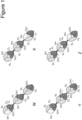
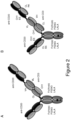


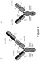

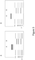
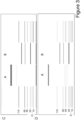


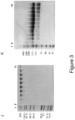
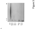


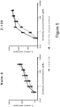

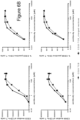

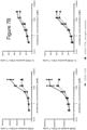
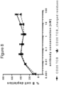
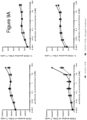
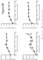
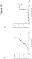
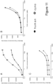
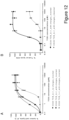

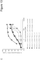
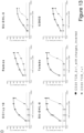


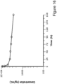


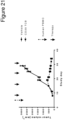

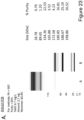
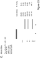
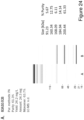
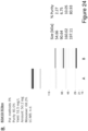

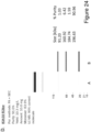


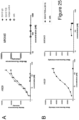

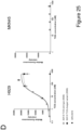
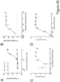

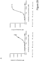
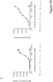
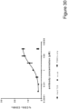
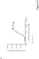
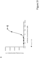
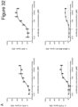
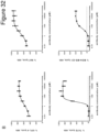

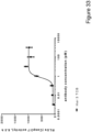
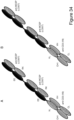

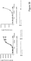
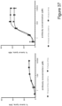
Classifications
-
- C—CHEMISTRY; METALLURGY
- C07—ORGANIC CHEMISTRY
- C07K—PEPTIDES
- C07K16/00—Immunoglobulins [IGs], e.g. monoclonal or polyclonal antibodies
- C07K16/18—Immunoglobulins [IGs], e.g. monoclonal or polyclonal antibodies against material from animals or humans
- C07K16/28—Immunoglobulins [IGs], e.g. monoclonal or polyclonal antibodies against material from animals or humans against receptors, cell surface antigens or cell surface determinants
- C07K16/2803—Immunoglobulins [IGs], e.g. monoclonal or polyclonal antibodies against material from animals or humans against receptors, cell surface antigens or cell surface determinants against the immunoglobulin superfamily
- C07K16/2809—Immunoglobulins [IGs], e.g. monoclonal or polyclonal antibodies against material from animals or humans against receptors, cell surface antigens or cell surface determinants against the immunoglobulin superfamily against the T-cell receptor (TcR)-CD3 complex
-
- A—HUMAN NECESSITIES
- A61—MEDICAL OR VETERINARY SCIENCE; HYGIENE
- A61P—SPECIFIC THERAPEUTIC ACTIVITY OF CHEMICAL COMPOUNDS OR MEDICINAL PREPARATIONS
- A61P35/00—Antineoplastic agents
-
- C—CHEMISTRY; METALLURGY
- C07—ORGANIC CHEMISTRY
- C07K—PEPTIDES
- C07K16/00—Immunoglobulins [IGs], e.g. monoclonal or polyclonal antibodies
- C07K16/18—Immunoglobulins [IGs], e.g. monoclonal or polyclonal antibodies against material from animals or humans
- C07K16/28—Immunoglobulins [IGs], e.g. monoclonal or polyclonal antibodies against material from animals or humans against receptors, cell surface antigens or cell surface determinants
- C07K16/2878—Immunoglobulins [IGs], e.g. monoclonal or polyclonal antibodies against material from animals or humans against receptors, cell surface antigens or cell surface determinants against the NGF-receptor/TNF-receptor superfamily, e.g. CD27, CD30, CD40, CD95
-
- C—CHEMISTRY; METALLURGY
- C07—ORGANIC CHEMISTRY
- C07K—PEPTIDES
- C07K16/00—Immunoglobulins [IGs], e.g. monoclonal or polyclonal antibodies
- C07K16/18—Immunoglobulins [IGs], e.g. monoclonal or polyclonal antibodies against material from animals or humans
- C07K16/28—Immunoglobulins [IGs], e.g. monoclonal or polyclonal antibodies against material from animals or humans against receptors, cell surface antigens or cell surface determinants
- C07K16/2887—Immunoglobulins [IGs], e.g. monoclonal or polyclonal antibodies against material from animals or humans against receptors, cell surface antigens or cell surface determinants against CD20
-
- C—CHEMISTRY; METALLURGY
- C07—ORGANIC CHEMISTRY
- C07K—PEPTIDES
- C07K16/00—Immunoglobulins [IGs], e.g. monoclonal or polyclonal antibodies
- C07K16/18—Immunoglobulins [IGs], e.g. monoclonal or polyclonal antibodies against material from animals or humans
- C07K16/28—Immunoglobulins [IGs], e.g. monoclonal or polyclonal antibodies against material from animals or humans against receptors, cell surface antigens or cell surface determinants
- C07K16/30—Immunoglobulins [IGs], e.g. monoclonal or polyclonal antibodies against material from animals or humans against receptors, cell surface antigens or cell surface determinants from tumour cells
- C07K16/3053—Skin, nerves, brain
-
- C—CHEMISTRY; METALLURGY
- C07—ORGANIC CHEMISTRY
- C07K—PEPTIDES
- C07K16/00—Immunoglobulins [IGs], e.g. monoclonal or polyclonal antibodies
- C07K16/18—Immunoglobulins [IGs], e.g. monoclonal or polyclonal antibodies against material from animals or humans
- C07K16/32—Immunoglobulins [IGs], e.g. monoclonal or polyclonal antibodies against material from animals or humans against translation products of oncogenes
-
- A—HUMAN NECESSITIES
- A61—MEDICAL OR VETERINARY SCIENCE; HYGIENE
- A61K—PREPARATIONS FOR MEDICAL, DENTAL OR TOILETRY PURPOSES
- A61K39/00—Medicinal preparations containing antigens or antibodies
- A61K2039/505—Medicinal preparations containing antigens or antibodies comprising antibodies
-
- C—CHEMISTRY; METALLURGY
- C07—ORGANIC CHEMISTRY
- C07K—PEPTIDES
- C07K2317/00—Immunoglobulins specific features
- C07K2317/10—Immunoglobulins specific features characterized by their source of isolation or production
- C07K2317/14—Specific host cells or culture conditions, e.g. components, pH or temperature
-
- C—CHEMISTRY; METALLURGY
- C07—ORGANIC CHEMISTRY
- C07K—PEPTIDES
- C07K2317/00—Immunoglobulins specific features
- C07K2317/20—Immunoglobulins specific features characterized by taxonomic origin
- C07K2317/21—Immunoglobulins specific features characterized by taxonomic origin from primates, e.g. man
-
- C—CHEMISTRY; METALLURGY
- C07—ORGANIC CHEMISTRY
- C07K—PEPTIDES
- C07K2317/00—Immunoglobulins specific features
- C07K2317/30—Immunoglobulins specific features characterized by aspects of specificity or valency
- C07K2317/31—Immunoglobulins specific features characterized by aspects of specificity or valency multispecific
-
- C—CHEMISTRY; METALLURGY
- C07—ORGANIC CHEMISTRY
- C07K—PEPTIDES
- C07K2317/00—Immunoglobulins specific features
- C07K2317/30—Immunoglobulins specific features characterized by aspects of specificity or valency
- C07K2317/33—Crossreactivity, e.g. for species or epitope, or lack of said crossreactivity
-
- C—CHEMISTRY; METALLURGY
- C07—ORGANIC CHEMISTRY
- C07K—PEPTIDES
- C07K2317/00—Immunoglobulins specific features
- C07K2317/50—Immunoglobulins specific features characterized by immunoglobulin fragments
- C07K2317/51—Complete heavy chain or Fd fragment, i.e. VH + CH1
-
- C—CHEMISTRY; METALLURGY
- C07—ORGANIC CHEMISTRY
- C07K—PEPTIDES
- C07K2317/00—Immunoglobulins specific features
- C07K2317/50—Immunoglobulins specific features characterized by immunoglobulin fragments
- C07K2317/515—Complete light chain, i.e. VL + CL
-
- C—CHEMISTRY; METALLURGY
- C07—ORGANIC CHEMISTRY
- C07K—PEPTIDES
- C07K2317/00—Immunoglobulins specific features
- C07K2317/50—Immunoglobulins specific features characterized by immunoglobulin fragments
- C07K2317/52—Constant or Fc region; Isotype
- C07K2317/526—CH3 domain
-
- C—CHEMISTRY; METALLURGY
- C07—ORGANIC CHEMISTRY
- C07K—PEPTIDES
- C07K2317/00—Immunoglobulins specific features
- C07K2317/50—Immunoglobulins specific features characterized by immunoglobulin fragments
- C07K2317/54—F(ab')2
-
- C—CHEMISTRY; METALLURGY
- C07—ORGANIC CHEMISTRY
- C07K—PEPTIDES
- C07K2317/00—Immunoglobulins specific features
- C07K2317/50—Immunoglobulins specific features characterized by immunoglobulin fragments
- C07K2317/55—Fab or Fab'
-
- C—CHEMISTRY; METALLURGY
- C07—ORGANIC CHEMISTRY
- C07K—PEPTIDES
- C07K2317/00—Immunoglobulins specific features
- C07K2317/60—Immunoglobulins specific features characterized by non-natural combinations of immunoglobulin fragments
- C07K2317/64—Immunoglobulins specific features characterized by non-natural combinations of immunoglobulin fragments comprising a combination of variable region and constant region components
-
- C—CHEMISTRY; METALLURGY
- C07—ORGANIC CHEMISTRY
- C07K—PEPTIDES
- C07K2317/00—Immunoglobulins specific features
- C07K2317/60—Immunoglobulins specific features characterized by non-natural combinations of immunoglobulin fragments
- C07K2317/66—Immunoglobulins specific features characterized by non-natural combinations of immunoglobulin fragments comprising a swap of domains, e.g. CH3-CH2, VH-CL or VL-CH1
-
- C—CHEMISTRY; METALLURGY
- C07—ORGANIC CHEMISTRY
- C07K—PEPTIDES
- C07K2317/00—Immunoglobulins specific features
- C07K2317/70—Immunoglobulins specific features characterized by effect upon binding to a cell or to an antigen
- C07K2317/71—Decreased effector function due to an Fc-modification
-
- C—CHEMISTRY; METALLURGY
- C07—ORGANIC CHEMISTRY
- C07K—PEPTIDES
- C07K2317/00—Immunoglobulins specific features
- C07K2317/70—Immunoglobulins specific features characterized by effect upon binding to a cell or to an antigen
- C07K2317/73—Inducing cell death, e.g. apoptosis, necrosis or inhibition of cell proliferation
- C07K2317/732—Antibody-dependent cellular cytotoxicity [ADCC]
-
- C—CHEMISTRY; METALLURGY
- C07—ORGANIC CHEMISTRY
- C07K—PEPTIDES
- C07K2317/00—Immunoglobulins specific features
- C07K2317/90—Immunoglobulins specific features characterized by (pharmaco)kinetic aspects or by stability of the immunoglobulin
- C07K2317/92—Affinity (KD), association rate (Ka), dissociation rate (Kd) or EC50 value
-
- C—CHEMISTRY; METALLURGY
- C07—ORGANIC CHEMISTRY
- C07K—PEPTIDES
- C07K2317/00—Immunoglobulins specific features
- C07K2317/90—Immunoglobulins specific features characterized by (pharmaco)kinetic aspects or by stability of the immunoglobulin
- C07K2317/94—Stability, e.g. half-life, pH, temperature or enzyme-resistance
Definitions
- the present invention generally relates to bispecific antigen binding molecules for activating T cells.
- the selective destruction of an individual cell or a specific cell type is often desirable in a variety of clinical settings. For example, it is a primary goal of cancer therapy to specifically destroy tumor cells, while leaving healthy cells and tissues intact and undamaged.
- CTLs constitute the most potent effector cells of the immune system, however they cannot be activated by the effector mechanism mediated by the Fc domain of conventional therapeutic antibodies.
- bispecific antibodies designed to bind with one "arm” to a surface antigen on target cells, and with the second "arm” to an activating, invariant component of the T cell receptor (TCR) complex, have become of interest in recent years.
- TCR T cell receptor
- the simultaneous binding of such an antibody to both of its targets will force a temporary interaction between target cell and T cell, causing activation of any cytotoxic T cell and subsequent lysis of the target cell.
- the immune response is re-directed to the target cells and is independent of peptide antigen presentation by the target cell or the specificity of the T cell as would be relevant for normal MHC-restricted activation of CTLs.
- CTLs are only activated when a target cell is presenting the bispecific antibody to them, i.e. the immunological synapse is mimicked.
- bispecific antibodies that do not require lymphocyte preconditioning or co-stimulation in order to elicit efficient lysis of target cells.
- BiTE bispecific T cell engager
- BiTEs are tandem scFv molecules wherein two scFv molecules are fused by a flexible linker.
- Further bispecific formats being evaluated for T cell engagement include diabodies ( Holliger et al., Prot Eng 9, 299-305 (1996 )) and derivatives thereof, such as tandem diabodies ( Kipriyanov et al., J Mol Biol 293, 41-66 (1999 )).
- DART dual affinity retargeting
- triomabs which are whole hybrid mouse/rat IgG molecules and also currently being evaluated in clinical trials, represent a larger sized format (reviewed in Seimetz et al., Cancer Treat Rev 36, 458-467 (2010 )).
- the variety of formats that are being developed shows the great potential attributed to T cell re-direction and activation in immunotherapy.
- the task of generating bispecific antibodies suitable therefor is, however, by no means trivial, but involves a number of challenges that have to be met related to efficacy, toxicity, applicability and produceability of the antibodies.
- IgG-like formats while being able to efficiently crosslink effector and target cells - have a very short serum half life requiring them to be administered to patients by continuous infusion.
- IgG-like formats on the other hand - while having the great benefit of a long half life - suffer from toxicity associated with the native effector functions inherent to IgG molecules.
- Their immunogenic potential constitutes another unfavorable feature of IgG-like bispecific antibodies, especially non-human formats, for successful therapeutic development.
- bispecific antibodies a major challenge in the general development of bispecific antibodies has been the production of bispecific antibody constructs at a clinically sufficient quantity and purity, due to the mispairing of antibody heavy and light chains of different specificities upon co-expression, which decreases the yield of the correctly assembled construct and results in a number of non-functional side products from which the desired bispecific antibody may be difficult to separate.
- the ⁇ knobs-into-holes' strategy aims at forcing the pairing of two different antibody heavy chains by introducing mutations into the CH3 domains to modify the contact interface.
- bulky amino acids are replaced by amino acids with short side chains to create a ⁇ hole'.
- amino acids with large side chains are introduced into the other CH3 domain, to create a 'knob'.
- heterodimer By coexpressing these two heavy chains (and two identical light chains, which have to be appropriate for both heavy chains), high yields of heterodimer ('knob-hole') versus homodimer ( ⁇ hole-hole' or ⁇ knob-knob') are observed ( Ridgway, J.B., et al., Protein Eng. 9 (1996) 617-621 ; and WO 96/027011 ).
- the percentage of heterodimer could be further increased by remodeling the interaction surfaces of the two CH3 domains using a phage display approach and the introduction of a disulfide bridge to stabilize the heterodimers ( Merchant, A.M., et al., Nature Biotech.
- the ⁇ knobs-into-holes' strategy does, however, not solve the problem of heavy chain-light chain mispairing, which occurs in bispecific antibodies comprising different light chains for binding to the different target antigens.
- a strategy to prevent heavy chain-light chain mispairing is to exchange domains between the heavy and light chains of one of the binding arms of a bispecific antibody (see WO 2009/080251 , WO 2009/080252 , WO 2009/080253 , WO 2009/080254 and Schaefer, W. et al, PNAS, 108 (2011) 11187-11191 , which relate to bispecific IgG antibodies with a domain crossover).
- the present invention provides bispecific antigen binding molecules designed for T cell activation and re-direction that combine good efficacy and produceability with low toxicity and favorable pharmacokinetic properties.
- the invention is as defined in claim 1. According to the invention, the ratio of a desired bispecific antibody compared to undesired side products, in particular Bence Jones-type side products occurring in bispecific antibodies with a VH/VL domain exchange in one of their binding arms, can be improved by the introduction of charged amino acids with opposite charges at specific amino acid positions in the CH1 and CL domains.
- the present invention provides a T cell activating bispecific antigen binding molecule as defined in claim 1, comprising
- the T cell activating bispecific antigen binding molecule according to the invention comprises a third Fab molecule which specifically binds to the first antigen.
- the third Fab molecule is identical to the first Fab molecule.
- the third Fab molecule thus comprises the same amino acid substitutions as the first Fab molecule.
- the third Fab molecule particularly is a conventional Fab molecule.
- the first and the third Fab molecule specifically bind to CD20, and the second Fab molecule specifically binds to CD3, more particularly CD3 epsilon
- the first Fab molecule under a) and the second Fab molecule under b) are fused to each other, via a peptide linker.
- the first Fab molecule is fused at the C-terminus of the Fab heavy chain to the N-terminus of the Fab heavy chain of the second Fab molecule.
- the second Fab molecule is fused at the C-terminus of the Fab heavy chain to the N-terminus of the Fab heavy chain of the first Fab molecule or (ii) the first Fab molecule is fused at the C-terminus of the Fab heavy chain to the N-terminus of the Fab heavy chain of the second Fab molecule
- the Fab light chain of the Fab molecule and the Fab light chain of the second Fab molecule may be fused to each other, optionally via a peptide linker.
- the T cell activating bispecific antigen binding molecule according to the invention additionally comprises an Fc domain composed of a first and a second subunit capable of stable association.
- the second Fab molecule is fused at the C-terminus of the Fab heavy chain to the N-terminus of the first subunit of the Fc domain.
- the first Fab molecule is fused at the C-terminus of the Fab heavy chain to the N-terminus of the Fab heavy chain of the second Fab molecule.
- the third Fab molecule is fused at the C-terminus of the Fab heavy chain to the N-terminus of the first subunit of the Fc domain.
- the second and the third Fab molecule are each fused at the C-terminus of the Fab heavy chain to the N-terminus of one of the subunits of the Fc domain, and the first Fab molecule is fused at the C-terminus of the Fab heavy chain to the N-terminus of the Fab heavy chain of the second Fab molecule.
- the T cell activating bispecific antigen binding molecule essentially comprises an immunoglobulin molecule, wherein in one of the Fab arms the heavy and light chain variable regions VH and VL are exchanged/replaced by each other, and wherein an additional (conventional) Fab molecule is N-terminally fused to said Fab arm.
- the amino acid substitutions described herein are in the CH1 and CL domains of the first and the third Fab molecule.
- the amino acid substitutions as described herein are made in the first andthe third Fab molecule, and no such amino acid substitutions are made in the second Fab molecule.
- Amino acid substitutions as described herein are made in the first and the third Fab molecule and the constant domain CL of the first and the third Fab molecule is of kappa isotype.
- the constant domain CL of the first (and the third) Fab molecule and the constant domain CL of the second Fab molecule are of kappa isotype.
- the immunoglobulin molecule comprised in the T cell activating bispecific antigen binding molecule according to the invention is an IgG 1 subclass immunoglobulin.
- the immunoglobulin is an IgG 4 subclass immunoglobulin.
- the invention provides a T cell activating bispecific antigen binding molecule as defined in claim 1, which molecule comprises
- the Fc domain is an IgG 1 Fc domain.
- the Fc domain is an IgG 4 Fc domain.
- the Fc domain is an IgG 4 Fc domain comprising the amino acid substitution S228P (Kabat numbering).
- the Fc domain is a human Fc domain.
- the Fc domain comprises a modification promoting the association of the first and the second Fc domain subunit.
- An amino acid residue in the CH3 domain of the first subunit of the Fc domain is replaced with an amino acid residue having a larger side chain volume, thereby generating a protuberance within the CH3 domain of the first subunit which is positionable in a cavity within the CH3 domain of the second subunit, and an amino acid residue in the CH3 domain of the second subunit of the Fc domain is replaced with an amino acid residue having a smaller side chain volume, thereby generating a cavity within the CH3 domain of the second subunit within which the protuberance within the CH3 domain of the first subunit is positionable.
- the Fc domain exhibits reduced binding affinity to an Fc receptor and/or reduced effector function, as compared to a native IgG 1 Fc domain.
- the Fc domain is engineered to have reduced binding affinity to an Fc receptor and/or reduced effector function, as compared to a non-engineered Fc domain.
- the Fc domain comprises one or more amino acid substitution that reduces binding to an Fc receptor and/or effector function.
- the one or more amino acid substitution in the Fc domain that reduces binding to an Fc receptor and/or effector function is at one or more position selected from the group of L234, L235, and P329 (Kabat EU index numbering).
- Each subunit of the Fc domain comprises three amino acid substitutions that reduce binding to an Fc receptor and/or effector function wherein said amino acid substitutions are L234A, L235A and P329G (Kabat EU index numbering).
- the Fc domain is an IgG 1 Fc domain, particularly a human IgG 1 Fc domain.
- each subunit of the Fc domain comprises two amino acid substitutions that reduce binding to an Fc receptor and/or effector function wherein said amino acid substitutions are L235E and P329G (Kabat EU index numbering).
- the Fc domain is an IgG 4 Fc domain, particularly a human IgG 4 Fc domain.
- the Fc domain of the T cell activating bispecific antigen binding molecule is an IgG 4 Fc domain and comprises the amino acid substitutions L235E and S228P (SPLE) (Kabat EU index numbering).
- the Fc receptor is an Fcy receptor.
- the Fc receptor is a human Fc receptor and the Fc receptor is an activating Fc receptor.
- the Fc receptor is human Fc ⁇ RIIa, FcyRI, and/or FcyRIIIa.
- the effector function is antibody-dependent cell-mediated cytotoxicity (ADCC).
- the T cell activating bispecific antigen binding molecule comprises a Fab molecule which specifically binds to CD3, more particularly CD3 epsilon, comprises the heavy chain complementarity determining region (CDR) 1 of SEQ ID NO: 4, the heavy chain CDR 2 of SEQ ID NO: 5, the heavy chain CDR 3 of SEQ ID NO: 6, the light chain CDR 1 of SEQ ID NO: 8, the light chain CDR 2 of SEQ ID NO: 9 and the light chain CDR 3 of SEQ ID NO: 10.
- CDR heavy chain complementarity determining region
- the Fab molecule which specifically binds to an activating T cell antigen, particularly CD3, more particularly CD3 epsilon comprises a heavy chain variable region comprising an amino acid sequence that is at least about 95%, 96%, 97%, 98%, 99% or 100% identical to the amino acid sequence of SEQ ID NO: 3 and a light chain variable region comprising an amino acid sequence that is at least about 95%, 96%, 97%, 98%, 99% or 100% identical to the amino acid sequence of SEQ ID NO: 7.
- the second Fab molecule comprised in the T cell activating bispecific antigen binding molecule according to the invention specifically binds to CD3, more particularly CD3 epsilon, and comprises the heavy chain complementarity determining region (CDR) 1 of SEQ ID NO: 4, the heavy chain CDR 2 of SEQ ID NO: 5, the heavy chain CDR 3 of SEQ ID NO: 6, the light chain CDR 1 of SEQ ID NO: 8, the light chain CDR 2 of SEQ ID NO: 9 and the light chain CDR 3 of SEQ ID NO: 10.
- the second Fab molecule comprises a heavy chain variable region comprising the amino acid sequence of SEQ ID NO: 3 and a light chain variable region comprising the amino acid sequence of SEQ ID NO: 7.
- the T cell activating bispecific antigen binding molecule comprisesthe two Fab molecules which specifically bind to the target cell antigen CD20 which comprises the heavy chain complementarity determining region (CDR) 1 of SEQ ID NO: 46, the heavy chain CDR 2 of SEQ ID NO: 47, the heavy chain CDR 3 of SEQ ID NO: 48, the light chain CDR 1 of SEQ ID NO: 49, the light chain CDR 2 of SEQ ID NO: 50 and the light chain CDR 3 of SEQ ID NO: 51.
- CD20 which comprises the heavy chain complementarity determining region (CDR) 1 of SEQ ID NO: 46, the heavy chain CDR 2 of SEQ ID NO: 47, the heavy chain CDR 3 of SEQ ID NO: 48, the light chain CDR 1 of SEQ ID NO: 49, the light chain CDR 2 of SEQ ID NO: 50 and the light chain CDR 3 of SEQ ID NO: 51.
- the Fab molecule which specifically binds to a target cell antigen, particularly CD20 comprises a heavy chain variable region comprising an amino acid sequence that is at least about 95%, 96%, 97%, 98%, 99% or 100% identical to the amino acid sequence of SEQ ID NO: 30 and a light chain variable region comprising an amino acid sequence that is at least about 95%, 96%, 97%, 98%, 99% or 100% identical to the amino acid sequence of SEQ ID NO: 31.
- the first and, the third Fab molecule comprised in the T cell activating bispecific antigen binding molecule according to the invention specifically binds to CD20, and comprises the heavy chain complementarity determining region (CDR) 1 of SEQ ID NO: 46, the heavy chain CDR 2 of SEQ ID NO: 47, the heavy chain CDR 3 of SEQ ID NO: 48, the light chain CDR 1 of SEQ ID NO: 49, the light chain CDR 2 of SEQ ID NO: 50 and the light chain CDR 3 of SEQ ID NO: 51.
- the first and third Fab molecule comprises a heavy chain variable region comprising the amino acid sequence of SEQ ID NO: 30 and a light chain variable region comprising the amino acid sequence of SEQ ID NO: 31.
- the invention provides a T cell activating bispecific antigen binding molecule as defined in claim 1, which molecule comprises
- one or more isolated polynucleotide(s) encoding a T cell activating bispecific antigen binding molecule of the invention.
- the disclosure further provides one or more expression vector(s) comprising the isolated polynucleotide(s) of the invention, and a host cell comprising the isolated polynucleotide(s) or the expression vector(s) of the disclosure.
- the host cell is a eukaryotic cell, particularly a mammalian cell.
- a method of producing the T cell activating bispecific antigen binding molecule of the disclosure comprising the steps of a) culturing the host cell of the disclosure under conditions suitable for the expression of the T cell activating bispecific antigen binding molecule and b) recovering the T cell activating bispecific antigen binding molecule.
- the disclosure also encompasses a T cell activating bispecific antigen binding molecule produced by the method of the invention.
- the disclosure further provides a pharmaceutical composition
- a pharmaceutical composition comprising the T cell activating bispecific antigen binding molecule of the invention and a pharmaceutically acceptable carrier.
- the disclosure provides a T cell activating bispecific antigen binding molecule or a pharmaceutical composition of the invention for use as a medicament.
- a T cell activating bispecific antigen binding molecule or a pharmaceutical composition according to the disclosure for use in the treatment of a disease in an individual in need thereof.
- the disease is cancer.
- a T cell activating bispecific antigen binding molecule of the invention for the manufacture of a medicament for the treatment of a disease in an individual in need thereof; comprising administering to said individual a therapeutically effective amount of a composition comprising the T cell activating bispecific antigen binding molecule according to the invention in a pharmaceutically acceptable form.
- the disease is cancer.
- the individual preferably is a mammal, particularly a human.
- the invention is as defined in the claim.
- antigen binding molecule refers in its broadest sense to a molecule that specifically binds an antigenic determinant.
- antigen binding molecules are immunoglobulins and derivatives, e.g. fragments, thereof.
- bispecific means that the antigen binding molecule is able to specifically bind to at least two distinct antigenic determinants.
- a bispecific antigen binding molecule comprises two antigen binding sites, each of which is specific for a different antigenic determinant.
- the bispecific antigen binding molecule is capable of simultaneously binding two antigenic determinants, particularly two antigenic determinants expressed on two distinct cells.
- valent denotes the presence of a specified number of antigen binding sites in an antigen binding molecule.
- monovalent binding to an antigen denotes the presence of one (and not more than one) antigen binding site specific for the antigen in the antigen binding molecule.
- an “antigen binding site” refers to the site, i.e. one or more amino acid residues, of an antigen binding molecule which provides interaction with the antigen.
- the antigen binding site of an antibody comprises amino acid residues from the complementarity determining regions (CDRs).
- CDRs complementarity determining regions
- a native immunoglobulin molecule typically has two antigen binding sites, a Fab molecule typically has a single antigen binding site.
- the term "antigen binding moiety” refers to a polypeptide molecule that specifically binds to an antigenic determinant.
- the antigen binding moiety is able to direct the entity to which it is attached (e.g. a second antigen binding moiety) to a target site, for example to a specific type of tumor cell or tumor stroma bearing the antigenic determinant.
- the antigen binding moiety is able to activate signaling through its target antigen, for example a T cell receptor complex antigen.
- Antigen binding moieties include antibodies and fragments thereof as further defined herein. Particular antigen binding moieties include an antigen binding domain of an antibody, comprising an antibody heavy chain variable region and an antibody light chain variable region.
- the antigen binding moieties may comprise antibody constant regions as further defined herein and known in the art.
- Useful heavy chain constant regions include any of the five isotypes: ⁇ , ⁇ , ⁇ , ⁇ , or ⁇ .
- Useful light chain constant regions include any of the two isotypes: ⁇ and ⁇ .
- antigenic determinant is synonymous with “antigen” and “epitope,” and refers to a site (e.g. a contiguous stretch of amino acids or a conformational configuration made up of different regions of non-contiguous amino acids) on a polypeptide macromolecule to which an antigen binding moiety binds, forming an antigen binding moiety-antigen complex.
- Useful antigenic determinants can be found, for example, on the surfaces of tumor cells, on the surfaces of virus-infected cells, on the surfaces of other diseased cells, on the surface of immune cells, free in blood serum, and/or in the extracellular matrix (ECM).
- ECM extracellular matrix
- CD3 can be any native form the proteins from any vertebrate source, including mammals such as primates (e.g. humans) and rodents (e.g. mice and rats), unless otherwise indicated.
- the antigen is a human protein.
- the term encompasses the "full-length", unprocessed protein as well as any form of the protein that results from processing in the cell.
- the term also encompasses naturally occurring variants of the protein, e.g. splice variants or allelic variants.
- An exemplary human protein useful as antigen is CD3, particularly the epsilon subunit of CD3 (see UniProt no. P07766 (version 130), NCBI RefSeq no.
- the T cell activating bispecific antigen binding molecule of the invention binds to an epitope of CD3 or a target cell antigen that is conserved among the CD3 or target cell antigen from different species.
- ELISA enzyme-linked immunosorbent assay
- SPR surface plasmon resonance
- an antigen binding moiety that binds to the antigen, or an antigen binding molecule comprising that antigen binding moiety has a dissociation constant (K D ) of ⁇ 1 ⁇ M, ⁇ 100 nM, ⁇ 10 nM, ⁇ 1 nM, ⁇ 0.1 nM, ⁇ 0.01 nM, or ⁇ 0.001 nM (e.g. 10 -8 M or less, e.g. from 10 -8 M to 10 -13 M, e.g., from 10 -9 M to 10 -13 M).
- K D dissociation constant
- Binding affinity refers to intrinsic binding affinity which reflects a 1:1 interaction between members of a binding pair (e.g., an antigen binding moiety and an antigen, or a receptor and its ligand).
- the affinity of a molecule X for its partner Y can generally be represented by the dissociation constant (K D ), which is the ratio of dissociation and association rate constants (k off and k on , respectively).
- affinities may comprise different rate constants, as long as the ratio of the rate constants remains the same.
- Affinity can be measured by well established methods known in the art, including those described herein.
- a particular method for measuring affinity is Surface Plasmon Resonance (SPR).
- Reduced binding for example reduced binding to an Fc receptor, refers to a decrease in affinity for the respective interaction, as measured for example by SPR.
- the term includes also reduction of the affinity to zero (or below the detection limit of the analytic method), i.e. complete abolishment of the interaction.
- increased binding refers to an increase in binding affinity for the respective interaction.
- an "activating T cell antigen” as used herein refers to an antigenic determinant expressed on the surface of a T lymphocyte, particularly a cytotoxic T lymphocyte, which is capable of inducing T cell activation upon interaction with an antigen binding molecule. Specifically, interaction of an antigen binding molecule with an activating T cell antigen may induce T cell activation by triggering the signaling cascade of the T cell receptor complex.
- the activating T cell antigen is CD3, particularly the epsilon subunit of CD3 (see UniProt no. P07766 (version 130), NCBI RefSeq no. NP_000724.1, SEQ ID NO: 1 for the human sequence; or UniProt no. Q95LI5 (version 49), NCBI GenBank no. BAB71849.1, SEQ ID NO: 2 for the cynomolgus [Macaca fascicularis] sequence).
- T cell activation refers to one or more cellular response of a T lymphocyte, particularly a cytotoxic T lymphocyte, selected from: proliferation, differentiation, cytokine secretion, cytotoxic effector molecule release, cytotoxic activity, and expression of activation markers.
- the T cell activating bispecific antigen binding molecules of the invention are capable of inducing T cell activation. Suitable assays to measure T cell activation are known in the art described herein.
- target cell antigen refers to an antigenic determinant presented on the surface of a target cell, for example a cell in a tumor such as a cancer cell or a cell of the tumor stroma.
- the target cell antigen is CD20, particularly human CD20 (see UniProt no. P11836).
- the terms “first”, “second” or “third” with respect to Fab molecules etc. are used for convenience of distinguishing when there is more than one of each type of moiety. Use of these terms is not intended to confer a specific order or orientation of the T cell activating bispecific antigen binding molecule unless explicitly so stated.
- a “Fab molecule” refers to a protein consisting of the VH and CH1 domain of the heavy chain (the “Fab heavy chain”) and the VL and CL domain of the light chain (the “Fab light chain”) of an immunoglobulin.
- fused is meant that the components (e.g. a Fab molecule and an Fc domain subunit) are linked by peptide bonds, either directly or via one or more peptide linkers.
- single-chain refers to a molecule comprising amino acid monomers linearly linked by peptide bonds.
- one of the antigen binding moieties is a single-chain Fab molecule, i.e. a Fab molecule wherein the Fab light chain and the Fab heavy chain are connected by a peptide linker to form a single peptide chain.
- the C-terminus of the Fab light chain is connected to the N-terminus of the Fab heavy chain in the single-chain Fab molecule.
- crossover Fab molecule also termed “Crossfab” is meant a Fab molecule wherein the variable domains of the Fab heavy and light chain are exchanged (i.e. replaced by each other), i.e. the crossover Fab molecule comprises a peptide chain composed of the light chain variable domain VL and the heavy chain constant domain 1 CH1 (VL-CH1, in N- to C-terminal direction), and a peptide chain composed of the heavy chain variable domain VH and the light chain constant domain CL (VH-CL, in N- to C-terminal direction).
- the peptide chain comprising the heavy chain constant domain 1 CH1 is referred to herein as the "heavy chain" of the crossover Fab molecule.
- a "conventional" Fab molecule is meant a Fab molecule in its natural format, i.e. comprising a heavy chain composed of the heavy chain variable and constant domains (VH-CH1, in N- to C-terminal direction), and a light chain composed of the light chain variable and constant domains (VL-CL, in N- to C-terminal direction).
- immunoglobulin molecule refers to a protein having the structure of a naturally occurring antibody.
- immunoglobulins of the IgG class are heterotetrameric glycoproteins of about 150,000 daltons, composed of two light chains and two heavy chains that are disulfide-bonded. From N- to C-terminus, each heavy chain has a variable domain (VH), also called a variable heavy domain or a heavy chain variable region, followed by three constant domains (CH1, CH2, and CH3), also called a heavy chain constant region.
- VH variable domain
- CH1, CH2, and CH3 constant domains
- each light chain has a variable domain (VL), also called a variable light domain or a light chain variable region, followed by a constant light (CL) domain, also called a light chain constant region.
- VL variable domain
- CL constant light
- the heavy chain of an immunoglobulin may be assigned to one of five types, called ⁇ (IgA), ⁇ (IgD), ⁇ (IgE), ⁇ (IgG), or ⁇ (IgM), some of which may be further divided into subtypes, e.g. ⁇ 1 (IgG 1 ), ⁇ 2 (IgG 2 ), ⁇ 3 (IgG 3 ), ⁇ 4 (IgG 4 ), ⁇ 1 (IgA 1 ) and ⁇ 2 (IgA 2 ).
- the light chain of an immunoglobulin may be assigned to one of two types, called kappa ( ⁇ ) and lambda ( ⁇ ), based on the amino acid sequence of its constant domain.
- An immunoglobulin essentially consists of two Fab molecules and an Fc domain, linked via the immunoglobulin hinge region.
- antibody herein is used in the broadest sense and encompasses various antibody structures, including but not limited to monoclonal antibodies, polyclonal antibodies, and antibody fragments so long as they exhibit the desired antigen-binding activity.
- antibody fragment refers to a molecule other than an intact antibody that comprises a portion of an intact antibody that binds the antigen to which the intact antibody binds.
- antibody fragments include but are not limited to Fv, Fab, Fab', Fab'-SH, F(ab') 2 , diabodies, linear antibodies, single-chain antibody molecules (e.g. scFv), and single-domain antibodies.
- scFv single-chain antibody molecules
- Diabodies are antibody fragments with two antigen-binding sites that may be bivalent or bispecific.
- Single-domain antibodies are antibody fragments comprising all or a portion of the heavy chain variable domain or all or a portion of the light chain variable domain of an antibody.
- a single-domain antibody is a human single-domain antibody (Domantis, Inc., Waltham, MA; see e.g. U.S. Patent No.
- Antibody fragments can be made by various techniques, including but not limited to proteolytic digestion of an intact antibody as well as production by recombinant host cells (e.g. E. coli or phage), as described herein.
- recombinant host cells e.g. E. coli or phage
- an antigen binding domain refers to the part of an antibody that comprises the area which specifically binds to and is complementary to part or all of an antigen.
- An antigen binding domain may be provided by, for example, one or more antibody variable domains (also called antibody variable regions).
- an antigen binding domain comprises an antibody light chain variable domain (VL) and an antibody heavy chain variable domain (VH).
- variable region refers to the domain of an antibody heavy or light chain that is involved in binding the antibody to antigen.
- the variable domains of the heavy chain and light chain (VH and VL, respectively) of a native antibody generally have similar structures, with each domain comprising four conserved framework regions (FRs) and three hypervariable regions (HVRs). See, e.g., Kindt et al., Kuby Immunology, 6th ed., W.H. Freeman and Co., page 91 (2007 ). A single VH or VL domain may be sufficient to confer antigen-binding specificity.
- hypervariable region refers to each of the regions of an antibody variable domain which are hypervariable in sequence and/or form structurally defined loops ("hypervariable loops").
- native four-chain antibodies comprise six HVRs; three in the VH (H1, H2, H3), and three in the VL (L1, L2, L3).
- HVRs generally comprise amino acid residues from the hypervariable loops and/or from the complementarity determining regions (CDRs), the latter being of highest sequence variability and/or involved in antigen recognition. With the exception of CDR1 in VH, CDRs generally comprise the amino acid residues that form the hypervariable loops.
- Hypervariable regions are also referred to as "complementarity determining regions” (CDRs), and these terms are used herein interchangeably in reference to portions of the variable region that form the antigen binding regions.
- CDRs complementarity determining regions
- This particular region has been described by Kabat et al., Sequences of Proteins of Immunological Interest, 5th Ed. Public Health Service, National Institutes of Health, Bethesda, MD (1991 ) and by Chothia et al., J Mol Biol 196:901-917 (1987 ), where the definitions include overlapping or subsets of amino acid residues when compared against each other. Nevertheless, application of either definition to refer to a CDR of an antibody or variants thereof is intended to be within the scope of the term as defined and used herein.
- CDR Definitions 1 CDR Kabat Chothia AbM 2 V H CDR1 31-35 26-32 26-35 V H CDR2 50-65 52-58 50-58 V H CDR3 95-102 95-102 95-102 V L CDR1 24-34 26-32 24-34 V L CDR2 50-56 50-52 50-56 V L CDR3 89-97 91-96 89-97 1 Numbering of all CDR definitions in Table 1 is according to the numbering conventions set forth by Kabat et al. (see below). 2 "AbM” with a lowercase “b” as used in Table 1 refers to the CDRs as defined by Oxford Molecular's "AbM” antibody modeling software.
- Kabat et al. also defined a numbering system for variable region sequences that is applicable to any antibody.
- One of ordinary skill in the art can unambiguously assign this system of "Kabat numbering" to any variable region sequence, without reliance on any experimental data beyond the sequence itself.
- Kabat numbering refers to the numbering system set forth by Kabat et al., Sequences of Proteins of Immunological Interest, 5th Ed. Public Health Service, National Institutes of Health, Bethesda, MD (1991 ). Unless otherwise specified, references to the numbering of specific amino acid residue positions in an antibody variable region are according to the Kabat numbering system.
- amino acid positions of all constant regions and domains of the heavy and light chain are numbered according to the Kabat numbering system described in Kabat, et al., Sequences of Proteins of Immunological Interest, 5th ed., Public Health Service, National Institutes of Health, Bethesda, MD (1991 ) and is referred to as "numbering according to Kabat” or "Kabat numbering" herein.
- Kabat numbering system see pages 647-660 of Kabat, et al., Sequences of Proteins of Immunological Interest, 5th ed., Public Health Service, National Institutes of Health, Bethesda, MD (1991 )
- CL light chain constant domain
- Kabat EU index numbering system see pages 661-723
- CH1, Hinge, CH2 and CH3 heavy chain constant domains
- polypeptide sequences of the sequence listing are not numbered according to the Kabat numbering system. However, it is well within the ordinary skill of one in the art to convert the numbering of the sequences of the Sequence Listing to Kabat numbering.
- FR Framework or "FR” refers to variable domain residues other than hypervariable region (HVR) residues.
- the FR of a variable domain generally consists of four FR domains: FR1, FR2, FR3, and FR4. Accordingly, the HVR and FR sequences generally appear in the following sequence in VH (or VL): FR1-H1(L1)-FR2-H2(L2)-FR3-H3(L3)-FR4.
- the "class" of an antibody or immunoglobulin refers to the type of constant domain or constant region possessed by its heavy chain.
- the heavy chain constant domains that correspond to the different classes of immunoglobulins are called ⁇ , ⁇ , ⁇ , ⁇ , and ⁇ , respectively.
- Fc domain or "Fc region” herein is used to define a C-terminal region of an immunoglobulin heavy chain that contains at least a portion of the constant region.
- the term includes native sequence Fc regions and variant Fc regions.
- the boundaries of the Fc region of an IgG heavy chain might vary slightly, the human IgG heavy chain Fc region is usually defined to extend from Cys226, or from Pro230, to the carboxyl-terminus of the heavy chain.
- antibodies produced by host cells may undergo post-translational cleavage of one or more, particularly one or two, amino acids from the C-terminus of the heavy chain.
- an antibody produced by a host cell by expression of a specific nucleic acid molecule encoding a full-length heavy chain may include the full-length heavy chain, or it may include a cleaved variant of the full-length heavy chain (also referred to herein as a "cleaved variant heavy chain").
- a cleaved variant heavy chain also referred to herein as a "cleaved variant heavy chain”
- the final two C-terminal amino acids of the heavy chain are glycine (G446) and lysine (K447, numbering according to Kabat EU index). Therefore, the C-terminal lysine (Lys447), or the C-terminal glycine (Gly446) and lysine (K447), of the Fc region may or may not be present.
- a heavy chain including a subunit of an Fc domain as specified herein comprised in a T cell activating bispecific antigen binding molecule according to the invention, comprises an additional C-terminal glycine-lysine dipeptide (G446 and K447, numbering according to EU index of Kabat).
- a heavy chain including a subunit of an Fc domain as specified herein, comprised in a T cell activating bispecific antigen binding molecule according to the invention comprises an additional C-terminal glycine residue (G446, numbering according to EU index of Kabat).
- Compositions of the disclosure such as the pharmaceutical compositions described herein, comprise a population of T cell activating bispecific antigen binding molecules of the invention.
- the population of T cell activating bispecific antigen binding molecule may comprise molecules having a full-length heavy chain and molecules having a cleaved variant heavy chain.
- the population of T cell activating bispecific antigen binding molecules may consist of a mixture of molecules having a full-length heavy chain and molecules having a cleaved variant heavy chain, wherein at least 50%, at least 60%, at least 70%, at least 80% or at least 90% of the T cell activating bispecific antigen binding molecules have a cleaved variant heavy chain.
- a composition comprising a population of T cell activating bispecific antigen binding molecules of the invention comprises an T cell activating bispecific antigen binding molecule comprising a heavy chain including a subunit of an Fc domain as specified herein with an additional C-terminal glycine-lysine dipeptide (G446 and K447, numbering according to EU index of Kabat).
- such a composition comprises a population of T cell activating bispecific antigen binding molecules comprised of molecules comprising a heavy chain including a subunit of an Fc domain as specified herein; molecules comprising a heavy chain including a subunit of a Fc domain as specified herein with an additional C-terminal glycine residue (G446, numbering according to EU index of Kabat); and molecules comprising a heavy chain including a subunit of an Fc domain as specified herein with an additional C-terminal glycine-lysine dipeptide (G446 and K447, numbering according to EU index of Kabat).
- a "subunit" of an Fc domain as used herein refers to one of the two polypeptides forming the dimeric Fc domain, i.e. a polypeptide comprising C-terminal constant regions of an immunoglobulin heavy chain, capable of stable self-association.
- a subunit of an IgG Fc domain comprises an IgG CH2 and an IgG CH3 constant domain.
- a "modification promoting the association of the first and the second subunit of the Fc domain” is a manipulation of the peptide backbone or the post-translational modifications of an Fc domain subunit that reduces or prevents the association of a polypeptide comprising the Fc domain subunit with an identical polypeptide to form a homodimer.
- a modification promoting association as used herein particularly includes separate modifications made to each of the two Fc domain subunits desired to associate (i.e. the first and the second subunit of the Fc domain), wherein the modifications are complementary to each other so as to promote association of the two Fc domain subunits.
- a modification promoting association may alter the structure or charge of one or both of the Fc domain subunits so as to make their association sterically or electrostatically favorable, respectively.
- (hetero)dimerization occurs between a polypeptide comprising the first Fc domain subunit and a polypeptide comprising the second Fc domain subunit, which might be non-identical in the sense that further components fused to each of the subunits (e.g. antigen binding moieties) are not the same.
- the modification promoting association comprises an amino acid mutation in the Fc domain, specifically an amino acid substitution.
- the modification promoting association comprises a separate amino acid mutation, specifically an amino acid substitution, in each of the two subunits of the Fc domain.
- effector functions refers to those biological activities attributable to the Fc region of an antibody, which vary with the antibody isotype.
- antibody effector functions include: C1q binding and complement dependent cytotoxicity (CDC), Fc receptor binding, antibody-dependent cell-mediated cytotoxicity (ADCC), antibody-dependent cellular phagocytosis (ADCP), cytokine secretion, immune complex-mediated antigen uptake by antigen presenting cells, down regulation of cell surface receptors (e.g. B cell receptor), and B cell activation.
- engine engineered, engineering
- engineering includes modifications of the amino acid sequence, of the glycosylation pattern, or of the side chain group of individual amino acids, as well as combinations of these approaches.
- amino acid mutation as used herein is meant to encompass amino acid substitutions, deletions, insertions, and modifications. Any combination of substitution, deletion, insertion, and modification can be made to arrive at the final construct, provided that the final construct possesses the desired characteristics, e.g., reduced binding to an Fc receptor, or increased association with another peptide.
- Amino acid sequence deletions and insertions include amino- and/or carboxy-terminal deletions and insertions of amino acids.
- Particular amino acid mutations are amino acid substitutions.
- non-conservative amino acid substitutions i.e. replacing one amino acid with another amino acid having different structural and/or chemical properties, are particularly preferred.
- Amino acid substitutions include replacement by non-naturally occurring amino acids or by naturally occurring amino acid derivatives of the twenty standard amino acids (e.g. 4-hydroxyproline, 3-methylhistidine, ornithine, homoserine, 5-hydroxylysine).
- Amino acid mutations can be generated using genetic or chemical methods well known in the art. Genetic methods may include site-directed mutagenesis, PCR, gene synthesis and the like. It is contemplated that methods of altering the side chain group of an amino acid by methods other than genetic engineering, such as chemical modification, may also be useful. Various designations may be used herein to indicate the same amino acid mutation. For example, a substitution from proline at position 329 of the Fc domain to glycine can be indicated as 329G, G329, G 329 , P329G, or Pro329Gly.
- polypeptide refers to a molecule composed of monomers (amino acids) linearly linked by amide bonds (also known as peptide bonds).
- polypeptide refers to any chain of two or more amino acids, and does not refer to a specific length of the product.
- peptides, dipeptides, tripeptides, oligopeptides, "protein,” “amino acid chain,” or any other term used to refer to a chain of two or more amino acids are included within the definition of "polypeptide,” and the term “polypeptide” may be used instead of, or interchangeably with any of these terms.
- polypeptide is also intended to refer to the products of post-expression modifications of the polypeptide, including without limitation glycosylation, acetylation, phosphorylation, amidation, derivatization by known protecting/blocking groups, proteolytic cleavage, or modification by non-naturally occurring amino acids.
- a polypeptide may be derived from a natural biological source or produced by recombinant technology, but is not necessarily translated from a designated nucleic acid sequence. It may be generated in any manner, including by chemical synthesis.
- a polypeptide of the disclosure may be of a size of about 3 or more, 5 or more, 10 or more, 20 or more, 25 or more, 50 or more, 75 or more, 100 or more, 200 or more, 500 or more, 1,000 or more, or 2,000 or more amino acids.
- Polypeptides may have a defined three-dimensional structure, although they do not necessarily have such structure. Polypeptides with a defined three-dimensional structure are referred to as folded, and polypeptides which do not possess a defined three-dimensional structure, but rather can adopt a large number of different conformations, and are referred to as unfolded.
- an “isolated” polypeptide or a variant, or derivative thereof is intended a polypeptide that is not in its natural milieu. No particular level of purification is required.
- an isolated polypeptide can be removed from its native or natural environment.
- Recombinantly produced polypeptides and proteins expressed in host cells are considered isolated for the purpose of the disclosure, as are native or recombinant polypeptides which have been separated, fractionated, or partially or substantially purified by any suitable technique.
- Percent (%) amino acid sequence identity with respect to a reference polypeptide sequence is defined as the percentage of amino acid residues in a candidate sequence that are identical with the amino acid residues in the reference polypeptide sequence, after aligning the sequences and introducing gaps, if necessary, to achieve the maximum percent sequence identity, and not considering any conservative substitutions as part of the sequence identity. Alignment for purposes of determining percent amino acid sequence identity can be achieved in various ways that are within the skill in the art, for instance, using publicly available computer software such as BLAST, BLAST-2, ALIGN or Megalign (DNASTAR) software. Those skilled in the art can determine appropriate parameters for aligning sequences, including any algorithms needed to achieve maximal alignment over the full length of the sequences being compared.
- % amino acid sequence identity values are generated using the sequence comparison computer program ALIGN-2.
- the ALIGN-2 sequence comparison computer program was authored by Genentech, Inc., and the source code has been filed with user documentation in the U.S. Copyright Office, Washington D.C., 20559, where it is registered under U.S. Copyright Registration No. TXU510087.
- the ALIGN-2 program is publicly available from Genentech, Inc., South San Francisco, California, or may be compiled from the source code.
- the ALIGN-2 program should be compiled for use on a UNIX operating system, including digital UNIX V4.0D. All sequence comparison parameters are set by the ALIGN-2 program and do not vary.
- the % amino acid sequence identity of a given amino acid sequence A to, with, or against a given amino acid sequence B is calculated as follows: 10 0 times the fraction X/Y where X is the number of amino acid residues scored as identical matches by the sequence alignment program ALIGN-2 in that program's alignment of A and B, and where Y is the total number of amino acid residues in B.
- polynucleotide refers to an isolated nucleic acid molecule or construct, e.g. messenger RNA (mRNA), virally-derived RNA, or plasmid DNA (pDNA).
- mRNA messenger RNA
- pDNA virally-derived RNA
- a polynucleotide may comprise a conventional phosphodiester bond or a non-conventional bond (e.g. an amide bond, such as found in peptide nucleic acids (PNA).
- PNA peptide nucleic acids
- nucleic acid molecule refers to any one or more nucleic acid segments, e.g. DNA or RNA fragments, present in a polynucleotide.
- isolated nucleic acid molecule or polynucleotide is intended a nucleic acid molecule, DNA or RNA, which has been removed from its native environment.
- a recombinant polynucleotide encoding a polypeptide contained in a vector is considered isolated for the purposes of the present disclosure.
- Further examples of an isolated polynucleotide include recombinant polynucleotides maintained in heterologous host cells or purified (partially or substantially) polynucleotides in solution.
- An isolated polynucleotide includes a polynucleotide molecule contained in cells that ordinarily contain the polynucleotide molecule, but the polynucleotide molecule is present extrachromosomally or at a chromosomal location that is different from its natural chromosomal location.
- Isolated RNA molecules include in vivo or in vitro RNA transcripts of the present disclosure, as well as positive and negative strand forms, and double-stranded forms. Isolated polynucleotides or nucleic acids according to the present disclosure further include such molecules produced synthetically.
- a polynucleotide or a nucleic acid may be or may include a regulatory element such as a promoter, ribosome binding site, or a transcription terminator.
- nucleic acid or polynucleotide having a nucleotide sequence at least, for example, 95% "identical" to a reference nucleotide sequence of the present disclosure it is intended that the nucleotide sequence of the polynucleotide is identical to the reference sequence except that the polynucleotide sequence may include up to five point mutations per each 100 nucleotides of the reference nucleotide sequence.
- a polynucleotide having a nucleotide sequence at least 95% identical to a reference nucleotide sequence up to 5% of the nucleotides in the reference sequence may be deleted or substituted with another nucleotide, or a number of nucleotides up to 5% of the total nucleotides in the reference sequence may be inserted into the reference sequence.
- These alterations of the reference sequence may occur at the 5' or 3' terminal positions of the reference nucleotide sequence or anywhere between those terminal positions, interspersed either individually among residues in the reference sequence or in one or more contiguous groups within the reference sequence.
- any particular polynucleotide sequence is at least 80%, 85%, 90%, 95%, 96%, 97%, 98% or 99% identical to a nucleotide sequence of the present disclosure can be determined conventionally using known computer programs, such as the ones discussed above for polypeptides (e.g. ALIGN-2).
- expression cassette refers to a polynucleotide generated recombinantly or synthetically, with a series of specified nucleic acid elements that permit transcription of a particular nucleic acid in a target cell.
- the recombinant expression cassette can be incorporated into a plasmid, chromosome, mitochondrial DNA, plastid DNA, virus, or nucleic acid fragment.
- the recombinant expression cassette portion of an expression vector includes, among other sequences, a nucleic acid sequence to be transcribed and a promoter.
- the expression cassette comprises polynucleotide sequences that encode bispecific antigen binding molecules of the invention or fragments thereof.
- vector or "expression vector” is synonymous with "expression construct” and refers to a DNA molecule that is used to introduce and direct the expression of a specific gene to which it is operably associated in a target cell.
- the term includes the vector as a self-replicating nucleic acid structure as well as the vector incorporated into the genome of a host cell into which it has been introduced.
- the expression vector of the present disclosure comprises an expression cassette. Expression vectors allow transcription of large amounts of stable mRNA. Once the expression vector is inside the target cell, the ribonucleic acid molecule or protein that is encoded by the gene is produced by the cellular transcription and/or translation machinery.
- the expression vector comprises an expression cassette that comprises polynucleotide sequences that encode bispecific antigen binding molecules of the invention or fragments thereof.
- host cell refers to cells into which exogenous nucleic acid has been introduced, including the progeny of such cells.
- Host cells include “transformants” and “transformed cells,” which include the primary transformed cell and progeny derived therefrom without regard to the number of passages.
- Progeny may not be completely identical in nucleic acid content to a parent cell, but may contain mutations. Mutant progeny that have the same function or biological activity as screened or selected for in the originally transformed cell are included herein.
- a host cell is any type of cellular system that can be used to generate the bispecific antigen binding molecules of the present invention.
- Host cells include cultured cells, e.g. mammalian cultured cells, such as CHO cells, BHK cells, NS0 cells, SP2/0 cells, YO myeloma cells, P3X63 mouse myeloma cells, PER cells, PER.C6 cells or hybridoma cells, yeast cells, insect cells, and plant cells, to name only a few, but also cells comprised within a transgenic animal, transgenic plant or cultured plant or animal tissue.
- mammalian cultured cells such as CHO cells, BHK cells, NS0 cells, SP2/0 cells, YO myeloma cells, P3X63 mouse myeloma cells, PER cells, PER.C6 cells or hybridoma cells, yeast cells, insect cells, and plant cells, to name only a few, but also cells comprised within a transgenic animal, transgenic
- an “activating Fc receptor” is an Fc receptor that following engagement by an Fc domain of an antibody elicits signaling events that stimulate the receptor-bearing cell to perform effector functions.
- Human activating Fc receptors include FcyRIIIa (CD16a), FcyRI (CD64), FcyRIIa (CD32), and Fc ⁇ RI (CD89).
- Antibody-dependent cell-mediated cytotoxicity is an immune mechanism leading to the lysis of antibody-coated target cells by immune effector cells.
- the target cells are cells to which antibodies or derivatives thereof comprising an Fc region specifically bind, generally via the protein part that is N-terminal to the Fc region.
- reduced ADCC is defined as either a reduction in the number of target cells that are lysed in a given time, at a given concentration of antibody in the medium surrounding the target cells, by the mechanism of ADCC defined above, and/or an increase in the concentration of antibody in the medium surrounding the target cells, required to achieve the lysis of a given number of target cells in a given time, by the mechanism of ADCC.
- the reduction in ADCC is relative to the ADCC mediated by the same antibody produced by the same type of host cells, using the same standard production, purification, formulation and storage methods (which are known to those skilled in the art), but that has not been engineered.
- the reduction in ADCC mediated by an antibody comprising in its Fc domain an amino acid substitution that reduces ADCC is relative to the ADCC mediated by the same antibody without this amino acid substitution in the Fc domain.
- Suitable assays to measure ADCC are well known in the art (see e.g. PCT publication no. WO 2006/082515 or PCT publication no. WO 2012/130831 ).
- an “effective amount” of an agent refers to the amount that is necessary to result in a physiological change in the cell or tissue to which it is administered.
- a “therapeutically effective amount” of an agent refers to an amount effective, at dosages and for periods of time necessary, to achieve the desired therapeutic or prophylactic result.
- a therapeutically effective amount of an agent for example eliminates, decreases, delays, minimizes or prevents adverse effects of a disease.
- mammals include, but are not limited to, domesticated animals (e.g. cows, sheep, cats, dogs, and horses), primates (e.g. humans and non-human primates such as monkeys), rabbits, and rodents (e.g. mice and rats). Particularly, the individual or subject is a human.
- domesticated animals e.g. cows, sheep, cats, dogs, and horses
- primates e.g. humans and non-human primates such as monkeys
- rabbits e.g. mice and rats
- rodents e.g. mice and rats
- composition refers to a preparation which is in such form as to permit the biological activity of an active ingredient contained therein to be effective, and which contains no additional components which are unacceptably toxic to a subject to which the formulation would be administered.
- a “pharmaceutically acceptable carrier” refers to an ingredient in a pharmaceutical composition, other than an active ingredient, which is nontoxic to a subject.
- a pharmaceutically acceptable carrier includes, but is not limited to, a buffer, excipient, stabilizer, or preservative.
- treatment refers to clinical intervention in an attempt to alter the natural course of a disease in the individual being treated, and can be performed either for prophylaxis or during the course of clinical pathology. Desirable effects of treatment include, but are not limited to, preventing occurrence or recurrence of disease, alleviation of symptoms, diminishment of any direct or indirect pathological consequences of the disease, preventing metastasis, decreasing the rate of disease progression, amelioration or palliation of the disease state, and remission or improved prognosis.
- T cell activating bispecific antigen binding molecules of the invention are used to delay development of a disease or to slow the progression of a disease.
- package insert is used to refer to instructions customarily included in commercial packages of therapeutic products, that contain information about the indications, usage, dosage, administration, combination therapy, contraindications and/or warnings concerning the use of such therapeutic products.
- the invention is as defined in the claim.
- the invention provides a T cell activating bispecific antigen binding molecule with favorable properties for therapeutic application, in particular with improved produceability (e.g. with respect to purity, yield).
- the amino acid substitutions in Fab molecules comprised in the T cell activating bispecific antigen binding molecules of the invention are particularly efficient in reducing mispairing of light chains with non-matching heavy chains (Bence-Jones-type side products), which can occur in the production of Fab-based bi-/multispecific antigen binding molecules with a VH/VL exchange in one (or more, in case of molecules comprising more than two antigen-binding Fab molecules) of their binding arms (see also WO2015/150447 , particularly the examples therein).
- the invention provides a T cell activating bispecific antigen binding molecule as defined in the claim, which molecule comprises
- the amino acid at position 124 is substituted independently by lysine (K), arginine (R) or histidine (H) (numbering according to Kabat) (in one preferred aspect independently by lysine (K) or arginine (R)), and in the constant domain CH1 of the first Fab molecule under a) the amino acid at position 147 or the amino acid at position 213 is substituted independently by glutamic acid (E), or aspartic acid (D) (numbering according to Kabat EU index).
- the amino acid at position 124 is substituted independently by lysine (K), arginine (R) or histidine (H) (numbering according to Kabat), and in the constant domain CH1 of the first Fab molecule under a) the amino acid at position 147 is substituted independently by glutamic acid (E), or aspartic acid (D) (numbering according to Kabat EU index).
- the amino acid at position 124 is substituted independently by lysine (K), arginine (R) or histidine (H) (numbering according to Kabat) (in one preferred aspect independently by lysine (K) or arginine (R)) and the amino acid at position 123 is substituted independently by lysine (K), arginine (R) or histidine (H) (numbering according to Kabat) (in one preferred aspectt independently by lysine (K) or arginine (R)), and in the constant domain CH1 of the first Fab molecule under a) the amino acid at position 147 is substituted independently by glutamic acid (E), or aspartic acid (D) (numbering according to Kabat EU index) and the amino acid at position 213 is substituted independently by glutamic acid (E), or aspartic acid (D) (numbering according to Kabat EU index).
- the amino acid at position 124 is substituted by lysine (K) (numbering according to Kabat) and the amino acid at position 123 is substituted by lysine (K) or arginine (R) (numbering according to Kabat)
- the amino acid at position 147 is substituted by glutamic acid (E) (numbering according to Kabat EU index) and the amino acid at position 213 is substituted by glutamic acid (E) (numbering according to Kabat EU index).
- the amino acid at position 124 is substituted by lysine (K) (numbering according to Kabat) and the amino acid at position 123 is substituted by arginine (R) (numbering according to Kabat), and in the constant domain CH1 of the first Fab molecule under a) the amino acid at position 147 is substituted by glutamic acid (E) (numbering according to Kabat EU index) and the amino acid at position 213 is substituted by glutamic acid (E) (numbering according to Kabat EU index).
- the constant domain CL of the first Fab molecule under a) is of kappa isotype.
- the amino acid substitutions according to oneaspect of the disclosure may be made in the constant domain CL and the constant domain CH1 of the second Fab molecule under b) instead of in the constant domain CL and the constant domain CH1 of the first Fab molecule under a).
- the constant domain CL of the second Fab molecule under b) is of kappa isotype.
- the T cell activating bispecific antigen binding molecule according to the invention further comprises a third Fab molecule which specifically binds to CD20.
- Said third Fab molecule is identical to the first Fab molecule under a).
- the amino acid substitutions are in the constant domain CL and the constant domain CH1 of each of the first Fab molecule and the third Fab molecule.
- the amino acid substitutions according to aspects of the disclosure may be made in the constant domain CL and the constant domain CH1 of the second Fab molecule under b), but not in the constant domain CL and the constant domain CH1 of the first Fab molecule and the third Fab molecule.
- the T cell activating bispecific antigen binding molecule according to the invention further comprises an Fc domain composed of a first and a second subunit capable of stable association.
- T cell activating bispecific antigen binding molecule of the disclosure can be fused to each other in a variety of configurations. Exemplary configurations are depicted in Figure 1 .
- the T cell activating bispecific antigen binding molecule comprises an Fc domain composed of a first and a second subunit capable of stable association.
- the second Fab molecule is fused at the C-terminus of the Fab heavy chain to the N-terminus of the first subunit of the Fc domain.
- the first Fab molecule is fused at the C-terminus of the Fab heavy chain to the N-terminus of the Fab heavy chain of the second Fab molecule.
- the T cell activating bispecific antigen binding molecule essentially consists of the first and the second Fab molecule, the Fc domain composed of a first and a second subunit, and optionally one or more peptide linkers, wherein the first Fab molecule is fused at the C-terminus of the Fab heavy chain to the N-terminus of the Fab heavy chain of the second Fab molecule, and the second Fab molecule is fused at the C-terminus of the Fab heavy chain to the N-terminus of the first or the second subunit of the Fc domain.
- Such a configuration is schematically depicted in Figures 1G and 1K .
- the Fab light chain of the first Fab molecule and the Fab light chain of the second Fab molecule may additionally be fused to each other.
- the first Fab molecule is fused at the C-terminus of the Fab heavy chain to the N-terminus of the first or second subunit of the Fc domain.
- the T cell activating bispecific antigen binding molecule essentially consists of the first and the second Fab molecule, the Fc domain composed of a first and a second subunit, and optionally one or more peptide linkers, wherein the first and the second Fab molecule are each fused at the C-terminus of the Fab heavy chain to the N-terminus of one of the subunits of the Fc domain.
- the first and the second Fab molecule may be fused to the Fc domain directly or through a peptide linker.
- the first and the second Fab molecule are each fused to the Fc domain through an immunoglobulin hinge region.
- the immunoglobulin hinge region is a human IgG 1 hinge region, particularly where the Fc domain is an IgG 1 Fc domain.
- the first Fab molecule is fused at the C-terminus of the Fab heavy chain to the N-terminus of the first or second subunit of the Fc domain.
- the second Fab molecule is fused at the C-terminus of the Fab heavy chain to the N-terminus of the Fab heavy chain of the first Fab molecule.
- the T cell activating bispecific antigen binding molecule essentially consists of the first and the second Fab molecule, the Fc domain composed of a first and a second subunit, and optionally one or more peptide linkers, wherein the second Fab molecule is fused at the C-terminus of the Fab heavy chain to the N-terminus of the Fab heavy chain of the first Fab molecule, and the first Fab molecule is fused at the C-terminus of the Fab heavy chain to the N-terminus of the first or the second subunit of the Fc domain.
- Such a configuration is schematically depicted in Figures 1H and 1L .
- the Fab light chain of the first Fab molecule and the Fab light chain of the second Fab molecule may additionally be fused to each other.
- the Fab molecules may be fused to the Fc domain or to each other directly or through a peptide linker, comprising one or more amino acids, typically about 2-20 amino acids.
- Peptide linkers are known in the art and are described herein. Suitable, non-immunogenic peptide linkers include, for example, (G 4 S) n , (SG 4 ) n , (G 4 S) n or G 4 (SG 4 ) n peptide linkers.
- "n" is generally an integer from 1 to 10, typically from 2 to 4.
- said peptide linker has a length of at least 5 amino acids, in one aspect a length of 5 to 100, in a further aspect of 10 to 50 amino acids.
- said peptide linker is (G 4 S) 2 .
- a particularly suitable peptide linker for fusing the Fab light chains of the first and the second Fab molecule to each other is (G 4 S) 2 .
- An exemplary peptide linker suitable for connecting the Fab heavy chains of the first and the second Fab fragments comprises the sequence (D)-(G 4 S) 2 (SEQ ID NOs 11 and 12). Additionally, linkers may comprise (a portion of) an immunoglobulin hinge region. Particularly where a Fab molecule is fused to the N-terminus of an Fc domain subunit, it may be fused via an immunoglobulin hinge region or a portion thereof, with or without an additional peptide linker.
- a T cell activating bispecific antigen binding molecule with a single antigen binding moiety capable of specific binding to a target cell antigen (for example as shown in Figure 1A , D, G, H, K, L) is useful, particularly in cases where internalization of the target cell antigen is to be expected following binding of a high affinity antigen binding moiety.
- a target cell antigen for example as shown in Figure 1A , D, G, H, K, L
- the presence of more than one antigen binding moiety specific for the target cell antigen may enhance internalization of the target cell antigen, thereby reducing its availablity.
- T cell activating bispecific antigen binding molecule comprising two or more antigen binding moieties (such as Fab moelcules) specific for a target cell antigen (see examples shown in Figure 1B, 1C, 1E, 1F , 1I, 1J. 1M or 1N ), for example to optimize targeting to the target site or to allow crosslinking of target cell antigens.
- antigen binding moieties such as Fab moelcules
- the T cell activating bispecific antigen binding molecule of the invention further comprises a third Fab molecule which specifically binds to the first antigen.
- the first antigen is the target cell antigen.
- the third Fab molecule is a conventional Fab molecule.
- the third Fab molecule is identical to the first Fab molecule (i.e. the first and the third Fab molecule comprise the same heavy and light chain amino acid sequences and have the same arrangement of domains (i.e. conventional).
- the second Fab molecule specifically binds to the activating T cell antigenCD3, and the first and third Fab molecule specifically bind to CD20.
- the T cell activating bispecific antigen binding molecule of the invention further comprises a third Fab molecule which specifically binds to the second antigen.
- the second antigen preferably is the target cell antigen.
- the third Fab molecule is a crossover Fab molecule (a Fab molecule wherein the variable domains VH and VL of the Fab heavy and light chains are exchanged / replaced by each other).
- the third Fab molecule is identical to the second Fab molecule (i.e. the second and the third Fab molecule comprise the same heavy and light chain amino acid sequences and have the same arrangement of domains (i.e. conventional or crossover)).
- the first Fab molecule specifically binds to an activating T cell antigen, particularly CD3, and the second and third Fab molecule specifically bind to a target cell antigen.
- the third Fab molecule is fused at the C-terminus of the Fab heavy chain to the N-terminus of the first subunit of the Fc domain.
- Tthe second and the third Fab molecule are each fused at the C-terminus of the Fab heavy chain to the N-terminus of one of the subunits of the Fc domain, and the first Fab molecule is fused at the C-terminus of the Fab heavy chain to the N-terminus of the Fab heavy chain of the second Fab molecule.
- the T cell activating bispecific antigen binding molecule essentially consists of the first, the second and the third Fab molecule, the Fc domain composed of a first and a second subunit, and one or more peptide linkers, wherein the first Fab molecule is fused at the C-terminus of the Fab heavy chain to the N-terminus of the Fab heavy chain of the second Fab molecule, and the second Fab molecule is fused at the C-terminus of the Fab heavy chain to the N-terminus of the first subunit of the Fc domain, and wherein the third Fab molecule is fused at the C-terminus of the Fab heavy chain to the N-terminus of the second subunit of the Fc domain.
- the third Fab molecule is a conventional Fab molecule and identical to the first Fab molecule
- Figure 1I and 1M alternative aspects, wherein the third Fab molecule is a crossover Fab molecule and preferably identical to the second Fab molecule.
- the second and the third Fab molecule may be fused to the Fc domain directly or through a peptide linker.
- the second and the third Fab molecule are each fused to the Fc domain through an immunoglobulin hinge region.
- the immunoglobulin hinge region is a human IgG 1 hinge region
- the Fc domain is an IgG 1 Fc domain.
- the Fab light chain of the first Fab molecule and the Fab light chain of the second Fab molecule may additionally be fused to each other.
- the first and the third Fab molecule are each fused at the C-terminus of the Fab heavy chain to the N-terminus of one of the subunits of the Fc domain, and the second Fab molecule is fused at the C-terminus of the Fab heavy chain to the N-terminus of the Fab heavy chain of the first Fab molecule.
- the T cell activating bispecific antigen binding molecule essentially consists of the first, the second and the third Fab molecule, the Fc domain composed of a first and a second subunit, and optionally one or more peptide linkers, wherein the second Fab molecule is fused at the C-terminus of the Fab heavy chain to the N-terminus of the Fab heavy chain of the first Fab molecule, and the first Fab molecule is fused at the C-terminus of the Fab heavy chain to the N-terminus of the first subunit of the Fc domain, and wherein the third Fab molecule is fused at the C-terminus of the Fab heavy chain to the N-terminus of the second subunit of the Fc domain.
- Such a configuration is schematically depicted in Figure 1C and 1F (particular aspects, wherein the third Fab molecule is a conventional Fab molecule and preferably identical to the first Fab molecule) and in Figure 1J and 1N (alternative aspects of the disclosure, wherein the third Fab molecule is a crossover Fab molecule and preferably identical to the second Fab molecule).
- the first and the third Fab molecule may be fused to the Fc domain directly or through a peptide linker.
- the first and the third Fab molecule are each fused to the Fc domain through an immunoglobulin hinge region.
- the immunoglobulin hinge region is a human IgG 1 hinge region, particularly where the Fc domain is an IgG 1 Fc domain.
- the Fab light chain of the first Fab molecule and the Fab light chain of the second Fab molecule may additionally be fused to each other.
- the two Fab molecules, the hinge regions and the Fc domain essentially form an immunoglobulin molecule.
- the immunoglobulin molecule is an IgG class immunoglobulin.
- the immunoglobulin is an IgG 1 subclass immunoglobulin.
- the immunoglobulin is an IgG 4 subclass immunoglobulin.
- the immunoglobulin is a human immunoglobulin.
- the immunoglobulin is a chimeric immunoglobulin or a humanized immunoglobulin.
- the Fab light chain of the first Fab molecule and the Fab light chain of the second Fab molecule are fused to each other, optionally via a peptide lnker.
- the Fab light chain of the first Fab molecule may be fused at its C-terminus to the N-terminus of the Fab light chain of the second Fab molecule, or the Fab light chain of the second Fab molecule may be fused at its C-terminus to the N-terminus of the Fab light chain of the first Fab molecule.
- Fusion of the Fab light chains of the first and the second Fab molecule further reduces mispairing of unmatched Fab heavy and light chains, and also reduces the number of plasmids needed for expression of some of the T cell activating bispecific antigen binding molecules of the disclosure.
- the T cell activating bispecific antigen binding molecule comprises a polypeptide wherein the Fab light chain variable region of the second Fab molecule shares a carboxy-terminal peptide bond with the Fab heavy chain constant region of the second Fab molecule (i.e.
- the second Fab molecule comprises a crossover Fab heavy chain, wherein the heavy chain variable region is replaced by a light chain variable region), which in turn shares a carboxy-terminal peptide bond with an Fc domain subunit (VL (2) -CH1 (2) -CH2-CH3(-CH4)), and a polypeptide wherein the Fab heavy chain of the first Fab molecule shares a carboxy-terminal peptide bond with an Fc domain subunit (VH (1) -CH1 (1) -CH2-CH3(-CH4)).
- the T cell activating bispecific antigen binding molecule further comprises a polypeptide wherein the Fab heavy chain variable region of the second Fab molecule shares a carboxy-terminal peptide bond with the Fab light chain constant region of the second Fab molecule (VH (2) -CL (2) ) and the Fab light chain polypeptide of the first Fab molecule (VL(i)-CL(i)).
- the polypeptides are covalently linked, e.g., by a disulfide bond.
- the T cell activating bispecific antigen binding molecule comprises a polypeptide wherein the Fab light chain variable region of the second Fab molecule shares a carboxy-terminal peptide bond with the Fab heavy chain constant region of the second Fab molecule (i.e. the second Fab molecule comprises a crossover Fab heavy chain, wherein the heavy chain variable region is replaced by a light chain variable region), which in turn shares a carboxy-terminal peptide bond with the Fab heavy chain of the first Fab molecule, which in turn shares a carboxy-terminal peptide bond with an Fc domain subunit (VL (2) -CH1 (2) -VH (1) -CH1 (1) -CH2-CH3(-CH4)).
- VL (2) -CH1 (2) -VH (1) -CH1 (1) -CH2-CH3(-CH4) an Fc domain subunit
- the T cell activating bispecific antigen binding molecule comprises a polypeptide wherein the Fab heavy chain of the first Fab molecule shares a carboxy-terminal peptide bond with the Fab light chain variable region of the second Fab molecule which in turn shares a carboxy-terminal peptide bond with the Fab heavy chain constant region of the second Fab molecule (i.e. the second Fab molecule comprises a crossover Fab heavy chain, wherein the heavy chain variable region is replaced by a light chain variable region), which in turn shares a carboxy-terminal peptide bond with an Fc domain subunit (VH (1) -CH1 (1) -VL (2) -CH1 (2) -CH2-CH3(-CH4)).
- VH (1) -CH1 (1) -VL (2) -CH1 (2) -CH2-CH3(-CH4) an Fc domain subunit
- the T cell activating bispecific antigen binding molecule further comprises a crossover Fab light chain polypeptide of the second Fab molecule, wherein the Fab heavy chain variable region of the second Fab molecule shares a carboxy-terminal peptide bond with the Fab light chain constant region of the second Fab molecule (VH (2) -CL (2) ), and the Fab light chain polypeptide of the first Fab molecule (VL(i)-CL(i)).
- the T cell activating bispecific antigen binding molecule further comprises a polypeptide wherein the Fab light chain variable region of the second Fab molecule shares a carboxy-terminal peptide bond with the Fab heavy chain constant region of the second Fab molecule which in turn shares a carboxy-terminal peptide bond with the Fab light chain polypeptide of the first Fab molecule (VL (2) -CH1 (2) -VL (1) -CL (1) ), or a polypeptide wherein the Fab light chain polypeptide of the first Fab molecule shares a carboxy-terminal peptide bond with the Fab heavy chain variable region of the second Fab molecule which in turn shares a carboxy-terminal peptide bond with the Fab light chain constant region of the second Fab molecule (VL (1) -CL (1) -VH (2) -CL (2) ), as appropriate.
- the T cell activating bispecific antigen binding molecule further comprises a polypeptide wherein the Fab heavy chain of a third Fab molecule shares a carboxy-terminal peptide bond with an Fc domain subunit (VH (3) -CH1 (3) -CH (2) -CH (3) (-CH4)) and the Fab light chain polypeptide of a third Fab molecule (VL (3) -CL (3) ).
- the polypeptides are covalently linked, e.g., by a disulfide bond.
- the first Fab molecule is fused at the C-terminus of the Fab heavy chain to the N-terminus of the Fab heavy chain of the second Fab molecule.
- the T cell activating bispecific antigen binding molecule does not comprise an Fc domain.
- the T cell activating bispecific antigen binding molecule essentially consists of the first and the second Fab molecule, and optionally one or more peptide linkers, wherein the first Fab molecule is fused at the C-terminus of the Fab heavy chain to the N-terminus of the Fab heavy chain of the second Fab molecule.
- Such a configuration is schematically depicted in Figures 1O and 1S .
- the second Fab molecule is fused at the C-terminus of the Fab heavy chain to the N-terminus of the Fab heavy chain of the first Fab molecule.
- the T cell activating bispecific antigen binding molecule does not comprise an Fc domain.
- the T cell activating bispecific antigen binding molecule essentially consists of the first and the second Fab molecule, and optionally one or more peptide linkers, wherein the second Fab molecule is fused at the C-terminus of the Fab heavy chain to the N-terminus of the Fab heavy chain of the first Fab molecule.
- Such a configuration is schematically depicted in Figures 1P and 1T .
- the first Fab molecule is fused at the C-terminus of the Fab heavy chain to the N-terminus of the Fab heavy chain of the second Fab molecule
- the T cell activating bispecific antigen binding molecule further comprises a third Fab molecule, wherein said third Fab molecule is fused at the C-terminus of the Fab heavy chain to the N-terminus of the Fab heavy chain of the first Fab molecule.
- said third Fab molecule is a conventional Fab molecule.
- said third Fab molecule is a crossover Fab molecule as described herein, i.e.
- the T cell activating bispecific antigen binding molecule essentially consists of the first, the second and the third Fab molecule, and optionally one or more peptide linkers, wherein the first Fab molecule is fused at the C-terminus of the Fab heavy chain to the N-terminus of the Fab heavy chain of the second Fab molecule, and the third Fab molecule is fused at the C-terminus of the Fab heavy chain to the N-terminus of the Fab heavy chain of the first Fab molecule.
- Such a configuration is schematically depicted in Figure 1Q and 1U (particular aspects, wherein the third Fab molecule is a conventional Fab molecule and preferably identical to the first Fab molecule).
- the first Fab molecule is fused at the C-terminus of the Fab heavy chain to the N-terminus of the Fab heavy chain of the second Fab molecule
- the T cell activating bispecific antigen binding molecule further comprises a third Fab molecule, wherein said third Fab molecule is fused at the N-terminus of the Fab heavy chain to the C-terminus of the Fab heavy chain of the second Fab molecule.
- said third Fab molecule is a crossover Fab molecule as described herein, i.e. a Fab molecule wherein the variable domains VH and VL of the Fab heavy and light chains are exchanged / replaced by each other.
- said third Fab molecule is a conventional Fab molecule.
- the T cell activating bispecific antigen binding molecule essentially consists of the first, the second and the third Fab molecule, and optionally one or more peptide linkers, wherein the first Fab molecule is fused at the C-terminus of the Fab heavy chain to the N-terminus of the Fab heavy chain of the second Fab molecule, and the third Fab molecule is fused at the N-terminus of the Fab heavy chain to the C-terminus of the Fab heavy chain of the second Fab molecule.
- Figure 1W and 1Y particular aspects, wherein the third Fab molecule is a crossover Fab molecule and preferably identical to the second Fab molecule).
- the second Fab molecule is fused at the C-terminus of the Fab heavy chain to the N-terminus of the Fab heavy chain of the first Fab molecule
- the T cell activating bispecific antigen binding molecule further comprises a third Fab molecule, wherein said third Fab molecule is fused at the N-terminus of the Fab heavy chain to the C-terminus of the Fab heavy chain of the first Fab molecule.
- said third Fab molecule is a conventional Fab molecule.
- said third Fab molecule is a crossover Fab molecule as described herein, i.e.
- the T cell activating bispecific antigen binding molecule essentially consists of the first, the second and the third Fab molecule, and optionally one or more peptide linkers, wherein the second Fab molecule is fused at the C-terminus of the Fab heavy chain to the N-terminus of the Fab heavy chain of the first Fab molecule, and the third Fab molecule is fused at the N-terminus of the Fab heavy chain to the C-terminus of the Fab heavy chain of the first Fab molecule.
- Such a configuration is schematically depicted in Figure 1R and 1V (particular aspects, wherein the third Fab molecule is a conventional Fab molecule and preferably identical to the first Fab molecule).
- the second Fab molecule is fused at the C-terminus of the Fab heavy chain to the N-terminus of the Fab heavy chain of the first Fab molecule
- the T cell activating bispecific antigen binding molecule further comprises a third Fab molecule, wherein said third Fab molecule is fused at the C-terminus of the Fab heavy chain to the N-terminus of the Fab heavy chain of the second Fab molecule.
- said third Fab molecule is a crossover Fab molecule as described herein, i.e. a Fab molecule wherein the variable domains VH and VL of the Fab heavy and light chains are exchanged / replaced by each other.
- said third Fab molecule is a conventional Fab molecule.
- the T cell activating bispecific antigen binding molecule essentially consists of the first, the second and the third Fab molecule, and optionally one or more peptide linkers, wherein the second Fab molecule is fused at the C-terminus of the Fab heavy chain to the N-terminus of the Fab heavy chain of the first Fab molecule, and the third Fab molecule is fused at the C-terminus of the Fab heavy chain to the N-terminus of the Fab heavy chain of the second Fab molecule.
- Figure 1X and 1Z particular aspects, wherein the third Fab molecule is a crossover Fab molecule and preferably identical to the first Fab molecule).
- the T cell activating bispecific antigen binding molecule comprises a polypeptide wherein the Fab heavy chain of the first Fab molecule shares a carboxy-terminal peptide bond with the Fab light chain variable region of the second Fab molecule, which in turn shares a carboxy-terminal peptide bond with the Fab heavy chain constant region of the second Fab molecule (i.e. the second Fab molecule comprises a crossover Fab heavy chain, wherein the heavy chain variable region is replaced by a light chain variable region) (VH (1) -CH1 (1) -VL (2) -CH1 (2) ).
- the T cell activating bispecific antigen binding molecule further comprises a polypeptide wherein the Fab heavy chain variable region of the second Fab molecule shares a carboxy-terminal peptide bond with the Fab light chain constant region of the second Fab molecule (VH (2) -CL (2) ) and the Fab light chain polypeptide of the first Fab molecule (VL(i)-CL(i)).
- the T cell activating bispecific antigen binding molecule according to the invention comprises a polypeptide wherein the Fab light chain variable region of the second Fab molecule shares a carboxy-terminal peptide bond with the Fab heavy chain constant region of the second Fab molecule (i.e.
- the second Fab molecule comprises a crossover Fab heavy chain, wherein the heavy chain variable region is replaced by a light chain variable region), which in turn shares a carboxy-terminal peptide bond with the Fab heavy chain of the first Fab molecule (VL (2) -CH1 (2) -VH (1) -CH1 (1) ).
- the T cell activating bispecific antigen binding molecule further comprises a polypeptide wherein the Fab heavy chain variable region of the second Fab molecule shares a carboxy-terminal peptide bond with the Fab light chain constant region of the second Fab molecule (VH (2) -CL (2) ) and the Fab light chain polypeptide of the first Fab molecule (VL (1) -CL (1) ).
- the T cell activating bispecific antigen binding molecule comprises a polypeptide wherein the Fab heavy chain of a third Fab molecule shares a carboxy-terminal peptide bond with the Fab heavy chain of the first Fab molecule, which in turn shares a carboxy-terminal peptide bond with the Fab light chain variable region of the second Fab molecule, which in turn shares a carboxy-terminal peptide bond with the Fab heavy chain constant region of the second Fab molecule (i.e.
- the second Fab molecule comprises a crossover Fab heavy chain, wherein the heavy chain variable region is replaced by a light chain variable region) (VH (3) -CH1 (3) -VH (1) -CH1 (1) -VL (2) -CH1 (2) ).
- the T cell activating bispecific antigen binding molecule further comprises a polypeptide wherein the Fab heavy chain variable region of the second Fab molecule shares a carboxy-terminal peptide bond with the Fab light chain constant region of the second Fab molecule (VH (2) -CL (2) ) and the Fab light chain polypeptide of the first Fab molecule (VL(i)-CL(i)).
- the T cell activating bispecific antigen binding molecule further comprises the Fab light chain polypeptide of a third Fab molecule (VL (3) -CL (3) ).
- the T cell activating bispecific antigen binding molecule according to the invention comprises a polypeptide wherein the Fab light chain variable region of the second Fab molecule shares a carboxy-terminal peptide bond with the Fab heavy chain constant region of the second Fab molecule (i.e.
- the second Fab molecule comprises a crossover Fab heavy chain, wherein the heavy chain variable region is replaced by a light chain variable region), which in turn shares a carboxy-terminal peptide bond with the Fab heavy chain of the first Fab molecule, which in turn shares a carboxy-terminal peptide bond with the Fab heavy chain of a third Fab molecule (VL (2) -CH1 (2) -VH (1) -CH1 (1) -VH (3) -CH1 (3) ).
- the T cell activating bispecific antigen binding molecule further comprises a polypeptide wherein the Fab heavy chain variable region of the second Fab molecule shares a carboxy-terminal peptide bond with the Fab light chain constant region of the second Fab molecule (VH (2) -CL (2) ) and the Fab light chain polypeptide of the first Fab molecule (VL(i)-CL(i)).
- the T cell activating bispecific antigen binding molecule further comprises the Fab light chain polypeptide of a third Fab molecule (VL (3) -CL (3) ).
- the T cell activating bispecific antigen binding molecule comprises a polypeptide wherein the Fab heavy chain of the first Fab molecule shares a carboxy-terminal peptide bond with the Fab light chain variable region of the second Fab molecule, which in turn shares a carboxy-terminal peptide bond with the Fab heavy chain constant region of the second Fab molecule (i.e.
- the second Fab molecule comprises a crossover Fab heavy chain, wherein the heavy chain variable region is replaced by a light chain variable region), which in turn shares a carboxy-terminal peptide bond with the Fab light chain variable region of a third Fab molecule, which in turn shares a carboxy-terminal peptide bond with the Fab heavy chain constant region of a third Fab molecule (i.e. the third Fab molecule comprises a crossover Fab heavy chain, wherein the heavy chain variable region is replaced by a light chain variable region) (VH (1) -CH1 (1) - L (2) -CH1 (2) -VL (3) -CH1 (3) ).
- the T cell activating bispecific antigen binding molecule further comprises a polypeptide wherein the Fab heavy chain variable region of the second Fab molecule shares a carboxy-terminal peptide bond with the Fab light chain constant region of the second Fab molecule (VH (2) -CL (2) ) and the Fab light chain polypeptide of the first Fab molecule (VL(i)-CL(i)).
- the T cell activating bispecific antigen binding molecule further comprises a polypeptide wherein the Fab heavy chain variable region of a third Fab molecule shares a carboxy-terminal peptide bond with the Fab light chain constant region of a third Fab molecule (VH (3) -CL (3) ).
- the T cell activating bispecific antigen binding molecule comprises a polypeptide wherein the Fab light chain variable region of a third Fab molecule shares a carboxy-terminal peptide bond with the Fab heavy chain constant region of a third Fab molecule (i.e. the third Fab molecule comprises a crossover Fab heavy chain, wherein the heavy chain variable region is replaced by a light chain variable region), which in turn shares a carboxy-terminal peptide bond with the Fab light chain variable region of the second Fab molecule, which in turn shares a carboxy-terminal peptide bond with the Fab heavy chain constant region of the second Fab molecule (i.e.
- the second Fab molecule comprises a crossover Fab heavy chain, wherein the heavy chain variable region is replaced by a light chain variable region), which in turn shares a carboxy-terminal peptide bond with the Fab heavy chain of the first Fab molecule (VL (3) -CH1 (3) -VL (2) -CH1 (2) -VH (1) -CH1 (1) ).
- the T cell activating bispecific antigen binding molecule further comprises a polypeptide wherein the Fab heavy chain variable region of the second Fab molecule shares a carboxy-terminal peptide bond with the Fab light chain constant region of the second Fab molecule (VH (2) -CL (2) ) and the Fab light chain polypeptide of the first Fab molecule (VL(i)-CL(i)).
- the T cell activating bispecific antigen binding molecule further comprises a polypeptide wherein the Fab heavy chain variable region of a third Fab molecule shares a carboxy-terminal peptide bond with the Fab light chain constant region of a third Fab molecule (VH (3) -CL (3) ).
- components of the T cell activating bispecific antigen binding molecule may be fused directly or through various linkers, particularly peptide linkers comprising one or more amino acids, typically about 2-20 amino acids, that are described herein or are known in the art.
- Suitable, non-immunogenic peptide linkers include, for example, (G 4 S) n , (SG 4 ) n , (G 4 S) n or G 4 (SG 4 ) n peptide linkers, wherein n is generally an integer from 1 to 10, typically from 2 to 4.
- the Fc domain of the T cell activating bispecific antigen binding molecule consists of a pair of polypeptide chains comprising heavy chain domains of an immunoglobulin molecule.
- the Fc domain of an immunoglobulin G (IgG) molecule is a dimer, each subunit of which comprises the CH2 and CH3 IgG heavy chain constant domains.
- the two subunits of the Fc domain are capable of stable association with each other.
- the T cell activating bispecific antigen binding molecule of the invention comprises not more than one Fc domain.
- the Fc domain of the T cell activating bispecific antigen binding molecule is an IgG Fc domain.
- the Fc domain is an IgG 1 Fc domain.
- the Fc domain is an IgG 4 Fc domain.
- the Fc domain is an IgG 4 Fc domain comprising an amino acid substitution at position S228 (Kabat numbering), particularly the amino acid substitution S228P. This amino acid substitution reduces in vivo Fab arm exchange of IgG 4 antibodies (see Stubenrauch et al., Drug Metabolism and Disposition 38, 84-91 (2010 )).
- the Fc domain is human. An exemplary sequence of a human IgG 1 Fc region is given in SEQ ID NO: 13.
- T cell activating bispecific antigen binding molecules according to the invention and the disclosure comprise different Fab molecules, fused to one or the other of the two subunits of the Fc domain, thus the two subunits of the Fc domain are typically comprised in two non-identical polypeptide chains. Recombinant co-expression of these polypeptides and subsequent dimerization leads to several possible combinations of the two polypeptides. To improve the yield and purity of T cell activating bispecific antigen binding molecules in recombinant production, it will thus be advantageous to introduce in the Fc domain of the T cell activating bispecific antigen binding molecule a modification promoting the association of the desired polypeptides.
- the Fc domain of the T cell activating bispecific antigen binding molecule comprises a modification promoting the association of the first and the second subunit of the Fc domain.
- the site of most extensive protein-protein interaction between the two subunits of a human IgG Fc domain is in the CH3 domain of the Fc domain.
- said modification is in the CH3 domain of the Fc domain.
- the CH3 domain of the first subunit of the Fc domain and the CH3 domain of the second subunit of the Fc domain are both engineered in a complementary manner so that each CH3 domain (or the heavy chain comprising it) can no longer homodimerize with itself but is forced to heterodimerize with the complementarily engineered other CH3 domain (so that the first and second CH3 domain heterodimerize and no homdimers between the two first or the two second CH3 domains are formed).
- said modification promoting the association of the first and the second subunit of the Fc domain is a so-called "knob-into-hole” modification, comprising a "knob” modification in one of the two subunits of the Fc domain and a "hole” modification in the other one of the two subunits of the Fc domain.
- the method involves introducing a protuberance ("knob") at the interface of a first polypeptide and a corresponding cavity ("hole") in the interface of a second polypeptide, such that the protuberance can be positioned in the cavity so as to promote heterodimer formation and hinder homodimer formation.
- Protuberances are constructed by replacing small amino acid side chains from the interface of the first polypeptide with larger side chains (e.g. tyrosine or tryptophan).
- Compensatory cavities of identical or similar size to the protuberances are created in the interface of the second polypeptide by replacing large amino acid side chains with smaller ones (e.g. alanine or threonine).
- an amino acid residue is replaced with an amino acid residue having a larger side chain volume, thereby generating a protuberance within the CH3 domain of the first subunit which is positionable in a cavity within the CH3 domain of the second subunit, and in the CH3 domain of the second subunit of the Fc domain an amino acid residue is replaced with an amino acid residue having a smaller side chain volume, thereby generating a cavity within the CH3 domain of the second subunit within which the protuberance within the CH3 domain of the first subunit is positionable.
- said amino acid residue having a larger side chain volume is selected from the group consisting of arginine (R), phenylalanine (F), tyrosine (Y), and tryptophan (W).
- said amino acid residue having a smaller side chain volume is selected from the group consisting of alanine (A), serine (S), threonine (T), and valine (V).
- the protuberance and cavity can be made by altering the nucleic acid encoding the polypeptides, e.g. by site-specific mutagenesis, or by peptide synthesis.
- the threonine residue at position 366 is replaced with a tryptophan residue (T366W), and in the CH3 domain of the second subunit of the Fc domain (the "hole” subunit) the tyrosine residue at position 407 is replaced with a valine residue (Y407V).
- the threonine residue at position 366 is replaced with a serine residue (T366S) and the leucine residue at position 368 is replaced with an alanine residue (L368A) (numberings according to Kabat EU index).
- the serine residue at position 354 is replaced with a cysteine residue (S354C) or the glutamic acid residue at position 356 is replaced with a cysteine residue (E356C), and in the second subunit of the Fc domain additionally the tyrosine residue at position 349 is replaced by a cysteine residue (Y349C) (numberings according to Kabat EU index). Introduction of these two cysteine residues results in formation of a disulfide bridge between the two subunits of the Fc domain, further stabilizing the dimer ( Carter, J Immunol Methods 248, 7-15 (2001 )).
- the first subunit of the Fc domain comprises amino acid substitutions S354C and T366W
- the second subunit of the Fc domain comprises amino acid substitutions Y349C, T366S, L368A and Y407V (numbering according to Kabat EU index).
- the Fab molecule which specifically binds an activating T cell antigen is fused (optionally via a Fab molecule which specifically binds to a target cell antigen) to the first subunit of the Fc domain (comprising the "knob" modification).
- fusion of the Fab molecule which specifically binds an activating T cell antigen to the knob-containing subunit of the Fc domain will (further) minimize the generation of antigen binding molecules comprising two Fab molecules which bind to an activating T cell antigen (steric clash of two knob-containing polypeptides).
- heterodimerization approach described in EP 1870459 A1 is used alternatively. This approach is based on the introduction of charged amino acids with opposite charges at specific amino acid positions in the CH3/CH3 domain interface between the two subunits of the Fc domain.
- T cell activating bispecific antigen binding molecule are amino acid mutations R409D; K370E in one of the two CH3 domains (of the Fc domain) and amino acid mutations D399K; E357K in the other one of the CH3 domains of the Fc domain (numbering according to Kabat EU index).
- the T cell activating bispecific antigen binding molecule comprises amino acid mutation T366W in the CH3 domain of the first subunit of the Fc domain and amino acid mutations T366S, L368A, Y407V in the CH3 domain of the second subunit of the Fc domain, and additionally amino acid mutations R409D; K370E in the CH3 domain of the first subunit of the Fc domain and amino acid mutations D399K; E357K in the CH3 domain of the second subunit of the Fc domain (numberings according to Kabat EU index).
- T cell activating bispecific antigen binding molecule comprises amino acid mutations S354C, T366W in the CH3 domain of the first subunit of the Fc domain and amino acid mutations Y349C, T366S, L368A, Y407V in the CH3 domain of the second subunit of the Fc domain, or said T cell activating bispecific antigen binding molecule comprises amino acid mutations Y349C, T366W in the CH3 domain of the first subunit of the Fc domain and amino acid mutations S354C, T366S, L368A, Y407V in the CH3 domains of the second subunit of the Fc domain and additionally amino acid mutations R409D; K370E in the CH3 domain of the first subunit of the Fc domain and amino acid mutations D399K; E357K in the CH3 domain of the second subunit of the Fc domain (all numberings according to Kabat EU index).
- a first CH3 domain comprises amino acid mutation T366K and a second CH3 domain comprises amino acid mutation L351D (numberings according to Kabat EU index).
- the first CH3 domain comprises further amino acid mutation L351K.
- the second CH3 domain comprises further an amino acid mutation selected from Y349E, Y349D and L368E (preferably L368E) (numberings according to Kabat EU index).
- a first CH3 domain comprises amino acid mutations L351Y, Y407A and a second CH3 domain comprises amino acid mutations T366A, K409F.
- the second CH3 domain comprises a further amino acid mutation at position T411, D399, S400, F405, N390, or K392, e.g.
- T411N, T411R, T411Q, T411K, T411D, T411E or T411W b) D399R, D399W, D399Y or D399K
- S400E, S400D, S400R, or S400K d) F405I, F405M, F405T, F405S, F405V or F405W, e) N390R, N390K or N390D, f) K392V, K392M, K392R, K392L, K392F or K392E (numberings according to Kabat EU index).
- a first CH3 domain comprises amino acid mutations L351Y, Y407A and a second CH3 domain comprises amino acid mutations T366V, K409F.
- a first CH3 domain comprises amino acid mutation Y407A and a second CH3 domain comprises amino acid mutations T366A, K409F.
- the second CH3 domain further comprises amino acid mutations K392E, T411E, D399R and S400R (numberings according to Kabat EU index).
- heterodimerization approach described in WO 2011/143545 is used alternatively, e.g. with the amino acid modification at a position selected from the group consisting of 368 and 409 (numbering according to Kabat EU index).
- a first CH3 domain comprises amino acid mutation T366W and a second CH3 domain comprises amino acid mutation Y407A.
- a first CH3 domain comprises amino acid mutation T366Y and a second CH3 domain comprises amino acid mutation Y407T (numberings according to Kabat EU index).
- the T cell activating bispecific antigen binding molecule or its Fc domain is of IgG 2 subclass and the heterodimerization approach described in WO 2010/129304 is used alternatively.
- a modification promoting association of the first and the second subunit of the Fc domain comprises a modification mediating electrostatic steering effects, e.g. as described in PCT publication WO 2009/089004 .
- this method involves replacement of one or more amino acid residues at the interface of the two Fc domain subunits by charged amino acid residues so that homodimer formation becomes electrostatically unfavorable but heterodimerization electrostatically favorable.
- a first CH3 domain comprises amino acid substitution of K392 or N392 with a negatively charged amino acid (e.g. glutamic acid (E), or aspartic acid (D), preferably K392D or N392D) and a second CH3 domain comprises amino acid substitution of D399, E356, D356, or E357 with a positively charged amino acid (e.g. lysine (K) or arginine (R), preferably D399K, E356K, D356K, or E357K, and more preferably D399K and E356K).
- the first CH3 domain further comprises amino acid substitution of K409 or R409 with a negatively charged amino acid (e.g.
- the first CH3 domain further or alternatively comprises amino acid substitution of K439 and/or K370 with a negatively charged amino acid (e.g. glutamic acid (E), or aspartic acid (D)) (all numberings according to Kabat EU index).
- a negatively charged amino acid e.g. glutamic acid (E), or aspartic acid (D)
- a first CH3 domain comprises amino acid mutations K253E, D282K, and K322D and a second CH3 domain comprises amino acid mutations D239K, E240K, and K292D (numberings according to Kabat EU index).
- heterodimerization approach described in WO 2007/110205 can be used alternatively.
- the first subunit of the Fc domain comprises amino acid substitutions K392D and K409D
- the second subunit of the Fc domain comprises amino acid substitutions D356K and D399K (numbering according to Kabat EU index).
- the Fc domain confers to the T cell activating bispecific antigen binding molecule favorable pharmacokinetic properties, including a long serum half-life which contributes to good accumulation in the target tissue and a favorable tissue-blood distribution ratio. At the same time it may, however, lead to undesirable targeting of the T cell activating bispecific antigen binding molecule to cells expressing Fc receptors rather than to the preferred antigen-bearing cells. Moreover, the co-activation of Fc receptor signaling pathways may lead to cytokine release which, in combination with the T cell activating properties and the long half-life of the antigen binding molecule, results in excessive activation of cytokine receptors and severe side effects upon systemic administration. Activation of (Fc receptor-bearing) immune cells other than T cells may even reduce efficacy of the T cell activating bispecific antigen binding molecule due to the potential destruction of T cells e.g. by NK cells.
- the Fc domain of the T cell activating bispecific antigen binding molecules according to the invention exhibits reduced binding affinity to an Fc receptor and/or reduced effector function, as compared to a native IgG 1 Fc domain.
- the Fc domain (or the T cell activating bispecific antigen binding molecule comprising said Fc domain) exhibits less than 50%, preferably less than 20%, more preferably less than 10% and most preferably less than 5% of the binding affinity to an Fc receptor, as compared to a native IgG 1 Fc domain (or a T cell activating bispecific antigen binding molecule comprising a native IgG 1 Fc domain), and/or less than 50%, preferably less than 20%, more preferably less than 10% and most preferably less than 5% of the effector function, as compared to a native IgG 1 Fc domain domain (or a T cell activating bispecific antigen binding molecule comprising a native IgG 1 Fc domain).
- the Fc domain domain (or the T cell activating bispecific antigen binding molecule comprising said Fc domain) does not substantially bind to an Fc receptor and/or induce effector function.
- the Fc receptor is an Fcy receptor.
- the Fc receptor is a human Fc receptor.
- the Fc receptor is an activating Fc receptor.
- the Fc receptor is an activating human Fc ⁇ receptor, more specifically a human FcyRIIIa receptor.
- the effector function is one or more selected from the group of CDC, ADCC, ADCP, and cytokine secretion. In a particular embodiment the effector function is ADCC.
- the Fc domain domain exhibits substantially similar binding affinity to neonatal Fc receptor (FcRn), as compared to a native IgG 1 Fc domain domain.
- FcRn neonatal Fc receptor
- Substantially similar binding to FcRn is achieved when the Fc domain (or the T cell activating bispecific antigen binding molecule comprising said Fc domain) exhibits greater than about 70%, particularly greater than about 80%, more particularly greater than about 90% of the binding affinity of a native IgG 1 Fc domain (or the T cell activating bispecific antigen binding molecule comprising a native IgG 1 Fc domain) to FcRn.
- the Fc domain is engineered to have reduced binding affinity to an Fc receptor and/or reduced effector function, as compared to a non-engineered Fc domain.
- the Fc domain of the T cell activating bispecific antigen binding molecule comprises one or more amino acid mutation that reduces the binding affinity of the Fc domain to an Fc receptor and/or effector function. Typically, the same one or more amino acid mutation is present in each of the two subunits of the Fc domain.
- the amino acid mutation reduces the binding affinity of the Fc domain to an Fc receptor. In one aspectthe amino acid mutation reduces the binding affinity of the Fc domain to an Fc receptor by at least 2-fold, at least 5-fold, or at least 10-fold.
- the combination of these amino acid mutations may reduce the binding affinity of the Fc domain to an Fc receptor by at least 10-fold, at least 20-fold, or even at least 50-fold.
- the T cell activating bispecific antigen binding molecule comprising an engineered Fc domain exhibits less than 20%, particularly less than 10%, more particularly less than 5% of the binding affinity to an Fc receptor as compared to a T cell activating bispecific antigen binding molecule comprising a non-engineered Fc domain.
- the Fc receptor is an Fcy receptor.
- the Fc receptor is a human Fc receptor.
- the Fc receptor is an activating Fc receptor.
- the Fc receptor is an activating human Fc ⁇ receptor, more specifically human FcyRIIIa, FcyRI or FcyRIIa, most specifically human FcyRIIIa.
- binding to each of these receptors is reduced.
- Binding affinity to a complement component, specifically binding affinity to C1q, is also reduced.
- Binding affinity to neonatal Fc receptor (FcRn) is not reduced. Substantially similar binding to FcRn, i.e.
- the Fc domain, or T cell activating bispecific antigen binding molecules of the disclosure comprising said Fc domain may exhibit greater than about 80% and even greater than about 90% of such affinity.
- the Fc domain of the T cell activating bispecific antigen binding molecule is engineered to have reduced effector function, as compared to a non-engineered Fc domain.
- the reduced effector function can include, but is not limited to, one or more of the following: reduced complement dependent cytotoxicity (CDC), reduced antibody-dependent cell-mediated cytotoxicity (ADCC), reduced antibody-dependent cellular phagocytosis (ADCP), reduced cytokine secretion, reduced immune complex-mediated antigen uptake by antigen-presenting cells, reduced binding to NK cells, reduced binding to macrophages, reduced binding to monocytes, reduced binding to polymorphonuclear cells, reduced direct signaling inducing apoptosis, reduced crosslinking of target-bound antibodies, reduced dendritic cell maturation, or reduced T cell priming.
- CDC complement dependent cytotoxicity
- ADCC reduced antibody-dependent cell-mediated cytotoxicity
- ADCP reduced antibody-dependent cellular phagocytosis
- reduced immune complex-mediated antigen uptake by antigen-presenting cells reduced binding to NK cells, reduced binding to macrophages, reduced binding to monocytes, reduced binding to polymorphonuclear cells, reduced direct signaling inducing
- the reduced effector function is one or more selected from the group of reduced CDC, reduced ADCC, reduced ADCP, and reduced cytokine secretion. According to the invention, the reduced effector function is reduced ADCC. In one aspect the reduced ADCC is less than 20% of the ADCC induced by a non-engineered Fc domain (or a T cell activating bispecific antigen binding molecule comprising a non-engineered Fc domain).
- the amino acid mutation that reduces the binding affinity of the Fc domain to an Fc receptor and/or effector function is an amino acid substitution.
- the Fc domain comprises an amino acid substitution at a position selected from the group of E233, L234, L235, N297, P331 and P329 (numberings according to Kabat EU index).
- the Fc domain comprises an amino acid substitution at a position selected from the group of L234, L235 and P329 (numberings according to Kabat EU index).
- the Fc domain comprises the amino acid substitutions L234A and L235A (numberings according to Kabat EU index).
- the Fc domain is an IgG 1 Fc domain, particularly a human IgG 1 Fc domain.
- the Fc domain comprises an amino acid substitution at position P329.
- the amino acid substitution is P329A or P329G, particularly P329G (numberings according to Kabat EU index).
- the Fc domain comprises an amino acid substitution at position P329 and a further amino acid substitution at a position selected from E233, L234, L235, N297 and P331 (numberings according to Kabat EU index).
- the further amino acid substitution is E233P, L234A, L235A, L235E, N297A, N297D or P331S.
- the Fc domain comprises amino acid substitutions at positions P329, L234 and L235(numberings according to Kabat EU index).
- the Fc domain comprises the amino acid mutations L234A, L235A and P329G ("P329G LALA").
- the Fc domain is an IgG 1 Fc domain, particularly a human IgG 1 Fc domain.
- the "P329G LALA" combination of amino acid substitutions almost completely abolishes Fcy receptor (as well as complement) binding of a human IgG 1 Fc domain, as described in PCT publication no. WO 2012/130831 .
- WO 2012/130831 also describes methods of preparing such mutant Fc domains and methods for determining its properties such as Fc receptor binding or effector functions.
- the Fc domain of the T cell activating bispecific antigen binding molecules is an IgG 4 Fc domain, particularly a human IgG 4 Fc domain.
- the IgG 4 Fc domain comprises amino acid substitutions at position S228, specifically the amino acid substitution S228P (numberings according to Kabat EU index).
- the IgG 4 Fc domain comprises an amino acid substitution at position L235, specifically the amino acid substitution L235E (numberings according to Kabat EU index).
- the IgG 4 Fc domain comprises an amino acid substitution at position P329, specifically the amino acid substitution P329G (numberings according to Kabat EU index).
- the IgG 4 Fc domain comprises amino acid substitutions at positions S228, L235 and P329, specifically amino acid substitutions S228P, L235E and P329G (numberings according to Kabat EU index).
- Such IgG 4 Fc domain mutants and their Fc ⁇ receptor binding properties are described in PCT publication no. WO 2012/130831 .
- the Fc domain exhibiting reduced binding affinity to an Fc receptor and/or reduced effector function, as compared to a native IgG 1 Fc domain, is a human IgG 1 Fc domain comprising the amino acid substitutions L234A, L235A and P329G (numberings according to Kabat EU index).
- N-glycosylation of the Fc domain has been eliminated.
- the Fc domain comprises an amino acid mutation at position N297, particularly an amino acid substitution replacing asparagine by alanine (N297A) or aspartic acid (N297D) (numberings according to Kabat EU index).
- Fc domains with reduced Fc receptor binding and/or effector function according to the disclosure also include those with substitution of one or more of Fc domain residues 238, 265, 269, 270, 297, 327 and 329 ( U.S. Patent No. 6,737,056 ) (numberings according to Kabat EU index).
- Fc mutants include Fc mutants with substitutions at two or more of amino acid positions 265, 269, 270, 297 and 327, including the so-called "DANA" Fc mutant with substitution of residues 265 and 297 to alanine ( US Patent No. 7,332,581 ).
- Mutant Fc domains can be prepared by amino acid deletion, substitution, insertion or modification using genetic or chemical methods well known in the art. Genetic methods may include site-specific mutagenesis of the encoding DNA sequence, PCR, gene synthesis, and the like. The correct nucleotide changes can be verified for example by sequencing.
- Binding to Fc receptors can be easily determined e.g. by ELISA, or by Surface Plasmon Resonance (SPR) using standard instrumentation such as a BIAcore instrument (GE Healthcare), and Fc receptors such as may be obtained by recombinant expression. A suitable such binding assay is described herein. Alternatively, binding affinity of Fc domains or cell activating bispecific antigen binding molecules comprising an Fc domain for Fc receptors may be evaluated using cell lines known to express particular Fc receptors, such as human NK cells expressing FcyIIIa receptor.
- Effector function of an Fc domain, or a T cell activating bispecific antigen binding molecule comprising an Fc domain can be measured by methods known in the art.
- a suitable assay for measuring ADCC is described herein.
- Other examples of in vitro assays to assess ADCC activity of a molecule of interest are described in U.S. Patent No. 5,500,362 ; Hellstrom et al. Proc Natl Acad Sci USA 83, 7059-7063 (1986 ) and Hellstrom et al., Proc Natl Acad Sci USA 82, 1499-1502 (1985 ); U.S. Patent No. 5,821,337 ; Bruggemann et al., J Exp Med 166, 1351-1361 (1987 ).
- non-radioactive assays methods may be employed (see, for example, ACTI TM non-radioactive cytotoxicity assay for flow cytometry (CellTechnology, Inc. Mountain View, CA); and CytoTox 96 ® non-radioactive cytotoxicity assay (Promega, Madison, WI)).
- Useful effector cells for such assays include peripheral blood mononuclear cells (PBMC) and Natural Killer (NK) cells.
- PBMC peripheral blood mononuclear cells
- NK Natural Killer
- ADCC activity of the molecule of interest may be assessed in vivo, e.g. in a animal model such as that disclosed in Clynes et al., Proc Natl Acad Sci USA 95, 652-656 (1998 ).
- the binding of the Fc domain to a complement component, specifically to C1q, is reduced. Accordingly, the Fc domain is engineered to have reduced effector function, said reduced effector function includes reduced CDC.
- C1q binding assays may be carried out to determine whether the T cell activating bispecific antigen binding molecule is able to bind C1q and hence has CDC activity. See e.g., C1q and C3c binding ELISA in WO 2006/029879 and WO 2005/100402 .
- a CDC assay may be performed (see, for example, Gazzano-Santoro et al., J Immunol Methods 202, 163 (1996 ); Cragg et al., Blood 101, 1045-1052 (2003 ); and Cragg and Glennie, Blood 103, 2738-2743 (2004 )).
- the antigen binding molecule of the invention is bispecific, i.e. it comprises at least two antigen binding moieties capable of specific binding to two distinct antigenic determinants.
- the antigen binding moieties are Fab molecules (i.e. antigen binding domains composed of a heavy and a light chain, each comprising a variable and a constant domain).
- said Fab molecules are human.
- said Fab molecules are humanized. Said Fab molecules comprise human heavy and light chain constant domains.
- At least one of the antigen binding moieties is a crossover Fab molecule.
- Such modification reduces mispairing of heavy and light chains from different Fab molecules, thereby improving the yield and purity of the T cell activating bispecific antigen binding molecule of the invention in recombinant production.
- the variable domains of the Fab light chain and the Fab heavy chain (VL and VH, respectively) are exchanged.
- the preparation of the T cell activating bispecific antigen binding molecule may comprise certain side products due to a so-called Bence Jones-type interaction between mispaired heavy and light chains (see Schaefer et al, PNAS, 108 (2011) 11187-11191 ).
- charged amino acids with opposite charges are introduced at specific amino acid positions in the CH1 and CL domains of either the Fab molecule(s) specifically binding to a target cell antigen, or the Fab molecule specifically binding to an activating T cell antigen.
- Charge modifications according to the invention are made in the conventional Fab molecule(s) comprised in the T cell activating bispecific antigen binding molecule (such as shown e.g. in Figures 1 A-C , G-J), further charge modifications in the crossover Fab molecule(s) comprised in the T cell activating bispecific antigen binding molecule are disclosed (such as shown e.g. in Figure 1 D-F , K-N) (but not in both).
- the charge modifications are made in the conventional Fab molecules comprised in the T cell activating bispecific antigen binding molecule (which specifically bind to the target cell antigen).
- the T cell activating bispecific antigen binding molecule is capable of simultaneous binding to a target cell antigen, particularly a tumor cell antigen, and an activating T cell antigen, particularly CD3.
- the T cell activating bispecific antigen binding molecule is capable of crosslinking a T cell and a target cell by simultaneous binding to a target cell antigen and an activating T cell antigen. Such simultaneous binding results in lysis of the target cell, particularly a tumor cell. Such simultaneous binding results in activation of the T cell.
- Such simultaneous binding results in a cellular response of a T lymphocyte, particularly a cytotoxic T lymphocyte, selected from the group of: proliferation, differentiation, cytokine secretion, cytotoxic effector molecule release, cytotoxic activity, and expression of activation markers. Binding of the T cell activating bispecific antigen binding molecule to the activating T cell antigen, particularly CD3, without simultaneous binding to the target cell antigen does not result in T cell activation.
- the T cell activating bispecific antigen binding molecule is capable of re-directing cytotoxic activity of a T cell to a target cell. Said re-direction is independent of MHC-mediated peptide antigen presentation by the target cell and and/or specificity of the T cell.
- a T cell according to the invention is a cytotoxic T cell.
- the T cell is a CD4 + or a CD8 + T cell, particularly a CD8 + T cell.
- the T cell activating bispecific antigen binding molecule of the invention comprises one Fab molecule which specifically binds to an activating T cell antigen (also referred to herein as an "activating T cell antigen binding Fab molecule").
- the T cell activating bispecific antigen binding molecule comprises not more than one Fab molecule (or other Fab molecule) capable of specific binding to an activating T cell antigen.
- the T cell activating bispecific antigen binding molecule provides monovalent binding to the activating T cell antigen.
- the Fab molecule which specifically binds an activating T cell antigen is a crossover Fab molecule as described herein, i.e. a Fab molecule wherein the variable domains VH and VL of the Fab heavy and light chains are exchanged / replaced by each other.
- the Fab molecules which specifically bind a target cell antigen are conventional Fab molecules.
- the Fab molecule which specifically binds to an activating T cell antigen is a crossover Fab molecule and the Fab molecules which specifically bind to a target cell antigen are conventional Fab molecules.
- the Fab molecule which specifically binds an activating T cell antigen is a conventional Fab molecule.
- the Fab molecule(s) which specifically binds a target cell antigen is a crossover Fab molecule as described herein, i.e. a Fab molecule wherein the variable domains VH and VL of the Fab heavy and light chains are exchanged / replaced by each other.
- the activating T cell antigen is CD3, particularly human CD3 (SEQ ID NO: 1) or cynomolgus CD3 (SEQ ID NO: 2), most particularly human CD3.
- the activating T cell antigen binding Fab molecule is cross-reactive for (i.e. specifically binds to) human and cynomolgus CD3.
- the activating T cell antigen is the epsilon subunit of CD3 (CD3 epsilon).
- the activating T cell antigen binding Fab molecule specifically binds to CD3, particularly CD3 epsilon, and comprises at least one heavy chain complementarity determining region (CDR) selected from the group consisting of SEQ ID NO: 4, SEQ ID NO: 5 and SEQ ID NO: 6 and at least one light chain CDR selected from the group of SEQ ID NO: 8, SEQ ID NO: 9, SEQ ID NO: 10.
- CDR heavy chain complementarity determining region
- the CD3 binding Fab molecule comprises a heavy chain variable region comprising the heavy chain CDR1 of SEQ ID NO: 4, the heavy chain CDR2 of SEQ ID NO: 5, the heavy chain CDR3 of SEQ ID NO: 6, and a light chain variable region comprising the light chain CDR1 of SEQ ID NO: 8, the light chain CDR2 of SEQ ID NO: 9, and the light chain CDR3 of SEQ ID NO: 10.
- the CD3 binding Fab molecule comprises a heavy chain variable region comprising the heavy chain CDR1 of SEQ ID NO: 4, the heavy chain CDR2 of SEQ ID NO: 67, the heavy chain CDR3 of SEQ ID NO: 6, and a light chain variable region comprising the light chain CDR1 of SEQ ID NO: 68, the light chain CDR2 of SEQ ID NO: 9, and the light chain CDR3 of SEQ ID NO: 10.
- the CD3 binding Fab molecule comprises a heavy chain variable region sequence that is at least about 95%, 96%, 97%, 98%, 99% or 100% identical to SEQ ID NO: 3 and a light chain variable region sequence that is at least about 95%, 96%, 97%, 98%, 99% or 100% identical to SEQ ID NO: 7.
- the CD3 binding Fab molecule comprises a heavy chain variable region comprising the amino acid sequence of SEQ ID NO: 3 and a light chain variable region comprising the amino acid sequence of SEQ ID NO: 7.
- the CD3 binding Fab molecule comprises the heavy chain variable region sequence of SEQ ID NO: 3 and the light chain variable region sequence of SEQ ID NO: 7.
- the T cell activating bispecific antigen binding molecule of the invention comprises two Fab molecules which specifically bind to CD20 (also referred to herein as "target cell antigen binding Fab molecule").
- the T cell activating bispecific antigen binding molecule comprises two Fab molecules which specifically bind to a target cell antigen. Each of these Fab molecules specifically binds to the same antigenic determinant. All of these Fab molecules are identical, i.e. they comprise the same amino acid sequences including the same amino acid substitutions in the CH1 and CL domain as described herein .
- the T cell activating bispecific antigen binding molecule comprises an immunoglobulin molecule which specifically binds to a target cell antigen.
- the T cell activating bispecific antigen binding molecule comprises not more than two Fab molecules which specifically bind to a target cell antigen.
- the Fab molecules which specficially bind to a target cell antigen are a conventional Fab molecule.
- the Fab molecule which specifically binds an activating T cell antigen is a crossover Fab molecule as described herein, i.e. a Fab molecule wherein the variable domains VH and VL of the Fab heavy and light chains are exchanged / replaced by each other.
- the Fab molecule(s) which specficially bind to a target cell antigen is/are a crossover Fab molecule as described herein, i.e. a Fab molecule wherein the variable domains VH and VL of the Fab heavy and light chains are exchanged / replaced by each other.
- the Fab molecule(s) which specifically binds an activating T cell antigen is a conventional Fab molecule.
- the target cell antigen binding Fab molecule binds to a specific antigenic determinant and is able to direct the T cell activating bispecific antigen binding molecule to a target site, for example to a specific type of tumor cell that bears the antigenic determinant.
- the target cell antigen binding Fab molecule specifically binds to a cell surface antigen.
- the target cell antigen binding Fab molecule is directed to an antigen associated with a pathological condition, such as an antigen presented on a tumor cell or on a virus-infected cell.
- Suitable target cell antigens are cell surface antigens, for example, but not limited to, cell surface receptors.
- the target cell antigen is a human antigen.
- Exemplary target cell antigens include CD20, Her2, Her3, MCSP (melanoma-associated chondroitin sulfate proteoglycan, also known as chondroitin sulfate proteoglycan 4), or BCMA (human B cell maturation target, also known as Tumor Necrosis Factor Receptor Superfamily Member 17 (UniProt Q02223)).
- the target cell antigen is CD20, particularly human CD20.
- the target cell antigen is CD20 and the Fab molecule which specifically binds to said target cell antigen comprises a heavy chain variable region comprising the heavy chain complementarity determining region (CDR) 1 of SEQ ID NO: 46, the heavy chain CDR 2 of SEQ ID NO: 47, and the heavy chain CDR 3 of SEQ ID NO: 48, and a light chain variable region comprising the light chain CDR 1 of SEQ ID NO: 49, the light chain CDR 2 of SEQ ID NO: 50 and the light chain CDR 3 of SEQ ID NO: 51.
- CDR heavy chain complementarity determining region
- the target cell antigen is CD20 and the Fab molecule which specifically binds to said target cell antigen comprises a heavy chain variable region that is at least 95%, 96%, 97%, 98%, or 99% identical to the sequence of SEQ ID NO: 30, and a light chain variable region that is at least 95%, 96%, 97%, 98%, or 99% identical to the sequence of SEQ ID NO: 31,
- the target cell antigen is CD20 and the Fab molecule which specifically binds to said target cell antigen comprises the heavy chain variable region sequence of SEQ ID NO: 30, and the light chain variable region sequence of SEQ ID NO: 31.
- the T cell activating bispecific antigen binding molecule comprises a polypeptide that is at least 95%, 96%, 97%, 98%, or 99% identical to the sequence of SEQ ID NO: 18, a polypeptide that is at least 95%, 96%, 97%, 98%, or 99% identical to the sequence of SEQ ID NO: 19, a polypeptide that is at least 95%, 96%, 97%, 98%, or 99% identical to the sequence of SEQ ID NO: 20, and a polypeptide that is at least 95%, 96%, 97%, 98%, or 99% identical to the sequence of SEQ ID NO: 21.
- the T cell activating bispecific antigen binding molecule comprises a polypeptide sequence of SEQ ID NO: 18, a polypeptide sequence of SEQ ID NO: 19, two polypeptide sequences of SEQ ID NO: 20 and a polypeptide sequence of SEQ ID NO: 21.
- the T cell activating bispecific antigen binding molecule comprises a polypeptide that is at least 95%, 96%, 97%, 98%, or 99% identical to the sequence of SEQ ID NO: 32, a polypeptide that is at least 95%, 96%, 97%, 98%, or 99% identical to the sequence of SEQ ID NO: 19, a polypeptide that is at least 95%, 96%, 97%, 98%, or 99% identical to the sequence of SEQ ID NO: 20, and a polypeptide that is at least 95%, 96%, 97%, 98%, or 99% identical to the sequence of SEQ ID NO: 21.
- the T cell activating bispecific antigen binding molecule comprises a polypeptide sequence of SEQ ID NO: 32, a polypeptide sequence of SEQ ID NO: 19, a polypeptide sequence of SEQ ID NO: 20 and a polypeptide sequence of SEQ ID NO: 21.
- the T cell activating bispecific antigen binding molecule comprises a polypeptide that is at least 95%, 96%, 97%, 98%, or 99% identical to the sequence of SEQ ID NO: 36, a polypeptide that is at least 95%, 96%, 97%, 98%, or 99% identical to the sequence of SEQ ID NO: 37, a polypeptide that is at least 95%, 96%, 97%, 98%, or 99% identical to the sequence of SEQ ID NO: 38, and a polypeptide that is at least 95%, 96%, 97%, 98%, or 99% identical to the sequence of SEQ ID NO: 39.
- the T cell activating bispecific antigen binding molecule comprises a polypeptide sequence of SEQ ID NO: 36, a polypeptide sequence of SEQ ID NO: 37, a polypeptide sequence of SEQ ID NO: 38 and a polypeptide sequence of SEQ ID NO: 39.
- the T cell activating bispecific antigen binding molecule comprises a polypeptide that is at least 95%, 96%, 97%, 98%, or 99% identical to the sequence of SEQ ID NO: 40, a polypeptide that is at least 95%, 96%, 97%, 98%, or 99% identical to the sequence of SEQ ID NO: 41, a polypeptide that is at least 95%, 96%, 97%, 98%, or 99% identical to the sequence of SEQ ID NO: 20, and a polypeptide that is at least 95%, 96%, 97%, 98%, or 99% identical to the sequence of SEQ ID NO: 21.
- the T cell activating bispecific antigen binding molecule comprises a polypeptide sequence of SEQ ID NO: 40, a polypeptide sequence of SEQ ID NO: 41, a polypeptide sequence of SEQ ID NO: 20 and a polypeptide sequence of SEQ ID NO: 21.
- the target antigen is Her2, particularly human Her2.
- the target cell antigen is Her2 and the Fab molecule which specifically binds to said target cell antigen comprises a heavy chain variable region that is at least 95%, 96%, 97%, 98%, or 99% identical to the sequence of SEQ ID NO: 61, and a light chain variable region that is at least 95%, 96%, 97%, 98%, or 99% identical to the sequence of SEQ ID NO: 62
- the target cell antigen is Her2 and the Fab molecule which specifically binds to said target cell antigen comprises the heavy chain variable region sequence of SEQ ID NO: 61, and the light chain variable region sequence of SEQ ID NO: 62.
- the T cell activating bispecific antigen binding molecule comprises a polypeptide that is at least 95%, 96%, 97%, 98%, or 99% identical to the sequence of SEQ ID NO: 21, a polypeptide that is at least 95%, 96%, 97%, 98%, or 99% identical to the sequence of SEQ ID NO: 52, a polypeptide that is at least 95%, 96%, 97%, 98%, or 99% identical to the sequence of SEQ ID NO: 53, and a polypeptide that is at least 95%, 96%, 97%, 98%, or 99% identical to the sequence of SEQ ID NO: 54.
- the T cell activating bispecific antigen binding molecule comprises a polypeptide sequence of SEQ ID NO: 21, a polypeptide sequence of SEQ ID NO: 52, a polypeptide sequence of SEQ ID NO: 53 and a polypeptide sequence of SEQ ID NO: 54.
- the target antigen is Her3, particularly human Her3.
- the target cell antigen is Her3 and the Fab molecule which specifically binds to said target cell antigen comprises a heavy chain variable region that is at least 95%, 96%, 97%, 98%, or 99% identical to the sequence of SEQ ID NO: 63, and a light chain variable region that is at least 95%, 96%, 97%, 98%, or 99% identical to the sequence of SEQ ID NO: 64,
- the target cell antigen is Her3 and the Fab molecule which specifically binds to said target cell antigen comprises the heavy chain variable region sequence of SEQ ID NO: 63, and the light chain variable region sequence of SEQ ID NO: 64.
- the T cell activating bispecific antigen binding molecule comprises a polypeptide that is at least 95%, 96%, 97%, 98%, or 99% identical to the sequence of SEQ ID NO: 21, a polypeptide that is at least 95%, 96%, 97%, 98%, or 99% identical to the sequence of SEQ ID NO: 55, a polypeptide that is at least 95%, 96%, 97%, 98%, or 99% identical to the sequence of SEQ ID NO: 56, and a polypeptide that is at least 95%, 96%, 97%, 98%, or 99% identical to the sequence of SEQ ID NO: 57.
- the T cell activating bispecific antigen binding molecule comprises a polypeptide sequence of SEQ ID NO: 21, a polypeptide sequence of SEQ ID NO: 55, a polypeptide sequence of SEQ ID NO: 56 and a polypeptide sequence of SEQ ID NO: 57.
- the target antigen is melanoma-associated chondroitin sulfate proteoglycan (MCSP), particularly human MCSP.
- the target cell antigen is MCSP and the Fab molecule which specifically binds to said target cell antigen comprises a heavy chain variable region that is at least 95%, 96%, 97%, 98%, or 99% identical to the sequence of SEQ ID NO: 65, and a light chain variable region that is at least 95%, 96%, 97%, 98%, or 99% identical to the sequence of SEQ ID NO: 66
- the target cell antigen is Her2 and the Fab molecule which specifically binds to said target cell antigen comprises the heavy chain variable region sequence of SEQ ID NO: 65, and the light chain variable region sequence of SEQ ID NO: 66.
- the target antigen is BCMA. In other aspects, the target cell antigen is not BCMA.
- the disclosure further provides isolated polynucleotides encoding a T cell activating bispecific antigen binding molecule as described herein or a fragment thereof.
- said fragment is an antigen binding fragment.
- the polynucleotides encoding T cell activating bispecific antigen binding molecules of the invention may be expressed as a single polynucleotide that encodes the entire T cell activating bispecific antigen binding molecule or as multiple (e.g., two or more) polynucleotides that are co-expressed. Polypeptides encoded by polynucleotides that are co-expressed may associate through, e.g., disulfide bonds or other means to form a functional T cell activating bispecific antigen binding molecule.
- the light chain portion of a Fab molecule may be encoded by a separate polynucleotide from the portion of the T cell activating bispecific antigen binding molecule comprising the heavy chain portion of the Fab molecule, an Fc domain subunit and optionally (part of) another Fab molecule.
- the heavy chain polypeptides When co-expressed, the heavy chain polypeptides will associate with the light chain polypeptides to form the Fab molecule.
- the portion of the T cell activating bispecific antigen binding molecule comprising one of the two Fc domain subunits and optionally (part of) one or more Fab molecules could be encoded by a separate polynucleotide from the portion of the T cell activating bispecific antigen binding molecule comprising the the other of the two Fc domain subunits and optionally (part of) a Fab molecule. When co-expressed, the Fc domain subunits will associate to form the Fc domain.
- the isolated polynucleotide encodes the entire T cell activating bispecific antigen binding molecule according to the invention as described herein. In other aspects, the isolated polynucleotide encodes polypeptides comprised in the T cell activating bispecific antigen binding molecule according to the invention as described herein.
- RNA for example, in the form of messenger RNA (mRNA).
- mRNA messenger RNA
- RNA of the present disclosure may be single stranded or double stranded.
- T cell activating bispecific antigen binding molecules of the invention may be obtained, for example, by solid-state peptide synthesis (e.g. Merrifield solid phase synthesis) or recombinant production.
- solid-state peptide synthesis e.g. Merrifield solid phase synthesis
- Such polynucleotide may be readily isolated and sequenced using conventional procedures.
- a vector, preferably an expression vector, comprising one or more of the polynucleotides as described herein is provided.
- Methods which are well known to those skilled in the art can be used to construct expression vectors containing the coding sequence of a T cell activating bispecific antigen binding molecule (fragment) along with appropriate transcriptional/translational control signals.
- These methods include in vitro recombinant DNA techniques, synthetic techniques and in vivo recombination/genetic recombination. See, for example, the techniques described in Maniatis et al., MOLECULAR CLONING: A LABORATORY MANUAL, Cold Spring Harbor Laboratory, N.Y. (1989 ); and Ausubel et al., CURRENT PROTOCOLS IN MOLECULAR BIOLOGY, Greene Publishing Associates and Wiley Interscience, N.Y (1989 ).
- the expression vector can be part of a plasmid, virus, or may be a nucleic acid fragment.
- the expression vector includes an expression cassette into which the polynucleotide encoding the T cell activating bispecific antigen binding molecule (fragment) (i.e. the coding region) is cloned in operable association with a promoter and/or other transcription or translation control elements.
- a "coding region" is a portion of nucleic acid which consists of codons translated into amino acids.
- a "stop codon" (TAG, TGA, or TAA) is not translated into an amino acid, it may be considered to be part of a coding region, if present, but any flanking sequences, for example promoters, ribosome binding sites, transcriptional terminators, introns, 5' and 3' untranslated regions, and the like, are not part of a coding region.
- Two or more coding regions can be present in a single polynucleotide construct, e.g. on a single vector, or in separate polynucleotide constructs, e.g. on separate (different) vectors.
- any vector may contain a single coding region, or may comprise two or more coding regions, e.g.
- a vector as described herein may encode one or more polypeptides, which are post- or co-translationally separated into the final proteins via proteolytic cleavage.
- a vector, polynucleotide, or nucleic acid as described herein may encode heterologous coding regions, either fused or unfused to a polynucleotide encoding the T cell activating bispecific antigen binding molecule (fragment) of the invention, or variant or derivative thereof.
- Heterologous coding regions include without limitation specialized elements or motifs, such as a secretory signal peptide or a heterologous functional domain. An operable association is when a coding region for a gene product, e.g.
- a polypeptide is associated with one or more regulatory sequences in such a way as to place expression of the gene product under the influence or control of the regulatory sequence(s).
- Two DNA fragments (such as a polypeptide coding region and a promoter associated therewith) are "operably associated” if induction of promoter function results in the transcription of mRNA encoding the desired gene product and if the nature of the linkage between the two DNA fragments does not interfere with the ability of the expression regulatory sequences to direct the expression of the gene product or interfere with the ability of the DNA template to be transcribed.
- a promoter region would be operably associated with a nucleic acid encoding a polypeptide if the promoter was capable of effecting transcription of that nucleic acid.
- the promoter may be a cell-specific promoter that directs substantial transcription of the DNA only in predetermined cells.
- Other transcription control elements besides a promoter, for example enhancers, operators, repressors, and transcription termination signals, can be operably associated with the polynucleotide to direct cell-specific transcription.
- Suitable promoters and other transcription control regions are disclosed herein.
- a variety of transcription control regions are known to those skilled in the art. These include, without limitation, transcription control regions, which function in vertebrate cells, such as, but not limited to, promoter and enhancer segments from cytomegaloviruses (e.g. the immediate early promoter, in conjunction with intron-A), simian virus 40 (e.g.
- transcription control regions include those derived from vertebrate genes such as actin, heat shock protein, bovine growth hormone and rabbit â-globin, as well as other sequences capable of controlling gene expression in eukaryotic cells. Additional suitable transcription control regions include tissue-specific promoters and enhancers as well as inducible promoters (e.g. promoters inducible tetracyclins). Similarly, a variety of translation control elements are known to those of ordinary skill in the art.
- the expression cassette may also include other features such as an origin of replication, and/or chromosome integration elements such as retroviral long terminal repeats (LTRs), or adeno-associated viral (AAV) inverted terminal repeats (ITRs).
- LTRs retroviral long terminal repeats
- AAV adeno-associated viral
- Polynucleotide and nucleic acid coding regions of the presentdisclosure may be associated with additional coding regions which encode secretory or signal peptides, which direct the secretion of a polypeptide encoded by a polynucleotide of the present disclosure.
- additional coding regions which encode secretory or signal peptides, which direct the secretion of a polypeptide encoded by a polynucleotide of the present disclosure.
- DNA encoding a signal sequence may be placed upstream of the nucleic acid encoding a T cell activating bispecific antigen binding molecule of thedisclosure or a fragment thereof.
- proteins secreted by mammalian cells have a signal peptide or secretory leader sequence which is cleaved from the mature protein once export of the growing protein chain across the rough endoplasmic reticulum has been initiated.
- polypeptides secreted by vertebrate cells generally have a signal peptide fused to the N-terminus of the polypeptide, which is cleaved from the translated polypeptide to produce a secreted or "mature" form of the polypeptide.
- the native signal peptide e.g.
- an immunoglobulin heavy chain or light chain signal peptide is used, or a functional derivative of that sequence that retains the ability to direct the secretion of the polypeptide that is operably associated with it.
- a heterologous mammalian signal peptide, or a functional derivative thereof may be used.
- the wild-type leader sequence may be substituted with the leader sequence of human tissue plasminogen activator (TPA) or mouse ⁇ -glucuronidase.
- TPA tissue plasminogen activator
- DNA encoding a short protein sequence that could be used to facilitate later purification (e.g. a histidine tag) or assist in labeling the T cell activating bispecific antigen binding molecule may be included within or at the ends of the T cell activating bispecific antigen binding molecule (fragment) encoding polynucleotide.
- a host cell comprising one or more polynucleotides as described herein.
- a host cell comprising one or more vectors as described herein.
- the polynucleotides and vectors may incorporate any of the features, singly or in combination, described herein in relation to polynucleotides and vectors, respectively.
- a host cell comprises (e.g. has been transformed or transfected with) a vector comprising a polynucleotide that encodes (part of) a T cell activating bispecific antigen binding molecule of the invention.
- the term "host cell” refers to any kind of cellular system which can be engineered to generate the T cell activating bispecific antigen binding molecules of the invention or fragments thereof.
- Host cells suitable for replicating and for supporting expression of T cell activating bispecific antigen binding molecules are well known in the art. Such cells may be transfected or transduced as appropriate with the particular expression vector and large quantities of vector containing cells can be grown for seeding large scale fermenters to obtain sufficient quantities of the T cell activating bispecific antigen binding molecule for clinical applications.
- Suitable host cells include prokaryotic microorganisms, such as E. coli, or various eukaryotic cells, such as Chinese hamster ovary cells (CHO), insect cells, or the like.
- polypeptides may be produced in bacteria in particular when glycosylation is not needed. After expression, the polypeptide may be isolated from the bacterial cell paste in a soluble fraction and can be further purified.
- eukaryotic microbes such as filamentous fungi or yeast are suitable cloning or expression hosts for polypeptide-encoding vectors, including fungi and yeast strains whose glycosylation pathways have been "humanized", resulting in the production of a polypeptide with a partially or fully human glycosylation pattern. See Gerngross, Nat Biotech 22, 1409-1414 (2004 ), and Li et al., Nat Biotech 24, 210-215 (2006 ).
- Suitable host cells for the expression of (glycosylated) polypeptides are also derived from multicellular organisms (invertebrates and vertebrates).
- invertebrate cells include plant and insect cells. Numerous baculoviral strains have been identified which may be used in conjunction with insect cells, particularly for transfection of Spodoptera frugiperda cells. Plant cell cultures can also be utilized as hosts. See e.g. US Patent Nos. 5,959,177 , 6,040,498 , 6,420,548 , 7,125,978 , and 6,417,429 (describing PLANTIBODIES TM technology for producing antibodies in transgenic plants). Vertebrate cells may also be used as hosts.
- mammalian cell lines that are adapted to grow in suspension may be useful.
- useful mammalian host cell lines are monkey kidney CV1 line transformed by SV40 (COS-7); human embryonic kidney line (293 or 293T cells as described, e.g., in Graham et al., J Gen Virol 36, 59 (1977 )), baby hamster kidney cells (BHK), mouse sertoli cells (TM4 cells as described, e.g., in Mather, Biol Reprod 23, 243-251 (1980 )), monkey kidney cells (CV1), African green monkey kidney cells (VERO-76), human cervical carcinoma cells (HELA), canine kidney cells (MDCK), buffalo rat liver cells (BRL 3A), human lung cells (W138), human liver cells (Hep G2), mouse mammary tumor cells (MMT 060562), TRI cells (as described, e.g., in Mather et al., Annals N.Y.
- CHO Chinese hamster ovary
- dhfr - CHO cells Urlaub et al., Proc Natl Acad Sci USA 77, 4216 (1980 )
- myeloma cell lines such as YO, NS0, P3X63 and Sp2/0.
- Yazaki and Wu Methods in Molecular Biology, Vol. 248 (B.K.C. Lo, ed., Humana Press, Totowa, NJ), pp. 255-268 (2003 ).
- Host cells include cultured cells, e.g., mammalian cultured cells, yeast cells, insect cells, bacterial cells and plant cells, to name only a few, but also cells comprised within a transgenic animal, transgenic plant or cultured plant or animal tissue.
- the host cell is a eukaryotic cell, preferably a mammalian cell, such as a Chinese Hamster Ovary (CHO) cell, a human embryonic kidney (HEK) cell or a lymphoid cell (e.g., Y0, NS0, Sp20 cell).
- CHO Chinese Hamster Ovary
- HEK human embryonic kidney
- a lymphoid cell e.g., Y0, NS0, Sp20 cell.
- Cells expressing a polypeptide comprising either the heavy or the light chain of an antigen binding domain such as an antibody may be engineered so as to also express the other of the antibody chains such that the expressed product is an antibody that has both a heavy and a light chain.
- a method of producing a T cell activating bispecific antigen binding molecule according to the invention comprises culturing a host cell comprising a polynucleotide encoding the T cell activating bispecific antigen binding molecule, as provided herein, under conditions suitable for expression of the T cell activating bispecific antigen binding molecule, and recovering the T cell activating bispecific antigen binding molecule from the host cell (or host cell culture medium).
- T cell activating bispecific antigen binding molecule The components of the T cell activating bispecific antigen binding molecule are genetically fused to each other.
- T cell activating bispecific antigen binding molecule can be designed such that its components are fused directly to each other or indirectly through a linker sequence.
- the composition and length of the linker may be determined in accordance with methods well known in the art and may be tested for efficacy. Examples of linker sequences between different components of T cell activating bispecific antigen binding molecules are found in the sequences provided herein. Additional sequences may also be included to incorporate a cleavage site to separate the individual components of the fusion if desired, for example an endopeptidase recognition sequence.
- the one or more antigen binding moieties of the T cell activating bispecific antigen binding molecules comprise at least an antibody variable region capable of binding an antigenic determinant.
- Variable regions can form part of and be derived from naturally or non-naturally occurring antibodies and fragments thereof.
- Methods to produce polyclonal antibodies and monoclonal antibodies are well known in the art (see e.g. Harlow and Lane, "Antibodies, a laboratory manual", Cold Spring Harbor Laboratory, 1988 ).
- Non-naturally occurring antibodies can be constructed using solid phase-peptide synthesis, can be produced recombinantly (e.g. as described in U.S. patent No. 4,186,567 ) or can be obtained, for example, by screening combinatorial libraries comprising variable heavy chains and variable light chains (see e.g. U.S. Patent. No. 5,969,108 to McCafferty).
- any animal species of antibody, antibody fragment, antigen binding domain or variable region can be used in the T cell activating bispecific antigen binding molecules of the invention.
- Non-limiting antibodies, antibody fragments, antigen binding domains or variable regions useful in the present disclosure can be of murine, primate, or human origin. If the T cell activating bispecific antigen binding molecule is intended for human use, a chimeric form of antibody may be used wherein the constant regions of the antibody are from a human.
- a humanized or fully human form of the antibody can also be prepared in accordance with methods well known in the art (see e. g. U.S. Patent No. 5,565,332 to Winter ).
- Humanization may be achieved by various methods including, but not limited to (a) grafting the non-human (e.g., donor antibody) CDRs onto human (e.g. recipient antibody) framework and constant regions with or without retention of critical framework residues (e.g. those that are important for retaining good antigen binding affinity or antibody functions), (b) grafting only the non-human specificity-determining regions (SDRs or a-CDRs; the residues critical for the antibody-antigen interaction) onto human framework and constant regions, or (c) transplanting the entire non-human variable domains, but "cloaking" them with a human-like section by replacement of surface residues.
- a grafting the non-human (e.g., donor antibody) CDRs onto human (e.g. recipient antibody) framework and constant regions with or without retention of critical framework residues (e.g. those that are important for retaining good antigen binding affinity or antibody functions)
- SDRs or a-CDRs the residues critical for the antibody-antigen interaction
- Human antibodies and human variable regions can be produced using various techniques known in the art. Human antibodies are described generally in van Dijk and van de Winkel, Curr Opin Pharmacol 5, 368-74 (2001 ) and Lonberg, Curr Opin Immunol 20, 450-459 (2008 ). Human variable regions can form part of and be derived from human monoclonal antibodies made by the hybridoma method (see e.g. Monoclonal Antibody Production Techniques and Applications, pp. 51-63 (Marcel Dekker, Inc., New York, 1987 )). Human antibodies and human variable regions may also be prepared by administering an immunogen to a transgenic animal that has been modified to produce intact human antibodies or intact antibodies with human variable regions in response to antigenic challenge (see e.g.
- Human antibodies and human variable regions may also be generated by isolating Fv clone variable region sequences selected from human-derived phage display libraries (see e.g., Hoogenboom et al. in Methods in Molecular Biology 178, 1-37 (O'Brien et al., ed., Human Press, Totowa, NJ, 2001); and McCafferty et al., Nature 348, 552-554 ; Clackson et al., Nature 352, 624-628 (1991 )). Phage typically display antibody fragments, either as single-chain Fv (scFv) fragments or as Fab fragments.
- scFv single-chain Fv
- the antigen binding moieties useful in the present disclosure are engineered to have enhanced binding affinity according to, for example, the methods disclosed in U.S. Pat. Appl. Publ. No. 2004/0132066 .
- the ability of the T cell activating bispecific antigen binding molecule of the invention to bind to a specific antigenic determinant can be measured either through an enzyme-linked immunosorbent assay (ELISA) or other techniques familiar to one of skill in the art, e.g. surface plasmon resonance technique (analyzed on a BIACORE T100 system) ( Liljeblad, et al., Glyco J 17, 323-329 (2000 )), and traditional binding assays ( Heeley, Endocr Res 28, 217-229 (2002 )).
- ELISA enzyme-linked immunosorbent assay
- Competition assays may be used to identify an antibody, antibody fragment, antigen binding domain or variable domain that competes with a reference antibody for binding to a particular antigen, e.g. an antibody that competes with the V9 antibody for binding to CD3.
- a competing antibody binds to the same epitope (e.g. a linear or a conformational epitope) that is bound by the reference antibody.
- epitope e.g. a linear or a conformational epitope
- Detailed exemplary methods for mapping an epitope to which an antibody binds are provided in Morris (1996) "Epitope Mapping Protocols," in Methods in Molecular Biology vol. 66 (Humana Press, Totowa, NJ ).
- immobilized antigen e.g.
- CD3 is incubated in a solution comprising a first labeled antibody that binds to the antigen (e.g. V9 antibody, described in US 6,054,297 ) and a second unlabeled antibody that is being tested for its ability to compete with the first antibody for binding to the antigen.
- the second antibody may be present in a hybridoma supernatant.
- immobilized antigen is incubated in a solution comprising the first labeled antibody but not the second unlabeled antibody. After incubation under conditions permissive for binding of the first antibody to the antigen, excess unbound antibody is removed, and the amount of label associated with immobilized antigen is measured.
- T cell activating bispecific antigen binding molecules prepared as described herein may be purified by art-known techniques such as high performance liquid chromatography, ion exchange chromatography, gel electrophoresis, affinity chromatography, size exclusion chromatography, and the like.
- the actual conditions used to purify a particular protein will depend, in part, on factors such as net charge, hydrophobicity, hydrophilicity etc., and will be apparent to those having skill in the art.
- affinity chromatography purification an antibody, ligand, receptor or antigen can be used to which the T cell activating bispecific antigen binding molecule binds.
- a matrix with protein A or protein G may be used for affinity chromatography purification of T cell activating bispecific antigen binding molecules of the invention.
- Sequential Protein A or G affinity chromatography and size exclusion chromatography can be used to isolate a T cell activating bispecific antigen binding molecule essentially as described in the Examples.
- the purity of the T cell activating bispecific antigen binding molecule can be determined by any of a variety of well known analytical methods including gel electrophoresis, high pressure liquid chromatography, and the like.
- the heavy chain fusion proteins expressed as described in the Examples were shown to be intact and properly assembled as demonstrated by reducing SDS-PAGE (see e.g. Figure 3 ). Three bands were resolved at approximately Mr 25,000, Mr 50,000 and Mr 75,000, corresponding to the predicted molecular weights of the T cell activating bispecific antigen binding molecule light chain, heavy chain and heavy chain/light chain fusion protein.
- T cell activating bispecific antigen binding molecules provided herein may be identified, screened for, or characterized for their physical/chemical properties and/or biological activities by various assays known in the art.
- the affinity of the T cell activating bispecific antigen binding molecule for an Fc receptor or a target antigen can be determined in accordance with the methods set forth in the Examples by surface plasmon resonance (SPR), using standard instrumentation such as a BIAcore instrument (GE Healthcare), and receptors or target proteins such as may be obtained by recombinant expression.
- SPR surface plasmon resonance
- BIAcore instrument GE Healthcare
- receptors or target proteins such as may be obtained by recombinant expression.
- binding of T cell activating bispecific antigen binding molecules for different receptors or target antigens may be evaluated using cell lines expressing the particular receptor or target antigen, for example by flow cytometry (FACS).
- FACS flow cytometry
- K D is measured by surface plasmon resonance using a BIACORE ® T100 machine (GE Healthcare) at 25 °C.
- CM5 chips To analyze the interaction between the Fc-portion and Fc receptors, His-tagged recombinant Fc-receptor is captured by an anti-Penta His antibody (Qiagen) immobilized on CM5 chips and the bispecific constructs are used as analytes. Briefly, carboxymethylated dextran biosensor chips (CM5, GE Healthcare) are activated with N-ethyl-N'-(3-dimethylaminopropyl)-carbodiimide hydrochloride (EDC) and N-hydroxysuccinimide (NHS) according to the supplier's instructions.
- EDC N-ethyl-N'-(3-dimethylaminopropyl)-carbodiimide hydrochloride
- NHS N-hydroxysuccinimide
- Anti Penta-His antibody is diluted with 10 mM sodium acetate, pH 5.0, to 40 ⁇ g/ml before injection at a flow rate of 5 ⁇ l/min to achieve approximately 6500 response units (RU) of coupled protein. Following the injection of the ligand, 1 M ethanolamine is injected to block unreacted groups. Subsequently the Fc-receptor is captured for 60 s at 4 or 10 nM.
- HBS-EP GE Healthcare, 10 mM HEPES, 150 mM NaCl, 3 mM EDTA, 0.05% Surfactant P20, pH 7.4
- HBS-EP GE Healthcare, 10 mM HEPES, 150 mM NaCl, 3 mM EDTA, 0.05% Surfactant P20, pH 7.4
- bispecific constructs are captured by an anti human Fab specific antibody (GE Healthcare) that is immobilized on an activated CM5-sensor chip surface as described for the anti Penta-His antibody.
- the final amount of coupled protein is approximately 12000 RU.
- the bispecific constructs are captured for 90 s at 300 nM.
- the target antigens are passed through the flow cells for 180 s at a concentration range from 250 to 1000 nM with a flowrate of 30 ⁇ l/min. The dissociation is monitored for 180 s.
- Biological activity of the T cell activating bispecific antigen binding molecules of the invention can be measured by various assays as described in the Examples. Biological activities may for example include the induction of proliferation of T cells, the induction of signaling in T cells, the induction of expression of activation markers in T cells, the induction of cytokine secretion by T cells, the induction of lysis of target cells such as tumor cells, and the induction of tumor regression and/or the improvement of survival.
- compositions Compositions, Formulations, and Routes of Administration
- compositions comprising any of the T cell activating bispecific antigen binding molecules provided herein, e.g., for use in any of the below therapeutic methods.
- a pharmaceutical composition comprises any of the T cell activating bispecific antigen binding molecules provided herein and a pharmaceutically acceptable carrier.
- a pharmaceutical composition comprises any of the T cell activating bispecific antigen binding molecules provided herein and at least one additional therapeutic agent, e.g., as described below.
- a method of producing a T cell activating bispecific antigen binding molecule of the invention in a form suitable for administration in vivo comprising (a) obtaining a T cell activating bispecific antigen binding molecule according to the invention, and (b) formulating the T cell activating bispecific antigen binding molecule with at least one pharmaceutically acceptable carrier, whereby a preparation of T cell activating bispecific antigen binding molecule is formulated for administration in vivo.
- compositions of the presentdisclosure comprise a therapeutically effective amount of one or more T cell activating bispecific antigen binding molecule dissolved or dispersed in a pharmaceutically acceptable carrier.
- pharmaceutically acceptable refers to molecular entities and compositions that are generally non-toxic to recipients at the dosages and concentrations employed, i.e. do not produce an adverse, allergic or other untoward reaction when administered to an animal, such as, for example, a human, as appropriate.
- the preparation of a pharmaceutical composition that contains at least one T cell activating bispecific antigen binding molecule and optionally an additional active ingredient will be known to those of skill in the art in light of the present disclosure, as exemplified by Remington's Pharmaceutical Sciences, 18th Ed.
- compositions are lyophilized formulations or aqueous solutions.
- pharmaceutically acceptable carrier includes any and all solvents, buffers, dispersion media, coatings, surfactants, antioxidants, preservatives (e.g.
- antibacterial agents antifungal agents
- isotonic agents absorption delaying agents, salts, preservatives, antioxidants, proteins, drugs, drug stabilizers, polymers, gels, binders, excipients, disintegration agents, lubricants, sweetening agents, flavoring agents, dyes, such like materials and combinations thereof, as would be known to one of ordinary skill in the art (see, for example, Remington's Pharmaceutical Sciences, 18th Ed. Mack Printing Company, 1990, pp. 1289-1329 ). Except insofar as any conventional carrier is incompatible with the active ingredient, its use in the therapeutic or pharmaceutical compositions is contemplated.
- T cell activating bispecific antigen binding molecules of the present invention can be administered intravenously, intradermally, intraarterially, intraperitoneally, intralesionally, intracranially, intraarticularly, intraprostatically, intrasplenically, intrarenally, intrapleurally, intratracheally, intranasally, intravitreally, intravaginally, intrarectally, intratumorally, intramuscularly, intraperitoneally, subcutaneously, subconjunctivally, intravesicularlly, mucosally, intrapericardially, intraumbilically, intraocularally, orally, topically, locally, by inhalation (e.g.
- aerosol inhalation injection, infusion, continuous infusion, localized perfusion bathing target cells directly, via a catheter, via a lavage, in cremes, in lipid compositions (e.g. liposomes), or by other method or any combination of the forgoing as would be known to one of ordinary skill in the art (see, for example, Remington's Pharmaceutical Sciences, 18th Ed. Mack Printing Company, 1990 ).
- Parenteral administration in particular intravenous injection, is most commonly used for administering polypeptide molecules such as the T cell activating bispecific antigen binding molecules of the invention.
- compositions include those designed for administration by injection, e.g. subcutaneous, intradermal, intralesional, intravenous, intraarterial intramuscular, intrathecal or intraperitoneal injection.
- the T cell activating bispecific antigen binding molecules of the invention may be formulated in aqueous solutions, preferably in physiologically compatible buffers such as Hanks' solution, Ringer's solution, or physiological saline buffer.
- the solution may contain formulatory agents such as suspending, stabilizing and/or dispersing agents.
- the T cell activating bispecific antigen binding molecules may be in powder form for constitution with a suitable vehicle, e.g., sterile pyrogen-free water, before use.
- Sterile injectable solutions are prepared by incorporating the T cell activating bispecific antigen binding molecules of the invention in the required amount in the appropriate solvent with various of the other ingredients enumerated below, as required. Sterility may be readily accomplished, e.g., by filtration through sterile filtration membranes. Generally, dispersions are prepared by incorporating the various sterilized active ingredients into a sterile vehicle which contains the basic dispersion medium and/or the other ingredients. In the case of sterile powders for the preparation of sterile injectable solutions, suspensions or emulsion, the preferred methods of preparation are vacuum-drying or freeze-drying techniques which yield a powder of the active ingredient plus any additional desired ingredient from a previously sterile-filtered liquid medium thereof.
- the liquid medium should be suitably buffered if necessary and the liquid diluent first rendered isotonic prior to injection with sufficient saline or glucose.
- the composition must be stable under the conditions of manufacture and storage, and preserved against the contaminating action of microorganisms, such as bacteria and fungi. It will be appreciated that endotoxin contamination should be kept minimally at a safe level, for example, less that 0.5 ng/mg protein.
- Suitable pharmaceutically acceptable carriers include, but are not limited to: buffers such as phosphate, citrate, and other organic acids; antioxidants including ascorbic acid and methionine; preservatives (such as octadecyldimethylbenzyl ammonium chloride; hexamethonium chloride; benzalkonium chloride; benzethonium chloride; phenol, butyl or benzyl alcohol; alkyl parabens such as methyl or propyl paraben; catechol; resorcinol; cyclohexanol; 3-pentanol; and m-cresol); low molecular weight (less than about 10 residues) polypeptides; proteins, such as serum albumin, gelatin, or immunoglobulins; hydrophilic polymers such as polyvinylpyrrolidone; amino acids such as glycine, glutamine, asparagine, histidine, arginine, or lysine; monosaccharides
- Aqueous injection suspensions may contain compounds which increase the viscosity of the suspension, such as sodium carboxymethyl cellulose, sorbitol, dextran, or the like.
- the suspension may also contain suitable stabilizers or agents which increase the solubility of the compounds to allow for the preparation of highly concentrated solutions.
- suspensions of the active compounds may be prepared as appropriate oily injection suspensions.
- Suitable lipophilic solvents or vehicles include fatty oils such as sesame oil, or synthetic fatty acid esters, such as ethyl cleats or triglycerides, or liposomes.
- Active ingredients may be entrapped in microcapsules prepared, for example, by coacervation techniques or by interfacial polymerization, for example, hydroxymethylcellulose or gelatin-microcapsules and poly-(methylmethacylate) microcapsules, respectively, in colloidal drug delivery systems (for example, liposomes, albumin microspheres, microemulsions, nano-particles and nanocapsules) or in macroemulsions.
- colloidal drug delivery systems for example, liposomes, albumin microspheres, microemulsions, nano-particles and nanocapsules
- Sustained-release preparations may be prepared.
- sustained-release preparations include semipermeable matrices of solid hydrophobic polymers containing the polypeptide, which matrices are in the form of shaped articles, e.g. films, or microcapsules.
- prolonged absorption of an injectable composition can be brought about by the use in the compositions of agents delaying absorption, such as, for example, aluminum monostearate, gelatin or combinations thereof.
- the T cell activating bispecific antigen binding molecules may also be formulated as a depot preparation. Such long acting formulations may be administered by implantation (for example subcutaneously or intramuscularly) or by intramuscular injection.
- the T cell activating bispecific antigen binding molecules may be formulated with suitable polymeric or hydrophobic materials (for example as an emulsion in an acceptable oil) or ion exchange resins, or as sparingly soluble derivatives, for example, as a sparingly soluble salt.
- compositions comprising the T cell activating bispecific antigen binding molecules of the invention may be manufactured by means of conventional mixing, dissolving, emulsifying, encapsulating, entrapping or lyophilizing processes.
- Pharmaceutical compositions may be formulated in conventional manner using one or more physiologically acceptable carriers, diluents, excipients or auxiliaries which facilitate processing of the proteins into preparations that can be used pharmaceutically. Proper formulation is dependent upon the route of administration chosen.
- the T cell activating bispecific antigen binding molecules may be formulated into a composition in a free acid or base, neutral or salt form.
- Pharmaceutically acceptable salts are salts that substantially retain the biological activity of the free acid or base. These include the acid addition salts, e.g., those formed with the free amino groups of a proteinaceous composition, or which are formed with inorganic acids such as for example, hydrochloric or phosphoric acids, or such organic acids as acetic, oxalic, tartaric or mandelic acid.
- Salts formed with the free carboxyl groups can also be derived from inorganic bases such as for example, sodium, potassium, ammonium, calcium or ferric hydroxides; or such organic bases as isopropylamine, trimethylamine, histidine or procaine. Pharmaceutical salts tend to be more soluble in aqueous and other protic solvents than are the corresponding free base forms.
- T cell activating bispecific antigen binding molecules may be used in therapeutic methods.
- T cell activating bispecific antigen binding molecules of the disclosure can be used as immunotherapeutic agents, for example in the treatment of cancers.
- T cell activating bispecific antigen binding molecules of the invention would be formulated, dosed, and administered in a fashion consistent with good medical practice.
- Factors for consideration in this context include the particular disorder being treated, the particular mammal being treated, the clinical condition of the individual patient, the cause of the disorder, the site of delivery of the agent, the method of administration, the scheduling of administration, and other factors known to medical practitioners.
- T cell activating bispecific antigen binding molecules for use as a medicament are provided.
- T cell activating bispecific antigen binding molecules for use in treating a disease are provided.
- the disclosure provides a T cell activating bispecific antigen binding molecule as described herein for use in the treatment of a disease in an individual in need thereof.
- the disclosure provides a T cell activating bispecific antigen binding molecule for use in treating an individual having a disease comprising administering to the individual a therapeutically effective amount of the T cell activating bispecific antigen binding molecule.
- the disease to be treated is a proliferative disorder.
- the disease is cancer.
- the use further comprises administering to the individual a therapeutically effective amount of at least one additional therapeutic agent, e.g., an anti-cancer agent if the disease to be treated is cancer.
- the disclosure provides a T cell activating bispecific antigen binding molecule as described herein for use in inducing lysis of a target cell, particularly a tumor cell.
- the disclosure provides a T cell activating bispecific antigen binding molecule for use in inducing lysis of a target cell, particularly a tumor cell, in an individual comprising administering to the individual an effective amount of the T cell activating bispecific antigen binding molecule to induce lysis of a target cell.
- An "individual" according to any of the above aspects is a mammal, preferably a human.
- the disclosure provides for the use of a T cell activating bispecific antigen binding molecule of the invention in the manufacture or preparation of a medicament.
- the medicament is for the treatment of a disease in an individual in need thereof.
- the medicament is for use in treating a disease comprising administering to an individual having the disease a therapeutically effective amount of the medicament.
- the disease to be treated is a proliferative disorder.
- the disease is cancer.
- the use further comprises administering to the individual a therapeutically effective amount of at least one additional therapeutic agent, e.g., an anti-cancer agent if the disease to be treated is cancer.
- the medicament is for inducing lysis of a target cell, particularly a tumor cell.
- the medicament is for use in inducing lysis of a target cell, particularly a tumor cell, in an individual comprising administering to the individual an effective amount of the medicament to induce lysis of a target cell.
- An "individual" according to any of the above aspects may be a mammal, preferably a human.
- the disease to be treated is a proliferative disorder, particularly cancer.
- cancers include bladder cancer, brain cancer, head and neck cancer, pancreatic cancer, lung cancer, breast cancer, ovarian cancer, uterine cancer, cervical cancer, endometrial cancer, esophageal cancer, colon cancer, colorectal cancer, rectal cancer, gastric cancer, prostate cancer, blood cancer, skin cancer, squamous cell carcinoma, bone cancer, and kidney cancer.
- the cancer is chosen from the group consisting of renal cell cancer, skin cancer, lung cancer, colorectal cancer, breast cancer, brain cancer, head and neck cancer.
- T cell activating bispecific antigen binding molecule may not provide a cure but may only provide partial benefit.
- a physiological change having some benefit is also considered therapeutically beneficial.
- an amount of T cell activating bispecific antigen binding molecule that provides a physiological change is considered an "effective amount" or a "therapeutically effective amount".
- the subject, patient, or individual in need of treatment is typically a mammal, more specifically a human.
- an effective amount of a T cell activating bispecific antigen binding molecule of the invention is administered to a cell.
- a therapeutically effective amount of a T cell activating bispecific antigen binding molecule of the invention is administered to an individual for the treatment of disease.
- the appropriate dosage of a T cell activating bispecific antigen binding molecule of the invention (when used alone or in combination with one or more other additional therapeutic agents) will depend on the type of disease to be treated, the route of administration, the body weight of the patient, the type of T cell activating bispecific antigen binding molecule, the severity and course of the disease, whether the T cell activating bispecific antigen binding molecule is administered for preventive or therapeutic purposes, previous or concurrent therapeutic interventions, the patient's clinical history and response to the T cell activating bispecific antigen binding molecule, and the discretion of the attending physician.
- the practitioner responsible for administration will, in any event, determine the concentration of active ingredient(s) in a composition and appropriate dose(s) for the individual subject.
- Various dosing schedules including but not limited to single or multiple administrations over various time-points, bolus administration, and pulse infusion are contemplated herein.
- the T cell activating bispecific antigen binding molecule is suitably administered to the patient at one time or over a series of treatments.
- about 1 ⁇ g/kg to 15 mg/kg (e.g. 0.1 mg/kg - 10 mg/kg) of T cell activating bispecific antigen binding molecule can be an initial candidate dosage for administration to the patient, whether, for example, by one or more separate administrations, or by continuous infusion.
- One typical daily dosage might range from about 1 ⁇ g/kg to 100 mg/kg or more, depending on the factors mentioned above.
- the treatment would generally be sustained until a desired suppression of disease symptoms occurs.
- a dose may also comprise from about 1 microgram/kg body weight, about 5 microgram/kg body weight, about 10 microgram/kg body weight, about 50 microgram/kg body weight, about 100 microgram/kg body weight, about 200 microgram/kg body weight, about 350 microgram/kg body weight, about 500 microgram/kg body weight, about 1 milligram/kg body weight, about 5 milligram/kg body weight, about 10 milligram/kg body weight, about 50 milligram/kg body weight, about 100 milligram/kg body weight, about 200 milligram/kg body weight, about 350 milligram/kg body weight, about 500 milligram/kg body weight, to about 1000 mg/kg body weight or more per administration, and any range derivable therein.
- a range of about 5 mg/kg body weight to about 100 mg/kg body weight, about 5 microgram/kg body weight to about 500 milligram/kg body weight, etc. can be administered, based on the numbers described above.
- one or more doses of about 0.5 mg/kg, 2.0 mg/kg, 5.0 mg/kg or 10 mg/kg (or any combination thereof) may be administered to the patient.
- Such doses may be administered intermittently, e.g. every week or every three weeks (e.g. such that the patient receives from about two to about twenty, or e.g. about six doses of the T cell activating bispecific antigen binding molecule).
- An initial higher loading dose, followed by one or more lower doses may be administered.
- other dosage regimens may be useful. The progress of this therapy is easily monitored by conventional techniques and assays.
- the T cell activating bispecific antigen binding molecules of the invention will generally be used in an amount effective to achieve the intended purpose.
- the T cell activating bispecific antigen binding molecules of the invention, or pharmaceutical compositions thereof are administered or applied in a therapeutically effective amount. Determination of a therapeutically effective amount is well within the capabilities of those skilled in the art, especially in light of the detailed disclosure provided herein.
- a therapeutically effective dose can be estimated initially from in vitro assays, such as cell culture assays.
- a dose can then be formulated in animal models to achieve a circulating concentration range that includes the IC 50 as determined in cell culture. Such information can be used to more accurately determine useful doses in humans.
- Initial dosages can also be estimated from in vivo data, e.g., animal models, using techniques that are well known in the art. One having ordinary skill in the art could readily optimize administration to humans based on animal data.
- Dosage amount and interval may be adjusted individually to provide plasma levels of the T cell activating bispecific antigen binding molecules which are sufficient to maintain therapeutic effect.
- Usual patient dosages for administration by injection range from about 0.1 to 50 mg/kg/day, typically from about 0.5 to 1 mg/kg/day.
- Therapeutically effective plasma levels may be achieved by administering multiple doses each day. Levels in plasma may be measured, for example, by HPLC.
- the effective local concentration of the T cell activating bispecific antigen binding molecules may not be related to plasma concentration.
- One having skill in the art will be able to optimize therapeutically effective local dosages without undue experimentation.
- a therapeutically effective dose of the T cell activating bispecific antigen binding molecules described herein will generally provide therapeutic benefit without causing substantial toxicity.
- Toxicity and therapeutic efficacy of a T cell activating bispecific antigen binding molecule can be determined by standard pharmaceutical procedures in cell culture or experimental animals. Cell culture assays and animal studies can be used to determine the LD 50 (the dose lethal to 50% of a population) and the ED 50 (the dose therapeutically effective in 50% of a population). The dose ratio between toxic and therapeutic effects is the therapeutic index, which can be expressed as the ratio LD 50 /ED 50 . T cell activating bispecific antigen binding molecules that exhibit large therapeutic indices are preferred.
- the T cell activating bispecific antigen binding molecule according to the present invention exhibits a high therapeutic index.
- the data obtained from cell culture assays and animal studies can be used in formulating a range of dosages suitable for use in humans.
- the dosage lies preferably within a range of circulating concentrations that include the ED 50 with little or no toxicity.
- the dosage may vary within this range depending upon a variety of factors, e.g., the dosage form employed, the route of administration utilized, the condition of the subject, and the like.
- the exact formulation, route of administration and dosage can be chosen by the individual physician in view of the patient's condition (see, e.g., Fingl et al., 1975, in: The Pharmacological Basis of Therapeutics, Ch. 1, p. 1 ).
- the attending physician for patients treated with T cell activating bispecific antigen binding molecules of the invention would know how and when to terminate, interrupt, or adjust administration due to toxicity, organ dysfunction, and the like. Conversely, the attending physician would also know to adjust treatment to higher levels if the clinical response were not adequate (precluding toxicity).
- the magnitude of an administered dose in the management of the disorder of interest will vary with the severity of the condition to be treated, with the route of administration, and the like. The severity of the condition may, for example, be evaluated, in part, by standard prognostic evaluation methods. Further, the dose and perhaps dose frequency will also vary according to the age, body weight, and response of the individual patient.
- the T cell activating bispecific antigen binding molecules of the invention may be administered in combination with one or more other agents in therapy.
- a T cell activating bispecific antigen binding molecule of the invention may be co-administered with at least one additional therapeutic agent.
- therapeutic agent encompasses any agent administered to treat a symptom or disease in an individual in need of such treatment.
- additional therapeutic agent may comprise any active ingredients suitable for the particular indication being treated, preferably those with complementary activities that do not adversely affect each other.
- an additional therapeutic agent is an immunomodulatory agent, a cytostatic agent, an inhibitor of cell adhesion, a cytotoxic agent, an activator of cell apoptosis, or an agent that increases the sensitivity of cells to apoptotic inducers.
- the additional therapeutic agent is an anti-cancer agent, for example a microtubule disruptor, an antimetabolite, a topoisomerase inhibitor, a DNA intercalator, an alkylating agent, a hormonal therapy, a kinase inhibitor, a receptor antagonist, an activator of tumor cell apoptosis, or an antiangiogenic agent.
- Such other agents are suitably present in combination in amounts that are effective for the purpose intended.
- the effective amount of such other agents depends on the amount of T cell activating bispecific antigen binding molecule used, the type of disorder or treatment, and other factors discussed above.
- the T cell activating bispecific antigen binding molecules are generally used in the same dosages and with administration routes as described herein, or about from 1 to 99% of the dosages described herein, or in any dosage and by any route that is empirically/clinically determined to be appropriate.
- combination therapies noted above encompass combined administration (where two or more therapeutic agents are included in the same or separate compositions), and separate administration, in which case, administration of the T cell activating bispecific antigen binding molecule of the invention can occur prior to, simultaneously, and/or following, administration of the additional therapeutic agent and/or adjuvant.
- T cell activating bispecific antigen binding molecules of the invention can also be used in combination with radiation therapy.
- an article of manufacture containing materials useful for the treatment, prevention and/or diagnosis of the disorders described above comprises a container and a label or package insert on or associated with the container.
- Suitable containers include, for example, bottles, vials, syringes, IV solution bags, etc.
- the containers may be formed from a variety of materials such as glass or plastic.
- the container holds a composition which is by itself or combined with another composition effective for treating, preventing and/or diagnosing the condition and may have a sterile access port (for example the container may be an intravenous solution bag or a vial having a stopper pierceable by a hypodermic injection needle).
- At least one active agent in the composition is a T cell activating bispecific antigen binding molecule of the invention.
- the label or package insert indicates that the composition is used for treating the condition of choice.
- the article of manufacture may comprise (a) a first container with a composition contained therein, wherein the composition comprises a T cell activating bispecific antigen binding molecule of the invention; and (b) a second container with a composition contained therein, wherein the composition comprises a further cytotoxic or otherwise therapeutic agent.
- the article of manufacture in this aspect of the disclosure may further comprise a package insert indicating that the compositions can be used to treat a particular condition.
- the article of manufacture may further comprise a second (or third) container comprising a pharmaceutically-acceptable buffer, such as bacteriostatic water for injection (BWFI), phosphate-buffered saline, Ringer's solution and dextrose solution. It may further include other materials desirable from a commercial and user standpoint, including other buffers, diluents, filters, needles, and syringes.
- BWFI bacteriostatic water for injection
- phosphate-buffered saline such as bacteriostatic water for injection (BWFI), phosphate-buffered saline, Ringer's solution and dextrose solution.
- BWFI bacteriostatic water for injection
- phosphate-buffered saline such as bacteriostatic water for injection (BWFI), phosphate-buffered saline, Ringer's solution and dextrose solution.
- BWFI bacteriostatic water for injection
- Ringer's solution such as phosphate
- DNA sequences were determined by double strand sequencing.
- Desired gene segments where required were either generated by PCR using appropriate templates or were synthesized by Geneart AG (Regensburg, Germany) from synthetic oligonucleotides and PCR products by automated gene synthesis. In cases where no exact gene sequence was available, oligonucleotide primers were designed based on sequences from closest homologues and the genes were isolated by RT-PCR from RNA originating from the appropriate tissue. The gene segments flanked by singular restriction endonuclease cleavage sites were cloned into standard cloning / sequencing vectors. The plasmid DNA was purified from transformed bacteria and concentration determined by UV spectroscopy. The DNA sequence of the subcloned gene fragments was confirmed by DNA sequencing. Gene segments were designed with suitable restriction sites to allow sub-cloning into the respective expression vectors. All constructs were designed with a 5'-end DNA sequence coding for a leader peptide which targets proteins for secretion in eukaryotic cells.
- T-cell bispecific (TCB) antibodies with and without charge modifications (anti-CD20 / anti-CD3)
- variable region of heavy and light chain DNA sequences were subcloned in frame with either the constant heavy chain or the constant light chain pre-inserted into the respective recipient mammalian expression vector.
- Protein expression is driven by an MPSV promoter and a synthetic polyA signal sequence is present at the 3' end of the CDS.
- each vector contains an EBV OriP sequence.
- the molecules were produced by co-transfecting HEK293-EBNA cells growing in suspension with the mammalian expression vectors using polyethylenimine (PEI).
- PEI polyethylenimine
- the cells were transfected with the corresponding expression vectors in a 1:2:1:1 ratio
- A "vector heavy chain (VH-CH1-VH-CL-CH2-CH3)” : “vector light chain (VL-CL)” : “vector heavy chain (VH-CH1-CH2-CH3)” : “vector light chain (VL-CH1)”
- B, D, G, J, K “vector heavy chain (VH-CH1-VL-CH1-CH2-CH3)” : “vector light chain (VL-CL)” : “vector heavy chain (VH-CH1-CH2-CH3)” : “vector light chain (VH-CL)”
- C "vector heavy chain (VL-CH1-VH-CH1-CH2-CH3)
- HEK293 EBNA cells were cultivated in suspension serum free in Excell culture medium containing 6 mM L-glutamine and 250 mg/l G418.
- 600 ml tubespin flasks maximum. working volume 400 ml
- 600 million HEK293 EBNA cells were seeded 24 hours before transfection.
- cells were centrifuged for 5 min at 210 ⁇ g, and supernatant was replaced by 20 ml pre-warmed CD CHO medium.
- Expression vectors are mixed in 20 ml CD CHO medium to a final amount of 400 ⁇ g DNA.
- the concentration of the constructs in the culture medium was determined by ProteinA-HPLC. The basis of separation was binding of Fc-containing molecules on ProteinA at pH 8.0 and step elution from pH 2.5. There were two mobile phases. These were Tris (10 mM) - glycine (50 mM) - NaCl (100 mM) buffers, identical except that they were adjusted to different pHs (8 and 2.5).
- the column body was an Upchurch 2x20 mm pre-column with an internal volume of ⁇ 63 ⁇ l packed with POROS 20A. 100 ⁇ l of each sample was injected on equilibrated material with a flow rate of 0.5 ml/min. After 0.67 minutes the sample was eluted with a pH step to pH 2.5. Quantitation was done by determination of 280 nm absorbance and calculation using a standard curve with a concentration range of human IgG1 from 16 to 166 mg/l.
- the secreted protein was purified from cell culture supernatants by affinity chromatography using Protein A affinity chromatography, followed by a size exclusion chromatographic step.
- the column was washed subsequently with 20 ml 10 mM MES, 100 mM sodium chloride, pH 5.0, and target protein was eluted in 6 column volumes 20 mM sodium citrate, 100 mM sodium chloride, 100 mM glycine, pH 3.0. Protein solution was neutralized by adding 1/10 of 0.5 M sodium phosphate, pH 8.0. Target protein was concentrated and filtrated prior to loading on a HiLoad Superdex 200 column (GE Healthcare) equilibrated with 20 mM histidine, 140 mM sodium chloride, 0.01% Tween-20, pH 6.0.
- Molecule A had to be purified by an additional preparative size exclusion chromatography (SEC) step to achieve a final monomer content of 100%. Therefore, fractions with high monomer content from the first size exclusion step were pooled, concentrated and again loaded on a HiLoad Superdex 200 column (GE Healthcare). This additional purification step was not necessary for the other molecules (depending on the side product profile, however, pooling of fractions and therefore recovery after the first size exclusion chromatography was different for these molecules).
- SEC preparative size exclusion chromatography
- the protein concentration of purified protein samples was determined by measuring the optical density (OD) at 280 nm, using the molar extinction coefficient calculated on the basis of the amino acid sequence.
- the aggregate content of antibody samples was analyzed using a TSKgel G3000 SW XL analytical size-exclusion column (Tosoh) in 25 mM K 2 HPO 4 , 125 mM NaCl, 200 mM L-arginine monohydrocloride, 0.02 % (w/v) NaN 3 , pH 6.7 running buffer at 25°C.
- Molecule A showed a high aggregate content after the first preparative size exclusion chromatography. The content of aggregates after this purification step could not be determined since there was no baseline separation of high molecular weight impurities and the monomeric fraction. To obtain 100% monomeric material an additional preparative size exclusion chromatography step was necessary. Molecule B was 100% monomeric after one preparative size exclusion chromatography.
- Figure 3M and 3N show chromatograms of the SEC purification step (preparative SEC) wherein molecule A has a broad peak as compared to molecule B, indicating that the preparation of molecule A loaded on the SEC is not homogenous while the preparation of molecule B is largely monomeric.
- Molecule C could be produced with high titer but compared to molecule B the final recovery was lower due to a high content of side products that could not be completely removed by the applied chromatography methods (Table 2; Table 3; Figure 3B and J , and Figure 3C and K ). As shown in Figure 3B and 3K , the SDS-PAGE analysis after the Protein A purification step showed no side product for molecule B, while the preparation of molecule C contains some side products appearing at an apparent molecular weight of 100 kDa.
- Molecule D differs from molecule B only in the absence of the charged residues in the anti-CD20 Fabs. This molecule could also be produced transiently with high titer but as already described for molecule C the final quality shown on analytical SEC (98% monomer for molecule D, vs. 100% monomer for molecule B) and the recovery was lower than for molecule B due to a high content of side products (Table 2; Table 3; Figure 3B and J and Figure 3D and L ). As shown in Figure 3J and 3L , the SDS-PAGE analysis after the Protein A purification step showed no side product for molecule B, while the preparation of molecule D contains some side products appearing at an apparent molecular weight of 66 kDa and 40 kDa.
- Figure 3N and 3O show chromatograms of the SEC purification step (preparative SEC) wherein molecule D has a broad peak as compared to molecule B, indicating that the preparation of molecule D loaded on the SEC is not homogenous while the preparation of molecule B is largely monomeric.
- molecule F In contrast to molecule B, molecule F has the VH-VL exchange on the Fab of the tumor target binding moiety whereas the charge modifications have been introduced in the anti-CD3 Fab. This molecule could be produced with high titers too, but the final recovery was low due to side products.
- the format with charge modifications in the anti-CD20 Fab is preferable with regard to production and purification.
- DD K392D
- K409D K392D
- K409D K409D in one of the subunits of the Fc domain
- KK D356K
- D399K in the other of the subunits of the Fc domain (EU numbering)
- the generation of bispecific molecules is fostered by the introduction of two aspartic acid residues on one heavy chain and two lysine residues in the second heavy chain ( Figure 2G ).
- This molecule could be produced with high titer but the final product has still some high molecular and low molecular weight impurities shown by analytical SEC and capillary electrophoresis (Table 2; Table 3) whereas the side products could be completely removed for the same molecule carrying the "knob into hole” mutation (molecule B).
- Molecule I which differs from molecule H in its CD20 binder, showed a higher aggregate content after the final preparative size exclusion chromatography compared to molecule H.
- the final purity shown by CE-SDS analyses was higher for molecule H than for the molecule I (Table 3; Figure 3H and I ).
- the recovery for molecule H was 40% higher than for molecule I (Table 2). This result shows that the quality of the molecule is also dependent on the antibody used in the T cell bispecific format.
- Molecule K has charge modifications in the CD3 binding CrossFab rather than the CD20 binding Fabs. This has an impact on the final quality as shown by CE-SDS (Table 3, Figure 3O). The difference in quality is mostly visible after the first purification step on SDS-Page ( Figure 3P, 3Q). Molecule K contains more side products at 150 kDa and 70 kDa (half molecules and constructs probably missing light chains) than molecule J. Both molecules have the same thermal stability which is similar to molecule B (Table 4).
- the final protein solution was analyzed by LC-MS analyses.
- the constructs were treated with PNGaseF.
- the pH of the protein solution was adjusted to pH 7.0 by adding 2 ⁇ l 2 M Tris to 20 ⁇ g protein with a concentration of 0.5 mg/ml. 0.8 ⁇ g PNGaseF was added and incubated for 12 h at 37°C.
- the LC-MS method was performed on an Agilent HPLC 1200 coupled to a TOF 6441 mass spectrometer (Agilent).
- the chromatographic separation was performed on a Macherey Nagel Polysterene column; RP1000-8 (8 ⁇ m particle size, 4.6 ⁇ 250 mm; cat. No. 719510).
- Eluent A was 5% acetonitrile and 0.05% (v/v) formic acid in water
- eluent B was 95% acetonitrile, 5% water and 0.05% formic acid.
- the flow rate was 1 ml/min, the separation was performed at 40°C and with 6 ⁇ g (15 ⁇ l) protein sample obtained with the treatment described before.
- Time (min.) %B 0.5 15 10 60 12.5 100 14.5 100 14.6 15 16 15 16.1 100
- the ESI-source was running with a drying gas flow of 12 l/min, a temperature of 350°C and a nebulizer pressure of 60 psi.
- the MS spectra were acquired using a fragmentor voltage of 380 V and a mass range 700 to 3200 m/z in positive ion mode. MS data are acquired by the instrument software from 4 to 17 minutes.
- the preparation of molecule A had about 10-15% molecules with mispaired light chains and traces of free or linked light chains.
- the preparation of molecule B had traces of molecules comprising two CD3 light chains. Impurities such as free light chain or linked light chain could not be detected (Table 2).
- Thermal stability was monitored by Static Light Scattering (SLS) and by measuring the intrinsic protein fluorescence in response to applied temperature stress.
- SLS Static Light Scattering
- 30 ⁇ g of filtered protein sample with a protein concentration of 1 mg/ml was applied in duplicate to Optim 2 (Avacta Analytical Ltd; GB).
- the temperature was ramped from 25 to 85°C at 0.1 °C/min, with the radius and total scattering intensity being collected.
- For determination of intrinsic protein fluorescence the sample was excited at 295 nm and emission was collected between 266 and 473 nm.
- Thermal stability was determined for all molecules, results are shown in Table 4.
- the aggregation temperature (T Agg ) determined by dynamic light scattering and the melting temperature (T M ) measured by protein fluorescence after applying a temperature gradient was comparable for all molecules with T Agg ranging from 54-58°C and T M ranging from 56-60°C (Table 4).
- TABLE 4 Thermal stability of anti-CD20 / anti-CD3 TCB molecules with and without charge modifications. Molecule T Aggregation [°C] T M [°C] A 54.4 55.9 B 54.3 56.4 C 56 59 D 56 59 E 56 60 F 58 60 G 57 59 H 55 56 I 53 57 J 54 55 K 54 55
- the binding to CD3 of anti-CD3 / anti-CD20 T cell bispecific (TCB) antibodies with or without charge modifications was measured using human CD3-expressing Jurkat cells.
- the binding to CD20 was determined using human CD20-expressing Z-138 cells.
- Suspension cells were harvested, washed once with PBS, and viability and cell density determined using Vicell.
- the suspension cells were resuspended at 2 ⁇ 10 6 cells/ml in FACS buffer. 100 ⁇ l of the cell suspension were seeded into a 96 well round bottom plate. Each step was performed at 4°C. The plates were centrifuged at 360 ⁇ g for 5 min and the supernatant was removed.
- Antibody dilutions were prepared in PBS/0.1 % BSA. 30 ⁇ l of the diluted anti-CD3 / anti-CD20 TCB antibodies or FACS buffer were added to the wells and the cells were incubated for 30 min at 4°C. After the incubation, 120 ⁇ l FACS buffer were added per well, the plate was centrifuged for 5 min at 350 ⁇ g, and the supernatant was removed. The washing step was repeated once. 30 ⁇ l prediluted secondary antibody was added per well, as indicated in the plate layout. The plates were incubated for further 30 min at 4°C.
- PBMCs peripheral blood mononuclear cells
- Fresh blood was diluted with sterile PBS and layered over Histopaque gradient (Sigma, #H8889). After centrifugation (450 ⁇ g, 30 minutes, room temperature), the plasma above the PBMC-containing interphase was discarded and PBMCs transferred in a new falcon tube subsequently filled with 50 ml PBS. The mixture was centrifuged (400 ⁇ g, 10 minutes, room temperature), the supernatant discarded and the PBMC pellet washed twice with sterile PBS (centrifugation steps 350 ⁇ g, 10 minutes).
- the resulting PBMC population was counted automatically (ViCell) and kept in RPMI1640 medium containing 10% FCS and 1% L-alanyl-L-glutamine (Biochrom, K0302) in cell incubator (37°C, 5% CO 2 ) until further use (no longer than 24 h).
- the antibodies were added at indicated concentrations (range of 1000 pM -0.1 pM in triplicates).
- PBMCs were added to target cells at the final E:T ratio of 6:1. After the incubation, plates were centrifuged at 420 ⁇ g for 4 min and 50 ⁇ l/well was transferred into fresh 96-flat bottom plates for LDH detection.
- LDH detection was performed using a Cytotoxicity Detection Kit (Roche #11644793001) according to the instructions of the manufacturer. The remaining cells were washed with PBS containing 0.1% BSA. Surface staining for CD8 (APCCy7 anti-human CD8, Biolegend #301016), CD4 (FITC anti-human CD4, Biolegend # 300506), CD69 (BV421 anti-human CD69 Biolegend #310930) and CD25 (PECy7 anti-human CD25 Biolegend #302612) was performed according to the suppliers' indications. After 30 min at 4°C cells were washed twice with 150 ⁇ l/well PBS containing 0.1% BSA and fixed using 100 ⁇ l/well 2 % PFA. The measurement was performed using a BD FACS CantoII.
- DLBCL Diffuse Large-Cell B Cell Lymphoma
- the crossreactivity of the anti-CD3 / anti-CD20 TCB antibody shown as molecule "B" above was evaluated by assessing binding to human and cynomolgus monkey CD20-expressing B cells and CD3-expressing CD4 and CD8 T cells. Briefly, heparinized human and cynomolgus monkey blood from healthy donors was used to isolate PBMCs by density centrifugation. Isolated PBMCs were counted, checked for viability and resuspended at 4 ⁇ 10 6 cells/ml in FACS buffer (100 ⁇ l PBS 0.1% BSA).
- PBMCs were incubated for 30 min at 4°C with increasing concentrations of the CD20 TCB-AlexaFlour488 (200 pM - 200 nM), washed twice with cold PBS 0.1% BSA, re-incubated for further 30 min at 4°C with human/cynomolgus cross-reactive antibodies: anti-CD19 (in house, clone 8B8)-AlexaFluor647, anti-CD4 (BD, #552838, clone L200)-PerCPCy5.5 and anti-CD8 (BD, #555367, clone RPA-T8)-PE.
- PBMCs were washed twice with cold PBS 0.1% BSA and treated with FACS Lysing solution (BD, # 349202) followed by FACS analysis using a FACS CantoII (Software FACS Diva). Binding curves were obtained using GraphPadPrism5.
- Results are shown in Figure 11A (binding to human and cynomolgus monkey B cells), Figure 11B (binding to human and cynomolgus monkey CD4 T cells) and Figure 11C (binding to human and cynomolgus monkey CD8 T cells).
- Tumor cell lysis mediated by different anti-CD20 / anti-CD3 TCB antibody formats was assessed on Z138 cells (mantle cell lymphoma, 0.06-0.23 ⁇ 10 6 CD20 binding sites).
- Human PBMCs were used as effectors and tumor lysis was detected at 21-24 h of incubation with the different bispecific antibody formats. Briefly, target cells were harvested, washed, and plated at density of 50 000 cells/well using U-bottom 96-well plates. Peripheral blood mononuclear cells (PBMCs) were prepared by Histopaque density centrifugation of healthy human blood.
- PBMCs Peripheral blood mononuclear cells
- Fresh blood was diluted with sterile PBS and layered over Histopaque gradient (Sigma, #H8889). After centrifugation (450 ⁇ g, 30 minutes, room temperature, w/o brake), the plasma above the PBMC-containing interphase was discarded and PBMCs transferred in a new falcon tube subsequently filled with 50 ml of PBS. The mixture was centrifuged (350 ⁇ g, 10 minutes, room temperature), the supernatant discarded and the PBMC pellet washed with sterile PBS (300 ⁇ g, 10 minutes).
- the resulting PBMC population was counted automatically (ViCell) and stored in RPMI1640 medium containing 10% FCS and 1% L-alanyl-L-glutamine (Biochrom, K0302) at 37°C, 5% CO 2 in cell incubator until further use (no longer than 24 h).
- the antibodies were added at the indicated concentrations (range of 0.1 pM - 1 nM in triplicates).
- PBMCs were added to target cells at final E:T ratio of 6:1.
- Figure 12 shows that different CD20 TCB antibody formats induced a strong and target-specific lysis of CD20 + target cells.
- Panel A shows that the "CD20 TCB_2+1_with charges, inverted” (molecule “B” shown above) displays comparable activity to the "CD20 TCB_2+1_no charges, inverted” (molecule “A” shown above) and that both are more potent than the "CD20 TCB_1+1_with charges” format (molecule "H” shown above).
- Panel B shows that "CD20 TCB_2+1_with charges, inverted” (molecule “B” shown above) is more potent than “CD20 TCB_2+1_with charges, classical” format (molecule "C” shown above).
- Tumor cell lysis mediated by different anti-CD20 / anti-CD3 TCB antibody formats was further assessed on Z138 cells (mantle cell lymphoma) using human PBMCs derived from three different healthy donors as well as on a broader panel of DLBCL lines including OCI Ly-18 (0.06-0.2 ⁇ 10 6 CD20 binding sites), Ramos (0.1-0.4 ⁇ 10 6 CD20 binding sites), SU-DHL-5 (0.13-0.21 ⁇ 10 6 CD20 binding sites), SU-DHL-8 (CD20 binding sites below detection limit of the assay), Toledo (0.02 ⁇ 10 6 CD20 binding sites) and U2932 (0.09-0.4 ⁇ 10 6 CD20 binding sites) cell lines.
- Tumor cell harvest, PBMC isolation, and assay conditions were identical to the ones described in the previous example.
- E:T ratio for the assays shown in Figures 13 A-C was 6:1, for the assay shown in Figure 13D it was 3:1.
- Tumor cell lysis was assessed after 21 h of incubation at 37°C, 5% CO 2 by quantification of LDH released into cell supernatants by apoptotic/necrotic cells (LDH detection kit, Roche Applied Science, #11 644 793 001).
- LDH detection kit Roche Applied Science, #11 644 793 001
- PBMCs were transferred to a round-bottom 96-well plate, centrifuged at 400 ⁇ g for 5 min and washed twice with PBS containing 0.1% BSA.
- CD8 APCCy7 anti-human CD8 Biolegend, #301016)
- CD4 FITC anti-human CD4, Biolegend #300506
- CD25 PECy7 anti-human CD25 Biolegend #302612
- Figure 13 shows that the "CD20 TCB_2+1_with charges, inverted” antibody format (molecule "B” shown above) is more potent than “CD20 TCB_1+1” antibody format (molecule "H” shown above) as assessed by detection of both tumor cell lysis (Panels A, D) and T cell activation (Panel B, C) using PBMCs from different donors.
- the EC50 values related to tumor lysis and T cell activation of Z138 cells are given in Table 6a.
- the EC50 values related to tumor lysis assays of a panel of DLBCL cell lines are given in Table 6b.
- the EC50 values were calculated using GraphPadPrism5. TABLE 6a.
- Ramos n.d. n.d. SU-DHL-5 1.2 69.7 SU-DHL-8 0.5 218.9
- Figure 14 shows that the "CD20 TCB_2+1_with charges, inverted” (molecule "B” above) is more potent in depleting normal B cells than both obinutuzumab (Gazyva) and "CD20 TCB_1+1” with charges (molecule "H” above).
- CD20-targeting antibodies EC50 [pM] 24 h CD20 TCB_2+1_with charges, inverted (molecule B) 13.2 Obinutuzumab (Gazyva ® ) 79.2 CD20 TCB_1+1 (molecule H) 3753
- the capacity of different anti-CD20 / anti-CD3 TCB antibody formats to induce T cell crosslinking and subsequently T cell activation was assessed using co-cultures of CD20-expressing tumor target cells and Jurkat-NFAT reporter cells (a CD3-expressing human acute lymphatic leukemia reporter cell line with a NFAT promoter, GloResponse Jurkat NFAT-RE-luc2P, Promega #CS176501).
- Jurkat-NFAT reporter cells a CD3-expressing human acute lymphatic leukemia reporter cell line with a NFAT promoter, GloResponse Jurkat NFAT-RE-luc2P, Promega #CS176501.
- the intensity of luminescence signal (obtained upon addition of luciferase substrate) is proportional to the intensity of CD3 activation and signaling.
- Jurkat-NFAT reporter cells grow in suspension and were cultured in RPMI1640, 2g/l glucose, 2 g/l NaHCO 3 , 10 % FCS, 25 mM HEPES, 2 mM L-glutamin, 1 x NEAA, 1 x sodium-pyruvate at 0.1 - 0.5 mio cells per ml, 200 ⁇ g per ml hygromycin.
- tumor target cells Z138 were harvested and viability determined using ViCell. 50 ⁇ l/well of diluted antibodies or medium (for controls) was added to target cells.
- a single dose pharmacokinetic study was performed to evaluate exposure of anti-CD20 / anti-CD3 TCB molecule "B" (hereinafter called "CD20 TCB") during efficacy studies ( Figure 16 ).
- An i.v. bolus administration of 0.5 mg/kg was administered to NOG mice and blood samples were taken at selected time points for pharmacokinetic evaluation.
- a generic immunoassay was used for measuring total concentrations of the CD20 TCB.
- the calibration range of the standard curve for the CD20 TCB was 0.78 to 50 ng/ml, where 15 ng/ml is the lower limit of quantification (LLOQ).
- mice were bled for B cell and T-cell analysis one and three days after the first therapeutic injection (D1, D3), and three days after the second (D10), at which time point the study was terminated.
- FIG. 18 shows B- and T-cell counts analysed by ex vivo flow cytometry in blood of vehicle (left panel) and CD20 TCB (right panel)-treated mice at the different time points. Results demonstrate that circulating B-cells were very efficiently depleted already one day after CD20 TCB injection, and their number remained undetectable for the whole study duration. On the contrary, circulating T-cell count only transiently dropped at D1 after therapeutic injection, returned to baseline levels at D3, and remained stable for the whole study duration.
- T-cell activation status was also analysed in blood of treated mice at D3 and D10 after first therapeutic injection, by means of ex vivo flow cytometry using different T-cell surface markers and the proliferation marker Ki67 ( Figure 19 ).
- T-cells from CD20 TCB-treated mice showed an activated phenotype at D3 after therapeutic injection (upper panel), with up-regulation of the activation markers CD25, 4-1BB, PD-1 and granzyme-B (GZB) in both CD4 and CD8 T-cell compartments, compared to T-cells from vehicle control.
- T-cells from treated mice also expressed higher levels of the proliferation marker Ki67.
- most of the T-cell activation markers had returned to baseline levels with the exception of GZB and PD-1, which were still expressed at higher levels compared to vehicle control.
- Figure 20 shows the results of B-cell and T-cell analyses done on spleens of vehicle and CD20 TCB-treated mice at study termination (D10).
- CD20 TCB treatment mediated a very efficient B cell depletion also in this secondary lymphoid organ ( Figure 20A ), while T-cell counts showed levels comparable to vehicle control ( Figure 20B ).
- the T cell activation status ( Figure 20C ) was similar to that observed in blood, with higher expression of GZB and PD-1 in splenic T cells of treated mice compared to vehicle control.
- CD20 TCB Anti-tumor activity of CD20 TCB was tested in NOG mice bearing the human diffuse large B cell lymphoma cell line WSU-DLCL2 and transferred with human peripheral mononuclear cells (PBMC). Briefly, female NOG mice were injected sub-cutaneously (s.c.) with 1.5 ⁇ 10 6 WSU-DLCL2 cells (originally obtained from the European Collection of Cell Culture). When average tumor volume reached 200 mm 3 , mice received intra-peritoneal injection of human PBMC (10 ⁇ 10 6 cells per mouse) as source human T-cells. Two days later, mice received CD20 TCB therapy i.v. at a dose of 0.5 mg/kg administered once a week. As depicted in Figure 21 , CD20 TCB shows a potent anti-tumor activity, with almost complete tumor regression observed at study termination (day 34).
- PBMC peripheral mononuclear cells
- Example 2 is not according to the invention and is present for illustration purposes only.
- the anti-BCMA/anti-CD3 "2+1 IgG CrossFab, inverted” molecule without charge modifications (referred to in this example as "83A10-TCB") comprises the amino acid sequences of SEQ ID NOs 22-25
- the anti-BCMA/anti-CD3 "2+1 IgG CrossFab, inverted” molecule with charge modifications (referred to in this example as "83A10-TCBcv”) comprises the amino acid sequences of SEQ ID NOs 26-29.
- the IgG1 derived bispecific molecules consist at least of two antigen binding moieties capable of binding specifically to two distinct antigenic determinants CD3 and BCMA.
- the antigen binding moieties are Fab fragments composed of a heavy and a light chain, each comprising a variable and a constant region. At least one of the Fab fragments was a "Crossfab" fragment, wherein VH and VL were exchanged. The exchange of VH and VL within the Fab fragment assures that Fab fragments of different specificity do not have identical domain arrangements.
- the bispecific molecule design was monovalent for CD3 and bivalent for BCMA where one Fab fragment was fused to the N-terminus of the inner CrossFab (2+1).
- the bispecific molecule contained an Fc part in order for the molecule to have a longer half-life.
- the molecules were produced by co-transfecting HEK293 EBNA cells growing in suspension with the mammalian expression vectors using polyethylenimine (PEI).
- PEI polyethylenimine
- 2+1 CrossFab-IgG constructs cells were transfected with the corresponding expression vectors in a 1:2:1:1 ratio ("vector Fc(knob)” : "vector light chain” : “vector light chain CrossFab” : “vector heavy chain-CrossFab ").
- an additional approach is applied to the molecule by introducing substitutions of charged amino acids with the opposite charge at specific amino acid positions in the CH1 and CL domains in the constant domain CL of the first light chain under a) the amino acid at position 124 is substituted independently by lysine (K), arginine (R) or histidine (H) (numbering according to Kabat) (in one preferred embodiment independently by lysine (K), arginine (R)), and wherein in the constant domain CH1 of the first heavy chain under a) the amino acid at position 147 or the amino acid at position 213 is substituted independently by glutamic acid (E), or aspartic acid (D) (numbering according to Kabat); or ii) in the constant domain CL of the second light chain under b) the amino acid at position 124 is substituted independently by lysine (K), arginine (R) or histidine (H) (numbering according to Kabat) (in one preferred embodiment independently by lysine (K), arginine
- bispecific antibodies were expressed by transient co-transfection of the respective mammalian expression vectors in HEK293-EBNA cells, which were cultivated in suspension, using polyethylenimine (PEI).
- PEI polyethylenimine
- the HEK293-EBNA cells were seeded at 1.5 Mio viable cells/mL in Ex-Cell medium supplemented with 6 mM of L-Glutamine.
- 2.0 Mio viable cells were centrifuged (5 minutes at 210 ⁇ g). The supernatant was aspirated and the cells resuspended in 100 ⁇ L of CD CHO medium.
- the supernatant was loaded on a protein A column (HiTrap Protein A FF , 5 mL, GE Healthcare) equilibrated with 6 CV 20 mM sodium phosphate, 20 mM sodium citrate, pH 7.5. After a washing step with the same buffer the antibody was eluted from the column by step elution with 20 mM sodium phosphate, 100 mM sodium chloride, 100 mM Glycine, pH 3.0. The fractions with the desired antibody were immediately neutralized by 0.5 M Sodium Phosphate, pH 8.0 (1:10), pooled and concentrated by centrifugation. The concentrate was sterile filtered and processed further by cation exchange chromatography and/or size exclusion chromatography.
- the concentrated protein was diluted 1:10 with the elution buffer used for the affinity step and loaded onto a cation exchange colume (Poros 50 HS, Applied Biosystems). After two washing steps with the equilibration buffer and a washing buffer resp. 20 mM sodium phosphate, 20 mM sodium citrate, 20 mM TRIS, pH 5.0 and 20 mM sodium phosphate, 20 mM sodium citrate, 20 mM TRIS, 100 mM sodium chloride pH 5.0 the protein was eluted with a gradient using 20 mM sodium phosphate, 20 mM sodium citrate, 20 mM TRIS, 100 mM sodium chloride pH 8.5. The fractions containing the desired antibody were pooled, concentrated by centrifugation, sterile filtered and processed further a size exclusion step.
- the concentrated protein was injected in a XK16/60 HiLoad Superdex 200 column (GE Healthcare), and 20 mM Histidine, 140 mM Sodium Chloride, pH 6.0 with or without Tween20 as formulation buffer.
- the fractions containing the monomers were pooled, concentrated by centrifugation and sterile filtered into a sterile vial.
- Determination of the antibody concentration was done by measurement of the absorbance at 280 nm, using the theoretical value of the absorbance of a 0.1% solution of the antibody. This value was based on the amino acid sequence and calculated by GPMAW software (Lighthouse data).
- liquid chromatography-mass spectometry (LC-MS) was used.
- a deglycosylation step was first performed.
- PNGaseF ProZyme
- the pH of the protein solution was adjusted to pH7.0 by adding 2 ⁇ l 2 M Tris to 20 ⁇ g protein with a concentration of 0.5 mg/ml. 0.8 ⁇ g PNGaseF was added and incubated for 12 h at 37 °C.
- the LC-MS online detection was then performed.
- LC-MS method was performed on an Agilent HPLC 1200 coupled to a TOF 6441 mass spectrometer (Agilent).
- the chromatographic separation was performed on a Macherey Nagel Polysterene column; RP1000-8 (8 ⁇ m particle size, 4.6 ⁇ 250 mm; cat. No. 719510).
- Eluent A was 5 % acetonitrile and 0.05 % (v/v) formic acid in water
- eluent B was 95 % acetonitrile, 5 % water and 0.05 % formic acid.
- the flow rate was 1 ml/min, the separation was performed at 40°C and 6 ⁇ g (15 ⁇ l) of a protein sample obtained with a treatment as described before.
- Time (min.) %B 0.5 15 10 60 12.5 100 14.5 100 14.6 15 16 15 16.1 100
- the eluate was directed into the waste to protect the mass spectrometer from salt contamination.
- the ESI-source was running with a drying gas flow of 12 l/min, a temperature of 350°C and a nebulizer pressure of 60psi.
- the MS spectra were acquired using a fragmentor voltage of 380 V and a mass range 700 to 3200 m/z in positive ion mode using. MS data were acquired by the instrument software from 4 to 17 minutes.
- Figure 23 depicts the CE-SDS (non-reduced) graphs of the final protein preparations after different methods of purification for 83A10-TCB and 83A10-TCBcv antibodies.
- Protein A (PA) affinity chromatography and size exclusion chromatographic (SEC) purification steps applied to 83A10-TCB antibody resulted in a purity of ⁇ 30% and 82.8% of monomer content (A).
- additional purifications steps including cation exchange chromatography (cIEX) and a final size exclusion chromatographic (re-SEC) steps were applied to the final protein preparations in (A), the purity was increased to 93.4% but the monomer content remained the same and the yield was significantly reduced to 0.42 mg/L.
- cIEX cation exchange chromatography
- re-SEC final size exclusion chromatographic
- a head-to-head production run to compare the production/purification profile of 83A10-TCB vs. 83A10-TCBcv antibodies was then conducted to further evaluate the advantages of the CL-CH1 charge modifications applied to the antibodies.
- properties of 83A10-TCB and 83A10-TCBcv antibodies were measured side-by-side and compared after each purification steps 1) PA affinity chromatography only (A, B), 2) PA affinity chromatography then SEC (C, D) and 3) PA affinity chromatography then SEC then cIEX and re-SEC (E, F).
- 83A10-TCB and 83A10-TCBcv final protein preparations from Figures 24C and 24D were pooled with approximately 1 L (equivolume) of respective final protein preparations from another purification batch (same production) following PA affinity chromatography purification step only. The pooled protein preparations were then being further purified by cIEX and SEC purification methods.
- Table 10 summarizes the properties of 83A10-TCB as compared to 83A10-TCVcv following PA purification step.
- Table 11 summarizes the properties of 83A10-TCB as compared to 83A10-TCVcv following PA and SEC purification steps.
- Table 12 summarizes the properties of 83A10-TCB as compared to 83A10-TCVcv following PA and SEC plus PA alone then cIEX and re-SEC purification steps. For Tables 10 to 12, the values in bold highlight the superior property as compared between 83A10-TCB vs. 83A10-TCVcv.
- Anti-BCMA/anti-CD3 TCB antibodies were analyzed by flow cytometry for binding to human BCMA on BCMA-expressing NCI-H929 cells (ATCC ® CRL-9068 TM ).
- MKN45 human gastric adenocarcinoma cell line that does not express BCMA
- ViCell Viable cells were then adjusted to 2 ⁇ 10 6 cells per ml in BSA-containing FACS Stain Buffer (BD Biosciences).
- EC50 were calculated using Prism GraphPad (LaJolla, CA, USA) and EC50 values denoting the antibody concentration required to reach 50% of the maximal binding for the binding of 83A10-TCB and 83A10-TCBcv to H929 cells are summarized in Table 13.
- Figure 25C shows that 83A10-TCB and 83A10-TCBcv bind to H929 cells in a concentration-dependent manner and with similar potency. Such results are expected since 83A10-TCB and 83A10-TCBcv molecules share identical CDR sequences on the respective VL and VH variable domains.
- DP47-TCB control antibody did not bind to BCMA-positive H929 myeloma cells as measured by a lack of increase in median fluorescence intensity.
- 83A10-TCB and 83A10-TCBcv were evaluated for binding to BCMA-positive H929 cells and lack of binding to BCMA/CD3-negative MKN45 cells. As depicted in Figure 25D , 83A10-TCB and 83A10-TCBcv bind to BCMA-positive H929 cells in a concentration-dependent manner and with similar potency.
- EC50 values for the binding of 83A10-TCB and 83A10-TCBcv to H929 cells for this second experiment are summarized in Table 14. TABLE 13. EC50 values for binding of anti-BCMA/anti-CD3 TCB antibodies to H929 cells (Experiment 1). Anti-BCMA/anti-CD3 TCB molecules EC50 (nM) EC50 ( ⁇ g/ml) 83A10-TCB 9.8 1.9 83A10-TCBcv 14.5 2.8 TABLE 14. EC50 values for binding of anti-BCMA/anti-CD3 TCB antibodies to H929 cells (Experiment 2). Anti-BCMA/anti-CD3 TCB molecules EC50 (nM) EC50 ( ⁇ g/ml) 83A10-TCB 16.9 3.25 83A10-TCBcv 14.5 2.8
- Anti-BCMA/anti-CD3 TCB antibodies were also analyzed for their potential to induce T cell-mediated apoptosis in BCMA-high expressing myeloma cells upon crosslinking of the construct via binding of the antigen binding moieties to BCMA on cells. Briefly, human BCMA-expressing H929 multiple myeloma target cells were harvested with Cell Dissociation Buffer, washed and resuspended in RPMI supplemented with 10% fetal bovine serum (Invitrogen).
- LDH release from the apoptotic/necrotic myeloma target cells into the supernatant was then measured with the LDH detection kit (Roche Applied Science), following the manufacturer's instructions.
- the percentage of LDH release was plotted against the concentrations of anti-BCMA/anti-CD3 T cell bispecific antibodies in concentration-response curves.
- the EC50 values were measured using Prism software (GraphPad) and determined as the TCB antibody concentration that results in 50% of maximum LDH release.
- anti-BCMA/anti-CD3 TCB antibodies ((A,B) 83A10-TCB, (C,D) 83A10-TCBcv) induced a concentration-dependent killing of BCMA-positive H929 myeloma cells as measured by LDH release.
- the killing of H929 cells was specific since DP47-TCB control antibody which does not bind to BCMA-positive target cells did not induce LDH release, even at the highest concentration of 1 nM (A).
- Example 3 is not according to the invention and is present for illustration purposes only.
- FIG. 27 A schematic illustration of the molecules prepared in this example is shown in Figure 27 .
- the anti-Her2/anti-CD3 "2+1 IgG CrossFab, inverted” molecule with charge modifications (referred to in this example as "Her2 TCB") comprises the amino acid sequences of SEQ ID NOs 21, 52, 53 and 54.
- the anti-Her3/anti-CD3 "2+1 IgG CrossFab” molecule with charge modifications (referred to in this example as "Her3 TCB”) comprises the amino acid sequences of SEQ ID NOs 21, 55, 56 and 57.
- Her3 TCB could be purified with good recovery. The final quality was superior to the Her2 TCB comparing the final monomer content. Also the CE-SDS shows 100% target protein, assuming the peak detected at 192.05 kDa corresponds to the non-glycosylated Fc-species.
- Adherent KPL-4 tumor cells (kindly provided by J. Kurebayashi, Kawasaki Medical School, Japan) were harvested with Cell Dissociation Buffer (Gibco Invitrogen) and washed with FACS buffer once, before viability was determined by ViCell.
- 0.2 million cells were plated per well of a round-bottom 96-well plate and the plates were centrifuged for 4 min at 400 g. Then 25 ⁇ l per well of the TCB dilutions in FACS buffer was added to the cells. The cells were incubated for 30 min in the fridge. Afterwards the cells were washed twice with 150 ⁇ l FACS buffer per well.
- the plates were washed twice with 150 ⁇ l FACS buffer per well and resuspended in 150 ⁇ l FACS buffer.
- the analysis was performed using a BD FACS CantoII, equipped with FACS Diva Software. Median fluorescence values (MFI) were plotted against the concentration of the TCB molecules.
- both TCBs show concentration-dependent good binding to their respective target antigens on cells.
- CD8 + T effector cells of a classical tumor cell lysis experiment (as described below) with Her3 TCB using an effector-to-target ratio (E:T) of 10:1 and an incubation time of 48h were evaluated for the percentage of CD69-positive cells.
- PBMCs were transferred to a round-bottom 96-well plate, centrifuged at 350 ⁇ g for 5 min and washed twice with PBS containing 0.1% BSA.
- Cells were washed twice with 150 ⁇ l/well PBS containing 0.1% BSA and fixed for 20 min at 4°C using 100 ⁇ l/well 1% PFA. After centrifugation, the samples were resuspended in 200 ⁇ l/well PBS 0.1% BSA and analyzed at FACS CantoII (Software FACS Diva).
- the Her3 TCB induces cross-linkage of T cells and tumor cells (KPL-4) via its respective targeting moieties and induces activation of T cells in a concentration-dependent manner.
- the capacity of the Her2 TCB and the Her3 TCB to induce T cell cross-linking and subsequently T cell activation was assessed using co-cultures of tumor antigen positive target cells (KPL-4) and Jurkat-NFAT reporter cells (a CD3-expressing human acute lymphatic leukemia reporter cell line with a NFAT promoter, GloResponse Jurkat NFAT-RE-luc2P, Promega #CS176501).
- KPL-4 tumor antigen positive target cells
- Jurkat-NFAT reporter cells a CD3-expressing human acute lymphatic leukemia reporter cell line with a NFAT promoter, GloResponse Jurkat NFAT-RE-luc2P, Promega #CS176501.
- KPL-4 human tumor cells were harvested with Cell Dissociation Buffer (Gibco Invitrogen) and viability was determined using ViCell. 20 000 cells/well were plated in a flat-bottom, white-walled 96-well-plate (Greiner bio-one) and diluted TCBs or medium (for controls) was added. Subsequently, Jurkat-NFAT reporter cells were harvested and viability assessed using ViCell. Cells were resuspended in cell culture medium and added to tumor cells to obtain a final E:T of 2.5:1 (for Her2 TCB) or 5:1 (for Her3 TCB) as indicated, and a final volume of 100 ⁇ l per well.
- Cell Dissociation Buffer Gibco Invitrogen
- ONE-Glo solution Promega, #E6120
- Luminescence was detected using WALLAC Victor3 ELISA reader (PerkinElmer2030), 5 sec/well as detection time.
- both TCB molecules induce T cell cross-linking via CD3 and subsequently T cell activation.
- the Her3 TCB is slightly more potent on KPL-4 cells, which might be explained by a higher level of Her3 over Her2 on these target cells.
- Tumor cell lysis of Her2- or Her3-expressing tumor target cells induced by the respective TCB molecules was assessed, using human peripheral blood mononuclear cells (PBMCs) as effectors, at an E:T of 10:1. Tumor cell lysis was determined by measurement of released LDH into the supernatants after 24 h and 48 h upon incubation with the TCBs, as indicated.
- PBMCs peripheral blood mononuclear cells
- Human PBMCs were isolated from fresh blood or from a buffy coat. Briefly, blood was diluted 2:1 (fresh blood) or 3:1 (buffy coat) with PBS. About 30 ml of the blood/PBS mixture was layered on 15 ml of Histopaque (Sigma) and centrifuged for 30 min at 450 ⁇ g without brake at RT. The lymphocytes were collected with a 10 ml pipette into 50 ml tubes containing PBS. The tubes were filled up to 50 ml with PBS and centrifuged 10 min at 350 g. The supernatant was discarded, the pellet re-suspended in 50 ml PBS and centrifuged for 10 min at 300 ⁇ g. The washing step was repeated once.
- the cells were re-suspended in RPMI containing 10% FCS and 1% GlutaMax (Life Technologies) and stored at 37°C, 5% CO 2 in the incubator until assay start (not longer than 24h).
- Target cells were harvested with Trypsin/EDTA, washed, and plated at density of 30 000 cells/well using flat-bottom 96-well plates. Cells were left to adhere overnight in a humidified incubator. On the day of the assay, the assay plates were centrifuged at 350 ⁇ g for 5 min and the medium was aspirated. 100 ⁇ l per well of assay medium were added.
- TCBs were added at indicated concentrations (range of 0.001 pM - 1 nM for the Her3 TCB, and 0.01 pM - 100 nM for the Her2 TCB, in triplicates).
- PBMCs were added to target cells at the final E:T ratio of 10:1.
- LDH lactate dehydrogenase
- the EC50 values were calculated using GraphPadPrism5.
- tumor cell lysis was determined by Caspase 3/7 activity after 6.5h by measuring luminescence in a microplate reader (5 s reading time per wells).
- KPL-4-Caspase-3/7 GloSensor target cells KPL-4 cells stably transfected with GloSensor plasmid
- PBMCs Peripheral blood mononuclear cells
- Example 4 is not according to the invention and is present for illustration purposes only.
- FIG. 34 A schematic illustration of the molecules prepared in this example is shown in Figure 34 .
- the anti-MCSP/anti-CD3 "(Fab) 2 -CrossFab” molecule with charge modifications in the MCSP binders (referred to as "(Fab)2-XFab-LC007cv” in this example) comprises the amino acid sequences of SEQ ID NOs 58, 59 and 60.
- the anti-MCSP/anti-CD3 "(Fab) 2 -CrossFab” molecule without charge modifications referred to as "(Fab)2-XFab” in this example) comprises the corresponding amino acid sequences without the charge modifications.
- the HEK293-EBNA cells were transfected with the corresponding expression vectors in a 1:2:1 ratio ("vector heavy chain” : “vector light chain anti-MSCP Fab” : “vector light chain anti-CD3 Fab”).
- Concentration of the constructs in the culture medium was determined by ProteinA-HPLC, based on binding of parts of the CH1 domain to ProteinA at pH8.0 and step elution from pH2.5 as described in Example 1.
- the secreted proteins were purified from cell culture supernatants by affinity chromatography using affinity chromatography binding to CH1, followed by a size exclusion chromatographic step.
- the target protein was eluted in 10 column volumes gradient to 50 mM Tris, 100 mM glycine, 150 mM NaCl pH 2.0. Protein solution was neutralized by adding 1/40 of 2 M Tris pH 8.0. Target protein is concentrated and filtered prior loading on a HiLoad Superdex 200 column (GE Healthcare) equilibrated with 20 mM histidine, 140 mM sodium chloride, 0.01% Tween-20, pH 6.0.
- Jurkat-NFAT suspension cells were harvested, washed with FACS buffer (PBS + 0.1% BSA) once and viability was determined by ViCell.
- Adherent MV-3 tumor cells were harvested with Cell Dissociation Buffer (Gibco Invitrogen) and washed with FACS buffer once, before viability was determined by ViCell.
- 0.2 million cells were plated per well of a round-bottom 96-well plate and the plates were centrifuged for 4 min at 400 ⁇ g. Then 25 ⁇ l per well of the primary antibody dilutions in FACS buffer was added to the cells. The cells were incubated for 30 min in the fridge. Afterwards the cells were washed twice with 150 ⁇ l FACS buffer per well. 25 ⁇ l of the diluted secondary antibody (FITC conjugated AffiniPure F(ab') 2 Fragment, Goat Anti-Human IgG, F(ab') 2 fragment specific, Jackson ImmunoResearch) were added per well and the plates were stained for further 30 min at 4°C in the dark.
- FITC conjugated AffiniPure F(ab') 2 Fragment Goat Anti-Human IgG, F(ab') 2 fragment specific, Jackson ImmunoResearch
- the plates were washed twice with 150 ⁇ l FACS buffer per well and resuspended in 150 ⁇ l FACS buffer.
- the analysis was performed using a BD FACS CantoII, equipped with FACS Diva Software. Median fluorescence values (MFI) were plotted against the concentration of the MCSP TCB molecules.
- the (Fab)2-XFAb-LC007cv molecule shows concentration-dependent binding to human MCSP on MV-3 and to human CD3 on Jurkat cells.
- the (Fab)2-XFab molecule without charge modifications shows comparable binding to human MCSP as (Fab)2-XFAb-LC007cv (EC50 binding of 2.3 nM for the (Fab)2-XFAb-LC007cv versus EC 50 1.5 nM for the (Fab)2-XFab).
- Tumor cell lysis of MCSP-expressing MV-3 tumor target cells induced by the MCSP TCB molecules was using human PBMCs as effectors, at an E:T of 10:1. Tumor cell lysis was determined by measurement of released LDH into the supernatants after 24 h and 48 h upon incubation with the TCBs.
- target cells were harvested with Trypsin/EDTA, washed, and plated at density of 25 000 cells/well using flat-bottom 96-well plates. Cells were left to adhere overnight in a humidified incubator. On the day of the assay, the assay plates were centrifuged at 350 ⁇ g for 5 min and the medium was aspirated. 100 ⁇ l per well of assay medium were added.
- PBMCs Peripheral blood mononuclear cells
- blood was diluted 2:1 with PBS.
- Histopaque Sigma
- the lymphocytes were collected with a 10 ml pipette into 50 ml tubes containing PBS.
- the tubes were filled up to 50 ml with PBS and centrifuged 10 min at 350 ⁇ g.
- the supernatant was discarded, the pellet re-suspended in 50 ml PBS and centrifuged for 10 min at 300 ⁇ g.
- the washing step was repeated once.
- the cells were re-suspended in RPMI containing 10 % FCS and 1% GlutaMax (Life Technologies) and stored at 37°C, 5% CO 2 in the incubator until assay start (not longer than 24h).
- the TCB molecules were added at indicated concentrations (range of 0.04 pM - 10 nM in triplicates).
- PBMCs were added to target cells at the final E:T ratio of 10:1.
- the EC50 values were calculated using GraphPadPrism5.
- both molecules show concentration-dependent lysis of hMCSP-expressing target cells.
- the potency of the (Fab)2-XFAb-LC007cv molecule (EC50 2.8 pM after 24h, and 8.6 pM after 48h) is comparable to the potency of the (Fab)2-XFab molecule without charge modifications (EC50 5.9 pM after 24 h, and 4.8 pM after 48 h).
Landscapes
- Health & Medical Sciences (AREA)
- Chemical & Material Sciences (AREA)
- Immunology (AREA)
- Organic Chemistry (AREA)
- Life Sciences & Earth Sciences (AREA)
- Medicinal Chemistry (AREA)
- General Health & Medical Sciences (AREA)
- Proteomics, Peptides & Aminoacids (AREA)
- Genetics & Genomics (AREA)
- Biophysics (AREA)
- Molecular Biology (AREA)
- Biochemistry (AREA)
- Oncology (AREA)
- Cell Biology (AREA)
- Biomedical Technology (AREA)
- Neurology (AREA)
- Engineering & Computer Science (AREA)
- General Chemical & Material Sciences (AREA)
- Animal Behavior & Ethology (AREA)
- Pharmacology & Pharmacy (AREA)
- Nuclear Medicine, Radiotherapy & Molecular Imaging (AREA)
- Veterinary Medicine (AREA)
- Chemical Kinetics & Catalysis (AREA)
- Public Health (AREA)
- Peptides Or Proteins (AREA)
- Preparation Of Compounds By Using Micro-Organisms (AREA)
- Micro-Organisms Or Cultivation Processes Thereof (AREA)
- Pharmaceuticals Containing Other Organic And Inorganic Compounds (AREA)
- Medicines Containing Material From Animals Or Micro-Organisms (AREA)
- Medicines Containing Antibodies Or Antigens For Use As Internal Diagnostic Agents (AREA)
- Measuring Or Testing Involving Enzymes Or Micro-Organisms (AREA)
- Medicines That Contain Protein Lipid Enzymes And Other Medicines (AREA)
Applications Claiming Priority (4)
| Application Number | Priority Date | Filing Date | Title |
|---|---|---|---|
| EP14179764 | 2014-08-04 | ||
| EP15170866 | 2015-06-05 | ||
| EP15742329.4A EP3177643B1 (en) | 2014-08-04 | 2015-08-03 | Bispecific t cell activating antigen binding molecules |
| PCT/EP2015/067776 WO2016020309A1 (en) | 2014-08-04 | 2015-08-03 | Bispecific t cell activating antigen binding molecules |
Related Parent Applications (1)
| Application Number | Title | Priority Date | Filing Date |
|---|---|---|---|
| EP15742329.4A Division EP3177643B1 (en) | 2014-08-04 | 2015-08-03 | Bispecific t cell activating antigen binding molecules |
Publications (2)
| Publication Number | Publication Date |
|---|---|
| EP3608337A1 EP3608337A1 (en) | 2020-02-12 |
| EP3608337B1 true EP3608337B1 (en) | 2024-04-17 |
Family
ID=53758236
Family Applications (2)
| Application Number | Title | Priority Date | Filing Date |
|---|---|---|---|
| EP19172443.4A Active EP3608337B1 (en) | 2014-08-04 | 2015-08-03 | Bispecific t cell activating antigen binding molecules |
| EP15742329.4A Active EP3177643B1 (en) | 2014-08-04 | 2015-08-03 | Bispecific t cell activating antigen binding molecules |
Family Applications After (1)
| Application Number | Title | Priority Date | Filing Date |
|---|---|---|---|
| EP15742329.4A Active EP3177643B1 (en) | 2014-08-04 | 2015-08-03 | Bispecific t cell activating antigen binding molecules |
Country Status (34)
| Country | Link |
|---|---|
| US (7) | US9914776B2 (pt) |
| EP (2) | EP3608337B1 (pt) |
| JP (1) | JP6464255B2 (pt) |
| KR (4) | KR102067092B1 (pt) |
| CN (1) | CN106661120B (pt) |
| AU (2) | AU2015299163B2 (pt) |
| BR (1) | BR112016030462A2 (pt) |
| CA (1) | CA2951599A1 (pt) |
| CL (1) | CL2017000278A1 (pt) |
| CR (1) | CR20170032A (pt) |
| DK (1) | DK3177643T3 (pt) |
| EA (1) | EA038958B1 (pt) |
| ES (1) | ES2734178T3 (pt) |
| FI (2) | FI3177643T5 (pt) |
| HR (1) | HRP20191174T1 (pt) |
| HU (2) | HUE044814T2 (pt) |
| IL (2) | IL249120B (pt) |
| LT (1) | LT3177643T (pt) |
| MA (2) | MA50584A (pt) |
| MX (1) | MX2017001556A (pt) |
| MY (1) | MY179611A (pt) |
| NL (1) | NL301246I2 (pt) |
| NZ (1) | NZ766556A (pt) |
| PE (2) | PE20211533A1 (pt) |
| PH (1) | PH12016502505A1 (pt) |
| PL (1) | PL3177643T3 (pt) |
| PT (1) | PT3177643T (pt) |
| RS (1) | RS58913B1 (pt) |
| SG (2) | SG11201700879WA (pt) |
| SI (1) | SI3177643T1 (pt) |
| TW (1) | TWI589591B (pt) |
| UA (1) | UA124254C2 (pt) |
| WO (1) | WO2016020309A1 (pt) |
| ZA (1) | ZA201608205B (pt) |
Families Citing this family (150)
| Publication number | Priority date | Publication date | Assignee | Title |
|---|---|---|---|---|
| US20090162359A1 (en) | 2007-12-21 | 2009-06-25 | Christian Klein | Bivalent, bispecific antibodies |
| US9676845B2 (en) | 2009-06-16 | 2017-06-13 | Hoffmann-La Roche, Inc. | Bispecific antigen binding proteins |
| MX342034B (es) | 2011-02-28 | 2016-09-12 | Hoffmann La Roche | Proteinas monovalentes que se unen a antigenos. |
| BR112013019975A2 (pt) | 2011-02-28 | 2017-08-01 | Hoffmann La Roche | proteínas de ligação de antígeno, composição farmacêutica, uso de uma proteína de ligação de antígeno, método para o tratamento de um paciente e método para a preparação de uma proteína de ligação de antígeno, ácido nucleico, vetor e célula hospedeira" |
| CN107586340B (zh) | 2011-08-23 | 2022-01-21 | 罗切格利卡特公司 | 对t细胞活化性抗原和肿瘤抗原特异性的双特异性抗体及使用方法 |
| EP2904016B1 (en) | 2012-10-08 | 2018-11-14 | Roche Glycart AG | Fc-free antibodies comprising two fab-fragments and methods of use |
| EP3444278A1 (en) | 2013-02-26 | 2019-02-20 | Roche Glycart AG | Bispecific t cell activating antigen binding molecules |
| CN104936986B (zh) | 2013-02-26 | 2019-08-09 | 罗切格利卡特公司 | 双特异性t细胞活化性抗原结合分子 |
| EP3055329B1 (en) | 2013-10-11 | 2018-06-13 | F. Hoffmann-La Roche AG | Multispecific domain exchanged common variable light chain antibodies |
| UA120753C2 (uk) | 2013-12-17 | 2020-02-10 | Дженентек, Інк. | Біспецифічне антитіло до сd3 та cd20 |
| EP2982692A1 (en) * | 2014-08-04 | 2016-02-10 | EngMab AG | Bispecific antibodies against CD3epsilon and BCMA |
| DK3177643T3 (da) | 2014-08-04 | 2019-07-15 | Hoffmann La Roche | Bispecifikke T-celle-aktiverende antigenbindende molekyler |
| AR101846A1 (es) | 2014-09-12 | 2017-01-18 | Genentech Inc | Anticuerpos anti-cll-1 e inmunoconjugados |
| JP6708635B2 (ja) * | 2014-10-09 | 2020-06-10 | エンクマフ エスアーエールエル | CD3εおよびROR1に対する二特異性抗体 |
| EP4141032B1 (en) | 2014-11-20 | 2024-05-29 | F. Hoffmann-La Roche AG | Combination therapy of t cell activating bispecific antigen binding molecules and pd-1 axis binding antagonists |
| EP3029068A1 (en) * | 2014-12-03 | 2016-06-08 | EngMab AG | Bispecific antibodies against CD3epsilon and BCMA for use in the treatment of diseases |
| AR102918A1 (es) | 2014-12-05 | 2017-04-05 | Genentech Inc | Anticuerpos anti-cd79b y métodos de uso |
| CA2986928A1 (en) | 2015-06-16 | 2016-12-22 | Genentech, Inc. | Humanized and affinity matured antibodies to fcrh5 and methods of use |
| TW201718647A (zh) | 2015-06-16 | 2017-06-01 | 建南德克公司 | 抗-cll-1抗體及使用方法 |
| PE20221008A1 (es) | 2015-06-24 | 2022-06-15 | Hoffmann La Roche | Anticuerpos anti-receptor de transferrina con afinidad disenada |
| TWI796283B (zh) | 2015-07-31 | 2023-03-21 | 德商安美基研究(慕尼黑)公司 | Msln及cd3抗體構築體 |
| TWI744242B (zh) | 2015-07-31 | 2021-11-01 | 德商安美基研究(慕尼黑)公司 | Egfrviii及cd3抗體構築體 |
| TWI829617B (zh) | 2015-07-31 | 2024-01-21 | 德商安美基研究(慕尼黑)公司 | Flt3及cd3抗體構築體 |
| CN105384825B (zh) * | 2015-08-11 | 2018-06-01 | 南京传奇生物科技有限公司 | 一种基于单域抗体的双特异性嵌合抗原受体及其应用 |
| AR106188A1 (es) | 2015-10-01 | 2017-12-20 | Hoffmann La Roche | Anticuerpos anti-cd19 humano humanizados y métodos de utilización |
| PE20181004A1 (es) | 2015-10-02 | 2018-06-26 | Hoffmann La Roche | Anticuerpos biespecificos contra el cd20 humano y el receptor de transferrina humano y metodos de uso |
| MA43025A (fr) * | 2015-10-02 | 2021-05-26 | Hoffmann La Roche | Molécules bispécifiques de liaison à l'antigène activant les lymphocytes t anti-ceaxcd3 |
| AR106189A1 (es) | 2015-10-02 | 2017-12-20 | Hoffmann La Roche | ANTICUERPOS BIESPECÍFICOS CONTRA EL A-b HUMANO Y EL RECEPTOR DE TRANSFERRINA HUMANO Y MÉTODOS DE USO |
| BR112018003984A2 (pt) | 2015-12-09 | 2018-09-25 | Hoffmann La Roche | anticorpos |
| PL3400246T3 (pl) | 2016-01-08 | 2021-03-08 | F. Hoffmann-La Roche Ag | Sposoby leczenia nowotworów z dodatnim markerem cea z wykorzystaniem antagonistów wiążących oś pd-1 oraz przeciwciał dwuswoistych anty-cea/anty-cd3 |
| EA039859B1 (ru) | 2016-02-03 | 2022-03-21 | Эмджен Рисерч (Мюник) Гмбх | Биспецифические конструкты антител, связывающие egfrviii и cd3 |
| CA3011746A1 (en) | 2016-02-06 | 2017-08-10 | Epimab Biotherapeutics, Inc. | Fabs-in-tandem immunoglobulin and uses thereof |
| US11291721B2 (en) | 2016-03-21 | 2022-04-05 | Marengo Therapeutics, Inc. | Multispecific and multifunctional molecules and uses thereof |
| DK3433280T3 (da) | 2016-03-22 | 2023-06-19 | Hoffmann La Roche | Protease-aktiverede T-celle-bispecifikke molekyler |
| WO2017189964A2 (en) | 2016-04-29 | 2017-11-02 | Voyager Therapeutics, Inc. | Compositions for the treatment of disease |
| WO2017189959A1 (en) | 2016-04-29 | 2017-11-02 | Voyager Therapeutics, Inc. | Compositions for the treatment of disease |
| WO2018220099A1 (en) | 2017-06-02 | 2018-12-06 | F. Hoffmann-La Roche Ag | Type ii anti-cd20 antibody and anti-cd20/cd3 bispecific antibody for treatment of cancer |
| EP3252078A1 (en) | 2016-06-02 | 2017-12-06 | F. Hoffmann-La Roche AG | Type ii anti-cd20 antibody and anti-cd20/cd3 bispecific antibody for treatment of cancer |
| CN109476749A (zh) | 2016-06-30 | 2019-03-15 | 豪夫迈·罗氏有限公司 | 改良的过继性t细胞疗法 |
| TWI672317B (zh) * | 2016-08-16 | 2019-09-21 | 英屬開曼群島商岸邁生物科技有限公司 | 單價不對稱串聯Fab雙特異性抗體 |
| WO2018060301A1 (en) | 2016-09-30 | 2018-04-05 | F. Hoffmann-La Roche Ag | Bispecific antibodies against cd3 |
| EP4295918A3 (en) * | 2016-11-02 | 2024-03-20 | Bristol-Myers Squibb Company | Bispecific antibody against bcma and cd3 and an immunological drug for combined use in treating multiple myeloma |
| EP3541843A1 (en) | 2016-11-15 | 2019-09-25 | Genentech, Inc. | Dosing for treatment with anti-cd20/anti-cd3 bispecific antibodies |
| TW201829463A (zh) | 2016-11-18 | 2018-08-16 | 瑞士商赫孚孟拉羅股份公司 | 抗hla-g抗體及其用途 |
| JP7247091B2 (ja) | 2016-12-20 | 2023-03-28 | エフ・ホフマン-ラ・ロシュ・アクチェンゲゼルシャフト | 抗cd20/抗cd3二重特異性抗体と4-1bb(cd137)アゴニストの併用療法 |
| US20200291089A1 (en) | 2017-02-16 | 2020-09-17 | Elstar Therapeutics, Inc. | Multifunctional molecules comprising a trimeric ligand and uses thereof |
| MA49355A (fr) | 2017-03-27 | 2020-02-05 | Hoffmann La Roche | Formats de récepteurs de liaison d'antigène améliorés |
| CN110494452B (zh) | 2017-04-03 | 2023-08-25 | 豪夫迈·罗氏有限公司 | 结合steap-1的抗体 |
| BR112019019821A2 (pt) | 2017-04-05 | 2020-04-22 | Hoffmann La Roche | anticorpo biespecífico, polinucleotídeo, célula hospedeira procariótica ou eucariótica, métodos de produção do anticorpo biespecífico, de tratamento de um indivíduo que tem câncer ou uma infecção viral crônica e de inibição do crescimento de células de tumor, composição farmacêutica e uso do anticorpo biespecífico |
| AU2018250641A1 (en) | 2017-04-11 | 2019-10-31 | Inhibrx, Inc. | Multispecific polypeptide constructs having constrained CD3 binding and methods of using the same |
| EP3630831B1 (en) | 2017-05-25 | 2022-06-15 | Bristol-Myers Squibb Company | Antagonistic cd40 monoclonal antibodies and uses thereof |
| WO2018222901A1 (en) | 2017-05-31 | 2018-12-06 | Elstar Therapeutics, Inc. | Multispecific molecules that bind to myeloproliferative leukemia (mpl) protein and uses thereof |
| WO2019035938A1 (en) | 2017-08-16 | 2019-02-21 | Elstar Therapeutics, Inc. | MULTISPECIFIC MOLECULES BINDING TO BCMA AND USES THEREOF |
| AU2018353420A1 (en) * | 2017-10-20 | 2020-04-16 | F. Hoffmann-La Roche Ag | Method for generating multispecific antibodies from monospecific antibodies |
| BR112020011469A2 (pt) * | 2017-12-21 | 2020-11-24 | F. Hoffmann-La Roche Ag | anticorpos, molécula de ligação ao antígeno biespecífica, um ou mais polinucleotídeos isolados, um ou mais vetores, célula hospedeira, método para produzir um anticorpo, composição farmacêutica, uso do anticorpo, método de tratamento de uma doença e invenção |
| KR20200103765A (ko) | 2017-12-22 | 2020-09-02 | 제넨테크, 인크. | 핵산의 표적화된 통합 |
| EP3746470A1 (en) | 2018-01-31 | 2020-12-09 | F. Hoffmann-La Roche AG | Stabilized immunoglobulin domains |
| EP3746480A1 (en) | 2018-01-31 | 2020-12-09 | F. Hoffmann-La Roche AG | Bispecific antibodies comprising an antigen-binding site binding to lag3 |
| CA3089287A1 (en) | 2018-02-08 | 2019-08-15 | Genentech, Inc. | Bispecific antigen-binding molecules and methods of use |
| TWI829667B (zh) | 2018-02-09 | 2024-01-21 | 瑞士商赫孚孟拉羅股份公司 | 結合gprc5d之抗體 |
| JP2021514648A (ja) * | 2018-03-01 | 2021-06-17 | エフ・ホフマン−ラ・ロシュ・アクチェンゲゼルシャフト | 新規の標的抗原結合部分のための特異性アッセイ |
| EP3765489A1 (en) * | 2018-03-13 | 2021-01-20 | F. Hoffmann-La Roche AG | Therapeutic combination of 4-1 bb agonists with anti-cd20 antibodies |
| WO2019178364A2 (en) | 2018-03-14 | 2019-09-19 | Elstar Therapeutics, Inc. | Multifunctional molecules and uses thereof |
| US20210238280A1 (en) | 2018-03-14 | 2021-08-05 | Elstar Therapeutics, Inc. | Multifunctional molecules that bind to calreticulin and uses thereof |
| EP3775184A1 (en) | 2018-03-29 | 2021-02-17 | F. Hoffmann-La Roche AG | Modulating lactogenic activity in mammalian cells |
| AR115052A1 (es) | 2018-04-18 | 2020-11-25 | Hoffmann La Roche | Anticuerpos multiespecíficos y utilización de los mismos |
| US20220073637A1 (en) * | 2018-06-29 | 2022-03-10 | City Of Hope | Compositions and methods for treating autoimmune diseases |
| US11845797B2 (en) | 2018-07-03 | 2023-12-19 | Marengo Therapeutics, Inc. | Anti-TCR antibody molecules and uses thereof |
| JP2021531785A (ja) * | 2018-07-24 | 2021-11-25 | インヒブルクス インコーポレイテッド | 制約されたcd3結合ドメインおよび受容体結合領域を含有する多重特異性ポリペプチド構築物ならびにそれを使用する方法 |
| JP2022508529A (ja) * | 2018-09-29 | 2022-01-19 | 上海博槿生物科技有限公司 | 二重標的抗原結合分子 |
| TW202029980A (zh) * | 2018-10-26 | 2020-08-16 | 美商免疫遺傳股份有限公司 | E p C A M 抗體、可活化抗體及免疫偶聯物以及其用途 |
| CN109836502B (zh) * | 2018-11-12 | 2021-09-07 | 浙江大学 | 一种双特异性抗体及其应用 |
| AR117091A1 (es) | 2018-11-19 | 2021-07-07 | Bristol Myers Squibb Co | Anticuerpos monoclonales antagonistas contra cd40 y sus usos |
| US20220041719A1 (en) | 2018-12-05 | 2022-02-10 | Morphosys Ag | Multispecific antigen-binding molecules |
| JP7090780B2 (ja) * | 2018-12-21 | 2022-06-24 | エフ・ホフマン-ラ・ロシュ・アクチェンゲゼルシャフト | Cd3に結合する抗体 |
| CN113621062A (zh) | 2018-12-21 | 2021-11-09 | 豪夫迈·罗氏有限公司 | 与cd3结合的抗体 |
| WO2020132165A1 (en) | 2018-12-21 | 2020-06-25 | Genentech, Inc. | Targeted integration of nucleic acids |
| PE20211696A1 (es) | 2018-12-21 | 2021-09-01 | Hoffmann La Roche | Moleculas agonistas de union al antigeno cd28 que actuan sobre el tumor |
| EP3927744A1 (en) | 2019-02-21 | 2021-12-29 | Marengo Therapeutics, Inc. | Multifunctional molecules that bind to t cell related cancer cells and uses thereof |
| CN114127113A (zh) | 2019-02-21 | 2022-03-01 | 马伦戈治疗公司 | 与钙网蛋白结合的多功能分子及其用途 |
| SG11202109122SA (en) | 2019-02-21 | 2021-09-29 | Marengo Therapeutics Inc | Anti-tcr antibody molecules and uses thereof |
| CA3130508A1 (en) | 2019-02-21 | 2020-08-27 | Marengo Therapeutics, Inc. | Antibody molecules that bind to nkp30 and uses thereof |
| SG11202109033XA (en) | 2019-02-21 | 2021-09-29 | Marengo Therapeutics Inc | Multifunctional molecules that bind to t cells and uses thereof to treat autoimmune disorders |
| WO2020227554A1 (en) * | 2019-05-09 | 2020-11-12 | Genentech, Inc. | Methods of making antibodies |
| BR112021025401A2 (pt) * | 2019-06-19 | 2022-02-01 | Hoffmann La Roche | Métodos para produzir um anticorpo trivalente, ácido desoxirribonucleico, uso de um ácido desoxirribonucleico, célula de mamífero recombinante, composição e método para produzir uma célula de mamífero recombinante |
| CA3140192A1 (en) | 2019-06-19 | 2020-12-24 | Johannes Auer | Method for the generation of a multivalent, multispecific antibody expressing cell by targeted integration of multiple expression cassettes in a defined organization |
| CN114174338A (zh) | 2019-07-31 | 2022-03-11 | 豪夫迈·罗氏有限公司 | 与gprc5d结合的抗体 |
| CR20220019A (es) | 2019-07-31 | 2022-02-11 | Hoffmann La Roche | Anticuerpos que se fijan a gprc5d |
| CN114423791A (zh) | 2019-09-18 | 2022-04-29 | 豪夫迈·罗氏有限公司 | 抗klk7抗体、抗klk5抗体、多特异性抗klk5/klk7抗体及使用方法 |
| WO2021119505A1 (en) | 2019-12-13 | 2021-06-17 | Genentech, Inc. | Anti-ly6g6d antibodies and methods of use |
| BR112022011854A2 (pt) | 2019-12-18 | 2022-09-06 | Hoffmann La Roche | Anticorpos, polinucleotídeo isolado, célula hospedeira, métodos para produzir um anticorpo e para tratar uma doença, composição farmacêutica, usos do anticorpo e invenção |
| WO2021133723A2 (en) | 2019-12-23 | 2021-07-01 | Genentech, Inc. | Apolipoprotein l1-specific antibodies and methods of use |
| EP4084821A4 (en) | 2020-01-03 | 2024-04-24 | Marengo Therapeutics Inc | CD33-BINDING MULTIFUNCTIONAL MOLECULES AND THEIR USES |
| JP2023514727A (ja) | 2020-02-21 | 2023-04-07 | シルバーバック セラピューティックス インコーポレイテッド | ネクチン-4抗体コンジュゲートおよびその使用 |
| EP4114860A1 (en) | 2020-03-06 | 2023-01-11 | Go Therapeutics, Inc. | Anti-glyco-cd44 antibodies and their uses |
| TW202202620A (zh) | 2020-03-26 | 2022-01-16 | 美商建南德克公司 | 經修飾之哺乳動物細胞 |
| AU2021261420A1 (en) | 2020-04-24 | 2022-12-01 | Marengo Therapeutics, Inc. | Multifunctional molecules that bind to T cell related cancer cells and uses thereof |
| AR121918A1 (es) | 2020-04-24 | 2022-07-20 | Hoffmann La Roche | Modulación de enzimas y vías con compuestos de sulfhidrilo y sus derivados |
| CA3184495A1 (en) | 2020-06-08 | 2021-12-16 | F. Hoffmann-La Roche Ag | Anti-hbv antibodies and methods of use |
| MX2022016069A (es) | 2020-06-19 | 2023-02-02 | Hoffmann La Roche | Anticuerpos que se unen a cd3 y cd19. |
| CA3176579A1 (en) | 2020-06-19 | 2021-12-23 | F. Hoffmann-La Roche Ag | Antibodies binding to cd3 |
| WO2021255146A1 (en) | 2020-06-19 | 2021-12-23 | F. Hoffmann-La Roche Ag | Antibodies binding to cd3 and cea |
| KR20230025673A (ko) | 2020-06-19 | 2023-02-22 | 에프. 호프만-라 로슈 아게 | CD3 및 FolR1에 결합하는 항체 |
| CN116096907A (zh) | 2020-06-24 | 2023-05-09 | 基因泰克公司 | 核酸的靶向整合 |
| KR20230026491A (ko) | 2020-06-24 | 2023-02-24 | 제넨테크, 인크. | 아폽토시스 내성 세포주 |
| WO2021260064A1 (en) * | 2020-06-25 | 2021-12-30 | F. Hoffmann-La Roche Ag | Anti-cd3/anti-cd28 bispecific antigen binding molecules |
| CA3183993A1 (en) | 2020-07-01 | 2022-01-06 | Peter R. Baum | Anti-asgr1 antibody conjugates and uses thereof |
| CN115776898A (zh) * | 2020-07-07 | 2023-03-10 | 百奥泰生物制药股份有限公司 | 双特异性抗体及其应用 |
| MX2023000617A (es) | 2020-07-17 | 2023-02-13 | Genentech Inc | Anticuerpos anti-notch2 y metodos de uso. |
| TW202221008A (zh) | 2020-08-07 | 2022-06-01 | 瑞士商赫孚孟拉羅股份公司 | 製備蛋白質組成物之方法 |
| CA3190755A1 (en) | 2020-08-26 | 2022-03-03 | Andreas Loew | Multifunctional molecules that bind to calreticulin and uses thereof |
| AU2021333779A1 (en) | 2020-08-26 | 2023-04-13 | Marengo Therapeutics, Inc. | Methods of detecting TRBC1 or TRBC2 |
| KR20230074144A (ko) | 2020-08-26 | 2023-05-26 | 마렝고 테라퓨틱스, 인크. | NKp30에 결합하는 항체 분자 및 이의 용도 |
| KR20230056766A (ko) | 2020-08-28 | 2023-04-27 | 제넨테크, 인크. | 숙주 세포 단백질의 CRISPR/Cas9 다중 녹아웃 |
| CN116322691A (zh) | 2020-09-24 | 2023-06-23 | 豪夫迈·罗氏有限公司 | T细胞双特异性抗体相关不良反应的预防或减轻 |
| WO2022101120A1 (en) | 2020-11-10 | 2022-05-19 | F. Hoffmann-La Roche Ag | Prevention or mitigation of t-cell engaging agent-related adverse effects |
| EP4247850A1 (en) | 2020-11-20 | 2023-09-27 | Simcere Innovation, Inc. | Armed dual car-t compositions and methods for cancer immunotherapy |
| US20220213199A1 (en) | 2020-12-17 | 2022-07-07 | Hoffmann-La Roche Inc. | Anti-HLA-G antibodies and use thereof |
| CN116829598A (zh) | 2021-01-06 | 2023-09-29 | 豪夫迈·罗氏有限公司 | 采用pd1-lag3双特异性抗体和cd20 t细胞双特异性抗体的组合疗法 |
| JP2024509695A (ja) | 2021-02-03 | 2024-03-05 | ジェネンテック, インコーポレイテッド | 多重特異性結合タンパク質分解プラットフォームおよび使用方法 |
| JP2024512324A (ja) | 2021-03-05 | 2024-03-19 | ジーオー セラピューティクス,インコーポレイテッド | 抗グリコcd44抗体およびその使用 |
| EP4304732A1 (en) | 2021-03-12 | 2024-01-17 | Genentech, Inc. | Anti-klk7 antibodies, anti-klk5 antibodies, multispecific anti-klk5/klk7 antibodies, and methods of use |
| GB2623199A (en) | 2021-04-08 | 2024-04-10 | Marengo Therapeutics Inc | Multifunctional molecules binding to TCR and uses thereof |
| KR20230173164A (ko) | 2021-04-19 | 2023-12-26 | 제넨테크, 인크. | 변형된 포유류 세포 |
| IL307821A (en) | 2021-04-30 | 2023-12-01 | Hoffmann La Roche | Dosage for treatment with BISPIFIC ANTI-CD20/ANTI-CD3 antibody |
| WO2022228705A1 (en) | 2021-04-30 | 2022-11-03 | F. Hoffmann-La Roche Ag | Dosing for combination treatment with anti-cd20/anti-cd3 bispecific antibody and anti-cd79b antibody drug conjugate |
| WO2022246259A1 (en) | 2021-05-21 | 2022-11-24 | Genentech, Inc. | Modified cells for the production of a recombinant product of interest |
| TW202317625A (zh) | 2021-06-17 | 2023-05-01 | 德商百靈佳殷格翰國際股份有限公司 | 新穎三特異性結合分子 |
| EP4363449A2 (en) | 2021-07-02 | 2024-05-08 | Genentech, Inc. | Methods and compositions for treating cancer |
| WO2023287663A1 (en) | 2021-07-13 | 2023-01-19 | Genentech, Inc. | Multi-variate model for predicting cytokine release syndrome |
| KR20240036570A (ko) | 2021-07-22 | 2024-03-20 | 에프. 호프만-라 로슈 아게 | 이종이량체 Fc 도메인 항체 |
| CA3224180A1 (en) | 2021-07-28 | 2023-02-02 | F. Hoffmann-La Roche Ag | Methods and compositions for treating cancer |
| CN117794953A (zh) | 2021-08-03 | 2024-03-29 | 豪夫迈·罗氏有限公司 | 双特异性抗体及使用方法 |
| CA3228178A1 (en) | 2021-08-05 | 2023-02-09 | Go Therapeutics, Inc. | Anti-glyco-muc4 antibodies and their uses |
| WO2023034569A1 (en) | 2021-09-03 | 2023-03-09 | Go Therapeutics, Inc. | Anti-glyco-cmet antibodies and their uses |
| AU2022339819A1 (en) | 2021-09-03 | 2024-04-11 | Go Therapeutics, Inc. | Anti-glyco-lamp1 antibodies and their uses |
| AR127887A1 (es) | 2021-12-10 | 2024-03-06 | Hoffmann La Roche | Anticuerpos que se unen a cd3 y plap |
| WO2023141445A1 (en) | 2022-01-19 | 2023-07-27 | Genentech, Inc. | Anti-notch2 antibodies and conjugates and methods of use |
| WO2023180353A1 (en) | 2022-03-23 | 2023-09-28 | F. Hoffmann-La Roche Ag | Combination treatment of an anti-cd20/anti-cd3 bispecific antibody and chemotherapy |
| US20230406930A1 (en) | 2022-04-13 | 2023-12-21 | Genentech, Inc. | Pharmaceutical compositions of therapeutic proteins and methods of use |
| TW202404637A (zh) | 2022-04-13 | 2024-02-01 | 瑞士商赫孚孟拉羅股份公司 | 抗cd20/抗cd3雙特異性抗體之醫藥組成物及使用方法 |
| US20230416412A1 (en) | 2022-05-31 | 2023-12-28 | Hoffmann-La Roche Inc. | Prevention or mitigation of t-cell engaging agent-related adverse effects |
| WO2024020564A1 (en) | 2022-07-22 | 2024-01-25 | Genentech, Inc. | Anti-steap1 antigen-binding molecules and uses thereof |
| WO2024027120A1 (en) * | 2022-08-05 | 2024-02-08 | Shanghai Kaijin Biotechnology , Ltd | Multi-specific polypeptide complexes |
| WO2024079015A1 (en) | 2022-10-10 | 2024-04-18 | F. Hoffmann-La Roche Ag | Combination therapy of a gprc5d tcb and imids |
| WO2024079010A1 (en) | 2022-10-10 | 2024-04-18 | F. Hoffmann-La Roche Ag | Combination therapy of a gprc5d tcb and cd38 antibodies |
| WO2024079074A1 (en) | 2022-10-10 | 2024-04-18 | Universite D'aix Marseille | ANTI-sCD146 ANTIBODIES AND USES THEREOF |
| WO2024079009A1 (en) | 2022-10-10 | 2024-04-18 | F. Hoffmann-La Roche Ag | Combination therapy of a gprc5d tcb and proteasome inhibitors |
| WO2024081918A1 (en) | 2022-10-14 | 2024-04-18 | Talem Therapeutics Llc | Anti-trkb/cd3 antibodies and uses thereof |
| WO2024094741A1 (en) | 2022-11-03 | 2024-05-10 | F. Hoffmann-La Roche Ag | Combination therapy with anti-cd19/anti-cd28 bispecific antibody |
Family Cites Families (133)
| Publication number | Priority date | Publication date | Assignee | Title |
|---|---|---|---|---|
| BR7801185A (pt) | 1977-04-18 | 1978-11-28 | Hitachi Metals Ltd | Ornato adaptado para ser fixado por pelo menos um ima permanente |
| US6548640B1 (en) | 1986-03-27 | 2003-04-15 | Btg International Limited | Altered antibodies |
| IL85035A0 (en) | 1987-01-08 | 1988-06-30 | Int Genetic Eng | Polynucleotide molecule,a chimeric antibody with specificity for human b cell surface antigen,a process for the preparation and methods utilizing the same |
| JP2919890B2 (ja) | 1988-11-11 | 1999-07-19 | メディカル リサーチ カウンスル | 単一ドメインリガンド、そのリガンドからなる受容体、その製造方法、ならびにそのリガンドおよび受容体の使用 |
| DE3920358A1 (de) | 1989-06-22 | 1991-01-17 | Behringwerke Ag | Bispezifische und oligospezifische, mono- und oligovalente antikoerperkonstrukte, ihre herstellung und verwendung |
| WO1991003493A1 (en) | 1989-08-29 | 1991-03-21 | The University Of Southampton | Bi-or trispecific (fab)3 or (fab)4 conjugates |
| US5959177A (en) | 1989-10-27 | 1999-09-28 | The Scripps Research Institute | Transgenic plants expressing assembled secretory antibodies |
| GB9015198D0 (en) | 1990-07-10 | 1990-08-29 | Brien Caroline J O | Binding substance |
| US5571894A (en) | 1991-02-05 | 1996-11-05 | Ciba-Geigy Corporation | Recombinant antibodies specific for a growth factor receptor |
| CA2103059C (en) | 1991-06-14 | 2005-03-22 | Paul J. Carter | Method for making humanized antibodies |
| WO1994004679A1 (en) | 1991-06-14 | 1994-03-03 | Genentech, Inc. | Method for making humanized antibodies |
| GB9114948D0 (en) | 1991-07-11 | 1991-08-28 | Pfizer Ltd | Process for preparing sertraline intermediates |
| US5565332A (en) | 1991-09-23 | 1996-10-15 | Medical Research Council | Production of chimeric antibodies - a combinatorial approach |
| US5587458A (en) | 1991-10-07 | 1996-12-24 | Aronex Pharmaceuticals, Inc. | Anti-erbB-2 antibodies, combinations thereof, and therapeutic and diagnostic uses thereof |
| EP0625200B1 (en) | 1992-02-06 | 2005-05-11 | Chiron Corporation | Biosynthetic binding protein for cancer marker |
| US5798100A (en) | 1994-07-06 | 1998-08-25 | Immunomedics, Inc. | Multi-stage cascade boosting vaccine |
| US5731168A (en) | 1995-03-01 | 1998-03-24 | Genentech, Inc. | Method for making heteromultimeric polypeptides |
| US5869046A (en) | 1995-04-14 | 1999-02-09 | Genentech, Inc. | Altered polypeptides with increased half-life |
| WO1996040210A1 (en) | 1995-06-07 | 1996-12-19 | Imclone Systems Incorporated | Antibody and antibody fragments for inhibiting the growth of tumors |
| US7423125B2 (en) | 1995-08-01 | 2008-09-09 | Vegenics Limited | Antibodies to VEGF-C |
| WO1998050431A2 (en) | 1997-05-02 | 1998-11-12 | Genentech, Inc. | A method for making multispecific antibodies having heteromultimeric and common components |
| US6040498A (en) | 1998-08-11 | 2000-03-21 | North Caroline State University | Genetically engineered duckweed |
| WO1999029888A1 (en) | 1997-12-05 | 1999-06-17 | The Scripps Research Institute | Humanization of murine antibody |
| DK1049787T3 (da) | 1998-01-23 | 2005-04-04 | Vlaams Interuniv Inst Biotech | Antistofderivater med flere anvendelsesmuligheder |
| US6737056B1 (en) | 1999-01-15 | 2004-05-18 | Genentech, Inc. | Polypeptide variants with altered effector function |
| US7125978B1 (en) | 1999-10-04 | 2006-10-24 | Medicago Inc. | Promoter for regulating expression of foreign genes |
| BR0014480A (pt) | 1999-10-04 | 2002-06-11 | Medicago Inc | Método para regular a transcrição de genes estranhos |
| SI2857516T1 (sl) | 2000-04-11 | 2017-09-29 | Genentech, Inc. | Multivalentna protitelesa in njihove uporabe |
| WO2002009573A2 (en) | 2000-07-31 | 2002-02-07 | The Brigham And Women's Hospital, Inc. | Prognostic classification of endometrial cancer |
| US7317091B2 (en) | 2002-03-01 | 2008-01-08 | Xencor, Inc. | Optimized Fc variants |
| WO2003074679A2 (en) | 2002-03-01 | 2003-09-12 | Xencor | Antibody optimization |
| CA2544865C (en) * | 2003-11-05 | 2019-07-09 | Glycart Biotechnology Ag | Cd20 antibodies with increased fc receptor binding affinity and effector function |
| NZ550217A (en) | 2004-03-31 | 2009-11-27 | Genentech Inc | Humanized anti-TGF-beta antibodies |
| EP2374817B1 (en) | 2004-04-13 | 2017-09-06 | F. Hoffmann-La Roche AG | Anti-P-selectin antibodies |
| TWI309240B (en) | 2004-09-17 | 2009-05-01 | Hoffmann La Roche | Anti-ox40l antibodies |
| KR101569300B1 (ko) | 2005-02-07 | 2015-11-13 | 로슈 글리카트 아게 | Egfr 에 결합하는 항원 결합 분자, 이를 코딩하는 벡터, 및 그의 용도 |
| WO2006106905A1 (ja) | 2005-03-31 | 2006-10-12 | Chugai Seiyaku Kabushiki Kaisha | 会合制御によるポリペプチド製造方法 |
| CA2606081C (en) | 2005-04-26 | 2013-09-17 | Markus M. Heiss | Combination of the application of antibodies for immunostimulation together with glucocorticoids |
| BRPI0611445A2 (pt) | 2005-05-09 | 2010-09-08 | Glycart Biotechnology Ag | molécula de ligação a antìgeno glicomanipulada, polinucleotìdeo, polipeptìdeo, vetor, célula hospedeira, método para produção, uso e composição farmacêutica |
| US7612181B2 (en) | 2005-08-19 | 2009-11-03 | Abbott Laboratories | Dual variable domain immunoglobulin and uses thereof |
| EP2500352A1 (en) | 2005-08-19 | 2012-09-19 | Abbott Laboratories | Dual variable domain immunoglobulin and uses thereof |
| AU2006301492B2 (en) | 2005-10-11 | 2011-06-09 | Amgen Research (Munich) Gmbh | Compositions comprising cross-species-specific antibodies and uses thereof |
| WO2007075270A2 (en) | 2005-12-16 | 2007-07-05 | Ibc Pharmaceuticals, Inc. | Multivalent immunoglobulin-based bioactive assemblies |
| ES2395969T3 (es) | 2006-03-24 | 2013-02-18 | Merck Patent Gmbh | Dominios de proteínas heterodiméricas genéticamente modificados |
| MX2008015524A (es) | 2006-06-12 | 2009-01-13 | Trubion Pharmaceuticals Inc | Proteinas de union multivalentes monocatenarias con funcion efectora. |
| JP2009541275A (ja) | 2006-06-22 | 2009-11-26 | ノボ・ノルデイスク・エー/エス | 二重特異性抗体の生産 |
| KR101626988B1 (ko) | 2007-04-03 | 2016-06-02 | 암젠 리서치 (뮌헨) 게엠베하 | 종간 특이적 이중특이적인 결합제들 |
| HUE040467T2 (hu) | 2007-04-03 | 2019-03-28 | Amgen Res Munich Gmbh | Keresztfaj-specifikus kötõdomén |
| WO2008119567A2 (en) | 2007-04-03 | 2008-10-09 | Micromet Ag | Cross-species-specific cd3-epsilon binding domain |
| US9308257B2 (en) | 2007-11-28 | 2016-04-12 | Medimmune, Llc | Protein formulation |
| US8227577B2 (en) | 2007-12-21 | 2012-07-24 | Hoffman-La Roche Inc. | Bivalent, bispecific antibodies |
| US8242247B2 (en) | 2007-12-21 | 2012-08-14 | Hoffmann-La Roche Inc. | Bivalent, bispecific antibodies |
| US20090162359A1 (en) | 2007-12-21 | 2009-06-25 | Christian Klein | Bivalent, bispecific antibodies |
| US9266967B2 (en) | 2007-12-21 | 2016-02-23 | Hoffmann-La Roche, Inc. | Bivalent, bispecific antibodies |
| PT2235064E (pt) | 2008-01-07 | 2016-03-01 | Amgen Inc | Método de preparação de moléculas heterodiméricas de fc de anticorpos utilizando efeitos de indução eletrostática |
| CA2756244A1 (en) | 2009-04-02 | 2010-10-07 | Roche Glycart Ag | Multispecific antibodies comprising full length antibodies and single chain fab fragments |
| EP2417156B1 (en) | 2009-04-07 | 2015-02-11 | Roche Glycart AG | Trivalent, bispecific antibodies |
| WO2010129304A2 (en) | 2009-04-27 | 2010-11-11 | Oncomed Pharmaceuticals, Inc. | Method for making heteromultimeric molecules |
| CN102448985B (zh) | 2009-05-27 | 2015-08-05 | 霍夫曼-拉罗奇有限公司 | 三或四特异性抗体 |
| US9676845B2 (en) | 2009-06-16 | 2017-06-13 | Hoffmann-La Roche, Inc. | Bispecific antigen binding proteins |
| US8703132B2 (en) | 2009-06-18 | 2014-04-22 | Hoffmann-La Roche, Inc. | Bispecific, tetravalent antigen binding proteins |
| RU2570554C2 (ru) | 2009-08-31 | 2015-12-10 | Роше Гликарт Аг | Гуманизированные моноклональные антитела к сеа с созревшей аффинностью |
| WO2011028952A1 (en) | 2009-09-02 | 2011-03-10 | Xencor, Inc. | Compositions and methods for simultaneous bivalent and monovalent co-engagement of antigens |
| EP3112382A1 (en) | 2009-12-29 | 2017-01-04 | Emergent Product Development Seattle, LLC | Heterodimer binding proteins and uses thereof |
| TW201138821A (en) | 2010-03-26 | 2011-11-16 | Roche Glycart Ag | Bispecific antibodies |
| JP6022444B2 (ja) | 2010-05-14 | 2016-11-09 | ライナット ニューロサイエンス コーポレイション | ヘテロ二量体タンパク質ならびにそれを生産および精製するための方法 |
| JP6167040B2 (ja) | 2010-11-05 | 2017-07-19 | ザイムワークス,インコーポレイテッド | Fcドメイン中に突然変異を有する、安定したヘテロ二量体抗体の設計 |
| TWI638833B (zh) * | 2010-11-30 | 2018-10-21 | 中外製藥股份有限公司 | 細胞傷害誘導治療劑 |
| EP2673294B1 (en) | 2011-02-10 | 2016-04-27 | Roche Glycart AG | Mutant interleukin-2 polypeptides |
| MX347590B (es) | 2011-03-02 | 2017-05-03 | Roche Glycart Ag | Anticuerpos del cea. |
| SG193554A1 (en) | 2011-03-29 | 2013-11-29 | Roche Glycart Ag | Antibody fc variants |
| WO2012143524A2 (en) | 2011-04-20 | 2012-10-26 | Genmab A/S | Bispecific antibodies against her2 and cd3 |
| EA201892619A1 (ru) | 2011-04-29 | 2019-04-30 | Роше Гликарт Аг | Иммуноконъюгаты, содержащие мутантные полипептиды интерлейкина-2 |
| CN107936121B (zh) | 2011-05-16 | 2022-01-14 | 埃泰美德(香港)有限公司 | 多特异性fab融合蛋白及其使用方法 |
| SG10201509588TA (en) | 2011-05-21 | 2015-12-30 | Macrogenics Inc | CD3-Binding Molecules Capable Of Binding To Human And Non-Human CD3 |
| WO2013012414A1 (en) | 2011-07-18 | 2013-01-24 | Medimmune, Llc | Dosing regimens for treatment of cea-expressing cancers |
| CN107586340B (zh) * | 2011-08-23 | 2022-01-21 | 罗切格利卡特公司 | 对t细胞活化性抗原和肿瘤抗原特异性的双特异性抗体及使用方法 |
| LT2748201T (lt) * | 2011-08-23 | 2018-02-26 | Roche Glycart Ag | Dvigubai specifinė t ląsteles aktyvinantį antigeną surišanti molekulė |
| MX2014001799A (es) | 2011-08-23 | 2014-03-31 | Roche Glycart Ag | Anticuerpos sin fc qie comprenden dos fragmentos fab y metodos de uso. |
| US20130078250A1 (en) | 2011-08-23 | 2013-03-28 | Oliver Ast | Bispecific t cell activating antigen binding molecules |
| KR20140048292A (ko) | 2011-08-23 | 2014-04-23 | 로슈 글리카트 아게 | 항-mcsp 항체 |
| PL2748202T3 (pl) | 2011-08-23 | 2018-12-31 | Roche Glycart Ag | Dwuswoiste cząsteczki wiążące antygen |
| EP2578230A1 (en) | 2011-10-04 | 2013-04-10 | Trion Pharma Gmbh | Removal of Tumor Cells from Intraoperative Autologous Blood Salvage |
| US10851178B2 (en) | 2011-10-10 | 2020-12-01 | Xencor, Inc. | Heterodimeric human IgG1 polypeptides with isoelectric point modifications |
| WO2013096291A2 (en) | 2011-12-20 | 2013-06-27 | Medimmune, Llc | Modified polypeptides for bispecific antibody scaffolds |
| PT2838918T (pt) | 2012-04-20 | 2019-08-23 | Merus Nv | Métodos e meios para a produção de moléculas heterrodiméricas do tipo ig |
| WO2014022540A1 (en) | 2012-08-02 | 2014-02-06 | Regeneron Pharmaceuticals, Inc. | Multivalent antigen-binding proteins |
| EP2882777B1 (en) | 2012-08-07 | 2018-10-10 | Roche Glycart AG | Composition comprising two antibodies engineered to have reduced and increased effector function |
| CN109513003A (zh) | 2012-08-14 | 2019-03-26 | Ibc药品公司 | 用于治疗疾病的t-细胞重定向双特异性抗体 |
| JOP20200236A1 (ar) | 2012-09-21 | 2017-06-16 | Regeneron Pharma | الأجسام المضادة لمضاد cd3 وجزيئات ربط الأنتيجين ثنائية التحديد التي تربط cd3 وcd20 واستخداماتها |
| US9714291B2 (en) * | 2012-10-05 | 2017-07-25 | Kyowa Hakko Kirin Co., Ltd | Heterodimer protein composition |
| EP2904016B1 (en) | 2012-10-08 | 2018-11-14 | Roche Glycart AG | Fc-free antibodies comprising two fab-fragments and methods of use |
| UY35148A (es) * | 2012-11-21 | 2014-05-30 | Amgen Inc | Immunoglobulinas heterodiméricas |
| US10131710B2 (en) | 2013-01-14 | 2018-11-20 | Xencor, Inc. | Optimized antibody variable regions |
| US10968276B2 (en) | 2013-03-12 | 2021-04-06 | Xencor, Inc. | Optimized anti-CD3 variable regions |
| US9605084B2 (en) | 2013-03-15 | 2017-03-28 | Xencor, Inc. | Heterodimeric proteins |
| US9701759B2 (en) | 2013-01-14 | 2017-07-11 | Xencor, Inc. | Heterodimeric proteins |
| US10487155B2 (en) | 2013-01-14 | 2019-11-26 | Xencor, Inc. | Heterodimeric proteins |
| EP3620473A1 (en) | 2013-01-14 | 2020-03-11 | Xencor, Inc. | Novel heterodimeric proteins |
| WO2014122144A1 (en) | 2013-02-05 | 2014-08-14 | Engmab Ag | BISPECIFIC ANTIBODIES AGAINST CD3ε AND BCMA |
| JO3529B1 (ar) | 2013-02-08 | 2020-07-05 | Amgen Res Munich Gmbh | مضاد التصاق خلايا الدم البيض من أجل التخفيف من الاثار السلبية الممكنة الناتجة عن مجالات ارتباط cd3- المحدد |
| WO2014131711A1 (en) | 2013-02-26 | 2014-09-04 | Roche Glycart Ag | Bispecific t cell activating antigen binding molecules |
| CN104936986B (zh) * | 2013-02-26 | 2019-08-09 | 罗切格利卡特公司 | 双特异性t细胞活化性抗原结合分子 |
| EP3444278A1 (en) | 2013-02-26 | 2019-02-20 | Roche Glycart AG | Bispecific t cell activating antigen binding molecules |
| DK2968552T3 (da) | 2013-03-14 | 2020-04-14 | Scripps Research Inst | Targeteringsmiddel-antistofkonjugater og anvendelser deraf |
| CN105073136A (zh) | 2013-03-15 | 2015-11-18 | 葛兰素史克知识产权第二有限公司 | 低浓度抗体制剂 |
| US10858417B2 (en) | 2013-03-15 | 2020-12-08 | Xencor, Inc. | Heterodimeric proteins |
| EP2789630A1 (en) | 2013-04-09 | 2014-10-15 | EngMab AG | Bispecific antibodies against CD3e and ROR1 |
| CA2913069A1 (en) | 2013-05-28 | 2014-12-04 | Numab Ag | Novel antibodies |
| BR112016013896A2 (pt) | 2013-12-17 | 2017-10-10 | Genentech Inc | métodos de tratamento de câncer positivo para her2 usando antagonistas de ligação do eixo de pd-1 e anticorpos anti-her2 |
| UA120753C2 (uk) | 2013-12-17 | 2020-02-10 | Дженентек, Інк. | Біспецифічне антитіло до сd3 та cd20 |
| UA117289C2 (uk) | 2014-04-02 | 2018-07-10 | Ф. Хоффманн-Ля Рош Аг | Мультиспецифічне антитіло |
| DK3177643T3 (da) | 2014-08-04 | 2019-07-15 | Hoffmann La Roche | Bispecifikke T-celle-aktiverende antigenbindende molekyler |
| EP2982692A1 (en) | 2014-08-04 | 2016-02-10 | EngMab AG | Bispecific antibodies against CD3epsilon and BCMA |
| WO2016019969A1 (en) | 2014-08-08 | 2016-02-11 | Ludwig-Maximilians-Universität München | Subcutaneously administered bispecific antibodies for use in the treatment of cancer |
| JP6603707B2 (ja) | 2014-08-29 | 2019-11-06 | エフ・ホフマン−ラ・ロシュ・アクチェンゲゼルシャフト | 腫瘍を標的としたil−2バリアント免疫サイトカインとヒトpd−l1に対する抗体との併用療法 |
| WO2016036678A1 (en) | 2014-09-02 | 2016-03-10 | Medimmune, Llc | Formulations of bispecific antibodies |
| MA40764A (fr) | 2014-09-26 | 2017-08-01 | Chugai Pharmaceutical Co Ltd | Agent thérapeutique induisant une cytotoxicité |
| WO2016055593A1 (en) | 2014-10-09 | 2016-04-14 | Engmab Ag | Bispecific antibodies against cd3epsilon and ror1 for use in the treatment of ovarian cancer |
| JP6708635B2 (ja) | 2014-10-09 | 2020-06-10 | エンクマフ エスアーエールエル | CD3εおよびROR1に対する二特異性抗体 |
| EP3023437A1 (en) | 2014-11-20 | 2016-05-25 | EngMab AG | Bispecific antibodies against CD3epsilon and BCMA |
| JP7146395B2 (ja) | 2014-11-20 | 2022-10-04 | エフ・ホフマン-ラ・ロシュ・アクチェンゲゼルシャフト | FolR1及びCD3を標的とする二重特異性抗体 |
| CN107207609B (zh) | 2014-11-20 | 2022-07-19 | 豪夫迈·罗氏有限公司 | 共同轻链和使用方法 |
| EP3029068A1 (en) | 2014-12-03 | 2016-06-08 | EngMab AG | Bispecific antibodies against CD3epsilon and BCMA for use in the treatment of diseases |
| JP2018520642A (ja) | 2015-05-01 | 2018-08-02 | ジェネンテック, インコーポレイテッド | マスク抗cd3抗体及びその使用方法 |
| CA2986928A1 (en) | 2015-06-16 | 2016-12-22 | Genentech, Inc. | Humanized and affinity matured antibodies to fcrh5 and methods of use |
| PT3331910T (pt) | 2015-08-03 | 2020-03-24 | Engmab Sarl | Anticorpos monoclonais contra o antigénio de maturação de células b (bcma) humano |
| WO2017055318A1 (en) | 2015-10-02 | 2017-04-06 | F. Hoffmann-La Roche Ag | Cd33xcd3 bispecific t cell activating antigen binding molecules |
| JP6937746B2 (ja) | 2015-10-02 | 2021-09-22 | エフ・ホフマン−ラ・ロシュ・アクチェンゲゼルシャフト | 二重特異性抗cd19×cd3t細胞活性化抗原結合分子 |
| CN108026179A (zh) | 2015-10-02 | 2018-05-11 | 豪夫迈·罗氏有限公司 | 结合间皮素和cd3的双特异性t细胞活化性抗原结合分子 |
| WO2017055388A2 (en) | 2015-10-02 | 2017-04-06 | F. Hoffmann-La Roche Ag | Bispecific t cell activating antigen binding molecules |
| MA43025A (fr) | 2015-10-02 | 2021-05-26 | Hoffmann La Roche | Molécules bispécifiques de liaison à l'antigène activant les lymphocytes t anti-ceaxcd3 |
| BR112018003984A2 (pt) | 2015-12-09 | 2018-09-25 | Hoffmann La Roche | anticorpos |
-
2015
- 2015-08-03 DK DK15742329.4T patent/DK3177643T3/da active
- 2015-08-03 JP JP2017506345A patent/JP6464255B2/ja active Active
- 2015-08-03 RS RS20190799A patent/RS58913B1/sr unknown
- 2015-08-03 HU HUE15742329 patent/HUE044814T2/hu unknown
- 2015-08-03 KR KR1020177002967A patent/KR102067092B1/ko active IP Right Grant
- 2015-08-03 KR KR1020227020721A patent/KR20220090589A/ko not_active Application Discontinuation
- 2015-08-03 KR KR1020217033668A patent/KR102411972B1/ko active IP Right Grant
- 2015-08-03 PE PE2021000892A patent/PE20211533A1/es unknown
- 2015-08-03 PL PL15742329T patent/PL3177643T3/pl unknown
- 2015-08-03 EP EP19172443.4A patent/EP3608337B1/en active Active
- 2015-08-03 MA MA050584A patent/MA50584A/fr unknown
- 2015-08-03 MY MYPI2017700354A patent/MY179611A/en unknown
- 2015-08-03 EA EA201790307A patent/EA038958B1/ru unknown
- 2015-08-03 ES ES15742329T patent/ES2734178T3/es active Active
- 2015-08-03 KR KR1020197018263A patent/KR102317315B1/ko active IP Right Grant
- 2015-08-03 MA MA040510A patent/MA40510A/fr unknown
- 2015-08-03 WO PCT/EP2015/067776 patent/WO2016020309A1/en active Application Filing
- 2015-08-03 PT PT15742329T patent/PT3177643T/pt unknown
- 2015-08-03 SG SG11201700879WA patent/SG11201700879WA/en unknown
- 2015-08-03 US US14/816,252 patent/US9914776B2/en active Active
- 2015-08-03 AU AU2015299163A patent/AU2015299163B2/en active Active
- 2015-08-03 BR BR112016030462-4A patent/BR112016030462A2/pt active Search and Examination
- 2015-08-03 SG SG10201803384TA patent/SG10201803384TA/en unknown
- 2015-08-03 CR CR20170032A patent/CR20170032A/es unknown
- 2015-08-03 EP EP15742329.4A patent/EP3177643B1/en active Active
- 2015-08-03 LT LTEP15742329.4T patent/LT3177643T/lt unknown
- 2015-08-03 SI SI201530794T patent/SI3177643T1/sl unknown
- 2015-08-03 CN CN201580036082.8A patent/CN106661120B/zh active Active
- 2015-08-03 CA CA2951599A patent/CA2951599A1/en active Pending
- 2015-08-03 NZ NZ766556A patent/NZ766556A/en unknown
- 2015-08-03 FI FIEP15742329.4T patent/FI3177643T5/fi active
- 2015-08-03 UA UAA201702046A patent/UA124254C2/uk unknown
- 2015-08-03 MX MX2017001556A patent/MX2017001556A/es unknown
- 2015-08-03 PE PE2016002507A patent/PE20170263A1/es unknown
- 2015-08-03 TW TW104125134A patent/TWI589591B/zh active
-
2016
- 2016-11-22 IL IL249120A patent/IL249120B/en active IP Right Grant
- 2016-11-28 ZA ZA2016/08205A patent/ZA201608205B/en unknown
- 2016-12-15 PH PH12016502505A patent/PH12016502505A1/en unknown
-
2017
- 2017-02-02 CL CL2017000278A patent/CL2017000278A1/es unknown
-
2018
- 2018-01-24 US US15/879,260 patent/US10611841B2/en active Active
- 2018-01-24 US US15/879,040 patent/US10611840B2/en active Active
-
2019
- 2019-06-28 HR HRP20191174TT patent/HRP20191174T1/hr unknown
- 2019-10-17 AU AU2019250201A patent/AU2019250201B2/en active Active
- 2019-12-03 IL IL271133A patent/IL271133B/en unknown
-
2020
- 2020-02-24 US US16/799,662 patent/US11117965B2/en active Active
-
2022
- 2022-04-01 US US17/711,489 patent/US20220403027A1/en not_active Abandoned
- 2022-10-13 US US18/046,365 patent/US20230287118A1/en not_active Abandoned
-
2023
- 2023-07-06 US US18/348,060 patent/US20240141044A1/en not_active Abandoned
- 2023-10-03 HU HUS2300033C patent/HUS2300033I1/hu unknown
- 2023-10-04 NL NL301246C patent/NL301246I2/nl unknown
- 2023-12-04 FI FIC20230037C patent/FIC20230037I1/fi unknown
Non-Patent Citations (1)
| Title |
|---|
| W. SCHAEFER ET AL: "Immunoglobulin domain crossover as a generic approach for the production of bispecific IgG antibodies", PROCEEDINGS OF THE NATIONAL ACADEMY OF SCIENCES, vol. 108, no. 27, 20 June 2011 (2011-06-20), pages 11187 - 11192, XP055241993, ISSN: 0027-8424, DOI: 10.1073/pnas.1019002108 * |
Also Published As
Similar Documents
| Publication | Publication Date | Title |
|---|---|---|
| US11117965B2 (en) | Bispecific T cell activating antigen binding molecules | |
| US20210070882A1 (en) | Bispecific t cell activating antigen binding molecules | |
| EP3356410B1 (en) | Bispecific anti-ceaxcd3 t cell activating antigen binding molecules | |
| US20170096495A1 (en) | Bispecific t cell activating antigen binding molecules | |
| US20170096485A1 (en) | Bispecific t cell activating antigen binding molecules | |
| WO2017055392A1 (en) | Anti-cd3xcd44v6 bispecific t cell activating antigen binding molecules | |
| WO2017055385A1 (en) | Anti-cd3xgd2 bispecific t cell activating antigen binding molecules | |
| NZ727600B2 (en) | Bispecific t cell activating antigen binding molecules |
Legal Events
| Date | Code | Title | Description |
|---|---|---|---|
| PUAI | Public reference made under article 153(3) epc to a published international application that has entered the european phase |
Free format text: ORIGINAL CODE: 0009012 |
|
| STAA | Information on the status of an ep patent application or granted ep patent |
Free format text: STATUS: THE APPLICATION HAS BEEN PUBLISHED |
|
| AC | Divisional application: reference to earlier application |
Ref document number: 3177643 Country of ref document: EP Kind code of ref document: P |
|
| AK | Designated contracting states |
Kind code of ref document: A1 Designated state(s): AL AT BE BG CH CY CZ DE DK EE ES FI FR GB GR HR HU IE IS IT LI LT LU LV MC MK MT NL NO PL PT RO RS SE SI SK SM TR |
|
| STAA | Information on the status of an ep patent application or granted ep patent |
Free format text: STATUS: REQUEST FOR EXAMINATION WAS MADE |
|
| 17P | Request for examination filed |
Effective date: 20200812 |
|
| RAV | Requested validation state of the european patent: fee paid |
Extension state: MA Effective date: 20200812 |
|
| RBV | Designated contracting states (corrected) |
Designated state(s): AL AT BE BG CH CY CZ DE DK EE ES FI FR GB GR HR HU IE IS IT LI LT LU LV MC MK MT NL NO PL PT RO RS SE SI SK SM TR |
|
| STAA | Information on the status of an ep patent application or granted ep patent |
Free format text: STATUS: EXAMINATION IS IN PROGRESS |
|
| 17Q | First examination report despatched |
Effective date: 20230316 |
|
| GRAP | Despatch of communication of intention to grant a patent |
Free format text: ORIGINAL CODE: EPIDOSNIGR1 |
|
| STAA | Information on the status of an ep patent application or granted ep patent |
Free format text: STATUS: GRANT OF PATENT IS INTENDED |
|
| INTG | Intention to grant announced |
Effective date: 20231110 |
|
| RIC1 | Information provided on ipc code assigned before grant |
Ipc: A61P 35/00 20060101ALI20231030BHEP Ipc: C07K 16/32 20060101ALI20231030BHEP Ipc: C07K 16/30 20060101ALI20231030BHEP Ipc: C07K 16/28 20060101AFI20231030BHEP |
|
| GRAS | Grant fee paid |
Free format text: ORIGINAL CODE: EPIDOSNIGR3 |
|
| GRAA | (expected) grant |
Free format text: ORIGINAL CODE: 0009210 |
|
| STAA | Information on the status of an ep patent application or granted ep patent |
Free format text: STATUS: THE PATENT HAS BEEN GRANTED |
|
| P01 | Opt-out of the competence of the unified patent court (upc) registered |
Effective date: 20240214 |
|
| AC | Divisional application: reference to earlier application |
Ref document number: 3177643 Country of ref document: EP Kind code of ref document: P |
|
| AK | Designated contracting states |
Kind code of ref document: B1 Designated state(s): AL AT BE BG CH CY CZ DE DK EE ES FI FR GB GR HR HU IE IS IT LI LT LU LV MC MK MT NL NO PL PT RO RS SE SI SK SM TR |
|
| REG | Reference to a national code |
Ref country code: GB Ref legal event code: FG4D |
|
| REG | Reference to a national code |
Ref country code: CH Ref legal event code: EP |
|
| REG | Reference to a national code |
Ref country code: DE Ref legal event code: R096 Ref document number: 602015088386 Country of ref document: DE |
|
| REG | Reference to a national code |
Ref country code: IE Ref legal event code: FG4D |