WO2014122144A1 - BISPECIFIC ANTIBODIES AGAINST CD3ε AND BCMA - Google Patents

BISPECIFIC ANTIBODIES AGAINST CD3ε AND BCMA Download PDF

Info

Publication number
WO2014122144A1
WO2014122144A1 PCT/EP2014/052190 EP2014052190W WO2014122144A1 WO 2014122144 A1 WO2014122144 A1 WO 2014122144A1 EP 2014052190 W EP2014052190 W EP 2014052190W WO 2014122144 A1 WO2014122144 A1 WO 2014122144A1
Authority
WO
WIPO (PCT)
Prior art keywords
antibody
bcma
cells
april
human
Prior art date
Application number
PCT/EP2014/052190
Other languages
French (fr)
Inventor
Minh Diem Vu
Klaus Strein
Ekkehard Moessner
Ralf Hosse
Oliver Ast
Anne Freimoser-Grundschober
Marina Bacac
Tanja FAUTI
Christian Klein
Pablo Umana
Samuel Moser
Original Assignee
Engmab Ag
Priority date (The priority date is an assumption and is not a legal conclusion. Google has not performed a legal analysis and makes no representation as to the accuracy of the date listed.)
Filing date
Publication date
Priority claimed from EP13000570.5A external-priority patent/EP2762496A1/en
Priority claimed from EP13000571.3A external-priority patent/EP2762497A1/en
Application filed by Engmab Ag filed Critical Engmab Ag
Priority to EP14705304.5A priority Critical patent/EP2953974B1/en
Priority to US14/764,968 priority patent/US10077315B2/en
Priority to CN201480007525.6A priority patent/CN104968682A/en
Priority to ES14705304.5T priority patent/ES2667420T3/en
Priority to JP2015555752A priority patent/JP2016507523A/en
Publication of WO2014122144A1 publication Critical patent/WO2014122144A1/en
Priority to HK16100575.5A priority patent/HK1212709A1/en

Links

Classifications

    • CCHEMISTRY; METALLURGY
    • C07ORGANIC CHEMISTRY
    • C07KPEPTIDES
    • C07K16/00Immunoglobulins [IGs], e.g. monoclonal or polyclonal antibodies
    • C07K16/18Immunoglobulins [IGs], e.g. monoclonal or polyclonal antibodies against material from animals or humans
    • C07K16/28Immunoglobulins [IGs], e.g. monoclonal or polyclonal antibodies against material from animals or humans against receptors, cell surface antigens or cell surface determinants
    • C07K16/2878Immunoglobulins [IGs], e.g. monoclonal or polyclonal antibodies against material from animals or humans against receptors, cell surface antigens or cell surface determinants against the NGF-receptor/TNF-receptor superfamily, e.g. CD27, CD30, CD40, CD95
    • AHUMAN NECESSITIES
    • A61MEDICAL OR VETERINARY SCIENCE; HYGIENE
    • A61KPREPARATIONS FOR MEDICAL, DENTAL OR TOILETRY PURPOSES
    • A61K39/00Medicinal preparations containing antigens or antibodies
    • A61K39/395Antibodies; Immunoglobulins; Immune serum, e.g. antilymphocytic serum
    • AHUMAN NECESSITIES
    • A61MEDICAL OR VETERINARY SCIENCE; HYGIENE
    • A61KPREPARATIONS FOR MEDICAL, DENTAL OR TOILETRY PURPOSES
    • A61K39/00Medicinal preparations containing antigens or antibodies
    • A61K39/395Antibodies; Immunoglobulins; Immune serum, e.g. antilymphocytic serum
    • A61K39/39533Antibodies; Immunoglobulins; Immune serum, e.g. antilymphocytic serum against materials from animals
    • A61K39/3955Antibodies; Immunoglobulins; Immune serum, e.g. antilymphocytic serum against materials from animals against proteinaceous materials, e.g. enzymes, hormones, lymphokines
    • AHUMAN NECESSITIES
    • A61MEDICAL OR VETERINARY SCIENCE; HYGIENE
    • A61PSPECIFIC THERAPEUTIC ACTIVITY OF CHEMICAL COMPOUNDS OR MEDICINAL PREPARATIONS
    • A61P31/00Antiinfectives, i.e. antibiotics, antiseptics, chemotherapeutics
    • AHUMAN NECESSITIES
    • A61MEDICAL OR VETERINARY SCIENCE; HYGIENE
    • A61PSPECIFIC THERAPEUTIC ACTIVITY OF CHEMICAL COMPOUNDS OR MEDICINAL PREPARATIONS
    • A61P35/00Antineoplastic agents
    • AHUMAN NECESSITIES
    • A61MEDICAL OR VETERINARY SCIENCE; HYGIENE
    • A61PSPECIFIC THERAPEUTIC ACTIVITY OF CHEMICAL COMPOUNDS OR MEDICINAL PREPARATIONS
    • A61P37/00Drugs for immunological or allergic disorders
    • A61P37/02Immunomodulators
    • AHUMAN NECESSITIES
    • A61MEDICAL OR VETERINARY SCIENCE; HYGIENE
    • A61PSPECIFIC THERAPEUTIC ACTIVITY OF CHEMICAL COMPOUNDS OR MEDICINAL PREPARATIONS
    • A61P43/00Drugs for specific purposes, not provided for in groups A61P1/00-A61P41/00
    • CCHEMISTRY; METALLURGY
    • C07ORGANIC CHEMISTRY
    • C07KPEPTIDES
    • C07K16/00Immunoglobulins [IGs], e.g. monoclonal or polyclonal antibodies
    • C07K16/18Immunoglobulins [IGs], e.g. monoclonal or polyclonal antibodies against material from animals or humans
    • C07K16/28Immunoglobulins [IGs], e.g. monoclonal or polyclonal antibodies against material from animals or humans against receptors, cell surface antigens or cell surface determinants
    • C07K16/30Immunoglobulins [IGs], e.g. monoclonal or polyclonal antibodies against material from animals or humans against receptors, cell surface antigens or cell surface determinants from tumour cells
    • C07K16/3061Blood cells
    • GPHYSICS
    • G01MEASURING; TESTING
    • G01NINVESTIGATING OR ANALYSING MATERIALS BY DETERMINING THEIR CHEMICAL OR PHYSICAL PROPERTIES
    • G01N33/00Investigating or analysing materials by specific methods not covered by groups G01N1/00 - G01N31/00
    • G01N33/48Biological material, e.g. blood, urine; Haemocytometers
    • G01N33/50Chemical analysis of biological material, e.g. blood, urine; Testing involving biospecific ligand binding methods; Immunological testing
    • G01N33/68Chemical analysis of biological material, e.g. blood, urine; Testing involving biospecific ligand binding methods; Immunological testing involving proteins, peptides or amino acids
    • G01N33/6863Cytokines, i.e. immune system proteins modifying a biological response such as cell growth proliferation or differentiation, e.g. TNF, CNF, GM-CSF, lymphotoxin, MIF or their receptors
    • AHUMAN NECESSITIES
    • A61MEDICAL OR VETERINARY SCIENCE; HYGIENE
    • A61KPREPARATIONS FOR MEDICAL, DENTAL OR TOILETRY PURPOSES
    • A61K39/00Medicinal preparations containing antigens or antibodies
    • A61K2039/505Medicinal preparations containing antigens or antibodies comprising antibodies
    • CCHEMISTRY; METALLURGY
    • C07ORGANIC CHEMISTRY
    • C07KPEPTIDES
    • C07K16/00Immunoglobulins [IGs], e.g. monoclonal or polyclonal antibodies
    • C07K16/18Immunoglobulins [IGs], e.g. monoclonal or polyclonal antibodies against material from animals or humans
    • C07K16/28Immunoglobulins [IGs], e.g. monoclonal or polyclonal antibodies against material from animals or humans against receptors, cell surface antigens or cell surface determinants
    • C07K16/2803Immunoglobulins [IGs], e.g. monoclonal or polyclonal antibodies against material from animals or humans against receptors, cell surface antigens or cell surface determinants against the immunoglobulin superfamily
    • C07K16/2809Immunoglobulins [IGs], e.g. monoclonal or polyclonal antibodies against material from animals or humans against receptors, cell surface antigens or cell surface determinants against the immunoglobulin superfamily against the T-cell receptor (TcR)-CD3 complex
    • CCHEMISTRY; METALLURGY
    • C07ORGANIC CHEMISTRY
    • C07KPEPTIDES
    • C07K16/00Immunoglobulins [IGs], e.g. monoclonal or polyclonal antibodies
    • C07K16/18Immunoglobulins [IGs], e.g. monoclonal or polyclonal antibodies against material from animals or humans
    • C07K16/28Immunoglobulins [IGs], e.g. monoclonal or polyclonal antibodies against material from animals or humans against receptors, cell surface antigens or cell surface determinants
    • C07K16/2863Immunoglobulins [IGs], e.g. monoclonal or polyclonal antibodies against material from animals or humans against receptors, cell surface antigens or cell surface determinants against receptors for growth factors, growth regulators
    • CCHEMISTRY; METALLURGY
    • C07ORGANIC CHEMISTRY
    • C07KPEPTIDES
    • C07K2317/00Immunoglobulins specific features
    • C07K2317/30Immunoglobulins specific features characterized by aspects of specificity or valency
    • C07K2317/31Immunoglobulins specific features characterized by aspects of specificity or valency multispecific
    • CCHEMISTRY; METALLURGY
    • C07ORGANIC CHEMISTRY
    • C07KPEPTIDES
    • C07K2317/00Immunoglobulins specific features
    • C07K2317/30Immunoglobulins specific features characterized by aspects of specificity or valency
    • C07K2317/33Crossreactivity, e.g. for species or epitope, or lack of said crossreactivity
    • CCHEMISTRY; METALLURGY
    • C07ORGANIC CHEMISTRY
    • C07KPEPTIDES
    • C07K2317/00Immunoglobulins specific features
    • C07K2317/50Immunoglobulins specific features characterized by immunoglobulin fragments
    • C07K2317/52Constant or Fc region; Isotype
    • CCHEMISTRY; METALLURGY
    • C07ORGANIC CHEMISTRY
    • C07KPEPTIDES
    • C07K2317/00Immunoglobulins specific features
    • C07K2317/50Immunoglobulins specific features characterized by immunoglobulin fragments
    • C07K2317/56Immunoglobulins specific features characterized by immunoglobulin fragments variable (Fv) region, i.e. VH and/or VL
    • C07K2317/565Complementarity determining region [CDR]
    • CCHEMISTRY; METALLURGY
    • C07ORGANIC CHEMISTRY
    • C07KPEPTIDES
    • C07K2317/00Immunoglobulins specific features
    • C07K2317/60Immunoglobulins specific features characterized by non-natural combinations of immunoglobulin fragments
    • C07K2317/66Immunoglobulins specific features characterized by non-natural combinations of immunoglobulin fragments comprising a swap of domains, e.g. CH3-CH2, VH-CL or VL-CH1
    • CCHEMISTRY; METALLURGY
    • C07ORGANIC CHEMISTRY
    • C07KPEPTIDES
    • C07K2317/00Immunoglobulins specific features
    • C07K2317/70Immunoglobulins specific features characterized by effect upon binding to a cell or to an antigen
    • C07K2317/71Decreased effector function due to an Fc-modification
    • CCHEMISTRY; METALLURGY
    • C07ORGANIC CHEMISTRY
    • C07KPEPTIDES
    • C07K2317/00Immunoglobulins specific features
    • C07K2317/70Immunoglobulins specific features characterized by effect upon binding to a cell or to an antigen
    • C07K2317/72Increased effector function due to an Fc-modification
    • CCHEMISTRY; METALLURGY
    • C07ORGANIC CHEMISTRY
    • C07KPEPTIDES
    • C07K2317/00Immunoglobulins specific features
    • C07K2317/70Immunoglobulins specific features characterized by effect upon binding to a cell or to an antigen
    • C07K2317/73Inducing cell death, e.g. apoptosis, necrosis or inhibition of cell proliferation
    • C07K2317/732Antibody-dependent cellular cytotoxicity [ADCC]
    • CCHEMISTRY; METALLURGY
    • C07ORGANIC CHEMISTRY
    • C07KPEPTIDES
    • C07K2317/00Immunoglobulins specific features
    • C07K2317/70Immunoglobulins specific features characterized by effect upon binding to a cell or to an antigen
    • C07K2317/76Antagonist effect on antigen, e.g. neutralization or inhibition of binding
    • CCHEMISTRY; METALLURGY
    • C07ORGANIC CHEMISTRY
    • C07KPEPTIDES
    • C07K2317/00Immunoglobulins specific features
    • C07K2317/90Immunoglobulins specific features characterized by (pharmaco)kinetic aspects or by stability of the immunoglobulin
    • C07K2317/92Affinity (KD), association rate (Ka), dissociation rate (Kd) or EC50 value
    • CCHEMISTRY; METALLURGY
    • C12BIOCHEMISTRY; BEER; SPIRITS; WINE; VINEGAR; MICROBIOLOGY; ENZYMOLOGY; MUTATION OR GENETIC ENGINEERING
    • C12NMICROORGANISMS OR ENZYMES; COMPOSITIONS THEREOF; PROPAGATING, PRESERVING, OR MAINTAINING MICROORGANISMS; MUTATION OR GENETIC ENGINEERING; CULTURE MEDIA
    • C12N15/00Mutation or genetic engineering; DNA or RNA concerning genetic engineering, vectors, e.g. plasmids, or their isolation, preparation or purification; Use of hosts therefor
    • C12N15/09Recombinant DNA-technology
    • C12N15/63Introduction of foreign genetic material using vectors; Vectors; Use of hosts therefor; Regulation of expression
    • CCHEMISTRY; METALLURGY
    • C12BIOCHEMISTRY; BEER; SPIRITS; WINE; VINEGAR; MICROBIOLOGY; ENZYMOLOGY; MUTATION OR GENETIC ENGINEERING
    • C12NMICROORGANISMS OR ENZYMES; COMPOSITIONS THEREOF; PROPAGATING, PRESERVING, OR MAINTAINING MICROORGANISMS; MUTATION OR GENETIC ENGINEERING; CULTURE MEDIA
    • C12N5/00Undifferentiated human, animal or plant cells, e.g. cell lines; Tissues; Cultivation or maintenance thereof; Culture media therefor
    • C12N5/06Animal cells or tissues; Human cells or tissues
    • GPHYSICS
    • G01MEASURING; TESTING
    • G01NINVESTIGATING OR ANALYSING MATERIALS BY DETERMINING THEIR CHEMICAL OR PHYSICAL PROPERTIES
    • G01N2333/00Assays involving biological materials from specific organisms or of a specific nature
    • G01N2333/435Assays involving biological materials from specific organisms or of a specific nature from animals; from humans
    • G01N2333/705Assays involving receptors, cell surface antigens or cell surface determinants
    • G01N2333/715Assays involving receptors, cell surface antigens or cell surface determinants for cytokines; for lymphokines; for interferons
    • G01N2333/7151Assays involving receptors, cell surface antigens or cell surface determinants for cytokines; for lymphokines; for interferons for tumor necrosis factor [TNF]; for lymphotoxin [LT]
    • GPHYSICS
    • G01MEASURING; TESTING
    • G01NINVESTIGATING OR ANALYSING MATERIALS BY DETERMINING THEIR CHEMICAL OR PHYSICAL PROPERTIES
    • G01N2500/00Screening for compounds of potential therapeutic value
    • G01N2500/04Screening involving studying the effect of compounds C directly on molecule A (e.g. C are potential ligands for a receptor A, or potential substrates for an enzyme A)

Definitions

  • the present invention relates to novel bispecific antibodies against CD3s and BCMA, their manufacture and use.
  • BCMA Human B cell maturation target
  • TR17_HUMAN TR17_HUMAN
  • TNFRSF17 TNFRSF17
  • TNFRSF17 TNFRSF17
  • BCMA is a non glycosylated type III transmembrane protein, which is involved in B cell maturation, growth and survival.
  • BCMA is a receptor for two ligands of the TNF superfamily: APRIL (a proliferation-inducing ligand), the high- affinity ligand to BCMA and the B cell activation factor BAFF, the low-affinity ligand to BCMA (THANK, BlyS, B lymphocyte stimulator, TALL-1 and zTNF4).
  • APRIL and BAFF show structural similarity and overlapping yet distinct receptor binding specificity.
  • the negative regulator TACI also binds to both BAFF and APRIL.
  • the coordinate binding of APRIL and BAFF to BCMA and/or TACI activates transcription factor NF- ⁇ and increases the expression of pro-survival Bcl-2 family members (e.g.
  • Bcl-2, Bcl-xL, Bcl-w, Mcl-1, Al Bcl-2, Bcl-xL, Bcl-w, Mcl-1, Al
  • pro-apoptotic factors e.g. Bid, Bad, Bik, Bim, etc.
  • This combined action promotes B cell differentiation, proliferation, survival and antibody production (as reviewed in Rickert RC et al., Immunol Rev (2011) 244 (1): 115-133).
  • Antibodies against BCMA are described e.g. in Gras M-P. et al. Int Immunol. 7 (1995) 1093-1106, WO200124811, and WO200124812.
  • the use of anti-BCMA antibodies for the treatment of lymphomas and multiple myeloma are mentioned e.g. in WO2002066516 and WO2010104949.
  • the TCR/CD3 complex of T-lymphocytes consists of either a TCR alpha (a) beta ( ⁇ ) or TCR gamma (y)/delta ( ⁇ ) heterodimer coexpressed at the cell surface with the invariant subunits of CD3 labeled gamma ( ⁇ ), delta ( ⁇ ), epsilon ( ⁇ ), zeta ( ⁇ ), and eta ( ⁇ ).
  • Human CD3s is described under UniProt P07766 (CD3E_HUMAN).
  • An anti CD3s antibody described in the state of the art is SP34 (Yang SJ, The Journal of Immunology (1986) 137; 1097-1100). SP34 reacts with both primate and human CD3. SP34 is available from Pharmingen.
  • a further anti CD3 antibody described in the state of the art is UCHT-1 (see WO2000041474).
  • a further anti CD3 antibody described in the state of the art is BC-3 (Fred Hutchinson Cancer Research Institute; used in Phase I/II trials of GvHD, Anasetti et al., Transplantation 54: 844 (1992)).
  • SP34 differs from UCHT-1 and BC-3 in that SP-34 recognizes an epitope present on solely the ⁇ chain of CD3 (see Salmeron et al., (1991) J. Immunol. 147: 3047) whereas UCHT-1 and BC-3 recognize an epitope contributed by both the ⁇ and ⁇ chains.
  • bispecific antibody formats have been developed in the recent past, e.g. by fusion of, e.g. an IgG antibody format and single chain domains (see Kontermann RE, mAbs 4:2, (2012) 1-16).
  • Bispecific antibodies wherein the variable domains VL and VH or the constant domains CL and CHI are replaced by each other are described in WO2009080251 and WO2009080252.
  • the percentage of heterodimer could be further increased by remodeling the interaction surfaces of the two CH3 domains using a phage display approach and the introduction of a disulfide bridge to stabilize the heterodimers (Merchant A.M, et al, Nature Biotech 16 (1998) 677-681; Axwell S, Ridgway JB, Wells JA, Carter P., J Mol Biol 270 (1997) 26-35).
  • New approaches for the knobs-into-holes technology are described in e.g. in EP 1870459A1. Although this format appears very attractive, no data describing progression towards the clinic are currently available.
  • One important constraint of this strategy is that the light chains of the two parent antibodies have to be identical to prevent mispairing and formation of inactive molecules.
  • WO2013154760 relates to chimeric antigen receptors comprising a BCMA recognition moirty and a T-cell activation moiety.
  • Ryan, MC et al., Mol. Cancer Ther. 6 (2007) 3009-3018 relate selectively targeting of BCMA for plasma cell malignancies.
  • the invention comprises a bispecific antibody specifically binding to the two targets human CD3s (further named also as "CD3”) and the extracellular domain of human BCMA (further named also as "BCMA”), characterized in that the binding of said antibody in a concentration of 6.25nM is not reduced by 140 ng/ml murine APRIL for more than 10%, preferably not reduced by for more than 1% measured in an ELISA assay as OD at 450 nm compared to the binding of said antibody to human BCMA without APRIL.
  • the bispecific antibody is characterized in that the binding of said antibody in a concentration of 50nM is not reduced by 140 ng/ml murine APRIL for more than 10%, measured in an ELISA assay as OD at 450 nm, compared to the binding of said antibody to human BCMA without APRIL.
  • the antibody according to the invention is characterized in showing an EC50 value for binding of anti-BCMA antibodies to H929 cells (ATCC® CRL-9068TM) of 15 nM or lower.
  • the invention relates to a bispecific antibody specifically binding to the two targets human CD3s (further named also as "CD3”) and the extracellular domain of human BCMA (further named also as "BCMA”), characterized in that i) said bispecific antibody is a fusion protein of a Fab fragment of an anti-CD3 antibody chemically linked at its N-terminus to the C-terminus of a Fab fragment of an anti-BCMA antibody and ii) the variable domains VL and VH of the anti-CD3 antibody or the constant domains CL and CHI are replaced by each other.
  • CD3 human CD3s
  • BCMA extracellular domain of human BCMA
  • a VH domain of said anti-CD3 antibody is linked to a CHI or CL domain of said anti-BCMA antibody.
  • a VL domain of said anti-CD3 antibody is linked to a CHI or CL domain of said anti-BCMA antibody.
  • the bispecific antibody comprises not more than one Fab fragment of an anti-CD3 antibody, not more than two Fab fragments of an anti-BCMA antibody and not more than one Fc part, preferably a human Fc part.
  • the bispecific antibody comprises not more than one Fab fragment of an anti-CD3 antibody, not more than two Fab fragments of an anti-BCMA antibody and not more than one Fc part, preferably a human Fc part.
  • not more than one Fab fragment of an anti-CD3 antibody and not more than one Fab fragment of an anti-BCMA antibody are linked to the Fc part and linking is performed via C- terminal binding of the Fab fragment(s) to the hinge region.
  • the second Fab fragment of an anti-BCMA antibody is linked via its C-terminus either to the N-terminus of the Fab fragment of an anti- CD3 antibody or to the hinge region of the Fc part.
  • the preferred bispecific antibodies are shown in Figure 3.
  • bispecific antibodies comprising only the Fab fragments and the Fc part as specified:
  • the bispecific antibody comprises a second Fab fragment of said anti-BCMA antibody chemically linked with its C-terminus to the N-terminus of the first Fab fragment of said anti-BCMA antibody.
  • a VH domain of said first anti-BCMA antibody is linked to a CHI or CL domain of said second anti-BCMA antibody.
  • a VL domain of said first anti-BCMA antibody is linked to a CHI or CL domain of said second anti-BCMA antibody.
  • the bispecific antibody comprises a Fc part linked with its N-terminus to the C-terminus of said anti-CD3 antibody.
  • the bispecific antibody comprises a Fc part linked with its first N- terminus to the C-terminus of said anti-CD3 antibody and a second Fab fragment of said anti-BCMA antibody linked with its C-terminus to the second N-terminus of the Fc part.
  • the CL domain of the CD3 antibody Fab fragment is linked to the hinge region of the Fc part.
  • the CHI domain of the BCMA antibody Fab fragment is linked to the hinge region of the Fc part.
  • the Fab fragments are chemically linked together by the use of an appropriate linker according to the state of the art.
  • a (Gly 4 -Ser 1 ) 3 linker is used (Desplancq DK et al., Protein Eng. 1994 Aug;7(8): 1027-33 and Mack M. et al., PNAS July 18, 1995 vol. 92 no. 15 7021-7025).
  • the antibodies according to the invention mentioned above are preferably characterized by the features mentioned below, especially in regard to that binding of said antibody is not reduced by 100 ng/ml APRIL and preferably by BAFF for more than 20% and does not alter APRIL-dependent NF-KB activation for more than 20%, with and without APRIL and preferably with and without BAFF for more than 20%.
  • the invention relates to a bispecific antibody specifically binding to the two targets human CD3s (further named also as "CD3”) and the extracellular domain of human BCMA (further named also as "BCMA”), characterized in a) comprising the light chain and heavy chain of an antibody specifically binding to one of said targets; and
  • variable domains VL and VH or the constant domains CL and CHI are replaced by each other, and characterized in that
  • the binding of said antibody specifically binding to human BCMA is not reduced by 100 ng/ml APRIL for more than 20% measured in an ELISA assay as OD at 405 nm compared to the binding of said antibody to human BCMA without APRIL,
  • said antibody does not alter APRIL-dependent NF- ⁇ activation for more than 20%, as compared to APRIL alone, and
  • said antibody does not alter NF- ⁇ activation without APRIL for more than 20%, as compared without said antibody.
  • the naked antibody specifically binding to BCMA from which the anti-BCMA antibody portion of the bispecific antibody according to the invention is derived also shows the above mentioned features c) to e).
  • the antibody is further characterized in that the binding of said antibody to human BCMA is not reduced by 100 ng/ml APRIL for more than 15%, measured in said ELISA.
  • the antibody is further characterized in that the binding of said antibody to human BCMA is not reduced by 1000 ng/ml APRIL and not reduced by 1000 ng/ml, for more than 20%, measured in said ELISA.
  • the antibody is further characterized in that the binding of said antibody to human BCMA is not reduced by 1000 ng/ml APRIL for more than 15%, measured in said ELISA.
  • the antibody according to the invention does not alter APRIL-dependent NF- ⁇ activation for more than 15%.
  • the antibody according to the invention does not alter NF- ⁇ activation without APRIL for more than 15%.
  • the invention relates to a bispecific antibody specifically binding to the two targets human CD3s (further named also as "CD3”) and the extracellular domain of human BCMA (further named also as "BCMA”), characterized in a) comprising the light chain and heavy chain of an antibody specifically binding to one of said targets; and
  • variable domains VL and VH or the constant domains CL and CHI are replaced by each other, and characterized in that
  • the binding of said antibody specifically binding to human BCMA is not reduced by 100 ng/ml APRIL and not reduced by 100 ng/ml BAFF for more than 20% measured in an ELISA assay as OD at 405 nm compared to the binding of said antibody to human BCMA without APRIL or BAFF respectively,
  • said antibody does not alter APRIL-dependent NF- ⁇ activation for more than 20%, as compared to APRIL alone,
  • said antibody does not alter BAFF-dependent NF- ⁇ activation for more than 20%, as compared to BAFF alone, and
  • said antibody does not alter NF- ⁇ activation without BAFF and APRIL for more than 20%, as compared without said antibody.
  • the antibody portion specifically binding to human CD3 is characterized in comprising a variable domain VH comprising the heavy chain CDRs of SEQ ID NO: 1, 2 and 3 as respectively heavy chain CDR1, CDR2 and CDR3 and a variable domain VL comprising the light chain CDRs of SEQ ID NO: 4, 5 and 6 as respectively light chain CDR1, CDR2 and CDR3 of the anti CD3s antibody (CDR MAB CD3).
  • the antibody portion specifically binding to human CD3 is characterized in that the variable domains are of SEQ ID NO:7 and 8 (VHVL MAB CD3).
  • the antibody portion, preferably the Fab fragment, specifically binding to human BCMA is characterized in comprising a variable domain VH comprising the heavy chain CDRs of SEQ ID NO: 37 to 45, 47 to 55 and 57 to 65 as respectively heavy chain CDR1, CDR2 and CDR3 and a variable domain VL comprising the light chain CDRs of SEQ ID NO: 67 to 75, 77 to 85, and 87 to 95 as respectively light chain CDR1, CDR2 and CDR3 of the anti BCMA antibody.
  • the antibody portion specifically binding to human BCMA is characterized in that the variable domain VH is selected from the group of SEQ ID NO: 17 to 25 and the variable domain VL is selected from the group of SEQ ID NO:27 to 35 respectively.
  • the antibody portion, preferably the Fab fragment, specifically binding to human BCMA is characterized in comprising a CDRIH of SEQ ID NO:37, a CDR2H of SEQ ID NO:47, a CDR3H of SEQ ID NO: 57 and a CDR1L of SEQ ID NO:67, a CDR2L of SEQ ID NO:77, a CDR3L of SEQ ID NO: 87 (CDR MAB 13C2).
  • the antibody portion, preferably the Fab fragment, specifically binding to human BCMA is characterized in comprising a CDRIH of SEQ ID NO:38, a CDR2H of SEQ ID NO:48, a CDR3H of SEQ ID NO: 58 and a CDR1L of SEQ ID NO:68, a CDR2L of SEQ ID NO:78, a CDR3L of SEQ ID NO: 88 (CDR MAB 17A5) .
  • the antibody portion, preferably the Fab fragment, specifically binding to human BCMA is characterized in comprising a CDRIH of SEQ ID NO:39, a CDR2H of SEQ ID NO:49, a CDR3H of SEQ ID NO: 59 and a CDR1L of SEQ ID NO:69, a CDR2L of SEQ ID NO:79, a CDR3L of SEQ ID NO: 89 (CDR MAB 83A10).
  • the antibody portion, preferably the Fab fragment, specifically binding to human BCMA is characterized in comprising a CDRIH of SEQ ID NO:40, a CDR2H of SEQ ID NO:50, a CDR3H of SEQ ID NO: 60 and a CDR1L of SEQ ID NO:70, a CDR2L of SEQ ID NO:80, a CDR3L of SEQ ID NO: 90 (CDR MAB 13A4).
  • the antibody portion, preferably the Fab fragment, specifically binding to human BCMA is characterized in comprising a CDRIH of SEQ ID NO:41, a CDR2H of SEQ ID NO:51, a CDR3H of SEQ ID NO: 61 and a CDR1L of SEQ ID NO:71, a CDR2L of SEQ ID NO:81, a CDR3L of SEQ ID NO: 91 (CDR MAB 13D2).
  • the antibody portion, preferably the Fab fragment, specifically binding to human BCMA is characterized in comprising a CDRIH of SEQ ID NO:42, a CDR2H of SEQ ID NO:52, a CDR3H of SEQ ID NO: 62 and a CDR1L of SEQ ID NO:72, a CDR2L of SEQ ID NO:82, a CDR3L of SEQ ID NO: 92 (CDR MAB 14B 11).
  • the antibody portion, preferably the Fab fragment, specifically binding to human BCMA is characterized in comprising a CDRIH of SEQ ID NO:43, a CDR2H of SEQ ID NO:53, a CDR3H of SEQ ID NO: 63 and a CDR1L of SEQ ID NO:73, a CDR2L of SEQ ID NO:83, a CDR3L of SEQ ID NO: 93 (CDR MAB 14E1).
  • the antibody portion, preferably the Fab fragment, specifically binding to human BCMA is characterized in comprising a CDRIH of SEQ ID NO:44, a CDR2H of SEQ ID NO:54, a CDR3H of SEQ ID NO: 64 and a CDR1L of SEQ ID NO:74, a CDR2L of SEQ ID NO:84, a CDR3L of SEQ ID NO: 94 (CDR MAB29B 11).
  • the antibody portion, preferably the Fab fragment, specifically binding to human BCMA is characterized in comprising a CDRIH of SEQ ID NO:45, a CDR2H of SEQ ID NO:55, a CDR3H of SEQ ID NO: 65 and a CDR1L of SEQ ID NO:75, a CDR2L of SEQ ID NO:85, a CDR3L of SEQ ID NO: 95 (CDR MAB29F3).
  • a further embodiment of the invention is characterized in that the bispecific antibody specifically binding to CD3s and BCMA consists of CDR MAB CD3 and a CDR MAB, selected from the group consisting of 13C2, 17A5, 83A10, 13A4, 13D2, 14B 11, 14E1, 29B 11 and 29F3 of the bispecific format
  • a further embodiment of the invention is characterized in that the bispecific antibody specifically binding to CD3s and BCMA" consists of CDR MAB CD3 and a CDR MAB, selected from the group consisting of 13C2, 17A5, 83A10, 13A4, 13D2, 14B 11, 14E1, 29B 11 and 29F3of the bispecific format
  • a further embodiment of the invention is characterized in that the bispecific antibody specifically binding to CD3s and BCMA" consists of CDR MAB CD3 and a CDR MAB, selected from the group consisting of 13C2, 17A5, 83A10, 13A4, 13D2, 14B 11, 14E1, 29B 11 and 29F3 of the bispecific format 3.3.
  • the bispecific antibody specifically binding to CD3s and BCMA consists of CDR MAB CD3 and a CDR MAB, selected from the group consisting of 13C2, 17A5, 83A10, 13A4, 13D2, 14B 11, 14E1, 29B 11 and 29F3 of the bispecific format 3.4.
  • the antibody portion, preferably the Fab fragment, specifically binding to human BCMA is characterized in comprising a VH selected from the group consisting of SEQ ID NO: 17 to 25 and/or in comprising a VL selected from the group consisting of SEQ ID NO: 27 to 35.
  • the antibody portion, preferably the Fab fragment, specifically binding to human BCMA is characterized in comprising a VH of SEQ ID NO: 17 and a VL of SEQ ID NO: 27 (VHVL MAB 13C2).
  • the antibody portion, preferably the Fab fragment, specifically binding to human BCMA is characterized in comprising a VH of SEQ ID NO: 18 and a VL of SEQ ID NO: 28 (VHVL MAB 17A5).
  • the antibody portion, preferably the Fab fragment, specifically binding to human BCMA is characterized in comprising a VH of SEQ ID NO: 19 and a VL of SEQ ID NO: 29 (VHVL MAB 83A10).
  • the antibody portion, preferably the Fab fragment, specifically binding to human BCMA is characterized in comprising a VH of SEQ ID NO: 20 and a VL of SEQ ID NO: 30 (VHVL MAB 13A4).
  • the antibody portion, preferably the Fab fragment, specifically binding to human BCMA is characterized in comprising a VH of SEQ ID NO: 21 and a VL of SEQ ID NO: 31 (VHVL MAB 13D2).
  • the antibody portion, preferably the Fab fragment, specifically binding to human BCMA is characterized in comprising a VH of SEQ ID NO: 22 and a VL of SEQ ID NO: 32 (VHVL MAB 14B 11).
  • the antibody portion, preferably the Fab fragment, specifically binding to human BCMA is characterized in comprising a VH of SEQ ID NO: 23 and a VL of SEQ ID NO: 33 (VHVL MAB 14E1).
  • the antibody portion, preferably the Fab fragment, specifically binding to human BCMA is characterized in comprising a VH of SEQ ID NO: 24 and a VL of SEQ ID NO: 34 (VHVL MAB29B 11).
  • the antibody portion, preferably the Fab fragment, specifically binding to human BCMA is characterized in comprising a VH of SEQ ID NO: 25 and a VL of SEQ ID NO: 35 (VHVL MAB 29F3).
  • the antibody portion, preferably the Fab fragment, specifically binding to human BCMA is characterized in comprising a CDR1H of SEQ ID NO:46, a CDR2H of SEQ ID NO:56, a CDR3H of SEQ ID NO: 66 and a CDR1L of SEQ ID NO:76, a CDR2L of SEQ ID NO:86, a CDR3L of SEQ ID NO: 96 (CDR MAB 13A7).
  • the antibody portion, preferably the Fab fragment, specifically binding to human BCMA is characterized in comprising a VH of SEQ ID NO: 26 and a VL of SEQ ID NO: 36 (VHVL MAB 13A7). The binding of antibody MAB 13A7 is reduced by 100 ng/ml APRIL for more than 20% measured in an ELISA assay.
  • a further embodiment of the invention is characterized in that the bispecific antibody specifically binding to CD3s and BCMA consists of VHVL MAB CD3 and a VHVL MAB, selected from the group consisting of 13C2, 17A5, 83A10, 13A4, 13D2, 14B11, 14E1, 29B11, and 29F3 of the bispecific format 3.1.
  • the bispecific antibody specifically binding to CD3s and BCMA consists of VHVL MAB CD3 and a VHVL MAB, selected from the group consisting of 13C2, 17A5, 83A10, 13A4, 13D2, 14B 11, 14E1, 29B11, and 29F3 of the bispecific format 3.2.
  • a further embodiment of the invention is characterized in that the bispecific antibody specifically binding to CD3s and BCMA" consists of VHVL MAB CD3 and a VHVL MAB, selected from the group consisting of 13C2, 17A5, 83A10, 13A4, 13D2, 14B 11, 14E1, 29B 11, 29F3, and 13A7 of the bispecific format 3.3.
  • the bispecific antibody specifically binding to CD3s and BCMA consists of VHVL MAB CD3 and a VHVL MAB, selected from the group consisting of 13C2, 17A5, 83A10, 13A4, 13D2, 14B 11, 14E1, 29B 11, and 29F3 of the bispecific format 3.4.
  • the bispecific antibody according to the invention is characterized in that the CH3 domain of one heavy chain and the CH3 domain of the other heavy chain each meet at an interface which comprises an original interface between the antibody CH3 domains; wherein said interface is altered to promote the formation of the bispecific antibody, wherein the alteration is characterized in that: a) the CH3 domain of one heavy chain is altered, so that within the original interface the CH3 domain of one heavy chain that meets the original interface of the CH3 domain of the other heavy chain within the bispecific antibody, an amino acid residue is replaced with an amino acid residue having a larger side chain volume, thereby generating a protuberance within the interface of the CH3 domain of one heavy chain which is positionable in a cavity within the interface of the CH3 domain of the other heavy chain and b) the CH3 domain of the other heavy chain is altered, so that within the original interface of the second CH3 domain that meets the original interface of the first CH3 domain within the bispecific antibody an amino acid residue is replaced with an amino acid residue having a smaller side chain volume, thereby
  • the antibody according to the invention is characterized in that said amino acid residue having a larger side chain volume is selected from the group consisting of arginine (R), phenylalanine (F), tyrosine (Y), tryptophan (W).
  • said amino acid residue having a smaller side chain volume is selected from the group consisting of alanine (A), serine (S), threonine (T), valine (V).
  • the antibody according to the invention is characterized in that both CH3 domains are further altered by the introduction of cysteine (C) as amino acid in the corresponding positions of each CH3 domain.
  • the antibody according to the invention is characterized in that one of the constant heavy chain domains CH3 of both heavy chains is replaced by a constant heavy chain domain CHI; and the other constant heavy chain domain CH3 is replaced by a constant light chain domain CL.
  • the antibody is further characterized in that the binding of said antibody to human BCMA is not reduced by 100 ng/ml APRIL and not reduced by 100 ng/ml BAFF for more than 15%, measured in said ELISA.
  • the antibody is further characterized in that the binding of said antibody to human BCMA is not reduced by 1000 ng/ml APRIL and not reduced by 1000 ng/ml, for more than 20%, measured in said ELISA.
  • the antibody is further characterized in that the binding of said antibody to human BCMA is not reduced by 1000 ng/ml, APRIL and not reduced by 1000 ng/ml BAFF for more than 15%, measured in said ELISA.
  • the antibody according to the invention does not alter APRIL-dependent NF- ⁇ activation for more than 15%.
  • the antibody according to the invention does not alter BAFF-dependent NF- ⁇ activation for more than 15%.
  • the antibody according to the invention does not alter NF- ⁇ activation without APRIL and BAFF for more than 15%.
  • the antibody according to the invention is characterized in that its binding to BCMA is not reduced by APRIL and preferably not reduced by BAFF for more than 25%, preferably not more than 20%, preferably not more than 10%, measured as binding of said antibody in a concentration of 140nM, preferably 50nM, and preferably 5nM to NCI-H929 cells (ATCC® CRL-9068TM) in presence or absence of APRIL or respectively BAFF in a concentration of 2 ⁇ g/ml compared to the binding of said antibody to NCI-H929 cells without APRIL or BAFF respectively.
  • the antibody according to the invention is further characterized in that it binds also specifically to cynomolgus BCMA.
  • a further embodiment of the invention is a method for the preparation of a bispecific antibody according to the invention comprising the steps of a) transforming a host cell with vectors comprising nucleic acid molecules encoding the light chain and heavy chain of an antibody specifically binding to the first target and vectors comprising nucleic acid molecules encoding the light chain and heavy chain of an antibody specifically binding to the second target, wherein the variable domains VL and VH or the constant domains CL and CHI are replaced by each other;
  • a further embodiment of the invention is a host cell comprising vectors comprising nucleic acid molecules encoding the light chain and heavy chain of an antibody specifically binding to the first target and vectors comprising nucleic acid molecules encoding the light chain and heavy chain of an antibody specifically binding to the second target, wherein the variable domains VL and VH or the constant domains CL and CHI are replaced by each other.
  • a further preferred embodiment of the invention is a pharmaceutical composition comprising an antibody according to the invention and a pharmaceutically acceptable excipient.
  • a further embodiment of the invention is a diagnostic composition comprising an antibody according to the invention.
  • a further preferred embodiment of the invention is a pharmaceutical composition comprising an antibody according to the invention for use as a medicament.
  • a further preferred embodiment of the invention is a pharmaceutical composition comprising an antibody according to the invention for use as a medicament in the treatment of plasma cell disorders.
  • a further preferred embodiment of the invention is a pharmaceutical composition comprising an antibody according to the invention for use as a medicament in the treatment of Multiple Myeloma.
  • a further embodiment of the invention is an antibody according to the invention for the treatment of plasma cell disorders like Multiple Myeloma MM or other B-cell disorders expressing BCMA.
  • MM is a B-cell malignancy characterized by a monoclonal expansion and accumulation of abnormal plasma cells in the bone marrow compartment.
  • MM also involves circulating clonal B cells with same IgG gene rearrangement and somatic hypermutation.
  • MM arises from an asymptomatic, premalignant condition called monoclonal gammopathy of unknown significance (MGUS), characterized by low levels of bone marrow plasma cells and a monoclonal protein.
  • MGUS monoclonal gammopathy of unknown significance
  • MM cells proliferate at low rate.
  • MM results from a progressive occurrence of multiple structural chromosomal changes (e.g. unbalanced translocations).
  • MM involves the mutual interaction of malignant plasma cells and bone marrow microenvironment (e.g. normal bone marrow stromal cells).
  • Clinical signs of active MM include monoclonal antibody spike, plasma cells overcrowding the bone marrow, lytic bone lesions and bone destruction resulting from overstimulation of osteoclasts (Dimopulos & Terpos, Ann Oncol 2010; 21 suppl 7: viil43-150).
  • Another B-cell disorder involving plasma cells i.e. expressing BCMA is systemic lupus erythematosus (SLE), also known as lupus.
  • SLE is a systemic, autoimmune disease that can affect any part of the body and is represented with the immune system attacking the body's own cells and tissue, resulting in chronic inflammation and tissue damage.
  • a further preferred embodiment of the invention is pharmaceutical composition comprising an antibody according to the invention for use as a medicament in the treatment of systemic lupus erythematosus.
  • a bispecific antibody against BCMA and capable of binding specifically to an activating T cell antigen which 1) is in an ELISA assay not reduced more than 20% in its binding to BCMA by APRIL concentrations relevant in patients with Multiple Myeloma, 2) preferably is in an ELISA assay not reduced by more than 20% in its binding to BCMA by BAFF concentrations relevant in patients with Multiple Myeloma, avoids that the efficacy of the BCMA-TCB to eradicate BCMA-positive tumor cells in MM patients is negatively affected by the concentration of APRIL and BAFF in the serum or at the tumor (see also figures 1 and 2 and descriptions to figures 1 and 2).
  • a bispecific antibody against BCMA and capable of binding specifically to an activating T cell antigen which 1) is in a H929 cell based flow cytometry assay not reduced more than 25% in its binding to BCMA by APRIL concentrations which can be relevant in patients with Multiple Myeloma, 2) preferably is in a H929 cell based flow cytometry assay not reduced more than 25% in its binding to BCMA by APRIL concentrations which can be relevant in patients with Multiple Myeloma, avoids that the efficacy of the BCMA-TCB to eradicate BCMA-positive tumor cells in MM patients is negatively affected by the concentration of APRIL and BAFF in the serum or at the tumor (see also figures 1 and 2 and descriptions to figures 1 and 2).
  • a bispecific antibody against BCMA and capable of binding specifically to an activating T cell antigen which 1) does not block or increase APRIL- dependent NF- ⁇ activation, 2) preferably does not block or increase BAFF-dependent NF-KB activation, and 3) does not induce NF- ⁇ activation without APRIL and preferably without BAFF avoids that the efficacy of the BCMA-TCB to eradicate BCMA-positive tumor cells in MM patients is negatively affected by the concentration of APRIL and BAFF in the serum or at the tumor.
  • BCMA-TCB does not induce NF- ⁇ activation without APRIL and preferably without BAFF, activation and increase of survival of BCMA-positive resp.
  • tumor cells does not occur in the case that the BCMA-TCB for whatever reasons does not kill the tumor cells, e.g. by not binding to CD3 but only to tumor cells.
  • receptor internalization may also not occur which could reduce the efficacy of BCMA-TCB.
  • Another advantage of the antibody according to the invention is an elimination half -life of about 1 to 12 days which allows at least once or twice/week administration.
  • the antibody according to the invention is administered once or twice a week preferably via subcutaneous administration (e.g. preferably in the dose range of 0.25 to 2.5, preferably to 25 mg/m 2 /week). Due to superior cytotoxicity activities of the antibody according to the invention it can be administered at least at the same magnitude of clinical dose range (or even lower) as compared to conventional monospecific antibodies or conventional bispecific antibodies that are not T cell bispecifics (i.e. do not bind to CD3 on one arm). It is envisaged that for an antibody according to the invention subcutaneous administration is preferred in the clinical settings (e.g. in the dose range of 1 - 100 mg/m 2 /week).
  • OD can be measured at 405nm or 450nm (preferably with the same relative results, comparison without APRIL or BAFF). According to the invention OD can be measured with human or murine APIL or BAFF 450nm (preferably with the same relative results, comparison without APRIL or BAFF).
  • Figure 1 Superior binding properties of a non-ligand blocking/non-competing anti-BCMA antibody vs. a ligand-blocking/competing anti-BCMA antibody; or a non-ligand blocking/non-competing anti- BCMA containing TCB vs. a ligand-blocking/competing anti-BCMA containing TCB on plate-bound- BCMA cells by ELISA.
  • increasing concentrations i.e.
  • concentration of anti-BCMA antibodies or anti-BCMA containing TCB with different properties is preferably concentration(s) ranging from 0.1 pM to 200 nM as the levels of add-on circulating APRIL or BAFF range from 1 ng/mL (healthy normal) to 100 ng/mL (MM, blood) and beyond (MM, tumor in bone marrow).
  • bispecific antibody containing a non-ligand blocking/non-competing anti-BCMA antibody specific to BCMA-expressing MM cells continuously line.
  • high concentrations i.e. 100 ng/mL to 1000 ng/mL
  • BAFF soluble APRIL or BAFF representative of the levels found in the blood and bone marrow of multiple myeloma patients decrease the killing potency of a T cell bispecific antibody containing a ligand blocking/competing anti-BCMA antibody specific to BCMA-expressing MM cells
  • the concentration of T cell bispecifics with anti-BCMA antibody with different properties is preferably concentration(s) ranging from 0.1 pM to 200 nM as the levels of add-on circulating APRIL or BAFF range from 1 ng/mL (healthy normal) to 100 ng/mL (MM, blood) and beyond (MM, tumor in bone marrow).
  • Preferred bispecific antibodies comprising only the Fab fragments (specific to CD3 and 20 BCMA) and the Fc part as specified: (1) Fab BCMA-Fc-Fab CD3-Fab BCMA; (2) Fc-Fab CD3-Fab BCMA; (3) Fab CD3-Fab BCMA; (4) Fab CD3- Fab BCMA- Fab BCMA.
  • the Fabs CD3 include a CH1-CL crossover to reduce LC mispairing and side-products.
  • Fab CD3 and Fab BCMA are linked to each other with flexible linkers.
  • FIG. 4 BCMA expression on multiple myeloma cell lines. Increase of median fluorescence intensity 25 upon binding of increasing concentrations of the anti-BCMA antibody (from 0.3 to 10 ⁇ g/mL) to H929 cells as detected by flow cytometry.
  • Figure 5 Binding of anti-BCMA antibodies on BCMA-positive multiple myeloma cells.
  • A clones 13C2, 17A5, 83A10 on H929 cells
  • B clones 13C2, 30 17A5, 83A10 on MKN45 cells
  • C clones 13A4, 13D2, 14E1, 13A7, 14B 11 on H929 cells
  • D clones 13A4, 13D2, 14E1, 13A7, 14B 11 on MKN45 cells.
  • FIG. 7 Binding competition by FACS. Competition of ⁇ -APRIL with anti-BCMA antibodies detected by flow cytometry. Relative median fluorescence intensity of ⁇ -APRIL (FITC signal) used at a concentration of 1000 ng/mL detected in function of concentrations (1, 16, and 40 ⁇ g/mL) of anti- BCMA antibody clones 13A4, 13D2, 14E1, 14B 11 on H929 cells. The median fluorescence intensity upon binding of ⁇ -APRIL in presence of the isotype control was set to one; the other signals were normalized to it. The detection of APRIL binding to BCMA-positive H929 cells in the presence of anti- BCMA antibodies was measured via anti-HA fluorochrome-conjugated antibody.
  • FITC signal Relative median fluorescence intensity of ⁇ -APRIL (FITC signal) used at a concentration of 1000 ng/mL detected in function of concentrations (1, 16, and 40 ⁇ g/mL) of anti- BCMA antibody clones 13
  • FIG. 8 Binding competition by FACS. Competition of anti-BCMA antibodies with ⁇ -APRIL detected by flow cytometry. The relative median fluorescence intensity of anti-BCMA antibody (Alexa.Fluor 647 signal) used at a concentration of 40 ⁇ g/mL for anti-BCMA antibody clones 13A4, 13C7, 13D2, 14B 11, 17A5, 83A10 on RPMI cells detected in absence or presence of ⁇ -APRIL 1000 ng/mL . The median fluorescence intensity upon binding of anti-BCMA antibodies in absence of ⁇ -APRIL was set to one; the other signals respective to the anti-BCMA antibody in presence of ⁇ -APRIL were normalized to it. The detection of anti-BCMA antibodies binding to BCMA-positive RPMI cells in the presence of ⁇ - APRIL was measured via anti-human Fc fluorochrome-conjugated antibody.
  • Alexa.Fluor 647 signal used at a concentration of 40 ⁇ g/mL for anti-BC
  • Figure 9 Competition of anti-BCMA antibodies with ⁇ -APRIL after simultaneous incubation detected by flow cytometry.
  • A The mean fluorescence intensity and the relative fluorescence signal (Alexa.Fluor 647 signal) of the anti-BCMA antibody clones 14B 11, 13D2, 13A4, 17A5 and 83A10 at the concentration of 20 ⁇ g/mL in presence or absence of 2.5 ⁇ g/mL ⁇ -APRIL or
  • B the mean fluorescence intensity and the relative fluorescence signal of ⁇ -APRIL (FITC signal) at a concentration of 2.5 ⁇ g/mL ⁇ -APRIL and the anti-BCMA antibody clone 83A10 (20 ⁇ g/mL) (Alexa.Fluor 647 signal) were measured.
  • Detection of anti-BCMA antibody in presence of ⁇ -APRIL with FITC conjugated anti- human Fc antibody was normalized to the signal of anti-BCMA antibody clone in absence ⁇ - APRIL.
  • Detection of ⁇ -APRIL in presence of the anti-BCMA antibody clone with Alexa.Fluor 647 conjugated anti-HA antibody was normalized to ⁇ - APRIL signal in presence of the isotype control.
  • BCMA human B cell maturation target
  • TR17_HUMAN TNFRSF17 (UniProt Q02223)
  • TNFRSF17 UniProt Q02223
  • the extracellular domain of BCMA consists according to UniProt of amino acids 1 - 54 (or 5-51).
  • antibody against BCMA, anti BCMA antibody as used herein relates to an antibody specifically binding to BCMA.
  • CD3s or CD3 as used herein relates to human CD3s described under UniProt P07766 (CD3E_HUMAN).
  • antibody against CD3, anti CD3 antibody relates to an antibody binding to CD3s.
  • the antibody comprises a variable domain VH comprising the heavy chain CDRs of SEQ ID NO: 1, 2 and 3 as respectively heavy chain CDR1, CDR2 and CDR3 and a variable domain VL comprising the light chain CDRs of SEQ ID NO: 4, 5 and 6 as respectively light chain CDR1, CDR2 and CDR3.
  • the antibody comprises the variable domains of SEQ ID NO:7 (VH) and SEQ ID NO:8 (VL).
  • the term "antibody against CD3, anti CD3 antibody” as used herein relates to an antibody specifically binding to CD3.
  • Specifically binding to CD3 or BCMA refer to an antibody that is capable of binding CD3 or BCMA (the targets) with sufficient affinity such that the antibody is useful as a therapeutic agent in targeting CD3 or BCMA.
  • the extent of binding of an anti-CD3 or BCMA antibody to an unrelated, non-CD3 or non-BCMA protein is about 10-fold preferably >100-fold less than the binding of the antibody to CD3 or BCMA as measured, e.g., by surface plasmon resonance (SPR) e.g. Biacore®, enzyme-linked immunosorbent (ELISA) or flow cytometry (FACS).
  • SPR surface plasmon resonance
  • ELISA enzyme-linked immunosorbent
  • FACS flow cytometry
  • the antibody that binds to CD3 or BCMA has a dissociation constant (Kd) of 10 ⁇ 8 M or less, preferably from 10 ⁇ 8 M to 10 "13 M, preferably from 10 "9 M to 10 "13 M.
  • Kd dissociation constant
  • the anti-CD3 and/or anti-BCMA antibody binds to an epitope of CD3 and/or BCMA that is conserved among CD3 and/or BCMA from different species, preferably among human and cynomolgus.
  • Bispecific antibody specifically binding to CD3 and BCMA or “antibody according to the invention” refers to a respective definition for binding to both targets. An antibody specifically binding to BCMA (or BCMA and CD3) does not bind to other human antigens.
  • OD values for such unrelated targets will be equal or lower to that of the limit of detection of the specific assay, preferably > 0.3 ng/mL, or equal or lower to OD values of control samples without plate-bound-BCMA or with untransfected HEK293 cells.
  • APRIL as used herein relates to recombinant, truncated murine APRIL (amino acids 106- 241 ; NP_076006). APRIL can be produced as described in Ryan, 2007 (Mol Cancer Ther; 6 (11): 3009- 18).
  • BAFF as used herein relates to recombinant, truncated human BAFF (UniProt Q9Y275 (TN 13 B_HUM AN) which can be produced as described in Gordon, 2003 (Biochemistry; 42 (20): 5977- 5983). Preferably a His-tagged BAFF is used according to the invention.
  • the His-tagged BAFF is produced by cloning a DNA fragment encoding BAFF residues 82-285 into an expression vector, creating a fusion with an N-terminal His-tag followed by a thrombin cleavage site, expressing said vector and cleaving the recovered protein with thrombin.
  • Anti-BCMA antibodies are analyzed by ELISA for binding to human BCMA using plate-bound BCMA in the presence and absence of APRIL and/or BAFF.
  • an amount of plate-bound BCMA preferably 1.5 ⁇ g/mL and concentration(s) preferably ranging from 1 pM to 200 nM of anti-BCMA antibody are used.
  • a BCMA antibody for which its BCMA binding is not inhibited according to the invention is an anti-BCMA antibody "not inhibiting the binding of APRIL and/or BAFF to human BCMA in an ELISA assay".
  • NF- ⁇ activity is measured by a DNA-binding ELISA of an extract of NCI-H929 MM cells (CRL- 9068TM). NCI-H929 MM cells, untreated or treated with 0.1 ⁇ g/mL TNF-a, 100 ng/mL heat-treated HT- truncated-BAFF, 100 ng/mL truncated-BAFF, 0.1 pM to 200 nM isotype control, and with or without 0.1 pM to 200 nM of anti-BCMA antibodies are incubated for 20 min. NF- ⁇ activity is assayed using a functional ELISA that detects chemiluminescent signal from p65 bound to the NF- ⁇ consensus sequence (US6150090).
  • An antibody that does not block APRIL-dependent NF- ⁇ activation for more than 20% and does not reduce APRIL-dependent NF- ⁇ activation for more than 20% and does not increase APRIL-dependent NF-KB activation for more than 20% is considered "not to alter APRIL-dependent NF- ⁇ activation" for more than 20% as compared to APRIL-induced NF- ⁇ activation without an antibody according to the invention (control group); 20% representing the mean standard variability between experiments.
  • an antibody according to the invention does not alter APRIL-dependent NF- ⁇ activation for more than 15%.
  • An antibody that does not block BAFF-dependent NF- ⁇ activation for more than 20% and does not reduce BAFF-dependent NF- ⁇ activation for more than 20% and does not increase BAFF-dependent NF-KB activation for more than 20% is considered "not to alter BAFF-dependent NF- ⁇ activation" for more than 20% as compared to BAFF-induced NF- ⁇ activation without an antibody according to the invention (control group); 20% representing the mean standard variability between experiments.
  • an antibody according to the invention does not alter BAFF-dependent NF- ⁇ activation for more than 15%.
  • An antibody that does not block NF- ⁇ activation without APRIL and BAFF for more than 20% and does not reduce NF- ⁇ activation without APRIL and BAFF for more than 20% and does not increase NF-KB activation without APRIL and BAFF for more than 20% is considered "not to alter NF-KB activation without APRIL and BAFF" for more than 20% as compared to APRIL-induced NF-KB activation without an antibody according to the invention (control group); 20% representing the mean standard variability between experiments.
  • an antibody according to the invention does not alter NF- ⁇ activation without APRIL and BAFF for more than 15%.
  • an antibody according to the invention is used in large excess, preferably up to 500 nM or 1000 nM binding of said antibody is not reduced by 100 ng/ml APRIL and preferably by BAFF for more than 20% and does not alter APRIL-dependent NF- ⁇ activation for more than 20%, with and without APRIL and preferably with and without BAFF for more than 20%.
  • target means either BCMA or CD3.
  • first target and second target means either CD3 as first target and BCMA as second target or means BCMA as first target and CD3 as second target.
  • antibody refers to a monoclonal antibody.
  • An antibody consists of two pairs of a "light chain” (LC) and a “heavy chain” (HC) (such light chain (LC) /heavy chain pairs are abbreviated herein as LC/HC).
  • the light chains and heavy chains of such antibodies are polypeptides consisting of several domains.
  • Each heavy chain comprises a heavy chain variable region (abbreviated herein as HCVR or VH) and a heavy chain constant region.
  • the heavy chain constant region comprises the heavy chain constant domains CHI, CH2 and CH3 (antibody classes IgA, IgD, and IgG) and optionally the heavy chain constant domain CH4 (antibody classes IgE and IgM).
  • Each light chain comprises a light chain variable domain VL and a light chain constant domain CL.
  • the variable domains VH and VL can be further subdivided into regions of hypervariability, termed complementarity determining regions (CDR), interspersed with regions that are more conserved, termed framework regions (FR).
  • CDR complementarity determining regions
  • FR framework regions
  • Each VH and VL is composed of three CDRs and four FRs, arranged from amino- terminus to carboxy-terminus in the following order: FR1, CDR1, FR2, CDR2, FR3, CDR3, FR4.
  • the "constant domains" of the heavy chain and of the light chain are not involved directly in binding of an antibody to a target, but exhibit various effector functions.
  • antibody includes e.g. mouse antibodies, human antibodies, chimeric antibodies, humanized antibodies and genetically engineered antibodies (variant or mutant antibodies) as long as their characteristic properties are retained. Especially preferred are human or humanized antibodies, especially as recombinant human or humanized antibodies.
  • bispecific antibody and “antibody according to the invention” as used herein refer to an antibody in which one of the two pairs of heavy chain and light chain (HC/LC) is specifically binding to CD3 and the other one is specifically binding to BCMA.
  • HC/LC heavy chain and light chain
  • the type of heavy chain present defines the class of antibody; these chains are found in IgA, IgD, IgE, IgG, and IgM antibodies, respectively (Rhoades RA, Sullivaner RG (2002). Human Physiology, 4th ed., Thomson Learning). Distinct heavy chains differ in size and composition; a and ⁇ contain approximately 450 amino acids, while ⁇ and ⁇ have approximately 550 amino acids. Each heavy chain has two regions, the constant region and the variable region. The constant region is identical in all antibodies of the same isotype, but differs in antibodies of different isotype.
  • Heavy chains ⁇ , a and ⁇ have a constant region composed of three constant domains CHI, CH2, and CH3 (in a line) , and a hinge region for added flexibility (Woof J, Burton D Nat Rev Immunol 4 (2004) 89-99); heavy chains ⁇ and ⁇ have a constant region composed of four constant domains CHI, CH2, CH3, and CH4 (Janeway CA, Jr et al (2001). Immunobiology. 5th ed., Garland Publishing).
  • the variable region of the heavy chain differs in antibodies produced by different B cells, but is the same for all antibodies produced by a single B cell or B cell clone.
  • the variable region of each heavy chain is approximately 110 amino acids long and is composed of a single antibody domain.
  • a light chain has two successive domains: one constant domain CL and one variable domain VL.
  • the approximate length of a light chain is 211 to 217 amino acids.
  • the light chain is a kappa ( ⁇ ) light chain
  • the constant domain CL is preferably derived from a kappa (K) light chain (the constant domain CK).
  • monoclonal antibody or “monoclonal antibody composition” as used herein refer to a preparation of antibody molecules of a single amino acid composition.
  • the "antibodies” according to the invention can be of any class (e.g. IgA, IgD, IgE, IgG, and IgM, preferably IgG or IgE), or subclass (e.g., IgGl, IgG2, IgG3, IgG4, IgAl and IgA2, preferably IgGl), whereby both antibodies, from which the bivalent bispecific antibody according to the invention is derived, have an Fc part of the same subclass( e.g. IgGl, IgG4 and the like, preferably IgGl), preferably of the same allotype (e.g. Caucasian).
  • a "Fab fragment of an antibody” as used herein is a fragment on an antibody that binds to antigens. It is composed of one constant and one variable domain of each of the heavy chain (CHI and VH) and the light chain (CL and VL). According to the invention the domains of the heavy and light chain of a Fab fragment are not chemically linked together and are therefore no scFvs (single chain variable fragments).
  • a "Fc part of an antibody” is a term well known to the skilled artisan and defined on the basis of papain cleavage of antibodies.
  • the antibodies according to the invention contain as Fc part, preferably a Fc part derived from human origin and preferably all other parts of the human constant regions.
  • the Fc part of an antibody is directly involved in complement activation, Clq binding, C3 activation and Fc receptor binding. While the influence of an antibody on the complement system is dependent on certain conditions, binding to Clq is caused by defined binding sites in the Fc part. Such binding sites are known in the state of the art and described e.g. by Lukas, TJ., et al., J. Immunol. 127 (1981) 2555-2560; Brunhouse, R., and Cebra, J.J., Mol. Immunol. 16 (1979) 907-917; Burton, D.R., et al., Nature 288 (1980) 338-344; Tansen, J.E., et al., Mol. Immunol.
  • binding sites are e.g. L234, L235, D270, N297, E318, K320, K322, P331 and P329 (numbering according to EU index of Kabat, see below).
  • the antibody according to the invention comprises as Fc part an Fc variant of a wild-type human IgG Fc region, said Fc variant comprising an amino acid substitution at position Pro329 and at least one further amino acid substitution, wherein the residues are numbered according to the EU index of Kabat, and wherein said antibody exhibits a reduced affinity to the human FcyRIIIA and/or FcyRIIA and /or FcyRI compared to an antibody comprising the wildtype IgG Fc region, and wherein the ADCC induced by said antibody is reduced to at least 20% of the ADCC induced by the antibody comprising a wild-type human IgG Fc region.
  • Pro329 of a wild-type human Fc region in the antibody according to the invention is substituted with glycine or arginine or an amino acid residue large enough to destroy the proline sandwich within the Fc/Fcy receptor interface, that is formed between the proline329 of the Fc and tryptophane residues Trp 87 and Tip 110 of FcyRIII (Sondermann et al.: Nature 406, 267-273 (20 July 2000)).
  • the at least one further amino acid substitution in the Fc variant is S228P, E233P, L234A, L235A, L235E, N297A, N297D, or P331S and still in another embodiment said at least one further amino acid substitution is L234A and L235A of the human IgGl Fc region or S228P and L235E of the human IgG4 Fc region.
  • Fc variants are described in detail in WO2012130831.
  • chimeric antibody refers to an antibody comprising a variable region, i.e., binding region, from one source or species and at least a portion of a constant region derived from a different source or species, usually prepared by recombinant DNA techniques. Chimeric antibodies comprising a murine variable region and a human constant region are preferred. Other preferred forms of “chimeric antibodies” encompassed by the present invention are those in which the constant region has been modified or changed from that of the original antibody to generate the properties according to the invention, especially in regard to Clq binding and/or Fc receptor (FcR) binding. Such chimeric antibodies are also referred to as "class-switched antibodies”.
  • Chimeric antibodies are the product of expressed immunoglobulin genes comprising DNA segments encoding immunoglobulin variable regions and DNA segments encoding immunoglobulin constant regions. Methods for producing chimeric antibodies involve conventional recombinant DNA and gene transfection techniques are well known in the art. See, e.g., Morrison, S.L., et al., Proc. Natl. Acad. Sci. USA 81 (1984) 6851-6855; US Patent Nos. 5,202,238 and 5,204,244.
  • humanized antibody refers to antibodies in which the framework or "complementarity determining regions” (CDR) have been modified to comprise the CDR of an immunoglobulin of different specificity as compared to that of the parent immunoglobulin.
  • CDR complementarity determining regions
  • a murine CDR is grafted into the framework region of a human antibody to prepare the "humanized antibody.” See, e.g., Riechmann, L., et al., Nature 332 (1988) 323-327; and Neuberger, M.S., et al., Nature 314 (1985) 268-270.
  • Particularly preferred CDRs correspond to those representing sequences recognizing the targets noted above for chimeric antibodies.
  • Other forms of "humanized antibodies” encompassed by the present invention are those in which the constant region has been additionally modified or changed from that of the original antibody to generate the properties according to the invention, especially in regard to Clq binding and/or Fc receptor (FcR) binding.
  • human antibody is intended to include antibodies having variable and constant regions derived from human germ line immunoglobulin sequences.
  • Human antibodies are well-known in the state of the art (van Dijk, M.A., and van de Winkel, J.G., Curr. Opin. Chem. Biol. 5 (2001) 368-374).
  • Human antibodies can also be produced in transgenic animals (e.g., mice) that are capable, upon immunization, of producing a full repertoire or a selection of human antibodies in the absence of endogenous immunoglobulin production.
  • Human antibodies can also be produced in phage display libraries (Hoogenboom, H.R., and Winter, G., J. Mol. Biol.
  • human antibody as used herein also comprises such antibodies which are modified in the constant region to generate the properties according to the invention, especially in regard to Clq binding and/or FcR binding, e.g. by "class switching” i.e. change or mutation of Fc parts (e.g. from IgGl to IgG4 and/or IgGl/IgG4 mutation).
  • recombinant human antibody is intended to include all human antibodies that are prepared, expressed, created or isolated by recombinant means, such as antibodies isolated from a host cell such as a NSO or CHO cell or from an animal (e.g. a mouse) that is transgenic for human immunoglobulin genes or antibodies expressed using a recombinant expression vector transfected into a host cell.
  • recombinant human antibodies have variable and constant regions in a rearranged form.
  • the recombinant human antibodies according to the invention have been subjected to in vivo somatic hypermutation.
  • the amino acid sequences of the VH and VL regions of the recombinant antibodies are sequences that, while derived from and related to human germ line VH and VL sequences, may not naturally exist within the human antibody germ line repertoire in vivo.
  • variable domain denotes each of the pair of light and heavy chains which is involved directly in binding the antibody to the target.
  • the domains of variable human light and heavy chains have the same general structure and each domain comprises four framework (FR) regions whose sequences are widely conserved, connected by three "hypervariable regions” (or complementarity determining regions, CDRs).
  • the framework regions adopt a ⁇ -sheet conformation and the CDRs may form loops connecting the ⁇ -sheet structure.
  • the CDRs in each chain are held in their three-dimensional structure by the framework regions and form together with the CDRs from the other chain the target binding site.
  • the antibody heavy and light chain CDR3 regions play a particularly important role in the binding specif icity/affinity of the antibodies according to the invention and therefore provide a further object of the invention.
  • hypervariable region or "target-binding portion of an antibody” when used herein refer to the amino acid residues of an antibody which are responsible for target-binding.
  • the hypervariable region comprises amino acid residues from the "complementarity determining regions” or "CDRs".
  • “Framework” or "FR” regions are those variable domain regions other than the hypervariable region residues as herein defined. Therefore, the light and heavy chains of an antibody comprise from N- to C- terminus the domains FR1, CDR1, FR2, CDR2, FR3, CDR3, and FR4. CDRs on each chain are separated by such framework amino acids. Especially, CDR3 of the heavy chain is the region which contributes most to target binding.
  • the constant heavy chain domain CHI by which the heavy chain domain CH3 is replaced can be of any Ig class (e.g. IgA, IgD, IgE, IgG, and IgM), or subclass (e.g., IgGl, IgG2, IgG3, IgG4, IgAl and IgA2).
  • Ig class e.g. IgA, IgD, IgE, IgG, and IgM
  • subclass e.g., IgGl, IgG2, IgG3, IgG4, IgAl and IgA2
  • the constant light chain domain CL by which the heavy chain domain CH3 is replaced can be of the lambda ( ⁇ ) or kappa ( ) type, preferably the kappa ( ) type.
  • the term "target” or “target molecule” as used herein are used interchangeable and refer to human BCMA and human CD3s.
  • epitope includes any polypeptide determinant capable of specific binding to an antibody.
  • epitope determinant include chemically active surface groupings of molecules such as amino acids, sugar side chains, phosphoryl, or sulfonyl, and, in certain embodiments, may have specific three dimensional structural characteristics, and or specific charge characteristics.
  • An epitope is a region of a target that is bound by an antibody.
  • nucleic acid or nucleic acid molecule is intended to include DNA molecules and RNA molecules.
  • a nucleic acid molecule may be single-stranded or double-stranded, but preferably is double-stranded DNA.
  • the expressions "cell,” “cell line,” and “cell culture” are used interchangeably and all such designations include progeny.
  • the words “transformants” and “transformed cells” include the primary subject cell and cultures derived therefrom without regard for the number of transfers. It is also understood that all progeny may not be precisely identical in DNA content, due to deliberate or inadvertent mutations. Variant progeny that have the same function or biological activity as screened for in the originally transformed cell are included. Where distinct designations are intended, it will be clear from the context.
  • transformation refers to process of transfer of a vectors/nucleic acid into a host cell.
  • transfection is carried out e.g. by the calcium phosphate precipitation method as described by Graham and Van der Eh, Virology 52 (1978) 546ff.
  • other methods for introducing DNA into cells such as by nuclear injection or by protoplast fusion may also be used.
  • prokaryotic cells or cells which contain substantial cell wall constructions are used, e.g. one method of transfection is calcium treatment using calcium chloride as described by Cohen SN, et al, PNAS 1972, 69 (8): 2110-2114.
  • Recombinant production of antibodies using transformation is well-known in the state of the art and described, for example, in the review articles of Makrides, S. C, Protein Expr. Purif.
  • expression refers to the process by which a nucleic acid is transcribed into mRNA and/or to the process by which the transcribed mRNA (also referred to as transcript) is subsequently being translated into peptides, polypeptides, or proteins.
  • transcripts and the encoded polypeptides are collectively referred to as gene product.
  • expression in a eukaryotic cell may include splicing of the mRNA.
  • a "vector” is a nucleic acid molecule, in particular self-replicating, which transfers an inserted nucleic acid molecule into and/or between host cells.
  • the term includes vectors that function primarily for insertion of DNA or RNA into a cell (e.g., chromosomal integration), replication of vectors that function primarily for the replication of DNA or RNA, and expression vectors that function for transcription and/or translation of the DNA or RNA. Also included are vectors that provide more than one of the functions as described.
  • an "expression vector” is a polynucleotide which, when introduced into an appropriate host cell, can be transcribed and translated into a polypeptide.
  • An “expression system” usually refers to a suitable host cell comprised of an expression vector that can function to yield a desired expression product.
  • the bispecific antibodies according to the invention are preferably produced by recombinant means. Such methods are widely known in the state of the art and comprise protein expression in prokaryotic and eukaryotic cells with subsequent isolation of the antibody polypeptide and usually purification to a pharmaceutically acceptable purity.
  • nucleic acids encoding light and heavy chains or fragments thereof are inserted into expression vectors by standard methods.
  • the bispecific antibodies may be present in whole cells, in a cell lysate, or in a partially purified or substantially pure form. Purification is performed in order to eliminate other cellular components or other contaminants, e.g. other cellular nucleic acids or proteins, by standard techniques, including alkaline/SDS treatment, column chromatography and others well known in the art. See Ausubel, F., et al., ed., Current Protocols in Molecular Biology, Greene Publishing and Wiley Interscience, New York (1987).
  • NSO cells Expression in NSO cells is described by, e.g., Barnes, L.M., et al., Cytotechnology 32 (2000) 109-123; and Barnes, L.M., et al., Biotech. Bioeng. 73 (2001) 261-270.
  • Transient expression is described by, e.g., Durocher, Y., et al., Nucl. Acids. Res. 30 (2002) E9.
  • Cloning of variable domains is described by Orlandi, R., et al., Proc. Natl. Acad. Sci. USA 86 (1989) 3833-3837; Carter, P., et al., Proc. Natl. Acad. Sci.
  • control sequences that are suitable for prokaryotes include a promoter, optionally an operator sequence, and a ribosome binding site.
  • Eukaryotic cells are known to utilize promoters, enhancers and polyadenylation signals.
  • Nucleic acid is "operably linked" when it is placed into a functional relationship with another nucleic acid sequence.
  • DNA for a presequence or secretory leader is operably linked to DNA for a polypeptide if it is expressed as a preprotein that participates in the secretion of the polypeptide; a promoter or enhancer is operably linked to a coding sequence if it affects the transcription of the sequence; or a ribosome binding site is operably linked to a coding sequence if it is positioned so as to facilitate translation.
  • "operably linked" means that the
  • DNA sequences being linked are contiguous, and, in the case of a secretory leader, contiguous and in reading frame. However, enhancers do not have to be contiguous. Linking is accomplished by ligation at convenient restriction sites. If such sites do not exist, the synthetic oligonucleotide adaptors or linkers are used in accordance with conventional practice.
  • the bispecific antibodies are suitably separated from the culture medium by conventional immunoglobulin purification procedures such as, for example, protein A-Sepharose, hydroxylapatite chromatography, gel electrophoresis, dialysis, or affinity chromatography. DNA or RNA encoding the monoclonal antibodies is readily isolated and sequenced using conventional procedures.
  • the hybridoma cells can serve as a source of such DNA and RNA.
  • the DNA may be inserted into expression vectors, which are then transfected into host cells such as HEK293 cells, CHO cells, or myeloma cells that do not otherwise produce immunoglobulin protein, to obtain the synthesis of recombinant monoclonal antibodies in the host cells.
  • Amino acid sequence variants (or mutants) of the bispecific antibody are prepared by introducing appropriate nucleotide changes into the antibody DNA, or by nucleotide synthesis.
  • Such modifications can be performed, however, only in a very limited range, e.g. as described above. For example, the modifications do not alter the above mentioned antibody characteristics such as the IgG isotype and target binding, but may improve the yield of the recombinant production, protein stability or facilitate the purification.
  • T cell bispecific (TCB) binders have very high concentration/tumor-cell-receptor-occupancy dependent potency in cell killing (e.g. EC 50 in in vitro cell killing assays in the sub- or low picomolar range; Dreier et al. Int J Cancer 2002), T-cell bispecific binder (TCB) are given at much lower doses than conventional monospecific antibodies. For example, blinatumomab (CD19xCD3) is given at a continuous intravenous dose of 5 to 15 ⁇ g/m 2 /day (i.e.
  • TCB can cause considerable adverse events (Klinger et al., Blood 2012). Therefore it is critical to control tumor cell occupancy/coverage.
  • APRIL and BAFF e.g. multiple myeloma patients, Moreaux et al. 2004; Blood 103(8): 3148-3157
  • number of TCB bound to the tumor cells resp. tumor cell occupancy may be considerably influenced by APRIL/BAFF.
  • tumor cell occupancy respectively efficacy/safety it may not be required to increase the dose for an antibody according to this invention as said antibody may not be affected by APRIL/BAFF ligand competition.
  • Another advantage of the antibody according to the invention is based on the inclusion of an Fc portion, which increases the elimination half-life to -12 days and allows at least once or twice/week administrations as compared to TCBs without an Fc portion (e.g. blinatumomab) which are required to be given intravenously and continuously with a pump carried by patients.
  • an Fc portion e.g. blinatumomab
  • Desired gene segments are prepared from oligonucleotides made by chemical synthesis.
  • the 600 - 1800 bp long gene segments, which are flanked by singular restriction endonuclease cleavage sites, are assembled by annealing and ligation of oligonucleotides including PCR amplification and subsequently cloned via the indicated restriction sites e.g. Kpnl/ Sad or Ascl/Pacl into a pPCRScript (Stratagene) based pGA4 cloning vector.
  • the DNA sequences of the subcloned gene fragments are confirmed by DNA sequencing. Gene synthesis fragments are ordered according to given specifications at Geneart (Regensburg, Germany).
  • DNA sequences were determined by double strand sequencing. DNA and protein sequence analysis and sequence data management
  • the GCG's (Genetics Computer Group, Madison, Wisconsin) software package version 10.2 and Infomax's Vector NT1 Advance suite version 8.0 is used for sequence creation, mapping, analysis, annotation and illustration.
  • Expression vectors a) For the expression of the described antibodies variants of expression plasmids for transient expression (e.g. in HEK293 EBNA or HEK293-F) cells based either on a cDNA organization with a CMV-Intron A promoter or on a genomic organization with a CMV promoter are applied. Beside the antibody expression cassette the vectors contain an origin of replication which allows replication of this plasmid in E. coli, and- a ⁇ -lactamase gene which confers ampicillin resistance in E. coli.
  • the transcription unit of the antibody gene is composed of the following elements:
  • the human antibody chain (wildtype or with domain exchange) either as cDNA or as genomic organization with an the immunoglobulin exon-intron organization
  • the fusion genes comprising the described antibody chains as described below are generated by PCR and/or gene synthesis and assembled with known recombinant methods and techniques by connection of the according nucleic acid segments e.g. using unique restriction sites in the respective vectors.
  • the subcloned nucleic acid sequences are verified by DNA sequencing.
  • larger quantities of the plasmids are prepared by plasmid preparation from transformed E. coli cultures (Nucleobond AX, Macherey-Nagel).
  • variable region of heavy and light chain DNA sequences were subcloned in frame with either the human IgGl constant heavy chain or the hum IgGl constant light chain pre-inserted into the respective recipient mammalian expression vector.
  • the antibody expression was driven by a chimeric MPSV promoter comprising a CMV enhancer and a MPSV promoter followed by a 5' UTR, an intron and a kappa MAR element.
  • the transcription is terminated by a synthetic polyA signal sequence at the 3' end of the CDS.
  • All vectors carry a 5'-end DNA sequence coding for a leader peptide which targets proteins for secretion in eukaryotic cells.
  • each vector contains an EBV OriP sequence for episomal plasmid replication in EBV EBNA expressing cells.
  • the antigens that have been used for the phage display selection campaigns and to characterize the binding properties of the selected antibodies were expressed from mammalian antigen expression vectors with pre-inserted DNA sequences coding for C-terminal tags.
  • An Avi tag has been used for in vivo or in vitro biotinylation of the respective antigen.
  • a hum IgGl Fc wt or Fc knob was fused to the C-terminus of the antigen expression cassette.
  • the antigen expression was driven by a chimeric MPSV promoter comprising a CMV enhancer and a MPSV promoter followed by a 5' UTR, an intron and a kappa MAR element.
  • each vector contains an EBV OriP sequence for episomal plasmid replication in EBV EBNA expressing cells.
  • Bispecific antibodies are expressed by transient co-transfection of the respective expression plasmids in adherently growing HEK293-EBNA or in HEK293-F cells growing in suspension as described below.
  • a) Transient transfections in HEK293-EBNA system Bispecific antibodies are expressed by transient co-transfection of the respective expression plasmids (e.g. encoding the heavy and modified heavy chain, as well as the corresponding light and modified light chain) in adherently growing HEK293-EBNA cells (human embryonic kidney cell line 293 expressing Epstein-Barr- Virus nuclear target; American type culture collection deposit number ATCC # CRL- 5 10852, Lot.
  • Bispecific antibodies are generated by transient transfection of the respective plasmids (e.g. encoding the heavy and modified heavy chain, as well as the corresponding light and modified light chain) using0 the HEK293-F system (Invitrogen) according to the manufacturer's instruction. Briefly, HEK293-F cells (Invitrogen) growing in suspension either in a shake flask or in a stirred fermenter in serumfree FreeStyle 293 expression medium (Invitrogen) are transfected with a mix of the four expression plasmids and 293fectin or fectin (Invitrogen).
  • HEK293-F cells growing in suspension either in a shake flask or in a stirred fermenter in serumfree FreeStyle 293 expression medium (Invitrogen) are transfected with a mix of the four expression plasmids and 293fectin or fectin (Invitrogen).
  • HEK293-F cells are seeded at a density of 1.0 x 10 6 cells/mL in 600 mL and incubated at 120 rpm, 8% C02.
  • Opti-MEM Invitrogen
  • B 20 ml Opti-MEM + 1.2 mL 293 fectin or fectin (2 ⁇ /mL.
  • glucose consumption glucose solution is added during the course of the fermentation.
  • the supernatant containing the secreted antibody is harvested
  • Protein determination The protein concentration of purified antibodies and derivatives is determined by determining the optical density (OD) at 280 nm, using the molar extinction coefficient calculated on the basis of the amino acid sequence according to Pace et al., Protein Science, 1995, 4, 2411-1423.
  • Antibody concentration determination in supernatants The concentration of antibodies and derivatives in cell culture supernatants is estimated by immunoprecipitation with Protein A Agarose-beads (Roche). 60 ⁇ ⁇ Protein A Agarose beads are washed three times in TBS-NP40 (50 mM Tris, pH 7.5, 150 mM NaCl, 1% Nonidet-P40). Subsequently, 1 -15 mL cell culture supernatant is applied to the Protein A Agarose beads pre-equilibrated in TBS- NP40.
  • the concentration of antibodies and derivatives in cell culture supernatants is quantitatively measured by affinity HPLC chromatography. Briefly, cell culture supernatants containing antibodies and derivatives that bind to Protein A are applied to an Applied Biosystems Poros A/20 column in 200 mM KH 2 P0 4 , 100 mM sodium citrate, pH 7.4 and eluted from the matrix with 200 mM NaCl, 100 mM citric acid, pH 2,5 on an Agilent HPLC 1100 system. The eluted protein is quantified by UV absorbance and integration of peak areas. A purified standard IgGl antibody served as a standard.
  • the concentration of antibodies and derivatives in cell culture supernatants is measured by Sandwich-IgG-ELISA. Briefly, StreptaWell High Bind Strepatavidin A-96 well microtiter plates (Roche) are coated with 100 ⁇ biotinylated anti-human IgG capture molecule F(ab')2 ⁇ h-Fcy> BI (Dianova) at 0.1 ⁇ g/mL for 1 h at room temperature or alternatively over night at 4°C and subsequently washed three times with 200 ⁇ PBS, 0.05% Tween® (PBST, Sigma).
  • PBST 0.05% Tween®
  • Proteins are purified from filtered cell culture supernatants referring to standard protocols.
  • antibodies are applied to a Protein A Sepharose column (GE healthcare) and washed with PBS. Elution of antibodies is achieved at pH 2.8 followed by immediate neutralization of the sample.
  • Aggregated protein is separated from monomeric antibodies by size exclusion chromatography (Superdex 200, GE Healthcare) in PBS or in 20 mM Histidine, 150 mM NaCl pH 6.0. Monomeric antibody fractions are pooled, concentrated if required using e.g. a MILLIPORE Amicon Ultra (30 MWCO) centrifugal concentrator, frozen and stored at -20°C. Part of the samples are provided for subsequent protein analytics and analytical characterization e.g. by SDS-PAGE, size exclusion chromatography or mass spectrometry.
  • the NuPAGE® Pre-Cast gel system (Invitrogen) is used according to the manufacturer's instruction. In particular, 10% or 4-12% NuPAGE® Novex® Bis-TRIS Pre-Cast gels (pH 6.4) and a NuPAGE® MES (reduced gels, with NuPAGE® Antioxidant running buffer additive) or MOPS (non-reduced gels) running buffer is used.
  • NuPAGE® MES reduced gels, with NuPAGE® Antioxidant running buffer additive
  • MOPS non-reduced gels
  • Size exclusion chromatography for the determination of the aggregation and oligomeric state of antibodies is performed by HPLC chromatography. Briefly, Protein A purified antibodies are applied to a Tosoh TSKgel G3000SW column in 300 mM NaCl, 50 mM KH 2 P0 4 /K 2 HP0 4 , pH 7.5 on an Agilent HPLC 1100 system or to a Superdex 200 column (GE Healthcare) in 2 x PBS on a Dionex HPLC- System. The eluted protein is quantified by UV absorbance and integration of peak areas. BioRad Gel Filtration Standard 151-1901 served as a standard. Mass spectrometry
  • the total deglycosylated mass of crossover antibodies is determined and confirmed via electrospray ionization mass spectrometry (ESI-MS). Briefly, 100 ⁇ g purified antibodies are deglycosylated with 50 mil N-Glycosidase F (PNGaseF, ProZyme) in 100 mM KH 2 P0 4 /K 2 HP0 4 , pH 7 at 37°C for 12-24 h at a protein concentration of up to 2 mg/ml and subsequently desalted via HPLC on a Sephadex G25 column (GE Healthcare). The mass of the respective heavy and light chains is determined by ESI-MS after deglycosylation and reduction.
  • PNGaseF 50 mil N-Glycosidase F
  • Example 1 Generation of anti-BCMA antibodies
  • Example 1A Production of antigens and tool reagents
  • Recombinant, soluble, human BCMA extracellular domain a) Recombinant, soluble, human BCMA extracellular domain (“BCMA ECD”) is produced as described in Ryan, 2007 (Mol Cancer Ther; 6 (11): 3009-18). Briefly, the human BCMA extracellular domain (ECD; amino acids 5-51; NP_001183) is amplified with forward primer 5- AAGCTTGGATCCATGTTGCAGATGGCTGGGCAGTGCTCC-3 (SEQ ID NO: 11) incorporating a BamHl site (bold, underlined) and reverse primer 5-
  • GAATTCGCGGCCGCTCATCCTTTCACTGAATTGGTCACACTTGCATTAC-3 (SEQ ID NO: 12) incorporating a stop codon (italic) and Notl site (bold, underlined) using IMAGE clone 687194 (Invitrogen) as a PCR template.
  • the PCR product is cloned into an expression vector comprising a glutathione S-transferase gene upstream of glutathione S-transferase (GST), transformed into an E. coli strain comprising T7 RNA polymerase gene under the control of the lacUV5 promoter and the induced protein is purified at 4°C on an AKTAexplorer (GE Healthcare).
  • the cell pellet is lysed in 1 :15 w/v of B-PER buffer (Pierce) containing protease inhibitor and lysozyme.
  • B-PER buffer Pierce
  • the extract is supplemented with 1 to 2 ⁇ g/mL DNase I (Sigma), stirred for an additional 20 min, and adjusted to pH 7.5.
  • DNase I Sigma
  • the soluble fusion protein is collected after centrifugation at 31,000 x g for 20 min (Beckman) and loaded onto a glutathione Sepharose 4 FF column (GE Healthcare) preequilibrated with B-PER buffer.
  • the column is washed with 4 column volumes (CV) B-PER buffer, 3 CV each of wash buffers 1 and 2 (Pierce), followed by a final column wash with 5 CV 50 mmol/L Tris (pH 8.0), 0.15 mol/L NaCl.
  • the GST- tagged BCMA is eluted with 20 mmol/L reduced glutathione in 50 mmol/L Tris (pH 8.0) and dialyzed against PBS (pH 7.4) using a 3500 MWCO slide- A-lyzer (Pierce).
  • BCMA:GST is treated with thrombin in 50 mmol/L Tris (pH 8.0), 0.15 mol/L NaCl, while bound to the glutathione Sepharose.
  • thrombin Released thrombin is then captured by a benzamidine Sepharose column (GE Healthcare).
  • the GST-cleaved BCMA is eluted from the column with 3 to 5 CV 50 mmol/L Tris (pH 8.0), 0.15 mol/L NaCl, and dialyzed against PBS (pH 7.4).
  • Thrombin removal is confirmed by analyzing fractions for thrombin activity using the chromogenic substrate S-2238 (Chromogenix, DiaPharma). Protein concentration is determined by A280. All purified proteins are analyzed by SDS-PAGE and by TSK-Gel G3000SW HPLC size exclusion chromatography (Tosoh Bioscience).
  • BCMA-ECD-biot A biotinylated variant of BCMA ECD (“BCMA-ECD-biot") is produced as described above using the same procedures with the following modifications.
  • a DNA sequence encoding an Avi-His tag is added, via PCR amplification, in frame downstream at the 3' end of the first PCR product described above.
  • This new, second PCR product is then subcloned into the pGEX4Tl expression vector and then co- transformed in bacteria together with a vector for expression of BirA enzyme for in vivo biotinylation of the Avi tag.
  • the remaining production and purification steps are performed as indicated above for BCMA-ECD.
  • the extracellular domains of human, cynomolgus and murine BCMA that were used as antigens for phage display selections were transiently expressed as N-terminal monomelic Fc-fusion in HEK EBNA cells and in vivo site-specifically biotinylated via co-expression of BirA biotin ligase at the avi-tag recognition sequence located at the C-terminus of the Fc portion carrying the receptor chain (Fc knob chain).
  • the extracellular domains of human, cynomolgus and murine BCMA comprised methionine 4 to asparagine 53, methionine 4 to asparagine 52, and alanine 2 to threonine 49, respectively. These were N- terminally fused to the hinge of a human IgGl enabling heterodimerization with an unfused human IgGl Fc portion (hole chain) by knobs-into-holes technology.
  • Example 1A2 Recombinant, truncated murine APRIL a) Recombinant, truncated murine APRIL is produced as described in Ryan, 2007 (Mol Cancer Ther; 6 (11): 3009-18). Briefly, murine APRIL (residues 106-241; NP_076006) is amplified from IMAGE clone 5290965 (Invitrogen) and cloned into a bacterial expression vector fused at the COOH terminus to Gene-specific forward primer 5-ACGTTAGATCTCCACTCAGTCCTGCATCTTGTTCCAGTTAAC- 3 (SEQ ID NO: 13) and reverse primer 5-AACGTTGCGGCCGCTAGTTTCACAAACCCCAGG-3 (SEQ ID NO: 14) are used for amplification.
  • the Bglll and Notl sites (bold, underlined) in the forward and reverse primers, respectively, are used to clone the resulting PCR fragment a bacterial expression vector fused at the COOH terminus to thioredoxin.thioredoxin
  • the construct is transformed into an Escherichia coli strain K-12 comprising a mutation in the thioredoxin reductase gene and cultured at 25°C until A600 ⁇ 0.6, induced with 1 mmol/L isopropyl-L-thio-p-D-galactopyranoside, and then cultured overnight at 25°C.
  • coli cell paste is resuspended and stirred at 4°C in a 1:10 w/v of B- PER lysis buffer containing complete EDTA-free protease inhibitors.
  • the mixture is then diluted with 5x stock buffer to a final concentration of 50 mmol/L Tris-HCl, 0.4 mol/L NaCl, 1% Triton-X100, 5% glycerol, and 10 mmol/L imidazole (pH 8-9).
  • the sample is supplemented with lysozyme, DNase I, and 2 mmol/L MgC12 (Sigma), stirred for 30 min, adjusted to 4 mmol/L EDTA, stirred for 20 min, and then centrifuged to remove the cell debris.
  • the sample is adjusted to 40 mmol/L MgC12 and stirred for 30 min before loading onto a Ni-IMAC column (GE Healthcare).
  • the column is sequentially washed with 3 to 5 CV of 10 mmol/L imidazole in 20 mmol/L Tris-HCl (pH 8.0), 2 to 3 CV of 0.5% v/v Triton X-100 in 20 mmol/L Tris-HCl (pH 8.0), then with 5 to 10 CV of 70 mmol/L imidazole in 20 mmol/L Tris-HCl (pH 8.0).
  • Recombinant, truncated, murine APRIL that was used as tool (competitor) for the phage display selections and ELISAs was transiently expressed as N-terminal monomeric Fc -fusion in HEK EBNA cells.
  • Murine APRIL comprised histidine 106 to leucine 241. It was N-terminally fused to the hinge of a human IgGl enabling heterodimerization with an unfused human IgGl Fc portion (hole chain) by knobs-into-holes technology.
  • Example 1A3. Recombinant, truncated human B AFF
  • coli strain comprising T7 RNA polymerase gene under the control of the lacUV5 promoter is cultured to mid-log phase at 37 °C in LB medium with 50 mg/L carbenicillin and then cooled to 16 °C prior to induction with 1.0 mM IPTG.
  • Cells are harvested by centrifugation after 12 h of further growth and stored at -80 °C. The cell pellet is resuspended in 50 mM Tris, pH 8.0, and 500 mM NaCl and sonicated on ice. After centrifugation, the supernatant is loaded onto a Ni-NTA agarose column (Qiagen).
  • Recombinant cells expressing human BCMA on their surface Recombinant cells expressing human BCMA on their surface
  • HEK293-BCMA cells Recombinant cells expressing human BCMA on their surface
  • GAATTCAAGCTTGCCACCATGTTGCAGATGGCTGGGCAGTGCTCC-3 (SEQ ID NO: 15) including a Hindlll restriction site (bold, underlined) and Kozak consensus sequence and reverse primer 5-GAATTCTCTAGATTACCTAGCAGAAATTGATTTCTCTATCTCCGTAGC-3 (SEQ ID NO: 16) including a 3 stop codon and Xbal restriction site (bold, underlined) using IMAGE clone 687194 (Invitrogen) as a PCR template.
  • the amplification product is cloned into an E.
  • coli expression vector comprising human cytomegalovirus (CMV) immediate early enhancer/promoter, a polyhistidine (6xHis), and a neomycin resistance gene, linearized and transfected into human embryonic kidney 293 (HEK293) cells. These cells are selected which express human BCMA on their surface high expressing stable clones are chosen by fluorescence-activated cell sorting analysis.
  • CMV cytomegalovirus
  • 6xHis polyhistidine
  • neomycin resistance gene linearized and transfected into human embryonic kidney 293 (HEK293) cells.
  • Example 1C Human myeloma cell line expressing BCMA on their surface a) Cell origin and culture conditions.
  • Human MM cell line NCI-H929 is acquired from the American Type Culture Collection (ATCC CRL-9068). NCI-H929 cells are grown in RPMI 1640 supplemented with 10% fetal calf serum, 2mM L-Glutamine, ImM Sodiumpyruvate.
  • U266B 1 (ATCC TIB-196) a human B lymphocyte myeloma cell line cultured in RPMI high Glucose, 10% FCS, l%Glutamine, 1% Sodiumpyruvate, lOmM HEPES).
  • RPMI 8226 ATCC CCL-155
  • MKN45 DSMZ ACC 409
  • BCMA expression on MM cell lines is confirmed by flow cytometry using fluorochrome-conjugated anti-human BCMA antibodies (BD Biosciences).
  • BCMA expression was assessed on three human myeloma cell lines (H929, RPMI-8226 and U266B 1) by flow cytometry.
  • cells were harvested, washed, counted for viability, resuspended at 50 000 cells/well of a 96-well round bottom plate and incubated with anti human BCMA antibody (Abeam, #ab54834, mouse IgGl) at 10 ⁇ g/ml for 30 min at 4°C (to prevent internalization).
  • a mouse IgGl was used as isotype control (BD Biosciences, #554121). Cells were then centrifuged (5 min at 350 x g), washed twice and incubated with the FITC-conjugated anti mouse secondary antibody for 30 min at 4°C.
  • FIG. 4 shows increase of median fluorescence intensity upon binding of increasing concentrations of the anti-BCMA antibody to H929 cells. Quantification of BCMA receptor number on membrane surface of H929, RPMI-8226 and U266B 1 myeloma cell lines was assessed by QFIKIT analysis (Dako, #K0078, following manufacturer's instructions).
  • Table 2 Quantification of BCMA receptor number on membrane surface of H929, RPMI-8226 and U266B 1 myeloma cell lines.
  • Example ID Obtaining anti-BCMA antibodies via immunization
  • Anti-BCMA antibodies are generated by immunization of rats with BCMA ECD as described in Ryan, 2007 (Mol Cancer Ther; 6 (11): 3009-18). Briefly, Sprague-Dawley rats are immunized subcutaneously with keyhole limpet hemocyanin-conjugated BCMA ECD (amino acids 5-54; NP_001183) using TiterMax® adjuvant (Sigma). Keyhole limpet hemocyanin conjugation is performed with a lysine residue using Imject mcKLHV (Pierce). Due to the high sequence homology between human and mouse BCMA proteins, rats are preferred for antibody production.
  • Hybridomas are cultured in 80% Iscove's modified Dulbecco's medium supplemented with 10% fetal clone I, 4 mmol/L L-glutamine, 10% cloning factor and also including penicillin, streptomycin and lx sodium hypoxanthine, aminopterin, and thymidine. ELISA testing is performed to detect binding of hybridoma culture supernatants to BCMA.
  • BCMA-binding hybridomas are further screened by flow cytometry for cell-based binding to BCMA transfectants (HEK293-BCMA cells). Chosen hybridomas undergo two rounds of limiting dilution cloning and are further expanded for purification. In addition, antibodies from those same chosen hybridomas are converted to chimeric antibodies with human constant regions by standard methods. Briefly, cDNAs encoding the heavy and light chain variable regions are amplified bz RT-PCR out of mRNA from the hybridomas and then joined in frame with cDNAs coding the heavy constant region of human IgGl and the human kappa light chain constant region, respectively.
  • cDNAs are cloned into mammalian transient expression vectors and plasmid DNA is produced in E. coli and purified for transfection.
  • HEK293 cells are transfected by a standard transfection method (calcium phosphate-based transfection) and 7 days later IgGl antibodies are purified from culture supernatants by affinity chromatography on a Protein A column followed by isolation of the monomeric antibody fraction via size exclusion chromatography.
  • fragment 10 are randomized in CDR3 of the light chain (L3) and CDR3 of the heavy chain (H3) and are assembled from 3 fragments per library by splicing by overlapping extension (SOE) PCR.
  • Fragment 1 comprises the 5' end of the antibody gene including randomized L3
  • fragment 2 is a central constant fragment spanning from L3 to H3
  • fragment 3 comprises randomized H3 and the 3' portion of the antibody gene.
  • the following primer combinations are used to generate library fragments for DP47-3
  • fragment 1 (LMB3-LibLlb_new), fragment 2 (MS63-MS64), fragment 3 (Lib2H-fdseqlong).
  • the PCR protocol for the production of library fragments includes: 5 min of initial denaturation at 94°C;
  • the assembly PCR protocol includes: 3 min of initial denaturation at 94°C; and 5 cycles of 30 seconds at 94°C, 1 min at 58° C, and 2 min at 72°C. At this stage, primers complementary to sequence outside fragments 1-3 are added and an additional 20 cycles are performed prior to a terminal elongation for 10 min at 72°C.
  • the Fab constructs are digested with Ncol/Notl for the DP47-3 library and with Ncol/Nhel for the DP88-3 library alongside with similarly treated acceptor phagemid vector.
  • Ncol/Notl for the DP47-3 library
  • Ncol/Nhel for the DP88-3 library
  • similarly treated acceptor phagemid vector for the DP47-3 library, 22.8 ⁇ g of Fab library is ligated with 16.2 ⁇ g of phagemid vector.
  • 30.6 ⁇ g of Fab library is ligated with 30.6 ⁇ g of phagemid vector.
  • Example 1E2. Selection of Anti-BCMA Fab Clones are carried out against BCMA-ECD-biot. The antigen is biotinylated in vivo upon expression. Selections are carried out in solution according to the following protocol: (i) binding of ⁇ 10 12 phagemid particles of library DP88-3 and 100 nM BCMA-ECD-biot for 0.5 hours in a total volume of 1 ml; (ii) capture of BCMA-ECD-biot and attached phage by the addition of 5.4xl0 7 streptavidin-coated magnetic beads for 10 min; (iii) washing of beads using 5x1 ml PBS/Tween®20 and 5x1 ml PBS; (iv) elution of phage particles by the addition of 1 mL 100 mM TEA (triethylamine) for 10 min and neutralization by the addition of 500 ⁇ 1M Tris/HCl pH 7.4; and (v) re-infection of
  • Anti-BCMA Fabs were established by phage display from synthetic Fab libraries consisting of VL and VH pairings derived from different V-domain families.
  • Clones 13C2, 17A5, 83A10, 13D2, 14B 11, 14E1, 29B 11, and 29F3 were generated from Vk3_20 / VH3_23 sublibrary, clone 13A4 from Vk2D_28 / VH5_1 sublibrary, and clone 13A7 from Vk2D_28 / VH3_23 sublibrary, respectively (Table 3).
  • These libraries are based on entirely human frameworks with sequence diversity in CDR3 of VL (3 different lengths) and VH domains (6 different lengths).
  • Selection rounds were performed in solution according to the following pattern: 1. pre- clearing of ⁇ 10 12 phagemid particles per library pool in immunotubes coated with lOug/ml of an unrelated human IgG to deplete the libraries of antibodies recognizing the Fc -portion of the antigens, 2. incubation of the non-Fc -binding phagemid particles with lOOnM biotinylated BCMA for 0.5 h in the presence of lOOnM unrelated non-biotinylated Fc knobs-into-holes construct for further depletion of Fc- binders in a total volume of 2ml, 3.
  • binders were identified by surface plasmon resonance-screening of Fab-containing bacterial culture supernatants using BioRad's ProteOn XPR36 biosensor.
  • BioRad's ProteOn XPR36 biosensor After infection of log-phase E. coli TGI cells with the eluted phage particles, single colony forming units (cfu) were plated and picked for inoculation of 1ml expression cultures in 96-deep well plates.
  • Fabs were captured from the supernatants on a ProteOn GLM chip that was derivatized with 8.000 - 10.000 RU of a goat anti-human IgG, F(ab')2 fragment specific polyclonal antibody (Jackson ImmunoResearch, # 109-005-006) in vertical orientation.
  • BCMA binding assays surface plasmon resonance a
  • BCMA-ECD surface plasmon resonance a
  • the receptor BCMA BCMA-ECD
  • Alternative BCMA-ECD-biot is coupled to a streptavidin-sensor chip, also at a level of 400 resonance units, using the protocol as provided by the manufacturer.
  • flow cell 1 is used as the reference cell.
  • Sensorgrams are recorded for Fab solutions ranging in concentration from 0.1 pM to 200 nM.
  • Nonlinear regression analysis is used to calculate kinetic constants and binding constants simultaneously with the use of the manufacturer's software.
  • Fab clones with monovalent binding affinities to BCMA-ECD of ⁇ 100 nM are converted into IgGs by standard methods. Briefly, cDNAs encoding the heavy and light chain variable regions are joined in frame with cDNAs coding the heavy constant region of human IgGl and the human kappa light chain constant region, respectively. These cDNAs are cloned into mammalian transient expression vectors and plasmid DNA is produced in E. coli and purified for transfection.
  • HEK293 cells are transfected by a standard transfection method (calcium phosphate-based transfection) and 7 days later IgGl antibodies are purified from culture supernatants by affinity chromatography on a Protein A column followed by isolation of the monomeric antibody fraction via size exclusion chromatography.
  • b) Affinities (KD) of anti-BCMA Fab clones were measured by surface plasmon resonance using a ProteOn XPR36 instrument (Biorad) at 25°C with biotinylated human, cynomolgus and murine BCMA immobilized on NLC chips by neutravidin capture (Table 4). An unrelated biotinylated Fc knobs-into- holes construct was immobilized in a similar fashion as negative control.
  • Immobilization of antigens Recombinant antigens were diluted with PBST (10 mM phosphate, 150 mM sodium chloride pH 7.4, 0.005% Tween 20) to 10 ug/ml, then injected at 40 ul/minute for 300s in vertical orientation.
  • Injection of analytes For one-shot kinetics measurements, injection direction was changed to horizontal orientation, two-fold dilution series of purified Fab (varying concentration ranges) were injected simultaneously at 40ul/min along channels 1-5, with association times of 200 or 300s, and dissociation times of 300s. Buffer (PBST) was injected along the sixth channel to provide an "in-line" blank for referencing.
  • association rate constants (kon) and dissociation rate constants (koff) were calculated using a simple one-to-one Langmuir binding model in ProteOn Manager v3.1 software by simultaneously fitting the association and dissociation sensorgrams.
  • the equilibrium dissociation constant (KD) was calculated as the ratio koff/kon. Regeneration was performed in horizontal orientation using lOmM glycine-HCl pH 1.5 at a flow rate of 100 ul/min for a contact time of 18s.
  • Biotinylated recombinant human, cynomolgus and murine BCMA Fc(kih) were directly coupled on a SA chip following instructions (Biacore, Freiburg/Germany).
  • the immobilization level ranged from 200 to 700 RU.
  • the anti-BCMA antibodies were passed at a 2-fold concentration range (1.95 to 500 nM) with a flow of 30 ⁇ /minutes through the flow cells over 120 seconds. The dissociation was monitored for 180 seconds. Bulk refractive index differences were corrected for by subtracting the response obtained on the reference flow cell.
  • the anti-BCMA antibodies were flown over an empty surface previously activated and deactivated as described in the standard amine coupling kit.
  • the affinity of the interaction between anti-BCMA antibodies and recombinant human BCMA Fc(kih) was also determined.
  • Anti-human Fab antibody (GE Healthcare) was directly coupled on a CM5 chip at pH 5.0 using the standard amine coupling kit (Biacore, Freiburg/Germany). The immobilization level was about 6500 RU.
  • Anti-BCMA antibody was captured for 90 seconds at 25 nM.
  • Recombinant human BCMA Fc(kih) was passed at a 4-fold concentration range (1.95 to 500 nM) with a flow of 30 ⁇ / ⁇ ⁇ through the flow cells over 120 seconds. The dissociation was monitored for 120 seconds. Bulk refractive index differences were corrected for by subtracting the response obtained on reference flow cell.
  • recombinant BCMA was flown over a surface with immobilized anti-human Fab antibody but on which HBS-EP has been injected rather than anti-BCMA antibody.
  • Kinetic constants were derived using the Biacore T100 Evaluation Software (vAA, Biacore AB, Uppsala/Sweden), to fit rate equations for 1: 1 Langmuir binding by numerical integration (Table 5). Binding of 83A10 anti- BCMA antibody to recombinant cynomolgus BCMA Fc(kih) and murine BCMA Fc(kih) was also measured (Table 6). Table 5. Affinity constants determined by fitting rate equations for 1 : 1 Langmuir binding
  • Example 1G BCMA-signaling assay: NF- ⁇ activation a) As described in Ryan, 2007 (Mol Cancer Ther; 6 (11): 3009-18), NCI-H929 cells are washed and incubated in RPMI supplemented with 0.25% fetal bovine serum for 24 h before treatment.
  • the cells are then untreated or treated with 0.1 ⁇ g/mL TNF-a, 100 ng/mL, preferably 1000 ng/mL heat-treated HT- truncated- APRIL, 100 ng/mL, preferably 1000 ng/mL truncated- APRIL, 0.1 pM to 200 nM isotype control, or 0.1 pM to 200 nM anti-BCMA antibodies for 20 min.
  • cells pre- treated for 20 min with 0.1 pM to 200 nM of anti-BCMA antibodies or an isotype control antibody are treated with 1 ⁇ g/mL of truncated-APRIL.
  • Viable cells were adjusted to 4 x 10⁶ cells per ml in BSA-containing FACS Stain Buffer (BD Biosciences). 30 ⁇ of this cell suspension were further aliquoted per well into a round-bottom 96-well plate and pre-incubated with anti-BCMA antibodies (15 or 50 ug/ml) or isotype control antibodies (10, 20 and 40 ug/ml) for 20 min in cell incubator.
  • Protein extracts were analyzed for NF- ⁇ activity using a TransAm ⁇ NFKB p65 Chemi Assay kit (Active Motif, #40097) following manufacturer's instructions. Luminescent signal was read using the Spectra Max M5 luminometer (Molecular Devices).
  • Example 1H Screening anti-BCMA antibodies to select antibodies not affected by 100 ng/niL, preferably 1000 ng/niL of APRIL or BAFF in their binding to BCMA and that neither promote nor block signaling via the BCMA intracellular domain
  • the invention relates to the generation of an anti-human BCMA antibody that 1) binds to human BCMA, 2) binding to BCMA is not affected by 100 ng/mL, preferably 1000 ng/mL of APRIL and BAFF, 3) does not block or reduce >20%, preferably >15% or increase >20%, preferably >15% APRIL- dependent NF- ⁇ activation, 4) does not block or reduce >20%, preferably >15% or increase >20%, preferably >15% BAFF-dependent NF- ⁇ activation, 5) does not induce NF- ⁇ activation by itself, without APRIL or BAFF.
  • Table 10 shows the screening paradigm for selection of a BCMA antibody with desired new properties: non-ligand binding/blocking, non-ligand competing. Antibodies are selected whose binding to BCMA is not blocked by APRIL or by BAFF.
  • Example 1H Binding to BCMA on HEK293-B CMA cells, plate-bound-BCMA or BCMA- positive multiple myeloma cell lines (Flow cytometry and ELISA) a) Anti-BCMA antibodies coming either from the immunization approach and/or from the screening of the recombinant in vitro library described above are analyzed by flow cytometry for binding to human BCMA on HEK293-BCMA cells. Briefly, cultured cells are harvested, counted and cell viability is evaluated using the Trypan Blue exclusion method. Viable cells are then adjusted to 2 x 10 6 cells per ml in BSA-containing FACS Stain Buffer (BD Biosciences).
  • the cells are centrifuged (5 min, 350 x g), washed with 150 ⁇ /well FACS Stain Buffer (BD Biosciences), resuspended and incubated for an additional 30 min at 4°C with 12 ⁇ /well fluorochrome- conjugated AffiniPure F(ab')2 Fragment goat anti-human IgG Fey Fragment Specific (Jackson Immuno Research Lab; working solution: 1:20). Cells are then washed with Stain Buffer (BD Biosciences) 120 ⁇ /well and pelleted down by centrifugation at 350 x g for 5 min. A second washing step is performed using FACS Stain Buffer 150 ⁇ /well.
  • the samples are resuspended in 200 ⁇ /well FACS Stain Buffer and acquired and analyzed using an LSR II flow cytometer with FACSDiva® software (BD Biosciences).
  • the mean fluorescence intensity (MFI) is plotted as a function of anti-BCMA antibody concentration to obtain the binding curve and to calculate the effective antibody concentration to reach 50% of maximal binding (EC 50 ).
  • Anti-BCMA antibodies that bind to BCMA on cells as judged from this assay are selected for the next screening step, namely the competition BCMA binding assay against APRIL and BAFF (step (Example 1H2) below).
  • Anti-BCMA IgG antibodies (clones 13C2, 17A5, 83A10, 13A4, 13D2, 14E1, 13A7, 14B 11) were analyzed by flow cytometry for binding to human BCMA on BCMA-expressing H929 cells.
  • MKN45 human gastric adenocarcinoma cell line that does not express BCMA was used as negative control. Briefly, cultured cells are harvested, counted and cell viability was evaluated using ViCell.
  • Viable cells are then adjusted to 2 x 10 6 cells per ml in BSA-containing FACS Stain Buffer (BD Biosciences). 100 ⁇ of this cell suspension were further aliquoted per well into a round-bottom 96-well plate and incubated with 30 ⁇ of the anti-BCMA antibodies or corresponding IgG control for 30 min at 4°c. All anti-BCMA antibodies (and isotype control) were titrated and analyzed in final concentration range between 0.1 - 40 ug/ml.
  • Figure 5 shows the mean fluorescence intensity for anti-BCMA IgG clones plotted in function of anti-BCMA antibody concentration;
  • A clones 13C2, 17A5, 83A10 on H929 cells
  • B clones 13C2, 17A5, 83A10 on MKN45 cells
  • C clones 13A4, 13D2, 14E1, 13A7, 14B 11 on H929 cells
  • D clones 13A4, 13D2, 14E1, 13A7, 14B 11 on MKN45 cells.
  • EC50 values (denoting the antibody concentration required to reach 50% of the maximal binding) for the binding of clones 13C2, 17A5, 83A10 to H929 cells are summarized in Table 7.
  • Table 7 EC50 values for binding of anti-BCMA antibodies to H929 multiple myeloma cells
  • Example 1H2 100 ng/niL, preferably 1000 ng/niL of APRIL or BAFF does not alter BCMA antibody binding to human-BCMA (Flow cytometry and ELISA) a) Anti-BCMA antibodies selected from step (Example 1H1) above are then analyzed by flow cytometry for binding to human BCMA on HEK293-BCMA cells in the presence and absence of 100 ng/mL, preferably 1000 ng/mL APRIL or BAFF. Viable HEK293-BCMA cells are adjusted to 2 x 10 6 cells per ml in BSA-containing FACS Stain Buffer (BD Biosciences). 90 ⁇ of this cell suspension are further aliquoted per well into a round-bottom 96-well plate.
  • BSA-containing FACS Stain Buffer BD Biosciences
  • Stain Buffer (BD Biosciences) 120 ⁇ /well and pelleted down by centrifugation at 350 x g for 5 min.
  • a second washing step is performed using FACS Stain Buffer 150 ⁇ /well.
  • the samples are resuspended in 200 ⁇ /well FACS Stain Buffer and acquired and analyzed using an LSR II flow cytometer with FACSDiva® software (BD Biosciences).
  • the mean fluorescence intensity is plotted as a function of anti-BCMA antibody concentration to obtain the binding curve and to calculate the effective antibody concentration to reach 50% of maximal binding (EC 50 ).
  • One binding curve is done in the presence of APRIL, another in its absence, and the same is done for presence and absence of BAFF.
  • Those antibodies whose binding to BCMA is not affected by 100 ng/ml, preferably 1000 ng/mL of APRIL and also is not affected by 100 ng/ml, preferably 1000 ng/mL of B AFF are selected for next steps below.
  • Representative binding curves for antibodies that are non-competing with the ligands APRIL and BAFF for binding to BCMA and for antibodies that are competing with these ligands for binding to BCMA are shown in Figure 1.
  • the properties of antibodies that show binding to human BCMA on HEK293-BCMA cells in the presence of 100 ng/mL, preferably 1000 ng/mL APRIL or BAFF are confirmed using an ELISA method as described by Ryan et al. (2007).
  • immunosorb 96-well plates are coated with 1.5 ⁇ g/mL of GST-BCMA-ECD, washed with PBS + 1% Tween® (PBS-T), and blocked with PBS-T plus 1% serum albumin.
  • BCMA-coated plates are incubated with hybridoma culture supernatants for 2 h at room temperature, washed 5 times with PBS-T, and incubated with peroxidase-conjugated goat-anti-rat IgG. Following incubation with secondary antibody, plates are washed, incubated with 3,3,5,5- tetramethylbenzidine substrate, and stopped with an equal volume of 1 mol/L H 2 S0 4 .
  • plates are coated with 1 ⁇ g/mL of GST-BCMA-ECD as described above. Coated plates are preincubated with purified antibodies at the specified concentrations, washed with PBS-T, and then incubated with 3 ⁇ g/mL of recombinant human MegaAPRIL (Alexis Biochemicals) or recombinant human BAFF (R&D Systems). APRIL or BAFF binding is detected using peroxidase-conjugated anti- FLAG followed by development with 3,3',5,5'-tetramethylbenzidine as described above. b) Identification of non-APRIL-competing anti-BCMA Fabs or antibodies by ELISA.
  • Binding of Fabs to immobilized human BCMA was assessed in the presence of increasing concentrations of murine APRIL.
  • 25nM biotinylated human BCMA (lOOul/well) were coated on a neutravidin plate and incubated on a shaker for lh at room temperature.
  • 500nM or ⁇ purified Fabs were added to saturate the coated human BCMA for lh at room temperature.
  • the plate was washed 3 times with PBS and murine APRIL was added at eight different concentrations using a two-fold dilution series in PBS buffer, ranging from 0 to ⁇ , and incubated on a shaker for 30min.
  • the plate was washed 3 times with PBS and anti-FLAG-HRP secondary antibody (1:4000) was added for lh. Again, the plate was washed 3 times with PBS and developed by adding lOOul/well BM Blue POD (Roche). The reaction was stopped by adding 50ul/well 1M H 2 S0 4 and the OD was read at 450nm (reference at 650nm) for a final read-out of OD 450 -65o- Results for selected Fabs are shown in Figure 6. The reduction (%) in OD values measured with the anti-BCMA clones in the absence vs.
  • Cells are then centrifuged (5 min, 350 x g), washed with 120 ⁇ 1 ⁇ 11 FACS Stain Buffer (BD Biosciences), resuspended and incubated with 1 ug/ml recombinant mouse A-APRIL tagged with hemagglutinin (HA) (R&D Systems Europe, #7907-AP-010) for additional 30 min at 4°C.
  • Cells are then washed once with 120 ⁇ /well FACS Buffer and incubated with FITC-conjugated anti-HA antibody (Sigma Aldrich, #H7411) for 30 min at 4°C.
  • FIG. 7 shows the relative median fluorescence intensity of ⁇ -APRIL (FITC signal) detected in function of increasing concentrations of anti-BCMA antibody clones 13A4, 13D2, 14E1, 13A7, 14B 11 on H929 cells.
  • the median fluorescence intensity upon binding of ⁇ -APRIL in presence of the isotype control was set to one; the other signals were normalized to it.
  • Figure 8 shows the relative median fluorescence intensity of anti-BCMA antibody (Alexa.Fluor 647 signal) clones 13A4, 13C7, 13D2, 14B 11, 17A5, 83A10 on RPMI cells detected in absence or presence of 1000 ng/mL of ⁇ -APRIL.
  • the median fluorescence intensity upon binding of anti-BCMA antibodies in absence of ⁇ -APRIL was set to one; the other signals respective to the anti-BCMA antibody in presence of ⁇ -APRIL were normalized to it.
  • H292 cells NCI-H929, ATCC® CRL-9068TM
  • anti- BCMA antibodies clones 14B 11, 13D2, 13A4, 17A5, 83A10
  • cultured cells were harvested, counted and cell viability evaluated using ViCell.
  • Viable cells were adjusted to 1 x 10 6 cells per ml in BSA-containing FACS Stain Buffer (BD Biosciences).
  • Figure 9 A shows the mean fluorescence intensity and the relative fluorescence signal of the anti-BCMA antibody clone (Alexa.Fluor 647 signal) and Figure 9B shows the mean fluorescence intensity and the relative fluorescence signal of ⁇ -APRIL (FITC signal) and the anti- BCMA antibody clone (Alexa.Fluor 647 signal). Detection of anti-BCMA antibody in presence of ⁇ - APRIL with FITC-conjugated anti-human Fc antibody was normalized to the signal of anti-BCMA antibody clone in absence ⁇ -APRIL.
  • ⁇ -APRIL Detection of ⁇ -APRIL in presence of the anti-BCMA antibody clone with Alexa.Fluor 647 -conjugated anti-HA antibody was normalized to ⁇ - APRIL signal in presence of the isotype control. Reduction in binding of anti-BCMA antibodies (20 ⁇ g/mL) clones 14B 11, 13D2, 13A4, 17A5 and 83A10 in presence of ⁇ -APRIL (2.5 ⁇ g/mL) as detected with fluorochrome-conjugated anti-human Fc antibody is summarized in Table 9.
  • Example 1H3. BCMA antibody does not block or increase APRIL-dependent NF- ⁇ activation a) Antibodies selected as non-competing in step (Example 1H2) above (i.e., their BCMA binding curve is not affected by the presence of 100 ng/ml, preferably 1000 ng/mL of APRIL and is also not affected by the presence of 100 ng/ml, preferably 1000 ng/mL of BAFF) are then tested in step (Example 1H3) for effects on APRIL, BAFF, and BCMA mediated NF- ⁇ activation. As APRIL is the high affinity ligand to BCMA, the blocking or agonist properties of anti-BCMA antibodies on APRIL signaling is first examined.
  • NCI-H929 human multiple myeloma (MM) cells are washed and incubated in serum free RPMI for 24 h before treatment.
  • the cells are then untreated or treated with 0.1 ⁇ g/mL TNF-a (used as positive control), 100 ng/mL, preferably 1000 ng/mL heat-treated HT-truncated-APRIL, 100 ng/mL, preferably 1000 ng/mL truncated-APRIL, 0.1 pM to 200 nM isotype control, or 0.1 pM to 200 nM anti-BCMA antibodies for 20 min.
  • TNF-a used as positive control
  • 100 ng/mL 100 ng/mL, preferably 1000 ng/mL heat-treated HT-truncated-APRIL, 100 ng/mL, preferably 1000 ng/mL truncated-APRIL, 0.1 pM to 200 nM isotype control, or 0.1 pM to 200 nM anti-BCMA antibodies for 20 min.
  • TNF-a used as positive control
  • 100 ng/mL preferably 1000 ng/mL heat-treated
  • Cells are then harvested, washed, and lysed with 50 mmol/L Tris-HCl (pH 7.5), 1% ⁇ , 150 mmol/L NaCl, 1 mmol/L EDTA, 1 mmol/L EGTA supplemented with protease, and phosphatase inhibitors. Protein extracts are then analyzed for NF- ⁇ activity using a TransAM® chemiluminescent assay kit (Active Motif) and the luminescent signal reading is performed with a Fusion HT plate reader (Packard Instruments).
  • TransAM® chemiluminescent assay kit Active Motif
  • anti-BCMA antibodies that do not alter APRIL-mediated downstream signaling and NF- ⁇ activation (i.e. that the mean luminescent signal detected by ELISA in nuclear extracts from NCI-H929 MM cells treated with APRIL alone is similar, not significantly reduced or increased, to that of nuclear extracts from NCI-H929 MM cells treated with APRIL and anti- BCMA antibodies) are selected for the next steps below.
  • APRIL-mediated downstream signaling and NF- ⁇ activation i.e. that the mean luminescent signal detected by ELISA in nuclear extracts from NCI-H929 MM cells treated with APRIL alone is similar, not significantly reduced or increased, to that of nuclear extracts from NCI-H929 MM cells treated with APRIL and anti- BCMA antibodies
  • H929 cells were starved in RPMI1640 with 0.25 % FCS for 24 h at 37°C in cell incubator. At the end of the starvation time, cells were harvested, counted and cell viability evaluated using ViCell. Viable cells were adjusted to 4 x 10 6 cells per ml in BSA-containing FACS Stain Buffer (BD Biosciences). 30 ⁇ of this cell suspension were further aliquoted per well into a round-bottom 96-well plate and pre-incubated with anti-BCMA antibodies (15 or 50 ug/ml) or isotype control antibodies (10, 20 and 40 ug/ml) for 20 min in cell incubator.
  • anti-BCMA antibodies 15 or 50 ug/ml
  • isotype control antibodies (10, 20 and 40 ug/ml) for 20 min in cell incubator.
  • Protein extracts were analyzed for NF-kB activity using a TransAm ⁇ NfkB p65 Chemi Assay kit (Active Motif, #40097) following manufacturer's instructions.
  • Luminescent signal was read using the Spectra Max M5 luminometer (Molecular Devices). The relative luminescence signal intensity obtained using H929 cells treated as described above was measured. The luminescence signal obtained upon binding of ⁇ -APRIL in presence of the isotype control was set to one; the other signals were normalized to it.
  • Example 1H4. BCMA antibody does not block or increase BAFF-dependent NF- ⁇ activation
  • Antibodies selected as non-blocking and non-increasing APRIL-dependent NF- ⁇ activation in step (Example 1H3) above are then tested in step (Example 1H3) for effects on BAFF mediated NF-KB activation.
  • NCI-H929 MM cells (CRL-9068TM) are washed and incubated in serum free RPMI medium for 24 h before treatment, as described in Ryan 2007, (Mol Cancer Ther; 6 (11): 3009-18), The cells are then untreated or treated with 0.1 ⁇ g/mL TNF-a, 100 ng/mL, preferably 1000 ng/mL heat-treated HT-truncated-BAFF, 100 ng/mL, preferably 1000 ng/mL truncated-BAFF, 0.1 pM to 200 nM isotype control, or 0.1 pM to 200 nM anti-BCMA antibodies for 20 min.
  • cells pre-treated for 20 min with 0.1 pM to 200 nM of anti-BCMA antibodies or a respective isotype control antibody are treated with 1 ⁇ g/mL of truncated-BAFF.
  • Cells are then harvested, washed, and lysed with 50 mmol/L Tris-HCl (pH 7.5), 1% NP 4 0, 150 mmol/L NaCl, 1 mmol/L EDTA, 1 mmol/L EGTA supplemented with protease, and phosphatase inhibitors.
  • Protein extracts are then analyzed for NF- ⁇ activity using a TransAM® chemiluminescent assay kit (Active Motif) and the luminescent signal reading is performed with a Fusion HT plate reader (Packard Instruments).
  • Anti-BCMA antibodies that do not alter BAFF-mediated downstream signaling and NF- ⁇ activation i.e. that the mean luminescent signal from p65 bound to the NF-KB consensus sequence detected by ELISA in nuclear extracts from NCI-H929 MM cells treated with BAFF alone is similar, not significantly reduced or increased, to that of nuclear extracts from NCI- H929 MM cells treated with BAFF and anti-BCMA antibodies
  • BCMA antibody does not induce NF- ⁇ activation by itself a) Antibodies selected as non-blocking and non-increasing BAFF-dependent NF- ⁇ activation in step (Example 1H4) above are then tested in step (Example 1H5) for their intrinsic agonistic effects to mediate NF- ⁇ activation. To verify whether BCMA antibodies are agonistic and induce downstream signaling by themselves, NCI-H929 cells are washed and incubated in serum-free RPMI medium for 24
  • the cells are then untreated or treated with 0.1 ⁇ g/mL TNF-a, 0.1 pM to 200 nM isotype control or 0.1 pM to 200 nM anti-BCMA antibodies for 20 min.
  • Cells are then harvested, washed, and lysed with 50 mmol/L Tris-HCl (pH 7.5), 1% NP0, 150 mmol/L NaCl, 1 mmol/L EDTA, 1 mmol/L EGTA supplemented with protease, and phosphatase inhibitors. Protein extracts are then analyzed for NF- ⁇ activity using a TransAM® chemiluminescent assay kit (Active Motif) and the
  • H929 cells are finally selected for further production and in vitro and in vivo characterization. They represent the anti-BCMA antibodies that are non-ligand blocking, non-competing, and non-signaling (see Table 10). b) It was assessed whether binding of anti-BCMA antibodies to BCMA-expressing H929 cells induces NFkB activation, a known signaling pathway downstream of BCMA. Briefly, H929 cells were starved in RPMI1640 with 0.25 % FCS for 24 h at 37°C in cell incubator. At the end of the starvation time, cells
  • Viable cells were adjusted to 4 x 10 6 cells per ml in BSA-containing FACS Stain Buffer (BD Biosciences). 30 ⁇ of this cell suspension were further aliquoted per well into a round-bottom 96-well plate and incubated with 30 ⁇ of the anti-BCMA antibodies at 100 or 350 nM (14 or 50 ug/ml) for 20 min at 37°C. As negative controls, cells were either left untreated or incubated with the corresponding IgG isotype control antibodies 100 nM (14 ug/ml) for
  • Luminescent signal was read using the Spectra Max M5 luminometer (Molecular Devices). The relative luminescence signal intensity obtained from H929 cells treated as described above was measured. The luminescence signal obtained upon binding of ⁇ - APRIL in presence of the isotype control was set to one; the other signals were normalized to it. Table 10: Screening paradigm for BCMA antibody selection
  • Example 2 Production of therapeutic anti-BCMA antibodies that do not block ligand (APRIL, BAFF) binding and neither promote nor block signaling via the BCMA intracellular domain and whose binding to BCMA is not affected by 100 ng/ml, preferably 1000 ng/mL of APRIL or by 100 ng/ml, preferably 1000 ng/mL of BAFF
  • the selected antibodies after step (Example 1H5) above are derived from the in vitro selection out of the recombinant antibody library, then they are already unconjugated human IgGl antibodies.
  • Those selected antibodies after step (Example 1H5) above that are derived from immunization are in a rat- human chimeric format and are then preferably humanized to be able to apply them for therapy.
  • standard antibody humanization methods are applied by transferring the complementarity- determining regions of those rat variable regions into human antibody variable region frameworks. Additional mutations are introduced into the variable regions, if necessary, to recover binding to BCMA as compared to the chimeric, parental antibody.
  • the cells are co-transfected with two plasmids, (one for expression of the heavy chain of the antibody and another for expression of the light chain of the antibody), at a ratio of 1: 1, respectively.
  • Cells are grown as adherent monolayer cultures in T flasks using DMEM culture medium supplemented with 10% FCS, and are transfected when they are between 50 and 80% confluent.
  • DMEM culture medium supplemented with 10% FCS
  • the secreted antibodies are purified by Protein A affinity chromatography, followed by cation exchange chromatography and a final size exclusion chromatographic step on a Superdex® 200 column (Amersham Pharmacia) exchanging the buffer to phosphate buffer saline and collecting the pure monomeric IgGl antibodies.
  • Antibody concentration is estimated using a spectrophotometer from the 25 absorbance at 280 nm.
  • the antibodies were formulated in a 25 mM potassium phosphate, 125 mM sodium chloride, 100 mM glycine solution of pH 6.7.
  • Example 3 Generation of anti-BCMA/anti-CD3 T cell bispecific antibodies
  • Example 3A Generation of anti-CD3 antibodies
  • H2C_VH (SEQ ID NO:7): EVQLVESGGGLVQPGGSLKLSCAASGFTFNKYAMNWVRQAPGKGLEWVARIRSKYNNYATY YADSVKDRFTISRDDSKNTAYLQMNNLKTEDTAVYYCVRHGNFGNSYISYWAYWGQGTLVT
  • H2C_VL (SEQ ID NO:8) QTVVTQEPSLTVSPGGTVTLTCGSSTGAVTSGYYPNWVQQKPGQAPRGLIGGTKFLAPGTPARF SGSLLGGKAALTLSGVQPEDEAEYYCALWYSNRWVFGGGTKLTVL
  • oliogonucleotides encoding the above sequences are joined together via PCR to synthesize cDNAs encoding the VH are VL sequences, respectively, of the anti-CD3 antibody.
  • Example 3B Generation of anti-BCMA/anti-CD3 T cell bispecific 1+1 format with Fc Anti-BCMA/anti-CD3 T cell bispecific are produced for the human or humanized anti-BCMA antibodies selected after step (Example 1H5).
  • four protein chains are involved comprising the heavy and light chains of the corresponding anti-BCMA antibody and the heavy and light chains of the anti-CD3 antibody described above, respectively.
  • a mutated heterodimeric Fc region is used carrying "knob-into- hole mutations" and an engineered disulphide bond, as described in WO2009080251 and in WO2009080252.
  • a CHI x constant kappa crossover is applied to the heavy and light chains of the anti-CD3 antibody using the methodology described in WO2009080251 and in WO2009080252.
  • each bispecific antibody is produced by simultaneous cotransfection of four mammalian expression vectors encoding, respectively: a) the full light chain cDNA of the corresponding BCMA antibody, b) the full heavy chain cDNA of the corresponding BCMA antibody carrying the "hole mutations" in the Fc region to produce a heterodimeric antibody (see details below), c) a fusion cDNA generated by standard molecular biology methods, such as splice-overlap-extension PCR, encoding a fusion protein made of (in N- to C-terminal order) secretory leader sequence, VL of the anti-CD3 antibody described above and human CHI domain of an IgGl antibody and d) a fusion cDNA generated by standard molecular biology methods, such as splice-overlap-extension PC, encoding a fusion protein made of (in N- to C-terminal order) secretory leader sequence, VH of the anti-CD3 antibody described above, constant kappa domain of
  • Example 2 Co- transfection of mammalian cells and antibody production and purification using the methods described above for production of human or humanized IgGl antibodies (see Example 2).
  • the "knob-into-hole mutations" in the human IgGl Fc region consist of : T366W, known as the "knob mutation”; and T366S, L368A, and Y407V, collectively known as the "hole mutations".
  • a disulfide can be included to increase the stability and yields as well as additional residues forming ionic bridges and increasing the heterodimerization yields (EP 1870459A1).
  • Example 4 Simultaneous binding of anti-BCMA/anti-CD3 T cell bispecific antibodies to BCMA and CD3 (Surface plasmon resonance)
  • the binding properties to BCMA and CD3 of bispecific anti-BCMA/anti-CD3 T cell bispecific antibodies generated in Example 3 are analyzed by surface plasmon resonance (SPR) technology using a Biacore® T100 instrument (Biacore AB) with HBS-EP as running buffer (0.01 M HEPES pH 7.4, 0.15 M NaCl, 3 mM EDTA, 0.005% Surfactant P20, Biacore).
  • This system is well established for the study of molecule interactions. It allows a continuous real-time monitoring of ligand/analyte bindings and thus the determination of association rate constants (ka), dissociation rate constants (kd), and equilibrium constants (KD) in various assay settings.
  • SPR-technology is based on the measurement of the refractive index close to the surface of a gold coated biosensor chip. Changes in the refractive index indicate mass changes on the surface caused by the interaction of immobilized ligand with analyte injected in solution. If molecules bind to immobilized ligand on the surface the mass increases, in case of dissociation the mass decreases.
  • Capturing anti-His tag antibody is immobilized on the surface of a CM5 biosensorchip using amine- coupling chemistry.
  • Flow cells are activated with a 1 : 1 mixture of 0.1 M N-hydroxysuccinimide and 0.1 M 3-(N,N-dimethylamino)propyl-N-ethylcarbodiimide at a flow rate of 5 ⁇ /min anti-human IgG antibody is injected in sodium acetate, pH 5.0 at 10 ⁇ g/ml, which resulted in a surface density of approximately 12000 resonance units (RU).
  • a reference control flow cell is treated in the same way but with vehicle buffers only instead of the capturing antibody.
  • the anti-BCMA/anti-CD3 T cell bispecific antibodies are diluted in HBS- P and injected at a flow rate of 5 ⁇ /min.
  • the contact time (association phase) is 1 min for the antibodies at a concentration between 1 and 100 nM for the BCMA-ECD binding and 1 and 200 nM for the CD3 interaction.
  • BCMA-ECD is injected at increasing concentrations of 3.125, 6.25, 12.5, 25, 50 and 100 riM, CD3 at concentrations of 0.21, 0.62, 1.85, 5.6, 16.7, 50, 100 and 200 nM.
  • the contact time (association phase) is 3 min, the dissociation time (washing with running buffer) 5 min for both molecules at a flowrate of 30 ⁇ /min. All interactions are performed at 25°C (standard temperature).
  • the regeneration solution of 3 M Magnesium chloride is injected for 60 s at 5 ⁇ /min flow to remove any non-covalently bound protein after each binding cycle. Signals are detected at a rate of one signal per second. Samples are injected at increasing concentrations. SPR graphs showing the rate of signal (i.e. resonance unit) plotted against contact time are determined.
  • Example 5 Binding of anti-BCMA/anti-CD3 T cell bispecific antibodies to BCMA on MM cells or CD3 on T cells (Flow cytometry)
  • Anti-BCMA/anti-CD3 T cell bispecific antibodies generated in Example 3 are also analyzed by flow cytometry for their binding properties to human BCMA expressed on NCI-H929 multiple myeloma cells or human CD3 expressed on human leukemic T cells Jurkat (ATCC). Briefly, cultured cells are harvested, counted and cell viability is evaluated using the Trypan Blue exclusion method. Viable cells are then adjusted to 2 x 10 6 cells per ml in PBS containing 0.1% BSA. 90 ⁇ of this cell suspension are further aliquoted per well into a round-bottom 96-well plate.
  • T cell bispecific antibody or corresponding IgG control 10 ⁇ of the T cell bispecific antibody or corresponding IgG control are added to the cell -containing wells to obtain final concentrations of 0.1 pM to 200 nM.
  • Anti-BCMA/anti-CD3 T cell bispecific antibodies and control IgG are used at the same molarity. After incubation for 30 min at 4°C, cells are centrifuged (5 min, 350 x g), washed with 150 ⁇ /well BSA-containing FACS Stain Buffer (BD Biosciences), resuspended and incubated for an additional 30 min at 4°C with 12 ⁇ /well fluorochrome-conjugated anti-His antibody (Lucerna) for detection of the T cell bisepecific antibody.
  • Example 6 Activation of T cells upon engagement of anti-BCMA/anti-CD3 T cell bispecific antibodies (Flow cytometry)
  • Anti-BCMA/anti-CD3 T cell bispecific antibodies generated in Example 3 are also analyzed by flow cytometry for their potential to induce T cell activation by evaluating the surface expression of the early activation marker CD69, or the late activation marker CD25 on CD4 + and CD8 + T cells in the presence or absence of human BCMA-expressing MM cells. Briefly, BCMA-expressing NCI-H929 MM cells are harvested with Cell Dissociation buffer, counted and cell viability is verified using Trypan Blue.
  • Viable MM cells are adjusted to 0.2 x 10 6 cells/mL in complete RPMI-1640 medium, 100 ⁇ of this cell suspension per well is pipetted into a round-bottom 96-well plate. 50 ⁇ of the T cell bispecific constructs are added to the MM cells-containing wells to obtain a final concentration of 1 nM. The 96- well plate is set aside and kept at 37°C, 5% C0 2 until further manipulations.
  • PBMC peripheral blood mononuclear cells
  • PBMC peripheral blood mononuclear cells
  • Total human T cells are then isolated using the Pan T Cell Isolation Kit II (Miltenyi Biotec), according to the manufacturer's instructions. Human total T cells (effector) are then adjusted to 2 x 10 6 cells per ml in complete RPMI-1640 medium. 50 ⁇ of this cell suspension is added per well in the assay plate containing already BCMA-expressing MM cells to obtain a final E:T ratio of 5: 1.
  • T cell bispecific constructs are able to activate T cells only in the presence of BCMA-expressing MM tumor target cells.
  • wells containing final concentration(s) in the range of 0.1 pM to 200 nM of the respective bispecific molecules with effector cells but without MM tumor target cells are also included. After incubation for five days at 37°C, 5% C0 2 , cells are pelleted down by centrifugation (5 min, 350 x g) and washed twice with 150 ⁇ /well of FACS Stain Buffer (BD Biosciences).
  • Anti-BCMA/anti-CD3 T cell bispecific antibodies generated in Example 3 are also analyzed by flow cytometry for their potential to induce proliferation of CD8 + or CD4 + T cells in the presence or absence of human BCMA-expressing MM cells. Briefly, BCMA-expressing NCI-H929 MM cells are harvested with Cell Dissociation buffer, counted and looked for viability using Trypan Blue. Viable MM cells are adjusted to 0.2 x 10 6 cells per ml in complete RPMI medium, 100 ⁇ of this cell suspension are pipetted per well into a round-bottom 96-well plate. 50 ⁇ of the T cell bispecific constructs are added to the MM cell-containing wells to obtain final concentration(s) in the range of 0.1 pM to 200 nM. The well plate is set aside and kept at 37°C, 5% C0 2 .
  • PBMC peripheral blood mononuclear cells
  • PBMC peripheral blood mononuclear cells
  • Total human T cells are then isolated using the Pan T Cell Isolation Kit II (Miltenyi Biotec), according to the manufacturer's instructions.
  • the total T cells are then adjusted to 1 million cells per ml in pre-warm RPMI without serum (37°C) and stained with 1 ⁇ CFSE at room temperature for 6 min, protected from light.
  • the staining volume is then doubled by addition of RPMI-1640 medium supplemented with 10% FCS and 1% GlutaMax to stop CFSE staining.
  • T cell bispecific constructs are able to activate T cells only in the presence of BCMA-expressing MM tumor target cells.
  • wells containing 1 nM of the T cell bispecific antibodies with effector cells but without MM tumor target cells are also included.
  • Cells are washed twice with 150 ⁇ /well FACS Stain Buffer, resuspended in 200 ⁇ /well FACS Stain Buffer, and acquired and analyzed using a LSR II flow cytometer complemented with FACSDiva® software (BD).
  • the percentage of non-proliferating cells is determined by gating on the far right undiluted CFSE peak in the group which the wells contain BCMA-expressing MM cells and CFSE-stained T cells but without the T cell bispecific antibodies, and compared that to other experimental groups (wells).
  • the percentage of proliferating cells is measured by gating all the diluted CFSE peaks excluding the far right peak (if observable).
  • the proliferation level of CD4 + and CD8 + T cells is determined by gating on that population first then to further look at the CFSE dilution peaks.
  • Example 8 Cytokine production from activated T cells upon engagement of anti-BCMA/anti- CD3 T cell bispecific antibodies
  • Example 8A Interferon- ⁇ production
  • Anti-BCMA/anti-CD3 T cell bispecific antibodies generated in Example 3 are also analyzed for their potential to induce interferon- ⁇ (IFN- ⁇ ) production by the T cells in the presence or absence of human BCMA-expressing MM cells. Briefly, BCMA-expressing NCI-H929 MM cells are harvested with Cell Dissociation buffer, counted and looked for viability using Trypan Blue. Approximately 20,000 viable cells per well are plated in a round-bottom 96-well-plate and the respective antibody dilution is added to obtain final concentration(s) in the range of 0.1 pM to 200 nM. Anti-human BCMA and anti-CD3 IgGs adjusted to the same molarity are used as controls.
  • Human total T effector cells are added to obtain a final E:T ratio of 5:1.
  • human IFN- ⁇ levels in the supernatant are measured by ELISA, according to the manufacturer's instructions (human IFN- ⁇ ELISA Kit II, BD Biosciences).
  • the levels of IFN- ⁇ produced by T cells in the presence of anti-BCMA/anti-CD3 T cell bispecific antibody and BCMA-expressing MM cells is measured and plotted in histograms and compared to that produced by T cells in the presence of anti-BCMA/anti-CD3 T cell bispecific antibody and but without BCMA-expressing MM cells.
  • Example 8B Cytokine release assay (CBA analysis)
  • CBA analysis Anti-BCMA/anti-CD3 T cell bispecific antibodies generated in Example 3 are also analyzed for their potential to induce T-cell mediated cytokine production in the presence or absence of human BCMA- expressing MM cells.
  • PBMC are isolated from fresh blood using density gradient centrifugation using Cell Preparation Tubes with Sodium citrate (Vacutainer CPT tubes, BD Biosciences) and a final cell concentration of 0.3 million cells/well are plated into a round-bottom 96-well plate.
  • BCMA-expressing NCI-H929 MM cells are then added to obtain a final E:T-ratio of 10:1, as well as T cell bispecific constructs and IgG controls are added to obtain final concentration(s) in the range of 0.1 pM to 200 nM, for a 24 h incubation at 37°C, 5% C0 2 .
  • the CBA analysis is performed according to manufacturer's instructions for LSR II flow cytometer, using the Human Thl/Th2 Cytokine Kit II (BD Biosciences) including human IL-2, human IL-4, human IL-6, human IL-10, human TNF-a, and human IFN- ⁇ .
  • the levels of cytokines produced by T cells in the presence of anti-BCMA/anti-CD3 T cell bispecific antibody and BCMA-expressing MM cells is measured and plotted in histograms and compared to that produced by T cells in the presence of anti- BCMA/anti-CD3 T cell bispecific antibody and but without BCMA-expressing MM cells.
  • Example 9 Redirected T cell cytotoxicity of MM Cells upon cross-linking of anti-BCMA/anti- CD3 T cell bispecific antibodies to CD3 on T cells and BCMA on MM cells (LDH release assay)
  • Anti-BCMA/anti-CD3 T cell bispecific antibodies generated in Example 3 are also analyzed for their potential to induce T cell-mediated apoptosis in BCMA-expressing MM cells upon crosslinking of the construct via binding of the antigen binding moieties to BCMA on cells.
  • human BCMA- expressing NCI-H929 multiple myeloma target cells are harvested with Cell Dissociation Buffer, washed and resuspended in RPMI supplemented with 10% fetal bovine serum (Invitrogen).
  • the percentage of LDH release is plotted against the concentrations of anti- BCMA/anti-CD3 T cell bispecific antibodies in concentration-response curves.
  • the IC 50 values are measured using Prism software (GraphPad) and determined as the T cell bispecific antibody concentration that results in 50% of LDH release.
  • Example 10 Comparison of T cell bispecifics containing a non-ligand blocking/non-competing anti-BCMA antibody vs. a ligand-blocking/competing anti-BCMA antibody on the killing potency of BCMA-expressing MM cells
  • the level of circulating BCMA-ligands APRIL and BAFF can be elevated (Moreaux et al. 2004; Blood 103(8): 3148-3157).
  • the inventors recognize that high levels of ligands in the serum may interfere with the binding of anti-BCMA antibodies to BCMA on the tumor cells.
  • the levels of circulating APRIL are -100 ng/mL vs. -10 ng/mL.
  • BAFF the low affinity ligand to BCMA
  • the levels can fluctuate from 1-1000 ng/mL as compared to -3 ng/mL in healthy donors. Close to the tumor cells APRIL/B AFF concentrations may be well even higher than measured in the serum.
  • the levels of circulating APRIL are also elevated with ⁇ 85 ng/mL (Koyama et al. 2005; Ann Rheum Dis 64: 1065-1067).
  • Anti-BCMA/anti-CD3 T cell bispecific antibodies generated in Example 3 containing a non-ligand blocking/non-competing anti-BCMA antibody are also analyzed for their potential to induce T cell- mediated apoptosis in BCMA-expressing MM cells upon crosslinkage of the construct via binding of the antigen binding moieties to BCMA on cells in the presence of elevated concentrations (i.e. 100 ng/mL to 1000 ng/mL) of APRIL or BAFF in comparison to anti-BCMA/anti-CD3 T cell bispecific antibodies containing a ligand blocking/competing anti-BCMA antibody of the same format.
  • the increasing concentrations (i.e. 10, 100, 1000 ng/mL) of soluble APRIL or BAFF representative of the levels found in the blood and bone marrow of multiple myeloma patients do not alter the binding of a non-ligand blocking/non-competing anti-BCMA antibody to plate-bound- BCMA (continuous line).
  • the high concentrations of soluble APRIL or BAFF representative of the levels (i.e. 100 ng/mL to 1000 ng/mL) found in the blood and bone marrow of multiple myeloma patients decrease the binding of a ligand blocking/competing anti-BCMA antibody to plate-bound- BCMA (dotted line).
  • the increasing concentrations (i.e. 10, 100, 1000 ng/mL) of soluble APRIL or BAFF representative of the levels found in the blood and bone marrow of multiple myeloma patients do not alter the killing potency of a T cell bispecific antibody containing a non-ligand blocking/non- competing anti-BCMA antibody specific to BCMA-expressing MM cells ( continuous line).
  • the high concentrations i.e.
  • soluble APRIL or BAFF representative of the levels found in the blood and bone marrow of multiple myeloma patients decrease the killing potency of a T cell bispecific antibody containing a ligand blocking/competing anti-BCMA antibody specific to BCMA-expressing MM cells (dotted line).
  • Example 10A Binding properties of anti-BCMA/anti-CD3 T cell bispecific antibodies to BCMA- expressing MM cells with a non-ligand binding/blocking, non-competing anti-BCMA antibody in the presence of 10, 100, 1000 ng/mL of APRIL or BAFF (Flow cytometry)
  • Anti-BCMA/anti-CD3 T cell bispecific antibodies with a non-ligand binding/blocking, non-competing anti-BCMA antibody generated in Example 3 are analyzed by flow cytometry for their binding properties to human BCMA expressed on NCI-H929 multiple myeloma cells in the presence of 10, 100 and 1000 ng/mL of APRIL or BAFF. Briefly, cultured cells are harvested, counted and cell viability is evaluated using the Trypan Blue exclusion method. Viable cells are then adjusted to 2 x 10 6 cells per ml in PBS containing 0.1% BSA. 90 ⁇ of this cell suspension are further aliquoted per well into a round- bottom 96-well plate.
  • T cell bispecific antibody or corresponding IgG control 10 ⁇ of the T cell bispecific antibody or corresponding IgG control are added to the cell-containing wells to obtain preferably final concentrations ranging from 0.1 pM to 200 nM.
  • Anti- BCMA/anti-CD3 T cell bispecific antibodies and control IgG are used at the same molarity. After incubation for 30 min at 4°C, cells are centrifuged (5 min, 350 x g), washed with 150 ⁇ /well BSA- containing FACS Stain Buffer (BD Biosciences), resuspended and incubated for an additional 30 min at 4°C with 12 ⁇ /well fluorochrome-conjugated anti-His antibody (Lucerna) for detection of the T cell bisepecific antibody.
  • MFI MFI of an anti-BCMA/anti-CD3 T cell bispecific antibodies with a non- ligand binding/blocking, non-competing anti-BCMA antibody to MM cells is then compared to that of an anti-BCMA/anti-CD3 T cell bispecific antibodies with a ligand binding/blocking, competing anti- BCMA antibody in the presence of 0, 10, 100, 1000 ng/mL of APRIL or BAFF.
  • Example 10B Killing properties of anti-BCMA/anti-CD3 T cell bispecific antibodies with a non- ligand binding/blocking, non-competing anti-BCMA antibody in the presence of 10, 100, 1000 ng/mL of APRIL or BAFF: Redirected T cell cytotoxicity of BCMA-expressing MM cells (LDH release assay)
  • Anti-BCMA/anti-CD3 T cell bispecific antibodies with a non-ligand binding/blocking, non-competing anti-BCMA antibody generated in Example 3 are analyzed for their potential to induce T cell-mediated apoptosis in BCMA-expressing MM cells upon crosslinking of the construct via binding of the antigen binding moieties to BCMA on cells, in the presence or absence of increasing concentrations (i.e. 10, 100, 1000 ng/mL) of APRIL or BAFF.
  • human BCMA-expressing NCI-H929 multiple myeloma target cells are harvested with Cell Dissociation Buffer, washed and resuspendend in RPMI supplemented with 10% fetal bovine serum (Invitrogen).
  • T cell bispecific antibody Approximately, 30,000 cells per well are plated in a round-bottom 96-well plate and the respective dilution of the T cell bispecific antibody is added preferably for concentration(s) in the range of 0.1 pM to 200 nM (in triplicates). For an appropriate comparison, all T cell bispecific antibodies and controls are adjusted to the same molarity. Increasing concentrations (i.e. 10, 100, 1000 ng/mL) of soluble human recombinant APRIL or BAFF are added to the cell cultures. Wells without addition of APRIL or BAFF are also included in the plate as controls. Human total T cells (effector) are then added into the wells to obtain a final E:T ratio of 5: 1.
  • E:T ratio 10:1 is used.
  • PHA-L Sigma
  • Negative control groups are represented by effector or target cells only.
  • LDH release from the apoptotic/necrotic MM target cells into the supernatant is then measured with the LDH detection kit (Roche Applied Science), following the manufacturer's instructions.
  • the percentage of LDH release is plotted against the concentrations of APRIL or BAFF in the presence of concentration(s) of anti-BCMA/anti-CD3 T cell bispecific antibodies preferably in the concentration range of 0.1 pM to 200 nM in concentration-response curves.
  • the IC 50 values are then measured using Prism software (GraphPad).
  • IC 50 values of an anti-BCMA/anti-CD3 T cell bispecific antibodies with a non-ligand binding blocking, non-competing anti-BCMA antibody is then compared to that of an anti-BCMA/anti- CD3 T cell bispecific antibodies with a ligand binding/blocking, competing anti-BCMA antibody in the presence of 0, 10, 100, 1000 ng/mL of APRIL or BAFF.
  • Example 11 Evaluation of therapeutic efficacy of anti-BCMA/anti-CD3 T cell bispecific antibody in the Vk*MYC multiple myeloma mouse model
  • Murine cross-reactive anti-BCMA/anti-CD3 T cell bispecific antibodies are tested for their potential to prevent multiple myeloma in Vk*MYC multiple myeloma prone mice as described in Chesi, 2012 (Chesi et al. 2012; Blood 120: 376-385). Multiple myeloma is a hematological malignancy involving an uncontrolled expansion of plasma cells in the bone marrow. Since BCMA is strongly expressed on malignant plasma cells, we hypothesize that an anti-BCMA/anti-CD3 T cell bispecific will be efficacious for the treatment of multiple myeloma.
  • Vk*MYC multiple myeloma mouse model is highly representative of human myeloma and predictive of drug response used in the clinic; representing an excellent tool for testing the preclinical proof -of-concept of anti-BCMA/anti-CD3 T cell bispecific antibodies.
  • Vk*MYC mice obtained from an academic collaboration at Mayo Clinic Arizona are crossed with human CD3e transgenic (huCD3e Tg) mice.
  • T cells from huCD3e Tg x Vk*MYC mice express both human CD3e and mouse CD3e on the cell surface and the mice are therefore responsive to anti-BCMA/anti-CD3 T cell bispecifics.
  • Vk*MYC mice uniformly develop a monoclonal gammopathy initiated at around 30 weeks of age that progresses slowly over time associated with clinical signs representative of human myeloma such as anemia, osteoporosis and renal disease.
  • Mice are periodically bled by tail grazing and blood is collected into Microtainer tubes (BD Biosciences), let coagulate at room temperature then spun for 10 min at 2,300g.
  • Sera are diluted 1:2 in normal saline buffer and analyzed on a QuickGel Chamber apparatus using pre-casted QuickGels (Helena Laboratories) according to manufacturer's instruction.
  • Gamma/albumin ratio and serum fractions are measured by densitometric analysis.
  • the dose(s) of anti-BCMA/anti-CD3 T cell bispecific antibodies could be multiple and range from 200 to 1000 ⁇ g kg/week.
  • Vk*MYC mice In each group, at least three aged (>1 year old) Vk*MYC mice with gamma/albumin ratio between 0.3-2.0, corresponding to a predominant M-spike between approximately 10-70 g/1 as measured by densitometry. Serum protein electrophoresis (SPEP) is performed on day 0 and day 14 post treatment to measure treatment-mediated reduction in the M-spike as a marker of tumor response, as done in the clinic. In some therapeutic studies, transplanted Vk*MYC mice with an M-spike approximately 10-70 g/1 and a bone marrow plasmacytosis of greater than 5% are enrolled and assigned to different treatment groups. The efficacy of anti-BCMA anti-CD3 T cell bispecific antibodies to reduce M-spike is evaluated.
  • Example 12 Evaluation of therapeutic efficacy in the NZB/W lupus prone mouse model of systemic lupus erythematosus
  • Murine cross-reactive anti-BCMA anti-CD3 T cell bispecific antibodies are tested for their potential to prevent systemic lupus erythematosus (SLE) in the NZB/W prone mice, a well-characterized model (Hass et al. 2010; J Immunol 184(9): 4789-4800).
  • SLE systemic lupus erythematosus
  • a well-characterized model Hass et al. 2010; J Immunol 184(9): 4789-4800.
  • autoreactive plasma cells play an important role in SLE and depletion of the autoreactive plasma cells with an anti-BCMA anti-CD3 T cell bispecific could be beneficial to SLE patients.
  • NZB and NZW mice are purchased from the Jackson Laboratory and crossed with huCD3e Tg mice.
  • NZB x huCD3e Tg mice and NZW x huCD3e Tg mice are then crossed with each other and female huCD3e Tg x NZB/W Fl mice are selected for future studies.
  • Mice are tested semiquantitatively for proteinuria with Albustix reagent strips (Siemens Healthcare Diagnostics Inc.) every other week, and scored on a scale from 0 to 4 according to protein concentration (from 0 to >20 g/1).
  • the clinical endpoints representative of SLE consist of proteinuria, kidney diseases and manifestations such as glomerulonephritis, glomerular cellularity and size increase, periodic acid-Schiff (PAS)-positive deposits, and appearance of autoantibodies in sera such as dsDNA, total IgA, IgG and IgM as measured by ELISA.
  • kidney diseases and manifestations such as glomerulonephritis, glomerular cellularity and size increase, periodic acid-Schiff (PAS)-positive deposits, and appearance of autoantibodies in sera such as dsDNA, total IgA, IgG and IgM as measured by ELISA.
  • PAS periodic acid-Schiff

Abstract

A bispecific antibody specifically binding to the two targets human CD3ɛ and human BCMA, wherein the variable domains of one antibody portion are replaced by each other, and characterized in that the binding of said antibody is not reduced by APRIL and not reduced by BAFF, said antibody does not alter APRIL-dependent NF-κB activation, BAFF-dependent NF-κB activation, and does not alter NF- κ B activation without BAFF and APRIL is useful as a therapeutic agent.

Description

Bispecific antibodies against CD 3ε and BCMA
The present invention relates to novel bispecific antibodies against CD3s and BCMA, their manufacture and use. Background of the Invention
Human B cell maturation target, also known as BCMA; TR17_HUMAN, TNFRSF17 (UniProt Q02223), is a member of the tumor necrosis receptor superfamily that is preferentially expressed in differentiated plasma cells [Laabi et al. 1992; Madry et al. 1998]. BCMA is a non glycosylated type III transmembrane protein, which is involved in B cell maturation, growth and survival. BCMA is a receptor for two ligands of the TNF superfamily: APRIL (a proliferation-inducing ligand), the high- affinity ligand to BCMA and the B cell activation factor BAFF, the low-affinity ligand to BCMA (THANK, BlyS, B lymphocyte stimulator, TALL-1 and zTNF4). APRIL and BAFF show structural similarity and overlapping yet distinct receptor binding specificity. The negative regulator TACI also binds to both BAFF and APRIL. The coordinate binding of APRIL and BAFF to BCMA and/or TACI activates transcription factor NF-κΒ and increases the expression of pro-survival Bcl-2 family members (e.g. Bcl-2, Bcl-xL, Bcl-w, Mcl-1, Al) and the downregulation of pro-apoptotic factors (e.g. Bid, Bad, Bik, Bim, etc.), thus inhibiting apoptosis and promoting survival. This combined action promotes B cell differentiation, proliferation, survival and antibody production (as reviewed in Rickert RC et al., Immunol Rev (2011) 244 (1): 115-133). Antibodies against BCMA are described e.g. in Gras M-P. et al. Int Immunol. 7 (1995) 1093-1106, WO200124811, and WO200124812. The use of anti-BCMA antibodies for the treatment of lymphomas and multiple myeloma are mentioned e.g. in WO2002066516 and WO2010104949.
The TCR/CD3 complex of T-lymphocytes consists of either a TCR alpha (a) beta (β) or TCR gamma (y)/delta (δ) heterodimer coexpressed at the cell surface with the invariant subunits of CD3 labeled gamma (γ), delta (δ), epsilon (ε), zeta (ζ), and eta (η). Human CD3s is described under UniProt P07766 (CD3E_HUMAN). An anti CD3s antibody described in the state of the art is SP34 (Yang SJ, The Journal of Immunology (1986) 137; 1097-1100). SP34 reacts with both primate and human CD3. SP34 is available from Pharmingen. A further anti CD3 antibody described in the state of the art is UCHT-1 (see WO2000041474). A further anti CD3 antibody described in the state of the art is BC-3 (Fred Hutchinson Cancer Research Institute; used in Phase I/II trials of GvHD, Anasetti et al., Transplantation 54: 844 (1992)). SP34 differs from UCHT-1 and BC-3 in that SP-34 recognizes an epitope present on solely the ε chain of CD3 (see Salmeron et al., (1991) J. Immunol. 147: 3047) whereas UCHT-1 and BC-3 recognize an epitope contributed by both the ε and γ chains. The sequence of an antibody with the same sequence as of antibody SP34 is mentioned in WO2008119565, WO2008119566, WO2008119567, WO2010037836, WO2010037837 and WO2010037838. A sequence which is 96% identical to VH of antibody SP34 is mentioned in US 8236308 (WO2007042261). VH and VL sequences of a further antibody with the same sequences as of SP34 are shown in SEQ ID NO:7 and 8.
A wide variety of recombinant bispecific antibody formats have been developed in the recent past, e.g. by fusion of, e.g. an IgG antibody format and single chain domains (see Kontermann RE, mAbs 4:2, (2012) 1-16). Bispecific antibodies wherein the variable domains VL and VH or the constant domains CL and CHI are replaced by each other are described in WO2009080251 and WO2009080252.
An approach to circumvent the problem of mispaired byproducts, which is known as 'knobs-into-holes', aims at forcing the pairing of two different antibody heavy chains by introducing mutations into the CH3 domains to modify the contact interface. On one chain bulky amino acids were replaced by amino acids with short side chains to create a 'hole'. Conversely, amino acids with large side chains were introduced into the other CH3 domain, to create a 'knob'. By coexpressing these two heavy chains (and two identical light chains, which have to be appropriate for both heavy chains), high yields of heterodimer formation ('knob-hole') versus homodimer formation ('hole-hole' or 'knob-knob') was observed (Ridgway JB, Presta LG, Carter P; and WO1996027011). The percentage of heterodimer could be further increased by remodeling the interaction surfaces of the two CH3 domains using a phage display approach and the introduction of a disulfide bridge to stabilize the heterodimers (Merchant A.M, et al, Nature Biotech 16 (1998) 677-681; Axwell S, Ridgway JB, Wells JA, Carter P., J Mol Biol 270 (1997) 26-35). New approaches for the knobs-into-holes technology are described in e.g. in EP 1870459A1. Although this format appears very attractive, no data describing progression towards the clinic are currently available. One important constraint of this strategy is that the light chains of the two parent antibodies have to be identical to prevent mispairing and formation of inactive molecules. Thus this technique is not appropriate for easily developing recombinant, bispecific antibodies against two targets starting from two antibodies against the first and the second target, as either the heavy chains of these antibodies and/or the identical light chains have to be optimized. Xie, Z., et al, J Immunol Methods 286 (2005) 95-101 refers to a format of bispecific antibody using scFvs in combination with knobs-into- holes technology for the FC part. WO 2012116927 and WO 2010/145792, and mention exchanging the CHI and CL domains. WO 2009/080254 mentions knob in hole constructs for producing bispecific antibodies. Antibodies against BCMA are described e.g. in Gras M-P. et al. Int Immunol. 7 (1995) 1093-1106, WO200124811, WO200124812, WO2010104949 and WO2012163805. Antibodies against BCMA and their use for the treatment of lymphomas and multiple myeloma are mentioned e.g. in WO2002066516 and WO2010104949. WO2013154760 relates to chimeric antigen receptors comprising a BCMA recognition moirty and a T-cell activation moiety. Ryan, MC et al., Mol. Cancer Ther. 6 (2007) 3009-3018 relate selectively targeting of BCMA for plasma cell malignancies. Antibody SGI, with ligand blocking activity could promote cytotoxicity of multiple myeloma (MM) cell lines as naked antibodies or as antibody-drug conjugates (ADC). SGI, an inhibitory BCMA antibody, blocks APRIL-dependent activation of nuclear factor-κΒ in a dose- dependent manner in vitro. Cytotoxicity of SGI was assessed as a naked antibody after chimerization with and without Fc mutations that enhance FcyRIIIA binding. Ryan also mentions antibody SG2 which does not significantly inhibit APRIL binding to BCMA. However SG2 showed a 20 fold higher IC50 value as SGI measured as cytotoxic activity of a drug conjugate against BCMA positive myeloma cell lines. Ryan conclude that BCMA antibodies can act on MM cell lines through multiple mechanisms that include inhibition of APRIL-dependent NF-κΒ activation, promotion of tumor cell lysis by natural killer cell-mediated ADCC activity, and induction of cytotoxicity by ADCs.
Bispecific antibodies against CD3 and BCMA are mentioned in WO2007117600, WO2009132058, WO2012066058, and WO2012143498.
Summary of the Invention The invention comprises a bispecific antibody specifically binding to the two targets human CD3s (further named also as "CD3") and the extracellular domain of human BCMA (further named also as "BCMA"), characterized in that the binding of said antibody in a concentration of 6.25nM is not reduced by 140 ng/ml murine APRIL for more than 10%, preferably not reduced by for more than 1% measured in an ELISA assay as OD at 450 nm compared to the binding of said antibody to human BCMA without APRIL. Preferably the bispecific antibody is characterized in that the binding of said antibody in a concentration of 50nM is not reduced by 140 ng/ml murine APRIL for more than 10%, measured in an ELISA assay as OD at 450 nm, compared to the binding of said antibody to human BCMA without APRIL.
Preferably the antibody according to the invention is characterized in showing an EC50 value for binding of anti-BCMA antibodies to H929 cells (ATCC® CRL-9068™) of 15 nM or lower.
The invention relates to a bispecific antibody specifically binding to the two targets human CD3s (further named also as "CD3") and the extracellular domain of human BCMA (further named also as "BCMA"), characterized in that i) said bispecific antibody is a fusion protein of a Fab fragment of an anti-CD3 antibody chemically linked at its N-terminus to the C-terminus of a Fab fragment of an anti-BCMA antibody and ii) the variable domains VL and VH of the anti-CD3 antibody or the constant domains CL and CHI are replaced by each other.
Preferably a VH domain of said anti-CD3 antibody is linked to a CHI or CL domain of said anti-BCMA antibody. Preferably a VL domain of said anti-CD3 antibody is linked to a CHI or CL domain of said anti-BCMA antibody.
Preferably the bispecific antibody comprises not more than one Fab fragment of an anti-CD3 antibody, not more than two Fab fragments of an anti-BCMA antibody and not more than one Fc part, preferably a human Fc part. Preferably not more than one Fab fragment of an anti-CD3 antibody and not more than one Fab fragment of an anti-BCMA antibody are linked to the Fc part and linking is performed via C- terminal binding of the Fab fragment(s) to the hinge region. Preferably the second Fab fragment of an anti-BCMA antibody is linked via its C-terminus either to the N-terminus of the Fab fragment of an anti- CD3 antibody or to the hinge region of the Fc part. The preferred bispecific antibodies are shown in Figure 3.
Especially preferred are these bispecific antibodies comprising only the Fab fragments and the Fc part as specified:
Fab BCMA-Fc-Fab CD3-Fab BCMA (bispecific format 3.1)
Fc-Fab CD3-Fab BCMA (bispecific format 3.2),
Fab CD3-Fab BCMA (bispecific format 3.3),
Fab CD3- Fab BCMA- Fab BCMA (bispecific format 3.4), Preferably the bispecific antibody comprises a second Fab fragment of said anti-BCMA antibody chemically linked with its C-terminus to the N-terminus of the first Fab fragment of said anti-BCMA antibody. Preferably a VH domain of said first anti-BCMA antibody is linked to a CHI or CL domain of said second anti-BCMA antibody. Preferably a VL domain of said first anti-BCMA antibody is linked to a CHI or CL domain of said second anti-BCMA antibody. Preferably the bispecific antibody comprises a Fc part linked with its N-terminus to the C-terminus of said anti-CD3 antibody. Preferably the bispecific antibody comprises a Fc part linked with its first N- terminus to the C-terminus of said anti-CD3 antibody and a second Fab fragment of said anti-BCMA antibody linked with its C-terminus to the second N-terminus of the Fc part. Preferably the CL domain of the CD3 antibody Fab fragment is linked to the hinge region of the Fc part. Preferably the CHI domain of the BCMA antibody Fab fragment is linked to the hinge region of the Fc part. The Fab fragments are chemically linked together by the use of an appropriate linker according to the state of the art. Preferably a (Gly4-Ser1)3 linker is used (Desplancq DK et al., Protein Eng. 1994 Aug;7(8): 1027-33 and Mack M. et al., PNAS July 18, 1995 vol. 92 no. 15 7021-7025).
The antibodies according to the invention mentioned above are preferably characterized by the features mentioned below, especially in regard to that binding of said antibody is not reduced by 100 ng/ml APRIL and preferably by BAFF for more than 20% and does not alter APRIL-dependent NF-KB activation for more than 20%, with and without APRIL and preferably with and without BAFF for more than 20%.
The invention relates to a bispecific antibody specifically binding to the two targets human CD3s (further named also as "CD3") and the extracellular domain of human BCMA (further named also as "BCMA"), characterized in a) comprising the light chain and heavy chain of an antibody specifically binding to one of said targets; and
b) comprising the light chain and heavy chain of an antibody specifically binding to the other one of said targets, wherein the variable domains VL and VH or the constant domains CL and CHI are replaced by each other, and characterized in that
c) the binding of said antibody specifically binding to human BCMA is not reduced by 100 ng/ml APRIL for more than 20% measured in an ELISA assay as OD at 405 nm compared to the binding of said antibody to human BCMA without APRIL,
d) said antibody does not alter APRIL-dependent NF-κΒ activation for more than 20%, as compared to APRIL alone, and
e) said antibody does not alter NF-κΒ activation without APRIL for more than 20%, as compared without said antibody.
The naked antibody specifically binding to BCMA from which the anti-BCMA antibody portion of the bispecific antibody according to the invention is derived also shows the above mentioned features c) to e).
Preferably the antibody is further characterized in that the binding of said antibody to human BCMA is not reduced by 100 ng/ml APRIL for more than 15%, measured in said ELISA. Preferably the antibody is further characterized in that the binding of said antibody to human BCMA is not reduced by 1000 ng/ml APRIL and not reduced by 1000 ng/ml, for more than 20%, measured in said ELISA. Preferably the antibody is further characterized in that the binding of said antibody to human BCMA is not reduced by 1000 ng/ml APRIL for more than 15%, measured in said ELISA. Preferably the antibody according to the invention does not alter APRIL-dependent NF-κΒ activation for more than 15%. Preferably the antibody according to the invention does not alter NF-κΒ activation without APRIL for more than 15%.
The invention relates to a bispecific antibody specifically binding to the two targets human CD3s (further named also as "CD3") and the extracellular domain of human BCMA (further named also as "BCMA"), characterized in a) comprising the light chain and heavy chain of an antibody specifically binding to one of said targets; and
b) comprising the light chain and heavy chain of an antibody specifically binding to the other one of said targets, wherein the variable domains VL and VH or the constant domains CL and CHI are replaced by each other, and characterized in that
c) the binding of said antibody specifically binding to human BCMA is not reduced by 100 ng/ml APRIL and not reduced by 100 ng/ml BAFF for more than 20% measured in an ELISA assay as OD at 405 nm compared to the binding of said antibody to human BCMA without APRIL or BAFF respectively,
d) said antibody does not alter APRIL-dependent NF-κΒ activation for more than 20%, as compared to APRIL alone,
e) said antibody does not alter BAFF-dependent NF-κΒ activation for more than 20%, as compared to BAFF alone, and
f) said antibody does not alter NF-κΒ activation without BAFF and APRIL for more than 20%, as compared without said antibody.
The naked antibody specifically binding to BCMA from which the anti-BCMA antibody portion of the bispecific antibody according to the invention is derived also shows the above mentioned features c) to f). Preferably the antibody portion specifically binding to human CD3 is characterized in comprising a variable domain VH comprising the heavy chain CDRs of SEQ ID NO: 1, 2 and 3 as respectively heavy chain CDR1, CDR2 and CDR3 and a variable domain VL comprising the light chain CDRs of SEQ ID NO: 4, 5 and 6 as respectively light chain CDR1, CDR2 and CDR3 of the anti CD3s antibody (CDR MAB CD3). Preferably the antibody portion specifically binding to human CD3 is characterized in that the variable domains are of SEQ ID NO:7 and 8 (VHVL MAB CD3).
Preferably the antibody portion, preferably the Fab fragment, specifically binding to human BCMA is characterized in comprising a variable domain VH comprising the heavy chain CDRs of SEQ ID NO: 37 to 45, 47 to 55 and 57 to 65 as respectively heavy chain CDR1, CDR2 and CDR3 and a variable domain VL comprising the light chain CDRs of SEQ ID NO: 67 to 75, 77 to 85, and 87 to 95 as respectively light chain CDR1, CDR2 and CDR3 of the anti BCMA antibody. Preferably the antibody portion specifically binding to human BCMA is characterized in that the variable domain VH is selected from the group of SEQ ID NO: 17 to 25 and the variable domain VL is selected from the group of SEQ ID NO:27 to 35 respectively.
Preferably the antibody portion, preferably the Fab fragment, specifically binding to human BCMA is characterized in comprising a CDRIH of SEQ ID NO:37, a CDR2H of SEQ ID NO:47, a CDR3H of SEQ ID NO: 57 and a CDR1L of SEQ ID NO:67, a CDR2L of SEQ ID NO:77, a CDR3L of SEQ ID NO: 87 (CDR MAB 13C2). Preferably the antibody portion, preferably the Fab fragment, specifically binding to human BCMA is characterized in comprising a CDRIH of SEQ ID NO:38, a CDR2H of SEQ ID NO:48, a CDR3H of SEQ ID NO: 58 and a CDR1L of SEQ ID NO:68, a CDR2L of SEQ ID NO:78, a CDR3L of SEQ ID NO: 88 (CDR MAB 17A5) . Preferably the antibody portion, preferably the Fab fragment, specifically binding to human BCMA is characterized in comprising a CDRIH of SEQ ID NO:39, a CDR2H of SEQ ID NO:49, a CDR3H of SEQ ID NO: 59 and a CDR1L of SEQ ID NO:69, a CDR2L of SEQ ID NO:79, a CDR3L of SEQ ID NO: 89 (CDR MAB 83A10). Preferably the antibody portion, preferably the Fab fragment, specifically binding to human BCMA is characterized in comprising a CDRIH of SEQ ID NO:40, a CDR2H of SEQ ID NO:50, a CDR3H of SEQ ID NO: 60 and a CDR1L of SEQ ID NO:70, a CDR2L of SEQ ID NO:80, a CDR3L of SEQ ID NO: 90 (CDR MAB 13A4). Preferably the antibody portion, preferably the Fab fragment, specifically binding to human BCMA is characterized in comprising a CDRIH of SEQ ID NO:41, a CDR2H of SEQ ID NO:51, a CDR3H of SEQ ID NO: 61 and a CDR1L of SEQ ID NO:71, a CDR2L of SEQ ID NO:81, a CDR3L of SEQ ID NO: 91 (CDR MAB 13D2). Preferably the antibody portion, preferably the Fab fragment, specifically binding to human BCMA is characterized in comprising a CDRIH of SEQ ID NO:42, a CDR2H of SEQ ID NO:52, a CDR3H of SEQ ID NO: 62 and a CDR1L of SEQ ID NO:72, a CDR2L of SEQ ID NO:82, a CDR3L of SEQ ID NO: 92 (CDR MAB 14B 11). Preferably the antibody portion, preferably the Fab fragment, specifically binding to human BCMA is characterized in comprising a CDRIH of SEQ ID NO:43, a CDR2H of SEQ ID NO:53, a CDR3H of SEQ ID NO: 63 and a CDR1L of SEQ ID NO:73, a CDR2L of SEQ ID NO:83, a CDR3L of SEQ ID NO: 93 (CDR MAB 14E1). Preferably the antibody portion, preferably the Fab fragment, specifically binding to human BCMA is characterized in comprising a CDRIH of SEQ ID NO:44, a CDR2H of SEQ ID NO:54, a CDR3H of SEQ ID NO: 64 and a CDR1L of SEQ ID NO:74, a CDR2L of SEQ ID NO:84, a CDR3L of SEQ ID NO: 94 (CDR MAB29B 11). Preferably the antibody portion, preferably the Fab fragment, specifically binding to human BCMA is characterized in comprising a CDRIH of SEQ ID NO:45, a CDR2H of SEQ ID NO:55, a CDR3H of SEQ ID NO: 65 and a CDR1L of SEQ ID NO:75, a CDR2L of SEQ ID NO:85, a CDR3L of SEQ ID NO: 95 (CDR MAB29F3). A further embodiment of the invention is characterized in that the bispecific antibody specifically binding to CD3s and BCMA consists of CDR MAB CD3 and a CDR MAB, selected from the group consisting of 13C2, 17A5, 83A10, 13A4, 13D2, 14B 11, 14E1, 29B 11 and 29F3 of the bispecific format
3.1. A further embodiment of the invention is characterized in that the bispecific antibody specifically binding to CD3s and BCMA") consists of CDR MAB CD3 and a CDR MAB, selected from the group consisting of 13C2, 17A5, 83A10, 13A4, 13D2, 14B 11, 14E1, 29B 11 and 29F3of the bispecific format
3.2. A further embodiment of the invention is characterized in that the bispecific antibody specifically binding to CD3s and BCMA") consists of CDR MAB CD3 and a CDR MAB, selected from the group consisting of 13C2, 17A5, 83A10, 13A4, 13D2, 14B 11, 14E1, 29B 11 and 29F3 of the bispecific format 3.3. A further embodiment of the invention is characterized in that the bispecific antibody specifically binding to CD3s and BCMA") consists of CDR MAB CD3 and a CDR MAB, selected from the group consisting of 13C2, 17A5, 83A10, 13A4, 13D2, 14B 11, 14E1, 29B 11 and 29F3 of the bispecific format 3.4.
Preferably the antibody portion, preferably the Fab fragment, specifically binding to human BCMA is characterized in comprising a VH selected from the group consisting of SEQ ID NO: 17 to 25 and/or in comprising a VL selected from the group consisting of SEQ ID NO: 27 to 35.
Preferably the antibody portion, preferably the Fab fragment, specifically binding to human BCMA is characterized in comprising a VH of SEQ ID NO: 17 and a VL of SEQ ID NO: 27 (VHVL MAB 13C2). Preferably the antibody portion, preferably the Fab fragment, specifically binding to human BCMA is characterized in comprising a VH of SEQ ID NO: 18 and a VL of SEQ ID NO: 28 (VHVL MAB 17A5). Preferably the antibody portion, preferably the Fab fragment, specifically binding to human BCMA is characterized in comprising a VH of SEQ ID NO: 19 and a VL of SEQ ID NO: 29 (VHVL MAB 83A10). Preferably the antibody portion, preferably the Fab fragment, specifically binding to human BCMA is characterized in comprising a VH of SEQ ID NO: 20 and a VL of SEQ ID NO: 30 (VHVL MAB 13A4). Preferably the antibody portion, preferably the Fab fragment, specifically binding to human BCMA is characterized in comprising a VH of SEQ ID NO: 21 and a VL of SEQ ID NO: 31 (VHVL MAB 13D2). Preferably the antibody portion, preferably the Fab fragment, specifically binding to human BCMA is characterized in comprising a VH of SEQ ID NO: 22 and a VL of SEQ ID NO: 32 (VHVL MAB 14B 11). Preferably the antibody portion, preferably the Fab fragment, specifically binding to human BCMA is characterized in comprising a VH of SEQ ID NO: 23 and a VL of SEQ ID NO: 33 (VHVL MAB 14E1). Preferably the antibody portion, preferably the Fab fragment, specifically binding to human BCMA is characterized in comprising a VH of SEQ ID NO: 24 and a VL of SEQ ID NO: 34 (VHVL MAB29B 11). Preferably the antibody portion, preferably the Fab fragment, specifically binding to human BCMA is characterized in comprising a VH of SEQ ID NO: 25 and a VL of SEQ ID NO: 35 (VHVL MAB 29F3). In a further embodiment of the invention, the antibody portion, preferably the Fab fragment, specifically binding to human BCMA is characterized in comprising a CDR1H of SEQ ID NO:46, a CDR2H of SEQ ID NO:56, a CDR3H of SEQ ID NO: 66 and a CDR1L of SEQ ID NO:76, a CDR2L of SEQ ID NO:86, a CDR3L of SEQ ID NO: 96 (CDR MAB 13A7). In a further embodiment of the invention the antibody portion, preferably the Fab fragment, specifically binding to human BCMA is characterized in comprising a VH of SEQ ID NO: 26 and a VL of SEQ ID NO: 36 (VHVL MAB 13A7). The binding of antibody MAB 13A7 is reduced by 100 ng/ml APRIL for more than 20% measured in an ELISA assay.
A further embodiment of the invention is characterized in that the bispecific antibody specifically binding to CD3s and BCMA consists of VHVL MAB CD3 and a VHVL MAB, selected from the group consisting of 13C2, 17A5, 83A10, 13A4, 13D2, 14B11, 14E1, 29B11, and 29F3 of the bispecific format 3.1. A further embodiment of the invention is characterized in that the bispecific antibody specifically binding to CD3s and BCMA") consists of VHVL MAB CD3 and a VHVL MAB, selected from the group consisting of 13C2, 17A5, 83A10, 13A4, 13D2, 14B 11, 14E1, 29B11, and 29F3 of the bispecific format 3.2. A further embodiment of the invention is characterized in that the bispecific antibody specifically binding to CD3s and BCMA") consists of VHVL MAB CD3 and a VHVL MAB, selected from the group consisting of 13C2, 17A5, 83A10, 13A4, 13D2, 14B 11, 14E1, 29B 11, 29F3, and 13A7 of the bispecific format 3.3. A further embodiment of the invention is characterized in that the bispecific antibody specifically binding to CD3s and BCMA") consists of VHVL MAB CD3 and a VHVL MAB, selected from the group consisting of 13C2, 17A5, 83A10, 13A4, 13D2, 14B 11, 14E1, 29B 11, and 29F3 of the bispecific format 3.4.
Preferably the bispecific antibody according to the invention is characterized in that the CH3 domain of one heavy chain and the CH3 domain of the other heavy chain each meet at an interface which comprises an original interface between the antibody CH3 domains; wherein said interface is altered to promote the formation of the bispecific antibody, wherein the alteration is characterized in that: a) the CH3 domain of one heavy chain is altered, so that within the original interface the CH3 domain of one heavy chain that meets the original interface of the CH3 domain of the other heavy chain within the bispecific antibody, an amino acid residue is replaced with an amino acid residue having a larger side chain volume, thereby generating a protuberance within the interface of the CH3 domain of one heavy chain which is positionable in a cavity within the interface of the CH3 domain of the other heavy chain and b) the CH3 domain of the other heavy chain is altered, so that within the original interface of the second CH3 domain that meets the original interface of the first CH3 domain within the bispecific antibody an amino acid residue is replaced with an amino acid residue having a smaller side chain volume, thereby generating a cavity within the interface of the second CH3 domain within which a protuberance within the interface of the first CH3 domain is positionable.
Preferably the antibody according to the invention is characterized in that said amino acid residue having a larger side chain volume is selected from the group consisting of arginine (R), phenylalanine (F), tyrosine (Y), tryptophan (W). Preferably the antibody according to the invention is characterized in that said amino acid residue having a smaller side chain volume is selected from the group consisting of alanine (A), serine (S), threonine (T), valine (V). Preferably the antibody according to the invention is characterized in that both CH3 domains are further altered by the introduction of cysteine (C) as amino acid in the corresponding positions of each CH3 domain. Preferably the antibody according to the invention is characterized in that one of the constant heavy chain domains CH3 of both heavy chains is replaced by a constant heavy chain domain CHI; and the other constant heavy chain domain CH3 is replaced by a constant light chain domain CL.
Preferably the antibody is further characterized in that the binding of said antibody to human BCMA is not reduced by 100 ng/ml APRIL and not reduced by 100 ng/ml BAFF for more than 15%, measured in said ELISA. Preferably the antibody is further characterized in that the binding of said antibody to human BCMA is not reduced by 1000 ng/ml APRIL and not reduced by 1000 ng/ml, for more than 20%, measured in said ELISA. Preferably the antibody is further characterized in that the binding of said antibody to human BCMA is not reduced by 1000 ng/ml, APRIL and not reduced by 1000 ng/ml BAFF for more than 15%, measured in said ELISA. Preferably the antibody according to the invention does not alter APRIL-dependent NF-κΒ activation for more than 15%. Preferably the antibody according to the invention does not alter BAFF-dependent NF-κΒ activation for more than 15%. Preferably the antibody according to the invention does not alter NF-κΒ activation without APRIL and BAFF for more than 15%.
Preferably the antibody according to the invention is characterized in that its binding to BCMA is not reduced by APRIL and preferably not reduced by BAFF for more than 25%, preferably not more than 20%, preferably not more than 10%, measured as binding of said antibody in a concentration of 140nM, preferably 50nM, and preferably 5nM to NCI-H929 cells (ATCC® CRL-9068™) in presence or absence of APRIL or respectively BAFF in a concentration of 2^g/ml compared to the binding of said antibody to NCI-H929 cells without APRIL or BAFF respectively. Preferably the antibody according to the invention is further characterized in that it binds also specifically to cynomolgus BCMA. A further embodiment of the invention is a method for the preparation of a bispecific antibody according to the invention comprising the steps of a) transforming a host cell with vectors comprising nucleic acid molecules encoding the light chain and heavy chain of an antibody specifically binding to the first target and vectors comprising nucleic acid molecules encoding the light chain and heavy chain of an antibody specifically binding to the second target, wherein the variable domains VL and VH or the constant domains CL and CHI are replaced by each other;
b) culturing the host cell under conditions that allow synthesis of said antibody molecule; and c) recovering said antibody molecule from said culture. A further embodiment of the invention is a host cell comprising vectors comprising nucleic acid molecules encoding the light chain and heavy chain of an antibody specifically binding to the first target and vectors comprising nucleic acid molecules encoding the light chain and heavy chain of an antibody specifically binding to the second target, wherein the variable domains VL and VH or the constant domains CL and CHI are replaced by each other. A further preferred embodiment of the invention is a pharmaceutical composition comprising an antibody according to the invention and a pharmaceutically acceptable excipient.
A further embodiment of the invention is a diagnostic composition comprising an antibody according to the invention.
A further preferred embodiment of the invention is a pharmaceutical composition comprising an antibody according to the invention for use as a medicament. A further preferred embodiment of the invention is a pharmaceutical composition comprising an antibody according to the invention for use as a medicament in the treatment of plasma cell disorders. A further preferred embodiment of the invention is a pharmaceutical composition comprising an antibody according to the invention for use as a medicament in the treatment of Multiple Myeloma. A further embodiment of the invention is an antibody according to the invention for the treatment of plasma cell disorders like Multiple Myeloma MM or other B-cell disorders expressing BCMA. MM is a B-cell malignancy characterized by a monoclonal expansion and accumulation of abnormal plasma cells in the bone marrow compartment. MM also involves circulating clonal B cells with same IgG gene rearrangement and somatic hypermutation. MM arises from an asymptomatic, premalignant condition called monoclonal gammopathy of unknown significance (MGUS), characterized by low levels of bone marrow plasma cells and a monoclonal protein. MM cells proliferate at low rate. MM results from a progressive occurrence of multiple structural chromosomal changes (e.g. unbalanced translocations). MM involves the mutual interaction of malignant plasma cells and bone marrow microenvironment (e.g. normal bone marrow stromal cells). Clinical signs of active MM include monoclonal antibody spike, plasma cells overcrowding the bone marrow, lytic bone lesions and bone destruction resulting from overstimulation of osteoclasts (Dimopulos & Terpos, Ann Oncol 2010; 21 suppl 7: viil43-150). Another B-cell disorder involving plasma cells i.e. expressing BCMA is systemic lupus erythematosus (SLE), also known as lupus. SLE is a systemic, autoimmune disease that can affect any part of the body and is represented with the immune system attacking the body's own cells and tissue, resulting in chronic inflammation and tissue damage. It is a Type III hypersensitivity reaction in which antibody-immune complexes precipitate and cause a further immune response (Inaki & Lee, Nat Rev Rheumatol 2010; 6: 326-337). A further preferred embodiment of the invention is pharmaceutical composition comprising an antibody according to the invention for use as a medicament in the treatment of systemic lupus erythematosus.
In regard to bispecific antibodies against BCMA and CD3 the inventors recognize that a bispecific antibody against BCMA and capable of binding specifically to an activating T cell antigen (BCMA- TCB) which 1) is in an ELISA assay not reduced more than 20% in its binding to BCMA by APRIL concentrations relevant in patients with Multiple Myeloma, 2) preferably is in an ELISA assay not reduced by more than 20% in its binding to BCMA by BAFF concentrations relevant in patients with Multiple Myeloma, avoids that the efficacy of the BCMA-TCB to eradicate BCMA-positive tumor cells in MM patients is negatively affected by the concentration of APRIL and BAFF in the serum or at the tumor (see also figures 1 and 2 and descriptions to figures 1 and 2). In addition the inventors recognize that a bispecific antibody against BCMA and capable of binding specifically to an activating T cell antigen (BCMA-TCB) which 1) is in a H929 cell based flow cytometry assay not reduced more than 25% in its binding to BCMA by APRIL concentrations which can be relevant in patients with Multiple Myeloma, 2) preferably is in a H929 cell based flow cytometry assay not reduced more than 25% in its binding to BCMA by APRIL concentrations which can be relevant in patients with Multiple Myeloma, avoids that the efficacy of the BCMA-TCB to eradicate BCMA-positive tumor cells in MM patients is negatively affected by the concentration of APRIL and BAFF in the serum or at the tumor (see also figures 1 and 2 and descriptions to figures 1 and 2).
In addition, the inventors recognize that a bispecific antibody against BCMA and capable of binding specifically to an activating T cell antigen (BCMA-TCB) which 1) does not block or increase APRIL- dependent NF-κΒ activation, 2) preferably does not block or increase BAFF-dependent NF-KB activation, and 3) does not induce NF-κΒ activation without APRIL and preferably without BAFF avoids that the efficacy of the BCMA-TCB to eradicate BCMA-positive tumor cells in MM patients is negatively affected by the concentration of APRIL and BAFF in the serum or at the tumor. In addition as the BCMA-TCB does not induce NF-κΒ activation without APRIL and preferably without BAFF, activation and increase of survival of BCMA-positive resp. tumor cells does not occur in the case that the BCMA-TCB for whatever reasons does not kill the tumor cells, e.g. by not binding to CD3 but only to tumor cells. In addition receptor internalization may also not occur which could reduce the efficacy of BCMA-TCB.
Because efficacy of antibodies usually increases with tumor occupancy/concentration of TCB, the result with a BCMA-TCB without a BCMA antibody according to this invention could be of considerable inter-patient variability in efficacy (e.g. overall less efficacy, see also Figures 1 and 2). In addition, in patients with high levels of serum APRIL and BAFF (e.g. multiple myeloma patients) it may not be required to increase the dose for an antibody according to this invention as it may not be affected by ligand competition. In contrast, the doses for other ligand-blocking/competing anti-BCMA antibodies may need to be increased in those patients. Another advantage of the antibody according to the invention is an elimination half -life of about 1 to 12 days which allows at least once or twice/week administration. Preferably the antibody according to the invention is administered once or twice a week preferably via subcutaneous administration (e.g. preferably in the dose range of 0.25 to 2.5, preferably to 25 mg/m2/week). Due to superior cytotoxicity activities of the antibody according to the invention it can be administered at least at the same magnitude of clinical dose range (or even lower) as compared to conventional monospecific antibodies or conventional bispecific antibodies that are not T cell bispecifics (i.e. do not bind to CD3 on one arm). It is envisaged that for an antibody according to the invention subcutaneous administration is preferred in the clinical settings (e.g. in the dose range of 1 - 100 mg/m2/week).
According to the invention OD can be measured at 405nm or 450nm (preferably with the same relative results, comparison without APRIL or BAFF). According to the invention OD can be measured with human or murine APIL or BAFF 450nm (preferably with the same relative results, comparison without APRIL or BAFF).
Description of the Figures
Figure 1. Superior binding properties of a non-ligand blocking/non-competing anti-BCMA antibody vs. a ligand-blocking/competing anti-BCMA antibody; or a non-ligand blocking/non-competing anti- BCMA containing TCB vs. a ligand-blocking/competing anti-BCMA containing TCB on plate-bound- BCMA cells by ELISA. In this graph, increasing concentrations (i.e. 10, 100, 1000 ng/mL) of soluble APRIL or BAFF representative of the levels found in the blood and bone marrow of multiple myeloma patients does not alter the binding of a non-ligand blocking/non-competing anti-BCMA antibody or non- ligand blocking/non-competing anti-BCMA containing TCB to plate-bound-BCMA (continuous line). In contrast, the high concentrations (i.e. 100 ng/mL to 1000 ng/mL) of soluble APRIL or BAFF representative of the levels found in the blood and bone marrow of multiple myeloma patients reduce the binding of a ligand blocking/competing anti-BCMA antibody or ligand blocking/competing anti- BCMA containing TCB to plate-bound-BCMA (dotted line). The concentration of anti-BCMA antibodies or anti-BCMA containing TCB with different properties is preferably concentration(s) ranging from 0.1 pM to 200 nM as the levels of add-on circulating APRIL or BAFF range from 1 ng/mL (healthy normal) to 100 ng/mL (MM, blood) and beyond (MM, tumor in bone marrow).
5 Figure 2. Superior potency in redirected T cell cytotoxicity of BCMA-expressing MM cells mediated by a T cell bispecific antibody containing a non-ligand blocking/non-competing anti-BCMA antibody vs. a ligand-blocking/competing anti-BCMA antibody in a LDH release assay. In this graph, increasing concentrations (i.e. 10, 100, 1000 ng/mL) of soluble APRIL or BAFF representative of the levels found in the blood and bone marrow of multiple myeloma patients does not alter the killing potency of a T cell
10 bispecific antibody containing a non-ligand blocking/non-competing anti-BCMA antibody specific to BCMA-expressing MM cells (continuous line). In contrast, the high concentrations (i.e. 100 ng/mL to 1000 ng/mL) of soluble APRIL or BAFF representative of the levels found in the blood and bone marrow of multiple myeloma patients decrease the killing potency of a T cell bispecific antibody containing a ligand blocking/competing anti-BCMA antibody specific to BCMA-expressing MM cells
15 (dotted line). The concentration of T cell bispecifics with anti-BCMA antibody with different properties is preferably concentration(s) ranging from 0.1 pM to 200 nM as the levels of add-on circulating APRIL or BAFF range from 1 ng/mL (healthy normal) to 100 ng/mL (MM, blood) and beyond (MM, tumor in bone marrow).
Figure 3. Preferred bispecific antibodies comprising only the Fab fragments (specific to CD3 and 20 BCMA) and the Fc part as specified: (1) Fab BCMA-Fc-Fab CD3-Fab BCMA; (2) Fc-Fab CD3-Fab BCMA; (3) Fab CD3-Fab BCMA; (4) Fab CD3- Fab BCMA- Fab BCMA. Preferably, the Fabs CD3 include a CH1-CL crossover to reduce LC mispairing and side-products. Fab CD3 and Fab BCMA are linked to each other with flexible linkers.
Figure 4. BCMA expression on multiple myeloma cell lines. Increase of median fluorescence intensity 25 upon binding of increasing concentrations of the anti-BCMA antibody (from 0.3 to 10 μg/mL) to H929 cells as detected by flow cytometry.
Figure 5. Binding of anti-BCMA antibodies on BCMA-positive multiple myeloma cells. Mean fluorescence intensity for anti-BCMA IgG clones plotted in function of anti-BCMA antibody concentrations (from 0.2 to 40 μg/mL); (A) clones 13C2, 17A5, 83A10 on H929 cells, (B) clones 13C2, 30 17A5, 83A10 on MKN45 cells, (C) clones 13A4, 13D2, 14E1, 13A7, 14B 11 on H929 cells (D) clones 13A4, 13D2, 14E1, 13A7, 14B 11 on MKN45 cells.
Figure 6. Competition ELISA. ELISA results of 7 selected anti-BCMA Fab clones (13C2, 17A5, 83A19, 13A4, 13D2, 29B 11, 13A7), at saturating concentrations of 500 or 1000 nM, binding to immobilized human BCMA in the presence of a concentration range of murine APRIL (from 1.56 to 100 nM) are shown. In case of non-competition, signals remain constant within the variability of the assay across the concentration range and signals in the presence of murine APRIL are comparable to those from the control wells where no murine APRIL was added. In case of competition a concentration dependent reduction of the signal is measured.
Figure 7. Binding competition by FACS. Competition of Δ-APRIL with anti-BCMA antibodies detected by flow cytometry. Relative median fluorescence intensity of Δ-APRIL (FITC signal) used at a concentration of 1000 ng/mL detected in function of concentrations (1, 16, and 40 μg/mL) of anti- BCMA antibody clones 13A4, 13D2, 14E1, 14B 11 on H929 cells. The median fluorescence intensity upon binding of Δ-APRIL in presence of the isotype control was set to one; the other signals were normalized to it. The detection of APRIL binding to BCMA-positive H929 cells in the presence of anti- BCMA antibodies was measured via anti-HA fluorochrome-conjugated antibody.
Figure 8. Binding competition by FACS. Competition of anti-BCMA antibodies with Δ-APRIL detected by flow cytometry. The relative median fluorescence intensity of anti-BCMA antibody (Alexa.Fluor 647 signal) used at a concentration of 40 μg/mL for anti-BCMA antibody clones 13A4, 13C7, 13D2, 14B 11, 17A5, 83A10 on RPMI cells detected in absence or presence of Δ-APRIL 1000 ng/mL . The median fluorescence intensity upon binding of anti-BCMA antibodies in absence of Δ-APRIL was set to one; the other signals respective to the anti-BCMA antibody in presence of Δ-APRIL were normalized to it. The detection of anti-BCMA antibodies binding to BCMA-positive RPMI cells in the presence of Δ- APRIL was measured via anti-human Fc fluorochrome-conjugated antibody.
Figure 9. Competition of anti-BCMA antibodies with Δ-APRIL after simultaneous incubation detected by flow cytometry. (A) The mean fluorescence intensity and the relative fluorescence signal (Alexa.Fluor 647 signal) of the anti-BCMA antibody clones 14B 11, 13D2, 13A4, 17A5 and 83A10 at the concentration of 20 μg/mL in presence or absence of 2.5 μg/mL Δ-APRIL or (B) the mean fluorescence intensity and the relative fluorescence signal of Δ-APRIL (FITC signal) at a concentration of 2.5 μg/mL Δ-APRIL and the anti-BCMA antibody clone 83A10 (20 μg/mL) (Alexa.Fluor 647 signal) were measured. Detection of anti-BCMA antibody in presence of Δ-APRIL with FITC conjugated anti- human Fc antibody was normalized to the signal of anti-BCMA antibody clone in absence Δ- APRIL. Detection of Δ-APRIL in presence of the anti-BCMA antibody clone with Alexa.Fluor 647 conjugated anti-HA antibody was normalized to Δ- APRIL signal in presence of the isotype control.
Detailed Description of the Invention
The term "BCMA" as used herein relates to human B cell maturation target, also known as BCMA; TR17_HUMAN, TNFRSF17 (UniProt Q02223), which is a member of the tumor necrosis receptor superfamily that is preferentially expressed in differentiated plasma cells. The extracellular domain of BCMA consists according to UniProt of amino acids 1 - 54 (or 5-51). The term "antibody against BCMA, anti BCMA antibody" as used herein relates to an antibody specifically binding to BCMA.
The term "CD3s or CD3" as used herein relates to human CD3s described under UniProt P07766 (CD3E_HUMAN). The term "antibody against CD3, anti CD3 antibody" relates to an antibody binding to CD3s. Preferably the antibody comprises a variable domain VH comprising the heavy chain CDRs of SEQ ID NO: 1, 2 and 3 as respectively heavy chain CDR1, CDR2 and CDR3 and a variable domain VL comprising the light chain CDRs of SEQ ID NO: 4, 5 and 6 as respectively light chain CDR1, CDR2 and CDR3. Preferably the antibody comprises the variable domains of SEQ ID NO:7 (VH) and SEQ ID NO:8 (VL). The term "antibody against CD3, anti CD3 antibody" as used herein relates to an antibody specifically binding to CD3.
"Specifically binding to CD3 or BCMA" refer to an antibody that is capable of binding CD3 or BCMA (the targets) with sufficient affinity such that the antibody is useful as a therapeutic agent in targeting CD3 or BCMA. In some embodiments, the extent of binding of an anti-CD3 or BCMA antibody to an unrelated, non-CD3 or non-BCMA protein is about 10-fold preferably >100-fold less than the binding of the antibody to CD3 or BCMA as measured, e.g., by surface plasmon resonance (SPR) e.g. Biacore®, enzyme-linked immunosorbent (ELISA) or flow cytometry (FACS). Preferably the antibody that binds to CD3 or BCMA has a dissociation constant (Kd) of 10~8 M or less, preferably from 10~8 M to 10"13 M, preferably from 10"9 M to 10"13 M. Preferably the anti-CD3 and/or anti-BCMA antibody binds to an epitope of CD3 and/or BCMA that is conserved among CD3 and/or BCMA from different species, preferably among human and cynomolgus. "Bispecific antibody specifically binding to CD3 and BCMA" or "antibody according to the invention" refers to a respective definition for binding to both targets. An antibody specifically binding to BCMA (or BCMA and CD3) does not bind to other human antigens. Therefore in an ELISA, OD values for such unrelated targets will be equal or lower to that of the limit of detection of the specific assay, preferably > 0.3 ng/mL, or equal or lower to OD values of control samples without plate-bound-BCMA or with untransfected HEK293 cells.
The term "APRIL" as used herein relates to recombinant, truncated murine APRIL (amino acids 106- 241 ; NP_076006). APRIL can be produced as described in Ryan, 2007 (Mol Cancer Ther; 6 (11): 3009- 18). The term "BAFF" as used herein relates to recombinant, truncated human BAFF (UniProt Q9Y275 (TN 13 B_HUM AN) which can be produced as described in Gordon, 2003 (Biochemistry; 42 (20): 5977- 5983). Preferably a His-tagged BAFF is used according to the invention. Preferably the His-tagged BAFF is produced by cloning a DNA fragment encoding BAFF residues 82-285 into an expression vector, creating a fusion with an N-terminal His-tag followed by a thrombin cleavage site, expressing said vector and cleaving the recovered protein with thrombin.
Anti-BCMA antibodies are analyzed by ELISA for binding to human BCMA using plate-bound BCMA in the presence and absence of APRIL and/or BAFF. For this assay, an amount of plate-bound BCMA preferably 1.5 μg/mL and concentration(s) preferably ranging from 1 pM to 200 nM of anti-BCMA antibody are used. A BCMA antibody for which its BCMA binding is not inhibited according to the invention is an anti-BCMA antibody "not inhibiting the binding of APRIL and/or BAFF to human BCMA in an ELISA assay".
The term "NF-κΒ" as used herein relates to recombinant NF-κΒ p50 (accession number (P19838). NF-κΒ activity is measured by a DNA-binding ELISA of an extract of NCI-H929 MM cells (CRL- 9068™). NCI-H929 MM cells, untreated or treated with 0.1 μg/mL TNF-a, 100 ng/mL heat-treated HT- truncated-BAFF, 100 ng/mL truncated-BAFF, 0.1 pM to 200 nM isotype control, and with or without 0.1 pM to 200 nM of anti-BCMA antibodies are incubated for 20 min. NF-κΒ activity is assayed using a functional ELISA that detects chemiluminescent signal from p65 bound to the NF-κΒ consensus sequence (US6150090).
An antibody that does not block APRIL-dependent NF-κΒ activation for more than 20% and does not reduce APRIL-dependent NF-κΒ activation for more than 20% and does not increase APRIL-dependent NF-KB activation for more than 20% is considered "not to alter APRIL-dependent NF-κΒ activation" for more than 20% as compared to APRIL-induced NF-κΒ activation without an antibody according to the invention (control group); 20% representing the mean standard variability between experiments. Preferably an antibody according to the invention does not alter APRIL-dependent NF-κΒ activation for more than 15%.
An antibody that does not block BAFF-dependent NF-κΒ activation for more than 20% and does not reduce BAFF-dependent NF-κΒ activation for more than 20% and does not increase BAFF-dependent NF-KB activation for more than 20% is considered "not to alter BAFF-dependent NF-κΒ activation" for more than 20% as compared to BAFF-induced NF-κΒ activation without an antibody according to the invention (control group); 20% representing the mean standard variability between experiments. Preferably an antibody according to the invention does not alter BAFF-dependent NF-κΒ activation for more than 15%. An antibody that does not block NF-κΒ activation without APRIL and BAFF for more than 20% and does not reduce NF-κΒ activation without APRIL and BAFF for more than 20% and does not increase NF-KB activation without APRIL and BAFF for more than 20% is considered "not to alter NF-KB activation without APRIL and BAFF" for more than 20% as compared to APRIL-induced NF-KB activation without an antibody according to the invention (control group); 20% representing the mean standard variability between experiments. Preferably an antibody according to the invention does not alter NF-κΒ activation without APRIL and BAFF for more than 15%. Also if an antibody according to the invention is used in large excess, preferably up to 500 nM or 1000 nM binding of said antibody is not reduced by 100 ng/ml APRIL and preferably by BAFF for more than 20% and does not alter APRIL-dependent NF-κΒ activation for more than 20%, with and without APRIL and preferably with and without BAFF for more than 20%.
The term "target" as used herein means either BCMA or CD3.The term "first target and second target" means either CD3 as first target and BCMA as second target or means BCMA as first target and CD3 as second target.
The term "antibody" as used herein refers to a monoclonal antibody. An antibody consists of two pairs of a "light chain" (LC) and a "heavy chain" (HC) (such light chain (LC) /heavy chain pairs are abbreviated herein as LC/HC). The light chains and heavy chains of such antibodies are polypeptides consisting of several domains. Each heavy chain comprises a heavy chain variable region (abbreviated herein as HCVR or VH) and a heavy chain constant region. The heavy chain constant region comprises the heavy chain constant domains CHI, CH2 and CH3 (antibody classes IgA, IgD, and IgG) and optionally the heavy chain constant domain CH4 (antibody classes IgE and IgM). Each light chain comprises a light chain variable domain VL and a light chain constant domain CL. The variable domains VH and VL can be further subdivided into regions of hypervariability, termed complementarity determining regions (CDR), interspersed with regions that are more conserved, termed framework regions (FR). Each VH and VL is composed of three CDRs and four FRs, arranged from amino- terminus to carboxy-terminus in the following order: FR1, CDR1, FR2, CDR2, FR3, CDR3, FR4. The "constant domains" of the heavy chain and of the light chain are not involved directly in binding of an antibody to a target, but exhibit various effector functions.
The term "antibody" includes e.g. mouse antibodies, human antibodies, chimeric antibodies, humanized antibodies and genetically engineered antibodies (variant or mutant antibodies) as long as their characteristic properties are retained. Especially preferred are human or humanized antibodies, especially as recombinant human or humanized antibodies. The terms "bispecific antibody" and "antibody according to the invention" as used herein refer to an antibody in which one of the two pairs of heavy chain and light chain (HC/LC) is specifically binding to CD3 and the other one is specifically binding to BCMA. There are five types of mammalian antibody heavy chains denoted by the Greek letters: α, δ, ε, γ, and μ (Janeway CA, Jr et al (2001). Immunobiology. 5th ed., Garland Publishing). The type of heavy chain present defines the class of antibody; these chains are found in IgA, IgD, IgE, IgG, and IgM antibodies, respectively (Rhoades RA, Pflanzer RG (2002). Human Physiology, 4th ed., Thomson Learning). Distinct heavy chains differ in size and composition; a and γ contain approximately 450 amino acids, while μ and ε have approximately 550 amino acids. Each heavy chain has two regions, the constant region and the variable region. The constant region is identical in all antibodies of the same isotype, but differs in antibodies of different isotype. Heavy chains γ, a and δ have a constant region composed of three constant domains CHI, CH2, and CH3 (in a line) , and a hinge region for added flexibility (Woof J, Burton D Nat Rev Immunol 4 (2004) 89-99); heavy chains μ and ε have a constant region composed of four constant domains CHI, CH2, CH3, and CH4 (Janeway CA, Jr et al (2001). Immunobiology. 5th ed., Garland Publishing). The variable region of the heavy chain differs in antibodies produced by different B cells, but is the same for all antibodies produced by a single B cell or B cell clone. The variable region of each heavy chain is approximately 110 amino acids long and is composed of a single antibody domain.
In mammals there are only two types of light chain, which are called lambda (λ) and kappa (κ). A light chain has two successive domains: one constant domain CL and one variable domain VL. The approximate length of a light chain is 211 to 217 amino acids. Preferably the light chain is a kappa (κ) light chain, and the constant domain CL is preferably derived from a kappa (K) light chain (the constant domain CK).
The terms "monoclonal antibody" or "monoclonal antibody composition" as used herein refer to a preparation of antibody molecules of a single amino acid composition.
The "antibodies" according to the invention can be of any class (e.g. IgA, IgD, IgE, IgG, and IgM, preferably IgG or IgE), or subclass (e.g., IgGl, IgG2, IgG3, IgG4, IgAl and IgA2, preferably IgGl), whereby both antibodies, from which the bivalent bispecific antibody according to the invention is derived, have an Fc part of the same subclass( e.g. IgGl, IgG4 and the like, preferably IgGl), preferably of the same allotype (e.g. Caucasian).
A "Fab fragment of an antibody" as used herein is a fragment on an antibody that binds to antigens. It is composed of one constant and one variable domain of each of the heavy chain (CHI and VH) and the light chain (CL and VL). According to the invention the domains of the heavy and light chain of a Fab fragment are not chemically linked together and are therefore no scFvs (single chain variable fragments). A "Fc part of an antibody" is a term well known to the skilled artisan and defined on the basis of papain cleavage of antibodies. The antibodies according to the invention contain as Fc part, preferably a Fc part derived from human origin and preferably all other parts of the human constant regions. The Fc part of an antibody is directly involved in complement activation, Clq binding, C3 activation and Fc receptor binding. While the influence of an antibody on the complement system is dependent on certain conditions, binding to Clq is caused by defined binding sites in the Fc part. Such binding sites are known in the state of the art and described e.g. by Lukas, TJ., et al., J. Immunol. 127 (1981) 2555-2560; Brunhouse, R., and Cebra, J.J., Mol. Immunol. 16 (1979) 907-917; Burton, D.R., et al., Nature 288 (1980) 338-344; Thommesen, J.E., et al., Mol. Immunol. 37 (2000) 995-1004; Idusogie, E.E., et al., J. Immunol. 164 (2000) 4178-4184; Hezareh, M., et al., J. Virol. 75 (2001) 12161-12168; Morgan, A., et al., Immunology 86 (1995) 319-324; and EP 0 307 434. Such binding sites are e.g. L234, L235, D270, N297, E318, K320, K322, P331 and P329 (numbering according to EU index of Kabat, see below). Antibodies of subclass IgGl, IgG2 and IgG3 usually show complement activation, Clq binding and C3 activation, whereas IgG4 do not activate the complement system, do not bind Clq and do not activate C3. Preferably the Fc part is a human Fc part. Preferably the Fc part is a human IgGlFc part.
Preferably the antibody according to the invention comprises as Fc part an Fc variant of a wild-type human IgG Fc region, said Fc variant comprising an amino acid substitution at position Pro329 and at least one further amino acid substitution, wherein the residues are numbered according to the EU index of Kabat, and wherein said antibody exhibits a reduced affinity to the human FcyRIIIA and/or FcyRIIA and /or FcyRI compared to an antibody comprising the wildtype IgG Fc region, and wherein the ADCC induced by said antibody is reduced to at least 20% of the ADCC induced by the antibody comprising a wild-type human IgG Fc region. In a specific embodiment Pro329 of a wild-type human Fc region in the antibody according to the invention is substituted with glycine or arginine or an amino acid residue large enough to destroy the proline sandwich within the Fc/Fcy receptor interface, that is formed between the proline329 of the Fc and tryptophane residues Trp 87 and Tip 110 of FcyRIII (Sondermann et al.: Nature 406, 267-273 (20 July 2000)). In a further aspect of the invention the at least one further amino acid substitution in the Fc variant is S228P, E233P, L234A, L235A, L235E, N297A, N297D, or P331S and still in another embodiment said at least one further amino acid substitution is L234A and L235A of the human IgGl Fc region or S228P and L235E of the human IgG4 Fc region. Such Fc variants are described in detail in WO2012130831.
The term "chimeric antibody" refers to an antibody comprising a variable region, i.e., binding region, from one source or species and at least a portion of a constant region derived from a different source or species, usually prepared by recombinant DNA techniques. Chimeric antibodies comprising a murine variable region and a human constant region are preferred. Other preferred forms of "chimeric antibodies" encompassed by the present invention are those in which the constant region has been modified or changed from that of the original antibody to generate the properties according to the invention, especially in regard to Clq binding and/or Fc receptor (FcR) binding. Such chimeric antibodies are also referred to as "class-switched antibodies". Chimeric antibodies are the product of expressed immunoglobulin genes comprising DNA segments encoding immunoglobulin variable regions and DNA segments encoding immunoglobulin constant regions. Methods for producing chimeric antibodies involve conventional recombinant DNA and gene transfection techniques are well known in the art. See, e.g., Morrison, S.L., et al., Proc. Natl. Acad. Sci. USA 81 (1984) 6851-6855; US Patent Nos. 5,202,238 and 5,204,244.
The term "humanized antibody" refers to antibodies in which the framework or "complementarity determining regions" (CDR) have been modified to comprise the CDR of an immunoglobulin of different specificity as compared to that of the parent immunoglobulin. In a preferred embodiment, a murine CDR is grafted into the framework region of a human antibody to prepare the "humanized antibody." See, e.g., Riechmann, L., et al., Nature 332 (1988) 323-327; and Neuberger, M.S., et al., Nature 314 (1985) 268-270. Particularly preferred CDRs correspond to those representing sequences recognizing the targets noted above for chimeric antibodies. Other forms of "humanized antibodies" encompassed by the present invention are those in which the constant region has been additionally modified or changed from that of the original antibody to generate the properties according to the invention, especially in regard to Clq binding and/or Fc receptor (FcR) binding.
The term "human antibody", as used herein, is intended to include antibodies having variable and constant regions derived from human germ line immunoglobulin sequences. Human antibodies are well- known in the state of the art (van Dijk, M.A., and van de Winkel, J.G., Curr. Opin. Chem. Biol. 5 (2001) 368-374). Human antibodies can also be produced in transgenic animals (e.g., mice) that are capable, upon immunization, of producing a full repertoire or a selection of human antibodies in the absence of endogenous immunoglobulin production. Transfer of the human germ-line immunoglobulin gene array in such germ-line mutant mice will result in the production of human antibodies upon target challenge (see, e.g., Jakobovits, A., et al., Proc. Natl. Acad. Sci. USA 90 (1993) 2551-2555; Jakobovits, A., et al., Nature 362 ( 1993) 255-258; Bruggemann, M., et al., Year Immunol. 7 (1993) 33-40). Human antibodies can also be produced in phage display libraries (Hoogenboom, H.R., and Winter, G., J. Mol. Biol. 227 (1992) 381-388; Marks, J.D., et al., J. Mol. Biol. 222 (1991) 581-597). The techniques of Cole et al. and Boerner et al. are also available for the preparation of human monoclonal antibodies (Cole et al., Monoclonal Antibodies and Cancer Therapy, Alan R. Liss, p. 77 (1985); and Boerner, P., et al., J. Immunol. 147 (1991) 86-95). As already mentioned for chimeric and humanized antibodies according to the invention the term "human antibody" as used herein also comprises such antibodies which are modified in the constant region to generate the properties according to the invention, especially in regard to Clq binding and/or FcR binding, e.g. by "class switching" i.e. change or mutation of Fc parts (e.g. from IgGl to IgG4 and/or IgGl/IgG4 mutation).
The term "recombinant human antibody", as used herein, is intended to include all human antibodies that are prepared, expressed, created or isolated by recombinant means, such as antibodies isolated from a host cell such as a NSO or CHO cell or from an animal (e.g. a mouse) that is transgenic for human immunoglobulin genes or antibodies expressed using a recombinant expression vector transfected into a host cell. Such recombinant human antibodies have variable and constant regions in a rearranged form. The recombinant human antibodies according to the invention have been subjected to in vivo somatic hypermutation. Thus, the amino acid sequences of the VH and VL regions of the recombinant antibodies are sequences that, while derived from and related to human germ line VH and VL sequences, may not naturally exist within the human antibody germ line repertoire in vivo.
The "variable domain" (variable domain of a light chain (VL), variable region of a heavy chain (VH)) as used herein denotes each of the pair of light and heavy chains which is involved directly in binding the antibody to the target. The domains of variable human light and heavy chains have the same general structure and each domain comprises four framework (FR) regions whose sequences are widely conserved, connected by three "hypervariable regions" (or complementarity determining regions, CDRs). The framework regions adopt a β-sheet conformation and the CDRs may form loops connecting the β-sheet structure. The CDRs in each chain are held in their three-dimensional structure by the framework regions and form together with the CDRs from the other chain the target binding site. The antibody heavy and light chain CDR3 regions play a particularly important role in the binding specif icity/affinity of the antibodies according to the invention and therefore provide a further object of the invention.
The terms "hypervariable region" or "target-binding portion of an antibody" when used herein refer to the amino acid residues of an antibody which are responsible for target-binding. The hypervariable region comprises amino acid residues from the "complementarity determining regions" or "CDRs". "Framework" or "FR" regions are those variable domain regions other than the hypervariable region residues as herein defined. Therefore, the light and heavy chains of an antibody comprise from N- to C- terminus the domains FR1, CDR1, FR2, CDR2, FR3, CDR3, and FR4. CDRs on each chain are separated by such framework amino acids. Especially, CDR3 of the heavy chain is the region which contributes most to target binding. CDR and FR regions are determined according to the standard definition of Kabat et al., Sequences of Proteins of Immunological Interest, 5th ed., Public Health Service, National Institutes of Health, Bethesda, MD (1991). The constant heavy chain domain CHI by which the heavy chain domain CH3 is replaced can be of any Ig class (e.g. IgA, IgD, IgE, IgG, and IgM), or subclass (e.g., IgGl, IgG2, IgG3, IgG4, IgAl and IgA2). The constant light chain domain CL by which the heavy chain domain CH3 is replaced can be of the lambda (λ) or kappa ( ) type, preferably the kappa ( ) type. The term "target" or "target molecule" as used herein are used interchangeable and refer to human BCMA and human CD3s.
The term "epitope" includes any polypeptide determinant capable of specific binding to an antibody. In certain embodiments, epitope determinant include chemically active surface groupings of molecules such as amino acids, sugar side chains, phosphoryl, or sulfonyl, and, in certain embodiments, may have specific three dimensional structural characteristics, and or specific charge characteristics. An epitope is a region of a target that is bound by an antibody.
In general there are two vectors encoding the light chain and heavy chain of said antibody specifically binding to the first target, and further two vectors encoding the light chain and heavy chain of said antibody specifically binding to the second target. One of the two vectors is encoding the respective light chain and the other of the two vectors is encoding the respective heavy chain. However in an alternative method for the preparation of a bispecific antibody according to the invention, only one first vector encoding the light chain and heavy chain of the antibody specifically binding to the first target and only one second vector encoding the light chain and heavy chain of the antibody specifically binding to the second target can be used for transforming the host cell. The term "nucleic acid or nucleic acid molecule", as used herein, is intended to include DNA molecules and RNA molecules. A nucleic acid molecule may be single-stranded or double-stranded, but preferably is double-stranded DNA.
As used herein, the expressions "cell," "cell line," and "cell culture" are used interchangeably and all such designations include progeny. Thus, the words "transformants" and "transformed cells" include the primary subject cell and cultures derived therefrom without regard for the number of transfers. It is also understood that all progeny may not be precisely identical in DNA content, due to deliberate or inadvertent mutations. Variant progeny that have the same function or biological activity as screened for in the originally transformed cell are included. Where distinct designations are intended, it will be clear from the context. The term "transformation" as used herein refers to process of transfer of a vectors/nucleic acid into a host cell. If cells without formidable cell wall barriers are used as host cells, transfection is carried out e.g. by the calcium phosphate precipitation method as described by Graham and Van der Eh, Virology 52 (1978) 546ff. However, other methods for introducing DNA into cells such as by nuclear injection or by protoplast fusion may also be used. If prokaryotic cells or cells which contain substantial cell wall constructions are used, e.g. one method of transfection is calcium treatment using calcium chloride as described by Cohen SN, et al, PNAS 1972, 69 (8): 2110-2114. Recombinant production of antibodies using transformation is well-known in the state of the art and described, for example, in the review articles of Makrides, S. C, Protein Expr. Purif. 17 (1999) 183-202; Geisse, S., et al., Protein Expr. Purif. 8 (1996) 271-282; Kaufman, RJ., Mol. Biotechnol. 16 (2000) 151- 161 ; Werner, R.G., et al., Arzneimittelforschung 48 (1998) 870-880 as well as in US6331415 and US4816567. As used herein, "expression" refers to the process by which a nucleic acid is transcribed into mRNA and/or to the process by which the transcribed mRNA (also referred to as transcript) is subsequently being translated into peptides, polypeptides, or proteins. The transcripts and the encoded polypeptides are collectively referred to as gene product. If the polynucleotide is derived from genomic DNA, expression in a eukaryotic cell may include splicing of the mRNA. A "vector" is a nucleic acid molecule, in particular self-replicating, which transfers an inserted nucleic acid molecule into and/or between host cells. The term includes vectors that function primarily for insertion of DNA or RNA into a cell (e.g., chromosomal integration), replication of vectors that function primarily for the replication of DNA or RNA, and expression vectors that function for transcription and/or translation of the DNA or RNA. Also included are vectors that provide more than one of the functions as described.
An "expression vector" is a polynucleotide which, when introduced into an appropriate host cell, can be transcribed and translated into a polypeptide. An "expression system" usually refers to a suitable host cell comprised of an expression vector that can function to yield a desired expression product.
The bispecific antibodies according to the invention are preferably produced by recombinant means. Such methods are widely known in the state of the art and comprise protein expression in prokaryotic and eukaryotic cells with subsequent isolation of the antibody polypeptide and usually purification to a pharmaceutically acceptable purity. For the protein expression, nucleic acids encoding light and heavy chains or fragments thereof are inserted into expression vectors by standard methods. Expression is performed in appropriate prokaryotic or eukaryotic host cells like CHO cells, NSO cells, SP2/0 cells, HEK293 cells, COS cells, yeast, or E.coli cells, and the antibody is recovered from the cells (supernatant or cells after lysis).The bispecific antibodies may be present in whole cells, in a cell lysate, or in a partially purified or substantially pure form. Purification is performed in order to eliminate other cellular components or other contaminants, e.g. other cellular nucleic acids or proteins, by standard techniques, including alkaline/SDS treatment, column chromatography and others well known in the art. See Ausubel, F., et al., ed., Current Protocols in Molecular Biology, Greene Publishing and Wiley Interscience, New York (1987).
Expression in NSO cells is described by, e.g., Barnes, L.M., et al., Cytotechnology 32 (2000) 109-123; and Barnes, L.M., et al., Biotech. Bioeng. 73 (2001) 261-270. Transient expression is described by, e.g., Durocher, Y., et al., Nucl. Acids. Res. 30 (2002) E9. Cloning of variable domains is described by Orlandi, R., et al., Proc. Natl. Acad. Sci. USA 86 (1989) 3833-3837; Carter, P., et al., Proc. Natl. Acad. Sci. USA 89 (1992) 4285-4289; and Norderhaug, L., et al., J. Immunol. Methods 204 (1997) 77-87. A preferred transient expression system (HEK293) is described by Schlaeger, E.- J., and Christensen, K., in Cytotechnology 30 (1999) 71-83 and by Schlaeger, E.-J., in J. Immunol. Methods 194 (1996) 191- 199.
The control sequences that are suitable for prokaryotes, for example, include a promoter, optionally an operator sequence, and a ribosome binding site. Eukaryotic cells are known to utilize promoters, enhancers and polyadenylation signals. Nucleic acid is "operably linked" when it is placed into a functional relationship with another nucleic acid sequence. For example, DNA for a presequence or secretory leader is operably linked to DNA for a polypeptide if it is expressed as a preprotein that participates in the secretion of the polypeptide; a promoter or enhancer is operably linked to a coding sequence if it affects the transcription of the sequence; or a ribosome binding site is operably linked to a coding sequence if it is positioned so as to facilitate translation. Generally, "operably linked" means that the
DNA sequences being linked are contiguous, and, in the case of a secretory leader, contiguous and in reading frame. However, enhancers do not have to be contiguous. Linking is accomplished by ligation at convenient restriction sites. If such sites do not exist, the synthetic oligonucleotide adaptors or linkers are used in accordance with conventional practice. The bispecific antibodies are suitably separated from the culture medium by conventional immunoglobulin purification procedures such as, for example, protein A-Sepharose, hydroxylapatite chromatography, gel electrophoresis, dialysis, or affinity chromatography. DNA or RNA encoding the monoclonal antibodies is readily isolated and sequenced using conventional procedures. The hybridoma cells can serve as a source of such DNA and RNA. Once isolated, the DNA may be inserted into expression vectors, which are then transfected into host cells such as HEK293 cells, CHO cells, or myeloma cells that do not otherwise produce immunoglobulin protein, to obtain the synthesis of recombinant monoclonal antibodies in the host cells. Amino acid sequence variants (or mutants) of the bispecific antibody are prepared by introducing appropriate nucleotide changes into the antibody DNA, or by nucleotide synthesis. Such modifications can be performed, however, only in a very limited range, e.g. as described above. For example, the modifications do not alter the above mentioned antibody characteristics such as the IgG isotype and target binding, but may improve the yield of the recombinant production, protein stability or facilitate the purification.
T cell bispecific (TCB) binders have very high concentration/tumor-cell-receptor-occupancy dependent potency in cell killing (e.g. EC50 in in vitro cell killing assays in the sub- or low picomolar range; Dreier et al. Int J Cancer 2002), T-cell bispecific binder (TCB) are given at much lower doses than conventional monospecific antibodies. For example, blinatumomab (CD19xCD3) is given at a continuous intravenous dose of 5 to 15 μg/m2/day (i.e. only 0.35 to 0.105 mg/m2/week) for treatment of acute lymphocytic leukemia or 60 μg/m2/day for treatment of Non Hodgkin Lymphoma, and the serum concentrations at these doses are in the range of 0.5 to 4 ng/ml (Klinger et al., Blood 2012; Topp et al., J Clin Oncol 2011; Goebeler et al. Ann Oncol 2011). Because low doses of TCB can exert high efficacy in patients, it is envisaged that for an antibody according to the invention subcutaneous administration is possible and preferred in the clinical settings (preferably in the dose range of 0.25 to 2.5 mg/m2/week). Even at these low concentrations/doses/receptor occupancies, TCB can cause considerable adverse events (Klinger et al., Blood 2012). Therefore it is critical to control tumor cell occupancy/coverage. In patients with high and variable levels of serum APRIL and BAFF (e.g. multiple myeloma patients, Moreaux et al. 2004; Blood 103(8): 3148-3157) number of TCB bound to the tumor cells resp. tumor cell occupancy may be considerably influenced by APRIL/BAFF. But by using said antibody of this invention, tumor cell occupancy respectively efficacy/safety it may not be required to increase the dose for an antibody according to this invention as said antibody may not be affected by APRIL/BAFF ligand competition. Another advantage of the antibody according to the invention is based on the inclusion of an Fc portion, which increases the elimination half-life to -12 days and allows at least once or twice/week administrations as compared to TCBs without an Fc portion (e.g. blinatumomab) which are required to be given intravenously and continuously with a pump carried by patients.
Table 1 Antibody sequences
SEQ ID NO:
BCMA
VH VL CDR1H CDR2H CDR3H CDR1L CDR2L CDR3L
antibody
13C2 17 27 37 47 57 67 77 87
17A5 18 28 38 48 58 68 78 88 83A10 19 29 39 49 59 69 79 89
13A4 20 30 40 50 60 70 80 90
13D2 21 31 41 51 61 71 81 91
14B 11 22 32 42 52 62 72 82 92
14E1 23 33 43 53 63 73 83 93
29B 11 24 34 44 54 64 74 84 94
29F3 25 35 45 55 65 75 85 95
13A7 26 36 46 56 66 76 86 96
The following examples, sequence listing and figures are provided to aid the understanding of the present invention, the true scope of which is set forth in the appended claims. It is understood that modifications can be made in the procedures set forth without departing from the spirit of the invention. Materials & general methods
General information regarding the nucleotide sequences of human immunoglobulins light and heavy chains is given in: Kabat, E. A., et al., Sequences of Proteins of Immunological Interest, 5th ed., Public Health Service, National Institutes of Health, Bethesda, MD (1991). Amino acids of antibody chains are numbered and referred to according to EU numbering (Edelman, G.M., et al., Proc. Natl. Acad. Sci. USA 63 (1969) 78-85; Kabat, E.A., et al., Sequences of Proteins of Immunological Interest, 5th ed., Public Health Service, National Institutes of Health, Bethesda, MD, (1991)).
Recombinant DNA techniques
Standard methods were used to manipulate DNA as described in Sambrook et al., Molecular cloning: A laboratory manual; Cold Spring Harbor Laboratory Press, Cold Spring Harbor, New York, 1989. The molecular biological reagents were used according to the manufacturers' instructions. General information regarding the nucleotide sequences of human immunoglobulins light and heavy chains is given in: Kabat, E.A. et al., (1991) Sequences of Proteins of Immunological Interest, 5th ed., NIH Publication No. 9l-3242.Gene synthesis
Desired gene segments are prepared from oligonucleotides made by chemical synthesis. The 600 - 1800 bp long gene segments, which are flanked by singular restriction endonuclease cleavage sites, are assembled by annealing and ligation of oligonucleotides including PCR amplification and subsequently cloned via the indicated restriction sites e.g. Kpnl/ Sad or Ascl/Pacl into a pPCRScript (Stratagene) based pGA4 cloning vector. The DNA sequences of the subcloned gene fragments are confirmed by DNA sequencing. Gene synthesis fragments are ordered according to given specifications at Geneart (Regensburg, Germany).
DNA sequence determination
DNA sequences were determined by double strand sequencing. DNA and protein sequence analysis and sequence data management
The GCG's (Genetics Computer Group, Madison, Wisconsin) software package version 10.2 and Infomax's Vector NT1 Advance suite version 8.0 is used for sequence creation, mapping, analysis, annotation and illustration.
Expression vectors a) For the expression of the described antibodies variants of expression plasmids for transient expression (e.g. in HEK293 EBNA or HEK293-F) cells based either on a cDNA organization with a CMV-Intron A promoter or on a genomic organization with a CMV promoter are applied. Beside the antibody expression cassette the vectors contain an origin of replication which allows replication of this plasmid in E. coli, and- a β-lactamase gene which confers ampicillin resistance in E. coli. The transcription unit of the antibody gene is composed of the following elements:
- unique restriction site(s) at the 5' end - the immediate early enhancer and promoter from the human cytomegalovirus,
- followed by the Intron A sequence in the case of the cDNA organization,
- a 5'-untranslated region of a human antibody gene, - a immunoglobulin heavy chain signal sequence,
- the human antibody chain (wildtype or with domain exchange) either as cDNA or as genomic organization with an the immunoglobulin exon-intron organization
- a 3' untranslated region with a polyadenylation signal sequence, and
- unique restriction site(s) at the 3' end. The fusion genes comprising the described antibody chains as described below are generated by PCR and/or gene synthesis and assembled with known recombinant methods and techniques by connection of the according nucleic acid segments e.g. using unique restriction sites in the respective vectors. The subcloned nucleic acid sequences are verified by DNA sequencing. For transient transfections larger quantities of the plasmids are prepared by plasmid preparation from transformed E. coli cultures (Nucleobond AX, Macherey-Nagel). b) Generation of antibody and antigen expression vectors The variable region of heavy and light chain DNA sequences were subcloned in frame with either the human IgGl constant heavy chain or the hum IgGl constant light chain pre-inserted into the respective recipient mammalian expression vector. The antibody expression was driven by a chimeric MPSV promoter comprising a CMV enhancer and a MPSV promoter followed by a 5' UTR, an intron and a kappa MAR element. The transcription is terminated by a synthetic polyA signal sequence at the 3' end of the CDS. All vectors carry a 5'-end DNA sequence coding for a leader peptide which targets proteins for secretion in eukaryotic cells. In addition each vector contains an EBV OriP sequence for episomal plasmid replication in EBV EBNA expressing cells.
The antigens that have been used for the phage display selection campaigns and to characterize the binding properties of the selected antibodies were expressed from mammalian antigen expression vectors with pre-inserted DNA sequences coding for C-terminal tags. An Avi tag has been used for in vivo or in vitro biotinylation of the respective antigen. For purification and homo- or heterodimerization of the antigen a hum IgGl Fc wt or Fc knob was fused to the C-terminus of the antigen expression cassette. The antigen expression was driven by a chimeric MPSV promoter comprising a CMV enhancer and a MPSV promoter followed by a 5' UTR, an intron and a kappa MAR element. The transcription was terminated by a synthetic polyA signal sequence at the 3' end of the CDS. All vectors carry a 5'- end DNA sequence coding for a leader peptide which targets proteins for secretion in eukaryotic cells. In addition each vector contains an EBV OriP sequence for episomal plasmid replication in EBV EBNA expressing cells.
Cell culture techniques Standard cell culture techniques are used as described in Current Protocols in Cell Biology (2000), Bonifacino, J. S., Dasso, M., Harford, J.B., Lippincott-Schwartz, J. and Yamada, K.M. (eds.), John Wiley & Sons, Inc.
Transient expression in HEK293 cells
Bispecific antibodies are expressed by transient co-transfection of the respective expression plasmids in adherently growing HEK293-EBNA or in HEK293-F cells growing in suspension as described below. a) Transient transfections in HEK293-EBNA system Bispecific antibodies are expressed by transient co-transfection of the respective expression plasmids (e.g. encoding the heavy and modified heavy chain, as well as the corresponding light and modified light chain) in adherently growing HEK293-EBNA cells (human embryonic kidney cell line 293 expressing Epstein-Barr- Virus nuclear target; American type culture collection deposit number ATCC # CRL- 5 10852, Lot. 959 218) cultivated in DMEM (Dulbecco's modified Eagle's medium, Gibco) supplemented with 10% Ultra Low IgG FCS (fetal calf serum, Gibco), 2 mM L-Glutamine (Gibco), and 250 μ^ητΐ Geneticin (Gibco). For transfection FuGENE™ 6 Transfection Reagent (Roche Molecular Biochemicals) is used in a ratio of FuGENE™ reagent (μΐ) to DNA ^g) of 4:1 (ranging from 3: 1 to 6:1). 0 Proteins are expressed from the respective plasmids using a molar ratio of (modified and wildtype) light chain and heavy chain encoding plasmids of 1 :1 (equimolar) ranging from 1:2 to 2: 1, respectively. Cells are feeded at day 3 with L-Glutamine ad 4 mM, Glucose [Sigma] and NAA [Gibco]. Bispecific antibody containing cell culture supernatants are harvested from day 5 to 11 after transfection by centrifugation and stored at -200C. General information regarding the recombinant expression of human5 immunoglobulins in e.g. HEK293 cells is given in: Meissner, P. et al., Biotechnol. Bioeng. 75 (2001) 197-203. b) Transient transfections in HEK293-F system
Bispecific antibodies are generated by transient transfection of the respective plasmids (e.g. encoding the heavy and modified heavy chain, as well as the corresponding light and modified light chain) using0 the HEK293-F system (Invitrogen) according to the manufacturer's instruction. Briefly, HEK293-F cells (Invitrogen) growing in suspension either in a shake flask or in a stirred fermenter in serumfree FreeStyle 293 expression medium (Invitrogen) are transfected with a mix of the four expression plasmids and 293fectin or fectin (Invitrogen). For 2 L shake flask (Corning) HEK293-F cells are seeded at a density of 1.0 x 10⁶ cells/mL in 600 mL and incubated at 120 rpm, 8% C02. The day after the cells5 are transfected at a cell density of ca. 1.5 x 10⁶ cells/mL with ca. 42 mL mix of A) 20 mL Opti-MEM (Invitrogen) with 600 μg total plasmid DNA (1 μg/mL) encoding the heavy or modified heavy chain, respectively and the corresponding light chain in an equimolar ratio and B) 20 ml Opti-MEM + 1.2 mL 293 fectin or fectin (2 μΙ/mL). According to the glucose consumption glucose solution is added during the course of the fermentation. The supernatant containing the secreted antibody is harvested after 5-100 days and antibodies are either directly purified from the supernatant or the supernatant is frozen and stored.
Protein determination The protein concentration of purified antibodies and derivatives is determined by determining the optical density (OD) at 280 nm, using the molar extinction coefficient calculated on the basis of the amino acid sequence according to Pace et al., Protein Science, 1995, 4, 2411-1423.
Antibody concentration determination in supernatants The concentration of antibodies and derivatives in cell culture supernatants is estimated by immunoprecipitation with Protein A Agarose-beads (Roche). 60 μΐ^ Protein A Agarose beads are washed three times in TBS-NP40 (50 mM Tris, pH 7.5, 150 mM NaCl, 1% Nonidet-P40). Subsequently, 1 -15 mL cell culture supernatant is applied to the Protein A Agarose beads pre-equilibrated in TBS- NP40. After incubation for at 1 h at room temperature the beads are washed on an Ultrafree-MC -filter column (Amicon] once with 0.5 mL TBS-NP40, twice with 0.5 mL 2x phosphate buffered saline (2xPBS, Roche) and briefly four times with 0.5 mL 100 mM Na-citrate pH 5,0. Bound antibody is eluted by addition of 35 μΐ NuPAGE® LDS Sample Buffer (Invitrogen). Half of the sample is combined with NuPAGE® Sample Reducing Agent or left unreduced, respectively, and heated for 10 min at 70°C. Consequently, 5-30 μΐ are applied to an 4-12% NuPAGE® Bis-Tris SDS-PAGE (Invitrogen) (with MOPS buffer for non-reduced SDS-PAGE and MES buffer with NuPAGE® Antioxidant running buffer additive (Invitrogen) for reduced SDS-PAGE) and stained with Coomassie Blue.
The concentration of antibodies and derivatives in cell culture supernatants is quantitatively measured by affinity HPLC chromatography. Briefly, cell culture supernatants containing antibodies and derivatives that bind to Protein A are applied to an Applied Biosystems Poros A/20 column in 200 mM KH2P04, 100 mM sodium citrate, pH 7.4 and eluted from the matrix with 200 mM NaCl, 100 mM citric acid, pH 2,5 on an Agilent HPLC 1100 system. The eluted protein is quantified by UV absorbance and integration of peak areas. A purified standard IgGl antibody served as a standard.
Alternatively, the concentration of antibodies and derivatives in cell culture supernatants is measured by Sandwich-IgG-ELISA. Briefly, StreptaWell High Bind Strepatavidin A-96 well microtiter plates (Roche) are coated with 100 μίΛνεΙΙ biotinylated anti-human IgG capture molecule F(ab')2<h-Fcy> BI (Dianova) at 0.1 μg/mL for 1 h at room temperature or alternatively over night at 4°C and subsequently washed three times with 200 μίΛνεΙΙ PBS, 0.05% Tween® (PBST, Sigma). 100 μίΛνεΙΙ of a dilution series in PBS (Sigma) of the respective antibody containing cell culture supernatants is added to the wells and incubated for 1-2 h on a micro titerplate shaker at room temperature. The wells are washed three times with 200 μίΛνεΙΙ PBST and bound antibody is detected with 100 μΐ F(ab')2<hFcy>POD (Dianova) at 0.1 μg/mL as detection antibody for 1-2 h on a microtiterplate shaker at room temperature. Unbound detection antibody is washed away three times with 200 μίΛνεΙΙ PBST and the bound detection antibody is detected by addition of 100 μΐ^ ABTS/well. Determination of absorbance is performed on a Tecan Fluor Spectrometer at a measurement wavelength of 405 nm (reference wavelength 492 nm).
Protein purification
Proteins are purified from filtered cell culture supernatants referring to standard protocols. In brief, antibodies are applied to a Protein A Sepharose column (GE healthcare) and washed with PBS. Elution of antibodies is achieved at pH 2.8 followed by immediate neutralization of the sample. Aggregated protein is separated from monomeric antibodies by size exclusion chromatography (Superdex 200, GE Healthcare) in PBS or in 20 mM Histidine, 150 mM NaCl pH 6.0. Monomeric antibody fractions are pooled, concentrated if required using e.g. a MILLIPORE Amicon Ultra (30 MWCO) centrifugal concentrator, frozen and stored at -20°C. Part of the samples are provided for subsequent protein analytics and analytical characterization e.g. by SDS-PAGE, size exclusion chromatography or mass spectrometry.
SDS-PAGE
The NuPAGE® Pre-Cast gel system (Invitrogen) is used according to the manufacturer's instruction. In particular, 10% or 4-12% NuPAGE® Novex® Bis-TRIS Pre-Cast gels (pH 6.4) and a NuPAGE® MES (reduced gels, with NuPAGE® Antioxidant running buffer additive) or MOPS (non-reduced gels) running buffer is used.
Analytical size exclusion chromatography
Size exclusion chromatography for the determination of the aggregation and oligomeric state of antibodies is performed by HPLC chromatography. Briefly, Protein A purified antibodies are applied to a Tosoh TSKgel G3000SW column in 300 mM NaCl, 50 mM KH2P04/K2HP04, pH 7.5 on an Agilent HPLC 1100 system or to a Superdex 200 column (GE Healthcare) in 2 x PBS on a Dionex HPLC- System. The eluted protein is quantified by UV absorbance and integration of peak areas. BioRad Gel Filtration Standard 151-1901 served as a standard. Mass spectrometry
The total deglycosylated mass of crossover antibodies is determined and confirmed via electrospray ionization mass spectrometry (ESI-MS). Briefly, 100 μg purified antibodies are deglycosylated with 50 mil N-Glycosidase F (PNGaseF, ProZyme) in 100 mM KH2P04/K2HP04, pH 7 at 37°C for 12-24 h at a protein concentration of up to 2 mg/ml and subsequently desalted via HPLC on a Sephadex G25 column (GE Healthcare). The mass of the respective heavy and light chains is determined by ESI-MS after deglycosylation and reduction. In brief, 50 μg antibody in 115 μΐ are incubated with 60 μΐ IM TCEP and 50 μΐ 8 M Guanidine-hydrochloride subsequently desalted. The total mass and the mass of the reduced heavy and light chains is determined via ESI-MS on a Q-Star Elite MS system equipped with a NanoMate® source.
Examples
Example 1 - Generation of anti-BCMA antibodies Example 1A. Production of antigens and tool reagents Example 1A1. Recombinant, soluble, human BCMA extracellular domain a) Recombinant, soluble, human BCMA extracellular domain ("BCMA ECD") is produced as described in Ryan, 2007 (Mol Cancer Ther; 6 (11): 3009-18). Briefly, the human BCMA extracellular domain (ECD; amino acids 5-51; NP_001183) is amplified with forward primer 5- AAGCTTGGATCCATGTTGCAGATGGCTGGGCAGTGCTCC-3 (SEQ ID NO: 11) incorporating a BamHl site (bold, underlined) and reverse primer 5-
GAATTCGCGGCCGCTCATCCTTTCACTGAATTGGTCACACTTGCATTAC-3 (SEQ ID NO: 12) incorporating a stop codon (italic) and Notl site (bold, underlined) using IMAGE clone 687194 (Invitrogen) as a PCR template. The PCR product is cloned into an expression vector comprising a glutathione S-transferase gene upstream of glutathione S-transferase (GST), transformed into an E. coli strain comprising T7 RNA polymerase gene under the control of the lacUV5 promoter and the induced protein is purified at 4°C on an AKTAexplorer (GE Healthcare). The cell pellet is lysed in 1 :15 w/v of B-PER buffer (Pierce) containing protease inhibitor and lysozyme. The extract is supplemented with 1 to 2 μg/mL DNase I (Sigma), stirred for an additional 20 min, and adjusted to pH 7.5. The soluble fusion protein is collected after centrifugation at 31,000 x g for 20 min (Beckman) and loaded onto a glutathione Sepharose 4 FF column (GE Healthcare) preequilibrated with B-PER buffer. The column is washed with 4 column volumes (CV) B-PER buffer, 3 CV each of wash buffers 1 and 2 (Pierce), followed by a final column wash with 5 CV 50 mmol/L Tris (pH 8.0), 0.15 mol/L NaCl. The GST- tagged BCMA is eluted with 20 mmol/L reduced glutathione in 50 mmol/L Tris (pH 8.0) and dialyzed against PBS (pH 7.4) using a 3500 MWCO slide- A-lyzer (Pierce). For GST tag removal, BCMA:GST is treated with thrombin in 50 mmol/L Tris (pH 8.0), 0.15 mol/L NaCl, while bound to the glutathione Sepharose. Released thrombin is then captured by a benzamidine Sepharose column (GE Healthcare). The GST-cleaved BCMA is eluted from the column with 3 to 5 CV 50 mmol/L Tris (pH 8.0), 0.15 mol/L NaCl, and dialyzed against PBS (pH 7.4). Thrombin removal is confirmed by analyzing fractions for thrombin activity using the chromogenic substrate S-2238 (Chromogenix, DiaPharma). Protein concentration is determined by A280. All purified proteins are analyzed by SDS-PAGE and by TSK-Gel G3000SW HPLC size exclusion chromatography (Tosoh Bioscience).
A biotinylated variant of BCMA ECD ("BCMA-ECD-biot") is produced as described above using the same procedures with the following modifications. A DNA sequence encoding an Avi-His tag is added, via PCR amplification, in frame downstream at the 3' end of the first PCR product described above. This new, second PCR product is then subcloned into the pGEX4Tl expression vector and then co- transformed in bacteria together with a vector for expression of BirA enzyme for in vivo biotinylation of the Avi tag. The remaining production and purification steps are performed as indicated above for BCMA-ECD. b) The extracellular domains of human, cynomolgus and murine BCMA that were used as antigens for phage display selections were transiently expressed as N-terminal monomelic Fc-fusion in HEK EBNA cells and in vivo site-specifically biotinylated via co-expression of BirA biotin ligase at the avi-tag recognition sequence located at the C-terminus of the Fc portion carrying the receptor chain (Fc knob chain). The extracellular domains of human, cynomolgus and murine BCMA comprised methionine 4 to asparagine 53, methionine 4 to asparagine 52, and alanine 2 to threonine 49, respectively. These were N- terminally fused to the hinge of a human IgGl enabling heterodimerization with an unfused human IgGl Fc portion (hole chain) by knobs-into-holes technology.
Example 1A2. Recombinant, truncated murine APRIL a) Recombinant, truncated murine APRIL is produced as described in Ryan, 2007 (Mol Cancer Ther; 6 (11): 3009-18). Briefly, murine APRIL (residues 106-241; NP_076006) is amplified from IMAGE clone 5290965 (Invitrogen) and cloned into a bacterial expression vector fused at the COOH terminus to Gene-specific forward primer 5-ACGTTAGATCTCCACTCAGTCCTGCATCTTGTTCCAGTTAAC- 3 (SEQ ID NO: 13) and reverse primer 5-AACGTTGCGGCCGCTAGTTTCACAAACCCCAGG-3 (SEQ ID NO: 14) are used for amplification. The Bglll and Notl sites (bold, underlined) in the forward and reverse primers, respectively, are used to clone the resulting PCR fragment a bacterial expression vector fused at the COOH terminus to thioredoxin.thioredoxin The construct is transformed into an Escherichia coli strain K-12 comprising a mutation in the thioredoxin reductase gene and cultured at 25°C until A600 ~0.6, induced with 1 mmol/L isopropyl-L-thio-p-D-galactopyranoside, and then cultured overnight at 25°C. The E. coli cell paste is resuspended and stirred at 4°C in a 1:10 w/v of B- PER lysis buffer containing complete EDTA-free protease inhibitors. The mixture is then diluted with 5x stock buffer to a final concentration of 50 mmol/L Tris-HCl, 0.4 mol/L NaCl, 1% Triton-X100, 5% glycerol, and 10 mmol/L imidazole (pH 8-9). The sample is supplemented with lysozyme, DNase I, and 2 mmol/L MgC12 (Sigma), stirred for 30 min, adjusted to 4 mmol/L EDTA, stirred for 20 min, and then centrifuged to remove the cell debris. The sample is adjusted to 40 mmol/L MgC12 and stirred for 30 min before loading onto a Ni-IMAC column (GE Healthcare). The column is sequentially washed with 3 to 5 CV of 10 mmol/L imidazole in 20 mmol/L Tris-HCl (pH 8.0), 2 to 3 CV of 0.5% v/v Triton X-100 in 20 mmol/L Tris-HCl (pH 8.0), then with 5 to 10 CV of 70 mmol/L imidazole in 20 mmol/L Tris-HCl (pH 8.0). The truncated-APRIL is eluted with a linear gradient from 70 to 500 mmol/L imidazole in 20 mmol/L Tris-HCl, 5% glycerol (pH 8.0). Pooled protein fractions are dialyzed against PBS buffer containing 50 mmol/L imidazole, 0.1 mol/L L-Arg, 5% glycerol, 1 mmol/L EDTA (pH 8.0). The protein concentration is determined spectrophotometrically [ε280 (1%) = 0.94]. b) Recombinant, truncated, murine APRIL that was used as tool (competitor) for the phage display selections and ELISAs was transiently expressed as N-terminal monomeric Fc -fusion in HEK EBNA cells. Murine APRIL comprised histidine 106 to leucine 241. It was N-terminally fused to the hinge of a human IgGl enabling heterodimerization with an unfused human IgGl Fc portion (hole chain) by knobs-into-holes technology. Example 1A3. Recombinant, truncated human B AFF
Recombinant, truncated human is produced as describe in Gordon, 2003 (Biochemistry; 42 (20): 5977- 5983). Briefly, a DNA fragment encoding BAFF residues 82-285 is cloned into a pBr322 vector comprising a His-Tag at the N-terminus and a subsequent thrombin cleavage site, creating a fusion with an N-terminal His-tag followed by a thrombin cleavage site. An E. coli strain comprising T7 RNA polymerase gene under the control of the lacUV5 promoter is cultured to mid-log phase at 37 °C in LB medium with 50 mg/L carbenicillin and then cooled to 16 °C prior to induction with 1.0 mM IPTG. Cells are harvested by centrifugation after 12 h of further growth and stored at -80 °C. The cell pellet is resuspended in 50 mM Tris, pH 8.0, and 500 mM NaCl and sonicated on ice. After centrifugation, the supernatant is loaded onto a Ni-NTA agarose column (Qiagen). The column is washed with 50 mM Tris, pH 8.0, 500 mM NaCl, and 20 mM imidazole and then eluted with a step gradient in the same buffer with 250 mM imidazole. BAFF-containing fractions are pooled, thrombin is added, and the sample is dialyzed overnight against 20 mM Tris, pH 8.0, and 5 mM CaCl2 at 4 °C. The protein is further purified on a monoQ (Pharmacia) column and finally on an S-200 size exclusion column in 20 mM Tris, 150 mM NaCl, and 5 mM MgCl2. Example IB. Recombinant cells expressing human BCMA on their surface Recombinant cells expressing human BCMA on their surface ("HEK293-BCMA cells") are generated as described in Ryan, 2007 (Mol Cancer Ther; 6 (11): 3009-18). Briefly, full-length human BCMA is amplified using forward primer 5-
GAATTCAAGCTTGCCACCATGTTGCAGATGGCTGGGCAGTGCTCC-3 (SEQ ID NO: 15) including a Hindlll restriction site (bold, underlined) and Kozak consensus sequence and reverse primer 5-GAATTCTCTAGATTACCTAGCAGAAATTGATTTCTCTATCTCCGTAGC-3 (SEQ ID NO: 16) including a 3 stop codon and Xbal restriction site (bold, underlined) using IMAGE clone 687194 (Invitrogen) as a PCR template. The amplification product is cloned into an E. coli expression vector, comprising human cytomegalovirus (CMV) immediate early enhancer/promoter, a polyhistidine (6xHis), and a neomycin resistance gene, linearized and transfected into human embryonic kidney 293 (HEK293) cells. These cells are selected which express human BCMA on their surface high expressing stable clones are chosen by fluorescence-activated cell sorting analysis.
Example 1C. Human myeloma cell line expressing BCMA on their surface a) Cell origin and culture conditions. Human MM cell line NCI-H929 is acquired from the American Type Culture Collection (ATCC CRL-9068). NCI-H929 cells are grown in RPMI 1640 supplemented with 10% fetal calf serum, 2mM L-Glutamine, ImM Sodiumpyruvate. U266B 1 (ATCC TIB-196) a human B lymphocyte myeloma cell line cultured in RPMI high Glucose, 10% FCS, l%Glutamine, 1% Sodiumpyruvate, lOmM HEPES). RPMI 8226 (ATCC CCL-155), a human B lymphocyte myeloma cell line cultured in DMEM, 10% FCS, 1 % Glutamine. MKN45 (DSMZ ACC 409), a human gastric adenocarcinoma cell line cultured in DMEM containing 10 % FCS and 1 % Glutamine. BCMA expression on MM cell lines is confirmed by flow cytometry using fluorochrome-conjugated anti-human BCMA antibodies (BD Biosciences). b) BCMA expression was assessed on three human myeloma cell lines (H929, RPMI-8226 and U266B 1) by flow cytometry. Briefly, cells were harvested, washed, counted for viability, resuspended at 50 000 cells/well of a 96-well round bottom plate and incubated with anti human BCMA antibody (Abeam, #ab54834, mouse IgGl) at 10 μg/ml for 30 min at 4°C (to prevent internalization). A mouse IgGl was used as isotype control (BD Biosciences, #554121). Cells were then centrifuged (5 min at 350 x g), washed twice and incubated with the FITC-conjugated anti mouse secondary antibody for 30 min at 4°C. At the end of incubation time, cells were centrifuged (5 min at 350 x g), washed twice with FACS buffer, resuspended in 100 ul FACS buffer and analyzed on a CantoII device running FACS Diva software. Figure 4 shows increase of median fluorescence intensity upon binding of increasing concentrations of the anti-BCMA antibody to H929 cells. Quantification of BCMA receptor number on membrane surface of H929, RPMI-8226 and U266B 1 myeloma cell lines was assessed by QFIKIT analysis (Dako, #K0078, following manufacturer's instructions).
Table 2: Quantification of BCMA receptor number on membrane surface of H929, RPMI-8226 and U266B 1 myeloma cell lines.
Figure imgf000039_0001
Example ID. Obtaining anti-BCMA antibodies via immunization
Anti-BCMA antibodies are generated by immunization of rats with BCMA ECD as described in Ryan, 2007 (Mol Cancer Ther; 6 (11): 3009-18). Briefly, Sprague-Dawley rats are immunized subcutaneously with keyhole limpet hemocyanin-conjugated BCMA ECD (amino acids 5-54; NP_001183) using TiterMax® adjuvant (Sigma). Keyhole limpet hemocyanin conjugation is performed with a lysine residue using Imject mcKLHV (Pierce). Due to the high sequence homology between human and mouse BCMA proteins, rats are preferred for antibody production. B cells are harvested from immunized spleens and fused to P3-X63.Ag8 myeloma cells using a standard polyethylene glycol fusion protocol (Coding 1996; Monoclonal antibodies: principles and practice. 3rd ed. Academic Press). Hybridomas are cultured in 80% Iscove's modified Dulbecco's medium supplemented with 10% fetal clone I, 4 mmol/L L-glutamine, 10% cloning factor and also including penicillin, streptomycin and lx sodium hypoxanthine, aminopterin, and thymidine. ELISA testing is performed to detect binding of hybridoma culture supernatants to BCMA. Positive BCMA-binding hybridomas are further screened by flow cytometry for cell-based binding to BCMA transfectants (HEK293-BCMA cells). Chosen hybridomas undergo two rounds of limiting dilution cloning and are further expanded for purification. In addition, antibodies from those same chosen hybridomas are converted to chimeric antibodies with human constant regions by standard methods. Briefly, cDNAs encoding the heavy and light chain variable regions are amplified bz RT-PCR out of mRNA from the hybridomas and then joined in frame with cDNAs coding the heavy constant region of human IgGl and the human kappa light chain constant region, respectively. These cDNAs are cloned into mammalian transient expression vectors and plasmid DNA is produced in E. coli and purified for transfection. HEK293 cells are transfected by a standard transfection method (calcium phosphate-based transfection) and 7 days later IgGl antibodies are purified from culture supernatants by affinity chromatography on a Protein A column followed by isolation of the monomeric antibody fraction via size exclusion chromatography.
5 Example IE. Obtaining anti-BCMA antibodies out of an in vitro, recombinant library
Example 1E1. Construction of Generic Fab-Libraries
Generic antibody libraries in the Fab-format are constructed on the basis of human germline genes using the following V-domain pairings: Vk3_20 kappa light chain with VH3_23 heavy chain for the DP47-3 library and Vkl_17 kappa light chain with VH1_69 heavy chain for the DP88-3 library. Both libraries
10 are randomized in CDR3 of the light chain (L3) and CDR3 of the heavy chain (H3) and are assembled from 3 fragments per library by splicing by overlapping extension (SOE) PCR. Fragment 1 comprises the 5' end of the antibody gene including randomized L3, fragment 2 is a central constant fragment spanning from L3 to H3, whereas fragment 3 comprises randomized H3 and the 3' portion of the antibody gene. The following primer combinations are used to generate library fragments for DP47-3
15 library: fragment 1 (LMB3-LibLlb_new), fragment 2 (MS63-MS64), fragment 3 (Lib2H-fdseqlong).
See Table 1 of WO2012020038. The following primer combinations are used to generate library fragments for the DP88-3 library: fragment 1 (LMB 3 -RJH_LIB 3 ), fragment 2 (RJH31-RJH32) and fragment 3 (LIB88_2-fdseqlong). See tables 3 and 4 of WO2012020038.
The PCR protocol for the production of library fragments includes: 5 min of initial denaturation at 94°C;
20 25 cycles of 1 min at 94°C, 1 min at 58° C, and 1 min at 72°C; and terminal elongation for 10 min at 72°C. For assembly PCR, equimolar ratios of the 3 fragments are used as template. The assembly PCR protocol includes: 3 min of initial denaturation at 94°C; and 5 cycles of 30 seconds at 94°C, 1 min at 58° C, and 2 min at 72°C. At this stage, primers complementary to sequence outside fragments 1-3 are added and an additional 20 cycles are performed prior to a terminal elongation for 10 min at 72°C. After
25 assembly of sufficient amounts of full length randomized Fab constructs, the Fab constructs are digested with Ncol/Notl for the DP47-3 library and with Ncol/Nhel for the DP88-3 library alongside with similarly treated acceptor phagemid vector. For the DP47-3 library, 22.8 μg of Fab library is ligated with 16.2 μg of phagemid vector. For the DP88-3 library, 30.6 μg of Fab library is ligated with 30.6 μg of phagemid vector.
30 Purified ligations are used for 68 transformations for the DP47-3 library and 64 transformations for the DP88-3 library, respectively, to obtain final DP47-3 and DP88-3 libraries. Phagemid particles displaying the Fab libraries are rescued and purified by PEG/NaCl purification to be used for selection of anti- BCMA Fab clones.
Example 1E2. Selection of Anti-BCMA Fab Clones a) Selections are carried out against BCMA-ECD-biot. The antigen is biotinylated in vivo upon expression. Selections are carried out in solution according to the following protocol: (i) binding of ~1012 phagemid particles of library DP88-3 and 100 nM BCMA-ECD-biot for 0.5 hours in a total volume of 1 ml; (ii) capture of BCMA-ECD-biot and attached phage by the addition of 5.4xl07 streptavidin-coated magnetic beads for 10 min; (iii) washing of beads using 5x1 ml PBS/Tween®20 and 5x1 ml PBS; (iv) elution of phage particles by the addition of 1 mL 100 mM TEA (triethylamine) for 10 min and neutralization by the addition of 500 μΕ 1M Tris/HCl pH 7.4; and (v) re-infection of log-phase E. coli TGI cells (Zymo Research), infection with helper phage VCSM13 (Stratagene) and subsequent PEG/NaCl precipitation of phagemid particles to be used in subsequent selection rounds.
Selections are carried out over 3 rounds using constant BCMA-ECD-biot concentrations at 100 nM. In round 2, capture of antigemphage complexes is performed on neutravidin plates instead of streptavidin beads. Specific binders are identified by ELISA as follows using: 100 μΐ of 100 nM BCMA-ECD-biot is coated in each well of neutravidin plates.
Fab-containing bacterial supernatants are added and binding Fabs are detected via their Flag-tags by using an anti-Flag/HRP secondary antibody. Once identified, anti-BCMA ECD clones are bacterially expressed in a 0.5 litre culture volume, affinity purified and further characterized by SPR-analysis using a BIACORE® instrument. b) Anti-BCMA Fabs were established by phage display from synthetic Fab libraries consisting of VL and VH pairings derived from different V-domain families. Clones 13C2, 17A5, 83A10, 13D2, 14B 11, 14E1, 29B 11, and 29F3 were generated from Vk3_20 / VH3_23 sublibrary, clone 13A4 from Vk2D_28 / VH5_1 sublibrary, and clone 13A7 from Vk2D_28 / VH3_23 sublibrary, respectively (Table 3). These libraries are based on entirely human frameworks with sequence diversity in CDR3 of VL (3 different lengths) and VH domains (6 different lengths).
Table 3: Anti-BCMA clones and respective VL / VH pairings
Figure imgf000042_0001
Selection rounds (biopanning) were performed in solution according to the following pattern: 1. pre- clearing of ~ 1012 phagemid particles per library pool in immunotubes coated with lOug/ml of an unrelated human IgG to deplete the libraries of antibodies recognizing the Fc -portion of the antigens, 2. incubation of the non-Fc -binding phagemid particles with lOOnM biotinylated BCMA for 0.5 h in the presence of lOOnM unrelated non-biotinylated Fc knobs-into-holes construct for further depletion of Fc- binders in a total volume of 2ml, 3. capture of biotinylated BCMA and specifically binding phage by splitting up and transferring the panning reaction into 16 wells on a neutravidin or streptavidin pre- coated microtiter plate for 20 min on a shaker, 4. washing of respective wells 10-30x with PBS/Tween20 and 10-30x with PBS using a plate washer, 5. optional competitive washing step by addition of 230nM murine APRIL to displace Fab clones that recognize the binding site of the natural ligand thus selecting for APRIL-non-competing phage antibodies, 6. elution of phage particles by addition of 125ul lOOmM TEA (triethylamine) per well for 5-10 min and neutralization by addition of an equal volume of 1M Tris/HCl pH 7.4, 7. re-infection of log-phase E. coli TGI cells with the eluted phage particles, infection with helperphage VCSM13, incubation on a shaker at 30°C overnight and subsequent PEG/NaCl precipitation of phagemid particles to be used in the next selection round.
Selections were carried out over 3 to 5 rounds using constant antigen concentrations of lOOnM. Apart from selection campaigns during which only human BCMA was used as antigen, additional selection campaigns were carried out during which also cynomolgus or murine BCMA were used in an alternating fashion with human BCMA in order to select for cross-reactive antibodies. Moreover, as an alternative to streptavidin plate-based capture, capture of antigen : phage complexes was performed by addition of 5.4 x 10⁷ streptavidin-coated magnetic beads to the panning reaction followed by washing steps using respective magnets under the conditions described above.
Specific binders were identified by surface plasmon resonance-screening of Fab-containing bacterial culture supernatants using BioRad's ProteOn XPR36 biosensor. In brief, after infection of log-phase E. coli TGI cells with the eluted phage particles, single colony forming units (cfu) were plated and picked for inoculation of 1ml expression cultures in 96-deep well plates. Fabs were captured from the supernatants on a ProteOn GLM chip that was derivatized with 8.000 - 10.000 RU of a goat anti-human IgG, F(ab')2 fragment specific polyclonal antibody (Jackson ImmunoResearch, # 109-005-006) in vertical orientation. Subsequently, human, cynomolgus and murine BCMA as well as an unrelated Fc knobs-into-holes construct were injected as analytes in horizontal orientation. Clones that exhibited significant binding responses to BCMA and did not bind the Fc -portion of the antigens, were bacterially expressed in a 0.5 liter culture volume, affinity purified and kinetically characterized by SPR-analysis using a one-shot-kinetics protocol on BioRad's ProteOn XPR36 biosensor.
Example IF. BCMA binding assays: surface plasmon resonance a) To measure binding affinities of BCMA antibody to immobilized BCMA, surface plasmon resonance measurements are performed on a Biacore® 3000 instrument (Pharmacia Biosensor). The receptor BCMA (BCMA-ECD) is coupled to the sensor chip at a level of 400 resonance units using the amine coupling protocol as provided by manufacturer. Alternative BCMA-ECD-biot is coupled to a streptavidin-sensor chip, also at a level of 400 resonance units, using the protocol as provided by the manufacturer. In all experiments, flow cell 1 is used as the reference cell. Sensorgrams are recorded for Fab solutions ranging in concentration from 0.1 pM to 200 nM. Nonlinear regression analysis is used to calculate kinetic constants and binding constants simultaneously with the use of the manufacturer's software. Fab clones with monovalent binding affinities to BCMA-ECD of < 100 nM are converted into IgGs by standard methods. Briefly, cDNAs encoding the heavy and light chain variable regions are joined in frame with cDNAs coding the heavy constant region of human IgGl and the human kappa light chain constant region, respectively. These cDNAs are cloned into mammalian transient expression vectors and plasmid DNA is produced in E. coli and purified for transfection. HEK293 cells are transfected by a standard transfection method (calcium phosphate-based transfection) and 7 days later IgGl antibodies are purified from culture supernatants by affinity chromatography on a Protein A column followed by isolation of the monomeric antibody fraction via size exclusion chromatography. b) Affinities (KD) of anti-BCMA Fab clones were measured by surface plasmon resonance using a ProteOn XPR36 instrument (Biorad) at 25°C with biotinylated human, cynomolgus and murine BCMA immobilized on NLC chips by neutravidin capture (Table 4). An unrelated biotinylated Fc knobs-into- holes construct was immobilized in a similar fashion as negative control. Immobilization of antigens (ligand): Recombinant antigens were diluted with PBST (10 mM phosphate, 150 mM sodium chloride pH 7.4, 0.005% Tween 20) to 10 ug/ml, then injected at 40 ul/minute for 300s in vertical orientation. Injection of analytes: For one-shot kinetics measurements, injection direction was changed to horizontal orientation, two-fold dilution series of purified Fab (varying concentration ranges) were injected simultaneously at 40ul/min along channels 1-5, with association times of 200 or 300s, and dissociation times of 300s. Buffer (PBST) was injected along the sixth channel to provide an "in-line" blank for referencing. Association rate constants (kon) and dissociation rate constants (koff) were calculated using a simple one-to-one Langmuir binding model in ProteOn Manager v3.1 software by simultaneously fitting the association and dissociation sensorgrams. The equilibrium dissociation constant (KD) was calculated as the ratio koff/kon. Regeneration was performed in horizontal orientation using lOmM glycine-HCl pH 1.5 at a flow rate of 100 ul/min for a contact time of 18s.
Table 4: Monovalent affinities of anti-BCMA Fab clones:
Figure imgf000045_0001
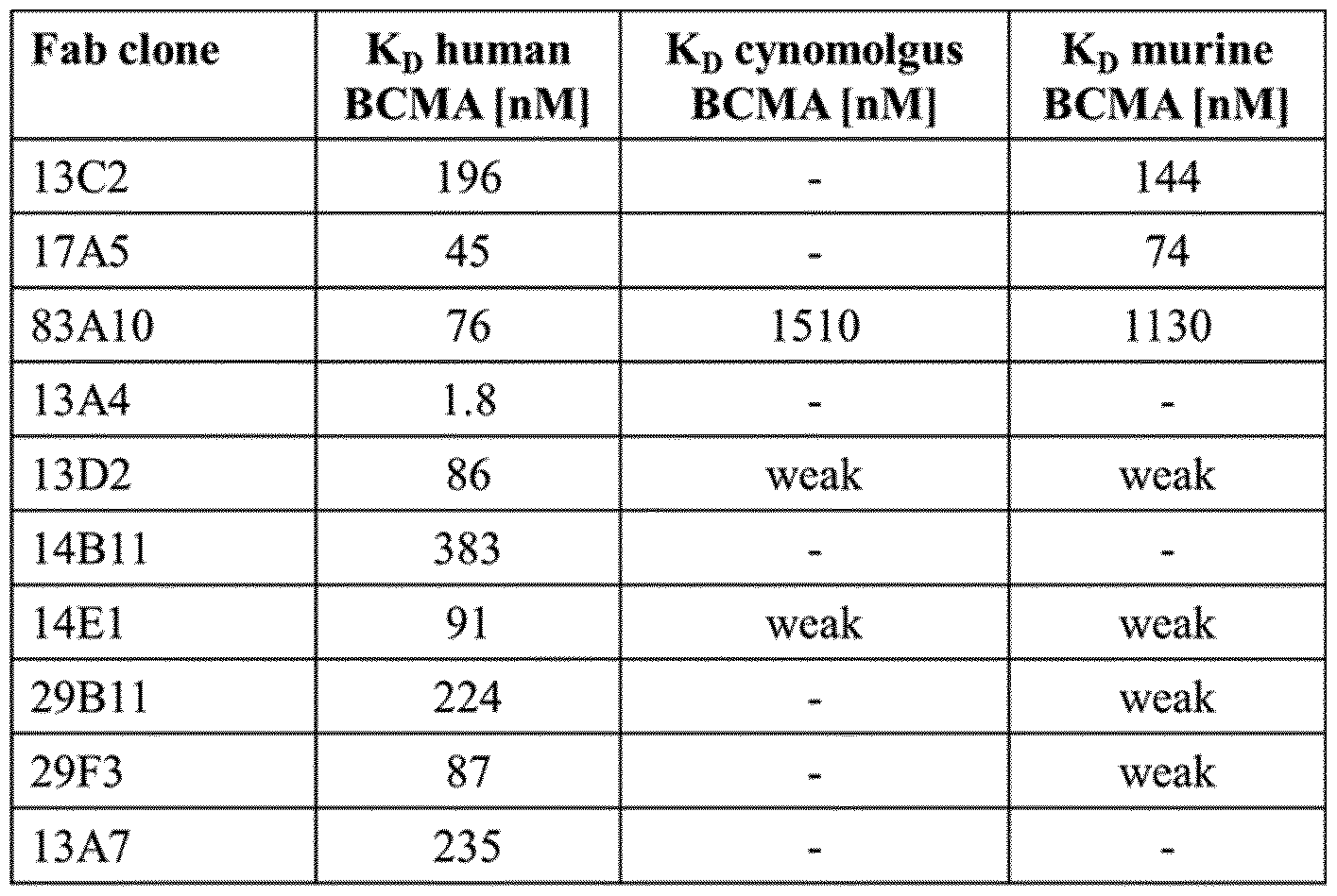
c) Assessment of binding of anti-BCMA antibodies to recombinant BCMA by surface plasmon resonance (SPR) as follow. All SPR experiments were performed on a Biacore T200 at 25 °C with HBS- EP as running buffer (0.01 M HEPES pH 7.4, 0.15 M NaCl, 3 mM EDTA, 0.005% Surfactant P20, Biacore, Freiburg/Germany). The avidity of the interaction between anti-BCMA antibodies and recombinant BCMA Fc(kih) (human, cynomolgus and murine) was determined. Biotinylated recombinant human, cynomolgus and murine BCMA Fc(kih) were directly coupled on a SA chip following instructions (Biacore, Freiburg/Germany). The immobilization level ranged from 200 to 700 RU. The anti-BCMA antibodies were passed at a 2-fold concentration range (1.95 to 500 nM) with a flow of 30 μΕ/minutes through the flow cells over 120 seconds. The dissociation was monitored for 180 seconds. Bulk refractive index differences were corrected for by subtracting the response obtained on the reference flow cell. Here, the anti-BCMA antibodies were flown over an empty surface previously activated and deactivated as described in the standard amine coupling kit. Apparent kinetic constants were derived using the Biacore T200 Evaluation Software (vAA, Biacore AB, Uppsala/Sweden), to fit rate equations for 1:1 Langmuir binding by numerical integration, despite the bivalency of the interaction for comparison purposes.
The affinity of the interaction between anti-BCMA antibodies and recombinant human BCMA Fc(kih) was also determined. Anti-human Fab antibody (GE Healthcare) was directly coupled on a CM5 chip at pH 5.0 using the standard amine coupling kit (Biacore, Freiburg/Germany). The immobilization level was about 6500 RU. Anti-BCMA antibody was captured for 90 seconds at 25 nM. Recombinant human BCMA Fc(kih) was passed at a 4-fold concentration range (1.95 to 500 nM) with a flow of 30 μΕ/η ηυίεβ through the flow cells over 120 seconds. The dissociation was monitored for 120 seconds. Bulk refractive index differences were corrected for by subtracting the response obtained on reference flow cell. Here, recombinant BCMA was flown over a surface with immobilized anti-human Fab antibody but on which HBS-EP has been injected rather than anti-BCMA antibody. Kinetic constants were derived using the Biacore T100 Evaluation Software (vAA, Biacore AB, Uppsala/Sweden), to fit rate equations for 1: 1 Langmuir binding by numerical integration (Table 5). Binding of 83A10 anti- BCMA antibody to recombinant cynomolgus BCMA Fc(kih) and murine BCMA Fc(kih) was also measured (Table 6). Table 5. Affinity constants determined by fitting rate equations for 1 : 1 Langmuir binding
Kon Koff KD
Ligand Anal te
pais) (1/s) (M)
13C2 anti-BCMA IgG huBCMA Fc(Mii) 2.4E+05 1.1E-02 4.Έ -08
17A5 anti-BCMA IgG huBCMA Fc{Mh) 2.2E+05 1.9E-03 8.7E-09
83 A 10 anti-BCMA IgG huBCMA Fc(Mh) 6.2E+05 2.5E-03 4.1E-09
29F3 anti-BCMA IgG huBCMA Fc(Mli) 3.2E+05 6.8E-03 2.1 E-08
I SA" anti-BCMA IgG huBCMA Fc( h) 8.0E+04 7.9E-03 1 .0E-07
13A4 anti-BCMA IgG huBCMA Fc(kih) 7.2E+04 3.6E-04 5.1E-09
13D2 anti-BCMA IgG huBCMA Fc(M!i) 3.6E+05 9.3E-03 2.6E-08
143 1 1 anti-BCMA IgG huBCMA Fc(Idh) 1 .5E -05 1.6E-02 1 .1E-07
1 E1 anti-BCMA IgG huBCMA Fc(kiii) 4,01+05 8.1E-03 2.0E-08
29B 1 1 anti-BCMA IgG huBCMA Fc(Mi) 1 .-F -05 6.6E-03 4.0E-08 Table 6: Binding of recombinant BCMA Fc(kih) to 83A10 anti-BCMA antibody, a) cynomolgus BCMA Fc(kih). b) murine BCMA Fc(kih)
Figure imgf000047_0001
Example 1G. BCMA-signaling assay: NF- Β activation a) As described in Ryan, 2007 (Mol Cancer Ther; 6 (11): 3009-18), NCI-H929 cells are washed and incubated in RPMI supplemented with 0.25% fetal bovine serum for 24 h before treatment. The cells are then untreated or treated with 0.1 μg/mL TNF-a, 100 ng/mL, preferably 1000 ng/mL heat-treated HT- truncated- APRIL, 100 ng/mL, preferably 1000 ng/mL truncated- APRIL, 0.1 pM to 200 nM isotype control, or 0.1 pM to 200 nM anti-BCMA antibodies for 20 min. To evaluate ligand blockade, cells pre- treated for 20 min with 0.1 pM to 200 nM of anti-BCMA antibodies or an isotype control antibody are treated with 1 μg/mL of truncated-APRIL. Cells are then harvested, washed, and lysed with 50 mmol/L Tris-HCl (pH 7.5), 1% NP40, 150 mmol/L NaCl, 1 mmol/L EDTA, 1 mmol/L EGTA supplemented with protease, and phosphatase inhibitors. Protein extracts are then analyzed for NF-κΒ activity using a TransAM® chemiluminescent assay kit (Active Motif) and the luminescent signal reading is performed with a Fusion HT plate reader (Packard Instruments). b) Briefly, H929 cells were starved in RPMI1640 with 0.25 % FCS for 24 h at 37°C in cell incubator. At the end of the starvation time, cells were harvested, counted and cell viability evaluated using ViCell. Viable cells were adjusted to 4 x 10⁶ cells per ml in BSA-containing FACS Stain Buffer (BD Biosciences). 30 μΐ of this cell suspension were further aliquoted per well into a round-bottom 96-well plate and pre-incubated with anti-BCMA antibodies (15 or 50 ug/ml) or isotype control antibodies (10, 20 and 40 ug/ml) for 20 min in cell incubator. Cells were then supplemented with 1 ug/ml recombinant mouse A-APRIL tagged with hemagglutinin (HA) (R&D Systems Europe, #7907-AP-010) for 40 min at 37°C. Heat inactivated A-APRIL (HI APRIL) was used in the assay to confirm the specificity of Δ- APRIL-induced NFkB signal (heat inactivation was performed by treatment of A-APRIL at 60°C for 1 h). At the end of incubation time, cells were harvested, washed, lysed, and processed according to the manufacturer's protocol of the Nuclear Extract Kit (Active Motif, # 40410). Protein extracts were analyzed for NF-κΒ activity using a TransAm© NFKB p65 Chemi Assay kit (Active Motif, #40097) following manufacturer's instructions. Luminescent signal was read using the Spectra Max M5 luminometer (Molecular Devices). Example 1H. Screening anti-BCMA antibodies to select antibodies not affected by 100 ng/niL, preferably 1000 ng/niL of APRIL or BAFF in their binding to BCMA and that neither promote nor block signaling via the BCMA intracellular domain
The invention relates to the generation of an anti-human BCMA antibody that 1) binds to human BCMA, 2) binding to BCMA is not affected by 100 ng/mL, preferably 1000 ng/mL of APRIL and BAFF, 3) does not block or reduce >20%, preferably >15% or increase >20%, preferably >15% APRIL- dependent NF-κΒ activation, 4) does not block or reduce >20%, preferably >15% or increase >20%, preferably >15% BAFF-dependent NF-κΒ activation, 5) does not induce NF-κΒ activation by itself, without APRIL or BAFF. Table 10 shows the screening paradigm for selection of a BCMA antibody with desired new properties: non-ligand binding/blocking, non-ligand competing. Antibodies are selected whose binding to BCMA is not blocked by APRIL or by BAFF.
Example 1H1. Binding to BCMA on HEK293-B CMA cells, plate-bound-BCMA or BCMA- positive multiple myeloma cell lines (Flow cytometry and ELISA) a) Anti-BCMA antibodies coming either from the immunization approach and/or from the screening of the recombinant in vitro library described above are analyzed by flow cytometry for binding to human BCMA on HEK293-BCMA cells. Briefly, cultured cells are harvested, counted and cell viability is evaluated using the Trypan Blue exclusion method. Viable cells are then adjusted to 2 x 10⁶ cells per ml in BSA-containing FACS Stain Buffer (BD Biosciences). 90 μΐ of this cell suspension are further aliquoted per well into a round-bottom 96-well plate. 10 μΐ of the anti-BCMA antibodies or corresponding IgG control are added to the cell -containing wells to obtain final concentrations of 0.1 pM to 200 nM. All constructs and control IgG are used at the same molarity. After incubation for 30 min at 4°C, the cells are centrifuged (5 min, 350 x g), washed with 150 μΐ/well FACS Stain Buffer (BD Biosciences), resuspended and incubated for an additional 30 min at 4°C with 12 μΐ/well fluorochrome- conjugated AffiniPure F(ab')2 Fragment goat anti-human IgG Fey Fragment Specific (Jackson Immuno Research Lab; working solution: 1:20). Cells are then washed with Stain Buffer (BD Biosciences) 120 μΐ/well and pelleted down by centrifugation at 350 x g for 5 min. A second washing step is performed using FACS Stain Buffer 150 μΐ/well. The samples are resuspended in 200 μΐ/well FACS Stain Buffer and acquired and analyzed using an LSR II flow cytometer with FACSDiva® software (BD Biosciences). The mean fluorescence intensity (MFI) is plotted as a function of anti-BCMA antibody concentration to obtain the binding curve and to calculate the effective antibody concentration to reach 50% of maximal binding (EC50). Anti-BCMA antibodies that bind to BCMA on cells as judged from this assay are selected for the next screening step, namely the competition BCMA binding assay against APRIL and BAFF (step (Example 1H2) below).
The properties of antibodies that show binding to human BCMA on HEK293-BCMA cells are confirmed using an ELISA method as described by Ryan et al. (2007). Briefly, immunosorb 96-well plates are coated with 1.5 μg/mL of GST-BCMA-ECD, washed with PBS + 1% Tween (PBS-T), and blocked with PBS-T plus 1% serum albumin. BCMA-coated plates are incubated with hybridoma culture supernatants for 2 h at room temperature, washed 5 times with PBS-T, and incubated with peroxidase-conjugated goat-anti-rat IgG. Following incubation with secondary antibody, plates are washed, incubated with 3,3,5,5-tetramethylbenzidine substrate, and stopped with an equal volume of 1 mol/L H2S04. b) Anti-BCMA IgG antibodies (clones 13C2, 17A5, 83A10, 13A4, 13D2, 14E1, 13A7, 14B 11) were analyzed by flow cytometry for binding to human BCMA on BCMA-expressing H929 cells. MKN45 (human gastric adenocarcinoma cell line that does not express BCMA) was used as negative control. Briefly, cultured cells are harvested, counted and cell viability was evaluated using ViCell. Viable cells are then adjusted to 2 x 10⁶ cells per ml in BSA-containing FACS Stain Buffer (BD Biosciences). 100 μΐ of this cell suspension were further aliquoted per well into a round-bottom 96-well plate and incubated with 30 μΐ of the anti-BCMA antibodies or corresponding IgG control for 30 min at 4°c. All anti-BCMA antibodies (and isotype control) were titrated and analyzed in final concentration range between 0.1 - 40 ug/ml. Cells were then centrifuged (5 min, 350 x g), washed with 120μ1Λνε11 FACS Stain Buffer (BD Biosciences), resuspended and incubated for an additional 30 min at 4°C with fluorochrome-conjugated PE-conjugated AffiniPure F(ab')2 Fragment goat anti-human IgG Fc Fragment Specific (Jackson Immuno Research Lab; 109-116-170). Cells were then washed twice with Stain Buffer (BD Biosciences), fixed using 100 ul BD Fixation buffer per well (#BD Biosciences, 554655) at 4°C for 20 min, resuspended in 120 μΐ FACS buffer and analyzed using BD FACS CantoII. Figure 5 shows the mean fluorescence intensity for anti-BCMA IgG clones plotted in function of anti-BCMA antibody concentration; (A) clones 13C2, 17A5, 83A10 on H929 cells, (B) clones 13C2, 17A5, 83A10 on MKN45 cells, (C) clones 13A4, 13D2, 14E1, 13A7, 14B 11 on H929 cells (D) clones 13A4, 13D2, 14E1, 13A7, 14B 11 on MKN45 cells. EC50 values (denoting the antibody concentration required to reach 50% of the maximal binding) for the binding of clones 13C2, 17A5, 83A10 to H929 cells are summarized in Table 7.
Table 7: EC50 values for binding of anti-BCMA antibodies to H929 multiple myeloma cells
Figure imgf000050_0001
Example 1H2. 100 ng/niL, preferably 1000 ng/niL of APRIL or BAFF does not alter BCMA antibody binding to human-BCMA (Flow cytometry and ELISA) a) Anti-BCMA antibodies selected from step (Example 1H1) above are then analyzed by flow cytometry for binding to human BCMA on HEK293-BCMA cells in the presence and absence of 100 ng/mL, preferably 1000 ng/mL APRIL or BAFF. Viable HEK293-BCMA cells are adjusted to 2 x 10⁶ cells per ml in BSA-containing FACS Stain Buffer (BD Biosciences). 90 μΐ of this cell suspension are further aliquoted per well into a round-bottom 96-well plate. 10 μΐ of the anti-BCMA antibodies or corresponding IgG control are added to the cell -containing wells to obtain final concentrations of 0.1 pM to 200 nM. All constructs and control IgG are used at the same molarity. After incubation for 30 min at 37°C, in the presence and absence of 100 ng/ml, preferably 1000 ng/mL of APRIL and BAFF, respectively, the cells are centrifuged (5 min, 350 x g), washed with 150 μΐ/well FACS Stain Buffer (BD Biosciences), resuspended and incubated for an additional 30 min at 4°C with 12 μΐ/well fluorochrome- conjugated AffiniPure F(ab')2 Fragment goat anti-human IgG Fey Fragment Specific (Jackson Immuno Research Lab; working solution: 1:20). Cells are then washed with Stain Buffer (BD Biosciences) 120 μΐ/well and pelleted down by centrifugation at 350 x g for 5 min. A second washing step is performed using FACS Stain Buffer 150 μΐ/well. The samples are resuspended in 200 μΐ/well FACS Stain Buffer and acquired and analyzed using an LSR II flow cytometer with FACSDiva® software (BD Biosciences). The mean fluorescence intensity is plotted as a function of anti-BCMA antibody concentration to obtain the binding curve and to calculate the effective antibody concentration to reach 50% of maximal binding (EC50). One binding curve is done in the presence of APRIL, another in its absence, and the same is done for presence and absence of BAFF. Those antibodies whose binding to BCMA is not affected by 100 ng/ml, preferably 1000 ng/mL of APRIL and also is not affected by 100 ng/ml, preferably 1000 ng/mL of B AFF are selected for next steps below. Representative binding curves for antibodies that are non-competing with the ligands APRIL and BAFF for binding to BCMA and for antibodies that are competing with these ligands for binding to BCMA are shown in Figure 1. The properties of antibodies that show binding to human BCMA on HEK293-BCMA cells in the presence of 100 ng/mL, preferably 1000 ng/mL APRIL or BAFF are confirmed using an ELISA method as described by Ryan et al. (2007). Briefly, immunosorb 96-well plates are coated with 1.5 μg/mL of GST-BCMA-ECD, washed with PBS + 1% Tween® (PBS-T), and blocked with PBS-T plus 1% serum albumin. BCMA-coated plates are incubated with hybridoma culture supernatants for 2 h at room temperature, washed 5 times with PBS-T, and incubated with peroxidase-conjugated goat-anti-rat IgG. Following incubation with secondary antibody, plates are washed, incubated with 3,3,5,5- tetramethylbenzidine substrate, and stopped with an equal volume of 1 mol/L H2S04. For plate-based ligand blockade, plates are coated with 1 μg/mL of GST-BCMA-ECD as described above. Coated plates are preincubated with purified antibodies at the specified concentrations, washed with PBS-T, and then incubated with 3 μg/mL of recombinant human MegaAPRIL (Alexis Biochemicals) or recombinant human BAFF (R&D Systems). APRIL or BAFF binding is detected using peroxidase-conjugated anti- FLAG followed by development with 3,3',5,5'-tetramethylbenzidine as described above. b) Identification of non-APRIL-competing anti-BCMA Fabs or antibodies by ELISA. Binding of Fabs to immobilized human BCMA was assessed in the presence of increasing concentrations of murine APRIL. 25nM biotinylated human BCMA (lOOul/well) were coated on a neutravidin plate and incubated on a shaker for lh at room temperature. 500nM or ΙΟΟΟηΜ purified Fabs were added to saturate the coated human BCMA for lh at room temperature. The plate was washed 3 times with PBS and murine APRIL was added at eight different concentrations using a two-fold dilution series in PBS buffer, ranging from 0 to ΙΟΟηΜ, and incubated on a shaker for 30min. The plate was washed 3 times with PBS and anti-FLAG-HRP secondary antibody (1:4000) was added for lh. Again, the plate was washed 3 times with PBS and developed by adding lOOul/well BM Blue POD (Roche). The reaction was stopped by adding 50ul/well 1M H2S04 and the OD was read at 450nm (reference at 650nm) for a final read-out of OD450-65o- Results for selected Fabs are shown in Figure 6. The reduction (%) in OD values measured with the anti-BCMA clones in the absence vs. presence of 50 nM (1000 ng/mL) or 6.25 nM (140 ng/mL) muAPRIL (murine APRIL) is summarized in Table 8. Table 8: Reduction in OD values measured (450 nm) in absence vs. presence of muAPRIL
Figure imgf000052_0001
c) Competition of Δ-APRIL with anti-BCMA antibodies detected by flow cytometry. The assessment of the eventual competition between A-APRIL and anti-BCMA antibodies was performed on H929 cells by quantifying the binding of A-APRIL in presence of increasing concentrations of anti-BCMA antibodies (clones 13C2, 17A5, 83A10, 13A4, 13D2, 14E1, 13A7, 14B 11). Briefly, cultured cells were harvested, counted and cell viability evaluated using ViCell. Viable cells were adjusted to 1 x 10⁶ cells per ml in BSA-containing FACS Stain Buffer (BD Biosciences). 100 μΐ of this cell suspension are further aliquoted per well into a round-bottom 96-well plate and incubated with 30 μΐ of the anti-BCMA antibodies or corresponding IgG control for 30 min at 4°c. All anti-BCMA antibodies (and isotype control) are titrated and analyzed at final concentrations of 1, 16 and 40 ug/ml. Cells are then centrifuged (5 min, 350 x g), washed with 120μ1Λνε11 FACS Stain Buffer (BD Biosciences), resuspended and incubated with 1 ug/ml recombinant mouse A-APRIL tagged with hemagglutinin (HA) (R&D Systems Europe, #7907-AP-010) for additional 30 min at 4°C. Cells are then washed once with 120 μΐ/well FACS Buffer and incubated with FITC-conjugated anti-HA antibody (Sigma Aldrich, #H7411) for 30 min at 4°C. At the end of incubation time, cells are washed with 120 μΐ/well FACS Buffer, fixed using 100 ul BD Fixation buffer per well (#BD Biosciences, 554655) at 4°C for 20 min, resuspended in 80 μΐ FACS buffer and analyzed using BD FACS Fortessa. Figure 7 shows the relative median fluorescence intensity of Δ-APRIL (FITC signal) detected in function of increasing concentrations of anti-BCMA antibody clones 13A4, 13D2, 14E1, 13A7, 14B 11 on H929 cells. The median fluorescence intensity upon binding of Δ-APRIL in presence of the isotype control was set to one; the other signals were normalized to it. d) Competition of anti-BCMA antibodies with Δ-APRIL detected by flow cytometry. The assessment of the eventual competition between Δ-APRIL and anti-BCMA antibodies was performed on RPMI cells by quantifying the binding of anti-BCMA antibodies (clones 13A4, 13C2, 13D2, 14B 11, 17A5, 83A10,) in presence or absence of Δ-APRIL. Briefly, cultured cells were harvested, counted and cell viability evaluated using ViCell. Viable cells were adjusted to 1 x 10⁶ cells per ml in BSA-containing FACS Stain Buffer (BD Biosciences). 100 μΐ of this cell suspension were further aliquoted per well into a round-bottom 96-well plate and incubated with 30 μΐ of the anti-BCMA antibodies or corresponding IgG control for 20 min at 4°C. All anti-BCMA antibodies and isotype control were analyzed at final concentrations 40 ug/ml. Cells were then centrifuged (5 min, 350 x g), washed with 120 μΐ/well FACS Stain Buffer (BD Biosciences), resuspended and incubated with 1 ug/ml recombinant mouse Δ-APRIL tagged with hemagglutinin (HA) (R&D Systems Europe, #7907-AP-010) for additional 40 min at 4°C. Cells were then washed once with 120 μΐ/well FACS Buffer and incubated with with Alexa.Fluor 647- conjugated anti-human Fc antibody (Jackson Immuno Research Lab, #109-606-008) for 30 min at 4°C. At the end of incubation time, cells were washed with 120 μΐ/well FACS Buffer, fixed using 100 ul BD Fixation buffer per well (#BD Biosciences, 554655) at 4°C for 20 min, resuspended in 80 μΐ FACS buffer and analyzed using BD FACS Fortessa. Figure 8 shows the relative median fluorescence intensity of anti-BCMA antibody (Alexa.Fluor 647 signal) clones 13A4, 13C7, 13D2, 14B 11, 17A5, 83A10 on RPMI cells detected in absence or presence of 1000 ng/mL of Δ-APRIL. The median fluorescence intensity upon binding of anti-BCMA antibodies in absence of Δ-APRIL was set to one; the other signals respective to the anti-BCMA antibody in presence of Δ-APRIL were normalized to it. e) Competition of anti-BCMA antibodies with Δ-APRIL after simultaneous incubation detected by flow cytometry. The assessment of the eventual competition between Δ-APRIL and anti-BCMA antibodies was performed on H292 cells (NCI-H929, ATCC® CRL-9068™) by quantifying the binding of anti- BCMA antibodies (clones 14B 11, 13D2, 13A4, 17A5, 83A10) in presence or absence of Δ-APRIL. Briefly, cultured cells were harvested, counted and cell viability evaluated using ViCell. Viable cells were adjusted to 1 x 10⁶ cells per ml in BSA-containing FACS Stain Buffer (BD Biosciences). 100 μΐ of this cell suspension were further aliquoted per well into a round-bottom 96-well plate and incubated with 30 μΐ of the anti-BCMA antibodies or corresponding IgG control and 30 μΐ of Δ-APRIL tagged with hemagglutinin (HA) (R&D Systems Europe, #7907-AP-010) for 40 min at 4°C. All anti-BCMA antibodies and isotype control were analyzed at final concentrations 20 ug/ml; Δ-APRIL at final concentrations 2.5 ug/ml. Cells were then centrifuged (5 min, 350 x g) and washed with 120μ1Λνε11 FACS Stain Buffer (BD Biosciences). After that, cells were incubated with with Alexa.Fluor 647- conjugated anti-human Fc antibody (Jackson Immuno Research Lab, #109-606-008) and FITC -conjugated anti-HA antibody (Sigma Aldrich, #H7411) for 30 min at 4°C. At the end of incubation time, cells were washed with 120 μΐ/well FACS Buffer, fixed using 100 ul BD Fixation buffer per well (#BD Biosciences, 554655) at 4°C for 20 min, resuspended in 80 μΐ FACS buffer and analyzed using BD FACS CantoII. Figure 9 A shows the mean fluorescence intensity and the relative fluorescence signal of the anti-BCMA antibody clone (Alexa.Fluor 647 signal) and Figure 9B shows the mean fluorescence intensity and the relative fluorescence signal of Δ-APRIL (FITC signal) and the anti- BCMA antibody clone (Alexa.Fluor 647 signal). Detection of anti-BCMA antibody in presence of Δ- APRIL with FITC-conjugated anti-human Fc antibody was normalized to the signal of anti-BCMA antibody clone in absence Δ-APRIL. Detection of Δ-APRIL in presence of the anti-BCMA antibody clone with Alexa.Fluor 647 -conjugated anti-HA antibody was normalized to Δ- APRIL signal in presence of the isotype control. Reduction in binding of anti-BCMA antibodies (20 μg/mL) clones 14B 11, 13D2, 13A4, 17A5 and 83A10 in presence of Δ-APRIL (2.5 μg/mL) as detected with fluorochrome-conjugated anti-human Fc antibody is summarized in Table 9.
Table 9: Reduction in binding of anti-BCMA antibodies to H929 cells in presence of APRIL
Figure imgf000054_0001
Example 1H3. BCMA antibody does not block or increase APRIL-dependent NF- Β activation a) Antibodies selected as non-competing in step (Example 1H2) above (i.e., their BCMA binding curve is not affected by the presence of 100 ng/ml, preferably 1000 ng/mL of APRIL and is also not affected by the presence of 100 ng/ml, preferably 1000 ng/mL of BAFF) are then tested in step (Example 1H3) for effects on APRIL, BAFF, and BCMA mediated NF-κΒ activation. As APRIL is the high affinity ligand to BCMA, the blocking or agonist properties of anti-BCMA antibodies on APRIL signaling is first examined. As described in Ryan 2007 (Mol Cancer Ther; 6 (11): 3009-18), to verify whether anti- BCMA antibodies block or increase APRIL downstream signaling, NCI-H929 human multiple myeloma (MM) cells are washed and incubated in serum free RPMI for 24 h before treatment. The cells are then untreated or treated with 0.1 μg/mL TNF-a (used as positive control), 100 ng/mL, preferably 1000 ng/mL heat-treated HT-truncated-APRIL, 100 ng/mL, preferably 1000 ng/mL truncated-APRIL, 0.1 pM to 200 nM isotype control, or 0.1 pM to 200 nM anti-BCMA antibodies for 20 min. To evaluate APRIL blockade, cells pre-treated for 20 min with 0.1 pM to 200 nM of anti-BCMA antibodies or a respective isotype control antibody are treated with 100 ng/mL, preferably 1000 ng/mL of truncated-APRIL. Cells are then harvested, washed, and lysed with 50 mmol/L Tris-HCl (pH 7.5), 1% ΝΡκΟ, 150 mmol/L NaCl, 1 mmol/L EDTA, 1 mmol/L EGTA supplemented with protease, and phosphatase inhibitors. Protein extracts are then analyzed for NF-κΒ activity using a TransAM® chemiluminescent assay kit (Active Motif) and the luminescent signal reading is performed with a Fusion HT plate reader (Packard Instruments).
As NF-κΒ activity is assayed using a functional ELISA that detects chemiluminescent signal from p65 bound to the NF-κΒ consensus sequence, anti-BCMA antibodies that do not alter APRIL-mediated downstream signaling and NF-κΒ activation (i.e. that the mean luminescent signal detected by ELISA in nuclear extracts from NCI-H929 MM cells treated with APRIL alone is similar, not significantly reduced or increased, to that of nuclear extracts from NCI-H929 MM cells treated with APRIL and anti- BCMA antibodies) are selected for the next steps below. b) It was assessed whether binding of anti-BCMA antibodies interferes with APRIL-induced NFkB activation, a known signaling pathway downstream of BCMA. Briefly, H929 cells were starved in RPMI1640 with 0.25 % FCS for 24 h at 37°C in cell incubator. At the end of the starvation time, cells were harvested, counted and cell viability evaluated using ViCell. Viable cells were adjusted to 4 x 10⁶ cells per ml in BSA-containing FACS Stain Buffer (BD Biosciences). 30 μΐ of this cell suspension were further aliquoted per well into a round-bottom 96-well plate and pre-incubated with anti-BCMA antibodies (15 or 50 ug/ml) or isotype control antibodies (10, 20 and 40 ug/ml) for 20 min in cell incubator. Cells were then supplemented with 1 ug/ml recombinant mouse Δ-APRIL tagged with hemagglutinin (HA) (R&D Systems Europe, #7907-AP-010) for 40 min at 37°C. Heat inactivated Δ- APRIL (HI APRIL) was used in the assay to confirm the specificity of Δ-APRIL-induced NFkB signal (heat inactivation was performed by treatment of Δ-APRIL at 60°C for 1 h). At the end of incubation time, cells were harvested, washed, lysed, and processed according to the manufacturer's protocol of the Nuclear Extract Kit (Active Motif, # 40410). Protein extracts were analyzed for NF-kB activity using a TransAm© NfkB p65 Chemi Assay kit (Active Motif, #40097) following manufacturer's instructions. Luminescent signal was read using the Spectra Max M5 luminometer (Molecular Devices). The relative luminescence signal intensity obtained using H929 cells treated as described above was measured. The luminescence signal obtained upon binding of Δ-APRIL in presence of the isotype control was set to one; the other signals were normalized to it. Example 1H4. BCMA antibody does not block or increase BAFF-dependent NF- Β activation
Antibodies selected as non-blocking and non-increasing APRIL-dependent NF-κΒ activation in step (Example 1H3) above are then tested in step (Example 1H3) for effects on BAFF mediated NF-KB activation. As described in Ryan 2007, (Mol Cancer Ther; 6 (11): 3009-18), to verify whether BCMA antibodies block or increase BAFF downstream signaling leading to NF-κΒ activation, NCI-H929 MM cells (CRL-9068™) are washed and incubated in serum free RPMI medium for 24 h before treatment, as described in Ryan 2007, (Mol Cancer Ther; 6 (11): 3009-18), The cells are then untreated or treated with 0.1 μg/mL TNF-a, 100 ng/mL, preferably 1000 ng/mL heat-treated HT-truncated-BAFF, 100 ng/mL, preferably 1000 ng/mL truncated-BAFF, 0.1 pM to 200 nM isotype control, or 0.1 pM to 200 nM anti-BCMA antibodies for 20 min. To evaluate BAFF blockade, cells pre-treated for 20 min with 0.1 pM to 200 nM of anti-BCMA antibodies or a respective isotype control antibody are treated with 1 μg/mL of truncated-BAFF. Cells are then harvested, washed, and lysed with 50 mmol/L Tris-HCl (pH 7.5), 1% NP40, 150 mmol/L NaCl, 1 mmol/L EDTA, 1 mmol/L EGTA supplemented with protease, and phosphatase inhibitors. Protein extracts are then analyzed for NF-κΒ activity using a TransAM® chemiluminescent assay kit (Active Motif) and the luminescent signal reading is performed with a Fusion HT plate reader (Packard Instruments). Anti-BCMA antibodies that do not alter BAFF-mediated downstream signaling and NF-κΒ activation (i.e. that the mean luminescent signal from p65 bound to the NF-KB consensus sequence detected by ELISA in nuclear extracts from NCI-H929 MM cells treated with BAFF alone is similar, not significantly reduced or increased, to that of nuclear extracts from NCI- H929 MM cells treated with BAFF and anti-BCMA antibodies) are selected for the next steps below. Example 1H5. BCMA antibody does not induce NF-κΒ activation by itself a) Antibodies selected as non-blocking and non-increasing BAFF-dependent NF-κΒ activation in step (Example 1H4) above are then tested in step (Example 1H5) for their intrinsic agonistic effects to mediate NF-κΒ activation. To verify whether BCMA antibodies are agonistic and induce downstream signaling by themselves, NCI-H929 cells are washed and incubated in serum-free RPMI medium for 24
5 h before treatment. The cells are then untreated or treated with 0.1 μg/mL TNF-a, 0.1 pM to 200 nM isotype control or 0.1 pM to 200 nM anti-BCMA antibodies for 20 min. Cells are then harvested, washed, and lysed with 50 mmol/L Tris-HCl (pH 7.5), 1% NP0, 150 mmol/L NaCl, 1 mmol/L EDTA, 1 mmol/L EGTA supplemented with protease, and phosphatase inhibitors. Protein extracts are then analyzed for NF-κΒ activity using a TransAM® chemiluminescent assay kit (Active Motif) and the
10 luminescent signal reading is performed with a Fusion HT plate reader (Packard Instruments). Anti- BCMA antibodies that do not induce downstream signaling and NF-κΒ activation (i.e. that the mean luminescent signal from p65 bound to the NF-κΒ consensus sequence detected by ELISA in nuclear extracts from NCI-H929 MM cells treated with anti-BCMA antibodies alone is similar, not significantly increased, to that of nuclear extracts from NCI-H929 MM cells treated with isotype control antibody)
15 are finally selected for further production and in vitro and in vivo characterization. They represent the anti-BCMA antibodies that are non-ligand blocking, non-competing, and non-signaling (see Table 10). b) It was assessed whether binding of anti-BCMA antibodies to BCMA-expressing H929 cells induces NFkB activation, a known signaling pathway downstream of BCMA. Briefly, H929 cells were starved in RPMI1640 with 0.25 % FCS for 24 h at 37°C in cell incubator. At the end of the starvation time, cells
20 were harvested, counted and cell viability evaluated using ViCell. Viable cells were adjusted to 4 x 10⁶ cells per ml in BSA-containing FACS Stain Buffer (BD Biosciences). 30 μΐ of this cell suspension were further aliquoted per well into a round-bottom 96-well plate and incubated with 30 μΐ of the anti-BCMA antibodies at 100 or 350 nM (14 or 50 ug/ml) for 20 min at 37°C. As negative controls, cells were either left untreated or incubated with the corresponding IgG isotype control antibodies 100 nM (14 ug/ml) for
25 20 min at 37°C. As positive controls, cells were incubated with 1 ug/ml recombinant mouse Δ-APRIL tagged with hemagglutinin (HA) (R&D Systems Europe, #7907-AP-010) for 20 min at 37°C. At the end of incubation time, cells were harvested, washed, lysed, and processed according to the manufacturer's protocol of the Nuclear Extract Kit (Active Motif, # 40410). Protein extracts were analyzed for NFkB activity using a TransAm© NFkB p65 Chemi Assay kit (Active Motif, #40097) following
30 manufacturer's instructions. Luminescent signal was read using the Spectra Max M5 luminometer (Molecular Devices). The relative luminescence signal intensity obtained from H929 cells treated as described above was measured. The luminescence signal obtained upon binding of Δ- APRIL in presence of the isotype control was set to one; the other signals were normalized to it. Table 10: Screening paradigm for BCMA antibody selection
Figure imgf000058_0001
Example 2 - Production of therapeutic anti-BCMA antibodies that do not block ligand (APRIL, BAFF) binding and neither promote nor block signaling via the BCMA intracellular domain and whose binding to BCMA is not affected by 100 ng/ml, preferably 1000 ng/mL of APRIL or by 100 ng/ml, preferably 1000 ng/mL of BAFF
If the selected antibodies after step (Example 1H5) above are derived from the in vitro selection out of the recombinant antibody library, then they are already unconjugated human IgGl antibodies. Those selected antibodies after step (Example 1H5) above that are derived from immunization are in a rat- human chimeric format and are then preferably humanized to be able to apply them for therapy. In that case, standard antibody humanization methods are applied by transferring the complementarity- determining regions of those rat variable regions into human antibody variable region frameworks. Additional mutations are introduced into the variable regions, if necessary, to recover binding to BCMA as compared to the chimeric, parental antibody.
5 For the production of the antibody, the cells are co-transfected with two plasmids, (one for expression of the heavy chain of the antibody and another for expression of the light chain of the antibody), at a ratio of 1: 1, respectively. Cells are grown as adherent monolayer cultures in T flasks using DMEM culture medium supplemented with 10% FCS, and are transfected when they are between 50 and 80% confluent. For the transfection of a T75 flask, 8 million cells are seeded 24 hours before transfection in
10 14 ml DMEM culture medium supplemented with FCS (at 10% V/V final), 250 μg/ml neomycin, and cells are placed at 37°C in an incubator with a 5% C02 atmosphere overnight. For each T75 flask to be transfected, a solution of DNA, CaCl2 and water is prepared by mixing 47 μg total plasmid vector DNA divided equally between the light and heavy chain expression vectors, 235 μΐ of a 1M CaCl2 solution, and adding water to a final volume of 469 μΐ. To this solution, 469 μΐ of a 50 mM HEPES, 280 mM
15 NaCl, 1.5 mM Na2HP04 solution at pH 7.05 are added, mixed immediately for 10 sec and left to stand at room temperature for 20 sec. The suspension is diluted with 12 ml of DMEM supplemented with 2% FCS, and added to the T75 in place of the existing medium. The cells are incubated at 37°C, 5% C02 for about 17 to 20 hours, then medium is replaced with 12 ml DMEM, 10% FCS. The conditioned culture medium is harvested 5 to 7 days post-transfection centrifuged for 5 min at 1200 rpm, followed
20 by a second centrifugation for 10 min at 4000 rpm and kept at 4°C.
The secreted antibodies are purified by Protein A affinity chromatography, followed by cation exchange chromatography and a final size exclusion chromatographic step on a Superdex® 200 column (Amersham Pharmacia) exchanging the buffer to phosphate buffer saline and collecting the pure monomeric IgGl antibodies. Antibody concentration is estimated using a spectrophotometer from the 25 absorbance at 280 nm. The antibodies were formulated in a 25 mM potassium phosphate, 125 mM sodium chloride, 100 mM glycine solution of pH 6.7.
Example 3 - Generation of anti-BCMA/anti-CD3 T cell bispecific antibodies Example 3A. Generation of anti-CD3 antibodies
The following protein sequences of the VH and VL regions are used to generate human and cynomolgus 30 monkey cross reactive CD3e antibodies as described in WO2007/042261.
H2C_VH (SEQ ID NO:7): EVQLVESGGGLVQPGGSLKLSCAASGFTFNKYAMNWVRQAPGKGLEWVARIRSKYNNYATY YADSVKDRFTISRDDSKNTAYLQMNNLKTEDTAVYYCVRHGNFGNSYISYWAYWGQGTLVT
vss
H2C_VL (SEQ ID NO:8) QTVVTQEPSLTVSPGGTVTLTCGSSTGAVTSGYYPNWVQQKPGQAPRGLIGGTKFLAPGTPARF SGSLLGGKAALTLSGVQPEDEAEYYCALWYSNRWVFGGGTKLTVL
Briefly, oliogonucleotides encoding the above sequences are joined together via PCR to synthesize cDNAs encoding the VH are VL sequences, respectively, of the anti-CD3 antibody.
Example 3B. Generation of anti-BCMA/anti-CD3 T cell bispecific 1+1 format with Fc Anti-BCMA/anti-CD3 T cell bispecific are produced for the human or humanized anti-BCMA antibodies selected after step (Example 1H5). cDNAs encoding the full heavy and light chains of the corresponding anti-BCMA IgGl antibodies, as described in Example 2, as well as the anti-CD3 VH and VL cDNAs describe in Example 3A, are used as the starting materials. For each bispecific antibody, four protein chains are involved comprising the heavy and light chains of the corresponding anti-BCMA antibody and the heavy and light chains of the anti-CD3 antibody described above, respectively. In order to minimize the formation of side-products with mispaired heavy chains, for example with two heavy chains of the anti-CD3 antibody, a mutated heterodimeric Fc region is used carrying "knob-into- hole mutations" and an engineered disulphide bond, as described in WO2009080251 and in WO2009080252. In order to minimize the formation of side-products with mispaired light chains, for example with two light chains of the anti-BCMA antibody, a CHI x constant kappa crossover is applied to the heavy and light chains of the anti-CD3 antibody using the methodology described in WO2009080251 and in WO2009080252.
Briefly, each bispecific antibody is produced by simultaneous cotransfection of four mammalian expression vectors encoding, respectively: a) the full light chain cDNA of the corresponding BCMA antibody, b) the full heavy chain cDNA of the corresponding BCMA antibody carrying the "hole mutations" in the Fc region to produce a heterodimeric antibody (see details below), c) a fusion cDNA generated by standard molecular biology methods, such as splice-overlap-extension PCR, encoding a fusion protein made of (in N- to C-terminal order) secretory leader sequence, VL of the anti-CD3 antibody described above and human CHI domain of an IgGl antibody and d) a fusion cDNA generated by standard molecular biology methods, such as splice-overlap-extension PC, encoding a fusion protein made of (in N- to C-terminal order) secretory leader sequence, VH of the anti-CD3 antibody described above, constant kappa domain of a human light chain cDNA, hinge region of a human IgGl antibody, and Fc region (CH2 and CH3 domains) of a human IgGl antibody including a "knob mutation" (see details below) in the Fc region to produce a heterodimeric antibody. Co- transfection of mammalian cells and antibody production and purification using the methods described above for production of human or humanized IgGl antibodies (see Example 2). The "knob-into-hole mutations" in the human IgGl Fc region consist of : T366W, known as the "knob mutation"; and T366S, L368A, and Y407V, collectively known as the "hole mutations". In addition, a disulfide can be included to increase the stability and yields as well as additional residues forming ionic bridges and increasing the heterodimerization yields (EP 1870459A1). Example 4 - Simultaneous binding of anti-BCMA/anti-CD3 T cell bispecific antibodies to BCMA and CD3 (Surface plasmon resonance)
The binding properties to BCMA and CD3 of bispecific anti-BCMA/anti-CD3 T cell bispecific antibodies generated in Example 3 are analyzed by surface plasmon resonance (SPR) technology using a Biacore® T100 instrument (Biacore AB) with HBS-EP as running buffer (0.01 M HEPES pH 7.4, 0.15 M NaCl, 3 mM EDTA, 0.005% Surfactant P20, Biacore). This system is well established for the study of molecule interactions. It allows a continuous real-time monitoring of ligand/analyte bindings and thus the determination of association rate constants (ka), dissociation rate constants (kd), and equilibrium constants (KD) in various assay settings. SPR-technology is based on the measurement of the refractive index close to the surface of a gold coated biosensor chip. Changes in the refractive index indicate mass changes on the surface caused by the interaction of immobilized ligand with analyte injected in solution. If molecules bind to immobilized ligand on the surface the mass increases, in case of dissociation the mass decreases.
Capturing anti-His tag antibody is immobilized on the surface of a CM5 biosensorchip using amine- coupling chemistry. Flow cells are activated with a 1 : 1 mixture of 0.1 M N-hydroxysuccinimide and 0.1 M 3-(N,N-dimethylamino)propyl-N-ethylcarbodiimide at a flow rate of 5 μΐ/min anti-human IgG antibody is injected in sodium acetate, pH 5.0 at 10 μg/ml, which resulted in a surface density of approximately 12000 resonance units (RU). A reference control flow cell is treated in the same way but with vehicle buffers only instead of the capturing antibody. Surfaces are blocked with an injection of 1 M ethanolamine/HCl pH 8.5. The anti-BCMA/anti-CD3 T cell bispecific antibodies are diluted in HBS- P and injected at a flow rate of 5 μΐ/min. The contact time (association phase) is 1 min for the antibodies at a concentration between 1 and 100 nM for the BCMA-ECD binding and 1 and 200 nM for the CD3 interaction. BCMA-ECD is injected at increasing concentrations of 3.125, 6.25, 12.5, 25, 50 and 100 riM, CD3 at concentrations of 0.21, 0.62, 1.85, 5.6, 16.7, 50, 100 and 200 nM. The contact time (association phase) is 3 min, the dissociation time (washing with running buffer) 5 min for both molecules at a flowrate of 30 μΐ/min. All interactions are performed at 25°C (standard temperature). The regeneration solution of 3 M Magnesium chloride is injected for 60 s at 5 μΐ/min flow to remove any non-covalently bound protein after each binding cycle. Signals are detected at a rate of one signal per second. Samples are injected at increasing concentrations. SPR graphs showing the rate of signal (i.e. resonance unit) plotted against contact time are determined.
Example 5 - Binding of anti-BCMA/anti-CD3 T cell bispecific antibodies to BCMA on MM cells or CD3 on T cells (Flow cytometry) Anti-BCMA/anti-CD3 T cell bispecific antibodies generated in Example 3 are also analyzed by flow cytometry for their binding properties to human BCMA expressed on NCI-H929 multiple myeloma cells or human CD3 expressed on human leukemic T cells Jurkat (ATCC). Briefly, cultured cells are harvested, counted and cell viability is evaluated using the Trypan Blue exclusion method. Viable cells are then adjusted to 2 x 10⁶ cells per ml in PBS containing 0.1% BSA. 90 μΐ of this cell suspension are further aliquoted per well into a round-bottom 96-well plate. 10 μΐ of the T cell bispecific antibody or corresponding IgG control are added to the cell -containing wells to obtain final concentrations of 0.1 pM to 200 nM. Anti-BCMA/anti-CD3 T cell bispecific antibodies and control IgG are used at the same molarity. After incubation for 30 min at 4°C, cells are centrifuged (5 min, 350 x g), washed with 150 μΐ/well BSA-containing FACS Stain Buffer (BD Biosciences), resuspended and incubated for an additional 30 min at 4°C with 12 μΐ/well fluorochrome-conjugated anti-His antibody (Lucerna) for detection of the T cell bisepecific antibody. Cells are then washed by addition of 120 μΐ/well FACS Stain Buffer and centrifugation at 350 x g for 5 min. A second washing step is performed with 150 μΐ/well FACS Stain Buffer. The samples are resuspended in 200 μΐ/well FACS Stain Buffer, acquired and analyzed using an LSR II flow cytometer with FACSDiva® software (BD Biosciences). Binding of the anti-BCMA/anti-CD3 T cell bispecific antibodies to MM cells and T cells are evaluated and the mean fluorescence intensity is determined gated on either BCMA-expressing NCI-H929 MM cells or CD3 -expressing Jurkat T cells and plotted in histograms or dot plots.
Example 6 - Activation of T cells upon engagement of anti-BCMA/anti-CD3 T cell bispecific antibodies (Flow cytometry) Anti-BCMA/anti-CD3 T cell bispecific antibodies generated in Example 3 are also analyzed by flow cytometry for their potential to induce T cell activation by evaluating the surface expression of the early activation marker CD69, or the late activation marker CD25 on CD4+ and CD8+ T cells in the presence or absence of human BCMA-expressing MM cells. Briefly, BCMA-expressing NCI-H929 MM cells are harvested with Cell Dissociation buffer, counted and cell viability is verified using Trypan Blue. Viable MM cells are adjusted to 0.2 x 10⁶ cells/mL in complete RPMI-1640 medium, 100 μΐ of this cell suspension per well is pipetted into a round-bottom 96-well plate. 50 μΐ of the T cell bispecific constructs are added to the MM cells-containing wells to obtain a final concentration of 1 nM. The 96- well plate is set aside and kept at 37°C, 5% C02 until further manipulations.
PBMC are isolated from fresh blood using density gradient centrifugation using Cell Preparation Tubes with Sodium citrate (Vacutainer CPT tubes, BD Biosciences). Total human T cells are then isolated using the Pan T Cell Isolation Kit II (Miltenyi Biotec), according to the manufacturer's instructions. Human total T cells (effector) are then adjusted to 2 x 10⁶ cells per ml in complete RPMI-1640 medium. 50 μΐ of this cell suspension is added per well in the assay plate containing already BCMA-expressing MM cells to obtain a final E:T ratio of 5: 1. To test whether the T cell bispecific constructs are able to activate T cells only in the presence of BCMA-expressing MM tumor target cells, wells containing final concentration(s) in the range of 0.1 pM to 200 nM of the respective bispecific molecules with effector cells but without MM tumor target cells are also included. After incubation for five days at 37°C, 5% C02, cells are pelleted down by centrifugation (5 min, 350 x g) and washed twice with 150 μΐ/well of FACS Stain Buffer (BD Biosciences). Surface staining of the effector cells with selected fluorochrome- conjugated antibodies against human CD4, CD8, CD69 or CD25 (BD Biosciences) is performed at 4°C for 30 min, protected from light, in FACS Stain Buffer (BD Biosciences) according to the manufacturer's protocol. Cells are washed twice with 150 μΐ/well FACS Stain Buffer, resuspended in 200 μΐ/well FACS Stain Buffer, and acquired and analyzed using a LSRII flow cytometer complemented with FACSDiva® software (BD Biosciences). The expression of CD69 and CD25 activation markers are determined by measuring the mean fluorescence intensity gated on CD4+ and CD8+ T cell populations as represented in histograms or dot plots. Example 7 - Proliferation of T cells upon engagement of anti-BCMA/anti-CD3 T cell bispecific antibodies (CFSE dilution)
Anti-BCMA/anti-CD3 T cell bispecific antibodies generated in Example 3 are also analyzed by flow cytometry for their potential to induce proliferation of CD8+ or CD4+ T cells in the presence or absence of human BCMA-expressing MM cells. Briefly, BCMA-expressing NCI-H929 MM cells are harvested with Cell Dissociation buffer, counted and looked for viability using Trypan Blue. Viable MM cells are adjusted to 0.2 x 10⁶ cells per ml in complete RPMI medium, 100 μΐ of this cell suspension are pipetted per well into a round-bottom 96-well plate. 50 μΐ of the T cell bispecific constructs are added to the MM cell-containing wells to obtain final concentration(s) in the range of 0.1 pM to 200 nM. The well plate is set aside and kept at 37°C, 5% C02.
PBMC are isolated from fresh blood using density gradient centrifugation using Cell Prepartaion Tubes with Sodium citrate (Vacutainer CPT tubes, BD Biosciences). Total human T cells are then isolated using the Pan T Cell Isolation Kit II (Miltenyi Biotec), according to the manufacturer's instructions. The total T cells are then adjusted to 1 million cells per ml in pre-warm RPMI without serum (37°C) and stained with 1 μΜ CFSE at room temperature for 6 min, protected from light. The staining volume is then doubled by addition of RPMI-1640 medium supplemented with 10% FCS and 1% GlutaMax to stop CFSE staining. After incubation at room temperature for further 20 min, the cells are washed three times with pre-warmed serum-containing medium to remove remaining CFSE. CFSE-stained total T cells (effector) are then adjusted to 2 x 10⁶ cells/mL in complete RPMI-1640 medium. 50 μΐ of this cell suspension is added per well in the assay plate already containing BCMA-expressing NCI-H929 MM cells to obtain a final E:T ratio of 5: 1. To test whether the T cell bispecific constructs are able to activate T cells only in the presence of BCMA-expressing MM tumor target cells, wells containing 1 nM of the T cell bispecific antibodies with effector cells but without MM tumor target cells are also included. After incubation for five days at 37°C, 5% C02, cells are pelleted down by centrifugation (5 min, 350 x g) and washed twice with 150 μΐ/well of FACS Stain Buffer (BD Biosciences). Surface staining of the effector cells with selected fluorochrome-conjugated antibodies against human CD4, CD8 or CD25 (BD) is performed at 4°C for 30 min, protected from light, in FACS Stain Buffer according to the manufacturer's protocol. Cells are washed twice with 150 μΐ/well FACS Stain Buffer, resuspended in 200 μΐ/well FACS Stain Buffer, and acquired and analyzed using a LSR II flow cytometer complemented with FACSDiva® software (BD). The percentage of non-proliferating cells is determined by gating on the far right undiluted CFSE peak in the group which the wells contain BCMA-expressing MM cells and CFSE-stained T cells but without the T cell bispecific antibodies, and compared that to other experimental groups (wells). The percentage of proliferating cells is measured by gating all the diluted CFSE peaks excluding the far right peak (if observable). The proliferation level of CD4+ and CD8+ T cells is determined by gating on that population first then to further look at the CFSE dilution peaks.
Example 8 - Cytokine production from activated T cells upon engagement of anti-BCMA/anti- CD3 T cell bispecific antibodies
Example 8A. Interferon-γ production Anti-BCMA/anti-CD3 T cell bispecific antibodies generated in Example 3 are also analyzed for their potential to induce interferon-γ (IFN-γ) production by the T cells in the presence or absence of human BCMA-expressing MM cells. Briefly, BCMA-expressing NCI-H929 MM cells are harvested with Cell Dissociation buffer, counted and looked for viability using Trypan Blue. Approximately 20,000 viable cells per well are plated in a round-bottom 96-well-plate and the respective antibody dilution is added to obtain final concentration(s) in the range of 0.1 pM to 200 nM. Anti-human BCMA and anti-CD3 IgGs adjusted to the same molarity are used as controls. Human total T effector cells are added to obtain a final E:T ratio of 5:1. After 20 h incubation at 37°C, 5% C02, human IFN-γ levels in the supernatant are measured by ELISA, according to the manufacturer's instructions (human IFN-γ ELISA Kit II, BD Biosciences). The levels of IFN-γ produced by T cells in the presence of anti-BCMA/anti-CD3 T cell bispecific antibody and BCMA-expressing MM cells is measured and plotted in histograms and compared to that produced by T cells in the presence of anti-BCMA/anti-CD3 T cell bispecific antibody and but without BCMA-expressing MM cells.
Example 8B. Cytokine release assay (CBA analysis) Anti-BCMA/anti-CD3 T cell bispecific antibodies generated in Example 3 are also analyzed for their potential to induce T-cell mediated cytokine production in the presence or absence of human BCMA- expressing MM cells. PBMC are isolated from fresh blood using density gradient centrifugation using Cell Preparation Tubes with Sodium citrate (Vacutainer CPT tubes, BD Biosciences) and a final cell concentration of 0.3 million cells/well are plated into a round-bottom 96-well plate. BCMA-expressing NCI-H929 MM cells are then added to obtain a final E:T-ratio of 10:1, as well as T cell bispecific constructs and IgG controls are added to obtain final concentration(s) in the range of 0.1 pM to 200 nM, for a 24 h incubation at 37°C, 5% C02. The next day, the cells are centrifuged for 5 min at 350 x g and the supernatant is transferred into a new deep-well 96-well-plate for the further analysis. The CBA analysis is performed according to manufacturer's instructions for LSR II flow cytometer, using the Human Thl/Th2 Cytokine Kit II (BD Biosciences) including human IL-2, human IL-4, human IL-6, human IL-10, human TNF-a, and human IFN-γ. The levels of cytokines produced by T cells in the presence of anti-BCMA/anti-CD3 T cell bispecific antibody and BCMA-expressing MM cells is measured and plotted in histograms and compared to that produced by T cells in the presence of anti- BCMA/anti-CD3 T cell bispecific antibody and but without BCMA-expressing MM cells. Example 9 - Redirected T cell cytotoxicity of MM Cells upon cross-linking of anti-BCMA/anti- CD3 T cell bispecific antibodies to CD3 on T cells and BCMA on MM cells (LDH release assay) Anti-BCMA/anti-CD3 T cell bispecific antibodies generated in Example 3 are also analyzed for their potential to induce T cell-mediated apoptosis in BCMA-expressing MM cells upon crosslinking of the construct via binding of the antigen binding moieties to BCMA on cells. Briefly, human BCMA- expressing NCI-H929 multiple myeloma target cells are harvested with Cell Dissociation Buffer, washed and resuspended in RPMI supplemented with 10% fetal bovine serum (Invitrogen). Approximately, 30,000 cells per well are plated in a round-bottom 96-well plate and the respective dilution of the construct is added for a desired final concentration (in triplicates); final concentrations ranging from 0.1 pM to 200 nM. For an appropriate comparison, all T cell bispecific constructs and controls are adjusted to the same molarity. Human total T cells (effector) are added into the wells to obtain a final E:T ratio of 5:1. When human PBMC are used as effector cells, a final E:T ratio of 10: 1 is used. PHA-L (Sigma) is used as positive control for human T cell activation at a concentration of 1 μg/ml. Negative control groups are represented by effector or target cells only. For normalization, maximal lysis of the NCI-H929 MM target cells (= 100%) is determined by incubation of the target cells with a final concentration of 1% Triton X-100, inducing cell death. Minimal lysis (= 0%) is represented by target cells co-incubated with effector cells only, i.e. without any T cell bispecific antibody. After 20 h incubation at 37°C, 5% C02, LDH release from the apoptotic/necrotic MM target cells into the supernatant is then measured with the LDH detection kit (Roche Applied Science), following the manufacturer's instructions. The percentage of LDH release is plotted against the concentrations of anti- BCMA/anti-CD3 T cell bispecific antibodies in concentration-response curves. The IC50 values are measured using Prism software (GraphPad) and determined as the T cell bispecific antibody concentration that results in 50% of LDH release.
Example 10 - Comparison of T cell bispecifics containing a non-ligand blocking/non-competing anti-BCMA antibody vs. a ligand-blocking/competing anti-BCMA antibody on the killing potency of BCMA-expressing MM cells In certain hematological malignancies such as multiple myeloma, the level of circulating BCMA-ligands APRIL and BAFF can be elevated (Moreaux et al. 2004; Blood 103(8): 3148-3157). Thus, the inventors recognize that high levels of ligands in the serum may interfere with the binding of anti-BCMA antibodies to BCMA on the tumor cells. In comparison to healthy donors, the levels of circulating APRIL (the high affinity ligand to BCMA) in MM patient are -100 ng/mL vs. -10 ng/mL. For BAFF (the low affinity ligand to BCMA), the levels can fluctuate from 1-1000 ng/mL as compared to -3 ng/mL in healthy donors. Close to the tumor cells APRIL/B AFF concentrations may be well even higher than measured in the serum. In certain autoimmune diseases such as systemic lupus erythematosus, the levels of circulating APRIL are also elevated with ~ 85 ng/mL (Koyama et al. 2005; Ann Rheum Dis 64: 1065-1067).
Anti-BCMA/anti-CD3 T cell bispecific antibodies generated in Example 3 containing a non-ligand blocking/non-competing anti-BCMA antibody are also analyzed for their potential to induce T cell- mediated apoptosis in BCMA-expressing MM cells upon crosslinkage of the construct via binding of the antigen binding moieties to BCMA on cells in the presence of elevated concentrations (i.e. 100 ng/mL to 1000 ng/mL) of APRIL or BAFF in comparison to anti-BCMA/anti-CD3 T cell bispecific antibodies containing a ligand blocking/competing anti-BCMA antibody of the same format.
As shown in Figure 1, the increasing concentrations (i.e. 10, 100, 1000 ng/mL) of soluble APRIL or BAFF representative of the levels found in the blood and bone marrow of multiple myeloma patients do not alter the binding of a non-ligand blocking/non-competing anti-BCMA antibody to plate-bound- BCMA (continuous line). In contrast, the high concentrations of soluble APRIL or BAFF representative of the levels (i.e. 100 ng/mL to 1000 ng/mL) found in the blood and bone marrow of multiple myeloma patients decrease the binding of a ligand blocking/competing anti-BCMA antibody to plate-bound- BCMA (dotted line).
As shown in Figure 2, the increasing concentrations (i.e. 10, 100, 1000 ng/mL) of soluble APRIL or BAFF representative of the levels found in the blood and bone marrow of multiple myeloma patients do not alter the killing potency of a T cell bispecific antibody containing a non-ligand blocking/non- competing anti-BCMA antibody specific to BCMA-expressing MM cells ( continuous line). In contrast, the high concentrations (i.e. 100 ng/mL to 1000 ng/mL) of soluble APRIL or BAFF representative of the levels found in the blood and bone marrow of multiple myeloma patients decrease the killing potency of a T cell bispecific antibody containing a ligand blocking/competing anti-BCMA antibody specific to BCMA-expressing MM cells (dotted line).
Example 10A. Binding properties of anti-BCMA/anti-CD3 T cell bispecific antibodies to BCMA- expressing MM cells with a non-ligand binding/blocking, non-competing anti-BCMA antibody in the presence of 10, 100, 1000 ng/mL of APRIL or BAFF (Flow cytometry)
Anti-BCMA/anti-CD3 T cell bispecific antibodies with a non-ligand binding/blocking, non-competing anti-BCMA antibody generated in Example 3 are analyzed by flow cytometry for their binding properties to human BCMA expressed on NCI-H929 multiple myeloma cells in the presence of 10, 100 and 1000 ng/mL of APRIL or BAFF. Briefly, cultured cells are harvested, counted and cell viability is evaluated using the Trypan Blue exclusion method. Viable cells are then adjusted to 2 x 10⁶ cells per ml in PBS containing 0.1% BSA. 90 μΐ of this cell suspension are further aliquoted per well into a round- bottom 96-well plate. 10 μΐ of the T cell bispecific antibody or corresponding IgG control are added to the cell-containing wells to obtain preferably final concentrations ranging from 0.1 pM to 200 nM. Anti- BCMA/anti-CD3 T cell bispecific antibodies and control IgG are used at the same molarity. After incubation for 30 min at 4°C, cells are centrifuged (5 min, 350 x g), washed with 150 μΐ/well BSA- containing FACS Stain Buffer (BD Biosciences), resuspended and incubated for an additional 30 min at 4°C with 12 μΐ/well fluorochrome-conjugated anti-His antibody (Lucerna) for detection of the T cell bisepecific antibody. Cells are then washed by addition of 120 μΐ/well FACS Stain Buffer and centrifugation at 350 x g for 5 min. A second washing step is performed with 150 μΐ/well FACS Stain Buffer. The samples are resuspended in 200 μΐ/well FACS Stain Buffer, acquired and analyzed using an LSR II flow cytometer with FACSDiva® software (BD Biosciences). Binding of the anti-BCMA/anti- CD3 T cell bispecific antibodies to MM cells and T cells are evaluated and the mean fluorescence intensity is determined gated on BCMA-expressing NCI-H929 MM cells and plotted in histograms or dot plots. The binding (e.g. MFI) of an anti-BCMA/anti-CD3 T cell bispecific antibodies with a non- ligand binding/blocking, non-competing anti-BCMA antibody to MM cells is then compared to that of an anti-BCMA/anti-CD3 T cell bispecific antibodies with a ligand binding/blocking, competing anti- BCMA antibody in the presence of 0, 10, 100, 1000 ng/mL of APRIL or BAFF.
Example 10B. Killing properties of anti-BCMA/anti-CD3 T cell bispecific antibodies with a non- ligand binding/blocking, non-competing anti-BCMA antibody in the presence of 10, 100, 1000 ng/mL of APRIL or BAFF: Redirected T cell cytotoxicity of BCMA-expressing MM cells (LDH release assay)
Anti-BCMA/anti-CD3 T cell bispecific antibodies with a non-ligand binding/blocking, non-competing anti-BCMA antibody generated in Example 3 are analyzed for their potential to induce T cell-mediated apoptosis in BCMA-expressing MM cells upon crosslinking of the construct via binding of the antigen binding moieties to BCMA on cells, in the presence or absence of increasing concentrations (i.e. 10, 100, 1000 ng/mL) of APRIL or BAFF. Briefly, human BCMA-expressing NCI-H929 multiple myeloma target cells are harvested with Cell Dissociation Buffer, washed and resuspendend in RPMI supplemented with 10% fetal bovine serum (Invitrogen). Approximately, 30,000 cells per well are plated in a round-bottom 96-well plate and the respective dilution of the T cell bispecific antibody is added preferably for concentration(s) in the range of 0.1 pM to 200 nM (in triplicates). For an appropriate comparison, all T cell bispecific antibodies and controls are adjusted to the same molarity. Increasing concentrations (i.e. 10, 100, 1000 ng/mL) of soluble human recombinant APRIL or BAFF are added to the cell cultures. Wells without addition of APRIL or BAFF are also included in the plate as controls. Human total T cells (effector) are then added into the wells to obtain a final E:T ratio of 5: 1. When human PBMC are used as effector cells, a final E:T ratio of 10:1 is used. PHA-L (Sigma) is used as positive control for human T cell activation at a concentration of 1 μg/ml. Negative control groups are represented by effector or target cells only. For normalization, maximal lysis of the NCI-H929 MM target cells (= 100%) is determined by incubation of the target cells with a final concentration of 1% Triton X-100, inducing cell death. Minimal lysis (= 0%) is represented by target cells co-incubated with effector cells only, i.e. without any construct or antibody. After 20 h incubation at 37°C, 5% C02, LDH release from the apoptotic/necrotic MM target cells into the supernatant is then measured with the LDH detection kit (Roche Applied Science), following the manufacturer's instructions. The percentage of LDH release is plotted against the concentrations of APRIL or BAFF in the presence of concentration(s) of anti-BCMA/anti-CD3 T cell bispecific antibodies preferably in the concentration range of 0.1 pM to 200 nM in concentration-response curves. The IC50 values are then measured using Prism software (GraphPad). The IC50 values of an anti-BCMA/anti-CD3 T cell bispecific antibodies with a non-ligand binding blocking, non-competing anti-BCMA antibody is then compared to that of an anti-BCMA/anti- CD3 T cell bispecific antibodies with a ligand binding/blocking, competing anti-BCMA antibody in the presence of 0, 10, 100, 1000 ng/mL of APRIL or BAFF.
Example 11 - Evaluation of therapeutic efficacy of anti-BCMA/anti-CD3 T cell bispecific antibody in the Vk*MYC multiple myeloma mouse model
Murine cross-reactive anti-BCMA/anti-CD3 T cell bispecific antibodies are tested for their potential to prevent multiple myeloma in Vk*MYC multiple myeloma prone mice as described in Chesi, 2012 (Chesi et al. 2012; Blood 120: 376-385). Multiple myeloma is a hematological malignancy involving an uncontrolled expansion of plasma cells in the bone marrow. Since BCMA is strongly expressed on malignant plasma cells, we hypothesize that an anti-BCMA/anti-CD3 T cell bispecific will be efficacious for the treatment of multiple myeloma. The Vk*MYC multiple myeloma mouse model is highly representative of human myeloma and predictive of drug response used in the clinic; representing an excellent tool for testing the preclinical proof -of-concept of anti-BCMA/anti-CD3 T cell bispecific antibodies. Briefly, Vk*MYC mice obtained from an academic collaboration at Mayo Clinic Arizona are crossed with human CD3e transgenic (huCD3e Tg) mice. T cells from huCD3e Tg x Vk*MYC mice express both human CD3e and mouse CD3e on the cell surface and the mice are therefore responsive to anti-BCMA/anti-CD3 T cell bispecifics. Vk*MYC mice uniformly develop a monoclonal gammopathy initiated at around 30 weeks of age that progresses slowly over time associated with clinical signs representative of human myeloma such as anemia, osteoporosis and renal disease. Mice are periodically bled by tail grazing and blood is collected into Microtainer tubes (BD Biosciences), let coagulate at room temperature then spun for 10 min at 2,300g. Sera are diluted 1:2 in normal saline buffer and analyzed on a QuickGel Chamber apparatus using pre-casted QuickGels (Helena Laboratories) according to manufacturer's instruction. Gamma/albumin ratio and serum fractions are measured by densitometric analysis. For therapeutic studies, Vk*MYC mice are enrolled and randomized into different treatment groups (n=5-8/group): for example, 1) control IgGs; 2) anti-BCMA/anti-CD3 T cell bispecific antibodies; 500 μg/kg/week or 10 μg/mouse/week administered intravenously via the tail vein; 3) bortezomib 1 mg/kg/ i.p. on days 1,4,8,11 used as standard of care. Preferably, the dose(s) of anti-BCMA/anti-CD3 T cell bispecific antibodies could be multiple and range from 200 to 1000 μg kg/week. In each group, at least three aged (>1 year old) Vk*MYC mice with gamma/albumin ratio between 0.3-2.0, corresponding to a predominant M-spike between approximately 10-70 g/1 as measured by densitometry. Serum protein electrophoresis (SPEP) is performed on day 0 and day 14 post treatment to measure treatment-mediated reduction in the M-spike as a marker of tumor response, as done in the clinic. In some therapeutic studies, transplanted Vk*MYC mice with an M-spike approximately 10-70 g/1 and a bone marrow plasmacytosis of greater than 5% are enrolled and assigned to different treatment groups. The efficacy of anti-BCMA anti-CD3 T cell bispecific antibodies to reduce M-spike is evaluated.
Example 12 - Evaluation of therapeutic efficacy in the NZB/W lupus prone mouse model of systemic lupus erythematosus
Murine cross-reactive anti-BCMA anti-CD3 T cell bispecific antibodies are tested for their potential to prevent systemic lupus erythematosus (SLE) in the NZB/W prone mice, a well-characterized model (Hass et al. 2010; J Immunol 184(9): 4789-4800). There is accumulating evidence suggesting that autoreactive plasma cells play an important role in SLE and depletion of the autoreactive plasma cells with an anti-BCMA anti-CD3 T cell bispecific could be beneficial to SLE patients. Briefly, NZB and NZW mice are purchased from the Jackson Laboratory and crossed with huCD3e Tg mice. NZB x huCD3e Tg mice and NZW x huCD3e Tg mice are then crossed with each other and female huCD3e Tg x NZB/W Fl mice are selected for future studies. Mice are tested semiquantitatively for proteinuria with Albustix reagent strips (Siemens Healthcare Diagnostics Inc.) every other week, and scored on a scale from 0 to 4 according to protein concentration (from 0 to >20 g/1). huCD3e Tg x NZB/W Fl female mice of 7-8 months of age are enrolled in therapeutic studies and randomized into different treatment groups (n=16/group): for example, 1) control IgGs; 2) anti-BCMA anti-CD3 T cell bispecific antibodies; 500 μg/kg/week or 10 μg/mouse/week administered intravenously via the tail vein; 3) anti-BAFF 20 mg/kg/week used as standard of care. Preferably, the dose(s) of anti-BCMA/anti-CD3 T cell bispecific antibodies could be multiple and range from 200 to 1000 μg kg/week. The baseline protein levels at the start of the therapeutic studies are between 30 and 300 mg/dL.
The clinical endpoints representative of SLE consist of proteinuria, kidney diseases and manifestations such as glomerulonephritis, glomerular cellularity and size increase, periodic acid-Schiff (PAS)-positive deposits, and appearance of autoantibodies in sera such as dsDNA, total IgA, IgG and IgM as measured by ELISA.

Claims

Patent Claims
1. A bispecific antibody specifically binding to the two targets human CD3e and human BCMA, characterized in a) comprising the light chain and heavy chain of an antibody specifically binding to one of said targets; and
b) comprising the light chain and heavy chain of an antibody specifically binding to the other one of said targets, wherein the variable domains VL and VH or the constant domains CL and CHI are replaced by each other, and characterized in that
c) the binding of said antibody specifically binding to human BCMA is not reduced by 100 ng/ml APRIL for more than 20% measured in an ELISA assay as OD at 405 nm compared to the binding of said antibody to human BCMA without APRIL,
d) said antibody does not alter APRIL-dependent NF-κΒ activation for more than 20%, as compared to APRIL alone,, and
e) said antibody does not alter NF-κΒ activation without APRIL for more than 20%, as compared without said antibody.
2. A bispecific antibody specifically binding to the two targets human CD3e and human BCMA according to claim 1 , characterized in a) comprising the light chain and heavy chain of an antibody specifically binding to one of said targets; and
b) comprising the light chain and heavy chain of an antibody specifically binding to the other one of said targets, wherein the variable domains VL and VH or the constant domains CL and CHI are replaced by each other, and characterized in that
c) the binding of said antibody specifically binding to human BCMA is not reduced by 100 ng/ml APRIL and not reduced by 100 ng/ml BAFF for more than 20% measured in an
ELISA assay as OD at 405 nm compared to the binding of said antibody to human BCMA without APRIL or BAFF respectively,
d) said antibody does not alter APRIL-dependent NF-κΒ activation for more than 20%, as compared to APRIL alone,
e) said antibody does not alter BAFF-dependent NF-κΒ activation for more than 20%, as compared to BAFF alone, and
f) said antibody does not alter NF-κΒ activation without BAFF and APRIL for more than 20%, as compared without said antibody.
3. A bispecific antibody according to claim 1 or 2, characterized in that a) said bispecific antibody is a fusion protein of a Fab fragment of an anti-CD3 antibody chemically linked at its N-terminus to the C-terminus of a Fab fragment of an anti-BCMA antibody and b) the variable domains VL and VH or the constant domains CL and CHI of the anti-CD3 antibody are replaced by each other.
4. A bispecific antibody specifically binding to the two targets human CD3e and human BCMA, characterized in that a) said bispecific antibody is a fusion protein of a Fab fragment of an anti-CD3 antibody chemically linked at its N-terminus to the C-terminus of a Fab fragment of an anti-BCMA antibody and b) the variable domains VL and VH or the constant domains CL and CHI of the anti-CD3 antibody are replaced by each other.
5. An antibody according to any one of claim 1 to 4, characterized in that the antibody portion specifically binding to human CD3e is characterized in comprising a variable domain VH comprising the heavy chain CDRs of SEQ ID NO: 1 , 2 and 3 as respectively heavy chain CDRl , CDR2 and CDR3 and a variable domain VL comprising the light chain CDRs of SEQ ID NO: 4, 5 and 6 as respectively light chain CDRl , CDR2 and CDR3 of the anti CD3e antibody.
6. An antibody according to any one of claim 1 to 5, characterized in that the antibody portion specifically binding to human CD3e is characterized in comprising the variable domains of SEQ ID NO:7 and 8.
7. An antibody according to any one of claim 1 to 6, characterized in that the antibody portion specifically binding to human BCMA is characterized in comprising a variable domain VH comprising the heavy chain CDRs of SEQ ID NO: 37 to 45, 47 to 55, and 57 to 65 as respectively heavy chain CDRl , CDR2 and CDR3.
8. An antibody according any one of claim 1 to 7, characterized in that the antibody portion specifically binding to human BCMA is characterized in comprising a variable domain VL comprising the light chain CDRs of SEQ ID NO: 67 to 75, 77 to 85, and 87 to 95 as respectively light chain CDRl , CDR2 and CDR3.
9. An antibody according to any one of claim 1 to 8, characterized in that the antibody portion specifically binding to human BCMA is characterized in comprising a variable domain VH comprising the heavy chain CDRs of SEQ ID NO:37 to 45, 47 to 55, and 57 to 65 as respective heavy chain CDRl , CDR2 and CDR3 and in comprising a variable domain VL comprising the light chain CDRs of SEQ ID NO:67 to 75, 77 to 85, and 87 to 95 as respective light chain CDRl , CDR2 and CDR3.
10. An antibody according any one of claim 1 to 9, characterized in in that the variable domain VH of the antibody portion specifically binding to human BCMA is selected from the group of SEQ ID
NO: 17 to 25.
11. An antibody according any one of claim 1 to 10, characterized in in that the variable domain VL of the antibody portion specifically binding to human BCMA is selected from the group of SEQ ID NO:27 to 35.
12. An antibody according any one of claim 1 to 11 , characterized in that the variable domain VH of the antibody portion specifically binding to human BCMA is selected from the group of SEQ ID NO: 17 to 25 and in that the variable domain VL of the antibody portion specifically binding to human BCMA is selected from the group of SEQ ID NO:27 to 35 respectively.
13. An antibody according to any one of claims 1 to 12, characterized in that the CH3 domain of one heavy chain and the CH3 domain of the other heavy chain each meet at an interface which comprises an original interface between the antibody CH3 domains; wherein said interface is altered to promote the formation of the bispecific antibody, wherein the alteration is characterized in that: a) the CH3 domain of one heavy chain is altered, so that within the original interface the CH3 domain of one heavy chain that meets the original interface of the CH3 domain of the other heavy chain within the bispecific antibody, an amino acid residue is replaced with an amino acid residue having a larger side chain volume, thereby generating a protuberance within the interface of the CH3 domain of one heavy chain which is positionable in a cavity within the interface of the CH3 domain of the other heavy chain and b) the CH3 domain of the other heavy chain is altered, so that within the original interface of the second CH3 domain that meets the original interface of the first CH3 domain within the bispecific antibody an amino acid residue is replaced with an amino acid residue having a smaller side chain volume, thereby generating a cavity within the interface of the second CH3 domain within which a protuberance within the interface of the first CH3 domain is positionable.
14. An antibody according to any one of claims 1 to 13, characterized in that one of the constant heavy chain domains CH3 of both heavy chains is replaced by a constant heavy chain domain CHI ; and the other constant heavy chain domain CH3 is replaced by a constant light chain domain CL.
15. A bispecific antibody according to claim 1 or 14, characterized in that a) said bispecific antibody is a fusion protein of a Fab fragment of an anti-CD3 antibody chemically linked at its N-terminus to the C-terminus of a Fab fragment of an anti-BCMA antibody and b) the variable domains VL and VH or the constant domains CL and CHI of the anti-CD3 antibody are replaced by each other.
16. A method for the preparation of an a bispecific antibody according to any one of claims 1 to 15 comprising the steps of a) transforming a host cell with vectors comprising nucleic acid molecules encoding the light chain and heavy chain of an antibody specifically binding to the first target and vectors comprising nucleic acid molecules encoding the light chain and heavy chain of an antibody specifically binding to the second target, wherein the variable domains VL and VH or the constant domains CL and CHI are replaced by each other;
b) culturing the host cell under conditions that allow synthesis of said antibody molecule; and c) recovering said antibody molecule from said culture.
17. A host cell comprising vectors comprising nucleic acid molecules encoding the light chain and heavy chains of an antibody according to any one of claims 1 to 15.
18. A pharmaceutical composition comprising an antibody according to any one of claims 1 to 15 and a pharmaceutically acceptable excipient.
19. An antibody according to any one of claims 1 to 15 or the pharmaceutical composition of claim 8 for use as a medicament.
20. An antibody according to any one of claims 1 to 15 or the pharmaceutical composition of claim 8 for use as a medicament in the treatment of plasma cell disorders.
21. An antibody according to any one of claims 1 to 15 or the pharmaceutical composition of claim 8 for use as a medicament in the treatment of Multiple Myeloma.
22. An antibody according to any one of claims 1 to 15 or the pharmaceutical composition of claim 8 for the treatment of plasma cell disorders like Multiple Myeloma MM or other B-cell disorders expressing BCMA.
PCT/EP2014/052190 2013-02-05 2014-02-05 BISPECIFIC ANTIBODIES AGAINST CD3ε AND BCMA WO2014122144A1 (en)

Priority Applications (6)

Application Number Priority Date Filing Date Title
EP14705304.5A EP2953974B1 (en) 2013-02-05 2014-02-05 Bispecific antibodies against cd3epsilon and bcma
US14/764,968 US10077315B2 (en) 2013-02-05 2014-02-05 Bispecific antibodies against CD3 and BCMA
CN201480007525.6A CN104968682A (en) 2013-02-05 2014-02-05 Bispecific antibodies against CD3[epsilon] and BCMA
ES14705304.5T ES2667420T3 (en) 2013-02-05 2014-02-05 Bispecific antibodies against cd3epsilon and bcma
JP2015555752A JP2016507523A (en) 2013-02-05 2014-02-05 Bispecific antibodies against CD3ε and BCMA
HK16100575.5A HK1212709A1 (en) 2013-02-05 2016-01-20 Bispecific antibodies against cd3epsilon and bcma cd3 bcma

Applications Claiming Priority (4)

Application Number Priority Date Filing Date Title
EP13000570.5A EP2762496A1 (en) 2013-02-05 2013-02-05 Method for the selection of antibodies against BCMA
EP13000571.3 2013-02-05
EP13000570.5 2013-02-05
EP13000571.3A EP2762497A1 (en) 2013-02-05 2013-02-05 Bispecific antibodies against CD3epsilon and BCMA

Publications (1)

Publication Number Publication Date
WO2014122144A1 true WO2014122144A1 (en) 2014-08-14

Family

ID=50033579

Family Applications (2)

Application Number Title Priority Date Filing Date
PCT/EP2014/052190 WO2014122144A1 (en) 2013-02-05 2014-02-05 BISPECIFIC ANTIBODIES AGAINST CD3ε AND BCMA
PCT/EP2014/052189 WO2014122143A1 (en) 2013-02-05 2014-02-05 Method for the selection of antibodies against bcma

Family Applications After (1)

Application Number Title Priority Date Filing Date
PCT/EP2014/052189 WO2014122143A1 (en) 2013-02-05 2014-02-05 Method for the selection of antibodies against bcma

Country Status (9)

Country Link
US (4) US9963513B2 (en)
EP (3) EP3620468A1 (en)
JP (4) JP6636803B2 (en)
CN (3) CN111848796A (en)
DK (1) DK2953972T3 (en)
ES (2) ES2667420T3 (en)
HK (2) HK1212709A1 (en)
PL (1) PL2953972T3 (en)
WO (2) WO2014122144A1 (en)

Cited By (76)

* Cited by examiner, † Cited by third party
Publication number Priority date Publication date Assignee Title
EP3023437A1 (en) 2014-11-20 2016-05-25 EngMab AG Bispecific antibodies against CD3epsilon and BCMA
EP3029068A1 (en) 2014-12-03 2016-06-08 EngMab AG Bispecific antibodies against CD3epsilon and BCMA for use in the treatment of diseases
WO2017021450A1 (en) * 2015-08-03 2017-02-09 Engmab Ag Monoclonal antibodies against bcma
WO2017031104A1 (en) 2015-08-17 2017-02-23 Janssen Pharmaceutica Nv Anti-bcma antibodies, bispecific antigen binding molecules that bind bcma and cd3, and uses thereof
WO2017149515A1 (en) 2016-03-04 2017-09-08 Novartis Ag Cells expressing multiple chimeric antigen receptor (car) molecules and uses therefore
WO2017181119A2 (en) 2016-04-15 2017-10-19 Novartis Ag Compositions and methods for selective protein expression
WO2018026819A2 (en) 2016-08-01 2018-02-08 Novartis Ag Treatment of cancer using a chimeric antigen receptor in combination with an inhibitor of a pro-m2 macrophage molecule
US9963513B2 (en) 2013-02-05 2018-05-08 Engmab Sàrl Method for the selection of antibodies against BCMA
WO2018083204A1 (en) 2016-11-02 2018-05-11 Engmab Sàrl Bispecific antibody against bcma and cd3 and an immunological drug for combined use in treating multiple myeloma
US9969809B2 (en) 2015-04-13 2018-05-15 Pfizer Inc. Therapeutic antibodies and their uses
WO2018158349A1 (en) 2017-02-28 2018-09-07 Affimed Gmbh Tandem diabody for cd16a-directed nk-cell engagement
US10087250B2 (en) 2012-10-08 2018-10-02 Roche Glycart Ag Fc-free antibodies comprising two fab-fragments and methods of use
US10155815B2 (en) 2013-02-26 2018-12-18 Roche Glycart Ag Bispecific T cell activating antigen binding molecules
WO2018237006A1 (en) 2017-06-20 2018-12-27 Teneoone, Inc. Anti-bcma heavy chain-only antibodies
WO2018237037A2 (en) 2017-06-20 2018-12-27 Teneobio, Inc. Anti-bcma heavy chain-only antibodies
WO2019053613A2 (en) 2017-09-14 2019-03-21 Glaxosmithkline Intellectual Property Development Limited Combination treatment for cancer
WO2019053612A1 (en) 2017-09-14 2019-03-21 Glaxosmithkline Intellectual Property Development Limited Combination treatment for cancer
WO2019053611A1 (en) 2017-09-14 2019-03-21 Glaxosmithkline Intellectual Property Development Limited Combination treatment for cancer
CN109641049A (en) * 2016-06-21 2019-04-16 特尼奥生物股份有限公司 CD3 binding antibody
US10294304B2 (en) 2015-04-13 2019-05-21 Pfizer Inc. Chimeric antigen receptors targeting B-cell maturation antigen
US10435453B2 (en) * 2014-02-07 2019-10-08 Mcmaster University Trifunctional T cell-antigen coupler and methods and uses thereof
WO2019198051A2 (en) 2018-04-13 2019-10-17 Affimed Gmbh Nk cell engaging antibody fusion constructs
WO2019210153A1 (en) 2018-04-27 2019-10-31 Novartis Ag Car t cell therapies with enhanced efficacy
WO2019213282A1 (en) 2018-05-01 2019-11-07 Novartis Ag Biomarkers for evaluating car-t cells to predict clinical outcome
WO2019227003A1 (en) 2018-05-25 2019-11-28 Novartis Ag Combination therapy with chimeric antigen receptor (car) therapies
WO2019237035A1 (en) 2018-06-08 2019-12-12 Intellia Therapeutics, Inc. Compositions and methods for immunooncology
WO2020012337A1 (en) 2018-07-10 2020-01-16 Novartis Ag 3-(5-amino-1-oxoisoindolin-2-yl)piperidine-2,6-dione derivatives and their use in the treatment of i karos family zinc finger 2 (ikzf2)-dependent diseases
WO2019229701A3 (en) * 2018-06-01 2020-02-13 Novartis Ag Binding molecules against bcma and uses thereof
US10596257B2 (en) 2016-01-08 2020-03-24 Hoffmann-La Roche Inc. Methods of treating CEA-positive cancers using PD-1 axis binding antagonists and anti-CEA/anti-CD3 bispecific antibodies
US10611840B2 (en) 2014-08-04 2020-04-07 Hoffman-La Roche Inc. Bispecific T cell activating antigen binding molecules
US10640562B2 (en) 2018-07-17 2020-05-05 Mcmaster University T cell-antigen coupler with various construct optimizations
WO2020089794A1 (en) 2018-10-31 2020-05-07 Glaxosmithkline Intellectual Property Development Limited Methods of treating cancer
WO2020128972A1 (en) 2018-12-20 2020-06-25 Novartis Ag Dosing regimen and pharmaceutical combination comprising 3-(1-oxoisoindolin-2-yl)piperidine-2,6-dione derivatives
WO2020165833A1 (en) 2019-02-15 2020-08-20 Novartis Ag 3-(1-oxo-5-(piperidin-4-yl)isoindolin-2-yl)piperidine-2,6-dione derivatives and uses thereof
WO2020165834A1 (en) 2019-02-15 2020-08-20 Novartis Ag Substituted 3-(1-oxoisoindolin-2-yl)piperidine-2,6-dione derivatives and uses thereof
US10766967B2 (en) 2015-10-02 2020-09-08 Hoffmann-La Roche Inc. Bispecific T cell activating antigen binding molecules
US10781262B2 (en) 2014-11-20 2020-09-22 Hoffmann-La Roche Inc. Combination therapy of T cell activating bispecific antigen binding molecules and PD-1 axis binding antagonists
WO2020191346A1 (en) 2019-03-21 2020-09-24 Regeneron Pharmaceuticals, Inc. Combination of il-4/il-13 pathway inhibitors and plasma cell ablation for treating allergy
US10882918B2 (en) 2016-09-30 2021-01-05 Hoffmann-La Roche Inc. Bispecific T cell activating antigen binding molecules
WO2021024133A2 (en) 2019-08-06 2021-02-11 Glaxosmithkline Intellectual Property Development Limited Biopharmacuetical compositions and related methods
US11013801B2 (en) 2015-12-09 2021-05-25 Hoffmann-La Roche Inc. Treatment method
WO2021123996A1 (en) 2019-12-20 2021-06-24 Novartis Ag Uses of anti-tgf-beta antibodies and checkpoint inhibitors for the treatment of proliferative diseases
US11053309B2 (en) 2017-08-04 2021-07-06 Regeneron Pharmaceuticals, Inc. Methods for treating active eosinophilic esophagitis
US11078291B2 (en) 2016-02-17 2021-08-03 Seagen Inc. BCMA antibodies and use of same to treat cancer and immunological disorders
US11110123B2 (en) 2018-07-17 2021-09-07 Triumvira Immunologics Usa, Inc. T cell-antigen coupler with various construct optimizations
US11179473B2 (en) 2020-02-21 2021-11-23 Silverback Therapeutics, Inc. Nectin-4 antibody conjugates and uses thereof
WO2021234560A1 (en) 2020-05-19 2021-11-25 Janssen Biotech, Inc. Compositions comprising a t cell redirection therapeutic and a vla-4 adhesion pathway inhibitor
US11198737B2 (en) 2017-10-12 2021-12-14 Mcmaster University T cell-antigen coupler with Y177 mutation and methods of uses thereof
WO2021252920A1 (en) 2020-06-11 2021-12-16 Novartis Ag Zbtb32 inhibitors and uses thereof
WO2021260528A1 (en) 2020-06-23 2021-12-30 Novartis Ag Dosing regimen comprising 3-(1-oxoisoindolin-2-yl)piperidine-2,6-dione derivatives
WO2022006316A1 (en) 2020-06-30 2022-01-06 Teneobio, Inc. Multi-specific antibodies binding to bcma
CN113966344A (en) * 2019-05-03 2022-01-21 细胞基因公司 anti-BCMA antibody conjugates, compositions comprising the same, and methods of making and using the same
US11242390B2 (en) 2016-03-22 2022-02-08 Hoffmann-La Roche Inc. Protease-activated T cell bispecific molecules
WO2022029573A1 (en) 2020-08-03 2022-02-10 Novartis Ag Heteroaryl substituted 3-(1-oxoisoindolin-2-yl)piperidine-2,6-dione derivatives and uses thereof
US11292847B2 (en) 2018-05-13 2022-04-05 Regeneron Pharmaceuticals, Inc. Methods for treating atopic dermatitis by administering an IL-4R inhibitor
US11421036B2 (en) 2013-07-11 2022-08-23 Regeneron Pharmaceuticals, Inc. Methods of treating eosinophilic esophagitis by administering an antibody which inhibits interleukin-4 receptor (IL-4R)
US11421027B2 (en) 2016-09-14 2022-08-23 Teneoone, Inc. CD3 binding antibodies
US11434299B2 (en) 2016-12-21 2022-09-06 Teneobio, Inc. Anti-BCMA heavy chain-only antibodies
US11453723B1 (en) 2021-06-25 2022-09-27 Mcmaster University BCMA T cell-antigen couplers and uses thereof
WO2022215011A1 (en) 2021-04-07 2022-10-13 Novartis Ag USES OF ANTI-TGFβ ANTIBODIES AND OTHER THERAPEUTIC AGENTS FOR THE TREATMENT OF PROLIFERATIVE DISEASES
US11485788B2 (en) 2013-06-04 2022-11-01 Regeneron Pharmaceuticals, Inc. Methods for treating allergy and enhancing allergen-specific immunotherapy by administering an IL-4R inhibitor
US11504426B2 (en) 2019-08-05 2022-11-22 Regeneron Pharmaceuticals, Inc. Methods for treating allergy and enhancing allergen-specific immunotherapy by administering an IL-4R antagonist
WO2022248870A1 (en) 2021-05-28 2022-12-01 Glaxosmithkline Intellectual Property Development Limited Combination therapies for treating cancer
US11541126B1 (en) 2020-07-01 2023-01-03 Silverback Therapeutics, Inc. Anti-ASGR1 antibody TLR8 agonist comprising conjugates and uses thereof
WO2023012669A2 (en) 2021-08-03 2023-02-09 Glaxosmithkline Intellectual Property Development Limited Biopharmaceutical compositions and stable isotope labeling peptide mapping method
US11603408B2 (en) 2014-02-28 2023-03-14 Regeneron Pharmaceuticals, Inc. Methods for treating skin infection by administering an IL-4R antagonist
WO2023057893A1 (en) 2021-10-05 2023-04-13 Glaxosmithkline Intellectual Property Development Limited Combination therapies for treating cancer
US11639397B2 (en) 2011-08-23 2023-05-02 Roche Glycart Ag Bispecific antibodies specific for T-cell activating antigens and a tumor antigen and methods of use
WO2023144702A1 (en) 2022-01-25 2023-08-03 Glaxosmithkline Intellectual Property Development Limited Combination therapy for cancer
US11771743B2 (en) 2016-09-01 2023-10-03 Regeneron Pharmaceuticals, Inc. Methods for preventing or treating allergy by administering an IL-4R antagonist
WO2023214325A1 (en) 2022-05-05 2023-11-09 Novartis Ag Pyrazolopyrimidine derivatives and uses thereof as tet2 inhibitors
US11851491B2 (en) 2016-11-22 2023-12-26 TCR2 Therapeutics Inc. Compositions and methods for TCR reprogramming using fusion proteins
US11866498B2 (en) 2018-02-08 2024-01-09 Genentech, Inc. Bispecific antigen-binding molecules and methods of use
RU2812322C2 (en) * 2018-06-01 2024-01-29 Новартис Аг Molecules that bind bcma and use of such molecules
US11905326B2 (en) 2019-06-14 2024-02-20 Teneobio, Inc. Multispecific heavy chain antibodies binding to CD22 and CD3
WO2024050524A1 (en) 2022-09-01 2024-03-07 University Of Georgia Research Foundation, Inc. Compositions and methods for directing apolipoprotein l1 to induce mammalian cell death

Families Citing this family (72)

* Cited by examiner, † Cited by third party
Publication number Priority date Publication date Assignee Title
GB201317928D0 (en) * 2013-10-10 2013-11-27 Ucl Business Plc Molecule
EP3747905A1 (en) 2014-11-20 2020-12-09 F. Hoffmann-La Roche AG Common light chains and methods of use
EP3466967A1 (en) 2015-05-18 2019-04-10 TCR2 Therapeutics Inc. Compositions and methods for tcr reprogramming using fusion proteins
TWI829617B (en) 2015-07-31 2024-01-21 德商安美基研究(慕尼黑)公司 Antibody constructs for flt3 and cd3
TWI796283B (en) 2015-07-31 2023-03-21 德商安美基研究(慕尼黑)公司 Antibody constructs for msln and cd3
CN105384825B (en) 2015-08-11 2018-06-01 南京传奇生物科技有限公司 A kind of bispecific chimeric antigen receptor and its application based on single domain antibody
AR106188A1 (en) 2015-10-01 2017-12-20 Hoffmann La Roche ANTI-CD19 HUMANIZED HUMAN ANTIBODIES AND METHODS OF USE
IL260920B (en) 2016-02-03 2022-09-01 Amgen Res Munich Gmbh Psma and cd3 bispecific t cell engaging antibody constructs, compositions comprising same and uses thereof
EA039859B1 (en) 2016-02-03 2022-03-21 Эмджен Рисерч (Мюник) Гмбх Bispecific antibody constructs binding egfrviii and cd3
EP4039709A1 (en) 2016-02-03 2022-08-10 Amgen Research (Munich) GmbH Bcma and cd3 bispecific t cell engaging antibody constructs
HUE060645T2 (en) 2016-04-01 2023-04-28 Kite Pharma Inc Chimeric receptors and methods of use thereof
EA201891966A1 (en) * 2016-04-01 2019-04-30 Кайт Фарма, Инк. MOLECULES ASSOCIATING BCMA AND METHODS OF THEIR APPLICATION
AU2017240667C1 (en) 2016-04-01 2022-11-24 Kite Pharma, Inc. Chimeric antigen and T cell receptors and methods of use
WO2018026953A1 (en) 2016-08-02 2018-02-08 TCR2 Therapeutics Inc. Compositions and methods for tcr reprogramming using fusion proteins
US20180064758A1 (en) * 2016-09-05 2018-03-08 Ucl Business Plc Chimeric antigen receptor
EP3445787B1 (en) 2016-10-07 2020-12-02 TCR2 Therapeutics Inc. Compositions and methods for t-cell receptors reprogramming using fusion proteins
EP3572427A4 (en) * 2017-01-23 2021-04-28 Cafa Therapeutics Limited Bcma-targeting antibody and use thereof
JP2020506700A (en) 2017-01-31 2020-03-05 ノバルティス アーゲー Cancer treatment using multispecific chimeric T cell receptor protein
JP7231549B2 (en) * 2017-02-06 2023-03-01 デイナ ファーバー キャンサー インスティチュート,インコーポレイテッド Compositions and methods for enhancing antibody-mediated receptor signaling
KR20190118172A (en) 2017-02-08 2019-10-17 드래곤플라이 쎄라퓨틱스, 인크. Multispecific Binding Proteins for Activation of Natural Killer Cells and Their Therapeutic Uses in Cancer Treatment
US20190375838A1 (en) * 2017-02-10 2019-12-12 Dragonfly Therapeutics, Inc. Proteins binding bcma, nkg2d and cd16
MX2019009552A (en) 2017-02-17 2019-10-02 Hutchinson Fred Cancer Res Combination therapies for treatment of bcma-related cancers and autoimmune disorders.
FI3582806T3 (en) 2017-02-20 2023-09-07 Dragonfly Therapeutics Inc Proteins binding her2, nkg2d and cd16
EP3615068A1 (en) 2017-04-28 2020-03-04 Novartis AG Bcma-targeting agent, and combination therapy with a gamma secretase inhibitor
US20200055948A1 (en) 2017-04-28 2020-02-20 Novartis Ag Cells expressing a bcma-targeting chimeric antigen receptor, and combination therapy with a gamma secretase inhibitor
US11622977B2 (en) 2017-05-12 2023-04-11 Crispr Therapeutics Ag Materials and methods for engineering cells and uses thereof in immuno-oncology
US11166985B2 (en) 2017-05-12 2021-11-09 Crispr Therapeutics Ag Materials and methods for engineering cells and uses thereof in immuno-oncology
WO2018213335A1 (en) 2017-05-16 2018-11-22 Scalmibio, Inc. Activatable antibodies and methods of use thereof
CA3072575A1 (en) 2017-08-11 2019-02-14 City Of Hope Rna aptamers against transferrin receptor (tfr)
WO2019035938A1 (en) 2017-08-16 2019-02-21 Elstar Therapeutics, Inc. Multispecific molecules that bind to bcma and uses thereof
TWI713843B (en) * 2017-09-30 2020-12-21 大陸商科濟生物醫藥(上海)有限公司 Target BCMA antibody and its application
TWI829655B (en) 2017-10-18 2024-01-21 瑞士商諾華公司 Compositions and methods for selective protein degradation
CA3088095A1 (en) 2017-11-15 2019-05-23 Novartis Ag Bcma-targeting chimeric antigen receptor, cd19-targeting chimeric antigen receptor, and combination therapies
JP2021509009A (en) 2017-11-30 2021-03-18 ノバルティス アーゲー BCMA Targeting Chimeric Antigen Receptor and Its Use
WO2019126756A1 (en) 2017-12-22 2019-06-27 Teneobio, Inc. Heavy chain antibodies binding to cd22
KR20200104364A (en) * 2017-12-27 2020-09-03 테네오바이오, 인코포레이티드 CD3-delta/epsilon heterodimer specific antibody
US20200368268A1 (en) 2018-01-08 2020-11-26 Novartis Ag Immune-enhancing rnas for combination with chimeric antigen receptor therapy
WO2019152660A1 (en) 2018-01-31 2019-08-08 Novartis Ag Combination therapy using a chimeric antigen receptor
CA3090244A1 (en) 2018-02-08 2019-08-15 Dragonfly Therapeutics, Inc. Antibody variable domains targeting the nkg2d receptor
WO2019160956A1 (en) 2018-02-13 2019-08-22 Novartis Ag Chimeric antigen receptor therapy in combination with il-15r and il15
KR20210028140A (en) 2018-03-02 2021-03-11 씨디알-라이프 아게 Trispecific antigen binding protein
BR112020022879A2 (en) 2018-05-11 2021-02-23 Crispr Therapeutics Ag methods and compositions for cancer treatment
EP3818083A2 (en) 2018-07-03 2021-05-12 Elstar Therapeutics, Inc. Anti-tcr antibody molecules and uses thereof
CA3107126A1 (en) 2018-07-19 2020-01-23 Regeneron Pharmaceuticals, Inc. Bispecific anti-bcma x anti-cd3 antibodies and uses thereof
AU2019331496A1 (en) 2018-08-31 2021-03-18 Novartis Ag Methods of making chimeric antigen receptor-expressing cells
WO2020047449A2 (en) 2018-08-31 2020-03-05 Novartis Ag Methods of making chimeric antigen receptor-expressing cells
CN112125974B (en) * 2018-09-25 2022-04-15 上海邦耀生物科技有限公司 Antibodies targeting BCMA proteins, chimeric antigen receptors and drugs
CN109678961A (en) * 2019-01-15 2019-04-26 深圳市南科生物工程有限公司 A kind of construction method of the Chimeric antigen receptor based on BMCA nano antibody sequence and its application
SG11202107825WA (en) 2019-02-25 2021-09-29 Novartis Ag Mesoporous silica particles compositions for viral delivery
EP3942025A1 (en) 2019-03-21 2022-01-26 Novartis AG Car-t cell therapies with enhanced efficacy
EP3953455A1 (en) 2019-04-12 2022-02-16 Novartis AG Methods of making chimeric antigen receptor-expressing cells
WO2020219742A1 (en) 2019-04-24 2020-10-29 Novartis Ag Compositions and methods for selective protein degradation
EA202192975A1 (en) 2019-04-30 2022-02-01 Криспр Терапьютикс Аг ALLOGENIC CELL THERAPY FOR B-CELL Malignancies Using Genetically Engineered T-Cells Targeting CD19
MX2022006230A (en) * 2019-11-26 2022-06-22 Shanghai Epimab Biotherapeutics Co Ltd Antibodies to cd3 and bcma, and bispecific binding proteins made therefrom.
WO2021108661A2 (en) 2019-11-26 2021-06-03 Novartis Ag Chimeric antigen receptors and uses thereof
CN111234020B (en) * 2020-01-23 2020-10-23 和铂医药(苏州)有限公司 BCMA binding protein and preparation method and application thereof
CN115397460A (en) 2020-02-27 2022-11-25 诺华股份有限公司 Methods of making cells expressing chimeric antigen receptors
KR20220146530A (en) 2020-02-27 2022-11-01 노파르티스 아게 Methods of Making Chimeric Antigen Receptor-Expressing Cells
PE20230431A1 (en) 2020-04-29 2023-03-08 Teneobio Inc MULTI-SPECIFIC HEAVY CHAIN ANTIBODIES WITH MODIFIED HEAVY CHAIN CONSTANT REGIONS
CA3214992A1 (en) 2020-04-29 2021-11-04 Teneobio, Inc. Multispecific heavy chain antibodies with modified heavy chain constant regions
CR20220637A (en) 2020-06-19 2023-01-31 Hoffmann La Roche Antibodies binding to cd3 and cd19
TW202227124A (en) 2020-08-21 2022-07-16 瑞士商諾華公司 Compositions and methods for in vivo generation of car expressing cells
EP4237451A1 (en) 2020-11-02 2023-09-06 Hummingbird Bioscience Pte. Ltd. Bcma/taci antigen-binding molecules
WO2022135468A1 (en) * 2020-12-23 2022-06-30 信达生物制药(苏州)有限公司 Anti-bcma×cd3 bispecific antibody and use thereof
EP4301418A1 (en) * 2021-03-03 2024-01-10 Sorrento Therapeutics, Inc. Antibody-drug conjugates comprising an anti-bcma antibody
WO2022203414A1 (en) * 2021-03-26 2022-09-29 세라노틱스(주) B7-h3 antibody or antigen-binding fragment thereof, and use thereof
BR112023020832A2 (en) 2021-04-08 2023-12-19 Marengo Therapeutics Inc TCR-BINDED MULTIFUNCTIONAL MOLECULES AND THEIR USES
TW202309294A (en) 2021-04-27 2023-03-01 瑞士商諾華公司 Viral vector production system
TW202323521A (en) 2021-08-20 2023-06-16 瑞士商諾華公司 Methods of making chimeric antigen receptor-expressing cells
WO2023029089A1 (en) * 2021-09-03 2023-03-09 苏州近岸蛋白质科技股份有限公司 Anti-cd3 humanized antibody
WO2023215725A1 (en) 2022-05-02 2023-11-09 Fred Hutchinson Cancer Center Compositions and methods for cellular immunotherapy
WO2024008960A1 (en) 2022-07-08 2024-01-11 Agency For Science, Technology And Research Cnx antigen-binding molecules

Citations (34)

* Cited by examiner, † Cited by third party
Publication number Priority date Publication date Assignee Title
EP0307434A1 (en) 1987-03-18 1989-03-22 Medical Res Council Altered antibodies.
US4816567A (en) 1983-04-08 1989-03-28 Genentech, Inc. Recombinant immunoglobin preparations
US5202238A (en) 1987-10-27 1993-04-13 Oncogen Production of chimeric antibodies by homologous recombination
US5204244A (en) 1987-10-27 1993-04-20 Oncogen Production of chimeric antibodies by homologous recombination
WO1996027011A1 (en) 1995-03-01 1996-09-06 Genentech, Inc. A method for making heteromultimeric polypeptides
WO2000041474A2 (en) 1999-01-15 2000-07-20 Novartis Ag Anti-cd3 immunotoxins and therapeutic uses therefor
US6150090A (en) 1986-01-09 2000-11-21 Massachusetts Institute Of Technology Nuclear factors associated with transcriptional regulation
WO2001024811A1 (en) 1999-10-06 2001-04-12 Biogen, Inc. April receptor (bcma) and uses thereof
WO2001024812A1 (en) 1999-10-06 2001-04-12 N.V. Nutricia USE OF TRANSFORMING GROWTH FACTOR β AND GROWTH FACTORS IN THE TREATMENT AND PREVENTION OF DISEASES OF THE INTESTINAL MUCOSA
WO2002066516A2 (en) 2001-02-20 2002-08-29 Zymogenetics, Inc. Antibodies that bind both bcma and taci
WO2007042261A2 (en) 2005-10-11 2007-04-19 Micromet Ag Compositions comprising cross-species-specific antibodies and uses thereof
WO2007117600A2 (en) 2006-04-07 2007-10-18 Macrogenics, Inc. Combination therapy for treating autoimmune diseases
EP1870459A1 (en) 2005-03-31 2007-12-26 Chugai Seiyaku Kabushiki Kaisha Methods for producing polypeptides by regulating polypeptide association
WO2008119565A2 (en) 2007-04-03 2008-10-09 Micromet Ag Cross-species-specific binding domain
WO2008119566A2 (en) 2007-04-03 2008-10-09 Micromet Ag Cross-species-specific bispecific binders
WO2008119567A2 (en) 2007-04-03 2008-10-09 Micromet Ag Cross-species-specific cd3-epsilon binding domain
WO2009080251A1 (en) 2007-12-21 2009-07-02 F. Hoffmann-La Roche Ag Bivalent, bispecific antibodies
WO2009080252A1 (en) 2007-12-21 2009-07-02 F. Hoffmann-La Roche Ag Bivalent, bispecific antibodies
WO2009080254A1 (en) 2007-12-21 2009-07-02 F. Hoffmann-La Roche Ag Bivalent, bispecific antibodies
WO2009132058A2 (en) 2008-04-25 2009-10-29 Zymogenetics, Inc. Levels of bcma protein expression on b cells and use in diagnostic methods
WO2010037836A2 (en) 2008-10-01 2010-04-08 Micromet Ag Cross-species-specific psmaxcd3 bispecific single chain antibody
WO2010037837A2 (en) 2008-10-01 2010-04-08 Micromet Ag Bispecific single chain antibodies with specificity for high molecular weight target antigens
WO2010037838A2 (en) 2008-10-01 2010-04-08 Micromet Ag Cross-species-specific single domain bispecific single chain antibody
WO2010104949A2 (en) 2009-03-10 2010-09-16 Biogen Idec Ma Inc. Anti-bcma antibodies
WO2010145792A1 (en) 2009-06-16 2010-12-23 F. Hoffmann-La Roche Ag Bispecific antigen binding proteins
WO2012020038A1 (en) 2010-08-13 2012-02-16 Roche Glycart Ag Anti-tenascin-c a2 antibodies and methods of use
WO2012066058A1 (en) 2010-11-16 2012-05-24 Boehringer Ingelheim International Gmbh Agents and methods for treating diseases that correlate with bcma expression
WO2012116927A1 (en) 2011-02-28 2012-09-07 F. Hoffmann-La Roche Ag Monovalent antigen binding proteins
WO2012130831A1 (en) 2011-03-29 2012-10-04 Roche Glycart Ag Antibody fc variants
WO2012143498A1 (en) 2011-04-21 2012-10-26 Boehringer Ingelheim International Gmbh Bcma-based stratification and therapy for multiple myeloma patients
WO2012143798A1 (en) * 2011-04-21 2012-10-26 Dr. Hassan Ghaderi Moghaddam Method and system for optically evaluating proximity to the inferior alveolar nerve in situ
WO2012163805A1 (en) 2011-05-27 2012-12-06 Glaxo Group Limited Bcma (cd269/tnfrsf17) -binding proteins
WO2013072415A1 (en) * 2011-11-15 2013-05-23 Amgen Research (Munich) Gmbh Binding molecules for bcma and cd3
WO2013154760A1 (en) 2012-04-11 2013-10-17 The United States Of America, As Represented By The Secretary, Department Of Health And Human Services Chimeric antigen receptors targeting b-cell maturation antigen

Family Cites Families (74)

* Cited by examiner, † Cited by third party
Publication number Priority date Publication date Assignee Title
ATE243754T1 (en) 1987-05-21 2003-07-15 Micromet Ag MULTIFUNCTIONAL PROTEINS WITH PREDEFINED TARGET
US5273743A (en) 1990-03-09 1993-12-28 Hybritech Incorporated Trifunctional antibody-like compounds as a combined diagnostic and therapeutic agent
EP0627932B1 (en) 1992-11-04 2002-05-08 City Of Hope Antibody construct
US5837242A (en) 1992-12-04 1998-11-17 Medical Research Council Multivalent and multispecific binding proteins, their manufacture and use
AU6796094A (en) 1993-04-29 1994-11-21 Raymond Hamers Production of antibodies or (functionalized) fragments thereof derived from heavy chain immunoglobulins of (camelidae)
US5885573A (en) 1993-06-01 1999-03-23 Arch Development Corporation Methods and materials for modulation of the immunosuppressive activity and toxicity of monoclonal antibodies
EP0807173B1 (en) 1994-12-30 2007-08-08 Planet Biotechnology, Inc. Methods for producing immunoglobulins containing protection proteins in plants and their use
GB9501079D0 (en) 1995-01-19 1995-03-08 Bioinvent Int Ab Activation of T-cells
EP0739981A1 (en) 1995-04-25 1996-10-30 Vrije Universiteit Brussel Variable fragments of immunoglobulins - use for therapeutic or veterinary purposes
ES2176574T3 (en) 1996-09-03 2002-12-01 Gsf Forschungszentrum Umwelt USE OF BI AND TRIESPECIFIC ANTIBODIES FOR INDUCTION OF TUMOR IMMUNITY.
US6323322B1 (en) 1997-04-30 2001-11-27 Enzon, Inc. Single-chain antigen-binding proteins capable of glycosylation, production and uses thereof
EA200000311A1 (en) 1997-09-12 2000-10-30 Апотек Р Энд Д Са NEW PROTEIN IMMUNE SYSTEM - KAU
US6194551B1 (en) 1998-04-02 2001-02-27 Genentech, Inc. Polypeptide variants
DK1071700T3 (en) 1998-04-20 2010-06-07 Glycart Biotechnology Ag Glycosylation modification of antibodies to enhance antibody-dependent cellular cytotoxicity
GB9809951D0 (en) 1998-05-08 1998-07-08 Univ Cambridge Tech Binding molecules
ATE453712T1 (en) 1999-01-07 2010-01-15 Zymogenetics Inc METHOD FOR THERAPEUTIC USE OF SOLUBLE RECEPTORS BR43X2
WO2000042072A2 (en) 1999-01-15 2000-07-20 Genentech, Inc. Polypeptide variants with altered effector function
EP2264166B1 (en) 1999-04-09 2016-03-23 Kyowa Hakko Kirin Co., Ltd. Method for controlling the activity of immunologically functional molecule
AU7950400A (en) 1999-10-19 2001-04-30 Kyowa Hakko Kogyo Co. Ltd. Process for producing polypeptide
DE60140474D1 (en) 2000-09-08 2009-12-24 Univ Zuerich COLLECTION OF PROTEINS WITH REPEATED SEQUENCES (REPEAT PROTEINS) CONTAINING REPETITIVE SEQUENCE MODULES
WO2002030954A1 (en) 2000-10-06 2002-04-18 Kyowa Hakko Kogyo Co., Ltd. Method of purifying antibody
CA2424602C (en) 2000-10-06 2012-09-18 Kyowa Hakko Kogyo Co., Ltd. Antibody composition-producing cell
US7064191B2 (en) 2000-10-06 2006-06-20 Kyowa Hakko Kogyo Co., Ltd. Process for purifying antibody
GB0029407D0 (en) 2000-12-01 2001-01-17 Affitech As Product
EP2202243A3 (en) 2001-08-10 2012-08-08 Aberdeen University Antigen binding domains from fish
ATE346866T1 (en) 2001-09-14 2006-12-15 Affimed Therapeutics Ag MULTIMERIC, SINGLE CHAIN, TANDEM FV ANTIBODIES
EP1314741B1 (en) 2001-11-14 2007-03-07 Affimed Therapeutics AG Bispecific anti-CD19 x anti-CD16 antibodies and uses thereof
WO2003072713A2 (en) 2002-02-21 2003-09-04 Biogen Idec Ma Inc. Use of bcma as an immunoregulatory agent
US7317091B2 (en) 2002-03-01 2008-01-08 Xencor, Inc. Optimized Fc variants
GB0230201D0 (en) 2002-12-27 2003-02-05 Domantis Ltd Retargeting
PL224786B1 (en) 2003-01-22 2017-01-31 Glycart Biotechnology Ag Fusion constructs and use of same to produce antibodies with increased fc receptor binding affinity and effector function
ES2531204T3 (en) 2003-02-25 2015-03-11 Vaccibody As Modified antibody
BRPI0410338A (en) 2003-05-31 2006-05-30 Micromet Ag pharmaceutical composition comprising a bispecific antibody construct for epcam, process for its production, use of a bispecific construct, kit containing same and method for the prevention, treatment or alleviation of a tumor disease
JP4276000B2 (en) 2003-06-16 2009-06-10 株式会社ブンリ Cyclone type foreign matter separator
US20050033031A1 (en) 2003-08-07 2005-02-10 Couto Fernando Jose Rebelo Do Methods for humanizing rabbit monoclonal antibodies
WO2005047327A2 (en) 2003-11-12 2005-05-26 Biogen Idec Ma Inc. NEONATAL Fc RECEPTOR (FcRn)-BINDING POLYPEPTIDE VARIANTS, DIMERIC Fc BINDING PROTEINS AND METHODS RELATED THERETO
US7235641B2 (en) 2003-12-22 2007-06-26 Micromet Ag Bispecific antibodies
AU2004315198A1 (en) 2004-01-29 2005-08-18 Genentech, Inc. Variants of the extracellular domain of BCMA and uses thereof
CA2567655C (en) 2004-06-02 2015-06-30 Diatech Pty Ltd Binding moieties based on shark ignar domains
JP4937138B2 (en) 2005-01-05 2012-05-23 エフ−シュタール・ビオテヒノロギシェ・フォルシュングス−ウント・エントヴィックルングスゲゼルシャフト・ミット・ベシュレンクテル・ハフツング Synthetic immunoglobulin domains with binding properties designed in a region of the molecule that is different from the complementarity-determining region
AU2006236439B2 (en) 2005-04-15 2012-05-03 Macrogenics, Inc. Covalent diabodies and uses thereof
AU2006298519A1 (en) 2005-05-09 2007-04-12 Glycart Biotechnology Ag Antigen binding molecules having modified Fc regions and altered binding to Fc receptors
JP2009504685A (en) 2005-08-15 2009-02-05 アラーナ・テラピューティクス・リミテッド Engineered antibodies using the New World primate framework region
CN101291954B (en) 2005-08-26 2013-03-27 罗氏格黎卡特股份公司 Modified antigen binding molecules with altered cell signaling activity
DK1931709T3 (en) 2005-10-03 2017-03-13 Xencor Inc FC VARIETIES WITH OPTIMIZED FC RECEPTOR BINDING PROPERTIES
CN102784392A (en) * 2005-10-13 2012-11-21 人体基因组科学有限公司 Methods and compositions for use in treatment of patients with autoantibody positive disease
JP2009538273A (en) 2006-04-14 2009-11-05 トルビオン ファーマシューティカルズ, インコーポレイテッド Binding protein with immunoglobulin hinge region and Fc region with modified FC effector function
EP2626372B1 (en) 2007-03-29 2018-03-21 Genmab A/S Bispecific antibodies and methods for production thereof
KR20100065162A (en) * 2007-09-26 2010-06-15 제넨테크, 인크. Novel antibodies
AU2009204974A1 (en) 2008-01-15 2009-07-23 F. Hoffmann-La Roche Ag Afucosylated antibodies against CCR5 and their use
DK2842575T3 (en) 2008-03-18 2017-11-27 Seattle Genetics Inc Auristatin drug linker conjugates
CA3053156A1 (en) 2008-12-03 2010-06-10 Genmab A/S Antibody variants having modifications in the constant region
DK2196090T3 (en) 2008-12-12 2012-05-21 Oro Clean Chemie Ag Virucid disinfectant
CN102334088B (en) 2009-07-29 2015-04-15 阿尔卑斯电气株式会社 Manipulation device
TWI409079B (en) 2009-08-14 2013-09-21 Roche Glycart Ag Combination therapy of an afucosylated cd20 antibody with bendamustine
US9493578B2 (en) 2009-09-02 2016-11-15 Xencor, Inc. Compositions and methods for simultaneous bivalent and monovalent co-engagement of antigens
EA201201435A1 (en) 2010-04-20 2013-04-30 Генмаб А/С HETERODIMERNY ANTIBODY-Fc-CONTAINING PROTEINS AND METHODS FOR THEIR RECEIVING
WO2012004811A1 (en) 2010-07-06 2012-01-12 Ind-Swift Laboratories Limited Process for the preparation of 5-substsituted indole derivative
AU2012215572A1 (en) 2011-02-10 2013-05-02 Roche Glycart Ag Improved immunotherapy
EA201892619A1 (en) 2011-04-29 2019-04-30 Роше Гликарт Аг IMMUNOCONJUGATES CONTAINING INTERLEUKIN-2 MUTANT POLYPETIPS
PT2714733T (en) 2011-05-21 2019-05-23 Macrogenics Inc Cd3-binding molecules capable of binding to human and non-human cd3
MY169358A (en) * 2011-08-23 2019-03-26 Roche Glycart Ag Bispecific t cell activating antigen binding molecules
JP6060162B2 (en) 2011-08-23 2017-01-11 ロシュ グリクアート アーゲー Fc-free antibody comprising two Fab fragments and methods of use
US20130078250A1 (en) 2011-08-23 2013-03-28 Oliver Ast Bispecific t cell activating antigen binding molecules
US20130058936A1 (en) 2011-08-23 2013-03-07 Peter Bruenker Bispecific antibodies specific for t-cell activating antigens and a tumor antigen and methods of use
SI2748202T1 (en) 2011-08-23 2018-10-30 Roche Glycart Ag Bispecific antigen binding molecules
WO2013055958A1 (en) 2011-10-11 2013-04-18 Genentech, Inc. Improved assembly of bispecific antibodies
SG10201800535XA (en) 2012-08-07 2018-02-27 Roche Glycart Ag Composition comprising two antibodies engineered to have reduced and increased effector function
JP6444874B2 (en) 2012-10-08 2018-12-26 ロシュ グリクアート アーゲー Fc-free antibody comprising two Fab fragments and methods of use
EP2914628A1 (en) 2012-11-01 2015-09-09 Max-Delbrück-Centrum für Molekulare Medizin An antibody that binds cd269 (bcma) suitable for use in the treatment of plasma cell diseases such as multiple myeloma and autoimmune diseases
TW201425336A (en) 2012-12-07 2014-07-01 Amgen Inc BCMA antigen binding proteins
PL2953972T3 (en) 2013-02-05 2021-03-08 Engmab Sàrl Method for the selection of antibodies against bcma
AR095374A1 (en) 2013-03-15 2015-10-14 Amgen Res (Munich) Gmbh UNION MOLECULES FOR BCMA AND CD3
EP2982692A1 (en) * 2014-08-04 2016-02-10 EngMab AG Bispecific antibodies against CD3epsilon and BCMA

Patent Citations (36)

* Cited by examiner, † Cited by third party
Publication number Priority date Publication date Assignee Title
US4816567A (en) 1983-04-08 1989-03-28 Genentech, Inc. Recombinant immunoglobin preparations
US6331415B1 (en) 1983-04-08 2001-12-18 Genentech, Inc. Methods of producing immunoglobulins, vectors and transformed host cells for use therein
US6150090A (en) 1986-01-09 2000-11-21 Massachusetts Institute Of Technology Nuclear factors associated with transcriptional regulation
EP0307434A1 (en) 1987-03-18 1989-03-22 Medical Res Council Altered antibodies.
US5202238A (en) 1987-10-27 1993-04-13 Oncogen Production of chimeric antibodies by homologous recombination
US5204244A (en) 1987-10-27 1993-04-20 Oncogen Production of chimeric antibodies by homologous recombination
WO1996027011A1 (en) 1995-03-01 1996-09-06 Genentech, Inc. A method for making heteromultimeric polypeptides
WO2000041474A2 (en) 1999-01-15 2000-07-20 Novartis Ag Anti-cd3 immunotoxins and therapeutic uses therefor
WO2001024812A1 (en) 1999-10-06 2001-04-12 N.V. Nutricia USE OF TRANSFORMING GROWTH FACTOR β AND GROWTH FACTORS IN THE TREATMENT AND PREVENTION OF DISEASES OF THE INTESTINAL MUCOSA
WO2001024811A1 (en) 1999-10-06 2001-04-12 Biogen, Inc. April receptor (bcma) and uses thereof
WO2002066516A2 (en) 2001-02-20 2002-08-29 Zymogenetics, Inc. Antibodies that bind both bcma and taci
EP1870459A1 (en) 2005-03-31 2007-12-26 Chugai Seiyaku Kabushiki Kaisha Methods for producing polypeptides by regulating polypeptide association
WO2007042261A2 (en) 2005-10-11 2007-04-19 Micromet Ag Compositions comprising cross-species-specific antibodies and uses thereof
US8236308B2 (en) 2005-10-11 2012-08-07 Micromet Ag Composition comprising cross-species-specific antibodies and uses thereof
WO2007117600A2 (en) 2006-04-07 2007-10-18 Macrogenics, Inc. Combination therapy for treating autoimmune diseases
WO2008119565A2 (en) 2007-04-03 2008-10-09 Micromet Ag Cross-species-specific binding domain
WO2008119566A2 (en) 2007-04-03 2008-10-09 Micromet Ag Cross-species-specific bispecific binders
WO2008119567A2 (en) 2007-04-03 2008-10-09 Micromet Ag Cross-species-specific cd3-epsilon binding domain
WO2009080252A1 (en) 2007-12-21 2009-07-02 F. Hoffmann-La Roche Ag Bivalent, bispecific antibodies
WO2009080251A1 (en) 2007-12-21 2009-07-02 F. Hoffmann-La Roche Ag Bivalent, bispecific antibodies
WO2009080254A1 (en) 2007-12-21 2009-07-02 F. Hoffmann-La Roche Ag Bivalent, bispecific antibodies
WO2009132058A2 (en) 2008-04-25 2009-10-29 Zymogenetics, Inc. Levels of bcma protein expression on b cells and use in diagnostic methods
WO2010037836A2 (en) 2008-10-01 2010-04-08 Micromet Ag Cross-species-specific psmaxcd3 bispecific single chain antibody
WO2010037837A2 (en) 2008-10-01 2010-04-08 Micromet Ag Bispecific single chain antibodies with specificity for high molecular weight target antigens
WO2010037838A2 (en) 2008-10-01 2010-04-08 Micromet Ag Cross-species-specific single domain bispecific single chain antibody
WO2010104949A2 (en) 2009-03-10 2010-09-16 Biogen Idec Ma Inc. Anti-bcma antibodies
WO2010145792A1 (en) 2009-06-16 2010-12-23 F. Hoffmann-La Roche Ag Bispecific antigen binding proteins
WO2012020038A1 (en) 2010-08-13 2012-02-16 Roche Glycart Ag Anti-tenascin-c a2 antibodies and methods of use
WO2012066058A1 (en) 2010-11-16 2012-05-24 Boehringer Ingelheim International Gmbh Agents and methods for treating diseases that correlate with bcma expression
WO2012116927A1 (en) 2011-02-28 2012-09-07 F. Hoffmann-La Roche Ag Monovalent antigen binding proteins
WO2012130831A1 (en) 2011-03-29 2012-10-04 Roche Glycart Ag Antibody fc variants
WO2012143498A1 (en) 2011-04-21 2012-10-26 Boehringer Ingelheim International Gmbh Bcma-based stratification and therapy for multiple myeloma patients
WO2012143798A1 (en) * 2011-04-21 2012-10-26 Dr. Hassan Ghaderi Moghaddam Method and system for optically evaluating proximity to the inferior alveolar nerve in situ
WO2012163805A1 (en) 2011-05-27 2012-12-06 Glaxo Group Limited Bcma (cd269/tnfrsf17) -binding proteins
WO2013072415A1 (en) * 2011-11-15 2013-05-23 Amgen Research (Munich) Gmbh Binding molecules for bcma and cd3
WO2013154760A1 (en) 2012-04-11 2013-10-17 The United States Of America, As Represented By The Secretary, Department Of Health And Human Services Chimeric antigen receptors targeting b-cell maturation antigen

Non-Patent Citations (68)

* Cited by examiner, † Cited by third party
Title
"Current Protocols in Cell Biology", 2000, JOHN WILEY & SONS, INC.
"Current Protocols in Molecular Biology", 1987, GREENE PUBLISHING AND WILEY INTERSCIENCE
AIWELL S; RIDGWAY JB; WELLS JA; CARTER P., J MOL BIOL, vol. 270, 1997, pages 26 - 35
ANASETTI ET AL., TRANSPLANTATION, vol. 54, 1992, pages 844
BAMES, L.M. ET AL., BIOTECH. BIOENG., vol. 73, 2001, pages 261 - 270
BARNES, L.M. ET AL., CYTOTECHNOLOGY, vol. 32, 2000, pages 109 - 123
BOERNER, P. ET AL., J. IMMUNOL., vol. 147, 1991, pages 86 - 95
BRUGGEMANN, M. ET AL., YEAR IMMUNOL., vol. 7, 1993, pages 33 - 40
BRUNHOUSE, R.; CEBRA, J.J., MOL. IMMUNOL., vol. 16, 1979, pages 907 - 917
BURTON, D.R. ET AL., NATURE, vol. 288, 1980, pages 338 - 344
CARTER, P. ET AL., PROC. NATL. ACAD. SCI. USA, vol. 89, 1992, pages 4285 - 4289
CHESI ET AL., BLOOD, vol. 120, 2012, pages 376 - 385
COHEN SN ET AL., PNAS, vol. 69, no. 8, 1972, pages 2110 - 2114
COLE ET AL.: "Monoclonal Antibodies and Cancer Therapy", 1985, ALAN R. LISS, pages: 77
DESPLANCQ DK ET AL., PROTEIN ENG., vol. 7, no. 8, August 1994 (1994-08-01), pages 1027 - 33
DREIER ET AL., INT J CANCER, 2002
DUROCHER, Y. ET AL., NUCL. ACIDS. RES., vol. 30, 2002, pages E9
EDELMAN, G.M. ET AL., PROC. NATL. ACAD. SCI. USA, vol. 63, 1969, pages 78 - 85
GEISSE, S. ET AL., PROTEIN EXPR. PURIF., vol. 8, 1996, pages 271 - 282
GODING: "Monoclonal antibodies: principles and practice", 1996, ACADEMIC PRESS
GOEBELER ET AL., ANN ONCOL, 2011
GORDON, BIOCHEMISTRY, vol. 42, no. 20, 2003, pages 5977 - 5983
GRAHAM; VAN DER EH, VIROLOGY, vol. 52, 1978, pages 546FF
GRAS M-P. ET AL., INT IMMUNOL., vol. 7, 1995, pages 1093 - 1106
HASS ET AL., J IMMUNOL, vol. 184, no. 9, 2010, pages 4789 - 4800
HEZAREH, M. ET AL., J. VIROL., vol. 75, 2001, pages 12161 - 12168
HOOGENBOOM, H.R.; WINTER, G., J. MOL. BIOL., vol. 227, 1992, pages 381 - 388
IDUSOGIE, E.E. ET AL., J. IMMUNOL., vol. 164, 2000, pages 4178 - 4184
INAKI; LEE, NAT REV RHEUMATOL, vol. 6, 2010, pages 326 - 337
JAKOBOVITS, A. ET AL., NATURE, vol. 362, 1993, pages 255 - 258
JAKOBOVITS, A. ET AL., PROC. NATL. ACAD. SCI. USA, vol. 90, 1993, pages 2551 - 2555
KABAT ET AL.: "Sequences of Proteins of Immunological Interest", 1991, NATIONAL INSTITUTES OF HEALTH
KABAT, E. A. ET AL.: "Sequences of Proteins of Immunological Interest", 1991, NATIONAL INSTITUTES OF HEALTH
KABAT, E.A. ET AL.: "Sequences of Proteins of Immunological Interest", 1991, NATIONAL INSTITUTES OF HEALTH
KABAT, E.A. ET AL.: "Sequences of Proteins of Immunological Interest", 1991, NIH PUBLICATION NO. 91-3242
KAUFMAN, RJ., MOL. BIOTECHNOL., vol. 16, 2000, pages 151 - 161
KLINGER ET AL., BLOOD, 2012
KOYAMA ET AL., ANN RHEUM DIS, vol. 64, 2005, pages 1065 - 1067
LUKAS, TJ. ET AL., J. IMMUNOL., vol. 127, 1981, pages 2555 - 2560
MACK M. ET AL., PNAS, vol. 92, no. 15, 18 July 1995 (1995-07-18), pages 7021 - 7025
MAKRIDES, S. C, PROTEIN EXPR. PURIF., vol. 17, 1999, pages 183 - 202
MARKS, J.D. ET AL., J. MOL. BIOL., vol. 222, 1991, pages 581 - 597
MEISSNER, P. ET AL., BIOTECHNOL. BIOENG., vol. 75, 2001, pages 197 - 203
MERCHANT A.M ET AL., NATURE BIOTECH, vol. 16, 1998, pages 677 - 681
MOREAUX ET AL., BLOOD, vol. 103, no. 8, 2004, pages 3148 - 3157
MORGAN, A. ET AL., IMMUNOLOGY, vol. 86, 1995, pages 319 - 324
MORRISON, S.L. ET AL., PROC. NATL. ACAD. SCI. USA, vol. 81, 1984, pages 6851 - 6855
NEUBERGER, M.S. ET AL., NATURE, vol. 314, 1985, pages 268 - 270
NORDERHAUG, L. ET AL., J. IMMUNOL. METHODS, vol. 204, 1997, pages 77 - 87
ORLANDI, R. ET AL., PROC. NATL. ACAD. SCI. USA, vol. 86, 1989, pages 3833 - 3837
PACE ET AL., PROTEIN SCIENCE, vol. 4, 1995, pages 2411 - 1423
RICKERT RC ET AL., IMMUNOL REV, vol. 244, no. 1, 2011, pages 115 - 133
RIECHMANN, L. ET AL., NATURE, vol. 332, 1988, pages 323 - 327
RYAN MAUREEN C ET AL: "Antibody targeting of B-cell maturation antigen on malignant plasma cells", MOLECULAR CANCER THERAPEUTICS, AMERICAN ASSOCIATION OF CANCER RESEARCH, US, vol. 6, no. 11, 1 November 2007 (2007-11-01), pages 3009 - 3018, XP002581270, ISSN: 1535-7163 *
RYAN, MC ET AL., MOL. CANCER THER., vol. 6, 2007, pages 3009 - 3018
RYAN, MOL CANCER THER, vol. 6, no. 11, 2007, pages 3009 - 18
SALMERON ET AL., J. IMMUNOL., vol. 147, 1991, pages 3047
SAMBROOK ET AL.: "Molecular cloning: A laboratory manual", 1989, COLD SPRING HARBOR LABORATORY PRESS
SCHLAEGER, E.- J.; CHRISTENSEN, K., CYTOTECHNOLOGY, vol. 30, 1999, pages 71 - 83
SCHLAEGER, E.-J., J. IMMUNOL. METHODS, vol. 194, 1996, pages 191 - 199
SONDERMANN ET AL., NATURE, vol. 406, 20 July 2000 (2000-07-20), pages 267 - 273
THOMMESEN, J.E. ET AL., MOL. IMMUNOL., vol. 37, 2000, pages 995 - 1004
TOPP ET AL., J CLIN ONCOL, 2011
VAN DIJK, M.A.; VAN DE WINKEL, J.G., CURR. OPIN. CHEM. BIOL., vol. 5, 2001, pages 368 - 374
WERNER, R.G. ET AL., ARZNEIMITTELFORSCHUNG, vol. 48, 1998, pages 870 - 880
WOOF J; BURTON D, NAT REV IMMUNOL, vol. 4, 2004, pages 89 - 99
XIE, Z. ET AL., J IMMUNOL METHODS, vol. 286, 2005, pages 95 - 101
YANG SJ, THE JOURNAL OF IMMUNOLOGY, vol. 137, 1986, pages 1097 - 1100

Cited By (120)

* Cited by examiner, † Cited by third party
Publication number Priority date Publication date Assignee Title
US11639397B2 (en) 2011-08-23 2023-05-02 Roche Glycart Ag Bispecific antibodies specific for T-cell activating antigens and a tumor antigen and methods of use
US10087250B2 (en) 2012-10-08 2018-10-02 Roche Glycart Ag Fc-free antibodies comprising two fab-fragments and methods of use
US10851171B2 (en) 2013-02-05 2020-12-01 Engmab Sarl Method for the selection of antibodies against BCMA
US10077315B2 (en) 2013-02-05 2018-09-18 Engmab Sàrl Bispecific antibodies against CD3 and BCMA
US9963513B2 (en) 2013-02-05 2018-05-08 Engmab Sàrl Method for the selection of antibodies against BCMA
US10781258B2 (en) 2013-02-26 2020-09-22 Roche Glycart Ag Bispecific T cell activating antigen binding molecules
US10781257B2 (en) 2013-02-26 2020-09-22 Roche GlyeArt AG Bispecific T cell activating antigen binding molecules
US10155815B2 (en) 2013-02-26 2018-12-18 Roche Glycart Ag Bispecific T cell activating antigen binding molecules
US11485788B2 (en) 2013-06-04 2022-11-01 Regeneron Pharmaceuticals, Inc. Methods for treating allergy and enhancing allergen-specific immunotherapy by administering an IL-4R inhibitor
US11421036B2 (en) 2013-07-11 2022-08-23 Regeneron Pharmaceuticals, Inc. Methods of treating eosinophilic esophagitis by administering an antibody which inhibits interleukin-4 receptor (IL-4R)
US11001621B1 (en) 2014-02-07 2021-05-11 Mcmaster University Trifunctional T cell-antigen coupler and methods and uses thereof
US11008376B2 (en) 2014-02-07 2021-05-18 Mcmaster University Trifunctional T cell-antigen coupler and methods and uses thereof
US10435453B2 (en) * 2014-02-07 2019-10-08 Mcmaster University Trifunctional T cell-antigen coupler and methods and uses thereof
US11421014B2 (en) 2014-02-07 2022-08-23 Mcmaster University Trifunctional T cell-antigen coupler and methods and uses thereof
US11603408B2 (en) 2014-02-28 2023-03-14 Regeneron Pharmaceuticals, Inc. Methods for treating skin infection by administering an IL-4R antagonist
US10611841B2 (en) 2014-08-04 2020-04-07 Hoffmann-La Roche Inc. Bispecific T cell activating antigen binding molecules
US10611840B2 (en) 2014-08-04 2020-04-07 Hoffman-La Roche Inc. Bispecific T cell activating antigen binding molecules
US11117965B2 (en) 2014-08-04 2021-09-14 Hoffmann-La Roche Inc. Bispecific T cell activating antigen binding molecules
US11952421B2 (en) 2014-10-09 2024-04-09 Bristol-Myers Squibb Company Bispecific antibodies against CD3EPSILON and ROR1
US10781262B2 (en) 2014-11-20 2020-09-22 Hoffmann-La Roche Inc. Combination therapy of T cell activating bispecific antigen binding molecules and PD-1 axis binding antagonists
EP3023437A1 (en) 2014-11-20 2016-05-25 EngMab AG Bispecific antibodies against CD3epsilon and BCMA
US11613587B2 (en) 2014-11-20 2023-03-28 Hoffmann-La Roche Inc. Combination therapy of T cell activating bispecific antigen binding molecules and PD-1 axis binding antagonists
EP3029068A1 (en) 2014-12-03 2016-06-08 EngMab AG Bispecific antibodies against CD3epsilon and BCMA for use in the treatment of diseases
EP4282484A2 (en) 2014-12-03 2023-11-29 Bristol-Myers Squibb Company Bispecific antibodies against cd3epsilon and bcma for use in treatment of diseases
US10040860B2 (en) 2015-04-13 2018-08-07 Pfizer Inc. Therapeutic antibodies and their uses
JP2018516068A (en) * 2015-04-13 2018-06-21 ファイザー・インク Therapeutic antibodies and uses thereof
US10793635B2 (en) 2015-04-13 2020-10-06 Pfizer Inc. Therapeutic antibodies and their uses
US11312782B2 (en) 2015-04-13 2022-04-26 Pfizer Inc. Chimeric antigen receptors targeting B-cell maturation antigen
US10294304B2 (en) 2015-04-13 2019-05-21 Pfizer Inc. Chimeric antigen receptors targeting B-cell maturation antigen
US9969809B2 (en) 2015-04-13 2018-05-15 Pfizer Inc. Therapeutic antibodies and their uses
US11155630B2 (en) 2015-04-13 2021-10-26 Pfizer Inc. Therapeutic antibodies and their uses
US11912776B2 (en) 2015-04-13 2024-02-27 Pfizer Inc. Chimeric antigen receptors targeting B-cell maturation antigen
US11814435B2 (en) 2015-04-13 2023-11-14 Pfizer Inc. Therapeutic antibodies and their uses
JP2021169486A (en) * 2015-04-13 2021-10-28 ファイザー・インク Therapeutic antibodies and their uses
JP2020109117A (en) * 2015-08-03 2020-07-16 エンクマフ エスアーエールエル Monoclonal antibodies against bcma
WO2017021450A1 (en) * 2015-08-03 2017-02-09 Engmab Ag Monoclonal antibodies against bcma
CN108350073A (en) * 2015-08-03 2018-07-31 英格玛布有限责任公司 For the monoclonal antibody of BCMA
EA036975B1 (en) * 2015-08-03 2021-01-21 Энгмаб Сарл Monoclonal antibodies against bcma
US10683369B2 (en) 2015-08-03 2020-06-16 Engmab Sàrl Monoclonal antibodies against BCMA
EP3670535A1 (en) 2015-08-03 2020-06-24 EngMab Sàrl Monoclonal antibodies against bcma
CN108350073B (en) * 2015-08-03 2022-03-18 英格玛布有限责任公司 Monoclonal antibodies against BCMA
AU2016302881B2 (en) * 2015-08-03 2022-09-15 Bristol-Myers Squibb Company Monoclonal antibodies against BCMA
US10072088B2 (en) 2015-08-17 2018-09-11 Janssen Pharmaceutica, Nv Anti-BCMA antibodies and uses thereof
WO2017031104A1 (en) 2015-08-17 2017-02-23 Janssen Pharmaceutica Nv Anti-bcma antibodies, bispecific antigen binding molecules that bind bcma and cd3, and uses thereof
EP3757131A1 (en) 2015-08-17 2020-12-30 Janssen Pharmaceutica NV Anti-bcma antibodies, bispecific antigen binding molecules that bind bcma and cd3, and uses thereof
US10766967B2 (en) 2015-10-02 2020-09-08 Hoffmann-La Roche Inc. Bispecific T cell activating antigen binding molecules
US11013801B2 (en) 2015-12-09 2021-05-25 Hoffmann-La Roche Inc. Treatment method
US10596257B2 (en) 2016-01-08 2020-03-24 Hoffmann-La Roche Inc. Methods of treating CEA-positive cancers using PD-1 axis binding antagonists and anti-CEA/anti-CD3 bispecific antibodies
US11767365B2 (en) 2016-02-17 2023-09-26 Seagen Inc. BCMA antibodies and use of same to treat cancer and immunological disorders
US11078291B2 (en) 2016-02-17 2021-08-03 Seagen Inc. BCMA antibodies and use of same to treat cancer and immunological disorders
WO2017149515A1 (en) 2016-03-04 2017-09-08 Novartis Ag Cells expressing multiple chimeric antigen receptor (car) molecules and uses therefore
US11242390B2 (en) 2016-03-22 2022-02-08 Hoffmann-La Roche Inc. Protease-activated T cell bispecific molecules
EP4219721A2 (en) 2016-04-15 2023-08-02 Novartis AG Compositions and methods for selective protein expression
WO2017181119A2 (en) 2016-04-15 2017-10-19 Novartis Ag Compositions and methods for selective protein expression
CN109641049A (en) * 2016-06-21 2019-04-16 特尼奥生物股份有限公司 CD3 binding antibody
CN109641049B (en) * 2016-06-21 2023-07-07 特尼奥生物股份有限公司 CD3 binding antibodies
US11613572B2 (en) 2016-06-21 2023-03-28 Teneobio, Inc. CD3 binding antibodies
WO2018026819A2 (en) 2016-08-01 2018-02-08 Novartis Ag Treatment of cancer using a chimeric antigen receptor in combination with an inhibitor of a pro-m2 macrophage molecule
US11771743B2 (en) 2016-09-01 2023-10-03 Regeneron Pharmaceuticals, Inc. Methods for preventing or treating allergy by administering an IL-4R antagonist
US11505606B2 (en) 2016-09-14 2022-11-22 Teneobio, Inc. CD3 binding antibodies
US11421027B2 (en) 2016-09-14 2022-08-23 Teneoone, Inc. CD3 binding antibodies
US10882918B2 (en) 2016-09-30 2021-01-05 Hoffmann-La Roche Inc. Bispecific T cell activating antigen binding molecules
WO2018083204A1 (en) 2016-11-02 2018-05-11 Engmab Sàrl Bispecific antibody against bcma and cd3 and an immunological drug for combined use in treating multiple myeloma
US11124577B2 (en) 2016-11-02 2021-09-21 Engmab Sàrl Bispecific antibody against BCMA and CD3 and an immunological drug for combined use in treating multiple myeloma
EP4295918A2 (en) 2016-11-02 2023-12-27 Bristol-Myers Squibb Company Bispecific antibody against bcma and cd3 and an immunological drug for combined use in treating multiple myeloma
US11851491B2 (en) 2016-11-22 2023-12-26 TCR2 Therapeutics Inc. Compositions and methods for TCR reprogramming using fusion proteins
US11434299B2 (en) 2016-12-21 2022-09-06 Teneobio, Inc. Anti-BCMA heavy chain-only antibodies
WO2018158349A1 (en) 2017-02-28 2018-09-07 Affimed Gmbh Tandem diabody for cd16a-directed nk-cell engagement
US11427642B2 (en) 2017-06-20 2022-08-30 Teneoone, Inc. Anti-BCMA heavy chain-only antibodies
WO2018237037A2 (en) 2017-06-20 2018-12-27 Teneobio, Inc. Anti-bcma heavy chain-only antibodies
WO2018237006A1 (en) 2017-06-20 2018-12-27 Teneoone, Inc. Anti-bcma heavy chain-only antibodies
US11053309B2 (en) 2017-08-04 2021-07-06 Regeneron Pharmaceuticals, Inc. Methods for treating active eosinophilic esophagitis
WO2019053611A1 (en) 2017-09-14 2019-03-21 Glaxosmithkline Intellectual Property Development Limited Combination treatment for cancer
WO2019053612A1 (en) 2017-09-14 2019-03-21 Glaxosmithkline Intellectual Property Development Limited Combination treatment for cancer
WO2019053613A2 (en) 2017-09-14 2019-03-21 Glaxosmithkline Intellectual Property Development Limited Combination treatment for cancer
US11198737B2 (en) 2017-10-12 2021-12-14 Mcmaster University T cell-antigen coupler with Y177 mutation and methods of uses thereof
US11643472B2 (en) 2017-10-12 2023-05-09 Mcmaster University T cell-antigen coupler with Y182T mutation and methods and uses thereof
US11866498B2 (en) 2018-02-08 2024-01-09 Genentech, Inc. Bispecific antigen-binding molecules and methods of use
WO2019198051A2 (en) 2018-04-13 2019-10-17 Affimed Gmbh Nk cell engaging antibody fusion constructs
WO2019210153A1 (en) 2018-04-27 2019-10-31 Novartis Ag Car t cell therapies with enhanced efficacy
WO2019213282A1 (en) 2018-05-01 2019-11-07 Novartis Ag Biomarkers for evaluating car-t cells to predict clinical outcome
US11292847B2 (en) 2018-05-13 2022-04-05 Regeneron Pharmaceuticals, Inc. Methods for treating atopic dermatitis by administering an IL-4R inhibitor
WO2019227003A1 (en) 2018-05-25 2019-11-28 Novartis Ag Combination therapy with chimeric antigen receptor (car) therapies
US11492409B2 (en) 2018-06-01 2022-11-08 Novartis Ag Binding molecules against BCMA and uses thereof
RU2812322C2 (en) * 2018-06-01 2024-01-29 Новартис Аг Molecules that bind bcma and use of such molecules
WO2019229701A3 (en) * 2018-06-01 2020-02-13 Novartis Ag Binding molecules against bcma and uses thereof
WO2019237035A1 (en) 2018-06-08 2019-12-12 Intellia Therapeutics, Inc. Compositions and methods for immunooncology
WO2020012337A1 (en) 2018-07-10 2020-01-16 Novartis Ag 3-(5-amino-1-oxoisoindolin-2-yl)piperidine-2,6-dione derivatives and their use in the treatment of i karos family zinc finger 2 (ikzf2)-dependent diseases
US10640562B2 (en) 2018-07-17 2020-05-05 Mcmaster University T cell-antigen coupler with various construct optimizations
US11111298B2 (en) 2018-07-17 2021-09-07 Mcmaster University T cell-antigen coupler with various construct optimizations
US11110123B2 (en) 2018-07-17 2021-09-07 Triumvira Immunologics Usa, Inc. T cell-antigen coupler with various construct optimizations
US11406667B2 (en) 2018-07-17 2022-08-09 Triumvira Immunologies USA, Inc. T cell-antigen coupler with various construct optimizations
US11878035B2 (en) 2018-07-17 2024-01-23 Triumvira Immunologics Usa, Inc. T cell-antigen coupler with various construct optimizations
WO2020089794A1 (en) 2018-10-31 2020-05-07 Glaxosmithkline Intellectual Property Development Limited Methods of treating cancer
WO2020128972A1 (en) 2018-12-20 2020-06-25 Novartis Ag Dosing regimen and pharmaceutical combination comprising 3-(1-oxoisoindolin-2-yl)piperidine-2,6-dione derivatives
WO2020165833A1 (en) 2019-02-15 2020-08-20 Novartis Ag 3-(1-oxo-5-(piperidin-4-yl)isoindolin-2-yl)piperidine-2,6-dione derivatives and uses thereof
WO2020165834A1 (en) 2019-02-15 2020-08-20 Novartis Ag Substituted 3-(1-oxoisoindolin-2-yl)piperidine-2,6-dione derivatives and uses thereof
WO2020191346A1 (en) 2019-03-21 2020-09-24 Regeneron Pharmaceuticals, Inc. Combination of il-4/il-13 pathway inhibitors and plasma cell ablation for treating allergy
US11952424B2 (en) 2019-03-29 2024-04-09 Merus N.V. Multivalent antibody
CN113966344A (en) * 2019-05-03 2022-01-21 细胞基因公司 anti-BCMA antibody conjugates, compositions comprising the same, and methods of making and using the same
US11905326B2 (en) 2019-06-14 2024-02-20 Teneobio, Inc. Multispecific heavy chain antibodies binding to CD22 and CD3
US11504426B2 (en) 2019-08-05 2022-11-22 Regeneron Pharmaceuticals, Inc. Methods for treating allergy and enhancing allergen-specific immunotherapy by administering an IL-4R antagonist
WO2021024133A2 (en) 2019-08-06 2021-02-11 Glaxosmithkline Intellectual Property Development Limited Biopharmacuetical compositions and related methods
WO2021123996A1 (en) 2019-12-20 2021-06-24 Novartis Ag Uses of anti-tgf-beta antibodies and checkpoint inhibitors for the treatment of proliferative diseases
US11179473B2 (en) 2020-02-21 2021-11-23 Silverback Therapeutics, Inc. Nectin-4 antibody conjugates and uses thereof
WO2021234560A1 (en) 2020-05-19 2021-11-25 Janssen Biotech, Inc. Compositions comprising a t cell redirection therapeutic and a vla-4 adhesion pathway inhibitor
WO2021252920A1 (en) 2020-06-11 2021-12-16 Novartis Ag Zbtb32 inhibitors and uses thereof
WO2021260528A1 (en) 2020-06-23 2021-12-30 Novartis Ag Dosing regimen comprising 3-(1-oxoisoindolin-2-yl)piperidine-2,6-dione derivatives
WO2022006316A1 (en) 2020-06-30 2022-01-06 Teneobio, Inc. Multi-specific antibodies binding to bcma
US11541126B1 (en) 2020-07-01 2023-01-03 Silverback Therapeutics, Inc. Anti-ASGR1 antibody TLR8 agonist comprising conjugates and uses thereof
WO2022029573A1 (en) 2020-08-03 2022-02-10 Novartis Ag Heteroaryl substituted 3-(1-oxoisoindolin-2-yl)piperidine-2,6-dione derivatives and uses thereof
WO2022215011A1 (en) 2021-04-07 2022-10-13 Novartis Ag USES OF ANTI-TGFβ ANTIBODIES AND OTHER THERAPEUTIC AGENTS FOR THE TREATMENT OF PROLIFERATIVE DISEASES
US11952426B2 (en) 2021-05-11 2024-04-09 Janssen Biotech, Inc. Methods for treating multiple myeloma
WO2022248870A1 (en) 2021-05-28 2022-12-01 Glaxosmithkline Intellectual Property Development Limited Combination therapies for treating cancer
US11453723B1 (en) 2021-06-25 2022-09-27 Mcmaster University BCMA T cell-antigen couplers and uses thereof
WO2023012669A2 (en) 2021-08-03 2023-02-09 Glaxosmithkline Intellectual Property Development Limited Biopharmaceutical compositions and stable isotope labeling peptide mapping method
WO2023057893A1 (en) 2021-10-05 2023-04-13 Glaxosmithkline Intellectual Property Development Limited Combination therapies for treating cancer
WO2023144702A1 (en) 2022-01-25 2023-08-03 Glaxosmithkline Intellectual Property Development Limited Combination therapy for cancer
WO2023214325A1 (en) 2022-05-05 2023-11-09 Novartis Ag Pyrazolopyrimidine derivatives and uses thereof as tet2 inhibitors
WO2024050524A1 (en) 2022-09-01 2024-03-07 University Of Georgia Research Foundation, Inc. Compositions and methods for directing apolipoprotein l1 to induce mammalian cell death

Also Published As

Publication number Publication date
WO2014122143A1 (en) 2014-08-14
ES2829499T3 (en) 2021-06-01
HK1212709A1 (en) 2016-06-17
EP2953972B1 (en) 2020-07-08
JP6636803B2 (en) 2020-01-29
JP2016508496A (en) 2016-03-22
CN111848796A (en) 2020-10-30
CN104968683B (en) 2020-07-17
CN104968682A (en) 2015-10-07
JP2020023523A (en) 2020-02-13
JP2016507523A (en) 2016-03-10
DK2953972T3 (en) 2020-10-12
EP2953972A1 (en) 2015-12-16
EP2953974B1 (en) 2017-12-20
US10851171B2 (en) 2020-12-01
US20150376287A1 (en) 2015-12-31
CN104968683A (en) 2015-10-07
ES2667420T3 (en) 2018-05-10
EP3620468A1 (en) 2020-03-11
HK1212710A1 (en) 2016-06-17
JP6944573B2 (en) 2021-10-06
EP2953974A1 (en) 2015-12-16
US9963513B2 (en) 2018-05-08
JP6918065B2 (en) 2021-08-11
US10077315B2 (en) 2018-09-18
US20210070873A1 (en) 2021-03-11
US20150368351A1 (en) 2015-12-24
US20180222991A1 (en) 2018-08-09
PL2953972T3 (en) 2021-03-08
JP2020180158A (en) 2020-11-05

Similar Documents

Publication Publication Date Title
JP6944573B2 (en) Bispecific antibody against CD3ε and BCMA
JP6703520B2 (en) Bispecific antibodies against CD3 epsilon and BCMA
EP3204415B1 (en) Bispecific antibodies against cd3epsilon and ror1
EP3023437A1 (en) Bispecific antibodies against CD3epsilon and BCMA
US20160297881A1 (en) BISPECIFIC ANTIBODIES AGAINST CD3EPSILON and ROR1
EP2762497A1 (en) Bispecific antibodies against CD3epsilon and BCMA
EP2762496A1 (en) Method for the selection of antibodies against BCMA
CN116390948A (en) Bifunctional molecules

Legal Events

Date Code Title Description
121 Ep: the epo has been informed by wipo that ep was designated in this application

Ref document number: 14705304

Country of ref document: EP

Kind code of ref document: A1

ENP Entry into the national phase

Ref document number: 2015555752

Country of ref document: JP

Kind code of ref document: A

WWE Wipo information: entry into national phase

Ref document number: 14764968

Country of ref document: US

WWE Wipo information: entry into national phase

Ref document number: 2014705304

Country of ref document: EP

NENP Non-entry into the national phase

Ref country code: DE