EP2883030B1 - Verfahren und zusammensetzungen zur herstellung biologischer proben zur mikroskopischen untersuchung - Google Patents
Verfahren und zusammensetzungen zur herstellung biologischer proben zur mikroskopischen untersuchung Download PDFInfo
- Publication number
- EP2883030B1 EP2883030B1 EP13828542.4A EP13828542A EP2883030B1 EP 2883030 B1 EP2883030 B1 EP 2883030B1 EP 13828542 A EP13828542 A EP 13828542A EP 2883030 B1 EP2883030 B1 EP 2883030B1
- Authority
- EP
- European Patent Office
- Prior art keywords
- specimen
- hydrogel
- tissue
- panel
- microscopy
- Prior art date
- Legal status (The legal status is an assumption and is not a legal conclusion. Google has not performed a legal analysis and makes no representation as to the accuracy of the status listed.)
- Active
Links
- 238000000034 method Methods 0.000 title claims description 152
- 238000007431 microscopic evaluation Methods 0.000 title claims description 20
- 239000000203 mixture Substances 0.000 title description 15
- 210000004556 brain Anatomy 0.000 claims description 94
- 239000000017 hydrogel Substances 0.000 claims description 83
- 238000001962 electrophoresis Methods 0.000 claims description 45
- PEDCQBHIVMGVHV-UHFFFAOYSA-N Glycerine Chemical compound OCC(O)CO PEDCQBHIVMGVHV-UHFFFAOYSA-N 0.000 claims description 39
- 230000001413 cellular effect Effects 0.000 claims description 28
- 230000003287 optical effect Effects 0.000 claims description 28
- 238000000386 microscopy Methods 0.000 claims description 27
- 108020004707 nucleic acids Proteins 0.000 claims description 24
- 102000039446 nucleic acids Human genes 0.000 claims description 24
- 150000007523 nucleic acids Chemical class 0.000 claims description 24
- DBMJMQXJHONAFJ-UHFFFAOYSA-M Sodium laurylsulphate Chemical group [Na+].CCCCCCCCCCCCOS([O-])(=O)=O DBMJMQXJHONAFJ-UHFFFAOYSA-M 0.000 claims description 21
- 239000003795 chemical substances by application Substances 0.000 claims description 19
- 150000002632 lipids Chemical class 0.000 claims description 18
- 229930040373 Paraformaldehyde Natural products 0.000 claims description 17
- 239000012120 mounting media Substances 0.000 claims description 17
- 229920002866 paraformaldehyde Polymers 0.000 claims description 17
- 108090000765 processed proteins & peptides Proteins 0.000 claims description 16
- 102000004196 processed proteins & peptides Human genes 0.000 claims description 13
- 238000004132 cross linking Methods 0.000 claims description 12
- 150000003384 small molecules Chemical class 0.000 claims description 12
- HRPVXLWXLXDGHG-UHFFFAOYSA-N Acrylamide Chemical compound NC(=O)C=C HRPVXLWXLXDGHG-UHFFFAOYSA-N 0.000 claims description 10
- 210000003169 central nervous system Anatomy 0.000 claims description 10
- 238000001493 electron microscopy Methods 0.000 claims description 9
- 229920001184 polypeptide Polymers 0.000 claims description 9
- 239000007853 buffer solution Substances 0.000 claims description 7
- 238000004621 scanning probe microscopy Methods 0.000 claims description 7
- 239000002563 ionic surfactant Substances 0.000 claims description 6
- 238000000399 optical microscopy Methods 0.000 claims description 6
- 230000000379 polymerizing effect Effects 0.000 claims description 6
- 230000005855 radiation Effects 0.000 claims description 6
- 230000004855 vascular circulation Effects 0.000 claims description 5
- 241000124008 Mammalia Species 0.000 claims description 3
- 210000001519 tissue Anatomy 0.000 description 134
- 241000699666 Mus <mouse, genus> Species 0.000 description 65
- 210000002569 neuron Anatomy 0.000 description 64
- 239000000523 sample Substances 0.000 description 62
- 239000000872 buffer Substances 0.000 description 60
- QURLONWWPWCPIC-UHFFFAOYSA-N 2-(2-aminoethoxy)ethanol;3,6-dichloro-2-methoxybenzoic acid Chemical compound NCCOCCO.COC1=C(Cl)C=CC(Cl)=C1C(O)=O QURLONWWPWCPIC-UHFFFAOYSA-N 0.000 description 48
- 210000004027 cell Anatomy 0.000 description 45
- 239000000243 solution Substances 0.000 description 45
- 229920002521 macromolecule Polymers 0.000 description 43
- 208000037265 diseases, disorders, signs and symptoms Diseases 0.000 description 40
- 201000010099 disease Diseases 0.000 description 36
- 238000003384 imaging method Methods 0.000 description 30
- 230000008569 process Effects 0.000 description 30
- 102000001675 Parvalbumin Human genes 0.000 description 29
- 108060005874 Parvalbumin Proteins 0.000 description 29
- 235000018102 proteins Nutrition 0.000 description 28
- 102000004169 proteins and genes Human genes 0.000 description 28
- 108091000117 Tyrosine 3-Monooxygenase Proteins 0.000 description 27
- 102000048218 Tyrosine 3-monooxygenases Human genes 0.000 description 27
- 108090000623 proteins and genes Proteins 0.000 description 27
- -1 e.g. Substances 0.000 description 26
- 238000010186 staining Methods 0.000 description 22
- 210000005013 brain tissue Anatomy 0.000 description 21
- 239000000835 fiber Substances 0.000 description 21
- 241000282414 Homo sapiens Species 0.000 description 20
- 238000004458 analytical method Methods 0.000 description 17
- 239000003153 chemical reaction reagent Substances 0.000 description 17
- 238000007654 immersion Methods 0.000 description 16
- 238000009877 rendering Methods 0.000 description 16
- 239000000834 fixative Substances 0.000 description 15
- 239000005090 green fluorescent protein Substances 0.000 description 15
- 239000000178 monomer Substances 0.000 description 15
- 239000013504 Triton X-100 Substances 0.000 description 14
- 229920004890 Triton X-100 Polymers 0.000 description 14
- XLYOFNOQVPJJNP-UHFFFAOYSA-N water Substances O XLYOFNOQVPJJNP-UHFFFAOYSA-N 0.000 description 14
- 108010043121 Green Fluorescent Proteins Proteins 0.000 description 13
- 102000004144 Green Fluorescent Proteins Human genes 0.000 description 13
- 229910021538 borax Inorganic materials 0.000 description 13
- 230000005284 excitation Effects 0.000 description 13
- 238000005286 illumination Methods 0.000 description 13
- 210000004940 nucleus Anatomy 0.000 description 13
- 235000010339 sodium tetraborate Nutrition 0.000 description 13
- 238000011282 treatment Methods 0.000 description 13
- BSVBQGMMJUBVOD-UHFFFAOYSA-N trisodium borate Chemical compound [Na+].[Na+].[Na+].[O-]B([O-])[O-] BSVBQGMMJUBVOD-UHFFFAOYSA-N 0.000 description 13
- LFQSCWFLJHTTHZ-UHFFFAOYSA-N Ethanol Chemical compound CCO LFQSCWFLJHTTHZ-UHFFFAOYSA-N 0.000 description 12
- WSFSSNUMVMOOMR-UHFFFAOYSA-N Formaldehyde Chemical compound O=C WSFSSNUMVMOOMR-UHFFFAOYSA-N 0.000 description 12
- LOKCTEFSRHRXRJ-UHFFFAOYSA-I dipotassium trisodium dihydrogen phosphate hydrogen phosphate dichloride Chemical compound P(=O)(O)(O)[O-].[K+].P(=O)(O)([O-])[O-].[Na+].[Na+].[Cl-].[K+].[Cl-].[Na+] LOKCTEFSRHRXRJ-UHFFFAOYSA-I 0.000 description 12
- 239000002953 phosphate buffered saline Substances 0.000 description 12
- FWBHETKCLVMNFS-UHFFFAOYSA-N 4',6-Diamino-2-phenylindol Chemical compound C1=CC(C(=N)N)=CC=C1C1=CC2=CC=C(C(N)=N)C=C2N1 FWBHETKCLVMNFS-UHFFFAOYSA-N 0.000 description 11
- 230000003376 axonal effect Effects 0.000 description 11
- 230000005684 electric field Effects 0.000 description 11
- 239000000463 material Substances 0.000 description 11
- 210000000056 organ Anatomy 0.000 description 11
- 239000000126 substance Substances 0.000 description 11
- 102000034287 fluorescent proteins Human genes 0.000 description 10
- 108091006047 fluorescent proteins Proteins 0.000 description 10
- 230000006870 function Effects 0.000 description 10
- 229920000642 polymer Polymers 0.000 description 10
- 210000004895 subcellular structure Anatomy 0.000 description 10
- WEVYAHXRMPXWCK-UHFFFAOYSA-N Acetonitrile Chemical compound CC#N WEVYAHXRMPXWCK-UHFFFAOYSA-N 0.000 description 9
- 108700019745 Disks Large Homolog 4 Proteins 0.000 description 9
- 102000047174 Disks Large Homolog 4 Human genes 0.000 description 9
- OKKJLVBELUTLKV-UHFFFAOYSA-N Methanol Chemical compound OC OKKJLVBELUTLKV-UHFFFAOYSA-N 0.000 description 9
- 238000001574 biopsy Methods 0.000 description 9
- 239000011521 glass Substances 0.000 description 9
- 238000002360 preparation method Methods 0.000 description 9
- 238000012800 visualization Methods 0.000 description 9
- IJGRMHOSHXDMSA-UHFFFAOYSA-N Atomic nitrogen Chemical compound N#N IJGRMHOSHXDMSA-UHFFFAOYSA-N 0.000 description 8
- 206010028980 Neoplasm Diseases 0.000 description 8
- 239000003814 drug Substances 0.000 description 8
- BTCSSZJGUNDROE-UHFFFAOYSA-N gamma-aminobutyric acid Chemical compound NCCCC(O)=O BTCSSZJGUNDROE-UHFFFAOYSA-N 0.000 description 8
- 239000000499 gel Substances 0.000 description 8
- 210000001320 hippocampus Anatomy 0.000 description 8
- 238000012744 immunostaining Methods 0.000 description 8
- 238000002372 labelling Methods 0.000 description 8
- 210000001009 nucleus accumben Anatomy 0.000 description 8
- 238000006116 polymerization reaction Methods 0.000 description 8
- 239000001397 quillaja saponaria molina bark Substances 0.000 description 8
- 229930182490 saponin Natural products 0.000 description 8
- 150000007949 saponins Chemical class 0.000 description 8
- 238000003860 storage Methods 0.000 description 8
- 206010003805 Autism Diseases 0.000 description 7
- 208000020706 Autistic disease Diseases 0.000 description 7
- 241000252212 Danio rerio Species 0.000 description 7
- 201000011510 cancer Diseases 0.000 description 7
- 210000005056 cell body Anatomy 0.000 description 7
- 238000001514 detection method Methods 0.000 description 7
- 230000000694 effects Effects 0.000 description 7
- 238000001914 filtration Methods 0.000 description 7
- 238000001502 gel electrophoresis Methods 0.000 description 7
- 238000010438 heat treatment Methods 0.000 description 7
- 238000010166 immunofluorescence Methods 0.000 description 7
- 239000002609 medium Substances 0.000 description 7
- 230000010412 perfusion Effects 0.000 description 7
- 239000008363 phosphate buffer Substances 0.000 description 7
- 108010054624 red fluorescent protein Proteins 0.000 description 7
- CSCPPACGZOOCGX-UHFFFAOYSA-N Acetone Chemical compound CC(C)=O CSCPPACGZOOCGX-UHFFFAOYSA-N 0.000 description 6
- 108010020004 Microtubule-Associated Proteins Proteins 0.000 description 6
- 102000009664 Microtubule-Associated Proteins Human genes 0.000 description 6
- HEMHJVSKTPXQMS-UHFFFAOYSA-M Sodium hydroxide Chemical compound [OH-].[Na+] HEMHJVSKTPXQMS-UHFFFAOYSA-M 0.000 description 6
- KRKNYBCHXYNGOX-UHFFFAOYSA-N citric acid Chemical compound OC(=O)CC(O)(C(O)=O)CC(O)=O KRKNYBCHXYNGOX-UHFFFAOYSA-N 0.000 description 6
- 238000005057 refrigeration Methods 0.000 description 6
- 238000002560 therapeutic procedure Methods 0.000 description 6
- 239000004971 Cross linker Substances 0.000 description 5
- 210000003050 axon Anatomy 0.000 description 5
- 229910052799 carbon Inorganic materials 0.000 description 5
- 210000003850 cellular structure Anatomy 0.000 description 5
- 238000011161 development Methods 0.000 description 5
- 230000018109 developmental process Effects 0.000 description 5
- 238000003745 diagnosis Methods 0.000 description 5
- 238000010790 dilution Methods 0.000 description 5
- 239000012895 dilution Substances 0.000 description 5
- 239000003517 fume Substances 0.000 description 5
- 230000002068 genetic effect Effects 0.000 description 5
- 238000007901 in situ hybridization Methods 0.000 description 5
- 239000007788 liquid Substances 0.000 description 5
- ZIUHHBKFKCYYJD-UHFFFAOYSA-N n,n'-methylenebisacrylamide Chemical compound C=CC(=O)NCNC(=O)C=C ZIUHHBKFKCYYJD-UHFFFAOYSA-N 0.000 description 5
- 238000004651 near-field scanning optical microscopy Methods 0.000 description 5
- 210000000478 neocortex Anatomy 0.000 description 5
- 210000001577 neostriatum Anatomy 0.000 description 5
- 210000002241 neurite Anatomy 0.000 description 5
- BASFCYQUMIYNBI-UHFFFAOYSA-N platinum Substances [Pt] BASFCYQUMIYNBI-UHFFFAOYSA-N 0.000 description 5
- 210000003538 post-synaptic density Anatomy 0.000 description 5
- OGNSCSPNOLGXSM-UHFFFAOYSA-N (+/-)-DABA Natural products NCCC(N)C(O)=O OGNSCSPNOLGXSM-UHFFFAOYSA-N 0.000 description 4
- OZAIFHULBGXAKX-UHFFFAOYSA-N 2-(2-cyanopropan-2-yldiazenyl)-2-methylpropanenitrile Chemical compound N#CC(C)(C)N=NC(C)(C)C#N OZAIFHULBGXAKX-UHFFFAOYSA-N 0.000 description 4
- 241000006867 Discosoma Species 0.000 description 4
- SXRSQZLOMIGNAQ-UHFFFAOYSA-N Glutaraldehyde Chemical compound O=CCCCC=O SXRSQZLOMIGNAQ-UHFFFAOYSA-N 0.000 description 4
- 241001465754 Metazoa Species 0.000 description 4
- 239000012472 biological sample Substances 0.000 description 4
- 108091005948 blue fluorescent proteins Proteins 0.000 description 4
- 238000001816 cooling Methods 0.000 description 4
- 230000001054 cortical effect Effects 0.000 description 4
- 108010082025 cyan fluorescent protein Proteins 0.000 description 4
- 239000003599 detergent Substances 0.000 description 4
- 229910001873 dinitrogen Inorganic materials 0.000 description 4
- 208000035475 disorder Diseases 0.000 description 4
- 238000009826 distribution Methods 0.000 description 4
- 238000010894 electron beam technology Methods 0.000 description 4
- 210000001652 frontal lobe Anatomy 0.000 description 4
- 229960003692 gamma aminobutyric acid Drugs 0.000 description 4
- 238000003364 immunohistochemistry Methods 0.000 description 4
- 238000011534 incubation Methods 0.000 description 4
- 239000003999 initiator Substances 0.000 description 4
- 238000013507 mapping Methods 0.000 description 4
- 238000005259 measurement Methods 0.000 description 4
- 210000000653 nervous system Anatomy 0.000 description 4
- 239000003960 organic solvent Substances 0.000 description 4
- 229920003023 plastic Polymers 0.000 description 4
- 239000004033 plastic Substances 0.000 description 4
- 229920000435 poly(dimethylsiloxane) Polymers 0.000 description 4
- 239000004814 polyurethane Substances 0.000 description 4
- 229920002635 polyurethane Polymers 0.000 description 4
- 239000011148 porous material Substances 0.000 description 4
- 108010092804 postsynaptic density proteins Proteins 0.000 description 4
- 238000004393 prognosis Methods 0.000 description 4
- 230000002829 reductive effect Effects 0.000 description 4
- 239000011347 resin Substances 0.000 description 4
- 229920005989 resin Polymers 0.000 description 4
- 239000002699 waste material Substances 0.000 description 4
- 108091005957 yellow fluorescent proteins Proteins 0.000 description 4
- 239000012110 Alexa Fluor 594 Substances 0.000 description 3
- 102000002260 Alkaline Phosphatase Human genes 0.000 description 3
- 108020004774 Alkaline Phosphatase Proteins 0.000 description 3
- WVDDGKGOMKODPV-UHFFFAOYSA-N Benzyl alcohol Chemical compound OCC1=CC=CC=C1 WVDDGKGOMKODPV-UHFFFAOYSA-N 0.000 description 3
- 235000014653 Carica parviflora Nutrition 0.000 description 3
- 241000243321 Cnidaria Species 0.000 description 3
- 102100039289 Glial fibrillary acidic protein Human genes 0.000 description 3
- 101710193519 Glial fibrillary acidic protein Proteins 0.000 description 3
- MHAJPDPJQMAIIY-UHFFFAOYSA-N Hydrogen peroxide Chemical compound OO MHAJPDPJQMAIIY-UHFFFAOYSA-N 0.000 description 3
- 108091005461 Nucleic proteins Proteins 0.000 description 3
- 108091034117 Oligonucleotide Proteins 0.000 description 3
- JLCPHMBAVCMARE-UHFFFAOYSA-N [3-[[3-[[3-[[3-[[3-[[3-[[3-[[3-[[3-[[3-[[3-[[5-(2-amino-6-oxo-1H-purin-9-yl)-3-[[3-[[3-[[3-[[3-[[3-[[5-(2-amino-6-oxo-1H-purin-9-yl)-3-[[5-(2-amino-6-oxo-1H-purin-9-yl)-3-hydroxyoxolan-2-yl]methoxy-hydroxyphosphoryl]oxyoxolan-2-yl]methoxy-hydroxyphosphoryl]oxy-5-(5-methyl-2,4-dioxopyrimidin-1-yl)oxolan-2-yl]methoxy-hydroxyphosphoryl]oxy-5-(6-aminopurin-9-yl)oxolan-2-yl]methoxy-hydroxyphosphoryl]oxy-5-(6-aminopurin-9-yl)oxolan-2-yl]methoxy-hydroxyphosphoryl]oxy-5-(6-aminopurin-9-yl)oxolan-2-yl]methoxy-hydroxyphosphoryl]oxy-5-(6-aminopurin-9-yl)oxolan-2-yl]methoxy-hydroxyphosphoryl]oxyoxolan-2-yl]methoxy-hydroxyphosphoryl]oxy-5-(5-methyl-2,4-dioxopyrimidin-1-yl)oxolan-2-yl]methoxy-hydroxyphosphoryl]oxy-5-(4-amino-2-oxopyrimidin-1-yl)oxolan-2-yl]methoxy-hydroxyphosphoryl]oxy-5-(5-methyl-2,4-dioxopyrimidin-1-yl)oxolan-2-yl]methoxy-hydroxyphosphoryl]oxy-5-(5-methyl-2,4-dioxopyrimidin-1-yl)oxolan-2-yl]methoxy-hydroxyphosphoryl]oxy-5-(6-aminopurin-9-yl)oxolan-2-yl]methoxy-hydroxyphosphoryl]oxy-5-(6-aminopurin-9-yl)oxolan-2-yl]methoxy-hydroxyphosphoryl]oxy-5-(4-amino-2-oxopyrimidin-1-yl)oxolan-2-yl]methoxy-hydroxyphosphoryl]oxy-5-(4-amino-2-oxopyrimidin-1-yl)oxolan-2-yl]methoxy-hydroxyphosphoryl]oxy-5-(4-amino-2-oxopyrimidin-1-yl)oxolan-2-yl]methoxy-hydroxyphosphoryl]oxy-5-(6-aminopurin-9-yl)oxolan-2-yl]methoxy-hydroxyphosphoryl]oxy-5-(4-amino-2-oxopyrimidin-1-yl)oxolan-2-yl]methyl [5-(6-aminopurin-9-yl)-2-(hydroxymethyl)oxolan-3-yl] hydrogen phosphate Polymers Cc1cn(C2CC(OP(O)(=O)OCC3OC(CC3OP(O)(=O)OCC3OC(CC3O)n3cnc4c3nc(N)[nH]c4=O)n3cnc4c3nc(N)[nH]c4=O)C(COP(O)(=O)OC3CC(OC3COP(O)(=O)OC3CC(OC3COP(O)(=O)OC3CC(OC3COP(O)(=O)OC3CC(OC3COP(O)(=O)OC3CC(OC3COP(O)(=O)OC3CC(OC3COP(O)(=O)OC3CC(OC3COP(O)(=O)OC3CC(OC3COP(O)(=O)OC3CC(OC3COP(O)(=O)OC3CC(OC3COP(O)(=O)OC3CC(OC3COP(O)(=O)OC3CC(OC3COP(O)(=O)OC3CC(OC3COP(O)(=O)OC3CC(OC3COP(O)(=O)OC3CC(OC3COP(O)(=O)OC3CC(OC3COP(O)(=O)OC3CC(OC3CO)n3cnc4c(N)ncnc34)n3ccc(N)nc3=O)n3cnc4c(N)ncnc34)n3ccc(N)nc3=O)n3ccc(N)nc3=O)n3ccc(N)nc3=O)n3cnc4c(N)ncnc34)n3cnc4c(N)ncnc34)n3cc(C)c(=O)[nH]c3=O)n3cc(C)c(=O)[nH]c3=O)n3ccc(N)nc3=O)n3cc(C)c(=O)[nH]c3=O)n3cnc4c3nc(N)[nH]c4=O)n3cnc4c(N)ncnc34)n3cnc4c(N)ncnc34)n3cnc4c(N)ncnc34)n3cnc4c(N)ncnc34)O2)c(=O)[nH]c1=O JLCPHMBAVCMARE-UHFFFAOYSA-N 0.000 description 3
- 210000004727 amygdala Anatomy 0.000 description 3
- 239000000427 antigen Substances 0.000 description 3
- 108091007433 antigens Proteins 0.000 description 3
- 102000036639 antigens Human genes 0.000 description 3
- 238000011888 autopsy Methods 0.000 description 3
- 102000005936 beta-Galactosidase Human genes 0.000 description 3
- 108010005774 beta-Galactosidase Proteins 0.000 description 3
- 230000015572 biosynthetic process Effects 0.000 description 3
- KGBXLFKZBHKPEV-UHFFFAOYSA-N boric acid Chemical compound OB(O)O KGBXLFKZBHKPEV-UHFFFAOYSA-N 0.000 description 3
- 239000004327 boric acid Substances 0.000 description 3
- 210000000877 corpus callosum Anatomy 0.000 description 3
- 210000001787 dendrite Anatomy 0.000 description 3
- 238000013461 design Methods 0.000 description 3
- 108010048367 enhanced green fluorescent protein Proteins 0.000 description 3
- 238000011156 evaluation Methods 0.000 description 3
- 238000002474 experimental method Methods 0.000 description 3
- 239000012530 fluid Substances 0.000 description 3
- 239000007789 gas Substances 0.000 description 3
- 210000005046 glial fibrillary acidic protein Anatomy 0.000 description 3
- 239000002085 irritant Substances 0.000 description 3
- 231100000021 irritant Toxicity 0.000 description 3
- 210000003734 kidney Anatomy 0.000 description 3
- 239000012528 membrane Substances 0.000 description 3
- VLKZOEOYAKHREP-UHFFFAOYSA-N n-Hexane Chemical compound CCCCCC VLKZOEOYAKHREP-UHFFFAOYSA-N 0.000 description 3
- 230000035699 permeability Effects 0.000 description 3
- 238000004647 photon scanning tunneling microscopy Methods 0.000 description 3
- 229920001223 polyethylene glycol Polymers 0.000 description 3
- 210000002442 prefrontal cortex Anatomy 0.000 description 3
- 210000002637 putamen Anatomy 0.000 description 3
- 238000010791 quenching Methods 0.000 description 3
- 230000004044 response Effects 0.000 description 3
- 230000004043 responsiveness Effects 0.000 description 3
- 238000004583 scanning Hall probe microscopy Methods 0.000 description 3
- 238000004645 scanning capacitance microscopy Methods 0.000 description 3
- 238000001115 scanning electrochemical microscopy Methods 0.000 description 3
- 238000004658 scanning gate microscopy Methods 0.000 description 3
- 238000004582 scanning ion conductance microscopy Methods 0.000 description 3
- 238000004570 scanning spreading resistance microscopy Methods 0.000 description 3
- 238000000542 scanning thermal microscopy Methods 0.000 description 3
- 238000004578 scanning tunneling potentiometry Methods 0.000 description 3
- 238000004579 scanning voltage microscopy Methods 0.000 description 3
- 238000012216 screening Methods 0.000 description 3
- 241000894007 species Species 0.000 description 3
- 238000004569 spin polarized scanning tunneling microscopy Methods 0.000 description 3
- 150000003431 steroids Chemical class 0.000 description 3
- 210000003523 substantia nigra Anatomy 0.000 description 3
- 210000000225 synapse Anatomy 0.000 description 3
- 210000004062 tegmentum mesencephali Anatomy 0.000 description 3
- 210000001103 thalamus Anatomy 0.000 description 3
- 239000003053 toxin Substances 0.000 description 3
- 231100000765 toxin Toxicity 0.000 description 3
- 108700012359 toxins Proteins 0.000 description 3
- 230000009261 transgenic effect Effects 0.000 description 3
- 239000013598 vector Substances 0.000 description 3
- KISWVXRQTGLFGD-UHFFFAOYSA-N 2-[[2-[[6-amino-2-[[2-[[2-[[5-amino-2-[[2-[[1-[2-[[6-amino-2-[(2,5-diamino-5-oxopentanoyl)amino]hexanoyl]amino]-5-(diaminomethylideneamino)pentanoyl]pyrrolidine-2-carbonyl]amino]-3-hydroxypropanoyl]amino]-5-oxopentanoyl]amino]-5-(diaminomethylideneamino)p Chemical compound C1CCN(C(=O)C(CCCN=C(N)N)NC(=O)C(CCCCN)NC(=O)C(N)CCC(N)=O)C1C(=O)NC(CO)C(=O)NC(CCC(N)=O)C(=O)NC(CCCN=C(N)N)C(=O)NC(CO)C(=O)NC(CCCCN)C(=O)NC(C(=O)NC(CC(C)C)C(O)=O)CC1=CC=C(O)C=C1 KISWVXRQTGLFGD-UHFFFAOYSA-N 0.000 description 2
- WXZJYRMHQCZVKG-BGNCJLHMSA-N 4-(trifluoromethyl)-7-[(2s,3r,4s,5r,6r)-3,4,5-trihydroxy-6-(hydroxymethyl)oxan-2-yl]oxychromen-2-one Chemical compound O[C@@H]1[C@@H](O)[C@@H](O)[C@@H](CO)O[C@H]1OC1=CC=C(C(=CC(=O)O2)C(F)(F)F)C2=C1 WXZJYRMHQCZVKG-BGNCJLHMSA-N 0.000 description 2
- YUDPTGPSBJVHCN-DZQJYWQESA-N 4-methylumbelliferyl beta-D-galactoside Chemical compound C1=CC=2C(C)=CC(=O)OC=2C=C1O[C@@H]1O[C@H](CO)[C@H](O)[C@H](O)[C@H]1O YUDPTGPSBJVHCN-DZQJYWQESA-N 0.000 description 2
- QULZFZMEBOATFS-DISONHOPSA-N 7-[(2s,3r,4s,5r,6r)-3,4,5-trihydroxy-6-(hydroxymethyl)oxan-2-yl]oxyphenoxazin-3-one Chemical compound O[C@@H]1[C@@H](O)[C@@H](O)[C@@H](CO)O[C@H]1OC1=CC=C(N=C2C(=CC(=O)C=C2)O2)C2=C1 QULZFZMEBOATFS-DISONHOPSA-N 0.000 description 2
- 241000242758 Actinia Species 0.000 description 2
- 241000243290 Aequorea Species 0.000 description 2
- 229920000936 Agarose Polymers 0.000 description 2
- 241000242763 Anemonia Species 0.000 description 2
- 101100341170 Caenorhabditis elegans irg-7 gene Proteins 0.000 description 2
- 208000024172 Cardiovascular disease Diseases 0.000 description 2
- 229920002307 Dextran Polymers 0.000 description 2
- 208000017701 Endocrine disease Diseases 0.000 description 2
- 241000212238 Entacmaea Species 0.000 description 2
- 208000018522 Gastrointestinal disease Diseases 0.000 description 2
- 241001517046 Heteractis Species 0.000 description 2
- 241000238631 Hexapoda Species 0.000 description 2
- 241000700588 Human alphaherpesvirus 1 Species 0.000 description 2
- 208000019693 Lung disease Diseases 0.000 description 2
- 208000023178 Musculoskeletal disease Diseases 0.000 description 2
- 108020004711 Nucleic Acid Probes Proteins 0.000 description 2
- 239000004698 Polyethylene Substances 0.000 description 2
- 229920001213 Polysorbate 20 Polymers 0.000 description 2
- 239000004793 Polystyrene Substances 0.000 description 2
- AUNGANRZJHBGPY-SCRDCRAPSA-N Riboflavin Chemical compound OC[C@@H](O)[C@@H](O)[C@@H](O)CN1C=2C=C(C)C(C)=CC=2N=C2C1=NC(=O)NC2=O AUNGANRZJHBGPY-SCRDCRAPSA-N 0.000 description 2
- 241000283984 Rodentia Species 0.000 description 2
- 241000669326 Selenaspidus articulatus Species 0.000 description 2
- FAPWRFPIFSIZLT-UHFFFAOYSA-M Sodium chloride Chemical compound [Na+].[Cl-] FAPWRFPIFSIZLT-UHFFFAOYSA-M 0.000 description 2
- 238000003917 TEM image Methods 0.000 description 2
- WYURNTSHIVDZCO-UHFFFAOYSA-N Tetrahydrofuran Chemical compound C1CCOC1 WYURNTSHIVDZCO-UHFFFAOYSA-N 0.000 description 2
- XSQUKJJJFZCRTK-UHFFFAOYSA-N Urea Chemical compound NC(N)=O XSQUKJJJFZCRTK-UHFFFAOYSA-N 0.000 description 2
- 108010046516 Wheat Germ Agglutinins Proteins 0.000 description 2
- 238000010521 absorption reaction Methods 0.000 description 2
- WYTGDNHDOZPMIW-RCBQFDQVSA-N alstonine Natural products C1=CC2=C3C=CC=CC3=NC2=C2N1C[C@H]1[C@H](C)OC=C(C(=O)OC)[C@H]1C2 WYTGDNHDOZPMIW-RCBQFDQVSA-N 0.000 description 2
- 229910052782 aluminium Inorganic materials 0.000 description 2
- XAGFODPZIPBFFR-UHFFFAOYSA-N aluminium Chemical compound [Al] XAGFODPZIPBFFR-UHFFFAOYSA-N 0.000 description 2
- WLDHEUZGFKACJH-UHFFFAOYSA-K amaranth Chemical compound [Na+].[Na+].[Na+].C12=CC=C(S([O-])(=O)=O)C=C2C=C(S([O-])(=O)=O)C(O)=C1N=NC1=CC=C(S([O-])(=O)=O)C2=CC=CC=C12 WLDHEUZGFKACJH-UHFFFAOYSA-K 0.000 description 2
- 238000013459 approach Methods 0.000 description 2
- 238000004630 atomic force microscopy Methods 0.000 description 2
- QVGXLLKOCUKJST-UHFFFAOYSA-N atomic oxygen Chemical compound [O] QVGXLLKOCUKJST-UHFFFAOYSA-N 0.000 description 2
- 238000004676 ballistic electron emission microscopy Methods 0.000 description 2
- 230000008901 benefit Effects 0.000 description 2
- SESFRYSPDFLNCH-UHFFFAOYSA-N benzyl benzoate Chemical compound C=1C=CC=CC=1C(=O)OCC1=CC=CC=C1 SESFRYSPDFLNCH-UHFFFAOYSA-N 0.000 description 2
- AFYNADDZULBEJA-UHFFFAOYSA-N bicinchoninic acid Chemical compound C1=CC=CC2=NC(C=3C=C(C4=CC=CC=C4N=3)C(=O)O)=CC(C(O)=O)=C21 AFYNADDZULBEJA-UHFFFAOYSA-N 0.000 description 2
- 238000000339 bright-field microscopy Methods 0.000 description 2
- 230000000711 cancerogenic effect Effects 0.000 description 2
- 239000002775 capsule Substances 0.000 description 2
- 231100000357 carcinogen Toxicity 0.000 description 2
- 239000003183 carcinogenic agent Substances 0.000 description 2
- 230000000747 cardiac effect Effects 0.000 description 2
- 210000001638 cerebellum Anatomy 0.000 description 2
- 238000004666 chemical force microscopy Methods 0.000 description 2
- 238000006243 chemical reaction Methods 0.000 description 2
- 230000008045 co-localization Effects 0.000 description 2
- 230000000295 complement effect Effects 0.000 description 2
- 150000001875 compounds Chemical class 0.000 description 2
- 238000001124 conductive atomic force microscopy Methods 0.000 description 2
- 239000003431 cross linking reagent Substances 0.000 description 2
- 229920003020 cross-linked polyethylene Polymers 0.000 description 2
- 239000004703 cross-linked polyethylene Substances 0.000 description 2
- 210000003520 dendritic spine Anatomy 0.000 description 2
- 210000001947 dentate gyrus Anatomy 0.000 description 2
- MHDVGSVTJDSBDK-UHFFFAOYSA-N dibenzyl ether Chemical compound C=1C=CC=CC=1COCC1=CC=CC=C1 MHDVGSVTJDSBDK-UHFFFAOYSA-N 0.000 description 2
- 238000009792 diffusion process Methods 0.000 description 2
- 208000010643 digestive system disease Diseases 0.000 description 2
- 229940079593 drug Drugs 0.000 description 2
- 230000002526 effect on cardiovascular system Effects 0.000 description 2
- 238000005868 electrolysis reaction Methods 0.000 description 2
- 238000004667 electrostatic force microscopy Methods 0.000 description 2
- 238000010828 elution Methods 0.000 description 2
- 230000002124 endocrine Effects 0.000 description 2
- 208000030172 endocrine system disease Diseases 0.000 description 2
- 238000005516 engineering process Methods 0.000 description 2
- 238000004662 feature-oriented scanning probe microscopy Methods 0.000 description 2
- 238000000799 fluorescence microscopy Methods 0.000 description 2
- 108010021843 fluorescent protein 583 Proteins 0.000 description 2
- 239000011888 foil Substances 0.000 description 2
- 238000004680 force modulation microscopy Methods 0.000 description 2
- 238000007306 functionalization reaction Methods 0.000 description 2
- 208000018685 gastrointestinal system disease Diseases 0.000 description 2
- 230000014509 gene expression Effects 0.000 description 2
- 230000000971 hippocampal effect Effects 0.000 description 2
- 238000009396 hybridization Methods 0.000 description 2
- 210000002865 immune cell Anatomy 0.000 description 2
- 238000001727 in vivo Methods 0.000 description 2
- 238000001802 infusion Methods 0.000 description 2
- 239000004615 ingredient Substances 0.000 description 2
- 238000002347 injection Methods 0.000 description 2
- 239000007924 injection Substances 0.000 description 2
- 210000002425 internal capsule Anatomy 0.000 description 2
- 238000004654 kelvin probe force microscopy Methods 0.000 description 2
- 230000000670 limiting effect Effects 0.000 description 2
- 238000002465 magnetic force microscopy Methods 0.000 description 2
- 238000004652 magnetic resonance force microscopy Methods 0.000 description 2
- 108020004999 messenger RNA Proteins 0.000 description 2
- 238000002705 metabolomic analysis Methods 0.000 description 2
- 230000001431 metabolomic effect Effects 0.000 description 2
- 239000000693 micelle Substances 0.000 description 2
- 238000012986 modification Methods 0.000 description 2
- 230000004048 modification Effects 0.000 description 2
- 239000003068 molecular probe Substances 0.000 description 2
- 238000012544 monitoring process Methods 0.000 description 2
- 208000017445 musculoskeletal system disease Diseases 0.000 description 2
- 230000001537 neural effect Effects 0.000 description 2
- JPXMTWWFLBLUCD-UHFFFAOYSA-N nitro blue tetrazolium(2+) Chemical compound COC1=CC(C=2C=C(OC)C(=CC=2)[N+]=2N(N=C(N=2)C=2C=CC=CC=2)C=2C=CC(=CC=2)[N+]([O-])=O)=CC=C1[N+]1=NC(C=2C=CC=CC=2)=NN1C1=CC=C([N+]([O-])=O)C=C1 JPXMTWWFLBLUCD-UHFFFAOYSA-N 0.000 description 2
- 229910052757 nitrogen Inorganic materials 0.000 description 2
- 239000002853 nucleic acid probe Substances 0.000 description 2
- 230000001590 oxidative effect Effects 0.000 description 2
- 239000001301 oxygen Substances 0.000 description 2
- 229910052760 oxygen Inorganic materials 0.000 description 2
- 230000001575 pathological effect Effects 0.000 description 2
- 230000008823 permeabilization Effects 0.000 description 2
- 230000010363 phase shift Effects 0.000 description 2
- 238000000470 piezoresponse force microscopy Methods 0.000 description 2
- 108010025715 plants leukoagglutinins Proteins 0.000 description 2
- 229910052697 platinum Inorganic materials 0.000 description 2
- 229920001606 poly(lactic acid-co-glycolic acid) Polymers 0.000 description 2
- 229920003229 poly(methyl methacrylate) Polymers 0.000 description 2
- 229920002401 polyacrylamide Polymers 0.000 description 2
- 229920000573 polyethylene Polymers 0.000 description 2
- 229920000671 polyethylene glycol diacrylate Polymers 0.000 description 2
- 239000004633 polyglycolic acid Substances 0.000 description 2
- 229950008885 polyglycolic acid Drugs 0.000 description 2
- 239000004626 polylactic acid Substances 0.000 description 2
- 239000000256 polyoxyethylene sorbitan monolaurate Substances 0.000 description 2
- 235000010486 polyoxyethylene sorbitan monolaurate Nutrition 0.000 description 2
- 229920002223 polystyrene Polymers 0.000 description 2
- 238000012545 processing Methods 0.000 description 2
- 238000003906 pulsed field gel electrophoresis Methods 0.000 description 2
- 210000002804 pyramidal tract Anatomy 0.000 description 2
- 238000011002 quantification Methods 0.000 description 2
- 238000000988 reflection electron microscopy Methods 0.000 description 2
- 208000017443 reproductive system disease Diseases 0.000 description 2
- 238000011160 research Methods 0.000 description 2
- 238000004626 scanning electron microscopy Methods 0.000 description 2
- 238000001350 scanning transmission electron microscopy Methods 0.000 description 2
- 238000004574 scanning tunneling microscopy Methods 0.000 description 2
- 239000000565 sealant Substances 0.000 description 2
- 238000007789 sealing Methods 0.000 description 2
- SQGYOTSLMSWVJD-UHFFFAOYSA-N silver(1+) nitrate Chemical compound [Ag+].[O-]N(=O)=O SQGYOTSLMSWVJD-UHFFFAOYSA-N 0.000 description 2
- 239000011780 sodium chloride Substances 0.000 description 2
- 239000002904 solvent Substances 0.000 description 2
- 238000007619 statistical method Methods 0.000 description 2
- 239000000758 substrate Substances 0.000 description 2
- 208000024891 symptom Diseases 0.000 description 2
- 238000004580 synchrotron x ray scanning tunneling microscopy Methods 0.000 description 2
- 238000012360 testing method Methods 0.000 description 2
- 229940124597 therapeutic agent Drugs 0.000 description 2
- 230000001225 therapeutic effect Effects 0.000 description 2
- 230000001988 toxicity Effects 0.000 description 2
- 231100000419 toxicity Toxicity 0.000 description 2
- 238000011830 transgenic mouse model Methods 0.000 description 2
- 238000004627 transmission electron microscopy Methods 0.000 description 2
- 238000002054 transplantation Methods 0.000 description 2
- 238000005406 washing Methods 0.000 description 2
- 239000008096 xylene Substances 0.000 description 2
- 150000003738 xylenes Chemical class 0.000 description 2
- KIUKXJAPPMFGSW-DNGZLQJQSA-N (2S,3S,4S,5R,6R)-6-[(2S,3R,4R,5S,6R)-3-Acetamido-2-[(2S,3S,4R,5R,6R)-6-[(2R,3R,4R,5S,6R)-3-acetamido-2,5-dihydroxy-6-(hydroxymethyl)oxan-4-yl]oxy-2-carboxy-4,5-dihydroxyoxan-3-yl]oxy-5-hydroxy-6-(hydroxymethyl)oxan-4-yl]oxy-3,4,5-trihydroxyoxane-2-carboxylic acid Chemical compound CC(=O)N[C@H]1[C@H](O)O[C@H](CO)[C@@H](O)[C@@H]1O[C@H]1[C@H](O)[C@@H](O)[C@H](O[C@H]2[C@@H]([C@@H](O[C@H]3[C@@H]([C@@H](O)[C@H](O)[C@H](O3)C(O)=O)O)[C@H](O)[C@@H](CO)O2)NC(C)=O)[C@@H](C(O)=O)O1 KIUKXJAPPMFGSW-DNGZLQJQSA-N 0.000 description 1
- 108091032973 (ribonucleotides)n+m Proteins 0.000 description 1
- YMHOBZXQZVXHBM-UHFFFAOYSA-N 2,5-dimethoxy-4-bromophenethylamine Chemical compound COC1=CC(CCN)=C(OC)C=C1Br YMHOBZXQZVXHBM-UHFFFAOYSA-N 0.000 description 1
- UWYKXZBFDOWDHO-GRIHUTHFSA-N 5-[(3as,4s,6ar)-2-oxo-1,3,3a,4,6,6a-hexahydrothieno[3,4-d]imidazol-4-yl]-n-(2-aminoethyl)pentanamide;hydrochloride Chemical compound Cl.N1C(=O)N[C@@H]2[C@H](CCCCC(=O)NCCN)SC[C@@H]21 UWYKXZBFDOWDHO-GRIHUTHFSA-N 0.000 description 1
- OPIFSICVWOWJMJ-AEOCFKNESA-N 5-bromo-4-chloro-3-indolyl beta-D-galactoside Chemical compound O[C@@H]1[C@@H](O)[C@@H](O)[C@@H](CO)O[C@H]1OC1=CNC2=CC=C(Br)C(Cl)=C12 OPIFSICVWOWJMJ-AEOCFKNESA-N 0.000 description 1
- QRXMUCSWCMTJGU-UHFFFAOYSA-N 5-bromo-4-chloro-3-indolyl phosphate Chemical compound C1=C(Br)C(Cl)=C2C(OP(O)(=O)O)=CNC2=C1 QRXMUCSWCMTJGU-UHFFFAOYSA-N 0.000 description 1
- FHVDTGUDJYJELY-UHFFFAOYSA-N 6-{[2-carboxy-4,5-dihydroxy-6-(phosphanyloxy)oxan-3-yl]oxy}-4,5-dihydroxy-3-phosphanyloxane-2-carboxylic acid Chemical compound O1C(C(O)=O)C(P)C(O)C(O)C1OC1C(C(O)=O)OC(OP)C(O)C1O FHVDTGUDJYJELY-UHFFFAOYSA-N 0.000 description 1
- 241000242733 Acropora Species 0.000 description 1
- 241000251468 Actinopterygii Species 0.000 description 1
- 102000007469 Actins Human genes 0.000 description 1
- 108010085238 Actins Proteins 0.000 description 1
- 108700028369 Alleles Proteins 0.000 description 1
- 241000242757 Anthozoa Species 0.000 description 1
- 108020005544 Antisense RNA Proteins 0.000 description 1
- 241000283690 Bos taurus Species 0.000 description 1
- 240000008213 Brosimum alicastrum Species 0.000 description 1
- 241000282465 Canis Species 0.000 description 1
- OKTJSMMVPCPJKN-UHFFFAOYSA-N Carbon Chemical compound [C] OKTJSMMVPCPJKN-UHFFFAOYSA-N 0.000 description 1
- 241000668997 Carulaspis minima Species 0.000 description 1
- 108091005944 Cerulean Proteins 0.000 description 1
- 229920001661 Chitosan Polymers 0.000 description 1
- 241000579895 Chlorostilbon Species 0.000 description 1
- 102000009016 Cholera Toxin Human genes 0.000 description 1
- 108010049048 Cholera Toxin Proteins 0.000 description 1
- 206010008805 Chromosomal abnormalities Diseases 0.000 description 1
- 208000031404 Chromosome Aberrations Diseases 0.000 description 1
- 208000016718 Chromosome Inversion Diseases 0.000 description 1
- 102000008186 Collagen Human genes 0.000 description 1
- 108010035532 Collagen Proteins 0.000 description 1
- 108091005943 CyPet Proteins 0.000 description 1
- 102100020756 D(2) dopamine receptor Human genes 0.000 description 1
- AUNGANRZJHBGPY-UHFFFAOYSA-N D-Lyxoflavin Natural products OCC(O)C(O)C(O)CN1C=2C=C(C)C(C)=CC=2N=C2C1=NC(=O)NC2=O AUNGANRZJHBGPY-UHFFFAOYSA-N 0.000 description 1
- YXHKONLOYHBTNS-UHFFFAOYSA-N Diazomethane Chemical compound C=[N+]=[N-] YXHKONLOYHBTNS-UHFFFAOYSA-N 0.000 description 1
- 108091005941 EBFP Proteins 0.000 description 1
- 241000196324 Embryophyta Species 0.000 description 1
- 241000283073 Equus caballus Species 0.000 description 1
- 241000282324 Felis Species 0.000 description 1
- 241000205550 Galaxea Species 0.000 description 1
- 108010010803 Gelatin Proteins 0.000 description 1
- 206010056740 Genital discharge Diseases 0.000 description 1
- 241000138306 Geranium versicolor Species 0.000 description 1
- 241000282412 Homo Species 0.000 description 1
- 101000931901 Homo sapiens D(2) dopamine receptor Proteins 0.000 description 1
- 108010001336 Horseradish Peroxidase Proteins 0.000 description 1
- 101100321817 Human parvovirus B19 (strain HV) 7.5K gene Proteins 0.000 description 1
- ODKSFYDXXFIFQN-BYPYZUCNSA-N L-arginine Chemical compound OC(=O)[C@@H](N)CCCN=C(N)N ODKSFYDXXFIFQN-BYPYZUCNSA-N 0.000 description 1
- 229930064664 L-arginine Natural products 0.000 description 1
- 235000014852 L-arginine Nutrition 0.000 description 1
- 108090001090 Lectins Proteins 0.000 description 1
- 102000004856 Lectins Human genes 0.000 description 1
- 239000004472 Lysine Substances 0.000 description 1
- KDXKERNSBIXSRK-UHFFFAOYSA-N Lysine Natural products NCCCCC(N)C(O)=O KDXKERNSBIXSRK-UHFFFAOYSA-N 0.000 description 1
- 102000008109 Mixed Function Oxygenases Human genes 0.000 description 1
- 108010074633 Mixed Function Oxygenases Proteins 0.000 description 1
- 241001529936 Murinae Species 0.000 description 1
- 241000699670 Mus sp. Species 0.000 description 1
- 102000047918 Myelin Basic Human genes 0.000 description 1
- 101710107068 Myelin basic protein Proteins 0.000 description 1
- 108010088373 Neurofilament Proteins Proteins 0.000 description 1
- 102000008763 Neurofilament Proteins Human genes 0.000 description 1
- 101710138657 Neurotoxin Proteins 0.000 description 1
- 241001635529 Orius Species 0.000 description 1
- 241000094111 Parthenolecanium persicae Species 0.000 description 1
- 241000009328 Perro Species 0.000 description 1
- 206010034972 Photosensitivity reaction Diseases 0.000 description 1
- 229920003171 Poly (ethylene oxide) Polymers 0.000 description 1
- 229920001311 Poly(hydroxyethyl acrylate) Polymers 0.000 description 1
- 239000004952 Polyamide Substances 0.000 description 1
- 239000004721 Polyphenylene oxide Substances 0.000 description 1
- 239000004743 Polypropylene Substances 0.000 description 1
- 241000288906 Primates Species 0.000 description 1
- 108020004518 RNA Probes Proteins 0.000 description 1
- 239000003391 RNA probe Substances 0.000 description 1
- 241000700159 Rattus Species 0.000 description 1
- 241000242743 Renilla reniformis Species 0.000 description 1
- 235000011449 Rosa Nutrition 0.000 description 1
- 241000242732 Scleractinia Species 0.000 description 1
- 108091027967 Small hairpin RNA Proteins 0.000 description 1
- 108020004459 Small interfering RNA Proteins 0.000 description 1
- 102000005157 Somatostatin Human genes 0.000 description 1
- 108010056088 Somatostatin Proteins 0.000 description 1
- 102000001435 Synapsin Human genes 0.000 description 1
- 108050009621 Synapsin Proteins 0.000 description 1
- 241001487573 Trachyphyllia Species 0.000 description 1
- 241000545067 Venus Species 0.000 description 1
- 241000251539 Vertebrata <Metazoa> Species 0.000 description 1
- 241000700605 Viruses Species 0.000 description 1
- 208000027418 Wounds and injury Diseases 0.000 description 1
- 241000269370 Xenopus <genus> Species 0.000 description 1
- 230000002159 abnormal effect Effects 0.000 description 1
- 229920006397 acrylic thermoplastic Polymers 0.000 description 1
- 230000002411 adverse Effects 0.000 description 1
- 210000003766 afferent neuron Anatomy 0.000 description 1
- 229940072056 alginate Drugs 0.000 description 1
- 229920000615 alginic acid Polymers 0.000 description 1
- 235000010443 alginic acid Nutrition 0.000 description 1
- 230000000735 allogeneic effect Effects 0.000 description 1
- 230000000692 anti-sense effect Effects 0.000 description 1
- 238000009175 antibody therapy Methods 0.000 description 1
- 239000003963 antioxidant agent Substances 0.000 description 1
- 230000003078 antioxidant effect Effects 0.000 description 1
- 238000000149 argon plasma sintering Methods 0.000 description 1
- 238000003556 assay Methods 0.000 description 1
- 230000004888 barrier function Effects 0.000 description 1
- 235000019445 benzyl alcohol Nutrition 0.000 description 1
- 229960004217 benzyl alcohol Drugs 0.000 description 1
- 229960002903 benzyl benzoate Drugs 0.000 description 1
- 230000027455 binding Effects 0.000 description 1
- 230000033228 biological regulation Effects 0.000 description 1
- HOQPTLCRWVZIQZ-UHFFFAOYSA-H bis[[2-(5-hydroxy-4,7-dioxo-1,3,2$l^{2}-dioxaplumbepan-5-yl)acetyl]oxy]lead Chemical compound [Pb+2].[Pb+2].[Pb+2].[O-]C(=O)CC(O)(CC([O-])=O)C([O-])=O.[O-]C(=O)CC(O)(CC([O-])=O)C([O-])=O HOQPTLCRWVZIQZ-UHFFFAOYSA-H 0.000 description 1
- 229920001400 block copolymer Polymers 0.000 description 1
- 210000000133 brain stem Anatomy 0.000 description 1
- 238000005251 capillar electrophoresis Methods 0.000 description 1
- 239000004202 carbamide Substances 0.000 description 1
- 150000001720 carbohydrates Chemical class 0.000 description 1
- 230000015556 catabolic process Effects 0.000 description 1
- 239000003054 catalyst Substances 0.000 description 1
- 230000021164 cell adhesion Effects 0.000 description 1
- 238000004113 cell culture Methods 0.000 description 1
- 230000003833 cell viability Effects 0.000 description 1
- 210000003198 cerebellar cortex Anatomy 0.000 description 1
- 210000003591 cerebellar nuclei Anatomy 0.000 description 1
- 210000003710 cerebral cortex Anatomy 0.000 description 1
- 210000004720 cerebrum Anatomy 0.000 description 1
- 230000008859 change Effects 0.000 description 1
- 238000010382 chemical cross-linking Methods 0.000 description 1
- 238000002512 chemotherapy Methods 0.000 description 1
- 230000019771 cognition Effects 0.000 description 1
- 229920001436 collagen Polymers 0.000 description 1
- 239000003184 complementary RNA Substances 0.000 description 1
- 238000004624 confocal microscopy Methods 0.000 description 1
- 230000009073 conformational modification Effects 0.000 description 1
- 239000000470 constituent Substances 0.000 description 1
- 238000012937 correction Methods 0.000 description 1
- 239000006059 cover glass Substances 0.000 description 1
- 210000003792 cranial nerve Anatomy 0.000 description 1
- 239000013078 crystal Substances 0.000 description 1
- 210000000805 cytoplasm Anatomy 0.000 description 1
- 230000006378 damage Effects 0.000 description 1
- 238000001446 dark-field microscopy Methods 0.000 description 1
- 230000034994 death Effects 0.000 description 1
- 230000007423 decrease Effects 0.000 description 1
- 230000003247 decreasing effect Effects 0.000 description 1
- 238000006731 degradation reaction Methods 0.000 description 1
- 238000004925 denaturation Methods 0.000 description 1
- 230000036425 denaturation Effects 0.000 description 1
- 230000001066 destructive effect Effects 0.000 description 1
- UQLDLKMNUJERMK-UHFFFAOYSA-L di(octadecanoyloxy)lead Chemical compound [Pb+2].CCCCCCCCCCCCCCCCCC([O-])=O.CCCCCCCCCCCCCCCCCC([O-])=O UQLDLKMNUJERMK-UHFFFAOYSA-L 0.000 description 1
- 238000001152 differential interference contrast microscopy Methods 0.000 description 1
- 239000002612 dispersion medium Substances 0.000 description 1
- 238000002224 dissection Methods 0.000 description 1
- 238000007877 drug screening Methods 0.000 description 1
- 230000004064 dysfunction Effects 0.000 description 1
- 238000004520 electroporation Methods 0.000 description 1
- 239000010976 emerald Substances 0.000 description 1
- 238000000295 emission spectrum Methods 0.000 description 1
- 230000003511 endothelial effect Effects 0.000 description 1
- 238000001704 evaporation Methods 0.000 description 1
- 230000008020 evaporation Effects 0.000 description 1
- 230000001747 exhibiting effect Effects 0.000 description 1
- 238000001125 extrusion Methods 0.000 description 1
- 238000005562 fading Methods 0.000 description 1
- 230000008175 fetal development Effects 0.000 description 1
- 210000002950 fibroblast Anatomy 0.000 description 1
- GNBHRKFJIUUOQI-UHFFFAOYSA-N fluorescein Chemical compound O1C(=O)C2=CC=CC=C2C21C1=CC=C(O)C=C1OC1=CC(O)=CC=C21 GNBHRKFJIUUOQI-UHFFFAOYSA-N 0.000 description 1
- 239000007850 fluorescent dye Substances 0.000 description 1
- 230000002496 gastric effect Effects 0.000 description 1
- 229920000159 gelatin Polymers 0.000 description 1
- 239000008273 gelatin Substances 0.000 description 1
- 235000019322 gelatine Nutrition 0.000 description 1
- 235000011852 gelatine desserts Nutrition 0.000 description 1
- 230000004545 gene duplication Effects 0.000 description 1
- 230000002518 glial effect Effects 0.000 description 1
- 210000001905 globus pallidus Anatomy 0.000 description 1
- 230000036449 good health Effects 0.000 description 1
- 230000003394 haemopoietic effect Effects 0.000 description 1
- 230000036541 health Effects 0.000 description 1
- 210000002216 heart Anatomy 0.000 description 1
- 238000011134 hematopoietic stem cell transplantation Methods 0.000 description 1
- 230000002440 hepatic effect Effects 0.000 description 1
- 230000001744 histochemical effect Effects 0.000 description 1
- 229920002674 hyaluronan Polymers 0.000 description 1
- 229960003160 hyaluronic acid Drugs 0.000 description 1
- 210000003016 hypothalamus Anatomy 0.000 description 1
- 210000000987 immune system Anatomy 0.000 description 1
- 208000026278 immune system disease Diseases 0.000 description 1
- 238000000760 immunoelectrophoresis Methods 0.000 description 1
- 230000002055 immunohistochemical effect Effects 0.000 description 1
- 238000005470 impregnation Methods 0.000 description 1
- 230000008595 infiltration Effects 0.000 description 1
- 238000001764 infiltration Methods 0.000 description 1
- 230000002401 inhibitory effect Effects 0.000 description 1
- 208000014674 injury Diseases 0.000 description 1
- 230000003993 interaction Effects 0.000 description 1
- 210000001153 interneuron Anatomy 0.000 description 1
- 230000003834 intracellular effect Effects 0.000 description 1
- 150000002500 ions Chemical class 0.000 description 1
- 150000002605 large molecules Chemical class 0.000 description 1
- 210000003140 lateral ventricle Anatomy 0.000 description 1
- 239000002523 lectin Substances 0.000 description 1
- 210000004185 liver Anatomy 0.000 description 1
- 230000004807 localization Effects 0.000 description 1
- 230000033001 locomotion Effects 0.000 description 1
- 210000004962 mammalian cell Anatomy 0.000 description 1
- 238000004519 manufacturing process Methods 0.000 description 1
- 239000011159 matrix material Substances 0.000 description 1
- 230000035800 maturation Effects 0.000 description 1
- 230000001404 mediated effect Effects 0.000 description 1
- 229910052751 metal Inorganic materials 0.000 description 1
- 239000002184 metal Substances 0.000 description 1
- 150000002739 metals Chemical class 0.000 description 1
- WSFSSNUMVMOOMR-NJFSPNSNSA-N methanone Chemical compound O=[14CH2] WSFSSNUMVMOOMR-NJFSPNSNSA-N 0.000 description 1
- 229920000609 methyl cellulose Polymers 0.000 description 1
- 239000001923 methylcellulose Substances 0.000 description 1
- 108091070501 miRNA Proteins 0.000 description 1
- 239000002679 microRNA Substances 0.000 description 1
- 244000005700 microbiome Species 0.000 description 1
- 238000001634 microspectroscopy Methods 0.000 description 1
- 238000002156 mixing Methods 0.000 description 1
- 239000003607 modifier Substances 0.000 description 1
- 230000000877 morphologic effect Effects 0.000 description 1
- 238000002311 multiphoton fluorescence microscopy Methods 0.000 description 1
- 210000003205 muscle Anatomy 0.000 description 1
- 239000003471 mutagenic agent Substances 0.000 description 1
- 231100000707 mutagenic chemical Toxicity 0.000 description 1
- 230000003505 mutagenic effect Effects 0.000 description 1
- 230000035772 mutation Effects 0.000 description 1
- 239000002105 nanoparticle Substances 0.000 description 1
- 229920005615 natural polymer Polymers 0.000 description 1
- 210000005044 neurofilament Anatomy 0.000 description 1
- 239000002581 neurotoxin Substances 0.000 description 1
- 231100000618 neurotoxin Toxicity 0.000 description 1
- 239000002736 nonionic surfactant Substances 0.000 description 1
- 125000002524 organometallic group Chemical group 0.000 description 1
- 230000003534 oscillatory effect Effects 0.000 description 1
- 239000012285 osmium tetroxide Substances 0.000 description 1
- 229910000489 osmium tetroxide Inorganic materials 0.000 description 1
- 239000007800 oxidant agent Substances 0.000 description 1
- 230000003647 oxidation Effects 0.000 description 1
- 238000007254 oxidation reaction Methods 0.000 description 1
- 238000004806 packaging method and process Methods 0.000 description 1
- 210000000496 pancreas Anatomy 0.000 description 1
- 230000036961 partial effect Effects 0.000 description 1
- KHIWWQKSHDUIBK-UHFFFAOYSA-N periodic acid Chemical compound OI(=O)(=O)=O KHIWWQKSHDUIBK-UHFFFAOYSA-N 0.000 description 1
- 230000002093 peripheral effect Effects 0.000 description 1
- 230000002572 peristaltic effect Effects 0.000 description 1
- 150000002978 peroxides Chemical class 0.000 description 1
- 230000000144 pharmacologic effect Effects 0.000 description 1
- 125000002467 phosphate group Chemical group [H]OP(=O)(O[H])O[*] 0.000 description 1
- 230000008832 photodamage Effects 0.000 description 1
- 208000007578 phototoxic dermatitis Diseases 0.000 description 1
- 231100000018 phototoxicity Toxicity 0.000 description 1
- OXNIZHLAWKMVMX-UHFFFAOYSA-N picric acid Chemical compound OC1=C([N+]([O-])=O)C=C([N+]([O-])=O)C=C1[N+]([O-])=O OXNIZHLAWKMVMX-UHFFFAOYSA-N 0.000 description 1
- 229920000747 poly(lactic acid) Polymers 0.000 description 1
- 229920002647 polyamide Polymers 0.000 description 1
- 239000004417 polycarbonate Substances 0.000 description 1
- 229920000515 polycarbonate Polymers 0.000 description 1
- 229920000728 polyester Polymers 0.000 description 1
- 229920000570 polyether Polymers 0.000 description 1
- 229920000139 polyethylene terephthalate Polymers 0.000 description 1
- 239000005020 polyethylene terephthalate Substances 0.000 description 1
- 229920002338 polyhydroxyethylmethacrylate Polymers 0.000 description 1
- 229920001155 polypropylene Polymers 0.000 description 1
- 229920001451 polypropylene glycol Polymers 0.000 description 1
- 229920001343 polytetrafluoroethylene Polymers 0.000 description 1
- 239000004810 polytetrafluoroethylene Substances 0.000 description 1
- 229920002451 polyvinyl alcohol Polymers 0.000 description 1
- 235000019422 polyvinyl alcohol Nutrition 0.000 description 1
- 229920000036 polyvinylpyrrolidone Polymers 0.000 description 1
- 239000001267 polyvinylpyrrolidone Substances 0.000 description 1
- 235000013855 polyvinylpyrrolidone Nutrition 0.000 description 1
- 210000002975 pon Anatomy 0.000 description 1
- 239000013641 positive control Substances 0.000 description 1
- 230000004481 post-translational protein modification Effects 0.000 description 1
- 230000000270 postfertilization Effects 0.000 description 1
- KMUONIBRACKNSN-UHFFFAOYSA-N potassium dichromate Chemical compound [K+].[K+].[O-][Cr](=O)(=O)O[Cr]([O-])(=O)=O KMUONIBRACKNSN-UHFFFAOYSA-N 0.000 description 1
- 239000008057 potassium phosphate buffer Substances 0.000 description 1
- 230000003389 potentiating effect Effects 0.000 description 1
- 239000002244 precipitate Substances 0.000 description 1
- 239000002243 precursor Substances 0.000 description 1
- 238000004321 preservation Methods 0.000 description 1
- 239000000047 product Substances 0.000 description 1
- 210000001176 projection neuron Anatomy 0.000 description 1
- 230000000069 prophylactic effect Effects 0.000 description 1
- 210000004129 prosencephalon Anatomy 0.000 description 1
- 230000001681 protective effect Effects 0.000 description 1
- 238000002731 protein assay Methods 0.000 description 1
- 230000002685 pulmonary effect Effects 0.000 description 1
- 235000021251 pulses Nutrition 0.000 description 1
- 238000010926 purge Methods 0.000 description 1
- 238000003908 quality control method Methods 0.000 description 1
- 230000000171 quenching effect Effects 0.000 description 1
- 239000000700 radioactive tracer Substances 0.000 description 1
- 238000001959 radiotherapy Methods 0.000 description 1
- 235000005828 ramon Nutrition 0.000 description 1
- 108020003175 receptors Proteins 0.000 description 1
- 102000005962 receptors Human genes 0.000 description 1
- 210000000463 red nucleus Anatomy 0.000 description 1
- 230000001850 reproductive effect Effects 0.000 description 1
- 230000000241 respiratory effect Effects 0.000 description 1
- 231100000282 respiratory sensitizer Toxicity 0.000 description 1
- 210000002345 respiratory system Anatomy 0.000 description 1
- 230000000717 retained effect Effects 0.000 description 1
- 230000003307 reticuloendothelial effect Effects 0.000 description 1
- 239000002151 riboflavin Substances 0.000 description 1
- 235000019192 riboflavin Nutrition 0.000 description 1
- 229960002477 riboflavin Drugs 0.000 description 1
- 150000003839 salts Chemical class 0.000 description 1
- 230000035945 sensitivity Effects 0.000 description 1
- 231100000489 sensitizer Toxicity 0.000 description 1
- 210000002813 septal nuclei Anatomy 0.000 description 1
- 229910001961 silver nitrate Inorganic materials 0.000 description 1
- 231100000051 skin sensitiser Toxicity 0.000 description 1
- 239000004055 small Interfering RNA Substances 0.000 description 1
- 239000007787 solid Substances 0.000 description 1
- NHXLMOGPVYXJNR-ATOGVRKGSA-N somatostatin Chemical compound C([C@H]1C(=O)N[C@H](C(N[C@@H](CO)C(=O)N[C@@H](CSSC[C@@H](C(=O)N[C@@H](CCCCN)C(=O)N[C@@H](CC(N)=O)C(=O)N[C@@H](CC=2C=CC=CC=2)C(=O)N[C@@H](CC=2C=CC=CC=2)C(=O)N[C@@H](CC=2C3=CC=CC=C3NC=2)C(=O)N[C@@H](CCCCN)C(=O)N[C@H](C(=O)N1)[C@@H](C)O)NC(=O)CNC(=O)[C@H](C)N)C(O)=O)=O)[C@H](O)C)C1=CC=CC=C1 NHXLMOGPVYXJNR-ATOGVRKGSA-N 0.000 description 1
- 229960000553 somatostatin Drugs 0.000 description 1
- 230000009870 specific binding Effects 0.000 description 1
- 210000000278 spinal cord Anatomy 0.000 description 1
- 210000000952 spleen Anatomy 0.000 description 1
- 238000002672 stereotactic surgery Methods 0.000 description 1
- 238000003756 stirring Methods 0.000 description 1
- 210000002784 stomach Anatomy 0.000 description 1
- 230000002739 subcortical effect Effects 0.000 description 1
- 210000004281 subthalamic nucleus Anatomy 0.000 description 1
- 238000010869 super-resolution microscopy Methods 0.000 description 1
- 230000002522 swelling effect Effects 0.000 description 1
- 230000000946 synaptic effect Effects 0.000 description 1
- 229920001059 synthetic polymer Polymers 0.000 description 1
- 231100000462 teratogen Toxicity 0.000 description 1
- 239000003439 teratogenic agent Substances 0.000 description 1
- ISXSCDLOGDJUNJ-UHFFFAOYSA-N tert-butyl prop-2-enoate Chemical compound CC(C)(C)OC(=O)C=C ISXSCDLOGDJUNJ-UHFFFAOYSA-N 0.000 description 1
- YLQBMQCUIZJEEH-UHFFFAOYSA-N tetrahydrofuran Natural products C=1C=COC=1 YLQBMQCUIZJEEH-UHFFFAOYSA-N 0.000 description 1
- 229920001169 thermoplastic Polymers 0.000 description 1
- 239000004416 thermosoftening plastic Substances 0.000 description 1
- 238000003325 tomography Methods 0.000 description 1
- 231100000041 toxicology testing Toxicity 0.000 description 1
- 238000012546 transfer Methods 0.000 description 1
- 230000005945 translocation Effects 0.000 description 1
- 230000005641 tunneling Effects 0.000 description 1
- 238000000482 two photon fluorescence microscopy Methods 0.000 description 1
- 238000002604 ultrasonography Methods 0.000 description 1
- 238000009424 underpinning Methods 0.000 description 1
- 210000004515 ventral tegmental area Anatomy 0.000 description 1
- UKRDPEFKFJNXQM-UHFFFAOYSA-N vinylsilane Chemical compound [SiH3]C=C UKRDPEFKFJNXQM-UHFFFAOYSA-N 0.000 description 1
- 230000003612 virological effect Effects 0.000 description 1
- 230000004304 visual acuity Effects 0.000 description 1
- 230000000007 visual effect Effects 0.000 description 1
- 238000011179 visual inspection Methods 0.000 description 1
Images


Classifications
-
- G—PHYSICS
- G01—MEASURING; TESTING
- G01N—INVESTIGATING OR ANALYSING MATERIALS BY DETERMINING THEIR CHEMICAL OR PHYSICAL PROPERTIES
- G01N1/00—Sampling; Preparing specimens for investigation
- G01N1/28—Preparing specimens for investigation including physical details of (bio-)chemical methods covered elsewhere, e.g. G01N33/50, C12Q
- G01N1/30—Staining; Impregnating ; Fixation; Dehydration; Multistep processes for preparing samples of tissue, cell or nucleic acid material and the like for analysis
- G01N1/31—Apparatus therefor
-
- G—PHYSICS
- G02—OPTICS
- G02B—OPTICAL ELEMENTS, SYSTEMS OR APPARATUS
- G02B21/00—Microscopes
- G02B21/34—Microscope slides, e.g. mounting specimens on microscope slides
-
- G—PHYSICS
- G01—MEASURING; TESTING
- G01N—INVESTIGATING OR ANALYSING MATERIALS BY DETERMINING THEIR CHEMICAL OR PHYSICAL PROPERTIES
- G01N1/00—Sampling; Preparing specimens for investigation
- G01N1/28—Preparing specimens for investigation including physical details of (bio-)chemical methods covered elsewhere, e.g. G01N33/50, C12Q
- G01N1/30—Staining; Impregnating ; Fixation; Dehydration; Multistep processes for preparing samples of tissue, cell or nucleic acid material and the like for analysis
-
- G—PHYSICS
- G01—MEASURING; TESTING
- G01N—INVESTIGATING OR ANALYSING MATERIALS BY DETERMINING THEIR CHEMICAL OR PHYSICAL PROPERTIES
- G01N1/00—Sampling; Preparing specimens for investigation
- G01N1/28—Preparing specimens for investigation including physical details of (bio-)chemical methods covered elsewhere, e.g. G01N33/50, C12Q
- G01N1/40—Concentrating samples
-
- G—PHYSICS
- G01—MEASURING; TESTING
- G01N—INVESTIGATING OR ANALYSING MATERIALS BY DETERMINING THEIR CHEMICAL OR PHYSICAL PROPERTIES
- G01N27/00—Investigating or analysing materials by the use of electric, electrochemical, or magnetic means
- G01N27/26—Investigating or analysing materials by the use of electric, electrochemical, or magnetic means by investigating electrochemical variables; by using electrolysis or electrophoresis
- G01N27/416—Systems
- G01N27/447—Systems using electrophoresis
- G01N27/44704—Details; Accessories
- G01N27/44743—Introducing samples
-
- G—PHYSICS
- G01—MEASURING; TESTING
- G01N—INVESTIGATING OR ANALYSING MATERIALS BY DETERMINING THEIR CHEMICAL OR PHYSICAL PROPERTIES
- G01N27/00—Investigating or analysing materials by the use of electric, electrochemical, or magnetic means
- G01N27/26—Investigating or analysing materials by the use of electric, electrochemical, or magnetic means by investigating electrochemical variables; by using electrolysis or electrophoresis
- G01N27/416—Systems
- G01N27/447—Systems using electrophoresis
- G01N27/44704—Details; Accessories
- G01N27/44747—Composition of gel or of carrier mixture
-
- G—PHYSICS
- G01—MEASURING; TESTING
- G01N—INVESTIGATING OR ANALYSING MATERIALS BY DETERMINING THEIR CHEMICAL OR PHYSICAL PROPERTIES
- G01N33/00—Investigating or analysing materials by specific methods not covered by groups G01N1/00 - G01N31/00
- G01N33/48—Biological material, e.g. blood, urine; Haemocytometers
- G01N33/50—Chemical analysis of biological material, e.g. blood, urine; Testing involving biospecific ligand binding methods; Immunological testing
- G01N33/53—Immunoassay; Biospecific binding assay; Materials therefor
- G01N33/543—Immunoassay; Biospecific binding assay; Materials therefor with an insoluble carrier for immobilising immunochemicals
- G01N33/544—Immunoassay; Biospecific binding assay; Materials therefor with an insoluble carrier for immobilising immunochemicals the carrier being organic
- G01N33/549—Immunoassay; Biospecific binding assay; Materials therefor with an insoluble carrier for immobilising immunochemicals the carrier being organic with antigen or antibody entrapped within the carrier
-
- G—PHYSICS
- G01—MEASURING; TESTING
- G01N—INVESTIGATING OR ANALYSING MATERIALS BY DETERMINING THEIR CHEMICAL OR PHYSICAL PROPERTIES
- G01N1/00—Sampling; Preparing specimens for investigation
- G01N1/28—Preparing specimens for investigation including physical details of (bio-)chemical methods covered elsewhere, e.g. G01N33/50, C12Q
- G01N1/40—Concentrating samples
- G01N2001/4038—Concentrating samples electric methods, e.g. electromigration, electrophoresis, ionisation
-
- G—PHYSICS
- G01—MEASURING; TESTING
- G01N—INVESTIGATING OR ANALYSING MATERIALS BY DETERMINING THEIR CHEMICAL OR PHYSICAL PROPERTIES
- G01N33/00—Investigating or analysing materials by specific methods not covered by groups G01N1/00 - G01N31/00
- G01N33/48—Biological material, e.g. blood, urine; Haemocytometers
- G01N33/483—Physical analysis of biological material
- G01N33/4833—Physical analysis of biological material of solid biological material, e.g. tissue samples, cell cultures
Definitions
- This invention pertains to preparing biological specimens for microscopic analysis.
- the present invention relates to a method for preparing a biological specimen for microscopic analysis.
- the method finds many uses, for example in medicine and research, e.g., to diagnose or monitor disease or graft transplantation, to study healthy or diseased tissue, and to screen candidate agents for toxicity and efficacy in disease modification.
- reagents, devices, kits and systems thereof that find use in practicing the subject methods.
- the method of the invention includes fixing the specimen with a plurality of hydrogel subunits, polymerizing the hydrogel subunits to form a hydrogel-embedded specimen, and clearing the hydrogel-embedded specimen, wherein clearing the hydrogel-embedded specimen involves substantially removing a plurality of cellular components from the specimen.
- the cellular components include lipids.
- clearing the hydrogel-embedded specimen comprises electrophoresing the specimen.
- the specimen is electrophoresed using a buffer solution that comprises an ionic surfactant.
- the ionic surfactant is sodium dodecyl sulfate (SDS).
- the specimen is electrophoresed using a voltage ranging from about 10 to about 60 volts.
- the specimen is electrophoresed for a period of time ranging from about 15 minutes up to about 10 days.
- the methods further involve incubating the cleared specimen in a mounting medium that has a refractive index that matches that of the cleared tissue.
- the mounting medium increases the optical clarity of the specimen.
- the mounting medium comprises glycerol.
- the microscopic analysis is optical microscopy, laser microscopy, electron microscopy, and scanning probe microscopy.
- fixing the specimen involves contacting the specimen with a paraformaldehyde.
- the hydrogel subunits include an acrylamide.
- polymerizing the specimen comprises thermal crosslinking.
- the methods further involve contacting the specimen with a polypeptide, nucleic acid, or small molecule.
- the contacting involves electrophoresis, hydrodynamic pressure, ultrasonic vibration, solute contrasts, microwave radiation, or vascular circulation.
- the polypeptide, nucleic acid, or small molecule includes a component that can be rendered visible when the specimen is microscopically analyzed.
- the tissue is central nervous system (CNS) tissue.
- the CNS tissue is a whole brain.
- the present disclosure provides methods of imaging a biological specimen by microscopy, the methods including preparing a biological specimen as described above and imaging the biological specimen with a microscope.
- the microscope is an optical microscope, laser microscope, electron microscope, or a scanning probe microscope.
- cellular or subcellular aspects of the specimen are labeled with one or more small molecules, nucleic acids or proteins transported into the prepared tissue.
- the methods further involve removing one or more small molecules, nucleic acids, or proteins that were previously transported into the prepared tissue.
- the present disclosure provides methods of mapping the connectivity of nervous system tissue, the methods including preparing a nervous system tissue specimen as described above and imaging one or more neurons in the specimen with a microscope.
- the subject methods further include labeling the one or more neurons in the specimen with a component that can be rendered visible when the specimen is microscopically analyzed.
- the neurons are labeled before fixing the tissue.
- the neurons are labeled after polymerizing the hydrogel.
- kits for preparing a tissue for microscopic analysis including a fixative and a plurality of hydrogel subunits.
- the kits further include an apparatus for electrophoresing a three-dimensional hydrogel-embedded specimen to substantially remove a plurality of cellular components from the specimen.
- the cellular components include lipids.
- kits are not part of the present invention.
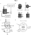
- electrophoretic tissue clearing devices may be used, the devices including an electrophoresis chamber for containing a three-dimensional hydrogel-embedded specimen, a plurality of electrodes, a power supply, and a temperature-controlled buffer circulator.
- the subject devices further include a buffer filtering component.
- the subject devices further include a plurality of fluid inlet and/or outlet ports.
- the subject devices further include a component configured to support the hydrogel-embedded specimen.
- the component is configured to support the hydrogel-embedded specimen in a position that is substantially inside an electric field generated between two or more of the electrodes.
- one or more of the electrodes comprises an expansion component for increasing the size of an electric field generated by the electrodes.
- the expansion component comprises one or more S-shaped bends.
- the length and the width of the one or more electrodes are approximately equal.
- the subject devices further include a lid that forms a fluid-tight and/or air-tight seal with the electrophoresis chamber.
- the present disclosure provides methods of preserving a biological specimen, the methods involving fixing the specimen with a plurality of hydrogel subunits, polymerizing the hydrogel subunits to form a hydrogel-embedded specimen, and clearing the hydrogel-embedded specimen.
- the subject methods further involve storing the cleared hydrogel-embedded specimen in a mounting medium.
- the subject methods further involve analyzing the cleared hydrogel-embedded specimen for evaluation, diagnosis, or prognosis of a pathological state.
- the specimen is a biopsy specimen or an autopsy specimen.
- the pathological state is cancer, immune system dysfunction, neuropsychiatric disease, endocrine/reproductive disease, cardiovascular/pulmonary disease, musculoskeletal disease, or gastrointestinal disease.
- the specimen includes normal tissue
- the method further involves analyzing the specimen to evaluate cell, tissue, organ or system function and/or relationships between cells and tissues, including during development.
- the subject methods further involve conducting a genetic, transcriptomic, genomic, proteomic, metabolomic and/or drug screening analysis on the specimen.
- the subject methods further involve storing the specimen for future analysis, assessment, or functionalization.
- the present disclosure provides systems for infusing hydrogel monomers into biological tissue and subsequently triggering the monomers to form a polymer, gel, mesh, or network with desired stiffness, transparency, pore size, conductivity, or permeability properties, the system including a biological specimen and a plurality of hydrogel subunits.
- the subject systems further include nanoscale hardware devices, proteins, oligonucleotides, and/or fluorescent staining reagents.
- the components of the system are activated or functionalized by energy or external signals such as heat, light, chemical triggers, and/or accelerators.
- the present invention provides a method for preparing a biological specimen for microscopic analysis.
- the method finds many uses, for example in medicine and research, e.g., to diagnose or monitor disease or graft transplantation, to study healthy or diseased tissue, to screen candidate agents for toxicity and efficacy in disease modification.
- reagents, devices, kits and systems thereof that find use in practicing the subject methods.
- Any steps of a method may be separated from another step of the method by an optional storage step, i.e. storage at room temperature, at 16° C, at 4° C, at -12° C, at -20° C, at -70° C, or on -130°C.
- an optional storage step i.e. storage at room temperature, at 16° C, at 4° C, at -12° C, at -20° C, at -70° C, or on -130°C.
- methods are provided for preparing biological specimens for microscopic analysis.
- microscopic analysis is meant the analysis of a specimen using techniques that provide for the visualization of aspects of a specimen that cannot be seen with the unaided eye, i.e., that are not within the resolution range of the normal eye. Such techniques may include, without limitation, optical microscopy (e.g., bright field, oblique illumination, dark field, phase contrast, differential interference contrast, interference reflection, epifluorescence, confocal, etc., microscopy), laser microscopy, electron microscopy, and scanning probe microscopy.
- optical microscopy e.g., bright field, oblique illumination, dark field, phase contrast, differential interference contrast, interference reflection, epifluorescence, confocal, etc., microscopy
- laser microscopy e.g., laser microscopy, electron microscopy, and scanning probe microscopy.
- hydrogel subunits In practicing the subject methods, a biological specimen is fixed in the presence of hydrogel subunits.
- fixing the specimen it is meant exposing the specimen, i.e., cells of the specimen, to a fixation agent such that the cellular components become crosslinked to one another.
- hydrogel or “hydrogel network” is meant a network of polymer chains that are water-insoluble, sometimes found as a colloidal gel in which water is the dispersion medium.
- hydrogels are a class of polymeric materials that can absorb large amounts of water without dissolving. Hydrogels can contain over 99% water and may comprise natural or synthetic polymers, or a combination thereof. Hydrogels also possess a degree of flexibility very similar to natural tissue, due to their significant water content.
- hydrogel subunits or “hydrogel precursors” is meant hydrophilic monomers, prepolymers, or polymers that can be crosslinked, or “polymerized”, to form a three-dimensional (3D) hydrogel network.
- 3D three-dimensional
- fixative any convenient fixation agent, or "fixative,” may be used in the fixative/hydrogel composition to fix the specimen in the presence of hydrogel subunits, for example, formaldehyde, paraformaldehyde, glutaraldehyde, acetone, ethanol, methanol, etc.
- fixative will be diluted in a buffer, e.g., saline, phosphate buffer (PB), phosphate buffered saline (PBS), citric acid buffer, potassium phosphate buffer, etc., usually at a concentration of about 1-10%, e.g.
- 4% paraformaldehyde/0.1M phosphate buffer 2% paraformaldehyde/0.2% picric acid/0.1M phosphate buffer; 4% paraformaldehyde/0.2% periodate/1.2% lysine in 0.1M phosphate buffer; 4% paraformaldehyde/0.05% glutaraldehyde in phosphate buffer; etc.
- fixative used and the duration of exposure to the fixative will depend on the sensitivity of the molecules of interest in the specimen to denaturation by the fixative, and will be known by the ordinarily skilled artisan or may be readily determined using conventional histochemical or immunohistochemical techniques, for example as described in Buchwalow and Böcker. Immunohistochemistry: Basics and Methods. Springer-Verlag Berlin Heidelberg 2010 .
- the fixative/hydrogel composition may comprise any convenient hydrogel subunits, such as, but not limited to, poly(ethylene glycol) and derivatives thereof (e.g. PEG-diacrylate (PEG-DA), PEG-RGD), polyaliphatic polyurethanes, polyether polyurethanes, polyester polyurethanes, polyethylene copolymers, polyamides, polyvinyl alcohols, polypropylene glycol, polytetramethylene oxide, polyvinyl pyrrolidone, polyacrylamide, poly(hydroxyethyl acrylate), and poly(hydroxyethyl methacrylate), collagen, hyaluronic acid, chitosan, dextran, agarose, gelatin, alginate, protein polymers, methylcellulose and the like.
- PEG-DA PEG-diacrylate
- PEG-RGD PEG-diacrylate
- polyaliphatic polyurethanes polyether polyurethanes
- polyester polyurethanes polyethylene
- the hydrogel subunits may be modified to add specific properties to the hydrogel; for example, peptide sequences can be incorporated to induce degradation (see, e.g., West and Hubbell, 1999, Macromolecules, 32:241 ) or to modify cell adhesion (see, e.g. Hem and Hubbell, 1998, J. Biomed. Mater. Res., 39:266 ).
- Agents such as hydrophilic nanoparticles, e.g., poly-lactic acid (PLA), poly-glycolic acid (PLG), poly(lactic-co-glycolic acid) (PLGA), polystyrene, poly(dimethylsiloxane) (PDMS), etc.
- Suitable poly(lactic acid) (PLA), and other similar materials can be used to add specific properties to the hydrogels (see, e.g., Huh and Bae, 1999, Polymer, 40:6147 ).
- Crosslinkers e.g. bis-acrylamide, diazirine, etc.
- initiatiors e.g. azobisisobutyronitrile (AIBN), riboflavin, L-arginine, etc.
- AIBN azobisisobutyronitrile
- riboflavin, L-arginine, etc. may be included to promote covalent bonding between interacting macromolecules in later polymerization steps.
- the concentration and molecular weight of the hydrogel subunit(s) and modifying agents will depend on the selected polymer and the desired characteristics, e.g., pore size, swelling properties, conductivity, elasticity/stiffness (Young's modulus), biodegradability index, etc., of the hydrogel network into which they will be polymerized.
- the hydrogel may comprise pores of sufficient size to allow the passage of macromolecules, e.g., proteins, nucleic acids, or small molecules as described in greater detail below, into the specimen.
- pore size generally decreases with increasing concentration of hydrogel subunits and generally increases with an increasing ratio of hydrogel subunits to crosslinker, and will prepare a fixative/hydrogel composition that comprises a concentration of hydrogel subunits that allows the passage of such macromolecules.
- the hydrogel may be desirable for the hydrogel to have a particular stiffness, e.g., to provide stability in handling the embedded specimen, e.g., a Young's Modulus of about 2-70kN/m 2 , for example, about 2 kN/m 2 , about 4 kN/m 2 , about 7 kN/m 2 , about 10 kN/m 2 , about 15 kN/m 2 , about 20 kN/m 2 , about 40 kN/m 2 , but typically not more than about 70 kN/m 2 .
- a Young's Modulus of about 2-70kN/m 2 , for example, about 2 kN/m 2 , about 4 kN/m 2 , about 7 kN/m 2 , about 10 kN/m 2 , about 15 kN/m 2 , about 20 kN/m 2 , about 40 kN/m 2 , but typically not more than about 70 kN/m 2
- the elasticity of a hydrogel network may be influenced by a variety of factors, including the branching of the polymer, the concentration of hydrogel subunits, and the degree of cross-linking, and will prepare a fixative/hydrogel composition that comprises a concentration of hydrogel subunits to provide such desired elasticity.
- the fixative/hydrogel composition may comprise an acrylamide monomer at a concentration of from about 1% w/v to about 20% w/v, e.g., about 2% to about 15%, about 3% to about 10%, about 4% to about 8%, and a concentration of bis-acrylamide crosslinker in the range of about 0.01% to about 0.075%, e.g., 0.01%, 0.02%, 0.025%, 0.03%, 0.04%, 0.05%, 0.06%, or 0.075%; or, for example, the fixative/hydrogel composition may comprise PEG prepolymers having a molecular weight ranging from at least about 2.5K to about 50K, e.g., 2.5K or more, 3.5K or more, 5K or more, 7.5K or more, 10K or more, 15K or more, 20K or more, but typically not more than about 50K, at a concentration in a range from about 1% w/w to about 50% w/w, e.
- the fixative/hydrogel solution may be delivered to the specimen by any convenient method, e.g., perfusion, injection, instillation, absorption, application, immersion/submersion, etc.
- the specimen will typically be fixed in the presence of the hydrogel for 15 minutes or more, for example, for 30 minutes or more, 1 hour or more, 2 hours or more, 4 hours or more, 6 hours or more, 12 hours or more, in some instances, for 16 hours or more, 20 hours or more, or 24 hours or more.
- the hydrogel subunits are polymerized, i.e., covalently or physically crosslinked, to form a hydrogel network.
- Polymerization may be by any method including, but not limited to, thermal crosslinking, chemical crosslinking, physical crosslinking, ionic crosslinking, photo-crosslinking, irradiative crosslinking (e.g., x-ray, electron beam), and the like, and may be selected based on the type of hydrogel used and knowledge in the art. For example, mixing of an un-polymerized or partially polymerized resin with specific crosslinking chemicals results in a chemical reaction that forms cross-links.
- Crosslinking can be induced in materials that are normally thermoplastic through exposure to a radiation source, such as electron beam exposure, gamma-radiation, or UV light; for example, electron beam processing is used to polymerize the C type of crosslinked polyethylene.
- a radiation source such as electron beam exposure, gamma-radiation, or UV light
- electron beam processing is used to polymerize the C type of crosslinked polyethylene.
- Other types of crosslinked polyethylene are made by addition of peroxide during extruding (type A) or by addition of a cross-linking agent (e.g. vinylsilane) and a catalyst during extruding and then performing a post-extrusion curing.
- a cross-linking agent e.g. vinylsilane
- Many polymers undergo oxidative cross-linking, typically when exposed to atmospheric oxygen. In some cases the reaction is more rapid than desired and thus polymerization reactions may involve the use of an antioxidant to slow the formation of oxidative cross-links.
- an oxidizer such as hydrogen peroxide may be used to speed up the process.
- the length of time for polymerization will depend on the type of hydrogel subunits used and the chosen polymerization method, but will typically be about 15 minutes to about 48 hours, for example, 15 minutes or more, 1 hour or more, 2 hours or more, 3 hours or more, 4 hours or more, 6 hours or more, 12 hours or more, 16 hours or more, 24 hours or more, or in some instances, 48 hours.
- the hydrogel-embedded (i.e., hydrogel-hybridized) specimen may be cleared.
- a specimen it is meant that the specimen is made substantially permeable to light, i.e., transparent.
- the visual (i.e., white) light, ultraviolet light or infrared light that is used to illuminate the specimen will to pass through the specimen and illuminate only selected cellular components therein, e.g., 75% or more of the light, 80% or more of the light, 85% or more of the light, in some instances, 90% or more of the light, 95% or more of the light, 98% or more of the light, e.g. 100% of the light will pass through the specimen.
- This change in the optical properties of the specimen provides for the visualization of cellular and subcellular structures internal to the tissue.
- Any treatment that forces cellular components, e.g., lipids, from the specimen, that draws cellular components, e.g., lipids, from a specimen, or that causes cellular components, e.g., lipids, to break down, i.e., dissolve, within a specimen may be used to clear the specimen, including, without limitation, exposure to organic solvents such as xylenes, ethanol or methanol, exposure to detergents such as saponin, Triton X-100 and Tween-20, exposure to ionic surfactants, e.g., sodium dodecyl sulfate (SDS), electrophoresis, hydrodynamic pressure, ultrasonic vibration, solute contrasts, microwave radiation, vascular circulation, and the like.
- organic solvents such as xylenes, ethanol or methanol
- detergents such as saponin, Triton X-100 and Tween-20
- ionic surfactants e.g., sodium dodecyl
- clearing is performed using a solvent that does not quench fluorescent proteins.
- organic solvents that are known to quench fluorescent proteins include tetrahydrofuran, hexane, benzylalcohol/benzylbenzoate (BABB), and dibenzyl ether. Accordingly, in order to preserve the fluorescence of various proteins, in some embodiments clearing is conducted using solvents other than those listed above, e.g., is conducted using non-organic solvents.
- clearing is conducted using an ionic surfactant, e.g., SDS, in order to expedite the clearing process by actively transporting charged ionic micelles out of the specimen that is being cleared.
- Clearing may be performed in any convenient buffer that is compatible with the selected clearance method, e.g., saline, phosphate buffer, phosphate buffered saline (PBS), sodium borate buffer, boric acid buffer, citric acid buffer, etc., as known in the art, and will typically take about 1-10 days per centimeter thickness of specimen, i.e., usually about 1 day, in some instances 2 days, sometimes 5 days, and typically no more than 10 days per cubic centimeter. Optimal time may be readily determined by visual inspection of the specimen for clarity.
- a sample After clearing, a sample will generally be substantially free of lipids.
- substantially free of lipids is meant that the original amount of lipids present in the sample before clearing has been reduced by approximately 70% or more, such as by 75% or more, such as by 80% or more, such as by 85% or more, such as by 90% or more, such as by 95% or more, such as by 99% or more, such as by 100%.
- Tissue specimens suitable for use with the methods and systems described herein generally include any type of tissue specimens collected from living or dead subjects, such as, e.g., biopsy specimens and autopsy specimens. Tissue specimens may be collected and processed using the methods and systems described herein and subjected to microscopic analysis immediately following processing, or may be preserved and subjected to microscopic analysis at a future time, e.g., after storage for an extended period of time. In some embodiments, the methods described herein may be used to preserve tissue specimens in a stable, accessible and fully intact form for future analysis.
- tissue specimens such as, e.g., human brain tissue specimens
- tissue specimens may be processed as described above and cleared to remove a plurality of cellular components, such as, e.g., lipids, and then stored for future analysis.
- the methods and systems described herein may be used to analyze a previously-preserved or stored tissue specimen.
- a previously-preserved tissue specimen that has not been subjected to the CLARITY process may be processed and analyzed as described herein.
- the specimen may comprise biomolecules that can be directly visualized by microscopy.
- biomolecules it is generally meant proteins, lipids, steroids, nucleic acids, etc. within a tissue or cell.
- FP fluorescent protein
- fluoresce is meant to absorb energy at one wavelength and emit it at another wavelength.
- a green fluorescent protein refers to a polypeptide that has a peak in the emission spectrum at 510 nm or about 510 nm.
- FPs of interest include, but are not limited to, a green fluorescent protein (GFP), yellow fluorescent protein (YFP), orange fluorescent protein (OFP), cyan fluorescent protein (CFP), blue fluorescent protein (BFP), red fluorescent protein (RFP), far-red fluorescent protein, or near-infrared fluorescent protein.
- GFP green fluorescent protein
- YFP yellow fluorescent protein
- OFP orange fluorescent protein
- CFP cyan fluorescent protein
- BFP blue fluorescent protein
- RFP red fluorescent protein
- far-red fluorescent protein or near-infrared fluorescent protein.
- Aequorea GFP refers to GFPs from the genus Aequorea and to mutants or variants thereof.
- GFPs from other species such as Anthozoa reef coral, Anemonia sea anemone, Renilla sea pansy, Galaxea coral, Acropora brown coral, Trachyphyllia and Pectimidae stony coral and other species are well known and are available and known to those of skill in the art.
- Exemplary GFP variants include, but are not limited to BFP, CFP, YFP and OFP.
- florescent proteins and their variants include GFP proteins, such as Emerald (Invitrogen, Carlsbad, Calif.), EGFP (Clontech, Palo Alto, Calif), Azami-Green (MBL International, Woburn, Mass.), Kaede (MBL International, Woburn, Mass.), ZsGreen1 (Clontech, Palo Alto, Calif.) and CopGFP (Evrogen/Axxora, LLC, San Diego, Calif.); CFP proteins, such as Cerulean ( Rizzo, Nat Biotechnol. 22(4):445-9 (2004 )), mCFP ( Wang et al., PNAS USA.
- GFP proteins such as Emerald (Invitrogen, Carlsbad, Calif.), EGFP (Clontech, Palo Alto, Calif), Azami-Green (MBL International, Woburn, Mass.), Kaede (MBL International, Woburn, Mass.), ZsGreen1 (Clontech, Palo Alto, Calif.) and CopGFP (Evro
- RFP red fluorescent protein Discosoma
- DsRed red fluorescent protein Discosoma
- mRFP1 monomeric red fluorescent protein 1
- mCherry mCherry
- tdTomato mStrawberry
- mTangerine Wang et al., PNAS U S A.
- DsRed2 (Clontech, Palo Alto, Calif.), and DsRed-T1 ( Bevis and Glick, Nat. Biotechnol., 20: 83-87 (2002 )), Anthomedusa J-Red (Evrogen) and Anemonia AsRed2 (Clontech, Palo Alto, Calif.).
- Far-red fluorescent proteins include, for example, Actinia AQ143 ( Shkrob et al., Biochem J. 392(Pt 3):649-54 (2005 )), Entacmaea eqFP611 ( Wiedenmann et al. Proc Natl Acad Sci USA.
- Discosoma variants such as mPlum and mRasberry ( Wang et al., PNAS U S A. 101(48):16745-9 (2004 )), and Heteractis HcRed1 and t-HcRed (Clontech, Palo Alto, Calif.).
- macromolecules may be provided that promote the visualization of particular cellular biomolecules, e.g., proteins, lipids, steroids, nucleic acids, etc. and sub-cellular structures.
- the macromolecules are diagnostic.
- the macromolecules are prognostic.
- the macromolecules are predictive of responsiveness to a therapy.
- the macromolecules are candidate agents in a screen, e.g., a screen for agents that will aid in the diagnosis and/or prognosis of disease, in the treatment of a disease, and the like.
- specimens may be contacted with nucleic acid stains like DAPI and Hoechst, which bind the minor groove of DNA, thus labeling the nuclei of cells.
- Drugs or toxins that bind specific cellular structures and have been derivatized with a fluorescent reporter may be employed, e.g., fluorescently labelled-phalloidin, which is used to stain actin fibers in mammalian cells.
- fluorescently labelled-phalloidin which is used to stain actin fibers in mammalian cells.
- fluorophores or fluorochromes such as fluorescein, Alexa Fluors or DyLight 488, which can be chemically linked to molecules which bind the target biomolecules of interest within the sample.
- the specimen may be contacted with one or more polypeptides, e.g. antibodies, labeled peptides, and the like, that are specific for and will bind to particular cellular biomolecules for either direct or indirect labeling by color or immunofluorescence.
- immunofluorescence it is meant a technique that uses the highly specific binding of an antibody to its antigen or binding partner in order to label specific proteins or other molecules within the cell.
- a sample is treated with a primary antibody specific for the biomolecule of interest.
- a fluorophore can be directly conjugated to the primary antibody or peptide.
- a secondary antibody, conjugated to a detection moiety or fluorophore, which binds specifically to the first antibody can be used. See, for example, Buchwalow and Bocker.
- a class of agents that may be provided as macromolecules is nucleic acids.
- a specimen may be contacted with an antisense RNA that is complementary to and specifically hybridizes to a transcript of a gene of interest, e.g., to study gene expression in cells of the specimen.
- a specimen may be contacted with a DNA that is complementary to and specifically hybridizes to genomic material of interest, e.g., to study genetic mutations, e.g., loss of heterozygosity, gene duplication, chromosomal inversions, and the like.
- the hybridizing RNA or DNA is conjugated to detection moieties, i.e. agents that may be either directly or indirectly visualized microscopically.
- in situ hybridization techniques may be found at, for example, Harris and Wilkinson. In situ hybridization: Application to developmental biology and medicine, Cambridge University Press 1990 ; and Fluorescence In Situ Hybridization (FISH) Application Guide. Liehr, T, ed., Springer-Verlag, Berlin Heidelberg 1990 .
- the specimen may be contacted with small molecules.
- the specimen comprises ⁇ -galactosidase or alkaline phosphatase
- it may be desirable to visualize cells and regions of the tissue expressing these proteins.
- a specimen may be contacted with substrates for ⁇ -galactosidase (e.g.
- NBT nitro-blue tetrazolium
- BCIP nitro-blue tetrazolium
- the biomolecules that are targeted by the provided macromolecules are endogenous to the cell.
- the macromolecules may be provided to the specimen to target/visualize biomolecules that were ectopically provided to the cells of the specimen, e.g. agents that were introduced to the specimen in vivo or ex vivo to label certain cell populations or subcellular structures.
- stereotactic surgery is often used in the field of neuroscience to provide biomolecules such as proteins, viruses, chemicals to neural tissue that label, or "trace", the projections and/or the connectivity of subsets of neurons in vivo or ex vivo.
- a needle comprising a labeling macromolecule is lowered into CNS tissue at a precise location and the labeling molecule is released into the tissue.
- the molecule will fill the neurons in the vicinity of the injection site and, depending on the type of macromolecule delivered, may be transported across synapses to label their efferent targets ("anterograde tracing") and/or across dendrites to label the afferent neurons from which they are receiving signals (“retrograde tracing").
- agents that may be used to label neurons stereotactically are well known in the art, including, for example, nucleic acids that encode fluorescent proteins; viral tracers, e.g.
- Herpes simplex virus type1 and the Rhabdoviruses; wheat-germ agglutinin (WGA); Phaseolus vulgaris leucoagglutinin (PHA-L); horseradish peroxidase-conjugated lectins; biotinylated dextran amines (BDA); cholera toxin B; NEUROBIOTIN Tracer ® (Vector labs).
- WGA wheat-germ agglutinin
- PHA-L Phaseolus vulgaris leucoagglutinin
- BDA biotinylated dextran amines
- BDA cholera toxin B
- NEUROBIOTIN Tracer ® Vector labs.
- Specimens labeled in this way may be contacted with macromolecules, e.g. polypeptides or chemicals, that promote the visualization of these ectopically provided labels.
- the macromolecules that are used to visualize the cellular biomolecules or subcellular structures are passively transported into the specimen. In other words, the macromolecules diffuse into the specimen. In other instances, the macromolecules are actively transported into the specimen, e.g. by electroporation, hydrodynamic pressure, ultrasonic vibration, solute contrasts, microwave radiation, vascular circulation, or the like.
- the specimen is contacted with the macromolecules after the specimen has been cleared.
- the hydrogel-embedded specimen may be contacted with the macromolecules prior to clearing the specimen. In such embodiments, contact with the macromolecules may be facilitated by permeabilizing the specimen, that is, changing the properties of the specimen to improve the permeability of the specimen to macromolecules.
- a permeabilized specimen it is meant that about 50% or more of the macromolecules applied to the specimen will penetrate to the deepest regions of the specimen, e.g. 60% or more of the macromolecules, 70% or more of the macromolecules, or 80% or more of the macromolecules, in some instances 85% or more of the macromolecules, 90% or more of the macromolecules, or 95% or more of the macromolecules, for example 98% or more of the macromolecules, e.g. 100% of the macromolecules will pass through the specimen.
- Permeabilization of the specimen, and of the cells therein may be achieved by any of the protocols discussed above for the removal of cellular components, e.g. lipids, from the specimen or as known in the art for permeabilizing cells.
- the specimen is embedded in a mounting medium.
- Mounting medium is typically selected based on its suitability for the reagents used to visualize the cellular biomolecules, the refractive index of the specimen, and the microscopic analysis to be performed. For example, for phase-contrast work, the refractive index of the mounting medium should be different from the refractive index of the specimen, whereas for bright-field work the refractive indexes should be similar.
- a mounting medium should be selected that reduces fading, photobleaching or quenching during microscopy or storage.
- a mounting medium or mounting solution may be selected to enhance or increase the optical clarity of the cleared tissue specimen.
- Nonlimiting examples of suitable mounting media include glycerol, CC/Mount TM , Fluoromount TM Fluoroshield TM , ImmunHistoMount TM , Vectashield TM , Permount TM , Acrytol TM , CureMount TM , FocusClear TM , or equivalents thereof.
- the hydrogel-embedded specimen is permanently mounted. In other words, once mounted in mounting medium, the hydrogel-embedded specimen cannot be removed for further manipulation. In other instances, the specimen is temporarily, or reversibly, mounted. In other words, the hydrogel-embedded specimen may be removed from the mounting medium and re-stained after microscopy to visualize alternative/additional biomolecules or subcellular structures. In such instances, macromolecules that were previously added to the specimen, e.g.
- hydrogel-embedded specimen is then contacted with different macromolecules specific for other biomolecules or subcellular structures. As such, iterative staining may be performed on the same specimen.
- organic solvents such as xylenes, ethanol or methanol
- detergents such as sodium dodecyl sulfate (SDS), saponin, Triton X-100 and Tween-20
- electrophoresis hydrodynamic pressure, ultrasonic vibration, solute contrasts, microwave radiation, vascular circulation, and the like.
- hydrogel-embedded specimen is then contacted with different macromolecules specific for other biomolecules or subcellular structures. As such, iterative staining may be performed on the same specimen.
- Specimens prepared using the subject methods may be analyzed by any of a number of different types of microscopy, for example, optical microscopy (e.g. bright field, oblique illumination, dark field, phase contrast, differential interference contrast, interference reflection, epifluorescence, confocal, etc., microscopy), laser microscopy, electron microscopy, and scanning probe microscopy.
- optical microscopy e.g. bright field, oblique illumination, dark field, phase contrast, differential interference contrast, interference reflection, epifluorescence, confocal, etc.
- microscopy laser microscopy
- electron microscopy e.g., electron microscopy, and scanning probe microscopy.
- Bright field microscopy is the simplest of all the optical microscopy techniques.
- Sample illumination is via transmitted white light, i.e. illuminated from below and observed from above.
- Limitations include low contrast of most biological samples and low apparent resolution due to the blur of out of focus material. The simplicity of the technique and the minimal sample preparation required are significant advantages.
- oblique illumination microscopy the specimen is illuminated from the side. This gives the image a 3-dimensional appearance and can highlight otherwise invisible features.
- a more recent technique based on this method is Hoffmann's modulation contrast, a system found on inverted microscopes for use in cell culture. Though oblique illumination suffers from the same limitations as bright field microscopy (low contrast of many biological samples; low apparent resolution due to out of focus objects), it may highlight otherwise invisible structures.
- Dark field microscopy is a technique for improving the contrast of unstained, transparent specimens.
- Dark field illumination uses a carefully aligned light source to minimize the quantity of directly-transmitted (unscattered) light entering the image plane, collecting only the light scattered by the sample.
- Dark field can dramatically improve image contrast (especially of transparent objects) while requiring little equipment setup or sample preparation.
- the technique suffers from low light intensity in final image of many biological samples, and continues to be affected by low apparent resolution.
- Phase contrast is an optical microscopy illumination technique that converts phase shifts in light passing through a transparent specimen to brightness changes in the image.
- phase contrast shows differences in refractive index as difference in contrast.
- the phase shifts themselves are invisible to the human eye, but become visible when they are shown as brightness changes.
- DIC differential interference contrast
- the system consists of a special prism (Nomarski prism, Wollaston prism) in the condenser that splits light in an ordinary and an extraordinary beam.
- the spatial difference between the two beams is minimal (less than the maximum resolution of the objective).
- the beams are reunited by a similar prism in the objective.
- a refractive boundary e.g. a nucleus within the cytoplasm
- the difference between the ordinary and the extraordinary beam will generate a relief in the image.
- Differential interference contrast requires a polarized light source to function; two polarizing filters have to be fitted in the light path, one below the condenser (the polarizer), and the other above the objective (the analyzer).
- interference reflection microscopy also known as reflected interference contrast, or RIC. It is used to examine the adhesion of cells to a glass surface, using polarized light of a narrow range of wavelengths to be reflected whenever there is an interface between two substances with different refractive indices. Whenever a cell is attached to the glass surface, reflected light from the glass and that from the attached cell will interfere. If there is no cell attached to the glass, there will be no interference.
- a fluorescence microscope is an optical microscope that uses fluorescence and phosphorescence instead of, or in addition to, reflection and absorption to study properties of organic or inorganic substances.
- fluorescence microscopy a sample is illuminated with light of a wavelength which excites fluorescence in the sample. The fluoresced light, which is usually at a longer wavelength than the illumination, is then imaged through a microscope objective.
- Two filters may be used in this technique; an illumination (or excitation) filter which ensures the illumination is near monochromatic and at the correct wavelength, and a second emission (or barrier) filter which ensures none of the excitation light source reaches the detector.
- these functions may both be accomplished by a single dichroic filter.
- the "fluorescence microscope” refers to any microscope that uses fluorescence to generate an image, whether it is a more simple set up like an epifluorescence microscope, or a more complicated design such as a confocal microscope, which uses optical sectioning to get better resolution of the fluorescent image.
- Confocal microscopy uses point illumination and a pinhole in an optically conjugate plane in front of the detector to eliminate out-of-focus signal.
- the image's optical resolution is much better than that of wide-field microscopes.
- this increased resolution is at the cost of decreased signal intensity - so long exposures are often required.
- 2D or 3D imaging requires scanning over a regular raster (i.e., a rectangular pattern of parallel scanning lines) in the specimen.
- the achievable thickness of the focal plane is defined mostly by the wavelength of the used light divided by the numerical aperture of the objective lens, but also by the optical properties of the specimen. The thin optical sectioning possible makes these types of microscopes particularly good at 3D imaging and surface profiling of samples.
- SPIM single plane illumination microscopy
- the light sheet is a beam that is collimated in one and focused in the other direction. Since no fluorophores are excited outside the detectors' focal plane, the method also provides intrinsic optical sectioning. Moreover, when compared to conventional microscopy, light sheet methods exhibit reduced photobleaching and lower phototoxicity, and often enable far more scans per specimen. By rotating the specimen, the technique can image virtually any plane with multiple views obtained from different angles. For every angle, however, only a relatively shallow section of the specimen is imaged with high resolution, whereas deeper regions appear increasingly blurred.
- Super-resolution microscopy is a form of light microscopy. Due to the diffraction of light, the resolution of conventional light microscopy is limited as stated by Ernst Abbe in 1873. A good approximation of the resolution attainable is the FWHM (full width at half-maximum) of the point spread function, and a precise widefield microscope with high numerical aperture and visible light usually reaches a resolution of ⁇ 250 nm.
- Super-resolution techniques allow the capture of images with a higher resolution than the diffraction limit. They fall into two broad categories, “true" super-resolution techniques, which capture information contained in evanescent waves, and “functional" super-resolution techniques, which use experimental techniques and known limitations on the matter being imaged to reconstruct a super-resolution image.
- Laser microscopy uses laser illumination sources in various forms of microscopy.
- laser microscopy focused on biological applications uses ultrashort pulse lasers, or femtosecond lasers, in a number of techniques including nonlinear microscopy, saturation microscopy, and multiphoton fluorescence microscopy such as two-photon excitation microscopy (a fluorescence imaging technique that allows imaging of living tissue up to a very high depth, e.g. one millimeter)
- EM electron microscopy
- An electron microscope has greater resolving power than a light-powered optical microscope because electrons have wavelengths about 100,000 times shorter than visible light (photons). They can achieve better than 50 pm resolution and magnifications of up to about 10,000,000x whereas ordinary, non-confocal light microscopes are limited by diffraction to about 200 nm resolution and useful magnifications below 2000x.
- the electron microscope uses electrostatic and electromagnetic "lenses" to control the electron beam and focus it to form an image. These lenses are analogous to but different from the glass lenses of an optical microscope that form a magnified image by focusing light on or through the specimen.
- Electron microscopes are used to observe a wide range of biological and inorganic specimens including microorganisms, cells, large molecules, biopsy samples, metals, and crystals. Industrially, the electron microscope is often used for quality control and failure analysis. Examples of electron microscopy include Transmission electron microscopy (TEM), Scanning electron microscopy (SEM), reflection electron microscopy (REM), Scanning transmission electron microscopy (STEM) and low-voltage electron microscopy (LVEM).
- TEM Transmission electron microscopy
- SEM Scanning electron microscopy
- REM reflection electron microscopy
- STEM Scanning transmission electron microscopy
- LVEM low-voltage electron microscopy
- Scanning probe microscopy is a branch of microscopy that forms images of surfaces using a physical probe that scans the specimen. An image of the surface is obtained by mechanically moving the probe in a raster scan of the specimen, line by line, and recording the probe-surface interaction as a function of position.
- SPM examples include atomic force microscopy (ATM), ballistic electron emission microscopy (BEEM), chemical force microscopy (CFM), conductive atomic force microscopy (C-AFM), electrochemical scanning tunneling microscope (ECSTM), electrostatic force microscopy (EFM), fluidic force microscope (FluidFM), force modulation microscopy (FMM), feature-oriented scanning probe microscopy (FOSPM), kelvin probe force microscopy (KPFM), magnetic force microscopy (MFM), magnetic resonance force microscopy (MRFM), near-field scanning optical microscopy (NSOM) (or SNOM, scanning near-field optical microscopy, SNOM, Piezoresponse Force Microscopy (PFM), PSTM, photon scanning tunneling microscopy (PSTM), PTMS, photothermal microspectroscopy/microscopy (PTMS), SCM, scanning capacitance microscopy (SCM), SECM, scanning electrochemical microscopy (SECM), SGM, scanning gate microscopy (SGM), SHPM
- Reagents and kits for practicing one or more of the above-described methods.
- Reagents and kits may include one or more of the following: fixative; hydrogel subunits; clearing reagents; detection macromolecules, e.g., labeled and or unlabeled antibodies, nucleic acid probes (oligonucleotides, vectors, etc.), chemicals, etc.; buffers, e.g. buffer for fixing, washing, clearing, and/or staining specimens; mounting medium; embedding molds; dissection tools; etc.
- fixative fixative
- hydrogel subunits clearing reagents
- detection macromolecules e.g., labeled and or unlabeled antibodies, nucleic acid probes (oligonucleotides, vectors, etc.), chemicals, etc.
- buffers e.g. buffer for fixing, washing, clearing, and/or staining specimens
- mounting medium e.g. buffer for fixing, washing, clearing, and/or staining specimen
- specimens that have been prepared by the subject methods for use in, for example, studying tissue at the cellular and subcellular level.
- fixed and polymerized specimens, or specimens that have been fixed, polymerized, and cleared are provided for use in studying the expression of genes of interest, for screens to identify candidate agents that target cells and/or subcellular structures of interest, etc.
- Such prepared specimens may also be provided as a positive control in one of the kits or systems as described herein.
- the subject kits may further include instructions for practicing the subject methods. These instructions may be present in the subject kits in a variety of forms, one or more of which may be present in the kit.
- One form in which these instructions may be present is as printed information on a suitable medium or substrate, e.g., a piece or pieces of paper on which the information is printed, in the packaging of the kit, in a package insert, etc.
- Yet another means would be a computer readable medium, e.g., diskette, CD, digital storage medium, etc., on which the information has been recorded.
- Yet another means that may be present is a website address which may be used via the Internet to access the information at a removed site. Any convenient means may be present in the kits.
- the subject devices may include, for example, electrophoresis apparatus, ultrasounds, microwaves, needles, tubing, perfusion pumps, etc., for fixing, clearing, and/or staining specimens.
- electrophoresis device suitable for use in removing cellular components from a specimen, e.g., cellular components that are not crosslinked to the hydrogel network.
- electrophoresis it is meant the application of an electric field to a sample, e.g., to a biological sample. Electrophoresis is most commonly used to mobilize biological macromolecules, e.g., nucleic acids, proteins, in a sample to separate and analyze those macromolecules. Numerous electrophoretic techniques have been developed including capillary electrophoresis, gel electrophoresis, paper electrophoresis, and immunoelectrophoresis.
- a hydrogel is formed using compounds such as agarose or polyacrylamide.
- a mixture containing the desired macromolecules is placed (or loaded) onto one end of the gel, and the gel is then placed in contact with a liquid buffer.
- This liquid buffer contains salts, which, when dissolved, form ions within the buffer.
- Biological molecules are typically charged, for example when contacted with electrophoresis buffer.
- DNA is negatively charged in common electrophoresis buffers due to the phosphate groups in its backbone. Therefore, when electric current is applied to the ends of the gel, the biological molecules move through the gel from one end to the other. Examples of electrophoresis devices and methods of gel electrophoresis may be found in, for example, US Patent Nos.
- Electrophoresis devices suitable for use in the subject methods will generally comprise an electrophoresis chamber into which a buffer solution and the hydrogel-embedded specimen may be placed. See, for example, FIG. 2 and FIG. 8 .
- the electrophoresis chamber may generally be any suitable size to accommodate a hydrogel-embedded sample of interest, and may be constructed of any material that will retain solution within the chamber, for example glasses and plastics, such as, for example, acrylics, polycarbonates, polystyrenes, polymethyl methacrylates, polyethylene, polyfluoroethylene, polypropylene, polyurethane, polyethylene terephthalate, polytetrafluoroethylene and the like.
- a chamber may be molded or machined or otherwise formed from a resin or hard plastic, as appropriate for particular applications.
- an electrophoresis chamber may further comprise a component that is configured to support a hydrogel-embedded sample, such as, e.g., a platform, within the electrophoresis chamber.
- an electrophoresis device may include a lid that fits over the electrophoresis chamber to close the chamber.
- Lids in accordance with embodiments of the invention may include a seal that forms a liquid-tight and/or air-tight seal with the body of the electrophoresis chamber when the lid is coupled to the chamber.
- one or more sealing components may be attached to the lid, attached to the chamber, or attached to both the lid and the chamber. When the lid is coupled to the chamber, the sealing components may form a liquid and/or air-tight seal.
- Electrophoresis devices in accordance with some embodiments of the invention may comprise two or more electrodes of opposite polarity (i.e. "anode” (negatively charged) and at least one "cathode” (positively charged)) operably associated with the electrophoresis chamber to which an electric current may be applied to create an electric field within the chamber.
- the electrodes may be constructed of any material that will result in an electric field being established upon the application of an electric current to the electrodes, and may be configured within the chamber and relative to the site where the specimen is to be placed in any convenient way that will promote the establishment of an electric field across a specimen positioned therein, for example as well known in the art of nucleic acid or protein electrophoresis. See, for example, US Patent Nos.
- the electrodes may be configured within the chamber so as to substantially flank the specimen.
- one or more of the electrodes may comprise an extension component that is used to enlarge the size of the electric field that is generated between the electrodes.
- an electrode may comprise an extension component in the form of a serpentine portion of the electrode the doubles back on itself to form a plurality of S-shaped bends.
- An example of an electrode comprising a serpentine extension component can be seen in FIG. 2 , Panel c (reference numbers 103a and 103b).
- the extension component enlarges the size of the electric field that is generated when a voltage is applied to the electrodes so that an entire three-dimensional tissue specimen can be placed inside the electric field.
- the length and width of the extension component can be adjusted as needed to accommodate tissue specimens of various dimensions. For example, in certain embodiments the length and the width of an electrode comprising an extension component are approximately equal, as depicted in FIG. 8 , Panel e.
- an electrophoresis chamber may be partitioned by, e.g., a solid divider or by air into two distinct regions, where each region comprises one electrode in a buffer, and the specimen is positioned within the buffer such that the specimen spans, or straddles the two regions, such that the electric field created by the electrodes is created through the specimen.
- the chamber may comprise a platform or support structure, upon or into which the hydrogel-embedded specimen is placed, e.g., a platform between two electrodes, a platform that spans regions of the chamber comprising the electrodes, etc.
- the electrophoresis apparatus may be operably linked to a power source from which voltage may be applied to the electrodes.
- the power source may be separate from the electrophoresis apparatus, i.e. the electrophoresis apparatus may be a separate module from the power source.
- the power source may be integrated into the electrophoresis apparatus, i.e., the electrophoresis apparatus will comprise the power source.
- recirculated buffer it is meant that buffer is removed from the chamber and then returned to the chamber, for example, after passing through a cooling unit (refrigeration unit, ice bath, etc.), a heating unit, a filter, etc.
- replacement is meant that buffer is removed from the chamber and fresh buffer is added in its place.
- the electrophoresis apparatus may optionally comprise one or more ports through which buffer may enter and/or exit the chamber.
- the chamber may comprise two or more ports, e.g. a first port through which buffer enters the chamber and a second port through which buffer exits the chamber.
- Buffer may be added/removed/recirculated/replaced by the use of the one or more ports and optionally, tubing, pumps, valves, or any other suitable fluid handling and/or fluid manipulation equipment, for example, tubing that is removably attached or permanently attached to one or more components of a device.
- a first tube having a first and second end may be attached to a first port and a second tube having a first and second end may be attached to a second port, where the first end of the first tube is attached to the first port and the second end of the first tube is operably linked to a receptacle, e.g.
- a cooling unit heating unit, filtration unit, waste receptacle, etc.
- the first end of the second tube is attached to the second port and the second end of the second tube is operably linked to a receptacle, e.g. a cooling unit, beaker on ice, filtration unit, waste receptacle, etc.
- one tube having a first and second end may be removably attached to both a first and second port, i.e., the first end of the tube is removably attached to the first port and the second end of the tube is removably attached to the second port, where the tubing is operably linked to, for example, a refrigeration unit (e.g., the tubing passes through the unit), a filter (e.g. the tubing comprises a filter), a buffer reservoir (e.g. the tubing receives replacement buffer from a reservoir via, e.g., a splitter), etc.
- the tubing will also be operably connected to a pump, e.g.
- the electrophoresis apparatus may be operably connected to a cooling unit, heating unit, filtration unit, buffer reservoirs/receptacles, pump, etc.
- a refrigeration unit, heating unit, filtration unit, buffer reservoirs/receptacles, pump, etc. will be integrated into the electrophoresis apparatus.
- the electrophoresis apparatus may comprise the refrigeration unit, heating unit, filtration unit, buffer reservoirs/receptacles, pump, etc.
- the refrigeration unit, heating unit, filtration unit, buffer reservoirs/receptacles, pump, etc. may be a separate module from the electrophoresis apparatus.
- the example device 101 includes a lid 102, a first electrode 103a, a second electrode 103b, a base 104, an outlet port 105, an inlet port 106, a first electrode connector 107a and a second electrode connector 107b.
- Systems may include one or more of the modules described herein, e.g. an electrophoresis apparatus, a power supply, a refrigeration unit, a heating unit, a pump, etc.
- Systems may also include any of the reagents described herein, e.g. fixative; hydrogel subunits; clearing reagents; detection macromolecules, e.g., antibodies, nucleic acid probes (oligonucleotides, vectors, etc.), chemicals, etc.; buffers, e.g., buffer for fixing, washing, clearing, and/or staining specimens; mounting medium; embedding molds; etc.
- Systems in accordance with certain embodiments may also include a microscope and/or related imaging equipment, e.g., camera components, digital imaging components and/or image capturing equipment, computer processors configured to collect images according to one or more user inputs, and the like.
- reagents, kits, systems and devices may be used to prepare any biological tissue for microscopic analysis.
- Methods, reagents, kits, systems and devices may be used to prepare a specimen from any plant or animal, including but not limited to transgenic animals, e.g., vertebrate or invertebrate, e.g. insect, worm, xenopus, zebrafish, mammal, e.g. equine, bovine, ovine, canine, feline, murine, rodent, non-human primate or human.
- transgenic animals e.g., vertebrate or invertebrate, e.g. insect, worm, xenopus, zebrafish, mammal, e.g. equine, bovine, ovine, canine, feline, murine, rodent, non-human primate or human.
- Tissue specimens may be collected from living subjects (e.g., bipsy samples) or may be collected from dead subjects (e.g., autopsy or necrospsy samples).
- the specimens may be of any tissue type, e.g. hematopoietic, neural (central or peripheral), glial, mesenchymal, cutaneous, mucosal, stromal, muscle (skeletal, cardiac, or smooth), spleen, reticulo-endothelial, epithelial, endothelial, hepatic, kidney, pancreatic, gastrointestinal, pulmonary, fibroblast, and other cell types.
- the specimen is the entire organism, e.g. a worm, an insect, a zebrafish.
- the specimen is a whole organ, e.g., the whole brain of a rodent.
- the specimen is a portion of an organ, i.e. a biopsy, e.g. a biopsy of a transplanted tissue.
- the specimen may be freshly isolated or preserved, e.g. snap frozen.
- the specimen may be a previously preserved specimen, such as, e.g., a preserved specimen from a tissue bank, e.g., a preserved specimen of a human brain obtained from a tissue collection program.
- a specimen may be from a subject known to suffer from a specified disease or condition, such as, e.g., a sample of brain tissue from an autistic human.
- a sample may be from a "normal" subject that does not suffer from a specific disease or condition.
- a sample may be from a transgenic subject, such as, e.g., a transgenic mouse.
- the cells of the specimen are crosslinked to a hydrogel that physically supports the ultrastructure of the tissue, cellular components, e.g. lipids, that normally provide structural support but that hinder visualization of subcellular proteins and molecules may be removed while preserving the 3-dimensional architecture of the cells and tissue.
- This removal renders the interior of biological specimen substantially permeable to light and/or macromolecules, allowing the interior of the specimen, e.g. cells and subcellular structures, to be microscopically visualized without time-consuming and disruptive sectioning of the tissue.
- the procedure is also more rapid than procedures commonly used in the art, as clearance and permeabilization, typically performed in separate steps, may be combined in a single step of removing cellular components.
- the specimen can be iteratively stained, unstained, and re-stained with other reagents for comprehensive analysis.
- Connectivity generally means the connections between neurons, and includes connections at the single cell level, e.g., synapses, axon termini, dendritic spines, etc., as well as connections between groups of neurons and regions of the CNS as major axon tracts, e.g., corpus callosum (CC), anterior commissure (AC), hippocampal commissure (HC), pyramidal decussation, pyramidal tracts, external capsule, internal capsule (IC), cerebral peduncle (CP), etc.
- a whole brain and/or spinal cord specimen or region thereof e.g.
- cerebrum i.e. cerebral cortex
- cerebellum i.e. cerebellar cortex
- ventral region of the forebrain e.g. striatum, caudate, putamen, globus pallidus, nucleus accumbens; septal nuclei, subthalamic nucleus
- regions and nuclei of the thalamus and hypothalamus regions and nuclei of the deep cerebellum (e.g dentate nucleus, globose nucleus, emboliform nucleus, fastigial nucleus) and brainstem (e.g.
- substantia nigra, red nucleus, pons, olivary nuclei, cranial nerve nuclei); and regions of the spine may be prepared post-mortem by the subject methods and the connectivity of the neurons therein microscopically analyzed, e.g. obtained, stored, rendered, used, and actuated, e.g. to provide the full connectivity of a brain, e.g. a human brain, after death.
- Such studies will contribute greatly to the understanding of how the brain develops and functions in health and during disease, and of the underpinnings of cognition and personality.
- the subject methods may be employed to evaluate, diagnose or monitor a disease.
- "Diagnosis" as used herein generally includes a prediction of a subject's susceptibility to a disease or disorder, determination as to whether a subject is presently affected by a disease or disorder, prognosis of a subject affected by a disease or disorder (e.g., identification of cancerous states, stages of cancer, likelihood that a patient will die from the cancer), prediction of a subject's responsiveness to treatment for a disease or disorder (e.g., a positive response, a negative response, no response at all to, e.g., allogeneic hematopoietic stem cell transplantation, chemotherapy, radiation therapy, antibody therapy, small molecule compound therapy) and use of therametrics (e.g., monitoring a subject's condition to provide information as to the effect or efficacy of therapy).
- a biopsy may be prepared from a cancerous tissue and microscopically analyzed to determine the type of cancer, the extent to which the extent to
- a biopsy may be prepared from a diseased tissue, e.g. kidney, pancreas, stomach, etc., to determine the condition of the tissue, the extent to which the disease has developed, the likelihood that a treatment will be successful, etc.
- treatment e.g. treating
- treating and the like are used herein to generally mean obtaining a desired pharmacologic and/or physiologic effect.
- the effect may be prophylactic in terms of completely or partially preventing a disease or symptom thereof and/or may be therapeutic in terms of a partial or complete cure for a disease and/or adverse effect attributable to the disease.
- Treatment covers any treatment of a disease in a mammal, and includes: (a) preventing the disease from occurring in a subject which may be predisposed to the disease but has not yet been diagnosed as having it; (b) inhibiting the disease, i.e., arresting its development; or (c) relieving the disease, i.e., causing regression of the disease.
- the therapeutic agent may be administered before, during or after the onset of disease or injury.
- the treatment of ongoing disease where the treatment stabilizes or reduces the undesirable clinical symptoms of the patient, is of particular interest. Such treatment is desirably performed prior to complete loss of function in the affected tissues.
- the subject therapy will desirably be administered during the symptomatic stage of the disease, and in some cases after the symptomatic stage of the disease.
- the terms "individual,” “subject,” “host,” and “patient,” are used interchangeably herein and refer to any mammalian subject for whom diagnosis, treatment, or therapy is desired, particularly humans.
- diseases that are suitable to evaluation, analysis, diagnosis, prognosis, and/or treatment using the subject methods and systems include, but are not limited to, cancer, immune system disorders, neuropsychiatric disease, endocrine/reproductive disease, cardiovascular/pulmonary disease, musculoskeletal disease, gastrointestinal disease, and the like.
- the subject methods may be used to monitor tissue grafts to determine how well the subject has accepted a transplanted organ/tissue, e.g. heart, kidney, liver, or other organ.
- a biopsy of the transplanted organ may be prepared by the subject methods, and the specimen microscopically analyzed for, e.g., tissue integrity, tissue vascularization, the infiltration of immune cells, etc.
- the subject methods may also be used to evaluate normal tissues, organs and cells, for example to evaluate the relationships between cells and tissues of a normal tissue specimen, e.g., a tissue specimen taken from a subject not known to suffer from a specific disease or condition.
- the subject methods may be used to investigate, e.g., relationships between cells and tissues during fetal development, such as, e.g., during development and maturation of the nervous system, as well as to investigate the relationships between cells and tissues after development has been completed, e.g., the relationships between cells and tissues of the nervous systems of a fully developed adult specimen.
- the subject methods may be used on samples collected from transgenic animals to investigate the effects of genetic changes on the development and/or function of specific cells, tissues, and/or organs.
- the subject methods also provide a useful system for screening candidate therapeutic agents for their effect on a tissue or a disease.
- a subject e.g. a mouse, rat, dog, primate, human, etc.
- an organ or a biopsy thereof may be prepared by the subject methods, and the prepared specimen microscopically analyzed for one or more cellular or tissue parameters.
- Parameters are quantifiable components of cells or tissues, particularly components that can be accurately measured, desirably in a high throughput system.
- a parameter can be any cell component or cell product including cell surface determinant, receptor, protein or conformational or posttranslational modification thereof, lipid, carbohydrate, organic or inorganic molecule, nucleic acid, e.g.
- Readouts may include a single determined value, or may include mean, median value or the variance, etc. Characteristically a range of parameter readout values will be obtained for each parameter from a multiplicity of the same assays. Variability is expected and a range of values for each of the set of test parameters will be obtained using standard statistical methods with a common statistical method used to provide single values. Thus, for example, one such method may comprise detecting cellular viability, tissue vascularization, the presence of immune cell infiltrates, efficacy in altering the progression of the disease, etc.
- the screen includes comparing the analyzed parameter(s) to those from a control, or reference, sample, e.g., a specimen similarly prepared from a subject not contacted with the candidate agent.
- Candidate agents of interest for screening include known and unknown compounds that encompass numerous chemical classes, primarily organic molecules, which may include organometallic molecules, inorganic molecules, genetic sequences, etc.
- Candidate agents of interest for screening also include nucleic acids, for example, nucleic acids that encode siRNA, shRNA, antisense molecules, or miRNA, or nucleic acids that encode polypeptides.
- An important aspect of the invention is to evaluate candidate drugs, including toxicity testing; and the like. Evaluations of tissue samples using the subject methods may include, e.g., genetic, transcriptomic, genomic, proteomic, and/or metabolomics analyses.
- the subject methods may also be used to visualize the distribution of genetically encoded markers in whole tissue at subcellular resolution, for example, chromosomal abnormalities (inversions, duplications, translocations, etc.), loss of genetic heterozygosity, the presence of gene alleles indicative of a predisposition towards disease or good health, likelihood of responsiveness to therapy, ancestry, and the like.
- detection may be used in, for example, diagnosing and monitoring disease as, e.g., described above, in personalized medicine, and in studying paternity.
- mice (4-12 weeks old) were anesthetized with Beuthenasia-D and perfused transcardially with a mixture of 4 wt% paraformaldehyde/4% acrylamide (wt/vol) / 0.025% bis-acrylamide (wt/vol) / 0.25 % VA044 (wt/vol) / PBS.
- the brains were then extracted and incubated in the same solution at 4°C for three days.
- the temperature of the solution was increased to 37°C to initiate polymerization.
- the hydrogel-embedded brains were placed in an electrophoresis chamber.
- the frontal lobe (Brodmann's area 10) of postmortem human brain tissue (autism case, #AN13961; age, 7 years; sex, male; storage, 82 months in 10% formalin at room temperature) was sliced into 500 ⁇ m-thick blocks using a vibratome.
- the blocks were incubated in a mixture of 4 wt% paraformaldehyde/4% acrylamide (wt/vol) / 0.025% bis-acrylamide (wt/vol) / 0.25 % VA044 (wt/vol) / PBS at 4°C for one week. Next, the temperature of the solution was increased to 37°C to initiate polymerization.
- the hydrogel-embedded tissues were placed in the custom-built electrophoresis chamber (see FIG. 2 and FIG. 8 ). While circulating sodium borate buffer (200mM, pH 8.5) containing 4% SDS (wt/vol) through the chamber, 10-40V was applied across the tissues at 50°C for one day. After clearing, the cleared tissues were incubated in PBS buffer at 37°C for one day to remove SDS.
- sodium borate buffer 200mM, pH 8.5
- SDS wt/vol
- the cleared 1mm-thick block of Thy1-EYFP H line mouse (two months old) brain was incubated at 37°C for two days in 0.1% Triton X-100 (wt/vol) / anti-GFP antibody conjugated with Alexa Fluor 594 (Cat. no. A21312, Invitrogen, Carlsbad, CA, 1:50 dilution) / 1M sodium borate buffer (pH 8.5) solution, followed by wash at 37°C for one day with 0.1% Triton X-100 (wt/vol) / 1M sodium borate buffer (pH 8.5).
- the cleared 1mm-thick block of Thy1-EYFP H line mouse was incubated at 37°C for two days in 0.1% Triton X-100 (wt/vol) / primary antibody (dilution, 1:50 - 1:100) / 1M sodium borate buffer (pH 8.5) solution, followed by wash at 37°C for one day with 0.1% Triton X-100 (wt/vol) / 1M sodium borate buffer (pH 8.5).
- the tissue was then incubated at 37°C for one day in 0.1% Triton X-100 (wt/vol) / secondary antibody (dilution, 1:50 - 1:100) / 1M sodium borate buffer (pH 8.5) solution, followed by wash at 37°C for one day with 0.1% Triton X-100 (wt/vol) / 1M sodium borate buffer (pH 8.5).
- the cleared 500 ⁇ m-thick blocks were incubated at 37°C for one day in 0.1% Triton X-100 (wt/vol) / primary antibody (dilution, 1:50 - 1:100) / 1M sodium borate buffer (pH 8.5) solution, followed by wash at 37°C for half day with 0.1% Triton X-100 (wt/vol) / 1M sodium borate buffer (pH 8.5).
- the tissue was then incubated at 37°C for one day in 0.1% Triton X-100 (wt/vol) / secondary antibody (dilution, 1:50 - 1:100) / 1M sodium borate buffer (pH 8.5) solution, followed by wash at 37°C for half day with 0.1% Triton X-100 (wt/vol) / 1M sodium borate buffer (pH 8.5).
- FocusClear TM a water-based immersion medium
- the cleared and immunostained tissues were incubated in FocusClear TM for one day and mounted as described above.
- Alexa Fluor 594 was excited with a 780nm laser.
- PFA paraformaldehyde
- Acrylamide is a potent neurotoxin, a respiratory and skin sensitizer, carcinogen, irritant, mutagen, teratogen and reproductive hazard.
- Many of the chemical constituents of hydrogels which could be used for CLARITY would fall into one or more of these categories. Therefore, to avoid skin contact or inhalation of monomers and/or crosslinkers (e.g.
- saponin can be employed in the hydrogel monomer infusion process to facilitate diffusion of the hydrogel monomer and initiator into the tissue, particularly for samples in which cardiac perfusion is not feasible, such as postmortem human tissues and zebrafish brains. Saponin shortens incubation time required in the hydrogel monomer infusion process. However, bubbles may form that could be linked to saponin use, so routine saponin is not suggested.
- Seal tubes tightly and keep in secondary containment (on ice) before removing them from the hood. Transfer aliquots from ice to -20°C. Store these solutions at -20°C until they are ready to be used. They can be stored at -20°C indefinitely if all components were kept sufficiently cold during the preparation process.
- ETC Electrophoretic Tissue Clearing
- FIG. 1 An illustration of the process, termed “CLARITY,” is provided in FIG. 1 .
- Panel a tissue is crosslinked using formaldehyde (red) in the presence of hydrogel monomers (blue).
- formaldehyde covalently links the monomers to biomolecules, such as proteins, nucleic acids, and small molecules.
- Panel b next, biomolecule-bound monomers are polymerized into a hydrogel mesh. This hydrogel-tissue hybridization physically supports the structure of the tissue and chemically incorporates the biomolecules into the hydrogel mesh.
- Panel c next, an electric field is applied across the hybrid immersed in an ionic detergent solution.
- Panel d electrophoresis is used to actively transport ionic micelles into the hybrid and extract only membrane lipids out of the tissue, leaving structures and crosslinked biomolecules in place and available for imaging and molecular phenotyping.
- Panel e whole tissue immunostaining can be conducted using antibodies. Detergent-mediated removal of the antibodies can be used to conduct multiple rounds of immunostaining on the same sample.
- ETC electrophoretic tissue clearing
- associated components including a temperature-controlled buffer circulator, buffer filter, and electrophoresis power supply
- FIG. 2 Illustrations of the devices and systems are provided in FIG. 2 .
- Panel a ETC setup consists of the custom ETC chamber, a temperature-controlled buffer circulator (RE415, Lauda, Germany), a buffer filter (Cat. No. 4422K61, McMaster, Robbinsville, NJ), and a power supply (Cat. no. 164-5052, Biorad, Hercules, CA).
- the sample is electrophoresed by applying 20-60V to the electrodes.
- Buffer solution is circulated through the chamber to maintain temperature and the composition of the buffer solution constant throughout the clearing process.
- Panels b-d ETC chamber design.
- Panel b shows various views of a representative device showing a cylindrical plastic housing, inlet / outlet ports (Cat. No. 5463K245, McMaster, Robbinsville, NJ) and two platinum electrodes (Cat. No. 267201, Sigma, St. Louis, MO).
- the buffer inlet and outlet are located such that buffer flow through the chamber effectively removes air bubbles generated by electrolysis of the buffer solution.
- Panel c shows components of the assembly.
- Panel d shows a top cut-through view.
- FIG. 3 shows the results.
- Panels a-d images of whole mouse brains (three months old) with a Ramon y Cajal quote in the background.
- Panel a before CLARITY.
- Panel b after CLARITY: Thy1-EYFP H line brain after hydrogel-tissue hybridization, ETC, and refractive index (RI) matching with FocusClear TM .
- Panel c fluorescent image of the same brain shown in Panel b.
- Panel d mounting of the whole cleared mouse brain for imaging. The brain is enclosed between two coverglasses.
- Panels g-I three optical sections from Panel f at different depths showing negligible resolution loss even at the depth of ⁇ 3400 ⁇ m. Scale bar, 100 ⁇ m.
- Panel g: z 446 ⁇ m.
- Panel h: z 1683 ⁇ m.
- Panel i: z 3384 ⁇ m.
- Panels j-I boxed regions in Panels g, h, and i, respectively.
- Panel m image of a cross section of axon bundles in the striatum of Thy1-ChR2-EYFP line showing membrane-localized ChR2-EYFP after the CLARITY process.
- Panel n high-resolution image showing well-preserved dendrites and dendritic spines of pyramidal neurons in the cortex of the CLARITY-processed Thy1-EYFP H line mouse. 1 mm-thick coronal block was imaged using the 63x glycerol objective. Scale bar, 5 ⁇ m.
- Panel c enlarged 3D rendering of the boxed region in the cortex showing complete overlap of EYFP and anti-GFP staining.
- Panel d co-localization analysis of the GFP staining. Manders Overlap Coefficient (MOC) was plotted as a function of tissue depth. Right, two optical sections at different depths from the 3D rendering on the left. Scale bar, 100 ⁇ m. Panels e-f: identification of individual synapses.
- Panels h-i small molecules, proteins, and nucleic acids are well preserved, localized, and can be visualized using molecular probes in CLARITY-transformed tissue.
- Panel h gamma-aminobutyric acid (GABA) and parvalbumin (PV) staining in the hippocampus showing co-localization of GABA and PV. Left, GABA. Middle, PV. Right, overlay.
- GABA gamma-aminobutyric acid
- PV parvalbumin
- the imaged antibodies were eluted from the block shown in Panel a by incubating the block in 4% SDS solution at 60°C for 0.5 days. Note that the TH signal was completely removed while the fluorescence signal of EYFP was retained.
- Panel c 2nd round staining. 3D rendering of the same block immunostained for PV (red) and GFAP (blue). DAPI (white) was used to counterstain nuclei.
- Panels d-f maximum projections of 100 ⁇ m-volume of yellow-boxed region in Panels a, b, and c, respectively.
- TH staining The pattern and signal intensity of TH staining is similar between the 1st round and 3rd round; antigens and antigenicity are well preserved after two sequential antibody elution processes. Scale bar, 100 ⁇ m.
- Panel j 3 rd round staining. The same block shown in Panels a-c was immunostained for TH (red) and ChAT (blue).
- Panel k enlarged 3D view of the hippocampus in Panel c showing distribution of EYFP-expressing neurons (green), PV-positive neurons (red), and GFAP (blue). Alv, alveus of hippocampus; scale bar, 200 ⁇ m.
- Panel a optical section showing myelin basic protein (MBP) and PV staining.
- White arrowheads indicate membrane-localized MBP wrapping around PV-positive projections.
- Scale bar 10 ⁇ m.
- Panel b TH and PV staining. Maximum projection of a 120 ⁇ m-thick volume image. Step size, 0.5 ⁇ m; scale bar, 50 ⁇ m.
- Panel c optical section showing Neurofilament (NP) and GFAP staining. Scale bar, 20 ⁇ m.
- Panel d somatostatin and PV staining. Maximum projection of a 63 ⁇ m-thick volume image. Step size, 0.5 ⁇ m; scale bar, 20 ⁇ m.
- Panel e 3D reconstruction of NP-positive axonal fibers. Red, traced axon running across the volume. Scale bar, 500 ⁇ m; Inset, enlarged image of the boxed region (scale bar, 20 ⁇ m).
- Panel f visualization of PV-positive neurons in the neocortex of the autism case. A 500 ⁇ m-thick intact block was cleared and stained as described above, and subcortical layers were identified. Scale bar, 500 ⁇ m.
- Panel g yellow-boxed region in Panel f showing PV-positive neuronal cell bodies and fibers in layers IV, V, and VI. Three representative PV-positive interneurons in layer VI with ladder-shaped hetero- or isoneuronal connections were traced (green, purple, blue).
- Panel h 3D reconstruction of the three abnormal neurons in Panel g. Scale bar, 80 ⁇ m.
- Panel i zoomed-in maximum projection of 8 ⁇ m volume image showing morphological details of the ladder-shaped structure formed by neurites from a single neuron. Scale bar, 10 ⁇ m.
- Panel j tracing of the structure shown in Panel i.
- Panel k maximum projection of 18 ⁇ m volume image showing a ladder-shaped structure formed by neurites from two different neurons. Scale bar, 10 ⁇ m.
- Panel I tracing of the structure shown in Panel k.
- Panel m bar graph showing iso- or hetero-neuronal dendritic bridges per neuron.
- Transgenic mouse brain samples were processed using CLARITY and subjected to imaging analysis. The results are shown in FIG. 7 .
- Panel a 3D rendering of a 1 mm-thick Thy1-EGFP M line mouse brain block processed by CLARITY (Bregma -1.6 ->-2.6) showing distribution of EGFP-expressing neurons and projections. Scale bar, 1mm.
- Panel b 3D rendering of a 1 mm-thick mouse coronal block of PV-Cre mouse line (B6; 129P2-Pvalb tm1(cre)Arbr /J) crossed with TdTomato reporter line (B6;129S6-Gt(ROSA)26Sor tm9(CAG-tdTomato)Hze /J).
- Panel c maximum projection of 300 ⁇ m volume of the hippocampus in Panel a showing EGFP-expressing neurons and their neurites. Scale bar, 50 ⁇ m.
- Panel d high resolution optical section from the tissue in Panel b showing TdTomato-expressing neurons in the cortex. Scale bar, 25 ⁇ m. The results demonstrate that GFP and TdTomato signals are preserved during the CLARITY process.
- FIG. 8 An ETC device was designed and constructed to carry out the CLARITY process.
- An example of an ETC device in accordance with embodiments of the invention is shown in FIG. 8 .
- Panels a-d various views of a representative device showing a cylindrical plastic housing, inlet / outlet ports (Cat. No. 5463K245, McMaster, Robbinsville, NJ) and two platinum electrodes (Cat. No. 267201, Sigma, St. Louis, MO).
- the buffer inlet and outlet are located such that buffer flow through the chamber effectively removes air bubbles generated by electrolysis of the buffer solution.
- Panels e-h components of the assembly and example dimensions of a representative device. All dimensions are in millimeters.
- Panels i-j sections through the device in Panel i (isometric) and Panel j (top view) indicate component positions in the assembled chamber.
- the hydrogel-embedded tissue is placed in the sample holder (Cell Strainer, BD Biosciences, Durham, NC) located in the middle of the chamber between the two electrodes.
- the single end of each electrode that is exposed outside the chamber is connected to a power supply.
- Example 9 Optical tissue clearing of intact adult mouse brain using FocusClear TM and glycerol
- FIG. 9 Intact adult mouse brain samples were processed using CLARITY and FocusClear TM and glycerol solutions. The results are shown in FIG. 9 .
- Panel a an image of a PFA-fixed / hydrogel-embedded / non-ETC cleared mouse brain (four weeks old) incubated in FocusClear TM for two days at room temperature.
- Panel b the same mouse brain shown in Panel a incubated in FocusClear TM for eight days at room temperature.
- Panel c an image of a PFA-fixed / hydrogel- embedded / non-ETC cleared mouse brain (four weeks old) incubated in 85% glycerol for four days at 37°C.
- Panel d an image of PFA-fixed / hydrogel-embedded / ETC cleared mouse brain (four weeks old) incubated in 85% glycerol for two days at room temperature.
- Example 10 Electron Microscope (EM) imaging of CLARITY-processed samples
- Panel a TEM image showing myelinated axons in the hippocampus. Scale bar, 100 nm.
- Panel b TEM image showing post-synaptic densities (yellow arrowheads) in the hippocampus. Scale bar, 200 nm.
- Panel c 3D rendering of a cleared 1 mm-thick Thy1-EYFP H line mouse block before EM sample preparation. Mouse brain tissues fixed using 2% glutaraldehyde for EM were cleared and imaged using CLARITY.
- Example 12 Imaging of axonal fibers in the prefrontal cortex of mouse brain samples
- Panel a 3D rendering of 1 mm-thick mouse brain block (Bregma 2.68 -> 1.68) stained for TH (red) and DAPI (green). The block was ETC-cleared for 1 day and immunostained for six days: primary (2 day) - wash (1 day) - secondary (2 day) - wash (1 day). PrL, prelimbic cortex; IL, infralimbic cortex; Scale bar, 500 ⁇ m. Panel b: zoom-in 3D rendering showing TH fibers in the IL/PrL. Scale bar, 200 ⁇ m.
- Panels c-e maximum projection of 50 ⁇ m-volume of the IL/PrL in Panel a. Scale bar, 200 ⁇ m.
- Scale bar 100 ⁇ m.
- Example 14 Imaging of axonal fibers in the amygdala of mouse brain samples Mouse brain samples were processed using CLARITY and the axonal fibers of the
- Panels a-d a 1 mm-thick mouse brain block (Bregma -1.46. -> -2.46) was ETC-cleared for 1 day and immune-stained for six days: primary (2 day) - wash (1 day) - secondary (2 day) - wash (1 day).
- Panels a-b 3D rendering of the tissue stained for TH (red) and DAPI (blue).
- BLA basolateral amygdaloid nucleus
- CeA central amygdale
- Pir piriform cortex
- Scale bar 300 ⁇ m.
- Panel b TH only.
- Panels c-d maximum projection of 100 ⁇ m-volume in Panel a. Scale bar, 200 ⁇ m.
- Panel d TH only.
- Example 15 Measurement of average immunofluorescence cross-section of PSD-95 puncta at varying depths in tissue volumes processed using CLARITY
- Intact tissue volumes were processed using CLARITY and the average immunofluorescence cross-section of post-synaptic density (PSD)-95 puncta was measured at different depths (0-200 ⁇ m, 20 ⁇ m interval). The results are shown in FIG. 15 .
- PSD-95 puncta was estimated at each depth by applying a circular Hough transform to the gradient field of the PSD-95 images (custom Matlab software). This located the center of all puncta with radii ranging from 114 nm to 684 nm (N between 31 and 108). The centers were aligned to compute the average puncta at each depth and the cross-section and the full width at half maximum was calculated (see graph shown in FIG. 4 , Panel g).
- Example 16 Measurement of average PSD-95 puncta at varying depths in tissue volumes processed using CLARITY
- Intact tissue volumes were processed using CLARITY and the average post-synaptic density (PSD)-95 puncta was measured at different depths (0-200 ⁇ m, 20 ⁇ m interval). The results are shown in FIG. 16 . Scale bar, 200 ⁇ m. The method described in Example 14 was used.
- Example 18 Molecular imaging of whole adult zebrafish brain
- a whole adult zebrafish brain was processed using CLARITY and subject to molecular phenotyping analysis. The results are shown in FIG. 18 .
- Adult zebrafish brain was extracted from a 213 dpf (days post fertilization) fish, hydrogel-hybridized, and cleared by incubating in 4% SDS solution for 15 days.
- the cleared brain was stained with antityrosine hydroxylase (TH) primary for four days and Alexa fluor 594 secondary for three days.
- TH antityrosine hydroxylase
- Alexa fluor 594 secondary was then imaged with the 25x water immersion objective.
- Example 19 Tracing of PV-positive neurons in a brain sample from an autistic subject
- a brain sample from an autistic subject was processed using CLARITY and PV-positive neurons in the neocortex were traced. The results are shown in FIG. 19 .
- a 500 ⁇ m-thick intact block of frontal lobe (BA 10) of post-mortem human brain (autism case, #AN13961; age, 7 years; sex, male; PMI, 25; storage, 82 months in 10% formalin at room temperature) was ETC cleared for a day and immune-stained for parvalbumin (PV) for three days: primary (1 day) - wash (0.5 day) - secondary (1 day) - wash (0.5 day).
- the stained block was imaged using the 25x water immersion objective.
- Example 20 Tracing of PV-positive neurons in a brain sample from a normal subject
- a brain sample from a normal subject was processed using CLARITY and PV-positive neurons in the neocortex were traced. The results are shown in FIG. 20 .
- a 500 ⁇ m-thick intact block of frontal lobe (BA 10) of post-mortem human brain (normal control case for #AN13961 shown in FIG. 19 , #AN10251; age, 10 years; sex, male; PMI, 19.83) was ETC cleared for a day and immune-stained for parvalbumin (PV) for three days: primary (1 day) - wash (0.5 day) - secondary (1 day) - wash (0.5 day).
- the stained block was imaged using the 25x water immersion objective.
- Panel a top view showing cortical laminar structure and traced PV-positive neurons. PV, red; scale bar, 200 ⁇ m.
- Panel b the traced neurons only (top view) showing relative positions of neurons (1-8) in the cortical lamina. Number of iso- or hetero-neuronal dendritic bridges for each neuron can be found in Table 2, below.
- Panel c side view.
- Panel d side view, traced neurons only.
- Table 2 Raw quantification data for the eight PV-positive neurons in normal control human brain tissue shown in FIG.
Claims (16)
- Ein Verfahren zur Vorbereitung einer biologischen Gewebeprobe für die mikroskopische Analyse, wobei das Verfahren folgende Schritte aufweist:Fixieren einer biologischen Gewebeprobe, die von einem Säugetier erhalten wurde, durch Inkontaktbringen der biologischen Gewebeprobe mit einem Fixiermittel und einer Mehrzahl an Hydrogel-Untereinheiten, wodurch innerhalb der biologischen Gewebeprobe die Hydrogel-Untereinheiten mit Biomolekülen vernetzt werden, um an Biomoleküle gebundene Hydrogel-Untereinheiten zu erzeugen;Polymerisieren der an Biomoleküle gebundenen Hydrogel-Untereinheiten, um eine in das Hydrogel eingebettete biologische Gewebeprobe zu bilden; undReinigen der in Hydrogel eingebetteten biologischen Gewebeprobe durch weitgehendes Entfernen einer Mehrzahl an zellulären Komponenten aus der Probe, um eine gereinigte, in Hydrogel eingebettete biologische Gewebeprobe zu bilden.
- Das Verfahren nach Anspruch 1, wobei die zellulären Komponenten Lipide umfassen.
- Das Verfahren nach Anspruch 1 oder Anspruch 2, wobei die Reinigung der in Hydrogel eingebetteten biologischen Gewebeprobe die Elektrophorese der Probe umfasst.
- Das Verfahren nach einem der Ansprüche 1 bis 3, wobei die Probe unter Verwendung einer ein ionisches Tensid enthaltenden Pufferlösung einer Elektrophorese unterzogen wird, wobei das ionische Tensid vorzugsweise Natriumdodecylsulfat darstellt.
- Das Verfahren nach einem der Ansprüche 1 bis 4, wobei die Probe bei einer Spannung von etwa 10 bis etwa 60 Volt einer Elektrophorese unterzogen wird, vorzugsweise über einen Zeitraum von etwa 15 Minuten bis zu etwa 10 Tagen.
- Das Verfahren nach einem der Ansprüche 1 bis 5 weiterhin aufweisend das Inkubieren der gereinigten, in Hydrogel eingebetteten biologischen Gewebeprobe in einem Einbettungsmedium, welches einen Brechungsindex aufweist, der dem des gereinigten Gewebes entspricht.
- Das Verfahren nach Anspruch 6, wobei das Einbettungsmedium die optische Klarheit der Probe erhöht.
- Das Verfahren nach Anspruch 6, wobei das Einbettungsmedium Glycerin enthält.
- Verfahren nach einem der Ansprüche 1 bis 8, wobei die mikroskopische Analyse aus der Gruppe bestehend aus optischer Mikroskopie, Lasermikroskopie, Elektronenmikroskopie und Rastersondenmikroskopie ausgewählt wird.
- Verfahren nach einem der Ansprüche 1 bis 9, wobei das Fixiermittel Paraformaldehyd enthält.
- Verfahren nach einem der Ansprüche 1 bis 10, wobei die Hydrogel-Untereinheiten ein Acrylamid aufweisen.
- Verfahren nach einem der Ansprüche 1 bis 11, wobei die Polymerisation der Probe eine thermische Vernetzung umfasst.
- Verfahren nach einem der Ansprüche 1 bis 12, wobei das Verfahren ferner den Schritt des Inkontaktbringens der biologischen Gewebeprobe mit einem Polypeptid, einer Nukleinsäure oder einem kleinen Molekül aufweist.
- Verfahren nach Anspruch 13, wobei das Inkontaktbringen durch Elektrophorese, hydrodynamischen Druck, Ultraschallschwingungen, gelöste Kontrastmittel, Mikrowellenstrahlung oder Gefäßzirkulation erfolgt.
- Verfahren nach Anspruch 13 oder Anspruch 14, wobei das Polypeptid, die Nukleinsäure oder das kleine Molekül eine Komponente aufweist, welche sichtbar gemacht werden kann, wenn die in das Hydrogel eingebettete biologische Gewebeprobe mikroskopisch analysiert wird.
- Verfahren nach einem der Ansprüche 1 bis 15, wobei es sich bei dem Gewebe um Gewebe des Zentralnervensystems (ZNS) handelt und das ZNS-Gewebe vorzugsweise ein ganzes Gehirn darstellt.
Priority Applications (1)
| Application Number | Priority Date | Filing Date | Title |
|---|---|---|---|
| EP22192548.0A EP4163617A1 (de) | 2012-08-09 | 2013-03-13 | Verfahren und zusammensetzungen zur herstellung biologischer proben zur mikroskopischen untersuchung |
Applications Claiming Priority (2)
| Application Number | Priority Date | Filing Date | Title |
|---|---|---|---|
| US201261681551P | 2012-08-09 | 2012-08-09 | |
| PCT/US2013/031066 WO2014025392A1 (en) | 2012-08-09 | 2013-03-13 | Methods and compositions for preparing biological specimens for microscopic analysis |
Related Child Applications (1)
| Application Number | Title | Priority Date | Filing Date |
|---|---|---|---|
| EP22192548.0A Division EP4163617A1 (de) | 2012-08-09 | 2013-03-13 | Verfahren und zusammensetzungen zur herstellung biologischer proben zur mikroskopischen untersuchung |
Publications (3)
| Publication Number | Publication Date |
|---|---|
| EP2883030A1 EP2883030A1 (de) | 2015-06-17 |
| EP2883030A4 EP2883030A4 (de) | 2016-03-02 |
| EP2883030B1 true EP2883030B1 (de) | 2022-08-31 |
Family
ID=50068460
Family Applications (2)
| Application Number | Title | Priority Date | Filing Date |
|---|---|---|---|
| EP13828542.4A Active EP2883030B1 (de) | 2012-08-09 | 2013-03-13 | Verfahren und zusammensetzungen zur herstellung biologischer proben zur mikroskopischen untersuchung |
| EP22192548.0A Withdrawn EP4163617A1 (de) | 2012-08-09 | 2013-03-13 | Verfahren und zusammensetzungen zur herstellung biologischer proben zur mikroskopischen untersuchung |
Family Applications After (1)
| Application Number | Title | Priority Date | Filing Date |
|---|---|---|---|
| EP22192548.0A Withdrawn EP4163617A1 (de) | 2012-08-09 | 2013-03-13 | Verfahren und zusammensetzungen zur herstellung biologischer proben zur mikroskopischen untersuchung |
Country Status (9)
| Country | Link |
|---|---|
| US (3) | US10545075B2 (de) |
| EP (2) | EP2883030B1 (de) |
| JP (1) | JP6228205B2 (de) |
| KR (1) | KR102148747B1 (de) |
| CN (1) | CN104350372B (de) |
| AU (1) | AU2013300171B2 (de) |
| HK (1) | HK1204677A1 (de) |
| IN (1) | IN2014DN09073A (de) |
| WO (1) | WO2014025392A1 (de) |
Families Citing this family (127)
| Publication number | Priority date | Publication date | Assignee | Title |
|---|---|---|---|---|
| GB201106254D0 (en) | 2011-04-13 | 2011-05-25 | Frisen Jonas | Method and product |
| EP2766498B1 (de) | 2011-10-14 | 2019-06-19 | President and Fellows of Harvard College | Sequenzierung durch strukturanordnung |
| US11021737B2 (en) | 2011-12-22 | 2021-06-01 | President And Fellows Of Harvard College | Compositions and methods for analyte detection |
| EP4249605A3 (de) | 2011-12-22 | 2023-11-15 | President And Fellows Of Harvard College | Verfahren zum nachweis von analyten |
| WO2013184754A2 (en) | 2012-06-05 | 2013-12-12 | President And Fellows Of Harvard College | Spatial sequencing of nucleic acids using dna origami probes |
| EP2883030B1 (de) | 2012-08-09 | 2022-08-31 | The Board of Trustees of the Leland Stanford Junior University | Verfahren und zusammensetzungen zur herstellung biologischer proben zur mikroskopischen untersuchung |
| US10138509B2 (en) | 2013-03-12 | 2018-11-27 | President And Fellows Of Harvard College | Method for generating a three-dimensional nucleic acid containing matrix |
| DE102013205115A1 (de) * | 2013-03-22 | 2014-09-25 | Leica Microsystems Cms Gmbh | SPIM-Anordnung |
| CN111662960B (zh) | 2013-06-25 | 2024-04-12 | 普罗格诺西斯生物科学公司 | 采用微流控装置的空间编码生物分析 |
| DE102013216934A1 (de) * | 2013-08-26 | 2015-02-26 | Leica Microsystems Cms Gmbh | Verfahren und Vorrichtung zur Erhöhung der optischen Transparenz von Bereichen einer Gewebeprobe |
| KR20160058900A (ko) * | 2013-09-20 | 2016-05-25 | 캘리포니아 인스티튜트 오브 테크놀로지 | 온전한 전체 조직의 표현형분석을 위한 방법 |
| US10794802B2 (en) * | 2013-09-20 | 2020-10-06 | California Institute Of Technology | Whole-body tissue stabilization and selective extractions via tissue-hydrogel hybrids for high resolution intact circuit mapping and phenotyping |
| CN103558055B (zh) * | 2013-09-29 | 2016-01-06 | 中国疾病预防控制中心病毒病预防控制所 | 一种针对感染病毒的病变细胞的定位超薄切片方法 |
| EP3108218A4 (de) * | 2014-02-21 | 2017-11-15 | Massachusetts Institute Of Technology | Expansionsmikroskopie |
| WO2015154000A1 (en) | 2014-04-04 | 2015-10-08 | Massachusetts Institute Of Technology | Active transport of charged molecules into, within, and/or from charged matrices |
| EP3149534A4 (de) | 2014-05-30 | 2018-01-31 | The Board of Trustees of The Leland Stanford Junior University | Verfahren und vorrichtungen zur bildgebung von intakten grossen gewebeproben |
| JP6778186B2 (ja) * | 2014-07-07 | 2020-10-28 | ロゴス バイオシステムズ, インコーポレイテッド | 電気泳動を使用する組織クリアリング装置 |
| CN106715768B (zh) | 2014-07-30 | 2020-06-16 | 哈佛学院院长及董事 | 用于测定核酸的系统和方法 |
| US10571371B2 (en) | 2014-08-07 | 2020-02-25 | Wake Forest University Health Sciences | Compositions and methods for clearing a biological sample |
| KR101563826B1 (ko) * | 2014-12-31 | 2015-10-28 | 계명대학교 산학협력단 | 조직 투명화 방법 |
| AU2016218055B2 (en) * | 2015-02-09 | 2019-05-16 | Ventana Medical Systems, Inc. | Materials and methods for standardizing diffusion of a fluid into tissues |
| DK3901281T3 (da) | 2015-04-10 | 2023-01-23 | Spatial Transcriptomics Ab | Rumligt adskilt, multipleks nukleinsyreanalyse af biologiske prøver |
| US11408890B2 (en) | 2015-04-14 | 2022-08-09 | Massachusetts Institute Of Technology | Iterative expansion microscopy |
| US10526649B2 (en) | 2015-04-14 | 2020-01-07 | Massachusetts Institute Of Technology | Augmenting in situ nucleic acid sequencing of expanded biological samples with in vitro sequence information |
| US10059990B2 (en) | 2015-04-14 | 2018-08-28 | Massachusetts Institute Of Technology | In situ nucleic acid sequencing of expanded biological samples |
| CN111579769B (zh) * | 2015-04-30 | 2023-10-27 | 李小卫 | 一种对组织样本进行免疫标记的方法 |
| CN104965016A (zh) * | 2015-06-15 | 2015-10-07 | 南昌大学 | 一体化的生物组织透明装置 |
| EP3332029B1 (de) * | 2015-08-07 | 2021-10-06 | Massachusetts Institute of Technology | Nanoskalige bildgebung von proteinen und nukleinsäuren durch expansionsmikroskopie |
| WO2017027368A1 (en) * | 2015-08-07 | 2017-02-16 | Massachusetts Institute Of Technology | Protein retention expansion microscopy |
| WO2017031249A1 (en) * | 2015-08-17 | 2017-02-23 | California Institute Of Technology | Whole-body tissue stabilization and selective extractions via tissue-hydrogel hybrids for high resolution intact circuit mapping and phenotyping |
| WO2017075265A1 (en) * | 2015-10-28 | 2017-05-04 | The Broad Institute, Inc. | Multiplex analysis of single cell constituents |
| AU2016349288A1 (en) | 2015-11-03 | 2018-05-31 | President And Fellows Of Harvard College | Method and apparatus for volumetric imaging of a three-dimensional nucleic acid containing matrix |
| JP2019507352A (ja) * | 2015-12-02 | 2019-03-14 | クリアライト ダイアグノスティックス リミテッド ライアビリティ カンパニー | 癌の検出およびモニタリングのための腫瘍組織サンプルの調製および分析のための方法 |
| CN108431664B (zh) * | 2015-12-23 | 2021-11-02 | 皇家飞利浦有限公司 | 荧光校准载片 |
| CN105823807B (zh) * | 2016-01-04 | 2018-04-20 | 浙江农林大学 | 抗癌药物羟喜树碱药效检测装置及检测方法 |
| US11254974B2 (en) * | 2016-02-10 | 2022-02-22 | The Board Of Trustees Of The Leland Stanford Junior University | RNA fixation and detection in clarity-based hydrogel tissue |
| CA3016361C (en) * | 2016-03-07 | 2024-01-02 | X-Zell Inc. | Compositions and methods for identifying rare cells |
| JP6721899B2 (ja) * | 2016-03-30 | 2020-07-15 | 国立大学法人 大分大学 | 高親水性高分子モノマーを主剤とする電子顕微鏡用包埋方法 |
| WO2017189525A1 (en) | 2016-04-25 | 2017-11-02 | President And Fellows Of Harvard College | Hybridization chain reaction methods for in situ molecular detection |
| US11397140B2 (en) * | 2016-04-29 | 2022-07-26 | Massachusetts Institute Of Technology | Methods for reversible and tunable tissue magnification |
| US10852290B2 (en) | 2016-05-11 | 2020-12-01 | Bonraybio Co., Ltd. | Analysis accuracy improvement in automated testing apparatus |
| US10324022B2 (en) | 2016-05-11 | 2019-06-18 | Bonraybio Co., Ltd. | Analysis accuracy improvement in automated testing apparatus |
| US10281386B2 (en) * | 2016-05-11 | 2019-05-07 | Bonraybio Co., Ltd. | Automated testing apparatus |
| EP3252452A1 (de) | 2016-05-25 | 2017-12-06 | The Board of Trustees of the Leland Stanford Junior University | Verfahren zur abbildung und analyse einer biologischen probe |
| WO2017205531A1 (en) * | 2016-05-25 | 2017-11-30 | The Board Of Trustees Of The Leland Stanford Junior University | Methods for visualization and quantification of fiber-like structures |
| WO2017222453A1 (en) | 2016-06-21 | 2017-12-28 | Hauling Thomas | Nucleic acid sequencing |
| CN107621462B (zh) * | 2016-07-13 | 2020-03-31 | 王志伟 | 一种组织透明化液sut及其制备和应用 |
| CN107674879A (zh) * | 2016-08-01 | 2018-02-09 | 深圳先进技术研究院 | 一种光‑基因质粒及其应用 |
| WO2018044899A1 (en) | 2016-08-29 | 2018-03-08 | The United States Of America, As Represented By The Secretary, Department Of Health And Human Services | Method and composition for optical clearing of tissues |
| CN109923216A (zh) | 2016-08-31 | 2019-06-21 | 哈佛学院董事及会员团体 | 将生物分子的检测组合到使用荧光原位测序的单个试验的方法 |
| JP7239465B2 (ja) | 2016-08-31 | 2023-03-14 | プレジデント アンド フェローズ オブ ハーバード カレッジ | 蛍光in situ配列決定による検出のための核酸配列ライブラリの作製法 |
| US10538632B1 (en) | 2016-09-30 | 2020-01-21 | National Technology & Engineering Solutions Of Sandia, Llc | Shape-preserving polymeric replication of biological matter |
| JP6898720B2 (ja) * | 2016-10-14 | 2021-07-07 | 株式会社ファンケル | 皮膚内部構造評価方法 |
| CN116242687A (zh) * | 2016-11-08 | 2023-06-09 | 哈佛学院院长及董事 | 使用merfish、扩展显微术和相关技术进行多重成像 |
| CN110140175A (zh) * | 2016-11-08 | 2019-08-16 | 哈佛学院院长及董事 | 基质印迹和清除 |
| KR101866249B1 (ko) | 2016-11-29 | 2018-06-12 | 박순현 | 생체 조직 투명화용 조성물 및 이를 이용한 생체 조직 투명화 방법 |
| DE102016123458B3 (de) | 2016-12-05 | 2018-03-15 | Georg-August-Universität Göttingen Stiftung Öffentlichen Rechts, Universitätsmedizin | Verfahren zur Herstellung transparenter biologischer Präparate für eine lichtmikroskopische Untersuchung |
| CN106682453B (zh) * | 2016-12-28 | 2019-03-26 | 长春理工大学 | 一种dna分子的定点编辑方法 |
| US10955322B2 (en) | 2017-01-18 | 2021-03-23 | California Institute Of Technology | Methods and devices for soft and osseous tissue clearing and fluorescent imaging |
| CN106769350B (zh) * | 2017-01-20 | 2019-11-22 | 上海交通大学 | 一种用于富含脂滴组织快速完全透明化的方法 |
| WO2018136856A1 (en) | 2017-01-23 | 2018-07-26 | Massachusetts Institute Of Technology | Multiplexed signal amplified fish via splinted ligation amplification and sequencing |
| KR101888782B1 (ko) * | 2017-02-07 | 2018-08-14 | 고려대학교 산학협력단 | 피부 조직의 투명화 방법, 이를 이용한 피부 조직의 3차원 영상화 방법 및 평가 및 진단 방법 |
| US11436719B2 (en) | 2017-02-16 | 2022-09-06 | Kyoto University | Cell evaluation method, cell evaluation device, and recording medium storing cell evaluation program |
| WO2018157048A1 (en) | 2017-02-24 | 2018-08-30 | Massachusetts Institute Of Technology | Methods for examining podocyte foot processes in human renal samples using conventional optical microscopy |
| KR20180099482A (ko) * | 2017-02-27 | 2018-09-05 | 고려대학교 산학협력단 | 하이드로겔 중합체를 이용한 탈세포화 조직의 제조방법 및 이로부터 제조된 탈세포화 조직 |
| WO2018155925A2 (ko) * | 2017-02-27 | 2018-08-30 | 고려대학교 산학협력단 | 하이드로겔 중합체를 이용한 탈세포화 조직의 제조방법 및 이로부터 제조된 탈세포화 조직 |
| TWI620927B (zh) * | 2017-04-21 | 2018-04-11 | 胡宇光 | 神經組織樣本染色之方法及套組與顯現神經元之方法 |
| US11788123B2 (en) | 2017-05-26 | 2023-10-17 | President And Fellows Of Harvard College | Systems and methods for high-throughput image-based screening |
| MX2017008919A (es) * | 2017-07-05 | 2018-03-01 | Gabriela Reyes Fuchs Carmen | Proceso para formar una imagen a color de materiales incinerados mediante tecnicas de microscopia. |
| JP7160350B2 (ja) * | 2017-07-06 | 2022-10-25 | 公立大学法人大阪 | 生体組織透明化法及びその試薬 |
| KR101996722B1 (ko) * | 2017-07-07 | 2019-07-04 | 재단법인대구경북과학기술원 | 다채널 박막전극을 이용한 전기적 생체분자 이동방법 |
| WO2019023214A1 (en) | 2017-07-25 | 2019-01-31 | Massachusetts Institute Of Technology | ATAC IN SITU SEQUENCING |
| WO2019068880A1 (en) | 2017-10-06 | 2019-04-11 | Cartana Ab | RNA MATRIX LIGATION |
| KR102200618B1 (ko) * | 2018-01-12 | 2021-01-08 | 재단법인대구경북과학기술원 | 설포베타인계 쯔비터이온성 계면활성제를 포함하는 생물학적 분자의 조직침투용 조성물 및 이의 용도 |
| WO2019078655A1 (ko) * | 2017-10-19 | 2019-04-25 | 재단법인대구경북과학기술원 | 설포베타인계 쯔비터이온성 계면활성제를 포함하는 생물학적 분자의 조직침투용 조성물 및 이의 용도 |
| US10488307B2 (en) | 2017-12-12 | 2019-11-26 | Dignity Health | Systems, devices, and methods for improved tissue treatment |
| KR102050248B1 (ko) * | 2017-12-29 | 2019-11-29 | 연세대학교 산학협력단 | 조직 투명도 분석 방법 |
| CN107966477A (zh) * | 2018-01-22 | 2018-04-27 | 哈尔滨工业大学 | 一种生物敏感元件 |
| WO2019156957A1 (en) | 2018-02-06 | 2019-08-15 | Massachusetts Institute Of Technology | Swellable and structurally homogenous hydrogels and methods of use thereof |
| WO2020011735A2 (en) | 2018-07-09 | 2020-01-16 | Universität Zürich | Systems, apparatuses, and methods for preparation of tissue samples |
| EP3836967A4 (de) | 2018-07-30 | 2022-06-15 | ReadCoor, LLC | Verfahren und systeme zur probenverarbeitung oder -analyse |
| KR102151128B1 (ko) * | 2018-08-24 | 2020-09-02 | 주식회사 바이나리 | 질소 가스 가압식 조직 투명화 및 항체 침투 증진용 장치 |
| EP3845629B1 (de) * | 2018-08-29 | 2023-10-25 | National Institute for Materials Science | Messvorrichtung und beurteilungsverfahren |
| KR102368585B1 (ko) * | 2018-09-28 | 2022-03-03 | 서울대학교산학협력단 | 온도에 따라 가역적 팽창/초기화가 가능한 하이드로젤을 이용한 생체조직 이미징 |
| US11435302B2 (en) * | 2018-11-13 | 2022-09-06 | The Trustees Of Princeton University | X-ray assisted electron microscopy staining procedure |
| GB201818567D0 (en) * | 2018-11-14 | 2018-12-26 | Francis Crick Institute Ltd | Composition |
| CN109612811A (zh) * | 2018-12-26 | 2019-04-12 | 华中科技大学苏州脑空间信息研究院 | 一种保护生物组织结构和荧光的水凝胶包埋方法 |
| CN109781493A (zh) * | 2018-12-26 | 2019-05-21 | 华中科技大学苏州脑空间信息研究院 | 一种可长时间连续精细切削成像的生物组织水凝胶包埋方法 |
| CN110006862B (zh) * | 2019-03-29 | 2020-09-08 | 华中科技大学 | 膨胀切削显微成像方法及适用于该方法的超吸水水凝胶 |
| CN112740013A (zh) * | 2019-05-09 | 2021-04-30 | 科来旺科技公司 | 使用离子导电膜对生物样本进行电泳染色的方法和设备 |
| AU2020282024A1 (en) | 2019-05-31 | 2021-11-11 | 10X Genomics, Inc. | Method of detecting target nucleic acid molecules |
| WO2021095718A1 (ja) * | 2019-11-11 | 2021-05-20 | 公立大学法人大阪 | 生体組織透明化方法及びその試薬 |
| US11802822B2 (en) | 2019-12-05 | 2023-10-31 | Massachusetts Institute Of Technology | Multiplexed expansion (MultiExM) pathology |
| US20210254140A1 (en) | 2020-02-17 | 2021-08-19 | 10X Genomics, Inc. | Situ analysis of chromatin interaction |
| US20210262018A1 (en) | 2020-02-21 | 2021-08-26 | 10X Genomics, Inc. | Methods and compositions for integrated in situ spatial assay |
| CN115551638A (zh) * | 2020-03-13 | 2022-12-30 | 耶鲁大学 | 用于生物样品的物理膨胀和成像的方法和系统 |
| US20210348991A1 (en) * | 2020-05-05 | 2021-11-11 | Massachusetts Institute Of Technology | Compositions and methods relating to reversibly compressible tissue-hydrogel hybrids |
| CN111397999B (zh) * | 2020-05-09 | 2021-12-03 | 中国科学院植物研究所 | 一种增强显微ct植物样品对比度的简易方法 |
| CN114129780B (zh) * | 2020-09-03 | 2022-06-28 | 天津大学 | 一种光控释放一氧化氮的复合水凝胶的制备及应用 |
| RU2752751C1 (ru) * | 2020-09-28 | 2021-08-02 | Ахмеджан Атаханович Аллаберганов | Способ получения цифровых двойников документа в виде бумажного носителя текстовой информации или в виде изображения текстового документа |
| CN112284861B (zh) * | 2020-10-21 | 2023-02-17 | 上海市农业科学院 | 用于真姬菇担孢子显微观察的固定液、制备方法、固定方法及应用 |
| US11333588B1 (en) * | 2020-12-07 | 2022-05-17 | Nebulum Technologies Co., Ltd. | Matrix-assisted methods and compositions to prepare biological samples for super-resolution imaging |
| EP4012046A1 (de) | 2020-12-11 | 2022-06-15 | 10X Genomics, Inc. | Verfahren und zusammensetzungen zur multimodalen in-situ-analyse |
| CN112665951B (zh) * | 2020-12-22 | 2022-11-11 | 中国农业科学院作物科学研究所 | 一种胚乳组织包埋方法与应用 |
| US20220195498A1 (en) | 2020-12-23 | 2022-06-23 | 10X Genomics, Inc. | Methods and compositions for analyte detection |
| US20220228200A1 (en) | 2021-01-19 | 2022-07-21 | 10X Genomics, Inc. | Methods and compositions for internally controlled in situ assays |
| WO2022164809A1 (en) | 2021-01-26 | 2022-08-04 | 10X Genomics, Inc. | Nucleic acid analog probes for in situ analysis |
| US20220282319A1 (en) | 2021-03-03 | 2022-09-08 | 10X Genomics, Inc. | Analyte detection in situ using nucleic acid origami |
| CN113004885A (zh) * | 2021-03-08 | 2021-06-22 | 光华临港工程应用技术研发(上海)有限公司 | 一种用于膨胀超分辨成像的dna纳米结构染料及其应用 |
| CN117396613A (zh) | 2021-06-01 | 2024-01-12 | 10X基因组学有限公司 | 用于分析物检测和探针解析的方法和组合物 |
| WO2022256422A1 (en) | 2021-06-02 | 2022-12-08 | 10X Genomics, Inc. | Sample analysis using asymmetric circularizable probes |
| WO2023283442A1 (en) | 2021-07-09 | 2023-01-12 | 10X Genomics, Inc. | Methods for detecting analytes using sparse labelling |
| CN117651855A (zh) | 2021-07-13 | 2024-03-05 | 10X基因组学有限公司 | 用于制备具有可控厚度的聚合基质的方法 |
| US20230057571A1 (en) | 2021-08-03 | 2023-02-23 | 10X Genomics, Inc. | Nucleic acid concatemers and methods for stabilizing and/or compacting the same |
| US20230061542A1 (en) | 2021-08-16 | 2023-03-02 | 10X Genomics, Inc. | Probes comprising a split barcode region and methods of use |
| WO2023102118A2 (en) | 2021-12-01 | 2023-06-08 | 10X Genomics, Inc. | Methods, compositions, and systems for improved in situ detection of analytes and spatial analysis |
| US20230242974A1 (en) | 2021-12-27 | 2023-08-03 | 10X Genomics, Inc. | Methods and compositions for rolling circle amplification |
| US20230279475A1 (en) | 2022-01-21 | 2023-09-07 | 10X Genomics, Inc. | Multiple readout signals for analyzing a sample |
| WO2023192616A1 (en) | 2022-04-01 | 2023-10-05 | 10X Genomics, Inc. | Compositions and methods for targeted masking of autofluorescence |
| WO2023196526A1 (en) | 2022-04-06 | 2023-10-12 | 10X Genomics, Inc. | Methods for multiplex cell analysis |
| WO2023204581A1 (ko) * | 2022-04-20 | 2023-10-26 | 동국대학교 산학협력단 | 암세포 사멸효과를 가지는 3d 하이드로젤 복합체 및 이의 제조방법 |
| US20240002902A1 (en) | 2022-05-06 | 2024-01-04 | 10X Genomics, Inc. | Analysis of antigen and antigen receptor interactions |
| US20240026427A1 (en) | 2022-05-06 | 2024-01-25 | 10X Genomics, Inc. | Methods and compositions for in situ analysis of v(d)j sequences |
| WO2023245190A1 (en) | 2022-06-17 | 2023-12-21 | 10X Genomics, Inc. | Catalytic de-crosslinking of samples for in situ analysis |
| KR102582110B1 (ko) * | 2022-07-18 | 2023-09-21 | (재)씨젠의료재단 | 3차원 세포 또는 생체 조직의 투명화 및 이미지 분석용 조성물 |
| KR20240042887A (ko) * | 2022-09-26 | 2024-04-02 | 연세대학교 산학협력단 | 고배율의 세포 확대 분석을 위한 세포 필름, 이의 제조용 조성물 및 제조 방법 |
| WO2024081869A1 (en) | 2022-10-14 | 2024-04-18 | 10X Genomics, Inc. | Methods for analysis of biological samples |
Family Cites Families (86)
| Publication number | Priority date | Publication date | Assignee | Title |
|---|---|---|---|---|
| US3129158A (en) | 1961-01-24 | 1964-04-14 | Raymond Samuel | Process for gel electrophoresis |
| US3208929A (en) | 1962-09-11 | 1965-09-28 | Robert H Raymond | Apparatus for gel electrophoresis |
| US3346479A (en) | 1964-04-09 | 1967-10-10 | Scientific Industries | Preparative separation by a combination of gel separation and electrophoresis |
| US3375187A (en) | 1965-05-13 | 1968-03-26 | Buchler Instr Inc | Apparatus for temperature controlled preparative and analytical electrophoresis |
| US3576727A (en) | 1968-05-23 | 1971-04-27 | Bruce Lee Evatt | Gel electrophoresis process |
| US3563880A (en) | 1968-08-28 | 1971-02-16 | Arthur W Anderson | Gel electrophoresis unit |
| SE328135B (de) | 1968-09-19 | 1970-09-07 | Lkb Produkter Ab | |
| US3616454A (en) | 1969-03-13 | 1971-10-26 | Univ New York | Method of and apparatus for electrophoretic separation in a gel column |
| US3674678A (en) | 1970-10-28 | 1972-07-04 | Millipore Corp | Electrophoretic apparatus |
| US3865712A (en) | 1972-04-13 | 1975-02-11 | Lkb Produkter Ab | Electrode arrangement for an electrophoretical separation performed in a gel plate |
| US3989613A (en) * | 1973-05-16 | 1976-11-02 | The Dow Chemical Company | Continuous balanced flow fixed boundary electrophoresis |
| US4088561A (en) | 1977-06-27 | 1978-05-09 | The United States Of America As Represented By The United States Department Of Energy | Apparatus for electrophoresis separation |
| US4151065A (en) | 1978-01-30 | 1979-04-24 | The Regents Of The University Of California | Horizontal slab gel electrophoresis |
| US4292161A (en) | 1979-01-03 | 1981-09-29 | Hoefer Scientific Instruments | Vertical gel slab electrophoresis apparatus |
| FR2496889A1 (fr) | 1980-12-22 | 1982-06-25 | Hours Michel | Cuve de migration et de reaction utilisable pour des analyses par electrophorese |
| US4339327A (en) | 1981-04-23 | 1982-07-13 | Tyler Jonathan M | Miniature slab gel electrophoresis system |
| US4375401A (en) | 1981-05-20 | 1983-03-01 | Nicholas Catsimpoolas | Electrophoresis system |
| EP0101859B1 (de) | 1982-07-23 | 1987-10-14 | Matthias Hediger | Einrichtung für präparative Gelelektrophorese, insbesondere zur Gemischtrennung an einer einen Gradienten enthaltenden Polyacrylamid-Gelsäure |
| US4588491A (en) | 1984-02-27 | 1986-05-13 | International Biotechnologies, Inc. | Horizontal gel electrophoresis device |
| DE3580418D1 (de) * | 1984-07-31 | 1990-12-13 | Hitachi Ltd | Elektrophoretisches trennverfahren mit freier stroemung und apparat dafuer. |
| US4685025A (en) | 1985-03-14 | 1987-08-04 | Raychem Corporation | Conductive polymer circuit protection devices having improved electrodes |
| US5188963A (en) | 1989-11-17 | 1993-02-23 | Gene Tec Corporation | Device for processing biological specimens for analysis of nucleic acids |
| JPH03167472A (ja) * | 1989-11-27 | 1991-07-19 | Sumitomo Chem Co Ltd | 実験動物の胎児骨格染色法 |
| JP3216907B2 (ja) | 1992-05-18 | 2001-10-09 | 株式会社日立製作所 | 電子ズーム装置及び撮像装置 |
| JP3049537B2 (ja) * | 1995-02-03 | 2000-06-05 | 悌二 竹崎 | 生検試料の定着支持方法とその定着支持剤及び包埋カセット |
| WO1999036559A1 (en) | 1998-01-13 | 1999-07-22 | Julien Jean Pierre | Viral vectors expressing self-polymerizing neuronal intermediate filaments and their use |
| US6722395B2 (en) | 1998-01-13 | 2004-04-20 | James W. Overbeck | Depositing fluid specimens on substrates, resulting ordered arrays, techniques for analysis of deposited arrays |
| WO2000017355A2 (en) | 1998-09-18 | 2000-03-30 | Incyte Pharmaceuticals, Inc. | Human cytoskeleton associated proteins |
| US6232092B1 (en) | 1998-10-02 | 2001-05-15 | Rogers Imaging Corporation | Method for preparing biological specimens for visual analysis |
| US6219575B1 (en) | 1998-10-23 | 2001-04-17 | Babak Nemati | Method and apparatus to enhance optical transparency of biological tissues |
| US6409774B1 (en) | 1999-06-11 | 2002-06-25 | Resolution Sciences Corporation | Electrophoresis-assisted staining of materials |
| CA2426871A1 (en) | 2000-10-24 | 2002-05-16 | Oncosis Llc | Method and device for selectively targeting cells within a three -dimensional specimen |
| AUPR378001A0 (en) * | 2001-03-16 | 2001-04-12 | Proteome Systems Ltd | Protein chip |
| US6472216B1 (en) | 2001-07-24 | 2002-10-29 | Ann-Shyn Chiang | Aqueous tissue clearing solution |
| EP1438976B1 (de) * | 2001-09-28 | 2007-12-19 | Biopharma Development Ltd | Polyfunktionales biokompatibles hydrogel und herstellungsverfahren dafür |
| DE10209609A1 (de) | 2002-03-05 | 2003-10-02 | Europ Lab Molekularbiolog | Verfahren und Vorrichtung zur Parallelanalyse von Bio-Molekülen |
| CA2491528A1 (en) | 2002-06-28 | 2004-01-08 | Cardio, Inc. | Decellularized tissue |
| WO2004034890A2 (en) | 2002-10-17 | 2004-04-29 | Vacanti, Joseph, P. | Biological scaffolding material |
| JP4361057B2 (ja) * | 2003-04-30 | 2009-11-11 | モイル,ウィリアム,アール | 生体分子検出および細胞分類用センサ |
| US7713923B2 (en) | 2003-06-25 | 2010-05-11 | Massachusetts Institute Of Technology | Self-assembling peptides incorporating modifications and methods of use thereof |
| US8226715B2 (en) | 2003-06-30 | 2012-07-24 | Depuy Mitek, Inc. | Scaffold for connective tissue repair |
| US7326571B2 (en) | 2003-07-17 | 2008-02-05 | Boston Scientific Scimed, Inc. | Decellularized bone marrow extracellular matrix |
| US7660620B2 (en) | 2003-09-29 | 2010-02-09 | Medtronic, Inc. | Timing techniques for magnetic resonance imaging |
| US7955788B2 (en) * | 2003-10-30 | 2011-06-07 | Medtronic, Inc. | Bioprosthetic tissue preparation with synthetic hydrogels |
| WO2005062938A2 (en) | 2003-12-23 | 2005-07-14 | The Regents Of The University Of California | Chemical agents and methods for optical clearing of skin and mucous membranes to improve light-based diagnostics and therapeutics |
| DE602005023426D1 (de) | 2004-03-25 | 2010-10-21 | California Inst Of Techn | Hybridisierungskettenreaktion |
| GB0414825D0 (en) * | 2004-07-02 | 2004-08-04 | Biostatus Ltd | Gel formulations and uses thereof |
| CN1256579C (zh) * | 2004-09-23 | 2006-05-17 | 上海交通大学 | 基于原子力显微镜观察的生物样品包埋块的制备方法 |
| US7727721B2 (en) | 2005-03-08 | 2010-06-01 | California Institute Of Technology | Hybridization chain reaction amplification for in situ imaging |
| WO2007030012A2 (en) * | 2005-09-05 | 2007-03-15 | Stichting Voor De Technische Wetenscahappen | Extracellular matrix imaging |
| US8163565B2 (en) | 2005-12-08 | 2012-04-24 | Cytyc Corporation | Light curing fixative |
| EP1801573A1 (de) * | 2005-12-21 | 2007-06-27 | Boehringer Mannheim Gmbh | Vorrichtung und Verfahren für Parallele Zweidimensional-Elektrophorese |
| US20070172911A1 (en) * | 2006-01-13 | 2007-07-26 | Michael Farrell | Biological sample processing composition and method |
| CA2696562C (en) | 2007-08-14 | 2016-02-16 | Smith & Nephew Plc | Formation of a scaffold from multiple smaller scaffold components |
| DE102007045897A1 (de) | 2007-09-26 | 2009-04-09 | Carl Zeiss Microimaging Gmbh | Verfahren zur mikroskopischen dreidimensionalen Abbildung einer Probe |
| WO2010014244A2 (en) | 2008-07-30 | 2010-02-04 | The Regents Of The University Of California, San Francisco | Multidirectional selective plane illumination microscopy |
| US20100055733A1 (en) | 2008-09-04 | 2010-03-04 | Lutolf Matthias P | Manufacture and uses of reactive microcontact printing of biomolecules on soft hydrogels |
| WO2010030358A1 (en) | 2008-09-12 | 2010-03-18 | Quickmbed, Inc. | Scaffold for tissue sample orientation |
| US9671400B2 (en) * | 2009-02-19 | 2017-06-06 | Dako Denmark A/S | Conjugate molecules |
| CA2756225A1 (en) | 2009-03-25 | 2010-09-30 | Los Alamos National Security, Llc | Fiber optical assembly for fluorescence spectrometry |
| EP2425216B1 (de) * | 2009-04-27 | 2019-10-23 | Expedeon Holdings Ltd. | Programmierbare elektrophoretische kerbenfiltersysteme und verfahren dafür |
| US8852614B2 (en) | 2010-03-12 | 2014-10-07 | The Board Of Trustees Of The Leland Stanford Junior University | Hydrogels with network defects enhanced by nanoparticle incorporation |
| CN108061677B (zh) * | 2010-03-12 | 2021-10-29 | 国立研究开发法人理化学研究所 | 用于生物材料的澄清试剂和其用途 |
| US8867803B2 (en) | 2010-04-20 | 2014-10-21 | Eric J. Seibel | Optical projection tomography microscopy (OPTM) for large specimen sizes |
| US8927624B2 (en) | 2010-05-14 | 2015-01-06 | Fujifilm Imaging Colorants Limited | Dispersion, process for preparing a dispersion and ink jet printing ink |
| TWI425201B (zh) | 2010-10-01 | 2014-02-01 | Nat Univ Tsing Hua | 觀察厚組織三維結構之方法 |
| CN102072836B (zh) * | 2010-11-12 | 2014-02-19 | 深圳华大基因科技有限公司 | 生物大分子浓缩除盐方法及试剂盒 |
| WO2012103343A1 (en) | 2011-01-28 | 2012-08-02 | The Board Of Trustees Of The Leland Stanford Junior University | Functional targeted brain endoskeletonization |
| EP2703801B1 (de) | 2011-04-28 | 2020-11-25 | Riken | Verfahren zur herstellung eines transparenten biologischen materials |
| CN103562702A (zh) | 2011-05-20 | 2014-02-05 | 独立行政法人理化学研究所 | 生物材料用透明化试剂、及其利用 |
| WO2013016712A2 (en) | 2011-07-27 | 2013-01-31 | The Rockefeller University | Methods for fixing and detecting rna |
| JP5967528B2 (ja) | 2012-06-22 | 2016-08-10 | 国立研究開発法人理化学研究所 | 生物材料を透明化する方法および生物材料用透明化処理キット |
| DE102012211462A1 (de) | 2012-07-03 | 2014-01-23 | Carl Zeiss Microscopy Gmbh | Verfahren zur Vorbereitung und Durchführung der Aufnahme von Bildstapeln einer Probe aus verschiedenen Orientierungswinkeln |
| EP2883030B1 (de) | 2012-08-09 | 2022-08-31 | The Board of Trustees of the Leland Stanford Junior University | Verfahren und zusammensetzungen zur herstellung biologischer proben zur mikroskopischen untersuchung |
| DE102012020240A1 (de) | 2012-10-12 | 2014-04-17 | Carl Zeiss Microscopy Gmbh | Mikroskop und Verfahren zur SPIM Mikroskopie |
| KR102368591B1 (ko) | 2013-04-30 | 2022-02-25 | 캘리포니아 인스티튜트 오브 테크놀로지 | 순차적 하이브리드화 바코딩에 의한 분자의 멀티플렉스 표지화 |
| US10267714B2 (en) | 2013-08-14 | 2019-04-23 | Riken | Composition for preparing biomaterial with excellent light-transmitting property, and use thereof |
| DE102013216934A1 (de) | 2013-08-26 | 2015-02-26 | Leica Microsystems Cms Gmbh | Verfahren und Vorrichtung zur Erhöhung der optischen Transparenz von Bereichen einer Gewebeprobe |
| KR20160058900A (ko) | 2013-09-20 | 2016-05-25 | 캘리포니아 인스티튜트 오브 테크놀로지 | 온전한 전체 조직의 표현형분석을 위한 방법 |
| CN103513411B (zh) | 2013-09-27 | 2016-02-03 | 香港应用科技研究院有限公司 | 荧光显微镜中聚焦的装置和方法 |
| EP3149534A4 (de) | 2014-05-30 | 2018-01-31 | The Board of Trustees of The Leland Stanford Junior University | Verfahren und vorrichtungen zur bildgebung von intakten grossen gewebeproben |
| US10571371B2 (en) | 2014-08-07 | 2020-02-25 | Wake Forest University Health Sciences | Compositions and methods for clearing a biological sample |
| WO2016073941A1 (en) | 2014-11-07 | 2016-05-12 | California Institute Of Technology | Methods for phenotyping of intact bones by tissue clearing and staining |
| JP6687949B2 (ja) | 2015-01-20 | 2020-04-28 | 国立研究開発法人理化学研究所 | 生物材料用透明化試薬、システム及びその利用 |
| WO2016147812A1 (ja) | 2015-03-18 | 2016-09-22 | 国立研究開発法人理化学研究所 | 生物材料の観察方法および透明化方法 |
| JP2019507352A (ja) | 2015-12-02 | 2019-03-14 | クリアライト ダイアグノスティックス リミテッド ライアビリティ カンパニー | 癌の検出およびモニタリングのための腫瘍組織サンプルの調製および分析のための方法 |
-
2013
- 2013-03-13 EP EP13828542.4A patent/EP2883030B1/de active Active
- 2013-03-13 AU AU2013300171A patent/AU2013300171B2/en active Active
- 2013-03-13 JP JP2015526521A patent/JP6228205B2/ja active Active
- 2013-03-13 KR KR1020147033139A patent/KR102148747B1/ko active IP Right Grant
- 2013-03-13 IN IN9073DEN2014 patent/IN2014DN09073A/en unknown
- 2013-03-13 CN CN201380028711.3A patent/CN104350372B/zh active Active
- 2013-03-13 US US14/403,050 patent/US10545075B2/en active Active
- 2013-03-13 WO PCT/US2013/031066 patent/WO2014025392A1/en active Application Filing
- 2013-03-13 EP EP22192548.0A patent/EP4163617A1/de not_active Withdrawn
-
2015
- 2015-05-28 HK HK15105084.9A patent/HK1204677A1/xx unknown
-
2017
- 2017-02-13 US US15/431,388 patent/US20170219465A1/en not_active Abandoned
-
2020
- 2020-11-17 US US16/950,640 patent/US20210215581A1/en active Pending
Also Published As
| Publication number | Publication date |
|---|---|
| JP2015533210A (ja) | 2015-11-19 |
| EP2883030A1 (de) | 2015-06-17 |
| JP6228205B2 (ja) | 2017-11-08 |
| IN2014DN09073A (de) | 2015-05-22 |
| US20150144490A1 (en) | 2015-05-28 |
| AU2013300171A1 (en) | 2014-11-06 |
| US20210215581A1 (en) | 2021-07-15 |
| KR20150043241A (ko) | 2015-04-22 |
| EP4163617A1 (de) | 2023-04-12 |
| EP2883030A4 (de) | 2016-03-02 |
| AU2013300171B2 (en) | 2017-09-14 |
| KR102148747B1 (ko) | 2020-08-27 |
| CN104350372B (zh) | 2018-03-02 |
| HK1204677A1 (en) | 2015-11-27 |
| CN104350372A (zh) | 2015-02-11 |
| US10545075B2 (en) | 2020-01-28 |
| WO2014025392A1 (en) | 2014-02-13 |
| US20170219465A1 (en) | 2017-08-03 |
Similar Documents
| Publication | Publication Date | Title |
|---|---|---|
| US20210215581A1 (en) | Methods and Compositions for Preparing Biological Specimens for Microscopic Analysis | |
| US11624685B2 (en) | Method and system for imaging and analysis of a biological specimen | |
| Bruckbauer et al. | Nanopipette delivery of individual molecules to cellular compartments for single-molecule fluorescence tracking | |
| CN111492223B (zh) | 组织样品制备系统 | |
| US20170108414A1 (en) | High-resolution three-dimensional imaging of mammalian hearts | |
| Siegerist et al. | Novel microscopic techniques for podocyte research | |
| Susaki et al. | Perspective: extending the utility of three-dimensional organoids by tissue clearing technologies | |
| US11802822B2 (en) | Multiplexed expansion (MultiExM) pathology | |
| Shan et al. | A method for ultrafast tissue clearing that preserves fluorescence for multimodal and longitudinal brain imaging | |
| WO2017205531A1 (en) | Methods for visualization and quantification of fiber-like structures | |
| Bode et al. | Light sheet fluorescence microscopy combined with optical clearing methods as a novel imaging tool in biomedical research | |
| KR102340869B1 (ko) | 이온 전도성 필름을 이용한 전기 영동 방식의 생체 시료 염색 방법 및 염색 장치 | |
| US20240142352A1 (en) | Biological-specimen transparentizing agent, system, and use therefor | |
| Xie et al. | Fluorescent Silver-AIE Visualization of Neurons and Fibres in the Cleared Brain | |
| Jose et al. | CLARITY-Based Tissue Clearing Techniques and Light Sheet Microscopy: For Imaging and Investigation of Neurodegenerative Disorders | |
| Wouterlood et al. | Neurohistology with a Touch of History: Technology-Driven Research | |
| Schueth et al. | hFRUIT: An optimized agent for optical clearing of DiI-stained adult human brain tissue | |
| Efimova et al. | Novel Approaches to the Molecular Mapping of the Brain: 3D Cyclic Immunohistochemistry and Optical Clearing | |
| Zhu et al. | MACS: Rapid aqueous clearing system for three-dimensional mapping of intact organs | |
| 김민선 | Optimizing Tissue Clearing and Imaging Method for Human Brain Tissue: Preparing Methods, Clearing Efficiency, Staining Methods, and Imaging Strategy | |
| Nagao et al. | Real‐time histological imaging of kidneys stained with food dyes using multiphoton microscopy | |
| Greenwald et al. | Histology and staining | |
| RU2433409C1 (ru) | Способ гистохимического выявления метацида в печени при отравлении суррогатным алкоголем на полимерной основе | |
| WO2019093760A1 (ko) | 전자 초점화를 이용한 생체 시료 염색 방법 및 염색 장치 |
Legal Events
| Date | Code | Title | Description |
|---|---|---|---|
| PUAI | Public reference made under article 153(3) epc to a published international application that has entered the european phase |
Free format text: ORIGINAL CODE: 0009012 |
|
| 17P | Request for examination filed |
Effective date: 20150116 |
|
| AK | Designated contracting states |
Kind code of ref document: A1 Designated state(s): AL AT BE BG CH CY CZ DE DK EE ES FI FR GB GR HR HU IE IS IT LI LT LU LV MC MK MT NL NO PL PT RO RS SE SI SK SM TR |
|
| AX | Request for extension of the european patent |
Extension state: BA ME |
|
| DAX | Request for extension of the european patent (deleted) | ||
| RIN1 | Information on inventor provided before grant (corrected) |
Inventor name: CHUNG, KWANGHUN Inventor name: DEISSEROTH, KARL, A. |
|
| RA4 | Supplementary search report drawn up and despatched (corrected) |
Effective date: 20160129 |
|
| RIC1 | Information provided on ipc code assigned before grant |
Ipc: G01N 1/30 20060101ALI20160125BHEP Ipc: G01N 1/28 20060101AFI20160125BHEP Ipc: G01N 1/31 20060101ALI20160125BHEP Ipc: G02B 21/34 20060101ALI20160125BHEP |
|
| STAA | Information on the status of an ep patent application or granted ep patent |
Free format text: STATUS: EXAMINATION IS IN PROGRESS |
|
| 17Q | First examination report despatched |
Effective date: 20190813 |
|
| RAP1 | Party data changed (applicant data changed or rights of an application transferred) |
Owner name: THE BOARD OF TRUSTEES OF THE LELAND STANFORD JUNIO |
|
| STAA | Information on the status of an ep patent application or granted ep patent |
Free format text: STATUS: EXAMINATION IS IN PROGRESS |
|
| RIC1 | Information provided on ipc code assigned before grant |
Ipc: G01N 33/483 20060101ALI20220128BHEP Ipc: G02B 21/34 20060101ALI20220128BHEP Ipc: G01N 1/40 20060101ALI20220128BHEP Ipc: G01N 1/31 20060101ALI20220128BHEP Ipc: G01N 1/30 20060101ALI20220128BHEP Ipc: G01N 1/28 20060101AFI20220128BHEP |
|
| GRAP | Despatch of communication of intention to grant a patent |
Free format text: ORIGINAL CODE: EPIDOSNIGR1 |
|
| STAA | Information on the status of an ep patent application or granted ep patent |
Free format text: STATUS: GRANT OF PATENT IS INTENDED |
|
| INTG | Intention to grant announced |
Effective date: 20220420 |
|
| GRAS | Grant fee paid |
Free format text: ORIGINAL CODE: EPIDOSNIGR3 |
|
| GRAA | (expected) grant |
Free format text: ORIGINAL CODE: 0009210 |
|
| STAA | Information on the status of an ep patent application or granted ep patent |
Free format text: STATUS: THE PATENT HAS BEEN GRANTED |
|
| AK | Designated contracting states |
Kind code of ref document: B1 Designated state(s): AL AT BE BG CH CY CZ DE DK EE ES FI FR GB GR HR HU IE IS IT LI LT LU LV MC MK MT NL NO PL PT RO RS SE SI SK SM TR |
|
| REG | Reference to a national code |
Ref country code: CH Ref legal event code: EP Ref country code: GB Ref legal event code: FG4D |
|
| REG | Reference to a national code |
Ref country code: AT Ref legal event code: REF Ref document number: 1515630 Country of ref document: AT Kind code of ref document: T Effective date: 20220915 |
|
| REG | Reference to a national code |
Ref country code: DE Ref legal event code: R096 Ref document number: 602013082440 Country of ref document: DE |
|
| REG | Reference to a national code |
Ref country code: IE Ref legal event code: FG4D |
|
| REG | Reference to a national code |
Ref country code: LT Ref legal event code: MG9D |
|
| REG | Reference to a national code |
Ref country code: NL Ref legal event code: MP Effective date: 20220831 |
|
| PG25 | Lapsed in a contracting state [announced via postgrant information from national office to epo] |
Ref country code: SE Free format text: LAPSE BECAUSE OF FAILURE TO SUBMIT A TRANSLATION OF THE DESCRIPTION OR TO PAY THE FEE WITHIN THE PRESCRIBED TIME-LIMIT Effective date: 20220831 Ref country code: RS Free format text: LAPSE BECAUSE OF FAILURE TO SUBMIT A TRANSLATION OF THE DESCRIPTION OR TO PAY THE FEE WITHIN THE PRESCRIBED TIME-LIMIT Effective date: 20220831 Ref country code: NO Free format text: LAPSE BECAUSE OF FAILURE TO SUBMIT A TRANSLATION OF THE DESCRIPTION OR TO PAY THE FEE WITHIN THE PRESCRIBED TIME-LIMIT Effective date: 20221130 Ref country code: LV Free format text: LAPSE BECAUSE OF FAILURE TO SUBMIT A TRANSLATION OF THE DESCRIPTION OR TO PAY THE FEE WITHIN THE PRESCRIBED TIME-LIMIT Effective date: 20220831 Ref country code: LT Free format text: LAPSE BECAUSE OF FAILURE TO SUBMIT A TRANSLATION OF THE DESCRIPTION OR TO PAY THE FEE WITHIN THE PRESCRIBED TIME-LIMIT Effective date: 20220831 Ref country code: FI Free format text: LAPSE BECAUSE OF FAILURE TO SUBMIT A TRANSLATION OF THE DESCRIPTION OR TO PAY THE FEE WITHIN THE PRESCRIBED TIME-LIMIT Effective date: 20220831 Ref country code: ES Free format text: LAPSE BECAUSE OF FAILURE TO SUBMIT A TRANSLATION OF THE DESCRIPTION OR TO PAY THE FEE WITHIN THE PRESCRIBED TIME-LIMIT Effective date: 20220831 |
|
| REG | Reference to a national code |
Ref country code: AT Ref legal event code: MK05 Ref document number: 1515630 Country of ref document: AT Kind code of ref document: T Effective date: 20220831 |
|
| REG | Reference to a national code |
Ref country code: FR Ref legal event code: PLFP Year of fee payment: 11 |
|
| PG25 | Lapsed in a contracting state [announced via postgrant information from national office to epo] |
Ref country code: PL Free format text: LAPSE BECAUSE OF FAILURE TO SUBMIT A TRANSLATION OF THE DESCRIPTION OR TO PAY THE FEE WITHIN THE PRESCRIBED TIME-LIMIT Effective date: 20220831 Ref country code: IS Free format text: LAPSE BECAUSE OF FAILURE TO SUBMIT A TRANSLATION OF THE DESCRIPTION OR TO PAY THE FEE WITHIN THE PRESCRIBED TIME-LIMIT Effective date: 20221231 Ref country code: HR Free format text: LAPSE BECAUSE OF FAILURE TO SUBMIT A TRANSLATION OF THE DESCRIPTION OR TO PAY THE FEE WITHIN THE PRESCRIBED TIME-LIMIT Effective date: 20220831 Ref country code: GR Free format text: LAPSE BECAUSE OF FAILURE TO SUBMIT A TRANSLATION OF THE DESCRIPTION OR TO PAY THE FEE WITHIN THE PRESCRIBED TIME-LIMIT Effective date: 20221201 |
|
| PG25 | Lapsed in a contracting state [announced via postgrant information from national office to epo] |
Ref country code: SM Free format text: LAPSE BECAUSE OF FAILURE TO SUBMIT A TRANSLATION OF THE DESCRIPTION OR TO PAY THE FEE WITHIN THE PRESCRIBED TIME-LIMIT Effective date: 20220831 Ref country code: RO Free format text: LAPSE BECAUSE OF FAILURE TO SUBMIT A TRANSLATION OF THE DESCRIPTION OR TO PAY THE FEE WITHIN THE PRESCRIBED TIME-LIMIT Effective date: 20220831 Ref country code: PT Free format text: LAPSE BECAUSE OF FAILURE TO SUBMIT A TRANSLATION OF THE DESCRIPTION OR TO PAY THE FEE WITHIN THE PRESCRIBED TIME-LIMIT Effective date: 20230102 Ref country code: DK Free format text: LAPSE BECAUSE OF FAILURE TO SUBMIT A TRANSLATION OF THE DESCRIPTION OR TO PAY THE FEE WITHIN THE PRESCRIBED TIME-LIMIT Effective date: 20220831 Ref country code: CZ Free format text: LAPSE BECAUSE OF FAILURE TO SUBMIT A TRANSLATION OF THE DESCRIPTION OR TO PAY THE FEE WITHIN THE PRESCRIBED TIME-LIMIT Effective date: 20220831 Ref country code: AT Free format text: LAPSE BECAUSE OF FAILURE TO SUBMIT A TRANSLATION OF THE DESCRIPTION OR TO PAY THE FEE WITHIN THE PRESCRIBED TIME-LIMIT Effective date: 20220831 |
|
| PGFP | Annual fee paid to national office [announced via postgrant information from national office to epo] |
Ref country code: FR Payment date: 20230221 Year of fee payment: 11 |
|
| PG25 | Lapsed in a contracting state [announced via postgrant information from national office to epo] |
Ref country code: SK Free format text: LAPSE BECAUSE OF FAILURE TO SUBMIT A TRANSLATION OF THE DESCRIPTION OR TO PAY THE FEE WITHIN THE PRESCRIBED TIME-LIMIT Effective date: 20220831 Ref country code: EE Free format text: LAPSE BECAUSE OF FAILURE TO SUBMIT A TRANSLATION OF THE DESCRIPTION OR TO PAY THE FEE WITHIN THE PRESCRIBED TIME-LIMIT Effective date: 20220831 |
|
| PGFP | Annual fee paid to national office [announced via postgrant information from national office to epo] |
Ref country code: GB Payment date: 20230223 Year of fee payment: 11 Ref country code: DE Payment date: 20230222 Year of fee payment: 11 |
|
| REG | Reference to a national code |
Ref country code: DE Ref legal event code: R097 Ref document number: 602013082440 Country of ref document: DE |
|
| PG25 | Lapsed in a contracting state [announced via postgrant information from national office to epo] |
Ref country code: NL Free format text: LAPSE BECAUSE OF FAILURE TO SUBMIT A TRANSLATION OF THE DESCRIPTION OR TO PAY THE FEE WITHIN THE PRESCRIBED TIME-LIMIT Effective date: 20220831 Ref country code: AL Free format text: LAPSE BECAUSE OF FAILURE TO SUBMIT A TRANSLATION OF THE DESCRIPTION OR TO PAY THE FEE WITHIN THE PRESCRIBED TIME-LIMIT Effective date: 20220831 |
|
| PLBE | No opposition filed within time limit |
Free format text: ORIGINAL CODE: 0009261 |
|
| STAA | Information on the status of an ep patent application or granted ep patent |
Free format text: STATUS: NO OPPOSITION FILED WITHIN TIME LIMIT |
|
| PGFP | Annual fee paid to national office [announced via postgrant information from national office to epo] |
Ref country code: CH Payment date: 20230401 Year of fee payment: 11 |
|
| 26N | No opposition filed |
Effective date: 20230601 |
|
| PG25 | Lapsed in a contracting state [announced via postgrant information from national office to epo] |
Ref country code: SI Free format text: LAPSE BECAUSE OF FAILURE TO SUBMIT A TRANSLATION OF THE DESCRIPTION OR TO PAY THE FEE WITHIN THE PRESCRIBED TIME-LIMIT Effective date: 20220831 |
|
| PG25 | Lapsed in a contracting state [announced via postgrant information from national office to epo] |
Ref country code: MC Free format text: LAPSE BECAUSE OF FAILURE TO SUBMIT A TRANSLATION OF THE DESCRIPTION OR TO PAY THE FEE WITHIN THE PRESCRIBED TIME-LIMIT Effective date: 20220831 |
|
| REG | Reference to a national code |
Ref country code: BE Ref legal event code: MM Effective date: 20230331 |
|
| PG25 | Lapsed in a contracting state [announced via postgrant information from national office to epo] |
Ref country code: LU Free format text: LAPSE BECAUSE OF NON-PAYMENT OF DUE FEES Effective date: 20230313 |
|
| REG | Reference to a national code |
Ref country code: IE Ref legal event code: MM4A |
|
| PG25 | Lapsed in a contracting state [announced via postgrant information from national office to epo] |
Ref country code: IE Free format text: LAPSE BECAUSE OF NON-PAYMENT OF DUE FEES Effective date: 20230313 |
|
| PG25 | Lapsed in a contracting state [announced via postgrant information from national office to epo] |
Ref country code: BE Free format text: LAPSE BECAUSE OF NON-PAYMENT OF DUE FEES Effective date: 20230331 |