EP3721872B1 - Methods for the treatment of ocular disease in human subjects - Google Patents
Methods for the treatment of ocular disease in human subjects Download PDFInfo
- Publication number
- EP3721872B1 EP3721872B1 EP20178612.6A EP20178612A EP3721872B1 EP 3721872 B1 EP3721872 B1 EP 3721872B1 EP 20178612 A EP20178612 A EP 20178612A EP 3721872 B1 EP3721872 B1 EP 3721872B1
- Authority
- EP
- European Patent Office
- Prior art keywords
- drug
- microneedle
- eye
- scs
- drug formulation
- Prior art date
- Legal status (The legal status is an assumption and is not a legal conclusion. Google has not performed a legal analysis and makes no representation as to the accuracy of the status listed.)
- Active
Links
Images


Classifications
-
- A—HUMAN NECESSITIES
- A61—MEDICAL OR VETERINARY SCIENCE; HYGIENE
- A61K—PREPARATIONS FOR MEDICAL, DENTAL OR TOILETRY PURPOSES
- A61K9/00—Medicinal preparations characterised by special physical form
- A61K9/0012—Galenical forms characterised by the site of application
- A61K9/0019—Injectable compositions; Intramuscular, intravenous, arterial, subcutaneous administration; Compositions to be administered through the skin in an invasive manner
-
- A—HUMAN NECESSITIES
- A61—MEDICAL OR VETERINARY SCIENCE; HYGIENE
- A61K—PREPARATIONS FOR MEDICAL, DENTAL OR TOILETRY PURPOSES
- A61K31/00—Medicinal preparations containing organic active ingredients
- A61K31/33—Heterocyclic compounds
- A61K31/395—Heterocyclic compounds having nitrogen as a ring hetero atom, e.g. guanethidine or rifamycins
- A61K31/435—Heterocyclic compounds having nitrogen as a ring hetero atom, e.g. guanethidine or rifamycins having six-membered rings with one nitrogen as the only ring hetero atom
- A61K31/44—Non condensed pyridines; Hydrogenated derivatives thereof
- A61K31/4427—Non condensed pyridines; Hydrogenated derivatives thereof containing further heterocyclic ring systems
- A61K31/4439—Non condensed pyridines; Hydrogenated derivatives thereof containing further heterocyclic ring systems containing a five-membered ring with nitrogen as a ring hetero atom, e.g. omeprazole
-
- A—HUMAN NECESSITIES
- A61—MEDICAL OR VETERINARY SCIENCE; HYGIENE
- A61K—PREPARATIONS FOR MEDICAL, DENTAL OR TOILETRY PURPOSES
- A61K31/00—Medicinal preparations containing organic active ingredients
- A61K31/56—Compounds containing cyclopenta[a]hydrophenanthrene ring systems; Derivatives thereof, e.g. steroids
- A61K31/57—Compounds containing cyclopenta[a]hydrophenanthrene ring systems; Derivatives thereof, e.g. steroids substituted in position 17 beta by a chain of two carbon atoms, e.g. pregnane or progesterone
- A61K31/573—Compounds containing cyclopenta[a]hydrophenanthrene ring systems; Derivatives thereof, e.g. steroids substituted in position 17 beta by a chain of two carbon atoms, e.g. pregnane or progesterone substituted in position 21, e.g. cortisone, dexamethasone, prednisone or aldosterone
-
- A—HUMAN NECESSITIES
- A61—MEDICAL OR VETERINARY SCIENCE; HYGIENE
- A61K—PREPARATIONS FOR MEDICAL, DENTAL OR TOILETRY PURPOSES
- A61K39/00—Medicinal preparations containing antigens or antibodies
- A61K39/395—Antibodies; Immunoglobulins; Immune serum, e.g. antilymphocytic serum
-
- A—HUMAN NECESSITIES
- A61—MEDICAL OR VETERINARY SCIENCE; HYGIENE
- A61K—PREPARATIONS FOR MEDICAL, DENTAL OR TOILETRY PURPOSES
- A61K45/00—Medicinal preparations containing active ingredients not provided for in groups A61K31/00 - A61K41/00
- A61K45/06—Mixtures of active ingredients without chemical characterisation, e.g. antiphlogistics and cardiaca
-
- A—HUMAN NECESSITIES
- A61—MEDICAL OR VETERINARY SCIENCE; HYGIENE
- A61K—PREPARATIONS FOR MEDICAL, DENTAL OR TOILETRY PURPOSES
- A61K47/00—Medicinal preparations characterised by the non-active ingredients used, e.g. carriers or inert additives; Targeting or modifying agents chemically bound to the active ingredient
- A61K47/06—Organic compounds, e.g. natural or synthetic hydrocarbons, polyolefins, mineral oil, petrolatum or ozokerite
- A61K47/08—Organic compounds, e.g. natural or synthetic hydrocarbons, polyolefins, mineral oil, petrolatum or ozokerite containing oxygen, e.g. ethers, acetals, ketones, quinones, aldehydes, peroxides
- A61K47/12—Carboxylic acids; Salts or anhydrides thereof
-
- A—HUMAN NECESSITIES
- A61—MEDICAL OR VETERINARY SCIENCE; HYGIENE
- A61K—PREPARATIONS FOR MEDICAL, DENTAL OR TOILETRY PURPOSES
- A61K47/00—Medicinal preparations characterised by the non-active ingredients used, e.g. carriers or inert additives; Targeting or modifying agents chemically bound to the active ingredient
- A61K47/06—Organic compounds, e.g. natural or synthetic hydrocarbons, polyolefins, mineral oil, petrolatum or ozokerite
- A61K47/26—Carbohydrates, e.g. sugar alcohols, amino sugars, nucleic acids, mono-, di- or oligo-saccharides; Derivatives thereof, e.g. polysorbates, sorbitan fatty acid esters or glycyrrhizin
-
- A—HUMAN NECESSITIES
- A61—MEDICAL OR VETERINARY SCIENCE; HYGIENE
- A61K—PREPARATIONS FOR MEDICAL, DENTAL OR TOILETRY PURPOSES
- A61K47/00—Medicinal preparations characterised by the non-active ingredients used, e.g. carriers or inert additives; Targeting or modifying agents chemically bound to the active ingredient
- A61K47/30—Macromolecular organic or inorganic compounds, e.g. inorganic polyphosphates
- A61K47/36—Polysaccharides; Derivatives thereof, e.g. gums, starch, alginate, dextrin, hyaluronic acid, chitosan, inulin, agar or pectin
- A61K47/38—Cellulose; Derivatives thereof
-
- A—HUMAN NECESSITIES
- A61—MEDICAL OR VETERINARY SCIENCE; HYGIENE
- A61K—PREPARATIONS FOR MEDICAL, DENTAL OR TOILETRY PURPOSES
- A61K9/00—Medicinal preparations characterised by special physical form
- A61K9/0012—Galenical forms characterised by the site of application
- A61K9/0048—Eye, e.g. artificial tears
-
- A—HUMAN NECESSITIES
- A61—MEDICAL OR VETERINARY SCIENCE; HYGIENE
- A61K—PREPARATIONS FOR MEDICAL, DENTAL OR TOILETRY PURPOSES
- A61K9/00—Medicinal preparations characterised by special physical form
- A61K9/10—Dispersions; Emulsions
-
- A—HUMAN NECESSITIES
- A61—MEDICAL OR VETERINARY SCIENCE; HYGIENE
- A61K—PREPARATIONS FOR MEDICAL, DENTAL OR TOILETRY PURPOSES
- A61K9/00—Medicinal preparations characterised by special physical form
- A61K9/14—Particulate form, e.g. powders, Processes for size reducing of pure drugs or the resulting products, Pure drug nanoparticles
- A61K9/16—Agglomerates; Granulates; Microbeadlets ; Microspheres; Pellets; Solid products obtained by spray drying, spray freeze drying, spray congealing,(multiple) emulsion solvent evaporation or extraction
-
- A—HUMAN NECESSITIES
- A61—MEDICAL OR VETERINARY SCIENCE; HYGIENE
- A61K—PREPARATIONS FOR MEDICAL, DENTAL OR TOILETRY PURPOSES
- A61K9/00—Medicinal preparations characterised by special physical form
- A61K9/48—Preparations in capsules, e.g. of gelatin, of chocolate
-
- A—HUMAN NECESSITIES
- A61—MEDICAL OR VETERINARY SCIENCE; HYGIENE
- A61M—DEVICES FOR INTRODUCING MEDIA INTO, OR ONTO, THE BODY; DEVICES FOR TRANSDUCING BODY MEDIA OR FOR TAKING MEDIA FROM THE BODY; DEVICES FOR PRODUCING OR ENDING SLEEP OR STUPOR
- A61M37/00—Other apparatus for introducing media into the body; Percutany, i.e. introducing medicines into the body by diffusion through the skin
- A61M37/0015—Other apparatus for introducing media into the body; Percutany, i.e. introducing medicines into the body by diffusion through the skin by using microneedles
-
- A—HUMAN NECESSITIES
- A61—MEDICAL OR VETERINARY SCIENCE; HYGIENE
- A61P—SPECIFIC THERAPEUTIC ACTIVITY OF CHEMICAL COMPOUNDS OR MEDICINAL PREPARATIONS
- A61P25/00—Drugs for disorders of the nervous system
-
- A—HUMAN NECESSITIES
- A61—MEDICAL OR VETERINARY SCIENCE; HYGIENE
- A61P—SPECIFIC THERAPEUTIC ACTIVITY OF CHEMICAL COMPOUNDS OR MEDICINAL PREPARATIONS
- A61P27/00—Drugs for disorders of the senses
- A61P27/02—Ophthalmic agents
-
- A—HUMAN NECESSITIES
- A61—MEDICAL OR VETERINARY SCIENCE; HYGIENE
- A61P—SPECIFIC THERAPEUTIC ACTIVITY OF CHEMICAL COMPOUNDS OR MEDICINAL PREPARATIONS
- A61P27/00—Drugs for disorders of the senses
- A61P27/02—Ophthalmic agents
- A61P27/06—Antiglaucoma agents or miotics
-
- A—HUMAN NECESSITIES
- A61—MEDICAL OR VETERINARY SCIENCE; HYGIENE
- A61P—SPECIFIC THERAPEUTIC ACTIVITY OF CHEMICAL COMPOUNDS OR MEDICINAL PREPARATIONS
- A61P27/00—Drugs for disorders of the senses
- A61P27/02—Ophthalmic agents
- A61P27/10—Ophthalmic agents for accommodation disorders, e.g. myopia
-
- A—HUMAN NECESSITIES
- A61—MEDICAL OR VETERINARY SCIENCE; HYGIENE
- A61P—SPECIFIC THERAPEUTIC ACTIVITY OF CHEMICAL COMPOUNDS OR MEDICINAL PREPARATIONS
- A61P29/00—Non-central analgesic, antipyretic or antiinflammatory agents, e.g. antirheumatic agents; Non-steroidal antiinflammatory drugs [NSAID]
-
- A—HUMAN NECESSITIES
- A61—MEDICAL OR VETERINARY SCIENCE; HYGIENE
- A61P—SPECIFIC THERAPEUTIC ACTIVITY OF CHEMICAL COMPOUNDS OR MEDICINAL PREPARATIONS
- A61P3/00—Drugs for disorders of the metabolism
- A61P3/08—Drugs for disorders of the metabolism for glucose homeostasis
- A61P3/10—Drugs for disorders of the metabolism for glucose homeostasis for hyperglycaemia, e.g. antidiabetics
-
- A—HUMAN NECESSITIES
- A61—MEDICAL OR VETERINARY SCIENCE; HYGIENE
- A61P—SPECIFIC THERAPEUTIC ACTIVITY OF CHEMICAL COMPOUNDS OR MEDICINAL PREPARATIONS
- A61P31/00—Antiinfectives, i.e. antibiotics, antiseptics, chemotherapeutics
- A61P31/12—Antivirals
- A61P31/20—Antivirals for DNA viruses
-
- A—HUMAN NECESSITIES
- A61—MEDICAL OR VETERINARY SCIENCE; HYGIENE
- A61P—SPECIFIC THERAPEUTIC ACTIVITY OF CHEMICAL COMPOUNDS OR MEDICINAL PREPARATIONS
- A61P37/00—Drugs for immunological or allergic disorders
- A61P37/02—Immunomodulators
-
- A—HUMAN NECESSITIES
- A61—MEDICAL OR VETERINARY SCIENCE; HYGIENE
- A61P—SPECIFIC THERAPEUTIC ACTIVITY OF CHEMICAL COMPOUNDS OR MEDICINAL PREPARATIONS
- A61P43/00—Drugs for specific purposes, not provided for in groups A61P1/00-A61P41/00
-
- A—HUMAN NECESSITIES
- A61—MEDICAL OR VETERINARY SCIENCE; HYGIENE
- A61P—SPECIFIC THERAPEUTIC ACTIVITY OF CHEMICAL COMPOUNDS OR MEDICINAL PREPARATIONS
- A61P7/00—Drugs for disorders of the blood or the extracellular fluid
- A61P7/02—Antithrombotic agents; Anticoagulants; Platelet aggregation inhibitors
-
- A—HUMAN NECESSITIES
- A61—MEDICAL OR VETERINARY SCIENCE; HYGIENE
- A61P—SPECIFIC THERAPEUTIC ACTIVITY OF CHEMICAL COMPOUNDS OR MEDICINAL PREPARATIONS
- A61P9/00—Drugs for disorders of the cardiovascular system
- A61P9/10—Drugs for disorders of the cardiovascular system for treating ischaemic or atherosclerotic diseases, e.g. antianginal drugs, coronary vasodilators, drugs for myocardial infarction, retinopathy, cerebrovascula insufficiency, renal arteriosclerosis
-
- C—CHEMISTRY; METALLURGY
- C07—ORGANIC CHEMISTRY
- C07K—PEPTIDES
- C07K16/00—Immunoglobulins [IGs], e.g. monoclonal or polyclonal antibodies
- C07K16/18—Immunoglobulins [IGs], e.g. monoclonal or polyclonal antibodies against material from animals or humans
- C07K16/22—Immunoglobulins [IGs], e.g. monoclonal or polyclonal antibodies against material from animals or humans against growth factors ; against growth regulators
-
- A—HUMAN NECESSITIES
- A61—MEDICAL OR VETERINARY SCIENCE; HYGIENE
- A61K—PREPARATIONS FOR MEDICAL, DENTAL OR TOILETRY PURPOSES
- A61K39/00—Medicinal preparations containing antigens or antibodies
- A61K2039/505—Medicinal preparations containing antigens or antibodies comprising antibodies
-
- A—HUMAN NECESSITIES
- A61—MEDICAL OR VETERINARY SCIENCE; HYGIENE
- A61K—PREPARATIONS FOR MEDICAL, DENTAL OR TOILETRY PURPOSES
- A61K39/00—Medicinal preparations containing antigens or antibodies
- A61K2039/54—Medicinal preparations containing antigens or antibodies characterised by the route of administration
-
- A—HUMAN NECESSITIES
- A61—MEDICAL OR VETERINARY SCIENCE; HYGIENE
- A61K—PREPARATIONS FOR MEDICAL, DENTAL OR TOILETRY PURPOSES
- A61K2300/00—Mixtures or combinations of active ingredients, wherein at least one active ingredient is fully defined in groups A61K31/00 - A61K41/00
-
- A—HUMAN NECESSITIES
- A61—MEDICAL OR VETERINARY SCIENCE; HYGIENE
- A61M—DEVICES FOR INTRODUCING MEDIA INTO, OR ONTO, THE BODY; DEVICES FOR TRANSDUCING BODY MEDIA OR FOR TAKING MEDIA FROM THE BODY; DEVICES FOR PRODUCING OR ENDING SLEEP OR STUPOR
- A61M37/00—Other apparatus for introducing media into the body; Percutany, i.e. introducing medicines into the body by diffusion through the skin
- A61M37/0015—Other apparatus for introducing media into the body; Percutany, i.e. introducing medicines into the body by diffusion through the skin by using microneedles
- A61M2037/0023—Drug applicators using microneedles
-
- A—HUMAN NECESSITIES
- A61—MEDICAL OR VETERINARY SCIENCE; HYGIENE
- A61M—DEVICES FOR INTRODUCING MEDIA INTO, OR ONTO, THE BODY; DEVICES FOR TRANSDUCING BODY MEDIA OR FOR TAKING MEDIA FROM THE BODY; DEVICES FOR PRODUCING OR ENDING SLEEP OR STUPOR
- A61M37/00—Other apparatus for introducing media into the body; Percutany, i.e. introducing medicines into the body by diffusion through the skin
- A61M37/0015—Other apparatus for introducing media into the body; Percutany, i.e. introducing medicines into the body by diffusion through the skin by using microneedles
- A61M2037/0061—Methods for using microneedles
Definitions
- This invention is generally in the field of ophthalmic therapies, and more particularly to the use of a microneedle for infusion of a fluid drug formulation into ocular tissues for targeted, local drug delivery.
- SCS suprachoroidal space
- the disclosure relates to non-surgical ophthalmic therapies in human patients in need of such treatment, and more particularly to the infusion of a drug formulation into the suprachoroidal space of the eye for targeted, local drug delivery, for the treatment of posterior ocular disorders, choroidal maladies and other diseases associated with vascular abnormalities.
- a drug formulation comprising axitinib for use in a method of treating a posterior ocular disorder in a human subject in need of treatment.
- the method comprises non-surgically administering an effective amount of the drug formulation to the suprachoroidal space (SCS) of the eye of the subject in need of treatment of the posterior ocular disorder, wherein the non-surgical administration is performed with a microneedle with a diameter of 28 gauge or smaller.
- SCS suprachoroidal space
- the drug formulation flows away from the insertion site and is substantially localized to the posterior segment of the eye.
- the posterior ocular disorder is an ocular inflammatory condition such as uveitis, scleritis, glaucoma, ocular sarcoidosis, optic neuritis, macular edema, diabetic retinopathy, macular degeneration, a corneal ulcer, an autoimmune disorder, ophthalmic manifestations of AIDS, optic nerve degeneration, geographic atrophy, choroidal disease or retinitis.
- the condition in one embodiment is acute. In another embodiment, the condition is chronic.
- a method for the treatment of a choroidal malady e.g., ocular neovascularization, polypoidal choroidal vasculopathy, choroidal sclerosis, central sirrus choroidopathy, a multi-focal choroidopathy or a choroidal dystrophy (e.g., central gyrate choroidal dystrophy, serpiginous choroidal dystrophy, total central choroidal atrophy).
- a choroidal malady e.g., ocular neovascularization, polypoidal choroidal vasculopathy, choroidal sclerosis, central sirrus choroidopathy, a multi-focal choroidopathy or a choroidal dystrophy (e.g., central gyrate choroidal dystrophy, serpiginous choroidal dystrophy, total central choroidal atrophy).
- the method comprises non-surgically administering a drug formulation comprising an effective amount of an anti-inflammatory drug, a vascular endothelial growth factor (VEGF) modulator, a platelet derived growth factor (PDGF) modulator, an angiogenesis inhibitor, an immunosuppressive agent, a vascular permeability inhibitor, or a combination thereof, to the SCS of the patient in need of treatment.
- VEGF vascular endothelial growth factor
- PDGF platelet derived growth factor
- angiogenesis inhibitor an immunosuppressive agent
- a vascular permeability inhibitor or a combination thereof
- the effective amount of the drug administered to the SCS provides higher efficacy or a greater therapeutic effect of the drug, compared to the identical drug dose administered intravitreally, intracamerally, topically, parenterally or orally.
- the patient undergoing treatment via SCS drug therapy was not previously responsive to a different type of therapy for the same condition.
- a method for decreasing subretinal exudation and bleeding in a subject comprises non-surgically administering a drug formulation comprising an effective amount of an effective amount of an anti-inflammatory drug, a vascular endothelial growth factor (VEGF) modulator, a platelet derived growth factor (PDGF) modulator, an angiogenesis inhibitor, an immunosuppressive agent, a vascular permeability inhibitor, or a combination thereof, to the SCS of the patient in need of treatment, wherein administration of the drug formulation reduces subretinal exudation and bleeding experienced by the patient, as compared to the identical dosage of the drug administered intravitreally to the patient.
- VEGF vascular endothelial growth factor
- PDGF platelet derived growth factor
- a drug formulation comprising axitinib for use in a method for treating a posterior ocular disorder in a human patient
- the method comprises non-surgically administering an effective amount of a drug formulation to the suprachoroidal space (SCS) of the eye of the subject in need of treatment of the posterior ocular disorder, as recited by the appended claims.
- the intraocular elimination half life (t 1/2 ) of the drug administered to the SCS is greater than the intraocular t 1/2 of the drug, when administered intravitreally, intracamerally, topically, parenterally or orally.
- the mean intraocular maximum concentration (C max ) of the drug, when administered to the SCS via the methods described herein, is greater than the intraocular C max of the drug, when administered intravitreally, intracamerally, topically, parenterally or orally.
- the mean intraocular area under the curve (AUC 0-t ) of the drug, when administered to the SCS via the methods described herein is greater than the intraocular AUC 0-t of the drug, when administered intravitreally, intracamerally, topically, parenterally or orally.
- the intraocular time to peak concentration (t max ) of the drug when administered to the SCS via the methods described herein, is greater than the intraocular t max of the drug, when the same drug dose is administered intravitreally, intracamerally, topically, parenterally or orally.
- the drug formulation comprises an effective amount of an anti-inflammatory drug (e.g., a steroid or NSAID), a VEGF modulator (e.g., VEGF antagonist), a platelet derived growth factor (PDGF) modulator, an angiogenesis inhibitor, an immunosuppressive agent, a vascular permeability inhibitor, or a combination thereof.
- the drug formulation comprises axitinib.
- the method for treating a posterior ocular disorder in a human subject comprises delivering a drug formulation via a hollow microneedle to the SCS of the eye of the human subject in need of treatment, whereby the needle has a diameter of 28 gauge or smaller.
- delivering the drug formulation comprises inserting a hollow microneedle into the eye of the human subject at an insertion site, the microneedle having a tip end with an opening; and infusing over a period of time a drug formulation through the inserted microneedle and into the SCS space away from the insertion site.
- the drug formulation administered to the SCS flows away from the insertion site and is substantially localized to the posterior segment of the eye, thereby increasing the therapeutic efficacy of the dose of the drug compared to the therapeutic efficacy of the same drug dose administered by another means (e.g. intravitreally, intracamerally, topically, parenterally, and/or orally).
- the dose of the drug sufficient to elicit a therapeutic response when administered to the SCS is less than the dosage of the drug sufficient to elicit a therapeutic response when administered intravitreally, topically, parenterally or orally.
- the drug formulation is delivered to the SCS by a hollow microneedle inserted into the sclera at the equator of the eye or between the equator and the limbus of the eye.
- the hollow microneedle is inserted in the insertion site at a 90 degree angle (perpendicular).
- the drug formulation delivered by the methods described herein, according to the invention comprises an effective amount of axitinib.
- an additional the drug delivered to the SCS via the methods described herein is a steroid, immunosuppressive, antimetabolite, T-cell inhibitor, alkylating agent, biologic, TNF ⁇ antagonist, interleukin antagonist, neuroprotectant, vascular endothelial growth factor (VEGF) antagonist, or a combination thereof.
- the drug affects inflammation, neuroprotection, complement inhibition, drusen formation, scar formation, reduction in choriocapillaris or choroidal neocasvularization.
- the drug formulation comprises microparticles and/or nanoparticles of the drug.
- the drug formulation comprises microparticles having a D 50 of 1 ⁇ m or less and/or a D 99 of 10 ⁇ m or less.
- the drug formulation comprises axitinib.
- the invention includes a method for treating a posterior ocular disorder in a human subject in need thereof comprising non-surgically administering a drug formulation comprising axitinib to the SCS of the eye of the human subject, wherein upon administration, the drug formulation flows away from the insertion site and is substantially localized to the posterior segment.
- the intraocular pressure of the eye remains substantially constant during administration of the drug formulation to the SCS.
- administration of the drug formulation to the SCS of the eye results in a decreased number of side effects, or a reduced severity of one or more side effects, compared to administration of the same drug dose intravitreally, intracamerally, topically, orally or parenterally.
- the present disclosure relates to a method for treating a choroidal malady in a human patient in need of treatment.
- the method comprises non-surgically administering a drug formulation comprising an effective amount of an anti-inflammatory drug, a vascular endothelial growth factor (VEGF) modulator, a platelet derived growth factor (PDGF) modulator, an angiogenesis inhibitor, an immunosuppressive agent or a vascular permeability inhibitor, to the suprachoroidal space (SCS) of the eye of the patient.
- VEGF vascular endothelial growth factor
- PDGF platelet derived growth factor
- angiogenesis inhibitor angiogenesis inhibitor
- an immunosuppressive agent or a vascular permeability inhibitor an immunosuppressive agent or a vascular permeability inhibitor
- the disclosure relates to a method for treating ocular neovascularization in a human patient in need of treatment.
- the method comprises non-surgically administering a drug formulation comprising an effective amount of an anti-inflammatory drug, a vascular endothelial growth factor (VEGF) modulator (e.g., a VEGF antagonist), a platelet derived growth factor (PDGF) modulator (e.g., a PDGF antagonist), an angiogenesis inhibitor, an immunosuppressive agent or a vascular permeability inhibitor, to the suprachoroidal space (SCS) of the eye of the patient.
- VEGF vascular endothelial growth factor
- PDGF platelet derived growth factor
- angiogenesis inhibitor e.g., angiogenesis inhibitor
- an immunosuppressive agent or a vascular permeability inhibitor e.g., an immunosuppressive agent or a vascular permeability inhibitor
- the ocular neovascularization is a choroidal neovascular
- the drug formulation delivered by the methods described herein comprises an effective amount of axitinib.
- an additional drug is an anti-inflammatory drug, for example a steroidal compound or a non-steroidal anti-inflammatory drug (NSAID).
- an additional drug delivered to the SCS via the methods described herein is a vascular permeability inhibitor, an angiogenesis inhibitor or a VEGF modulator, e.g., a VEGF antagonist.
- the VEGF antagonist is a VEGF receptor antagonist or a soluble VEGF receptor.
- the drug formulation comprises drug microparticles having a D 50 of 1 ⁇ m or less and/or a D 99 of 10 ⁇ m or less.
- the drug formulation further comprises triamcinolone.
- a method for treating a posterior ocular disorder in a human subject in need thereof comprising non-surgically administering a drug formulation comprising axitinib to the SCS of the eye of the human subject, wherien, the intraocular pressure of the eye remains substantially constant during administration of the drug formulation to the SCS.
- administration of the drug formulation comprising axitinib to the SCS of the eye of the patient in need of treatment of the posterior ocular disorder results in a decreased number of side effects, or a reduced severity of one or more side effects, compared to administration of the same drug dose intravitreally, intracamerally, topically, orally or parenterally.
- the side effect reduced by the methods described herein is subretinal exudation and/or bleeding.
- Drug formulations comprising axitinib are provided herein for treating posterior ocular disorders in human subjects in need thereof. They allow for effective posterior segment drug delivery to treat posterior ocular disorders, and generally embody the following characteristics: (1) the methods are non-surgical and thus minimally invasive and safe; (2) the drug formulations are administered in such a way that they are well targeted to the posterior segment of the eye and/or the suprachoroidal space (SCS) of the eye while simultaneously limiting drug exposure to the anterior segment or other regions of the eye; (3) the methods and formulations are capable of delivering drug in a sustained and/or controlled manner; (4) the methods and devices are user-friendly.
- the non-surgical SCS delivery methods, devices for implementing the methods, and drug formulations for SCS delivery set forth herein achieve these desired characteristics.
- non-surgical ocular drug delivery methods refer to methods of drug delivery that do not require general anesthesia and/or retrobulbar anesthesia (also referred to as a retrobulbar block).
- a “non-surgical" ocular drug delivery method is performed with an instrument having a diameter of 28 gauge or smaller. Additionally, “non-surgical" ocular drug delivery methods do not require a guidance mechanism that is typically required for ocular drug delivery via a shunt or cannula.
- the non-surgical posterior ocular disorder treatment methods described herein are particularly useful for the local delivery of drugs to the posterior region of the eye, for example the retinochoroidal tissue, macula, retinal pigment epithelium (RPE) and optic nerve in the posterior segment of the eye.
- the non-surgical methods and microneedles provided herein can be used to target drug delivery to specific posterior ocular tissues or regions within the eye or in neighboring tissue.
- the methods described herein deliver drug specifically to the sclera, the choroid, the Brach's membrane, the retinal pigment epithelium, the subretinal space, the retina, the macula, the optic disk, the optic nerve, the ciliary body, the trabecular meshwork, the aqueous humor, the vitreous humor, and/or other ocular tissue or neighboring tissue in the eye of a human subject in need of treatment.
- the methods and microneedles provided herein in one embodiment, can be used to target drug delivery to specific posterior ocular tissues or regions within the eye or in neighboring tissue.
- non-surgical delivery of a drug e.g., an anti-inflammatory drug (e.g., triamcinolone), a vascular endothelial growth factor (VEGF) modulator (e.g., VEGF antagonist), a platelet derived growth factor (PDGF) antagonist to the suprachoroidal space for treatment of a posterior ocular disorder or choroidal malady
- a drug e.g., an anti-inflammatory drug (e.g., triamcinolone), a vascular endothelial growth factor (VEGF) modulator (e.g., VEGF antagonist), a platelet derived growth factor (PDGF) antagonist
- the effective amount of the drug administered to the SCS provides higher thereapeutic efficacy of the drug, compared to the therapeutic efficacy of the drug when the identical dosage is administered intravitreally, topically, intracamerally, parenterally or orally.
- the microneedle drug delivery methods described herein precisely deliver the drug into the SCS for subsequent local delivery to nearby posterior ocular tissues in need of treatment.
- the drug may be released into the ocular tissues from the infused volume (or, e.g., from microparticles or nanoparticles in the drug formulation) for an extended period, e.g., several hours or days or weeks or months, after the non-surgical drug administration has been completed.
- This beneficially can provide increased bioavailability of the drug relative, for example, to delivery by topical application of the drug formulation to ocular tissue surfaces, or increased bioavailability compared to oral, parenteral on intravitreal administration of the same drug dosage.
- the SCS drug delivery methods advantageously include precise control of the depth of insertion into the ocular tissue, so that the microneedle tip can be placed into the eye so that the drug formulation flows into the suprachoroidal space and in some embodiments to the posterior ocular tissues surrounding the SCS.
- insertion of the microneedle is in the sclera of the eye.
- drug flow into the SCS is accomplished without contacting underlying tissues with the microneedle, such as choroid and retina tissues.
- the methods provided herein achieve delivery of drug to the suprachoroidal space, thereby allowing drug access to posterior ocular tissues not obtainable via topical, parenteral, intracameral or intravitreal drug delivery. Because the methods provided herein deliver drug to the posterior ocular tissue for the treatment of a posterior ocular disorder or choroidal malady, the suprachoroidal drug dose sufficient to achieve a therapeutic response in a human subject treated with the methods provided herein is less than the intravitreal, topical, parenteral or oral drug dose sufficient to elicit the same or substantially the same therapeutic response.
- the SCS delivery methods described herein allow for decreased drug dose of the posterior ocular disorder treating drug, or the choroidal malady treating drug, compared to the intravitreal, topical, intracameral parenteral or oral drug dose sufficient to elicit the same or substantially the same therapeutic response.
- the suprachoroidal drug dose sufficient to elicit a therapeutic response is 75% or less, or 50% or less, or 25% or less than the intravitreal, topical parenteral or oral drug dose sufficient to elicit a therapeutic response.
- the therapeutic response in one embodiment, is a reduction in severity of a symptom/clinical manifestation of the posterior ocular disorder for which the patient is undergoing treatment, or a reduction in number of symptom(s)/clinical manifestation(s) of the posterior ocular disorder for which the patient is undergoing treatment.
- suprachoroidal space is used interchangeably with suprachoroidal, SCS, suprachoroid and suprachoroidia, and describes the potential space in the region of the eye disposed between the sclera and choroid. This region primarily is composed of closely packed layers of long pigmented processes derived from each of the two adjacent tissues; however, a space can develop in this region as a result of fluid or other material buildup in the suprachoroidal space and the adjacent tissues. Those skilled in the art will appreciate that the suprachoroidal space frequently is expanded by fluid buildup because of some disease state in the eye or as a result of some trauma or surgical intervention.
- the fluid buildup is intentionally created by infusion of a drug formulation into the suprachoroid to create the suprachoroidal space (which is filled with drug formulation).
- the SCS region serves as a pathway for uveoscleral outflow (i.e., a natural process of the eye moving fluid from one region of the eye to the other through) and becomes a real space in instances of choroidal detachment from the sclera.
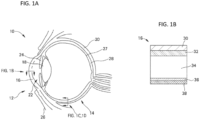
- anterior segment 12 and “eye” 10 include both the anterior segment 12 of the eye (i.e., the portion of the eye in front of the lens) and the posterior segment 14 of the eye (i.e., the portion of the eye behind the lens), as illustrated in FIG. 1A .
- the anterior segment 12 is bounded by the cornea 16 and the lens 18, while the posterior segment 14 is bounded by the sclera 20 and the lens 18.
- the anterior segment 12 is further subdivided into the anterior chamber 22, between the iris 24 and the cornea 16, and the posterior chamber 26, between the lens 18 and the iris 24.
- the exposed portion of the sclera 20 on the anterior segment 12 of the eye is protected by a clear membrane referred to as the conjunctiva (not shown).
- FIG. 1B illustrates the cornea 16, which is composed of the epithelium 30, the Bowman's layer 32, the stroma 34, the Descemet's membrane 36, and the endothelium 38.
- FIG. 1C and FIG. 1D illustrate the sclera 20 with surrounding Tenon's Capsule 40 or conjunctiva 41, suprachoroidal space 42, choroid 28, and retina 27, both without and with a fluid in the suprachoroidal space, respectively.
- microneedle refers to a conduit body having a base, a shaft, and a tip end suitable for insertion into the sclera and other ocular tissue and has dimensions suitable for minimally invasive insertion and drug formulation infusion as described herein. That is, the microneedle has a length or effective length that does not exceed about 2000 microns and a diameter that does not exceed about 600 microns. Both the "length” and “effective length” of the microneedle encompass the length of the shaft of the microneedle and the bevel height of the microneedle. The microneedle has a diameter of 28 gauge or smaller.
- a hollow microneedle has a structure that includes one or more continuous pathways from the base of the microneedle to an exit point (opening) in the shaft and/or tip portion of the microneedle distal to the base.
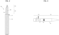
- FIGS. 2-5 illustrate exemplary embodiments of microneedle devices.
- the microneedle device 110 includes a hollow microneedle 114 having a hollow bore 140 through which a fluid drug formulation (not shown) can be delivered to the eye or through which a biological fluid can be withdrawn from the eye.
- the microneedle includes a proximal portion 116 and a tip portion 118.
- the microneedle 114 may extend from a base comprising, for example, an elongated body 112 having a distal end from which the proximal portion 116 and tip portion 118 of the microneedle extends.
- the elongated body may further comprise a means for securing 111 a base portion of the microneedle extending beyond the distal end of the base 112, such as a screw or pin.
- An exemplary embodiment of the elongated body 112 for securing the microneedle is illustrated in FIG. 3 , and comprises a cap portion 113 and a base portion 115 having a hollow bore 117 therein.
- the cap portion 113 and base portion 115 of the elongated body 112 desirably comprise a means for manually adjusting the length of needle (i.e., the proximal portion and tip portion of the microneedle extending from the base 112 ) protruding out of the cap portion of the elongated body.
- Such means may include, for example, threads 119 allowing the cap portion 113 to be screwed in and out of the base portion 115 of the elongated body.
- the base portion 115 of the elongated body may be operably connected to an actuator 120 for controlled infusion of the fluid drug formulation through the microneedle into the suprachoroidal space.
- the microneedle device may further comprise a fluid reservoir for containing the drug formulation, e.g. , as a solution or suspension, and the drug reservoir being in operable communication with the bore of the microneedle at a location distal to the tip end of the microneedle.
- the fluid reservoir may be integral with the microneedle, integral with the elongated body, or separate from both the microneedle and elongated body.
- the microneedle can be formed/constructed of different biocompatible materials, including metals, glasses, semi-conductor materials, ceramics, or polymers.
- suitable metals include pharmaceutical grade stainless steel, gold, titanium, nickel, iron, gold, tin, chromium, copper, and alloys thereof.
- the polymer can be biodegradable or non-biodegradable.
- suitable biocompatible, biodegradable polymers include polylactides, polyglycolides, polylactide-co-glycolides (PLGA), polyanhydrides, polyorthoesters, polyetheresters, polycaprolactones, polyesteramides, poly(butyric acid), poly(valeric acid), polyurethanes and copolymers and blends thereof.
- non-biodegradable polymers include various thermoplastics or other polymeric structural materials known in the fabrication of medical devices. Examples include nylons, polyesters, polycarbonates, polyacrylates, polymers of ethylene-vinyl acetates and other acyl substituted cellulose acetates, non-degradable polyurethanes, polystyrenes, polyvinyl chloride, polyvinyl fluoride, poly(vinyl imidazole), chlorosulphonate polyolefins, polyethylene oxide, blends and copolymers thereof.
- Biodegradable microneedles can provide an increased level of safety compared to non-biodegradable ones, such that they are essentially harmless even if inadvertently broken off into the ocular tissue.
- the microneedle can be fabricated by a variety of methods known in the art or as described in the Examples below.
- the hollow microneedle is fabricated using a laser or similar optical energy source.
- a microcannula may be cut using a laser to represent the desired microneedle length.
- the laser may also be use to shape single or multiple tip openings. Single or multiple cuts may be performed on a single microncannula to shape the desired microneedle structure.
- the microcannula may be made of metal such as stainless steel and cut using a laser with a wavelength in the infrared region of the light spectrum ( e.g. , from about 0.7 to about 300 ⁇ m).
- the microneedle length and optional bevel is formed by a physical grinding process, which for example may include grinding a metal cannula against a moving abrasive surface.
- the fabrication process may further include precision grinding, micro-bead jet blasting and ultrasonic cleaning to form the shape of the desired precise tip of the microneedle.
- the methods and devices provided herein allow for suprachoroidal drug delivery to be accomplished in a minimally invasive, non-surgical manner, superior to other non-surgical (e.g., conventional needle) and surgical approaches.
- the methods provided herein are carried out via the use of one or more microneedles.
- the microneedles are be inserted perpendicular, or at an angle from about 80° to about 100°, into the eye, e.g., into the sclera, reaching the suprachoroidal space in a short penetration distance.
- the microneedle in one embodiment, is part of an array of two or more microneedles such that the method further includes inserting at least a second microneedle into the sclera without penetrating across the sclera.
- the drug formulation of each of the two or more microneedles may be identical to or different from one another, in drug, formulation, volume/quantity of drug formulation, or a combination of these parameters.
- different types of drug formulations may be injected via the one or more microneedles. For example, inserting a second hollow microneedle comprising a second drug formulation into the ocular tissue will result in delivery of the second drug formulation into the ocular tissue.
- microneedle devices described herein are adapted to remove substances, such as a fluid, tissue, or molecule sample, from the eye.
- microneedles e.g., solid microneedles
- Non-limiting examples include dissolving, at least in part, a coating of a drug formulation off of a microneedle; detaching, at least in part, a coating of a drug formulation (e.g., as a substantially intact sleeve or in fragments) off of a microneedle; breaking or dissolving a microneedle off of a base to which the microneedle is integrally formed or is connected; or any combination thereof.
- the microneedle devices described herein also may be adapted to use the one or more microneedles as a sensor to detect analytes, electrical activity, and optical or other signals.
- the sensor may include sensors of pressure, temperature, chemicals, and/or electromagnetic fields (e.g., light).
- Biosensors can be located on or within the microneedle, or inside a device in communication with the body tissue via the microneedle.
- the microneedle biosensor can be any of the four classes of principal transducers: potentiometric, amperometric, optical, and physiochemical.
- a hollow microneedle is filled with a substance, such as a gel, that has a sensing functionality associated with it.
- the substrate or enzyme can be immobilized in the needle interior.
- a wave guide can be incorporated into the microneedle device to direct light to a specific location, or for detection, for example, using means such as a pH dye for color evaluation.
- heat, electricity, light, ultrasound or other energy forms may be precisely transmitted to directly stimulate, damage, or heal a specific tissue or for diagnostic purposes.
- the microneedle device for non-surgically delivering drug to the suprachoroidal space of the eye of a human subject comprises a hollow microneedle.
- the device may include an elongated housing for holding the proximal end of the microneedle.
- the device may further include a means for conducting a drug formulation through the microneedle.
- the means may be a flexible or rigid conduit in fluid connection with the base or proximal end of the microneedle.
- the means may also include a pump or other devices for creating a pressure gradient for inducing fluid flow through the device.
- the conduit may in operable connection with a source of the drug formulation.
- the source may be any suitable container. In one embodiment, the source may be in the form of a conventional syringe.
- the source may be a disposable unit dose container.
- the microneedle has an effective length of about 50 ⁇ m to about 2000 ⁇ m. In another particular embodiment, the microneedle has an effective length of from about 150 ⁇ m to about 1500 ⁇ m, or from about 300 ⁇ m to about 1250 ⁇ m, or from about 500 ⁇ m to about 1250 ⁇ m, or from about 500 ⁇ m to about 1500 ⁇ m, or from about 600 ⁇ m to about 1000 ⁇ m, or from about 700 ⁇ m to about 1000 ⁇ m. In one embodiment, the effective length of the microneedle is about 600 ⁇ m, or about 700 ⁇ m, or about 800 ⁇ m or about 1000 ⁇ m.
- the proximal portion of the microneedle has a maximum width or cross-sectional dimension of from about 50 ⁇ m to 600 ⁇ m, or from about 50 ⁇ m to about 400 ⁇ m, or from about 50 ⁇ m to about 500 ⁇ m, or from about 100 ⁇ m to about 400 ⁇ m, or from about 200 ⁇ m to about 600 ⁇ m, or from about 100 ⁇ m to about 250 ⁇ m, with an aperture diameter of about 5 ⁇ m to about 400 ⁇ m.
- the proximal portion of the microneedle has a maximum width or cross-sectional dimension of about 600 ⁇ m.
- the aperture diameter may be greater than the outer diameter of the proximal portion of the microneedle.
- the microneedle may be fabricated to have an aspect ratio (width: length) of about 1:1.5 to about 1:10. In one embodiment, the aspect ratio of the microneedle is about 1:3 to about 1:5. In another embodiment, the aspect ratio of the microneedle is about 1:4 to about 1:10.
- the microneedle can have a straight or tapered shaft.
- the diameter of the microneedle is greatest at the base end of the microneedle and tapers to a point at the end distal the base.
- the microneedle can also be fabricated to have a shaft that includes both a straight (i.e., untapered) portion and a tapered (e.g., beveled) portion.
- the microneedle has a bevel angle of about 5 degrees to about 30 degrees, of about 5 degrees to about 25 degrees, about 5 degrees to about 20 degrees, about 10 degrees to about 20 degrees, and about 10 degrees to about 30 degrees.
- the microneedles can be formed with shafts that have a circular cross-section in the perpendicular, or the cross-section can be non-circular.
- the tip portion of the microneedles can have a variety of configurations.
- the tip of the microneedle can be symmetrical or asymmetrical about the longitudinal axis of the shaft.
- the tips may be beveled, tapered, squared-off, or rounded.
- the microneedle has a bevel height from about 50 ⁇ m to 500 ⁇ m, about 100 ⁇ m to about 500 ⁇ m, about 100 ⁇ m to about 400 ⁇ m, about 200 ⁇ m to about 400 ⁇ m, and about 300 ⁇ m to about 500 ⁇ m.
- the microneedle may be designed such that the tip portion of the microneedle is substantially the only portion of the microneedle inserted into the ocular tissue (i.e., the tip portion is greater than 75% of the total length of the microneedle, greater than 85% of the total length of the microneedle, or greater than about 95% of the total length of the microneedle).
- the microneedle may be designed such that the tip portion is only a portion of the microneedle that is inserted into the ocular tissue and generally has a length that is less than about 75% of the total length of the microneedle, less than about 50% of the total length of the microneedle, or less than about 25% of the total length of the microneedle.
- the microneedle has a total effective length between 500 ⁇ m and 1500 ⁇ m, wherein the tip portion has a length that is less than about 400 ⁇ m, less than about 300 ⁇ m, or less than about 200 ⁇ m.
- the height of the bevel is about 100 ⁇ m to about 500 ⁇ m. In another embodiment, the height of the bevel is about 500 ⁇ m or less, about 450 ⁇ m or less, about 400 ⁇ m or less or about 350 ⁇ m or less. In another embodiment, the height of the bevel is from about 200 ⁇ m to about 500 ⁇ m, or from about 100 ⁇ m to about 700 ⁇ m, or from about 200 ⁇ m to about 700 ⁇ m. In still other embodiments, the height of the bevel is from about 500 ⁇ m to about 900 ⁇ m, or from about 500 ⁇ m to about 800 ⁇ m, or from about 500 ⁇ m to about 700 ⁇ m.
- the arrangement of the bevel can be such that the distal edge is sufficiently sharp such as to pierce a target tissue and penetrate into the vitreous without (i) substantially causing the target tissue to elastically deform or (ii) damaging internal structures of the eye, e.g., the lens or retina.
- the microneedle extends from a base.
- the base may be integral with or separate from the microneedle.
- the base may be rigid or flexible.
- the base may be substantially planar or it may be curved, for example, in the shape of the ocular tissue surface at the site of injection or, for example, curved away from the ocular surface (e.g., convex) so as to minimize contact between the base and the ocular tissue.
- the base is shaped to provide minimal contact with the surface of the eye at the point of insertion.
- the base may extend only a minimal distance from the microneedle shaft substantially perpendicular.
- the base may be shaped so as to elevate the ocular tissue towards the microneedle so as to counteract the deflection of the ocular tissue and facilitate insertion of the microneedle into the ocular tissue (e.g., the base may extend from the microneedle toward the tip portion of the microneedle so as to "pinch" the ocular tissue).
- Some such embodiments may be based, at least in part, on the devices described in U.S. Patent No. 6,743,211 .
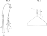
- the microneedle device has a single microneedle.
- the microneedle device 130 includes a convex base 132 and a hollow microneedle 134 which has a bore 140 through which a fluid drug formulation (not shown) can be delivered to the eye or through which a biological fluid can be withdrawn from the eye.
- the hollow microneedle 134 includes a proximal portion 136 and a tip portion 138.
- the microneedle may extend from the base of the microneedle device at any angle suitable for insertion into the eye.
- the microneedle extends from the base at an angle of about 90 degrees to provide approximately perpendicular insertion of the microneedles into the surface of the eye.
- the microneedle extends from the base at an angle from about 60 to about 110 degrees, or from about 70 degrees to about 100 degrees, or from about 80 degrees to about 90 degrees, or from about 85 degrees to about 95 degrees.
- the microneedle device may comprise a means for controllably inserting, and optionally retracting, the microneedle into the ocular tissue.
- the microneedle device may include means of controlling the angle at which the at least one microneedle is inserted into the ocular tissue (e.g. , by inserting the at least one microneedle into the surface of the ocular tissue at an angle of about 90 degrees).
- the depth of microneedle insertion into the ocular tissue can be controlled by the length of the microneedle, as well as other geometric features of the microneedle. For example, a flange or other a sudden change in microneedle width can be used to limit the depth of microneedle insertion.
- the microneedle insertion can also be controlled using a mechanical micropositioning system involving gears or other mechanical components that move the microneedle into the ocular tissue a controlled distance and, likewise, can be operated, for example, in reverse, to retract the microneedle a controlled distance.
- the depth of insertion can also be controlled by the velocity at which the microneedle is inserted into the ocular tissue.
- the retraction distance can be controlled by elastic recoil of the ocular tissue into which the microneedle is inserted or by including an elastic element within the microneedle device that pulls the microneedle back a specified distance after the force of insertion is released.
- the angle of insertion can be directed by positioning the microneedle at a first angle relative to the microneedle base and positioning the base at a second angle relative to the ocular surface.
- the first angle can be about 90° and the second angle can be about 0°.
- the angle of insertion can also be directed by having the microneedle protrude from a device housing through a channel in that housing that is oriented at a specified angle.
- microneedle insertion may be manually operable, electromechanically operable, or a combination thereof.
- the transport of drug formulation or biological fluid through a hollow microneedle can be controlled or monitored using, for example, one or more valves, pumps, sensors, actuators, and microprocessors.
- the microneedle device may include a micropump, microvalve, and positioner, with a microprocessor programmed to control a pump or valve to control the rate of delivery of a drug formulation through the microneedle and into the ocular tissue.
- the flow through a microneedle may be driven by diffusion, capillary action, a mechanical pump, electroosmosis, electrophoresis, convection or other driving forces. Devices and microneedle designs can be tailored using known pumps and other devices to utilize these drivers.
- the microneedle device may further include an iontophoretic apparatus, similar to that described in U.S. Patent 6,319,240 to Beck, for enhancing the delivery of the drug formulation to the ocular tissue.
- the microneedle devices can further include a flowmeter or other means to monitor flow through the microneedles and to coordinate use of the pumps and valves.
- the flow of drug formulation or biological fluid can be regulated using various valves or gates known in the art.
- the valve may be one which can be selectively and repeatedly opened and closed, or it may be a single-use type, such as a fracturable barrier.
- Other valves or gates used in the microneedle devices can be activated thermally, electrochemically, mechanically, or magnetically to selectively initiate, modulate, or stop the flow of material through the microneedles.
- the flow is controlled with a rate-limiting membrane acting as the valve.
- the device includes an array of two or more microneedles.
- the device may include an array of from 2 to 1000 (e.g., from 2 to 100) microneedles.
- a device includes between 1 and 10 microneedles.
- An array of microneedles may include a mixture of different microneedles.
- an array may include microneedles having various lengths, base portion diameters, tip portion shapes, spacings between microneedles, drug coatings, etc.
- the angle at which a single microneedle extends from the base may be independent from the angle at which another microneedle in the array extends from the base.
- the SCS drug delivery methods provided herein allow for the delivery of drug formulation over a larger tissue area and to more difficult to target tissue in a single administration as compared to previously known needle devices.
- the drug formulation flows circumferentially from the insertion site toward the retinochoroidal tissue, macula, and optic nerve in the posterior segment of the eye as well as anteriorly toward the uvea and ciliary body.
- a portion of the infused drug formulation may remain in the SCS as a depot, or remain in tissue overlying the SCS, for example the sclera, near the microneedle insertion site, serving as additional depot of the drug formulation that subsequently can diffuse into the SCS and into other adjacent posterior tissues.
- Devices are not recited by the appended claims.
- the microneedle devices and non-surgical methods described herein may be used to deliver drug formulations to the eye of a human subject, particularly for the treatment, diagnosis, or prevention of a posterior ocular disorder.
- the drug formulation comprises an effective amount of axitinib, or additionally an anti-inflammatory drug, an immunosuppressive agent, a VEGF modulator (e.g., a VEGF antagonist), an additional angiogenesis inhibitor (e.g., a PDGF antagonist) or a vascular permeability inhibitor.
- the formulation further comprises an anti-inflammatory drug selected from a steroid compound and a non-steroidal anti-inflammatory drug (NSAID).
- the drug formulation is further comprises a triamcinolone formulation, e.g., a triamcinolone acetonide formulation.
- the method in one embodiment, comprises non-surgically delivering a drug formulation comprising an effective amount of a choroidal malady treating drug to the suprachoroidal space of one or both eyes of the patient in need of treatment. It should be understood that a patient having one eye will undergo treatment in only one eye.
- the methods and microneedles described herein relate to the non-surgical administration of a drug formulation comprising axitinib for the treatment of a posterior ocular disorder, wherein the majority of the drug formulation is retained in the SCS in one or both eyes of a patient in need of treatment of the posterior ocular disorder, for a period of time after the non-surgical treatment method is completed.
- drug formulation retention in the SCS contributes to the sustained release profile of the drug formulations described herein.
- the human subject treated with the methods provided herein may be an adult or a child.
- a wide range of posterior ocular disorders are treatable with the methods, devices and drug formulations described herein.
- posterior ocular disorders amenable for treatment by the methods, devices and drug formulations described herein include, but are not limited to, uveitis, glaucoma, macular edema, diabetic macular edema, retinopathy, age-related macular degeneration (for example, wet AMD or dry AMD), scleritis, optic nerve degeneration, geographic atrophy, choroidal disease, ocular sarcoidosis, optic neuritis, choroidal neovascularization, ocular cancer, genetic disease(s), autoimmune diseases affecting the posterior segment of the eye, retinitis (e.g., cytomegalovirus retinitis) and corneal ulcers.
- uveitis glaucoma
- macular edema diabetic macular edema
- retinopathy for example, wet AMD or dry AMD
- scleritis optic nerve degeneration
- geographic atrophy choroidal disease
- ocular sarcoidosis optic neuriti
- the posterior ocular disorders amenable for treatment by the methods, devices, and drug formulations described herein may be acute or chronic.
- the ocular disease may be acute or chronic uveitis.
- Uveitis can be caused by infection with viruses, fungi, or parasites; the presence of noninfectious foreign substances in the eye; autoimmune diseases; or surgical or traumatic injury.
- disorders caused by pathogenic organisms that can lead to uveitis or other types of ocular inflammation include, but are not limited to, toxoplasmosis, toxocariasis, histoplasmosis, herpes simplex or herpes zoster infection, tuberculosis, syphilis, sarcoidosis, Vogt-Koyanagi-Harada syndrome, Behcet's disease, idiopathic retinal vasculitis, Vogt-Koyanagi-Harada Syndrome, acute posterior multifocal placoid pigment epitheliopathy (APMPPE), presumed ocular histoplasmosis syndrome (POHS), birdshot chroidopathy, Multiple Sclerosis, sympathetic opthalmia, punctate inner choroidopathy, pars planitis, or iridocyclitis. Acute uveitis occurs suddenly and may last for up to about six weeks. Chronic uveitis is a form of uveitis in which the
- Signs of uveitis include ciliary injection, aqueous flare, the accumulation of cells visible on ophthalmic examination, such as aqueous cells, retrolental cells, and vitreouscells, keratic precipitates, and hypema.
- Symptoms of uveitis include pain (such as ciliary spasm), redness, photophobia, increased lacrimation, and decreased vision.
- Posterior uveitis affects the posterior or choroid part of the eye. Inflammation of the choroid part of the eye is also often referred to as choroiditis.
- Posterior uveitis is may also be associated with inflammation that occurs in the retina (retinitis) or in the blood vessels in the posterior segment of the eye (vasculitis).
- the methods provided herein comprise non-surgically administering to a uveitis patient in need thereof, an effective amount of a uveitis treating drug to the SCS of the eye of the patient.
- the patient experiences a reduction in the severity of the symptoms, after administration of a uveitis treating drug to the SCS.
- the drug formulation delivered to the SCS results in the patient experiencing a reduction in inflammation, neuroprotection, complement inhibition, drusen formation, scar formation, and/or a reduction in choriocapillaris or choroidal neocasvularization.
- the non-surgical methods described herein are particularly useful for the local delivery of drugs to the posterior region of the eye, for example the retinochoroidal tissue, macula, and optic nerve in the posterior segment of the eye.
- the non-surgical treatment methods and devices described herein may be used in gene-based therapy applications.
- the method in one embodiment, comprises administering a drug formulation into the suprachoroidal space to deliver select DNA, RNA, or oligonucleotides to targeted ocular tissues. Only drug formulations comprising axitinib form part of the invention.
- choroidal maladies amenable for treatment by the methods, devices and drug formulations described herein include, but are not limited to, choroidal neovascularization, polypoidal choroidal vasculopathy, central sirrus choroidopathy, a multi-focal choroidopathy or a choroidal dystrophy (e.g. , central gyrate choroidal dystrophy, serpiginous choroidal dystrophy or total central choroidal atrophy). Choroidal maladies are described in further detail below.
- the choroidal malady treating drug which is not claimed, is an angiogenesis inhibitor, a vascular permeability inhibitor or an anti-inflammatory drug.
- the angiogenesis inhibitor in one embodiment, is a vascular endothelial growth factor (VEGF) modulator or a platelet derived growth factor (PDGF) modulator.
- the choroidal malady treatment method in one embodiment, comprises administering the drug formulation to the SCS of one or both eyes of the patient in need of treatment via a microneedle.
- the microneedle is a hollow microneedle having a tip and an opening, and the drug formulation is infused into the SCS of one or both eyes through the tip of the hollow microneedle.
- the method of treating a posterior ocular disorder in a human subject in need thereof comprises, in one embodiment, non-surgically administering a drug formulation comprising axitinib to the suprachoroidal space of the eye of the human subject, wherein upon administration, the drug formulation flows away from the insertion site and is substantially localized to the posterior segment of the eye.
- the non-surgical methods provided herein allow for longer retention of the drug in the eye, as compared to intravitreal, topical, parenteral, intracameral or oral administration of the same drug dose.
- the suprachoroidal drug dose sufficient to achieve a therapeutic response in a human subject treated with the non-surgical SCS drug delivery method is less than the intravitreal, parenteral, intracameral, topical, or oral drug dose sufficient to elicit the identical or substantially identical therapeutic response.
- the suprachoroidal drug dose is at least 10 percent less than the oral, parenteral or intravitreal dose sufficient to achieve the identical or substantially identical therapeutic response.
- the suprachoroidal dose is about 10 percent to about 25 percent less, or about 10 percent to about 50 percent less than the oral, parenteral, intracameral, topical, or intravitreal dose sufficient to achieve the identical or substantially identical therapeutic response.
- the method of treating a posterior ocular disorder or choroidal malady described herein achieves a greater therapeutic efficacy than other routes of administration.
- the non-surgical method provided herein comprises inserting a hollow microneedle into the sclera of the eye of the human subject and infusing a drug formulation through the hollow microneedle and into the suprachoroidal space of the eye.
- the drug formulation in one embodiment, is a solution or suspension of the drug.
- the non-surgical method for treating a posterior ocular disorder in a human subject comprises drug delivery to the SCS of one or both eyes of the patient via a microneedle (hollow or solid), the microneedle insertion site is between the equator and the limbus of the respective eye.
- the insertion site is between about 2 mm and about 10 mm posterior to the limbus of the eye.
- the microneedle insertion site is at the pars plana. However, in other embodiments the insertion site is outside the pars plana. In one embodiment, the insertion site of the microneedle is at about the equator of the eye.
- the insertion site is from 2 to 10 mm anterior to the limbus of the eye, for example, about 5 mm anterior to the limbus.
- the drug formulation is introduced into the SCS at the site of injection (i.e., at the tip of the microneedle) and then flows through the SCS away from the site of injection while the injection occurs.
- the site of injection i.e., at the tip of the microneedle
- the site of injection is anterior to the equator of the eye and at least a portion of the drug formulation flows posterior to the equator of the eye during the injection (i.e., while drug formulation continues to flow out of the microneedle).
- the site of injection i.e., at the tip of the microneedle
- the site of injection is anterior to the equator of the eye and at least a portion of the drug formulation flows near the macular during the injection (i.e., while drug formulation continues to flow out of the microneedle).
- the depth of insertion of the microneedle into the ocular tissue is precisely controlled.
- Various methods can be used to control the insertion depth of the microneedles described herein.
- the insertion depth is limited by the selected length or effective length of the microneedle.
- the "effective length" is that portion available for tissue insertion, i.e., the length that extends from the base and would be inserted if there were zero tissue deformation.
- the "effective length” neglects any proximal portion of the microneedle that extends into or through the base and thus cannot be inserted in the tissue, and includes both the microneedle shaft length and bevel length. That is, the microneedle may have an effective length approximately equal to the desired penetration depth.
- the microneedle is short enough that the tip of the microneedle may be inserted substantially to the base of the sclera (i.e., near the interface of the sclera and choroid) without completely penetrating across the sclera.
- the tip of the microneedle is inserted through the sclera into the suprachoroidal space without penetrating through the choroid.
- the microneedle is designed to have a length longer than the desired penetration depth, but the microneedle is controllably inserted only part way into the tissue. Partial insertion may be controlled by the mechanical properties of the tissue, which bends and dimples during the microneedle insertion process. In this way, as a microneedle is inserted into the tissue, its movement partially elastically deforms the tissue and partially penetrates into the tissue. By controlling the degree to which the tissue deforms, the depth of microneedle insertion into the tissue can be controlled.
- the microneedle is inserted into the eye of the human patient using a rotational/drilling technique and/or a vibrating action.
- the microneedle can be inserted to a desired depth by, for example, drilling the microneedles a desired number of rotations, which corresponds to a desired depth into the tissue. See, e.g., U.S. Patent Application Publication No. 2005/0137525 , for a description of drilling microneedles.
- the rotational/drilling technique and/or a vibrating action may be applied during the insertion step, retraction step, or both.
- the drug formulation is infused into the suprachoroidal space through a hollow microneedle by driving the drug formulation from a source reservoir into the ocular tissue using a pressure gradient (e.g., pumping, syringe).
- a pressure gradient e.g., pumping, syringe
- the drug formulation is driven from a source reservoir into the ocular tissue using an electric field (e.g., iontophoresis) or another externally applied energy (e.g., ultrasound/acoustic energy).
- an electric field e.g., iontophoresis
- another externally applied energy e.g., ultrasound/acoustic energy
- the amount of drug formulation infused into the suprachoroidal space from the non-surgical drug delivery methods described herein is from about 10 ⁇ L to about 200 ⁇ L, e.g., from about 50 ⁇ L to about 150 ⁇ L. In another embodiment, from about 10 ⁇ L to about 500 ⁇ L, e.g., from about 50 ⁇ L to about 250 ⁇ L, is non-surgically administered to the suprachoroidal space.
- the non-surgical method comprises inserting a hollow microneedle into the sclera at an insertion site, the microneedle having a tip end with an opening, and infusing the drug formulation through a hollow microneedle and into the suprachoroidal space.
- from about 10 ⁇ L to about 200 ⁇ L, or from about 50 ⁇ L to about 150 ⁇ L or from about 10 ⁇ L to about 500 ⁇ L or from about 50 ⁇ L to about 250 ⁇ L can be delivered via one or more hollow microneedles described herein.
- the driving force or pressure infusing the drug formulation through the hollow microneedle causes the infused drug formulation to flow within the suprachoroidal space and reach the back of the eye during the administration (i.e., during the infusion) process. This may occur in less than one or two minutes, such as from about 1 second to about 100 seconds, e.g., from about 10 seconds to about 30 seconds.
- the drug formulation flows away from the insertion site during and after infusing the drug into the SCS.
- the drug flows circumferentially within the suprachoroidal space during the infusion process to a site that is at least 2.5 mm away from the insertion site, or to a site that is at least 5 mm away from the insertion site, or to a site that is at least 7.5 mm away from the insertion site, or to a site that is at least 10 mm away from the insertion site.
- the drug formulation flows circumferentially within the suprachoroidal space from the insertion site toward the back (posterior segment) of the eye (i.e., the retinochoroidal tissue, macula, and optic nerve in the posterior segment of the eye).
- the amount of drug delivered within the SCS also may be controlled, in part, by the type of microneedle used and how it is used.
- a hollow microneedle is inserted into the ocular tissue and progressively retracted from the ocular tissue after insertion to deliver a fluid drug, where after achieving a certain dosage, the delivery could be stopped by deactivating the fluid driving force, such as pressure (e.g., from a mechanical device such as a syringe) or an electric field, to avoid leakage/uncontrolled deliver of drug.
- the amount of drug being delivered is controlled by driving the fluid drug formulation at a suitable infusion pressure.
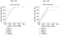
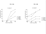
- the infusion pressure may be at least 150 kPa, at least 250 kPa, or at least 300 kPa. In another embodiment, the infusion pressure is about 150 kPa to about 300 kPa. Suitable infusion pressures may vary with the particular patient or species.
- the desired infusion pressure to deliver a suitable amount of drug formulation might be influenced by the depth of insertion of the microneedle and the composition of the drug formulation.
- a greater infusion pressure may be required in embodiments wherein the drug formulation for delivery into the eye is in the form of or includes nanoparticles or microparticles encapsulating the active agent or microbubbles. Nanoparticle or microparticle encapsulation techniques are well known in the art.
- the drug formulation is comprised of drug particles in suspension with a D 99 of 10 ⁇ m or less. In one embodiment, the drug formulation is comprised of drug particles in suspension with a D 99 of 7 ⁇ m or less.
- the drug formulation is comprised of drug particles in suspension with aD 99 of 3 ⁇ m or less. In another embodiment, the drug formulation is comprised of drug particles in suspension with a D 50 of 5 ⁇ m or less. In one embodiment, the drug formulation is comprised of drug particles in suspension with a D 50 1 ⁇ m or less.
- the non-surgical method of administering a drug to the SCS further includes partially retracting the hollow microneedle after insertion of the microneedle into the eye, and before and/or during the infusion of the drug formulation into the suprachoroidal space.
- the partial retraction of the microneedle occurs prior to the step of infusing the drug formulation into the ocular tissue. This insertion/retraction step may form a pocket and beneficially permits the drug formulation to flow out of the microneedle unimpeded or less impeded by ocular tissue at the opening at the tip portion of the microneedle.
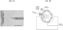
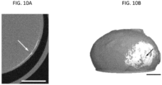
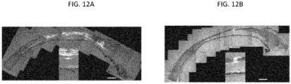
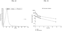
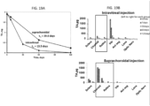
- FIG. 6A shows a hollow microneedle 130 inserted into the sclera 20, with drug formulation 131 temporarily positioned in the hollow bore of the microneedle. (The fluid communication to a reservoir of the drug formulation is not shown.)
- FIG. 6B shows the microneedle 130 following partial retraction and infusion of the drug formulation 131 into the suprachoroidal space. Arrows show the circumferential flow of the drug formulation through the suprachoroidal space.
- the microneedle infuses a drug formulation through the sclera into the suprachoroidal space for controlled (i.e., sustained, extended, or modulated over time) release of a drug to one or more ocular or neighboring tissues.
- controlled release or “sustained release” or “extended release” or “modulated release” is generally more prolonged than that obtainable by topical application or intravitreal injection of the drug formulation to the ocular tissue.
- This delivery method can be particularly advantageous with ocular tissues, where it is desirable for the insertion and withdrawal process to occur over as short a period as possible to minimize patient discomfort-in contrast to transdermal microneedle patch applications, where patches may more likely be worn (with microneedles inserted) over an extended period without patient discomfort.
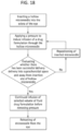
- the method of treating a posterior ocular disorder by non-surgically administering a drug to the suprachoroidal space of the eye of a human subject includes monitoring the insertion of the microneedle and/or infusion of the fluid drug formulation to ensure precise delivery of the fluid drug formulation to the SCS ( see, e.g., FIG. 18 ).
- Such monitoring may be achieved using imaged-guided feedback methods during one or more of these steps, non-limiting examples of which include conventional microscopy, MRI, x-ray, confocal microscopy, ocular coherence tomography (e.g., anterior segment optical coherence tomography, Heidelberg retina tomography, spectral domain optical coherence tomography), fluorescein angiography, indocyanine green angiography, high resolution stereoscopic fundus photography, autofluorescence imaging, ultra-wide field imaging, and various ultrasound techniques.
- the method may further comprise determining whether an initial infusion of the fluid drug formulation has flowed into the suprachoroidal space of the eye and away from the insertion site.
- a desired volume of the fluid drug formulation can be infused and the infusion discontinued by removing the fluid driving force, such as pressure, and retracting the microneedle from the eye. If, however, it is determined that the initial infusion of the fluid drug formulation has been unsuccessful (i.e., substantially none of the drug formulation has flowed into the suprachoroidal space of the eye and away from the insertion site), then the microneedle may be repositioned and the process repeated until a successful delivery is achieved.
- Targeting a drug formulation to the SCS and the posterior ocular tissues allows for high concentrations of the drug to be delivered to the choroid/sclera and the retina, with little to no drug being delivered to the aqueous humor of the anterior chamber. Additionally, the methods provided herein allow for greater drug retention in the eye compared to other drug delivery methods, for example, a greater amount of drug is retained in the eye when delivered via the methods provided herein as compared to the same dose delivered via intracameral, intravitreal, topical, parenteral or oral drug delivery methods.
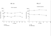
- the intraocular elimination half life (t 1/2 ) of the drug when delivered via the methods described herein is greater than the intraocular t 1/2 of the drug when the same drug dose is administered intravitreally, intracamerally, topically, parenterally or orally.
- the intraocular C max of the drug, when delivered via the methods described herein is greater than the intraocular C max of the drug when the same drug dose is administered intravitreally, intracamerally, topically, parenterally or orally.
- the mean intraocular area under the curve (AUC 0-t ) of the drug, when administered to the SCS via the methods described herein, is greater than the intraocular AUC 0-t of the drug, when administered intravitreally, intracamerally, topically, parenterally or orally.
- the intraocular time to peak concentration (t max ) of the drug, when administered to the SCS via the methods described herein is greater than the intraocular t max of the drug, when the same drug dose is administered intravitreally, intracamerally, topically, parenterally or orally.
- the drug is axitinib.
- the intraocular t 1/2 of the drug when administered via the non-surgical SCS drug delivery methods provided herein is longer than the intraocular t 1/2 of the drug when the identical dose is administered topically, intracamerally, intravitreally, orally or parenterally.
- the intraocular t 1/2 of the drug when administered via the non-surgical SCS drug delivery methods provided herein is from about 1.1 times to about 10 times longer, or from about 1.25 times to about 10 times longer, or from about 1.5 times to about 10 times longer, or about 2 times to about 5 times longer, than the intraocular t 1/2 of the drug when the identical dosage is administered topically, intracamerally, intravitreally, orally or parenterally.
- the drug is an angiogenesis inhibitor, an anti-inflammatory drug (e.g., a steroid or NSAID), a VEGF modulator (e.g., a VEGF antagonist), a PDGF modulator (e.g., a PDGF antagonist), an immunosuppressive agent, or a vascular permeability inhibitor.
- an anti-inflammatory drug e.g., a steroid or NSAID
- a VEGF modulator e.g., a VEGF antagonist
- a PDGF modulator e.g., a PDGF antagonist
- an immunosuppressive agent e.g., an immunosuppressive agent, or a vascular permeability inhibitor.
- the intraocular C max of the drug when delivered via the methods described herein, is greater than the intraocular C max of the drug when the same drug dose is administered intravitreally, intracamerally, topically, parenterally or orally.
- the intraocular C max of the drug when administered via the non-surgical SCS drug delivery methods provided herein is at least 1.1 times greater, or at least 1.25 times greater, or at least 1.5 times greater, or at least 2 times greater, or at least 5 times greater, than the intraocular C max of the drug when the identical dose is administered topically, intracamerally, intravitreally, orally or parenterally.
- the intraocular C max of the drug when administered via the non-surgical SCS drug delivery methods provided herein is about 1 to about 2 times greater, or about 1.25 to about 2 times greater, or about 1 to about 5 times greater, or about 1 to about 10 times greater, or about 2 to about 5 times greater, or about 2 to about 10 times greater, than the intraocular C max of the drug when the identical dose is administered topically, intracamerally, intravitreally, orally or parenterally.
- the drug is axitinib.
- the mean intraocular area under the curve (AUC 0-t ) of the drug, when administered to the SCS via the methods described herein, is greater than the intraocular AUC 0-t of the drug, when administered intravitreally, intracamerally, topically, parenterally or orally.
- the intraocular AUC 0-t of the drug when administered via the non-surgical SCS drug delivery methods provided herein is at least 1.1 times greater, or at least 1.25 times greater, or at least 1.5 times greater, or at least 2 times greater, or at least 5 times greater, than the intraocular AUC 0-t of the drug when the identical dose is administered topically, intracamerally, intravitreally, orally or parenterally.
- the intraocular AUC 0-t of the drug when administered via the non-surgical SCS drug delivery methods provided herein is about 1 to about 2 times greater, or about 1.25 to about 2 times greater, or about 1 to about 5 times greater, or about 1 to about 10 times greater, or about 2 to about 5 times greater, or about 2 to about 10 times greater, than the intraocular AUC 0-t of the drug when the identical dose is administered topically, intracamerally, intravitreally, orally or parenterally.
- the drug is axitinib.
- the drug formulation comprising axitinib once delivered to the SCS, is substantially retained in the SCS over a period of time.
- about 80% of the drug formulation is retained in the SCS for about 30 minutes, or about 1 hour, or about 4 hours or about 24 hours or about 48 hours or about 72 hours.
- a depot of drug is formed in the SCS and/or surrounding tissue, to allow for sustained release of the drug over a period of time.
- the suprachoroidal space once loaded with drug (e.g., drug microparticles or nanoparticles), provides a sustained release of drug to the retina or other posterior ocular tissues over a period of time.
- drug e.g., drug microparticles or nanoparticles
- the targeting of the drug to the posterior ocular tissues via the methods described herein allows for a greater therapeutic efficacy in the treatment of one or more posterior ocular disorders or choroidal maladies (e.g., PCV), as compared to other administration methods of the same drug dose, such as intravitreal, intracameral, oral, parenteral and topical delivery of the same drug dose.
- the therapeutic effect of the drug delivered to the SCS is achieved with a lower dose than the intravitreal, intracameral, topical, parenteral or oral dose sufficient to achieve the same therapeutic effect in the human subject.
- the lower doses achievable with the methods provided herein result in reduced number of side effects of the drug, and/or reduced severity of one or more side effect(s), compared to higher doses of the drug, or the same drug dose delivered to the human patient via non-suprachoroidal routes of administration (e.g., intravitreal, intracameral, topical, parenteral, oral).
- the methods provided herein provide a reduced number of side effects, or reduced severity of one or more side effects, or clinical manifestations, as compared to oral, topical, intracameral, parenteral or intravitreal administration of the same drug at the same dose.
- the side effect or clinical manifestation that is lessened in the treated patient is subretinal exudation and/or subretinal bleeding.
- the non-surgical suprachoroidal drug delivery methods provided herein result in an increased therapeutic efficacy and/or improved therapeutic response, as compared to oral, parenteral and/or intravitreal drug delivery methods of the identical or similar drug dose.
- the SCS drug dose sufficient to provide a therapeutic response is about 90%, or about 75%, or about one-half ( e.g. , about one half or less) the intravitreal, intracameral, topical, oral or parenteral drug dose sufficient to provide the same or substantially the same therapeutic response.
- the SCS dose sufficient to provide a therapeutic response is about one-fourth the intravitreal, intracameral, topical, oral or parenteral drug dose sufficient to provide the same or substantially the same therapeutic response.
- the SCS dose sufficient to provide a therapeutic response is one-tenth the intravitreal, intracameral, topical, oral or parenteral drug dose sufficient to provide the same or substantially the same therapeutic response.
- the therapeutic response is a decrease in inflammation, as measured by methods known to those of skill in the art.
- the therapeutic response is a decrease in number of ocular lesions, or decrease in ocular lesion size.
- the total amount of the effective amount of the drug in the drug formulation is about 0.05 mg to about 5 mg. In one embodiment, the total amount of the drug in the drug formulation is about 0.2 mg to about 4 mg. In another embodiment, the total amount of the drug in the drug formulation is about 1 mg to about 4 mg.
- Drug doses can be varied according to methods known to those of skill in the art and will vary, for example, based on patient age and clinical manifestation of the posterior ocular disorder.
- the therapeutic efficacy of the drug formulations delivered by the methods described herein and therapeutic response of the human subject can be assayed by standard means in the art, as known to those of skill in the art.
- the therapeutic efficacy of any particular drug can be assessed by measuring the response of the human subject after administration of the drug; a drug with a high therapeutic efficacy will show a greater amelioration and/or discontinuation of symptoms than a drug with a lower therapeutic efficacy.
- the efficacy of the drug formulations comprising axitinib provided herein can be measured, for example, by observing changes in pain intensity, changes in ocular lesions (size or number), intraocular pressure, inflammation (e.g., by measuring changes in the hackett/McDonald ocular score), ocular hypertension, and/or visual acuity.
- the efficacy of the drug i.e., axitinib
- the efficacy of the drug can be measured, for example, by observing changes in lesion growth and/or number.
- the efficacy of the drug e.g., an angiogenesis inhibitor, an anti-inflammatory drug (e.g., a steroid or NSAID), a VEGF modulator (e.g., a VEGF antagonist), a PDGF modulator (e.g., a PDGF antagonist), an immunosuppressive agent or a vascular permeability inhibitor
- an angiogenesis inhibitor e.g., an anti-inflammatory drug
- a VEGF modulator e.g., a VEGF antagonist
- a PDGF modulator e.g., a PDGF antagonist
- an immunosuppressive agent or a vascular permeability inhibitor can be measured, for example, by observing changes in the measurements according to the HackettIMcDonald ocular scores, inflammation
- the efficacy of the drug for example triamcinolone or mycophenolate
- the efficacy of the drug can be measured, for example, by observing changes in the measurements according to the HackettIMcDonald ocular scores, inflammation, visual acuity, and/or edema.
- the therapeutic efficacy of the drug for example sorafenib
- the efficacy of the drug for example infliximab (Remicade ® ), can be measured, for example, by observing changes in retinal thickness, inflammation, visual acuity, photophobia, typical time between flares, corneal ulceration, and/or edema.
- the efficacy of the drug for example nepafenac, can be measured, for example, by observing changes in optical coherence tomography (OCT) measurements of retinal thickness and volume, inflammation, visual acuity, pain and/or intraocular pressure.
- OCT optical coherence tomography
- the efficacy of the drug for example azathiopine
- the efficacy of the drug can be measured, for example, by observing changes in visual acuity, macular edema, intraocular pressure, inflammation, and/or measurements on the SF-36 Physical Component Score.
- the efficacy of the drug for example an anti-inflammatory drug such as an TNF family antagonist, for example, TNF- ⁇ antagonist, lymphotoxin- ⁇ antagonist, lymphotoxin- ⁇ antagonist, CD27L antagonist, CD20L antagonist, FASL antagonist, 4-BBL antagonist, OX40L antagonist, TNF-related apoptosis inducing ligand (TRAIL) antagonist, a Janus kinase (JAK) antagonist, or interleukin antagonist, can be measured, for example, by observing changes in inflammation, lesions, cell death, and/or visual acuity.
- TNF family antagonist for example, TNF- ⁇ antagonist, lymphotoxin- ⁇ antagonist, lymphotoxin- ⁇ antagonist, CD27L antagonist, CD20L antagonist, FASL antagonist, 4-BBL antagonist, OX40L antagonist, TNF-related apoptosis inducing ligand (TRAIL) antagonist, a Janus kinase (JAK) antagonist, or interleukin antagonist
- TNF family antagonist
- the therapeutic efficacy of cyclophosphamide can be measured, for example, by observing changes in lesion size and/or number, lesion growth, visual acuity, macular edema, intraocular pressure and/or inflammation.
- the non-surgical administration of an effective amount of the drug formulation according to the invention to the SCS results in a decreased number of deleterious side effects or clinical manifestations as compared to the number of side effects or clinical manifestations caused by the same drug dose administered intravitreally, intracamerally, orally or parenterally.
- the non-surgical administration of an effective amount of the drug formulation according to the invention to the SCS results in a decreased number of one or more deleterious side effects or clinical manifestations, as compared to the deleterious side effects or clinical manifestations caused by the same drug dose administered intravitreally, intracamerally, orally or parenterally.
- side effects and clinical manifestations include, but are not limited to, inflammation, gastrointestinal side effects (e.g., diarrhea, nausea, gastroenteritis, vomiting, gastrointestinal, rectal, and duodenal hemorrhage, hemorrhagic pancreatitis, large intestine perforation black or bloody stools, and/or coughing up blood); hematologic side effects (e.g., leucopenia, anemia, pancytopenia and agranulocytosis, thrombocytopenia, neutropenia, pure red cell aplasia (PRCA), deep venous thrombosis easy bruising, and /or unusual bleeding from the nose, mouth, vagina, or rectum); immunologic side effects/clinical manifestations (e.g., immunosuppression, immunosuppression resulting in sepsis, opportunistic infections (herpes simplex virus herpes zoster, and invasive candidal infections), and/or increased infection
- gastrointestinal side effects e
- renal side effects/clinical manifestations e.g. dysuria, urgency, urinary tract infections, hematuria, kidney tubular necrosis, and/or BK virus-associated nephropathy
- metabolic side effects/clinical manifestations e.g. edema, hyperphosphatemia, hypokalemia, hyperglycemia, hyperkalemia. swelling, rapid weight gain, and/or enlarged thyroid
- respiratory side effects/clinical manifestations e.g. respiratory infection, dyspnea, increased cough, primary tuberculosis dry cough, wheezing, and/or stuffy nose
- dermatologic side effects/clinical manifestations e.g.
- myopathy and/or muscle pain e.g. myopathy and/or muscle pain
- hepatic side effects/clinical manifestations e.g. hepatoxicity and/or jaundice
- abdominal pain e.g. myopathy and/or muscle pain
- hepatic side effects/clinical manifestations e.g. he
- the reduction or amelioration of the side effect or clinical manifestation is a reduction or amelioration, as compared to the severity of the side effect or clinical manifestation prior to administration of the drug formulation to the SCS of the eye of the patient, or a reduction or amelioration of the side effect or clinical manifestation in the patient, as compared to the reduction or amelioration experienced upon intravitreal, intracameral, parenteral or oral administration of the same drug.
- the non-surgical administration of an effective amount of a drug formulation to the SCS results in a decreased number of choroidal malady symptoms, as compared to the number of symptoms experienced by the patient prior to administration of the drug to the SCS, or compared to the number of symptoms experienced by the patient after treatment with the same drug dose administered intravitreally, intracamerally, orally or parenterally.
- the non-surgical administration of the drug formulation comprising an effective amount of a choroidal malady treating drug to the SCS of one or both eyes of the patient results in a decreased number of deleterious side effects or deleterious clinical manifestations, as compared to the number of deleterious side effects or clinical manifestations caused by the same drug dose administered intravitreally, intracamerally, orally, topically or parenterally.
- the non-surgical administration of an effective amount of a drug formulation to the SCS results in a decreased severity of a side effect or clinical manifestation in a patient suffering from a choroidal malady.
- the severity of the side effect or clinical manifestation is decreased, compared to the severity of the deleterious side effect or clinical manifestation caused by the same drug dose administered intravitreally, intracamerally, orally, topically or parenterally.
- subretinal exudation is reduced and/or subretinal bleeding is reduced in a patient suffering from a choroidal malady, upon administration of the drug formulation to the SCS of the patient's eye, as compared to the subretinal exudation and/or subretinal bleeding, prior to administration of the drug to the SCS.
- the subretinal exudation is reduced and/or subretinal bleeding is reduced in the patient, compared to the reduction in severity experienced when the same drug is administered intravitreally, intracamerally, orally, topically or parenterally.
- the concentration of the drug can be achieved at greater than 10 ⁇ g/g posterior ocular tissue for 28 days or longer after a single dose. In another embodiment, the concentration of the drug can be achieved at greater than 100 ⁇ g/g posterior ocular tissue for 28 days or longer after a single dose. In another embodiment, the concentration of the drug can be achieved at greater than 1000 ⁇ g/g tissue for longer than 28 days after a single dose. It has been found that more hydrophobic drugs clear slowly from the SCS compared to more water soluble drugs. In one embodiment, the drug formulation administered to the SCS comprises a more hydrophobic drug.
- a method for treating a patient for a choroidal malady comprises non-surgically administering a drug formulation comprising an effective amount of a choroidal malady treating drug (e.g., an angiogenesis inhibitor such as a VEGF modulator) to the SCS of one or both eyes of the patient in need of treatment, wherein upon administration, the drug formulation is substantially localized to the posterior segment of the eye.
- a choroidal malady treating drug e.g., an angiogenesis inhibitor such as a VEGF modulator
- the drug formulation is substantially localized to the posterior segment of the eye.
- the drug formulation is substantially localized to the RPE.
- the drug is substantially localized to the macula or the subretinal space.
- One or more of the microneedles described herein, in one exemplary embodiment, are used to carry out the method.
- the method of treating a choroidal malady in a human subject in need thereof comprises, in one exemplary embodiment, non-surgically administering a drug formulation comprising an effective amount of a choroidal malady treating drug to the suprachoroidal space of one or both eyes of the human subject.
- the effective amount of the choroidal malady treating drug comprises an effective amount of an anti-inflammatory drug, a vascular endothelial growth factor (VEGF) modulator, a platelet derived growth factor (PDGF) modulator, an angiogenesis inhibitor, an immunosuppressive agent or a vascular permeability inhibitor.
- VEGF vascular endothelial growth factor
- PDGF platelet derived growth factor
- the choroidal malady treating drug formulation flows away from the insertion site and is substantially localized to the posterior segment of the eye.
- the non-surgical methods provided herein allow for longer retention of the drug in the eye, as compared to intravitreal, topical, parenteral or oral administration of the same drug dose.
- the intraocular pressure (IOP) of the patient's eye undergoing treatment for the posterior ocular disorder is substantially the same IOP, compared to the IOP of the patient's eye prior to administration of the posterior ocular disorder treating drug.
- the IOP of the patient's eye undergoing treatment for the posterior ocular disorder varies by no more than 10%, compared to the IOP of the patient's eye prior to administration of the posterior ocular disorder treating drug. In one embodiment, the IOP of the patient's eye undergoing treatment for the posterior ocular disorder y, 2 minutes, 10 minutes, 15 minutes or 30 minutes after suprachoroidal drug administration, varies by no more than 20%, compared to the IOP of the patient's eye prior to administration of the posterior ocular disorder treating drug.
- the IOP of the patient's eye undergoing treatment for the posterior ocular disorder varies by no more than 10%-30%, compared to the IOP of the patient's eye prior to administration of the posterior ocular disorder treating drug.
- the posterior ocular disorder treating drug comprises an effective amount of axitinib.
- the choroidal malady amenable for treatment with the methods described herein in one exemplary embodiment, is a choroidal neovascularization, choroidal sclerosis, polypoidal choroidal vasculopathy, central sirrus choroidopathy, a multi-focal choroidopathy or a choroidal dystrophy.
- the choroidal dystrophy for example, is central gyrate choroidal dystrophy, serpiginous choroidal dystrophy or total central choroidal atrophy.
- the patient in need of treatment of the choroidal malady experiences subretinal exudation and bleeding, and the methods provided herein lessen the subretinal exudation and/or bleeding, compared to the subretinal exudation and/or bleeding experienced by the patient prior to administration of the drug formulation to the SCS.
- the patient in need of treatment experiences subretinal exudation and bleeding, and the subretinal exudation and bleeding experienced by the patient after undergoing one of the non-surgical treatment methods provided herein is less than the subretinal exudation and bleeding experienced by the patient after intravitreal therapy with the same drug at the same dose.
- the methods provided herein provide for effective treatment of a patient who had previously undergone posterior ocular disorder treatment, but was unresponsive, or not properly responsive to the prior treatment.
- a patient undergoing a posterior ocular disorder treatment method of the present invention was previously treated for the same posterior ocular disorder, but was unresponsive or not properly responsive.
- a patient unresponsive or not properly responsive to treatment does not exhibit an improvement in a symptom or improvement in a clinical manifestation of the posterior ocular disorder.
- the symptom or clinical manifestation is lesion size, inflammation, edema, visual acuity or vitreous haze.
- a patient in need of treatment of polypoidal choroidal vasculopathy is treated with one of the non-surgical SCS drug delivery methods provided herein.
- a patient in need of treatment is administered a drug formulation comprising an effective amount of a PCV treating drug to the SCS of one or both eyes.
- drug formulation administration is carried out with a microneedle device described herein.
- the effective amount of the PCV treating drug comprises an effective amount of an anti-inflammatory drug, a vascular endothelial growth factor (VEGF) modulator, a platelet derived growth factor (PDGF) modulator, an angiogenesis inhibitor, an immunosuppressive agent or a vascular permeability inhibitor.
- VEGF vascular endothelial growth factor
- PDGF platelet derived growth factor
- PCV is an abnormal choroidal vasculopathy that is believed to be a variant of type 1 neovascularization, although it has been proposed that PCV is a distinct vascular abnormality of choroidal vessels ( Imamura et al. (2010). Survey of Ophthalmology, volume 55, pp. 501-515 ). PCV has been reported to occur at a higher frequency in pigmented races, although it has also been reported to be present in Caucasian patients. ( Imamura et al. (2010). Survey of Ophthalmology, volume 55, pp. 501-515 ). The methods described herein are employable in patients of both pigmented and non-pigmented patients.
- the patient receiving treatment for PCV is of African, Hispanic, Middle Eastern or Asian descent. In another embodiment, the patient receiving treatment is Caucasian.
- Clinical manifestations of patients with PCV include vascular abnormalities and variably sized serous, serosanguineous detachments of the neurosensory retina and pigment epithelium around the optic nerve or in the central macula. Subretinal exudation and/or bleeding can also be experienced by patients with PCV.
- the PCV patient has lipid depositions in the eye.
- the present invention provides for a reduction in occurrence and/or severity of a PCV clinical manifestation experienced by the PCV patient treated with the methods described herein, compared to the occurrence and/or severity of the clinical manifestation prior to treatment.
- a patient receiving treatment for PCV with one of the non-surgical treatment methods provided herein experiences a reduction in the occurrence and/or severity of a vascular abnormality, as compared to the occurrence and/or severity of the vascular abnormality manifested prior to undergoing treatment with the non-surgical SCS drug delivery method.
- the severity of subretinal exudation and/or bleeding is reduced in the PCV patient, compared to the severity of the subretinal exudation and/or bleeding prior to undergoing treatment with one of the non-surgical SCS drug delivery methods described herein.
- PCV treating drugs e.g., angiogenesis inhibitors, VEGF modulators, PDGF modulators, anti-inflammatory drugs, vascular permeability inhibitors, are described in more detail below.
- the patient being treated for PCV with one of the non-surgical methods described herein is also treated for a second ocular disease.
- the additional ocular disease is drusen, sickle cell retinopathy, central serous chorioretinopathy, typical neovascular (type 1 or 2) age related macular degeneration, melanocytoma of the optic nerve, circumscribed choroidal hemangioma, the tilted disk syndrome, pathological myopia, choroidal osteoma, retinal microangiopathy.
- the treatment of the second ocular disease can be performed with the non-surgical SCS drug delivery methods described herein, or other methods known in the art, for example, intravitreal or topical drug administration.
- the method for treating a choroidal malady described herein is a method for treating a patient for central serous chorioretinopathy (CSC) (also known as central serous retinopathy (CSR)).
- CSC central serous chorioretinopathy
- CSR is an exudative chorioreditopathy, and is characterized by an exudative neurosensory retinal detachment with or without an associated detachment of the retinal pigment epithelium (RPE).
- RPE retinal pigment epithelium
- CSR results in metamorphopsia and micropsia.
- CSR is characterized by leakage of fluid under the retina.
- a method for treating a patient for CSR comprising non-surgically administering a drug formulation comprising an effective amount of a CSR treating drug to the SCS of one or both eyes of the patient.
- the drug is administered, in one embodiment, with one of the microneedles described herein.
- the CSR treating drug is an anti-inflammatory drug, a vascular endothelial growth factor (VEGF) modulator (e.g., a VEGF antagonist), a platelet derived growth factor (PDGF) modulator (e.g., a PDGF antagonist), an angiogenesis inhibitor, an immunosuppressive agent or a vascular permeability inhibitor.
- VEGF vascular endothelial growth factor
- PDGF platelet derived growth factor
- the patient treated for CSR by one of the methods described herein experiences an increase in visual acuity, compared to the patient's visual acuity prior to undergoing the treatment.
- the patient, after undergoing treatment for CSR experiences a decrease in leakage of fluid under the retina, compared to the leakage of fluid under the retina experienced by the patient prior to undergoing treatment via non-surgical SCS drug delivery.
- a method for treating a patient for multi-focal choroiditis comprises non-surgically administering a drug formulation comprising an effective amount of a MFC treating drug to the SCS of one or both eyes of the patient in need of MFC treatment.
- the drug formulation is administered, in one embodiment, with one of the microneedles described herein.
- the effective amount of the MFC treating drug comprises an effective amount of an anti-inflammatory drug, a vascular endothelial growth factor (VEGF) modulator (e.g., a VEGF antagonist), a platelet derived growth factor (PDGF) modulator (e.g., a PDGF antagonist), an angiogenesis inhibitor, an immunosuppressive agent or a vascular permeability inhibitor.
- VEGF vascular endothelial growth factor
- PDGF platelet derived growth factor
- angiogenesis inhibitor e.g., a vascular permeability inhibitor.
- the MFC patient in one embodiment, is moderately myopic, and in one embodiment, is affected bilaterally.
- the MFC patient in some embodiments, presents with symptoms of posterior uveitis including decreased visual acuity, floaters, photopsias, as well as with anterior segment symptoms such as photophobia.
- the MFC patient presents with vitreous cell and/or anterior chamber cell.
- Funduscopic findings in MFC patients consist of yellow to gray lesions at the level of the retinal pigment epithelium (RPE) and choriocapillaris.
- the lesions range in size from about 50 ⁇ m to about 1,000 ⁇ m and have a distribution in the peripapillary region.
- the methods provided herein reduce the lesion size and/or number in the patient receiving treatment.
- Active lesions in some instances are associated with subretinal fluid and fluffy borders.
- Active disease may be also associated with optic nerve head hyperemia and edema, cystoid macular edema and macular and peripapillary choroidal neovascularization.
- the method for treating a patient for MFC comprises non-surgically administering a drug formulation comprising an effective amount of a MFC treating drug to the SCS of one or both eyes of the patient in need of treatment.
- the method comprises administration of the drug formulation to the SCS of one or both eyes of the patient with one of the microneedles described herein.
- a drug formulation is delivered in one embodiment to the SCS of an eye of the patient via a hollow microneedle with a tip and an opening, through the opening and into the SCS.
- the effective amount of the MFC treating drug is an effective amount of an anti-inflammatory drug, angiogenesis inhibitor, VEGF modulator or vascular permeability inhibitor.
- the patient treated for MFC experiences a decreased number of lesion(s) (e.g., decreased number of lesion(s) in the RPE), decreased size of lesion(s) (e.g., decreased size of lesion(s) present in the RPE), decreased amount of subretinal fluid, an increase in visual acuity, or an attenuation in choroidal neovascularization, compared to the number of lesion(s), the size of lesion(s), amount of subretinal fluid, visual acuity and choroidal neovascularization in the eye of the patient, prior to undergoing the MFC treatment method of the invention.
- a decreased number of lesion(s) e.g., decreased number of lesion(s) in the RPE
- decreased size of lesion(s) e.g., decreased size of lesion(s) present in the RPE
- decreased amount of subretinal fluid e.g., an increase in visual acuity, or an attenuation in chor
- the patient treated for MFC experiences an attenuation of optic nerve head hyperemia and edema, compared to the optic nerve head hyperemia and edema experienced by the patient prior to undergoing treatment via the non-surgical SCS drug delivery method of the invention.
- a patient in need of treatment of choroidal neovascularization is treated with one of the non-surgical SCS drug delivery methods provided herein.
- a patient in need of treatment is administered a drug formulation comprising an effective amount of a choroidal neovascularization treating drug to the SCS of one or both eyes.
- drug formulation administration is carried out with a microneedle device described herein.
- the effective amount of the choroidal neovascularization treating drug is an anti-inflammatory drug, angiogenesis inhibitor, VEGF modulator, ( e.g. , a VEGF antagonist), a platelet derived growth factor (PDGF) modulator (e.g., a PDGF antagonist), an angiogenesis inhibitor, an immunosuppressive agent or a vascular permeability inhibitor.
- a patient in need of treatment of choroidal dystrophy is treated with one of the non-surgical SCS drug delivery methods provided herein.
- a patient in need of treatment is administered a drug formulation comprising an effective amount of a choroidal dystrophy treating drug to the SCS of one or both eyes.
- the effective amount of the choroidal dystrophy treating drug comprises an effective amount of an anti-inflammatory drug, a vascular endothelial growth factor (VEGF) modulator, a platelet derived growth factor (PDGF) modulator, an angiogenesis inhibitor, an immunosuppressive agent or a vascular permeability inhibitor.
- VEGF vascular endothelial growth factor
- PDGF platelet derived growth factor
- drug formulation administration is carried out with a microneedle device described herein.
- the choroidal dystrophy methods provided herein improve a symptom or clinical manifestation of the choroidal dystrophy to a greater extent, compared to the identical drug administered to the patient via a topical, oral, parenteral, intravitreal or intracameral route.
- the method for treating a choroidal malady described herein is a method for treating a patient for punctuate inner choroidopathy (PIC).
- the method for treating a patient for PIC comprises non-surgically administering a drug formulation comprising an effective amount of a PIC treating drug to the SCS of one or two eyes of the patient.
- the method comprises drug delivery with one of the microneedles described herein.
- the effective amount of the PIC treating drug is an anti-inflammatory drug, angiogenesis inhibitor, immunosuppressive agent, VEGF modulator (e.g., a VEGF antagonist), a PDGF modulator (e.g., a PDGF antagonist) or vascular permeability inhibitor.
- VEGF modulator e.g., a VEGF antagonist
- PDGF modulator e.g., a PDGF antagonist
- vascular permeability inhibitor vascular permeability inhibitor.
- a PIC patient undergoing one of the SCS treatment methods provided herein experiences an improved PIC symptom/clinical manifestation, or a decreased number of PIC symptoms/clinical manifestations, compared to the symptoms/clinical manifestations experienced prior to SCS drug administration.
- the symptom/clinical manifestation is inflammation, blurred vision, photopsia, central and/or peripheral scotomatas or metamorphopsias.
- the symptom/clinical manifestation is decreased visual acuity, bilateral white-yellow chorioretinal lesion(s) (e.g., from about 100 ⁇ m to about 200 ⁇ m in diameter) at the level of the inner choroid and retinal pigment epithelium.
- the lesion(s) typically is not associated with vitritis and typically does not extend to the midperiphery. PIC lesions progress to atrophic scars and leave a halo of depigmentation.
- the PIC patient treated with one of the non-surgical SCS drug delivery methods described herein experiences decreased inflammation, a decreased number of lesion(s), or decreased size of lesion(s), as compared to the inflammation, number of lesion(s), or size of lesion(s) manifested by the patient prior to therapy.
- PIC patient treated with one of the non-surgical SCS drug delivery methods described herein experiences decreased inflammation, a decreased number of lesion(s), or decreased size of lesion(s), as compared to the inflammation, number of lesion(s), or size of lesion(s) after intravitreal, oral, topical, parenteral or intracameral drug therapy with the same drug dose as administered to the SCS.
- the method for treating a choroidal malady described herein is a method for treating a patient for choroidal dystrophy.
- the method for treating a patient for a choroidal dystrophy comprises non-surgically administering a drug formulation comprising an effective amount of a choroidal dystrophy treating drug to the SCS of one or both eyes of the patient in need of treatment.
- the method comprises delivering the drug formulation to the SCS of one or both eyes of the patient via a hollow microneedle having a tip and an opening.
- the effective amount of the choroidal dystrophy treating drug comprises an effective amount of an anti-inflammatory drug, a vascular endothelial growth factor (VEGF) modulator (e.g., a VEGF antagonist), a platelet derived growth factor (PDGF) modulator ( e.g., a PDGF antagonist), an angiogenesis inhibitor, an immunosuppressive agent or a vascular permeability inhibitor.
- VEGF vascular endothelial growth factor
- PDGF platelet derived growth factor
- angiogenesis inhibitor e.g., angiogenesis inhibitor
- an immunosuppressive agent e.g., a vascular permeability inhibitor.
- the microneedle is inserted into the sclera and the drug formulation is infused into the SCS through the opening of the inserted microneedle.
- the choroidal dystrophy in one embodiment, is central areolar chroidal dystrophy (CACD), central gyrate choroidal dystrophy, serpiginous choroidal dystrophy, or total central choroidal atrophy.
- CACD central areolar chroidal dystrophy
- the patient is treated for CACD, and the CACD is CACD1, CACD2 or CACD3.
- the CACD in one embodiment, is with drusen. In another embodiment, the CACD is without drusen.
- the choroidal dystrophy treatment methods provided herein decrease the number of choroidal dystrophy symptom(s) experienced by the patient prior to therapy. In another example, the choroidal dystrophy treatment methods provided herein decrease the severity of choroidal dystrophy symptom(s) experienced by the patient prior to therapy.
- a drug formulation comprising an effective amount of a choroidal malady treating drug, for example an effective amount of an anti-inflammatory drug (e.g., a steroidal compound or an NSAID), a vascular endothelial growth factor (VEGF) modulator (e.g., a VEGF antagonist), an immunosuppressive agent, an angiogenesis inhibitor (e.g., a platelet derived growth factor (PDGF) antagonist), or a vascular permeability inhibitor, to the SCS of one or both eyes of the patient in need of treatment, is achieved by inserting a microneedle into the eye of a patient, and infusing the drug into the SCS through the microneedle, or infused into the SCS via a coating on a solid or hollow microneedle.
- an anti-inflammatory drug e.g., a steroidal compound or an NSAID
- VEGF vascular endothelial growth factor
- an immunosuppressive agent e.g., an angiogenesis
- the solid or hollow microneedle in one embodiment, is inserted into the sclera.
- a hollow microneedle with a tip and an opening is inserted into the sclera, and the drug formulation is injected or infused through the inserted microneedle and into the suprachoroidal space of the eye.
- a solid microneedle with an effective amount of a choroidal malady treating drug coated on the microneedle is inserted into the sclera, and the drug diffuses into the SCS of the eye of the patient.
- the drug formulation delivered to the suprachoroidal space of the eye of a human subject for the treatment of a posterior ocular disorder may be in the form of a liquid drug, a liquid solution that includes a drug in a suitable solvent, or liquid suspension.
- the liquid suspension may include microparticles or nanoparticles dispersed in a suitable liquid vehicle for infusion.
- the drug is included in a liquid vehicle, in microparticles or nanoparticles, or in both the vehicle and particles.
- the drug formulation is sufficiently fluid to flow into and within the suprachoroidal space, as well as into the surrounding posterior ocular tissues.
- the viscosity of the fluid drug formulation is about 0.001 Pa ⁇ s (1 cP) at 37 °C.
- the drug formulation of the invention comprises axitinib.
- drug refers to any prophylactic, therapeutic, or diagnostic agent, i.e., an ingredient useful for medical applications.
- the drug may be selected from small molecules, proteins, peptides and fragments thereof, which can be naturally occurring, synthesized or recombinantly produced.
- the drug delivered to the suprachoroidal space with the non-surgical methods described herein is an antibody or a fragment thereof (e.g., a Fab, Fv or Fc fragment).
- the drug is a sub-immunoglobulin antigen-binding molecule, such as Fv immunoglobulin fragment, minibody, diabody, and the like, as described in U.S. Patent No. 6,773,916 .
- the drug is a humanized antibody or a fragment thereof.
- the drug is selected from a suitable oligonucleotide (e.g ., antisense oligonucleotide agents), polynucleotide (e.g., therapeutic DNA), ribozyme, dsRNA, siRNA, RNAi, gene therapy vectors, and/or vaccine.
- the drug is an aptamer (e.g ., an oligonucleotide or peptide molecule that binds to a specific target molecule).
- the drug formulation delivered via the methods provided herein comprises a small molecule drug, an endogenous protein or fragment thereof, or an endogenous peptide or fragment thereof.
- drugs for delivery to ocular tissues include anti-inflammatory drugs, including, but not limited to steroids, immunosuppressives, antimetabolites, T-cell inhibitors, alkylating agents, biologics, TNF antagonists (e.g., TNF- ⁇ antagonists), VEGF antagonists, and/or non-steroidal anti-inflammatory drugs (NSAIDs).
- anti-inflammatory drugs including, but not limited to steroids, immunosuppressives, antimetabolites, T-cell inhibitors, alkylating agents, biologics, TNF antagonists (e.g., TNF- ⁇ antagonists), VEGF antagonists, and/or non-steroidal anti-inflammatory drugs (NSAIDs).
- Nonlimiting examples of specific drugs and classes of drugs that can be delivered to the suprachoroidal space to treat posterior ocular disorders include miotics (e.g., pilocarpine, carbachol, physostigmine), sympathomimetics (e.g., adrenaline, dipivefrine), carbonic anhydrase inhibitors (e.g., acetazolamide, dorzolamide), VEGF antagonists, PDGF antagonists, NSAIDs, steroids, prostaglandins, anti-microbial compounds, including anti-bacterials and antifungals (e.g., chloramphenicol, chlortetracycline, ciprofloxacin, framycetin, fusidic acid, gentamicin, neomycin, norfloxacin, ofloxacin, polymyxin, propamidine, tetracycline, tobramycin, quinolines), aldose reductase inhibitors, anti-inflammatory and/or anti-allergy compounds (e.g.,
- methods of delivering a drug formulation comprising an effective amount of an angiogenesis inhibitor, an anti-inflammatory drug (e.g., a steroid or NSAID), a VEGF modulator (e.g., a VEGF antagonist), a PDGF modulator (e.g., a PDGF antagonist), an immunosuppressive agent or a vascular permeability inhibitor, to the SCS of an eye of a patient in need thereof are provided, but only claimed for formulations comprising axitinib.
- an anti-inflammatory drug e.g., a steroid or NSAID
- a VEGF modulator e.g., a VEGF antagonist
- a PDGF modulator e.g., a PDGF antagonist
- an immunosuppressive agent or a vascular permeability inhibitor e.g., an immunosuppressive agent or a vascular permeability inhibitor
- the angiogenesis inhibitor is an integrin antagonist, a selectin antagonist, an adhesion molecule antagonist (e.g ., antagonist of intercellular adhesion molecule (ICAM)-1, ICAM-2, ICAM-3, platelet endothelial adhesion molecule (PCAM), vascular cell adhesion molecule (VCAM)), lymphocyte function-associated antigen 1 (LFA-1)), a basic fibroblast growth factor antagonist, a vascular endothelial growth factor (VEGF) modulator, or a platelet derived growth factor (PDGF) modulator (e.g ., a PDGF antagonist).
- an adhesion molecule antagonist e.g ., antagonist of intercellular adhesion molecule (ICAM)-1, ICAM-2, ICAM-3, platelet endothelial adhesion molecule (PCAM), vascular cell adhesion molecule (VCAM)), lymphocyte function-associated antigen 1 (LFA-1)
- a basic fibroblast growth factor antagonist e.g .,
- the integrin antagonist delivered via the methods described herein is a small molecule integrin antagonist, for example, an antagonist described by Paolillo et al. (Mini Rev Med Chem, 2009, volume 12, pp. 1439-1446 ), or a leukocyte adhesion-inducing cytokine or growth factor antagonist (e.g., tumor necrosis factor- ⁇ (TNF- ⁇ ), interleukin-1 ⁇ (IL-1 ⁇ ), monocyte chemotactic protein-1 (MCP-1) and a vascular endothelial growth factor (VEGF)), as described in U.S. Patent No. 6,524,581 .
- the drug may be a diagnostic agent, such as a contrast agent, known in the art.
- the intraocular elimination half life (t 1/2 ) of the angiogenesis inhibitor administered to the SCS is greater than the intraocular t 1/2 of the angiogenesis inhibitor, when the identical dosage of the angiogenesis inhibitor is administered intravitreally, intracamerally, topically, parenterally or orally.
- the mean intraocular maximum concentration (C max ) of the angiogenesis inhibitor when the identical dosage of the angiogenesis inhibitor is administered to the SCS via the methods described herein, is greater than the intraocular C max of the angiogenesis inhibitor, when the identical dosage is administered intravitreally, intracamerally, topically, parenterally or orally.
- the mean intraocular area under the curve (AUC 0-t ) of the angiogenesis inhibitor when administered to the SCS via the methods described herein is greater than the intraocular AUC 0-t of the angiogenesis inhibitor, when the identical dosage of the angiogenesis inhibitor is administered intravitreally, intracamerally, topically, parenterally or orally.
- the angiogenesis inhibitor delivered via the methods and devices described herein is interferon gamma 1 ⁇ , interferon gamma 1 ⁇ (Actimmune ® ) with pirfenidone, ACUHTR028, ⁇ V ⁇ 5, aminobenzoate potassium, amyloid P, ANG1122, ANG1170, ANG3062, ANG3281, ANG3298, ANG4011, anti-CTGF RNAi, Aplidin, astragalus membranaceus extract with salvia and schisandra chinensis, atherosclerotic plaque blocker, Azol, AZX100, BB3, connective tissue growth factor antibody, CT140, danazol, Esbriet, EXC001, EXC002, EXC003, EXC004, EXC005, F647, FG3019, Fibrocorin, Follistatin, FT011, a galectin-3 inhibitor, GKT137831, GMCT01, GMCT02
- Specific endogenous angiogenesis inhibitors for delivery via the methods described herein include endostatin, a 20 kDa C-terminal fragment derived from type XVIII collagen, angiostatin (a 38 kDa fragment of plasmin), or a member of the thrombospondin (TSP) family of proteins.
- the angiogenesis inhibitor is a TSP-1, TSP-2, TSP-3, TSP-4 and TSP-5.
- Other endogenous angiogenesis inhibitors that are amenable for delivery via the choroidal malady treatment methods described herein include a soluble VEGF receptor, e.g.
- soluble VEGFR-1 and neuropilin 1 (NPR1), angiopoietin-1, angiopoietin-2, vasostatin, calreticulin, platelet factor-4, a tissue inhibitor of metalloproteinase (TIMP) (e.g., TIMP1, TIMP2, TIMP3, TIMP4), cartilage-derived angiogenesis inhibitor (e.g ., peptide troponin I and chrondomodulin I), a disintegrin and metalloproteinase with thrombospondin motif 1, an interferon (IFN) (e.g., IFN- ⁇ , IFN- ⁇ , IFN- ⁇ ), a chemokine, e.g., a chemokine having the C-X-C motif ( e.g., CXCL10, also known as interferon gamma-induced protein 10 or small inducible cytokine B10), an interleukin cytokine (e.g
- the angiogenesis inhibitor delivered via the methods described herein to treat a choroidal malady is an antibody.
- the antibody is a humanized monoclonal antibody.
- the humanized monoclonal antibody is bevacizumab.
- the non-surgical treatment methods and devices described herein may be used in gene-based therapy applications.
- the method in one embodiment, comprises administering a fluid drug formulation into the suprachoroidal space to deliver select DNA, RNA, or oligonucleotides to targeted ocular tissues.
- the drug is useful in treating a choroidal malady.
- the choroidal malady treating drug is a nucleic acid administered to inhibit gene expression.
- the nucleic acid in one embodiment, is a micro-ribonucleic acid (microRNA), a small interfering RNA (siRNA), a small hairpin RNA (shRNA) or a double stranded RNA (dsRNA), that targets a gene involved in angiogenesis.
- the methods provided herein to treat a choroidal malady comprise administering an RNA molecule to the SCS of a patient in need thereof.
- the RNA molecule is delivered to the SCS via one of the microneedles described herein.
- the patient is being treated for PCV, and the RNA molecule targets HTRA1, CFH, elastin or ARMS2, such that the expression of the targeted gene is downregulated in the patient, upon administration of the RNA.
- the targeted gene is CFH, and the RNA molecule targets a polymorphism selected from rs3753394, rs800292, rs3753394, rs6680396, rs1410996, rs2284664, rs1329428, and rs1065489.
- the patient is being treated for a choroidal dystrophy, and the RNA molecule targets the PRPH2 gene.
- the RNA molecule targets a mutation in the PRPH2 gene.
- the drug delivered to the suprachoroidal space using the non-surgical methods is sirolimus (Rapamycin ® , Rapamune ® ).
- the non-surgical drug delivery methods disclosed herein are used in conjunction with rapamycin to treat, prevent and/or ameliorate a wide range of diseases or disorders including, but not limited to: abdominal neoplasms, acquired immunodeficiency syndrome, acute coronary syndrome, acute lymphoblastic leukemia, acute myelocytic leukemia, acute non-lymphoblastic leukemia, adenocarcinoma, adenoma, adenomyoepithelioma, adnexal diseases, anaplastic astrocytoma, anaplastic large cell lymphoma, anaplastic plasmacytoma, anemia, angina pectoris, angioimmunoblastic lymphadenopathy with dysproteinemia, angiomyolipoma
- the angiogenesis inhibitor delivered to the suprachoroidal space using the non-surgical methods described herein is an antagonist of a member of the platelet derived growth factor (PDGF) family, for example, a drug that inhibits, reduces or modulates the signaling and/or activity of PDGF-receptors (PDGFR).
- PDGF platelet derived growth factor
- the PDGF antagonist delivered to the suprachoroidal space for the treatment of a choroidal malady in one embodiment, is an anti-PDGF aptamer, an anti-PDGF antibody or fragment thereof, an anti-PDGFR antibody or fragment thereof, or a small molecule antagonist.
- the PDGF antagonist is an antagonist of the PDGFR- ⁇ or PDGFR- ⁇ .
- the PDGF antagonist is the anti-PDGF- ⁇ aptamer E10030, sunitinib, axitinib, sorefenib, imatinib, imatinib mesylate, nintedanib, pazopanib HCl, ponatinib, MK-2461, dovitinib, pazopanib, crenolanib, PP-121, telatinib, imatinib, KRN 633, CP 673451, TSU-68, Ki8751, amuvatinib, tivozanib, masitinib, motesanib diphosphate, dovitinib dilactic acid, linifanib (ABT-869).
- the invention only encompasses drug formulations comprising axitinib.
- the intraocular elimination half life (t 1/2 ) of the PDGF antagonist administered to the SCS is greater than the intraocular t 1/2 of the PDGF antagonist, when the identical dosage of the PDGF antagonist is administered intravitreally, intracamerally, topically, parenterally or orally.
- the mean intraocular maximum concentration (C max ) of the PDGF antagonist, when administered to the SCS via the methods described herein, is greater than the intraocular C max of the PDGF antagonist, when the identical dosage of the PDGF antagonist is administered intravitreally, intracamerally, topically, parenterally or orally.
- the mean intraocular area under the curve (AUC 0-t ) of the PDGF antagonist when administered to the SCS via the methods described herein, is greater than the intraocular AUC 0-t of the PDGF antagonist, when the identical dosage of the PDGF antagonist is administered intravitreally, intracamerally, topically, parenterally or orally.
- the drug delivered to the suprachoroidal space using the microneedle devices and methods disclosed herein is vascular endothelial growth factor (VEGF) modulator.
- the VEGF modulator is a VEGF antagonist.
- the VEGF modulator is a VEGF-receptor kinase antagonist, an anti-VEGF antibody or fragment thereof, an anti-VEGF receptor antibody, an anti-VEGF aptamer, a small molecule VEGF antagonist, a thiazolidinedione, a quinoline or a designed ankyrin repeat protein (DARPin).
- DARPin ankyrin repeat protein
- the VEGF antagonist delivered via the non-surgical methods described herein is an antagonist of a VEGF receptor (VEGFR), .e., a drug that inhibits, reduces, or modulates the signaling and/or activity of a VEGFR.
- VEGFR may be a membrane-bound or soluble VEGFR.
- the VEGFR is VEGFR-1, VEGFR-2 or VEGFR-3.
- the VEGF antagonist targets the VEGF-C protein.
- the VEGF modulator is an antagonist of a tyrosine kinase or a tyrosine kinase receptor.
- the VEGF modulator is a modulator of the VEGF-A protein.
- the VEGF antagonist is a monoclonal antibody.
- the monoclonal antibody is a humanized monoclonal antibody.
- the intraocular elimination half life (t 1/2 ) of the VEGF antagonist administered to the SCS is greater than the intraocular t 1/2 of the VEGF antagonist, when the identical dosage of the VEGF antagonist is administered intravitreally, intracamerally, topically, parenterally or orally.
- the mean intraocular maximum concentration (C max ) of the VEGF antagonist, when administered to the SCS via the methods described herein, is greater than the intraocular C max of the VEGF antagonist, when the identical dosage of the VEGF antagonist is administered intravitreally, intracamerally, topically, parenterally or orally.
- the mean intraocular area under the curve (AUC 0-t ) of the VEGF antagonist when administered to the SCS via the methods described herein, is greater than the intraocular AUC 0-t of the VEGF antagonist, when the identical dosage of the VEGF antagonist is administered intravitreally, intracamerally, topically, parenterally or orally.
- the non-surgical methods, needles and/or devices described herein are used to deliver one of the following VEGF modulators to the suprachoroidal space of the eye to treat one or more posterior ocular disorders or choroidal maladies described herein: AL8326, 2C3 antibody, AT001 antibody, HyBEV, bevacizumab (Avastin ® ), ANG3070, APX003 antibody, APX004 antibody, ponatinib (AP24534), BDM-E, VGX100 antibody (VGX100 CIRCADIAN), VGX200 (c-fos induced growth factor monoclonal antibody), VGX300, COSMIX, DLX903/1008 antibody, ENMD2076, sunitinib malate (Sutent ® ), INDUS815C, R84 antibody, KD019, NM3, allogenic mesenchymal precursor cells combined with an anti-VEGF antagonist (e.g., anti-VEGF antibody), MGCD265,
- delivery of a VEGF antagonist to the suprachoroidal space of the eye of a human subject using the microneedle devices and methods disclosed herein is used to treat, prevent and/or ameliorate one or more posterior ocular disorder or choroidal malady.
- the posterior ocular disorder in one embodiment, is a disease of the retina.
- the posterior ocular disorder is a disease of the choroid.
- the posterior ocular disorder is an optic nerve disease.
- the posterior ocular disorder or disorder is selected from macular degeneration, age related macular degeneration, neovascular age-related macular degeneration, subfoveal neovascular age related macular degeneration, macular edema, macular edema following retinal vein occlusion, macular edema with retinal vein occlusion (RVO), diabetic macular edema, macular edema secondary to branch retinal vein occlusion, macular edema secondary to central retinal vein occlusion, branch retinal vein occlusion, central retinal vein occlusion, neovascularization, choroidal neovascularization, subfoveal choroidal neovascularization, visual impairment due to diabetic macular edema, diabetic retinopathy, dry eye syndrome (inflammation and corneal tissue damage of dry eye), retinal detachment, retinal disorders, retinitis pigmentosa, retinal vein occlusion,
- the methods provided herein to treat a choroidal malady comprise administering to a patient in need thereof, a drug formulation comprising an effective amount of an anti-inflammatory drug to the suprachoroidal space of the eye of the patient.
- the drug delivered to the suprachoroidal space of the eye of a human patient via the non-surgical posterior ocular disorder or choroidal malady treatment methods described herein reduces, inhibits, prevents and/or ameliorates inflammation, i.e., is an anti-inflammatory drug.
- the drug formulation delivered to the SCS of an eye of a patient in need thereof via the methods described herein comprises an effective amount of an immunosuppressive agent.
- the immunosuppressive agent is a glucocorticoid, cytokine inhibitor, cytostatic, alkylating agent, anti-metabolite, folic acid analogue, cytotoxic antibiotic, interferon, opioid, T-cell receptor directed antibody or an IL-2 receptor directed antibody.
- the immunosuppressive agent is an anti-metabolite and the anti-metabolite is a purine analog, pyrimidine analogue, folic acid analogue or a protein synthesis inhibitor.
- the immunosuppressive agent is an interleukin-2 inhibitor (e.g., basiliximab or daclizumab).
- immunosuppressive agents amenable for use with the methods and formulations described herein include, but are not limited to cyclophosphamide, nitrosourea, methotrexate, azathioprine, mercaptopurine, fluorouracil, dactinomycin, anthracycline, mitomycin C, bleomycin, mithramycin, muromonab-CD3, cyclosporine, tacrolimus, sirolimus or mycophenolate.
- the drug formulation comprises an effective amount mycophenolate.
- the invention only encompasses drug formulations comprising axitinib.
- the intraocular elimination half life (t 1/2 ) of the immunosuppressive agent administered to the SCS is greater than the intraocular t 1/2 of the immunosuppressive agent, when the identical dosage of the immunosuppressive agent is administered intravitreally, intracamerally, topically, parenterally or orally.
- the mean intraocular maximum concentration (C max ) of the immunosuppressive agent, when administered to the SCS via the methods described herein is greater than the intraocular C max of the immunosuppressive agent, when the identical dosage of the immunosuppressive agent is administered intravitreally, intracamerally, topically, parenterally or orally.
- the mean intraocular area under the curve (AUC 0-t ) of the immunosuppressive agent when administered to the SCS via the methods described herein, is greater than the intraocular AUC 0-t of the immunosuppressive agent, when the identical dosage of the immunosuppressive agent is administered intravitreally, intracamerally, topically, parenterally or orally.
- the drug formulation delivered to the SCS of an eye of a patient in need thereof via the methods described herein comprises an effective amount of vascular permeability inhibitor.
- the vascular permeability inhibitor is a vascular endothelial growth factor (VEGF) antagonist or an angiotensin converting enzyme (ACE) inhibitor.
- the vascular permeability inhibitor is an angiotensin converting enzyme (ACE) inhibitor and the ACE inhibitor is captopril.
- VEGF vascular endothelial growth factor
- ACE an angiotensin converting enzyme
- the invention only encompasses drug formulations comprising axitinib.
- the intraocular elimination half life (t 1/2 ) of the vascular permeability inhibitor administered to the SCS is greater than the intraocular t 1/2 of the vascular permeability inhibitor, when the identical dosage of the vascular permeability inhibitor is administered intravitreally, intracamerally, topically, parenterally or orally.
- the mean intraocular maximum concentration (C max ) of the vascular permeability inhibitor, when administered to the SCS via the methods described herein, is greater than the intraocular C max of the vascular permeability inhibitor, when the identical dosage of the vascular permeability inhibitor is administered intravitreally, intracamerally, topically, parenterally or orally.
- the mean intraocular area under the curve (AUC 0-t ) of the vascular permeability inhibitor when administered to the SCS via the methods described herein, is greater than the intraocular AUC 0-t of the vascular permeability inhibitor, when the identical dosage of the vascular permeability inhibitor is administered intravitreally, intracamerally, topically, parenterally or orally.
- the drug is a steroid or a non-steroid anti-inflammatory drug (NSAID).
- the anti-inflammatory drug is an antibody or fragment thereof, an anti-inflammatory peptide(s) or an anti-inflammatory aptamer(s).
- the delivery of the anti-inflammatory drug to the suprachoroidal space results in benefits over administration of the same drug delivered via oral, intravitreal, intracameral, topical and/or a parenteral route of administration.
- the therapeutic effect of the drug delivered to the suprachoroidal space is greater than the therapeutic effect of the same drug, delivered at the same dosage, when the drug is delivered via oral, intravitreal, topical or parenteral route.
- the intraocular elimination half life (t 1/2 ) of the anti-inflammatory drug administered to the SCS is greater than the intraocular t 1/2 of the anti-inflammatory drug, when the identical dosage of the anti-inflammatory drug is administered intravitreally, intracamerally, topically, parenterally or orally.
- the mean intraocular maximum concentration (C max ) of the anti-inflammatory drug, when administered to the SCS via the methods described herein is greater than the intraocular C max of the anti-inflammatory drug, when administered intravitreally, intracamerally, topically, parenterally or orally.
- the mean intraocular area under the curve (AUC 0-t ) of the anti-inflammatory drug when administered to the SCS via the methods described herein, is greater than the intraocular AUC 0-t of the anti-inflammatory drug, when the identical dosage of the anti-inflammatory drug is administered intravitreally, intracamerally, topically, parenterally or orally.
- the invention only encompasses drug formulations comprising axitinib.
- Steroidal compounds that can be administered via the methods provided herein include hydrocortisone, hydrocortisone-17-butyrate, hydrocortisone-17-aceponate, hydrocortisone-17-buteprate, cortisone, tixocortol pivalate, prednisolone, methylprednisolone, prednisone, triamcinolone, triamcinolone acetonide, mometasone, amcinonide, budesonide, desonide, fluocinonide, halcinonide, bethamethasone, bethamethasone dipropionate, dexamethasone, fluocortolone, hydrocortisone-17-valerate, halometasone, alclometasone dipropionate, prednicarbate, clobetasone-17-butyrate, clobetasol-17-propionate, fluocortolone caproate, fluocortolone
- NSAIDs that can be administered via the methods provided herein include, salicylates, propionic acid derivatives, acetic acid derivatives, enolic acid derivatives, fenamic acid derivatives and cyclooxygenase-2 (COX-2) inhibitors.
- the methods provided herein are used to deliver one or more of the following NSAIDs to the SCS of an eye of a patient in need thereof: acetylsalicylic acid, diflunisal, salsalate, ibuprofen, dexibuprofen, naproxen, fenoprofen, keotoprofen, dexketoprofen, flurbiprofen, oxaprozin, loxaprofen, indomethacin, tolmetin, sulindac, etodolac, ketorolac, diclofenac or nabumetone, piroxicam, meloxicam, tenoxicam, droxicam, lornoxicam or isoxicam, mefanamic acid, meclofenamic acid, flufenamic acid, tolfenamic acid, celecoxib, refecoxib, valdecoxib, parecoxib, lumiracoxib, e
- anti-inflammatory drugs that can be used in the methods provided herein to treat a posterior ocular disorder or a choroidal malady, choroidal neovascularization, or subretinal exudation, include, but are not limited to: mycophenolate, remicase, nepafenac, 19AV agonist(s), 19GJ agonists, 2MD analogs, 4SC101, 4SC102, 57-57, 5-HT2 receptor antagonist, 64G12, A804598, A967079, AAD2004, AB1010, AB224050, abatacept, etaracizumab (Abegrin TM ), Abevac ® , AbCm134, AbGn168, Abki, ABN912, ABR215062, ABR224050, cyclosporine (Abrammune ® ), docosanol (behenyl alcohol, Abreva ® ), ABS15, ABS4, ABS6, ABT122, A
- the anti-inflammatory drug is non-surgically delivered to the suprachoroidal space of the eye using the microneedle devices and methods disclosed herein, and is used to treat, prevent and/or ameliorate a posterior ocular disorder in a human patient in need thereof.
- the posterior ocular disorder or disorder selected from macular degeneration e.g., age related macular degeneration, dry age related macular degeneration, exudative age-related macular degeneration, geographic atrophy associated with age related macular degeneration, neovascular (wet) age-related macular degeneration, neovascular maculopathy and age related macular degeneration, occult with no classic choroidal neovascularization (CNV) in age-related macular degeneration, Stargardt's disease, subfoveal wet age-related macular degeneration, and Vitreomacular Adhesion (VMA) associated with neovascular age related macular degeneration), macular edema, diabetic macular edema, uveitis, scleritis, chorioretinal inflammation, chorioretinitis, choroiditis, retinitis, retinochoroiditis, focal chorioretinal inflammation, focal chorioo
- the drug delivered to the suprachoroidal space using the non-surgical methods described herein is an antagonist of a member of the platelet derived growth factor (PDGF) family, for example, a drug that inhibits, reduces or modulates the signaling and/or activity of PDGF-receptors (PDGFR), namely axitinib.
- PDGF antagonists delivered to the suprachoroidal space for the treatment of one or more posterior ocular disorders or choroidal maladies which, in one exemplary embodiment, is an anti-PDGF aptamer, an anti-PDGF antibody or fragment thereof, an anti-PDGFR antibody or fragment thereof, or a small molecule antagonist.
- the PDGF antagonist is an antagonist of the PDGFR ⁇ or PDGFR ⁇ .
- the PDGF antagonist is the anti-PDGF- ⁇ aptamer E10030, sunitinib, sorefenib, imatinib, imatinib mesylate, nintedanib, pazopanib HCl, ponatinib , MK-2461, Dovitinib, pazopanib, crenolanib, PP-121, telatinib, imatinib, KRN 633, CP 673451, TSU-68, Ki8751, amuvatinib, tivozanib, masitinib, motesanib diphosphate, dovitinib dilactic acid, linifanib (ABT-869).
- the intraocular elimination half life (t 1/2 ) of the PDGF antagonist administered to the SCS is greater than the intraocular t 1/2 of the PDGF antagonist, when administered intravitreally, intracamerally, topically, parenterally or orally.
- the mean intraocular maximum concentration (C max ) of the PDGF antagonist, when administered to the SCS via the methods described herein is greater than the intraocular C max of the PDGF antagonist, when administered intravitreally, intracamerally, topically, parenterally or orally.
- the mean intraocular area under the curve (AUC 0-t ) of the PDGF antagonist when administered to the SCS via the methods described herein, is greater than the intraocular AUC 0-t of the PDGF antagonist, when administered intravitreally, intracamerally, topically, parenterally or orally.
- the drug non-surgically delivered to the suprachoroidal space using devices and methods disclosed herein treats, prevents, and/or ameliorates the posterior ocular disorder macular degeneration, or a disease or disorder associated with macular degeneration.
- the method described herein is used to treat or ameliorate age related macular degeneration, dry age related macular degeneration, exudative age-related macular degeneration, geographic atrophy associated with age related macular degeneration, neovascular (wet) age-related macular degeneration, neovascular maculopathy and age related macular degeneration, occult with no classic choroidal neovascularization (CNV) in age-related macular degeneration, Stargardt's disease, Subfoveal wet age-related macular degeneration or vitreomacular adhesion (VMA) associated with neovascular age related macular degeneration in a human patient in need of treatment.
- VMA vitreomacular adhesion
- drugs that treat, prevent and/or ameliorate macular degeneration that can be delivered to the suprachoroidal space via the devices and methods described herein include, but are not limited to: A0003, A36 peptide, AAV2-sFLT01, ACE041, ACU02, ACU3223, ACU4429, AdPEDF, aflibercept, AG13958, aganirsen, AGN150998, AGN745, AL39324, AL78898A, AL8309B, ALN-VEG01, alprostadil, AM1101, amyloid beta antibody, anecortave acetate, Anti-VEGFR-2 Alterase, Aptocine, APX003, ARC1905, ARC1905 with Lucentis, ATG3, ATP-binding cassette, sub-family A, member 4 gene, ATXS10, Avastin with Visudyne, AVT101, AVT2, bertilimumab, bevacizumab with verteporfin, bevasi
- the methods and devices provided herein are used to delivery triamcinolone or triamcinolone acetonide to the suprachoroidal space of an eye of a human subject in need of treatment of a posterior ocular disorder or choroidal malady.
- the triamcinolone or triamcinolone acetonide is delivered for the treatment of sympathetic ophthalmia, temporal arteritis, uveitis and/or other posterior ocular inflammatory conditions.
- triamcinolone or triamcinolone acetonide is delivered to the suprachoroidal space of the eye of a human subject in need of treatment of sympathetic opthalmia with the methods and devices described herein.
- triamcinolone or triamcinolone acetonide is delivered to the suprachoroidal space of the eye in a human subject in need of treatment of temporal arteritis with the methods and devices described herein.
- triamcinolone or triamcinolone acetonide is delivered to the suprachoroidal space of the eye in a human subject in need of treatment of uveitis, with the methods and devices described herein.
- triamcinolone or triamcinolone acetonide is delivered to the suprachoroidal space of the eye in a human subject in need of treatment of one or more posterior ocular inflammatory conditions, with the methods and devices described herein.
- triamcinolone or triamcinolone acetonide is delivered via one of the methods described herein.
- the triamcinolone composition provided herein, which is not claimed, is a suspension comprising microparticles or nanoparticles of triamcinolone or triamcinolone acetonide.
- the microparticles in one embodiment, have a D 50 of about 3 ⁇ m or less. In a further embodiment, the D 50 is about 2 ⁇ m. In another embodiment, the D 50 is about 2 ⁇ m or less. In even another embodiment, the D 50 is about 1000 nm or less.
- the microparticles in one embodiment, have a D 99 of about 10 ⁇ m or less. In another embodiment, the D 99 is about 10 ⁇ m. In another embodiment, the D 99 is about 10 ⁇ m or less, or about 9 ⁇ m or less.
- triamcinolone is present in the composition at from about 1 mg/mL to about 400 mg/mL. In a further embodiment, triamcinolone is present in the composition at from about 2 mg/mL to about 300 mg/mL. In a further embodiment, triamcinolone is present in the composition at from about 5 mg/mL to about 200 mg/mL. In a further embodiment, triamcinolone is present in the composition at from about 10 mg/mL to about 100 mg/mL. In a further embodiment, triamcinolone is present in the composition at from about 20 mg/mL to about 75 mg/mL.
- triamcinolone is present in the composition at from about 30 mg/mL to about 50 mg/mL. In one embodiment, triamcinolone is present in the composition at about 10, about 20, about 25, about 30, about 35, about 40, about 45, about 50, about 55 about 60, or about 75 mg/mL. In one embodiment, triamcinolone is present in the composition at about 40 mg/mL.
- the triamcinolone composition comprises sodium chloride. In another embodiment, the triamcinolone composition comprises carboxymethylcellulose sodium.
- the triamcinolone composition comprises triamcinolone microparticles.
- the composition comprises polysorbate 80.
- the triamcinolone composition comprises one or more of CaCl 2 , MgCl 2 , sodium acetate and sodium citrate.
- the composition comprises polysorbate 80 at a w/v% of 0.02% or about 0.02%, 0.015% or about 0.015%.
- the pH of the composition is from about 5.0 to about 8.5. In a further embodiment, the pH of the composition is from about 5.5 to about 8.0. In a yet further embodiment, the pH of the composition os from about 6.0 to about 7.5
- the present invention provides a method for diagnosing a patient for a choroidal malady.
- the method comprises administering a choroidal malady diagnostic agent to the SCS of one or both eyes of the patient, visualizing the diagnostic agent, and making a determination based on the visualization whether the patient has the choroidal malady.
- the diagnostic agent is delivered to the SCS of one or both eyes of the patient via one of the microneedles described herein.
- a method for diagnosing a patient for a choroidal malady comprises administering to the SCS of one or both eyes of the patient, an indocyanine green (ICG) molecule.
- ICG indocyanine green
- the ICG molecule is stimulated by the absorption of infrared light in the range from about 790 nm to about 805 nm.
- the ICG molecule allows for visualization of the choroidal vasculatures.
- a positive diagnosis of a choroidal malady is provided when pulsatile polypoidal vessels are visualized in the macula.
- the choroidal malady is PCV.
- one or more drugs provided herein are delivered to the suprachoroidal space of the eye of a patient in need thereof, for the treatment of one or more posterior ocular disorders, using the microneedle devices and methods disclosed herein treats, prevents, and/or ameliorates fibrosis in the posterior segment of the eye (e.g. myelofibrosis, fibrosis in diabetic nephropathy, cystic fibrosis, scarring, and skin fibrosis).
- fibrosis in the posterior segment of the eye e.g. myelofibrosis, fibrosis in diabetic nephropathy, cystic fibrosis, scarring, and skin fibrosis.
- one or more drugs provided herein are delivered to the suprachoroidal space of the eye of a patient in need thereof, for the treatment of choroidal malady, using the microneedle devices and methods disclosed herein treats, prevents, and/or ameliorates choroidal dystrophy.
- a drug that treats, prevents and/or ameliorates fibrosis is used in conjunction with the devices and methods described herein and is delivered to the suprachoroidal space of the eye.
- the drug is interferon gamma 1b (Actimmune ® ) with pirfenidone, ACUHTR028, AlphaVBeta5, aminobenzoate potassium, amyloid P, ANG1122, ANG1170, ANG3062, ANG3281, ANG3298, ANG4011, Anti-CTGF RNAi, Aplidin, astragalus membranaceus extract with salvia and schisandra chinensis, atherosclerotic plaque blocker, Azol, AZX100, BB3, connective tissue growth factor antibody, CT140, danazol, Esbriet, EXC001, EXC002, EXC003, EXC004, EXC005, F647, FG3019, Fibrocorin, Follistatin,
- a drug that treats, prevents and/or ameliorates diabetic macular edema is used in conjunction with the devices and methods described herein and is delivered to the suprachoroidal space of the eye.
- the drug is AKB9778, bevasiranib sodium, Cand5, choline fenofibrate, Cortiject, c-raf 2-methoxyethyl phosphorothioate oligonucleotide, DE109, dexamethasone, DNA damage inducible transcript 4 oligonucleotide, FOV2304, iCo007, KH902, MP0112, NCX434, Optina, Ozurdex, PF4523655, SAR1118, sirolimus, SK0503 or TriLipix.
- one or more of the diabetic macular edema treating drugs described above is combined with one or more agents listed above or herein or with other agents known in the art. The invention only encompasses drug formulations
- a drug that treats, prevents and/or ameliorates macular edema is used in conjunction with the devices and methods described herein and is delivered to the suprachoroidal space of the eye.
- the drug is delivered to the suprachoroidal space of a human subject in need of treatment of a posterior ocular disorder or choroidal malady via a hollow microneedle.
- the drug is denufosol tetrasodium, dexamethasone, ecallantide, pegaptanib sodium, ranibizumab or triamcinolone.
- the drugs delivered to ocular tissues using the microneedle devices and methods disclosed herein which treat, prevent, and/or ameliorate macular edema, as listed above may be combined with one or more agents listed above or herein or with other agents known in the art.
- a drug that treats, prevents and/or ameliorates ocular hypertension is used in conjunction with the devices and methods described herein and is delivered to the suprachoroidal space of the eye.
- the drug is 2-MeS-beta gamma-CCl2-ATP, Aceta Diazol, acetazolamide, Aristomol, Arteoptic, AZD4017, Betalmic, betaxolol hydrochloride, Betimol, Betoptic S, Brimodin, Brimonal, brimonidine, brimonidine tartrate, Brinidin, Calte, carteolol hydrochloride, Cosopt, CS088, DE092, DE104, DE111, dorzolamide, dorzolamide hydrochloride, Dorzolamide hydrochloride with Timolol maleate, Droptimol, Fortinol, Glaumol, Hypadil, Ismotic, isopropyl unoprostone, isosorbide, Latalux, latan
- the drugs delivered to the suprachoroidal space using the microneedle devices and methods disclosed herein which treat, prevent, and/or ameliorate ocular hypertension, as listed above, may be combined with one or more agents listed above or herein or with other agents known in the art.
- the drug delivered to the suprachoroidal space via the non-surgical methods described herein, is present as a drug formulation.
- the "drug formulation” in one embodiment, is an aqueous solution or suspension, and comprises an effective amount of the drug. Accordingly, in some embodiments, the drug formulation is a fluid drug formulation.
- the "drug formulation” is a formulation of a drug, which typically includes one or more pharmaceutically acceptable excipient materials known in the art.
- excipient refers to any non-active ingredient of the formulation intended to facilitate handling, stability, dispersibility, wettability, release kinetics, and/or injection of the drug. In one embodiment, the excipient may include or consist of water or saline.
- the drug formulation (e.g., fluid drug formulation) includes microparticles or nanoparticles, either of which includes at least one drug.
- the microparticles or nanoparticles provide for the controlled release of drug into the suprachoroidal space and surrounding posterior ocular tissue.
- the term "microparticle” encompasses microspheres, microcapsules, microparticles, and beads, having a number average diameter of from about 1 ⁇ m to about 100 ⁇ m, for example from about 1 to about 25 ⁇ m, or from about 1 ⁇ m to about 7 ⁇ m.
- Nanoparticles are particles having an average diameter of from about 1 nm to about 1000 nm.
- the microparticles in one embodiment, have a D 50 of about 3 ⁇ m or less. In a further embodiment, the D 50 is about 2 ⁇ m. In another embodiment, the D 50 of the particles in the drug formulation is about 2 ⁇ m or less. In another embodiment, the D 50 of the particles in the drug formulation is about 1000 nm or less. In one embodiment, the drug formulation comprises microparticles having a D 99 of about 10 ⁇ m or less. The microparticles, in one embodiment, have a D 50 of about 3 ⁇ m or less. In a further embodiment, the D 50 is about 2 ⁇ m. In another embodiment, the D 50 of the particles in the drug formulation is about 2 ⁇ m or less.
- the D 50 of the particles in the drug formulation is about 1000 nm or less.
- the drug formulation comprises microparticles having a D 99 of about 10 ⁇ m or less.
- the microparticles in one embodiment, have a D 50 of about 3 ⁇ m or less.
- the D 50 is about 2 ⁇ m.
- the D 50 of the particles in the drug formulation is about 2 ⁇ m or less.
- the D 50 of the particles in the drug formulation is about 100 nm to about 1000 nm.
- the drug formulation comprises microparticles having a D 99 of about 1000 nm to about 10 ⁇ m.
- the microparticles in one embodiment, have a D 50 of about 1 ⁇ m to about 5 ⁇ m or less.
- the drug formulation comprises particles having a D 99 of about 10 ⁇ m.
- the D 99 of the particles in the formulation is less than about 10 ⁇ m, or less than about 9 ⁇ m, or less than about 7 ⁇ m or less than about 3 ⁇ m.
- the microparticles or nanoparticles comprise an anti-inflammatory drug.
- the anti-inflammatory drug is triamcinolone.
- Microparticles and nanoparticles may or may not be spherical in shape.
- Microcapsules and “nanocapsules” are defined as microparticles and nanoparticles having an outer shell surrounding a core of another material.
- the core can be liquid, gel, solid, gas, or a combination thereof.
- the microcapsule or nanocapsule may be a "microbubble” or “nanobubble” having an outer shell surrounding a core of gas, wherein the drug is disposed on the surface of the outer shell, in the outer shell itself, or in the core.
- Microbubbles and nanobubles may be respond to accoustic vibrations as known in the art for diagnosis or to burst the microbubble to release its payload at/into a select ocular tissue site.
- "Microspheres" and “nanospheres” can be solid spheres, can be porous and include a sponge-like or honeycomb structure formed by pores or voids in a matrix material or shell, or can include multiple discrete voids in a matrix material or shell.
- the microparticles or nanoparticles may further include a matrix material.
- the shell or matrix material may be a polymer, amino acid, saccharride, or other material known in the art of microencapsulation.
- the drug-containing microparticles or nanoparticles may be suspended in an aqueous or non-aqueous liquid vehicle.
- the liquid vehicle may be a pharmaceutically acceptable aqueous solution, and optionally may further include a surfactant.
- the microparticles or nanoparticles of drug themselves may include an excipient material, such as a polymer, a polysaccharide, a surfactant, etc., which are known in the art to control the kinetics of drug release from particles.
- the drug formulation further includes an agent effective to degrade collagen or GAG fibers in the sclera, which may enhance penetration/release of the drug into the ocular tissues.
- This agent may be, for example, an enzyme, such a hyaluronidase, a collagenase, or a combination thereof.
- the enzyme is administered to the ocular tissue in a separate step from-preceding or following-infusion of the drug. The enzyme and drug are administered at the same site.
- the drug formulation is one which undergoes a phase change upon administration.
- a liquid drug formulation may be injected through hollow microneedles into the suprachoroidal space, where it then gels and the drug diffuses out from the gel for controlled release.
- the drugs delivered to the suprachoroidal space via the methods described herein, i.e., for the treatment of one or more posterior ocular disorders can be administered with one or more additional drugs.
- the one or more additional drugs in one embodiment, are present in the same formulation as the first posterior ocular disorder treating drug or the choroidal malady treating drug.
- the one or more additional drugs are delivered intravitreally, orally, topically or parenterally to the human subject in need of treatment of the posterior ocular disorder or choroidal malady.
- a VEGF antagonist is delivered to the suprachoroidal space of the eye of a human subject in need of treatment of a posterior ocular disorder or choroidal malady via one of the methods disclosed herein, in conjunction with a PDGF antagonist.
- the PDGF antagonist is administered, for example, intravitreally, or to the suprachoroidal space.
- a PDGF antagonist is delivered to the suprachoroidal space of the eye of a human subject via the methods described herein, in conjunction with a VEGF antagonist.
- the PDGF antagonist and VEGF antagonist can be administered in the same formulation or separate formulations.
- the one or more additional drugs delivered to the human subject can be delivered via intravitreal (IVT) administration (e.g., intravitreal injection, intravitreal implant or eye drops).
- IVT intravitreal
- IVT administration e.g., intravitreal injection, intravitreal implant or eye drops.
- drugs that can be administered via IVT include, but are not limited to: A0003, A0006, Acedolone, AdPEDF, aflibercept, AG13958, aganirsen, AGN208397, AKB9778, AL78898A, amyloid P, Angiogenesis Inhibitor Gene Therapy, ARC1905, Aurocort, bevasiranib sodium, brimonidine, Brimonidine, brimonidine tartrate, bromfenac sodium, Cand5, CERE140, Ciganclor, CLT001, CLT003, CLT004, CLT005, complement component 5 aptamer (pegylated), complement factor D antibody, Cortiject, c-raf
- a custom, pen-like device with a threaded cap was fabricated to position the microneedle and allow precise adjustment of its length.
- This device was attached to a micropipette holder (MMP-KIT, World Precision Instruments, Sarasota, FL) with tubing that was connected to a carbon dioxide gas cylinder for application of infusion pressure.
- the holder was attached to a micromanipulator (KITE, World Precision Instruments) which was used to control insertion of the microneedle into the sclera.
- Carboxylate-modified FluoSpheres ® (Invitrogen, Carlsbad, CA) were injected as 2 wt % solids suspension of 20 nm, 100 nm, 500 nm, and 1000 nm diameter particles.
- Tween 80 (Sigma-Aldrich, St. Louis, MO) at a final concentration of 0.5 wt %, was added to the suspension and sonicated prior to use.
- Sulforhodamine B (Sigma-Aldrich) was dissolved in Hanks' balanced salt solution (Mediatech, Manassas, VA) to make a sulforhodmine solution of 10 -4 M.
- Barium sulfate particles (Fisher Scientific, Waltham, MA) measuring 1 ⁇ m in diameter were suspended in balanced salt solution (BSS Plus, Alcon, Fort Worth, TX) to form a 1.5 wt % suspension.
- a catheter was inserted through the optic nerve into the vitreous and connected to a bottle of BSS Plus raised to a height to generate internal eye pressure (18 or 36 mm Hg). Suction was applied to a channel within the mold to hold the external surface of the eye steady during microneedle insertion and manipulation.
- Each microneedle was prefilled with a desired volume of the material to be injected. The microneedle was placed in the device holder at a set microneedle length, attached to the micromanipulator and connected to the constant pressure source.
- Microneedles were then inserted perpendicular to the sclera tissue 5-7 mm posterior from the limbus. A set pressure was applied to induce infusion. Thirty seconds were allowed to see if infusion of the solution began. If infusion occurred, the pressure was stopped immediately upon injection of the specified volume. If visual observation of the injected material showed localization in the suprachoroidal space, the injection was considered a success. If infusion had not begun within that timeframe, then the applied pressure was stopped and the needle was retracted. This was considered an unsuccessful delivery.
- Eyes to be imaged using microscopy were detached from the set-up within minutes after delivery was completed.
- the eyes were placed in acetone or isopentane kept on dry ice or liquid nitrogen, causing the eye to freeze completely within minutes after placement.
- the frozen eye was removed from the liquid and portions of the eye were hand cut using a razor blade for imaging of injected material. Imaging was performed using a stereo microscope using brightfield and fluorescence optics (model SZX12, Olympus America, Center Valley, PA).
- the portions containing the sclera, choroid and retina were placed in Optimal Cutting Temperature media (Sakura Finetek, Torrance, CA) and frozen under dry ice or liquid nitrogen.
- Scanco ⁇ CT40 desktop conebeam system Scanco Medical AG, Brüttisellen, Switzerland
- Example 1 Delivery of a model compound to the suprachoroidal space using a hollow microneedle
- Red-fluorescent sulforhodamine B was used as a model compound and injected into pig eyes ex vivo using a single hollow microneedle inserted just to the base of the sclera in order to target the suprachoroidal space.
- the normal ocular tissue FIG. 8A
- FIG. 8A can be distinguished to identify the sclera, choroid, retina, and vitreous humor.
- the sulforhodamine solution can be seen just below the sclera and above the choroid in the suprachoroidal space, confirming that the solution was injected and spread within the suprachoroidal space from the initial injection site.
- Volumes up to 35 ⁇ L were able to be injected without leakage, but larger volumes leaked out from openings on the surface of the eye where vortex veins would be attached in vivo.
- subsequent experiments in pigs and rabbits in vivo have demonstrated suprachoroidal delivery of up to 100 ⁇ L without leakage through these openings (data not shown).
- Particles with diameters of 500 nm or 1000 nm were injected into the suprachoroidal space of rabbit, pig and human eyes ex vivo and imaged to evaluate the distribution and localization of the particles just below the sclera.
- the sclera (1), choroid (2), and retina (3) were identified in a fluoroscopic image of a cryosection of a pig eye with no infusion into the suprachoroidal space ( FIG. 9A , Scale bar: 500 ⁇ m).
- Fluoroscopic images of cryosections of a rabbit eye after injection of 500 nm particles were taken in the axial plane and the images were collaged to form a panoramic view ( FIG. 9B , Scale bar: 500 ⁇ m).
- the spread of the fluorescent particles (which appear as the bright white regions in the images) was observed along the equator of the eye in a thin sheath just below the sclera. A volume of 15 ⁇ L was injected and, in this particular cross-section taken in the plane of the insertion site, the injection had spread approximately 20 mm, which corresponds to about 36% of the total circumference of the eye.
- Fluoroscopic images of cryosections of pig and human eyes were taken in the saggittal directions so that the images show the anterior of the eye to the right and the posterior of the eye to the left ( FIG. 9C and 9D , respectively). These images show the ability of microinjected particles (which appear bright white) to spread in the suprachoroidal space both in the anterior and posterior direction of the eye from the injection site.
- a single microneedle delivered 30 ⁇ L of a 2 wt % particle suspension into the suprachoroidal space of both species. Leakage was observed at the vortex vein openings away from the injection site similar to what was observed with sulforhodamine injections.
- the insets in these images show magnified views of the microneedle insertion site.
- the insertion site within the sclera was filled with particles.
- the retina was still attached and visible, and it was clear that the microneedle had not penetrated to the retina.
- the retina In the case of the rabbit ( FIG. 9B ), the retina separated during the cryosectioning procedure and was not visible.
- Microcomputed tomography was utilized to image the circumferential spread and localization of injected material in the suprachoroidal space in three dimensions using a noninvasive method.
- ⁇ CT Microcomputed tomography
- the particles spread was approximately 5 mm in radius, although asymmetrically distributed around the injection site, and covered an approximate area of 70 mm 2 (which represents 7% of the surface area of the back of the eye). This further confirmed the ability of microneedles to spread particles over a significant portion of the posterior segment of the eye by targeting the suprachoroidal space.
- Particles of 20, 100, 500, and 1000 nm diameter were injected into pig eyes ex vivo using a range of different microneedle lengths and infusion pressures to determine the success rate of suprachoroidal delivery.
- An attempted injection was considered to be either fully successful (complete injection of the 25 ⁇ L particle suspension into the suprachoroidal space) or fully unsuccessful (an inability to inject at all). No partial injections were observed.
- the effect of infusion pressure and microneedle length on the success rate of suprachoroidal delivery of particles are shown for 20 nm ( FIG. 11A ), 100 nm ( FIG. 11B ), 500 nm ( FIG. 11C ), and 1000 nm ( FIG. 11D ) particles into pig eyes.
- the success rate increased with greater infusion pressure and with greater microneedle length (ANOVA, p ⁇ 0.05).
- ANOVA p ⁇ 0.05
- the larger particles (500 and 1000 nm) ( FIG. 11C and 11D , respectively), the effects of pressure generally plateued at 300 kPa and success rate significantly decreased for shorter microneedles.
- short microneedles lengths inject within the sclera, such that particles must be forced through a portion of the sclera to reach the suprachoroidal space.
- Smaller particles (20 and 100 nm) can more easily force through a portion of the sclera to reach the suprachoroidal space because the spacing of collagen fiber bundles in the sclera is on the order of 300 nm.
- FIG. 12A The spread of 20 nm particles ( FIG. 12A ) was compared to the spread of 1000 nm particles ( FIG. 12B ) under identical conditions.
- the smaller particles exhibited significant spread in the sclera as well as the suprachoroidal space.
- the larger particles were relegated primarily to the suprachoroidal space and were largely excluded from the sclera. This localization of large particles was consistent with the results shown in FIG. 11 .
- Intraocular Pressure is the internal pressure within the eye that keeps the eye inflated. It provides a back pressure that can counteract the infusion pressure.
- IOP Intraocular Pressure
- the effect of infusion pressure and microneedle length on the success rate of suprachoroidal delivery of 1000 nm particles at simulated IOP levels of 18 mmHg and 36 mmHg is shown in FIG. 13A and FIG. 13B , respectively.
- the delivery success rate generally increased with an increase in IOP.
- microneedle insertion may be more effective at elevated IOP because the microneedles insert deeper into the sclera and thereby increase infusion success rate.
- a fluorescent molecule sodium fluorescein
- the delivery of a fluorescent molecule (sodium fluorescein) to the suprachoroidal space was evaluated using rabbits according to approved live animal experimental protocols.
- a one dimensional scan of the eye (through line of sight) was taken within the first five minutes after injection to determine the dispersion of the fluorescent molecule in the eye ( FIG. 14 ).
- the y-axis indicates the fluorescent intensity (i.e., the concentration) and the x-axis represents the position in the eye from front (160) to back (0).
- the results illustrate that within the first 5 minutes after injection, the fluorescein had already flowed through the suprachoroidal space to the back of the eye, with some remaining at the initial insertion site.
- Live animal experiments also were conducted to evaluate the delivery of particles to the suprachoroidal space. Fluorescent particles having a diameter of 20 nm and 500 nm were infused into rabbit eyes and the fluorescent intensity was evaluated to determine the length of time the particles remained in two regions of the eye (the suprachoroidal space and mid-vitreous region).
- the smaller particles ( FIG. 16 ) were successfully delivered to the suprachoroidal space and remained in the suprachoroidal space for at least 35 days.
- the larger particles ( FIG. 17 ) also were successfully delivered to the suprachoroidal space and remained in the suprachoroidal space for at least 24 days. Notably, both the smaller and larger particles were well localized as indicated by the low level of fluorescence in the mid-vitreous region.
- Triamcinolone is delivered to the suprachoroidal space using the methods and devices provided herein.
- the triamcinolone formulation in one embodiment, is selected from one of the following three formulations.
- Formulation E Triamcinolone acetonide 40 mg/mL 40 mg/mL 40 mg/mL 40 mg/mL 40 mg/mL Particle Size D50: ⁇ 2 ⁇ m D50: ⁇ 2 ⁇ m D50: ⁇ 2 ⁇ m D50: ⁇ 2 ⁇ m D50: ⁇ 2 ⁇ m D 99 : ⁇ 10 ⁇ m D 99 : ⁇ 10 ⁇ m D 99 : ⁇ 10 ⁇ m D 99 : ⁇ 10 ⁇ m D 99 : ⁇ 10 ⁇ m Sodium Chloride 0.64% w/v 0.64% w/v 0.64% w/v 0.64% w/v 0.64% w/v 0.64% w/v 0.64% w/v 0.6
- TA triamcinolone acetonide
- TA Triesence ® 2mg/100 ⁇ l TA Triesence ®
- Intravitreal injections were performed using a 30 g needle (Becton-Dickinson) and suprachoroidal injections were performed using a 33g 750 ⁇ m microneedle.
- the residual amount of TA present in the syringe/needle assembly after injection was determined by RP-HPLC.
- Clinical observations, slit lamp biomicroscopy with McDonald-Shadduck scoring, intraocular pressure assessment (IOP), electroretinography (ERG), and systemic exposure were assessed up to 120 days post-dose. Animals were sacrificed at 30 minutes, 7 days, 30 days, 60 days, and 120 days for macroscopic observations, ocular toxicokinetics, and ocular histopathology.
- the TA was retained longer in the eye after administration to the SCS than after intravitreal administration.
- the t 1/2 of intravitreally administered drug was 13.3 days, while the drug delivered to the SCS had a t 1/2 of 39.8 days.
- FIG. 19B shows that a higher concentration of TA found in the choroid and retina when the drug was administered to the SCS.
- SCS administration not only directed the drug to the posterior portion of the eye, but also limited the amount of drug found in anterior portions, such as the lens, and in the vitreous fluid.
- the drug was administered intravitreally, the opposite was observed: the highest concentrations of the drug were found in the vitreous, with lower doses being found in the choroid and retina.
- Higher TA concentrations in the anterior of the eye after intravitreal administration are also shown in FIG. 19C . This figures shows that the ratio of TA administered intravitreally was approximately 1:1 for both Lens:Chroroid and Lens:Retina while the ratio for SCS delivered TA was much lower.
- New Zealand White (NZW) rabbits were administered a single bilateral suprachoroidal injection of vehicle, 3.2 mg (80 ⁇ L) or 5.2 mg (130 ⁇ L) of TA (Triesence ® , Alcon labs) using a 33g 750 ⁇ m microneedle.
- Clinical observations, slit lamp biomicroscopy with McDonald-Shadduck scoring, intraocular pressure assessment (IOP), electroretinography (ERG), and systemic exposure were assessed up to 26 weeks post-dose. Animals were sacrificed on Day 1, Week 13, and Week 26 for macroscopic observations, ocular toxicokinetics, and ocular histopathology. Results of ophthalmic examinations at 13 weeks post administration are provided in Table 1, below.
- Electroretinography results Dose level (mg TA/eye) Time point # of eyes Protocol Scotopic OdB wave A OdB wave B Right & left eye Right & left eye Mean SD Mean SD 0 Baseline 60 Time (ms) 11.7 0.7 42.5 8.7 Amplitude ( ⁇ V) -126.4 27.1 273.7 68.6 Day 1 60 Time (ms) 11.7 0.9 42.4 8.2 Amplitude ( ⁇ V) -121 25.8 255.2 57 Week 13 20 Time (ms) 11.7 0.6 41.9 8.2 Amplitude ( ⁇ V) -101.1 11 248.1 60.2 3.2 Baseline 60 Time (ms) 11.6 0.8 42.1 8.5 Amplitude ( ⁇ V) -133.2 24.5 285.8 59.5 Day 1 60 Time (ms) 11.4 0.8 47.5 6.7 Amplitude ( ⁇ V) -132 22.2 276.5 58.7 Week 13 20 Time (ms) 11.7 0.6 51.1 1.1 Amplitude ( ⁇ V) -132.6 17.2 299.8 72.8 5.2 Baseline 60
- Plasma and ocular matrixes (aqueous humor (AH), lens, iris/ciliary body (ICB), vitreous humor (VH), sclera/choroid (SC), and retina) were sampled on Days 1, 14, 28, and 60.
- TA in plasma peaked on Day 1 around 12 ng/mL in both the high and low TA dose groups.
- SCS TA TA was observed (in decreasing order) in SC>retina > ICB >VH >lens>AH.
- TA was observed at high concentrations in the sclera/choroid and retina, to a lesser extent in the iris/ciliary body, and was present only at low concentrations in the aqueous humor, lens, and vitreous ( FIG. 20D ).
- sclera/choroid tissue concentration of TA can be achieved at greater than 100 ⁇ g/g tissue for longer than 28 days after a single dose.
- the majority of the dose delivered is retained in the posterior ocular tissue with very little drug delivered to the anterior tissues.
- the levels of TA in the sclera/choroid were 6 orders of magnitude different from the levels found in the vitreous. This is the opposite of what one would expect to find after administration of the agent intravitreally.
- the concentration of TA observed in the posterior portion of the eye provide efficacy in animal models of posterior inflammation even though there is very little drug concentration in the vitreous.
- the amount of TA retained in the sclera/choroid and the retina delivered to the SCS did not significantly differ depending on the dose.
- Comparison of the mass of TA ( ⁇ g) found in the sclera/choroid over time showed that, by day 30 the amount of TA retained in the sclera/choroid of animals treated with the high dose of TA and those treated with the low dose of TA did not significantly differ ( FIG. 20E ).
- the amount of TA retained in the retina did not significantly differ by day 15 ( FIG. 20F ).
- PK ocular and systemic pharmacokinetics
- Plasma and ocular matrixes were sampled on Days 1, 14, 28, 56, and 91.
- Plasma (LLOQ 0.5 ng/mL) and ocular matrixes (LLOQ 2 - 15 ng/mL) were analyzed using LC-MS/MS, and resulting data were assessed for noncompartmental PK parameters.
- TA in plasma peaked on Day 1 at 4 ng/mL in both groups, and TA was quantifiable in all ocular matrixes through Day 91.
- C max and AUC 0-t values were greatest in the sclera/choroid, followed by the retina, vitreous humor, iris, lens, aqueous humor, and plasma, in order of decreasing values.
- Cmax and AUC 0-t values were greatest in the vitreous humor, followed by the iris, retina, lens, sclera/choroid, aqueous humor, and plasma, in order of decreasing values.
- SCS TA C max and AUC area under the concentration curve was increased in the sclera/choroid (C max : 10-fold, AUC: 11-fold) compared with IVT TA (Table 5).
- SCS and IVT TA retina C max and AUC were roughly equivalent (Table 5), but SCS TA peaked more quickly (Day 1) compared with IVT TA (Day 14). Exposure to TA was higher in the sclera/choroid following SCS injection compared to IVT injection.
- the SCS:IVT C max and AUC 0-t ratios for the sclera/choroid were 12.2 and 11.7, respectively (Table 5).
- IVT TA C max and AUC was increased in lens (C max : 290-fold, AUC: 690-fold), aqueous Humor (C max : 250-fold, AUC: 63-fold), Iris/ciliary body (C max : 24-fold, AUC: 44-fold) and VH (C max : 4-fold, AUC: 52-fold) compared with SCS TA (Table 5).
- Table 5 Pharmacokinetics of IVT vs. SCS treatment.
- each animal received a single unilateral subretinal injection of lipopolysaccharide (LPS, 20-30 ⁇ L, 203 ⁇ g/g of 1% sodium hylauronate) to induce ocular inflammation in the treated eye. Animals were monitored for 22 days following dose administration.
- LPS lipopolysaccharide
- Endpoints included body weights, ocular observations, slit lamp biomicroscopy with McDonald-Shadduck scoring and photography, indirect ophthalmoscopy, fundus photography, intraocular pressure (IOP), and histopathology.
- IOP intraocular pressure
- FIG. 21D There were no test article- or administration-related effects on mortality, body weights, or ocular observations. Additionally, no significant increase in IOP was observed after SCS TA administration ( FIG. 21D ). Thirteen days following LPS injection, eyes that were administered the SCS vehicle displayed greater panuveitis than SCS TA or IVT TA eyes ( FIG. 21A ). SCS administration of TA caused a reduction in the overall inflammatory response as compared to control ( FIG. 21A ). Vitritis, aqueous flare, and cellularity were substantially less severe in both SCS and IVT TA groups of eyes compared to SCS vehicle eyes ( FIG. 21B ).
- Iris vessel dilation and tortuosity was reduced in SCS TA animals and reduced to a lesser extent in IVT TA animals when compared with the SCS vehicle group.
- SCS TA caused a significant reduction in inflammatory endpoints when compared with the vehicle group throughout the study. There was a marked reduction in inflammation as assessed histopathologically in eyes administered either SCS or IVT TA when compared with the vehicle group ( FIG. 21C ).
- FIG. 22A shows that a reduction in inflammation was observed with in 3 days with a dose of TA administered to the SCS that was ten percent of the dose of TA required when administered intravitreally.
- Comparative Example 13 Treatment of acute posterior uveitis in a porcine model by injection of triamcinolone acetonide into the suprachoroidal space using microneedles
- a total of 20 domestic weanling pigs (Sus scrofa domesticus), male or female, (12-20 kg) were used in this study. Only the left eye was tested in this study; the right eye was not injected, examined, or treated. All injections were performed with the pigs anesthetized (intramuscular Telazol-Ketamine-Xylazine and isoflorane in oxygen via mask) and the eye prepared aseptically (sterile 5% betadine solution followed by irrigation with sterile eyewash). Immediately following the injections, 1 drop of moxifloxacin ophthalmic solution (Vigamox ® , Alcon Laboratories, Fort Worth, TX) was applied topically.
- moxifloxacin ophthalmic solution Vigamox ® , Alcon Laboratories, Fort Worth, TX
- LPS lipopolysaccharide
- TA commercially-available TA
- vehicle was injected either intravitreally (27 gauge needle) or into the SCS (33 gauge, 850 ⁇ m microneedle) in eyes prepared aseptically (Table 7).
- the dose of TA was selected to represent a typical therapeutic dose (e.g., 2.0 mg) and a dose 10 times less to compare therapeutic effect.
- Treatment groups and study design Group Treatment 100 ⁇ L (Day-1 / Day 0) Number of animals Examinations 1 BSS IVT / Vehicle SCS 2 Ocular inflammatory scores: 2 100 ng LPS IVT / Vehicle SCS 2 Days -1, 0, 1, 2, & 3 3 100 ng LPS IVT / 0.2 mg TA SCS 4 IOP: Days -6, -4, -1, 0, 1, 2, & 3* 4 100 ng LPS IVT / 2.0 mg TA SCS 4 ERG, OCT, Photo: 5 100 ng LPS IVT / 0.2 mg TA IVT 4 Days -1, 0, & 3 6 100 ng LPS IVT / 2.0 mg TA IVT 4 BSS - balanced salt solution; IVT - intravitreal; SCS - suprachoroidal space; LPS - lipopolysaccharide; TA - triamcinolone acetanide; ERG - electroretinography; OCT - optical coherence tomography; Photo: ocular fundus photography
- a Winett-McDonald microscopic ocular inflammatory scoring system was used to evaluate the ocular anterior segment and anterior vitreous. Scores of the conjunctiva (congestion, swelling, discharge, 0-4); aqueous flare (0-3); pupillary light reflex (0-2); iris involvement (0-4); cornea (involvement and area, 0-4); pannus (vascularization, 0-2); and anterior vitreal cellular infiltrate (0-4) were summed to provide a single inflammatory score for each animal for each examination. Using a portable slit lamp biomicroscope (Zeiss HSO-10, Carl Zeiss Meditec, Inc. USA), ocular inflammatory scores were evaluated at Day -1 (prior to LPS injection), at Day 0 (prior to vehicle or TA injection), then at 1, 2 and 3 days after injection.
- Intraocular pressure was measured at -6, -4, -1, 0, 1, 2, and 3 days using a TonoVet Tonometer (iCare, Finland). In addition, IOP was measured 1, 3, and 6 hours after SCS or IVT injections on Day 0. The measurements were collected without use of topical anesthetic, per manufacturer recommendation. Conditioning of the pigs during acclimation permitted routine ocular examinations and IOP measurements to be done with minimal manual restraint. The tip of the tonometer probe was directed to contact the central cornea and 6 measurements were made consecutively. After the six measurements, the mean IOP was shown on the display providing the IOP that was recorded.
- Electroretinography ECG
- ERGs ERG-jet, La Chaux des Fonds, Switzerland
- a monopolar contact lens electrode ERG-jet, La Chaux des Fonds, Switzerland
- a subdermal electrode at the lateral canthus served as the indifferent electrode.
- a Barraquer eyelid speculum was placed to maintain open eyelids and a subdermal needle electrode was inserted dorsally as the ground electrode.
- ERGs were elicited by brief flashes at 0.33 Hz delivered with a mini-ganzfeld photostimulator (Roland Instruments, Wiesbaden, Germany) at maximal intensity. Twenty responses were amplified, filtered, and averaged (Retiport Electrophysiologic Diagnostic Systems, Roland Instruments, Wiesbaden, Germany). B wave amplitudes were recorded from each pig at the designated times.
- SD-OCT spectral-domain optical coherence tomography
- 6 imaging protocols including rectangular volume scans of 6, 8, and 12 mm, and doppler rectangular volume.
- the SD-OCT allowed in vivo assessment of retinal pathology and retinal thickness was measured, using internal calipers, of three representative areas one disc diameter superior to the optic disc then averaged to provide a mean value retinal thickness per eye per time period.
- Pigs were euthanized on study day 3 after clinical scoring, OCT, ERG, and wide-field ocular fundus photography was completed. After euthanasia with an overdose of intravenous barbiturate, both eyes were removed. Aqueous humor (AH) was aspirated and a 1 mL sample of vitreous humor (VH) was collected from each eye immediately after euthanasia. The globe was then fixed in Davidson's solution for 24 hours, followed by alcohol. Central, saggital sections of each globe, including the optic nerve, were stained with hematoxylin and eosin and examined by light microscopy.
- AH Aqueous humor
- VH vitreous humor
- 0 no evidence of cellular infiltrate
- 1 a few cells infiltrated (mild) - focal
- 2 a few cells infiltrated (mild) - diffuse
- 3 moderate number of cells infiltrated
- 4 high amount of cellular infiltrate.
- AH and VH were immediately placed on ice, transferred to the laboratory, then frozen at -80°C until processing. Samples where thawed at room temperature, vortexed, and total cell counts were performed with a hemocytometer. Total protein concentration was measured using the Bradford Assay (Pierce BCA Protein Assay Kit, Thermo Scientific Pierce, Rockford, IL).
- Injections of TA or vehicle into the SCS were accomplished using microneedles without difficulty or adverse effect. Eyes were examined via slit lamp biomicroscopy and indirect ophthalmoscopy following each injection. No evidence of back-leakage of treatment materials through the microneedle scleral perforation or leakage of the white drug suspension into the vitreous was observed following SCS injection. Intravitreal TA injections were visible as central vitreal white depots on indirect ophthalmoscopy. Furthermore, there was no evidence of injection site or vitreal hemorrhage following any injections (SCS or IVT).
- Intraocular pressure ranged from 19 to 24 mmHg during acclimation and decreased slightly over time as pigs became accustomed to being handled.
- the IOP decreased by time 0 to between 12 and 16 mmHg in groups receiving LPS.
- IOP remained low in all groups through 6 hours post injection, then returned to baseline.
- Group 1 eyes, which did not receive LPS, had significantly higher IOP 1 and 3 hours after treatment injections than Group 2 eyes ( P 0.01; 0.04). Otherwise, there were no significant differences between the groups ( FIG. 24 ) and there were no acute elevations in IOP noted immediately (i.e., 1, 3, 6 hours) after injections.
- Scotopic B wave amplitudes were not significantly different between any of the groups evaluated at each time point (i.e., Days -1, 0, and 3), except for Group 4 at Day -1, which was significantly higher than Groups 1, 3, 5, and 6 ( P ⁇ 0.007). This pretreatment result was likely a result of biologic variation and is not clinically significant. However, no evidence of retinal dysfunction (i.e., decrease in b wave amplitude) was noted after injections.
- Group 1 BSS intravitreal / vehicle SCS
- Group 2 eyes LPS intravitreal / vehicle SCS
- LPS intravitreal / vehicle SCS had moderate to severe neutrophilic infiltrate in the iris, iris root and iridocorneal angles.
- FIG. 27 Review of ocular histopathologic inflammatory scores ( FIG. 27 ) of the anterior and posterior segment revealed that Group 1 eyes (BSS intravitreal / vehicle SCS) had mean histologic inflammatory scores that were significantly lower than the other groups ( P ⁇ 0.04). Eyes in Group 5 (LPS intravitreal / low dose TA IVT) had mean histologic inflammatory scores in the anterior segment that were significantly higher than eyes receiving high dose TA either in the SCS (Group 4) or intravitreally (Group 6) (P ⁇ 0.04).
- Mean aqueous humor (AH) cell counts ranged from 2,000 cells/ ml in Group 1 (BSS intravitreal / vehicle SCS) eyes to 27,800 ⁇ SD 530 cells/ml in Group 2 (LPS intravitreal / vehicle SCS) eyes, which were significantly higher than each other group ( P ⁇ 0.0023).
- VH cell counts ranged from 6,300 cells/ml in Group 1 (BSS intravitreal / vehicle SCS) eyes to 55,000 ⁇ SD 1,620 cells/ml in Group 2 (LPS intravitreal / vehicle SCS) eyes, which was significantly higher than each other group (P ⁇ 0.018).
- Mean VH cell count of Group 3 (LPS intravitreal / low dose TA SCS) was not significantly different than VH cell counts in Groups 5 (LPS intravitreal / low dose TA IVT) and 6 (LPS intravitreal / high dose TA IVT).
- mean VH cell counts of Group 4 (LPS intravitreal / high dose TA SCS) were not significantly different than the VH cell counts of Group 1, the untreated control ( FIG. 28 ).
- Mean aqueous humor (AH) protein concentration ranged from 0.0 mg/ml in Group 1 (BSS intravitreal / vehicle SCS) eyes to 3.0 ⁇ SD 3.5 mg/ml in Group 6 eyes. There were no significant differences in mean aqueous humor protein concentration among the groups.
- Mean vitreous humor (VH) protein concentration ranged from 0.0 mg/ml in Group 1 (BSS intravitreal / vehicle SCS) eyes to 4.0 ⁇ SD 0.8 mg/ml in Group 6 (LPS intravitreal / high dose TA IVT) eyes.
- Group 1 BSS intravitreal / vehicle SCS
- Group 4 LPS intravitreal / high dose TA SCS
- Groups 3 LPS intravitreal / low dose TA SCS
- 5 LPS intravitreal / low dose TA IVT
- 6 LPS intravitreal / high dose TA IVT
- TA delivery of TA to the SCS provides effective therapy to reduce acute posterior uveitis in a model that is similar in anatomy, size, and retinal vascular pattern to the human eye. There were no adverse effects, increased IOP, or evidence of procedural or acute drug toxicity following injection of TA into the SCS in porcine eyes.
- the microneedle was inserted into the sclera approximately 8-12 mm posterior to the limbus in the superior temporal quadrant.
- Treated patients remained in the clinic for 4 hours for observation and then returned multiple times for follow-up during a 2 month period.
- Major safety examinations included intraocular pressure (IOP), angiograms, biomicroscopy, indirect ophthalmoscopy, fundus photography, optical coherence tomography (OCT), visual acuity (VA) (Table 8), and assessment of pain.
- IOP intraocular pressure
- OCT optical coherence tomography
- VA visual acuity
- TA triamcinolone acetonide
- Intravitreal and suprachoroidal injections of TA were performed using Triesence ® (TA; Alcon Labs). Intravitreal injections were performed using a 30 g needle (Becton-Dickinson) and suprachoroidal injections were performed using a hollow microneedle. 1mL syringes (Becton-Dickinson) were loaded with the required amount of TA at each of the three volumes assessed: 50, 100, and 150 ⁇ L (2, 4, and 6 mg, respectively). The residual amount of TA present in the syringe/needle assembly after injection was determined by RP-HPLC. The total amount of TA delivered to the eye for each dose volume was determined as the difference in the total amount loaded into a syringe before injection into the pig eye versus the residual amount of TA recovered from the syringe/needle assembly after injection.
- Average total dose administered following 50, 100 and 150 ⁇ L TA injected into the suprachoroidal space ranged from 86-92% of the target dose level, while average total dose administered following 50 and 100 ⁇ L TA injected into the vitreous ranged from 88-89%. Virtually no difference was observed between the two routes of administration and needles for each volume. The results of the study showed that the target dose level of TA can be consistently delivered into the SCS using a microneedle or into the vitreous using a 30 g needle. Total amount of TA delivered was similar between the two administration routes.
- New Zealand White (NZW) rabbits (4/sex/group/terminal time point, 48 total) were administered a single bilateral suprachoroidal injection of 100 ⁇ L of 4 mg TA (40 mg/mL) or vehicle, using a 33g 750 ⁇ m microneedle.
- Clinical observations, body weights, food consumption, slit lamp biomicroscopy with McDonald-Shadduck scoring, indirect ophthalmoscopy, intraocular pressure assessment (IOP), central corneal thickness (CCT), electroretinography (ERG), serum chemistry and hematology, and systemic exposure were assessed up to 13 weeks post-dose. Animals were sacrificed on Day 1 or Week 13 and were evaluated for macroscopic observations at necropsy and ocular histopathology. Four animals/sex were given an additional suprachoroidal administration on Day 90, and will be followed for an additional 13 weeks.
- TA injection into the suprachoroidal space using the microneedle was performed successfully in 96 eyes. There were no administration- or TA-related adverse effects on clinical observations, body weight, body weight gain, food consumption, or serum chemistry and hematology.
- IOP intraocular pressure
- CCT central corneal thickness
- FIG. 33 shows the mean TA concentration in plasma, ⁇ SD, as measured in 8 rabbits. Systemic exposure to TA following suprachoroidal administration was minimal with a mean C max of 12 ng/mL on Day 1.
Landscapes
- Health & Medical Sciences (AREA)
- Chemical & Material Sciences (AREA)
- Life Sciences & Earth Sciences (AREA)
- General Health & Medical Sciences (AREA)
- Medicinal Chemistry (AREA)
- Veterinary Medicine (AREA)
- Public Health (AREA)
- Animal Behavior & Ethology (AREA)
- Pharmacology & Pharmacy (AREA)
- Epidemiology (AREA)
- Engineering & Computer Science (AREA)
- General Chemical & Material Sciences (AREA)
- Chemical Kinetics & Catalysis (AREA)
- Organic Chemistry (AREA)
- Bioinformatics & Cheminformatics (AREA)
- Nuclear Medicine, Radiotherapy & Molecular Imaging (AREA)
- Ophthalmology & Optometry (AREA)
- Molecular Biology (AREA)
- Immunology (AREA)
- Dermatology (AREA)
- Oil, Petroleum & Natural Gas (AREA)
- Biochemistry (AREA)
- Diabetes (AREA)
- Hematology (AREA)
- Inorganic Chemistry (AREA)
- Dispersion Chemistry (AREA)
- Heart & Thoracic Surgery (AREA)
- Biomedical Technology (AREA)
- Biophysics (AREA)
- Genetics & Genomics (AREA)
- Proteomics, Peptides & Aminoacids (AREA)
- Virology (AREA)
- Anesthesiology (AREA)
- Medical Informatics (AREA)
- Neurology (AREA)
- Biotechnology (AREA)
- Vascular Medicine (AREA)
- Cardiology (AREA)
- Pain & Pain Management (AREA)
- Rheumatology (AREA)
Priority Applications (1)
| Application Number | Priority Date | Filing Date | Title |
|---|---|---|---|
| EP25153002.8A EP4566672A3 (en) | 2012-11-08 | 2013-11-08 | Methods for the treatment of ocular disease in human subjects |
Applications Claiming Priority (10)
| Application Number | Priority Date | Filing Date | Title |
|---|---|---|---|
| US201261724144P | 2012-11-08 | 2012-11-08 | |
| US201261734872P | 2012-12-07 | 2012-12-07 | |
| US201261745237P | 2012-12-21 | 2012-12-21 | |
| US201361773124P | 2013-03-05 | 2013-03-05 | |
| US201361785229P | 2013-03-14 | 2013-03-14 | |
| US201361819388P | 2013-05-03 | 2013-05-03 | |
| US201361873660P | 2013-09-04 | 2013-09-04 | |
| US201361898926P | 2013-11-01 | 2013-11-01 | |
| EP13853777.4A EP2916827B1 (en) | 2012-11-08 | 2013-11-08 | Methods for the treatment of ocular disease in human subjects |
| PCT/US2013/069156 WO2014074823A1 (en) | 2012-11-08 | 2013-11-08 | Methods and devices for the treatment of ocular diseases in human subjects |
Related Parent Applications (1)
| Application Number | Title | Priority Date | Filing Date |
|---|---|---|---|
| EP13853777.4A Division EP2916827B1 (en) | 2012-11-08 | 2013-11-08 | Methods for the treatment of ocular disease in human subjects |
Related Child Applications (1)
| Application Number | Title | Priority Date | Filing Date |
|---|---|---|---|
| EP25153002.8A Division EP4566672A3 (en) | 2012-11-08 | 2013-11-08 | Methods for the treatment of ocular disease in human subjects |
Publications (2)
| Publication Number | Publication Date |
|---|---|
| EP3721872A1 EP3721872A1 (en) | 2020-10-14 |
| EP3721872B1 true EP3721872B1 (en) | 2025-01-22 |
Family
ID=50685178
Family Applications (3)
| Application Number | Title | Priority Date | Filing Date |
|---|---|---|---|
| EP20178612.6A Active EP3721872B1 (en) | 2012-11-08 | 2013-11-08 | Methods for the treatment of ocular disease in human subjects |
| EP25153002.8A Pending EP4566672A3 (en) | 2012-11-08 | 2013-11-08 | Methods for the treatment of ocular disease in human subjects |
| EP13853777.4A Active EP2916827B1 (en) | 2012-11-08 | 2013-11-08 | Methods for the treatment of ocular disease in human subjects |
Family Applications After (2)
| Application Number | Title | Priority Date | Filing Date |
|---|---|---|---|
| EP25153002.8A Pending EP4566672A3 (en) | 2012-11-08 | 2013-11-08 | Methods for the treatment of ocular disease in human subjects |
| EP13853777.4A Active EP2916827B1 (en) | 2012-11-08 | 2013-11-08 | Methods for the treatment of ocular disease in human subjects |
Country Status (15)
| Country | Link |
|---|---|
| US (10) | US20150258120A1 (enExample) |
| EP (3) | EP3721872B1 (enExample) |
| JP (5) | JP6487848B2 (enExample) |
| KR (3) | KR20150083117A (enExample) |
| CN (2) | CN104884049A (enExample) |
| AU (2) | AU2013342275B2 (enExample) |
| BR (1) | BR112015010566A2 (enExample) |
| CA (2) | CA3240136A1 (enExample) |
| EA (1) | EA034963B1 (enExample) |
| ES (2) | ES3023054T3 (enExample) |
| IL (1) | IL238432B (enExample) |
| MX (2) | MX373894B (enExample) |
| PH (1) | PH12015500986A1 (enExample) |
| SG (1) | SG11201503637SA (enExample) |
| WO (1) | WO2014074823A1 (enExample) |
Families Citing this family (198)
| Publication number | Priority date | Publication date | Assignee | Title |
|---|---|---|---|---|
| ES2393768T3 (es) | 2005-05-26 | 2012-12-27 | Neuron Systems, Inc | Derivado de quinolina para el tratamiento de enfermedades retinianas |
| US8128933B2 (en) | 2005-11-23 | 2012-03-06 | Acceleron Pharma, Inc. | Method of promoting bone growth by an anti-activin B antibody |
| EA201692543A1 (ru) | 2005-11-23 | 2017-08-31 | Акселерон Фарма Инк. | Антагонисты активина-actriia и их применение для стимулирования роста кости |
| US20070202186A1 (en) | 2006-02-22 | 2007-08-30 | Iscience Interventional Corporation | Apparatus and formulations for suprachoroidal drug delivery |
| AU2007237096C1 (en) | 2006-04-07 | 2012-12-13 | EyePoint Pharmaceuticals, Inc. | Antibodies that bind human protein tyrosine phosphatase beta (HPTPbeta) and uses thereof |
| US7622593B2 (en) | 2006-06-27 | 2009-11-24 | The Procter & Gamble Company | Human protein tyrosine phosphatase inhibitors and methods of use |
| US8895016B2 (en) | 2006-12-18 | 2014-11-25 | Acceleron Pharma, Inc. | Antagonists of activin-actriia and uses for increasing red blood cell levels |
| MX354099B (es) | 2007-02-01 | 2018-02-13 | Acceleron Pharma Inc | Antagonistas de activina-actriia y usos para tratar o prevenir cancer de mama. |
| TWI432449B (zh) | 2007-02-02 | 2014-04-01 | Acceleron Pharma Inc | 衍生自ActRIIB的變體與其用途 |
| KR20160129095A (ko) | 2007-02-09 | 2016-11-08 | 악셀레론 파마 인코포레이티드 | 액티빈-ActRⅡA 길항제 및 암 환자에서 골 생장을 촉진시키기 위한 이의 용도 |
| CA2699936A1 (en) | 2007-09-18 | 2009-03-26 | Acceleron Pharma Inc. | Activin-actriia antagonists and uses for decreasing or inhibiting fsh secretion |
| PT2340031T (pt) | 2008-08-14 | 2019-07-25 | Acceleron Pharma Inc | Armadilhas de gdf para uso no tratamento de anemia |
| US8216997B2 (en) | 2008-08-14 | 2012-07-10 | Acceleron Pharma, Inc. | Methods for increasing red blood cell levels and treating anemia using a combination of GDF traps and erythropoietin receptor activators |
| EP2440577A4 (en) | 2009-06-12 | 2013-01-23 | Acceleron Pharma Inc | SHORTEN ACTRIIB FC FUSION PROTEINS |
| CA2748765C (en) | 2009-07-06 | 2014-07-22 | Akebia Therapeutics Inc. | Compounds, compositions, and methods for preventing metastasis of cancer cells |
| WO2011063018A1 (en) | 2009-11-17 | 2011-05-26 | Acceleron Pharma Inc. | Actriib proteins and variants and uses therefore relating to utrophin induction for muscular dystrophy therapy |
| WO2011072141A1 (en) | 2009-12-11 | 2011-06-16 | Neuron Systems, Inc. | Compositions and methods for the treatment of macular degeneration |
| WO2011106702A2 (en) | 2010-02-25 | 2011-09-01 | The Johns Hopkins University | Sustained delivery of therapeutic agents to an eye compartment |
| EP3578205A1 (en) | 2010-08-06 | 2019-12-11 | ModernaTX, Inc. | A pharmaceutical formulation comprising engineered nucleic acids and medical use thereof |
| CN103429606A (zh) | 2010-10-01 | 2013-12-04 | 现代治疗公司 | 设计核酸及其使用方法 |
| CN106214321B (zh) | 2010-10-15 | 2018-08-28 | 科尼尔赛德生物医学公司 | 用于进入眼睛的装置 |
| US20120121576A1 (en) | 2010-11-08 | 2012-05-17 | Jasbir Seehra | Actriia binding agents and uses thereof |
| CA2831613A1 (en) | 2011-03-31 | 2012-10-04 | Moderna Therapeutics, Inc. | Delivery and formulation of engineered nucleic acids |
| US9464124B2 (en) | 2011-09-12 | 2016-10-11 | Moderna Therapeutics, Inc. | Engineered nucleic acids and methods of use thereof |
| KR20190099538A (ko) | 2011-10-03 | 2019-08-27 | 모더나 세라퓨틱스, 인코포레이티드 | 변형된 뉴클레오사이드, 뉴클레오타이드, 및 핵산, 및 이들의 용도 |
| EP3505182A1 (en) | 2011-10-13 | 2019-07-03 | Aerpio Therapeutics, Inc. | Methods for treating vascular leak syndrome and cancer |
| EP4144378A1 (en) | 2011-12-16 | 2023-03-08 | ModernaTX, Inc. | Modified nucleoside, nucleotide, and nucleic acid compositions |
| US9254311B2 (en) | 2012-04-02 | 2016-02-09 | Moderna Therapeutics, Inc. | Modified polynucleotides for the production of proteins |
| AU2013243954A1 (en) | 2012-04-02 | 2014-10-30 | Moderna Therapeutics, Inc. | Modified polynucleotides for the production of cosmetic proteins and peptides |
| US9572897B2 (en) | 2012-04-02 | 2017-02-21 | Modernatx, Inc. | Modified polynucleotides for the production of cytoplasmic and cytoskeletal proteins |
| US9283287B2 (en) | 2012-04-02 | 2016-03-15 | Moderna Therapeutics, Inc. | Modified polynucleotides for the production of nuclear proteins |
| MX366336B (es) | 2012-11-02 | 2019-07-05 | Celgene Corp | Antagonistas de activina - actrii y usos para el tratar trastornos oseos y otros. |
| EP3721872B1 (en) * | 2012-11-08 | 2025-01-22 | Clearside Biomedical Inc. | Methods for the treatment of ocular disease in human subjects |
| DK2922554T3 (en) | 2012-11-26 | 2022-05-23 | Modernatx Inc | Terminalt modificeret rna |
| MA38274A1 (fr) * | 2013-01-18 | 2017-02-28 | Advenchen Pharmaceuticals Llc | Procédé pour l'élaboration d'un agent antitumoral, la 6-(7-((1-amino-cyclo-propyl)-méthoxy)-6- méthoxyquinolin-4-yloxy)-n-méthyl-1-naphthamide, et de sa forme cristalline |
| CN117045653A (zh) | 2013-01-23 | 2023-11-14 | 奥尔德拉医疗公司 | 与毒性醛相关的疾病和治疗 |
| CA3144684A1 (en) | 2013-03-05 | 2014-09-12 | University Of Pittsburgh - Of The Commonwealth System Of Higher Education | Thermoresponsive hydrogel containing polymer microparticles for noninvasive ocular drug delivery |
| US8980864B2 (en) | 2013-03-15 | 2015-03-17 | Moderna Therapeutics, Inc. | Compositions and methods of altering cholesterol levels |
| US20150050277A1 (en) | 2013-03-15 | 2015-02-19 | Aerpio Therapeutics Inc. | Compositions and methods for treating ocular diseases |
| EP2991706B1 (en) | 2013-05-03 | 2021-06-23 | Clearside Biomedical, Inc. | Apparatus and methods for ocular injection |
| AU2014270497A1 (en) * | 2013-05-24 | 2015-11-12 | Bayer Healthcare Llc | Treatment of polypoidal choroidal vasculopathy |
| WO2014197317A1 (en) | 2013-06-03 | 2014-12-11 | Clearside Biomedical, Inc. | Apparatus and methods for drug delivery using multiple reservoirs |
| WO2014203182A1 (en) * | 2013-06-20 | 2014-12-24 | Novartis Ag | Use of a vegf antagonist in treating choroidal neovascularisation |
| USD756524S1 (en) * | 2013-08-05 | 2016-05-17 | Xialing Zhang | Protective cover for a subdermal needle electrode cable assembly |
| US10023626B2 (en) | 2013-09-30 | 2018-07-17 | Modernatx, Inc. | Polynucleotides encoding immune modulating polypeptides |
| BR112016007255A2 (pt) | 2013-10-03 | 2017-09-12 | Moderna Therapeutics Inc | polinucleotídeos que codificam receptor de lipoproteína de baixa densidade |
| EP3081169B1 (en) | 2013-12-12 | 2023-06-28 | Samsung Medison Co., Ltd. | Method and apparatus for displaying ultrasonic image |
| US10010447B2 (en) | 2013-12-18 | 2018-07-03 | Novartis Ag | Systems and methods for subretinal delivery of therapeutic agents |
| JP2017503028A (ja) * | 2014-01-16 | 2017-01-26 | オントジェネシス・エル・エル・シー | 眼内血管新生及び/または漏出を治療するための組成物及び方法 |
| EP4555981A3 (en) | 2014-02-12 | 2025-08-06 | Genentech, Inc. | Method and apparatus for suprachoroidal administration of therapeutic agent |
| CN106456614A (zh) | 2014-03-14 | 2017-02-22 | 爱尔皮奥治疗有限公司 | HPTP‑β抑制剂 |
| EP3145532A4 (en) * | 2014-05-02 | 2018-02-21 | Mayo Foundation for Medical Education and Research | Individualized treatment of eye disease |
| US9925088B2 (en) | 2014-06-06 | 2018-03-27 | Janssen Biotech, Inc. | Sub-retinal tangential needle catheter guide and introducer |
| US9949874B2 (en) | 2014-06-06 | 2018-04-24 | Janssen Biotech, Inc. | Therapeutic agent delivery device with convergent lumen |
| BR112016029226A2 (pt) | 2014-06-13 | 2017-10-17 | Acceleron Pharma Inc | métodos e composições para o tratamento de úlceras |
| CA2952822A1 (en) * | 2014-06-17 | 2015-12-23 | Clearside Biomedical, Inc. | Methods and devices for treating posterior ocular disorders |
| MX2016017028A (es) | 2014-06-20 | 2017-08-07 | Clearside Biomedical Inc | Canula de diametro variable y metodos para el control de la profundidad de insercion para administracion de medicamentos. |
| US9840553B2 (en) | 2014-06-28 | 2017-12-12 | Kodiak Sciences Inc. | Dual PDGF/VEGF antagonists |
| EP3174540B9 (en) | 2014-07-30 | 2021-05-05 | Massachusetts Eye & Ear Infirmary | Methotrexate for proliferative vitreoretinopathy |
| US10219936B2 (en) | 2014-09-11 | 2019-03-05 | Orbit Biomedical Limited | Therapeutic agent delivery device with advanceable cannula and needle |
| US10258502B2 (en) | 2014-09-18 | 2019-04-16 | Orbit Biomedical Limited | Therapeutic agent delivery device |
| AU2015345320A1 (en) | 2014-11-10 | 2017-04-06 | F. Hoffmann-La Roche Ag | Anti-IL-1beta antibodies and methods of use |
| SG11201703426PA (en) * | 2014-11-10 | 2017-05-30 | Hoffmann La Roche | Anti-pdgf-b antibodies and methods of use |
| RU2017120360A (ru) | 2014-11-10 | 2018-12-13 | Ф.Хоффманн-Ля Рош Аг | Биспецифические антитела и способы их применения в офтальмологии |
| JP2017534644A (ja) | 2014-11-10 | 2017-11-24 | エフ.ホフマン−ラ ロシュ アーゲーF. Hoffmann−La Roche Aktiengesellschaft | 抗ang2抗体及び使用方法 |
| WO2016093299A1 (ja) * | 2014-12-10 | 2016-06-16 | 千寿製薬株式会社 | 水性液剤 |
| TWI597051B (zh) * | 2015-04-16 | 2017-09-01 | 林伯剛 | 高半胱胺酸用於偵測及診斷人類眼部之息肉狀脈絡膜血管病變的用途及方法 |
| TWI773641B (zh) * | 2015-05-08 | 2022-08-11 | 日商活效製藥股份有限公司 | 含有糖皮質類固醇(glucocorticoids)之奈米微粒子之水性懸浮液劑 |
| AU2016276170B2 (en) | 2015-06-06 | 2021-08-05 | Cloudbreak Therapeutics, Llc | Compositions and methods for treating pterygium |
| EP3103491A1 (en) * | 2015-06-10 | 2016-12-14 | Sanofi-Aventis Deutschland GmbH | A device for attachment to a drug delivery device |
| AU2016278040B2 (en) | 2015-06-16 | 2019-07-04 | Nanophagix LLC | Drug delivery and imaging chemical conjugate, formulations and methods of use thereof |
| TWI700085B (zh) * | 2015-06-22 | 2020-08-01 | 新源生物科技股份有限公司 | 酪胺酸激酶抑制劑之眼用調配物的用途 |
| KR101778004B1 (ko) * | 2015-06-22 | 2017-09-15 | (주) 에빅스젠 | 이마티닙을 유효성분으로 포함하는 안구 건조 질환 예방 및 치료용 약학 조성물 |
| US20190004048A1 (en) * | 2015-06-26 | 2019-01-03 | Amgen Inc. | Biomarker of Survival in the Treatment of Renal Cell Carcinoma with a VEGFR Inhibitor and an Ang2 Inhibitor |
| JP6914931B2 (ja) * | 2015-07-23 | 2021-08-04 | アエリエ ファーマシューティカルズ インコーポレイテッド | 眼の症状の治療のための医薬組成物、硝子体内インプラント及び医薬品の製造におけるそれらの使用 |
| US20170029499A1 (en) * | 2015-07-31 | 2017-02-02 | Astrazeneca Pharmaceuticals Lp | Methods for treating hepcidin-mediated disorders |
| JP6947406B2 (ja) | 2015-08-21 | 2021-10-13 | アルデイラ セラピューティクス, インコーポレイテッド | 重水素化化合物およびその使用 |
| MX387804B (es) | 2015-09-23 | 2025-03-19 | Aerpio Therapeutics Inc | Metodos para tratar presion intraocular con activadores de tie-2 |
| TW201730211A (zh) * | 2015-10-30 | 2017-09-01 | 建南德克公司 | 抗因子d抗體及結合物 |
| CN108295072A (zh) * | 2015-12-09 | 2018-07-20 | 瑞阳(苏州)生物科技有限公司 | 尼达尼布防治眼部疾病的用途 |
| CN119857095A (zh) * | 2015-12-17 | 2025-04-22 | 林科杰诺米克斯股份有限公司 | 脉络膜新生血管抑制剂或玻璃膜疣抑制剂及其评价或筛选方法 |
| CN105496957A (zh) * | 2015-12-22 | 2016-04-20 | 中山大学中山眼科中心 | 一种吡非尼酮缓释滴眼液 |
| CA3010056A1 (en) | 2015-12-30 | 2017-07-06 | Kodiak Sciences Inc. | Antibodies and conjugates thereof |
| CA3010862A1 (en) * | 2016-01-08 | 2017-07-13 | Clearside Biomedical, Inc. | Methods and devices for treating posterior ocular disorderswith aflibercept and other biologics |
| WO2017120600A1 (en) * | 2016-01-08 | 2017-07-13 | Clearside Biomedical, Inc. | Compositions and methods of treating wet age-related macular degeneration |
| JP7081818B2 (ja) | 2016-02-04 | 2022-06-07 | エーディーエス・セラピューティクス・エルエルシー | 疾患治療のための抗体-薬剤相乗作用技術 |
| CN116531400A (zh) | 2016-02-28 | 2023-08-04 | 奥尔德拉医疗公司 | 用环糊精治疗过敏性眼部病状 |
| US10478553B2 (en) | 2016-03-09 | 2019-11-19 | Orbit Biomedical Limited | Apparatus for subretinal administration of therapeutic agent via a curved needle |
| RU2756109C2 (ru) | 2016-03-10 | 2021-09-28 | Виела Байо, Инк. | Связывающие ilt7 молекулы и способы их применения |
| US20190099365A1 (en) * | 2016-03-22 | 2019-04-04 | University Of Pittsburgh - Of The Commonwealth System Of Higher Education | Thermoresponsive hydrogel containing polymer microparticles for noninvasive ocular biologic delivery |
| KR102574810B1 (ko) | 2016-04-15 | 2023-09-08 | 더 트러스티스 오브 더 유니버시티 오브 펜실베니아 | 습성 연령 관련 황반 변성의 치료를 위한 조성물 |
| EP3452165A1 (en) | 2016-05-02 | 2019-03-13 | Clearside Biomedical, Inc. | Systems and methods for ocular drug delivery |
| WO2017196881A1 (en) * | 2016-05-09 | 2017-11-16 | Aldeyra Therapeutics, Inc. | Combination treatment of ocular inflammatory disorders and diseases |
| CN119656170A (zh) * | 2016-06-02 | 2025-03-21 | 拨康视云生物医药科技(广州)有限公司 | 使用尼达尼布来治疗具有异常新生血管形成的眼病的组合物和方法 |
| US10806629B2 (en) | 2016-06-17 | 2020-10-20 | Gyroscope Therapeutics Limited | Injection device for subretinal delivery of therapeutic agent |
| US11000410B2 (en) | 2016-06-17 | 2021-05-11 | Gyroscope Therapeutics Limited | Guide apparatus for tangential entry into suprachoroidal space |
| US10646374B2 (en) | 2016-06-17 | 2020-05-12 | Orbit Biomedical Limited | Apparatus and method to form entry bleb for subretinal delivery of therapeutic agent |
| WO2018017714A1 (en) | 2016-07-20 | 2018-01-25 | Aerpio Therapeutics, Inc. | HUMANIZED MONOCLONAL ANTIBODIES THAT TARGET VE-PTP (HPTP-ß) |
| US11278546B2 (en) * | 2016-07-22 | 2022-03-22 | Aiviva Biopharma, Inc. | Multikinase inhibitors and uses in ocular fibrosis |
| CN110177527B (zh) | 2016-08-12 | 2022-02-01 | 科尼尔赛德生物医学公司 | 用于调节药剂递送用针的插入深度的装置和方法 |
| EP3515444A4 (en) * | 2016-09-26 | 2020-06-03 | Reyoung (Suzhou) Biology Science & Technology Co., Ltd | COMPOSITION FOR TREATING EYE DISEASES AND METHOD FOR USE AND PRODUCTION |
| KR20190134591A (ko) * | 2016-12-15 | 2019-12-04 | 프로젠 피지500 시리즈 피티와이 리미티드 | 조성물 및 그의 용도 |
| EP3554582A4 (en) | 2016-12-16 | 2020-06-24 | The Brigham and Women's Hospital, Inc. | System and method for resistance-dependent, self-regulated medical penetration |
| US11273072B2 (en) | 2017-01-13 | 2022-03-15 | Gyroscope Therapeutics Limited | Suprachoroidal injection device |
| TW202444416A (zh) | 2017-02-01 | 2024-11-16 | 耶魯大學 | 心臟衰竭及心腎症候群之精準治療 |
| WO2018162271A1 (en) * | 2017-03-05 | 2018-09-13 | Rita Dobmeyer | Nanoparticles for the treatment of macular degeneration |
| US11076984B2 (en) | 2017-03-13 | 2021-08-03 | Gyroscope Therapeutics Limited | Method of performing subretinal drainage and agent delivery |
| AU2018234919A1 (en) | 2017-03-16 | 2019-09-19 | Aldeyra Therapeutics, Inc. | Polymorphic compounds and uses thereof |
| RU2643955C1 (ru) * | 2017-04-19 | 2018-02-06 | Галина Мэлсовна Чернакова | Способ отбора и подготовки пробы клеток конъюнктивы для проведения бактериологического, вирусологического и иммунологического исследований |
| WO2018204515A1 (en) * | 2017-05-02 | 2018-11-08 | Georgia Tech Research Corporation | Targeted drug delivery methods using a microneedle |
| EP3622088A4 (en) * | 2017-05-08 | 2021-04-14 | Hadasit Medical Research Services&Development Ltd. | BIOMARKERS FOR ANTI-TNF THERAPY FOR NETWORK DISEASES |
| CN111278401B (zh) * | 2017-05-30 | 2023-08-15 | 艾克思摩尔有限公司 | 递送抗生素大环内酯治疗干眼综合征的组合物和方法 |
| EP3638215A4 (en) | 2017-06-15 | 2021-03-24 | Modernatx, Inc. | RNA FORMULATIONS |
| AU2018302022A1 (en) * | 2017-07-17 | 2020-02-27 | Macregen, Inc. | Topical delivery of therapeutic agents comprising cell-penetrating peptides for use for the treatment of age-related macular degeneration and other eye diseases |
| CN111315359A (zh) | 2017-08-31 | 2020-06-19 | 摩登纳特斯有限公司 | 制备脂质纳米颗粒的方法 |
| CN111343970A (zh) | 2017-09-13 | 2020-06-26 | 北卡罗莱纳州立大学 | 微针贴片局部诱导脂肪组织褐变治疗肥胖症 |
| CN111712228B (zh) * | 2017-09-15 | 2024-05-07 | 奥叙拉尔有限公司 | 眼科药物组合物 |
| WO2019070727A2 (en) | 2017-10-02 | 2019-04-11 | The Johns Hopkins University | NON-VIRAL GENE TRANSFER TO THE SUPRACHOROIDIAN SPACE |
| JP7311162B2 (ja) | 2017-10-10 | 2023-07-19 | アルデイラ セラピューティクス, インコーポレイテッド | 炎症性障害の処置 |
| CN107815453B (zh) * | 2017-10-23 | 2020-08-07 | 中山大学中山眼科中心 | 一种核酸适配子及其用途 |
| US20200316225A1 (en) * | 2017-11-29 | 2020-10-08 | Copernicus Therapeutics, Inc. | Gene Therapy For Ocular Improvement |
| CN111512160B (zh) * | 2017-12-21 | 2024-04-09 | 豪夫迈·罗氏有限公司 | Htra1 rna拮抗剂的伴随诊断 |
| WO2019136133A1 (en) * | 2018-01-03 | 2019-07-11 | The Trustees Of Columbia University In The City Of New York | Microneedle for local delivery of therapeutic agent |
| KR20230047223A (ko) | 2018-01-05 | 2023-04-06 | 노보 노르디스크 에이/에스 | 면역억제 없이 il-6 매개 염증을 치료하기 위한 방법 |
| US20200345637A1 (en) * | 2018-01-19 | 2020-11-05 | Aiviva Biopharma, Inc. | Suspension compositions of multi-target inhibitors |
| EA202091786A1 (ru) | 2018-01-26 | 2020-10-20 | Дзе Риджентс Оф Дзе Юниверсити Оф Калифорния | Способы и композиции для лечения ангиогенных расстройств с применением агентов против фрэс |
| US11724941B2 (en) | 2018-02-15 | 2023-08-15 | North Carolina State University | Synthesis of micron and nanoscale carbon spheres and structures using hydrothemal carbonization |
| CN112203679A (zh) | 2018-03-02 | 2021-01-08 | 科达制药股份有限公司 | Il-6抗体及其融合构建体和缀合物 |
| US20190269702A1 (en) * | 2018-03-05 | 2019-09-05 | Clearside Biomedical, Inc. | Compositions and methods for treating noninfectious uveitis |
| CN108452309B (zh) * | 2018-03-05 | 2019-08-23 | 佳木斯大学附属第一医院 | 一种用于预防或治疗视神经炎的药物组合物及其制备方法 |
| WO2019203310A1 (ja) * | 2018-04-18 | 2019-10-24 | 株式会社ニコン | 画像処理方法、プログラム、及び画像処理装置 |
| WO2019217678A1 (en) * | 2018-05-09 | 2019-11-14 | North Carolina State University | Applicator for corneal therapeutics |
| IL318714A (en) | 2018-05-10 | 2025-03-01 | Regeneron Pharma | Formulations that include high concentrations of VEGF receptor fusion protein |
| WO2019226864A1 (en) * | 2018-05-25 | 2019-11-28 | Scott Whitcup | A composition for treating ocular hyperemia and a method for treating ocular hyperemia with the same |
| WO2020028667A1 (en) * | 2018-08-01 | 2020-02-06 | Nanoneuron Therapeutics, Inc. | Neuroprotective compositions and use thereof |
| US20210290727A1 (en) * | 2018-08-01 | 2021-09-23 | University Of Maryland, Baltimore | MODULATION OF mTORCI ACTIVITY AND AUTOPHAGY VIA CIB2-RHEB INTERACTION |
| CN112714762A (zh) | 2018-08-06 | 2021-04-27 | 奥尔德拉医疗公司 | 多晶型化合物及其用途 |
| CN112805023A (zh) * | 2018-08-21 | 2021-05-14 | 加州理工学院 | 无渗漏或最小渗漏的脉络膜或视网膜血管再生 |
| CN109187987B (zh) * | 2018-08-23 | 2021-05-11 | 中国人民解放军第三0九医院 | Ms4a3蛋白作为标志物在诊断活动性结核病中的应用 |
| WO2020061457A1 (en) | 2018-09-20 | 2020-03-26 | Modernatx, Inc. | Preparation of lipid nanoparticles and methods of administration thereof |
| CN113056353B (zh) | 2018-09-25 | 2022-11-01 | 奥尔德拉医疗公司 | 用于治疗干眼病的调配物 |
| CN109172128A (zh) * | 2018-09-28 | 2019-01-11 | 宜邻医疗科技(上海)有限公司 | 一种用于细胞和基因药物注入治疗的精准注射系统 |
| CN110840893A (zh) * | 2018-12-13 | 2020-02-28 | 安罗格制药有限责任公司 | 含克莱拉尼的药物组合物及其用途 |
| JP2022516099A (ja) * | 2018-12-27 | 2022-02-24 | サーフィス オフサルミクス,インコーポレイテッド | 眼科用医薬組成物及び癌表面疾患の処置方法 |
| CN109799216B (zh) * | 2018-12-29 | 2021-11-12 | 佛山科学技术学院 | 一种基于吲哚箐绿纳米的荧光oct双模成像方法和装置 |
| US20220133908A1 (en) | 2019-02-08 | 2022-05-05 | Curevac Ag | Coding rna administered into the suprachoroidal space in the treatment of ophthalmic diseases |
| JP7706758B2 (ja) | 2019-03-26 | 2025-07-14 | アルデイラ セラピューティクス, インコーポレイテッド | 眼科用製剤およびその使用 |
| US20220160694A1 (en) * | 2019-04-08 | 2022-05-26 | Biocryst Pharmaceuticals, Inc. | Plasma kallikrein inhibitors and methods of use thereof in ocular disorders |
| KR20220019667A (ko) | 2019-04-16 | 2022-02-17 | 클리어사이드 바이오메디컬, 인코포레이드 | 주사 가능한 트리암시놀론 제형 |
| JP7564824B2 (ja) | 2019-04-29 | 2024-10-09 | アイポイント ファーマシューティカルズ, インコーポレイテッド | シュレム管を標的とするTie-2活性化物質 |
| CN109999032A (zh) * | 2019-04-30 | 2019-07-12 | 南昌大学 | 雷帕霉素在制备降眼内压的局部滴眼给药药物中的应用 |
| EP3962894A4 (en) | 2019-05-02 | 2023-01-11 | Aldeyra Therapeutics, Inc. | POLYMORPHIC COMPOUNDS AND USES THEREOF |
| WO2020223717A1 (en) | 2019-05-02 | 2020-11-05 | Aldeyra Therapeutics, Inc. | Process for preparation of aldehyde scavenger and intermediates |
| WO2020227341A1 (en) * | 2019-05-06 | 2020-11-12 | 9 Meters Biopharma, Inc. | Compositions and methods for treating sjogren's syndrome |
| PL3773558T3 (pl) * | 2019-05-10 | 2022-10-03 | Synact Pharma Aps | Leczenie skojarzone choroby zapalnej stawów |
| EP3976106A4 (en) * | 2019-05-31 | 2023-01-11 | DOSE Medical Corporation | BIOERODABLE POLYESTER-POLYMER IMPLANTS AND ASSOCIATED METHODS OF USE |
| US20220370460A1 (en) | 2019-09-13 | 2022-11-24 | Aldeyra Therapeutics, Inc. | Ophthalmic formulations of methotrexate |
| CN114786731A (zh) | 2019-10-10 | 2022-07-22 | 科达制药股份有限公司 | 治疗眼部病症的方法 |
| KR20220104756A (ko) | 2019-11-25 | 2022-07-26 | 더 리젠츠 오브 더 유니버시티 오브 캘리포니아 | 안구내 신생혈관형성을 위한 장기지속형 vegf 억제제 |
| EP4094765A1 (en) * | 2019-12-19 | 2022-11-30 | Active Biotech AB | Compounds for treatment of eye diseases associated with excessive vascularisation |
| EP4087542A4 (en) * | 2020-01-06 | 2023-12-20 | Shilpa Medicare Limited | FINGOLIMOD SUSTAINED-RELEASE INJECTABLE SUSPENSION |
| DK3861985T3 (da) | 2020-02-06 | 2023-07-24 | Ocular Therapeutix Inc | Sammensætninger og fremgangsmåder til behandling af øjensygdomme |
| CN119745879A (zh) * | 2020-02-19 | 2025-04-04 | 科尼尔赛德生物医学公司 | 包含阿西替尼的组合物和治疗眼部疾病的方法 |
| EP3884929B1 (en) | 2020-03-25 | 2023-06-14 | Ocular Therapeutix, Inc. | Ocular implant containing a tyrosine kinase inhibitor |
| CN115803009A (zh) | 2020-05-08 | 2023-03-14 | 瑞泽恩制药公司 | 用于治疗眼病和癌症的vegf阱和微阱及方法 |
| CA3175856A1 (en) | 2020-05-13 | 2021-11-18 | Todd Brady | Pharmaceutical formulations and uses thereof |
| CN116370408B (zh) * | 2020-06-17 | 2025-12-02 | 成都瑞沐生物医药科技有限公司 | 一种滴眼给药治疗黄斑水肿、视神经炎和非感染性眼内炎的眼用制剂 |
| CN112168487B (zh) * | 2020-10-30 | 2024-12-17 | 重庆南坪爱尔眼科医院有限公司 | 一种反向青光眼滤过泡疤痕探通器 |
| JP2024507327A (ja) * | 2021-01-22 | 2024-02-19 | チョンドゥー ルイムー バイオファーマスーティカルズ カンパニー リミテッド | 眼科用製剤の担体または補助材料ならびにその調製方法および使用 |
| WO2022226347A1 (en) * | 2021-04-23 | 2022-10-27 | AmMax Bio, Inc. | Treatment of ophthalmic diseases |
| BR112023022439A2 (pt) | 2021-04-26 | 2023-12-26 | Celanese Eva Performance Polymers Llc | Dispositivo implantável para liberação sustentada de um composto de fármaco macromolecular |
| US12440510B2 (en) | 2021-05-10 | 2025-10-14 | Surface Ophthalmics, Inc. | Use of chondroitin sulfate for relieving ocular pain |
| US12310981B2 (en) | 2021-05-10 | 2025-05-27 | Surface Ophthalmics, Inc. | Use of chondroitin sulfate for relieving ocular pain |
| WO2022236779A1 (en) | 2021-05-13 | 2022-11-17 | Beijing Sightnovo Medical Technology Co., Ltd | Medical penetration and drainage for glaucoma treatment |
| WO2022236778A1 (en) | 2021-05-13 | 2022-11-17 | Beijing Sightnovo Medical Technology Co., Ltd | Medical penetration device and system |
| US20240342155A1 (en) * | 2021-07-21 | 2024-10-17 | Slbst Pharma, Inc. | Hydroxychloroquine formulations, dosage forms, and methods of use |
| WO2023039168A1 (en) * | 2021-09-09 | 2023-03-16 | The Board Of Trustees Of The Leland Stanford Junior University | Use of verteporfin to modulate wound healing after an ocular surgical procedure or ocular injury |
| EP4147709A1 (en) * | 2021-09-14 | 2023-03-15 | The Provost, Fellows, Foundation Scholars, and The Other Members of Board, of The College of The Holy and Undivided Trinity of Queen Elizabeth | A composition comprising il-36 and/or il-18 for use in treating ocular disorders |
| CN114134195B (zh) * | 2021-11-03 | 2024-06-11 | 南方科技大学 | 预防前列腺癌的药剂的筛选方法、硝唑尼特在制药中的应用 |
| CN114099515A (zh) * | 2021-12-27 | 2022-03-01 | 中山大学中山眼科中心 | 氨甲蝶呤在制备治疗视网膜色素变性的药物方面的应用 |
| USD1037439S1 (en) | 2022-01-17 | 2024-07-30 | EyePoint Pharamaceuticals, Inc. | Ocular injector |
| US20230233455A1 (en) * | 2022-01-24 | 2023-07-27 | Celanese Eva Performance Polymers Llc | Method for Prohibiting and/or Treating an Eye Condition |
| CN119546310A (zh) * | 2022-05-10 | 2025-02-28 | 加利福尼亚大学董事会 | 超低剂量唑来膦酸治疗视网膜疾病 |
| CN117100874B (zh) * | 2022-05-16 | 2025-10-31 | 苏州维益生物科技有限公司 | 纳米颗粒、其制备和应用、及包括其的眼疾药物及其制备 |
| WO2024044745A2 (en) * | 2022-08-26 | 2024-02-29 | Asclepix Therapeutics, Inc. | Formulations for intraocular delivery of peptides derived from type iv collagen |
| CN117100845A (zh) * | 2022-10-24 | 2023-11-24 | 景泽生物医药(合肥)股份有限公司 | 重组人奥克纤溶酶在制备治疗脉络膜血管疾病药物中的用途 |
| EP4615449A1 (en) * | 2022-11-07 | 2025-09-17 | Weinberg, Assa | Method to prevent and treat diabetic retinopathy by calcium channel blockers, angiotensin converting enzyme inhibitors, and angiotensin receptor blockers |
| EP4615377A1 (en) | 2022-11-11 | 2025-09-17 | Beijing Sightnovo Medical Technology Co., Ltd | Drug-containing devices, suprachoroidal space implants, and adapters for injection |
| US11911385B1 (en) | 2022-12-14 | 2024-02-27 | Aldeyra Therapeutics, Inc. | Methotrexate treatment methods |
| IL322360A (en) | 2023-04-11 | 2025-09-01 | Ocular Therapeutix Inc | Intraocular implant containing axitinib iv polymorph |
| WO2024226533A1 (en) * | 2023-04-24 | 2024-10-31 | Clearside Biomedical, Inc. | Methods for treating ocular disorders with integrin antagonist composition |
| CN117224314B (zh) * | 2023-08-28 | 2024-04-26 | 叶绿体(北京)生物医药有限公司 | 一种可降解式自密封眼球给药微针及其制备方法与应用 |
| CN119925562B (zh) * | 2023-11-03 | 2025-11-28 | 中国科学院上海药物研究所 | 趋化因子受体2拮抗剂在制备治疗糖尿病并发焦虑症的应用 |
| KR20250119466A (ko) * | 2024-01-31 | 2025-08-07 | (주)노보메디슨 | 캐스파제 저해제의 용도 |
| WO2025169089A1 (en) | 2024-02-05 | 2025-08-14 | Sun Pharmaceutical Industries Limited | Ophthalmic compositions of tyrosine kinase inhibitors and their uses |
| WO2025217214A2 (en) | 2024-04-08 | 2025-10-16 | Regenxbio Inc. | Recombinant adeno-associated viruses and uses thereof |
| WO2025217230A1 (en) | 2024-04-08 | 2025-10-16 | Regenxbio Inc. | Vectorized anti-complement antibodies and complement agents and administration thereof |
| CN119331126B (zh) * | 2024-10-22 | 2025-10-24 | 江南大学 | 一种用于溃疡性结肠炎治疗的口服纳米给药系统 |
Citations (1)
| Publication number | Priority date | Publication date | Assignee | Title |
|---|---|---|---|---|
| WO2013126799A1 (en) * | 2012-02-22 | 2013-08-29 | Trustees Of Tufts College | Compositions and methods for ocular delivery of a therapeutic agent |
Family Cites Families (417)
| Publication number | Priority date | Publication date | Assignee | Title |
|---|---|---|---|---|
| US2008700A (en) | 1930-05-12 | 1935-07-23 | Jr John Hays Hammond | Reediting of talking films |
| US2187259A (en) | 1936-07-11 | 1940-01-16 | George E Barnhart | Hypodermic needle |
| US2841145A (en) | 1956-05-07 | 1958-07-01 | John A Epps | Syringe |
| US2939459A (en) | 1957-01-11 | 1960-06-07 | Jorge A Lazarte | Tandem syringe |
| US3477432A (en) | 1964-07-21 | 1969-11-11 | Joseph Denman Shaw | Combination mixing and injecting medical syringe |
| US3376999A (en) | 1967-05-31 | 1968-04-09 | Gen Dynamics Corp | Packaging, mixing and dispensing apparatus |
| US3892311A (en) | 1969-01-31 | 1975-07-01 | Vincent R Sneider | Disposable syringe kit |
| AT318132B (de) | 1969-08-01 | 1974-09-25 | Dentaire Ivoclar Ets | Mischbehälter für die Aufnahme von miteinander reagierenden Substanzen für die Herstellung von gebrauchsfertigen pastenförmigen Dentalpräparaten |
| US3762540A (en) | 1970-05-19 | 1973-10-02 | Dentaire Ivoclar Ets | Receptacle having at least three chambers |
| US3964482A (en) | 1971-05-17 | 1976-06-22 | Alza Corporation | Drug delivery device |
| US3838690A (en) | 1971-07-14 | 1974-10-01 | B Friedman | Replaceable cartridge hypodermic syringe having sterile maintenance of needle |
| US3788320A (en) | 1972-02-25 | 1974-01-29 | Kendall & Co | Spinal needle |
| US3962430A (en) | 1974-08-07 | 1976-06-08 | Merck & Co., Inc. | Sterilization of solid non-electrolyte medicinal agents employing sodium chloride |
| US4226328A (en) | 1979-04-09 | 1980-10-07 | Baxter Travenol Laboratories, Inc. | Catheterization package |
| US4383530A (en) | 1981-06-05 | 1983-05-17 | John Bruno | Hypodermic needle and method of making needles |
| US4377897A (en) | 1981-08-04 | 1983-03-29 | Ocular Associates | Ophthalmic needle and method for manufacturing the same |
| US4432964A (en) | 1981-08-24 | 1984-02-21 | Alza Corporation | Topical composition containing steroid in two forms released independently from polymeric carrier |
| US4525346A (en) * | 1981-09-28 | 1985-06-25 | Alcon Laboratories, Inc. | Aqueous antimicrobial ophthalmic solutions |
| JPS5875563A (ja) | 1981-10-30 | 1983-05-07 | テルモ株式会社 | 留置カテ−テル用接続具 |
| US4564016A (en) | 1982-05-24 | 1986-01-14 | The Board Of Trustees Of The Leland Stanford Junior University | Apparatus for introducing ionized drugs into the posterior segment of the eye and method |
| WO1985000097A1 (fr) | 1983-06-28 | 1985-01-17 | Sterimed Gesellschaft Für Medizinischen Bedarf Mbh | Appareils medicaux avec dispositif d'aide d'introduction |
| US4501363A (en) | 1983-10-25 | 1985-02-26 | Isbey Jr Edward K | Surgical kit |
| NZ212899A (en) | 1984-07-31 | 1987-10-30 | Phillips Pty Ltd N J | Piston operated adjustable volume dose injector for animals |
| US4708147A (en) | 1985-02-25 | 1987-11-24 | Haaga John R | Universal biopsy needle |
| US4826871A (en) | 1985-03-13 | 1989-05-02 | Gressel Philip D | Topical ophthalmic compositions containing one or more retinoids |
| US4689040A (en) | 1985-04-29 | 1987-08-25 | Thompson Robert J | Tip for a phacoemulsification needle |
| US4689042A (en) | 1985-05-20 | 1987-08-25 | Survival Technology, Inc. | Automatic medicament ingredient mixing and injecting apparatus |
| US4826490A (en) | 1985-07-29 | 1989-05-02 | National Research Development Corporation | Safety device for hypodermic needle or the like |
| US4601708A (en) | 1985-09-09 | 1986-07-22 | Pavel Jordan | Automatic injection for syringe needle, and assembly |
| US4736850A (en) | 1986-10-17 | 1988-04-12 | W. L. Gore & Associates, Inc. | Endothelial cell harvesting kit |
| US4966773A (en) | 1986-11-25 | 1990-10-30 | Alcon Laboratories, Inc. | Topical ophthalmic compositions containing microfine retinoid particles |
| US4795432A (en) | 1987-02-19 | 1989-01-03 | Karczmer Claude M | Shield assembly for hypodermic injection devices |
| US4804371A (en) | 1987-05-06 | 1989-02-14 | Vaillancourt Vincent L | Post-injection needle sheath |
| EP0301246B1 (de) | 1987-07-10 | 1992-02-05 | B. Braun Melsungen AG | Kanüle |
| DE3802158A1 (de) | 1987-08-11 | 1989-02-23 | Hoechst Ag | Vorrichtung zur applikation von implantaten |
| US5547467A (en) | 1988-01-21 | 1996-08-20 | Massachusettes Institute Of Technology | Method for rapid temporal control of molecular transport across tissue |
| US5066276A (en) | 1988-06-21 | 1991-11-19 | Alcon Laboratories, Inc. | Method and apparatus for injecting viscous fluid into the eye to lift pre-retinal and post-retinal membrane with linear pressure control |
| JP2918946B2 (ja) | 1988-09-23 | 1999-07-12 | ハンター,フランク | 口腔衛生における改良 |
| US5817075A (en) | 1989-08-14 | 1998-10-06 | Photogenesis, Inc. | Method for preparation and transplantation of planar implants and surgical instrument therefor |
| US5164188A (en) | 1989-11-22 | 1992-11-17 | Visionex, Inc. | Biodegradable ocular implants |
| US5024662A (en) | 1990-03-13 | 1991-06-18 | Menes Cesar M | Resistance syringe for epidural anesthesia |
| US5015240A (en) | 1990-05-01 | 1991-05-14 | Ian Campbell Cree | Hypodermic needle shield |
| US5098389A (en) | 1990-06-28 | 1992-03-24 | Becton, Dickinson And Company | Hypodermic needle assembly |
| ES2023597A6 (es) | 1990-11-05 | 1992-01-16 | Rodiera Olive Jose Javier | Aguja para uso medico, en especial para funcion lumbar y anestesia regional. |
| US5273530A (en) | 1990-11-14 | 1993-12-28 | The University Of Rochester | Intraretinal delivery and withdrawal instruments |
| EP0501034A1 (en) | 1991-01-30 | 1992-09-02 | CeramOptec GmbH | Illuminated leading probe device |
| US5206267A (en) * | 1991-05-09 | 1993-04-27 | Morton Shulman | Pain controlling composition and method of preparation |
| US5181909A (en) | 1991-05-15 | 1993-01-26 | Mcfarlane Richard H | Ampule-container medical syringe and methods |
| DE4120173A1 (de) | 1991-06-19 | 1992-12-24 | Hoechst Ag | Positiv arbeitendes strahlungsempfindliches gemisch und damit hergestelltes strahlungsempfindliches aufzeichnungsmaterial |
| US5312361A (en) | 1991-09-13 | 1994-05-17 | Zadini Filiberto P | Automatic cannulation device |
| US5172807A (en) | 1991-09-30 | 1992-12-22 | Centrix, Inc. | Cement mixing capsule |
| WO1993020784A1 (en) | 1992-04-10 | 1993-10-28 | State Of Oregon, Acting By And Through The Oregon State Board Of Higher Education On Behalf Of The Oregon Health Sciences University | A microneedle for injection of ocular blood vessels |
| US5767079A (en) | 1992-07-08 | 1998-06-16 | Celtrix Pharmaceuticals, Inc. | Method of treating ophthalmic disorders using TGF -β |
| CA2138644A1 (en) | 1992-07-08 | 1994-01-20 | Bert M. Glaser | Method of treating ophthalmic disorders using tgf-.beta. |
| US5295972A (en) | 1992-08-04 | 1994-03-22 | Metatech Corporation | Hypodermic syringe with protective cap |
| US5279564A (en) | 1992-09-11 | 1994-01-18 | Edward Weck Incorporated | Cannula retention device |
| US5300084A (en) | 1992-11-23 | 1994-04-05 | United States Surgical Corporation | Pneumoperitoneum needle |
| EP0674528A4 (en) | 1992-12-02 | 1996-06-26 | Insite Vision Inc | DRUG DELIVERY SYSTEM BASED ON CYCLODEXTRIN AND A POLYMER. |
| US5320609A (en) | 1992-12-07 | 1994-06-14 | Habley Medical Technology Corporation | Automatic pharmaceutical dispensing syringe |
| US5681825A (en) | 1993-03-15 | 1997-10-28 | Lindqvist; Bengt | Surgical method |
| US5399159A (en) | 1993-03-30 | 1995-03-21 | Origin Medsystems, Inc. | Apparatus and method for hand-held insufflation |
| US5407070A (en) | 1993-08-18 | 1995-04-18 | Bascos; Christine M. | One-time resealable package for needled medical devices |
| US5415629A (en) | 1993-09-15 | 1995-05-16 | Henley; Julian L. | Programmable apparatus for the transdermal delivery of drugs and method |
| US5443505A (en) | 1993-11-15 | 1995-08-22 | Oculex Pharmaceuticals, Inc. | Biocompatible ocular implants |
| US5354286A (en) | 1993-12-07 | 1994-10-11 | Survival Technology, Inc. | Injection device having polyparaxylylene coated container |
| US5397313A (en) | 1994-01-27 | 1995-03-14 | The Kendall Company | Low friction syringe |
| US5658256A (en) | 1994-08-22 | 1997-08-19 | Shields; Jack W. | Universal sharps shield |
| SE9402816D0 (sv) | 1994-08-24 | 1994-08-24 | Pharmacia Ab | Method and meams for drug administration |
| AU3584895A (en) | 1994-09-28 | 1996-04-19 | Celtrix Pharmaceuticals, Inc. | Method of treating macular degeneration using tgf-beta |
| US5856152A (en) | 1994-10-28 | 1999-01-05 | The Trustees Of The University Of Pennsylvania | Hybrid adenovirus-AAV vector and methods of use therefor |
| US5856153A (en) | 1994-11-17 | 1999-01-05 | Cayla | Suicide genes and new associations of pyrimidine nucleobase and nucleoside analogs with new suicide genes for gene therapy of acquired diseases |
| US5792099A (en) | 1995-02-14 | 1998-08-11 | Decamp; Dennis | Syringe and cannula for insertion of viscoelastic material into an eye and method of using same |
| US5779668A (en) | 1995-03-29 | 1998-07-14 | Abbott Laboratories | Syringe barrel for lyophilization, reconstitution and administration |
| US5968022A (en) | 1995-04-28 | 1999-10-19 | Saito; Yoshikuni | Medical hollow needle and method of production |
| USD383049S (en) | 1995-06-30 | 1997-09-02 | Fiskars Inc. | Tool handle grip |
| US6280470B1 (en) | 1995-10-20 | 2001-08-28 | Gholam A. Peyman | Intrastromal corneal modification |
| US5788679A (en) | 1996-06-26 | 1998-08-04 | Gravlee, Jr.; Joseph F. | Phacoemulsification needle |
| US5976573A (en) * | 1996-07-03 | 1999-11-02 | Rorer Pharmaceutical Products Inc. | Aqueous-based pharmaceutical composition |
| US5911223A (en) | 1996-08-09 | 1999-06-15 | Massachusetts Institute Of Technology | Introduction of modifying agents into skin by electroporation |
| IT237434Y1 (it) | 1997-04-17 | 2000-09-13 | Philco Italiana | Lavatrice con carrello per il suo spostamento |
| PL199565B1 (pl) | 1997-05-16 | 2008-10-31 | Amgen Inc | Preparat farmaceutyczny zawierający kompozycję o przedłużonym uwalnianiu oraz zastosowanie preparatu farmaceutycznego zawierającego kompozycję o przedłużonym uwalnianiu |
| AUPO940697A0 (en) | 1997-09-23 | 1997-10-16 | Kaal, Joseph Hermes | Retractable syringe |
| WO1999034858A1 (en) | 1998-01-12 | 1999-07-15 | Georgia Tech Research Corporation | Assessment and control of acoustic tissue effects |
| AU2895899A (en) | 1998-03-04 | 1999-09-20 | Medtronic, Inc. | Medical device packaging system |
| US6309347B1 (en) | 1998-03-17 | 2001-10-30 | Fuji Photo Optical Co., Ltd. | Air and water supply system for endoscopes |
| JP2951938B1 (ja) | 1998-03-19 | 1999-09-20 | 日本ケミカルリサーチ株式会社 | 薬剤溶解機構を内蔵した注射器 |
| US6391005B1 (en) | 1998-03-30 | 2002-05-21 | Agilent Technologies, Inc. | Apparatus and method for penetration with shaft having a sensor for sensing penetration depth |
| US6540725B1 (en) | 1998-06-04 | 2003-04-01 | Biosense Webster, Inc. | Injection catheter with controllably extendable injection needle |
| US6503231B1 (en) | 1998-06-10 | 2003-01-07 | Georgia Tech Research Corporation | Microneedle device for transport of molecules across tissue |
| US6936053B1 (en) | 1998-07-02 | 2005-08-30 | Jeffrey N. Weiss | Ocular implant needle |
| US6378526B1 (en) | 1998-08-03 | 2002-04-30 | Insite Vision, Incorporated | Methods of ophthalmic administration |
| US6309374B1 (en) | 1998-08-03 | 2001-10-30 | Insite Vision Incorporated | Injection apparatus and method of using same |
| CA2346262A1 (en) | 1998-09-17 | 2000-03-23 | University Of Florida | Methods for treatment of degenerative retinal diseases |
| FR2784034B1 (fr) | 1998-10-01 | 2000-12-15 | Marc Brunel | Dispositif d'injection a usage unique destine a etre pre-rempli |
| US6219575B1 (en) | 1998-10-23 | 2001-04-17 | Babak Nemati | Method and apparatus to enhance optical transparency of biological tissues |
| US20040141925A1 (en) * | 1998-11-12 | 2004-07-22 | Elan Pharma International Ltd. | Novel triamcinolone compositions |
| US6670321B1 (en) | 1998-12-30 | 2003-12-30 | The Children's Medical Center Corporation | Prevention and treatment for retinal ischemia and edema |
| US6773916B1 (en) | 1999-01-05 | 2004-08-10 | The Flinders University Of South Australia | Agents and methods for treatment and diagnosis of ocular disorders |
| US6699210B2 (en) | 1999-04-27 | 2004-03-02 | The Arizona Board Of Regents | Glaucoma shunt and a method of making and surgically implanting the same |
| US6159218A (en) | 1999-05-19 | 2000-12-12 | Aramant; Robert B. | Retinal tissue implantation tool |
| US6319240B1 (en) | 1999-05-25 | 2001-11-20 | Iomed, Inc. | Methods and apparatus for ocular iontophoresis |
| US6743211B1 (en) | 1999-11-23 | 2004-06-01 | Georgia Tech Research Corporation | Devices and methods for enhanced microneedle penetration of biological barriers |
| US6611707B1 (en) | 1999-06-04 | 2003-08-26 | Georgia Tech Research Corporation | Microneedle drug delivery device |
| US6256533B1 (en) | 1999-06-09 | 2001-07-03 | The Procter & Gamble Company | Apparatus and method for using an intracutaneous microneedle array |
| FR2794650B1 (fr) | 1999-06-10 | 2001-09-14 | Marc Brunel | Dispositif d'injection a usage unique |
| FR2801795B1 (fr) | 1999-12-07 | 2002-07-05 | Plastef Investissements | Dispositif de support de securite pour une seringue et ensemble d'un tel dispositif et d'une seringue |
| CA2393673A1 (en) | 1999-12-10 | 2001-06-14 | Iscience Corporation | Treatment of ocular disease |
| US20010051798A1 (en) | 1999-12-28 | 2001-12-13 | Hochman Mark N. | Method of performing an injection using a bi-directional rotational insertion technique |
| US6726676B2 (en) | 2000-01-05 | 2004-04-27 | Grieshaber & Co. Ag Schaffhausen | Method of and device for improving the flow of aqueous humor within the eye |
| RU14351U1 (ru) | 2000-02-15 | 2000-07-20 | Государственное учреждение Межотраслевой научно-технический комплекс "Микрохирургия глаза" | Игла-направитель для пункции ретробульбарного пространства |
| JP4372310B2 (ja) | 2000-04-10 | 2009-11-25 | ニプロ株式会社 | 混注用アダプター |
| US6622864B1 (en) | 2000-06-01 | 2003-09-23 | Osteotech, Inc. | Moisture resistant package for storing sterile items |
| JP4187922B2 (ja) | 2000-09-14 | 2008-11-26 | テルモ株式会社 | 液体注入針および液体注入装置 |
| CA2426600A1 (en) | 2000-10-31 | 2002-05-23 | Smith & Nephew, Inc. | Packaging and delivery system for bone graft particles |
| US20030060447A1 (en) | 2002-04-24 | 2003-03-27 | Mutlu Karakelle | Non-aspirating transitional viscoelastics for use in surgery |
| US6915901B2 (en) | 2000-12-11 | 2005-07-12 | Marc Feinberg | Packaging assembly for surgical use |
| EP1345646A2 (en) | 2000-12-14 | 2003-09-24 | Georgia Tech Research Corporation | Microneedle devices and production thereof |
| US6387078B1 (en) | 2000-12-21 | 2002-05-14 | Gillespie, Iii Richard D. | Automatic mixing and injecting apparatus |
| US20030171722A1 (en) | 2001-01-04 | 2003-09-11 | Michel Paques | Microsurgical injection and/or distending instruments and surgical method and apparatus utilizing same |
| JP4194842B2 (ja) | 2001-01-18 | 2008-12-10 | ザ リージェンツ オブ ザ ユニバーシティ オブ カリフォルニア | 低侵襲的緑内障外科用器具および方法 |
| US6378849B1 (en) * | 2001-04-13 | 2002-04-30 | General Electric Company | Methods and apparatus for mounting motors |
| US20090088721A1 (en) | 2001-04-17 | 2009-04-02 | Eyegate Pharma S.A.S. | Enhanced retinal delivery of a nucleic acid through iontophoresis |
| US7025774B2 (en) | 2001-06-12 | 2006-04-11 | Pelikan Technologies, Inc. | Tissue penetration device |
| US8337419B2 (en) | 2002-04-19 | 2012-12-25 | Sanofi-Aventis Deutschland Gmbh | Tissue penetration device |
| US20030073609A1 (en) | 2001-06-29 | 2003-04-17 | Pinkerton Thomas C. | Enhanced pharmacokinetic profile of intradermally delivered substances |
| US6749792B2 (en) | 2001-07-09 | 2004-06-15 | Lifescan, Inc. | Micro-needles and methods of manufacture and use thereof |
| AU2002317573B2 (en) | 2001-07-24 | 2007-07-26 | Artes Medical, Inc. | Elongated syringe |
| JP2003052819A (ja) | 2001-08-10 | 2003-02-25 | Seikagaku Kogyo Co Ltd | 薬剤充填注射器包装物およびその滅菌または殺菌方法 |
| US8361037B2 (en) | 2001-09-19 | 2013-01-29 | Valeritas, Inc. | Microneedles, microneedle arrays, and systems and methods relating to same |
| WO2003024508A2 (en) | 2001-09-21 | 2003-03-27 | Biovalve Technologies, Inc. | Gas pressure actuated microneedle arrays, and systems and methods relating to same |
| US7429258B2 (en) | 2001-10-26 | 2008-09-30 | Massachusetts Institute Of Technology | Microneedle transport device |
| US7569035B1 (en) | 2001-11-02 | 2009-08-04 | Meridian Medical Technologies, Inc. | Automatic injector with anti-coring needle |
| DE50210569D1 (de) | 2001-11-22 | 2007-09-06 | Eduard Anton Haefliger | Vorrichtung zur durchführung ophtalmologischer eingriffe |
| US7204823B2 (en) | 2001-12-19 | 2007-04-17 | Medtronic Minimed, Inc. | Medication delivery system and monitor |
| US6821286B1 (en) | 2002-01-23 | 2004-11-23 | Cardica, Inc. | System for preparing a graft vessel for anastomosis |
| US20050033230A1 (en) | 2002-02-15 | 2005-02-10 | Alchas Paul G. | Prefillable intradermal delivery device with hidden needle and passive shielding |
| PT1484054E (pt) | 2002-02-22 | 2012-11-28 | Santen Pharmaceutical Co Ltd | Sistema de administração de fármaco para administração subconjuntival de partículas finas |
| ES2347544T3 (es) | 2002-03-13 | 2010-11-02 | Janssen Pharmaceutica Nv | Inhibidores de histona-desacetilasas. |
| US20050148034A1 (en) | 2002-04-12 | 2005-07-07 | Hariri Robert J. | Methods for identification of modulators of angiogenesis, compounds discovered thereby, and methods of treatment using the compounds |
| US7198606B2 (en) | 2002-04-19 | 2007-04-03 | Pelikan Technologies, Inc. | Method and apparatus for a multi-use body fluid sampling device with analyte sensing |
| AU2003241481B2 (en) | 2002-05-16 | 2008-09-04 | Scott Laboratories, Inc. | Drug container entry mechanisms and method |
| US6945952B2 (en) | 2002-06-25 | 2005-09-20 | Theraject, Inc. | Solid solution perforator for drug delivery and other applications |
| US7316676B2 (en) | 2002-08-20 | 2008-01-08 | Gholam A. Peyman | Treatment of retinal detachment |
| DE10242984B4 (de) | 2002-09-17 | 2010-09-23 | Sanatis Gmbh | Vorrichtung zum Herstellen von Gemischen aus zwei Komponenten |
| PL223153B1 (pl) | 2002-09-18 | 2016-10-31 | Allergan Inc | Urządzenie do wszczepiania implantu okulistycznego |
| JP2006507368A (ja) | 2002-09-29 | 2006-03-02 | サーモディックス,インコーポレイティド | ステロイド含有治療剤の網膜下投与方法;脈絡膜及び網膜に薬力学作用を局在化するための方法;並びに網膜疾患の治療及び/又は予防のための関連する方法 |
| WO2004033021A1 (en) | 2002-10-07 | 2004-04-22 | Biovalve Technologies, Inc. | Microneedle array patch |
| US6883222B2 (en) | 2002-10-16 | 2005-04-26 | Bioject Inc. | Drug cartridge assembly and method of manufacture |
| KR20050085367A (ko) | 2002-12-04 | 2005-08-29 | 산텐 세이야꾸 가부시키가이샤 | 결막하 데포에 의한 약물 송달 시스템 |
| JP2004196787A (ja) | 2002-12-04 | 2004-07-15 | Santen Pharmaceut Co Ltd | 結膜下デポによるドラッグデリバリーシステム |
| EP1589901A4 (en) | 2002-12-20 | 2006-08-09 | Generipharm Inc | INTRACUTANEOUS INJECTION |
| CA2513705A1 (en) | 2003-01-21 | 2004-08-05 | Carmel Pharma Ab | A needle for penetrating a membrane |
| GB0301934D0 (en) | 2003-01-28 | 2003-02-26 | Sundar Satish | Delivery apparatus and location method |
| JP4444122B2 (ja) | 2003-02-24 | 2010-03-31 | ベクトン・ディキンソン・フランス・エス.エー.エス. | 一団の注射器本体物を保持するプレート |
| US20040186084A1 (en) | 2003-03-21 | 2004-09-23 | Akorn, Inc. | Triamcinolone formulations and methods for their preparation and use |
| US20040199130A1 (en) | 2003-04-03 | 2004-10-07 | Chornenky Victor I. | Apparatus and method for treatment of macular degeneration |
| US7533514B2 (en) | 2003-04-25 | 2009-05-19 | Boston Scientific Scimed, Inc. | Method and apparatus for automated handling of medical devices during manufacture |
| US7435237B2 (en) | 2003-06-02 | 2008-10-14 | Boston Scientific Scimed, Inc. | Mixing syringes with breakable septums |
| US8403941B2 (en) | 2003-06-02 | 2013-03-26 | Abbott Medical Optics Inc. | Intraocular lens and cartridge packaging with lens-loading function |
| WO2005000382A2 (en) | 2003-06-04 | 2005-01-06 | Georgia Tech Research Corporation | Drilling microneedle device |
| DE10327119A1 (de) | 2003-06-13 | 2004-12-30 | Aventis Pharma Deutschland Gmbh | Kappe für Injektionsvorrichtungen |
| US7914803B2 (en) | 2003-06-13 | 2011-03-29 | Alcon, Inc. | Ophthalmic compositions containing a synergistic combination of three polymers |
| WO2004110391A2 (en) | 2003-06-16 | 2004-12-23 | Solx, Inc. | Shunt for the treatment of glaucoma |
| PL1638468T3 (pl) | 2003-06-30 | 2008-01-31 | Alza Corp | Sposób powlekania mikrowystępów do przekłuwania skóry |
| USD499153S1 (en) | 2003-07-08 | 2004-11-30 | Test Rite International Company, Ltd | Handle for jump rope |
| US20050009910A1 (en) | 2003-07-10 | 2005-01-13 | Allergan, Inc. | Delivery of an active drug to the posterior part of the eye via subconjunctival or periocular delivery of a prodrug |
| EP1663144A1 (en) * | 2003-09-23 | 2006-06-07 | Alcon, Inc. | Triamcinolone acetonide and anecortave acetate formulations for injection |
| US20070224278A1 (en) | 2003-11-12 | 2007-09-27 | Lyons Robert T | Low immunogenicity corticosteroid compositions |
| US20060141049A1 (en) * | 2003-11-12 | 2006-06-29 | Allergan, Inc. | Triamcinolone compositions for intravitreal administration to treat ocular conditions |
| US20090148527A1 (en) | 2007-12-07 | 2009-06-11 | Robinson Michael R | Intraocular formulation |
| US20050101882A1 (en) | 2003-11-12 | 2005-05-12 | Leira Enrique C. | Safety pressure device for body fluid extraction |
| US20050101582A1 (en) | 2003-11-12 | 2005-05-12 | Allergan, Inc. | Compositions and methods for treating a posterior segment of an eye |
| JP2007520276A (ja) | 2004-01-12 | 2007-07-26 | アイサイエンス・インターベンショナル・コーポレーション | 粘性材料のための注入器 |
| WO2005072701A1 (en) | 2004-01-20 | 2005-08-11 | Allergan, Inc. | Compositions for localized therapy of the eye, comprising preferably triamcinolone acetonide and hyaluronic acid |
| KR101195052B1 (ko) | 2004-01-23 | 2012-10-29 | 아이싸이언스 인터벤셔날 코포레이션 | 복합체 안과 마이크로캐뉼라 |
| US7678077B2 (en) | 2004-02-20 | 2010-03-16 | Boston Scientific Scimed, Inc. | Variable depth injection device and method |
| US7361168B2 (en) * | 2004-04-21 | 2008-04-22 | Acclarent, Inc. | Implantable device and methods for delivering drugs and other substances to treat sinusitis and other disorders |
| US20080058704A1 (en) | 2004-04-29 | 2008-03-06 | Michael Hee | Apparatus and Method for Ocular Treatment |
| KR20070036044A (ko) | 2004-04-29 | 2007-04-02 | 아이싸이언스 인터벤셔날 코포레이션 | 안과 치료를 위한 장치와 방법 |
| US20100173866A1 (en) | 2004-04-29 | 2010-07-08 | Iscience Interventional Corporation | Apparatus and method for ocular treatment |
| US20050244469A1 (en) * | 2004-04-30 | 2005-11-03 | Allergan, Inc. | Extended therapeutic effect ocular implant treatments |
| CA2591267C (fr) | 2004-04-30 | 2012-04-10 | Eyegate Pharma S.A. | Dispositif de iontophorese oculaire diminuant les irritations |
| US8685435B2 (en) | 2004-04-30 | 2014-04-01 | Allergan, Inc. | Extended release biodegradable ocular implants |
| US20050244462A1 (en) | 2004-04-30 | 2005-11-03 | Allergan, Inc. | Devices and methods for treating a mammalian eye |
| US20050244463A1 (en) | 2004-04-30 | 2005-11-03 | Allergan, Inc. | Sustained release intraocular implants and methods for treating ocular vasculopathies |
| US8690865B2 (en) | 2004-05-28 | 2014-04-08 | Georgia Tech Research Corporation | Methods and devices for thermal treatment |
| US8323227B2 (en) | 2004-07-02 | 2012-12-04 | C. R. Bard, Inc. | Tip configurations for a multi-lumen catheter |
| US20060009498A1 (en) | 2004-07-12 | 2006-01-12 | Allergan, Inc. | Ophthalmic compositions and methods for treating ophthalmic conditions |
| US7615041B2 (en) | 2004-07-29 | 2009-11-10 | Boston Scientific Scimed, Inc. | Vial adaptor |
| US7503920B2 (en) | 2004-08-11 | 2009-03-17 | Tzony Siegal | Spinal surgery system and method |
| US7722669B2 (en) | 2004-08-13 | 2010-05-25 | Richard Foulkes | Method and insert for modifying eye color |
| US8114110B2 (en) | 2004-09-22 | 2012-02-14 | St. Jude Medical, Atrial Fibrillation Division, Inc. | Transseptal puncture needle and needle assemblies |
| US7947267B2 (en) | 2004-10-08 | 2011-05-24 | Potentia Pharmaceuticals, Inc. | Viral complement control proteins for eye disorders |
| US7627938B2 (en) | 2004-10-15 | 2009-12-08 | Board Of Regents, The Univeristy Of Texas System | Tapered hollow metallic microneedle array assembly and method of making and using the same |
| US7604647B2 (en) | 2004-10-22 | 2009-10-20 | Medical Instrument Development Laboratories, Inc. | Ophthalmic cannula insertion tool and method |
| US7097776B2 (en) | 2004-10-22 | 2006-08-29 | Hewlett-Packard Development Company, L.P. | Method of fabricating microneedles |
| US20060089590A1 (en) | 2004-10-27 | 2006-04-27 | John Higuchi | Methods and devices for sustained in-vivo release of an active agent |
| CA2588433A1 (en) | 2004-11-17 | 2006-05-26 | Michael R. Robinson | Steroid formulation and methods of treatment using same |
| US8262715B2 (en) | 2004-11-23 | 2012-09-11 | Eye Delivery System, Llc | Medical device and method for temperature control and treatment of the eye and surrounding tissues via magnetic drug therapy |
| DE602005026978D1 (de) | 2004-11-23 | 2011-04-28 | Timothy L Lee | Medizinische vorrichtung zur temperaturregelung und behandlung des auges sowie des umgebenden gewebes |
| US20060173418A1 (en) | 2004-12-13 | 2006-08-03 | Arrow International, Inc. | Loss of resistance syringe |
| US20060169664A1 (en) | 2004-12-28 | 2006-08-03 | Holopack International Corp. | Vial for filling a syringe |
| US7803142B2 (en) | 2005-02-02 | 2010-09-28 | Summit Access Llc | Microtaper needle and method of use |
| TW200640443A (en) | 2005-02-23 | 2006-12-01 | Alcon Inc | Methods for treating ocular angiogenesis, retinal edema, retinal ischemia, and diabetic retinopathy using selective RTK inhibitors |
| US20060233858A1 (en) | 2005-03-08 | 2006-10-19 | Allergan, Inc. | Systems and methods providing targeted intraocular drug delivery |
| EP1856259A1 (en) | 2005-03-11 | 2007-11-21 | Alcon Inc. | Rnai-mediated inhibition of frizzled related protein-1 for treatment of glaucoma |
| CA2601864A1 (en) | 2005-04-08 | 2006-10-19 | Surmodics, Inc. | Sustained release implants and methods for subretinal delivery of bioactive agents to treat or prevent retinal disease |
| US7722581B2 (en) | 2005-04-11 | 2010-05-25 | Gholam A. Peyman | Crystalline lens drug delivery |
| US7645264B2 (en) | 2005-04-11 | 2010-01-12 | Becton, Dickinson And Company | Injection device with secondary reservoir |
| KR100976281B1 (ko) | 2005-04-26 | 2010-08-16 | 바이오레이즈 테크놀로지, 인크. | 안구 치료 장치 |
| US20060259008A1 (en) | 2005-04-27 | 2006-11-16 | Allergan, Inc. | Apparatus and methods useful for intravitreal injection of drugs |
| CN100463668C (zh) | 2005-05-09 | 2009-02-25 | 凌沛学 | 可逆性热凝胶化水性药物组合物 |
| US20080269666A1 (en) | 2005-05-25 | 2008-10-30 | Georgia Tech Research Corporation | Microneedles and Methods for Microinfusion |
| WO2006138719A2 (en) | 2005-06-17 | 2006-12-28 | Georgia Tech Research Corporation | Coated microstructures and method of manufacture thereof |
| CN100430069C (zh) | 2005-07-28 | 2008-11-05 | 凌沛学 | 一种中药及其复方的可逆性热凝胶化水性药物组合物 |
| US20070082841A1 (en) | 2005-09-27 | 2007-04-12 | Aciont, Inc. | Ocular administration of immunosuppressive agents |
| US20090082321A1 (en) | 2007-09-21 | 2009-03-26 | Allergan, Inc. | Steroid containing drug delivery systems |
| US20070093877A1 (en) | 2005-10-26 | 2007-04-26 | Beecham Michael C | System for maintaining normal health of retinal cells and promoting regeneration of retinal cells |
| CA2627392A1 (en) | 2005-11-14 | 2007-05-24 | Pactiv Corporation | Container having internal reservoir |
| US20070123947A1 (en) | 2005-11-30 | 2007-05-31 | Wenger William K | Medical device packaging system |
| CA2634077A1 (en) | 2005-12-23 | 2007-06-28 | Cathrx Ltd | Irrigation catheter |
| EP3338743A1 (en) | 2006-01-17 | 2018-06-27 | Novartis Ag | Drug delivery treatment device |
| US20070185495A1 (en) | 2006-01-30 | 2007-08-09 | Howmedica International S. De R. L. | Plug-in syringe stand |
| US20070202186A1 (en) | 2006-02-22 | 2007-08-30 | Iscience Interventional Corporation | Apparatus and formulations for suprachoroidal drug delivery |
| ES2355937T3 (es) | 2006-04-27 | 2011-04-01 | Ecolab Inc. | Montaje de distribución de productos sólidos. |
| WO2007131050A2 (en) | 2006-05-02 | 2007-11-15 | Georgia Tech Research Corporation | Method for drug delivery to ocular tissue using microneedle |
| US8197435B2 (en) | 2006-05-02 | 2012-06-12 | Emory University | Methods and devices for drug delivery to ocular tissue using microneedle |
| US20070270768A1 (en) | 2006-05-17 | 2007-11-22 | Bruno Dacquay | Mechanical Linkage Mechanism For Ophthalmic Injection Device |
| US20070270745A1 (en) | 2006-05-18 | 2007-11-22 | Camran Nezhat | Vacuum actuated tissue lifting device |
| USD598543S1 (en) | 2006-06-13 | 2009-08-18 | Angiomed Gmbh & Co. Medizintechnik Kg | Handle for a medical delivery device |
| US8668676B2 (en) | 2006-06-19 | 2014-03-11 | Allergan, Inc. | Apparatus and methods for implanting particulate ocular implants |
| US8802128B2 (en) | 2006-06-23 | 2014-08-12 | Allergan, Inc. | Steroid-containing sustained release intraocular implants and related methods |
| US8974511B2 (en) | 2010-11-15 | 2015-03-10 | Aquesys, Inc. | Methods for treating closed angle glaucoma |
| US8852137B2 (en) | 2010-11-15 | 2014-10-07 | Aquesys, Inc. | Methods for implanting a soft gel shunt in the suprachoroidal space |
| US8663303B2 (en) | 2010-11-15 | 2014-03-04 | Aquesys, Inc. | Methods for deploying an intraocular shunt from a deployment device and into an eye |
| WO2008011125A2 (en) | 2006-07-20 | 2008-01-24 | Neurosystec Corporation | Devices, systems and methods for ophthalmic drug delivery |
| US20080097335A1 (en) | 2006-08-04 | 2008-04-24 | Allergan, Inc. | Ocular implant delivery assemblies |
| US20080058717A1 (en) | 2006-09-04 | 2008-03-06 | Spector David M | Oral cavity liquid delivery system including pre-angled needle guidance assembly and method for using the same |
| US7998108B2 (en) | 2006-09-12 | 2011-08-16 | Psivida Us, Inc. | Injector apparatus and method of use |
| US8944069B2 (en) | 2006-09-12 | 2015-02-03 | Vidacare Corporation | Assemblies for coupling intraosseous (IO) devices to powered drivers |
| US20080097346A1 (en) | 2006-09-19 | 2008-04-24 | Alcon, Inc. | Trocar cannula |
| US20080097390A1 (en) | 2006-09-27 | 2008-04-24 | Alcon Manufacturing, Ltd. | Spring actuated delivery system |
| CA2668954C (en) | 2006-11-10 | 2020-09-08 | Glaukos Corporation | Uveoscleral shunt and methods for implanting same |
| US8969415B2 (en) | 2006-12-01 | 2015-03-03 | Allergan, Inc. | Intraocular drug delivery systems |
| CA2670990A1 (en) | 2006-12-01 | 2008-06-12 | Allergan, Inc. | Method for determining optimum intraocular locations for drug delivery systems |
| RU2344767C2 (ru) | 2006-12-18 | 2009-01-27 | Наталья Александровна Уракова | Способ внутриартериальной инъекции по а.л. уракову |
| TWI457336B (zh) * | 2006-12-28 | 2014-10-21 | Kinex Pharmaceuticals Llc | 調節激酶級聯之組成物及方法 |
| JP5201743B2 (ja) | 2007-01-09 | 2013-06-05 | フォヴェア ファルマシューティカル | 眼内注射装置 |
| US20080177239A1 (en) | 2007-01-18 | 2008-07-24 | Yong Li | Trocar cannula system |
| DE102007018696A1 (de) | 2007-04-18 | 2008-10-23 | Sanofi-Aventis Deutschland Gmbh | Injektionsvorrichtung zur Abgabe eines Medikaments |
| USD590690S1 (en) | 2007-05-24 | 2009-04-21 | Domino S.P.A. | Handlebar hand grip |
| US20090030381A1 (en) | 2007-07-23 | 2009-01-29 | Lind Casey J | Arced Hypodermic Needle |
| AU2008287317A1 (en) | 2007-08-16 | 2009-02-19 | East Carolina University | Smart injection syringe systems providing real-time user feedback of correct needle position |
| AU2008300018A1 (en) | 2007-09-07 | 2009-03-19 | Qlt Inc. | Insertion and extraction tools for lacrimal implants |
| CA2639322C (en) | 2007-09-07 | 2016-11-08 | Becton, Dickinson And Company | Pen needle hub having increased contact area |
| US20090076463A1 (en) | 2007-09-17 | 2009-03-19 | Jurg Attinger | Trocar Cannula |
| US20090081277A1 (en) | 2007-09-21 | 2009-03-26 | Allergan, Inc. | Pharmaceutical formulations and methods for treating ocular conditions |
| US8083759B2 (en) | 2007-11-02 | 2011-12-27 | Refocus Ocular, Inc. | Apparatuses and methods for forming incisions in ocular tissue |
| US8631935B2 (en) | 2009-06-03 | 2014-01-21 | Medline Industries, Inc. | Catheter tray, packaging system, and associated methods |
| US8448786B2 (en) | 2009-06-30 | 2013-05-28 | Medline Industries, Inc. | Catheter tray, packaging system, instruction insert, and associated methods |
| US8602959B1 (en) | 2010-05-21 | 2013-12-10 | Robert Park | Methods and devices for delivery of radiation to the posterior portion of the eye |
| JP2009183441A (ja) | 2008-02-06 | 2009-08-20 | Ken Kondo | 静脈留置針組立体 |
| JP2011513229A (ja) * | 2008-02-21 | 2011-04-28 | イスタ・ファーマスーティカルズ・インコーポレイテッド | 補助剤としての眼科用nsaid |
| US20100152646A1 (en) | 2008-02-29 | 2010-06-17 | Reshma Girijavallabhan | Intravitreal injection device and method |
| SI2262506T1 (sl) * | 2008-03-11 | 2014-07-31 | Alcon Research, Ltd. | Močno flokulirane suspenzije triamcinolon acetonida z nizko viskoznostjo za intravitrealno injekcijo |
| US20090259180A1 (en) | 2008-04-11 | 2009-10-15 | Jong Soo Choi | Injection method using injector with length-adjustable needle and injection apparatus using the same |
| US20090287161A1 (en) | 2008-05-15 | 2009-11-19 | Allergan, Inc | Metered, multiple dose/aliquot syringe |
| US20090312782A1 (en) | 2008-06-13 | 2009-12-17 | Maxwell Choongwon Park | Method and apparatus for repairing tendons |
| US8821870B2 (en) | 2008-07-18 | 2014-09-02 | Allergan, Inc. | Method for treating atrophic age related macular degeneration |
| US9022940B2 (en) | 2008-07-18 | 2015-05-05 | Joseph H. Meier | Handheld imaging devices and related methods |
| EP2313129B1 (en) | 2008-07-21 | 2015-01-28 | Smiths Medical ASD, Inc. | Tray assembly |
| CA2735289A1 (en) | 2008-08-30 | 2010-03-04 | Sanofi-Aventis Deutschland Gmbh | Cartridge and needle system therefor |
| US8221353B2 (en) | 2008-10-21 | 2012-07-17 | KMG Pharma, Inc | Intravitreal injection device and system |
| US7678078B1 (en) | 2008-10-21 | 2010-03-16 | KMG Pharma LLC | Intravitreal injection device, system and method |
| WO2010054660A1 (en) | 2008-11-11 | 2010-05-20 | Herlev Hospital | Double cannula system for anaesthetic needle |
| AU2009322146B2 (en) | 2008-12-05 | 2015-05-28 | Alcon Inc. | Methods and apparatus for delivering ocular implants into the eye |
| US20110282298A1 (en) | 2008-12-09 | 2011-11-17 | Nadav Agian | Device for injecting fluid isolated from microneedle hub with dead-space-reducing insert |
| US8545554B2 (en) | 2009-01-16 | 2013-10-01 | Allergan, Inc. | Intraocular injector |
| US8425473B2 (en) | 2009-01-23 | 2013-04-23 | Iscience Interventional Corporation | Subretinal access device |
| ES2378012B1 (es) | 2009-01-29 | 2013-02-12 | Innova Salud Desarrollos Sanitarios S.L. | Dispositivo para la administración de productos inyectables con caudal controlado. |
| CA2752018C (en) | 2009-02-10 | 2017-07-04 | Psivida Us, Inc. | Ocular trocar assembly |
| US8057483B2 (en) | 2009-02-14 | 2011-11-15 | Ocular Transplantation Llc | Subretinal implantation instrument |
| CN201356711Y (zh) | 2009-02-23 | 2009-12-09 | 徐学农 | 玻璃体腔冲洗针头 |
| CA2753724C (en) | 2009-03-02 | 2018-01-16 | Sanofi-Aventis Deutschland Gmbh | Medication delivery device with foldable finger pad |
| AU2010220254B2 (en) | 2009-03-05 | 2014-12-04 | Sanofi-Aventis Deutschland Gmbh | Needle assembly |
| JP5570533B2 (ja) | 2009-03-05 | 2014-08-13 | サノフィ−アベンティス・ドイチュラント・ゲゼルシャフト・ミット・ベシュレンクテル・ハフツング | ニードルユニット |
| US8617121B2 (en) | 2009-03-05 | 2013-12-31 | Sanofi-Aventis Deutschland Gmbh | Drug delivery device |
| EP2403569B1 (en) | 2009-03-05 | 2014-04-23 | Sanofi-Aventis Deutschland GmbH | Drug delivery device with retractable needle |
| AU2010220259A1 (en) | 2009-03-06 | 2011-09-22 | Sanofi-Aventis Deutschland Gmbh | Drug delivery device with retractable needle |
| US8287494B2 (en) | 2009-03-23 | 2012-10-16 | Colin Ma | Intravitreal injection devices and methods of injecting a substance into the vitreous chamber of the eye |
| SE0900371A1 (sv) | 2009-03-24 | 2010-09-25 | Istvan Bartha | Anordning för distribution av flytande läkemedel |
| JP4505561B1 (ja) | 2009-03-30 | 2010-07-21 | 篤志 今津 | 圧力式穿刺注射器 |
| WO2010119392A1 (en) | 2009-04-15 | 2010-10-21 | Koninklijke Philips Electronics N.V. | Needle with integrated fibers in the cutting facets of the bevel |
| WO2010125551A2 (en) | 2009-04-29 | 2010-11-04 | Janisys Limited | A micro-needle device and apparatus and a method for applying a micro-needle element to a site on the skin of a subject |
| WO2010132751A1 (en) | 2009-05-15 | 2010-11-18 | Iscience Interventional Corporation | Methods and apparatus for sub-retinal catheterization |
| WO2010149734A1 (en) | 2009-06-25 | 2010-12-29 | Sanofi-Aventis Deutschland Gmbh | Cannula assembly for co-delivery of medicaments |
| RU2428956C2 (ru) | 2009-07-13 | 2011-09-20 | Александр Дмитриевич Ромащенко | Способ лечения атрофии зрительного нерва посредством трансплантации аутологичных стволовых клеток |
| US8535333B2 (en) | 2009-07-29 | 2013-09-17 | Transcend Medical, Inc. | Ocular implant applier and methods of use |
| CA2770558A1 (en) | 2009-08-12 | 2011-02-17 | Sanofi-Aventis Deutschland Gmbh | Cap for a portable medical delivery device and such a medical delivery device |
| US8221753B2 (en) * | 2009-09-30 | 2012-07-17 | Tracon Pharmaceuticals, Inc. | Endoglin antibodies |
| DK2470243T3 (da) | 2009-08-28 | 2019-12-16 | Sanofi Aventis Deutschland | Samlesæt til fremstilling af en fyldt sprøjte |
| EP2480270A4 (en) | 2009-09-21 | 2015-07-08 | Harvard Apparatus Regenerative Technology Inc | METHOD AND DEVICE FOR INTRODUCING CELLS TO A TISSUE POSITION |
| WO2011057065A1 (en) | 2009-11-06 | 2011-05-12 | The Uab Research Foundation | Apparatuses and methods for delivering substances to the inner eye |
| CN102695533B (zh) | 2009-11-11 | 2015-11-25 | 尤尼特拉克特注射器公司 | 医用注射器 |
| EP2918302B1 (en) | 2009-12-22 | 2018-11-14 | UNL Holdings LLC | Retractable syringe with improved delivery efficiency and locking system |
| US8529492B2 (en) | 2009-12-23 | 2013-09-10 | Trascend Medical, Inc. | Drug delivery devices and methods |
| AU2010340024A1 (en) | 2010-01-05 | 2012-07-26 | Allergan, Inc. | Syringe comprising two plungers with locking means |
| WO2011109388A2 (en) | 2010-03-01 | 2011-09-09 | Medical Instrument Development Laboratories, Inc. | Cannula for intraocular surgery |
| EP2364740A1 (en) | 2010-03-09 | 2011-09-14 | Sanofi-Aventis Deutschland GmbH | Arrangement for transferring a translation of a drive means to a plunger |
| GB201004102D0 (en) | 2010-03-12 | 2010-04-28 | Liversidge Barry P | Syringe barrels and handling systems |
| WO2011123722A1 (en) | 2010-03-31 | 2011-10-06 | Ocuject, Llc | Device and method for intraocular drug delivery |
| EP2552349A4 (en) | 2010-03-31 | 2017-12-06 | Ocuject, LLC | Device and method for intraocular drug delivery |
| US9320647B2 (en) | 2010-03-31 | 2016-04-26 | Ocuject, Llc | Device and method for intraocular drug delivery |
| US9408746B2 (en) | 2010-03-31 | 2016-08-09 | Ocuject, Llc | Device and method for intraocular drug delivery |
| US8430862B2 (en) | 2010-04-08 | 2013-04-30 | KMG Pharma LLC | Subconjunctival agent delivery apparatus, system and method |
| US8574217B2 (en) | 2010-04-08 | 2013-11-05 | Kmg Pharma, Llc | Sub-mucosal agent delivery method for the eye |
| US8652118B2 (en) | 2010-04-08 | 2014-02-18 | Kmg Pharma, Llc | Sub-mucosal agent delivery, apparatus, system and method |
| WO2011135400A1 (en) | 2010-04-30 | 2011-11-03 | Indian Institute Of Technology Bombay | Nanoparticulate in-situ gels as vitreous humor substitutes for ocular diseases |
| US8632589B2 (en) | 2010-05-04 | 2014-01-21 | Abbott Medical Optics Inc. | IOL insertion system with semi-automatic trailing haptic configuration management |
| BR112012028038A2 (pt) | 2010-05-04 | 2018-05-15 | Unitract Syringe Pty Ltd | conjunto de agulhas e adaptador de tubo de seringa |
| US8932258B2 (en) | 2010-05-14 | 2015-01-13 | C. R. Bard, Inc. | Catheter placement device and method |
| US8545430B2 (en) | 2010-06-09 | 2013-10-01 | Transcend Medical, Inc. | Expandable ocular devices |
| WO2011155988A1 (en) | 2010-06-10 | 2011-12-15 | Colantonio Anthony J | Apparatus and method for safely inserting an introducer needle into epidural space |
| MX2012014487A (es) | 2010-07-02 | 2013-02-21 | Alcon Res Ltd | Compuestos para el tratamiento de transtornos y enfermedades del segmento posterior. |
| KR101180032B1 (ko) | 2010-07-12 | 2012-09-05 | 인싸이토(주) | 외형 조절이 가능한 중공형 마이크로니들의 제조방법 |
| TW201216948A (en) | 2010-07-19 | 2012-05-01 | Sanofi Aventis Deutschland | Medicament cartridges with non-standard dimensions |
| US9033911B2 (en) | 2010-08-05 | 2015-05-19 | Forsight Vision4, Inc. | Injector apparatus and method for drug delivery |
| CA2813255A1 (en) | 2010-09-30 | 2012-04-05 | Evonik Corporation | Drug delivery blade and methods for delivering a drug depot to a target site |
| WO2012041870A1 (en) | 2010-10-01 | 2012-04-05 | Sanofi-Aventis Deutschland Gmbh | Needle assembly with release mechanism |
| EP2438939A1 (en) | 2010-10-08 | 2012-04-11 | Sanofi-Aventis Deutschland GmbH | Arrangement for coupling a plunger to either a syringe or a stopper |
| CN106214321B (zh) | 2010-10-15 | 2018-08-28 | 科尼尔赛德生物医学公司 | 用于进入眼睛的装置 |
| US20120101475A1 (en) | 2010-10-21 | 2012-04-26 | Meridian Medical Technologies, Inc. | High Efficiency Auto-Injector |
| JP2013540028A (ja) | 2010-10-25 | 2013-10-31 | サノフィ−アベンティス・ドイチュラント・ゲゼルシャフト・ミット・ベシュレンクテル・ハフツング | 注射針の刺入深さを制御するためのデバイス |
| EP2446866A1 (en) | 2010-11-02 | 2012-05-02 | Fovea Pharmaceuticals | Apparatus for injection into an eye |
| WO2012059456A1 (en) | 2010-11-03 | 2012-05-10 | Sanofi-Aventis Deutschland Gmbh | Needle cannula containing medicament |
| US9241701B2 (en) | 2010-11-11 | 2016-01-26 | Depuy Mitek, Inc. | Cannula system and method for partial thickness rotator cuff repair |
| JP5704389B2 (ja) | 2010-11-29 | 2015-04-22 | ニプロ株式会社 | 医療用中空針および医療用中空針の製造方法 |
| US10285852B2 (en) | 2010-12-02 | 2019-05-14 | Tel Hashomer Medical Research Infrastructure And Services Ltd., The Chaim Sheba Medical Center | Subretinal delivery of therapeutic compositions |
| US8608710B2 (en) | 2010-12-09 | 2013-12-17 | Becton Dickinson & Company | Pen needle assembly with different gauge needle cannulas |
| US9968337B2 (en) | 2010-12-20 | 2018-05-15 | Cook Medical Technologies Llc | Coring tissue biopsy needle and method of use |
| CA2828831C (en) * | 2011-03-02 | 2019-05-07 | Aquilus Pharmaceuticals, Inc. | Compounds and methods for the treatment of pain and other disorders |
| EP2686007B1 (en) * | 2011-03-15 | 2019-04-24 | University of Utah Research Foundation | Methods of diagnosing and treating vascular associated maculopathy and symptoms thereof |
| USD667111S1 (en) | 2011-03-17 | 2012-09-11 | Surgical Innovations Limited | Handle for surgical instrument |
| EP2500002A1 (en) | 2011-03-17 | 2012-09-19 | Sanofi-Aventis Deutschland GmbH | Apparatus for intraocular injection |
| US20140088552A1 (en) | 2011-03-25 | 2014-03-27 | The Curators Of The University Of Missouri | Intravitreal Injection Device |
| EP2510911A1 (en) | 2011-04-13 | 2012-10-17 | Sanofi-Aventis Deutschland GmbH | Apparatus for intraocular injection |
| EP2510902A1 (en) | 2011-04-15 | 2012-10-17 | Sanofi-Aventis Deutschland GmbH | Intraocular injection device |
| EP2514684A1 (en) | 2011-04-20 | 2012-10-24 | Becton Dickinson France | Packaging for medical containers |
| EP2514461A1 (en) | 2011-04-20 | 2012-10-24 | Sanofi-Aventis Deutschland GmbH | Needle assembly for a medical device |
| EP2522318A1 (en) | 2011-05-12 | 2012-11-14 | Sanofi-Aventis Deutschland GmbH | Guide device for intraocular injection |
| WO2012162459A1 (en) | 2011-05-26 | 2012-11-29 | Pepose Jay | Device and method for administering eye medications |
| EP2540261A1 (en) | 2011-06-30 | 2013-01-02 | Sanofi-Aventis Deutschland GmbH | Intraocular medicament delivery device |
| WO2013022772A1 (en) | 2011-08-05 | 2013-02-14 | Unitract Syringe Pty Ltd | Cannula with controlled depth of insertion |
| EP2755705B1 (en) | 2011-09-13 | 2015-11-25 | Sanofi-Aventis Deutschland GmbH | Injection device |
| EP2574355A1 (en) | 2011-09-27 | 2013-04-03 | Sanofi-Aventis Deutschland GmbH | Package for a medicament delivery device |
| EP2578191A1 (en) | 2011-10-07 | 2013-04-10 | Sanofi-Aventis Deutschland GmbH | Apparatus for intraocular injection |
| EP2578260A1 (en) | 2011-10-07 | 2013-04-10 | Sanofi-Aventis Deutschland GmbH | Apparatus for intraocular injection |
| EP2578190A1 (en) | 2011-10-07 | 2013-04-10 | Sanofi-Aventis Deutschland GmbH | Intraocular injection device |
| EP2578189A1 (en) | 2011-10-07 | 2013-04-10 | Sanofi-Aventis Deutschland GmbH | Apparatus for intraocular injection |
| EP2586407A1 (en) | 2011-10-31 | 2013-05-01 | Sanofi-Aventis Deutschland GmbH | Aid device for intraocular injection |
| EP2596826A1 (en) | 2011-11-24 | 2013-05-29 | Sanofi-Aventis Deutschland GmbH | Safety syringe |
| EP2601990A1 (en) | 2011-12-08 | 2013-06-12 | Sanofi-Aventis Deutschland GmbH | Syringe carrier |
| US9345839B2 (en) | 2011-12-08 | 2016-05-24 | Unitract Syringe Pty Ltd | Accurate dose control mechanisms and drug delivery syringes |
| EP2601992A1 (en) | 2011-12-08 | 2013-06-12 | Sanofi-Aventis Deutschland GmbH | Syringe carrier |
| EP2601988A1 (en) | 2011-12-08 | 2013-06-12 | Sanofi-Aventis Deutschland GmbH | Syringe carrier |
| US20150013827A1 (en) | 2011-12-15 | 2015-01-15 | Sanofi Sa | Reservoir or medicated module for a drug delivery system and method of and assembly for filling the same |
| ES2533576T3 (es) | 2011-12-30 | 2015-04-13 | Q-Med Ab | Cánula sin hematomas |
| AU344164S (en) | 2012-02-02 | 2012-09-04 | Terumo Corp | Safety device for indwelling needle |
| EP2626096A1 (en) | 2012-02-10 | 2013-08-14 | Sanofi-Aventis Deutschland GmbH | Medicament delivery device with needle assembly removal mechanism |
| CN110251766A (zh) | 2012-02-20 | 2019-09-20 | 泰尔茂株式会社 | 医疗用具包装体 |
| CA2865273C (en) | 2012-02-22 | 2018-06-05 | Ira H. Schachar | Device and method for treatment of retinal detachment and other maladies of the eye |
| CA2864707A1 (en) | 2012-02-23 | 2013-08-29 | Unitract Syringe Pty Ltd | Devices for targeted delivery of therapeutic implants |
| USD672506S1 (en) | 2012-02-28 | 2012-12-11 | Idea Village Products, Inc. | Exfoliating device |
| JP2015508690A (ja) | 2012-02-29 | 2015-03-23 | サノフィ−アベンティス・ドイチュラント・ゲゼルシャフト・ミット・ベシュレンクテル・ハフツング | 容器から薬剤を1回だけ引き出すための引出しデバイス |
| HK1202463A1 (en) | 2012-03-12 | 2015-10-02 | Unitract Syringe Pty Ltd | Fill-finish cartridges for sterile fluid pathway assemblies and drug delivery devices incorporating fill-finish cartridges |
| EP2641629A1 (en) | 2012-03-22 | 2013-09-25 | Sanofi-Aventis Deutschland GmbH | Medical injection device with injection site pain reduction device |
| US9421129B2 (en) | 2012-04-02 | 2016-08-23 | Ocuject, Llc | Intraocular delivery devices and methods therefor |
| US9833523B2 (en) | 2012-04-24 | 2017-12-05 | The Ohio State University | Pharmacokinetic determination of intravitreal agents |
| US9889248B2 (en) | 2012-05-21 | 2018-02-13 | Baxalta GmbH | Syringe storage tray |
| HK1204564A1 (en) | 2012-06-25 | 2015-11-27 | Bayer Healthcare Llc | Topical ophthalmological pharmaceutical composition containing axitinib |
| US9956341B2 (en) | 2012-07-03 | 2018-05-01 | Milestone Scientific, Inc. | Drug infusion with pressure sensing and non-continuous flow for identification of and injection into fluid-filled anatomic spaces |
| US8727117B2 (en) | 2012-07-16 | 2014-05-20 | Becton, Dickinson And Company | Package for syringe |
| US10201655B2 (en) | 2012-08-13 | 2019-02-12 | The Brigham And Women's Hospital, Inc. | Methods and devices for inserting a needle |
| US20150209180A1 (en) | 2012-08-27 | 2015-07-30 | Clearside Biomedical, Inc. | Apparatus and Methods for Drug Delivery Using Microneedles |
| USD715125S1 (en) | 2012-10-08 | 2014-10-14 | The Ames Companies, Inc. | Grip |
| EP3721872B1 (en) | 2012-11-08 | 2025-01-22 | Clearside Biomedical Inc. | Methods for the treatment of ocular disease in human subjects |
| US9022968B2 (en) | 2013-01-08 | 2015-05-05 | University Of South Florida | Auto-regulation system for intraocular pressure |
| US9968603B2 (en) | 2013-03-14 | 2018-05-15 | Forsight Vision4, Inc. | Systems for sustained intraocular delivery of low solubility compounds from a port delivery system implant |
| USD740098S1 (en) | 2013-03-19 | 2015-10-06 | Tong Yah Electronic Technology Co., Ltd. | Vehicular handle |
| US9987163B2 (en) | 2013-04-16 | 2018-06-05 | Novartis Ag | Device for dispensing intraocular substances |
| EP2991706B1 (en) | 2013-05-03 | 2021-06-23 | Clearside Biomedical, Inc. | Apparatus and methods for ocular injection |
| WO2014197317A1 (en) | 2013-06-03 | 2014-12-11 | Clearside Biomedical, Inc. | Apparatus and methods for drug delivery using multiple reservoirs |
| JP6594869B2 (ja) | 2013-08-02 | 2019-10-23 | テル ハショメール メディカル リサーチ インフラストラクチュアー アンド サービス エルティーディー. | 目に組成物を輸送するためのデバイス |
| HK1226134A1 (zh) | 2013-08-07 | 2017-09-22 | Unitract Syringe Pty Ltd | 用於注射器的鲁尔连接适配器 |
| US9931480B2 (en) | 2013-08-07 | 2018-04-03 | Unitract Syringe Pty Ltd | Luer connection adapters for retractable needle syringes |
| USD718602S1 (en) | 2013-09-09 | 2014-12-02 | Midwest Tool And Cutlery Company | Hand grip for hand tools |
| USD713958S1 (en) | 2013-12-05 | 2014-09-23 | Becton, Dickinson And Company | Pen needle outer cover |
| US20160310417A1 (en) | 2013-12-20 | 2016-10-27 | Emory University | Formulations and Methods For Targeted Ocular Delivery of Therapeutic Agents |
| USD726908S1 (en) | 2014-02-06 | 2015-04-14 | St. Jude Medical, Cardiology Division, Inc. | Catheter handle |
| CA2952822A1 (en) | 2014-06-17 | 2015-12-23 | Clearside Biomedical, Inc. | Methods and devices for treating posterior ocular disorders |
| MX2016017028A (es) | 2014-06-20 | 2017-08-07 | Clearside Biomedical Inc | Canula de diametro variable y metodos para el control de la profundidad de insercion para administracion de medicamentos. |
| KR20170060074A (ko) | 2014-09-19 | 2017-05-31 | 옥슬러 리미티드 | 안과용 전달 장치 |
| JP6819908B2 (ja) | 2014-09-19 | 2021-01-27 | オクラー リミテッド | 眼科用医薬組成物 |
| USD750223S1 (en) | 2014-10-14 | 2016-02-23 | Clearside Biomedical, Inc. | Medical injector for ocular injection |
| US11369591B2 (en) | 2015-05-12 | 2022-06-28 | Incept, Llc | Drug delivery from hydrogels |
| WO2017120600A1 (en) | 2016-01-08 | 2017-07-13 | Clearside Biomedical, Inc. | Compositions and methods of treating wet age-related macular degeneration |
| CA3010862A1 (en) | 2016-01-08 | 2017-07-13 | Clearside Biomedical, Inc. | Methods and devices for treating posterior ocular disorderswith aflibercept and other biologics |
| EP3413851B1 (en) | 2016-02-10 | 2023-09-27 | Clearside Biomedical, Inc. | Packaging |
| WO2017190142A1 (en) | 2016-04-29 | 2017-11-02 | Clearside Biomedical, Inc. | Formulations and methods for reduction of intraocular pressure |
| EP3452165A1 (en) | 2016-05-02 | 2019-03-13 | Clearside Biomedical, Inc. | Systems and methods for ocular drug delivery |
| CN110177527B (zh) | 2016-08-12 | 2022-02-01 | 科尼尔赛德生物医学公司 | 用于调节药剂递送用针的插入深度的装置和方法 |
| WO2018204515A1 (en) | 2017-05-02 | 2018-11-08 | Georgia Tech Research Corporation | Targeted drug delivery methods using a microneedle |
| KR20200060456A (ko) | 2017-09-27 | 2020-05-29 | 리젠엑스바이오 인크. | 완전-인간 번역후 변형된 항-VEGF Fab를 이용한 안구 질환의 치료 |
| US20190269702A1 (en) | 2018-03-05 | 2019-09-05 | Clearside Biomedical, Inc. | Compositions and methods for treating noninfectious uveitis |
| KR20220019667A (ko) | 2019-04-16 | 2022-02-17 | 클리어사이드 바이오메디컬, 인코포레이드 | 주사 가능한 트리암시놀론 제형 |
| CN119745879A (zh) | 2020-02-19 | 2025-04-04 | 科尼尔赛德生物医学公司 | 包含阿西替尼的组合物和治疗眼部疾病的方法 |
| US20230201371A1 (en) | 2020-03-19 | 2023-06-29 | Clearside Biomedical, Inc. | Compositions and methods for treating ocular disorders |
-
2013
- 2013-11-08 EP EP20178612.6A patent/EP3721872B1/en active Active
- 2013-11-08 CN CN201380069089.0A patent/CN104884049A/zh active Pending
- 2013-11-08 CN CN201911001650.7A patent/CN110893188A/zh active Pending
- 2013-11-08 KR KR1020157015062A patent/KR20150083117A/ko not_active Ceased
- 2013-11-08 US US14/441,151 patent/US20150258120A1/en not_active Abandoned
- 2013-11-08 JP JP2015540925A patent/JP6487848B2/ja active Active
- 2013-11-08 SG SG11201503637SA patent/SG11201503637SA/en unknown
- 2013-11-08 KR KR1020217008160A patent/KR102467714B1/ko active Active
- 2013-11-08 EA EA201590902A patent/EA034963B1/ru unknown
- 2013-11-08 MX MX2015005839A patent/MX373894B/es active IP Right Grant
- 2013-11-08 AU AU2013342275A patent/AU2013342275B2/en active Active
- 2013-11-08 WO PCT/US2013/069156 patent/WO2014074823A1/en not_active Ceased
- 2013-11-08 EP EP25153002.8A patent/EP4566672A3/en active Pending
- 2013-11-08 ES ES20178612T patent/ES3023054T3/es active Active
- 2013-11-08 CA CA3240136A patent/CA3240136A1/en active Pending
- 2013-11-08 KR KR1020217034816A patent/KR20210133321A/ko not_active Ceased
- 2013-11-08 EP EP13853777.4A patent/EP2916827B1/en active Active
- 2013-11-08 MX MX2020005126A patent/MX389676B/es unknown
- 2013-11-08 ES ES13853777T patent/ES2813869T3/es active Active
- 2013-11-08 BR BR112015010566A patent/BR112015010566A2/pt not_active Application Discontinuation
- 2013-11-08 CA CA2890471A patent/CA2890471C/en active Active
-
2015
- 2015-04-22 IL IL238432A patent/IL238432B/en active IP Right Grant
- 2015-05-04 PH PH12015500986A patent/PH12015500986A1/en unknown
-
2016
- 2016-01-20 US US15/001,610 patent/US9636332B2/en active Active
- 2016-03-31 US US15/086,485 patent/US9572800B2/en active Active
-
2017
- 2017-03-09 US US15/454,636 patent/US20180028516A1/en not_active Abandoned
- 2017-08-09 US US15/673,073 patent/US9931330B2/en active Active
- 2017-12-04 US US15/830,727 patent/US20180092897A1/en not_active Abandoned
- 2017-12-05 AU AU2017272182A patent/AU2017272182A1/en not_active Abandoned
-
2018
- 2018-03-13 JP JP2018045358A patent/JP2018090632A/ja not_active Withdrawn
- 2018-07-20 US US16/041,067 patent/US20180325884A1/en not_active Abandoned
- 2018-09-07 US US16/124,407 patent/US20190240208A1/en not_active Abandoned
-
2019
- 2019-11-29 JP JP2019216125A patent/JP7082599B2/ja active Active
-
2020
- 2020-01-08 US US16/737,010 patent/US20200383965A1/en not_active Abandoned
-
2021
- 2021-05-07 US US17/314,272 patent/US12350261B2/en active Active
-
2022
- 2022-05-27 JP JP2022086839A patent/JP7576061B2/ja active Active
-
2024
- 2024-10-18 JP JP2024184115A patent/JP2025010589A/ja active Pending
Patent Citations (1)
| Publication number | Priority date | Publication date | Assignee | Title |
|---|---|---|---|---|
| WO2013126799A1 (en) * | 2012-02-22 | 2013-08-29 | Trustees Of Tufts College | Compositions and methods for ocular delivery of a therapeutic agent |
Also Published As
Similar Documents
| Publication | Publication Date | Title |
|---|---|---|
| US12350261B2 (en) | Methods and devices for the treatment of ocular diseases in human subjects | |
| US20180042765A1 (en) | Methods and devices for treating posterior ocular disorders | |
| US20160310417A1 (en) | Formulations and Methods For Targeted Ocular Delivery of Therapeutic Agents | |
| HK40041836B (en) | Methods for the treatment of ocular disease in human subjects | |
| CA3121763C (en) | Methods and devices for the treatment of ocular diseases in human subjects | |
| HK40041836A (en) | Methods for the treatment of ocular disease in human subjects | |
| HK1213192B (en) | Methods for the treatment of ocular disease in human subjects |
Legal Events
| Date | Code | Title | Description |
|---|---|---|---|
| PUAI | Public reference made under article 153(3) epc to a published international application that has entered the european phase |
Free format text: ORIGINAL CODE: 0009012 |
|
| STAA | Information on the status of an ep patent application or granted ep patent |
Free format text: STATUS: THE APPLICATION HAS BEEN PUBLISHED |
|
| AC | Divisional application: reference to earlier application |
Ref document number: 2916827 Country of ref document: EP Kind code of ref document: P |
|
| AK | Designated contracting states |
Kind code of ref document: A1 Designated state(s): AL AT BE BG CH CY CZ DE DK EE ES FI FR GB GR HR HU IE IS IT LI LT LU LV MC MK MT NL NO PL PT RO RS SE SI SK SM TR |
|
| STAA | Information on the status of an ep patent application or granted ep patent |
Free format text: STATUS: REQUEST FOR EXAMINATION WAS MADE |
|
| 17P | Request for examination filed |
Effective date: 20210414 |
|
| RBV | Designated contracting states (corrected) |
Designated state(s): AL AT BE BG CH CY CZ DE DK EE ES FI FR GB GR HR HU IE IS IT LI LT LU LV MC MK MT NL NO PL PT RO RS SE SI SK SM TR |
|
| REG | Reference to a national code |
Ref country code: HK Ref legal event code: DE Ref document number: 40041836 Country of ref document: HK |
|
| STAA | Information on the status of an ep patent application or granted ep patent |
Free format text: STATUS: EXAMINATION IS IN PROGRESS |
|
| 17Q | First examination report despatched |
Effective date: 20221010 |
|
| P01 | Opt-out of the competence of the unified patent court (upc) registered |
Effective date: 20230512 |
|
| REG | Reference to a national code |
Ref country code: DE Ref legal event code: R079 Free format text: PREVIOUS MAIN CLASS: A61K0009160000 Ipc: A61K0009000000 Ref document number: 602013086468 Country of ref document: DE |
|
| GRAP | Despatch of communication of intention to grant a patent |
Free format text: ORIGINAL CODE: EPIDOSNIGR1 |
|
| STAA | Information on the status of an ep patent application or granted ep patent |
Free format text: STATUS: GRANT OF PATENT IS INTENDED |
|
| RIC1 | Information provided on ipc code assigned before grant |
Ipc: A61P 31/20 20060101ALI20240709BHEP Ipc: A61P 29/00 20060101ALI20240709BHEP Ipc: A61P 27/10 20060101ALI20240709BHEP Ipc: A61P 27/06 20060101ALI20240709BHEP Ipc: A61P 25/00 20060101ALI20240709BHEP Ipc: A61P 9/10 20060101ALI20240709BHEP Ipc: A61P 3/10 20060101ALI20240709BHEP Ipc: A61P 43/00 20060101ALI20240709BHEP Ipc: A61P 37/02 20060101ALI20240709BHEP Ipc: C07K 16/22 20060101ALI20240709BHEP Ipc: A61K 45/06 20060101ALI20240709BHEP Ipc: A61P 27/02 20060101ALI20240709BHEP Ipc: A61K 31/4439 20060101ALI20240709BHEP Ipc: A61K 31/573 20060101ALI20240709BHEP Ipc: A61K 47/38 20060101ALI20240709BHEP Ipc: A61K 47/26 20060101ALI20240709BHEP Ipc: A61K 47/12 20060101ALI20240709BHEP Ipc: A61K 9/16 20060101ALI20240709BHEP Ipc: A61K 9/10 20060101ALI20240709BHEP Ipc: A61K 9/00 20060101AFI20240709BHEP |
|
| INTG | Intention to grant announced |
Effective date: 20240812 |
|
| RAP3 | Party data changed (applicant data changed or rights of an application transferred) |
Owner name: CLEARSIDE BIOMEDICAL INC. |
|
| GRAS | Grant fee paid |
Free format text: ORIGINAL CODE: EPIDOSNIGR3 |
|
| GRAA | (expected) grant |
Free format text: ORIGINAL CODE: 0009210 |
|
| STAA | Information on the status of an ep patent application or granted ep patent |
Free format text: STATUS: THE PATENT HAS BEEN GRANTED |
|
| AC | Divisional application: reference to earlier application |
Ref document number: 2916827 Country of ref document: EP Kind code of ref document: P |
|
| AK | Designated contracting states |
Kind code of ref document: B1 Designated state(s): AL AT BE BG CH CY CZ DE DK EE ES FI FR GB GR HR HU IE IS IT LI LT LU LV MC MK MT NL NO PL PT RO RS SE SI SK SM TR |
|
| REG | Reference to a national code |
Ref country code: GB Ref legal event code: FG4D |
|
| REG | Reference to a national code |
Ref country code: CH Ref legal event code: EP |
|
| REG | Reference to a national code |
Ref country code: IE Ref legal event code: FG4D |
|
| REG | Reference to a national code |
Ref country code: DE Ref legal event code: R096 Ref document number: 602013086468 Country of ref document: DE |
|
| REG | Reference to a national code |
Ref country code: NL Ref legal event code: MP Effective date: 20250122 |
|
| REG | Reference to a national code |
Ref country code: ES Ref legal event code: FG2A Ref document number: 3023054 Country of ref document: ES Kind code of ref document: T3 Effective date: 20250529 |
|
| PG25 | Lapsed in a contracting state [announced via postgrant information from national office to epo] |
Ref country code: NL Free format text: LAPSE BECAUSE OF FAILURE TO SUBMIT A TRANSLATION OF THE DESCRIPTION OR TO PAY THE FEE WITHIN THE PRESCRIBED TIME-LIMIT Effective date: 20250122 |
|
| PG25 | Lapsed in a contracting state [announced via postgrant information from national office to epo] |
Ref country code: RS Free format text: LAPSE BECAUSE OF FAILURE TO SUBMIT A TRANSLATION OF THE DESCRIPTION OR TO PAY THE FEE WITHIN THE PRESCRIBED TIME-LIMIT Effective date: 20250422 |
|
| PG25 | Lapsed in a contracting state [announced via postgrant information from national office to epo] |
Ref country code: FI Free format text: LAPSE BECAUSE OF FAILURE TO SUBMIT A TRANSLATION OF THE DESCRIPTION OR TO PAY THE FEE WITHIN THE PRESCRIBED TIME-LIMIT Effective date: 20250122 |
|
| PG25 | Lapsed in a contracting state [announced via postgrant information from national office to epo] |
Ref country code: PL Free format text: LAPSE BECAUSE OF FAILURE TO SUBMIT A TRANSLATION OF THE DESCRIPTION OR TO PAY THE FEE WITHIN THE PRESCRIBED TIME-LIMIT Effective date: 20250122 |
|
| REG | Reference to a national code |
Ref country code: LT Ref legal event code: MG9D |
|
| PG25 | Lapsed in a contracting state [announced via postgrant information from national office to epo] |
Ref country code: NO Free format text: LAPSE BECAUSE OF FAILURE TO SUBMIT A TRANSLATION OF THE DESCRIPTION OR TO PAY THE FEE WITHIN THE PRESCRIBED TIME-LIMIT Effective date: 20250422 Ref country code: IS Free format text: LAPSE BECAUSE OF FAILURE TO SUBMIT A TRANSLATION OF THE DESCRIPTION OR TO PAY THE FEE WITHIN THE PRESCRIBED TIME-LIMIT Effective date: 20250522 |
|
| REG | Reference to a national code |
Ref country code: AT Ref legal event code: MK05 Ref document number: 1760960 Country of ref document: AT Kind code of ref document: T Effective date: 20250122 |
|
| PG25 | Lapsed in a contracting state [announced via postgrant information from national office to epo] |
Ref country code: HR Free format text: LAPSE BECAUSE OF FAILURE TO SUBMIT A TRANSLATION OF THE DESCRIPTION OR TO PAY THE FEE WITHIN THE PRESCRIBED TIME-LIMIT Effective date: 20250122 |
|
| PG25 | Lapsed in a contracting state [announced via postgrant information from national office to epo] |
Ref country code: LV Free format text: LAPSE BECAUSE OF FAILURE TO SUBMIT A TRANSLATION OF THE DESCRIPTION OR TO PAY THE FEE WITHIN THE PRESCRIBED TIME-LIMIT Effective date: 20250122 Ref country code: PT Free format text: LAPSE BECAUSE OF FAILURE TO SUBMIT A TRANSLATION OF THE DESCRIPTION OR TO PAY THE FEE WITHIN THE PRESCRIBED TIME-LIMIT Effective date: 20250522 |
|
| PG25 | Lapsed in a contracting state [announced via postgrant information from national office to epo] |
Ref country code: BG Free format text: LAPSE BECAUSE OF FAILURE TO SUBMIT A TRANSLATION OF THE DESCRIPTION OR TO PAY THE FEE WITHIN THE PRESCRIBED TIME-LIMIT Effective date: 20250122 Ref country code: GR Free format text: LAPSE BECAUSE OF FAILURE TO SUBMIT A TRANSLATION OF THE DESCRIPTION OR TO PAY THE FEE WITHIN THE PRESCRIBED TIME-LIMIT Effective date: 20250423 |
|
| PG25 | Lapsed in a contracting state [announced via postgrant information from national office to epo] |
Ref country code: AT Free format text: LAPSE BECAUSE OF FAILURE TO SUBMIT A TRANSLATION OF THE DESCRIPTION OR TO PAY THE FEE WITHIN THE PRESCRIBED TIME-LIMIT Effective date: 20250122 |
|
| PG25 | Lapsed in a contracting state [announced via postgrant information from national office to epo] |
Ref country code: SE Free format text: LAPSE BECAUSE OF FAILURE TO SUBMIT A TRANSLATION OF THE DESCRIPTION OR TO PAY THE FEE WITHIN THE PRESCRIBED TIME-LIMIT Effective date: 20250122 |
|
| PG25 | Lapsed in a contracting state [announced via postgrant information from national office to epo] |
Ref country code: SM Free format text: LAPSE BECAUSE OF FAILURE TO SUBMIT A TRANSLATION OF THE DESCRIPTION OR TO PAY THE FEE WITHIN THE PRESCRIBED TIME-LIMIT Effective date: 20250122 |
|
| PG25 | Lapsed in a contracting state [announced via postgrant information from national office to epo] |
Ref country code: DK Free format text: LAPSE BECAUSE OF FAILURE TO SUBMIT A TRANSLATION OF THE DESCRIPTION OR TO PAY THE FEE WITHIN THE PRESCRIBED TIME-LIMIT Effective date: 20250122 |
|
| PG25 | Lapsed in a contracting state [announced via postgrant information from national office to epo] |
Ref country code: EE Free format text: LAPSE BECAUSE OF FAILURE TO SUBMIT A TRANSLATION OF THE DESCRIPTION OR TO PAY THE FEE WITHIN THE PRESCRIBED TIME-LIMIT Effective date: 20250122 Ref country code: CZ Free format text: LAPSE BECAUSE OF FAILURE TO SUBMIT A TRANSLATION OF THE DESCRIPTION OR TO PAY THE FEE WITHIN THE PRESCRIBED TIME-LIMIT Effective date: 20250122 |
|
| REG | Reference to a national code |
Ref country code: DE Ref legal event code: R097 Ref document number: 602013086468 Country of ref document: DE |
|
| PG25 | Lapsed in a contracting state [announced via postgrant information from national office to epo] |
Ref country code: RO Free format text: LAPSE BECAUSE OF FAILURE TO SUBMIT A TRANSLATION OF THE DESCRIPTION OR TO PAY THE FEE WITHIN THE PRESCRIBED TIME-LIMIT Effective date: 20250122 |
|
| PG25 | Lapsed in a contracting state [announced via postgrant information from national office to epo] |
Ref country code: SK Free format text: LAPSE BECAUSE OF FAILURE TO SUBMIT A TRANSLATION OF THE DESCRIPTION OR TO PAY THE FEE WITHIN THE PRESCRIBED TIME-LIMIT Effective date: 20250122 |
|
| PLBE | No opposition filed within time limit |
Free format text: ORIGINAL CODE: 0009261 |
|
| STAA | Information on the status of an ep patent application or granted ep patent |
Free format text: STATUS: NO OPPOSITION FILED WITHIN TIME LIMIT |