EP3174540B9 - Methotrexate for proliferative vitreoretinopathy - Google Patents
Methotrexate for proliferative vitreoretinopathy Download PDFInfo
- Publication number
- EP3174540B9 EP3174540B9 EP15827946.3A EP15827946A EP3174540B9 EP 3174540 B9 EP3174540 B9 EP 3174540B9 EP 15827946 A EP15827946 A EP 15827946A EP 3174540 B9 EP3174540 B9 EP 3174540B9
- Authority
- EP
- European Patent Office
- Prior art keywords
- methotrexate
- pvr
- composition
- injection
- retinal
- Prior art date
- Legal status (The legal status is an assumption and is not a legal conclusion. Google has not performed a legal analysis and makes no representation as to the accuracy of the status listed.)
- Active
Links
- 208000002158 Proliferative Vitreoretinopathy Diseases 0.000 title claims description 91
- 206010038934 Retinopathy proliferative Diseases 0.000 title claims description 91
- 208000021971 neovascular inflammatory vitreoretinopathy Diseases 0.000 title claims description 91
- 230000006785 proliferative vitreoretinopathy Effects 0.000 title claims description 91
- FBOZXECLQNJBKD-ZDUSSCGKSA-N L-methotrexate Chemical compound C=1N=C2N=C(N)N=C(N)C2=NC=1CN(C)C1=CC=C(C(=O)N[C@@H](CCC(O)=O)C(O)=O)C=C1 FBOZXECLQNJBKD-ZDUSSCGKSA-N 0.000 title claims description 63
- 229960000485 methotrexate Drugs 0.000 title claims description 62
- 238000002347 injection Methods 0.000 claims description 53
- 239000007924 injection Substances 0.000 claims description 53
- 208000001351 Epiretinal Membrane Diseases 0.000 claims description 44
- 206010038848 Retinal detachment Diseases 0.000 claims description 44
- 238000000034 method Methods 0.000 claims description 42
- 230000004264 retinal detachment Effects 0.000 claims description 42
- 238000001356 surgical procedure Methods 0.000 claims description 37
- 239000000203 mixture Substances 0.000 claims description 20
- 208000002367 Retinal Perforations Diseases 0.000 claims description 14
- 230000002980 postoperative effect Effects 0.000 claims description 14
- 238000011161 development Methods 0.000 claims description 10
- 230000003442 weekly effect Effects 0.000 claims description 10
- 208000014674 injury Diseases 0.000 claims description 8
- 230000008733 trauma Effects 0.000 claims description 7
- 206010038897 Retinal tear Diseases 0.000 claims description 5
- 208000032843 Hemorrhage Diseases 0.000 claims description 4
- 210000004027 cell Anatomy 0.000 description 19
- 210000001525 retina Anatomy 0.000 description 16
- 238000011282 treatment Methods 0.000 description 14
- 238000012057 exposure response modelling Methods 0.000 description 13
- 239000012528 membrane Substances 0.000 description 12
- 210000003583 retinal pigment epithelium Anatomy 0.000 description 10
- 230000001413 cellular effect Effects 0.000 description 8
- 230000004438 eyesight Effects 0.000 description 8
- 238000013268 sustained release Methods 0.000 description 8
- 239000012730 sustained-release form Substances 0.000 description 8
- 238000009472 formulation Methods 0.000 description 7
- 239000007943 implant Substances 0.000 description 7
- 230000002062 proliferating effect Effects 0.000 description 6
- 239000003814 drug Substances 0.000 description 5
- 229920002545 silicone oil Polymers 0.000 description 5
- 230000004304 visual acuity Effects 0.000 description 5
- KIUKXJAPPMFGSW-DNGZLQJQSA-N (2S,3S,4S,5R,6R)-6-[(2S,3R,4R,5S,6R)-3-Acetamido-2-[(2S,3S,4R,5R,6R)-6-[(2R,3R,4R,5S,6R)-3-acetamido-2,5-dihydroxy-6-(hydroxymethyl)oxan-4-yl]oxy-2-carboxy-4,5-dihydroxyoxan-3-yl]oxy-5-hydroxy-6-(hydroxymethyl)oxan-4-yl]oxy-3,4,5-trihydroxyoxane-2-carboxylic acid Chemical compound CC(=O)N[C@H]1[C@H](O)O[C@H](CO)[C@@H](O)[C@@H]1O[C@H]1[C@H](O)[C@@H](O)[C@H](O[C@H]2[C@@H]([C@@H](O[C@H]3[C@@H]([C@@H](O)[C@H](O)[C@H](O3)C(O)=O)O)[C@H](O)[C@@H](CO)O2)NC(C)=O)[C@@H](C(O)=O)O1 KIUKXJAPPMFGSW-DNGZLQJQSA-N 0.000 description 4
- 230000002411 adverse Effects 0.000 description 4
- 239000003795 chemical substances by application Substances 0.000 description 4
- 229940079593 drug Drugs 0.000 description 4
- 230000000694 effects Effects 0.000 description 4
- 229920002674 hyaluronan Polymers 0.000 description 4
- 229960003160 hyaluronic acid Drugs 0.000 description 4
- 239000001866 hydroxypropyl methyl cellulose Substances 0.000 description 4
- 229920003088 hydroxypropyl methyl cellulose Polymers 0.000 description 4
- 235000010979 hydroxypropyl methyl cellulose Nutrition 0.000 description 4
- UFVKGYZPFZQRLF-UHFFFAOYSA-N hydroxypropyl methyl cellulose Chemical compound OC1C(O)C(OC)OC(CO)C1OC1C(O)C(O)C(OC2C(C(O)C(OC3C(C(O)C(O)C(CO)O3)O)C(CO)O2)O)C(CO)O1 UFVKGYZPFZQRLF-UHFFFAOYSA-N 0.000 description 4
- GHASVSINZRGABV-UHFFFAOYSA-N Fluorouracil Chemical compound FC1=CNC(=O)NC1=O GHASVSINZRGABV-UHFFFAOYSA-N 0.000 description 3
- 206010037508 Punctate keratitis Diseases 0.000 description 3
- 206010046851 Uveitis Diseases 0.000 description 3
- 230000008901 benefit Effects 0.000 description 3
- 239000000227 bioadhesive Substances 0.000 description 3
- 230000001186 cumulative effect Effects 0.000 description 3
- 208000037265 diseases, disorders, signs and symptoms Diseases 0.000 description 3
- 239000012530 fluid Substances 0.000 description 3
- 229960002949 fluorouracil Drugs 0.000 description 3
- 230000012010 growth Effects 0.000 description 3
- 239000000463 material Substances 0.000 description 3
- 229920000642 polymer Polymers 0.000 description 3
- 229920001296 polysiloxane Polymers 0.000 description 3
- 230000002265 prevention Effects 0.000 description 3
- 230000035755 proliferation Effects 0.000 description 3
- 230000002207 retinal effect Effects 0.000 description 3
- 238000000926 separation method Methods 0.000 description 3
- 230000002459 sustained effect Effects 0.000 description 3
- 230000000699 topical effect Effects 0.000 description 3
- 230000000007 visual effect Effects 0.000 description 3
- CPKVUHPKYQGHMW-UHFFFAOYSA-N 1-ethenylpyrrolidin-2-one;molecular iodine Chemical compound II.C=CN1CCCC1=O CPKVUHPKYQGHMW-UHFFFAOYSA-N 0.000 description 2
- 206010002091 Anaesthesia Diseases 0.000 description 2
- 206010003694 Atrophy Diseases 0.000 description 2
- 208000003950 B-cell lymphoma Diseases 0.000 description 2
- 229920002134 Carboxymethyl cellulose Polymers 0.000 description 2
- 108090000695 Cytokines Proteins 0.000 description 2
- 102000004127 Cytokines Human genes 0.000 description 2
- AOJJSUZBOXZQNB-TZSSRYMLSA-N Doxorubicin Chemical compound O([C@H]1C[C@@](O)(CC=2C(O)=C3C(=O)C=4C=CC=C(C=4C(=O)C3=C(O)C=21)OC)C(=O)CO)[C@H]1C[C@H](N)[C@H](O)[C@H](C)O1 AOJJSUZBOXZQNB-TZSSRYMLSA-N 0.000 description 2
- 229920002153 Hydroxypropyl cellulose Polymers 0.000 description 2
- 206010061218 Inflammation Diseases 0.000 description 2
- 206010025421 Macule Diseases 0.000 description 2
- 206010028980 Neoplasm Diseases 0.000 description 2
- 108010038512 Platelet-Derived Growth Factor Proteins 0.000 description 2
- 102000010780 Platelet-Derived Growth Factor Human genes 0.000 description 2
- 229920000153 Povidone-iodine Polymers 0.000 description 2
- 229920002125 Sokalan® Polymers 0.000 description 2
- 206010041951 Staphyloma Diseases 0.000 description 2
- 210000001744 T-lymphocyte Anatomy 0.000 description 2
- 102000004887 Transforming Growth Factor beta Human genes 0.000 description 2
- 108090001012 Transforming Growth Factor beta Proteins 0.000 description 2
- 108010073929 Vascular Endothelial Growth Factor A Proteins 0.000 description 2
- 102000005789 Vascular Endothelial Growth Factors Human genes 0.000 description 2
- 108010019530 Vascular Endothelial Growth Factors Proteins 0.000 description 2
- 208000027418 Wounds and injury Diseases 0.000 description 2
- 230000002159 abnormal effect Effects 0.000 description 2
- OIRDTQYFTABQOQ-KQYNXXCUSA-N adenosine Chemical compound C1=NC=2C(N)=NC=NC=2N1[C@@H]1O[C@H](CO)[C@@H](O)[C@H]1O OIRDTQYFTABQOQ-KQYNXXCUSA-N 0.000 description 2
- 238000001949 anaesthesia Methods 0.000 description 2
- 230000037005 anaesthesia Effects 0.000 description 2
- 230000037444 atrophy Effects 0.000 description 2
- 238000011888 autopsy Methods 0.000 description 2
- 230000004378 blood-retinal barrier Effects 0.000 description 2
- 201000011510 cancer Diseases 0.000 description 2
- 239000000470 constituent Substances 0.000 description 2
- 230000003247 decreasing effect Effects 0.000 description 2
- 201000010099 disease Diseases 0.000 description 2
- 210000002950 fibroblast Anatomy 0.000 description 2
- FEBLZLNTKCEFIT-VSXGLTOVSA-N fluocinolone acetonide Chemical compound C1([C@@H](F)C2)=CC(=O)C=C[C@]1(C)[C@]1(F)[C@@H]2[C@@H]2C[C@H]3OC(C)(C)O[C@@]3(C(=O)CO)[C@@]2(C)C[C@@H]1O FEBLZLNTKCEFIT-VSXGLTOVSA-N 0.000 description 2
- 238000002695 general anesthesia Methods 0.000 description 2
- 239000003102 growth factor Substances 0.000 description 2
- 230000009036 growth inhibition Effects 0.000 description 2
- 239000001963 growth medium Substances 0.000 description 2
- 230000004054 inflammatory process Effects 0.000 description 2
- 229920000831 ionic polymer Polymers 0.000 description 2
- 239000002502 liposome Substances 0.000 description 2
- 238000002690 local anesthesia Methods 0.000 description 2
- 210000002540 macrophage Anatomy 0.000 description 2
- 230000007246 mechanism Effects 0.000 description 2
- 239000000693 micelle Substances 0.000 description 2
- 239000011859 microparticle Substances 0.000 description 2
- 239000002105 nanoparticle Substances 0.000 description 2
- 201000008106 ocular cancer Diseases 0.000 description 2
- 230000008506 pathogenesis Effects 0.000 description 2
- 230000000144 pharmacologic effect Effects 0.000 description 2
- 108091008695 photoreceptors Proteins 0.000 description 2
- 229960001621 povidone-iodine Drugs 0.000 description 2
- 230000008569 process Effects 0.000 description 2
- 230000000306 recurrent effect Effects 0.000 description 2
- 230000008439 repair process Effects 0.000 description 2
- 238000011160 research Methods 0.000 description 2
- 230000004044 response Effects 0.000 description 2
- 230000037390 scarring Effects 0.000 description 2
- 238000010186 staining Methods 0.000 description 2
- 230000001629 suppression Effects 0.000 description 2
- ZRKFYGHZFMAOKI-QMGMOQQFSA-N tgfbeta Chemical compound C([C@H](NC(=O)[C@H](C(C)C)NC(=O)CNC(=O)[C@H](CCC(O)=O)NC(=O)[C@H](CCCNC(N)=N)NC(=O)[C@H](CC(N)=O)NC(=O)[C@H](CC(C)C)NC(=O)[C@H]([C@@H](C)O)NC(=O)[C@H](CCC(O)=O)NC(=O)[C@H]([C@@H](C)O)NC(=O)[C@H](CC(C)C)NC(=O)CNC(=O)[C@H](C)NC(=O)[C@H](CO)NC(=O)[C@H](CCC(N)=O)NC(=O)[C@@H](NC(=O)[C@H](C)NC(=O)[C@H](C)NC(=O)[C@@H](NC(=O)[C@H](CC(C)C)NC(=O)[C@@H](N)CCSC)C(C)C)[C@@H](C)CC)C(=O)N[C@@H]([C@@H](C)O)C(=O)N[C@@H](C(C)C)C(=O)N[C@@H](CC=1C=CC=CC=1)C(=O)N[C@@H](C)C(=O)N1[C@@H](CCC1)C(=O)N[C@@H]([C@@H](C)O)C(=O)N[C@@H](CC(N)=O)C(=O)N[C@@H](CCC(O)=O)C(=O)N[C@@H](C)C(=O)N[C@@H](CC=1C=CC=CC=1)C(=O)N[C@@H](CCCNC(N)=N)C(=O)N[C@@H](C)C(=O)N[C@@H](CC(C)C)C(=O)N1[C@@H](CCC1)C(=O)N1[C@@H](CCC1)C(=O)N[C@@H](CCCNC(N)=N)C(=O)N[C@@H](CCC(O)=O)C(=O)N[C@@H](CCCNC(N)=N)C(=O)N[C@@H](CO)C(=O)N[C@@H](CCCNC(N)=N)C(=O)N[C@@H](CC(C)C)C(=O)N[C@@H](CC(C)C)C(O)=O)C1=CC=C(O)C=C1 ZRKFYGHZFMAOKI-QMGMOQQFSA-N 0.000 description 2
- 238000002560 therapeutic procedure Methods 0.000 description 2
- 210000001519 tissue Anatomy 0.000 description 2
- 229960005294 triamcinolone Drugs 0.000 description 2
- GFNANZIMVAIWHM-OBYCQNJPSA-N triamcinolone Chemical compound O=C1C=C[C@]2(C)[C@@]3(F)[C@@H](O)C[C@](C)([C@@]([C@H](O)C4)(O)C(=O)CO)[C@@H]4[C@@H]3CCC2=C1 GFNANZIMVAIWHM-OBYCQNJPSA-N 0.000 description 2
- MZOFCQQQCNRIBI-VMXHOPILSA-N (3s)-4-[[(2s)-1-[[(2s)-1-[[(1s)-1-carboxy-2-hydroxyethyl]amino]-4-methyl-1-oxopentan-2-yl]amino]-5-(diaminomethylideneamino)-1-oxopentan-2-yl]amino]-3-[[2-[[(2s)-2,6-diaminohexanoyl]amino]acetyl]amino]-4-oxobutanoic acid Chemical compound OC[C@@H](C(O)=O)NC(=O)[C@H](CC(C)C)NC(=O)[C@H](CCCN=C(N)N)NC(=O)[C@H](CC(O)=O)NC(=O)CNC(=O)[C@@H](N)CCCCN MZOFCQQQCNRIBI-VMXHOPILSA-N 0.000 description 1
- MSTNYGQPCMXVAQ-RYUDHWBXSA-N (6S)-5,6,7,8-tetrahydrofolic acid Chemical compound C([C@H]1CNC=2N=C(NC(=O)C=2N1)N)NC1=CC=C(C(=O)N[C@@H](CCC(O)=O)C(O)=O)C=C1 MSTNYGQPCMXVAQ-RYUDHWBXSA-N 0.000 description 1
- XYLJNLCSTIOKRM-UHFFFAOYSA-N Alphagan Chemical compound C1=CC2=NC=CN=C2C(Br)=C1NC1=NCCN1 XYLJNLCSTIOKRM-UHFFFAOYSA-N 0.000 description 1
- 201000004569 Blindness Diseases 0.000 description 1
- 239000002126 C01EB10 - Adenosine Substances 0.000 description 1
- 241000282693 Cercopithecidae Species 0.000 description 1
- 206010051625 Conjunctival hyperaemia Diseases 0.000 description 1
- UOACKFBJUYNSLK-XRKIENNPSA-N Estradiol Cypionate Chemical compound O([C@H]1CC[C@H]2[C@H]3[C@@H](C4=CC=C(O)C=C4CC3)CC[C@@]21C)C(=O)CCC1CCCC1 UOACKFBJUYNSLK-XRKIENNPSA-N 0.000 description 1
- 102000010834 Extracellular Matrix Proteins Human genes 0.000 description 1
- 108010037362 Extracellular Matrix Proteins Proteins 0.000 description 1
- 102000016359 Fibronectins Human genes 0.000 description 1
- 108010067306 Fibronectins Proteins 0.000 description 1
- 208000010412 Glaucoma Diseases 0.000 description 1
- XQFRJNBWHJMXHO-RRKCRQDMSA-N IDUR Chemical compound C1[C@H](O)[C@@H](CO)O[C@H]1N1C(=O)NC(=O)C(I)=C1 XQFRJNBWHJMXHO-RRKCRQDMSA-N 0.000 description 1
- 102100037850 Interferon gamma Human genes 0.000 description 1
- 108010074328 Interferon-gamma Proteins 0.000 description 1
- 108010002352 Interleukin-1 Proteins 0.000 description 1
- 102000000589 Interleukin-1 Human genes 0.000 description 1
- 108090001005 Interleukin-6 Proteins 0.000 description 1
- 102000004889 Interleukin-6 Human genes 0.000 description 1
- 102000004407 Lactalbumin Human genes 0.000 description 1
- 108090000942 Lactalbumin Proteins 0.000 description 1
- 206010025323 Lymphomas Diseases 0.000 description 1
- 208000031471 Macular fibrosis Diseases 0.000 description 1
- 206010063341 Metamorphopsia Diseases 0.000 description 1
- 241001465754 Metazoa Species 0.000 description 1
- CMWTZPSULFXXJA-UHFFFAOYSA-N Naproxen Natural products C1=C(C(C)C(O)=O)C=CC2=CC(OC)=CC=C21 CMWTZPSULFXXJA-UHFFFAOYSA-N 0.000 description 1
- 208000022873 Ocular disease Diseases 0.000 description 1
- 241000283973 Oryctolagus cuniculus Species 0.000 description 1
- 206010033546 Pallor Diseases 0.000 description 1
- 206010073286 Pathologic myopia Diseases 0.000 description 1
- 208000017442 Retinal disease Diseases 0.000 description 1
- 206010042441 Sudden visual loss Diseases 0.000 description 1
- 108010022394 Threonine synthase Proteins 0.000 description 1
- 108060008682 Tumor Necrosis Factor Proteins 0.000 description 1
- 102000000852 Tumor Necrosis Factor-alpha Human genes 0.000 description 1
- 208000034698 Vitreous haemorrhage Diseases 0.000 description 1
- 206010052428 Wound Diseases 0.000 description 1
- 229960005305 adenosine Drugs 0.000 description 1
- 239000002671 adjuvant Substances 0.000 description 1
- 229940009456 adriamycin Drugs 0.000 description 1
- 229940003677 alphagan Drugs 0.000 description 1
- 210000002159 anterior chamber Anatomy 0.000 description 1
- 230000003110 anti-inflammatory effect Effects 0.000 description 1
- 230000001028 anti-proliverative effect Effects 0.000 description 1
- 239000004599 antimicrobial Substances 0.000 description 1
- 230000006907 apoptotic process Effects 0.000 description 1
- 230000003305 autocrine Effects 0.000 description 1
- 239000003855 balanced salt solution Substances 0.000 description 1
- 230000009286 beneficial effect Effects 0.000 description 1
- 230000015572 biosynthetic process Effects 0.000 description 1
- 230000010261 cell growth Effects 0.000 description 1
- 230000004663 cell proliferation Effects 0.000 description 1
- 230000008859 change Effects 0.000 description 1
- 208000035850 clinical syndrome Diseases 0.000 description 1
- 150000001875 compounds Chemical class 0.000 description 1
- 238000000315 cryotherapy Methods 0.000 description 1
- 210000004748 cultured cell Anatomy 0.000 description 1
- 231100000433 cytotoxic Toxicity 0.000 description 1
- 230000001472 cytotoxic effect Effects 0.000 description 1
- 230000006378 damage Effects 0.000 description 1
- 238000011257 definitive treatment Methods 0.000 description 1
- 208000001309 degenerative myopia Diseases 0.000 description 1
- 230000004340 degenerative myopia Effects 0.000 description 1
- 230000003111 delayed effect Effects 0.000 description 1
- 230000001419 dependent effect Effects 0.000 description 1
- UREBDLICKHMUKA-CXSFZGCWSA-N dexamethasone Chemical compound C1CC2=CC(=O)C=C[C@]2(C)[C@]2(F)[C@@H]1[C@@H]1C[C@@H](C)[C@@](C(=O)CO)(O)[C@@]1(C)C[C@@H]2O UREBDLICKHMUKA-CXSFZGCWSA-N 0.000 description 1
- 102000004419 dihydrofolate reductase Human genes 0.000 description 1
- OZRNSSUDZOLUSN-LBPRGKRZSA-N dihydrofolic acid Chemical compound N=1C=2C(=O)NC(N)=NC=2NCC=1CNC1=CC=C(C(=O)N[C@@H](CCC(O)=O)C(O)=O)C=C1 OZRNSSUDZOLUSN-LBPRGKRZSA-N 0.000 description 1
- 208000035475 disorder Diseases 0.000 description 1
- 230000004406 elevated intraocular pressure Effects 0.000 description 1
- 230000008030 elimination Effects 0.000 description 1
- 238000003379 elimination reaction Methods 0.000 description 1
- 210000002889 endothelial cell Anatomy 0.000 description 1
- 210000002744 extracellular matrix Anatomy 0.000 description 1
- 210000000630 fibrocyte Anatomy 0.000 description 1
- 229940033835 flonase Drugs 0.000 description 1
- 229960002714 fluticasone Drugs 0.000 description 1
- MGNNYOODZCAHBA-GQKYHHCASA-N fluticasone Chemical compound C1([C@@H](F)C2)=CC(=O)C=C[C@]1(C)[C@]1(F)[C@@H]2[C@@H]2C[C@@H](C)[C@@](C(=O)SCF)(O)[C@@]2(C)C[C@@H]1O MGNNYOODZCAHBA-GQKYHHCASA-N 0.000 description 1
- WMWTYOKRWGGJOA-CENSZEJFSA-N fluticasone propionate Chemical compound C1([C@@H](F)C2)=CC(=O)C=C[C@]1(C)[C@]1(F)[C@@H]2[C@@H]2C[C@@H](C)[C@@](C(=O)SCF)(OC(=O)CC)[C@@]2(C)C[C@@H]1O WMWTYOKRWGGJOA-CENSZEJFSA-N 0.000 description 1
- 150000002224 folic acids Chemical class 0.000 description 1
- 230000006870 function Effects 0.000 description 1
- IRSCQMHQWWYFCW-UHFFFAOYSA-N ganciclovir Chemical compound O=C1NC(N)=NC2=C1N=CN2COC(CO)CO IRSCQMHQWWYFCW-UHFFFAOYSA-N 0.000 description 1
- 230000002518 glial effect Effects 0.000 description 1
- 230000036541 health Effects 0.000 description 1
- 239000001863 hydroxypropyl cellulose Substances 0.000 description 1
- 235000010977 hydroxypropyl cellulose Nutrition 0.000 description 1
- 229940071676 hydroxypropylcellulose Drugs 0.000 description 1
- 229960004716 idoxuridine Drugs 0.000 description 1
- 229940029200 iluvien Drugs 0.000 description 1
- 230000006872 improvement Effects 0.000 description 1
- 238000000338 in vitro Methods 0.000 description 1
- 238000001727 in vivo Methods 0.000 description 1
- 238000007373 indentation Methods 0.000 description 1
- 230000002757 inflammatory effect Effects 0.000 description 1
- 230000002401 inhibitory effect Effects 0.000 description 1
- 230000005764 inhibitory process Effects 0.000 description 1
- 230000003834 intracellular effect Effects 0.000 description 1
- 230000004410 intraocular pressure Effects 0.000 description 1
- 229940060928 lacrisert Drugs 0.000 description 1
- GGXICVAJURFBLW-CEYXHVGTSA-N latanoprost Chemical compound CC(C)OC(=O)CCC\C=C/C[C@H]1[C@@H](O)C[C@@H](O)[C@@H]1CC[C@@H](O)CCC1=CC=CC=C1 GGXICVAJURFBLW-CEYXHVGTSA-N 0.000 description 1
- 239000010410 layer Substances 0.000 description 1
- 230000000670 limiting effect Effects 0.000 description 1
- 239000007788 liquid Substances 0.000 description 1
- 230000033001 locomotion Effects 0.000 description 1
- 230000007774 longterm Effects 0.000 description 1
- 239000003055 low molecular weight heparin Substances 0.000 description 1
- 229940127215 low-molecular weight heparin Drugs 0.000 description 1
- 108010002159 methotrexate-serum albumin Proteins 0.000 description 1
- 230000005012 migration Effects 0.000 description 1
- 238000013508 migration Methods 0.000 description 1
- 238000012986 modification Methods 0.000 description 1
- 230000004048 modification Effects 0.000 description 1
- 238000012544 monitoring process Methods 0.000 description 1
- 239000002086 nanomaterial Substances 0.000 description 1
- 229960002009 naproxen Drugs 0.000 description 1
- CMWTZPSULFXXJA-VIFPVBQESA-N naproxen Chemical compound C1=C([C@H](C)C(O)=O)C=CC2=CC(OC)=CC=C21 CMWTZPSULFXXJA-VIFPVBQESA-N 0.000 description 1
- 230000001537 neural effect Effects 0.000 description 1
- 210000004498 neuroglial cell Anatomy 0.000 description 1
- 238000010899 nucleation Methods 0.000 description 1
- 230000003287 optical effect Effects 0.000 description 1
- 229940083224 ozurdex Drugs 0.000 description 1
- 230000001575 pathological effect Effects 0.000 description 1
- 230000007170 pathology Effects 0.000 description 1
- 239000008194 pharmaceutical composition Substances 0.000 description 1
- 108010048507 poliovirus receptor Proteins 0.000 description 1
- 230000017363 positive regulation of growth Effects 0.000 description 1
- 230000003449 preventive effect Effects 0.000 description 1
- 238000004393 prognosis Methods 0.000 description 1
- 230000000770 proinflammatory effect Effects 0.000 description 1
- 238000011321 prophylaxis Methods 0.000 description 1
- 239000002213 purine nucleotide Substances 0.000 description 1
- 150000003212 purines Chemical class 0.000 description 1
- 230000009103 reabsorption Effects 0.000 description 1
- 230000002829 reductive effect Effects 0.000 description 1
- 230000002040 relaxant effect Effects 0.000 description 1
- 239000000790 retinal pigment Substances 0.000 description 1
- 229940061341 retisert Drugs 0.000 description 1
- 230000001953 sensory effect Effects 0.000 description 1
- 239000002356 single layer Substances 0.000 description 1
- 150000003431 steroids Chemical class 0.000 description 1
- 239000004575 stone Substances 0.000 description 1
- 210000002301 subretinal fluid Anatomy 0.000 description 1
- 230000000153 supplemental effect Effects 0.000 description 1
- 238000003786 synthesis reaction Methods 0.000 description 1
- TXEYQDLBPFQVAA-UHFFFAOYSA-N tetrafluoromethane Chemical compound FC(F)(F)F TXEYQDLBPFQVAA-UHFFFAOYSA-N 0.000 description 1
- 239000005460 tetrahydrofolate Substances 0.000 description 1
- 230000001225 therapeutic effect Effects 0.000 description 1
- 210000001578 tight junction Anatomy 0.000 description 1
- 230000001052 transient effect Effects 0.000 description 1
- 230000000472 traumatic effect Effects 0.000 description 1
- 208000029257 vision disease Diseases 0.000 description 1
- 230000004393 visual impairment Effects 0.000 description 1
- 230000004412 visual outcomes Effects 0.000 description 1
- 229940053728 vitrasert Drugs 0.000 description 1
- 239000002699 waste material Substances 0.000 description 1
- 230000029663 wound healing Effects 0.000 description 1
- 229940002639 xalatan Drugs 0.000 description 1
- 235000021241 α-lactalbumin Nutrition 0.000 description 1
Images

Classifications
-
- A—HUMAN NECESSITIES
- A61—MEDICAL OR VETERINARY SCIENCE; HYGIENE
- A61K—PREPARATIONS FOR MEDICAL, DENTAL OR TOILETRY PURPOSES
- A61K31/00—Medicinal preparations containing organic active ingredients
- A61K31/33—Heterocyclic compounds
- A61K31/395—Heterocyclic compounds having nitrogen as a ring hetero atom, e.g. guanethidine or rifamycins
- A61K31/495—Heterocyclic compounds having nitrogen as a ring hetero atom, e.g. guanethidine or rifamycins having six-membered rings with two or more nitrogen atoms as the only ring heteroatoms, e.g. piperazine or tetrazines
- A61K31/505—Pyrimidines; Hydrogenated pyrimidines, e.g. trimethoprim
- A61K31/519—Pyrimidines; Hydrogenated pyrimidines, e.g. trimethoprim ortho- or peri-condensed with heterocyclic rings
-
- A—HUMAN NECESSITIES
- A61—MEDICAL OR VETERINARY SCIENCE; HYGIENE
- A61K—PREPARATIONS FOR MEDICAL, DENTAL OR TOILETRY PURPOSES
- A61K9/00—Medicinal preparations characterised by special physical form
-
- A—HUMAN NECESSITIES
- A61—MEDICAL OR VETERINARY SCIENCE; HYGIENE
- A61K—PREPARATIONS FOR MEDICAL, DENTAL OR TOILETRY PURPOSES
- A61K9/00—Medicinal preparations characterised by special physical form
- A61K9/0012—Galenical forms characterised by the site of application
- A61K9/0019—Injectable compositions; Intramuscular, intravenous, arterial, subcutaneous administration; Compositions to be administered through the skin in an invasive manner
-
- A—HUMAN NECESSITIES
- A61—MEDICAL OR VETERINARY SCIENCE; HYGIENE
- A61K—PREPARATIONS FOR MEDICAL, DENTAL OR TOILETRY PURPOSES
- A61K9/00—Medicinal preparations characterised by special physical form
- A61K9/0012—Galenical forms characterised by the site of application
- A61K9/0048—Eye, e.g. artificial tears
- A61K9/0051—Ocular inserts, ocular implants
-
- A—HUMAN NECESSITIES
- A61—MEDICAL OR VETERINARY SCIENCE; HYGIENE
- A61K—PREPARATIONS FOR MEDICAL, DENTAL OR TOILETRY PURPOSES
- A61K9/00—Medicinal preparations characterised by special physical form
- A61K9/48—Preparations in capsules, e.g. of gelatin, of chocolate
- A61K9/50—Microcapsules having a gas, liquid or semi-solid filling; Solid microparticles or pellets surrounded by a distinct coating layer, e.g. coated microspheres, coated drug crystals
- A61K9/5005—Wall or coating material
- A61K9/5021—Organic macromolecular compounds
- A61K9/5026—Organic macromolecular compounds obtained by reactions only involving carbon-to-carbon unsaturated bonds, e.g. polyvinyl pyrrolidone, poly(meth)acrylates
-
- A—HUMAN NECESSITIES
- A61—MEDICAL OR VETERINARY SCIENCE; HYGIENE
- A61K—PREPARATIONS FOR MEDICAL, DENTAL OR TOILETRY PURPOSES
- A61K9/00—Medicinal preparations characterised by special physical form
- A61K9/48—Preparations in capsules, e.g. of gelatin, of chocolate
- A61K9/50—Microcapsules having a gas, liquid or semi-solid filling; Solid microparticles or pellets surrounded by a distinct coating layer, e.g. coated microspheres, coated drug crystals
- A61K9/5005—Wall or coating material
- A61K9/5021—Organic macromolecular compounds
- A61K9/5036—Polysaccharides, e.g. gums, alginate; Cyclodextrin
- A61K9/5042—Cellulose; Cellulose derivatives, e.g. phthalate or acetate succinate esters of hydroxypropyl methylcellulose
- A61K9/5047—Cellulose ethers containing no ester groups, e.g. hydroxypropyl methylcellulose
-
- A—HUMAN NECESSITIES
- A61—MEDICAL OR VETERINARY SCIENCE; HYGIENE
- A61P—SPECIFIC THERAPEUTIC ACTIVITY OF CHEMICAL COMPOUNDS OR MEDICINAL PREPARATIONS
- A61P27/00—Drugs for disorders of the senses
- A61P27/02—Ophthalmic agents
Definitions
- This invention relates to the use of methotrexate, e.g., repeated dosing of methotrexate, for reducing risk of proliferative vitreoretinopathy (PVR) or epiretinal membranes (ERM) after surgical vitrectomy and/or scleral buckle to treat retinal detachment.
- methotrexate e.g., repeated dosing of methotrexate
- PVR proliferative vitreoretinopathy
- ERP epiretinal membranes
- RD Retinal detachment
- a retinal detachment is defined as the separation of the neurosensory retina from the retinal pigment epithelium (RPE).
- RPE retinal pigment epithelium
- the retinal pigment epithelium is a continuous epithelial monolayer occluded by tight junctions, which maintain a strict separation of the underlying choroidal capillary beds from the photoreceptors of the sensory retina, thus forming the outer blood-retina barrier. Its functions include the nourishment of photoreceptors, elimination of waste products, and reabsorption of subretinal fluid.
- the definitive treatment of retinal detachment is surgical repair. Multiple operative techniques are available to the treating retinologist, but the principles underlying treatment of retinal detachment remain the same: removal of fluid from the subretinal space, relief of any existing traction, and treatment and prophylaxis against the underlying cause for the ingression of fluid, whether it be due to a retinal break or an exudative process.
- Proliferative vitreoretinopathy is the most common cause for failure of retinal detachment surgery, a complication which occurs in 5-10% of all retinal detachment surgeries. PVR can also occur spontaneously in the absence of surgery. PVR is most likely to develop following repeated surgical instrumentation of the eye, following significant physiologic insult to the eye such as in trauma, as well as in retinal detachments complicated by multiple tears, giant tears, vitreous hemorrhage, or in eyes with uveitis.
- a milder form of PVR complicates the post-operative course of 20-30% of RD surgeries and half of these are so visually distorting that patients will require surgery.
- ERM epiretinal membrane
- autopsy studies show that close to 75-80% of patients with RD surgery have histological evidence of proliferative membranes. This may explain why many patients do not achieve perfect vision postoperatively after RD surgery, yet do not have any clinically obvious ERMs.
- ERMs can also develop spontaneously.
- SUNALP M ET AL "Effects of cytotoxic drugs on proliferative vitreoretinopathy in the rabbit cell injection model", CURRENT EYE RESEARCH, vol. 3, no. 4, 31 March 1984, pages 619-623 , discloses the effects of a single injection adriamycin, 5-fluorouracil, triamcinolone or methotrexate in an in-vivo model of PVR.
- the present invention is directed to a composition for use in treating or reducing the risk of proliferative vitreoretinopathy (PVR) or epiretinal membranes (ERM) in a subject, wherein the composition comprises methotrexate and the methotrexate is administered via a plurality of intravitreal injections over a period of at least one, two, three, or more months, given no more frequently than weekly.
- PVR proliferative vitreoretinopathy
- ERP epiretinal membranes
- the present invention is based, at least in part, on the development of methods to treat and to reduce the risk of developing PVR or ERM.
- the invention provides methods for treating or reducing the risk of proliferative vitreoretinopathy (PVR) or epiretinal membranes (ERM) in a subject.
- the methods include administering a plurality, e.g., ten or more, intravitreal injections of methotrexate over a period of at least one, two, three, or more months, given no more frequently than weekly.
- each injection provides a dose of 400 mcg in 0.1 ml methotrexate.
- the methotrexate is administered posterior to the limbus.
- the subject is undergoing an ocular surgical procedure that increases the subject's risk of developing ERM or PVR, e.g., a pars plana vitrectomy (PPV), Retinal Detachment (RD) surgery; ERM surgery; scleral buckle surgery; or a procedure in the other eye.
- ERM or PVR e.g., a pars plana vitrectomy (PPV), Retinal Detachment (RD) surgery; ERM surgery; scleral buckle surgery; or a procedure in the other eye.
- the subject requires a PPV to treat a rhegmatagenous retinal detachment secondary to trauma; preexisting proliferative vitreoretinopathy (e.g., grade C or higher); or for other indications associated with high risk condition for PVR development, e.g., giant retinal tears (giant retinal tears are defined as tears involving 90° or more of the circumference of the globe), retinal breaks larger than 3 disc areas, long-standing retinal detachments, or detachments associated with hemorrhage.
- a PPV to treat a rhegmatagenous retinal detachment secondary to trauma; preexisting proliferative vitreoretinopathy (e.g., grade C or higher); or for other indications associated with high risk condition for PVR development, e.g., giant retinal tears (giant retinal tears are defined as tears involving 90° or more of the circumference of the globe), retinal breaks larger than 3 disc areas, long-standing retinal detachments,
- a first injection is given at conclusion of the surgical procedure; eight weekly injections are given until postoperative month two; and a final tenth injection is given at postoperative month three.
- the methods include administering nine consecutive weekly injections, and a tenth injection three months after the first injection.
- the methods include administering additional injections monthly after the final, e.g., tenth, injection.
- the methods include administering one, two, three, four, five, six, seven, eight, or nine additional injections, e.g., monthly, after the tenth injection.
- the present disclosure provides methods for treating or reducing the risk of PVR or ERM in a subject.
- the methods include intravitreally administering a sustained release formulation of methotrexate over at least a three-month period.
- the sustained release formulation is or comprises a lipid-encapsulated formulation; multivesicular liposome (MVL) formulations of methotrexate (MTX); nano- or microparticles; polyion complex (PIC) micelles; or bioadhesive polymers.
- the bioadhesive polymers comprise one or more of hydroxypropyl methylcellulose (HPMC), carboxymethylcellulose (CMC), polyacrylic acid (PAA), or hyaluronic acid (HA).
- the disclosure provides methods for treating or reducing the risk of PVR or ERM in a subject.
- the methods include implanting a device for sustained release of methotrexate over at least a three-month period into the eye of the subject.
- the device is non-biodegradable.
- the subject does not have cancer, e.g., does not have an ocular cancer, e.g., does not have ocular or B cell lymphoma. In some embodiments, the subject does not have uveitis. In some embodiments, the methods include determining that a subject has or is at risk of developing PVR or ERM, or is about to undergo a procedure with a high risk of PVR or ERM as a side effect, and selecting the subject.
- PVR Proliferative vitreoretinopathy
- Epiretinal membranes are caused by an abnormal proliferation of cells, e.g., retinal pigment epithelial (RPE) cells, glial cells, fibroblasts, and macrophages, on the surface of the retina, typically in response to ocular disease; the membranes tend to contract and cause puckering and thus distortion of the macula.
- RPE retinal pigment epithelial
- glial cells e.g., glial cells, fibroblasts, and macrophages
- PVR is an abnormal wound healing response of the vitreous and retina, a clinical syndrome where cells with proliferative capacity, driven by inflammatory mediators, multiply on the retinal surface, contract, and eventually cause recurrent retinal detachment (RD).
- the pathogenesis of PVR begins with the introduction of RPE cells into the vitreous cavity. These cells may be introduced at the time of the retinal tear itself or may be introduced iatrogenically such as through the use of cryotherapy or retinectomy. Studies from monkey eyes with PVR have also postulated that the introduction of Müller cells, as well as potentially fibrocytes, occur as well.
- VEGF vascular endothelial growth factor
- PDGF platelet-derived growth factor
- TGF- ⁇ transforming growth factor-beta
- a pharmacologic adjuvant to prevent PVR has been an elusive goal in ophthalmology.
- a general pharmacologic strategy employed in prior PVR studies has been the single intravitreal, intraoperative administration of a variety of agents, such as duanoribicin, 5-fluorouracil (5-FU), triamcinolone, low-molecular weight heparin, and naproxen. 10-15
- the clearance of vitreally delivered drugs is dependent on a number of factors including the molecular weight of the drug, the status of the blood-retina barriers, contents of the vitreous cavity, etc., it is likely that those drugs used in these prior PVR studies were cleared from the eye within days after their administration. In contrast, PVR does not become a clinically appreciable, pathologic entity until at least 6-8 weeks after surgery.
- Methotrexate is a non-naturally occurring chemically also known as N-[4-[[(2,4-diamino-6-pteridinyl) methyl] methylamino] benzoyl]-L-glutamic acid.
- a folate analog, methotrexate is thought to act as an anti-proliferative agent by reversibly inhibiting dihydrofolate reductase, which prevents dihydrofolate from being reduced to tetrahydrofolate, which is used in the synthesis of purine nucleotides.
- Mechanisms of anti-inflammatory action are less clear, although mechanisms that have been proposed include its ability to enhance the extracellular concentration of adenosine, suppression of pro-inflammatory cytokines, inducement of apoptosis of activated T cells, and suppression of intracellular adhesion by activated T cells.
- the methotrexate is formulated for repeated injection, e.g., in Balanced Salt Solution from 25 mg vials to a sterile, single-use dose of 400 mcg/0.1 ml.
- the methotrexate is formulated for sustained release.
- sustained release formulations of methotrexate are known in the art, including but not limited to biodegradable implants such as lipid-encapsulated formulations, e.g., Depo/Methotrexate, as described in Bonetti et al., Cancer Chemother Pharmacol 33:303-306 (1994 ) and Chatelut et al., J Pharm Sci.
- MDL multivesicular liposome formulations of methotrexate (MTX), e.g., as described in WO2011143484 ; nano- or micropartricules, e.g., alpha-lactalbumin microparticles, e.g., as described in Vijayaragavan et al., Int J Pharm Res 3(1):39-44 (2011 ) or nanoparticles of conjugated methotrexate-human serum albumin as described in Taheri et al., J Nanomaterials 2011 (dx.doi.org/10.1155/2011/768201 ); polyion complex (PIC) micelles; bioadhesive polymers such as hydroxypropyl methylcellulose (HPMC), carboxymethylcellulose (CMC) and polyacrylic acid (PAA) derivatives, as well as hyaluronic acid (HA), e.g., Lacrisert (Aton Pharma), which is adhesive polymers such as hydroxypropy
- sustained release can be achieved using a sustained-release device such as intravitreal implants, e.g., as described in Palakurthi et al., Current Eye Research, 35(12):1105-1115 (2010 ) or similar to the Retisert (Bausch & Lomb), Ozurdex (Allergan); or non-biodegradable implants, e.g., similar to Iluvien (Alimera) or Vitrasert (Bausch & Lomb) implants; the I-vation platform (SurModics Inc.). See also Lee et al., Pharm Res. 27(10):2043-53 (2010 ); Haghjou et al., J Ophthalmic Vis Res.
- a sustained-release device such as intravitreal implants, e.g., as described in Palakurthi et al., Current Eye Research, 35(12):1105-1115 (2010 ) or similar to the Retisert (Bausch & Lomb), Ozurdex (Allergan);
- the methods described herein can be used to prevent (reduce the risk of) PVR or ERM in patients, e.g., in patients requiring pars plana vitrectomy (PPV), e.g., for rhegmatagenous retinal detachment secondary to trauma; for patients requiring PPV for preexisting proliferative vitreoretinopathy grade C or higher; and/or for patients with retinal detachments requiring PPV for other indications associated with high risk condition for PVR development, e.g., giant retinal tears (giant retinal tears are defined as tears involving 90° or more of the circumference of the globe), retinal breaks larger than 3 disc areas, long-standing retinal detachments, or detachments associated with hemorrhage.
- PPV pars plana vitrectomy
- sustained methotrexate in the eye in addition to PVR include the following:
- ERMs can develop spontaneously, which then requires surgery. If a subject developed an ERM in one eye, implanting a device to prevent ERMs in the other eye could prevent development in that eye.
- the methods described herein can include identifying and/or selecting a subject who is in need of treatment to prevent the development of PVR or ERM as a result of a condition listed above (e.g., selecting the subject on the basis of the need of treatment as a result of a condition listed above, e.g., an increased risk of developing PVR or ERM as a result of a condition listed above).
- the subjects treated with a method described herein do not have ocular cancers, e.g., do not have lymphoma (e.g., B cell lymphoma), and/or do not have uveitis.
- PVR clinically encompasses a wide phenotype.
- PVR can vary from a mild cellular haze (Grade A) to thick, fibrous membranes that cause the characteristic stiffened funnel of the detached retina (Grade D).
- GAA mild cellular haze
- G D fibrous membranes that cause the characteristic stiffened funnel of the detached retina
- a number of grading systems are in use, see, e.g., Ryan, Retina, 5th ed (Elsevier 2013 ); Retina Society Terminology Committee. The classification of retinal detachment with proliferative vitreoretinopathy. Ophthalmology 1983;90:121-5 (1983 ); Machemer R, Aaberg TM, Freeman HM, et al.
- the methods include identifying, selecting, and/or treating a subject who has a low grade (e.g., Grade A or Grade 1) PVR, or who has ERM.
- a low grade e.g., Grade A or Grade 1
- the methods include monitoring the subject for early signs of the development of PVR or ERM, i.e., the presence of a "vitreous haze" indicating a cellular proliferation (which may eventually develop into an organized sheet), and administering one or more doses of MTX as described herein.
- a "vitreous haze" indicating a cellular proliferation (which may eventually develop into an organized sheet)
- MTX cellular proliferation
- early Grade A PVR vs. an early ERM may be difficult to distinguish from one another, eventually untreated PVR will progress; ERMs will cause a mild traction on the macula resulting in metamorphopsia but will not cause detachment of the retina, whereas untreated PVR will cause detachment and eventually result in a funneled, atrophic retina.
- the methods can also be used to treat subjects without present signs of PVR but who are at risk for PVR or ERMs.
- the methods described herein include the use of methotrexate in subjects who are at risk of developing a first or recurring PVR or ERM, e.g., a subject who is undergoing RD surgery or ERM surgery, as described above, and in subjects who have PVR or ERM or who are at risk for developing PVR or ERMs.
- the methods described herein include the use of methotrexate in subjects who have undergone, are undergoing, or will undergo a pars plana vitrectomy (PPV) or scleral buckle (SB).
- the methods include performing a PPV, RD surgery, or ERM surgery.
- PPV PPV is performed under local or general anesthesia using three, 23 or 20 gauge sclerotomy ports.
- Any present epiretinal membranes can be dissected, e.g., using a membrane pick and forceps.
- Intraoperative tissue staining, perfluorocarbons, cryopexy, endolaser, scleral buckling, and lensectomy can also be performed as needed.
- Standard tamponading agents can be used, e.g., silicone oil or gas.
- an effective amount of methotrexate is an amount sufficient to effect beneficial or desired results, e.g., the desired therapeutic effect (i.e., a prophylactically effective amount that reduces the risk of developing PVR or ERM).
- An effective amount can be administered in one or more administrations, applications or dosages.
- a therapeutically effective amount of methotrexate can be, e.g., 400 ⁇ g/0.1 ml per injection, e.g., with at least ten injections, giving a cumulative dose of 4,000 ⁇ g over ten injections; in some embodiments, the methods include giving more than ten injections, for a cumulative dose of more than 4,000 ⁇ g.
- the methods include giving fewer than ten injections, for a cumulative does of less than 4,000 ug.
- the compositions can be administered one or more times per month. The skilled artisan will appreciate that certain factors may influence the dosage and timing required to effectively treat a subject, including but not limited to the severity of the disease or disorder, previous treatments, the general health and/or age of the subject, and other diseases present.
- intravitreal methotrexate injections are performed aseptically after the topical application of anaesthesia and an antiseptic agent, e.g., 5% povidone iodine, to the conjunctival sac.
- an antiseptic agent e.g., 5% povidone iodine
- each subject receives an intravitreal injection of methotrexate, e.g., 400 mcg/0.1 ml methotrexate, 3.0 to 3.5 mm posterior to the limbus, depending on lens status, with a 30-gauge needle.
- the subjects receive multiple intravitreal injections of methotrexate during their post-operative period.
- the first injection can be administered intraoperatively; subsequently, injections can be administered on post-operative (post-op) weeks 1, 2, 3, 4, 5, 6, 7, and 8, and on post-op month 3, for a total of 10 injections. See, e.g., Figure 1 .
- the methods include administering the methotrexate in ten doses, or ten or more doses, or less than ten doses, over a three-month period or longer, and injections would be given no more frequently than weekly.
- the methods include additional doses at weekly, biweekly, or monthly frequency thereafter for an additional one, two, three, four, five, six, seven, eight, nine, ten, 11, or 12 months thereafter. In some embodiments the methods include ten doses over three months as shown in Fig. 1 , with an optional additional one or more doses at monthly intervals thereafter for an additional one, three, six, or more months thereafter.
- the subjects receive a sustained release implant, e.g., as described above, that will release MTX over time, e.g., over a week, two weeks, a month, two months, three months, six months, or a year.
- the methods include administering subsequent implants to provide MTX administration for at least six months, one year, two years, or more.
- Example 1 Sustained methotrexate in the silicone filled postoperative eye at high risk for proliferative vitreoretinopathy
- the PPV was performed under local or general anesthesia using three, 23 or 20 gauge sclerotomy ports. Any present epiretinal membranes were dissected using a membrane pick and/or forceps. Intraoperative tissue staining, perfluorocarbons, cryopexy, endolaser, scleral buckling, and lensectomy were performed as needed. Either silicone oil or gas was used as the tamponading agent.
- Routine post-operative visits which involve a dilated funduscopic examination, occured on post-operative day 1, 7, month 1, month 2, and month 3. Patients return to the operating room after three months for silicone oil removal and were seen in clinic 4 months after the original surgery.
- the first injection was administered intraoperatively and subsequently was injected on post-op week 1,2,3,4,5,6,7,8 and on post-op month 3, for a total of 10 injections.
- Intravitreal methotrexate injections were performed aseptically after the topical application of anaesthesia and 5% povidone iodine to the conjunctival sac.
- Patient MTX08M ended the study with NLP vision. He had a history in the operative eye of pathological myopia, staphyloma, atrophy, lattice, and pave stoning. One month after his surgery (his third intravitreal surgery in that eye and the study surgery date), disc pallor was noted. Optical coherence tomographic pictures of that retina before and after had noted disorganized laminae, secondary to his underlying retinal disease. His vision at this was noted to be LP. He continued to receive injection with limited improvement in his vision and noted to be NLP at the final visit.
- Example 2 Sustained methotrexate inhibits the growth of proliferative vitreoretinopathy in vitro
- PVR membranectomy was performed in patients undergoing retinal detachment repair secondary to PVR.
- cellular constituents of the PVR membranes were separated from the extracellular matrix membranes.
- 30,000 cells per well were placed into a standard 12 welled plate. All 12 wells received endothelial cell growth medium with supplemental growth factors.
- Four arms were designated consisting of three wells each. The first arm served as a control receiving the standard growth medium but no other intervention. The remaining wells were designated as treatment arms.
- the second arm of three wells served as the first treatment arm and exposed the cells to 400 micrograms of methotrexate.
- the third and fourth arms of three wells each exposed the cultured cells to 200 and 100 micrograms, respectively, of methotrexate.
Description
- This application claims the benefit of
U.S. Patent Application Serial No. 62/030,778, filed on July 30, 2014 - This invention relates to the use of methotrexate, e.g., repeated dosing of methotrexate, for reducing risk of proliferative vitreoretinopathy (PVR) or epiretinal membranes (ERM) after surgical vitrectomy and/or scleral buckle to treat retinal detachment.
- Retinal detachment (RD) is an important cause of sudden visual loss in the United States, with approximately 40,000 cases occurring annually. Permanent visual loss will result if treatment is delayed.
- A retinal detachment is defined as the separation of the neurosensory retina from the retinal pigment epithelium (RPE). In the nonpathologic state, the retinal pigment epithelium is a continuous epithelial monolayer occluded by tight junctions, which maintain a strict separation of the underlying choroidal capillary beds from the photoreceptors of the sensory retina, thus forming the outer blood-retina barrier. Its functions include the nourishment of photoreceptors, elimination of waste products, and reabsorption of subretinal fluid.
- The definitive treatment of retinal detachment is surgical repair. Multiple operative techniques are available to the treating retinologist, but the principles underlying treatment of retinal detachment remain the same: removal of fluid from the subretinal space, relief of any existing traction, and treatment and prophylaxis against the underlying cause for the ingression of fluid, whether it be due to a retinal break or an exudative process.
- Proliferative vitreoretinopathy (PVR) is the most common cause for failure of retinal detachment surgery, a complication which occurs in 5-10% of all retinal detachment surgeries. PVR can also occur spontaneously in the absence of surgery. PVR is most likely to develop following repeated surgical instrumentation of the eye, following significant physiologic insult to the eye such as in trauma, as well as in retinal detachments complicated by multiple tears, giant tears, vitreous hemorrhage, or in eyes with uveitis.
- A milder form of PVR, called macular pucker or epiretinal membrane (ERM), complicates the post-operative course of 20-30% of RD surgeries and half of these are so visually distorting that patients will require surgery. In addition, autopsy studies show that close to 75-80% of patients with RD surgery have histological evidence of proliferative membranes. This may explain why many patients do not achieve perfect vision postoperatively after RD surgery, yet do not have any clinically obvious ERMs. In addition, ERMs can also develop spontaneously. SUNALP M ET AL: "Effects of cytotoxic drugs on proliferative vitreoretinopathy in the rabbit cell injection model", CURRENT EYE RESEARCH, vol. 3, no. 4, 31 March 1984, pages 619-623, discloses the effects of a single injection adriamycin, 5-fluorouracil, triamcinolone or methotrexate in an in-vivo model of PVR.
- No treatments to date have been found to be preventive against PVR or ERMs. Once PVR or ERMs develop, surgery is the only treatment.
- The invention is defined by the claims. Any subject-matter falling outside the scope of the claims is provided for information purposes only. Furthermore, any references in the description to methods of treatment refer to the compounds, pharmaceutical compositions and medicaments of the present invention for use in a method of treatment of the human or animal body by therapy.
- In particular, the present invention is directed to a composition for use in treating or reducing the risk of proliferative vitreoretinopathy (PVR) or epiretinal membranes (ERM) in a subject, wherein the composition comprises methotrexate and the methotrexate is administered via a plurality of intravitreal injections over a period of at least one, two, three, or more months, given no more frequently than weekly.
- The present invention is based, at least in part, on the development of methods to treat and to reduce the risk of developing PVR or ERM.
- Thus, in a first aspect the invention provides methods for treating or reducing the risk of proliferative vitreoretinopathy (PVR) or epiretinal membranes (ERM) in a subject. The methods include administering a plurality, e.g., ten or more, intravitreal injections of methotrexate over a period of at least one, two, three, or more months, given no more frequently than weekly.
- In some embodiments, each injection provides a dose of 400 mcg in 0.1 ml methotrexate.
- In some embodiments, the methotrexate is administered posterior to the limbus.
- In some embodiments, the subject is undergoing an ocular surgical procedure that increases the subject's risk of developing ERM or PVR, e.g., a pars plana vitrectomy (PPV), Retinal Detachment (RD) surgery; ERM surgery; scleral buckle surgery; or a procedure in the other eye. In some embodiments, the subject requires a PPV to treat a rhegmatagenous retinal detachment secondary to trauma; preexisting proliferative vitreoretinopathy (e.g., grade C or higher); or for other indications associated with high risk condition for PVR development, e.g., giant retinal tears (giant retinal tears are defined as tears involving 90° or more of the circumference of the globe), retinal breaks larger than 3 disc areas, long-standing retinal detachments, or detachments associated with hemorrhage.
- In some embodiments, a first injection is given at conclusion of the surgical procedure; eight weekly injections are given until postoperative month two; and a final tenth injection is given at postoperative month three.
- In some embodiments, the methods include administering nine consecutive weekly injections, and a tenth injection three months after the first injection.
- In some embodiments, the methods include administering additional injections monthly after the final, e.g., tenth, injection.
- In some embodiments, the methods include administering one, two, three, four, five, six, seven, eight, or nine additional injections, e.g., monthly, after the tenth injection.
- In another aspect, the present disclosure provides methods for treating or reducing the risk of PVR or ERM in a subject. The methods include intravitreally administering a sustained release formulation of methotrexate over at least a three-month period.
- In some aspects, the sustained release formulation is or comprises a lipid-encapsulated formulation; multivesicular liposome (MVL) formulations of methotrexate (MTX); nano- or microparticles; polyion complex (PIC) micelles; or bioadhesive polymers. In some embodiments, the bioadhesive polymers comprise one or more of hydroxypropyl methylcellulose (HPMC), carboxymethylcellulose (CMC), polyacrylic acid (PAA), or hyaluronic acid (HA).
- In a further aspect, the disclosure provides methods for treating or reducing the risk of PVR or ERM in a subject. The methods include implanting a device for sustained release of methotrexate over at least a three-month period into the eye of the subject.
- In some embodiments, the device is non-biodegradable.
- In general, in the methods described herein, the subject does not have cancer, e.g., does not have an ocular cancer, e.g., does not have ocular or B cell lymphoma. In some embodiments, the subject does not have uveitis. In some embodiments, the methods include determining that a subject has or is at risk of developing PVR or ERM, or is about to undergo a procedure with a high risk of PVR or ERM as a side effect, and selecting the subject.
- Unless otherwise defined, all technical and scientific terms used herein have the same meaning as commonly understood by one of ordinary skill in the art to which this invention belongs. Methods and materials are described herein for use in the present invention; other, suitable methods and materials known in the art can also be used. The materials, methods, and examples are illustrative only and not intended to be limiting.
- Other features and advantages of the invention will be apparent from the following detailed description and figures, and from the claims.
-
-
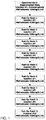
FIG. 1 is a flowchart showing an exemplary treatment protocol using the present methods. -
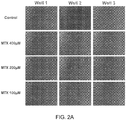
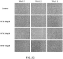
FIGs. 2A-2C are each sets of nine images of human PVR cells plated into 12 wells with 30,000 cells per well showing that methotrexate inhibited proliferation of human proliferative vitreoretinopathy (PVR) cells in culture. The cells were treated with 100 µM, 200 µM, or 400 µM Methotrexate (MTX) as indicated, and the images were taken after 72 hours (2A), 1 week (2B) or 2 weeks (2C). At 72 hours (2A), the photomicrograph showed similar epithelioid morphology and limited confluence across the control plates (top row), as well as the three methotrexate concentrations (rows 2-4). At 1 week (2B), the control plates (row 1) showed a uniform, confluent cellular sheet whereas rows 2-4, which were exposed to methotrexate 400, 200, and 100 respectively, showed growth inhibition and lack of confluency, and were less epithelioid in appearance. At 2 weeks, the control plates (row 1) continued to be a uniform, confluent cellular sheet, whereas rows 2-4, exposed to methotrexate 400, 200, and 100 respectively, continued to have inhibited growth and lack of confluency. - Proliferative vitreoretinopathy (PVR) is a common occurrence after retinal detachment surgery. PVR is a "scarring" condition that forms inside the eye after surgery, significant trauma, or even spontaneously. Its pathogenesis is the disruption of the retinal pigment epithelium layer, which is associated with inflammation, migration, and proliferation of cells to the (neural) retinal surface. Over the next 4-12 weeks, membranes on the surface of the retina proliferate, contract, and apply traction on the retina, which results in redetachment of the retina from the RPE. Once PVR is present and the retina detaches for a second time, it is unlikely that vision will be restored.
- Epiretinal membranes (ERM) are caused by an abnormal proliferation of cells, e.g., retinal pigment epithelial (RPE) cells, glial cells, fibroblasts, and macrophages, on the surface of the retina, typically in response to ocular disease; the membranes tend to contract and cause puckering and thus distortion of the macula. See, e.g., Hiscott et al., Br J Ophthalmol. 68(10):708-15 (1984); Hiscott et al., Eye 16, 393-403 (2002); and Asato et al., PLoS One. 8(1): e54191 (2013).
- Like ERM, PVR is an abnormal wound healing response of the vitreous and retina, a clinical syndrome where cells with proliferative capacity, driven by inflammatory mediators, multiply on the retinal surface, contract, and eventually cause recurrent retinal detachment (RD). The pathogenesis of PVR begins with the introduction of RPE cells into the vitreous cavity. These cells may be introduced at the time of the retinal tear itself or may be introduced iatrogenically such as through the use of cryotherapy or retinectomy. Studies from monkey eyes with PVR have also postulated that the introduction of Müller cells, as well as potentially fibrocytes, occur as well. Concomitant with the introduction of RPE cells is the introduction or upregulation of growth factors, including vascular endothelial growth factor (VEGF), platelet-derived growth factor (PDGF), fibronectin, transforming growth factor-beta (TGF-β), and other mediators. This process begins an autocrine loop where glial, RPE, and other cellular constituents proliferate and transdifferentiate into contractile myofibrocytes. On pathology, macrophages as well as fibroblasts are commonly identified in specimens of PVR.
- It has also been proposed that, at this time of RPE disruption, inflammation plays an important role in the development of PVR. Cytokines IL-6, IL-1, TNF-alpha, and IFN-gamma have been identified in high concentrations in the vitreous in the early, proliferative stages of PVR, but they decrease to normal levels in the scarring phase. These cytokines are not present in eyes that do not develop PVR.
- The use of a pharmacologic adjuvant to prevent PVR has been an elusive goal in ophthalmology. A general pharmacologic strategy employed in prior PVR studies has been the single intravitreal, intraoperative administration of a variety of agents, such as duanoribicin, 5-fluorouracil (5-FU), triamcinolone, low-molecular weight heparin, and naproxen.10-15 Although the clearance of vitreally delivered drugs is dependent on a number of factors including the molecular weight of the drug, the status of the blood-retina barriers, contents of the vitreous cavity, etc., it is likely that those drugs used in these prior PVR studies were cleared from the eye within days after their administration. In contrast, PVR does not become a clinically appreciable, pathologic entity until at least 6-8 weeks after surgery.
- Methotrexate is a non-naturally occurring chemically also known as N-[4-[[(2,4-diamino-6-pteridinyl) methyl] methylamino] benzoyl]-L-glutamic acid. A folate analog, methotrexate is thought to act as an anti-proliferative agent by reversibly inhibiting dihydrofolate reductase, which prevents dihydrofolate from being reduced to tetrahydrofolate, which is used in the synthesis of purine nucleotides.
- Mechanisms of anti-inflammatory action are less clear, although mechanisms that have been proposed include its ability to enhance the extracellular concentration of adenosine, suppression of pro-inflammatory cytokines, inducement of apoptosis of activated T cells, and suppression of intracellular adhesion by activated T cells.
- In some embodiments, the methotrexate is formulated for repeated injection, e.g., in Balanced Salt Solution from 25 mg vials to a sterile, single-use dose of 400 mcg/0.1 ml.
- In some aspects, the methotrexate is formulated for sustained release. A number of sustained release formulations of methotrexate are known in the art, including but not limited to biodegradable implants such as lipid-encapsulated formulations, e.g., Depo/Methotrexate, as described in Bonetti et al., Cancer Chemother Pharmacol 33:303-306 (1994) and Chatelut et al., J Pharm Sci. 1994 Mar;83(3):429-32; multivesicular liposome (MVL) formulations of methotrexate (MTX), e.g., as described in
WO2011143484 ; nano- or micropartricules, e.g., alpha-lactalbumin microparticles, e.g., as described in Vijayaragavan et al., Int J Pharm Res 3(1):39-44 (2011) or nanoparticles of conjugated methotrexate-human serum albumin as described in Taheri et al., J Nanomaterials 2011 (dx.doi.org/10.1155/2011/768201); polyion complex (PIC) micelles; bioadhesive polymers such as hydroxypropyl methylcellulose (HPMC), carboxymethylcellulose (CMC) and polyacrylic acid (PAA) derivatives, as well as hyaluronic acid (HA), e.g., Lacrisert (Aton Pharma), which is a soluble hydroxy propyl cellulose ocular insert. - Alternatively or in addition, sustained release can be achieved using a sustained-release device such as intravitreal implants, e.g., as described in Palakurthi et al., Current Eye Research, 35(12):1105-1115 (2010) or similar to the Retisert (Bausch & Lomb), Ozurdex (Allergan); or non-biodegradable implants, e.g., similar to Iluvien (Alimera) or Vitrasert (Bausch & Lomb) implants; the I-vation platform (SurModics Inc.). See also Lee et al., Pharm Res. 27(10):2043-53 (2010); Haghjou et al., J Ophthalmic Vis Res. 6(4):317-329 (2011); Kim et al., Invest. Ophthalmol. Vis. Sci. 45(8):2722-2731 (2004); and Velez and Whitcup, Br J Ophthalmol 83:1225-1229 (1999).
- The methods described herein can be used to prevent (reduce the risk of) PVR or ERM in patients, e.g., in patients requiring pars plana vitrectomy (PPV), e.g., for rhegmatagenous retinal detachment secondary to trauma; for patients requiring PPV for preexisting proliferative vitreoretinopathy grade C or higher; and/or for patients with retinal detachments requiring PPV for other indications associated with high risk condition for PVR development, e.g., giant retinal tears (giant retinal tears are defined as tears involving 90° or more of the circumference of the globe), retinal breaks larger than 3 disc areas, long-standing retinal detachments, or detachments associated with hemorrhage.
- Other uses of sustained methotrexate in the eye in addition to PVR include the following:
- Approximately 20-30% of RD cases develop clinically perceptible ERMs. Half of these are so visually distorting that patients will require surgery. In addition, autopsy studies show that close to 75-80% of patients with RD surgery have some degree of proliferative of membranes. This may explain why many patients do not achieve perfect vision postoperatively after RD surgery, yet do not have any ERMs grossly perceptible to the human eye.
- ERMs can develop spontaneously, which then requires surgery. If a subject developed an ERM in one eye, implanting a device to prevent ERMs in the other eye could prevent development in that eye.
- For patients who develop ERMs, these can be removed but some reoccur and require reoperation. Leaving an implant could prevent the recurrent ERM.
- The methods described herein can include identifying and/or selecting a subject who is in need of treatment to prevent the development of PVR or ERM as a result of a condition listed above (e.g., selecting the subject on the basis of the need of treatment as a result of a condition listed above, e.g., an increased risk of developing PVR or ERM as a result of a condition listed above). In some embodiments, the subjects treated with a method described herein do not have ocular cancers, e.g., do not have lymphoma (e.g., B cell lymphoma), and/or do not have uveitis.
- The presentation of PVR clinically encompasses a wide phenotype. PVR can vary from a mild cellular haze (Grade A) to thick, fibrous membranes that cause the characteristic stiffened funnel of the detached retina (Grade D). A number of grading systems are in use, see, e.g., Ryan, Retina, 5th ed (Elsevier 2013); Retina Society Terminology Committee. The classification of retinal detachment with proliferative vitreoretinopathy. Ophthalmology 1983;90:121-5 (1983); Machemer R, Aaberg TM, Freeman HM, et al. Am J Ophthalmol 112:159-65 (1991); Lean J, Irvine A, Stern W, et al. Classification of proliferative vitreoretinopathy used in the silicone study. The Silicone study group. Ophthalmology 1989;96:765 - 771. In some embodiments the methods include identifying, selecting, and/or treating a subject who has a low grade (e.g., Grade A or Grade 1) PVR, or who has ERM. In some embodiments, the methods include monitoring the subject for early signs of the development of PVR or ERM, i.e., the presence of a "vitreous haze" indicating a cellular proliferation (which may eventually develop into an organized sheet), and administering one or more doses of MTX as described herein. Although early Grade A PVR vs. an early ERM may be difficult to distinguish from one another, eventually untreated PVR will progress; ERMs will cause a mild traction on the macula resulting in metamorphopsia but will not cause detachment of the retina, whereas untreated PVR will cause detachment and eventually result in a funneled, atrophic retina. The methods can also be used to treat subjects without present signs of PVR but who are at risk for PVR or ERMs.
- The methods described herein include the use of methotrexate in subjects who are at risk of developing a first or recurring PVR or ERM, e.g., a subject who is undergoing RD surgery or ERM surgery, as described above, and in subjects who have PVR or ERM or who are at risk for developing PVR or ERMs. In some embodiments, the methods described herein include the use of methotrexate in subjects who have undergone, are undergoing, or will undergo a pars plana vitrectomy (PPV) or scleral buckle (SB). In some embodiments, the methods include performing a PPV, RD surgery, or ERM surgery. Methods for performing these surgeries are known in the art; for example, typically, PPV is performed under local or general anesthesia using three, 23 or 20 gauge sclerotomy ports. Any present epiretinal membranes can be dissected, e.g., using a membrane pick and forceps. Intraoperative tissue staining, perfluorocarbons, cryopexy, endolaser, scleral buckling, and lensectomy can also be performed as needed. Standard tamponading agents can be used, e.g., silicone oil or gas.
- The methods described herein include the use of an effective amount of methotrexate. An "effective amount" is an amount sufficient to effect beneficial or desired results, e.g., the desired therapeutic effect (i.e., a prophylactically effective amount that reduces the risk of developing PVR or ERM). An effective amount can be administered in one or more administrations, applications or dosages. A therapeutically effective amount of methotrexate can be, e.g., 400µg/0.1 ml per injection, e.g., with at least ten injections, giving a cumulative dose of 4,000 µg over ten injections; in some embodiments, the methods include giving more than ten injections, for a cumulative dose of more than 4,000 µg. In some embodiments, the methods include giving fewer than ten injections, for a cumulative does of less than 4,000 ug. The compositions can be administered one or more times per month. The skilled artisan will appreciate that certain factors may influence the dosage and timing required to effectively treat a subject, including but not limited to the severity of the disease or disorder, previous treatments, the general health and/or age of the subject, and other diseases present.
- In some embodiments, intravitreal methotrexate injections are performed aseptically after the topical application of anaesthesia and an antiseptic agent, e.g., 5% povidone iodine, to the conjunctival sac. In some embodiments, each subject receives an intravitreal injection of methotrexate, e.g., 400 mcg/0.1 ml methotrexate, 3.0 to 3.5 mm posterior to the limbus, depending on lens status, with a 30-gauge needle.
- In some embodiments, the subjects receive multiple intravitreal injections of methotrexate during their post-operative period. The first injection can be administered intraoperatively; subsequently, injections can be administered on post-operative (post-op)
weeks post-op month 3, for a total of 10 injections. See, e.g.,Figure 1 . In some embodiments, the methods include administering the methotrexate in ten doses, or ten or more doses, or less than ten doses, over a three-month period or longer, and injections would be given no more frequently than weekly. In some embodiments, the methods include additional doses at weekly, biweekly, or monthly frequency thereafter for an additional one, two, three, four, five, six, seven, eight, nine, ten, 11, or 12 months thereafter. In some embodiments the methods include ten doses over three months as shown inFig. 1 , with an optional additional one or more doses at monthly intervals thereafter for an additional one, three, six, or more months thereafter. - In some aspects, the subjects receive a sustained release implant, e.g., as described above, that will release MTX over time, e.g., over a week, two weeks, a month, two months, three months, six months, or a year. In some aspects, the methods include administering subsequent implants to provide MTX administration for at least six months, one year, two years, or more.
- The invention is further described in the following examples, which do not limit the scope of the invention described in the claims.
- We hypothesized that the administration of multiple, intravitreal methotrexate injections into eyes with high-risk features for post-operative proliferative vitreoretinopathy (PVR) development will have improved visual outcomes, higher anatomic final reattachment rates, decreased reoperation rates, and decreased occurrence of PVR at 4 months postoperatively.
- We have performed a small pilot study in 10 patients with retinal detachment whom had high-risk clinical factors for developing PVR.
- Patients 18 years to 89 years old of both genders were eligible for this study if they required pars plana vitrectomy (PPV) for rhegmatagenous retinal detachment secondary to trauma, PPV for preexisting proliferative vitreoretinopathy grade C or higher, or if they had retinal detachments requiring PPV for other indications associated with high risk condition for PVR development, i.e.: giant retinal tears (Giant retinal tears are defined as tears involving 90° or more of the circumference of the globe), retinal breaks larger than 3 disc areas, long-standing retinal detachments, detachments associated with hemorrhage.
- The PPV was performed under local or general anesthesia using three, 23 or 20 gauge sclerotomy ports. Any present epiretinal membranes were dissected using a membrane pick and/or forceps. Intraoperative tissue staining, perfluorocarbons, cryopexy, endolaser, scleral buckling, and lensectomy were performed as needed. Either silicone oil or gas was used as the tamponading agent.
- Routine post-operative visits, which involve a dilated funduscopic examination, occured on
post-operative day month 1,month 2, andmonth 3. Patients return to the operating room after three months for silicone oil removal and were seen inclinic 4 months after the original surgery. - In addition to receiving the above-described standard of care, patients received multiple intravitreal methotrexate injections during their post-operative period. The first injection was administered intraoperatively and subsequently was injected on
post-op week post-op month 3, for a total of 10 injections. Intravitreal methotrexate injections were performed aseptically after the topical application of anaesthesia and 5% povidone iodine to the conjunctival sac. Each patient received an intravitreal injection of 400mcg/0.1ml methotrexate, 3.0 to 3.5 mm posterior to the limbus, depending on lens status, with a 30-gauge needle. - After injection, patients were monitored for adverse events, including a full-dilated funduscopic examination.
- Eight men and two women enrolled into the study (Table 1). The age of patients ranged from 18 to 63. Two patients (#4 & #9) were enrolled with traumatic retinal detachment (total retinal detachment, 360 degree giant retinal tear, and retina incarcerated in scleral wound after open globe injury). The remaining 8 patients had had multiple (average 2.5) prior retinal detachments secondary to proliferative membranes. One patient (MTX08M) had significant baseline retinal comorbidity with high pathologic myopia, staphyloma, atrophy, lattice and paving stone in the operative eye. The median pre-operative visual acuity was Hand Motions at 2 feet.
- Operative details of each patient are provided in Table 2. Operative time was recorded as a surrogate for surgical complexity. All patients underwent vitrectomy, extensive membrane peeling, relaxing retinectomy, perfluorocarbon liquid, endolaser, and silicone oil injection. The four subjects previously surgically intervened on had buckles that were still providing adequate indentation of the globe and were therefore left in place.
- Despite the extremely poor visual and anatomic prognosis of all the subjects enrolled in the study, no subjects developed PVR while receiving methotrexate during the three-month treatment protocol. Interestingly, one of the trauma patients (#4) experienced massive PVR two weeks after completion of the injection protocol (at 3 ½ months postoperatively), but weekly examination during the study had shown no evidence of proliferating cells; this is extremely unusual and may be accounted for by the presence of methotrexate for three months and its subsequent absence. This patient required re-operation. Two other subjects developed reaccumulation of fluid under the retina requiring re-operation, but no membranes were appreciated.
- Adverse events observed are reported in Table 3. All subjects experienced a degree of conjunctival hyperemia, consistent with the use of silicone oil. Superficial punctate keratopathy (SPK) was observed in one asymptomatic patient at a single clinical exam. Examination one week later showed a normal corneal surface and no further sequelae were observed. Follow up duration in our 10 patients ranged from 4 months to 39 months, with a median follow up time of 25 months. Even after months to years of follow up data, no significant adverse events were observed. Visual acuity and intraocular pressure at the last follow up visit was similar in all patients to the visual acuity and pressure observed at
post-operative month 4, at the conclusion of the study period, suggesting good long term safety. - In addition, in the same patient who experienced SPK, elevated intraocular pressure was documented (44 mm Hg by Goldmann Tonometry) at a single examination. Per patient report, she had been taking Flonase (fluticasone nasal) "many times per day" and had been taking Pred Forte drops QID, although she had been instructed to take Pred Forte BID. Her pressure was normalized in the exam room with topical therapy. The intravitreal injection was administered as scheduled, and she was discharged from clinic with normal pressure. She was given a prescription for Alphagan and Xalatan with plans to follow up with the Glaucoma service. No further elevated pressures were observed during the course of the study or in follow up, suggesting that her transient elevated pressure was most likely due to excessive steroid use as suspected. Patient MTX08M ended the study with NLP vision. He had a history in the operative eye of pathological myopia, staphyloma, atrophy, lattice, and pave stoning. One month after his surgery (his third intravitreal surgery in that eye and the study surgery date), disc pallor was noted. Optical coherence tomographic pictures of that retina before and after had noted disorganized laminae, secondary to his underlying retinal disease. His vision at this was noted to be LP. He continued to receive injection with limited improvement in his vision and noted to be NLP at the final visit.
- In addition to the excellent anatomical results seen, with virtually no PVR developing during the study protocol, the protocol was associated with excellent visual results. The median post-operative visual acuity was 20/200. This is notable because although one other group has used methotrexate in eyes with a wide variety of conditions, including PVR, no one has previously demonstrated improved outcomes. Hardwig et al. injected methotrexate intravitreally of varying doses into 5 patients, however, only one patient had any improved vision and there was no change in visual acuity for the group (Hardwig et al., Retina 28:1082-1086 (2008)). In addition, this study did not attempt to validate efficacy. Also, in 2006 Hardwig et al. injected 1 dose of methotrexate into the anterior chamber of one patient with PVR, but not into the intravitreal space as described herein (Hardwig et al., Am J Ophthalmol 2006;142:883-885 (2006)).
- The present results are very encouraging and are unlikely explained by chance. The patients who have received 10 intravitreal methotrexate injections have done remarkably well given the severity of their condition, as we intentionally selected patients who were at the highest risk for PVR.
- We hypothesized that methotrexate would inhibit human proliferative vitreoretinopathy (PVR) cells in culture. PVR membranectomy was performed in patients undergoing retinal detachment repair secondary to PVR. Using cellular separation techniques, cellular constituents of the PVR membranes were separated from the extracellular matrix membranes. 30,000 cells per well were placed into a standard 12 welled plate. All 12 wells received endothelial cell growth medium with supplemental growth factors. Four arms were designated consisting of three wells each. The first arm served as a control receiving the standard growth medium but no other intervention. The remaining wells were designated as treatment arms. The second arm of three wells served as the first treatment arm and exposed the cells to 400 micrograms of methotrexate. The third and fourth arms of three wells each exposed the cultured cells to 200 and 100 micrograms, respectively, of methotrexate.
- At 72 hours, similar growth of PVR cells with typical epithelioid morphology and limited confluence was observed (
Figure 2a ). One week after seeding (Figure 2b ) the PVR control cells demonstrated a confluent cellular sheet whereas cells exposed to methotrexate at all concentrations demonstrated growth inhibition, lack of confluency, and were less epithelioid in appearance. At two weeks (Figure 2C ), inhibition of cellular confluency continued whereas the control plates continued to proliferate.


-
- 1. Frenkel S, Hendler K, Siegal T, et al. Intravitreal methotrexate for treating vitreoretinal lymphoma: 10 years of experience. British Journal of Ophthalmology 2008;92:383-388.
- 2. Fishburne BC, Wilson DJ, Rosenbaum JT, Neuwelt EA. Intravitreal methotrexate as an adjunctive treatment of intraocular lymphoma. Arch Ophthalmol 1997;115:1152-1156.
- 3. de Smet MD, Vancs VS, Kohler D, et al. Intravitreal chemotherapy for the treatment of recurrent intraocular lymphoma. British Journal of Ophthalmology 1999;83:448-451.
- 4. Mietz H, Heimann K. Onset and recurrence of proliferative vitreoretinopathy in various vitreoretinal disease. British Journal of Ophthalmology 1995;79:874-877.
- 5. Pastor JC, la Rúa de ER, Martin F. Proliferative vitreoretinopathy: risk factors and pathobiology. Progress in Retinal and Eye Research 2002;21:127-144.
- 6. Hardwig PW, Pulido JS, Erie JC, et al. Intraocular Methotrexate in Ocular Diseases Other Than Primary Central Nervous System Lymphoma. Am J Ophthalmol 2006;142:883-885.
- 7. Hardwig PW, Pulido JS, Bakri SJ. The safety of intraocular methotrexate in silicone-filled eyes. Retina 2008;28:1082-1086.
- 8. Velez G, Yuan P, Sung C, et al. Pharmacokinetics and toxicity of intravitreal chemotherapy for primary intraocular lymphoma. Arch Ophthalmol 2001;119:1518-1524.
- 9. Creten O, Spileers W, Stalmans P. Systemic resorption of 5-fluorouracil used in infusion fluid during vitrectomy. Bull Soc Belge Ophtalmol 2007:37-41.
- 10. Cheema RA, Peyman GA, Fang T, et al. Triamcinolone acetonide as an adjuvant in the surgical treatment of retinal detachment with proliferative vitreoretinopathy. Ophthalmic Surg Lasers Imaging 2007;38:365-370.
- 11. Ahmadieh H, Feghhi M, Tabatabaei H, et al. Triamcinolone Acetonide in Silicone-Filled Eyes as Adjunctive Treatment for Proliferative Vitreoretinopathy. Ophthalmology 2008;115:1938-1943.
- 12. Yang CS, Khawly JA, Hainsworth DP, et al. An intravitreal sustained-release triamcinolone and 5-fluorouracil codrug in the treatment of experimental proliferative vitreoretinopathy. Arch Ophthalmol 1998;116:69-77.
- 13. Chen EP, Steinhorst UH, Samsa GP, et al. The effect of combined daunorubicin and triamcinolone acetonide treatment on a refined experimental model of proliferative vitreoretinopathy. Investigative Ophthalmology & Visual Science 1992;33:2160-2164.
- 14. Munir WM, Pulido JS, Sharma MC, Buerk BM. Intravitreal triamcinolone for treatment of complicated proliferative diabetic retinopathy and proliferative vitreoretinopathy. Can J Ophthalmol 2005;40:598-604.
- 15. Chandler DB, Rozakis G, de Juan E, Machemer R. The effect of triamcinolone acetonide on a refined experimental model of proliferative vitreoretinopathy. AJOPHT 1985;99:686-690.
- 16. Montesinos MC, Yap JS, Desai A, et al. Reversal of the antiinflammatory effects of methotrexate by the nonselective adenosine receptor antagonists theophylline and caffeine: evidence that the antiinflammatory effects of methotrexate are mediated via multiple adenosine receptors in rat adjuvant arthritis. Arthritis Rheum 2000;43:656-663.
- 17. Gerards AH, de Lathouder S, de Groot ER, et al. Inhibition of cytokine production by methotrexate. Studies in healthy volunteers and patients with rheumatoid arthritis. Rheumatology (Oxford) 2003;42:1189-1196.
- 18. Genestier L, Paillot R, Fournel S, et al. Immunosuppressive properties of methotrexate: apoptosis and clonal deletion of activated peripheral T cells. J Clin Invest 1998;102:322-328.
- 19. Seitz M, Zwicker M, Wider B. Enhanced in vitro induced production of interleukin 10 by peripheral blood mononuclear cells in rheumatoid arthritis is associated with clinical response to methotrexate treatment. J Rheumatol 2001;28:496-501.
- 20. Johnston A, Gudjonsson JE, Sigmundsdottir H, et al. The anti-inflammatory action of methotrexate is not mediated by lymphocyte apoptosis, but by the suppression of activation and adhesion molecules. Clin Immunol 2005;114:154-163.
- It is to be understood that while the invention has been described in conjunction with the detailed description thereof, the foregoing description is intended to illustrate and not limit the scope of the invention, which is defined by the scope of the appended claims. Other aspects, advantages, and modifications are within the scope of the following claims.
Claims (9)
- A composition for use in treating or reducing the risk of proliferative vitreoretinopathy (PVR) or epiretinal membranes (ERM) in a subject, wherein the composition comprises methotrexate and the methotrexate is administered via a plurality of intravitreal injections over a period of at least one, two, three, or more months, given no more frequently than weekly.
- The composition for the use of claim 1, comprising administering ten or more intravitreal injections of methotrexate.
- The composition for the use of any one of claims 1 to 2, wherein each injection provides a dose of 400 mcg in 0.1 ml methotrexate.
- The composition for the use of any one of claims 1 to 3, wherein the methotrexate is administered posterior to the limbus.
- The composition for the use of any one of claims 1 to 4, wherein the subject is undergoing an ocular surgical procedure that increases the subject's risk of developing ERM or PVR, optionally wherein the ocular surgical procedure is a pars plana vitrectomy (PPV), Retinal Detachment (RD) surgery; ERM surgery; scleral buckle surgery; or a procedure in the other eye.
- The composition for the use of claim 5, wherein the subject requires a PPV to treat a rhegmatagenous retinal detachment secondary to trauma; preexisting proliferative vitreoretinopathy; or for other indications associated with high risk condition for PVR development and optionally selected from the group consisting of a giant retinal tear, a retinal break larger than 3 disc areas, a long-standing retinal detachment, or a detachment associated with hemorrhage.
- The composition for the use of claim 5, wherein:a first injection is given at conclusion of the surgical procedure;eight weekly injections are given until postoperative month two; anda tenth injection is given at postoperative month three.
- The composition for the use of any one of claims 1 to 6, comprising administering nine consecutive weekly injections, and a tenth injection three months after the first injection, and optionally administering additional injections after the tenth injection..
- The composition for the use of claim 8, comprising administering one, two, three, four, five, six, seven, eight, or nine additional injections after the tenth injection.
Priority Applications (2)
| Application Number | Priority Date | Filing Date | Title |
|---|---|---|---|
| EP20191599.8A EP3791883B1 (en) | 2014-07-30 | 2015-07-30 | Methotrexate for proliferative vitreoretinopathy |
| EP23205570.7A EP4311575A3 (en) | 2014-07-30 | 2015-07-30 | Methotrexate for proliferative vitreoretinopathy |
Applications Claiming Priority (2)
| Application Number | Priority Date | Filing Date | Title |
|---|---|---|---|
| US201462030778P | 2014-07-30 | 2014-07-30 | |
| PCT/US2015/042951 WO2016019165A1 (en) | 2014-07-30 | 2015-07-30 | Methotrexate for proliferative vitreoretinopathy |
Related Child Applications (3)
| Application Number | Title | Priority Date | Filing Date |
|---|---|---|---|
| EP20191599.8A Division EP3791883B1 (en) | 2014-07-30 | 2015-07-30 | Methotrexate for proliferative vitreoretinopathy |
| EP20191599.8A Division-Into EP3791883B1 (en) | 2014-07-30 | 2015-07-30 | Methotrexate for proliferative vitreoretinopathy |
| EP23205570.7A Division EP4311575A3 (en) | 2014-07-30 | 2015-07-30 | Methotrexate for proliferative vitreoretinopathy |
Publications (4)
| Publication Number | Publication Date |
|---|---|
| EP3174540A1 EP3174540A1 (en) | 2017-06-07 |
| EP3174540A4 EP3174540A4 (en) | 2018-04-04 |
| EP3174540B1 EP3174540B1 (en) | 2020-09-02 |
| EP3174540B9 true EP3174540B9 (en) | 2021-05-05 |
Family
ID=55218325
Family Applications (3)
| Application Number | Title | Priority Date | Filing Date |
|---|---|---|---|
| EP23205570.7A Pending EP4311575A3 (en) | 2014-07-30 | 2015-07-30 | Methotrexate for proliferative vitreoretinopathy |
| EP15827946.3A Active EP3174540B9 (en) | 2014-07-30 | 2015-07-30 | Methotrexate for proliferative vitreoretinopathy |
| EP20191599.8A Active EP3791883B1 (en) | 2014-07-30 | 2015-07-30 | Methotrexate for proliferative vitreoretinopathy |
Family Applications Before (1)
| Application Number | Title | Priority Date | Filing Date |
|---|---|---|---|
| EP23205570.7A Pending EP4311575A3 (en) | 2014-07-30 | 2015-07-30 | Methotrexate for proliferative vitreoretinopathy |
Family Applications After (1)
| Application Number | Title | Priority Date | Filing Date |
|---|---|---|---|
| EP20191599.8A Active EP3791883B1 (en) | 2014-07-30 | 2015-07-30 | Methotrexate for proliferative vitreoretinopathy |
Country Status (6)
| Country | Link |
|---|---|
| US (4) | US10098884B2 (en) |
| EP (3) | EP4311575A3 (en) |
| JP (3) | JP7097181B2 (en) |
| CA (2) | CA2991921C (en) |
| ES (1) | ES2835499T3 (en) |
| WO (1) | WO2016019165A1 (en) |
Families Citing this family (5)
| Publication number | Priority date | Publication date | Assignee | Title |
|---|---|---|---|---|
| CA2991921C (en) * | 2014-07-30 | 2023-08-08 | Massachusetts Eye And Ear Infirmary | Methotrexate for proliferative vitreoretinopathy |
| US20200179392A1 (en) * | 2016-08-05 | 2020-06-11 | Schepens Eye Research Institute | Idelalisib for Treating Proliferative Vitreoretinopathy and Abnormal Intraocular Neovascularization |
| AU2018369784B2 (en) | 2017-11-14 | 2023-06-01 | Massachusetts Eye And Ear Infirmary | RUNX1 inhibition for treatment of proliferative vitreoretinopathy and conditions associated with epithelial to mesenchymal transition |
| CA3149467A1 (en) * | 2019-09-13 | 2021-03-18 | Dean Eliott | Ophthalmic formulations of methotrexate |
| US11911385B1 (en) | 2022-12-14 | 2024-02-27 | Aldeyra Therapeutics, Inc. | Methotrexate treatment methods |
Family Cites Families (16)
| Publication number | Priority date | Publication date | Assignee | Title |
|---|---|---|---|---|
| US20020032734A1 (en) | 2000-07-26 | 2002-03-14 | Rhoads Geoffrey B. | Collateral data combined with user characteristics to select web site |
| AUPQ496500A0 (en) | 2000-01-06 | 2000-02-03 | University Of Sydney, The | Kit |
| US20040253243A1 (en) | 2003-01-21 | 2004-12-16 | David Epstein | Aptamer therapeutics useful in ocular pharmacotherapy |
| US20050255144A1 (en) * | 2003-04-09 | 2005-11-17 | Directcontact Llc | Methods and articles for the delivery of medicaments to the eye for the treatment of posterior segment diseases |
| US7807675B2 (en) * | 2004-04-02 | 2010-10-05 | Denki Kagaku Kogyo Kabushiki Kaisha | Hyaluronic acid-methotrexate conjugate |
| US20050244469A1 (en) | 2004-04-30 | 2005-11-03 | Allergan, Inc. | Extended therapeutic effect ocular implant treatments |
| DE602004022523D1 (en) | 2004-07-02 | 2009-09-24 | Novagali Pharma Sa | Use of emulsions for intra- and periocular injection |
| US20080038316A1 (en) | 2004-10-01 | 2008-02-14 | Wong Vernon G | Conveniently implantable sustained release drug compositions |
| JP5682991B2 (en) * | 2004-10-01 | 2015-03-11 | ラムズコア, インコーポレイテッド | Convenient implantable sustained release drug formulation |
| US8231892B2 (en) | 2007-05-24 | 2012-07-31 | Allergan, Inc. | Biodegradable drug delivery system |
| US20090081277A1 (en) * | 2007-09-21 | 2009-03-26 | Allergan, Inc. | Pharmaceutical formulations and methods for treating ocular conditions |
| WO2010047803A2 (en) | 2008-10-22 | 2010-04-29 | Oncotx, L.L.C. | A method for the treatment of proliferative disorders of the eye |
| US9770414B2 (en) | 2010-05-13 | 2017-09-26 | Pacira Pharmaceuticals, Inc. | Sustained release formulation of methotrexate as a disease-modifying antirheumatic drug (DMARD) and an anti-cancer agent |
| US20140105956A1 (en) * | 2012-10-11 | 2014-04-17 | Rupak BANERJEE | Biodegradable polymer based microimplant for ocular drug delivery |
| BR112015010566A2 (en) * | 2012-11-08 | 2017-07-11 | Clearside Biomedical Inc | methods and devices for the treatment of eye disease in human subjects |
| CA2991921C (en) * | 2014-07-30 | 2023-08-08 | Massachusetts Eye And Ear Infirmary | Methotrexate for proliferative vitreoretinopathy |
-
2015
- 2015-07-30 CA CA2991921A patent/CA2991921C/en active Active
- 2015-07-30 WO PCT/US2015/042951 patent/WO2016019165A1/en active Application Filing
- 2015-07-30 EP EP23205570.7A patent/EP4311575A3/en active Pending
- 2015-07-30 EP EP15827946.3A patent/EP3174540B9/en active Active
- 2015-07-30 US US15/500,351 patent/US10098884B2/en active Active
- 2015-07-30 JP JP2017505180A patent/JP7097181B2/en active Active
- 2015-07-30 CA CA3200870A patent/CA3200870A1/en active Pending
- 2015-07-30 ES ES15827946T patent/ES2835499T3/en active Active
- 2015-07-30 EP EP20191599.8A patent/EP3791883B1/en active Active
-
2017
- 2017-09-08 US US15/699,559 patent/US10272089B2/en active Active
-
2019
- 2019-04-29 US US16/397,562 patent/US10828306B2/en active Active
-
2020
- 2020-10-30 US US17/085,907 patent/US20210283135A1/en not_active Abandoned
- 2020-12-07 JP JP2020202357A patent/JP2021059554A/en active Pending
-
2023
- 2023-07-13 JP JP2023114963A patent/JP2023145527A/en active Pending
Also Published As
| Publication number | Publication date |
|---|---|
| JP2017523974A (en) | 2017-08-24 |
| US10828306B2 (en) | 2020-11-10 |
| US20170216294A1 (en) | 2017-08-03 |
| CA2991921C (en) | 2023-08-08 |
| EP3174540A1 (en) | 2017-06-07 |
| EP4311575A2 (en) | 2024-01-31 |
| CA2991921A1 (en) | 2016-02-04 |
| US10098884B2 (en) | 2018-10-16 |
| JP2021059554A (en) | 2021-04-15 |
| EP3174540B1 (en) | 2020-09-02 |
| JP7097181B2 (en) | 2022-07-07 |
| US20200085827A1 (en) | 2020-03-19 |
| EP4311575A3 (en) | 2024-04-03 |
| EP3791883A1 (en) | 2021-03-17 |
| CA3200870A1 (en) | 2016-02-04 |
| WO2016019165A1 (en) | 2016-02-04 |
| US10272089B2 (en) | 2019-04-30 |
| JP2023145527A (en) | 2023-10-11 |
| EP3174540A4 (en) | 2018-04-04 |
| US20170368067A1 (en) | 2017-12-28 |
| US20210283135A1 (en) | 2021-09-16 |
| EP3791883B1 (en) | 2023-10-25 |
| ES2835499T3 (en) | 2021-06-22 |
Similar Documents
| Publication | Publication Date | Title |
|---|---|---|
| US10828306B2 (en) | Methotrexate for proliferative vitreoretinopathy | |
| Chang et al. | Phase II results of an intraocular steroid delivery system for cataract surgery | |
| Wadood et al. | Safety and efficacy of a dexamethasone anterior segment drug delivery system in patients after phacoemulsification | |
| US20070197491A1 (en) | Method for treating primary and secondary forms of glaucoma | |
| US20080125406A1 (en) | Method for Treating Primary and Secondary Forms of Glaucoma | |
| Kaufman et al. | Cataract extraction in patients with pars planitis | |
| Eperon et al. | A biodegradable drug delivery system for the treatment of postoperative inflammation | |
| Stewart et al. | Efficacy and safety profile of ketorolac 0.5% ophthalmic solution in the prevention of surgically induced miosis during cataract surgery | |
| JP2022107631A (en) | Use of medications with neuroprotective properties to prevent or reduce the risk of ischemia-reperfusion injury in subject | |
| US20130156841A1 (en) | Method of reducing incidence of intraocular pressure associated with intraocular use of corticosteroids | |
| Hatton et al. | Treatment of relapsed cytomegalovirus retinitis with the sustained-release ganciclovir implant | |
| Mehta et al. | Formulation of a peribulbar block for prolonged postoperative pain management in vitreoretinal surgery: a randomized clinical trial | |
| Sandoval et al. | Evaluation of 0.4% ketorolac tromethamine ophthalmic solution versus 0.5% ketorolac tromethamine ophthalmic solution after phacoemulsification and intraocular lens implantation | |
| Chang et al. | Two clinical trials of an intraocular steroid delivery system for cataract surgery. | |
| US20230310446A1 (en) | Rho kinase inhibition for treatment of proliferative vitreoretinopathy and conditions associated with epithelial to mesenchymal transition | |
| Simaroj et al. | Effects of intracameral triamcinolone and gentamicin injections following cataract surgery | |
| AU2003213084B2 (en) | Treatment of ophthalmic disorders using urea and urea derivatives | |
| CN101365456A (en) | Method for treating primary and secondary forms of glaucoma | |
| US20230355611A1 (en) | Topotecan for proliferative vitreoretinopathy | |
| Onal et al. | Effect of apraclonidine versus dorzolamide on intraocular pressure after phacoemulsification | |
| CN1802151A (en) | Formulations of non-steroidal anti-inflammatory agents to treat pathologic ocular angiogenesis | |
| SA06270472B1 (en) | Method for treating primar and secondary forms of glaucma |
Legal Events
| Date | Code | Title | Description |
|---|---|---|---|
| STAA | Information on the status of an ep patent application or granted ep patent |
Free format text: STATUS: THE INTERNATIONAL PUBLICATION HAS BEEN MADE |
|
| 17P | Request for examination filed |
Effective date: 20170216 |
|
| AK | Designated contracting states |
Kind code of ref document: A1 Designated state(s): AL AT BE BG CH CY CZ DE DK EE ES FI FR GB GR HR HU IE IS IT LI LT LU LV MC MK MT NL NO PL PT RO RS SE SI SK SM TR |
|
| AX | Request for extension of the european patent |
Extension state: BA ME |
|
| PUAI | Public reference made under article 153(3) epc to a published international application that has entered the european phase |
Free format text: ORIGINAL CODE: 0009012 |
|
| STAA | Information on the status of an ep patent application or granted ep patent |
Free format text: STATUS: REQUEST FOR EXAMINATION WAS MADE |
|
| DAV | Request for validation of the european patent (deleted) | ||
| DAX | Request for extension of the european patent (deleted) | ||
| RIC1 | Information provided on ipc code assigned before grant |
Ipc: A61K 9/00 20060101ALI20171127BHEP Ipc: A61P 27/02 20060101ALI20171127BHEP Ipc: A61K 31/519 20060101AFI20171127BHEP |
|
| REG | Reference to a national code |
Ref country code: HK Ref legal event code: DE Ref document number: 1236395 Country of ref document: HK |
|
| A4 | Supplementary search report drawn up and despatched |
Effective date: 20180305 |
|
| RIC1 | Information provided on ipc code assigned before grant |
Ipc: A61K 31/519 20060101AFI20180227BHEP Ipc: A61P 27/02 20060101ALI20180227BHEP Ipc: A61K 9/00 20060101ALI20180227BHEP |
|
| GRAP | Despatch of communication of intention to grant a patent |
Free format text: ORIGINAL CODE: EPIDOSNIGR1 |
|
| STAA | Information on the status of an ep patent application or granted ep patent |
Free format text: STATUS: GRANT OF PATENT IS INTENDED |
|
| RIC1 | Information provided on ipc code assigned before grant |
Ipc: A61K 9/00 20060101ALI20200214BHEP Ipc: A61K 9/50 20060101ALI20200214BHEP Ipc: A61K 31/519 20060101AFI20200214BHEP Ipc: A61P 27/02 20060101ALI20200214BHEP |
|
| INTG | Intention to grant announced |
Effective date: 20200317 |
|
| GRAS | Grant fee paid |
Free format text: ORIGINAL CODE: EPIDOSNIGR3 |
|
| GRAJ | Information related to disapproval of communication of intention to grant by the applicant or resumption of examination proceedings by the epo deleted |
Free format text: ORIGINAL CODE: EPIDOSDIGR1 |
|
| GRAL | Information related to payment of fee for publishing/printing deleted |
Free format text: ORIGINAL CODE: EPIDOSDIGR3 |
|
| STAA | Information on the status of an ep patent application or granted ep patent |
Free format text: STATUS: REQUEST FOR EXAMINATION WAS MADE |
|
| INTC | Intention to grant announced (deleted) | ||
| GRAR | Information related to intention to grant a patent recorded |
Free format text: ORIGINAL CODE: EPIDOSNIGR71 |
|
| STAA | Information on the status of an ep patent application or granted ep patent |
Free format text: STATUS: GRANT OF PATENT IS INTENDED |
|
| GRAA | (expected) grant |
Free format text: ORIGINAL CODE: 0009210 |
|
| STAA | Information on the status of an ep patent application or granted ep patent |
Free format text: STATUS: THE PATENT HAS BEEN GRANTED |
|
| INTG | Intention to grant announced |
Effective date: 20200706 |
|
| AK | Designated contracting states |
Kind code of ref document: B1 Designated state(s): AL AT BE BG CH CY CZ DE DK EE ES FI FR GB GR HR HU IE IS IT LI LT LU LV MC MK MT NL NO PL PT RO RS SE SI SK SM TR |
|
| REG | Reference to a national code |
Ref country code: GB Ref legal event code: FG4D |
|
| REG | Reference to a national code |
Ref country code: AT Ref legal event code: REF Ref document number: 1308004 Country of ref document: AT Kind code of ref document: T Effective date: 20200915 Ref country code: CH Ref legal event code: EP |
|
| REG | Reference to a national code |
Ref country code: DE Ref legal event code: R096 Ref document number: 602015058548 Country of ref document: DE |
|
| REG | Reference to a national code |
Ref country code: IE Ref legal event code: FG4D |
|
| REG | Reference to a national code |
Ref country code: LT Ref legal event code: MG4D |
|
| PG25 | Lapsed in a contracting state [announced via postgrant information from national office to epo] |
Ref country code: GR Free format text: LAPSE BECAUSE OF FAILURE TO SUBMIT A TRANSLATION OF THE DESCRIPTION OR TO PAY THE FEE WITHIN THE PRESCRIBED TIME-LIMIT Effective date: 20201203 Ref country code: NO Free format text: LAPSE BECAUSE OF FAILURE TO SUBMIT A TRANSLATION OF THE DESCRIPTION OR TO PAY THE FEE WITHIN THE PRESCRIBED TIME-LIMIT Effective date: 20201202 Ref country code: BG Free format text: LAPSE BECAUSE OF FAILURE TO SUBMIT A TRANSLATION OF THE DESCRIPTION OR TO PAY THE FEE WITHIN THE PRESCRIBED TIME-LIMIT Effective date: 20201202 Ref country code: LT Free format text: LAPSE BECAUSE OF FAILURE TO SUBMIT A TRANSLATION OF THE DESCRIPTION OR TO PAY THE FEE WITHIN THE PRESCRIBED TIME-LIMIT Effective date: 20200902 Ref country code: HR Free format text: LAPSE BECAUSE OF FAILURE TO SUBMIT A TRANSLATION OF THE DESCRIPTION OR TO PAY THE FEE WITHIN THE PRESCRIBED TIME-LIMIT Effective date: 20200902 Ref country code: FI Free format text: LAPSE BECAUSE OF FAILURE TO SUBMIT A TRANSLATION OF THE DESCRIPTION OR TO PAY THE FEE WITHIN THE PRESCRIBED TIME-LIMIT Effective date: 20200902 Ref country code: SE Free format text: LAPSE BECAUSE OF FAILURE TO SUBMIT A TRANSLATION OF THE DESCRIPTION OR TO PAY THE FEE WITHIN THE PRESCRIBED TIME-LIMIT Effective date: 20200902 |
|
| REG | Reference to a national code |
Ref country code: NL Ref legal event code: MP Effective date: 20200902 |
|
| REG | Reference to a national code |
Ref country code: AT Ref legal event code: MK05 Ref document number: 1308004 Country of ref document: AT Kind code of ref document: T Effective date: 20200902 |
|
| PG25 | Lapsed in a contracting state [announced via postgrant information from national office to epo] |
Ref country code: LV Free format text: LAPSE BECAUSE OF FAILURE TO SUBMIT A TRANSLATION OF THE DESCRIPTION OR TO PAY THE FEE WITHIN THE PRESCRIBED TIME-LIMIT Effective date: 20200902 Ref country code: RS Free format text: LAPSE BECAUSE OF FAILURE TO SUBMIT A TRANSLATION OF THE DESCRIPTION OR TO PAY THE FEE WITHIN THE PRESCRIBED TIME-LIMIT Effective date: 20200902 Ref country code: PL Free format text: LAPSE BECAUSE OF FAILURE TO SUBMIT A TRANSLATION OF THE DESCRIPTION OR TO PAY THE FEE WITHIN THE PRESCRIBED TIME-LIMIT Effective date: 20200902 |
|
| REG | Reference to a national code |
Ref country code: CH Ref legal event code: PK Free format text: BERICHTIGUNG B9 |
|
| PG25 | Lapsed in a contracting state [announced via postgrant information from national office to epo] |
Ref country code: SM Free format text: LAPSE BECAUSE OF FAILURE TO SUBMIT A TRANSLATION OF THE DESCRIPTION OR TO PAY THE FEE WITHIN THE PRESCRIBED TIME-LIMIT Effective date: 20200902 Ref country code: RO Free format text: LAPSE BECAUSE OF FAILURE TO SUBMIT A TRANSLATION OF THE DESCRIPTION OR TO PAY THE FEE WITHIN THE PRESCRIBED TIME-LIMIT Effective date: 20200902 Ref country code: PT Free format text: LAPSE BECAUSE OF FAILURE TO SUBMIT A TRANSLATION OF THE DESCRIPTION OR TO PAY THE FEE WITHIN THE PRESCRIBED TIME-LIMIT Effective date: 20210104 Ref country code: EE Free format text: LAPSE BECAUSE OF FAILURE TO SUBMIT A TRANSLATION OF THE DESCRIPTION OR TO PAY THE FEE WITHIN THE PRESCRIBED TIME-LIMIT Effective date: 20200902 Ref country code: CZ Free format text: LAPSE BECAUSE OF FAILURE TO SUBMIT A TRANSLATION OF THE DESCRIPTION OR TO PAY THE FEE WITHIN THE PRESCRIBED TIME-LIMIT Effective date: 20200902 |
|
| PG25 | Lapsed in a contracting state [announced via postgrant information from national office to epo] |
Ref country code: AL Free format text: LAPSE BECAUSE OF FAILURE TO SUBMIT A TRANSLATION OF THE DESCRIPTION OR TO PAY THE FEE WITHIN THE PRESCRIBED TIME-LIMIT Effective date: 20200902 Ref country code: AT Free format text: LAPSE BECAUSE OF FAILURE TO SUBMIT A TRANSLATION OF THE DESCRIPTION OR TO PAY THE FEE WITHIN THE PRESCRIBED TIME-LIMIT Effective date: 20200902 Ref country code: IS Free format text: LAPSE BECAUSE OF FAILURE TO SUBMIT A TRANSLATION OF THE DESCRIPTION OR TO PAY THE FEE WITHIN THE PRESCRIBED TIME-LIMIT Effective date: 20210102 |
|
| REG | Reference to a national code |
Ref country code: DE Ref legal event code: R097 Ref document number: 602015058548 Country of ref document: DE |
|
| REG | Reference to a national code |
Ref country code: ES Ref legal event code: FG2A Ref document number: 2835499 Country of ref document: ES Kind code of ref document: T3 Effective date: 20210622 |
|
| PG25 | Lapsed in a contracting state [announced via postgrant information from national office to epo] |
Ref country code: SK Free format text: LAPSE BECAUSE OF FAILURE TO SUBMIT A TRANSLATION OF THE DESCRIPTION OR TO PAY THE FEE WITHIN THE PRESCRIBED TIME-LIMIT Effective date: 20200902 |
|
| PLBE | No opposition filed within time limit |
Free format text: ORIGINAL CODE: 0009261 |
|
| STAA | Information on the status of an ep patent application or granted ep patent |
Free format text: STATUS: NO OPPOSITION FILED WITHIN TIME LIMIT |
|
| 26N | No opposition filed |
Effective date: 20210603 |
|
| PG25 | Lapsed in a contracting state [announced via postgrant information from national office to epo] |
Ref country code: DK Free format text: LAPSE BECAUSE OF FAILURE TO SUBMIT A TRANSLATION OF THE DESCRIPTION OR TO PAY THE FEE WITHIN THE PRESCRIBED TIME-LIMIT Effective date: 20200902 Ref country code: SI Free format text: LAPSE BECAUSE OF FAILURE TO SUBMIT A TRANSLATION OF THE DESCRIPTION OR TO PAY THE FEE WITHIN THE PRESCRIBED TIME-LIMIT Effective date: 20200902 |
|
| REG | Reference to a national code |
Ref country code: CH Ref legal event code: PL |
|
| PG25 | Lapsed in a contracting state [announced via postgrant information from national office to epo] |
Ref country code: MC Free format text: LAPSE BECAUSE OF FAILURE TO SUBMIT A TRANSLATION OF THE DESCRIPTION OR TO PAY THE FEE WITHIN THE PRESCRIBED TIME-LIMIT Effective date: 20200902 |
|
| REG | Reference to a national code |
Ref country code: BE Ref legal event code: MM Effective date: 20210731 |
|
| PG25 | Lapsed in a contracting state [announced via postgrant information from national office to epo] |
Ref country code: LI Free format text: LAPSE BECAUSE OF NON-PAYMENT OF DUE FEES Effective date: 20210731 Ref country code: CH Free format text: LAPSE BECAUSE OF NON-PAYMENT OF DUE FEES Effective date: 20210731 |
|
| PG25 | Lapsed in a contracting state [announced via postgrant information from national office to epo] |
Ref country code: LU Free format text: LAPSE BECAUSE OF NON-PAYMENT OF DUE FEES Effective date: 20210730 |
|
| PG25 | Lapsed in a contracting state [announced via postgrant information from national office to epo] |
Ref country code: IE Free format text: LAPSE BECAUSE OF NON-PAYMENT OF DUE FEES Effective date: 20210730 Ref country code: BE Free format text: LAPSE BECAUSE OF NON-PAYMENT OF DUE FEES Effective date: 20210731 |
|
| PG25 | Lapsed in a contracting state [announced via postgrant information from national office to epo] |
Ref country code: HU Free format text: LAPSE BECAUSE OF FAILURE TO SUBMIT A TRANSLATION OF THE DESCRIPTION OR TO PAY THE FEE WITHIN THE PRESCRIBED TIME-LIMIT; INVALID AB INITIO Effective date: 20150730 |
|
| PG25 | Lapsed in a contracting state [announced via postgrant information from national office to epo] |
Ref country code: NL Free format text: LAPSE BECAUSE OF NON-PAYMENT OF DUE FEES Effective date: 20200923 Ref country code: CY Free format text: LAPSE BECAUSE OF FAILURE TO SUBMIT A TRANSLATION OF THE DESCRIPTION OR TO PAY THE FEE WITHIN THE PRESCRIBED TIME-LIMIT Effective date: 20200902 |
|
| P01 | Opt-out of the competence of the unified patent court (upc) registered |
Effective date: 20230529 |
|
| PGFP | Annual fee paid to national office [announced via postgrant information from national office to epo] |
Ref country code: IT Payment date: 20230720 Year of fee payment: 9 Ref country code: GB Payment date: 20230727 Year of fee payment: 9 Ref country code: ES Payment date: 20230804 Year of fee payment: 9 |
|
| PGFP | Annual fee paid to national office [announced via postgrant information from national office to epo] |
Ref country code: FR Payment date: 20230725 Year of fee payment: 9 Ref country code: DE Payment date: 20230727 Year of fee payment: 9 |