EP2664666B1 - Devices and methods for enrichment of cells and other particles - Google Patents
Devices and methods for enrichment of cells and other particles Download PDFInfo
- Publication number
- EP2664666B1 EP2664666B1 EP13156633.3A EP13156633A EP2664666B1 EP 2664666 B1 EP2664666 B1 EP 2664666B1 EP 13156633 A EP13156633 A EP 13156633A EP 2664666 B1 EP2664666 B1 EP 2664666B1
- Authority
- EP
- European Patent Office
- Prior art keywords
- cells
- particles
- array
- obstacles
- product
- Prior art date
- Legal status (The legal status is an assumption and is not a legal conclusion. Google has not performed a legal analysis and makes no representation as to the accuracy of the status listed.)
- Expired - Lifetime
Links
Images


Classifications
-
- G—PHYSICS
- G01—MEASURING; TESTING
- G01N—INVESTIGATING OR ANALYSING MATERIALS BY DETERMINING THEIR CHEMICAL OR PHYSICAL PROPERTIES
- G01N30/00—Investigating or analysing materials by separation into components using adsorption, absorption or similar phenomena or using ion-exchange, e.g. chromatography or field flow fractionation
- G01N30/0005—Field flow fractionation
-
- B—PERFORMING OPERATIONS; TRANSPORTING
- B03—SEPARATION OF SOLID MATERIALS USING LIQUIDS OR USING PNEUMATIC TABLES OR JIGS; MAGNETIC OR ELECTROSTATIC SEPARATION OF SOLID MATERIALS FROM SOLID MATERIALS OR FLUIDS; SEPARATION BY HIGH-VOLTAGE ELECTRIC FIELDS
- B03C—MAGNETIC OR ELECTROSTATIC SEPARATION OF SOLID MATERIALS FROM SOLID MATERIALS OR FLUIDS; SEPARATION BY HIGH-VOLTAGE ELECTRIC FIELDS
- B03C1/00—Magnetic separation
- B03C1/02—Magnetic separation acting directly on the substance being separated
- B03C1/30—Combinations with other devices, not otherwise provided for
-
- B—PERFORMING OPERATIONS; TRANSPORTING
- B01—PHYSICAL OR CHEMICAL PROCESSES OR APPARATUS IN GENERAL
- B01L—CHEMICAL OR PHYSICAL LABORATORY APPARATUS FOR GENERAL USE
- B01L3/00—Containers or dishes for laboratory use, e.g. laboratory glassware; Droppers
- B01L3/50—Containers for the purpose of retaining a material to be analysed, e.g. test tubes
- B01L3/502—Containers for the purpose of retaining a material to be analysed, e.g. test tubes with fluid transport, e.g. in multi-compartment structures
- B01L3/5027—Containers for the purpose of retaining a material to be analysed, e.g. test tubes with fluid transport, e.g. in multi-compartment structures by integrated microfluidic structures, i.e. dimensions of channels and chambers are such that surface tension forces are important, e.g. lab-on-a-chip
- B01L3/502746—Containers for the purpose of retaining a material to be analysed, e.g. test tubes with fluid transport, e.g. in multi-compartment structures by integrated microfluidic structures, i.e. dimensions of channels and chambers are such that surface tension forces are important, e.g. lab-on-a-chip characterised by the means for controlling flow resistance, e.g. flow controllers, baffles or throttle valves
-
- B—PERFORMING OPERATIONS; TRANSPORTING
- B01—PHYSICAL OR CHEMICAL PROCESSES OR APPARATUS IN GENERAL
- B01L—CHEMICAL OR PHYSICAL LABORATORY APPARATUS FOR GENERAL USE
- B01L3/00—Containers or dishes for laboratory use, e.g. laboratory glassware; Droppers
- B01L3/50—Containers for the purpose of retaining a material to be analysed, e.g. test tubes
- B01L3/502—Containers for the purpose of retaining a material to be analysed, e.g. test tubes with fluid transport, e.g. in multi-compartment structures
- B01L3/5027—Containers for the purpose of retaining a material to be analysed, e.g. test tubes with fluid transport, e.g. in multi-compartment structures by integrated microfluidic structures, i.e. dimensions of channels and chambers are such that surface tension forces are important, e.g. lab-on-a-chip
- B01L3/502753—Containers for the purpose of retaining a material to be analysed, e.g. test tubes with fluid transport, e.g. in multi-compartment structures by integrated microfluidic structures, i.e. dimensions of channels and chambers are such that surface tension forces are important, e.g. lab-on-a-chip characterised by bulk separation arrangements on lab-on-a-chip devices, e.g. for filtration or centrifugation
-
- B—PERFORMING OPERATIONS; TRANSPORTING
- B01—PHYSICAL OR CHEMICAL PROCESSES OR APPARATUS IN GENERAL
- B01L—CHEMICAL OR PHYSICAL LABORATORY APPARATUS FOR GENERAL USE
- B01L3/00—Containers or dishes for laboratory use, e.g. laboratory glassware; Droppers
- B01L3/50—Containers for the purpose of retaining a material to be analysed, e.g. test tubes
- B01L3/502—Containers for the purpose of retaining a material to be analysed, e.g. test tubes with fluid transport, e.g. in multi-compartment structures
- B01L3/5027—Containers for the purpose of retaining a material to be analysed, e.g. test tubes with fluid transport, e.g. in multi-compartment structures by integrated microfluidic structures, i.e. dimensions of channels and chambers are such that surface tension forces are important, e.g. lab-on-a-chip
- B01L3/502761—Containers for the purpose of retaining a material to be analysed, e.g. test tubes with fluid transport, e.g. in multi-compartment structures by integrated microfluidic structures, i.e. dimensions of channels and chambers are such that surface tension forces are important, e.g. lab-on-a-chip specially adapted for handling suspended solids or molecules independently from the bulk fluid flow, e.g. for trapping or sorting beads or physically stretching molecules
-
- B—PERFORMING OPERATIONS; TRANSPORTING
- B03—SEPARATION OF SOLID MATERIALS USING LIQUIDS OR USING PNEUMATIC TABLES OR JIGS; MAGNETIC OR ELECTROSTATIC SEPARATION OF SOLID MATERIALS FROM SOLID MATERIALS OR FLUIDS; SEPARATION BY HIGH-VOLTAGE ELECTRIC FIELDS
- B03C—MAGNETIC OR ELECTROSTATIC SEPARATION OF SOLID MATERIALS FROM SOLID MATERIALS OR FLUIDS; SEPARATION BY HIGH-VOLTAGE ELECTRIC FIELDS
- B03C1/00—Magnetic separation
- B03C1/32—Magnetic separation acting on the medium containing the substance being separated, e.g. magneto-gravimetric-, magnetohydrostatic-, or magnetohydrodynamic separation
-
- C—CHEMISTRY; METALLURGY
- C12—BIOCHEMISTRY; BEER; SPIRITS; WINE; VINEGAR; MICROBIOLOGY; ENZYMOLOGY; MUTATION OR GENETIC ENGINEERING
- C12M—APPARATUS FOR ENZYMOLOGY OR MICROBIOLOGY; APPARATUS FOR CULTURING MICROORGANISMS FOR PRODUCING BIOMASS, FOR GROWING CELLS OR FOR OBTAINING FERMENTATION OR METABOLIC PRODUCTS, i.e. BIOREACTORS OR FERMENTERS
- C12M47/00—Means for after-treatment of the produced biomass or of the fermentation or metabolic products, e.g. storage of biomass
- C12M47/04—Cell isolation or sorting
-
- C—CHEMISTRY; METALLURGY
- C12—BIOCHEMISTRY; BEER; SPIRITS; WINE; VINEGAR; MICROBIOLOGY; ENZYMOLOGY; MUTATION OR GENETIC ENGINEERING
- C12M—APPARATUS FOR ENZYMOLOGY OR MICROBIOLOGY; APPARATUS FOR CULTURING MICROORGANISMS FOR PRODUCING BIOMASS, FOR GROWING CELLS OR FOR OBTAINING FERMENTATION OR METABOLIC PRODUCTS, i.e. BIOREACTORS OR FERMENTERS
- C12M47/00—Means for after-treatment of the produced biomass or of the fermentation or metabolic products, e.g. storage of biomass
- C12M47/06—Hydrolysis; Cell lysis; Extraction of intracellular or cell wall material
-
- C—CHEMISTRY; METALLURGY
- C12—BIOCHEMISTRY; BEER; SPIRITS; WINE; VINEGAR; MICROBIOLOGY; ENZYMOLOGY; MUTATION OR GENETIC ENGINEERING
- C12Q—MEASURING OR TESTING PROCESSES INVOLVING ENZYMES, NUCLEIC ACIDS OR MICROORGANISMS; COMPOSITIONS OR TEST PAPERS THEREFOR; PROCESSES OF PREPARING SUCH COMPOSITIONS; CONDITION-RESPONSIVE CONTROL IN MICROBIOLOGICAL OR ENZYMOLOGICAL PROCESSES
- C12Q1/00—Measuring or testing processes involving enzymes, nucleic acids or microorganisms; Compositions therefor; Processes of preparing such compositions
- C12Q1/68—Measuring or testing processes involving enzymes, nucleic acids or microorganisms; Compositions therefor; Processes of preparing such compositions involving nucleic acids
- C12Q1/6844—Nucleic acid amplification reactions
- C12Q1/686—Polymerase chain reaction [PCR]
-
- G—PHYSICS
- G01—MEASURING; TESTING
- G01N—INVESTIGATING OR ANALYSING MATERIALS BY DETERMINING THEIR CHEMICAL OR PHYSICAL PROPERTIES
- G01N1/00—Sampling; Preparing specimens for investigation
- G01N1/28—Preparing specimens for investigation including physical details of (bio-)chemical methods covered elsewhere, e.g. G01N33/50, C12Q
- G01N1/40—Concentrating samples
- G01N1/4077—Concentrating samples by other techniques involving separation of suspended solids
-
- G—PHYSICS
- G01—MEASURING; TESTING
- G01N—INVESTIGATING OR ANALYSING MATERIALS BY DETERMINING THEIR CHEMICAL OR PHYSICAL PROPERTIES
- G01N33/00—Investigating or analysing materials by specific methods not covered by groups G01N1/00 - G01N31/00
- G01N33/48—Biological material, e.g. blood, urine; Haemocytometers
- G01N33/483—Physical analysis of biological material
- G01N33/487—Physical analysis of biological material of liquid biological material
- G01N33/49—Blood
-
- G—PHYSICS
- G01—MEASURING; TESTING
- G01N—INVESTIGATING OR ANALYSING MATERIALS BY DETERMINING THEIR CHEMICAL OR PHYSICAL PROPERTIES
- G01N33/00—Investigating or analysing materials by specific methods not covered by groups G01N1/00 - G01N31/00
- G01N33/48—Biological material, e.g. blood, urine; Haemocytometers
- G01N33/50—Chemical analysis of biological material, e.g. blood, urine; Testing involving biospecific ligand binding methods; Immunological testing
- G01N33/5005—Chemical analysis of biological material, e.g. blood, urine; Testing involving biospecific ligand binding methods; Immunological testing involving human or animal cells
- G01N33/5008—Chemical analysis of biological material, e.g. blood, urine; Testing involving biospecific ligand binding methods; Immunological testing involving human or animal cells for testing or evaluating the effect of chemical or biological compounds, e.g. drugs, cosmetics
- G01N33/5044—Chemical analysis of biological material, e.g. blood, urine; Testing involving biospecific ligand binding methods; Immunological testing involving human or animal cells for testing or evaluating the effect of chemical or biological compounds, e.g. drugs, cosmetics involving specific cell types
-
- B—PERFORMING OPERATIONS; TRANSPORTING
- B01—PHYSICAL OR CHEMICAL PROCESSES OR APPARATUS IN GENERAL
- B01L—CHEMICAL OR PHYSICAL LABORATORY APPARATUS FOR GENERAL USE
- B01L2200/00—Solutions for specific problems relating to chemical or physical laboratory apparatus
- B01L2200/06—Fluid handling related problems
- B01L2200/0647—Handling flowable solids, e.g. microscopic beads, cells, particles
-
- B—PERFORMING OPERATIONS; TRANSPORTING
- B01—PHYSICAL OR CHEMICAL PROCESSES OR APPARATUS IN GENERAL
- B01L—CHEMICAL OR PHYSICAL LABORATORY APPARATUS FOR GENERAL USE
- B01L2300/00—Additional constructional details
- B01L2300/08—Geometry, shape and general structure
- B01L2300/0809—Geometry, shape and general structure rectangular shaped
- B01L2300/0816—Cards, e.g. flat sample carriers usually with flow in two horizontal directions
-
- B—PERFORMING OPERATIONS; TRANSPORTING
- B01—PHYSICAL OR CHEMICAL PROCESSES OR APPARATUS IN GENERAL
- B01L—CHEMICAL OR PHYSICAL LABORATORY APPARATUS FOR GENERAL USE
- B01L2300/00—Additional constructional details
- B01L2300/08—Geometry, shape and general structure
- B01L2300/0861—Configuration of multiple channels and/or chambers in a single devices
- B01L2300/0864—Configuration of multiple channels and/or chambers in a single devices comprising only one inlet and multiple receiving wells, e.g. for separation, splitting
-
- B—PERFORMING OPERATIONS; TRANSPORTING
- B01—PHYSICAL OR CHEMICAL PROCESSES OR APPARATUS IN GENERAL
- B01L—CHEMICAL OR PHYSICAL LABORATORY APPARATUS FOR GENERAL USE
- B01L2400/00—Moving or stopping fluids
- B01L2400/04—Moving fluids with specific forces or mechanical means
- B01L2400/0403—Moving fluids with specific forces or mechanical means specific forces
- B01L2400/0406—Moving fluids with specific forces or mechanical means specific forces capillary forces
-
- B—PERFORMING OPERATIONS; TRANSPORTING
- B01—PHYSICAL OR CHEMICAL PROCESSES OR APPARATUS IN GENERAL
- B01L—CHEMICAL OR PHYSICAL LABORATORY APPARATUS FOR GENERAL USE
- B01L2400/00—Moving or stopping fluids
- B01L2400/04—Moving fluids with specific forces or mechanical means
- B01L2400/0403—Moving fluids with specific forces or mechanical means specific forces
- B01L2400/0409—Moving fluids with specific forces or mechanical means specific forces centrifugal forces
-
- B—PERFORMING OPERATIONS; TRANSPORTING
- B01—PHYSICAL OR CHEMICAL PROCESSES OR APPARATUS IN GENERAL
- B01L—CHEMICAL OR PHYSICAL LABORATORY APPARATUS FOR GENERAL USE
- B01L2400/00—Moving or stopping fluids
- B01L2400/04—Moving fluids with specific forces or mechanical means
- B01L2400/0403—Moving fluids with specific forces or mechanical means specific forces
- B01L2400/0415—Moving fluids with specific forces or mechanical means specific forces electrical forces, e.g. electrokinetic
-
- B—PERFORMING OPERATIONS; TRANSPORTING
- B01—PHYSICAL OR CHEMICAL PROCESSES OR APPARATUS IN GENERAL
- B01L—CHEMICAL OR PHYSICAL LABORATORY APPARATUS FOR GENERAL USE
- B01L2400/00—Moving or stopping fluids
- B01L2400/04—Moving fluids with specific forces or mechanical means
- B01L2400/0403—Moving fluids with specific forces or mechanical means specific forces
- B01L2400/043—Moving fluids with specific forces or mechanical means specific forces magnetic forces
-
- B—PERFORMING OPERATIONS; TRANSPORTING
- B01—PHYSICAL OR CHEMICAL PROCESSES OR APPARATUS IN GENERAL
- B01L—CHEMICAL OR PHYSICAL LABORATORY APPARATUS FOR GENERAL USE
- B01L2400/00—Moving or stopping fluids
- B01L2400/04—Moving fluids with specific forces or mechanical means
- B01L2400/0403—Moving fluids with specific forces or mechanical means specific forces
- B01L2400/0472—Diffusion
-
- B—PERFORMING OPERATIONS; TRANSPORTING
- B01—PHYSICAL OR CHEMICAL PROCESSES OR APPARATUS IN GENERAL
- B01L—CHEMICAL OR PHYSICAL LABORATORY APPARATUS FOR GENERAL USE
- B01L2400/00—Moving or stopping fluids
- B01L2400/04—Moving fluids with specific forces or mechanical means
- B01L2400/0475—Moving fluids with specific forces or mechanical means specific mechanical means and fluid pressure
- B01L2400/0487—Moving fluids with specific forces or mechanical means specific mechanical means and fluid pressure fluid pressure, pneumatics
-
- B—PERFORMING OPERATIONS; TRANSPORTING
- B01—PHYSICAL OR CHEMICAL PROCESSES OR APPARATUS IN GENERAL
- B01L—CHEMICAL OR PHYSICAL LABORATORY APPARATUS FOR GENERAL USE
- B01L2400/00—Moving or stopping fluids
- B01L2400/08—Regulating or influencing the flow resistance
- B01L2400/084—Passive control of flow resistance
- B01L2400/086—Passive control of flow resistance using baffles or other fixed flow obstructions
-
- B—PERFORMING OPERATIONS; TRANSPORTING
- B03—SEPARATION OF SOLID MATERIALS USING LIQUIDS OR USING PNEUMATIC TABLES OR JIGS; MAGNETIC OR ELECTROSTATIC SEPARATION OF SOLID MATERIALS FROM SOLID MATERIALS OR FLUIDS; SEPARATION BY HIGH-VOLTAGE ELECTRIC FIELDS
- B03C—MAGNETIC OR ELECTROSTATIC SEPARATION OF SOLID MATERIALS FROM SOLID MATERIALS OR FLUIDS; SEPARATION BY HIGH-VOLTAGE ELECTRIC FIELDS
- B03C2201/00—Details of magnetic or electrostatic separation
- B03C2201/18—Magnetic separation whereby the particles are suspended in a liquid
-
- B—PERFORMING OPERATIONS; TRANSPORTING
- B33—ADDITIVE MANUFACTURING TECHNOLOGY
- B33Y—ADDITIVE MANUFACTURING, i.e. MANUFACTURING OF THREE-DIMENSIONAL [3D] OBJECTS BY ADDITIVE DEPOSITION, ADDITIVE AGGLOMERATION OR ADDITIVE LAYERING, e.g. BY 3D PRINTING, STEREOLITHOGRAPHY OR SELECTIVE LASER SINTERING
- B33Y80/00—Products made by additive manufacturing
-
- G—PHYSICS
- G01—MEASURING; TESTING
- G01N—INVESTIGATING OR ANALYSING MATERIALS BY DETERMINING THEIR CHEMICAL OR PHYSICAL PROPERTIES
- G01N1/00—Sampling; Preparing specimens for investigation
- G01N1/28—Preparing specimens for investigation including physical details of (bio-)chemical methods covered elsewhere, e.g. G01N33/50, C12Q
- G01N1/40—Concentrating samples
-
- G—PHYSICS
- G01—MEASURING; TESTING
- G01N—INVESTIGATING OR ANALYSING MATERIALS BY DETERMINING THEIR CHEMICAL OR PHYSICAL PROPERTIES
- G01N35/00—Automatic analysis not limited to methods or materials provided for in any single one of groups G01N1/00 - G01N33/00; Handling materials therefor
- G01N2035/00178—Special arrangements of analysers
- G01N2035/00237—Handling microquantities of analyte, e.g. microvalves, capillary networks
-
- Y—GENERAL TAGGING OF NEW TECHNOLOGICAL DEVELOPMENTS; GENERAL TAGGING OF CROSS-SECTIONAL TECHNOLOGIES SPANNING OVER SEVERAL SECTIONS OF THE IPC; TECHNICAL SUBJECTS COVERED BY FORMER USPC CROSS-REFERENCE ART COLLECTIONS [XRACs] AND DIGESTS
- Y10—TECHNICAL SUBJECTS COVERED BY FORMER USPC
- Y10T—TECHNICAL SUBJECTS COVERED BY FORMER US CLASSIFICATION
- Y10T137/00—Fluid handling
- Y10T137/0318—Processes
-
- Y—GENERAL TAGGING OF NEW TECHNOLOGICAL DEVELOPMENTS; GENERAL TAGGING OF CROSS-SECTIONAL TECHNOLOGIES SPANNING OVER SEVERAL SECTIONS OF THE IPC; TECHNICAL SUBJECTS COVERED BY FORMER USPC CROSS-REFERENCE ART COLLECTIONS [XRACs] AND DIGESTS
- Y10—TECHNICAL SUBJECTS COVERED BY FORMER USPC
- Y10T—TECHNICAL SUBJECTS COVERED BY FORMER US CLASSIFICATION
- Y10T137/00—Fluid handling
- Y10T137/8593—Systems
-
- Y—GENERAL TAGGING OF NEW TECHNOLOGICAL DEVELOPMENTS; GENERAL TAGGING OF CROSS-SECTIONAL TECHNOLOGIES SPANNING OVER SEVERAL SECTIONS OF THE IPC; TECHNICAL SUBJECTS COVERED BY FORMER USPC CROSS-REFERENCE ART COLLECTIONS [XRACs] AND DIGESTS
- Y10—TECHNICAL SUBJECTS COVERED BY FORMER USPC
- Y10T—TECHNICAL SUBJECTS COVERED BY FORMER US CLASSIFICATION
- Y10T436/00—Chemistry: analytical and immunological testing
- Y10T436/25—Chemistry: analytical and immunological testing including sample preparation
- Y10T436/25375—Liberation or purification of sample or separation of material from a sample [e.g., filtering, centrifuging, etc.]
Definitions
- the invention relates to the fields of cell separation and fluidic devices.
- Clinically or environmentally relevant information may often be present in a sample, but in quantities too low to detect.
- various enrichment or amplification methods are often employed in order to increase the detectability of such information.
- the invention features devices that contain one or more structures that deterministically deflect particles, in a fluid, having a hydrodynamic size above a critical size in a direction not parallel to the average direction of flow of the fluid in the structure.
- An exemplary structure includes an array of obstacles that form a network of gaps, wherein a fluid passing through the gaps is divided unequally into a major flux and a minor flux so that the average direction of the major flux is not parallel to the average direction of fluidic flow in the channel, and the major flux from the first outer region is directed either toward the second outer region or away from the second outer region, wherein the particles are directed into the major flux.
- the array of obstacles preferably includes first and second rows displaced laterally relative to one another so that fluid passing through a gap in the first row is divided unequally into two gaps in the second row.
- Such structures may be arranged in series in a single channel, in parallel in the same channel, e.g., a duplex configuration, in parallel in multiple channels in a device, or combinations thereof.
- Each channel will have at least one inlet and at least one outlet.
- a single inlet and outlet may be employed for two or more structures in parallel, in the same or different channels.
- each structure may have its own inlet and outlet or a single structure may contain multiple inlets and outlets, e.g., to introduce or collect two different fluids simultaneously.
- the invention further features methods of enriching and altering samples employing a device of the invention.
- the devices include microfluidic channels.
- the devices of the invention are configured to separate blood components, e.g., red blood cells, white blood cells, or platelets from whole blood, rare cells such as nucleated red blood cells from maternal blood, and stem cells, pathogenic or parasitic organisms, or host or graft immune cells from blood.
- the methods may also be employed to separate all blood cells, or portions thereof, from plasma, or all particles in a sample such as cellular components or intracellular parasites, or subsets thereof, from the suspending fluid. Other particles that may be separated in devices of the invention are described herein.
- the method employs differential lysis between the cells of interest and other cells (e.g., other nucleated cells) in the sample.
- preferential lysis results in lysis of at least 10%, 20%, 30%, 40%, 50%, 60%, 70%, 80%, 90%, 95%, or 99% of cells of interest, e.g., red blood cells or fetal nucleated red blood cells, and lysis of less than 50%, 40%, 30%, 20%, 10%, 5%, or 1% of undesired cells, e.g. maternal white blood cells or maternal nucleated red blood cells.
- cells of interest e.g., red blood cells or fetal nucleated red blood cells
- undesired cells e.g. maternal white blood cells or maternal nucleated red blood cells.
- gaps an opening through which fluids and/or particles may flow.
- a gap may be a capillary, a space between two obstacles wherein fluids may flow, or a hydrophilic pattern on an otherwise hydrophobic surface wherein aqueous fluids are confined.
- the network of gaps is defined by an array of obstacles.
- the gaps are the spaces between adjacent obstacles.
- the network of gaps is constructed with an array of obstacles on the surface of a substrate.
- an obstacle is meant an impediment to flow in a channel, e.g., a protrusion from one surface.
- an obstacle may refer to a post outstanding on a base substrate or a hydrophobic barrier for aqueous fluids.
- the obstacle may be partially permeable.
- an obstacle may be a post made of porous material, wherein the pores allow penetration of an aqueous component but are too small for the particles being separated to enter.
- hydrodynamic size is meant the effective size of a particle when interacting with a flow, posts, and other particles. It is used as a general term for particle volume, shape, and deformability in the flow.
- flow-extracting boundary is meant a boundary designed to remove fluid from an array.
- flow-feeding boundary is meant a boundary designed to add fluid to an array.
- swelling reagent is meant a reagent that increases the hydrodynamic radius of a particle. Swelling reagents may act by increasing the volume, reducing the deformability, or changing the shape of a particle.
- shrinking reagent is meant a reagent that decreases the hydrodynamic radius of a particle.
- Shrinking reagents may act by decreasing the volume, increasing the deformability, or changing the shape of a particle.
- labeling reagent is meant a reagent that is capable of binding to or otherwise being localized with a particle and being detected, e.g., through shape, morphology, color, fluorescence, luminescence, phosphorescence, absorbance, magnetic properties, or radioactive emission.
- channel is meant a gap through which fluid may flow.
- a channel may be a capillary, a conduit, or a strip of hydrophilic pattern on an otherwise hydrophobic surface wherein aqueous fluids are confined.
- microfluidic is meant having at least one dimension of less than 1 mm.
- enriched sample is meant a sample containing cells or other particles that has been processed to increase the relative population of cells or particles of interest relative to other components typically present in a sample.
- samples may be enriched by increasing the relative population of particles of interest by at least 10%, 25%, 50%, 75%, 100% or by a factor of at least 1000, 10,000, 100,000, or 1,000,000.
- Intracellular activation is meant activation of second messenger pathways, leading to transcription factor activation, or activation of kinases or other metabolic pathways. Intracellular activation through modulation of external cell membrane antigens can also lead to changes in receptor trafficking.
- cellular sample is meant a sample containing cells or components thereof.
- samples include naturally occurring fluids (e.g., blood, lymph, cerebrospinal fluid, urine, cervical lavage, and water samples) and fluids into which cells have been introduced (e.g., culture media, and liquefied tissue samples).
- fluids into which cells have been introduced e.g., culture media, and liquefied tissue samples.
- the term also includes a lysate.
- biological sample is meant any same of biological origin or containing, or potentially containing, biological particles.
- Preferred biological samples are cellular samples.
- biological particle any species of biological origin that is insoluble in aqueous media. Examples include cells, particulate cell components, viruses, and complexes including proteins, lipids, nucleic acids, and carbohydrates.
- component of a cell is meant any component of a cell that may be at least partially isolated upon lysis of the cell.
- Cellular components may be organelles (e.g., nuclei, peri-nuclear compartments, nuclear membranes, mitochondria, chloroplasts, or cell membranes), polymers or molecular complexes (e.g., lipids, polysaccharides, proteins (membrane, trans-membrane, or cytosolic), nucleic acids (native, therapeutic, or pathogenic), viral particles, or ribosomes), intracellular parasites or pathogens, or other molecules (e.g., hormones, ions, cofactors, or drugs).
- organelles e.g., nuclei, peri-nuclear compartments, nuclear membranes, mitochondria, chloroplasts, or cell membranes
- polymers or molecular complexes e.g., lipids, polysaccharides, proteins (membrane, trans-membrane, or
- blood component any component of whole blood, including host red blood cells, white blood cells, and platelets. Blood components also include the components of plasma, e.g., proteins, lipids, nucleic acids, and carbohydrates, and any other cells that may be present in blood, e.g., because of current or past pregnancy, organ transplant, or infection.
- counterpart is meant a cellular component, which although different at the detail level (e.g., sequence) is of the same class. Examples are nuclei, mitochondria, mRNA, and ribosomes from different cell types, e.g., fetal red blood cells and maternal white blood cells.
- preferential lysis is meant lysing a cell of interest to a greater extent than undesired cells on the time scale of the lysis.
- Undesired cells typically contain the same cellular component found in the cells of interest or a counterpart thereof or cellular components that damage the contents of cells of interest.
- Preferential lysis may result in lysis of at least 10%, 20%, 30%, 40%, 50%, 60%, 70%, 80%, 90%, 95%, or 99% of cells of interest, e.g., while lysing less than 50%, 40%, 30%, 20%, 10%, 5%, or 1% of undesired cells.
- Preferential lysis may also result in a ratio of lysis of cells of interest to undesired cells.
- the devices include one or more arrays of obstacles that allow deterministic lateral displacement of components of fluids.
- Prior art devices that differ from those the present invention, but which, like those of the invention, employ obstacles for this purpose are described, e.g., in Huang et al. Science 304, 987-990 (2004 ) and U.S. Publication No. 20040144651 .
- the devices of the invention for separating particles according to size employ an array of a network of gaps, wherein a fluid passing through a gap is divided unequally into subsequent gaps.
- the array includes a network of gaps arranged such that fluid passing through a gap is divided unequally, even though the gaps may be identical in dimensions.
- the device uses a flow that carries cells to be separated through the array of gaps.
- the flow is aligned at a small angle (flow angle) with respect to a line-of-sight of the array.
- Cells having a hydrodynamic size larger than a critical size migrate along the line-of-sight in the array, whereas those having a hydrodynamic size smaller than the critical size follow the flow in a different direction.
- Flow in the device occurs under laminar flow conditions.
- the critical size is a function of several design parameters.
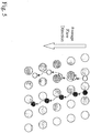
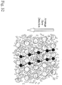
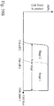
- each row of posts is shifted horizontally with respect to the previous row by ⁇ , where ⁇ is the center-to-center distance between the posts ( Fig. 1A ).
- the parameter ⁇ / ⁇ (the "bifurcation ratio," ⁇ ) determines the ratio of flow bifurcated to the left of the next post.
- ⁇ is 1/3, for the convenience of illustration.
- the flux through a gap between two posts is ⁇
- the minor flux is ⁇
- the major flux is (1- ⁇ ) ( Fig. 2 ).
- the flux through a gap is divided essentially into thirds ( Fig. 1B ).
- Fig. 1C illustrates the movement of a particles sized above the critical size through the array. Such particles move with the major flux, being transferred sequentially to the major flux passing through each gap.
- the critical size is approximately 2R critical , where R critical is the distance between the stagnant flow line and the post. If the center of mass of a particle, e.g., a cell, falls at least R critical away from the post, the particle would follow the major flux and move along the line-of-sight of the array. If the center of mass of a particle falls within R critical of the post, it follows the minor flux in a different direction.
- R critical can be determined if the flow profile across the gap is known ( Fig. 3 ); it is the thickness of the layer of fluids that would make up the minor flux.
- d R critical can be tailored based on the bifurcation ratio, ⁇ . In general, the smaller ⁇ , the smaller R critical .
- lymphocytes are spheres of ⁇ 5 ⁇ m diameter
- erythrocytes are biconcave disks of ⁇ 7 ⁇ m diameter, and ⁇ 1.5 ⁇ m thick.
- the long axis of erythrocytes (diameter) is larger than that of the lymphocytes, but the short axis (thickness) is smaller. If erythrocytes align their long axes to a flow when driven through an array of posts by the flow, their hydrodynamic size is effectively their thickness ( ⁇ 1.5 ⁇ m), which is smaller than lymphocytes.
- the method and device may therefore separate cells according to their shapes, although the volumes of the cells could be the same.
- particles having different deformability behave as if they have different sizes ( Fig. 5 ).
- two particles having the undeformed shape may be separated by deterministic lateral displacement, as the cell with the greater deformability may deform when it comes into contact with an obstacle in the array and change shape.
- separation in the device may be achieved based on any parameter that affects hydrodynamic size including the physical dimensions, the shape, and the deformability of the particle.
- the output containing cells larger than the critical size 2R critical
- waste containing cells smaller than the critical size waste containing cells smaller than the critical size.
- particles below the critical size may be collected while the particles above the critical size are discarded.
- Both types of outputs may also be desirably collected, e.g., when fractionating a sample into two or more sub-samples.
- Cells larger than the gap size will get trapped inside the array. Therefore, an array has a working size range. Cells have to be larger than a critical size (2R critical ) and smaller than a maximum pass-through size (array gap size) to be directed into the major flux.
- the improved devices can be used for the separation of particles, including bacteria, viruses, fungi, cells, cellular components, viruses, nucleic acids, proteins, and protein complexes, according to size.
- the devices may be used to effect various manipulations on particles in a sample. Such manipulations include enrichment or concentration of a particle, including size based fractionization, or alteration of the particle itself or the fluid carrying the particle.
- the devices are employed to enrich rare particles from a heterogeneous mixture or to alter a rare particle, e.g., by exchanging the liquid in the suspension or by contacting a particle with a reagent.
- Such devices allow for a high degree of enrichment with limited stress on cells, e.g., reduced mechanical lysis or intracellular activation of cells.
- the devices of the invention may be employed with any other particles whose size allows for separation in a device of the invention.
- a single stage contains an array of obstacles, e.g., cylindrical posts ( Fig. 1D ).
- the array has a maximum pass-through size that is several times larger than the critical size, e.g., when separating white blood cells from red blood cells. This result may be achieved using a combination of a large gap size d and a small bifurcation ratio ⁇ .
- the ⁇ is at most 1/2, e.g., at most 1/3, 1/10, 1/30, 1/100, 1/300, or 1/1000.
- the obstacle shape may affect the flow profile in the gap; however, the obstacles can be compressed in the flow direction, in order to make the array short ( Fig. 1E ).
- Single stage arrays may include bypass channels as described herein.
- Multiple-stage arrays In another embodiment, multiple stages are employed to separate particles over a wide range of sizes.
- An exemplary device is shown in Fig. 7 .
- the device shown has three stages, but any number of stages may be employed.
- the cut-off size (i.e. critical size) in the first stage is larger than the cut-off in the second stage, and the first stage cut-off size is smaller than the maximum pass-through size of the second stage ( Fig. 8 ).
- the first stage will deflect (and remove) particles, e.g., that would cause clogging in the second stage, before they reach the second stage.
- the second stage will deflect (and remove) particles that would cause clogging in the third stage, before they reach the third stage.
- an array can have as many stages as desired.
- devices of the invention may include bypass channels that remove output from an array. Although described here in terms of removing particles above the critical size, bypass channels may also be employed to remove output from any portion of the array.
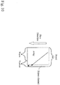
- Single bypass channels In this design, all stages share one bypass channel, or there is only one stage.
- the physical boundary of the bypass channel may be defined by the array boundary on one side and a sidewall on the other side ( Figs. 9-11 ).
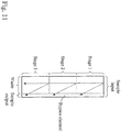
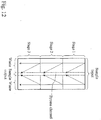
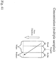
- Single bypass channels may also be employed with duplex arrays such that a central bypass channel separates the two arrays (i.e., two outer regions) ( Fig. 12 ).
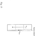
- Single bypass channels may also be designed, in conjunction with an array to maintain constant flux through a device ( Fig. 13 ).
- the bypass channel has varying width designed to maintain constant flux through all the stages, so that the flow in the channel does not interfere with the flow in the arrays.
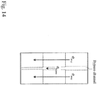
- Such a design may also be employed with an array duplex ( Fig. 14 ).
- Single bypass channels may also be designed in conjunction with the array in order to maintain substantially constant fluidic resistance through all stages ( Fig 15 ).
- Such a design may also be employed with an array duplex ( Fig. 16 .)
- each stage has its own bypass channel, and the channels are separated from each other by sidewalls, e.g., to prevent the mixing of the contents of different channels.
- Large particles, e.g., cells are deflected into the major flux to the lower right corner of the first stage and then into in a bypass channel (bypass channel 1 in Fig. 17 ).
- Smaller cells that would not cause clogging in the second stage proceed to the second stage, and cells above the critical size of the second stage are deflected to the lower right corner of the second stage and into in another bypass channel (bypass channel 2 in Fig. 17 ).
- This design may be repeated for as many stages as desired.
- bypass channels are not fluidically connected, allowing for separate collection and other manipulations.
- the bypass channels do not need to be straight or be physically parallel to each other ( Fig. 18 ).
- Multiple bypass channels may also be employed with duplex arrays ( Fig. 19 ).
- bypass channels may be designed, in conjunction with an array to maintain constant flux through a device ( Fig. 20 ).
- bypass channels are designed to remove an amount of flow so the flow in the array is not perturbed, i.e., substantially constant.
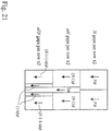
- Such a design may also be employed with an array duplex ( Fig. 21 ). In this design, the center bypass channel may be shared between the two arrays in the duplex.
- Boundary Design If the array were infinitely large, the flow distribution would be the same at every gap. The flux ⁇ going through a gap would be the same, and the minor flux would be ⁇ for every gap. In practice, the boundaries of the array perturb this infinite flow pattern. Portions of the boundaries of arrays may be designed to generate the flow pattern of an infinite array. Boundaries may be flow-feeding, i.e., the boundary injects fluid into the array, or flow-extracting, i.e., the boundary extracts fluid from the array.
- the distance between the array and the sidewall gradually increases to allow for the addition of ⁇ to the boundary from each gap along that boundary.
- the flow pattern inside this array is not affected by the bypass channel because of the boundary design.
- the distance between the array and the sidewall gradually decreases to allow for the addition of ⁇ to each gap along the boundary from that boundary.
- the flow pattern inside this array is not affected by the bypass channel because of the boundary design.
- a wide boundary may be desired if the boundary serves as a bypass channel, e.g., to allow for collection of particles.
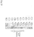
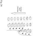
- a boundary may be employed that uses part of its entire flow to feed the array and feeds ⁇ into each gap at the boundary (represented by arrows in Fig. 24 ).
- the bypass channel includes two flow-feeding boundaries.
- the flux across the dashed line 1 in the bypass channel is ⁇ bypass.
- a flow ⁇ joins ⁇ bypass from a gap to the left of the dashed line.
- the shapes of the obstacles at the boundaries are adjusted so that the flows going into the arrays are ⁇ at each gap at the boundaries.
- the flux at dashed line 2 is again ⁇ bypass.
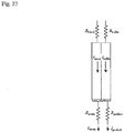
- On-chip flow resistor for defining and stabilizing flow
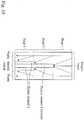
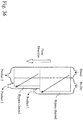
- FIG. 26 shows a schematic of planar device; a sample, e.g., blood, inlet channel, a buffer inlet channel, a waste outlet channel, and a product outlet channel are each connected to an array. The inlets and outlets act as flow resistors. Figure 26 also shows the corresponding fluidic resistances of these different device components.
- Figures 27 and 28 show the currents and corresponding widths of the sample and buffer flows within the array when the device has a constant depth and is operated with a given pressure drop. The flow is determined by the pressure drop divided by the resistance. In this particular device, I blood and I buffer are equivalent, and this determines equivalent widths of the blood and buffer streams in the array.
- Each of the inlet and outlet channels can be designed so that the pressure drops across the channels are appreciable to or greater than the fluctuations of the overall driving pressure. In typical cases, the inlet and outlet pressure drops are 0.001 to 0.99 times the driving pressure.
- multiplexed arrays Also disclosed herein are multiplexed arrays. Putting multiple arrays on one device increases sample-processing throughput and allows for parallel processing of multiple samples or portions of the sample for different fractions or manipulations. Multiplexing is further desirable for preparative devices.
- the simplest multiplex device includes two devices attached in series, i.e., a cascade. For example, the output from the major flux of one device may be coupled to the input of a second device. Alternatively, the output from the minor flux of one device may be coupled to the input of the second device.
- Two arrays can be disposed side-by-side, e.g., as mirror images ( Fig. 29 ).
- the critical size of the two arrays may be the same or different.
- the arrays may be arranged so that the major flux flows to the boundary of the two arrays, to the edge of each array, or a combination thereof.
- Such a duplexed array may also contain a central bypass channel disposed between the arrays, e.g., to collect particles above the critical size or to alter the sample, e.g., through buffer exchange, reaction, or labeling.
- two or more arrays that have separated inputs may be disposed on the same device ( Fig. 30A ). Such an arrangement could be employed for multiple samples, or the plurality of arrays may be connected to the same inlet for parallel processing of the same sample.
- the outlets may or may not be fluidically connected.
- the outlets may be connected for high throughput sample processing.
- the arrays may not all have the same critical size or the particles in the arrays may not all be treated in the same manner, and the outlets may not be fluidically connected.
- Multiplexing may also be achieved by placing a plurality of duplex arrays on a single device ( Fig. 30B ).
- a plurality of arrays, duplex or single, may be placed in any possible three-dimensional relationship to one another.
- Devices as described herein may also feature a small-footprint. Reducing the footprint of an array can lower cost, and reduce the number of collisions with obstacles to eliminate any potential mechanical damage or other effects to particles.
- the length of a multiple stage array can be reduced if the boundaries between stages are not perpendicular to the direction of flow. The length reduction becomes significant as the number of stages increases.
- Fig. 31 shows a small-footprint three-stage array.
- devices as described herien may include additional elements, e.g., for isolating, collection, manipulation, or detection. Such elements are known in the art. Arrays may also be employed on a device having components for other types of separation, including affinity, magnetic, electrophoretic, centrifugal, and dielectrophoretic separation. Devices as described herein may also be employed with a component for two-dimensional imaging of the output from the device, e.g., an array of wells or a planar surface. Preferably, arrays of gaps as described herein are employed in conjunction with an affinity enrichment.
- the devices may also be employed in conjunction with other enrichment devices, either on the same device or in different devices.
- Other enrichment techniques are described, e.g., in International Publication Nos. 2004/029221 and 2004/113877 , U.S. Patent No. 6,692,952 , U.S. Application Publications 2005/0282293 and 2005/0266433 , and U.S. Application No. 60/668,415 .
- Devices of the invention and as described herein may be fabricated using techniques well known in the art. The choice of fabrication technique will depend on the material used for the device and the size of the array. Exemplary materials for fabricating the devices include glass, silicon, steel, nickel, poly(methylmethacrylate) (PMMA), polycarbonate, polystyrene, polyethylene, polyolefins, silicones (e.g., poly(dimethylsiloxane)), and combinations thereof. Other materials are known in the art. For example, deep Reactive Ion Etching (DRIE) is used to fabricate silicon-based devices with small gaps, small obstacles and large aspect ratios (ratio of obstacle height to lateral dimension).
- DRIE deep Reactive Ion Etching
- Thermoforming (embossing, injection molding) of plastic devices can also be used, e.g., when the smallest lateral feature is 20 microns and the aspect ratio of these features is less than 3.
- Additional methods include photolithography (e.g., stereolithography or x-ray photolithography), molding, embossing, silicon micromachining, wet or dry chemical etching, milling, diamond cutting, Lithographie Galvanoformung and Abformung (LIGA), and electroplating.
- photolithography e.g., stereolithography or x-ray photolithography
- molding embossing, silicon micromachining, wet or dry chemical etching, milling, diamond cutting, Lithographie Galvanoformung and Abformung (LIGA), and electroplating.
- PEX Lithographie Galvanoformung and Abformung
- electroplating Lithographie Galvanoformung and Abformung
- thermoplastic injection molding and compression molding may be suitable.
- Conventional thermoplastic injection molding used for mass-fabrication of compact discs (which preserves fidelity of features in sub-microns) may also be employed to fabricate the devices.
- the device features are replicated on a glass master by conventional photolithography.
- the glass master is electroformed to yield a tough, thermal shock resistant, thermally conductive, hard mold. This mold serves as the master template for injection molding or compression molding the features into a plastic device.
- compression molding or injection molding may be chosen as the method of manufacture.
- Compression molding also called hot embossing or relief imprinting
- Injection molding works well for high-aspect ratio structures but is most suitable for low molecular weight polymers.
- a device may be fabricated in one or more pieces that are then assembled. Layers of a device may be bonded together by clamps, adhesives, heat, anodic bonding, or reactions between surface groups (e.g., wafer bonding). Alternatively, a device with channels in more than one plane may be fabricated as a single piece, e.g., using stereolithography or other three-dimensional fabrication techniques.
- one or more channel walls may be chemically modified to be non-adherent or repulsive.
- the walls may be coated with a thin film coating (e.g., a monolayer) of commercial non-stick reagents, such as those used to form hydrogels.
- chemical species that may be used to modify the channel walls include oligoethylene glycols, fluorinated polymers, organosilanes, thiols, poly-ethylene glycol, hyaluronic acid, bovine serum albumin, poly-vinyl alcohol, mucin, poly-HEMA, methacrylated PEG, and agarose.
- Charged polymers may also be employed to repel oppositely charged species.
- the type of chemical species used for repulsion and the method of attachment to the channel walls will depend on the nature of the species being repelled and the nature of the walls and the species being attached. Such surface modification techniques are well known in the art.
- the walls may be functionalized before or after the device is assembled.
- the channel walls may also be coated in order to capture materials in the sample, e.g., membrane fragments or proteins.
- Devices of the invention and as further described herein may be employed in any application where the production of a sample enriched in particles above or below a critical size is desired.
- a preferred use of the device is in produced samples enriched in cells, e.g., rare cells. Once an enriched sample is produced, it may be collected for analysis or otherwise manipulated, e.g., through further enrichment.
- the method of the invention uses a flow that carries cells to be separated through the array of gaps.
- the flow is aligned at a small angle (flow angle) with respect to a line-of-sight of the array.
- Cells having a hydrodynamic size larger than a critical size migrate along the line-of-sight in the array, whereas those having a hydrodynamic size smaller than the critical size follow the flow in a different direction.
- Flow in the device occurs under laminar flow conditions.
- the method of the invention may be employed with concentrated samples, e.g., where particles are touching, hydrodynamically interacting with each other, or exerting an effect on the flow distribution around another particle.
- the method can separate white blood cells from red blood cells in whole blood from a human donor. Human blood typically contains ⁇ 45% of cells by volume. Cells are in physical contact and/or coupled to each other hydrodynamically when they flow through the array.
- Fig. 32 shows schematically that cells are densely packed inside an array and could physically interact with each other.
- the methods disclosed herein can be employed to produce a sample enriched in particles of a desired hydrodynamic size. Applications of such enrichment include concentrating particles, e.g., rare cells, and size fractionization, e.g., size filtering (selecting cells in a particular range of sizes). The methods may also be used to enrich components of cells, e.g., nuclei. Nuclei or other cellular components may be produced by manipulation of the sample, e.g., lysis as described herein, or be naturally present in the sample, e.g., via apoptosis or necrosis.
- the methods of the invention retain at least 1%, 10%, 30%, 50%, 75%, 80%, 90%, 95%, 98%, or 99% of the desired particles compared to the initial mixture, while potentially enriching the desired particles by a factor of at least 1, 10, 100, 1000, 10,000, 100,000, or even 1,000,000 relative to one or more non-desired particles.
- the enrichment may also result in a dilution of the separated particles compared to the original sample, although the concentration of the separated particles relative to other particles in the sample has increased.
- the dilution is at most 90%, e.g., at most 75%, 50%, 33%, 25%, 10%, or 1%.
- a rare particle is a particle that is present as less than 10% of a sample.
- exemplary rare particles include, depending on the sample, fetal cells, nucleated red blood cells (e.g., fetal or maternal), stem cells (e.g., undifferentiated), cancer cells, immune system cells (host or graft), epithelial cells, connective tissue cells, bacteria, fungi, viruses, parasites, and pathogens (e.g., bacterial or protozoan).
- Such rare particles may be isolated from samples including bodily fluids, e.g., blood, or environmental sources, e.g., pathogens in water samples.
- Fetal cells may be enriched from maternal peripheral blood, e.g., for the purpose of determining sex and identifying aneuploidies or genetic characteristics, e.g., mutations, in the developing fetus.
- Cancer cells may also be enriched from peripheral blood for the purpose of diagnosis and monitoring therapeutic progress.
- Bodily fluids or environmental samples may also be screened for pathogens or parasites, e.g., for coliform bacteria, blood borne illnesses such as sepsis, or bacterial or viral meningitis.
- Rare cells also include cells from one organism present in another organism, e.g., an in cells from a transplanted organ.
- An exemplary preparative application includes generation of cell packs from blood.
- the methods of the invention may be configured to produce fractions enriched in platelets, red blood cells, and white cells. By using multiplexed devices or multistage devices, all three cellular fractions may be produced in parallel or in series from the same sample. Other methods disclosed herein can be employed to separate nucleated from enucleated cells, e.g., from cord blood sources.
- the devices disclosed herein may be designed to enrich cells with a minimum number of collisions between the cells and obstacles. This minimization reduces mechanical damage to cells and also prevents intracellular activation of cells caused by the collisions. This gentle handling of the cells preserves the limited number of rare cells in a sample, prevents rupture of cells leading to contamination or degradation by intracellular components, and prevents maturation or activation of cells, e.g., stem cells or platelets. In some methods, cells are enriched such that fewer than 30%, 10 %, 5%, 1%, 0.1%, or even 0.01 % are activated or mechanically lysed.
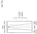
- Fig. 33 shows a typical size distribution of cells in human peripheral blood.
- the white blood cells range from ⁇ 4 ⁇ m to ⁇ 18 ⁇ m, whereas the red blood cells are ⁇ 1.5 ⁇ m (short axis).
- An array designed to separate white blood cells from red blood cells typically has a cut-off size (i.e., critical size) of 2 to 4 ⁇ m and a maximum pass-through size of greater than 18 ⁇ m.
- the device would function as a detector for abnormalities in red blood cells.
- the deterministic principle of sorting enables a predictive outcome of the percentage of enucleated cells deflected in the device.
- a disease state such as malarial infection or sickle cell anemia
- the distortion in shape and flexibility of the red cells would significantly change the percentage of cells deflected.
- This change can be monitored as a first level sentry to alert to the potential of a diseased physiology to be followed by microscopy examination of shape and size of red cells to assign the disease.
- the method is also generally applicable monitoring for any change in flexibility of particles in a sample.
- the device would function as a detector for platelet aggregation.
- the deterministic principle of sorting enables a predictive outcome of the percentage of free platelets deflected in the device. Activated platelets would form aggregates, and the aggregates would be deflected. This change can be monitored as a first level sentry to alert the compromised efficacy of a platelet pack for reinfusion.
- the method is also generally applicable monitoring for any change in size, e.g., through agglomeration, of particles in a sample.
- cells of interest are contacted with an altering reagent that may chemically or physically alter the particle or the fluid in the suspension.
- altering reagent that may chemically or physically alter the particle or the fluid in the suspension.
- Such applications include purification, buffer exchange, labeling (e.g., immunohistochemical, magnetic, and histochemical labeling, cell staining, and flow in-situ fluorescence hybridization (FISH)), cell fixation, cell stabilization, cell lysis, and cell activation.
- labeling e.g., immunohistochemical, magnetic, and histochemical labeling, cell staining, and flow in-situ fluorescence hybridization (FISH)
- FISH flow in-situ fluorescence hybridization
- Fig. 34A shows this effect schematically for a single stage device
- Fig. 34B shows this effect for a multistage device
- Fig. 34C shows this effect for a duplex array
- Fig. 34D shows this effect for a multistage duplex array.
- blood cells may be separated from plasma.
- Such transfers of particles from one liquid to another may be also employed to effect a series of alterations, e.g., Wright staining blood on-chip.
- Such a series may include reacting a particle with a first reagent and then transferring the particle to a wash buffer, and then to another reagent.
- Figs. 35A , 35B , 35C illustrate a further example of alteration in a two-stage device having two bypass channels.
- large blood particles are moved from blood to buffer and collected in stage 1
- medium blood particles are moved from blood to buffer in stage 2
- small blood particles that are not removed from the blood in stages 1 and 2 are collected.
- Fig. 35B illustrates the size cut-off of the two stages
- Fig. 35C illustrates the size distribution of the three fractions collected.
- Fig. 36 illustrates an example of alteration in a two-stage device having bypass channels that are disposed between the lateral edge of the array and the channel wall.
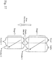
- Fig. 37 illustrates a device similar to that in Fig. 36 , except that the two stages are connected by fluidic channels.
- Fig. 38 illustrates alteration in a device having two stages with a small footprint.
- Figs. 39A-39B illustrates alteration in a device in which the output from the first and second stages is captured in a single channel.
- Fig. 40 illustrates another device for use in the methods of the invention.
- Fig. 41 illustrates the use of a device to perform multiple, sequential alterations on a particle.
- a blood particle is moved from blood into a reagent that reacts with the particle, and the reacted particle is then moved into a buffer, thereby removing the unreacted reagent or reaction byproducts. Additional steps may be added.
- reagents are added to the sample to selectively or nonselectively increase the hydrodynamic size of the particles within the sample.
- This modified sample is then pumped through an obstacle array. Because the particles are swollen and have an increased hydrodynamic diameter, it will be possible to use obstacle arrays with larger and more easily manufactured gap sizes.
- the steps of swelling and size-based enrichment may be performed in an integrated fashion on a device.
- Suitable reagents include any hypotonic solution, e.g., deionized water, 2% sugar solution, or neat non-aqueous solvents.
- Other reagents include beads, e.g., magnetic or polymer, that bind selectively (e.g., through antibodies or avidin-biotin) or non-selectively.
- reagents are added to the sample to selectively or nonselectively decrease the hydrodynamic size of the particles within the sample.
- Nonuniform decrease in particles in a sample will increase the difference in hydrodynamic size between particles.
- nucleated cells are separated from enucleated cells by hypertonically shrinking the cells.
- the enucleated cells can shrink to a very small particle, while the nucleated cells cannot shrink below the size of the nucleus.
- Exemplary shrinking reagents include hypertonic solutions.
- affinity functionalized beads are used to increase the volume of particles of interest relative to the other particles present in a sample, thereby allowing for the operation of a obstacle array with a larger and more easily manufactured gap size.
- Enrichment and alteration may also be combined, e.g., where desired cells are contacted with a lysing reagent and cellular components, e.g., nuclei, are enriched based on size.
- cellular components e.g., nuclei
- particles may be contacted with particulate labels, e.g., magnetic beads, which bind to the particles. Unbound particulate labels may be removed based on size.
- Enrichment and alteration methods employing devices of the invention and as further disclosed herein may be combined with other particulate sample manipulation techniques.
- further enrichment or purification of a particle may be desirable.
- Further enrichment may occur by any technique, including affinity enrichment.
- Suitable affinity enrichment techniques include contact particles of interest with affinity agents bound to channel walls or an array of obstacles.
- Fluids may be driven through a device either actively or passively. Fluids may be pumped using electric field, a centrifugal field, pressure-driven fluid flow, an electro-osmotic flow, and capillary action. In some methods and devices disclosed herein, the average direction of the field will be parallel to the walls of the channel that contains the array.
- the method employs differential lysis between the cells of interest and other cells (e.g., other nucleated cells) in the sample.
- Cells of interest may be lysed using any suitable method.
- cells may be lysed by being contacted with a solution that causes preferential lysis.
- Lysis solutions for these cells may include cell specific IgM molecules and proteins in the complement cascade to initiate complement mediated lysis.
- Another kind of lysis solution may include viruses that infect a specific cell type and cause lysis as a result of replication (see, e.g., Pawlik et al. Cancer 2002, 95:1171-81 ).
- Other lysis solutions include those that disrupt the osmotic balance of cells, e.g., hypotonic or hypertonic (e.g., distilled water), to cause lysis.
- Other lysis solutions are known in the art.
- Lysis may also occur by mechanical means, e.g., by passing cells through a sieve or other structure that mechanically disrupts the cells, through the addition of heat, acoustic, or light energy to lyse the cells, or through cell-regulated processes such as apoptosis and necrosis.
- Cells may also be lysed by subjecting them to one or more cycles of freezing and thawing. Additionally, detergents may be employed to solubilize the cell membrane, lysing the cells to liberate their contents.
- the cells of interest are rare cells, e.g., circulating cancer cells, fetal cells (such as fetal nucleated red blood cells), blood cells (such as nucleated red blood cells, including maternal and/or fetal nucleated red blood cells), immune cells, connective tissue cells, parasites, or pathogens (such as, bacteria, protozoa, and fungi).
- rare cells e.g., circulating cancer cells, fetal cells (such as fetal nucleated red blood cells), blood cells (such as nucleated red blood cells, including maternal and/or fetal nucleated red blood cells), immune cells, connective tissue cells, parasites, or pathogens (such as, bacteria, protozoa, and fungi).
- Most circulating rare cells of interest have compromised membrane integrity as a result of the immune attack from the host RES (Reticulo-Endothelial-System), and accordingly are more susceptible to lysis.
- RES Reticulo-Endothelial-System
- the cells of interest are lysed as they flow through a microfluidic device, e.g., as described in International Publications WO 2004/029221 and WO 2004/113877 or as described herein.
- cells of interest are first bound to obstacles in a microfluidic device, e.g., as described in U.S. Patent No. 5,837,115 , and then lysed. In this method, the cellular components of cells of interest are released from the obstacles, while cellular components of undesired cells remain bound.
- Desired cellular components may be separated from cell lysate by any suitable method, e.g., based on size, weight, shape, charge, hydrophilicity/hydrophobicity, chemical reactivity or inertness, or affinity.
- nucleic acids, ions, proteins, and other charged species may be captured by ion exchange resins or separated by electrophoresis.
- Cellular components may also be separated based on size or weight by size exclusion chromatography, centrifugation, or filtration.
- Cellular components may also be separated by affinity mechanisms (i.e., a specific binding interaction, such antibody-antigen and nucleic acid complementary interactions), e.g., affinity chromatography, binding to affinity species bound to surfaces, and affinity-based precipitation.
- nucleic acids e.g., genomic DNA
- sequence specific probes e.g., attached to beads or an array
- Cellular components may also be collected on the basis of shape or deformability or non-specific chemical interactions, e.g., chromatography or reverse phase chromatography or precipitation with salts or other reagents, e.g., organic solvents.
- Cellular components may also be collected based on chemical reactions, e.g., binding of free amines or thiols.
- cellular components Prior to collection, cellular components may also be altered to enable or enhance a particular mode of collection, e.g., via denaturation, enzymatic cleavage (such as via a protease, endonuclease, exonuclease, or restriction endonuclease), or labeling or other chemical reaction.
- denaturation e.g., denaturation, enzymatic cleavage (such as via a protease, endonuclease, exonuclease, or restriction endonuclease), or labeling or other chemical reaction.
- the level of purity required for collected cellular components will depend on the particular manipulation employed and may be determined by the skilled artisan.
- the cellular component may not need to be isolated from the lysate, e.g., when the cellular component of interest may be analyzed or otherwise manipulated without interference from other cellular components.
- Affinity based manipulations e.g., reaction with nucleic acid probes or primers, aptamers, antibodies, or sequence specific intercalating agents, with or without detectable labels
- Affinity based manipulations are amenable for use without purification of the cellular components.
- a device e.g., as described in U.S. Application Publication 2004/0144651 or as described herein, is employed to isolate particulate cellular components of interest, e.g., nuclei, from the lysate based on size.
- the particulate cellular components of interest may be separated from other particulate cellular components and intact cells using the device.
- nucleic acid e.g., nuclei, mitochondria, and nuclear or cytoplasmic DNA or RNA.
- nucleic acids may include RNA, such as mRNA or rRNA, or DNA, such as chromosomal DNA, e.g., that has been cleaved, or DNA that has undergone apoptotic processing.
- Genetic analysis of the nucleic acid in the cellular component may be performed by any suitable methods, e.g., PCR, FISH, and sequencing. Genetic information may be employed to diagnose disease, status as a genetic disease carrier, or infection with pathogens or parasites. If acquired from fetal cells, genetic information relating to sex, paternity, mutations (e.g., cystic fibrosis), and aneuploidy (e.g., trisomy 21) may be obtained. In some methods disclosed herein, analysis of fetal cells or components thereof is used to determine the presence or absence of a genetic abnormality, such as a chromosomal, DNA, or RNA abnormality.
- a genetic abnormality such as a chromosomal, DNA, or RNA abnormality.
- autosomal chromosome abnormalities include, but are not limited to, Angleman syndrome (15q11.2-q13), cri-du-chat syndrome (5p-), DiGeorge syndrome and Velo-cardiofacial syndrome (22q11.2), Miller-Dieker syndrome (17p13.3), Prader-Willi syndrome (15q11.2-q13), retinoblastoma (13q14), Smith-Magenis syndrome (17p11.2), trisomy 13, trisomy 16, trisomy 18, trisomy 21 (Down syndrome), triploidy, Williams syndrome (7q11.23), and Wolf-Hirschhorn (4p-).
- sex chromosome abnormalities include, but are not limited to, Kallman syndrome (Xp22.3), steroid sulfate deficiency (STS) (Xp22.3), X-linked ichthiosis (Xp22.3), Klinefelter syndrome (XXY); fragile X syndrome; Turner syndrome; metafemales or trisomy X; and monosomy X.
- chromosomal abnormalities that can be analyzed by the systems herein include, but are not limited to, deletions (small missing sections); microdeletions (a minute amount of missing material that may include only a single gene); translocations (a section of a chromosome is attached to another chromosome); and inversions (a section of chromosome is snipped out and reinserted upside down).
- analysis of fetal cells or components thereof is used to analyze SNPs and predict a condition of the fetus based on such SNPs. If acquired from cancer cells, genetic information relating to tumorgenic properties may be obtained. If acquired from viral or bacterial cells, genetic information relating to the pathogenicity and classification may be obtained.
- the components may be analyzed to diagnose disease or to monitor health.
- proteins or metabolites from rare cells e.g., fetal cells
- affinity-based assays e.g., ELISA
- analytical techniques e.g., chromatography and mass spectrometry.
- Samples may be employed in the methods described herein with or without purification, e.g., stabilization and removal of certain components. Some sample may be diluted or concentrated prior to introduction into the device.
- a sample is contacted with a microfluidic device containing a plurality of obstacles, e.g., as described in U.S. Patent No. 5,837,115 or as described herein.
- Cells of interest bind to affinity moieties bound to the obstacles in such a device and are thereby enriched relative to undesired cells, e.g., as described in WO 2004/029221 .
- cells of non-interest bind to affinity moieties bound to the obstacles, while allowing the cells of interest to pass through resulting in an enriched sample with cells of interest, e.g., as described in WO 2004/029221 .
- the sized based method and the affinity-based method may also be combined in a two-step method to further enrich a sample in cells of interest.
- a cell sample is pre-filtered by contact with a microfluidic device containing a plurality of obstacles disposed such that particles above a certain size are deflected to travel in a direction not parallel to the average direction of fluid flow, e.g., as described in U.S. Application Publication 2004/0144651 or as described herein.
- Figures 42A-42E show an exemplary device of the invention, characterized as follows.
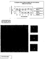
- Measurement techniques Complete blood counts were determined using a Coulter impedance hematology analyzer (COULTER® Ac ⁇ T diffTM, Beckman Coulter, Fullerton, CA).
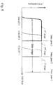
- Figs. 43A-43F shows typical histograms generated by the hematology analyzer from a blood sample and the waste (buffer, plasma, red blood cells, and platelets) and product (buffer and nucleated cells) fractions generated by the device.
- the following table shows the performance over 5 different blood samples: Sample number Throughput Performance Metrics RBC removal Platelet removal WBC loss 1 4 mL/hr 100% 99% ⁇ 1% 2 6 mL/hr 100% 99% ⁇ 1% 3 6 mL/hr 100% 99% ⁇ 1% 4 6 mL/hr 100% 97% ⁇ 1% 5 6 mL/hr 100% 98% ⁇ 1%
- Figures 44 shows an exemplary device of the invention, characterized as follows.
- Measurement techniques Complete blood counts were determined using a Coulter impedance hematology analyzer (COULTER® Ac ⁇ T diffTM, Beckman Coulter, Fullerton, CA).
- Figure 45 shows a schematic of the device used to separate nucleated cells from fetal cord blood.
- Nucleated cells from the blood were separated from enucleated cells (red blood cells and platelets), and plasma delivered into a buffer stream of calcium and magnesium-free Dulbecco's Phosphate Buffered Saline (14190-144, Invitrogen, Carlsbad, CA) containing 1% Bovine Serum Albumin (BSA) (A8412-100ML, Sigma-Aldrich, St Louis, MO) and 2 mM EDTA (15575-020, Invitrogen, Carlsbad, CA).
- BSA Bovine Serum Albumin
- Example 1 The device and process described in detail in Example 1 were used in combination with immunomagnetic affinity enrichment techniques to demonstrate the feasibility of isolating fetal cells from maternal blood.
- the nucleated cell fraction was labeled with anti-CD71 microbeads (130-046-201, Miltenyi Biotech Inc., Auburn, CA) and enriched using the MiniMACSTM MS column (130-042-201, Miltenyi Biotech Inc., Auburn, CA) according to the manufacturer's specifications. Finally, the CD71-positive fraction was spotted onto glass slides.
- Measurement techniques Spotted slides were stained using fluorescence in situ hybridization (FISH) techniques according to the manufacturer's specifications using Vysis probes (Abbott Laboratories, Downer's Grove, IL). Samples were stained from the presence of X and Y chromosomes. In one case, a sample prepared from a known Trisomy 21 pregnancy was also stained for chromosome 21.
- FISH fluorescence in situ hybridization
- each device provides the number of stages in series, the gap size for each stage, ⁇ (Flow Angle), and the number of channels per device (Arrays/Chip).
- ⁇ Flow Angle
- Arrays/Chip the number of channels per device
- This device includes five stages in a single array.
- This device includes three stages, where each stage is a duplex having a bypass channel.
- the height of the device was 125 ⁇ m.
- Array Design symmetric 3 stage array with central collection channel Gap Sizes: Stage 1: 8 ⁇ m Stage 2: 12 ⁇ m Stage 3: 18 ⁇ m Stage 4: Stage 5: Flow Angle: 1/10 Arrays / Chip: 1 Other: central collection channel
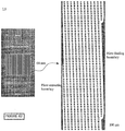
- Figure 49A shows the mask employed to fabricate the device.
- Figures 49B-49D are enlargements of the portions of the mask that define the inlet, array, and outlet.
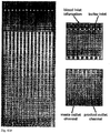
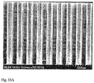
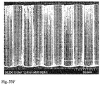
- Figures 50A-50G show SEMs of the actual device.
- This device includes three stages, where each stage is a duplex having a bypass channel. "Fins" were designed to flank the bypass channel to keep fluid from the bypass channel from re-entering the array.
- the chip also included on-chip flow resistors, i.e., the inlets and outlets possessed greater fluidic resistance than the array. The height of the device was 117 ⁇ m.
- Array Design 3 stage symmetric array Gap Sizes: Stage 1: 8 ⁇ m Stage 2: 12 ⁇ m Stage 3: 18 ⁇ m Stage 4: Stage 5: Flow Angle: 1/10 Arrays / Chip: 10 Other: large fin central collection channel on-chip flow resistors
- Figure 51A shows the mask employed to fabricate the device.
- Figures 51B-51D are enlargements of the portions of the mask that define the inlet, array, and outlet.
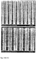
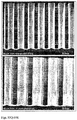
- Figures 52A-52F show SEMs of the actual device.
- This device includes three stages, where each stage is a duplex having a bypass channel. "Fins" were designed to flank the bypass channel to keep fluid from the bypass channel from re-entering the array. The edge of the fin closest to the array was designed to mimic the shape of the array.
- the chip also included on-chip flow resistors, i.e., the inlets and outlets possessed greater fluidic resistance than the array. The height of the device was 138 ⁇ m.
- Array Design 3 stage symmetric array Gap Sizes: Stage 1: 8 ⁇ m Stage 2: 12 ⁇ m Stage 3: 18 ⁇ m Stage 4: Stage 5: Flow Angle: 1/10 Arrays / Chip: 10 Other: alternate large fin central collection channel on-chip flow resistors
- Figure 45A shows the mask employed to fabricate the device.
- Figures 45B-45D are enlargements of the portions of the mask that define the inlet, array, and outlet.
- Figures 532A-532F show SEMs of the actual device.
- This device includes three stages, where each stage is a duplex having a bypass channel.
- "Fins” were optimized using Femlab to flank the bypass channel to keep fluid from the bypass channel from re-entering the array.
- the edge of the fin closest to the array was designed to mimic the shape of the array.
- the chip also included on-chip flow resistors, i.e., the inlets and outlets possessed greater fluidic resistance than the array.
- the height of the device was 139 or 142 ⁇ m.
- Figure 54A shows the mask employed to fabricate the device.
- Figures 54B-54D are enlargements of the portions of the mask that define the inlet, array, and outlet.
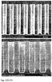
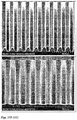
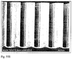
- Figures 55A-55S show SEMs of the actual device.
- This device includes a single stage, duplex device having a bypass channel disposed to receive output from the ends of both arrays.
- the obstacles in this device are elliptical.
- the array boundary was modeled in Femlab.
- the chip also included on-chip flow resistors, i.e., the inlets and outlets possessed greater fluidic resistance than the array. The height of the device was 152 ⁇ m.
- Array Design single stage symmetric array Gap Sizes: Stage 1: 24 ⁇ m Stage 2: Stage 3: Stage 4: Stage 5: Flow Angle: 1/60 Arrays / Chip: 14 Other: central barrier ellipsoid posts on-chip resistors Femlab modeled array boundary
- Figure 44A shows the mask employed to fabricate the device.
- Figures 44B-44D are enlargements of the portions of the mask that define the inlet, array, and outlet.
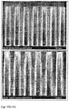
- Figures 56A-56C show SEMs of the actual device.
- Figure 57 shows a flowchart for a method of isolating fetal nuclei from a maternal blood sample. The method results in the preferential lysis of red blood cells ( Figure 58 ).
- Density centrifugation methods were used to obtain an enriched population of lymphocytes. An aliquot of these lymphocytes were exposed to a hypotonic ammonium chloride solution for sufficient time to lyse > 95% of the cells. These nuclei were then labeled with Hoechst 33342 (bisbenzimide H 33342), a specific stain for AT rich regions of double stranded DNA, and added back to the original lymphocyte population to create a 90:10 (cell: nuclei) mixture. This mixture was fed into a device that separated cells from nuclei based on size, as depicted in Figure 60 , and the waste and product fractions were collected and the cell: nuclei ratio contained in each fraction were measured.
- Hoechst 33342 bisbenzimide H 33342
- the lysed nuclei of mixed cell suspensions that have been processed through a differential lysis procedure can be enriched by adding a sucrose cushion solution to the lysate. This mixture is then layered on a pure sucrose cushion solution and then centrifuged to form an enriched nuclei pellet. The unlysed cells and debris are aspirated from the supernatant; the nuclei pellet is re-suspended in a buffer solution and then cytospun onto glass slides.
- Acid Alcohol Total Cell lysis and Nuclear RNA FISH for Targeted Cell Identification Product obtained from a device that separated cells based on size, as depicted in Figure 60 , was exposed to an acid alcohol solution (methanol:acetic acid 3:1 v/v) for 30 minutes on ice resulting in the lysis of >99% of enucleated cells and >99.0% lysis of nucleated cells.
- a hypotonic treatment by exposing the cells to salt solution (0.6% NaCl) for 30 minutes to swell the nuclei before acid alcohol lysis can also be included.
- the released nuclei can be quantitatively deposited on to a glass slide by cytospin and FISHed ( Figure 66a and 66b ).
- the cells of interest such as fetal nucleated erythrocytes
- RNA-FISH probes for positive selection, such as zeta-, epsilon, gamma-globins, and negative selection such as beta-globin or analyzing the length of telomeres.
- positive selection such as zeta-, epsilon, gamma-globins
- negative selection such as beta-globin or analyzing the length of telomeres.
- Other methods for distinguishing between fetal and non-fetal cells are known in the art, e.g., U.S. Patent No. 5,766,843 .
- Figure 62 shows a device that is optimized for separation of particles in blood. It is a one-stage device with a fixed gap width of 22 ⁇ m, with 48 multiplexed arrays for parallel sample processing.
- the parameters of the device are as follows: Array Design: L5 Gap Sizes: Stage 1: 22 ⁇ m Flow Angle: 1/50 Arrays/Chip: 48 Nominal Depth 150 ⁇ m Device Footprint 32 mm x 64 mm Design Features • Multiplexed single arrays • Optimized bypass channels • Flow stabilization • Flow-feeding and Flow-extracting boundaries
- Blood was obtained from pregnant volunteer donors and diluted 1:1 with Dulbecco's phosphate buffered saline (without calcium and magnesium)(iDPBS).
- Blood and Running Buffer iDPBS with 1% BSA and 2mM EDTA
- iDPBS 1% BSA and 2mM EDTA
- Blood and Running Buffer were delivered using an active pressure of 0.8 PSI to the device engaged with a manifold as described in Example 13.
- Blood was separated into two components nucleated cells in Running Buffer and enucleated cells and plasma proteins in Running Buffer. Both components were analyzed using a standard impedance counter.
- the component containing nucleated cells was additionally characterized using a propidium iodide staining solution in conjunction with a standard Nageotte counting chamber to determine total nucleated cell loss.
- FIG. 63 An exemplary manifold into which a microfluidic device as described herein is inserted is shown in Figure 63 .
- the manifold has two halves between which a microfluidic device is disposed.
- One half of the manifold includes separate inlets for blood and buffer, each of which is connected to a corresponding fluid reservoir.
- the channels in the device are oriented so that they connect to the reservoirs via through holes in the device.
- the device is oriented vertically, and the processed blood is collected as it drips out of the product outlet.
- a region around the product outlet of the microfluidic device may also be marked with a hydrophobic substance, e.g., from a permanent marker, to limit the size of drops formed.
- the device also includes two hydrophobic vent filters, e.g., 0.2 ⁇ m PTFE filters. These filters allow air trapped in the device to be displaced by aqueous solutions, but do not let the liquid pass at low pressures, e.g., ⁇ 5 psi.
- buffer e.g., Dulbecco's PBS with 1% bovine serum albumin (w/v) and 2 mM EDTA
- the buffer is then pumped into the device via the buffer inlet in the manifold at a pressure of ⁇ 5 psi.
- the buffer then fills the buffer chamber by displacing air through the hydrophobic vent filter and then fills the channels in the microfluidic device and the blood chamber.
- a hydrophobic vent filter connected to the blood chamber allows for the displacement of air in the chamber.
- a fetal nRBC population enriched by any of the devices described herein is subjected to hypotonic shock by adding a large volume of low ionic strength buffer, e.g., deionized water to lyse enucleated RBCs and nRBCs selectively and release their nuclei.
- the hypotonic shock is then terminated by adding an equal volume of a high ionic strength buffer.
- Figure 64 illustrates the selective lysis of fetal nRBCs vs. maternal nRBCs as a function of the duration of exposure to lysing conditions.
- This selective lysis procedure also can be used to lyse selectively fetal nRBCs in a population of cells composed of fetal nRBC, maternal nRBC, enucleated fetal and maternal RBCs, and fetal and maternal white blood cells.
- fetal nRBCs and maternal nRBCs were lysed over time during which the number of lysed (non-viable) fetal nRBCs increased by a factor of 10, whereas the number of lysed maternal nRBCs increased by a smaller multiple.
- the lysed cells were stained with propidium iodide and were concentrated through gradient centrifugation to determine the ratio of lysed fetal nRBCs vs. maternal nRBCs.
- An optimized time duration can be determined and applied to enrich selectively for fetal nRBCs nuclei.
- a sample enriched in fetal nRBCs is treated with a RBC lysis buffer, such as 0.155 M NH 4 Cl, 0.01 M KHCO 3 , 2 mM EDTA, 1% BSA with a carbonic anhydrase inhibitor, such as acetazolamide (e.g., at 0.1-100 mM), to induce lysis, followed by termination of the lysis process using a large volume of balanced salt buffer , such as 10x volume of 1xPBS, or balanced salt buffer, such as 1xPBS, with an ion exchange channel inhibitor such as 4,4'-diisothiocyanostilbene-2,2'-disulphonic acid (DIDS).
- a RBC lysis buffer such as 0.155 M NH 4 Cl, 0.01 M KHCO 3 , 2 mM EDTA, 1% BSA with a carbonic anhydrase inhibitor, such as acetazolamide (e.g., at 0.1-100 mM)
- K562 cells to simulate white blood cells, were labeled with Hoechst and calcein AM at room temperature for 30 minutes ( Figure 65 ). These labeled K562 cells were added to blood specimens, followed by the addition of buffer ( 0.155 M NH 4 Cl, 0.01 M KHCO 3 , 2 mM EDTA, 1% BSA, and 10 mM acetazolamide) (the ratio of buffer volume to spiked blood volume is 3:2). The spiked blood specimens were incubated at room temperature for 4 hours with periodic gentle agitation. The fraction of viable cells in each spiked specimen were determined by measuring the green fluorescence at 610 nm at multiple time-points. Cell lysis is observed only after three minutes of treatment (in the absence of DIDS).
- a sample enriched in fetal nRBC may be lysed and analyzed for genetic content. Possible methods of cell lysis and isolation of the desired cells or cell components include:
- the desired cells or cell components may be analyzed for genetic content.
- FISH may be used to identify defects in chromosomes 13 and 18 or other chromosomal abnormalities such as trisomy 21 or XXY. Chromosomal aneuploidies may also be detected using methods such as comparative genome hybridization.
- the identified fetal cells may be examined using micro-dissection. Upon extraction, the fetal cells' nucleic acids may be subjected to one or more rounds of PCR or whole genome amplification followed by comparative genome hybridization, or short tandem repeats (STR) analysis, genetic mutation analysis such as single nucleotide point mutations (SNP), deletions, or translocations.
- STR short tandem repeats
- the product obtained from a device as depicted in Figure 60 including 3 ml of erythrocytes in 1xPBS is treated with 50 mM sodium nitrite/0.1 mM acetazolamide for 10 minutes.
- the cells are then contacted with a lysis buffer of 0.155 M NH 4 Cl, 0.01 M KHCO 3 , 2 mM EDTA, 1% BSA and 0.1 mM acetazolamide, and the lysis reaction is stopped by directly dripping into a quenching solution containing BAND 3 ion exchanger channel inhibitors such as 4,4'-diisothiocyanostilbene-2,2'-disulphonic acid (DIDS).
- DIDS 4,4'-diisothiocyanostilbene-2,2'-disulphonic acid
- Chaotropic Salt or Detergent Mediated Total Lysis and Oligo-Nucleotide Mediated Enrichment of Apoptotic DNA from Fetal Nucleated RBCs The product obtained from a device as depicted in Figure 60 is lysed in a chaotropic salt solution, such as buffered guanidinium hydrochloride solution (at least 4.0 M), guanidinium thiocyanate (at least 4.0 M) or a buffered detergent solution such as tris buffered solution with SDS. The cell lysate is then incubated at 55°C for 20 minutes with 10 ⁇ l of 50mg/ml protease K to remove proteins and followed by a 5 minutes at 95°C to inactive protease.
- a chaotropic salt solution such as buffered guanidinium hydrochloride solution (at least 4.0 M), guanidinium thiocyanate (at least 4.0 M) or a buffered detergent solution such as tris buffered solution with SDS
- the fetal nRBCs undergo apoptosis when entering maternal blood circulation, and this apoptotic process leads to DNA fragmentation of fetal nRBC DNA.
- the apoptotic fetal nRBCs DNA can be selectively enriched through hybridization to oligonucleotides in solution, attached to beads, or bound to an array or other surface in order to identify the unique molecular markers such as short tandem repeats (STR).
- STR short tandem repeats
- the unwanted nucleic acids or other contaminants may be washed away with a high salt buffered solution, such as 150 mM sodium chloride in 10 mM Tris HCL pH 7.5, and the captured targets then released into a buffered solution, such as 10 mM Tris pH 7.8, or distilled water.
- a high salt buffered solution such as 150 mM sodium chloride in 10 mM Tris HCL pH 7.5
- the captured targets then released into a buffered solution, such as 10 mM Tris pH 7.8, or distilled water.
- the apoptotic DNA thus enriched is then analyzed using the methods for analysis of genetic content, e.g., as described in Reference Example 16.
- Figure 67 shows a flowchart detailing variations on lysis procedures that may be performed on maternal blood samples.
- lysis may be employed to lyse (i) wanted cells (e.g., fetal cells) selectively, (ii) wanted cells and their nuclei selectively, (iii) all cells, (iv) all cells and their nuclei, (v) unwanted cells (e.g., maternal RBCs, WBCs, platelets, or a combination thereof), (vi) unwanted cells and their nuclei, and (vii) lysis of all cells and selective lysis of nuclei of unwanted cells.
- wanted cells e.g., fetal cells
- wanted cells and their nuclei selectively e.g., all cells, (iv) all cells and their nuclei, (v) unwanted cells (e.g., maternal RBCs, WBCs, platelets, or a combination thereof)
- unwanted cells e.g., maternal RBCs, WBCs, platelets, or a combination thereof
- a blood sample enriched using size based separation as described herein was divided into 4 equal volumes.
- Three of the volumes were processed through a microfluidic device capable of transporting the cells into a first pre-defined medium for a defined path length within the device and then into a second pre-defined medium for collection.
- the volumetric cell suspension flow rate was varied to allow controlled incubation times with the first pre-defined medium along the defined path length before contacting the second pre-defined medium.
- DI water was used as the first pre-defined medium and 2 ⁇ PBS was used as the second predefined medium.
Landscapes
- Health & Medical Sciences (AREA)
- Life Sciences & Earth Sciences (AREA)
- Chemical & Material Sciences (AREA)
- Engineering & Computer Science (AREA)
- General Health & Medical Sciences (AREA)
- Biomedical Technology (AREA)
- Hematology (AREA)
- Molecular Biology (AREA)
- Analytical Chemistry (AREA)
- Immunology (AREA)
- Physics & Mathematics (AREA)
- Biotechnology (AREA)
- Biochemistry (AREA)
- Bioinformatics & Cheminformatics (AREA)
- Organic Chemistry (AREA)
- Zoology (AREA)
- Wood Science & Technology (AREA)
- Cell Biology (AREA)
- Microbiology (AREA)
- Chemical Kinetics & Catalysis (AREA)
- Urology & Nephrology (AREA)
- General Physics & Mathematics (AREA)
- Pathology (AREA)
- Dispersion Chemistry (AREA)
- Clinical Laboratory Science (AREA)
- General Engineering & Computer Science (AREA)
- Genetics & Genomics (AREA)
- Sustainable Development (AREA)
- Fluid Mechanics (AREA)
- Food Science & Technology (AREA)
- Medicinal Chemistry (AREA)
- Tropical Medicine & Parasitology (AREA)
- Toxicology (AREA)
- Biophysics (AREA)
- Proteomics, Peptides & Aminoacids (AREA)
- Ecology (AREA)
- Apparatus Associated With Microorganisms And Enzymes (AREA)
- Investigating Or Analysing Biological Materials (AREA)
- Micro-Organisms Or Cultivation Processes Thereof (AREA)
- Measuring Or Testing Involving Enzymes Or Micro-Organisms (AREA)
Applications Claiming Priority (4)
| Application Number | Priority Date | Filing Date | Title |
|---|---|---|---|
| US66841505P | 2005-04-05 | 2005-04-05 | |
| US70406705P | 2005-07-29 | 2005-07-29 | |
| EP06740612A EP1866410B1 (en) | 2005-04-05 | 2006-04-05 | Devices and method for enrichment and alteration of cells and other particles |
| EP20120169261 EP2492339B1 (en) | 2005-04-05 | 2006-04-05 | Devices and methods for enrichment and alteration of cells and other particles |
Related Parent Applications (3)
| Application Number | Title | Priority Date | Filing Date |
|---|---|---|---|
| EP06740612A Division EP1866410B1 (en) | 2005-04-05 | 2006-04-05 | Devices and method for enrichment and alteration of cells and other particles |
| EP20120169261 Division-Into EP2492339B1 (en) | 2005-04-05 | 2006-04-05 | Devices and methods for enrichment and alteration of cells and other particles |
| EP20120169261 Division EP2492339B1 (en) | 2005-04-05 | 2006-04-05 | Devices and methods for enrichment and alteration of cells and other particles |
Publications (3)
| Publication Number | Publication Date |
|---|---|
| EP2664666A2 EP2664666A2 (en) | 2013-11-20 |
| EP2664666A3 EP2664666A3 (en) | 2014-03-12 |
| EP2664666B1 true EP2664666B1 (en) | 2017-06-07 |
Family
ID=37071048
Family Applications (3)
| Application Number | Title | Priority Date | Filing Date |
|---|---|---|---|
| EP13156633.3A Expired - Lifetime EP2664666B1 (en) | 2005-04-05 | 2006-04-05 | Devices and methods for enrichment of cells and other particles |
| EP06740612A Expired - Lifetime EP1866410B1 (en) | 2005-04-05 | 2006-04-05 | Devices and method for enrichment and alteration of cells and other particles |
| EP20120169261 Expired - Lifetime EP2492339B1 (en) | 2005-04-05 | 2006-04-05 | Devices and methods for enrichment and alteration of cells and other particles |
Family Applications After (2)
| Application Number | Title | Priority Date | Filing Date |
|---|---|---|---|
| EP06740612A Expired - Lifetime EP1866410B1 (en) | 2005-04-05 | 2006-04-05 | Devices and method for enrichment and alteration of cells and other particles |
| EP20120169261 Expired - Lifetime EP2492339B1 (en) | 2005-04-05 | 2006-04-05 | Devices and methods for enrichment and alteration of cells and other particles |
Country Status (8)
| Country | Link |
|---|---|
| US (7) | US20070196820A1 (https=) |
| EP (3) | EP2664666B1 (https=) |
| JP (1) | JP2008538283A (https=) |
| CN (1) | CN101918527B (https=) |
| AU (1) | AU2006232103A1 (https=) |
| CA (1) | CA2601480A1 (https=) |
| GB (1) | GB2429774A (https=) |
| WO (1) | WO2006108101A2 (https=) |
Cited By (1)
| Publication number | Priority date | Publication date | Assignee | Title |
|---|---|---|---|---|
| US11253857B2 (en) | 2018-03-27 | 2022-02-22 | Hewlett-Packard Development Company, L.P. | Nucleic acid separation |
Families Citing this family (224)
| Publication number | Priority date | Publication date | Assignee | Title |
|---|---|---|---|---|
| US6692952B1 (en) * | 1999-11-10 | 2004-02-17 | Massachusetts Institute Of Technology | Cell analysis and sorting apparatus for manipulation of cells |
| US6913697B2 (en) | 2001-02-14 | 2005-07-05 | Science & Technology Corporation @ Unm | Nanostructured separation and analysis devices for biological membranes |
| CA2500392C (en) * | 2002-09-27 | 2012-11-27 | The General Hospital Corporation | Microfluidic device for cell separation and uses thereof |
| EP1604184A4 (en) * | 2003-02-27 | 2010-10-27 | Stephen A Lesko | STANDARDIZED EVALUATION OF THERAPEUTIC EFFICIENCY BASED ON CELLULAR BIOMARKERS |
| EP1636564A1 (en) * | 2003-06-13 | 2006-03-22 | The General Hospital Corporation | Microfluidic systems for size based removal of red blood cells and platelets from blood |
| JP2008526251A (ja) | 2005-01-18 | 2008-07-24 | バイオセプト インコーポレイティッド | パターン化されたポストを有するマイクロチャネルを用いた細胞分離 |
| US8158410B2 (en) | 2005-01-18 | 2012-04-17 | Biocept, Inc. | Recovery of rare cells using a microchannel apparatus with patterned posts |
| US20060252087A1 (en) * | 2005-01-18 | 2006-11-09 | Biocept, Inc. | Recovery of rare cells using a microchannel apparatus with patterned posts |
| US20090136982A1 (en) * | 2005-01-18 | 2009-05-28 | Biocept, Inc. | Cell separation using microchannel having patterned posts |
| US20070026413A1 (en) * | 2005-07-29 | 2007-02-01 | Mehmet Toner | Devices and methods for enrichment and alteration of circulating tumor cells and other particles |
| US20070196820A1 (en) | 2005-04-05 | 2007-08-23 | Ravi Kapur | Devices and methods for enrichment and alteration of cells and other particles |
| US20070026414A1 (en) * | 2005-07-29 | 2007-02-01 | Martin Fuchs | Devices and methods for enrichment and alteration of circulating tumor cells and other particles |
| JP2008538282A (ja) * | 2005-04-05 | 2008-10-23 | セルポイント ダイアグノスティクス, インコーポレイテッド | 装置および循環腫瘍細胞および他の粒子の濃縮および変更のための方法 |
| US20070026417A1 (en) * | 2005-07-29 | 2007-02-01 | Martin Fuchs | Devices and methods for enrichment and alteration of circulating tumor cells and other particles |
| US20060223178A1 (en) * | 2005-04-05 | 2006-10-05 | Tom Barber | Devices and methods for magnetic enrichment of cells and other particles |
| US20070026415A1 (en) * | 2005-07-29 | 2007-02-01 | Martin Fuchs | Devices and methods for enrichment and alteration of circulating tumor cells and other particles |
| US8921102B2 (en) | 2005-07-29 | 2014-12-30 | Gpb Scientific, Llc | Devices and methods for enrichment and alteration of circulating tumor cells and other particles |
| US20070026416A1 (en) * | 2005-07-29 | 2007-02-01 | Martin Fuchs | Devices and methods for enrichment and alteration of circulating tumor cells and other particles |
| US20070059680A1 (en) * | 2005-09-15 | 2007-03-15 | Ravi Kapur | System for cell enrichment |
| US20090181421A1 (en) * | 2005-07-29 | 2009-07-16 | Ravi Kapur | Diagnosis of fetal abnormalities using nucleated red blood cells |
| US20070059719A1 (en) * | 2005-09-15 | 2007-03-15 | Michael Grisham | Business methods for prenatal Diagnosis |
| US20070059716A1 (en) * | 2005-09-15 | 2007-03-15 | Ulysses Balis | Methods for detecting fetal abnormality |
| US20070059781A1 (en) * | 2005-09-15 | 2007-03-15 | Ravi Kapur | System for size based separation and analysis |
| US20070059718A1 (en) * | 2005-09-15 | 2007-03-15 | Mehmet Toner | Systems and methods for enrichment of analytes |
| US20070059774A1 (en) * | 2005-09-15 | 2007-03-15 | Michael Grisham | Kits for Prenatal Testing |
| US20070059683A1 (en) * | 2005-09-15 | 2007-03-15 | Tom Barber | Veterinary diagnostic system |
| US7695956B2 (en) * | 2006-01-12 | 2010-04-13 | Biocept, Inc. | Device for cell separation and analysis and method of using |
| TR201910868T4 (tr) | 2006-02-02 | 2019-08-21 | Univ Leland Stanford Junior | Dijital analizle invazif olmayan fetal genetik tarama. |
| WO2007137245A2 (en) | 2006-05-22 | 2007-11-29 | Columbia University | Systems and methods of microfluidic membraneless exchange using filtration of extraction fluid outlet streams |
| EP2589668A1 (en) | 2006-06-14 | 2013-05-08 | Verinata Health, Inc | Rare cell analysis using sample splitting and DNA tags |
| US20080090239A1 (en) * | 2006-06-14 | 2008-04-17 | Daniel Shoemaker | Rare cell analysis using sample splitting and dna tags |
| US8137912B2 (en) | 2006-06-14 | 2012-03-20 | The General Hospital Corporation | Methods for the diagnosis of fetal abnormalities |
| WO2007147074A2 (en) | 2006-06-14 | 2007-12-21 | Living Microsystems, Inc. | Use of highly parallel snp genotyping for fetal diagnosis |
| US20080050739A1 (en) * | 2006-06-14 | 2008-02-28 | Roland Stoughton | Diagnosis of fetal abnormalities using polymorphisms including short tandem repeats |
| US20080007838A1 (en) * | 2006-07-07 | 2008-01-10 | Omnitech Partners, Inc. | Field-of-view indicator, and optical system and associated method employing the same |
| US20100288689A1 (en) * | 2006-08-22 | 2010-11-18 | Agency For Science, Technology And Research | Microfluidic filtration unit, device and methods thereof |
| WO2008130977A2 (en) | 2007-04-16 | 2008-10-30 | The General Hospital Corporation D/B/A Massachusetts General Hospital | Systems and methods for particle focusing in microchannels |
| US20100233694A1 (en) * | 2007-04-16 | 2010-09-16 | On-O-ity, Inc | Devices and methods for diagnosing, prognosing, or theranosing a condition by enriching rare cells |
| WO2008130655A1 (en) * | 2007-04-20 | 2008-10-30 | The General Hospital Corporation | Methods for counting cells |
| US12180549B2 (en) | 2007-07-23 | 2024-12-31 | The Chinese University Of Hong Kong | Diagnosing fetal chromosomal aneuploidy using genomic sequencing |
| LT2557517T (lt) | 2007-07-23 | 2023-01-10 | The Chinese University Of Hong Kong | Nukleino rūgščių sekos disbalanso nustatymas |
| WO2009026566A1 (en) * | 2007-08-23 | 2009-02-26 | Cynvenio Biosystems, Llc | Trapping magnetic sorting system for target species |
| WO2009042457A1 (en) * | 2007-09-21 | 2009-04-02 | Streck, Inc. | Nucleic acid isolation in preserved whole blood |
| US20110143340A1 (en) * | 2007-11-01 | 2011-06-16 | Biocept, Inc. | Non-invasive isolation of fetal nucleic acid |
| CN102065926A (zh) * | 2008-02-04 | 2011-05-18 | 纽约市哥伦比亚大学理事会 | 流体分离装置、系统和方法 |
| US8008032B2 (en) | 2008-02-25 | 2011-08-30 | Cellective Dx Corporation | Tagged ligands for enrichment of rare analytes from a mixed sample |
| WO2009117611A2 (en) * | 2008-03-19 | 2009-09-24 | Cynvenio Biosystems, Llc | Trapping magnetic cell sorting system |
| US20110137018A1 (en) * | 2008-04-16 | 2011-06-09 | Cynvenio Biosystems, Inc. | Magnetic separation system with pre and post processing modules |
| FR2931141B1 (fr) | 2008-05-13 | 2011-07-01 | Commissariat Energie Atomique | Systeme microfluidique et procede pour le tri d'amas de cellules et de preference pour leur encapsulation en continu suite a leur tri |
| FR2931085B1 (fr) | 2008-05-13 | 2011-05-27 | Commissariat Energie Atomique | Procede de tri de particules ou d'amas de particules dans un fluide circulant dans un canal |
| WO2010006174A2 (en) | 2008-07-10 | 2010-01-14 | Reichenbach Steven H | Method and apparatus for sorting particles using asymmetrical particle shifting |
| JP5795255B2 (ja) * | 2008-07-18 | 2015-10-14 | キヤノン ユー.エス. ライフ サイエンシズ, インコーポレイテッドCanon U.S. Life Sciences, Inc. | 微小流体dna試料調製のための方法およびシステム |
| EP2324063A4 (en) * | 2008-07-18 | 2012-01-18 | Novartis Ag | NON-INVASIVE FETAL RHD GENOTYPING FROM MATURE FULL BLOOD |
| HUE031848T2 (en) | 2008-09-20 | 2017-08-28 | Univ Leland Stanford Junior | Non-invasive diagnosis of fetal aneuploidy by sequencing |
| IT1391408B1 (it) * | 2008-10-02 | 2011-12-23 | Silicon Biosystems Spa | Camera di separazione |
| EP2350652A2 (en) | 2008-10-10 | 2011-08-03 | Cnrs-Dae | Cell sorting device |
| JP2010099052A (ja) * | 2008-10-27 | 2010-05-06 | Olympus Corp | 細胞分離方法 |
| US8162149B1 (en) | 2009-01-21 | 2012-04-24 | Sandia Corporation | Particle sorter comprising a fluid displacer in a closed-loop fluid circuit |
| JP5750661B2 (ja) * | 2009-01-23 | 2015-07-22 | 学校法人 芝浦工業大学 | 三次元誘電泳動デバイス |
| US20100304978A1 (en) * | 2009-01-26 | 2010-12-02 | David Xingfei Deng | Methods and compositions for identifying a fetal cell |
| US9447467B2 (en) * | 2009-04-21 | 2016-09-20 | Genetic Technologies Limited | Methods for obtaining fetal genetic material |
| WO2010141131A1 (en) | 2009-06-04 | 2010-12-09 | Lockheed Martin Corporation | Multiple-sample microfluidic chip for dna analysis |
| PL2440941T3 (pl) * | 2009-06-10 | 2017-10-31 | Cynvenio Biosystems Inc | Sposoby i urządzenia z przepływem laminarnym |
| FR2946895A1 (fr) | 2009-06-19 | 2010-12-24 | Commissariat Energie Atomique | Systeme microfluidique et procede correspondant pour le transfert d'elements entre phases liquides et utilisation de ce systeme pour extraire ces elements |
| EP2470636A1 (en) * | 2009-08-28 | 2012-07-04 | The Trustees of Columbia University in the City of New York | Multi-layer blood component exchange devices, systems, and methods |
| US8584535B2 (en) * | 2009-09-17 | 2013-11-19 | Innova Prep LLC | Liquid to liquid biological particle concentrator with disposable fluid path |
| JP5907880B2 (ja) | 2009-11-13 | 2016-04-26 | ベックマン コールター, インコーポレイテッド | クラスタリングを用いて生物学的持続状態の存在を検出するためのシステムおよび方法 |
| US9034658B2 (en) | 2009-11-23 | 2015-05-19 | The General Hospital Corporation | Microfluidic devices for the capture of biological sample components |
| US8961878B2 (en) | 2009-12-07 | 2015-02-24 | Yale University | Label-free cellular manipulation and sorting via biocompatible ferrofluids |
| US8187979B2 (en) * | 2009-12-23 | 2012-05-29 | Varian Semiconductor Equipment Associates, Inc. | Workpiece patterning with plasma sheath modulation |
| EP2516320A4 (en) | 2009-12-23 | 2015-03-04 | Cytovera Inc | SYSTEM AND METHOD FOR PARTICLE FILTRATION |
| BR112012018166A2 (pt) | 2010-01-21 | 2015-09-15 | Biocep Ltd | separação magnética de células raras. |
| US9458489B2 (en) | 2010-03-04 | 2016-10-04 | Massachusetts Institute Of Technology | Microfluidics sorter for cell detection and isolation |
| US8774488B2 (en) | 2010-03-11 | 2014-07-08 | Cellscape Corporation | Method and device for identification of nucleated red blood cells from a maternal blood sample |
| US20110293558A1 (en) * | 2010-03-22 | 2011-12-01 | Massachusetts Institute Of Technology | Material properties of t cells and related methods and compositions |
| JP5728778B2 (ja) * | 2010-06-01 | 2015-06-03 | 国立大学法人九州工業大学 | 解析装置及び解析装置の製造方法 |
| US8590710B2 (en) * | 2010-06-10 | 2013-11-26 | Samsung Electronics Co., Ltd. | Target particles-separating device and method using multi-orifice flow fractionation channel |
| US20140031250A1 (en) | 2010-10-07 | 2014-01-30 | David Tsai Ting | Biomarkers of Cancer |
| GB2497501A (en) | 2010-10-15 | 2013-06-12 | Lockheed Corp | Micro fluidic optic design |
| EP2633330B1 (en) | 2010-10-28 | 2022-05-11 | Yale University | Microfluidic processing of target species in ferrofluids |
| AU2015268583B2 (en) * | 2010-10-28 | 2017-06-15 | Yale University | Microfluidic Processing Of Target Species In Ferrofluids |
| KR101768123B1 (ko) * | 2010-12-03 | 2017-08-16 | 삼성전자주식회사 | 수력학 필터, 이를 구비한 필터링 장치 및 이에 의한 필터링 방법 |
| AU2012211957B2 (en) * | 2011-02-04 | 2017-02-23 | The Trustees Of The University Of Pennsylvania | A method for detecting chromosome structure and gene expression simultaneously in single cells |
| EP2490020A1 (en) * | 2011-02-18 | 2012-08-22 | Koninklijke Philips Electronics N.V. | Measurement chip, microfluidic device and method of measurement chip manufacture |
| EP2490005A1 (en) * | 2011-02-18 | 2012-08-22 | Koninklijke Philips Electronics N.V. | Microfluidic resistance network and microfluidic device |
| KR101882864B1 (ko) | 2011-06-24 | 2018-08-27 | 삼성전자주식회사 | 수력학 필터 유닛, 이를 포함하는 수력학 필터 및 이들을 사용하여 표적 물질을 필터링하는 방법 |
| CN103998932B (zh) | 2011-06-29 | 2017-06-06 | 中央研究院 | 使用表面涂层对生物物质的捕获、纯化和释放 |
| US9174216B2 (en) | 2013-03-13 | 2015-11-03 | DeNovo Science, Inc. | System for capturing and analyzing cells |
| US9404864B2 (en) | 2013-03-13 | 2016-08-02 | Denovo Sciences, Inc. | System for imaging captured cells |
| EP2739587B1 (en) | 2011-08-01 | 2020-05-27 | Denovo Sciences | Cell capture system |
| US10466160B2 (en) | 2011-08-01 | 2019-11-05 | Celsee Diagnostics, Inc. | System and method for retrieving and analyzing particles |
| US9012373B2 (en) | 2011-08-04 | 2015-04-21 | Sage Science, Inc. | Systems and methods for processing fluids |
| GB2510539B (en) | 2011-10-24 | 2016-04-20 | Gen Hospital Corp | Biomarkers of cancer |
| KR101933618B1 (ko) | 2011-11-29 | 2018-12-31 | 삼성전자주식회사 | 표적 물질 검출 및 분리 장치, 및 이를 이용한 표적 물질 검출 및 분리 방법 |
| SG11201402821VA (en) | 2011-12-07 | 2014-06-27 | Cytovera Inc | Method and device for sample processing |
| US9322054B2 (en) | 2012-02-22 | 2016-04-26 | Lockheed Martin Corporation | Microfluidic cartridge |
| FR2987282B1 (fr) * | 2012-02-24 | 2017-12-29 | Fonds De L'espci Georges Charpak | Microcanal avec dispositif d'ouverture et/ou fermeture et/ou pompage |
| WO2014046621A1 (en) | 2012-09-21 | 2014-03-27 | Massachusetts Institute Of Technology | Micro-fluidic device and uses thereof |
| US9599590B2 (en) | 2012-10-12 | 2017-03-21 | Sage Science, Inc. | Side-eluting molecular fractionator |
| WO2014065861A1 (en) | 2012-10-26 | 2014-05-01 | The Trustees Of The University Of Pennsylvania | Compositions, methods and microfluidics device for telomerase based in vitro diagnostic assays for detecting circulating tumor cells (ctc) |
| US9494500B2 (en) | 2012-10-29 | 2016-11-15 | Academia Sinica | Collection and concentration system for biologic substance of interest and use thereof |
| US9752181B2 (en) | 2013-01-26 | 2017-09-05 | Denovo Sciences, Inc. | System and method for capturing and analyzing cells |
| US8934700B2 (en) * | 2013-03-04 | 2015-01-13 | Caliper Life Sciences, Inc. | High-throughput single-cell imaging, sorting, and isolation |
| JP2016514047A (ja) * | 2013-03-06 | 2016-05-19 | プレジデント アンド フェローズ オブ ハーバード カレッジ | 比較的単分散の液滴を形成するためのデバイスおよび方法 |
| US9888283B2 (en) | 2013-03-13 | 2018-02-06 | Nagrastar Llc | Systems and methods for performing transport I/O |
| USD758372S1 (en) * | 2013-03-13 | 2016-06-07 | Nagrastar Llc | Smart card interface |
| US9707562B2 (en) | 2013-03-13 | 2017-07-18 | Denovo Sciences, Inc. | System for capturing and analyzing cells |
| WO2014145237A1 (en) * | 2013-03-15 | 2014-09-18 | Dialyflux, Llc | Surfaces for manipulating particle flow |
| US20160299132A1 (en) | 2013-03-15 | 2016-10-13 | Ancera, Inc. | Systems and methods for bead-based assays in ferrofluids |
| EP3569313A1 (en) | 2013-03-15 | 2019-11-20 | GPB Scientific, LLC | On-chip microfluidic processing of particles |
| US20150064153A1 (en) | 2013-03-15 | 2015-03-05 | The Trustees Of Princeton University | High efficiency microfluidic purification of stem cells to improve transplants |
| EP3608022A1 (en) | 2013-03-15 | 2020-02-12 | The Trustees of Princeton University | Methods and devices for high throughput purification |
| US20160296945A1 (en) | 2013-03-15 | 2016-10-13 | Ancera, Inc. | Systems and methods for active particle separation |
| US12590955B2 (en) | 2013-03-15 | 2026-03-31 | Zeon Corporation | Methods and systems for processing particles |
| US9856535B2 (en) | 2013-05-31 | 2018-01-02 | Denovo Sciences, Inc. | System for isolating cells |
| US10391490B2 (en) | 2013-05-31 | 2019-08-27 | Celsee Diagnostics, Inc. | System and method for isolating and analyzing cells |
| US9656262B2 (en) * | 2013-06-11 | 2017-05-23 | Euveda Biosciences, Inc. | Microfluidic grid-based design for high throughput assays |
| DE102013215570A1 (de) | 2013-08-07 | 2015-02-12 | Robert Bosch Gmbh | Verfahren und Vorrichtung zum Aufbereiten einer Zielzellen und Begleitzellen enthaltenden Probe biologischen Materials zum Extrahieren von Nukleinsäuren der Zielzellen |
| DE102013215575A1 (de) * | 2013-08-07 | 2015-02-12 | Robert Bosch Gmbh | Verfahren und Vorrichtung zum Aufbereiten einer Zielzellen und Begleitzellen enthaltenden Probe biologischen Materials zum Extrahieren von Nukleinsäuren der Zielzellen |
| EP3058316B1 (en) | 2013-10-18 | 2019-03-13 | The General Hospital Corporation | Microfluidic sorting using high gradient magnetic fields |
| CA2930131A1 (en) * | 2013-11-19 | 2015-05-28 | Platod | Fluidic device for producing platelets |
| US20160279637A1 (en) | 2013-11-22 | 2016-09-29 | The General Hospital Corporation | Microfluidic methods and systems for isolating particle clusters |
| SG11201606832YA (en) | 2014-02-18 | 2016-09-29 | Massachusetts Inst Technology | Biophysically sorted osteoprogenitors from culture expanded bone marrow derived mesenchymal stromal cells (mscs) |
| JP2015166707A (ja) * | 2014-03-04 | 2015-09-24 | キヤノン株式会社 | マイクロ流路デバイス |
| US10697719B2 (en) * | 2018-08-09 | 2020-06-30 | International Business Machines Corporation | Monitoring a recirculating cooling system for bacterial growth |
| WO2015153816A2 (en) | 2014-04-01 | 2015-10-08 | Academia Sinica | Methods and systems for cancer diagnosis and prognosis |
| JP2017515472A (ja) | 2014-05-01 | 2017-06-15 | キング アブドラ ユニバーシティ オブ サイエンス アンド テクノロジー | 細胞を分離するマイクロ流体装置 |
| EP3174976B1 (en) | 2014-08-01 | 2020-10-28 | GPB Scientific, Inc. | Methods and systems for processing particles |
| EP3177904A4 (en) * | 2014-08-07 | 2018-01-03 | The General Hospital Corporation | Platelet-targeted microfluidic isolation of cells |
| KR102323205B1 (ko) | 2014-08-22 | 2021-11-08 | 삼성전자주식회사 | 표적물질 분리장치 및 표적물질 분리방법 |
| US10112198B2 (en) | 2014-08-26 | 2018-10-30 | Academia Sinica | Collector architecture layout design |
| US10806845B2 (en) | 2014-09-17 | 2020-10-20 | Massachusetts Institute Of Technology | System and method for inertial focusing microfiltration for intra-operative blood salvage autotransfusion |
| CN105287643B (zh) * | 2014-09-19 | 2018-11-27 | 平邑县人民医院 | 一种肠道内容物成分提取装置 |
| MX389213B (es) | 2014-10-06 | 2025-03-20 | Univ Pennsylvania | Composiciones y métodos para el aislamiento de células tumorales circulantes (ctc). |
| WO2016061416A1 (en) | 2014-10-15 | 2016-04-21 | Sage Science, Inc. | Apparatuses, methods and systems for automated processing of nucleic acids and electrophoretic sample preparation |
| JP6744302B2 (ja) | 2014-11-03 | 2020-08-19 | ザ ジェネラル ホスピタル コーポレイション | ミクロ流体装置における粒子の分類 |
| US9835538B2 (en) * | 2014-11-26 | 2017-12-05 | International Business Machines Corporation | Biopolymer separation using nanostructured arrays |
| US9636675B2 (en) * | 2014-11-26 | 2017-05-02 | International Business Machines Corporation | Pillar array structure with uniform and high aspect ratio nanometer gaps |
| KR102360072B1 (ko) * | 2014-12-08 | 2022-02-08 | 삼성전자주식회사 | 미세입자 분리 장치 |
| WO2016109864A1 (en) * | 2015-01-07 | 2016-07-14 | Indee. Inc. | A method for mechanical and hydrodynamic microfluidic transfection and apparatus therefor |
| KR101583633B1 (ko) * | 2015-01-12 | 2016-01-08 | 한국항공대학교산학협력단 | 음의 유전 영동력 기반의 입자 분리 장치 및 이를 이용한 입자 분리 방법 |
| US10364467B2 (en) | 2015-01-13 | 2019-07-30 | The Chinese University Of Hong Kong | Using size and number aberrations in plasma DNA for detecting cancer |
| JP2018508194A (ja) * | 2015-01-23 | 2018-03-29 | ユニメド バイオテック (シャンハイ) カンパニー、リミテッドUnimed Biotech (Shanghai) Co., Ltd. | 非侵襲的出生前検査のためのマイクロ流体力学ベースの胎児細胞検出および単離 |
| KR101749796B1 (ko) * | 2015-02-24 | 2017-06-22 | 고려대학교 산학협력단 | 혈소판 검사용 칩 |
| CN107250343B (zh) * | 2015-02-27 | 2020-12-29 | 凸版印刷株式会社 | 分离细胞的方法及用于该方法的器件 |
| USD864968S1 (en) | 2015-04-30 | 2019-10-29 | Echostar Technologies L.L.C. | Smart card interface |
| US10156568B2 (en) * | 2015-04-30 | 2018-12-18 | International Business Machines Corporation | Immunoassay for detection of virus-antibody nanocomplexes in solution by chip-based pillar array |
| US20160355777A1 (en) | 2015-06-05 | 2016-12-08 | Novartis Ag | Flow-through paramagnetic particle-based cell separation and paramagnetic particle removal |
| US11285490B2 (en) | 2015-06-26 | 2022-03-29 | Ancera, Llc | Background defocusing and clearing in ferrofluid-based capture assays |
| CN114272966B (zh) * | 2015-07-22 | 2023-11-07 | 北卡罗来纳-查佩尔山大学 | 含有具有空间分离的珠保留和信号检测段的珠孔几何结构的流体装置和相关方法 |
| US10976232B2 (en) | 2015-08-24 | 2021-04-13 | Gpb Scientific, Inc. | Methods and devices for multi-step cell purification and concentration |
| HK1250543A1 (zh) | 2015-08-24 | 2018-12-21 | 日本瑞翁株式会社 | 用於多步骤细胞纯化和浓缩的方法和装置 |
| WO2017087979A1 (en) | 2015-11-20 | 2017-05-26 | Washington University | Preparative electrophoretic method for targeted purification of genomic dna fragments |
| CN115322960A (zh) | 2016-01-22 | 2022-11-11 | 小利兰·斯坦福大学托管委员会 | 一种用于从未处理的精液中选择性分选高活动和形态正常的精子的微流体装置 |
| US10107726B2 (en) | 2016-03-16 | 2018-10-23 | Cellmax, Ltd. | Collection of suspended cells using a transferable membrane |
| WO2017185098A1 (en) * | 2016-04-22 | 2017-10-26 | Purdue Research Foundation | High-throughput particle capture and analysis |
| US11383240B2 (en) * | 2016-05-22 | 2022-07-12 | Cornell University | Single cell whole genome amplification via micropillar arrays under flow conditions |
| WO2017221898A1 (ja) * | 2016-06-20 | 2017-12-28 | 凸版印刷株式会社 | 液体媒体の置換方法及び該方法のための流路デバイス |
| CN106053456B (zh) * | 2016-06-24 | 2018-11-27 | 许毅 | 一种尿液浓缩装置 |
| CN111500534B (zh) * | 2016-07-11 | 2022-11-29 | 山东亚大药业有限公司 | 分离纯化胎儿有核红细胞的试剂盒 |
| WO2018018017A1 (en) | 2016-07-21 | 2018-01-25 | Berkeley Lights, Inc. | Sorting of t lymphocytes in a microfluidic device |
| US11207683B2 (en) * | 2016-08-02 | 2021-12-28 | Imec Vzw | Method and arrangement for focusing objects in a flow |
| JP6843420B2 (ja) * | 2016-08-22 | 2021-03-17 | 国立大学法人東京工業大学 | 微粒子分離デバイスおよび微粒子の分離方法 |
| US11561219B2 (en) | 2016-08-26 | 2023-01-24 | Juno Therapeutics, Inc. | Methods of enumerating particles present in a cell composition |
| CN110520206B (zh) * | 2016-09-13 | 2022-03-18 | 海世欧申有限责任公司 | 微流体过滤装置 |
| US10253350B2 (en) * | 2016-09-20 | 2019-04-09 | International Business Machines Corporation | Separation of molecules using nanopillar arrays |
| US10386276B2 (en) | 2016-09-20 | 2019-08-20 | International Business Machines Corporation | Phosphoprotein detection using a chip-based pillar array |
| US10010883B2 (en) * | 2016-09-20 | 2018-07-03 | International Business Machines Corporation | Deterministic lateral displacement arrays |
| AU2017345813B2 (en) * | 2016-10-23 | 2024-06-13 | Berkeley Lights, Inc. | Methods for screening B cell lymphocytes |
| WO2018085453A1 (en) | 2016-11-01 | 2018-05-11 | California Institute Of Technology | Microfluidic devices and methods for purifying rare antigen-specific t cell populations |
| US10603647B2 (en) * | 2016-12-01 | 2020-03-31 | Imagine Tf, Llc | Microstructure flow mixing devices |
| US10471425B2 (en) | 2017-02-16 | 2019-11-12 | International Business Machines Corporation | Automated machine for sorting of biological fluids |
| KR102581732B1 (ko) | 2017-03-28 | 2023-09-21 | 크로마탄 인코포레이티드 | 연속 항류 나선형 크로마토그래피 |
| JP2020515250A (ja) | 2017-04-07 | 2020-05-28 | セージ サイエンス, インコーポレイテッド | 統合型電気泳動dna精製を使用する遺伝的構造変動を検出するためのシステムおよび方法 |
| DE102017003455B4 (de) * | 2017-04-10 | 2020-12-10 | Forschungszentrum Jülich GmbH | Vorrichtung und Verfahren zur Umwandlung von Wärme, chemischer Energie oder elektrischer Energie in Bewegungsenergie sowie Verwendung der Vorrichtung |
| CN110520735B (zh) | 2017-04-14 | 2024-07-09 | 文塔纳医疗系统公司 | 解离固定组织的基于尺寸的分离 |
| US11648559B2 (en) * | 2017-08-04 | 2023-05-16 | University Of Georgia Research Foundation, Inc. | Devices and methods for separating circulating tumor cells from biological samples |
| WO2019046307A1 (en) | 2017-08-29 | 2019-03-07 | Celsee Diagnostics, Inc. | SYSTEM AND METHOD FOR ISOLATING AND ANALYZING CELLS |
| EP3675876A4 (en) | 2017-09-01 | 2021-06-02 | GPB Scientific, Inc. | METHOD FOR PRODUCING THERAPEUTICALLY ACTIVE CELLS USING MICROFLUIDICS |
| US11421198B2 (en) | 2017-10-03 | 2022-08-23 | Nok Corporation | Cell capture apparatus |
| CN107723207B (zh) * | 2017-11-01 | 2019-01-01 | 深圳市瑞格生物科技有限公司 | 一种分离捕获细胞的芯片及其在肿瘤细胞分选中的应用 |
| US11426724B2 (en) * | 2018-01-11 | 2022-08-30 | NanoCav, LLC | Microfluidic cellular device and methods of use thereof |
| US11458474B2 (en) | 2018-01-19 | 2022-10-04 | International Business Machines Corporation | Microfluidic chips with one or more vias |
| US20190226953A1 (en) | 2018-01-19 | 2019-07-25 | International Business Machines Corporation | Microscale and mesoscale condenser devices |
| US10946380B2 (en) * | 2018-01-19 | 2021-03-16 | International Business Machines Corporation | Microfluidic chips for particle purification and fractionation |
| WO2019153032A1 (en) * | 2018-02-07 | 2019-08-15 | University Of South Australia | Pre-natal cell isolation |
| US12140526B2 (en) | 2018-02-28 | 2024-11-12 | Juno Therapeutics, Inc. | Methods for detecting particles present in a cell composition |
| WO2019190488A1 (en) * | 2018-03-27 | 2019-10-03 | Hewlett-Packard Development Company, L.P. | Particle separation |
| JP7048940B2 (ja) * | 2018-03-30 | 2022-04-06 | 株式会社日立製作所 | 細胞製造装置 |
| CN119753091A (zh) | 2018-04-02 | 2025-04-04 | 格里尔公司 | 用于扩增与癌症相关联的cfDNA分子的化验板 |
| WO2019226958A1 (en) | 2018-05-24 | 2019-11-28 | The Research Foundation For The State University Of New York | Capacitive sensor |
| CN109874316B (zh) * | 2018-05-25 | 2022-10-11 | 江苏汇先医药技术有限公司 | 用于从样本中富集筛选目标物例如细胞、细菌或生物分子的装置 |
| US11185861B2 (en) | 2018-06-13 | 2021-11-30 | International Business Machines Corporation | Multistage deterministic lateral displacement device for particle separation |
| DE102018210665A1 (de) * | 2018-06-29 | 2020-01-02 | Robert Bosch Gmbh | Mikrofluidische Flusszelle und Verfahren zur Separierung von Zellen |
| US10898895B2 (en) * | 2018-09-13 | 2021-01-26 | Talis Biomedical Corporation | Vented converging capillary biological sample port and reservoir |
| IL281741B2 (en) | 2018-09-27 | 2026-04-01 | Grail Inc | Methylation markers and a targeted methylation detection panel |
| US11440002B2 (en) * | 2018-10-23 | 2022-09-13 | International Business Machines Corporation | Microfluidic chips with one or more vias filled with sacrificial plugs |
| US10967375B2 (en) | 2018-10-23 | 2021-04-06 | International Business Machines Corporation | Microfluidic devices with multiple inlets and outlets |
| WO2020085000A1 (ja) * | 2018-10-25 | 2020-04-30 | 株式会社 TL Genomics | 微細流路による血球の分級のための血液の前処理 |
| WO2020097048A1 (en) * | 2018-11-05 | 2020-05-14 | Micromedicine, Inc. | Systems and methods for sorting particles using hydrodynamic sizing |
| US10633693B1 (en) | 2019-04-16 | 2020-04-28 | Celsee Diagnostics, Inc. | System and method for leakage control in a particle capture system |
| EP3966307B1 (en) | 2019-05-07 | 2024-11-20 | Bio-Rad Laboratories, Inc. | System and method for automated single cell processing |
| US11273439B2 (en) | 2019-05-07 | 2022-03-15 | Bio-Rad Laboratories, Inc. | System and method for target material retrieval from microwells |
| SG11202112898WA (en) | 2019-06-14 | 2021-12-30 | Bio Rad Laboratories | System and method for automated single cell processing and analyses |
| US20220267726A1 (en) | 2019-07-18 | 2022-08-25 | Gpb Scientific, Inc. | Ordered processing of blood products to produce therapeutically active cells |
| AU2020384282A1 (en) * | 2019-11-11 | 2022-05-12 | Case Western Reserve University | Microfluidic device and uses thereof |
| US20230028754A1 (en) | 2019-12-28 | 2023-01-26 | Gpb Scientific, Inc. | Microfluidic cartridges for processing particles and cells |
| CN110862905B (zh) * | 2020-01-09 | 2023-03-31 | 北京航空航天大学合肥创新研究院 | 用于细胞迁移实验的芯片装置、制备方法及实验方法 |
| US11211144B2 (en) | 2020-02-18 | 2021-12-28 | Tempus Labs, Inc. | Methods and systems for refining copy number variation in a liquid biopsy assay |
| US11211147B2 (en) | 2020-02-18 | 2021-12-28 | Tempus Labs, Inc. | Estimation of circulating tumor fraction using off-target reads of targeted-panel sequencing |
| US11475981B2 (en) | 2020-02-18 | 2022-10-18 | Tempus Labs, Inc. | Methods and systems for dynamic variant thresholding in a liquid biopsy assay |
| US11504719B2 (en) | 2020-03-12 | 2022-11-22 | Bio-Rad Laboratories, Inc. | System and method for receiving and delivering a fluid for sample processing |
| EP4164795A4 (en) * | 2020-06-12 | 2024-01-24 | BioFluidica, Inc. | DUAL-DEPTH THERMOPLASTIC MICROFLUIDIC DEVICE AND ASSOCIATED SYSTEMS AND METHODS |
| US11738288B2 (en) | 2020-06-29 | 2023-08-29 | Jacques Chammas | Automated system and method to isolate specific cells from blood or bone marrow |
| AU2021316022A1 (en) * | 2020-07-29 | 2023-03-09 | Board Of Regents, The University Of Texas System | Nanofabrication of deterministic diagnostic devices |
| CN111733138B (zh) * | 2020-07-30 | 2021-03-30 | 首都医科大学附属北京友谊医院 | 一种循环肿瘤细胞高通量磁力分选方法 |
| FR3117884B1 (fr) | 2020-12-21 | 2024-02-16 | Commissariat Energie Atomique | Système de tri par taille de particules expulsées par centrifugation et procédé de configuration d'un tel système |
| CN113214959B (zh) * | 2021-04-06 | 2022-08-26 | 深圳市儿童医院 | 一种用于分离捕获尤文肉瘤循环肿瘤细胞的芯片 |
| WO2023107539A1 (en) | 2021-12-10 | 2023-06-15 | Bio-Rad Laboratories, Inc. | Compositions, methods, and systems for sample processing with morphology-adjustable functionalized particles |
| CN114196521A (zh) * | 2021-12-30 | 2022-03-18 | 中国科学院上海微系统与信息技术研究所 | 一种荧光原位杂交芯片及荧光原位杂交方法 |
| CN116286268B (zh) * | 2023-03-21 | 2025-07-08 | 中国科学院上海微系统与信息技术研究所 | 一种基于微流控技术的外泌体分离装置及其方法 |
| TWI889038B (zh) * | 2023-04-14 | 2025-07-01 | 前瞻優勢生醫股份有限公司 | 微流體微粒分選器 |
| WO2025024975A1 (en) * | 2023-07-28 | 2025-02-06 | Lin Ta Chin | Specimen container for reproductive medicine |
| CN117065814A (zh) * | 2023-08-25 | 2023-11-17 | 广州国家实验室 | 一种微流控装置、支架以及血浆分离方法 |
Family Cites Families (464)
| Publication number | Priority date | Publication date | Assignee | Title |
|---|---|---|---|---|
| US4009435A (en) | 1973-10-19 | 1977-02-22 | Coulter Electronics, Inc. | Apparatus for preservation and identification of particles analyzed by flow-through apparatus |
| US3924947A (en) | 1973-10-19 | 1975-12-09 | Coulter Electronics | Apparatus for preservation and identification of particles analyzed by flow-through apparatus |
| US3906929A (en) | 1973-11-23 | 1975-09-23 | Lynn Lawrence Augspurger | Processes for reproduction of cellular bodies |
| DE2502621C3 (de) | 1975-01-23 | 1978-09-14 | Kernforschungsanlage Juelich Gmbh, 5170 Juelich | Messung elastischer und dielektrischer Eigenschaften der Membran lebender Zellen |
| US4115534A (en) | 1976-08-19 | 1978-09-19 | Minnesota Mining And Manufacturing Company | In vitro diagnostic test |
| US4190535A (en) | 1978-02-27 | 1980-02-26 | Corning Glass Works | Means for separating lymphocytes and monocytes from anticoagulated blood |
| EP0057907B1 (en) * | 1981-02-05 | 1986-12-30 | Asahi Kasei Kogyo Kabushiki Kaisha | Apparatus for separating blood components |
| US4415405A (en) | 1981-08-19 | 1983-11-15 | Yale University | Method for engraving a grid pattern on microscope slides and slips |
| US4584268A (en) | 1981-10-13 | 1986-04-22 | Ceriani Roberto Luis | Method and compositions for carcinoma diagnosis |
| US4434156A (en) * | 1981-10-26 | 1984-02-28 | The Salk Institute For Biological Studies | Monoclonal antibodies specific for the human transferrin receptor glycoprotein |
| US5310674A (en) | 1982-05-10 | 1994-05-10 | Bar-Ilan University | Apertured cell carrier |
| IL68507A (en) | 1982-05-10 | 1986-01-31 | Univ Bar Ilan | System and methods for cell selection |
| US4508625A (en) | 1982-10-18 | 1985-04-02 | Graham Marshall D | Magnetic separation using chelated magnetic ions |
| JPS61501126A (ja) | 1983-11-08 | 1986-06-12 | バー イラン ユニバーシティー | 細胞選択のシステムおよび方法 |
| US4675286A (en) | 1985-01-28 | 1987-06-23 | Aspen Diagnostics, Inc. | Fetal cell separation and testing |
| WO1986006170A1 (en) | 1985-04-10 | 1986-10-23 | Immunicon Corporation | Direct homogeneous assay |
| US4990326A (en) * | 1985-05-31 | 1991-02-05 | Summa Medical Corporation | Method for detecting blood platelets |
| US4963498A (en) | 1985-08-05 | 1990-10-16 | Biotrack | Capillary flow device |
| US5164598A (en) * | 1985-08-05 | 1992-11-17 | Biotrack | Capillary flow device |
| US4664796A (en) | 1985-09-16 | 1987-05-12 | Coulter Electronics, Inc. | Flux diverting flow chamber for high gradient magnetic separation of particles from a liquid medium |
| US4790640A (en) | 1985-10-11 | 1988-12-13 | Nason Frederic L | Laboratory slide |
| US4999283A (en) | 1986-01-10 | 1991-03-12 | University Of Kentucky Research Foundation | Method for x and y spermatozoa separation |
| US5447841A (en) | 1986-01-16 | 1995-09-05 | The Regents Of The Univ. Of California | Methods for chromosome-specific staining |
| US4800159A (en) * | 1986-02-07 | 1989-01-24 | Cetus Corporation | Process for amplifying, detecting, and/or cloning nucleic acid sequences |
| JPS62217973A (ja) | 1986-03-20 | 1987-09-25 | 東レ株式会社 | 液体を分別する装置 |
| US4906439A (en) | 1986-03-25 | 1990-03-06 | Pb Diagnostic Systems, Inc. | Biological diagnostic device and method of use |
| US4789628A (en) | 1986-06-16 | 1988-12-06 | Vxr, Inc. | Devices for carrying out ligand/anti-ligand assays, methods of using such devices and diagnostic reagents and kits incorporating such devices |
| US4814098A (en) | 1986-09-06 | 1989-03-21 | Bellex Corporation | Magnetic material-physiologically active substance conjugate |
| US4925788A (en) | 1986-10-24 | 1990-05-15 | Immunicon Corporation | Immunoassay system and procedure based on precipitin-like interaction between immune complex and Clq or other non-immunospecific factor |
| JP2662215B2 (ja) | 1986-11-19 | 1997-10-08 | 株式会社日立製作所 | 細胞保持装置 |
| US4886761A (en) | 1987-03-26 | 1989-12-12 | Yellowstone Diagnostics Corporation | Polysilicon binding assay support and methods |
| US4971904A (en) | 1987-06-26 | 1990-11-20 | E. I. Du Pont De Nemours And Company | Heterogeneous immunoassay |
| JP2559760B2 (ja) | 1987-08-31 | 1996-12-04 | 株式会社日立製作所 | 細胞搬送方法 |
| US4977078A (en) | 1987-12-22 | 1990-12-11 | Olympus Optical Co., Ltd. | Plate substrate immunoassay device and method for performing a multi-test immunoassay on a specimen |
| US5039426A (en) | 1988-05-17 | 1991-08-13 | University Of Utah | Process for continuous particle and polymer separation in split-flow thin cells using flow-dependent lift forces |
| US5215926A (en) | 1988-06-03 | 1993-06-01 | Cellpro, Inc. | Procedure for designing efficient affinity cell separation processes |
| AU634990B2 (en) | 1988-08-31 | 1993-03-11 | Aprogenex, Inc. | Manual in situ hybridization assay |
| US5183744A (en) | 1988-10-26 | 1993-02-02 | Hitachi, Ltd. | Cell handling method for cell fusion processor |
| US5101825A (en) | 1988-10-28 | 1992-04-07 | Blackbox, Inc. | Method for noninvasive intermittent and/or continuous hemoglobin, arterial oxygen content, and hematocrit determination |
| JP3871066B2 (ja) | 1988-11-15 | 2007-01-24 | エール・ユニバーシテイ | 正常部位抑制ハイブリダイゼーシヨンおよびその使用 |
| US4984574A (en) * | 1988-11-23 | 1991-01-15 | Seth Goldberg | Noninvasive fetal oxygen monitor using NMR |
| ES2112833T3 (es) | 1988-12-06 | 1998-04-16 | Flinders Technologies Pty Ltd | Aislamiento de celulas fetales a partir de sangre materna para permitir el diagnostico prenatal. |
| CA1340565C (en) | 1989-06-29 | 1999-05-25 | Thomas B. Okarma | Device and process for cell capture and recovery |
| US5698271A (en) | 1989-08-22 | 1997-12-16 | Immunivest Corporation | Methods for the manufacture of magnetically responsive particles |
| JPH05501612A (ja) | 1989-11-13 | 1993-03-25 | チルドレンズ・メデイカル・センター・コーポレーシヨン | 胎児dna単離および検出のための非侵入性方法 |
| US5641628A (en) * | 1989-11-13 | 1997-06-24 | Children's Medical Center Corporation | Non-invasive method for isolation and detection of fetal DNA |
| WO1991007661A1 (en) | 1989-11-20 | 1991-05-30 | Hill Vincent E | A method of detecting drugs in living and post-mortem skin and a kit therefor |
| WO1991008304A1 (en) | 1989-11-24 | 1991-06-13 | Isis Innovation Limited | Prenatal genetic determination |
| GB8926781D0 (en) | 1989-11-27 | 1990-01-17 | Nat Res Dev | Identification of micro-organisms |
| AU647741B2 (en) | 1989-12-01 | 1994-03-31 | Regents Of The University Of California, The | Methods and compositions for chromosome-specific staining |
| GB8929057D0 (en) | 1989-12-22 | 1990-02-28 | Gen Electric Co Plc | Sensor |
| FR2657543B1 (fr) * | 1990-01-26 | 1992-12-18 | Biocom Sa | Dispositif modulaire pour le recueil, l'incubation, la filtration d'echantillons multiples. |
| EP0517760A1 (en) | 1990-02-24 | 1992-12-16 | University Of Hertfordshire | Biorheological measurement |
| JPH03247276A (ja) | 1990-02-27 | 1991-11-05 | Hitachi Ltd | 細胞の配列方法及び装置 |
| US6054034A (en) * | 1990-02-28 | 2000-04-25 | Aclara Biosciences, Inc. | Acrylic microchannels and their use in electrophoretic applications |
| US5770029A (en) | 1996-07-30 | 1998-06-23 | Soane Biosciences | Integrated electrophoretic microdevices |
| US6176962B1 (en) * | 1990-02-28 | 2001-01-23 | Aclara Biosciences, Inc. | Methods for fabricating enclosed microchannel structures |
| US5858188A (en) * | 1990-02-28 | 1999-01-12 | Aclara Biosciences, Inc. | Acrylic microchannels and their use in electrophoretic applications |
| US5750015A (en) | 1990-02-28 | 1998-05-12 | Soane Biosciences | Method and device for moving molecules by the application of a plurality of electrical fields |
| FR2659347B1 (fr) | 1990-03-12 | 1994-09-02 | Agronomique Inst Nat Rech | Dispositif de culture de cellules assurant leur immobilisation. |
| US5153117A (en) | 1990-03-27 | 1992-10-06 | Genetype A.G. | Fetal cell recovery method |
| JPH05507404A (ja) | 1990-04-23 | 1993-10-28 | セルプロ インコーポレイティド | 母体血液からの胎児細胞の富化のための方法 |
| US5147606A (en) | 1990-08-06 | 1992-09-15 | Miles Inc. | Self-metering fluid analysis device |
| ATE148503T1 (de) | 1990-09-20 | 1997-02-15 | Amoco Corp | Sonde zusammensetzungen für chromosom identifizierung und verfahren |
| US6277569B1 (en) | 1990-09-20 | 2001-08-21 | Vysis, Inc. | Methods for multiple direct label probe detection of multiple chromosomes or regions thereof by in situ hybridization |
| US5622831A (en) * | 1990-09-26 | 1997-04-22 | Immunivest Corporation | Methods and devices for manipulation of magnetically collected material |
| US5541072A (en) | 1994-04-18 | 1996-07-30 | Immunivest Corporation | Method for magnetic separation featuring magnetic particles in a multi-phase system |
| US5135627A (en) | 1990-10-15 | 1992-08-04 | Soane Technologies, Inc. | Mosaic microcolumns, slabs, and separation media for electrophoresis and chromatography |
| US5217627A (en) | 1990-11-06 | 1993-06-08 | Pall Corporation | System and method for processing biological fluid |
| US5646001A (en) | 1991-03-25 | 1997-07-08 | Immunivest Corporation | Affinity-binding separation and release of one or more selected subset of biological entities from a mixed population thereof |
| US5186827A (en) | 1991-03-25 | 1993-02-16 | Immunicon Corporation | Apparatus for magnetic separation featuring external magnetic means |
| US5466574A (en) | 1991-03-25 | 1995-11-14 | Immunivest Corporation | Apparatus and methods for magnetic separation featuring external magnetic means |
| US5173158A (en) | 1991-07-22 | 1992-12-22 | Schmukler Robert E | Apparatus and methods for electroporation and electrofusion |
| US5672481A (en) | 1991-10-23 | 1997-09-30 | Cellpro, Incorporated | Apparatus and method for particle separation in a closed field |
| US5240856A (en) | 1991-10-23 | 1993-08-31 | Cellpro Incorporated | Apparatus for cell separation |
| US5846708A (en) | 1991-11-19 | 1998-12-08 | Massachusetts Institiute Of Technology | Optical and electrical methods and apparatus for molecule detection |
| WO1993021345A1 (en) | 1992-04-21 | 1993-10-28 | The Regents Of The University Of California | Multicolor in situ hybridization methods for genetic testing |
| US5637469A (en) | 1992-05-01 | 1997-06-10 | Trustees Of The University Of Pennsylvania | Methods and apparatus for the detection of an analyte utilizing mesoscale flow systems |
| US5726026A (en) | 1992-05-01 | 1998-03-10 | Trustees Of The University Of Pennsylvania | Mesoscale sample preparation device and systems for determination and processing of analytes |
| AU677780B2 (en) | 1992-05-01 | 1997-05-08 | Trustees Of The University Of Pennsylvania, The | Microfabricated detection structures |
| US5296375A (en) | 1992-05-01 | 1994-03-22 | Trustees Of The University Of Pennsylvania | Mesoscale sperm handling devices |
| US5498392A (en) | 1992-05-01 | 1996-03-12 | Trustees Of The University Of Pennsylvania | Mesoscale polynucleotide amplification device and method |
| US5486335A (en) | 1992-05-01 | 1996-01-23 | Trustees Of The University Of Pennsylvania | Analysis based on flow restriction |
| US5304487A (en) * | 1992-05-01 | 1994-04-19 | Trustees Of The University Of Pennsylvania | Fluid handling in mesoscale analytical devices |
| US6156270A (en) | 1992-05-21 | 2000-12-05 | Biosite Diagnostics, Inc. | Diagnostic devices and apparatus for the controlled movement of reagents without membranes |
| US6143576A (en) | 1992-05-21 | 2000-11-07 | Biosite Diagnostics, Inc. | Non-porous diagnostic devices for the controlled movement of reagents |
| US5629147A (en) * | 1992-07-17 | 1997-05-13 | Aprogenex, Inc. | Enriching and identifying fetal cells in maternal blood for in situ hybridization |
| WO1994007138A1 (en) * | 1992-09-14 | 1994-03-31 | Fodstad Oystein | Detection of specific target cells in specialized or mixed cell population and solutions containing mixed cell populations |
| US5275933A (en) * | 1992-09-25 | 1994-01-04 | The Board Of Trustees Of The Leland Stanford Junior University | Triple gradient process for recovering nucleated fetal cells from maternal blood |
| US5489506A (en) | 1992-10-26 | 1996-02-06 | Biolife Systems, Inc. | Dielectrophoretic cell stream sorter |
| US6953668B1 (en) | 1992-11-05 | 2005-10-11 | Sloan-Kettering Institute For Cancer Research | Prostate-specific membrane antigen |
| US5457024A (en) | 1993-01-22 | 1995-10-10 | Aprogenex, Inc. | Isolation of fetal erythrocytes |
| US5427663A (en) * | 1993-06-08 | 1995-06-27 | British Technology Group Usa Inc. | Microlithographic array for macromolecule and cell fractionation |
| US5714325A (en) * | 1993-09-24 | 1998-02-03 | New England Medical Center Hospitals | Prenatal diagnosis by isolation of fetal granulocytes from maternal blood |
| US5472842A (en) | 1993-10-06 | 1995-12-05 | The Regents Of The University Of California | Detection of amplified or deleted chromosomal regions |
| ATE424927T1 (de) | 1993-10-28 | 2009-03-15 | Houston Advanced Res Ct | Mikrofabriziertes poröses durchflussgerät zur diskreten bestimmung von bindungsreaktionen |
| US6331274B1 (en) | 1993-11-01 | 2001-12-18 | Nanogen, Inc. | Advanced active circuits and devices for molecular biological analysis and diagnostics |
| US6315953B1 (en) | 1993-11-01 | 2001-11-13 | Nanogen, Inc. | Devices for molecular biological analysis and diagnostics including waveguides |
| US6068818A (en) | 1993-11-01 | 2000-05-30 | Nanogen, Inc. | Multicomponent devices for molecular biological analysis and diagnostics |
| NL9401260A (nl) | 1993-11-12 | 1995-06-01 | Cornelis Johannes Maria Van Ri | Membraan voor microfiltratie, ultrafiltratie, gasscheiding en katalyse, werkwijze ter vervaardiging van een dergelijk membraan, mal ter vervaardiging van een dergelijk membraan, alsmede diverse scheidingssystemen omvattende een dergelijk membraan. |
| US5432054A (en) | 1994-01-31 | 1995-07-11 | Applied Imaging | Method for separating rare cells from a population of cells |
| NO180658C (no) | 1994-03-10 | 1997-05-21 | Oeystein Fodstad | Fremgangsmåte og anordning for deteksjon av spesifikke målceller i spesialiserte eller blandede cellepopulasjoner og opplösninger som inneholder blandede cellepopulasjoner |
| US5563067A (en) | 1994-06-13 | 1996-10-08 | Matsushita Electric Industrial Co., Ltd. | Cell potential measurement apparatus having a plurality of microelectrodes |
| US6071394A (en) | 1996-09-06 | 2000-06-06 | Nanogen, Inc. | Channel-less separation of bioparticles on a bioelectronic chip by dielectrophoresis |
| US5637458A (en) | 1994-07-20 | 1997-06-10 | Sios, Inc. | Apparatus and method for the detection and assay of organic molecules |
| US5648222A (en) | 1994-07-27 | 1997-07-15 | The Trustees Of Columbia University In The City Of New York | Method for preserving cells, and uses of said method |
| US6001229A (en) | 1994-08-01 | 1999-12-14 | Lockheed Martin Energy Systems, Inc. | Apparatus and method for performing microfluidic manipulations for chemical analysis |
| US5840502A (en) * | 1994-08-31 | 1998-11-24 | Activated Cell Therapy, Inc. | Methods for enriching specific cell-types by density gradient centrifugation |
| US5707799A (en) * | 1994-09-30 | 1998-01-13 | Abbott Laboratories | Devices and methods utilizing arrays of structures for analyte capture |
| US5662813A (en) | 1994-10-21 | 1997-09-02 | Bioseparations, Inc. | Method for separation of nucleated fetal erythrocytes from maternal blood samples |
| CA2181190C (en) | 1994-11-14 | 2001-02-06 | Peter Wilding | Mesoscale sample preparation device and systems for determination and processing of analytes |
| US5750339A (en) | 1994-11-30 | 1998-05-12 | Thomas Jefferson University | Methods for identifying fetal cells |
| US5648220A (en) | 1995-02-14 | 1997-07-15 | New England Medical Center Hospitals, Inc. | Methods for labeling intracytoplasmic molecules |
| US6207369B1 (en) | 1995-03-10 | 2001-03-27 | Meso Scale Technologies, Llc | Multi-array, multi-specific electrochemiluminescence testing |
| FR2733055B1 (fr) | 1995-04-12 | 1997-12-19 | Chemodyne Sa | Nouveau dispositif d'etude de cultures organotypiques et ses applications en electrophysiologie |
| US5709943A (en) | 1995-05-04 | 1998-01-20 | Minnesota Mining And Manufacturing Company | Biological adsorption supports |
| DE19520298A1 (de) * | 1995-06-02 | 1996-12-05 | Bayer Ag | Sortiervorrichtung für biologische Zellen oder Viren |
| US5715946A (en) * | 1995-06-07 | 1998-02-10 | Reichenbach; Steven H. | Method and apparatus for sorting particles suspended in a fluid |
| US5639669A (en) | 1995-06-07 | 1997-06-17 | Ledley; Robert | Separation of fetal cells from maternal blood |
| EP0871539B1 (en) | 1995-06-16 | 2002-02-20 | University of Washington | Tangential flow planar microfabricated fluid filter |
| US6454945B1 (en) | 1995-06-16 | 2002-09-24 | University Of Washington | Microfabricated devices and methods |
| US6143247A (en) | 1996-12-20 | 2000-11-07 | Gamera Bioscience Inc. | Affinity binding-based system for detecting particulates in a fluid |
| US5856174A (en) | 1995-06-29 | 1999-01-05 | Affymetrix, Inc. | Integrated nucleic acid diagnostic device |
| US6130098A (en) | 1995-09-15 | 2000-10-10 | The Regents Of The University Of Michigan | Moving microdroplets |
| DE19681033D2 (de) | 1995-11-16 | 1999-03-18 | Michael W Dahm | Verfahren zur Quantifizierung von Tumorzellen in einer Körperflüssigkeit und dazu geeignete Testkits |
| US5863502A (en) * | 1996-01-24 | 1999-01-26 | Sarnoff Corporation | Parallel reaction cassette and associated devices |
| US5830679A (en) | 1996-03-01 | 1998-11-03 | New England Medical Center Hospitals, Inc. | Diagnostic blood test to identify infants at risk for sepsis |
| US5972721A (en) | 1996-03-14 | 1999-10-26 | The United States Of America As Represented By The Secretary Of The Air Force | Immunomagnetic assay system for clinical diagnosis and other purposes |
| US5891651A (en) * | 1996-03-29 | 1999-04-06 | Mayo Foundation For Medical Education And Research | Methods of recovering colorectal epithelial cells or fragments thereof from stool |
| CA2251186A1 (en) | 1996-04-05 | 1997-10-16 | The Johns Hopkins University | A method of enriching rare cells |
| US6399023B1 (en) | 1996-04-16 | 2002-06-04 | Caliper Technologies Corp. | Analytical system and method |
| US6387707B1 (en) | 1996-04-25 | 2002-05-14 | Bioarray Solutions | Array Cytometry |
| JP4034351B2 (ja) | 1996-04-25 | 2008-01-16 | バイオアレイ ソリューションズ エルエルシー | 粒子近接表面の光制御した動電学的アッセンブリ |
| US6958245B2 (en) | 1996-04-25 | 2005-10-25 | Bioarray Solutions Ltd. | Array cytometry |
| US5989835A (en) | 1997-02-27 | 1999-11-23 | Cellomics, Inc. | System for cell-based screening |
| EP0920627B1 (en) | 1996-06-07 | 2004-05-12 | Immunivest Corporation | Magnetic separation employing external and internal gradients |
| US6890426B2 (en) | 1996-06-07 | 2005-05-10 | Immunivest Corporation | Magnetic separation apparatus and methods |
| US6429025B1 (en) | 1996-06-28 | 2002-08-06 | Caliper Technologies Corp. | High-throughput screening assay systems in microscale fluidic devices |
| IT1294964B1 (it) | 1996-07-12 | 1999-04-23 | Domenico Valerio | Isolamento e cultura di cellule fetali dal sangue periferico materno |
| US6074827A (en) | 1996-07-30 | 2000-06-13 | Aclara Biosciences, Inc. | Microfluidic method for nucleic acid purification and processing |
| US6280967B1 (en) | 1996-08-02 | 2001-08-28 | Axiom Biotechnologies, Inc. | Cell flow apparatus and method for real-time of cellular responses |
| US6100029A (en) | 1996-08-14 | 2000-08-08 | Exact Laboratories, Inc. | Methods for the detection of chromosomal aberrations |
| EP0929658A4 (en) | 1996-08-26 | 2005-11-02 | Univ Princeton | REVERSIBLE SEALABLE DEVICE FOR SORTING MICROSTRUCTURES |
| JP2002503334A (ja) | 1996-09-04 | 2002-01-29 | テクニカル ユニバーシティ オブ デンマーク | 粒子の分離と分析用のマイクロフローシステム |
| US5858187A (en) * | 1996-09-26 | 1999-01-12 | Lockheed Martin Energy Systems, Inc. | Apparatus and method for performing electrodynamic focusing on a microchip |
| US6120666A (en) | 1996-09-26 | 2000-09-19 | Ut-Battelle, Llc | Microfabricated device and method for multiplexed electrokinetic focusing of fluid streams and a transport cytometry method using same |
| US6110343A (en) | 1996-10-04 | 2000-08-29 | Lockheed Martin Energy Research Corporation | Material transport method and apparatus |
| US5731156A (en) * | 1996-10-21 | 1998-03-24 | Applied Imaging, Inc. | Use of anti-embryonic hemoglobin antibodies to identify fetal cells |
| US6008010A (en) | 1996-11-01 | 1999-12-28 | University Of Pittsburgh | Method and apparatus for holding cells |
| DE19712309A1 (de) | 1996-11-16 | 1998-05-20 | Nmi Univ Tuebingen | Mikroelementenanordnung, Verfahren zum Kontaktieren von in einer flüssigen Umgebung befindlichen Zellen und Verfahren zum Herstellen einer Mikroelementenanordnung |
| WO1998022819A1 (de) | 1996-11-16 | 1998-05-28 | Nmi Naturwissenschaftliches Und Medizinisches Institut An Der Universität Tübingen In Reutlingen Stiftung Bürgerlichen Rechts | Mikroelementenanordnung, verfahren zum kontaktieren von in einer flüssigen umgebung befindlichen zellen und verfahren zum herstellen einer mikroelementenanordnung |
| US6083761A (en) | 1996-12-02 | 2000-07-04 | Glaxo Wellcome Inc. | Method and apparatus for transferring and combining reagents |
| WO1998026284A1 (en) * | 1996-12-11 | 1998-06-18 | Nycomed Amersham Plc | Selective lysis of cells |
| US6235474B1 (en) | 1996-12-30 | 2001-05-22 | The Johns Hopkins University | Methods and kits for diagnosing and determination of the predisposition for diseases |
| US6087134A (en) | 1997-01-14 | 2000-07-11 | Applied Imaging Corporation | Method for analyzing DNA from a rare cell in a cell population |
| US5879624A (en) * | 1997-01-15 | 1999-03-09 | Boehringer Laboratories, Inc. | Method and apparatus for collecting and processing blood |
| US6306584B1 (en) | 1997-01-21 | 2001-10-23 | President And Fellows Of Harvard College | Electronic-property probing of biological molecules at surfaces |
| US6008007A (en) | 1997-01-31 | 1999-12-28 | Oncotech, Inc. | Radiation resistance assay for predicting treatment response and clinical outcome |
| US6056859A (en) | 1997-02-12 | 2000-05-02 | Lockheed Martin Energy Research Corporation | Method and apparatus for staining immobilized nucleic acids |
| ID22965A (id) * | 1997-02-28 | 1999-12-23 | Burstein Lab Inc | Laboratorium dalam suatu disk |
| GB9704444D0 (en) | 1997-03-04 | 1997-04-23 | Isis Innovation | Non-invasive prenatal diagnosis |
| BR9808219A (pt) | 1997-03-08 | 2000-05-16 | Univ Dundee | Processos de identificação e de isolamento de glóbulos vermelhos fetais ou embriÈnicos e de determinação de uma anormalidade fetal, kit de partes para determinação de uma anormalidade fetal e uso de uma unidade de ligação e de um meio |
| GB9704876D0 (en) | 1997-03-08 | 1997-04-23 | Univ Dundee | Diagnostic methods |
| EP0970365B1 (en) | 1997-03-25 | 2003-10-01 | Immunivest Corporation | Apparatus and methods for capture and analysis of particulate entities |
| US6391622B1 (en) | 1997-04-04 | 2002-05-21 | Caliper Technologies Corp. | Closed-loop biochemical analyzers |
| US6066449A (en) | 1997-04-15 | 2000-05-23 | The Trustees Of Columbia University In The City Of New York | Method of detecting metastatic thyroid cancer |
| CN1105914C (zh) | 1997-04-25 | 2003-04-16 | 卡钳技术有限公司 | 改进了通道几何结构的微型流体装置 |
| WO1998049344A1 (en) * | 1997-04-28 | 1998-11-05 | Lockheed Martin Energy Research Corporation | Method and apparatus for analyzing nucleic acids |
| US6169816B1 (en) | 1997-05-14 | 2001-01-02 | Applied Imaging, Inc. | Identification of objects of interest using multiple illumination schemes and finding overlap of features in corresponding multiple images |
| US6632619B1 (en) | 1997-05-16 | 2003-10-14 | The Governors Of The University Of Alberta | Microfluidic system and methods of use |
| US6156273A (en) | 1997-05-27 | 2000-12-05 | Purdue Research Corporation | Separation columns and methods for manufacturing the improved separation columns |
| US5869004A (en) * | 1997-06-09 | 1999-02-09 | Caliper Technologies Corp. | Methods and apparatus for in situ concentration and/or dilution of materials in microfluidic systems |
| DK0988534T3 (da) | 1997-06-12 | 2011-05-23 | Clinical Micro Sensors Inc | Elektroniske fremgangsmåder og apparat til detektering af analytter |
| US5882465A (en) | 1997-06-18 | 1999-03-16 | Caliper Technologies Corp. | Method of manufacturing microfluidic devices |
| SG115335A1 (en) | 1997-07-04 | 2005-10-28 | Tokyo Electron Ltd | Process solution supplying apparatus |
| US5876675A (en) | 1997-08-05 | 1999-03-02 | Caliper Technologies Corp. | Microfluidic devices and systems |
| US6368871B1 (en) * | 1997-08-13 | 2002-04-09 | Cepheid | Non-planar microstructures for manipulation of fluid samples |
| AU745989B2 (en) | 1997-08-13 | 2002-04-11 | Cepheid | Microstructures for the manipulation of fluid samples |
| US6540895B1 (en) * | 1997-09-23 | 2003-04-01 | California Institute Of Technology | Microfabricated cell sorter for chemical and biological materials |
| US7214298B2 (en) | 1997-09-23 | 2007-05-08 | California Institute Of Technology | Microfabricated cell sorter |
| US5842787A (en) * | 1997-10-09 | 1998-12-01 | Caliper Technologies Corporation | Microfluidic systems incorporating varied channel dimensions |
| US6241894B1 (en) | 1997-10-10 | 2001-06-05 | Systemix | High gradient magnetic device and method for cell separation or purification |
| US5962234A (en) | 1997-10-20 | 1999-10-05 | Applied Imaging Corporation | Use of anti-embryonic epsilon hemoglobin antibodies to identify fetal cells |
| US5962250A (en) | 1997-10-28 | 1999-10-05 | Glaxo Group Limited | Split multi-well plate and methods |
| US6197523B1 (en) * | 1997-11-24 | 2001-03-06 | Robert A. Levine | Method for the detection, identification, enumeration and confirmation of circulating cancer and/or hematologic progenitor cells in whole blood |
| AU9233698A (en) | 1997-11-22 | 1999-06-17 | Robert A. Levine | Method for the detection, identification, enumeration and confirmation of circulating cancer cells and/or hematologic progenitor cells in whole blood |
| US6758961B1 (en) | 1997-12-17 | 2004-07-06 | Ecole Polytechnique Federale De Lausanne | Positioning and electrophysiological characterization of individual cells and reconstituted membrane systems on microstructured carriers |
| US6210889B1 (en) * | 1998-01-28 | 2001-04-03 | The Universite Laval | Method for enrichment of fetal cells from maternal blood and use of same in determination of fetal sex and detection of chromosomal abnormalities |
| US20010018192A1 (en) | 1998-02-12 | 2001-08-30 | Terstappen Leon W.M.M. | Labeled cells for use as an internal functional control in rare cell detection assays |
| US20020172987A1 (en) | 1998-02-12 | 2002-11-21 | Terstappen Leon W.M.M. | Methods and reagents for the rapid and efficient isolation of circulating cancer cells |
| WO1999041613A1 (en) | 1998-02-12 | 1999-08-19 | Immunivest | Methods and reagents for the rapid and efficient isolation of circulating cancer cells |
| US6537505B1 (en) * | 1998-02-20 | 2003-03-25 | Bio Dot, Inc. | Reagent dispensing valve |
| US6036857A (en) | 1998-02-20 | 2000-03-14 | Florida State University Research Foundation, Inc. | Apparatus for continuous magnetic separation of components from a mixture |
| US6251343B1 (en) | 1998-02-24 | 2001-06-26 | Caliper Technologies Corp. | Microfluidic devices and systems incorporating cover layers |
| WO1999044064A1 (en) | 1998-02-27 | 1999-09-02 | Cli Oncology, Inc. | Method and compositions for differential detection of primary tumor cells and metastatic cells |
| US6210910B1 (en) | 1998-03-02 | 2001-04-03 | Trustees Of Tufts College | Optical fiber biosensor array comprising cell populations confined to microcavities |
| US6027623A (en) * | 1998-04-22 | 2000-02-22 | Toyo Technologies, Inc. | Device and method for electrophoretic fraction |
| US6100033A (en) | 1998-04-30 | 2000-08-08 | The Regents Of The University Of California | Diagnostic test for prenatal identification of Down's syndrome and mental retardation and gene therapy therefor |
| US6200765B1 (en) * | 1998-05-04 | 2001-03-13 | Pacific Northwest Cancer Foundation | Non-invasive methods to detect prostate cancer |
| ATE530891T1 (de) | 1998-05-22 | 2011-11-15 | California Inst Of Techn | Miniaturisierter zellsortierer |
| US6306589B1 (en) | 1998-05-27 | 2001-10-23 | Vysis, Inc. | Biological assays for analyte detection |
| US6296752B1 (en) | 1998-06-05 | 2001-10-02 | Sarnoff Corporation | Apparatus for separating molecules |
| US6529835B1 (en) * | 1998-06-25 | 2003-03-04 | Caliper Technologies Corp. | High throughput methods, systems and apparatus for performing cell based screening assays |
| JP2002519183A (ja) | 1998-06-29 | 2002-07-02 | エボテック バイオシステムズ アクチェン ゲゼルシャフト | マイクロシステム内の粒子を操作する方法と装置 |
| US6045990A (en) | 1998-07-09 | 2000-04-04 | Baust; John M. | Inclusion of apoptotic regulators in solutions for cell storage at low temperature |
| US6576478B1 (en) | 1998-07-14 | 2003-06-10 | Zyomyx, Inc. | Microdevices for high-throughput screening of biomolecules |
| US6897073B2 (en) | 1998-07-14 | 2005-05-24 | Zyomyx, Inc. | Non-specific binding resistant protein arrays and methods for making the same |
| US6274339B1 (en) | 1999-02-05 | 2001-08-14 | Millennium Pharmaceuticals, Inc. | Methods and compositions for the diagnosis and treatment of body weight disorders, including obesity |
| FR2782730B1 (fr) | 1998-08-25 | 2002-05-17 | Biocom Sa | Procede de separation cellulaire pour l'isolation de cellules pathogeniques, notamment cancereuses rares, equipement et reactif pour la mise en oeuvre du procede et application du procede |
| US20030166132A1 (en) | 1998-08-26 | 2003-09-04 | Genentech, Inc. | Secreted and transmembrane polypeptides and nucleic acids encoding the same |
| US6245227B1 (en) | 1998-09-17 | 2001-06-12 | Kionix, Inc. | Integrated monolithic microfabricated electrospray and liquid chromatography system and method |
| DK1109938T3 (da) * | 1998-09-18 | 2002-05-27 | Micromet Ag | DNA-amplificering af en enkelt celle |
| US6656697B1 (en) * | 1998-09-28 | 2003-12-02 | Lifescan, Inc. | Diagnostics based on tetrazolium compounds |
| US6637463B1 (en) | 1998-10-13 | 2003-10-28 | Biomicro Systems, Inc. | Multi-channel microfluidic system design with balanced fluid flow distribution |
| US6086740A (en) | 1998-10-29 | 2000-07-11 | Caliper Technologies Corp. | Multiplexed microfluidic devices and systems |
| US6277489B1 (en) | 1998-12-04 | 2001-08-21 | The Regents Of The University Of California | Support for high performance affinity chromatography and other uses |
| US6062261A (en) * | 1998-12-16 | 2000-05-16 | Lockheed Martin Energy Research Corporation | MicrofluIdic circuit designs for performing electrokinetic manipulations that reduce the number of voltage sources and fluid reservoirs |
| EP1144092A4 (en) | 1998-12-23 | 2002-10-29 | Nanogen Inc | Integrated portable biological detection system |
| US6636498B1 (en) * | 1999-01-08 | 2003-10-21 | Cisco Technology, Inc. | Mobile IP mobile router |
| US6150119A (en) | 1999-01-19 | 2000-11-21 | Caliper Technologies Corp. | Optimized high-throughput analytical system |
| AU2876900A (en) | 1999-02-10 | 2000-08-29 | Cell Works Inc. | Class characterization of circulating cancer cells isolated from body fluids andmethods of use |
| DE60045917D1 (de) * | 1999-02-23 | 2011-06-16 | Caliper Life Sciences Inc | Sequenzierung durch inkorporation |
| EP1173744A4 (en) | 1999-03-02 | 2002-10-16 | Qualigen Inc | METHODS AND APPARATUS FOR SEPARATING BIOLOGICAL FLUIDS |
| US6942978B1 (en) | 1999-03-03 | 2005-09-13 | The Board Of Trustees Of The University Of Arkansas | Transmembrane serine protease overexpressed in ovarian carcinoma and uses thereof |
| US6858439B1 (en) * | 1999-03-15 | 2005-02-22 | Aviva Biosciences | Compositions and methods for separation of moieties on chips |
| TW496775B (en) * | 1999-03-15 | 2002-08-01 | Aviva Bioscience Corp | Individually addressable micro-electromagnetic unit array chips |
| CN1185492C (zh) * | 1999-03-15 | 2005-01-19 | 清华大学 | 可单点选通式微电磁单元阵列芯片、电磁生物芯片及应用 |
| MXPA01009361A (es) | 1999-03-19 | 2002-06-04 | Genencor Int | Placa de pruebas de multiples agujeros pasantes para filtracion de alto rendimiento. |
| US6368562B1 (en) * | 1999-04-16 | 2002-04-09 | Orchid Biosciences, Inc. | Liquid transportation system for microfluidic device |
| US6942771B1 (en) | 1999-04-21 | 2005-09-13 | Clinical Micro Sensors, Inc. | Microfluidic systems in the electrochemical detection of target analytes |
| US6511967B1 (en) * | 1999-04-23 | 2003-01-28 | The General Hospital Corporation | Use of an internalizing transferrin receptor to image transgene expression |
| US6174683B1 (en) | 1999-04-26 | 2001-01-16 | Biocept, Inc. | Method of making biochips and the biochips resulting therefrom |
| US6589791B1 (en) | 1999-05-20 | 2003-07-08 | Cartesian Technologies, Inc. | State-variable control system |
| JP2003500098A (ja) | 1999-05-21 | 2003-01-07 | ザ ボード オブ トラスティーズ オブ レランド スタンフォード ジュニア ユニバーシティ | マイクロ流体装置及び液体環境におけるパルスマイクロ液体ジェットの発生方法 |
| US6635163B1 (en) | 1999-06-01 | 2003-10-21 | Cornell Research Foundation, Inc. | Entropic trapping and sieving of molecules |
| US6878540B2 (en) | 1999-06-25 | 2005-04-12 | Cepheid | Device for lysing cells, spores, or microorganisms |
| US6818395B1 (en) | 1999-06-28 | 2004-11-16 | California Institute Of Technology | Methods and apparatus for analyzing polynucleotide sequences |
| US6395232B1 (en) | 1999-07-09 | 2002-05-28 | Orchid Biosciences, Inc. | Fluid delivery system for a microfluidic device using a pressure pulse |
| US6524456B1 (en) * | 1999-08-12 | 2003-02-25 | Ut-Battelle, Llc | Microfluidic devices for the controlled manipulation of small volumes |
| US6762059B2 (en) | 1999-08-13 | 2004-07-13 | U.S. Genomics, Inc. | Methods and apparatuses for characterization of single polymers |
| US6613581B1 (en) * | 1999-08-26 | 2003-09-02 | Caliper Technologies Corp. | Microfluidic analytic detection assays, devices, and integrated systems |
| US6623945B1 (en) | 1999-09-16 | 2003-09-23 | Motorola, Inc. | System and method for microwave cell lysing of small samples |
| FR2798673B1 (fr) | 1999-09-16 | 2004-05-28 | Exonhit Therapeutics Sa | Methodes et compositions pour la detection d'evenements pathologiques |
| US20030113528A1 (en) | 1999-09-17 | 2003-06-19 | Wilson Moya | Patterned porous structures |
| EP1218547A4 (en) * | 1999-10-15 | 2005-04-20 | Ventana Med Syst Inc | METHOD OF IN-SITU DETECTION OF INDIVIDUAL GENOCOPIES |
| AU4504001A (en) | 1999-11-04 | 2001-06-04 | Princeton University | Electrodeless dielectrophoresis for polarizable particles |
| US20060128006A1 (en) | 1999-11-10 | 2006-06-15 | Gerhardt Antimony L | Hydrodynamic capture and release mechanisms for particle manipulation |
| US6692952B1 (en) | 1999-11-10 | 2004-02-17 | Massachusetts Institute Of Technology | Cell analysis and sorting apparatus for manipulation of cells |
| WO2001035071A2 (en) | 1999-11-10 | 2001-05-17 | Massachusetts Institute Of Technology | Cell analysis and sorting apparatus for manipulation of cells |
| US6875619B2 (en) | 1999-11-12 | 2005-04-05 | Motorola, Inc. | Microfluidic devices comprising biochannels |
| US6361958B1 (en) * | 1999-11-12 | 2002-03-26 | Motorola, Inc. | Biochannel assay for hybridization with biomaterial |
| AU2427301A (en) | 1999-12-01 | 2001-06-12 | Regents Of The University Of California, The | Electric-field-assisted fluidic assembly of inorganic and organic materials, molecules and like small things including living cells |
| US6309889B1 (en) | 1999-12-23 | 2001-10-30 | Glaxo Wellcome Inc. | Nano-grid micro reactor and methods |
| WO2001049874A1 (en) * | 2000-01-06 | 2001-07-12 | Caliper Technologies Corp. | Methods and systems for monitoring intracellular binding reactions |
| JP2003523185A (ja) | 2000-01-13 | 2003-08-05 | イムニベスト・コーポレイション | 強磁性流体に基づくアレイ |
| WO2001071026A2 (de) | 2000-03-20 | 2001-09-27 | Adnagen Ag | Kit, verfahren und mikroarray zur bestimmung des geschlechtes eines menschlichen fötus |
| AU2001249548A1 (en) * | 2000-03-27 | 2001-10-08 | Thomas Jefferson University | Compositions and methods for identifying and targeting cancer cells |
| EP1268554A2 (de) | 2000-03-31 | 2003-01-02 | IPF Pharmaceuticals GmbH | Diagnostik- und arzneimittel zur untersuchung des zelloberflächenproteoms von tumor- und entzündungszellen sowie zur behandlung von tumorerkrankungen und entzündlichen erkrankungen vorzugsweise mit hilfe einer spezifischen chemokinrezeptor-analyse und der chemokinrezeptor-ligand-interaktion |
| US20030170631A1 (en) | 2000-04-03 | 2003-09-11 | Corixa Corporation | Methods, compositions and kits for the detection and monitoring of breast cancer |
| DE60126592T2 (de) * | 2000-04-03 | 2007-11-22 | Corixa Corp., Hamilton | Verfahren, zusammensetzungen und kits zur identifizierung und überwachung von brustkrebs |
| AU2001252973A1 (en) * | 2000-04-17 | 2001-10-30 | Purdue Research Foundation | Biosensor and related method |
| GB0009784D0 (en) | 2000-04-20 | 2000-06-07 | Simeg Limited | Methods for clinical diagnosis |
| DE10019877A1 (de) | 2000-04-20 | 2001-10-25 | Clariant Gmbh | Wasch- und Reinigungsmittel enthaltend bleichaktive Dendrimer-Liganden und deren Metall-Komplexe |
| AU2001242507A1 (en) | 2000-04-20 | 2001-11-07 | Adnagen Ag | Method, diagnostic kit and microarray for determining the rhesus factor |
| US7641856B2 (en) | 2004-05-14 | 2010-01-05 | Honeywell International Inc. | Portable sample analyzer with removable cartridge |
| WO2001096025A2 (en) | 2000-06-14 | 2001-12-20 | Board Of Regents, The University Of Texas System | Systems and methods for cell subpopulation analysis |
| DE60117556T2 (de) | 2000-06-21 | 2006-11-02 | Bioarray Solutions Ltd. | Multianalytische molekularanalyse durch verwendung anwendungsspezifischer zufallspartikelarrays |
| CA2415258A1 (en) * | 2000-07-03 | 2002-01-10 | Xeotron Corporation | Devices and methods for carrying out chemical reactions using photogenerated reagents |
| WO2002007302A1 (en) | 2000-07-17 | 2002-01-24 | Toyo Communication Equipment Co., Ltd. | Piezoelectric oscillator |
| DE10035433C2 (de) | 2000-07-20 | 2002-07-18 | Tuma Wolfgang | Schonende Hochanreicherung von fetalen Zellen aus pripherem Blut und Verwendung derselben |
| EP1309863A1 (en) * | 2000-08-08 | 2003-05-14 | Aviva Biosciences Corporation | Methods for manipulating moieties in microfluidic systems |
| US20040005582A1 (en) * | 2000-08-10 | 2004-01-08 | Nanobiodynamics, Incorporated | Biospecific desorption microflow systems and methods for studying biospecific interactions and their modulators |
| US6610499B1 (en) | 2000-08-31 | 2003-08-26 | The Regents Of The University Of California | Capillary array and related methods |
| US6818424B2 (en) | 2000-09-01 | 2004-11-16 | E. I. Du Pont De Nemours And Company | Production of cyclic terpenoids |
| US20020164825A1 (en) | 2000-09-09 | 2002-11-07 | Wen-Tien Chen | Cell separation matrix |
| CA2422162C (en) | 2000-09-09 | 2012-10-23 | The Research Foundation Of State University Of New York | Method and compositions for isolating metastatic cancer cells, and use in measuring metastatic potential of a cancer thereof |
| WO2002023163A1 (en) | 2000-09-15 | 2002-03-21 | California Institute Of Technology | Microfabricated crossflow devices and methods |
| AU2001292997A1 (en) | 2000-09-30 | 2002-04-15 | Aviva Biosciences Corporation | Apparatuses containing multiple force generating elements and uses thereof |
| US6689615B1 (en) | 2000-10-04 | 2004-02-10 | James Murto | Methods and devices for processing blood samples |
| AU2001296516A1 (en) | 2000-10-09 | 2002-04-22 | Aviva Biosciences Coropration | Compositions and methods for separation of moieties on chips |
| EP1328342A4 (en) | 2000-10-10 | 2006-03-15 | Aviva Biosciences Corp | INTEGRATED BIOCHIP SYSTEM FOR SAMPLE PREPARATION AND ANALYSIS |
| US20050100951A1 (en) | 2000-10-26 | 2005-05-12 | Biocept, Inc. | 3D format biochips and method of use |
| US6833542B2 (en) | 2000-11-13 | 2004-12-21 | Genoptix, Inc. | Method for sorting particles |
| US20020108859A1 (en) | 2000-11-13 | 2002-08-15 | Genoptix | Methods for modifying interaction between dielectric particles and surfaces |
| US6744038B2 (en) | 2000-11-13 | 2004-06-01 | Genoptix, Inc. | Methods of separating particles using an optical gradient |
| US20020115163A1 (en) | 2000-11-13 | 2002-08-22 | Genoptix | Methods for sorting particles by size and elasticity |
| US20020123112A1 (en) | 2000-11-13 | 2002-09-05 | Genoptix | Methods for increasing detection sensitivity in optical dielectric sorting systems |
| US20030007894A1 (en) | 2001-04-27 | 2003-01-09 | Genoptix | Methods and apparatus for use of optical forces for identification, characterization and/or sorting of particles |
| US6784420B2 (en) | 2000-11-13 | 2004-08-31 | Genoptix, Inc. | Method of separating particles using an optical gradient |
| EP1368369A4 (en) | 2000-11-15 | 2006-02-22 | Hoffmann La Roche | METHOD AND REAGENTS FOR IDENTIFYING RARE FOETAL CELLS IN THE MATERIAL CIRCUIT |
| US6521188B1 (en) * | 2000-11-22 | 2003-02-18 | Industrial Technology Research Institute | Microfluidic actuator |
| WO2002044689A2 (en) | 2000-11-28 | 2002-06-06 | The Regents Of The University Of California | Storing microparticles in optical switch which is transported by micro-fluidic device |
| US6778724B2 (en) | 2000-11-28 | 2004-08-17 | The Regents Of The University Of California | Optical switching and sorting of biological samples and microparticles transported in a micro-fluidic device, including integrated bio-chip devices |
| US6495340B2 (en) | 2000-11-28 | 2002-12-17 | Medis El Ltd. | Cell carrier grids |
| US6849423B2 (en) | 2000-11-29 | 2005-02-01 | Picoliter Inc | Focused acoustics for detection and sorting of fluid volumes |
| US20020064808A1 (en) | 2000-11-29 | 2002-05-30 | Mutz Mitchell W. | Focused acoustic energy for ejecting cells from a fluid |
| US6893836B2 (en) | 2000-11-29 | 2005-05-17 | Picoliter Inc. | Spatially directed ejection of cells from a carrier fluid |
| US20020076354A1 (en) * | 2000-12-01 | 2002-06-20 | Cohen David Samuel | Apparatus and methods for separating components of particulate suspension |
| FR2817967B1 (fr) * | 2000-12-08 | 2003-02-28 | Diagast | Procede de magnetisation de marqueurs chimiques ou biologiques |
| US6770434B2 (en) | 2000-12-29 | 2004-08-03 | The Provost, Fellows And Scholars Of The College Of The Holy & Undivided Trinity Of Queen Elizabeth Near Dublin | Biological assay method |
| US6453928B1 (en) | 2001-01-08 | 2002-09-24 | Nanolab Ltd. | Apparatus, and method for propelling fluids |
| US7205157B2 (en) | 2001-01-08 | 2007-04-17 | Becton, Dickinson And Company | Method of separating cells from a sample |
| US20020160363A1 (en) | 2001-01-31 | 2002-10-31 | Mcdevitt John T. | Magnetic-based placement and retention of sensor elements in a sensor array |
| US20020106715A1 (en) | 2001-02-02 | 2002-08-08 | Medisel Ltd | System and method for collecting data from individual cells |
| US20020110835A1 (en) | 2001-02-13 | 2002-08-15 | Rajan Kumar | Microfluidic devices and methods |
| US6913697B2 (en) | 2001-02-14 | 2005-07-05 | Science & Technology Corporation @ Unm | Nanostructured separation and analysis devices for biological membranes |
| AU2002251946A1 (en) * | 2001-02-14 | 2002-08-28 | Science And Technology Corporation @ Unm | Nanostructured devices for separation and analysis |
| NZ528205A (en) | 2001-03-12 | 2006-09-29 | Monogen Inc | Cell-based detection and differentiation of disease status in a cytological sample using a panel comprising a plurality of probes |
| US20030190602A1 (en) | 2001-03-12 | 2003-10-09 | Monogen, Inc. | Cell-based detection and differentiation of disease states |
| US7323140B2 (en) | 2001-03-28 | 2008-01-29 | Handylab, Inc. | Moving microdroplets in a microfluidic device |
| US6674525B2 (en) * | 2001-04-03 | 2004-01-06 | Micronics, Inc. | Split focusing cytometer |
| US20020173043A1 (en) | 2001-04-04 | 2002-11-21 | Eddine Merabet | Cyanide-free reagent, and method for detecting hemoglobin |
| US20030036100A1 (en) * | 2001-04-10 | 2003-02-20 | Imperial College Innovations Ltd. | Simultaneous determination of phenotype and genotype |
| US20030040119A1 (en) * | 2001-04-11 | 2003-02-27 | The Regents Of The University Of Michigan | Separation devices and methods for separating particles |
| US7713705B2 (en) | 2002-12-24 | 2010-05-11 | Biosite, Inc. | Markers for differential diagnosis and methods of use thereof |
| FR2824144B1 (fr) * | 2001-04-30 | 2004-09-17 | Metagenex S A R L | Methode de diagnostic prenatal sur cellule foetale isolee du sang maternel |
| US6805841B2 (en) | 2001-05-09 | 2004-10-19 | The Provost Fellows And Scholars Of The College Of The Holy And Undivided Trinity Of Queen Elizabeth Near Dublin | Liquid pumping system |
| AU2002305436A1 (en) | 2001-05-09 | 2002-11-18 | Virginia Commonwealth University | Multiple sequencible and ligatible structures for genomic analysis |
| US20020166760A1 (en) * | 2001-05-11 | 2002-11-14 | Prentiss Mara G. | Micromagentic systems and methods for microfluidics |
| US6743636B2 (en) | 2001-05-24 | 2004-06-01 | Industrial Technology Research Institute | Microfluid driving device |
| DE10127079A1 (de) | 2001-06-02 | 2002-12-12 | Ulrich Pachmann | Verfahren zum quantitativen Nachweis vitaler epithelialer Tumorzellen in einer Körperflüssigkeit |
| US7419574B2 (en) | 2001-06-20 | 2008-09-02 | Cummings Eric B | Dielectrophoresis device and method having non-uniform arrays for manipulating particles |
| CA2451753A1 (en) | 2001-06-20 | 2003-01-03 | Cytonome, Inc. | Microfluidic system including a virtual wall fluid interface port for interfacing fluids with the microfluidic system |
| US20060019235A1 (en) * | 2001-07-02 | 2006-01-26 | The Board Of Trustees Of The Leland Stanford Junior University | Molecular and functional profiling using a cellular microarray |
| CA2396408C (en) * | 2001-08-03 | 2006-03-28 | Nec Corporation | Fractionating apparatus having colonies of pillars arranged in migration passage at interval and process for fabricating pillars |
| EP1413346A4 (en) | 2001-08-03 | 2005-05-25 | Nec Corp | SEPARATING DEVICE AND METHOD FOR PRODUCING THE SEPARATOR |
| EP1425294B2 (en) | 2001-08-23 | 2012-12-12 | Veridex, LLC | Analysis of circulating tumor cells, fragments, and debris |
| US7863012B2 (en) | 2004-02-17 | 2011-01-04 | Veridex, Llc | Analysis of circulating tumor cells, fragments, and debris |
| EP2281633A1 (en) | 2001-08-28 | 2011-02-09 | Gyros Patent Ab | Retaining microfluidic microcavity and other microfluidic structures |
| DE60219642T2 (de) | 2001-09-04 | 2008-01-17 | Iq Corp. B.V. | Bestimung und quantifizierung von erythrocytenpopulation in proben |
| ATE309393T1 (de) | 2001-09-06 | 2005-11-15 | Adnagen Ag | Verfahren zum qualitativen und/oder quantitativen nachweis von zellen |
| DE10143776A1 (de) * | 2001-09-06 | 2003-04-03 | Adnagen Ag | Verfahren und Kit zur Diagnostik oder Behandlungskontrolle von Brustkrebs |
| US7202045B2 (en) * | 2001-09-19 | 2007-04-10 | Regents Of The University Of Michigan | Detection and treatment of cancers of the lung |
| US20030072682A1 (en) * | 2001-10-11 | 2003-04-17 | Dan Kikinis | Method and apparatus for performing biochemical testing in a microenvironment |
| US8980568B2 (en) | 2001-10-11 | 2015-03-17 | Aviva Biosciences Corporation | Methods and compositions for detecting non-hematopoietic cells from a blood sample |
| US7166443B2 (en) | 2001-10-11 | 2007-01-23 | Aviva Biosciences Corporation | Methods, compositions, and automated systems for separating rare cells from fluid samples |
| DE60237531D1 (de) * | 2001-10-11 | 2010-10-14 | Aviva Biosciences Corp | Verfahren zum trennen von seltenen zellen von fluidproben |
| US6783647B2 (en) | 2001-10-19 | 2004-08-31 | Ut-Battelle, Llc | Microfluidic systems and methods of transport and lysis of cells and analysis of cell lysate |
| IL161357A0 (en) | 2001-10-26 | 2004-09-27 | Immunivest Corp | Multiparameter analysis of comprehensive nucleic acids and morphological features on the same sample |
| US20030082148A1 (en) | 2001-10-31 | 2003-05-01 | Florian Ludwig | Methods and device compositions for the recruitment of cells to blood contacting surfaces in vivo |
| EP1527193A2 (en) * | 2001-11-07 | 2005-05-04 | Origene Technologies Inc. | Prostate cancer genes |
| US20030232350A1 (en) | 2001-11-13 | 2003-12-18 | Eos Biotechnology, Inc. | Methods of diagnosis of cancer, compositions and methods of screening for modulators of cancer |
| US20050244843A1 (en) | 2001-11-16 | 2005-11-03 | Wen-Tien Chen | Blood test prototypes and methods for the detection of circulating tumor and endothelial cells |
| WO2003044519A1 (en) * | 2001-11-20 | 2003-05-30 | Nec Corporation | Separation apparatus, method of separation, and process for producing separation apparatus |
| EP1448792A1 (de) | 2001-11-22 | 2004-08-25 | Adnagen AG | Diagnose-kit, dns-chip sowie verfahren zur diagnostik oder behandlungskontrolle bei hodenkrebs |
| EP1462800A4 (en) | 2001-12-11 | 2007-01-17 | Netech Inc | SYSTEM FOR SEPARATING BLOOD CELLS |
| WO2003058201A2 (en) | 2001-12-31 | 2003-07-17 | Quark Biotech, Inc. | Methods for identifying marker genes for cancer |
| US7452725B2 (en) | 2002-01-10 | 2008-11-18 | Board Of Regents, The University Of Texas System | Flow sorting system and methods regarding same |
| US7383134B2 (en) | 2002-01-15 | 2008-06-03 | Piper James R | Method and/or system for analyzing biological samples using a computer system |
| US7318902B2 (en) | 2002-02-04 | 2008-01-15 | Colorado School Of Mines | Laminar flow-based separations of colloidal and cellular particles |
| EP1474772A4 (en) | 2002-02-14 | 2005-11-09 | Immunivest Corp | METHODS AND ALGORITHMS FOR COUNTING CELLS IN A SMALL CYTOMETER |
| US7053967B2 (en) | 2002-05-23 | 2006-05-30 | Planar Systems, Inc. | Light sensitive display |
| FR2836071B1 (fr) | 2002-02-21 | 2005-02-04 | Commissariat Energie Atomique | Composant pour microsysteme d'analyse biologique ou biochimique |
| FR2836072B1 (fr) | 2002-02-21 | 2004-11-12 | Commissariat Energie Atomique | Composant utilisant un materiau composite et destine a un microsysteme d'analyse biologique ou biochimique |
| US6958119B2 (en) | 2002-02-26 | 2005-10-25 | Agilent Technologies, Inc. | Mobile phase gradient generation microfluidic device |
| US7223371B2 (en) | 2002-03-14 | 2007-05-29 | Micronics, Inc. | Microfluidic channel network device |
| SE0200860D0 (sv) | 2002-03-20 | 2002-03-20 | Monica Almqvist | Microfluidic cell and method for sample handling |
| AU2003285849A1 (en) | 2002-03-20 | 2004-03-29 | Advanced Sensor Technologies, Inc. | Personal monitor to detect exposure to toxic agents |
| US20040241707A1 (en) | 2002-04-01 | 2004-12-02 | Gao Chun L. | Enhanced diagnostic potential of prostate-specific antigen expressing cells |
| CA2480728A1 (en) | 2002-04-01 | 2003-10-16 | Fluidigm Corporation | Microfluidic particle-analysis systems |
| US7312085B2 (en) * | 2002-04-01 | 2007-12-25 | Fluidigm Corporation | Microfluidic particle-analysis systems |
| US7141369B2 (en) | 2002-04-25 | 2006-11-28 | Semibio Technology, Inc. | Measuring cellular metabolism of immobilized cells |
| CA2484730A1 (en) | 2002-05-03 | 2003-11-13 | Immunivest Corporation | Device and method for analytical cell imaging |
| CA2526950C (en) | 2002-05-27 | 2012-06-26 | Leif Hakansson | Method for determining immune system affecting compounds |
| SE0201738D0 (sv) * | 2002-06-07 | 2002-06-07 | Aamic Ab | Micro-fluid structures |
| US20040005247A1 (en) | 2002-07-03 | 2004-01-08 | Nanostream, Inc. | Microfluidic closed-end metering systems and methods |
| US20040101444A1 (en) | 2002-07-15 | 2004-05-27 | Xeotron Corporation | Apparatus and method for fluid delivery to a hybridization station |
| US20040018611A1 (en) * | 2002-07-23 | 2004-01-29 | Ward Michael Dennis | Microfluidic devices for high gradient magnetic separation |
| US7214348B2 (en) * | 2002-07-26 | 2007-05-08 | Applera Corporation | Microfluidic size-exclusion devices, systems, and methods |
| US20040019300A1 (en) * | 2002-07-26 | 2004-01-29 | Leonard Leslie Anne | Microfluidic blood sample separations |
| US7118676B2 (en) * | 2003-09-04 | 2006-10-10 | Arryx, Inc. | Multiple laminar flow-based particle and cellular separation with laser steering |
| US9435799B2 (en) * | 2002-07-31 | 2016-09-06 | Janssen Diagnostics, Inc. | Methods and reagents for improved selection of biological materials |
| WO2004015411A1 (en) | 2002-08-08 | 2004-02-19 | Nanostream, Inc. | Systems and methods for high-throughput microfluidic sample analysis |
| US20060008807A1 (en) * | 2002-08-23 | 2006-01-12 | O'hara Shawn M | Multiparameter analysis of comprehensive nucleic acids and morphological features on the same sample |
| US20040043506A1 (en) * | 2002-08-30 | 2004-03-04 | Horst Haussecker | Cascaded hydrodynamic focusing in microfluidic channels |
| US6878271B2 (en) | 2002-09-09 | 2005-04-12 | Cytonome, Inc. | Implementation of microfluidic components in a microfluidic system |
| US7094345B2 (en) | 2002-09-09 | 2006-08-22 | Cytonome, Inc. | Implementation of microfluidic components, including molecular fractionation devices, in a microfluidic system |
| US7455770B2 (en) | 2002-09-09 | 2008-11-25 | Cytonome, Inc. | Implementation of microfluidic components in a microfluidic system |
| US6806543B2 (en) | 2002-09-12 | 2004-10-19 | Intel Corporation | Microfluidic apparatus with integrated porous-substrate/sensor for real-time (bio)chemical molecule detection |
| CN1911962A (zh) | 2002-09-27 | 2007-02-14 | 奥里迪斯生物医学研究及开发有限责任公司 | 多肽与编码这些多肽的核酸和它们用于预防、诊断或治疗肝脏失调和上皮癌的用途 |
| CA2500392C (en) * | 2002-09-27 | 2012-11-27 | The General Hospital Corporation | Microfluidic device for cell separation and uses thereof |
| EP1585583B1 (en) * | 2002-10-23 | 2010-04-07 | The Trustees of Princeton University | Method for continuous particle separation using obstacle arrays asymmetrically aligned to fields |
| US6811385B2 (en) | 2002-10-31 | 2004-11-02 | Hewlett-Packard Development Company, L.P. | Acoustic micro-pump |
| US7122384B2 (en) | 2002-11-06 | 2006-10-17 | E. I. Du Pont De Nemours And Company | Resonant light scattering microparticle methods |
| AU2002952696A0 (en) | 2002-11-14 | 2002-11-28 | Genomics Research Partners Pty Ltd | Status determination |
| JPWO2004051231A1 (ja) * | 2002-11-29 | 2006-04-06 | 日本電気株式会社 | 分離装置および分離方法 |
| JP2004354364A (ja) * | 2002-12-02 | 2004-12-16 | Nec Corp | 微粒子操作ユニット、それを搭載したチップと検出装置、ならびにタンパク質の分離、捕獲、および検出方法 |
| DE10259703A1 (de) | 2002-12-19 | 2004-07-08 | Ivonex Gmbh | Trennungsverfahren |
| US6746503B1 (en) | 2003-01-30 | 2004-06-08 | The Regents Of The University Of California | Precision gap particle separator |
| EP1597353B1 (en) | 2003-02-27 | 2010-11-24 | Veridex, LLC | CIRCULATING TUMOR CELLS (CTC's): EARLY ASSESSMENT OF TIME TO PROGRESSION SURVIVAL AND RESPONSE TO THERAPY IN METASTATIC CANCER PATIENTS |
| DE10313201A1 (de) | 2003-03-21 | 2004-10-07 | Steag Microparts Gmbh | Mikrostrukturierte Trennvorrichtung und mikrofluidisches Verfahren zum Abtrennen von flüssigen Bestandteilen aus einer Flüssigkeit, die Partikel enthält |
| US20040197832A1 (en) | 2003-04-03 | 2004-10-07 | Mor Research Applications Ltd. | Non-invasive prenatal genetic diagnosis using transcervical cells |
| US7319010B2 (en) | 2003-05-12 | 2008-01-15 | The Regents Of The University Of Michigan | Detection and treatment of cancers of the colon |
| US6962658B2 (en) | 2003-05-20 | 2005-11-08 | Eksigent Technologies, Llc | Variable flow rate injector |
| US7544506B2 (en) | 2003-06-06 | 2009-06-09 | Micronics, Inc. | System and method for heating, cooling and heat cycling on microfluidic device |
| EP1636564A1 (en) | 2003-06-13 | 2006-03-22 | The General Hospital Corporation | Microfluidic systems for size based removal of red blood cells and platelets from blood |
| JP2005037346A (ja) * | 2003-06-25 | 2005-02-10 | Aisin Seiki Co Ltd | マイクロ流体制御システム |
| JP4533382B2 (ja) | 2003-08-28 | 2010-09-01 | セルラ・インコーポレイテッド | マイクロ流体分析およびソーティング用の一体化された構造物 |
| DE602004026077D1 (de) | 2003-09-10 | 2010-04-29 | Althea Technologies Inc | Erstellung von expressionsprofilen unter verwendung von mikroarrays |
| CA2539108A1 (en) | 2003-09-18 | 2005-03-31 | Immunivest Corporation | Operator independent programmable sample preparation and analysis system |
| WO2005042713A2 (en) | 2003-10-28 | 2005-05-12 | The Johns Hopkins University | Quantitative multiplex methylation-specific pcr |
| CA2544353A1 (en) | 2003-10-29 | 2005-05-12 | Mec Dynamics Corp. | Micro mechanical methods and systems for performing assays |
| AU2004286307A1 (en) | 2003-10-31 | 2005-05-12 | Vitatex, Inc. | Blood test prototypes and methods for the detection of circulating tumor and endothelial cells |
| WO2005049168A2 (en) | 2003-11-17 | 2005-06-02 | Immunivest Corporation | Method and apparatus for pre-enrichment and recovery of cells from densified whole blood |
| US7329391B2 (en) | 2003-12-08 | 2008-02-12 | Applera Corporation | Microfluidic device and material manipulating method using same |
| JP5060134B2 (ja) | 2003-12-12 | 2012-10-31 | ガバメント オブ ザ ユナイテッド ステイツ オブ アメリカ・アズ リプレゼンテッド バイ ザ セクレタリー・デパートメント オブ ヘルス アンド ヒューマン サービシーズ | ヒト細胞傷害性Tリンパ球のエピトープ及びそのMUC−1の非VNTR(non−variablenumberoftandemrepeatsequence)由来のアゴニストエピトープ |
| US7939249B2 (en) | 2003-12-24 | 2011-05-10 | 3M Innovative Properties Company | Methods for nucleic acid isolation and kits using a microfluidic device and concentration step |
| US20050147977A1 (en) | 2003-12-29 | 2005-07-07 | Tae-Woong Koo | Methods and compositions for nucleic acid detection and sequence analysis |
| DK2311873T3 (en) | 2004-01-07 | 2018-11-26 | Novartis Vaccines & Diagnostics Inc | M-CSF-SPECIFIC MONOCLONAL ANTIBODY AND APPLICATIONS THEREOF |
| US7277876B2 (en) * | 2004-01-23 | 2007-10-02 | Solomon Research Llc | Dynamic adaptive distributed computer system |
| EP1561507A1 (en) | 2004-01-27 | 2005-08-10 | Future Diagnostics B.V. | System for characterising a fluid, microfluidic device for characterising or analysing concentration components, a method of characterising or analysing such concentrations and a measurement device |
| US7425253B2 (en) | 2004-01-29 | 2008-09-16 | Massachusetts Institute Of Technology | Microscale sorting cytometer |
| US20050181353A1 (en) | 2004-02-17 | 2005-08-18 | Rao Galla C. | Stabilization of cells and biological specimens for analysis |
| US20050191636A1 (en) | 2004-03-01 | 2005-09-01 | Biocept, Inc. | Detection of STRP, such as fragile X syndrome |
| WO2005084380A2 (en) | 2004-03-03 | 2005-09-15 | The General Hospital Corporation | System for delivering a diluted solution |
| US20060121624A1 (en) | 2004-03-03 | 2006-06-08 | Huang Lotien R | Methods and systems for fluid delivery |
| WO2005085861A2 (en) | 2004-03-03 | 2005-09-15 | Oridis Biomed Forschungs- Und Entwicklungs Gmbh | Nucleic acids and encoded polypeptides for use in liver disorders and epithelial cancer |
| WO2005084374A2 (en) | 2004-03-03 | 2005-09-15 | The General Hospital Corporation | Magnetic device for isolation of cells and biomolecules in a microfluidic environment |
| US20110045582A1 (en) | 2004-03-12 | 2011-02-24 | The Regents Of The University Of California | Methods and apparatus for integrated cell handling and measurements |
| SE0400662D0 (sv) * | 2004-03-24 | 2004-03-24 | Aamic Ab | Assay device and method |
| US7390388B2 (en) | 2004-03-25 | 2008-06-24 | Hewlett-Packard Development Company, L.P. | Method of sorting cells on a biodevice |
| WO2005098046A2 (en) | 2004-04-01 | 2005-10-20 | Immunivest Corporation | Methods for the determination of cell specific biomarkers |
| CA2562310A1 (en) | 2004-04-30 | 2005-11-17 | Yale University | Methods and compositions for cancer diagnosis |
| EP1751673A4 (en) | 2004-05-03 | 2008-06-25 | Cygene Lab Inc | PROCESS AND SYSTEM FOR COMPREHENSIVE, KNOWLEDGE BASED ANONYMOUS TESTS AND REPORTS AND FOR THE PROVISION OF SELECTIVE ACCESS TO RESULTS AND REPORTS |
| US7468249B2 (en) | 2004-05-05 | 2008-12-23 | Biocept, Inc. | Detection of chromosomal disorders |
| WO2005108963A1 (en) | 2004-05-06 | 2005-11-17 | Nanyang Technological University | Microfluidic cell sorter system |
| US7858040B2 (en) | 2004-05-07 | 2010-12-28 | Saryna Biotechnologies Llc | Direct mixing and injection for high throughput fluidic systems |
| US20050252840A1 (en) | 2004-05-13 | 2005-11-17 | Eksigent Technologies, Llc | Micromixer |
| US7622281B2 (en) * | 2004-05-20 | 2009-11-24 | The Board Of Trustees Of The Leland Stanford Junior University | Methods and compositions for clonal amplification of nucleic acid |
| WO2005116264A2 (en) | 2004-05-24 | 2005-12-08 | Immunivest Corporation | A blood test to monitor the genetic changes of progressive cancer using immunomagnetic enrichment and fluorescence in situ hybridization (fish) |
| EP1607485A1 (en) | 2004-06-14 | 2005-12-21 | Institut National De La Sante Et De La Recherche Medicale (Inserm) | Method for quantifying VEGF121 isoform in a biological sample |
| DE102004036669A1 (de) | 2004-07-28 | 2006-03-23 | Otto Bock Healthcare Gmbh | Pumpe mit einem mit wenigstens einer flexiblen Wandung abgeschlossenen Fluidvolumen |
| US20060051265A1 (en) | 2004-09-08 | 2006-03-09 | Health Research, Inc. | Apparatus and method for sorting microstructures in a fluid medium |
| US20080106853A1 (en) | 2004-09-30 | 2008-05-08 | Wataru Suenaga | Process for Producing Porous Sintered Metal |
| DE102004047953A1 (de) | 2004-10-01 | 2006-04-20 | Rudolf Rigler | Selektion von Partikeln im laminaren Fluss |
| JP2008526255A (ja) | 2005-01-13 | 2008-07-24 | マイクロニクス, インコーポレイテッド | 微量流体希薄細胞検出デバイス |
| US20060252087A1 (en) | 2005-01-18 | 2006-11-09 | Biocept, Inc. | Recovery of rare cells using a microchannel apparatus with patterned posts |
| US8158410B2 (en) * | 2005-01-18 | 2012-04-17 | Biocept, Inc. | Recovery of rare cells using a microchannel apparatus with patterned posts |
| JP2008526251A (ja) | 2005-01-18 | 2008-07-24 | バイオセプト インコーポレイティッド | パターン化されたポストを有するマイクロチャネルを用いた細胞分離 |
| US7981696B2 (en) | 2005-02-18 | 2011-07-19 | The United States of America, as represented by the Secretary of Commerce, The National Institute of Standards and Technology | Microfluidic platform of arrayed switchable spin-valve elements for high-throughput sorting and manipulation of magnetic particles and biomolecules |
| US20070026418A1 (en) * | 2005-07-29 | 2007-02-01 | Martin Fuchs | Devices and methods for enrichment and alteration of circulating tumor cells and other particles |
| US20060223178A1 (en) | 2005-04-05 | 2006-10-05 | Tom Barber | Devices and methods for magnetic enrichment of cells and other particles |
| US20070026417A1 (en) * | 2005-07-29 | 2007-02-01 | Martin Fuchs | Devices and methods for enrichment and alteration of circulating tumor cells and other particles |
| US20070196820A1 (en) * | 2005-04-05 | 2007-08-23 | Ravi Kapur | Devices and methods for enrichment and alteration of cells and other particles |
| JP2008538282A (ja) | 2005-04-05 | 2008-10-23 | セルポイント ダイアグノスティクス, インコーポレイテッド | 装置および循環腫瘍細胞および他の粒子の濃縮および変更のための方法 |
| US20070026415A1 (en) * | 2005-07-29 | 2007-02-01 | Martin Fuchs | Devices and methods for enrichment and alteration of circulating tumor cells and other particles |
| US20070026413A1 (en) | 2005-07-29 | 2007-02-01 | Mehmet Toner | Devices and methods for enrichment and alteration of circulating tumor cells and other particles |
| US20070026414A1 (en) * | 2005-07-29 | 2007-02-01 | Martin Fuchs | Devices and methods for enrichment and alteration of circulating tumor cells and other particles |
| CA2651367A1 (en) * | 2005-05-11 | 2006-11-16 | Genetic Technologies Limited | Methods of enriching fetal cells |
| US20070059680A1 (en) | 2005-09-15 | 2007-03-15 | Ravi Kapur | System for cell enrichment |
| US20070026419A1 (en) | 2005-07-29 | 2007-02-01 | Martin Fuchs | Devices and methods for enrichment and alteration of circulating tumor cells and other particles |
| US20070026416A1 (en) * | 2005-07-29 | 2007-02-01 | Martin Fuchs | Devices and methods for enrichment and alteration of circulating tumor cells and other particles |
| US8921102B2 (en) * | 2005-07-29 | 2014-12-30 | Gpb Scientific, Llc | Devices and methods for enrichment and alteration of circulating tumor cells and other particles |
| US20070059718A1 (en) | 2005-09-15 | 2007-03-15 | Mehmet Toner | Systems and methods for enrichment of analytes |
| US20070059683A1 (en) | 2005-09-15 | 2007-03-15 | Tom Barber | Veterinary diagnostic system |
| EP1931800A4 (en) | 2005-09-15 | 2011-06-15 | Artemis Health Inc | SYSTEMS AND METHOD FOR ENRICHING ANALYTES |
| US20070059719A1 (en) | 2005-09-15 | 2007-03-15 | Michael Grisham | Business methods for prenatal Diagnosis |
| US20070059774A1 (en) | 2005-09-15 | 2007-03-15 | Michael Grisham | Kits for Prenatal Testing |
| US20070059716A1 (en) | 2005-09-15 | 2007-03-15 | Ulysses Balis | Methods for detecting fetal abnormality |
| US20070059781A1 (en) | 2005-09-15 | 2007-03-15 | Ravi Kapur | System for size based separation and analysis |
| CA2626757A1 (en) * | 2005-10-21 | 2007-04-26 | Monaliza Medical Ltd. | Methods and kits for analyzing genetic material of a fetus |
| TR201910868T4 (tr) * | 2006-02-02 | 2019-08-21 | Univ Leland Stanford Junior | Dijital analizle invazif olmayan fetal genetik tarama. |
| US20080038733A1 (en) * | 2006-03-28 | 2008-02-14 | Baylor College Of Medicine | Screening for down syndrome |
| EP2040843B1 (en) * | 2006-06-01 | 2020-02-26 | The Trustees of Princeton University | Apparatus for continuous particle separation |
-
2005
- 2005-09-15 US US11/227,904 patent/US20070196820A1/en not_active Abandoned
-
2006
- 2006-04-05 WO PCT/US2006/012820 patent/WO2006108101A2/en not_active Ceased
- 2006-04-05 CA CA 2601480 patent/CA2601480A1/en not_active Abandoned
- 2006-04-05 AU AU2006232103A patent/AU2006232103A1/en not_active Abandoned
- 2006-04-05 EP EP13156633.3A patent/EP2664666B1/en not_active Expired - Lifetime
- 2006-04-05 JP JP2008505514A patent/JP2008538283A/ja not_active Withdrawn
- 2006-04-05 EP EP06740612A patent/EP1866410B1/en not_active Expired - Lifetime
- 2006-04-05 EP EP20120169261 patent/EP2492339B1/en not_active Expired - Lifetime
- 2006-04-05 CN CN2006800198868A patent/CN101918527B/zh not_active Expired - Lifetime
- 2006-04-05 GB GB0612648A patent/GB2429774A/en not_active Withdrawn
- 2006-06-08 US US11/449,149 patent/US8021614B2/en active Active
-
2011
- 2011-09-14 US US13/232,781 patent/US9174222B2/en active Active
-
2012
- 2012-04-20 US US13/452,054 patent/US8585971B2/en not_active Expired - Lifetime
-
2015
- 2015-11-02 US US14/930,313 patent/US9956562B2/en active Active
-
2018
- 2018-04-27 US US15/965,128 patent/US10786817B2/en not_active Expired - Lifetime
-
2020
- 2020-09-15 US US17/021,100 patent/US12409457B2/en active Active
Cited By (1)
| Publication number | Priority date | Publication date | Assignee | Title |
|---|---|---|---|---|
| US11253857B2 (en) | 2018-03-27 | 2022-02-22 | Hewlett-Packard Development Company, L.P. | Nucleic acid separation |
Also Published As
| Publication number | Publication date |
|---|---|
| US20070026381A1 (en) | 2007-02-01 |
| GB2429774A (en) | 2007-03-07 |
| EP1866410B1 (en) | 2012-08-08 |
| EP1866410A4 (en) | 2009-11-04 |
| EP2492339B1 (en) | 2014-03-19 |
| US20210178403A1 (en) | 2021-06-17 |
| WO2006108101A2 (en) | 2006-10-12 |
| HK1175494A1 (en) | 2013-07-05 |
| GB0612648D0 (en) | 2006-08-16 |
| EP2664666A2 (en) | 2013-11-20 |
| CN101918527B (zh) | 2012-06-27 |
| US20120225473A1 (en) | 2012-09-06 |
| US20120196273A1 (en) | 2012-08-02 |
| JP2008538283A (ja) | 2008-10-23 |
| WO2006108101A3 (en) | 2009-04-16 |
| US8585971B2 (en) | 2013-11-19 |
| EP2664666A3 (en) | 2014-03-12 |
| US9174222B2 (en) | 2015-11-03 |
| US8021614B2 (en) | 2011-09-20 |
| US9956562B2 (en) | 2018-05-01 |
| EP1866410A2 (en) | 2007-12-19 |
| US12409457B2 (en) | 2025-09-09 |
| US20070196820A1 (en) | 2007-08-23 |
| US10786817B2 (en) | 2020-09-29 |
| AU2006232103A1 (en) | 2006-10-12 |
| US20190001344A1 (en) | 2019-01-03 |
| CN101918527A (zh) | 2010-12-15 |
| CA2601480A1 (en) | 2006-10-12 |
| EP2492339A1 (en) | 2012-08-29 |
| US20160144378A1 (en) | 2016-05-26 |
Similar Documents
| Publication | Publication Date | Title |
|---|---|---|
| US12409457B2 (en) | Devices and method for enrichment and alteration of cells and other particles | |
| US11052392B2 (en) | Microfluidic device for cell separation and uses thereof | |
| EP1934326A2 (en) | Devices and methods for magnetic enrichment of cells and other particles | |
| JP2009511001A (ja) | 細胞及びその他の粒子を磁気濃縮するためのデバイス並びに方法 | |
| WO2007079229A2 (en) | Devices and methods for enrichment and alteration of circulating tumor cells and other particles | |
| HK1175494B (en) | Devices and methods for enrichment and alteration of cells and other particles | |
| HK1079960B (en) | Microfluidic device for cell separation and uses thereof |
Legal Events
| Date | Code | Title | Description |
|---|---|---|---|
| PUAI | Public reference made under article 153(3) epc to a published international application that has entered the european phase |
Free format text: ORIGINAL CODE: 0009012 |
|
| AC | Divisional application: reference to earlier application |
Ref document number: 1866410 Country of ref document: EP Kind code of ref document: P Ref document number: 2492339 Country of ref document: EP Kind code of ref document: P |
|
| AK | Designated contracting states |
Kind code of ref document: A2 Designated state(s): AT BE BG CH CY CZ DE DK EE ES FI FR GB GR HU IE IS IT LI LT LU LV MC NL PL PT RO SE SI SK TR |
|
| RIN1 | Information on inventor provided before grant (corrected) |
Inventor name: VERNUCCI, PAUL Inventor name: HUANG, LOTIEN RICHARD Inventor name: KAPUR, RAVI Inventor name: TONER, MEHMET Inventor name: CARVALHO, BRUCE L. Inventor name: BARBER, THOMAS A. Inventor name: WANG, ZIHUA |
|
| RIC1 | Information provided on ipc code assigned before grant |
Ipc: B03C 1/32 20060101ALN20131114BHEP Ipc: G01N 33/48 20060101ALI20131114BHEP Ipc: G01N 33/50 20060101ALN20131114BHEP Ipc: C12M 1/00 20060101AFI20131114BHEP Ipc: B03C 1/30 20060101ALN20131114BHEP Ipc: B01L 3/00 20060101ALI20131114BHEP Ipc: G01N 33/00 20060101ALI20131114BHEP Ipc: G01N 35/00 20060101ALN20131114BHEP Ipc: C12Q 1/68 20060101ALI20131114BHEP Ipc: C12M 3/00 20060101ALI20131114BHEP Ipc: C12M 1/34 20060101ALI20131114BHEP Ipc: G01N 1/40 20060101ALI20131114BHEP Ipc: G01N 15/06 20060101ALI20131114BHEP |
|
| PUAL | Search report despatched |
Free format text: ORIGINAL CODE: 0009013 |
|
| AK | Designated contracting states |
Kind code of ref document: A3 Designated state(s): AT BE BG CH CY CZ DE DK EE ES FI FR GB GR HU IE IS IT LI LT LU LV MC NL PL PT RO SE SI SK TR |
|
| RIC1 | Information provided on ipc code assigned before grant |
Ipc: G01N 33/00 20060101ALI20140206BHEP Ipc: G01N 33/50 20060101ALN20140206BHEP Ipc: C12M 3/00 20060101ALI20140206BHEP Ipc: B01L 3/00 20060101ALI20140206BHEP Ipc: C12M 1/34 20060101ALI20140206BHEP Ipc: B03C 1/32 20060101ALN20140206BHEP Ipc: G01N 35/00 20060101ALN20140206BHEP Ipc: G01N 1/40 20060101ALI20140206BHEP Ipc: B03C 1/30 20060101ALN20140206BHEP Ipc: C12M 1/00 20060101AFI20140206BHEP Ipc: C12Q 1/68 20060101ALI20140206BHEP Ipc: G01N 15/06 20060101ALI20140206BHEP Ipc: G01N 33/48 20060101ALI20140206BHEP |
|
| 17P | Request for examination filed |
Effective date: 20140908 |
|
| RBV | Designated contracting states (corrected) |
Designated state(s): AT BE BG CH CY CZ DE DK EE ES FI FR GB GR HU IE IS IT LI LT LU LV MC NL PL PT RO SE SI SK TR |
|
| GRAP | Despatch of communication of intention to grant a patent |
Free format text: ORIGINAL CODE: EPIDOSNIGR1 |
|
| RIC1 | Information provided on ipc code assigned before grant |
Ipc: C12M 1/00 20060101AFI20161121BHEP Ipc: B03C 1/30 20060101ALN20161121BHEP Ipc: G01N 35/00 20060101ALN20161121BHEP Ipc: G01N 15/06 20060101ALI20161121BHEP Ipc: B01L 3/00 20060101ALI20161121BHEP Ipc: G01N 33/50 20060101ALN20161121BHEP Ipc: C12Q 1/68 20060101ALI20161121BHEP Ipc: G01N 1/40 20060101ALI20161121BHEP Ipc: B03C 1/32 20060101ALN20161121BHEP Ipc: C12M 3/00 20060101ALI20161121BHEP Ipc: G01N 33/48 20060101ALI20161121BHEP Ipc: G01N 33/00 20060101ALI20161121BHEP Ipc: C12M 1/34 20060101ALI20161121BHEP |
|
| INTG | Intention to grant announced |
Effective date: 20161215 |
|
| GRAS | Grant fee paid |
Free format text: ORIGINAL CODE: EPIDOSNIGR3 |
|
| RIN1 | Information on inventor provided before grant (corrected) |
Inventor name: CARVALHO, BRUCE L. Inventor name: VERNUCCI, PAUL Inventor name: KAPUR, RAVI Inventor name: TONER, MEHMET Inventor name: BARBER, THOMAS A. Inventor name: HUANG, LOTIEN RICHARD Inventor name: WANG, ZIHUA |
|
| AC | Divisional application: reference to earlier application |
Ref document number: 1866410 Country of ref document: EP Kind code of ref document: P Ref document number: 2492339 Country of ref document: EP Kind code of ref document: P |
|
| AK | Designated contracting states |
Kind code of ref document: B1 Designated state(s): AT BE BG CH CY CZ DE DK EE ES FI FR GB GR HU IE IS IT LI LT LU LV MC NL PL PT RO SE SI SK TR |
|
| REG | Reference to a national code |
Ref country code: GB Ref legal event code: FG4D |
|
| GRAA | (expected) grant |
Free format text: ORIGINAL CODE: 0009210 |
|
| REG | Reference to a national code |
Ref country code: CH Ref legal event code: EP Ref country code: AT Ref legal event code: REF Ref document number: 899241 Country of ref document: AT Kind code of ref document: T Effective date: 20170615 |
|
| REG | Reference to a national code |
Ref country code: IE Ref legal event code: FG4D |
|
| REG | Reference to a national code |
Ref country code: DE Ref legal event code: R096 Ref document number: 602006052764 Country of ref document: DE |
|
| REG | Reference to a national code |
Ref country code: NL Ref legal event code: MP Effective date: 20170607 |
|
| REG | Reference to a national code |
Ref country code: LT Ref legal event code: MG4D |
|
| PG25 | Lapsed in a contracting state [announced via postgrant information from national office to epo] |
Ref country code: FI Free format text: LAPSE BECAUSE OF FAILURE TO SUBMIT A TRANSLATION OF THE DESCRIPTION OR TO PAY THE FEE WITHIN THE PRESCRIBED TIME-LIMIT Effective date: 20170607 Ref country code: GR Free format text: LAPSE BECAUSE OF FAILURE TO SUBMIT A TRANSLATION OF THE DESCRIPTION OR TO PAY THE FEE WITHIN THE PRESCRIBED TIME-LIMIT Effective date: 20170908 Ref country code: LT Free format text: LAPSE BECAUSE OF FAILURE TO SUBMIT A TRANSLATION OF THE DESCRIPTION OR TO PAY THE FEE WITHIN THE PRESCRIBED TIME-LIMIT Effective date: 20170607 Ref country code: ES Free format text: LAPSE BECAUSE OF FAILURE TO SUBMIT A TRANSLATION OF THE DESCRIPTION OR TO PAY THE FEE WITHIN THE PRESCRIBED TIME-LIMIT Effective date: 20170607 |
|
| REG | Reference to a national code |
Ref country code: AT Ref legal event code: MK05 Ref document number: 899241 Country of ref document: AT Kind code of ref document: T Effective date: 20170607 |
|
| PG25 | Lapsed in a contracting state [announced via postgrant information from national office to epo] |
Ref country code: LV Free format text: LAPSE BECAUSE OF FAILURE TO SUBMIT A TRANSLATION OF THE DESCRIPTION OR TO PAY THE FEE WITHIN THE PRESCRIBED TIME-LIMIT Effective date: 20170607 Ref country code: BG Free format text: LAPSE BECAUSE OF FAILURE TO SUBMIT A TRANSLATION OF THE DESCRIPTION OR TO PAY THE FEE WITHIN THE PRESCRIBED TIME-LIMIT Effective date: 20170907 Ref country code: SE Free format text: LAPSE BECAUSE OF FAILURE TO SUBMIT A TRANSLATION OF THE DESCRIPTION OR TO PAY THE FEE WITHIN THE PRESCRIBED TIME-LIMIT Effective date: 20170607 Ref country code: NL Free format text: LAPSE BECAUSE OF FAILURE TO SUBMIT A TRANSLATION OF THE DESCRIPTION OR TO PAY THE FEE WITHIN THE PRESCRIBED TIME-LIMIT Effective date: 20170607 |
|
| PG25 | Lapsed in a contracting state [announced via postgrant information from national office to epo] |
Ref country code: AT Free format text: LAPSE BECAUSE OF FAILURE TO SUBMIT A TRANSLATION OF THE DESCRIPTION OR TO PAY THE FEE WITHIN THE PRESCRIBED TIME-LIMIT Effective date: 20170607 Ref country code: CZ Free format text: LAPSE BECAUSE OF FAILURE TO SUBMIT A TRANSLATION OF THE DESCRIPTION OR TO PAY THE FEE WITHIN THE PRESCRIBED TIME-LIMIT Effective date: 20170607 Ref country code: SK Free format text: LAPSE BECAUSE OF FAILURE TO SUBMIT A TRANSLATION OF THE DESCRIPTION OR TO PAY THE FEE WITHIN THE PRESCRIBED TIME-LIMIT Effective date: 20170607 Ref country code: EE Free format text: LAPSE BECAUSE OF FAILURE TO SUBMIT A TRANSLATION OF THE DESCRIPTION OR TO PAY THE FEE WITHIN THE PRESCRIBED TIME-LIMIT Effective date: 20170607 Ref country code: RO Free format text: LAPSE BECAUSE OF FAILURE TO SUBMIT A TRANSLATION OF THE DESCRIPTION OR TO PAY THE FEE WITHIN THE PRESCRIBED TIME-LIMIT Effective date: 20170607 |
|
| PG25 | Lapsed in a contracting state [announced via postgrant information from national office to epo] |
Ref country code: PL Free format text: LAPSE BECAUSE OF FAILURE TO SUBMIT A TRANSLATION OF THE DESCRIPTION OR TO PAY THE FEE WITHIN THE PRESCRIBED TIME-LIMIT Effective date: 20170607 Ref country code: IT Free format text: LAPSE BECAUSE OF FAILURE TO SUBMIT A TRANSLATION OF THE DESCRIPTION OR TO PAY THE FEE WITHIN THE PRESCRIBED TIME-LIMIT Effective date: 20170607 Ref country code: IS Free format text: LAPSE BECAUSE OF FAILURE TO SUBMIT A TRANSLATION OF THE DESCRIPTION OR TO PAY THE FEE WITHIN THE PRESCRIBED TIME-LIMIT Effective date: 20171007 |
|
| REG | Reference to a national code |
Ref country code: DE Ref legal event code: R097 Ref document number: 602006052764 Country of ref document: DE |
|
| PLBE | No opposition filed within time limit |
Free format text: ORIGINAL CODE: 0009261 |
|
| STAA | Information on the status of an ep patent application or granted ep patent |
Free format text: STATUS: NO OPPOSITION FILED WITHIN TIME LIMIT |
|
| REG | Reference to a national code |
Ref country code: FR Ref legal event code: PLFP Year of fee payment: 13 |
|
| PG25 | Lapsed in a contracting state [announced via postgrant information from national office to epo] |
Ref country code: DK Free format text: LAPSE BECAUSE OF FAILURE TO SUBMIT A TRANSLATION OF THE DESCRIPTION OR TO PAY THE FEE WITHIN THE PRESCRIBED TIME-LIMIT Effective date: 20170607 |
|
| 26N | No opposition filed |
Effective date: 20180308 |
|
| PG25 | Lapsed in a contracting state [announced via postgrant information from national office to epo] |
Ref country code: SI Free format text: LAPSE BECAUSE OF FAILURE TO SUBMIT A TRANSLATION OF THE DESCRIPTION OR TO PAY THE FEE WITHIN THE PRESCRIBED TIME-LIMIT Effective date: 20170607 |
|
| PG25 | Lapsed in a contracting state [announced via postgrant information from national office to epo] |
Ref country code: MC Free format text: LAPSE BECAUSE OF FAILURE TO SUBMIT A TRANSLATION OF THE DESCRIPTION OR TO PAY THE FEE WITHIN THE PRESCRIBED TIME-LIMIT Effective date: 20170607 |
|
| REG | Reference to a national code |
Ref country code: CH Ref legal event code: PL |
|
| REG | Reference to a national code |
Ref country code: BE Ref legal event code: MM Effective date: 20180430 |
|
| REG | Reference to a national code |
Ref country code: IE Ref legal event code: MM4A |
|
| PG25 | Lapsed in a contracting state [announced via postgrant information from national office to epo] |
Ref country code: LU Free format text: LAPSE BECAUSE OF NON-PAYMENT OF DUE FEES Effective date: 20180405 |
|
| PG25 | Lapsed in a contracting state [announced via postgrant information from national office to epo] |
Ref country code: CH Free format text: LAPSE BECAUSE OF NON-PAYMENT OF DUE FEES Effective date: 20180430 Ref country code: BE Free format text: LAPSE BECAUSE OF NON-PAYMENT OF DUE FEES Effective date: 20180430 Ref country code: LI Free format text: LAPSE BECAUSE OF NON-PAYMENT OF DUE FEES Effective date: 20180430 |
|
| PG25 | Lapsed in a contracting state [announced via postgrant information from national office to epo] |
Ref country code: IE Free format text: LAPSE BECAUSE OF NON-PAYMENT OF DUE FEES Effective date: 20180405 |
|
| PG25 | Lapsed in a contracting state [announced via postgrant information from national office to epo] |
Ref country code: TR Free format text: LAPSE BECAUSE OF FAILURE TO SUBMIT A TRANSLATION OF THE DESCRIPTION OR TO PAY THE FEE WITHIN THE PRESCRIBED TIME-LIMIT Effective date: 20170607 |
|
| PG25 | Lapsed in a contracting state [announced via postgrant information from national office to epo] |
Ref country code: PT Free format text: LAPSE BECAUSE OF FAILURE TO SUBMIT A TRANSLATION OF THE DESCRIPTION OR TO PAY THE FEE WITHIN THE PRESCRIBED TIME-LIMIT Effective date: 20170607 Ref country code: HU Free format text: LAPSE BECAUSE OF FAILURE TO SUBMIT A TRANSLATION OF THE DESCRIPTION OR TO PAY THE FEE WITHIN THE PRESCRIBED TIME-LIMIT; INVALID AB INITIO Effective date: 20060405 |
|
| PG25 | Lapsed in a contracting state [announced via postgrant information from national office to epo] |
Ref country code: CY Free format text: LAPSE BECAUSE OF FAILURE TO SUBMIT A TRANSLATION OF THE DESCRIPTION OR TO PAY THE FEE WITHIN THE PRESCRIBED TIME-LIMIT Effective date: 20170607 |
|
| P01 | Opt-out of the competence of the unified patent court (upc) registered |
Effective date: 20230530 |
|
| PGFP | Annual fee paid to national office [announced via postgrant information from national office to epo] |
Ref country code: FR Payment date: 20250327 Year of fee payment: 20 |
|
| PGFP | Annual fee paid to national office [announced via postgrant information from national office to epo] |
Ref country code: GB Payment date: 20250327 Year of fee payment: 20 |
|
| REG | Reference to a national code |
Ref country code: DE Ref legal event code: R081 Ref document number: 602006052764 Country of ref document: DE Owner name: ZEON CORP., JP Free format text: FORMER OWNERS: GPB SCIENTIFIC, LLC, RICHMOND, VA., US; THE GENERAL HOSPITAL CORPORATION, BOSTON, MASS., US Ref country code: DE Ref legal event code: R081 Ref document number: 602006052764 Country of ref document: DE Owner name: THE GENERAL HOSPITAL CORPORATION, BOSTON, US Free format text: FORMER OWNERS: GPB SCIENTIFIC, LLC, RICHMOND, VA., US; THE GENERAL HOSPITAL CORPORATION, BOSTON, MASS., US |
|
| PGFP | Annual fee paid to national office [announced via postgrant information from national office to epo] |
Ref country code: DE Payment date: 20250326 Year of fee payment: 20 |
|
| REG | Reference to a national code |
Ref country code: DE Ref legal event code: R071 Ref document number: 602006052764 Country of ref document: DE |