EP3500343B1 - Systems and methods for cosmetic ultrasound treatment of skin - Google Patents
Systems and methods for cosmetic ultrasound treatment of skin Download PDFInfo
- Publication number
- EP3500343B1 EP3500343B1 EP17841923.0A EP17841923A EP3500343B1 EP 3500343 B1 EP3500343 B1 EP 3500343B1 EP 17841923 A EP17841923 A EP 17841923A EP 3500343 B1 EP3500343 B1 EP 3500343B1
- Authority
- EP
- European Patent Office
- Prior art keywords
- ultrasound
- transducer
- treatment
- imaging
- therapy
- Prior art date
- Legal status (The legal status is an assumption and is not a legal conclusion. Google has not performed a legal analysis and makes no representation as to the accuracy of the status listed.)
- Active
Links
- 238000000034 method Methods 0.000 title claims description 92
- 238000009210 therapy by ultrasound Methods 0.000 title claims description 78
- 239000002537 cosmetic Substances 0.000 title claims description 50
- 238000011282 treatment Methods 0.000 claims description 300
- 238000002604 ultrasonography Methods 0.000 claims description 235
- 238000002560 therapeutic procedure Methods 0.000 claims description 211
- 238000003384 imaging method Methods 0.000 claims description 178
- 210000001519 tissue Anatomy 0.000 claims description 173
- 238000012285 ultrasound imaging Methods 0.000 claims description 126
- 230000033001 locomotion Effects 0.000 claims description 63
- 239000000523 sample Substances 0.000 claims description 52
- 230000009467 reduction Effects 0.000 claims description 36
- 230000007246 mechanism Effects 0.000 claims description 34
- 239000000463 material Substances 0.000 claims description 32
- 210000001217 buttock Anatomy 0.000 claims description 22
- 238000005345 coagulation Methods 0.000 claims description 20
- 230000015271 coagulation Effects 0.000 claims description 20
- 208000035484 Cellulite Diseases 0.000 claims description 16
- 206010049752 Peau d'orange Diseases 0.000 claims description 16
- 230000036232 cellulite Effects 0.000 claims description 16
- 230000006872 improvement Effects 0.000 claims description 16
- 230000003187 abdominal effect Effects 0.000 claims description 13
- 238000010438 heat treatment Methods 0.000 claims description 13
- 230000004044 response Effects 0.000 claims description 13
- 231100000241 scar Toxicity 0.000 claims description 13
- 210000000106 sweat gland Anatomy 0.000 claims description 13
- 230000037331 wrinkle reduction Effects 0.000 claims description 13
- 206010000496 acne Diseases 0.000 claims description 12
- 238000012163 sequencing technique Methods 0.000 claims description 12
- 208000002874 Acne Vulgaris Diseases 0.000 claims description 10
- 210000004204 blood vessel Anatomy 0.000 claims description 8
- 230000001419 dependent effect Effects 0.000 claims description 8
- 230000003716 rejuvenation Effects 0.000 claims description 8
- 230000005684 electric field Effects 0.000 claims description 7
- 230000008602 contraction Effects 0.000 claims description 5
- 230000037303 wrinkles Effects 0.000 claims description 5
- 230000001413 cellular effect Effects 0.000 claims description 3
- 239000012528 membrane Substances 0.000 claims description 2
- 238000006073 displacement reaction Methods 0.000 claims 1
- 210000003491 skin Anatomy 0.000 description 65
- 230000008878 coupling Effects 0.000 description 61
- 238000010168 coupling process Methods 0.000 description 61
- 238000005859 coupling reaction Methods 0.000 description 61
- 230000006870 function Effects 0.000 description 41
- 101000644617 Conus textile Textile convulsant peptide Proteins 0.000 description 38
- 101000966542 Methanosarcina barkeri Trimethylamine corrinoid protein Proteins 0.000 description 38
- 101000831437 Plasmodium falciparum Trophozoite cysteine proteinase Proteins 0.000 description 38
- 238000009616 inductively coupled plasma Methods 0.000 description 38
- 235000010384 tocopherol Nutrition 0.000 description 38
- 235000019731 tricalcium phosphate Nutrition 0.000 description 38
- 230000026683 transduction Effects 0.000 description 28
- 238000010361 transduction Methods 0.000 description 28
- 238000000926 separation method Methods 0.000 description 24
- 239000013598 vector Substances 0.000 description 21
- 230000001225 therapeutic effect Effects 0.000 description 17
- 230000005284 excitation Effects 0.000 description 16
- 210000003462 vein Anatomy 0.000 description 16
- 210000000988 bone and bone Anatomy 0.000 description 13
- 210000003205 muscle Anatomy 0.000 description 11
- 230000008901 benefit Effects 0.000 description 10
- 239000011248 coating agent Substances 0.000 description 10
- 238000000576 coating method Methods 0.000 description 10
- 238000002156 mixing Methods 0.000 description 10
- 238000012546 transfer Methods 0.000 description 10
- 210000004207 dermis Anatomy 0.000 description 9
- 230000000694 effects Effects 0.000 description 9
- 210000002615 epidermis Anatomy 0.000 description 9
- 210000000689 upper leg Anatomy 0.000 description 9
- 208000008454 Hyperhidrosis Diseases 0.000 description 8
- 239000000919 ceramic Substances 0.000 description 8
- 238000009826 distribution Methods 0.000 description 8
- 230000037315 hyperhidrosis Effects 0.000 description 8
- 206010049287 Lipodystrophy acquired Diseases 0.000 description 7
- 210000001015 abdomen Anatomy 0.000 description 7
- 238000004891 communication Methods 0.000 description 7
- 208000006132 lipodystrophy Diseases 0.000 description 7
- 230000008859 change Effects 0.000 description 6
- 239000007943 implant Substances 0.000 description 6
- 230000001965 increasing effect Effects 0.000 description 6
- 230000002829 reductive effect Effects 0.000 description 6
- 239000007787 solid Substances 0.000 description 6
- 206010040954 Skin wrinkling Diseases 0.000 description 5
- 238000013459 approach Methods 0.000 description 5
- 210000000617 arm Anatomy 0.000 description 5
- 238000003491 array Methods 0.000 description 5
- 230000007423 decrease Effects 0.000 description 5
- 238000013461 design Methods 0.000 description 5
- 230000002500 effect on skin Effects 0.000 description 5
- 210000003195 fascia Anatomy 0.000 description 5
- 239000000499 gel Substances 0.000 description 5
- 238000012545 processing Methods 0.000 description 5
- 230000004580 weight loss Effects 0.000 description 5
- 102000008186 Collagen Human genes 0.000 description 4
- 108010035532 Collagen Proteins 0.000 description 4
- 238000004458 analytical method Methods 0.000 description 4
- 229920001436 collagen Polymers 0.000 description 4
- 239000012530 fluid Substances 0.000 description 4
- 230000003902 lesion Effects 0.000 description 4
- 238000012544 monitoring process Methods 0.000 description 4
- 230000003387 muscular Effects 0.000 description 4
- 230000009471 action Effects 0.000 description 3
- 239000011149 active material Substances 0.000 description 3
- 230000004075 alteration Effects 0.000 description 3
- 230000009286 beneficial effect Effects 0.000 description 3
- 230000015572 biosynthetic process Effects 0.000 description 3
- 210000000038 chest Anatomy 0.000 description 3
- 201000010251 cutis laxa Diseases 0.000 description 3
- 210000003128 head Anatomy 0.000 description 3
- 230000002452 interceptive effect Effects 0.000 description 3
- 210000000936 intestine Anatomy 0.000 description 3
- 210000005036 nerve Anatomy 0.000 description 3
- 206010033675 panniculitis Diseases 0.000 description 3
- 230000037361 pathway Effects 0.000 description 3
- 238000004088 simulation Methods 0.000 description 3
- 210000004304 subcutaneous tissue Anatomy 0.000 description 3
- XLYOFNOQVPJJNP-UHFFFAOYSA-N water Substances O XLYOFNOQVPJJNP-UHFFFAOYSA-N 0.000 description 3
- 238000002679 ablation Methods 0.000 description 2
- 230000002457 bidirectional effect Effects 0.000 description 2
- 238000004364 calculation method Methods 0.000 description 2
- 230000007012 clinical effect Effects 0.000 description 2
- 239000002131 composite material Substances 0.000 description 2
- 239000013078 crystal Substances 0.000 description 2
- 210000004709 eyebrow Anatomy 0.000 description 2
- 230000000670 limiting effect Effects 0.000 description 2
- 210000003739 neck Anatomy 0.000 description 2
- 230000000737 periodic effect Effects 0.000 description 2
- 230000001902 propagating effect Effects 0.000 description 2
- 230000005855 radiation Effects 0.000 description 2
- 238000011084 recovery Methods 0.000 description 2
- 238000007493 shaping process Methods 0.000 description 2
- 230000035939 shock Effects 0.000 description 2
- 210000004872 soft tissue Anatomy 0.000 description 2
- 238000003860 storage Methods 0.000 description 2
- 238000007920 subcutaneous administration Methods 0.000 description 2
- 238000007669 thermal treatment Methods 0.000 description 2
- 238000013519 translation Methods 0.000 description 2
- CVOFKRWYWCSDMA-UHFFFAOYSA-N 2-chloro-n-(2,6-diethylphenyl)-n-(methoxymethyl)acetamide;2,6-dinitro-n,n-dipropyl-4-(trifluoromethyl)aniline Chemical compound CCC1=CC=CC(CC)=C1N(COC)C(=O)CCl.CCCN(CCC)C1=C([N+]([O-])=O)C=C(C(F)(F)F)C=C1[N+]([O-])=O CVOFKRWYWCSDMA-UHFFFAOYSA-N 0.000 description 1
- 238000012935 Averaging Methods 0.000 description 1
- 206010011732 Cyst Diseases 0.000 description 1
- 206010028980 Neoplasm Diseases 0.000 description 1
- RTAQQCXQSZGOHL-UHFFFAOYSA-N Titanium Chemical compound [Ti] RTAQQCXQSZGOHL-UHFFFAOYSA-N 0.000 description 1
- 208000027418 Wounds and injury Diseases 0.000 description 1
- 206010000269 abscess Diseases 0.000 description 1
- 238000010521 absorption reaction Methods 0.000 description 1
- 230000004913 activation Effects 0.000 description 1
- 239000008186 active pharmaceutical agent Substances 0.000 description 1
- 210000001789 adipocyte Anatomy 0.000 description 1
- 210000000577 adipose tissue Anatomy 0.000 description 1
- 239000003570 air Substances 0.000 description 1
- 230000003444 anaesthetic effect Effects 0.000 description 1
- JRPBQTZRNDNNOP-UHFFFAOYSA-N barium titanate Chemical compound [Ba+2].[Ba+2].[O-][Ti]([O-])([O-])[O-] JRPBQTZRNDNNOP-UHFFFAOYSA-N 0.000 description 1
- 229910002113 barium titanate Inorganic materials 0.000 description 1
- 230000005540 biological transmission Effects 0.000 description 1
- 230000002051 biphasic effect Effects 0.000 description 1
- 230000017531 blood circulation Effects 0.000 description 1
- 210000004556 brain Anatomy 0.000 description 1
- 210000000481 breast Anatomy 0.000 description 1
- 210000000845 cartilage Anatomy 0.000 description 1
- 210000004027 cell Anatomy 0.000 description 1
- 150000001875 compounds Chemical class 0.000 description 1
- 208000031513 cyst Diseases 0.000 description 1
- 230000006378 damage Effects 0.000 description 1
- 230000003247 decreasing effect Effects 0.000 description 1
- 230000001934 delay Effects 0.000 description 1
- 238000009795 derivation Methods 0.000 description 1
- 238000010586 diagram Methods 0.000 description 1
- NKZSPGSOXYXWQA-UHFFFAOYSA-N dioxido(oxo)titanium;lead(2+) Chemical compound [Pb+2].[O-][Ti]([O-])=O NKZSPGSOXYXWQA-UHFFFAOYSA-N 0.000 description 1
- 210000005069 ears Anatomy 0.000 description 1
- 238000010894 electron beam technology Methods 0.000 description 1
- 230000002708 enhancing effect Effects 0.000 description 1
- 238000002474 experimental method Methods 0.000 description 1
- 210000000720 eyelash Anatomy 0.000 description 1
- 210000000744 eyelid Anatomy 0.000 description 1
- 230000002349 favourable effect Effects 0.000 description 1
- 238000001914 filtration Methods 0.000 description 1
- 239000007789 gas Substances 0.000 description 1
- 239000003102 growth factor Substances 0.000 description 1
- 210000004209 hair Anatomy 0.000 description 1
- 210000002216 heart Anatomy 0.000 description 1
- 230000003054 hormonal effect Effects 0.000 description 1
- 230000001771 impaired effect Effects 0.000 description 1
- 208000014674 injury Diseases 0.000 description 1
- 210000003734 kidney Anatomy 0.000 description 1
- 210000002414 leg Anatomy 0.000 description 1
- 210000003041 ligament Anatomy 0.000 description 1
- 238000007443 liposuction Methods 0.000 description 1
- 239000007788 liquid Substances 0.000 description 1
- GQYHUHYESMUTHG-UHFFFAOYSA-N lithium niobate Chemical compound [Li+].[O-][Nb](=O)=O GQYHUHYESMUTHG-UHFFFAOYSA-N 0.000 description 1
- 210000004185 liver Anatomy 0.000 description 1
- 210000004072 lung Anatomy 0.000 description 1
- 238000007726 management method Methods 0.000 description 1
- 238000012986 modification Methods 0.000 description 1
- 230000004048 modification Effects 0.000 description 1
- 230000003287 optical effect Effects 0.000 description 1
- 210000000056 organ Anatomy 0.000 description 1
- 239000003973 paint Substances 0.000 description 1
- 230000036961 partial effect Effects 0.000 description 1
- 230000035515 penetration Effects 0.000 description 1
- 239000004033 plastic Substances 0.000 description 1
- 230000017363 positive regulation of growth Effects 0.000 description 1
- 230000008569 process Effects 0.000 description 1
- 239000000047 product Substances 0.000 description 1
- 230000002441 reversible effect Effects 0.000 description 1
- 210000000614 rib Anatomy 0.000 description 1
- 238000005070 sampling Methods 0.000 description 1
- 230000035945 sensitivity Effects 0.000 description 1
- 230000007480 spreading Effects 0.000 description 1
- 238000003892 spreading Methods 0.000 description 1
- 230000000638 stimulation Effects 0.000 description 1
- 210000002784 stomach Anatomy 0.000 description 1
- 239000000126 substance Substances 0.000 description 1
- 238000006467 substitution reaction Methods 0.000 description 1
- 239000013589 supplement Substances 0.000 description 1
- 238000001356 surgical procedure Methods 0.000 description 1
- 230000002195 synergetic effect Effects 0.000 description 1
- 230000026676 system process Effects 0.000 description 1
- 230000002123 temporal effect Effects 0.000 description 1
- 238000012360 testing method Methods 0.000 description 1
- 210000001550 testis Anatomy 0.000 description 1
- 210000001685 thyroid gland Anatomy 0.000 description 1
- 230000009466 transformation Effects 0.000 description 1
- 230000007704 transition Effects 0.000 description 1
- 210000004291 uterus Anatomy 0.000 description 1
- 210000001215 vagina Anatomy 0.000 description 1
- 210000001835 viscera Anatomy 0.000 description 1
- 238000012800 visualization Methods 0.000 description 1
- 239000011800 void material Substances 0.000 description 1
- 238000003466 welding Methods 0.000 description 1
Images


Classifications
-
- A—HUMAN NECESSITIES
- A61—MEDICAL OR VETERINARY SCIENCE; HYGIENE
- A61B—DIAGNOSIS; SURGERY; IDENTIFICATION
- A61B8/00—Diagnosis using ultrasonic, sonic or infrasonic waves
- A61B8/44—Constructional features of the ultrasonic, sonic or infrasonic diagnostic device
- A61B8/4483—Constructional features of the ultrasonic, sonic or infrasonic diagnostic device characterised by features of the ultrasound transducer
-
- A—HUMAN NECESSITIES
- A61—MEDICAL OR VETERINARY SCIENCE; HYGIENE
- A61B—DIAGNOSIS; SURGERY; IDENTIFICATION
- A61B8/00—Diagnosis using ultrasonic, sonic or infrasonic waves
- A61B8/08—Detecting organic movements or changes, e.g. tumours, cysts, swellings
- A61B8/0858—Detecting organic movements or changes, e.g. tumours, cysts, swellings involving measuring tissue layers, e.g. skin, interfaces
-
- A—HUMAN NECESSITIES
- A61—MEDICAL OR VETERINARY SCIENCE; HYGIENE
- A61B—DIAGNOSIS; SURGERY; IDENTIFICATION
- A61B8/00—Diagnosis using ultrasonic, sonic or infrasonic waves
- A61B8/54—Control of the diagnostic device
-
- A—HUMAN NECESSITIES
- A61—MEDICAL OR VETERINARY SCIENCE; HYGIENE
- A61N—ELECTROTHERAPY; MAGNETOTHERAPY; RADIATION THERAPY; ULTRASOUND THERAPY
- A61N7/00—Ultrasound therapy
- A61N7/02—Localised ultrasound hyperthermia
-
- A—HUMAN NECESSITIES
- A61—MEDICAL OR VETERINARY SCIENCE; HYGIENE
- A61B—DIAGNOSIS; SURGERY; IDENTIFICATION
- A61B90/00—Instruments, implements or accessories specially adapted for surgery or diagnosis and not covered by any of the groups A61B1/00 - A61B50/00, e.g. for luxation treatment or for protecting wound edges
- A61B90/36—Image-producing devices or illumination devices not otherwise provided for
- A61B90/37—Surgical systems with images on a monitor during operation
- A61B2090/378—Surgical systems with images on a monitor during operation using ultrasound
-
- A—HUMAN NECESSITIES
- A61—MEDICAL OR VETERINARY SCIENCE; HYGIENE
- A61N—ELECTROTHERAPY; MAGNETOTHERAPY; RADIATION THERAPY; ULTRASOUND THERAPY
- A61N7/00—Ultrasound therapy
- A61N2007/0004—Applications of ultrasound therapy
- A61N2007/0008—Destruction of fat cells
-
- A—HUMAN NECESSITIES
- A61—MEDICAL OR VETERINARY SCIENCE; HYGIENE
- A61N—ELECTROTHERAPY; MAGNETOTHERAPY; RADIATION THERAPY; ULTRASOUND THERAPY
- A61N7/00—Ultrasound therapy
- A61N2007/0004—Applications of ultrasound therapy
- A61N2007/0034—Skin treatment
-
- A—HUMAN NECESSITIES
- A61—MEDICAL OR VETERINARY SCIENCE; HYGIENE
- A61N—ELECTROTHERAPY; MAGNETOTHERAPY; RADIATION THERAPY; ULTRASOUND THERAPY
- A61N7/00—Ultrasound therapy
- A61N2007/0082—Scanning transducers
-
- A—HUMAN NECESSITIES
- A61—MEDICAL OR VETERINARY SCIENCE; HYGIENE
- A61N—ELECTROTHERAPY; MAGNETOTHERAPY; RADIATION THERAPY; ULTRASOUND THERAPY
- A61N7/00—Ultrasound therapy
- A61N2007/0086—Beam steering
- A61N2007/0091—Beam steering with moving parts, e.g. transducers, lenses, reflectors
-
- A—HUMAN NECESSITIES
- A61—MEDICAL OR VETERINARY SCIENCE; HYGIENE
- A61N—ELECTROTHERAPY; MAGNETOTHERAPY; RADIATION THERAPY; ULTRASOUND THERAPY
- A61N7/00—Ultrasound therapy
- A61N2007/0086—Beam steering
- A61N2007/0095—Beam steering by modifying an excitation signal
-
- A—HUMAN NECESSITIES
- A61—MEDICAL OR VETERINARY SCIENCE; HYGIENE
- A61N—ELECTROTHERAPY; MAGNETOTHERAPY; RADIATION THERAPY; ULTRASOUND THERAPY
- A61N7/00—Ultrasound therapy
- A61N7/02—Localised ultrasound hyperthermia
- A61N2007/025—Localised ultrasound hyperthermia interstitial
-
- A—HUMAN NECESSITIES
- A61—MEDICAL OR VETERINARY SCIENCE; HYGIENE
- A61N—ELECTROTHERAPY; MAGNETOTHERAPY; RADIATION THERAPY; ULTRASOUND THERAPY
- A61N7/00—Ultrasound therapy
- A61N7/02—Localised ultrasound hyperthermia
- A61N2007/027—Localised ultrasound hyperthermia with multiple foci created simultaneously
Definitions
- Several embodiments of the invention relate to energy-based noninvasive treatments for obtaining aesthetically and/or cosmetically enhancing effects on skin and/or tissue near the skin of a human face, head, neck, and/or body.
- Some cosmetic procedures involve invasive procedures that may require invasive surgery. Patients not only have to endure weeks of recovery time, but also are frequently required to undergo risky anesthetic procedures. Non-invasive energy-based therapeutic devices and methods are available, but may have various shortcomings with respect to efficiency and effectiveness. Some cosmetic procedures create a sequential series of treatment points or lines. In those procedures, the period of time for treatment is the sum of the sequential treatments.
- an ultrasound system is configured for focusing ultrasound to produce localized, mechanical motion within tissues and cells for the purpose of producing either localized heating for tissue coagulation or for mechanical cellular membrane disruption intended for non-invasive aesthetic use.
- an ultrasound system is configured for lifting a brow (e.g., an eyebrow).
- an ultrasound system is configured for lifting lift lax tissue, such as submental (beneath the chin) and neck tissue.
- an ultrasound system is configured for improving lines and wrinkles of the Vietnameselleté.
- an ultrasound system is configured for reducing fat.
- an ultrasound system is configured for reducing the appearance of cellulite.
- an ultrasound system is configured for imaging to visualize tissue (e.g., dermal and subdermal layers of tissue) to ensure proper coupling of the transducer to the skin.
- an ultrasound system is configured for imaging to visualize tissue (e.g., dermal and subdermal layers of tissue) to confirm appropriate depth of treatment such as to avoid certain tissues (e.g., bone).
- treating tissue such as skin tissue
- multiple beams provides one or more advantages, such as, for example, reducing treatment time, creating unique heating patterns, leveraging multiple channels for greater power, the option to treat skin at two or more depths with the same or different power levels, (e.g., a thermal coagulation point in the superficial muscular aponeurotic system ("SMAS") and another defocused energy at the surface of the skin, or other combinations), optional simultaneous treatment at different depths (e.g., such as at depths below a skin surface of 3 mm and 4.5 mm thermal coagulation points simultaneously or in an overlapping time period); and/or treatment with one, two, or more simultaneous linear or line focuses, such as at different depths below the skin surface or spaced apart.
- simultaneous multi-focus therapy uses dithering.
- a single focal zone targeted.
- an ultrasound therapy beam is split into two, three, four, or more simultaneous focal zones for performing various treatment and/or imaging procedures.
- embodiments of the invention improve effectiveness and/or efficiency in confirming the proper coupling between the treatment device and tissue for treatment in a treatment zone.
- an ultrasound treatment system creates two or more simultaneous therapeutic treatment points and/or focal zones under the skin surface for a cosmetic treatment, wherein the treatment points are enlarged by dithering the ultrasound beams.
- a focal zone is a point.
- a focal zone is a line.
- a focal zone is a plane.
- a focal zone is a three-dimensional volume or shape.
- the dithering of the ultrasound beam focus points enlarges the treatment area by shaking, blurring, or splattering the focus point or focus zone (e.g., a focus point, line, plane, or volume) like paint through an air brush by mechanically and/or electronically scattering the location of the focus points by varying the frequency, and therefore focal point, of the ultrasound treatment beams.
- dithering increases efficacy by making a larger treatment points and/or focal zones.
- dithering reduces pain since the temperature of the hot spot is spread over a larger volume of tissue, allowing a potential reduction in dose.
- mechanical dithering is one method of spreading the acoustic energy from the ultrasound beam so there is less reliance on tissue thermal conduction away from the focus.
- the therapy transducer is moved locally around the intended center of the thermal coagulation point (TCP).
- the acoustic beam movement can be side-to-side, up-down, and/or angular.
- the movement of the motion mechanism is sufficiently fast enough to create a flatter temperature profile around the intended TCP which either allows a reduction of total acoustic energy for the same effected tissue volume or the same total acoustic energy for a larger effected tissue volume or any combination thereof.
- frequency modulation modifies the location of a focal zone and/or spacing between the focal zones, such that electronic dithering of beam via modulation of the frequency precisely alters and/or moves the position of the beam focus point(s).
- a spacing of 1.5 mm can be dithered with +/- 0.1 mm using a small frequency swing.
- any one or more spacings of 0.5, 0.75, 1.0, 1.2, 1.5, 2.0 mm can be dithered with +/- 0.01, 0.05, 0.1, 0.12, 0.15, 0.20, 0.25, 0.30 mm using a frequency swing.
- a frequency is modulated by 1 - 200% (e.g., 1%, 5%, 10%, 15%, 20%, 25%, 30%, 35%, 40%, 45%, 50%.100%, 120%, 150%, 180%, 200% and any range therein).
- Several embodiments relate to devices, systems and methods for providing one or more (e.g., a plurality or multiple) focus zones and/or ultrasound treatment points in performing various ultrasound treatment and/or imaging procedures quickly, safely, efficiently, and effectively.
- no imaging is used.
- Some embodiments relate to splitting an ultrasound therapy beam to two, three, four, or more focal zones from a single ultrasound transducer and/or single ultrasound transduction element.
- multiple ultrasound beams are electronically manipulated with frequency modulation.
- dithering e.g., electronic dithering
- of multiple and/or split ultrasound beam apertures using frequency modulation provide treatment zones or points in multiple locations.
- dithering relates to intentional movement of the position/location of a focal point of an energy beam.
- dithering involves shaking, moving, vibrating, altering the location and/or position of a single focal zone, and/or a relative spacing between two or more focal zones.
- the relative position of a focal zones is dithered by 1 - 50% (e.g., 1%, 5%, 10%, 15%, 20%, 25%, 30%, 35%, 40%, 45%, 50% and any range therein, such as a percentage of a mean location by a certain percentage).
- spacing between focal zones is dithered by a range of between 1 - 50% (e.g., 1%, 5%, 10%, 15%, 20%, 25%, 30%, 35%, 40%, 45%, 50% and any range therein).
- dithering may be achieved through mechanical, electronic, or combinations of mechanical and electronic means depending on the system design.
- the ultrasound beam is moved locally around the intended TCP center through a mechanical translation or tilt of the therapy transducer or patient or any combination thereof. The mechanical translation and/or tilt enable(s) the spread of the acoustic energy such that thermal conduction limitations of tissue are overcome.
- a flatter temperature profile in tissue to either reduce the total acoustic energy to create the same effected tissue volume or have the same total acoustic energy to increase the effected tissue volume when compared to a stationary ultrasound therapy device.
- frequency, phase, amplitude modulations or time based techniques are used to in combination with a uniquely defined transducer to move the ultrasound beam in tissue without any mechanical movement.
- electronic movement of the ultrasound beam occurs significantly faster than mechanical movement to overcome the thermal conductivity limitation of tissue.
- a ratio of relative focal zone positioning via dithering is 1:1000, 1:500, 1:200; 1:100, 1:50, 1:25, 1:10, 1:2 or any ratio between 1:1000 and 1:1.
- a ratio of spacing between relative focal zone positioning via dithering is 1:1000, 1:500, 1:200; 1:100, 1:50, 1:25, 1:10, 1:2 or any ratio between 1:1000 and 1:1.
- a focal zone is activated at "1" and an open spacing ratio of untreated tissue is provided in the second number of the ratio.
- a dithering spacing is e.g., 1 mm, and a dithering distance is 0.1 mm, so a ratio is 1:10.
- a ratio of spacing between focal zones via dithering is 1:1000, 1:500, 1:200; 1:100, 1:50, 1:25, 1:10, 1:2 or any ratio between 1:1000 and 1:1.
- the spacing of simultaneous focal zones is dithered.
- the treatment points and/or zones are formed simultaneously in tissue.
- dithering for performing various treatment and/or imaging procedures is with modulated and/or multiphased with controlled variance in frequency.
- Some embodiments relate to splitting an ultrasound therapy beam to two, three, four, or more focal zones for performing various treatment with, for example, dithering, poling, phasing, and/or modulation techniques and/or imaging procedures.
- non-invasive ultrasound systems are adapted to be used in achieving one or more of the following beneficial aesthetic and/or cosmetic improvement effects: a face lift, a brow lift, a chin lift, an eye treatment (e.g., malar bags, treat infraorbital laxity), a wrinkle reduction, fat reduction (e.g., treatment of adipose and/or cellulite), cellulite (which may be called gynoid lipodystrophy) treatment (e.g., dimple or non-dimple type female gynoid lipodystrophy), Vietnameselletage improvement (e.g., upper chest), a buttock lift (e.g., buttock tightening), skin tightening (for example, treating laxity to cause tightening on the face or body, such as the face, neck, chest, arms, thighs, abdomen, buttocks, etc.), a scar reduction, a burn treatment, a tattoo removal, a vein removal,
- Several embodiments of the invention are particularly advantageous because they include one, several or all of the following benefits: faster treatment time, (ii) less pain during treatment, (iii) less pain after treatment, (iv) shorter recovery time, (v) more efficient treatment, (vi) higher customer satisfaction, (vii) less energy to complete a treatment, and/or (viii) larger treatment area by dithered focal regions.
- a cosmetic ultrasound treatment system and/or method can non-invasively produce single or multiple dithered cosmetic treatment zones and/or thermal coagulation points where ultrasound is focused in one or more locations in a region of treatment in tissue under a skin surface, and moved via changes in frequency (e.g., via frequency modulation).
- Some systems and methods provide cosmetic treatment at different locations in tissue, such as at different depths, heights, widths, and/or positions.
- a method and system comprise a multiple depth/height/width transducer system configured for providing ultrasound treatment to one or more region of interest, such as between at least one depth of treatment region of interest, a superficial region of interest, and/or a subcutaneous region of interest.
- a method and system comprise a transducer system configured for providing ultrasound treatment to more than one region of interest, such as between at least two points in various locations (e.g. at a fixed or variable depth, height, width, and/or orientation, etc.) in a region of interest in tissue.
- Some embodiments can split a beam to focus at two, three, four, or more focal points (e.g., multiple focal points, multi-focal points) for cosmetic treatment zones and/or for imaging in a region of interest in tissue.
- Position and/or dithering of the focal points can be positioned axially, laterally, or otherwise within the tissue.
- Some embodiments can be configured for spatial control, such as by the location and/or dithering of a focus point, changing the distance from a transducer to a reflecting surface, and/or changing the angles of energy focused or unfocused to the region of interest, and/or configured for temporal control, such as by controlling changes in the frequency, drive amplitude and timing of the transducer.
- the position and/or dithering of multiple treatment zones or focal points is achieved with poling, phasic poling, biphasic poling, and/or multi-phasic poling.
- the position of multiple treatment zones or focal points with phasing such as in one embodiment, electrical phasing.
- a cosmetic ultrasound treatment system and/or method can create multiple cosmetic treatment zones using one or more of frequency modulation, phase modulation, poling, nonlinear acoustics, and/or Fourier transforms to create any spatial periodic pattern with one or multiple ultrasound portions.
- a system simultaneously or sequentially delivers single or multiple treatment zones using poling at a ceramic level.
- a poling pattern is function of focal depth and frequency, and the use of odd or even functions.
- a poling pattern which can be a combination of odd or even functions, is applied, and based on focal depth and/or frequency.
- a process can be used in two or more dimensions to create any spatial periodic pattern.
- an ultrasound beam is split axially and laterally to significantly reduce treatment time through the use of nonlinear acoustics and Fourier transforms.
- modulation from a system and amplitude modulation from a ceramic or a transducer can be used to place multiple treatments zones in tissue, either sequentially or simultaneously.
- an aesthetic imaging and treatment system includes an ultrasonic probe that includes an ultrasound transducer configured to apply ultrasonic therapy to tissue at a plurality of locations at a focal depth with electronic dithering of multiple energy beam apertures with frequency modulation.
- the system includes a control module coupled to the ultrasonic probe for controlling the ultrasound transducer.
- the system includes dithering configured to provide variable spacing between a plurality of individual cosmetic treatment zones.
- a sequence of individual cosmetic treatment zones has a treatment spacing in a range from about 0.01 mm to about 25 mm (e.g., 1 mm, 1.5 mm, 2 mm, 2,5 mm, 3 mm, 5 mm, 10 mm, 20 mm and any value ranges therein), with a dithering alteration of the spacing by 1 - 50% (e.g., 1%, 5%, 10%, 15%, 20%, 25%, 30%, 35%, 40%, 45%, 50% and any range therein).
- a sequence of individual cosmetic treatment zones has a treatment spacing in a range from about 0.01 mm to about 100 mm (e.g., 1 mm, 1.5 mm, 2 mm, 2,5 mm, 3 mm, 5 mm, 10 mm, 20 mm, 25 mm, 30 mm, 35 mm, 40 mm, 45, mm, 50 mm, 60 mm, 70 mm, 80 mm, 90 mm, and 100 mm, and any value ranges therein), with a dithering alteration of the spacing by 1 - 50% (e.g., 1%, 5%, 10%, 15%, 20%, 25%, 30%, 35%, 40%, 45%, 50% and any range therein).
- 1 dithering alteration of the spacing by 1 - 50% (e.g., 1%, 5%, 10%, 15%, 20%, 25%, 30%, 35%, 40%, 45%, 50% and any range therein).
- the system further includes a movement mechanism configured to be programmed to provide constant or variable spacing between the plurality of individual cosmetic treatment zones.
- a sequence of individual cosmetic treatment zones has a treatment spacing in a range from about 0.01 mm to about 25 mm (e.g., 0.1, 0.5, 1, 2, 3, 4, 5, 6, 7, 8, 9, 10, 15, 19 mm or any range or value therein).
- a sequence of individual cosmetic treatment zones has a treatment spacing in a range from about 0.01 mm to about 100 mm (e.g., 0.1, 0.5, 1, 2, 3, 4, 5, 6, 7, 8, 9, 10, 15, 20, 25, 30, 35, 40, 50, 100 mm or any range or value therein).
- treatment zones are provided along a distance of about 25 mm.
- treatment zones are provided along a distance of about 50 mm. In various embodiments, treatment zones are provided along a distance of 5 mm to 100 mm (e.g., 10 mm, 20 mm, 25 mm, 35 mm, 50 mm, 75 mm, 100 mm, and any amounts or ranges therein. In various embodiments, treatment zones are provided along a linear and/or curved distance.
- transducers can be configured for a tissue depth of 0.5 mm, 1.0 mm, 1.5 mm, 2 mm, 3 mm, 4.5 mm, 6 mm, less than 3 mm, between 0.5 mm and 5 mm, between 1.5 mm and 4.5 mm, more than more than 4.5 mm, more than 6 mm, and anywhere in the ranges of 0.1 mm -3 mm, 0.1 mm - 4.5 mm, 0.1 mm - 25 mm, 0.1 mm - 100 mm, and any depths therein (e.g., 6 mm, 10 mm, 13 mm, 15 mm).
- tissue is treated at a depth below a skin surface and the skin surface is not impaired. Instead, the therapeutic effect achieved at the depth below the skin surface results in a favorable cosmetic appearance of the skin surface.
- the skin surface is treated with ultrasound (e.g., at a depth less than 0.5 mm).
- a motion mechanism can provide for a more efficient, accurate and precise use of an ultrasound transducer, for imaging and/or therapy purposes.
- this type of motion mechanism has over conventional fixed arrays of multiple transducers fixed in space in a housing is that the fixed arrays are a fixed distance apart.
- the transducer module is configured to provide an acoustic power of the ultrasonic therapy in a range of between about 1 W to about 100 W (e.g., 3-30 W, 7-30 W, 21-33 W) and a frequency of about 1 MHz to about 10 MHz to thermally heat the tissue to cause coagulation.
- the transducer module is configured to provide an acoustic power of the ultrasonic therapy in a range of between about 1 W to about 500 W for peak or average energy, (e.g., 3-30 W, 7-30 W, 21-33 W, 100 W, 220 W, or more) and a frequency of about 1 MHz to about 10 MHz to thermally heat the tissue to cause coagulation.
- peak or average energy e.g., 3-30 W, 7-30 W, 21-33 W, 100 W, 220 W, or more
- a frequency of about 1 MHz to about 10 MHz to thermally heat the tissue to cause coagulation.
- an instantaneous energy is delivered.
- an average energy is delivered.
- the acoustic power can be from a range of 1 W to about 100 W in a frequency range from about 1 MHz to about 12 MHz (e.g., 1 MHz, 3 MHz, 4 MHz, 4.5 MHz, 7 MHz, 10 MHz, 2-12 MHz), or from about 10 W to about 50 W at a frequency range from about 3 MHz to about 8 MHz (e.g., 3 MHz, 4 MHz, 4.5 MHz, 7 MHz).
- the acoustic power can be from a range of 1 W to about 500 W in a frequency range from about 1 MHz to about 12 MHz (e.g., 1 MHz, 4 MHz, 7 MHz, 10 MHz, 2-12 MHz), or from about 10 W to about 220 W at a frequency range from about 3 MHz to about 8 MHz, or 3 MHz to 10 MHz.
- the acoustic power and frequencies are about 40 W at about 4.3 MHz and about 30 W at about 7.5 MHz.
- An acoustic energy produced by this acoustic power can be between about 0.01 joule ("J") to about 10 J or about 2 J to about 5 J.
- An acoustic energy produced by this acoustic power can be between about 0.01 J to about 60,000 J (e.g., via bulk heating, for body shaping, submental fat, abdomen and/or flanks, arms, inner thigh, outer thigh, buttocks, abdominal laxity, cellulite), about 10 J or about 2 J to about 5 J. In one embodiment, the acoustic energy is in a range less than about 3 J.
- a treatment power is 1 kW/cm 2 to 100 kW/cm 2 , 15 kW/cm 2 to 75 kW/cm 2 , 1 kW/cm 2 to 5 kW/cm 2 , 500 W/cm 2 to 10 kW/cm 2 , 3 kW/cm 2 to 10 kW/cm 2 , 15 kW/cm 2 to 50 kW/cm 2 , 20 kW/cm 2 to 40 kW/cm 2 , and/or 15 kW/cm 2 to 35 kW/cm 2 .
- an ultrasound treatment system for dithering multiple simultaneous focus points from an ultrasound transducer includes an ultrasonic probe and a control module coupled to the ultrasonic probe for controlling the ultrasound transducer.
- the ultrasonic probe includes an ultrasound transducer with a single transduction element adapted to simultaneously apply ultrasonic therapy to tissue at a plurality of spaced locations at a focal depth.
- the ultrasound transducer is poled with at least a first poling configuration and a second poling configuration.
- the control module modifies the spacing between the spaced locations via dithering of a first focal zone and a second focal zone, such that dithering via modulation of a frequency precisely moves a position of a beam focus point at the spaced locations.
- the plurality of locations are positioned in a linear sequence within a cosmetic treatment zone, wherein the spaced locations are separated with a spacing dithered via a frequency swing.
- a first set of locations is positioned within a first cosmetic treatment zone and a second set of locations is positioned within a second cosmetic treatment zone, the first zone being different from the second zone.
- the ultrasound transducer is adapted to apply ultrasonic therapy using amplitude modulation whereby a plurality of portions of the ultrasound transducer are adapted to emit ultrasonic therapy at a plurality of amplitudes of acoustic intensity, wherein a first amplitude is different than a second amplitude.
- the ultrasonic transducer is adapted to emit ultrasonic therapy at two or more amplitudes of acoustic intensity, and wherein the amplitude of ultrasonic therapy emitted by the at least one portion of the piezoelectric varies over time.
- the ultrasound transducer comprises piezoelectric material and the plurality of portions of the ultrasound transducer are adapted to create a plurality of corresponding piezoelectric material variations in response to an electric field applied to the ultrasound transducer.
- the plurality of piezoelectric material variations comprise at least one of expansion of the piezoelectric material and contraction of the piezoelectric material.
- the ultrasound transducer is adapted to apply ultrasonic therapy via phase shifting whereby a plurality of portions of the ultrasound transducer are adapted to emit ultrasonic therapy at a plurality of phases of acoustic intensity, wherein a first phase is different than a second phase.
- the plurality of phases comprises discrete phase values.
- the ultrasound transducer is adapted to apply ultrasonic therapy using amplitude modulation whereby a plurality of portions of the ultrasound transducer are adapted to emit ultrasonic therapy at a plurality of amplitudes of acoustic intensity, wherein a first amplitude is different than a second amplitude, and apply ultrasonic therapy whereby a plurality of portions of the ultrasound transducer are adapted to emit ultrasonic therapy at a plurality of phases of acoustic intensity, wherein a first phase is different than a second phase.
- the ultrasonic treatment is at least one of: a face lift, a brow lift, a chin lift, an eye treatment (e.g., malar bags, treat infraorbital laxity), a wrinkle reduction, a Vietnameselletage improvement, a buttock lift, a scar reduction, a burn treatment, a skin tightening (e.g., abdominal laxity treatment or treating laxity in other locations), a blood vessel reduction, a treatment of a sweat gland, a sun spot removal, a fat treatment, and a cellulite treatment.
- Skin tightening by reducing skin laxity is accomplished in some embodiments to treat subject with excess or loose skin post weight loss, whether such weight loss occurs naturally or is performed surgically.
- the transducer module is adapted for both ultrasonic imaging and ultrasonic treatment.
- the transducer module is adapted for coupling to the ultrasonic probe.
- the transducer module includes an ultrasound transducer adapted to apply ultrasonic therapy to tissue at a plurality of locations at a focal depth.
- the transducer module is adapted to be operably coupled to at least one of the switch and the movement mechanism.
- the control module includes a processor and a display for controlling the transducer module.
- the transducer module is adapted to apply ultrasonic therapy using amplitude modulation whereby a plurality of portions of the transducer module are adapted to emit ultrasonic therapy at a plurality of amplitudes of acoustic intensity, wherein a first amplitude is different than a second amplitude.
- the transducer module is adapted to apply ultrasonic therapy whereby a plurality of portions of the transducer module are adapted to emit ultrasonic therapy at a plurality of phases of acoustic intensity, wherein a first phase is different than a second phase.
- an ultrasound treatment system for dithering multi-focus treatment includes a module comprising an ultrasound transducer.
- the ultrasound transducer is adapted to simultaneously apply ultrasonic therapy to tissue at a plurality of spaced locations in tissue, wherein the module modifies a spacing between the plurality of spaced locations via dithering of a first focal zone and a second focal zone, such that dithering via modulation of a frequency precisely moves a position of a beam focus point at the plurality of spaced locations, wherein the module further comprises an interface guide designed to for removable coupling to a hand wand to provide electronic communication and power between the module and the hand wand.
- the ultrasound transducer is adapted to apply ultrasonic therapy using amplitude modulation whereby a plurality of portions of the ultrasound transducer are adapted to emit ultrasonic therapy at a plurality of amplitudes of acoustic intensity, wherein a first amplitude is different than a second amplitude.
- the ultrasound transducer is adapted to apply ultrasonic therapy whereby a plurality of portions of the ultrasound transducer are adapted to emit ultrasonic therapy at a plurality of phases of acoustic intensity, wherein a first phase is different than a second phase.
- the ultrasound transducer comprises piezoelectric material and the plurality of portions of the ultrasound transducer are adapted to create a plurality of corresponding piezoelectric material variations in response to an electric field applied to the ultrasound transducer.
- at least one portion of the ultrasonic transducer is adapted to emit ultrasonic therapy at two or more amplitudes of acoustic intensity, and wherein the amplitude of ultrasonic therapy emitted by the at least one portion of the ultrasonic transducer remains constant over time.
- the ultrasonic treatment is at least one of a face lift, a brow lift, a chin lift, an eye treatment (e.g., malar bags, treat infraorbital laxity), a wrinkle reduction, a Vietnameselletage improvement, a buttock lift, a scar reduction, a burn treatment, a tattoo removal, a skin tightening (e.g., abdominal laxity treatment or tightening of the skin on other areas of the body and face, such as any excess skin or tissue, such as during or after weight loss, such as, for example, the abdomen, buttocks, thighs, arms, and other areas), a vein removal, a vein reduction, a treatment on a sweat gland, a treatment of hyperhidrosis, a sun spot removal, a fat treatment, a vaginal rejuvenation, and an acne treatment.
- an eye treatment e.g., malar bags, treat infraorbital laxity
- a wrinkle reduction e.g., a Vietnamese
- a method of dithering simultaneous focused ultrasound treatment beams includes providing an ultrasonic probe comprising an ultrasound transducer comprising a single transduction element adapted to simultaneously apply ultrasonic therapy to tissue at a plurality of spaced locations at a focal depth and a control module coupled to the ultrasonic probe for controlling the ultrasound transducer, and dithering the spacing between the spaced locations of a first focal zone and a second focal zone via modulation of a frequency to move a position of an ultrasound focus point at the spaced locations.
- the method includes imaging the first focal zone with an ultrasound imaging element. In one embodiment, the method includes imaging the second focal zone with an ultrasound imaging element. In one embodiment, the spacing between the first focal zone and the second focal zone is dithered in a range of between 1 - 50%. In one embodiment, the spacing between the first focal zone and the second focal zone is 1.5 mm and is by 0.1 mm. In one embodiment, the modulation of frequency is in a range of between 1 - 50%.
- the ultrasound treatment is at least one of a face lift, a brow lift, a chin lift, an eye treatment (e.g., malar bags, treat infraorbital laxity), a wrinkle reduction, a Vietnameselletage improvement, a buttock lift, a scar reduction, a burn treatment, a tattoo removal, a skin tightening (e.g., treating laxity on the face and body, such as abdominal laxity treatment, tightening of the skin on other areas of the body and face, such as any excess skin or tissue, such as during or after weight loss, such as, for example, the abdomen, buttocks, thighs, arms, and other areas), a vein removal, a vein reduction, a treatment on a sweat gland, a treatment of hyperhidrosis, a sun spot removal, a fat treatment, a vaginal rejuvenation, and an acne treatment.
- an eye treatment e.g., malar bags, treat infraorbital laxity
- a wrinkle reduction
- a method of dithering a focused ultrasound beam includes providing an ultrasonic probe comprising a single transduction element and a control module, wherein the single transduction element is adapted to apply ultrasonic therapy to tissue at a focal zone at a focal depth, wherein the control module is coupled to the ultrasonic probe for controlling the single transduction element, and dithering the focal zone via modulation of a frequency to alter a size of the focal zone at the tissue.
- the relative position of the focal zone is dithered in a range of between 1 - 50%.
- a second focal zone is emitted simultaneously from the single transduction element.
- the method includes imaging the focal zone with an ultrasound imaging element.
- the modulation of the frequency is in a range of between 1 - 50%.
- the procedure is entirely cosmetic and not a medical act.
- the methods described herein need not be performed by a doctor, but at a spa or other aesthetic institute.
- a system can be used for the non-invasive cosmetic treatment of skin.
- a treatment system utilizes multiple therapy channels to enable electronic focusing and/or steering.
- a treatment system that utilizes multiple therapy channels to enable electronic focusing and/or steering allows for faster electronic dithering to either create more thermal coagulation using the same amount of energy as other treatment devices or equal thermal coagulation using electronic dithering with less energy than other treatment devices.
- the plurality of locations are positioned in a linear sequence within a cosmetic treatment zone, wherein the spaced locations are separated.
- a first set of locations is positioned within a first cosmetic treatment zone and a second set of locations is positioned within a second cosmetic treatment zone, the first zone being different from the second zone.
- the ultrasound transducer is adapted to apply ultrasonic therapy whereby a plurality of portions of the ultrasound transducer are adapted to emit ultrasonic therapy at a plurality of amplitudes of acoustic intensity, wherein a first amplitude is different than a second amplitude.
- the ultrasonic transducer is adapted to emit ultrasonic therapy at two or more amplitudes of acoustic intensity, and wherein the amplitude of ultrasonic therapy emitted by the at least one portion of the piezoelectric varies over time.
- the ultrasound transducer comprises piezoelectric material and the plurality of portions of the ultrasound transducer are adapted to create a plurality of corresponding piezoelectric material variations in response to an electric field applied to the ultrasound transducer.
- the plurality of piezoelectric material variations comprise at least one of expansion of the piezoelectric material and contraction of the piezoelectric material.
- the ultrasound transducer is adapted to apply ultrasonic therapy via phase shifting whereby a plurality of portions of the ultrasound transducer are adapted to emit ultrasonic therapy at a plurality of phases of acoustic intensity, wherein a first phase is different than a second phase.
- the plurality of phases comprises discrete phase values.
- the ultrasound transducer is adapted to apply ultrasonic therapy using amplitude modulation whereby a plurality of portions of the ultrasound transducer are adapted to emit ultrasonic therapy at a plurality of amplitudes of acoustic intensity, wherein a first amplitude is different than a second amplitude; and apply ultrasonic therapy whereby a plurality of portions of the ultrasound transducer are adapted to emit ultrasonic therapy at a plurality of phases of acoustic intensity, wherein a first phase is different than a second phase.
- the ultrasonic treatment is at least one of: a face lift, a brow lift, a chin lift, an eye (e.g., malar bags, treat infraorbital laxity) treatment, a wrinkle reduction, a Vietnameselletage improvement, a buttock lift, a scar reduction, a burn treatment, a skin tightening (e.g., abdominal, thigh, buttock, arm, neck or other laxity treatment), a blood vessel reduction, a treatment of a sweat gland, a sun spot removal, a fat treatment, and a cellulite treatment.
- a face lift e.g., a brow lift, a chin lift
- an eye e.g., malar bags, treat infraorbital laxity
- a wrinkle reduction e.g., a Vietnamese, thigh, buttock, arm, neck or other laxity treatment
- a skin tightening e.g., abdominal, thigh, buttock
- an ultrasound treatment system for use in cosmetic treatment for forming multiple simultaneous focal zones from an ultrasound transducer, the system includes an ultrasonic probe including a control module adapted to modify a spacing between a first focal zone and a second focal zone, a switch operably controlling an ultrasonic treatment function for providing an ultrasonic treatment; and a movement mechanism adapted to direct ultrasonic treatment in at least one pair of simultaneous sequences of individual thermal cosmetic treatment zones; and a transducer module adapted to apply ultrasonic therapy, wherein the transducer module is adapted for ultrasonic imaging and/or ultrasonic treatment, wherein the transducer module is adapted for coupling to the ultrasonic probe, wherein the transducer module comprises an ultrasound transducer adapted to simultaneously apply ultrasonic therapy to tissue at a plurality of locations, wherein the transducer module is adapted to be operably coupled to at least one of the switch and the movement mechanism; and wherein the control module comprises a processor and a display for controlling the transducer module.
- the transducer module is adapted to apply ultrasonic therapy whereby a plurality of portions of the transducer module are adapted to emit ultrasonic therapy at a plurality of amplitudes of acoustic intensity, wherein a first amplitude is different than a second amplitude. In one embodiment, the transducer module is adapted to apply ultrasonic therapy whereby a plurality of portions of the transducer module are adapted to emit ultrasonic therapy at a plurality of acoustic intensities.
- an ultrasound treatment system for generating a multi-focus treatment using multi-channel signal mixing including a module comprising an ultrasound transducer, wherein the ultrasound transducer is adapted to simultaneously apply ultrasonic therapy to tissue at a plurality of spaced locations in tissue, wherein the module modifies a spacing between the plurality of spaced locations between a first focal zone and a second focal zone, such that multi-channel signal mixing precisely moves a position of a beam focus point at the plurality of spaced locations, wherein the module further comprises an interface guide designed to for removable coupling to a hand wand to provide electronic communication and power between the module and the hand wand.
- the ultrasound transducer is adapted to apply ultrasonic therapy whereby a plurality of portions of the ultrasound transducer are adapted to emit ultrasonic therapy at a plurality of amplitudes of acoustic intensity, wherein a first amplitude is different than a second amplitude.
- the ultrasound transducer is adapted to apply ultrasonic therapy whereby a plurality of portions of the ultrasound transducer are adapted to emit ultrasonic therapy at a plurality of phases of acoustic intensity, wherein a first phase is different than a second phase.
- the ultrasound transducer comprises piezoelectric material and the plurality of portions of the ultrasound transducer are adapted to create a plurality of corresponding piezoelectric material variations in response to an electric field applied to the ultrasound transducer.
- at least one portion of the ultrasonic transducer is adapted to emit ultrasonic therapy at two or more amplitudes of acoustic intensity, and wherein the amplitude of ultrasonic therapy emitted by the at least one portion of the ultrasonic transducer remains constant over time.
- the ultrasonic treatment is at least one of a face lift, a brow lift, a chin lift, an eye treatment (e.g., malar bags, treat infraorbital laxity), a wrinkle reduction, a Vietnameselletage improvement, a buttock lift, a scar reduction, a burn treatment, a tattoo removal, a skin tightening (e.g., a laxity treatment, a tissue laxity treatment, an abdominal laxity treatment, and any tightening of the skin on other areas of the body and face, such as any excess skin or tissue, such as during or after weight loss, such as, for example, the abdomen, buttocks, thighs, arms, and other areas) a vein removal, a vein reduction, a treatment on a sweat gland, a treatment of hyperhidrosis, a sun spot removal, a fat treatment, a vaginal rejuvenation, and an acne treatment.
- a face lift e.g., a brow lift, a chin lift,
- a method of generating simultaneous focused ultrasound treatment beams using multi-channel signal mixing includes providing an ultrasonic probe comprising an ultrasound transducer comprising a plurality of transduction elements adapted to simultaneously apply ultrasonic therapy to tissue at a plurality of spaced locations at a plurality of focal depths and a control module coupled to the ultrasonic probe for controlling the ultrasound transducer, and modifying the spacing between the spaced locations of a first focal zone and a second focal zone via multi-channel signal mixing to move a position of an ultrasound focus point at the spaced locations.
- the method includes imaging the first focal zone with an ultrasound imaging element.
- the method includes imaging the second focal zone with an ultrasound imaging element.
- the spacing between the first focal zone and the second focal zone is varied in a range of between 1 - 50%. In one embodiment, the spacing between the first focal zone and the second focal zone is 1.5 mm and is by 0.1 mm. In one embodiment, the spacing between electrical foci ranges of between 10 - 50% of the nominal distance between the electrical foci.
- the ultrasound treatment is at least one of a face lift, a brow lift, a chin lift, an eye treatment, a wrinkle reduction, a Vietnameselletage improvement, a buttock lift, a scar reduction, a burn treatment, a tattoo removal, a skin tightening (e.g., tightening of tissue on a human or an abdominal laxity treatment), a vein removal, a vein reduction, a treatment on a sweat gland, a treatment of hyperhidrosis, a sun spot removal, a fat treatment, a vaginal rejuvenation, and an acne treatment.
- a skin tightening e.g., tightening of tissue on a human or an abdominal laxity treatment
- a vein removal e.g., a vein reduction
- a treatment on a sweat gland e.g., a treatment of hyperhidrosis, a sun spot removal, a fat treatment, a vaginal rejuvenation, and an acne treatment.
- a method of generating simultaneous focused ultrasound beams includes providing an ultrasonic probe comprising an array of transduction elements and a control module, wherein the array of transduction elements is adapted to apply ultrasonic therapy to tissue at a focal zone at a plurality of foci, wherein the control module is coupled to the ultrasonic probe for controlling the array of transduction elements, and moving the focal zone.
- the relative position of the focal zone is moved in a range of between 10 - 50%.
- a second focal zone is emitted simultaneously from the single transduction element.
- the method includes imaging the focal zone with an ultrasound imaging element.
- the system is designed to work non-invasively to treat tissue.
- the method functions in a non-invasive manner to treat tissue.
- ultrasound imaging is employed to ensure sufficient acoustic coupling during delivery of an ultrasound therapy treatment.
- ultrasound imaging is employed to prevent treatment at an undesired area in a body, such as a bone or an implant. Sound, unlike light, needs a medium for propagation.
- an ultrasound treatment system acoustically couples ultrasound energy from the transducer to the body through an acoustic window using gel.
- the gel is the medium which mimics the acoustic impedance properties of tissue so there is efficient transfer of energy from the device into tissue. Unfortunately, any pockets of air between the transducer and tissue prevent proper coupling and can therefore cause an inadequate transfer of the ultrasound therapy energy. The ultrasound imaging checks this coupling.
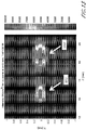
- Inadequate coupling may show up as shadows or vertical stripes in the ultrasound images or a completely dark image.
- tissues or objects, such as bone or an implant can cause challenges since these objects can have a different acoustic impedance and absorption characteristics than soft tissue (e.g. skin, muscle).
- soft tissue e.g. skin, muscle.
- objects (such as bone or an implant) in between the device and intended therapy focus may cause significant reflection and the appearance heating at a shallower depth than intended.
- Objects (e.g., bone, etc.) slightly beyond the focus may also cause issues since the object reflects and readily absorbs the ultrasound from the soft tissue. The reflected energy may inadvertently add to the energy already at the therapy focus causing a higher temperature rise than intended. The absorbed energy at the bone may cause heating or discomfort in the bone.
- advantages of the present invention include using image to assess coupling of an ultrasound therapy beam to the intended treatment tissue.
- higher resolution imaging is advantageous to provide more detail in an image of the tissue in and near the target tissue for treatment.
- the invention improves safety characteristics, improves efficacy performance, provides a component of safety and efficacy for bulk heating devices (such as a band treatment, a linear focal treatment zone, a cylindrical focal line, a plane and/or a volume, etc.) for body shaping, submental fat, abdomen and/or flanks, arms, inner thigh, outer thigh, buttocks, laxity, abdominal laxity, etc., provides qualitative and/or quantitative assessment of coupling, provides for blending of high resolution image(s) with coupling image(s), is employed for assessing out-of-plane impediments post-focally (e.g. bone, intestine, implants), and/or can be used to reduce the need for sonographer equivalent skills.
- bulk heating devices such as a band treatment, a linear focal treatment zone
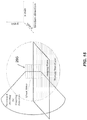
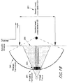
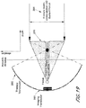
- an ultrasound treatment and imaging system includes an ultrasonic probe comprising an ultrasound therapy transducer adapted to apply ultrasonic therapy to tissue, an ultrasound imaging transducer adapted for imaging the tissue, and an acoustic window, wherein the ultrasound imaging transducer comprises an annular imaging array; wherein the ultrasound imaging transducer comprises a plurality of transmit channels; wherein the ultrasound imaging transducer comprises a plurality of receive channels; wherein the ultrasound imaging transducer is configured for focusing at a location proximate the ultrasound imaging transducer with respect to a distance between the ultrasound imaging transducer and the acoustic window; and a control module coupled to the ultrasonic probe for controlling the ultrasound imaging transducer, wherein the ultrasound imaging transducer is configured to interrogate more than 40% of the acoustic window.
- an ultrasound treatment and imaging system includes an ultrasonic probe comprising an ultrasound therapy transducer adapted to apply ultrasonic therapy to tissue, an ultrasound imaging transducer adapted for imaging the tissue, and an acoustic window, wherein the ultrasound imaging transducer comprises an annular imaging array; wherein the ultrasound imaging transducer comprises a plurality of transmit channels; wherein the ultrasound imaging transducer comprises a plurality of receive channels; wherein the wherein the ultrasound imaging transducer operates at an imaging frequency of between 8 MHz to 50 MHz, wherein the ultrasound imaging transducer is configured to image tissue at a depth of up to 25 mm (e.g., 5 mm, 8 mm, 10 mm, 12 mm, 15 mm, 20 mm) below a skin surface; wherein the ultrasound imaging transducer is configured for focusing at a location behind the ultrasound imaging transducer with respect to the acoustic window; and a control module coupled to the ultrasonic probe for controlling the ultrasound imaging transducer, wherein the ultrasound imaging transduc
- an ultrasound treatment and imaging system includes an ultrasonic probe comprising an ultrasound therapy transducer adapted to apply ultrasonic therapy to tissue, an ultrasound imaging transducer adapted for imaging the tissue, and an acoustic window, wherein the ultrasound imaging transducer comprises an annular or a linear imaging array; wherein the ultrasound imaging transducer comprises a plurality of transmit channels; wherein the ultrasound imaging transducer comprises a plurality of receive channels; wherein the wherein the ultrasound imaging transducer operates at an imaging frequency of between 8 MHz to 50 MHz, wherein the ultrasound imaging transducer is configured to image tissue at a depth of up to 25 mm below a skin surface; wherein the ultrasound imaging transducer is configured for focusing at a location proximate the ultrasound imaging transducer with respect to a distance between the ultrasound imaging transducer and the acoustic window; and a control module coupled to the ultrasonic probe for controlling the ultrasound imaging transducer, wherein the ultrasound imaging transducer is configured to interrogate more than 15% of the
- an imaging beam width from the ultrasound imaging transducer is at least 20% the cross-sectional size of a therapy beam width from the ultrasound therapy transducer. In one embodiment, an imaging beam width from the ultrasound imaging transducer is at least 30% the cross-sectional size of a therapy beam width from the ultrasound therapy transducer. In one embodiment, an imaging beam width from the ultrasound imaging transducer is at least 40% the cross-sectional size of a therapy beam width from the ultrasound therapy transducer. In one embodiment, an imaging beam width from the ultrasound imaging transducer is at least 50% the cross-sectional size of a therapy beam width from the ultrasound therapy transducer. In one embodiment, an imaging beam width from the ultrasound imaging transducer is at least 80% the cross-sectional size of a therapy beam width from the ultrasound therapy transducer.
- a coupling of the imaging of the ultrasound imaging transducer provides an indication of the coupling for the treatment by the ultrasound therapy transducer.
- the ultrasound imaging transducer is configured to interrogate more than 80% of the acoustic window.
- the ultrasound imaging transducer is configured to interrogate more than 90% of the acoustic window.
- the annular imaging array is positioned in the ultrasound therapy transducer.
- control module controls the ultrasound imaging transducer for vector imaging. In one embodiment, the control module controls the ultrasound imaging transducer for defocused vector imaging.
- the ultrasound therapy transducer is configured for treatment of tissue at a first set of locations that is positioned within a first cosmetic treatment zone and a second set of locations that is positioned within a second cosmetic treatment zone, the first zone being different from the second zone.
- the ultrasound therapy transducer is adapted to apply ultrasonic therapy using amplitude modulation whereby a plurality of portions of the ultrasound transducer are adapted to emit ultrasonic therapy at a plurality of amplitudes of acoustic intensity, wherein a first amplitude is different than a second amplitude.
- At least one portion of the ultrasonic transducer is adapted to emit ultrasonic therapy at two or more amplitudes of acoustic intensity, and wherein the amplitude of ultrasonic therapy emitted by the at least one portion of the piezoelectric varies over time.
- the ultrasound transducer comprises piezoelectric material and the plurality of portions of the ultrasound transducer are adapted to create a plurality of corresponding piezoelectric material variations in response to an electric field applied to the ultrasound transducer.
- plurality of piezoelectric material variations comprise at least one of expansion of the piezoelectric material and contraction of the piezoelectric material.
- the ultrasound transducer is adapted to apply ultrasonic therapy via phase shifting whereby a plurality of portions of the ultrasound transducer are adapted to emit ultrasonic therapy at a plurality of phases of acoustic intensity, wherein a first phase is different than a second phase.
- the plurality of phases comprises discrete phase values.
- the ultrasound transducer is adapted to apply ultrasonic therapy using amplitude modulation whereby a plurality of portions of the ultrasound transducer are adapted to emit ultrasonic therapy at a plurality of amplitudes of acoustic intensity, wherein a first amplitude is different than a second amplitude; and apply ultrasonic therapy whereby a plurality of portions of the ultrasound transducer are adapted to emit ultrasonic therapy at a plurality of phases of acoustic intensity, wherein a first phase is different than a second phase.
- the ultrasonic treatment is at least one of a face lift, a brow lift, a chin lift, an eye treatment, a wrinkle reduction, a Vietnameselletage improvement, a buttock lift, a scar reduction, a burn treatment, a skin tightening (e.g., an abdominal laxity treatment), a blood vessel reduction, a treatment of a sweat gland, a sun spot removal, a fat treatment, and a cellulite treatment.
- a method of confirming coupling between an ultrasound probe and tissue for treatment includes providing an ultrasonic probe comprising an acoustic window, an ultrasound transducer comprising an ultrasound therapy transduction element adapted to apply ultrasonic therapy to a tissue, a plurality of imaging transduction elements in an array for imaging the tissue, and a control module coupled to the ultrasonic probe for controlling the ultrasound transducer, and interrogating at least 20% of the acoustic window with an imaging beam from the plurality of imaging transduction elements.
- the plurality of imaging transduction elements interrogates at least 30% of the acoustic window. In one embodiment, the plurality of imaging transduction elements interrogates at least 40% of the acoustic window. In one embodiment, the plurality of imaging transduction elements interrogates at least 50% of the acoustic window.
- the plurality of imaging transduction elements interrogates at least 60% of the acoustic window. In one embodiment, the plurality of imaging transduction elements interrogates at least 70% of the acoustic window. In one embodiment, the method further includes vector imaging. In one embodiment, the method further includes defocused vector imaging. In one embodiment, the method further includes imaging a first focal zone in the tissue with the plurality of imaging transduction elements. In one embodiment, the method further includes imaging a second focal zone in the tissue with the plurality of imaging transduction elements.
- the ultrasound treatment is at least one of a face lift, a brow lift, a chin lift, an eye treatment, a wrinkle reduction, a Vietnameselletage improvement, a buttock lift, a scar reduction, a burn treatment, a tattoo removal, a skin tightening, (e.g., a laxity treatment), a vein removal, a vein reduction, a treatment on a sweat gland, a treatment of hyperhidrosis, a sun spot removal, a fat treatment, a vaginal rejuvenation, and an acne treatment.
- a face lift e.g., a brow lift, a chin lift
- an eye treatment e.g., a wrinkle reduction, a Vietnamese etage improvement, a buttock lift, a scar reduction, a burn treatment, a tattoo removal, a skin tightening, (e.g., a laxity treatment), a vein removal, a vein reduction, a treatment on a sweat gland, a treatment of hyperhidrosis,
- the procedure is entirely cosmetic and not a medical act.
- the methods described herein need not be performed by a doctor, but at a spa or other aesthetic institute.
- a system can be used for the non-invasive cosmetic treatment of skin.
- higher resolution is achieved.
- better imaging signal quality is obtained.
- ultrasound imaging is used with a therapeutic tissue treatment.
- an ultrasound treatment and imaging system configured for reducing imaging misalignment, including an ultrasonic probe comprising an ultrasound therapy transducer adapted to apply ultrasonic therapy to tissue, an ultrasound imaging transducer adapted for imaging the tissue, and a motion mechanism for moving the ultrasound imaging transducer in a first direction and a second direction.
- the ultrasound imaging transducer is mechanically attached to the motion mechanism.
- the first direction is linear.
- the second direction is linear.
- the first direction is parallel to the second direction.
- the first direction is opposite the second direction.
- the ultrasound imaging transducer images with a first focal zone sequence order (f1, f2) when travelling in the first direction
- the ultrasound imaging transducer images with a second focal zone sequence order (f2, f1) when travelling in the second direction
- a spatial registration between the first direction imaging and the second direction imaging is improved by staggering a triggering location.
- a control module is coupled to the ultrasonic probe for controlling the ultrasound imaging transducer.
- an ultrasound treatment and imaging system configured for reducing imaging misalignment, includes an ultrasonic probe comprising an ultrasound therapy transducer adapted to apply ultrasonic therapy to tissue, an ultrasound imaging transducer adapted for imaging the tissue, and a motion mechanism for moving the ultrasound imaging transducer in a first direction and a second direction.
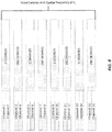
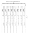
- the ultrasound imaging transducer is mechanically attached to the motion mechanism, wherein the first direction is linear, wherein the second direction is linear, wherein the first direction is parallel to the second direction, wherein the first direction is opposite the second direction, wherein the ultrasound imaging transducer images with a first focal zone sequence order (f1, f2, f3, f4) when travelling in the first direction, wherein the ultrasound imaging transducer images with a second focal zone sequence order (f4, f3, f2, f1) when travelling in the second direction, wherein a spatial registration between the first direction imaging and the second direction imaging is improved by staggering a triggering location, wherein the imaging system employs a sequence of two consecutive A-lines following progression of (line 1: f1, f2, f3, f4; line2: f4, f3, f2, f1) continuously; and a control module coupled to the ultrasonic probe for controlling the ultrasound imaging transducer.
- an ultrasound treatment and imaging system configured for reducing imaging misalignment, includes an ultrasonic probe comprising an ultrasound therapy transducer adapted to apply ultrasonic therapy to tissue, an ultrasound imaging transducer adapted for imaging the tissue, and a motion mechanism for moving the ultrasound imaging transducer in a first direction and a second direction.
- the ultrasound imaging transducer is mechanically attached to the motion mechanism.
- the first direction is opposite the second direction.
- the ultrasound imaging transducer images with a focal zone sequence order (f1, ..., fN), where N>1 when travelling in the first direction.
- the ultrasound imaging transducer images with a second focal zone sequence order (fN,..., f1) when travelling in the second direction.
- a spatial registration between the first direction imaging and the second direction imaging is improved by staggering a triggering location.
- the imaging system employs a directionally dependent focal zone sequencing with alternating between (f1-...-fN) and (fN- ... -f1) on consecutive A-lines; and a control module coupled to the ultrasonic probe for controlling the ultrasound imaging transducer.
- the first direction of motion of the transducer is any one or more of the group consisting of: linear, rotational, and curved.
- the second direction is the reversed path of the first direction.
- the first direction of motion occurs in multiple dimensions and the second direction is the reversed path of the first direction.
- the ultrasound imaging transducer images with a first focal zone sequence order is specified as (f1, ..., fN), where N>1.
- the ultrasound therapy transducer is configured for treatment of tissue at a first set of locations that is positioned within a first cosmetic treatment zone and a second set of locations that is positioned within a second cosmetic treatment zone, the first zone being different from the second zone.
- the ultrasound therapy transducer is adapted to apply ultrasonic therapy using amplitude modulation whereby a plurality of portions of the ultrasound transducer are adapted to emit ultrasonic therapy at a plurality of amplitudes of acoustic intensity, wherein a first amplitude is different than a second amplitude.
- at least one portion of the ultrasonic transducer is adapted to emit ultrasonic therapy at two or more amplitudes of acoustic intensity, and wherein the amplitude of ultrasonic therapy emitted by the at least one portion of the piezoelectric varies over time.
- the ultrasound transducer comprises piezoelectric material and the plurality of portions of the ultrasound transducer are adapted to create a plurality of corresponding piezoelectric material variations in response to an electric field applied to the ultrasound transducer.
- the plurality of piezoelectric material variations comprise at least one of expansion of the piezoelectric material and contraction of the piezoelectric material.
- the ultrasound transducer is adapted to apply ultrasonic therapy via phase shifting whereby a plurality of portions of the ultrasound transducer are adapted to emit ultrasonic therapy at a plurality of phases of acoustic intensity, wherein a first phase is different than a second phase.
- the plurality of phases comprises discrete phase values.
- the ultrasound transducer is adapted to apply ultrasonic therapy using amplitude modulation whereby a plurality of portions of the ultrasound transducer are adapted to emit ultrasonic therapy at a plurality of amplitudes of acoustic intensity, wherein a first amplitude is different than a second amplitude; and apply ultrasonic therapy whereby a plurality of portions of the ultrasound transducer are adapted to emit ultrasonic therapy at a plurality of phases of acoustic intensity, wherein a first phase is different than a second phase.
- the ultrasonic treatment is at least one of: a face lift, a brow lift, a chin lift, an eye treatment, a wrinkle reduction, a Vietnameselletage improvement, a buttock lift, a scar reduction, a burn treatment, a skin tightening (e.g., a laxity treatment), a blood vessel reduction, a treatment of a sweat gland, a sun spot removal, a fat treatment, a cellulite treatment, a vaginal rejuvenation, and an acne treatment.
- a face lift e.g., a brow lift, a chin lift
- an eye treatment e.g., a wrinkle reduction, a Vietnamese etage improvement, a buttock lift, a scar reduction, a burn treatment, a skin tightening (e.g., a laxity treatment), a blood vessel reduction, a treatment of a sweat gland, a sun spot removal, a fat treatment, a cellulite treatment, a vaginal rejuvenation, and
- a method of reducing imaging misalignment in a moving ultrasound probe including staggering a triggering location of a spatial registration between a first direction imaging and a second direction imaging with an ultrasonic probe, the ultrasound probe comprising an ultrasound therapy transducer adapted to apply ultrasonic therapy to tissue, an ultrasound imaging transducer adapted for imaging the tissue, and a motion mechanism for moving the ultrasound imaging transducer in a first direction and a second direction, wherein the ultrasound imaging transducer is mechanically attached to the motion mechanism, wherein the first direction is opposite the second direction, wherein the ultrasound imaging transducer images with a focal zone sequence order (f1, ..., fN), with N>1, wherein the ultrasound imaging transducer images with a first focal zone sequence order (f1, ..., fN) when travelling in the first direction, wherein the ultrasound imaging transducer images with a second focal zone sequence order (fN, ..., f1) when travelling in the second direction.
- the ultrasound treatment is at least one of a face lift, a brow lift, a chin lift, an eye treatment, a wrinkle reduction, a Vietnameselletage improvement, a buttock lift, a scar reduction, a burn treatment, a tattoo removal, a skin tightening (e.g., an abdominal laxity treatment), a vein removal, a vein reduction, a treatment on a sweat gland, a treatment of hyperhidrosis, a sun spot removal, a fat treatment, a vaginal rejuvenation, and an acne treatment.
- a face lift e.g., a brow lift, a chin lift, an eye treatment, a wrinkle reduction, a Vietnamese beautage improvement, a buttock lift, a scar reduction, a burn treatment, a tattoo removal, a skin tightening (e.g., an abdominal laxity treatment), a vein removal, a vein reduction, a treatment on a sweat gland, a treatment of hyperhidrosis, a sun spot removal, a fat
- actions taken by a practitioner; however, it should be understood that they can also include the instruction of those actions by another party.
- actions such as “dithering an energy beam” include “instructing the dithering of an energy beam.”
- the system comprises various features that are present as single features (as opposed to multiple features).
- the system includes a single transduction element that produces two simultaneous treatment focus points that are dithered. Multiple features or components are provided in alternate embodiments.
- the system comprises, consists essentially of, or consists of one, two, three, or more embodiments of any features or components disclosed herein.
- a feature or component is not included and can be negatively disclaimed from a specific claim, such that the system is without such feature or component.
- systems and methods for ultrasound treatment of tissue are adapted for and/or configured to provide cosmetic treatment.
- devices and methods of directing ultrasound therapy to a single focus point or multiple, simultaneous focus points employing ultrasound imaging to confirm sufficient acoustic coupling to a treatment area for improving performance or providing improved correlation between movement in a first and second direction when forming images in cosmetic and/or medical procedures are provided in several embodiments.
- devices and methods of employing ultrasound imaging to confirm sufficient acoustic coupling to a treatment area for improving performance and safety when directing ultrasound therapy to a single focus point or multiple, simultaneous focus points in cosmetic and/or medical procedures are provided in several embodiments.
- devices and methods of improved ultrasound imaging provide better correlation between movement in a first and second direction when forming images.
- Embodiments of the invention provide better imaging correlation between a first moving direction and a second moving direction, (e.g., better correlation between left-traveling & right-traveling formed images).
- Devices and methods of improved ultrasound imaging improve effect B-mode imaging faster (e.g., 1.5x, 2x, 3x, 5x times the scanning rate).
- tissue below or even at a skin surface such as epidermis, dermis, fascia, muscle, fat, and superficial muscular aponeurotic system (“SMAS”), are treated non-invasively with ultrasound energy.
- SMAS superficial muscular aponeurotic system
- the ultrasound energy can be focused at one or more treatment points and/or zones, can be unfocused and/or defocused, and can be applied to a region of interest containing at least one of epidermis, dermis, hypodermis, fascia, muscle, fat, cellulite, and SMAS to achieve a cosmetic and/or therapeutic effect.
- systems and/or methods provide non-invasive dermatological treatment to tissue through thermal treatment, coagulation, ablation, and/or tightening.
- non-invasive ultrasound is used to achieve one or more of the following effects: a face lift, a brow lift, a chin lift, an eye treatment (e.g., malar bags, treat infraorbital laxity), a wrinkle reduction, fat reduction (e.g., treatment of adipose and/or cellulite), cellulite treatment (e.g., dimple or non-dimple type female gynoid lipodystrophy), Vietnameselletage improvement (e.g, upper chest), a buttock lift (e.g, buttock tightening), a skin laxity treatment (e.g, treatment of tissue for tightening or an abdominal laxity treatment), a scar reduction, a burn treatment, a tattoo removal, a vein removal, a vein reduction, a treatment on a sweat gland, a treatment of hyperhidrosis, sun spot removal, an acne treatment, and a pimple removal.
- an eye treatment e.g., malar bags, treat infraor
- fat reduction is achieved.
- cellulite e.g., dimple or non-dimple type gynoid lipodystrophy
- reduction or amelioration of one or more characteristics is achieved by about 10-20%, 20-40%, 40-60%, 60-80% or higher (as well as overlapping ranging therein) as compared to, for example, untreated tissue.
- Vietnamese peel is treated.
- two, three or more beneficial effects are achieved during the same treatment session, and may be achieved simultaneously.
- Various embodiments of the present invention relate to devices or methods of controlling the delivery of energy to tissue.
- various forms of energy can include acoustic, ultrasound, light, laser, radio-frequency (RF), microwave, electromagnetic, radiation, thermal, cryogenic, electron beam, photon-based, magnetic, magnetic resonance, and/or other energy forms.
- Various embodiments of the present invention relate to devices or methods of splitting an ultrasonic energy beam into multiple beams.
- devices or methods can be used to alter the delivery of ultrasound acoustic energy in any procedures such as, but not limited to, therapeutic ultrasound, diagnostic ultrasound, ultrasonic welding, any application that involves coupling mechanical waves to an object, and other procedures.
- a tissue effect is achieved by concentrating the acoustic energy using focusing techniques from the aperture.
- high intensity focused ultrasound HIFU
- a tissue effect created by application of therapeutic ultrasound at a particular depth to can be referred to as creation of a thermal coagulation point (TCP).
- TCP thermal coagulation point
- a zone can include a point.
- a zone is a line, plane, spherical, elliptical, cubical, or other one-, two-, or three-dimensional shape. It is through creation of TCPs at particular positions that thermal and/or mechanical ablation of tissue can occur non-invasively or remotely.
- an ultrasound treatment does not include cavitation and/or shock waves.
- an ultrasound treatment includes cavitation and/or shock waves.
- TCPs can be created in a linear or substantially linear, curved or substantially curved, zone or sequence, with each individual TCP separated from neighboring TCPs by a treatment spacing.
- multiple sequences of TCPs can be created in a treatment region.
- TCPs can be formed along a first sequence and a second sequence separated by a treatment distance from the first sequence.
- Treatment with therapeutic ultrasound can be administered through creation of individual TCPs in a sequence and sequences of individual TCPs, it may be desirable to reduce treatment time and corresponding risk of pain and/or discomfort experienced by a patient.
- Therapy time can be reduced by forming multiple TCPs simultaneously, nearly simultaneously, or sequentially.
- a treatment time can be reduced 10%, 20%, 25%, 30%, 35%, 40%, 45%, 50%, 55%, 60%, 65%, 70%, 75%, 80% or more by creating multiple TCPs.
- target tissue is, but is not limited to, any of skin, eyelids, eye lash, eye brow, caruncula lacrimalis, crow's feet, wrinkles, eye, nose, mouth (e.g., nasolabial fold, perioral wrinkles), tongue, teeth, gums, ears, brain, heart, lungs, ribs, abdomen (e.g, for abdominal laxity), stomach, liver, kidneys, uterus, breast, vagina, prostrate, testicles, glands, thyroid glands, internal organs, hair, muscle, bone, ligaments, cartilage, fat, fat labuli, adipose tissue, subcutaneous tissue, implanted tissue, an implanted organ, lymphoid, a tumor, a cyst, an
- an ultrasound system 20 includes a hand wand (e.g., handpiece) 100, module (e.g., transducer module, cartridge, probe) 200, and a controller (e.g., console) 300.
- a console 300 comprises a communication system (e.g., wifi, Bluetooth, modem, etc. to communicate with another party, a manufacturer, a supplier, a service provider, the Internet, and/or a cloud.
- a cart 301 provides mobility and/or position of the system 20, and can include wheels, surfaces to write on or place components, and/or compartments 302 (e.g., drawers, containers, shelves, etc.) to, for example, store or organize components.
- the cart has a power supply, such as a power connection to a battery and/or one or more cords to connect power, communications (e.g., Ethernet) to the system 20.
- the system 20 comprises a cart 301.
- the system 20 does not comprise a cart 301.
- the hand wand 100 can be coupled to the controller 300 by an interface 130, which may be a wired or wireless interface.
- the interface 130 can be coupled to the hand wand 100 by a connector 145.
- the distal end of the interface 130 can be connected to a controller connector on a circuit 345 (not shown).
- the interface 130 can transmit controllable power from the controller 300 to the hand wand 100.
- the system 20 has multiple imaging channels (e.g., 8 channels) for ultra-clear HD (high definition) visualization of subcutaneous structures to improve imaging.
- the system 20 multiple therapy channels (e.g., 8 channels) and a precision linear-drive motor that doubles treatment accuracy while increasing speed (e.g,, by 25%, 40%, 50%, 60%, 75%, 100% or more). Together, these features establish one of the most versatile system platforms in the industry and provide a foundation for unprecedented future possibilities.
- the controller 300 can be adapted to and/or configured for operation with the hand wand 100 and the module 200, as well as the overall ultrasound system 20 functionality.
- multiple controllers 300, 300', 300", etc. can be adapted to and/or configured for operation with multiple hand wands 100, 100', 100", etc. and or multiple modules 200, 200', 200", etc.
- the controller 300 can include connectivity to one or more interactive graphical display 310, which can include a touchscreen monitor and Graphic User Interface (GUI) that allows the user to interact with the ultrasound system 20.
- GUI Graphic User Interface
- a second smaller, more mobile display that allows the user to more easily position and view the treatment screen.
- a second display that allows the system user to view a treatment screen (e.g., on a wall, on a mobile device, large screen, remote screen).
- the graphical display 310 includes a touchscreen interface 315 (not shown).
- the display 310 sets and displays the operating conditions, including equipment activation status, treatment parameters, system messages and prompts, and ultrasound images.
- the controller 300 can be adapted to and/or configured to include, for example, a microprocessor with software and input/output devices, systems and devices for controlling electronic and/or mechanical scanning and/or multiplexing of transducers and/or multiplexing of transducer modules, a system for power delivery, systems for monitoring, systems for sensing the spatial position of the probe and/or transducers and/or multiplexing of transducer modules, and/or systems for handling user input and recording treatment results, among others.
- a microprocessor with software and input/output devices systems and devices for controlling electronic and/or mechanical scanning and/or multiplexing of transducers and/or multiplexing of transducer modules
- a system for power delivery systems for monitoring, systems for sensing the spatial position of the probe and/or transducers and/or multiplexing of transducer modules
- systems for handling user input and recording treatment results among others.
- the controller 300 can include a system processor and various analog and/or digital control logic, such as one or more of microcontrollers, microprocessors, field-programmable gate arrays, computer boards, and associated components, including firmware and control software, which may be capable of interfacing with user controls and interfacing circuits as well as input/output circuits and systems for communications, displays, interfacing, storage, documentation, and other useful functions.
- System software running on the system process may be adapted to and/or configured to control all initialization, timing, level setting, monitoring, safety monitoring, and all other ultrasound system functions for accomplishing user-defined treatment objectives.
- the controller 300 can include various input/output modules, such as switches, buttons, etc., that may also be suitably adapted to and/or configured to control operation of the ultrasound system 20.
- the hand wand 100 includes one or more finger activated controllers or switches, such as 150 and 160.
- one or more thermal treatment controllers 160 e.g., switch, button
- one or more imaging controllers 150 e.g., switch, button
- the hand wand 100 can include a removable module 200.
- the module 200 may be non-removable.
- the module 200 can be mechanically coupled to the hand wand 100 using a latch or coupler 140.
- an interface guide 235 or multiple interface guides 235 can be used for assisting the coupling of the module 200 to the hand wand 100.
- the module 200 can include one or more ultrasound transducers 280.
- an ultrasound transducer 280 includes one or more ultrasound elements.
- the module 200 can include one or more ultrasound elements.
- the hand wand 100 can include imaging-only modules, treatment-only modules, imaging-and-treatment modules, and the like.
- the ultrasound transducer 280 is movable in one or more directions 290 within the module 200.
- the transducer 280 is connected to a motion mechanism 400.
- the motion mechanism comprises zero, one, or more bearings, shafts, rods, screws, lead screws 401, encoders 402 (e.g., optical encoder to measure position of the transducer 280), motors 403 (e.g., a step motor) to help ensure accurate and repeatable movement of the transducer 280 within the module 200.
- module 200 can include a transducer 280 which can emit energy through an acoustically transparent member 230.
- the control module 300 can be coupled to the hand wand 100 via the interface 130, and the graphic user interface 310 can be adapted to and/or configured for controlling the module 200. In one embodiment, the control module 300 can provide power to the hand wand 100.
- the hand wand 100 can include a power source.
- the switch 150 can be adapted to and/or configured for controlling a tissue imaging function and the switch 160 can be adapted to and/or configured for controlling a tissue treatment function.
- delivery of emitted energy 50 at a suitable focal depth, distribution, timing, and energy level is provided by the module 200 through controlled operation by the control system 300 of the transducer 280 to achieve the desired therapeutic effect with a thermal coagulation zone 550.
- the module 200 can be coupled to the hand wand 100.
- the module 200 can emit and receive energy, such as ultrasonic energy.
- the module 200 can be electronically coupled to the hand wand 100 and such coupling may include an interface which is in communication with the controller 300.
- the interface guide 235 can be adapted to and/or configured to provide electronic communication between the module 200 and the hand wand 100.
- the module 200 can comprise various probe and/or transducer configurations.
- the module 200 can be adapted to and/or configured for a combined dual-mode imaging/therapy transducer, coupled or co-housed imaging/therapy transducers, separate therapy and imaging probes, and the like.
- the controller 300 automatically detects it and updates the interactive graphical display 310.
- an access key 320 (e.g., a secure USB drive, key) is removably connected to a system 20 to permit the system 20 to function.
- the access key is programmed to be customer specific, and serves multiple functions, including system security, country/region specific access to treatment guidelines and functionality, software upgrades, support log transfers and /or credit transfer and/or storage.
- the system 20 has internet and/or data connectivity.
- connectivity provides a method by which data is transferred between the system 20 provider and the customer.
- data includes credits, software updates and support logs. Connectivity is divided into different model embodiments, based on how a user's console is connected to the internet.
- Disconnected Model connectivity comprises a console that is disconnected from the internet and customer doesn't have internet access. Credit transfers and software upgrades are conducted by shipping access key(s), (e.g., USB drives) to the customer.
- Semi-Connected Model connectivity comprises a console that is disconnected from the internet but customer has internet access. Credit transfers, software upgrades and support log transfers are conducted using the customer's personal computer, smart phone, or other computing device in conjunction with the system access key to transfer data.
- Fully-Connected Model connectivity comprises a console that is wirelessly connected to the internet using wifi, cellular modem, Bluetooth, or other protocol. Credit transfers, software upgrades and support log transfers are made directly between the console and the cloud.
- the system 20 connects to an online portal, for streamlined inventory management, on-demand treatment purchases and business analytics insights to drive customer aesthetic treatment business to the next level.
- tissue below or even at a skin surface such as epidermis, dermis, hypodermis, fascia, and superficial muscular aponeurotic system ("SMAS"), and/or muscle are treated non-invasively with ultrasound energy.
- Tissue may also include blood vessels and/or nerves.
- the ultrasound energy can be focused, unfocused or defocused and applied to a region of interest containing at least one of epidermis, dermis, hypodermis, fascia, and SMAS to achieve a therapeutic effect.
- FIG. 2 is a schematic illustration of the ultrasound system 20 coupled to a region of interest 10.
- tissue layers of the region of interest 10 can be at any part of the body of a subject. In one embodiment, the tissue layers are in the head and face region of the subject.
- the cross-sectional portion of the tissue of the region of interest 10 includes a skin surface 501, an epidermal layer 502, a dermal layer 503, a fat layer 505, a superficial muscular aponeurotic system 507 (hereinafter "SMAS 507"), and a muscle layer 509.
- the tissue can also include the hypodermis 504, which can include any tissue below the dermal layer 503.
- the combination of these layers in total may be known as subcutaneous tissue 510.
- a treatment zone 525 which is below the surface 501.
- the surface 501 can be a surface of the skin of a subject 500.
- the system and/or methods may be used on tissue (including but not limited to one or a combination of muscles, fascia, SMAS, dermis, epidermis, fat, adipose cells, cellulite, which may be called gynoid lipodystrophy, (e.g., non-dimple type female gynoid lipodystrophy), collagen, skin, blood vessels, of the face, neck, head, arms, legs, or any other location on or in the body (including bodily cavities).
- tissue including but not limited to one or a combination of muscles, fascia, SMAS, dermis, epidermis, fat, adipose cells, cellulite, which may be called gynoid lipodystrophy, (e.g., non-dimple type female gynoid lipodystrophy), collagen, skin, blood vessels, of the face, neck, head, arms, legs, or any other location on or in the body (including bodily cavities).
- cellulite (e.g., non-dimple type female gynoid lipodystrophy) reduction is achieved in an amount of 2%, 5%, 10%, 15%, 20%, 25%, 30%, 40%, 50%, 75%, 80%, 90%, 95%, and any ranges therein.
- an embodiment of the ultrasound system 20 includes the hand wand 100, the module 200, and the controller 300.
- the module 200 includes a transducer 280.
- FIG. 3 illustrates an embodiment of an ultrasound system 20 with a transducer 280 adapted to and/or configured to treat tissue at a focal depth 278.
- the focal depth 278 is a distance between the transducer 280 and the target tissue for treatment.
- a focal depth 278 is fixed for a given transducer 280.
- a focal depth 278 is variable for a given transducer 280.
- a transducer 280 is configured to treat simultaneously at multiple depths below a skin surface (e.g., 1.5 mm, 3.0 mm, 4.5 mm, or other depths).
- the module 200 can include a transducer 280 which can emit energy through an acoustically transparent member 230.
- a depth may refer to the focal depth 278.
- the transducer 280 can have an offset distance 270, which is the distance between the transducer 280 and a surface of the acoustically transparent member 230.
- the focal depth 278 of a transducer 280 is a fixed distance from the transducer.
- a transducer 280 may have a fixed offset distance 270 from the transducer to the acoustically transparent member 230.
- an acoustically transparent member 230 is adapted to and/or configured at a position on the module 200 or the ultrasound system 20 for contacting the skin surface 501.
- the focal depth 278 exceeds the offset distance 270 by an amount to correspond to treatment at a target area located at a tissue depth 279 below a skin surface 501.
- the tissue depth 279 is a distance between the acoustically transparent member 230 and the target area, measured as the distance from the portion of the hand wand 100 or module 200 surface that contacts skin (with or without an acoustic coupling gel, medium, etc.) and the depth in tissue from that skin surface contact point to the target area.
- the focal depth 278 can correspond to the sum of an offset distance 270 (as measured to the surface of the acoustically transparent member 230 in contact with a coupling medium and/or skin 501) in addition to a tissue depth 279 under the skin surface 501 to the target region.
- the acoustically transparent member 230 is not used.
- Coupling components can comprise various substances, materials, and/or devices to facilitate coupling of the transducer 280 or module 200 to a region of interest.
- coupling components can comprise an acoustic coupling system adapted to and/or configured for acoustic coupling of ultrasound energy and signals.
- Acoustic coupling system with possible connections such as manifolds may be utilized to couple sound into the region of interest, provide liquid- or fluid-filled lens focusing.
- the coupling system may facilitate such coupling through use of one or more coupling media, including air, gases, water, liquids, fluids, gels, solids, non-gels, and/or any combination thereof, or any other medium that allows for signals to be transmitted between the transducer 280 and a region of interest.
- one or more coupling media is provided inside a transducer.
- a fluid-filled module 200 contains one or more coupling media inside a housing.
- a fluid-filled module 200 contains one or more coupling media inside a sealed housing, which is separable from a dry portion of an ultrasonic device.
- a coupling medium is used to transmit ultrasound energy between one or more devices and tissue with a transmission efficiency of 100%, 99% or more, 98% or more, 95% or more, 90% or more, 80% or more, 75% or more, 60% or more, 50% or more, 40% or more, 30% or more, 25% or more, 20% or more, 10% or more, and/or 5% or more.
- the transducer 280 can image and treat a region of interest at any suitable tissue depths 279.
- the transducer module 280 can provide an acoustic power in a range of about 1 W or less, between about 1 W to about 100 W, and more than about 100 W, e.g., 200 W, 300 W, 400 W, 500 W.
- the transducer module 280 can provide an acoustic power at a frequency of about 1 MHz or less, between about 1 MHz to about 10 MHz (e.g., 3 MHz, 4 MHz, 4.5 MHz, 7 MHz, 10 MHz), and more than about 10 MHz.
- the module 200 has a focal depth 278 for a treatment at a tissue depth 279 of about 4.5 mm below the skin surface 501. In one embodiment, the module 200 has a focal depth 278 for a treatment at a tissue depth 279 of about 3 mm below the skin surface 501. In one embodiment, the module 200 has a focal depth 278 for a treatment at a tissue depth 279 of about 1.5 mm below the skin surface 501.
- transducers 280 or modules 200 can be adapted to and/or configured for delivering ultrasonic energy at a tissue depth of 1.5 mm, 3 mm, 4.5 mm, 6 mm, 7 mm, less than 3 mm, between 3 mm and 4.5 mm, between 4.5 mm and 6 mm, more than more than 4.5 mm, more than 6 mm, etc., and anywhere in the ranges of 0-3 mm, 0-4.5 mm, 0-6 mm, 0-25 mm, 0-100 mm, etc. and any depths therein.
- the ultrasound system 20 is provided with two or more transducer modules 280.
- a first transducer module can apply treatment at a first tissue depth (e.g., about 4.5 mm) and a second transducer module can apply treatment at a second tissue depth (e.g., of about 3 mm), and a third transducer module can apply treatment at a third tissue depth (e.g., of about 1.5-2 mm).
- at least some or all transducer modules can be adapted to and/or configured to apply treatment at substantially same depths.
- changing the number of focus point locations (e.g., such as with a tissue depth 279) for an ultrasonic procedure can be advantageous because it permits treatment of a patient at varied tissue depths even if the focal depth 278 of a transducer 270 is fixed.
- This can provide synergistic results and maximizing the clinical results of a single treatment session.
- treatment at multiple depths under a single surface region permits a larger overall volume of tissue treatment, which results in enhanced collagen formation and tightening.
- treatment at different depths affects different types of tissue, thereby producing different clinical effects that together provide an enhanced overall cosmetic result. For example, superficial treatment may reduce the visibility of wrinkles and deeper treatment may induce formation of more collagen growth.
- treatment at various locations at the same or different depths can improve a treatment.
- a subject may be treated under the same surface region at one depth in time one, a second depth in time two, etc.
- the time can be on the order of nanoseconds, microseconds, milliseconds, seconds, minutes, hours, days, weeks, months, or other time periods.
- the new collagen produced by the first treatment may be more sensitive to subsequent treatments, which may be desired for some indications.
- multiple depth treatment under the same surface region in a single session may be advantageous because treatment at one depth may synergistically enhance or supplement treatment at another depth (due to, for example, enhanced blood flow, stimulation of growth factors, hormonal stimulation, etc.).
- different transducer modules provide treatment at different depths.
- a single transducer module can be adjusted or controlled for varied depths. Safety features to minimize the risk that an incorrect depth will be selected can be used in conjunction with the single module system.
- a method of treating the lower face and neck area e.g., the submental area
- a method of treating (e.g., softening) mentolabial folds is provided.
- a method of treating the eye region e.g., malar bags, treat infraorbital laxity
- Upper lid laxity improvement and periorbital lines and texture improvement will be achieved by several embodiments by treating at variable depths.
- optimal clinical effects e.g., softening, tightening
- the treatment methods described herein are non-invasive cosmetic procedures.
- the methods can be used in conjunction with invasive procedures, such as surgical facelifts or liposuction, where skin tightening is desired.
- the methods can be applied to any part of the body.
- a transducer module 200 permits a treatment sequence at a fixed depth at or below the skin surface. In one embodiment, a transducer module permits a treatment sequence at one, two, or more variable or fixed depths below the dermal layer. In several embodiments, the transducer module comprises a movement mechanism adapted to and/or configured to direct ultrasonic treatment in a sequence of individual thermal lesions (hereinafter "thermal coagulation points" or "TCPs”) at a fixed focal depth.
- TCPs thermal coagulation points
- the sequence of individual TCPs has a treatment spacing in a range from about 0.01 mm to about 25 mm (e.g., 1 mm, 1.5 mm, 2 mm, 2,5 mm, 3 mm, 5 mm, 10 mm, 20 mm and any value ranges therein), with a dithering alteration of the spacing by 1 - 50% (e.g., 1%, 5%, 10%, 15%, 20%, 25%, 30%, 35%, 40%, 45%, 50% and any range therein).
- the spacing can be 1.1 mm or less, 1.5 mm or more, between about 1.1 mm and about 1.5 mm, etc.
- the individual TCPs are discrete. In one embodiment, the individual TCPs are overlapping.
- a transducer module comprises a movement mechanism adapted to and/or configured to direct ultrasonic treatment in a sequence so that TCPs are formed in linear or substantially linear sequences separated by a treatment distance.
- a transducer module can be adapted to and/or configured to form TCPs along a first linear sequence and a second linear sequence separated by a treatment distance from the first linear sequence.
- treatment distance between adjacent linear sequences of individual TCPs is in a range from about 0.01 mm to about 25 mm. In one embodiment, treatment distance between adjacent linear sequences of individual TCPs is in a range from about 0.01 mm to about 50 mm. For example, the treatment distance can be 2 mm or less, 3 mm or more, between about 2 mm and about 3 mm, etc.
- a transducer module can comprise one or more movement mechanisms 400 adapted to and/or configured to direct ultrasonic treatment in a sequence so that TCPs are formed in linear or substantially linear sequences of individual thermal lesions separated by a treatment distance from other linear sequences. In one embodiment a treatment is applied in a first direction 290 (e.g., push).
- a treatment is applied opposite the first direction 290 (e.g., pull). In one embodiment, treatment is applied in both a first direction 290 and opposite the first direction (e.g., push and pull). In one embodiment, the treatment distance separating linear or substantially linear TCPs sequences is the same or substantially the same. In one embodiment, the treatment distance separating linear or substantially linear TCPs sequences is different or substantially different for various adjacent pairs of linear TCPs sequences.
- first and second removable transducer modules are provided.
- each of the first and second transducer modules are adapted to and/or configured for both ultrasonic imaging and ultrasonic treatment.
- a transducer module is adapted to and/or configured for treatment only.
- an imaging transducer may be attached to a handle of a probe or a hand wand.
- the first and second transducer modules are adapted to and/or configured for interchangeable coupling to a hand wand.
- the first transducer module is adapted to and/or configured to apply ultrasonic therapy to a first layer of tissue
- the second transducer module is adapted to and/or configured to apply ultrasonic therapy to a second layer of tissue.
- the second layer of tissue is at a different depth than the first layer of tissue.
- FIG. 3 illustrates one embodiment of a depth that corresponds to a depth for treating muscle.
- the depth can correspond to any tissue, tissue layer, skin, epidermis, dermis, hypodermis, fat, SMAS, muscle, blood vessel, nerve, or other tissue.
- the module 200 and/or the transducer 280 can also be mechanically and/or electronically scanned along the surface 501 to treat an extended area.
- monitoring of the treatment area and surrounding structures can be provided to plan and assess the results and/or provide feedback to the controller 300 and the user via a graphical interface 310.
- an ultrasound system 20 generates ultrasound energy which is directed to and focused below the surface 501.
- This controlled and focused ultrasound energy 50 creates the thermal coagulation point or zone (TCP) 550.
- TCP thermal coagulation point or zone
- the ultrasound energy 50 creates a void in subcutaneous tissue 510.
- the emitted energy 50 targets the tissue below the surface 501 which cuts, ablates, coagulates, micro-ablates, manipulates, and/or causes a TCP 550 in the tissue portion 10 below the surface 501 at a specified focal depth 278.
- the transducer 280 moves in a direction denoted by the arrow marked 290 at specified intervals 295 to create a series of treatment zones 254 each of which receives an emitted energy 50 to create one or more TCPs 550.
- an arrow marked 291 illustrates an axis or direction that is orthogonal to arrow 290, and a spacing of TCP's 550 show TCP's can be spaced orthogonally to the motion direction of the transducer 280.
- an orientation of the spaced TCP's can be set at any angle 0 - 180 degrees from arrow 290.
- an orientation of the spaced TCP's can be set at any angle 0 - 180 degrees based on the orientation of poled areas on the transducer 280.
- transducer modules can comprise one or more transduction elements.
- the transduction elements can comprise a piezoelectrically active material, such as lead zirconante titanate (PZT), or any other piezoelectrically active material, such as a piezoelectric ceramic, crystal, plastic, and/or composite materials, as well as lithium niobate, lead titanate, barium titanate, and/or lead metaniobate.
- PZT lead zirconante titanate
- transducer modules in addition to, or instead of, a piezoelectrically active material, can comprise any other materials adapted to and/or configured for generating radiation and/or acoustical energy.
- transducer modules can be adapted to and/or configured to operate at different frequencies and treatment depths.
- Transducer properties can be defined by an outer diameter ("OD") and focal length (F L ).
- OD outer diameter
- F L focal length
- other suitable values of OD and F L can be used, such as OD of less than about 19 mm, greater than about 19 mm, etc. and F L of less than about 15 mm, greater than about 15 mm, etc.
- Transducer modules can be adapted to and/or configured to apply ultrasonic energy at different target tissue depths.
- transducer modules comprise movement mechanisms adapted to and/or configured to direct ultrasonic treatment in a linear or substantial liner sequence of individual TCPs with a treatment spacing between individual TCPs.
- treatment spacing can be about 1.1 mm, 1.5 mm, etc.
- transducer modules can further comprise movement mechanisms adapted to and/or configured to direct ultrasonic treatment in a sequence so that TCPs are formed in linear or substantially linear sequences separated by a treatment spacing.
- a transducer module can be adapted to and/or configured to form TCPs along a first linear sequence and a second linear sequence separated by treatment spacing between about 2 mm and 3 mm from the first linear sequence.
- a user can manually move the transducer modules across the surface of a treatment area so that adjacent linear sequences of TCPs are created.
- a movement mechanism can automatically move the transducer modules across the surface of a treatment area so that adjacent linear sequences of TCPs are created.
- spatial frequency analysis techniques based on Fourier analysis and Fourier optics can be used to increase efficiency of therapeutic treatment.
- a system that has an impulse response h(t) is excited by a stimulus x(t)
- Fourier transform can be applied to compute the convolution of equation (1).
- the Fraunhofer approximation can be used for deriving a relationship between a transducer opening or aperture and a resulting ultrasonic beam response. Derivation of the Fraunhofer approximation is described in Joseph Goodman, Introduction to Fourier Optics (3d ed. 2004 ). According to the Fraunhofer approximation, a far-field complex amplitude pattern produced by a complex aperture is equal to a two-dimensional Fourier transform of the aperture amplitude and phase. In several embodiments, this relationship in optics can be extended to ultrasound since linear wave equations can be used to represent both light propagation and sound propagation. In the case of optics and/or ultrasound, the two-dimensional Fourier transform can determine a sound wave pressure amplitude distribution at the focus of a transducer.
- variable z which represents depth can be replaced with z f which represents a focal distance.
- Fourier optics and Fourier transform identities can be used for ultrasound transducers in order to determine the intensity distribution corresponding to a transducer design.
- Fourier transform of a rectangle rect(ax) is a sinc function.
- Fourier transform of a two dimensional circle of uniform amplitude is a first order Bessel function which can be represented as J 1 .
- an ultrasound transducer can have a rectangular aperture of suitable dimensions and focal length. In several embodiments, an ultrasound transducer can have a circular aperture with suitable dimensions and focal length. In one embodiment, a transducer can have a circular aperture with an outer radius of approximately 9.5 mm, an inner diameter of approximately 2 mm, and focal length of approximately 15 mm.
- the variable 'a' can be approximately 9.5 mm and the variable 'b' in equation (5a) can be approximately 2 mm.
- Applying Fourier transform to equation (5a) can provide an estimate of the sound wave pressure distribution at the focus.
- ⁇ x and ⁇ y are same as f x and f y of equations (4a) and (4b).
- Equation (6) demonstrates that the sound wave pressure distribution of a transducer with a circular aperture is a first order Bessel function.
- a substantial majority of the energy is concentrated at the focus (e.g., 15 mm away from the aperture).
- the width of a main ultrasonic beam and the distribution of energy away from the main beam can be expressed as a function of the operating frequency as is expressed in equations (4a) and (4b).
- two identical or nearly identical beams could be created at the focus if the aperture was modulated (e.g., multiplied) by a correct function.
- An energy distribution or beam response at the focus of the modulated aperture of equation (7) is the convolution of the Fourier transform of the two functions of the aperture:
- Equation (8) can be simplified into the summation of two separate functions applying the Fourier Transform identity for a Dirac delta function (e.g., identity 2 in Table 2):
- G ⁇ x ⁇ y 1 2 F ⁇ x ⁇ c 2 ⁇ , ⁇ y + F ⁇ x + c 2 ⁇ , ⁇ y
- Equation (9) shows that two beams appearing at the focus are spatially shifted by ⁇ c 2 ⁇ compared to the original, non-modulated beam.
- one or more other modulation functions such as sine function, can be used to achieve a desired beam response.
- aperture can be modulated such that more than two foci are created. For example, three, four, five, etc. foci can be created.
- aperture can be modulated such that foci are created sequentially or substantially sequentially rather than simultaneously.
- therapy transducer modules comprise movement mechanisms configured to direct ultrasonic treatment in a linear or substantial liner sequence of individual TCPs with a treatment spacing between individual TCPs.
- treatment spacing can be about 1.1 mm, 1.5 mm, etc.
- transducer modules can further comprise movement mechanisms configured to direct ultrasonic treatment in a sequence so that TCPs are formed in linear or substantially linear sequences separated by a treatment spacing.
- a transducer module can be configured to form TCPs along a first linear sequence and a second linear sequence separated by treatment spacing between about 2 mm and 3 mm from the first linear sequence.
- a simultaneous or substantially simultaneous split in the ultrasonic beam may be achieved at the focus (or before the focus) if the aperture is modulated by a cosine and/or sine function of a desired spatial frequency.
- two simultaneous or nearly simultaneous focused beams separated by about 1.1 mm treatment spacing can be created in a linear or substantially linear sequence.
- the wavelength ⁇ of ultrasound wave in water is approximately 0.220 mm. Accordingly, spatial frequencies ⁇ x and ⁇ y at the focus are represented as:
- a transducer with circular aperture emitting ultrasonic energy at various operating frequencies can be modulated by a sine and/or cosine functions at spatial frequencies listed in Table 2.
- Modulated aperture of the transducer can produce a simultaneously or substantially simultaneously split beam with two foci having different separation distances, as is indicated in Table 2.
- the transducer can have OD of about 19 mm and a focal length of about 15 mm. Table 2 Separation Distance Between Foci Ultrasound Frequency 1.1 mm 1.5 mm 2 mm 3 mm 4 MHz 0.60 0.82 1.09 1.63 7 MHz 1.04 1.43 1.90 2.86 10 MHz 1.50 2.04 2.72 3.08
- a spatial frequency of an aperture modulation function increases as the ultrasonic operating frequency increases for a given foci separation distance.
- the spatial frequency increases as the desired foci separation distance increases.
- higher spatial frequency can result in amplitude transitions in the aperture occurring more rapidly. Due to transducer processing limitations, rapid amplitude variations in the aperture can make the aperture less efficient as there may be a variance in an amount of sound pressure produced by different parts of the aperture.
- using spatial frequencies to simultaneously or nearly simultaneously split the beam can reduce the overall focal gain of each beam. As is shown in equation (9), a field pressure at the focus of each beam is reduced by a factor of two in comparison with an unmodulated beam.
- the sound pressure or ultrasound intensity from the aperture can be increased to obtain similar or substantially similar intensities at the focal plane.
- increasing the pressure at the aperture may not be limited by system and/or transducer processing limitations.
- an increase in the pressure at the aperture can increase the overall intensity in the near field, which may increase the possibility of excessively heating treatment area tissue(s) that is located before focus.
- the possibility of additional heating of the pre-focal tissue(s) may be limited or eliminated by using a lower ultrasound treatment frequency.
- ultrasound beam can be split multiple times, such as three, four, five, etc. times, such that multiple simultaneous or nearly simultaneous beams are created.
- unmodulated beam at the focus can be created at four different locations along the x-axis.
- a constant or DC term, C1 may be added to the amplitude modulation function to maintain placement of energy at the original focal location:
- g x y cos cx + cos dx + C 1 circ r a ⁇ circ r b
- G ⁇ x ⁇ y 1 2 F ⁇ x ⁇ c 2 ⁇ , ⁇ y + F ⁇ x + c 2 ⁇ , ⁇ y + F ⁇ x ⁇ d 2 ⁇ , ⁇ y + F ⁇ x + d 2 ⁇ , ⁇ y + C 1 F ⁇ x ⁇ y
- aperture modulation of equations (12) and (13), whereby the beam can be placed at multiple locations simultaneously or nearly simultaneously may be have limited applicability due to system, material, and/or tissue limitations.
- the frequency of ultrasound therapy may be adjusted, such as lowered, in order to limit and/or eliminate such possibility.
- nonlinear techniques can be applied at the focus in order to limit and/or eliminate the possibility of heating of the pre-focal tissue(s).
- the sound pressure or ultrasound intensity from the aperture can be increased to obtain similar or substantially similar intensities at the focal plane.
- the two-dimensional Fourier transform of a sound pressure function U(x 1 , y 1 ) can be expressed as a product of a one-dimensional Fourier transform of two functions in x and y.
- Equation (16) shows that the separation distance decreases as the operational frequency increases.
- Table 3 shows the rate in change of separation distance as a function of operational frequency for the different spatial frequencies (e.g., in various embodiments, 4 MHz, 7 MHz, 10 MHz).
- Table 3 Derivative of Equation (16) [mm/MHz] Ultrasound Frequency 1.1 mm 1.5 mm 2 mm 3 mm 4 MHz -0.269 -0.367 -0.488 -0.730 7 MHz -0.152 -0.209 -0.278 -0.418 10 MHz -0.107 -0.146 -0.195 -0.221
- the foci get closer together and as the operational frequency decreases the foci get farther apart without the need to change the phase or mechanically move the transducer.
- This is a unique method of electronically moving the beam to spread the energy without relying on thermal conduction in tissue.
- the benefits include a reduction or a minimization of the maximum temperature and an increase in the thermal coagulation volume of the lesion without the need for additional system channels.
- the amount of movement from a main operational frequency can be determined by using equation (14).
- the main operational frequency of an aperture is 5 MHz and the focal length is 15 mm.
- the operational frequency is called the aperture center frequency.
- the operational frequency is 5 MHz.
- the spacing decreases for higher frequencies relative to 5 MHz and increases for lower frequencies relative to 5 MHz.
- FIG. 6 shows the spacing difference for all operational frequencies of the aperture for different aperture spatial frequencies. As FIG. 6 shows, the separation distance increases as the frequency decreases.
- the separation distance is relative to a frequency 5 MHz.
- one way to estimate the electronic dithering from frequency modulation can be determined by referencing all movement to the initial separation at 5 MHz. As FIG. 7 shows, the spread of the separation distance between the foci can easily vary by over 1 mm.
- the range of possible operational frequencies from one aperture can be described in terms of the transducer bandwidth.
- a larger transducer bandwidth results in an aperture that has a wider range of operational frequencies.
- Transducer bandwidth can be described as a percent fraction of the aperture center frequency by locating the frequency where the transmit intensity decreases to -3 dB of the peak transmit intensity.
- the -3 dB high frequency is designated as f -3db,H and the -3 dB low frequency is designated as f -3dB, L for the transmit response of a transducer aperture.
- BW ⁇ 3 dB 100 % * f ⁇ 3 dB , H ⁇ f ⁇ 3 dB , L / f ⁇ 3 dB , H + f ⁇ 3 dB , L / 2
- increasing the range of operational frequencies possible within one aperture may be achieved (but not limited to) by the use of backing layers, matching layers, multiple piezoelectric layers, electrical matching, piezoelectric composites, and/or a single crystal piezoceramic.
- Table 5 shows how based on percent bandwidth the foci spread can vary if the aperture center frequency is 5 MHz.
- the foci separation distance for 5 MHz is 0.72 mm, 1.43 mm, 2.15 mm and 2.86 mm respectively for spatial frequencies of 0.5 mm -1 , 1.00 mm -1 , 1.50 mm -1 , 2.00 mm -1 . If the spatial frequency at the aperture is 1.50 mm -1 and the transducer bandwidth is 60%, then the separation distance between the foci varies by 1.42 mm which is a distance greater than the lateral resolution of the beam at 5 MHz.
- the depth-of-field will also change as well as lateral resolution and focal gain. In one embodiment, as the frequency is changed, the depth-of-field, lateral resolution and focal gain will also change. Therefore, in one embodiment, the intensity at the aperture may change depending in the heating rate goals. Also, in some embodiments, it may be advantageous to send multiple operational frequencies at the same time to spread the energy immediately or near-immediately. For example, transmit excitation of the aperture may include excitation at 4 MHz, 5 MHz and 6 MHz all at the same time.
- an aperture is poled with a spatial frequency of k x .
- the spatial frequency can be easily doubled or decreased to zero by connecting individual electrical excitation channels that have the ability to modify the phase to 0 degrees or 180 degrees, as shown in the embodiments in FIG. 8 .
- the aperture spatial frequency is k x .
- the spatial frequency at the aperture is 1 ⁇ 2 k x .
- the spatial frequency at the aperture is 0. If channel 1 is 0 degrees, channel 2 is 180 degrees, channel 3 is 180 degrees, channel 4 is 0 degrees and so on, then the spatial frequency at the aperture is 2k x . In this case, seven unique foci can be created. As noted in Table 4 (at FIG.
- the aperture center frequency is 5 MHz
- the aperture frequency is any of 0 mm -1 , 0.5 mm -1 , 1.0 mm -1 , or 2.0 mm -1
- the corresponding separation distances are 0 mm, 0.72 mm, 1.43 mm and 2.86 mm, which yield seven unique focal positions separated by 0.36 mm.
- intermediate phases between 0 degrees and 180 degrees would further allow the two foci to be tilted such that a line of foci could be created at the focal plane.
- the tilting, modulation of focal position, and frequency modulation enables the heating and possible coagulation of an entire line with a length of approximately 2.86 mm.
- a poled ceramic has a spatial frequency of 2k x , as shown in FIG. 9 .
- each electrical channel covers two poled areas in the ceramic (e.g., a piezoceramic). If channel 1 through channel 8 have the same electrical phase, then the spatial frequency of the aperture is 2k x . If the phase alternates such that odd channels have a phase of 0 degrees and even channels have a phase of 180 degrees, then the spatial frequency of the aperture is k x .
- this configuration of only two phases are possible on the channels enables four unique foci. In various embodiments, if additional phases are allowable then it is possible to tilt the two foci to many different focal positions. This configuration limits the number of required electronic channels to get multiple foci positions.
- a treatment system utilizes multiple therapy channels to enable electronic focusing and/or steering.
- a treatment system that utilizes multiple therapy channels to enable electronic focusing and/or steering allows for faster electronic dithering to either create more thermal coagulation using the same amount of energy as other treatment devices or equal thermal coagulation using electronic dithering with less energy than other treatment devices. This technique broadens the efficacy and comfort continuum that the device offers.
- the multiple therapy channels also offer the possibility to move the beam to different depth locations such that two conventional transducers such as the DS7-4.5 (7 MHz at 4.5 mm depth) and DS7-3.0 (7 MHz at 3.0 mm depth) could be replaced by one single device that moves between the two different depths.
- two conventional transducers such as the DS7-4.5 (7 MHz at 4.5 mm depth) and DS7-3.0 (7 MHz at 3.0 mm depth) could be replaced by one single device that moves between the two different depths.
- a transducer 280 with multiple therapy channels 281 connected to move the beam axially would typically create a TCP 550 at a deep depth first and then move to the shallower depth.
- a TCP 550 is created at a shallow depth and then at a deeper depth below the skin surface. This creates the TCP 550 sequentially and would cause the treatment time to be extended. For example, in one embodiment, if the time for the deep TCP 550 is t deep and the time for the shallow TCP 550 is t shallow; then the total treatment time for the two TCPs 550 is the sum of the two treatment times, t deep plus t shallow .
- total treatment time is reduced by forming multiple (two, or more) TCP's 550 simultaneously using signal mixing techniques which uses both signal apodization (shading) and phase control at each channel.
- the total treatment time is the maximum of t deep and t shallow :
- an annular array design 280 enables the electronic movement of the therapy beam in depth (e.g, by changing depth of the TCP 550 below the skin surface).
- a transducer 280 includes an eight therapy channel annular transducers elements 281 with a fixed mechanical focus.
- FIG. 10 shows a top view of one embodiment of this ceramic annular array design 280 with an imaging transducer 285 at the center of the bowl.
- the therapy annular transducer 280 has eight rings identified as Tx0 though Tx7, corresponding to the elements 281.
- FIG. 11 shows a side view of the same eight channel annular transducer 280 with hash marks signifying the boundaries between rings.
- eight separate excitation sources have been connected to the individual annular rings 281.
- the geometric focus 551 and two electronic foci 552, 552' have been identified.
- the phase of the rings must be adjusted to focus the ultrasound at the spatial point using bowl geometry and time delay estimates.
- the amplitude and phase at each ring is modified to support the focusing at two locations simultaneously.
- the time to deliver the dose for one focus will be slightly different than the second focus.
- the excitation may start or end on the focus with the longer dosing time with excitation modified to support the dosing at two foci simultaneously using Equation (22b) during the other times. For example, in one embodiment, for f n,1 a total dosing time of 30 msec is required whereas for f n,2 a total dosing time of 60 msec is required.
- this concept can be further generalized to more than two simultaneous foci.
- the excitation on one ring is the following: where n is the ring number and m is the number of simultaneous foci. This generalization to more than two foci enables the geometric focus, shallow electronic focus and deep electronic focus to be delivered at the same time.
- an experiment was conducted using simulations of two simultaneous foci were completed to show that when this theory is applied that two foci appear.
- the simulations attempted to place a therapy focus at 15 mm and 17 mm simultaneously.
- FIG. 12 shows the intensity map in azimuth and depth for this simultaneous excitation.
- the intensity map clearly shows two foci appearing at 15 mm and 17 mm.
- Another simulation was done with the foci at 15 mm and 19 mm respectively.
- FIG. 13 shows the results.
- this technique can be applied to any array.
- the array can be annular, linear, or any electronically controlled array transducer.
- the imaging resolution is improved via electronic focusing on the beam axis in transmit and receive signals. In various embodiments, imaging resolution is improved by 10%, 20%, 40%, or 50%, 10% - 50%, or any values therein. In an embodiment, increasing imaging resolution may not interrogate as well the coupling between the therapy transducer and the skin since the cross-section of the therapy beam is much wider than the imaging beam at this tissue interface.
- FIG. 14 illustrates an embodiment of a cross-section of a therapy beam 281 from a therapy transducer 280 through an acoustic window interface compared to the cross-section of the imaging beam 286 from an imaging transducer 285.
- the movement of the transducer 280 is in-and-out of the page.
- the OD of the therapy cross-section is significantly greater than the imaging cross-section.
- Analysis using trigonometry and simple ray tracing shows that for a therapy transducer with a 4 MHz therapy beam directed to a depth of 4.5 mm below the skin surface (DS 4-4.5) has a therapy beam OD 281 of 8 mm whereas the imaging beam OD 286 is expected to be approximately 0.25 mm.
- this estimate may be slightly underestimated due to diffraction effects of the therapy beam.
- an imaging beam 286 is extended to a larger (e.g., 10%, 15%. 25%, 50%, 75%, 90%, 100%) entire image frame to cover more, or all, of the therapy beam 281 cross section.
- an image has a width of 25 mm. If the areas are calculated and compared (e.g. slice thickness and width), then the imaging plane only interrogates approximately 2.5% of the total therapy area cross-section at the acoustic window. Although this is improved over the initial calculation, it is still significantly below 100% coverage.
- imaging provides for properly interrogate more (e.g., 10%, 15%. 25%, 50%, 75%, 90%, 100%) the coupling using an imaging system with an annular array.
- image processing enables proper interpretation by the operator.
- an ultrasound treatment system comprises an imaging module and an imaging array 285.
- the imaging array 285 is a linear array, such as shown in the embodiment at Fig. 15 .
- a method for detecting the amount of acoustic coupling between tissue and the ultrasound treatment systems is to use a linear array which is oriented in the transducer module such that electronic steering and focusing of the beam is along the y and z dimensions. This is orthogonal to the direction of motion with a motion mechanism.
- the linear array focuses the imaging beam in the imaging plane multiple times as the transducer is moved along the x-axis which generates a high resolution ultrasound image.
- an imaging beam can also be steered and focused away from the imaging plane to better assess the coupling of the therapy beam cross-section into tissue. In some cases, this may give an even better spatial determination of poor coupling regions than the annular array due to the spatial specificity of the imaging beam. This is especially true if the linear array is a 1.25D, 1.5D, 1.75D, or 2D array.
- an ultrasound treatment system comprises an imaging module and an imaging array 285.
- the imaging module has multiple (e.g., 2, 4, 8) transmit channels and multiple (e.g., 2, 4, 8) receive channels that operate from between 8 MHz to 50 MHz (e.g., 8, 9, 10, 12, 15, 20, 22, 25, 28 , 30, 40 or 50 MHz and any ranges therein) for purposes of imaging the skin to approximately 25 mm in depth.
- the imaging module has eight transmit channels and eight receive channels that operate from 8 MHz to 50 MHz for purposes of imaging the skin to approximately 25 mm in depth.
- the eight channels enable unique imaging aperture designs with elements that offer electronic steering and focusing in transmit and receive.
- One of these types of apertures is an annular array ( FIG. 16 ).
- the annular array 285 contains rings of equal element areas that permit electronic focusing along the beam axis.
- a mechanically scanned annular array 285 offers superior imaging performance over a more technically advanced, electronically controlled linear array 285'. This is because an annular array 285 focuses the beam along the beam axis in azimuth and elevation. The radial symmetry produces a high resolution beam with equivalent beamwidth.
- the linear array 285' uses electronic focusing in azimuth and mechanical focusing in elevation which is equivalent to a compound lens. The resolution of the beam in azimuth can match the performance of the annular array 285; however, the resolution of the beam in elevation underperforms the annular array 285 due to the mechanical lens having only one focal depth.
- FIG. 17 shows an embodiment of the focusing capability of an annular array 285 when compared to a linear array 285' in elevation.
- the beamwidth 286 remains narrow throughout the depth for the annular array 285. However, this narrow beamwidth 286 limits an acceptable level of interrogation of the therapy beam both pre-focally (e.g. at tissue coupling) and post-focally (e.g., at bone).
- the annular array 285 is superior to standard imaging transducers because it can focus on the beam axis in transmit and receive. Just as the annular array 285 can focus in tissue, it can also effectively focus behind the transducer 285. This focus behind the imaging array 285 defocuses the acoustic energy propagating toward the tissue such that it is possible to better interrogate the coupling of the therapy behind at the acoustic window as well as the possibility of obstructions (e.g., bone) behind the therapy focus.
- FIG. 18 shows an embodiment of a virtual focus behind the annular imaging array 285 and the effective response toward the tissue.
- the defocused beam 286 spreads from the imaging array 285 toward the acoustic window such that a much larger percentage (e.g., 10%, 20%, 30%, 40%, 50%, 60%, 70%, 80%, 90%, 100%, and any ranges or values therein) of the coupling for the therapy beam is interrogated.
- Another transmit focus depending on the specific beamwidth and penetration characteristics may be used to defocus the beam behind the therapy focus. This may be accomplished by placing a virtual focus behind the annular array 285 or a focus immediately in front to meet the required point-spread-function beamwidth when compared to the therapy beam.
- FIG 19 shows an embodiment of a therapy beam 286 that quickly spreads behind the focus and the width of the imaging beam 286 may need to be slightly larger to search for tissues or implants that do not readily couple ultrasound energy (e.g. bone, intestines).
- the goal is to better probe the acoustic window prior to the therapy focus and tissue behind the therapy focus to insure safe and efficacious treatment.
- defocusing the beam at the acoustic window and behind the therapy focus is advantageous to test for coupling and potential tissue impediments (e.g, bone, intestines) or implants.
- tissue impediments e.g, bone, intestines
- the processing and display of this information can be used by the system operator to make appropriate decisions without interfering with normal imaging.
- the transmit-receive events with the defocused beam are be interwoven with standard imaging. This form of imaging enables the frame rates for regular B-mode imaging and the coupling pulses.
- FIG. 20 shows an embodiment the time progression of the transmit-receive vectors for ordinary B-mode imaging.
- the transmit-receive events occur when the annular array 285 is at the appropriate azimuth location (e.g. PI).
- an ultrasound imaging and treatment system will utilize 1 to 4 transmit foci per imaging vector to generate a high resolution frame.
- FIG. 20 represents the position vector with a 'P' and then a number.
- PI has three transmits: TR1, TR2, and TR3.
- DF1 is an interrogation pulse to check if the system properly coupled to the tissue to be treated.
- the scan will consist of 501 vectors separated by 0.050 mm for a total imaging width of 25 mm.
- the transmit-receive event is represented with a 'TR' and then a number.
- FIG. 20 shows that three transmit-receive events are associated with each position, or in other words, there are three transmit foci for each vector position.
- the imaging beam width at the acoustic window for the defocused transmit is approximately 5 mm, then possibly the window is sampled every 0.5 mm. This is because there is really no additional information acquired by finer sampling.
- This type of imaging involves an interleaved (e.g., overlapping, etc.) imaging approach as shown in FIG. 21 .
- the vector imaging is similar in FIG. 21 as in FIG. 20 except that at PI and then every 10 positions after than a defocused vector is acquired. Therefore, there are still 501 vectors acquired for the high resolution image. However, in addition to these 501 vectors, 51 vectors are acquired using the defocused transmit to assess coupling at the acoustic window. The 501 vectors and corresponding transmit-receive events are processed differently than the 51 vectors used to assess coupling. Note that this is only one method of vector sequencing. Because the high resolution imaging is oversampled laterally by approximately four to five beamwidths, it is possible to drop one sequence at a position and just perform defocused imaging. Averaging may be applied where defocused imaging is applied to interpolate between vectors.
- the type of sequencing is analogous to duplex imaging which perform B-mode and Doppler imaging simultaneously.
- synthetic transmit and receive aperture imaging may be utilized to achieve optimal resolution in the ultrasound image and permit ample means to determine if there is sufficient coupling for the therapy transducer.
- FIG. 21 shows an embodiment where defocused transmit-receive events are interwoven with three standard focused transmit-receive events. This method may cause a compromise with the ultrasound image resolution.
- a method transmits on each imaging array element separately and receive on the individual receive elements. After the data is digitized and stored for each of the eight transmit receive sequences as shown in the timing diagram, synthetic transmit and receive aperture methods are used to generate the optimal resolution for the ultrasound image and develop ideal beam widths to assess the coupling of the therapy transducer.
- Synthetic transmit and receive aperture methods simultaneously apply transmit and receive delays on post processed data for every spatial point in the ultrasound image. This technique yields ideal resolution throughout the entire ultrasound image when there is sufficient receive SNR at the cost of a reduced frame rate. The same method can be applied when interrogating the therapy beam cross-section.
- an advantage of using the defocused beam are to help the operator assess coupling and the tissue behind the acoustic focus.
- a method to display the information includes calculating a brightness variance across the top of the image. A significant brightness variance off of the dermis strongly suggests insufficient coupling whereas uniform brightness suggests uniform coupling across the majority of the therapy beam. A brightness variance calculation would be the second moment of the speckle brightness over a specific depth such as 1 mm to 2 mm from the acoustic window.
- a two-dimensional (2D) filtering function is used to reduce the brightness variation that naturally occurs from the speckle.
- a quantitative or qualitative variable is presented to the user along with the high resolution image to suggest the quality of coupling at the acoustic window or tissue behind the focus.
- the coupling assessment image is combined with the high resolution image.
- the two images could be multiplied together. This will provide one image to the operator without removing any of the information from the high resolution image.
- the 2D multiplication (pixel-by-pixel) will show the shadowing from poor coupling on top of the high resolution image. The operator can then decide whether treatment is appropriate based on the amount of brightness shadowing.
- the two images are blended together like an overlay which permits greater emphasis on either the high resolution image or the coupling assessment image.
- overlapping images can be configured in a manner like images presented to radiologists when combining registered images from different systems (e.g. MRI and ultrasound).
- ultrasound imaging is used with a therapeutic tissue treatment.
- an ultrasound treatment system creates one, two or more simultaneous therapeutic treatment points and/or focal zones under the skin surface for a cosmetic treatment.
- a treatment comprises mechanical dithering in which the therapy transducer is moved locally around the intended center of the thermal coagulation point (TCP).
- TCP thermal coagulation point
- the acoustic beam movement can be side-to-side, up-down, and/or angular.
- the movement of the motion mechanism is sufficiently fast enough to create a flatter temperature profile around the intended TCP which either allows a reduction of total acoustic energy for the same effected tissue volume or the same total acoustic energy for a larger effected tissue volume or any combination thereof.
- frequency modulation modifies the location of a focal zone and/or spacing between the focal zones, such that electronic dithering of beam via modulation of the frequency precisely alters and/or moves the position of the beam focus point(s). For example, in one embodiment, a spacing of 1.5 mm can be dithered with +/- 0.1 mm using a small frequency swing.
- any one or more spacings of 0.5, 0.75, 1.0, 1.2, 1.5, 2.0 mm can be dithered with +/- 0.01, 0.05, 0.1, 0.12, 0.15, 0.20, 0.25, 0.30 mm using a frequency swing.
- a frequency is modulated by 1 - 200% (e.g., 1%, 5%, 10%, 15%, 20%, 25%, 30%, 35%, 40%, 45%, 50%.100%, 120%, 150%, 180%, 200% and any range therein).
- multiple focal zones are employed to obtain better signal quality and resolution through depth.
- the sequence of acquiring these multiple focal zones are relatively inconsequential as precise placement of these focal zones can be controlled electronically.
- FIG. 23 illustrates a focal zone imaging that does not move while imaging, with electronically steered/translated aperture.
- focal zone positioning is precise, therefore focal zone sequencing is not employed.
- the order of the focal zone interrogation does vary. For example, a 4-focal zone sequence will follow the progression (f1, f2, f3, f4) independent of location and direction of motion.
- FIG. 24 illustrates bidirectional imaging at the same lateral location.
- spatial mis-registration occurs due to the fact that the transducer is moving while imaging.
- focal zone 4 Fz4
- Fz4 focal zone 4
- the transmit/receive position of the transducer will vary, due to the fact that during the propagation time associated with an ultrasound signal, the transducer has also moved.
- an alternative sequence is proposed such that the first direction-traveling (outbound) sequence shall proceed in order (f1, f2, f3, f4), but the second direction-traveling (returning) sequence is reversed (f4, f3, f2, f1), thereby allowing better registration of two images.
- an alternative sequence is proposed such that the right-traveling (outbound) sequence shall proceed in order (f1, f2, f3, f4), but the left-traveling (returning) sequence is reversed (f4, f3, f2, f1), thereby allowing better registration of two images ( FIG. 25 ).
- a direction can be left, right, forward, backward, up, or down.
- FIG. 25 illustrates an embodiment of directionally dependent focal zone sequencing.
- the left-traveling sequence is in reverse order relative to the right-traveling sequence.
- the focal zone alignment has been improved.
- the positions of acquisitions can be staggered, such that the same regions of interest are better registered between these two images ( FIG. 26 ).
- FIG. 26 illustrates an embodiment of a directionally dependent focal zone sequencing with different triggering locations.
- the spatial registration between right traveling and left traveling A-lines has been further improved by staggering the triggering locations.
- an imaging system employs a novel sequence of two consecutive A-lines following progression of (line 1: f1, f2, f3, f4; line2: f4, f3, f2, f1) continuously. This sequence can be repeated across the entire field of view, and assuming an even number of vectors within the field of view, the returning sequence can have the exact same alternating pattern focal zone sequence, and the two images would be registered ( FIG. 27 ).
- FIG. 27 illustrates an embodiment of a directionally dependent focal zone sequencing with alternating between (fl-f2-f3-f4) and (f4-f3-f2-fl) on consecutive A-lines. If the entire field of view is spanned by an even number of A-lines, then the left-traveling and right-traveling focal sequences are the same. Triggering locations still vary between the two images.
- the multifocal zone imaging provides advantages for better correlation between first direction-traveling and second direction-traveling formed images.
- the multifocal zone imaging provides advantages for improved effectiveness of B-mode imaging at faster (e.g., 2x, 3x, 4x) the scanning rate.
- multifocal zone imaging is applied to any number of focal zones greater than one.
- the number of focal zones is two, three, four, five, six, seven, eight, nine, ten, or more.
- transducer 280 comprises a convex side 282 and a concave side 283.
- a transducer 280 comprises a convex side 282 and a concave side 283 with features that provide for any one or more of variable depth, variable spacing, variable focus positioning, with one, two, three, four, or more simultaneous focus zones.
- Fig. 28 illustrates an embodiment of a transducer 280 comprising a single element with a convex side 282 and a concave side 283.
- FIG. 29 illustrates an embodiment of a transducer 280 comprising a solid, coated a convex side 282 and a striped a concave side 283, where the stripes comprise first poled and second poled regions, wherein a poled region is a positive, negative, or unpoled.
- Fig. 29 illustrates an embodiment of a transducer 280 comprising a solid, coated a convex side 282 and a striped a concave side 283, where the stripes comprise first regions and second regions, wherein a region can comprising a coating or no coating.
- Fig. 30 illustrates an embodiment of a transducer 280 comprising a striped convex side 282 and a solid, coated concave side 283, where the stripes comprise first poled and second poled regions, wherein a poled region is a positive, negative, or unpoled.
- Fig. 30 illustrates an embodiment of a transducer 280 comprising a striped convex side 282 and a solid, coated concave side 283, where the stripes comprise first regions and second regions, wherein a region can comprising a coating or no coating.
- Fig. 31 illustrates an embodiment of a transducer 280 comprising a striped convex side 282 and a striped concave side 283, where the stripes comprise first poled and second poled regions, wherein a poled region is a positive, negative, or unpoled, wherein the striped regions are rotated in an orientation of about 90 degrees with respect to each other.
- Fig. 31 illustrates an embodiment of a transducer 280 comprising a striped convex side 282 and a solid, coated concave side 283, where the stripes comprise first regions and second regions, wherein a region can comprising a coating or no coating, and wherein the stripes are rotated about 90 degrees with respect to each other.
- Fig. 32 illustrates an embodiment of a transducer 280 comprising an annular convex side 282 and a striped concave side 283, where the stripes comprise comprise first poled and second poled regions, wherein a poled region is a positive, negative, or unpoled.
- Fig. 32 illustrates an embodiment of a transducer 280 comprising an annular convex side 282 and a striped concave side 283, where the stripes comprise first regions and second regions, wherein a region can comprising a coating or no coating.
- Fig. 33 illustrates an embodiment of a transducer 280 comprising a striped convex side 282 and an annular concave side 283, where the stripes comprise comprise first poled and second poled regions, wherein a poled region is a positive, negative, or unpoled.
- Fig. 33 illustrates an embodiment of a transducer 280 comprising a striped convex side 282 and an annular concave side 283, where the stripes comprise first regions and second regions, wherein a region can comprising a coating or no coating.
- the system comprises various features that are present as single features (as opposed to multiple features).
- the system comprises, consists essentially of, or consists of a single ultrasound transduction element that is adapted to provide two simultaneous treatment zones via dithering. Multiple features or components are provided in alternate embodiments.
Priority Applications (3)
| Application Number | Priority Date | Filing Date | Title |
|---|---|---|---|
| EP21211813.7A EP3981466B9 (en) | 2016-08-16 | 2017-08-14 | Systems and methods for cosmetic ultrasound treatment of skin |
| DK21211813.7T DK3981466T3 (da) | 2016-08-16 | 2017-08-14 | Systemer og fremgangsmåder til kosmetisk ultralydsbehandling af hud |
| EP23176320.2A EP4252696A3 (en) | 2016-08-16 | 2017-08-14 | Systems and methods for cosmetic ultrasound treatment of skin |
Applications Claiming Priority (5)
| Application Number | Priority Date | Filing Date | Title |
|---|---|---|---|
| US201662375607P | 2016-08-16 | 2016-08-16 | |
| US201762482476P | 2017-04-06 | 2017-04-06 | |
| US201762482440P | 2017-04-06 | 2017-04-06 | |
| US201762520055P | 2017-06-15 | 2017-06-15 | |
| PCT/US2017/046703 WO2018035012A1 (en) | 2016-08-16 | 2017-08-14 | Systems and methods for cosmetic ultrasound treatment of skin |
Related Child Applications (3)
| Application Number | Title | Priority Date | Filing Date |
|---|---|---|---|
| EP23176320.2A Division EP4252696A3 (en) | 2016-08-16 | 2017-08-14 | Systems and methods for cosmetic ultrasound treatment of skin |
| EP21211813.7A Division EP3981466B9 (en) | 2016-08-16 | 2017-08-14 | Systems and methods for cosmetic ultrasound treatment of skin |
| EP21211813.7A Division-Into EP3981466B9 (en) | 2016-08-16 | 2017-08-14 | Systems and methods for cosmetic ultrasound treatment of skin |
Publications (3)
| Publication Number | Publication Date |
|---|---|
| EP3500343A1 EP3500343A1 (en) | 2019-06-26 |
| EP3500343A4 EP3500343A4 (en) | 2020-04-15 |
| EP3500343B1 true EP3500343B1 (en) | 2022-01-12 |
Family
ID=61197027
Family Applications (3)
| Application Number | Title | Priority Date | Filing Date |
|---|---|---|---|
| EP21211813.7A Active EP3981466B9 (en) | 2016-08-16 | 2017-08-14 | Systems and methods for cosmetic ultrasound treatment of skin |
| EP23176320.2A Pending EP4252696A3 (en) | 2016-08-16 | 2017-08-14 | Systems and methods for cosmetic ultrasound treatment of skin |
| EP17841923.0A Active EP3500343B1 (en) | 2016-08-16 | 2017-08-14 | Systems and methods for cosmetic ultrasound treatment of skin |
Family Applications Before (2)
| Application Number | Title | Priority Date | Filing Date |
|---|---|---|---|
| EP21211813.7A Active EP3981466B9 (en) | 2016-08-16 | 2017-08-14 | Systems and methods for cosmetic ultrasound treatment of skin |
| EP23176320.2A Pending EP4252696A3 (en) | 2016-08-16 | 2017-08-14 | Systems and methods for cosmetic ultrasound treatment of skin |
Country Status (19)
| Country | Link |
|---|---|
| US (2) | US11241218B2 (ko) |
| EP (3) | EP3981466B9 (ko) |
| JP (2) | JP2019528804A (ko) |
| KR (2) | KR102593310B1 (ko) |
| CN (2) | CN114631846A (ko) |
| AU (2) | AU2017312527B2 (ko) |
| BR (1) | BR112018072101B1 (ko) |
| CA (1) | CA3022003A1 (ko) |
| CO (1) | CO2018011356A2 (ko) |
| DK (1) | DK3981466T3 (ko) |
| ES (2) | ES2907588T3 (ko) |
| FI (1) | FI3981466T3 (ko) |
| IL (3) | IL301765A (ko) |
| MX (2) | MX2018013961A (ko) |
| PL (1) | PL3981466T3 (ko) |
| PT (1) | PT3981466T (ko) |
| RU (1) | RU2748788C2 (ko) |
| SG (1) | SG11201809850QA (ko) |
| WO (1) | WO2018035012A1 (ko) |
Families Citing this family (33)
| Publication number | Priority date | Publication date | Assignee | Title |
|---|---|---|---|---|
| US8535228B2 (en) | 2004-10-06 | 2013-09-17 | Guided Therapy Systems, Llc | Method and system for noninvasive face lifts and deep tissue tightening |
| US8444562B2 (en) | 2004-10-06 | 2013-05-21 | Guided Therapy Systems, Llc | System and method for treating muscle, tendon, ligament and cartilage tissue |
| US10864385B2 (en) | 2004-09-24 | 2020-12-15 | Guided Therapy Systems, Llc | Rejuvenating skin by heating tissue for cosmetic treatment of the face and body |
| US9694212B2 (en) | 2004-10-06 | 2017-07-04 | Guided Therapy Systems, Llc | Method and system for ultrasound treatment of skin |
| US20060111744A1 (en) | 2004-10-13 | 2006-05-25 | Guided Therapy Systems, L.L.C. | Method and system for treatment of sweat glands |
| US8133180B2 (en) | 2004-10-06 | 2012-03-13 | Guided Therapy Systems, L.L.C. | Method and system for treating cellulite |
| US11235179B2 (en) | 2004-10-06 | 2022-02-01 | Guided Therapy Systems, Llc | Energy based skin gland treatment |
| US8690778B2 (en) | 2004-10-06 | 2014-04-08 | Guided Therapy Systems, Llc | Energy-based tissue tightening |
| US11883688B2 (en) | 2004-10-06 | 2024-01-30 | Guided Therapy Systems, Llc | Energy based fat reduction |
| US9827449B2 (en) | 2004-10-06 | 2017-11-28 | Guided Therapy Systems, L.L.C. | Systems for treating skin laxity |
| US11207548B2 (en) | 2004-10-07 | 2021-12-28 | Guided Therapy Systems, L.L.C. | Ultrasound probe for treating skin laxity |
| US11724133B2 (en) | 2004-10-07 | 2023-08-15 | Guided Therapy Systems, Llc | Ultrasound probe for treatment of skin |
| EP2282675B1 (en) | 2008-06-06 | 2016-04-20 | Ulthera, Inc. | System for cosmetic treatment and imaging |
| KR20110101204A (ko) | 2008-12-24 | 2011-09-15 | 가이디드 테라피 시스템스, 엘.엘.씨. | 지방 감소 및/또는 셀룰라이트 치료 방법 및 시스템 |
| CN204637350U (zh) | 2013-03-08 | 2015-09-16 | 奥赛拉公司 | 美学成像与处理系统、多焦点处理系统和执行美容过程的系统 |
| CA2944707C (en) | 2014-04-18 | 2023-01-24 | Ulthera, Inc. | Band transducer ultrasound therapy |
| WO2016139087A1 (en) * | 2015-03-03 | 2016-09-09 | Koninklijke Philips N.V. | A cmut array comprising an acoustic window layer |
| WO2016144931A1 (en) | 2015-03-09 | 2016-09-15 | The Research Foundation For The State University Of New York | Systems and methods for promoting cellular activities for tissue maintenance, repair, and regeneration |
| ES2939604T3 (es) | 2016-01-18 | 2023-04-25 | Ulthera Inc | Dispositivo de ultrasonidos compacto que tiene una matriz de ultrasonidos anular conectada eléctricamente de manera periférica a una placa de circuito impreso flexible |
| AU2017278615B2 (en) | 2016-06-06 | 2022-06-16 | Sofwave Medical Ltd. | Ultrasound transducer and system |
| AU2017312527B2 (en) | 2016-08-16 | 2022-03-17 | Ulthera, Inc. | Systems and methods for cosmetic ultrasound treatment of skin |
| TW202327520A (zh) * | 2018-01-26 | 2023-07-16 | 美商奧賽拉公司 | 用於多個維度中的同時多聚焦超音治療的系統和方法 |
| US11944849B2 (en) | 2018-02-20 | 2024-04-02 | Ulthera, Inc. | Systems and methods for combined cosmetic treatment of cellulite with ultrasound |
| US20220176167A1 (en) * | 2018-08-02 | 2022-06-09 | Sofwave Medical Ltd. | Fat tissue treatment |
| CN109498406A (zh) * | 2019-01-09 | 2019-03-22 | 珠海泓韵科技有限公司 | 一种便携式相控阵超声波美容仪 |
| JP7192512B2 (ja) * | 2019-01-11 | 2022-12-20 | 富士通株式会社 | 学習プログラム、学習装置及び学習方法 |
| TW202042860A (zh) * | 2019-01-12 | 2020-12-01 | 佐伯正典 | 超音波影像診斷裝置及狐臭或多汗症之治療裝置 |
| USD904429S1 (en) * | 2019-03-07 | 2020-12-08 | Fujifilm Sonosite, Inc. | Display screen or portion thereof with an animated graphical user interface |
| WO2021011458A1 (en) * | 2019-07-15 | 2021-01-21 | Ulthera, Inc. | Systems and methods for measuring elasticity with imaging of ultrasound multi-focus shearwaves in multiple dimensions |
| KR102610342B1 (ko) * | 2020-08-13 | 2023-12-07 | 한국과학기술연구원 | 플렉서블 초음파 트랜스듀서 및 이의 제조방법 |
| KR20230145057A (ko) | 2020-12-31 | 2023-10-17 | 소프웨이브 메디컬 엘티디. | 인쇄 회로 기판에 장착된 초음파 에너자이저의 냉각 |
| CN112674812A (zh) * | 2021-02-24 | 2021-04-20 | 贵阳德漫多医疗科技有限公司 | 一种用于治疗皮肤病变的微创手术系统 |
| KR20230016370A (ko) | 2021-07-26 | 2023-02-02 | 주식회사 엘림텍 | 초음파 및 이온토포레시스 방식의 피부 미용 장치 및 이에 의한 피부 미용 방법 |
Citations (2)
| Publication number | Priority date | Publication date | Assignee | Title |
|---|---|---|---|---|
| US20110079083A1 (en) * | 2008-06-05 | 2011-04-07 | Koninklijke Philips Electronics N.V. | Extended field of view ultrasonic imaging with guided efov scanning |
| US20170119345A1 (en) * | 2015-10-13 | 2017-05-04 | Arcscan, Inc. | Ultrasonic scanning apparatus |
Family Cites Families (1003)
| Publication number | Priority date | Publication date | Assignee | Title |
|---|---|---|---|---|
| US2427348A (en) | 1941-08-19 | 1947-09-16 | Bell Telephone Labor Inc | Piezoelectric vibrator |
| US2792829A (en) | 1952-02-06 | 1957-05-21 | Raytheon Mfg Co | Frequency modulated ultrasonic therapeutic apparatus |
| FR2190364B1 (ko) | 1972-07-04 | 1975-06-13 | Patru Marcel | |
| FR2214378A5 (ko) | 1973-01-16 | 1974-08-09 | Commissariat Energie Atomique | |
| FR2254030B1 (ko) | 1973-12-10 | 1977-08-19 | Philips Massiot Mat Medic | |
| US3965455A (en) | 1974-04-25 | 1976-06-22 | The United States Of America As Represented By The Secretary Of The Navy | Focused arc beam transducer-reflector |
| US4059098A (en) | 1975-07-21 | 1977-11-22 | Stanford Research Institute | Flexible ultrasound coupling system |
| JPS5343987A (en) | 1976-09-30 | 1978-04-20 | Tokyo Shibaura Electric Co | Ultrasonic diagnostic device |
| AT353506B (de) | 1976-10-19 | 1979-11-26 | List Hans | Piezoelektrischer resonator |
| JPS5353393A (en) | 1976-10-25 | 1978-05-15 | Matsushita Electric Ind Co Ltd | Ultrasonic probe |
| US4213344A (en) | 1978-10-16 | 1980-07-22 | Krautkramer-Branson, Incorporated | Method and apparatus for providing dynamic focussing and beam steering in an ultrasonic apparatus |
| US4211948A (en) | 1978-11-08 | 1980-07-08 | General Electric Company | Front surface matched piezoelectric ultrasonic transducer array with wide field of view |
| US4211949A (en) | 1978-11-08 | 1980-07-08 | General Electric Company | Wear plate for piezoelectric ultrasonic transducer arrays |
| US4276491A (en) | 1979-10-02 | 1981-06-30 | Ausonics Pty. Limited | Focusing piezoelectric ultrasonic medical diagnostic system |
| US4343301A (en) | 1979-10-04 | 1982-08-10 | Robert Indech | Subcutaneous neural stimulation or local tissue destruction |
| US4325381A (en) | 1979-11-21 | 1982-04-20 | New York Institute Of Technology | Ultrasonic scanning head with reduced geometrical distortion |
| JPS5686121A (en) | 1979-12-14 | 1981-07-13 | Teijin Ltd | Antitumor proten complex and its preparation |
| US4315514A (en) | 1980-05-08 | 1982-02-16 | William Drewes | Method and apparatus for selective cell destruction |
| US4381787A (en) | 1980-08-15 | 1983-05-03 | Technicare Corporation | Ultrasound imaging system combining static B-scan and real-time sector scanning capability |
| US4372296A (en) | 1980-11-26 | 1983-02-08 | Fahim Mostafa S | Treatment of acne and skin disorders and compositions therefor |
| US4484569A (en) | 1981-03-13 | 1984-11-27 | Riverside Research Institute | Ultrasonic diagnostic and therapeutic transducer assembly and method for using |
| US4381007A (en) | 1981-04-30 | 1983-04-26 | The United States Of America As Represented By The United States Department Of Energy | Multipolar corneal-shaping electrode with flexible removable skirt |
| EP0068961A3 (fr) | 1981-06-26 | 1983-02-02 | Thomson-Csf | Dispositif d'échauffement localisé de tissus biologiques |
| US4409839A (en) | 1981-07-01 | 1983-10-18 | Siemens Ag | Ultrasound camera |
| US4397314A (en) | 1981-08-03 | 1983-08-09 | Clini-Therm Corporation | Method and apparatus for controlling and optimizing the heating pattern for a hyperthermia system |
| US4622972A (en) | 1981-10-05 | 1986-11-18 | Varian Associates, Inc. | Ultrasound hyperthermia applicator with variable coherence by multi-spiral focusing |
| US4441486A (en) | 1981-10-27 | 1984-04-10 | Board Of Trustees Of Leland Stanford Jr. University | Hyperthermia system |
| US4417170A (en) | 1981-11-23 | 1983-11-22 | Imperial Clevite Inc. | Flexible circuit interconnect for piezoelectric element |
| DE3300121A1 (de) | 1982-01-07 | 1983-07-14 | Technicare Corp., 80112 Englewood, Col. | Verfahren und geraet zum abbilden und thermischen behandeln von gewebe mittels ultraschall |
| US4528979A (en) | 1982-03-18 | 1985-07-16 | Kievsky Nauchno-Issledovatelsky Institut Otolaringologii Imeni Professora A.S. Kolomiiobenka | Cryo-ultrasonic surgical instrument |
| US4431008A (en) | 1982-06-24 | 1984-02-14 | Wanner James F | Ultrasonic measurement system using a perturbing field, multiple sense beams and receivers |
| US4534221A (en) | 1982-09-27 | 1985-08-13 | Technicare Corporation | Ultrasonic diagnostic imaging systems for varying depths of field |
| US4507582A (en) | 1982-09-29 | 1985-03-26 | New York Institute Of Technology | Matching region for damped piezoelectric ultrasonic apparatus |
| US4452084A (en) | 1982-10-25 | 1984-06-05 | Sri International | Inherent delay line ultrasonic transducer and systems |
| EP0111386B1 (en) | 1982-10-26 | 1987-11-19 | University Of Aberdeen | Ultrasound hyperthermia unit |
| US4513749A (en) | 1982-11-18 | 1985-04-30 | Board Of Trustees Of Leland Stanford University | Three-dimensional temperature probe |
| US4527550A (en) | 1983-01-28 | 1985-07-09 | The United States Of America As Represented By The Department Of Health And Human Services | Helical coil for diathermy apparatus |
| JPH064074B2 (ja) | 1983-02-14 | 1994-01-19 | 株式会社日立製作所 | 超音波診断装置およびこれを用いる音速計測方法 |
| FR2543437B1 (fr) | 1983-03-30 | 1987-07-10 | Duraffourd Alain | Composition pour regenerer le collagene du tissu conjonctif de la peau et son procede de preparation |
| EP0142215A3 (en) | 1983-05-26 | 1987-03-11 | Advanced Technology Laboratories, Inc. | Ultrasound transducer with improved vibrational modes |
| US4900540A (en) | 1983-06-20 | 1990-02-13 | Trustees Of The University Of Massachusetts | Lipisomes containing gas for ultrasound detection |
| DE3476132D1 (en) | 1983-06-23 | 1989-02-16 | Matsushita Electric Ind Co Ltd | Ultrasonic probe having dual-motion transducer |
| FR2551611B1 (fr) | 1983-08-31 | 1986-10-24 | Labo Electronique Physique | Nouvelle structure de transducteur ultrasonore et appareil d'examen de milieux par echographie ultrasonore comprenant une telle structure |
| US4601296A (en) | 1983-10-07 | 1986-07-22 | Yeda Research And Development Co., Ltd. | Hyperthermia apparatus |
| US5143074A (en) | 1983-12-14 | 1992-09-01 | Edap International | Ultrasonic treatment device using a focussing and oscillating piezoelectric element |
| US5150711A (en) | 1983-12-14 | 1992-09-29 | Edap International, S.A. | Ultra-high-speed extracorporeal ultrasound hyperthermia treatment device |
| US4513750A (en) | 1984-02-22 | 1985-04-30 | The United States Of America As Represented By The Administrator Of The National Aeronautics And Space Administration | Method for thermal monitoring subcutaneous tissue |
| US4567895A (en) | 1984-04-02 | 1986-02-04 | Advanced Technology Laboratories, Inc. | Fully wetted mechanical ultrasound scanhead |
| US4620546A (en) | 1984-06-30 | 1986-11-04 | Kabushiki Kaisha Toshiba | Ultrasound hyperthermia apparatus |
| US4587971A (en) | 1984-11-29 | 1986-05-13 | North American Philips Corporation | Ultrasonic scanning apparatus |
| DE3447440A1 (de) | 1984-12-27 | 1986-07-03 | Siemens AG, 1000 Berlin und 8000 München | Stosswellenrohr fuer die zertruemmerung von konkrementen |
| DE3501808A1 (de) | 1985-01-21 | 1986-07-24 | Siemens AG, 1000 Berlin und 8000 München | Ultraschallwandler |
| JPS61209643A (ja) | 1985-03-15 | 1986-09-17 | 株式会社東芝 | 超音波診断治療装置 |
| DE3611669A1 (de) | 1985-04-10 | 1986-10-16 | Hitachi Medical Corp., Tokio/Tokyo | Ultraschallwandler |
| JPH0678460B2 (ja) | 1985-05-01 | 1994-10-05 | 株式会社バイオマテリアル・ユニバース | 多孔質透明ポリビニルアルユールゲル |
| DE3650004T2 (de) | 1985-05-20 | 1995-02-23 | Matsushita Electric Ind Co Ltd | Ultraschallsonde. |
| US4865042A (en) | 1985-08-16 | 1989-09-12 | Hitachi, Ltd. | Ultrasonic irradiation system |
| US5054310A (en) | 1985-09-13 | 1991-10-08 | The California Province Of The Society Of Jesus | Test object and method of measurement of an ultrasonic beam |
| US5304169A (en) | 1985-09-27 | 1994-04-19 | Laser Biotech, Inc. | Method for collagen shrinkage |
| US4976709A (en) | 1988-12-15 | 1990-12-11 | Sand Bruce J | Method for collagen treatment |
| EP0226466B1 (en) | 1985-12-13 | 1993-07-14 | Matsushita Electric Industrial Co., Ltd. | Ultrasonic diagnostic apparatus based on variations of acoustic characteristic |
| JPS6323126A (ja) | 1986-02-13 | 1988-01-30 | Bio Material Yunibaasu:Kk | ソフトコンタクトレンズおよびその製造法 |
| JPS62249644A (ja) | 1986-04-22 | 1987-10-30 | 日石三菱株式会社 | 擬似生体構造物 |
| JPS62258597A (ja) | 1986-04-25 | 1987-11-11 | Yokogawa Medical Syst Ltd | 超音波トランスデユ−サ |
| US4875487A (en) | 1986-05-02 | 1989-10-24 | Varian Associates, Inc. | Compressional wave hyperthermia treating method and apparatus |
| US4807633A (en) | 1986-05-21 | 1989-02-28 | Indianapolis Center For Advanced Research | Non-invasive tissue thermometry system and method |
| US4803625A (en) | 1986-06-30 | 1989-02-07 | Buddy Systems, Inc. | Personal health monitor |
| US4867169A (en) | 1986-07-29 | 1989-09-19 | Kaoru Machida | Attachment attached to ultrasound probe for clinical application |
| JPS6336171A (ja) | 1986-07-29 | 1988-02-16 | Toshiba Corp | 超音波カプラ |
| US4801459A (en) | 1986-08-05 | 1989-01-31 | Liburdy Robert P | Technique for drug and chemical delivery |
| JPS63122923A (ja) | 1986-11-13 | 1988-05-26 | Agency Of Ind Science & Technol | 超音波測温装置 |
| US4865041A (en) | 1987-02-04 | 1989-09-12 | Siemens Aktiengesellschaft | Lithotripter having an ultrasound locating system integrated therewith |
| JPS63220847A (ja) | 1987-03-10 | 1988-09-14 | 松下電器産業株式会社 | 超音波探触子 |
| US5178135A (en) | 1987-04-16 | 1993-01-12 | Olympus Optical Co., Ltd. | Therapeutical apparatus of extracorporeal type |
| BG46024A1 (en) | 1987-05-19 | 1989-10-16 | Min Na Narodnata Otbrana | Method and device for treatment of bone patology |
| US4891043A (en) | 1987-05-28 | 1990-01-02 | Board Of Trustees Of The University Of Illinois | System for selective release of liposome encapsulated material via laser radiation |
| US4932414A (en) | 1987-11-02 | 1990-06-12 | Cornell Research Foundation, Inc. | System of therapeutic ultrasound and real-time ultrasonic scanning |
| US5040537A (en) | 1987-11-24 | 1991-08-20 | Hitachi, Ltd. | Method and apparatus for the measurement and medical treatment using an ultrasonic wave |
| US4860732A (en) | 1987-11-25 | 1989-08-29 | Olympus Optical Co., Ltd. | Endoscope apparatus provided with endoscope insertion aid |
| US4917096A (en) | 1987-11-25 | 1990-04-17 | Laboratory Equipment, Corp. | Portable ultrasonic probe |
| US5163421A (en) | 1988-01-22 | 1992-11-17 | Angiosonics, Inc. | In vivo ultrasonic system with angioplasty and ultrasonic contrast imaging |
| US5251127A (en) | 1988-02-01 | 1993-10-05 | Faro Medical Technologies Inc. | Computer-aided surgery apparatus |
| US5143063A (en) | 1988-02-09 | 1992-09-01 | Fellner Donald G | Method of removing adipose tissue from the body |
| US5054470A (en) | 1988-03-02 | 1991-10-08 | Laboratory Equipment, Corp. | Ultrasonic treatment transducer with pressurized acoustic coupling |
| US4858613A (en) | 1988-03-02 | 1989-08-22 | Laboratory Equipment, Corp. | Localization and therapy system for treatment of spatially oriented focal disease |
| US5036855A (en) | 1988-03-02 | 1991-08-06 | Laboratory Equipment, Corp. | Localization and therapy system for treatment of spatially oriented focal disease |
| US4951653A (en) | 1988-03-02 | 1990-08-28 | Laboratory Equipment, Corp. | Ultrasound brain lesioning system |
| US4955365A (en) | 1988-03-02 | 1990-09-11 | Laboratory Equipment, Corp. | Localization and therapy system for treatment of spatially oriented focal disease |
| US5665141A (en) | 1988-03-30 | 1997-09-09 | Arjo Hospital Equipment Ab | Ultrasonic treatment process |
| JP2615132B2 (ja) | 1988-05-19 | 1997-05-28 | 富士通株式会社 | 超音波探触子 |
| US4947046A (en) | 1988-05-27 | 1990-08-07 | Konica Corporation | Method for preparation of radiographic image conversion panel and radiographic image conversion panel thereby |
| US4966953A (en) | 1988-06-02 | 1990-10-30 | Takiron Co., Ltd. | Liquid segment polyurethane gel and couplers for ultrasonic diagnostic probe comprising the same |
| US5018508A (en) | 1988-06-03 | 1991-05-28 | Fry Francis J | System and method using chemicals and ultrasound or ultrasound alone to replace more conventional surgery |
| US4893624A (en) | 1988-06-21 | 1990-01-16 | Massachusetts Institute Of Technology | Diffuse focus ultrasound hyperthermia system |
| US4938216A (en) | 1988-06-21 | 1990-07-03 | Massachusetts Institute Of Technology | Mechanically scanned line-focus ultrasound hyperthermia system |
| US4938217A (en) | 1988-06-21 | 1990-07-03 | Massachusetts Institute Of Technology | Electronically-controlled variable focus ultrasound hyperthermia system |
| US4896673A (en) | 1988-07-15 | 1990-01-30 | Medstone International, Inc. | Method and apparatus for stone localization using ultrasound imaging |
| WO1990001902A1 (en) | 1988-08-30 | 1990-03-08 | Fujitsu Limited | Acoustic coupler |
| US5054491A (en) | 1988-10-17 | 1991-10-08 | Olympus Optical Co., Ltd. | Ultrasonic endoscope apparatus |
| US5159931A (en) | 1988-11-25 | 1992-11-03 | Riccardo Pini | Apparatus for obtaining a three-dimensional reconstruction of anatomic structures through the acquisition of echographic images |
| FR2643770B1 (fr) | 1989-02-28 | 1991-06-21 | Centre Nat Rech Scient | Sonde microechographique de collimation a ultrasons a travers une surface deformable |
| JP2745147B2 (ja) | 1989-03-27 | 1998-04-28 | 三菱マテリアル 株式会社 | 圧電変換素子 |
| EP0390311B1 (en) | 1989-03-27 | 1994-12-28 | Kabushiki Kaisha Toshiba | Mechanical ultrasonic scanner |
| DE3914619A1 (de) | 1989-05-03 | 1990-11-08 | Kontron Elektronik | Vorrichtung zur transoesophagealen echokardiographie |
| US6016255A (en) | 1990-11-19 | 2000-01-18 | Dallas Semiconductor Corp. | Portable data carrier mounting system |
| US5057104A (en) | 1989-05-30 | 1991-10-15 | Cyrus Chess | Method and apparatus for treating cutaneous vascular lesions |
| US5212671A (en) | 1989-06-22 | 1993-05-18 | Terumo Kabushiki Kaisha | Ultrasonic probe having backing material layer of uneven thickness |
| US5435311A (en) | 1989-06-27 | 1995-07-25 | Hitachi, Ltd. | Ultrasound therapeutic system |
| US5115814A (en) | 1989-08-18 | 1992-05-26 | Intertherapy, Inc. | Intravascular ultrasonic imaging probe and methods of using same |
| AU650845B2 (en) | 1989-08-28 | 1994-07-07 | K. Michael Sekins | Lung cancer hyperthermia via ultrasound and/or convection with perfluorocarbon liquids |
| US5240003A (en) | 1989-10-16 | 1993-08-31 | Du-Med B.V. | Ultrasonic instrument with a micro motor having stator coils on a flexible circuit board |
| JPH03136642A (ja) | 1989-10-20 | 1991-06-11 | Olympus Optical Co Ltd | 超音波治療装置 |
| US5156144A (en) | 1989-10-20 | 1992-10-20 | Olympus Optical Co., Ltd. | Ultrasonic wave therapeutic device |
| DE69019289T2 (de) | 1989-10-27 | 1996-02-01 | Storz Instr Co | Verfahren zum Antreiben eines Ultraschallwandlers. |
| EP0647428A3 (en) | 1989-11-08 | 1995-07-12 | George S Allen | Interactive image-guided surgery system. |
| US5070879A (en) | 1989-11-30 | 1991-12-10 | Acoustic Imaging Technologies Corp. | Ultrasound imaging method and apparatus |
| DE69027284T2 (de) | 1989-12-14 | 1996-12-05 | Aloka Co Ltd | Dreidimensionaler Ultraschallabtaster |
| US5209720A (en) | 1989-12-22 | 1993-05-11 | Unger Evan C | Methods for providing localized therapeutic heat to biological tissues and fluids using gas filled liposomes |
| US5580575A (en) | 1989-12-22 | 1996-12-03 | Imarx Pharmaceutical Corp. | Therapeutic drug delivery systems |
| US5469854A (en) | 1989-12-22 | 1995-11-28 | Imarx Pharmaceutical Corp. | Methods of preparing gas-filled liposomes |
| US5305757A (en) | 1989-12-22 | 1994-04-26 | Unger Evan C | Gas filled liposomes and their use as ultrasonic contrast agents |
| US5149319A (en) | 1990-09-11 | 1992-09-22 | Unger Evan C | Methods for providing localized therapeutic heat to biological tissues and fluids |
| US5012797A (en) | 1990-01-08 | 1991-05-07 | Montefiore Hospital Association Of Western Pennsylvania | Method for removing skin wrinkles |
| JP3015481B2 (ja) | 1990-03-28 | 2000-03-06 | 株式会社東芝 | 超音波プローブ・システム |
| IN172208B (ko) | 1990-04-02 | 1993-05-01 | Sint Sa | |
| JPH03297475A (ja) | 1990-04-16 | 1991-12-27 | Ken Ishihara | 共振音波により薬物の放出を制御する方法 |
| US5205287A (en) | 1990-04-26 | 1993-04-27 | Hoechst Aktiengesellschaft | Ultrasonic contrast agents, processes for their preparation and the use thereof as diagnostic and therapeutic agents |
| DE4117638A1 (de) | 1990-05-30 | 1991-12-05 | Toshiba Kawasaki Kk | Stosswellengenerator mit einem piezoelektrischen element |
| US5215680A (en) | 1990-07-10 | 1993-06-01 | Cavitation-Control Technology, Inc. | Method for the production of medical-grade lipid-coated microbubbles, paramagnetic labeling of such microbubbles and therapeutic uses of microbubbles |
| JP3044054B2 (ja) | 1990-07-31 | 2000-05-22 | ヤーマン株式会社 | 接触温度を可変できる超音波美容装置 |
| US5191880A (en) | 1990-07-31 | 1993-03-09 | Mcleod Kenneth J | Method for the promotion of growth, ingrowth and healing of bone tissue and the prevention of osteopenia by mechanical loading of the bone tissue |
| US5174929A (en) | 1990-08-31 | 1992-12-29 | Ciba-Geigy Corporation | Preparation of stable polyvinyl alcohol hydrogel contact lens |
| DE4029175C2 (de) | 1990-09-13 | 1993-10-28 | Lauerer Friedrich | Elektrische Schutzeinrichtung |
| SE501045C2 (sv) | 1990-09-17 | 1994-10-24 | Roofer Int Ab | Sätt vid läggning av takpapp och anordning för genomförande av förfarandet |
| US5117832A (en) | 1990-09-21 | 1992-06-02 | Diasonics, Inc. | Curved rectangular/elliptical transducer |
| JPH04150847A (ja) | 1990-10-12 | 1992-05-25 | Katsuya Takasu | わきが手術装置およびその手術用チップ |
| US5685820A (en) | 1990-11-06 | 1997-11-11 | Partomed Medizintechnik Gmbh | Instrument for the penetration of body tissue |
| GB9025431D0 (en) | 1990-11-22 | 1991-01-09 | Advanced Tech Lab | Three dimensional ultrasonic imaging |
| US5997497A (en) | 1991-01-11 | 1999-12-07 | Advanced Cardiovascular Systems | Ultrasound catheter having integrated drug delivery system and methods of using same |
| US5957882A (en) | 1991-01-11 | 1999-09-28 | Advanced Cardiovascular Systems, Inc. | Ultrasound devices for ablating and removing obstructive matter from anatomical passageways and blood vessels |
| FR2672486A1 (fr) | 1991-02-11 | 1992-08-14 | Technomed Int Sa | Appareil ultrasonore de traitement therapeutique extracorporel des varicoses et des varices superficielles. |
| FR2679125B1 (fr) | 1991-07-19 | 1993-11-26 | Technomed International | Utilisation d'au moins un transducteur piezo-electrique composite pour la fabrication d'un appareil de therapie ultrasonique pour la therapie notamment de concretions, de tissus ou des os d'un etre vivant.. |
| US5255681A (en) | 1991-03-20 | 1993-10-26 | Olympus Optical Co., Ltd. | Ultrasonic wave diagnosing apparatus having an ultrasonic wave transmitting and receiving part transmitting and receiving ultrasonic waves |
| EP0514010B1 (en) | 1991-04-15 | 1996-02-07 | Kabushiki Kaisha Toshiba | Apparatus for destroying a calculus |
| US5150714A (en) | 1991-05-10 | 1992-09-29 | Sri International | Ultrasonic inspection method and apparatus with audible output |
| US5429582A (en) | 1991-06-14 | 1995-07-04 | Williams; Jeffery A. | Tumor treatment |
| JP3084088B2 (ja) | 1991-06-25 | 2000-09-04 | 松下電工株式会社 | 血行促進装置 |
| US5383917A (en) | 1991-07-05 | 1995-01-24 | Jawahar M. Desai | Device and method for multi-phase radio-frequency ablation |
| US5327895A (en) | 1991-07-10 | 1994-07-12 | Kabushiki Kaisha Toshiba | Ultrasonic probe and ultrasonic diagnosing system using ultrasonic probe |
| JP3095835B2 (ja) | 1991-10-30 | 2000-10-10 | 株式会社町田製作所 | 内視鏡用重力方向指示装置 |
| US5704361A (en) | 1991-11-08 | 1998-01-06 | Mayo Foundation For Medical Education And Research | Volumetric image ultrasound transducer underfluid catheter system |
| US5524620A (en) | 1991-11-12 | 1996-06-11 | November Technologies Ltd. | Ablation of blood thrombi by means of acoustic energy |
| US5329202A (en) | 1991-11-22 | 1994-07-12 | Advanced Imaging Systems | Large area ultrasonic transducer |
| DE69214672T2 (de) | 1991-12-20 | 1997-04-03 | Technomed Medical Systems | Schallwellen aussendende,thermische effekte und kavitationseffekte erzeugende vorrichtung fur die ultraschalltherapie |
| FR2685872A1 (fr) | 1992-01-07 | 1993-07-09 | Edap Int | Appareil d'hyperthermie ultrasonore extracorporelle a tres grande puissance et son procede de fonctionnement. |
| US5230334A (en) | 1992-01-22 | 1993-07-27 | Summit Technology, Inc. | Method and apparatus for generating localized hyperthermia |
| WO1993016641A1 (en) | 1992-02-21 | 1993-09-02 | Diasonics, Inc. | Ultrasound intracavity system for imaging therapy planning and treatment of focal disease |
| US5269297A (en) | 1992-02-27 | 1993-12-14 | Angiosonics Inc. | Ultrasonic transmission apparatus |
| JP3386488B2 (ja) | 1992-03-10 | 2003-03-17 | 株式会社東芝 | 超音波治療装置 |
| WO1993019705A1 (en) | 1992-03-31 | 1993-10-14 | Massachusetts Institute Of Technology | Apparatus and method for acoustic heat generation and hyperthermia |
| US5690608A (en) | 1992-04-08 | 1997-11-25 | Asec Co., Ltd. | Ultrasonic apparatus for health and beauty |
| US5257970A (en) | 1992-04-09 | 1993-11-02 | Health Research, Inc. | In situ photodynamic therapy |
| US5295484A (en) | 1992-05-19 | 1994-03-22 | Arizona Board Of Regents For And On Behalf Of The University Of Arizona | Apparatus and method for intra-cardiac ablation of arrhythmias |
| JPH0773576B2 (ja) | 1992-05-27 | 1995-08-09 | アロカ株式会社 | 三次元データ取込み用超音波探触子 |
| JP3257640B2 (ja) | 1992-06-09 | 2002-02-18 | オリンパス光学工業株式会社 | 立体視内視鏡装置 |
| US5321520A (en) | 1992-07-20 | 1994-06-14 | Automated Medical Access Corporation | Automated high definition/resolution image storage, retrieval and transmission system |
| DE4229817C2 (de) | 1992-09-07 | 1996-09-12 | Siemens Ag | Verfahren zur zerstörungsfreien und/oder nichtinvasiven Messung einer Temperaturänderung im Inneren eines insbesondere lebenden Objektes |
| US5523058A (en) | 1992-09-16 | 1996-06-04 | Hitachi, Ltd. | Ultrasonic irradiation apparatus and processing apparatus based thereon |
| US5626631A (en) | 1992-10-20 | 1997-05-06 | Esc Medical Systems Ltd. | Method and apparatus for therapeutic electromagnetic treatment |
| JP3224286B2 (ja) | 1992-11-02 | 2001-10-29 | 株式会社日本自動車部品総合研究所 | 超音波を用いた温度測定装置 |
| KR100285388B1 (ko) | 1992-11-02 | 2001-03-15 | 이마이 기요스케 | 초음파 장치 |
| US6537306B1 (en) | 1992-11-13 | 2003-03-25 | The Regents Of The University Of California | Method of manufacture of a transurethral ultrasound applicator for prostate gland thermal therapy |
| US5391197A (en) | 1992-11-13 | 1995-02-21 | Dornier Medical Systems, Inc. | Ultrasound thermotherapy probe |
| US5620479A (en) | 1992-11-13 | 1997-04-15 | The Regents Of The University Of California | Method and apparatus for thermal therapy of tumors |
| US5370122A (en) | 1992-11-18 | 1994-12-06 | Kunig; Horst E. | Method and apparatus for measuring myocardial impairment, dysfunctions, sufficiency, and insufficiency |
| DE4241161C2 (de) | 1992-12-07 | 1995-04-13 | Siemens Ag | Akustische Therapieeinrichtung |
| JP3272792B2 (ja) | 1992-12-15 | 2002-04-08 | フクダ電子株式会社 | 超音波カプラ製造方法 |
| US5573497A (en) | 1994-11-30 | 1996-11-12 | Technomed Medical Systems And Institut National | High-intensity ultrasound therapy method and apparatus with controlled cavitation effect and reduced side lobes |
| FR2717942B1 (fr) | 1994-03-01 | 1996-05-31 | Technomed Int Sa | Procédé et appareil de thérapie générant des ultrasons de haute intensité à effet de cavitation contrôlé. |
| US5423220A (en) | 1993-01-29 | 1995-06-13 | Parallel Design | Ultrasonic transducer array and manufacturing method thereof |
| DE4302538C1 (de) | 1993-01-29 | 1994-04-07 | Siemens Ag | Therapiegerät zur Ortung und Behandlung einer im Körper eines Lebewesens befindlichen Zone mit akustischen Wellen |
| DE4302537C1 (de) | 1993-01-29 | 1994-04-28 | Siemens Ag | Therapiegerät zur Ortung und Behandlung einer Zone im Körper eines Lebewesens mit akustischen Wellen |
| US5453575A (en) | 1993-02-01 | 1995-09-26 | Endosonics Corporation | Apparatus and method for detecting blood flow in intravascular ultrasonic imaging |
| US5267985A (en) | 1993-02-11 | 1993-12-07 | Trancell, Inc. | Drug delivery by multiple frequency phonophoresis |
| DE69431741T2 (de) | 1993-03-12 | 2003-09-11 | Toshiba Kawasaki Kk | Vorrichtung zur medizinischen Behandlung mit Ultraschall |
| US5307812A (en) | 1993-03-26 | 1994-05-03 | General Electric Company | Heat surgery system monitored by real-time magnetic resonance profiling |
| DE4310924C2 (de) | 1993-04-02 | 1995-01-26 | Siemens Ag | Therapieeinrichtung zur Behandlung von pathologischem Gewebe mit Ultraschallwellen und einem Katheder |
| US5305756A (en) | 1993-04-05 | 1994-04-26 | Advanced Technology Laboratories, Inc. | Volumetric ultrasonic imaging with diverging elevational ultrasound beams |
| EP0693954B1 (de) | 1993-04-15 | 1999-07-07 | Siemens Aktiengesellschaft | Therapieeinrichtung zur behandlung von leiden des herzens und herznaher gefässe |
| DE69433588T2 (de) | 1993-04-26 | 2005-02-03 | St. Louis University | Anzeige der lage einer sonde |
| DE4318237A1 (de) | 1993-06-01 | 1994-12-08 | Storz Medical Ag | Vorrichtung zur Behandlung von biologischem Gewebe und Körperkonkrementen |
| US5460595A (en) | 1993-06-01 | 1995-10-24 | Dynatronics Laser Corporation | Multi-frequency ultrasound therapy systems and methods |
| US5392259A (en) | 1993-06-15 | 1995-02-21 | Bolorforosh; Mir S. S. | Micro-grooves for the design of wideband clinical ultrasonic transducers |
| US5398689A (en) | 1993-06-16 | 1995-03-21 | Hewlett-Packard Company | Ultrasonic probe assembly and cable therefor |
| US5526812A (en) | 1993-06-21 | 1996-06-18 | General Electric Company | Display system for enhancing visualization of body structures during medical procedures |
| US5413550A (en) | 1993-07-21 | 1995-05-09 | Pti, Inc. | Ultrasound therapy system with automatic dose control |
| CA2167917C (en) | 1993-07-26 | 2002-11-19 | Jacques Dory | Therapy and imaging probe and therapeutic treatment apparatus utilizing it |
| JP2998505B2 (ja) | 1993-07-29 | 2000-01-11 | 富士写真光機株式会社 | ラジアル超音波走査装置 |
| US5503320A (en) | 1993-08-19 | 1996-04-02 | United States Surgical Corporation | Surgical apparatus with indicator |
| US5438998A (en) | 1993-09-07 | 1995-08-08 | Acuson Corporation | Broadband phased array transducer design with frequency controlled two dimension capability and methods for manufacture thereof |
| US5792058A (en) | 1993-09-07 | 1998-08-11 | Acuson Corporation | Broadband phased array transducer with wide bandwidth, high sensitivity and reduced cross-talk and method for manufacture thereof |
| JPH0780087A (ja) | 1993-09-16 | 1995-03-28 | Aaku Techno Res Kk | 顔面しわ除去装置 |
| US5379773A (en) | 1993-09-17 | 1995-01-10 | Hornsby; James J. | Echographic suction cannula and electronics therefor |
| US5661235A (en) | 1993-10-01 | 1997-08-26 | Hysitron Incorporated | Multi-dimensional capacitive transducer |
| US20050288748A1 (en) | 1993-10-04 | 2005-12-29 | Huan-Chen Li | Medical device for treating skin problems |
| IL107523A (en) | 1993-11-07 | 2000-01-31 | Ultraguide Ltd | Articulated needle guide for ultrasound imaging and method of using same |
| US5526814A (en) | 1993-11-09 | 1996-06-18 | General Electric Company | Automatically positioned focussed energy system guided by medical imaging |
| US5380280A (en) | 1993-11-12 | 1995-01-10 | Peterson; Erik W. | Aspiration system having pressure-controlled and flow-controlled modes |
| US20020169394A1 (en) | 1993-11-15 | 2002-11-14 | Eppstein Jonathan A. | Integrated tissue poration, fluid harvesting and analysis device, and method therefor |
| US5445611A (en) | 1993-12-08 | 1995-08-29 | Non-Invasive Monitoring Company (Nimco) | Enhancement of transdermal delivery with ultrasound and chemical enhancers |
| US5814599A (en) | 1995-08-04 | 1998-09-29 | Massachusetts Insitiute Of Technology | Transdermal delivery of encapsulated drugs |
| US5609562A (en) | 1993-11-16 | 1997-03-11 | Worldwide Optical Trocar Licensing Corporation | Visually directed trocar and method |
| JPH07136162A (ja) | 1993-11-17 | 1995-05-30 | Fujitsu Ltd | 超音波カプラ |
| US5842473A (en) | 1993-11-29 | 1998-12-01 | Life Imaging Systems | Three-dimensional imaging system |
| US5371483A (en) | 1993-12-20 | 1994-12-06 | Bhardwaj; Mahesh C. | High intensity guided ultrasound source |
| EP0659387B1 (en) | 1993-12-24 | 2003-04-16 | Olympus Optical Co., Ltd. | Ultrasonic diagnosis and therapy system in which focusing point of therapeutic ultrasonic wave is locked at predetermined position within observation ultrasonic scanning range |
| JPH07184907A (ja) | 1993-12-28 | 1995-07-25 | Toshiba Corp | 超音波治療装置 |
| DE4443947B4 (de) | 1994-01-14 | 2005-09-22 | Siemens Ag | Endoskop |
| FR2715313B1 (fr) | 1994-01-27 | 1996-05-31 | Edap Int | Procédé de commande d'un appareil de traitement par hyperthermie à l'aide d'ultrasons. |
| JP3378336B2 (ja) | 1994-02-08 | 2003-02-17 | 株式会社アバン | 美容器具 |
| WO1995024159A1 (en) | 1994-03-07 | 1995-09-14 | Medisonic A/S | Apparatus for non-invasive tissue destruction by means of ultrasound |
| US5507790A (en) | 1994-03-21 | 1996-04-16 | Weiss; William V. | Method of non-invasive reduction of human site-specific subcutaneous fat tissue deposits by accelerated lipolysis metabolism |
| US5471488A (en) | 1994-04-05 | 1995-11-28 | International Business Machines Corporation | Clock fault detection circuit |
| US5511296A (en) | 1994-04-08 | 1996-04-30 | Hewlett Packard Company | Method for making integrated matching layer for ultrasonic transducers |
| US5492126A (en) | 1994-05-02 | 1996-02-20 | Focal Surgery | Probe for medical imaging and therapy using ultrasound |
| WO1995029737A1 (en) | 1994-05-03 | 1995-11-09 | Board Of Regents, The University Of Texas System | Apparatus and method for noninvasive doppler ultrasound-guided real-time control of tissue damage in thermal therapy |
| US5524624A (en) | 1994-05-05 | 1996-06-11 | Amei Technologies Inc. | Apparatus and method for stimulating tissue growth with ultrasound |
| US5458596A (en) | 1994-05-06 | 1995-10-17 | Dorsal Orthopedic Corporation | Method and apparatus for controlled contraction of soft tissue |
| US5549638A (en) | 1994-05-17 | 1996-08-27 | Burdette; Everette C. | Ultrasound device for use in a thermotherapy apparatus |
| US5396143A (en) | 1994-05-20 | 1995-03-07 | Hewlett-Packard Company | Elevation aperture control of an ultrasonic transducer |
| US5496256A (en) | 1994-06-09 | 1996-03-05 | Sonex International Corporation | Ultrasonic bone healing device for dental application |
| US5575807A (en) | 1994-06-10 | 1996-11-19 | Zmd Corporation | Medical device power supply with AC disconnect alarm and method of supplying power to a medical device |
| US5560362A (en) | 1994-06-13 | 1996-10-01 | Acuson Corporation | Active thermal control of ultrasound transducers |
| US5540235A (en) | 1994-06-30 | 1996-07-30 | Wilson; John R. | Adaptor for neurophysiological monitoring with a personal computer |
| FR2722358B1 (fr) | 1994-07-08 | 1996-08-14 | Thomson Csf | Transducteur acoustique multifrequences a larges bandes |
| US5677491A (en) * | 1994-08-08 | 1997-10-14 | Diasonics Ultrasound, Inc. | Sparse two-dimensional transducer array |
| NO300407B1 (no) | 1994-08-30 | 1997-05-26 | Vingmed Sound As | Apparat for endoskop- eller gastroskopundersökelse av pasienter |
| US5829444A (en) | 1994-09-15 | 1998-11-03 | Visualization Technology, Inc. | Position tracking and imaging system for use in medical applications |
| US5694936A (en) | 1994-09-17 | 1997-12-09 | Kabushiki Kaisha Toshiba | Ultrasonic apparatus for thermotherapy with variable frequency for suppressing cavitation |
| US5443068A (en) | 1994-09-26 | 1995-08-22 | General Electric Company | Mechanical positioner for magnetic resonance guided ultrasound therapy |
| US5810009A (en) | 1994-09-27 | 1998-09-22 | Kabushiki Kaisha Toshiba | Ultrasonic probe, ultrasonic probe device having the ultrasonic probe, and method of manufacturing the ultrasonic probe |
| US5503152A (en) | 1994-09-28 | 1996-04-02 | Tetrad Corporation | Ultrasonic transducer assembly and method for three-dimensional imaging |
| US5487388A (en) | 1994-11-01 | 1996-01-30 | Interspec. Inc. | Three dimensional ultrasonic scanning devices and techniques |
| US5520188A (en) | 1994-11-02 | 1996-05-28 | Focus Surgery Inc. | Annular array transducer |
| US5577507A (en) | 1994-11-21 | 1996-11-26 | General Electric Company | Compound lens for ultrasound transducer probe |
| US6100626A (en) | 1994-11-23 | 2000-08-08 | General Electric Company | System for connecting a transducer array to a coaxial cable in an ultrasound probe |
| DE4446429C1 (de) | 1994-12-23 | 1996-08-22 | Siemens Ag | Vorrichtung zur Behandlung eines Objektes mit fokussierten Ultraschallwellen |
| US5999843A (en) | 1995-01-03 | 1999-12-07 | Omnicorder Technologies, Inc. | Detection of cancerous lesions by their effect on the spatial homogeneity of skin temperature |
| US5626554A (en) | 1995-02-21 | 1997-05-06 | Exogen, Inc. | Gel containment structure |
| US6019724A (en) | 1995-02-22 | 2000-02-01 | Gronningsaeter; Aage | Method for ultrasound guidance during clinical procedures |
| WO1996028107A1 (de) | 1995-03-10 | 1996-09-19 | Forschungszentrum Karlsruhe Gmbh | Vorrichtung zur führung chirurgischer instrumente für die endoskopische chirurgie |
| US6246898B1 (en) | 1995-03-28 | 2001-06-12 | Sonometrics Corporation | Method for carrying out a medical procedure using a three-dimensional tracking and imaging system |
| US5658328A (en) | 1995-03-30 | 1997-08-19 | Johnson; Gerald W. | Endoscopic assisted mastopexy |
| US5899861A (en) | 1995-03-31 | 1999-05-04 | Siemens Medical Systems, Inc. | 3-dimensional volume by aggregating ultrasound fields of view |
| US5873902A (en) | 1995-03-31 | 1999-02-23 | Focus Surgery, Inc. | Ultrasound intensity determining method and apparatus |
| US5655535A (en) | 1996-03-29 | 1997-08-12 | Siemens Medical Systems, Inc. | 3-Dimensional compound ultrasound field of view |
| EP0734742B1 (en) | 1995-03-31 | 2005-05-11 | Kabushiki Kaisha Toshiba | Ultrasound therapeutic apparatus |
| US5924989A (en) | 1995-04-03 | 1999-07-20 | Polz; Hans | Method and device for capturing diagnostically acceptable three-dimensional ultrasound image data records |
| US5644085A (en) | 1995-04-03 | 1997-07-01 | General Electric Company | High density integrated ultrasonic phased array transducer and a method for making |
| US5577502A (en) | 1995-04-03 | 1996-11-26 | General Electric Company | Imaging of interventional devices during medical procedures |
| US5701900A (en) | 1995-05-01 | 1997-12-30 | Cedars-Sinai Medical Center | Ultrasonic transducer orientation sensing and display apparatus and method |
| US5735280A (en) | 1995-05-02 | 1998-04-07 | Heart Rhythm Technologies, Inc. | Ultrasound energy delivery system and method |
| US5660836A (en) | 1995-05-05 | 1997-08-26 | Knowlton; Edward W. | Method and apparatus for controlled contraction of collagen tissue |
| US5755753A (en) | 1995-05-05 | 1998-05-26 | Thermage, Inc. | Method for controlled contraction of collagen tissue |
| US6425912B1 (en) | 1995-05-05 | 2002-07-30 | Thermage, Inc. | Method and apparatus for modifying skin surface and soft tissue structure |
| US6461378B1 (en) | 1995-05-05 | 2002-10-08 | Thermage, Inc. | Apparatus for smoothing contour irregularities of skin surface |
| US6241753B1 (en) | 1995-05-05 | 2001-06-05 | Thermage, Inc. | Method for scar collagen formation and contraction |
| US5558092A (en) | 1995-06-06 | 1996-09-24 | Imarx Pharmaceutical Corp. | Methods and apparatus for performing diagnostic and therapeutic ultrasound simultaneously |
| US5605154A (en) | 1995-06-06 | 1997-02-25 | Duke University | Two-dimensional phase correction using a deformable ultrasonic transducer array |
| US5755228A (en) | 1995-06-07 | 1998-05-26 | Hologic, Inc. | Equipment and method for calibration and quality assurance of an ultrasonic bone anaylsis apparatus |
| US6059727A (en) | 1995-06-15 | 2000-05-09 | The Regents Of The University Of Michigan | Method and apparatus for composition and display of three-dimensional image from two-dimensional ultrasound scan data |
| US5655538A (en) | 1995-06-19 | 1997-08-12 | General Electric Company | Ultrasonic phased array transducer with an ultralow impedance backfill and a method for making |
| US6248073B1 (en) | 1995-06-29 | 2001-06-19 | Teratech Corporation | Ultrasound scan conversion with spatial dithering |
| EP0845959A4 (en) | 1995-07-16 | 1998-09-30 | Ultra Guide Ltd | HAND-FREE DRAWING A NEEDLE GUIDE |
| US5706564A (en) | 1995-07-27 | 1998-01-13 | General Electric Company | Method for designing ultrasonic transducers using constraints on feasibility and transitional Butterworth-Thompson spectrum |
| JPH0947458A (ja) | 1995-08-09 | 1997-02-18 | Toshiba Corp | 超音波治療装置及びアプリケータ |
| US5638819A (en) | 1995-08-29 | 1997-06-17 | Manwaring; Kim H. | Method and apparatus for guiding an instrument to a target |
| US5662116A (en) | 1995-09-12 | 1997-09-02 | Fuji Photo Optical Co., Ltd. | Multi-plane electronic scan ultrasound probe |
| US5964749A (en) | 1995-09-15 | 1999-10-12 | Esc Medical Systems Ltd. | Method and apparatus for skin rejuvenation and wrinkle smoothing |
| US5622175A (en) | 1995-09-29 | 1997-04-22 | Hewlett-Packard Company | Miniaturization of a rotatable sensor |
| US5615091A (en) | 1995-10-11 | 1997-03-25 | Biochem International, Inc. | Isolation transformer for medical equipment |
| JP2741493B2 (ja) | 1995-10-18 | 1998-04-15 | 勝男 曽我 | 美容用超音波拡散発振装置 |
| US5618275A (en) | 1995-10-27 | 1997-04-08 | Sonex International Corporation | Ultrasonic method and apparatus for cosmetic and dermatological applications |
| US6135971A (en) | 1995-11-09 | 2000-10-24 | Brigham And Women's Hospital | Apparatus for deposition of ultrasound energy in body tissue |
| US5895356A (en) | 1995-11-15 | 1999-04-20 | American Medical Systems, Inc. | Apparatus and method for transurethral focussed ultrasound therapy |
| FR2743194B1 (fr) | 1995-12-29 | 1998-03-20 | Sgs Thomson Microelectronics | Identification de carte a pointes pour une fabrication assistee par ordinateur |
| US6350276B1 (en) | 1996-01-05 | 2002-02-26 | Thermage, Inc. | Tissue remodeling apparatus containing cooling fluid |
| US7473251B2 (en) | 1996-01-05 | 2009-01-06 | Thermage, Inc. | Methods for creating tissue effect utilizing electromagnetic energy and a reverse thermal gradient |
| US7189230B2 (en) | 1996-01-05 | 2007-03-13 | Thermage, Inc. | Method for treating skin and underlying tissue |
| US7006874B2 (en) | 1996-01-05 | 2006-02-28 | Thermage, Inc. | Treatment apparatus with electromagnetic energy delivery device and non-volatile memory |
| US7115123B2 (en) | 1996-01-05 | 2006-10-03 | Thermage, Inc. | Handpiece with electrode and non-volatile memory |
| US20040000316A1 (en) | 1996-01-05 | 2004-01-01 | Knowlton Edward W. | Methods for creating tissue effect utilizing electromagnetic energy and a reverse thermal gradient |
| US20030212393A1 (en) | 1996-01-05 | 2003-11-13 | Knowlton Edward W. | Handpiece with RF electrode and non-volatile memory |
| US5715823A (en) | 1996-02-27 | 1998-02-10 | Atlantis Diagnostics International, L.L.C. | Ultrasonic diagnostic imaging system with universal access to diagnostic information and images |
| US5603323A (en) | 1996-02-27 | 1997-02-18 | Advanced Technology Laboratories, Inc. | Medical ultrasonic diagnostic system with upgradeable transducer probes and other features |
| AU1983397A (en) | 1996-02-29 | 1997-09-16 | Acuson Corporation | Multiple ultrasound image registration system, method and transducer |
| US6190323B1 (en) | 1996-03-13 | 2001-02-20 | Agielnt Technologies | Direct contact scanner and related method |
| US5817013A (en) | 1996-03-19 | 1998-10-06 | Enable Medical Corporation | Method and apparatus for the minimally invasive harvesting of a saphenous vein and the like |
| US5676692A (en) | 1996-03-28 | 1997-10-14 | Indianapolis Center For Advanced Research, Inc. | Focussed ultrasound tissue treatment method |
| US5673699A (en) | 1996-05-31 | 1997-10-07 | Duke University | Method and apparatus for abberation correction in the presence of a distributed aberrator |
| US5749364A (en) | 1996-06-21 | 1998-05-12 | Acuson Corporation | Method and apparatus for mapping pressure and tissue properties |
| US5746762A (en) | 1996-06-24 | 1998-05-05 | Bass; Lawrence S. | Device and method for surgical flap dissection |
| WO1998000194A2 (en) | 1996-06-28 | 1998-01-08 | Sontra Medical, L.P. | Ultrasound enhancement of transdermal transport |
| US5671746A (en) | 1996-07-29 | 1997-09-30 | Acuson Corporation | Elevation steerable ultrasound transducer array |
| US5763886A (en) | 1996-08-07 | 1998-06-09 | Northrop Grumman Corporation | Two-dimensional imaging backscatter probe |
| US5984882A (en) | 1996-08-19 | 1999-11-16 | Angiosonics Inc. | Methods for prevention and treatment of cancer and other proliferative diseases with ultrasonic energy |
| US5971949A (en) | 1996-08-19 | 1999-10-26 | Angiosonics Inc. | Ultrasound transmission apparatus and method of using same |
| US6605041B2 (en) | 1996-08-22 | 2003-08-12 | Synthes (U.S.A.) | 3-D ultrasound recording device |
| US20020002345A1 (en) | 1996-08-22 | 2002-01-03 | Marlinghaus Ernest H. | Device and therapeutic method for treatment of the heart or pancreas |
| US5844140A (en) | 1996-08-27 | 1998-12-01 | Seale; Joseph B. | Ultrasound beam alignment servo |
| DE19635593C1 (de) | 1996-09-02 | 1998-04-23 | Siemens Ag | Ultraschallwandler für den diagnostischen und therapeutischen Einsatz |
| US5795297A (en) | 1996-09-12 | 1998-08-18 | Atlantis Diagnostics International, L.L.C. | Ultrasonic diagnostic imaging system with personal computer architecture |
| US5727554A (en) | 1996-09-19 | 1998-03-17 | University Of Pittsburgh Of The Commonwealth System Of Higher Education | Apparatus responsive to movement of a patient during treatment/diagnosis |
| US5879303A (en) | 1996-09-27 | 1999-03-09 | Atl Ultrasound | Ultrasonic diagnostic imaging of response frequency differing from transmit frequency |
| US6283919B1 (en) | 1996-11-26 | 2001-09-04 | Atl Ultrasound | Ultrasonic diagnostic imaging with blended tissue harmonic signals |
| US5957941A (en) | 1996-09-27 | 1999-09-28 | Boston Scientific Corporation | Catheter system and drive assembly thereof |
| US5665053A (en) | 1996-09-27 | 1997-09-09 | Jacobs; Robert A. | Apparatus for performing endermology with ultrasound |
| US5740804A (en) | 1996-10-18 | 1998-04-21 | Esaote, S.P.A | Multipanoramic ultrasonic probe |
| US6719755B2 (en) | 1996-10-22 | 2004-04-13 | Epicor Medical, Inc. | Methods and devices for ablation |
| US5746005A (en) | 1996-10-22 | 1998-05-05 | Powerhorse Corporation | Angular position sensor |
| US5769790A (en) | 1996-10-25 | 1998-06-23 | General Electric Company | Focused ultrasound surgery system guided by ultrasound imaging |
| EP0840139B1 (fr) | 1996-10-29 | 2005-02-16 | Koninklijke Philips Electronics N.V. | Procédé de traitement de signaux relatifs à un objet ayant des parties en mouvement et dispositif échographique mettant en oeuvre ce procédé |
| US5827204A (en) | 1996-11-26 | 1998-10-27 | Grandia; Willem | Medical noninvasive operations using focused modulated high power ultrasound |
| US5810008A (en) | 1996-12-03 | 1998-09-22 | Isg Technologies Inc. | Apparatus and method for visualizing ultrasonic images |
| FR2756741B1 (fr) | 1996-12-05 | 1999-01-08 | Cird Galderma | Utilisation d'un chromophore dans une composition destinee a etre appliquee sur la peau avant un traitement laser |
| US5820564A (en) | 1996-12-16 | 1998-10-13 | Albatross Technologies, Inc. | Method and apparatus for surface ultrasound imaging |
| IL120079A (en) | 1997-01-27 | 2001-03-19 | Technion Res & Dev Foundation | Ultrasound system and cosmetic methods utilizing same |
| US7789841B2 (en) | 1997-02-06 | 2010-09-07 | Exogen, Inc. | Method and apparatus for connective tissue treatment |
| US7108663B2 (en) | 1997-02-06 | 2006-09-19 | Exogen, Inc. | Method and apparatus for cartilage growth stimulation |
| US5904659A (en) | 1997-02-14 | 1999-05-18 | Exogen, Inc. | Ultrasonic treatment for wounds |
| JPH10248850A (ja) | 1997-03-11 | 1998-09-22 | Olympus Optical Co Ltd | 超音波プローブ |
| US5853367A (en) | 1997-03-17 | 1998-12-29 | General Electric Company | Task-interface and communications system and method for ultrasound imager control |
| JP4322322B2 (ja) | 1997-03-31 | 2009-08-26 | 株式会社東芝 | 超音波治療装置 |
| US5938612A (en) | 1997-05-05 | 1999-08-17 | Creare Inc. | Multilayer ultrasonic transducer array including very thin layer of transducer elements |
| US5840032A (en) | 1997-05-07 | 1998-11-24 | General Electric Company | Method and apparatus for three-dimensional ultrasound imaging using transducer array having uniform elevation beamwidth |
| AU8053598A (en) | 1997-05-23 | 1998-12-11 | Transurgical, Inc. | Mri-guided therapeutic unit and methods |
| US5931805A (en) | 1997-06-02 | 1999-08-03 | Pharmasonics, Inc. | Catheters comprising bending transducers and methods for their use |
| JP3783339B2 (ja) | 1997-06-13 | 2006-06-07 | 松下電工株式会社 | 超音波美容器 |
| ES2129364B1 (es) | 1997-06-20 | 2000-01-16 | Medicina En Forma S L | Un equipo para el tratamiento de las contracturas capsulares en implantaciones mamarias y su procedimiento de aplicacion. |
| EP1018955A4 (en) | 1997-06-24 | 2001-06-20 | Laser Aesthetics Inc | PULSATING ACTUATED BULB FOR TREATMENT |
| US5810888A (en) | 1997-06-26 | 1998-09-22 | Massachusetts Institute Of Technology | Thermodynamic adaptive phased array system for activating thermosensitive liposomes in targeted drug delivery |
| US5876341A (en) | 1997-06-30 | 1999-03-02 | Siemens Medical Systems, Inc. | Removing beam interleave effect on doppler spectrum in ultrasound imaging |
| US6547788B1 (en) | 1997-07-08 | 2003-04-15 | Atrionx, Inc. | Medical device with sensor cooperating with expandable member |
| US6093883A (en) | 1997-07-15 | 2000-07-25 | Focus Surgery, Inc. | Ultrasound intensity determining method and apparatus |
| TW370458B (en) | 1997-08-11 | 1999-09-21 | Matsushita Electric Works Ltd | Ultrasonic facial apparatus |
| US20020169442A1 (en) | 1997-08-12 | 2002-11-14 | Joseph Neev | Device and a method for treating skin conditions |
| US7981112B1 (en) | 1997-08-12 | 2011-07-19 | Joseph Neev | Home use device and methods for treating skin conditions |
| AU732188B2 (en) | 1997-08-13 | 2001-04-12 | Surx, Inc. | Noninvasive devices, methods, and systems for shrinking of tissues |
| US6413253B1 (en) | 1997-08-16 | 2002-07-02 | Cooltouch Corporation | Subsurface heating of material |
| US6126619A (en) | 1997-09-02 | 2000-10-03 | Transon Llc | Multiple transducer assembly and method for coupling ultrasound energy to a body |
| US5990598A (en) | 1997-09-23 | 1999-11-23 | Hewlett-Packard Company | Segment connections for multiple elevation transducers |
| US6113558A (en) | 1997-09-29 | 2000-09-05 | Angiosonics Inc. | Pulsed mode lysis method |
| US5923099A (en) | 1997-09-30 | 1999-07-13 | Lam Research Corporation | Intelligent backup power controller |
| US6049159A (en) | 1997-10-06 | 2000-04-11 | Albatros Technologies, Inc. | Wideband acoustic transducer |
| US6500121B1 (en) | 1997-10-14 | 2002-12-31 | Guided Therapy Systems, Inc. | Imaging, therapy, and temperature monitoring ultrasonic system |
| US6050943A (en) | 1997-10-14 | 2000-04-18 | Guided Therapy Systems, Inc. | Imaging, therapy, and temperature monitoring ultrasonic system |
| US6623430B1 (en) | 1997-10-14 | 2003-09-23 | Guided Therapy Systems, Inc. | Method and apparatus for safety delivering medicants to a region of tissue using imaging, therapy and temperature monitoring ultrasonic system |
| JPH11123226A (ja) | 1997-10-21 | 1999-05-11 | Prism Rira:Kk | 純チタン鋼を用いたエステティック用プローブ |
| US6325758B1 (en) | 1997-10-27 | 2001-12-04 | Nomos Corporation | Method and apparatus for target position verification |
| US6071239A (en) * | 1997-10-27 | 2000-06-06 | Cribbs; Robert W. | Method and apparatus for lipolytic therapy using ultrasound energy |
| US6007499A (en) | 1997-10-31 | 1999-12-28 | University Of Washington | Method and apparatus for medical procedures using high-intensity focused ultrasound |
| US20060184071A1 (en) | 1997-12-29 | 2006-08-17 | Julia Therapeutics, Llc | Treatment of skin with acoustic energy |
| US20080027328A1 (en) | 1997-12-29 | 2008-01-31 | Julia Therapeutics, Llc | Multi-focal treatment of skin with acoustic energy |
| US20020040199A1 (en) | 1997-12-29 | 2002-04-04 | Klopotek Peter J. | Method and apparatus for therapeutic treatment of skin |
| US6325769B1 (en) | 1998-12-29 | 2001-12-04 | Collapeutics, Llc | Method and apparatus for therapeutic treatment of skin |
| US6113559A (en) | 1997-12-29 | 2000-09-05 | Klopotek; Peter J. | Method and apparatus for therapeutic treatment of skin with ultrasound |
| US6575956B1 (en) | 1997-12-31 | 2003-06-10 | Pharmasonics, Inc. | Methods and apparatus for uniform transcutaneous therapeutic ultrasound |
| US6171244B1 (en) | 1997-12-31 | 2001-01-09 | Acuson Corporation | Ultrasonic system and method for storing data |
| JPH11244386A (ja) | 1998-01-01 | 1999-09-14 | Ge Yokogawa Medical Systems Ltd | 血行阻止方法及び加温装置 |
| DE19800416C2 (de) | 1998-01-08 | 2002-09-19 | Storz Karl Gmbh & Co Kg | Vorrichtung zur Behandlung von Körpergewebe, insbesondere von oberflächennahem Weichgewebe, mittels Ultraschall |
| CN1058905C (zh) | 1998-01-25 | 2000-11-29 | 重庆海扶(Hifu)技术有限公司 | 高强度聚焦超声肿瘤扫描治疗系统 |
| CA2319583C (en) | 1998-02-05 | 2007-08-07 | Miwa Science Laboratory Inc. | Ultrasound sonication equipment |
| US20020055702A1 (en) | 1998-02-10 | 2002-05-09 | Anthony Atala | Ultrasound-mediated drug delivery |
| US6370411B1 (en) | 1998-02-10 | 2002-04-09 | Biosense, Inc. | Catheter calibration |
| US6101407A (en) | 1998-02-13 | 2000-08-08 | Eastman Kodak Company | Method and system for remotely viewing and configuring output from a medical imaging device |
| US6325798B1 (en) | 1998-02-19 | 2001-12-04 | Curon Medical, Inc. | Vacuum-assisted systems and methods for treating sphincters and adjoining tissue regions |
| US6039689A (en) | 1998-03-11 | 2000-03-21 | Riverside Research Institute | Stripe electrode transducer for use with therapeutic ultrasonic radiation treatment |
| US6013032A (en) | 1998-03-13 | 2000-01-11 | Hewlett-Packard Company | Beamforming methods and apparatus for three-dimensional ultrasound imaging using two-dimensional transducer array |
| EP1066086B1 (en) | 1998-03-27 | 2013-01-02 | The General Hospital Corporation | Method and apparatus for the selective targeting of lipid-rich tissues |
| US6685640B1 (en) | 1998-03-30 | 2004-02-03 | Focus Surgery, Inc. | Ablation system |
| WO1999049788A1 (en) | 1998-03-30 | 1999-10-07 | Focus Surgery, Inc. | Ablation system |
| US6432057B1 (en) | 1998-03-31 | 2002-08-13 | Lunar Corporation | Stabilizing acoustic coupler for limb densitometry |
| US6030374A (en) | 1998-05-29 | 2000-02-29 | Mcdaniel; David H. | Ultrasound enhancement of percutaneous drug absorption |
| US6039048A (en) | 1998-04-08 | 2000-03-21 | Silberg; Barry | External ultrasound treatment of connective tissue |
| JP3053069U (ja) | 1998-04-09 | 1998-10-13 | 株式会社 幸福電子 | 超音波美容器用プローブ |
| US6022327A (en) | 1998-05-04 | 2000-02-08 | Chang; Henry Ping | Facial steamer machine with detachable function units |
| US6004262A (en) | 1998-05-04 | 1999-12-21 | Ad-Tech Medical Instrument Corp. | Visually-positioned electrical monitoring apparatus |
| US5977538A (en) | 1998-05-11 | 1999-11-02 | Imarx Pharmaceutical Corp. | Optoacoustic imaging system |
| US6186951B1 (en) | 1998-05-26 | 2001-02-13 | Riverside Research Institute | Ultrasonic systems and methods for fluid perfusion and flow rate measurement |
| US6440121B1 (en) | 1998-05-28 | 2002-08-27 | Pearl Technology Holdings, Llc. | Surgical device for performing face-lifting surgery using radiofrequency energy |
| US6432101B1 (en) | 1998-05-28 | 2002-08-13 | Pearl Technology Holdings, Llc | Surgical device for performing face-lifting using electromagnetic radiation |
| US7494488B2 (en) | 1998-05-28 | 2009-02-24 | Pearl Technology Holdings, Llc | Facial tissue strengthening and tightening device and methods |
| US6077294A (en) | 1998-06-11 | 2000-06-20 | Cynosure, Inc. | Method for non-invasive wrinkle removal and skin treatment |
| US6425865B1 (en) | 1998-06-12 | 2002-07-30 | The University Of British Columbia | Robotically assisted medical ultrasound |
| US6322532B1 (en) | 1998-06-24 | 2001-11-27 | 3M Innovative Properties Company | Sonophoresis method and apparatus |
| US6036646A (en) | 1998-07-10 | 2000-03-14 | Guided Therapy Systems, Inc. | Method and apparatus for three dimensional ultrasound imaging |
| US6889089B2 (en) | 1998-07-28 | 2005-05-03 | Scimed Life Systems, Inc. | Apparatus and method for treating tumors near the surface of an organ |
| US20030009153A1 (en) | 1998-07-29 | 2003-01-09 | Pharmasonics, Inc. | Ultrasonic enhancement of drug injection |
| AU754022B2 (en) | 1998-07-29 | 2002-10-31 | Pharmasonics, Inc. | Ultrasonic enhancement of drug injection |
| US6443914B1 (en) | 1998-08-10 | 2002-09-03 | Lysonix, Inc. | Apparatus and method for preventing and treating cellulite |
| US6042556A (en) | 1998-09-04 | 2000-03-28 | University Of Washington | Method for determining phase advancement of transducer elements in high intensity focused ultrasound |
| AU763938B2 (en) | 1998-09-11 | 2003-08-07 | Gr Intellectual Reserve, Llc | Methods for using resonant acoustic energy to detect or effect structures |
| IL126236A0 (en) | 1998-09-16 | 1999-05-09 | Ultra Cure Ltd | A method device and system for skin peeling |
| US6425867B1 (en) | 1998-09-18 | 2002-07-30 | University Of Washington | Noise-free real time ultrasonic imaging of a treatment site undergoing high intensity focused ultrasound therapy |
| US7686763B2 (en) | 1998-09-18 | 2010-03-30 | University Of Washington | Use of contrast agents to increase the effectiveness of high intensity focused ultrasound therapy |
| JP4460691B2 (ja) | 1998-09-30 | 2010-05-12 | 株式会社東芝 | 超音波治療装置 |
| JP3330092B2 (ja) | 1998-09-30 | 2002-09-30 | 松下電器産業株式会社 | 超音波診断装置 |
| US6302848B1 (en) | 1999-07-01 | 2001-10-16 | Sonotech, Inc. | In vivo biocompatible acoustic coupling media |
| IL126505A0 (en) | 1998-10-09 | 1999-08-17 | Ultra Cure Ltd | A method and device for hair removal |
| US6540700B1 (en) | 1998-10-26 | 2003-04-01 | Kabushiki Kaisha Toshiba | Ultrasound treatment apparatus |
| JP2000126310A (ja) | 1998-10-26 | 2000-05-09 | Ya Man Ltd | 超音波摩擦美容装置 |
| JP4095729B2 (ja) | 1998-10-26 | 2008-06-04 | 株式会社日立製作所 | 治療用超音波装置 |
| EP1125121B1 (en) | 1998-10-28 | 2007-12-12 | Covaris, Inc. | Apparatus and methods for controlling sonic treatment |
| US6948843B2 (en) | 1998-10-28 | 2005-09-27 | Covaris, Inc. | Method and apparatus for acoustically controlling liquid solutions in microfluidic devices |
| US6080108A (en) | 1998-11-17 | 2000-06-27 | Atl Ultrasound, Inc. | Scanning aid for quantified three dimensional ultrasonic diagnostic imaging |
| US6605043B1 (en) | 1998-11-19 | 2003-08-12 | Acuson Corp. | Diagnostic medical ultrasound systems and transducers utilizing micro-mechanical components |
| US6645145B1 (en) | 1998-11-19 | 2003-11-11 | Siemens Medical Solutions Usa, Inc. | Diagnostic medical ultrasound systems and transducers utilizing micro-mechanical components |
| US6159150A (en) | 1998-11-20 | 2000-12-12 | Acuson Corporation | Medical diagnostic ultrasonic imaging system with auxiliary processor |
| AU1128600A (en) | 1998-11-20 | 2000-06-13 | Joie P. Jones | Methods for selectively dissolving and removing materials using ultra-high frequency ultrasound |
| US6142946A (en) | 1998-11-20 | 2000-11-07 | Atl Ultrasound, Inc. | Ultrasonic diagnostic imaging system with cordless scanheads |
| US6676655B2 (en) | 1998-11-30 | 2004-01-13 | Light Bioscience L.L.C. | Low intensity light therapy for the manipulation of fibroblast, and fibroblast-derived mammalian cells and collagen |
| US6887260B1 (en) | 1998-11-30 | 2005-05-03 | Light Bioscience, Llc | Method and apparatus for acne treatment |
| US6936044B2 (en) | 1998-11-30 | 2005-08-30 | Light Bioscience, Llc | Method and apparatus for the stimulation of hair growth |
| JP4089058B2 (ja) | 1998-12-10 | 2008-05-21 | ソニー株式会社 | 印刷用スクリーンの清掃装置及び清掃方法 |
| US6309355B1 (en) | 1998-12-22 | 2001-10-30 | The Regents Of The University Of Michigan | Method and assembly for performing ultrasound surgery using cavitation |
| US6296619B1 (en) | 1998-12-30 | 2001-10-02 | Pharmasonics, Inc. | Therapeutic ultrasonic catheter for delivering a uniform energy dose |
| US6428532B1 (en) | 1998-12-30 | 2002-08-06 | The General Hospital Corporation | Selective tissue targeting by difference frequency of two wavelengths |
| US6183773B1 (en) | 1999-01-04 | 2001-02-06 | The General Hospital Corporation | Targeting of sebaceous follicles as a treatment of sebaceous gland disorders |
| JP2000214966A (ja) | 1999-01-20 | 2000-08-04 | Ricoh Co Ltd | 携帯型情報処理装置 |
| US6200308B1 (en) | 1999-01-29 | 2001-03-13 | Candela Corporation | Dynamic cooling of tissue for radiation treatment |
| JP2000233009A (ja) | 1999-02-16 | 2000-08-29 | Ya Man Ltd | 超音波美容器の温度調節プローブ |
| US6139499A (en) | 1999-02-22 | 2000-10-31 | Wilk; Peter J. | Ultrasonic medical system and associated method |
| JP2002537013A (ja) | 1999-02-22 | 2002-11-05 | ファーマソニックス,インコーポレイテッド | 均一な経皮治療的超音波のための方法および装置 |
| KR20000059516A (ko) | 1999-03-04 | 2000-10-05 | 임영환 | 멀티미디어 프리젠테이션 메일을 전송 및 실행시키는 방법 및 장치 |
| JP4102031B2 (ja) | 1999-03-09 | 2008-06-18 | サーメイジ インコーポレイテッド | 組織を治療するのための装置および方法 |
| US6508774B1 (en) | 1999-03-09 | 2003-01-21 | Transurgical, Inc. | Hifu applications with feedback control |
| US6775404B1 (en) | 1999-03-18 | 2004-08-10 | University Of Washington | Apparatus and method for interactive 3D registration of ultrasound and magnetic resonance images based on a magnetic position sensor |
| US6375672B1 (en) | 1999-03-22 | 2002-04-23 | Board Of Trustees Of Michigan State University | Method for controlling the chemical and heat induced responses of collagenous materials |
| US6461304B1 (en) | 1999-03-30 | 2002-10-08 | Fuji Photo Optical Co., Ltd. | Ultrasound inspection apparatus detachably connected to endoscope |
| US6488626B1 (en) | 1999-04-07 | 2002-12-03 | Riverside Research Institute | Ultrasonic sensing by induced tissue motion |
| US6408212B1 (en) | 1999-04-13 | 2002-06-18 | Joseph Neev | Method for treating acne |
| US6210327B1 (en) | 1999-04-28 | 2001-04-03 | General Electric Company | Method and apparatus for sending ultrasound image data to remotely located device |
| US6268405B1 (en) | 1999-05-04 | 2001-07-31 | Porex Surgical, Inc. | Hydrogels and methods of making and using same |
| US6251088B1 (en) | 1999-05-12 | 2001-06-26 | Jonathan J. Kaufman | Ultrasonic plantar fasciitis therapy: apparatus and method |
| US6217530B1 (en) | 1999-05-14 | 2001-04-17 | University Of Washington | Ultrasonic applicator for medical applications |
| US6666835B2 (en) | 1999-05-14 | 2003-12-23 | University Of Washington | Self-cooled ultrasonic applicator for medical applications |
| US20030060736A1 (en) | 1999-05-14 | 2003-03-27 | Martin Roy W. | Lens-focused ultrasonic applicator for medical applications |
| US6233476B1 (en) | 1999-05-18 | 2001-05-15 | Mediguide Ltd. | Medical positioning system |
| US6241679B1 (en) | 1999-05-24 | 2001-06-05 | Medwave, Inc. | Non-invasive blood pressure sensing device and method using transducer with associate memory |
| US7399279B2 (en) | 1999-05-28 | 2008-07-15 | Physiosonics, Inc | Transmitter patterns for multi beam reception |
| US20040015079A1 (en) | 1999-06-22 | 2004-01-22 | Teratech Corporation | Ultrasound probe with integrated electronics |
| US6193658B1 (en) | 1999-06-24 | 2001-02-27 | Martin E Wendelken | Method and kit for wound evaluation |
| US6287257B1 (en) | 1999-06-29 | 2001-09-11 | Acuson Corporation | Method and system for configuring a medical diagnostic ultrasound imaging system |
| BR0215339A (pt) | 1999-06-30 | 2004-11-16 | Thermage Inc | Dispositivo de tratamento e uso do dispositivo |
| GB9915707D0 (en) | 1999-07-05 | 1999-09-08 | Young Michael J R | Method and apparatus for focused treatment of subcutaneous blood vessels |
| US20030216795A1 (en) | 1999-07-07 | 2003-11-20 | Yoram Harth | Apparatus and method for high energy photodynamic therapy of acne vulgaris, seborrhea and other skin disorders |
| AU775394B2 (en) | 1999-07-19 | 2004-07-29 | St. Jude Medical, Atrial Fibrillation Division, Inc. | Apparatus and method for ablating tissue |
| US6307302B1 (en) | 1999-07-23 | 2001-10-23 | Measurement Specialities, Inc. | Ultrasonic transducer having impedance matching layer |
| CA2377190A1 (en) | 1999-07-23 | 2001-02-01 | University Of Florida | Ultrasonic guidance of target structures for medical procedures |
| US6451007B1 (en) | 1999-07-29 | 2002-09-17 | Dale E. Koop | Thermal quenching of tissue |
| JP3409051B2 (ja) | 1999-08-04 | 2003-05-19 | 技術研究組合医療福祉機器研究所 | 超音波治療アプリケータ |
| US6533726B1 (en) | 1999-08-09 | 2003-03-18 | Riverside Research Institute | System and method for ultrasonic harmonic imaging for therapy guidance and monitoring |
| US20020173721A1 (en) | 1999-08-20 | 2002-11-21 | Novasonics, Inc. | User interface for handheld imaging devices |
| KR20010019317A (ko) | 1999-08-26 | 2001-03-15 | 황현배 | 초음파를 이용한 피부 미용방법 및 미용장치 |
| AU7362400A (en) | 1999-09-10 | 2001-04-10 | Transurgical, Inc. | Occlusion of tubular anatomical structures by energy application |
| US7510536B2 (en) | 1999-09-17 | 2009-03-31 | University Of Washington | Ultrasound guided high intensity focused ultrasound treatment of nerves |
| US7520856B2 (en) * | 1999-09-17 | 2009-04-21 | University Of Washington | Image guided high intensity focused ultrasound device for therapy in obstetrics and gynecology |
| US6123081A (en) | 1999-09-22 | 2000-09-26 | Durette; Jean-Francois | Ocular surgical protective shield |
| US6301989B1 (en) | 1999-09-30 | 2001-10-16 | Civco Medical Instruments, Inc. | Medical imaging instrument positioning device |
| US6198956B1 (en) | 1999-09-30 | 2001-03-06 | Oti Ophthalmic Technologies Inc. | High speed sector scanning apparatus having digital electronic control |
| US20040158150A1 (en) | 1999-10-05 | 2004-08-12 | Omnisonics Medical Technologies, Inc. | Apparatus and method for an ultrasonic medical device for tissue remodeling |
| US6287304B1 (en) | 1999-10-15 | 2001-09-11 | Neothermia Corporation | Interstitial cauterization of tissue volumes with electrosurgically deployed electrodes |
| US6440071B1 (en) | 1999-10-18 | 2002-08-27 | Guided Therapy Systems, Inc. | Peripheral ultrasound imaging system |
| WO2001028623A2 (en) | 1999-10-18 | 2001-04-26 | Focus Surgery, Inc. | Split beam transducer |
| AU2619301A (en) | 1999-10-25 | 2001-06-06 | Therus Corporation | Use of focused ultrasound for vascular sealing |
| US20050240170A1 (en) | 1999-10-25 | 2005-10-27 | Therus Corporation | Insertable ultrasound probes, systems, and methods for thermal therapy |
| JP2001136599A (ja) | 1999-11-02 | 2001-05-18 | Toshiba Corp | 治療用超音波発生源及び超音波治療装置 |
| US20030229331A1 (en) | 1999-11-05 | 2003-12-11 | Pharmasonics, Inc. | Methods and apparatus for uniform transcutaneous therapeutic ultrasound |
| US6338716B1 (en) | 1999-11-24 | 2002-01-15 | Acuson Corporation | Medical diagnostic ultrasonic transducer probe and imaging system for use with a position and orientation sensor |
| US6626855B1 (en) | 1999-11-26 | 2003-09-30 | Therus Corpoation | Controlled high efficiency lesion formation using high intensity ultrasound |
| US6325540B1 (en) | 1999-11-29 | 2001-12-04 | General Electric Company | Method and apparatus for remotely configuring and servicing a field replaceable unit in a medical diagnostic system |
| US6356780B1 (en) | 1999-12-22 | 2002-03-12 | General Electric Company | Method and apparatus for managing peripheral devices in a medical imaging system |
| US6719694B2 (en) * | 1999-12-23 | 2004-04-13 | Therus Corporation | Ultrasound transducers for imaging and therapy |
| US6436061B1 (en) | 1999-12-29 | 2002-08-20 | Peter D. Costantino | Ultrasound treatment of varicose veins |
| US6699237B2 (en) | 1999-12-30 | 2004-03-02 | Pearl Technology Holdings, Llc | Tissue-lifting device |
| US6409720B1 (en) | 2000-01-19 | 2002-06-25 | Medtronic Xomed, Inc. | Methods of tongue reduction using high intensity focused ultrasound to form an ablated tissue area containing a plurality of lesions |
| US6692450B1 (en) | 2000-01-19 | 2004-02-17 | Medtronic Xomed, Inc. | Focused ultrasound ablation devices having selectively actuatable ultrasound emitting elements and methods of using the same |
| US7706882B2 (en) | 2000-01-19 | 2010-04-27 | Medtronic, Inc. | Methods of using high intensity focused ultrasound to form an ablated tissue area |
| US7338434B1 (en) | 2002-08-21 | 2008-03-04 | Medtronic, Inc. | Method and system for organ positioning and stabilization |
| US6413254B1 (en) | 2000-01-19 | 2002-07-02 | Medtronic Xomed, Inc. | Method of tongue reduction by thermal ablation using high intensity focused ultrasound |
| US6595934B1 (en) | 2000-01-19 | 2003-07-22 | Medtronic Xomed, Inc. | Methods of skin rejuvenation using high intensity focused ultrasound to form an ablated tissue area containing a plurality of lesions |
| US8241274B2 (en) | 2000-01-19 | 2012-08-14 | Medtronic, Inc. | Method for guiding a medical device |
| US6447443B1 (en) | 2001-01-13 | 2002-09-10 | Medtronic, Inc. | Method for organ positioning and stabilization |
| US6451013B1 (en) | 2000-01-19 | 2002-09-17 | Medtronic Xomed, Inc. | Methods of tonsil reduction using high intensity focused ultrasound to form an ablated tissue area containing a plurality of lesions |
| US6361531B1 (en) | 2000-01-21 | 2002-03-26 | Medtronic Xomed, Inc. | Focused ultrasound ablation devices having malleable handle shafts and methods of using the same |
| US6517484B1 (en) | 2000-02-28 | 2003-02-11 | Wilk Patent Development Corporation | Ultrasonic imaging system and associated method |
| US6511427B1 (en) | 2000-03-10 | 2003-01-28 | Acuson Corporation | System and method for assessing body-tissue properties using a medical ultrasound transducer probe with a body-tissue parameter measurement mechanism |
| US6428477B1 (en) | 2000-03-10 | 2002-08-06 | Koninklijke Philips Electronics, N.V. | Delivery of theraputic ultrasound by two dimensional ultrasound array |
| US6613004B1 (en) | 2000-04-21 | 2003-09-02 | Insightec-Txsonics, Ltd. | Systems and methods for creating longer necrosed volumes using a phased array focused ultrasound system |
| US6419648B1 (en) | 2000-04-21 | 2002-07-16 | Insightec-Txsonics Ltd. | Systems and methods for reducing secondary hot spots in a phased array focused ultrasound system |
| AU2001257328A1 (en) | 2000-04-28 | 2001-11-12 | Focus Surgery, Inc. | Ablation system with visualization |
| AU2001255724A1 (en) | 2000-04-29 | 2001-11-12 | Focus Surgery, Inc. | Non-invasive tissue characterization |
| US6312385B1 (en) | 2000-05-01 | 2001-11-06 | Ge Medical Systems Global Technology Company, Llc | Method and apparatus for automatic detection and sizing of cystic objects |
| JP4799795B2 (ja) | 2000-05-22 | 2011-10-26 | 有限会社三輪サイエンス研究所 | 超音波照射装置 |
| US6932814B2 (en) | 2000-07-10 | 2005-08-23 | The United States Of America As Represented By The Department Of Health And Human Services | Radiofrequency probes for tissue treatment and methods of use |
| US6506171B1 (en) | 2000-07-27 | 2003-01-14 | Insightec-Txsonics, Ltd | System and methods for controlling distribution of acoustic energy around a focal point using a focused ultrasound system |
| WO2002009813A1 (en) | 2000-07-31 | 2002-02-07 | El. En. S.P.A. | Method and device for epilation by ultrasound |
| US6582381B1 (en) | 2000-07-31 | 2003-06-24 | Txsonics Ltd. | Mechanical positioner for MRI guided ultrasound therapy system |
| JP3556582B2 (ja) | 2000-08-02 | 2004-08-18 | 松下電器産業株式会社 | 超音波診断装置 |
| ATE463237T1 (de) | 2000-08-16 | 2010-04-15 | Gen Hospital Corp | Topische aminolevulinsäure-photodynamische therapie für akne vulgaris |
| CN2460061Y (zh) | 2000-08-23 | 2001-11-21 | 范英 | 高强度超声治疗肿瘤的多焦点旋转式超声聚焦装置 |
| US20040073115A1 (en) | 2000-08-24 | 2004-04-15 | Timi 3 Systems, Inc. | Systems and methods for applying ultrasound energy to increase tissue perfusion and/or vasodilation without substantial deep heating of tissue |
| US20020082529A1 (en) | 2000-08-24 | 2002-06-27 | Timi 3 Systems, Inc. | Systems and methods for applying pulsed ultrasonic energy |
| US20020072691A1 (en) | 2000-08-24 | 2002-06-13 | Timi 3 Systems, Inc. | Systems and methods for applying ultrasonic energy to the thoracic cavity |
| AU8667401A (en) | 2000-08-24 | 2002-03-04 | Timi 3 Systems Inc | Systems and methods for applying ultrasonic energy to the thoracic cavity and other targeted body regions |
| US6790187B2 (en) | 2000-08-24 | 2004-09-14 | Timi 3 Systems, Inc. | Systems and methods for applying ultrasonic energy |
| US7335169B2 (en) | 2000-08-24 | 2008-02-26 | Timi 3 Systems, Inc. | Systems and methods for delivering ultrasound energy at an output power level that remains essentially constant despite variations in transducer impedance |
| JP2002078764A (ja) | 2000-09-06 | 2002-03-19 | Purotec Fuji:Kk | 携帯美容マッサージ機 |
| US6524250B1 (en) | 2000-09-19 | 2003-02-25 | Pearl Technology Holdings, Llc | Fat layer thickness mapping system to guide liposuction surgery |
| JP2004508867A (ja) | 2000-09-19 | 2004-03-25 | フォーカス サージェリー,インコーポレイテッド | 組織治療法および装置 |
| US6910139B2 (en) | 2000-10-02 | 2005-06-21 | Fujitsu Limited | Software processing apparatus with a switching processing unit for displaying animation images in an environment operating base on type of power supply |
| KR100400870B1 (ko) | 2000-10-10 | 2003-10-08 | 김영애 | 원격 피부진단 및 치료기 |
| US6882884B1 (en) | 2000-10-13 | 2005-04-19 | Soundskin, L.L.C. | Process for the stimulation of production of extracellular dermal proteins in human tissue |
| JP2001170068A (ja) | 2000-10-16 | 2001-06-26 | Toshiba Corp | 超音波治療装置 |
| DE60143764D1 (de) | 2000-10-18 | 2011-02-10 | Paieon Inc | System zur positionierung einer einrichtung in einem rohrförmigen organ |
| US6485420B1 (en) | 2000-11-07 | 2002-11-26 | James K. Bullis | Attenuation leveling method and apparatus for improved ultrasonic wave propagation |
| US6540685B1 (en) | 2000-11-09 | 2003-04-01 | Koninklijke Philips Electronics N.V. | Ultrasound diagnostic device |
| JP3490390B2 (ja) | 2000-11-17 | 2004-01-26 | 松下電器産業株式会社 | 超音波探触子およびその製造方法 |
| US6821274B2 (en) | 2001-03-07 | 2004-11-23 | Gendel Ltd. | Ultrasound therapy for selective cell ablation |
| US6618620B1 (en) | 2000-11-28 | 2003-09-09 | Txsonics Ltd. | Apparatus for controlling thermal dosing in an thermal treatment system |
| US6875176B2 (en) | 2000-11-28 | 2005-04-05 | Aller Physionix Limited | Systems and methods for making noninvasive physiological assessments |
| GB0030449D0 (en) | 2000-12-13 | 2001-01-24 | Deltex Guernsey Ltd | Improvements in or relating to doppler haemodynamic monitors |
| US6746444B2 (en) | 2000-12-18 | 2004-06-08 | Douglas J. Key | Method of amplifying a beneficial selective skin response to light energy |
| US6761729B2 (en) | 2000-12-22 | 2004-07-13 | Advanced Medicalapplications, Inc. | Wound treatment method and device with combination of ultrasound and laser energy |
| US6626854B2 (en) | 2000-12-27 | 2003-09-30 | Insightec - Txsonics Ltd. | Systems and methods for ultrasound assisted lipolysis |
| US6645162B2 (en) | 2000-12-27 | 2003-11-11 | Insightec - Txsonics Ltd. | Systems and methods for ultrasound assisted lipolysis |
| US6540679B2 (en) | 2000-12-28 | 2003-04-01 | Guided Therapy Systems, Inc. | Visual imaging system for ultrasonic probe |
| US7914453B2 (en) | 2000-12-28 | 2011-03-29 | Ardent Sound, Inc. | Visual imaging system for ultrasonic probe |
| DE60124585T2 (de) | 2000-12-28 | 2007-10-04 | Palomar Medical Technologies, Inc., Burlington | Apparat zur therapeutischen elektromagnetischen Strahlentherapie von der Haut |
| US7347855B2 (en) | 2001-10-29 | 2008-03-25 | Ultrashape Ltd. | Non-invasive ultrasonic body contouring |
| US6607498B2 (en) | 2001-01-03 | 2003-08-19 | Uitra Shape, Inc. | Method and apparatus for non-invasive body contouring by lysing adipose tissue |
| WO2002054018A2 (en) | 2001-01-03 | 2002-07-11 | Ultrashape Inc. | Non-invasive ultrasonic body contouring |
| WO2002054827A2 (en) | 2001-01-05 | 2002-07-11 | Angelsen Bjoern A J | Wideband transducer |
| US6569099B1 (en) | 2001-01-12 | 2003-05-27 | Eilaz Babaev | Ultrasonic method and device for wound treatment |
| JP2002209905A (ja) | 2001-01-22 | 2002-07-30 | Hitachi Medical Corp | 超音波治療プローブ及び超音波治療装置 |
| US6626834B2 (en) * | 2001-01-25 | 2003-09-30 | Shane Dunne | Spiral scanner with electronic control |
| US6740040B1 (en) | 2001-01-30 | 2004-05-25 | Advanced Cardiovascular Systems, Inc. | Ultrasound energy driven intraventricular catheter to treat ischemia |
| JP2002238919A (ja) | 2001-02-20 | 2002-08-27 | Olympus Optical Co Ltd | 医療システム用制御装置及び医療システム |
| JP2002248153A (ja) | 2001-02-23 | 2002-09-03 | Matsushita Electric Works Ltd | 超音波美容器 |
| US6569108B2 (en) | 2001-03-28 | 2003-05-27 | Profile, Llc | Real time mechanical imaging of the prostate |
| US6804327B2 (en) | 2001-04-03 | 2004-10-12 | Lambda Physik Ag | Method and apparatus for generating high output power gas discharge based source of extreme ultraviolet radiation and/or soft x-rays |
| US20020165529A1 (en) | 2001-04-05 | 2002-11-07 | Danek Christopher James | Method and apparatus for non-invasive energy delivery |
| US6478754B1 (en) | 2001-04-23 | 2002-11-12 | Advanced Medical Applications, Inc. | Ultrasonic method and device for wound treatment |
| WO2002087692A1 (en) | 2001-04-26 | 2002-11-07 | The Procter & Gamble Company | A method and apparatus for the treatment of cosmetic skin conditioins |
| US6663627B2 (en) | 2001-04-26 | 2003-12-16 | Medtronic, Inc. | Ablation system and method of use |
| GB0111440D0 (en) | 2001-05-10 | 2001-07-04 | Procter & Gamble | Method and kit for the treatment or prevention of cosmetic skin conditions |
| JP3937755B2 (ja) | 2001-05-28 | 2007-06-27 | 松下電工株式会社 | 超音波美容器 |
| US20030069502A1 (en) | 2001-05-29 | 2003-04-10 | Makin Inder Raj. S. | Ultrasound feedback in medically-treated patients |
| US7846096B2 (en) | 2001-05-29 | 2010-12-07 | Ethicon Endo-Surgery, Inc. | Method for monitoring of medical treatment using pulse-echo ultrasound |
| US7058440B2 (en) | 2001-06-28 | 2006-06-06 | Koninklijke Philips Electronics N.V. | Dynamic computed tomography imaging using positional state modeling |
| US6659956B2 (en) | 2001-06-29 | 2003-12-09 | Barzell-Whitmore Maroon Bells, Inc. | Medical instrument positioner |
| US7056331B2 (en) | 2001-06-29 | 2006-06-06 | Quill Medical, Inc. | Suture method |
| US6932771B2 (en) | 2001-07-09 | 2005-08-23 | Civco Medical Instruments Co., Inc. | Tissue warming device and method |
| FR2827149B1 (fr) | 2001-07-13 | 2003-10-10 | Technomed Medical Systems | Sonde de traitement par ultrasons focalises |
| JP2003050298A (ja) | 2001-08-06 | 2003-02-21 | Fuji Photo Film Co Ltd | 放射線像変換パネルおよびその製造方法 |
| US7018396B2 (en) | 2001-08-07 | 2006-03-28 | New England Medical Center Hospitals, Inc. | Method of treating acne |
| US20030032900A1 (en) | 2001-08-08 | 2003-02-13 | Engii (2001) Ltd. | System and method for facial treatment |
| DE10140064A1 (de) | 2001-08-16 | 2003-03-13 | Rainer Weismueller | Vorrichtung zur Behandlung subkutaner Zellbereiche |
| US7094252B2 (en) | 2001-08-21 | 2006-08-22 | Cooltouch Incorporated | Enhanced noninvasive collagen remodeling |
| US6537220B1 (en) | 2001-08-31 | 2003-03-25 | Siemens Medical Solutions Usa, Inc. | Ultrasound imaging with acquisition of imaging data in perpendicular scan planes |
| US6773409B2 (en) | 2001-09-19 | 2004-08-10 | Surgrx Llc | Surgical system for applying ultrasonic energy to tissue |
| US6638226B2 (en) | 2001-09-28 | 2003-10-28 | Teratech Corporation | Ultrasound imaging system |
| CA2406684A1 (en) | 2001-10-05 | 2003-04-05 | Queen's University At Kingston | Ultrasound transducer array |
| US6659223B2 (en) | 2001-10-05 | 2003-12-09 | Collins & Aikman Products Co. | Sound attenuating material for use within vehicles and methods of making same |
| US6709397B2 (en) | 2001-10-16 | 2004-03-23 | Envisioneering, L.L.C. | Scanning probe |
| US6920883B2 (en) | 2001-11-08 | 2005-07-26 | Arthrocare Corporation | Methods and apparatus for skin treatment |
| US7115093B2 (en) | 2001-11-21 | 2006-10-03 | Ge Medical Systems Global Technology Company, Llc | Method and system for PDA-based ultrasound system |
| US7317818B2 (en) | 2001-11-26 | 2008-01-08 | L'ORéAL S.A. | Method of enabling an analysis of an external body portion |
| AU2002349065A1 (en) | 2001-11-30 | 2003-06-10 | Shu Lin Cheng | A method and device for the non-invasive assessment of bones |
| US6554771B1 (en) | 2001-12-18 | 2003-04-29 | Koninklijke Philips Electronics N.V. | Position sensor in ultrasound transducer probe |
| US6746402B2 (en) | 2002-01-02 | 2004-06-08 | E. Tuncay Ustuner | Ultrasound system and method |
| JP2003204982A (ja) | 2002-01-09 | 2003-07-22 | Byeong Gon Kim | 腹部温熱振動ベルト |
| EP1465701A4 (en) | 2002-01-15 | 2008-08-13 | Univ California | SYSTEM AND METHOD FOR DIRECTIONAL ULTRASONIC THERAPY OF SKELETAL JOINTS |
| SE520857C2 (sv) | 2002-01-15 | 2003-09-02 | Ultrazonix Dnt Ab | Anordning med såväl terapeutiska som diagnostiska givare för mini-invasiv ultraljudsbehandling av ett objekt, där den terapeuti ska givaren är termiskt isolerad |
| TWI220386B (en) | 2002-01-21 | 2004-08-21 | Matsushita Electric Works Ltd | Ultrasonic transdermal permeation device |
| US7819826B2 (en) | 2002-01-23 | 2010-10-26 | The Regents Of The University Of California | Implantable thermal treatment method and apparatus |
| JP4363987B2 (ja) | 2002-01-29 | 2009-11-11 | ヤング、マイケル・ジョン・ラドリー | 超音波振動ビームを収束する装置 |
| US6755789B2 (en) | 2002-02-05 | 2004-06-29 | Inceptio Medical Technologies, Llc | Ultrasonic vascular imaging system and method of blood vessel cannulation |
| NZ535051A (en) | 2002-02-07 | 2006-04-28 | Boehringer Ingelheim Ca Ltd | E2 displacement assay for identifying inhibitors of HPV |
| JP4265139B2 (ja) | 2002-02-18 | 2009-05-20 | コニカミノルタホールディングス株式会社 | 放射線画像変換パネル及び放射線画像読み取り装置 |
| CA2476873A1 (en) | 2002-02-20 | 2003-08-28 | Liposonix, Inc. | Ultrasonic treatment and imaging of adipose tissue |
| JP2003248097A (ja) | 2002-02-25 | 2003-09-05 | Konica Corp | 放射線画像変換パネル及び放射線画像変換パネルの製造方法 |
| US6648839B2 (en) | 2002-02-28 | 2003-11-18 | Misonix, Incorporated | Ultrasonic medical treatment device for RF cauterization and related method |
| US20030171701A1 (en) | 2002-03-06 | 2003-09-11 | Eilaz Babaev | Ultrasonic method and device for lypolytic therapy |
| US6824516B2 (en) | 2002-03-11 | 2004-11-30 | Medsci Technologies, Inc. | System for examining, mapping, diagnosing, and treating diseases of the prostate |
| US8840608B2 (en) | 2002-03-15 | 2014-09-23 | The General Hospital Corporation | Methods and devices for selective disruption of fatty tissue by controlled cooling |
| IL148791A0 (en) | 2002-03-20 | 2002-09-12 | Yoni Iger | Method and apparatus for altering activity of tissue layers |
| US6662054B2 (en) | 2002-03-26 | 2003-12-09 | Syneron Medical Ltd. | Method and system for treating skin |
| US7534211B2 (en) | 2002-03-29 | 2009-05-19 | Sonosite, Inc. | Modular apparatus for diagnostic ultrasound |
| JP2003309890A (ja) | 2002-04-17 | 2003-10-31 | Matsushita Electric Ind Co Ltd | 超音波探触子 |
| JP2003305050A (ja) | 2002-04-17 | 2003-10-28 | Olympus Optical Co Ltd | 超音波手術装置 |
| US6887239B2 (en) | 2002-04-17 | 2005-05-03 | Sontra Medical Inc. | Preparation for transmission and reception of electrical signals |
| US7000126B2 (en) | 2002-04-18 | 2006-02-14 | Intel Corporation | Method for media content presentation in consideration of system power |
| DE10219297A1 (de) | 2002-04-25 | 2003-11-06 | Laser & Med Tech Gmbh | Verfahren und Vorrichtung zur Verfestigung biologischen Weichgewebes |
| US20030236487A1 (en) | 2002-04-29 | 2003-12-25 | Knowlton Edward W. | Method for treatment of tissue with feedback |
| DE10219217B3 (de) | 2002-04-29 | 2004-02-12 | Creative-Line Gmbh | Gegenstand mit Linienbild und Verfahren zum Herstellen eines Gegenstands mit Linienbild |
| US6992305B2 (en) | 2002-05-08 | 2006-01-31 | Konica Corporation | Radiation image converting panel and production method of the same |
| US20030212129A1 (en) | 2002-05-13 | 2003-11-13 | Liu Kay Miyakawa | System and method for revitalizing human skin |
| US6846290B2 (en) | 2002-05-14 | 2005-01-25 | Riverside Research Institute | Ultrasound method and system |
| US7359745B2 (en) | 2002-05-15 | 2008-04-15 | Case Western Reserve University | Method to correct magnetic field/phase variations in proton resonance frequency shift thermometry in magnetic resonance imaging |
| US8376946B2 (en) | 2002-05-16 | 2013-02-19 | Barbara Ann Karamanos Cancer Institute | Method and apparatus for combined diagnostic and therapeutic ultrasound system incorporating noninvasive thermometry, ablation control and automation |
| US7967839B2 (en) | 2002-05-20 | 2011-06-28 | Rocky Mountain Biosystems, Inc. | Electromagnetic treatment of tissues and cells |
| US7179238B2 (en) | 2002-05-21 | 2007-02-20 | Medtronic Xomed, Inc. | Apparatus and methods for directly displacing the partition between the middle ear and inner ear at an infrasonic frequency |
| US6958043B2 (en) | 2002-05-21 | 2005-10-25 | Medtronic Xomed, Inc. | Apparatus and method for displacing the partition between the middle ear and the inner ear using a manually powered device |
| CA2487284A1 (en) | 2002-05-23 | 2003-12-04 | Gendel Limited | Ablation device |
| US20070239143A1 (en) | 2006-03-10 | 2007-10-11 | Palomar Medical Technologies, Inc. | Photocosmetic device |
| JP2005527336A (ja) | 2002-05-30 | 2005-09-15 | ユニヴァーシティ オブ ワシントン | 超音波撮像及び超音波治療のための固体ヒドロゲル結合物、及びにその音響的結合方法並びにヒドロゲルマス作製方法 |
| US20030233085A1 (en) | 2002-06-18 | 2003-12-18 | Pedro Giammarusti | Optimization of transcutaneous active permeation of compounds through the synergistic use of ultrasonically generated mechanical abrasion of the skin, chemical enhancers and simultaneous application of sonophoresis, iontophoresis, electroporation, mechanical vibrations and magnetophoresis through single application devices |
| CA2489506A1 (en) | 2002-06-19 | 2003-12-31 | Palomar Medical Technologies, Inc. | Method and apparatus for treatment of cutaneous and subcutaneous conditions |
| CA2490725A1 (en) | 2002-06-25 | 2003-12-31 | Ultrashape Inc. | Devices and methodologies useful in body aesthetics |
| US20040001809A1 (en) | 2002-06-26 | 2004-01-01 | Pharmasonics, Inc. | Methods and apparatus for enhancing a response to nucleic acid vaccines |
| US7022080B2 (en) | 2002-06-27 | 2006-04-04 | Acuson Corporation | Electrical and mechanical enhancements for a modular transducer system |
| US20040082859A1 (en) | 2002-07-01 | 2004-04-29 | Alan Schaer | Method and apparatus employing ultrasound energy to treat body sphincters |
| US20040049134A1 (en) | 2002-07-02 | 2004-03-11 | Tosaya Carol A. | System and methods for treatment of alzheimer's and other deposition-related disorders of the brain |
| KR100872242B1 (ko) | 2002-08-29 | 2008-12-05 | 엘지전자 주식회사 | 휴대 가능한 복합형 컴퓨터 |
| JP3728283B2 (ja) | 2002-08-30 | 2005-12-21 | キヤノン株式会社 | 記録装置 |
| JP2004147719A (ja) | 2002-10-29 | 2004-05-27 | Toshiba Corp | 超音波照射装置 |
| US20040122493A1 (en) | 2002-09-09 | 2004-06-24 | Kabushiki Kaisha Toshiba | Ultrasonic irradiation apparatus |
| US7234106B2 (en) | 2002-09-10 | 2007-06-19 | Simske Steven J | System for and method of generating image annotation information |
| US20070219604A1 (en) | 2006-03-20 | 2007-09-20 | Palomar Medical Technologies, Inc. | Treatment of tissue with radiant energy |
| US6669638B1 (en) | 2002-10-10 | 2003-12-30 | Koninklijke Philips Electronics N.V. | Imaging ultrasound transducer temperature control system and method |
| US7004940B2 (en) | 2002-10-10 | 2006-02-28 | Ethicon, Inc. | Devices for performing thermal ablation having movable ultrasound transducers |
| US6709392B1 (en) | 2002-10-10 | 2004-03-23 | Koninklijke Philips Electronics N.V. | Imaging ultrasound transducer temperature control system and method using feedback |
| US6921371B2 (en) | 2002-10-14 | 2005-07-26 | Ekos Corporation | Ultrasound radiating members for catheter |
| US6860852B2 (en) | 2002-10-25 | 2005-03-01 | Compex Medical S.A. | Ultrasound therapeutic device |
| CA2543725A1 (en) | 2002-10-28 | 2004-05-06 | John Perrier | Ultrasonic medical device |
| JP4059752B2 (ja) | 2002-11-05 | 2008-03-12 | オリンパス株式会社 | 超音波処置具 |
| AU2003278424A1 (en) | 2002-11-06 | 2004-06-07 | Koninklijke Philips Electronics N.V. | Phased array acoustic system for 3d imaging of moving parts_____ |
| US7676047B2 (en) | 2002-12-03 | 2010-03-09 | Bose Corporation | Electroacoustical transducing with low frequency augmenting devices |
| US8088067B2 (en) | 2002-12-23 | 2012-01-03 | Insightec Ltd. | Tissue aberration corrections in ultrasound therapy |
| US20040143297A1 (en) | 2003-01-21 | 2004-07-22 | Maynard Ramsey | Advanced automatic external defibrillator powered by alternative and optionally multiple electrical power sources and a new business method for single use AED distribution and refurbishment |
| US7150716B2 (en) | 2003-02-20 | 2006-12-19 | Siemens Medical Solutions Usa, Inc. | Measuring transducer movement methods and systems for multi-dimensional ultrasound imaging |
| US20030191396A1 (en) | 2003-03-10 | 2003-10-09 | Sanghvi Narendra T | Tissue treatment method and apparatus |
| US20120035473A1 (en) | 2003-03-10 | 2012-02-09 | Focus Surgery, Inc. | Laparoscopic hifu probe |
| US6918907B2 (en) | 2003-03-13 | 2005-07-19 | Boston Scientific Scimed, Inc. | Surface electrode multiple mode operation |
| BRPI0408740B8 (pt) | 2003-03-13 | 2021-06-22 | Alfatech Medical Systems Ltd | "sistema de tratamento não terapêutico para reduzir o perímetro do corpo em uma região de tratamento" |
| US6733449B1 (en) | 2003-03-20 | 2004-05-11 | Siemens Medical Solutions Usa, Inc. | System and method for real-time streaming of ultrasound data to a diagnostic medical ultrasound streaming application |
| JP2004297951A (ja) | 2003-03-27 | 2004-10-21 | Olympus Corp | 超音波振動子及び超音波モータ |
| US20040206365A1 (en) | 2003-03-31 | 2004-10-21 | Knowlton Edward Wells | Method for treatment of tissue |
| JP2006521902A (ja) | 2003-03-31 | 2006-09-28 | ライポソニックス, インコーポレイテッド | 渦型トランスデューサー |
| US9149322B2 (en) | 2003-03-31 | 2015-10-06 | Edward Wells Knowlton | Method for treatment of tissue |
| DE602004017248D1 (de) | 2003-05-19 | 2008-12-04 | Ust Inc | Geometrisch geformte Kopplungskörper aus Hydrogel für die Behandlung mit fokussiertem Ultraschall von hoher Intensität |
| WO2004103183A2 (de) | 2003-05-21 | 2004-12-02 | Dietrich Rene H | Ultraschall-koppelmedium für die medizinische diagnostik |
| ITSV20030023A1 (it) | 2003-05-22 | 2004-11-23 | Esaote Spa | Metodo per l'ottimizzazione di impulsi ad ultrasuoni in |
| US7611462B2 (en) | 2003-05-22 | 2009-11-03 | Insightec-Image Guided Treatment Ltd. | Acoustic beam forming in phased arrays including large numbers of transducer elements |
| US6896657B2 (en) | 2003-05-23 | 2005-05-24 | Scimed Life Systems, Inc. | Method and system for registering ultrasound image in three-dimensional coordinate system |
| JP4116930B2 (ja) | 2003-06-03 | 2008-07-09 | 古野電気株式会社 | 超音波送信装置、超音波送受信装置、および探知装置 |
| JP4041014B2 (ja) | 2003-06-06 | 2008-01-30 | オリンパス株式会社 | 超音波手術装置 |
| CA2526166C (en) | 2003-06-12 | 2014-04-15 | Bracco Research Sa | Blood flow estimates through replenishment curve fitting in ultrasound contrast imaging |
| EP1633439B1 (en) | 2003-06-13 | 2006-12-13 | Matsushita Electric Works, Ltd. | Ultrasound applying skin care device |
| US7074218B2 (en) | 2003-06-30 | 2006-07-11 | Ethicon, Inc. | Multi-modality ablation device |
| US7303555B2 (en) | 2003-06-30 | 2007-12-04 | Depuy Products, Inc. | Imaging and therapeutic procedure for carpal tunnel syndrome |
| US20050033316A1 (en) | 2003-07-14 | 2005-02-10 | M. Glen Kertz | Ultrasonic skin cleaner |
| US20050070961A1 (en) | 2003-07-15 | 2005-03-31 | Terumo Kabushiki Kaisha | Energy treatment apparatus |
| WO2005011804A2 (en) | 2003-07-31 | 2005-02-10 | Costantino Peter D | Ultasound treatment and imaging system |
| JP4472395B2 (ja) | 2003-08-07 | 2010-06-02 | オリンパス株式会社 | 超音波手術システム |
| US7375455B2 (en) | 2003-08-08 | 2008-05-20 | Matsushita Electric Industrial Co., Ltd. | Ultrasonic motor driving device and ultrasonic diagnosis apparatus |
| US7398116B2 (en) | 2003-08-11 | 2008-07-08 | Veran Medical Technologies, Inc. | Methods, apparatuses, and systems useful in conducting image guided interventions |
| US7294125B2 (en) | 2003-08-22 | 2007-11-13 | Scimed Life Systems, Inc. | Methods of delivering energy to body portions to produce a therapeutic response |
| US20080086056A1 (en) | 2003-08-25 | 2008-04-10 | Industrial Technology Research Institute | Micro ultrasonic transducers |
| US20050080469A1 (en) | 2003-09-04 | 2005-04-14 | Larson Eugene A. | Treatment of cardiac arrhythmia utilizing ultrasound |
| US20050055018A1 (en) | 2003-09-08 | 2005-03-10 | Michael Kreindel | Method and device for sub-dermal tissue treatment |
| AU2004272023B2 (en) | 2003-09-08 | 2008-06-26 | Board Of Trustees Of The University Of Arkansas | Ultrasound apparatus and method for augmented clot lysis |
| DE20314479U1 (de) | 2003-09-13 | 2004-02-12 | Peter Krauth Gmbh | Gerät für die Behandlung von Erkrankungen mit Ultraschallwellen im Niederfrequenzbereich |
| FR2859983B1 (fr) | 2003-09-22 | 2006-03-10 | Valois Sas | Dispositif de fixation et procede de montage pour fixer un organe de distribution sur une ouverture de reservoir |
| US20050074407A1 (en) | 2003-10-01 | 2005-04-07 | Sonotech, Inc. | PVP and PVA as in vivo biocompatible acoustic coupling medium |
| EP1680038A2 (en) | 2003-10-14 | 2006-07-19 | Gregg S. Homer | Method and device for dermal retraction and collagen and elastin generation |
| US20050085731A1 (en) | 2003-10-21 | 2005-04-21 | Miller David G. | Ultrasound transducer finger probe |
| US7332985B2 (en) | 2003-10-30 | 2008-02-19 | Avago Technologies Wireless Ip (Singapore) Pte Ltd. | Cavity-less film bulk acoustic resonator (FBAR) devices |
| WO2005044129A2 (en) | 2003-11-04 | 2005-05-19 | University Of Washington | Toothbrush employing an acoustic waveguide |
| US20050113689A1 (en) | 2003-11-21 | 2005-05-26 | Arthur Gritzky | Method and apparatus for performing multi-mode imaging |
| US20050131302A1 (en) | 2003-12-16 | 2005-06-16 | Poland Mckee D. | Ultrasonic probe having a selector switch |
| US20110040171A1 (en) | 2003-12-16 | 2011-02-17 | University Of Washington | Image guided high intensity focused ultrasound treatment of nerves |
| US20050137656A1 (en) | 2003-12-23 | 2005-06-23 | American Environmental Systems, Inc. | Acoustic-optical therapeutical devices and methods |
| US7857773B2 (en) | 2003-12-30 | 2010-12-28 | Medicis Technologies Corporation | Apparatus and methods for the destruction of adipose tissue |
| US7532201B2 (en) | 2003-12-30 | 2009-05-12 | Liposonix, Inc. | Position tracking device |
| AU2004311459B2 (en) | 2003-12-30 | 2011-07-14 | Medicis Technologies Corporation | Ultrasound therapy head with movement control |
| US8337407B2 (en) | 2003-12-30 | 2012-12-25 | Liposonix, Inc. | Articulating arm for medical procedures |
| US20050193451A1 (en) | 2003-12-30 | 2005-09-01 | Liposonix, Inc. | Articulating arm for medical procedures |
| WO2005065408A2 (en) | 2003-12-30 | 2005-07-21 | Liposonix, Inc. | Component ultrasound transducer |
| US8343051B2 (en) | 2003-12-30 | 2013-01-01 | Liposonix, Inc. | Apparatus and methods for the destruction of adipose tissue |
| KR20060113930A (ko) | 2003-12-30 | 2006-11-03 | 리포소닉스 인코포레이티드 | 지방 조직의 파괴를 위한 시스템 및 장치 |
| US20050154308A1 (en) | 2003-12-30 | 2005-07-14 | Liposonix, Inc. | Disposable transducer seal |
| US20050154332A1 (en) | 2004-01-12 | 2005-07-14 | Onda | Methods and systems for removing hair using focused acoustic energy |
| JP2007520307A (ja) | 2004-02-06 | 2007-07-26 | テクニオン リサーチ アンド ディベロップメント ファウンデーション リミティド | 微小気泡局所形成方法、強化超音波の使用によるキャビテーション効果制御および加熱効果制御 |
| EP1718366A4 (en) | 2004-02-06 | 2007-11-21 | Daniel Barolet | METHOD AND DEVICE FOR TREATING MAMMAL TISSUE |
| JP2005245521A (ja) | 2004-03-01 | 2005-09-15 | Japan Natural Laboratory Co Ltd | イオン導入器、超音波美顔器並びに化粧品添加物を使用する美肌又は美容システム。 |
| JP4453701B2 (ja) | 2004-03-02 | 2010-04-21 | 株式会社村田製作所 | 弾性表面波装置 |
| US7662114B2 (en) | 2004-03-02 | 2010-02-16 | Focus Surgery, Inc. | Ultrasound phased arrays |
| US20050193820A1 (en) | 2004-03-04 | 2005-09-08 | Siemens Medical Solutions Usa, Inc. | Integrated sensor and motion sensing for ultrasound and other devices |
| DE05727506T1 (de) | 2004-03-12 | 2007-09-06 | The University Of Virginia Patent Foundation | Elektronentransferdissoziation zur biopolymer-sequenzanalyse |
| US20050228281A1 (en) | 2004-03-31 | 2005-10-13 | Nefos Thomas P | Handheld diagnostic ultrasound system with head mounted display |
| EP1748740A4 (en) | 2004-04-09 | 2008-12-31 | Palomar Medical Tech Inc | METHOD AND PRODUCTS FOR PRODUCING GRIDS OF ELECTROMAGNETIC RADIATION TREATED ISLANDS IN WOVEN FABRICS AND USES THEREOF |
| US20070219448A1 (en) * | 2004-05-06 | 2007-09-20 | Focus Surgery, Inc. | Method and Apparatus for Selective Treatment of Tissue |
| JP4100372B2 (ja) | 2004-05-10 | 2008-06-11 | 松下電工株式会社 | 超音波美容器具 |
| US8235909B2 (en) | 2004-05-12 | 2012-08-07 | Guided Therapy Systems, L.L.C. | Method and system for controlled scanning, imaging and/or therapy |
| WO2005113068A1 (en) | 2004-05-14 | 2005-12-01 | Medtronic, Inc. | Methods of using high intensity focused ultrasound to form an ablated tissue area |
| US7951095B2 (en) | 2004-05-20 | 2011-05-31 | Ethicon Endo-Surgery, Inc. | Ultrasound medical system |
| US7806839B2 (en) | 2004-06-14 | 2010-10-05 | Ethicon Endo-Surgery, Inc. | System and method for ultrasound therapy using grating lobes |
| US7837675B2 (en) | 2004-07-22 | 2010-11-23 | Shaser, Inc. | Method and device for skin treatment with replaceable photosensitive window |
| US7713203B2 (en) | 2004-07-23 | 2010-05-11 | Inserm And Theraclion | Ultrasound treatment device and method |
| JP4581545B2 (ja) | 2004-08-02 | 2010-11-17 | 株式会社デンソー | 超音波センサの取付け構造 |
| US7699780B2 (en) | 2004-08-11 | 2010-04-20 | Insightec—Image-Guided Treatment Ltd. | Focused ultrasound system with adaptive anatomical aperture shaping |
| US7310928B2 (en) | 2004-08-24 | 2007-12-25 | Curry Janine V | Retractable spurs |
| US7105986B2 (en) | 2004-08-27 | 2006-09-12 | General Electric Company | Ultrasound transducer with enhanced thermal conductivity |
| US7393325B2 (en) | 2004-09-16 | 2008-07-01 | Guided Therapy Systems, L.L.C. | Method and system for ultrasound treatment with a multi-directional transducer |
| US7824348B2 (en) | 2004-09-16 | 2010-11-02 | Guided Therapy Systems, L.L.C. | System and method for variable depth ultrasound treatment |
| US9011336B2 (en) | 2004-09-16 | 2015-04-21 | Guided Therapy Systems, Llc | Method and system for combined energy therapy profile |
| DE602004020939D1 (de) | 2004-09-19 | 2009-06-10 | Bioscan Ltd | Intravaskuläre ultraschalldarstellungsvorrichtung |
| US8535228B2 (en) | 2004-10-06 | 2013-09-17 | Guided Therapy Systems, Llc | Method and system for noninvasive face lifts and deep tissue tightening |
| US20150165243A1 (en) | 2004-09-24 | 2015-06-18 | Guided Therapy Systems, Llc | System and Method for Treating Cartilage and Injuries to Joints and Connective Tissue |
| US20130096471A1 (en) | 2010-08-02 | 2013-04-18 | Guided Therapy Systems, Llc | Systems and methods for treating injuries to joints and connective tissue |
| US10864385B2 (en) | 2004-09-24 | 2020-12-15 | Guided Therapy Systems, Llc | Rejuvenating skin by heating tissue for cosmetic treatment of the face and body |
| US8444562B2 (en) | 2004-10-06 | 2013-05-21 | Guided Therapy Systems, Llc | System and method for treating muscle, tendon, ligament and cartilage tissue |
| US7530958B2 (en) | 2004-09-24 | 2009-05-12 | Guided Therapy Systems, Inc. | Method and system for combined ultrasound treatment |
| US20160016015A1 (en) | 2004-09-24 | 2016-01-21 | Guided Therapy Systems, Llc | Systems and methods for improving an outside appearance of skin using ultrasound as an energy source |
| US9694212B2 (en) | 2004-10-06 | 2017-07-04 | Guided Therapy Systems, Llc | Method and system for ultrasound treatment of skin |
| US7758524B2 (en) | 2004-10-06 | 2010-07-20 | Guided Therapy Systems, L.L.C. | Method and system for ultra-high frequency ultrasound treatment |
| US8066641B2 (en) | 2004-10-06 | 2011-11-29 | Guided Therapy Systems, L.L.C. | Method and system for treating photoaged tissue |
| US8133180B2 (en) | 2004-10-06 | 2012-03-13 | Guided Therapy Systems, L.L.C. | Method and system for treating cellulite |
| US20120046547A1 (en) | 2004-10-06 | 2012-02-23 | Guided Therapy Systems, Llc | System and method for cosmetic treatment |
| ES2642406T3 (es) | 2004-10-06 | 2017-11-16 | Guided Therapy Systems, L.L.C. | Sistema para tratamiento térmico controlado de tejido superficial humano |
| US20150217141A1 (en) | 2004-10-06 | 2015-08-06 | Guided Therapy Systems, Llc | Energy-based tissue tightening system |
| US8690778B2 (en) | 2004-10-06 | 2014-04-08 | Guided Therapy Systems, Llc | Energy-based tissue tightening |
| US7530356B2 (en) | 2004-10-06 | 2009-05-12 | Guided Therapy Systems, Inc. | Method and system for noninvasive mastopexy |
| US20060111744A1 (en) | 2004-10-13 | 2006-05-25 | Guided Therapy Systems, L.L.C. | Method and system for treatment of sweat glands |
| EP2279698A3 (en) | 2004-10-06 | 2014-02-19 | Guided Therapy Systems, L.L.C. | Method and system for non-invasive cosmetic enhancement of stretch marks |
| US9827449B2 (en) | 2004-10-06 | 2017-11-28 | Guided Therapy Systems, L.L.C. | Systems for treating skin laxity |
| US20150025420A1 (en) | 2004-10-06 | 2015-01-22 | Guided Therapy Systems, Llc | Ultrasound treatment device and methods of use |
| US20060079868A1 (en) | 2004-10-07 | 2006-04-13 | Guided Therapy Systems, L.L.C. | Method and system for treatment of blood vessel disorders |
| GB0422525D0 (en) | 2004-10-11 | 2004-11-10 | Luebcke Peter | Dermatological compositions and methods |
| US7235592B2 (en) | 2004-10-12 | 2007-06-26 | Zimmer Gmbh | PVA hydrogel |
| US20060089688A1 (en) | 2004-10-25 | 2006-04-27 | Dorin Panescu | Method and apparatus to reduce wrinkles through application of radio frequency energy to nerves |
| US20060094988A1 (en) | 2004-10-28 | 2006-05-04 | Tosaya Carol A | Ultrasonic apparatus and method for treating obesity or fat-deposits or for delivering cosmetic or other bodily therapy |
| US20060122509A1 (en) * | 2004-11-24 | 2006-06-08 | Liposonix, Inc. | System and methods for destroying adipose tissue |
| US20060116583A1 (en) | 2004-11-26 | 2006-06-01 | Yoichi Ogasawara | Ultrasonic diagnostic apparatus and control method thereof |
| US8162858B2 (en) | 2004-12-13 | 2012-04-24 | Us Hifu, Llc | Ultrasonic medical treatment device with variable focal zone |
| CN100542635C (zh) | 2005-01-10 | 2009-09-23 | 重庆海扶(Hifu)技术有限公司 | 高强度聚焦超声治疗装置和方法 |
| CN1814323B (zh) * | 2005-01-31 | 2010-05-12 | 重庆海扶(Hifu)技术有限公司 | 一种聚焦超声波治疗系统 |
| US7553284B2 (en) | 2005-02-02 | 2009-06-30 | Vaitekunas Jeffrey J | Focused ultrasound for pain reduction |
| US7918795B2 (en) | 2005-02-02 | 2011-04-05 | Gynesonics, Inc. | Method and device for uterine fibroid treatment |
| CA2597116A1 (en) | 2005-02-06 | 2006-08-10 | Ultrashape Ltd. | Non-thermal acoustic tissue modification |
| US20060241440A1 (en) | 2005-02-07 | 2006-10-26 | Yoram Eshel | Non-thermal acoustic tissue modification |
| US7408290B2 (en) | 2005-02-28 | 2008-08-05 | Sulphco, Inc. | Power driving circuit for controlling a variable load ultrasonic transducer |
| US7771418B2 (en) | 2005-03-09 | 2010-08-10 | Sunnybrook Health Sciences Centre | Treatment of diseased tissue using controlled ultrasonic heating |
| US7931611B2 (en) | 2005-03-23 | 2011-04-26 | Misonix, Incorporated | Ultrasonic wound debrider probe and method of use |
| US20060224090A1 (en) | 2005-03-29 | 2006-10-05 | Isaac Ostrovsky | Apparatus and method for stiffening tissue |
| US7335997B2 (en) | 2005-03-31 | 2008-02-26 | Ethicon Endo-Surgery, Inc. | System for controlling ultrasonic clamping and cutting instruments |
| JP4695188B2 (ja) | 2005-04-25 | 2011-06-08 | アーデント サウンド, インコーポレイテッド | コンピュータ周辺機器の安全性を向上させるための方法および装置 |
| US7909836B2 (en) | 2005-05-20 | 2011-03-22 | Neotract, Inc. | Multi-actuating trigger anchor delivery system |
| US8454511B2 (en) | 2005-05-27 | 2013-06-04 | Board Of Regents, The University Of Texas System | Magneto-motive ultrasound detection of magnetic nanoparticles |
| US8038631B1 (en) * | 2005-06-01 | 2011-10-18 | Sanghvi Narendra T | Laparoscopic HIFU probe |
| US20070016039A1 (en) | 2005-06-21 | 2007-01-18 | Insightec-Image Guided Treatment Ltd. | Controlled, non-linear focused ultrasound treatment |
| US7330578B2 (en) | 2005-06-23 | 2008-02-12 | Accuray Inc. | DRR generation and enhancement using a dedicated graphics device |
| US7785277B2 (en) | 2005-06-23 | 2010-08-31 | Celleration, Inc. | Removable applicator nozzle for ultrasound wound therapy device |
| KR20070011803A (ko) | 2005-07-21 | 2007-01-25 | 삼성에스디아이 주식회사 | 전자 방출 소자 및 이를 구비한 평판 디스플레이 장치 |
| CN101262960B (zh) | 2005-07-26 | 2012-08-29 | 比约恩·A·J·安杰尔森 | 双频带超声换能器阵列 |
| US7955262B2 (en) | 2005-07-26 | 2011-06-07 | Syneron Medical Ltd. | Method and apparatus for treatment of skin using RF and ultrasound energies |
| US8182428B2 (en) | 2005-07-26 | 2012-05-22 | Surf Technology As | Dual frequency band ultrasound transducer arrays |
| US8128618B2 (en) | 2005-08-03 | 2012-03-06 | Massachusetts Eye & Ear Infirmary | Targeted muscle ablation for reducing signs of aging |
| US7621873B2 (en) | 2005-08-17 | 2009-11-24 | University Of Washington | Method and system to synchronize acoustic therapy with ultrasound imaging |
| US20070065420A1 (en) | 2005-08-23 | 2007-03-22 | Johnson Lanny L | Ultrasound Therapy Resulting in Bone Marrow Rejuvenation |
| US7517315B2 (en) | 2005-08-26 | 2009-04-14 | Boston Scientific Scimed, Inc. | System and method for determining the proximity between a medical probe and a tissue surface |
| US8518069B2 (en) | 2005-09-07 | 2013-08-27 | Cabochon Aesthetics, Inc. | Dissection handpiece and method for reducing the appearance of cellulite |
| US8057408B2 (en) | 2005-09-22 | 2011-11-15 | The Regents Of The University Of Michigan | Pulsed cavitational ultrasound therapy |
| US20070083120A1 (en) | 2005-09-22 | 2007-04-12 | Cain Charles A | Pulsed cavitational ultrasound therapy |
| WO2007037619A1 (en) | 2005-09-27 | 2007-04-05 | Medison Co., Ltd. | Probe for ultrasound diagnosis and ultrasound diagnostic system using the same |
| US20070088346A1 (en) | 2005-10-14 | 2007-04-19 | Mirizzi Michael S | Method and apparatus for varicose vein treatment using acoustic hemostasis |
| WO2007047726A2 (en) | 2005-10-20 | 2007-04-26 | The General Hospital Corporation | Non-invasive treatment of fascia |
| CN101299968A (zh) | 2005-11-07 | 2008-11-05 | 西格诺斯蒂克斯有限公司 | 超声测量系统和方法 |
| DE102005053918A1 (de) | 2005-11-11 | 2007-05-16 | Zimmer Elektromedizin Gmbh | Verfahren und Vorrichtung zur Einstrahlung von Ultraschall in Gewebe |
| US20080146970A1 (en) | 2005-12-06 | 2008-06-19 | Julia Therapeutics, Llc | Gel dispensers for treatment of skin with acoustic energy |
| US8287337B2 (en) | 2006-01-11 | 2012-10-16 | Hcr Incorporated | Cold storage doorway with airflow control system and method |
| US9017717B2 (en) | 2006-01-16 | 2015-04-28 | Peach Technologies Llc | Bandage for facilitating transdermal respiration and healing |
| EP2001385B1 (en) | 2006-01-17 | 2016-03-23 | Endymed Medical Ltd. | Electrosurgical methods and devices employing phase-controlled radiofrequency energy |
| US8133191B2 (en) | 2006-02-16 | 2012-03-13 | Syneron Medical Ltd. | Method and apparatus for treatment of adipose tissue |
| US9107798B2 (en) | 2006-03-09 | 2015-08-18 | Slender Medical Ltd. | Method and system for lipolysis and body contouring |
| US20110251524A1 (en) | 2006-03-09 | 2011-10-13 | Slender Medical, Ltd. | Device for ultrasound treatment and monitoring tissue treatment |
| US20090048514A1 (en) | 2006-03-09 | 2009-02-19 | Slender Medical Ltd. | Device for ultrasound monitored tissue treatment |
| US7828734B2 (en) | 2006-03-09 | 2010-11-09 | Slender Medical Ltd. | Device for ultrasound monitored tissue treatment |
| US8920320B2 (en) | 2006-03-10 | 2014-12-30 | Liposonix, Inc. | Methods and apparatus for coupling a HIFU transducer to a skin surface |
| ITBO20060221A1 (it) | 2006-03-30 | 2006-06-29 | Massimo Santangelo | Metodo ed apparecchiatura per indurre l'osteogenesi in una regione ossea del paziente. |
| EP2010288A2 (en) | 2006-04-07 | 2009-01-07 | The General Hospital Corporation | Method and apparatus for selective treatment of biological tissue using ultrasound energy |
| JP3123559U (ja) | 2006-05-10 | 2006-07-20 | ニチハ株式会社 | 化粧コーナー材 |
| US20070264625A1 (en) | 2006-05-11 | 2007-11-15 | Reliant Technologies, Inc. | Apparatus and Method for Ablation-Related Dermatological Treatment of Selected Targets |
| FR2903316B1 (fr) | 2006-07-05 | 2009-06-26 | Edap S A | Sonde de therapie et appareil de therapie incluant une telle sonde |
| US20100030076A1 (en) | 2006-08-01 | 2010-02-04 | Kobi Vortman | Systems and Methods for Simultaneously Treating Multiple Target Sites |
| US20080039724A1 (en) | 2006-08-10 | 2008-02-14 | Ralf Seip | Ultrasound transducer with improved imaging |
| FR2905277B1 (fr) | 2006-08-29 | 2009-04-17 | Centre Nat Rech Scient | Dispositif de traitement volumique de tissus biologiques |
| US20080097214A1 (en) | 2006-09-05 | 2008-04-24 | Capistrano Labs, Inc. | Ophthalmic ultrasound probe assembly |
| US20080195000A1 (en) | 2006-09-06 | 2008-08-14 | Spooner Gregory J R | System and Method for Dermatological Treatment Using Ultrasound |
| US20080183110A1 (en) | 2006-09-06 | 2008-07-31 | Davenport Scott A | Ultrasound system and method for hair removal |
| US8262591B2 (en) | 2006-09-07 | 2012-09-11 | Nivasonix, Llc | External ultrasound lipoplasty |
| US7955281B2 (en) | 2006-09-07 | 2011-06-07 | Nivasonix, Llc | External ultrasound lipoplasty |
| US7652411B2 (en) | 2006-09-18 | 2010-01-26 | Medicis Technologies Corporation | Transducer with shield |
| US8334637B2 (en) | 2006-09-18 | 2012-12-18 | Liposonix, Inc. | Transducer with shield |
| US9566454B2 (en) | 2006-09-18 | 2017-02-14 | Guided Therapy Systems, Llc | Method and sysem for non-ablative acne treatment and prevention |
| EP3103522A1 (en) | 2006-09-19 | 2016-12-14 | Guided Therapy Systems, L.L.C. | System for treating muscle, tendon, ligament and cartilage tissue |
| US9241683B2 (en) | 2006-10-04 | 2016-01-26 | Ardent Sound Inc. | Ultrasound system and method for imaging and/or measuring displacement of moving tissue and fluid |
| US20080183077A1 (en) | 2006-10-19 | 2008-07-31 | Siemens Corporate Research, Inc. | High intensity focused ultrasound path determination |
| CN101536547B (zh) | 2006-11-08 | 2013-05-22 | 株式会社日立医药 | 超声波探头及使用了其的超声波诊断装置 |
| US20080114251A1 (en) | 2006-11-10 | 2008-05-15 | Penrith Corporation | Transducer array imaging system |
| US20100056925A1 (en) | 2006-11-28 | 2010-03-04 | Chongqing Ronghai Medical Ultrasound Industry Ltd. | Ultrasonic Therapeutic Device Capable of Multipoint Transmitting |
| US9492686B2 (en) | 2006-12-04 | 2016-11-15 | Koninklijke Philips N.V. | Devices and methods for treatment of skin conditions |
| US20080139943A1 (en) | 2006-12-07 | 2008-06-12 | Industrial Technology Research Institute | Ultrasonic wave device |
| US8382689B2 (en) | 2007-02-08 | 2013-02-26 | St. Jude Medical, Atrial Fibrillation Division, Inc. | Device and method for high intensity focused ultrasound ablation with acoustic lens |
| WO2008083305A2 (en) | 2006-12-29 | 2008-07-10 | Palomar Medical Technologies, Inc. | Devices for fractional ablation of tissue |
| US20120046553A9 (en) | 2007-01-18 | 2012-02-23 | General Electric Company | Ultrasound catheter housing with electromagnetic shielding properties and methods of manufacture |
| US9706976B2 (en) | 2007-02-08 | 2017-07-18 | Siemens Medical Solutions Usa, Inc. | Ultrasound imaging systems and methods of performing ultrasound procedures |
| US8231533B2 (en) | 2007-02-16 | 2012-07-31 | Buchalter Neal | Ultrasound coupling device |
| DE502007002101D1 (de) | 2007-03-12 | 2010-01-07 | Dobavet Gmbh | Arzneimittel mit Dobesilat-Calcium zur Behandlung und Prophylaxe von Sehnenerkrankungen |
| EP2139560B1 (en) | 2007-03-19 | 2012-05-30 | Syneron Medical Ltd. | Device for soft tissue destruction |
| US8142200B2 (en) | 2007-03-26 | 2012-03-27 | Liposonix, Inc. | Slip ring spacer and method for its use |
| US10183183B2 (en) | 2007-04-13 | 2019-01-22 | Acoustic Medsystems, Inc. | Acoustic applicators for controlled thermal modification of tissue |
| WO2008131302A2 (en) | 2007-04-19 | 2008-10-30 | The Foundry, Inc. | Methods and apparatus for reducing sweat production |
| US20090012394A1 (en) | 2007-04-30 | 2009-01-08 | General Electric Company | User interface for ultrasound system |
| JP2010526589A (ja) | 2007-05-07 | 2010-08-05 | ガイデッド セラピー システムズ, エル.エル.シー. | 音響エネルギーを使用してメディカントを調節するための方法およびシステム |
| US8764687B2 (en) | 2007-05-07 | 2014-07-01 | Guided Therapy Systems, Llc | Methods and systems for coupling and focusing acoustic energy using a coupler member |
| US20150174388A1 (en) | 2007-05-07 | 2015-06-25 | Guided Therapy Systems, Llc | Methods and Systems for Ultrasound Assisted Delivery of a Medicant to Tissue |
| WO2008144274A2 (en) | 2007-05-14 | 2008-11-27 | Sono Esthetx, Inc. | Method, system, and apparatus for line-focused ultrasound therapy |
| US20080294072A1 (en) | 2007-05-24 | 2008-11-27 | Crutchfield Dermatology | Mesotherapy with ultrasound |
| RU2502470C2 (ru) | 2007-06-01 | 2013-12-27 | Конинклейке Филипс Электроникс, Н.В. | Облегченный беспроводной ультразвуковой датчик |
| BRPI0812502A2 (pt) | 2007-07-26 | 2015-06-16 | Syneron Medical Ltd | Método e aparelho para tratamento de tecido por ultrassom |
| EP2175792A4 (en) | 2007-08-10 | 2011-07-06 | Eleme Medical Inc | MULTIMODULE SKIN OR BODY TREATMENT DEVICE AND METHOD OF USE |
| US8235902B2 (en) | 2007-09-11 | 2012-08-07 | Focus Surgery, Inc. | System and method for tissue change monitoring during HIFU treatment |
| US20100256489A1 (en) | 2007-09-28 | 2010-10-07 | Nivasonix, Llc | Handheld Transducer Scanning Speed Guides and Position Detectors |
| WO2009048969A1 (en) | 2007-10-09 | 2009-04-16 | Cabochon Aesthetics, Inc. | Ultrasound apparatus with treatment lens |
| US20100274161A1 (en) | 2007-10-15 | 2010-10-28 | Slender Medical, Ltd. | Implosion techniques for ultrasound |
| WO2009073884A1 (en) | 2007-12-06 | 2009-06-11 | Measurement Specialties, Inc. | Multilayer backing absorber for ultrasonic transducer |
| US20090163807A1 (en) | 2007-12-21 | 2009-06-25 | Sliwa John W | Finger-mounted or robot-mounted transducer device |
| US20090177122A1 (en) | 2007-12-28 | 2009-07-09 | Celleration, Inc. | Methods for treating inflammatory skin disorders |
| US20090177123A1 (en) | 2007-12-28 | 2009-07-09 | Celleration, Inc. | Methods for treating inflammatory disorders |
| US20090171266A1 (en) | 2008-01-01 | 2009-07-02 | Dagan Harris | Combination therapy |
| MX2010008314A (es) | 2008-02-01 | 2010-10-20 | Medicis Technologies Corp | Cabezal terapeutico para usarse con un sistema de ultrasonido. |
| US20090198157A1 (en) | 2008-02-01 | 2009-08-06 | Eilaz Babaev | Ultrasound moxibustion method and device |
| WO2009111793A2 (en) | 2008-03-07 | 2009-09-11 | Myoscience, Inc. | Subdermal tissue remodeling using myostatin, methods and related systems |
| US20090230823A1 (en) | 2008-03-13 | 2009-09-17 | Leonid Kushculey | Operation of patterned ultrasonic transducers |
| EP2282675B1 (en) | 2008-06-06 | 2016-04-20 | Ulthera, Inc. | System for cosmetic treatment and imaging |
| US20090312693A1 (en) | 2008-06-13 | 2009-12-17 | Vytronus, Inc. | System and method for delivering energy to tissue |
| US20090318852A1 (en) | 2008-06-18 | 2009-12-24 | Jenu Biosciences, Inc. | Ultrasound based cosmetic therapy method and apparatus |
| US20100022919A1 (en) | 2008-07-22 | 2010-01-28 | Celleration, Inc. | Methods of Skin Grafting Using Ultrasound |
| US20100042020A1 (en) | 2008-08-13 | 2010-02-18 | Shmuel Ben-Ezra | Focused energy delivery apparatus method and system |
| WO2010029555A1 (en) | 2008-09-12 | 2010-03-18 | Slender Medical, Ltd. | Virtual ultrasonic scissors |
| US20100113983A1 (en) | 2008-10-31 | 2010-05-06 | Microsoft Corporation | Utilizing ultrasound to disrupt pathogens |
| US20100130891A1 (en) | 2008-11-21 | 2010-05-27 | Taggart Rebecca M | Wearable Therapeutic Ultrasound Article |
| US8585618B2 (en) | 2008-12-22 | 2013-11-19 | Cutera, Inc. | Broad-area irradiation of small near-field targets using ultrasound |
| KR20110101204A (ko) | 2008-12-24 | 2011-09-15 | 가이디드 테라피 시스템스, 엘.엘.씨. | 지방 감소 및/또는 셀룰라이트 치료 방법 및 시스템 |
| US20100191120A1 (en) | 2009-01-28 | 2010-07-29 | General Electric Company | Apparatus and method for controlling an ultrasound system based on contact with an ultrasound probe |
| TW201034628A (en) | 2009-03-04 | 2010-10-01 | Medicis Technologies Corp | Ultrasonic treatment of adipose tissue at multiple depths |
| US8486001B2 (en) | 2009-03-12 | 2013-07-16 | Tim Weyant | Method of treating capsular contracture |
| US7905007B2 (en) | 2009-03-18 | 2011-03-15 | General Electric Company | Method for forming a matching layer structure of an acoustic stack |
| US8208346B2 (en) | 2009-03-23 | 2012-06-26 | Liposonix, Inc. | Selectable tuning transformer |
| US8292835B1 (en) | 2009-05-01 | 2012-10-23 | Body Beam Research Inc. | Non-invasive ultrasonic soft-tissue treatment method |
| US20100286518A1 (en) | 2009-05-11 | 2010-11-11 | General Electric Company | Ultrasound system and method to deliver therapy based on user defined treatment spaces |
| EP2442869A2 (en) | 2009-06-16 | 2012-04-25 | Wavomed Ltd. | Moving standing waves |
| US8348966B2 (en) | 2009-08-07 | 2013-01-08 | Thayer Intellectual Property, Inc. | Systems and methods for treatment of compressed nerves |
| US20120143100A1 (en) | 2009-08-14 | 2012-06-07 | University Of Southern California | Extended depth-of-focus high intensity ultrasonic transducer |
| AU2010282256A1 (en) | 2009-08-14 | 2012-03-01 | Ethicon Endo-Surgery, Inc. | Ultrasonic surgical apparatus and silicon waveguide and methods for use thereof |
| WO2011022411A2 (en) | 2009-08-17 | 2011-02-24 | Histosonics, Inc. | Disposable acoustic coupling medium container |
| WO2011021184A1 (en) | 2009-08-20 | 2011-02-24 | Syneron Medical Ltd. | Method and apparatus for non- invasive aesthetic treatment of skin and sub-dermis. |
| US8264126B2 (en) | 2009-09-01 | 2012-09-11 | Measurement Specialties, Inc. | Multilayer acoustic impedance converter for ultrasonic transducers |
| GB2473265A (en) | 2009-09-07 | 2011-03-09 | Sonovia Ltd | Flexible PCB mounting for ultrasonic transducers |
| US7946986B2 (en) | 2009-09-29 | 2011-05-24 | Medicis Technologies Corporation | Cartridge for use with an ultrasound therapy head |
| US20140074076A1 (en) * | 2009-10-12 | 2014-03-13 | Kona Medical, Inc. | Non-invasive autonomic nervous system modulation |
| US8715186B2 (en) | 2009-11-24 | 2014-05-06 | Guided Therapy Systems, Llc | Methods and systems for generating thermal bubbles for improved ultrasound imaging and therapy |
| US20110190745A1 (en) | 2009-12-04 | 2011-08-04 | Uebelhoer Nathan S | Treatment of sweat glands |
| US20110144490A1 (en) | 2009-12-10 | 2011-06-16 | General Electric Company | Devices and methods for adipose tissue reduction and skin contour irregularity smoothing |
| US20110319794A1 (en) | 2010-01-15 | 2011-12-29 | Michael Gertner | Convective Energy Transfer into the Eye |
| US8398549B2 (en) | 2010-02-16 | 2013-03-19 | Duke University | Ultrasound methods, systems and computer program products for imaging contrasting objects using combined images |
| IL295704B2 (en) | 2010-03-03 | 2023-11-01 | Lumenis Be Ltd | Equipment and methods for tissue microbibilization using fractional treatment patterns |
| US20110270137A1 (en) | 2010-04-29 | 2011-11-03 | Applisonix Ltd. | Method and system for treating skin tissue |
| WO2011138722A1 (en) | 2010-05-03 | 2011-11-10 | Andrey Rybyanets | Resonantly amplified shear waves |
| CN102917756B (zh) * | 2010-05-27 | 2016-12-21 | 皇家飞利浦电子股份有限公司 | 用于选择性地生成超声波和热的超声换能器 |
| FR2960789B1 (fr) | 2010-06-07 | 2013-07-19 | Image Guided Therapy | Transducteur d'ultrasons a usage medical |
| WO2012006053A1 (en) | 2010-06-29 | 2012-01-12 | Kullervo Henrik Hynynen | Thermal therapy apparatus and method using focused ultrasonic sound fields |
| AR082336A1 (es) | 2010-07-24 | 2012-11-28 | Medicis Technologies Corp | Aparato y metodos cosmeticos para una remodelacion corporal no invasiva |
| US9504446B2 (en) | 2010-08-02 | 2016-11-29 | Guided Therapy Systems, Llc | Systems and methods for coupling an ultrasound source to tissue |
| US9149658B2 (en) | 2010-08-02 | 2015-10-06 | Guided Therapy Systems, Llc | Systems and methods for ultrasound treatment |
| US8686335B2 (en) | 2011-12-31 | 2014-04-01 | Seno Medical Instruments, Inc. | System and method for adjusting the light output of an optoacoustic imaging system |
| US8573392B2 (en) | 2010-09-22 | 2013-11-05 | Liposonix, Inc. | Modified atmosphere packaging for ultrasound transducer cartridge |
| US9492645B2 (en) | 2010-10-12 | 2016-11-15 | La Pierres, Inc. | Skin treatment device with an integrated specimen dispenser |
| US8857438B2 (en) | 2010-11-08 | 2014-10-14 | Ulthera, Inc. | Devices and methods for acoustic shielding |
| US20120191020A1 (en) * | 2011-01-25 | 2012-07-26 | Shuki Vitek | Uniform thermal treatment of tissue interfaces |
| EP2670334A1 (en) | 2011-02-03 | 2013-12-11 | Tria Beauty, Inc. | Radiation-based dermatological devices and methods |
| US9308390B2 (en) | 2011-02-03 | 2016-04-12 | Tria Beauty, Inc. | Devices and methods for radiation-based dermatological treatments |
| US8968205B2 (en) | 2011-02-10 | 2015-03-03 | Siemens Medical Solutions Usa, Inc. | Sub-aperture control in high intensity focused ultrasound |
| US20120271202A1 (en) | 2011-03-23 | 2012-10-25 | Cutera, Inc. | Ultrasonic therapy device with diffractive focusing |
| FR2973250B1 (fr) | 2011-03-29 | 2015-01-02 | Edap Tms France | Sonde de therapie pour le traitement de tissus par l'intermediaire d'ondes ultrasonores focalisees croisees |
| US9498651B2 (en) | 2011-04-11 | 2016-11-22 | University Of Washington | Methods of soft tissue emulsification using a mechanism of ultrasonic atomization inside gas or vapor cavities and associated systems and devices |
| WO2012156944A1 (en) | 2011-05-19 | 2012-11-22 | Alma Lasers Ltd. | Apparatus for concurrent treatment with thermal and ultrasonic energy |
| US20120296240A1 (en) | 2011-05-20 | 2012-11-22 | Slender Medical Ltd. | Ultrasound eye bag treatment |
| KR20120131552A (ko) | 2011-05-25 | 2012-12-05 | 삼성전자주식회사 | 초음파를 이용한 치료 및 진단 방법 및 시스템 |
| US20120330283A1 (en) | 2011-06-23 | 2012-12-27 | Elwha LLC, a limited liability company of the State of Delaware | Systems, devices, and methods to induce programmed cell death in adipose tissue |
| US8746123B2 (en) | 2011-06-30 | 2014-06-10 | Elwha Llc | Wearable air blast protection device having at least two reflective regions |
| US8858471B2 (en) | 2011-07-10 | 2014-10-14 | Guided Therapy Systems, Llc | Methods and systems for ultrasound treatment |
| US9011337B2 (en) | 2011-07-11 | 2015-04-21 | Guided Therapy Systems, Llc | Systems and methods for monitoring and controlling ultrasound power output and stability |
| KR20130009138A (ko) | 2011-07-14 | 2013-01-23 | 삼성전자주식회사 | 집속 초음파 치료 장치 및 이의 초점 제어 방법 |
| US8583211B2 (en) | 2011-08-10 | 2013-11-12 | Siemens Aktiengesellschaft | Method for temperature control in magnetic resonance-guided volumetric ultrasound therapy |
| EP2564895B1 (en) | 2011-09-05 | 2015-11-18 | Venus Concept Ltd | An improved esthetic device for beautifying skin |
| KR20130026327A (ko) | 2011-09-05 | 2013-03-13 | 삼성전자주식회사 | 초음파 의료 장치 및 이의 제어 방법 |
| US20130066237A1 (en) | 2011-09-09 | 2013-03-14 | Palomar Medical Technologies, Inc. | Methods and devices for inflammation treatment |
| US8954155B2 (en) | 2011-09-19 | 2015-02-10 | Biotalk Technologies Inc | Apparatus and method for rejuvenating skin |
| WO2013048912A2 (en) | 2011-09-26 | 2013-04-04 | Guided Therapy Systems, Llc | Reflective ultrasound technology for dermatological treatments |
| CN104114115B (zh) | 2011-10-17 | 2017-02-22 | 声外科技术有限公司 | 用于处理脂肪团的超声波探头 |
| US20130338475A1 (en) | 2012-06-13 | 2013-12-19 | Seno Medical Instruments, Inc. | Optoacoustic imaging system with fiber optic cable |
| US9392992B2 (en) | 2012-02-28 | 2016-07-19 | Siemens Medical Solutions Usa, Inc. | High intensity focused ultrasound registration with imaging |
| EP2636428A1 (en) | 2012-03-08 | 2013-09-11 | INSERM (Institut National de la Santé et de la Recherche Médicale) | Method for determining parameters to generate ultrasound intensity and device for the same |
| US8836203B2 (en) | 2012-03-30 | 2014-09-16 | Measurement Specialties, Inc. | Signal return for ultrasonic transducers |
| US9263663B2 (en) | 2012-04-13 | 2016-02-16 | Ardent Sound, Inc. | Method of making thick film transducer arrays |
| US20130278111A1 (en) | 2012-04-19 | 2013-10-24 | Masdar Institute Of Science And Technology | Piezoelectric micromachined ultrasound transducer with patterned electrodes |
| US20130296743A1 (en) * | 2012-05-02 | 2013-11-07 | Siemens Medical Solutions Usa, Inc. | Ultrasound for Therapy Control or Monitoring |
| KR101365946B1 (ko) | 2012-05-07 | 2014-02-24 | 주식회사 하이로닉 | 피하 지방층의 감소를 위한 고강도 집속 초음파 생성 장치 |
| WO2013178830A1 (es) | 2012-05-29 | 2013-12-05 | Mailin Auxiliadora Franco Lissot | Procedimiento y aparato para el tratamiento de contracturas capsulares periprotesicas |
| WO2013184798A1 (en) | 2012-06-07 | 2013-12-12 | Ulthera, Inc. | Devices and methods for ultrasound focal depth control |
| US11167154B2 (en) * | 2012-08-22 | 2021-11-09 | Medtronic, Inc. | Ultrasound diagnostic and therapy management system and associated method |
| EP2897547B1 (en) | 2012-09-20 | 2017-11-15 | Koninklijke Philips N.V. | Skin treatment apparatus |
| US9510802B2 (en) | 2012-09-21 | 2016-12-06 | Guided Therapy Systems, Llc | Reflective ultrasound technology for dermatological treatments |
| WO2014055708A1 (en) | 2012-10-02 | 2014-04-10 | Ardent Sound, Inc. | Motion mechanisms for ultrasound transducer modules |
| BR112015007848A8 (pt) | 2012-10-12 | 2018-02-06 | Koninklijke Philips Nv | Aparelho para tratamento de hipertermia leve, e método de tratamento de hipertermia leve |
| US9433803B2 (en) | 2012-10-12 | 2016-09-06 | National Health Research Institutes | Method and system for destroying adipose tissue non-invasively and accelerating lipid metabolism |
| US9289188B2 (en) | 2012-12-03 | 2016-03-22 | Liposonix, Inc. | Ultrasonic transducer |
| US9710607B2 (en) | 2013-01-15 | 2017-07-18 | Itrace Biomedical Inc. | Portable electronic therapy device and the method thereof |
| US20150297188A1 (en) | 2013-01-17 | 2015-10-22 | The Trustees Of Columbia University In The City Of New York | Systems and methods for estimating acoustic attentuation in a tissue |
| WO2014127091A1 (en) * | 2013-02-14 | 2014-08-21 | Thync, Inc. | Transcranial ultrasound systems |
| KR102189678B1 (ko) | 2013-02-15 | 2020-12-11 | 삼성전자주식회사 | 의료 영상을 이용하여 관심 영역 내에 다중 초점들을 형성하는 초음파를 생성하는 방법, 장치 및 hifu 시스템 |
| KR101335476B1 (ko) | 2013-02-25 | 2013-12-11 | 주식회사 코러스트 | 선집속 초음파 변환기 및 이를 포함하는 고강도 선집속 초음파 발생 장치 |
| CN105007983B (zh) * | 2013-02-25 | 2018-10-12 | 皇家飞利浦有限公司 | 高强度聚焦超声辐照 |
| CN105073194B (zh) * | 2013-02-28 | 2018-04-03 | 爱飞纽医疗机械贸易有限公司 | 焦点补偿方法和用于焦点补偿的超声波医疗装置 |
| CN204637350U (zh) * | 2013-03-08 | 2015-09-16 | 奥赛拉公司 | 美学成像与处理系统、多焦点处理系统和执行美容过程的系统 |
| JP6845009B2 (ja) | 2013-03-15 | 2021-03-17 | ケアウェア コーポレイション | 光及び超音波トランスデューサデバイス |
| US20140330124A1 (en) * | 2013-05-03 | 2014-11-06 | SonaCare Medical, LLC | Flexible endoscopic probe system and method of using same |
| WO2014207665A2 (en) * | 2013-06-28 | 2014-12-31 | Koninklijke Philips N.V. | Transducer placement and registration for image-guided sonothrombolysis |
| EP3021941A1 (en) * | 2013-07-19 | 2016-05-25 | Koninklijke Philips N.V. | High-intensity focused ultrasound (hifu) probes with automated control |
| WO2015027164A1 (en) | 2013-08-22 | 2015-02-26 | The Regents Of The University Of Michigan | Histotripsy using very short ultrasound pulses |
| US10117892B2 (en) | 2013-08-29 | 2018-11-06 | Allergan, Inc. | Devices and methods for reducing the appearance of cellulite |
| GB201317711D0 (en) | 2013-10-07 | 2013-11-20 | Lumenis Ltd | Treatment device |
| WO2015089426A1 (en) | 2013-12-12 | 2015-06-18 | Guided Therapy Systems, Llc | System and method for cosmetic enhancement of lips |
| EP3079769A1 (en) | 2013-12-13 | 2016-10-19 | Guided Therapy Systems, L.L.C. | System and method for non-invasive treatment with improved efficiency |
| EP2886159A1 (en) | 2013-12-23 | 2015-06-24 | Theraclion SA | Method for operating a device for treatment of a tissue and device for treatment of a tissue |
| EP3096838B1 (en) | 2014-01-20 | 2022-11-16 | Guided Therapy Systems, LLC | Systems for controlling acoustic energy deposition in various media |
| US10272272B2 (en) | 2014-03-18 | 2019-04-30 | Hironic Co., Ltd. | High-intensity focused ultrasound operation device and operation method thereof |
| US20170072227A1 (en) | 2014-03-28 | 2017-03-16 | Koninklijke Philips N.V., A Corporporation Organized And Existing Under The Laws | Boiling histotripsy methods and systems for uniform volumetric ablation of an object by high-intensity focused ultrasound waves with shocks |
| CA2944707C (en) | 2014-04-18 | 2023-01-24 | Ulthera, Inc. | Band transducer ultrasound therapy |
| EP3154633B1 (en) | 2014-06-13 | 2018-12-19 | Guided Therapy Systems, L.L.C. | System for fast ultrasound treatment |
| WO2015200762A1 (en) | 2014-06-27 | 2015-12-30 | Guided Therapy Systems, Llc | Methods and systems for tattoo removal |
| US10123782B2 (en) * | 2014-07-07 | 2018-11-13 | The Board Of Trustees Of The Leland Stanford Junior University | Integrated system for ultrasound imaging and therapy using per-pixel switches |
| US9919167B2 (en) | 2014-08-01 | 2018-03-20 | Lumenis Ltd. | Multiwavelength ultrasonic tissue treatment apparatus |
| US20170232276A1 (en) | 2014-09-30 | 2017-08-17 | Primegen Biotech, Llc | Treatment of fibrosis using deep tissue heating and stem cell therapy |
| JP6682539B2 (ja) | 2014-12-19 | 2020-04-15 | ハイロニック コーポレーション リミテッドHironic Co.,Ltd. | 集束超音波施術装置 |
| WO2016115363A1 (en) | 2015-01-16 | 2016-07-21 | The Regents Of The University Of California | Piezoelectric transducers and methods of making and using the same |
| KR20170118746A (ko) | 2015-01-20 | 2017-10-25 | 가이디드 테라피 시스템스, 엘.엘.씨. | 신체에서 목표 조직을 제거하는 방법 및 시스템 |
| WO2016118583A1 (en) | 2015-01-20 | 2016-07-28 | Guided Therapy Systems, Llc | Methods and system for removal of a foreign object from tissue |
| US9351945B1 (en) | 2015-02-27 | 2016-05-31 | John Daniel Dobak, III | Reduction of adipose tissue |
| US10765851B2 (en) | 2015-03-03 | 2020-09-08 | Guided Therapy Systems Llc | Methods and systems for material transport across an impermeable or semi-permeable membrane via artificially created microchannels |
| KR20180004727A (ko) | 2015-04-08 | 2018-01-12 | 가이디드 테라피 시스템스, 엘.엘.씨. | 초음파 치료의 제어 향상을 위한 시스템 및 방법 |
| US10492862B2 (en) | 2015-04-27 | 2019-12-03 | Lumenis Ltd. | Ultrasound technology for hair removal |
| US20160361571A1 (en) | 2015-06-15 | 2016-12-15 | Gian Franco Bernabei | Apparatus and method for damaging or destroying adipocytes |
| US20180099163A1 (en) | 2015-06-15 | 2018-04-12 | Mattioli Engineering Corporation | Apparatus and method for damaging or destroying adipocytes |
| US20180099162A1 (en) | 2015-06-15 | 2018-04-12 | Mattioli Engineering Corporation | Apparatus and method for treating electile disfunction applying transversal ultrasound waves |
| EP3124047A1 (en) | 2015-07-28 | 2017-02-01 | Merz Pharma GmbH & Co. KGaA | Pentacyclic triterpenoids for injection lipolysis |
| KR101574951B1 (ko) | 2015-08-13 | 2015-12-07 | 김유인 | 고강도 집속형 초음파 장치 |
| MX2018003465A (es) | 2015-09-22 | 2018-09-06 | Johnson & Johnson Consumer Inc | Dispositivos y metodos para mejorar la aplicacion topica de un agente benefico. |
| WO2017055403A1 (en) | 2015-09-29 | 2017-04-06 | Institut National De La Sante Et De La Recherche Medicale (Inserm) | Device and system for generating ultrasonic waves in a target region of a soft solid and method for locally treating a tissue |
| US11426611B2 (en) * | 2015-10-13 | 2022-08-30 | Arcscan, Inc. | Ultrasound therapeutic and scanning apparatus |
| CA3001068A1 (en) | 2015-10-16 | 2017-04-20 | Madorra Inc. | Ultrasound device for vulvovaginal rejuvenation |
| US20170136263A1 (en) | 2015-11-18 | 2017-05-18 | Julie Ann Reil | Circumferential neck toning method |
| WO2017101290A2 (zh) | 2015-12-17 | 2017-06-22 | 南京广慈医疗科技有限公司 | 高强度聚焦超声系统在治疗原发性高血压方面的用途 |
| ES2939604T3 (es) | 2016-01-18 | 2023-04-25 | Ulthera Inc | Dispositivo de ultrasonidos compacto que tiene una matriz de ultrasonidos anular conectada eléctricamente de manera periférica a una placa de circuito impreso flexible |
| US10582962B2 (en) | 2016-01-23 | 2020-03-10 | Covidien Lp | System and method for harmonic control of dual-output generators |
| IL260382B2 (en) | 2016-02-13 | 2023-04-01 | Lumenis Ltd | Device and cosmetic method to treat excessive sweating |
| KR102346814B1 (ko) | 2016-03-03 | 2022-01-03 | 알마 레이저 엘티디. | 소노트로드 |
| CA3018842A1 (en) | 2016-03-23 | 2017-09-28 | Soliton, Inc. | Pulsed acoustic wave dermal clearing system and method |
| US11123577B2 (en) | 2016-04-26 | 2021-09-21 | Textural Concepts, LLC | Method and apparatus for the treatment of cellulite with the combination of low level light, ultrasound, and vacuum |
| US10583287B2 (en) | 2016-05-23 | 2020-03-10 | Btl Medical Technologies S.R.O. | Systems and methods for tissue treatment |
| AU2017278615B2 (en) | 2016-06-06 | 2022-06-16 | Sofwave Medical Ltd. | Ultrasound transducer and system |
| US20180001113A1 (en) | 2016-06-30 | 2018-01-04 | L'oreal | Ultrasound device with topical conducting medium |
| AU2017312527B2 (en) | 2016-08-16 | 2022-03-17 | Ulthera, Inc. | Systems and methods for cosmetic ultrasound treatment of skin |
| WO2018057580A1 (en) | 2016-09-23 | 2018-03-29 | SonaCare Medical, LLC | System, apparatus and method for high-intensity focused ultrasound (hifu) and/or ultrasound delivery while protecting critical structures |
| WO2018067654A1 (en) | 2016-10-04 | 2018-04-12 | Sanchez Hector Daniel Romo | Devices and methods for selectively activating afferent nerve fibers |
| CN106730424B (zh) | 2016-12-19 | 2018-10-30 | 西安交通大学 | 共焦谐波叠加百微秒脉冲超声组织毁损模式控制方法 |
| US11491351B2 (en) | 2017-03-01 | 2022-11-08 | Toosonix A/S | Acoustic device for skin treatment and methods of using the same |
| EP3634579A1 (en) | 2017-06-08 | 2020-04-15 | Gunnar Myhr | System for the rejuvenation and removal of wrinkles of the skin |
| US11272904B2 (en) | 2017-06-20 | 2022-03-15 | Insightec, Ltd. | Ultrasound focusing using a cross-point switch matrix |
| US20190009110A1 (en) | 2017-07-06 | 2019-01-10 | Slender Medical Ltd. | Ultrasound energy applicator |
| US20190184202A1 (en) | 2017-12-15 | 2019-06-20 | Gholam Hossein Zereshkian | Hand-held Battery-Operated Therapeutic Ultrasonic Device |
| US10751246B2 (en) | 2017-12-26 | 2020-08-25 | Sanjeev Kaila | Acoustic shock wave therapeutic methods |
| TW202327520A (zh) | 2018-01-26 | 2023-07-16 | 美商奧賽拉公司 | 用於多個維度中的同時多聚焦超音治療的系統和方法 |
| US11944849B2 (en) | 2018-02-20 | 2024-04-02 | Ulthera, Inc. | Systems and methods for combined cosmetic treatment of cellulite with ultrasound |
| KR102124422B1 (ko) | 2018-06-05 | 2020-06-18 | 한국과학기술연구원 | 고강도-저강도 집속초음파 치료장치 |
| KR101964257B1 (ko) | 2018-07-03 | 2019-04-01 | 김동수 | 발진자의 이동위치 감지를 위한 장치 내장형 hifu 장치 |
| US11471705B2 (en) | 2018-10-11 | 2022-10-18 | Medicon Co., Ltd. | HIFU skin care device and cartridge |
| KR102149061B1 (ko) | 2018-10-15 | 2020-08-28 | 주식회사 하이로닉 | 미용 의료 장치 |
| WO2020121307A1 (en) | 2018-12-11 | 2020-06-18 | Verner Rashkovsky Ines | Ultrasonic system for skin-tightening or body-shaping treatment |
-
2017
- 2017-08-14 AU AU2017312527A patent/AU2017312527B2/en active Active
- 2017-08-14 JP JP2018560039A patent/JP2019528804A/ja active Pending
- 2017-08-14 KR KR1020197002932A patent/KR102593310B1/ko active Application Filing
- 2017-08-14 EP EP21211813.7A patent/EP3981466B9/en active Active
- 2017-08-14 ES ES17841923T patent/ES2907588T3/es active Active
- 2017-08-14 FI FIEP21211813.7T patent/FI3981466T3/fi active
- 2017-08-14 CA CA3022003A patent/CA3022003A1/en active Pending
- 2017-08-14 IL IL301765A patent/IL301765A/en unknown
- 2017-08-14 IL IL264440A patent/IL264440B/en unknown
- 2017-08-14 CN CN202210239164.4A patent/CN114631846A/zh active Pending
- 2017-08-14 DK DK21211813.7T patent/DK3981466T3/da active
- 2017-08-14 RU RU2018135258A patent/RU2748788C2/ru active
- 2017-08-14 ES ES21211813T patent/ES2955339T3/es active Active
- 2017-08-14 EP EP23176320.2A patent/EP4252696A3/en active Pending
- 2017-08-14 SG SG11201809850QA patent/SG11201809850QA/en unknown
- 2017-08-14 PL PL21211813.7T patent/PL3981466T3/pl unknown
- 2017-08-14 CN CN201780044113.3A patent/CN109562279B/zh active Active
- 2017-08-14 MX MX2018013961A patent/MX2018013961A/es unknown
- 2017-08-14 EP EP17841923.0A patent/EP3500343B1/en active Active
- 2017-08-14 PT PT212118137T patent/PT3981466T/pt unknown
- 2017-08-14 WO PCT/US2017/046703 patent/WO2018035012A1/en unknown
- 2017-08-14 KR KR1020237035864A patent/KR20230149878A/ko active Application Filing
- 2017-08-14 IL IL293809A patent/IL293809B2/en unknown
- 2017-08-14 BR BR112018072101-8A patent/BR112018072101B1/pt active IP Right Grant
- 2017-08-14 US US15/562,384 patent/US11241218B2/en active Active
-
2018
- 2018-10-24 CO CONC2018/0011356A patent/CO2018011356A2/es unknown
- 2018-11-14 MX MX2023001774A patent/MX2023001774A/es unknown
-
2021
- 2021-12-21 US US17/557,701 patent/US20220110608A1/en active Pending
-
2022
- 2022-04-26 JP JP2022072570A patent/JP2022109991A/ja active Pending
- 2022-06-13 AU AU2022204098A patent/AU2022204098A1/en active Pending
Patent Citations (2)
| Publication number | Priority date | Publication date | Assignee | Title |
|---|---|---|---|---|
| US20110079083A1 (en) * | 2008-06-05 | 2011-04-07 | Koninklijke Philips Electronics N.V. | Extended field of view ultrasonic imaging with guided efov scanning |
| US20170119345A1 (en) * | 2015-10-13 | 2017-05-04 | Arcscan, Inc. | Ultrasonic scanning apparatus |
Also Published As
Similar Documents
| Publication | Publication Date | Title |
|---|---|---|
| US20220110608A1 (en) | Systems and methods for cosmetic ultrasound treatment of skin | |
| US20210038925A1 (en) | Systems and methods for simultaneous multi-focus ultrasound therapy in multiple dimensions | |
| US11969609B2 (en) | Devices and methods for multi-focus ultrasound therapy | |
| US20220266063A1 (en) | Systems and methods for measuring elasticity with imaging of ultrasound multi-focus shearwaves in multiple dimensions | |
| RU2785827C2 (ru) | Системы и способы для косметической ультразвуковой обработки кожи | |
| RU2800076C2 (ru) | Системы и способы одновременной многофокусной ультразвуковой терапии во множестве точек |
Legal Events
| Date | Code | Title | Description |
|---|---|---|---|
| STAA | Information on the status of an ep patent application or granted ep patent |
Free format text: STATUS: THE INTERNATIONAL PUBLICATION HAS BEEN MADE |
|
| PUAI | Public reference made under article 153(3) epc to a published international application that has entered the european phase |
Free format text: ORIGINAL CODE: 0009012 |
|
| STAA | Information on the status of an ep patent application or granted ep patent |
Free format text: STATUS: REQUEST FOR EXAMINATION WAS MADE |
|
| 17P | Request for examination filed |
Effective date: 20190313 |
|
| AK | Designated contracting states |
Kind code of ref document: A1 Designated state(s): AL AT BE BG CH CY CZ DE DK EE ES FI FR GB GR HR HU IE IS IT LI LT LU LV MC MK MT NL NO PL PT RO RS SE SI SK SM TR |
|
| AX | Request for extension of the european patent |
Extension state: BA ME |
|
| DAV | Request for validation of the european patent (deleted) | ||
| DAX | Request for extension of the european patent (deleted) | ||
| A4 | Supplementary search report drawn up and despatched |
Effective date: 20200312 |
|
| RIC1 | Information provided on ipc code assigned before grant |
Ipc: A61N 7/00 20060101ALN20200306BHEP Ipc: A61N 7/02 20060101AFI20200306BHEP Ipc: A61B 90/00 20160101ALN20200306BHEP |
|
| STAA | Information on the status of an ep patent application or granted ep patent |
Free format text: STATUS: EXAMINATION IS IN PROGRESS |
|
| STAA | Information on the status of an ep patent application or granted ep patent |
Free format text: STATUS: EXAMINATION IS IN PROGRESS |
|
| 17Q | First examination report despatched |
Effective date: 20201208 |
|
| REG | Reference to a national code |
Ref country code: DE Ref legal event code: R079 Ref document number: 602017052340 Country of ref document: DE Free format text: PREVIOUS MAIN CLASS: A61N0007000000 Ipc: A61N0007020000 |
|
| GRAP | Despatch of communication of intention to grant a patent |
Free format text: ORIGINAL CODE: EPIDOSNIGR1 |
|
| STAA | Information on the status of an ep patent application or granted ep patent |
Free format text: STATUS: GRANT OF PATENT IS INTENDED |
|
| RIC1 | Information provided on ipc code assigned before grant |
Ipc: A61N 7/02 20060101AFI20210708BHEP Ipc: A61B 90/00 20160101ALN20210708BHEP Ipc: A61N 7/00 20060101ALN20210708BHEP |
|
| INTG | Intention to grant announced |
Effective date: 20210727 |
|
| GRAS | Grant fee paid |
Free format text: ORIGINAL CODE: EPIDOSNIGR3 |
|
| GRAA | (expected) grant |
Free format text: ORIGINAL CODE: 0009210 |
|
| STAA | Information on the status of an ep patent application or granted ep patent |
Free format text: STATUS: THE PATENT HAS BEEN GRANTED |
|
| AK | Designated contracting states |
Kind code of ref document: B1 Designated state(s): AL AT BE BG CH CY CZ DE DK EE ES FI FR GB GR HR HU IE IS IT LI LT LU LV MC MK MT NL NO PL PT RO RS SE SI SK SM TR |
|
| REG | Reference to a national code |
Ref country code: GB Ref legal event code: FG4D |
|
| REG | Reference to a national code |
Ref country code: CH Ref legal event code: EP |
|
| REG | Reference to a national code |
Ref country code: DE Ref legal event code: R096 Ref document number: 602017052340 Country of ref document: DE |
|
| REG | Reference to a national code |
Ref country code: IE Ref legal event code: FG4D |
|
| REG | Reference to a national code |
Ref country code: AT Ref legal event code: REF Ref document number: 1461985 Country of ref document: AT Kind code of ref document: T Effective date: 20220215 |
|
| REG | Reference to a national code |
Ref country code: ES Ref legal event code: FG2A Ref document number: 2907588 Country of ref document: ES Kind code of ref document: T3 Effective date: 20220425 |
|
| REG | Reference to a national code |
Ref country code: LT Ref legal event code: MG9D |
|
| REG | Reference to a national code |
Ref country code: NL Ref legal event code: MP Effective date: 20220112 |
|
| PG25 | Lapsed in a contracting state [announced via postgrant information from national office to epo] |
Ref country code: NL Free format text: LAPSE BECAUSE OF FAILURE TO SUBMIT A TRANSLATION OF THE DESCRIPTION OR TO PAY THE FEE WITHIN THE PRESCRIBED TIME-LIMIT Effective date: 20220112 |
|
| PG25 | Lapsed in a contracting state [announced via postgrant information from national office to epo] |
Ref country code: SE Free format text: LAPSE BECAUSE OF FAILURE TO SUBMIT A TRANSLATION OF THE DESCRIPTION OR TO PAY THE FEE WITHIN THE PRESCRIBED TIME-LIMIT Effective date: 20220112 Ref country code: RS Free format text: LAPSE BECAUSE OF FAILURE TO SUBMIT A TRANSLATION OF THE DESCRIPTION OR TO PAY THE FEE WITHIN THE PRESCRIBED TIME-LIMIT Effective date: 20220112 Ref country code: PT Free format text: LAPSE BECAUSE OF FAILURE TO SUBMIT A TRANSLATION OF THE DESCRIPTION OR TO PAY THE FEE WITHIN THE PRESCRIBED TIME-LIMIT Effective date: 20220512 Ref country code: NO Free format text: LAPSE BECAUSE OF FAILURE TO SUBMIT A TRANSLATION OF THE DESCRIPTION OR TO PAY THE FEE WITHIN THE PRESCRIBED TIME-LIMIT Effective date: 20220412 Ref country code: LT Free format text: LAPSE BECAUSE OF FAILURE TO SUBMIT A TRANSLATION OF THE DESCRIPTION OR TO PAY THE FEE WITHIN THE PRESCRIBED TIME-LIMIT Effective date: 20220112 Ref country code: HR Free format text: LAPSE BECAUSE OF FAILURE TO SUBMIT A TRANSLATION OF THE DESCRIPTION OR TO PAY THE FEE WITHIN THE PRESCRIBED TIME-LIMIT Effective date: 20220112 Ref country code: BG Free format text: LAPSE BECAUSE OF FAILURE TO SUBMIT A TRANSLATION OF THE DESCRIPTION OR TO PAY THE FEE WITHIN THE PRESCRIBED TIME-LIMIT Effective date: 20220412 |
|
| PG25 | Lapsed in a contracting state [announced via postgrant information from national office to epo] |
Ref country code: PL Free format text: LAPSE BECAUSE OF FAILURE TO SUBMIT A TRANSLATION OF THE DESCRIPTION OR TO PAY THE FEE WITHIN THE PRESCRIBED TIME-LIMIT Effective date: 20220112 Ref country code: LV Free format text: LAPSE BECAUSE OF FAILURE TO SUBMIT A TRANSLATION OF THE DESCRIPTION OR TO PAY THE FEE WITHIN THE PRESCRIBED TIME-LIMIT Effective date: 20220112 Ref country code: GR Free format text: LAPSE BECAUSE OF FAILURE TO SUBMIT A TRANSLATION OF THE DESCRIPTION OR TO PAY THE FEE WITHIN THE PRESCRIBED TIME-LIMIT Effective date: 20220413 Ref country code: FI Free format text: LAPSE BECAUSE OF FAILURE TO SUBMIT A TRANSLATION OF THE DESCRIPTION OR TO PAY THE FEE WITHIN THE PRESCRIBED TIME-LIMIT Effective date: 20220112 |
|
| PG25 | Lapsed in a contracting state [announced via postgrant information from national office to epo] |
Ref country code: IS Free format text: LAPSE BECAUSE OF FAILURE TO SUBMIT A TRANSLATION OF THE DESCRIPTION OR TO PAY THE FEE WITHIN THE PRESCRIBED TIME-LIMIT Effective date: 20220512 |
|
| REG | Reference to a national code |
Ref country code: DE Ref legal event code: R097 Ref document number: 602017052340 Country of ref document: DE |
|
| PG25 | Lapsed in a contracting state [announced via postgrant information from national office to epo] |
Ref country code: SM Free format text: LAPSE BECAUSE OF FAILURE TO SUBMIT A TRANSLATION OF THE DESCRIPTION OR TO PAY THE FEE WITHIN THE PRESCRIBED TIME-LIMIT Effective date: 20220112 Ref country code: SK Free format text: LAPSE BECAUSE OF FAILURE TO SUBMIT A TRANSLATION OF THE DESCRIPTION OR TO PAY THE FEE WITHIN THE PRESCRIBED TIME-LIMIT Effective date: 20220112 Ref country code: RO Free format text: LAPSE BECAUSE OF FAILURE TO SUBMIT A TRANSLATION OF THE DESCRIPTION OR TO PAY THE FEE WITHIN THE PRESCRIBED TIME-LIMIT Effective date: 20220112 Ref country code: EE Free format text: LAPSE BECAUSE OF FAILURE TO SUBMIT A TRANSLATION OF THE DESCRIPTION OR TO PAY THE FEE WITHIN THE PRESCRIBED TIME-LIMIT Effective date: 20220112 Ref country code: DK Free format text: LAPSE BECAUSE OF FAILURE TO SUBMIT A TRANSLATION OF THE DESCRIPTION OR TO PAY THE FEE WITHIN THE PRESCRIBED TIME-LIMIT Effective date: 20220112 Ref country code: CZ Free format text: LAPSE BECAUSE OF FAILURE TO SUBMIT A TRANSLATION OF THE DESCRIPTION OR TO PAY THE FEE WITHIN THE PRESCRIBED TIME-LIMIT Effective date: 20220112 |
|
| PLBE | No opposition filed within time limit |
Free format text: ORIGINAL CODE: 0009261 |
|
| STAA | Information on the status of an ep patent application or granted ep patent |
Free format text: STATUS: NO OPPOSITION FILED WITHIN TIME LIMIT |
|
| PG25 | Lapsed in a contracting state [announced via postgrant information from national office to epo] |
Ref country code: AL Free format text: LAPSE BECAUSE OF FAILURE TO SUBMIT A TRANSLATION OF THE DESCRIPTION OR TO PAY THE FEE WITHIN THE PRESCRIBED TIME-LIMIT Effective date: 20220112 |
|
| 26N | No opposition filed |
Effective date: 20221013 |
|
| PG25 | Lapsed in a contracting state [announced via postgrant information from national office to epo] |
Ref country code: SI Free format text: LAPSE BECAUSE OF FAILURE TO SUBMIT A TRANSLATION OF THE DESCRIPTION OR TO PAY THE FEE WITHIN THE PRESCRIBED TIME-LIMIT Effective date: 20220112 |
|
| PG25 | Lapsed in a contracting state [announced via postgrant information from national office to epo] |
Ref country code: MC Free format text: LAPSE BECAUSE OF FAILURE TO SUBMIT A TRANSLATION OF THE DESCRIPTION OR TO PAY THE FEE WITHIN THE PRESCRIBED TIME-LIMIT Effective date: 20220112 |
|
| PG25 | Lapsed in a contracting state [announced via postgrant information from national office to epo] |
Ref country code: LU Free format text: LAPSE BECAUSE OF NON-PAYMENT OF DUE FEES Effective date: 20220814 |
|
| REG | Reference to a national code |
Ref country code: BE Ref legal event code: MM Effective date: 20220831 |
|
| REG | Reference to a national code |
Ref country code: AT Ref legal event code: UEP Ref document number: 1461985 Country of ref document: AT Kind code of ref document: T Effective date: 20220112 |
|
| P01 | Opt-out of the competence of the unified patent court (upc) registered |
Effective date: 20230419 |
|
| PG25 | Lapsed in a contracting state [announced via postgrant information from national office to epo] |
Ref country code: BE Free format text: LAPSE BECAUSE OF NON-PAYMENT OF DUE FEES Effective date: 20220831 |
|
| PGFP | Annual fee paid to national office [announced via postgrant information from national office to epo] |
Ref country code: IT Payment date: 20230831 Year of fee payment: 7 Ref country code: IE Payment date: 20230821 Year of fee payment: 7 Ref country code: GB Payment date: 20230824 Year of fee payment: 7 Ref country code: ES Payment date: 20230918 Year of fee payment: 7 Ref country code: CH Payment date: 20230902 Year of fee payment: 7 Ref country code: AT Payment date: 20230818 Year of fee payment: 7 |
|
| PGFP | Annual fee paid to national office [announced via postgrant information from national office to epo] |
Ref country code: FR Payment date: 20230821 Year of fee payment: 7 Ref country code: DE Payment date: 20230822 Year of fee payment: 7 |
|
| PG25 | Lapsed in a contracting state [announced via postgrant information from national office to epo] |
Ref country code: HU Free format text: LAPSE BECAUSE OF FAILURE TO SUBMIT A TRANSLATION OF THE DESCRIPTION OR TO PAY THE FEE WITHIN THE PRESCRIBED TIME-LIMIT; INVALID AB INITIO Effective date: 20170814 |
|
| PG25 | Lapsed in a contracting state [announced via postgrant information from national office to epo] |
Ref country code: CY Free format text: LAPSE BECAUSE OF FAILURE TO SUBMIT A TRANSLATION OF THE DESCRIPTION OR TO PAY THE FEE WITHIN THE PRESCRIBED TIME-LIMIT Effective date: 20220112 |