EP1960009B1 - Echafaudages pour transplantation cellulaire - Google Patents
Echafaudages pour transplantation cellulaire Download PDFInfo
- Publication number
- EP1960009B1 EP1960009B1 EP06847667A EP06847667A EP1960009B1 EP 1960009 B1 EP1960009 B1 EP 1960009B1 EP 06847667 A EP06847667 A EP 06847667A EP 06847667 A EP06847667 A EP 06847667A EP 1960009 B1 EP1960009 B1 EP 1960009B1
- Authority
- EP
- European Patent Office
- Prior art keywords
- cells
- cell
- scaffold
- composition
- scaffolds
- Prior art date
- Legal status (The legal status is an assumption and is not a legal conclusion. Google has not performed a legal analysis and makes no representation as to the accuracy of the status listed.)
- Active
Links
- 238000002054 transplantation Methods 0.000 title description 27
- 239000000203 mixture Substances 0.000 claims abstract description 173
- 230000000975 bioactive effect Effects 0.000 claims abstract description 76
- 230000007115 recruitment Effects 0.000 claims abstract description 16
- 230000004048 modification Effects 0.000 claims abstract description 15
- 238000012986 modification Methods 0.000 claims abstract description 15
- 210000004027 cell Anatomy 0.000 claims description 486
- 210000001519 tissue Anatomy 0.000 claims description 120
- 210000003098 myoblast Anatomy 0.000 claims description 93
- 238000013508 migration Methods 0.000 claims description 89
- 230000005012 migration Effects 0.000 claims description 85
- 239000003102 growth factor Substances 0.000 claims description 83
- 102000003745 Hepatocyte Growth Factor Human genes 0.000 claims description 81
- 108090000100 Hepatocyte Growth Factor Proteins 0.000 claims description 81
- 210000003205 muscle Anatomy 0.000 claims description 78
- 108090000379 Fibroblast growth factor 2 Proteins 0.000 claims description 72
- 102000003974 Fibroblast growth factor 2 Human genes 0.000 claims description 70
- 229920000642 polymer Polymers 0.000 claims description 52
- 108091007433 antigens Proteins 0.000 claims description 50
- 239000000427 antigen Substances 0.000 claims description 49
- 102000036639 antigens Human genes 0.000 claims description 49
- 238000000034 method Methods 0.000 claims description 47
- 239000011159 matrix material Substances 0.000 claims description 45
- 108090000765 processed proteins & peptides Proteins 0.000 claims description 43
- 230000006378 damage Effects 0.000 claims description 39
- 108090000623 proteins and genes Proteins 0.000 claims description 39
- 230000008929 regeneration Effects 0.000 claims description 37
- 238000011069 regeneration method Methods 0.000 claims description 37
- 102000004169 proteins and genes Human genes 0.000 claims description 33
- 210000000130 stem cell Anatomy 0.000 claims description 33
- 206010028980 Neoplasm Diseases 0.000 claims description 31
- 102000010834 Extracellular Matrix Proteins Human genes 0.000 claims description 29
- 108010037362 Extracellular Matrix Proteins Proteins 0.000 claims description 29
- 108010073929 Vascular Endothelial Growth Factor A Proteins 0.000 claims description 25
- 102000005789 Vascular Endothelial Growth Factors Human genes 0.000 claims description 25
- 230000009756 muscle regeneration Effects 0.000 claims description 25
- 108010019530 Vascular Endothelial Growth Factors Proteins 0.000 claims description 24
- 230000004069 differentiation Effects 0.000 claims description 24
- 210000002744 extracellular matrix Anatomy 0.000 claims description 24
- 239000003446 ligand Substances 0.000 claims description 24
- IYMAXBFPHPZYIK-BQBZGAKWSA-N Arg-Gly-Asp Chemical group NC(N)=NCCC[C@H](N)C(=O)NCC(=O)N[C@@H](CC(O)=O)C(O)=O IYMAXBFPHPZYIK-BQBZGAKWSA-N 0.000 claims description 23
- 210000004443 dendritic cell Anatomy 0.000 claims description 23
- 102000004196 processed proteins & peptides Human genes 0.000 claims description 22
- 210000001057 smooth muscle myoblast Anatomy 0.000 claims description 22
- 230000000694 effects Effects 0.000 claims description 21
- 102000004127 Cytokines Human genes 0.000 claims description 19
- 108090000695 Cytokines Proteins 0.000 claims description 19
- 108010017213 Granulocyte-Macrophage Colony-Stimulating Factor Proteins 0.000 claims description 18
- 210000002865 immune cell Anatomy 0.000 claims description 18
- 201000011510 cancer Diseases 0.000 claims description 17
- 230000021164 cell adhesion Effects 0.000 claims description 17
- 210000000988 bone and bone Anatomy 0.000 claims description 16
- 210000002889 endothelial cell Anatomy 0.000 claims description 16
- 210000002950 fibroblast Anatomy 0.000 claims description 14
- 102000016289 Cell Adhesion Molecules Human genes 0.000 claims description 13
- 108010067225 Cell Adhesion Molecules Proteins 0.000 claims description 13
- 238000004519 manufacturing process Methods 0.000 claims description 13
- 102000004190 Enzymes Human genes 0.000 claims description 12
- 108090000790 Enzymes Proteins 0.000 claims description 12
- 230000033115 angiogenesis Effects 0.000 claims description 12
- 230000001939 inductive effect Effects 0.000 claims description 12
- 230000017423 tissue regeneration Effects 0.000 claims description 12
- 239000003795 chemical substances by application Substances 0.000 claims description 11
- 238000009792 diffusion process Methods 0.000 claims description 11
- 210000002510 keratinocyte Anatomy 0.000 claims description 10
- 108020004414 DNA Proteins 0.000 claims description 9
- 230000011712 cell development Effects 0.000 claims description 9
- 230000010261 cell growth Effects 0.000 claims description 9
- 210000001612 chondrocyte Anatomy 0.000 claims description 9
- 230000003511 endothelial effect Effects 0.000 claims description 9
- 210000000963 osteoblast Anatomy 0.000 claims description 9
- 239000002243 precursor Substances 0.000 claims description 9
- 230000001737 promoting effect Effects 0.000 claims description 9
- 210000000329 smooth muscle myocyte Anatomy 0.000 claims description 9
- 239000000758 substrate Substances 0.000 claims description 9
- 230000011664 signaling Effects 0.000 claims description 8
- 108010012236 Chemokines Proteins 0.000 claims description 7
- 102000019034 Chemokines Human genes 0.000 claims description 7
- 102000004108 Neurotransmitter Receptors Human genes 0.000 claims description 7
- 108090000590 Neurotransmitter Receptors Proteins 0.000 claims description 7
- 238000009566 cancer vaccine Methods 0.000 claims description 7
- 229940022399 cancer vaccine Drugs 0.000 claims description 7
- 230000006041 cell recruitment Effects 0.000 claims description 7
- 230000002500 effect on skin Effects 0.000 claims description 7
- 239000002858 neurotransmitter agent Substances 0.000 claims description 7
- 108020004707 nucleic acids Proteins 0.000 claims description 7
- 102000039446 nucleic acids Human genes 0.000 claims description 7
- 150000007523 nucleic acids Chemical class 0.000 claims description 7
- 102000015696 Interleukins Human genes 0.000 claims description 6
- 108010063738 Interleukins Proteins 0.000 claims description 6
- 210000001744 T-lymphocyte Anatomy 0.000 claims description 6
- 210000003169 central nervous system Anatomy 0.000 claims description 6
- 229940047122 interleukins Drugs 0.000 claims description 6
- 239000013612 plasmid Substances 0.000 claims description 6
- 230000009467 reduction Effects 0.000 claims description 6
- 108010071942 Colony-Stimulating Factors Proteins 0.000 claims description 5
- 210000003719 b-lymphocyte Anatomy 0.000 claims description 5
- 102000005962 receptors Human genes 0.000 claims description 5
- 108020003175 receptors Proteins 0.000 claims description 5
- 210000003491 skin Anatomy 0.000 claims description 5
- 102000007644 Colony-Stimulating Factors Human genes 0.000 claims description 4
- 102000001189 Cyclic Peptides Human genes 0.000 claims description 4
- 108010069514 Cyclic Peptides Proteins 0.000 claims description 4
- 102000009465 Growth Factor Receptors Human genes 0.000 claims description 4
- 108010009202 Growth Factor Receptors Proteins 0.000 claims description 4
- 108010050904 Interferons Proteins 0.000 claims description 4
- 102000014150 Interferons Human genes 0.000 claims description 4
- 241000124008 Mammalia Species 0.000 claims description 4
- 108010067390 Viral Proteins Proteins 0.000 claims description 4
- 230000001580 bacterial effect Effects 0.000 claims description 4
- 229940047120 colony stimulating factors Drugs 0.000 claims description 4
- 230000001419 dependent effect Effects 0.000 claims description 4
- 210000003494 hepatocyte Anatomy 0.000 claims description 4
- 229940088597 hormone Drugs 0.000 claims description 4
- 239000005556 hormone Substances 0.000 claims description 4
- 238000006703 hydration reaction Methods 0.000 claims description 4
- 230000005934 immune activation Effects 0.000 claims description 4
- 229940047124 interferons Drugs 0.000 claims description 4
- 210000002435 tendon Anatomy 0.000 claims description 4
- 108010052285 Membrane Proteins Proteins 0.000 claims description 3
- 102000001253 Protein Kinase Human genes 0.000 claims description 3
- 210000000577 adipose tissue Anatomy 0.000 claims description 3
- 210000004504 adult stem cell Anatomy 0.000 claims description 3
- 210000000013 bile duct Anatomy 0.000 claims description 3
- 210000001185 bone marrow Anatomy 0.000 claims description 3
- 230000000747 cardiac effect Effects 0.000 claims description 3
- 210000004413 cardiac myocyte Anatomy 0.000 claims description 3
- 239000005482 chemotactic factor Substances 0.000 claims description 3
- 210000002919 epithelial cell Anatomy 0.000 claims description 3
- 210000003958 hematopoietic stem cell Anatomy 0.000 claims description 3
- 230000036571 hydration Effects 0.000 claims description 3
- 210000002490 intestinal epithelial cell Anatomy 0.000 claims description 3
- 210000002540 macrophage Anatomy 0.000 claims description 3
- 201000001441 melanoma Diseases 0.000 claims description 3
- 210000002901 mesenchymal stem cell Anatomy 0.000 claims description 3
- 210000001074 muscle attachment cell Anatomy 0.000 claims description 3
- 210000000822 natural killer cell Anatomy 0.000 claims description 3
- 210000001178 neural stem cell Anatomy 0.000 claims description 3
- 108060006633 protein kinase Proteins 0.000 claims description 3
- 210000001646 side-population cell Anatomy 0.000 claims description 3
- 108091032973 (ribonucleotides)n+m Proteins 0.000 claims description 2
- 206010006187 Breast cancer Diseases 0.000 claims description 2
- 208000026310 Breast neoplasm Diseases 0.000 claims description 2
- 102000014914 Carrier Proteins Human genes 0.000 claims description 2
- 108010078791 Carrier Proteins Proteins 0.000 claims description 2
- 102000053642 Catalytic RNA Human genes 0.000 claims description 2
- 108090000994 Catalytic RNA Proteins 0.000 claims description 2
- 230000033616 DNA repair Effects 0.000 claims description 2
- 206010018338 Glioma Diseases 0.000 claims description 2
- 108010052370 Inhibitor of Differentiation Proteins Proteins 0.000 claims description 2
- 102000018728 Inhibitor of Differentiation Proteins Human genes 0.000 claims description 2
- 208000008839 Kidney Neoplasms Diseases 0.000 claims description 2
- 206010058467 Lung neoplasm malignant Diseases 0.000 claims description 2
- 208000000172 Medulloblastoma Diseases 0.000 claims description 2
- 102000018697 Membrane Proteins Human genes 0.000 claims description 2
- 208000034578 Multiple myelomas Diseases 0.000 claims description 2
- 108700020796 Oncogene Proteins 0.000 claims description 2
- 102000043276 Oncogene Human genes 0.000 claims description 2
- 108700019535 Phosphoprotein Phosphatases Proteins 0.000 claims description 2
- 102000045595 Phosphoprotein Phosphatases Human genes 0.000 claims description 2
- 206010035226 Plasma cell myeloma Diseases 0.000 claims description 2
- 206010038389 Renal cancer Diseases 0.000 claims description 2
- 210000001789 adipocyte Anatomy 0.000 claims description 2
- 230000001919 adrenal effect Effects 0.000 claims description 2
- 239000003242 anti bacterial agent Substances 0.000 claims description 2
- 230000001772 anti-angiogenic effect Effects 0.000 claims description 2
- 230000006909 anti-apoptosis Effects 0.000 claims description 2
- 230000000692 anti-sense effect Effects 0.000 claims description 2
- 239000003443 antiviral agent Substances 0.000 claims description 2
- 230000006907 apoptotic process Effects 0.000 claims description 2
- 244000052616 bacterial pathogen Species 0.000 claims description 2
- 208000035250 cutaneous malignant susceptibility to 1 melanoma Diseases 0.000 claims description 2
- 231100000599 cytotoxic agent Toxicity 0.000 claims description 2
- 239000002619 cytotoxin Substances 0.000 claims description 2
- 230000006862 enzymatic digestion Effects 0.000 claims description 2
- 239000013604 expression vector Substances 0.000 claims description 2
- 238000005187 foaming Methods 0.000 claims description 2
- 208000029824 high grade glioma Diseases 0.000 claims description 2
- 230000002267 hypothalamic effect Effects 0.000 claims description 2
- 210000004153 islets of langerhan Anatomy 0.000 claims description 2
- 201000010982 kidney cancer Diseases 0.000 claims description 2
- 208000032839 leukemia Diseases 0.000 claims description 2
- 201000005202 lung cancer Diseases 0.000 claims description 2
- 208000020816 lung neoplasm Diseases 0.000 claims description 2
- 201000004792 malaria Diseases 0.000 claims description 2
- 201000011614 malignant glioma Diseases 0.000 claims description 2
- 239000003550 marker Substances 0.000 claims description 2
- 210000002997 osteoclast Anatomy 0.000 claims description 2
- 230000002611 ovarian Effects 0.000 claims description 2
- 230000000849 parathyroid Effects 0.000 claims description 2
- 230000001817 pituitary effect Effects 0.000 claims description 2
- 230000022983 regulation of cell cycle Effects 0.000 claims description 2
- 108091092562 ribozyme Proteins 0.000 claims description 2
- 210000003079 salivary gland Anatomy 0.000 claims description 2
- 230000002381 testicular Effects 0.000 claims description 2
- 210000001685 thyroid gland Anatomy 0.000 claims description 2
- 238000013518 transcription Methods 0.000 claims description 2
- 230000035897 transcription Effects 0.000 claims description 2
- 201000008827 tuberculosis Diseases 0.000 claims description 2
- 244000052613 viral pathogen Species 0.000 claims description 2
- 239000011248 coating agent Substances 0.000 claims 2
- 238000000576 coating method Methods 0.000 claims 2
- 102000004457 Granulocyte-Macrophage Colony-Stimulating Factor Human genes 0.000 claims 1
- 210000004602 germ cell Anatomy 0.000 claims 1
- 230000001629 suppression Effects 0.000 claims 1
- 239000000463 material Substances 0.000 abstract description 53
- FHVDTGUDJYJELY-UHFFFAOYSA-N 6-{[2-carboxy-4,5-dihydroxy-6-(phosphanyloxy)oxan-3-yl]oxy}-4,5-dihydroxy-3-phosphanyloxane-2-carboxylic acid Chemical compound O1C(C(O)=O)C(P)C(O)C(O)C1OC1C(C(O)=O)OC(OP)C(O)C1O FHVDTGUDJYJELY-UHFFFAOYSA-N 0.000 description 89
- 229920000615 alginic acid Polymers 0.000 description 73
- 235000010443 alginic acid Nutrition 0.000 description 72
- 229940072056 alginate Drugs 0.000 description 63
- 208000027418 Wounds and injury Diseases 0.000 description 53
- 239000000126 substance Substances 0.000 description 49
- 230000007547 defect Effects 0.000 description 43
- 239000000499 gel Substances 0.000 description 41
- -1 e.g. Proteins 0.000 description 35
- 208000014674 injury Diseases 0.000 description 35
- 230000035899 viability Effects 0.000 description 31
- 235000018102 proteins Nutrition 0.000 description 29
- 210000001087 myotubule Anatomy 0.000 description 27
- 238000001727 in vivo Methods 0.000 description 26
- 239000007924 injection Substances 0.000 description 24
- 238000002347 injection Methods 0.000 description 24
- 230000001965 increasing effect Effects 0.000 description 23
- 239000011148 porous material Substances 0.000 description 23
- 239000000835 fiber Substances 0.000 description 22
- 239000000243 solution Substances 0.000 description 22
- 230000001172 regenerating effect Effects 0.000 description 21
- 230000035755 proliferation Effects 0.000 description 20
- 230000008439 repair process Effects 0.000 description 20
- 206010052428 Wound Diseases 0.000 description 19
- 230000006870 function Effects 0.000 description 19
- 102100039620 Granulocyte-macrophage colony-stimulating factor Human genes 0.000 description 18
- 239000000017 hydrogel Substances 0.000 description 18
- 238000013459 approach Methods 0.000 description 17
- 230000015556 catabolic process Effects 0.000 description 17
- 229920001436 collagen Polymers 0.000 description 16
- 238000006731 degradation reaction Methods 0.000 description 16
- 108010035532 Collagen Proteins 0.000 description 15
- 102000008186 Collagen Human genes 0.000 description 15
- 102000016359 Fibronectins Human genes 0.000 description 15
- 108010067306 Fibronectins Proteins 0.000 description 15
- 210000003141 lower extremity Anatomy 0.000 description 15
- 229920001606 poly(lactic acid-co-glycolic acid) Polymers 0.000 description 15
- 230000004083 survival effect Effects 0.000 description 15
- 230000001114 myogenic effect Effects 0.000 description 14
- 229920001282 polysaccharide Polymers 0.000 description 14
- 239000005017 polysaccharide Substances 0.000 description 14
- LMDZBCPBFSXMTL-UHFFFAOYSA-N 1-ethyl-3-(3-dimethylaminopropyl)carbodiimide Chemical compound CCN=C=NCCCN(C)C LMDZBCPBFSXMTL-UHFFFAOYSA-N 0.000 description 13
- LOKCTEFSRHRXRJ-UHFFFAOYSA-I dipotassium trisodium dihydrogen phosphate hydrogen phosphate dichloride Chemical compound P(=O)(O)(O)[O-].[K+].P(=O)(O)([O-])[O-].[Na+].[Na+].[Cl-].[K+].[Cl-].[Na+] LOKCTEFSRHRXRJ-UHFFFAOYSA-I 0.000 description 13
- 208000037265 diseases, disorders, signs and symptoms Diseases 0.000 description 13
- 150000004676 glycans Chemical class 0.000 description 13
- 238000000338 in vitro Methods 0.000 description 13
- 238000010348 incorporation Methods 0.000 description 13
- 239000002953 phosphate buffered saline Substances 0.000 description 13
- 238000011282 treatment Methods 0.000 description 13
- KIUKXJAPPMFGSW-DNGZLQJQSA-N (2S,3S,4S,5R,6R)-6-[(2S,3R,4R,5S,6R)-3-Acetamido-2-[(2S,3S,4R,5R,6R)-6-[(2R,3R,4R,5S,6R)-3-acetamido-2,5-dihydroxy-6-(hydroxymethyl)oxan-4-yl]oxy-2-carboxy-4,5-dihydroxyoxan-3-yl]oxy-5-hydroxy-6-(hydroxymethyl)oxan-4-yl]oxy-3,4,5-trihydroxyoxane-2-carboxylic acid Chemical compound CC(=O)N[C@H]1[C@H](O)O[C@H](CO)[C@@H](O)[C@@H]1O[C@H]1[C@H](O)[C@@H](O)[C@H](O[C@H]2[C@@H]([C@@H](O[C@H]3[C@@H]([C@@H](O)[C@H](O)[C@H](O3)C(O)=O)O)[C@H](O)[C@@H](CO)O2)NC(C)=O)[C@@H](C(O)=O)O1 KIUKXJAPPMFGSW-DNGZLQJQSA-N 0.000 description 12
- 208000034693 Laceration Diseases 0.000 description 12
- 102000007547 Laminin Human genes 0.000 description 12
- 108010085895 Laminin Proteins 0.000 description 12
- 210000004204 blood vessel Anatomy 0.000 description 12
- 229920002674 hyaluronan Polymers 0.000 description 12
- 229960003160 hyaluronic acid Drugs 0.000 description 12
- 201000010099 disease Diseases 0.000 description 11
- 229940088598 enzyme Drugs 0.000 description 11
- 210000001165 lymph node Anatomy 0.000 description 11
- 230000001105 regulatory effect Effects 0.000 description 11
- 102000018233 Fibroblast Growth Factor Human genes 0.000 description 10
- 108050007372 Fibroblast Growth Factor Proteins 0.000 description 10
- 102100034256 Mucin-1 Human genes 0.000 description 10
- 230000004913 activation Effects 0.000 description 10
- 150000001875 compounds Chemical class 0.000 description 10
- 210000002027 skeletal muscle Anatomy 0.000 description 10
- 241000282414 Homo sapiens Species 0.000 description 9
- 108010038807 Oligopeptides Proteins 0.000 description 9
- 102000015636 Oligopeptides Human genes 0.000 description 9
- 238000004458 analytical method Methods 0.000 description 9
- 230000003915 cell function Effects 0.000 description 9
- 230000012292 cell migration Effects 0.000 description 9
- 238000005859 coupling reaction Methods 0.000 description 9
- 229940126864 fibroblast growth factor Drugs 0.000 description 9
- 210000000056 organ Anatomy 0.000 description 9
- 102000009123 Fibrin Human genes 0.000 description 8
- 108010073385 Fibrin Proteins 0.000 description 8
- BWGVNKXGVNDBDI-UHFFFAOYSA-N Fibrin monomer Chemical compound CNC(=O)CNC(=O)CN BWGVNKXGVNDBDI-UHFFFAOYSA-N 0.000 description 8
- 101001133056 Homo sapiens Mucin-1 Proteins 0.000 description 8
- 108010038512 Platelet-Derived Growth Factor Proteins 0.000 description 8
- 102000010780 Platelet-Derived Growth Factor Human genes 0.000 description 8
- 230000009471 action Effects 0.000 description 8
- OSGAYBCDTDRGGQ-UHFFFAOYSA-L calcium sulfate Chemical compound [Ca+2].[O-]S([O-])(=O)=O OSGAYBCDTDRGGQ-UHFFFAOYSA-L 0.000 description 8
- 230000001413 cellular effect Effects 0.000 description 8
- 230000008878 coupling Effects 0.000 description 8
- 238000010168 coupling process Methods 0.000 description 8
- 238000004132 cross linking Methods 0.000 description 8
- 238000011161 development Methods 0.000 description 8
- 230000018109 developmental process Effects 0.000 description 8
- 229950003499 fibrin Drugs 0.000 description 8
- 238000011084 recovery Methods 0.000 description 8
- 230000001225 therapeutic effect Effects 0.000 description 8
- 239000006144 Dulbecco’s modified Eagle's medium Substances 0.000 description 7
- 102000002274 Matrix Metalloproteinases Human genes 0.000 description 7
- 108010000684 Matrix Metalloproteinases Proteins 0.000 description 7
- 241001465754 Metazoa Species 0.000 description 7
- 241000699670 Mus sp. Species 0.000 description 7
- 230000001070 adhesive effect Effects 0.000 description 7
- 108010072041 arginyl-glycyl-aspartic acid Proteins 0.000 description 7
- 230000008901 benefit Effects 0.000 description 7
- 230000015572 biosynthetic process Effects 0.000 description 7
- 230000008081 blood perfusion Effects 0.000 description 7
- 230000003833 cell viability Effects 0.000 description 7
- 238000010276 construction Methods 0.000 description 7
- MHMNJMPURVTYEJ-UHFFFAOYSA-N fluorescein-5-isothiocyanate Chemical compound O1C(=O)C2=CC(N=C=S)=CC=C2C21C1=CC=C(O)C=C1OC1=CC(O)=CC=C21 MHMNJMPURVTYEJ-UHFFFAOYSA-N 0.000 description 7
- 230000028993 immune response Effects 0.000 description 7
- 239000007943 implant Substances 0.000 description 7
- 238000002513 implantation Methods 0.000 description 7
- 210000004379 membrane Anatomy 0.000 description 7
- 239000012528 membrane Substances 0.000 description 7
- 238000006116 polymerization reaction Methods 0.000 description 7
- 239000003361 porogen Substances 0.000 description 7
- 230000002459 sustained effect Effects 0.000 description 7
- MWOGMBZGFFZBMK-LJZWMIMPSA-N (2s)-2-[[(2s)-2-[[2-[[(2s,3s)-2-[[(2s)-2-amino-3-(4-hydroxyphenyl)propanoyl]amino]-3-methylpentanoyl]amino]acetyl]amino]-3-hydroxypropanoyl]amino]-5-(diaminomethylideneamino)pentanoic acid Chemical compound NC(N)=NCCC[C@@H](C(O)=O)NC(=O)[C@H](CO)NC(=O)CNC(=O)[C@H]([C@@H](C)CC)NC(=O)[C@@H](N)CC1=CC=C(O)C=C1 MWOGMBZGFFZBMK-LJZWMIMPSA-N 0.000 description 6
- 102000016611 Proteoglycans Human genes 0.000 description 6
- 108010067787 Proteoglycans Proteins 0.000 description 6
- 102000004887 Transforming Growth Factor beta Human genes 0.000 description 6
- 108090001012 Transforming Growth Factor beta Proteins 0.000 description 6
- 102100035140 Vitronectin Human genes 0.000 description 6
- 108010031318 Vitronectin Proteins 0.000 description 6
- 239000000853 adhesive Substances 0.000 description 6
- 150000001413 amino acids Chemical group 0.000 description 6
- 108010005774 beta-Galactosidase Proteins 0.000 description 6
- 239000001506 calcium phosphate Substances 0.000 description 6
- 230000008859 change Effects 0.000 description 6
- 230000003247 decreasing effect Effects 0.000 description 6
- 210000004207 dermis Anatomy 0.000 description 6
- 238000002360 preparation method Methods 0.000 description 6
- 238000002560 therapeutic procedure Methods 0.000 description 6
- QORWJWZARLRLPR-UHFFFAOYSA-H tricalcium bis(phosphate) Chemical compound [Ca+2].[Ca+2].[Ca+2].[O-]P([O-])([O-])=O.[O-]P([O-])([O-])=O QORWJWZARLRLPR-UHFFFAOYSA-H 0.000 description 6
- 108010052768 tyrosyl-isoleucyl-glycyl-seryl-arginine Proteins 0.000 description 6
- 239000003981 vehicle Substances 0.000 description 6
- 102100026189 Beta-galactosidase Human genes 0.000 description 5
- 108010007726 Bone Morphogenetic Proteins Proteins 0.000 description 5
- 102000007350 Bone Morphogenetic Proteins Human genes 0.000 description 5
- 108091003079 Bovine Serum Albumin Proteins 0.000 description 5
- 102100036912 Desmin Human genes 0.000 description 5
- 108010044052 Desmin Proteins 0.000 description 5
- 101800003838 Epidermal growth factor Proteins 0.000 description 5
- 229920002683 Glycosaminoglycan Polymers 0.000 description 5
- 102100022297 Integrin alpha-X Human genes 0.000 description 5
- 102100033237 Pro-epidermal growth factor Human genes 0.000 description 5
- 210000000612 antigen-presenting cell Anatomy 0.000 description 5
- WQZGKKKJIJFFOK-VFUOTHLCSA-N beta-D-glucose Chemical compound OC[C@H]1O[C@@H](O)[C@H](O)[C@@H](O)[C@@H]1O WQZGKKKJIJFFOK-VFUOTHLCSA-N 0.000 description 5
- 210000005045 desmin Anatomy 0.000 description 5
- 238000009826 distribution Methods 0.000 description 5
- 230000014509 gene expression Effects 0.000 description 5
- 108010044426 integrins Proteins 0.000 description 5
- 102000006495 integrins Human genes 0.000 description 5
- 201000006417 multiple sclerosis Diseases 0.000 description 5
- 230000001338 necrotic effect Effects 0.000 description 5
- 230000000704 physical effect Effects 0.000 description 5
- 229920001223 polyethylene glycol Polymers 0.000 description 5
- 230000002028 premature Effects 0.000 description 5
- 230000008569 process Effects 0.000 description 5
- 238000001356 surgical procedure Methods 0.000 description 5
- 229920000936 Agarose Polymers 0.000 description 4
- 229920002134 Carboxymethyl cellulose Polymers 0.000 description 4
- 102000029816 Collagenase Human genes 0.000 description 4
- 108060005980 Collagenase Proteins 0.000 description 4
- QOSSAOTZNIDXMA-UHFFFAOYSA-N Dicylcohexylcarbodiimide Chemical compound C1CCCCC1N=C=NC1CCCCC1 QOSSAOTZNIDXMA-UHFFFAOYSA-N 0.000 description 4
- 229920001503 Glucan Polymers 0.000 description 4
- WQZGKKKJIJFFOK-GASJEMHNSA-N Glucose Natural products OC[C@H]1OC(O)[C@H](O)[C@@H](O)[C@@H]1O WQZGKKKJIJFFOK-GASJEMHNSA-N 0.000 description 4
- 108010017080 Granulocyte Colony-Stimulating Factor Proteins 0.000 description 4
- 102000004269 Granulocyte Colony-Stimulating Factor Human genes 0.000 description 4
- WZUVPPKBWHMQCE-UHFFFAOYSA-N Haematoxylin Chemical compound C12=CC(O)=C(O)C=C2CC2(O)C1C1=CC=C(O)C(O)=C1OC2 WZUVPPKBWHMQCE-UHFFFAOYSA-N 0.000 description 4
- 108010008707 Mucin-1 Proteins 0.000 description 4
- 241000699666 Mus <mouse, genus> Species 0.000 description 4
- 206010028851 Necrosis Diseases 0.000 description 4
- 108091034117 Oligonucleotide Proteins 0.000 description 4
- 229920001244 Poly(D,L-lactide) Polymers 0.000 description 4
- 208000025865 Ulcer Diseases 0.000 description 4
- JLCPHMBAVCMARE-UHFFFAOYSA-N [3-[[3-[[3-[[3-[[3-[[3-[[3-[[3-[[3-[[3-[[3-[[5-(2-amino-6-oxo-1H-purin-9-yl)-3-[[3-[[3-[[3-[[3-[[3-[[5-(2-amino-6-oxo-1H-purin-9-yl)-3-[[5-(2-amino-6-oxo-1H-purin-9-yl)-3-hydroxyoxolan-2-yl]methoxy-hydroxyphosphoryl]oxyoxolan-2-yl]methoxy-hydroxyphosphoryl]oxy-5-(5-methyl-2,4-dioxopyrimidin-1-yl)oxolan-2-yl]methoxy-hydroxyphosphoryl]oxy-5-(6-aminopurin-9-yl)oxolan-2-yl]methoxy-hydroxyphosphoryl]oxy-5-(6-aminopurin-9-yl)oxolan-2-yl]methoxy-hydroxyphosphoryl]oxy-5-(6-aminopurin-9-yl)oxolan-2-yl]methoxy-hydroxyphosphoryl]oxy-5-(6-aminopurin-9-yl)oxolan-2-yl]methoxy-hydroxyphosphoryl]oxyoxolan-2-yl]methoxy-hydroxyphosphoryl]oxy-5-(5-methyl-2,4-dioxopyrimidin-1-yl)oxolan-2-yl]methoxy-hydroxyphosphoryl]oxy-5-(4-amino-2-oxopyrimidin-1-yl)oxolan-2-yl]methoxy-hydroxyphosphoryl]oxy-5-(5-methyl-2,4-dioxopyrimidin-1-yl)oxolan-2-yl]methoxy-hydroxyphosphoryl]oxy-5-(5-methyl-2,4-dioxopyrimidin-1-yl)oxolan-2-yl]methoxy-hydroxyphosphoryl]oxy-5-(6-aminopurin-9-yl)oxolan-2-yl]methoxy-hydroxyphosphoryl]oxy-5-(6-aminopurin-9-yl)oxolan-2-yl]methoxy-hydroxyphosphoryl]oxy-5-(4-amino-2-oxopyrimidin-1-yl)oxolan-2-yl]methoxy-hydroxyphosphoryl]oxy-5-(4-amino-2-oxopyrimidin-1-yl)oxolan-2-yl]methoxy-hydroxyphosphoryl]oxy-5-(4-amino-2-oxopyrimidin-1-yl)oxolan-2-yl]methoxy-hydroxyphosphoryl]oxy-5-(6-aminopurin-9-yl)oxolan-2-yl]methoxy-hydroxyphosphoryl]oxy-5-(4-amino-2-oxopyrimidin-1-yl)oxolan-2-yl]methyl [5-(6-aminopurin-9-yl)-2-(hydroxymethyl)oxolan-3-yl] hydrogen phosphate Polymers Cc1cn(C2CC(OP(O)(=O)OCC3OC(CC3OP(O)(=O)OCC3OC(CC3O)n3cnc4c3nc(N)[nH]c4=O)n3cnc4c3nc(N)[nH]c4=O)C(COP(O)(=O)OC3CC(OC3COP(O)(=O)OC3CC(OC3COP(O)(=O)OC3CC(OC3COP(O)(=O)OC3CC(OC3COP(O)(=O)OC3CC(OC3COP(O)(=O)OC3CC(OC3COP(O)(=O)OC3CC(OC3COP(O)(=O)OC3CC(OC3COP(O)(=O)OC3CC(OC3COP(O)(=O)OC3CC(OC3COP(O)(=O)OC3CC(OC3COP(O)(=O)OC3CC(OC3COP(O)(=O)OC3CC(OC3COP(O)(=O)OC3CC(OC3COP(O)(=O)OC3CC(OC3COP(O)(=O)OC3CC(OC3COP(O)(=O)OC3CC(OC3CO)n3cnc4c(N)ncnc34)n3ccc(N)nc3=O)n3cnc4c(N)ncnc34)n3ccc(N)nc3=O)n3ccc(N)nc3=O)n3ccc(N)nc3=O)n3cnc4c(N)ncnc34)n3cnc4c(N)ncnc34)n3cc(C)c(=O)[nH]c3=O)n3cc(C)c(=O)[nH]c3=O)n3ccc(N)nc3=O)n3cc(C)c(=O)[nH]c3=O)n3cnc4c3nc(N)[nH]c4=O)n3cnc4c(N)ncnc34)n3cnc4c(N)ncnc34)n3cnc4c(N)ncnc34)n3cnc4c(N)ncnc34)O2)c(=O)[nH]c1=O JLCPHMBAVCMARE-UHFFFAOYSA-N 0.000 description 4
- 230000000735 allogeneic effect Effects 0.000 description 4
- 229910052925 anhydrite Inorganic materials 0.000 description 4
- 239000012620 biological material Substances 0.000 description 4
- 229940112869 bone morphogenetic protein Drugs 0.000 description 4
- 230000010478 bone regeneration Effects 0.000 description 4
- 229910000389 calcium phosphate Inorganic materials 0.000 description 4
- 235000011010 calcium phosphates Nutrition 0.000 description 4
- 210000000845 cartilage Anatomy 0.000 description 4
- 238000004113 cell culture Methods 0.000 description 4
- 230000001684 chronic effect Effects 0.000 description 4
- 230000001276 controlling effect Effects 0.000 description 4
- 230000008021 deposition Effects 0.000 description 4
- 238000013461 design Methods 0.000 description 4
- 239000003814 drug Substances 0.000 description 4
- 210000002615 epidermis Anatomy 0.000 description 4
- 238000001943 fluorescence-activated cell sorting Methods 0.000 description 4
- 238000005194 fractionation Methods 0.000 description 4
- 239000011521 glass Substances 0.000 description 4
- 239000008103 glucose Substances 0.000 description 4
- 210000002216 heart Anatomy 0.000 description 4
- 210000000987 immune system Anatomy 0.000 description 4
- 238000011065 in-situ storage Methods 0.000 description 4
- 230000010354 integration Effects 0.000 description 4
- 230000000302 ischemic effect Effects 0.000 description 4
- 230000007774 longterm Effects 0.000 description 4
- 210000004962 mammalian cell Anatomy 0.000 description 4
- 239000002609 medium Substances 0.000 description 4
- 230000017074 necrotic cell death Effects 0.000 description 4
- 229920001184 polypeptide Polymers 0.000 description 4
- 229920002451 polyvinyl alcohol Polymers 0.000 description 4
- 238000012545 processing Methods 0.000 description 4
- 108010088201 squamous cell carcinoma-related antigen Proteins 0.000 description 4
- 238000010186 staining Methods 0.000 description 4
- UCSJYZPVAKXKNQ-HZYVHMACSA-N streptomycin Chemical compound CN[C@H]1[C@H](O)[C@@H](O)[C@H](CO)O[C@H]1O[C@@H]1[C@](C=O)(O)[C@H](C)O[C@H]1O[C@@H]1[C@@H](NC(N)=N)[C@H](O)[C@@H](NC(N)=N)[C@H](O)[C@H]1O UCSJYZPVAKXKNQ-HZYVHMACSA-N 0.000 description 4
- 238000003786 synthesis reaction Methods 0.000 description 4
- 231100000397 ulcer Toxicity 0.000 description 4
- VBEQCZHXXJYVRD-GACYYNSASA-N uroanthelone Chemical compound C([C@@H](C(=O)N[C@H](C(=O)N[C@@H](CS)C(=O)N[C@@H](CC(N)=O)C(=O)N[C@@H](CS)C(=O)N[C@H](C(=O)N[C@@H]([C@@H](C)CC)C(=O)NCC(=O)N[C@@H](CC=1C=CC(O)=CC=1)C(=O)N[C@@H](CO)C(=O)NCC(=O)N[C@@H](CC(O)=O)C(=O)N[C@@H](CCCNC(N)=N)C(=O)N[C@@H](CS)C(=O)N[C@@H](CCC(N)=O)C(=O)N[C@@H]([C@@H](C)O)C(=O)N[C@@H](CCCNC(N)=N)C(=O)N[C@@H](CC(O)=O)C(=O)N[C@@H](CC(C)C)C(=O)N[C@@H](CCCNC(N)=N)C(=O)N[C@@H](CC=1C2=CC=CC=C2NC=1)C(=O)N[C@@H](CC=1C2=CC=CC=C2NC=1)C(=O)N[C@@H](CCC(O)=O)C(=O)N[C@@H](CC(C)C)C(=O)N[C@@H](CCCNC(N)=N)C(O)=O)C(C)C)[C@@H](C)O)NC(=O)[C@H](CO)NC(=O)[C@H](CC(O)=O)NC(=O)[C@H](CC(C)C)NC(=O)[C@H](CO)NC(=O)[C@H](CCC(O)=O)NC(=O)[C@@H](NC(=O)[C@H](CC=1NC=NC=1)NC(=O)[C@H](CCSC)NC(=O)[C@H](CS)NC(=O)[C@@H](NC(=O)CNC(=O)CNC(=O)[C@H](CC(N)=O)NC(=O)[C@H](CC(C)C)NC(=O)[C@H](CS)NC(=O)[C@H](CC=1C=CC(O)=CC=1)NC(=O)CNC(=O)[C@H](CC(O)=O)NC(=O)[C@H](CC=1C=CC(O)=CC=1)NC(=O)[C@H](CO)NC(=O)[C@H](CO)NC(=O)[C@H]1N(CCC1)C(=O)[C@H](CS)NC(=O)CNC(=O)[C@H]1N(CCC1)C(=O)[C@H](CC=1C=CC(O)=CC=1)NC(=O)[C@H](CO)NC(=O)[C@@H](N)CC(N)=O)C(C)C)[C@@H](C)CC)C1=CC=C(O)C=C1 VBEQCZHXXJYVRD-GACYYNSASA-N 0.000 description 4
- 229960005486 vaccine Drugs 0.000 description 4
- 229910001868 water Inorganic materials 0.000 description 4
- 108050007957 Cadherin Proteins 0.000 description 3
- 102000000905 Cadherin Human genes 0.000 description 3
- OYPRJOBELJOOCE-UHFFFAOYSA-N Calcium Chemical compound [Ca] OYPRJOBELJOOCE-UHFFFAOYSA-N 0.000 description 3
- BHPQYMZQTOCNFJ-UHFFFAOYSA-N Calcium cation Chemical compound [Ca+2] BHPQYMZQTOCNFJ-UHFFFAOYSA-N 0.000 description 3
- 238000002965 ELISA Methods 0.000 description 3
- 108010010803 Gelatin Proteins 0.000 description 3
- 229920002971 Heparan sulfate Polymers 0.000 description 3
- 101001036688 Homo sapiens Melanoma-associated antigen B1 Proteins 0.000 description 3
- 101000595923 Homo sapiens Placenta growth factor Proteins 0.000 description 3
- 108090000723 Insulin-Like Growth Factor I Proteins 0.000 description 3
- 102100039477 Melanoma-associated antigen B1 Human genes 0.000 description 3
- 102000007298 Mucin-1 Human genes 0.000 description 3
- 208000029549 Muscle injury Diseases 0.000 description 3
- 102100032970 Myogenin Human genes 0.000 description 3
- 108010056785 Myogenin Proteins 0.000 description 3
- 102000001068 Neural Cell Adhesion Molecules Human genes 0.000 description 3
- 108010069196 Neural Cell Adhesion Molecules Proteins 0.000 description 3
- 102100035194 Placenta growth factor Human genes 0.000 description 3
- 229920003171 Poly (ethylene oxide) Polymers 0.000 description 3
- 102100036383 Serpin B3 Human genes 0.000 description 3
- 102000002938 Thrombospondin Human genes 0.000 description 3
- 108060008245 Thrombospondin Proteins 0.000 description 3
- 101001023030 Toxoplasma gondii Myosin-D Proteins 0.000 description 3
- 108091023040 Transcription factor Proteins 0.000 description 3
- 102000040945 Transcription factor Human genes 0.000 description 3
- 108010009583 Transforming Growth Factors Proteins 0.000 description 3
- 102000009618 Transforming Growth Factors Human genes 0.000 description 3
- 102400001320 Transforming growth factor alpha Human genes 0.000 description 3
- 101800004564 Transforming growth factor alpha Proteins 0.000 description 3
- GLNADSQYFUSGOU-GPTZEZBUSA-J Trypan blue Chemical compound [Na+].[Na+].[Na+].[Na+].C1=C(S([O-])(=O)=O)C=C2C=C(S([O-])(=O)=O)C(/N=N/C3=CC=C(C=C3C)C=3C=C(C(=CC=3)\N=N\C=3C(=CC4=CC(=CC(N)=C4C=3O)S([O-])(=O)=O)S([O-])(=O)=O)C)=C(O)C2=C1N GLNADSQYFUSGOU-GPTZEZBUSA-J 0.000 description 3
- 108060008682 Tumor Necrosis Factor Proteins 0.000 description 3
- 102000000852 Tumor Necrosis Factor-alpha Human genes 0.000 description 3
- 230000003213 activating effect Effects 0.000 description 3
- WQZGKKKJIJFFOK-DVKNGEFBSA-N alpha-D-glucose Chemical compound OC[C@H]1O[C@H](O)[C@H](O)[C@@H](O)[C@@H]1O WQZGKKKJIJFFOK-DVKNGEFBSA-N 0.000 description 3
- 235000001014 amino acid Nutrition 0.000 description 3
- 206010002026 amyotrophic lateral sclerosis Diseases 0.000 description 3
- 230000002491 angiogenic effect Effects 0.000 description 3
- 210000001367 artery Anatomy 0.000 description 3
- 238000001574 biopsy Methods 0.000 description 3
- DQXBYHZEEUGOBF-UHFFFAOYSA-N but-3-enoic acid;ethene Chemical compound C=C.OC(=O)CC=C DQXBYHZEEUGOBF-UHFFFAOYSA-N 0.000 description 3
- 229910052791 calcium Inorganic materials 0.000 description 3
- 239000011575 calcium Substances 0.000 description 3
- 150000001718 carbodiimides Chemical class 0.000 description 3
- 239000001768 carboxy methyl cellulose Substances 0.000 description 3
- 235000010948 carboxy methyl cellulose Nutrition 0.000 description 3
- 239000008112 carboxymethyl-cellulose Substances 0.000 description 3
- 150000001768 cations Chemical class 0.000 description 3
- 230000020411 cell activation Effects 0.000 description 3
- 230000022131 cell cycle Effects 0.000 description 3
- 230000024245 cell differentiation Effects 0.000 description 3
- 230000009087 cell motility Effects 0.000 description 3
- 230000004663 cell proliferation Effects 0.000 description 3
- 238000002659 cell therapy Methods 0.000 description 3
- 239000003153 chemical reaction reagent Substances 0.000 description 3
- 239000002975 chemoattractant Substances 0.000 description 3
- 229960002424 collagenase Drugs 0.000 description 3
- 230000001186 cumulative effect Effects 0.000 description 3
- 238000005516 engineering process Methods 0.000 description 3
- 229940116977 epidermal growth factor Drugs 0.000 description 3
- 239000005038 ethylene vinyl acetate Substances 0.000 description 3
- 239000012091 fetal bovine serum Substances 0.000 description 3
- 238000009472 formulation Methods 0.000 description 3
- 229920000159 gelatin Polymers 0.000 description 3
- 239000008273 gelatin Substances 0.000 description 3
- 229940014259 gelatin Drugs 0.000 description 3
- 235000019322 gelatine Nutrition 0.000 description 3
- 235000011852 gelatine desserts Nutrition 0.000 description 3
- 238000001415 gene therapy Methods 0.000 description 3
- 230000035876 healing Effects 0.000 description 3
- 230000006872 improvement Effects 0.000 description 3
- 230000003993 interaction Effects 0.000 description 3
- 238000002955 isolation Methods 0.000 description 3
- 230000033001 locomotion Effects 0.000 description 3
- 239000000178 monomer Substances 0.000 description 3
- 230000003387 muscular Effects 0.000 description 3
- 230000001537 neural effect Effects 0.000 description 3
- 230000010412 perfusion Effects 0.000 description 3
- 229920001200 poly(ethylene-vinyl acetate) Polymers 0.000 description 3
- 230000035752 proliferative phase Effects 0.000 description 3
- 238000011002 quantification Methods 0.000 description 3
- 239000003488 releasing hormone Substances 0.000 description 3
- 230000004044 response Effects 0.000 description 3
- 231100000241 scar Toxicity 0.000 description 3
- 238000001338 self-assembly Methods 0.000 description 3
- 150000003384 small molecules Chemical class 0.000 description 3
- 230000004936 stimulating effect Effects 0.000 description 3
- 238000013268 sustained release Methods 0.000 description 3
- 239000012730 sustained-release form Substances 0.000 description 3
- 230000002195 synergetic effect Effects 0.000 description 3
- ZRKFYGHZFMAOKI-QMGMOQQFSA-N tgfbeta Chemical compound C([C@H](NC(=O)[C@H](C(C)C)NC(=O)CNC(=O)[C@H](CCC(O)=O)NC(=O)[C@H](CCCNC(N)=N)NC(=O)[C@H](CC(N)=O)NC(=O)[C@H](CC(C)C)NC(=O)[C@H]([C@@H](C)O)NC(=O)[C@H](CCC(O)=O)NC(=O)[C@H]([C@@H](C)O)NC(=O)[C@H](CC(C)C)NC(=O)CNC(=O)[C@H](C)NC(=O)[C@H](CO)NC(=O)[C@H](CCC(N)=O)NC(=O)[C@@H](NC(=O)[C@H](C)NC(=O)[C@H](C)NC(=O)[C@@H](NC(=O)[C@H](CC(C)C)NC(=O)[C@@H](N)CCSC)C(C)C)[C@@H](C)CC)C(=O)N[C@@H]([C@@H](C)O)C(=O)N[C@@H](C(C)C)C(=O)N[C@@H](CC=1C=CC=CC=1)C(=O)N[C@@H](C)C(=O)N1[C@@H](CCC1)C(=O)N[C@@H]([C@@H](C)O)C(=O)N[C@@H](CC(N)=O)C(=O)N[C@@H](CCC(O)=O)C(=O)N[C@@H](C)C(=O)N[C@@H](CC=1C=CC=CC=1)C(=O)N[C@@H](CCCNC(N)=N)C(=O)N[C@@H](C)C(=O)N[C@@H](CC(C)C)C(=O)N1[C@@H](CCC1)C(=O)N1[C@@H](CCC1)C(=O)N[C@@H](CCCNC(N)=N)C(=O)N[C@@H](CCC(O)=O)C(=O)N[C@@H](CCCNC(N)=N)C(=O)N[C@@H](CO)C(=O)N[C@@H](CCCNC(N)=N)C(=O)N[C@@H](CC(C)C)C(=O)N[C@@H](CC(C)C)C(O)=O)C1=CC=C(O)C=C1 ZRKFYGHZFMAOKI-QMGMOQQFSA-N 0.000 description 3
- 230000009772 tissue formation Effects 0.000 description 3
- 238000001262 western blot Methods 0.000 description 3
- 230000029663 wound healing Effects 0.000 description 3
- XQQUSYWGKLRJRA-RABCQHRBSA-N (2s)-2-[[(2s)-2-[[(2s)-2-[[(2s)-6-amino-2-[[(2s,3s)-2-amino-3-methylpentanoyl]amino]hexanoyl]amino]-3-methylbutanoyl]amino]propanoyl]amino]-3-methylbutanoic acid Chemical compound CC[C@H](C)[C@H](N)C(=O)N[C@@H](CCCCN)C(=O)N[C@@H](C(C)C)C(=O)N[C@@H](C)C(=O)N[C@@H](C(C)C)C(O)=O XQQUSYWGKLRJRA-RABCQHRBSA-N 0.000 description 2
- MDNRBNZIOBQHHK-KWBADKCTSA-N (2s)-2-[[(2s)-2-[[2-[[(2s)-2-amino-5-(diaminomethylideneamino)pentanoyl]amino]acetyl]amino]-3-carboxypropanoyl]amino]-3-methylbutanoic acid Chemical compound CC(C)[C@@H](C(O)=O)NC(=O)[C@H](CC(O)=O)NC(=O)CNC(=O)[C@@H](N)CCCN=C(N)N MDNRBNZIOBQHHK-KWBADKCTSA-N 0.000 description 2
- NNRFRJQMBSBXGO-CIUDSAMLSA-N (3s)-3-[[2-[[(2s)-2-amino-5-(diaminomethylideneamino)pentanoyl]amino]acetyl]amino]-4-[[(1s)-1-carboxy-2-hydroxyethyl]amino]-4-oxobutanoic acid Chemical compound NC(N)=NCCC[C@H](N)C(=O)NCC(=O)N[C@@H](CC(O)=O)C(=O)N[C@@H](CO)C(O)=O NNRFRJQMBSBXGO-CIUDSAMLSA-N 0.000 description 2
- NMWKYTGJWUAZPZ-WWHBDHEGSA-N (4S)-4-[[(4R,7S,10S,16S,19S,25S,28S,31R)-31-[[(2S)-2-[[(1R,6R,9S,12S,18S,21S,24S,27S,30S,33S,36S,39S,42R,47R,53S,56S,59S,62S,65S,68S,71S,76S,79S,85S)-47-[[(2S)-2-[[(2S)-4-amino-2-[[(2S)-2-[[(2S)-2-[[(2S)-2-[[(2S)-2-[[(2S)-2-amino-3-methylbutanoyl]amino]-3-methylbutanoyl]amino]-3-hydroxypropanoyl]amino]-3-(1H-imidazol-4-yl)propanoyl]amino]-3-phenylpropanoyl]amino]-4-oxobutanoyl]amino]-3-carboxypropanoyl]amino]-18-(4-aminobutyl)-27,68-bis(3-amino-3-oxopropyl)-36,71,76-tribenzyl-39-(3-carbamimidamidopropyl)-24-(2-carboxyethyl)-21,56-bis(carboxymethyl)-65,85-bis[(1R)-1-hydroxyethyl]-59-(hydroxymethyl)-62,79-bis(1H-imidazol-4-ylmethyl)-9-methyl-33-(2-methylpropyl)-8,11,17,20,23,26,29,32,35,38,41,48,54,57,60,63,66,69,72,74,77,80,83,86-tetracosaoxo-30-propan-2-yl-3,4,44,45-tetrathia-7,10,16,19,22,25,28,31,34,37,40,49,55,58,61,64,67,70,73,75,78,81,84,87-tetracosazatetracyclo[40.31.14.012,16.049,53]heptaoctacontane-6-carbonyl]amino]-3-methylbutanoyl]amino]-7-(3-carbamimidamidopropyl)-25-(hydroxymethyl)-19-[(4-hydroxyphenyl)methyl]-28-(1H-imidazol-4-ylmethyl)-10-methyl-6,9,12,15,18,21,24,27,30-nonaoxo-16-propan-2-yl-1,2-dithia-5,8,11,14,17,20,23,26,29-nonazacyclodotriacontane-4-carbonyl]amino]-5-[[(2S)-1-[[(2S)-1-[[(2S)-3-carboxy-1-[[(2S)-1-[[(2S)-1-[[(1S)-1-carboxyethyl]amino]-4-methyl-1-oxopentan-2-yl]amino]-4-methyl-1-oxopentan-2-yl]amino]-1-oxopropan-2-yl]amino]-1-oxopropan-2-yl]amino]-3-(1H-imidazol-4-yl)-1-oxopropan-2-yl]amino]-5-oxopentanoic acid Chemical compound CC(C)C[C@H](NC(=O)[C@H](CC(C)C)NC(=O)[C@H](CC(O)=O)NC(=O)[C@H](C)NC(=O)[C@H](Cc1c[nH]cn1)NC(=O)[C@H](CCC(O)=O)NC(=O)[C@@H]1CSSC[C@H](NC(=O)[C@@H](NC(=O)[C@@H]2CSSC[C@@H]3NC(=O)[C@H](Cc4ccccc4)NC(=O)[C@H](CCC(N)=O)NC(=O)[C@@H](NC(=O)[C@H](Cc4c[nH]cn4)NC(=O)[C@H](CO)NC(=O)[C@H](CC(O)=O)NC(=O)[C@@H]4CCCN4C(=O)[C@H](CSSC[C@H](NC(=O)[C@@H](NC(=O)CNC(=O)[C@H](Cc4c[nH]cn4)NC(=O)[C@H](Cc4ccccc4)NC3=O)[C@@H](C)O)C(=O)N[C@@H](CCCNC(N)=N)C(=O)N[C@@H](Cc3ccccc3)C(=O)N[C@@H](CC(C)C)C(=O)N[C@@H](C(C)C)C(=O)N[C@@H](CCC(N)=O)C(=O)N[C@@H](CCC(O)=O)C(=O)N[C@@H](CC(O)=O)C(=O)N[C@@H](CCCCN)C(=O)N3CCC[C@H]3C(=O)N[C@@H](C)C(=O)N2)NC(=O)[C@H](CC(O)=O)NC(=O)[C@H](CC(N)=O)NC(=O)[C@H](Cc2ccccc2)NC(=O)[C@H](Cc2c[nH]cn2)NC(=O)[C@H](CO)NC(=O)[C@@H](NC(=O)[C@@H](N)C(C)C)C(C)C)[C@@H](C)O)C(C)C)C(=O)N[C@@H](Cc2c[nH]cn2)C(=O)N[C@@H](CO)C(=O)NCC(=O)N[C@@H](Cc2ccc(O)cc2)C(=O)N[C@@H](C(C)C)C(=O)NCC(=O)N[C@@H](C)C(=O)N[C@@H](CCCNC(N)=N)C(=O)N1)C(=O)N[C@@H](C)C(O)=O NMWKYTGJWUAZPZ-WWHBDHEGSA-N 0.000 description 2
- HZHXMUPSBUKRBW-FXQIFTODSA-N (4s)-4-[[2-[[(2s)-2-amino-3-carboxypropanoyl]amino]acetyl]amino]-5-[[(1s)-1-carboxyethyl]amino]-5-oxopentanoic acid Chemical compound OC(=O)[C@H](C)NC(=O)[C@H](CCC(O)=O)NC(=O)CNC(=O)[C@@H](N)CC(O)=O HZHXMUPSBUKRBW-FXQIFTODSA-N 0.000 description 2
- BMTZEAOGFDXDAD-UHFFFAOYSA-M 4-(4,6-dimethoxy-1,3,5-triazin-2-yl)-4-methylmorpholin-4-ium;chloride Chemical compound [Cl-].COC1=NC(OC)=NC([N+]2(C)CCOCC2)=N1 BMTZEAOGFDXDAD-UHFFFAOYSA-M 0.000 description 2
- 101150079978 AGRN gene Proteins 0.000 description 2
- 102100036601 Aggrecan core protein Human genes 0.000 description 2
- 108010067219 Aggrecans Proteins 0.000 description 2
- 102100040026 Agrin Human genes 0.000 description 2
- 108700019743 Agrin Proteins 0.000 description 2
- 208000023275 Autoimmune disease Diseases 0.000 description 2
- 102100023995 Beta-nerve growth factor Human genes 0.000 description 2
- 108010049931 Bone Morphogenetic Protein 2 Proteins 0.000 description 2
- 108010029697 CD40 Ligand Proteins 0.000 description 2
- 102100032937 CD40 ligand Human genes 0.000 description 2
- OKTJSMMVPCPJKN-UHFFFAOYSA-N Carbon Chemical compound [C] OKTJSMMVPCPJKN-UHFFFAOYSA-N 0.000 description 2
- 102100025475 Carcinoembryonic antigen-related cell adhesion molecule 5 Human genes 0.000 description 2
- 206010007559 Cardiac failure congestive Diseases 0.000 description 2
- 229920001661 Chitosan Polymers 0.000 description 2
- 208000014085 Chronic respiratory disease Diseases 0.000 description 2
- GUTLYIVDDKVIGB-OUBTZVSYSA-N Cobalt-60 Chemical compound [60Co] GUTLYIVDDKVIGB-OUBTZVSYSA-N 0.000 description 2
- 108050001175 Connexin Proteins 0.000 description 2
- 102000010970 Connexin Human genes 0.000 description 2
- 239000004971 Cross linker Substances 0.000 description 2
- AEMOLEFTQBMNLQ-BZINKQHNSA-N D-Guluronic Acid Chemical compound OC1O[C@H](C(O)=O)[C@H](O)[C@@H](O)[C@H]1O AEMOLEFTQBMNLQ-BZINKQHNSA-N 0.000 description 2
- 108010014258 Elastin Proteins 0.000 description 2
- 102000016942 Elastin Human genes 0.000 description 2
- LFQSCWFLJHTTHZ-UHFFFAOYSA-N Ethanol Chemical class CCO LFQSCWFLJHTTHZ-UHFFFAOYSA-N 0.000 description 2
- 102100024785 Fibroblast growth factor 2 Human genes 0.000 description 2
- IAJILQKETJEXLJ-UHFFFAOYSA-N Galacturonsaeure Natural products O=CC(O)C(O)C(O)C(O)C(O)=O IAJILQKETJEXLJ-UHFFFAOYSA-N 0.000 description 2
- 208000006442 Gastroschisis Diseases 0.000 description 2
- 102000003886 Glycoproteins Human genes 0.000 description 2
- 108090000288 Glycoproteins Proteins 0.000 description 2
- 206010019280 Heart failures Diseases 0.000 description 2
- HTTJABKRGRZYRN-UHFFFAOYSA-N Heparin Chemical compound OC1C(NC(=O)C)C(O)OC(COS(O)(=O)=O)C1OC1C(OS(O)(=O)=O)C(O)C(OC2C(C(OS(O)(=O)=O)C(OC3C(C(O)C(O)C(O3)C(O)=O)OS(O)(=O)=O)C(CO)O2)NS(O)(=O)=O)C(C(O)=O)O1 HTTJABKRGRZYRN-UHFFFAOYSA-N 0.000 description 2
- 241000282412 Homo Species 0.000 description 2
- 101001036686 Homo sapiens Melanoma-associated antigen B2 Proteins 0.000 description 2
- 101001036406 Homo sapiens Melanoma-associated antigen C1 Proteins 0.000 description 2
- 101001024605 Homo sapiens Next to BRCA1 gene 1 protein Proteins 0.000 description 2
- 101000604116 Homo sapiens RNA-binding protein Nova-2 Proteins 0.000 description 2
- 101000829980 Homo sapiens Ral guanine nucleotide dissociation stimulator Proteins 0.000 description 2
- OAKJQQAXSVQMHS-UHFFFAOYSA-N Hydrazine Chemical compound NN OAKJQQAXSVQMHS-UHFFFAOYSA-N 0.000 description 2
- 108060003951 Immunoglobulin Proteins 0.000 description 2
- 108090001061 Insulin Proteins 0.000 description 2
- 102000014429 Insulin-like growth factor Human genes 0.000 description 2
- 108010028750 Integrin-Binding Sialoprotein Proteins 0.000 description 2
- 102000016921 Integrin-Binding Sialoprotein Human genes 0.000 description 2
- 102000006992 Interferon-alpha Human genes 0.000 description 2
- 108010047761 Interferon-alpha Proteins 0.000 description 2
- 108010074328 Interferon-gamma Proteins 0.000 description 2
- 108010002352 Interleukin-1 Proteins 0.000 description 2
- 102000000589 Interleukin-1 Human genes 0.000 description 2
- 108090000174 Interleukin-10 Proteins 0.000 description 2
- 108010065805 Interleukin-12 Proteins 0.000 description 2
- 108090000172 Interleukin-15 Proteins 0.000 description 2
- 108090000171 Interleukin-18 Proteins 0.000 description 2
- 108010002350 Interleukin-2 Proteins 0.000 description 2
- 108090000978 Interleukin-4 Proteins 0.000 description 2
- 108010002616 Interleukin-5 Proteins 0.000 description 2
- 108090001005 Interleukin-6 Proteins 0.000 description 2
- 108010046938 Macrophage Colony-Stimulating Factor Proteins 0.000 description 2
- 102000007651 Macrophage Colony-Stimulating Factor Human genes 0.000 description 2
- 108010072582 Matrilin Proteins Proteins 0.000 description 2
- 102000055008 Matrilin Proteins Human genes 0.000 description 2
- 102100039479 Melanoma-associated antigen B2 Human genes 0.000 description 2
- 102100039447 Melanoma-associated antigen C1 Human genes 0.000 description 2
- 108010063954 Mucins Proteins 0.000 description 2
- 102000015728 Mucins Human genes 0.000 description 2
- 208000023178 Musculoskeletal disease Diseases 0.000 description 2
- 241000187479 Mycobacterium tuberculosis Species 0.000 description 2
- 108010025020 Nerve Growth Factor Proteins 0.000 description 2
- 102100037001 Next to BRCA1 gene 1 protein Human genes 0.000 description 2
- 102100037369 Nidogen-1 Human genes 0.000 description 2
- IOVCWXUNBOPUCH-UHFFFAOYSA-N Nitrous acid Chemical compound ON=O IOVCWXUNBOPUCH-UHFFFAOYSA-N 0.000 description 2
- 108010077077 Osteonectin Proteins 0.000 description 2
- 102000009890 Osteonectin Human genes 0.000 description 2
- 102000004264 Osteopontin Human genes 0.000 description 2
- 108010081689 Osteopontin Proteins 0.000 description 2
- 229930040373 Paraformaldehyde Natural products 0.000 description 2
- 102000003982 Parathyroid hormone Human genes 0.000 description 2
- 108090000445 Parathyroid hormone Proteins 0.000 description 2
- 229930182555 Penicillin Natural products 0.000 description 2
- JGSARLDLIJGVTE-MBNYWOFBSA-N Penicillin G Chemical compound N([C@H]1[C@H]2SC([C@@H](N2C1=O)C(O)=O)(C)C)C(=O)CC1=CC=CC=C1 JGSARLDLIJGVTE-MBNYWOFBSA-N 0.000 description 2
- 208000005764 Peripheral Arterial Disease Diseases 0.000 description 2
- 208000030831 Peripheral arterial occlusive disease Diseases 0.000 description 2
- 241000233805 Phoenix Species 0.000 description 2
- 102000013566 Plasminogen Human genes 0.000 description 2
- 108010051456 Plasminogen Proteins 0.000 description 2
- 102100024616 Platelet endothelial cell adhesion molecule Human genes 0.000 description 2
- 101710204736 Platelet endothelial cell adhesion molecule Proteins 0.000 description 2
- 229920002732 Polyanhydride Polymers 0.000 description 2
- 229920000954 Polyglycolide Polymers 0.000 description 2
- 239000004372 Polyvinyl alcohol Substances 0.000 description 2
- 101000781681 Protobothrops flavoviridis Disintegrin triflavin Proteins 0.000 description 2
- LCTONWCANYUPML-UHFFFAOYSA-M Pyruvate Chemical compound CC(=O)C([O-])=O LCTONWCANYUPML-UHFFFAOYSA-M 0.000 description 2
- 102100038461 RNA-binding protein Nova-2 Human genes 0.000 description 2
- 102100023320 Ral guanine nucleotide dissociation stimulator Human genes 0.000 description 2
- 241000702660 Rice gall dwarf virus Species 0.000 description 2
- 108090000184 Selectins Proteins 0.000 description 2
- 102000003800 Selectins Human genes 0.000 description 2
- 102100030326 Serpin B4 Human genes 0.000 description 2
- 108020004459 Small interfering RNA Proteins 0.000 description 2
- FAPWRFPIFSIZLT-UHFFFAOYSA-M Sodium chloride Chemical compound [Na+].[Cl-] FAPWRFPIFSIZLT-UHFFFAOYSA-M 0.000 description 2
- 108010008125 Tenascin Proteins 0.000 description 2
- 102000007000 Tenascin Human genes 0.000 description 2
- 108010046722 Thrombospondin 1 Proteins 0.000 description 2
- 102100036034 Thrombospondin-1 Human genes 0.000 description 2
- 102000004142 Trypsin Human genes 0.000 description 2
- 108090000631 Trypsin Proteins 0.000 description 2
- 108010000134 Vascular Cell Adhesion Molecule-1 Proteins 0.000 description 2
- 210000003815 abdominal wall Anatomy 0.000 description 2
- 239000002253 acid Substances 0.000 description 2
- 150000007513 acids Chemical class 0.000 description 2
- 230000001154 acute effect Effects 0.000 description 2
- AEMOLEFTQBMNLQ-WAXACMCWSA-N alpha-D-glucuronic acid Chemical compound O[C@H]1O[C@H](C(O)=O)[C@@H](O)[C@H](O)[C@H]1O AEMOLEFTQBMNLQ-WAXACMCWSA-N 0.000 description 2
- 230000004075 alteration Effects 0.000 description 2
- 230000030741 antigen processing and presentation Effects 0.000 description 2
- 239000012867 bioactive agent Substances 0.000 description 2
- 229920002988 biodegradable polymer Polymers 0.000 description 2
- 239000004621 biodegradable polymer Substances 0.000 description 2
- 230000004071 biological effect Effects 0.000 description 2
- 230000017531 blood circulation Effects 0.000 description 2
- 229940098773 bovine serum albumin Drugs 0.000 description 2
- 229910001424 calcium ion Inorganic materials 0.000 description 2
- 125000003178 carboxy group Chemical group [H]OC(*)=O 0.000 description 2
- 125000002091 cationic group Chemical group 0.000 description 2
- 239000013592 cell lysate Substances 0.000 description 2
- 230000017455 cell-cell adhesion Effects 0.000 description 2
- 230000008614 cellular interaction Effects 0.000 description 2
- 238000012512 characterization method Methods 0.000 description 2
- 229960005188 collagen Drugs 0.000 description 2
- 239000000512 collagen gel Substances 0.000 description 2
- 229920001577 copolymer Polymers 0.000 description 2
- 208000029078 coronary artery disease Diseases 0.000 description 2
- ATDGTVJJHBUTRL-UHFFFAOYSA-N cyanogen bromide Chemical compound BrC#N ATDGTVJJHBUTRL-UHFFFAOYSA-N 0.000 description 2
- 230000002950 deficient Effects 0.000 description 2
- 230000000593 degrading effect Effects 0.000 description 2
- 206010012601 diabetes mellitus Diseases 0.000 description 2
- 208000035475 disorder Diseases 0.000 description 2
- VYFYYTLLBUKUHU-UHFFFAOYSA-N dopamine Chemical compound NCCC1=CC=C(O)C(O)=C1 VYFYYTLLBUKUHU-UHFFFAOYSA-N 0.000 description 2
- 229920002549 elastin Polymers 0.000 description 2
- 210000001671 embryonic stem cell Anatomy 0.000 description 2
- 230000007613 environmental effect Effects 0.000 description 2
- YQGOJNYOYNNSMM-UHFFFAOYSA-N eosin Chemical compound [Na+].OC(=O)C1=CC=CC=C1C1=C2C=C(Br)C(=O)C(Br)=C2OC2=C(Br)C(O)=C(Br)C=C21 YQGOJNYOYNNSMM-UHFFFAOYSA-N 0.000 description 2
- 238000002474 experimental method Methods 0.000 description 2
- 210000001105 femoral artery Anatomy 0.000 description 2
- 210000004700 fetal blood Anatomy 0.000 description 2
- 102000006482 fibulin Human genes 0.000 description 2
- 108010044392 fibulin Proteins 0.000 description 2
- 239000012530 fluid Substances 0.000 description 2
- 239000012634 fragment Substances 0.000 description 2
- 238000004108 freeze drying Methods 0.000 description 2
- 230000004927 fusion Effects 0.000 description 2
- 230000002496 gastric effect Effects 0.000 description 2
- 230000002068 genetic effect Effects 0.000 description 2
- 239000003292 glue Substances 0.000 description 2
- 229920000578 graft copolymer Polymers 0.000 description 2
- 230000012010 growth Effects 0.000 description 2
- 229910052739 hydrogen Inorganic materials 0.000 description 2
- 229910052588 hydroxylapatite Inorganic materials 0.000 description 2
- 102000018358 immunoglobulin Human genes 0.000 description 2
- 238000011532 immunohistochemical staining Methods 0.000 description 2
- 230000001976 improved effect Effects 0.000 description 2
- 208000015181 infectious disease Diseases 0.000 description 2
- 230000008595 infiltration Effects 0.000 description 2
- 238000001764 infiltration Methods 0.000 description 2
- 230000002757 inflammatory effect Effects 0.000 description 2
- 239000003112 inhibitor Substances 0.000 description 2
- 230000002401 inhibitory effect Effects 0.000 description 2
- NOESYZHRGYRDHS-UHFFFAOYSA-N insulin Chemical compound N1C(=O)C(NC(=O)C(CCC(N)=O)NC(=O)C(CCC(O)=O)NC(=O)C(C(C)C)NC(=O)C(NC(=O)CN)C(C)CC)CSSCC(C(NC(CO)C(=O)NC(CC(C)C)C(=O)NC(CC=2C=CC(O)=CC=2)C(=O)NC(CCC(N)=O)C(=O)NC(CC(C)C)C(=O)NC(CCC(O)=O)C(=O)NC(CC(N)=O)C(=O)NC(CC=2C=CC(O)=CC=2)C(=O)NC(CSSCC(NC(=O)C(C(C)C)NC(=O)C(CC(C)C)NC(=O)C(CC=2C=CC(O)=CC=2)NC(=O)C(CC(C)C)NC(=O)C(C)NC(=O)C(CCC(O)=O)NC(=O)C(C(C)C)NC(=O)C(CC(C)C)NC(=O)C(CC=2NC=NC=2)NC(=O)C(CO)NC(=O)CNC2=O)C(=O)NCC(=O)NC(CCC(O)=O)C(=O)NC(CCCNC(N)=N)C(=O)NCC(=O)NC(CC=3C=CC=CC=3)C(=O)NC(CC=3C=CC=CC=3)C(=O)NC(CC=3C=CC(O)=CC=3)C(=O)NC(C(C)O)C(=O)N3C(CCC3)C(=O)NC(CCCCN)C(=O)NC(C)C(O)=O)C(=O)NC(CC(N)=O)C(O)=O)=O)NC(=O)C(C(C)CC)NC(=O)C(CO)NC(=O)C(C(C)O)NC(=O)C1CSSCC2NC(=O)C(CC(C)C)NC(=O)C(NC(=O)C(CCC(N)=O)NC(=O)C(CC(N)=O)NC(=O)C(NC(=O)C(N)CC=1C=CC=CC=1)C(C)C)CC1=CN=CN1 NOESYZHRGYRDHS-UHFFFAOYSA-N 0.000 description 2
- 239000007927 intramuscular injection Substances 0.000 description 2
- 238000010255 intramuscular injection Methods 0.000 description 2
- 150000002500 ions Chemical class 0.000 description 2
- 108010088381 isoleucyl-lysyl-valyl-alanyl-valine Proteins 0.000 description 2
- 101150066555 lacZ gene Proteins 0.000 description 2
- JJTUDXZGHPGLLC-UHFFFAOYSA-N lactide Chemical compound CC1OC(=O)C(C)OC1=O JJTUDXZGHPGLLC-UHFFFAOYSA-N 0.000 description 2
- 238000002386 leaching Methods 0.000 description 2
- 210000000265 leukocyte Anatomy 0.000 description 2
- 210000003041 ligament Anatomy 0.000 description 2
- 239000007788 liquid Substances 0.000 description 2
- 239000006166 lysate Substances 0.000 description 2
- 230000035800 maturation Effects 0.000 description 2
- 230000001404 mediated effect Effects 0.000 description 2
- 229910052751 metal Inorganic materials 0.000 description 2
- 239000002184 metal Substances 0.000 description 2
- 150000002739 metals Chemical class 0.000 description 2
- 239000011325 microbead Substances 0.000 description 2
- 244000005700 microbiome Species 0.000 description 2
- 238000007431 microscopic evaluation Methods 0.000 description 2
- 239000004005 microsphere Substances 0.000 description 2
- 230000004899 motility Effects 0.000 description 2
- 229940051875 mucins Drugs 0.000 description 2
- 210000002894 multi-fate stem cell Anatomy 0.000 description 2
- 210000000663 muscle cell Anatomy 0.000 description 2
- 208000010125 myocardial infarction Diseases 0.000 description 2
- OHDXDNUPVVYWOV-UHFFFAOYSA-N n-methyl-1-(2-naphthalen-1-ylsulfanylphenyl)methanamine Chemical compound CNCC1=CC=CC=C1SC1=CC=CC2=CC=CC=C12 OHDXDNUPVVYWOV-UHFFFAOYSA-N 0.000 description 2
- 210000002241 neurite Anatomy 0.000 description 2
- 210000002569 neuron Anatomy 0.000 description 2
- 230000014511 neuron projection development Effects 0.000 description 2
- 108010008217 nidogen Proteins 0.000 description 2
- 238000010899 nucleation Methods 0.000 description 2
- 229960005030 other vaccine in atc Drugs 0.000 description 2
- 229920002866 paraformaldehyde Polymers 0.000 description 2
- 229960001319 parathyroid hormone Drugs 0.000 description 2
- 239000000199 parathyroid hormone Substances 0.000 description 2
- 229940049954 penicillin Drugs 0.000 description 2
- XYJRXVWERLGGKC-UHFFFAOYSA-D pentacalcium;hydroxide;triphosphate Chemical compound [OH-].[Ca+2].[Ca+2].[Ca+2].[Ca+2].[Ca+2].[O-]P([O-])([O-])=O.[O-]P([O-])([O-])=O.[O-]P([O-])([O-])=O XYJRXVWERLGGKC-UHFFFAOYSA-D 0.000 description 2
- 230000035699 permeability Effects 0.000 description 2
- 239000012071 phase Substances 0.000 description 2
- 229940012957 plasmin Drugs 0.000 description 2
- 229920001983 poloxamer Polymers 0.000 description 2
- 229920001432 poly(L-lactide) Polymers 0.000 description 2
- 229920000233 poly(alkylene oxides) Polymers 0.000 description 2
- 229920001308 poly(aminoacid) Polymers 0.000 description 2
- 229920000747 poly(lactic acid) Polymers 0.000 description 2
- 229920002627 poly(phosphazenes) Polymers 0.000 description 2
- 229920002401 polyacrylamide Polymers 0.000 description 2
- 229920000728 polyester Polymers 0.000 description 2
- 239000004633 polyglycolic acid Substances 0.000 description 2
- 239000004626 polylactic acid Substances 0.000 description 2
- 229920005862 polyol Polymers 0.000 description 2
- 150000003077 polyols Chemical class 0.000 description 2
- 239000000843 powder Substances 0.000 description 2
- 238000007639 printing Methods 0.000 description 2
- 239000000047 product Substances 0.000 description 2
- 230000005855 radiation Effects 0.000 description 2
- 238000007634 remodeling Methods 0.000 description 2
- 208000023504 respiratory system disease Diseases 0.000 description 2
- 230000002441 reversible effect Effects 0.000 description 2
- 206010039073 rheumatoid arthritis Diseases 0.000 description 2
- 230000037390 scarring Effects 0.000 description 2
- 102000015340 serglycin Human genes 0.000 description 2
- 108010050065 serglycin Proteins 0.000 description 2
- 230000019491 signal transduction Effects 0.000 description 2
- 230000022379 skeletal muscle tissue development Effects 0.000 description 2
- 238000005507 spraying Methods 0.000 description 2
- 238000010561 standard procedure Methods 0.000 description 2
- 229960005322 streptomycin Drugs 0.000 description 2
- 125000003011 styrenyl group Chemical class [H]\C(*)=C(/[H])C1=C([H])C([H])=C([H])C([H])=C1[H] 0.000 description 2
- 239000013589 supplement Substances 0.000 description 2
- 229920001059 synthetic polymer Polymers 0.000 description 2
- 210000001550 testis Anatomy 0.000 description 2
- FYSNRJHAOHDILO-UHFFFAOYSA-N thionyl chloride Chemical compound ClS(Cl)=O FYSNRJHAOHDILO-UHFFFAOYSA-N 0.000 description 2
- 210000003371 toe Anatomy 0.000 description 2
- 230000009261 transgenic effect Effects 0.000 description 2
- 230000032258 transport Effects 0.000 description 2
- 229940078499 tricalcium phosphate Drugs 0.000 description 2
- 229910000391 tricalcium phosphate Inorganic materials 0.000 description 2
- 235000019731 tricalcium phosphate Nutrition 0.000 description 2
- 230000001228 trophic effect Effects 0.000 description 2
- 239000012588 trypsin Substances 0.000 description 2
- 210000004881 tumor cell Anatomy 0.000 description 2
- 238000002255 vaccination Methods 0.000 description 2
- 239000012646 vaccine adjuvant Substances 0.000 description 2
- 229940124931 vaccine adjuvant Drugs 0.000 description 2
- 210000005167 vascular cell Anatomy 0.000 description 2
- 210000004509 vascular smooth muscle cell Anatomy 0.000 description 2
- 108010047303 von Willebrand Factor Proteins 0.000 description 2
- 102100036537 von Willebrand factor Human genes 0.000 description 2
- 229960001134 von willebrand factor Drugs 0.000 description 2
- AEMOLEFTQBMNLQ-AZLKCVHYSA-N (2r,3s,4s,5s,6r)-3,4,5,6-tetrahydroxyoxane-2-carboxylic acid Chemical compound O[C@@H]1O[C@@H](C(O)=O)[C@@H](O)[C@H](O)[C@@H]1O AEMOLEFTQBMNLQ-AZLKCVHYSA-N 0.000 description 1
- AEMOLEFTQBMNLQ-SYJWYVCOSA-N (2s,3s,4s,5s,6r)-3,4,5,6-tetrahydroxyoxane-2-carboxylic acid Chemical compound O[C@@H]1O[C@H](C(O)=O)[C@@H](O)[C@H](O)[C@@H]1O AEMOLEFTQBMNLQ-SYJWYVCOSA-N 0.000 description 1
- MZOFCQQQCNRIBI-VMXHOPILSA-N (3s)-4-[[(2s)-1-[[(2s)-1-[[(1s)-1-carboxy-2-hydroxyethyl]amino]-4-methyl-1-oxopentan-2-yl]amino]-5-(diaminomethylideneamino)-1-oxopentan-2-yl]amino]-3-[[2-[[(2s)-2,6-diaminohexanoyl]amino]acetyl]amino]-4-oxobutanoic acid Chemical compound OC[C@@H](C(O)=O)NC(=O)[C@H](CC(C)C)NC(=O)[C@H](CCCN=C(N)N)NC(=O)[C@H](CC(O)=O)NC(=O)CNC(=O)[C@@H](N)CCCCN MZOFCQQQCNRIBI-VMXHOPILSA-N 0.000 description 1
- RKDVKSZUMVYZHH-UHFFFAOYSA-N 1,4-dioxane-2,5-dione Chemical compound O=C1COC(=O)CO1 RKDVKSZUMVYZHH-UHFFFAOYSA-N 0.000 description 1
- PCUNWFPYQFPXJH-UHFFFAOYSA-N 1-hydroxy-2,5-dioxopyrrolidine-3-sulfonic acid 1-hydroxypyrrolidine-2,5-dione Chemical compound ON1C(=O)CCC1=O.ON1C(=O)CC(C1=O)S(O)(=O)=O PCUNWFPYQFPXJH-UHFFFAOYSA-N 0.000 description 1
- FEBUJFMRSBAMES-UHFFFAOYSA-N 2-[(2-{[3,5-dihydroxy-2-(hydroxymethyl)-6-phosphanyloxan-4-yl]oxy}-3,5-dihydroxy-6-({[3,4,5-trihydroxy-6-(hydroxymethyl)oxan-2-yl]oxy}methyl)oxan-4-yl)oxy]-3,5-dihydroxy-6-(hydroxymethyl)oxan-4-yl phosphinite Chemical compound OC1C(O)C(O)C(CO)OC1OCC1C(O)C(OC2C(C(OP)C(O)C(CO)O2)O)C(O)C(OC2C(C(CO)OC(P)C2O)O)O1 FEBUJFMRSBAMES-UHFFFAOYSA-N 0.000 description 1
- AXAVXPMQTGXXJZ-UHFFFAOYSA-N 2-aminoacetic acid;2-amino-2-(hydroxymethyl)propane-1,3-diol Chemical compound NCC(O)=O.OCC(N)(CO)CO AXAVXPMQTGXXJZ-UHFFFAOYSA-N 0.000 description 1
- MWOOKDULMBMMPN-UHFFFAOYSA-N 3-(2-ethyl-1,2-oxazol-2-ium-5-yl)benzenesulfonate Chemical compound O1[N+](CC)=CC=C1C1=CC=CC(S([O-])(=O)=O)=C1 MWOOKDULMBMMPN-UHFFFAOYSA-N 0.000 description 1
- FPQQSJJWHUJYPU-UHFFFAOYSA-N 3-(dimethylamino)propyliminomethylidene-ethylazanium;chloride Chemical compound Cl.CCN=C=NCCCN(C)C FPQQSJJWHUJYPU-UHFFFAOYSA-N 0.000 description 1
- 238000010146 3D printing Methods 0.000 description 1
- 101710166488 6 kDa early secretory antigenic target Proteins 0.000 description 1
- 208000030507 AIDS Diseases 0.000 description 1
- 108010075348 Activated-Leukocyte Cell Adhesion Molecule Proteins 0.000 description 1
- 101150044980 Akap1 gene Proteins 0.000 description 1
- 208000037259 Amyloid Plaque Diseases 0.000 description 1
- 102000013455 Amyloid beta-Peptides Human genes 0.000 description 1
- 108010090849 Amyloid beta-Peptides Proteins 0.000 description 1
- 102000008076 Angiogenic Proteins Human genes 0.000 description 1
- 108010074415 Angiogenic Proteins Proteins 0.000 description 1
- 102000009088 Angiopoietin-1 Human genes 0.000 description 1
- 108010048154 Angiopoietin-1 Proteins 0.000 description 1
- 102100034608 Angiopoietin-2 Human genes 0.000 description 1
- 108010048036 Angiopoietin-2 Proteins 0.000 description 1
- 102000009840 Angiopoietins Human genes 0.000 description 1
- 108010009906 Angiopoietins Proteins 0.000 description 1
- 102100034609 Ankyrin repeat domain-containing protein 17 Human genes 0.000 description 1
- 101000705294 Arabidopsis thaliana Oxygen-evolving enhancer protein 1-2, chloroplastic Proteins 0.000 description 1
- 206010003694 Atrophy Diseases 0.000 description 1
- 241000894006 Bacteria Species 0.000 description 1
- 108010081589 Becaplermin Proteins 0.000 description 1
- 241001474374 Blennius Species 0.000 description 1
- 208000019838 Blood disease Diseases 0.000 description 1
- 108010049870 Bone Morphogenetic Protein 7 Proteins 0.000 description 1
- 102100024506 Bone morphogenetic protein 2 Human genes 0.000 description 1
- 102100022544 Bone morphogenetic protein 7 Human genes 0.000 description 1
- 241000283690 Bos taurus Species 0.000 description 1
- 102100036842 C-C motif chemokine 19 Human genes 0.000 description 1
- 102100036846 C-C motif chemokine 21 Human genes 0.000 description 1
- 102100036850 C-C motif chemokine 23 Human genes 0.000 description 1
- 238000011740 C57BL/6 mouse Methods 0.000 description 1
- 238000011746 C57BL/6J (JAX™ mouse strain) Methods 0.000 description 1
- 102100024210 CD166 antigen Human genes 0.000 description 1
- 108010084313 CD58 Antigens Proteins 0.000 description 1
- 108010021064 CTLA-4 Antigen Proteins 0.000 description 1
- 229940045513 CTLA4 antagonist Drugs 0.000 description 1
- 241000282465 Canis Species 0.000 description 1
- 241000282472 Canis lupus familiaris Species 0.000 description 1
- 101100507655 Canis lupus familiaris HSPA1 gene Proteins 0.000 description 1
- 241000283707 Capra Species 0.000 description 1
- 108010022366 Carcinoembryonic Antigen Proteins 0.000 description 1
- 208000031229 Cardiomyopathies Diseases 0.000 description 1
- 102100035444 Centrosomal protein of 85 kDa-like Human genes 0.000 description 1
- 229920002101 Chitin Polymers 0.000 description 1
- 229920001287 Chondroitin sulfate Polymers 0.000 description 1
- 108010038447 Chromogranin A Proteins 0.000 description 1
- 102000010792 Chromogranin A Human genes 0.000 description 1
- 102100031186 Chromogranin-A Human genes 0.000 description 1
- 102000012422 Collagen Type I Human genes 0.000 description 1
- 108010022452 Collagen Type I Proteins 0.000 description 1
- 206010009944 Colon cancer Diseases 0.000 description 1
- 208000035473 Communicable disease Diseases 0.000 description 1
- 108091035707 Consensus sequence Proteins 0.000 description 1
- 208000028006 Corneal injury Diseases 0.000 description 1
- 208000011231 Crohn disease Diseases 0.000 description 1
- 208000025962 Crush injury Diseases 0.000 description 1
- 229920002558 Curdlan Polymers 0.000 description 1
- 239000001879 Curdlan Substances 0.000 description 1
- 102100039498 Cytotoxic T-lymphocyte protein 4 Human genes 0.000 description 1
- WQZGKKKJIJFFOK-QTVWNMPRSA-N D-mannopyranose Chemical compound OC[C@H]1OC(O)[C@@H](O)[C@@H](O)[C@@H]1O WQZGKKKJIJFFOK-QTVWNMPRSA-N 0.000 description 1
- AEMOLEFTQBMNLQ-VANFPWTGSA-N D-mannopyranuronic acid Chemical group OC1O[C@H](C(O)=O)[C@@H](O)[C@H](O)[C@@H]1O AEMOLEFTQBMNLQ-VANFPWTGSA-N 0.000 description 1
- 229920002307 Dextran Polymers 0.000 description 1
- 101100239693 Dictyostelium discoideum myoD gene Proteins 0.000 description 1
- 206010061818 Disease progression Diseases 0.000 description 1
- 102100037957 Dixin Human genes 0.000 description 1
- KCXVZYZYPLLWCC-UHFFFAOYSA-N EDTA Chemical compound OC(=O)CN(CC(O)=O)CCN(CC(O)=O)CC(O)=O KCXVZYZYPLLWCC-UHFFFAOYSA-N 0.000 description 1
- 241000283086 Equidae Species 0.000 description 1
- 241000283073 Equus caballus Species 0.000 description 1
- 241000931304 Eriachne Species 0.000 description 1
- VGGSQFUCUMXWEO-UHFFFAOYSA-N Ethene Chemical compound C=C VGGSQFUCUMXWEO-UHFFFAOYSA-N 0.000 description 1
- 239000005977 Ethylene Substances 0.000 description 1
- 101000738180 Euglena gracilis Chaperonin CPN60, mitochondrial Proteins 0.000 description 1
- 241000282324 Felis Species 0.000 description 1
- 241000282326 Felis catus Species 0.000 description 1
- 108010080379 Fibrin Tissue Adhesive Proteins 0.000 description 1
- 108090000386 Fibroblast Growth Factor 1 Proteins 0.000 description 1
- 102100031706 Fibroblast growth factor 1 Human genes 0.000 description 1
- 229920002670 Fructan Polymers 0.000 description 1
- 102100028617 GRIP and coiled-coil domain-containing protein 2 Human genes 0.000 description 1
- 229920002148 Gellan gum Polymers 0.000 description 1
- 102100041003 Glutamate carboxypeptidase 2 Human genes 0.000 description 1
- WHUUTDBJXJRKMK-UHFFFAOYSA-N Glutamic acid Natural products OC(=O)C(N)CCC(O)=O WHUUTDBJXJRKMK-UHFFFAOYSA-N 0.000 description 1
- 108010051696 Growth Hormone Proteins 0.000 description 1
- 102000006354 HLA-DR Antigens Human genes 0.000 description 1
- 108010058597 HLA-DR Antigens Proteins 0.000 description 1
- 102000002812 Heat-Shock Proteins Human genes 0.000 description 1
- 108010004889 Heat-Shock Proteins Proteins 0.000 description 1
- 206010019909 Hernia Diseases 0.000 description 1
- 241001441571 Hiodontidae Species 0.000 description 1
- 102000008949 Histocompatibility Antigens Class I Human genes 0.000 description 1
- 101000722210 Homo sapiens ATP-dependent DNA helicase DDX11 Proteins 0.000 description 1
- 101000834898 Homo sapiens Alpha-synuclein Proteins 0.000 description 1
- 101000924481 Homo sapiens Ankyrin repeat domain-containing protein 17 Proteins 0.000 description 1
- 101000713106 Homo sapiens C-C motif chemokine 19 Proteins 0.000 description 1
- 101000713085 Homo sapiens C-C motif chemokine 21 Proteins 0.000 description 1
- 101000713081 Homo sapiens C-C motif chemokine 23 Proteins 0.000 description 1
- 101000737643 Homo sapiens Centrosomal protein of 85 kDa-like Proteins 0.000 description 1
- 101000993094 Homo sapiens Chromogranin-A Proteins 0.000 description 1
- 101000951250 Homo sapiens Dixin Proteins 0.000 description 1
- 101001058870 Homo sapiens GRIP and coiled-coil domain-containing protein 2 Proteins 0.000 description 1
- 101000892862 Homo sapiens Glutamate carboxypeptidase 2 Proteins 0.000 description 1
- 101000578784 Homo sapiens Melanoma antigen recognized by T-cells 1 Proteins 0.000 description 1
- 101001005718 Homo sapiens Melanoma-associated antigen 2 Proteins 0.000 description 1
- 101001005720 Homo sapiens Melanoma-associated antigen 4 Proteins 0.000 description 1
- 101000623901 Homo sapiens Mucin-16 Proteins 0.000 description 1
- 101001112222 Homo sapiens Neural cell adhesion molecule L1-like protein Proteins 0.000 description 1
- 101001094820 Homo sapiens Paraneoplastic antigen Ma2 Proteins 0.000 description 1
- 101001001487 Homo sapiens Phosphatidylinositol-glycan biosynthesis class F protein Proteins 0.000 description 1
- 101000595876 Homo sapiens Protein TASOR Proteins 0.000 description 1
- 101000743264 Homo sapiens RNA-binding protein 6 Proteins 0.000 description 1
- 101000973629 Homo sapiens Ribosome quality control complex subunit NEMF Proteins 0.000 description 1
- 101000628514 Homo sapiens STAGA complex 65 subunit gamma Proteins 0.000 description 1
- 101000652133 Homo sapiens STE20-like serine/threonine-protein kinase Proteins 0.000 description 1
- 101000649929 Homo sapiens Serine/threonine-protein kinase VRK1 Proteins 0.000 description 1
- 101000701902 Homo sapiens Serpin B4 Proteins 0.000 description 1
- 101000847107 Homo sapiens Tetraspanin-8 Proteins 0.000 description 1
- 101000835790 Homo sapiens Tudor domain-containing protein 6 Proteins 0.000 description 1
- 108010001336 Horseradish Peroxidase Proteins 0.000 description 1
- 241001502974 Human gammaherpesvirus 8 Species 0.000 description 1
- AVXURJPOCDRRFD-UHFFFAOYSA-N Hydroxylamine Chemical compound ON AVXURJPOCDRRFD-UHFFFAOYSA-N 0.000 description 1
- 206010021143 Hypoxia Diseases 0.000 description 1
- 206010061218 Inflammation Diseases 0.000 description 1
- 102000004877 Insulin Human genes 0.000 description 1
- 102000004218 Insulin-Like Growth Factor I Human genes 0.000 description 1
- 102100037850 Interferon gamma Human genes 0.000 description 1
- 102000008070 Interferon-gamma Human genes 0.000 description 1
- 108010002386 Interleukin-3 Proteins 0.000 description 1
- 108010002586 Interleukin-7 Proteins 0.000 description 1
- 108090001007 Interleukin-8 Proteins 0.000 description 1
- 102100033420 Keratin, type I cytoskeletal 19 Human genes 0.000 description 1
- 108010066302 Keratin-19 Proteins 0.000 description 1
- YQEZLKZALYSWHR-UHFFFAOYSA-N Ketamine Chemical compound C=1C=CC=C(Cl)C=1C1(NC)CCCCC1=O YQEZLKZALYSWHR-UHFFFAOYSA-N 0.000 description 1
- 108700021430 Kruppel-Like Factor 4 Proteins 0.000 description 1
- JVTAAEKCZFNVCJ-REOHCLBHSA-N L-lactic acid Chemical compound C[C@H](O)C(O)=O JVTAAEKCZFNVCJ-REOHCLBHSA-N 0.000 description 1
- 108010000851 Laminin Receptors Proteins 0.000 description 1
- 102000002297 Laminin Receptors Human genes 0.000 description 1
- 108010074338 Lymphokines Proteins 0.000 description 1
- 102000008072 Lymphokines Human genes 0.000 description 1
- 102100035304 Lymphotactin Human genes 0.000 description 1
- 239000007987 MES buffer Substances 0.000 description 1
- 108091054437 MHC class I family Proteins 0.000 description 1
- 208000007466 Male Infertility Diseases 0.000 description 1
- 229920000057 Mannan Polymers 0.000 description 1
- 102100028389 Melanoma antigen recognized by T-cells 1 Human genes 0.000 description 1
- 102000000440 Melanoma-associated antigen Human genes 0.000 description 1
- 108050008953 Melanoma-associated antigen Proteins 0.000 description 1
- 102100025081 Melanoma-associated antigen 2 Human genes 0.000 description 1
- 102100025077 Melanoma-associated antigen 4 Human genes 0.000 description 1
- 102000012750 Membrane Glycoproteins Human genes 0.000 description 1
- 108010090054 Membrane Glycoproteins Proteins 0.000 description 1
- 201000009906 Meningitis Diseases 0.000 description 1
- 102000005741 Metalloproteases Human genes 0.000 description 1
- 108010006035 Metalloproteases Proteins 0.000 description 1
- 102000013967 Monokines Human genes 0.000 description 1
- 108010050619 Monokines Proteins 0.000 description 1
- 102100023123 Mucin-16 Human genes 0.000 description 1
- 241001529936 Murinae Species 0.000 description 1
- 108010074084 Muscle Proteins Proteins 0.000 description 1
- 102000008934 Muscle Proteins Human genes 0.000 description 1
- 102100038895 Myc proto-oncogene protein Human genes 0.000 description 1
- 101710135898 Myc proto-oncogene protein Proteins 0.000 description 1
- 101001057048 Mycobacterium tuberculosis (strain ATCC 25618 / H37Rv) ESAT-6-like protein EsxB Proteins 0.000 description 1
- 101000783356 Naja sputatrix Cytotoxin Proteins 0.000 description 1
- 206010029113 Neovascularisation Diseases 0.000 description 1
- 208000012902 Nervous system disease Diseases 0.000 description 1
- 102100023616 Neural cell adhesion molecule L1-like protein Human genes 0.000 description 1
- 208000025966 Neurological disease Diseases 0.000 description 1
- 101100380548 Neurospora crassa (strain ATCC 24698 / 74-OR23-1A / CBS 708.71 / DSM 1257 / FGSC 987) apg-2 gene Proteins 0.000 description 1
- 102100035467 Paraneoplastic antigen Ma2 Human genes 0.000 description 1
- 102000035195 Peptidases Human genes 0.000 description 1
- 108091005804 Peptidases Proteins 0.000 description 1
- 206010034576 Peripheral ischaemia Diseases 0.000 description 1
- 108010030678 Phosphatidylethanolamine N-Methyltransferase Proteins 0.000 description 1
- 239000002202 Polyethylene glycol Substances 0.000 description 1
- 229920000331 Polyhydroxybutyrate Polymers 0.000 description 1
- 206010036105 Polyneuropathy Diseases 0.000 description 1
- 239000004743 Polypropylene Substances 0.000 description 1
- 229920001213 Polysorbate 20 Polymers 0.000 description 1
- 208000024777 Prion disease Diseases 0.000 description 1
- 239000004365 Protease Substances 0.000 description 1
- 102100037686 Protein SSX2 Human genes 0.000 description 1
- 101710149284 Protein SSX2 Proteins 0.000 description 1
- 102100035191 Protein TASOR Human genes 0.000 description 1
- 108090000412 Protein-Tyrosine Kinases Proteins 0.000 description 1
- 102000004022 Protein-Tyrosine Kinases Human genes 0.000 description 1
- 229920001218 Pullulan Polymers 0.000 description 1
- 239000004373 Pullulan Substances 0.000 description 1
- 102100038150 RNA-binding protein 6 Human genes 0.000 description 1
- 101100247004 Rattus norvegicus Qsox1 gene Proteins 0.000 description 1
- 102000007056 Recombinant Fusion Proteins Human genes 0.000 description 1
- 108010008281 Recombinant Fusion Proteins Proteins 0.000 description 1
- 201000007737 Retinal degeneration Diseases 0.000 description 1
- 208000017442 Retinal disease Diseases 0.000 description 1
- 102100022213 Ribosome quality control complex subunit NEMF Human genes 0.000 description 1
- 235000011449 Rosa Nutrition 0.000 description 1
- 238000011579 SCID mouse model Methods 0.000 description 1
- 101150086694 SLC22A3 gene Proteins 0.000 description 1
- 102100026710 STAGA complex 65 subunit gamma Human genes 0.000 description 1
- 102100030571 STE20-like serine/threonine-protein kinase Human genes 0.000 description 1
- 229920002305 Schizophyllan Polymers 0.000 description 1
- 206010039710 Scleroderma Diseases 0.000 description 1
- 102100028235 Serine/threonine-protein kinase VRK1 Human genes 0.000 description 1
- 241000270295 Serpentes Species 0.000 description 1
- 208000028990 Skin injury Diseases 0.000 description 1
- 206010072170 Skin wound Diseases 0.000 description 1
- 102100038803 Somatotropin Human genes 0.000 description 1
- 238000000692 Student's t-test Methods 0.000 description 1
- 102000019361 Syndecan Human genes 0.000 description 1
- 108050006774 Syndecan Proteins 0.000 description 1
- 102000003698 Syndecan-3 Human genes 0.000 description 1
- 108090000068 Syndecan-3 Proteins 0.000 description 1
- 102100037220 Syndecan-4 Human genes 0.000 description 1
- 108010055215 Syndecan-4 Proteins 0.000 description 1
- 108091008874 T cell receptors Proteins 0.000 description 1
- 102000016266 T-Cell Antigen Receptors Human genes 0.000 description 1
- 108010017842 Telomerase Proteins 0.000 description 1
- 102100028644 Tenascin-R Human genes 0.000 description 1
- 208000024313 Testicular Neoplasms Diseases 0.000 description 1
- 206010057644 Testis cancer Diseases 0.000 description 1
- 102100032802 Tetraspanin-8 Human genes 0.000 description 1
- 208000026062 Tissue disease Diseases 0.000 description 1
- 101710150448 Transcriptional regulator Myc Proteins 0.000 description 1
- 102000046299 Transforming Growth Factor beta1 Human genes 0.000 description 1
- 102000011117 Transforming Growth Factor beta2 Human genes 0.000 description 1
- 101800002279 Transforming growth factor beta-1 Proteins 0.000 description 1
- 101800000304 Transforming growth factor beta-2 Proteins 0.000 description 1
- 102100033579 Trophoblast glycoprotein Human genes 0.000 description 1
- 101710190034 Trophoblast glycoprotein Proteins 0.000 description 1
- 102100026366 Tudor domain-containing protein 6 Human genes 0.000 description 1
- 102100031988 Tumor necrosis factor ligand superfamily member 6 Human genes 0.000 description 1
- 108050002568 Tumor necrosis factor ligand superfamily member 6 Proteins 0.000 description 1
- 102100039094 Tyrosinase Human genes 0.000 description 1
- 108060008724 Tyrosinase Proteins 0.000 description 1
- LRQOQMWIEDQCHM-XCJASTIHSA-N Urobiose Chemical compound O[C@H]1[C@H](O)[C@@H](O)[C@H](CO)O[C@@H]1O[C@@]1(O)[C@H](CO)O[C@H](O[C@@]2(O)[C@@H](O[C@H](O)[C@@H](O)[C@@H]2O)CO)[C@@H](O)[C@@H]1O LRQOQMWIEDQCHM-XCJASTIHSA-N 0.000 description 1
- 101710123661 Venom allergen 5 Proteins 0.000 description 1
- 108700005077 Viral Genes Proteins 0.000 description 1
- 241000700605 Viruses Species 0.000 description 1
- 238000002835 absorbance Methods 0.000 description 1
- 238000010521 absorption reaction Methods 0.000 description 1
- 239000008186 active pharmaceutical agent Substances 0.000 description 1
- 239000000556 agonist Substances 0.000 description 1
- 230000002152 alkylating effect Effects 0.000 description 1
- 101150078331 ama-1 gene Proteins 0.000 description 1
- 238000010976 amide bond formation reaction Methods 0.000 description 1
- 125000003277 amino group Chemical group 0.000 description 1
- 230000006427 angiogenic response Effects 0.000 description 1
- 210000004102 animal cell Anatomy 0.000 description 1
- 125000000129 anionic group Chemical group 0.000 description 1
- 239000005557 antagonist Substances 0.000 description 1
- 230000003466 anti-cipated effect Effects 0.000 description 1
- 229930185229 antidesmin Natural products 0.000 description 1
- 108010036226 antigen CYFRA21.1 Proteins 0.000 description 1
- 239000012122 aqueous mounting media Substances 0.000 description 1
- 239000008346 aqueous phase Substances 0.000 description 1
- 125000003118 aryl group Chemical group 0.000 description 1
- 238000003556 assay Methods 0.000 description 1
- 230000037444 atrophy Effects 0.000 description 1
- 239000005667 attractant Substances 0.000 description 1
- 230000003416 augmentation Effects 0.000 description 1
- 230000003190 augmentative effect Effects 0.000 description 1
- 230000003305 autocrine Effects 0.000 description 1
- 230000008267 autocrine signaling Effects 0.000 description 1
- 210000002469 basement membrane Anatomy 0.000 description 1
- 239000011324 bead Substances 0.000 description 1
- AEMOLEFTQBMNLQ-UHFFFAOYSA-N beta-D-galactopyranuronic acid Natural products OC1OC(C(O)=O)C(O)C(O)C1O AEMOLEFTQBMNLQ-UHFFFAOYSA-N 0.000 description 1
- 102000005936 beta-Galactosidase Human genes 0.000 description 1
- 230000002146 bilateral effect Effects 0.000 description 1
- 229920000249 biocompatible polymer Polymers 0.000 description 1
- 230000000903 blocking effect Effects 0.000 description 1
- 210000004369 blood Anatomy 0.000 description 1
- 239000008280 blood Substances 0.000 description 1
- 230000036760 body temperature Effects 0.000 description 1
- 210000002805 bone matrix Anatomy 0.000 description 1
- 210000000481 breast Anatomy 0.000 description 1
- 150000001732 carboxylic acid derivatives Chemical group 0.000 description 1
- 239000002340 cardiotoxin Substances 0.000 description 1
- 231100000677 cardiotoxin Toxicity 0.000 description 1
- 239000000969 carrier Substances 0.000 description 1
- 238000006555 catalytic reaction Methods 0.000 description 1
- 239000003518 caustics Substances 0.000 description 1
- 238000012832 cell culture technique Methods 0.000 description 1
- 230000006727 cell loss Effects 0.000 description 1
- 239000002771 cell marker Substances 0.000 description 1
- 239000006285 cell suspension Substances 0.000 description 1
- 229940030156 cell vaccine Drugs 0.000 description 1
- 229920002678 cellulose Polymers 0.000 description 1
- 239000001913 cellulose Substances 0.000 description 1
- 201000010353 central nervous system germ cell tumor Diseases 0.000 description 1
- 238000005119 centrifugation Methods 0.000 description 1
- 239000000919 ceramic Substances 0.000 description 1
- 238000006243 chemical reaction Methods 0.000 description 1
- 230000031902 chemoattractant activity Effects 0.000 description 1
- 230000003399 chemotactic effect Effects 0.000 description 1
- 239000000515 collagen sponge Substances 0.000 description 1
- 208000029742 colonic neoplasm Diseases 0.000 description 1
- 238000004440 column chromatography Methods 0.000 description 1
- 239000002131 composite material Substances 0.000 description 1
- 230000001010 compromised effect Effects 0.000 description 1
- 239000004020 conductor Substances 0.000 description 1
- 238000013270 controlled release Methods 0.000 description 1
- 238000002316 cosmetic surgery Methods 0.000 description 1
- 238000007428 craniotomy Methods 0.000 description 1
- 239000003431 cross linking reagent Substances 0.000 description 1
- 235000019316 curdlan Nutrition 0.000 description 1
- 229940078035 curdlan Drugs 0.000 description 1
- MGNCLNQXLYJVJD-UHFFFAOYSA-N cyanuric chloride Chemical compound ClC1=NC(Cl)=NC(Cl)=N1 MGNCLNQXLYJVJD-UHFFFAOYSA-N 0.000 description 1
- 230000001351 cycling effect Effects 0.000 description 1
- 108010057085 cytokine receptors Proteins 0.000 description 1
- 102000003675 cytokine receptors Human genes 0.000 description 1
- 230000001086 cytosolic effect Effects 0.000 description 1
- 210000001151 cytotoxic T lymphocyte Anatomy 0.000 description 1
- 230000003412 degenerative effect Effects 0.000 description 1
- 230000003111 delayed effect Effects 0.000 description 1
- 229940029030 dendritic cell vaccine Drugs 0.000 description 1
- 238000001514 detection method Methods 0.000 description 1
- 230000006866 deterioration Effects 0.000 description 1
- 238000010586 diagram Methods 0.000 description 1
- 125000005442 diisocyanate group Chemical group 0.000 description 1
- 238000010790 dilution Methods 0.000 description 1
- 239000012895 dilution Substances 0.000 description 1
- OGQYPPBGSLZBEG-UHFFFAOYSA-N dimethyl(dioctadecyl)azanium Chemical compound CCCCCCCCCCCCCCCCCC[N+](C)(C)CCCCCCCCCCCCCCCCCC OGQYPPBGSLZBEG-UHFFFAOYSA-N 0.000 description 1
- 230000006806 disease prevention Effects 0.000 description 1
- 230000005750 disease progression Effects 0.000 description 1
- 238000004090 dissolution Methods 0.000 description 1
- 229960003638 dopamine Drugs 0.000 description 1
- 229940079593 drug Drugs 0.000 description 1
- 239000012636 effector Substances 0.000 description 1
- 238000001962 electrophoresis Methods 0.000 description 1
- 238000005538 encapsulation Methods 0.000 description 1
- 230000002124 endocrine Effects 0.000 description 1
- 230000010595 endothelial cell migration Effects 0.000 description 1
- 230000002708 enhancing effect Effects 0.000 description 1
- 206010015037 epilepsy Diseases 0.000 description 1
- 230000007717 exclusion Effects 0.000 description 1
- 230000029142 excretion Effects 0.000 description 1
- 210000003414 extremity Anatomy 0.000 description 1
- 238000001125 extrusion Methods 0.000 description 1
- 210000003195 fascia Anatomy 0.000 description 1
- 210000003191 femoral vein Anatomy 0.000 description 1
- 108700014844 flt3 ligand Proteins 0.000 description 1
- 210000001650 focal adhesion Anatomy 0.000 description 1
- 210000002683 foot Anatomy 0.000 description 1
- 238000007710 freezing Methods 0.000 description 1
- 230000008014 freezing Effects 0.000 description 1
- 125000000524 functional group Chemical group 0.000 description 1
- 230000002538 fungal effect Effects 0.000 description 1
- 150000002270 gangliosides Chemical class 0.000 description 1
- 239000007789 gas Substances 0.000 description 1
- 238000001879 gelation Methods 0.000 description 1
- 238000010353 genetic engineering Methods 0.000 description 1
- 229960002442 glucosamine Drugs 0.000 description 1
- 235000013922 glutamic acid Nutrition 0.000 description 1
- 239000004220 glutamic acid Substances 0.000 description 1
- 239000008187 granular material Substances 0.000 description 1
- 210000003714 granulocyte Anatomy 0.000 description 1
- 239000000122 growth hormone Substances 0.000 description 1
- 125000005614 guluronate group Chemical group 0.000 description 1
- 238000003306 harvesting Methods 0.000 description 1
- 230000036541 health Effects 0.000 description 1
- 208000019622 heart disease Diseases 0.000 description 1
- 210000005003 heart tissue Anatomy 0.000 description 1
- 210000002443 helper t lymphocyte Anatomy 0.000 description 1
- 208000014951 hematologic disease Diseases 0.000 description 1
- 210000000777 hematopoietic system Anatomy 0.000 description 1
- 208000018706 hematopoietic system disease Diseases 0.000 description 1
- 230000011132 hemopoiesis Effects 0.000 description 1
- 102000022382 heparin binding proteins Human genes 0.000 description 1
- 108091012216 heparin binding proteins Proteins 0.000 description 1
- 206010073071 hepatocellular carcinoma Diseases 0.000 description 1
- 231100000844 hepatocellular carcinoma Toxicity 0.000 description 1
- 229920006158 high molecular weight polymer Polymers 0.000 description 1
- 238000004128 high performance liquid chromatography Methods 0.000 description 1
- 210000001981 hip bone Anatomy 0.000 description 1
- 208000014471 histiocytoid cardiomyopathy Diseases 0.000 description 1
- 239000001257 hydrogen Substances 0.000 description 1
- 125000004435 hydrogen atom Chemical class [H]* 0.000 description 1
- 230000003301 hydrolyzing effect Effects 0.000 description 1
- 230000002209 hydrophobic effect Effects 0.000 description 1
- 230000001146 hypoxic effect Effects 0.000 description 1
- 238000003384 imaging method Methods 0.000 description 1
- 210000001822 immobilized cell Anatomy 0.000 description 1
- 230000005931 immune cell recruitment Effects 0.000 description 1
- 230000007124 immune defense Effects 0.000 description 1
- 230000036039 immunity Effects 0.000 description 1
- 238000003018 immunoassay Methods 0.000 description 1
- 238000003364 immunohistochemistry Methods 0.000 description 1
- 238000012744 immunostaining Methods 0.000 description 1
- 239000003018 immunosuppressive agent Substances 0.000 description 1
- 229940125721 immunosuppressive agent Drugs 0.000 description 1
- 230000002779 inactivation Effects 0.000 description 1
- 230000006698 induction Effects 0.000 description 1
- 230000004054 inflammatory process Effects 0.000 description 1
- 230000028709 inflammatory response Effects 0.000 description 1
- 206010022000 influenza Diseases 0.000 description 1
- 230000005764 inhibitory process Effects 0.000 description 1
- 239000003999 initiator Substances 0.000 description 1
- 230000000977 initiatory effect Effects 0.000 description 1
- 238000001746 injection moulding Methods 0.000 description 1
- 238000003780 insertion Methods 0.000 description 1
- 230000037431 insertion Effects 0.000 description 1
- 229940125396 insulin Drugs 0.000 description 1
- 229960003130 interferon gamma Drugs 0.000 description 1
- 230000000968 intestinal effect Effects 0.000 description 1
- 239000007928 intraperitoneal injection Substances 0.000 description 1
- 238000005342 ion exchange Methods 0.000 description 1
- 230000001678 irradiating effect Effects 0.000 description 1
- 208000028867 ischemia Diseases 0.000 description 1
- 230000029774 keratinocyte migration Effects 0.000 description 1
- 229960003299 ketamine Drugs 0.000 description 1
- 230000002147 killing effect Effects 0.000 description 1
- 108010042502 laminin A Proteins 0.000 description 1
- 230000003902 lesion Effects 0.000 description 1
- AIHDCSAXVMAMJH-GFBKWZILSA-N levan Chemical compound O[C@H]1[C@H](O)[C@@H](CO)O[C@@]1(CO)OC[C@@H]1[C@@H](O)[C@H](O)[C@](CO)(CO[C@@H]2[C@H]([C@H](O)[C@@](O)(CO)O2)O)O1 AIHDCSAXVMAMJH-GFBKWZILSA-N 0.000 description 1
- 239000007791 liquid phase Substances 0.000 description 1
- 210000004185 liver Anatomy 0.000 description 1
- 210000005228 liver tissue Anatomy 0.000 description 1
- 210000004924 lung microvascular endothelial cell Anatomy 0.000 description 1
- 206010025135 lupus erythematosus Diseases 0.000 description 1
- 210000004698 lymphocyte Anatomy 0.000 description 1
- 108010019677 lymphotactin Proteins 0.000 description 1
- 239000012139 lysis buffer Substances 0.000 description 1
- 229920002521 macromolecule Polymers 0.000 description 1
- 238000012423 maintenance Methods 0.000 description 1
- 208000008585 mastocytosis Diseases 0.000 description 1
- 230000005541 medical transmission Effects 0.000 description 1
- 238000010128 melt processing Methods 0.000 description 1
- 102000006240 membrane receptors Human genes 0.000 description 1
- 108020004084 membrane receptors Proteins 0.000 description 1
- 230000002503 metabolic effect Effects 0.000 description 1
- 229910001092 metal group alloy Inorganic materials 0.000 description 1
- 108091070501 miRNA Proteins 0.000 description 1
- 238000012737 microarray-based gene expression Methods 0.000 description 1
- 230000000813 microbial effect Effects 0.000 description 1
- 244000000010 microbial pathogen Species 0.000 description 1
- 238000000386 microscopy Methods 0.000 description 1
- 239000003226 mitogen Substances 0.000 description 1
- 230000002297 mitogenic effect Effects 0.000 description 1
- 230000000394 mitotic effect Effects 0.000 description 1
- 238000002156 mixing Methods 0.000 description 1
- 210000001616 monocyte Anatomy 0.000 description 1
- 230000000877 morphologic effect Effects 0.000 description 1
- 238000012243 multiplex automated genomic engineering Methods 0.000 description 1
- 229940031348 multivalent vaccine Drugs 0.000 description 1
- 230000009753 muscle formation Effects 0.000 description 1
- 201000006938 muscular dystrophy Diseases 0.000 description 1
- 208000017445 musculoskeletal system disease Diseases 0.000 description 1
- 210000000107 myocyte Anatomy 0.000 description 1
- 210000003365 myofibril Anatomy 0.000 description 1
- 229950006780 n-acetylglucosamine Drugs 0.000 description 1
- 229940053128 nerve growth factor Drugs 0.000 description 1
- 230000002981 neuropathic effect Effects 0.000 description 1
- 230000004112 neuroprotection Effects 0.000 description 1
- 229920002113 octoxynol Polymers 0.000 description 1
- 210000000287 oocyte Anatomy 0.000 description 1
- 230000033667 organ regeneration Effects 0.000 description 1
- 210000004789 organ system Anatomy 0.000 description 1
- 230000002018 overexpression Effects 0.000 description 1
- JVFKLOJKAANZIF-UHFFFAOYSA-N oxolane-2,5-dione;pentanedial Chemical compound O=CCCCC=O.O=C1CCC(=O)O1 JVFKLOJKAANZIF-UHFFFAOYSA-N 0.000 description 1
- 238000006213 oxygenation reaction Methods 0.000 description 1
- 239000003973 paint Substances 0.000 description 1
- 238000010422 painting Methods 0.000 description 1
- 230000003076 paracrine Effects 0.000 description 1
- 230000014306 paracrine signaling Effects 0.000 description 1
- 239000012188 paraffin wax Substances 0.000 description 1
- 230000037361 pathway Effects 0.000 description 1
- 239000008188 pellet Substances 0.000 description 1
- 238000010647 peptide synthesis reaction Methods 0.000 description 1
- 201000001245 periodontitis Diseases 0.000 description 1
- 210000005259 peripheral blood Anatomy 0.000 description 1
- 239000011886 peripheral blood Substances 0.000 description 1
- 102000013415 peroxidase activity proteins Human genes 0.000 description 1
- 108040007629 peroxidase activity proteins Proteins 0.000 description 1
- 108010017843 platelet-derived growth factor A Proteins 0.000 description 1
- 108010000685 platelet-derived growth factor AB Proteins 0.000 description 1
- 210000001778 pluripotent stem cell Anatomy 0.000 description 1
- 239000005015 poly(hydroxybutyrate) Substances 0.000 description 1
- 238000002264 polyacrylamide gel electrophoresis Methods 0.000 description 1
- 229920000867 polyelectrolyte Polymers 0.000 description 1
- 229920002643 polyglutamic acid Polymers 0.000 description 1
- 229920000656 polylysine Polymers 0.000 description 1
- 238000012667 polymer degradation Methods 0.000 description 1
- 230000007824 polyneuropathy Effects 0.000 description 1
- 239000000256 polyoxyethylene sorbitan monolaurate Substances 0.000 description 1
- 235000010486 polyoxyethylene sorbitan monolaurate Nutrition 0.000 description 1
- 229920001155 polypropylene Polymers 0.000 description 1
- 150000004804 polysaccharides Polymers 0.000 description 1
- 229920001296 polysiloxane Polymers 0.000 description 1
- 230000003389 potentiating effect Effects 0.000 description 1
- 230000037452 priming Effects 0.000 description 1
- 210000001236 prokaryotic cell Anatomy 0.000 description 1
- 230000002062 proliferating effect Effects 0.000 description 1
- 238000002731 protein assay Methods 0.000 description 1
- 102000033506 protein kinase C binding proteins Human genes 0.000 description 1
- 108091009576 protein kinase C binding proteins Proteins 0.000 description 1
- 230000017854 proteolysis Effects 0.000 description 1
- 235000019423 pullulan Nutrition 0.000 description 1
- 208000005069 pulmonary fibrosis Diseases 0.000 description 1
- 238000004445 quantitative analysis Methods 0.000 description 1
- 238000010791 quenching Methods 0.000 description 1
- 230000002829 reductive effect Effects 0.000 description 1
- 230000008672 reprogramming Effects 0.000 description 1
- 230000004258 retinal degeneration Effects 0.000 description 1
- 230000028327 secretion Effects 0.000 description 1
- 238000000110 selective laser sintering Methods 0.000 description 1
- 208000007056 sickle cell anemia Diseases 0.000 description 1
- 208000017520 skin disease Diseases 0.000 description 1
- 210000003625 skull Anatomy 0.000 description 1
- 239000002002 slurry Substances 0.000 description 1
- 239000011780 sodium chloride Substances 0.000 description 1
- 238000002415 sodium dodecyl sulfate polyacrylamide gel electrophoresis Methods 0.000 description 1
- RPENMORRBUTCPR-UHFFFAOYSA-M sodium;1-hydroxy-2,5-dioxopyrrolidine-3-sulfonate Chemical compound [Na+].ON1C(=O)CC(S([O-])(=O)=O)C1=O RPENMORRBUTCPR-UHFFFAOYSA-M 0.000 description 1
- 239000002904 solvent Substances 0.000 description 1
- 238000000807 solvent casting Methods 0.000 description 1
- 210000000278 spinal cord Anatomy 0.000 description 1
- 208000020431 spinal cord injury Diseases 0.000 description 1
- 230000002269 spontaneous effect Effects 0.000 description 1
- 239000012192 staining solution Substances 0.000 description 1
- 239000010935 stainless steel Substances 0.000 description 1
- 229910001220 stainless steel Inorganic materials 0.000 description 1
- 238000011146 sterile filtration Methods 0.000 description 1
- 239000011550 stock solution Substances 0.000 description 1
- 238000007920 subcutaneous administration Methods 0.000 description 1
- 230000008093 supporting effect Effects 0.000 description 1
- 238000010189 synthetic method Methods 0.000 description 1
- 230000009885 systemic effect Effects 0.000 description 1
- 230000008685 targeting Effects 0.000 description 1
- 230000002123 temporal effect Effects 0.000 description 1
- 108010020387 tenascin R Proteins 0.000 description 1
- 201000003120 testicular cancer Diseases 0.000 description 1
- 230000025366 tissue development Effects 0.000 description 1
- 230000008467 tissue growth Effects 0.000 description 1
- 238000012546 transfer Methods 0.000 description 1
- 230000001052 transient effect Effects 0.000 description 1
- 230000008733 trauma Effects 0.000 description 1
- 239000003656 tris buffered saline Substances 0.000 description 1
- 230000002792 vascular Effects 0.000 description 1
- 210000003556 vascular endothelial cell Anatomy 0.000 description 1
- 239000013598 vector Substances 0.000 description 1
- 239000013603 viral vector Substances 0.000 description 1
- XLYOFNOQVPJJNP-UHFFFAOYSA-N water Substances O XLYOFNOQVPJJNP-UHFFFAOYSA-N 0.000 description 1
- 229920001285 xanthan gum Polymers 0.000 description 1
- 239000000230 xanthan gum Substances 0.000 description 1
- 235000010493 xanthan gum Nutrition 0.000 description 1
- 229940082509 xanthan gum Drugs 0.000 description 1
- BPICBUSOMSTKRF-UHFFFAOYSA-N xylazine Chemical compound CC1=CC=CC(C)=C1NC1=NCCCS1 BPICBUSOMSTKRF-UHFFFAOYSA-N 0.000 description 1
- 229960001600 xylazine Drugs 0.000 description 1
Images

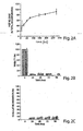
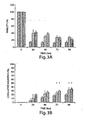
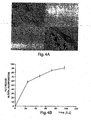

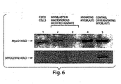


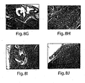

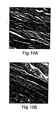


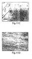

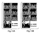
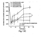
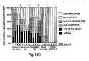


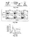
Classifications
-
- A—HUMAN NECESSITIES
- A61—MEDICAL OR VETERINARY SCIENCE; HYGIENE
- A61K—PREPARATIONS FOR MEDICAL, DENTAL OR TOILETRY PURPOSES
- A61K39/00—Medicinal preparations containing antigens or antibodies
- A61K39/0005—Vertebrate antigens
- A61K39/0011—Cancer antigens
-
- A—HUMAN NECESSITIES
- A61—MEDICAL OR VETERINARY SCIENCE; HYGIENE
- A61K—PREPARATIONS FOR MEDICAL, DENTAL OR TOILETRY PURPOSES
- A61K38/00—Medicinal preparations containing peptides
- A61K38/16—Peptides having more than 20 amino acids; Gastrins; Somatostatins; Melanotropins; Derivatives thereof
- A61K38/17—Peptides having more than 20 amino acids; Gastrins; Somatostatins; Melanotropins; Derivatives thereof from animals; from humans
- A61K38/18—Growth factors; Growth regulators
- A61K38/1858—Platelet-derived growth factor [PDGF]
- A61K38/1866—Vascular endothelial growth factor [VEGF]
-
- A—HUMAN NECESSITIES
- A61—MEDICAL OR VETERINARY SCIENCE; HYGIENE
- A61K—PREPARATIONS FOR MEDICAL, DENTAL OR TOILETRY PURPOSES
- A61K38/00—Medicinal preparations containing peptides
- A61K38/16—Peptides having more than 20 amino acids; Gastrins; Somatostatins; Melanotropins; Derivatives thereof
- A61K38/17—Peptides having more than 20 amino acids; Gastrins; Somatostatins; Melanotropins; Derivatives thereof from animals; from humans
- A61K38/19—Cytokines; Lymphokines; Interferons
- A61K38/193—Colony stimulating factors [CSF]
-
- A—HUMAN NECESSITIES
- A61—MEDICAL OR VETERINARY SCIENCE; HYGIENE
- A61K—PREPARATIONS FOR MEDICAL, DENTAL OR TOILETRY PURPOSES
- A61K38/00—Medicinal preparations containing peptides
- A61K38/16—Peptides having more than 20 amino acids; Gastrins; Somatostatins; Melanotropins; Derivatives thereof
- A61K38/17—Peptides having more than 20 amino acids; Gastrins; Somatostatins; Melanotropins; Derivatives thereof from animals; from humans
- A61K38/39—Connective tissue peptides, e.g. collagen, elastin, laminin, fibronectin, vitronectin, cold insoluble globulin [CIG]
-
- A—HUMAN NECESSITIES
- A61—MEDICAL OR VETERINARY SCIENCE; HYGIENE
- A61K—PREPARATIONS FOR MEDICAL, DENTAL OR TOILETRY PURPOSES
- A61K39/00—Medicinal preparations containing antigens or antibodies
- A61K39/0005—Vertebrate antigens
- A61K39/0011—Cancer antigens
- A61K39/001154—Enzymes
- A61K39/001156—Tyrosinase and tyrosinase related proteinases [TRP-1 or TRP-2]
-
- A—HUMAN NECESSITIES
- A61—MEDICAL OR VETERINARY SCIENCE; HYGIENE
- A61K—PREPARATIONS FOR MEDICAL, DENTAL OR TOILETRY PURPOSES
- A61K39/00—Medicinal preparations containing antigens or antibodies
- A61K39/0005—Vertebrate antigens
- A61K39/0011—Cancer antigens
- A61K39/001169—Tumor associated carbohydrates
- A61K39/00117—Mucins, e.g. MUC-1
-
- A—HUMAN NECESSITIES
- A61—MEDICAL OR VETERINARY SCIENCE; HYGIENE
- A61K—PREPARATIONS FOR MEDICAL, DENTAL OR TOILETRY PURPOSES
- A61K39/00—Medicinal preparations containing antigens or antibodies
- A61K39/0005—Vertebrate antigens
- A61K39/0011—Cancer antigens
- A61K39/001169—Tumor associated carbohydrates
- A61K39/001171—Gangliosides, e.g. GM2, GD2 or GD3
-
- A—HUMAN NECESSITIES
- A61—MEDICAL OR VETERINARY SCIENCE; HYGIENE
- A61K—PREPARATIONS FOR MEDICAL, DENTAL OR TOILETRY PURPOSES
- A61K39/00—Medicinal preparations containing antigens or antibodies
- A61K39/0005—Vertebrate antigens
- A61K39/0011—Cancer antigens
- A61K39/00118—Cancer antigens from embryonic or fetal origin
- A61K39/001182—Carcinoembryonic antigen [CEA]
-
- A—HUMAN NECESSITIES
- A61—MEDICAL OR VETERINARY SCIENCE; HYGIENE
- A61K—PREPARATIONS FOR MEDICAL, DENTAL OR TOILETRY PURPOSES
- A61K39/00—Medicinal preparations containing antigens or antibodies
- A61K39/0005—Vertebrate antigens
- A61K39/0011—Cancer antigens
- A61K39/001184—Cancer testis antigens, e.g. SSX, BAGE, GAGE or SAGE
- A61K39/001186—MAGE
-
- A—HUMAN NECESSITIES
- A61—MEDICAL OR VETERINARY SCIENCE; HYGIENE
- A61K—PREPARATIONS FOR MEDICAL, DENTAL OR TOILETRY PURPOSES
- A61K39/00—Medicinal preparations containing antigens or antibodies
- A61K39/0005—Vertebrate antigens
- A61K39/0011—Cancer antigens
- A61K39/00119—Melanoma antigens
- A61K39/001191—Melan-A/MART
-
- A—HUMAN NECESSITIES
- A61—MEDICAL OR VETERINARY SCIENCE; HYGIENE
- A61K—PREPARATIONS FOR MEDICAL, DENTAL OR TOILETRY PURPOSES
- A61K39/00—Medicinal preparations containing antigens or antibodies
- A61K39/0005—Vertebrate antigens
- A61K39/0011—Cancer antigens
- A61K39/00119—Melanoma antigens
- A61K39/001192—Glycoprotein 100 [Gp100]
-
- A—HUMAN NECESSITIES
- A61—MEDICAL OR VETERINARY SCIENCE; HYGIENE
- A61K—PREPARATIONS FOR MEDICAL, DENTAL OR TOILETRY PURPOSES
- A61K39/00—Medicinal preparations containing antigens or antibodies
- A61K39/0005—Vertebrate antigens
- A61K39/0011—Cancer antigens
- A61K39/001193—Prostate associated antigens e.g. Prostate stem cell antigen [PSCA]; Prostate carcinoma tumor antigen [PCTA]; PAP or PSGR
- A61K39/001195—Prostate specific membrane antigen [PSMA]
-
- A—HUMAN NECESSITIES
- A61—MEDICAL OR VETERINARY SCIENCE; HYGIENE
- A61K—PREPARATIONS FOR MEDICAL, DENTAL OR TOILETRY PURPOSES
- A61K39/00—Medicinal preparations containing antigens or antibodies
- A61K39/12—Viral antigens
- A61K39/21—Retroviridae, e.g. equine infectious anemia virus
-
- A—HUMAN NECESSITIES
- A61—MEDICAL OR VETERINARY SCIENCE; HYGIENE
- A61K—PREPARATIONS FOR MEDICAL, DENTAL OR TOILETRY PURPOSES
- A61K39/00—Medicinal preparations containing antigens or antibodies
- A61K39/39—Medicinal preparations containing antigens or antibodies characterised by the immunostimulating additives, e.g. chemical adjuvants
-
- A—HUMAN NECESSITIES
- A61—MEDICAL OR VETERINARY SCIENCE; HYGIENE
- A61K—PREPARATIONS FOR MEDICAL, DENTAL OR TOILETRY PURPOSES
- A61K9/00—Medicinal preparations characterised by special physical form
- A61K9/0012—Galenical forms characterised by the site of application
- A61K9/0019—Injectable compositions; Intramuscular, intravenous, arterial, subcutaneous administration; Compositions to be administered through the skin in an invasive manner
- A61K9/0024—Solid, semi-solid or solidifying implants, which are implanted or injected in body tissue
-
- A—HUMAN NECESSITIES
- A61—MEDICAL OR VETERINARY SCIENCE; HYGIENE
- A61K—PREPARATIONS FOR MEDICAL, DENTAL OR TOILETRY PURPOSES
- A61K9/00—Medicinal preparations characterised by special physical form
- A61K9/14—Particulate form, e.g. powders, Processes for size reducing of pure drugs or the resulting products, Pure drug nanoparticles
- A61K9/16—Agglomerates; Granulates; Microbeadlets ; Microspheres; Pellets; Solid products obtained by spray drying, spray freeze drying, spray congealing,(multiple) emulsion solvent evaporation or extraction
- A61K9/1605—Excipients; Inactive ingredients
- A61K9/1629—Organic macromolecular compounds
- A61K9/1641—Organic macromolecular compounds obtained otherwise than by reactions only involving carbon-to-carbon unsaturated bonds, e.g. polyethylene glycol, poloxamers
- A61K9/1647—Polyesters, e.g. poly(lactide-co-glycolide)
-
- A—HUMAN NECESSITIES
- A61—MEDICAL OR VETERINARY SCIENCE; HYGIENE
- A61L—METHODS OR APPARATUS FOR STERILISING MATERIALS OR OBJECTS IN GENERAL; DISINFECTION, STERILISATION OR DEODORISATION OF AIR; CHEMICAL ASPECTS OF BANDAGES, DRESSINGS, ABSORBENT PADS OR SURGICAL ARTICLES; MATERIALS FOR BANDAGES, DRESSINGS, ABSORBENT PADS OR SURGICAL ARTICLES
- A61L27/00—Materials for grafts or prostheses or for coating grafts or prostheses
- A61L27/14—Macromolecular materials
- A61L27/18—Macromolecular materials obtained otherwise than by reactions only involving carbon-to-carbon unsaturated bonds
-
- A—HUMAN NECESSITIES
- A61—MEDICAL OR VETERINARY SCIENCE; HYGIENE
- A61L—METHODS OR APPARATUS FOR STERILISING MATERIALS OR OBJECTS IN GENERAL; DISINFECTION, STERILISATION OR DEODORISATION OF AIR; CHEMICAL ASPECTS OF BANDAGES, DRESSINGS, ABSORBENT PADS OR SURGICAL ARTICLES; MATERIALS FOR BANDAGES, DRESSINGS, ABSORBENT PADS OR SURGICAL ARTICLES
- A61L27/00—Materials for grafts or prostheses or for coating grafts or prostheses
- A61L27/14—Macromolecular materials
- A61L27/20—Polysaccharides
-
- A—HUMAN NECESSITIES
- A61—MEDICAL OR VETERINARY SCIENCE; HYGIENE
- A61L—METHODS OR APPARATUS FOR STERILISING MATERIALS OR OBJECTS IN GENERAL; DISINFECTION, STERILISATION OR DEODORISATION OF AIR; CHEMICAL ASPECTS OF BANDAGES, DRESSINGS, ABSORBENT PADS OR SURGICAL ARTICLES; MATERIALS FOR BANDAGES, DRESSINGS, ABSORBENT PADS OR SURGICAL ARTICLES
- A61L27/00—Materials for grafts or prostheses or for coating grafts or prostheses
- A61L27/36—Materials for grafts or prostheses or for coating grafts or prostheses containing ingredients of undetermined constitution or reaction products thereof, e.g. transplant tissue, natural bone, extracellular matrix
- A61L27/3604—Materials for grafts or prostheses or for coating grafts or prostheses containing ingredients of undetermined constitution or reaction products thereof, e.g. transplant tissue, natural bone, extracellular matrix characterised by the human or animal origin of the biological material, e.g. hair, fascia, fish scales, silk, shellac, pericardium, pleura, renal tissue, amniotic membrane, parenchymal tissue, fetal tissue, muscle tissue, fat tissue, enamel
- A61L27/3633—Extracellular matrix [ECM]
-
- A—HUMAN NECESSITIES
- A61—MEDICAL OR VETERINARY SCIENCE; HYGIENE
- A61L—METHODS OR APPARATUS FOR STERILISING MATERIALS OR OBJECTS IN GENERAL; DISINFECTION, STERILISATION OR DEODORISATION OF AIR; CHEMICAL ASPECTS OF BANDAGES, DRESSINGS, ABSORBENT PADS OR SURGICAL ARTICLES; MATERIALS FOR BANDAGES, DRESSINGS, ABSORBENT PADS OR SURGICAL ARTICLES
- A61L27/00—Materials for grafts or prostheses or for coating grafts or prostheses
- A61L27/36—Materials for grafts or prostheses or for coating grafts or prostheses containing ingredients of undetermined constitution or reaction products thereof, e.g. transplant tissue, natural bone, extracellular matrix
- A61L27/38—Materials for grafts or prostheses or for coating grafts or prostheses containing ingredients of undetermined constitution or reaction products thereof, e.g. transplant tissue, natural bone, extracellular matrix containing added animal cells
- A61L27/3804—Materials for grafts or prostheses or for coating grafts or prostheses containing ingredients of undetermined constitution or reaction products thereof, e.g. transplant tissue, natural bone, extracellular matrix containing added animal cells characterised by specific cells or progenitors thereof, e.g. fibroblasts, connective tissue cells, kidney cells
- A61L27/3826—Muscle cells, e.g. smooth muscle cells
-
- A—HUMAN NECESSITIES
- A61—MEDICAL OR VETERINARY SCIENCE; HYGIENE
- A61L—METHODS OR APPARATUS FOR STERILISING MATERIALS OR OBJECTS IN GENERAL; DISINFECTION, STERILISATION OR DEODORISATION OF AIR; CHEMICAL ASPECTS OF BANDAGES, DRESSINGS, ABSORBENT PADS OR SURGICAL ARTICLES; MATERIALS FOR BANDAGES, DRESSINGS, ABSORBENT PADS OR SURGICAL ARTICLES
- A61L27/00—Materials for grafts or prostheses or for coating grafts or prostheses
- A61L27/50—Materials characterised by their function or physical properties, e.g. injectable or lubricating compositions, shape-memory materials, surface modified materials
- A61L27/54—Biologically active materials, e.g. therapeutic substances
-
- A—HUMAN NECESSITIES
- A61—MEDICAL OR VETERINARY SCIENCE; HYGIENE
- A61L—METHODS OR APPARATUS FOR STERILISING MATERIALS OR OBJECTS IN GENERAL; DISINFECTION, STERILISATION OR DEODORISATION OF AIR; CHEMICAL ASPECTS OF BANDAGES, DRESSINGS, ABSORBENT PADS OR SURGICAL ARTICLES; MATERIALS FOR BANDAGES, DRESSINGS, ABSORBENT PADS OR SURGICAL ARTICLES
- A61L27/00—Materials for grafts or prostheses or for coating grafts or prostheses
- A61L27/50—Materials characterised by their function or physical properties, e.g. injectable or lubricating compositions, shape-memory materials, surface modified materials
- A61L27/56—Porous materials, e.g. foams or sponges
-
- A—HUMAN NECESSITIES
- A61—MEDICAL OR VETERINARY SCIENCE; HYGIENE
- A61L—METHODS OR APPARATUS FOR STERILISING MATERIALS OR OBJECTS IN GENERAL; DISINFECTION, STERILISATION OR DEODORISATION OF AIR; CHEMICAL ASPECTS OF BANDAGES, DRESSINGS, ABSORBENT PADS OR SURGICAL ARTICLES; MATERIALS FOR BANDAGES, DRESSINGS, ABSORBENT PADS OR SURGICAL ARTICLES
- A61L27/00—Materials for grafts or prostheses or for coating grafts or prostheses
- A61L27/50—Materials characterised by their function or physical properties, e.g. injectable or lubricating compositions, shape-memory materials, surface modified materials
- A61L27/58—Materials at least partially resorbable by the body
-
- A—HUMAN NECESSITIES
- A61—MEDICAL OR VETERINARY SCIENCE; HYGIENE
- A61P—SPECIFIC THERAPEUTIC ACTIVITY OF CHEMICAL COMPOUNDS OR MEDICINAL PREPARATIONS
- A61P35/00—Antineoplastic agents
-
- A—HUMAN NECESSITIES
- A61—MEDICAL OR VETERINARY SCIENCE; HYGIENE
- A61P—SPECIFIC THERAPEUTIC ACTIVITY OF CHEMICAL COMPOUNDS OR MEDICINAL PREPARATIONS
- A61P43/00—Drugs for specific purposes, not provided for in groups A61P1/00-A61P41/00
-
- C—CHEMISTRY; METALLURGY
- C12—BIOCHEMISTRY; BEER; SPIRITS; WINE; VINEGAR; MICROBIOLOGY; ENZYMOLOGY; MUTATION OR GENETIC ENGINEERING
- C12N—MICROORGANISMS OR ENZYMES; COMPOSITIONS THEREOF; PROPAGATING, PRESERVING, OR MAINTAINING MICROORGANISMS; MUTATION OR GENETIC ENGINEERING; CULTURE MEDIA
- C12N15/00—Mutation or genetic engineering; DNA or RNA concerning genetic engineering, vectors, e.g. plasmids, or their isolation, preparation or purification; Use of hosts therefor
- C12N15/09—Recombinant DNA-technology
- C12N15/11—DNA or RNA fragments; Modified forms thereof; Non-coding nucleic acids having a biological activity
- C12N15/117—Nucleic acids having immunomodulatory properties, e.g. containing CpG-motifs
-
- C—CHEMISTRY; METALLURGY
- C12—BIOCHEMISTRY; BEER; SPIRITS; WINE; VINEGAR; MICROBIOLOGY; ENZYMOLOGY; MUTATION OR GENETIC ENGINEERING
- C12N—MICROORGANISMS OR ENZYMES; COMPOSITIONS THEREOF; PROPAGATING, PRESERVING, OR MAINTAINING MICROORGANISMS; MUTATION OR GENETIC ENGINEERING; CULTURE MEDIA
- C12N7/00—Viruses; Bacteriophages; Compositions thereof; Preparation or purification thereof
-
- A—HUMAN NECESSITIES
- A61—MEDICAL OR VETERINARY SCIENCE; HYGIENE
- A61K—PREPARATIONS FOR MEDICAL, DENTAL OR TOILETRY PURPOSES
- A61K39/00—Medicinal preparations containing antigens or antibodies
- A61K2039/54—Medicinal preparations containing antigens or antibodies characterised by the route of administration
-
- A—HUMAN NECESSITIES
- A61—MEDICAL OR VETERINARY SCIENCE; HYGIENE
- A61K—PREPARATIONS FOR MEDICAL, DENTAL OR TOILETRY PURPOSES
- A61K39/00—Medicinal preparations containing antigens or antibodies
- A61K2039/555—Medicinal preparations containing antigens or antibodies characterised by a specific combination antigen/adjuvant
- A61K2039/55511—Organic adjuvants
- A61K2039/55516—Proteins; Peptides
-
- A—HUMAN NECESSITIES
- A61—MEDICAL OR VETERINARY SCIENCE; HYGIENE
- A61K—PREPARATIONS FOR MEDICAL, DENTAL OR TOILETRY PURPOSES
- A61K39/00—Medicinal preparations containing antigens or antibodies
- A61K2039/555—Medicinal preparations containing antigens or antibodies characterised by a specific combination antigen/adjuvant
- A61K2039/55511—Organic adjuvants
- A61K2039/55522—Cytokines; Lymphokines; Interferons
-
- A—HUMAN NECESSITIES
- A61—MEDICAL OR VETERINARY SCIENCE; HYGIENE
- A61K—PREPARATIONS FOR MEDICAL, DENTAL OR TOILETRY PURPOSES
- A61K39/00—Medicinal preparations containing antigens or antibodies
- A61K2039/555—Medicinal preparations containing antigens or antibodies characterised by a specific combination antigen/adjuvant
- A61K2039/55511—Organic adjuvants
- A61K2039/55561—CpG containing adjuvants; Oligonucleotide containing adjuvants
-
- A—HUMAN NECESSITIES
- A61—MEDICAL OR VETERINARY SCIENCE; HYGIENE
- A61K—PREPARATIONS FOR MEDICAL, DENTAL OR TOILETRY PURPOSES
- A61K39/00—Medicinal preparations containing antigens or antibodies
- A61K2039/555—Medicinal preparations containing antigens or antibodies characterised by a specific combination antigen/adjuvant
- A61K2039/55511—Organic adjuvants
- A61K2039/55583—Polysaccharides
-
- A—HUMAN NECESSITIES
- A61—MEDICAL OR VETERINARY SCIENCE; HYGIENE
- A61K—PREPARATIONS FOR MEDICAL, DENTAL OR TOILETRY PURPOSES
- A61K39/00—Medicinal preparations containing antigens or antibodies
- A61K2039/60—Medicinal preparations containing antigens or antibodies characteristics by the carrier linked to the antigen
- A61K2039/6093—Synthetic polymers, e.g. polyethyleneglycol [PEG], Polymers or copolymers of (D) glutamate and (D) lysine
-
- A—HUMAN NECESSITIES
- A61—MEDICAL OR VETERINARY SCIENCE; HYGIENE
- A61L—METHODS OR APPARATUS FOR STERILISING MATERIALS OR OBJECTS IN GENERAL; DISINFECTION, STERILISATION OR DEODORISATION OF AIR; CHEMICAL ASPECTS OF BANDAGES, DRESSINGS, ABSORBENT PADS OR SURGICAL ARTICLES; MATERIALS FOR BANDAGES, DRESSINGS, ABSORBENT PADS OR SURGICAL ARTICLES
- A61L2300/00—Biologically active materials used in bandages, wound dressings, absorbent pads or medical devices
- A61L2300/20—Biologically active materials used in bandages, wound dressings, absorbent pads or medical devices containing or releasing organic materials
- A61L2300/23—Carbohydrates
- A61L2300/232—Monosaccharides, disaccharides, polysaccharides, lipopolysaccharides
-
- A—HUMAN NECESSITIES
- A61—MEDICAL OR VETERINARY SCIENCE; HYGIENE
- A61L—METHODS OR APPARATUS FOR STERILISING MATERIALS OR OBJECTS IN GENERAL; DISINFECTION, STERILISATION OR DEODORISATION OF AIR; CHEMICAL ASPECTS OF BANDAGES, DRESSINGS, ABSORBENT PADS OR SURGICAL ARTICLES; MATERIALS FOR BANDAGES, DRESSINGS, ABSORBENT PADS OR SURGICAL ARTICLES
- A61L2300/00—Biologically active materials used in bandages, wound dressings, absorbent pads or medical devices
- A61L2300/20—Biologically active materials used in bandages, wound dressings, absorbent pads or medical devices containing or releasing organic materials
- A61L2300/252—Polypeptides, proteins, e.g. glycoproteins, lipoproteins, cytokines
-
- A—HUMAN NECESSITIES
- A61—MEDICAL OR VETERINARY SCIENCE; HYGIENE
- A61L—METHODS OR APPARATUS FOR STERILISING MATERIALS OR OBJECTS IN GENERAL; DISINFECTION, STERILISATION OR DEODORISATION OF AIR; CHEMICAL ASPECTS OF BANDAGES, DRESSINGS, ABSORBENT PADS OR SURGICAL ARTICLES; MATERIALS FOR BANDAGES, DRESSINGS, ABSORBENT PADS OR SURGICAL ARTICLES
- A61L2300/00—Biologically active materials used in bandages, wound dressings, absorbent pads or medical devices
- A61L2300/40—Biologically active materials used in bandages, wound dressings, absorbent pads or medical devices characterised by a specific therapeutic activity or mode of action
- A61L2300/412—Tissue-regenerating or healing or proliferative agents
- A61L2300/414—Growth factors
-
- A—HUMAN NECESSITIES
- A61—MEDICAL OR VETERINARY SCIENCE; HYGIENE
- A61L—METHODS OR APPARATUS FOR STERILISING MATERIALS OR OBJECTS IN GENERAL; DISINFECTION, STERILISATION OR DEODORISATION OF AIR; CHEMICAL ASPECTS OF BANDAGES, DRESSINGS, ABSORBENT PADS OR SURGICAL ARTICLES; MATERIALS FOR BANDAGES, DRESSINGS, ABSORBENT PADS OR SURGICAL ARTICLES
- A61L2300/00—Biologically active materials used in bandages, wound dressings, absorbent pads or medical devices
- A61L2300/40—Biologically active materials used in bandages, wound dressings, absorbent pads or medical devices characterised by a specific therapeutic activity or mode of action
- A61L2300/426—Immunomodulating agents, i.e. cytokines, interleukins, interferons
-
- A—HUMAN NECESSITIES
- A61—MEDICAL OR VETERINARY SCIENCE; HYGIENE
- A61L—METHODS OR APPARATUS FOR STERILISING MATERIALS OR OBJECTS IN GENERAL; DISINFECTION, STERILISATION OR DEODORISATION OF AIR; CHEMICAL ASPECTS OF BANDAGES, DRESSINGS, ABSORBENT PADS OR SURGICAL ARTICLES; MATERIALS FOR BANDAGES, DRESSINGS, ABSORBENT PADS OR SURGICAL ARTICLES
- A61L2300/00—Biologically active materials used in bandages, wound dressings, absorbent pads or medical devices
- A61L2300/40—Biologically active materials used in bandages, wound dressings, absorbent pads or medical devices characterised by a specific therapeutic activity or mode of action
- A61L2300/438—Antigens
-
- A—HUMAN NECESSITIES
- A61—MEDICAL OR VETERINARY SCIENCE; HYGIENE
- A61L—METHODS OR APPARATUS FOR STERILISING MATERIALS OR OBJECTS IN GENERAL; DISINFECTION, STERILISATION OR DEODORISATION OF AIR; CHEMICAL ASPECTS OF BANDAGES, DRESSINGS, ABSORBENT PADS OR SURGICAL ARTICLES; MATERIALS FOR BANDAGES, DRESSINGS, ABSORBENT PADS OR SURGICAL ARTICLES
- A61L2300/00—Biologically active materials used in bandages, wound dressings, absorbent pads or medical devices
- A61L2300/60—Biologically active materials used in bandages, wound dressings, absorbent pads or medical devices characterised by a special physical form
- A61L2300/602—Type of release, e.g. controlled, sustained, slow
- A61L2300/604—Biodegradation
-
- A—HUMAN NECESSITIES
- A61—MEDICAL OR VETERINARY SCIENCE; HYGIENE
- A61L—METHODS OR APPARATUS FOR STERILISING MATERIALS OR OBJECTS IN GENERAL; DISINFECTION, STERILISATION OR DEODORISATION OF AIR; CHEMICAL ASPECTS OF BANDAGES, DRESSINGS, ABSORBENT PADS OR SURGICAL ARTICLES; MATERIALS FOR BANDAGES, DRESSINGS, ABSORBENT PADS OR SURGICAL ARTICLES
- A61L2300/00—Biologically active materials used in bandages, wound dressings, absorbent pads or medical devices
- A61L2300/60—Biologically active materials used in bandages, wound dressings, absorbent pads or medical devices characterised by a special physical form
- A61L2300/606—Coatings
-
- A—HUMAN NECESSITIES
- A61—MEDICAL OR VETERINARY SCIENCE; HYGIENE
- A61L—METHODS OR APPARATUS FOR STERILISING MATERIALS OR OBJECTS IN GENERAL; DISINFECTION, STERILISATION OR DEODORISATION OF AIR; CHEMICAL ASPECTS OF BANDAGES, DRESSINGS, ABSORBENT PADS OR SURGICAL ARTICLES; MATERIALS FOR BANDAGES, DRESSINGS, ABSORBENT PADS OR SURGICAL ARTICLES
- A61L2400/00—Materials characterised by their function or physical properties
- A61L2400/18—Modification of implant surfaces in order to improve biocompatibility, cell growth, fixation of biomolecules, e.g. plasma treatment
-
- A—HUMAN NECESSITIES
- A61—MEDICAL OR VETERINARY SCIENCE; HYGIENE
- A61L—METHODS OR APPARATUS FOR STERILISING MATERIALS OR OBJECTS IN GENERAL; DISINFECTION, STERILISATION OR DEODORISATION OF AIR; CHEMICAL ASPECTS OF BANDAGES, DRESSINGS, ABSORBENT PADS OR SURGICAL ARTICLES; MATERIALS FOR BANDAGES, DRESSINGS, ABSORBENT PADS OR SURGICAL ARTICLES
- A61L2430/00—Materials or treatment for tissue regeneration
- A61L2430/30—Materials or treatment for tissue regeneration for muscle reconstruction
-
- A—HUMAN NECESSITIES
- A61—MEDICAL OR VETERINARY SCIENCE; HYGIENE
- A61L—METHODS OR APPARATUS FOR STERILISING MATERIALS OR OBJECTS IN GENERAL; DISINFECTION, STERILISATION OR DEODORISATION OF AIR; CHEMICAL ASPECTS OF BANDAGES, DRESSINGS, ABSORBENT PADS OR SURGICAL ARTICLES; MATERIALS FOR BANDAGES, DRESSINGS, ABSORBENT PADS OR SURGICAL ARTICLES
- A61L2430/00—Materials or treatment for tissue regeneration
- A61L2430/34—Materials or treatment for tissue regeneration for soft tissue reconstruction
-
- B—PERFORMING OPERATIONS; TRANSPORTING
- B33—ADDITIVE MANUFACTURING TECHNOLOGY
- B33Y—ADDITIVE MANUFACTURING, i.e. MANUFACTURING OF THREE-DIMENSIONAL [3-D] OBJECTS BY ADDITIVE DEPOSITION, ADDITIVE AGGLOMERATION OR ADDITIVE LAYERING, e.g. BY 3-D PRINTING, STEREOLITHOGRAPHY OR SELECTIVE LASER SINTERING
- B33Y80/00—Products made by additive manufacturing
-
- C—CHEMISTRY; METALLURGY
- C12—BIOCHEMISTRY; BEER; SPIRITS; WINE; VINEGAR; MICROBIOLOGY; ENZYMOLOGY; MUTATION OR GENETIC ENGINEERING
- C12N—MICROORGANISMS OR ENZYMES; COMPOSITIONS THEREOF; PROPAGATING, PRESERVING, OR MAINTAINING MICROORGANISMS; MUTATION OR GENETIC ENGINEERING; CULTURE MEDIA
- C12N2310/00—Structure or type of the nucleic acid
- C12N2310/10—Type of nucleic acid
- C12N2310/17—Immunomodulatory nucleic acids
-
- C—CHEMISTRY; METALLURGY
- C12—BIOCHEMISTRY; BEER; SPIRITS; WINE; VINEGAR; MICROBIOLOGY; ENZYMOLOGY; MUTATION OR GENETIC ENGINEERING
- C12N—MICROORGANISMS OR ENZYMES; COMPOSITIONS THEREOF; PROPAGATING, PRESERVING, OR MAINTAINING MICROORGANISMS; MUTATION OR GENETIC ENGINEERING; CULTURE MEDIA
- C12N2320/00—Applications; Uses
- C12N2320/30—Special therapeutic applications
- C12N2320/32—Special delivery means, e.g. tissue-specific
-
- C—CHEMISTRY; METALLURGY
- C12—BIOCHEMISTRY; BEER; SPIRITS; WINE; VINEGAR; MICROBIOLOGY; ENZYMOLOGY; MUTATION OR GENETIC ENGINEERING
- C12N—MICROORGANISMS OR ENZYMES; COMPOSITIONS THEREOF; PROPAGATING, PRESERVING, OR MAINTAINING MICROORGANISMS; MUTATION OR GENETIC ENGINEERING; CULTURE MEDIA
- C12N2740/00—Reverse transcribing RNA viruses
- C12N2740/00011—Details
- C12N2740/10011—Retroviridae
- C12N2740/16011—Human Immunodeficiency Virus, HIV
- C12N2740/16034—Use of virus or viral component as vaccine, e.g. live-attenuated or inactivated virus, VLP, viral protein
-
- Y—GENERAL TAGGING OF NEW TECHNOLOGICAL DEVELOPMENTS; GENERAL TAGGING OF CROSS-SECTIONAL TECHNOLOGIES SPANNING OVER SEVERAL SECTIONS OF THE IPC; TECHNICAL SUBJECTS COVERED BY FORMER USPC CROSS-REFERENCE ART COLLECTIONS [XRACs] AND DIGESTS
- Y02—TECHNOLOGIES OR APPLICATIONS FOR MITIGATION OR ADAPTATION AGAINST CLIMATE CHANGE
- Y02A—TECHNOLOGIES FOR ADAPTATION TO CLIMATE CHANGE
- Y02A50/00—TECHNOLOGIES FOR ADAPTATION TO CLIMATE CHANGE in human health protection, e.g. against extreme weather
- Y02A50/30—Against vector-borne diseases, e.g. mosquito-borne, fly-borne, tick-borne or waterborne diseases whose impact is exacerbated by climate change
Definitions
- the devices and methods of the invention provide a solution to several problems associated with previous cell transplantation protocols.
- Transplantation systems that enhance the viability of the cells and induce their outward migration to populate injured or defective bodily tissues enhance the success of tissue regeneration, e.g., the regeneration of muscle tissue or other tissues as well as angiogenesis.
- Such a device that controls cell function and/or behavior comprises a scaffold composition comprising open, interconnected macropores wherein the size of said macropores is 400-500 ⁇ m; a cell recruitment composition or cells seeded onto said scaffold; a deployment signal capable of inducing or promoting migration of cells, which deployment signal is:
- the devices mediate active recruitment, modification, and release of host cells from the material in vivo, thereby improving the function of cells that have resided in the scaffold.
- the device attracts or recruits cells already resident in the body to the scaffold material, and programs or reprograms the resident cells to a desired fate (e.g., immune activation or tissue regeneration).
- This device includes a scaffold composition which incorporates or is coated with a bioactive composition; the device regulates the egress of resident cells. Egress is regulated spatially and temporally. Depending on the application for which the device is designed, the device regulates egress through the physical or chemical characteristics of the scaffold itself. For example, the scaffold composition is differentially permeable, allowing cell egress only in certain physical areas of the scaffold. The permeability of the scaffold composition is regulated, for example, by selecting or engineering a material for greater or smaller pore size, density, polymer cross-linking, stiffness, toughness, ductility, or viscoelascticity.
- the scaffold composition contains physical channels or paths through which cells can move more easily towards a targeted area of egress of the device or of a compartment within the device.
- the scaffold composition is optionally organized into compartments or layers, each with a different permeability, so that the time required for a cell to move through the device is precisely and predictably controlled. Migration is also regulated by the degradation, de- or re-hydration, oxygenation, chemical or pH alteration, or ongoing self-assembly of the scaffold composition. These processes are driven by diffusion or cell-secretion of enzymes or other reactive chemicals.
- egress is regulated by a bioactive composition.
- the device controls and directs the migration of cells through its structure. Chemical affinities are used to channel cells towards a specific area of egress. For example, adhesion molecules are used to attract or retard the migration of cells.
- the device controls the timing of the migration and egress.
- the density and mixture of these bioactive substances is controlled by initial doping levels or concentration gradient of the substance, by embedding the bioactive substances in scaffold material with a known leaching rate, by release as the scaffold material degrades, by diffusion from an area of concentration, by interaction of precursor chemicals diffusing into an area, or by production/excretion of compositions by resident support cells.
- the physical or chemical structure of the scaffold also regulates the diffusion of bioactive agents through the device.
- the bioactive composition includes one or more compounds that regulate cell function and/or behavior.
- the bioactive composition is covalently linked to the scaffold composition or non-covalently associated with the scaffold.
- the bioactive composition is an extracellular matrix (ECM) component that is chemically crosslinked to the scaffold composition.
- ECM components generally include three general classes of macromolecules: collagens, proteoglycans/glycosaminoglycans (PG/GAG), and glycoproteins, e.g., fibronectin (FN), laminin, and thrombospondin.
- ECM components associate with molecules on the cell surface and mediate adhesion and/or motility.
- the ECM component associated with the scaffold is a proteoglycan attachment peptide or cyclic peptide containing the amino acid sequence arginine-glycine-aspartic acid (RGD).
- Proteoglycan attachment peptides are selected from the group consisting of G 4 RGDSP, XBBXBX, PRRARV, YEKPGSPPREVVPRPRPGV, RPSLAKKQRFRHRNRKGYRSQRGHSRGR, and RIQNLLKITNLRIKFVK
- cell attachment peptides are selected from the group consisting of RGD, RGDS, LDV, REDV, RGDV, LRGDN, IKVAV, YIGSR, PDSGR, RNIAEIIKDA, RGDT, DGEA, and VTXG.
- Components of the ECM e.g., FN, laminin, and collagen, interact with the cell surface via the integrin family of receptors, a group of divalent cation-dependent cell surface glycoproteins that mediate cellular recognition and adhesion to components of the ECM and to other cells.
- Ligands recognized by integrins typically contain an RGD amino acid sequence that is expressed in many ECM proteins.
- Exemplary molecules that mediate cell adhesion and/or movement include FN, laminin, collagen, thrombospondin 1, vitronectin, elastin, tenascin, aggrecan, agrin, bone sialoprotein, cartilage matrix protein, fibronogen, fibrin, fibulin, mucins, entactin, osteopontin, plasminogen, restrictin, serglycin, SPARC/osteonectin, versican, von Willebrand Factor, polysacchride heparin sulfate, cell adhesion molecules including connexins, selectinsinclude collagen, RGD (Arg-Gly-Asp) and YIGSR (Tyr-Ile-Gly-Ser-Arg) peptides, glycosaminoglycans (GAGs), hyaluronic acid (HA), integrins, selectins, cadherins and members of the immunoglobulin superfamily-Carbohydrate
- the device optionally contains a second bioactive composition that is a growth factor, morphogen, differentiation factor, or chemoattractant.
- the device includes vascular endothelial growth factor (VEGF), hepatocyte growth factor (HGF), or fibroblast growth factor 2 (FGF2) or a combination thereof.
- VEGF vascular endothelial growth factor
- HGF hepatocyte growth factor
- FGF2 fibroblast growth factor 2
- Other factors include hormones, neurotransmitters, neurotransmitter or growth factor receptors, interferons, interleukins, chemokines, MMP-sensitive substrate, cytokines, colony stimulating factors.
- Growth factors used to promote angiogenesis, bone regeneration, wound healing, and other aspects of tissue regeneration are listed herein and are used alone or in combination to induce colonization or regeneration of bodily tissues by cells that have migrated out of an implanted scaffold device.
- the second bioactive composition is an inhibitor of differentiation.
- the cells are maintained at a desired stage of development or differentiation by the inhibitor until the inhibitor is depleted (e.g., has diffused out of the scaffold or is rendered inactive) or until another signal is provided to induce a change, e.g., to promote maturation or differentiation.
- the second bioactive composition is covalently linked to the scaffold composition, keeping the composition relatively immobilized in or on the scaffold composition.
- the second bioactive composition is noncovalently associated with the scaffold.
- Noncovalent bonds are generally one to three orders of magnitude weaker than covalent bonds permitting diffusion of the factor out of the scaffold and into surrounding tissues.
- Noncovalent bonds include electrostatic, hydrogen, van der Waals, ⁇ aromatic, and hydrophobic.
- a growth factor such as VEGF is associated with the device by noncovalent bonds and exits the device following administration of the cell-seeded device to a target site to further promote angiogenesis and tissue repair of the target bodily tissue.
- the scaffold composition is biocompatible.
- the composition is biodegradable/erodable or resistant to breakdown in the body.
- Relatively permanent (degradation resistant) scaffold compositions include metals and some polymers such as silk.
- the scaffold composition degrades at a predetermined rate based on a physical parameter selected from the group consisting of temperature, pH, hydration status, and porosity, the cross-link density, type, and chemistry or the susceptibility of main chain linkages to degradation or it degrades at a predetermined rate based on a ratio of chemical polymers.
- a high molecular weight polymer comprised of solely lactide degrades over a period of years, e.g., 1-2 years
- a low molecular weight polymer comprised of a 50:50 mixture of lactide and glycolide degrades in a matter of weeks, e.g., 1, 2, 3, 4, 6, 10 weeks.
- a calcium cross-linked gels composed of high molecular weight, high guluronic acid alginate degrade over several months (1, 2, 4, 6, 8, 10, 12 months) to years (1, 2, 5 years) in vivo, while a gel comprised of low molecular weight alginate, and/or alginate that has been partially oxidized, will degrade in a matter of weeks.
- cells mediate degradation of the scaffold matrix, i.e., the scaffold composition is enzymatically digested by a composition elicited by a resident cell, and the egress of the cell is dependent upon the rate of enzymatic digestion of the scaffold.
- polymer main chains or cross-links contain compositions, e.g., oligopeptides, that are substrates for collagenase or plasmin, or other enzymes produced by within or adjacent to the scaffold.
- Exemplary scaffold compositions include polylactic acid, polyglycolic acid, PLGA polymers, alginates and alginate derivatives, gelatin, collagen, fibrin, hyaluronic acid, laminin rich gels, agarose, natural and synthetic polysaccharides, polyamino acids, polypeptides, polyesters, polyanhydrides, polyphosphazines, poly(vinyl alcohols), poly(alkylene oxides), poly(allylamines)(PAM), poly(acrylates), modified styrene polymers, pluronic polyols, polyoxamers, poly(uronic acids), poly(vinylpyrrolidone) and copolymers or graft copolymers of any of the above.
- One preferred scaffold composition includes an RGD-modified alginate.
- the scaffold is macroporous with aligned pores of about 400-500 ⁇ m in diameter.
- the devices are manufactured in their entirety in the absence of cells or can be assembled around or in contact with cells (the material is gelled or assembled around cells in vitro or in vivo in the presence of cells and tissues) and then contacted with cells to produce a cell-seeded structure.
- the device is manufactured in two or more (3, 4, 5, 6, ....10 or more) stages in which one layer or compartment is made and seeded with cells followed by the construction of a second, third, fourth or more layers, which are in turn seeded with cells in sequence.
- Each layer or compartment is identical to the others or distinguished from one another by the number, genotype, or phenotype of the seed cell population as well as distinct chemical, physical and biological properties.
- the device Prior to implantation, the device is contacted with purified populations cells or characterized mixtures of cells as described above.
- Exemplary cells include myoblasts for muscle regeneration, repair or replacement; hepatocytes for liver tissue regeneration, repair or organ transplantation, chondrocytes for cartilage replacement, regeneration or repair, and osteoblasts for bone regeneration, replacement or repair, various stem cell populations (embryonic stem cells differentiated into various cell types), bone marrow or adipose tissue derived adult stem cells, cardiac stem cells, pancreatic stem cells, endothelial progenitors and outgrowth endothelial cells, mesenchymal stem cells, hematopoietic stem cells, neural stem cells, satellite cells, side population cells, differentiated cell populations including osteoprogenitors and osteoblasts, chondrocytes, keratinocytes for skin, tenocytes for tendon, intestinal epithelial cells, endothelial cells, smooth muscle cells and fibroblasts for tissue or organ regeneration, repair or replacement and
- a method of making a scaffold is carried out by providing a scaffold composition and covalently linking or noncovalently associating the scaffold composition with a first bioactive composition.
- the first bioactive composition preferably contains a cell adhesion ligand.
- the scaffold composition is also contacted with a second bioactive composition.
- the second bioactive composition is preferably non-covalently associated with the scaffold composition to yield a doped scaffold, i.e., a scaffold composition that includes one or more bioactive substances.
- the contacting steps are optionally repeated to yield a plurality of doped scaffolds, e.g., each of the contacting steps is characterized by a different amount of the second bioactive composition to yield a gradient of the second bioactive composition in the scaffold device.
- subsequent contacting steps involve a different bioactive composition, i.e., a third, fourth, fifth, sixth...., composition or mixture of compositions, that is distinguished from the prior compositions or mixtures of prior doping steps by the structure or chemical formula of the factor(s).
- the method optionally involves adhering individual niches, layers, or components to one another and/or insertion of semi-permeable, permeable, or nonpermeable membranes within or at one or more boundaries of the device to further control/regulate locomotion of cells or bioactive compositions.
- the scaffold is seeded with cells after completion of the construction of the device or in an iterative manner throughout the construction of each component.
- the method includes the steps of providing a device that includes scaffold composition with a bioactive composition incorporated therein or thereon and a mammalian cell bound to the scaffold.
- a mammalian tissue is contacted with the device.
- the scaffold composition temporally controls egress of the cell and the bioactive composition spatially or directionally regulates egress of the cell.
- the device that is provided contains a scaffold composition with a bioactive composition incorporated therein or thereon and a mammalian cell immobilized within the scaffold.
- the cell remains immobilized within the scaffold, and the scaffold composition temporally controls egress of a progeny cell of the immobilized cell and the bioactive composition spatially regulates egress of the progeny cells.
- a method of modulating an activity of a cell e.g., a host cell, disclosed herein. This method is carried out by administering to a mammal a device containing a scaffold composition and a recruitment composition incorporated therein or thereon, and then contacting the cell with a deployment signal.
- the deployment signal induces egress of the cells from the device.
- the activity of the cell at egress differs from that prior to entering the device.
- Cells are recruited into the device and remain resident in the device for a period of time, e.g., minutes; 0.2.
- the cells are contacted with or exposed to a deployment signal that induces induces egress of the altered (re-educated or reprogrammed) cells and the cells migrate out of the device and into surrounding tissues or remote target locations.
- the deployment signal is a composition such as protein, peptide, or nucleic acid.
- the deployment signal is a nucleic acid molecule, e.g., a plasmid containing sequence encoding a protein that induces migration of the cell out of the device and into surrounding tissues.
- the deployment signal occurs when the cell encounters the plasmid in the device, the DNA becomes internalized in the cell (i.e., the cell is transfected), and the cell manufactures the gene product encoded by the DNA.
- the molecule that signals deployment is an element of the device and is released from the device in delayed manner (e.g., temporally or spatially) relative to exposure of the cell to the recruitment composition.
- the deployment signal is a reduction in or absence of the recruitment composition.
- a recruitment composition induces migration of cells into the device, and a reduction in the concentration or depletion, dissipation, or diffusion of the recruitment composition from the device results in egress of cells out of the device.
- immune cells such as T cells, B cells, or dendritic cells (DCs) of an individual are recruited into the device, primed and activated to mount an immune response against an antigen-specific target.
- an antigen corresponding to a target to which an immune response is desired is incorporated into or onto the scaffold structure.
- Cytokines such as granulocyte macrophage colony stimulating factor (GM-CSF) are also a component of the device to amplify immune activation and/or induce migration of the primed cells to lymph nodes.
- GM-CSF granulocyte macrophage colony stimulating factor
- Other cell specific recruitment compositions are described below.
- vascular endothelial growth factor (VEGF) is useful to recruit angiogenic cells.
- the device recruit cells in vivo, modifies these cells, and then promotes their migration to another site in the body.
- This approach is examplied herein in the context of dendritic cells and cancer vaccine development but is also useful to other vaccines such as those against microbial pathogens as well as cell therapies in general.
- Cells educated using the devices described herein promote regeneration of a tissue or organ immediately adjacent to the material, or at some distant site. Alternatively, the cells are educated to promote destruction of a tissue (locally or at a distant site).
- the methods are also useful for disease prevention, e.g., to promote cell-based maintenance of tissue structure and function to stop or retard disease progression or age-related tissue changes.
- the education of cells within the device, "programming" and "reprogramming" permits modification of the function or activity of any cell in the body to become a multipotent stem cell again and exert therapeutic effects.
- Regenerative medical technologies are devices and methods that repair or replace diseased or defective tissues or organs.
- Tissue engineering is the application of the principles and methods of engineering and the life sciences to the development of biological substitutes to restore, maintain or improve function of bodily structures and tissues, or to selectively promote the destruction of undesired tissues. It involves the development of methods to build biological substitutes as supplements or alternatives to whole organ or tissue transplantation, or the development of strategies to manipulate tissues in vivo.
- the use of living cells and/or extracellular matrix (ECM) components in the development of implantable parts or devices is an attractive approach to restore or to replace function.
- the methods and devices are useful to generate functional biological structure de novo or to regenerate organs in situ , as well as to restore or supplement tissue function.
- the devices are placed into or adjacent to a particular diseased or injured tissue in the body or broadly dispersed throughout a tissue in the body. The device makes direct contact with the tissue to be treated or contains cells that migrate to nearby or remote tissue targets following residence in the device.
- Scaffold structures are seeded with one or more populations of purified or isolated cells.
- isolated used in reference to a cell type, e.g., a stem cell means that the cell is substantially free of other cell types or cellular material with which it naturally occurs.
- a sample of cells of a particular tissue type or phenotype is “substantially pure” when it is at least 60% of the cell population, Preferably, the preparation is at least 75%, more preferably at least 90%, and most preferably at least 99% or 100%, of the cell population. Purity is measured by any appropriate standard method, for example, by fluorescence-activated cell sorting (FACS).
- FACS fluorescence-activated cell sorting
- the device is seeded with two or more substantially pure populations of cells.
- the populations are spatially or physically separated, e.g., one population is encapsulated, or the cells are allowed to come into contact with one another.
- the scaffold or structural support not only provides a surface upon which cells are seeded/attached but indirectly affects production/education of cell populations by housing a second (third, or several) cell population(s) with which a first population of cells associates (cell-cell adhesion).
- Such "accessory" cell populations secrete desirable cytokines, growth factors or other signaling molecules, and/or deposit appropriate extracellular matrix proteins. Cytokines are small secreted proteins which mediate and regulate immunity, inflammation, and hematopoiesis. Cytokines can act over short distances and short time spans and at very low concentration.
- cytokines act by binding to specific membrane receptors, which then signal cells via second messengers, often tyrosine kinases, to alter its behavior (e.g., cell function and/or gene expression).
- Responses to cytokines include increasing or decreasing expression of membrane proteins (including cytokine receptors), proliferation, and secretion of effector molecules.
- Such molecules are also referred to as lymphokines (cytokines made by lymphocytes), monokines (cytokines made by monocytes), chemokines (cytokines with chemotactic activities), and interleukins (cytokines made by one leukocyte and acting on other leukocytes). Cytokines act on the cells that secrete them (autocrine action), on nearby cells (paracrine action), or on distant cells (endocrine action).
- a stem cell is an undifferentiated cell that differentiates into a mature functional tissue specific cell upon contact with appropriate microenviromnent, e.g., growth factors and other differentiating agents.
- the devices/scaffold described herein represent such a microenvironment. Each device constitutes a factory that attracts/accepts, reproduces, sustains, educates, and sends forth to surrounding bodily tissues tissue-specific cells that are capable of colonizing and regenerating damaged tissue.
- the scaffolds house progenitor cells, differentiated cells, support cells, modified cells (e.g., genetically modified (e.g., by DNA delivery (by plasmid or virus), or by siRNA, ⁇ RNA) to produce exogenous proteins or chemically modified with drugs to enhance or suppress specific signaling pathways (e.g., protein kinases) or genetic regulatory pathways (e.g., upregulate or downregulate activity of master transcription factors) that are involved in a variety of cell fate decisions, or surface modified with ligands or growth factors and morphogens, or small molecule mimics of the same, to promote specific adhesive interactions with other cells, including immune cells, or provide autocrine or paracrine signaling, fused cell populations, fibroblasts, chondrocytes, osteoblasts, myoblasts, endothelial, smooth muscle and neuronal cells.
- modified cells e.g., genetically modified (e.g., by DNA delivery (by plasmid or virus), or by siRNA, ⁇
- Differentiated cells are reprogrammed to an embryonic-like state by transfer of nuclear contents into oocytes or by fusion with embryonic stem (ES) cells.
- ES embryonic stem
- the induction of pluripotent stem cells from mouse embryonic or adult fibroblasts is accomplished by introducing one or more of the following factors, Oct3/4, Sox2, c-Myc, Klf4, and Nanog.
- differentiated cells are reprogrammed and exhibit the morphology and growth properties of stem cells, e.g., ES cells, and express stem cell marker genes.
- the scaffolds are seeded in vitro or in vivo .
- scaffolds are seeded by incubating the structure in a solution containing the cells.
- cells are injected/titrated into the scaffold or recruited to migrate into the device.
- the scaffold is built in stages with each layer of the multicomponent scaffold being seeded prior to laying down of another layer or before adherences of another pre-formed component.
- Different cell types e.g., stem vs. differentiated, support vs. therapeutic, are optionally co-resident in the scaffold housing.
- Cells optionally vary in phenotype, e.g., differentiation state, activation state, metabolic state, or functional state.
- the scaffolds are suitable for use with any cell type that one may want to transplant.
- Such cells include but are not limited to, various stem cell populations (embryonic stem cells differentiated into various cell types), bone marrow or adipose tissue derived adult stem cells, mesenchymal stem cells, cardiac stem cells, pancreatic stem cells, endothelaila progenitor cells, outgrowth endothelial cells, dendritic cells, hematopoietic stem cells, neural stem cells, satellite cells, side population cells.
- stem cell populations embryonic stem cells differentiated into various cell types
- mesenchymal stem cells derived stem cells
- cardiac stem cells pancreatic stem cells
- endothelaila progenitor cells endothelaila progenitor cells
- outgrowth endothelial cells dendritic cells
- hematopoietic stem cells neural stem cells
- satellite cells side population cells.
- Such cells may further include but are not limited to, differentiated cell populations including osteoprogenitors and osteoblasts, chondrocytes, keratinocytes for skin, tenocytes for tendon, and intestinal epithelial cells, smooth muscle cells, cardiac muscle cells, epithelial cells, endothelial cells, urothelial cells, fibroblasts, myoblasts, chondroblasts, osteoclasts, hepatocytes, bile duct cells, pancreatic islet cells, thyroid, parathyroid, adrenal, hypothalamic, pituitary, ovarian, testicular, salivary gland cells, adipocytes, and precursor cells.
- differentiated cell populations including osteoprogenitors and osteoblasts, chondrocytes, keratinocytes for skin, tenocytes for tendon, and intestinal epithelial cells, smooth muscle cells, cardiac muscle cells, epithelial cells, endothelial cells, urothelial cells, fibroblasts, myoblasts
- smooth muscle cells and endothelial cells may be employed for muscular, tubular scaffolds, e.g., scaffolds intended as vascular, esophageal, intestinal, rectal, or ureteral scaffolds; chondrocytes may be employed in cartilaginous scaffolds; cardiac muscle cells may be employed in heart scaffolds; hepatocytes and bile duct cells may be employed in liver scaffolds; epithelial, endothelial, fibroblast, and nerve cells may be employed in scaffolds intended to function as replacements or enhancements for any of the wide variety of tissue types that contain these cells.
- scaffolds of the invention may comprise any cell population competent to participate in regeneration, replacement or repair of a target tissue or organ.
- cells are myoblasts for use in muscle regeneration.
- Cells are optionally genetically manipulated by the introduction of exogenous genetic sequences or the inactivation or modification of endogenous sequences. For example, recombinant genes are introduced to cause the cells to make proteins that are otherwise lacking in the host or target tissue. Production of scarce but desirable proteins (in the context of certain tissues) is augmented by transplanting genetically engineered cells. Cells used to seed the scaffold are capable of degrading the scaffold matrix over a desired period time in order to migrate through and out of the scaffold matrix. Scaffold matrices are selected such that they are susceptible to degradation by certain cell types seeded within the matrix.
- scaffold materials and cells are selected and designed such that all or some of the cells seeded within the scaffolds require a certain desired period of time degrade the scaffold sufficiently to migrate through it and reach the surrounding tissue.
- the delay in the release of the cells to the surrounding tissue is controlled by varying the composition of the scaffold, to allow optimal time to signal the cells to multiply, differentiate, or achieve various phenotypes.
- General mammalian cell culture techniques, cell lines, and cell culture systems are described in Doyle, A., Griffiths, J. B., Newell, D. G., (eds.) Cell and Tissue Culture: Laboratory Procedures, Wiley, 1998 .
- migrating cells typically secrete collagenases and plasmin to degrade their matrix and allow cell movement.
- the rate of cells exiting may thus be regulated by controlling the density and susceptibility to these enzymes of oligopeptides used as either cross-links in the material or as components of the main chains.
- Certain materials are degraded in a preprogrammed manner independent of cell action (e.g. hydrolytic degradation of poly(lactide-co glyolide) as a degradable scaffold.
- the scaffolds may be prepared such that the degradation time may be controlled by using a mixture of degradable components in proportions to achieve a desired degradation rate.
- scaffold compositions are sensitive to degradation by materials secreted by the cells themselves that are seeded within the scaffold.
- MMP metalloproteinase
- stromal vascular cells and smooth muscle cells are useful in sheetlike structures are used for repair of vessel-like structures such as blood vessels or layers of the body cavity. Such structures are used to repair abdominal wall injuries or defects such as gastroschisis.
- sheetlike scaffolds seeded with dermal stem cells and/or keratinocytes are used in bandages or wound dressings for regeneration of dermal tissue.
- Components of the scaffolds are organized in a variety of geometric shapes (e.g., beads, pellets), niches, planar layers (e.g., thin sheets).
- multicomponent scaffolds are constructed in concentric layers each of which is characterized by different physical qualities (% polymer, % crosslinking of polymer, chemical composition of scaffold, pore size, porosity, and pore architecture, stiffness, toughness, ductility, viscoelasticity, and or composition of bioactive substances such as growth factors, homing/migration factors, differentiation factors.
- Each niche has a specific effect on a cell population, e.g., promoting or inhibiting a specific cellular function, proliferation, differentiation, elaboration of secreted factors or enzymes, or migration.
- stromal vascular cells and smooth muscle cells are useful in sheetlike structures are used for repair of vessel-like structures such as blood vessels or layers of the body cavity.
- vessel-like structures such as blood vessels or layers of the body cavity.
- such structures are used to repair abdominal wall injuries or defects such as gastroschisis.
- sheetlike scaffolds seeded with dermal stem cells and/or keratinocytes are used in bandages or wound dressings for regeneration of dermal tissue.
- the device is placed or transplanted on or next to a target tissue, in a protected location in the body, next to blood vessels, or outside the body as in the case of an external wound dressing.
- Devices are introduced into or onto a bodily tissue using a variety of known methods and tools, e.g., spoon, tweezers or graspers, hypodermic needle, endoscopic manipulator, endo- or trans-vascular- catheter, stereotaxic needle, snake device, organ-surface-crawling robot (United States Patent Application 20050154376 ; Ota et al., 2006, Innovations 1:227-231 ), minimally invasive surgical devices, surgical implantation tools, and transdermal patches. Devices can also be assembled in place, for example by senquentially injecting or inserting matrix materials. Scaffold devices are optionally recharged with cells or with bioactive compounds, e.g., by sequential injection or spraying of substances such as growth factors or differentiation factors.
- substances such as growth factors or differentiation factors.
- a scaffold or scaffold device is the physical structure upon which or into which cells associate or attach
- a scaffold composition is the material from which the structure is made.
- scaffold compositions include biodegradable or permanent materials such as those listed below.
- the mechanical characteristics of the scaffold vary according to the application or tissue type for which regeneration is sought. It is biodegradable (e.g., collagen, alginates, polysaccharides, polyethylene glycol (PEG), poly(glycolide) (PGA), poly(L-lactide) (PLA), or poly(lactide-co-glycolide) (PLGA) or permanent (e.g., silk).
- the composition is degraded by physical or chemical action, e.g., level of hydration, heat or ion exchange or by cellular action, e.g., elaboration of enzyme, peptides, or other compounds by nearby or resident cells.
- the consistency varies from a sofr/pliable (e.g., a gel) to glassy, rubbery, brittle, tough, elastic, stiff.
- the structures contain pores, which are nanoporous, microporous, or macroporous, and the pattern of the pores is optionally homogeneous, heterogenous, aligned, repeating, or random.
- Alginates are versatile polysaccharide based polymers that may be formulated for specific applications by controlling the molecular weight, rate of degradation and method of scaffold formation. Coupling reactions can be used to covalently attach bioactive epitopes, such as the cell adhesion sequence RGD to the polymer backbone.
- Alginate polymers are formed into a variety of scaffold types. Injectable hydrogels can be formed from low MW alginate solutions upon addition of a cross-linking agents, such as calcium ions, while macroporous scaffolds are formed by lyophilization of high MW alginate discs. Differences in scaffold formulation control the kinetics of scaffold degradation.
- Release rates of morphogens or other bioactive substances from alginate scaffolds is controlled by scaffold formulation to present morphogens in a spatially and temporally controlled manner. This controlled release not only eliminates systemic side effects and the need for multiple injections, but can be used to create a microenvironment that activates host cells at the implant site and transplanted cells seeded onto a scaffold.
- the scaffold comprises a biocompatible polymer matrix that is optionally biodegradable in whole or in part.
- a hydrogel is one example of a suitable polymer matrix material.
- materials which can form hydrogels include polylactic acid, polyglycolic acid, PLGA polymers, alginates and alginate derivatives, gelatin, collagen, agarose, natural and synthetic polysaccharides, polyamino acids such as polypeptides particularly poly(lysine), polyesters such as polyhydroxybutyrate and poly-epsilon.-caprolactone, polyanhydrides; polyphosphazines, poly(vinyl alcohols), poly(alkylene oxides) particularly poly(ethylene oxides), poly(allylamines)(PAM), poly(acrylates), modified styrene polymers such as poly(4-aminomethylstyrene), pluronic polyols, polyoxamers, poly(uronic acids), poly(vinylpyrrolidone) and copolymers of the above,
- the scaffolds are fabricated from a variety of synthetic polymers and naturally-occurring polymers such as, but not limited to, collagen, fibrin, hyaluronic acid, agarose, and laminin-rich gels.
- One preferred material for the hydrogel is alginate or modified alginate material.
- Alginate molecules are comprised of (1-4)-linked ⁇ -D-mannuronic acid (M units) and ⁇ L-guluronic acid (G units) monomers, which can vary in proportion and sequential distribution along the polymer chain.
- Alginate polysaccharides are polyelectrolyte systems which have a strong affinity for divalent cations (e.g. Ca +2 , Mg +2 , Ba +2 ) and form stable hydrogels when exposed to these molecules.
- calcium cross-linked alginate hydrogels are useful for dental applications, wound dressings chondrocyte transplantation and as a matrix for other cell types..
- An exemplary device utilizes an alginate or other polysaccharide of a relatively low molecular weight, preferably of size which, after dissolution, is at the renal threshold for clearance by humans, e.g., the alginate or polysaccharide is reduced to a molecular weight of 1000 to 80,000 daltons. Prefereably, the molecular mass is 1000 to 60,000 daltons, particularly preferably 1000 to 50,000 daltons. It is also useful to use an alginate material of high guluronate content since the guluronate units, as opposed to the mannuronate units, provide sites for ionic crosslinking through divalent cations to gel the polymer.
- U.S. Patent Number 6,642,363 discloses methods for making and using polymers containing polysachharides such as alginates or modified alginates that are particularly useful for cell transplantation and tissue engineering applications.
- polysaccharides other than alginates include agarose and microbial polysaccharides such as those listed in the table below.
- Polysaccharide Scaffold Compositions Polymers a Structure Fungal Pullulan (N) 1,4-;1,6- ⁇ -D-Glucan Scleroglucan (N) 1,3;1,6- ⁇ -D-Glucan Chitin (N) 1,4- ⁇ -D-Acetyl Glucosamine Chitosan (C) 1,4- ⁇ .-D-N-Glucosamine Elsinan (N) 1,4-;1,3- ⁇ -D-Glucan Bacterial Xanthan gum (A) 1,4- ⁇ .-D-Glucan with D-mannose; D-glucuronic Acid as side groups Curdlan (N) 1,3- ⁇ .-D-Glucan (with branching) Dextran (N) 1,6- a -D-Glucan with some 1,2;1,3-; 1,4- ⁇ - linkages Gellan (A) 1,4
- the scaffolds of the invention comprise open, interconnected macropores wherein the size of the macropores is 400-500 ⁇ m, In one example, the macroporous with aligned pores of about 400-500 ⁇ m in diameter.
- the pores have a diameter of less than about 10nm; 100nm-20 ⁇ m, more preferable greater than about 100 ⁇ m, even more preferable greater than 400 ⁇ m.
- the preparation of polymer matrices having the desired pore sizes and pore alignments are described in the Examples. Other methods of preparing porous hydrogel products are known in the art. ( U.S. Pat. No. 6,511,650 ).
- the device includes one or more bioactive compositions.
- Bioactive compositions are purified naturally-occurring, synthetically produced, or recombinant compounds, e.g., polypeptides, nucleic acids, small molecules, or other agents.
- the compositions described herein are purified.
- Purified compounds are at least 60% by weight (dry weight) the compound of interest.
- the preparation is at least 75%, more preferably at least 90%, and most preferably at least 99%, by weight the compound of interest. Purity is measured by any appropriate standard method, for example, by column chromatography, polyacrylamide gel electrophoresis, or HPLC analysis.
- the bioactive composition alters a function, e.g., level of differentiation, state of activation, motility, or gene expression, of a cell.
- a function e.g., level of differentiation, state of activation, motility, or gene expression
- at least one cell adhesion molecule is incorporated into or onto the polymer matrix. Such molecules are incorporated into the polymer matrix prior to polymerization of the matrix or after polymerization of the matrix.
- cell adhesion molecules include but are not limited to peptides, proteins and polysaccharides.
- cell adhesion molecules include fibronectin, laminin, collagen, thrombospondin 1, vitronectin, elastin, tenascin, aggrecan, agrin, bone sialoprotein, cartilage matrix protein, fibronogen, fibrin, fibulin, mucins, entactin, osteopontin, plasminogen, restricrin, serglycin, SPARC/osteonectin, versican, von Willebrand Factor, polysaccharide heparin sulfate, connexins, collagen, RGD (Arg-Gly-Asp) and YIGSR (Tyr- [upsilon]e-Gly-Ser-Arg) peptides and cyclic peptides, glycosaminoglycans (GAGs), hyaluronic acid (HA), condroitin-6-sulfate, integrin ligands, selectins, cadherins and members of the immuno
- ECM Proteins and peptides and role in cell function Protein Sequence Seq.ID No: Role Fibronectin RGDS Adhesion LDV Adhesion REDV Adhesion Vitronectin RGDV Adhesion Laminin A LRGDN Adhesion IKVAV Neurite extension B1 YIGSR Adhesion of many cells, via Laminin 67 kD laminin receptor PDSGR Adhesion Laminin B2 RNIAEIIKDA Neurite extension Collagen 1 RGDT Adhesion of most cells DGEA Adhesion of platelets, other cells Thrombospondin RGD Adhesion of most cells VTXG Adhesion of platelets Hubbell, JA (1995): Biomaterials in tissue engineering . Bio/Technology 13:565-576 .
- One-letter abbreviations of amino acids are used, X stands for any amino acid.
- Particularly preferred cell adhesion molecules are peptides or cyclic peptides containing the amino acid sequence arginine-glycine-aspartic acid (RGD) which is known as a cell attachment ligand and found in various natural extracellular matrix molecules.
- RGD arginine-glycine-aspartic acid
- a polymer matrix with such a modification provides cell adhesion properties to the scaffold, and sustains long-term survival of mammalian cell systems, as well as supporting cell growth and differentiation.
- Coupling of the cell adhesion molecules to the polymer matrix is accomplished using synthetic methods which are in general known to one of ordinary skill in the art and are described in the examples. Approaches to coupling of peptides to polymers are discussed in Hirano and Mooney, Advanced Materials, p.17-25 (2004 ). Other useful bonding chemistries include those discussed in Hermanson, Bioconjugate Techniques, p. 152-185 (1996 ), particularly by use of carbodiimide couplers, DCC and DIC (Woodward's Reagent K). Since many of the cell adhesion molecules are peptides, they contain a terminal amine group for such bonding.
- the amide bond formation is preferably catalyzed by 1-ethyl-3-(3-dimethylaminopropyl)carbodiimide (EDC), which is a water soluble enzyme commonly used in peptide synthesis.
- EDC 1-ethyl-3-(3-dimethylaminopropyl)carbodiimide
- the density of cell adhesion ligands a critical regulator of cellular phenotype following adhesion to a biomaterial. ( Massia and Hubbell, J. Cell Biol. 114:1089-1100, 1991 ; Mooney et al., J. Cell Phys. 151:497-505, 1992 ; and Hansen et al., Mol. Biol. Cell 5:967-975, 1994 ) can be readily varied over a 5-order of magnitude density range.
- the scaffold structure is constructed out of a number of different rigid, semi-rigid, flexible, gel, self-assembling, liquid crystalline, or fluid compositions such as peptide polymers, polysaccharides, synthetic polymers, hydrogel materials, ceramics (e.g., calcium phosphate or hydroxyapatite), proteins, glycoproteins, proteoglycans, metals and metal alloys.
- the compositions are assembled into cell scaffold structures using methods known in the art, e.g., injection molding, lyophillization of preformed structures, printing, self-assembly, phase inversion, solvent casting, melt processing, gas foaming, fiber forming/processing, particulate leaching or a combination thereof.
- the assembled devices are then implanted or administered to the body of an individual to be treated.
- the device is assembled in vivo in several ways.
- the scaffold is made from a gelling material, which is introduced into the body in its ungelled form where it gells in situ.
- Exemplary methods of delivering device components to a site at which assembly occurs include injection through a needle or other extrusion tool, spraying, painting, or methods of deposit at a tissue site, e.g., delivery using an application device inserted through a cannula.
- the ungelled or unformed scaffold material is mixed with bioactive substances and cells prior to introduction into the body or while it is introduced.
- the resultant in vivo / in situ assembled scaffold contains a mixture of these substances and cells.
- I n situ assembly of the scaffold occurs as a result of spontaneous association of polymers or from synergistically or chemically catalyzed polymerization.
- Synergistic or chemical catalysis is initiated by a number of endogenous factors or conditions at or near the assembly site, e.g., body temperature, ions or pH in the body, or by exogenous factors or conditions supplied by the operator to the assembly site, e.g., photons, heat, electrical, sound, or other radiation directed at the ungelled material after it has been introduced.
- the energy is directed at the scaffold material by a radiation beam or through a heat or light conductor, such as a wire or fiber optic cable or an ultrasonic transducer.
- a shear-thinning material such as an ampliphile, is used which re-cross links after the shear force exerted upon it, for example by its passage through a needle, has been relieved.
- Suitable hydrogels for both in vivo and ex vivo assembly of scaffold devices are well known in the art and described, e.g., in Lee et al., 2001, Chem. Rev. 7:1869-1879 .
- the peptide amphiphile approach to self-assembly assembly is described, e.g., in Hartgerink et al., 2002, Proc. Natl. Acad. Sci. U. S. A. 99:5133-5138 .
- a method for reversible gellation following shear thinning is exemplied in Lee et al., 2003, Adv. Mat. 15:1828-1832
- a multiple compartment device is assembled in vivo by applying sequential layers of similarly or differentially doped gel or other scaffold material to the target site.
- the device is formed by sequentially injecting the next, inner layer into the center of the previously injected material using a needle, forming concentric spheroids.
- Non-concentric compartments are formed by injecting material into different locations in a previously injected layer.
- a multi-headed injection device extrudes compartments in parallel and simultaneously.
- the layers are made of similar or different scaffolding compositions differentially doped with bioactive substances and different cell types. Alternatively, compartments self-organize based on their hydro-philic/phobic characteristics or on secondary interactions within each compartment.
- a device containing compartments with distinct chemical and/or physical properties is useful. Such a configuration is particularly useful in maintaining for long time periods the "stemness" of a population of cells, while simultaneously pushing daughter cells to multiply rapidly and differentiate appropriately for participation in tissue regeneration.
- This system provides a long-term (e.g., months to years) stream of cells from the device.
- an inner compartment maintains a quiescent population of multipotent stem cells, and a second compartment promotes a high rate of proliferation of the cells while inhibiting differentiation.
- the cells that migrate out of the second device to the surrounding tissue are instructed, as they pass through a third compartment, to differentiate appropriately.
- the slowly cycling cells in the inner population repopulate the intermediate compartment with a portion of their daughters, where the transient, amplifying cells provide the bulk of regenerative cells.
- a comparmentalized device is designed and fabricated using different compositions or concentrations of compositions for each compartment.
- the stem cell population is encapsulated within hydrogels, using standard encapsulation techniques (e.g., alginate microbead formation).
- This first hydrogel contains factors required to maintain the multipotent nature of the stem cells, either by their covalent coupling to the polymer forming the gel or by their slow and sustained release from the gel.
- This compartment is then coated with a second layer of gel (e.g., double layered alginate microbeads) that contains factors that do not maintain stemness, but instead promote the stem cells to rapidly proliferate and generate large numbers of more specialized daughter cells.
- This second compartment is formed from the same material that contains distinct factors (e.g., morphogens, growth factors, adhesion ligands), the same material in a distinct form (e.g., varying mechanical properties or porosity), or a completely different material that provides appropriate chemical/physical properties.
- distinct factors e.g., morphogens, growth factors, adhesion ligands
- the same material in a distinct form e.g., varying mechanical properties or porosity
- a completely different material that provides appropriate chemical/physical properties.
- the compartments are fabricated individually, and then adhered to each other (e.g., a "sandwich" with an inner compartment surrounded on one or all sides with the second compartment).
- This latter construction approach is accomplished using the intrinsic adhesiveness of each layer for the other, diffusion and interpenetration of polymer chains in each layer, polymerization or cross-linking of the second layer to the first, use of an adhesive (e.g., fibrin glue), or physical entrapment of one compartment in the other.
- compartments self-assemble and interface appropriately, either in vitro or in vivo , depending on the presence of appropriate precursors (e.g., temperature sensitive oligopeptides, ionic strength sensitive oligopeptides, block polymers, cross-linkers and polymer chains (or combinations thereof), and precursors containing cell adhesion molecules that allow cell-controlled assembly).
- precursors e.g., temperature sensitive oligopeptides, ionic strength sensitive oligopeptides, block polymers, cross-linkers and polymer chains (or combinations thereof), and precursors containing cell adhesion molecules that allow cell-controlled assembly.
- Multiple compartments are designed to stage the proliferation and specialization of the desired cells appropriately.
- the device is designed to have a number of compartments, in which cells enter in parallel, in contrast to serially passing through all compartments. The different compartments each induce distinct fates for the contained cells, and in this manner provide multiple specialized daughter cell populations from a single, starting stem cell population.
- the compartmentalized device is formed using a printing technology.
- Successive layers of a scaffold precursor doped with bioactive substances and/or cells is placed on a substrate then cross linked, for example by self-assembling chemistries.
- the cross linking is controlled by chemical-, photo- or heat-catalyzed polymerization, the thickness and pattern of each layer is controlled by a masque, allowing complex three dimensional patterns to be built up when un-cross-linked precursor material is washed away after each catalyzation.
- WT Brinkman et al. Photo-cross-linking of type 1 collagen gels in the presence of smooth muscle cells: mechanical properties, cell viability, and function.
- Bioactive substances that influence growth, development, movement, and other cellular functions are introduced into or onto the scaffold structures.
- Such substances include BMP, bone morphogenetic protein; ECM, extracellular matrix proteins or fragments thereof; EGF, epidermal growth factor; FGF-2, fibroblast growth factor 2; NGF, nerve growth factor; PDGF, platelet-derived growth factor; PIGF, placental growth factor, TGF, transforming growth factor, and VEGF, vascular endothelial growth factor.
- BMP bone morphogenetic protein
- ECM extracellular matrix proteins or fragments thereof
- EGF epidermal growth factor
- FGF-2 fibroblast growth factor 2
- NGF nerve growth factor
- PDGF platelet-derived growth factor
- PIGF placental growth factor
- TGF transforming growth factor
- VEGF vascular endothelial growth factor.
- Cell-cell adhesion molecules cadherins, integrins, ALCAM, NCAM, proteases
- Exemplary growth factors and ligands are provided in the tables below.
- Growth factors used for angiogenesis Growth factor Abbreviation Relevant activities
- Platelet-derived Growth factor PDGF Promotes the maturation of blood vessels by the recruitment of smooth muscle cells
- Angiopoietin-1 Ang-1 Strengthens EC-smooth muscle cell interaction
- Angiopoietin-2 Ang-2 Weakens EC-smooth muscle cell interaction
- Growth factors used for bone regeneration Abbreviation Relevant activities Transforming growth factor- ⁇ TGF- ⁇ Proliferation and differentiation of bone-forming cells
- Bone morphogenetic protein BMP Differentiation of bone-forming cells Insulin-like
- BMP-7 31.5 Basic fibroblast growth factor bFGF/FGF-2 17.2 Proliferation of fibroblasts and initiation of angiogenesis PeproTech Inc.
- Vascular endothelial growth factor VEGF 165 38.2 Migration, proliferation, and survival of endothelial cells PeproTech Inc.
- ECM molecules including fibronectin, vitronectin, laminin, collagen and thrombospondin Enhance bone and cartilage tissue formation in vitro and in vivo Regulate neurite outgrowth in vitro and in vivo Promote myoblast adhesion, proliferation and differentiation Enhance endothelial cell adhesion and proliferation IK VAV YIGSR RNIAEIIKDI Laminin Regulate neurite outgrowth in vitro and in vivo Recombinant fibronectin fragment (FNIII 7-10 ) Fibronectin Promote formulation of focal contacts in pre-osteoblasts Ac-GCRDGPQ-GTWGQDRCG Common MMP substrates, (e.g.
- the release profiles of bioactive substances from scaffold devices is controlled by both factor diffusion and polymer degradation, the dose of the factor loaded in the system, and the composition of the polymer. Similarly, the range of action (tissue distribution) and duration of action, or spatiotemporal gradients of the released factors are regulated by these variables.
- the diffusion and degradation of the factors in the tissue of interest is optionally regulated by chemically modifying the factors (e.g., PEGylating growth factors). In both cases, the time frame of release determines the time over which effective cell delivery by the device is desired.
- Carrier systems for tissue regeneration are described in the table below.
- Polymeric carriers used to deliver various growth factors and the type of tissues regenerated Growth factor Carrier Tissue regenerated EGF Gelatin Dermis PET suture
- Tendon PVA sponge Dermis PDGF Chitosan-PLLA scaffold Craniofacial bone CMC gel Dermis Fibrin Ligament Porous HA Long Bone TGF- ⁇ Alginate Cartilage PLA Long Bone CaP-titanium mesh Craniofacial bone Polyoxamer; PEO gel Dermis rhBMP-2 Collagen sponge Long bone Craniofacial bone HA-TCP granules Spinal bone HA-collagen Long bone PLA-DX-PEG Ectopic and hip bone rHBMP-7 HA Spinal bone Collagen-CMC Spinal bone Porous HA Craniofacial bone bFGF Chitosan Dermis Heparin-alginate Blood vessels EVAc microspheres Blood vessels Fibrin matrices Blood vessels VEGF P
- the bioactive substances are added to the scaffold compositions using known methods including surface absorption, physical immobilization, e.g., using a phase change to entrap the substance in the scaffold material.
- a growth factor is mixed with the scaffold composition while it is in an aqueous or liquid phase, and after a change in environmental conditions (e.g., pH, temperature, ion concentration), the liquid gels or solidifies thereby entrapping the bioactive substance.
- covalent coupling e.g., using alkylating or acylating agents, is used to provide a stable, longterm presentation of a bioactive substance on the scaffold in a defined conformation. Exemplary reagents for covalent coupling of such substances are provided in the table below.
- Bioactive substances are capable of inducing migration of the transplanted cells and their progeny out of the polymer matrix.
- Other preferred bioactive substances are capable of maintaining cell viability, promoting cell proliferation or preventing premature terminal differentiation of the transplanted cells. Such bioactive substances are used alone or in combination to achieve the desired result.
- Bioactive substances suitable for use in the present invention include, but are not limited to: growth factors, hormones, neurotransmitters, neurotransmitter or growth factor receptors, interferons, interleukins, chemokines cytokines, colony stimulating factors, chemotactic factors, MMP-sensitive substrate, extracellular matrix components; such as growth hormone, parathyroid hormone (PTH), bone morphogenetic protein (BMP), transforming growth factor- ⁇ (TGF- ⁇ ), TGF- ⁇ 1, TGF- ⁇ 2, fibroblast growth factor (FGF), granulocyte/macrophage colony stimulating factor (GMCSF), epidermal growth factor (EGF), platelet derived growth factor (PDGF), insulin-like growth factor (IGF), scatter factor/hepatocyte growth factor (HGF), fibrin, collagen, fibronectin, vitronectin, hyaluronic acid, an RGD-containing peptide or polypeptide, an angiopoietin and vascular endothelial cell growth factor (VEGF).
- cytokines examples include, but are not limited to IL-1, IL-2, IL-4, IL-5, IL-6, IL-7, IL-10, IL-12, IL-15, IL-18, gmnulocyte-macrophage colony stimulating factor (GM-CSF), granulocyte colony stimulating factor (G-CSF), interferon- ⁇ ( ⁇ -IFN), IFN- ⁇ , tumor necrosis factor (TNF), TGF- ⁇ , FLT-3 ligand, and CD40 ligand.
- GM-CSF gmnulocyte-macrophage colony stimulating factor
- G-CSF granulocyte colony stimulating factor
- ⁇ -IFN interferon- ⁇
- IFN- ⁇ IFN- ⁇
- TGF tumor necrosis factor
- FLT-3 ligand FLT-3 ligand
- CD40 ligand examples include, but are not limited to IL-1, IL-2, IL-4, IL-5, IL-6, IL-7,
- Suitable bioactive substances useful in accordance with the invention also include but are not limited to DNA molecules, RNA molecules, antisense nucleic acids, ribozymes, plasmids, expression vectors, marker proteins, transcription or elongation factors, cell cycle control proteins, kinases, phosphatases, DNA repair proteins, oncogenes, tumor suppressors, angiogenic proteins, anti-angiogenic proteins, cell surface receptors, accessory signaling molecules, transport proteins, enzymes, anti-bacterial agents, anti-viral agents, antigens, immunogens, apoptosis-inducing agents, anti-apoptosis agents, and cytotoxins.
- the scaffolds of the invention includes at least one cell growth factor that prevents premature terminal differentiation of the transplanted cells in the polymer matrix and induces migration of the transplanted cells and their progeny out of the polymer matrix.
- Cell growth factors are incorporated into the polymer matrix prior to polymerization of fabrication or may be coupled to the polymer matrix after polymerization. The choice of growth factor will depend upon the type of cells and the influence of a particular growth factor on those cells such that the cells are directed to bypass their normal tendency to differentiate, and remain in a proliferative phase until a sufficient number of cells is attained to regenerate the targeted tissue and for the cells to have also migrated from the scaffold.
- Scaffolds of the invention optionally comprise at least one non-viral gene therapy vector such that either the transplanted cells or host cells in the vicinity of the implant would take up and express gene that lead to local availability of the desired factor for a desirable time frame.
- non-viral vectors include, but are not limited to, cationic lipids, polymers, targeting proteins, and calcium phosphate.
- the cells seeded in the scaffold are myoblasts and the preferred combination of growth factors is HGF and FGF2.
- FGF2 is particularly useful in preventing the premature differentiation of the transplanted cells, while HGF induces migration of the cells from the scaffold.
- the incorporation of the two growth factors significantly increased the viability and migration of the seeded myoblasts as discussed below.
- the devices and methods are useful for generation or regeneration of a number of different organs and tissue types such as musculoskeletal tissue.
- environmental cues work in concert with transcription factors to activate satellite cells, induce them to proliferate and eventually differentiate into mature muscle fibers.
- Numerous trophic factors play a role as initiators of satellite cell activation.
- HGF hepatocyte growth factor
- FGF fibroblast growth factor
- Both types of factors initiate satellite cell activation, stimulate satellite cells to enter the cell cycle in vivo and are potent mitogens for satellite cells.
- HGF heparin binding proteins which depend on heparin sulfate proteoglycans (HSPG) to facilitate receptor activation.
- HSPG heparin sulfate proteoglycans
- Syndecans While HSPG's are ubiquitous on the surface of the cells of mammals, a specific family of HSPG's called Syndecans are involved in FGF2 signaling.
- Syndecan 3 and 4 are expressed on both quiescent and activated satellite cells indicating that HGF and FGF2 play important physiological roles in regulating satellite cell activation.
- the invention provides a new approach to tissue engineering and regenerative medicine.
- the systems mediate and regulate delivery of cells on a material that maintains the viability of the cells for extended time periods while simultaneously encouraging outward migration of the cells to populate surrounding host tissue in need of regeneration.
- Appropriate combinations of scaffold architecture, adhesion ligands that maintain viability and allow migration, and growth factors that regulate cell phenotype are used to inform cell behavior and exert complex control over the fate of the transplanted cells.
- stem cells In addition to generation or regeneration of muscle tissue, stem cells have been identified for many different types of tissues, including the human heart ( Proceedings of the National Academy of Sciences (DOI: 10.1073/pnas.0600635103)), delivering them in a way which is therapeutically effective has proven to be a challenge. Neither intravascular delivery nor direct injection into target tissue have proven successful. Intravascular delivery with the objective that the cells will find their way to where they are needed, has proven highly inefficient. Direct injection into the site has also delivered poor results, with an extremely high necrosis rate and low cell integration rate.
- the biocompatible scaffolds of the invention are useful in a broad range of in vivo and in vitro regenerative medicine and tissue engineering.
- Devices are designed and manufactured for a wide variety of injuries, diseases, conditions and cell therapies, and delivered to the treatment location using surgical, endoscopic, endovascular, and other techniques. The devices degrade and resorb after the treatment is successfully completed or remain in place permantly or semi-permanently.
- Cells are seeded ex vivo into the scaffold with autologous or allogeneic cells.
- the devices are particularly useful in regenerating heart tissue (ischemia lesions and scarring), dermal tissue (scarring, ulcers, burns), CNS tissue (spinal cord injury, MS, ALS, dopamine shortage), and for skeletal-muscle system repairs (tendons, ligaments, discs, post-surgical, hernias)
- a method for treating a patient in need of tissue regeneration, replacement or repair comprises the step implanting a scaffold in or near the tissue in need of regeneration, repair or replacement
- This method for treating a patient in need of muscle repair involves implanting in the patient a biocompatible scaffold containing a macroporous, polymer matrix having at least one cell adhesion molecule incorporated therein, a population of myoblast cells capable of muscle regeneration transplanted within the polymer matrix; and at least one cell growth inductive factor that prevents terminal differentiation of the transplanted cells in the polymer matrix and induces migration of the transplanted cells and their progeny out of the polymer matrix.
- the cell growth inductive factor(s) is a combination of HGF and FGF2.
- the devices are useful to treat acute and chronic tissue disease or defects in humans as well as animals such as dogs, cats, horses, and other domesticated and wild animals. Conditions treated include neuropathological disorders such as Amyotrophic Lateral Sclerosis (ALS), multiple sclerosis, polyneuropathy, multiple sclerosis (MS), Parkinson's, and epilepsy. Retinal diseases such as retinal degeneration and corneal injury (caustic) also can be treated with the devices.
- the device can also be used to treat various heart and respiratory diseases such as myocardial infarction (MI), congestive heart failure (CHF), coronary artery disease (CAD), and cardiomyopathy or respiratory diseases, e.g., chronic respiratory diseases (CRDs) or pulmonary fibrosis, respectively.
- MI myocardial infarction
- CHF congestive heart failure
- CAD coronary artery disease
- cardiomyopathy or respiratory diseases e.g., chronic respiratory diseases (CRDs) or pulmonary fibrosis, respectively.
- the device is used to treat bone and cartilage defects/diseases such as periodontitis or a skull injury generally, but also with craniotomy.
- the device is implanted into or adjacent to neural tissues, e.g., to treat spinal chord injuries such as a crushed spinal cord.
- the device is used in other surgeries such as in masectomies to heal and augment reconstruction of breast tissue..
- autoimmune diseases such as Lupus, Mastocytosis, Scleroderma, and Rheumatoid Arthritis.
- the device are also useful to supply cell for treating blood disorders such as Sickle-cell Anemia or vacular disorders such as peripheral arterial disease (PAD), Peripheral Ischemia, or diabetes.
- PAD peripheral arterial disease
- Peripheral Ischemia or diabetes.
- GI gastrointestinal
- the device used for male infertility for example, the device is implanted into a testicle and functions as a surrogate spermatogonia to produce a steady stream of sperm cells.
- Skin diseases, injuries, or defects that are treated with the device include skin bums and ulcers. Surgical defects such as those resulting from Caesarian section births and those resulting from cosmetic surgery are particularly amenable to treatment using flexible device scaffolds.
- the delivered or programmed/reprogrammed cells delivered from the device maintain tissue and organ structure and function (e.g., prevent age-related alterations or deterioration).
- the devices increase the efficacy of stem and transgenic cell therapies, and the devices are tailored to suit each clinical problem with the appropriate choice of scaffold composition, pore size, bioactive substance(s) and cell types.
- the device solves the major problem of efficiently integrating therapeutic cells into target tissue. Physicians place the device near the site requiring therapy or regeneration, where it delivers a flow of cells to the target site. Unlike traditional scaffolds, th device exports cells after they have incubated, replicated and matured inside the device. The device has shown 20X+ improvements in viable cell delivery and tissue re-growth for damaged skeletal muscle. By matching its design to the specific cell type biochemistry, the device causes an extended stream of matured cells to migrate into the target tissue.
- the devices offer several advantages over other scaffold systems. Maximum therapeutic efficacy is achieved, because cells are delivered in prime condition at the right time in the right quantities directly to the locus of disease or injury. Sustained delivery facilitates accretive integration of therapeutic cells into tissue at a desired location.
- the devices has been shown to be more efficient in viable cell delivery (110% for this device vs. 5% for the best alternative techniques). Thus, fewer cells are needed per treatment allowing successful therapies which might have failed at lower cell delivery rates.
- Lower cell numbers also permit autologous grafts, because fewer cells need to be harvested from the patient to be treated and 1Less time is required between harvest and graft to proliferate cells in vitro. Since fewer cells are required, relative rare cells can be used.
- the devices also permit less expensive allogeneic grafts. Other advantages include rapid determination of the therapeutic benefit of any treatment and faster tissue growth and enhanced healing.
- the biocompatible scaffolds are useful as delivery vehicles for cancer vaccines.
- the cancer vaccine stimulates an endogenous immune response against cancer cells.
- Currently produced vaccines predominantly activate the humoral immune system (i.e., the antibody dependent immune response).
- Other vaccines currently in development are focused on activating the cell-mediated immune system including cytotoxic T lymphocytes which are capable of killing tumor cells.
- Cancer vaccines generally enhance the presentation of cancer antigens to both antigen presenting cells (e.g., macrophages and dendritic cells) and/or to other immune cells such as T cells, B cells, and NK cells.
- cancer vaccines may take one of several forms, their purpose is to deliver cancer antigens and/or cancer associated antigens to antigen presenting cells (APC) in order to facilitate the endogenous processing of such antigens by APC and the ultimate presentation of antigen presentation on the cell surface in the context of MHC class I molecules.
- APC antigen presenting cells
- One form of cancer vaccine is a whole cell vaccine which is a preparation of cancer cells which have been removed from a subject, treated ex vivo and then reintroduced as whole cells in the subject These treatments optionally involve cytokine exposure to activate the cells, genetic manipulation to overexpress cytokines from the cells, or priming with tumor specific antigens or cocktails of antigens, and expansion in culture.
- Dendritic cell vaccines activate antigen presenting cells directly, and their proliferation, activation and migration to lymph nodes is regulated by scaffold compositions to enhance their ability to elicit an immune response.
- Types of cancers to be treated include central nervous system (CNS) cancers, CNS Germ Cell tumor, lung cancer, Leukemia, Multiple Myeloma, Renal Cancer, Malignant Glioma, Medulloblastoma, and Melanoma.
- CNS central nervous system
- a scaffold device is implanted into a mammal.
- the device is tailored to activate immune cells and prime the cells with a specific antigen thereby enhancing immune defenses and destruction of undesired tissues and targeted microorganisms such as bacterial or viral pathogens.
- the device attracts appropriate immune cells, such as macrophages, T cells, B cells, NK cells, and dendritic cells, by containing and/or releasing signaling substances such as GM-CSF.
- signaling substances are incorporated in the scaffold composition in such a way as to control their release spatially and temporally using the same techniques used to integrate other bioactive compounds in the scaffold composition.
- the device grograms the immune cells to attack or cause other aspects of the immune system to attack undesired tissues (e.g., cancer, adipose deposits, or virus-infected or otherwise diseased cells) or microorganisms.
- Immune cell activation is accomplished by exposing the resident immune cells to preparations of target-specific compositions, e.g,. ligands found on the surface of the undesired tissues or organisms, such as cancer cell surface markers, viral proteins, oligonucleatides, peptide sequences or other specific antigens.
- target-specific compositions e.g,. ligands found on the surface of the undesired tissues or organisms, such as cancer cell surface markers, viral proteins, oligonucleatides, peptide sequences or other specific antigens.
- useful cancer cell-specific antigens and other tissue or organism-specific proteins are listed in the table below.
- the device optionally contains multiple ligands or antigens in order to create a multivalent vaccine.
- the compositions are embedded in or coated on the surface of one or more compartments of the scaffold composition such that immune cells migrating through the device are exposed to the compositions in their traverse through the device. Antigens or other immune stimulatory molecules are exposed or become exposed to the cells as the scaffold composition degrades.
- the device may also contain vaccine adjuvants that program the immune cells to recognize ligands and enhance antigen presentation.
- Exemplary vaccine adjuvants include chemokines/cytokines, CpG rich oligonucleotides. or antibodies that are exposed concurrently with target cell-specific antigens or ligands.
- the device attracts immune cells to migrate into a scaffold where they are educated in an antigen-specific manner and activated.
- the programmed immune cells are then induced to egress towards lymph nodes in a number of ways.
- the recruitment composition and deployment signal/composition e.g., a lymph node migration inducing substance, is released in one or more bursts, programmed by the method of incorporation and/or release from the scaffold material, or controlled by the sequential degradation of scaffold compartments which contain the attractant. When a burst dissipates, the cells migrate away. Compartments containing repulsive substances are designed to degrade and release the repulsive substance in one or more bursts or steadily over time.
- Relative concentration of the repulsive substances cause the immune cells to migrate out of the device.
- cells which have been placed in or have migrated into the device are programmed to release repulsive substances or to change their own behavior.
- localized gene therapy is carried out by cell exposure to plasmid DNA attached to the scaffold.
- Useful repulsive substances include chemokines and cytokines.
- the device may cause immune cells to egress by degrading and releasing them.
- HIV associated antigens HIV associated antigens
- Neuro protection protect against neurological diseases (e.g., Alzheimer's, Parkinsons, Prion disease)
- Example 1 Designing scaffolds to enhance transplanted myoblasts survival and myogenesis
- Myoblast transplantation is currently limited by poor survival and integration of cells into host musculature. Transplantation systems that enhance the viability of the cells and induce their outward migration to populate injured muscle enhances the success of this approach to muscle regeneration. Enriched populations of primary myoblasts were seeded onto delivery vehicles formed from alginate, and the role of vehicle design and local growth factor delivery in cell survival and migration were examined. Only 5+/-2.5%, of cells seeded into nanoporous alginate gels survived for 24 hrs, and only 4+/-0.5% migrated out of the gels.
- Coupling cell adhesion peptides e.g., G 4 RGDSP
- G 4 RGDSP processing peptide-modified alginate gels to yield macroporous scaffolds, in combination with sustained delivery of HGF and FGF2 from the material, dramatically increased the viability of seeded cells over a 5 day time-course, and increased outward migration to 110+/-12%.
- These data indicate that long-term survival and migration of myoblasts placed within polymeric delivery vehicles is greatly increased by appropriate scaffold composition, architecture, and growth factor delivery. This system is particularly useful in the regeneration of muscle tissue, and is broadly useful in the regeneration of other tissues.
- bioactive compositions in or on a scaffold material maintains the viability of the cells for extended time periods while simultaneously encouraging outward migration of the cells to populate surrounding host muscle fibers.
- Biodegradable polymer matrices that co-deliver satellite cells and inductive molecules that signal endogenous cells to participate in muscle regeneration are specifically useful in this approach.
- the role of coupling cell adhesion ligands to the matrix, material pore structure, and growth factor delivery from the material were studied in vitro.
- Alginate a hydrophilic, biocompatible polysaccharide derived from seaweed, has carboxylic acid functional groups that allow covalent modification with cell adhesion peptides, allowing for the controlled presentation of signals that induce tissue development.
- Lyophilized alginate was added to MES buffer (Sigma) to yield a 1% w/v solution and EDC, Sulfo-NHS and RGDSP peptide were added to the dissolved alginate and allowed to react for 20 hours. The reaction was quenched with hydroxylamine and the solution was dialyzed with decreasing concentrations of NaCl (7.5%, 6.25%, 5%, 3.75%, 2.5% 1.25% and 0%), over 3 days. The solution was purified via the addition of activated charcoal and subsequent sterile filtration. Sterile filtered alginate was frozen and lyophilized and stored at -20°C. Finally, the modified alginates were reconstituted in calcium-free DMEM (Invitrogen) to obtain 2% w/v solution (50% LMW; 50% MVG used in all experiments) prior to gelation. Reconstituted alginate was stored at 4°C.
- nanoporous, microporous, and macroporous Three physical forms of scaffolds were prepared: nanoporous, microporous, and macroporous.
- 5 ml non-modified or peptide modified alginate containing myoblasts (10 6 cell/ml) was crosslinked by adding 200 ⁇ l of CaSO 4 (0.41 g CaSO 4 /ml dd H 2 0) (Aldrich), and the resulting solution was expressed into molds (2x2x5mm) constructed from polyvinylsulfoxane (PVS), (Kerr).
- PVS polyvinylsulfoxane
- alginate was gelled in the absence of cells and then frozen at-120°C. The frozen scaffolds were lyophilized and stored at -4°C until seeded with cells.
- the alginate /calcium sulfate solution was expressed into the PVS mold containing wire porogens (RMO othodontic wire, PO Box 17085, Denver CO). A sterile glass plate was placed over the mold containing the alginate, and left undisturbed for 30 minutes. After the alginate has completely gelled (30 minutes), the alginate containing the wire porogens was frozen at - 70°C. The frozen alginate gels were lyophilized overnight, wire porogens were carefully removed, and the dried scaffolds were stored at -20°C until seeded with cells.
- Myoblasts were derived from four week old C57BL/6 mice hindlimb skeletal musculature. Under sterile conditions, the tibialis muscle of the hindlimb was surgically excised, finely minced and disassociated in 0.02% Trypsin (GIBCO) and 2% Collagenase type 4 (Worthington Biochemical, Lakewood, NJ.) for 60 minutes at 37°C/5% CO 2 while agitating on an orbital shaker. Disassociated cells were strained through a 70 ⁇ m sieve, centrifuged at 1600 rpm for 5 minutes and resuspended in high glucose DMEM, with added pyruvate (GIBCO).
- GEBCO Trypsin
- Collagenase type 4 Worthington Biochemical, Lakewood, NJ.
- FBS Fetal Bovine Serum
- P/S penicillin/streptomycin
- the gradient consisted of 3ml of 20% Percoll diluted in PBS (GIBCO), 3ml of 30% percoll diluted in DMEM (Invitrogen) and 3ml of 35% Percoll diluted in Ham's F-12 (GIBCO). Cells were immediately centrifuged at 1600rpm for 20 minutes at 25°C. The cells from the 30% fraction were collected and resuspended in high glucose DMEM.
- Total cytoplasmic protein was collected by finely mincing modified alginate scaffolds containing primary myoblasts and placing the resultant solutions in 1.5 ml Eppendorf tubes. Fifty microliters of passive lysis buffer (Promega, Madison WI) was added directly to the minced scaffold and incubated at 37°C for 10 minutes. The amount of protein in each sample was quantified by dilution of 1 ⁇ m of sample in 200 ⁇ m protein assay reagent (BioRad), and the absorbance was measured at 595 nm (Sunrise spectrometer).
- PDVF membranes were incubated in the appropriate primary monoclonal antibody, either for desmin (1/100), myogenin (1/100), or MyoD (1/100), (Santa Cruz Biotechnologies, CA), for one hour at room temperature. Membranes were subsequently rinsed for 30 minutes in TBS-T (15 min x2), and incubated in horseradish peroxidase conjugated goat anti-mouse secondary antibody (BioRad), 1:1000 in TBS-T, for 1 hour. Membranes were rinsed in TBS-T for 30 min. (15 min x 2) and proteins were detected via chemiluminescent detection kit (ECL, Amersham) and visualized by exposure to Hyperfilm ECL (Amersham).
- ECL chemiluminescent detection kit
- ELISA sandwich enzyme immunoassay technique
- Recombinant HGF protein (Santa Cruz Biotechnologies, CA.) was incorporated into alginate solutions prior to gelling (500ng/ml), and gels were cast as previously described. After the gels had completely polymerized they were cut into 5mm squares and placed in 24 well plates, and 1ml of PBS was added to each well. At various time points the PBS was removed and stored at 4°C and fresh PBS added to the scaffolds. The PBS samples were measured for total HGF content via quantitative ELISA (Quantikine), and the results were compared to the initial HGF incorporated.
- Recombinant HGF protein (Santa Cruz Biotechnologies, CA.) was incorporated into alginate solutions prior to gelling (500ng/ml), and gels were cast as previously described. After the gels had completely polymerized they were cut into 5mm squares and placed in 24 well plates, and 1ml of PBS was added to each well. At various time points the PBS was removed and stored
- the scaffolds were finely minced, treated with 50 ⁇ l oftrypsin and 50 ⁇ l of 5mM EDTA for 5 minutes. Twenty ⁇ l of dissolved alginate and suspended cells was then added to 20 ⁇ l of 4% Trypan Blue Solution (Sigma). The percent of viable cells was determined via trypan blue exclusion (dead cells appear blue due to their inability of exclude trypan blue from their nucleus), as viewed on a hemocytometer under standard microscopic conditions (Nikon Eclipse E800, 20X).
- scaffolds were placed in new 24 well plates at various time points and the cells that had colonized the 24 well plates over the previous 24 hours were removed via trypsinization and counted in a Coulter Counter (Beckman Corp.). The total number of cells that migrated out of the scaffold was normalized to the total number of cells initially seeded into the alginate scaffolds.
- FIG. 1A Primary cells isolated from skeletal muscle were plated on coverslips and analyzed by staining with a Hoescht nuclear specific stain, and immunohistochemical staining for desmin. Analysis via light microscopy of random microscopic fields revealed that the initial isolation yielded a heterogeneous population of cells, 75% of which expressed desmin ( FIG. 1A ). To enrich the cell culture for myogenic cells, initial primary cell isolates were expanded in culture for 7 days, and subsequently purified via Percoll density gradient fractionation. The resulting cultures consisted of a 95% desmin positive population ( FIG. 1B ).
- the ability of myoblasts to both remain viable within and migrate from scaffolds without cell adhesion peptides was first tested by seeding myoblasts in scaffolds under the following three conditions: nanoporous alginate scaffolds, nanoporous scaffolds releasing HGF, and microporous scaffolds releasing HGF. Approximately 1/3 of incorporated HGF was released in the first 10 hours, and a sustained release was observed for the following time period ( FIG.2a ). The percentage of cells that maintained viability over the first 24 hours was less than 10% under all conditions ( FIG. 2B ). By 96 hours, a small percentage of viable cells could only be measured in microporous scaffolds releasing HGF. Consistent with the cell loss was the minimal migration of myoblasts from the scaffolds ( FIG. 2C ). Over the first 24 hours, less than 5% of the total incorporated cells migrated from the scaffolds in any of these conditions. This increased slightly by day two in the microporous scaffolds, but failed to further improve in any condition.
- Covalent modification of alginate with adhesion oligopeptides prior to scaffold fabrication improved both the viability of cells, as well as their outward migration ( FIG. 3A-B ).
- Myoblasts seeded in nanoporous, peptide-modified scaffolds demonstrated a two-fold increase in cell viability at 24 hours, as compared to cells in similar non-peptide modified scaffolds.
- HGF release further increased viability to 40%, and cells seeded in peptide-modified microporous scaffolds with HGF release demonstrated a similar viability ( FIG. 3A ). However, the viability decreased in all conditions over time.
- cumulative migration was enhanced by peptide modification in concert with HGF release and microporosity.
- Peptide-modified nanoporous scaffolds led to approximately 20% of seeded cells migrating out of the scaffolds ( FIG. 3B ). Release of HGF by itself, and in combination with micropores led to an approximately 4-fold increase in cell migration over non peptide-modified nanoporous scaffolds ( FIG. 3B ). In those conditions, 40% of the seeded cells migrated from the scaffolds, and were available to fuse with host muscle fibers.
- FIG. 4A The effects of creating aligned pore channels (macroporous scaffolds; FIG. 4A ), and FGF2 release ( FIG.4B ) on the viability and outward migration of myoblasts were next examined.
- Creation of macropores significantly enhanced both the viability and migration of myoblasts ( FIG. 5A-B ), as compared to similar micro or nanoporous scaffolds.
- Release of HGF further increased both survival and migration of myoblasts.
- the percentage of viable cells under this condition was 63% at 24 hr, and an enhanced viability was maintained through the 96 hr of the experiment ( FIG. 5A ). Further, release of HGF and FGF2 together led to the greatest level of outward migration ( FIG. 5B ).
- the viability and ability of myoblasts to emigrate from transplanted scaffolds was found to be strongly regulated by the presence of cell adhesion ligands associated with the scaffold material, the pore structure, and release of growth factors from the material.
- Peptides that present motifs found in fibronectin, are especially relevant to skeletal muscle engineering because myoblast adhesion to fibronectin is associated with the early proliferative phase of myogenesis.
- the RGD peptide provided a signal for migration of myoblasts out of the scaffolds; however, additional features of the scaffold in combination with the RGD signal yielded move robust migration. Alignment of macropores in the scaffolds led to a higher cell survival at later time points, and most importantly, a very efficient migration of cells out of the scaffolds. Macroporosity was important for the migration of cells, and smooth muscle cells have been demonstrated to grow most favorably on scaffolds with larger pores.
- FGF2 is particularly useful in preventing the premature differentiation of the transplanted cells, while the migration-inducing effects of HGF provide the latter function.
- scaffolds for transplanted cells can be optimized and designed for any phenotype to maintain cell viability and promote migration out of the vehicle.
- Appropriate combinations of scaffold architecture, adhesion ligands that maintain viability and allow migration, and growth factors that regulate phenotype are used in combination to obtain complex control over the fate of the transplanted cells.
- Macroporous alginate scaffolds were designed to serve as a micorenvironment for transplanted muscle progenitor cells (satellite cells) at a muscle laceration site.
- muscle progenitor cells satellite cells
- hepatacyte growth factor and fibroblast growth factor 2 factors that promote activation and migration
- cells competent to participate in regeneration of damaged host tissues are released in a sustained manner, effectively using the scaffold as a niche to sustain a pool of progenitor cells for regeneration of muscle fibers.
- Explanted tissues show largest area of new tissue formation when treated with scaffolds containing cells and both growth factors as quantified by muscle mass and decrease in defect area.
- Morphological analysis of regenerated muscle fibers showed increased fiber diameter and an increase in the number of centrally located nuclei in animals treated with scaffolds containing growth factors and cells, as compared to all other sample types.
- Transplantation of myoblasts derived from transgenic Rosa 26 mice that contain the ⁇ -galactosidease gene allows for observation of incorporation of transplanted cells into regenerating host tissue.
- Transplanting cells on scaffolds containing growth factors increases grafting of transplanted cells into regenerating fibers as compared to injection of cells without a scaffold or growth factors.
- Example 2 Niche scaffolds promote muscle regeneration using transplanted cells
- Myoblasts were expanded in culture, and delivered to tibialis anterior muscle laceration sites in mice by direct injection into muscle, transplantation on a macroporous delivery vehicle releasing factors that induce myoblast activation and migration (HGF and FGF), or transplantation on materials lacking factor release. Controls included the implantation of blank scaffolds, and scaffolds releasing factors without cells. Injected cells in the absence of a scaffold demonstrated a limited repopulation of damaged muscle, and led to a slight improvement in muscle regeneration. Delivery of cells on scaffolds that did not promote migration resulted in no improvement in muscle regeneration.
- HGF and FGF myoblast activation and migration
- the scaffolds described herein are not intended to guide tissue formation around the scaffold, but in contrast, maintain the viability of passenger cells while simultaneously encouraging and directing their outward migration to repopulate the surrounding host damaged tissue and to enhance its regeneration.
- the scaffold serves a function analogous to the special tissue microenvironments, termed niches, that maintain the potential of stem cell populations while allowing the daughter cells to migrate and attain specialized functions distant to the niche.
- niches special tissue microenvironments
- Scaffolds were designed to promote myoblast survival, migration, and prevent terminal differentiation to enhance repopulation of injured muscle from transplanted myoblasts.
- HGF and FGF2 release from macroporous scaffolds fabricated from RGD-presenting alginate polymers significantly enhanced the viability of satellite cells cultured in the scaffolds in vitro, and that HGF and FGF2 worked additively to promote outward migration of the seeded cells while preventing terminal differentiation in the scaffold.
- An in vitro muscle laceration model was used to recapitulate injuries common in athletes and in trauma. This model is an accurate and reliable model of human injury.
- donor myoblasts were obtained from Rosa26 mice to allow identification by their over-expression of ⁇ -galactosidase.
- Alginates were further modified with covalently conjugated oligopeptides with a sequence of G 4 RGDSP (Commonwealth Biotechnology Inc.) at an average density of 3.4 mM peptide/mole of alginate monomer, using carbodiimide chemistry.
- molds (2mmx5mmx5mm) were constructed from polyvinylsulfoxane (PVS) (Kerr). Porogens were constructed from size 14 stainless steel orthodontic straight wire cut to 10mm lengths. The orthodontic wire was aligned in two sets of parallel rows 500 ⁇ m apart, sterilized and placed in the scaffold mold.
- HGF human monocyte growth factor
- FGF2 B&D
- a calcium sulfate slurry (0.41 g CaSO 4 /ml dd H 2 O) (Aldrich) (Aldrich) was added at a ratio of 40 ⁇ l CaSO 4 / 1 ml alginate and vigorously mixed.
- the resulting solution was immediately expressed into the PVS mold containing the wire porogens.
- a sterile glass plate was placed over the mold, and after the alginate has completely gelled (30 minutes), the gel containing the wire porogens was carefully lifted from the PVS mold and placed in a 100 cm 3 petri dish.
- the gels were cooled to -70°C, the wire porogens were carefully removed, and the gels were lyophilized and stored at -20°C until needed.
- Hind limb skeletal musculature was surgically excised, finely minced and disassociated in 0.02% Trypsin (GIBCO) and 2% Collagenase type 4 (Worthington Biochemical, Lakewood, NJ) for 60 minutes at 37°C/5% CO 2 while agitating on an orbital shaker.
- Disassociated muscle was strained in a 70 ⁇ m sieve, centrifuged at 1600 rpm for 5 min. and resuspended in 10ml high glucose DMEM, supplemented with pyruvate (GIBCO).
- mice Four week old C57BL/J6 mice were anesthetized via intra-peritoneal injection of ketamine (0.5ml/kg) and xylazine (0.25 ml/kg). Bilateral incisions were made to expose the tibialis anterior muscle of both hindlimbs. Once exposed, the muscle was completely lacerated at the midlength ventral-dorsally. The proximal ends of the lacerated muscle were then closed using a #4 black silk continuous suture, and scaffolds were placed over the wound or myoblasts injected into the muscle. In all conditions utilizing myoblast transplantation, a total of 5x10 5 cells was delivered. The surgical site was closed with #4 black silk interrupted suture and the surgical site was left undisturbed until the muscle was retrieved at 10 or 30 days.
- Tibialis anterior muscle was excised and fixed in 4% paraformaldehyde for 2 hr and rinsed for 1 hr in PBS. Whole muscle was then incubated overnight in ⁇ -galactosidase staining solution containing 25 ⁇ l/ml Xgal stock solution. The muscle was paraffin embedded, cut into serial sections (5 ⁇ m thick) and placed on glass slides for histological analysis. Sections were deparaffinized through descending series of EtOH and rehydrated in H 2 O and washed for 5 minutes in 3% H 2 O 2 (Sigma) in PBS to quench any endogenous peroxidase activity.
- Sections were stained with Gill's 3 hematoxylin (Sigma) and aqueous eosin solution (Sigma) to visualize tissue morphology. Finally, serial sections were incubated with a monoclonal anti ⁇ -galactosidase antibody (1:1000), (Chemicon, Temecula CA) for one hour and then incubated with a HRP-conjugated secondary antibody (1:1000) (DakoCytomation, Carpinteria, CA). Samples were rinsed, and mounted with Permount (Fisher, Fairlawn, NJ).
- Defect size analysis was performed using Adobe Photoshop and Image Pro Plus software. High powered (100X) images were obtained using a Leica CTR 5000 light microscope and Open Lab software (Improvision). Six samples were analyzed for each condition. Areas of muscle defect were identified in hematoxylin and eosin (H&E) stained sections via their lack of organized muscle fibers. Fiber size and nuclei number were determined via high powered microscopic analysis of ten random fields of regenerating muscle fibers adjacent to the muscle defect. Only centrally located nuclei, a hallmark of regenerating muscle fibers, were counted in the quantification of number of nuclei.
- the tibialis muscle of each mouse was lacerated and the laceration was subsequently closed with suture.
- One of five conditions was used to treat the laceration site: 1) myoblasts were directly injected into the muscle at the laceration site, 2) blank scaffolds were placed over the laceration, 3) scaffolds seeded with myoblasts were placed over the laceration, 4) scaffolds releasing HGF and FGF2 (- cells) were placed over the laceration, and 5) scaffolds containing myoblasts and releasing HGF and FGF2 were placed.
- the implants were placed without the aid of any adhesive or glue, and upon retrieval at 10 and 30 days, 80% of the implants were in the same location as the day of surgery.
- the implants were attached to the injury site and the overlying epidermis by fascia like tissue.
- Quantification of the mass of these muscles revealed a statistically significant 30% increase in mass, as compared to the other conditions ( FIG. 7D ).
- Gross observation also revealed that ⁇ -galactosidase activity, as indicated by lacZ staining, was noticeably more intense in muscles treated with the scaffolds containing myoblasts and growth factors ( FIG. 7A ) than in the other conditions in which myoblasts were transplanted, indicating a greater repopulation of the native muscle by cells transplanted in this condition.
- FIG. 8A-E Analysis of tissue sections revealed a defect at 10 days that appeared largely necrotic in all conditions.
- No normal muscle tissue appeared within the defect at this early time point The defect was filled with cellular debris, blood and basophilic cells. There were no myofibers that spanned the defect area. The muscle fibers that lined the borders of the defect were largely disorganized and contained centrally located nuclei.
- the muscle injury treated with a localized sustained delivery of growth factors alone had a larger remaining defect area than any other condition at this time-point, although this difference was only statistically significant when compared to the injury treated with sustained delivery of both myoblasts and growth factors.
- sections from muscle defects treated with myoblast transplantation were viewed under high power magnification, there were no gross differences observed in the number of lacZ (+) cells present in the tissue at this time.
- the mean width of regenerated myofibers and number of post mitotic centrally located nuclei per length of myofiber in the region proximal to the resolving muscle defects were quantified via high powered light microscopic analysis.
- the mean width of regenerating fibers and density of centrally located nuclei were qualitatively greater in muscles treated with scaffold delivery of cells and growth factors ( FIG. 10B ), as compared to scaffolds delivering only growth factors ( FIG. 10A ), or any other experimental condition. Determination of the mean width of fibers 30 days post injury confirmed that muscles treated with myoblasts in combination with growth factors exhibited a 3 fold increase in fiber size as compared to the blank scaffolds, injected cells, or cells transplanted alone in scaffolds ( FIG.
- the fiber width also increased in the experimental group involving HGF and FGF2 delivery, but not as dramatically.
- the muscle fibers in the injury group treated with myoblasts and growth factors via scaffold delivery contained 30% more centrally located nuclei than any other conditions at 30 days post injury ( FIG. 10D ), indicating more fusion of myoblasts into the fibers, which supports the finding that these fibers were larger in size.
- Modulation of skeletal muscle regeneration, subsequent to injury, by myoblast transplantation requires the survival of donor myoblasts and their stable incorporation into muscle fibers within the host tissue.
- Transplantation of myoblasts on scaffolds that promote their outward migration combines the advantages of host muscle fiber regeneration obtained with direct cell injection with the control over transplanted cell fate made possible with the use of cell-instructive scaffolds.
- Direct injection of myoblasts into injured muscles enhances regeneration, as does localized delivery of HGF and FGF2 in combination from a scaffold, but transplanting the cells from a scaffold that simultaneously delivers HGF and FGF2 dramatically enhanced the participation of transplanted cells in muscle regeneration and the overall extent of regeneration.
- HGF HGF-like growth factor
- endogenous FGF2 had been previously reported to not enhance muscle regeneration.
- the scaffolds described herein delivered small quantities of the factors (e.g., 5 ng), continually released the factors over an extended time period, e.g., 3-10 days, delivered a combination of the two factors rather than a single factor, and the type of muscle injury was also different from previous systems.
- the model system used to generate the foregoing data more closely resembles a human injury or muscle defect compared to the earlier studies.
- the enhanced regeneration led to almost complete resolution of the injury defect by 30 days, and to a significant recovery of muscle mass following the atrophy induced by the injury.
- the increase in muscle mass, muscle fiber size and the number of myonuclei per fiber resemble the normal regeneration of muscle tissue associated with healing of muscle lacerations and other defects of muscular tissue (e.g., applications ranging from hematopoietic system reconstitution to neural regeneration).
- the goals of the therapy are dictated by the type of wound (e.g., acute burn, revision of scar, or chronic ulcer) and size of the wound.
- the objective is closure of the wound by regeneration of the dermis.
- the epidermis regenerates via migration of host keratinocytes from the adjacent epidermis.
- keratinocytes optimally autologous, are provided by the device to promote regeneration of the epidermis.
- Cells are loaded into a scaffold material that is placed directly over the wound site, e.g., a scaffold structure in the form of a bandage. The material provides a stream of appropriate cells to promote regeneration.
- fibroblasts cells are used to seed the device, and these cells are either autologous (biopsy taken from another location and expanded before transplantation) or allogeneic.
- autologous cells include a decreased risk of disease transmission, and immune acceptance of the cells. However, a time interval of several days to a few weeks would be required after patient biopsy to generate sufficient cells for treatment. Allogeneic cells allow immediate treatment of the patient from a stored bank of cells, and significant reductions in therapy cost.
- Immunosuppressive agents are optionally co-administered to reduce or prevent rejection of these cells by the patients immune system..
- the device design includes one or a combination of the following features: 1. (physical properties) pores that would readily allow cells to migrate out of the device into the underlying tissue; 2. (physical properties) a semipermeable external membrane designed to control fluid loss from the wound, prevent infection, and prevent cell migration out of the device away from the tissue; 3. (adhesion ligands) inclusion of cell adhesion ligands to allow fibroblasts to migrate through and out of the material, e.g., RGD containing peptides; 4. (growth factors) local presentation of FGF2 to induce fibroblast proliferation within the device; 5. (enzymes) the device is designed to allow for the rapid release into the wound of enzymes useful in debriding the wound; 6.
- endothelial cells or endothelial progenitors are be included in the device, and stimulated to repopulate the wound in concert with the fibroblasts to promote vascularization.
- This application utilizes materials with a relatively low elastic modulus, e.g., 0.1-100, 1-100 kPa. Stiff materials would not be suitable, as such materials would not conform to a wound. Hydrogels or elastomeric polymers are useful in this device in order to conform to the wound and provide control over fluid transport, prevent infection, and allow the physical contact required for cells to migrate out of the device into the wound.
- the hydrogel or other material also has adhesive properties.
- An adhesive surface permits contact to the wound so the remains fixed, even as the patient moves.
- the device itself is adhesive; alternatively, the device is fixed in place over the wound using an adhesive composition such as a pharmaceutically acceptable tape or glue.
- a semipermeable outer surface is provided by either using a composite material (e.g., nonporous silicone sheet placed on outer surface of porous device) or processing the device to create anisotropic porosity.
- Example 4 Devices and systems for promoting angiogenesis
- Angiogenesis is a critical element in any tissue regeneration effort, and the temporally distinct signaling of vascular endothelial growth factor (VEGF) is crucial in this process.
- Devices that contain compositions which promote angiogenesis together with endothelial cells resulted in a synergistic angiogenic effect.
- the approach utilizes cells that play a role in the angiogenic process, e.g., endothelial progenitor cells and outgrowth endothelial cells (e.g., derived from cord blood or from peripheral blood samples).
- An injectable alginate hydrogel was developed to provide spatial distribution and temporal control of factors inducing neovascularization of hypoxic tissues.
- the hindlimbs of C57BL/6J mice were made ischemic by femoral artery and vein ligation, and the hydrogel containing growth factors was directly injected into the ischemic muscle (bolus delivery of VEGF was used as a control), and the in vivo release kinetics and distribution of VEGF 121 and VEGF 165 were assessed using an ELISA on tissue samples.
- the gel led to complete return of tissue perfusion to normal levels by day 28, whereas normal levels of perfusion were not achieved with bolus delivery of VEGF.
- EPC endothelial progenitor cells
- EPC endothelial progenitor cells
- OEC outgrowth endothelial cells
- Microporous alginate-based hydrogels contained synthetic oligopeptides containing the Arg-Gly-Asp sequence (RGD peptides) and vascular endothelial growth factor (VEGF).
- RGD peptides Arg-Gly-Asp sequence
- VEGF vascular endothelial growth factor
- Hydrogels were prepared by cross-linking alginate molecules containing covalently bound RGD peptides with calcium ions.
- VEGF was loaded in the gel matrix by mixing with alginate solution prior to cross-linking.
- Micro-sized pores in the gel matrix were induced by freezing the gel at -20°C followed by lyophilization.
- Human microvascular endothelial cells were seeded into alginate scaffolds and placed into a collagen gel in a 24 well plate. After 3 days of culture, gel was degraded and cell number was quantified.
- the mixture of EPC and OEC was loaded into the micropores and transplanted to the ligation site.
- the femoral artery in the hind limb of SCID mice was ligated and the ends of the artery were tied off with sutures.
- the recovery of blood perfusion in the right hind limb was evaluated using laser Doppler perfusion imaging (LDPI) system.
- LDPI laser Doppler perfusion imaging
- Fig. 13c further illustrates the recovery of blood perfusion in the animals tested.
- the device and cell niche system described above provides transplanted cells with the proper microenvironment that leads to synergistic enhancement of vascularization in vivo .
- Example 5 Vaccine devices that regulate cell migration
- Polymeric-based delivery systems were designed to regulate local in vivo cellular migration.
- Cells of the body into which the device is administered enter the device/scaffold, pick up an agent (e.g., a target antigen or immune stimulatory molecule), and later emigrate to distant sites.
- agents e.g., a target antigen or immune stimulatory molecule
- These types of polymeric systems are especially useful in tissue and cellular engineering applications or vaccination protocols that seek to deliver molecules, such as peptides, proteins, oligonucleotides, siRNA or DNA to specific target cells in vivo that effectively modulates their function.
- the device recruits cells of the body into the scaffold where the cells encounter agents that alter their function (e.g., the state of differentiation or activation), and the modified cells leave the implant site and have biological effects at diseased sites or elsewhere. Exit of the cells from the device is controlled by the composition, pore size, and or agents (e.g., cytokines) associated with the device.
- GM-CSF Delivery of GM-CSF from poly-lactide-co-glycolide matrices promoted in vivo recruitment and infiltration of CD11c+ dendritic cells (DCs) in a dose dependant manner ( Figs. 14A-C and 15A-B ). Incorporation of a fluorescent tag, fluoroscein, in the matrices permitted tracking of the migration of matrix of host DCs away from the scaffolds and into the draining lymph nodes using flow cytommetry ( Fig. 16A-C ). GM-CSF delivery enhanced the total number of DCs in the lymph nodes that were derived from the implant site at days 14 and 28 after matrix implantation. These data indicate that the device scaffold systems effectively both promote the migration of cells into and out of a local site, in vivo , while picking up a bioactive agent, thereby modifying cell function such as immune activation.
Claims (51)
- Dispositif comprenant une composition d'échafaudage, laquelle composition d'échafaudage comprend des macropores interconnectés ouverts, la taille desdits macropores étant de 400 à 500 µm ;
une composition de recrutement de cellules ou des cellules ensemencées sur ledit échafaudage ;
un signal de déploiement capable d'induire ou de favoriser la migration de cellules, lequel signal de déploiement est :un ou plusieurs facteurs qui induisent la migration de cellules et qui ont ou ont la capacité de former un gradient ; ouune molécule d'acide nucléique codant une protéine qui induit la migration de cellules hors du dispositif ; oulorsqu'une composition de recrutement de cellules est présente, une déplétion ou diffusion de ladite composition de recrutement de cellules ;et une composition bioactive, ladite composition bioactive étant incorporée dans ou revêtue sur ladite composition d'échafaudage, ladite composition bioactive provoquant une modification de cellules dans ou recrutées pour le dispositif,et dans lequel la migration des cellules modifiées vers un autre site dans le corps est favorisée parlesdits macropores ouverts interconnectés, etledit signal de déploiement. - Dispositif selon la revendication 1, dans lequel les macropores sont alignés.
- Dispositif selon la revendication 1, dans lequel l'autre site dans le corps est une cible tissulaire voisine ou éloignée.
- Dispositif selon la revendication 1, dans lequel les cellules sont des cellules souches.
- Dispositif selon la revendication 1, dans lequel la composition bioactive est choisie parmi les facteurs de croissance, les hormones, les récepteurs de neurotransmetteurs ou de facteurs de croissance, les interférons, les interleukines, les chimiokines, les cytokines, les facteurs stimulant les colonies, les facteurs chimiotactiques, les substrats sensibles à la MMP, et les composants d'ECM, les molécules d'ADN, les molécules d'ARN, les acides nucléiques antisens, les ribozymes, les plasmides, les vecteurs d'expression, les protéines marqueur, les facteurs de transcription ou d'allongement, les protéines de contrôle du cycle cellulaire, les kinases, les phosphatasses, les protéines de réparation d'ADN, les oncogènes, les agents supprimant les tumeurs, les protéines anti-angiogéniques, les récepteurs de surface cellulaire, les molécules de signalisation accessoires, les protéines de transport, les enzymes, les agents antibactériens, les agents antiviraux, les antigènes, les immunogènes, les agents induisant l'apoptose, les agents anti-apoptose, et les cytotoxines.
- Dispositif selon la revendication 1, dans lequel ladite composition bioactive est un composant de matrice extracellulaire (ECM).
- Dispositif selon la revendication 6, dans lequel ledit composant d'ECM comprend une séquence RGD.
- Dispositif selon la revendication 1, dans lequel ladite composition bioactive est liée de manière covalente à ladite composition d'échafaudage.
- Dispositif selon la revendication 1, lequel dispositif comprend une autre composition bioactive.
- Dispositif selon la revendication 1 ou 9, dans lequel ledit signal de déploiement comprend un facteur de croissance ou un facteur de différenciation.
- Dispositif selon la revendication 10, dans lequel ledit facteur de croissance est le facteur de croissance endothélial vasculaire (VEGF).
- Dispositif selon la revendication 11, dans lequel les cellules sont des cellules souches endothéliales (EPC).
- Dispositif selon la revendication 12, pour utilisation dans la promotion de l'angiogenèse.
- Dispositif selon la revendication 10, dans lequel ledit facteur de croissance est le facteur de croissance des hépatocytes (HGF) ou le facteur 2 de croissance des fibroblastes (FGF2).
- Dispositif selon la revendication 14, dans lequel les cellules sont des cellules satellites.
- Dispositif selon la revendication 15, pour utilisation dans la génération ou la régénération de tissu musculo-squelettique.
- Dispositif selon la revendication 9, dans lequel ladite autre composition bioactive est un inhibiteur de différenciation.
- Dispositif selon la revendication 9, dans lequel ladite autre composition bioactive est liée de manière covalente à ladite composition d'échafaudage.
- Dispositif selon la revendication 1, dans lequel la composition d'échafaudage se dégrade à une vitesse prédéterminée sur la base d'un paramètre physique choisi dans l'ensemble constitué par la température, le pH, l'état d'hydratation, et la porosité.
- Dispositif selon la revendication 1, dans lequel ladite composition d'échafaudage est digérée par voie enzymatique par une composition déclenchée par une cellule résidente, ladite migration de cellules dépendant de la vitesse de la digestion enzymatique.
- Dispositif selon la revendication 1, lequel dispositif est une structure ensemencée de cellules.
- Dispositif selon la revendication 21, dans lequel les cellules sont des fibroblastes ou des kératinocytes et dans lequel ladite composition bioactive comprend des peptides ou des peptides cycliques ou des épitopes bioactifs contenant la séquence d'adhésion cellulaire RGD, liée de manière covalente audit échafaudage.
- Dispositif selon la revendication 22, pour utilisation en régénération dermique.
- Dispositif selon la revendication 21, dans lequel les cellules sont choisies parmi les cellules souches adultes dérivées de tissu adipeux ou de moelle osseuse, les cellules souches mésenchymateuses, les cellules souches cardiaques, les cellules souches pancréatiques, les cellules souches endothéliales, les cellules endothéliales d'excroissance, les cellules dendritiques, les cellules souches hématopoïétiques, les cellules souches neurales, les cellules satellites, les cellules SP et les populations de cellules différenciées, y compris les cellules ostéoprogénitrices et les ostéoblastes, les chondrocytes, les kératinocytes pour la peau, les ténocytes pour les tendons, et les cellules épithéliales intestinales, les cellules musculaires lisses, les cellules musculaires cardiaques, les cellules épithéliales, les cellules endothéliales, les cellules urothéliales, les fibroblastes, les myoblastes, les chondroblastes, les ostéoclastes, les hépatocytes, les cellules des voies biliaires, les cellules des îlots pancréatiques, les cellules thyroïdiennes, parathyroïdiennes, surrénales, hypothalamiques, pituitaires, ovariennes, testiculaires, des glandes salivaires, les adipocytes, et les cellules précurseurs.
- Dispositif selon la revendication 1, dans lequel les cellules recrutées pour celui-ci restent résidentes dans le dispositif pendant une période de temps choisie parmi 2, 4, 6, 12, 24 heures ; 2, 4, 6 jours ; et 1 à 4 semaines.
- Dispositif selon la revendication 1, lequel dispositif comprend une composition de recrutement spécifique de cellules.
- Dispositif selon la revendication 26, dans lequel le signal de déploiement est une réduction ou une absence de la composition de recrutement, de préférence due à une déplétion, dissipation ou diffusion du signal de recrutement.
- Dispositif selon la revendication 1, dans lequel ledit échafaudage est un échafaudage biocompatible contenant une matrice polymère macroporeuse ayant au moins une molécule d'adhésion cellulaire incorporée dedans, dans lequel lesdites cellules ensemencées sur ledit échafaudage comprennent une population de cellules myoblastes capables d'une régénération musculaire à l'intérieur de la matrice polymère, et dans lequel lesdits facteurs qui induisent la migration de cellules comprennent au moins un facteur induisant la croissance cellulaire qui empêche la différenciation terminale des cellules et induisent la migration de cellules et de leur descendance hors de la matrice polymère; choisis parmi HGF, FGF2, et une combinaison de HGF et FGF2.
- Dispositif selon la revendication 28, dans lequel lesdits facteurs qui induisent la migration de cellules comprennent HGF et FGF2 en combinaison.
- Dispositif selon la revendication 1, lequel dispositif contient et/ou libère un signal de recrutement de cellules comprenant GM-CSF, et des cellules immunes choisies parmi les macrophages, les cellules T, les cellules B, les cellules NK et les cellules dendritiques, sont recrutées pour celui-ci.
- Dispositif selon la revendication 1, dans lequel ladite composition bioactive est un antigène spécifique de cellules cancéreuses.
- Dispositif selon la revendication 31, dans lequel l'antigène spécifique de cellules cancéreuses est choisi parmi les cancers du système nerveux central (SNC), une tumeur à cellules souches du SNC, un cancer pulmonaire, une leucémie, un myélome multiple, un cancer rénal, un gliome malin, un médulloblastome, un mélanome et un cancer du sein.
- Dispositif selon l'une quelconque des revendications 30 à 32, pour utilisation en tant que vaccin anti-cancer.
- Dispositif selon la revendication 1, dans lequel ladite composition bioactive est une séquence peptidique ou un antigène issu d'un pathogène bactérien ou viral.
- Dispositif selon la revendication 34, dans lequel ladite composition bioactive est une protéine virale.
- Dispositif selon la revendication 35, dans lequel à protéine virale est un antigène associé au VIH.
- Dispositif selon la revendication 1, dans lequel la composition bioactive est un antigène associé au paludisme ou à la tuberculose.
- Dispositif selon la revendication 1, dans lequel ledit échafaudage comprenant des macropores interconnectés ouverts de 400 à 500 µm est produit par expansion avec un gaz.
- Procédé de préparation d'un dispositif selon la revendication 1, consistant à se procurer une composition d'échafaudage, à lier de manière covalente ladite composition d'échafaudage avec une composition bioactive comprenant un ligand d'adhésion cellulaire, et à mettre en contact ladite composition d'échafaudage avec un signal de déploiement choisi parmi un ou plusieurs facteurs qui induisent la migration de cellules, dans lequel ledit signal de déploiement est associé de manière non covalente à ladite composition d'échafaudage pour engendrer un échafaudage dopé.
- Procédé de préparation d'un dispositif selon la revendication 1, consistant à se procurer une composition d'échafaudage, à incorporer dans ou déposer sur la surface de ladite composition d'échafaudage une composition bioactive comprenant un antigène spécifique de cellules cancéreuses, et à doter ladite composition d'échafaudage d'un signal de déploiement choisi parmi un ou plusieurs facteurs qui induisent la migration de cellules, dans lequel ledit signal de déploiement est éventuellement contenu à l'intérieur d'un compartiment qui, lors de l'utilisation, se dégrade et libère ledit signal de déploiement en une ou plusieurs salves ou régulièrement au cours du temps.
- Procédé de préparation d'un dispositif selon la revendication 1, consistant à se procurer une composition d'échafaudage, à incorporer dans ou déposer sur la surface de ladite composition d'échafaudage une composition bioactive comprenant une protéine virale, et à doter ladite composition d'échafaudage d'un signal de déploiement choisi parmi un ou plusieurs facteurs qui induisent la migration de cellules, dans lequel ledit signal de déploiement est éventuellement contenu à l'intérieur d'un compartiment qui, lors de l'utilisation, se dégrade et libère ledit signal de déploiement en une ou plusieurs salves ou régulièrement au cours du temps.
- Procédé selon la revendication 39, dans lequel ladite liaison et lesdites étapes de mise en contact sont répétées pour engendrer une pluralité d'échafaudages dopés, chacune desdites étapes de mise en contact comprend une quantité différente dudit signal de déploiement pour engendrer un gradient dudit signal de déploiement dans ledit échafaudage dopé.
- Procédé selon l'une quelconque des revendications 39 à 41, comprenant en outre la mise en contact dudit échafaudage avec une population purifiée de cellules.
- Dispositif selon l'une quelconque des revendications 1 à 38, lequel dispositif a une composition de recrutement incorporée dedans ou dessus, pour utilisation dans un procédé pour moduler une activité d'une cellule, comprenant (a) l'administration du dispositif à un mammifère et (b) la mise en contact de ladite cellule avec ledit signal de déploiement qui induit la sortie desdites cellules, et dans lequel ladite activité de ladite cellule à la sortie diffère de celle avant l'entrée dans ledit dispositif.
- Dispositif selon la revendication 25, dans lequel ladite cellule est une cellule immune.
- Dispositif selon la revendication 45, dans lequel ladite cellule immune est choisie dans l'ensemble constitué par une cellule dendritique, un macrophage, une cellule T, et une cellule B.
- Dispositif selon la revendication 1, pour utilisation dans un procédé pour favoriser la régénération tissulaire.
- Dispositif pour utilisation selon la revendication 47, dans lequel le tissu est un muscle ou un
- Dispositif selon la revendication 1, pour utilisation dans un procédé pour favoriser l'angiogenèse.
- Dispositif selon la revendication 1, pour utilisation dans un procédé d'activation immunitaire.
- Dispositif selon la revendication 1, pour utilisation dans un procédé de destruction ciblée d'un cancer.
Priority Applications (4)
| Application Number | Priority Date | Filing Date | Title |
|---|---|---|---|
| EP10194626.7A EP2347774B1 (fr) | 2005-12-13 | 2006-12-13 | Echafaudages pour transplantation cellulaire |
| DK10194565.7T DK2347775T3 (da) | 2005-12-13 | 2006-12-13 | Skabeloner til celletransplantation |
| PL10194565T PL2347775T3 (pl) | 2005-12-13 | 2006-12-13 | Rusztowania do przeszczepiania komórek |
| EP10194565.7A EP2347775B1 (fr) | 2005-12-13 | 2006-12-13 | Echafaudages pour transplantation cellulaire |
Applications Claiming Priority (3)
| Application Number | Priority Date | Filing Date | Title |
|---|---|---|---|
| US74999805P | 2005-12-13 | 2005-12-13 | |
| US81413406P | 2006-06-16 | 2006-06-16 | |
| PCT/US2006/047823 WO2007070660A2 (fr) | 2005-12-13 | 2006-12-13 | Echafaudages pour transplantation cellulaire |
Related Child Applications (4)
| Application Number | Title | Priority Date | Filing Date |
|---|---|---|---|
| EP10194565.7A Division EP2347775B1 (fr) | 2005-12-13 | 2006-12-13 | Echafaudages pour transplantation cellulaire |
| EP10194626.7A Division EP2347774B1 (fr) | 2005-12-13 | 2006-12-13 | Echafaudages pour transplantation cellulaire |
| EP10194565.7 Division-Into | 2010-12-10 | ||
| EP10194626.7 Division-Into | 2010-12-10 |
Publications (2)
| Publication Number | Publication Date |
|---|---|
| EP1960009A2 EP1960009A2 (fr) | 2008-08-27 |
| EP1960009B1 true EP1960009B1 (fr) | 2011-11-23 |
Family
ID=38163533
Family Applications (3)
| Application Number | Title | Priority Date | Filing Date |
|---|---|---|---|
| EP10194565.7A Active EP2347775B1 (fr) | 2005-12-13 | 2006-12-13 | Echafaudages pour transplantation cellulaire |
| EP06847667A Active EP1960009B1 (fr) | 2005-12-13 | 2006-12-13 | Echafaudages pour transplantation cellulaire |
| EP10194626.7A Active EP2347774B1 (fr) | 2005-12-13 | 2006-12-13 | Echafaudages pour transplantation cellulaire |
Family Applications Before (1)
| Application Number | Title | Priority Date | Filing Date |
|---|---|---|---|
| EP10194565.7A Active EP2347775B1 (fr) | 2005-12-13 | 2006-12-13 | Echafaudages pour transplantation cellulaire |
Family Applications After (1)
| Application Number | Title | Priority Date | Filing Date |
|---|---|---|---|
| EP10194626.7A Active EP2347774B1 (fr) | 2005-12-13 | 2006-12-13 | Echafaudages pour transplantation cellulaire |
Country Status (13)
| Country | Link |
|---|---|
| US (8) | US8067237B2 (fr) |
| EP (3) | EP2347775B1 (fr) |
| JP (5) | JP5539649B2 (fr) |
| AT (1) | ATE534415T1 (fr) |
| AU (1) | AU2006326405B2 (fr) |
| CA (1) | CA2632630C (fr) |
| DK (1) | DK2347775T3 (fr) |
| ES (1) | ES2804472T3 (fr) |
| HU (1) | HUE051267T2 (fr) |
| PL (1) | PL2347775T3 (fr) |
| PT (1) | PT2347775T (fr) |
| SI (1) | SI2347775T1 (fr) |
| WO (1) | WO2007070660A2 (fr) |
Cited By (5)
| Publication number | Priority date | Publication date | Assignee | Title |
|---|---|---|---|---|
| WO2016057841A1 (fr) | 2014-10-08 | 2016-04-14 | Novartis Ag | Compositions et procédés d'utilisation pour une réponse immunitaire accrue et une thérapie anticancéreuse |
| WO2016061142A1 (fr) | 2014-10-14 | 2016-04-21 | Novartis Ag | Molécules d'anticorps de pd-l1 et leurs utilisations |
| WO2017019894A1 (fr) | 2015-07-29 | 2017-02-02 | Novartis Ag | Polythérapies comprenant des molécules d'anticorps dirigées contre lag-3 |
| WO2017019897A1 (fr) | 2015-07-29 | 2017-02-02 | Novartis Ag | Polythérapies comprenant des molécules d'anticorps contre tim -3 |
| WO2017106656A1 (fr) | 2015-12-17 | 2017-06-22 | Novartis Ag | Molécules d'anticorps anti-pd-1 et leurs utilisations |
Families Citing this family (127)
| Publication number | Priority date | Publication date | Assignee | Title |
|---|---|---|---|---|
| EP1729675A4 (fr) * | 2004-03-05 | 2011-05-18 | Univ Columbia | Echafaudage composite biodegradable d'integration osseuse a phases multiples pour la fixation biologique du tissu mou musculo-squelettique a l'os |
| US8153435B1 (en) | 2005-03-30 | 2012-04-10 | Tracer Detection Technology Corp. | Methods and articles for identifying objects using encapsulated perfluorocarbon tracers |
| EP2347775B1 (fr) | 2005-12-13 | 2020-04-15 | President and Fellows of Harvard College | Echafaudages pour transplantation cellulaire |
| US9005944B2 (en) * | 2005-12-14 | 2015-04-14 | The Invention Science Fund I, Llc | Bone cell delivery device |
| US20110172826A1 (en) * | 2005-12-14 | 2011-07-14 | Amodei Dario G | Device including altered microorganisms, and methods and systems of use |
| US8734823B2 (en) * | 2005-12-14 | 2014-05-27 | The Invention Science Fund I, Llc | Device including altered microorganisms, and methods and systems of use |
| US8278094B2 (en) * | 2005-12-14 | 2012-10-02 | The Invention Science Fund I, Llc | Bone semi-permeable device |
| US8900865B2 (en) * | 2005-12-14 | 2014-12-02 | The Invention Science Fund I, Llc | Blood brain barrier device |
| US8682619B2 (en) * | 2005-12-14 | 2014-03-25 | The Invention Science Fund I, Llc | Device including altered microorganisms, and methods and systems of use |
| US9061075B2 (en) * | 2005-12-14 | 2015-06-23 | The Invention Science Fund I, Llc | Bone delivery device |
| US7855062B2 (en) * | 2005-12-14 | 2010-12-21 | The Invention Science Fund I, Llc | Bone cell delivery device |
| US20110183319A1 (en) * | 2010-01-22 | 2011-07-28 | Searete Llc, A Limited Liability Corporation Of The State Of Delaware | Compositions and methods for therapeutic delivery with microorganisms |
| US8354258B2 (en) | 2005-12-14 | 2013-01-15 | The Invention Science Fund I, Llc | Diatom device |
| WO2007103208A2 (fr) * | 2006-03-01 | 2007-09-13 | Fmc Biopolymer As | Mousse biodégradable |
| US9485917B2 (en) | 2006-12-15 | 2016-11-08 | Ecovative Design, LLC | Method for producing grown materials and products made thereby |
| US8753391B2 (en) * | 2007-02-12 | 2014-06-17 | The Trustees Of Columbia University In The City Of New York | Fully synthetic implantable multi-phased scaffold |
| WO2008124229A2 (fr) * | 2007-04-06 | 2008-10-16 | Caridianbct, Inc. | Surfaces de bioréacteurs améliorées |
| WO2009002401A2 (fr) | 2007-06-21 | 2008-12-31 | President And Fellows Of Harvard College | Échafaudages pour recueil ou élimination de cellules |
| KR100932688B1 (ko) * | 2007-07-06 | 2009-12-21 | 한국과학기술연구원 | 인공혈관용 이중막 구조의 튜브형 다공성 스캐폴드 및 그의제조방법 |
| US20090081276A1 (en) * | 2007-08-13 | 2009-03-26 | Eben Alsberg | Bioresorbable implant composition |
| JP2009051773A (ja) * | 2007-08-27 | 2009-03-12 | Toyobo Co Ltd | 細胞接着能を有するhsp70ファミリータンパク質 |
| US8221744B2 (en) * | 2007-09-19 | 2012-07-17 | Abbott Cardiovascular Systems Inc. | Cytocompatible alginate gels |
| EP2200931B1 (fr) | 2007-09-19 | 2017-06-07 | The Charles Stark Draper Laboratory, Inc. | Structures microfluidiques pour applications biomédicales |
| JP5589274B2 (ja) * | 2007-10-25 | 2014-09-17 | 東レ株式会社 | 免疫誘導剤 |
| US8198083B1 (en) | 2007-10-31 | 2012-06-12 | William Gunter Loudon | Organotypic slices of the central nervous system |
| JP5690143B2 (ja) | 2008-02-13 | 2015-03-25 | プレジデント・アンド・フェロウズ・オブ・ハーバード・カレッジ | 持続的細胞プログラミング装置 |
| US9370558B2 (en) | 2008-02-13 | 2016-06-21 | President And Fellows Of Harvard College | Controlled delivery of TLR agonists in structural polymeric devices |
| EP2271354B1 (fr) * | 2008-03-27 | 2017-07-05 | Cleveland Clinic Foundation | Greffon tissulaire renforcé |
| US20130116799A1 (en) | 2008-03-27 | 2013-05-09 | The Cleveland Clinic Foundation | Reinforced tissue graft |
| EP2291512B1 (fr) * | 2008-05-07 | 2015-10-07 | UCL Business PLC | Echafaudages cellulaires biomimétiques |
| WO2009146456A1 (fr) | 2008-05-30 | 2009-12-03 | President And Fellows Of Harvard College | Libération contrôlée de facteurs de croissance et de molécules de signalisation pour favoriser l’angiogenèse |
| ES2651946T3 (es) | 2008-06-30 | 2018-01-30 | Institut National De La Sante Et De La Recherche Medicale | Utilización de péptidos CD31 en el tratamiento de trastornos trombóticos y autoinmunes |
| EP2196196A1 (fr) * | 2008-12-10 | 2010-06-16 | Medipol S.A. | Composé, médicament, composition de vaccin et nanocapsules |
| US9297005B2 (en) | 2009-04-13 | 2016-03-29 | President And Fellows Of Harvard College | Harnessing cell dynamics to engineer materials |
| US8551749B2 (en) * | 2009-04-23 | 2013-10-08 | The Invention Science Fund I, Llc | Device including bone cage and method for treatment of disease in a subject |
| US20150335400A1 (en) * | 2009-06-17 | 2015-11-26 | The Trustees Of Columbia University In The City Of New York | Tooth scaffolds |
| JP5750793B2 (ja) * | 2009-07-22 | 2015-07-22 | 国立大学法人信州大学 | Es細胞の継代方法及びes細胞増殖方法 |
| CA2768552A1 (fr) * | 2009-07-31 | 2011-02-03 | President And Fellows Of Harvard College | Programmation de cellules a des fins de therapie tolerogenique |
| JP5756108B2 (ja) | 2009-08-28 | 2015-07-29 | セルノバ コーポレイション | 細胞移植のための方法およびデバイス |
| WO2011109712A2 (fr) * | 2010-03-04 | 2011-09-09 | University Of Pittsburgh-Of The Commonwealth System Of Higher Education | Tissu du stroma cornéen humain mis au point par des techniques biologiques |
| EP2542230A4 (fr) * | 2010-03-05 | 2013-08-28 | Harvard College | Amélioration de prise de greffe de cellule-souche de muscle squelettique par double apport de vegf et d'igf-1 |
| TWI498132B (zh) | 2010-03-29 | 2015-09-01 | Ferring Bv | 快速溶解之藥學組成物(二) |
| JO3112B1 (ar) | 2010-03-29 | 2017-09-20 | Ferring Bv | تركيبة دوائية سريعة التحلل |
| WO2011127458A2 (fr) * | 2010-04-09 | 2011-10-13 | University Of Southern California | Systèmes et procédés de délivrance à libération contrôlée, à activation cellulaire, de facteurs de croissance pour la réparation et la régénération de tissus |
| US20130089594A1 (en) * | 2010-04-12 | 2013-04-11 | Converge Biotech Inc. | Macroporous bioengineered scaffolds for cell transplantation |
| US20110269231A1 (en) * | 2010-04-22 | 2011-11-03 | Wisconsin Alumni Research Foundation | Proteoglycan-binding peptides that modulate stem cell behavior |
| TWI415638B (zh) * | 2010-04-29 | 2013-11-21 | Nat Health Research Institutes | Composite material for repairing cartilage tissue and preparation method thereof |
| WO2011163669A2 (fr) | 2010-06-25 | 2011-12-29 | President And Fellows Of Harvard College | Coadministration de facteurs stimulants et inhibiteurs afin de créer des zones spatialement restreintes et temporellement stables |
| WO2012009341A2 (fr) * | 2010-07-12 | 2012-01-19 | The Trustees Of Columbia University In The City Of New York | Echafaudages tissulaires pour la libération contrôlée d'agents actifs |
| US9938501B2 (en) | 2010-07-28 | 2018-04-10 | Jointechlabs, Inc. | Encapsulated adipose-derived stem cells, methods for preparation and theraputic use |
| CN107648668B (zh) * | 2010-10-06 | 2021-06-18 | 哈佛学院董事会 | 用于基于材料的细胞疗法的可注射的成孔水凝胶 |
| KR20140001900A (ko) * | 2010-10-18 | 2014-01-07 | 선샤인 바이오테크 인크 | 인간 다분화능 배아 줄기 세포-유사 전구 세포 |
| US9603894B2 (en) | 2010-11-08 | 2017-03-28 | President And Fellows Of Harvard College | Materials presenting notch signaling molecules to control cell behavior |
| WO2012148684A1 (fr) * | 2011-04-27 | 2012-11-01 | President And Fellows Of Harvard College | Hydrogels d'opale inverse n'endommageant pas les cellules pour encapsulation cellulaire, administration de médicament et de protéine, et encapsulation de nanoparticule fonctionnelle |
| US9675561B2 (en) * | 2011-04-28 | 2017-06-13 | President And Fellows Of Harvard College | Injectable cryogel vaccine devices and methods of use thereof |
| ES2685327T3 (es) | 2011-04-28 | 2018-10-08 | President And Fellows Of Harvard College | Armazones tridimensionales macroscópicos preformados inyectables para administración mínimamente invasiva |
| EP2714747B1 (fr) | 2011-06-02 | 2020-08-05 | Massachusetts Institute of Technology | Alginates modifiés pour l'encapsulation cellulaire et la thérapie cellulaire |
| WO2012167230A1 (fr) | 2011-06-03 | 2012-12-06 | President And Fellows Of Harvard College | Vaccin anticancéreux de génération d'antigène in situ |
| US8753309B2 (en) | 2011-06-24 | 2014-06-17 | The Invention Science Fund I, Llc | Device, system, and method including micro-patterned cell treatment array |
| FR2979110B1 (fr) * | 2011-08-16 | 2013-09-27 | Etat Francais Ministere De La Defense Service De Sante Des Armees | Modelisation in vitro des niches medullaires a cellules souches hematopoietiques : un outil pour etudier la regulation de l'hematopoiese, evaluer le potentiel de nichage d'un greffon hematopoietique et tester la pharmaco-toxicologie de medicaments. |
| JP5876787B2 (ja) * | 2011-08-31 | 2016-03-02 | 富士フイルム株式会社 | 細胞移植用細胞構造体および細胞移植用細胞集合体 |
| AU2012307530B2 (en) | 2011-09-16 | 2016-12-08 | Ferring B.V. | A fast dissolving pharmaceutical composition |
| JP6018477B2 (ja) * | 2011-11-11 | 2016-11-02 | Hoya株式会社 | 人工骨−軟骨複合体及びその製造方法 |
| CN109125718A (zh) * | 2012-01-13 | 2019-01-04 | 哈佛学院董事会 | 在结构聚合装置中tlr激动剂的控制传递 |
| US9861663B2 (en) * | 2012-02-23 | 2018-01-09 | Technion Research & Development Foundation Ltd. | Ex-vivo vascularized implant composition comprising poly-l-lactic acid, polylactic-co-glycolic-acid and olfactory bulb cells |
| EP3662896B1 (fr) | 2012-04-16 | 2024-02-28 | President and Fellows of Harvard College | Compositions de silice mésoporeuse pour moduler les réponses immunitaires |
| EP2872177B1 (fr) | 2012-07-12 | 2017-12-20 | Ferring BV | Formulations de diclofénac |
| US9884851B2 (en) | 2012-10-19 | 2018-02-06 | Massachusetts Institute Of Technology | Heterocycle-bisamide inhibitors of scavenger receptor BI |
| WO2014081940A1 (fr) * | 2012-11-21 | 2014-05-30 | Trustees Of Boston University | Marqueurs tissulaires et leurs utilisations |
| EP2943565B1 (fr) * | 2013-01-14 | 2018-03-28 | Fred Hutchinson Cancer Research Center | Compositions et procédés pour l'administration de cellules immunitaires pour traiter des cellules tumorales non résécables ou non réséquées et une récidive de tumeur |
| WO2014165652A1 (fr) | 2013-04-03 | 2014-10-09 | University Of Pittsburgh-Of The Commonwealth System Of Higher Education | Systèmes pem couche à couche pour le revêtement de substrats pour améliorer la bioactivité et la libération de biomolécules |
| US10016524B2 (en) * | 2013-04-11 | 2018-07-10 | President And Fellows Of Harvard College | Prefabricated alginate-drug bandages |
| WO2014169249A1 (fr) | 2013-04-12 | 2014-10-16 | The Trustees Of Columbia University In The City Of New York | Procédés pour l'écotropisme de cellules hôtes et la régénération de pulpe dentaire |
| WO2015077401A1 (fr) | 2013-11-20 | 2015-05-28 | Drexel University | Compositions et procédés pour la conversion de macrophages |
| US20170119892A1 (en) | 2014-04-04 | 2017-05-04 | President And Fellows Of Harvard College | Refillable drug delivery devices and methods of use thereof |
| JP7348708B2 (ja) | 2014-04-30 | 2023-09-21 | プレジデント・アンド・フェロウズ・オブ・ハーバード・カレッジ | 組み合わせワクチン装置および癌細胞を殺滅する方法 |
| US20170182209A1 (en) * | 2014-06-12 | 2017-06-29 | President And Fellows Of Harvard College | Interpenetrating network hydrogels with independently tunable stiffness |
| JP6104312B2 (ja) * | 2014-06-19 | 2017-03-29 | 日東電工株式会社 | 組織再生促進剤 |
| KR102614915B1 (ko) * | 2014-07-17 | 2023-12-19 | 더 리전트 오브 더 유니버시티 오브 캘리포니아 | 생물의학적 적용을 위한 제어가능한 자기-어닐링 마이크로겔 입자 |
| CN112999429B (zh) | 2014-08-01 | 2022-07-22 | 麻省理工学院 | 用于抗纤维化材料的修饰的藻酸盐以及应用 |
| WO2016094108A1 (fr) * | 2014-12-08 | 2016-06-16 | Cormatrix Cardiovascular, Inc. | Constructions anisotropes et leurs procédés de formation et d'utilisation pour traiter un tissu biologique lésé |
| US11786457B2 (en) | 2015-01-30 | 2023-10-17 | President And Fellows Of Harvard College | Peritumoral and intratumoral materials for cancer therapy |
| CA2978109A1 (fr) | 2015-03-31 | 2016-10-06 | The University Of North Carolina At Chapel Hill | Vehicules d'apport pour cellules souches et leurs utilisations |
| HUE055344T2 (hu) | 2015-04-07 | 2021-11-29 | Revotek Co Ltd | Készítmények sejtalapú háromdimenziós nyomtatáshoz |
| TWI741980B (zh) | 2015-04-07 | 2021-10-11 | 大陸商四川藍光英諾生物科技股份有限公司 | 一種生物磚及其用途 |
| EP3280464A4 (fr) * | 2015-04-10 | 2018-09-26 | President and Fellows of Harvard College | Dispositifs de piégeage de cellules immunitaires et leurs procédés de fabrication et d'utilisation |
| DE102015122375A1 (de) | 2015-05-29 | 2016-12-01 | Technische Universität Ilmenau | Nachbildung einer Stammzellnische eines Organismus sowie Verfahren zu deren Erzeugung |
| JP6741408B2 (ja) * | 2015-09-11 | 2020-08-19 | 国立大学法人横浜国立大学 | 細胞包埋ビーズ及びその製造方法 |
| JP6675859B2 (ja) * | 2015-10-30 | 2020-04-08 | 国立大学法人横浜国立大学 | 血管網被包細胞包埋ビーズ及びその製造方法、並びに前記血管網被包細胞包埋ビーズを用いた集積体及びその製造方法 |
| RU2018120104A (ru) | 2015-11-01 | 2019-12-02 | Массачусетс Инститьют Оф Текнолоджи | Модифицированные альгинаты в качестве противофиброзных материалов и их применения |
| US20190008971A1 (en) * | 2016-01-07 | 2019-01-10 | The Regents Of The University Of Michigan | Implantable Scaffolds for Capturing Metastatic Breast Cancer Cells In Vivo |
| EP3190176A1 (fr) | 2016-01-11 | 2017-07-12 | IMBA-Institut für Molekulare Biotechnologie GmbH | Procédé de développement de culture tissulaire sur échafaudage et culture de tissus différenciés |
| CN115487351A (zh) | 2016-02-06 | 2022-12-20 | 哈佛学院校长同事会 | 重塑造血巢以重建免疫 |
| AU2017218476B2 (en) * | 2016-02-12 | 2022-02-17 | University Of Ottawa | Decellularised cell wall structures from plants and fungus and use thereof as scaffold materials |
| JP6854530B2 (ja) * | 2016-02-16 | 2021-04-07 | プレジデント アンド フェローズ オブ ハーバード カレッジ | 病原体ワクチンならびにその製造および使用方法 |
| JP6998883B2 (ja) | 2016-03-07 | 2022-02-04 | ザ リージェンツ オブ ザ ユニバーシティ オブ カリフォルニア | バイオ人工限外濾過装置及びそれに関連する方法 |
| CN109715196A (zh) | 2016-06-13 | 2019-05-03 | 转矩医疗股份有限公司 | 用于促进免疫细胞功能的组合物和方法 |
| BR112018075841A2 (pt) | 2016-06-13 | 2019-03-19 | Massachusetts Institute Of Technology | polímero biocompatível, hidrogel, dispositivo, biomaterial e método de preparação do hidrogel |
| WO2017217393A1 (fr) * | 2016-06-17 | 2017-12-21 | 国立大学法人横浜国立大学 | Billes enchâssées de cellules destinées à la régénérescence capillaire et leur procédé de production, et kit de régénérescence capillaire |
| MA45738A (fr) | 2016-07-13 | 2019-05-22 | Harvard College | Échafaudages mimétiques de cellules présentant l'antigène et procédés pour les préparer et les utiliser |
| FR3055805B1 (fr) * | 2016-09-09 | 2020-05-15 | Assistance Publique - Hopitaux De Paris (Ap-Hp) | Biomateriau a usage therapeutique |
| US11150251B2 (en) | 2016-10-07 | 2021-10-19 | Clemson University Research Foundation | Bi-functional arginine-glycine-aspartic acid (RGD) peptides and methods to promote angiogenesis |
| EP3544421A4 (fr) * | 2016-11-22 | 2020-07-29 | Nutech Ventures | Biofabrication cellulaire personnalisée avec système de culture cellulaire miniature fermé |
| WO2018183194A1 (fr) * | 2017-03-25 | 2018-10-04 | University Of Miami | Échafaudage biologique comprenant des cellules thérapeutiques |
| AU2018268713B2 (en) | 2017-05-16 | 2024-04-18 | Embody Inc. | Biopolymer compositions, scaffolds and devices |
| EP3624865A4 (fr) * | 2017-05-17 | 2021-02-24 | Advanced Aesthetic Technologies, Inc. | Structures agaroïdes et procédés d'utilisation et de fabrication associés |
| EP3409303A1 (fr) * | 2017-06-02 | 2018-12-05 | Geistlich Pharma AG | Utilisation d'une composition à stabilité de forme réticulée résorbable pour préparer une membrane |
| WO2018227205A1 (fr) * | 2017-06-09 | 2018-12-13 | President And Fellows Of Harvard College | Compositions pour induire une réponse immunitaire |
| WO2018231726A1 (fr) * | 2017-06-12 | 2018-12-20 | The University Of North Carolina At Chapel Hill | Compositions de greffe en patch pour greffe cellulaire |
| WO2019018727A1 (fr) | 2017-07-21 | 2019-01-24 | Washington University | Procédés et compositions pour l'activation de lymphocytes t |
| AU2018328209A1 (en) | 2017-09-05 | 2020-04-23 | Torque Therapeutics, Inc. | Therapeutic protein compositions and methods of making and using the same |
| WO2019079384A1 (fr) | 2017-10-17 | 2019-04-25 | The Methodist Hospital System | Dispositifs d'administration |
| AU2018354277A1 (en) | 2017-10-24 | 2020-04-30 | Embody Inc. | Biopolymer scaffold implants and methods for their production |
| WO2019090309A1 (fr) | 2017-11-06 | 2019-05-09 | Massachusetts Institute Of Technology | Revêtements anti-inflammatoires pour améliorer la biocompatibilité d'implants neurologiques |
| GB2573327B (en) * | 2018-05-03 | 2021-05-12 | Mosa Meat Bv | Apparatus and process for production of tissue from cells |
| WO2019234738A1 (fr) * | 2018-06-04 | 2019-12-12 | Ramot At Tel-Aviv University Ltd. | Milieu de support pour impression 3d de biomatériaux |
| CA3107431A1 (fr) * | 2018-07-23 | 2020-01-30 | Georgia Tech Research Corporation | Supports d'hydrogel synthetiques pour la reparation musculaire |
| JP7269594B2 (ja) * | 2018-09-27 | 2023-05-09 | 株式会社村田製作所 | 細胞分離フィルター及び細胞分離方法 |
| US20210382050A1 (en) * | 2018-10-19 | 2021-12-09 | The Regents Of The University Of Michigan | Method for monitoring autoimmune disease |
| CN113302284A (zh) * | 2018-11-20 | 2021-08-24 | 生态创新设计有限责任公司 | 生成菌丝体支架的方法和其应用 |
| EP3897764A4 (fr) * | 2018-12-17 | 2022-07-27 | President and Fellows of Harvard College | Échafaudages biotechniques pour la modulation du système immunitaire et leurs utilisations |
| WO2020160491A1 (fr) | 2019-02-01 | 2020-08-06 | Embody, Inc. | Extrusion microfluidique |
| AU2020274933A1 (en) * | 2019-05-15 | 2021-09-30 | Sekisui Chemical Co., Ltd. | Cell culturing scaffolding material and cell culturing vessel |
| CN112089883B (zh) * | 2020-09-14 | 2022-01-04 | 海南希睿达生物技术有限公司 | 一种医用冷敷敷料及其制备方法 |
| KR102348467B1 (ko) * | 2021-07-14 | 2022-01-07 | 주식회사 휴메딕스 | Dna 분획물을 포함하는 필러 제조방법 및 이로부터 제조된 필러 |
| WO2023147291A2 (fr) * | 2022-01-25 | 2023-08-03 | The Board Of Trustees Of The Leland Stanford Junior University | Édition génique et administration de cellules myéloïdes pour favoriser la cicatrisation |
| WO2023158629A1 (fr) * | 2022-02-15 | 2023-08-24 | The Regents Of The University Of Michigan | Plate-forme de surveillance de rejet de greffe |
Family Cites Families (247)
| Publication number | Priority date | Publication date | Assignee | Title |
|---|---|---|---|---|
| US3773919A (en) | 1969-10-23 | 1973-11-20 | Du Pont | Polylactide-drug mixtures |
| US4522811A (en) | 1982-07-08 | 1985-06-11 | Syntex (U.S.A.) Inc. | Serial injection of muramyldipeptides and liposomes enhances the anti-infective activity of muramyldipeptides |
| US4946778A (en) | 1987-09-21 | 1990-08-07 | Genex Corporation | Single polypeptide chain binding molecules |
| US5091513A (en) | 1987-05-21 | 1992-02-25 | Creative Biomolecules, Inc. | Biosynthetic antibody binding sites |
| US5132405A (en) | 1987-05-21 | 1992-07-21 | Creative Biomolecules, Inc. | Biosynthetic antibody binding sites |
| US6352694B1 (en) | 1994-06-03 | 2002-03-05 | Genetics Institute, Inc. | Methods for inducing a population of T cells to proliferate using agents which recognize TCR/CD3 and ligands which stimulate an accessory molecule on the surface of the T cells |
| US5073627A (en) | 1989-08-22 | 1991-12-17 | Immunex Corporation | Fusion proteins comprising GM-CSF and IL-3 |
| GB9206504D0 (en) * | 1992-03-25 | 1992-05-06 | Jevco Ltd | Heteromorphic sponges as wound implants |
| WO1995016464A1 (fr) | 1993-12-14 | 1995-06-22 | Johns Hopkins University School Of Medicine | Liberation controlee de substances pharmaceutiquement actives pour l'immunotherapie |
| US5538733A (en) | 1994-07-07 | 1996-07-23 | Willmar Poultry Company, Inc. | Method of priming an immune response in a one-day old animal |
| PT772619E (pt) | 1994-07-15 | 2006-10-31 | Univ Iowa Res Found | Oligonucleotidos imunomoduladores |
| US6429199B1 (en) | 1994-07-15 | 2002-08-06 | University Of Iowa Research Foundation | Immunostimulatory nucleic acid molecules for activating dendritic cells |
| US5625048A (en) | 1994-11-10 | 1997-04-29 | The Regents Of The University Of California | Modified green fluorescent proteins |
| US6251396B1 (en) | 1994-11-18 | 2001-06-26 | Neurocrine Biosciences, Inc. | Methods for treatment of multiple sclerosis using peptide analogs of human myelin basic protein |
| US6329499B1 (en) | 1994-11-18 | 2001-12-11 | Neurocrine Biosciences, Inc. | Methods for treatment of multiple sclerosis using peptide analogues of human myelin basic protein |
| BR9708387A (pt) | 1996-03-28 | 2000-01-04 | Genitrix Llc | Processo para vacinação de um mamìfero a um antìgeno selecionado, células patogênica e hospedeira, composição de vacina, ácido nucleico, e, opsonina engenheirada. |
| IL118376A0 (en) | 1996-05-22 | 1996-09-12 | Univ Ben Gurion | Polysaccharide sponges for cell culture and transplantation |
| WO1997045533A1 (fr) | 1996-05-28 | 1997-12-04 | The Regents Of The University Of Michigan | Reconstitution de tissus buccaux |
| US6124128A (en) | 1996-08-16 | 2000-09-26 | The Regents Of The University Of California | Long wavelength engineered fluorescent proteins |
| ES2317657T3 (es) | 1996-09-19 | 2009-04-16 | The Regents Of The University Of Michigan | Polimeros que contienen polisacaridos tales como alginatos o alginatos modificados. |
| US5863551A (en) | 1996-10-16 | 1999-01-26 | Organogel Canada Ltee | Implantable polymer hydrogel for therapeutic uses |
| GB2318577B (en) | 1996-10-28 | 2000-02-02 | Johnson & Johnson Medical | Solvent dried polysaccharide sponges |
| GB2323282B (en) | 1997-03-17 | 2001-03-07 | Bristol Myers Squibb Co | Improvements relating to hygiene and medical products |
| CA2285329A1 (fr) | 1997-03-31 | 1998-10-08 | The Regents Of The University Of Michigan | Matrices biodegradables a porosite ouverte et processus de fabrication |
| AU738334B2 (en) | 1997-05-30 | 2001-09-13 | Osteobiologics, Inc. | Fiber-reinforced, porous, biodegradable implant device |
| CN1106846C (zh) | 1997-08-21 | 2003-04-30 | 宝酒造株式会社 | 抗癌症剂 |
| DE69917224T2 (de) | 1998-02-23 | 2004-09-09 | Massachusetts Institute Of Technology, Cambridge | Bioabbaubare polymere mit formgedächtnis |
| CA2322435A1 (fr) | 1998-03-02 | 1999-09-10 | Applied Vaccine Technologies Corp. | Procedes et dispositifs modulant la reponse immunitaire |
| ATE356630T1 (de) | 1998-04-03 | 2007-04-15 | Univ Iowa Res Found | Verfahren und produkte zur stimulierung des immunsystems mittels immunotherapeutischer oligonukleotide und zytokine |
| EP1069822A1 (fr) | 1998-04-09 | 2001-01-24 | Charlotte-Mecklenberg Hospital Authority | Creation de tissus tridimensionnels |
| US7427602B1 (en) | 1998-05-13 | 2008-09-23 | The Regents Of The University Of Michigan | Sustained DNA delivery from structural matrices |
| US7244714B1 (en) | 1998-06-12 | 2007-07-17 | Aradigm Corporation | Methods of delivering aerosolized polynucleotides to the respiratory tract |
| FR2780730B1 (fr) | 1998-07-01 | 2000-10-13 | Corneal Ind | Compositions biphasiques injectables, notamment utiles en chirurgies reparatrice et esthetique |
| EP1591128A1 (fr) | 1998-07-30 | 2005-11-02 | The Government of the United States of America, as repres. by the Secretary of Health and Human Services, Nat. Inst. of Health | Thymosine beta 4 pour accélérer la guérison des plaies |
| WO2000021572A2 (fr) | 1998-10-09 | 2000-04-20 | The University Of Michigan | Hydrogels et supports polymeres solubles dans l'eau pour administration de medicaments |
| TR200101055T2 (tr) | 1998-10-16 | 2001-09-21 | Smithkline Beecham Biologicals S.A. | Adjuvan sistemler ve aşılar |
| US7109003B2 (en) | 1998-12-23 | 2006-09-19 | Abgenix, Inc. | Methods for expressing and recovering human monoclonal antibodies to CTLA-4 |
| US6974698B1 (en) | 1999-01-15 | 2005-12-13 | The United States Of America As Represented By The Department Of Health And Human Services | Methods for delivering biologically active molecules into cells |
| DE60020677T2 (de) | 1999-02-26 | 2006-05-04 | Chiron Corp., Emeryville | Mikroemulsionen mit adsorbierten makromolekülen und mikropartikeln |
| AU4173000A (en) | 1999-03-19 | 2000-10-09 | Regents Of The University Of Michigan, The | Mineralization and cellular patterning on biomaterial surfaces |
| US6767928B1 (en) | 1999-03-19 | 2004-07-27 | The Regents Of The University Of Michigan | Mineralization and biological modification of biomaterial surfaces |
| ATE376433T1 (de) | 1999-03-25 | 2007-11-15 | Metabolix Inc | Medizinische vorrichtungen und verwendungen von polyhydroxyalkanoatpolymeren |
| JP2002542326A (ja) | 1999-04-09 | 2002-12-10 | ザ・リージェンツ・オブ・ザ・ユニバーシティ・オブ・ミシガン | 多孔性ハイドロゲル製品の製造 |
| US6306424B1 (en) | 1999-06-30 | 2001-10-23 | Ethicon, Inc. | Foam composite for the repair or regeneration of tissue |
| AU782297B2 (en) * | 1999-06-30 | 2005-07-14 | Ethicon Inc. | Porous tissue scaffoldings for the repair or regeneration of tissue |
| US8084258B2 (en) | 1999-07-12 | 2011-12-27 | University Of Basel | Manipulation of tissue of organ type using the notch pathway |
| US6596296B1 (en) | 1999-08-06 | 2003-07-22 | Board Of Regents, The University Of Texas System | Drug releasing biodegradable fiber implant |
| US7015205B1 (en) | 1999-10-18 | 2006-03-21 | St. Vincent's Hospital And Medical Center Of New York | Melanoma vaccine and methods of making and using same |
| AU1622801A (en) | 1999-11-18 | 2001-05-30 | Regents Of The University Of Michigan, The | Sustained drug delivery from structural matrices |
| US6682754B2 (en) | 1999-11-24 | 2004-01-27 | Willmar Poultry Company, Inc. | Ovo delivery of an immunogen containing implant |
| US6790840B1 (en) | 1999-11-26 | 2004-09-14 | The Regents Of The University Of Michigan | Reversibly cross-linked hydrogels |
| US20020098167A1 (en) | 2000-07-31 | 2002-07-25 | Piero Anversa | Methods and compositions for the repair and/or regeneration of damaged myocardium |
| AU2001286613A1 (en) | 2000-08-21 | 2002-03-04 | Rice University | Tissue engineering scaffolds promoting matrix protein production |
| AU2001288437C1 (en) | 2000-09-09 | 2009-05-21 | The Research Foundation Of State University Of New York | Method and compositions for isolating metastatic cancer cells, and use in measuring metastatic potential of a cancer thereof |
| US6748954B2 (en) | 2000-10-27 | 2004-06-15 | The Regents Of The University Of Michigan | Drug release from polymer matrices through mechanical stimulation |
| AU2002212739A1 (en) * | 2000-11-14 | 2002-05-27 | Osteogenesis Co., Ltd. | Compositions for forming bone or periodontium and injections for forming bone orperiodontium |
| EP1349573A1 (fr) | 2000-11-27 | 2003-10-08 | Jens Christian Jensenius | Collectines utiles comme adjuvants |
| US9674575B2 (en) | 2001-01-19 | 2017-06-06 | SITO Mobile R&D IP, LLC | System and method for routing media |
| EP1392351A2 (fr) | 2001-01-24 | 2004-03-03 | Schering Corporation | Chimiokines utilisees comme adjuvants de la reponse immunitaire |
| US7029697B2 (en) | 2001-02-14 | 2006-04-18 | Northwestern University | Controlled surface-associated delivery of genes and oligonucleotides |
| WO2002068605A2 (fr) | 2001-02-26 | 2002-09-06 | The Regents Of The University Of California | Proteines fluorescentes tandem non oligomerisantes |
| US20020131953A1 (en) | 2001-03-14 | 2002-09-19 | Ut Southwestern Medical Center | In situ langerhans cell vaccine |
| JP2002263981A (ja) | 2001-03-14 | 2002-09-17 | Murata Mach Ltd | 板材吸着持ち上げ装置の吸着制御装置 |
| US6656488B2 (en) | 2001-04-11 | 2003-12-02 | Ethicon Endo-Surgery, Inc. | Bioabsorbable bag containing bioabsorbable materials of different bioabsorption rates for tissue engineering |
| US20030082806A1 (en) * | 2001-04-27 | 2003-05-01 | Xcyte Therapies, Inc. | Maturation of antigen-presenting cells using activated T cells |
| CN1289063C (zh) | 2001-05-11 | 2006-12-13 | 奥索-麦克尼尔药品公司 | 用于动物的免疫调节装置 |
| US20030118630A1 (en) | 2001-12-07 | 2003-06-26 | Anthony Cerami | Immune modulation device for use in animals |
| AU2002345691C1 (en) | 2001-06-13 | 2008-07-24 | Massachusetts Institute Of Technology | In vivo bioreactors |
| US7297343B2 (en) | 2001-07-31 | 2007-11-20 | Biosurface Engineering Technologies, Inc. | Bioactive medical films |
| WO2003020884A2 (fr) | 2001-08-14 | 2003-03-13 | The Government Of The United States Of America As Represented By The Secretary Of Health And Human Services | Procede de generation rapide de cellules dentritique matures |
| CA2412012C (fr) * | 2001-11-20 | 2011-08-02 | Ed. Geistlich Soehne Ag Fuer Chemische Industrie | Matrice extracellulaire resorbable contenant du collagene i et du collagene ii pour la reconstruction de cartilage |
| US7101862B2 (en) | 2001-12-31 | 2006-09-05 | Area Laboratories, Llc | Hemostatic compositions and methods for controlling bleeding |
| US7575759B2 (en) | 2002-01-02 | 2009-08-18 | The Regents Of The University Of Michigan | Tissue engineering scaffolds |
| JP4428693B2 (ja) | 2002-02-19 | 2010-03-10 | 独立行政法人産業技術総合研究所 | 増殖因子の遺伝子を導入した細胞を含むインプラント |
| US20070026518A1 (en) | 2005-03-29 | 2007-02-01 | The Regents Of The University Of California | Controlling stem cell destiny with tunable matrices |
| US20080233181A1 (en) | 2002-04-12 | 2008-09-25 | Nagy Jon O | Nanoparticle adjuvants for sub-unit vaccines |
| US6811777B2 (en) * | 2002-04-13 | 2004-11-02 | Allan Mishra | Compositions and minimally invasive methods for treating incomplete connective tissue repair |
| US7371734B2 (en) | 2002-04-22 | 2008-05-13 | Bioniche Life Sciences Inc. | Oligonucleotide compositions and their use for the modulation of immune responses |
| AU2003234159A1 (en) | 2002-04-22 | 2003-11-03 | Purdue Research Foundation | Hydrogels having enhanced elasticity and mechanical strength properties |
| WO2003096972A2 (fr) | 2002-05-13 | 2003-11-27 | Massachusetts Institute Of Technology | Angiogenese et mise au point de tissu cardiaque utilisant des hydrogels peptidiques, compositions apparentees et procedes d'utilisation |
| AUPS312602A0 (en) | 2002-06-21 | 2002-07-18 | James Cook University | Organ arrest, protection, preservation and recovery |
| US7332160B2 (en) | 2002-07-12 | 2008-02-19 | Boston Scientific Scimed, Inc. | Medical device and method for tissue removal and repair |
| DK1548032T3 (da) | 2002-09-12 | 2009-08-17 | Oncotherapy Science Inc | KDR-peptider og vacciner indeholdende disse |
| US20040136968A1 (en) | 2002-09-27 | 2004-07-15 | Verigen Ag | Autologous cells on a support matrix for tissue repair |
| US20040063206A1 (en) * | 2002-09-30 | 2004-04-01 | Rowley Jon A. | Programmable scaffold and method for making and using the same |
| AU2003272805A1 (en) * | 2002-10-01 | 2004-04-23 | Peter K. Law | Bioactive implants |
| JP2004159849A (ja) | 2002-11-12 | 2004-06-10 | National Institute Of Advanced Industrial & Technology | 細胞接着性生体吸収材料、人工血管およびそれらの製造方法 |
| CN1753912B (zh) | 2002-12-23 | 2011-11-02 | 惠氏公司 | 抗pd-1抗体及其用途 |
| US8940292B2 (en) | 2003-01-28 | 2015-01-27 | Wake Forest University Health Sciences | Enhancement of angiogenesis to grafts using cells engineered to produce growth factors |
| SE0301109D0 (sv) | 2003-04-14 | 2003-04-14 | Mallen Huang | Nucleotide vaccine composition |
| KR101180896B1 (ko) | 2003-05-05 | 2012-09-07 | 벤-구리온 유니버시티 오브 더 네게브 리서치 앤드 디벨럽먼트 어쏘러티 | 주사용 가교결합된 폴리머 제제 및 그의 용도 |
| EP1475434A1 (fr) | 2003-05-09 | 2004-11-10 | Oncoscience AG | Procédé de stockage de cellules tumorales |
| CA2531032A1 (fr) | 2003-07-09 | 2005-02-17 | Vaxdesign Corporation | Reponses immunitaires programmees mettant en oeuvre un noeud de vaccination |
| US20060264380A1 (en) | 2003-07-21 | 2006-11-23 | Mats Hellstrom | Compounds and Methods for Promoting Angiogenesis |
| CA2533926C (fr) | 2003-07-31 | 2013-10-15 | Endacea, Inc. | Procede et compositions pour produire des reponses antigeniques |
| GB0317999D0 (en) | 2003-07-31 | 2003-09-03 | Univ Liege | Improvements in or relating to drug delivery systems |
| US20080206308A1 (en) | 2003-08-29 | 2008-08-28 | Esmaiel Jabbari | Hydrogel Porogents for Fabricating Biodegradable Scaffolds |
| EP1670509A4 (fr) * | 2003-09-03 | 2007-10-31 | Dendritherapeutics Inc | Vaccins multiplex |
| GB0321615D0 (en) | 2003-09-15 | 2003-10-15 | Glaxo Group Ltd | Improvements in vaccination |
| EP2591786B1 (fr) | 2003-10-16 | 2017-04-12 | Cancure Limited | Compositions immunomodulatrices et leurs utilisations |
| EP1689289B1 (fr) | 2003-11-07 | 2016-12-21 | Carnegie Mellon University | Robot pour interventions peu invasives |
| CA2448995A1 (fr) | 2003-11-12 | 2005-05-12 | James Keenan | Dispositif et methode pour attirer les cellules malades et les substances etrangeres |
| JP2005160669A (ja) * | 2003-12-02 | 2005-06-23 | Olympus Corp | 生体組織補填体の製造方法 |
| JP2005170816A (ja) * | 2003-12-09 | 2005-06-30 | Naoki Ishiguro | 軟骨修復用材料、およびその製造方法 |
| JP2005168760A (ja) | 2003-12-10 | 2005-06-30 | Gunze Ltd | 再生医療用支持体、血管再生用支持体、神経再生用支持体及び治療方法 |
| CA2548992A1 (fr) | 2003-12-11 | 2005-08-11 | Vaxdesign Corporation | Compositions d'immunotherapie, methodes de preparation et d'utilisation de ces dernieres |
| US11395865B2 (en) | 2004-02-09 | 2022-07-26 | DePuy Synthes Products, Inc. | Scaffolds with viable tissue |
| US7192693B2 (en) | 2004-02-24 | 2007-03-20 | University Of Washington | Methods for photopatterning hydrogels |
| AU2005222626B2 (en) | 2004-03-15 | 2010-05-20 | Karagen Pharmaceuticals, Inc. | A method for inhibiting cancer cell proliferation or increasing cancer cell apoptosis |
| JP5795699B2 (ja) | 2004-04-28 | 2015-10-14 | サノフィ パスツール ヴァックスデザイン コーポレーション | 人工免疫システム:作製および使用方法 |
| WO2006080946A2 (fr) | 2004-06-08 | 2006-08-03 | Coley Pharmaceutical Gmbh | Oligonucleotide abasique utilise en tant que plate-forme support pour un antigene ainsi qu'un agoniste et un antagoniste immunostimulatoire |
| US8080245B2 (en) | 2004-08-04 | 2011-12-20 | University Of Massachusetts | Anti-pathogen immunoadhesins |
| WO2006039045A2 (fr) | 2004-09-01 | 2006-04-13 | The Government Of The United States Of America, As Represented By The Secretary, Department Of Health And Human Services | Methode d'utilisation de vecteurs adenoviraux presentant une immunogenicite accrue in vivo |
| US7235592B2 (en) | 2004-10-12 | 2007-06-26 | Zimmer Gmbh | PVA hydrogel |
| CA2583373C (fr) | 2004-10-12 | 2013-09-03 | Fmc Biopolymer As | Systemes d'alginate auto-gelifiants et utilisations associees |
| WO2007042554A2 (fr) | 2005-10-12 | 2007-04-19 | Cancer Research Technology Ltd. | Methodes et compositions permettant de traiter des troubles immunitaires |
| US7999161B2 (en) | 2005-01-22 | 2011-08-16 | Alexander Oraevsky | Laser-activated nanothermolysis of cells |
| US7569850B2 (en) | 2005-01-24 | 2009-08-04 | Lawrence Livermore National Security, Llc | Lipid bilayers on nano-templates |
| AU2006226923B2 (en) | 2005-03-23 | 2011-10-13 | Mayo Foundation For Medical Education And Research | Photocrosslinkable oligo(poly (ethylene glycol) fumarate) hydrogels for cell and drug delivery |
| EP1712238A1 (fr) | 2005-04-11 | 2006-10-18 | Institut Gustave Roussy | Composition immunogène comprenant des cellules mortes ou mourantes induites par l' anthracycline |
| US8828433B2 (en) | 2005-04-19 | 2014-09-09 | Advanced Cardiovascular Systems, Inc. | Hydrogel bioscaffoldings and biomedical device coatings |
| US20070003595A1 (en) | 2005-04-19 | 2007-01-04 | Shaopeng Wang | Three dimensional micro-environments and methods of making and using same |
| US7976852B2 (en) | 2005-04-26 | 2011-07-12 | Eisai R&D Management Co., Ltd. | Compositions and methods for cancer immunotherapy |
| WO2006119619A1 (fr) | 2005-05-06 | 2006-11-16 | Replicor Inc. | Oligonucleotides inhibant la proliferation cellulaire |
| ITPI20050071A1 (it) | 2005-06-20 | 2006-12-21 | Giuseppe Calvosa | Composizione biocompatibile per la sostituzione/rigenerazione di tessuti |
| US7645742B2 (en) | 2005-06-22 | 2010-01-12 | Advocare International, L.P. | Composition for enhancing cellular energy |
| CN1302050C (zh) | 2005-07-07 | 2007-02-28 | 复旦大学 | 一种互穿网络聚合物超多孔水凝胶及其制备方法和应用 |
| US20090017096A1 (en) | 2005-08-15 | 2009-01-15 | Anthony Lowman | Porous non-biodegradable hydrogel admixed with a chemoattractant for tissue replacement |
| BRPI0503817A (pt) | 2005-09-12 | 2007-05-15 | Cristalia Prod Quimicos Farm | complexo imunogênico formado por antìgenos vacinais encapsulados por sìlica mesoporosa nanoestruturada |
| SG165418A1 (en) | 2005-09-23 | 2010-10-28 | Cellerix Sl | Cell populations having immunoregulatory activity, method for isolation and uses |
| WO2007041190A2 (fr) | 2005-09-30 | 2007-04-12 | The University Of Iowa Research Foundation | Systeme d'administration a base de polymere pour immunotherapie anticancereuse |
| US8029575B2 (en) | 2005-10-25 | 2011-10-04 | Globus Medical, Inc. | Porous and nonporous materials for tissue grafting and repair |
| US20070116680A1 (en) | 2005-11-18 | 2007-05-24 | Rensselaer Polytechnic Institute | Stem cells within gel microenvironments |
| ES2596855T3 (es) | 2005-11-29 | 2017-01-12 | Intrexon Actobiotics Nv | Inducción de tolerancia mucosa a antígenos |
| KR100687281B1 (ko) | 2005-11-30 | 2007-02-27 | 한국과학기술연구원 | 생리 활성 물질이 결합된 조직 재생용 주입형 온도 감응성플루로닉 유도체 하이드로겔 및 이의 제조 방법 |
| US8367628B2 (en) | 2005-12-01 | 2013-02-05 | Pronai Therapeutics, Inc. | Amphoteric liposome formulation |
| AU2015264807A1 (en) | 2005-12-13 | 2015-12-17 | President And Fellows Of Harvard College | Scaffolds for Cell Transplantation |
| EP2347775B1 (fr) | 2005-12-13 | 2020-04-15 | President and Fellows of Harvard College | Echafaudages pour transplantation cellulaire |
| KR101105890B1 (ko) | 2005-12-14 | 2012-01-16 | 에스씨아이엘 테크놀로지 게엠베하 | 골 재생을 위한 성형성 생체적응재료 |
| EP1806395A1 (fr) | 2006-01-06 | 2007-07-11 | Stichting Sanquin Bloedvoorziening | Maturation de céllules dendritiques |
| US7615556B2 (en) | 2006-01-27 | 2009-11-10 | Bristol-Myers Squibb Company | Piperazinyl derivatives as modulators of chemokine receptor activity |
| US20070178159A1 (en) | 2006-01-30 | 2007-08-02 | Alza Corporation | In-Situ Forming Porous Scaffold |
| EP1986685A4 (fr) | 2006-01-31 | 2009-02-25 | Medivas Llc | Compositions des modes d'administration d'un vaccin et procédés d'utilisation |
| US20090041825A1 (en) | 2006-02-10 | 2009-02-12 | Kotov Nicholas A | Cell culture well-plates having inverted colloidal crystal scaffolds |
| US20100015709A1 (en) | 2006-02-10 | 2010-01-21 | Trustees Of The University Of Pennsylvania | Regulating Stem Cell Differentiation By Controlling 2D and 3D Matrix Elasticity |
| US20070190646A1 (en) | 2006-02-10 | 2007-08-16 | The Trustees Of The University Of Pennsylvania | Regulating stem cell differentiation by controlling matrix elasticity |
| CN101405037A (zh) | 2006-02-27 | 2009-04-08 | 新加坡科技研究局 | 可固化骨粘固剂 |
| US9456860B2 (en) | 2006-03-14 | 2016-10-04 | Kci Licensing, Inc. | Bioresorbable foaming tissue dressing |
| GB0605521D0 (en) | 2006-03-18 | 2006-04-26 | Isis Innovation | Adjuvant |
| WO2008127256A1 (fr) | 2006-06-01 | 2008-10-23 | Massachusetts Institute Of Technology | Surveillance de cellules et cellule à potentialités multiples dans des matrices tridimensionnelles |
| US20080069801A1 (en) | 2006-06-13 | 2008-03-20 | Lee Randall J | Methods and apparatus for using polymer-based beads and hydrogels for cardiac applications |
| US20070298067A1 (en) | 2006-06-22 | 2007-12-27 | Boston Scientific Scimed, Inc. | Control release drug coating for medical devices |
| US8318912B2 (en) | 2006-06-23 | 2012-11-27 | Augmenta Biologicals, Llc | Targeted immune conjugate comprising an antibody to glycophorin A and a M2e peptide |
| US8535719B2 (en) | 2006-07-07 | 2013-09-17 | University Of Pittsburgh - Of The Commonwealth System Of Higher Education | Biohybrid elastomeric scaffolds and methods of use thereof |
| JP4989934B2 (ja) | 2006-07-14 | 2012-08-01 | パナソニック株式会社 | スピーカシステム |
| US7993918B2 (en) | 2006-08-04 | 2011-08-09 | Anthrogenesis Corporation | Tumor suppression using placental stem cells |
| KR100802139B1 (ko) | 2006-08-08 | 2008-02-11 | 한국생명공학연구원 | 자성 나노입자를 함유하는 골드 나노케이지 |
| KR100818708B1 (ko) | 2006-08-18 | 2008-04-01 | 주식회사 하이닉스반도체 | 표면 세정을 포함하는 반도체소자 제조방법 |
| ITMI20061726A1 (it) | 2006-09-11 | 2008-03-12 | Fidia Farmaceutici | Derivati crosslinkati a base di acido ialuronico reticolato via click chemistry |
| WO2008036393A1 (fr) | 2006-09-21 | 2008-03-27 | Purdue Research Foundation | Préparation collagène et méthode d'isolement |
| WO2008043157A1 (fr) | 2006-10-12 | 2008-04-17 | The University Of Queensland | Compositions et procédés destinés à moduler des réponses immunes |
| WO2008057600A2 (fr) | 2006-11-09 | 2008-05-15 | Kci Licensing Inc. | Pansement biorésorbable poreux pouvant prendre la forme d'une plaie et procédés de fabrication de ce pansement |
| US8709464B2 (en) | 2007-01-10 | 2014-04-29 | The Regents Of The University Of Michigan | Porous objects having immobilized encapsulated biomolecules |
| AU2008222678B2 (en) | 2007-03-07 | 2013-01-17 | The General Hospital Corporation | Compositions and methods for the prevention and treatment of autoimmune conditions |
| WO2008114149A2 (fr) | 2007-03-21 | 2008-09-25 | Id Biomedical Corporation Of Quebec | Antigènes chimériques |
| WO2008118392A2 (fr) | 2007-03-22 | 2008-10-02 | The Regents Of The University Of California | Plate-formes synthétiques de cellules et leurs procédés d'utilisation |
| EP1975230A1 (fr) | 2007-03-30 | 2008-10-01 | Capsulution Nanoscience AG | Procédé pour former des micro-particules de détection du glucose, utilisation des micro-particules et kit |
| WO2008131074A1 (fr) | 2007-04-19 | 2008-10-30 | University Of Pittsburgh - Of The Commonwealth System Of Higher Education | Utilisation d'agonistes du récepteur 9 de type toll (tlr9), d'antagonistes du récepteur 4 de type toll (tlr4), et/ou d'agonistes du domaine 2 d'oligomérisation nucléaire (nod2) pour le traitement ou la prévention de troubles associés au récepteur 4 de type toll |
| US20100183602A1 (en) | 2007-06-05 | 2010-07-22 | Novartis Ag | Induction of Tolerogenic Phenotype in Mature Dendritic Cells |
| US20090136470A1 (en) | 2007-06-13 | 2009-05-28 | Hilde Cheroutre | Regulatory t cells and methods of making and using same |
| WO2009002401A2 (fr) | 2007-06-21 | 2008-12-31 | President And Fellows Of Harvard College | Échafaudages pour recueil ou élimination de cellules |
| DK2173859T3 (en) | 2007-06-29 | 2016-08-22 | Makoto Funaki | Soft-gel systems for the modulation of stem cell development |
| WO2009018500A1 (fr) | 2007-07-31 | 2009-02-05 | The Johns Hopkins University | Conjugué d'acide nucléique de polypeptide destiné à l'immunoprophylaxie ou à l'immunothérapie des troubles néoplasiques ou infectieux |
| GB0716264D0 (en) | 2007-08-21 | 2007-09-26 | Isis Innovation | Bilayers |
| US20090192079A1 (en) | 2007-10-09 | 2009-07-30 | Genzyme Corporation | Prolonged delivery of heparin-binding growth factors from heparin-derivatized collagen |
| KR100900837B1 (ko) | 2007-12-07 | 2009-06-04 | (주)두비엘 | 리포펩타이드와 폴리(i:c)를 아쥬반트로 포함하는 강력한백신 조성물 |
| EP2072617A1 (fr) | 2007-12-12 | 2009-06-24 | Trimed Biotech GmbH | Procédé pour produire des cellules dendritiques |
| DE102008008522A1 (de) | 2008-02-11 | 2009-08-13 | Magforce Nanotechnologies Ag | Implantierbare Nanopartikel-enthaltende Produkte |
| WO2011063336A2 (fr) | 2009-11-20 | 2011-05-26 | President And Fellows Of Harvard College | Site secondaire de stimulation d'antigène pour vaccination thérapeutique |
| JP5690143B2 (ja) | 2008-02-13 | 2015-03-25 | プレジデント・アンド・フェロウズ・オブ・ハーバード・カレッジ | 持続的細胞プログラミング装置 |
| US9370558B2 (en) | 2008-02-13 | 2016-06-21 | President And Fellows Of Harvard College | Controlled delivery of TLR agonists in structural polymeric devices |
| US20090238853A1 (en) | 2008-03-21 | 2009-09-24 | 3D Biotek, Llc | Hybrid Biomedical Device Fabricated From Biomaterials and Coated With a Natural Extra Cellular Matrix (ECM) Coating |
| WO2009146456A1 (fr) | 2008-05-30 | 2009-12-03 | President And Fellows Of Harvard College | Libération contrôlée de facteurs de croissance et de molécules de signalisation pour favoriser l’angiogenèse |
| US20110182957A1 (en) | 2008-06-19 | 2011-07-28 | Nicoll Steven B | Cellulosics for tissue replacement |
| PE20110435A1 (es) | 2008-08-25 | 2011-07-20 | Amplimmune Inc | Composiciones antagonistas del pd-1 |
| US8889124B2 (en) | 2008-09-25 | 2014-11-18 | The Board Of Trustees Of The Leland Stanford Junior University | Tolerogenic populations of dendritic cells |
| US8940331B2 (en) | 2008-11-22 | 2015-01-27 | The Board Of Trustees Of The Leland Stanford Junior University | Hydrogels, methods of making hydrogels, methods of using hydrogels, and methods of isolating, trapping, attracting, and/or killing cancer cells |
| KR101132732B1 (ko) | 2008-11-26 | 2012-04-06 | 한국과학기술연구원 | 인 시튜 조직재생용 지능형 다공성 생분해 고분자 지지체 및 이의 제조방법 |
| US8273373B2 (en) | 2008-12-30 | 2012-09-25 | Case Western Reserve University | Photocrosslinked biodegradable hydrogel |
| US9139809B2 (en) | 2009-01-08 | 2015-09-22 | Albert Einstein College Of Medicine Of Yeshiva University | Bacterial vaccines with cell wall-associated ceramide-like glycolipids and uses thereof |
| JP2010227012A (ja) | 2009-03-27 | 2010-10-14 | Seiko Epson Corp | がん細胞捕捉デバイス |
| US9297005B2 (en) | 2009-04-13 | 2016-03-29 | President And Fellows Of Harvard College | Harnessing cell dynamics to engineer materials |
| US8551749B2 (en) | 2009-04-23 | 2013-10-08 | The Invention Science Fund I, Llc | Device including bone cage and method for treatment of disease in a subject |
| CA2768552A1 (fr) | 2009-07-31 | 2011-02-03 | President And Fellows Of Harvard College | Programmation de cellules a des fins de therapie tolerogenique |
| CN101655611B (zh) | 2009-09-11 | 2011-06-08 | 中国科学院长春应用化学研究所 | 一种具有双层杂化结构的反蛋白石水凝胶光子晶体的制备方法 |
| CN102686606A (zh) | 2009-10-09 | 2012-09-19 | 阿纳福公司 | 结合il-23r的多肽 |
| CN102686727A (zh) | 2009-10-09 | 2012-09-19 | 阿纳福公司 | 基于c-型凝集素结构域的组合文库 |
| US20110207166A1 (en) | 2009-11-06 | 2011-08-25 | Sarah Rivkah Vaiselbuh | Human bone marrow microenvironments and uses thereof |
| WO2011074573A1 (fr) | 2009-12-18 | 2011-06-23 | 花王株式会社 | Procédé de production de particules de silice mésoporeuse |
| JP5603063B2 (ja) | 2009-12-21 | 2014-10-08 | 花王株式会社 | 複合シリカ粒子の製造方法 |
| AU2011207626B2 (en) | 2010-01-19 | 2015-06-18 | President And Fellows Of Harvard College | Engineered opsonin for pathogen detection and treatment |
| WO2011103594A1 (fr) | 2010-02-22 | 2011-08-25 | Le Labogroup Sas | Encapsulation de substances dans des systèmes de transport en matières naturelles |
| CN102869446B (zh) | 2010-03-02 | 2016-03-30 | 阿卜杜拉国王科技大学 | 高比表面积纤维二氧化硅纳米颗粒 |
| CN102834355B (zh) | 2010-03-04 | 2015-06-24 | 地方独立行政法人东京都立产业技术研究中心 | 多孔二氧化硅的制备方法以及多孔二氧化硅 |
| EP2542230A4 (fr) | 2010-03-05 | 2013-08-28 | Harvard College | Amélioration de prise de greffe de cellule-souche de muscle squelettique par double apport de vegf et d'igf-1 |
| CA2790248A1 (fr) | 2010-03-15 | 2011-09-22 | Ferrosan Medical Devices A/S | Procede destine a favoriser l'hemostase et/ou la cicatrisation |
| US20110256184A1 (en) | 2010-04-14 | 2011-10-20 | Battelle Memorial Institute | Non-ordered Mesoporous Silica Structure for Biomolecule Loading and Release |
| US20110300186A1 (en) | 2010-04-14 | 2011-12-08 | Battelle Memorial Institute | Functionalized Nano- and Micro-materials for Medical Therapies |
| CN107029223A (zh) | 2010-05-26 | 2017-08-11 | 西莱克塔生物科技公司 | 合成纳米载体联合疫苗 |
| GB201009273D0 (en) | 2010-06-03 | 2010-07-21 | Glaxosmithkline Biolog Sa | Novel vaccine |
| WO2011163669A2 (fr) | 2010-06-25 | 2011-12-29 | President And Fellows Of Harvard College | Coadministration de facteurs stimulants et inhibiteurs afin de créer des zones spatialement restreintes et temporellement stables |
| AU2011279024A1 (en) | 2010-07-16 | 2013-02-21 | The Johns Hopkins University | Methods and compositions for cancer immunotherapy |
| US11273250B2 (en) | 2010-08-04 | 2022-03-15 | Georgia Tech Research Corporation | Devices, systems, and methods for excavating cancer cells |
| CN107648668B (zh) | 2010-10-06 | 2021-06-18 | 哈佛学院董事会 | 用于基于材料的细胞疗法的可注射的成孔水凝胶 |
| US9603894B2 (en) | 2010-11-08 | 2017-03-28 | President And Fellows Of Harvard College | Materials presenting notch signaling molecules to control cell behavior |
| WO2012148684A1 (fr) | 2011-04-27 | 2012-11-01 | President And Fellows Of Harvard College | Hydrogels d'opale inverse n'endommageant pas les cellules pour encapsulation cellulaire, administration de médicament et de protéine, et encapsulation de nanoparticule fonctionnelle |
| US9675561B2 (en) | 2011-04-28 | 2017-06-13 | President And Fellows Of Harvard College | Injectable cryogel vaccine devices and methods of use thereof |
| ES2685327T3 (es) | 2011-04-28 | 2018-10-08 | President And Fellows Of Harvard College | Armazones tridimensionales macroscópicos preformados inyectables para administración mínimamente invasiva |
| US20120301498A1 (en) | 2011-04-29 | 2012-11-29 | Selecta Biosciences, Inc. | Controlled release of immunosuppressants from synthetic nanocarriers |
| WO2012167230A1 (fr) | 2011-06-03 | 2012-12-06 | President And Fellows Of Harvard College | Vaccin anticancéreux de génération d'antigène in situ |
| CA2837727C (fr) | 2011-06-24 | 2019-12-03 | Arqule, Inc. | Composes d'imidazopyridinyle-aminopyridine substitues |
| SG10201608671SA (en) | 2011-07-18 | 2016-12-29 | Harvard College | Engineered Microbe-Targeting Molecules and Uses Thereof |
| US20130145488A1 (en) | 2011-12-06 | 2013-06-06 | Iowa State University Research Foundation, Inc. | Mesoporous silica nanoparticles suitable for co-delivery |
| CN109125718A (zh) | 2012-01-13 | 2019-01-04 | 哈佛学院董事会 | 在结构聚合装置中tlr激动剂的控制传递 |
| JP6527331B2 (ja) | 2012-03-16 | 2019-06-05 | メルク パテント ゲゼルシャフト ミット ベシュレンクテル ハフツングMerck Patent Gesellschaft mit beschraenkter Haftung | 標的指向アミノ酸脂質 |
| EP3662896B1 (fr) | 2012-04-16 | 2024-02-28 | President and Fellows of Harvard College | Compositions de silice mésoporeuse pour moduler les réponses immunitaires |
| KR20220051197A (ko) | 2012-05-17 | 2022-04-26 | 익스텐드 바이오사이언시즈, 인크. | 개선된 약물 전달용 캐리어 |
| US20140294765A1 (en) | 2012-06-21 | 2014-10-02 | Compugen Ltd. | Lsr antibodies, and uses thereof for treatment of cancer |
| US20140072510A1 (en) | 2012-09-13 | 2014-03-13 | Northwestern University | Synthetic Scaffolds for Metastasis Detection |
| US11413329B2 (en) | 2012-10-20 | 2022-08-16 | Board Of Regents, The University Of Texas System | Cancer cell trap |
| JP6646571B2 (ja) | 2013-03-13 | 2020-02-14 | クリエイティクス エルエルシー | 膵臓がんを検出するための方法および組成物 |
| JP6453855B2 (ja) | 2013-05-18 | 2019-01-16 | アドゥロ バイオテック,インク. | 「インターフェロン遺伝子の刺激因子」依存性シグナル伝達を活性化するための組成物及び方法 |
| CA2913155A1 (fr) | 2013-05-21 | 2014-11-27 | President And Fellows Of Harvard College | Compositions se liant a l'heme manipulees et leurs utilisations |
| US20160129053A1 (en) | 2013-05-24 | 2016-05-12 | President And Fellows Of Harvard College | Methods of isolating microorganisms and uses thereof |
| RU2672055C2 (ru) | 2013-11-01 | 2018-11-09 | Йейл Юниверсити | Модульные частицы для иммунотерапии |
| ES2951908T3 (es) | 2014-04-04 | 2023-10-25 | Harvard College | Hidrogeles reticulados por química clic y métodos de uso |
| JP7348708B2 (ja) | 2014-04-30 | 2023-09-21 | プレジデント・アンド・フェロウズ・オブ・ハーバード・カレッジ | 組み合わせワクチン装置および癌細胞を殺滅する方法 |
| US11065362B2 (en) | 2014-06-12 | 2021-07-20 | President And Fellows Of Harvard College | Viscoelastic hydrogels with fast stress relaxation |
| WO2016004068A1 (fr) | 2014-06-30 | 2016-01-07 | Jae-Won Shin | Compositions d'hydrogel comportant des cellules encapsulées et leurs méthodes d'utilisation |
| US11786457B2 (en) | 2015-01-30 | 2023-10-17 | President And Fellows Of Harvard College | Peritumoral and intratumoral materials for cancer therapy |
| WO2016161372A1 (fr) | 2015-04-01 | 2016-10-06 | President And Fellows Of Harvard College | Immunoconjugués pour la programmation ou reprogrammation de cellules |
| EP3280464A4 (fr) | 2015-04-10 | 2018-09-26 | President and Fellows of Harvard College | Dispositifs de piégeage de cellules immunitaires et leurs procédés de fabrication et d'utilisation |
| EP3331549B1 (fr) | 2015-08-06 | 2020-12-23 | President and Fellows of Harvard College | Molécules améliorées aptes à se lier à des microbes, et leurs utilisations |
| CN115487351A (zh) | 2016-02-06 | 2022-12-20 | 哈佛学院校长同事会 | 重塑造血巢以重建免疫 |
| JP6854530B2 (ja) | 2016-02-16 | 2021-04-07 | プレジデント アンド フェローズ オブ ハーバード カレッジ | 病原体ワクチンならびにその製造および使用方法 |
| MA45738A (fr) | 2016-07-13 | 2019-05-22 | Harvard College | Échafaudages mimétiques de cellules présentant l'antigène et procédés pour les préparer et les utiliser |
| EP3493842A4 (fr) | 2016-08-02 | 2020-07-29 | President and Fellows of Harvard College | Biomatériaux pour moduler des réponses immunitaires |
-
2006
- 2006-12-13 EP EP10194565.7A patent/EP2347775B1/fr active Active
- 2006-12-13 PT PT101945657T patent/PT2347775T/pt unknown
- 2006-12-13 AT AT06847667T patent/ATE534415T1/de active
- 2006-12-13 DK DK10194565.7T patent/DK2347775T3/da active
- 2006-12-13 CA CA2632630A patent/CA2632630C/fr active Active
- 2006-12-13 JP JP2008545829A patent/JP5539649B2/ja active Active
- 2006-12-13 HU HUE10194565A patent/HUE051267T2/hu unknown
- 2006-12-13 US US11/638,796 patent/US8067237B2/en active Active
- 2006-12-13 PL PL10194565T patent/PL2347775T3/pl unknown
- 2006-12-13 WO PCT/US2006/047823 patent/WO2007070660A2/fr active Application Filing
- 2006-12-13 SI SI200632379T patent/SI2347775T1/sl unknown
- 2006-12-13 AU AU2006326405A patent/AU2006326405B2/en active Active
- 2006-12-13 ES ES10194565T patent/ES2804472T3/es active Active
- 2006-12-13 EP EP06847667A patent/EP1960009B1/fr active Active
- 2006-12-13 EP EP10194626.7A patent/EP2347774B1/fr active Active
-
2011
- 2011-11-28 US US13/305,088 patent/US8932583B2/en active Active
-
2013
- 2013-11-13 JP JP2013234787A patent/JP5976622B2/ja active Active
-
2014
- 2014-03-24 US US14/223,759 patent/US9132210B2/en active Active
-
2015
- 2015-06-25 US US14/750,423 patent/US9446107B2/en active Active
- 2015-07-14 JP JP2015140440A patent/JP6140222B2/ja active Active
-
2016
- 2016-03-30 US US15/085,858 patent/US20160271298A1/en not_active Abandoned
- 2016-04-21 US US15/135,207 patent/US10149897B2/en active Active
- 2016-04-21 US US15/135,213 patent/US10137184B2/en active Active
- 2016-12-26 JP JP2016250381A patent/JP6595446B2/ja active Active
-
2018
- 2018-10-25 US US16/170,313 patent/US11096997B2/en active Active
-
2019
- 2019-06-04 JP JP2019104265A patent/JP7065806B2/ja active Active
Cited By (8)
| Publication number | Priority date | Publication date | Assignee | Title |
|---|---|---|---|---|
| WO2016057841A1 (fr) | 2014-10-08 | 2016-04-14 | Novartis Ag | Compositions et procédés d'utilisation pour une réponse immunitaire accrue et une thérapie anticancéreuse |
| WO2016061142A1 (fr) | 2014-10-14 | 2016-04-21 | Novartis Ag | Molécules d'anticorps de pd-l1 et leurs utilisations |
| EP4245376A2 (fr) | 2014-10-14 | 2023-09-20 | Novartis AG | Molécules d'anticorps de pd-l1 et leurs utilisations |
| WO2017019894A1 (fr) | 2015-07-29 | 2017-02-02 | Novartis Ag | Polythérapies comprenant des molécules d'anticorps dirigées contre lag-3 |
| WO2017019897A1 (fr) | 2015-07-29 | 2017-02-02 | Novartis Ag | Polythérapies comprenant des molécules d'anticorps contre tim -3 |
| EP3878465A1 (fr) | 2015-07-29 | 2021-09-15 | Novartis AG | Polythérapies comprenant des molécules d'anticorps tim-3 |
| EP3964528A1 (fr) | 2015-07-29 | 2022-03-09 | Novartis AG | Polythérapies comprenant des molécules d'anticorps dirigées contre lag-3 |
| WO2017106656A1 (fr) | 2015-12-17 | 2017-06-22 | Novartis Ag | Molécules d'anticorps anti-pd-1 et leurs utilisations |
Also Published As
Similar Documents
| Publication | Publication Date | Title |
|---|---|---|
| US11096997B2 (en) | Scaffolds for cell transplantation | |
| AU2020227055B2 (en) | Scaffolds for Cell Transplantation | |
| Wolf et al. | Naturally derived and synthetic scaffolds for skeletal muscle reconstruction | |
| Kearney et al. | Macroscale delivery systems for molecular and cellular payloads | |
| AU2014200405B2 (en) | Scaffolds for cell transplantation | |
| Esmaeili et al. | Exploring the evolution of tissue engineering strategies over the past decade: From cell-based strategies to gene-activated matrix | |
| Makhaik | A Potential Approach Of Scaffolds For Drug Delivery In Tissue Engineering | |
| Lewis et al. | Immunomimetic materials | |
| Athanasiou et al. | In Vitro Tissue Engineering of Hyaline Articular Cartilage |
Legal Events
| Date | Code | Title | Description |
|---|---|---|---|
| PUAI | Public reference made under article 153(3) epc to a published international application that has entered the european phase |
Free format text: ORIGINAL CODE: 0009012 |
|
| 17P | Request for examination filed |
Effective date: 20080617 |
|
| AK | Designated contracting states |
Kind code of ref document: A2 Designated state(s): AT BE BG CH CY CZ DE DK EE ES FI FR GB GR HU IE IS IT LI LT LU LV MC NL PL PT RO SE SI SK TR |
|
| AX | Request for extension of the european patent |
Extension state: AL BA HR MK RS |
|
| R17D | Deferred search report published (corrected) |
Effective date: 20080724 |
|
| DAX | Request for extension of the european patent (deleted) | ||
| RIN1 | Information on inventor provided before grant (corrected) |
Inventor name: ALI, OMAR, ABDEL-RAHMAN Inventor name: BOONTHEEKUL, TANYARUT Inventor name: MOONEY, DAVID, J. Inventor name: HILL, ELLIOTT, EARL Inventor name: SILVA, EDUARDO ALEXANDRE, BARROS E Inventor name: KONG, HYUN, JOON |
|
| 17Q | First examination report despatched |
Effective date: 20081209 |
|
| REG | Reference to a national code |
Ref country code: DE Ref legal event code: R079 Ref document number: 602006026074 Country of ref document: DE Free format text: PREVIOUS MAIN CLASS: A61L0027220000 Ipc: A61L0027540000 |
|
| GRAP | Despatch of communication of intention to grant a patent |
Free format text: ORIGINAL CODE: EPIDOSNIGR1 |
|
| RIC1 | Information provided on ipc code assigned before grant |
Ipc: A61L 27/38 20060101ALI20110328BHEP Ipc: A61L 27/56 20060101ALI20110328BHEP Ipc: A61L 27/54 20060101AFI20110328BHEP |
|
| GRAS | Grant fee paid |
Free format text: ORIGINAL CODE: EPIDOSNIGR3 |
|
| GRAA | (expected) grant |
Free format text: ORIGINAL CODE: 0009210 |
|
| AK | Designated contracting states |
Kind code of ref document: B1 Designated state(s): AT BE BG CH CY CZ DE DK EE ES FI FR GB GR HU IE IS IT LI LT LU LV MC NL PL PT RO SE SI SK TR |
|
| REG | Reference to a national code |
Ref country code: GB Ref legal event code: FG4D |
|
| REG | Reference to a national code |
Ref country code: CH Ref legal event code: EP |
|
| REG | Reference to a national code |
Ref country code: IE Ref legal event code: FG4D |
|
| REG | Reference to a national code |
Ref country code: DE Ref legal event code: R096 Ref document number: 602006026074 Country of ref document: DE Effective date: 20120209 |
|
| REG | Reference to a national code |
Ref country code: NL Ref legal event code: VDEP Effective date: 20111123 |
|
| LTIE | Lt: invalidation of european patent or patent extension |
Effective date: 20111123 |
|
| PG25 | Lapsed in a contracting state [announced via postgrant information from national office to epo] |
Ref country code: IS Free format text: LAPSE BECAUSE OF FAILURE TO SUBMIT A TRANSLATION OF THE DESCRIPTION OR TO PAY THE FEE WITHIN THE PRESCRIBED TIME-LIMIT Effective date: 20120323 Ref country code: LT Free format text: LAPSE BECAUSE OF FAILURE TO SUBMIT A TRANSLATION OF THE DESCRIPTION OR TO PAY THE FEE WITHIN THE PRESCRIBED TIME-LIMIT Effective date: 20111123 |
|
| PG25 | Lapsed in a contracting state [announced via postgrant information from national office to epo] |
Ref country code: GR Free format text: LAPSE BECAUSE OF FAILURE TO SUBMIT A TRANSLATION OF THE DESCRIPTION OR TO PAY THE FEE WITHIN THE PRESCRIBED TIME-LIMIT Effective date: 20120224 Ref country code: PT Free format text: LAPSE BECAUSE OF FAILURE TO SUBMIT A TRANSLATION OF THE DESCRIPTION OR TO PAY THE FEE WITHIN THE PRESCRIBED TIME-LIMIT Effective date: 20120323 Ref country code: SE Free format text: LAPSE BECAUSE OF FAILURE TO SUBMIT A TRANSLATION OF THE DESCRIPTION OR TO PAY THE FEE WITHIN THE PRESCRIBED TIME-LIMIT Effective date: 20111123 Ref country code: LV Free format text: LAPSE BECAUSE OF FAILURE TO SUBMIT A TRANSLATION OF THE DESCRIPTION OR TO PAY THE FEE WITHIN THE PRESCRIBED TIME-LIMIT Effective date: 20111123 Ref country code: SI Free format text: LAPSE BECAUSE OF FAILURE TO SUBMIT A TRANSLATION OF THE DESCRIPTION OR TO PAY THE FEE WITHIN THE PRESCRIBED TIME-LIMIT Effective date: 20111123 Ref country code: BE Free format text: LAPSE BECAUSE OF FAILURE TO SUBMIT A TRANSLATION OF THE DESCRIPTION OR TO PAY THE FEE WITHIN THE PRESCRIBED TIME-LIMIT Effective date: 20111123 Ref country code: NL Free format text: LAPSE BECAUSE OF FAILURE TO SUBMIT A TRANSLATION OF THE DESCRIPTION OR TO PAY THE FEE WITHIN THE PRESCRIBED TIME-LIMIT Effective date: 20111123 |
|
| PG25 | Lapsed in a contracting state [announced via postgrant information from national office to epo] |
Ref country code: CY Free format text: LAPSE BECAUSE OF FAILURE TO SUBMIT A TRANSLATION OF THE DESCRIPTION OR TO PAY THE FEE WITHIN THE PRESCRIBED TIME-LIMIT Effective date: 20111123 |
|
| PG25 | Lapsed in a contracting state [announced via postgrant information from national office to epo] |
Ref country code: EE Free format text: LAPSE BECAUSE OF FAILURE TO SUBMIT A TRANSLATION OF THE DESCRIPTION OR TO PAY THE FEE WITHIN THE PRESCRIBED TIME-LIMIT Effective date: 20111123 Ref country code: SK Free format text: LAPSE BECAUSE OF FAILURE TO SUBMIT A TRANSLATION OF THE DESCRIPTION OR TO PAY THE FEE WITHIN THE PRESCRIBED TIME-LIMIT Effective date: 20111123 Ref country code: CZ Free format text: LAPSE BECAUSE OF FAILURE TO SUBMIT A TRANSLATION OF THE DESCRIPTION OR TO PAY THE FEE WITHIN THE PRESCRIBED TIME-LIMIT Effective date: 20111123 Ref country code: MC Free format text: LAPSE BECAUSE OF NON-PAYMENT OF DUE FEES Effective date: 20111231 Ref country code: DK Free format text: LAPSE BECAUSE OF FAILURE TO SUBMIT A TRANSLATION OF THE DESCRIPTION OR TO PAY THE FEE WITHIN THE PRESCRIBED TIME-LIMIT Effective date: 20111123 Ref country code: BG Free format text: LAPSE BECAUSE OF FAILURE TO SUBMIT A TRANSLATION OF THE DESCRIPTION OR TO PAY THE FEE WITHIN THE PRESCRIBED TIME-LIMIT Effective date: 20120223 |
|
| PG25 | Lapsed in a contracting state [announced via postgrant information from national office to epo] |
Ref country code: PL Free format text: LAPSE BECAUSE OF FAILURE TO SUBMIT A TRANSLATION OF THE DESCRIPTION OR TO PAY THE FEE WITHIN THE PRESCRIBED TIME-LIMIT Effective date: 20111123 Ref country code: IT Free format text: LAPSE BECAUSE OF FAILURE TO SUBMIT A TRANSLATION OF THE DESCRIPTION OR TO PAY THE FEE WITHIN THE PRESCRIBED TIME-LIMIT Effective date: 20111123 Ref country code: RO Free format text: LAPSE BECAUSE OF FAILURE TO SUBMIT A TRANSLATION OF THE DESCRIPTION OR TO PAY THE FEE WITHIN THE PRESCRIBED TIME-LIMIT Effective date: 20111123 |
|
| REG | Reference to a national code |
Ref country code: AT Ref legal event code: MK05 Ref document number: 534415 Country of ref document: AT Kind code of ref document: T Effective date: 20111123 |
|
| REG | Reference to a national code |
Ref country code: IE Ref legal event code: MM4A |
|
| PLBE | No opposition filed within time limit |
Free format text: ORIGINAL CODE: 0009261 |
|
| STAA | Information on the status of an ep patent application or granted ep patent |
Free format text: STATUS: NO OPPOSITION FILED WITHIN TIME LIMIT |
|
| 26N | No opposition filed |
Effective date: 20120824 |
|
| PG25 | Lapsed in a contracting state [announced via postgrant information from national office to epo] |
Ref country code: IE Free format text: LAPSE BECAUSE OF NON-PAYMENT OF DUE FEES Effective date: 20111213 |
|
| REG | Reference to a national code |
Ref country code: CH Ref legal event code: NV Representative=s name: BOHEST AG, CH |
|
| REG | Reference to a national code |
Ref country code: DE Ref legal event code: R097 Ref document number: 602006026074 Country of ref document: DE Effective date: 20120824 |
|
| PG25 | Lapsed in a contracting state [announced via postgrant information from national office to epo] |
Ref country code: AT Free format text: LAPSE BECAUSE OF FAILURE TO SUBMIT A TRANSLATION OF THE DESCRIPTION OR TO PAY THE FEE WITHIN THE PRESCRIBED TIME-LIMIT Effective date: 20111123 |
|
| PG25 | Lapsed in a contracting state [announced via postgrant information from national office to epo] |
Ref country code: ES Free format text: LAPSE BECAUSE OF FAILURE TO SUBMIT A TRANSLATION OF THE DESCRIPTION OR TO PAY THE FEE WITHIN THE PRESCRIBED TIME-LIMIT Effective date: 20120305 |
|
| PG25 | Lapsed in a contracting state [announced via postgrant information from national office to epo] |
Ref country code: LU Free format text: LAPSE BECAUSE OF NON-PAYMENT OF DUE FEES Effective date: 20111213 |
|
| PG25 | Lapsed in a contracting state [announced via postgrant information from national office to epo] |
Ref country code: FI Free format text: LAPSE BECAUSE OF FAILURE TO SUBMIT A TRANSLATION OF THE DESCRIPTION OR TO PAY THE FEE WITHIN THE PRESCRIBED TIME-LIMIT Effective date: 20111123 |
|
| PG25 | Lapsed in a contracting state [announced via postgrant information from national office to epo] |
Ref country code: TR Free format text: LAPSE BECAUSE OF FAILURE TO SUBMIT A TRANSLATION OF THE DESCRIPTION OR TO PAY THE FEE WITHIN THE PRESCRIBED TIME-LIMIT Effective date: 20111123 |
|
| PG25 | Lapsed in a contracting state [announced via postgrant information from national office to epo] |
Ref country code: HU Free format text: LAPSE BECAUSE OF FAILURE TO SUBMIT A TRANSLATION OF THE DESCRIPTION OR TO PAY THE FEE WITHIN THE PRESCRIBED TIME-LIMIT Effective date: 20111123 |
|
| REG | Reference to a national code |
Ref country code: CH Ref legal event code: PCAR Free format text: NEW ADDRESS: HOLBEINSTRASSE 36-38, 4051 BASEL (CH) |
|
| REG | Reference to a national code |
Ref country code: FR Ref legal event code: PLFP Year of fee payment: 10 |
|
| REG | Reference to a national code |
Ref country code: FR Ref legal event code: PLFP Year of fee payment: 11 |
|
| REG | Reference to a national code |
Ref country code: DE Ref legal event code: R082 Ref document number: 602006026074 Country of ref document: DE Representative=s name: SCHMITT-NILSON SCHRAUD WAIBEL WOHLFROM PATENTA, DE |
|
| REG | Reference to a national code |
Ref country code: FR Ref legal event code: PLFP Year of fee payment: 12 |
|
| PGFP | Annual fee paid to national office [announced via postgrant information from national office to epo] |
Ref country code: CH Payment date: 20230109 Year of fee payment: 17 |
|
| PGFP | Annual fee paid to national office [announced via postgrant information from national office to epo] |
Ref country code: DE Payment date: 20221228 Year of fee payment: 17 |
|
| P01 | Opt-out of the competence of the unified patent court (upc) registered |
Effective date: 20230505 |
|
| PGFP | Annual fee paid to national office [announced via postgrant information from national office to epo] |
Ref country code: GB Payment date: 20231227 Year of fee payment: 18 |
|
| PGFP | Annual fee paid to national office [announced via postgrant information from national office to epo] |
Ref country code: FR Payment date: 20231227 Year of fee payment: 18 |