EP2336190A2 - Anticorps anti-VEGF - Google Patents
Anticorps anti-VEGF Download PDFInfo
- Publication number
- EP2336190A2 EP2336190A2 EP10181127A EP10181127A EP2336190A2 EP 2336190 A2 EP2336190 A2 EP 2336190A2 EP 10181127 A EP10181127 A EP 10181127A EP 10181127 A EP10181127 A EP 10181127A EP 2336190 A2 EP2336190 A2 EP 2336190A2
- Authority
- EP
- European Patent Office
- Prior art keywords
- antibody
- vegf
- seq
- variant
- amino acid
- Prior art date
- Legal status (The legal status is an assumption and is not a legal conclusion. Google has not performed a legal analysis and makes no representation as to the accuracy of the status listed.)
- Withdrawn
Links
Images


Classifications
-
- C—CHEMISTRY; METALLURGY
- C07—ORGANIC CHEMISTRY
- C07K—PEPTIDES
- C07K16/00—Immunoglobulins [IGs], e.g. monoclonal or polyclonal antibodies
- C07K16/18—Immunoglobulins [IGs], e.g. monoclonal or polyclonal antibodies against material from animals or humans
- C07K16/22—Immunoglobulins [IGs], e.g. monoclonal or polyclonal antibodies against material from animals or humans against growth factors ; against growth regulators
-
- A—HUMAN NECESSITIES
- A61—MEDICAL OR VETERINARY SCIENCE; HYGIENE
- A61K—PREPARATIONS FOR MEDICAL, DENTAL OR TOILETRY PURPOSES
- A61K39/00—Medicinal preparations containing antigens or antibodies
-
- A—HUMAN NECESSITIES
- A61—MEDICAL OR VETERINARY SCIENCE; HYGIENE
- A61P—SPECIFIC THERAPEUTIC ACTIVITY OF CHEMICAL COMPOUNDS OR MEDICINAL PREPARATIONS
- A61P25/00—Drugs for disorders of the nervous system
-
- A—HUMAN NECESSITIES
- A61—MEDICAL OR VETERINARY SCIENCE; HYGIENE
- A61P—SPECIFIC THERAPEUTIC ACTIVITY OF CHEMICAL COMPOUNDS OR MEDICINAL PREPARATIONS
- A61P27/00—Drugs for disorders of the senses
-
- A—HUMAN NECESSITIES
- A61—MEDICAL OR VETERINARY SCIENCE; HYGIENE
- A61P—SPECIFIC THERAPEUTIC ACTIVITY OF CHEMICAL COMPOUNDS OR MEDICINAL PREPARATIONS
- A61P27/00—Drugs for disorders of the senses
- A61P27/02—Ophthalmic agents
-
- A—HUMAN NECESSITIES
- A61—MEDICAL OR VETERINARY SCIENCE; HYGIENE
- A61P—SPECIFIC THERAPEUTIC ACTIVITY OF CHEMICAL COMPOUNDS OR MEDICINAL PREPARATIONS
- A61P35/00—Antineoplastic agents
-
- A—HUMAN NECESSITIES
- A61—MEDICAL OR VETERINARY SCIENCE; HYGIENE
- A61P—SPECIFIC THERAPEUTIC ACTIVITY OF CHEMICAL COMPOUNDS OR MEDICINAL PREPARATIONS
- A61P43/00—Drugs for specific purposes, not provided for in groups A61P1/00-A61P41/00
-
- C—CHEMISTRY; METALLURGY
- C07—ORGANIC CHEMISTRY
- C07K—PEPTIDES
- C07K16/00—Immunoglobulins [IGs], e.g. monoclonal or polyclonal antibodies
- C07K16/005—Immunoglobulins [IGs], e.g. monoclonal or polyclonal antibodies constructed by phage libraries
-
- A—HUMAN NECESSITIES
- A61—MEDICAL OR VETERINARY SCIENCE; HYGIENE
- A61K—PREPARATIONS FOR MEDICAL, DENTAL OR TOILETRY PURPOSES
- A61K38/00—Medicinal preparations containing peptides
-
- C—CHEMISTRY; METALLURGY
- C07—ORGANIC CHEMISTRY
- C07K—PEPTIDES
- C07K2317/00—Immunoglobulins specific features
- C07K2317/20—Immunoglobulins specific features characterized by taxonomic origin
- C07K2317/24—Immunoglobulins specific features characterized by taxonomic origin containing regions, domains or residues from different species, e.g. chimeric, humanized or veneered
-
- C—CHEMISTRY; METALLURGY
- C07—ORGANIC CHEMISTRY
- C07K—PEPTIDES
- C07K2317/00—Immunoglobulins specific features
- C07K2317/50—Immunoglobulins specific features characterized by immunoglobulin fragments
- C07K2317/55—Fab or Fab'
-
- C—CHEMISTRY; METALLURGY
- C07—ORGANIC CHEMISTRY
- C07K—PEPTIDES
- C07K2317/00—Immunoglobulins specific features
- C07K2317/50—Immunoglobulins specific features characterized by immunoglobulin fragments
- C07K2317/56—Immunoglobulins specific features characterized by immunoglobulin fragments variable (Fv) region, i.e. VH and/or VL
- C07K2317/565—Complementarity determining region [CDR]
-
- C—CHEMISTRY; METALLURGY
- C07—ORGANIC CHEMISTRY
- C07K—PEPTIDES
- C07K2317/00—Immunoglobulins specific features
- C07K2317/50—Immunoglobulins specific features characterized by immunoglobulin fragments
- C07K2317/56—Immunoglobulins specific features characterized by immunoglobulin fragments variable (Fv) region, i.e. VH and/or VL
- C07K2317/569—Single domain, e.g. dAb, sdAb, VHH, VNAR or nanobody®
-
- C—CHEMISTRY; METALLURGY
- C07—ORGANIC CHEMISTRY
- C07K—PEPTIDES
- C07K2317/00—Immunoglobulins specific features
- C07K2317/70—Immunoglobulins specific features characterized by effect upon binding to a cell or to an antigen
- C07K2317/73—Inducing cell death, e.g. apoptosis, necrosis or inhibition of cell proliferation
-
- C—CHEMISTRY; METALLURGY
- C07—ORGANIC CHEMISTRY
- C07K—PEPTIDES
- C07K2317/00—Immunoglobulins specific features
- C07K2317/90—Immunoglobulins specific features characterized by (pharmaco)kinetic aspects or by stability of the immunoglobulin
- C07K2317/92—Affinity (KD), association rate (Ka), dissociation rate (Kd) or EC50 value
Definitions
- This invention relates generally to anti-VEGF antibodies and, in particular, to humanized anti-VEGF antibodies and variant anti-VEGF antibodies.
- angiogenesis is implicated in the pathogenesis of a variety of disorders. These include solid tumors, intraocular neovascular syndromes such as proliferative retinopathies or age-related macular degeneration (AMD), rheumatoid arthritis, and psoriasis ( Folkman et al. J. Biol. Chem. 267:10931-10934 (1992 ); Klagsbrun et al. Annu. Rev. Physiol. 53:217-239 (1991 ); and Garner A, Vascular diseases. In: Pathobiology of ocular disease. A dynamic approach. Garner A, Klintworth GK, Eds.
- the search for positive regulators of angiogenesis has yielded many candidates, including aFGF, bFGF, TGF- ⁇ , TGF- ⁇ , HGF, TNF- ⁇ , angiogenin, IL-8, etc. (Folkman et al. and Klagsbrun et al).
- the negative regulators so far identified include thrombospondin ( Good et al. Proc. Natl. Acad. Sci. USA. 87:6624-6628 (1990 )), the 16-kilodalton N-terminal fragment of prolactin ( Clapp et al. Endocrinology, 133:1292-1299 (1993 )), angiostatin ( O'Reilly et al. Cell, 79:315-328 (1994 )) and endostatin ( O'Reilly et al. Cell, 88:277-285 (1996 )).
- VEGF vascular endothelial growth factor
- anti-VEGF monoclonal antibodies or other inhibitors of VEGF action are promising candidates for the treatment of solid tumors and various intraocular neovascular disorders.
- This application describes humanized anti-VEGF antibodies and anti-VEGF antibody variants with desirable properties from a therapeutic perspective, including strong binding affinity for VEGF; the ability to inhibit VEGF-induced proliferation of endothelial cells in vitro; and the ability to inhibit VEGF-induced angiogenesis in vivo .
- the preferred humanized anti-VEGF antibody or variant anti-VEGF antibody herein binds human VEGF with a K d value of no more than about 1 x 10 -8 M and preferably no more than about 5 x 10 -9 M.
- the humanized or variant anti-VEGF antibody may have an ED50 value of no more than about 5nM for inhibiting VEGF-induced proliferation of endothelial cells in vitro.
- the humanized or variant anti-VEGF antibodies of particular interest herein are those which inhibit at least about 50% of tumor growth in an A673 i f7 vivo tumor model, at an antibody dose of 5mg/kg.
- the anti-VEGF antibody has a heavy and light chain variable domain, wherein the heavy chain variable domain comprises hypervariable regions with the following amino acid sequences: CDRH1 (GYX 1 FTX 2 YGMN, wherein X 1 is T or D and X 2 is N or H; SEQ ID NO: 128), CDRH2 (WINTYTGEPTYAADFKR; SEQ ID NO:2) and CDRH3 (YPX 1 YYGX 2 SHWYFDV, wherein X 1 is Y or H and 2 X is S or T; SEQ ID NO: 129).
- CDRH1 GYX 1 FTX 2 YGMN, wherein X 1 is T or D and X 2 is N or H
- CDRH2 WINTYTGEPTYAADFKR
- CDRH3 YPX 1 YYGX 2 SHWYFDV, wherein X 1 is Y or H and 2 X is S or T; SEQ ID NO: 129).
- the heavy chain variable domain may comprise the amino acid sequences of CDRH1 (GYTFTNYGMN; SEQ ID NO: 1), CDRH2 (WINTYTGEPTYAADFKR, SEQ ID NO:2) and CDRH3 (YPHYYGSSHWYFDV; SEQ ID NO:3).
- the three heavy chain hypervariable regions are provided in a human framework region, e.g., as a contiguous sequence represented by the following formula: FR1-CDRH1-FR2-CDRH2-FR3-CDRH3-FR4.
- the invention further provides an anti-VEGF antibody heavy chain variable domain comprising the amino acid sequence:
- the invention also provides preferred light chain variable domain sequences which may be combined with the above-identified heavy chain variable domain sequences or with other heavy chain variable domain sequences, provided that the antibody so produced retains the ability to bind to human VEGF.
- the light chain variable domain may comprise hypervariable regions with the following amino acid sequences: CDRL1 (SASQDISNYLN; SEQ ID NO:4), CDRL2 (FTSSLHS; SEQ ID NO:5) and CDRL3 (QQYSTVPWT; SEQ ID NO:6).
- the three light chain hypervariable regions are provided in a human framework region, e.g., as a contiguous sequence represented by the following formula: FR1-CDRL1-FR2-CDRL2-FR3-CDRL3-FR4.
- the invention provides a humanized anti-VEGF antibody light chain variable domain comprising the amino acid sequence:
- the invention also provides a variant of a parent anti-VEGF antibody (which parent antibody is preferably a humanized or human anti-VEGF antibody), wherein the variant binds human VEGF and comprises an amino acid substitution in a hypervariable region of the heavy or light chain variable domain of the parent anti-VEGF antibody.
- the variant preferably has one or more substitution(s) in one or more hypervariable region(s) of the anti-VEGF antibody.
- the substitution(s) are in the heavy chain variable domain of the parent antibody.
- the amino acid subsition(s) may be in the CDRH1 and/or CDRH3 of the heavy chain variable domain.
- the variant has an ED50 value for inhibiting VEGF-induced proliferation of endothelial cells in vitro which is at least about 10 fold lower, preferably at least about 20 fold lower, and most preferably at least about 50 fold lower, than that of the parent anti-VEGF antibody.
- Y0317 variant of Example 3 which has a CDRH1 comprising the amino acid sequence:GYDFTHYGMN (SEQ ID NO: 126) and a CDRH3 comprising the amino acid sequence:YPYYYGTSHWYFDV (SEQ ID NO:127).
- These hypervariable regions and CDRH2 are generally provided in a human framework region, e.g., resulting in a heavy chain variable domain comprising the amino acid sequence of SEQ ID NO: 116.
- Such heavy chain variable domain sequences are optionally combined with a light chain variable domain comprising the amino acid sequence of SEQ ID NO: 124, and preferably the light chain variable domain amino acid sequence of SEQ ID NO: 115.
- the anti-VEGF antibody may be a full length antibody (e.g. having an intact human Fc region) or an antibody fragment (e.g. a Fab, Fab' or F(ab') 2 ).
- the antibody may be labeled with a detectable label, immobilized on a solid phase and/or conjugated with a heterologous compound (such as a cytotoxic agent).
- the invention provides a method for determining the presence of VEGF protein comprising exposing a sample suspected of containing the VEGF protein to the anti-VEGF antibody and determining binding of the antibody to the sample.
- the invention provides a kit comprising the antibody and instructions for using the antibody to detect the VEGF protein.
- the invention further provides: isolated nucleic acid encoding the antibody; a vector comprising that nucleic acid, optionally operably linked to control sequences recognized by a host cell transformed with the vector; a host cell comprising that vector; a process for producing the antibody comprising culturing the host cell so that the nucleic acid is expressed and, optionally, recovering the antibody from the host cell culture (e.g. from the host cell culture medium),
- the invention also provides a composition comprising the anti-VEGF antibody and a pharmaceutically acceptable carrier or diluent.
- the composition for therapeutic use is sterile and may be lyophilized.
- the invention further provides a method for treating a mammal suffering from a tumor or retinal disorder, comprising administering a therapeutically effective amount of the anti-VEGF antibody to the mammal.
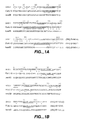
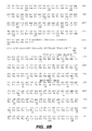
- Figs. 1A and 1B depict the amino acid sequences of variable heavy domain (SEQ ID NO:9) and light domain (SEQ ID NO: 10) of muMAbVEGF A.4.6.1, variable heavy domain (SEQ ID NO:7) and light domain (SEQ ID NO:8) of humanized F(ab) (F(ab)-12) and human consensus frameworks (hum III for heavy subgroup III (SEQ ID NO: 11); humk1 for light ⁇ subgroup I (SEQ ID NO: 12)).
- Fig. 1A aligns variable heavy domain sequences
- Fig. 1B aligns variable light domain sequences.
- Asterisks indicate differences between humanized F(ab)-12 and the murine MAb or between F(ab)-12 and the human framework.
- Complementarity Determining Regions (CDRs) are underlined.
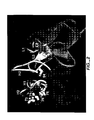
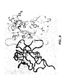
- Fig. 2 is a ribbon diagram of the model of humanized F(ab)-12 VL and VH domains.
- VL domain is shown in brown with CDRs in tan.
- the sidechain of residue L46 is shown in yellow.
- VH domain is shown in purple with CDRs in pink.
- Sidechains of VH residues changed from human to murine are shown in yellow.
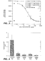
- Fig. 3 depicts inhibition of VEGF-induced mitogenesis by humanized anti-VEGF F(ab)-12 from Example 1.
- Bovine adrenal cortex-derived capillary endothelial cells were seeded at the density of 6 X 10 3 cells/well in six well plates, as described in Example 1.
- Either muMAb VEGF A.4.6.1 or rhuMAb VEGF (IgG1; F(ab)-12) was added at the indicated concentrations.
- rhVEGF165 was added at the final concentration of 3 ng/ml.
- cells were trypsinized and counted. Values shown are means of duplicate determinations. The variation from the mean did not exceed 10%.
- Fig. 4 shows inhibition of tumor growth in vivo by humanized anti-VEGF F(ab)-12 from Example 1.
- A673 rhabdomyosarcoma cells were injected in BALB/c nude mice at the density of 2 x 10 6 per mouse. Starting 24 hours after tumor cell inoculation, animals were injected with a control MAb, muMAb VEGF A4.6.1 or rhuVEGF MAb (IgG1; F(ab)-12) twice weekly, intra peritoneally.
- animals were euthanized and tumors were removed and weighed. * : significant difference when compared to the control group by ANOVA (p ⁇ 0.05).
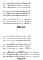
- Figs. 5A and 5B show the acid sequences of the light and heavy variable domains respectively of murine antibody A4.6.1 (SEQ ID NO: 10 for the VL and SEQ ID NO:9 for the VH) and humanized A4.6.1 variants hu2.0 (SEQ ID NO: 13 for the VL and SEQ ID NO: 14 for the VH) and hu2.10 (SEQ ID NO: 15 for the VL and SEQ ID NO: 16 for the VH) from Example 2. Sequence numbering is according to Kabat et al., Sequences of Proteins of Immunological Interest, 5th Ed. Public Health Service, National Institutes of Health, Bethesda, MD.
- hu2.0 contains only the CDR sequences (bold) from the murine antibody grafted onto a human light chain ⁇ subgroup I consensus framework (SEQ ID NO: 12) and heavy chain subgroup III consensus framework (SEQ ID NO: 11).
- hu2.10 was the consensus humanized clone obtained from phage sorting experiments described herein.
- Fig. 6 depicts framework residues targeted for randomization in Example 2.
- Fig. 7 depicts the phagemid construct for surface display of Fab-pIII fusions on phage.
- the phagemid encodes a humanized version of the Fab fragment for antibody A4.6.1 fused to a portion of the M13 gene III coat protein.
- the fusion protein consists of the Fab joined at the carboxyl terminus of the heavy chain to a single glutamine residue (from suppression of an amber codon in supE E. coli), then the C-terminal region of the gene III protein (residues 249-406). Transformation into F + E. coli, followed by superinfection with M13KO7 helper phage, produces phagemid particles in which a small proportion of these display a single copy of the fusion protein.
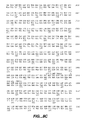
- Figs. 8A-E depict the double stranded nucleotide sequence (SEQ ID NO:99) for phage-display antibody vector phMB4-19-1.6 in Example 3 and the amino acid sequence encoded thereby (SEQ ID NO: 100).
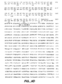
- Figs. 9A and 9B depict an alignment of the amino acid sequences for the light and heavy variable domains respectively of affinity matured anti-VEGF variants in Example 3, compared to F(ab)-12 of Example 1 (SEQ ID NO's 8 and 7 for light and heavy variable domains, respectively).
- CDRs are underlined and designated by L, light, or H, heavy chain, and numbers 1-3. Residues are numbered sequentially in the VL and VH domains, as opposed to the Kabat numbering scheme.
- the template molecule, MB 1.6 (SEQ ID NO's 101 and 102 for light and heavy variable domains, respectively) is shown, along with variants:
- Figs. 10A and 10B depict an alignment of the amino acid sequences for the light and heavy variable domains respectively of affinity matured anti-VEGF variants from Example 3 compared to F(ab)-12 of Example 1 (SEQ ID NO's 8 and 7 for light and heavy variable domains, respectively).
- CDRs are underlined and designated by L, light, or H, heavy chain, and numbers 1-3.
- the variants are designated Y0243-1 (SEQ ID NO's 109 and 110 for light and heavy variable domains, respectively), Y0238-3 (SEQ ID NO's 111 and 112 for light and heavy variable domains, respectively), Y0313-1 (SEQ ID NO's 113 and 114 for light and heavy variable domains, respectively), and Y0317 (SEQ ID NO's 115 and 116 for light and heavy variable domains, respectively). Differences from F(ab)-12 are shown in shaded boxes.
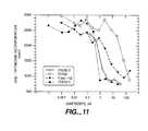
- Fig. 11 depicts the results of the HuVEC activity assay in Example 3 for variants Y0238-3, Y0192 and Y0313-1 as well as full length F(ab)-12 from Example 1.
- Fig. 12 depicts inhibition of VEGF-induced mitogenesis by full length F(ab)-12 from Example 1 (rhuMAb VEGF), a Fab fragment ofF(ab)-12 from Example 1 (rhuFab VEGF), and a Fab fragment of affinity matured variant Y0317 from Example 3 (rhuFab VEGF (affinity matured)).
- human VEGF refers to the 165-amino acid human vascular endothelial cell growth factor, and related 121-, 189-, and 206-amino acid vascular endothelial cell growth factors, as described by Leung et al., Science 246:1306 (1989 ), and Houck et al., Mol. Endocrin. 5:1806 (1991 ) together with the naturally occurring allelic and processed forms of those growth factors.
- the present invention provides anti-VEGF antagonistic antibodies which are capable of inhibiting one or more of the biological activities of VEGF, for example, its mitogenic or angiogenic activity.
- Antagonists of VEGF act by interfering with the binding of VEGF to a cellular receptor, by incapacitating or killing cells which have been activated by VEGF, or by interfering with vascular endothelial cell activation after VEGF binding to a cellular receptor. All such points of intervention by a VEGF antagonist shall be considered equivalent for purposes of this invention.
- VEGF receptor refers to a cellular receptor for VEGF, ordinarily a cell-surface receptor found on vascular endothelial cells, as well as variants thereof which retain the ability to bind hVEGF.
- a VEGF receptor is the fms -like tyrosine kinase ( flt ), a transmembrane receptor in the tyrosine kinase family. DeVries et al., Science 255:989 (1992 ); Shibuya et al., Oncogene 5:519 (1990 ).
- the flt receptor comprises an extracellular domain, a transmembrane domain, and an intracellular domain with tyrosine kinase activity.
- the extracellular domain is involved in the binding of VEGF, whereas the intracellular domain is involved in signal transduction.
- Another example of a VEGF receptor is the flk-1 receptor (also referred to as KDR).
- KDR flk-1 receptor
- Binding of VEGF to the flt receptor results in the formation of at least two high molecular weight complexes, having apparent molecular weight of 205,000 and 300,000 Daltons.
- the 300,000 Dalton complex is believed to be a dimer comprising two receptor molecules bound to a single molecule of VEGF.
- epitopope A4.6.1 when used herein, unless indicated otherwise, refers to the region of human VEGF to which the A4.6.1 antibody disclosed in Kim et al., Growth Factors 7:53 (1992 ) and Kim et al. Nature 362:841 (1993 ), binds.
- Treatment refers to both therapeutic treatment and prophylactic or preventative measures. Those in need of treatment include those already with the disorder as well as those in which the disorder is to be prevented.
- mammal for purposes of treatment refers to any animal classified as a mammal, including humans, domestic and farm animals, and zoo, sports, or pet animals, such as dogs, horses, cats, cows, etc. Preferably, the mammal is human.
- Antibodies are glycoproteins having the same structural characteristics. While antibodies exhibit binding specificity to a specific antigen, immunoglobulins include both antibodies and other antibody-like molecules which lack antigen specificity. Polypeptides of the latter kind are, for example, produced at low levels by the lymph system and at increased levels by myelomas.
- “Native antibodies” and “native immunoglobulins” are usually heterotetrameric glycoproteins of about 150,000 daltons, composed of two identical light (L) chains and two identical heavy (H) chains. Each light chain is linked to a heavy chain by one covalent disulfide bond, while the number of disulfide linkages varies among the heavy chains of different immunoglobulin isotypes. Each heavy and light chain also has regularly spaced intrachain disulfide bridges. Each heavy chain has at one end a variable domain (V H ) followed by a number of constant domains.
- V H variable domain
- Each light chain has a variable domain at one end (V 1 ) and a constant domain at its other end; the constant domain of the light chain is aligned with the first constant domain of the heavy chain, and the light- chain variable domain is aligned with the variable domain of the heavy chain. Particular amino acid residues are believed to form an interface between the light- and heavy-chain variable domains.
- variable refers to the fact that certain portions of the variable domains differ extensively in sequence among antibodies and are used in the binding and specificity of each particular antibody for its particular antigen. However, the variability is not evenly distributed throughout the variable domains of antibodies. It is concentrated in three segments called hypervariable regions both in the light chain and the heavy chain variable domains. The more highly conserved portions of variable domains are called the framework region (FR).
- the variable domains of native heavy and light chains each comprise four FRs (FR1, FR2, FR3 and FR4, respectively), largely adopting a ⁇ -sheet configuration, connected by three hypervariable regions, which form loops connecting, and in some cases forming part of, the ⁇ -sheet structure.
- the hypervariable regions in each chain are held together in close proximity by the FRs and, with the hypervariable regions from the other chain, contribute to the formation of the antigen-binding site of antibodies (see Kabat et al., Sequences of Proteins of Immunological Interest, 5th Ed. Public Health Service, National Institutes of Health, Bethesda, MD. (1991), pages 647-669 ).
- the constant domains are not involved directly in binding an antibody to an antigen, but exhibit various effector functions, such as participation of the antibody in antibody-dependent cellular toxicity.
- hypervariable region when used herein refers to the amino acid residues of an antibody which are responsible for antigen-binding.
- the hypervariable region comprises amino acid residues from a "complementarity determining region" or "CDR" (i.e. residues 24-34 (L1), 50-56 (L2) and 89-97 (L3) in the light chain variable domain and 31-35 (H1), 50-65 (H2) and 95-102 (H3) in the heavy chain variable domain; Kabat et al., Sequences of Proteins of Immunological Interest, 5th Ed. Public Health Service, National Institutes of Health, Bethesda, MD. (1991 )) and/or those residues from a "hypervariable loop" (i.e.
- Papain digestion of antibodies produces two identical antigen-binding fragments, called “Fab” fragments, each with a single antigen-binding site, and a residual "Fc” fragment, whose name reflects its ability to crystallize readily. Pepsin treatment yields an F(ab') 2 fragment that has two antigen-combining sites and is still capable of cross-linking antigen.
- Fv is the minimum antibody fragment which contains a complete antigen-recognition and -binding site. This region consists of a dimer of one heavy chain and one light chain variable domain in tight, non-covalent association. It is in this configuration that the three hypervariable regions of each variable domain interact to define an antigen-binding site on the surface of the V H- V L dimer. Collectively, the six hypervariable regions confer antigen-binding specificity to the antibody. However, even a single variable domain (or half of an Fv comprising only three hypervariable regions specific for an antigen) has the ability to recognize and bind antigen, although at a lower affinity than the entire binding site.
- the Fab fragment also contains the constant domain of the light chain and the first constant domain (CH1) of the heavy chain.
- Fab' fragments differ from Fab fragments by the addition of a few residues at the carboxyl terminus of the heavy chain CH1 domain including one or more cysteine(s) from the antibody hinge region.
- Fab'-SH is the designation herein for Fab' in which the cysteine residue(s) of the constant domains bear a free thiol group.
- F(ab') 2 antibody fragments originally were produced as pairs of Fab' fragments which have hinge cysteines between them. Other chemical couplings of antibody fragments are also known.
- the "light chains" of antibodies (immunoglobulins) from any vertebrate species can be assigned to one of two clearly distinct types, called kappa ( ⁇ ) and lambda ( ⁇ ), based on the amino acid sequences of their constant domains.
- immunoglobulins can be assigned to different classes. There are five major classes of immunoglobulins: IgA, IgD, IgE, IgG, and IgM, and several of these may be further divided into subclasses (isotypes), e.g., IgG1, IgG2, IgG3, IgG4, IgA, and IgA2.
- the heavy-chain constant domains that correspond to the different classes of immunoglobulins are called ⁇ , ⁇ , ⁇ , ⁇ , and ⁇ , respectively.
- the subunit structures and three-dimensional configurations of different classes of immunoglobulins are well known.
- antibody herein is used in the broadest sense and specifically covers monoclonal antibodies (including full length monoclonal antibodies), polyclonal antibodies, multispecific antibodies (e.g., bispecific antibodies), and antibody fragments so long as they exhibit the desired biological activity.
- Antibody fragments comprise a portion of a full length antibody, generally the antigen binding or variable domain thereof.
- Examples of antibody fragments include Fab, Fab', F(ab') 2 , and Fv fragments; diabodies; linear antibodies; single-chain antibody molecules; and multispecific antibodies formed from antibody fragments.
- the term "monoclonal antibody” as used herein refers to an antibody obtained from a population of substantially homogeneous antibodies, i.e., the individual antibodies comprising the population are identical except for possible naturally occurring mutations that may be present in minor amounts. Monoclonal antibodies are highly specific, being directed against a single antigenic site. Furthermore, in contrast to conventional (polyclonal) antibody preparations which typically include different antibodies directed against different determinants (epitopes), each monoclonal antibody is directed against a single determinant on the antigen.
- the modifier "monoclonal” indicates the character of the antibody as being obtained from a substantially homogeneous population of antibodies, and is not to be construed as requiring production of the antibody by any particular method.
- the monoclonal antibodies to be used in accordance with the present invention may be made by the hybridoma method first described by Kohler et al., Nature 256:495 (1975 ), or may be made by recombinant DNA methods (see, e.g., U.S. Patent No. 4,816,567 ).
- the "monoclonal antibodies” may also be isolated from phage antibody libraries using the techniques described in Clackson et al., Nature 352:624-628 (1991 ) and Marks et al., J. Mol. Biol. 222:581-597 (1991 ), for example.
- the monoclonal antibodies herein specifically include "chimeric" antibodies (immunoglobulins) in which a portion of the heavy and/or light chain is identical with or homologous to corresponding sequences in antibodies derived from a particular species or belonging to a particular antibody class or subclass, while the remainder of the chain(s) is identical with or homologous to corresponding sequences in antibodies derived from another species or belonging to another antibody class or subclass, as well as fragments of such antibodies, so long as they exhibit the desired biological activity ( U.S. Patent No. 4,816,567 ; and Morrison et al., Proc. Natl. Acad Sci. USA 81:6851-6855 (1984 )).
- chimeric antibodies immunoglobulins in which a portion of the heavy and/or light chain is identical with or homologous to corresponding sequences in antibodies derived from a particular species or belonging to a particular antibody class or subclass, while the remainder of the chain(s) is identical with or homologous to corresponding sequences
- Humanized forms of non-human (e.g., murine) antibodies are chimeric antibodies which contain minimal sequence derived from non-human immunoglobulin.
- humanized antibodies are human immunoglobulins (recipient antibody) in which hypervariable region residues of the recipient are replaced by hypervariable region residues from a non-human species (donor antibody) such as mouse, rat, rabbit or nonhuman primate having the desired specificity, affinity, and capacity.
- donor antibody such as mouse, rat, rabbit or nonhuman primate having the desired specificity, affinity, and capacity.
- donor antibody such as mouse, rat, rabbit or nonhuman primate having the desired specificity, affinity, and capacity.
- framework region (FR) residues of the human immunoglobulin are replaced by corresponding non-human residues.
- humanized antibodies may comprise residues which are not found in the recipient antibody or in the donor antibody. These modifications are made to further refine antibody performance.
- the humanized antibody will comprise substantially all of at least one, and typically two, variable domains, in which all or substantially all of the hypervariable regions correspond to those of a non-human immunoglobulin and all or substantially all of the FRs are those of a human immunoglobulin sequence.
- the humanized antibody optionally also will comprise at least a portion of an immunoglobulin constant region (Fc), typically that of a human immunoglobulin.
- Fc immunoglobulin constant region
- Single-chain Fv or “sFv” antibody fragments comprise the V H and V L domains of antibody, wherein these domains are present in a single polypeptide chain.
- the Fv polypeptide further comprises a polypeptide linker between the V H and V L domains which enables the sFv to form the desired structure for antigen binding.
- diabodies refers to small antibody fragments with two antigen-binding sites, which fragments comprise a heavy chain variable domain (V H ) connected to a light chain variable domain (V L ) in the same polypeptide chain (V H - V L ).
- V H heavy chain variable domain
- V L light chain variable domain
- the domains are forced to pair with the complementary domains of another chain and create two antigen-binding sites.
- Diabodies are described more fully in, for example, EP 404,097 ; WO 93/11161 ; and Hollinger et al., Proc. Natl. Acad. Sci. USA 90:6444-6448 (1993 ).
- linear antibodies when used throughout this application refers to the antibodies described in Zapata et al. Protein Eng. 8(10):1057-1062 (1995 ). Briefly, these antibodies comprise a pair of tandem Fd segments (V H -C H 1-V H -C H 1) which form a pair of antigen binding regions. Linear antibodies can be bispecific or monospecific.
- a “variant" anti-VEGF antibody refers herein to a molecule which differs in amino acid sequence from a "parent" anti-VEGF antibody amino acid sequence by virtue of addition, deletion and/or substitution of one or more amino acid residue(s) in the parent antibody sequence.
- the variant comprises one or more amino acid substitution(s) in one or more hypervariable region(s) of the parent antibody.
- the variant may comprise at least one, e.g. from about one to about ten, and preferably from about two to about five, substitutions in one or more hypervariable regions of the parent antibody.
- the variant will have an amino acid sequence having at least 75% amino acid sequence identity with the parent antibody heavy or light chain variable domain sequences (e.g.
- Identity or homology with respect to this sequence is defined herein as the percentage of amino acid residues in the candidate sequence that are identical with the parent antibody residues, after aligning the sequences and introducing gaps, if necessary, to achieve the maximum percent sequence identity. None of N-terminal, C-terminal, or internal extensions, deletions, or insertions into the antibody sequence shall be construed as affecting sequence identity or homology.
- the variant retains the ability to bind human VEGF and preferably has properties which are superior to those of the parent antibody.
- the variant may have a stronger binding affinity, enhanced ability to inhibit VEGF-induced proliferation of endothelial cells and/or increased ability to inhibit VEGF-induced angiogenesis in vivo.
- a Fab form of the variant to a Fab form of the parent antibody or a full length form of the variant to a full length form of the parent antibody, for example, since it has been found that the format of the anti-VEGF antibody impacts its activity in the biological activity assays disclosed herein.
- the variant antibody of particular interest herein is one which displays at least about 10 fold, preferably at least about 20 fold, and most preferably at least about 50 fold, enhancement in biological activity when compared to the parent antibody.
- the "parent” antibody herein is one which is encoded by an amino acid sequence used for the preparation of the variant.
- the parent antibody has a human framework region and, if present, has human antibody constant region(s).
- the parent antibody may be a humanized or human antibody.
- an “isolated” antibody is one which has been identified and separated and/or recovered from a component of its natural environment. Contaminant components of its natural environment are materials which would interfere with diagnostic or therapeutic uses for the antibody, and may include enzymes, hormones, and other proteinaceous or nonproteinaceous solutes.
- the antibody will be purified (1) to greater than 95% by weight of antibody as determined by the Lowry method, and most preferably more than 99% by weight, (2) to a degree sufficient to obtain at least 15 residues of N-terminal or internal amino acid sequence by use of a spinning cup sequenator, or (3) to homogeneity by SDS-PAGE under reducing or nonreducing conditions using Coomassie blue or, preferably, silver stain.
- Isolated antibody includes the antibody in situ within recombinant cells since at least one component of the antibody's natural environment will not be present. Ordinarily, however, isolated antibody will be prepared by at least one purification step.
- epitope tag polypeptide has enough residues to provide an epitope against which an antibody thereagainst can be made, yet is short enough such that it does not interfere with activity of the VEGF antibody.
- the epitope tag preferably is sufficiently unique so that the antibody thereagainst does not substantially cross-react with other epitopes.
- Suitable tag polypeptides generally have at least 6 amino acid residues and usually between about 8-50 amino acid residues (preferably between about 9-30 residues). Examples include the flu HA tag polypeptide and its antibody 12CA5 ( Field et al. Mol. Cell. Biol.
- the epitope tag is a "salvage receptor binding epitope".
- the term "salvage receptor binding epitope” refers to an epitope of the Fc region of an IgG molecule (e.g., IgG 1 , IgG 2 , IgG 3 , or IgG 4 ) that is responsible for increasing the in vivo serum half-life of the IgG molecule.
- cytotoxic agent refers to a substance that inhibits or prevents the function of cells and/or causes destruction of cells.
- the term is intended to include radioactive isotopes (e.g., I 131 , I 125 , Y 90 and Re 186 ), chemotherapeutic agents, and toxins such as enzymatically active toxins of bacterial, fungal, plant or animal origin, or fragments thereof.
- a “chemotherapeutic agent” is a chemical compound useful in the treatment of cancer.
- chemotherapeutic agents include Adriamycin, Doxorubicin, 5-Fluorouracil, Cytosine arabinoside ("Ara-C"), Cyclophosphamide, Thiotepa, Taxotere (docetaxel), Busulfan, Cytoxin, Taxol, Methotrexate, Cisplatin, Melphalan, Vinblastine, Bleomycin, Etoposide, Ifosfamide, Mitomycin C, Mitoxantrone, Vincreistine, Vinorelbine, Carboplatin, Teniposide, Daunomycin, Carminomycin, Aminopterin, Dactinomycin, Mitomycins, Esperamicins (see U.S. Pat. No. 4,675,187 ), Melphalan and other related nitrogen mustards.
- prodrug refers to a precursor or derivative form of a pharmaceutically active substance that is less cytotoxic to tumor cells compared to the parent drug and is capable of being enzymatically activated or converted into the more active parent form. See, e.g., Wilman, "Prodrugs in Cancer Chemotherapy” Biochemical Society Transactions, 14, pp. 375-382, 615th Meeting Harbor (1986 ) and Stella et al., “Prodrugs: A Chemical Approach to Targeted Drug Delivery,” Directed Drug Delivery, Borchardt et al., (ed.), pp. 247-267, Humana Press (1985 ).
- the prodrugs of this invention include, but are not limited to, phosphate-containing prodrugs, thiophosphate-containing prodrugs, sulfate-containing prodrugs, peptide-containing prodrugs, D-amino acid-modified prodrugs, glycosylated prodrugs, ⁇ -lactam-containing prodrugs, optionally substituted phenoxyacetamide-containing prodrugs or optionally substituted phenylacetamide-containing prodrugs, 5-fluorocytosine and other 5-fluorouridine prodrugs which can be converted into the more active cytotoxic free drug.
- cytotoxic drugs that can be derivatized into a prodrug form for use in this invention include, but are not limited to, those chemotherapeutic agents described above.
- label when used herein refers to a detectable compound or composition which is conjugated directly or indirectly to the antibody.
- the label may itself be detectable by itself (e.g., radioisotope labels or fluorescent labels) or, in the case of an enzymatic label, may catalyze chemical alteration of a substrate compound or composition which is detectable.
- solid phase is meant a non-aqueous matrix to which the antibody of the present invention can adhere.
- solid phases encompassed herein include those formed partially or entirely of glass (e.g. controlled pore glass), polysaccharides (e.g., agarose), polyacrylamides, polystyrene, polyvinyl alcohol and silicones.
- the solid phase can comprise the well of an assay plate; in others it is a purification column (e.g. an affinity chromatography column). This term also includes a discontinuous solid phase of discrete particles, such as those described in U.S. Patent No. 4,275,149 .
- a “liposome” is a small vesicle composed of various types of lipids, phospholipids and/or surfactant which is useful for delivery of a drug (such as the anti-VEGF antibodies disclosed herein and, optionally, a chemotherapeutic agent) to a mammal.
- the components of the liposome are commonly arranged in a bilayer formation, similar to the lipid arrangement of biological membranes.
- An "isolated" nucleic acid molecule is a nucleic acid molecule that is identified and separated from at least one contaminant nucleic acid molecule with which it is ordinarily associated in the natural source of the antibody nucleic acid.
- An isolated nucleic acid molecule is other than in the form or setting in which it is found in nature.
- Isolated nucleic acid molecules therefore are distinguished from the nucleic acid molecule as it exists in natural cells.
- an isolated nucleic acid molecule includes a nucleic acid molecule contained in cells that ordinarily express the antibody where, for example, the nucleic acid molecule is in a chromosomal location different from that of natural cells.
- control sequences refers to DNA sequences necessary for the expression of an operably linked coding sequence in a particular host organism.
- the control sequences that are suitable for prokaryotes include a promoter, optionally an operator sequence, and a ribosome binding site.
- Eukaryotic cells are known to utilize promoters, polyadenylation signals, and enhancers.
- Nucleic acid is "operably linked" when it is placed into a functional relationship with another nucleic acid sequence.
- DNA for a presequence or secretory leader is operably linked to DNA for a polypeptide if it is expressed as a preprotein that participates in the secretion of the polypeptide;
- a promoter or enhancer is operably linked to a coding sequence if it affects the transcription of the sequence; or
- a ribosome binding site is operably linked to a coding sequence if it is positioned so as to facilitate translation.
- "operably linked” means that the DNA sequences being linked are contiguous, and, in the case of a secretory leader, contiguous and in reading phase. However, enhancers do not have to be contiguous. Linking is accomplished by ligation at convenient restriction sites. If such sites do not exist, the synthetic oligonucleotide adaptors or linkers are used in accordance with conventional practice.
- the expressions "cell,” “cell line,” and “cell culture” are used interchangeably and all such designations include progeny.
- the words “transformants” and “transformed cells” include the primary subject cell and cultures derived therefrom without regard for the number of transfers. It is also understood that all progeny may not be precisely identical in DNA content, due to deliberate or inadvertent mutations. Mutant progeny that have the same function or biological activity as screened for in the originally transformed cell are included. Where distinct designations are intended, it will be clear from the context.
- the examples hereinbelow describe the production of humanized and variant anti-VEGF antibodies with desirable properties from a therapeutic perspective including: (a) strong binding affinity for the VEGF antigen; (b) an ability to inhibit VEGF-induced proliferation of endothelial cells in vitro; and (c) the ability to inhibit VEGF-induced angiogenesis in vivo.
- Antibody affinities may be determined as described in the examples hereinbelow.
- Preferred humanized or variant antibodies are those which bind human VEGF with a K d value of no more than about 1 x 10 -7 M; preferably no more than about 1 x 10 8 M; and most preferably no more than about 5 x 10 -9 M.
- the antibody may be one which inhibits endothelial cell growth in response to VEGF.
- the antibody may be able to inhibit bovine capillary endothelial cell proliferation in response to a near maximally effective concentration of VEGF (3 ng/ml).
- the antibody has an effective dose 50 (ED50) value of no more than about 5nM, preferably no more than about 1nM, and most preferably no more than about 0.5nM, for inhibiting VEGF-induced proliferation of endothelial cells in this "endothelial cell growth assay", i.e., at these concentrations the antibody is able to inhibit VEGF-induced endothelial cell growth in vitro by 50%.
- ED50 effective dose 50
- a preferred "endothelial cell growth assay” involves culturing bovine adrenal cortex-derived capillary endothelial cells in the presence of low glucose Dulbecco's modified Eagle's medium (DMEM) (GIBCO) supplemented with 10% calf serum, 2 mM glutamine, and antibiotics (growth medium), essentially as described in Example 1 below. These endothelial cells are seeded at a density of 6 x 10 3 cells per well, in 6-well plates in growth medium. Either parent anti-VEGF antibody (control), humanized or variant anti-VEGF antibody is then added at concentrations ranging between 1 and 5000 ng/ml. After 2-3 hr, purified VEGF was added to a final concentration of 3 ng/ml.
- DMEM Dulbecco's modified Eagle's medium
- each antibody may be added to endothelial cells at the concentration of 5000 ng/ml, either alone or in the presence of 2 ng/ml bFGF. After five or six days, cells are dissociated by exposure to trypsin and counted in a Coulter counter (Coulter Electronics, Hialeah, FL). Data may be analyzed by a four-parameter curve fitting program (KaleidaGraph).
- the preferred humanized or variant anti-VEGF antibody may also be one which has in vivo tumor suppression activity.
- the antibody may suppress the growth of human A673 rhabdomyosarcoma cells or breast carcinoma MDA-MB-435 5 cells in nude mice.
- human A673 rhabdomyosarcoma cells ATCC; CRL 1598
- MDA-MB-435 cells available from the ATCC
- DMEM/F12 fetal bovine serum
- 2 mM glutamine and antibiotics as described in Example 1 below.
- mice Female BALB/c nude mice, 6-10 weeks old, are injected subcutaneously with 2 x 10 6 tumor cells in the dorsal area in a volume of 200 ⁇ l. Animals are then treated with the humanized or variant antibody and a control antibody with no activity in this assay.
- the humanized or variant anti-VEGF MAb is administered at a dose of 0.5 and/or 5 mg/kg. Each MAb is administered twice weekly intra peritoneally in a volume of 100 ⁇ l, starting 24 hr after tumor cell inoculation. Tumor size is determined at weekly intervals. Four weeks after tumor cell inoculation, animals are euthanized and the tumors are removed and weighed. Statistical analysis may be performed by ANOVA.
- the antibody in this "in vivo tumor assay" inhibits about 50-100%, preferably about 70-100% and most preferably about 80-100% human A673 tumor cell growth at a dose of 5mg/kg.
- the humanized or variant antibody fails to elicit an immunogenic response upon administration of a therapeutically effective amount of the antibody to a human patient. If an immunogenic response is elicited, preferably the response will be such that the antibody still provides a therapeutic benefit to the patient treated therewith.
- the humanized or variant antibody is also preferably one which is able to inhibit VEGF-induced angiogenesis in a human, e.g. to inhibit human tumor growth and/or inhibit intraocular angiogenesis in retinal disorders.
- Preferred antibodies bind the "epitope A4.6.1 " as herein defined.
- an antibody of interest e.g., those which block binding of the A4.6.1 antibody to human VEGF
- a routine cross-blocking assay such as that described in Antibodies, A LaboratoryManual, Cold Spring Harbor Laboratory, Ed Harlow and David Lane (1988 )
- epitope mapping e.g. as described in Champe et al., J. Biol. Chem. 270:1388-1394 (1995 ), can be performed to determine whether the antibody binds an epitope of interest.
- the antibodies of the preferred embodiment herein have a heavy chain variable domain comprising an amino acid sequence represented by the formula: FR1-CDRH1-FR2-CDRH2-FR3-CDRH3-FR4, wherein "FR1-4" represent the four framework regions and "CDRH1-3" represent the three hypervariable regions of an anti-VEGF antibody variable heavy domain.
- FR1-4 may be derived from a "consensus sequence” (i.e. the most common amino acids of a class, subclass or subgroup of heavy or light chains of human immunoglobulins) as in the examples below or may be derived from an individual human antibody framework region or from a combination of different framework region sequences. Many human antibody framework region sequences are compiled in Kabat et al., supra, for example.
- variable heavy FR is provided by a consensus sequence of a human immunoglobulin subgroup as compiled by Kabat et al., supra.
- the human immunoglobulin subgroup is human heavy chains subgroup III (e.g. as in SEQ ID NO: 11).
- the human variable heavy FR sequence preferably has substitutions therein, e.g. wherein the human FR residue is replaced by a corresponding nonhuman residue (by "corresponding nonhuman residue” is meant the nonhuman residue with the same Kabat positional numbering as the human residue of interest when the human and nonhuman sequences are aligned), but replacement with the nonhuman residue is not necessary.
- a replacement FR residue other than the corresponding nonhuman residue may be selected by phage display (see Example 2 below).
- Exemplary variable heavy FR residues which may be substituted include any one or more of FR residue numbers: 37H, 49H, 67H, 69H, 71H, 73H, 75H, 76H, 78H, 94H (Kabat residue numbering employed here). Preferably at least two, or at least three, or at least four of these residues are substituted.
- a particularly preferred combination ofFR substitutions is: 49H, 69H, 71H, 73H, 76H, 78H, and 94
- the heavy chain variable domain optionally comprises what has been designated "CDR7" herein within (i.e. forming part of) FR3 (see Figs. 9B and 10B ), wherein CDR7 may have the following amino acid sequence:
- the antibodies of the preferred embodiment herein have a light chain variable domain comprising an amino acid sequence represented by the formula: FR1-CDRL1-FR2-CDRL2-FR3-CDRL3-FR4, wherein "FR1-4" represent the four framework regions and "CDRL1-3" represent the three hypervariable regions of an anti-VEGF antibody variable heavy domain.
- FR1-4 may be derived from a "consensus sequence” (i.e. the most common amino acids of a class, subclass or subgroup of heavy or light chains of human immunoglobulins) as in the examples below or may be derived from an individual human antibody framework region or from a combination of different framework region sequences.
- variable light FR is provided by a consensus sequence of a human immunoglobulin subgroup as compiled by Kabat et al., supra.
- the human immunoglobulin subgroup is human kappa light chains subgroup I (e.g. as in SEQ ID NO: 12).
- the human variable light FR sequence preferably has substitutions therein, e.g. wherein the human FR residue is replaced by a corresponding mouse residue, but replacement with the nonhuman residue is not necessary.
- a replacement residue other than the corresponding nonhuman residue may be selected by phage display (see Example 2 below).
- Exemplary variable light FR residues which may be substituted include any one or more of FR residue numbers: 4L, 46L and 71L (Kabat residue numbering employed here). Preferably only 46L is substituted. In another embodiment, both 4L and 46L are substituted.
- Preferred humanized anti-VEGF antibodies are those having the heavy and/or light variable domain sequences of F(ab)-12 in Example 1 and variants thereof such as affinity matured forms including variants Y0317, Y0313-1 and Y0238-3 in Example 3, with Y0317 being the preferred variant.
- Methods for generating humanized anti-VEGF antibodies of interest herein are elaborated in more detail below.
- nonhuman VEGF antibodies and generating variants of anti-VEGF antibodies are described in the examples below.
- the nonhuman antibody starting material is prepared.
- the parent antibody is prepared. Exemplary techniques for generating such nonhuman antibody starting material and parent antibodies will be described in the following sections.
- the VEGF antigen to be used for production of antibodies may be, e.g., intact VEGF or a fragment of VEGF (e.g. a VEGF fragment comprising "epitope A4.6.1").
- VEGF antigen used to generate the antibody is preferably human VEGF, e.g. as described in Leung et al., Science 246:1306 (1989 ), and Houck et al., Mol. Endocrin. 5:1806 (1991 ).
- a protein that is immunogenic in the species to be immunized e.g., keyhole limpet hemocyanin, serum albumin, bovine thy
- Animals are immunized against the antigen, immunogenic conjugates, or derivatives by combining, e.g., 100 ⁇ g or 5 ⁇ g of the protein or conjugate (for rabbits or mice, respectively) with 3 volumes of Freund's complete adjuvant and injecting the solution intradermally at multiple sites.
- the animals are boosted with 1/5 to 1/10 the original amount of peptide or conjugate in Freund's complete adjuvant by subcutaneous injection at multiple sites.
- Seven to 14 days later the animals are bled and the serum is assayed for antibody titer. Animals are boosted until the titer plateaus.
- the animal is boosted with the conjugate of the same antigen, but conjugated to a different protein and/or through a different cross-linking reagent.
- Conjugates also can be made in recombinant cell culture as protein fusions.
- aggregating agents such as alum are suitably used to enhance the immune response.
- Monoclonal antibodies may be made using the hybridoma method first described by Kohler el al., Nature, 256:495 (1975 ), or may be made by recombinant DNA methods ( U.S. Patent No. 4,816,567 ).
- a mouse or other appropriate host animal such as a hamster or macaque monkey
- lymphocytes that produce or are capable of producing antibodies that will specifically bind to the protein used for immunization.
- lymphocytes may be immunized in vitro. Lymphocytes then are fused with myeloma cells using a suitable fusing agent, such as polyethylene glycol, to form a hybridoma cell ( Goding, Monoclonal Antibodies: Principles and Practice, pp.59-103 (Academic Press, 1986 )).
- the hybridoma cells thus prepared are seeded and grown in a suitable culture medium that preferably contains one or more substances that inhibit the growth or survival of the unfused, parental myeloma cells.
- a suitable culture medium that preferably contains one or more substances that inhibit the growth or survival of the unfused, parental myeloma cells.
- the culture medium for the hybridomas typically will include hypoxanthine, aminopterin, and thymidine (HAT medium), which substances prevent the growth of HGPRT-deficient cells.
- Preferred myeloma cells are those that fuse efficiently, support stable high-level production of antibody by the selected antibody-producing cells, and are sensitive to a medium such as HAT medium.
- preferred myeloma cell lines are murine myeloma lines, such as those derived from MOP-21 and M.C.-11 mouse tumors available from the Salk Institute Cell Distribution Center, San Diego, California USA, and SP-2 or X63-Ag8-653 cells available from the American Type Culture Collection, Rockville, Maryland USA.
- Human myeloma and mouse-human heteromyeloma cell lines also have been described for the production of human monoclonal antibodies ( Kozbor, J. Immunol., 133:3001 (1984 ); Brodeur et al., Monoclonal Antibody Production Techniques and Applications, pp. 51-63 (Marcel Dekker, Inc., New York, 1987 )).
- Culture medium in which hybridoma cells are growing is assayed for production of monoclonal antibodies directed against the antigen.
- the binding specificity of monoclonal antibodies produced by hybridoma cells is determined by immunoprecipitation or by an in vitro binding assay, such as radioimmunoassay (RIA) or enzyme-linked immunoabsorbent assay (ELISA).
- RIA radioimmunoassay
- ELISA enzyme-linked immunoabsorbent assay
- the binding affinity of the monoclonal antibody can, for example, be determined by the Scatchard analysis of Munson et al., Anal. Biochem., 107:220 (1980 ).
- the clones may be subcloned by limiting dilution procedures and grown by standard methods ( Goding, Monoclonal Antibodies: Principles and Practice, pp.59-103 (Academic Press, 1986 )). Suitable culture media for this purpose include, for example, D-MEM or RPMI-1640 medium.
- the hybridoma cells may be grown in vivo as ascites tumors in an animal.
- the monoclonal antibodies secreted by the subclones are suitably separated from the culture medium, ascites fluid, or serum by conventional immunoglobulin purification procedures such as, for example, protein A-Sepharose, hydroxylapatite chromatography, gel electrophoresis, dialysis, or affinity chromatography.
- DNA encoding the monoclonal antibodies is readily isolated and sequenced using conventional procedures (e.g ., by using oligonucleotide probes that are capable of binding specifically to genes encoding the heavy and light chains of the monoclonal antibodies).
- the hybridoma cells serve as a preferred source of such DNA.
- the DNA may be placed into expression vectors, which are then transfected into host cells such as E. coli cells, simian COS cells, Chinese hamster ovary (CHO) cells, or myeloma cells that do not otherwise produce immunoglobulin protein, to obtain the synthesis of monoclonal antibodies in the recombinant host cells. Recombinant production of antibodies will be described in more detail below.
- Examples 1-2 below describe procedures for humanization of an anti-VEGF antibody. In certain embodiments, it may be desirable to generate amino acid sequence variants of these humanized antibodies, particularly where these improve the binding affinity or other biological properties of the humanized antibody.
- Example 3 describes methodologies for generating amino acid sequence variants of an anti-VEGF antibody with enhanced affinity relative to the parent antibody.
- Amino acid sequence variants of the anti-VEGF antibody are prepared by introducing appropriate nucleotide changes into the anti-VEGF antibody DNA, or by peptide synthesis.
- Such variants include, for example, deletions from, and/or insertions into and/or substitutions of, residues within the amino acid sequences of the anti-VEGF antibodies of the examples herein. Any combination of deletion, insertion, and substitution is made to arrive at the final construct, provided that the final construct possesses the desired characteristics.
- the amino acid changes also may alter post-translational processes of the humanized or variant anti-VEGF antibody, such as changing the number or position of glycosylation sites.
- a useful method for identification of certain residues or regions of the anti-VEGF antibody that are preferred locations for mutagenesis is called "alanine scanning mutagenesis," as described by Cunningham and Wells Science, 244:1081-1085 (1989 ).
- a residue or group of target residues are identified (e.g., charged residues such as arg, asp, his, lys, and glu) and replaced by a neutral or negatively charged amino acid (most preferably alanine or polyalanine) to affect the interaction of the amino acids with VEGF antigen.
- Those amino acid locations demonstrating functional sensitivity to the substitutions then are refined by introducing further or other variants at, or for, the sites of substitution.
- the site for introducing an amino acid sequence variation is predetermined, the nature of the mutation per se need not be predetermined.
- ala scanning or random mutagenesis is conducted at the target codon or region and the expressed anti-VEGF antibody variants are screened for the desired activity.
- Alanine scanning mutagenesis is described in Example 3.
- Amino acid sequence insertions include amino- and/or carboxyl-terminal fusions ranging in length from one residue to polypeptides containing a hundred or more residues, as well as intrasequence insertions of single or multiple amino acid residues.
- terminal insertions include an anti-VEGF antibody with an N-terminal methionyl residue or the antibody fused to an epitope tag.
- Other insertional variants of the anti-VEGF antibody molecule include the fusion to the N- or C-terminus of the anti-VEGF antibody of an enzyme or a polypeptide which increases the serum half-life of the antibody (see below).
- variants are an amino acid substitution variant. These variants have at least one amino acid residue in the anti-VEGF antibody molecule removed and a different residue inserted in its place.
- the sites of greatest interest for substitutional mutagenesis include the hypervariable regions, but FR alterations are also contemplated.
- Conservative substitutions are shown in Table 1 under the heading of "preferred substitutions". If such substitutions result in a change in biological activity, then more substantial changes, denominated "exemplary substitutions" in Table 1, or as further described below in reference to amino acid classes, may be introduced and the products screened.
- Substantial modifications in the biological properties of the antibody are accomplished by selecting substitutions that differ significantly in their effect on maintaining (a) the structure of the polypeptide backbone in the area of the substitution, for example, as a sheet or helical conformation, (b) the charge or hydrophobicity of the molecule at the target site, or (c) the bulk of the side chain.
- Naturally occurring residues are divided into groups based on common side-chain properties:
- Non-conservative substitutions will entail exchanging a member of one of these classes for another class.
- cysteine residues not involved in maintaining the proper conformation of the humanized or variant anti-VEGF antibody also may be substituted, generally with serine, to improve the oxidative stability of the molecule and prevent aberrant crosslinking.
- cysteine bond(s) may be added to the antibody to improve its stability (particularly where the antibody is an antibody fragment such as an Fv fragment).
- a particularly preferred type of substitutional variant involves substituting one or more hypervariable region residues of a parent antibody (e.g. a humanized or human antibody).
- a parent antibody e.g. a humanized or human antibody
- the resulting variant(s) selected for further development will have improved biological properties relative to the parent antibody from which they are generated.
- a convenient way for generating such substitutional variants is affinity maturation using phage display (see Example 3 herein). Briefly, several hypervariable region sites (e.g. 6-7 sites) are mutated to generate all possible amino substitutions at each site.
- the antibody variants thus generated are displayed in a monovalent fashion from filamentous phage particles as fusions to the gene III product of M13 packaged within each particle. The phage-displayed variants are then screened for their biological activity (e.g.
- alanine scanning mutagenesis (see Example 3) can be performed to identified hypervariable region residues contributing significantly to antigen binding.
- Another type of amino acid variant of the antibody alters the original glycosylation pattern of the antibody. By altering is meant deleting one or more carbohydrate moieties found in the antibody, and/or adding one or more glycosylation sites that are not present in the antibody.
- N-linked refers to the attachment of the carbohydrate moiety to the side chain of an asparagine residue.
- the tripeptide sequences asparagine-X-serine and asparagine-X-threonine, where X is any amino acid except proline, are the recognition sequences for enzymatic attachment of the carbohydrate moiety to the asparagine side chain.
- X is any amino acid except proline
- O-linked glycosylation refers to the attachment of one of the sugars N-aceylgalactosamine, galactose, or xylose to a hydroxyamino acid, most commonly serine or threonine, although 5-hydroxyproline or 5-hydroxylysine may also be used.
- glycosylation sites to the antibody is conveniently accomplished by altering the amino acid sequence such that it contains one or more of the above-described tripeptide sequences (for N-linked glycosylation sites).
- the alteration may also be made by the addition of, or substitution by, one or more serine or threonine residues to the sequence of the original antibody (for O-linked glycosylation sites).
- Nucleic acid molecules encoding amino acid sequence variants of the anti-VEGF antibody are prepared by a variety of methods known in the art. These methods include, but are not limited to, isolation from a natural source (in the case of naturally occurring amino acid sequence variants) or preparation by oligonucleotide-mediated (or site-directed) mutagenesis, PCR mutagenesis, and cassette mutagenesis of an earlier prepared variant or a non-variant version of the anti-VEGF antibody.
- human antibodies can be generated.
- transgenic animals e.g., mice
- transgenic animals e.g., mice
- J H antibody heavy-chain joining region
- transfer of the human germ-line immunoglobulin gene array in such germ-line mutant mice will result in the production of human antibodies upon antigen challenge. See, e.g., Jakobovits et al., Proc. Natl. Acad. Sci.
- Human antibodies can also be derived from phage-display libraries ( Hoogenboom et al., J. Mol. Biol., 227:381 (1991 ); Marks et al., J. Mol. Biol., 222:581-597 (1991 ); and US Patents 5,565,332 and 5,573,905 ). As discussed above, human antibodies may also be generated by in vitro activated B cells (see US Patents 5,567,610 and 5,229,275 )
- the humanized or variant anti-VEGF antibody is an antibody fragment.
- Various techniques have been developed for the production of antibody fragments. Traditionally, these fragments were derived via proteolytic digestion of intact antibodies (see, e.g., Morimoto et al., Journal of Biochemical and Biophysical Methods 24:107-117 (1992 ) and Brennan et al., Science 229:81 (1985 )). However, these fragments can now be produced directly by recombinant host cells. For example, Fab'-SH fragments can be directly recovered from E. coli and chemically coupled to form F(ab') 2 fragments ( Carter et al., Bio/Technology 10:163-167 (1992 )).
- the F(ab') 2 is formed using the leucine zipper GCN4 to promote assembly of the F(ab') 2 molecule.
- Fv, Fab or F(ab') 2 fragments can be isolated directly from recombinant host cell culture. Other techniques for the production of antibody fragments will be apparent to the skilled practitioner.
- multispecific humanized or variant anti-VEGF antibodies having binding specificities for at least two different epitopes.
- Exemplary bispecific antibodies may bind to two different epitopes of the VEGF protein.
- an anti-VEGF arm may be combined with an arm which binds to a triggering molecule on a leukocyte such as a T-cell receptor molecule (e.g., CD2 or CD3), or Fc receptors for IgG (Fc ⁇ R), such as Fc ⁇ RI (CD64), Fc ⁇ RII (CD32) and Fc ⁇ RIII (CD16) so as to focus cellular defense mechanisms to the VEGF-expressing cell.
- a triggering molecule such as a T-cell receptor molecule (e.g., CD2 or CD3), or Fc receptors for IgG (Fc ⁇ R), such as Fc ⁇ RI (CD64), Fc ⁇ RII (CD32) and Fc ⁇ RIII (CD16)
- Bispecific antibodies may also be used to localize cytotoxic agents to cells which express VEGF. These antibodies possess an VEGF-binding arm and an arm which binds the cytotoxic agent (e.g., saporin, anti-interferon- ⁇ , vinca alkaloid, ricin A chain, methotrexate or radioactive isotope hapten). Bispecific antibodies can be prepared as full length antibodies or antibody fragments (e.g., F(ab') 2 bispecific antibodies).
- the interface between a pair of antibody molecules can be engineered to maximize the percentage of heterodimers which are recovered from recombinant cell culture.
- the preferred interface comprises at least a part of the C H 3 domain of an antibody constant domain.
- one or more small amino acid side chains from the interface of the first antibody molecule are replaced with larger side chains (e.g., tyrosine or tryptophan).
- Compensatory "cavities" of identical or similar size to the large side chain(s) are created on the interface of the second antibody molecule by replacing large amino acid side chains with smaller ones (e.g., alanine or threonine). This provides a mechanism for increasing the yield of the heterodimer over other unwanted end-products such as homodimers. See WO96/27011 published September 6, 1996 .
- Bispecific antibodies include cross-linked or "heteroconjugate" antibodies.
- one of the antibodies in the heteroconjugate can be coupled to avidin, the other to biotin.
- Heteroconjugate antibodies may be made using any convenient cross-linking methods. Suitable cross-linking agents are well known in the art, and are disclosed in US Patent No. 4,676,980 , along with a number of cross-linking techniques.
- bispecific antibodies can be prepared using chemical linkage.
- Brennan et al., Science 229:81 (1985 ) describe a procedure wherein intact antibodies are proteolytically cleaved to generate F(ab') 2 fragments. These fragments are reduced in the presence of the dithiol complexing agent sodium arsenite to stabilize vicinal dithiols and prevent intermolecular disulfide formation.
- the Fab' fragments generated are then converted to thionitrobenzoate (TNB) derivatives.
- Fab'-TNB derivatives is then reconverted to the Fab'-thiol by reduction with mercaptoethylamine and is mixed with an equimolar amount of the other Fab'-TNB derivative to form the bispecific antibody.
- the bispecific antibodies produced can be used as agents for the selective immobilization of enzymes.
- Fab'-SH fragments directly recovered from E. coli can be chemically coupled in vitro to form bispecific antibodies. Shalaby et al., J. Exp. Med. 175:217-225 (1992 ).
- bispecific antibodies have been produced using leucine zippers.
- the leucine zipper peptides from the Fos and Jun proteins were linked to the Fab' portions of two different antibodies by gene fusion.
- the antibody homodimers were reduced at the hinge region to form monomers and then re-oxidized to form the antibody heterodimers. This method can also be utilized for the production of antibody homodimers.
- the fragments comprise a heavy-chain variable domain (V H ) connected to a light-chain variable domain (V I ) by a linker which is too short to allow pairing between the two domains on the same chain. Accordingly, the V H and V L domains of one fragment are forced to pair with the complementary V L and V H domains of another fragment, thereby forming two antigen-binding sites.
- V H and V L domains of one fragment are forced to pair with the complementary V L and V H domains of another fragment, thereby forming two antigen-binding sites.
- sFv single-chain Fv
- the bispecific antibody may be a "linear antibody" produced as described in Zapata et al. Protein Eng. 8(10):1057-1062 (1995 ).
- Antibodies with more than two valencies are contemplated.
- trispecific antibodies can be prepared. Tutt et al., J. Immunol. 147:60 (1991 ).
- humanized or variant anti-VEGF antibody may be desirable to modify the antibody of the invention with respect to effector function, so as to enhance the effectiveness of the antibody in treating cancer, for example.
- cysteine residue(s) may be introduced in the Fc region, thereby allowing interchain disulfide bond formation in this region.
- the homodimeric antibody thus generated may have improved internalization capability and/or increased complement-mediated cell killing and antibody-dependent cellular cytotoxicity (ADCC). See Caron et al., J. Exp Med 176:1191-1195 (1992 ) and Shopes, B. J. Immunol. 148:2918-2922 (1992 ).
- Homodimeric antibodies with enhanced anti-tumor activity may also be prepared using heterobifunctional cross-linkers as described in Wolff et al., Cancer Research 53:2560-2565 (1993 ).
- an antibody can be engineered which has dual Fc regions and may thereby have enhanced complement lysis and ADCC capabilities. See Stevenson et al., Anti-Cancer Drug Design 3:219-230 (1989 ).
- the invention also pertains to immunoconjugates comprising the antibody described herein conjugated to a cytotoxic agent such as a chemotherapeutic agent, toxin (e.g., an enzymatically active toxin of bacterial, fungal, plant or animal origin, or fragments thereof), or a radioactive isotope (i.e., a radioconjugate).
- a cytotoxic agent such as a chemotherapeutic agent, toxin (e.g., an enzymatically active toxin of bacterial, fungal, plant or animal origin, or fragments thereof), or a radioactive isotope (i.e., a radioconjugate).
- Enzymatically active toxins and fragments thereof which can be used include diphtheria A chain, nonbinding active fragments of diphtheria toxin, exotoxin A chain (from Pseudomonas aeruginosa), ricin A chain, abrin A chain, modeccin A chain, alpha-sarcin, Aleurites fordii proteins, dianthin proteins, Phytolaca americana proteins (PAPI, PAPII, and PAP-S), momordica charantia inhibitor, curcin, crotin, sapaonaria officinalis inhibitor, gelonin, mitogellin, restrictocin, phenomycin, enomycin and the tricothecenes.
- a variety of radionuclides are available for the production of radioconjugated anti-VEGF antibodies. Examples include 212 Bi, 131 I, 131 In, 90 Y and 186 Re.
- Conjugates of the antibody and cytotoxic agent are made using a variety of bifunctional protein coupling agents such as N-succinimidyl-3-(2-pyridyldithiol) propionate (SPDP), iminothiolane (IT), bifunctional derivatives of imidoesters (such as dimethyl adipimidate HCL), active esters (such as disuccinimidyl suberate), aldehydes (such as glutareldehyde), bis-azido compounds (such as bis (p-azidobenzoyl) hexanediamine), bis-diazonium derivatives (such as bis-(p-diazoniumbenzoyl)-ethylenediamine), diisocyanates (such as tolyene 2,6-diisocyanate), and bis-active fluorine compounds (such as 1,5-difluoro-2,4-dinitrobenzene).
- SPDP N-succinimidyl-3-(2-
- a ricin immunotoxin can be prepared as described in Vitetta et al., Science 238:1098 (1987 ).
- Carbon-14-labeled 1-isothiocyanatobenzyl-3-methyldiethylene triaminepentaacetic acid (MX-DTPA) is an exemplary chelating agent for conjugation of radionucleotide to the antibody. See W094/11026 .
- the antibody may be conjugated to a "receptor” (such streptavidin) for utilization in tumor pretargeting wherein the antibody-receptor conjugate is administered to the patient, followed by removal of unbound conjugate from the circulation using a clearing agent and then administration of a "ligand” (e.g., avidin) which is conjugated to a cytotoxic agent (e.g., a radionuclide).
- a "receptor” such streptavidin
- a ligand e.g., avidin
- cytotoxic agent e.g., a radionuclide
- the anti-VEGF antibodies disclosed herein may also be formulated as immunoliposomes.
- Liposomes containing the antibody are prepared by methods known in the art, such as described in Epstein et al., Proc. Natl. Acad. Sci. USA 82:3688 (1985 ); Hwang et al., Proc. Natl Acad. Sci. USA 77:4030 (1980 ); and U.S. Pat. Nos. 4,485,045 and 4,544,545 .
- Liposomes with enhanced circulation time are disclosed in U.S. Patent No. 5,013,556 .
- Particularly useful liposomes can be generated by the reverse phase evaporation method with a lipid composition comprising phosphatidylcholine, cholesterol and PEG-derivatized phosphatidylethanolamine (PEG-PE). Liposomes are extruded through filters of defined pore size to yield liposomes with the desired diameter.
- Fab' fragments of the antibody of the present invention can be conjugated to the liposomes as described in Martin et al., J. Biol. Chem. 257:286-288 (1982 ) via a disulfide interchange reaction.
- a chemotherapeutic agent such as Doxorubicin is optionally contained within the liposome. See Gabizon et al., J. National Cancer Inst. 81 (19):1484 (1989 )
- the antibody of the present invention may also be used in ADEPT by conjugating the antibody to a prodrug-activating enzyme which converts a prodrug (e.g., a peptidyl chemotherapeutic agent, see WO81/01145 ) to an active anti-cancer drug.
- a prodrug e.g., a peptidyl chemotherapeutic agent, see WO81/01145
- an active anti-cancer drug See, for example, WO 88/07378 and U.S. Patent No. 4,975,278 .
- the enzyme component of the immunoconjugate useful for ADEPT includes any enzyme capable of acting on a prodrug in such a way so as to covert it into its more active, cytotoxic form.
- Enzymes that are useful in the method of this invention include, but are not limited to, alkaline phosphatase useful for converting phosphate-containing prodrugs into free drugs; arylsulfatase useful for converting sulfate-containing prodrugs into free drugs; cytosine deaminase useful for converting non-toxic 5-fluorocytosine into the anti-cancer drug, 5-fluorouracil; proteases, such as serratia protease, thermolysin, subtilisin, carboxypeptidases and cathepsins (such as cathepsins B and L), that are useful for converting peptide-containing prodrugs into free drugs; D-alanylcarboxypeptidases, useful for converting prodrugs that contain D-amino acid substituents; carbohydrate-cleaving enzymes such as ⁇ -galactosidase and neuraminidase useful for converting glycosylated prodrugs into free drugs; ⁇
- antibodies with enzymatic activity can be used to convert the prodrugs of the invention into free active drugs (see, e.g., Massey, Nature 328:457-458 (1987 )).
- Antibody-abzyme conjugates can be prepared as described herein for delivery of the abzyme to a tumor cell population.
- the enzymes of this invention can be covalently bound to the anti-VEGF antibodies by techniques well known in the art such as the use of the heterobifunctional crosslinking reagents discussed above.
- fusion proteins comprising at least the antigen binding region of an antibody of the invention linked to at least a functionally active portion of an enzyme of the invention can be constructed using recombinant DNA techniques well known in the art (see, e.g. , Neuberger et al., Nature 312:604-608 (1984 )).
- an antibody fragment rather than an intact antibody, to increase tumor penetration, for example.
- the salvage receptor binding epitope generally constitutes a region wherein any one or more amino acid residues from one or two loops of a Fc domain are transferred to an analogous position of the antibody fragment. Even more preferably, three or more residues from one or two loops of the Fc domain are transferred. Still more preferred, the epitope is taken from the CH2 domain of the Fc region ( e.g., of an IgG) and transferred to the CH1, CH3, or V H region, or more than one such region, of the antibody. Alternatively, the epitope is taken from the CH2 domain of the Fc region and transferred to the C L region or V L region, or both, of the antibody fragment.
- the salvage receptor binding epitope comprises the sequence: PKNSSMISNTP (SEQ ID NO:17), and optionally further comprises a sequence selected from the group consisting of HQSLGTQ (SEQ ID NO:18), HQNLSDGK (SEQ ID NO:19), HQNISDGK (SEQ ID NO:20), or VISSHLGQ (SEQ ID NO:21), particularly where the antibody fragment is a Fab or F(ab') 2 ,
- the salvage receptor binding epitope is a polypeptide containing the sequence(s): HQNLSDGK (SEQ ID NO:19), HQNISDGK (SEQ ID NO:20), or VISSHLGQ (SEQ ID NO:21) and the sequence: PKNSSMISNTP (SEQ ID NO:17).
- Covalent modifications of the humanized or variant anti-VEGF antibody are also included within the scope of this invention. They may be made by chemical synthesis or by enzymatic or chemical cleavage of the antibody, if applicable. Other types of covalent modifications of the antibody are introduced into the molecule by reacting targeted amino acid residues of the antibody with an organic derivatizing agent that is capable of reacting with selected side chains or the N- or C-terminal residues. Exemplary covalent modifications of polypeptides are described in US Patent 5,534,615 , specifically incorporated herein by reference.
- a preferred type of covalent modification of the antibody comprises linking the antibody to one of a variety of nonproteinaceous polymers, e.g ., polyethylene glycol, polypropylene glycol, or polyoxyalkylenes, in the manner set forth in U.S. Patent Nos. 4,640,835 ; 4,496,689 ; 4,301,144 ; 4,670,417 ; 4,791,192 or 4,179,337 .
- nonproteinaceous polymers e.g ., polyethylene glycol, polypropylene glycol, or polyoxyalkylenes
- the invention also provides isolated nucleic acid encoding the humanized or variant anti-VEGF antibody, vectors and host cells comprising the nucleic acid, and recombinant techniques for the production of the antibody.
- the nucleic acid encoding it may be isolated and inserted into a replicable vector for further cloning (amplification of the DNA) or for expression.
- the antibody may be produced by homologous recombination, e.g. as described in US Patent 5,204,244 , specifically incorporated herein by reference.
- DNA encoding the monoclonal antibody is readily isolated and sequenced using conventional procedures (e.g., by using oligonucleotide probes that are capable of binding specifically to genes encoding the heavy and light chains of the antibody). Many vectors are available.
- the vector components generally include, but are not limited to, one or more of the following: a signal sequence, an origin of replication, one or more marker genes, an enhancer element, a promoter, and a transcription termination sequence, e.g., as described in US Patent 5,534,615 issued July 9, 1996 and specifically incorporated herein by reference.
- Suitable host cells for cloning or expressing the DNA in the vectors herein are the prokaryote, yeast, or higher eukaryote cells described above.
- Suitable prokaryotes for this purpose include eubacteria, such as Gram-negative or Gram-positive organisms, for example, Enterobacteriaceae such as Escherichia, e.g., E. coli, Enterobacter, Erwinia, Klebsiella, Proteus, Salmonella, e.g., Salmonella typhimurium, Serratia, e.g., Serratia marcescans, and Shigella, as well as Bacilli such as B. subtilis and B. licheniformis (e.g., B.
- E. coli cloning host is E. coli 294 (ATCC 31,446), although other strains such as E . coli B, E. coli X1776 (ATCC 31,537), and E. coli W3110 (ATCC 27,325) are suitable. These examples are illustrative rather than limiting.
- eukaryotic microbes such as filamentous fungi or yeast are suitable cloning or expression hosts for anti-VEGF antibody-encoding vectors.
- Saccharomyces cerevisiae or common baker's yeast, is the most commonly used among lower eukaryotic host microorganisms.
- a number of other genera, species, and strains are commonly available and useful herein, such as Schizosaccharomyces pombe; Kluyveromyces hosts such as, e.g., K. lactis, K. fragilis (ATCC 12,424), K. bulgaricus (ATCC 16,045), K. wickeramii (ATCC 24,178), K.
- waltii ATCC 56,500
- K. drosophilarum ATCC 36 , 906
- K. thermotolerans K. marxiamus
- yarrowia EP 402,226
- Pichia pastoris EP 183,070
- Candida Trichoderma reesia
- Neurospora crassa Schwanniomyces such as Schwanniomyces occidentalis
- filamentous fungi such as, e.g., Neurospora, Penicillium, Tolypocladium, and Aspergillus hosts such as A. nidulans and A. niger.
- Suitable host cells for the expression of glycosylated anti-VEGF antibody are derived from multicellular organisms.
- invertebrate cells include plant and insect cells.
- Numerous baculoviral strains and variants and corresponding permissive insect host cells from hosts such as Spodoptera frugiperda (caterpillar), Aedes aegypti (mosquito), Aedes albopictus (mosquito), Drosophila melanogaster (fruitfly), and Bombyx mori have been identified.
- a variety of viral strains for transfection are publicly available, e.g., the L-1 variant of Autographa californica NPV and the Bm-5 strain of Bombyx mori NPV, and such viruses may be used as the virus herein according to the present invention, particularly for transfection of Spodoptera frugiperda cells.
- Plant cell cultures of cotton, corn, potato, soybean, petunia, tomato, and tobacco can also be utilized as hosts.
- vertebrate cells have been greatest in vertebrate cells, and propagation of vertebrate cells in culture (tissue culture) has become a routine procedure.
- useful mammalian host cell lines are monkey kidney CV1 line transformed by SV40 (COS-7, ATCC CRL 1651); human embryonic kidney line (293 or 293 cells subcloned for growth in suspension culture, Graham et al., J. Gen Virol. 36:59 (1977 )); baby hamster kidney cells (BHK, ATCC CCL 10); Chinese hamster ovary cells/-DHFR (CHO, Urlaub et al., Proc. Natl. Acad. Sci. USA 77:4216 (1980 )); mouse sertoli cells (TM4, Mather, Biol. Reprod.
- SV40 monkey kidney CV1 line transformed by SV40
- human embryonic kidney line (293 or 293 cells subcloned for growth in suspension culture, Graham et al., J. Gen Virol. 36:59 (1977 )
- monkey kidney cells (CV1 ATCC CCL 70); African green monkey kidney cells (VERO-76, ATCC CRL-1587); human cervical carcinoma cells (HELA, ATCC CCL 2); canine kidney cells (MDCK, ATCC CCL 34); buffalo rat liver cells (BRL 3A, ATCC CRL 1442); human lung cells (W138, ATCC CCL 75); human liver cells (Hep G2, HB 8065); mouse mammary tumor (MMT 060562, ATCC CCL51); TRI cells ( Mather et al., Annals N.Y. Acad. Sci. 383:44-68 (1982 )); MRC 5 cells; FS4 cells; and a human hepatoma line (Hep G2).
- Host cells are transformed with the above-described expression or cloning vectors for anti-VEGF antibody production and cultured in conventional nutrient media modified as appropriate for inducing promoters, selecting transformants, or amplifying the genes encoding the desired sequences.
- the host cells used to produce the anti-VEGF antibody of this invention may be cultured in a variety of media.
- Commercially available media such as Ham's F10 (Sigma), Minimal Essential Medium ((MEM), (Sigma), RPMI-1640 (Sigma), and Dulbecco's Modified Eagle's Medium ((DMEM), Sigma) are suitable for culturing the host cells.
- any of these media may be supplemented as necessary with hormones and/or other growth factors (such as insulin, transferrin, or epidermal growth factor), salts (such as sodium chloride, calcium, magnesium, and phosphate), buffers (such as HEPES), nucleotides (such as adenosine and thymidine), antibiotics (such as GENTAMYCINTM drug), trace elements (defined as inorganic compounds usually present at final concentrations in the micromolar range), and glucose or an equivalent energy source. Any other necessary supplements may also be included at appropriate concentrations that would be known to those skilled in the art.
- the culture conditions such as temperature, pH, and the like, are those previously used with the host cell selected for expression, and will be apparent to the ordinarily skilled artisan.
- the antibody can be produced intracellularly, in the periplasmic space, or directly secreted into the medium. If the antibody is produced intracellularly, as a first step, the particulate debris, either host cells or lysed fragments, is removed, for example, by centrifugation or ultrafiltration. Carter et al., Bio/Technology 10:163-167 (1992 ) describe a procedure for isolating antibodies which are secreted to the periplasmic space of E. coli. Briefly, cell paste is thawed in the presence of sodium acetate (pH 3.5), EDTA, and phenylmethylsulfonylfluoride (PMSF) over about 30 min.
- sodium acetate pH 3.5
- EDTA EDTA
- PMSF phenylmethylsulfonylfluoride
- Cell debris can be removed by centrifugation.
- supernatants from such expression systems are generally first concentrated using a commercially available protein concentration filter, for example, an Amicon or Millipore Pellicon ultrafiltration unit.
- a protease inhibitor such as PMSF may be included in any of the foregoing steps to inhibit proteolysis and antibiotics may be included to prevent the growth of adventitious contaminants.
- the antibody composition prepared from the cells can be purified using, for example, hydroxylapatite chromatography, gel electrophoresis, dialysis, and affinity chromatography, with affinity chromatography being the preferred purification technique.
- affinity chromatography is the preferred purification technique.
- the suitability of protein A as an affinity ligand depends on the species and isotype of any immunoglobulin Fc domain that is present in the antibody.
- Protein A can be used to purify antibodies that are based on human ⁇ 1, ⁇ 2, or ⁇ 4 heavy chains ( Lindmark et al., J. Immunol. Meth. 62:1-13 (1983 )).
- Protein G is recommended for all mouse isotypes and for human ⁇ 3 ( Guss et al., EMBO J. 5:15671575 (1986 )).
- the matrix to which the affinity ligand is attached is most often agarose, but other matrices are available.
- Mechanically stable matrices such as controlled pore glass or poly(styrenedivinyl)benzene allow for faster flow rates and shorter processing times than can be achieved with agarose.
- the antibody comprises a C H 3 domain
- the Bakerbond ABXTMresin J. T. Baker, Phillipsburg, NJ is useful for purification.
- the mixture comprising the antibody of interest and contaminants may be subjected to low pH hydrophobic interaction chromatography using an elution buffer at a pH between about 2.5-4.5, preferably performed at low salt concentrations (e.g.,from about 0-0.25M salt).
- Therapeutic formulations of the antibody are prepared for storage by mixing the antibody having the desired degree of purity with optional physiologically acceptable carriers, excipients or stabilizers ( Remington's Pharmaceutical Sciences 16th edition, Osol, A. Ed. (1980 )), in the form of lyophilized formulations or aqueous solutions.
- Acceptable carriers, excipients, or stabilizers are nontoxic to recipients at the dosages and concentrations employed, and include buffers such as phosphate, citrate, and other organic acids; antioxidants including ascorbic acid and methionine; preservatives (such as octadecyldimethylbenzyl ammonium chloride; hexamethonium chloride; benzalkonium chloride, benzethonium chloride; phenol, butyl or benzyl alcohol; alkyl parabens such as methyl or propyl paraben; catechol; resorcinol; cyclohexanol; 3-pentanol; and m-cresol); low molecular weight (less than about 10 residues) polypeptides; proteins, such as serum albumin, gelatin, or immunoglobulins; hydrophilic polymers such as polyvinylpyrrolidone; amino acids such as glycine, glutamine, asparagine, histidine,
- the formulation herein may also contain more than one active compound as necessary for the particular indication being treated, preferably those with complementary activities that do not adversely affect each other (see Section F below).
- Such molecules are suitably present in combination in amounts that are effective for the purpose intended.
- the active ingredients may also be entrapped in microcapsule prepared, for example, by coacervation techniques or by interfacial polymerization, for example, hydroxymethylcellulose or gelatin-microcapsule and poly-(methylmethacylate) microcapsule, respectively, in colloidal drug delivery systems (for example, liposomes, albumin microspheres, microemulsions, nano-particles and nanocapsules) or in macroemulsions.
- colloidal drug delivery systems for example, liposomes, albumin microspheres, microemulsions, nano-particles and nanocapsules
- the formulations to be used for in vivo administration must be sterile. This is readily accomplished by filtration through sterile filtration membranes.
- Sustained-release preparations may be prepared. Suitable examples of sustained-release preparations include semipermeable matrices of solid hydrophobic polymers containing the antibody, which matrices are in the form of shaped articles, e.g., films, or microcapsule. Examples of sustained-release matrices include polyesters, hydrogels (for example, poly(2-hydroxyethyl-methacrylate), or poly(vinylalcohol)), polylactides ( U.S. Pat. No.
- copolymers of L-glutamic acid and ⁇ ethyl-L-glutamate copolymers of L-glutamic acid and ⁇ ethyl-L-glutamate, non-degradable ethylene-vinyl acetate, degradable lactic acid-glycolic acid copolymers such as the Lupron DepotTM (injectable microspheres composed of lactic acid-glycolic acid copolymer and leuprolide acetate), and poly-D-(-)-3-hydroxybutyric acid.
- Lupron DepotTM injectable microspheres composed of lactic acid-glycolic acid copolymer and leuprolide acetate
- poly-D-(-)-3-hydroxybutyric acid While polymers such as ethylene-vinyl acetate and lactic acid-glycolic acid enable release of molecules for over 100 days, certain hydrogels release proteins for shorter time periods.
- encapsulated antibodies When encapsulated antibodies remain in the body for a long time, they may denature or aggregate as a result of exposure to moisture at 37°C, resulting in a loss of biological activity and possible changes in immunogenicity. Rational strategies can be devised for stabilization depending on the mechanism involved. For example, if the aggregation mechanism is discovered to be intermolecular S-S bond formation through thio-disulfide interchange, stabilization may be achieved by modifying sulfhydryl residues, lyophilizing from acidic solutions, controlling moisture content, using appropriate additives, and developing specific polymer matrix compositions.
- the antibodies of the invention may be used as affinity purification agents.
- the antibodies are immobilized on a solid phase such a Sephadex resin or filter paper, using methods well known in the art.
- the immobilized antibody is contacted with a sample containing the VEGF protein (or fragment thereof) to be purified, and thereafter the support is washed with a suitable solvent that will remove substantially all the material in the sample except the VEGF protein, which is bound to the immobilized antibody. Finally, the support is washed with another suitable solvent, such as glycine buffer, pH 5.0, that will release the VEGF protein from the antibody.
- Anti-VEGF antibodies may also be useful in diagnostic assays for VEGF protein, e.g., detecting its expression in specific cells, tissues, or serum. Such diagnostic methods may be useful in cancer diagnosis.
- the antibody typically will be labeled with a detectable moiety.
- a detectable moiety Numerous labels are available which can be generally grouped into the following categories:
- enzyme-substrate combinations include, for example:
- the label is indirectly conjugated with the antibody.
- the antibody can be conjugated with biotin and any of the three broad categories of labels mentioned above can be conjugated with avidin, or vice versa. Biotin binds selectively to avidin and thus, the label can be conjugated with the antibody in this indirect manner.
- the antibody is conjugated with a small hapten (e.g., digoxin) and one of the different types of labels mentioned above is conjugated with an anti-hapten antibody (e.g., anti-digoxin antibody).
- a small hapten e.g., digoxin
- an anti-hapten antibody e.g., anti-digoxin antibody
- the anti-VEGF antibody need not be labeled, and the presence thereof can be detected using a labeled antibody which binds to the VEGF antibody.
- the antibodies of the present invention may be employed in any known assay method, such as competitive binding assays, direct and indirect sandwich assays, and immunoprecipitation assays. Zola, Monoclonal Antibodies: A Manual of Techniques, pp. 147-158 (CRC Press, Inc. 1987 ).
- VEGF protein in the test sample is inversely proportional to the amount of standard that becomes bound to the antibodies.
- the antibodies generally are insolubilized before or after the competition, so that the standard and analyte that are bound to the antibodies may conveniently be separated from the standard and analyte which remain unbound.
- Sandwich assays involve the use of two antibodies, each capable of binding to a different immunogenic portion, or epitope, of the protein to be detected.
- the test sample analyte is bound by a first antibody which is immobilized on a solid support, and thereafter a second antibody binds to the analyte, thus forming an insoluble three-part complex.
- the second antibody may itself be labeled with a detectable moiety (direct sandwich assays) or may be measured using an anti-immunoglobulin antibody that is labeled with a detectable moiety (indirect sandwich assay).
- sandwich assay is an ELISA assay, in which case the detectable moiety is an enzyme.
- the tumor sample may be fresh or frozen or may be embedded in paraffin and fixed with a preservative such as formalin, for example.
- the antibodies may also be used for in vivo diagnostic assays.
- the antibody is labeled with a radio nuclide (such as 111 In, 99 Tc, 14 C, 131 I, 125 I, 3 H, 32 P or 35 S) so that the tumor can be localized using immunoscintiography.
- a radio nuclide such as 111 In, 99 Tc, 14 C, 131 I, 125 I, 3 H, 32 P or 35 S
- the antibody of the present invention can be provided in a kit, i.e., a packaged combination of reagents in predetermined amounts with instructions for performing the diagnostic assay.
- the kit will include substrates and cofactors required by the enzyme (e.g ., a substrate precursor which provides the detectable chromophore or fluorophore).
- substrates and cofactors required by the enzyme e.g ., a substrate precursor which provides the detectable chromophore or fluorophore.
- other additives may be included such as stabilizers, buffers (e.g., a block buffer or lysis buffer) and the like.
- the relative amounts of the various reagents may be varied widely to provide for concentrations in solution of the reagents which substantially optimize the sensitivity of the assay.
- the reagents may be provided as dry powders, usually lyophilized, including excipients which on dissolution will provide a reagent solution having the appropriate concentration.
- the anti-VEGF antibodies of the invention are administered to a mammal, preferably a human, in a pharmaceutically acceptable dosage form such as those discussed above, including those that may be administered to a human intravenously as a bolus or by continuous infusion over a period of time, by intramuscular, intraperitoneal, intra-cerebrospinal, subcutaneous, intra-articular, intrasynovial, intrathecal, oral, topical, or inhalation routes.
- the antibodies also are suitably administered by intra tumoral, peritumoral, intralesional, or perilesional routes, to exert local as well as systemic therapeutic effects.
- the intraperitoneal route is expected to be particularly useful, for example, in the treatment of ovarian tumors.
- the appropriate dosage of antibody will depend on the type of disease to be treated, as defined above, the severity and course of the disease, whether the antibody is administered for preventive or therapeutic purposes, previous therapy, the patient's clinical history and response to the antibody, and the discretion of the attending physician.
- the antibody is suitably administered to the patient at one time or over a series of treatments.
- Neoplasms and related conditions that are amenable to treatment include breast carcinomas, lung carcinomas, gastric carcinomas, esophageal carcinomas, colorectal carcinomas, liver carcinomas, ovarian carcinomas, thecomas, arrhenoblastomas, cervical carcinomas, endometrial carcinoma, endometrial hyperplasia, endometriosis, fibrosarcomas, choriocarcinoma, head and neck cancer, nasopharyngeal carcinoma, laryngeal carcinomas, hepatoblastoma, Kaposi's sarcoma, melanoma, skin carcinomas, hemangioma, cavernous hemangioma, hemangioblastoma, pancreas carcinomas, retinoblastoma, astrocytoma, glioblastoma, Schwannoma, oli
- Non-neoplastic conditions that are amenable to treatment include rheumatoid arthritis, psoriasis, atherosclerosis, diabetic and other proliferative retinopathies including retinopathy of prematurity, retrolental fibroplasia, neovascular glaucoma, age-related macular degeneration, thyroid hyperplasias (including Grave's disease), corneal and other tissue transplantation, chronic inflammation, lung inflammation, nephrotic syndrome, preeclampsia, ascites, pericardial effusion (such as that associated with pericarditis), and pleural effusion.
- rheumatoid arthritis rheumatoid arthritis
- psoriasis atherosclerosis
- diabetic and other proliferative retinopathies including retinopathy of prematurity, retrolental fibroplasia, neovascular glaucoma, age-related macular degeneration, thyroid hyperplasia
- AMD Age-related macular degeneration
- AMD Age-related macular degeneration
- the exudative form of AMD is characterized by choroidal neovascularization and retinal pigment epithelial cell detachment. Because choroidal neovascularization is associated with a dramatic worsening in prognosis, the VEGF antibodys of the present invention are expected to be especially useful in reducing the severity of AMD.
- about 1 ⁇ g/kg to about 50 mg/kg (e.g., 0.1-20mg/kg) of antibody is an initial candidate dosage for administration to the patient, whether, for example, by one or more separate administrations, or by continuous infusion.
- a typical daily or weekly dosage might range from about 1 ⁇ g/kg to about 20 mg/kg or more, depending on the factors mentioned above.
- the treatment is repeated until a desired suppression of disease symptoms occurs.
- other dosage regimens may be useful The progress of this therapy is easily monitored by conventional techniques and assays, including, for example, radiographic tumor imaging.
- the effectiveness of the antibody in preventing or treating disease may be improved by administering the antibody serially or in combination with another agent that is effective for those purposes, such as tumor necrosis factor (TNF), an antibody capable of inhibiting or neutralizing the angiogenic activity of acidic or basic fibroblast growth factor (FGF) or hepatocyte growth factor (HGF), an antibody capable of inhibiting or neutralizing the coagulant activities of tissue factor, protein C, or protein S (see Esmon et al., PCT Patent Publication No. WO 91/01753, published 21 February 1991 ), an antibody capable of binding to HER2 receptor (see Hudziak et al., PCT Patent Publication No.
- TNF tumor necrosis factor
- FGF acidic or basic fibroblast growth factor
- HGF hepatocyte growth factor
- WO 89/06692 published 27 July 1989
- one or more conventional therapeutic agents such as, for example, alkylating agents, folic acid antagonists, antimetabolites of nucleic acid metabolism, antibiotics, pyrimidine analogs, 5-fluorouracil, cisplatin, purine nucleosides, amines, amino acids, triazol nucleosides, or corticosteroids.
- Such other agents may be present in the composition being administered or may be administered separately.
- the antibody is suitably administered serially or in combination with radiological treatments, whether involving irradiation or administration of radioactive substances.
- vascularization of tumors is attacked in combination therapy.
- the antibody and one or more other anti-VEGF antagonists are administered to tumor-bearing patients at therapeutically effective doses as determined for example by observing necrosis of the tumor or its metastatic foci, if any. This therapy is continued until such time as no further beneficial effect is observed or clinical examination shows no trace of the tumor or any metastatic foci.
- TNF is administered, alone or in combination with an auxiliary agent such as alpha-, beta-, or gamma-interferon, anti-HER2 antibody, heregulin, anti-heregulin antibody, D-factor, interleukin-1 (IL-1), interleukin-2 (IL-2), granulocyte-macrophage colony stimulating factor (GM-CSF), or agents that promote microvascular coagulation in tumors, such as anti-protein C antibody, anti-protein S antibody, or C4b binding protein (see Esmon et al., PCT Patent Publication No. WO 91/01753, published 21 February 1991 ), or heat or radiation.
- an auxiliary agent such as alpha-, beta-, or gamma-interferon, anti-HER2 antibody, heregulin, anti-heregulin antibody, D-factor, interleukin-1 (IL-1), interleukin-2 (IL-2), granulocyte-macrophage colony stimulating factor (GM-CSF), or agents that promote microvascular
- auxiliary agents will vary in their effectiveness it is desirable to compare their impact on the tumor by matrix screening in conventional fashion.
- the administration of anti-VEGF antibody and TNF is repeated until the desired clinical effect is achieved.
- the anti-VEGF antibody is administered together with TNF and, optionally, auxiliary agent(s).
- the therapeutic agents described herein are administered to the isolated tumor or organ.
- a FGF or platelet-derived growth factor (PDGF) antagonist such as an anti-FGF or an anti-PDGF neutralizing antibody, is administered to the patient in conjunction with the anti-VEGF antibody.
- Treatment with anti-VEGF antibodies optimally may be suspended during periods of wound healing or desirable neovascularization.
- an article of manufacture containing materials useful for the treatment of the disorders described above comprises a container and a label.
- Suitable containers include, for example, bottles, vials, syringes, and test tubes.
- the containers may be formed from a variety of materials such as glass or plastic.
- the container holds a composition which is effective for treating the condition and may have a sterile access port (for example the container may be an intravenous solution bag or a vial having a stopper pierceable by a hypodermic injection needle).
- the active agent in the composition is the anti-VEGF antibody.
- the label on, or associated with, the container indicates that the composition is used for treating the condition of choice.
- the article of manufacture may further comprise a second container comprising a pharmaceutically-acceptable buffer, such as phosphate-buffered saline, Ringer's solution and dextrose solution. It may further include other materials desirable from a commercial and user standpoint, including other buffers, diluents, filters, needles, syringes, and package inserts with instructions for use.
- a pharmaceutically-acceptable buffer such as phosphate-buffered saline, Ringer's solution and dextrose solution.
- It may further include other materials desirable from a commercial and user standpoint, including other buffers, diluents, filters, needles, syringes, and package inserts with instructions for use.
- This example describes the production of humanized anti-VEGF antibodies with desirable properties from a therapeutic standpoint.
- RNAsol TEL-TEST
- Oligo-dT primer GIBCO BRL, Gaithersburg, MD
- oligonucleotide primer pools based of the N-terminal amino acid sequences of the light and heavy chains of the antibody, were synthesized and used as forward primers.
- Reverse primers were based on framework 4 sequences obtained from murine light chain subgroup kV and heavy chain subgroup II ( Kabat et al. Sequences of Proteins of Immunological Interest. 5th ed. Public Health Service, National Institutes of Health, Bethesda, MD. (1991 )).
- PCR polymerase chain reaction
- DNA fragments were ligated to a TA cloning vector (Invitrogen, San Diego, CA). Eight clones each of the light and heavy chains were sequenced.
- the plasmid pEMX1 used for mutagenesis and expression of F(ab)s in E. coli has been described previously (Werther et al., supra). Briefly, the plasmid contains a DNA fragment encoding a consensus human k subgroup I light chain (VLkI-CL) and a consensus human subgroup III heavy chain (VHIII-CH1) and an alkaline phosphatase promoter. The use of the consensus sequences for VL and VH has been described previously (Carter et al., supra).
- F(ab) variant of humanized A4.6.1, F(ab)-1 site-directed mutagenesis ( Kunkel et al., Proc. Natl. Acad. Sci. USA 82:488-492 (1985 )) was performed on a deoxyuridine-containing template of pEMX1.
- Plasmids for all other F(ab) variants were constructed from the plasmid template of F(ab)-1. Plasmids were transformed into E .
- Plasmids were transformed into E. coli strain 16C9, a derivative of MM294, plated onto Luria broth plates containing 50 ⁇ g/ml carbenicillin, and a single colony selected for protein expression. The single colony was grown in 5 ml Luria broth-100 mg/ml carbenicillin for 5-8 h at 37°C.
- AP5 media consists of: 1.5 g glucose, 11.0 g Hycase SF, 0.6 g yeast extract (certified), 0.19 g MgSO4 (anhydrous), 1.07 g NH4Cl, 3.73 g KCl, 1.2g NaCl, 120 ml 1M triethanolamine, pH 7.4, to 1 L water and then sterile filtered through 0.1 mm Sealkeen filter. Cells were harvested by centrifugation in a 1L centrifuge bottle at 3000xg and the supernatant removed.
- the pellet was resuspended in 25 ml cold 10 mM Tris-1 mM EDTA-20% sucrose, pH 8.0. 250 ml of 0.1 M benzamidine (Sigma, St. Louis, MO) was added to inhibit proteolysis. After gentle stirring on ice for 3 h, the sample was centrifuged at 40,000xg for 15 min. The supernatant was then applied to a protein G-Sepharose CL-4B (Pharmacia, Uppsala, Sweden) column (0.5 ml bed volume) equilibrated with 10 mM Tris-1 mM EDTA, pH 7.5.
- the column was washed with 10 ml of 10 mM Tris-1 mM EDTA, pH 7.5, and eluted with 3 ml 0.3 M glycine, pH 3.0, into 1.25 ml 1M Tris, pH 8.0.
- the F(ab) was then buffer exchanged into PBS using a Centricon-30 (Amicon, Beverly, MA) and concentrated to a final volume of 0.5 ml. SDS-PAGE gels of all F(ab)s were run to ascertain purity and the molecular weight of each variant was verified by electrospray mass spectrometry.
- heavy and light chain plasmids were cotransfected into human 293 cells ( Graham et al., J. Gen. Virol. 36:59-74 (1977 )), using a high efficiency procedure ( Gorman et al., DNA Prot. Eng. Tech. 2:3-10 (1990 )). Media was changed to serum-free and harvested daily for up to five days.
- Antibodies were purified from the pooled supernatants using protein A-Sepharose CL-4B (Pharmacia). The eluted antibody was buffer exchanged into PBS using a Centricon-30 (Amicon), concentrated to 0.5 ml, sterile filtered using a Millex-GV (Millipore, Bedford, MA) and stored at 4°C.
- CHO cells were transfected with dicistronic vectors designed to coexpress both heavy and light chains ( Lucas et al., Nucleic Acid Res. 24:1774-79 (1996 )). Plasmids were introduced into DP 12 cells, a proprietary derivative of the CHO-K 1 DUX B 11 cell line developed by L. Chasin (Columbia University), via lipofection and selected for growth in GHT-free medium ( Chisholm, V. High efficiency gene transfer in mammalian cells. In: Glover,DM, Hames, BD. DNA Cloning 4. Mammalian systems. Oxford Univ.
- F(ab) and IgG Quantitation For quantitating F(ab) molecules, ELISA plates were coated with 2 ⁇ g/ml goat anti-human IgG Fab (Organon Teknika, Durham, NC) in 50 mM carbonate buffer, pH 9.6, at 4°C overnight and blocked with PBS-0.5% bovine serum albumin (blocking buffer) at room temperature for 1 h. Standards (0.78 - 50 ng/ml human F(ab)) were purchased from Chemicon (Temecula, CA). Serial dilutions of samples in PBS-0.5% bovine serum albumin-0.05% polysorbate 20 (assay buffer) were incubated on the plates for 2 h.
- Bound F(ab) was detected using horseradish peroxidase-labeled goat anti-human IgGF(ab) (Organon Teknika), followed by 3,3',5,5'-tetramethylbenzidine (Kirkegaard & Perry Laboratories, Gaithersburg, MD) as the substrate. Plates were washed between steps. Absorbance was read at 450 nm on a Vmax plate reader (Molecular Devices, Menlo Park, CA). The standard curve was fit using a four-parameter nonlinear regression curve-fitting program. Data points which fell in the range of the standard curve were used for calculating the F(ab) concentrations of samples.
- the concentration of full-length antibody was determined using goat anti-human IgG Fc (Cappel, Westchester, PA) for capture and horseradish peroxidase-labeled goat anti-human Fc (Cappel) for detection.
- Human IgG1 (Chemicon) was used as standard.
- VEGF Binding Assay For measuring the VEGF binding activity ofF(ab)s, ELISA plates were coated with 2 ⁇ g/ml rabbit F(ab') 2 to human IgG Fc (Jackson ImmunoResearch, West Grove, PA) and blocked with blocking buffer (described above). Diluted conditioned medium containing 3 ng/ml of KDR-IgG ( Park et al., J. Biol. Chem. 269:25646-25645 (1994 )) in blocking buffer were incubated on the plate for 1 h.
- BIAcoreTM Biosensor Assay VEGF binding of the humanized and chimeric F(ab)s were compared using a BIAcoreTM biosensor ( Karlsson et al. Methods: A Comparison to Methods in Enzymology 6:97-108 (1994 )). Concentrations of F(ab)s were determined by quantitative amino acid analysis. VEGF was coupled to a CM-5 biosensor chip through primary amine groups according to manufacturer's instructions (Pharmacia). Off-rate kinetics were measured by saturating the chip with F(ab) (3 5 ⁇ l of 2 ⁇ M F(ab) at a flow rate of 20 ⁇ l/min) and then switching to buffer (PBS-0.05% polysorbate 20).
- On-rate kinetics were measured using two-fold serial dilutions of F(ab) (0.0625 - 2 mM).
- the slope, K s was obtained from the plot of ln(-dR/dt) versus time for each F(ab) concentration using the BIAcoreTM kinetics evaluation software as described in the Pharmacia Biosensor manual.
- R is the signal at time t.
- Data between 80 and 168, 148, 128, 114, 102, and 92 sec were used for 0.0625, 0.125, 0.25, 0.5, 1, and 2 mM F(ab), respectively.
- the association rate constant (k on ) was obtained from the slope of the plot of K s versus F(ab) concentration.
- bound F(ab) was removed by injecting 5 ⁇ l of 50 mM HCl at a flow rate of 20 ⁇ l/min to regenerate the chip.
- Endothelial Cell Growth Assay Bovine adrenal cortex-derived capillary endothelial cells were cultured in the presence of low glucose Dulbecco's modified Eagle's medium (DMEM) (GIBCO) supplemented with 10% calf serum, 2 mM glutamine, and antibiotics (growth medium), essentially as previously described ( Leung et al. Science 246:1306-1309 (1989 )). For mitogenic assays, endothelial cells were seeded at a density of 6 x 10 3 cells per well, in 6-well plates in growth medium. Either muMAb VEGF A.4.6.1 or rhuMAb VEGF was then added at concentrations ranging between 1 and 5000 ng/ml.
- DMEM Dulbecco's modified Eagle's medium
- E.coli -expressed rhVEGF165 was added to a final concentration of 3 ng/ml.
- each antibody was added to endothelial cells at the concentration of 5000 ng/ml, either alone or in the presence of 2 ng/ml bFGF.
- cells were dissociated by exposure to trypsin and counted in a Coulter counter (Coulter Electronics, Hialeah, FL). The variation from the mean did not exceed 10%. Data were analyzed by a four-parameter curve fitting program (KaleidaGraph).
- Humanization The consensus sequence for the human heavy chain subgroup III and the light chain subgroup k I were used as the framework for the humanization (Kabat et al., supra ) ( Figs. 1A and 1B ). This framework has been successfully used in the humanization of other murine antibodies (Werther et al., supra; Carter et al., supra ; Presta et al. J. Immunol. 151:2623-2632 (1993 ); and Eigenbrot et al. Proteins 18:49-62 (1994 )).
- CDR-H1 included residues H26-H35. The other CDRs were according to Kabat et al., supra. All humanized variants were initially made and screened for binding as F(ab)s expressed in E. coli. Typical yields from 500 ml shake flasks were 0.1-0.4 mg F(ab).
- the chimeric F(ab) was used as the standard in the binding assays.
- F(ab)-1 the CDR residues were transferred from the murine antibody to the human framework and, based on the models of the murine and humanized F(ab)s, the residue at position H49 (Ala in human) was changed to the murine Gly.
- F(ab)s which consisted of the chimeric heavy chain/F(ab)-1 light chain (F(ab)-2) and F(ab)-1 heavy chain/chimeric light chain (F(ab)-3) were generated and tested for binding.
- F(ab)-1 exhibited a binding affinity greater than 1000-fold reduced from the chimeric F(ab) (Table 2).
- Murine residues are underlined; residue numbers are according to Kabat et al., supra.
- c Mean and standard deviation are the average of the ratios calculated for each of the independent assays; the EC50 for chimeric F(ab) was 0.049 ⁇ 0.013 mg/ml (1.0 nM).
- Position H69 may affect the conformation of CDR-H2 while position H78 may affect the conformation of CDR-H1 ( Figure 2 ).
- the binding improved by 2-fold in each case (F(ab)-6 and F(ab)-7, Table 2).
- the improvement in binding was 8-fold (F(ab)-8, Table 2).
- Residue H49 was originally included as the murine Gly; when changed to the human consensus counterpart Ala the binding was reduced by 15-fold (F(ab)-9, Table 2).
- InF(ab)-10 and F(ab)-11 two residues in framework loop 3, FR-3, were changed to their murine counterparts: AsnH76 to murine Ser (F(ab)-10) and LysH75 to murine Ala (F(ab)-11). Both effected a relatively small improvement in binding (Table 2). Finally, at position H94 human and murine sequences most often have an Arg (Kabat et al., supra ). In F(ab)-12, this Arg was replaced by the rare Lys found in the murine antibody ( Fig.1A ) and this resulted in binding which was less than 2-fold from the chimeric F(ab) (Table 2).
- F(ab)-12 was also compared to the chimeric F(ab) using the BIAcoreTM system (Pharmacia). Using this technique the K d of the humanized F(ab)-12 was 2-fold weaker than that of the chimeric F(ab) due to both a slower k on and faster k off (Table 3).
- Table 3 Binding of Anti-VEGF F(ab) Variants to VEGF Using the BIAcoreTM System a Variant Amount of (Fab) bound (RU) k off (S -1) k on (M -1 S -1 ) K d (nM) chim-F(ab) b 4250 5.9x10 -5 6.5x10 4 0.91 F(ab)-12 3740 6.3 x 10 -5 3.5x10 4 1.8 a The amount of F(ab) bound, in resonance units (RU), was measured using a BIAcoreTM system when 2 ⁇ g F(ab) was injected onto a chip containing 2480 RU immobilized VEGF.
- Off-rate kinetics were measured by saturating the chip with F(ab) and then monitoring dissociation after switching to buffer.
- On-rate kinetics were measured using two-fold serial dilutions ofF(ab).
- K d the equilibrium dissociation constant, was calculated as k off/ k on .
- b chim-F(ab) is a chimeric F(ab) with murine VL and VH domains fused to human CL and CH1 heavy domains.
- Full length mAbs were constructed by fusing the VL and VH domains of the chimeric F(ab) and variant F(ab)-12 to the constant domains of human k light chain and human IgGl heavy chain.
- the full length 12-IgG1 (F(ab)-12 fused to human IgGl) exhibited binding which was 1.7-fold weaker than the chimeric IgGl (Table 4).
- Both 12-IgGl and the chimeric IgG1 bound slightly less well than the original murine mAb A4.6.1 (Table 4).
- Table 4 Binding of Anti-VEGF IgG Variants to VEGF a IgG1/chIgG1 b Variant Mean S.D.
- N chIgG1 1.0 2 murIgG1 c 0.759 0.001 2 12-IgG1 d 1.71 0.03 2 a Anti-VEGF IgG variants were incubated with biotinylated VEGF and then transferred to ELISA plates coated with KDR-IgG (Park et al., (1994), supra ) .
- b chIgG1 is chimeric IgG1 with murine VL and VH domains fused to human CL and IgGl heavy chains; the EC50 for chIgGl was 0.113 ⁇ 0.013 ⁇ g/ml (0.75 nM).
- c murIgG1 is muMAbVEGF A461 purified from ascites.
- d 12-IgG1 is F(ab)-12 VL and VH domains fused to human CL and IgG1 heavy chains.
- rhuMAb VEGF and muMAb VEGF A.4.6.1. were compared for their ability to inhibit bovine capillary endothelial cell proliferation in response to a near maximally effective concentration of VEGF (3 ng/ml). As illustrated in Figure 3 , the two MAbs were essentially equivalent, both in potency and efficacy. The ED50 values were respectively 50 ⁇ 5 ng/ml and 48 ⁇ 8 ng/ml ( ⁇ 0.3 nM). In both cases 90% inhibition was achieved at the concentration of 500 ng/ml ( ⁇ 3 nM). Neither muMAb VEGF A.4.6.1 nor rhuMAb VEGF had any effect on basal or bFGF-stimulated proliferation of capillary endothelial cells, confirming that the inhibition is specific for VEGF.
- the decreases in tumor weight compared to the control group were respectively 85% and 93% at each dose in the animals treated with muMAb VEGF A.4.6.1. versus 90% and 95% in those treated with rhuMAb VEGF. Similar results were obtained with the breast carcinoma cell line MDA-MB 435.
- the murine anti-VEGF antibody A4.6.1 discussed above was humanized by randomizing a small set of framework residues and by monovalent display of the resultant library of antibody molecules on the surface of filamentous phage in order to identify high affinity framework sequences via affinity-based selection.
- Anti-VEGF Phagemid Vector, pMB4-19 The murine anti-VEGF mAb A4.6.1 is discussed above in Example 1.
- pMB4-19 encodes the initial humanized A4.6.1 Fab, hu2.0, with the C-terminus of the heavy chain fused precisely to the carboxyl portion of the M13 gene III coat protein pMB4-19 is similar in construction to pDH188, a previously described plasmid for monovalent display of Fab fragments ( Garrard et al. Biotechnology 9:1373-1377 (1991 )). Notable differences between pMB4-19 and pDH188 include a shorter M13 gene III segment (codons 249-406) and use of an amber stop codon immediately following the antibody heavy chain Fd fragment. This permits expression of both secreted heavy chain or heavy chain-gene III fusions in supE suppressor strains of E. coli.
- E. coli strain 34B8, a nonsuppressor was transformed with phagemid pMB4-19, or variants thereof. Single colonies were grown overnight at 37°C in 5 mL 2YT containing 50 ⁇ g/mL carbenicillin. These cultures were diluted into 200 mL AP5 medium ( Chang et al. Gene 55:189-196 (1987 )) containing 20 ⁇ g/mL carbenicillin and incubated for 26 hr at 30°C. The cells were pelleted at 4000 x g and frozen at -20°C for at least 2 h.
- Fab preparations were buffer-exchanged into PBS and concentrated using Centricon-30 concentrators (Amicon). Typical yields ofFab were ⁇ 1 mg/L culture, post-protein G purification. Purified Fab samples were characterized by electrospray mass spectrometry, and concentrations were determined by amino acid analysis.
- the humanized A4.6.1 phagemid library was constructed by site-directed mutagenesis according to the method of Kunkel et al. Methods Enzymol. 204:125-139 (1991 )).
- a derivative of pMB4-19 containing TAA stop triplets at V H codons 24, 37, 67 and 93 was prepared for use as the mutagenesis template (all sequence numbering according to Kabat et al., supra ) . This modification was to prevent subsequent background contamination by wild type sequences.
- the codons targeted for randomization were 4 and 71 (light chain) and 24, 37, 67, 69, 71, 73, 75, 76, 78, 93 and 94 (heavy chain).
- template oligonucleotide (5'-CTC AGC GCG CAG GCT GTT CAT CTG CAG GTA-3' (SEQ ID NO:25)
- template oligonucleotide 5'-CTC AGC GCG CAG GCT GTT CAT CTG CAG GTA-3' (SEQ ID NO:25)
- Taq ligase thermoostable ligase from New England Biolabs
- buffer were added, and the reaction mixture was subjected to 40 rounds of thermal cycling, (95 °C 1.25 min; 50°C for 5 min) so as to cycle the template oligonucleotide between ligated and unligated junctions.
- the product 126-mer oligonucleotides were purified on a 6% urea/TBE polyacrylamide gel and extracted from the polyacrylamide in buffer. The two 126-mer products were combined in equal ratio, ethanol precipitated and finally solubilized in 10mM Tris-HCl, 1mM EDTA. The mixed 126-mer oligonucleotide product was labeled 504-01.
- V L 4 Randomization of select framework codons (V L 4, 71; V H 24, 37, 67, 69, 71, 73, 75, 76, 93, 94) was effected in two steps. Firstly, V L randomization was achieved by preparing three additional derivatives of the modified pMB4-19 template. Framework codons 4 and 71 in the light chain were replaced individually or pairwise using the two mutagenic oligonucleotides 5'-GCT GAT ATC CAG TTG ACC CAG TCC CCG-3' (SEQ ID NO:26) 5'-and TCT GGG ACG GAT TAC ACT CTG ACC ATC-3' (SEQ ID NO:27). Deoxyuridine-containing template was prepared from each of these new derivatives. Together with the original template, these four constructs coded for each of the four possible light chain framework sequence combinations (Table 5).
- Oligonucleotides 504-1 a mixture of two 126-mer oligonucleotides (see above), and 5'-CGT TTG TCC TGT GCA RYT TCT GGC TAT ACC TTC ACC AAC TAT GGT ATG AAC TGG RTC CGT CAG GCC CCG GGT AAG-3' (SEQ ID NO:28) were used to randomize heavy chain framework codons using each of the four templates just described.
- the four libraries were electroporated into E. coli XL-1 Blue cells (Stratagene) and combined. The total number of independent transformants was estimated at >1.2 x 10 8 , approximately 1,500-fold greater than the maximum number of DNA sequences in the library.
- Fab fragments have no tendency to form dimeric species, the presence of which can prevent selection of the tightest binders due to avidity effects. Additionally the monovalency of the displayed protein eliminates a second potential source of avidity effects that would otherwise result from the presence of multiple copies of a protein on each phagemid particle. Bass and Wells, Proteins 8:309 (1990 ) and Lowman et al., Biochemistry 30:10832 (1991 ).
- Phagemid particles displaying the humanized A4.6.1 Fab fragments were propagated in E. coli XL-1 Blue cells. Briefly, cells harboring the randomized pMB4-19 construct were grown overnight at 37°C in 25 mL 2YT medium containing 50 ⁇ g/mL carbenicillin and approximately 10 10 M13KO7 helper phage ( Vieira & Messing Methods Enzymol. 153:3-11 (1987 )). Phagemid stocks were purified from culture supernatants by precipitation with a saline polyethylene glycol solution, and resuspended in 100 ⁇ L PBS ( ⁇ 10 14 phagemid/mL)
- VEGF 121 Purified VEGF 121 (100 ⁇ L at 10 ⁇ g/mL in PBS) was coated onto a microtiter plate well overnight at 4°C. The coating solution was discarded and this well, in addition to an uncoated well, were blocked with 6% skim milk for 1 h and washed with PBS containing 0.05% TWEEN 20TM (detergent). Then, 10 ⁇ L of phagemid stock, diluted to 100 ⁇ L with 20 mM Tris (pH 7.5) containing 0.1% BSA and 0.05%TWEEN 20TM, was added to each well.
- VEGF Binding Affinities Association (k on ) and dissociation (k off ) rate constants for binding of humanized A4.6.1 Fab variants to VEGF 121 were measured by surface plasmon resonance ( Karlsson et al. J. Immun. Methods 145:229-240 (1991 )) on a Pharmacia BIAcore instrument. VEGF 121 was covalently immobilized on the biosensor chip via primary amino groups. Binding of humanized A4.6.1 Fab variants was measured by flowing solutions of Fab in PBS/0.05% TWEEN 20TM over the chip at a flow rate of 20 ⁇ L/min.
- Humanized A4.6.1 An initial humanized A4.6.1 Fab fragment was constructed (hu2.0, Figs. 5A and 5B ), in which the CDRs from A4.6.1 were grafted onto a human V L ⁇ I-V H III framework. All other residues in hu2.0 were maintained as the human sequence. Binding of this variant to VEGF was so weak as to be undetectable. Based on the relative affinity of other weakly-binding humanized A4.6.1 variants, the K D for binding of hu2.0 was estimated at >7 ⁇ M. This contrasts with an affinity of 1.6 nM for a chimeric Fab construct consisting of the intact V L and V H domains from murine A4.6.1 and human constant domains. Thus binding of hu2.0 to VEGF was at least 4000-fold reduced relative to the chimera.
- a concern in designing the humanized A4.6.1 phagemid library was that residues targeted for randomization were widely distributed across the V L and V H sequences. Limitations in the length of synthetic oligonucleotides requires that simultaneous randomization of each of these framework positions can only be achieved through the use of multiple oligonucleotides. However, as the total number of oligonucleotides increases, the efficiency of mutagenesis decreases (i.e. the proportion of mutants obtained which incorporate sequence derived from all of the mutagenic oligonucleotides). To circumvent this problem, two features were incorporated into the library construction The first was to prepare four different mutagenesis templates coding for each of the possible V L framework combinations.
- Phage-selected variants hu2.1, hu2.2, hu2.6, hu2.7 and hu2.10 were expressed in E. coli using shake flasks and Fab fragments were purified from periplasmic extracts by protein G affinity chromatography. Recovered yields of Fab for these five clones ranged from 0.2 (hu2.6) to 1.7 mg/L (hu2.1).
- the affinity of each of these variants for antigen (VEGF) was measured by surface plasmon resonance on a BIAcore instrument (Table 7). Analysis of this binding data revealed that the consensus clone hu2.10 possessed the highest affinity for VEGF out of the five variants tested.
- the Fab phagemid library was selectively enriched for the tightest binding clone.
- the calculated K D for hu2.10 was 55 nM, at least 125-fold tighter than for hu2.0 which contains no framework changes (K D >7 ⁇ M).
- the other four selected variants all exhibited weaker binding to VEGF, ranging down to a K D of 360 nM for the weakest (hu2.7).
- the K D for hu2.6, 67 nM was only marginally weaker than that of hu2.10 and yet only one copy of this clone was found among 20 clones sequenced. This may have due to a lower level of expression and display, as was the case when expressing the soluble Fab of this variant.
- CDR randomization affinity maturation by monovalent Fab phage display, and cumulative combination of mutations were used to enhance the affinity of a humanized anti-VEGF antibody.
- Point mutations outside the CDR regions were made by site-directed mutagenesis to improve affinity for VEGF with oligonucleotides HL-242, HL-243, HL-245, HL-246, HL-254, HL-256, and HL-257 as shown in Table 8 below: Table 8: Oligos for Directed Mutations Oligo Number Region Substitution/ Comments Sequence HL-242 VL M4L 5'-GATATCCAGTTGACCCAGTCCCCG-3' (SEQ ID NO:29) HL-243 VL L46V 5'-GCTCCGAAAGTACTGATTTAC-3' (SEQ ID NO:30) HL-245 VH CDR-7 HL-246 VH R98K 5'-CTATTACTGTGCAAAGTACCCCCAC-3' (SEQ ID NO:32) HL-254 VL Y71F 5'-GGGACGGATTTCACTCTGACCATC-3' (SEQ ID NO:33) HL-256 VH I37V HL-257 VH
- the resulting variant was termed Y0101 ( Figs. 9A and 9B ).
- VL1 was mutated using stop-template oligonucleotides HL-248 and HL-249 (Table 9) and library oligonucleotides HL-258 and HL-259 (Table 10).
- three libraries were constructed for VH3 using stop template oligonucleotides HL-250, HL-251, and HL-252 (Table 9), and library oligonucleotides HL-260, HL-261, and HL-262 (Table 10). Library construction is summarized in Tables 9 and 10 below.
- Table 9 Template Oligos for Mutagenesis Oligo Number Region Comments Sequence HL-248 VL1 HL-249 VL1 HL-250 VH3 HL-251 VH3 HL-252 VH3 HL-263 VH1 HL-264 VH2 YC-80 VH3 YC-100 CDR7 YC-102 VH2 Table 10: Random Oligos for Library Construction Oligo Number Region Comment Sequence HL-258 VL1 HL-259 VL1 HL-260 VH3 HL-261 VH3 HL-262 VH3 HL-265 VH1 HL-266 VH2 YC-81 VH3 YC-101 CDR7 YC-103 VH2
- the products of random mutagenesis reactions were electroporated into XL1-Blue E.co / i cells (Stratagene) and amplified by growing 15-16 h with M13KO7 helper phage.
- the complexity of each library ranging from 2 x 10 7 to 1.5 x 10 8 , was estimated based upon plating of the initial transformation onto carbenicillin plates.
- the plates were incubated with PBS/TWEENTMbuffer for a period of time which lengthened progressively for each round of selection (from 0 minute for the first round, to 3 h for the ninth round of selection).
- PBS/TWEENTM buffer was removed, the remained phages were eluted with 0.1 M HCl and immediately neutralized with 1/3 volume of 1 M Tris, pH 8.0. The eluted phages were propagated by infecting XLl-Blue E.coli cells (Stratagene) for the next selection cycle.
- Phage-ELISA Assay of Binding Affinities From each of these libraries, representative clones (those represented by abundant sequences) were assayed for their affinities relative to that of parent clone pY0101 in a phage-ELISA assay. In such an assay, phages were first serially diluted to determine a fractional saturation titer which was then held constant and used to incubate with varying concentrations of VEGF (starting at 200 nM to 0 nM) in solution. The mixture was then transferred onto plate precoated with VEGF (2 ⁇ g/mL) and blocked with 5% instant milk, and allowed to equilibrate for 1 hour at room temperature.
- VEGF starting at 200 nM to 0 nM
- phage solution was removed and the remaining bound phages were detected with a solution of rabbit anti-phage antibody mixed with goat anti-rabbit conjugate of horse radish peroxidase.
- the plate was developed with a chromogenic substrate, o -phenylenediamine (Sigma). The reaction was stopped with addition of 1 ⁇ 2 volume of 2.5 M H 2 SO 4 Optical density at 492nm was measured on a spectrophotometric plate reader.
- Ala-Scanning the CDRs of Anti-VEGF To determine the energetics contributed by each of the amino acids in the CDR regions and thus better select target residues for randomization, the CDR regions were screened by substituting alanine for each residue. Each Ala mutant was constructed using site-directed mutagenesis with a synthetic oligonucleotide encoding for the specific alanine substitution. Where Ala was the wild-type residue, Ser was substituted to test the effect of a sidechain substitution. Phage clones having a single Ala mutation were purified and assayed in phage-ELISA as described above.
- Results of the Ala-scan demonstrated that Ala-substitution at various positions can have an effect, ranging from 2 to > 150 fold reductions, on antigen binding affinity compared to pY0192. In addition, it confirmed a previous observation that VH3, but not VL1, was involved in antigen binding. Results of the CDR Ala-scan are summarized in Table 11 below.
- Second-Generation Selections of Anti-VEGF Libraries Based on Ala-scan results as well as the crystal structure of the antigen-antibody (F(ab)-12) complex, a total of seventeen libraries were constructed using the pY0192 template and stop-template oligonucleotides (which code for a stop codon at the sites targeted for randomization) YC-80, YC-100, YC-102, HL-263, and HL-264 (Table 9 above).
- the corresponding randomization oligonucleotides (which employ NNS at the sites targeted for randomization) were YC81, YC-101, YC-103, HL-265, and HL-266 (Table 10 above).
- ELISA buffer contained 0.5% bovine serum albumin and 0.05% TWEEN 20TM in PBS.
- VEGF was included in the incubation buffer to minimize rebinding of phages to VEGF coated on the surface of the plate. Sorting of these libraries yielded phage enrichments over 7 to 8 rounds of selection.
- Phage-ELISA Assays of Second Generation Clones After eight round of selections, ten to twenty clones from each library were isolated from carbenicillin containing plates harboring E. coli (XL1) colonies which had been infected with an eluted phage pool. Colonies were isolated and grown with helper phage to obtain single-stranded DNA for sequencing. CDR substitutions selected for more favorable binding to VEGF were deduced from the DNA sequences of phagemid clones. A sampling of selected clones is shown in Table 13 below.
- Table 13 Protein Sequences of Anti- VEGF Variants from Second Generation Fab-Phage Libraries Variants from library YC-81 Name VH3 sequence (residues 99-111) Y0238-1 YPYYRGTSHWYFD (SEQ ID NO:56) Y0238-2 YPYYINKSHWYFD (SEQ ID NO:57) Y0238-3 YPYYYGTSHWYFD (SEQ ID NO:58) Y0238-4 YPYYYYNQSHWYFD (SEQ ID NO:59) Y0238-5 YPYYIAKSHWYFD (SEQ ID NO:60) Y0238-6 YPYYRDNSHWYFD (SEQ ID NO:61) Y0238-7 YPYYWGTSHWYFD (SEQ ID N0:62) Y0238-8 YPYYYRQNSHWYFD (SEQ ID NO:63) Y0238-9 YPY
- sequence of the randomized region only is shown as deduced from DNA sequencing.
- CDR Combination Variants To improve VEGF binding affinity further, mutations found by phage display were combined in different CDRs to create multiple-CDR mutants. In particular, the mutations identified in the most affinity-improved phage variants from VH1, VH2, and VH3 libraries were combined (Table 14) in order to test for additivity of their contributions to binding affinity.
- Table 14 Combination CDR Anti-VEGF Variants Name Parent clone Mutagenesis oligo/ comments Sequence Y0313-1 Y0243-1 YC-115 (VH3: H101Y and S105T) Y0317 Y0313-1 YC-108 (revert VL1 back to wild type) Y0313-3 Y0238-3 YC-116 (VH3; T105S)
- Mutations from the indicated parental vectors were combined with those from the indicated oligonucleotide by site-directed mutagenesis to yield the combination variants listed.
- Version Y0317 is equivalent to Y0313-1 except that the background mutation in VL1 was removed and its sequence reverted back to that in pY0101. The effects of mutating H101Y and S105T were tested by constructing a reversion mutant from Y0238-3 .
- VEGF-binding affinities of Fab fragments were calculated from association and dissociation rate constants measured using a BlAcore-2000TMsurface plasmon resonance system (BIAcore, Inc., Piscataway, NJ).
- a biosensor chip was activated for covalent coupling of VEGF using N-ethyl-N'-(3-dimethylaminopropyl)-carbodiimide hydrochloride (EDC) and N-hydroxysuccinimide (NHS) according to the supplier's (BIAcore, Inc., Piscataway, NJ) instructions.
- EDC N-ethyl-N'-(3-dimethylaminopropyl)-carbodiimide hydrochloride
- NHS N-hydroxysuccinimide
- the BIAcoreTM data in Table 15 show that several variants had improved affinity over Y0192.
- a CDRH1 variant, Y0243-1 showed 4.1 fold enhanced affinity, arising from mutations T28D and N31H.
- Variant Y0238-3 showed at least a 14 fold improvement in binding affinity over Y0192.
- Both CDRH3 mutations contribute to the improved affinity of Y0238-3 because reversion of T105 to S (variant Y0313-3) reduces the affinity of Y0238-3 from 0.15nM to 0.65 nM (see Table 15).
- the greater affinity enhancement relative to Y0192 was seen for Y0313-1, which contained CDRH3 mutations combined with CDRH1 mutations.
- VEGF vascular endothelial growth factor
- HuVECs human umbilical vein endothelial cells
- the 96-well plates were seeded with 1000 HuVECs per well and fasted in assay medium (F12:DMEM 50:50 supplemented with 1.5% diafiltered fetal bovine serum) for 24 h.
- the concentration of VEGF used for inducing the cells was determined by first titrating for the amount of VEGF that can stimulate 80% of maximal DNA synthesis.
- Fresh assay medium containing fixed amounts of VEGF (0.2 nM final concentration), and increasing concentrations of anti-VEGF Fab or Mab were then added. After 40 h of incubation, DNA synthesis was measured by incorporation of tritiated thymidine. Cells were pulsed with 0.5 ⁇ Ci per well of [3H]-thymidine for 24 h and harvested for counting, using a TopCount gamma counter.
- the affinity-improved variant is at least 30-fold improved in affinity for VEGF, and that it effectively blocks VEGF activity in vitro. Since the variant Y0317 differs from Y0313-1 only in the reversion of the VL1 sequence to wild-type ( Fig. 10A ), it is predicted that Y0317 will have similar activity to Y0313-1.
- Variant Y0317 (Fab) and humanized variant F(ab)-12 from Example 1 were compared for their ability to inhibit bovine capillary endothelial cell proliferation in response to a near maximally effective concentration of VEGF using the assay described in Example 1.
- Y0317 was markedly more effective at inhibiting bovine capillary endothelial cell proliferation than the full length and Fab forms of F(ab)-12 in this assay.
- the Y0317 affinity matured Fab demonstrated an ED50 value in this assay which was at least about 20 fold lower than F(ab)-12 Fab.
Applications Claiming Priority (5)
| Application Number | Priority Date | Filing Date | Title |
|---|---|---|---|
| US83350497A | 1997-04-07 | 1997-04-07 | |
| US08/908,469 US6884879B1 (en) | 1997-04-07 | 1997-08-06 | Anti-VEGF antibodies |
| EP98917991A EP0973804B1 (fr) | 1997-04-07 | 1998-04-03 | Anticorps anti-vegf |
| EP06024703A EP1787999B1 (fr) | 1997-04-07 | 1998-04-03 | Anti-VEGF anticorps |
| EP10007444A EP2301580B1 (fr) | 1997-04-07 | 1998-04-03 | Récipient contenant des anticorps anti-VEGF |
Related Parent Applications (3)
| Application Number | Title | Priority Date | Filing Date |
|---|---|---|---|
| EP98917991.6 Division | 1998-10-15 | ||
| EP06024703.8 Division | 2006-11-29 | ||
| EP10007444.2 Division | 2010-07-19 |
Publications (2)
| Publication Number | Publication Date |
|---|---|
| EP2336190A2 true EP2336190A2 (fr) | 2011-06-22 |
| EP2336190A3 EP2336190A3 (fr) | 2011-10-05 |
Family
ID=25264594
Family Applications (6)
| Application Number | Title | Priority Date | Filing Date |
|---|---|---|---|
| EP10007444A Expired - Lifetime EP2301580B1 (fr) | 1997-04-07 | 1998-04-03 | Récipient contenant des anticorps anti-VEGF |
| EP10010132A Withdrawn EP2338915A3 (fr) | 1997-04-07 | 1998-04-03 | Anticorps anti-VEGF |
| EP16181733.3A Withdrawn EP3260468A1 (fr) | 1997-04-07 | 1998-04-03 | Anticorps anti-vegf |
| EP10181127A Withdrawn EP2336190A3 (fr) | 1997-04-07 | 1998-04-03 | Anticorps anti-VEGF |
| EP05077948A Expired - Lifetime EP1695985B1 (fr) | 1997-04-07 | 1998-04-03 | Procédé pour la production d'anticorps humanisés par mutagénèse randomisé |
| EP98918013A Expired - Lifetime EP0971959B1 (fr) | 1997-04-07 | 1998-04-03 | Anticorps humanises et methode permettant de les produire |
Family Applications Before (3)
| Application Number | Title | Priority Date | Filing Date |
|---|---|---|---|
| EP10007444A Expired - Lifetime EP2301580B1 (fr) | 1997-04-07 | 1998-04-03 | Récipient contenant des anticorps anti-VEGF |
| EP10010132A Withdrawn EP2338915A3 (fr) | 1997-04-07 | 1998-04-03 | Anticorps anti-VEGF |
| EP16181733.3A Withdrawn EP3260468A1 (fr) | 1997-04-07 | 1998-04-03 | Anticorps anti-vegf |
Family Applications After (2)
| Application Number | Title | Priority Date | Filing Date |
|---|---|---|---|
| EP05077948A Expired - Lifetime EP1695985B1 (fr) | 1997-04-07 | 1998-04-03 | Procédé pour la production d'anticorps humanisés par mutagénèse randomisé |
| EP98918013A Expired - Lifetime EP0971959B1 (fr) | 1997-04-07 | 1998-04-03 | Anticorps humanises et methode permettant de les produire |
Country Status (22)
| Country | Link |
|---|---|
| EP (6) | EP2301580B1 (fr) |
| JP (1) | JP4191258B2 (fr) |
| KR (2) | KR100794454B1 (fr) |
| CN (6) | CN104231078A (fr) |
| AT (4) | ATE476664T1 (fr) |
| AU (1) | AU740738B2 (fr) |
| BR (1) | BRPI9809388B8 (fr) |
| CA (1) | CA2286397C (fr) |
| CY (3) | CY1111345T1 (fr) |
| DE (3) | DE69832970T2 (fr) |
| DK (4) | DK1787999T3 (fr) |
| ES (4) | ES2361267T3 (fr) |
| HK (7) | HK1025338A1 (fr) |
| IL (3) | IL132239A0 (fr) |
| LT (1) | LTC0973804I2 (fr) |
| NO (3) | NO325823B1 (fr) |
| NZ (1) | NZ500077A (fr) |
| PT (5) | PT973804E (fr) |
| SI (5) | SI1695985T1 (fr) |
| TR (1) | TR199902818T2 (fr) |
| WO (1) | WO1998045332A2 (fr) |
| ZA (2) | ZA982908B (fr) |
Families Citing this family (205)
| Publication number | Priority date | Publication date | Assignee | Title |
|---|---|---|---|---|
| US6582959B2 (en) | 1991-03-29 | 2003-06-24 | Genentech, Inc. | Antibodies to vascular endothelial cell growth factor |
| US6884879B1 (en) | 1997-04-07 | 2005-04-26 | Genentech, Inc. | Anti-VEGF antibodies |
| US20020032315A1 (en) | 1997-08-06 | 2002-03-14 | Manuel Baca | Anti-vegf antibodies |
| US7365166B2 (en) | 1997-04-07 | 2008-04-29 | Genentech, Inc. | Anti-VEGF antibodies |
| CN104231078A (zh) * | 1997-04-07 | 2014-12-24 | 基因技术股份有限公司 | 抗-血管内皮生长因子的抗体 |
| ES2273415T3 (es) * | 1997-04-07 | 2007-05-01 | Genentech, Inc. | Anticuerpos anti-vegf. |
| ATE405651T1 (de) | 1998-03-17 | 2008-09-15 | Genentech Inc | Zu vegf und bmp1 homologe polypeptide |
| DE69938054T2 (de) | 1998-11-18 | 2009-02-12 | Genentech, Inc., South San Francisco | Antikörpervarianten mit höheren bindungsaffinitäten im vergleich zu parentalen antikörpern |
| US20030035798A1 (en) * | 2000-08-16 | 2003-02-20 | Fang Fang | Humanized antibodies |
| US6703020B1 (en) | 1999-04-28 | 2004-03-09 | Board Of Regents, The University Of Texas System | Antibody conjugate methods for selectively inhibiting VEGF |
| EP1185559A2 (fr) | 1999-04-28 | 2002-03-13 | Board Of Regents, The University Of Texas System | Compositions et procedes de traitement du cancer par inhibition selective de vegf |
| US6346249B1 (en) * | 1999-10-22 | 2002-02-12 | Ludwig Institute For Cancer Research | Methods for reducing the effects of cancers that express A33 antigen using A33 antigen specific immunoglobulin products |
| DE60126602T2 (de) | 2000-04-21 | 2007-11-22 | Fuso Pharmaceutical Industries, Ltd. | Neue collectine |
| CA2648048A1 (fr) | 2000-06-23 | 2002-01-03 | Genentech, Inc. | Compositions et procedes de diagnostic et de traitement de troubles dont l'angiogenese |
| EP2792747A1 (fr) | 2000-06-23 | 2014-10-22 | Genentech, Inc. | Compositions et procédés pour le traitement et le diagnostic des troubles impliquant une angiogenèse |
| US20050123925A1 (en) | 2002-11-15 | 2005-06-09 | Genentech, Inc. | Compositions and methods for the diagnosis and treatment of tumor |
| AU2003216250A1 (en) * | 2002-02-11 | 2003-09-04 | Genentech, Inc. | Antibody variants with faster antigen association rates |
| WO2003105782A2 (fr) | 2002-06-17 | 2003-12-24 | The Government Of The United States Of America, As Represented By The Secretary Of The Department Of Health And Human Services | Greffage par specificite d'un anticorps murin sur une charpente humaine |
| US7575893B2 (en) | 2003-01-23 | 2009-08-18 | Genentech, Inc. | Methods for producing humanized antibodies and improving yield of antibodies or antigen binding fragments in cell culture |
| KR20120104408A (ko) | 2003-05-30 | 2012-09-20 | 제넨테크, 인크. | 항-vegf 항체를 사용한 치료 |
| AU2004280333A1 (en) * | 2003-08-22 | 2005-04-21 | Medimmune, Llc | Humanization of antibodies |
| DE602004028506D1 (de) | 2003-12-02 | 2010-09-16 | Pasteur Institut | Neuer mit sars verbunden coronavirus stamm und seine verwendungen |
| DK1716181T3 (da) | 2004-02-19 | 2010-03-01 | Genentech Inc | CDR-reparerede antistoffer |
| EP1771474B1 (fr) | 2004-07-20 | 2010-01-27 | Genentech, Inc. | Inhibiteurs de la proteine 4 de type angiopoietine, combinaisons correspondantes et leur utilisation |
| US8604185B2 (en) | 2004-07-20 | 2013-12-10 | Genentech, Inc. | Inhibitors of angiopoietin-like 4 protein, combinations, and their use |
| NZ596663A (en) | 2004-10-21 | 2013-07-26 | Genentech Inc | Use of vegf antagonists in intraocular neovascular disease treatment |
| UA96139C2 (uk) | 2005-11-08 | 2011-10-10 | Дженентек, Інк. | Антитіло до нейропіліну-1 (nrp1) |
| WO2007130697A2 (fr) | 2006-01-05 | 2007-11-15 | Genentech, Inc. | Anticorps anti-ephb4 et procedes qui les utilisent |
| RU2451031C2 (ru) | 2006-01-20 | 2012-05-20 | Дженентек, Инк. | Антитела к эфрину в2 и способы их применения |
| AR059851A1 (es) | 2006-03-16 | 2008-04-30 | Genentech Inc | Anticuerpos de la egfl7 y metodos de uso |
| TW200812615A (en) | 2006-03-22 | 2008-03-16 | Hoffmann La Roche | Tumor therapy with an antibody for vascular endothelial growth factor and an antibody for human epithelial growth factor receptor type 2 |
| MX2008014842A (es) * | 2006-05-25 | 2009-02-17 | Glaxo Group Ltd | Anticuerpos humanizados contra interleuquina-18 modificados. |
| MX2008015541A (es) | 2006-06-06 | 2008-12-18 | Genentech Inc | Anticuerpos anti-dll4 y metodos que los usan. |
| CN1903880B (zh) * | 2006-08-02 | 2010-05-12 | 中国人民解放军军事医学科学院基础医学研究所 | 抗肿瘤血管内皮生长因子vegf-e抗原及其编码基因与应用 |
| NZ574580A (en) | 2006-08-21 | 2011-12-22 | Hoffmann La Roche | Her2 tumor therapy with an anti-vegf antibody |
| CN101148474B (zh) * | 2006-09-21 | 2014-06-11 | 上海杰隆生物工程股份有限公司 | 人源人血管内皮生长因子单克隆抗体及其制备方法 |
| CA2671734A1 (fr) | 2006-12-19 | 2008-06-26 | Genentech, Inc. | Antagonistes specifiques de vegf pour therapie par adjuvant et neoadjuvant et traitement de tumeurs de premier stade |
| WO2008143665A1 (fr) | 2007-05-17 | 2008-11-27 | Genentech, Inc. | Inhibition de métastases tumorales par des anticorps anti-neuropiline 2 |
| EP2158215B1 (fr) | 2007-05-17 | 2015-04-22 | Genentech, Inc. | Structures cristallines des fragments de neuropiline et complexes neuropiline-anticorps |
| PE20090321A1 (es) | 2007-06-04 | 2009-04-20 | Genentech Inc | Anticuerpos anti-notch1 nrr, metodo de preparacion y composicion farmaceutica |
| US20090148435A1 (en) | 2007-10-30 | 2009-06-11 | Genentech, Inc. | Antibody purification by cation exchange chromatography |
| BRPI0817063A2 (pt) | 2007-11-09 | 2015-03-24 | Genentch Inc | "métodos de inibição, para o tratamento de uma condição patológica, para a inibição de linfagiogênese tumoral, de inibição ou prevenção,de desorganização, para inibição de crescimento de tumor, de tratamento, de acentuação da eficácia de um agente anti-angiogênese e antagonista de alk-1 |
| TWI468417B (zh) * | 2007-11-30 | 2015-01-11 | Genentech Inc | 抗-vegf抗體 |
| US9266967B2 (en) | 2007-12-21 | 2016-02-23 | Hoffmann-La Roche, Inc. | Bivalent, bispecific antibodies |
| US20090162359A1 (en) | 2007-12-21 | 2009-06-25 | Christian Klein | Bivalent, bispecific antibodies |
| US8349322B2 (en) * | 2008-06-25 | 2013-01-08 | ESBATech, an Alcon Biomedical Research Unit, LLC | Stable and soluble antibodies inhibiting VEGF |
| US8268314B2 (en) | 2008-10-08 | 2012-09-18 | Hoffmann-La Roche Inc. | Bispecific anti-VEGF/anti-ANG-2 antibodies |
| SG10201605250SA (en) | 2008-10-14 | 2016-08-30 | Genentech Inc | Immunoglobulin variants and uses thereof |
| MX359570B (es) | 2008-11-03 | 2018-10-01 | Molecular Partners Ag | Proteinas de union que inhiben la interaccion en el receptor de vegf-a. |
| HUE024872T2 (en) | 2008-11-22 | 2016-02-29 | Hoffmann La Roche | Use of anti-vegf antibody in combination with chemotherapy to treat breast cancer |
| WO2010075420A1 (fr) | 2008-12-23 | 2010-07-01 | Genentech, Inc. | Procédés et compositions pour une utilisation diagnostique chez les patients atteints de cancer |
| BRPI1014089A2 (pt) | 2009-04-02 | 2016-04-19 | Roche Glycart Ag | anticorpos multiespecíficos que compreendem anticorpos de comprimento completo e fragmentos fab de cadeia simples |
| EP2427496A4 (fr) * | 2009-04-03 | 2013-05-15 | Vegenics Pty Ltd | Anticorps anti-vegf-d |
| EP2417156B1 (fr) | 2009-04-07 | 2015-02-11 | Roche Glycart AG | Anticorps trivalents, bispécifiques |
| WO2010129609A2 (fr) | 2009-05-07 | 2010-11-11 | The Regents Of The University Of California | Anticorps et procédés d'utilisation de ceux-ci |
| TW201422237A (zh) | 2009-05-08 | 2014-06-16 | Genentech Inc | 人類化之抗-egfl7抗體及其使用方法 |
| US9676845B2 (en) | 2009-06-16 | 2017-06-13 | Hoffmann-La Roche, Inc. | Bispecific antigen binding proteins |
| RU2567639C2 (ru) | 2009-06-17 | 2015-11-10 | АббВай Биотерапеутикс Инк., | Антитела против vegf и их применения |
| CA2766403A1 (fr) | 2009-07-13 | 2011-01-20 | Genentech, Inc. | Procedes de diagnostic et compositions pour traitement d'un cancer |
| TW201106972A (en) | 2009-07-27 | 2011-03-01 | Genentech Inc | Combination treatments |
| CN102498129B (zh) | 2009-07-31 | 2016-03-02 | 霍夫曼-拉罗奇有限公司 | 使用bv8拮抗剂或g-csf拮抗剂抑制肿瘤转移 |
| AR077848A1 (es) | 2009-08-15 | 2011-09-28 | Genentech Inc | Terapia anti-angiogenesis para el tratamiento de cancer de mama previamente tratado |
| EP2475996A1 (fr) | 2009-09-11 | 2012-07-18 | F. Hoffmann-La Roche AG | Procédé pour identifier un patient plus fortement susceptible de répondre à un anticancéreux |
| WO2011034605A2 (fr) | 2009-09-16 | 2011-03-24 | Genentech, Inc. | Complexes protéiques contenant une super-hélice et/ou une attache et leurs utilisations |
| AU2010297344A1 (en) | 2009-09-17 | 2012-02-23 | F. Hoffmann-La Roche Ag | Methods and compositions for diagnostics use in cancer patients |
| WO2011056497A1 (fr) | 2009-10-26 | 2011-05-12 | Genentech, Inc. | Compositions de récepteur de l'activine de type iib et leurs méthodes d'utilisation |
| WO2011056502A1 (fr) | 2009-10-26 | 2011-05-12 | Genentech, Inc. | Compositions du gene recepteur de proteine morphogenetique osseuse type ii et procedes d'utilisation |
| CN102051396B (zh) * | 2009-11-04 | 2014-07-16 | 无锡天演生物技术有限公司 | 雁阵式定域随机突变方法及其在单抗分子进化技术中的应用 |
| US8486397B2 (en) | 2009-12-11 | 2013-07-16 | Genentech, Inc. | Anti-VEGF-C antibodies and methods using same |
| TW201636048A (zh) | 2009-12-21 | 2016-10-16 | 建南德克公司 | 抗體調配物 |
| JP5852010B2 (ja) | 2009-12-23 | 2016-02-03 | ジェネンテック, インコーポレイテッド | 抗Bv8抗体およびその使用 |
| FR2955773B1 (fr) | 2010-02-01 | 2017-05-26 | Commissariat A L'energie Atomique | Complexe moleculaire de ciblage des antigenes vers les cellules presentatrices d'antigene et ses applications pour la vaccination |
| MX369170B (es) | 2010-02-23 | 2019-10-30 | Genentech Inc | Combinación concurrente de un anticuerpo anti-vegf con carboplatino y gemcitabina para usarse en el tratamiento de cáncer ovárico. |
| AR080793A1 (es) | 2010-03-26 | 2012-05-09 | Roche Glycart Ag | Anticuerpos biespecificos |
| TWI426920B (zh) | 2010-03-26 | 2014-02-21 | Hoffmann La Roche | 雙專一性、雙價抗-vegf/抗-ang-2抗體 |
| AR081361A1 (es) | 2010-04-30 | 2012-08-29 | Molecular Partners Ag | Proteinas de union modificadas que inhiben la interaccion de receptor del factor de crecimiento endotelial vascular de glicoproteina a vegf-a |
| WO2011153224A2 (fr) | 2010-06-02 | 2011-12-08 | Genentech, Inc. | Procédés diagnostiques et compositions de traitement du cancer |
| WO2011153243A2 (fr) | 2010-06-02 | 2011-12-08 | Genentech, Inc. | Thérapie anti-angiogénique utilisée dans le traitement du cancer de l'estomac |
| CN102863533B (zh) * | 2010-06-21 | 2013-12-11 | 中国科学技术大学 | 抗体人源化改造方法 |
| WO2012006503A1 (fr) | 2010-07-09 | 2012-01-12 | Genentech, Inc. | Anticorps anti-neuropiline et leurs procédés d'utilisation |
| EP2848940A1 (fr) | 2010-07-19 | 2015-03-18 | F. Hoffmann-La Roche AG | Procédé pour identifier un patient avec une forte probabilité de réaction à une thérapie anticancéreuse |
| MX2013000676A (es) | 2010-07-19 | 2013-02-27 | Hoffmann La Roche | Metodo para identificar pacientes con probabilidad incrementada de responder a una terapia anti-cancer. |
| TW201208703A (en) | 2010-08-17 | 2012-03-01 | Roche Glycart Ag | Combination therapy of an afucosylated CD20 antibody with an anti-VEGF antibody |
| RU2013110875A (ru) | 2010-08-24 | 2014-09-27 | Ф.Хоффманн-Ля Рош Аг | БИСПЕЦИФИЧЕСКИЕ АНТИТЕЛА, СОДЕРЖАЩИЕ СТАБИЛИЗИРОВАННЫЙ ДИСУЛЬФИДОМ ФРАГМЕНТ Fv |
| KR20130124513A (ko) | 2010-11-15 | 2013-11-14 | 파이브 프라임 테라퓨틱스, 인크. | 가용성 fgfr1 융합 단백질의 상승된 투약량에 의한 암 치료 |
| EP2655413B1 (fr) | 2010-12-23 | 2019-01-16 | F.Hoffmann-La Roche Ag | Complexe polypeptide-polynucléotide et son utilisation dans l'administration d'une fraction effectrice ciblée |
| WO2012092539A2 (fr) | 2010-12-31 | 2012-07-05 | Takeda Pharmaceutical Company Limited | Anticorps contre dll4 et leurs utilisations |
| DK2658970T3 (en) * | 2010-12-31 | 2017-01-16 | Bioatla Llc | Express humanization of antibodies |
| WO2012116926A1 (fr) | 2011-02-28 | 2012-09-07 | F. Hoffmann-La Roche Ag | Protéines de liaison à un antigène |
| MX342034B (es) | 2011-02-28 | 2016-09-12 | Hoffmann La Roche | Proteinas monovalentes que se unen a antigenos. |
| DK2691417T3 (en) | 2011-03-29 | 2018-11-19 | Roche Glycart Ag | ANTIBODY FC VARIANTS |
| CA2831935A1 (fr) | 2011-04-01 | 2012-10-04 | Genentech, Inc. | Combinaisons de composes inhibiteurs d'akt et d'agents chimiotherapeutiques, et procedes d'utilisation |
| CN108341863A (zh) | 2011-04-20 | 2018-07-31 | 阿塞勒隆制药公司 | 内皮糖蛋白多肽及其用途 |
| US20140178398A1 (en) | 2011-05-03 | 2014-06-26 | Genentech, Inc | Vascular disruption agents and uses thereof |
| WO2012172054A1 (fr) | 2011-06-16 | 2012-12-20 | Scil Proteins Gmbh | Protéines d'ubiquitine multimères modifiées se liant au vegf-a |
| WO2013009767A2 (fr) | 2011-07-12 | 2013-01-17 | Epitomics, Inc. | Procédé sur la base de facs pour obtention de séquence d'anticorps |
| CN103890008A (zh) | 2011-08-17 | 2014-06-25 | 霍夫曼-拉罗奇有限公司 | 在顽固性肿瘤中抑制血管发生 |
| WO2013082511A1 (fr) | 2011-12-02 | 2013-06-06 | Genentech, Inc. | Procédés pour surmonter la résistance tumorale aux antagonistes de vegf |
| CA2861124A1 (fr) | 2012-02-10 | 2013-08-15 | Genentech, Inc. | Anticorps et autres heteromultimeres monocatenaires |
| JP6563197B2 (ja) | 2012-03-13 | 2019-08-21 | エフ・ホフマン−ラ・ロシュ・アクチェンゲゼルシャフト | 卵巣癌の治療のための併用療法 |
| AU2013267267B2 (en) | 2012-05-31 | 2017-10-26 | Genentech, Inc. | Methods of treating cancer using PD-L1 axis binding antagonists and VEGF antagonists |
| MX354862B (es) | 2012-06-27 | 2018-03-23 | Hoffmann La Roche | Método para la producción de entidades dirigidas altamente selectivas hechas a la medida y biespecíficas que contienen dos entidades de unión diferentes. |
| CN104411718B (zh) | 2012-06-27 | 2018-04-24 | 弗·哈夫曼-拉罗切有限公司 | 用于制备包含特异性结合靶的至少一种结合实体的抗体Fc区缀合物的方法及其用途 |
| PL3495387T3 (pl) | 2012-07-13 | 2022-01-10 | Roche Glycart Ag | Dwuswoiste przeciwciała anty-VEGF/anty-ANG-2 i ich zastosowanie w leczeniu chorób naczyniowych oka |
| AU2013299724A1 (en) | 2012-08-07 | 2015-02-26 | Genentech, Inc. | Combination therapy for the treatment of glioblastoma |
| KR20150083121A (ko) | 2012-11-12 | 2015-07-16 | 레드우드 바이오사이언스 인코포레이티드 | 화합물, 및 컨쥬게이트의 제조방법 |
| US9310374B2 (en) | 2012-11-16 | 2016-04-12 | Redwood Bioscience, Inc. | Hydrazinyl-indole compounds and methods for producing a conjugate |
| EP2920148B1 (fr) | 2012-11-16 | 2019-06-12 | The Regents of the University of California | Ligature pictet-spengler pour la modification chimique de protéines |
| FR2998579B1 (fr) | 2012-11-27 | 2015-06-19 | Commissariat Energie Atomique | Methode pour obtenir des anticorps humains specifiques d'un antigene par immunisation in vitro |
| US20140154255A1 (en) | 2012-11-30 | 2014-06-05 | Abbvie Biotherapeutics Inc. | Anti-vegf antibodies and their uses |
| CA2904969A1 (fr) * | 2013-03-13 | 2014-10-09 | Imaginab, Inc. | Constructions genetiques de liaison a l'antigene cd8 |
| SG10201913932VA (en) | 2013-03-13 | 2020-03-30 | Genentech Inc | Antibody formulations |
| EP3252171B8 (fr) | 2013-05-23 | 2021-12-15 | Five Prime Therapeutics, Inc. | Méthodes de traitement du cancer |
| CN104341504B (zh) | 2013-08-06 | 2017-10-24 | 百奥泰生物科技(广州)有限公司 | 双特异性抗体 |
| US10456470B2 (en) | 2013-08-30 | 2019-10-29 | Genentech, Inc. | Diagnostic methods and compositions for treatment of glioblastoma |
| US10617755B2 (en) | 2013-08-30 | 2020-04-14 | Genentech, Inc. | Combination therapy for the treatment of glioblastoma |
| CN105612182B (zh) | 2013-10-11 | 2019-12-10 | 豪夫迈·罗氏有限公司 | 多特异性结构域交换共有可变轻链抗体 |
| JP6546587B2 (ja) | 2013-10-25 | 2019-07-17 | アクセレロン ファーマ インコーポレイテッドAcceleron Pharma,Inc. | 線維性疾患を処置するためのエンドグリンポリペプチド |
| US10568951B2 (en) | 2013-11-18 | 2020-02-25 | Formycon Ag | Pharmaceutical composition of an anti-VEGF antibody |
| KR102320019B1 (ko) | 2013-11-27 | 2021-11-01 | 레드우드 바이오사이언스 인코포레이티드 | 히드라지닐-피롤로 화합물 및 콘쥬게이트 제조 방법 |
| SI3081576T1 (sl) * | 2013-12-12 | 2019-12-31 | Shanghai Hengrui Pharmaceutical Co., Ltd., | Protitelo PD-1, njegov antigen-vezavni fragment in njegova medicinska uporaba |
| WO2015091656A1 (fr) | 2013-12-20 | 2015-06-25 | F. Hoffmann-La Roche Ag | Anticorps anti-tau(ps422) humanisés et leurs méthodes d'utilisation |
| JOP20200094A1 (ar) | 2014-01-24 | 2017-06-16 | Dana Farber Cancer Inst Inc | جزيئات جسم مضاد لـ pd-1 واستخداماتها |
| JOP20200096A1 (ar) | 2014-01-31 | 2017-06-16 | Children’S Medical Center Corp | جزيئات جسم مضاد لـ tim-3 واستخداماتها |
| CN113583129A (zh) | 2014-03-14 | 2021-11-02 | 诺华股份有限公司 | 针对lag-3的抗体分子及其用途 |
| MA39776A (fr) | 2014-03-24 | 2017-02-01 | Hoffmann La Roche | Traitement du cancer avec des antagonistes de c-met et corrélation de ces derniers avec l'expression de hgf |
| WO2015153514A1 (fr) | 2014-03-31 | 2015-10-08 | Genentech, Inc. | Thérapie combinatoires comprenant des agents anti-angiogenèse et des agonistes se liant à ox40 |
| US20170232199A1 (en) | 2014-05-12 | 2017-08-17 | Formycon Ag | Pre-Filled Plastic Syringe Containing a VEGF Antagonist |
| WO2015187428A1 (fr) | 2014-06-06 | 2015-12-10 | Redwood Bioscience, Inc. | Conjugués anticorps anti-her2-maytansine et méthodes d'utilisation de ceux-ci |
| US9840553B2 (en) | 2014-06-28 | 2017-12-12 | Kodiak Sciences Inc. | Dual PDGF/VEGF antagonists |
| CN106460067A (zh) | 2014-07-14 | 2017-02-22 | 豪夫迈·罗氏有限公司 | 诊断方法和用于治疗成胶质细胞瘤的组合物 |
| US20170216403A1 (en) | 2014-08-12 | 2017-08-03 | Massachusetts Institute Of Technology | Synergistic tumor treatment with il-2, a therapeutic antibody, and an immune checkpoint blocker |
| EP3180018B1 (fr) | 2014-08-12 | 2019-07-24 | Massachusetts Institute Of Technology | Traitement tumoral synergique avec il-2 une protéine de fusion de fc liant l'intégrine |
| WO2016040892A1 (fr) | 2014-09-13 | 2016-03-17 | Novartis Ag | Polythérapies |
| ES2949173T3 (es) | 2014-09-15 | 2023-09-26 | Hoffmann La Roche | Formulaciones de anticuerpo |
| AU2015327868A1 (en) | 2014-10-03 | 2017-04-20 | Novartis Ag | Combination therapies |
| MA41044A (fr) | 2014-10-08 | 2017-08-15 | Novartis Ag | Compositions et procédés d'utilisation pour une réponse immunitaire accrue et traitement contre le cancer |
| CN107001478B (zh) | 2014-10-14 | 2022-01-11 | 诺华股份有限公司 | 针对pd-l1的抗体分子及其用途 |
| US10316094B2 (en) | 2014-10-24 | 2019-06-11 | The Board Of Trustees Of The Leland Stanford Junior University | Compositions and methods for inducing phagocytosis of MHC class I positive cells and countering anti-CD47/SIRPA resistance |
| JP6929771B2 (ja) | 2014-11-10 | 2021-09-01 | ジェネンテック, インコーポレイテッド | 抗インターロイキン−33抗体及びその使用 |
| PL3227332T3 (pl) | 2014-12-03 | 2020-06-15 | F. Hoffmann-La Roche Ag | Wielospecyficzne przeciwciała |
| WO2016100882A1 (fr) | 2014-12-19 | 2016-06-23 | Novartis Ag | Polythérapies |
| AU2015369624A1 (en) | 2014-12-23 | 2017-06-08 | Genentech, Inc. | Compositions and methods for treating and diagnosing chemotherapy-resistant cancers |
| CA2988420A1 (fr) | 2015-06-08 | 2016-12-15 | Genentech, Inc. | Methodes destinees a traiter le cancer a l'aide d'anticorps anti-ox40 et d'antagonistes se liant a l'axe pd-1 |
| PL3313877T3 (pl) * | 2015-06-24 | 2020-11-02 | F. Hoffmann-La Roche Ag | Humanizowane przeciwciała przeciwko białku tau(pS422) i sposoby stosowania |
| ES2878188T3 (es) | 2015-07-29 | 2021-11-18 | Novartis Ag | Terapias de combinación que comprenden moléculas de anticuerpos contra LAG-3 |
| EP3878465A1 (fr) | 2015-07-29 | 2021-09-15 | Novartis AG | Polythérapies comprenant des molécules d'anticorps tim-3 |
| MX2018001566A (es) | 2015-08-07 | 2019-04-25 | Imaginab Inc | Construcciones de union a antigeno para moleculas diana. |
| JP6750010B2 (ja) | 2015-10-30 | 2020-09-02 | ジェネンテック, インコーポレイテッド | 抗HtrA1抗体及びその使用方法 |
| EP3370768B9 (fr) | 2015-11-03 | 2022-03-16 | Janssen Biotech, Inc. | Anticorps se liant spécifiquement à pd-1 et leurs utilisations |
| WO2017079419A1 (fr) | 2015-11-05 | 2017-05-11 | The Regents Of The University Of California | Cellules marquées par des conjugués lipidiques et méthodes d'utilisation de ces dernières |
| KR20180105123A (ko) | 2015-11-18 | 2018-09-27 | 에스아이오2 메디컬 프로덕츠, 인크. | 안과 제제용 제약 패키지 |
| KR20180083377A (ko) | 2015-11-18 | 2018-07-20 | 포르미콘 아게 | Vegf 안타고니스트를 함유하는 사전충전형 플라스틱 주사기 |
| US10925927B2 (en) | 2015-11-18 | 2021-02-23 | Formycon Ag | Pre-filled pharmaceutical package comprising a liquid formulation of a VEGF-antagonist |
| EP3389712B1 (fr) | 2015-12-17 | 2024-04-10 | Novartis AG | Molécules d'anticorps anti-pd-1 et leurs utilisations |
| AU2016381964B2 (en) | 2015-12-30 | 2024-02-15 | Kodiak Sciences Inc. | Antibodies and conjugates thereof |
| AU2017213103B2 (en) | 2016-01-26 | 2022-08-11 | Formycon Ag | Liquid formulation of a VEGF antagonist |
| CN105481981B (zh) * | 2016-01-27 | 2019-03-19 | 中国人民解放军第二军医大学 | 靶向vegf双特异性抗体及其用途 |
| EP3443350B1 (fr) | 2016-04-15 | 2020-12-09 | H. Hoffnabb-La Roche Ag | Méthodes de suivi et de traitement du cancer |
| MX2018012493A (es) | 2016-04-15 | 2019-06-06 | Genentech Inc | Métodos para controlar y tratar el cáncer. |
| EP3482205A1 (fr) | 2016-07-08 | 2019-05-15 | H. Hoffnabb-La Roche Ag | Utilisation de la protéine 4 d'épididyme humaine (he4) pour évaluer la sensibilité à un traitement d'un cancer positif pour muc16 |
| CN109890982B (zh) | 2016-07-08 | 2023-07-07 | 基因泰克公司 | 通过nrf2及其下游目标基因的表达状态和突变状态诊断和治疗癌症的方法 |
| JP2019524820A (ja) | 2016-08-12 | 2019-09-05 | ジェネンテック, インコーポレイテッド | Mek阻害剤、pd−1軸阻害剤及びvegf阻害剤での組合せ療法 |
| ES2836286T3 (es) * | 2016-08-23 | 2021-06-24 | Medimmune Ltd | Anticuerpos anti-VEGF-A y usos de los mismos |
| US10350266B2 (en) | 2017-01-10 | 2019-07-16 | Nodus Therapeutics, Inc. | Method of treating cancer with a multiple integrin binding Fc fusion protein |
| AU2018207303A1 (en) | 2017-01-10 | 2019-07-25 | xCella Biosciences, Inc. | Combination tumor treatment with an integrin-binding-Fc fusion protein and immune modulator |
| EP3589754B1 (fr) | 2017-03-01 | 2023-06-28 | F. Hoffmann-La Roche AG | Procédés diagnostiques et thérapeutiques relatifs au cancer |
| CA3063995A1 (fr) | 2017-05-24 | 2018-11-29 | Sio2 Medical Products, Inc. | Conditionnement pharmaceutique sterilisable pour formulations ophtalmiques |
| WO2018217995A1 (fr) | 2017-05-24 | 2018-11-29 | Formycon Ag | Conditionnements pharmaceutiques pré-remplis stérilisables comprenant une formulation liquide d'un antagoniste du vegf |
| WO2018215580A1 (fr) | 2017-05-24 | 2018-11-29 | Formycon Ag | Procédé de stérilisation de seringues en plastique pré-remplies contenant un antagoniste du vegf |
| CN110785187B (zh) | 2017-06-22 | 2024-04-05 | 诺华股份有限公司 | 针对cd73的抗体分子及其用途 |
| WO2018237173A1 (fr) | 2017-06-22 | 2018-12-27 | Novartis Ag | Molécules d'anticorps dirigées contre cd73 et utilisations correspondantes |
| WO2019020777A1 (fr) | 2017-07-26 | 2019-01-31 | Formycon Ag | Formulation liquide d'un antagoniste du vegf |
| JP7436365B2 (ja) | 2017-12-29 | 2024-02-21 | エフ. ホフマン-ラ ロシュ アーゲー | 抗vegf抗体及び使用の方法 |
| EA202091710A1 (ru) | 2018-03-09 | 2021-02-16 | Агенус Инк. | Антитела против cd73 и способы их применения |
| KR20200144116A (ko) | 2018-04-16 | 2020-12-28 | 온퀄리티 파마슈티컬스 차이나 리미티드 | 암 치료의 부작용을 예방하거나 치료하기 위한 방법 |
| CN112513085A (zh) | 2018-05-24 | 2021-03-16 | 詹森生物科技公司 | Psma结合剂及其用途 |
| AR126019A1 (es) | 2018-05-30 | 2023-09-06 | Novartis Ag | Anticuerpos frente a entpd2, terapias de combinación y métodos de uso de los anticuerpos y las terapias de combinación |
| US20210214459A1 (en) | 2018-05-31 | 2021-07-15 | Novartis Ag | Antibody molecules to cd73 and uses thereof |
| WO2020081767A1 (fr) | 2018-10-18 | 2020-04-23 | Genentech, Inc. | Procédés de diagnostic et de thérapie pour le cancer sarcomatoïde du rein |
| WO2020109343A1 (fr) | 2018-11-29 | 2020-06-04 | F. Hoffmann-La Roche Ag | Polythérapie pour le traitement de la dégénérescence maculaire |
| US20220185875A1 (en) | 2019-03-18 | 2022-06-16 | Jiangsu Hengrui Medicine Co., Ltd. | Bispecific antibody specifically bound to vegf and ang2 |
| KR20220002899A (ko) | 2019-04-19 | 2022-01-07 | 얀센 바이오테크 인코포레이티드 | 항-psma/cd3 항체로 전립선암을 치료하는 방법 |
| AU2020270376A1 (en) | 2019-05-03 | 2021-10-07 | Genentech, Inc. | Methods of treating cancer with an anti-PD-L1 antibody |
| CN113795514A (zh) * | 2019-05-09 | 2021-12-14 | 豪夫迈·罗氏有限公司 | 制备抗体的方法 |
| AU2019461286B2 (en) | 2019-08-09 | 2024-01-04 | Anhui Biox Vision Biological Technology Co., Ltd. | Anti-VEGF-anti-PD1 bispecific antibody with new-type structure |
| WO2021053559A1 (fr) | 2019-09-18 | 2021-03-25 | Novartis Ag | Anticorps d'entpd2, polythérapies, et procédés d'utilisation des anticorps et des polythérapies |
| WO2021072265A1 (fr) | 2019-10-10 | 2021-04-15 | Kodiak Sciences Inc. | Procédés de traitement d'un trouble oculaire |
| BR112022018254A2 (pt) | 2020-03-13 | 2022-10-25 | Genentech Inc | Anticorpos isolados, ácido nucleico isolado, vetor ou conjunto de vetores, célula hospedeira, métodos para produzir um anticorpo, para tratar um distúrbio e para tratar ag em um paciente, composição e uso de um anticorpo |
| CN113461824A (zh) | 2020-03-31 | 2021-10-01 | 普米斯生物技术(珠海)有限公司 | 一种构建多特异性抗体的平台 |
| JP2023520515A (ja) | 2020-04-03 | 2023-05-17 | ジェネンテック, インコーポレイテッド | がんに対する治療方法及び診断方法 |
| FR3110838B1 (fr) | 2020-05-28 | 2022-06-24 | Commissariat Energie Atomique | Complexe immunomodulateur et ses applications pour la thérapie |
| US20220023405A1 (en) | 2020-06-30 | 2022-01-27 | Dcprime B.V. | Use of leukemia-derived cells in ovarian cancer vaccines |
| CN114106190A (zh) | 2020-08-31 | 2022-03-01 | 普米斯生物技术(珠海)有限公司 | 一种抗vegf/pd-l1双特异性抗体及其用途 |
| AU2021377810A1 (en) | 2020-11-13 | 2023-05-11 | Genentech, Inc. | Methods and compositions comprising a krasg12c inhibitor and a vegf inhibitor for treating solid tumors |
| EP4281102A1 (fr) | 2021-01-22 | 2023-11-29 | Mendus B.V. | Procédés de vaccination antitumorale |
| AU2022214491A1 (en) | 2021-01-28 | 2023-09-14 | Janssen Biotech, Inc. | Psma binding proteins and uses thereof |
| WO2022190058A1 (fr) | 2021-03-12 | 2022-09-15 | Dcprime B.V. | Méthodes de vaccination et utilisation d'un blocage de cd47 |
| EP4330436A1 (fr) | 2021-04-30 | 2024-03-06 | Genentech, Inc. | Méthodes thérapeutiques et diagnostiques et compositions contre le cancer |
| BR112023026966A2 (pt) | 2021-07-02 | 2024-03-12 | Hoffmann La Roche | Métodos para tratar um indivíduo com melanoma, para alcançar uma resposta clínica, para tratar um indivíduo com linfoma não hodgkin, para tratar uma população de indivíduos com linfoma não hodgkin e para tratar um indivíduo com câncer colorretal metastático |
| CN117715936A (zh) | 2021-07-28 | 2024-03-15 | 豪夫迈·罗氏有限公司 | 用于治疗癌症的方法和组合物 |
| WO2023080900A1 (fr) | 2021-11-05 | 2023-05-11 | Genentech, Inc. | Procédés et compositions pour classer et traiter le cancer rénal |
| WO2024062072A2 (fr) | 2022-09-21 | 2024-03-28 | Domain Therapeutics | Anticorps monoclonaux anti-ccr8 et leur usage thérapeutique |
| WO2024062082A1 (fr) | 2022-09-21 | 2024-03-28 | Domain Therapeutics | Anticorps monoclonaux anti-ccr8 et leur utilisation thérapeutique |
| WO2024062076A1 (fr) | 2022-09-21 | 2024-03-28 | Domain Therapeutics | Anticorps monoclonaux anti-ccr8 et leur utilisation therapeutique |
Citations (48)
| Publication number | Priority date | Publication date | Assignee | Title |
|---|---|---|---|---|
| US3773919A (en) | 1969-10-23 | 1973-11-20 | Du Pont | Polylactide-drug mixtures |
| US4179337A (en) | 1973-07-20 | 1979-12-18 | Davis Frank F | Non-immunogenic polypeptides |
| WO1981001145A1 (fr) | 1979-10-18 | 1981-04-30 | Univ Illinois | Medicaments "pro-drugs" pouvant etre actives par des enzymes hydrolytiques |
| US4275149A (en) | 1978-11-24 | 1981-06-23 | Syva Company | Macromolecular environment control in specific receptor assays |
| US4301144A (en) | 1979-07-11 | 1981-11-17 | Ajinomoto Company, Incorporated | Blood substitute containing modified hemoglobin |
| US4318980A (en) | 1978-04-10 | 1982-03-09 | Miles Laboratories, Inc. | Heterogenous specific binding assay employing a cycling reactant as label |
| USRE30985E (en) | 1978-01-01 | 1982-06-29 | Serum-free cell culture media | |
| US4376110A (en) | 1980-08-04 | 1983-03-08 | Hybritech, Incorporated | Immunometric assays using monoclonal antibodies |
| US4485045A (en) | 1981-07-06 | 1984-11-27 | Research Corporation | Synthetic phosphatidyl cholines useful in forming liposomes |
| US4496689A (en) | 1983-12-27 | 1985-01-29 | Miles Laboratories, Inc. | Covalently attached complex of alpha-1-proteinase inhibitor with a water soluble polymer |
| US4544545A (en) | 1983-06-20 | 1985-10-01 | Trustees University Of Massachusetts | Liposomes containing modified cholesterol for organ targeting |
| US4560655A (en) | 1982-12-16 | 1985-12-24 | Immunex Corporation | Serum-free cell culture medium and process for making same |
| EP0183070A2 (fr) | 1984-10-30 | 1986-06-04 | Phillips Petroleum Company | Transformation de levures du genre Pichia |
| WO1987000195A1 (fr) | 1985-06-28 | 1987-01-15 | Celltech Limited | Culture de cellules animales |
| US4640835A (en) | 1981-10-30 | 1987-02-03 | Nippon Chemiphar Company, Ltd. | Plasminogen activator derivatives |
| US4657866A (en) | 1982-12-21 | 1987-04-14 | Sudhir Kumar | Serum-free, synthetic, completely chemically defined tissue culture media |
| US4670417A (en) | 1985-06-19 | 1987-06-02 | Ajinomoto Co., Inc. | Hemoglobin combined with a poly(alkylene oxide) |
| US4675187A (en) | 1983-05-16 | 1987-06-23 | Bristol-Myers Company | BBM-1675, a new antibiotic complex |
| US4676980A (en) | 1985-09-23 | 1987-06-30 | The United States Of America As Represented By The Secretary Of The Department Of Health And Human Services | Target specific cross-linked heteroantibodies |
| EP0244234A2 (fr) | 1986-04-30 | 1987-11-04 | Alko Group Ltd. | Transformation de trichoderma |
| US4737456A (en) | 1985-05-09 | 1988-04-12 | Syntex (U.S.A.) Inc. | Reducing interference in ligand-receptor binding assays |
| US4767704A (en) | 1983-10-07 | 1988-08-30 | Columbia University In The City Of New York | Protein-free culture medium |
| WO1988007378A1 (fr) | 1987-03-09 | 1988-10-06 | Cancer Research Campaign Technology Ltd. | Amelioration apportees a des systemes d'administration de medicaments |
| US4791192A (en) | 1986-06-26 | 1988-12-13 | Takeda Chemical Industries, Ltd. | Chemically modified protein with polyethyleneglycol |
| US4816567A (en) | 1983-04-08 | 1989-03-28 | Genentech, Inc. | Recombinant immunoglobin preparations |
| DD266710A3 (de) | 1983-06-06 | 1989-04-12 | Ve Forschungszentrum Biotechnologie | Verfahren zur biotechnischen Herstellung van alkalischer Phosphatase |
| WO1989006692A1 (fr) | 1988-01-12 | 1989-07-27 | Genentech, Inc. | Procede de traitement de cellules tumorales par inhibition de la fonction receptrice du facteur de croissance |
| WO1990003430A1 (fr) | 1988-09-23 | 1990-04-05 | Cetus Corporation | Milieu de culture de cellules pour l'amelioration de la croissance des cellules, de la longivite de la culture et de l'expression du produit |
| US4927762A (en) | 1986-04-01 | 1990-05-22 | Cell Enterprises, Inc. | Cell culture medium with antioxidant |
| US4975278A (en) | 1988-02-26 | 1990-12-04 | Bristol-Myers Company | Antibody-enzyme conjugates in combination with prodrugs for the delivery of cytotoxic agents to tumor cells |
| EP0402226A1 (fr) | 1989-06-06 | 1990-12-12 | Institut National De La Recherche Agronomique | Vecteurs de transformation de la levure yarrowia |
| EP0404097A2 (fr) | 1989-06-22 | 1990-12-27 | BEHRINGWERKE Aktiengesellschaft | Récepteurs mono- et oligovalents, bispécifiques et oligospécifiques, ainsi que leur production et application |
| WO1991001753A1 (fr) | 1989-08-04 | 1991-02-21 | Oklahoma Medical Research Foundation | Inhibition de la croissance tumorale par blocage du systeme de proteine c |
| US5013556A (en) | 1989-10-20 | 1991-05-07 | Liposome Technology, Inc. | Liposomes with enhanced circulation time |
| US5122469A (en) | 1990-10-03 | 1992-06-16 | Genentech, Inc. | Method for culturing Chinese hamster ovary cells to improve production of recombinant proteins |
| US5204244A (en) | 1987-10-27 | 1993-04-20 | Oncogen | Production of chimeric antibodies by homologous recombination |
| WO1993011161A1 (fr) | 1991-11-25 | 1993-06-10 | Enzon, Inc. | Proteines multivalentes de fixation aux antigenes |
| US5229275A (en) | 1990-04-26 | 1993-07-20 | Akzo N.V. | In-vitro method for producing antigen-specific human monoclonal antibodies |
| WO1994011026A2 (fr) | 1992-11-13 | 1994-05-26 | Idec Pharmaceuticals Corporation | Application therapeutique d'anticorps chimeriques et radio-marques contre l'antigene a differentiation restreinte des lymphocytes b humains pour le traitement du lymphome des cellules b |
| US5534615A (en) | 1994-04-25 | 1996-07-09 | Genentech, Inc. | Cardiac hypertrophy factor and uses therefor |
| US5545807A (en) | 1988-10-12 | 1996-08-13 | The Babraham Institute | Production of antibodies from transgenic animals |
| WO1996027011A1 (fr) | 1995-03-01 | 1996-09-06 | Genentech, Inc. | Procede d'obtention de polypeptides heteromultimeriques |
| US5565332A (en) | 1991-09-23 | 1996-10-15 | Medical Research Council | Production of chimeric antibodies - a combinatorial approach |
| WO1996032478A1 (fr) | 1995-04-14 | 1996-10-17 | Genentech, Inc. | Polypeptides modifies a demi-vie accrue |
| US5567610A (en) | 1986-09-04 | 1996-10-22 | Bioinvent International Ab | Method of producing human monoclonal antibodies and kit therefor |
| US5573905A (en) | 1992-03-30 | 1996-11-12 | The Scripps Research Institute | Encoded combinatorial chemical libraries |
| US5589369A (en) | 1992-02-11 | 1996-12-31 | Cell Genesys Inc. | Cells homozygous for disrupted target loci |
| US5591669A (en) | 1988-12-05 | 1997-01-07 | Genpharm International, Inc. | Transgenic mice depleted in a mature lymphocytic cell-type |
Family Cites Families (11)
| Publication number | Priority date | Publication date | Assignee | Title |
|---|---|---|---|---|
| US4399216A (en) | 1980-02-25 | 1983-08-16 | The Trustees Of Columbia University | Processes for inserting DNA into eucaryotic cells and for producing proteinaceous materials |
| WO1990004788A1 (fr) * | 1988-10-28 | 1990-05-03 | Genentech, Inc. | Procede d'identification de domaines actifs et de residus d'acides amines dans des polypeptides et des variantes d'hormones |
| US5332671A (en) | 1989-05-12 | 1994-07-26 | Genetech, Inc. | Production of vascular endothelial cell growth factor and DNA encoding same |
| GB8928874D0 (en) * | 1989-12-21 | 1990-02-28 | Celltech Ltd | Humanised antibodies |
| ES2315612T3 (es) * | 1991-04-10 | 2009-04-01 | The Scripps Research Institute | Genotecas de receptores heterodimericos usando fagemidos. |
| WO1994004679A1 (fr) * | 1991-06-14 | 1994-03-03 | Genentech, Inc. | Procede pour fabriquer des anticorps humanises |
| DK0590058T3 (da) * | 1991-06-14 | 2004-03-29 | Genentech Inc | Humaniseret heregulin-antistof |
| ES2173873T5 (es) * | 1992-10-28 | 2007-03-01 | Genentech, Inc. | Utilizacion de anticuerpos anti-vegf para el tratamiento del cancer. |
| US5840301A (en) * | 1994-02-10 | 1998-11-24 | Imclone Systems Incorporated | Methods of use of chimerized, humanized, and single chain antibodies specific to VEGF receptors |
| CN1267550C (zh) * | 1994-03-08 | 2006-08-02 | 人体基因组科学有限公司 | 血管内皮生长因子2 |
| CN104231078A (zh) * | 1997-04-07 | 2014-12-24 | 基因技术股份有限公司 | 抗-血管内皮生长因子的抗体 |
-
1998
- 1998-04-03 CN CN201410411945.2A patent/CN104231078A/zh active Pending
- 1998-04-03 NZ NZ500077A patent/NZ500077A/en not_active IP Right Cessation
- 1998-04-03 PT PT98917991T patent/PT973804E/pt unknown
- 1998-04-03 ES ES05077948T patent/ES2361267T3/es not_active Expired - Lifetime
- 1998-04-03 AT AT06024703T patent/ATE476664T1/de active
- 1998-04-03 SI SI9830930T patent/SI1695985T1/sl unknown
- 1998-04-03 DK DK06024703.8T patent/DK1787999T3/da active
- 1998-04-03 SI SI9830876T patent/SI0973804T1/sl unknown
- 1998-04-03 DK DK10007444.2T patent/DK2301580T3/da active
- 1998-04-03 PT PT98918013T patent/PT971959E/pt unknown
- 1998-04-03 PT PT06024703T patent/PT1787999E/pt unknown
- 1998-04-03 PT PT10007444T patent/PT2301580E/pt unknown
- 1998-04-03 EP EP10007444A patent/EP2301580B1/fr not_active Expired - Lifetime
- 1998-04-03 DE DE69832970T patent/DE69832970T2/de not_active Expired - Lifetime
- 1998-04-03 ES ES10007444T patent/ES2380575T3/es not_active Expired - Lifetime
- 1998-04-03 ES ES06024703T patent/ES2349559T3/es not_active Expired - Lifetime
- 1998-04-03 EP EP10010132A patent/EP2338915A3/fr not_active Withdrawn
- 1998-04-03 CN CN2007101971402A patent/CN101210050B/zh not_active Expired - Lifetime
- 1998-04-03 KR KR1019997009194A patent/KR100794454B1/ko not_active IP Right Cessation
- 1998-04-03 SI SI9830824T patent/SI0971959T1/sl unknown
- 1998-04-03 EP EP16181733.3A patent/EP3260468A1/fr not_active Withdrawn
- 1998-04-03 SI SI9830927T patent/SI1787999T1/sl unknown
- 1998-04-03 EP EP10181127A patent/EP2336190A3/fr not_active Withdrawn
- 1998-04-03 CN CN200910147016A patent/CN101838328A/zh active Pending
- 1998-04-03 AT AT98918013T patent/ATE314395T1/de active
- 1998-04-03 AT AT10007444T patent/ATE541586T1/de active
- 1998-04-03 EP EP05077948A patent/EP1695985B1/fr not_active Expired - Lifetime
- 1998-04-03 SI SI9830935T patent/SI2301580T1/sl unknown
- 1998-04-03 EP EP98918013A patent/EP0971959B1/fr not_active Expired - Lifetime
- 1998-04-03 AT AT05077948T patent/ATE501170T1/de active
- 1998-04-03 TR TR1999/02818T patent/TR199902818T2/xx unknown
- 1998-04-03 CA CA2286397A patent/CA2286397C/fr not_active Expired - Lifetime
- 1998-04-03 ES ES98918013T patent/ES2256935T3/es not_active Expired - Lifetime
- 1998-04-03 WO PCT/US1998/006724 patent/WO1998045332A2/fr active IP Right Grant
- 1998-04-03 DK DK98918013T patent/DK0971959T3/da active
- 1998-04-03 AU AU71023/98A patent/AU740738B2/en not_active Expired
- 1998-04-03 DK DK05077948.7T patent/DK1695985T3/da active
- 1998-04-03 KR KR1019997009193A patent/KR100856995B1/ko not_active IP Right Cessation
- 1998-04-03 DE DE69842174T patent/DE69842174D1/de not_active Expired - Lifetime
- 1998-04-03 JP JP54296498A patent/JP4191258B2/ja not_active Expired - Lifetime
- 1998-04-03 BR BRPI9809388A patent/BRPI9809388B8/pt not_active IP Right Cessation
- 1998-04-03 CN CN2007101971417A patent/CN101210051B/zh not_active Expired - Lifetime
- 1998-04-03 CN CNB98805910XA patent/CN1191276C/zh not_active Expired - Lifetime
- 1998-04-03 PT PT05077948T patent/PT1695985E/pt unknown
- 1998-04-03 CN CN2009101470179A patent/CN101665536B/zh not_active Expired - Lifetime
- 1998-04-03 DE DE69841815T patent/DE69841815D1/de not_active Expired - Lifetime
- 1998-04-03 IL IL13223998A patent/IL132239A0/xx not_active IP Right Cessation
- 1998-04-06 ZA ZA982908A patent/ZA982908B/xx unknown
- 1998-04-06 ZA ZA982907A patent/ZA982907B/xx unknown
-
1999
- 1999-10-06 NO NO19994870A patent/NO325823B1/no not_active IP Right Cessation
- 1999-10-06 IL IL132240A patent/IL132240A/en not_active IP Right Cessation
-
2000
- 2000-07-17 HK HK00104355A patent/HK1025338A1/xx not_active IP Right Cessation
-
2006
- 2006-05-24 IL IL175906A patent/IL175906A/en not_active IP Right Cessation
-
2007
- 2007-01-12 HK HK07100445.4A patent/HK1095334A1/xx not_active IP Right Cessation
- 2007-04-04 LT LTPA2007004C patent/LTC0973804I2/lt unknown
-
2008
- 2008-09-09 HK HK08109991.2A patent/HK1114621A1/xx not_active IP Right Cessation
- 2008-09-09 HK HK08109992.1A patent/HK1114622A1/xx not_active IP Right Cessation
-
2010
- 2010-06-25 HK HK10106257.3A patent/HK1139425A1/xx not_active IP Right Cessation
- 2010-10-12 CY CY20101100908T patent/CY1111345T1/el unknown
-
2011
- 2011-04-15 HK HK11103803.8A patent/HK1149501A1/xx not_active IP Right Cessation
- 2011-06-02 CY CY20111100536T patent/CY1111520T1/el unknown
-
2012
- 2012-03-29 CY CY20121100322T patent/CY1112757T1/el unknown
-
2015
- 2015-05-21 HK HK15104841.6A patent/HK1204329A1/xx unknown
-
2017
- 2017-08-01 NO NO2017039C patent/NO2017039I2/no unknown
-
2019
- 2019-12-11 NO NO2019045C patent/NO2019045I1/no unknown
Patent Citations (48)
| Publication number | Priority date | Publication date | Assignee | Title |
|---|---|---|---|---|
| US3773919A (en) | 1969-10-23 | 1973-11-20 | Du Pont | Polylactide-drug mixtures |
| US4179337A (en) | 1973-07-20 | 1979-12-18 | Davis Frank F | Non-immunogenic polypeptides |
| USRE30985E (en) | 1978-01-01 | 1982-06-29 | Serum-free cell culture media | |
| US4318980A (en) | 1978-04-10 | 1982-03-09 | Miles Laboratories, Inc. | Heterogenous specific binding assay employing a cycling reactant as label |
| US4275149A (en) | 1978-11-24 | 1981-06-23 | Syva Company | Macromolecular environment control in specific receptor assays |
| US4301144A (en) | 1979-07-11 | 1981-11-17 | Ajinomoto Company, Incorporated | Blood substitute containing modified hemoglobin |
| WO1981001145A1 (fr) | 1979-10-18 | 1981-04-30 | Univ Illinois | Medicaments "pro-drugs" pouvant etre actives par des enzymes hydrolytiques |
| US4376110A (en) | 1980-08-04 | 1983-03-08 | Hybritech, Incorporated | Immunometric assays using monoclonal antibodies |
| US4485045A (en) | 1981-07-06 | 1984-11-27 | Research Corporation | Synthetic phosphatidyl cholines useful in forming liposomes |
| US4640835A (en) | 1981-10-30 | 1987-02-03 | Nippon Chemiphar Company, Ltd. | Plasminogen activator derivatives |
| US4560655A (en) | 1982-12-16 | 1985-12-24 | Immunex Corporation | Serum-free cell culture medium and process for making same |
| US4657866A (en) | 1982-12-21 | 1987-04-14 | Sudhir Kumar | Serum-free, synthetic, completely chemically defined tissue culture media |
| US4816567A (en) | 1983-04-08 | 1989-03-28 | Genentech, Inc. | Recombinant immunoglobin preparations |
| US4675187A (en) | 1983-05-16 | 1987-06-23 | Bristol-Myers Company | BBM-1675, a new antibiotic complex |
| DD266710A3 (de) | 1983-06-06 | 1989-04-12 | Ve Forschungszentrum Biotechnologie | Verfahren zur biotechnischen Herstellung van alkalischer Phosphatase |
| US4544545A (en) | 1983-06-20 | 1985-10-01 | Trustees University Of Massachusetts | Liposomes containing modified cholesterol for organ targeting |
| US4767704A (en) | 1983-10-07 | 1988-08-30 | Columbia University In The City Of New York | Protein-free culture medium |
| US4496689A (en) | 1983-12-27 | 1985-01-29 | Miles Laboratories, Inc. | Covalently attached complex of alpha-1-proteinase inhibitor with a water soluble polymer |
| EP0183070A2 (fr) | 1984-10-30 | 1986-06-04 | Phillips Petroleum Company | Transformation de levures du genre Pichia |
| US4737456A (en) | 1985-05-09 | 1988-04-12 | Syntex (U.S.A.) Inc. | Reducing interference in ligand-receptor binding assays |
| US4670417A (en) | 1985-06-19 | 1987-06-02 | Ajinomoto Co., Inc. | Hemoglobin combined with a poly(alkylene oxide) |
| WO1987000195A1 (fr) | 1985-06-28 | 1987-01-15 | Celltech Limited | Culture de cellules animales |
| US4676980A (en) | 1985-09-23 | 1987-06-30 | The United States Of America As Represented By The Secretary Of The Department Of Health And Human Services | Target specific cross-linked heteroantibodies |
| US4927762A (en) | 1986-04-01 | 1990-05-22 | Cell Enterprises, Inc. | Cell culture medium with antioxidant |
| EP0244234A2 (fr) | 1986-04-30 | 1987-11-04 | Alko Group Ltd. | Transformation de trichoderma |
| US4791192A (en) | 1986-06-26 | 1988-12-13 | Takeda Chemical Industries, Ltd. | Chemically modified protein with polyethyleneglycol |
| US5567610A (en) | 1986-09-04 | 1996-10-22 | Bioinvent International Ab | Method of producing human monoclonal antibodies and kit therefor |
| WO1988007378A1 (fr) | 1987-03-09 | 1988-10-06 | Cancer Research Campaign Technology Ltd. | Amelioration apportees a des systemes d'administration de medicaments |
| US5204244A (en) | 1987-10-27 | 1993-04-20 | Oncogen | Production of chimeric antibodies by homologous recombination |
| WO1989006692A1 (fr) | 1988-01-12 | 1989-07-27 | Genentech, Inc. | Procede de traitement de cellules tumorales par inhibition de la fonction receptrice du facteur de croissance |
| US4975278A (en) | 1988-02-26 | 1990-12-04 | Bristol-Myers Company | Antibody-enzyme conjugates in combination with prodrugs for the delivery of cytotoxic agents to tumor cells |
| WO1990003430A1 (fr) | 1988-09-23 | 1990-04-05 | Cetus Corporation | Milieu de culture de cellules pour l'amelioration de la croissance des cellules, de la longivite de la culture et de l'expression du produit |
| US5545807A (en) | 1988-10-12 | 1996-08-13 | The Babraham Institute | Production of antibodies from transgenic animals |
| US5591669A (en) | 1988-12-05 | 1997-01-07 | Genpharm International, Inc. | Transgenic mice depleted in a mature lymphocytic cell-type |
| EP0402226A1 (fr) | 1989-06-06 | 1990-12-12 | Institut National De La Recherche Agronomique | Vecteurs de transformation de la levure yarrowia |
| EP0404097A2 (fr) | 1989-06-22 | 1990-12-27 | BEHRINGWERKE Aktiengesellschaft | Récepteurs mono- et oligovalents, bispécifiques et oligospécifiques, ainsi que leur production et application |
| WO1991001753A1 (fr) | 1989-08-04 | 1991-02-21 | Oklahoma Medical Research Foundation | Inhibition de la croissance tumorale par blocage du systeme de proteine c |
| US5013556A (en) | 1989-10-20 | 1991-05-07 | Liposome Technology, Inc. | Liposomes with enhanced circulation time |
| US5229275A (en) | 1990-04-26 | 1993-07-20 | Akzo N.V. | In-vitro method for producing antigen-specific human monoclonal antibodies |
| US5122469A (en) | 1990-10-03 | 1992-06-16 | Genentech, Inc. | Method for culturing Chinese hamster ovary cells to improve production of recombinant proteins |
| US5565332A (en) | 1991-09-23 | 1996-10-15 | Medical Research Council | Production of chimeric antibodies - a combinatorial approach |
| WO1993011161A1 (fr) | 1991-11-25 | 1993-06-10 | Enzon, Inc. | Proteines multivalentes de fixation aux antigenes |
| US5589369A (en) | 1992-02-11 | 1996-12-31 | Cell Genesys Inc. | Cells homozygous for disrupted target loci |
| US5573905A (en) | 1992-03-30 | 1996-11-12 | The Scripps Research Institute | Encoded combinatorial chemical libraries |
| WO1994011026A2 (fr) | 1992-11-13 | 1994-05-26 | Idec Pharmaceuticals Corporation | Application therapeutique d'anticorps chimeriques et radio-marques contre l'antigene a differentiation restreinte des lymphocytes b humains pour le traitement du lymphome des cellules b |
| US5534615A (en) | 1994-04-25 | 1996-07-09 | Genentech, Inc. | Cardiac hypertrophy factor and uses therefor |
| WO1996027011A1 (fr) | 1995-03-01 | 1996-09-06 | Genentech, Inc. | Procede d'obtention de polypeptides heteromultimeriques |
| WO1996032478A1 (fr) | 1995-04-14 | 1996-10-17 | Genentech, Inc. | Polypeptides modifies a demi-vie accrue |
Non-Patent Citations (126)
| Title |
|---|
| "Current Protocols in Immunology", vol. 1, 2, 1991, WILEY-INTERSCIENCE |
| "Remington's Pharmaceutical Sciences", 1980 |
| "Sequenase", U.S. BIOCHEMICAL CORP. |
| ADAMIS ET AL., ARCH. OPHTHALMOL., vol. 114, 1996, pages 66 - 71 |
| AIELLO ET AL., N. ENGL. J. MED., vol. 331, 1994, pages 1480 - 1487 |
| BARNES ET AL., ANAL. BIOCHEM., vol. 102, 1980, pages 255 |
| BASS ET AL., PROTEINS, vol. 8, 1990, pages 309 - 314 |
| BASS; WELLS, PROTEINS, vol. 8, 1990, pages 309 |
| BERKMAN ET AL., J CLIN INVEST, vol. 91, 1993, pages 153 - 159 |
| BORGSTROM ET AL., CANCER RES., vol. 56, 1996, pages 4032 - 4039 |
| BORGSTRÖM ET AL., CANCER RES., vol. 56, 1996, pages 4032 - 4039 |
| BORGSTROM, CANCER RES, vol. 56, 1996, pages 4032 - 4039 |
| BRENNAN ET AL., SCIENCE, vol. 229, 1985, pages 81 |
| BRODEUR ET AL.: "Monoclonal Antibody Production Techniques and Applications", 1987, MARCEL DEKKER, INC., pages: 51 - 63 |
| BROWN ET AL., CANCER RES., vol. 53, 1993, pages 4727 - 473 |
| BROWN ET AL., HUMAN PATHOL., vol. 26, 1995, pages 86 - 91 |
| BRUGGERMANN ET AL., YEAR IN IMMUNO., vol. 7, 1993, pages 33 |
| CARON ET AL., EXP MED, vol. 176, 1992, pages 1191 - 1195 |
| CARTER ET AL., BIO/TECHNOLOGY, vol. 10, 1992, pages 163 - 167 |
| CARTER ET AL., PROC. NATL. ACAD. SCI. USA, vol. 89, 1992, pages 4285 - 4289 |
| CARTER, BIO/TECHNOLOGY, vol. 10, 1992, pages 163 - 167 |
| CHAMPE ET AL., BIOL. CHEM., vol. 270, 1995, pages 1388 - 1394 |
| CHANG ET AL., GENE, vol. 55, 1987, pages 189 - 196 |
| CHISHOLM, V.: "DNA Cloning 4. Mammalian systems", 1996, OXFORD UNIV. PRESS, article "High efficiency gene transfer in mammalian cells", pages: 1 - 41 |
| CHOTHIA; LESK, J. MOL. BIOL., vol. 196, 1987, pages 901 - 917 |
| CLACKSON ET AL., NATURE, vol. 352, 1991, pages 624 - 628 |
| CLAPP ET AL., ENDOCRINOLOGY, vol. 133, 1993, pages 1292 - 1299 |
| CUNNINGHAM, EMBO J., vol. 13, 1994, pages 2508 - 251 |
| CUNNINGHAM; WELLS, SCIENCE, vol. 244, 1989, pages 1081 - 1085 |
| DEVRIES ET AL., SCIENCE, vol. 255, 1992, pages 989 |
| DVORAK ET AL., AM J. PATHOL., vol. 146, 1995, pages 1029 - 1039 |
| EATON ET AL., BIOCHEMISTRY, vol. 25, 1986, pages 8343 - 8347 |
| ED HARLOW; DAVID LANE: "Antibodies, A Laboratory Manual", 1988, COLD SPRING HARBOR LABORATORY |
| EIGENBROT ET AL., J.MOL. BIOL., vol. 229, 1993, pages 969 - 995 |
| EIGENBROT ET AL., PROTEINS, vol. 18, 1994, pages 49 - 62 |
| EPSTEIN ET AL., PROC. NATL. ACAD SCI. USA, vol. 82, 1985, pages 3688 |
| EVAN ET AL., MOL. CELL. BIOL., vol. 5, no. 12, 1985, pages 3610 - 3616 |
| FERRARA ET AL., ENDOCR. REV., vol. 18, 1997, pages 4 - 25 |
| FIELD ET AL., MOL. CELL. BIOL., vol. 8, 1988, pages 2159 - 2165 |
| FOLKMAN ET AL., J. BIOL. CHEM., vol. 267, 1992, pages 10931 - 10934 |
| FOOTE; WINTER, V. MOL. BIOL., vol. 224, 1992, pages 487 - 499 |
| GABIZON ET AL., J. NATIONAL CANCER INST., vol. 81, no. 19, 1989, pages 1484 |
| GARNER A: "Pathobiology of ocular disease. A dynamic approach", 1994, MARCEL DEKKER, article "Vascular diseases", pages: 1625 - 1710 |
| GARRARD ET AL., BIOTECHN, vol. 9, 1991, pages 1373 |
| GARRARD, BIOTECHNOLOGY, vol. 9, 1991, pages 1373 - 1377 |
| GODING: "Monoclonal Antibodies: Principles and Practice", 1986, ACADEMIC PRESS, pages: 59 - 103 |
| GOOD ET AL., PROC. NATL. ACAD. SCI. USA., vol. 87, 1990, pages 6624 - 6628 |
| GORMAN ET AL., DNA PROT. ENG. TECH., vol. 2, 1990, pages 3 - 10 |
| GRAHAM ET AL., J. GEN VIROL., vol. 36, 1977, pages 59 |
| GRAHAM ET AL., J. GEN. VIROL., vol. 36, 1977, pages 59 - 74 |
| GRUBER ET AL., J. IMMUNOL., vol. 152, 1994, pages 5368 |
| GUSS ET AL., EMBO J, vol. 5, 1986, pages 1567 - 1575 |
| HAM ET AL., METH. ENZ., vol. 58, 1979, pages 44 |
| HOLLINGER ET AL., PROC. NATL. ACAD. SCI. USA, vol. 90, 1993, pages 6444 - 6448 |
| HOOGENBOOM ET AL., J. MOL. BIOL., vol. 227, 1991, pages 381 |
| HORAK ET AL., LANCET, vol. 340, 1992, pages 1120 - 1124 |
| HOUCK ET AL., MOL. ENDOCRIN., vol. 5, 1991, pages 1806 |
| HWANG ET AL., PROC. NAIL ACAD SCI. USA, vol. 77, 1980, pages 4030 |
| JAKOBOVITS ET AL., NATURE, vol. 362, 1993, pages 255 - 258 |
| JAKOBOVITS ET AL., PROC. NATL. ACAD. SCI. USA, vol. 90, 1993, pages 2551 |
| JONES ET AL., NATURE, vol. 321, 1986, pages 522 - 525 |
| KABAT ET AL.: "Sequences of Proteins of Immunological Interest", 1991, NATIONAL INSTITUTES OF HEALTH |
| KABAT ET AL.: "Sequences of Proteins of Immunological Interest", 1991, NATIONAL INSTITUTES OF HEALTH, pages: 647 - 669 |
| KARLSSON ET AL., J IMMUN. METHODS, vol. 145, 1991, pages 229 - 240 |
| KARLSSON ET AL., J. IMMUN. METHODS, vol. 145, 1991, pages 229 - 240 |
| KARLSSON ET AL., METHODS: A COMPARISON TO METHODS IN ENZYMOLOGY, vol. 6, 1994, pages 97 - 108 |
| KIM ET AL., GROWTH FACTORS, vol. 7, 1992, pages 53 |
| KIM ET AL., NATURE, vol. 362, 1993, pages 841 |
| KIM ET AL., NATURE, vol. 362, 1993, pages 841 - 844 |
| KLAGSBRUN ET AL., ANNU. REV. PHYSIOL., vol. 53, 1991, pages 217 - 239 |
| KOHLER ET AL., NATURE, vol. 256, 1975, pages 495 |
| KOSTELNY ET AL., J. IMMUNOL., vol. 148, no. 5, 1992, pages 1547 - 1553 |
| KOZBOR, J. IMMUNOL., vol. 133, 1984, pages 3001 |
| KUNKEL ET AL., METHODS ENZYMOL., vol. 204, 1991, pages 125 - 139 |
| KUNKEL ET AL., PROC. NATL. ACAD. SCI. USA, vol. 82, 1985, pages 488 - 492 |
| LEUNG ET AL., SCIENCE, vol. 246, 1989, pages 1306 |
| LEUNG ET AL., SCIENCE, vol. 246, 1989, pages 1306 - 1309 |
| LINDMARK ET AL., IMMUNOL. METH., vol. 62, 1983, pages 1 - 13 |
| LOPEZ ET AL., INVEST. OPHTALMO. VIS. SCI., vol. 37, 1996, pages 855 - 868 |
| LOWMAN ET AL., BIOCHEMISTRY, vol. 30, 1991, pages 10832 |
| LOWMAN ET AL., BIOCHEMISTRY, vol. 30, 1991, pages 10832 - 10838 |
| LUCAS ET AL., NUCLEIC ACID RES., vol. 24, 1996, pages 1774 - 79 |
| MACCHIARINI ET AL., LANCET, vol. 340, 1992, pages 145 - 146 |
| MARKS ET AL., J. MOL. BIOL., vol. 222, 1991, pages 581 - 597 |
| MARKS ET AL., MOL. BIOL., vol. 222, 1991, pages 581 - 597 |
| MARTIN ET AL., J. BIOL. CHEM., vol. 257, 1982, pages 286 - 288 |
| MASSEY, NATURE, vol. 328, 1987, pages 457 - 458 |
| MATHER ET AL., ANNALS N.Y. ACAD. SCI., vol. 383, 1982, pages 44 - 68 |
| MATHER, BIOL. REPROD., vol. 23, 1980, pages 243 - 251 |
| MATTERN ET AL., BRIT. J. CANCER., vol. 73, 1996, pages 931 - 934 |
| MATTHEWS ET AL., PROC. NAT. ACAD. SCI., vol. 88, 1991, pages 9026 |
| MELNYK E, CANCER RES., vol. 56, 1996, pages 921 - 924 |
| MORIMOTO ET AL., JOURNAL OF BIOCHEMICAL AND BIOPHYSICAL METHODS, vol. 24, 1992, pages 107 - 117 |
| MORRISON ET AL., PROC. NATL. ACAD. SCI. USA, vol. 81, 1984, pages 6851 - 6855 |
| MUNSON ET AL., ANAL. BIOCHEM., vol. 107, 1980, pages 220 |
| NEUBERGER, NATURE, vol. 312, 1984, pages 604 - 608 |
| O'REILLY ET AL., CELL, vol. 79, 1994, pages 315 - 328 |
| O'REILLY ET AL., CELL, vol. 88, 1996, pages 277 - 285 |
| O'SULLIVAN: "Methods in Enzym.", vol. 73, 1981, ACADEMIC PRESS, article "Methods for the Preparation of Enzyme-Antibody Conjugates for use in Enzyme Immunoassay", pages: 147 - 166 |
| PABORSKY ET AL., PROTEIN ENGINEERING, vol. 3, no. 6, 1990, pages 547 - 553 |
| PARK ET AL., BIOL. CHEM., vol. 269, 1994, pages 25646 - 25645 |
| PLUCKTHUN: "The Pharmacology of Monoclonal Antibodies", vol. 113, 1994, SPRINGER-VERLAG, pages: 269 - 315 |
| PRESTA ET AL., J. IMMUNOL., vol. 151, 1993, pages 2623 - 2632 |
| PRESTA, CURR. OP. STRUCT. BIOL., vol. 2, 1992, pages 593 - 596 |
| PROC. NATL. ACAD. SCI. U.S.A., vol. 89, 1992, pages 4285 - 4289 |
| REICHMANN, NATURE, vol. 332, 1988, pages 323 - 329 |
| SANGER ET AL., PROC. NATL. ACAD. SCI. U.S.A., vol. 74, 1977, pages 5463 - 5467 |
| SHALABY ET AL., EXP. MED., vol. 175, 1992, pages 217 - 225 |
| SHIBUYA ET AL., ONCOGENE, vol. 5, 1990, pages 519 |
| SHOPES, B., J. IMMUNOL., vol. 148, 1992, pages 2918 - 2922 |
| STELLA ET AL.: "Directed Drug Delivery", 1985, HUMANA PRESS, article "Prodrugs: A Chemical Approach to Targeted Drug Delivery", pages: 247 - 267 |
| STEVENSON ET AL., ANTI-CANCER DRUG DESIGN, vol. 3, 1989, pages 219 - 230 |
| TERMAN ET AL., BIOCHEM. BIOPHYS. RES. COMMUN., vol. 187, 1992, pages 1579 |
| TERMAN ET AL., ONCOGENE, vol. 6, 1991, pages 1677 |
| TUTT ET AL., J. IMMUNOL., vol. 147, 1991, pages 60 |
| URLAUB ET AL., PROC. NATL. ACAD. SCI. USA, vol. 77, 1980, pages 4216 |
| VIEIRA; MESSING, METHODS ENZYMOL., vol. 153, 1987, pages 3 - 11 |
| VITETTA ET AL., SCIENCE, vol. 238, 1987, pages 1098 |
| WARREN ET AL., J. CLIN. INVEST, vol. 95, 1995, pages 1789 - 1797 |
| WEIDNER ET AL., N ENGL JMED, vol. 324, 1991, pages 1 - 6 |
| WERTHER ET AL., J. IMMUNOL., vol. 157, 1996, pages 4986 - 4995 |
| WILMAN: "Prodrugs in Cancer Chemotherapy", BIOCHEMICAL SOCIETY TRANSACTIONS, vol. 14, 1986, pages 375 - 382 |
| WINTER ET AL., ANN. REV. IMMUNOL., vol. 12, 1994, pages 433 |
| WOLFF ET AL., CANCER RESEARCH, vol. 53, 1993, pages 2560 - 2565 |
| ZAPATA ET AL., PROTEIN ENG., vol. 8, no. 10, 1995, pages 1057 - 1062 |
| ZOLA: "Monoclonal Antihodies: A Manual of Techniques", 1987, CRC PRESS, INC., pages: 147 - 158 |
Also Published As
Similar Documents
| Publication | Publication Date | Title |
|---|---|---|
| EP2301580B1 (fr) | Récipient contenant des anticorps anti-VEGF | |
| EP1325932B9 (fr) | Anticorps anti-VEGF | |
| US7365166B2 (en) | Anti-VEGF antibodies | |
| US6884879B1 (en) | Anti-VEGF antibodies | |
| US7169901B2 (en) | Anti-VEGF antibodies | |
| US20170369564A1 (en) | Anti-vegf antibodies |
Legal Events
| Date | Code | Title | Description |
|---|---|---|---|
| PUAI | Public reference made under article 153(3) epc to a published international application that has entered the european phase |
Free format text: ORIGINAL CODE: 0009012 |
|
| AC | Divisional application: reference to earlier application |
Ref document number: 1787999 Country of ref document: EP Kind code of ref document: P Ref document number: 0973804 Country of ref document: EP Kind code of ref document: P Ref document number: 2301580 Country of ref document: EP Kind code of ref document: P |
|
| AK | Designated contracting states |
Kind code of ref document: A2 Designated state(s): AT BE CH CY DE DK ES FI FR GB GR IE IT LI LU MC NL PT SE |
|
| AX | Request for extension of the european patent |
Extension state: AL LT LV MK RO SI |
|
| PUAL | Search report despatched |
Free format text: ORIGINAL CODE: 0009013 |
|
| AK | Designated contracting states |
Kind code of ref document: A3 Designated state(s): AT BE CH CY DE DK ES FI FR GB GR IE IT LI LU MC NL PT SE |
|
| AX | Request for extension of the european patent |
Extension state: AL LT LV MK RO SI |
|
| RIC1 | Information provided on ipc code assigned before grant |
Ipc: C12N 15/66 20060101ALI20110829BHEP Ipc: A61K 38/17 20060101ALI20110829BHEP Ipc: C07K 16/22 20060101ALI20110829BHEP Ipc: C07K 16/46 20060101AFI20110829BHEP Ipc: C12N 15/13 20060101ALI20110829BHEP Ipc: A61P 35/00 20060101ALI20110829BHEP Ipc: A61P 25/00 20060101ALI20110829BHEP |
|
| REG | Reference to a national code |
Ref country code: HK Ref legal event code: DE Ref document number: 1152715 Country of ref document: HK |
|
| 17P | Request for examination filed |
Effective date: 20120316 |
|
| 17Q | First examination report despatched |
Effective date: 20121105 |
|
| GRAP | Despatch of communication of intention to grant a patent |
Free format text: ORIGINAL CODE: EPIDOSNIGR1 |
|
| INTG | Intention to grant announced |
Effective date: 20151105 |
|
| GRAP | Despatch of communication of intention to grant a patent |
Free format text: ORIGINAL CODE: EPIDOSNIGR1 |
|
| INTG | Intention to grant announced |
Effective date: 20160322 |
|
| STAA | Information on the status of an ep patent application or granted ep patent |
Free format text: STATUS: THE APPLICATION IS DEEMED TO BE WITHDRAWN |
|
| 18D | Application deemed to be withdrawn |
Effective date: 20160802 |
|
| REG | Reference to a national code |
Ref country code: HK Ref legal event code: WD Ref document number: 1152715 Country of ref document: HK |