EP2199774B1 - Apparatus for histoprocessing of tissue samples - Google Patents
Apparatus for histoprocessing of tissue samples Download PDFInfo
- Publication number
- EP2199774B1 EP2199774B1 EP08159241.2A EP08159241A EP2199774B1 EP 2199774 B1 EP2199774 B1 EP 2199774B1 EP 08159241 A EP08159241 A EP 08159241A EP 2199774 B1 EP2199774 B1 EP 2199774B1
- Authority
- EP
- European Patent Office
- Prior art keywords
- tissue
- solution
- tissue specimens
- vessel
- aqueous solution
- Prior art date
- Legal status (The legal status is an assumption and is not a legal conclusion. Google has not performed a legal analysis and makes no representation as to the accuracy of the status listed.)
- Expired - Lifetime
Links
- 239000000243 solution Substances 0.000 claims abstract description 104
- 238000012545 processing Methods 0.000 claims abstract description 59
- 239000007864 aqueous solution Substances 0.000 claims abstract description 13
- 238000005470 impregnation Methods 0.000 claims description 45
- KFZMGEQAYNKOFK-UHFFFAOYSA-N Isopropanol Chemical compound CC(C)O KFZMGEQAYNKOFK-UHFFFAOYSA-N 0.000 claims description 38
- 239000012188 paraffin wax Substances 0.000 claims description 37
- CSCPPACGZOOCGX-UHFFFAOYSA-N Acetone Chemical compound CC(C)=O CSCPPACGZOOCGX-UHFFFAOYSA-N 0.000 claims description 32
- LFQSCWFLJHTTHZ-UHFFFAOYSA-N Ethanol Chemical compound CCO LFQSCWFLJHTTHZ-UHFFFAOYSA-N 0.000 claims description 26
- 239000001993 wax Substances 0.000 claims description 20
- 150000002576 ketones Chemical class 0.000 claims description 9
- 239000002480 mineral oil Substances 0.000 claims description 9
- 235000010446 mineral oil Nutrition 0.000 claims description 9
- 229920001223 polyethylene glycol Polymers 0.000 claims description 9
- 239000002202 Polyethylene glycol Substances 0.000 claims description 8
- 238000010438 heat treatment Methods 0.000 claims description 6
- 210000001519 tissue Anatomy 0.000 description 248
- 238000000034 method Methods 0.000 description 39
- 239000000523 sample Substances 0.000 description 25
- IAZDPXIOMUYVGZ-UHFFFAOYSA-N Dimethylsulphoxide Chemical compound CS(C)=O IAZDPXIOMUYVGZ-UHFFFAOYSA-N 0.000 description 24
- WSFSSNUMVMOOMR-UHFFFAOYSA-N Formaldehyde Chemical compound O=C WSFSSNUMVMOOMR-UHFFFAOYSA-N 0.000 description 24
- 230000008569 process Effects 0.000 description 22
- 230000018044 dehydration Effects 0.000 description 19
- 238000006297 dehydration reaction Methods 0.000 description 19
- PEDCQBHIVMGVHV-UHFFFAOYSA-N Glycerine Chemical compound OCC(O)CO PEDCQBHIVMGVHV-UHFFFAOYSA-N 0.000 description 18
- 235000019441 ethanol Nutrition 0.000 description 18
- 238000013019 agitation Methods 0.000 description 17
- 238000005520 cutting process Methods 0.000 description 14
- 238000010186 staining Methods 0.000 description 14
- CTQNGGLPUBDAKN-UHFFFAOYSA-N O-Xylene Chemical compound CC1=CC=CC=C1C CTQNGGLPUBDAKN-UHFFFAOYSA-N 0.000 description 13
- 239000008096 xylene Substances 0.000 description 13
- QTBSBXVTEAMEQO-UHFFFAOYSA-N Acetic acid Chemical compound CC(O)=O QTBSBXVTEAMEQO-UHFFFAOYSA-N 0.000 description 12
- 239000003795 chemical substances by application Substances 0.000 description 11
- 239000000834 fixative Substances 0.000 description 10
- 238000011534 incubation Methods 0.000 description 10
- 238000003745 diagnosis Methods 0.000 description 9
- 235000011187 glycerol Nutrition 0.000 description 9
- 239000002904 solvent Substances 0.000 description 9
- XLYOFNOQVPJJNP-UHFFFAOYSA-N water Substances O XLYOFNOQVPJJNP-UHFFFAOYSA-N 0.000 description 9
- 210000004027 cell Anatomy 0.000 description 8
- 239000000463 material Substances 0.000 description 8
- 230000005587 bubbling Effects 0.000 description 7
- 239000003153 chemical reaction reagent Substances 0.000 description 7
- 230000007170 pathology Effects 0.000 description 7
- 238000002360 preparation method Methods 0.000 description 7
- 230000002829 reductive effect Effects 0.000 description 7
- RZVAJINKPMORJF-UHFFFAOYSA-N Acetaminophen Chemical compound CC(=O)NC1=CC=C(O)C=C1 RZVAJINKPMORJF-UHFFFAOYSA-N 0.000 description 6
- UHOVQNZJYSORNB-UHFFFAOYSA-N Benzene Chemical compound C1=CC=CC=C1 UHOVQNZJYSORNB-UHFFFAOYSA-N 0.000 description 6
- QGJOPFRUJISHPQ-UHFFFAOYSA-N Carbon disulfide Chemical compound S=C=S QGJOPFRUJISHPQ-UHFFFAOYSA-N 0.000 description 6
- HEDRZPFGACZZDS-UHFFFAOYSA-N Chloroform Chemical compound ClC(Cl)Cl HEDRZPFGACZZDS-UHFFFAOYSA-N 0.000 description 6
- OKKJLVBELUTLKV-UHFFFAOYSA-N Methanol Chemical compound OC OKKJLVBELUTLKV-UHFFFAOYSA-N 0.000 description 6
- 206010028980 Neoplasm Diseases 0.000 description 6
- YXFVVABEGXRONW-UHFFFAOYSA-N Toluene Chemical compound CC1=CC=CC=C1 YXFVVABEGXRONW-UHFFFAOYSA-N 0.000 description 6
- 239000000427 antigen Substances 0.000 description 6
- 102000036639 antigens Human genes 0.000 description 6
- 108091007433 antigens Proteins 0.000 description 6
- 239000012024 dehydrating agents Substances 0.000 description 6
- 108020004707 nucleic acids Proteins 0.000 description 6
- 102000039446 nucleic acids Human genes 0.000 description 6
- 150000007523 nucleic acids Chemical class 0.000 description 6
- 102000004169 proteins and genes Human genes 0.000 description 6
- 108090000623 proteins and genes Proteins 0.000 description 6
- 239000005297 pyrex Substances 0.000 description 6
- 230000005855 radiation Effects 0.000 description 6
- 229960000583 acetic acid Drugs 0.000 description 5
- 238000005273 aeration Methods 0.000 description 5
- 238000004458 analytical method Methods 0.000 description 5
- 201000011510 cancer Diseases 0.000 description 5
- 238000009792 diffusion process Methods 0.000 description 5
- 230000002068 genetic effect Effects 0.000 description 5
- 239000000203 mixture Substances 0.000 description 5
- 230000000284 resting effect Effects 0.000 description 5
- 239000007787 solid Substances 0.000 description 5
- RYHBNJHYFVUHQT-UHFFFAOYSA-N 1,4-Dioxane Chemical compound C1COCCO1 RYHBNJHYFVUHQT-UHFFFAOYSA-N 0.000 description 4
- RTZKZFJDLAIYFH-UHFFFAOYSA-N Diethyl ether Chemical compound CCOCC RTZKZFJDLAIYFH-UHFFFAOYSA-N 0.000 description 4
- LYCAIKOWRPUZTN-UHFFFAOYSA-N Ethylene glycol Chemical compound OCCO LYCAIKOWRPUZTN-UHFFFAOYSA-N 0.000 description 4
- -1 TWEEN 80) Chemical compound 0.000 description 4
- 230000027455 binding Effects 0.000 description 4
- 238000001574 biopsy Methods 0.000 description 4
- 238000007796 conventional method Methods 0.000 description 4
- 201000010099 disease Diseases 0.000 description 4
- 208000037265 diseases, disorders, signs and symptoms Diseases 0.000 description 4
- 238000012252 genetic analysis Methods 0.000 description 4
- XMGQYMWWDOXHJM-UHFFFAOYSA-N limonene Chemical compound CC(=C)C1CCC(C)=CC1 XMGQYMWWDOXHJM-UHFFFAOYSA-N 0.000 description 4
- 230000035772 mutation Effects 0.000 description 4
- 210000000056 organ Anatomy 0.000 description 4
- 229920003023 plastic Polymers 0.000 description 4
- 239000004033 plastic Substances 0.000 description 4
- VZGDMQKNWNREIO-UHFFFAOYSA-N tetrachloromethane Chemical compound ClC(Cl)(Cl)Cl VZGDMQKNWNREIO-UHFFFAOYSA-N 0.000 description 4
- ZWEHNKRNPOVVGH-UHFFFAOYSA-N 2-Butanone Chemical compound CCC(C)=O ZWEHNKRNPOVVGH-UHFFFAOYSA-N 0.000 description 3
- 208000019901 Anxiety disease Diseases 0.000 description 3
- 102000004190 Enzymes Human genes 0.000 description 3
- 108090000790 Enzymes Proteins 0.000 description 3
- DNIAPMSPPWPWGF-UHFFFAOYSA-N Propylene glycol Chemical compound CC(O)CO DNIAPMSPPWPWGF-UHFFFAOYSA-N 0.000 description 3
- 241001510071 Pyrrhocoridae Species 0.000 description 3
- 230000036506 anxiety Effects 0.000 description 3
- 230000008901 benefit Effects 0.000 description 3
- 238000007872 degassing Methods 0.000 description 3
- 230000008020 evaporation Effects 0.000 description 3
- 238000001704 evaporation Methods 0.000 description 3
- 239000012530 fluid Substances 0.000 description 3
- 239000012362 glacial acetic acid Substances 0.000 description 3
- 230000001744 histochemical effect Effects 0.000 description 3
- 238000003364 immunohistochemistry Methods 0.000 description 3
- 230000006872 improvement Effects 0.000 description 3
- 229910052751 metal Inorganic materials 0.000 description 3
- 239000002184 metal Substances 0.000 description 3
- 238000004321 preservation Methods 0.000 description 3
- 238000011160 research Methods 0.000 description 3
- 239000000126 substance Substances 0.000 description 3
- 238000001356 surgical procedure Methods 0.000 description 3
- 229920001817 Agar Polymers 0.000 description 2
- KCXVZYZYPLLWCC-UHFFFAOYSA-N EDTA Chemical compound OC(=O)CN(CC(O)=O)CCN(CC(O)=O)CC(O)=O KCXVZYZYPLLWCC-UHFFFAOYSA-N 0.000 description 2
- 108010010803 Gelatin Proteins 0.000 description 2
- VEXZGXHMUGYJMC-UHFFFAOYSA-N Hydrochloric acid Chemical compound Cl VEXZGXHMUGYJMC-UHFFFAOYSA-N 0.000 description 2
- CERQOIWHTDAKMF-UHFFFAOYSA-M Methacrylate Chemical compound CC(=C)C([O-])=O CERQOIWHTDAKMF-UHFFFAOYSA-M 0.000 description 2
- LRHPLDYGYMQRHN-UHFFFAOYSA-N N-Butanol Chemical compound CCCCO LRHPLDYGYMQRHN-UHFFFAOYSA-N 0.000 description 2
- 239000008272 agar Substances 0.000 description 2
- 235000010419 agar Nutrition 0.000 description 2
- 150000001298 alcohols Chemical class 0.000 description 2
- 150000001299 aldehydes Chemical class 0.000 description 2
- 230000003321 amplification Effects 0.000 description 2
- 238000000376 autoradiography Methods 0.000 description 2
- 210000004369 blood Anatomy 0.000 description 2
- 239000008280 blood Substances 0.000 description 2
- 210000000481 breast Anatomy 0.000 description 2
- 239000010627 cedar oil Substances 0.000 description 2
- 230000001413 cellular effect Effects 0.000 description 2
- 238000006243 chemical reaction Methods 0.000 description 2
- KRVSOGSZCMJSLX-UHFFFAOYSA-L chromic acid Substances O[Cr](O)(=O)=O KRVSOGSZCMJSLX-UHFFFAOYSA-L 0.000 description 2
- 239000010634 clove oil Substances 0.000 description 2
- 238000010924 continuous production Methods 0.000 description 2
- 230000008878 coupling Effects 0.000 description 2
- 238000010168 coupling process Methods 0.000 description 2
- 238000005859 coupling reaction Methods 0.000 description 2
- 238000004163 cytometry Methods 0.000 description 2
- 210000000805 cytoplasm Anatomy 0.000 description 2
- 230000000694 effects Effects 0.000 description 2
- 239000003623 enhancer Substances 0.000 description 2
- 239000003822 epoxy resin Substances 0.000 description 2
- 210000003743 erythrocyte Anatomy 0.000 description 2
- 238000011156 evaluation Methods 0.000 description 2
- 239000003517 fume Substances 0.000 description 2
- AWJWCTOOIBYHON-UHFFFAOYSA-N furo[3,4-b]pyrazine-5,7-dione Chemical compound C1=CN=C2C(=O)OC(=O)C2=N1 AWJWCTOOIBYHON-UHFFFAOYSA-N 0.000 description 2
- 229920000159 gelatin Polymers 0.000 description 2
- 239000008273 gelatin Substances 0.000 description 2
- 235000019322 gelatine Nutrition 0.000 description 2
- 235000011852 gelatine desserts Nutrition 0.000 description 2
- LEQAOMBKQFMDFZ-UHFFFAOYSA-N glyoxal Chemical compound O=CC=O LEQAOMBKQFMDFZ-UHFFFAOYSA-N 0.000 description 2
- LNEPOXFFQSENCJ-UHFFFAOYSA-N haloperidol Chemical compound C1CC(O)(C=2C=CC(Cl)=CC=2)CCN1CCCC(=O)C1=CC=C(F)C=C1 LNEPOXFFQSENCJ-UHFFFAOYSA-N 0.000 description 2
- 230000003993 interaction Effects 0.000 description 2
- ZXEKIIBDNHEJCQ-UHFFFAOYSA-N isobutanol Chemical compound CC(C)CO ZXEKIIBDNHEJCQ-UHFFFAOYSA-N 0.000 description 2
- 229940087305 limonene Drugs 0.000 description 2
- 235000001510 limonene Nutrition 0.000 description 2
- 239000007788 liquid Substances 0.000 description 2
- 238000000386 microscopy Methods 0.000 description 2
- 229920001220 nitrocellulos Polymers 0.000 description 2
- 238000003199 nucleic acid amplification method Methods 0.000 description 2
- 239000003960 organic solvent Substances 0.000 description 2
- 239000012285 osmium tetroxide Substances 0.000 description 2
- 229910000489 osmium tetroxide Inorganic materials 0.000 description 2
- 238000010827 pathological analysis Methods 0.000 description 2
- 230000035515 penetration Effects 0.000 description 2
- 239000003208 petroleum Substances 0.000 description 2
- OXNIZHLAWKMVMX-UHFFFAOYSA-N picric acid Chemical compound OC1=C([N+]([O-])=O)C=C([N+]([O-])=O)C=C1[N+]([O-])=O OXNIZHLAWKMVMX-UHFFFAOYSA-N 0.000 description 2
- 231100000614 poison Toxicity 0.000 description 2
- 229920001515 polyalkylene glycol Polymers 0.000 description 2
- 229920000647 polyepoxide Polymers 0.000 description 2
- 229920002451 polyvinyl alcohol Polymers 0.000 description 2
- 235000019422 polyvinyl alcohol Nutrition 0.000 description 2
- BDERNNFJNOPAEC-UHFFFAOYSA-N propan-1-ol Chemical compound CCCO BDERNNFJNOPAEC-UHFFFAOYSA-N 0.000 description 2
- 238000011084 recovery Methods 0.000 description 2
- 230000009467 reduction Effects 0.000 description 2
- 229920005989 resin Polymers 0.000 description 2
- 239000011347 resin Substances 0.000 description 2
- 230000004043 responsiveness Effects 0.000 description 2
- 150000003839 salts Chemical class 0.000 description 2
- 238000003892 spreading Methods 0.000 description 2
- 230000007480 spreading Effects 0.000 description 2
- 239000004094 surface-active agent Substances 0.000 description 2
- 239000003440 toxic substance Substances 0.000 description 2
- 238000002604 ultrasonography Methods 0.000 description 2
- 239000002699 waste material Substances 0.000 description 2
- TZYRSLHNPKPEFV-UHFFFAOYSA-N 2-ethyl-1-butanol Chemical compound CCC(CC)CO TZYRSLHNPKPEFV-UHFFFAOYSA-N 0.000 description 1
- OFNXOACBUMGOPC-HZYVHMACSA-N 5'-hydroxystreptomycin Chemical compound CN[C@H]1[C@H](O)[C@@H](O)[C@H](CO)O[C@H]1O[C@@H]1[C@](C=O)(O)[C@H](CO)O[C@H]1O[C@@H]1[C@@H](NC(N)=N)[C@H](O)[C@@H](NC(N)=N)[C@H](O)[C@H]1O OFNXOACBUMGOPC-HZYVHMACSA-N 0.000 description 1
- IKHGUXGNUITLKF-UHFFFAOYSA-N Acetaldehyde Chemical compound CC=O IKHGUXGNUITLKF-UHFFFAOYSA-N 0.000 description 1
- 102000002260 Alkaline Phosphatase Human genes 0.000 description 1
- 108020004774 Alkaline Phosphatase Proteins 0.000 description 1
- 206010003445 Ascites Diseases 0.000 description 1
- 208000035404 Autolysis Diseases 0.000 description 1
- 101100165186 Caenorhabditis elegans bath-34 gene Proteins 0.000 description 1
- 201000009030 Carcinoma Diseases 0.000 description 1
- 206010057248 Cell death Diseases 0.000 description 1
- 108010077544 Chromatin Proteins 0.000 description 1
- KRKNYBCHXYNGOX-UHFFFAOYSA-K Citrate Chemical compound [O-]C(=O)CC(O)(CC([O-])=O)C([O-])=O KRKNYBCHXYNGOX-UHFFFAOYSA-K 0.000 description 1
- 230000009946 DNA mutation Effects 0.000 description 1
- 206010061818 Disease progression Diseases 0.000 description 1
- 208000034826 Genetic Predisposition to Disease Diseases 0.000 description 1
- SXRSQZLOMIGNAQ-UHFFFAOYSA-N Glutaraldehyde Chemical compound O=CCCCC=O SXRSQZLOMIGNAQ-UHFFFAOYSA-N 0.000 description 1
- 206010018910 Haemolysis Diseases 0.000 description 1
- 108010001336 Horseradish Peroxidase Proteins 0.000 description 1
- 206010025323 Lymphomas Diseases 0.000 description 1
- 206010027476 Metastases Diseases 0.000 description 1
- AMQJEAYHLZJPGS-UHFFFAOYSA-N N-Pentanol Chemical compound CCCCCO AMQJEAYHLZJPGS-UHFFFAOYSA-N 0.000 description 1
- 208000012902 Nervous system disease Diseases 0.000 description 1
- 229910019142 PO4 Inorganic materials 0.000 description 1
- 239000005662 Paraffin oil Substances 0.000 description 1
- 239000004372 Polyvinyl alcohol Substances 0.000 description 1
- 238000002123 RNA extraction Methods 0.000 description 1
- 108010081750 Reticulin Proteins 0.000 description 1
- 108010090804 Streptavidin Proteins 0.000 description 1
- 238000009825 accumulation Methods 0.000 description 1
- 150000001242 acetic acid derivatives Chemical class 0.000 description 1
- 239000012062 aqueous buffer Substances 0.000 description 1
- 238000011888 autopsy Methods 0.000 description 1
- 239000013060 biological fluid Substances 0.000 description 1
- 230000015572 biosynthetic process Effects 0.000 description 1
- 210000000988 bone and bone Anatomy 0.000 description 1
- 210000004556 brain Anatomy 0.000 description 1
- OWBTYPJTUOEWEK-UHFFFAOYSA-N butane-2,3-diol Chemical compound CC(O)C(C)O OWBTYPJTUOEWEK-UHFFFAOYSA-N 0.000 description 1
- 229910052799 carbon Inorganic materials 0.000 description 1
- 210000000845 cartilage Anatomy 0.000 description 1
- 239000006285 cell suspension Substances 0.000 description 1
- 230000006800 cellular catabolic process Effects 0.000 description 1
- 210000003850 cellular structure Anatomy 0.000 description 1
- 238000005119 centrifugation Methods 0.000 description 1
- 238000007385 chemical modification Methods 0.000 description 1
- 210000003483 chromatin Anatomy 0.000 description 1
- 230000000295 complement effect Effects 0.000 description 1
- 230000006835 compression Effects 0.000 description 1
- 238000007906 compression Methods 0.000 description 1
- 238000009833 condensation Methods 0.000 description 1
- 230000005494 condensation Effects 0.000 description 1
- 230000003624 condensation of chromatin Effects 0.000 description 1
- 239000004020 conductor Substances 0.000 description 1
- 230000002596 correlated effect Effects 0.000 description 1
- 238000004132 cross linking Methods 0.000 description 1
- 239000013078 crystal Substances 0.000 description 1
- 230000006378 damage Effects 0.000 description 1
- 230000007423 decrease Effects 0.000 description 1
- 230000003247 decreasing effect Effects 0.000 description 1
- 230000007547 defect Effects 0.000 description 1
- 230000003412 degenerative effect Effects 0.000 description 1
- 239000003599 detergent Substances 0.000 description 1
- 230000003292 diminished effect Effects 0.000 description 1
- 230000005750 disease progression Effects 0.000 description 1
- KFMQPIHLHGRTFB-UHFFFAOYSA-L disodium;2,2-dimethyl-3-sulfobutanedioate Chemical compound [Na+].[Na+].[O-]C(=O)C(C)(C)C(C([O-])=O)S(O)(=O)=O KFMQPIHLHGRTFB-UHFFFAOYSA-L 0.000 description 1
- 238000006073 displacement reaction Methods 0.000 description 1
- 238000010494 dissociation reaction Methods 0.000 description 1
- 230000005593 dissociations Effects 0.000 description 1
- 238000009826 distribution Methods 0.000 description 1
- 238000001493 electron microscopy Methods 0.000 description 1
- 238000001962 electrophoresis Methods 0.000 description 1
- 230000008030 elimination Effects 0.000 description 1
- 238000003379 elimination reaction Methods 0.000 description 1
- 238000001861 endoscopic biopsy Methods 0.000 description 1
- 238000005516 engineering process Methods 0.000 description 1
- 125000001495 ethyl group Chemical group [H]C([H])([H])C([H])([H])* 0.000 description 1
- 239000000284 extract Substances 0.000 description 1
- 238000000605 extraction Methods 0.000 description 1
- 210000000416 exudates and transudate Anatomy 0.000 description 1
- 239000003925 fat Substances 0.000 description 1
- 230000008014 freezing Effects 0.000 description 1
- 238000007710 freezing Methods 0.000 description 1
- 210000000232 gallbladder Anatomy 0.000 description 1
- 239000007789 gas Substances 0.000 description 1
- 230000004077 genetic alteration Effects 0.000 description 1
- 231100000118 genetic alteration Toxicity 0.000 description 1
- 210000004602 germ cell Anatomy 0.000 description 1
- 239000011521 glass Substances 0.000 description 1
- 229940015043 glyoxal Drugs 0.000 description 1
- 239000004519 grease Substances 0.000 description 1
- 239000000383 hazardous chemical Substances 0.000 description 1
- 210000002216 heart Anatomy 0.000 description 1
- 229910001385 heavy metal Inorganic materials 0.000 description 1
- 230000008588 hemolysis Effects 0.000 description 1
- 230000002962 histologic effect Effects 0.000 description 1
- 238000012735 histological processing Methods 0.000 description 1
- OFNXOACBUMGOPC-UHFFFAOYSA-N hydroxystreptomycin Natural products CNC1C(O)C(O)C(CO)OC1OC1C(C=O)(O)C(CO)OC1OC1C(N=C(N)N)C(O)C(N=C(N)N)C(O)C1O OFNXOACBUMGOPC-UHFFFAOYSA-N 0.000 description 1
- 229940127121 immunoconjugate Drugs 0.000 description 1
- 230000001771 impaired effect Effects 0.000 description 1
- 238000007901 in situ hybridization Methods 0.000 description 1
- 238000011065 in-situ storage Methods 0.000 description 1
- 239000012678 infectious agent Substances 0.000 description 1
- 208000015181 infectious disease Diseases 0.000 description 1
- 210000000936 intestine Anatomy 0.000 description 1
- 230000001788 irregular Effects 0.000 description 1
- 230000002427 irreversible effect Effects 0.000 description 1
- OKPOKMCPHKVCPP-UHFFFAOYSA-N isoorientaline Natural products C1=C(O)C(OC)=CC(CC2C3=CC(OC)=C(O)C=C3CCN2C)=C1 OKPOKMCPHKVCPP-UHFFFAOYSA-N 0.000 description 1
- 210000003734 kidney Anatomy 0.000 description 1
- 210000004185 liver Anatomy 0.000 description 1
- 210000004072 lung Anatomy 0.000 description 1
- 230000036210 malignancy Effects 0.000 description 1
- 238000004519 manufacturing process Methods 0.000 description 1
- 230000008018 melting Effects 0.000 description 1
- 238000002844 melting Methods 0.000 description 1
- 206010027191 meningioma Diseases 0.000 description 1
- 208000030159 metabolic disease Diseases 0.000 description 1
- 230000002503 metabolic effect Effects 0.000 description 1
- 230000004060 metabolic process Effects 0.000 description 1
- 230000009401 metastasis Effects 0.000 description 1
- WSFSSNUMVMOOMR-NJFSPNSNSA-N methanone Chemical compound O=[14CH2] WSFSSNUMVMOOMR-NJFSPNSNSA-N 0.000 description 1
- 125000002496 methyl group Chemical group [H]C([H])([H])* 0.000 description 1
- CXKWCBBOMKCUKX-UHFFFAOYSA-M methylene blue Chemical compound [Cl-].C1=CC(N(C)C)=CC2=[S+]C3=CC(N(C)C)=CC=C3N=C21 CXKWCBBOMKCUKX-UHFFFAOYSA-M 0.000 description 1
- 229960000907 methylthioninium chloride Drugs 0.000 description 1
- 210000001989 nasopharynx Anatomy 0.000 description 1
- 238000013188 needle biopsy Methods 0.000 description 1
- 239000012457 nonaqueous media Substances 0.000 description 1
- 230000009871 nonspecific binding Effects 0.000 description 1
- 230000001473 noxious effect Effects 0.000 description 1
- 238000007899 nucleic acid hybridization Methods 0.000 description 1
- 230000008520 organization Effects 0.000 description 1
- 210000002741 palatine tonsil Anatomy 0.000 description 1
- 230000036961 partial effect Effects 0.000 description 1
- 244000052769 pathogen Species 0.000 description 1
- 230000008506 pathogenesis Effects 0.000 description 1
- 230000001717 pathogenic effect Effects 0.000 description 1
- 230000001575 pathological effect Effects 0.000 description 1
- 238000012335 pathological evaluation Methods 0.000 description 1
- 230000000737 periodic effect Effects 0.000 description 1
- 239000010452 phosphate Substances 0.000 description 1
- NBIIXXVUZAFLBC-UHFFFAOYSA-K phosphate Chemical compound [O-]P([O-])([O-])=O NBIIXXVUZAFLBC-UHFFFAOYSA-K 0.000 description 1
- 210000002826 placenta Anatomy 0.000 description 1
- 239000002984 plastic foam Substances 0.000 description 1
- 229920000642 polymer Polymers 0.000 description 1
- 235000010482 polyoxyethylene sorbitan monooleate Nutrition 0.000 description 1
- 229920000136 polysorbate Polymers 0.000 description 1
- 229920000053 polysorbate 80 Polymers 0.000 description 1
- 229920000036 polyvinylpyrrolidone Polymers 0.000 description 1
- 239000001267 polyvinylpyrrolidone Substances 0.000 description 1
- 235000013855 polyvinylpyrrolidone Nutrition 0.000 description 1
- 238000003672 processing method Methods 0.000 description 1
- 238000004393 prognosis Methods 0.000 description 1
- 230000002035 prolonged effect Effects 0.000 description 1
- 210000002307 prostate Anatomy 0.000 description 1
- 230000002285 radioactive effect Effects 0.000 description 1
- 230000004044 response Effects 0.000 description 1
- JTQHYPFKHZLTSH-UHFFFAOYSA-N reticulin Natural products COC1CC(OC2C(CO)OC(OC3C(O)CC(OC4C(C)OC(CC4OC)OC5CCC6(C)C7CCC8(C)C(CCC8(O)C7CC=C6C5)C(C)O)OC3C)C(O)C2OC)OC(C)C1O JTQHYPFKHZLTSH-UHFFFAOYSA-N 0.000 description 1
- 238000004062 sedimentation Methods 0.000 description 1
- 230000028043 self proteolysis Effects 0.000 description 1
- 239000008279 sol Substances 0.000 description 1
- 230000000392 somatic effect Effects 0.000 description 1
- 239000003381 stabilizer Substances 0.000 description 1
- 239000011550 stock solution Substances 0.000 description 1
- 210000002784 stomach Anatomy 0.000 description 1
- 238000003860 storage Methods 0.000 description 1
- 239000000758 substrate Substances 0.000 description 1
- 230000000153 supplemental effect Effects 0.000 description 1
- 238000011477 surgical intervention Methods 0.000 description 1
- 210000003813 thumb Anatomy 0.000 description 1
- 210000001541 thymus gland Anatomy 0.000 description 1
- 238000013518 transcription Methods 0.000 description 1
- 230000035897 transcription Effects 0.000 description 1
- 238000012546 transfer Methods 0.000 description 1
- 238000009966 trimming Methods 0.000 description 1
- 210000003954 umbilical cord Anatomy 0.000 description 1
- 210000004291 uterus Anatomy 0.000 description 1
Images


Classifications
-
- B—PERFORMING OPERATIONS; TRANSPORTING
- B01—PHYSICAL OR CHEMICAL PROCESSES OR APPARATUS IN GENERAL
- B01L—CHEMICAL OR PHYSICAL LABORATORY APPARATUS FOR GENERAL USE
- B01L9/00—Supporting devices; Holding devices
- B01L9/50—Clamping means, tongs
-
- G—PHYSICS
- G01—MEASURING; TESTING
- G01N—INVESTIGATING OR ANALYSING MATERIALS BY DETERMINING THEIR CHEMICAL OR PHYSICAL PROPERTIES
- G01N1/00—Sampling; Preparing specimens for investigation
- G01N1/02—Devices for withdrawing samples
- G01N1/04—Devices for withdrawing samples in the solid state, e.g. by cutting
- G01N1/06—Devices for withdrawing samples in the solid state, e.g. by cutting providing a thin slice, e.g. microtome
-
- G—PHYSICS
- G01—MEASURING; TESTING
- G01N—INVESTIGATING OR ANALYSING MATERIALS BY DETERMINING THEIR CHEMICAL OR PHYSICAL PROPERTIES
- G01N1/00—Sampling; Preparing specimens for investigation
- G01N1/28—Preparing specimens for investigation including physical details of (bio-)chemical methods covered elsewhere, e.g. G01N33/50, C12Q
- G01N1/30—Staining; Impregnating ; Fixation; Dehydration; Multistep processes for preparing samples of tissue, cell or nucleic acid material and the like for analysis
- G01N1/31—Apparatus therefor
- G01N1/312—Apparatus therefor for samples mounted on planar substrates
-
- G—PHYSICS
- G01—MEASURING; TESTING
- G01N—INVESTIGATING OR ANALYSING MATERIALS BY DETERMINING THEIR CHEMICAL OR PHYSICAL PROPERTIES
- G01N1/00—Sampling; Preparing specimens for investigation
- G01N1/28—Preparing specimens for investigation including physical details of (bio-)chemical methods covered elsewhere, e.g. G01N33/50, C12Q
- G01N1/36—Embedding or analogous mounting of samples
-
- G—PHYSICS
- G01—MEASURING; TESTING
- G01N—INVESTIGATING OR ANALYSING MATERIALS BY DETERMINING THEIR CHEMICAL OR PHYSICAL PROPERTIES
- G01N1/00—Sampling; Preparing specimens for investigation
- G01N1/28—Preparing specimens for investigation including physical details of (bio-)chemical methods covered elsewhere, e.g. G01N33/50, C12Q
- G01N1/44—Sample treatment involving radiation, e.g. heat
-
- G—PHYSICS
- G01—MEASURING; TESTING
- G01N—INVESTIGATING OR ANALYSING MATERIALS BY DETERMINING THEIR CHEMICAL OR PHYSICAL PROPERTIES
- G01N1/00—Sampling; Preparing specimens for investigation
- G01N1/28—Preparing specimens for investigation including physical details of (bio-)chemical methods covered elsewhere, e.g. G01N33/50, C12Q
- G01N1/30—Staining; Impregnating ; Fixation; Dehydration; Multistep processes for preparing samples of tissue, cell or nucleic acid material and the like for analysis
- G01N2001/305—Fixative compositions
-
- G—PHYSICS
- G01—MEASURING; TESTING
- G01N—INVESTIGATING OR ANALYSING MATERIALS BY DETERMINING THEIR CHEMICAL OR PHYSICAL PROPERTIES
- G01N1/00—Sampling; Preparing specimens for investigation
- G01N1/28—Preparing specimens for investigation including physical details of (bio-)chemical methods covered elsewhere, e.g. G01N33/50, C12Q
- G01N1/30—Staining; Impregnating ; Fixation; Dehydration; Multistep processes for preparing samples of tissue, cell or nucleic acid material and the like for analysis
- G01N1/31—Apparatus therefor
- G01N2001/315—Basket-type carriers for tissues
Definitions
- the present invention relates to an apparatus for rapid, continuous flow, processing of tissue for microscopic examination, from fixation to impregnation.
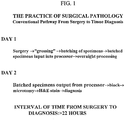
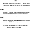
- Such conventional methodology demands that the tissue specimens be sent from the operating room, medical office or other sites, to a pathology laboratory on one day; the tissue specimens be prepared overnight; and the pathologist render a diagnosis based on microscopic examination of tissue sections the next day at the earliest, almost 24 hours after delivery of the specimen to the laboratory ( FIGURE 1 ).
- the pathologist renders a diagnosis based on microscopic examination of tissue sections the next day at the earliest, almost 24 hours after delivery of the specimen to the laboratory ( FIGURE 1 ).
- problems associated with impeded work flow in the pathology laboratory necessitated by the requisite batch processing of specimens, the safety concerns that attend having instruments operating overnight, the risk of possible instrument failures and the need to monitor the instruments, and the waste of using large volumes of reagents for such processing when automated.
- expensive measures are required to prevent exposure of laboratory personnel to fumes and toxic substances associated with the reagents used in this process.
- the large volumes of solvent waste and paraffin debris produced by conventional methodology pollute the environment.
- Histological diagnosis of a frozen section suffers from multiple disadvantages in comparison to sections prepared from paraffin blocks: the slide prepared from a frozen section "does not possess ... uniformity of quality"; "it is technically more difficult for serial sections of the same specimen to be examined”; extreme caution must be exercised in cutting the specimen in order to ensure a sufficiently thin section and to avoid the possibility of damaging details of the specimen”; and all the slides must be prepared “while the tissue is in the initial frozen state” because, "[i]f the tissue is thawed and refrozen for sectioning, it is severely damaged" ( U.S. Pat. No. 3,961,097 ).
- tissue processing There is an ever present interest in expediting tissue processing and analysis for diagnostic purposes. Furthermore, recent healthcare focus has been directed to lessening the cost of various procedures including tissue processing. The costs of tissue processing are related to time, the space required for preparation and analysis, reagents (both the amount required for processing and handling discard), and the number of personnel required. More importantly, patients and their physicians depend on evaluation and diagnosis by the pathologist to guide treatment. Reducing the amount of time needed to complete tissue processing would lessen the anxiety experienced during the period between obtaining the specimen and delivering the pathologist's report to the surgeon.
- U.S. Pat. Nos. 4,656,047 , 4,839,194 , and 5,244,787 use microwave energy;
- U.S. Pat. Nos. 3,961,097 and 5,089,288 use ultrasonic energy;
- U.S. Pat. No. 5,023,187 uses infrared energy.
- U.S. Pat. No. 5,104,640 disclosed a non-aqueous composition of a fixative, a stabilizing agent, and a solubilizing agent that adheres a blood smear to a slide.
- the aforementioned patents do not teach or suggest that the entire process of preparing diagnostic tissue sliders could be accomplished in less than two hours, starting from fixation and ending with impregnation, with continuous throughput of samples.
- the present invention provides such a process.
- a tissue slice must be on the order of 4 to 6 microns to be examined under a microscope, whereas the thinnest slice of fresh tissue that can be obtained by cutting is about 1 mm with the typical slice being on the order of 3 mm.
- it is necessary to harden the tissue so that a finer slice can be obtained e.g., by sectioning with a microtome.
- the present invention greatly accelerates the tissue hardening process and thus turns the conventional overnight processing into a process which totals on the order of 40 minutes.
- the rapid preparation of tissue by the present invention is capable of preserving tissue structures and morphology that were lost with conventional methods.
- tissues processed with the invention disclosed herein indicate better preservation of DNA and particularly RNA extraction than with conventional processing methods.
- tissues obtained in hospitals and other settings can be processed for both histologic and genetic studies soon after delivery to the laboratory, and archival material may be made available for future research and other applications. Improvements may be expected in the yield of genetic material, the stability of the genetic material in archival form, the size and integrity of the genetic material, and reducing chemical modification of the genetic material in comparison to the prior art.
- An object of the invention is to provide an apparatus for rapid processing of tissue for histology with continuous throughput.
- continuous throughput we mean accessing the system with additional samples, minutes apart. Therefore, at any given time there are samples of tissue in different stages of processing.
- continuous throughput we mean accessing the system with additional samples, minutes apart. Therefore, at any given time there are samples of tissue in different stages of processing.
- continuous throughput we mean accessing the system with additional samples, minutes apart. Therefore, at any given time there are samples of tissue in different stages of processing.
- continuous throughput we mean accessing the system with additional samples, minutes apart. Therefore, at any given time there are samples of tissue in different stages of processing.
- continuous throughput and flow of specimens along the various steps of tissue processing In contrast with our method, batch processing is presently required because conventional methodology takes eight hours or longer. Samples are placed in automated instruments, which can not be access with additional samples until the entire instrument cycle is completed. All these tissue samples are at the same stage of processing at any given step of the instrument cycle.
- a further object of the invention is to eliminate the need for toxic substances such as formalin and xylene in tissue processing.
- a tissue specimen is fixed, dehydrated, and fat is removed.
- a suitable admixture for use is a non-aqueous solution comprised of fixative and dehydrating agents, preferably a ketone and an alcohol; the volume ratio of alcohol to ketone may be between about 1:1 to about 3:1.
- the tissue specimen is incubated for about 25 minutes or less, more preferably for about 15 minutes or less, and even more preferably for about 5 minutes or less. Incubation is preferably between about 30°C and 65°C, more preferably between about 40°C and 55°C, and most preferably between about 45°C and 50°C.
- Another aspect of the invention is fixation, dehydration, fat removal, and clearing of a tissue specimen.
- a preferred solution in this aspect of the invention is alcohol and a clearant. This process may be accomplished in about 5 minutes or less.
- a tissue specimen is cleared and impregnated in a single solution comprised of a clearant and an impregnating agent. Preferably, this process may be accomplished in about 5 minutes or less. Prior to sectioning, the impregnated tissue specimen may be embedded in the impregnating agent.
- a tissue specimen which has been fixed, dehydrated, and defatted may then be impregnated in a wax solution.
- the wax solution is preferably as low as possible in water content.
- the wax solution may be prepared prior to impregnation by heating the wax to evaporate any dissolved water and by degassing under reduced pressure. Impregnation of the tissue specimen may take place under less than atmospheric pressure and at elevated temperature to remove any solvents from the tissue specimen and to draw the wax solution into the tissue specimen. Vacuum decreases impregnation time by accelerating diffusion and reducing the evaporation temperature of any solvents that may be present in the sample.
- the wax solution may comprise degassed paraffin and/or mineral oil. Impregnation of the tissue specimen may be completed in about 15 minutes or less; preferably, completed in about 10 minutes or less. Prior to sectioning, the impregnated tissue specimen may be embedded in the impregnating agent to form a tissue block.
- a tissue specimen is processed from fixation to impregnation in a series of solutions, at least some of which are admixtures that perform more than one task at the same time: fixation, dehydration, removal of fat, and impregnation.
- the admixture may include a fixative, a dehydrating agent, and a fat solvent (e.g., ketone and alcohol).
- Another solution may include fixative, dehydrating agent, fat solvent, and clearant (e.g., alcohol and xylene).
- Yet another solution may include a clearant and an impregnating agent (e.g., xylene and paraffin).
- the tissue specimen may be impregnated in a wax solution comprised of a mixture of different chain lengths (e.g., at room temperature, mineral oil which is liquid and paraffin which is solid).
- a wax solution comprised of a mixture of different chain lengths (e.g., at room temperature, mineral oil which is liquid and paraffin which is solid).
- an admixture contains at least two different chemicals (e.g., two alcohols).
- Processing time may be reduced by a non-aqueous admixture (e.g., fixative-dehydrating agent-fat solvent, fixative-dehydrating agent-fat solvent-clearant, clearant-impregnating agent), microwave energy as a source to achieve uniform heating within the tissue specimen, and reducing the pressure by using a vacuum source. Diffusion of the solution into the tissue specimen and chemical exchange may be promoted by mechanical agitation, heat, reduced pressure, or a combination thereof.
- a non-aqueous admixture e.g., fixative-dehydrating agent-fat solvent, fixative-dehydrating agent-fat solvent-clearant, clearant-impregnating agent
- microwave energy as a source to achieve uniform heating within the tissue specimen
- reducing the pressure by using a vacuum source. Diffusion of the solution into the tissue specimen and chemical exchange may be promoted by mechanical agitation, heat, reduced pressure, or a combination thereof.
- the above steps may be accelerated by adding a fixative enhancer, a surfactant, or both to the solutions used in the process.
- the fixative enhancer may be polyethylene glycol (PEG), mono- and dimethyleneglycol, propylene glycol, polyvinyl pyrrolidone, or the like; the polymer used may be between about 100 and about 500 average molecular weight, preferably about 300 molecular weight.
- the surfactant may be dimethyl sulfoxide (DMSO), polyoxyethylene sorbitan esters (e.g., TWEEN 80), sodium dimethyl sulfosuccinate, mild household detergents, or the like.
- the fixative may be a ketone (e.g., acetone, methyl ethyl ketone), aldehyde (e.g., acetylaldehyde, formaldehyde, glutaraldehyde, glyoxal), alcohol (e.g., methanol, ethanol, isopropanol), acetic acid, lead acetates and citrate, mercuric salts, chromic acid and its salts, picric acid, osmium tetroxide, or the like.
- aldehyde e.g., acetylaldehyde, formaldehyde, glutaraldehyde, glyoxal
- alcohol e.g., methanol, ethanol, isopropanol
- acetic acid lead acetates and citrate
- mercuric salts chromic acid and its salts
- picric acid osmium tetrox
- the tissue specimen may be dehydrated with methyl alcohol, isopropyl alcohol, ethyl alcohol, propyl alcohol, butanol, isobutanol, ethyl butanol, dioxane, ethylene glycol, acetone, amyl alcohol, or the like.
- Fat may be removed from the tissue specimen with an organic solvent such as, for example, acetone, chloroform or xylene.
- the clearant may be xylene, limonene, benzene, toluene, chloroform, petroleum ether, carbon bisulfide, carbon tetrachloride, dioxane, clove oil, cedar oil, or the like.
- the tissue specimen may be impregnated and/or embedded in paraffin, mineral oil, non-water soluble waxes, celloidin, polyethylene glycols, polyvinyl alcohol, agar, gelatin, nitrocelluloses, methacrylate resins, epoxy resins, other plastic media, or the like.
- tissue specimen is a piece of tissue that may be processed by the methods disclosed herein. It may also refer to single cells from any biological fluid (e.g., ascites, blood, pleural exudate), or cell suspensions obtained from aspiration of solid organs or lavage of body cavities. Single cells may be pelleted by sedimentation or buoyant centrifugation prior to processing.
- biological fluid e.g., ascites, blood, pleural exudate
- Single cells may be pelleted by sedimentation or buoyant centrifugation prior to processing.
- the apparatus of the invention are specially suitable for tissue specimens in which cell-cell contact, tissue organization, organ structure, or a combination thereof must be preserved.
- a specimen is a tissue slice preferably about 3 mm or less in its smallest dimension, more preferably about 2 mm or less, even more preferably about 1.5 mm or less, and most preferably about 1 mm or less.
- the tissue specimen may be fresh, partially fixed (e.g., fixation in 10% formalin for 2-3 hours), or fixed (e.g., overnight fixation in 10% formalin or any other fixative).
- the above invention allows processing of a tissue specimen from fixation to impregnation in less than about two hours, preferably less than about 90 minutes, more preferably less than about one hour, even more preferably less than about 45 minutes, and most preferably less than about 30 minutes. If the tissue specimen is fixed or partially fixed, then the processing time may be shortened accordingly.
- Tissue may be transported from the operating room to the pathology laboratory in an aqueous solution; such a transport solution may consist of equal volumes of an aqueous buffer and the non-aqueous admixture described herein.
- the tissue specimen can be embedded to produce a block.
- the agent used to embed the issue specimen is preferably the same as the material used for impregnation, but a different impregnating agent may also be used.
- the blocked tissue specimen can be mounted on a microtome to produce tissue sections of between about 1 micron and about 50 microns, preferably between about 2 microns and about 10 microns.
- the tissue sections may be further processed for histochemical staining, antibody binding, in situ nucleic acid hybridization/amplification, or a combination thereof.
- the tissue specimens are then typically examined by microscopy, but other techniques for detecting cellular properties may be used to examine the processed tissue specimen (e.g., automated cytometry, autoradiography, electrophoresis of nucleic acid).
- An apparatus for rapid, continuous histological processing of tissues is disclosed.
- the steps of fixation, dehydration, fat removal, and impregnation can be performed in less than about two hours; this allows a pathologist to evaluate samples shortly after receipt, perhaps while the patient is still in the operating or recovery room.
- Patient anxiety can be reduced by reducing the time required for pathological diagnosis.
- Rapid and continuous processing is accomplished by decreasing the thickness of tissue specimens, use of non-aqueous solutions composed of admixtures, solution exchange at elevated temperature and with agitation, uniform heating of tissues and solutions with microwave radiation, impregnation under vacuum pressure, or a combination thereof.
- Fixation, dehydration, and removal of fat are required for the preparation of tissue prior to impregnation. These steps are facilitated by trimming the tissue to a suitable size prior to processing, and using cassettes which hold such tissue blocks and allow their easy transfer between solutions for fixation, dehydration, removing fat, and impregnation.
- Fixation initiates hardening of the tissue specimen, and may preserve cell morphology by cross linking proteins and halting cellular degradation. Without chemical fixation, endogenous enzymes will catabolize and lyse the cell, and the tissue microanatomy will be altered. Such fixatives may be a ketone, aldehyde, alcohol, acetic acid, heavy metals, chromic acid, picric acid, or osmium tetroxide. Indications that fixation was inadequate can include: disassociation of tissue structures, bubbles in tissue sections, poor and irregular staining, shrunken cells, clumping of cytoplasm, condensation and less distinct nuclear chromatin, and autolysis/hemolysis of erythrocytes.
- Dehydration removes water from the tissue specimen to promote hardening. Replacement of water in the tissue specimen with a dehydrating agent also facilitates subsequent replacement of the dehydrating agent with material used for impregnation. This solution exchange is enhanced by using a volatile solvent for dehydration.
- the dehydrating agent may be low molecular weight alcohols, ketones, dioxane, alkylene glycols, or polyalkylene glycols. Failure to dehydrate the specimen can lead to inadequate impregnation, poor ribbon formation during sectioning, clefts in tissue sections, dissociation of structures, water crystals in tissue sections, and poor staining.
- Fat in the tissue specimen is removed with a solvent because fat impairs clearing and impregnation. Inadequate fat removal can result in spreading artifacts of tissue sections, wrinkling of tissue sections, and poor staining.
- the tissue specimen is cleared.
- the clearant extracts dehydrating agent from the tissue specimen and reduces its opacity.
- Examples of clearants include xylene, limonene, benzene, toluene, chloroform, petroleum ether, carbon bisulfide, carbon tetrachloride, dioxane, clove oil, or cedar oil.
- tissue specimen is suitably fixed and dehydrated, it is hardened by impregnation with an agent such as wax, celloidin, polyalkylene glycols, polyvinyl alcohols, agar, gelatin, nitrocelluloses, methacrylate resins, epoxy resins, or other plastics.
- an agent such as wax, celloidin, polyalkylene glycols, polyvinyl alcohols, agar, gelatin, nitrocelluloses, methacrylate resins, epoxy resins, or other plastics.
- Appropriate hardening of the tissue specimen with adequate preservation of cellular morphology is required prior to placing the impregnated specimen in a block and obtaining ten micron or thinner sections with a microtome knife.
- Preferred impregnation materials are commercial wax formulae, mixtures of waxes of different melting points (e.g., liquid mineral oil and solid paraffin), paraplast, bioloid, embedol, plastics and the like. Paraffin has been chosen for use in the examples herein because it
- Tissue processing is deemed a failure when one or more of the following problems is encountered: embedded tissue blocks are too soft or too hard, sections fall out or show an amount of compression different from the embedding agent, sections appear mushy, tissue ribbons fail to form or are crooked, sections crumble or tear, erythrocytes are lysed, or clumping of cytoplasm, condensation of chromatin, basophilic staining of nucleoli, shrunken cells, spreading artifacts and moth-eaten effect.
- the wax may be melted and removed prior to staining or immunohistochemistry.
- the tissue section is rehydrated and then analyzed as described below with stains or antibodies. After staining is completed or the histochemical reaction is developed, the slide may be coverslipped and viewed under a microscope. Alternatively, the stained or antibody-decorated specimen may be studied with an instrument for cytometry.
- the tissue blocks may be stored for archival purposes or retrospective studies.
- the present invention is compatible with preparation of nucleic acids, DNA or RNA, from processed tissues.
- Genetic study is possible for specimens collected routinely in the clinical pathology laboratory.
- the combined power of these technologies will be great.
- Histological observations may be correlated with genetics by analyzing one section by staining or immunohistochemistry, and preparing nucleic acids from an adjacent section for genetic analysis. For example, diseased and normal regions of the same section may be compared to detect genetic differences (e.g., mutations, levels of transcription), disease progression may be characterized by comparing genetics differences in samples taken at several time points, and tumor evolution may be assessed by following the accumulation of genetic differences from primary cancer to metastasis.
- Hematoxylin-eosin staining is commonly used for histological study and may be considered a standard for comparison by pathologists.
- the present invention has been found to be compatible with other stains including trichrome, reticulin, mucicarmine, and elastic stains as described in general references such as Thompson (Selected Histochemical and Histopathological Methods, C.C. Thomas, Springfield, Illinois, 1966 ), Sheehan and Hrapchak (Theory and Practice of Histotechnology, C.V. Mosby, St. Louis, Missouri, 1973 ), and Bancroft and Stevens (Theory and Practice of Histological Techniques, Churchill Livingstone, New York, New York, 1982 ).
- Such staining procedures would take between 30 minutes and several hours to complete, although rapid staining procedures are available from Fisher Scientific that require only five minutes to accomplish.
- Tissue may be obtained from an autopsy, a biopsy (e.g., endoscopic biopsy), or from surgery.
- a biopsy e.g., endoscopic biopsy
- the ability to provide a pathological diagnosis from a stained tissue section will provide the surgeon with information that may be used prior to the patient's departure from the operating room. For example, an indication from the pathologist that the cancer is confined to the resected tissue may allow the surgeon to be conservative in treatment and to preserve neighboring healthy tissue. Alternatively, a finding by the pathologist that cancer is not confined to a resected organ would permit more aggressive surgical treatment while the patient was still in the operating room.
- tissue Over 20,000 samples of tissue have been successfully processed by the present invention, including: brain, breast, carcinoma (e.g., bowel, nasopharynx, breast, lung, stomach), cartilage, heart, kidney, liver, lymphoma, meningioma, placenta, prostate, thymus, tonsil, umbilical cord, and uterus.
- Mineralized tissue e.g., bone, teeth
- tissue may be decalcified with a hydrochloric acid/ethylenediaminetetraacetic acid (EDTA) solution from Stephens Scientific (Allegiance Healthcare Supply, catalog no. 1209-1A) according to the manufacturer's instructions.
- EDTA hydrochloric acid/ethylenediaminetetraacetic acid
- Tissue sections processed by the present invention may also be used in immunohistochemistry.
- the choice of fixative may be optimized for recovery and preservation of particular antigens. Non-specific binding sites are blocked, antigen is bound by specific antibody (i.e., the primary antibody), and non-bound antibody is removed.
- the primary antibody may be detected directly but it is preferred to attach the probe to a protein (e.g., a secondary antibody) that specifically binds the primary antibody.
- Secondary antibody may be raised against the heavy or light chain constant region of the primary antibody. This amplifies the signal generated by an antigen-antibody conjugate because each primary antibody will bind many secondary antibodies.
- amplification may occur through other specific interactions such as biotin-streptavidin.
- Antibody binding is performed in a small volume to reduce usage of expensive reagents and maintain a high binding rate; evaporation of this small volume is reduced by incubation in a humidity chamber.
- the signal generating moiety is preferably an enzyme which is not otherwise present in the tissue.
- alkaline phosphatase and horseradish peroxidase may be attached to the secondary antibody or conjugated to streptavidin. Substrates are available for these enzymes that generate a chromogenic, fluorescent, or luminescent product that can be detected visually.
- the staining pattern for antigen may be used to localize expression of the antigen in the context of cellular structures revealed by counterstaining.
- Antigen expression can identify cell or tissue type, developmental stage, tumor prognostic markers, degenerative metabolic processes, or infection by a pathogen.
- Antigen-antibody binding may also be visualized with radioactive, fluorescence, or colloidal metal probes by autoradiography, epifluorescent microscopy, or electron microscopy, respectively. Similar probes may be used to detect nucleic acid in the tissue section by in situ hybridization to identify genetic mutations or transcripts; alternatively, the nucleic acid (DNA or RNA) may be extracted from tissue sections and analyzed directly by blotting, or amplified prior to further genetic analysis.
- Mutations may be germline and used to trace genetic predisposition of disease, or mutations may be somatic and used to determine genetic alterations in disease pathogenesis.
- the disease may be a metabolic or neurologic disorder, malignancy, developmental defect, or caused by an infectious agent.
- a series of tissue processing stations may be provided, e.g., in a single tissue processing unit or area.
- a suitable tissue processing facility is illustrated in FIGURE 3 .
- the first step in the process is to prepare a suitable tissue sample for hardening and ultimate examination.
- a slice of the tissue of interest is prepared.
- the finest slice possible is obtained, of about 1 to 3 mm and preferably 1 to 2 mm in thickness.
- Processing time is proportional to the size of the tissue sample being processed.
- the tissue slice is placed in a tissue cassette in which the tissue is contained during the immediately following processing steps.
- the tissue cassette is next placed in a first solution.
- the cassette 10 may be placed in a conventional beaker 12, having the first solution 14 therein, preferably by itself as the process described is a substantially continuous one, or together with a limited number of other, similar tissue cassettes.
- the beaker 12 is then placed in a shaker bath 16, as illustrated in FIGURE 4 , for gently agitating and heating the same.
- a shaker bath 16 as illustrated in FIGURE 4 .
- glycerine as the temperature conducting fluid 18 in the shaker bath 16.
- Glycerine has the advantage that it is an effective conductor of thermal energy but it does not evaporate.
- the tissue sample (in cassette 10) is disposed in the first solution, in the shaker bath 18 for approximately 3-15 minutes.
- Supplemental agitation is desirably also provided during the shaker-bath step.
- an external pump (A) FIGURE 3
- a tube (not shown) therefrom inserted into the solution beaker 12 or other receptacle for bubbling and thus agitating its contents.
- An aeration diffusion nozzle or plate may be provided to provide for more uniform solution agitation as deemed necessary or desirable.
- tissue cassette 10 and first solution containing beakers 12 remain upright and in a desired disposition, we have modified the conventional shaker-bath to provide transverse wires or stays 20, e.g., four wires, defining, e.g., five longitudinal channels in which tissue cassette containing beakers 12 may be disposed.
- sample containing beakers 12 may be regularly added to the shaker-bath 18 and sufficiently processed tissue samples removed in turn therefrom for further processing as described hereinbelow, by adding new samples on the left end of the shaker bath and removing sufficiently processed samples from the right end thereof..
- the tissue sample cassette 10 is exposed to a series of fluids while simultaneously being agitated and subjected to microwave radiation.
- three microwave units are provided, as shown in FIGURE 3 , each having a different solution in which the tissue sample containing cassette is submerged for a prescribed period.
- a single source of microwave energy could be provided. However, such would require sequential placement of the respective solutions for receiving the tissue cassette. While for a single tissue sample such solution placement and replacement would not significantly increase the duration of the tissue processing cycle, it can be appreciated that the use of a single microwave that receives multiple solutions, may hinder the continuity of the process with respect to subsequent samples.
- an exemplary microwave unit 22 for tissue processing is illustrated.
- a microwave processor model H-2800 and H-2500. Either model or another, similar such system could be used.
- a Pyrex or other clear microwaveable fluid receptacle 24 is utilized to hold respectively second, third and fourth solutions provided in each of the three microwave units ( FIGURE 3 ).
- a temperature probe 26 is placed in the solution to ensure that the temperature of the respective bath is within the desired range.
- aeration is provided to provide for agitation, which accelerates the tissue processing.
- the microwave units we have used include a tube 28 for aeration.
- a single tube may be inserted into the bath, but for more uniform and complete agitation, it is most preferred to provide a diffusion plate or nozzle head (not shown in detail) in cooperation with the gas tube 28 for diffusing the agitating bubbles, e.g., across a substantial portion of the diameter of the solution receptacle for uniform agitation of the entire volume of solution.
- a diffusion plate or nozzle head (not shown in detail) in cooperation with the gas tube 28 for diffusing the agitating bubbles, e.g., across a substantial portion of the diameter of the solution receptacle for uniform agitation of the entire volume of solution.
- Such diffusion plates and nozzles are well known and can be provided, e.g., at the base of the solution receptacle.
- paraffin is degassed as a part of the tissue processing procedure.
- Degassing removes organic solvents from the paraffin.
- continuous degassing This is accomplished by maintaining the vacuum within the covered Pyrex 32 at 640 mm. Hg.
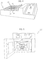
- the tissue sample cassette(s) are placed in a paraffin bath, as shown in FIGURE 6 .
- a paraffin bath comprising three paraffin bath stations (beakers) 30 provided within a covered Pyrex jar 32.
- the Pyrex jar 32 is placed in, e.g., a Poly Science brand water bath 34.
- a grease or the like By applying a grease or the like to the internal edges of the flanges on both the lid and jar, an airtight coupling can be provided between the lid and jar and thus a vacuum can be pulled through a tooled hose connector 36 provided in the lid.
- Suitable such Pyrex brand jars are available from Fisher Scientific. We have used Model No.
- a conventional pressure/vacuum pump 38 is coupled to a tube 40 that is in turn coupled to connector 36.
- a suitable such power operated pump is available from Fisher Scientific and has for example a 100 psi max. Agitation is preferably provided during the paraffin bath step, either through vibratory agitation, ultrasound, or potentially via aeration.
- tissue sample must be embedded.
- Tissue-Tek embedding console system ( FIGURE 3 ) available from Miles/Sakura, e.g. Model No. 4708.
- the embedded tissue sample is then cut in a conventional manner with a microtome (L) ( FIGURE 3 ) and floated (M) for placement, we use the Leitz 1512 Microtome, and the Lipshaw Electric Tissue Float Model 375.
- L microtome
- M floated
- FIGURE 3 To accelerate the staining process, we propose to use an automated stainer (O) ( FIGURE 3 ) to reduce the number of personnel and time required.
- a non-continuous process could use the Sakura diversified stainer DRS-601 which stains slides in batches; alternatively, a continuous process could use a Leica auto stainer XL which contains a dewaxing stage so that separate incubation in an oven may be omitted.
- the fixed and stained tissue sample is then covered, e.g. with the Tissue-Tek coverslipper, Manufacturer No. 4764 (R) ( FIGURE 3 ).
- the system for carrying out the dehydration and impregnation in accordance with the invention can be a series of discrete units.
- one or more steps can be carried out in a single processing component or unit.
- the number of units provided and the steps carried out by each unit impacts the continuity of the processing unit.
- a single unit for carrying out a plurality of the tissue processing steps may be advantageous and will not significantly impact continuity of tissue processing.
- two or more units may be preferred.
- FIGURE 7 An exemplary combined unit 42 is illustrated in FIGURE 7 .
- the combined unit 42 in fact includes two subunits; a microwave processor unit 44 and an impregnator unit 46.
- the microwave processor unit 44 is provided for sequentially submerging the tissue being processed in solution A, solution B, and solution C, in each instance agitating the solution and exposing the tissue to microwave energy.
- a vessel 48 is provided for receiving for example one or more trays 50 on which one or more tissue cassettes 10 may be placed.
- the vessel 48 is fluidly coupled to a source of each of the solutions for tissue dehydration.
- solution A is conducted to the vessel 48 and microwave energy is applied thereto simultaneous to agitation via, for example, an aeration tube (not shown in FIGURE 7 ).
- solution A is drained and the tissue cassettes are preferably flushed either with solution B or with a combination of solution A and solution B so as to substantially eliminate residual solution A.
- Solution B is then fed to the vessel 48 whereupon microwave energy and agitation are again applied for a prescribed period.
- solution B is returned to a storage vessel therefor and the tissue samples are flushed either with solution C or a combination of solution B and solution C.
- solution C is fed to the vessel 48, agitation and microwave energy are applied, and ultimately solution C is drained.
- the tissue samples are then ready for impregnation.
- impregnation is carried out in a second subunit 46 of the assembly. This allows impregnation to be carried out while a subsequent tissue sample(s) are subject to microwave energy application. If a single unit is provided, then the vessel used for microwave processing can be used for impregnation however the microwave energy would not be applied thereto during the impregnation steps.
- a series of paraffin solutions e.g., 3 or 4 are applied to the tissue cassettes disposed e.g. on suitable trays 52 in a vessel 54, to provide sequential paraffin baths to effect the impregnation of the tissue sample as a final step in the tissue preparation process.
- the tissue samples are placed under a vacuum at a controlled elevated temperature.
- the tissue samples are preferably also agitated during this step with a magnetic stirrer, ultrasound, or air bubbler.
- a slicing guide 60 is in the form of a thin metal plate 62 on the order of, e.g., 1 to 2 mm in thickness, having a cutout 64 the width of, for example, a thumb nail (about 1 cm 2 ).
- a stop 66 is defined at the end of the cutout or notch 64 to serve as a knife or blade stop.
- a lip 68 may be provided at the end of the metal plate 62, remote from the cutting notch.
- a larger segment of tissue is placed over the cutout or notch 64 so that a portion thereof is disposed in the notch.
- Pressure is then applied to the exposed surface of the tissue and a cutting instrument is placed against and slid horizontally along the slicing guide plate so as to sever the tissue disposed in the notch 64 from the remainder of the tissue.
- Engagement of the cutting blade with the blade stop 66 completes the cutting process and the bulk of the tissue, disposed above the cut, is placed aside.
- the remaining tissue, disposed in the slot can then be placed in a suitable tissue cassette for dehydration and impregnation.
- the slicing guide 60 facilitates the production of a thin slice of tissue of generally uniform thickness which may be further processed.
- FIGURE 9 As another alternative for producing a thin tissue slice, we have proposed to provide flat plates or blocks 70 at the end of an otherwise conventional forceps 72, as schematically illustrated in FIGURE 9 .
- the blocks may be permanently or temporarily secured to the ends of the forceps.
- This provides rather large, flat clamping surfaces 74.
- the tissue to be cut may be placed between the clamping blocks 70 and a sharp blade passed between the clamping blocks to slice the tissue.
- a thin tissue slice of generally uniform thickness can be provided.
- the parallel rather large flat surfaces provide uniform pressure distribution thus holding the tissue in position during the cutting process and then ensuring a uniform cut that preferably preserves the integrity of the tissue.
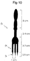
- FIGURE 10 To hold the tissue in position during cutting we have also proposed a three prong fork-like instrument 92, illustrated in FIGURE 10 .
- the prongs 94 are spaced from each other by approximately one centimeter and each has a sharp, pointed tip 96 to facilitate penetration of the tissue with minimal disruption.
- suitable slices of tissue can be obtained by cutting parallel to or between the prongs.
- the instrument 92 is characterized in that the prongs have a length on the order of 5-10 cm to accommodate a variety of specimens and a handle of about 8 centimeters in length, itself spaced from the prongs by 2-4 centimeters, to facilitate manipulation of the instrument and a sure grip during cutting.
- the fork-like instrument 92 is particularly advantageous in obtaining sections from organs such as the intestine and gallbladder. Indeed, securing such specimens with prongs 94 prevents the various layers of tissue from sliding upon each other during the cutting process.
- tissue receiving unit and cassette for use in the operating room, to facilitate transport of tissue, particularly very small segments of tissue, for example those obtained by needle biopsy.
- tissue particularly very small segments of tissue, for example those obtained by needle biopsy.
- FIGURES 11 and 12 we have proposed to provide tissue cassettes 10' to the operating room for immediately receiving such minute tissue samples.
- tissue cassette 10' To contain such tissue samples within the tissue cassette 10', we have provided thin sheets of biopsy sponge material 80, which is an open cell plastic foam, at least one of which has a partial depth recess 82 defined therein to provide, together with the other biopsy sponge a compartment for receiving the biopsied tissue.
- tissue cassette 10' To maintain the integrity of the tissue for transport to the processing lab, the tissue cassette 10' is placed within a jar of suitable solution.
- FIGURE 12 shows the tissue cassette 10' attached by its top surface.
- alternative attachment points are possible such as the bottom surface or the hinged side of the cassette.
- two or more cassettes may be attached to the columnar support 88.
- the tissue cassette 10' with the biopsied tissue therewithin can be temporarily secured to the distal end of the columnar support 88 and inserted into a suitable solution for transport.
- the lid 90 is removed from the jar 86 and the tissue cassette 10' removed from the column 88.
- Any suitable fasteners such as velcro type fasteners, plastic snap lock, dove tail slide connectors or other cooperative engagement structure can be provided to attach the tissue cassette 10' to the support column 88.
- the solution within the specimen jar 86 may be a transport (aqueous) solution or the first (non-aqueous) solution. It would be convenient to provide the specimen jar in the operating room with the cassette attached to the outside of the jar and then to invert the lid so that the cassette is immersed in the solution within the jar after tissue is placed within the cassette.
- Standardization of tissue fixation and processing procedures will ease comparison of specimens from different laboratories. Artifacts in histology due to the use of formaldehyde and/or prolonged processing will be eliminated; thus, allowing more precise evaluation of microscopic morphology of normal and diseased tissues. Similarly, antigen retrieval and staining will be improved. For genetic analysis, formaldehyde-induced DNA mutations will be eliminated and extraction of nucleic acid from archival material may be enhanced. The feasibility of RNA studies from stored, fixed paraffin-embedded tissue opens unlimited avenues for diagnostic and research applications.
- Tissues samples were incubated for 15 min at a glycerin bath temperature between 45°C and 50°C.
- the 400 ml solution for fixation was placed in a 500 ml beaker in a water bath shaker (linear displacement of 5 cm/sec). Additional agitation of the fixation solution was provided by bubbling with an air pump.
- Fixation, dehydration, fat removal, clearing, and impregnation are accomplished by sequential exposure of the tissue specimen to three different solutions (the second, third and fourth solutions described above), one in each of three microwave ovens from Energy Beam Sciences.
- a one liter solution of 70% isopropyl alcohol and 30% polyethylene glycol (average molecular weight 300) is placed in the first oven (model H2800) in a 1500 ml beaker
- the solution in the second oven (model H2800) consists of one liter of 70% isopropyl alcohol and 30% xylene in a 1500 ml beaker
- the third oven (model H2500) contains a solution of 1000 ml of xylene and 300 gm of paraffin in a 1500 ml beaker.
- Ten ml of DMSO per liter are added to these three solutions. Heating at 60°C by microwave radiation is effected for 15 minutes in the first oven, and 5 minutes each in the second and third ovens (75% power setting with a cycle
- tissue sections were incubated in four 500 ml baths of molten paraffin placed within a large dessicator filled with paraffin, and resting in a glycerin bath at 75°C. Tissue sections were transferred from one paraffin bath to the next at 3 minute intervals, for a total impregnation time of 12 minutes. Each 3 minute interval was measured from the time that the pressure reading is about 640 mm. Of Hg. No agitation was used during this step.
- Fixation, dehydration, fat removal, and paraffin impregnation of fresh or fixed tissue sections, approximately 1 mm thick, was accomplished in 40 minutes by exposing these tissue sections to four successive steps as follows.
- the samples were incubated in a solution of 70% isopropyl alcohol, 30% acetone, and DMSO added at an approximate concentration of 1% at 60°C. Samples were heated in a commercial tissue microwave processor (H2800, Energy Beam Sciences) for 5 min each in two beakers containing the solution (10 min total incubation), which were agitated by bubbling.
- H2800 Energy Beam Sciences
- Impregnation was completed by incubation in four baths of molten paraffin placed within a large dessicator resting in a glycerin bath at 75°C. Tissue sections were transferred from one paraffin bath to the next at 3 min intervals, for a total impregnation time of 12 min. Each 3 min interval was measured for the time that the pressure reading is about 640 mm of Hg.
- Tissue specimens acquire a blue tint that facilitates their handling during impregnation and handling; penetration of the tissue specimen may also be monitored by observation of an even blue color throughout the tissue specimen.
- Fixation, dehydration, fat removal, and paraffin impregnation of fresh or fixed tissue sections, up to about 1 to 2 mm thick, may be accomplished in 65 minutes as follows.
- the first solution consists of:
- samples are incubated in a solution of 55% isopropyl alcohol, 25% acetone, 10% polyethylene glycol (average molecular weight 300), 10% low viscosity mineral oil, glacial acetic acid added at an approximate concentration of 0.5% of the total volume, and DMSO added at an approximate concentration of 1%.
- Samples are heated at 65°C in a commercial tissue microwave processor (H2800, Energy Beam Sciences) for 15 min in a 1500 ml beaker containing the solution, which is agitated by bubbling.
- samples are incubated in a solution of 55% isopropylic alcohol, 25% acetone 20% low viscosity mineral oil, glacial acetic acid added at an approximate concentration of 0.5% of the total volume and DMSO added at an approximate concentration of 1% of the total volume.
- Samples are heated at 65°C in a commercial tissue microwave processor (H2800, Energy Beam Sciences) for 5 minutes in a 1500 ml beaker containing the solution, which is agitated by bubbling.
- impregnation is initiated by incubation in two baths of a wax solution of 30% low viscosity mineral oil and 70% molten paraffin placed in a large dessicator resting in a 60°C glycerin bath, under a vacuum of about 640 mm of Hg, for 5 min. in each bath.
- Impregnation is completed by incubation in four baths of molten paraffin placed within a large dessicator resting in a glycerin bath at about 75°C to 80°C and a reduced pressure of about 640 mm of Hg, for 5 min each. Tissue sections were transferred from one paraffin bath to the next at 5 min intervals, for a total impregnation time of 20 min. Each 5 min interval was measured for the time that the pressure reading is about 640 mm of Hg.
Applications Claiming Priority (3)
| Application Number | Priority Date | Filing Date | Title |
|---|---|---|---|
| US5610297P | 1997-08-20 | 1997-08-20 | |
| PCT/US1998/016463 WO1999009390A1 (en) | 1997-08-20 | 1998-08-19 | A high quality, continuous throughput, tissue fixation-dehydration-fat removal-impregnation method |
| EP98940811A EP1005633B1 (en) | 1997-08-20 | 1998-08-19 | A high quality, continuous throughput, tissue fixation-dehydration-fat removal-impregnation method |
Related Parent Applications (4)
| Application Number | Title | Priority Date | Filing Date |
|---|---|---|---|
| WOPCT/US98/16463 Previously-Filed-Application | 1998-08-19 | ||
| PCT/US1998/016463 Previously-Filed-Application WO1999009390A1 (en) | 1997-08-20 | 1998-08-19 | A high quality, continuous throughput, tissue fixation-dehydration-fat removal-impregnation method |
| EP98940811A Division EP1005633B1 (en) | 1997-08-20 | 1998-08-19 | A high quality, continuous throughput, tissue fixation-dehydration-fat removal-impregnation method |
| EP98940811.7 Division | 1998-08-19 |
Publications (2)
| Publication Number | Publication Date |
|---|---|
| EP2199774A1 EP2199774A1 (en) | 2010-06-23 |
| EP2199774B1 true EP2199774B1 (en) | 2018-10-03 |
Family
ID=22002161
Family Applications (2)
| Application Number | Title | Priority Date | Filing Date |
|---|---|---|---|
| EP98940811A Expired - Lifetime EP1005633B1 (en) | 1997-08-20 | 1998-08-19 | A high quality, continuous throughput, tissue fixation-dehydration-fat removal-impregnation method |
| EP08159241.2A Expired - Lifetime EP2199774B1 (en) | 1997-08-20 | 1998-08-19 | Apparatus for histoprocessing of tissue samples |
Family Applications Before (1)
| Application Number | Title | Priority Date | Filing Date |
|---|---|---|---|
| EP98940811A Expired - Lifetime EP1005633B1 (en) | 1997-08-20 | 1998-08-19 | A high quality, continuous throughput, tissue fixation-dehydration-fat removal-impregnation method |
Country Status (12)
| Country | Link |
|---|---|
| US (4) | US6207408B1 (zh) |
| EP (2) | EP1005633B1 (zh) |
| JP (2) | JP4164232B2 (zh) |
| KR (1) | KR20010023070A (zh) |
| AT (1) | ATE407353T1 (zh) |
| AU (1) | AU746497B2 (zh) |
| CA (2) | CA2301924C (zh) |
| DE (1) | DE69839963D1 (zh) |
| DK (1) | DK1005633T3 (zh) |
| ES (1) | ES2313752T3 (zh) |
| TW (1) | TW571100B (zh) |
| WO (1) | WO1999009390A1 (zh) |
Families Citing this family (136)
| Publication number | Priority date | Publication date | Assignee | Title |
|---|---|---|---|---|
| US6451551B1 (en) * | 1994-03-11 | 2002-09-17 | Biogenex Laboratories | Releasing embedding media from tissue specimens |
| US6793890B2 (en) | 1997-08-20 | 2004-09-21 | The University Of Miami | Rapid tissue processor |
| DK1005633T3 (da) | 1997-08-20 | 2008-12-15 | Univ Miami | Vævsfikserings-dehydratiserings-fedtfjernelses-imprægneringsmetode med höj kvalitet og kontinuerligt gennemlöb |
| DE69917779T2 (de) * | 1998-06-30 | 2005-06-16 | Lamina, Inc. | Zytologische und histologische fixier-zusammensetzung und verfahren zur verwendung |
| AU4812600A (en) * | 1999-04-29 | 2000-11-17 | Arcturus Engineering, Inc. | Processing technology for lcm samples |
| US7951612B2 (en) * | 1999-07-08 | 2011-05-31 | Lee H. Angros | In situ heat induced antigen recovery and staining apparatus and method |
| WO2001004634A1 (en) | 1999-07-08 | 2001-01-18 | Lee Angros | Antigen recovery and/or staining apparatus and method |
| US8298485B2 (en) * | 1999-07-08 | 2012-10-30 | Lee H. Angros | In situ heat induced antigen recovery and staining apparatus and method |
| US7897106B2 (en) * | 1999-07-08 | 2011-03-01 | Lee Angros | Situ heat induced antigen recovery and staining apparatus and method |
| US6916608B2 (en) * | 1999-09-10 | 2005-07-12 | Becton, Dickinson And Company | Composition for providing long term stability to cells for diagnostic testing |
| DE19945621A1 (de) * | 1999-09-23 | 2001-04-05 | Aurel Popa | Geräteanordnung zur Präparation und Analyse von Gewebe für mirkoskopische Untersuchungen |
| WO2001044783A1 (en) * | 1999-12-14 | 2001-06-21 | University Of Miami | Rapid tissue processor |
| ES2563454T3 (es) | 2000-06-09 | 2016-03-15 | The University Of Miami | Tabla patológica para rebanar a espesor determinado |
| US6518416B1 (en) | 2000-12-01 | 2003-02-11 | Response Genetics, Inc. | Method of determining a chemotherapeutic regimen based on ERCC1 expression |
| CA2436910C (en) * | 2000-12-01 | 2014-06-17 | Response Genetics, Inc. | Method of determining epidermal growth factor receptor and her2-neu gene expression and correlation of levels thereof with survival rates |
| US6582919B2 (en) | 2001-06-11 | 2003-06-24 | Response Genetics, Inc. | Method of determining epidermal growth factor receptor and HER2-neu gene expression and correlation of levels thereof with survival rates |
| US7049059B2 (en) * | 2000-12-01 | 2006-05-23 | Response Genetics, Inc. | Method of determining a chemotherapeutic regimen based on ERCC1 and TS expression |
| US6602670B2 (en) | 2000-12-01 | 2003-08-05 | Response Genetics, Inc. | Method of determining a chemotherapeutic regimen based on ERCC1 expression |
| US6956111B2 (en) | 2001-03-02 | 2005-10-18 | Response Genetics, Inc. | Method of determining dihydropyrimidine dehydrogenase gene expression |
| US6458598B1 (en) * | 2001-08-13 | 2002-10-01 | Dako A/S | Process for preparing and presenting a tissue sample for histological study |
| CN100359313C (zh) * | 2001-09-20 | 2008-01-02 | 王世峰 | 一种活体组织脱水盒 |
| ATE551606T1 (de) * | 2001-10-01 | 2012-04-15 | Leica Biosystems Melbourne Pty | Gewebeprozessor zur behandlung histologischer gewebepräparate |
| US11249095B2 (en) | 2002-04-15 | 2022-02-15 | Ventana Medical Systems, Inc. | Automated high volume slide processing system |
| US7468161B2 (en) | 2002-04-15 | 2008-12-23 | Ventana Medical Systems, Inc. | Automated high volume slide processing system |
| US6797928B2 (en) * | 2002-04-23 | 2004-09-28 | Ted Pella, Inc. | Method and apparatus for the rapid decalcification and fixation of mineralized tissues |
| US7138226B2 (en) * | 2002-05-10 | 2006-11-21 | The University Of Miami | Preservation of RNA and morphology in cells and tissues |
| CN1672028B (zh) * | 2002-05-28 | 2011-12-28 | 戴尔帕斯有限公司 | 制备组织、尸检、细胞样品的组合物 |
| DK1552266T3 (en) | 2002-09-26 | 2016-02-08 | Biopath Automation Llc | Apparatus and methods for automatic handling and embedding of tissue samples |
| US7179424B2 (en) | 2002-09-26 | 2007-02-20 | Biopath Automation, L.L.C. | Cassette for handling and holding tissue samples during processing, embedding and microtome procedures, and methods therefor |
| ES2345482T3 (es) | 2002-09-26 | 2010-09-24 | Biopath Automation, L.L.C. | Casete y ensamblado de incorporacion, dispositivos de estadificacion, y procedimientos para manipular muestras de tejido. |
| US7219884B2 (en) * | 2002-10-01 | 2007-05-22 | The University Of Miami | Pathology grossing tool |
| AU2003291659A1 (en) * | 2002-10-31 | 2004-06-07 | University Of Massachusetts | Rapid cell block embedding method and apparatus |
| US7541161B2 (en) | 2002-10-31 | 2009-06-02 | University Of Massachusetts | Method and apparatus for preparing cells for microtome sectioning and archiving nucleic acids and proteins |
| JP4189484B2 (ja) * | 2002-12-12 | 2008-12-03 | 国立循環器病センター総長 | マイクロ波照射による生体組織の処理方法 |
| WO2004072268A2 (en) * | 2003-02-12 | 2004-08-26 | Mayo Foundation For Medical Education And Research | Pkhdl1, a homolog of the autosomal recessive kidney disease gene |
| KR200327028Y1 (ko) * | 2003-06-17 | 2003-09-19 | 장시창 | 인체조직검사기구 |
| US8554372B2 (en) | 2003-09-29 | 2013-10-08 | Leica Biosystems Melbourne Pty Ltd | System and method for histological tissue specimen processing |
| KR20060115366A (ko) * | 2003-10-24 | 2006-11-08 | 더 유니버시티 오브 마이애미 | 간소화시킨 조직 가공 |
| KR100458860B1 (ko) * | 2004-04-22 | 2004-12-03 | 송영민 | 생검시료의 검사를 위한 보조기구 |
| US8409871B2 (en) * | 2004-05-25 | 2013-04-02 | Leica Biosystems Melbourne Pty Ltd | Method of treatment of tissue processing fluid and apparatus therefor |
| EP1605243B1 (en) * | 2004-06-07 | 2008-12-03 | Milestone S.r.l. | Method and apparatus for microwave assisted tissue processing |
| US7273587B1 (en) | 2004-12-02 | 2007-09-25 | Collaborative Laboratory Services, Llc | Rapid tissue processing method and apparatus |
| US7273720B1 (en) | 2004-12-02 | 2007-09-25 | Collaborative Laboratory Services, Llc | Rapid tissue processing method and apparatus |
| US8236255B2 (en) * | 2004-12-02 | 2012-08-07 | Lab Vision Corporation | Slide treatment apparatus and methods for use |
| EP2392908B1 (en) * | 2004-12-17 | 2019-08-21 | Ventana Medical Systems, Inc. | Methods for preparing a paraffin-embedded biological sample for staining. |
| GB0501590D0 (en) * | 2005-01-25 | 2005-03-02 | Ceres Power Ltd | Processing of enhanced performance LSCF fuel cell cathode microstructure and a fuel cell cathode |
| US20060228772A1 (en) * | 2005-04-11 | 2006-10-12 | Donndelinger Thomas M | Compositions and methods for preparing specimens for microscopic analysis |
| US8034610B2 (en) * | 2005-04-21 | 2011-10-11 | Celerus Diagnostics, Inc. | Parallel processing fluidic method and apparatus for automated rapid immunohistochemistry |
| BRPI0609898A2 (pt) * | 2005-05-24 | 2011-10-11 | Lee H Angros | aparelho de recuperação e manchamento de antìgeno in situ, módulo de reação, método para tratar uma lámina de microscópio, e, tira de dispensação de reagente reconfigurável |
| JP5443756B2 (ja) * | 2005-06-28 | 2014-03-19 | ザ ボード オブ リージェンツ オブ ザ ユニバーシティ オブ オクラホマ | カーボンナノチューブを成長および収集するための方法 |
| WO2007016935A1 (en) | 2005-07-29 | 2007-02-15 | Histogenex Nv | Methods, reagents and instrumentation for preparing impregnated tissue samples suitable for histopathological and molecular studies |
| US20070116612A1 (en) * | 2005-11-02 | 2007-05-24 | Biopath Automation, L.L.C. | Prefix tissue cassette |
| US7769962B2 (en) * | 2005-12-12 | 2010-08-03 | Jeda Technologies, Inc. | System and method for thread creation and memory management in an object-oriented programming environment |
| EP1804045B1 (de) * | 2005-12-30 | 2014-03-12 | QIAGEN GmbH | Ein Verfahren und ein Kit zur Behandlung einer biologischen Probe |
| CA2636912A1 (en) * | 2006-01-13 | 2007-07-26 | Ventana Medical Systems, Inc. | Biological sample processing composition and method |
| WO2007141888A1 (ja) * | 2006-06-05 | 2007-12-13 | Jokoh Co., Ltd | 生物組織固定装置 |
| US20080026366A1 (en) * | 2006-06-07 | 2008-01-31 | Newcomer Supply, Inc. | Biological fixative and method of using the biological fixative |
| AT504371B8 (de) | 2006-10-03 | 2008-09-15 | Leica Mikrosysteme Gmbh | Vorrichtung zur mikrowellen-gestützten präparation von proben |
| US7666620B2 (en) * | 2006-11-08 | 2010-02-23 | Richard-Allan Scientific Company | Rapid tissue processing system |
| JP5050200B2 (ja) * | 2006-11-20 | 2012-10-17 | 国立大学法人三重大学 | 医療検体用ケース |
| CN101583315A (zh) * | 2006-12-12 | 2009-11-18 | 比欧帕斯自动化公司 | 具有可切片弹性多孔材料的活检载具 |
| EP1965190A1 (en) * | 2007-02-27 | 2008-09-03 | Qiagen GmbH | Fixation of a biological sample |
| EP1975595A1 (en) * | 2007-03-21 | 2008-10-01 | Diapath S.r.l. | Automatic processor for histological samples and its operation |
| EP2009419B1 (en) * | 2007-06-25 | 2009-12-23 | Milestone S.r.l. | Measuring the thickness of organic samples |
| US7906076B2 (en) * | 2007-07-02 | 2011-03-15 | University Of Massachusetts | Method and apparatus for biopsy sample processing |
| JP4059520B1 (ja) * | 2007-07-25 | 2008-03-12 | 株式会社常光 | 組織片処理装置 |
| WO2009023234A1 (en) * | 2007-08-14 | 2009-02-19 | Charm Sciences, Inc. | Sample concentration method and apparatus |
| ES2852752T3 (es) | 2007-10-23 | 2021-09-14 | Becton Dickinson Co | Sistema de múltiples cámaras para contención de tejido para diagnóstico molecular e histológico |
| JP5256295B2 (ja) * | 2007-10-23 | 2013-08-07 | ベクトン・ディキンソン・アンド・カンパニー | 分子および組織診断用の流体移動組織容器 |
| ES2752754T3 (es) * | 2007-10-23 | 2020-04-06 | Becton Dickinson Co | Kit cerrado para contención y estabilización de tejidos para diagnósticos moleculares e histopatológicos |
| CN101835428B (zh) * | 2007-10-23 | 2012-01-04 | 贝克顿·迪金森公司 | 结合有易破膜的用于分子和组织学诊断的组织容器 |
| AU2013200912B2 (en) * | 2007-10-23 | 2014-04-17 | Becton, Dickinson And Company | Multi-chambered tissue containment system for molecular and histology diagnostics |
| WO2009055600A2 (en) * | 2007-10-23 | 2009-04-30 | Becton, Dickinson And Company | Container system for tissue stabilization for molecular and histopathology diagnostics |
| US8158381B2 (en) * | 2008-03-27 | 2012-04-17 | Richard-Allan Scientific Company | Methods for integrated tissue processing and staining |
| CA2724638C (en) | 2008-05-27 | 2020-02-18 | Dako Denmark A/S | Hybridization compositions and methods comprising a polar aprotic solvent |
| US20090298172A1 (en) | 2008-05-28 | 2009-12-03 | Steven Paul Wheeler | Histological specimen treatment apparatus and method |
| US20090325273A1 (en) * | 2008-06-30 | 2009-12-31 | Chin Pin Chang Chien | Multi-functional treating device for treating pathological tissue sections |
| US20110008884A1 (en) * | 2008-08-27 | 2011-01-13 | Morales Azorides R | Pathology grossing tool, cassette, and board |
| WO2010025425A1 (en) | 2008-08-29 | 2010-03-04 | Angros Lee H | Multiplexed microscope slide staining apparatus |
| US9782565B2 (en) | 2008-10-01 | 2017-10-10 | Covidien Lp | Endoscopic ultrasound-guided biliary access system |
| US8968210B2 (en) | 2008-10-01 | 2015-03-03 | Covidien LLP | Device for needle biopsy with integrated needle protection |
| US9332973B2 (en) | 2008-10-01 | 2016-05-10 | Covidien Lp | Needle biopsy device with exchangeable needle and integrated needle protection |
| US9186128B2 (en) | 2008-10-01 | 2015-11-17 | Covidien Lp | Needle biopsy device |
| US11298113B2 (en) | 2008-10-01 | 2022-04-12 | Covidien Lp | Device for needle biopsy with integrated needle protection |
| WO2010056883A1 (en) | 2008-11-12 | 2010-05-20 | Ventana Medical Systems, Inc. | Methods and apparatuses for heating slides carrying specimens |
| WO2010065775A2 (en) * | 2008-12-03 | 2010-06-10 | The United States Government As Represented By The Department Of Veterans Affairs | Pressure-assisted molecular recovery (pamr) of biomolecules, pressure-assisted antigen retrieval (paar), and pressure-assisted tissue histology (path) |
| DK3300667T3 (da) | 2009-01-22 | 2019-12-02 | Sakura Finetek Usa Inc | Biopsiunderstøtning der kan sektioneres i en mikrotom til orientering af vævsprøver |
| EP2401395A1 (en) | 2009-02-26 | 2012-01-04 | Dako Denmark A/S | Compositions and methods for rna hybridization applications |
| US8640357B2 (en) * | 2009-03-23 | 2014-02-04 | Hasan Huseyin Engin | Laboratory type quick film drying oven |
| DE102009025574A1 (de) * | 2009-06-19 | 2010-12-23 | Leica Biosystems Nussloch Gmbh | Verfahren zum automatischen Bearbeiten von Gewebeproben in einem Gewebeprozessor |
| US9237608B2 (en) | 2009-08-14 | 2016-01-12 | Cem Corporation | Pressure stepped microwave assisted digestion |
| CA2770545C (en) * | 2009-08-21 | 2015-11-03 | F. Hoffmann-La Roche Ag | Use of a bis-maleic anhydride cross-linking agent for fixation of a cell or tissue sample |
| US10126216B2 (en) | 2011-02-17 | 2018-11-13 | Ventana Medical Systems, Inc. | Method for tissue sample fixation |
| ES2759007T3 (es) | 2010-03-04 | 2020-05-07 | Ventana Med Syst Inc | Sistema de procesamiento para procesar muestras usando energía acústica |
| US10539487B2 (en) | 2010-03-04 | 2020-01-21 | Ventana Medical Systems, Inc. | Systems and methods for monitoring tissue sample processing |
| US8227233B2 (en) * | 2010-06-09 | 2012-07-24 | Ford Global Technologies, Llc | Method of making foamed mycelium structure |
| US20120171694A1 (en) * | 2010-07-30 | 2012-07-05 | Vermillion, Inc. | Predictive markers and biomarker panels for ovarian cancer |
| GB2490202B (en) * | 2011-04-20 | 2015-06-24 | Leica Biosystems Nussloch Gmbh | Method of detaching and/or isolating a histological sample |
| DE102011055120B4 (de) | 2011-04-20 | 2016-04-14 | Leica Biosystems Nussloch Gmbh | Verfahren zur Verarbeitung zumindest einer histologischen Probe und Vorrichtung zum Behandeln einer histologischen Probe. |
| CN103562702A (zh) | 2011-05-20 | 2014-02-05 | 独立行政法人理化学研究所 | 生物材料用透明化试剂、及其利用 |
| US8920720B2 (en) * | 2011-06-03 | 2014-12-30 | Rushabh Instruments, Inc. | Rotary tissue processor with configurable stations |
| RU2499242C2 (ru) * | 2011-08-29 | 2013-11-20 | Андрей Викторович Иванченко | Способ подготовки образцов биологических тканей к гистологическим исследованиям |
| CN103975229B (zh) | 2011-09-29 | 2017-09-22 | 迈阿密大学 | 作为冰冻切片的替代物的超快诊断组织制剂 |
| US10662465B2 (en) | 2011-09-30 | 2020-05-26 | Agilent Technologies, Inc. | Hybridization compositions and methods using formamide |
| US11118226B2 (en) | 2011-10-21 | 2021-09-14 | Agilent Technologies, Inc. | Hybridization compositions and methods |
| ES2695579T3 (es) * | 2011-12-30 | 2019-01-09 | Vivex Biomedical Inc | Matriz de regeneración de tejido |
| WO2013192606A1 (en) | 2012-06-22 | 2013-12-27 | Leica Biosystems Nussloch Gmbh | Biopsy tissue sample transport device and method of using thereof |
| ES2832554T3 (es) | 2012-06-22 | 2021-06-10 | Leica Biosystems Nussloch Gmbh | Recipiente para muestras de tejido |
| EP2926658B1 (en) | 2012-10-30 | 2018-08-29 | Kanazawa Medical University | Kit for producing transparentized biological specimen, and method for producing transparentized biological specimen |
| US9389154B2 (en) | 2013-03-15 | 2016-07-12 | Leica Biosystems Nussloch Gmbh | Tissue cassette with biasing element |
| CA2845830C (en) | 2013-03-15 | 2020-10-27 | Leica Biosystems Nussloch Gmbh | Tissue cassette with retractable member |
| US9052256B2 (en) | 2013-03-15 | 2015-06-09 | Leica Biosystems Nussloch Gmbh | Method for processing and embedding tissue |
| JP6433901B2 (ja) | 2013-08-14 | 2018-12-05 | 国立研究開発法人理化学研究所 | 光透過性に優れた生物材料を調製するための組成物およびその利用 |
| WO2015085175A1 (en) * | 2013-12-05 | 2015-06-11 | Shidham Vinod B | Cell blocks for cytopathology and surgical pathology specimens |
| US9733162B2 (en) * | 2014-02-11 | 2017-08-15 | Ihor Turkevych | Universal system, method and solution for the acceleration of the process of fixing, dehydrating and clearing the structure of biological tissue |
| US9766171B2 (en) | 2014-03-17 | 2017-09-19 | Columbia Insurance Company | Devices, systems and method for flooring performance testing |
| US10416116B2 (en) | 2014-04-04 | 2019-09-17 | Massachusetts Institute Of Technology | Active transport of charged molecules into, within, and/or from charged matrices |
| US10591392B2 (en) | 2014-07-03 | 2020-03-17 | Applikate Technologies Llc | Simultaneous dehydration and staining of tissue for deep imaging |
| JP6383619B2 (ja) * | 2014-09-22 | 2018-08-29 | 株式会社トスカバノック | 病理検体の管理方法 |
| JP6781421B2 (ja) * | 2015-03-18 | 2020-11-04 | 国立研究開発法人理化学研究所 | 生物材料の観察方法および透明化方法 |
| US10365189B2 (en) | 2015-05-07 | 2019-07-30 | Steven Wheeler | Histological specimen treatment |
| KR101765460B1 (ko) * | 2016-01-11 | 2017-08-07 | 주식회사 메딕슨 | 카트리지형 극초단파 생체시료 고정장치 |
| CN105606437B (zh) * | 2016-03-01 | 2018-06-19 | 青岛锴锦医疗器械有限公司 | 一种快速病理组织脱水机 |
| ITUA20162517A1 (it) * | 2016-04-12 | 2017-10-12 | Andrea Fantozzi | Composizione per la conservazione/fissazione di materiale biologico |
| CN106053126A (zh) * | 2016-05-27 | 2016-10-26 | 中国水产科学研究院淡水渔业研究中心 | 一种福瑞鲤成熟卵巢组织冷冻切片的制作方法 |
| KR101866249B1 (ko) | 2016-11-29 | 2018-06-12 | 박순현 | 생체 조직 투명화용 조성물 및 이를 이용한 생체 조직 투명화 방법 |
| TWI616525B (zh) * | 2017-01-26 | 2018-03-01 | Scl Biotech Ltd | 初代細胞萃取設備及其使用方法 |
| EP3732461A1 (en) * | 2017-12-29 | 2020-11-04 | F. Hoffmann-La Roche AG | Tissue sample preparation system |
| CN108387419A (zh) * | 2018-02-09 | 2018-08-10 | 青岛锴锦医疗器械有限公司 | 一种新型环保快速生物组织处理液 |
| CN108387421B (zh) * | 2018-02-09 | 2021-05-07 | 青岛锴锦医疗器械有限公司 | 一种用于病理切片的新型环保快速组织处理液 |
| CN108344608B (zh) * | 2018-02-09 | 2021-06-22 | 李西启 | 一种快速微波病理组织处理专用试剂及其制备方法 |
| CN110954385B (zh) * | 2019-12-17 | 2021-05-25 | 李惠 | 一种病理组织脱水仪 |
| RU209703U1 (ru) * | 2021-07-15 | 2022-03-18 | Федеральное государственное автономное образовательное учреждение высшего образования «Дальневосточный федеральный университет» (ДВФУ) | Инкубационная установка для длительного содержания переживающих срезов |
| WO2023076000A1 (en) * | 2021-11-01 | 2023-05-04 | Novodiax, Inc. | A method and apparatus for rapid circulating tissue pretreatment |
| CN114295446A (zh) * | 2021-12-28 | 2022-04-08 | 中国林业科学研究院木材工业研究所 | 一种利用包埋剂改善木质文物脆裂的徒手切片方法 |
| CN117147251B (zh) * | 2023-08-16 | 2024-04-26 | 四川大学 | 一种人类离体牙髓组织透明化方法 |
Family Cites Families (120)
| Publication number | Priority date | Publication date | Assignee | Title |
|---|---|---|---|---|
| US2698809A (en) * | 1955-01-04 | Preserving plant and animal tissues | ||
| US2150757A (en) | 1937-06-22 | 1939-03-14 | Agfa Ansco Corp | Film drier |
| US2684925A (en) | 1949-03-16 | 1954-07-27 | Technicon Chemical Company Inc | Liquid for treating tissue in histologic processing |
| US3546334A (en) * | 1965-05-21 | 1970-12-08 | Lerner Lab Inc | Composition for fixing and protecting a smear of body cells and method of applying same |
| US3389052A (en) | 1965-11-02 | 1968-06-18 | Ehrenreich Theodore | Fixing and drying cytological smears |
| GB1321712A (en) * | 1970-03-17 | 1973-06-27 | Shandon Elliott Ltd | Sequential processing apparatus for processing an article by immersion |
| US3961097A (en) * | 1971-10-28 | 1976-06-01 | Gravlee Jr Joseph F | Method of preparing tissue for microscopic examination |
| GB1454510A (en) | 1973-05-22 | 1976-11-03 | Tetronics Research Dev Co Ltd | Laying blood traces |
| US3892197A (en) | 1974-04-22 | 1975-07-01 | Thomas D Kinney | Light microscopy processing apparatus |
| IT1062484B (it) | 1975-08-08 | 1984-10-10 | Gen Electric | Combinazione di fluido colorante e incollante per vetrini da microscopio |
| US4199558A (en) | 1976-04-07 | 1980-04-22 | Shandon Southern Products Limited | Tissue processing method |
| US4099483A (en) | 1976-04-07 | 1978-07-11 | Shandon Southern Products Limited | Tissue processing apparatus |
| US4141312A (en) | 1977-05-16 | 1979-02-27 | Fisher Scientific Company | Apparatus for histological tissue processing |
| US4221823A (en) | 1978-04-21 | 1980-09-09 | Phillips Petroleum Company | Process for mounting palynological specimens |
| DE2906650A1 (de) | 1979-02-21 | 1980-08-28 | Pfrimmer Pharma | Verfahren zur herstellung von transplantatkonserven |
| US4670386A (en) * | 1980-05-23 | 1987-06-02 | Stephen Sugaar | Cancer tests using tumor antigen generated lymphokines and compositions |
| US4578282A (en) | 1982-02-04 | 1986-03-25 | Harrison James S | Composition for diagnostic reagents |
| US4545831A (en) | 1982-09-13 | 1985-10-08 | The Mount Sinai School Of Medicine | Method for transferring a thin tissue section |
| US4681996A (en) | 1982-12-16 | 1987-07-21 | Cem Corporation | Analytical process in which materials to be analyzed are directly and indirectly heated and dried by microwave radiation |
| DE3319564A1 (de) | 1983-05-30 | 1984-12-06 | Arthur Pfeiffer Vakuumtechnik Wetzlar Gmbh, 6334 Asslar | Verfahren zur herstellung flexibler, dauerhaltbargemachter nicht konservierter und konservierter biologischer materialien |
| DE8509640U1 (de) | 1985-03-30 | 1985-05-30 | AGW Analysen-Geräte GmbH, 7970 Leutkirch | Küvette |
| SE8501959L (sv) * | 1985-04-23 | 1986-10-24 | Henrik Gerhard Renvall | Framstellning av ett vevnadspreparat |
| US4839194A (en) * | 1985-07-05 | 1989-06-13 | Bone Diagnostic Center | Methods of preparing tissue samples |
| US5023187A (en) | 1985-09-13 | 1991-06-11 | Fisher Scientific Company | Method and device for accelerated treatment of thin sample on surface |
| US4820504A (en) * | 1986-02-12 | 1989-04-11 | City Of Hope | Multi-specimen tissue blocks and slides |
| US4656047A (en) * | 1986-03-06 | 1987-04-07 | Kok Lanbrecht P | Rapid method for cell block preparation |
| US4835354A (en) | 1987-03-30 | 1989-05-30 | Cem Corporation | Microwave heating apparatus for laboratory analyses |
| FR2616273B1 (fr) | 1987-06-05 | 1989-10-20 | Thomson Csf | Resonateur hyperfrequence en mode de chuchotement en galerie |
| US5782897A (en) | 1987-06-26 | 1998-07-21 | Microwave Medical Systems, Inc. | Microwave heating apparatus for rapid tissue fixation |
| US4857300A (en) | 1987-07-27 | 1989-08-15 | Cytocorp, Inc. | Cytological and histological fixative formulation and methods for using same |
| US4882128A (en) | 1987-07-31 | 1989-11-21 | Parr Instrument Company | Pressure and temperature reaction vessel, method, and apparatus |
| US4946669A (en) | 1987-10-09 | 1990-08-07 | Siegfried Barry A | Histological fixatives |
| US4911915A (en) | 1987-10-13 | 1990-03-27 | Richard-Allan Medical Industries | Method of processing tissue specimens and dehydrant solvent for use therein |
| US5544650A (en) | 1988-04-08 | 1996-08-13 | Neuromedical Systems, Inc. | Automated specimen classification system and method |
| US4891239A (en) | 1988-07-05 | 1990-01-02 | Raytheon Company | Method and apparatus for ultrafast microwave tissue fixation |
| EP0437480B1 (en) | 1988-10-10 | 1994-10-19 | Commonwealth Scientific And Industrial Research Organisation | Method and apparatus for continuous chemical reactions |
| US5104640A (en) | 1989-03-17 | 1992-04-14 | Wescor, Inc. | Fixative composition for fixing blood smears to slides |
| WO1990015515A1 (de) | 1989-06-07 | 1990-12-13 | Wolfgang Moshammer | Verfahren und vorrichtung zur einstrahlung von mikrowellenenergie in wasserhaltige oder mit wasser versetzte materie |
| US5089288A (en) * | 1989-06-24 | 1992-02-18 | Berger Hermann J | Method for impregnating tissue samples in paraffin |
| US5230865A (en) | 1989-09-08 | 1993-07-27 | Cem Corporation | Ventable rupture diaphragm-protected container for heating contained materials by microwave radiation |
| US5049510A (en) | 1990-01-05 | 1991-09-17 | Fisher Scientific Company | Process for histological tissue specimen treatment that includes variable sensitivity level control |
| US5030929A (en) | 1990-01-09 | 1991-07-09 | General Atomics | Compact waveguide converter apparatus |
| US5086288A (en) * | 1990-05-18 | 1992-02-04 | Borroughs Tool & Equipment Corporation | VATS interrogator accessory |
| US5068086A (en) | 1990-06-12 | 1991-11-26 | Raytheon Company | Method and apparatus for batch fixating tissue specimens |
| DE4018955A1 (de) | 1990-06-13 | 1991-12-19 | Werner Lautenschlaeger | Probenbehaelter zum aufschliessen bzw. analysieren von probenmaterial |
| US5244787A (en) | 1991-01-31 | 1993-09-14 | Biogenex Laboratories | Antigen retrieval in formalin-fixed tissues using microwave energy |
| DE4143541C2 (de) | 1991-02-19 | 1999-03-04 | Mls Gmbh | Vorrichtung zum Extrahieren von Proben mittels eines Lösungsmittels bei erhöhter Temperatur |
| US5256571A (en) | 1991-05-01 | 1993-10-26 | Cytyc Corporation | Cell preservative solution |
| WO1992019952A1 (en) | 1991-05-07 | 1992-11-12 | Wescor, Inc. | Improved staining method for histology and cytology specimens |
| US5460797A (en) | 1991-05-08 | 1995-10-24 | Streck Laboratories, Inc. | Method for fixing tissues and cells for analysis using oxazolidine compounds |
| US5849517A (en) | 1991-05-08 | 1998-12-15 | Streck Laboratories, Inc. | Method and composition for preserving antigens and nucleic acids and process for utilizing cytological material produced by same |
| US5696887A (en) | 1991-08-05 | 1997-12-09 | Biotek Solutions, Incorporated | Automated tissue assay using standardized chemicals and packages |
| US5289140A (en) | 1991-08-16 | 1994-02-22 | Dako Japan Co., Ltd. | Consistent diagnostic test method using microwaves |
| DE4136416C2 (de) | 1991-11-05 | 1994-01-13 | Gossler Kg Oscar | Vorrichtung zur Mikrowellen-Bestrahlung von Materialien |
| US5422277A (en) * | 1992-03-27 | 1995-06-06 | Ortho Diagnostic Systems Inc. | Cell fixative composition and method of staining cells without destroying the cell surface |
| DE4223116A1 (de) | 1992-04-30 | 1993-11-04 | Mikrowellen Labor Systeme | Vorrichtung zur verdampfungsbehandlung von vorzugsweise fluessigen stoffen, insbesondere reagenzstoffen, oder zum aufbereiten oder analysieren von probenmaterial |
| JPH06182718A (ja) * | 1992-05-21 | 1994-07-05 | Nara Pref Gov | 樹脂含浸竹材の製造方法、およびこの樹脂含浸竹材の製造方法によって製造された樹脂含浸竹材、ならびにこの樹脂含浸竹材を用いて製造した編み針 |
| EP0607422B1 (en) * | 1992-08-12 | 2002-03-13 | Biogenex Laboratories | Enhancement of immunochemical staining in aldehyde-fixed tissues |
| US5401625A (en) | 1993-06-24 | 1995-03-28 | E. K. Industries, Inc. | Histological composition for light microscopy |
| US5830417A (en) | 1993-09-24 | 1998-11-03 | Duquesne University Of The Holy Ghost | Method and apparatus for microwave assisted chemical reactions |
| US5725835A (en) | 1993-11-11 | 1998-03-10 | Lautenschlaeger; Werner | Device for initiating and/or promoting chemical or physical processes in a material |
| US5432056A (en) | 1993-11-15 | 1995-07-11 | Ventana Medical Systems, Inc. | Bisulfite-based tissue fixative |
| US5431952A (en) | 1994-02-28 | 1995-07-11 | Board Of Trustees Operating Michigan State University | Method for preservation of biological tissue |
| US6451551B1 (en) | 1994-03-11 | 2002-09-17 | Biogenex Laboratories | Releasing embedding media from tissue specimens |
| WO1995025572A1 (de) | 1994-03-22 | 1995-09-28 | Lautenschlaeger Werner | Verfahren und vorrichtung zum aufbereiten und/oder extrahieren von proben mittels eines verdampfungsfähigen mittels bei erhöhter temperatur |
| CA2147593C (en) | 1994-04-22 | 2008-07-29 | Hyman C. Birnboim | Dual purpose tissue fixative |
| US5532462A (en) | 1994-04-29 | 1996-07-02 | Communications & Power Industries | Method of and apparatus for heating a reaction vessel with microwave energy |
| US5712605A (en) | 1994-05-05 | 1998-01-27 | Hewlett-Packard Co. | Microwave resonator |
| CA2153215A1 (en) | 1994-07-06 | 1996-01-07 | Lu Wang | Treatment of paraffin embedded tissue for gene analysis |
| ATE193085T1 (de) | 1995-01-05 | 2000-06-15 | Werner Lautenschlaeger | Vorrichtung zur wärmebehandlung von materialien in einer heizkammer |
| DE19503591A1 (de) | 1995-02-03 | 1996-08-08 | Werner Lautenschlaeger | Verfahren und Vorrichtung zur Extraktions-Behandlung einer Probe |
| US5625706A (en) | 1995-05-31 | 1997-04-29 | Neopath, Inc. | Method and apparatus for continously monitoring and forecasting slide and specimen preparation for a biological specimen population |
| US5796080A (en) | 1995-10-03 | 1998-08-18 | Cem Corporation | Microwave apparatus for controlling power levels in individual multiple cells |
| US6072086A (en) | 1996-04-12 | 2000-06-06 | Intergen Company | Method and composition for controlling formaldehyde fixation by delayed quenching |
| DE69600484T2 (de) | 1996-08-02 | 1999-04-01 | Milestone Srl | Verfahren zur Bearbeitung von organischen Proben |
| DE19639022B4 (de) | 1996-09-23 | 2013-03-21 | Mwt Mikrowellen Labor Technik Ag | Verfahren und Vorrichtung zum Herstellen hochreiner flüssiger Chemikalien |
| DE19639021A1 (de) | 1996-09-23 | 1998-03-26 | Mikrowellen Systeme Mws Gmbh | Vorrichtung und Verfahren zum Behandeln einer chemischen Substanz durch Erhitzen unter Druck |
| US5679333A (en) * | 1996-10-25 | 1997-10-21 | Dunphy; Brian William | Formaldehyde-free tissue preservative compositions |
| DE19652339A1 (de) | 1996-12-17 | 1998-06-18 | Microm Laborgeraete Gmbh | Vorrichtung zur Behandlung von Objekten |
| DE19652784A1 (de) | 1996-12-19 | 1998-06-25 | Dade Behring Marburg Gmbh | Vorrichtung (Küvette) zur Aufnahme und Speicherung von Flüssigkeiten und zur Durchführung optischer Messungen |
| WO1999004240A1 (fr) | 1997-07-15 | 1999-01-28 | Bernard Pajak | Procede de fixation et d'enrobage de tissus destines aux preparations histologiques |
| DK1005633T3 (da) | 1997-08-20 | 2008-12-15 | Univ Miami | Vævsfikserings-dehydratiserings-fedtfjernelses-imprægneringsmetode med höj kvalitet og kontinuerligt gennemlöb |
| US6793890B2 (en) | 1997-08-20 | 2004-09-21 | The University Of Miami | Rapid tissue processor |
| US6017725A (en) | 1997-08-21 | 2000-01-25 | Aaper Alcohol And Chemical Company | Cytological fixative and dehydrating agent for treating histological and cytological tissue |
| US6183995B1 (en) | 1997-09-05 | 2001-02-06 | Lifespan Biosciences, Inc. | Methods of measuring gene expression using wax-embedded tissue specimens |
| US6033912A (en) | 1997-11-13 | 2000-03-07 | Milestone S.R.L. | Method of controlling a chemical process heated by microwave radiation |
| PT1080228E (pt) | 1998-05-18 | 2010-07-22 | Igenex Inc | MéTODO DE HIBRIDAÃO IN SITU PARA A DETECÃO DE áCIDO NUCLEICO ALVO |
| DE69917779T2 (de) | 1998-06-30 | 2005-06-16 | Lamina, Inc. | Zytologische und histologische fixier-zusammensetzung und verfahren zur verwendung |
| US6242723B1 (en) | 1998-07-30 | 2001-06-05 | Milestone S.R.L. | Apparatus for performing chemical and physical processes without sample transfer within a microwave radiation field |
| US6054695A (en) | 1998-07-30 | 2000-04-25 | Milestone S.R.L. | Apparatus for condensing gases within a microwave radiation field |
| US6204375B1 (en) | 1998-07-31 | 2001-03-20 | Ambion, Inc. | Methods and reagents for preserving RNA in cell and tissue samples |
| DE19928820A1 (de) | 1999-06-17 | 2000-12-21 | Ulrich Groth | Fixiermittel für Zellen und Gewebe von Lebewesen |
| WO2001004634A1 (en) * | 1999-07-08 | 2001-01-18 | Lee Angros | Antigen recovery and/or staining apparatus and method |
| US6916608B2 (en) | 1999-09-10 | 2005-07-12 | Becton, Dickinson And Company | Composition for providing long term stability to cells for diagnostic testing |
| US6291180B1 (en) | 1999-09-29 | 2001-09-18 | American Registry Of Pathology | Ultrasound-mediated high-speed biological reaction and tissue processing |
| US6248535B1 (en) | 1999-12-20 | 2001-06-19 | University Of Southern California | Method for isolation of RNA from formalin-fixed paraffin-embedded tissue specimens |
| US6337189B1 (en) * | 2000-02-08 | 2002-01-08 | Streck Laboratories, Inc. | Fixative system, method and composition for biological testing |
| US6329645B2 (en) | 2000-03-03 | 2001-12-11 | Ted Pella, Inc. | Apparatus for dampening standing wave pattern generation in microwave oven |
| DE10015794A1 (de) | 2000-03-27 | 2001-10-11 | Univ Schiller Jena | Vorrichtung zur Durchführung chemischer Reaktionen und Prozesse in Hochfrequenzfeldern |
| ATE237798T1 (de) * | 2000-05-18 | 2003-05-15 | Precisa Instr Ag | Automatische probenpräparation |
| DE10037417A1 (de) | 2000-07-24 | 2002-02-07 | Friedrich Schiller Uni Jena Bu | Vorrichtung zur Durchführung multipler chemischer Reaktionen und Prozesse in Hochfrequenzfeldern |
| DE10104320A1 (de) * | 2001-01-25 | 2002-08-01 | Univ Schiller Jena | Vorrichtung zur Erfüllung von Sicherheitsfunktionen in Räumen mit Hochfrequenzstrahlung |
| US6875583B2 (en) | 2001-05-22 | 2005-04-05 | Ted Pella, Inc. | Rapid microwave-assisted fixation of fresh tissue |
| US7297311B2 (en) | 2001-06-29 | 2007-11-20 | Sysmex Corporation | Automatic smear preparing apparatus and automatic sample analysis system having the same |
| US6458598B1 (en) | 2001-08-13 | 2002-10-01 | Dako A/S | Process for preparing and presenting a tissue sample for histological study |
| DE20120649U1 (de) * | 2001-12-20 | 2002-04-18 | Mikrowellen Labor Systeme | Vorrichtung zum Verschließen eines topfförmigen Aufschlussbehälters |
| US20030127313A1 (en) * | 2001-12-20 | 2003-07-10 | Milestone S.R.L. | Device for closing a pot-like digestion vessel |
| US20030194352A1 (en) * | 2001-12-20 | 2003-10-16 | Milestone S.R.L. | Device for closing a plurality of digestion vessesls |
| US6797928B2 (en) | 2002-04-23 | 2004-09-28 | Ted Pella, Inc. | Method and apparatus for the rapid decalcification and fixation of mineralized tissues |
| US7138226B2 (en) | 2002-05-10 | 2006-11-21 | The University Of Miami | Preservation of RNA and morphology in cells and tissues |
| AU2003291659A1 (en) | 2002-10-31 | 2004-06-07 | University Of Massachusetts | Rapid cell block embedding method and apparatus |
| DE60300218T2 (de) * | 2003-03-05 | 2005-12-29 | Milestone S.R.L. | Fixierungslösung |
| EP1464388A1 (de) * | 2003-04-04 | 2004-10-06 | Mikrowellen-Systeme MWS GmbH | Mikrowellen-Behandlung von chemischen Substanzen in einem Behälter |
| KR200327028Y1 (ko) | 2003-06-17 | 2003-09-19 | 장시창 | 인체조직검사기구 |
| KR20060115366A (ko) * | 2003-10-24 | 2006-11-08 | 더 유니버시티 오브 마이애미 | 간소화시킨 조직 가공 |
| EP1605243B1 (en) | 2004-06-07 | 2008-12-03 | Milestone S.r.l. | Method and apparatus for microwave assisted tissue processing |
| US7273587B1 (en) | 2004-12-02 | 2007-09-25 | Collaborative Laboratory Services, Llc | Rapid tissue processing method and apparatus |
| US7273720B1 (en) | 2004-12-02 | 2007-09-25 | Collaborative Laboratory Services, Llc | Rapid tissue processing method and apparatus |
| AT503448B1 (de) * | 2006-03-29 | 2007-10-15 | Leica Mikrosysteme Gmbh | Gerät zur präparation biologischer proben für die elektronenmikroskopie |
| AT504371B8 (de) * | 2006-10-03 | 2008-09-15 | Leica Mikrosysteme Gmbh | Vorrichtung zur mikrowellen-gestützten präparation von proben |
-
1998
- 1998-08-19 DK DK98940811T patent/DK1005633T3/da active
- 1998-08-19 AU AU89001/98A patent/AU746497B2/en not_active Expired
- 1998-08-19 WO PCT/US1998/016463 patent/WO1999009390A1/en active IP Right Grant
- 1998-08-19 EP EP98940811A patent/EP1005633B1/en not_active Expired - Lifetime
- 1998-08-19 CA CA002301924A patent/CA2301924C/en not_active Expired - Lifetime
- 1998-08-19 ES ES98940811T patent/ES2313752T3/es not_active Expired - Lifetime
- 1998-08-19 EP EP08159241.2A patent/EP2199774B1/en not_active Expired - Lifetime
- 1998-08-19 AT AT98940811T patent/ATE407353T1/de not_active IP Right Cessation
- 1998-08-19 JP JP2000510006A patent/JP4164232B2/ja not_active Expired - Lifetime
- 1998-08-19 US US09/136,292 patent/US6207408B1/en not_active Expired - Lifetime
- 1998-08-19 DE DE69839963T patent/DE69839963D1/de not_active Expired - Lifetime
- 1998-08-19 CA CA2635001A patent/CA2635001C/en not_active Expired - Lifetime
- 1998-08-19 KR KR1020007001688A patent/KR20010023070A/ko not_active Application Discontinuation
- 1998-09-14 TW TW087113665A patent/TW571100B/zh not_active IP Right Cessation
-
2000
- 2000-12-15 US US09/736,388 patent/US6586713B2/en not_active Expired - Lifetime
-
2003
- 2003-03-27 US US10/400,060 patent/US7547538B2/en not_active Expired - Lifetime
-
2008
- 2008-03-12 US US12/046,465 patent/US8221996B2/en not_active Expired - Lifetime
- 2008-05-30 JP JP2008141794A patent/JP4334601B2/ja not_active Expired - Lifetime
Non-Patent Citations (1)
| Title |
|---|
| None * |
Also Published As
| Publication number | Publication date |
|---|---|
| EP2199774A1 (en) | 2010-06-23 |
| US6207408B1 (en) | 2001-03-27 |
| US8221996B2 (en) | 2012-07-17 |
| US7547538B2 (en) | 2009-06-16 |
| US20040004075A1 (en) | 2004-01-08 |
| TW571100B (en) | 2004-01-11 |
| CA2301924A1 (en) | 1999-02-25 |
| EP1005633B1 (en) | 2008-09-03 |
| US20010000487A1 (en) | 2001-04-26 |
| JP4334601B2 (ja) | 2009-09-30 |
| JP4164232B2 (ja) | 2008-10-15 |
| US20080153127A1 (en) | 2008-06-26 |
| ES2313752T3 (es) | 2009-03-01 |
| JP2008281573A (ja) | 2008-11-20 |
| EP1005633A1 (en) | 2000-06-07 |
| CA2635001C (en) | 2012-12-04 |
| EP1005633A4 (en) | 2003-01-22 |
| ATE407353T1 (de) | 2008-09-15 |
| WO1999009390A1 (en) | 1999-02-25 |
| CA2635001A1 (en) | 1999-02-25 |
| JP2001516869A (ja) | 2001-10-02 |
| CA2301924C (en) | 2008-12-09 |
| DK1005633T3 (da) | 2008-12-15 |
| DE69839963D1 (de) | 2008-10-16 |
| AU8900198A (en) | 1999-03-08 |
| US6586713B2 (en) | 2003-07-01 |
| KR20010023070A (ko) | 2001-03-26 |
| AU746497B2 (en) | 2002-05-02 |
Similar Documents
| Publication | Publication Date | Title |
|---|---|---|
| EP2199774B1 (en) | Apparatus for histoprocessing of tissue samples | |
| Dey | Basic and advanced laboratory techniques in histopathology and cytology | |
| CA2543043C (en) | Simplified tissue processing | |
| WO2001044784A1 (en) | Microwave unit and system for tissue processing | |
| CA2849908C (en) | Ultra-rapid diagnostic tissue preparation as an alternative to frozen section | |
| AU2015227390B2 (en) | Ultra-rapid diagnostic tissue preparation as an alternative to frozen section | |
| Smith et al. | Specimen handling and preparation for routine diagnostic histopathology | |
| Mortensen et al. | Effects of fixation on tissues |
Legal Events
| Date | Code | Title | Description |
|---|---|---|---|
| PUAI | Public reference made under article 153(3) epc to a published international application that has entered the european phase |
Free format text: ORIGINAL CODE: 0009012 |
|
| AC | Divisional application: reference to earlier application |
Ref document number: 1005633 Country of ref document: EP Kind code of ref document: P |
|
| AK | Designated contracting states |
Kind code of ref document: A1 Designated state(s): AT BE CH CY DE DK ES FI FR GB GR IE IT LI LU MC NL PT SE |
|
| 17P | Request for examination filed |
Effective date: 20101223 |
|
| AKX | Designation fees paid |
Designated state(s): AT BE CH CY DE DK ES FI FR GB GR IE IT LI LU MC NL PT SE |
|
| 17Q | First examination report despatched |
Effective date: 20120611 |
|
| REG | Reference to a national code |
Ref country code: DE Ref legal event code: R079 Ref document number: 69843679 Country of ref document: DE Free format text: PREVIOUS MAIN CLASS: G01N0001300000 Ipc: B01L0009000000 |
|
| GRAP | Despatch of communication of intention to grant a patent |
Free format text: ORIGINAL CODE: EPIDOSNIGR1 |
|
| RIC1 | Information provided on ipc code assigned before grant |
Ipc: B01L 9/00 20060101AFI20180417BHEP Ipc: G01N 1/36 20060101ALI20180417BHEP Ipc: G01N 1/30 20060101ALI20180417BHEP Ipc: G01N 1/31 20060101ALI20180417BHEP Ipc: G01N 1/44 20060101ALI20180417BHEP Ipc: G01N 1/06 20060101ALI20180417BHEP |
|
| INTG | Intention to grant announced |
Effective date: 20180514 |
|
| REG | Reference to a national code |
Ref country code: DE Ref legal event code: R071 Ref document number: 69843679 Country of ref document: DE |
|
| GRAS | Grant fee paid |
Free format text: ORIGINAL CODE: EPIDOSNIGR3 |
|
| GRAA | (expected) grant |
Free format text: ORIGINAL CODE: 0009210 |
|
| AC | Divisional application: reference to earlier application |
Ref document number: 1005633 Country of ref document: EP Kind code of ref document: P |
|
| AK | Designated contracting states |
Kind code of ref document: B1 Designated state(s): AT BE CH CY DE DK ES FI FR GB GR IE IT LI LU MC NL PT SE |
|
| REG | Reference to a national code |
Ref country code: GB Ref legal event code: FG4D Ref country code: NL Ref legal event code: MK Effective date: 20180818 |
|
| REG | Reference to a national code |
Ref country code: CH Ref legal event code: EP Ref country code: AT Ref legal event code: REF Ref document number: 1048037 Country of ref document: AT Kind code of ref document: T Effective date: 20181015 |
|
| REG | Reference to a national code |
Ref country code: IE Ref legal event code: FG4D Ref country code: DE Ref legal event code: R096 Ref document number: 69843679 Country of ref document: DE |
|
| REG | Reference to a national code |
Ref country code: IE Ref legal event code: MK9A |
|
| REG | Reference to a national code |
Ref country code: BE Ref legal event code: MK Effective date: 20180819 |
|
| REG | Reference to a national code |
Ref country code: CH Ref legal event code: PL |
|
| REG | Reference to a national code |
Ref country code: GB Ref legal event code: PE20 Expiry date: 20180818 |
|
| REG | Reference to a national code |
Ref country code: AT Ref legal event code: MK05 Ref document number: 1048037 Country of ref document: AT Kind code of ref document: T Effective date: 20181003 |
|
| PG25 | Lapsed in a contracting state [announced via postgrant information from national office to epo] |
Ref country code: AT Free format text: LAPSE BECAUSE OF FAILURE TO SUBMIT A TRANSLATION OF THE DESCRIPTION OR TO PAY THE FEE WITHIN THE PRESCRIBED TIME-LIMIT Effective date: 20181003 Ref country code: ES Free format text: LAPSE BECAUSE OF FAILURE TO SUBMIT A TRANSLATION OF THE DESCRIPTION OR TO PAY THE FEE WITHIN THE PRESCRIBED TIME-LIMIT Effective date: 20181003 |
|
| PG25 | Lapsed in a contracting state [announced via postgrant information from national office to epo] |
Ref country code: PT Free format text: LAPSE BECAUSE OF FAILURE TO SUBMIT A TRANSLATION OF THE DESCRIPTION OR TO PAY THE FEE WITHIN THE PRESCRIBED TIME-LIMIT Effective date: 20190203 Ref country code: GR Free format text: LAPSE BECAUSE OF FAILURE TO SUBMIT A TRANSLATION OF THE DESCRIPTION OR TO PAY THE FEE WITHIN THE PRESCRIBED TIME-LIMIT Effective date: 20190104 Ref country code: SE Free format text: LAPSE BECAUSE OF FAILURE TO SUBMIT A TRANSLATION OF THE DESCRIPTION OR TO PAY THE FEE WITHIN THE PRESCRIBED TIME-LIMIT Effective date: 20181003 |
|
| REG | Reference to a national code |
Ref country code: DE Ref legal event code: R097 Ref document number: 69843679 Country of ref document: DE |
|
| PG25 | Lapsed in a contracting state [announced via postgrant information from national office to epo] |
Ref country code: DK Free format text: LAPSE BECAUSE OF FAILURE TO SUBMIT A TRANSLATION OF THE DESCRIPTION OR TO PAY THE FEE WITHIN THE PRESCRIBED TIME-LIMIT Effective date: 20181003 |
|
| PLBE | No opposition filed within time limit |
Free format text: ORIGINAL CODE: 0009261 |
|
| STAA | Information on the status of an ep patent application or granted ep patent |
Free format text: STATUS: NO OPPOSITION FILED WITHIN TIME LIMIT |
|
| 26N | No opposition filed |
Effective date: 20190704 |