WO2012144859A9 - 세포 환경 내에서의 단일 분자 수준의 단백질-단백질 상호작용 분석 방법 및 장치 - Google Patents
세포 환경 내에서의 단일 분자 수준의 단백질-단백질 상호작용 분석 방법 및 장치 Download PDFInfo
- Publication number
- WO2012144859A9 WO2012144859A9 PCT/KR2012/003077 KR2012003077W WO2012144859A9 WO 2012144859 A9 WO2012144859 A9 WO 2012144859A9 KR 2012003077 W KR2012003077 W KR 2012003077W WO 2012144859 A9 WO2012144859 A9 WO 2012144859A9
- Authority
- WO
- WIPO (PCT)
- Prior art keywords
- protein
- substrate
- interaction
- marker
- cell solution
- Prior art date
Links
Images


Classifications
-
- G—PHYSICS
- G01—MEASURING; TESTING
- G01N—INVESTIGATING OR ANALYSING MATERIALS BY DETERMINING THEIR CHEMICAL OR PHYSICAL PROPERTIES
- G01N33/00—Investigating or analysing materials by specific methods not covered by groups G01N1/00 - G01N31/00
- G01N33/48—Biological material, e.g. blood, urine; Haemocytometers
- G01N33/50—Chemical analysis of biological material, e.g. blood, urine; Testing involving biospecific ligand binding methods; Immunological testing
- G01N33/68—Chemical analysis of biological material, e.g. blood, urine; Testing involving biospecific ligand binding methods; Immunological testing involving proteins, peptides or amino acids
- G01N33/6803—General methods of protein analysis not limited to specific proteins or families of proteins
- G01N33/6845—Methods of identifying protein-protein interactions in protein mixtures
-
- G—PHYSICS
- G01—MEASURING; TESTING
- G01N—INVESTIGATING OR ANALYSING MATERIALS BY DETERMINING THEIR CHEMICAL OR PHYSICAL PROPERTIES
- G01N21/00—Investigating or analysing materials by the use of optical means, i.e. using sub-millimetre waves, infrared, visible or ultraviolet light
- G01N21/62—Systems in which the material investigated is excited whereby it emits light or causes a change in wavelength of the incident light
- G01N21/63—Systems in which the material investigated is excited whereby it emits light or causes a change in wavelength of the incident light optically excited
- G01N21/64—Fluorescence; Phosphorescence
- G01N21/6428—Measuring fluorescence of fluorescent products of reactions or of fluorochrome labelled reactive substances, e.g. measuring quenching effects, using measuring "optrodes"
-
- G—PHYSICS
- G01—MEASURING; TESTING
- G01N—INVESTIGATING OR ANALYSING MATERIALS BY DETERMINING THEIR CHEMICAL OR PHYSICAL PROPERTIES
- G01N21/00—Investigating or analysing materials by the use of optical means, i.e. using sub-millimetre waves, infrared, visible or ultraviolet light
- G01N21/62—Systems in which the material investigated is excited whereby it emits light or causes a change in wavelength of the incident light
- G01N21/63—Systems in which the material investigated is excited whereby it emits light or causes a change in wavelength of the incident light optically excited
- G01N21/64—Fluorescence; Phosphorescence
- G01N21/6486—Measuring fluorescence of biological material, e.g. DNA, RNA, cells
-
- G—PHYSICS
- G01—MEASURING; TESTING
- G01N—INVESTIGATING OR ANALYSING MATERIALS BY DETERMINING THEIR CHEMICAL OR PHYSICAL PROPERTIES
- G01N21/00—Investigating or analysing materials by the use of optical means, i.e. using sub-millimetre waves, infrared, visible or ultraviolet light
- G01N21/75—Systems in which material is subjected to a chemical reaction, the progress or the result of the reaction being investigated
- G01N21/77—Systems in which material is subjected to a chemical reaction, the progress or the result of the reaction being investigated by observing the effect on a chemical indicator
-
- G—PHYSICS
- G01—MEASURING; TESTING
- G01N—INVESTIGATING OR ANALYSING MATERIALS BY DETERMINING THEIR CHEMICAL OR PHYSICAL PROPERTIES
- G01N33/00—Investigating or analysing materials by specific methods not covered by groups G01N1/00 - G01N31/00
- G01N33/48—Biological material, e.g. blood, urine; Haemocytometers
- G01N33/50—Chemical analysis of biological material, e.g. blood, urine; Testing involving biospecific ligand binding methods; Immunological testing
- G01N33/53—Immunoassay; Biospecific binding assay; Materials therefor
- G01N33/531—Production of immunochemical test materials
- G01N33/532—Production of labelled immunochemicals
- G01N33/533—Production of labelled immunochemicals with fluorescent label
-
- G—PHYSICS
- G01—MEASURING; TESTING
- G01N—INVESTIGATING OR ANALYSING MATERIALS BY DETERMINING THEIR CHEMICAL OR PHYSICAL PROPERTIES
- G01N33/00—Investigating or analysing materials by specific methods not covered by groups G01N1/00 - G01N31/00
- G01N33/48—Biological material, e.g. blood, urine; Haemocytometers
- G01N33/50—Chemical analysis of biological material, e.g. blood, urine; Testing involving biospecific ligand binding methods; Immunological testing
- G01N33/53—Immunoassay; Biospecific binding assay; Materials therefor
- G01N33/574—Immunoassay; Biospecific binding assay; Materials therefor for cancer
-
- G—PHYSICS
- G01—MEASURING; TESTING
- G01N—INVESTIGATING OR ANALYSING MATERIALS BY DETERMINING THEIR CHEMICAL OR PHYSICAL PROPERTIES
- G01N21/00—Investigating or analysing materials by the use of optical means, i.e. using sub-millimetre waves, infrared, visible or ultraviolet light
- G01N21/75—Systems in which material is subjected to a chemical reaction, the progress or the result of the reaction being investigated
- G01N21/77—Systems in which material is subjected to a chemical reaction, the progress or the result of the reaction being investigated by observing the effect on a chemical indicator
- G01N2021/7769—Measurement method of reaction-produced change in sensor
- G01N2021/7786—Fluorescence
-
- G—PHYSICS
- G01—MEASURING; TESTING
- G01N—INVESTIGATING OR ANALYSING MATERIALS BY DETERMINING THEIR CHEMICAL OR PHYSICAL PROPERTIES
- G01N21/00—Investigating or analysing materials by the use of optical means, i.e. using sub-millimetre waves, infrared, visible or ultraviolet light
- G01N21/62—Systems in which the material investigated is excited whereby it emits light or causes a change in wavelength of the incident light
- G01N21/63—Systems in which the material investigated is excited whereby it emits light or causes a change in wavelength of the incident light optically excited
- G01N21/64—Fluorescence; Phosphorescence
- G01N21/645—Specially adapted constructive features of fluorimeters
- G01N21/6456—Spatial resolved fluorescence measurements; Imaging
- G01N21/6458—Fluorescence microscopy
-
- G—PHYSICS
- G01—MEASURING; TESTING
- G01N—INVESTIGATING OR ANALYSING MATERIALS BY DETERMINING THEIR CHEMICAL OR PHYSICAL PROPERTIES
- G01N21/00—Investigating or analysing materials by the use of optical means, i.e. using sub-millimetre waves, infrared, visible or ultraviolet light
- G01N21/62—Systems in which the material investigated is excited whereby it emits light or causes a change in wavelength of the incident light
- G01N21/63—Systems in which the material investigated is excited whereby it emits light or causes a change in wavelength of the incident light optically excited
- G01N21/64—Fluorescence; Phosphorescence
- G01N21/645—Specially adapted constructive features of fluorimeters
- G01N21/648—Specially adapted constructive features of fluorimeters using evanescent coupling or surface plasmon coupling for the excitation of fluorescence
Definitions
- the present invention relates to a method for analyzing protein-protein interactions, and more particularly, it is possible to analyze protein-protein interactions in a real cellular environment down to a single molecular level, as well as involve other proteins in a real cellular environment.
- the present invention relates to a method for analyzing protein-protein interactions and an apparatus for analyzing the influence on specific protein-protein interactions.
- Cells maintain life by performing various biological functions such as gene expression, cell growth, cell cycle, metabolism, and signaling through diverse and complex protein-protein interactions.
- understanding protein-protein interactions and their functions within cells is the cornerstone of understanding life phenomena and an important foundation for drug development and disease treatment.
- a typical method for investigating protein-protein interactions in vitro is affinity chromoatography.
- the method for investigating protein-protein interaction according to the prior art for quantitative measurement is to purify each protein present in the cell and to separate them from other substances in the cell in order to analyze the protein-protein interaction. Because of their interaction analysis, the protein-protein interaction in the real cellular environment in which other proteins and the like are mixed cannot be analyzed at the single molecule level.
- the method for investigating protein-protein interactions according to the prior art has a limitation in that it is not possible to analyze the influence on specific protein-protein interactions when other proteins are involved in the actual cellular environment. .
- the object of the present invention is not only to analyze protein-protein interactions in the real cellular environment down to the single molecule level, but also to specific protein-protein interactions when other proteins are involved in the real cellular environment. It is to provide a protein-protein interaction analysis method and analysis apparatus capable of analyzing the influence on.
- Protein-protein interaction analysis method for achieving the above object, in the method for analyzing the interaction of the first protein and the second protein to a single molecule level, (a) provided in the first protein Inducing binding of the first marker to the biomolecular sieve by supplying the first protein to a substrate to which the biomolecular sieve attached to the first marker is attached; (b) when the first marker and the biomolecular sieve are combined, supplying a cell solution of cells containing a second protein having a second marker to the substrate; And (c) analyzing the interaction of the first protein with the second protein while the cell solution is supplied to the substrate.
- the protein-protein interaction analysis method in the method for analyzing the interaction between the first protein and the second protein to a single molecule level, (a) is a biomolecular sieve to which the first protein is bound Supplying the first protein to a substrate to which a first protein binding molecular sieve is attached to induce binding of the first protein and the first protein binding molecular sieve; (b) when the first protein and the first protein binding molecular sieve are bound, supplying a cell solution of cells containing a second protein provided with a marker to the substrate; And (c) analyzing the interaction of the first protein with the second protein while the cell solution is supplied to the substrate.
- step (c) includes measuring a fluorescent signal of a specific wavelength generated by a marker provided in the second protein coupled to the first protein using an optical device for generating a near field.
- step (c) characterized in that the fluorescence signal of the specific wavelength integrated measurement for a predetermined time.
- step (c) is characterized in that the fluorescence signal of the specific wavelength is measured in real time in the presence of the supplied cell solution on the substrate.
- the method may further include supplying a buffer solution to the substrate.
- the first marker and the second marker is characterized in that the fluorescent protein having a different wavelength.
- the protein-protein interaction analysis method in the method for analyzing the interaction of the first protein and the second protein to a single molecule level, (a) the first marker provided in the first protein Supplying the first protein to a substrate having a biomolecular sieve attached thereto to induce binding of the first marker to the biomolecular sieve; (b) when the first marker and the biomolecular sieve are combined, supplying a cell solution of cells containing a second protein having a second marker to the substrate; (c) analyzing the interaction of the first protein and the second protein with the cell solution supplied to the substrate to obtain a first assay value; (d) a mixed cell solution in which the cell solution in step (b) is mixed with the cell solution for analysis on the same substrate as the substrate in which the first marker and the biomolecular sieve are bound in step (a) Supplying; (e) analyzing the interaction of the first protein with the second protein while the mixed cell solution is supplied to the substrate to obtain a second analysis value; And (f)
- the protein-protein interaction analysis method in the method for analyzing the interaction between the first protein and the second protein to a single molecule level, (a) is a biomolecular sieve to which the first protein is bound Supplying the first protein to a substrate to which a first protein binding molecular sieve is attached to induce binding of the first protein and the first protein binding molecular sieve; (b) when the first protein and the first protein binding molecular sieve are bound, supplying a cell solution of cells containing a second protein provided with a marker to the substrate; And (c) analyzing the interaction of the first protein and the second protein with the cell solution supplied to the substrate to obtain a first assay value; (d) Mixed cells obtained by mixing the cell solution of step (b) and the cell solution of analysis to the same substrate on which the first protein and the first protein binding molecular sieve are bound in step (a) Supplying a solution; (e) analyzing the interaction of the first protein with the second protein while the mixed cell solution is supplied to
- the step (c) includes the step of measuring a fluorescent signal of a specific wavelength generated by a marker provided on the second protein combined with the first protein using an optical device for generating a near field. Characterized in that.
- step (c) characterized in that the fluorescence signal of the specific wavelength integrated measurement for a predetermined time.
- step (c) is characterized in that the fluorescence signal of the specific wavelength is measured in real time in the presence of the supplied cell solution on the substrate.
- the method may further include supplying a buffer solution to the substrate.
- the first marker and the second marker is characterized in that the fluorescent protein having a different wavelength.
- Protein-protein interaction analysis device for achieving the above object, in the device for analyzing the interaction of the first protein and the second protein to a single molecule level, the first protein provided in the first protein Sample loading unit is loaded with a substrate attached to the biomolecule sieve is bonded to the marker-by supplying the first protein to the substrate is coupled to the first marker and the biomolecule sieve, the first marker and the living body When molecular sieves are bound, a cell solution of cells containing a second protein with a second marker is supplied to the substrate; An optical excitation portion generating a near field having a first wavelength on a surface of the substrate loaded in the sample loading portion; And an optical measuring unit configured to detect a change of the first wavelength to the second wavelength at the surface of the substrate according to the interaction of the first protein and the second protein while the cell solution is supplied to the substrate. do.
- the protein-protein interaction analysis device in the device for analyzing the interaction of the first protein and the second protein to a single molecule level, the first protein which is a biomolecular sieve to which the first protein is bound
- a sample loading unit in which a substrate having a binding molecular sieve attached is loaded The first protein is supplied to the substrate to induce binding of the first protein and the first protein binding molecular sieve, and the first protein and the first protein are loaded.
- a cellular solution of cells containing a second protein with a marker is supplied to the substrate;
- An optical excitation portion generating a near field having a first wavelength on a surface of the substrate loaded in the sample loading portion;
- an optical measuring unit configured to detect a change of the first wavelength to the second wavelength at the surface of the substrate according to the interaction of the first protein and the second protein while the cell solution is supplied to the substrate.
- the optical measuring unit characterized in that for measuring the integral of the fluorescent signal of the second wavelength for a predetermined time.
- the optical measuring unit may measure the fluorescent signal of the second wavelength in real time in the presence of the supplied cell solution on the substrate.
- the first marker and the second marker is characterized in that the fluorescent protein having a different wavelength.
- the apparatus may further include a signal analyzer configured to analyze the fluorescent signal of the second wavelength measured by the optical measuring unit through image processing.
- the protein-protein interaction analysis device in the device for analyzing the interaction of the first protein and the second protein to a single molecule level, is coupled to the first marker provided in the first protein
- Sample loading unit is loaded with a substrate on which the biomolecule sieve is attached-supplying the first protein to the substrate to induce binding of the first marker and the biomolecule sieve, the first marker and the biomolecule sieve is coupled
- a cell solution of cells containing a second protein with a second marker is supplied to the substrate;
- An optical excitation portion generating a near field having a first wavelength on a surface of the substrate loaded in the sample loading portion;
- An optical measuring unit configured to sense a change of the first wavelength to a second wavelength at the surface of the substrate according to the interaction of the first protein and the second protein while the cell solution is supplied to the substrate;
- a signal analyzer configured to analyze the fluorescence signal of the second wavelength measured by the optical measuring unit through image processing to obtain a first analysis value, wherein the sample loading unit includes a first marker provided in
- the optical measuring unit detects the change of the first wavelength to the second wavelength on the surface of the substrate according to the interaction of the first protein and the second protein, and the signal analyzing unit measures the first measured by the optical measuring unit
- the fluorescence signal of two wavelengths may be analyzed through image processing to obtain a second analysis value.
- the protein-protein interaction analysis device in the device for analyzing the interaction of the first protein and the second protein to a single molecule level, the first protein which is a biomolecular sieve to which the first protein is bound
- a sample loading unit in which a substrate having a binding molecular sieve attached is loaded.
- the sample loading unit is supplied to the substrate to induce binding of the first protein and the first protein binding molecular sieve.
- a cellular solution of cells containing a second protein with a marker is supplied to the substrate;
- An optical excitation portion generating a near field having a first wavelength on a surface of the substrate loaded in the sample loading portion;
- An optical measuring unit configured to sense a change of the first wavelength to a second wavelength at the surface of the substrate according to the interaction of the first protein and the second protein while the cell solution is supplied to the substrate;
- a signal analyzer configured to analyze the fluorescence signal of the second wavelength measured by the optical measuring unit through image processing to obtain a first analysis value, wherein the sample loading unit is a biomolecular sieve to which the first protein is bound.
- the optical measuring unit detects the change of the first wavelength to the second wavelength on the surface of the substrate according to the interaction of the first protein and the second protein, and the signal analyzing unit measures the first measured by the optical measuring unit
- the fluorescence signal of two wavelengths may be analyzed through image processing to obtain a second analysis value.
- the apparatus may further include a signal diagnosis unit configured to compare and analyze the first analysis value and the second analysis value.
- the optical measuring unit may integrally measure the fluorescent signal having the second wavelength for a predetermined time.
- the optical measuring unit may measure the fluorescent signal of the second wavelength in real time in the presence of the supplied cell solution on the substrate.
- the first marker and the second marker is characterized in that the fluorescent protein having a different wavelength.
- protein-protein interactions in the actual cellular environment can be analyzed up to the single molecule level.
- FIG. 1 is a flow chart illustrating a method for analyzing protein-protein interactions according to an embodiment of the present invention
- FIG. 5 is a flowchart illustrating a method for analyzing protein-protein interactions according to another embodiment of the present invention.
- FIG. 6 is a functional block diagram showing the structure of an assay apparatus in which the protein-protein interaction analysis method in FIGS. 1 to 5 is executed.
- FIG. 7 is a view showing a signal measured by the optical measuring unit of the protein-protein interaction analysis device according to the present invention.
- FIG. 1 is a flow chart illustrating a method for analyzing protein-protein interactions according to an embodiment of the present invention.
- the analyst analyzes the interaction of two specific proteins, the first protein and the second protein to a single molecule level
- the first fluorescent protein is expressed as a marker in the first protein existing in the cell through a genetic manipulation process in the cell state (S110).
- the first fluorescent protein may be attached or linked to the first protein by a physicochemical method.
- the first protein was h-Ras protein
- the second protein was Ras-binding domain (RBD) protein of C-Raf
- the first fluorescent protein was m-Cherry protein.
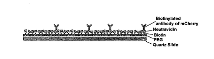
- the analyst then analyzed the first fluorescent protein, which is an antibody that binds to the first fluorescent protein expressed as a marker on the first protein in step S110, on a substrate that is a polyethyleneglycol (PEG) coated Quartz slide.
- the binding antibody is attached as in FIG. 2 (S120).
- the antibody which binds to the first fluorescent protein includes, in addition to the antibody, a biomolecular sieve that binds to a first fluorescent protein such as a liposome having a specific component that binds to DNA, RNA, and protein.
- a biomolecular sieve that binds to a first fluorescent protein such as a liposome having a specific component that binds to DNA, RNA, and protein.
- the analyst supplies the cytoplasmic stock solution of cells in which the first fluorescent protein is expressed in the internal first protein prepared in step S110 to the substrate (S130), thereby expressing the first fluorescent protein expressed in the first protein. Induces the binding of the first fluorescent protein binding antibody (S140).
- step S130 not only the cellular stock solution, but also a cell solution such as a cell stock solution, diluted cytoplasm stock solution, or diluted cell stock solution may be used.
- the analyzer determines the wavelength of the first fluorescent protein. From the change (ie, by measuring the individual monomolecular signals generated from the first fluorescent protein), it is possible to determine whether the first fluorescent protein is bound to the plurality of first fluorescent protein binding antibodies attached to the substrate.
- the predetermined protein expressed in the first protein for binding with the antibody attached to the substrate does not necessarily need to be a fluorescent protein. Since it is not possible to grasp through the total reflection microscope, the protein expressed in the first protein may be a fluorescent protein.
- the predetermined protein expressed in the first protein in the present invention performs the function of an antigen that binds to the antibody attached to the substrate.
- the predetermined antigen attached to the first protein must be a protein. It may be a compound capable of binding to the antibody.
- the protein expressed in the first protein is a fluorescent protein
- the analyst supplies a buffer solution to the substrate, thereby preparing the cytoplasmic stock except for the first protein expressing the first fluorescent protein.
- the remaining substances contained in the are removed on the substrate (S150).
- the analyst causes the second fluorescent protein to be expressed as a marker through genetic engineering to a second protein present in the cell in another cell identical to the cell in step S110 (S160).
- the second fluorescent protein may be attached or linked to the second protein by a physicochemical method.
- the above-described step S160 may be performed in advance with the above-described step S110 for the smooth progress of the analysis, in the practice of the present invention, since the second fluorescent protein preferably has a different wavelength range than the first fluorescent protein
- the second fluorescent protein may be an eGFP (enhanced Green Fluorescent Protein).
- step S160 the analyst supplies the entire cytoplasmic stock solution of the cell in which the second fluorescent protein is expressed in the second protein inside in step S160 (S170).
- step S170 not only the cellular stock solution, but also a cell solution such as a cell stock solution, diluted cellular stock solution, or diluted cell stock solution may be used.
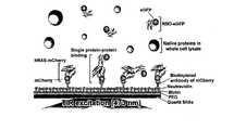
- the first protein is bound to the plurality of first fluorescent protein binding antibodies attached to the surface of the substrate, and when the entire cytoplasmic stock solution containing the second protein is supplied to the surface of the substrate, The first protein on the substrate surface interacts with the second protein in the same state as in the intracellular environment where the second protein in the cytoplasm and other proteins in whole cell lysate coexist. do.
- each first protein coupled via a first fluorescent protein to each antibody attached on the substrate surface as shown in FIG. 4 is at the single molecule level in the same state as in the second protein and the intracellular environment. There is an interaction of repeating joining and separating.
- the first protein and the second protein are detected by detecting a fluorescence signal of a specific wavelength band (520 nm) generated from the second fluorescent protein (eGFP) located on the surface of the substrate through the binding between the first protein and the second protein.
- the binding state can be confirmed, and the analyst continuously observes the change in wavelength of each antibody attached on the surface of the substrate so that the interaction between the first protein and the second protein, such as the frequency of binding and separation of the first protein Can be analyzed at the level (S180).
- the interaction between the first protein and the second protein such as the frequency of binding and separation of the cell It can be analyzed in the same environment as the environment.
- the second protein that is not bound to the first protein can also move near the surface of the substrate during the floating movement in the cellular stock solution supplied to the substrate, and even in this case, it is expressed in the second protein when the surface is observed through a total reflection microscope With the second fluorescent protein, eGFP, the wavelength at the substrate surface is changed from 473 nm to 520 nm, which may lead to the problem of an error in interaction analysis.
- the second protein which is not bound to the first protein and floats in the cytoplasmic stock solution, has a very high rate of movement in the cytoplasmic stock solution, so that even if the second protein temporarily approaches the surface of the substrate during the suspension movement, within the time interval of the integration measurement. This is because the substrate deviates from the substrate surface again.
- the second protein is treated so as not to bind with the first protein, so that all the second proteins are suspended in the cytosolic solution without the second protein bound to the first protein.
- the change As a result of integrating the change into an integration section of 50 msec, it was confirmed experimentally that no wavelength change was observed.
- the protein-protein interaction analysis value in the above-described step S180 is secured as the first analysis value, and the protein-protein interaction analysis value in another environment described later as the second analysis value. It may be possible to perform a comparative analysis of the two.
- the tester secures the above-described interaction analysis value in the above-described step S180 as the first analysis value, and then repeats the above-described steps S110 to S160 in the same manner.
- the tester does not supply the cytoplasmic stock solution of the cell in which the second fluorescent protein is expressed in the second protein therein to the substrate, but instead of the cytoplasmic stock solution of the cell.
- the mixed cytoplasmic stock solution obtained by mixing the cytoplasmic stock solution of another analysis target such as cancer cells is supplied to the substrate (S190).
- mixed cell solutions such as mixed cell stock, diluted mixed cytoplasm stock, or diluted mixed cell stock may be used.
- the first protein is bound to the plurality of first fluorescent protein binding antibodies attached to the surface of the substrate, respectively, and the cytoplasmic solution containing the second protein is mixed with the cytoplasmic stock solution to be analyzed.
- the mixed cytoplasmic stock solution is supplied to the surface of the substrate, the first protein on the substrate surface expresses not only the second protein expressing the second fluorescent protein contained in the mixed cytoplasm stock solution, but also the second fluorescent protein in the cell to be analyzed.
- the second protein which is not present, and other proteins (Native proteins in whole cell lysate) will interact with the second protein in a coexisting environment.
- the second protein in which the second fluorescent protein is expressed is competitive with the first protein on the substrate surface in the state of coexisting with the second protein in which the second fluorescent protein is not expressed in the cell to be analyzed. To interact.
- the second protein in which the second fluorescent protein is expressed by another protein in the cell to be analyzed is affected by the interaction with the first protein.
- the degree of influence by the other protein in the cell to be analyzed or the second protein in the cell to be analyzed may be determined by observing the surface of the substrate through an optical device that generates a near field such as a total reflection microscope as in the step S180 described above.
- the interactions such as the frequency of binding and separation of the first protein and the second protein on which the second fluorescent protein is expressed on the substrate surface can be confirmed by analyzing at a single molecule level (S195).
- the investigator compares the first analysis value described above with the second analysis value which is the analysis value at step S195 described above, and thereby the second protein in another cell in the interaction of the second protein with the first protein. And the extent of effects such as the promotion or interference of interaction by other proteins in other cells.
- FIG. 5 is a flowchart illustrating a method for analyzing protein-protein interaction according to another embodiment of the present invention.
- the first protein is not expressed in the first protein and is not a first fluorescent protein-binding antibody on the substrate.
- the interaction between the first protein and the second protein in the state in which the first protein is directly bound to the antibody attached to the substrate is analyzed.
- a first protein binding antibody which is an antibody bound to a first protein, is attached together to a substrate that is a polyethylene slide (Quartz Slide) coated with polyethylene glycol (PEG) (S210).
- a biomolecular sieve that binds to a first protein such as a liposome having DNA, RNA, a specific component that binds to the protein, or the like may be used as the antibody to bind to the first protein.
- a first protein such as a liposome having DNA, RNA, a specific component that binds to the protein, or the like
- the analyst induces the binding of the first protein and the first protein-binding antibody (S230) by supplying the substrate of the cytoplasm of the cell containing the first protein to the substrate (S220).
- step S220 not only the cellular stock solution, but also a cell solution such as a cell stock solution, diluted cytoplasm stock, or diluted cell stock may be used.
- the analyst may be pre-processed to express a predetermined fluorescent protein such as m-Cherry in the first protein, in this case the first protein binding antibody and the first protein is bound
- a predetermined fluorescent protein such as m-Cherry in the first protein
- the analyst observes the surface of the substrate by using a total reflection microscope
- the analyst analyzes the surface of the substrate using a total reflection microscope
- the analyzer analyzes the surface of the substrate and changes the wavelength of the first fluorescent protein expressed in the first protein. You can check whether it is combined.
- the analyst supplies a buffer solution to the substrate, thereby restoring the remaining substances contained in the cellular stock except for the first protein on the substrate. It will be removed (S240).
- the analyst manipulates the fluorescent protein to be expressed through genetic engineering to the second protein present in the cell in another cell identical to the cell described above (S250).
- step S250 may be performed before the above-mentioned step S210 to facilitate the progress of the analysis, in the practice of the present invention, it will be preferable that the fluorescent protein expressed in the second protein also eGFP.
- the analyst supplies the entire cytoplasmic stock solution of the cell in which the fluorescent protein is expressed in the internal second protein in step S250 (S260).
- a cell solution such as a cell stock solution, a diluted cellular stock solution, or a diluted cell stock solution may be used.
- the first protein is bound to the plurality of first protein-binding antibodies attached to the surface of the substrate, and when the whole cellular stock solution containing the second protein is supplied to the surface of the substrate, the first protein on the surface of the substrate is Interacts with the second protein contained in the cellular stock solution in the same state as in the intracellular environment.
- each first protein bound to each antibody attached on the substrate surface interacts with the second protein repeatedly binding and separation at the single molecule level in the same state as in the intracellular environment.
- eGFP is a fluorescent protein expressed in the second protein.
- the wavelength at the substrate surface is changed from 473 nm to 520 nm, and the change in the wavelength enables confirmation of the binding state between the first protein and the second protein.
- S270 single molecule level
- the protein-protein interaction analysis value in the above step S270 is ensured as the first analysis value, which will be described later Comparative analysis of the two may be performed by obtaining the protein-protein interaction analysis value in the other environment as the second analysis value.
- the tester secures the above-described interaction analysis value in the above-described step S270 as the first analysis value, and then repeats the above-described steps S210 to S250 in the same manner.
- the tester does not supply the cytoplasmic stock of the cell in which the second fluorescent protein is expressed in the second protein therein to the substrate, but not with the cytoplasmic stock of the cell.
- the mixed cytoplasmic stock solution which is a mixture of separate cytoplasmic stock solutions, such as cancer cells, is supplied to the substrate (S280).
- mixed cell solutions such as mixed cell stock, diluted mixed cytoplasm, or diluted mixed cell stock may be used.
- the first protein is bound to the plurality of first protein-binding antibodies attached to the surface of the substrate, and the mixed cytoplasmic solution containing the cytoplasmic solution containing the second protein and the cytoplasmic solution to be analyzed is the surface of the substrate.
- the first protein on the substrate surface is not only a second protein expressing the second fluorescent protein contained in the mixed cytoplasm stock solution, but also a second protein not expressing the second fluorescent protein in the cell to be analyzed and the like.
- Other proteins in the cell Native proteins in whole cell lysate will interact with the second protein in the environment.
- the second protein in which the second fluorescent protein is expressed is competitive with the first protein on the substrate surface in the state of coexisting with the second protein in which the second fluorescent protein is not expressed in the cell to be analyzed. To interact.
- the second protein in which the second fluorescent protein is expressed by another protein in the cell to be analyzed is affected by the interaction with the first protein.
- the degree of influence by the other protein in the cell to be analyzed or the second protein in the cell to be analyzed may be determined by observing the surface of the substrate through an optical device that generates a near field such as a total reflection microscope as in the step S270 described above.
- the interactions such as the frequency of binding and separation of the first protein on the substrate surface and the second protein on which the second fluorescent protein is expressed can be confirmed by analyzing at a single molecule level (S290).
- the investigator compares the first analysis value described above with the second analysis value which is the analysis value at step S290 described above, and thereby the second protein in another cell in interaction with the first protein of the second protein. And the extent of effects such as the promotion or interference of interaction by other proteins in other cells.
- FIGS. 1 to 5 the method for analyzing protein-protein interaction according to the present invention in FIGS. 1 to 5 described above is performed in an analysis apparatus having the structure in FIG. 6.
- the protein-protein interaction analysis device 300 includes an optical excitation unit 320, a sample loading unit 310, an optical measuring unit 330, a signal analyzing unit 340, and a signal.
- the diagnosis unit 360 and the storage unit 350 are included.
- the sample loading unit 310 is loaded with the substrate used in the protein-protein interaction analysis method described above in the present invention.
- the optical excitation portion 320 generates a near field having a first wavelength on the surface of the substrate loaded in the sample loading portion.
- the optical measuring unit 330 is the first protein on the substrate surface in accordance with the interaction of the first protein attached to the substrate surface and the second protein provided with a fluorescent protein in a state in which the cellular stock solution or mixed cytoplasm stock solution is supplied to the substrate Sensing a change in wavelength from the wavelength to the second wavelength.
- the first wavelength when the fluorescent protein included in the second protein is eGFP, the first wavelength will be 473 nm and the second wavelength will be 520 nm.
- the optical measuring unit 330 not only measures the fluorescence signal of the second wavelength for a predetermined time, but also measures the fluorescence signal of the second wavelength in real time in the presence of the cellular stock solution or the mixed cytoplasm stock solution, It would be desirable to have monomolecular sensitivity in the measurement.
- the signal analyzer 340 analyzes the fluorescence signal of the second wavelength measured by the optical measuring unit 330 through image processing to calculate an analysis value, and stores the calculated analysis value in the storage unit 350. .
- the signal analyzer 340 is individually based on the fluorescence signal of the second wavelength measured by the optical measuring unit 330 in each situation in which various cytoplasmic solutions such as cytoplasmic stock solution or mixed cytoplasmic stock solution are supplied to the substrate.
- the calculated analysis value is stored in the storage 350.
- the signal analyzer 340 may display the signal indicated by the red line in FIG. 7 among the measured fluorescence signals. Find the baseline.
- the signal low rises between approximately 10 to 20 seconds, which means that the cellular stock solution or the mixed cytoplasmic stock containing the second protein arrives at the imaging area of the optical measuring unit 330.
- the signal analyzer 340 measures fluctuation, ie, noise, which occurs around an elevated signal baseline, and then defines meaningful protein-protein interactions based on the fluctuation. Set the threshold.
- the noise generated around the signal low is formed by the behavior of the second proteins that are continuously moving quickly without interaction between the proteins, and the signal analyzer 340 may be exceeded by the noise that is not related to the interaction. Set the reference point to an unknown value.
- the signal analyzer 340 determines that there is a meaningful interaction between the first protein and the second protein at the corresponding time point, and the duration of the interaction ( ⁇ off ), and The time until the next binding between the first protein and the second protein ⁇ on is measured and stored in the storage 350 as an analysis value.
- the analysis of the protein-protein interaction through the analysis of the time ( ⁇ off ) and the time until the next binding of the first protein and the second protein ( ⁇ on ) obtained by the signal analyzer 340 is performed.
- a series of information can be obtained, and the signal diagnosis unit 360 compares and analyzes the analytical values calculated in each situation in which various cytoplasmic solutions are supplied to the substrate.
- the medical diagnosis is performed according to a predetermined diagnosis algorithm.
Landscapes
- Health & Medical Sciences (AREA)
- Life Sciences & Earth Sciences (AREA)
- Immunology (AREA)
- Engineering & Computer Science (AREA)
- Chemical & Material Sciences (AREA)
- Molecular Biology (AREA)
- Physics & Mathematics (AREA)
- Biomedical Technology (AREA)
- Hematology (AREA)
- Urology & Nephrology (AREA)
- Analytical Chemistry (AREA)
- Biochemistry (AREA)
- General Health & Medical Sciences (AREA)
- General Physics & Mathematics (AREA)
- Pathology (AREA)
- Medicinal Chemistry (AREA)
- Food Science & Technology (AREA)
- Biotechnology (AREA)
- Cell Biology (AREA)
- Microbiology (AREA)
- Nuclear Medicine, Radiotherapy & Molecular Imaging (AREA)
- Chemical Kinetics & Catalysis (AREA)
- Proteomics, Peptides & Aminoacids (AREA)
- Oncology (AREA)
- Hospice & Palliative Care (AREA)
- Bioinformatics & Cheminformatics (AREA)
- Bioinformatics & Computational Biology (AREA)
- Biophysics (AREA)
- Optics & Photonics (AREA)
- Plasma & Fusion (AREA)
- Investigating, Analyzing Materials By Fluorescence Or Luminescence (AREA)
- Investigating Or Analysing Materials By The Use Of Chemical Reactions (AREA)
- Measuring Or Testing Involving Enzymes Or Micro-Organisms (AREA)
- Apparatus Associated With Microorganisms And Enzymes (AREA)
- Investigating Or Analysing Biological Materials (AREA)
Abstract
단일 분자 수준까지의 단백질-단백질 상호작용 분석 방법 및 분석 장치가 개시된다. 본 발명은, 제1 단백질에 구비된 제1 표지자와 결합되는 생체 분자체가 부착된 기판에 제1 단백질을 공급하여 제1 표지자와 생체 분자체의 결합을 유도하고, 제1 표지자와 생체 분자체가 결합된 경우에, 제2 표지자가 구비된 제2 단백질이 포함된 세포의 세포 용액을 기판에 공급하며, 세포 용액이 기판에 공급된 상태에서, 제1 단백질과 제2 단백질의 상호작용을 분석하는 과정을 통해 구현된다. 본 발명에 따르면, 실제 세포 환경 내에서의 단백질-단백질 상호작용을 단일 분자 수준까지 분석할 수 있게 된다.
Description
본 발명은 단백질-단백질 상호작용 분석 방법에 관한 것으로, 더욱 상세하게는 실제 세포 환경 내에서의 단백질-단백질 상호작용을 단일 분자 수준까지 분석할 수 있을 뿐만 아니라, 실제 세포 환경 내에서 다른 단백질이 관여된 경우에 특정 단백질-단백질 상호작용에 대한 영향도를 분석할 수 있는 단백질-단백질 상호작용 분석 방법 및 분석 장치에 관한 것이다.
세포는 다양하고 복잡한 단백질-단백질 상호작용을 통하여 유전자 표현, 세포성장, 세포 주기, 대사, 신호전달 등의 여러 생물학적 기능을 수행함으로써 생명현상을 유지하고 있다. 따라서 세포내에서의 단백질-단백질 상호작용 및 상호작용의 기능을 이해하는 것은 생명현상들을 이해하는 초석이 되며 신약개발 및 질병 치료의 중요한 기반이 된다.
시험관 내에서 단백질-단백질 상호작용을 조사하기 위한 방법으로는 대표적인 방법으로는 친화성 크로마토 그래피(affinity chromoatography) 방법이 있다.
단백질 친화성 크로마토그래피(Protein affinity chromatography)의 경우, 정제된 단백질이 준비되어야하는 어려움이 있다. 또한 단백질 간 상호작용 확인이 시험관 내에서 일어나므로, 세포내에서는 상호작용하지 않는 단백질들이 컬럼을 통과하는 동안 정전기적 상호작용에 의하여 결합하는 것처럼 보일 수 있는 false-positive 결과를 도출 할 수 있다.
즉, 정량적 측정을 위해서 종래 기술에 따른 단백질-단백질 상호작용을 조사하기 위한 방법은 단백질-단백질 상호작용을 분석하기 위해서 세포 내에 존재하는 각각의 단백질을 정제하여 이들을 세포 내의 다른 물질들과 분리한 상태에서 상호작용을 분석하기 때문에, 다른 단백질 등이 혼재되어 있는 실제 세포 환경 내에서의 단백질-단백질 상호작용을 단일 분자 수준에서 분석할 수는 없다는 한계가 있다.
뿐만 아니라, 종래 기술에 따른 단백질-단백질 상호작용을 조사하기 위한 방법은 실제 세포 환경 내에서 기타의 다른 단백질이 관여된 경우에 특정 단백질-단백질 상호작용에 대한 영향도를 분석할 수 없다는 한계가 있다.
따라서, 본 발명의 목적은, 실제 세포 환경 내에서의 단백질-단백질 상호작용을 단일 분자 수준까지 분석할 수 있을 뿐만 아니라, 실제 세포 환경 내에서 다른 단백질이 관여된 경우에 특정 단백질-단백질 상호작용에 대한 영향도를 분석할 수 있는 단백질-단백질 상호작용 분석 방법 및 분석 장치를 제공함에 있다.
상기 목적을 달성하기 위한 본 발명에 따른 단백질-단백질 상호작용 분석 방법은, 제1 단백질과 제2 단백질의 상호작용을 단일 분자 수준까지 분석하기 위한 방법에 있어서, (a) 상기 제1 단백질에 구비된 제1 표지자와 결합되는 생체 분자체가 부착된 기판에 상기 제1 단백질을 공급하여 상기 제1 표지자와 상기 생체 분자체의 결합을 유도하는 단계; (b) 상기 제1 표지자와 상기 생체 분자체가 결합된 경우에, 제2 표지자가 구비된 제2 단백질이 포함된 세포의 세포 용액을 상기 기판에 공급하는 단계; 및 (c) 상기 세포 용액이 상기 기판에 공급된 상태에서, 상기 제1 단백질과 상기 제2 단백질의 상호작용을 분석하는 단계를 포함한다.
한편, 본 발명에 따른 단백질-단백질 상호작용 분석 방법은, 제1 단백질과 제2 단백질의 상호작용을 단일 분자 수준까지 분석하기 위한 방법에 있어서, (a) 상기 제1 단백질이 결합되는 생체 분자체인 제1 단백질 결합 분자체가 부착된 기판에 상기 제1 단백질을 공급하여 상기 제1 단백질과 상기 제1 단백질 결합 분자체의 결합을 유도하는 단계; (b) 상기 제1 단백질과 상기 제1 단백질 결합 분자체가 결합된 경우에, 표지자가 구비된 제2 단백질이 포함된 세포의 세포 용액을 상기 기판에 공급하는 단계; 및 (c) 상기 세포 용액이 상기 기판에 공급된 상태에서, 상기 제1 단백질과 상기 제2 단백질의 상호작용을 분석하는 단계를 포함한다.
또한, 상기 (c) 단계는, 상기 제1 단백질과 결합된 상기 제2 단백질에 구비된 표지자에 의해 발생하는 특정 파장의 형광 신호를 근접장을 발생시키는 광학장치를 이용하여 측정하는 단계를 포함한다.
또한, 상기 (c) 단계는, 상기 특정 파장의 형광 신호를 소정의 시간 동안 적분 측정하는 것을 특징으로 한다.
또한, 상기 (c) 단계는, 상기 기판에 상기 공급된 세포 용액이 존재하는 상황에서, 상기 특정 파장의 형광 신호를 실시간 측정하는 것을 특징으로 한다.
또한, 상기 (a) 단계는, 상기 제1 단백질이 포함된 세포의 세포 용액을 상기 기판에 공급하는 단계를 포함하는 것을 특징으로 한다.
또한, 상기 (b) 단계 이전에, 상기 기판에 버퍼 용액을 공급하는 단계를 더 포함한다.
또한, 상기 제1 표지자, 및 상기 제2 표지자는 서로 다른 파장을 갖는 형광 단백질인 것을 특징으로 한다.
한편, 본 발명에 따른 단백질-단백질 상호작용 분석 방법은, 제1 단백질과 제2 단백질의 상호작용을 단일 분자 수준까지 분석하기 위한 방법에 있어서, (a) 상기 제1 단백질에 구비된 제1 표지자와 결합되는 생체 분자체가 부착된 기판에 상기 제1 단백질을 공급하여 상기 제1 표지자와 상기 생체 분자체의 결합을 유도하는 단계; (b) 상기 제1 표지자와 상기 생체 분자체가 결합된 경우에, 제2 표지자가 구비된 제2 단백질이 포함된 세포의 세포 용액을 상기 기판에 공급하는 단계; (c) 상기 세포 용액이 상기 기판에 공급된 상태에서, 상기 제1 단백질과 상기 제2 단백질의 상호작용을 분석하여 제1 분석값을 획득하는 단계; (d) 상기 (a) 단계에서의 상기 제1 표지자와 상기 생체 분자체가 결합된 상태의 기판과 동일한 기판에 상기 (b) 단계에서의 상기 세포 용액과 분석 대상 세포 용액을 혼합한 혼합 세포 용액을 공급하는 단계; (e) 상기 혼합 세포 용액이 상기 기판에 공급된 상태에서, 상기 제1 단백질과 상기 제2 단백질의 상호작용을 분석하여 제2 분석값을 획득하는 단계; 및 (f) 상기 제1 분석값과 상기 제2 분석값을 비교분석하는 단계를 포함한다.
한편, 본 발명에 따른 단백질-단백질 상호작용 분석 방법은, 제1 단백질과 제2 단백질의 상호작용을 단일 분자 수준까지 분석하기 위한 방법에 있어서, (a) 상기 제1 단백질이 결합되는 생체 분자체인 제1 단백질 결합 분자체가 부착된 기판에 상기 제1 단백질을 공급하여 상기 제1 단백질과 상기 제1 단백질 결합 분자체의 결합을 유도하는 단계; (b) 상기 제1 단백질과 상기 제1 단백질 결합 분자체가 결합된 경우에, 표지자가 구비된 제2 단백질이 포함된 세포의 세포 용액을 상기 기판에 공급하는 단계; 및 (c) 상기 세포 용액이 상기 기판에 공급된 상태에서, 상기 제1 단백질과 상기 제2 단백질의 상호작용을 분석하여 제1 분석값을 획득하는 단계; (d) 상기 (a) 단계에서의 상기 제1 단백질과 상기 제1 단백질 결합 분자체가 결합된 기판과 동일한 기판에 상기 (b) 단계에서의 상기 세포 용액과 분석 대상 세포 용액을 혼합한 혼합 세포 용액을 공급하는 단계; (e) 상기 혼합 세포 용액이 상기 기판에 공급된 상태에서, 상기 제1 단백질과 상기 제2 단백질의 상호작용을 분석하여 제2 분석값을 획득하는 단계; 및 (f) 상기 제1 분석값과 상기 제2 분석값을 비교분석하는 단계를 포함한다.
바람직하게는, 상기 (c) 단계는, 상기 제1 단백질과 결합된 상기 제2 단백질에 구비된 표지자에 의해 발생하는 특정 파장의 형광 신호를 근접장을 발생시키는 광학장치를 이용하여 측정하는 단계를 포함하는 것을 특징으로 한다.
또한, 상기 (c) 단계는, 상기 특정 파장의 형광 신호를 소정의 시간 동안 적분 측정하는 것을 특징으로 한다.
또한, 상기 (c) 단계는, 상기 기판에 상기 공급된 세포 용액이 존재하는 상황에서, 상기 특정 파장의 형광 신호를 실시간 측정하는 것을 특징으로 한다.
또한, 상기 (a) 단계는, 상기 제1 단백질이 포함된 세포의 세포 용액을 상기 기판에 공급하는 단계를 포함하는 것을 특징으로 한다.
또한, 상기 (b) 단계 이전에, 상기 기판에 버퍼 용액을 공급하는 단계를 더 포함한다.
또한, 상기 제1 표지자, 및 상기 제2 표지자는 서로 다른 파장을 갖는 형광 단백질인 것을 특징으로 한다.
상기 목적을 달성하기 위한 본 발명에 따른 단백질-단백질 상호작용 분석 장치는, 제1 단백질과 제2 단백질의 상호작용을 단일 분자 수준까지 분석하기 위한 장치에 있어서, 상기 제1 단백질에 구비된 제1 표지자와 결합되는 생체 분자체가 부착된 기판이 로딩되는 시료 로딩부 -상기 기판에 상기 제1 단백질을 공급하여 상기 제1 표지자와 상기 생체 분자체의 결합이 유도되며, 상기 제1 표지자와 상기 생체 분자체가 결합된 경우에, 제2 표지자가 구비된 제2 단백질이 포함된 세포의 세포 용액이 상기 기판에 공급됨-; 상기 시료 로딩부에 로딩된 상기 기판의 표면에 제1 파장을 갖는 근접장을 발생시키는 광학 여기부; 및 상기 세포 용액이 상기 기판에 공급된 상태에서, 상기 제1 단백질과 상기 제2 단백질의 상호작용에 따른 상기 기판 표면에서의 상기 제1 파장의 제2 파장으로의 변화를 감지하는 광학 측정부를 포함한다.
한편, 본 발명에 따른 단백질-단백질 상호작용 분석 장치는, 제1 단백질과 제2 단백질의 상호작용을 단일 분자 수준까지 분석하기 위한 장치에 있어서, 상기 제1 단백질이 결합되는 생체 분자체인 제1 단백질 결합 분자체가 부착된 기판이 로딩되는 시료 로딩부 -상기 기판에 상기 제1 단백질을 공급하여 상기 제1 단백질과 상기 제1 단백질 결합 분자체의 결합을 유도하고, 상기 제1 단백질과 상기 제1 단백질 결합 분자체가 결합된 경우에, 표지자가 구비된 제2 단백질이 포함된 세포의 세포 용액이 상기 기판에 공급됨-; 상기 시료 로딩부에 로딩된 상기 기판의 표면에 제1 파장을 갖는 근접장을 발생시키는 광학 여기부; 및 상기 세포 용액이 상기 기판에 공급된 상태에서, 상기 제1 단백질과 상기 제2 단백질의 상호작용에 따른 상기 기판 표면에서의 상기 제1 파장의 제2 파장으로의 변화를 감지하는 광학 측정부를 포함한다.
바람직하게는, 상기 광학 측정부는, 상기 제2 파장의 형광 신호를 소정의 시간 동안 적분 측정하는 것을 특징으로 한다.
또한, 상기 광학 측정부는, 상기 기판에 상기 공급된 세포 용액이 존재하는 상황에서, 상기 제2 파장의 형광 신호를 실시간 측정하는 것을 특징으로 한다.
또한, 상기 제1 표지자, 및 상기 제2 표지자는 서로 다른 파장을 갖는 형광 단백질인 것을 특징으로 한다.
또한, 상기 광학 측정부가 측정한 상기 제2 파장의 형광 신호를 이미지 프로세싱을 통해 분석하는 신호 분석부를 더 포함하는 것을 특징으로 한다.
한편, 본 발명에 따른 단백질-단백질 상호작용 분석 장치는, 제1 단백질과 제2 단백질의 상호작용을 단일 분자 수준까지 분석하기 위한 장치에 있어서, 상기 제1 단백질에 구비된 제1 표지자와 결합되는 생체 분자체가 부착된 기판이 로딩되는 시료 로딩부 -상기 기판에 상기 제1 단백질을 공급하여 상기 제1 표지자와 상기 생체 분자체의 결합을 유도하고, 상기 제1 표지자와 상기 생체 분자체가 결합된 경우에, 제2 표지자가 구비된 제2 단백질이 포함된 세포의 세포 용액이 상기 기판에 공급됨-; 상기 시료 로딩부에 로딩된 상기 기판의 표면에 제1 파장을 갖는 근접장을 발생시키는 광학 여기부; 상기 세포 용액이 상기 기판에 공급된 상태에서, 상기 제1 단백질과 상기 제2 단백질의 상호작용에 따른 상기 기판 표면에서의 상기 제1 파장의 제2 파장으로의 변화를 감지하는 광학 측정부; 및 상기 광학 측정부가 측정한 상기 제2 파장의 형광 신호를 이미지 프로세싱을 통해 분석하여 제1 분석값을 획득하는 신호 분석부를 포함하며, 상기 시료 로딩부에는 상기 제1 단백질에 구비된 제1 표지자와 결합되는 생체 분자체가 부착된 새로운 기판이 로딩되고, 상기 새로운 기판에 상기 세포 용액과 분석 대상 세포 용액을 혼합한 혼합 세포 용액이 공급되고, 상기 혼합 세포 용액이 상기 새로운 기판에 공급된 상태에서, 상기 광학 측정부는 상기 제1 단백질과 상기 제2 단백질의 상호작용에 따른 상기 기판 표면에서의 상기 제1 파장의 제2 파장으로의 변화를 감지하며, 상기 신호 분석부는 상기 광학 측정부가 측정한 상기 제2 파장의 형광 신호를 이미지 프로세싱을 통해 분석하여 제2 분석값을 획득하는 것을 특징으로 한다.
한편, 본 발명에 따른 단백질-단백질 상호작용 분석 장치는, 제1 단백질과 제2 단백질의 상호작용을 단일 분자 수준까지 분석하기 위한 장치에 있어서, 상기 제1 단백질이 결합되는 생체 분자체인 제1 단백질 결합 분자체가 부착된 기판이 로딩되는 시료 로딩부 -상기 기판에 상기 제1 단백질을 공급하여 상기 제1 단백질과 상기 제1 단백질 결합 분자체의 결합을 유도하며, 상기 제1 단백질과 상기 제1 단백질 결합 분자체가 결합된 경우에, 표지자가 구비된 제2 단백질이 포함된 세포의 세포 용액이 상기 기판에 공급됨-; 상기 시료 로딩부에 로딩된 상기 기판의 표면에 제1 파장을 갖는 근접장을 발생시키는 광학 여기부; 상기 세포 용액이 상기 기판에 공급된 상태에서, 상기 제1 단백질과 상기 제2 단백질의 상호작용에 따른 상기 기판 표면에서의 상기 제1 파장의 제2 파장으로의 변화를 감지하는 광학 측정부; 및 상기 광학 측정부가 측정한 상기 제2 파장의 형광 신호를 이미지 프로세싱을 통해 분석하여 제1 분석값을 획득하는 신호 분석부를 포함하며, 상기 시료 로딩부에는 상기 제1 단백질이 결합되는 생체 분자체인 제1 단백질 결합 분자체가 부착된 새로운 기판이 로딩되고, 상기 새로운 기판에 상기 세포 용액과 분석 대상 세포 용액을 혼합한 혼합 세포 용액이 공급되고, 상기 혼합 세포 용액이 상기 새로운 기판에 공급된 상태에서, 상기 광학 측정부는 상기 제1 단백질과 상기 제2 단백질의 상호작용에 따른 상기 기판 표면에서의 상기 제1 파장의 제2 파장으로의 변화를 감지하며, 상기 신호 분석부는 상기 광학 측정부가 측정한 상기 제2 파장의 형광 신호를 이미지 프로세싱을 통해 분석하여 제2 분석값을 획득하는 것을 특징으로 한다.
바람직하게는, 상기 제1 분석값과 상기 제2 분석값을 비교분석하는 신호 진단부를 더 포함하는 것을 특징으로 한다.
또한, 상기 광학 측정부는, 상기 제2 파장의 형광 신호를 소정의 시간 동안 적분 측정하는 것을 특징으로 한다.
또한, 상기 광학 측정부는, 상기 기판에 상기 공급된 세포 용액이 존재하는 상황에서, 상기 제2 파장의 형광 신호를 실시간 측정하는 것을 특징으로 한다.
또한, 상기 제1 표지자, 및 상기 제2 표지자는 서로 다른 파장을 갖는 형광 단백질인 것을 특징으로 한다.
본 발명에 따르면, 실제 세포 환경 내에서의 단백질-단백질 상호작용을 단일 분자 수준까지 분석할 수 있게 된다.
아울러, 본 발명에 따르면, 실제 세포 환경 내에서 다른 단백질이 관여된 경우에 특정 단백질-단백질 상호작용에 대한 영향도를 분석할 수 있게 된다.
도 1은 본 발명의 일 실시예에 따른 단백질-단백질 상호작용 분석 방법을 설명하는 절차 흐름도,
도 2 내지 도 4는 본 발명의 일 실시예에 따른 단백질-단백질 상호작용 분석 방법의 각 단계를 설명하는 도면, 및
도 5는 본 발명의 다른 실시예에 따른 단백질-단백질 상호작용 분석 방법을 설명하는 절차 흐름도,
도 6은 도 1 내지 도 5에서의 단백질-단백질 상호작용 분석 방법이 실행되는 분석 장치의 구조를 나타내는 기능 블록도, 및
도 7은 본 발명에 따른 단백질-단백질 상호작용 분석 장치의 광학 측정부가 측정한 신호를 나타낸 도면이다.
이하에서는 도면을 참조하여 본 발명을 보다 상세하게 설명한다. 도면들 중 동일한 구성요소들은 가능한 한 어느 곳에서든지 동일한 부호들로 나타내고 있음에 유의해야 한다. 또한 본 발명의 요지를 불필요하게 흐릴 수 있는 공지 기능 및 구성에 대한 상세한 설명은 생략한다.
도 1은 본 발명의 일 실시예에 따른 단백질-단백질 상호작용 분석 방법을 설명하는 절차 흐름도이다. 도 1을 참조하여, 본 발명의 일 실시예에 따른 단백질-단백질 상호작용 분석 방법을 설명하면, 먼저, 분석자는 특정한 두 개의 단백질인 제1 단백질과 제2 단백질의 상호작용을 단일 분자 수준까지 분석하기 위해서, 세포 상태에서 유전자 조작 과정을 거쳐 해당 세포 내에 존재하는 제1 단백질에 표지자로서 제1 형광 단백질이 발현되도록 한다(S110). 한편, 본 발명을 실시함에 있어서는, 제1 형광 단백질을 물리 화학적인 방법에 의해서 제1 단백질에 부착 또는 연결시킬 수도 있을 것이다.
본 발명에 따른 실험에 있어서, 제1 단백질은 h-Ras 단백질이고, 제2 단백질은 C-Raf의 Ras-binding domain(RBD) 단백질이며, 제1 형광 단백질은 m-Cherry 단백질로 하였다.
그 다음 분석자는 폴리에틸렌 글리콜(polyethyleneglycol:PEG) 코팅처리된 퀄츠 슬라이드(Quartz Slide)인 기판에, 전술한 S110 단계에서 제1 단백질에 표지자로서 발현된 제1 형광 단백질과 결합되는 항체인 제1 형광 단백질 결합항체를 도 2에서와 같이 부착한다(S120).
한편, 본 발명을 실시함에 있어서는, 제1 형광 단백질과 결합하는 항체에는, 항체 이외에도 DNA, RNA, 단백질과 결합하는 특정 성분을 갖는 리포좀(liposome) 등의 제1 형광 단백질과 결합하는 생체 분자체가 사용될 수 있을 것이다.
그 다음, 분석자는 전술한 S110 단계에서 준비해 놓은, 내부의 제1 단백질에 제1 형광 단백질이 발현되어 있는 세포의 세포질 원액을 기판에 공급함으로써(S130), 제1 단백질에 발현된 제1 형광 단백질과 제1 형광 단백질 결합항체의 결합을 유도한다(S140).
한편, 전술한 S130 단계를 실시함에 있어서 세포질 원액뿐만 아니라, 세포 원액, 희석된 세포질 원액, 또는 희석된 세포 원액 등의 세포 용액이 사용될 수 있을 것이다.
도 3에서와 같이, 기판상에 부착된 제1 형광 단백질 결합항체와 제1 형광 단백질이 결합된 경우에 분석자가 전반사 현미경을 이용한 기판의 표면 관찰을 수행하면, 분석자는 제1 형광 단백질에 의한 파장 변화로부터(즉, 제1 형광 단백질로부터 발생하는 개개의 단분자 신호를 측정하여) 기판상에 부착된 복수개의 제1 형광 단백질 결합항체에 제1 형광 단백질이 결합되었는지 여부를 확인할 수 있게 된다.
한편, 본 발명을 실시함에 있어서는, 기판에 부착된 항체와의 결합을 위해 제1 단백질에 발현된 소정의 단백질은 반드시 형광 단백질일 필요는 없을 것이나, 형광 단백질이 아닌 경우에는 항체와의 결합여부를 전반사 현미경을 통해 파악할 수 없는 관계로 제1 단백질에 발현된 단백질은 형광 단백질이 되도록 함이 바람직할 것이다.
즉, 본 발명에서의 제1 단백질에 발현된 소정의 단백질은 기판에 부착된 항체와의 결합을 하는 항원의 기능을 수행하는 것으로서, 이와 같이 제1 단백질에 부착된 소정의 항원은 반드시 단백질일 필요는 없으며, 항체와의 결합이 가능한 화합물이 될 수도 있을 것이다.
즉, 제1 단백질에 발현된 단백질이 형광 단백질이 형광 단백질인 경우에는 항체와의 결합여부를 전반사 현미경을 통해 파악할 수 있으므로, 기판에 부착된 항체와 결합된 형광 단백질의 개수 및 그에 따른 결합 밀도를 정확하게 측정할 수 있게 되는 것이다.
기판상에 부착된 복수개의 제1 형광 단백질 결합항체에 각각 제1 형광 단백질이 결합된 것으로 확인되면, 분석자는 기판에 버퍼 용액을 공급함으로써, 제1 형광 단백질이 발현된 제1 단백질을 제외한 세포질 원액에 포함되어 있는 나머지 물질들을 기판상에서 제거하게 된다(S150).
그 다음, 분석자는 전술한 S110 단계에서의 세포와 동일한 다른 세포에서 해당 세포에 존재하는 제2 단백질에 유전자 조작을 통해 표지자로서 제2 형광 단백질이 발현되도록 한다(S160). 한편, 본 발명을 실시함에 있어서는, 제2 형광 단백질을 물리 화학적인 방법에 의해서 제2 단백질에 부착 또는 연결시킬 수도 있을 것이다.
한편, 전술한 S160 단계는 원활한 분석의 진행을 위해 전술한 S110 단계와 함께 미리 실행할 수도 있을 것이며, 본 발명을 실시함에 있어서, 제2 형광 단백질은 제1 형광 단백질과 다른 파장역역을 갖는 것이 바람직하므로, 제1 형광 단백질이 m-Cherry 단백질인 경우에 제2 형광 단백질은 녹색형광 단백질인 eGFP(enhanced Green Fluorescent Protein)가 될 수 있을 것이다.
그 다음, 분석자는 전술한 S160 단계에서의 내부의 제2 단백질에 제2 형광 단백질이 발현되어 있는 세포의 세포질 원액 전체를 기판에 공급한다(S170).
한편, 전술한 S170 단계를 실시함에 있어서 세포질 원액뿐만 아니라, 세포 원액, 희석된 세포질 원액, 또는 희석된 세포 원액 등의 세포 용액이 사용될 수 있을 것이다.
기판의 표면에 부착되어 있는 복수개의 제1 형광 단백질 결합항체에는 각각 제1 형광 단백질을 통해 제1 단백질이 결합되어 있으며, 이와 같은 기판의 표면에 제2 단백질이 포함되어 있는 세포질 원액 전체가 공급되면, 기판 표면의 제1 단백질은 세포질 원액에 포함되어 있는 제2 단백질 및 세포 내의 다른 단백질들(Native proteins in whole cell lysate)이 공존하는 세포내 환경에서와 동일한 상태에서 제2 단백질과 상호 작용을 하게 된다.
보다 구체적으로, 도 4에서와 같이 기판 표면상에 부착된 각각의 항체에 제1 형광 단백질을 통해 결합되어 있는 각각의 제1 단백질은 제2 단백질과 세포내 환경에서와 동일한 상태에서 단일 분자 수준에서 결합과 분리를 반복하는 상호 작용을 하게 된다.
도 4에서와 같이 제1 단백질과 제2 단백질 간의 단일 분자 수준에서의 결합이 있는 경우에 분석자가 473nm의 파장을 갖는 전반사 현미경 등의 근접장을 발생시키는 광학 장치를 통해 기판의 표면을 관찰하면, 제2 단백질에 발현되어 있는 제2 형광 단백질인 eGFP에 의해, 기판 표면에서의 파장이 473nm에서 520nm로 변화한 것을 확인할 수 있다.
즉, 제1 단백질과 제2 단백질 간의 결합을 통해 기판 표면에 위치하게 된 제2 형광 단백질(eGFP)로부터 발생되는 특정 파장대역(520nm)의 형광 신호의 검출을 통해 제1 단백질과 제 2단백질의 결합상태를 확인할 수 있게 되고, 분석자는 기판 표면상에 부착된 각각의 항체에서의 파장의 변화를 지속적으로 관찰함으로써, 제1 단백질과 제2 단백질의 결합과 분리의 빈도 등의 상호작용을 단분자 수준에서 분석할 수 있게 된다(S180).
아울러, 본 발명에서는 전술한 S170 단계에서 기판에 공급된 세포질 원액이 존재하는 상황에서, 특정 파장의 형광 신호를 실시간 측정함으로써 제1 단백질과 제2 단백질의 결합과 분리의 빈도 등의 상호작용을 세포 환경과 동일한 환경에서 분석할 수 있게 되는 것이다.
한편, 제1 단백질과 결합되지 않은 제2 단백질도 기판에 공급된 세포질 원액 내에서 부유이동하는 과정에서, 기판 표면 근처로 이동할 수 있으며, 이러한 경우에도 전반사 현미경을 통한 표면 관찰시 제2 단백질에 발현되어 있는 제2 형광 단백질인 eGFP에 의해, 기판 표면에서의 파장이 473nm에서 520nm로 변화하게 됨으로써, 상호작용 분석의 오류를 초래할 수 있다는 문제점이 제기될 수 있을 것이다.
따라서, 본 발명을 실시함에 있어서는, 전반사 현미경을 통해 기판 표면에서의 파장 변화를 측정하는 경우에 파장 변화를 소정의 시간 동안 적분 측정하는 것이 바람직할 것이다.
제1 단백질과 결합되지 않고 세포질 원액 내를 부유하는 제2 단백질은 세포질 원액내에서의 이동속도가 대단히 빠르므로, 제2 단백질이 부유 이동 중에 일시적으로 기판 표면에 접근하였더라도 적분 측정의 시간 구간 내에서 다시 기판 표면으로부터 이탈하게 되기 때문이다.
이를 검증하기 위해, 제2 단백질을 제1 단백질과 결합되지 않도록 처리함으로써, 제1 단백질과 결합되는 제2 단백질이 없이 모든 제2 단백질이 세포질 원액내를 부유 이동하게 되는 상태에서 기판 표면에서의 파장 변화를 50msec의 적분 구간으로 적분 측정한 결과, 파장 변화가 전혀 관측되지 않음을 실험적으로 확인하였다.
한편, 본 발명을 실시함에 있어서는, 전술한 S180 단계에서의 단백질-단백질 상호 작용 분석값을 제1 분석값으로 확보하고, 후술하는 다른 환경에서의 단백질-단백질 상호 작용 분석값을 제2 분석값으로 확보하여 양자의 비교 분석을 수행할 수도 있을 것이다.
즉, 시험자는 전술한 S180 단계에서의 상호 작용 분석값을 제1 분석값으로 확보한 후에, 전술한 S110 단계 내지 S160 단계까지를 동일하게 반복하여 실시한다.
한편, 시험자는 전술한 S170 단계를 실행함에 있어서, 전술한 S160 단계에서 내부의 제2 단백질에 제2 형광 단백질이 발현되어 있는 세포의 세포질 원액을 기판에 공급하는 것이 아니라, 해당 세포의 세포질 원액과 암세포 등의 별도의 분석 대상 세포질 원액을 혼합한 혼합 세포질 원액을 기판에 공급하게 된다(S190).
한편, 전술한 S190 단계를 실시함에 있어서 혼합 세포질 원액뿐만 아니라, 혼합 세포 원액, 희석된 혼합 세포질 원액, 또는 희석된 혼합 세포 원액 등의 혼합 세포 용액이 사용될 수 있을 것이다.
즉, 기판의 표면에 부착되어 있는 복수개의 제1 형광 단백질 결합항체에는 각각 제1 형광 단백질을 통해 제1 단백질이 결합되어 있으며, 제2 단백질이 포함되어 있는 세포질 원액과 분석 대상 세포질 원액을 혼합한 혼합 세포질 원액이 기판의 표면에 공급되면, 기판 표면의 제1 단백질은 혼합 세포질 원액에 포함되어 있는 제2 형광 단백질이 발현되어 있는 제2 단백질 뿐만 아니라, 분석 대상 세포 내의 제2 형광 단백질이 발현되어 있지 않은 제2 단백질 및 기타의 다른 단백질들(Native proteins in whole cell lysate)이 공존하는 환경에서 제2 단백질과 상호 작용을 하게 된다.
즉, 전술한 S180 단계에서와는 달리, 제2 형광 단백질이 발현되어 있는 제2 단백질은 분석 대상 세포 내의 제2 형광 단백질이 발현되어 있지 않은 제2 단백질과 공존하는 상태에서 기판 표면의 제1 단백질과 경쟁적으로 상호 작용을 하게 된다. 뿐만 아니라, 분석 대상 세포 내의 다른 단백질에 의해 제2 형광 단백질이 발현되어 있는 제2 단백질은 제1 단백질과의 상호 작용에 있어 영향을 받게 된다.
이와 같은 분석 대상 세포 내의 다른 단백질 또는 분석 대상 세포 내의 제2 단백질에 의한 영향의 정도는, 시험자가 전술한 S180 단계에서와 같이 전반사 현미경 등의 근접장을 발생시키는 광학 장치를 통해 기판의 표면을 관찰하여, 기판 표면의 제1 단백질과 제2 형광 단백질이 발현되어 있는 제2 단백질의 결합과 분리의 빈도 등의 상호작용을 단분자 수준에서 분석함으로써 확인할 수 있게 된다(S195).
보다 구체적으로, 시험자는 상술한 제1 분석값과 전술한 S195 단계에서의 분석값인 제2 분석값을 비교분석함으로써 제2 단백질의 제1 단백질과의 상호 작용에 있어서의 다른 세포 내의 제2 단백질 및 다른 세포 내의 기타의 단백질에 의한 상호 작용의 촉진 또는 방해 등의 영향 정도를 분석할 수 있게 될 것이다.
한편, 도 5는 본 발명의 다른 실시예에 따른 단백질-단백질 상호작용 분석 방법을 설명하는 절차 흐름도이다. 도 5에서의 본 발명의 다른 실시예에 있어서는 도 1에서의 본 발명의 일 실시예에서와는 달리, 제1 단백질에 제1 형광 단백질을 발현시키지 않고, 기판에 제1 형광 단백질 결합항체가 아니라, 제1 단백질 결합항체를 부착함으로써 제1 단백질이 기판에 부착된 항체에 직접 결합된 상태에서의 제1 단백질과 제2 단백질과의 상호작용을 분석하게 된다.
도 5를 참조하여, 본 발명의 다른 실시예에 따른 단백질-단백질 상호작용 분석 방법을 설명하면, 먼저, 분석자는 특정한 두 개의 단백질인 제1 단백질과 제2 단백질의 상호작용을 단일 분자 수준에서 분석하기 위해서, 폴리에틸렌 글리콜(polyethyleneglycol:PEG) 코팅처리된 퀄츠 슬라이드(Quartz Slide)인 기판에, 제1 단백질과 결합되는 항체인 제1 단백질 결합항체를 같이 부착한다(S210).
한편, 본 발명을 실시함에 있어서는, 제1 단백질과 결합하는 항체에는, 항체 이외에도 DNA, RNA, 단백질과 결합하는 특정 성분을 갖는 리포좀(liposome) 등의 제1 단백질과 결합하는 생체 분자체가 사용될 수 있을 것이다.
그 다음, 분석자는 제1 단백질이 포함되어 있는 세포의 세포질 원액을 기판에 공급함으로써(S220), 제1 단백질과 제1 단백질 결합항체의 결합을 유도한다(S230).
한편, 전술한 S220 단계를 실시함에 있어서 세포질 원액뿐만 아니라, 세포 원액, 희석된 세포질 원액, 또는 희석된 세포 원액 등의 세포 용액이 사용될 수 있을 것이다.
한편, 본 발명의 다른 실시예에서도 분석자는 제1 단백질에 m-Cherry 등의 소정의 형광 단백질이 발현되도록 사전 처리할 수 있을 것이며, 이 경우에 제1 단백질 결합항체와 제1 단백질이 결합된 경우에 분석자가 전반사 현미경을 이용한 기판의 표면 관찰을 수행하면, 분석자는 제1 단백질에 발현되어 있는 제1 형광 단백질에 의한 파장 변화로부터 기판상에 부착된 복수개의 제1 단백질 결합항체에 제1 단백질이 결합되었는지 여부를 확인할 수 있을 것이다.
기판상에 부착된 복수개의 제1 단백질 결합항체에 각각 제1 단백질이 결합된 것으로 확인되면, 분석자는 기판에 버퍼 용액을 공급함으로써, 제1 단백질을 제외한 세포질 원액에 포함되어 있는 나머지 물질들을 기판상에서 제거하게 된다(S240).
그 다음, 분석자는 전술한 세포와 동일한 다른 세포에서 해당 세포에 존재하는 제2 단백질에 유전자 조작을 통해 형광 단백질이 발현되도록 조작한다(S250).
한편, 전술한 S250 단계는 원활한 분석의 진행을 위해 전술한 S210 단계 이전에 미리 실행할 수도 있을 것이며, 본 발명을 실시함에 있어서, 제2 단백질에 발현되는 형광 단백질 또한 eGFP로 함이 바람직할 것이다.
그 다음, 분석자는 전술한 S250 단계에서의, 내부의 제2 단백질에 형광 단백질이 발현되어 있는 세포의 세포질 원액 전체를 기판에 공급한다(S260).
한편, 전술한 S260 단계를 실시함에 있어서 세포질 원액뿐만 아니라, 세포 원액, 희석된 세포질 원액, 또는 희석된 세포 원액 등의 세포 용액이 사용될 수 있을 것이다.
기판의 표면에 부착되어 있는 복수개의 제1 단백질 결합항체에는 각각 제1 단백질이 결합되어 있으며, 이와 같은 기판의 표면에 제2 단백질이 포함되어 있는 세포질 원액 전체가 공급되면, 기판 표면의 제1 단백질은 세포질 원액에 포함되어 있는 제2 단백질과 세포내 환경에서와 동일한 상태에서 상호 작용을 하게 된다.
즉, 기판 표면상에 부착된 각각의 항체에 결합되어 있는 각각의 제1 단백질은 제2 단백질과 세포내 환경에서와 동일한 상태에서 단일 분자 수준에서 결합과 분리를 반복하는 상호 작용을 하게 된다.
제1 단백질과 제2 단백질 간의 단일 분자 수준에서의 결합이 있는 경우에 분석자가 473nm의 파장을 갖는 전반사 현미경을 통해 기판의 표면을 관찰하면, 제2 단백질에 발현되어 있는 형광 단백질인 eGFP에 의해, 기판 표면에서의 파장이 473nm에서 520nm로 변화하게 되며, 이와 같은 파장의 변화를 통해 제1 단백질과 제 2단백질의 결합상태를 확인할 수 있게 되고, 분석자는 기판 표면상에 부착된 각각의 항체에서의 파장의 변화를 지속적으로 관찰함으로써, 제1 단백질과 제2 단백질의 결합과 분리의 빈도 등의 상호작용을 단분자 수준에서 분석할 수 있게 된다(S270).
아울러, 도 5에서의 본 발명의 다른 실시예에 따른 단백질-단백질 상호작용 분석 방법을 실시함에 있어서도, 전술한 S270 단계에서의 단백질-단백질 상호 작용 분석값을 제1 분석값으로 확보하고, 후술하는 다른 환경에서의 단백질-단백질 상호 작용 분석값을 제2 분석값으로 확보하여 양자의 비교 분석을 수행할 수도 있을 것이다.
즉, 시험자는 전술한 S270 단계에서의 상호 작용 분석값을 제1 분석값으로 확보한 후에, 전술한 S210 단계 내지 S250 단계까지를 동일하게 반복하여 실시한다.
한편, 시험자는 전술한 S260 단계를 실햄함에 있어서, 전술한 S250 단계에서 내부의 제2 단백질에 제2 형광 단백질이 발현되어 있는 세포의 세포질 원액을 기판에 공급하는 것이 아니라, 해당 세포의 세포질 원액과 암세포 등의 별도의 분석 대상 세포질 원액을 혼합한 혼합 세포질 원액을 기판에 공급하게 된다(S280).
한편, 전술한 S280 단계를 실시함에 있어서 혼합 세포질 원액뿐만 아니라, 혼합 세포 원액, 희석된 혼합 세포질 원액, 또는 희석된 혼합 세포 원액 등의 혼합 세포 용액이 사용될 수 있을 것이다.
즉, 기판의 표면에 부착되어 있는 복수개의 제1 단백질 결합항체에는 각각 제1 단백질이 결합되어 있으며, 제2 단백질이 포함되어 있는 세포질 원액과 분석 대상 세포질 원액을 혼합한 혼합 세포질 원액이 기판의 표면에 공급되면, 기판 표면의 제1 단백질은 혼합 세포질 원액에 포함되어 있는 제2 형광 단백질이 발현되어 있는 제2 단백질뿐만 아니라, 분석 대상 세포 내의 제2 형광 단백질이 발현되어 있지 않은 제2 단백질 및 기타의 다른 단백질들(Native proteins in whole cell lysate)이 공존하는 환경에서 제2 단백질과 상호 작용을 하게 된다.
즉, 전술한 S270 단계에서와는 달리, 제2 형광 단백질이 발현되어 있는 제2 단백질은 분석 대상 세포 내의 제2 형광 단백질이 발현되어 있지 않은 제2 단백질과 공존하는 상태에서 기판 표면의 제1 단백질과 경쟁적으로 상호 작용을 하게 된다. 뿐만 아니라, 분석 대상 세포 내의 다른 단백질에 의해 제2 형광 단백질이 발현되어 있는 제2 단백질은 제1 단백질과의 상호 작용에 있어 영향을 받게 된다.
이와 같은 분석 대상 세포 내의 다른 단백질 또는 분석 대상 세포 내의 제2 단백질에 의한 영향의 정도는, 시험자가 전술한 S270 단계에서와 같이 전반사 현미경 등의 근접장을 발생시키는 광학 장치를 통해 기판의 표면을 관찰하여, 기판 표면의 제1 단백질과 제2 형광 단백질이 발현되어 있는 제2 단백질의 결합과 분리의 빈도 등의 상호작용을 단분자 수준에서 분석함으로써 확인할 수 있게 된다(S290).
보다 구체적으로, 시험자는 상술한 제1 분석값과 전술한 S290 단계에서의 분석값인 제2 분석값을 비교분석함으로써 제2 단백질의 제1 단백질과의 상호 작용에 있어서의 다른 세포 내의 제2 단백질 및 다른 세포 내의 기타의 단백질에 의한 상호 작용의 촉진 또는 방해 등의 영향 정도를 분석할 수 있게 될 것이다.
한편, 전술한 도 1 내지 도 5에서의 본 발명에 따른 단백질-단백질 상호작용 분석 방법은 도 6에서의 구조를 갖는 분석 장치에서 실행된다.
도 6을 참조하면, 본 발명에 따른 단백질-단백질 상호작용 분석 장치(300)는 광학 여기부(320), 시료 로딩부(310), 광학 측정부(330), 신호 분석부(340), 신호 진단부(360), 및 저장부(350)를 포함한다.
먼저, 시료 로딩부(310)에서는 전술한 본 발명에서의 단백질-단백질 상호작용 분석 방법에 사용되는 기판이 로딩된다. 광학 여기부(320)는 시료 로딩부에 로딩된 기판의 표면에 제1 파장을 갖는 근접장을 발생시킨다.
광학 측정부(330)는 세포질 원액 또는 혼합 세포질 원액이 기판에 공급된 상태에, 기판 표면에 부착된 제1 단백질과 형광 단백질이 구비된 제2 단백질의 상호 작용에 따른 기판 표면에서의 상기 제1 파장의 제2 파장으로의 파장 변화를 감지한다.
본 발명을 실시함에 있어서, 제2 단백질에 구비된 형광 단백질이 eGFP인 경우에, 제1 파장은 473nm이고, 제2 파장은 520nm가 될 것이다.
아울러, 광학 측정부(330)는 제2 파장의 형광 신호를 소정의 시간 동안 적분 측정할 뿐만 아니라 기판에 세포질 원액 또는 혼합 세포질 원액이 존재하는 상황에서 제2 파장의 형광 신호를 실시간으로 측정하며, 그 측정에 있어서 단분자 민감도를 갖는 것이 바람직할 것이다.
한편, 신호 분석부(340)는 광학 측정부(330)가 측정한 제2 파장의 형광 신호를 이미지 프로세싱을 통해 분석하여 분석값을 산출하고, 산출된 분석값을 저장부(350)에 저장한다.
또한, 신호 분석부(340)는 세포질 원액 또는 혼합 세포질 원액 등의 다양한 세포질 원액이 기판에 공급된 각각의 상황에서 광학 측정부(330)가 각각 측정한 제2 파장의 형광 신호에 기초하여 개별적으로 산출한 분석값을 저장부(350)에 저장한다.
구체적으로, 신호 분석부(340)는 광학 측정부(330)가 도 7에서와 같은 형광 신호를 측정한 경우에 신호 분석부(340)는 측정된 형광 신호 중에서 도 7에서의 붉은 선으로 표시된 신호 저점(baseline)을 찾아낸다.
신호 저점은 대략 10초 내지 20초 사이에 상승하게 되는데 이는 제2 단백질을 포함하고 있는 세포질 원액 또는 혼합 세포질 원액이 광학 측정부(330)의 이미징 영역에 도착하게 된 것을 의미한다.
한편, 신호 분석부(340)는 상승된 신호 저점(baseline)을 중심으로 발생하는 변동(fluctuation, 즉, 노이즈(Noise))를 측정한 후, 이를 기준으로 의미있는 단백질-단백질 상호 작용을 정의하는 기준점(Threshold)을 설정한다.
신호 저점을 중심으로 발생하는 노이즈는 단백질 간의 상호 작용없이 단순히 빠르게 계속 이동하고 있는 제2 단백질들의 거동에 의해 형성되는 것이며, 신호 분석부(340)는 이와 같이 상호 작용과 무관한 노이즈에 의해 초과할 수 없는 값으로 기준점을 설정한다.
기준점을 초과하는 신호가 있는 경우에는 신호 분석부(340)는 해당 시점에 제1 단백질과 제2 단백질의 의미있는 상호 작용이 있는 것으로 판단하고, 상호 작용이 지속되는 시간(τoff)과, 제1 단백질과 제2 단백질의 다음 결합까지의 시간(τon)을 측정하고, 이를 분석값으로서 저장부(350)에 저장한다.
신호 분석부(340)가 획득한, 상호 작용이 지속되는 시간(τoff), 및 제1 단백질과 제2 단백질의 다음 결합까지의 시간(τon)의 분석을 통해 단백질-단백질 상호 작용에 대한 일련의 정보를 얻을 수 있게 되며, 신호 진단부(360)는 다양한 세포질 원액이 기판에 공급된 각각의 상황에서 산출된 분석값들을 상호 비교 분석하며, 비교 분석 결과에 따라 각각의 세포질 상태에 대한 생물학적, 의학적 진단을 소정의 진단 알고리즘에 따라 도출하는 기능을 수행한다.
이상에서는 본 발명의 바람직한 실시예 및 응용예에 대하여 도시하고 설명하였지만, 본 발명은 상술한 특정의 실시예 및 응용예에 한정되지 아니하며, 청구범위에서 청구하는 본 발명의 요지를 벗어남이 없이 당해 발명이 속하는 기술분야에서 통상의 지식을 가진자에 의해 다양한 변형실시가 가능한 것은 물론이고, 이러한 변형실시들은 본 발명의 기술적 사상이나 전망으로부터 개별적으로 이해되어져서는 안될 것이다.
본 발명은 의료 산업 분야에서의 산업상 이용 가능성이 인정된다.
Claims (28)
- 제1 단백질과 제2 단백질의 상호작용을 단일 분자 수준까지 분석하기 위한 방법에 있어서,(a) 상기 제1 단백질에 구비된 제1 표지자와 결합되는 생체 분자체가 부착된 기판에 상기 제1 단백질을 공급하여 상기 제1 표지자와 상기 생체 분자체의 결합을 유도하는 단계;(b) 상기 제1 표지자와 상기 생체 분자체가 결합된 경우에, 제2 표지자가 구비된 제2 단백질이 포함된 세포의 세포 용액을 상기 기판에 공급하는 단계; 및(c) 상기 세포 용액이 상기 기판에 공급된 상태에서, 상기 제1 단백질과 상기 제2 단백질의 상호작용을 분석하는 단계를 포함하는 단백질-단백질 상호작용 분석 방법.
- 제1 단백질과 제2 단백질의 상호작용을 단일 분자 수준까지 분석하기 위한 방법에 있어서,(a) 상기 제1 단백질이 결합되는 생체 분자체인 제1 단백질 결합 분자체가 부착된 기판에 상기 제1 단백질을 공급하여 상기 제1 단백질과 상기 제1 단백질 결합 분자체의 결합을 유도하는 단계;(b) 상기 제1 단백질과 상기 제1 단백질 결합 분자체가 결합된 경우에, 표지자가 구비된 제2 단백질이 포함된 세포의 세포 용액을 상기 기판에 공급하는 단계; 및(c) 상기 세포 용액이 상기 기판에 공급된 상태에서, 상기 제1 단백질과 상기 제2 단백질의 상호작용을 분석하는 단계를 포함하는 단백질-단백질 상호작용 분석 방법.
- 제1항 또는 제2항에 있어서,상기 (c) 단계는,상기 제1 단백질과 결합된 상기 제2 단백질에 구비된 표지자에 의해 발생하는 특정 파장의 형광 신호를 근접장을 발생시키는 광학장치를 이용하여 측정하는 단계를 포함하는 것인 단백질-단백질 상호작용 분석 방법.
- 제3항에 있어서,상기 (c) 단계는,상기 특정 파장의 형광 신호를 소정의 시간 동안 적분 측정하는 것인 단백질-단백질 상호작용 분석 방법.
- 제3항에 있어서,상기 (c) 단계는,상기 기판에 상기 공급된 세포 용액이 존재하는 상황에서, 상기 특정 파장의 형광 신호를 실시간 측정하는 것인 단백질-단백질 상호작용 분석 방법.
- 제1항 또는 제2항에 있어서,상기 (a) 단계는,상기 제1 단백질이 포함된 세포의 세포 용액을 상기 기판에 공급하는 단계를 포함하는 것인 단백질-단백질 상호작용 분석 방법.
- 제6항에 있어서,상기 (b) 단계 이전에,상기 기판에 버퍼 용액을 공급하는 단계를 더 포함하는 단백질-단백질 상호작용 분석 방법.
- 제1항에 있어서,상기 제1 표지자, 및 상기 제2 표지자는 서로 다른 파장을 갖는 형광 단백질인 것인 단백질-단백질 상호작용 분석 방법.
- 제1 단백질과 제2 단백질의 상호작용을 단일 분자 수준까지 분석하기 위한 방법에 있어서,(a) 상기 제1 단백질에 구비된 제1 표지자와 결합되는 생체 분자체가 부착된 기판에 상기 제1 단백질을 공급하여 상기 제1 표지자와 상기 생체 분자체의 결합을 유도하는 단계;(b) 상기 제1 표지자와 상기 생체 분자체가 결합된 경우에, 제2 표지자가 구비된 제2 단백질이 포함된 세포의 세포 용액을 상기 기판에 공급하는 단계;(c) 상기 세포 용액이 상기 기판에 공급된 상태에서, 상기 제1 단백질과 상기 제2 단백질의 상호작용을 분석하여 제1 분석값을 획득하는 단계;(d) 상기 (a) 단계에서의 상기 제1 표지자와 상기 생체 분자체가 결합된 상태의 기판과 동일한 기판에 상기 (b) 단계에서의 상기 세포 용액과 분석 대상 세포 용액을 혼합한 혼합 세포 용액을 공급하는 단계;(e) 상기 혼합 세포 용액이 상기 기판에 공급된 상태에서, 상기 제1 단백질과 상기 제2 단백질의 상호작용을 분석하여 제2 분석값을 획득하는 단계; 및(f) 상기 제1 분석값과 상기 제2 분석값을 비교분석하는 단계를 포함하는 단백질-단백질 상호작용 분석 방법.
- 제1 단백질과 제2 단백질의 상호작용을 단일 분자 수준까지 분석하기 위한 방법에 있어서,(a) 상기 제1 단백질이 결합되는 생체 분자체인 제1 단백질 결합 분자체가 부착된 기판에 상기 제1 단백질을 공급하여 상기 제1 단백질과 상기 제1 단백질 결합 분자체의 결합을 유도하는 단계;(b) 상기 제1 단백질과 상기 제1 단백질 결합 분자체가 결합된 경우에, 표지자가 구비된 제2 단백질이 포함된 세포의 세포 용액을 상기 기판에 공급하는 단계; 및(c) 상기 세포 용액이 상기 기판에 공급된 상태에서, 상기 제1 단백질과 상기 제2 단백질의 상호작용을 분석하여 제1 분석값을 획득하는 단계;(d) 상기 (a) 단계에서의 상기 제1 단백질과 상기 제1 단백질 결합 분자체가 결합된 기판과 동일한 기판에 상기 (b) 단계에서의 상기 세포 용액과 분석 대상 세포 용액을 혼합한 혼합 세포 용액을 공급하는 단계;(e) 상기 혼합 세포 용액이 상기 기판에 공급된 상태에서, 상기 제1 단백질과 상기 제2 단백질의 상호작용을 분석하여 제2 분석값을 획득하는 단계; 및(f) 상기 제1 분석값과 상기 제2 분석값을 비교분석하는 단계를 포함하는 단백질-단백질 상호작용 분석 방법.
- 제9항 또는 제10항에 있어서,상기 (c) 단계는,상기 제1 단백질과 결합된 상기 제2 단백질에 구비된 표지자에 의해 발생하는 특정 파장의 형광 신호를 근접장을 발생시키는 광학장치를 이용하여 측정하는 단계를 포함하는 것인 단백질-단백질 상호작용 분석 방법.
- 제11항에 있어서,상기 (c) 단계는,상기 특정 파장의 형광 신호를 소정의 시간 동안 적분 측정하는 것인 단백질-단백질 상호작용 분석 방법.
- 제11항에 있어서,상기 (c) 단계는,상기 기판에 상기 공급된 세포 용액이 존재하는 상황에서, 상기 특정 파장의 형광 신호를 실시간 측정하는 것인 단백질-단백질 상호작용 분석 방법.
- 제9항 또는 제10항에 있어서,상기 (a) 단계는,상기 제1 단백질이 포함된 세포의 세포 용액을 상기 기판에 공급하는 단계를 포함하는 것인 단백질-단백질 상호작용 분석 방법.
- 제14항에 있어서,상기 (b) 단계 이전에,상기 기판에 버퍼 용액을 공급하는 단계를 더 포함하는 단백질-단백질 상호작용 분석 방법.
- 제9항에 있어서,상기 제1 표지자, 및 상기 제2 표지자는 서로 다른 파장을 갖는 형광 단백질인 것인 단백질-단백질 상호작용 분석 방법.
- 제1 단백질과 제2 단백질의 상호작용을 단일 분자 수준까지 분석하기 위한 장치에 있어서,상기 제1 단백질에 구비된 제1 표지자와 결합되는 생체 분자체가 부착된 기판이 로딩되는 시료 로딩부 -상기 기판에 상기 제1 단백질을 공급하여 상기 제1 표지자와 상기 생체 분자체의 결합이 유도되며, 상기 제1 표지자와 상기 생체 분자체가 결합된 경우에, 제2 표지자가 구비된 제2 단백질이 포함된 세포의 세포 용액이 상기 기판에 공급됨-;상기 시료 로딩부에 로딩된 상기 기판의 표면에 제1 파장을 갖는 근접장을 발생시키는 광학 여기부; 및상기 세포 용액이 상기 기판에 공급된 상태에서, 상기 제1 단백질과 상기 제2 단백질의 상호작용에 따른 상기 기판 표면에서의 상기 제1 파장의 제2 파장으로의 변화를 감지하는 광학 측정부를 포함하는 단백질-단백질 상호작용 분석 장치.
- 제1 단백질과 제2 단백질의 상호작용을 단일 분자 수준까지 분석하기 위한 장치에 있어서,상기 제1 단백질이 결합되는 생체 분자체인 제1 단백질 결합 분자체가 부착된 기판이 로딩되는 시료 로딩부 -상기 기판에 상기 제1 단백질을 공급하여 상기 제1 단백질과 상기 제1 단백질 결합 분자체의 결합을 유도하고, 상기 제1 단백질과 상기 제1 단백질 결합 분자체가 결합된 경우에, 표지자가 구비된 제2 단백질이 포함된 세포의 세포 용액이 상기 기판에 공급됨-;상기 시료 로딩부에 로딩된 상기 기판의 표면에 제1 파장을 갖는 근접장을 발생시키는 광학 여기부; 및상기 세포 용액이 상기 기판에 공급된 상태에서, 상기 제1 단백질과 상기 제2 단백질의 상호작용에 따른 상기 기판 표면에서의 상기 제1 파장의 제2 파장으로의 변화를 감지하는 광학 측정부를 포함하는 단백질-단백질 상호작용 분석 장치.
- 제17항 또는 제18항에 있어서,상기 광학 측정부는, 상기 제2 파장의 형광 신호를 소정의 시간 동안 적분 측정하는 것인 단백질-단백질 상호작용 분석 장치.
- 제17항 또는 제18항에 있어서,상기 광학 측정부는, 상기 기판에 상기 공급된 세포 용액이 존재하는 상황에서, 상기 제2 파장의 형광 신호를 실시간 측정하는 것인 단백질-단백질 상호작용 분석 장치.
- 제17항에 있어서,상기 제1 표지자, 및 상기 제2 표지자는 서로 다른 파장을 갖는 형광 단백질인 것인 단백질-단백질 상호작용 분석 장치.
- 제17항 또는 제18항에 있어서,상기 광학 측정부가 측정한 상기 제2 파장의 형광 신호를 이미지 프로세싱을 통해 분석하는 신호 분석부를 더 포함하는 단백질-단백질 상호작용 분석 장치.
- 제1 단백질과 제2 단백질의 상호작용을 단일 분자 수준까지 분석하기 위한 장치에 있어서,상기 제1 단백질에 구비된 제1 표지자와 결합되는 생체 분자체가 부착된 기판이 로딩되는 시료 로딩부 -상기 기판에 상기 제1 단백질을 공급하여 상기 제1 표지자와 상기 생체 분자체의 결합을 유도하고, 상기 제1 표지자와 상기 생체 분자체가 결합된 경우에, 제2 표지자가 구비된 제2 단백질이 포함된 세포의 세포 용액이 상기 기판에 공급됨-;상기 시료 로딩부에 로딩된 상기 기판의 표면에 제1 파장을 갖는 근접장을 발생시키는 광학 여기부;상기 세포 용액이 상기 기판에 공급된 상태에서, 상기 제1 단백질과 상기 제2 단백질의 상호작용에 따른 상기 기판 표면에서의 상기 제1 파장의 제2 파장으로의 변화를 감지하는 광학 측정부; 및상기 광학 측정부가 측정한 상기 제2 파장의 형광 신호를 이미지 프로세싱을 통해 분석하여 제1 분석값을 획득하는 신호 분석부를 포함하며,상기 시료 로딩부에는 상기 제1 단백질에 구비된 제1 표지자와 결합되는 생체 분자체가 부착된 새로운 기판이 로딩되고,상기 새로운 기판에 상기 세포 용액과 분석 대상 세포 용액을 혼합한 혼합 세포 용액이 공급되고, 상기 혼합 세포 용액이 상기 새로운 기판에 공급된 상태에서, 상기 광학 측정부는 상기 제1 단백질과 상기 제2 단백질의 상호작용에 따른 상기 기판 표면에서의 상기 제1 파장의 제2 파장으로의 변화를 감지하며,상기 신호 분석부는 상기 광학 측정부가 측정한 상기 제2 파장의 형광 신호를 이미지 프로세싱을 통해 분석하여 제2 분석값을 획득하는 것인 단백질-단백질 상호작용 분석 장치.
- 제1 단백질과 제2 단백질의 상호작용을 단일 분자 수준까지 분석하기 위한 장치에 있어서,상기 제1 단백질이 결합되는 생체 분자체인 제1 단백질 결합 분자체가 부착된 기판이 로딩되는 시료 로딩부 -상기 기판에 상기 제1 단백질을 공급하여 상기 제1 단백질과 상기 제1 단백질 결합 분자체의 결합을 유도하며, 상기 제1 단백질과 상기 제1 단백질 결합 분자체가 결합된 경우에, 표지자가 구비된 제2 단백질이 포함된 세포의 세포 용액이 상기 기판에 공급됨-;상기 시료 로딩부에 로딩된 상기 기판의 표면에 제1 파장을 갖는 근접장을 발생시키는 광학 여기부;상기 세포 용액이 상기 기판에 공급된 상태에서, 상기 제1 단백질과 상기 제2 단백질의 상호작용에 따른 상기 기판 표면에서의 상기 제1 파장의 제2 파장으로의 변화를 감지하는 광학 측정부; 및상기 광학 측정부가 측정한 상기 제2 파장의 형광 신호를 이미지 프로세싱을 통해 분석하여 제1 분석값을 획득하는 신호 분석부를 포함하며,상기 시료 로딩부에는 상기 제1 단백질이 결합되는 생체 분자체인 제1 단백질 결합 분자체가 부착된 새로운 기판이 로딩되고,상기 새로운 기판에 상기 세포 용액과 분석 대상 세포 용액을 혼합한 혼합 세포 용액이 공급되고, 상기 혼합 세포 용액이 상기 새로운 기판에 공급된 상태에서, 상기 광학 측정부는 상기 제1 단백질과 상기 제2 단백질의 상호작용에 따른 상기 기판 표면에서의 상기 제1 파장의 제2 파장으로의 변화를 감지하며,상기 신호 분석부는 상기 광학 측정부가 측정한 상기 제2 파장의 형광 신호를 이미지 프로세싱을 통해 분석하여 제2 분석값을 획득하는 것인 단백질-단백질 상호작용 분석 장치.
- 제23항 또는 제24항에 있어서,상기 제1 분석값과 상기 제2 분석값을 비교분석하는 신호 진단부를 더 포함하는 단백질-단백질 상호작용 분석 장치.
- 제23항 또는 제24항에 있어서,상기 광학 측정부는, 상기 제2 파장의 형광 신호를 소정의 시간 동안 적분 측정하는 것인 단백질-단백질 상호작용 분석 장치.
- 제23항 또는 제24항에 있어서,상기 광학 측정부는, 상기 기판에 상기 공급된 세포 용액이 존재하는 상황에서, 상기 제2 파장의 형광 신호를 실시간 측정하는 것인 단백질-단백질 상호작용 분석 장치.
- 제23항에 있어서,상기 제1 표지자, 및 상기 제2 표지자는 서로 다른 파장을 갖는 형광 단백질인 것인 단백질-단백질 상호작용 분석 장치.
Priority Applications (7)
| Application Number | Priority Date | Filing Date | Title |
|---|---|---|---|
| JP2014506337A JP5903486B2 (ja) | 2011-04-20 | 2012-04-20 | 細胞環境内でのタンパク質−タンパク質相互作用分析方法及び装置 |
| EP12774021.5A EP2700947B1 (en) | 2011-04-20 | 2012-04-20 | Method for analyzing protein-protein interaction on single-molecule level within the cellular environment |
| CN201280026166.XA CN103608677B (zh) | 2011-04-20 | 2012-04-20 | 在单分子水平上在细胞环境中分析蛋白-蛋白相互作用的方法和装置 |
| US14/059,349 US9423400B2 (en) | 2011-04-20 | 2013-10-21 | Method and apparatus for analyzing protein-protein interaction on single-molecule level within the cellular environment |
| US15/211,096 US9733255B2 (en) | 2011-04-20 | 2016-07-15 | Method and apparatus for analyzing protein-protein interaction on single-molecule level within the cellular environment |
| US15/642,427 US9964544B2 (en) | 2011-04-20 | 2017-07-06 | Method and apparatus for analyzing protein-protein interaction on single-molecule level within the cellular environment |
| US15/945,890 US10401367B2 (en) | 2011-04-20 | 2018-04-05 | Method and apparatus for analyzing protein-protein interaction on single molecule level within the cellular environment |
Applications Claiming Priority (6)
| Application Number | Priority Date | Filing Date | Title |
|---|---|---|---|
| KR10-2011-0036942 | 2011-04-20 | ||
| KR20110036942 | 2011-04-20 | ||
| KR1020110088084A KR101167649B1 (ko) | 2011-04-20 | 2011-08-31 | 세포 환경 내에서의 단일 분자 수준의 단백질-단백질 상호작용 분석 장치 |
| KR10-2011-0088084 | 2011-08-31 | ||
| KR10-2011-0088062 | 2011-08-31 | ||
| KR1020110088062A KR101158362B1 (ko) | 2011-04-20 | 2011-08-31 | 세포 환경 내에서의 단일 분자 수준의 단백질-단백질 상호작용 분석 방법 |
Related Child Applications (1)
| Application Number | Title | Priority Date | Filing Date |
|---|---|---|---|
| US14/059,349 Continuation US9423400B2 (en) | 2011-04-20 | 2013-10-21 | Method and apparatus for analyzing protein-protein interaction on single-molecule level within the cellular environment |
Publications (3)
| Publication Number | Publication Date |
|---|---|
| WO2012144859A2 WO2012144859A2 (ko) | 2012-10-26 |
| WO2012144859A9 true WO2012144859A9 (ko) | 2013-01-31 |
| WO2012144859A3 WO2012144859A3 (ko) | 2013-03-21 |
Family
ID=46689228
Family Applications (2)
| Application Number | Title | Priority Date | Filing Date |
|---|---|---|---|
| PCT/KR2012/003077 WO2012144859A2 (ko) | 2011-04-20 | 2012-04-20 | 세포 환경 내에서의 단일 분자 수준의 단백질-단백질 상호작용 분석 방법 및 장치 |
| PCT/KR2012/003087 WO2012144861A2 (ko) | 2011-04-20 | 2012-04-20 | 세포 환경 내에서의 단일 분자 수준의 단백질-단백질 상호작용 분석 방법 및 세포 용액에서의 활성화된 단백질 농도 측정 방법 |
Family Applications After (1)
| Application Number | Title | Priority Date | Filing Date |
|---|---|---|---|
| PCT/KR2012/003087 WO2012144861A2 (ko) | 2011-04-20 | 2012-04-20 | 세포 환경 내에서의 단일 분자 수준의 단백질-단백질 상호작용 분석 방법 및 세포 용액에서의 활성화된 단백질 농도 측정 방법 |
Country Status (6)
| Country | Link |
|---|---|
| US (6) | US9377462B2 (ko) |
| EP (2) | EP2700950B1 (ko) |
| JP (4) | JP5767746B2 (ko) |
| KR (5) | KR101167649B1 (ko) |
| CN (3) | CN103582817B (ko) |
| WO (2) | WO2012144859A2 (ko) |
Families Citing this family (6)
| Publication number | Priority date | Publication date | Assignee | Title |
|---|---|---|---|---|
| KR101167649B1 (ko) * | 2011-04-20 | 2012-07-20 | 한국과학기술원 | 세포 환경 내에서의 단일 분자 수준의 단백질-단백질 상호작용 분석 장치 |
| AU2013295679A1 (en) * | 2012-07-25 | 2015-01-29 | Theranos, Inc. | Image analysis and measurement of biological samples |
| KR101415166B1 (ko) * | 2013-06-05 | 2014-07-07 | 한국과학기술원 | 전반사 형광 시스템에 사용하는 비특이적 결합방지 기판, 이의 제조방법 및 이를 이용한 단일 분자 수준의 분석 시스템 |
| KR101527797B1 (ko) * | 2014-06-03 | 2015-06-15 | 한국과학기술원 | 신호전달경로의 활성화 상태 분석방법 및 이를 이용한 개인 맞춤형 치료제의 선정방법 |
| KR20180117529A (ko) | 2017-04-19 | 2018-10-29 | 주식회사 프로티나 | 단백질-단백질 상호작용 분석에 의한 약물 반응성 예측 방법 |
| CN108195801B (zh) * | 2017-11-17 | 2020-11-24 | 北京林业大学 | 单分子水平观测气孔保卫细胞膜蛋白分布和动态的方法 |
Family Cites Families (40)
| Publication number | Priority date | Publication date | Assignee | Title |
|---|---|---|---|---|
| JPS593486A (ja) | 1982-06-29 | 1984-01-10 | ヤマハ株式会社 | 自動リズム演奏装置 |
| KR100243536B1 (ko) | 1992-08-12 | 2000-02-01 | 후지야마 아키라 | Fk506-결합 단백질을 인식하는 모노클로날 항체, fk506-결합 단백질의 농도를 분석하는 방법 및 이를 위한 키트 |
| DE19548028A1 (de) * | 1995-12-21 | 1997-06-26 | Bayer Ag | Verfahren zur Herstellung eines synthetischen Kalibrators für den Einsatz in Sandwich-Immunoassays, bestehend aus einem Antikörper gegen einen der im Assay benutzten Antikörper und einer Sequenz des Analyten |
| DE19631395A1 (de) * | 1996-08-02 | 1998-02-05 | Basf Ag | Verfahren zum Wirkstoff-Screening |
| US6406921B1 (en) * | 1998-07-14 | 2002-06-18 | Zyomyx, Incorporated | Protein arrays for high-throughput screening |
| US20030186311A1 (en) * | 1999-05-21 | 2003-10-02 | Bioforce Nanosciences, Inc. | Parallel analysis of molecular interactions |
| JP4845073B2 (ja) * | 1999-07-30 | 2011-12-28 | 全国農業協同組合連合会 | 再構築受精卵の作製方法及びそれを用いたトランスジェニック胚の作製方法 |
| AU2001233276A1 (en) * | 2000-02-03 | 2001-08-14 | Immunomatrix Inc. | Method and apparatus for signal transduction pathway profiling |
| JP2001242116A (ja) | 2000-03-02 | 2001-09-07 | Fuji Photo Film Co Ltd | タンパクチップおよびタンパク質の検出方法 |
| JP2002253240A (ja) * | 2001-02-27 | 2002-09-10 | Gencom Co | 分子間の相互作用の分析方法 |
| WO2002092118A1 (en) * | 2001-05-11 | 2002-11-21 | Yale University | Global analysis of protein activities using proteome chips |
| KR100455762B1 (ko) | 2001-11-29 | 2004-11-06 | 네오바이오다임 주식회사 | 아사이알로당단백질 수용체 재조합 단백질을 이용한 아사이알로당단백질의 농도 측정방법 |
| JP2003294631A (ja) | 2002-03-29 | 2003-10-15 | Olympus Optical Co Ltd | エバネッセント照明を用いる分子蛍光解析方法 |
| JP2004012176A (ja) * | 2002-06-04 | 2004-01-15 | Hitachi Software Eng Co Ltd | 蛋白質検出方法 |
| KR100523212B1 (ko) * | 2003-01-04 | 2005-10-24 | 한국과학기술원 | 반응 단백질과 그의 기질 펩타이드간의 반응분석을 위한단백질 칩 |
| GB0314980D0 (en) * | 2003-06-26 | 2003-07-30 | Babraham Inst | Methods for identifying compounds |
| KR100567768B1 (ko) | 2003-09-04 | 2006-04-05 | 한국생명공학연구원 | 시료 중 재조합 단백질의 농도를 정량적으로 측정 가능한 바이오칩과 그를 이용한 측정 방법 |
| JP2005257274A (ja) * | 2004-03-09 | 2005-09-22 | National Institute Of Advanced Industrial & Technology | 近接場光を用いた生体分子の相互作用の検出方法および検出装置 |
| US7247443B2 (en) * | 2004-03-17 | 2007-07-24 | University Of Hawaii | Sensor constructs and detection methods |
| JP4921458B2 (ja) * | 2005-04-04 | 2012-04-25 | ブルーシフト・バイオテクノロジーズ・インコーポレーテッド | Fret発光における偏光異方性を使用したスクリーニングの方法 |
| KR100886312B1 (ko) | 2006-01-20 | 2009-03-04 | 연세대학교 산학협력단 | 단백질-단백질의 상호작용을 분석하는 방법 |
| CN101443460B (zh) * | 2006-03-13 | 2013-03-06 | 利兰·斯坦福青年大学托管委员会 | 利用降低亲和力酶互补报告分子系统检测分子间相互作用 |
| KR100783670B1 (ko) * | 2006-05-22 | 2007-12-07 | 한국표준과학연구원 | 도메인 단백질을 이용한 생리활성물질의 검색 방법 |
| KR20070114660A (ko) * | 2006-05-29 | 2007-12-04 | 한국생명공학연구원 | Plk1 기반 단백질칩, 이를 이용한 항암물질의 초고속스크리닝 방법 및 상기 방법에 의해 얻어진 항암물질 |
| KR100815504B1 (ko) | 2006-09-12 | 2008-03-20 | (주) 디지탈바이오텍 | 혈액 내 트렌스티레틴 단백질의 농도를 측정하여 알츠하이머병을 진단하기 위한 혈액용 진단키트 |
| JP2008082715A (ja) * | 2006-09-26 | 2008-04-10 | Fujifilm Corp | 特定物質と相互作用するペプチドのスクリーニング方法 |
| KR100741160B1 (ko) | 2006-10-11 | 2007-07-20 | 충북대학교 산학협력단 | 단백질 나노어레이 상에서 단백질-단백질 상호작용의고성능 분석법 |
| CN101303354A (zh) * | 2006-12-08 | 2008-11-12 | 中国科学院上海微系统与信息技术研究所 | 表面等离子体共振的生物传感器的生物芯片、制备及应用 |
| KR100845139B1 (ko) * | 2006-12-13 | 2008-07-09 | 전북대학교산학협력단 | 단백질-단백질 상호작용 측정을 위한 실시간 이중 색상분석방법 및 그 장치 |
| JP4702298B2 (ja) * | 2007-01-31 | 2011-06-15 | 株式会社豊田中央研究所 | 生体分子の解析方法 |
| KR100908641B1 (ko) | 2007-03-20 | 2009-07-21 | 강원대학교산학협력단 | 칩 기술을 기반으로 하는 경쟁적 상호작용을 이용한 비표지혈액 단백질의 분석방법 |
| CN101105493B (zh) * | 2007-06-27 | 2011-06-22 | 中国人民解放军军事医学科学院放射与辐射医学研究所 | 基于蛋白芯片的免疫共沉淀检测蛋白相互作用的方法以及一种蛋白相互作用检测试剂盒 |
| WO2009093160A1 (en) * | 2008-01-22 | 2009-07-30 | Koninklijke Philips Electronics N. V. | Detection of target components with the help of indicator particles |
| WO2009100442A2 (en) * | 2008-02-07 | 2009-08-13 | The Board Of Regents Of The University Of Texas System | Methods for detecting molecule-molecule interactions with a single detection channel |
| WO2010123608A2 (en) * | 2009-01-29 | 2010-10-28 | The Regents Of The University Of California | A spatial biomarker of disease and detection of spatial organization of cellular recptors |
| KR20100012714A (ko) | 2008-07-29 | 2010-02-08 | 재단법인서울대학교산학협력재단 | Rna 분자와 단백질 사이의 상호작용을 조절하는 물질을동정하기 위한 분석 방법 |
| EP2416158A4 (en) * | 2009-03-31 | 2012-05-30 | Japan Tobacco Inc | METHOD FOR DETECTING A SUBSTANCE IN A BIOLOGICAL SAMPLE |
| GB0912231D0 (en) | 2009-07-14 | 2009-08-26 | Imp Innovations Ltd | Method and apparatus for determining an analyte parameter |
| KR101235978B1 (ko) | 2010-01-28 | 2013-02-21 | 현대제철 주식회사 | 연속 주조용 몰드의 용강 유동 측정장치 |
| KR101167649B1 (ko) * | 2011-04-20 | 2012-07-20 | 한국과학기술원 | 세포 환경 내에서의 단일 분자 수준의 단백질-단백질 상호작용 분석 장치 |
-
2011
- 2011-08-31 KR KR1020110088084A patent/KR101167649B1/ko active IP Right Grant
- 2011-08-31 KR KR1020110088062A patent/KR101158362B1/ko active IP Right Grant
- 2011-11-18 KR KR1020110120653A patent/KR101162919B1/ko active IP Right Grant
-
2012
- 2012-02-07 KR KR1020120012326A patent/KR101249456B1/ko active IP Right Grant
- 2012-04-20 EP EP12774636.0A patent/EP2700950B1/en active Active
- 2012-04-20 EP EP12774021.5A patent/EP2700947B1/en active Active
- 2012-04-20 CN CN201280026174.4A patent/CN103582817B/zh active Active
- 2012-04-20 WO PCT/KR2012/003077 patent/WO2012144859A2/ko active Application Filing
- 2012-04-20 JP JP2014506338A patent/JP5767746B2/ja active Active
- 2012-04-20 WO PCT/KR2012/003087 patent/WO2012144861A2/ko active Application Filing
- 2012-04-20 CN CN201280026166.XA patent/CN103608677B/zh active Active
- 2012-04-20 JP JP2014506337A patent/JP5903486B2/ja active Active
- 2012-04-20 CN CN201610239561.6A patent/CN105928915B/zh active Active
- 2012-04-30 KR KR1020120045529A patent/KR101339819B1/ko active IP Right Grant
-
2013
- 2013-10-21 US US14/059,294 patent/US9377462B2/en active Active
- 2013-10-21 US US14/059,349 patent/US9423400B2/en active Active
-
2015
- 2015-12-21 JP JP2015248664A patent/JP6258915B2/ja active Active
-
2016
- 2016-05-24 US US15/162,673 patent/US9739785B2/en active Active
- 2016-07-15 US US15/211,096 patent/US9733255B2/en active Active
-
2017
- 2017-07-06 US US15/642,427 patent/US9964544B2/en active Active
- 2017-12-07 JP JP2017234865A patent/JP6543681B2/ja active Active
-
2018
- 2018-04-05 US US15/945,890 patent/US10401367B2/en active Active
Also Published As
Similar Documents
| Publication | Publication Date | Title |
|---|---|---|
| WO2013165065A1 (ko) | 세포 환경 내에서의 단일 분자 수준의 단백질-단백질 상호작용 분석 장치 | |
| WO2012144859A9 (ko) | 세포 환경 내에서의 단일 분자 수준의 단백질-단백질 상호작용 분석 방법 및 장치 | |
| WO2014137152A1 (ko) | 국소 표면플라즈몬 공명현상을 이용한 시료분석을 위한 카트리지 및 이를 이용한 분석방법 | |
| Meza | Bead-based HTS applications in drug discovery | |
| Lugli et al. | FACS analysis of memory T lymphocytes | |
| CN110530829A (zh) | 一种基于荧光的早期药物筛选方法 | |
| CA2958042A1 (en) | Method for detecting target molecule, and kit for use in said method | |
| US20240052435A1 (en) | Methods and systems for analysis of samples containing particles used for gene delivery | |
| WO2021125440A1 (ko) | 자동화된 액상 면역반응 분석용 분석장치 및 이를 이용한 면역반응 분석방법 | |
| WO2013147388A1 (ko) | Cmos 이미지 센서의 픽셀 분석을 이용한 고감도 바이오센서 | |
| WO2018074832A1 (ko) | 바이오센서 | |
| WO2011129483A1 (ko) | 폐암 진단용 Xage-1d 마커 및 이를 이용한 진단 키트 | |
| WO2023191500A1 (ko) | 위양성 신호가 제거된 항원 검출방법 및 키트 | |
| WO2023234465A1 (ko) | 용액의 흐름 조절이 개선된 진단 카트리지 | |
| WO2022182023A1 (ko) | 재귀반사 신호 측정용 가젯 | |
| WO2021201635A1 (ko) | 분석 시스템 및 방법 | |
| WO2023163355A1 (ko) | 라만 분광 분석을 이용한 세포 치료제의 car 단백질 정량화 방법 및 장치 | |
| Dao et al. | A microfluidics-based method for isolation and visualization of cells based on receptor-ligand interactions | |
| WO2017039324A1 (ko) | 비특이적 결합이 최소화된 나노입자 기반 체외진단 방법 및 반응용기 | |
| Lee et al. | Developing a novel fiber optic fluorescence device for multiplexed high-throughput cytotoxic screening. |
Legal Events
| Date | Code | Title | Description |
|---|---|---|---|
| 121 | Ep: the epo has been informed by wipo that ep was designated in this application |
Ref document number: 12774021 Country of ref document: EP Kind code of ref document: A2 |
|
| ENP | Entry into the national phase |
Ref document number: 2014506337 Country of ref document: JP Kind code of ref document: A |
|
| NENP | Non-entry into the national phase |
Ref country code: DE |
|
| WWE | Wipo information: entry into national phase |
Ref document number: 2012774021 Country of ref document: EP |