EP3800657A1 - Desorption electrospray ionisation mass spectrometry ("desi-ms") and desorption electroflow focusing ionisation ("deffi-ms") analysis of biological samples on swabs - Google Patents
Desorption electrospray ionisation mass spectrometry ("desi-ms") and desorption electroflow focusing ionisation ("deffi-ms") analysis of biological samples on swabs Download PDFInfo
- Publication number
- EP3800657A1 EP3800657A1 EP20210062.4A EP20210062A EP3800657A1 EP 3800657 A1 EP3800657 A1 EP 3800657A1 EP 20210062 A EP20210062 A EP 20210062A EP 3800657 A1 EP3800657 A1 EP 3800657A1
- Authority
- EP
- European Patent Office
- Prior art keywords
- swab
- ionisation
- analysis
- sample
- desorption
- Prior art date
- Legal status (The legal status is an assumption and is not a legal conclusion. Google has not performed a legal analysis and makes no representation as to the accuracy of the status listed.)
- Pending
Links
- 239000012472 biological sample Substances 0.000 title claims abstract description 11
- 238000004458 analytical method Methods 0.000 title claims description 342
- 238000003795 desorption Methods 0.000 title claims description 105
- 238000000210 desorption electrospray ionisation mass spectrometry Methods 0.000 title description 8
- 150000002500 ions Chemical class 0.000 claims abstract description 720
- 238000000034 method Methods 0.000 claims abstract description 331
- 239000012491 analyte Substances 0.000 claims abstract description 257
- 239000007921 spray Substances 0.000 claims abstract description 140
- 239000000523 sample Substances 0.000 claims description 430
- 238000000688 desorption electrospray ionisation Methods 0.000 claims description 300
- 210000004877 mucosa Anatomy 0.000 claims description 90
- 208000037265 diseases, disorders, signs and symptoms Diseases 0.000 claims description 75
- 239000012528 membrane Substances 0.000 claims description 73
- 201000010099 disease Diseases 0.000 claims description 66
- 244000005700 microbiome Species 0.000 claims description 57
- 230000008859 change Effects 0.000 claims description 33
- 208000015181 infectious disease Diseases 0.000 claims description 31
- 230000003993 interaction Effects 0.000 claims description 29
- 230000004044 response Effects 0.000 claims description 19
- 241000700605 Viruses Species 0.000 claims description 16
- 230000004323 axial length Effects 0.000 claims description 13
- 239000003814 drug Substances 0.000 claims description 11
- 229940079593 drug Drugs 0.000 claims description 11
- 235000005911 diet Nutrition 0.000 claims description 8
- 230000037213 diet Effects 0.000 claims description 7
- 235000013305 food Nutrition 0.000 claims description 5
- 230000005945 translocation Effects 0.000 claims description 3
- 241000701242 Adenoviridae Species 0.000 claims description 2
- 241000004176 Alphacoronavirus Species 0.000 claims description 2
- 241000340974 Alphapapillomavirus Species 0.000 claims description 2
- 241001339888 Alphatorquevirus Species 0.000 claims description 2
- 241000710929 Alphavirus Species 0.000 claims description 2
- 241001339993 Anelloviridae Species 0.000 claims description 2
- 241000712892 Arenaviridae Species 0.000 claims description 2
- 241000712891 Arenavirus Species 0.000 claims description 2
- 241001533362 Astroviridae Species 0.000 claims description 2
- 241000008904 Betacoronavirus Species 0.000 claims description 2
- 241000341809 Betapapillomavirus Species 0.000 claims description 2
- 241001339887 Betatorquevirus Species 0.000 claims description 2
- 241000124740 Bocaparvovirus Species 0.000 claims description 2
- 241000714198 Caliciviridae Species 0.000 claims description 2
- 241000710190 Cardiovirus Species 0.000 claims description 2
- 241000702669 Coltivirus Species 0.000 claims description 2
- 241000711573 Coronaviridae Species 0.000 claims description 2
- 241001632249 Cosavirus Species 0.000 claims description 2
- 241000701022 Cytomegalovirus Species 0.000 claims description 2
- 241001663879 Deltaretrovirus Species 0.000 claims description 2
- 241001533413 Deltavirus Species 0.000 claims description 2
- 241000702421 Dependoparvovirus Species 0.000 claims description 2
- 241001115402 Ebolavirus Species 0.000 claims description 2
- 241000709661 Enterovirus Species 0.000 claims description 2
- 241000121268 Erythroparvovirus Species 0.000 claims description 2
- 241000711950 Filoviridae Species 0.000 claims description 2
- 241000710781 Flaviviridae Species 0.000 claims description 2
- 241000710831 Flavivirus Species 0.000 claims description 2
- 241001665968 Gammapapillomavirus Species 0.000 claims description 2
- 241001663880 Gammaretrovirus Species 0.000 claims description 2
- 241001339886 Gammatorquevirus Species 0.000 claims description 2
- 241001523857 Gemycircularvirus Species 0.000 claims description 2
- 241000035314 Henipavirus Species 0.000 claims description 2
- 241000711557 Hepacivirus Species 0.000 claims description 2
- 241000709715 Hepatovirus Species 0.000 claims description 2
- 241001112094 Hepevirus Species 0.000 claims description 2
- 241000700586 Herpesviridae Species 0.000 claims description 2
- 241001500351 Influenzavirus A Species 0.000 claims description 2
- 241001500350 Influenzavirus B Species 0.000 claims description 2
- 241001500343 Influenzavirus C Species 0.000 claims description 2
- 241001468006 Kobuvirus Species 0.000 claims description 2
- 241000713666 Lentivirus Species 0.000 claims description 2
- 241000701043 Lymphocryptovirus Species 0.000 claims description 2
- 241000711828 Lyssavirus Species 0.000 claims description 2
- 241001058043 Mamastrovirus Species 0.000 claims description 2
- 241001115401 Marburgvirus Species 0.000 claims description 2
- 241000701244 Mastadenovirus Species 0.000 claims description 2
- 241000351643 Metapneumovirus Species 0.000 claims description 2
- 241000700559 Molluscipoxvirus Species 0.000 claims description 2
- 241000358375 Mupapillomavirus Species 0.000 claims description 2
- 241001263478 Norovirus Species 0.000 claims description 2
- 241001548217 Nupapillomavirus Species 0.000 claims description 2
- 241000713112 Orthobunyavirus Species 0.000 claims description 2
- 241000150452 Orthohantavirus Species 0.000 claims description 2
- 241000700732 Orthohepadnavirus Species 0.000 claims description 2
- 241000712464 Orthomyxoviridae Species 0.000 claims description 2
- 241000150218 Orthonairovirus Species 0.000 claims description 2
- 241000700629 Orthopoxvirus Species 0.000 claims description 2
- 241001631646 Papillomaviridae Species 0.000 claims description 2
- 241000711504 Paramyxoviridae Species 0.000 claims description 2
- 241000700639 Parapoxvirus Species 0.000 claims description 2
- 241000991583 Parechovirus Species 0.000 claims description 2
- 241000701945 Parvoviridae Species 0.000 claims description 2
- 241000682735 Pegivirus Species 0.000 claims description 2
- 241000150350 Peribunyaviridae Species 0.000 claims description 2
- 241000713137 Phlebovirus Species 0.000 claims description 2
- 241000709664 Picornaviridae Species 0.000 claims description 2
- 241000711902 Pneumovirus Species 0.000 claims description 2
- 241001631648 Polyomaviridae Species 0.000 claims description 2
- 241001505332 Polyomavirus sp. Species 0.000 claims description 2
- 241000700625 Poxviridae Species 0.000 claims description 2
- 241000702247 Reoviridae Species 0.000 claims description 2
- 241001113283 Respirovirus Species 0.000 claims description 2
- 241000712907 Retroviridae Species 0.000 claims description 2
- 241000711931 Rhabdoviridae Species 0.000 claims description 2
- 241000701037 Rhadinovirus Species 0.000 claims description 2
- 241000405732 Rosavirus Species 0.000 claims description 2
- 241000122129 Roseolovirus Species 0.000 claims description 2
- 241000702670 Rotavirus Species 0.000 claims description 2
- 241000710801 Rubivirus Species 0.000 claims description 2
- 241001533467 Rubulavirus Species 0.000 claims description 2
- 241001352312 Salivirus Species 0.000 claims description 2
- 241000369757 Sapovirus Species 0.000 claims description 2
- 241001245656 Seadornavirus Species 0.000 claims description 2
- 241000700584 Simplexvirus Species 0.000 claims description 2
- 241000713675 Spumavirus Species 0.000 claims description 2
- 240000001068 Thogoto virus Species 0.000 claims description 2
- 241000710924 Togaviridae Species 0.000 claims description 2
- 241000711517 Torovirus Species 0.000 claims description 2
- 241000701067 Varicellovirus Species 0.000 claims description 2
- 241000711970 Vesiculovirus Species 0.000 claims description 2
- 108700010877 adenoviridae proteins Proteins 0.000 claims description 2
- 235000021045 dietary change Nutrition 0.000 claims description 2
- 241001493065 dsRNA viruses Species 0.000 claims description 2
- 241000701161 unidentified adenovirus Species 0.000 claims description 2
- 241001430294 unidentified retrovirus Species 0.000 claims description 2
- 230000003612 virological effect Effects 0.000 claims 2
- 238000001574 biopsy Methods 0.000 description 246
- 210000001519 tissue Anatomy 0.000 description 197
- 238000004949 mass spectrometry Methods 0.000 description 162
- 230000037230 mobility Effects 0.000 description 138
- 239000000443 aerosol Substances 0.000 description 113
- 239000000779 smoke Substances 0.000 description 99
- 238000001228 spectrum Methods 0.000 description 93
- 206010028980 Neoplasm Diseases 0.000 description 74
- 210000004379 membrane Anatomy 0.000 description 70
- 238000000513 principal component analysis Methods 0.000 description 67
- MYRTYDVEIRVNKP-UHFFFAOYSA-N 1,2-Divinylbenzene Chemical compound C=CC1=CC=CC=C1C=C MYRTYDVEIRVNKP-UHFFFAOYSA-N 0.000 description 65
- 239000002904 solvent Substances 0.000 description 60
- 210000002381 plasma Anatomy 0.000 description 49
- 239000011159 matrix material Substances 0.000 description 44
- 241000894006 Bacteria Species 0.000 description 43
- 239000000463 material Substances 0.000 description 42
- 238000000752 ionisation method Methods 0.000 description 41
- 239000002245 particle Substances 0.000 description 41
- 239000000203 mixture Substances 0.000 description 40
- 210000004027 cell Anatomy 0.000 description 39
- 239000000126 substance Substances 0.000 description 38
- VYPSYNLAJGMNEJ-UHFFFAOYSA-N Silicium dioxide Chemical group O=[Si]=O VYPSYNLAJGMNEJ-UHFFFAOYSA-N 0.000 description 37
- 239000010410 layer Substances 0.000 description 35
- XKRFYHLGVUSROY-UHFFFAOYSA-N Argon Chemical compound [Ar] XKRFYHLGVUSROY-UHFFFAOYSA-N 0.000 description 34
- 201000011510 cancer Diseases 0.000 description 33
- 239000007789 gas Substances 0.000 description 32
- 238000002414 normal-phase solid-phase extraction Methods 0.000 description 32
- 239000013074 reference sample Substances 0.000 description 31
- 229920000742 Cotton Polymers 0.000 description 30
- 229920000642 polymer Polymers 0.000 description 29
- 238000000132 electrospray ionisation Methods 0.000 description 27
- 238000001698 laser desorption ionisation Methods 0.000 description 27
- 230000000813 microbial effect Effects 0.000 description 27
- -1 polydimethylsiloxane Polymers 0.000 description 27
- 238000005070 sampling Methods 0.000 description 25
- 238000000375 direct analysis in real time Methods 0.000 description 24
- 230000035935 pregnancy Effects 0.000 description 24
- 239000007787 solid Substances 0.000 description 24
- 238000000576 coating method Methods 0.000 description 23
- 238000003745 diagnosis Methods 0.000 description 23
- 150000002632 lipids Chemical class 0.000 description 23
- 238000002405 diagnostic procedure Methods 0.000 description 22
- 239000012071 phase Substances 0.000 description 22
- OKKJLVBELUTLKV-UHFFFAOYSA-N Methanol Chemical compound OC OKKJLVBELUTLKV-UHFFFAOYSA-N 0.000 description 21
- 230000001580 bacterial effect Effects 0.000 description 21
- 238000012258 culturing Methods 0.000 description 21
- 210000003097 mucus Anatomy 0.000 description 21
- 238000012546 transfer Methods 0.000 description 21
- XLYOFNOQVPJJNP-UHFFFAOYSA-N water Substances O XLYOFNOQVPJJNP-UHFFFAOYSA-N 0.000 description 21
- 239000011248 coating agent Substances 0.000 description 20
- 238000000608 laser ablation Methods 0.000 description 20
- 238000012163 sequencing technique Methods 0.000 description 20
- 238000002679 ablation Methods 0.000 description 19
- 238000005345 coagulation Methods 0.000 description 18
- 230000015271 coagulation Effects 0.000 description 18
- 239000007788 liquid Substances 0.000 description 18
- 238000001819 mass spectrum Methods 0.000 description 18
- 238000000816 matrix-assisted laser desorption--ionisation Methods 0.000 description 18
- 108090000623 proteins and genes Proteins 0.000 description 18
- 238000002470 solid-phase micro-extraction Methods 0.000 description 18
- 230000003595 spectral effect Effects 0.000 description 18
- 238000001356 surgical procedure Methods 0.000 description 18
- 229910052786 argon Inorganic materials 0.000 description 17
- 239000000090 biomarker Substances 0.000 description 17
- 230000002496 gastric effect Effects 0.000 description 17
- 150000008106 phosphatidylserines Chemical class 0.000 description 17
- 210000003296 saliva Anatomy 0.000 description 17
- 210000000056 organ Anatomy 0.000 description 16
- 239000002594 sorbent Substances 0.000 description 16
- 125000004079 stearyl group Chemical group [H]C([*])([H])C([H])([H])C([H])([H])C([H])([H])C([H])([H])C([H])([H])C([H])([H])C([H])([H])C([H])([H])C([H])([H])C([H])([H])C([H])([H])C([H])([H])C([H])([H])C([H])([H])C([H])([H])C([H])([H])C([H])([H])[H] 0.000 description 16
- WHNWPMSKXPGLAX-UHFFFAOYSA-N N-Vinyl-2-pyrrolidone Chemical compound C=CN1CCCC1=O WHNWPMSKXPGLAX-UHFFFAOYSA-N 0.000 description 15
- 238000013459 approach Methods 0.000 description 15
- 210000004369 blood Anatomy 0.000 description 15
- 239000008280 blood Substances 0.000 description 15
- 229920001577 copolymer Polymers 0.000 description 15
- 244000052769 pathogen Species 0.000 description 15
- 230000001717 pathogenic effect Effects 0.000 description 15
- IJGRMHOSHXDMSA-UHFFFAOYSA-N Atomic nitrogen Chemical compound N#N IJGRMHOSHXDMSA-UHFFFAOYSA-N 0.000 description 14
- 208000027244 Dysbiosis Diseases 0.000 description 14
- 230000004888 barrier function Effects 0.000 description 14
- 150000001875 compounds Chemical class 0.000 description 14
- 238000002224 dissection Methods 0.000 description 14
- 230000007140 dysbiosis Effects 0.000 description 14
- 210000000981 epithelium Anatomy 0.000 description 14
- 238000002360 preparation method Methods 0.000 description 14
- 241000233866 Fungi Species 0.000 description 13
- 241001465754 Metazoa Species 0.000 description 13
- 206010028851 Necrosis Diseases 0.000 description 13
- 239000000853 adhesive Substances 0.000 description 13
- 230000001070 adhesive effect Effects 0.000 description 13
- 238000006243 chemical reaction Methods 0.000 description 13
- 238000013145 classification model Methods 0.000 description 13
- 239000004205 dimethyl polysiloxane Substances 0.000 description 13
- 238000001704 evaporation Methods 0.000 description 13
- 230000008020 evaporation Effects 0.000 description 13
- 230000006870 function Effects 0.000 description 13
- 238000001727 in vivo Methods 0.000 description 13
- 238000000091 laser ablation electrospray ionisation Methods 0.000 description 13
- 230000002503 metabolic effect Effects 0.000 description 13
- 239000002207 metabolite Substances 0.000 description 13
- 230000017074 necrotic cell death Effects 0.000 description 13
- 229920000435 poly(dimethylsiloxane) Polymers 0.000 description 13
- 241000894007 species Species 0.000 description 13
- XAZPKEBWNIUCKF-UHFFFAOYSA-N 1-[4-[4-[2-[4-[4-(2,5-dioxopyrrol-1-yl)phenoxy]phenyl]propan-2-yl]phenoxy]phenyl]pyrrole-2,5-dione Chemical compound C=1C=C(OC=2C=CC(=CC=2)N2C(C=CC2=O)=O)C=CC=1C(C)(C)C(C=C1)=CC=C1OC(C=C1)=CC=C1N1C(=O)C=CC1=O XAZPKEBWNIUCKF-UHFFFAOYSA-N 0.000 description 12
- WEVYAHXRMPXWCK-UHFFFAOYSA-N Acetonitrile Chemical compound CC#N WEVYAHXRMPXWCK-UHFFFAOYSA-N 0.000 description 12
- 230000005526 G1 to G0 transition Effects 0.000 description 12
- 238000005341 cation exchange Methods 0.000 description 12
- 238000012063 dual-affinity re-targeting Methods 0.000 description 12
- 238000000605 extraction Methods 0.000 description 12
- 239000000835 fiber Substances 0.000 description 12
- 210000004185 liver Anatomy 0.000 description 12
- 238000000491 multivariate analysis Methods 0.000 description 12
- 210000000440 neutrophil Anatomy 0.000 description 12
- 239000004033 plastic Substances 0.000 description 12
- 238000011282 treatment Methods 0.000 description 12
- 108020004465 16S ribosomal RNA Proteins 0.000 description 11
- 125000000217 alkyl group Chemical group 0.000 description 11
- 238000001482 ambient ionisation mass spectrometry Methods 0.000 description 11
- 238000005349 anion exchange Methods 0.000 description 11
- 210000001124 body fluid Anatomy 0.000 description 11
- 239000010839 body fluid Substances 0.000 description 11
- 238000007385 chemical modification Methods 0.000 description 11
- 230000001965 increasing effect Effects 0.000 description 11
- 210000004400 mucous membrane Anatomy 0.000 description 11
- 230000035945 sensitivity Effects 0.000 description 11
- 239000000377 silicon dioxide Substances 0.000 description 11
- 239000000243 solution Substances 0.000 description 11
- 229920000297 Rayon Polymers 0.000 description 10
- 210000001744 T-lymphocyte Anatomy 0.000 description 10
- 238000002790 cross-validation Methods 0.000 description 10
- 238000013467 fragmentation Methods 0.000 description 10
- 238000006062 fragmentation reaction Methods 0.000 description 10
- 238000010438 heat treatment Methods 0.000 description 10
- 230000002757 inflammatory effect Effects 0.000 description 10
- 238000001871 ion mobility spectroscopy Methods 0.000 description 10
- 239000002609 medium Substances 0.000 description 10
- 229920003023 plastic Polymers 0.000 description 10
- 239000002964 rayon Substances 0.000 description 10
- 238000007619 statistical method Methods 0.000 description 10
- 239000003643 water by type Substances 0.000 description 10
- NWUYHJFMYQTDRP-UHFFFAOYSA-N 1,2-bis(ethenyl)benzene;1-ethenyl-2-ethylbenzene;styrene Chemical compound C=CC1=CC=CC=C1.CCC1=CC=CC=C1C=C.C=CC1=CC=CC=C1C=C NWUYHJFMYQTDRP-UHFFFAOYSA-N 0.000 description 9
- 230000009286 beneficial effect Effects 0.000 description 9
- 208000035475 disorder Diseases 0.000 description 9
- 239000003456 ion exchange resin Substances 0.000 description 9
- 229920003303 ion-exchange polymer Polymers 0.000 description 9
- 238000002705 metabolomic analysis Methods 0.000 description 9
- 230000001431 metabolomic effect Effects 0.000 description 9
- 239000003607 modifier Substances 0.000 description 9
- 230000008569 process Effects 0.000 description 9
- 238000000926 separation method Methods 0.000 description 9
- KFZMGEQAYNKOFK-UHFFFAOYSA-N Isopropanol Chemical compound CC(C)O KFZMGEQAYNKOFK-UHFFFAOYSA-N 0.000 description 8
- 239000012620 biological material Substances 0.000 description 8
- 210000001035 gastrointestinal tract Anatomy 0.000 description 8
- 210000000224 granular leucocyte Anatomy 0.000 description 8
- 210000005024 intraepithelial lymphocyte Anatomy 0.000 description 8
- 150000003905 phosphatidylinositols Chemical class 0.000 description 8
- 229920000779 poly(divinylbenzene) Polymers 0.000 description 8
- 206010061218 Inflammation Diseases 0.000 description 7
- 125000003118 aryl group Chemical group 0.000 description 7
- 230000006378 damage Effects 0.000 description 7
- 238000000556 factor analysis Methods 0.000 description 7
- 239000012530 fluid Substances 0.000 description 7
- 238000000338 in vitro Methods 0.000 description 7
- 230000004054 inflammatory process Effects 0.000 description 7
- 230000001678 irradiating effect Effects 0.000 description 7
- 125000002496 methyl group Chemical group [H]C([H])([H])* 0.000 description 7
- 125000001997 phenyl group Chemical group [H]C1=C([H])C([H])=C(*)C([H])=C1[H] 0.000 description 7
- 150000003904 phospholipids Chemical class 0.000 description 7
- 241000222122 Candida albicans Species 0.000 description 6
- 108020004414 DNA Proteins 0.000 description 6
- 241000588747 Klebsiella pneumoniae Species 0.000 description 6
- 229940095731 candida albicans Drugs 0.000 description 6
- 238000012512 characterization method Methods 0.000 description 6
- 235000014113 dietary fatty acids Nutrition 0.000 description 6
- 150000002009 diols Chemical class 0.000 description 6
- 229930195729 fatty acid Natural products 0.000 description 6
- 239000000194 fatty acid Substances 0.000 description 6
- 150000004665 fatty acids Chemical class 0.000 description 6
- 125000000524 functional group Chemical group 0.000 description 6
- 230000002538 fungal effect Effects 0.000 description 6
- 210000000987 immune system Anatomy 0.000 description 6
- 238000012880 independent component analysis Methods 0.000 description 6
- 210000004698 lymphocyte Anatomy 0.000 description 6
- 210000002200 mouth mucosa Anatomy 0.000 description 6
- 210000002850 nasal mucosa Anatomy 0.000 description 6
- 229910052757 nitrogen Inorganic materials 0.000 description 6
- 239000003960 organic solvent Substances 0.000 description 6
- 150000008104 phosphatidylethanolamines Chemical class 0.000 description 6
- 229940067605 phosphatidylethanolamines Drugs 0.000 description 6
- 235000018102 proteins Nutrition 0.000 description 6
- 102000004169 proteins and genes Human genes 0.000 description 6
- 102000005962 receptors Human genes 0.000 description 6
- 108020003175 receptors Proteins 0.000 description 6
- 230000009467 reduction Effects 0.000 description 6
- 125000002914 sec-butyl group Chemical group [H]C([H])([H])C([H])([H])C([H])(*)C([H])([H])[H] 0.000 description 6
- 239000000758 substrate Substances 0.000 description 6
- 238000012706 support-vector machine Methods 0.000 description 6
- 238000002255 vaccination Methods 0.000 description 6
- 108091032973 (ribonucleotides)n+m Proteins 0.000 description 5
- HWXBTNAVRSUOJR-UHFFFAOYSA-N 2-hydroxyglutaric acid Chemical compound OC(=O)C(O)CCC(O)=O HWXBTNAVRSUOJR-UHFFFAOYSA-N 0.000 description 5
- 241000203069 Archaea Species 0.000 description 5
- 241000282412 Homo Species 0.000 description 5
- 206010020751 Hypersensitivity Diseases 0.000 description 5
- 201000008225 Klebsiella pneumonia Diseases 0.000 description 5
- 241000588655 Moraxella catarrhalis Species 0.000 description 5
- 206010035717 Pneumonia klebsiella Diseases 0.000 description 5
- 208000005107 Premature Birth Diseases 0.000 description 5
- 241000589516 Pseudomonas Species 0.000 description 5
- 240000004808 Saccharomyces cerevisiae Species 0.000 description 5
- 235000014680 Saccharomyces cerevisiae Nutrition 0.000 description 5
- 241000191940 Staphylococcus Species 0.000 description 5
- 208000027418 Wounds and injury Diseases 0.000 description 5
- 125000002877 alkyl aryl group Chemical group 0.000 description 5
- 230000007815 allergy Effects 0.000 description 5
- 208000006673 asthma Diseases 0.000 description 5
- 230000015572 biosynthetic process Effects 0.000 description 5
- 239000003153 chemical reaction reagent Substances 0.000 description 5
- BHYOQNUELFTYRT-DPAQBDIFSA-N cholesterol sulfate Chemical compound C1C=C2C[C@@H](OS(O)(=O)=O)CC[C@]2(C)[C@@H]2[C@@H]1[C@@H]1CC[C@H]([C@H](C)CCCC(C)C)[C@@]1(C)CC2 BHYOQNUELFTYRT-DPAQBDIFSA-N 0.000 description 5
- 238000001514 detection method Methods 0.000 description 5
- 238000007598 dipping method Methods 0.000 description 5
- 239000006185 dispersion Substances 0.000 description 5
- 238000009826 distribution Methods 0.000 description 5
- 150000002148 esters Chemical class 0.000 description 5
- 239000006260 foam Substances 0.000 description 5
- 208000005017 glioblastoma Diseases 0.000 description 5
- 208000027866 inflammatory disease Diseases 0.000 description 5
- 230000003834 intracellular effect Effects 0.000 description 5
- 230000003902 lesion Effects 0.000 description 5
- 210000000214 mouth Anatomy 0.000 description 5
- 230000001338 necrotic effect Effects 0.000 description 5
- 230000036961 partial effect Effects 0.000 description 5
- 229920000728 polyester Polymers 0.000 description 5
- 125000001453 quaternary ammonium group Chemical group 0.000 description 5
- 238000010183 spectrum analysis Methods 0.000 description 5
- 238000004659 sterilization and disinfection Methods 0.000 description 5
- 238000003860 storage Methods 0.000 description 5
- 239000003053 toxin Substances 0.000 description 5
- 231100000765 toxin Toxicity 0.000 description 5
- 108700012359 toxins Proteins 0.000 description 5
- WFKWXMTUELFFGS-UHFFFAOYSA-N tungsten Chemical compound [W] WFKWXMTUELFFGS-UHFFFAOYSA-N 0.000 description 5
- 229910052721 tungsten Inorganic materials 0.000 description 5
- 239000010937 tungsten Substances 0.000 description 5
- 210000002700 urine Anatomy 0.000 description 5
- 125000000923 (C1-C30) alkyl group Chemical group 0.000 description 4
- 102000040650 (ribonucleotides)n+m Human genes 0.000 description 4
- 229920001817 Agar Polymers 0.000 description 4
- KXDHJXZQYSOELW-UHFFFAOYSA-N Carbamic acid Chemical group NC(O)=O KXDHJXZQYSOELW-UHFFFAOYSA-N 0.000 description 4
- 102000004190 Enzymes Human genes 0.000 description 4
- 108090000790 Enzymes Proteins 0.000 description 4
- LFQSCWFLJHTTHZ-UHFFFAOYSA-N Ethanol Chemical compound CCO LFQSCWFLJHTTHZ-UHFFFAOYSA-N 0.000 description 4
- 102000015728 Mucins Human genes 0.000 description 4
- 108010063954 Mucins Proteins 0.000 description 4
- 239000008272 agar Substances 0.000 description 4
- 210000000612 antigen-presenting cell Anatomy 0.000 description 4
- 230000008901 benefit Effects 0.000 description 4
- 210000000941 bile Anatomy 0.000 description 4
- 239000011230 binding agent Substances 0.000 description 4
- 125000000484 butyl group Chemical group [H]C([*])([H])C([H])([H])C([H])([H])C([H])([H])[H] 0.000 description 4
- 230000001413 cellular effect Effects 0.000 description 4
- 239000002975 chemoattractant Substances 0.000 description 4
- 210000002808 connective tissue Anatomy 0.000 description 4
- 239000000356 contaminant Substances 0.000 description 4
- 125000004966 cyanoalkyl group Chemical group 0.000 description 4
- 229940088598 enzyme Drugs 0.000 description 4
- 210000002615 epidermis Anatomy 0.000 description 4
- 210000002919 epithelial cell Anatomy 0.000 description 4
- 102000015694 estrogen receptors Human genes 0.000 description 4
- 108010038795 estrogen receptors Proteins 0.000 description 4
- 238000002474 experimental method Methods 0.000 description 4
- 239000001963 growth medium Substances 0.000 description 4
- 210000003128 head Anatomy 0.000 description 4
- 238000007489 histopathology method Methods 0.000 description 4
- 244000005702 human microbiome Species 0.000 description 4
- 230000002209 hydrophobic effect Effects 0.000 description 4
- 208000026278 immune system disease Diseases 0.000 description 4
- 230000000968 intestinal effect Effects 0.000 description 4
- 210000004347 intestinal mucosa Anatomy 0.000 description 4
- 210000005228 liver tissue Anatomy 0.000 description 4
- 210000003126 m-cell Anatomy 0.000 description 4
- 210000002540 macrophage Anatomy 0.000 description 4
- 238000004519 manufacturing process Methods 0.000 description 4
- 238000012544 monitoring process Methods 0.000 description 4
- 230000035772 mutation Effects 0.000 description 4
- 244000039328 opportunistic pathogen Species 0.000 description 4
- 238000002341 plasma ionisation Methods 0.000 description 4
- 229920001707 polybutylene terephthalate Polymers 0.000 description 4
- 230000005855 radiation Effects 0.000 description 4
- 239000003642 reactive oxygen metabolite Substances 0.000 description 4
- 230000002829 reductive effect Effects 0.000 description 4
- 238000009774 resonance method Methods 0.000 description 4
- 210000002345 respiratory system Anatomy 0.000 description 4
- 210000003491 skin Anatomy 0.000 description 4
- 238000002791 soaking Methods 0.000 description 4
- 210000002784 stomach Anatomy 0.000 description 4
- 238000006557 surface reaction Methods 0.000 description 4
- 239000013077 target material Substances 0.000 description 4
- 125000000999 tert-butyl group Chemical group [H]C([H])([H])C(*)(C([H])([H])[H])C([H])([H])[H] 0.000 description 4
- 238000012360 testing method Methods 0.000 description 4
- PYJJCSYBSYXGQQ-UHFFFAOYSA-N trichloro(octadecyl)silane Chemical compound CCCCCCCCCCCCCCCCCC[Si](Cl)(Cl)Cl PYJJCSYBSYXGQQ-UHFFFAOYSA-N 0.000 description 4
- RCHUVCPBWWSUMC-UHFFFAOYSA-N trichloro(octyl)silane Chemical compound CCCCCCCC[Si](Cl)(Cl)Cl RCHUVCPBWWSUMC-UHFFFAOYSA-N 0.000 description 4
- 239000000304 virulence factor Substances 0.000 description 4
- 230000007923 virulence factor Effects 0.000 description 4
- 238000005406 washing Methods 0.000 description 4
- CSCPPACGZOOCGX-UHFFFAOYSA-N Acetone Chemical compound CC(C)=O CSCPPACGZOOCGX-UHFFFAOYSA-N 0.000 description 3
- 241001156739 Actinobacteria <phylum> Species 0.000 description 3
- 208000031261 Acute myeloid leukaemia Diseases 0.000 description 3
- 206010003571 Astrocytoma Diseases 0.000 description 3
- 238000010207 Bayesian analysis Methods 0.000 description 3
- 206010006187 Breast cancer Diseases 0.000 description 3
- 208000026310 Breast neoplasm Diseases 0.000 description 3
- OKTJSMMVPCPJKN-UHFFFAOYSA-N Carbon Chemical compound [C] OKTJSMMVPCPJKN-UHFFFAOYSA-N 0.000 description 3
- 208000015943 Coeliac disease Diseases 0.000 description 3
- 206010009944 Colon cancer Diseases 0.000 description 3
- 241000195493 Cryptophyta Species 0.000 description 3
- 102000053602 DNA Human genes 0.000 description 3
- 241001453172 Fusobacteria Species 0.000 description 3
- 241000224467 Giardia intestinalis Species 0.000 description 3
- 241000186660 Lactobacillus Species 0.000 description 3
- 241000186610 Lactobacillus sp. Species 0.000 description 3
- 201000007100 Pharyngitis Diseases 0.000 description 3
- 208000006994 Precancerous Conditions Diseases 0.000 description 3
- 208000034189 Sclerosis Diseases 0.000 description 3
- 239000002250 absorbent Substances 0.000 description 3
- 230000002745 absorbent Effects 0.000 description 3
- 125000003342 alkenyl group Chemical group 0.000 description 3
- 125000003545 alkoxy group Chemical group 0.000 description 3
- 125000000304 alkynyl group Chemical group 0.000 description 3
- 230000000845 anti-microbial effect Effects 0.000 description 3
- 239000000427 antigen Substances 0.000 description 3
- 102000036639 antigens Human genes 0.000 description 3
- 108091007433 antigens Proteins 0.000 description 3
- 238000013528 artificial neural network Methods 0.000 description 3
- 230000005540 biological transmission Effects 0.000 description 3
- 206010006451 bronchitis Diseases 0.000 description 3
- 150000001720 carbohydrates Chemical class 0.000 description 3
- 229910052799 carbon Inorganic materials 0.000 description 3
- 238000000451 chemical ionisation Methods 0.000 description 3
- GZGREZWGCWVAEE-UHFFFAOYSA-N chloro-dimethyl-octadecylsilane Chemical compound CCCCCCCCCCCCCCCCCC[Si](C)(C)Cl GZGREZWGCWVAEE-UHFFFAOYSA-N 0.000 description 3
- DBKNGKYVNBJWHL-UHFFFAOYSA-N chloro-dimethyl-octylsilane Chemical compound CCCCCCCC[Si](C)(C)Cl DBKNGKYVNBJWHL-UHFFFAOYSA-N 0.000 description 3
- 208000006990 cholangiocarcinoma Diseases 0.000 description 3
- 238000007621 cluster analysis Methods 0.000 description 3
- 230000003750 conditioning effect Effects 0.000 description 3
- 125000004122 cyclic group Chemical group 0.000 description 3
- 125000000113 cyclohexyl group Chemical group [H]C1([H])C([H])([H])C([H])([H])C([H])(*)C([H])([H])C1([H])[H] 0.000 description 3
- 238000003066 decision tree Methods 0.000 description 3
- 210000004443 dendritic cell Anatomy 0.000 description 3
- 230000001419 dependent effect Effects 0.000 description 3
- 230000001066 destructive effect Effects 0.000 description 3
- 238000011161 development Methods 0.000 description 3
- 230000018109 developmental process Effects 0.000 description 3
- 206010012601 diabetes mellitus Diseases 0.000 description 3
- 125000004663 dialkyl amino group Chemical group 0.000 description 3
- 238000003618 dip coating Methods 0.000 description 3
- 238000003255 drug test Methods 0.000 description 3
- 125000001495 ethyl group Chemical group [H]C([H])([H])C([H])([H])* 0.000 description 3
- 239000012634 fragment Substances 0.000 description 3
- 239000005350 fused silica glass Substances 0.000 description 3
- 239000008187 granular material Substances 0.000 description 3
- 230000036541 health Effects 0.000 description 3
- 125000004051 hexyl group Chemical group [H]C([H])([H])C([H])([H])C([H])([H])C([H])([H])C([H])([H])C([H])([H])* 0.000 description 3
- 230000003116 impacting effect Effects 0.000 description 3
- 208000014674 injury Diseases 0.000 description 3
- 230000002262 irrigation Effects 0.000 description 3
- 238000003973 irrigation Methods 0.000 description 3
- 125000001972 isopentyl group Chemical group [H]C([H])([H])C([H])(C([H])([H])[H])C([H])([H])C([H])([H])* 0.000 description 3
- 125000001449 isopropyl group Chemical group [H]C([H])([H])C([H])(*)C([H])([H])[H] 0.000 description 3
- 210000000265 leukocyte Anatomy 0.000 description 3
- 210000004072 lung Anatomy 0.000 description 3
- 210000003563 lymphoid tissue Anatomy 0.000 description 3
- 229910052751 metal Inorganic materials 0.000 description 3
- 239000002184 metal Substances 0.000 description 3
- 230000001394 metastastic effect Effects 0.000 description 3
- 206010061289 metastatic neoplasm Diseases 0.000 description 3
- 230000004048 modification Effects 0.000 description 3
- 238000012986 modification Methods 0.000 description 3
- 210000001616 monocyte Anatomy 0.000 description 3
- 229940051875 mucins Drugs 0.000 description 3
- 238000013188 needle biopsy Methods 0.000 description 3
- 125000000449 nitro group Chemical group [O-][N+](*)=O 0.000 description 3
- 210000001331 nose Anatomy 0.000 description 3
- 125000002347 octyl group Chemical group [H]C([*])([H])C([H])([H])C([H])([H])C([H])([H])C([H])([H])C([H])([H])C([H])([H])C([H])([H])[H] 0.000 description 3
- 239000011146 organic particle Substances 0.000 description 3
- 244000045947 parasite Species 0.000 description 3
- 238000010239 partial least squares discriminant analysis Methods 0.000 description 3
- 125000001147 pentyl group Chemical group C(CCCC)* 0.000 description 3
- 210000001986 peyer's patch Anatomy 0.000 description 3
- 108090000765 processed proteins & peptides Proteins 0.000 description 3
- 238000004393 prognosis Methods 0.000 description 3
- 125000001436 propyl group Chemical group [H]C([*])([H])C([H])([H])C([H])([H])[H] 0.000 description 3
- 238000007637 random forest analysis Methods 0.000 description 3
- 238000012216 screening Methods 0.000 description 3
- 239000002356 single layer Substances 0.000 description 3
- 238000000638 solvent extraction Methods 0.000 description 3
- 238000004885 tandem mass spectrometry Methods 0.000 description 3
- 210000004876 tela submucosa Anatomy 0.000 description 3
- 210000001685 thyroid gland Anatomy 0.000 description 3
- ITMCEJHCFYSIIV-UHFFFAOYSA-M triflate Chemical compound [O-]S(=O)(=O)C(F)(F)F ITMCEJHCFYSIIV-UHFFFAOYSA-M 0.000 description 3
- 238000007473 univariate analysis Methods 0.000 description 3
- 238000009834 vaporization Methods 0.000 description 3
- LKDMKWNDBAVNQZ-UHFFFAOYSA-N 4-[[1-[[1-[2-[[1-(4-nitroanilino)-1-oxo-3-phenylpropan-2-yl]carbamoyl]pyrrolidin-1-yl]-1-oxopropan-2-yl]amino]-1-oxopropan-2-yl]amino]-4-oxobutanoic acid Chemical compound OC(=O)CCC(=O)NC(C)C(=O)NC(C)C(=O)N1CCCC1C(=O)NC(C(=O)NC=1C=CC(=CC=1)[N+]([O-])=O)CC1=CC=CC=C1 LKDMKWNDBAVNQZ-UHFFFAOYSA-N 0.000 description 2
- 241000201860 Abiotrophia Species 0.000 description 2
- 241000251468 Actinopterygii Species 0.000 description 2
- 206010001233 Adenoma benign Diseases 0.000 description 2
- 206010002153 Anal fissure Diseases 0.000 description 2
- 208000004375 Angiodysplasia Diseases 0.000 description 2
- 102000044503 Antimicrobial Peptides Human genes 0.000 description 2
- 108700042778 Antimicrobial Peptides Proteins 0.000 description 2
- 208000016583 Anus disease Diseases 0.000 description 2
- 102100030009 Azurocidin Human genes 0.000 description 2
- 101710154607 Azurocidin Proteins 0.000 description 2
- 241000605059 Bacteroidetes Species 0.000 description 2
- 239000002028 Biomass Substances 0.000 description 2
- 208000003174 Brain Neoplasms Diseases 0.000 description 2
- 241001453380 Burkholderia Species 0.000 description 2
- 102000004173 Cathepsin G Human genes 0.000 description 2
- 108090000617 Cathepsin G Proteins 0.000 description 2
- 241000606161 Chlamydia Species 0.000 description 2
- 206010009900 Colitis ulcerative Diseases 0.000 description 2
- 102000008186 Collagen Human genes 0.000 description 2
- 108010035532 Collagen Proteins 0.000 description 2
- 208000001333 Colorectal Neoplasms Diseases 0.000 description 2
- 208000009798 Craniopharyngioma Diseases 0.000 description 2
- 241000223935 Cryptosporidium Species 0.000 description 2
- 241000192700 Cyanobacteria Species 0.000 description 2
- XUIIKFGFIJCVMT-GFCCVEGCSA-N D-thyroxine Chemical compound IC1=CC(C[C@@H](N)C(O)=O)=CC(I)=C1OC1=CC(I)=C(O)C(I)=C1 XUIIKFGFIJCVMT-GFCCVEGCSA-N 0.000 description 2
- 201000004624 Dermatitis Diseases 0.000 description 2
- RTZKZFJDLAIYFH-UHFFFAOYSA-N Diethyl ether Chemical compound CCOCC RTZKZFJDLAIYFH-UHFFFAOYSA-N 0.000 description 2
- 201000003066 Diffuse Scleroderma Diseases 0.000 description 2
- 208000012258 Diverticular disease Diseases 0.000 description 2
- 206010013554 Diverticulum Diseases 0.000 description 2
- SNRUBQQJIBEYMU-UHFFFAOYSA-N Dodecane Natural products CCCCCCCCCCCC SNRUBQQJIBEYMU-UHFFFAOYSA-N 0.000 description 2
- 241000588724 Escherichia coli Species 0.000 description 2
- 238000004252 FT/ICR mass spectrometry Methods 0.000 description 2
- 241000192125 Firmicutes Species 0.000 description 2
- 208000009531 Fissure in Ano Diseases 0.000 description 2
- 241000244332 Flavobacteriaceae Species 0.000 description 2
- 208000001034 Frostbite Diseases 0.000 description 2
- 241000224466 Giardia Species 0.000 description 2
- 201000010915 Glioblastoma multiforme Diseases 0.000 description 2
- 206010018338 Glioma Diseases 0.000 description 2
- JZNWSCPGTDBMEW-UHFFFAOYSA-N Glycerophosphorylethanolamin Natural products NCCOP(O)(=O)OCC(O)CO JZNWSCPGTDBMEW-UHFFFAOYSA-N 0.000 description 2
- 241000589989 Helicobacter Species 0.000 description 2
- NTYJJOPFIAHURM-UHFFFAOYSA-N Histamine Chemical compound NCCC1=CN=CN1 NTYJJOPFIAHURM-UHFFFAOYSA-N 0.000 description 2
- 101001012157 Homo sapiens Receptor tyrosine-protein kinase erbB-2 Proteins 0.000 description 2
- 206010058467 Lung neoplasm malignant Diseases 0.000 description 2
- 206010025323 Lymphomas Diseases 0.000 description 2
- 241000235048 Meyerozyma guilliermondii Species 0.000 description 2
- 241000736262 Microbiota Species 0.000 description 2
- 208000003445 Mouth Neoplasms Diseases 0.000 description 2
- 201000007224 Myeloproliferative neoplasm Diseases 0.000 description 2
- XJLXINKUBYWONI-NNYOXOHSSA-N NADP zwitterion Chemical compound NC(=O)C1=CC=C[N+]([C@H]2[C@@H]([C@H](O)[C@@H](COP([O-])(=O)OP(O)(=O)OC[C@@H]3[C@H]([C@@H](OP(O)(O)=O)[C@@H](O3)N3C4=NC=NC(N)=C4N=C3)O)O2)O)=C1 XJLXINKUBYWONI-NNYOXOHSSA-N 0.000 description 2
- 241000588652 Neisseria gonorrhoeae Species 0.000 description 2
- 241000187654 Nocardia Species 0.000 description 2
- 206010034277 Pemphigoid Diseases 0.000 description 2
- 241000191992 Peptostreptococcus Species 0.000 description 2
- 206010035226 Plasma cell myeloma Diseases 0.000 description 2
- 208000037062 Polyps Diseases 0.000 description 2
- 206010036595 Premature delivery Diseases 0.000 description 2
- 206010036790 Productive cough Diseases 0.000 description 2
- RJKFOVLPORLFTN-LEKSSAKUSA-N Progesterone Chemical compound C1CC2=CC(=O)CC[C@]2(C)[C@@H]2[C@@H]1[C@@H]1CC[C@H](C(=O)C)[C@@]1(C)CC2 RJKFOVLPORLFTN-LEKSSAKUSA-N 0.000 description 2
- 241000192142 Proteobacteria Species 0.000 description 2
- 241000589517 Pseudomonas aeruginosa Species 0.000 description 2
- 201000004681 Psoriasis Diseases 0.000 description 2
- 102100030086 Receptor tyrosine-protein kinase erbB-2 Human genes 0.000 description 2
- 201000000582 Retinoblastoma Diseases 0.000 description 2
- 241000223252 Rhodotorula Species 0.000 description 2
- 238000012300 Sequence Analysis Methods 0.000 description 2
- FAPWRFPIFSIZLT-UHFFFAOYSA-M Sodium chloride Chemical compound [Na+].[Cl-] FAPWRFPIFSIZLT-UHFFFAOYSA-M 0.000 description 2
- 241000193996 Streptococcus pyogenes Species 0.000 description 2
- 201000009594 Systemic Scleroderma Diseases 0.000 description 2
- 206010042953 Systemic sclerosis Diseases 0.000 description 2
- 208000003721 Triple Negative Breast Neoplasms Diseases 0.000 description 2
- 208000025865 Ulcer Diseases 0.000 description 2
- 201000006704 Ulcerative Colitis Diseases 0.000 description 2
- XSQUKJJJFZCRTK-UHFFFAOYSA-N Urea Chemical compound NC(N)=O XSQUKJJJFZCRTK-UHFFFAOYSA-N 0.000 description 2
- 208000024780 Urticaria Diseases 0.000 description 2
- 241001261005 Verrucomicrobia Species 0.000 description 2
- 230000002159 abnormal effect Effects 0.000 description 2
- 238000010521 absorption reaction Methods 0.000 description 2
- 230000002411 adverse Effects 0.000 description 2
- 125000003282 alkyl amino group Chemical group 0.000 description 2
- 208000026935 allergic disease Diseases 0.000 description 2
- 230000004075 alteration Effects 0.000 description 2
- 125000004103 aminoalkyl group Chemical group 0.000 description 2
- 239000004599 antimicrobial Substances 0.000 description 2
- 238000002417 atmospheric pressure glow discharge ionisation Methods 0.000 description 2
- 238000011888 autopsy Methods 0.000 description 2
- 229930000438 bacterial secondary metabolite Natural products 0.000 description 2
- 210000002469 basement membrane Anatomy 0.000 description 2
- 229910001423 beryllium ion Inorganic materials 0.000 description 2
- 210000004204 blood vessel Anatomy 0.000 description 2
- 210000005013 brain tissue Anatomy 0.000 description 2
- 210000000481 breast Anatomy 0.000 description 2
- 239000007853 buffer solution Substances 0.000 description 2
- 208000000594 bullous pemphigoid Diseases 0.000 description 2
- 210000000170 cell membrane Anatomy 0.000 description 2
- 229920001436 collagen Polymers 0.000 description 2
- 210000001072 colon Anatomy 0.000 description 2
- 230000000295 complement effect Effects 0.000 description 2
- 239000000470 constituent Substances 0.000 description 2
- 238000007796 conventional method Methods 0.000 description 2
- 230000001186 cumulative effect Effects 0.000 description 2
- 238000005520 cutting process Methods 0.000 description 2
- 238000007405 data analysis Methods 0.000 description 2
- 230000002950 deficient Effects 0.000 description 2
- 238000013461 design Methods 0.000 description 2
- 238000001396 desorption atmospheric pressure chemical ionisation Methods 0.000 description 2
- 238000001601 dielectric barrier discharge ionisation Methods 0.000 description 2
- 230000001079 digestive effect Effects 0.000 description 2
- 229910001873 dinitrogen Inorganic materials 0.000 description 2
- 125000003438 dodecyl group Chemical group [H]C([H])([H])C([H])([H])C([H])([H])C([H])([H])C([H])([H])C([H])([H])C([H])([H])C([H])([H])C([H])([H])C([H])([H])C([H])([H])C([H])([H])* 0.000 description 2
- 239000002019 doping agent Substances 0.000 description 2
- 238000001035 drying Methods 0.000 description 2
- 208000028715 ductal breast carcinoma in situ Diseases 0.000 description 2
- 210000004696 endometrium Anatomy 0.000 description 2
- 238000005516 engineering process Methods 0.000 description 2
- 210000005081 epithelial layer Anatomy 0.000 description 2
- 210000003743 erythrocyte Anatomy 0.000 description 2
- 210000003608 fece Anatomy 0.000 description 2
- 235000019688 fish Nutrition 0.000 description 2
- 238000007306 functionalization reaction Methods 0.000 description 2
- ZWZWYGMENQVNFU-AKGZTFGVSA-N glycerol 1-phosphoserine Chemical compound OC(=O)[C@@H](N)COP(O)(=O)OCC(O)CO ZWZWYGMENQVNFU-AKGZTFGVSA-N 0.000 description 2
- 150000002327 glycerophospholipids Chemical class 0.000 description 2
- 210000002175 goblet cell Anatomy 0.000 description 2
- 208000006454 hepatitis Diseases 0.000 description 2
- 231100000283 hepatitis Toxicity 0.000 description 2
- 238000004128 high performance liquid chromatography Methods 0.000 description 2
- 206010021198 ichthyosis Diseases 0.000 description 2
- 150000004693 imidazolium salts Chemical group 0.000 description 2
- 230000001900 immune effect Effects 0.000 description 2
- 230000001939 inductive effect Effects 0.000 description 2
- 238000005342 ion exchange Methods 0.000 description 2
- 208000002551 irritable bowel syndrome Diseases 0.000 description 2
- 238000011862 kidney biopsy Methods 0.000 description 2
- 208000017169 kidney disease Diseases 0.000 description 2
- 238000001679 laser desorption electrospray ionisation Methods 0.000 description 2
- 208000012987 lip and oral cavity carcinoma Diseases 0.000 description 2
- 238000004811 liquid chromatography Methods 0.000 description 2
- 238000012317 liver biopsy Methods 0.000 description 2
- 208000019423 liver disease Diseases 0.000 description 2
- 238000011068 loading method Methods 0.000 description 2
- 201000005202 lung cancer Diseases 0.000 description 2
- 208000020816 lung neoplasm Diseases 0.000 description 2
- 210000001165 lymph node Anatomy 0.000 description 2
- 238000002595 magnetic resonance imaging Methods 0.000 description 2
- 210000004962 mammalian cell Anatomy 0.000 description 2
- 230000007246 mechanism Effects 0.000 description 2
- 201000001441 melanoma Diseases 0.000 description 2
- 230000002906 microbiologic effect Effects 0.000 description 2
- 239000003595 mist Substances 0.000 description 2
- 239000000178 monomer Substances 0.000 description 2
- 201000005962 mycosis fungoides Diseases 0.000 description 2
- 210000003928 nasal cavity Anatomy 0.000 description 2
- 201000008026 nephroblastoma Diseases 0.000 description 2
- 230000007935 neutral effect Effects 0.000 description 2
- 238000007481 next generation sequencing Methods 0.000 description 2
- 208000008338 non-alcoholic fatty liver disease Diseases 0.000 description 2
- 238000010606 normalization Methods 0.000 description 2
- YTJSFYQNRXLOIC-UHFFFAOYSA-N octadecylsilane Chemical compound CCCCCCCCCCCCCCCCCC[SiH3] YTJSFYQNRXLOIC-UHFFFAOYSA-N 0.000 description 2
- 230000003287 optical effect Effects 0.000 description 2
- 201000008968 osteosarcoma Diseases 0.000 description 2
- 229940094443 oxytocics prostaglandins Drugs 0.000 description 2
- 230000007170 pathology Effects 0.000 description 2
- 210000003800 pharynx Anatomy 0.000 description 2
- 238000000131 plasma-assisted desorption ionisation Methods 0.000 description 2
- 238000006068 polycondensation reaction Methods 0.000 description 2
- 229920000139 polyethylene terephthalate Polymers 0.000 description 2
- 239000005020 polyethylene terephthalate Substances 0.000 description 2
- 239000002952 polymeric resin Substances 0.000 description 2
- 238000006116 polymerization reaction Methods 0.000 description 2
- 235000015277 pork Nutrition 0.000 description 2
- 230000002028 premature Effects 0.000 description 2
- 102000004196 processed proteins & peptides Human genes 0.000 description 2
- 239000000047 product Substances 0.000 description 2
- 102000003998 progesterone receptors Human genes 0.000 description 2
- 108090000468 progesterone receptors Proteins 0.000 description 2
- 150000003180 prostaglandins Chemical class 0.000 description 2
- 239000011241 protective layer Substances 0.000 description 2
- 230000001105 regulatory effect Effects 0.000 description 2
- 238000002271 resection Methods 0.000 description 2
- 230000000717 retained effect Effects 0.000 description 2
- 230000002441 reversible effect Effects 0.000 description 2
- 230000011664 signaling Effects 0.000 description 2
- 125000005372 silanol group Chemical group 0.000 description 2
- BMVUIWJCUQSHLZ-YFZGBAIXSA-N sn-Glycero-3-phosphoinositol Chemical compound OC[C@@H](O)COP(O)(=O)OC1[C@H](O)[C@@H](O)C(O)[C@@H](O)[C@H]1O BMVUIWJCUQSHLZ-YFZGBAIXSA-N 0.000 description 2
- JZNWSCPGTDBMEW-YFKPBYRVSA-N sn-glycero-3-phosphoethanolamine Chemical compound NCCO[P@@](O)(=O)OC[C@@H](O)CO JZNWSCPGTDBMEW-YFKPBYRVSA-N 0.000 description 2
- 239000011780 sodium chloride Substances 0.000 description 2
- 230000002269 spontaneous effect Effects 0.000 description 2
- 210000003802 sputum Anatomy 0.000 description 2
- 208000024794 sputum Diseases 0.000 description 2
- 239000013589 supplement Substances 0.000 description 2
- 229920003002 synthetic resin Polymers 0.000 description 2
- 231100000331 toxic Toxicity 0.000 description 2
- 230000002588 toxic effect Effects 0.000 description 2
- 230000002110 toxicologic effect Effects 0.000 description 2
- 231100000027 toxicology Toxicity 0.000 description 2
- 230000009466 transformation Effects 0.000 description 2
- 208000022679 triple-negative breast carcinoma Diseases 0.000 description 2
- 231100000397 ulcer Toxicity 0.000 description 2
- 239000002699 waste material Substances 0.000 description 2
- 238000009736 wetting Methods 0.000 description 2
- 125000003837 (C1-C20) alkyl group Chemical group 0.000 description 1
- WTJKGGKOPKCXLL-VYOBOKEXSA-N 1-hexadecanoyl-2-(9Z-octadecenoyl)-sn-glycero-3-phosphocholine Chemical compound CCCCCCCCCCCCCCCC(=O)OC[C@H](COP([O-])(=O)OCC[N+](C)(C)C)OC(=O)CCCCCCC\C=C/CCCCCCCC WTJKGGKOPKCXLL-VYOBOKEXSA-N 0.000 description 1
- FORFDCPQKJHEBF-VPUSDGANSA-N 1-octadecanoyl-2-[(9Z,12Z)-octadecadienoyl]-sn-glycero-3-phosphocholine Chemical compound CCCCCCCCCCCCCCCCCC(=O)OC[C@H](COP([O-])(=O)OCC[N+](C)(C)C)OC(=O)CCCCCCC\C=C/C\C=C/CCCCC FORFDCPQKJHEBF-VPUSDGANSA-N 0.000 description 1
- ZIIUUSVHCHPIQD-UHFFFAOYSA-N 2,4,6-trimethyl-N-[3-(trifluoromethyl)phenyl]benzenesulfonamide Chemical compound CC1=CC(C)=CC(C)=C1S(=O)(=O)NC1=CC=CC(C(F)(F)F)=C1 ZIIUUSVHCHPIQD-UHFFFAOYSA-N 0.000 description 1
- KPGXRSRHYNQIFN-UHFFFAOYSA-L 2-oxoglutarate(2-) Chemical compound [O-]C(=O)CCC(=O)C([O-])=O KPGXRSRHYNQIFN-UHFFFAOYSA-L 0.000 description 1
- HVCOBJNICQPDBP-UHFFFAOYSA-N 3-[3-[3,5-dihydroxy-6-methyl-4-(3,4,5-trihydroxy-6-methyloxan-2-yl)oxyoxan-2-yl]oxydecanoyloxy]decanoic acid;hydrate Chemical compound O.OC1C(OC(CC(=O)OC(CCCCCCC)CC(O)=O)CCCCCCC)OC(C)C(O)C1OC1C(O)C(O)C(O)C(C)O1 HVCOBJNICQPDBP-UHFFFAOYSA-N 0.000 description 1
- OPIGMRDAPFQALU-UHFFFAOYSA-N 4-amino-n-(5-propan-2-yl-1,3,4-thiadiazol-2-yl)benzenesulfonamide Chemical compound S1C(C(C)C)=NN=C1NS(=O)(=O)C1=CC=C(N)C=C1 OPIGMRDAPFQALU-UHFFFAOYSA-N 0.000 description 1
- 241000224422 Acanthamoeba Species 0.000 description 1
- 206010063409 Acarodermatitis Diseases 0.000 description 1
- 241000589218 Acetobacteraceae Species 0.000 description 1
- 241000590020 Achromobacter Species 0.000 description 1
- 241001673062 Achromobacter xylosoxidans Species 0.000 description 1
- 241000580482 Acidobacteria Species 0.000 description 1
- 241000726119 Acidovorax Species 0.000 description 1
- 241001600124 Acidovorax avenae Species 0.000 description 1
- 241001600126 Acidovorax citrulli Species 0.000 description 1
- 241000589291 Acinetobacter Species 0.000 description 1
- 208000002874 Acne Vulgaris Diseases 0.000 description 1
- NIXOWILDQLNWCW-UHFFFAOYSA-M Acrylate Chemical compound [O-]C(=O)C=C NIXOWILDQLNWCW-UHFFFAOYSA-M 0.000 description 1
- 241000606750 Actinobacillus Species 0.000 description 1
- 241000187362 Actinomadura Species 0.000 description 1
- 241000186046 Actinomyces Species 0.000 description 1
- 241000203809 Actinomycetales Species 0.000 description 1
- 208000024893 Acute lymphoblastic leukemia Diseases 0.000 description 1
- 208000003200 Adenoma Diseases 0.000 description 1
- 102100032157 Adenylate cyclase type 10 Human genes 0.000 description 1
- 241000193798 Aerococcus Species 0.000 description 1
- 241000947856 Aeromonadales Species 0.000 description 1
- 241000607534 Aeromonas Species 0.000 description 1
- 208000000884 Airway Obstruction Diseases 0.000 description 1
- 241000702462 Akkermansia muciniphila Species 0.000 description 1
- 241000589013 Alcaligenaceae Species 0.000 description 1
- 244000300657 Alchornea rugosa Species 0.000 description 1
- 244000291564 Allium cepa Species 0.000 description 1
- 235000002732 Allium cepa var. cepa Nutrition 0.000 description 1
- 240000002234 Allium sativum Species 0.000 description 1
- 201000004384 Alopecia Diseases 0.000 description 1
- 241001135756 Alphaproteobacteria Species 0.000 description 1
- 241001153886 Ami Species 0.000 description 1
- 241000379991 Anaerococcus Species 0.000 description 1
- 206010002091 Anaesthesia Diseases 0.000 description 1
- 206010061424 Anal cancer Diseases 0.000 description 1
- 241000606646 Anaplasma Species 0.000 description 1
- 206010073128 Anaplastic oligodendroglioma Diseases 0.000 description 1
- 206010002383 Angina Pectoris Diseases 0.000 description 1
- 206010002556 Ankylosing Spondylitis Diseases 0.000 description 1
- 208000007860 Anus Neoplasms Diseases 0.000 description 1
- 206010003011 Appendicitis Diseases 0.000 description 1
- 206010073360 Appendix cancer Diseases 0.000 description 1
- 235000017060 Arachis glabrata Nutrition 0.000 description 1
- 244000105624 Arachis hypogaea Species 0.000 description 1
- 235000010777 Arachis hypogaea Nutrition 0.000 description 1
- 235000018262 Arachis monticola Nutrition 0.000 description 1
- 206010003210 Arteriosclerosis Diseases 0.000 description 1
- 206010003267 Arthritis reactive Diseases 0.000 description 1
- 241001638540 Arthroascus Species 0.000 description 1
- 241000228212 Aspergillus Species 0.000 description 1
- 206010003598 Atelectasis Diseases 0.000 description 1
- 241001633064 Atopobium vaginae Species 0.000 description 1
- 208000023275 Autoimmune disease Diseases 0.000 description 1
- 208000010839 B-cell chronic lymphocytic leukemia Diseases 0.000 description 1
- 208000032791 BCR-ABL1 positive chronic myelogenous leukemia Diseases 0.000 description 1
- 241000223836 Babesia Species 0.000 description 1
- 241001112741 Bacillaceae Species 0.000 description 1
- 241000193833 Bacillales Species 0.000 description 1
- 241000304886 Bacilli Species 0.000 description 1
- 241000193830 Bacillus <bacterium> Species 0.000 description 1
- 241000193738 Bacillus anthracis Species 0.000 description 1
- 241000194108 Bacillus licheniformis Species 0.000 description 1
- 235000014469 Bacillus subtilis Nutrition 0.000 description 1
- 208000035143 Bacterial infection Diseases 0.000 description 1
- 241000606126 Bacteroidaceae Species 0.000 description 1
- 241000692822 Bacteroidales Species 0.000 description 1
- 241000606125 Bacteroides Species 0.000 description 1
- 241000606124 Bacteroides fragilis Species 0.000 description 1
- 241001148536 Bacteroides sp. Species 0.000 description 1
- 241001235574 Balantidium Species 0.000 description 1
- 241001235572 Balantioides coli Species 0.000 description 1
- 241000606660 Bartonella Species 0.000 description 1
- 241001518086 Bartonella henselae Species 0.000 description 1
- 241000606108 Bartonella quintana Species 0.000 description 1
- 206010004146 Basal cell carcinoma Diseases 0.000 description 1
- 241001135755 Betaproteobacteria Species 0.000 description 1
- 241001655328 Bifidobacteriales Species 0.000 description 1
- 241000186000 Bifidobacterium Species 0.000 description 1
- 241000186016 Bifidobacterium bifidum Species 0.000 description 1
- 206010004593 Bile duct cancer Diseases 0.000 description 1
- 206010005003 Bladder cancer Diseases 0.000 description 1
- 206010005949 Bone cancer Diseases 0.000 description 1
- 208000018084 Bone neoplasm Diseases 0.000 description 1
- 241000588807 Bordetella Species 0.000 description 1
- 241000588832 Bordetella pertussis Species 0.000 description 1
- 241000589968 Borrelia Species 0.000 description 1
- 241000180135 Borrelia recurrentis Species 0.000 description 1
- 241001148604 Borreliella afzelii Species 0.000 description 1
- 241000589969 Borreliella burgdorferi Species 0.000 description 1
- 241001148605 Borreliella garinii Species 0.000 description 1
- 241000283690 Bos taurus Species 0.000 description 1
- 208000013165 Bowen disease Diseases 0.000 description 1
- 208000019337 Bowen disease of the skin Diseases 0.000 description 1
- 206010006143 Brain stem glioma Diseases 0.000 description 1
- 244000056139 Brassica cretica Species 0.000 description 1
- 235000003351 Brassica cretica Nutrition 0.000 description 1
- 235000003343 Brassica rupestris Nutrition 0.000 description 1
- 206010072813 Breast angiosarcoma Diseases 0.000 description 1
- 241000722885 Brettanomyces Species 0.000 description 1
- 244000027711 Brettanomyces bruxellensis Species 0.000 description 1
- 235000000287 Brettanomyces bruxellensis Nutrition 0.000 description 1
- 241000131407 Brevundimonas Species 0.000 description 1
- 241000589562 Brucella Species 0.000 description 1
- 241000589567 Brucella abortus Species 0.000 description 1
- 241001509299 Brucella canis Species 0.000 description 1
- 241001148106 Brucella melitensis Species 0.000 description 1
- 241001148111 Brucella suis Species 0.000 description 1
- 241000589513 Burkholderia cepacia Species 0.000 description 1
- 241000253402 Burkholderiaceae Species 0.000 description 1
- 241001600148 Burkholderiales Species 0.000 description 1
- 208000011691 Burkitt lymphomas Diseases 0.000 description 1
- 102000005701 Calcium-Binding Proteins Human genes 0.000 description 1
- 108010045403 Calcium-Binding Proteins Proteins 0.000 description 1
- 241000813432 Callitriche antarctica Species 0.000 description 1
- 241000191796 Calyptosphaeria tropica Species 0.000 description 1
- 244000197813 Camelina sativa Species 0.000 description 1
- 241000589876 Campylobacter Species 0.000 description 1
- 241000589875 Campylobacter jejuni Species 0.000 description 1
- 241001570499 Campylobacterales Species 0.000 description 1
- 241000222120 Candida <Saccharomycetales> Species 0.000 description 1
- 241000847663 Candida ascalaphidarum Species 0.000 description 1
- 241000847665 Candida chauliodis Species 0.000 description 1
- 241000847666 Candida corydali Species 0.000 description 1
- 241000144583 Candida dubliniensis Species 0.000 description 1
- 241000736294 Candida ergatensis Species 0.000 description 1
- 241000192414 Candida insectamans Species 0.000 description 1
- 241000192312 Candida lyxosophila Species 0.000 description 1
- 241000222128 Candida maltosa Species 0.000 description 1
- 241000222173 Candida parapsilosis Species 0.000 description 1
- 241000192367 Candida quercitrusa Species 0.000 description 1
- 241001530515 Candida sake Species 0.000 description 1
- 241000420434 Candida sinolaborantium Species 0.000 description 1
- 241000509448 Candida sojae Species 0.000 description 1
- 241000835288 Candida subhashii Species 0.000 description 1
- 241000646536 Candida temnochilae Species 0.000 description 1
- 241001259751 Candida theae Species 0.000 description 1
- 241000222178 Candida tropicalis Species 0.000 description 1
- 241000222157 Candida viswanathii Species 0.000 description 1
- 241001146702 Candidatus Entotheonella factor Species 0.000 description 1
- 241000282472 Canis lupus familiaris Species 0.000 description 1
- 241000190890 Capnocytophaga Species 0.000 description 1
- 241000283707 Capra Species 0.000 description 1
- KXDHJXZQYSOELW-UHFFFAOYSA-M Carbamate Chemical compound NC([O-])=O KXDHJXZQYSOELW-UHFFFAOYSA-M 0.000 description 1
- BVKZGUZCCUSVTD-UHFFFAOYSA-L Carbonate Chemical compound [O-]C([O-])=O BVKZGUZCCUSVTD-UHFFFAOYSA-L 0.000 description 1
- 206010007247 Carbuncle Diseases 0.000 description 1
- 206010007275 Carcinoid tumour Diseases 0.000 description 1
- 201000009030 Carcinoma Diseases 0.000 description 1
- 241000947912 Cardiobacteriales Species 0.000 description 1
- 241000541656 Carex marina Species 0.000 description 1
- 241001268261 Cattleya milleri Species 0.000 description 1
- 241001291843 Caulobacteraceae Species 0.000 description 1
- 241001185306 Caulobacterales Species 0.000 description 1
- 241000700198 Cavia Species 0.000 description 1
- 206010007882 Cellulitis Diseases 0.000 description 1
- 240000008365 Celosia argentea Species 0.000 description 1
- 241000282693 Cercopithecidae Species 0.000 description 1
- 240000001817 Cereus hexagonus Species 0.000 description 1
- 206010008342 Cervix carcinoma Diseases 0.000 description 1
- 102000019034 Chemokines Human genes 0.000 description 1
- 108010012236 Chemokines Proteins 0.000 description 1
- 201000006082 Chickenpox Diseases 0.000 description 1
- 208000010445 Chilblains Diseases 0.000 description 1
- 206010008528 Chillblains Diseases 0.000 description 1
- 241001647372 Chlamydia pneumoniae Species 0.000 description 1
- 241001647378 Chlamydia psittaci Species 0.000 description 1
- 241000606153 Chlamydia trachomatis Species 0.000 description 1
- 241001185363 Chlamydiae Species 0.000 description 1
- 241000191368 Chlorobi Species 0.000 description 1
- 241001142109 Chloroflexi Species 0.000 description 1
- 208000005243 Chondrosarcoma Diseases 0.000 description 1
- 201000009047 Chordoma Diseases 0.000 description 1
- 108010077544 Chromatin Proteins 0.000 description 1
- 208000006545 Chronic Obstructive Pulmonary Disease Diseases 0.000 description 1
- 241000611330 Chryseobacterium Species 0.000 description 1
- 241001143290 Chrysiogenetes <phylum> Species 0.000 description 1
- 241000223782 Ciliophora Species 0.000 description 1
- 241000588923 Citrobacter Species 0.000 description 1
- 241000588917 Citrobacter koseri Species 0.000 description 1
- 241001508813 Clavispora lusitaniae Species 0.000 description 1
- 241001112696 Clostridia Species 0.000 description 1
- 241001112695 Clostridiales Species 0.000 description 1
- 241000193163 Clostridioides difficile Species 0.000 description 1
- 241000193403 Clostridium Species 0.000 description 1
- 241000193155 Clostridium botulinum Species 0.000 description 1
- 241000193468 Clostridium perfringens Species 0.000 description 1
- 241000193449 Clostridium tetani Species 0.000 description 1
- 206010056979 Colitis microscopic Diseases 0.000 description 1
- 102000029816 Collagenase Human genes 0.000 description 1
- 108060005980 Collagenase Proteins 0.000 description 1
- 241001600130 Comamonadaceae Species 0.000 description 1
- 241000589519 Comamonas Species 0.000 description 1
- 201000003874 Common Variable Immunodeficiency Diseases 0.000 description 1
- 208000035473 Communicable disease Diseases 0.000 description 1
- 244000018436 Coriandrum sativum Species 0.000 description 1
- 235000002787 Coriandrum sativum Nutrition 0.000 description 1
- 241000186216 Corynebacterium Species 0.000 description 1
- 241000158508 Corynebacterium amycolatum Species 0.000 description 1
- 241000186227 Corynebacterium diphtheriae Species 0.000 description 1
- 241000024402 Corynebacterium imitans Species 0.000 description 1
- 241001518260 Corynebacterium minutissimum Species 0.000 description 1
- 241000158523 Corynebacterium striatum Species 0.000 description 1
- 241001445332 Coxiella <snail> Species 0.000 description 1
- 241000699800 Cricetinae Species 0.000 description 1
- 208000011231 Crohn disease Diseases 0.000 description 1
- 201000007336 Cryptococcosis Diseases 0.000 description 1
- 241001527609 Cryptococcus Species 0.000 description 1
- 241000221204 Cryptococcus neoformans Species 0.000 description 1
- 241001528480 Cupriavidus Species 0.000 description 1
- 241001634927 Cutaneotrichosporon mucoides Species 0.000 description 1
- 229920001651 Cyanoacrylate Polymers 0.000 description 1
- 241000235646 Cyberlindnera jadinii Species 0.000 description 1
- 244000081727 Cyperus tenuis Species 0.000 description 1
- 238000007400 DNA extraction Methods 0.000 description 1
- 208000002506 Darier Disease Diseases 0.000 description 1
- 241000235035 Debaryomyces Species 0.000 description 1
- 241001143296 Deferribacteres <phylum> Species 0.000 description 1
- 241000192095 Deinococcus-Thermus Species 0.000 description 1
- 241001600129 Delftia Species 0.000 description 1
- 241001600125 Delftia acidovorans Species 0.000 description 1
- 241001135761 Deltaproteobacteria Species 0.000 description 1
- 241001508502 Dermabacter Species 0.000 description 1
- 206010012426 Dermal cyst Diseases 0.000 description 1
- 206010056340 Diabetic ulcer Diseases 0.000 description 1
- 241000970811 Dictyoglomi Species 0.000 description 1
- 241000157305 Dientamoeba Species 0.000 description 1
- 208000021994 Diffuse astrocytoma Diseases 0.000 description 1
- 241000605314 Ehrlichia Species 0.000 description 1
- 241000588877 Eikenella Species 0.000 description 1
- 241000196324 Embryophyta Species 0.000 description 1
- 206010014561 Emphysema Diseases 0.000 description 1
- 206010014733 Endometrial cancer Diseases 0.000 description 1
- 206010014759 Endometrial neoplasm Diseases 0.000 description 1
- 241000224431 Entamoeba Species 0.000 description 1
- 241000224432 Entamoeba histolytica Species 0.000 description 1
- 241000588914 Enterobacter Species 0.000 description 1
- 241000588697 Enterobacter cloacae Species 0.000 description 1
- 241000305071 Enterobacterales Species 0.000 description 1
- 241000588921 Enterobacteriaceae Species 0.000 description 1
- 241000194033 Enterococcus Species 0.000 description 1
- 241000194032 Enterococcus faecalis Species 0.000 description 1
- 241000194031 Enterococcus faecium Species 0.000 description 1
- 208000002460 Enteropathy-Associated T-Cell Lymphoma Diseases 0.000 description 1
- 206010014967 Ependymoma Diseases 0.000 description 1
- 208000010305 Epidermal Cyst Diseases 0.000 description 1
- 241001148568 Epsilonproteobacteria Species 0.000 description 1
- 241000283086 Equidae Species 0.000 description 1
- 241000186811 Erysipelothrix Species 0.000 description 1
- 206010015150 Erythema Diseases 0.000 description 1
- 241000588722 Escherichia Species 0.000 description 1
- 208000000461 Esophageal Neoplasms Diseases 0.000 description 1
- IAYPIBMASNFSPL-UHFFFAOYSA-N Ethylene oxide Chemical compound C1CO1 IAYPIBMASNFSPL-UHFFFAOYSA-N 0.000 description 1
- 244000187717 Eucalyptus intermedia Species 0.000 description 1
- 208000006168 Ewing Sarcoma Diseases 0.000 description 1
- 208000010201 Exanthema Diseases 0.000 description 1
- 241000936945 Facklamia Species 0.000 description 1
- 201000001342 Fallopian tube cancer Diseases 0.000 description 1
- 208000013452 Fallopian tube neoplasm Diseases 0.000 description 1
- 206010061857 Fat necrosis Diseases 0.000 description 1
- 241000282326 Felis catus Species 0.000 description 1
- 208000007659 Fibroadenoma Diseases 0.000 description 1
- 241000923108 Fibrobacteres Species 0.000 description 1
- 206010016654 Fibrosis Diseases 0.000 description 1
- 241000221420 Filobasidium uniguttulatum Species 0.000 description 1
- 241001617393 Finegoldia Species 0.000 description 1
- 241001141128 Flavobacteriales Species 0.000 description 1
- 206010016936 Folliculitis Diseases 0.000 description 1
- 206010061958 Food Intolerance Diseases 0.000 description 1
- 241000589601 Francisella Species 0.000 description 1
- 241000589602 Francisella tularensis Species 0.000 description 1
- VZCYOOQTPOCHFL-OWOJBTEDSA-N Fumaric acid Chemical compound OC(=O)\C=C\C(O)=O VZCYOOQTPOCHFL-OWOJBTEDSA-N 0.000 description 1
- 241001183186 Fusobacteriaceae Species 0.000 description 1
- 241001183197 Fusobacteriales Species 0.000 description 1
- 241000605909 Fusobacterium Species 0.000 description 1
- 241000605986 Fusobacterium nucleatum Species 0.000 description 1
- 208000022072 Gallbladder Neoplasms Diseases 0.000 description 1
- 241000192128 Gammaproteobacteria Species 0.000 description 1
- 208000007882 Gastritis Diseases 0.000 description 1
- 208000005577 Gastroenteritis Diseases 0.000 description 1
- 241000193789 Gemella Species 0.000 description 1
- 241000159512 Geotrichum Species 0.000 description 1
- 208000000527 Germinoma Diseases 0.000 description 1
- 208000032612 Glial tumor Diseases 0.000 description 1
- 206010018364 Glomerulonephritis Diseases 0.000 description 1
- 229930186217 Glycolipid Natural products 0.000 description 1
- 102000003886 Glycoproteins Human genes 0.000 description 1
- 108090000288 Glycoproteins Proteins 0.000 description 1
- 241001337904 Gordonia <angiosperm> Species 0.000 description 1
- 241000606790 Haemophilus Species 0.000 description 1
- 241000606768 Haemophilus influenzae Species 0.000 description 1
- 241000606841 Haemophilus sp. Species 0.000 description 1
- 244000286779 Hansenula anomala Species 0.000 description 1
- 235000014683 Hansenula anomala Nutrition 0.000 description 1
- 208000030836 Hashimoto thyroiditis Diseases 0.000 description 1
- 244000020551 Helianthus annuus Species 0.000 description 1
- 235000003222 Helianthus annuus Nutrition 0.000 description 1
- 241000590002 Helicobacter pylori Species 0.000 description 1
- 206010019728 Hepatitis alcoholic Diseases 0.000 description 1
- 208000007514 Herpes zoster Diseases 0.000 description 1
- 241000238631 Hexapoda Species 0.000 description 1
- 208000017604 Hodgkin disease Diseases 0.000 description 1
- 208000021519 Hodgkin lymphoma Diseases 0.000 description 1
- 208000010747 Hodgkins lymphoma Diseases 0.000 description 1
- 101000775498 Homo sapiens Adenylate cyclase type 10 Proteins 0.000 description 1
- 206010020649 Hyperkeratosis Diseases 0.000 description 1
- 206010020850 Hyperthyroidism Diseases 0.000 description 1
- 206010021042 Hypopharyngeal cancer Diseases 0.000 description 1
- 206010056305 Hypopharyngeal neoplasm Diseases 0.000 description 1
- 206010062767 Hypophysitis Diseases 0.000 description 1
- XQFRJNBWHJMXHO-RRKCRQDMSA-N IDUR Chemical compound C1[C@H](O)[C@@H](CO)O[C@H]1N1C(=O)NC(=O)C(I)=C1 XQFRJNBWHJMXHO-RRKCRQDMSA-N 0.000 description 1
- 208000007924 IgA Deficiency Diseases 0.000 description 1
- RAXXELZNTBOGNW-UHFFFAOYSA-O Imidazolium Chemical compound C1=C[NH+]=CN1 RAXXELZNTBOGNW-UHFFFAOYSA-O 0.000 description 1
- 206010061598 Immunodeficiency Diseases 0.000 description 1
- 206010021531 Impetigo Diseases 0.000 description 1
- 206010021639 Incontinence Diseases 0.000 description 1
- 206010062717 Increased upper airway secretion Diseases 0.000 description 1
- 206010061216 Infarction Diseases 0.000 description 1
- 208000005726 Inflammatory Breast Neoplasms Diseases 0.000 description 1
- 208000022559 Inflammatory bowel disease Diseases 0.000 description 1
- 206010021980 Inflammatory carcinoma of the breast Diseases 0.000 description 1
- 206010061252 Intraocular melanoma Diseases 0.000 description 1
- 102000012011 Isocitrate Dehydrogenase Human genes 0.000 description 1
- 108010075869 Isocitrate Dehydrogenase Proteins 0.000 description 1
- 208000007766 Kaposi sarcoma Diseases 0.000 description 1
- 208000001126 Keratosis Diseases 0.000 description 1
- 206010023369 Keratosis follicular Diseases 0.000 description 1
- 208000008839 Kidney Neoplasms Diseases 0.000 description 1
- 241000588748 Klebsiella Species 0.000 description 1
- 241000588915 Klebsiella aerogenes Species 0.000 description 1
- 241000588749 Klebsiella oxytoca Species 0.000 description 1
- 244000285963 Kluyveromyces fragilis Species 0.000 description 1
- 241001480034 Kodamaea ohmeri Species 0.000 description 1
- 241001304302 Kuraishia capsulata Species 0.000 description 1
- JVTAAEKCZFNVCJ-UHFFFAOYSA-M Lactate Chemical compound CC(O)C([O-])=O JVTAAEKCZFNVCJ-UHFFFAOYSA-M 0.000 description 1
- 241001112724 Lactobacillales Species 0.000 description 1
- 240000001046 Lactobacillus acidophilus Species 0.000 description 1
- 235000013956 Lactobacillus acidophilus Nutrition 0.000 description 1
- 241000218492 Lactobacillus crispatus Species 0.000 description 1
- 241000186606 Lactobacillus gasseri Species 0.000 description 1
- 241001324870 Lactobacillus iners Species 0.000 description 1
- 102000010445 Lactoferrin Human genes 0.000 description 1
- 108010063045 Lactoferrin Proteins 0.000 description 1
- 201000010743 Lambert-Eaton myasthenic syndrome Diseases 0.000 description 1
- 206010023825 Laryngeal cancer Diseases 0.000 description 1
- 241000589248 Legionella Species 0.000 description 1
- 241000589242 Legionella pneumophila Species 0.000 description 1
- 208000007764 Legionnaires' Disease Diseases 0.000 description 1
- 241000222722 Leishmania <genus> Species 0.000 description 1
- 241000222727 Leishmania donovani Species 0.000 description 1
- 208000004554 Leishmaniasis Diseases 0.000 description 1
- 241001387859 Lentisphaerae Species 0.000 description 1
- 241000589902 Leptospira Species 0.000 description 1
- 241000589929 Leptospira interrogans Species 0.000 description 1
- 241001135196 Leptospira noguchii Species 0.000 description 1
- 241001135198 Leptospira santarosai Species 0.000 description 1
- 241001135200 Leptospira weilii Species 0.000 description 1
- 241000067829 Lilaeopsis brasiliensis Species 0.000 description 1
- 208000012309 Linear IgA disease Diseases 0.000 description 1
- 206010061523 Lip and/or oral cavity cancer Diseases 0.000 description 1
- 108010028921 Lipopeptides Proteins 0.000 description 1
- 108010013563 Lipoprotein Lipase Proteins 0.000 description 1
- 102100022119 Lipoprotein lipase Human genes 0.000 description 1
- 102000004895 Lipoproteins Human genes 0.000 description 1
- 108090001030 Lipoproteins Proteins 0.000 description 1
- 241000186781 Listeria Species 0.000 description 1
- 241000186780 Listeria ivanovii Species 0.000 description 1
- 241000186779 Listeria monocytogenes Species 0.000 description 1
- 206010073099 Lobular breast carcinoma in situ Diseases 0.000 description 1
- 208000016604 Lyme disease Diseases 0.000 description 1
- 241001539803 Magnusiomyces capitatus Species 0.000 description 1
- 208000004059 Male Breast Neoplasms Diseases 0.000 description 1
- 208000006644 Malignant Fibrous Histiocytoma Diseases 0.000 description 1
- 206010025557 Malignant fibrous histiocytoma of bone Diseases 0.000 description 1
- 208000032271 Malignant tumor of penis Diseases 0.000 description 1
- 241000124008 Mammalia Species 0.000 description 1
- 208000000172 Medulloblastoma Diseases 0.000 description 1
- 208000002030 Merkel cell carcinoma Diseases 0.000 description 1
- 206010027406 Mesothelioma Diseases 0.000 description 1
- 241000937897 Meyerozyma caribbica Species 0.000 description 1
- 241001655327 Micrococcales Species 0.000 description 1
- 241000192041 Micrococcus Species 0.000 description 1
- 241000191938 Micrococcus luteus Species 0.000 description 1
- 241001138402 Millerozyma acaciae Species 0.000 description 1
- 241000235042 Millerozyma farinosa Species 0.000 description 1
- 241001430197 Mollicutes Species 0.000 description 1
- 241000588621 Moraxella Species 0.000 description 1
- 241000588771 Morganella <proteobacterium> Species 0.000 description 1
- 241000588772 Morganella morganii Species 0.000 description 1
- 208000034578 Multiple myelomas Diseases 0.000 description 1
- 102000016943 Muramidase Human genes 0.000 description 1
- 108010014251 Muramidase Proteins 0.000 description 1
- 241000699670 Mus sp. Species 0.000 description 1
- 241000186359 Mycobacterium Species 0.000 description 1
- 241000186367 Mycobacterium avium Species 0.000 description 1
- 241000186365 Mycobacterium fortuitum Species 0.000 description 1
- 241000186362 Mycobacterium leprae Species 0.000 description 1
- 241000187917 Mycobacterium ulcerans Species 0.000 description 1
- 241000204031 Mycoplasma Species 0.000 description 1
- 241000202934 Mycoplasma pneumoniae Species 0.000 description 1
- 208000033776 Myeloid Acute Leukemia Diseases 0.000 description 1
- 102400000569 Myeloperoxidase Human genes 0.000 description 1
- 108090000235 Myeloperoxidases Proteins 0.000 description 1
- 208000014767 Myeloproliferative disease Diseases 0.000 description 1
- 108010062010 N-Acetylmuramoyl-L-alanine Amidase Proteins 0.000 description 1
- 241000224436 Naegleria Species 0.000 description 1
- 208000001894 Nasopharyngeal Neoplasms Diseases 0.000 description 1
- 206010061306 Nasopharyngeal cancer Diseases 0.000 description 1
- 241000909283 Negativicutes Species 0.000 description 1
- 241000588653 Neisseria Species 0.000 description 1
- 241000588649 Neisseria lactamica Species 0.000 description 1
- 241000588650 Neisseria meningitidis Species 0.000 description 1
- 241001212279 Neisseriales Species 0.000 description 1
- 102000005348 Neuraminidase Human genes 0.000 description 1
- 108010006232 Neuraminidase Proteins 0.000 description 1
- 206010029260 Neuroblastoma Diseases 0.000 description 1
- 206010029266 Neuroendocrine carcinoma of the skin Diseases 0.000 description 1
- PXHVJJICTQNCMI-UHFFFAOYSA-N Nickel Chemical compound [Ni] PXHVJJICTQNCMI-UHFFFAOYSA-N 0.000 description 1
- 241000192121 Nitrospira <genus> Species 0.000 description 1
- 241001655308 Nocardiaceae Species 0.000 description 1
- 208000015914 Non-Hodgkin lymphomas Diseases 0.000 description 1
- 206010030155 Oesophageal carcinoma Diseases 0.000 description 1
- 208000000160 Olfactory Esthesioneuroblastoma Diseases 0.000 description 1
- 201000010133 Oligodendroglioma Diseases 0.000 description 1
- 102000015636 Oligopeptides Human genes 0.000 description 1
- 108010038807 Oligopeptides Proteins 0.000 description 1
- 208000010195 Onychomycosis Diseases 0.000 description 1
- 241000984031 Orientia Species 0.000 description 1
- 206010031096 Oropharyngeal cancer Diseases 0.000 description 1
- 206010057444 Oropharyngeal neoplasm Diseases 0.000 description 1
- 241000283973 Oryctolagus cuniculus Species 0.000 description 1
- 206010033128 Ovarian cancer Diseases 0.000 description 1
- 206010061535 Ovarian neoplasm Diseases 0.000 description 1
- 208000012868 Overgrowth Diseases 0.000 description 1
- 102000004316 Oxidoreductases Human genes 0.000 description 1
- 108090000854 Oxidoreductases Proteins 0.000 description 1
- 238000010222 PCR analysis Methods 0.000 description 1
- 208000025618 Paget disease of nipple Diseases 0.000 description 1
- 102000016387 Pancreatic elastase Human genes 0.000 description 1
- 108010067372 Pancreatic elastase Proteins 0.000 description 1
- 206010061902 Pancreatic neoplasm Diseases 0.000 description 1
- 241000345875 Pandoraea Species 0.000 description 1
- 235000008753 Papaver somniferum Nutrition 0.000 description 1
- 206010061332 Paraganglion neoplasm Diseases 0.000 description 1
- 241000223785 Paramecium Species 0.000 description 1
- 208000000821 Parathyroid Neoplasms Diseases 0.000 description 1
- 241001674788 Pasites Species 0.000 description 1
- 241000606860 Pasteurella Species 0.000 description 1
- 241000947860 Pasteurellales Species 0.000 description 1
- 241001494479 Pecora Species 0.000 description 1
- 208000002471 Penile Neoplasms Diseases 0.000 description 1
- 206010034299 Penile cancer Diseases 0.000 description 1
- 108091005804 Peptidases Proteins 0.000 description 1
- 102000035195 Peptidases Human genes 0.000 description 1
- 241000351207 Peptoniphilus Species 0.000 description 1
- 206010057249 Phagocytosis Diseases 0.000 description 1
- 208000009565 Pharyngeal Neoplasms Diseases 0.000 description 1
- 206010034811 Pharyngeal cancer Diseases 0.000 description 1
- 108010064785 Phospholipases Proteins 0.000 description 1
- 102000015439 Phospholipases Human genes 0.000 description 1
- 240000009188 Phyllostachys vivax Species 0.000 description 1
- 241000235648 Pichia Species 0.000 description 1
- 241000235645 Pichia kudriavzevii Species 0.000 description 1
- 206010050487 Pinealoblastoma Diseases 0.000 description 1
- 208000007641 Pinealoma Diseases 0.000 description 1
- 206010035104 Pituitary tumour Diseases 0.000 description 1
- 241001180199 Planctomycetes Species 0.000 description 1
- 241000224016 Plasmodium Species 0.000 description 1
- 241000223960 Plasmodium falciparum Species 0.000 description 1
- 241001442532 Plasmodium floridense Species 0.000 description 1
- 241001556543 Plasmodium gaboni Species 0.000 description 1
- 241000223801 Plasmodium knowlesi Species 0.000 description 1
- 241000223821 Plasmodium malariae Species 0.000 description 1
- 206010035501 Plasmodium malariae infection Diseases 0.000 description 1
- 241001126704 Plasmodium mexicanum Species 0.000 description 1
- 241001505293 Plasmodium ovale Species 0.000 description 1
- 206010035502 Plasmodium ovale infection Diseases 0.000 description 1
- 241000223808 Plasmodium reichenowi Species 0.000 description 1
- 241000607000 Plesiomonas Species 0.000 description 1
- 206010035664 Pneumonia Diseases 0.000 description 1
- 241000692843 Porphyromonadaceae Species 0.000 description 1
- 241000605894 Porphyromonas Species 0.000 description 1
- 206010036590 Premature baby Diseases 0.000 description 1
- 241000605861 Prevotella Species 0.000 description 1
- 241000692844 Prevotellaceae Species 0.000 description 1
- 241001136637 Priceomyces carsonii Species 0.000 description 1
- 241000186429 Propionibacterium Species 0.000 description 1
- 206010060862 Prostate cancer Diseases 0.000 description 1
- 208000000236 Prostatic Neoplasms Diseases 0.000 description 1
- 239000004365 Protease Substances 0.000 description 1
- 102000016611 Proteoglycans Human genes 0.000 description 1
- 108010067787 Proteoglycans Proteins 0.000 description 1
- 241000588769 Proteus <enterobacteria> Species 0.000 description 1
- 241000588770 Proteus mirabilis Species 0.000 description 1
- 206010037083 Prurigo Diseases 0.000 description 1
- 241000947836 Pseudomonadaceae Species 0.000 description 1
- 241001248479 Pseudomonadales Species 0.000 description 1
- 208000007123 Pulmonary Atelectasis Diseases 0.000 description 1
- 208000010378 Pulmonary Embolism Diseases 0.000 description 1
- 206010037423 Pulmonary oedema Diseases 0.000 description 1
- 206010037549 Purpura Diseases 0.000 description 1
- 238000003559 RNA-seq method Methods 0.000 description 1
- 241000232299 Ralstonia Species 0.000 description 1
- 241000321184 Raoultella Species 0.000 description 1
- 241000700159 Rattus Species 0.000 description 1
- 208000015634 Rectal Neoplasms Diseases 0.000 description 1
- 206010038389 Renal cancer Diseases 0.000 description 1
- 241001633102 Rhizobiaceae Species 0.000 description 1
- 241000589157 Rhizobiales Species 0.000 description 1
- 241000316848 Rhodococcus <scale insect> Species 0.000 description 1
- 241000158504 Rhodococcus hoagii Species 0.000 description 1
- 241001478124 Rhodococcus pyridinivorans Species 0.000 description 1
- 241001185316 Rhodospirillales Species 0.000 description 1
- 241000223254 Rhodotorula mucilaginosa Species 0.000 description 1
- 241000606701 Rickettsia Species 0.000 description 1
- 241000606695 Rickettsia rickettsii Species 0.000 description 1
- 241000692845 Rikenellaceae Species 0.000 description 1
- 241001303601 Rosacea Species 0.000 description 1
- 241001453443 Rothia <bacteria> Species 0.000 description 1
- 241000235070 Saccharomyces Species 0.000 description 1
- 241000178268 Saccharomycopsis schoenii Species 0.000 description 1
- 208000004337 Salivary Gland Neoplasms Diseases 0.000 description 1
- 206010061934 Salivary gland cancer Diseases 0.000 description 1
- 241000607142 Salmonella Species 0.000 description 1
- 241000293871 Salmonella enterica subsp. enterica serovar Typhi Species 0.000 description 1
- 241000293869 Salmonella enterica subsp. enterica serovar Typhimurium Species 0.000 description 1
- 206010039491 Sarcoma Diseases 0.000 description 1
- 241000447727 Scabies Species 0.000 description 1
- 241000192263 Scheffersomyces shehatae Species 0.000 description 1
- 241000191475 Scheffersomyces spartinae Species 0.000 description 1
- 206010039710 Scleroderma Diseases 0.000 description 1
- 206010039915 Selective IgA immunodeficiency Diseases 0.000 description 1
- 241000909295 Selenomonadales Species 0.000 description 1
- 201000010208 Seminoma Diseases 0.000 description 1
- 241000607720 Serratia Species 0.000 description 1
- 241000607715 Serratia marcescens Species 0.000 description 1
- 244000000231 Sesamum indicum Species 0.000 description 1
- 235000003434 Sesamum indicum Nutrition 0.000 description 1
- 208000032384 Severe immune-mediated enteropathy Diseases 0.000 description 1
- 208000009359 Sezary Syndrome Diseases 0.000 description 1
- 208000021388 Sezary disease Diseases 0.000 description 1
- 241000607768 Shigella Species 0.000 description 1
- 241000607760 Shigella sonnei Species 0.000 description 1
- 208000000453 Skin Neoplasms Diseases 0.000 description 1
- 241001180364 Spirochaetes Species 0.000 description 1
- 241000191967 Staphylococcus aureus Species 0.000 description 1
- 241001147736 Staphylococcus capitis Species 0.000 description 1
- 241000191963 Staphylococcus epidermidis Species 0.000 description 1
- 241000192087 Staphylococcus hominis Species 0.000 description 1
- 241001147691 Staphylococcus saprophyticus Species 0.000 description 1
- 241001147693 Staphylococcus sp. Species 0.000 description 1
- 241000793759 Starmerella bacillaris Species 0.000 description 1
- 241000122971 Stenotrophomonas Species 0.000 description 1
- 241000122973 Stenotrophomonas maltophilia Species 0.000 description 1
- 229930182558 Sterol Natural products 0.000 description 1
- 241000194017 Streptococcus Species 0.000 description 1
- 241000193985 Streptococcus agalactiae Species 0.000 description 1
- 241000193998 Streptococcus pneumoniae Species 0.000 description 1
- 241000194022 Streptococcus sp. Species 0.000 description 1
- 241000282887 Suidae Species 0.000 description 1
- 208000033809 Suppuration Diseases 0.000 description 1
- 241000813827 Sutterellaceae Species 0.000 description 1
- 108091008874 T cell receptors Proteins 0.000 description 1
- 102000016266 T-Cell Antigen Receptors Human genes 0.000 description 1
- 241001470488 Tannerella Species 0.000 description 1
- 239000004809 Teflon Substances 0.000 description 1
- 229920006362 Teflon® Polymers 0.000 description 1
- 206010043189 Telangiectasia Diseases 0.000 description 1
- 206010043276 Teratoma Diseases 0.000 description 1
- 208000024313 Testicular Neoplasms Diseases 0.000 description 1
- 206010057644 Testis cancer Diseases 0.000 description 1
- 241001143138 Thermodesulfobacteria <phylum> Species 0.000 description 1
- 241001141092 Thermomicrobia Species 0.000 description 1
- 241001143310 Thermotogae <phylum> Species 0.000 description 1
- 206010043515 Throat cancer Diseases 0.000 description 1
- 208000024770 Thyroid neoplasm Diseases 0.000 description 1
- AUYYCJSJGJYCDS-LBPRGKRZSA-N Thyrolar Chemical compound IC1=CC(C[C@H](N)C(O)=O)=CC(I)=C1OC1=CC=C(O)C(I)=C1 AUYYCJSJGJYCDS-LBPRGKRZSA-N 0.000 description 1
- 241000223996 Toxoplasma Species 0.000 description 1
- 241000223997 Toxoplasma gondii Species 0.000 description 1
- 241000589886 Treponema Species 0.000 description 1
- 241000589884 Treponema pallidum Species 0.000 description 1
- 241000224526 Trichomonas Species 0.000 description 1
- 241000223230 Trichosporon Species 0.000 description 1
- 241001634961 Trichosporon asahii Species 0.000 description 1
- 241001634942 Trichosporon inkin Species 0.000 description 1
- 235000021307 Triticum Nutrition 0.000 description 1
- 244000098338 Triticum aestivum Species 0.000 description 1
- 241000223104 Trypanosoma Species 0.000 description 1
- 241000223105 Trypanosoma brucei Species 0.000 description 1
- 241001442397 Trypanosoma brucei rhodesiense Species 0.000 description 1
- 241000223109 Trypanosoma cruzi Species 0.000 description 1
- 208000015778 Undifferentiated pleomorphic sarcoma Diseases 0.000 description 1
- 241000202898 Ureaplasma Species 0.000 description 1
- 241000202921 Ureaplasma urealyticum Species 0.000 description 1
- 206010046431 Urethral cancer Diseases 0.000 description 1
- 206010046458 Urethral neoplasms Diseases 0.000 description 1
- 208000007097 Urinary Bladder Neoplasms Diseases 0.000 description 1
- 208000006105 Uterine Cervical Neoplasms Diseases 0.000 description 1
- 201000005969 Uveal melanoma Diseases 0.000 description 1
- 206010046980 Varicella Diseases 0.000 description 1
- 241000607598 Vibrio Species 0.000 description 1
- 241000607626 Vibrio cholerae Species 0.000 description 1
- 241000947853 Vibrionales Species 0.000 description 1
- 206010047741 Vulval cancer Diseases 0.000 description 1
- 208000004354 Vulvar Neoplasms Diseases 0.000 description 1
- 208000016025 Waldenstroem macroglobulinemia Diseases 0.000 description 1
- 208000033559 Waldenström macroglobulinemia Diseases 0.000 description 1
- 208000008383 Wilms tumor Diseases 0.000 description 1
- 241000947909 Xanthomonadales Species 0.000 description 1
- 206010048218 Xeroderma Diseases 0.000 description 1
- 241000607734 Yersinia <bacteria> Species 0.000 description 1
- 241000607447 Yersinia enterocolitica Species 0.000 description 1
- 241000607479 Yersinia pestis Species 0.000 description 1
- 241000607477 Yersinia pseudotuberculosis Species 0.000 description 1
- 241001229690 Zygotorulaspora Species 0.000 description 1
- 241000229116 Zygotorulaspora florentina Species 0.000 description 1
- 241000420436 [Candida] amphicis Species 0.000 description 1
- 241000192429 [Candida] atlantica Species 0.000 description 1
- 241000192457 [Candida] atmosphaerica Species 0.000 description 1
- 241000847664 [Candida] blattae Species 0.000 description 1
- 241000224452 [Candida] bromeliacearum Species 0.000 description 1
- 241000142807 [Candida] carpophila Species 0.000 description 1
- 241000510560 [Candida] carvajalis Species 0.000 description 1
- 241000420432 [Candida] cerambycidarum Species 0.000 description 1
- 241000847667 [Candida] dosseyi Species 0.000 description 1
- 241000203998 [Candida] fructus Species 0.000 description 1
- 241000191353 [Candida] haemulonis Species 0.000 description 1
- 241000192319 [Candida] insectorum Species 0.000 description 1
- 241001132170 [Candida] keroseneae Species 0.000 description 1
- 241000192327 [Candida] membranifaciens Species 0.000 description 1
- 241000202227 [Candida] mogii Species 0.000 description 1
- 241000192351 [Candida] oleophila Species 0.000 description 1
- 241000203996 [Candida] oregonensis Species 0.000 description 1
- 241001672692 [Candida] tolerans Species 0.000 description 1
- 241000201773 [Candida] tsuchiyae Species 0.000 description 1
- 241000238733 [Candida] ubatubensis Species 0.000 description 1
- 206010000269 abscess Diseases 0.000 description 1
- 238000000862 absorption spectrum Methods 0.000 description 1
- 206010000496 acne Diseases 0.000 description 1
- 230000009471 action Effects 0.000 description 1
- 208000020990 adrenal cortex carcinoma Diseases 0.000 description 1
- 210000004100 adrenal gland Anatomy 0.000 description 1
- 208000007128 adrenocortical carcinoma Diseases 0.000 description 1
- 238000013019 agitation Methods 0.000 description 1
- 208000002353 alcoholic hepatitis Diseases 0.000 description 1
- 150000001298 alcohols Chemical class 0.000 description 1
- 150000001299 aldehydes Chemical class 0.000 description 1
- 125000002009 alkene group Chemical group 0.000 description 1
- 239000013566 allergen Substances 0.000 description 1
- 230000009285 allergic inflammation Effects 0.000 description 1
- 231100000360 alopecia Toxicity 0.000 description 1
- 102000018568 alpha-Defensin Human genes 0.000 description 1
- 108050007802 alpha-defensin Proteins 0.000 description 1
- 150000001412 amines Chemical class 0.000 description 1
- 150000001413 amino acids Chemical class 0.000 description 1
- 125000003277 amino group Chemical group 0.000 description 1
- 210000004381 amniotic fluid Anatomy 0.000 description 1
- 238000001949 anaesthesia Methods 0.000 description 1
- 230000037005 anaesthesia Effects 0.000 description 1
- 230000003444 anaesthetic effect Effects 0.000 description 1
- 238000000540 analysis of variance Methods 0.000 description 1
- 206010002224 anaplastic astrocytoma Diseases 0.000 description 1
- 238000010171 animal model Methods 0.000 description 1
- 239000003242 anti bacterial agent Substances 0.000 description 1
- 239000002260 anti-inflammatory agent Substances 0.000 description 1
- 229940124599 anti-inflammatory drug Drugs 0.000 description 1
- 229940088710 antibiotic agent Drugs 0.000 description 1
- 210000000436 anus Anatomy 0.000 description 1
- 201000011165 anus cancer Diseases 0.000 description 1
- 238000003782 apoptosis assay Methods 0.000 description 1
- 230000006907 apoptotic process Effects 0.000 description 1
- 208000021780 appendiceal neoplasm Diseases 0.000 description 1
- 208000011775 arteriosclerosis disease Diseases 0.000 description 1
- 238000000065 atmospheric pressure chemical ionisation Methods 0.000 description 1
- 238000000231 atomic layer deposition Methods 0.000 description 1
- 208000010668 atopic eczema Diseases 0.000 description 1
- 208000001974 autoimmune enteropathy Diseases 0.000 description 1
- 201000008680 babesiosis Diseases 0.000 description 1
- 229940065181 bacillus anthracis Drugs 0.000 description 1
- 208000022362 bacterial infectious disease Diseases 0.000 description 1
- 229940092524 bartonella henselae Drugs 0.000 description 1
- 229940092523 bartonella quintana Drugs 0.000 description 1
- 125000001797 benzyl group Chemical group [H]C1=C([H])C([H])=C(C([H])=C1[H])C([H])([H])* 0.000 description 1
- 229940002008 bifidobacterium bifidum Drugs 0.000 description 1
- 208000026900 bile duct neoplasm Diseases 0.000 description 1
- 230000003115 biocidal effect Effects 0.000 description 1
- QKSKPIVNLNLAAV-UHFFFAOYSA-N bis(2-chloroethyl) sulfide Chemical compound ClCCSCCCl QKSKPIVNLNLAAV-UHFFFAOYSA-N 0.000 description 1
- 210000005068 bladder tissue Anatomy 0.000 description 1
- 201000000053 blastoma Diseases 0.000 description 1
- 210000000988 bone and bone Anatomy 0.000 description 1
- 210000004556 brain Anatomy 0.000 description 1
- 201000003149 breast fibroadenoma Diseases 0.000 description 1
- 210000000621 bronchi Anatomy 0.000 description 1
- 229940056450 brucella abortus Drugs 0.000 description 1
- 229940038698 brucella melitensis Drugs 0.000 description 1
- 239000000872 buffer Substances 0.000 description 1
- 238000004422 calculation algorithm Methods 0.000 description 1
- 239000004202 carbamide Substances 0.000 description 1
- CREMABGTGYGIQB-UHFFFAOYSA-N carbon carbon Chemical compound C.C CREMABGTGYGIQB-UHFFFAOYSA-N 0.000 description 1
- 239000011203 carbon fibre reinforced carbon Substances 0.000 description 1
- 150000001732 carboxylic acid derivatives Chemical class 0.000 description 1
- 150000001735 carboxylic acids Chemical group 0.000 description 1
- 102000014509 cathelicidin Human genes 0.000 description 1
- 108060001132 cathelicidin Proteins 0.000 description 1
- POIUWJQBRNEFGX-XAMSXPGMSA-N cathelicidin Chemical compound C([C@@H](C(=O)N[C@@H](CCCNC(N)=N)C(=O)N[C@@H](CCCCN)C(=O)N[C@@H](CO)C(=O)N[C@@H](CCCCN)C(=O)N[C@@H](CCC(O)=O)C(=O)N[C@@H](CCCCN)C(=O)N[C@@H]([C@@H](C)CC)C(=O)NCC(=O)N[C@@H](CCCCN)C(=O)N[C@@H](CCC(O)=O)C(=O)N[C@@H](CC=1C=CC=CC=1)C(=O)N[C@@H](CCCCN)C(=O)N[C@@H](CCCNC(N)=N)C(=O)N[C@@H]([C@@H](C)CC)C(=O)N[C@@H](C(C)C)C(=O)N[C@@H](CCC(N)=O)C(=O)N[C@@H](CCCNC(N)=N)C(=O)N[C@@H]([C@@H](C)CC)C(=O)N[C@@H](CCCCN)C(=O)N[C@@H](CC(O)=O)C(=O)N[C@@H](CC=1C=CC=CC=1)C(=O)N[C@@H](CC(C)C)C(=O)N[C@@H](CCCNC(N)=N)C(=O)N[C@@H](CC(N)=O)C(=O)N[C@@H](CC(C)C)C(=O)N[C@@H](C(C)C)C(=O)N1[C@@H](CCC1)C(=O)N[C@@H](CCCNC(N)=N)C(=O)N[C@@H]([C@@H](C)O)C(=O)N[C@@H](CCC(O)=O)C(=O)N[C@@H](CO)C(O)=O)NC(=O)[C@H](CC=1C=CC=CC=1)NC(=O)[C@H](CC(O)=O)NC(=O)CNC(=O)[C@H](CC(C)C)NC(=O)[C@@H](N)CC(C)C)C1=CC=CC=C1 POIUWJQBRNEFGX-XAMSXPGMSA-N 0.000 description 1
- 125000002091 cationic group Chemical group 0.000 description 1
- 150000001768 cations Chemical class 0.000 description 1
- 230000001364 causal effect Effects 0.000 description 1
- 230000030833 cell death Effects 0.000 description 1
- 230000010261 cell growth Effects 0.000 description 1
- 210000001175 cerebrospinal fluid Anatomy 0.000 description 1
- 201000010881 cervical cancer Diseases 0.000 description 1
- 210000003756 cervix mucus Anatomy 0.000 description 1
- 150000001793 charged compounds Chemical class 0.000 description 1
- 239000002738 chelating agent Substances 0.000 description 1
- 238000005229 chemical vapour deposition Methods 0.000 description 1
- 229940038705 chlamydia trachomatis Drugs 0.000 description 1
- 210000003483 chromatin Anatomy 0.000 description 1
- 238000004587 chromatography analysis Methods 0.000 description 1
- 230000007882 cirrhosis Effects 0.000 description 1
- 208000019425 cirrhosis of liver Diseases 0.000 description 1
- 230000001112 coagulating effect Effects 0.000 description 1
- 229920001688 coating polymer Polymers 0.000 description 1
- 206010009887 colitis Diseases 0.000 description 1
- 229960002424 collagenase Drugs 0.000 description 1
- 208000008609 collagenous colitis Diseases 0.000 description 1
- 208000029742 colonic neoplasm Diseases 0.000 description 1
- 230000001143 conditioned effect Effects 0.000 description 1
- 238000012790 confirmation Methods 0.000 description 1
- 238000011109 contamination Methods 0.000 description 1
- 208000017763 cutaneous neuroendocrine carcinoma Diseases 0.000 description 1
- 125000004093 cyano group Chemical group *C#N 0.000 description 1
- NLCKLZIHJQEMCU-UHFFFAOYSA-N cyano prop-2-enoate Chemical class C=CC(=O)OC#N NLCKLZIHJQEMCU-UHFFFAOYSA-N 0.000 description 1
- 208000031513 cyst Diseases 0.000 description 1
- 230000009089 cytolysis Effects 0.000 description 1
- 210000000172 cytosol Anatomy 0.000 description 1
- 238000013075 data extraction Methods 0.000 description 1
- 238000000354 decomposition reaction Methods 0.000 description 1
- 230000001934 delay Effects 0.000 description 1
- 230000002939 deleterious effect Effects 0.000 description 1
- 210000001787 dendrite Anatomy 0.000 description 1
- 238000000151 deposition Methods 0.000 description 1
- 230000008021 deposition Effects 0.000 description 1
- 238000009795 derivation Methods 0.000 description 1
- 201000001981 dermatomyositis Diseases 0.000 description 1
- 238000012631 diagnostic technique Methods 0.000 description 1
- 230000000378 dietary effect Effects 0.000 description 1
- 230000004069 differentiation Effects 0.000 description 1
- KPUWHANPEXNPJT-UHFFFAOYSA-N disiloxane Chemical compound [SiH3]O[SiH3] KPUWHANPEXNPJT-UHFFFAOYSA-N 0.000 description 1
- 238000010494 dissociation reaction Methods 0.000 description 1
- 230000005593 dissociations Effects 0.000 description 1
- 239000002359 drug metabolite Substances 0.000 description 1
- 206010013864 duodenitis Diseases 0.000 description 1
- 210000001198 duodenum Anatomy 0.000 description 1
- 230000000694 effects Effects 0.000 description 1
- 235000013601 eggs Nutrition 0.000 description 1
- 150000002066 eicosanoids Chemical class 0.000 description 1
- 238000002330 electrospray ionisation mass spectrometry Methods 0.000 description 1
- 201000008184 embryoma Diseases 0.000 description 1
- 210000003890 endocrine cell Anatomy 0.000 description 1
- 210000003372 endocrine gland Anatomy 0.000 description 1
- 210000003989 endothelium vascular Anatomy 0.000 description 1
- 239000002158 endotoxin Substances 0.000 description 1
- 229940007078 entamoeba histolytica Drugs 0.000 description 1
- 229940092559 enterobacter aerogenes Drugs 0.000 description 1
- 229940032049 enterococcus faecalis Drugs 0.000 description 1
- 210000001842 enterocyte Anatomy 0.000 description 1
- 210000003979 eosinophil Anatomy 0.000 description 1
- 229920006332 epoxy adhesive Polymers 0.000 description 1
- 231100000321 erythema Toxicity 0.000 description 1
- 201000004101 esophageal cancer Diseases 0.000 description 1
- 208000032099 esthesioneuroblastoma Diseases 0.000 description 1
- 229940011871 estrogen Drugs 0.000 description 1
- 239000000262 estrogen Substances 0.000 description 1
- 201000005884 exanthem Diseases 0.000 description 1
- 210000003499 exocrine gland Anatomy 0.000 description 1
- 239000002095 exotoxin Substances 0.000 description 1
- 231100000776 exotoxin Toxicity 0.000 description 1
- 210000003722 extracellular fluid Anatomy 0.000 description 1
- 208000010706 fatty liver disease Diseases 0.000 description 1
- 201000001169 fibrillary astrocytoma Diseases 0.000 description 1
- 239000002657 fibrous material Substances 0.000 description 1
- 102000006482 fibulin Human genes 0.000 description 1
- 108010044392 fibulin Proteins 0.000 description 1
- 229940118764 francisella tularensis Drugs 0.000 description 1
- 210000000232 gallbladder Anatomy 0.000 description 1
- 201000010175 gallbladder cancer Diseases 0.000 description 1
- 235000004611 garlic Nutrition 0.000 description 1
- 238000004817 gas chromatography Methods 0.000 description 1
- 210000001156 gastric mucosa Anatomy 0.000 description 1
- 230000007614 genetic variation Effects 0.000 description 1
- 210000004392 genitalia Anatomy 0.000 description 1
- 201000003115 germ cell cancer Diseases 0.000 description 1
- 201000011610 giant cell glioblastoma Diseases 0.000 description 1
- 229940085435 giardia lamblia Drugs 0.000 description 1
- 210000004907 gland Anatomy 0.000 description 1
- 150000002313 glycerolipids Chemical class 0.000 description 1
- 244000005709 gut microbiome Species 0.000 description 1
- 210000005205 gut mucosa Anatomy 0.000 description 1
- 210000004837 gut-associated lymphoid tissue Anatomy 0.000 description 1
- 229940047650 haemophilus influenzae Drugs 0.000 description 1
- 201000009277 hairy cell leukemia Diseases 0.000 description 1
- 201000010536 head and neck cancer Diseases 0.000 description 1
- 208000014829 head and neck neoplasm Diseases 0.000 description 1
- 201000010235 heart cancer Diseases 0.000 description 1
- 208000024348 heart neoplasm Diseases 0.000 description 1
- 210000005003 heart tissue Anatomy 0.000 description 1
- 229940037467 helicobacter pylori Drugs 0.000 description 1
- 239000001307 helium Substances 0.000 description 1
- 229910052734 helium Inorganic materials 0.000 description 1
- SWQJXJOGLNCZEY-UHFFFAOYSA-N helium atom Chemical compound [He] SWQJXJOGLNCZEY-UHFFFAOYSA-N 0.000 description 1
- 208000016356 hereditary diffuse gastric adenocarcinoma Diseases 0.000 description 1
- 229960001340 histamine Drugs 0.000 description 1
- 210000003630 histaminocyte Anatomy 0.000 description 1
- 210000003701 histiocyte Anatomy 0.000 description 1
- 229940088597 hormone Drugs 0.000 description 1
- 239000005556 hormone Substances 0.000 description 1
- 239000001257 hydrogen Substances 0.000 description 1
- 229910052739 hydrogen Inorganic materials 0.000 description 1
- 125000004435 hydrogen atom Chemical group [H]* 0.000 description 1
- 230000003301 hydrolyzing effect Effects 0.000 description 1
- 230000005660 hydrophilic surface Effects 0.000 description 1
- 125000002887 hydroxy group Chemical group [H]O* 0.000 description 1
- 201000006866 hypopharynx cancer Diseases 0.000 description 1
- 210000003016 hypothalamus Anatomy 0.000 description 1
- 208000003532 hypothyroidism Diseases 0.000 description 1
- 230000002989 hypothyroidism Effects 0.000 description 1
- 210000003405 ileum Anatomy 0.000 description 1
- 238000003384 imaging method Methods 0.000 description 1
- 230000005934 immune activation Effects 0.000 description 1
- 210000002865 immune cell Anatomy 0.000 description 1
- 230000037451 immune surveillance Effects 0.000 description 1
- 201000007156 immunoglobulin alpha deficiency Diseases 0.000 description 1
- 230000006872 improvement Effects 0.000 description 1
- 239000012535 impurity Substances 0.000 description 1
- 230000007574 infarction Effects 0.000 description 1
- 230000008595 infiltration Effects 0.000 description 1
- 238000001764 infiltration Methods 0.000 description 1
- 201000004653 inflammatory breast carcinoma Diseases 0.000 description 1
- 230000028709 inflammatory response Effects 0.000 description 1
- 238000013383 initial experiment Methods 0.000 description 1
- 230000015788 innate immune response Effects 0.000 description 1
- 210000004964 innate lymphoid cell Anatomy 0.000 description 1
- 150000002484 inorganic compounds Chemical class 0.000 description 1
- 229910010272 inorganic material Inorganic materials 0.000 description 1
- 208000005005 intertrigo Diseases 0.000 description 1
- 210000005025 intestinal intraepithelial lymphocyte Anatomy 0.000 description 1
- 210000005206 intestinal lamina propria Anatomy 0.000 description 1
- 210000000936 intestine Anatomy 0.000 description 1
- 201000007450 intrahepatic cholangiocarcinoma Diseases 0.000 description 1
- 208000030776 invasive breast carcinoma Diseases 0.000 description 1
- 238000000951 ion mobility spectrometry-mass spectrometry Methods 0.000 description 1
- 125000003010 ionic group Chemical group 0.000 description 1
- 238000002955 isolation Methods 0.000 description 1
- 210000001630 jejunum Anatomy 0.000 description 1
- 201000004607 keratosis follicularis Diseases 0.000 description 1
- 210000003734 kidney Anatomy 0.000 description 1
- 208000022013 kidney Wilms tumor Diseases 0.000 description 1
- 201000010982 kidney cancer Diseases 0.000 description 1
- CSSYQJWUGATIHM-IKGCZBKSSA-N l-phenylalanyl-l-lysyl-l-cysteinyl-l-arginyl-l-arginyl-l-tryptophyl-l-glutaminyl-l-tryptophyl-l-arginyl-l-methionyl-l-lysyl-l-lysyl-l-leucylglycyl-l-alanyl-l-prolyl-l-seryl-l-isoleucyl-l-threonyl-l-cysteinyl-l-valyl-l-arginyl-l-arginyl-l-alanyl-l-phenylal Chemical compound C([C@H](N)C(=O)N[C@@H](CCCCN)C(=O)N[C@@H](CS)C(=O)N[C@@H](CCCNC(N)=N)C(=O)N[C@@H](CCCNC(N)=N)C(=O)N[C@@H](CC=1C2=CC=CC=C2NC=1)C(=O)N[C@@H](CCC(N)=O)C(=O)N[C@@H](CC=1C2=CC=CC=C2NC=1)C(=O)N[C@@H](CCCNC(N)=N)C(=O)N[C@@H](CCSC)C(=O)N[C@@H](CCCCN)C(=O)N[C@@H](CCCCN)C(=O)N[C@@H](CC(C)C)C(=O)NCC(=O)N[C@@H](C)C(=O)N1CCC[C@H]1C(=O)N[C@@H](CO)C(=O)N[C@@H]([C@@H](C)CC)C(=O)N[C@@H]([C@@H](C)O)C(=O)N[C@@H](CS)C(=O)N[C@@H](C(C)C)C(=O)N[C@@H](CCCNC(N)=N)C(=O)N[C@@H](CCCNC(N)=N)C(=O)N[C@@H](C)C(=O)N[C@@H](CC=1C=CC=CC=1)C(O)=O)C1=CC=CC=C1 CSSYQJWUGATIHM-IKGCZBKSSA-N 0.000 description 1
- 229940039696 lactobacillus Drugs 0.000 description 1
- 229940039695 lactobacillus acidophilus Drugs 0.000 description 1
- 229940035901 lactobacillus sp Drugs 0.000 description 1
- 229940078795 lactoferrin Drugs 0.000 description 1
- 235000021242 lactoferrin Nutrition 0.000 description 1
- 210000002429 large intestine Anatomy 0.000 description 1
- 206010023841 laryngeal neoplasm Diseases 0.000 description 1
- 210000000867 larynx Anatomy 0.000 description 1
- 229940115932 legionella pneumophila Drugs 0.000 description 1
- 201000010260 leiomyoma Diseases 0.000 description 1
- 208000032839 leukemia Diseases 0.000 description 1
- 201000011486 lichen planus Diseases 0.000 description 1
- 201000007270 liver cancer Diseases 0.000 description 1
- 208000014018 liver neoplasm Diseases 0.000 description 1
- 244000144972 livestock Species 0.000 description 1
- 230000033001 locomotion Effects 0.000 description 1
- 238000011551 log transformation method Methods 0.000 description 1
- 238000010234 longitudinal analysis Methods 0.000 description 1
- 206010025135 lupus erythematosus Diseases 0.000 description 1
- 210000002751 lymph Anatomy 0.000 description 1
- 210000001365 lymphatic vessel Anatomy 0.000 description 1
- 208000017830 lymphoblastoma Diseases 0.000 description 1
- 208000004341 lymphocytic colitis Diseases 0.000 description 1
- 230000000527 lymphocytic effect Effects 0.000 description 1
- 210000005004 lymphoid follicle Anatomy 0.000 description 1
- 230000002132 lysosomal effect Effects 0.000 description 1
- 210000003712 lysosome Anatomy 0.000 description 1
- 230000001868 lysosomic effect Effects 0.000 description 1
- 229960000274 lysozyme Drugs 0.000 description 1
- 235000010335 lysozyme Nutrition 0.000 description 1
- 239000004325 lysozyme Substances 0.000 description 1
- 229910001635 magnesium fluoride Inorganic materials 0.000 description 1
- 201000003175 male breast cancer Diseases 0.000 description 1
- 208000010907 male breast carcinoma Diseases 0.000 description 1
- 230000036210 malignancy Effects 0.000 description 1
- 208000015486 malignant pancreatic neoplasm Diseases 0.000 description 1
- 208000026045 malignant tumor of parathyroid gland Diseases 0.000 description 1
- 208000027202 mammary Paget disease Diseases 0.000 description 1
- 235000013372 meat Nutrition 0.000 description 1
- 230000001404 mediated effect Effects 0.000 description 1
- 238000000968 medical method and process Methods 0.000 description 1
- 244000000010 microbial pathogen Species 0.000 description 1
- 239000013586 microbial product Substances 0.000 description 1
- 238000009629 microbiological culture Methods 0.000 description 1
- 244000005706 microflora Species 0.000 description 1
- 238000000386 microscopy Methods 0.000 description 1
- 238000004476 mid-IR spectroscopy Methods 0.000 description 1
- 235000013336 milk Nutrition 0.000 description 1
- 210000004080 milk Anatomy 0.000 description 1
- 239000008267 milk Substances 0.000 description 1
- 150000002772 monosaccharides Chemical class 0.000 description 1
- 229940076266 morganella morganii Drugs 0.000 description 1
- 201000006417 multiple sclerosis Diseases 0.000 description 1
- 230000003387 muscular Effects 0.000 description 1
- 235000010460 mustard Nutrition 0.000 description 1
- 201000000050 myeloid neoplasm Diseases 0.000 description 1
- 125000001802 myricyl group Chemical group [H]C([*])([H])C([H])([H])C([H])([H])C([H])([H])C([H])([H])C([H])([H])C([H])([H])C([H])([H])C([H])([H])C([H])([H])C([H])([H])C([H])([H])C([H])([H])C([H])([H])C([H])([H])C([H])([H])C([H])([H])C([H])([H])C([H])([H])C([H])([H])C([H])([H])C([H])([H])C([H])([H])C([H])([H])C([H])([H])C([H])([H])C([H])([H])C([H])([H])C([H])([H])C([H])([H])[H] 0.000 description 1
- 125000001421 myristyl group Chemical group [H]C([*])([H])C([H])([H])C([H])([H])C([H])([H])C([H])([H])C([H])([H])C([H])([H])C([H])([H])C([H])([H])C([H])([H])C([H])([H])C([H])([H])C([H])([H])C([H])([H])[H] 0.000 description 1
- 208000018795 nasal cavity and paranasal sinus carcinoma Diseases 0.000 description 1
- 238000002663 nebulization Methods 0.000 description 1
- 229930027945 nicotinamide-adenine dinucleotide Natural products 0.000 description 1
- 102000039446 nucleic acids Human genes 0.000 description 1
- 108020004707 nucleic acids Proteins 0.000 description 1
- 150000007523 nucleic acids Chemical class 0.000 description 1
- 235000015816 nutrient absorption Nutrition 0.000 description 1
- 235000014571 nuts Nutrition 0.000 description 1
- 201000002575 ocular melanoma Diseases 0.000 description 1
- 210000001706 olfactory mucosa Anatomy 0.000 description 1
- 229920001542 oligosaccharide Polymers 0.000 description 1
- 150000002482 oligosaccharides Chemical class 0.000 description 1
- 150000002894 organic compounds Chemical class 0.000 description 1
- 125000000962 organic group Chemical group 0.000 description 1
- 239000005416 organic matter Substances 0.000 description 1
- 230000008520 organization Effects 0.000 description 1
- 201000006958 oropharynx cancer Diseases 0.000 description 1
- 201000002528 pancreatic cancer Diseases 0.000 description 1
- 208000008443 pancreatic carcinoma Diseases 0.000 description 1
- 210000004923 pancreatic tissue Anatomy 0.000 description 1
- 210000003134 paneth cell Anatomy 0.000 description 1
- 208000003154 papilloma Diseases 0.000 description 1
- 208000029211 papillomatosis Diseases 0.000 description 1
- 208000007312 paraganglioma Diseases 0.000 description 1
- 210000002990 parathyroid gland Anatomy 0.000 description 1
- 230000001575 pathological effect Effects 0.000 description 1
- 210000001716 patrolling monocyte Anatomy 0.000 description 1
- 235000020232 peanut Nutrition 0.000 description 1
- 230000008855 peristalsis Effects 0.000 description 1
- 201000002628 peritoneum cancer Diseases 0.000 description 1
- VGQXTTSVLMQFHM-UHFFFAOYSA-N peroxyacetyl nitrate Chemical compound CC(=O)OO[N+]([O-])=O VGQXTTSVLMQFHM-UHFFFAOYSA-N 0.000 description 1
- 206010034754 petechiae Diseases 0.000 description 1
- 230000008782 phagocytosis Effects 0.000 description 1
- 208000028591 pheochromocytoma Diseases 0.000 description 1
- 208000026435 phlegm Diseases 0.000 description 1
- 230000000704 physical effect Effects 0.000 description 1
- 230000035790 physiological processes and functions Effects 0.000 description 1
- 201000003113 pineoblastoma Diseases 0.000 description 1
- 210000003635 pituitary gland Anatomy 0.000 description 1
- 210000004180 plasmocyte Anatomy 0.000 description 1
- 210000004910 pleural fluid Anatomy 0.000 description 1
- 201000003144 pneumothorax Diseases 0.000 description 1
- 229930001118 polyketide hybrid Natural products 0.000 description 1
- 125000003308 polyketide hybrid group Chemical group 0.000 description 1
- 229920001184 polypeptide Polymers 0.000 description 1
- 229920002635 polyurethane Polymers 0.000 description 1
- 239000004814 polyurethane Substances 0.000 description 1
- 150000003135 prenol lipids Chemical class 0.000 description 1
- 229930010796 primary metabolite Natural products 0.000 description 1
- 239000006041 probiotic Substances 0.000 description 1
- 235000018291 probiotics Nutrition 0.000 description 1
- 238000012545 processing Methods 0.000 description 1
- 229960003387 progesterone Drugs 0.000 description 1
- 239000000186 progesterone Substances 0.000 description 1
- 230000005522 programmed cell death Effects 0.000 description 1
- 230000035755 proliferation Effects 0.000 description 1
- 210000002307 prostate Anatomy 0.000 description 1
- 230000001681 protective effect Effects 0.000 description 1
- 230000009993 protective function Effects 0.000 description 1
- 208000005333 pulmonary edema Diseases 0.000 description 1
- 210000004915 pus Anatomy 0.000 description 1
- 238000000197 pyrolysis Methods 0.000 description 1
- 206010037844 rash Diseases 0.000 description 1
- 208000002574 reactive arthritis Diseases 0.000 description 1
- 238000010223 real-time analysis Methods 0.000 description 1
- 230000007115 recruitment Effects 0.000 description 1
- 206010038038 rectal cancer Diseases 0.000 description 1
- 201000001275 rectum cancer Diseases 0.000 description 1
- 230000000306 recurrent effect Effects 0.000 description 1
- 210000003289 regulatory T cell Anatomy 0.000 description 1
- 210000002707 regulatory b cell Anatomy 0.000 description 1
- 230000009711 regulatory function Effects 0.000 description 1
- 210000005084 renal tissue Anatomy 0.000 description 1
- 210000005000 reproductive tract Anatomy 0.000 description 1
- 239000011347 resin Substances 0.000 description 1
- 229920005989 resin Polymers 0.000 description 1
- 230000019254 respiratory burst Effects 0.000 description 1
- 230000000241 respiratory effect Effects 0.000 description 1
- 238000004366 reverse phase liquid chromatography Methods 0.000 description 1
- 201000009410 rhabdomyosarcoma Diseases 0.000 description 1
- 210000003705 ribosome Anatomy 0.000 description 1
- 229940075118 rickettsia rickettsii Drugs 0.000 description 1
- 201000004700 rosacea Diseases 0.000 description 1
- 150000003313 saccharo lipids Chemical class 0.000 description 1
- 229960002181 saccharomyces boulardii Drugs 0.000 description 1
- 210000003079 salivary gland Anatomy 0.000 description 1
- 150000003839 salts Chemical class 0.000 description 1
- 208000005687 scabies Diseases 0.000 description 1
- 238000000682 scanning probe acoustic microscopy Methods 0.000 description 1
- 230000002784 sclerotic effect Effects 0.000 description 1
- 229930000044 secondary metabolite Natural products 0.000 description 1
- 230000003248 secreting effect Effects 0.000 description 1
- 230000028327 secretion Effects 0.000 description 1
- 208000029138 selective IgA deficiency disease Diseases 0.000 description 1
- 210000000582 semen Anatomy 0.000 description 1
- 210000002966 serum Anatomy 0.000 description 1
- 235000015170 shellfish Nutrition 0.000 description 1
- 229940115939 shigella sonnei Drugs 0.000 description 1
- 210000002027 skeletal muscle Anatomy 0.000 description 1
- 201000000849 skin cancer Diseases 0.000 description 1
- 210000000813 small intestine Anatomy 0.000 description 1
- 210000002460 smooth muscle Anatomy 0.000 description 1
- 210000004872 soft tissue Anatomy 0.000 description 1
- 238000005059 solid analysis Methods 0.000 description 1
- 239000011343 solid material Substances 0.000 description 1
- 238000001179 sorption measurement Methods 0.000 description 1
- 230000008010 sperm capacitation Effects 0.000 description 1
- 150000003408 sphingolipids Chemical class 0.000 description 1
- 201000011096 spinal cancer Diseases 0.000 description 1
- 210000000278 spinal cord Anatomy 0.000 description 1
- 208000014618 spinal cord cancer Diseases 0.000 description 1
- 210000000952 spleen Anatomy 0.000 description 1
- 238000005507 spraying Methods 0.000 description 1
- 230000000087 stabilizing effect Effects 0.000 description 1
- 238000010561 standard procedure Methods 0.000 description 1
- 230000001954 sterilising effect Effects 0.000 description 1
- 235000003702 sterols Nutrition 0.000 description 1
- 238000013517 stratification Methods 0.000 description 1
- KDYFGRWQOYBRFD-UHFFFAOYSA-L succinate(2-) Chemical compound [O-]C(=O)CCC([O-])=O KDYFGRWQOYBRFD-UHFFFAOYSA-L 0.000 description 1
- 238000010897 surface acoustic wave method Methods 0.000 description 1
- 238000005211 surface analysis Methods 0.000 description 1
- 239000002344 surface layer Substances 0.000 description 1
- 201000000596 systemic lupus erythematosus Diseases 0.000 description 1
- 208000009056 telangiectasis Diseases 0.000 description 1
- 201000003120 testicular cancer Diseases 0.000 description 1
- 230000001225 therapeutic effect Effects 0.000 description 1
- 238000002560 therapeutic procedure Methods 0.000 description 1
- 230000003685 thermal hair damage Effects 0.000 description 1
- 150000003568 thioethers Chemical class 0.000 description 1
- 201000003957 thoracic cancer Diseases 0.000 description 1
- 210000001541 thymus gland Anatomy 0.000 description 1
- 201000002510 thyroid cancer Diseases 0.000 description 1
- 229940034208 thyroxine Drugs 0.000 description 1
- XUIIKFGFIJCVMT-UHFFFAOYSA-N thyroxine-binding globulin Natural products IC1=CC(CC([NH3+])C([O-])=O)=CC(I)=C1OC1=CC(I)=C(O)C(I)=C1 XUIIKFGFIJCVMT-UHFFFAOYSA-N 0.000 description 1
- 201000005882 tinea unguium Diseases 0.000 description 1
- 210000003437 trachea Anatomy 0.000 description 1
- VZCYOOQTPOCHFL-UHFFFAOYSA-N trans-butenedioic acid Natural products OC(=O)C=CC(O)=O VZCYOOQTPOCHFL-UHFFFAOYSA-N 0.000 description 1
- 230000001052 transient effect Effects 0.000 description 1
- 230000032258 transport Effects 0.000 description 1
- 239000006163 transport media Substances 0.000 description 1
- 206010044697 tropical sprue Diseases 0.000 description 1
- 201000008827 tuberculosis Diseases 0.000 description 1
- 238000002525 ultrasonication Methods 0.000 description 1
- 238000002604 ultrasonography Methods 0.000 description 1
- 208000018417 undifferentiated high grade pleomorphic sarcoma of bone Diseases 0.000 description 1
- 210000000626 ureter Anatomy 0.000 description 1
- 210000003708 urethra Anatomy 0.000 description 1
- 201000005112 urinary bladder cancer Diseases 0.000 description 1
- 210000004291 uterus Anatomy 0.000 description 1
- 229960005486 vaccine Drugs 0.000 description 1
- 206010046885 vaginal cancer Diseases 0.000 description 1
- 208000013139 vaginal neoplasm Diseases 0.000 description 1
- 210000003462 vein Anatomy 0.000 description 1
- 229940118696 vibrio cholerae Drugs 0.000 description 1
- 210000001835 viscera Anatomy 0.000 description 1
- 239000003039 volatile agent Substances 0.000 description 1
- 210000004916 vomit Anatomy 0.000 description 1
- 230000008673 vomiting Effects 0.000 description 1
- 201000005102 vulva cancer Diseases 0.000 description 1
- 239000002023 wood Substances 0.000 description 1
- 229940098232 yersinia enterocolitica Drugs 0.000 description 1
Images


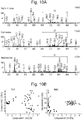


Classifications
-
- A—HUMAN NECESSITIES
- A61—MEDICAL OR VETERINARY SCIENCE; HYGIENE
- A61B—DIAGNOSIS; SURGERY; IDENTIFICATION
- A61B10/00—Other methods or instruments for diagnosis, e.g. instruments for taking a cell sample, for biopsy, for vaccination diagnosis; Sex determination; Ovulation-period determination; Throat striking implements
- A61B10/02—Instruments for taking cell samples or for biopsy
-
- A—HUMAN NECESSITIES
- A61—MEDICAL OR VETERINARY SCIENCE; HYGIENE
- A61B—DIAGNOSIS; SURGERY; IDENTIFICATION
- A61B1/00—Instruments for performing medical examinations of the interior of cavities or tubes of the body by visual or photographical inspection, e.g. endoscopes; Illuminating arrangements therefor
- A61B1/04—Instruments for performing medical examinations of the interior of cavities or tubes of the body by visual or photographical inspection, e.g. endoscopes; Illuminating arrangements therefor combined with photographic or television appliances
- A61B1/041—Capsule endoscopes for imaging
-
- A—HUMAN NECESSITIES
- A61—MEDICAL OR VETERINARY SCIENCE; HYGIENE
- A61B—DIAGNOSIS; SURGERY; IDENTIFICATION
- A61B1/00—Instruments for performing medical examinations of the interior of cavities or tubes of the body by visual or photographical inspection, e.g. endoscopes; Illuminating arrangements therefor
- A61B1/273—Instruments for performing medical examinations of the interior of cavities or tubes of the body by visual or photographical inspection, e.g. endoscopes; Illuminating arrangements therefor for the upper alimentary canal, e.g. oesophagoscopes, gastroscopes
- A61B1/2736—Gastroscopes
-
- A—HUMAN NECESSITIES
- A61—MEDICAL OR VETERINARY SCIENCE; HYGIENE
- A61B—DIAGNOSIS; SURGERY; IDENTIFICATION
- A61B10/00—Other methods or instruments for diagnosis, e.g. instruments for taking a cell sample, for biopsy, for vaccination diagnosis; Sex determination; Ovulation-period determination; Throat striking implements
- A61B10/0041—Detection of breast cancer
-
- A—HUMAN NECESSITIES
- A61—MEDICAL OR VETERINARY SCIENCE; HYGIENE
- A61B—DIAGNOSIS; SURGERY; IDENTIFICATION
- A61B10/00—Other methods or instruments for diagnosis, e.g. instruments for taking a cell sample, for biopsy, for vaccination diagnosis; Sex determination; Ovulation-period determination; Throat striking implements
- A61B10/02—Instruments for taking cell samples or for biopsy
- A61B10/0233—Pointed or sharp biopsy instruments
- A61B10/0283—Pointed or sharp biopsy instruments with vacuum aspiration, e.g. caused by retractable plunger or by connected syringe
-
- A—HUMAN NECESSITIES
- A61—MEDICAL OR VETERINARY SCIENCE; HYGIENE
- A61B—DIAGNOSIS; SURGERY; IDENTIFICATION
- A61B17/00—Surgical instruments, devices or methods, e.g. tourniquets
-
- A—HUMAN NECESSITIES
- A61—MEDICAL OR VETERINARY SCIENCE; HYGIENE
- A61B—DIAGNOSIS; SURGERY; IDENTIFICATION
- A61B17/00—Surgical instruments, devices or methods, e.g. tourniquets
- A61B17/32—Surgical cutting instruments
-
- A—HUMAN NECESSITIES
- A61—MEDICAL OR VETERINARY SCIENCE; HYGIENE
- A61B—DIAGNOSIS; SURGERY; IDENTIFICATION
- A61B17/00—Surgical instruments, devices or methods, e.g. tourniquets
- A61B17/32—Surgical cutting instruments
- A61B17/320068—Surgical cutting instruments using mechanical vibrations, e.g. ultrasonic
-
- A—HUMAN NECESSITIES
- A61—MEDICAL OR VETERINARY SCIENCE; HYGIENE
- A61B—DIAGNOSIS; SURGERY; IDENTIFICATION
- A61B18/00—Surgical instruments, devices or methods for transferring non-mechanical forms of energy to or from the body
-
- A—HUMAN NECESSITIES
- A61—MEDICAL OR VETERINARY SCIENCE; HYGIENE
- A61B—DIAGNOSIS; SURGERY; IDENTIFICATION
- A61B18/00—Surgical instruments, devices or methods for transferring non-mechanical forms of energy to or from the body
- A61B18/04—Surgical instruments, devices or methods for transferring non-mechanical forms of energy to or from the body by heating
- A61B18/042—Surgical instruments, devices or methods for transferring non-mechanical forms of energy to or from the body by heating using additional gas becoming plasma
-
- A—HUMAN NECESSITIES
- A61—MEDICAL OR VETERINARY SCIENCE; HYGIENE
- A61B—DIAGNOSIS; SURGERY; IDENTIFICATION
- A61B18/00—Surgical instruments, devices or methods for transferring non-mechanical forms of energy to or from the body
- A61B18/04—Surgical instruments, devices or methods for transferring non-mechanical forms of energy to or from the body by heating
- A61B18/12—Surgical instruments, devices or methods for transferring non-mechanical forms of energy to or from the body by heating by passing a current through the tissue to be heated, e.g. high-frequency current
- A61B18/14—Probes or electrodes therefor
- A61B18/1442—Probes having pivoting end effectors, e.g. forceps
- A61B18/1445—Probes having pivoting end effectors, e.g. forceps at the distal end of a shaft, e.g. forceps or scissors at the end of a rigid rod
-
- A—HUMAN NECESSITIES
- A61—MEDICAL OR VETERINARY SCIENCE; HYGIENE
- A61B—DIAGNOSIS; SURGERY; IDENTIFICATION
- A61B18/00—Surgical instruments, devices or methods for transferring non-mechanical forms of energy to or from the body
- A61B18/18—Surgical instruments, devices or methods for transferring non-mechanical forms of energy to or from the body by applying electromagnetic radiation, e.g. microwaves
- A61B18/1815—Surgical instruments, devices or methods for transferring non-mechanical forms of energy to or from the body by applying electromagnetic radiation, e.g. microwaves using microwaves
-
- A—HUMAN NECESSITIES
- A61—MEDICAL OR VETERINARY SCIENCE; HYGIENE
- A61B—DIAGNOSIS; SURGERY; IDENTIFICATION
- A61B18/00—Surgical instruments, devices or methods for transferring non-mechanical forms of energy to or from the body
- A61B18/18—Surgical instruments, devices or methods for transferring non-mechanical forms of energy to or from the body by applying electromagnetic radiation, e.g. microwaves
- A61B18/20—Surgical instruments, devices or methods for transferring non-mechanical forms of energy to or from the body by applying electromagnetic radiation, e.g. microwaves using laser
-
- A—HUMAN NECESSITIES
- A61—MEDICAL OR VETERINARY SCIENCE; HYGIENE
- A61B—DIAGNOSIS; SURGERY; IDENTIFICATION
- A61B5/00—Measuring for diagnostic purposes; Identification of persons
- A61B5/0059—Measuring for diagnostic purposes; Identification of persons using light, e.g. diagnosis by transillumination, diascopy, fluorescence
- A61B5/0062—Arrangements for scanning
- A61B5/0066—Optical coherence imaging
-
- A—HUMAN NECESSITIES
- A61—MEDICAL OR VETERINARY SCIENCE; HYGIENE
- A61B—DIAGNOSIS; SURGERY; IDENTIFICATION
- A61B5/00—Measuring for diagnostic purposes; Identification of persons
- A61B5/0059—Measuring for diagnostic purposes; Identification of persons using light, e.g. diagnosis by transillumination, diascopy, fluorescence
- A61B5/0075—Measuring for diagnostic purposes; Identification of persons using light, e.g. diagnosis by transillumination, diascopy, fluorescence by spectroscopy, i.e. measuring spectra, e.g. Raman spectroscopy, infrared absorption spectroscopy
-
- A—HUMAN NECESSITIES
- A61—MEDICAL OR VETERINARY SCIENCE; HYGIENE
- A61B—DIAGNOSIS; SURGERY; IDENTIFICATION
- A61B5/00—Measuring for diagnostic purposes; Identification of persons
- A61B5/01—Measuring temperature of body parts ; Diagnostic temperature sensing, e.g. for malignant or inflamed tissue
- A61B5/015—By temperature mapping of body part
-
- A—HUMAN NECESSITIES
- A61—MEDICAL OR VETERINARY SCIENCE; HYGIENE
- A61B—DIAGNOSIS; SURGERY; IDENTIFICATION
- A61B5/00—Measuring for diagnostic purposes; Identification of persons
- A61B5/05—Detecting, measuring or recording for diagnosis by means of electric currents or magnetic fields; Measuring using microwaves or radio waves
- A61B5/0507—Detecting, measuring or recording for diagnosis by means of electric currents or magnetic fields; Measuring using microwaves or radio waves using microwaves or terahertz waves
-
- A—HUMAN NECESSITIES
- A61—MEDICAL OR VETERINARY SCIENCE; HYGIENE
- A61B—DIAGNOSIS; SURGERY; IDENTIFICATION
- A61B5/00—Measuring for diagnostic purposes; Identification of persons
- A61B5/05—Detecting, measuring or recording for diagnosis by means of electric currents or magnetic fields; Measuring using microwaves or radio waves
- A61B5/055—Detecting, measuring or recording for diagnosis by means of electric currents or magnetic fields; Measuring using microwaves or radio waves involving electronic [EMR] or nuclear [NMR] magnetic resonance, e.g. magnetic resonance imaging
-
- A—HUMAN NECESSITIES
- A61—MEDICAL OR VETERINARY SCIENCE; HYGIENE
- A61B—DIAGNOSIS; SURGERY; IDENTIFICATION
- A61B6/00—Apparatus or devices for radiation diagnosis; Apparatus or devices for radiation diagnosis combined with radiation therapy equipment
- A61B6/02—Arrangements for diagnosis sequentially in different planes; Stereoscopic radiation diagnosis
- A61B6/03—Computed tomography [CT]
- A61B6/032—Transmission computed tomography [CT]
-
- A—HUMAN NECESSITIES
- A61—MEDICAL OR VETERINARY SCIENCE; HYGIENE
- A61B—DIAGNOSIS; SURGERY; IDENTIFICATION
- A61B6/00—Apparatus or devices for radiation diagnosis; Apparatus or devices for radiation diagnosis combined with radiation therapy equipment
- A61B6/02—Arrangements for diagnosis sequentially in different planes; Stereoscopic radiation diagnosis
- A61B6/03—Computed tomography [CT]
- A61B6/037—Emission tomography
-
- A—HUMAN NECESSITIES
- A61—MEDICAL OR VETERINARY SCIENCE; HYGIENE
- A61B—DIAGNOSIS; SURGERY; IDENTIFICATION
- A61B8/00—Diagnosis using ultrasonic, sonic or infrasonic waves
- A61B8/13—Tomography
-
- A—HUMAN NECESSITIES
- A61—MEDICAL OR VETERINARY SCIENCE; HYGIENE
- A61B—DIAGNOSIS; SURGERY; IDENTIFICATION
- A61B90/00—Instruments, implements or accessories specially adapted for surgery or diagnosis and not covered by any of the groups A61B1/00 - A61B50/00, e.g. for luxation treatment or for protecting wound edges
- A61B90/10—Instruments, implements or accessories specially adapted for surgery or diagnosis and not covered by any of the groups A61B1/00 - A61B50/00, e.g. for luxation treatment or for protecting wound edges for stereotaxic surgery, e.g. frame-based stereotaxis
- A61B90/11—Instruments, implements or accessories specially adapted for surgery or diagnosis and not covered by any of the groups A61B1/00 - A61B50/00, e.g. for luxation treatment or for protecting wound edges for stereotaxic surgery, e.g. frame-based stereotaxis with guides for needles or instruments, e.g. arcuate slides or ball joints
- A61B90/13—Instruments, implements or accessories specially adapted for surgery or diagnosis and not covered by any of the groups A61B1/00 - A61B50/00, e.g. for luxation treatment or for protecting wound edges for stereotaxic surgery, e.g. frame-based stereotaxis with guides for needles or instruments, e.g. arcuate slides or ball joints guided by light, e.g. laser pointers
-
- A—HUMAN NECESSITIES
- A61—MEDICAL OR VETERINARY SCIENCE; HYGIENE
- A61F—FILTERS IMPLANTABLE INTO BLOOD VESSELS; PROSTHESES; DEVICES PROVIDING PATENCY TO, OR PREVENTING COLLAPSING OF, TUBULAR STRUCTURES OF THE BODY, e.g. STENTS; ORTHOPAEDIC, NURSING OR CONTRACEPTIVE DEVICES; FOMENTATION; TREATMENT OR PROTECTION OF EYES OR EARS; BANDAGES, DRESSINGS OR ABSORBENT PADS; FIRST-AID KITS
- A61F13/00—Bandages or dressings; Absorbent pads
- A61F13/15—Absorbent pads, e.g. sanitary towels, swabs or tampons for external or internal application to the body; Supporting or fastening means therefor; Tampon applicators
- A61F13/38—Swabs having a stick-type handle, e.g. cotton tips
-
- C—CHEMISTRY; METALLURGY
- C12—BIOCHEMISTRY; BEER; SPIRITS; WINE; VINEGAR; MICROBIOLOGY; ENZYMOLOGY; MUTATION OR GENETIC ENGINEERING
- C12Q—MEASURING OR TESTING PROCESSES INVOLVING ENZYMES, NUCLEIC ACIDS OR MICROORGANISMS; COMPOSITIONS OR TEST PAPERS THEREFOR; PROCESSES OF PREPARING SUCH COMPOSITIONS; CONDITION-RESPONSIVE CONTROL IN MICROBIOLOGICAL OR ENZYMOLOGICAL PROCESSES
- C12Q1/00—Measuring or testing processes involving enzymes, nucleic acids or microorganisms; Compositions therefor; Processes of preparing such compositions
- C12Q1/02—Measuring or testing processes involving enzymes, nucleic acids or microorganisms; Compositions therefor; Processes of preparing such compositions involving viable microorganisms
- C12Q1/025—Measuring or testing processes involving enzymes, nucleic acids or microorganisms; Compositions therefor; Processes of preparing such compositions involving viable microorganisms for testing or evaluating the effect of chemical or biological compounds, e.g. drugs, cosmetics
-
- C—CHEMISTRY; METALLURGY
- C12—BIOCHEMISTRY; BEER; SPIRITS; WINE; VINEGAR; MICROBIOLOGY; ENZYMOLOGY; MUTATION OR GENETIC ENGINEERING
- C12Q—MEASURING OR TESTING PROCESSES INVOLVING ENZYMES, NUCLEIC ACIDS OR MICROORGANISMS; COMPOSITIONS OR TEST PAPERS THEREFOR; PROCESSES OF PREPARING SUCH COMPOSITIONS; CONDITION-RESPONSIVE CONTROL IN MICROBIOLOGICAL OR ENZYMOLOGICAL PROCESSES
- C12Q1/00—Measuring or testing processes involving enzymes, nucleic acids or microorganisms; Compositions therefor; Processes of preparing such compositions
- C12Q1/02—Measuring or testing processes involving enzymes, nucleic acids or microorganisms; Compositions therefor; Processes of preparing such compositions involving viable microorganisms
- C12Q1/04—Determining presence or kind of microorganism; Use of selective media for testing antibiotics or bacteriocides; Compositions containing a chemical indicator therefor
-
- C—CHEMISTRY; METALLURGY
- C12—BIOCHEMISTRY; BEER; SPIRITS; WINE; VINEGAR; MICROBIOLOGY; ENZYMOLOGY; MUTATION OR GENETIC ENGINEERING
- C12Q—MEASURING OR TESTING PROCESSES INVOLVING ENZYMES, NUCLEIC ACIDS OR MICROORGANISMS; COMPOSITIONS OR TEST PAPERS THEREFOR; PROCESSES OF PREPARING SUCH COMPOSITIONS; CONDITION-RESPONSIVE CONTROL IN MICROBIOLOGICAL OR ENZYMOLOGICAL PROCESSES
- C12Q1/00—Measuring or testing processes involving enzymes, nucleic acids or microorganisms; Compositions therefor; Processes of preparing such compositions
- C12Q1/02—Measuring or testing processes involving enzymes, nucleic acids or microorganisms; Compositions therefor; Processes of preparing such compositions involving viable microorganisms
- C12Q1/18—Testing for antimicrobial activity of a material
-
- C—CHEMISTRY; METALLURGY
- C12—BIOCHEMISTRY; BEER; SPIRITS; WINE; VINEGAR; MICROBIOLOGY; ENZYMOLOGY; MUTATION OR GENETIC ENGINEERING
- C12Q—MEASURING OR TESTING PROCESSES INVOLVING ENZYMES, NUCLEIC ACIDS OR MICROORGANISMS; COMPOSITIONS OR TEST PAPERS THEREFOR; PROCESSES OF PREPARING SUCH COMPOSITIONS; CONDITION-RESPONSIVE CONTROL IN MICROBIOLOGICAL OR ENZYMOLOGICAL PROCESSES
- C12Q1/00—Measuring or testing processes involving enzymes, nucleic acids or microorganisms; Compositions therefor; Processes of preparing such compositions
- C12Q1/02—Measuring or testing processes involving enzymes, nucleic acids or microorganisms; Compositions therefor; Processes of preparing such compositions involving viable microorganisms
- C12Q1/24—Methods of sampling, or inoculating or spreading a sample; Methods of physically isolating an intact microorganisms
-
- G—PHYSICS
- G01—MEASURING; TESTING
- G01N—INVESTIGATING OR ANALYSING MATERIALS BY DETERMINING THEIR CHEMICAL OR PHYSICAL PROPERTIES
- G01N1/00—Sampling; Preparing specimens for investigation
- G01N1/02—Devices for withdrawing samples
- G01N1/22—Devices for withdrawing samples in the gaseous state
- G01N1/2202—Devices for withdrawing samples in the gaseous state involving separation of sample components during sampling
-
- G—PHYSICS
- G01—MEASURING; TESTING
- G01N—INVESTIGATING OR ANALYSING MATERIALS BY DETERMINING THEIR CHEMICAL OR PHYSICAL PROPERTIES
- G01N27/00—Investigating or analysing materials by the use of electric, electrochemical, or magnetic means
- G01N27/62—Investigating or analysing materials by the use of electric, electrochemical, or magnetic means by investigating the ionisation of gases, e.g. aerosols; by investigating electric discharges, e.g. emission of cathode
- G01N27/622—Ion mobility spectrometry
- G01N27/623—Ion mobility spectrometry combined with mass spectrometry
-
- G—PHYSICS
- G01—MEASURING; TESTING
- G01N—INVESTIGATING OR ANALYSING MATERIALS BY DETERMINING THEIR CHEMICAL OR PHYSICAL PROPERTIES
- G01N27/00—Investigating or analysing materials by the use of electric, electrochemical, or magnetic means
- G01N27/62—Investigating or analysing materials by the use of electric, electrochemical, or magnetic means by investigating the ionisation of gases, e.g. aerosols; by investigating electric discharges, e.g. emission of cathode
- G01N27/622—Ion mobility spectrometry
- G01N27/624—Differential mobility spectrometry [DMS]; Field asymmetric-waveform ion mobility spectrometry [FAIMS]
-
- G—PHYSICS
- G01—MEASURING; TESTING
- G01N—INVESTIGATING OR ANALYSING MATERIALS BY DETERMINING THEIR CHEMICAL OR PHYSICAL PROPERTIES
- G01N3/00—Investigating strength properties of solid materials by application of mechanical stress
-
- G—PHYSICS
- G01—MEASURING; TESTING
- G01N—INVESTIGATING OR ANALYSING MATERIALS BY DETERMINING THEIR CHEMICAL OR PHYSICAL PROPERTIES
- G01N30/00—Investigating or analysing materials by separation into components using adsorption, absorption or similar phenomena or using ion-exchange, e.g. chromatography or field flow fractionation
- G01N30/02—Column chromatography
- G01N30/62—Detectors specially adapted therefor
- G01N30/72—Mass spectrometers
- G01N30/7233—Mass spectrometers interfaced to liquid or supercritical fluid chromatograph
- G01N30/724—Nebulising, aerosol formation or ionisation
-
- G—PHYSICS
- G01—MEASURING; TESTING
- G01N—INVESTIGATING OR ANALYSING MATERIALS BY DETERMINING THEIR CHEMICAL OR PHYSICAL PROPERTIES
- G01N33/00—Investigating or analysing materials by specific methods not covered by groups G01N1/00 - G01N31/00
- G01N33/48—Biological material, e.g. blood, urine; Haemocytometers
- G01N33/483—Physical analysis of biological material
- G01N33/487—Physical analysis of biological material of liquid biological material
-
- G—PHYSICS
- G01—MEASURING; TESTING
- G01N—INVESTIGATING OR ANALYSING MATERIALS BY DETERMINING THEIR CHEMICAL OR PHYSICAL PROPERTIES
- G01N33/00—Investigating or analysing materials by specific methods not covered by groups G01N1/00 - G01N31/00
- G01N33/48—Biological material, e.g. blood, urine; Haemocytometers
- G01N33/50—Chemical analysis of biological material, e.g. blood, urine; Testing involving biospecific ligand binding methods; Immunological testing
- G01N33/68—Chemical analysis of biological material, e.g. blood, urine; Testing involving biospecific ligand binding methods; Immunological testing involving proteins, peptides or amino acids
- G01N33/6803—General methods of protein analysis not limited to specific proteins or families of proteins
- G01N33/6848—Methods of protein analysis involving mass spectrometry
- G01N33/6851—Methods of protein analysis involving laser desorption ionisation mass spectrometry
-
- G—PHYSICS
- G01—MEASURING; TESTING
- G01N—INVESTIGATING OR ANALYSING MATERIALS BY DETERMINING THEIR CHEMICAL OR PHYSICAL PROPERTIES
- G01N33/00—Investigating or analysing materials by specific methods not covered by groups G01N1/00 - G01N31/00
- G01N33/48—Biological material, e.g. blood, urine; Haemocytometers
- G01N33/50—Chemical analysis of biological material, e.g. blood, urine; Testing involving biospecific ligand binding methods; Immunological testing
- G01N33/92—Chemical analysis of biological material, e.g. blood, urine; Testing involving biospecific ligand binding methods; Immunological testing involving lipids, e.g. cholesterol, lipoproteins, or their receptors
-
- G—PHYSICS
- G01—MEASURING; TESTING
- G01N—INVESTIGATING OR ANALYSING MATERIALS BY DETERMINING THEIR CHEMICAL OR PHYSICAL PROPERTIES
- G01N9/00—Investigating density or specific gravity of materials; Analysing materials by determining density or specific gravity
-
- G—PHYSICS
- G16—INFORMATION AND COMMUNICATION TECHNOLOGY [ICT] SPECIALLY ADAPTED FOR SPECIFIC APPLICATION FIELDS
- G16B—BIOINFORMATICS, i.e. INFORMATION AND COMMUNICATION TECHNOLOGY [ICT] SPECIALLY ADAPTED FOR GENETIC OR PROTEIN-RELATED DATA PROCESSING IN COMPUTATIONAL MOLECULAR BIOLOGY
- G16B20/00—ICT specially adapted for functional genomics or proteomics, e.g. genotype-phenotype associations
-
- H—ELECTRICITY
- H01—ELECTRIC ELEMENTS
- H01J—ELECTRIC DISCHARGE TUBES OR DISCHARGE LAMPS
- H01J49/00—Particle spectrometers or separator tubes
-
- H—ELECTRICITY
- H01—ELECTRIC ELEMENTS
- H01J—ELECTRIC DISCHARGE TUBES OR DISCHARGE LAMPS
- H01J49/00—Particle spectrometers or separator tubes
- H01J49/0004—Imaging particle spectrometry
-
- H—ELECTRICITY
- H01—ELECTRIC ELEMENTS
- H01J—ELECTRIC DISCHARGE TUBES OR DISCHARGE LAMPS
- H01J49/00—Particle spectrometers or separator tubes
- H01J49/0027—Methods for using particle spectrometers
-
- H—ELECTRICITY
- H01—ELECTRIC ELEMENTS
- H01J—ELECTRIC DISCHARGE TUBES OR DISCHARGE LAMPS
- H01J49/00—Particle spectrometers or separator tubes
- H01J49/0027—Methods for using particle spectrometers
- H01J49/0031—Step by step routines describing the use of the apparatus
-
- H—ELECTRICITY
- H01—ELECTRIC ELEMENTS
- H01J—ELECTRIC DISCHARGE TUBES OR DISCHARGE LAMPS
- H01J49/00—Particle spectrometers or separator tubes
- H01J49/0027—Methods for using particle spectrometers
- H01J49/0036—Step by step routines describing the handling of the data generated during a measurement
-
- H—ELECTRICITY
- H01—ELECTRIC ELEMENTS
- H01J—ELECTRIC DISCHARGE TUBES OR DISCHARGE LAMPS
- H01J49/00—Particle spectrometers or separator tubes
- H01J49/02—Details
- H01J49/025—Detectors specially adapted to particle spectrometers
-
- H—ELECTRICITY
- H01—ELECTRIC ELEMENTS
- H01J—ELECTRIC DISCHARGE TUBES OR DISCHARGE LAMPS
- H01J49/00—Particle spectrometers or separator tubes
- H01J49/02—Details
- H01J49/04—Arrangements for introducing or extracting samples to be analysed, e.g. vacuum locks; Arrangements for external adjustment of electron- or ion-optical components
- H01J49/0404—Capillaries used for transferring samples or ions
-
- H—ELECTRICITY
- H01—ELECTRIC ELEMENTS
- H01J—ELECTRIC DISCHARGE TUBES OR DISCHARGE LAMPS
- H01J49/00—Particle spectrometers or separator tubes
- H01J49/02—Details
- H01J49/04—Arrangements for introducing or extracting samples to be analysed, e.g. vacuum locks; Arrangements for external adjustment of electron- or ion-optical components
- H01J49/0409—Sample holders or containers
-
- H—ELECTRICITY
- H01—ELECTRIC ELEMENTS
- H01J—ELECTRIC DISCHARGE TUBES OR DISCHARGE LAMPS
- H01J49/00—Particle spectrometers or separator tubes
- H01J49/02—Details
- H01J49/04—Arrangements for introducing or extracting samples to be analysed, e.g. vacuum locks; Arrangements for external adjustment of electron- or ion-optical components
- H01J49/0422—Arrangements for introducing or extracting samples to be analysed, e.g. vacuum locks; Arrangements for external adjustment of electron- or ion-optical components for gaseous samples
-
- H—ELECTRICITY
- H01—ELECTRIC ELEMENTS
- H01J—ELECTRIC DISCHARGE TUBES OR DISCHARGE LAMPS
- H01J49/00—Particle spectrometers or separator tubes
- H01J49/02—Details
- H01J49/04—Arrangements for introducing or extracting samples to be analysed, e.g. vacuum locks; Arrangements for external adjustment of electron- or ion-optical components
- H01J49/0431—Arrangements for introducing or extracting samples to be analysed, e.g. vacuum locks; Arrangements for external adjustment of electron- or ion-optical components for liquid samples
- H01J49/044—Arrangements for introducing or extracting samples to be analysed, e.g. vacuum locks; Arrangements for external adjustment of electron- or ion-optical components for liquid samples with means for preventing droplets from entering the analyzer; Desolvation of droplets
-
- H—ELECTRICITY
- H01—ELECTRIC ELEMENTS
- H01J—ELECTRIC DISCHARGE TUBES OR DISCHARGE LAMPS
- H01J49/00—Particle spectrometers or separator tubes
- H01J49/02—Details
- H01J49/04—Arrangements for introducing or extracting samples to be analysed, e.g. vacuum locks; Arrangements for external adjustment of electron- or ion-optical components
- H01J49/0431—Arrangements for introducing or extracting samples to be analysed, e.g. vacuum locks; Arrangements for external adjustment of electron- or ion-optical components for liquid samples
- H01J49/0445—Arrangements for introducing or extracting samples to be analysed, e.g. vacuum locks; Arrangements for external adjustment of electron- or ion-optical components for liquid samples with means for introducing as a spray, a jet or an aerosol
-
- H—ELECTRICITY
- H01—ELECTRIC ELEMENTS
- H01J—ELECTRIC DISCHARGE TUBES OR DISCHARGE LAMPS
- H01J49/00—Particle spectrometers or separator tubes
- H01J49/02—Details
- H01J49/04—Arrangements for introducing or extracting samples to be analysed, e.g. vacuum locks; Arrangements for external adjustment of electron- or ion-optical components
- H01J49/0459—Arrangements for introducing or extracting samples to be analysed, e.g. vacuum locks; Arrangements for external adjustment of electron- or ion-optical components for solid samples
- H01J49/0463—Desorption by laser or particle beam, followed by ionisation as a separate step
-
- H—ELECTRICITY
- H01—ELECTRIC ELEMENTS
- H01J—ELECTRIC DISCHARGE TUBES OR DISCHARGE LAMPS
- H01J49/00—Particle spectrometers or separator tubes
- H01J49/02—Details
- H01J49/04—Arrangements for introducing or extracting samples to be analysed, e.g. vacuum locks; Arrangements for external adjustment of electron- or ion-optical components
- H01J49/0468—Arrangements for introducing or extracting samples to be analysed, e.g. vacuum locks; Arrangements for external adjustment of electron- or ion-optical components with means for heating or cooling the sample
- H01J49/049—Arrangements for introducing or extracting samples to be analysed, e.g. vacuum locks; Arrangements for external adjustment of electron- or ion-optical components with means for heating or cooling the sample with means for applying heat to desorb the sample; Evaporation
-
- H—ELECTRICITY
- H01—ELECTRIC ELEMENTS
- H01J—ELECTRIC DISCHARGE TUBES OR DISCHARGE LAMPS
- H01J49/00—Particle spectrometers or separator tubes
- H01J49/02—Details
- H01J49/06—Electron- or ion-optical arrangements
-
- H—ELECTRICITY
- H01—ELECTRIC ELEMENTS
- H01J—ELECTRIC DISCHARGE TUBES OR DISCHARGE LAMPS
- H01J49/00—Particle spectrometers or separator tubes
- H01J49/02—Details
- H01J49/06—Electron- or ion-optical arrangements
- H01J49/061—Ion deflecting means, e.g. ion gates
-
- H—ELECTRICITY
- H01—ELECTRIC ELEMENTS
- H01J—ELECTRIC DISCHARGE TUBES OR DISCHARGE LAMPS
- H01J49/00—Particle spectrometers or separator tubes
- H01J49/02—Details
- H01J49/06—Electron- or ion-optical arrangements
- H01J49/068—Mounting, supporting, spacing, or insulating electrodes
-
- H—ELECTRICITY
- H01—ELECTRIC ELEMENTS
- H01J—ELECTRIC DISCHARGE TUBES OR DISCHARGE LAMPS
- H01J49/00—Particle spectrometers or separator tubes
- H01J49/02—Details
- H01J49/10—Ion sources; Ion guns
-
- H—ELECTRICITY
- H01—ELECTRIC ELEMENTS
- H01J—ELECTRIC DISCHARGE TUBES OR DISCHARGE LAMPS
- H01J49/00—Particle spectrometers or separator tubes
- H01J49/02—Details
- H01J49/10—Ion sources; Ion guns
- H01J49/14—Ion sources; Ion guns using particle bombardment, e.g. ionisation chambers
-
- H—ELECTRICITY
- H01—ELECTRIC ELEMENTS
- H01J—ELECTRIC DISCHARGE TUBES OR DISCHARGE LAMPS
- H01J49/00—Particle spectrometers or separator tubes
- H01J49/02—Details
- H01J49/10—Ion sources; Ion guns
- H01J49/14—Ion sources; Ion guns using particle bombardment, e.g. ionisation chambers
- H01J49/142—Ion sources; Ion guns using particle bombardment, e.g. ionisation chambers using a solid target which is not previously vapourised
-
- H—ELECTRICITY
- H01—ELECTRIC ELEMENTS
- H01J—ELECTRIC DISCHARGE TUBES OR DISCHARGE LAMPS
- H01J49/00—Particle spectrometers or separator tubes
- H01J49/02—Details
- H01J49/10—Ion sources; Ion guns
- H01J49/16—Ion sources; Ion guns using surface ionisation, e.g. field-, thermionic- or photo-emission
- H01J49/161—Ion sources; Ion guns using surface ionisation, e.g. field-, thermionic- or photo-emission using photoionisation, e.g. by laser
- H01J49/164—Laser desorption/ionisation, e.g. matrix-assisted laser desorption/ionisation [MALDI]
-
- H—ELECTRICITY
- H01—ELECTRIC ELEMENTS
- H01J—ELECTRIC DISCHARGE TUBES OR DISCHARGE LAMPS
- H01J49/00—Particle spectrometers or separator tubes
- H01J49/02—Details
- H01J49/24—Vacuum systems, e.g. maintaining desired pressures
-
- H—ELECTRICITY
- H01—ELECTRIC ELEMENTS
- H01J—ELECTRIC DISCHARGE TUBES OR DISCHARGE LAMPS
- H01J49/00—Particle spectrometers or separator tubes
- H01J49/26—Mass spectrometers or separator tubes
-
- A—HUMAN NECESSITIES
- A61—MEDICAL OR VETERINARY SCIENCE; HYGIENE
- A61B—DIAGNOSIS; SURGERY; IDENTIFICATION
- A61B1/00—Instruments for performing medical examinations of the interior of cavities or tubes of the body by visual or photographical inspection, e.g. endoscopes; Illuminating arrangements therefor
- A61B1/00002—Operational features of endoscopes
- A61B1/00011—Operational features of endoscopes characterised by signal transmission
- A61B1/00013—Operational features of endoscopes characterised by signal transmission using optical means
-
- A—HUMAN NECESSITIES
- A61—MEDICAL OR VETERINARY SCIENCE; HYGIENE
- A61B—DIAGNOSIS; SURGERY; IDENTIFICATION
- A61B1/00—Instruments for performing medical examinations of the interior of cavities or tubes of the body by visual or photographical inspection, e.g. endoscopes; Illuminating arrangements therefor
- A61B1/31—Instruments for performing medical examinations of the interior of cavities or tubes of the body by visual or photographical inspection, e.g. endoscopes; Illuminating arrangements therefor for the rectum, e.g. proctoscopes, sigmoidoscopes, colonoscopes
-
- A—HUMAN NECESSITIES
- A61—MEDICAL OR VETERINARY SCIENCE; HYGIENE
- A61B—DIAGNOSIS; SURGERY; IDENTIFICATION
- A61B10/00—Other methods or instruments for diagnosis, e.g. instruments for taking a cell sample, for biopsy, for vaccination diagnosis; Sex determination; Ovulation-period determination; Throat striking implements
- A61B2010/0083—Other methods or instruments for diagnosis, e.g. instruments for taking a cell sample, for biopsy, for vaccination diagnosis; Sex determination; Ovulation-period determination; Throat striking implements for taking gas samples
-
- A—HUMAN NECESSITIES
- A61—MEDICAL OR VETERINARY SCIENCE; HYGIENE
- A61B—DIAGNOSIS; SURGERY; IDENTIFICATION
- A61B17/00—Surgical instruments, devices or methods, e.g. tourniquets
- A61B17/32—Surgical cutting instruments
- A61B17/320068—Surgical cutting instruments using mechanical vibrations, e.g. ultrasonic
- A61B2017/320069—Surgical cutting instruments using mechanical vibrations, e.g. ultrasonic for ablating tissue
-
- A—HUMAN NECESSITIES
- A61—MEDICAL OR VETERINARY SCIENCE; HYGIENE
- A61B—DIAGNOSIS; SURGERY; IDENTIFICATION
- A61B18/00—Surgical instruments, devices or methods for transferring non-mechanical forms of energy to or from the body
- A61B2018/00571—Surgical instruments, devices or methods for transferring non-mechanical forms of energy to or from the body for achieving a particular surgical effect
- A61B2018/00577—Ablation
-
- A—HUMAN NECESSITIES
- A61—MEDICAL OR VETERINARY SCIENCE; HYGIENE
- A61B—DIAGNOSIS; SURGERY; IDENTIFICATION
- A61B18/00—Surgical instruments, devices or methods for transferring non-mechanical forms of energy to or from the body
- A61B2018/00571—Surgical instruments, devices or methods for transferring non-mechanical forms of energy to or from the body for achieving a particular surgical effect
- A61B2018/00589—Coagulation
-
- A—HUMAN NECESSITIES
- A61—MEDICAL OR VETERINARY SCIENCE; HYGIENE
- A61B—DIAGNOSIS; SURGERY; IDENTIFICATION
- A61B18/00—Surgical instruments, devices or methods for transferring non-mechanical forms of energy to or from the body
- A61B2018/00994—Surgical instruments, devices or methods for transferring non-mechanical forms of energy to or from the body combining two or more different kinds of non-mechanical energy or combining one or more non-mechanical energies with ultrasound
-
- A—HUMAN NECESSITIES
- A61—MEDICAL OR VETERINARY SCIENCE; HYGIENE
- A61B—DIAGNOSIS; SURGERY; IDENTIFICATION
- A61B2218/00—Details of surgical instruments, devices or methods for transferring non-mechanical forms of energy to or from the body
- A61B2218/001—Details of surgical instruments, devices or methods for transferring non-mechanical forms of energy to or from the body having means for irrigation and/or aspiration of substances to and/or from the surgical site
- A61B2218/002—Irrigation
-
- A—HUMAN NECESSITIES
- A61—MEDICAL OR VETERINARY SCIENCE; HYGIENE
- A61B—DIAGNOSIS; SURGERY; IDENTIFICATION
- A61B2218/00—Details of surgical instruments, devices or methods for transferring non-mechanical forms of energy to or from the body
- A61B2218/001—Details of surgical instruments, devices or methods for transferring non-mechanical forms of energy to or from the body having means for irrigation and/or aspiration of substances to and/or from the surgical site
- A61B2218/007—Aspiration
- A61B2218/008—Aspiration for smoke evacuation
-
- A—HUMAN NECESSITIES
- A61—MEDICAL OR VETERINARY SCIENCE; HYGIENE
- A61B—DIAGNOSIS; SURGERY; IDENTIFICATION
- A61B5/00—Measuring for diagnostic purposes; Identification of persons
- A61B5/145—Measuring characteristics of blood in vivo, e.g. gas concentration, pH value; Measuring characteristics of body fluids or tissues, e.g. interstitial fluid, cerebral tissue
- A61B5/14542—Measuring characteristics of blood in vivo, e.g. gas concentration, pH value; Measuring characteristics of body fluids or tissues, e.g. interstitial fluid, cerebral tissue for measuring blood gases
-
- G—PHYSICS
- G01—MEASURING; TESTING
- G01N—INVESTIGATING OR ANALYSING MATERIALS BY DETERMINING THEIR CHEMICAL OR PHYSICAL PROPERTIES
- G01N1/00—Sampling; Preparing specimens for investigation
- G01N1/02—Devices for withdrawing samples
- G01N2001/028—Sampling from a surface, swabbing, vaporising
-
- G—PHYSICS
- G01—MEASURING; TESTING
- G01N—INVESTIGATING OR ANALYSING MATERIALS BY DETERMINING THEIR CHEMICAL OR PHYSICAL PROPERTIES
- G01N1/00—Sampling; Preparing specimens for investigation
- G01N1/02—Devices for withdrawing samples
- G01N1/22—Devices for withdrawing samples in the gaseous state
- G01N1/2202—Devices for withdrawing samples in the gaseous state involving separation of sample components during sampling
- G01N2001/222—Other features
- G01N2001/2223—Other features aerosol sampling devices
-
- G—PHYSICS
- G01—MEASURING; TESTING
- G01N—INVESTIGATING OR ANALYSING MATERIALS BY DETERMINING THEIR CHEMICAL OR PHYSICAL PROPERTIES
- G01N2333/00—Assays involving biological materials from specific organisms or of a specific nature
- G01N2333/195—Assays involving biological materials from specific organisms or of a specific nature from bacteria
-
- G—PHYSICS
- G01—MEASURING; TESTING
- G01N—INVESTIGATING OR ANALYSING MATERIALS BY DETERMINING THEIR CHEMICAL OR PHYSICAL PROPERTIES
- G01N2405/00—Assays, e.g. immunoassays or enzyme assays, involving lipids
-
- G—PHYSICS
- G01—MEASURING; TESTING
- G01N—INVESTIGATING OR ANALYSING MATERIALS BY DETERMINING THEIR CHEMICAL OR PHYSICAL PROPERTIES
- G01N2405/00—Assays, e.g. immunoassays or enzyme assays, involving lipids
- G01N2405/04—Phospholipids, i.e. phosphoglycerides
-
- G—PHYSICS
- G01—MEASURING; TESTING
- G01N—INVESTIGATING OR ANALYSING MATERIALS BY DETERMINING THEIR CHEMICAL OR PHYSICAL PROPERTIES
- G01N2405/00—Assays, e.g. immunoassays or enzyme assays, involving lipids
- G01N2405/08—Sphingolipids
-
- G—PHYSICS
- G01—MEASURING; TESTING
- G01N—INVESTIGATING OR ANALYSING MATERIALS BY DETERMINING THEIR CHEMICAL OR PHYSICAL PROPERTIES
- G01N2570/00—Omics, e.g. proteomics, glycomics or lipidomics; Methods of analysis focusing on the entire complement of classes of biological molecules or subsets thereof, i.e. focusing on proteomes, glycomes or lipidomes
-
- G—PHYSICS
- G01—MEASURING; TESTING
- G01N—INVESTIGATING OR ANALYSING MATERIALS BY DETERMINING THEIR CHEMICAL OR PHYSICAL PROPERTIES
- G01N2800/00—Detection or diagnosis of diseases
- G01N2800/26—Infectious diseases, e.g. generalised sepsis
-
- G—PHYSICS
- G01—MEASURING; TESTING
- G01N—INVESTIGATING OR ANALYSING MATERIALS BY DETERMINING THEIR CHEMICAL OR PHYSICAL PROPERTIES
- G01N33/00—Investigating or analysing materials by specific methods not covered by groups G01N1/00 - G01N31/00
- G01N33/48—Biological material, e.g. blood, urine; Haemocytometers
- G01N33/483—Physical analysis of biological material
- G01N33/487—Physical analysis of biological material of liquid biological material
- G01N33/48707—Physical analysis of biological material of liquid biological material by electrical means
- G01N33/48735—Investigating suspensions of cells, e.g. measuring microbe concentration
-
- G—PHYSICS
- G16—INFORMATION AND COMMUNICATION TECHNOLOGY [ICT] SPECIALLY ADAPTED FOR SPECIFIC APPLICATION FIELDS
- G16H—HEALTHCARE INFORMATICS, i.e. INFORMATION AND COMMUNICATION TECHNOLOGY [ICT] SPECIALLY ADAPTED FOR THE HANDLING OR PROCESSING OF MEDICAL OR HEALTHCARE DATA
- G16H10/00—ICT specially adapted for the handling or processing of patient-related medical or healthcare data
- G16H10/40—ICT specially adapted for the handling or processing of patient-related medical or healthcare data for data related to laboratory analysis, e.g. patient specimen analysis
-
- G—PHYSICS
- G16—INFORMATION AND COMMUNICATION TECHNOLOGY [ICT] SPECIALLY ADAPTED FOR SPECIFIC APPLICATION FIELDS
- G16H—HEALTHCARE INFORMATICS, i.e. INFORMATION AND COMMUNICATION TECHNOLOGY [ICT] SPECIALLY ADAPTED FOR THE HANDLING OR PROCESSING OF MEDICAL OR HEALTHCARE DATA
- G16H15/00—ICT specially adapted for medical reports, e.g. generation or transmission thereof
-
- G—PHYSICS
- G16—INFORMATION AND COMMUNICATION TECHNOLOGY [ICT] SPECIALLY ADAPTED FOR SPECIFIC APPLICATION FIELDS
- G16H—HEALTHCARE INFORMATICS, i.e. INFORMATION AND COMMUNICATION TECHNOLOGY [ICT] SPECIALLY ADAPTED FOR THE HANDLING OR PROCESSING OF MEDICAL OR HEALTHCARE DATA
- G16H50/00—ICT specially adapted for medical diagnosis, medical simulation or medical data mining; ICT specially adapted for detecting, monitoring or modelling epidemics or pandemics
- G16H50/20—ICT specially adapted for medical diagnosis, medical simulation or medical data mining; ICT specially adapted for detecting, monitoring or modelling epidemics or pandemics for computer-aided diagnosis, e.g. based on medical expert systems
-
- Y—GENERAL TAGGING OF NEW TECHNOLOGICAL DEVELOPMENTS; GENERAL TAGGING OF CROSS-SECTIONAL TECHNOLOGIES SPANNING OVER SEVERAL SECTIONS OF THE IPC; TECHNICAL SUBJECTS COVERED BY FORMER USPC CROSS-REFERENCE ART COLLECTIONS [XRACs] AND DIGESTS
- Y02—TECHNOLOGIES OR APPLICATIONS FOR MITIGATION OR ADAPTATION AGAINST CLIMATE CHANGE
- Y02A—TECHNOLOGIES FOR ADAPTATION TO CLIMATE CHANGE
- Y02A90/00—Technologies having an indirect contribution to adaptation to climate change
- Y02A90/10—Information and communication technologies [ICT] supporting adaptation to climate change, e.g. for weather forecasting or climate simulation
Definitions
- the present invention relates generally to mass spectrometers and in particular to the analysis of material by ambient ionisation ion sources including rapid evaporative ionisation mass spectrometry ("REIMS”) ion sources and the analysis of material by desorption electrospray ionisation (“DESI”) mass spectrometry.
- REIMS rapid evaporative ionisation mass spectrometry
- DESI desorption electrospray ionisation
- Various embodiments relate to the use of mass spectrometers in diagnostic methods.
- analyte ions generated by an ambient ionisation ion source are then subjected either to: (i) mass analysis by a mass analyser such as a quadrupole mass analyser or a Time of Flight mass analyser; (ii) ion mobility analysis (IMS) and/or differential ion mobility analysis (DMA) and/or Field Asymmetric Ion Mobility Spectrometry (FAIMS) analysis; and/or (iii) a combination of firstly ion mobility analysis (IMS) and/or differential ion mobility analysis (DMA) and/or Field Asymmetric Ion Mobility Spectrometry (FAIMS) analysis followed by secondly mass analysis by a mass analyser such as a quadrupole mass analyser or a Time of Flight mass analyser (or vice versa).
- Various embodiments also relate to an ion mobility spectrometer and/or mass analyser and a method of ion mobility spectrometry and/or method of mass analysis.
- the mucosal membrane is a protective layer responsible for trapping pathogens in the human body.
- the mucosal membrane is an easily accessible and highly clinically relevant sample to diagnose pathogenic and cancerous associated diseases.
- the swab is placed into a sterile tube containing a buffer solution for storage and then the tube is sent to a laboratory for analysis.
- a laboratory receiving the tube will wipe the smear content across a culture medium such as an agar plate.
- the culture medium is then incubated to allow organisms present on the swab to grow.
- Microbial identification may then be performed, e.g., under a microscope. Any organisms present in the sample may also be identified by 16S gene-sequencing and/or by using matrix-assisted laser desorption ionisation (“MALDI”) mass spectrometry and then comparing the mass spectra with a commercial available database.
- MALDI matrix-assisted laser desorption ionisation
- Desorption electrospray ionisation is an ambient ionisation method that involves directing a spray of (primary) electrically charged droplets onto a surface. The electrospray mist is pneumatically directed at the sample where subsequent splashed (secondary) droplets carry desorbed ionised analytes. After ionisation, the resulting ions travel through air and pass into an atmospheric pressure interface of a mass spectrometer.
- Desorption electrospray ionisation (“DESI”) is a technique that allows for ambient ionisation of a trace sample at atmospheric pressure with no sample preparation being required.
- a “swab” in accordance with various embodiments is intended to be understood as comprising a "standard medical swab” i.e. a swab that is designed for sampling biological samples such as mucosal membranes.
- standard medical swab should be understood as covering a “cotton bud” (British) or a “cotton swab” (American) i.e. a small wad of cotton wrapped around one or both ends of a tube.
- the tube may be made from plastic, rolled paper or wood.
- StableFlex (RTM) fibre cores are known and comprise a 80 ⁇ m fused silica core coated with a polymer. Such fibre cores are not intended to be considered as comprising a "swab" within the meaning of the present application as materials having a fibre core are not considered to comprise a "standard medical swab”.
- swab swab
- medical swab medical swab
- standard medical swab as used within the present application are intended to exclude materials having a fused silica core.
- SPME Solid Phase Micro Extraction
- DESI desorption electrospray ionisation
- the SPME fibre is then subjected to direct desorption electrospray ionisation ("DESI") mass spectrometry analysis by positioning the fibre in the spray of a desorption electrospray ionisation (“DESI”) mass spectrometry arrangement.
- DESI desorption electrospray ionisation
- this known technique requires (at least) a two-step sample preparation procedure comprising: (i) the acquisition or preparation of the sample, followed by (ii) the extraction of analyte into the SPME fibre.
- the known specialist SPME fibres are not suited for the direct sampling of biological material, e.g. from a patient, such as the direct sampling of the mucosal membrane.
- various embodiments are based at least in part upon the recognition that a sample provided on a swab, e.g. a standard medical swab, may be directly analysed by desorption electrospray ionisation ("DESI") mass spectrometry.
- a standard medical swab may itself be positioned in the spray of a desorption electrospray ionisation (“DESI”) ionisation source.
- DESI desorption electrospray ionisation
- Various embodiments are beneficial in that they require no sample preparation steps beyond the acquisition of the sample onto the swab.
- various embodiments provide a rapid direct analysis method for samples provided on medical swabs.
- Various embodiments are particularly suited to and useful in the analysis of biological material, e.g. from a patient, such as material sampled directly onto a swab from the mucosal membrane.
- DESI desorption electrospray ionisation
- desorption electrospray ionisation DESI
- mass spectrometry analysis is a relatively non-destructive analysis technique.
- other commercial analysis techniques such as culturing techniques and 16S rRNA sequencing techniques are optimised to use samples provided on (standard) medical swabs.
- the sample on the swab may be analysed multiple times using multiple different analysis techniques.
- biological sample biological material
- biological material may optionally comprise or consist of biological tissue.
- the biological material etc. may optionally be selected, for example, from a surgical resection specimen, a biopsy specimen, a tissue specimen, a cell specimen, a smear, a body fluid specimen and/or a faecal specimen, any of which may be sampled in vivo from the subject, e.g., directly sampled onto a swab or taken as a biopsy, or be sampled ex vivo or in vitro, e.g., a sample may be provided and then sampled onto a swab, which may be referred to as indirect sampling onto a swab, or sampled with a biopsy needle, which may be referred to as indirect sampling with a biopsy needle.
- the method is carried out on a provided biological sample, e.g., a provided biopsy, or a provided swab,
- a body fluid specimen may, for example, optionally be selected from blood, plasma, serum, sputum, lavage fluid, pus, urine, saliva, phlegm, vomit, faeces, amniotic fluid, cerebrospinal fluid, pleural fluid, semen, sputum, vaginal secretion, interstitial fluid, and/or lymph.
- the swab may comprise a medical swab or a standard medical swab.
- the swab may comprise a disposable swab.
- the swab may comprise a cotton, rayon, plastic or foam swab.
- the swab may comprise a hollow rod.
- the swab may comprise plastic, wood or rolled paper.
- the swab may comprise a swab arranged and adapted to sample a mucosal membrane.
- the swab may have been chemically modified to enhance selectivity for an analyte.
- the chemical modification may render the swab lipophilic.
- the chemical modification may involve formation of a coating on a surface of the swab.
- the coating may be a polymeric coating.
- the polymer coating may comprise polydivinylbenzene (DVB), a copolymer of N-vinylpyrrolidone and divinylbenzene, or polydimethylsiloxane.
- DVD polydivinylbenzene
- a copolymer of N-vinylpyrrolidone and divinylbenzene or polydimethylsiloxane.
- the chemical modification may utilise a solid-phase extraction material.
- the polymeric coating may contain particles of a solid-phase extraction material.
- the solid-phase extraction material may comprise polymer particles, silica particles, hybrid silica/organic particles, carbon particles or polymer-coated solid particles.
- the polymer-coated solid particle may be a silica particle.
- the polymer-coated solid particles may comprise polydivinylbenzene (DVB), a copolymer of N-vinylpyrrolidone and divinylbenzene, or polydimethylsiloxane.
- DVD polydivinylbenzene
- a copolymer of N-vinylpyrrolidone and divinylbenzene or polydimethylsiloxane.
- R' may be selected from the group consisting of methyl, ethyl, propyl, isopropyl, butyl, t-butyl, sec-butyl, pentyl, isopentyl, hexyl and cyclohexyl.
- R 2 may include an alkyl, alkaryl, alkenyl, alkynyl, aryl, cyano, amino, diol, nitro, ester, a cation exchange group, an anion exchange group, an alkyl or aryl group containing an embedded polar functionality or chiral moieties.
- R 2 may be C 1 -C 30 alkyl, alkaryl, cyanoalkyl, a diol; alkylamino, amino alkyl or carbamate group.
- the silica particles may be surface modified by reaction with a compound selected from the group consisting of octyltrichlorosilane, octadecyltrichlorosilane, octyldimethylchlorosilane and octadecyldimethylchlorosilane.
- the polymer particles may comprise polydivinylbenzene or a copolymer of N-vinylpyrrolidone and divinylbenzene.
- the solid-phase extraction material may be an ion exchange resin.
- the polymer particles may be ion exchange resin particles.
- the ion exchange resin may comprise a copolymer of N-vinylpyrrolidone and divinylbenzene in which at least some of the benzene rings are sulfonated or carboxylated.
- the ion exchange resin may comprise a copolymer of N-vinylpyrrolidone and divinylbenzene in which at least some of the benzene rings are substituted by imidazolium, -CH 2 -piperazine groups or quaternary ammonium groups.
- the quaternary ammonium group may be -CH 2 N + (CH 3 ) 2 (C 4 H 9 ).
- the solid-phase extraction material or solid particle may be attached to the swab using an adhesive.
- the sample may comprise biological tissue, biological matter, a bacterial colony, a fungal colony, and/or microbes.
- the sample may be provided on the swab in its native or unmodified state.
- the sample may comprise: (i) mammalian cells; (ii) microbes; (iii) extracellular or exogenous compounds; and/or (iv) a biomarker of (i), (ii) and/or (iii).
- the biomarker may be selected from fatty acids, glycerolipids, sterol lipids, sphingolipids, prenol lipids, saccharolipids, and/or phospholipids, and/or a fingerprint of one or more thereof.
- Providing the sample on the swab may comprise wiping the swab across an in vivo, in vitro or ex vivo biological sample or more generally a target.
- the target may comprise an organic sample including a plastic.
- the target may comprise one or more bacterial colonies or one or more fungal colonies.
- Providing the sample on the swab may comprise using the swab to sample a mucosal membrane.
- the mucosal membrane may comprise a vaginal, nasal or oral mucosal membrane.
- Directing the spray of charged droplets onto the swab may comprise directing a spray of charged solvent droplets onto the swab.
- Directing the spray of charged droplets onto the swab may comprise directing the spray of charged droplets onto the swab at about atmospheric pressure.
- Directing the spray of charged droplets onto the swab may comprise ionising the sample using Desorption Electrospray lonisation ("DESI”) or desorption electroflow focusing ionisation (“DEFFI”).
- DESI Desorption Electrospray lonisation
- DEFFI desorption electroflow focusing ionisation
- the method may comprise directing the spray of charged droplets onto the swab from a sprayer.
- the sprayer may be arranged at a distance from the swab in the range (i) ⁇ 0.5 mm; (ii) about 0.5-1 mm; (iii) about 1-1.5 mm; (iv) about 1.5-2 mm; (v) about 2-3 mm; (vi) about 3-4 mm; or (vii) >4 mm.
- the method may comprise providing the sprayer with a voltage in the range (i) ⁇ 1 kV; (ii) about 1-2 kV; (iii) about 2-3 kV; (iv) about 3-4 kV; (v) about 4-5 kV; (vi) about 5-6 kV; (vii) about 6-7 kV; (viii) about 7-8 kV; (ix) about 8-9 kV; (x) about 9-10 kV; or (xi) >10 kV.
- the method may comprise providing the sprayer with a solvent at a flow rate in the range (i) ⁇ 5 ⁇ L/min; (ii) about 5-7 ⁇ L/min; (iii) about 7-9 ⁇ L/min; (iv) about 9-10 ⁇ L/min; (v) about 10-11 ⁇ L/min; (vi) about 11-13 ⁇ L/min; (vii) about 13-15 ⁇ L/min; (viii) about 15-20 ⁇ L/min; or (ix) > 20 ⁇ L/min.
- the method may comprise providing the sprayer with a gas at a pressure in the range (i) ⁇ 5 bar; (ii) about 5-6 bar; (iii) about 6-7 bar; (iv) about 7-8 bar; (v) about 8-9 bar; (vi) about 9-10 bar; (vii) about 10-15 bar; or (viii) > 15 bar.
- the gas may comprise air or nitrogen.
- the method may comprise rotating the swab whilst directing the spray of charged droplets onto the surface of the swab.
- the step of rotating the swab whilst directing the spray of charged droplets onto the surface of the swab may comprise substantially continuously rotating the swab whilst directing the spray of charged droplets onto the surface of the swab.
- the method may comprise translating and/or oscillating the swab whilst directing the spray of charged droplets onto the surface of the swab.
- the step of translating and/or oscillating the swab may comprise substantially continuously translating and/or oscillating the swab whilst directing the spray of charged droplets onto the surface of the swab.
- the method may comprise translating and/or oscillating the swab substantially in the direction of the axial length of the swab.
- Analysing the analyte ions may comprise transferring the analyte ions to a mass and/or ion mobility spectrometer via a capillary or other inlet.
- the entrance to the capillary or other inlet may be arranged at a distance from the swab in the range (i) ⁇ 0.5 mm; (ii) about 0.5-1 mm; (iii) about 1-1.5 mm; (iv) about 1.5-2 mm; (v) about 2-3 mm; (vi) about 3-4 mm; or (vii) > 4 mm.
- Analysing the analyte ions may comprise mass analysing and/or ion mobility analysing the analyte ions or ions derived from the analyte ions to obtain mass spectrometric data and/or ion mobility data.
- analyte ions generated by an ambient ionisation ion source are then subjected either to: (i) mass analysis by a mass analyser or filter such as a quadrupole mass analyser or a Time of Flight mass analyser; (ii) ion mobility analysis (IMS) and/or differential ion mobility analysis (DMA) and/or Field Asymmetric Ion Mobility Spectrometry (FAIMS) analysis; and/or (iii) a combination of firstly ion mobility analysis (IMS) and/or differential ion mobility analysis (DMA) and/or Field Asymmetric Ion Mobility Spectrometry (FAIMS) analysis followed by secondly mass analysis by a mass analyser or filter such as a quadrupole mass analyser or a Time of Flight mass analyser (or vice versa).
- Various embodiments also relate to an ion mobility spectrometer and/or mass analyser and a method of ion mobility spectrometry and/or method of mass
- Analysing the analyte ions may comprise determining the ion mobility, collision cross section or interaction cross section of the analyte ions or ions derived from the analyte ions to obtain mass spectrometric data and/or ion mobility data.
- the method may comprise analysing the mass spectrometric data and/or ion mobility data in order either: (i) to distinguish between healthy and diseased states; (ii) to distinguish between potentially cancerous and non-cancerous states; (iii) to distinguish between different types or grades of cancer; (iv) to distinguish between different types or classes of sample; (v) to determine whether or not one or more desired or undesired substances are present in the sample; (vi) to confirm the identity or authenticity of the sample; (vii) to determine whether or not one or more impurities, illegal substances or undesired substances are present in the sample; (viii) to determine whether a human or animal patient is at an increased risk of suffering an adverse outcome; (ix) to make or assist in the making a diagnosis or prognosis; (x) to inform a surgeon, nurse, medic or robot of a medical, surgical or diagnostic outcome; and/or (xi) to identify and/or predict one or more diseases or clinical disorders.
- the one or more diseases or clinical disorders may comprise (i) an infection; (ii) a change in the microbiome; (iii) pre-term delivery in pregnancy; (iv) an immunological disorder; (v) asthma; (vi) an allergy; (vii) inflammation; (viii) cancer; (ix) necrosis; and/or (x) a pre-cancerous state.
- the method may comprise determining whether cancerous biological tissue or the tumour comprises either: (i) grade I, grade II, grade III or grade IV cancerous tissue; (ii) metastatic cancerous tissue; (iii) mixed grade cancerous tissue; or (iv) a sub-grade cancerous tissue.
- the step of analysing the mass spectrometric data may comprise performing a supervised and/or unsupervised analysis of the mass spectrometric data.
- the step of analysing the mass spectrometric data and/or ion mobility data may comprise using one or more of: univariate analysis; multivariate analysis; principal component analysis (PCA); linear discriminant analysis (LDA); maximum margin criteria (MMC); library-based analysis; soft independent modelling of class analogy (SIMCA); factor analysis (FA); recursive partitioning (decision trees); random forests; independent component analysis (ICA); partial least squares discriminant analysis (PLS-DA); orthogonal (partial least squares) projections to latent structures (OPLS); OPLS discriminant analysis (OPLS-DA); support vector machines (SVM); (artificial) neural networks; multilayer perceptron; radial basis function (RBF) networks; Bayesian analysis; cluster analysis; a kernelized method; and subspace discriminant analysis.
- PCA principal component analysis
- LDA linear discriminant analysis
- MMC maximum margin criteria
- SIMCA soft independent modelling of class analogy
- FA factor analysis
- recursive partitioning decision trees
- the method may comprise analysing the sample on the swab using one or more further different analysis methods.
- the one or more further different analysis methods may comprise a culturing analysis method.
- the culturing analysis method may comprise wiping the swab across a culturing medium, incubating the culturing medium, and examining the culturing medium under a microscope.
- the one or more further different analysis methods may comprise a gene sequencing method.
- the gene sequencing method may comprise a 16S rRNA gene sequencing method.
- the one or more further different analysis methods may comprise a Matrix-Assisted Laser Desorption lonisation (“MALDI”) method.
- MALDI Matrix-Assisted Laser Desorption lonisation
- the one or more further different analysis methods may comprise an ambient ionisation mass spectrometry method.
- the one or more further different analysis methods may comprise a Rapid Evaporative lonisation Mass Spectrometry ("REIMS”) method.
- REIMS Rapid Evaporative lonisation Mass Spectrometry
- apparatus comprising:
- the first device may be arranged and adapted to direct a spray of charged solvent droplets onto the swab.
- the first device may be arranged and to direct the spray of charged droplets onto the swab at about atmospheric pressure.
- the first device may comprise a Desorption Electrospray lonisation (“DESI”) ion source or a desorption electroflow focusing ionisation (“DEFFI”) ion source.
- DESI Desorption Electrospray lonisation
- DEFFI desorption electroflow focusing ionisation
- the first device may comprise a sprayer arranged and adapted to direct the spray of charged droplets onto the swab.
- the sprayer may be arranged at a distance from the swab in the range (i) ⁇ 0.5 mm; (ii) about 0.5-1 mm; (iii) about 1-1.5 mm; (iv) about 1.5-2 mm; (v) about 2-3 mm; (vi) about 3-4 mm; or (vii) >4 mm.
- the apparatus may comprise a device arranged and adapted to provide the sprayer with a voltage in the range (i) ⁇ 1 kV; (ii) about 1-2 kV; (iii) about 2-3 kV; (iv) about 3-4 kV; (v) about 4-5 kV; (vi) about 5-6 kV; (vii) about 6-7 kV; (viii) about 7-8 kV; (ix) about 8-9 kV; (x) about 9-10 kV; or (xi) > 10 kV.
- the apparatus may comprise a device arranged and adapted to provide the sprayer with a solvent at a flow rate in the range (i) ⁇ 5 ⁇ L/min; (ii) about 5-7 ⁇ L/min; (iii) about 7-9 ⁇ L/min; (iv) about 9-10 ⁇ L/min; (v) about 10-11 ⁇ L/min; (vi) about 11-13 ⁇ L/min; (vii) about 13-15 ⁇ L/min; (viii) about 15-20 ⁇ L/min; or (ix) > 20 ⁇ L/min.
- the apparatus may comprise a device arranged and adapted to provide the sprayer with a gas at a pressure in the range (i) ⁇ 5 bar; (ii) about 5-6 bar; (iii) about 6-7 bar; (iv) about 7-8 bar; (v) about 8-9 bar; (vi) about 9-10 bar; (vii) about 10-15 bar; or (viii) > 15 bar.
- the gas may comprise air or nitrogen.
- the apparatus may comprise a third device arranged and adapted to rotate the swab whilst the spray of charged droplets is directed onto the surface of the swab.
- the third device may be arranged and adapted to substantially continuously rotate the swab whilst the spray of charged droplets is directed onto the surface of the swab.
- the apparatus may comprise a fourth device arranged and adapted to translate and/or oscillate the swab whilst the spray of charged droplets is directed onto the surface of the swab.
- the fourth device may be arranged and adapted to substantially continuously translate and/or oscillate the swab whilst directing the spray of charged droplets onto the surface of the swab.
- the fourth device may be arranged and adapted to translate and/or oscillate the swab substantially in the direction of the axial length of the swab.
- the apparatus may comprise a capillary or other inlet arranged and adapted to transfer the analyte ions to the second device.
- the entrance to the capillary or other inlet may be arranged at a distance from the swab in the range (i) ⁇ 0.5 mm; (ii) about 0.5-1 mm; (iii) about 1-1.5 mm; (iv) about 1.5-2 mm; (v) about 2-3 mm; (vi) about 3-4 mm; or (vii) > 4 mm.
- the second device may comprise a mass analyser or filter and/or ion mobility analyser arranged and adapted to mass analyse and/or ion mobility analyse the analyte ions or ions derived from the analyte ions.
- the second device may comprise an ion mobility device arranged and adapted to determine the ion mobility, collision cross section or interaction cross section of the analyte ions or ions derived from the analyte ions.
- a medical swab for use in a method as described above, wherein the swab has been chemically modified to enhance selectivity for an analyte.
- the swab may be a disposable swab.
- the swab may be a cotton, rayon, plastic or foam swab.
- the chemical modification may render the swab lipophilic.
- the chemical modification may involve formation of a coating on a surface of the swab.
- the coating may be a polymeric coating.
- the polymer coating may comprise polydivinylbenzene (DVB), a copolymer of N-vinylpyrrolidone and divinylbenzene, or polydimethylsiloxane.
- DVD polydivinylbenzene
- a copolymer of N-vinylpyrrolidone and divinylbenzene or polydimethylsiloxane.
- the chemical modification may utilise a solid-phase extraction material.
- the polymeric coating may contain particles of a solid-phase extraction material.
- the solid-phase extraction material may comprise polymer particles, silica particles, hybrid silica/organic particles, carbon particles or polymer-coated solid particles.
- the polymer-coated solid particle may be a silica particle.
- the polymer-coated solid particles may comprise polydivinylbenzene (DVB), a copolymer of N-vinylpyrrolidone and divinylbenzene, or polydimethylsiloxane.
- DVD polydivinylbenzene
- a copolymer of N-vinylpyrrolidone and divinylbenzene or polydimethylsiloxane.
- R' may be selected from the group consisting of methyl, ethyl, propyl, isopropyl, butyl, t-butyl, sec-butyl, pentyl, isopentyl, hexyl and cyclohexyl.
- R 2 may include an alkyl, alkaryl, alkenyl, alkynyl, aryl, cyano, amino, diol, nitro, ester, a cation exchange group, an anion exchange group, an alkyl or aryl group containing an embedded polar functionality or chiral moieties.
- R 2 may be C 1 -C 30 alkyl, alkaryl, cyanoalkyl, a diol; alkylamino, amino alkyl or carbamate group.
- the silica particles may be surface modified by reaction with a compound selected from the group consisting of octyltrichlorosilane, octadecyltrichlorosilane, octyldimethylchlorosilane and octadecyldimethylchlorosilane.
- the polymer particles may comprise polydivinylbenzene or a copolymer of N-vinylpyrrolidone and divinylbenzene.
- the solid-phase extraction material may be an ion exchange resin.
- the polymer particles may be ion exchange resin particles.
- the ion exchange resin may comprise a copolymer of N-vinylpyrrolidone and divinylbenzene in which at least some of the benzene rings are sulfonated or carboxylated.
- the ion exchange resin may comprise a copolymer of N-vinylpyrrolidone and divinylbenzene in which at least some of the benzene rings are substituted by imidazolium, -CH 2 -piperazine groups or quaternary ammonium groups.
- the quaternary ammonium group may be -CH 2 N + (CH 3 ) 2 (C 4 H 9 ).
- the solid-phase extraction material or solid particle may be attached to the swab using an adhesive.
- a method of chemically modifying a medical swab which comprises attaching particles of a solid-phase extraction material to the surface of the swab.
- the attachment may utilise an adhesive.
- a method of chemically modifying a medical swab comprising:
- Steps (b) and (c) may be repeated at least once.
- a diagnostic method comprising:
- the solid-phase extraction substance may comprise a polymer.
- the solid-phase extraction substance may be hydrophilic.
- the solid-phase extraction substance may comprise a reversed-phase polymer.
- the solid-phase extraction substance may comprise a cation-exchange or anion-exchange polymer.
- the solid-phase extraction substance may comprise octadecylsilane ("ODS/C18”) or polydimethylsiloxane (“PDMS”).
- ODS/C18 octadecylsilane
- PDMS polydimethylsiloxane
- the solid-phase extraction substance may comprise: (i) Oasis MAX (mixed-mode cation exchange) (RTM); (ii) hydrophilic N-vinylpyrrolidone and lipophilic divinylbenzene or Oasis HLB (hydrophilic-lipophilic-balanced) (RTM); or (iii) Oasis MCX (mixed-mode cation exchange) (RTM).
- the method may further comprise rotating the swab whilst directing the spray of electrically charged droplets onto the surface of the swab.
- the step of rotating the swab whilst directing the spray of electrically charged droplets onto the surface of the swab further may comprise substantially continuously rotating the swab whilst directing the spray of electrically charged droplets onto the surface of the swab.
- the swab may comprise cotton, rayon or polyester.
- the swab may comprise fibres which do not have a fused silica core.
- a diagnostic method comprising:
- DESI Desorption Electrospray lonisation
- a method of mass spectrometry and/or method of ion mobility analysis comprising a method as described above.
- apparatus comprising:
- apparatus comprising:
- a mass and/or ion mobility analyser comprising apparatus as described above.
- a diagnostic method comprising:
- the first liquid may comprise water.
- REIMS Rapid Evaporative lonisation Mass Spectrometry
- apparatus comprising:
- apparatus comprising:
- a mass and/or ion mobility spectrometer comprising apparatus as described above.
- a swab comprising a solid-phase extraction (“SPE”) substance for extracting analyte from a liquid sample, wherein the swab comprises fibres which do not have a fused silica core.
- SPE solid-phase extraction
- the swab may comprise cotton, rayon or polyester.
- the solid-phase extraction substance may be hydrophilic.
- the solid-phase extraction substance may comprise a reversed-phase polymer.
- the solid-phase extraction substance may comprise a cation-exchange or anion-exchange polymer.
- the solid-phase extraction substance may comprise octadecylsilane ("ODS/C18”) or polydimethylsiloxane (“PDMS”).
- ODS/C18 octadecylsilane
- PDMS polydimethylsiloxane
- the solid-phase extraction substance may comprise: (i) Oasis MAX (mixed-mode cation exchange) (RTM); (ii) hydrophilic N-vinylpyrrolidone and lipophilic divinylbenzene or Oasis HLB (hydrophilic-lipophilic-balanced) (RTM); or (iii) Oasis MCX (mixed-mode cation exchange) (RTM).
- the step of rotating the swab whilst directing the spray of charged droplets onto the surface of the swab further may comprise substantially continuously rotating the swab whilst directing the spray of charged droplets onto the surface of the swab.
- the method may comprise translating and/or oscillating the swab whilst directing the spray of charged droplets onto the surface of the swab.
- the method may comprise substantially continuously translating and/or oscillating the swab whilst directing the spray of charged droplets onto the surface of the swab.
- the method may comprise translating and/or oscillating the swab substantially in the direction of the axial length of the swab.
- apparatus comprising:
- the second device may be arranged and adapted to substantially continuously rotate the swab whilst the first device directs the spray of charged droplets onto the surface of the swab.
- the apparatus may comprise a third device arranged and adapted to translate and/or oscillate the swab whilst the first device directs the spray of charged droplets onto the surface of the swab.
- apparatus comprising:
- the third device may be arranged and adapted to substantially continuously translate and/or oscillate the swab whilst the first device directs the spray of charged droplets onto the surface of the swab.
- the third device may be arranged and adapted to translate and/or oscillate the swab substantially in the direction of the axial length of the swab.
- Directing the spray of charged droplets onto the swab may comprise directing a spray of charged solvent droplets onto the swab.
- Directing the spray of charged droplets onto the swab may comprise directing the spray of charged droplets onto the swab at about atmospheric pressure.
- Directing the spray of charged droplets onto the swab may comprise ionising the sample using desorption electrospray ionisation ("DESI”) or desorption electroflow focusing ionisation (“DEFFI”).
- DESI desorption electrospray ionisation
- DEFFI desorption electroflow focusing ionisation
- the one or more second analysis methods may comprise a culturing analysis method.
- the culturing analysis method may comprise contacting the swab with a culturing medium, incubating the culturing medium, and examining the culturing medium or a sample thereof under a microscope.
- the one or more second analysis methods may comprise a gene sequencing method.
- the sequencing method may comprise a 16S rRNA gene sequencing method.
- the one or more second analysis methods may comprise a Matrix-Assisted Laser Desorption lonisation (“MALDI”) method.
- MALDI Matrix-Assisted Laser Desorption lonisation
- the one or more second analysis methods may comprise an ambient ionisation mass spectrometry method.
- the ambient ionisation mass spectrometry method may be selected from the group consisting of: (i) a rapid evaporative ionisation mass spectrometry (“REIMS”) method; (ii) a laser desorption ionisation (“LDI”) method; (iii) a thermal desorption ionisation method; (iv) a laser diode thermal desorption (“LDTD”) ionisation method; (v) a desorption electro-flow focusing ionisation (“DEFFI”) method; (vi) a dielectric barrier discharge (“DBD”) plasma ionisation method; (vii) an Atmospheric Solids Analysis Probe (“ASAP”) ionisation method; (viii) an ultrasonic assisted spray ionisation method; (ix) an easy ambient sonic-spray ionisation (“EASI”) method; (x) a desorption atmospheric pressure photoionisation (“DAPPI”) method; (xi) a paperspray (“PS”) ionisation method; (xii
- the method may comprise analysing the sample on the swab using a third different analysis method.
- Directing the spray of charged droplets onto the swab may comprise directing a spray of charged solvent droplets onto the swab.
- Directing the spray of charged droplets onto the swab may comprise directing the spray of charged droplets onto the swab at about atmospheric pressure.
- Directing the spray of charged droplets onto the swab may comprise ionising the sample using desorption electrospray ionisation ("DESI”) or desorption electroflow focusing ionisation (“DEFFI”).
- DESI desorption electrospray ionisation
- DEFFI desorption electroflow focusing ionisation
- the second mode of operation may comprise directing a spray of charged droplets onto a surface of the swab in a second different mode of operation.
- the first mode of operation may comprise directing a spray of charged droplets onto a surface of the swab, wherein the charged droplets comprise a first solvent or solvent composition; and the second mode of operation may comprise directing a spray of charged droplets onto a surface of the swab, wherein the charged droplets comprise a second different solvent or solvent composition.
- the second mode of operation may comprise an optimised version of the first mode of operation.
- the second mode of operation may comprise generating a plurality of analyte ions from the sample using a second different ambient ionisation analysis method.
- the second different ambient ionisation analysis method may be selected from the group consisting of: (i) a rapid evaporative ionisation mass spectrometry (“REIMS”) method; (ii) a laser desorption ionisation (“LDI”) method; (iii) a thermal desorption ionisation method; (iv) a laser diode thermal desorption (“LDTD”) ionisation method; (v) a desorption electro-flow focusing ionisation (“DEFFI”) method; (vi) a dielectric barrier discharge (“DBD”) plasma ionisation method; (vii) an Atmospheric Solids Analysis Probe (“ASAP”) ionisation method; (viii) an ultrasonic assisted spray ionisation method; (ix) an easy ambient sonic-spray ionisation (“EASI”) method; (x) a desorption atmospheric pressure photoionisation (“DAPPI”) method; (xi) a paperspray (“PS”) ionisation method; (xii)
- the first mode of operation may comprise analysing the analyte ions using first operational parameters; and the second mode of operation may comprise analysing analyte ions from the sample using second different operational parameters.
- the first and/or second mode of operation may comprise (i) a mode of operation wherein analyte ions or ions derived from the analyte ions are mass analysed and/or ion mobility analysed; (ii) a mode of operation wherein the ion mobility, collision cross section or interaction cross section of analyte ions or ions derived from the analyte ions may be determined; (iii) a mode of operation wherein analyte ions are further subjected to fragmentation; and/or (iv) a mode of operation wherein analyte ions are reacted, excited, fragmented or fractionally separated.
- the second different mode of operation may comprise: (i) a culturing mode of operation; (ii) a gene sequencing mode of operation; or (iii) a Matrix-Assisted Laser Desorption lonisation (“MALDI”) mode of operation.
- a culturing mode of operation e.g., a culturing mode of operation
- a gene sequencing mode of operation e.g., a gene sequencing mode of operation
- MALDI Matrix-Assisted Laser Desorption lonisation
- the method may comprise selecting and/or optimising the second mode of operation based on information acquired during the first mode of operation.
- the method may comprise:
- apparatus comprising:
- the first device may be arranged and adapted to direct a spray of charged solvent droplets onto the swab.
- the first device may be arranged and adapted to direct the spray of charged droplets onto the swab at about atmospheric pressure.
- the first device may comprise a desorption electrospray ionisation (“DESI”) device or a desorption electroflow focusing ionisation (“DEFFI”) device.
- DESI desorption electrospray ionisation
- DEFFI desorption electroflow focusing ionisation
- the second device may be arranged and adapted to cause the first device to analyse the sample on the swab in a second different mode of operation.
- the charged droplets may comprise a first solvent or solvent composition; and in the second mode of operation the charged droplets may comprise a second different solvent or solvent composition.
- the second mode of operation may comprise an optimised version of the first mode of operation.
- the second device may be arranged and adapted to cause a second device to analyse the sample on the swab using a second different ambient ionisation analysis method.
- the second device may be selected from the group consisting of: (i) a rapid evaporative ionisation mass spectrometry (“REIMS”) ion source; (ii) a laser desorption ionisation (“LDI”) ion source; (iii) a thermal desorption ionisation ion source; (iv) a laser diode thermal desorption (“LDTD”) ion source; (v) a desorption electro-flow focusing ionisation (“DEFFI”) ion source; (vi) a dielectric barrier discharge (“DBD”) plasma ion source; (vii) an Atmospheric Solids Analysis Probe (“ASAP”) ion source; (viii) an ultrasonic assisted spray ion source; (ix) an easy ambient sonic-spray ionisation (“EASI”) ion source; (x) a desorption atmospheric pressure photoionisation (“DAPPI”) ion source; (xi) a paperspray (“PS”) ion source;
- the apparatus may be arranged and adapted in the first mode of operation to analyse the analyte ions using first operational parameters; and the apparatus may be arranged and adapted in the second mode of operation to analyse analyte ions from the sample using second different operational parameters.
- the apparatus may comprise: (i) a mass analyser or filter and/or ion mobility analyser for mass analysing and/or ion mobility analysing analyte ions or ions derived from the analyte ions in the first and/or second mode of operation; (ii) an ion mobility device for determining the ion mobility, collision cross section or interaction cross section of analyte ions or ions derived from the analyte ions in the first and/or second mode of operation; (iii) a fragmentation device for subjecting analyte ions to fragmentation in the first and/or second mode of operation; and/or (iv) one or more devices for reacting, exciting, fragmenting and/or fractionally separating analyte ions in the first and/or second mode of operation.
- a different sample may be provided on each of the plurality of swabs.
- the step of automatically analysing the plurality of swabs may comprise automatically analysing the plurality of swabs substantially sequentially.
- the step of automatically analysing the plurality of swabs may comprise automatically analysing two or more of the plurality of swabs substantially simultaneously or in parallel.
- the method may comprise rotating, oscillating and/or translating each swab and at substantially the same time directing the spray of charged droplets onto a surface of each swab in order to generate the plurality of analyte ions.
- apparatus comprising:
- a different sample may be provided on each of the plurality of swabs.
- the first device may be arranged and adapted to automatically analyse the plurality of swabs substantially sequentially.
- the first device may be arranged and adapted to automatically analyse two or more of the plurality of swabs substantially simultaneously or in parallel.
- the apparatus may comprise a third device arranged and adapted to rotate, oscillate and/or translate each swab whilst the first device directs the spray of charged droplets onto the surface of each swab.
- the method may comprise:
- the ambient ion source may comprise a desorption electrospray ionisation (“DESI”) ion source or a desorption electroflow focusing ionisation (“DEFFI”) ion source.
- DESI desorption electrospray ionisation
- DEFFI desorption electroflow focusing ionisation
- the method may comprise identifying the first sample and/or the second sample and/or the one or more further samples.
- the method may comprise identifying the presence of one or more microbes in the sample of vaginal mucosa, wherein the microbes are optionally bacteria.
- the one or more microbes may be selected from the group consisting of: (i) Candida albicans; (ii) Pseudomonas montelli ; (iii) Staphylococcus epidermis; (iv) Moraxella catarrhalis; (v) Klebsiella pneumonia; and (vi) Lactobacillus sp.
- a method of pregnancy testing or pregnancy monitoring comprising a method as described above.
- the method may comprise periodically repeating the method in order to monitor the development of the pregnancy of the human or non-human animal.
- a method of diagnosis or prognosis comprising a method as described above.
- the step of using an ambient ionisation source to generate the plurality of analyte ions may comprise directing a spray of charged droplets onto the absorbent or other surface.
- the step of using an ambient ionisation source to generate the plurality of analyte ions may comprise generating aerosol, smoke or vapour from the faecal sample.
- the method may comprise ionising the aerosol, smoke or vapour in order to generate the analyte ions.
- the absorbent surface may comprise toilet tissue paper, tissue paper, a nappy or diaper or an incontinence pad or pant.
- the method may comprise determining based on the analysis whether or not the faecal sample contains blood, human blood, non-human animal blood, haemoglobin, pathogens, undesired material, non-human material, parasite material or excreta or other waste products from parasites.
- the method may comprise determining on the basis of whether or not the faecal sample is determined to contain blood, human blood, non-human animal blood or haemoglobin whether or not the human or non-human animal suffers from or has an anal fissure, diverticular disease, an inflammatory disease, angiodysplasia and/or a polyp in their colon, bowel or other part of their body or suffers from or has another medical disease or condition.
- the method may comprise analysing the level and/or composition of bile present in the faecal sample.
- the method may comprise determining from the analysis of the level and/or composition of bile present in the faecal sample whether or not the human or non-human animal suffers from or has a liver disease, kidney disease or other medical disease or condition.
- the method may comprise analysing the composition of the gastrointestinal microbiome of the human or non-human animal.
- the method may comprise determining or assessing the impact of antibiotics or probiotics upon the human or non-human animal based upon the analysis of the gastrointestinal microbiome, in the faecal sample of the human or non-human animal.
- the method may comprise determining, based on the analysis, whether the faecal sample may comprise: (i) one or more particular microbes; (ii) one or more pathogens; (iii) one or more parasites; and/or (iv) one or more metabolites.
- DESI desorption electrospray ionisation
- a method of mass spectrometry comprising a method as described above.
- DESI desorption electrospray ionisation
- a mass and/or ion mobility spectrometer comprising apparatus as described above.
- a mass and/or ion mobility spectrometer comprising:
- the biopsy sample may comprise a sample of tissue having a longitudinal length.
- composition of the sample of tissue may vary or change along the longitudinal length.
- the longitudinal length may correspond to the depth within a tissue.
- the biopsy sample may comprise a biopsy core or cylinder.
- the first device may comprise a channel arranged and adapted to accommodate a biopsy core.
- the second device may be arranged and adapted to generate first analyte ions from a first position along the longitudinal length of the biopsy sample at the first time, and to generate second analyte ions from a second different position along the longitudinal length of the biopsy sample at the second different time.
- the second device may be arranged and adapted to scan at least a portion of the longitudinal length of the biopsy sample so as to generate analyte ions from multiple positions along the longitudinal length of the biopsy sample.
- the second device may comprise an ambient ionisation ion source.
- the ambient ionisation ion source may comprise an ion source selected from the group consisting of: (i) a rapid evaporative ionisation mass spectrometry (“REIMS”) ion source; (ii) a desorption electrospray ionisation (“DESI”) ion source; (iii) a laser desorption ionisation (“LDI”) ion source; (iv) a thermal desorption ion source; (v) a laser diode thermal desorption (“LDTD”) ion source; (vi) a desorption electro-flow focusing (“DEFFI”) ion source; (vii) a dielectric barrier discharge (“DBD”) plasma ion source; (viii) an Atmospheric Solids Analysis Probe (“ASAP”) ion source; (ix) an ultrasonic assisted spray ionisation ion source; (x) an easy ambient sonic-spray ionisation (“EASI”) ion source; (xi) a desorption
- the second device may be arranged and adapted to generate aerosol, smoke or vapour from the biopsy sample, and to ionise the aerosol, smoke or vapour in order to generate the analyte ions.
- the second device may comprise one or more electrodes arranged and adapted to contact the biopsy sample to generate the aerosol, smoke or vapour.
- the one or more electrodes may comprise a bipolar device or a monopolar device.
- the one or more electrodes may comprise either: (i) a monopolar device, wherein the apparatus optionally further comprises a separate return electrode; (ii) a bipolar device; or (iii) a multi-phase RF device, wherein the apparatus optionally further comprises a separate return electrode or electrodes.
- the one or more electrodes may comprise a rapid evaporation ionisation mass spectrometry (“REIMS”) device.
- REIMS rapid evaporation ionisation mass spectrometry
- the mass and/or ion mobility spectrometer may comprise a device arranged and adapted to apply an AC or RF voltage to the one or more electrodes in order to generate the aerosol, smoke or vapour.
- the device for applying the AC or RF voltage to the one or more electrodes may be arranged and adapted to apply one or more pulses of the AC or RF voltage to the one or more electrodes.
- Application of the AC or RF voltage to the one or more electrodes may cause heat to be dissipated into the sample.
- the second device may comprise a laser for irradiating the sample.
- the second device may be arranged and adapted to generate aerosol, smoke or vapour from the sample by direct evaporation or vaporisation of sample material from the sample by Joule heating or diathermy.
- the second device may be arranged and adapted to direct ultrasonic energy into the sample.
- the second device may be arranged and adapted to direct a spray of charged droplets onto the biopsy sample so as to generate the analyte ions.
- the ambient ionisation ion source may comprise a desorption electrospray ionisation (“DESI”) ion source or a desorption electroflow focusing ionisation (“DEFFI”) ion source.
- DESI desorption electrospray ionisation
- DEFFI desorption electroflow focusing ionisation
- the desorption electrospray ionisation (“DESI”) ion source or desorption electroflow focusing ionisation (“DEFFI”) ion source may comprise a gradient desorption electrospray ionisation (“DESI”) ion source or a gradient desorption electroflow focusing ionisation (“DEFFI”) ion source wherein the composition of a solvent supplied to and/or emitted from the desorption electrospray ionisation (“DESI”) ion source or desorption electroflow focusing ionisation (“DEFFI”) ion source is varied as a function of time.
- DESI desorption electrospray ionisation
- DEFFI desorption electroflow focusing ionisation
- the desorption electrospray ionisation (“DESI”) ion source or desorption electroflow focusing ionisation (“DEFFI”) ion source may be arranged and adapted to perform a gradient desorption electrospray ionisation analysis of the biopsy sample.
- the desorption electrospray ionisation (“DESI”) ion source or desorption electroflow focusing ionisation (“DEFFI”) ion source may be arranged and adapted to perform a gradient desorption electrospray ionisation analysis along the length of the biopsy sample, wherein the composition of a solvent supplied to and/or emitted from the desorption electrospray ionisation (“DESI”) ion source or desorption electroflow focusing ionisation (“DEFFI”) ion source is varied as a function of position along the length of the biopsy sample.
- DESI desorption electrospray ionisation
- DEFFI desorption electroflow focusing ionisation
- the analyser may comprise: (i) a mass analyser or filter and/or ion mobility analyser for mass analysing and/or ion mobility analysing the analyte ions and/or ions derived from the analyte ions; (ii) an ion mobility device for determining the ion mobility, collision cross section or interaction cross section of the analyte ions and/or ions derived from the analyte ions; and/or (iii) one or more fragmentation, collision or reaction devices for fragmenting or reacting the analyte ions.
- a method of mass spectrometry comprising:
- the biopsy sample may comprise a sample of tissue having a longitudinal length.
- composition of the sample of tissue may vary or change along the longitudinal length.
- the longitudinal length may correspond to the depth within a tissue.
- the biopsy sample may comprise a biopsy core or cylinder.
- the method may comprise accommodating the biopsy sample in a channel.
- the method may comprise generating first analyte ions from a first position along the longitudinal length of the biopsy sample at the first time, and generating second analyte ions from a second different position along the longitudinal length of the biopsy sample at the second different time.
- the method may comprise scanning at least a portion of the longitudinal length of the biopsy sample so as to generate analyte ions from multiple positions along the longitudinal length of the biopsy sample.
- the method may comprise generating the analyte ions using an ambient ionisation ion source.
- the ambient ionisation ion source may comprise an ion source selected from the group consisting of: (i) a rapid evaporative ionisation mass spectrometry (“REIMS”) ion source; (ii) a desorption electrospray ionisation (“DESI”) ion source; (iii) a laser desorption ionisation (“LDI”) ion source; (iv) a thermal desorption ion source; (v) a laser diode thermal desorption (“LDTD”) ion source; (vi) a desorption electro-flow focusing (“DEFFI”) ion source; (vii) a dielectric barrier discharge (“DBD”) plasma ion source; (viii) an Atmospheric Solids Analysis Probe (“ASAP”) ion source; (ix) an ultrasonic assisted spray ionisation ion source; (x) an easy ambient sonic-spray ionisation (“EASI”) ion source; (xi) a desorption
- the method may comprise generating aerosol, smoke or vapour from the biopsy sample, and ionising the aerosol, smoke or vapour in order to generate the analyte ions.
- the method may comprise contacting the biopsy sample with one or more electrodes to generate the aerosol, smoke or vapour.
- the one or more electrodes may comprise a bipolar device or a monopolar device.
- the one or more electrodes may comprise either: (i) a monopolar device, wherein the apparatus optionally further comprises a separate return electrode; (ii) a bipolar device; or (iii) a multi-phase RF device, wherein the apparatus optionally further comprises a separate return electrode or electrodes.
- the one or more electrodes may comprise a rapid evaporation ionisation mass spectrometry (“REIMS”) device.
- REIMS rapid evaporation ionisation mass spectrometry
- the method may comprise applying an AC or RF voltage to the one or more electrodes in order to generate the aerosol, smoke or vapour.
- the step of applying the AC or RF voltage to the one or more electrodes may comprise applying one or more pulses of the AC or RF voltage to the one or more electrodes.
- Applying the AC or RF voltage to the one or more electrodes may cause heat to be dissipated into the sample.
- the method may comprise irradiating the sample with a laser in order to generate the analyte ions.
- the method may comprise directing ultrasonic energy into the sample.
- the method may comprise directing a spray of charged droplets onto the biopsy sample so as to generate the analyte ions.
- the step of directing the spray of charged droplets onto the biopsy sample may comprise using a desorption electrospray ionisation (“DESI”) ion source or a desorption electroflow focusing ionisation (“DEFFI”) ion source to generate the analyte ions.
- DESI desorption electrospray ionisation
- DEFFI desorption electroflow focusing ionisation
- the method may comprise varying the composition of a solvent supplied to and/or emitted from the desorption electrospray ionisation (“DESI”) ion source or desorption electroflow focusing ionisation (“DEFFI”) ion source as a function of time.
- DESI desorption electrospray ionisation
- DEFFI desorption electroflow focusing ionisation
- the method may comprise performing a gradient desorption electrospray ionisation analysis of the biopsy sample.
- the method may comprise varying the composition of a solvent supplied to and/or emitted from the desorption electrospray ionisation (“DESI”) ion source or desorption electroflow focusing ionisation (“DEFFI”) ion source as a function of position along the length of the biopsy sample.
- DESI desorption electrospray ionisation
- DEFFI desorption electroflow focusing ionisation
- the method may comprise: (i) mass analysing and/or ion mobility analysing the analyte ions and/or ions derived from the analyte ions; (ii) determining the ion mobility, collision cross section or interaction cross section of the analyte ions and/or ions derived from the analyte ions; and/or (iii) fragmenting or reacting the analyte ions.
- the method may comprise analysing a disease.
- the method may comprise determining the presence, location, margin and/or size of a tumour.
- the method may comprise characterising a tumour based on: (i) the aggressive of the tumour; (ii) the susceptibility of the tumour to treatment; (iii) whether and/or how much of the tumour can be surgically removed; and/or (iv) whether and/or how much of the tumour can be removed based on the location of the tumour.
- the first mode of operation may comprise generating analyte ions from the first biopsy sample using a first ambient ionisation analysis method.
- the first ambient ionisation analysis method may be selected from the group consisting of: (i) a rapid evaporative ionisation mass spectrometry (“REIMS”) method; (ii) a desorption electrospray ionisation (“DESI”) ionisation method; (iii) a laser desorption ionisation (“LDI”) method; (iv) a thermal desorption ionisation method; (v) a laser diode thermal desorption (“LDTD”) ionisation method; (vi) a desorption electro-flow focusing ionisation (“DEFFI”) method; (vii) a dielectric barrier discharge (“DBD”) plasma ionisation method; (viii) an Atmospheric Solids Analysis Probe (“ASAP”) ionisation method; (ix) an ultrasonic assisted spray ionisation method; (x) an easy ambient sonic-spray ionisation (“EASI”) method; (xi) a desorption atmospheric pressure photoionisation (“DAPPI”) method
- the step of generating analyte ions from the first biopsy sample may comprise directing a spray of charged droplets onto the first biopsy sample in order to generate the analyte ions.
- Directing the spray of charged droplets onto the first biopsy sample may comprise directing a spray of charged solvent droplets onto the first biopsy sample.
- Directing the spray of charged droplets onto the first biopsy sample may comprise directing the spray of charged droplets onto the first biopsy sample at about atmospheric pressure.
- Directing the spray of charged droplets onto the first biopsy sample may comprise ionising the sample using desorption electrospray ionisation ("DESI”) or desorption electroflow focusing ionisation (“DEFFI”).
- DESI desorption electrospray ionisation
- DEFFI desorption electroflow focusing ionisation
- the step of generating analyte ions from the first biopsy sample may comprise generating aerosol, smoke or vapour from the first biopsy sample, and ionising the aerosol, smoke or vapour to generate the analyte ions.
- the step of generating analyte ions from the first biopsy sample may comprise contacting one or more electrodes to the biopsy sample to generate the aerosol, smoke or vapour.
- the one or more electrodes may comprise a bipolar device or a monopolar device.
- the one or more electrodes may comprise either: (i) a monopolar device, wherein the apparatus optionally further comprises a separate return electrode; (ii) a bipolar device; or (iii) a multi-phase RF device, wherein the apparatus optionally further comprises a separate return electrode or electrodes.
- the one or more electrodes may comprise a rapid evaporation ionisation mass spectrometry (“REIMS”) device.
- REIMS rapid evaporation ionisation mass spectrometry
- the method may comprise applying an AC or RF voltage to the one or more electrodes in order to generate the aerosol, smoke or vapour.
- the step of applying the AC or RF voltage to the one or more electrodes may comprise applying one or more pulses of the AC or RF voltage to the one or more electrodes.
- Applying the AC or RF voltage to the one or more electrodes may cause heat to be dissipated into the sample.
- the step of generating analyte ions from the first biopsy sample may comprise directing a laser beam onto the first biopsy sample to generate the analyte ions.
- the second mode of operation may comprise generating analyte ions from the second biopsy sample and analysing the analyte ions in a second different mode of operation.
- the second mode of operation may comprise generating analyte ions from the second biopsy sample using the first ambient ionisation analysis method in a second different mode of operation.
- the first mode of operation may comprise generating the analyte ions from the first biopsy sample using a first solvent or solvent composition; and the second mode of operation may comprise generating the analyte ions from the second biopsy sample using a second different solvent or solvent composition.
- the second mode of operation may comprise an optimised version of the first mode of operation.
- the second mode of operation may comprise generating a plurality of analyte ions from the second biopsy sample using a second different ambient ionisation analysis method.
- the second different ambient ionisation analysis method may be selected from the group consisting of: (i) a rapid evaporative ionisation mass spectrometry (“REIMS”) method; (ii) a desorption electrospray ionisation (“DESI”) ionisation method; (iii) a laser desorption ionisation (“LDI”) method; (iv) a thermal desorption ionisation method; (v) a laser diode thermal desorption (“LDTD”) ionisation method; (vi) a desorption electro-flow focusing ionisation (“DEFFI”) method; (vii) a dielectric barrier discharge (“DBD”) plasma ionisation method; (viii) an Atmospheric Solids Analysis Probe (“ASAP”) ionisation method; (ix) an ultrasonic assisted spray ionisation method; (x) an easy ambient sonic-spray ionisation (“EASI”) method; (xi) a desorption atmospheric pressure photoionisation (“DAPPI)
- the first mode of operation may comprise analysing the analyte ions from the first biopsy sample using first operational parameters; and the second mode of operation may comprise analysing the analyte ions from the second biopsy sample using second different operational parameters.
- the first and/or second mode of operation may comprise: (i) a mode of operation wherein analyte ions or ions derived from the analyte ions are mass analysed and/or ion mobility analysed; (ii) a mode of operation wherein the ion mobility, collision cross section or interaction cross section of analyte ions or ions derived from the analyte ions is determined; (iii) a mode of operation wherein analyte ions are subjected to fragmentation; and/or (iv) a mode of operation wherein analyte ions are reacted, excited, fragmented or fractionally separated.
- the second different mode of operation may comprise: (i) a gene sequencing mode of operation; (ii) a Matrix-Assisted Laser Desorption Ionisation (“MALDI”) mode of operation; and/or (iii) a histopathological mode of operation.
- MALDI Matrix-Assisted Laser Desorption Ionisation
- the method may comprise selecting and/or optimising the second mode of operation based on information acquired during the first mode of operation.
- the first biopsy sample may comprise a first portion of the tissue having a first longitudinal length and/or the second biopsy sample may comprise a second portion of the tissue having a second longitudinal length.
- composition of the first biopsy sample may vary or change along the first longitudinal length and/or the composition of the second biopsy sample may vary or change along the second longitudinal length.
- the first longitudinal length may correspond to the depth within the tissue and/or the second longitudinal length may correspond to the depth within the tissue.
- the first biopsy sample may comprise a biopsy core or cylinder and/or the second biopsy sample may comprise a biopsy core or cylinder.
- the first biopsy sample may comprise a first portion of the tissue and the second biopsy sample may comprise a second portion of the tissue; and the first portion of the tissue may have been adjacent and/or connected to the second portion of the tissue.
- the first portion of the tissue may have been adjacent and/or connected to the second portion of the tissue along some, most or all the axial length of the first and/or second portion of the tissue.
- Sampling tissue using the biopsy needle may comprise producing the first biopsy sample and the second biopsy sample substantially at the same time.
- Sampling tissue using the biopsy needle may comprise inserting the biopsy needle into the tissue a single time so as to produce the first and second samples.
- the biopsy needle may comprise a needle comprising a first hollow tube or cylinder and a second hollow tube or cylinder.
- the first hollow tube or cylinder and second hollow tube or cylinder may be conjoined.
- the first hollow tube or cylinder and second hollow tube or cylinder may be conjoined along some, most or all of an axial length of the first and/or an axial length of the second hollow tube or cylinder.
- a biopsy needle arranged and adapted to produce a first biopsy sample and a second biopsy sample when sampling tissue.
- the first biopsy sample may comprise a first portion of the tissue having a first longitudinal length and/or the second biopsy sample may comprise a second portion of the tissue having a second longitudinal length.
- composition of the first biopsy sample may vary or change along the first longitudinal length and/or the composition of the second biopsy sample may vary or change along the second longitudinal length.
- the first longitudinal length may correspond to the depth within the tissue and/or the second longitudinal length may correspond to the depth within the tissue.
- the first biopsy sample may comprise a biopsy core or cylinder and/or the second biopsy sample may comprise a biopsy core or cylinder.
- the first biopsy sample may comprise a first portion of the tissue and the second biopsy sample may comprise a second portion of the tissue; wherein the first portion of the tissue may have been adjacent and/or connected to the second portion of the tissue.
- the first portion of the tissue may have been adjacent and/or connected to the second portion of the tissue along some, most or all the axial length of the first and/or second portion of the tissue.
- the biopsy needle may be arranged and adapted to produce the first and second samples substantially at the same time.
- the biopsy needle may be arranged and adapted to produce the first and second samples when inserted into the tissue a single time.
- the biopsy needle may comprise a needle comprising a first hollow tube or cylinder and a second hollow tube or cylinder.
- the first hollow tube or cylinder and second hollow tube or cylinder may be conjoined.
- the first hollow tube or cylinder and second hollow tube or cylinder may be conjoined along some, most or all of an axial length of the first and/or an axial length of the second hollow tube or cylinder.
- apparatus comprising:
- the one or more ambient ionisation devices may comprise one or more electrodes arranged and adapted to contact the biopsy sample to generate the aerosol, smoke or vapour.
- the one or more electrodes may comprise a bipolar device or a monopolar device.
- the one or more electrodes may comprise either: (i) a monopolar device, wherein the apparatus optionally further comprises a separate return electrode; (ii) a bipolar device; or (iii) a multi-phase RF device, wherein the apparatus optionally further comprises a separate return electrode or electrodes.
- the one or more electrodes may comprise a rapid evaporation ionisation mass spectrometry (“REIMS”) device.
- REIMS rapid evaporation ionisation mass spectrometry
- the control system may be arranged and adapted to apply an AC or RF voltage to the one or more electrodes in order to generate the aerosol, smoke or vapour.
- the control system may be arranged and adapted to apply one or more pulses of the AC or RF voltage to the one or more electrodes.
- Application of the AC or RF voltage to the one or more electrodes may cause heat to be dissipated into the sample.
- the one or more ambient ionisation devices may comprise a laser for irradiating the sample.
- the one or more ambient ionisation devices may be arranged and adapted to generate aerosol, smoke or vapour from the sample by direct evaporation or vaporisation of sample material from the sample by Joule heating or diathermy.
- the analyser may comprise a collision surface, and wherein the apparatus may be arranged and adapted to cause at least some of the aerosol, smoke and/or vapour to impact upon the collision surface in order to form analyte ions.
- the analyser may comprise: (i) a mass analyser or filter and/or ion mobility analyser for mass analysing and/or ion mobility analysing the aerosol, smoke, vapour, or the analyte ions and/or ions derived from the aerosol, smoke, vapour, the analyte ions; (ii) an ion mobility device for determining the ion mobility, collision cross section or interaction cross section of the aerosol, smoke, vapour, or the analyte ions and/or ions derived from the aerosol, smoke, vapour, the analyte ions; and/or (iii) one or more fragmentation, collision or reaction devices for fragmenting or reacting the aerosol, smoke, vapour, or the analyte ions.
- a biopsy needle comprising one or more ambient ionisation devices.
- the one or more ambient ionisation devices may comprise one or more electrodes arranged and adapted to contact a biopsy sample within the biopsy needle to generate aerosol, smoke or vapour.
- the one or more electrodes may comprise a bipolar device or a monopolar device.
- the one or more electrodes may comprise either: (i) a monopolar device, wherein the apparatus optionally further comprises a separate return electrode; (ii) a bipolar device; or (iii) a multi-phase RF device, wherein the apparatus optionally further comprises a separate return electrode or electrodes.
- the one or more electrodes may comprise a rapid evaporation ionisation mass spectrometry (“REIMS”) device.
- REIMS rapid evaporation ionisation mass spectrometry
- the one or more ambient ionisation devices may comprise a laser for irradiating a biopsy sample within the biopsy needle.
- the one or more ambient ionisation devices may comprise one or more electrodes arranged and adapted to contact the biopsy sample to generate the aerosol, smoke or vapour.
- the one or more electrodes may comprise a bipolar device or a monopolar device.
- the one or more electrodes may comprise either: (i) a monopolar device, wherein the apparatus optionally further comprises a separate return electrode; (ii) a bipolar device; or (iii) a multi-phase RF device, wherein the apparatus optionally further comprises a separate return electrode or electrodes.
- the one or more electrodes may comprise a rapid evaporation ionisation mass spectrometry (“REIMS”) device.
- REIMS rapid evaporation ionisation mass spectrometry
- Energising the one or more ambient ionisation devices may comprise applying an AC or RF voltage to the one or more electrodes in order to generate the aerosol, smoke or vapour.
- Applying the AC or RF voltage to the one or more electrodes may comprise applying one or more pulses of the AC or RF voltage to the one or more electrodes.
- Applying the AC or RF voltage to the one or more electrodes may cause heat to be dissipated into the sample.
- the one or more ambient ionisation devices may comprise a laser for irradiating the sample.
- the method may comprise generating the aerosol, smoke or vapour from the sample by direct evaporation or vaporisation of sample material from the sample by Joule heating or diathermy.
- the method may comprise causing at least some of the aerosol, smoke and/or vapour to impact upon a collision surface in order to form analyte ions.
- Analysing the aerosol, smoke or vapour may comprise: (i) mass analysing and/or ion mobility analysing the aerosol, smoke, vapour, or the analyte ions and/or ions derived from the aerosol, smoke, vapour, the analyte ions; (ii) determining the ion mobility, collision cross section or interaction cross section of the aerosol, smoke, vapour, or the analyte ions and/or ions derived from the aerosol, smoke, vapour, the analyte ions; and/or (iii) fragmenting or reacting the aerosol, smoke, vapour, or the analyte ions.
- the method may comprise inserting the biopsy needle into a tissue so as to provide the biopsy sample within the biopsy needle.
- the method may comprise energising the one or more ambient ionisation devices when the biopsy sample is inserted into the tissue.
- One or more of the one or more biopsy samples may comprise a sample of tissue having a longitudinal length.
- composition of the sample of tissue may vary or change along the longitudinal length.
- the longitudinal length may correspond to the depth within a tissue.
- One or more of the one or more biopsy samples may comprise a biopsy core or cylinder.
- Analysing the one or more biopsy samples may comprise generating analyte ions from the one or more biopsy samples and analysing the analyte ions.
- Generating analyte ions from the one or more biopsy samples may comprise generating analyte ions from the one or more biopsy samples using an ambient ionisation ion source.
- the ambient ionisation ion source may comprise an ion source selected from the group consisting of: (i) a rapid evaporative ionisation mass spectrometry (“REIMS”) ion source; (ii) a desorption electrospray ionisation (“DESI”) ion source; (iii) a laser desorption ionisation (“LDI”) ion source; (iv) a thermal desorption ion source; (v) a laser diode thermal desorption (“LDTD”) ion source; (vi) a desorption electro-flow focusing (“DEFFI”) ion source; (vii) a dielectric barrier discharge (“DBD”) plasma ion source; (viii) an Atmospheric Solids Analysis Probe (“ASAP”) ion source; (ix) an ultrasonic assisted spray ionisation ion source; (x) an easy ambient sonic-spray ionisation (“EASI”) ion source; (xi) a desorption
- the step of generating analyte ions from the one or more biopsy samples may comprise generating aerosol, smoke or vapour from the one or more biopsy samples, and ionising the aerosol, smoke or vapour to generate the analyte ions.
- the method may comprise causing at least some of the aerosol, smoke and/or vapour to impact upon a collision surface in order to generate the analyte ions.
- the step of generating analyte ions from the one or more biopsy samples may comprise contacting one or more electrodes to the one or more biopsy samples to generate the aerosol, smoke or vapour.
- the one or more electrodes may comprise a bipolar device or a monopolar device.
- the one or more electrodes may comprise either: (i) a monopolar device, wherein the apparatus optionally further comprises a separate return electrode; (ii) a bipolar device; or (iii) a multi-phase RF device, wherein the apparatus optionally further comprises a separate return electrode or electrodes.
- the one or more electrodes may comprise a rapid evaporation ionisation mass spectrometry (“REIMS”) device.
- REIMS rapid evaporation ionisation mass spectrometry
- the method may comprise applying an AC or RF voltage to the one or more electrodes in order to generate the aerosol, smoke or vapour.
- the step of applying the AC or RF voltage to the one or more electrodes may comprise applying one or more pulses of the AC or RF voltage to the one or more electrodes.
- Applying the AC or RF voltage to the one or more electrodes may cause heat to be dissipated into the sample.
- the step of generating analyte ions from the one or more biopsy samples may comprise directing a laser beam onto the one or more biopsy samples to generate the analyte ions.
- Analysing analyte ions may comprise: (i) mass analysing and/or ion mobility analysing the analyte ions and/or ions derived from the analyte ions; (ii) determining the ion mobility, collision cross section or interaction cross section of the analyte ions and/or ions derived from the analyte ions; and/or (iii) fragmenting or reacting the analyte ions.
- Analysing the one or more biopsy samples may comprise analysing the one or more biopsy samples using the first device.
- the first device may comprise an ambient ionisation ion source.
- the ambient ionisation ion source may comprise an ion source selected from the group consisting of: (i) a rapid evaporative ionisation mass spectrometry (“REIMS”) ion source; (ii) a desorption electrospray ionisation (“DESI”) ion source; (iii) a laser desorption ionisation (“LDI”) ion source; (iv) a thermal desorption ion source; (v) a laser diode thermal desorption (“LDTD”) ion source; (vi) a desorption electro-flow focusing (“DEFFI”) ion source; (vii) a dielectric barrier discharge (“DBD”) plasma ion source; (viii) an Atmospheric Solids Analysis Probe (“ASAP”) ion source; (ix) an ultrasonic assisted spray ionisation ion source; (x) an easy ambient sonic-spray ionisation (“EASI”) ion source; (xi) a desorption
- the step of performing a diagnostic or surgical procedure may comprise using the first device to generate aerosol, smoke or vapour from the tissue, and ionising the aerosol, smoke or vapour in order to generate the analyte ions.
- the method may comprise causing at least some of the aerosol, smoke or vapour to impact upon a collision surface in order to generate the analyte ions.
- Using the first device to generate aerosol, smoke or vapour from the tissue may comprise contacting the tissue with one or more electrodes to generate the aerosol, smoke or vapour.
- the one or more electrodes may comprise a bipolar device or a monopolar device.
- the one or more electrodes may comprise either: (i) a monopolar device, wherein the apparatus optionally further comprises a separate return electrode; (ii) a bipolar device; or (iii) a multi-phase RF device, wherein the apparatus optionally further comprises a separate return electrode or electrodes.
- the method may comprise applying an AC or RF voltage to the one or more electrodes in order to generate the aerosol, smoke or vapour.
- Applying the AC or RF voltage to the one or more electrodes may comprise applying one or more pulses of the AC or RF voltage to the one or more electrodes.
- Applying the AC or RF voltage to the one or more electrodes may cause heat to be dissipated into the sample.
- the first device and/or the one or more electrodes may comprise a rapid evaporation ionisation mass spectrometry (“REIMS”) device.
- REIMS rapid evaporation ionisation mass spectrometry
- the first device may comprise a laser for irradiating the sample.
- Analysing the analyte ions may comprise: (i) mass analysing and/or ion mobility analysing the analyte ions and/or ions derived from the analyte ions; (ii) determining the ion mobility, collision cross section or interaction cross section of the analyte ions and/or ions derived from the analyte ions; and/or (iii) fragmenting or reacting the analyte ions.
- the first device may comprise an electrosurgical tool.
- the first device may comprise an electrosurgical device, a diathermy device, an ultrasonic device, hybrid ultrasonic electrosurgical device, surgical water jet device, hybrid electrosurgery, argon plasma coagulation device, hybrid argon plasma coagulation device and water jet device and/or a laser device.
- Analysing the one or more biopsy samples may comprise determining spatially resolved information regarding the one or more biopsy samples and/or the tissue; and the one or more operational parameters may be calibrated, optimised or varied on the basis of the spatially resolved information.
- the one or more operational parameters may be calibrated, optimised or varied depending on the position of the first device during the diagnostic or surgical procedure.
- Analysing the one or more biopsy samples may comprise determining one or more tissue types of the one or more biopsy samples and/or the tissue; and the one or more operational parameters may be calibrated, optimised or varied on the basis of the determined tissue types.
- the one or more operational parameters may be calibrated, optimised or varied depending on the type of tissue being analysed by the first device during the diagnostic or surgical procedure.
- the one or more tissue types may be selected from the group consisting of: (i) healthy tissue; (ii) diseased or tumour tissue; (iii) tissue comprising both healthy and diseased cells, wherein the diseased cells are optionally cancer cells; (iv) a type or grade of diseased or tumour tissue; (v) tissue at a border region of an organ and/or tumour; and/or (vi) tissue away from a border region of an organ and/or tumour.
- the one or more operational parameters of the first device may comprise: (i) the magnitude and/or frequency of a voltage provided to the first device; (ii) a temperature of the first device; (iii) the composition of a matrix added to aerosol, smoke or vapour generated by the first device; (iv) the temperature of a collision surface onto which aerosol, smoke or vapour generated by the first device is impacted; (v) a voltage applied to a collision surface onto which aerosol, smoke or vapour generated by the first device is impacted; (vi) one or more operational parameters of a mass analyser or filter for mass analysing analyte ions and/or ions derived from the analyte ions; (vii) one or more operational parameters of an ion mobility device for determining the ion mobility, collision cross section or interaction cross section of analyte ions and/or ions derived from the analyte ions; and/or (viii) one or more operational parameters of a collision, reaction or fragmentation device for fragmenting
- the method may comprise generating or updating a library or database on the basis of the analysis, wherein the library or database is used for calibrating or optimising the first device during the diagnostic or surgical procedure.
- apparatus comprising:
- One or more of the one or more biopsy samples may comprise a sample of tissue having a longitudinal length.
- composition of the sample of tissue may vary or change along the longitudinal length.
- the longitudinal length may correspond to the depth within the tissue.
- One or more of the one or more biopsy samples may comprise a biopsy core or cylinder.
- the analysis of the one or more biopsy samples may comprise analysis of the one or more biopsy samples performed using the first device.
- the first device may comprise an ambient ionisation ion source.
- the ambient ionisation ion source may comprise an ion source selected from the group consisting of: (i) a rapid evaporative ionisation mass spectrometry (“REIMS”) ion source; (ii) a desorption electrospray ionisation (“DESI”) ion source; (iii) a laser desorption ionisation (“LDI”) ion source; (iv) a thermal desorption ion source; (v) a laser diode thermal desorption (“LDTD”) ion source; (vi) a desorption electro-flow focusing (“DEFFI”) ion source; (vii) a dielectric barrier discharge (“DBD”) plasma ion source; (viii) an Atmospheric Solids Analysis Probe (“ASAP”) ion source; (ix) an ultrasonic assisted spray ionisation ion source; (x) an easy ambient sonic-spray ionisation (“EASI”) ion source; (xi) a desorption
- the first device may be arranged and adapted to generate aerosol, smoke or vapour from the tissue, and to ionise the aerosol, smoke or vapour in order to generate the analyte ions.
- the apparatus may comprise a collision surface, wherein the apparatus may be arranged and adapted to cause at least some of the aerosol, smoke or vapour to impact upon the collision surface in order to generate the analyte ions.
- the first device may comprise one or more electrodes arranged and adapted to contact the tissue in order to generate the aerosol, smoke or vapour.
- the one or more electrodes may comprise a bipolar device or a monopolar device.
- the one or more electrodes may comprise either: (i) a monopolar device, wherein the apparatus optionally further comprises a separate return electrode; (ii) a bipolar device; or (iii) a multi-phase RF device, wherein the apparatus optionally further comprises a separate return electrode or electrodes.
- the apparatus may comprise a device arranged and adapted to apply an AC or RF voltage to the one or more electrodes in order to generate the aerosol, smoke or vapour.
- the device for applying the AC or RF voltage to the one or more electrodes may be arranged and adapted to apply one or more pulses of the AC or RF voltage to the one or more electrodes.
- Application of the AC or RF voltage to the one or more electrodes may cause heat to be dissipated into the sample.
- the first device and/or the one or more electrodes may comprise a rapid evaporation ionisation mass spectrometry (“REIMS”) device.
- REIMS rapid evaporation ionisation mass spectrometry
- the first device may comprise a laser for irradiating the sample.
- the apparatus may comprise: (i) a mass analyser or filter and/or ion mobility analyser for mass analysing and/or ion mobility analysing analyte ions and/or ions derived from the analyte ions; (ii) an ion mobility device for determining the ion mobility, collision cross section or interaction cross section of analyte ions and/or ions derived from the analyte ions; and/or (iii) a fragmentation, reaction or collision device for fragmenting or reacting analyte ions.
- the first device may comprise an electrosurgical tool.
- the first device may comprise an electrosurgical device, a diathermy device, an ultrasonic device, hybrid ultrasonic electrosurgical device, surgical water jet device, hybrid electrosurgery, argon plasma coagulation device, hybrid argon plasma coagulation device and water jet device and/or a laser device.
- the control system may be arranged and adapted to calibrate, optimise or vary the one or more operational parameters on the basis of spatially resolved information determined from the analysis of the one or more biopsy samples.
- the control system may be arranged and adapted to calibrate, optimise or vary the one or more operational parameters depending on the position of the first device during the diagnostic or surgical procedure.
- the control system may be arranged and adapted to calibrate, optimise or vary the one or more operational parameters on the basis of one or more tissue types determined from the analysis of the one or more biopsy samples.
- the control system may be arranged and adapted to calibrate, optimise or vary the one or more operational parameters depending on the type of tissue being analysed by the first device during the diagnostic or surgical procedure.
- the one or more tissue types may be selected from the group consisting of: (i) healthy tissue; (ii) diseased or tumour tissue; (iii) tissue comprising both healthy and diseased cells, wherein the diseased cells are optionally cancer cells; (iv) a type or grade of diseased or tumour tissue; (v) tissue at a border region of an organ and/or tumour; and/or (vi) tissue away from a border region of an organ and/or tumour.
- the one or more operational parameters of the first device may comprise: (i) the magnitude and/or frequency of a voltage provided to the first device; (ii) a temperature of the first device; (iii) the composition of a matrix added to aerosol, smoke or vapour generated by the first device; (iv) the temperature of a collision surface onto which aerosol, smoke or vapour generated by the first device is impacted; (v) a voltage applied to a collision surface onto which aerosol, smoke or vapour generated by the first device is impacted; (vi) one or more operational parameters of a mass analyser or filter for mass analysing analyte ions and/or ions derived from the analyte ions; (vii) one or more operational parameters of an ion mobility device for determining the ion mobility, collision cross section or interaction cross section of analyte ions and/or ions derived from the analyte ions; and/or (viii) one or more operational parameters of a collision, reaction or fragmentation device for fragmenting
- the control system may be arranged and adapted to generate or update a library or database on the basis of the analysis, and to calibrate or optimise the first device using the library or database during the diagnostic or surgical procedure.
- analysis of the analyte ions may result in spectrometric data and/or ion mobility data which may then be analysed.
- Analysing the spectrometric data and/or ion mobility data may comprise analysing one or more sample spectra so as to classify a sample.
- Analysing the one or more sample spectra so as to classify the sample may comprise unsupervised analysis of the one or more sample spectra (e.g., for dimensionality reduction) and/or supervised analysis of the one or more sample spectra (e.g., for classification)
- Analysing the one or more sample spectra may comprise unsupervised analysis (e.g., for dimensionality reduction) followed by supervised analysis (e.g., for classification).
- Analysing the one or more sample spectra may comprise using one or more of: (i) univariate analysis; (ii) multivariate analysis; (iii) principal component analysis (PCA); (iv) linear discriminant analysis (LDA); (v) maximum margin criteria (MMC); (vi) library-based analysis; (vii) soft independent modelling of class analogy (SIMCA); (viii) factor analysis (FA); (ix) recursive partitioning (decision trees); (x) random forests; (xi) independent component analysis (ICA); (xii) partial least squares discriminant analysis (PLS-DA); (xiii) orthogonal (partial least squares) projections to latent structures (OPLS); (xiv) OPLS discriminant analysis (OPLS-DA); (xv) support vector machines (SVM); (xvi) (artificial) neural networks; (xvii) multilayer perceptron; (xviii) radial basis function (RBF) networks; (xix) Bayesian analysis;
- Analysing the one or more sample spectra so as to classify the sample may comprise developing a classification model or library using one or more reference sample spectra.
- Analysing the one or more sample spectra so as to classify the sample may comprise performing linear discriminant analysis (LDA) (e.g., for classification) after performing principal component analysis (PCA) (e.g., for dimensionality reduction).
- LDA linear discriminant analysis
- PCA principal component analysis
- Analysing the one or more sample spectra so as to classify the sample may comprise performing a maximum margin criteria (MMC) process (e.g., for classification) after performing principal component analysis (PCA) (e.g., for dimensionality reduction).
- MMC maximum margin criteria
- PCA principal component analysis
- Analysing the one or more sample spectra so as to classify the sample may comprise defining one or more classes within a classification model or library.
- Analysing the one or more sample spectra so as to classify the sample may comprise defining one or more classes within a classification model or library manually or automatically according to one or more class or cluster criteria.
- the one or more class or cluster criteria for each class may be based on one or more of: a distance between one or more pairs of reference points for reference sample spectra within a model space; a variance value between groups of reference points for reference sample spectra within a model space; and a variance value within a group of reference points for reference sample spectra within a model space.
- the one or more classes may each be defined by one or more class definitions.
- the one or more class definitions may comprise one or more of: a set of one or more reference points for reference sample spectra, values, boundaries, lines, planes, hyperplanes, variances, volumes, Voronoi cells, and/or positions, within a model space; and one or more positions within a class hierarchy.
- Analysing the one or more sample spectra so as to classify the sample may comprise using a classification model or library to classify one or more unknown sample spectra.
- Analysing the one or more sample spectra so as to classify the sample may comprise classifying one or more sample spectra manually or automatically according to one or more classification criteria.
- the one or more classification criteria may comprise one or more of:
- a mass and/or ion mobility analyser comprising apparatus as described above.
- a method of mass spectrometry and/or method of ion mobility spectrometry comprising a method as described above.
- the mass and/or ion mobility spectrometer may obtain data in negative ion mode only, positive ion mode only, or in both positive and negative ion modes.
- Positive ion mode spectrometric data may be combined or concatenated with negative ion mode spectrometric data.
- Ion mobility spectrometric data may be obtained using different ion mobility drift gases and/or dopants. This data may then be combined or concatenated.
- Various embodiments are contemplated which relate to generating smoke, aerosol or vapour from a target (details of which are provided elsewhere herein) using an ambient ionisation ion source.
- the aerosol, smoke or vapour may then be mixed with a matrix and aspirated into a vacuum chamber of a mass spectrometer and/or ion mobility spectrometer.
- the mixture may be caused to impact upon a collision surface causing the aerosol, smoke or vapour to be ionised by impact ionization which results in the generation of analyte ions.
- the resulting analyte ions may then be mass analysed and/or ion mobility analysed and the resulting mass spectrometric data and/or ion mobility spectrometric data may be subjected to multivariate analysis or other mathematical treatment in order to determine one or more properties of the target in real time.
- the first device for generating aerosol, smoke or vapour from the target may comprise a tool which utilises an RF voltage, such as a continuous RF waveform.
- the first device for generating aerosol, smoke or vapour from the target may comprise an argon plasma coagulation ("APC") device.
- An argon plasma coagulation device involves the use of a jet of ionised argon gas (plasma) that is directed through a probe. The probe may be passed through an endoscope.
- Argon plasma coagulation is essentially a non-contact process as the probe is placed at some distance from the target. Argon gas is emitted from the probe and is then ionized by a high voltage discharge (e.g., 6 kV). High-frequency electric current is then conducted through the jet of gas, resulting in coagulation of the target on the other end of the jet.
- the depth of coagulation is usually only a few millimetres.
- the first device, surgical or electrosurgical tool, device or probe or other sampling device or probe disclosed in any of the aspects or embodiments herein may comprise a non-contact surgical device, such as one or more of a hydrosurgical device, a surgical water jet device, an argon plasma coagulation device, a hybrid argon plasma coagulation device, a water jet device and a laser device.
- a non-contact surgical device such as one or more of a hydrosurgical device, a surgical water jet device, an argon plasma coagulation device, a hybrid argon plasma coagulation device, a water jet device and a laser device.
- a non-contact surgical device may be defined as a surgical device arranged and adapted to dissect, fragment, liquefy, aspirate, fulgurate or otherwise disrupt biologic tissue without physically contacting the tissue. Examples include laser devices, hydrosurgical devices, argon plasma coagulation devices and hybrid argon plasma coagulation devices.
- the procedure may be seen as relatively safe and can be used to treat delicate tissue having low intracellular bonds, such as skin or fat.
- the mass spectrometer and/or ion mobility spectrometer may obtain data in negative ion mode only, positive ion mode only, or in both positive and negative ion modes.
- Positive ion mode spectrometric data may be combined or concatanated with negative ion mode spectrometric data.
- Negative ion mode can provide particularly useful spectra for classifying aerosol, smoke or vapour samples, such as aerosol, smoke or vapour samples from targets comprising lipids.
- Ion mobility spectrometric data may be obtained using different ion mobility drift gases, or dopants may be added to the drift gas to induce a change in drift time of one or more species. This data may then be combined or concatenated.
- the ambient ionisation ion source may comprise an ultrasonic ablation ion source or a hybrid electrosurgical -ultrasonic ablation source that generates a liquid sample which is then aspirated as an aerosol.
- the ultrasonic ablation ion source may comprise a focused or unfocussed ultrasound.
- the first device comprises or forms part of an ion source selected from the group consisting of: (i) a rapid evaporative ionisation mass spectrometry (“REIMS”) ion source; (ii) a desorption electrospray ionisation (“DESI”) ion source; (iii) a laser desorption ionisation (“LDI”) ion source; (iv) a thermal desorption ion source; (v) a laser diode thermal desorption (“LDTD”) ion source; (vi) a desorption electro-flow focusing (“DEFFI”) ion source; (vii) a dielectric barrier discharge (“DBD”) plasma ion source; (viii) an Atmospheric Solids Analysis Probe (“ASAP”) ion source; (ix) an ultrasonic assisted spray ionisation ion source; (x) an easy ambient sonic-spray ionisation (“EASI”) ion source; (xi) a desorption atmospheric pressure
- DESI Desorption Electrospray lonisation
- the devices or ion sources may comprise ambient ionisation ion sources which are characterised by the ability to generate analyte ions from a native or unmodified target.
- ambient ionisation ion sources which are characterised by the ability to generate analyte ions from a native or unmodified target.
- other types of ionisation ion sources such as Matrix Assisted Laser Desorption lonisation (“MALDI”) ion sources require the addition of a matrix or reagent to the sample prior to ionisation.
- MALDI Matrix Assisted Laser Desorption lonisation
- ambient ionisation techniques are particularly beneficial since firstly they do not require the addition of a matrix or a reagent (and hence are suitable for the analysis of in vivo tissue) and since secondly they enable a rapid simple analysis of target material to be performed.
- a number of different ambient ionisation techniques are known and are intended to fall within the scope of the present invention.
- Desorption Electrospray Ionisation (“DESI”) was the first ambient ionisation technique to be developed and was disclosed in 2004. Since 2004, a number of other ambient ionisation techniques have been developed. These ambient ionisation techniques differ in their precise ionisation method but they share the same general capability of generating gas-phase ions directly from native (i.e. untreated or unmodified) samples.
- a particular benefit of the various ambient ionisation techniques which fall within the scope of the present invention is that the various ambient ionisation techniques do not require any prior sample preparation. As a result, the various ambient ionisation techniques enable both in vivo and ex vivo tissue samples to be analysed without necessitating the time and expense of adding a matrix or reagent to the tissue sample or other target material.
- the ambient ionisation ion source may comprise a rapid evaporative ionisation mass spectrometry ("REIMS”) ion source wherein an RF voltage is applied to electrodes in order to generate an aerosol or plume of surgical smoke by Joule heating.
- REIMS rapid evaporative ionisation mass spectrometry
- the ambient ionisation ion source may comprise a laser ionisation ion source.
- the laser ionisation ion source may comprise a mid-IR laser ablation ion source.
- the ambient ionisation ion source may comprise a laser ablation ion source having a wavelength close to 2.94 ⁇ m, i.e., on the basis of the high absorption coefficient of water at 2.94 ⁇ m.
- the laser ablation ion source may comprise an Er:YAG laser which emits radiation at 2.94 ⁇ m.
- a mid-infrared optical parametric oscillator may be used to produce a laser ablation ion source having a longer wavelength than 2.94 ⁇ m.
- OPO optical parametric oscillator
- an Er:YAG pumped ZGP-OPO may be used to produce laser radiation having a wavelength of e.g. 6.1 ⁇ m, 6.45 ⁇ m or 6.73 ⁇ m.
- a Co:MgF2 laser may be used as a laser ablation ion source wherein the laser may be tuned from 1.75-2.5 ⁇ m.
- an optical parametric oscillator (“OPO") system pumped by a Nd:YAG laser may be used to produce a laser ablation ion source having a wavelength between 2.9-3.1 ⁇ m.
- a CO 2 laser having a wavelength of 10.6 ⁇ m may be used to generate the aerosol, smoke or vapour.
- the ambient ionisation ion source may comprise an ultrasonic ablation ion source which generates a liquid sample which is then aspirated as an aerosol.
- the ultrasonic ablation ion source may comprise a focused or unfocussed source.
- the first device for generating aerosol, smoke or vapour from one or more regions of a target may comprise an electrosurgical tool which utilises a continuous RF waveform.
- a radiofrequency tissue dissection system may be used which is arranged to supply pulsed plasma RF energy to a tool.
- the tool may comprise, for example, a PlasmaBlade (RTM).
- Pulsed plasma RF tools operate at lower temperatures than conventional electrosurgical tools (e.g. 40-170 °C c.f. 200-350 °C) thereby reducing thermal injury depth.
- Pulsed waveforms and duty cycles may be used for both cut and coagulation modes of operation by inducing electrical plasma along the cutting edge(s) of a thin insulated electrode.
- swabs medical swabs or standard medical swabs may be directly analysed using desorption electrospray ionisation ("DESI") mass spectrometry, and in particular the specific microbe(s) on the surface of swabs may be identified by their chemical signature within a short period of time.
- DESI desorption electrospray ionisation
- biomarkers such as metabolomic, inflammatory and/or microbial markers can be analysed, e.g., identified or determined, which e.g. enables the rapid analysis, e.g., identification, of different diseases, such as cancer, dysbiosis, infections, and/or any of the other diseases listed elsewhere herein.
- results according to various embodiments may optionally be compared with standard microbial testing.
- a real-time rapid medical swab analysis approach is disclosed which utilises desorption electrospray ionisation ("DESI”) mass spectrometry to reveal biomarkers such as pathogenic and/or inflammatory metabolomic markers.
- DESI desorption electrospray ionisation
- REIMS rapid evaporative ionisation mass spectrometry
- DESI desorption electrospray ionisation
- REIMS rapid evaporative ionisation mass spectrometry
- Soaking a swab in a fluid such as water prior to rapid evaporative ionisation mass spectrometry has been found to have the effect of improving the signal intensity.
- desorption electrospray ionisation mass spectrometry of swabs may be used, e.g. for toxicological screening, such as on-site emergency toxicological screening, drug testing, such as roadside drug testing, doping testing and so on.
- Medical swabs are a standard collection device for mucosal membranes and are commonly used for diagnosis of pathogenic related diseases. Routine clinical microbiology techniques for mucosal swab diagnostics are time consuming, lack sensitivity and are generally qualitative. Standard medical swabs which have been used to sample a mucosal membrane are sent to a microbiological laboratory where the sample is then analysed by culturing microbes. However, this conventional approach typically takes 24-48 hours and delays diagnosis of the patient.
- a method comprises providing a biological sample on a swab, directing a spray of charged droplets onto a surface of the swab in order to generate a plurality of analyte ions, and analysing the analyte ions.
- Various embodiments relate to a method for rapid, direct analysis of medical swabs by desorption electrospray ionisation mass spectrometry without the need for extensive extraction protocols.
- ionisation of mucosal biomass occurs directly from a medical swab, such as a standard medical rayon swab, which may be rotating, before analysis in a mass and/or ion mobility spectrometer, e.g. for online chemical monitoring.
- multivariate modelling of acquired mass spectral fingerprints permits discrimination of differing mucosal surfaces, characterisation of biochemical alterations, e.g. induced by pregnancy, and/or rapid identification of intact bacterial and fungal species.
- the direct medical swab analysis by desorption electrospray ionisation mass spectrometry may be used in a wide range of clinical applications, including rapid mucosal diagnostics and/or characterisation of clinically relevant changes in mucosal biochemistry.
- Various embodiments relate to a non-invasive and culture-independent method that allows profiling, e.g., metabolomic profiling of mucosal membranes to be performed by direct analysis of clinical swabs using desorption electrospray ionisation ("DESI"), e.g. within a few minutes.
- DESI desorption electrospray ionisation
- These swabs may be used to give a fast diagnosis of disease including, e.g.: (i) microbial infection; (ii) dysbiosis; (iii) immunological disorders; (iv) cancer; and/or any of the other diseases listed elsewhere herein.
- the mucosal membrane samples were subjected to desorption electrospray ionisation ("DESI") mass spectral analysis and the resulting mass spectral data was subjected to multivariate statistical analysis.
- Multivariate statistical analysis was able to separate different mucosa classes and biomarker changes that can be associated with a diverse microbiome within the mucosa.
- the microbiome comprises the community of microorganisms that inhabit human or non-human animal bodies, e.g., human bodies. Humans and non-human animals have co-evolved with microbes as a symbiotic system. Complex reactions of microbe communities influence health and disease.
- Fig. 1 illustrates a variety of microbes that may be present in the human microbiome.
- the human microbiome may include various bacteria, fungi, archaea, viruses, yeasts, protozoa, etc. which may be present, e.g., in the mouth, pharynx, respiratory system, skin, stomach, intestines, and/or urogenital tract, etc.
- a "microbe”, also known as a micro-organism, is an organism which is too small to be visible to the naked eye, i.e. is microscopic.
- a microbe may be selected from bacteria, fungi, archaea, algae, protozoa and viruses. Although the terms bacteria, fungi, archaea, algae, protozoa and viruses technically denote the plural form, it is common practice to use them also to denote the singular form.
- bacteria and fungi are used interchangeably herein; the terms “archaea” and “archaeum” are used interchangeably herein; the terms “protozoa” and “protozoum” are used interchangeably herein; and the terms “viruses” and “virus” are used interchangeably herein.
- analysis may optionally be on any taxonomic level, for example, at the Kingdom, Phylum or Division, Class, Order, Family, Genus, Species and/or Strain level.
- Taxonomy is the classification of organisms, and each level of classification may be referred to as a "taxon” (plural: taxa). Organisms may be classified into the following taxa in increasing order of specificity: Kingdom, Phylum or Division, Class, Order, Family, Genus, Species and Strain. Further subdivisions of each taxon may exist. It must be appreciated that within the vast scientific community there are some discrepancies within some taxonomic classifications. There may also be a lack of consensus with regard to the nomenclature of certain microbes, resulting in a particular microbe having more than one name or in two different microbes having the same name.
- microbe As a shorthand, the term "type" of microbe is used to refer to a microbe that differs from another microbe at any taxonomic level.
- the microbe may be selected from bacteria, fungi, archaea, algae and protozoa. In some embodiments, it may be selected from bacteria and fungi. In some embodiments, it may be selected from bacteria.
- the microbe may be single-cellular or multi-cellular. If the microbe is a fungus, it may optionally be filamentous or single-cellular, e.g., a yeast.
- a fungus may optionally be yeast. It may optionally be selected from the genus Aspergillus, Arthroascus, Brettanomyces Candida, Cryptococcus, Debaryomyces, Geotrichum, Pichia, Rhodotorula, Saccharomyces, Trichosporon and Zygotorulaspora.
- the protozoa may optionally be selected from the group of amoebae, flagellates, ciliates or sporozoa. It may optionally be selected from the genus Acanthamoeba, Babesia, Balantidium, Cryptosporidium, Dientamoeba, Entamoeba, Giardia, Leishmania, Naegleria, Plasmodium Paramecium, Trichomonas, Trypanosoma, Typanosoma and Toxoplasma.
- the protozoa may optionally be of the species Balantidium coli, Entamoeba histolytica, Giardia lamblia (also known as Giardia intestinalis, or Giardia duodenalis), Leishmania donovani, L. tropica, L. brasiliensis, Plasmodium falciparum, P. vivax, P. ovale, P. malariae, P. knowlesi, P. vraowi, P. gaboni, P. mexicanum, P. floridense Trypanosoma brucei, Typanosoma evansi, Trypanosoma rhodesiense, Trypanosoma cruzi and Toxoplasma gondii.
- the bacteria may optionally be selected from the phylum Aquficae, Thermotogae, Thermodesulfobacteria, Deinococcus-Thermus, Chrysiogenetes, Chloroflexi, Thermomicrobia, Nitrospira, Deferribacteres, Cyanobacteria, Chlorobi, Proteobacteria, Firmicutes, Actinobacteria, Planctomycetes, Chlamydiae, Spirochaetes, Fibrobacteres, Acidobacteria, Bacteroidetes, Fusobacteria, Verrucomicrobia, Dictyoglomi, Gemmatomonadetes and/or Lentisphaerae.
- the bacteria may optionally be selected from the class Actinobacteria, Alphaproteobacteria, Bacilli, Betaproteobacteria, Clostridia, Deltaproteobacteria, Epsilonproteobacteria, Flavobacteriaceae, Fusobacteria, Gammaproteobacteria, Mikeiasis, Mollicute, or Negativicutes.
- the bacteria may optionally be of the Order Aeromonadales, Actinomycetales, Bacillales, Bacteroidales, Bifidobacteriales, Burkholderiales, Campylobacterales, Caulobacterales, Cardiobacteriales, Clostridiales, Enterobacteriales, Flavobacteriales, Fusobacteriales, Lactobacillales, Micrococcales, Neisseriales, Pasteurellales, Pseudomonadales, Rhizobiales, Rhodospirillales, Selenomonadales, Vibrionales and/or Xanthomonadales.
- the bacteria may optionally be selected from the Family Acetobacteraceae, Alcaligenaceae, Bacillaceae, Bacteroidaceae, Burkholderiaceae, Caulobacteraceae, Comamonadaceae, Enterobacteriaceae, Flavobacteriaceae, Fusobacteriaceae Nocardiaceae, Prevotellaceae, Porphyromonadaceae, Pseudomonadaceae, Rikenellaceae, Rhizobiaceae and/or Sutterellaceae.
- the bacteria may optionally be of a genus selected from, e.g., Abiotrophia, Achromobacter, Acidovorax, Acinetobacter, Actinobacillus, Actinomadura, Actinomyces, Aerococcus, Aeromonas, Anaerococcus, Anaplasma, Bacillus, Bacteroides, Bartonella, Bifidobacterium, Bordetella, Borrelia, Brevundimonas, Brucella, Burkholderia Campylobacter, Capnocytophaga, Chlamydia, Citrobacter, Chlamydophila, Chryseobacterium, Clostridium, Comamonas, Corynebacterium, Coxiella, Cupriavidus, Delftia, Dermabacter, Ehrlichia, Eikenella, Enterobacter, Enterococcus, Escherichia, Erysipelothrix, Facklamia, Finegoldia, Francisella
- the bacteria may optionally be of a species selected from, e.g., Abiotrophia defective, Achromobacter xylosoxidans, Acidovorax avenae, Acidovorax citrulli, Akkermansia muciniphila, Bacillus anthracis, B. cereus, B. subtilis, B.
- licheniformis Bacteroides fragilis, Bartonella henselae, Bartonella quintana, Bordetella pertussis, Borrelia burgdorferi, Borrelia garinii, Borrelia afzelii, Borrelia recurrentis, Brucella abortus, Brucella canis, Brucella melitensis, Brucella suis, Burkholderia cepacia, Burkholderia genomovars, Campylobacter jejuni, Chlamydia pneumoniae, Chlamydia trachomatis, Chlamydophila psittaci, Citrobacter koseri, Clostridium botulinum, Clostridium difficile, C.
- perfringens C. tetani, Corynebacterium diphtheriae, C. striatum, C. minutissimum, C. imitans, C. amycolatum, Delftia acidovorans, Enterobacter aerogenes, E. cloacae Enterococcus faecalis, Enterococcus faecium, Escherichia coli, Francisella tularensis, Fusobacterium nucleatum, Haemophilus influenzae, Helicobacter pylori, Klebsiella oxytoca, K.
- the virus may optionally be a DNA virus, and RNA virus or a retrovirus. It may optionally be a single stranded (ss) or a double stranded (ds) virus. More particularly, it may optionally be a ssDNA, dsDNA, dsRNA, ssRNA(positive strand), ssRNA (negative strand), ssRNA (reverse transcribed) or dsDNA (reverse transcribed) virus.
- Herpesviridae optionally selected from Simplexvirus, Varicellovirus, Cytomegalovirus, Roseolovirus, Lymphocryptovirus, and/or Rhadinovirus
- the Adenoviridae optionally selected from Adenovirus and/or Mastadenovirus
- Papillomaviridae optionally selected from Alphapapillomavirus, Betapapillomavirus, Gammapapilloma- virus, Mupapillomavirus, and/or Nupapillomavirus
- Polyomaviridae optionally selected from Polyomavirus
- Poxviridae optionally selected from Molluscipoxvirus, Orthopoxvirus and/or Parapoxvirus
- Anelloviridae optionally selected from Alphatorquevirus, Betatorquevirus, and/or Gammatorquevirus
- Mycodnaviridae optionally selected from Gemycircular-viruses
- Parvoviridae optionally selected from Simplexvirus, Varicellovirus
- the microbes may optionally be pathogenic or non-pathogenic.
- a pathogenic microbe which may also be called a "pathogen” may be defined as a microbe that is able to cause disease in a host, such as a plant or animal.
- a pathogen may optionally be an obligate pathogen or an opportunistic pathogen.
- microbes The ability of a microbe to cause disease depends both on its intrinsic virulence factors and on the ability of the host to fight off the microbe. The distinction between non-pathogens and opportunistic pathogens is therefore not clear-cut, because, for example, immuno-compromised hosts will be susceptible to infection by microbes that may be unable to infect a host with a healthy immune system.
- Neisseria gonorrhoeae is an obligate pathogen
- Pseudomonas aeruginosa and Candida albicans are typically referred to as opportunistic pathogens
- Lactobacillus acidophilus and Bifidobacterium bifidum are typically considered to be non-pathogens, and may be referred to as "commensal”.
- Drugs such as, an antimicrobial and/or an anti-inflammatory drug, may also create an environment in which a microbe will flourish as an opportunistic pathogen.
- the method may therefore optionally involve analysing the microbiome, e.g., the mucosal microbiome, to analyse the response to a drug.
- Pathogenic microbes may optionally be characterised by the expression of one or more virulence factors, i.e. factors that allow or facilitate infection of a host.
- Virulence factors may optionally be selected from factors that mediate cell adherence, cell growth, the ability to bypass or overcome host defence mechanisms, and/or the production of toxins.
- Toxins may be selected from exotoxins and endotoxins.
- the method may optionally involve analysing one or more virulence factors.
- Commensal microbes are those which are part of the natural flora of a human or animal and which, in a balanced state, do not cause disease.
- microbiome The community of microbes in a particular environment may be referred to as a "microbiome".
- a microbiome may be a complex mixture of a vast number and vast variety of different microbes.
- the gastrointestinal (GI) microbiome is estimated to comprise over 100 trillion microbes that represent at least several hundreds or even over a thousand different species.
- the healthy human gut microbiota is dominated by the Bacteroidetes and the Firmicutes, whereas, for example, Proteobacteria, Verrucomicrobia, Actinobacteria, Fusobacteria, and Cyanobacteria are typically present in minor proportions.
- the microbiome may vary from one environment to another within the same human or animal, so a person's gastrointestinal (GI) microbiome may be different from that person's nasal microbiome.
- the GI microbiome may further be divided into the different GI regions, such as, stomach, duodenum, jejunum, ileum, and/or colon.
- the lumen microbiome may also differ from the mucosal microbiome.
- Each microbiome may also vary from one individual to another.
- the disturbance of the normal microbiome may be referred to as "dysbiosis”.
- Dysbiosis may cause, or be associated with, a disease, such as, any of the diseases mentioned herein.
- the method may optionally involve the analysis of a microbiome to analyse dysbiosis.
- the GI microbiome may also be referred to as the "gut flora”.
- the microbiome may change during pregnancy, so an analysis of the female (human or animal) microbiome may allow an analysis of pregnancy.
- Dysbiosis in pregnancy is associated with complications, such as, an increased risk of premature birth.
- Dysbiosis may involve the presence of one or more types of microbes that are normally, or were previously, absent from a particular microbiome. However, more commonly, dysbiosis may involve a relative increase in the proportion of one or more particular microbes, and/or a relative decrease in the proportion of one or more particular microbes.
- the mucosa comprises layers of mucus.
- Microbes such as bacteria, may adhere to and/or partially or fully infiltrate the mucus layer.
- the microbial adherence and/or proliferation may be influenced by carbohydrate modifications present on mucins; by antimicrobial agents, such as, host-derived antimicrobial peptides; by drugs; by diet; and/or by toxins, such as, toxins produced by (pathogenic) microbes.
- the mucosal (epithelial) surface beneath the mucus layer is free of microbes in at least about 80% of healthy humans.
- the thickness of the mucus layer and its spread may vary, for example, they may decrease with increasing severity of inflammation.
- microbes may infiltrate and/or adhere to the mucus layer, the epithelium and/or the LP.
- bacteria may typically be found within the mucus of biopsy specimens from subjects with ulcerative colitis, SLC, and/or acute appendicitis.
- the concentration of microbes within the mucus layer may inversely correlate to the numbers of leucocytes.
- microbiome is used herein to denote the microbiome which is associated with the mucosa, including the microbiome that has infiltrated the mucosa and the microbiome that is associated with (for example, through adhesion or partial or full infiltration) with the mucus layer.
- the method may optionally involve the analysis of an infection, e.g., the diagnosis of an infection, analysis of the genotype or phenotype of the infection-causing microbe, monitoring of progression of infection, and/or monitoring of treatment response to infection.
- an infection e.g., the diagnosis of an infection, analysis of the genotype or phenotype of the infection-causing microbe, monitoring of progression of infection, and/or monitoring of treatment response to infection.
- the method may optionally involve the analysis of vaccination. This may, e.g., involve analysing a target prior to and after vaccination.
- the subject may be challenged after vaccination with the microbe against which the vaccination is aimed, and a suitable target may then be analysed to determine whether, or at what level, the microbe is present.
- the presence or level of the microbe may be indicative of the success of vaccination, e.g., the absence or presence at low levels of the microbe may be indicative of successful vaccination, whereas the presence, or presence at high levels of the microbe may be indicative of the vaccine being deficient or ineffective.
- the mucosal membrane may be considered to be a protective layer responsible for trapping pathogens in the human body.
- the mucosa lines several passages and cavities of the body, particularly those with openings exposed to the external environment, including the oral-pharyngeal cavity, gastrointestinal (GI) tract, respiratory tract, urogenital tract, and exocrine glands.
- the mucosa may optionally be selected from Bronchial mucosa, Endometrium (mucosa of the uterus), Esophageal mucosa, Gastric mucosa, Intestinal mucosa (gut mucosa), Nasal mucosa, Olfactory mucosa, Oral mucosa, Penile mucosa and/or Vaginal mucosa.
- the mucosa comprises a mucus layer (the inner mucus layer); an epithelium; a basement membrane, a Lamina basement (LP), which is a layer of connective tissue; and a Muscularis mucosae, which is a thin layer of smooth muscle.
- LP Lamina basement
- Muscularis mucosae which is a thin layer of smooth muscle.
- the mucosa may also be covered by a further, outer mucus layer, which is typically more loosely associated therewith. Any reference herein to a "mucosa” may include reference to this further, outer mucus layer. Adjacent to the mucosa is the submucosa.
- the inner mucus layer may be degraded by microbes.
- mucin monosaccharides may be used by bacteria, e.g., commensal bacteria, as an energy source. Therefore, continuous renewal of the inner mucus layer is very important.
- the epithelium is a single or multiple layer(s) of epithelial cells.
- the epithelium may comprise, for example, intra-epithelial lymphocytes (IELs), endocrine cells, goblet cells, enterocytes and/or Paneth cells.
- IELs intra-epithelial lymphocytes
- endocrine cells for example, endocrine cells, goblet cells, enterocytes and/or Paneth cells.
- goblet cells goblet cells
- enterocytes enterocytes and/or Paneth cells.
- the basement membrane may comprise various proteins, particularly structural or adhesive proteins, such as, laminins, collagens, e.g., collagen IV, proteoglycans, and/or calcium binding proteins such as fibulin.
- structural or adhesive proteins such as, laminins, collagens, e.g., collagen IV, proteoglycans, and/or calcium binding proteins such as fibulin.
- the Lamina propria is connective tissue which may comprise, for example, plasma cells, eosinophils, histiocytes, mast cells and/or lymphocytes. Neutrophils are generally absent in the Lamina limbal growth factor (IL-12).
- the mucosa may also comprise, for example, antigen presenting cells (APCs) and microfold cells (M-cells).
- the mucosa may include one or more distinct types of regulatory immune cells, including intestinal intraepithelial lymphocytes (IELs), Foxp3(+) regulatory T cells, regulatory B cells, alternatively activated macrophages, dendritic cells, and/or innate lymphoid cells.
- IELs intestinal intraepithelial lymphocytes
- Foxp3(+) regulatory T cells including Foxp3(+) regulatory T cells, regulatory B cells, alternatively activated macrophages, dendritic cells, and/or innate lymphoid cells.
- the mucosa typically secretes mucus, which forms a mucus layer between the mucosal epithelium and the lumen.
- the mucus layer may have a protective function.
- a major constituent of mucus are mucins, which are produced by specialized mucosal cells called goblet cells. Mucins are glycoproteins characterized mainly by a high level of O-linked oligosaccharides. The level to which the protein moiety is linked to the carbohydrate moieties, as well as the precise identity of the charbohydrate moieties, may vary significantly.
- Mucosa establish a barrier between sometimes hostile external environments and the internal milieu.
- mucosae are also responsible for nutrient absorption and waste secretion, which require a selectively permeable barrier. These functions place the mucosal epithelium at the centre of interactions between the mucosal immune system and luminal contents, including dietary antigens and microbial products.
- many physiological and immunological stimuli trigger responses in the mucosa. Dysfunctional responses may contribute to disease.
- the mucosal immune system is a localized and specific immune organisation.
- the mucosal immune system at different organs share similar anatomical organization and features.
- the GI mucosal immune system is best understood, and is discussed below for illustrative purposes.
- the GI mucosal immune system is composed of three major compartments: the epithelial layer; the lamina basement (LP); and the mucosal-associated lymphoid tissue (MALT), which, in the GI tract, may be referred to as gut-associated lymphoid tissue, and which comprises Peyer's patches and isolated lymphoid follicles.
- Dendritic cells may project dendrites into the epithelium to uptake antigens and migrate to the LP, secondary lymphoid tissue and draining lymph nodes, where they prime naive T cells.
- Microfold cells located in the epithelium of Peyer's patches, may pass the antigens to dendritic cells, macrophages and other antigen presenting cells.
- Naive T cells in secondary lymphoid tissues may become activated after being primed by antigen presenting cell and home to LP (called LPLs) or infiltrate into inflamed epithelium.
- the gastrointestinal (GI) tract can be divided into four concentric layers that surround the lumen in the following order: (i) Mucosa; (ii) Submucosa; (iii) Muscular layer; and (iv) adventitia or serosa.
- the GI mucosa is the innermost layer of the gastrointestinal tract. This layer comes in direct contact with digested food.
- the epithelium is responsible for most digestive, absorptive and secretory processes, whereas the Muscularis mucosae aids the passing of material and enhances the interaction between the epithelial layer and the contents of the lumen by agitation and peristalsis.
- GI mucosae are highly specialized in each organ of the GI tract to deal with the different conditions. The most variation may occur in the epithelium.
- mucosa Different types differ from one another and the inventors have shown that the method of the various embodiments described herein may optionally be used, e.g., to distinguish between different types of mucosa, e.g. vaginal, nasal and oral.
- Fig. 2 illustrates various different mucosa or mucosal membranes which are present in the human body.
- Mucosal membranes 200 comprise a layer of epithelial tissue which lines all passages in the human body that are open to the external environments including the nose and parts of the digestive, urogenital and respiratory tracts. Mucosal membranes typically act as a protective barrier to trap pathogens such as bacteria, viruses and fungi.
- pathogens such as bacteria, viruses and fungi.
- the mucosae of oral, respiratory and urogenital tracts are composed of epithelial tissue and underlying lamina that are directly exposed to the external environment, making them a primary site of innate and acquired protection against host infection.
- mucosal membranes are present in the mouth, pharynx, and respiratory system 201, as well as in the gastro-intestinal tract 202 and the urogenital tract 203, and include the endometrium, intestinal, gastric, oral, vaginal, esophageal, gingival, nasal, buccal and bronchial membranes.
- the mucosal membrane is an easily accessible and highly clinically relevant sample to analyse, e.g., to diagnose diseases, e.g., microbial and/or cancerous associated diseases, etc., and mucosal diagnostics represents an important field that has wide clinical applications.
- a typical mucosal membrane may be present in a lumen 300 and may include mucus 301, bacteria 302, lymphatic vessels 303, blood vessels 304, mucosal glands 305, and submucosa 306.
- the biological tissue of the mucosa itself, e.g. mucus 301, and/or bacteria 302 present in or associated with the mucosa represent potential analytes/biomarkers.
- membrane lipids, and/or inflammatory markers of the mucosa, and/or complex lipids and/or signalling molecules of intact bacteria cells represent potential analytes/biomarkers.
- the method may involve the analysis of a mucosal target, e.g., on a swab or biopsy.
- the method may involve the analysis of a mucosal target to analyse the cellular composition of the mucosa; to analyse a disease; to analyse the response to a drug; to analyse the response to a particular food, diet, and/or a change in diet; to analyse a mucosal microbe; to analyse a microbial interaction with the mucosa; and/or to analyse the mucosal microbiome.
- the analysis of the cellular composition of a mucosa may, e.g., analyse the presence or absence and/or proportion of one or more cell types, which may optionally be selected from any of the cell types listed herein.
- the method may involve the analysis of MALT and/or a Peyer's patch.
- the method may involve the analysis of the phenotype and/or genotype of one or more cell types, which may optionally be selected from any of the cell types listed herein.
- the method may involve the analysis of a change in the mucosa, which may optionally be a change in, e.g., the cellular composition of the mucosa, the microbial interaction(s) with the mucosa, and/or the mucosal microbiome.
- a change in the mucosa is meant that the mucosa is different from how it would typically present in a healthy subject; that it is different in one location compared to another location within the same subject; and/or that it is different from how it was when it was analysed at an earlier point in time.
- a change in the mucosa may optionally, for example, be caused by, or associated with, a disease, the response to a substance, such as a drug, and/or the response to a food, diet, and/or diet change.
- a disease may optionally be selected from an autoimmune disorder, an inflammatory disease, tropical sprue, a food intolerance, an infection, a cancer, and/or any of the of the disorders mentioned herein.
- the disease may optionally be selected from, for example, asthma, Coeliac disease, gastritis, peptic duodenitis, Gluten-sensitive enteropathy; allergy and/or intolerance to an allergen, e.g. to milk, soy, tree nut(s), egg, wheat, meat, fish, shellfish, peanut, seed, such as sesame, sunflower, and/or poppy seeds, garlic, mustard, coriander, and/or onion; Hashimoto's thyroiditis; Irritable bowel syndrome; Graves's disease; reactive arthritis; psoriasis; multiple sclerosis; Systemic lupus erythematosus (SLE or lupus); ankylosing spondylitis; progressive systemic sclerosis (PSS); glomerulonephritis; autoimmune enteropathy; IgA deficiency; common variable immunodeficiency; Crohn's disease; colitis, such as, lymphocytic colitis, collagenous colitis and/or ulcer
- the method may, e.g., optionally involve the analysis of the interaction of the mucosa with microbes, or a change in the mucosa caused by, or associated with, such an interaction.
- the interaction may, e.g., be the translocation of microbes into the mucosa, e.g., the translocation of commensal bacteria.
- the method may, e.g., optionally involve the analysis of the mucosal microbiome, or a change in the mucosa caused by, or associated with, the mucosal microbiome.
- the method may, e.g., optionally involve the analysis of an infection, or a change in the mucosa caused by, or associated with, an infection.
- the analysis of microbes, a microbial interaction, infections and/or the microbiome are also discussed elsewhere herein.
- IELs are a normal constituent of the small intestinal mucosa. They play a significant role in immune surveillance and activation. In healthy humans, the vast majority of IELs are of T-cell type and express an ⁇ / ⁇ T-cell receptor on their surface. It is generally accepted that healthy humans have no more than about 20 lymphocytes per 100 epithelial cells in the intestinal mucosa.
- An increased number of lymphocytes in a mucosal specimen may optionally be indicative of a change, such as, a disease, the response to a drug, and/or a microbial change.
- the term "elevated” or “increased” levels of IELs is therefore used to refer to more than 20 IELs per 100 epithelial cells in the intestinal mucosa, optionally at least 22, 24, 25, 26, 28, 30, 32, 34, 36, 38, 40, 42, 44, 46, 48, 50, 52, 54, 56, 58, 60, 65, 70, 75 or 80 IELs per 100 epithelial cells in the intestinal mucosa.
- the gamma-delta receptor of T lymphocytes is not expressed by more than 2-3% of T lymphocytes in normal conditions.
- An increase in the percentage of T lymphocytes expressing this receptor may therefore be indicative of a change, such as, a disease, the response to a drug, and/or a microbial change.
- the method may therefore involve determining the presence or percentage of T lymphocyte gamma-delta receptor expression. For example, in coeliac disease 20-30% of mucosal T lymphocytes may express this receptor.
- the method may optionally involve the analysis of lymphocytes in a target, which may optionally be T lymphocytes, e.g. gamma-delta receptor-positive T lymphocytes.
- a target may be analysed for an increase or decrease in the number of lymphocytes.
- the phenotype and/or genotype of the lymphocytes may be analysed.
- PMN Polymorphonuclear leukocytes
- neutrophils are the most abundant leukocyte population in the blood, comprising 50-60% of the circulating leukocytes (25 ⁇ 109 cells).
- PMN are critical components of the innate immune response that are essential in protecting the host, e.g., from microbial pathogens, while also minimizing deleterious effects mediated by dying or injured cells.
- PMN may perform a variety of antimicrobial functions such as degranulation and phagocytosis. They are uniquely capable of forming large amounts of reactive oxygen species and other toxic molecules that may weaken and/or destroy pathogens.
- reactive oxygen species may be generated in an oxidative burst by an nicotinamide adenine dinucleotide phosphate (NADPH) oxidase
- NADPH nicotinamide adenine dinucleotide phosphate
- PMN may also possess different pools of intracellular granules that contain antimicrobial peptides, such as, ⁇ -defensins and/or cathelicidins; myeloperoxidase; hydrolytic enzymes, such as, lysozyme, sialidase, and/or collagenase; proteases, such as, cathepsin G; azurocidin, and/or elastase; cationic phospholipase; and/or metal chelators such as lactoferrin.
- antimicrobial peptides such as, ⁇ -defensins and/or cathelicidins
- myeloperoxidase such as, lysozyme, sialidase, and/or collagenase
- proteases such as, cathepsin G
- azurocidin azurocidin, and/or elastase
- cationic phospholipase cationic phospho
- PMN may also be capable of imprinting the tissue with neutrophil extracellular traps (NETs).
- NETs may be composed of nuclear contents (DNA and chromatin) mixed with toxic molecules from intracellular granules and the cytosol. Invading microorganisms may be sequestered in these NETs and effectively destroyed.
- neutrophils During intestinal inflammation, resident monocytes contribute to the recruitment of neutrophils through production of macrophage-derived chemokines. Neutrophils present in the blood sense the chemoattractant gradient and traverse the vascular endothelium to reach the intestinal lamina propria. In this manner, neutrophils are recruited to sites of infection or inflammatory stimuli within minutes. The response typically peaks by 24-48 hours. Under certain physiological or pathological conditions, neutrophils may cross the epithelium into the intestinal lumen.
- neutrophils may selectively release monocyte chemoattractants, such as CAP18, cathepsin G, and/or azurocidin.
- monocyte chemoattractants such as CAP18, cathepsin G, and/or azurocidin.
- the method may optionally involve the analysis of neutrophils in a target.
- the presence of reactive oxygen species and/or neutrophils generating reactive oxygen species in a target may be analysed.
- the presence of NETs and/or neutrophils generating NETs in a target may be analysed.
- the presence of monocyte chemoattractants and/or neutrophils generating monocyte chemoattractants in a target may be analysed.
- microbial e.g., bacterial, and/or animal, e.g., human mucosal membrane analytes
- DESI desorption electrospray ionisation
- REIMS rapid evaporative ionisation mass spectrometry
- these analytes e.g., membrane lipids and inflammatory markers of the mucosa, and complex lipids and signalling molecules of intact bacteria cells
- these analytes can be useful in identifying a number of clinical disorders.
- various embodiments are directed to the development of a real time point of care (“POC”) diagnostic method to investigate various clinical disorders.
- POC real time point of care
- various embodiments are directed to mass spectrometry (“MS”) based real-time point of care (“POC”) techniques.
- infections such as pharyngitis, bronchitis, and/or infections with any of the microbes mentioned herein etc. can be identified e.g. by analysing, e.g., identifying microbes.
- Changes in the microbiome can also be analysed, e.g., detected, e.g., by identifying microbes, and by way of example, determining a change in the microbiome of a pregnant patient can be used to identify those patients who are at an increased risk of having a pre-term or premature delivery during pregnancy.
- the various analytes taken from mucosal membranes can be used to identify various immunological disorders (e.g., asthma, allergies) as well as to identify cancer and pre-cancerous states.
- metabolomic profiling of analytes from various mucosal membranes using swabs can be useful in identifying a number of clinical disorders.
- allergies may be identified, e.g., by identifying inflammatory mediators (eicosanoids) such as prostaglandins (PGD2), leukotriends, histamine, etc.
- Inflammation (such as pharyngitis, angina, etc.) may be identified, e.g., by identifying microbial, e.g., bacterial secondary metabolites, lipids, etc. from bacteria such as streptococcus sp., staphylococcus sp., haemophilus sp., etc.
- Pre-term delivery may also be identified, e.g. by identifying healthy (e.g. comprising a stable lactobacilli environment including e.g., L. crispatus dominant, L. iners dominant, and/or L. gasseri mix, etc.) or unhealthy mucosa (e.g. comprising an overgrowth of pathogens including, e.g., Escherchia coli, Atopobium vaginae, Peptostreptococcus, and/or Bacteroides sp., etc.).
- healthy e.g. comprising a stable lactobacilli environment including e.g., L. crispatus dominant, L. iners dominant, and/or L. gasseri mix, etc.
- unhealthy mucosa e.g. comprising an overgrowth of pathogens including, e.g., Escherchia coli, Atopobium vaginae, Peptostreptococcus, and/or Bacteroides sp.,
- mucosal diagnostics enable non-invasive direct sampling of the mucosa from patients at a clinical point of care.
- analytes may be obtained from mucosal membranes using, e.g., a "standard medical swab", i.e., a standard collection or sampling device for mucosa that can be used in clinical microbiology (e.g. to take microbiological cultures), gene and drug testing, etc.
- the "standard medical swab” may comprise, for example, a pad of cotton, i.e. the standard sample collecting device for mucosa.
- the medical swab may be wetted or otherwise functionalised.
- the swabs may be wiped over or into an infected area, e.g. to sample microbe rich body fluid, such as, sanies, and/or the mucosa itself.
- the swab may then be placed into a sterile tube which may contain a buffer solution or transport media for storage before the tube is sent to a laboratory for analysis such as microscopy and/or culture-based characterisation of the microbial content.
- a laboratory receiving the tube may wipe the smear content across a culture medium such as an agar plate. The culture medium may then be incubated to allow organisms present to grow. Microbial identification may then be performed under a microscope.
- Any organisms present in the sample may also be identified, e.g., by sequence analysis, e.g., 16S gene-sequencing of bacteria, and/or by using matrix-assisted laser desorption ionisation ("MALDI”) mass spectrometry and then comparing the mass spectra with a commercially available database.
- sequence analysis e.g., 16S gene-sequencing of bacteria
- MALDI matrix-assisted laser desorption ionisation
- Fig. 6 illustrates a microbe identification workflow and shows sampling 601 an analyte using a swab and then transporting 602 the swab to a specialist laboratory for microbe culturing 603 and further analysis.
- culture based analysis may comprise imaging using a microscope 604 and/or Matrix Assisted Laser Desorption Ionisation ("MALDI”) Mass Spectrometry (“MS”) 605 followed by statistical analysis 606, etc.
- MALDI Matrix Assisted Laser Desorption Ionisation
- MS Mass Spectrometry
- 16s rRNA sequencing 607 is a culture independent analysis method.
- Sequencing of bacterial 16S rRNA gene or analysis of samples by matrix-assisted laser desorption ionization (MALDI) mass spectrometry (MS) are also time consuming and costly.
- Various embodiments which are described in more detail below provide a fast and direct way to investigate clinical samples from mucosal membranes, e.g. by identifying microbes and/or biomarkers characteristic of specific clinical disorders in mucosal samples, thereby permitting faster diagnoses and treatment of patients.
- Various embodiments are directed to real time rapid and direct analysis of analytes present on a swab using ambient mass spectrometry.
- Ambient ionisation mass spectrometry based techniques may be employed for direct analysis of the sample surface.
- a sample may be analysed in its native state with minimal or no prior sample preparation.
- Various embodiments permit rapid acquisition of detailed mass spectral metabolic fingerprints from a wide variety of biological materials, including intact bacteria, without the need of extraction or extensive sample preparation protocols.
- Desorption Electrospray lonisation has been found to be a particularly useful and convenient method for the real time rapid and direct analysis of analytes present on a swab.
- Desorption electrospray ionisation allows direct and fast analysis of surfaces without the need for prior sample preparation. The technique will now be described in more detail with reference to Fig. 7 .
- the desorption electrospray ionisation (“DESI”) technique is an ambient ionisation method that involves directing a spray of (primary) electrically charged droplets 701 onto a surface 702 with analyte 703 present on the surface 702 and/or directly onto a surface of a sample 704.
- the electrospray mist is pneumatically directed at the sample by a sprayer 700 where subsequent splashed (secondary) droplets 705 carry desorbed ionised analytes (e.g. desorbed lipid ions).
- the sprayer 700 may be supplied with a solvent 706, gas 707 such as nitrogen, and voltage from a high voltage (“HV”) source 708.
- HV high voltage
- the ions travel through air into an atmospheric pressure interface 709 of a mass and/or ion mobility spectrometer or mass analyser (not shown), e.g. via a transfer capillary 710.
- the desorption electrospray ionisation (“DESI”) technique allows for ambient ionisation of a trace sample at atmospheric pressure with little sample preparation.
- the desorption electrospray ionisation (“DESI”) technique allows, for example, direct analysis of biological compounds such as lipids, metabolites and peptides in their native state without requiring any advance sample preparation.
- Embodiments described herein relate to directly analysing medical swabs using desorption electrospray ionisation ("DESI") mass spectrometry.
- DESI desorption electrospray ionisation
- chemical signature identification of specific microbes, e.g., bacteria and/or biomarkers on the surface of the swabs is possible within a relatively short period of time.
- Various specific embodiments relate to the rapid diagnosis of infections and/or dysbiosis, e.g., associated with preterm (premature) delivery (and these results may optionally be compared with standard microbial testing).
- Further embodiments relate to a real-time rapid medical swab analysis using desorption electrospray ionisation (“DESI”) mass spectrometry to reveal pathogenic and/or inflammatory metabolomic markers.
- DESI desorption electrospray ionisation
- Desorption electrospray ionisation (“DESI”) mass spectrometry analysis of medical swabs is relatively simple, e.g. compared to liquid extraction based mass spectrometry techniques including high performance liquid chromatography (“HPLC”) mass spectrometry, as no sample preparation steps are required prior to analysis and ionisation occurs directly from the (rotatable) medical swab.
- Desorption electrospray ionisation (“DESI”) mass spectrometry analysis of medical swabs is also an improvement over to so-called “touch spray” (“TS”) mass spectrometry, which is limited in terms of the reproducibility of and control over the spray formation and spray stability due to strong evaporation and drying of swabs.
- TS touch spray
- Figs. 8A-C illustrate a desorption electrospray ionisation ("DESI”) mass spectrometry setup for swab analysis according to various embodiments.
- a desorption electrospray ionisation (“DESI”) sprayer 800 and a mass spectrometry inlet capillary 801 may be positioned adjacent to a medical swab 802.
- the sprayer 800 may be provided with a gas supply 803, a power/solvent supply 804, and may be provided on a movable sprayer stage 805.
- the swab 802 may be provided on a movable swab stage 806.
- the swab 802 may be rotated in order to access different portions of the analyte on the swab 802.
- the arrow in Fig. 8B shows the direction of motion of the swab according to an embodiment.
- Fig. 8C illustrates a number of swab positions and geometries in accordance with various embodiments.
- Various embodiments relate to the application of desorption electrospray ionisation ("DESI") mass spectrometry to direct analysis of standard medical swabs, thereby permitting rapid assessment of perturbations of mucosal surface chemistry.
- DESI desorption electrospray ionisation
- various embodiments relate to the development of a non-invasive point of care diagnostic technique, e.g. directed toward detection of diseases such as the detection of infections, dysbiosis, cancer and/or inflammatory diseases, and/or any of the other diseases mentioned elsewhere herein.
- desorption electrospray ionisation (“DESI") mass spectrometry with the intention of extracting chemical information relevant to patient care in a non-invasive procedure.
- desorption electrospray ionisation (“DESI”) mass spectrometry represents a fast and direct method for metabolomic profiling of different mucosal membrane models or membranes (e.g. nasal, vaginal, oral) by desorbing and analysing molecules from the surface of standard medical cotton swabs.
- Medical cotton swabs sold as Transwab (RTM) Amies (MWE medical wire, Wiltshire, UK) were used for sampling mucosal membranes which were then transferred to a sterile tube without buffer or storage medium solution and were stored at -80 °C in a freezer.
- Fig. 9 highlights the sampling points of analysed mucosal membranes collected from the urogenital tract, oral and nasal cavity with a medical cotton swab 900.
- the surface of the medical swab 900 was directly analysed by desorption electrospray ionisation ("DESI") mass spectrometry without prior sample preparation procedures.
- DESI desorption electrospray ionisation
- the desorption electrospray ionisation (“DESI”) source comprises an electronic spray emitter 901 connected with a gas 902, solvent 903 and power supply 904 and an automatic rotatable swab holder device 905 with adjustable rotation speed.
- the medical swab 900 was positioned orthogonally to and in front of an inlet capillary 906 connected to the mass and/or ion mobility spectrometer atmospheric pressure interface 907.
- a mixed methanol:water solution (95:5) spray solvent was used at a flow rate of around 10 ⁇ L/min for desorption of the sample material.
- Nitrogen gas at around 7 bar and a voltage of around 3.4 kV were also provided to the sprayer 900.
- the mucosa was absorbed from the surface of the rotated swabs by gently desorbing molecules with charged droplets of the organic solvent, and desorbed ions (e.g. lipids) were subsequently transferred to the mass and/or ion mobility spectrometer.
- desorbed ions e.g. lipids
- Figs. 10A and 10B show the results of desorption electrospray ionisation ("DESI") mass spectrometry analysis of swabs, and multivariate statistical analysis including principal component analysis (PCA) and recursive maximum margin criterion (RMMC), which were used to identify lipid patterns characteristic of different mucosal models.
- DESI desorption electrospray ionisation
- PCA principal component analysis
- RMMC recursive maximum margin criterion
- Fig. 10A shows averaged negative-ion mode desorption electrospray ionisation ("DESI") mass spectra from vaginal, oral and nasal mucosa recorded using a Xevo G2-S Q-Tof (RTM) mass spectrometer.
- DESI negative-ion mode desorption electrospray ionisation
- PCA principal component analysis
- MMC maximum margin criterion
- vaginal mucosa and oral mucosa featured predominately glycerophospholipids, e.g., [PS(34:1)-H] - having a mass to charge ratio ("m/z") of 760.4, [PS(36:2)-H] - having a m/z of 788.5 and [PI (36:1)-H] - having a m/z of 863.4.
- nasal mucosa featured mainly chlorinated adducts [PC(36:2)+Cl] - m/z 820.5, [PC(34:1)+Cl] - m/z 794.5 and [PI(36:2)-H] - m/z 826.4 in the m/z 700-900 range.
- vaginal mucosa A characteristic feature of vaginal mucosa was deprotonated cholesterol sulphate at a m/z of 465.3, which was consistently observed to be the most dominant peak in the spectrum. Chemical assignment of this peak was confirmed by tandem mass spectrometry experiments. This compound is an important component of cell membranes with regulatory functions including a stabilizing role, e.g., protecting erythrocytes from osmotic lysis and regulating sperm capacitation.
- Fig. 11 shows Fourier transform mass spectrometry ("FTMS”) mass spectral data obtained from vaginal, oral and nasal mucosa on medical cotton swabs in negative ion mode in the mass range of m/z 150-1000. Again, different metabolic signatures were observed in each mucosal membrane model.
- FTMS Fourier transform mass spectrometry
- Fig. 12 shows a desorption electrospray ionisation ("DESI") mass spectrum relating to a pregnant vaginal mucosal membrane in more detail which was obtained in negative ion mode using a medical cotton swab.
- the urogenital mucosa was found to produce cholesterol sulphate [M-H] - at a m/z of 465.41 as the most abundant lipid species as well as a different glycerophosholipids species such as glycerophosphoethanolamine (PE) [PE(40:7)-H] - at a m/z of 788.50, glycerophosphoserine (PS) [PS(34:1)-H] - at a m/z of 760.50 and glycerophosphoinositol (PI) [PI(36:1)-H] - at a m/z of 863.58.
- PE glycerophosphoethanolamine
- PS glycerophosphoser
- the mass spectral data of Fig. 11 were further processed using median normalization, background subtraction, Savitzky-Golay peak detection, peak alignment and log-transformation.
- multivariate statistical analysis was applied on the data set to characterise distinct mucosa models based on their metabolic profile.
- Multivariate statistical analysis tools including principal component analysis (PCA) and maximum margin criterion (MMC) were used to analyse the data set.
- PCA principal component analysis
- MMC maximum margin criterion
- the PCA score plot as well as the MMC score plot reveal a separation of the different mucosal membrane types within the first two components with a prediction accuracy between 92-100% obtained by leave one out cross validation.
- Pregnancy involves major changes in circulating hormone (e.g. estrogen and progesterone) levels as well as their secondary metabolites.
- hormone e.g. estrogen and progesterone
- pregnancy is associated with a reduction in vaginal microbial diversity and an increase in stability.
- differences in the chemical signature of vaginal mucosa in normal pregnancy and the non-pregnant state can be readily determined using desorption electrospray ionisation ("DESI”) mass spectrometry according to various embodiments.
- DESI desorption electrospray ionisation
- Desorption electrospray ionisation (“DESI") mass spectrometry spectra were acquired from both groups in negative ion mode in the mass range of m/z 150-1000. A number of different metabolites were detected in the vaginal mucosal membrane.
- Fig. 13A shows averaged desorption electrospray ionisation ("DESI") mass spectra from pregnant and non-pregnant group acquired in the negative ion mode in the mass range m/z 150-1000.
- DESI desorption electrospray ionisation
- DESI desorption electrospray ionisation
- Fig. 13B shows principal component analysis and discriminatory analysis using RMMC and Fig. 13C shows analysis with leave-one-out cross-validation.
- Fig. 13D shows box plots which indicate significant differences in the abundance of selected lipid peaks between non-pregnant and pregnant vaginal mucosal membrane mainly in the range from m/z 550-1000 obtained by Kruskal-Wallis ANOVA, p ⁇ 0.005.
- vaginal microbial e.g., bacterial diversity
- vaginal mucosal metabolites For example, during healthy pregnancy the vaginal mucosa is colonized mainly by the Lactobacillus species.
- a shift towards vaginal dysbiosis during pregnancy may be a causal trigger for preterm birth.
- DESI desorption electrospray ionisation
- Mass spectral profiling of vaginal mucosa can enable an early identification of females, e.g., women who are at risk of infection during pregnancy based upon microbial, e.g., bacterial diversity in the vaginal mucosa. Furthermore, this enables targeted treatment response strategies.
- Various embodiments are contemplated and include: (i) identification of vaginal mucosa metabolite biomarkers that are related to specific microbial, e.g., bacterial communities, optionally as determined using sequencing microbiome analysis; (ii) profiling of vaginal mucosal membrane during healthy pregnancy wherein microbe, e.g., bacteria-specific metabolites and signatures that are excreted during healthy pregnancy may be characterised in detail; and (iii) identification of diagnostic and prognostic metabolic signatures from vaginal mucosa membranes with poor pregnancy outcomes (e.g. preterm delivery).
- Fig. 14A shows desorption electrospray ionisation (“DESI”) mass spectrometry analysis of a bacteria ( Klebsiella pneumonia ) sample on a swab in accordance with an embodiment.
- the data illustrated in Fig. 14A shows that bacterial samples can be detected using desorption electrospray ionisation (“DESI”) mass spectrometry on swabs, according to various embodiments.
- Fig. 14B shows for comparison rapid evaporative ionisation mass spectrometry (“REIMS”) time of flight (“TOF”) mass spectrometry data of a corresponding bacterial sample measured directly from an agar plate. The peaks highlighted by stars were detected with both ionisation techniques.
- REIMS rapid evaporative ionisation mass spectrometry
- TOF time of flight
- Desorption electrospray ionisation (“DESI") swab analysis for microorganism detection was further tested on six cultivated species including Candida albicans, Pseudomonas montelli, Staphylococcus epidermis, Moraxella catarrhalis, Klebsiella pneumonia and Lactobacillus sp. These are all important bacteria and fungi species that were isolated from vaginal mucosal membranes of pregnant patients and which were identified by sequence analysis such as 16S rRNA gene sequencing.
- a swab was quickly dipped into a solution of diluted biomass from each species in 10 ⁇ L methanol, followed by desorption electrospray ionisation ("DESI”) mass spectrometry analysis of the swab surface.
- DESI desorption electrospray ionisation
- Figs. 15A-C show microorganism analysis using desorption electrospray ionisation ("DESI”) mass spectrometry on swabs.
- DESI desorption electrospray ionisation
- Fig. 15A shows averaged desorption electrospray ionisation ("DESI") mass spectra of diverse analysed microorganism species including Candida albicans, Pseudomonas montelli, Staphylococcus epidermis, Moraxella catarrhalis, Klebsiella pneumonia and Lactobacillus sp.
- DESI desorption electrospray ionisation
- Figs. 15B and 15C show PCA plots showing a separation between the vaginal mucosa (pregnant and non-pregnant group) and the microorganism species within the first two components. In addition, a separation can be observed between the different bacteria and fungi species.
- various embodiments provide a new desorption electrospray ionisation ("DESI") mass spectrometry setup for non-invasive and fast analysis of the mucosal metabolome profile from the surface of medical swabs.
- DESI desorption electrospray ionisation
- This arrangement has been successfully shown to be capable of differentiating animal, e.g., human mucosal membrane models and to enable microorganism identification.
- the method is capable of readily distinguishing different mucosal sites, biochemical alterations induced by physiological events such as pregnancy, and permits rapid identification of intact bacterial and fungal species.
- the medical swab can optionally be sent directly after desorption electrospray ionisation (“DESI”) analysis, e.g. to a microbiological lab, for further assessment such as cultivation, microbe identification/confirmation, and/or next generation sequencing analysis.
- DESI desorption electrospray ionisation
- the resultant desorption electrospray ionisation mass spectrometry spectral profiles harbour information descriptive of mucosal biochemistry as well as microbal-host interactions, the method according to various embodiments is applicable to a wide range of clinical applications.
- Various embodiments provide a new point of care mucosal screening diagnostic method which uses standard cotton medical swabs as both the sampling probe for mucosal membrane uptake and ionisation probe for desorption electrospray ionisation ("DESI") mass spectrometry analysis. After data acquisition the obtained spectra may be compared with spectra collected in a database to provide a rapid diagnosis to the patient, e.g., within several seconds.
- DESI desorption electrospray ionisation
- Various embodiments relate to the application of the desorption electrospray ionisation ("DESI") technique for direct metabolomic profiling of specific mucus models (nasal, vaginal, pharyngeal, bronchial, oesophageal) from the surface of standard medical swabs.
- DESI desorption electrospray ionisation
- Various embodiments relate to a rapid point-of-care diagnostic method for diseases, optionally selected from any of the diseases mentioned herein, e.g., inflammatory and pathogen-related diseases such as in immunological disorders, dysbiosis in the microflora (which may, e.g. be indicative of the risk of pre-term delivery during pregnancy), microbial, e.g., bacterial infections, or the detection of cancer or pre-cancerous states.
- the metabolomic profiling of animal e.g., human mucosal membrane followed by detailed statistical analysis permits the identification of disease-specific metabolic profiles and/or taxon specific microbial, e.g., bacterial markers in a rapid, robust manner conducive to a point-of-care diagnostic method.
- animal e.g., human mucosal membrane
- taxon specific microbial e.g., bacterial markers
- desorption electrospray ionisation (“DESI") mass spectral analysis 160 of a sample sampled 161 onto a swab may be subjected to statistical analysis 162 in order to provide a diagnosis 163 (or prognosis).
- DESI desorption electrospray ionisation
- the sample may be additionally or alternatively be analysed by rapid evaporative ionisation mass spectrometry (“REIMS”) mass spectrometry 164.
- REIMS rapid evaporative ionisation mass spectrometry
- Embodiments are contemplated wherein multiple different analysis techniques may be applied to the same swab (or another swab) so as to additionally perform analyses that rely on culturing 165, such as DNA extraction and PCR analysis, e.g., to produce complementary 16S rRNA microbiome data.
- analyses that rely on culturing 165, such as DNA extraction and PCR analysis, e.g., to produce complementary 16S rRNA microbiome data.
- any one or more or all of the additional analyses may be used to validate the desorption electrospray ionisation (“DESI”) based diagnosis 163.
- DESI desorption electrospray ionisation
- Fig. 17A illustrates how continuous rotation of a swab whilst subjecting the swab to desorption electrospray ionisation ("DESI") analysis can result in an enhanced signal intensity.
- DESI desorption electrospray ionisation
- Various embodiments described herein also relate to methods of rapid evaporative ionisation mass spectrometry (“REIMS”) analysis of a swab, wherein a sample on a swab is subjected to rapid evaporative ionisation mass spectrometry (“REIMS”) analysis.
- REIMS rapid evaporative ionisation mass spectrometry
- This approach is destructive for the swab, and in the bipolar mode the contact closure of the electrodes is restricted.
- the swab When a swab is analysed by rapid evaporative ionisation mass spectrometry, then the swab may be dipped, soaked or otherwise immersed in a fluid (such as water) prior to be being subjected to rapid evaporative ionisation mass spectrometry ("REIMS”) analysis.
- a fluid such as water
- Fig. 18 illustrates a method of rapid evaporative ionisation mass spectrometry ("REIMS") wherein bipolar forceps 1 may be brought into contact with in vivo tissue 2 of a patient 3.
- the bipolar forceps 1 may be brought into contact with brain tissue 2 of a patient 3 during the course of a surgical operation on the patient's brain.
- the bipolar forceps 1 may be brought into contact with a sample provided on a medical swab.
- An RF voltage from an RF voltage generator 4 may be applied to the bipolar forceps 1 which causes localised Joule or diathermy heating of the tissue 2 or sample.
- an aerosol or surgical plume 5 is generated.
- the aerosol or surgical plume 5 may then be captured or otherwise aspirated through an irrigation port of the bipolar forceps 1.
- the irrigation port of the bipolar forceps 1 is therefore reutilised as an aspiration port.
- the aerosol or surgical plume 5 may then be passed from the irrigation (aspiration) port of the bipolar forceps 1 to tubing 6 (e.g. 1/8" or 3.2 mm diameter Teflon (RTM) tubing).
- the tubing 6 is arranged to transfer the aerosol or surgical plume 5 to an atmospheric pressure interface 7 of a mass spectrometer 8 and/or ion mobility analyser.
- a matrix comprising an organic solvent such as isopropanol may be added to the aerosol or surgical plume 5 at the atmospheric pressure interface 7.
- the mixture of aerosol 3 and organic solvent may then be arranged to impact upon a collision surface within a vacuum chamber of the mass and/or ion mobility spectrometer 8.
- the collision surface may be heated.
- the aerosol is caused to ionise upon impacting the collision surface resulting in the generation of analyte ions.
- the ionisation efficiency of generating the analyte ions may be improved by the addition of the organic solvent.
- the addition of an organic solvent is not essential.
- Analyte ions which are generated by causing the aerosol, smoke or vapour 5 to impact upon the collision surface are then passed through subsequent stages of the mass spectrometer (and/or ion mobility analyser) and are subjected to analysis such as mass analysis and/or ion mobility analysis in a mass analyser or filter and/or ion mobility analyser.
- the mass analyser or filter may, for example, comprise a quadrupole mass analyser or a Time of Flight mass analyser.
- Various further embodiments are directed to a modified, chemically functionalised and/or solid-phase microextraction (“SPME”) swab approach.
- SPME solid-phase microextraction
- standard cotton swabs are commercially available and are relatively non-invasive.
- swabs comprise fibrous material having absorbing properties, the swabs release molecules relatively poorly, provide non-selective extraction and can result in a relatively high background signal being observed.
- coated or chemically modified swabs can beneficially provide a solid surface, enable selective extraction and exhibit improved sensitivity.
- a (standard) medical swab may be wetted or otherwise functionalised with one or more sorbents in order either: (i) to increase the intensity or signal of observed analyte ions across one or more particular mass to charge ratio ranges; and/or (ii) to reduce the intensity or signal due to undesired background ions across one or more particular mass to charge ratio ranges.
- standard cotton medical swabs may be coated with ODS/C18 (octadecyl), polydimethylsiloxane (“PDMS”), Oasis (RTM) MAX (mixed-mode cation exchange), Oasis (RTM) HLB (hydrophilic-lipophilic-balanced) and/or Oasis (RTM) MCX (mixed-mode cation exchange).
- ODS/C18 octadecyl
- PDMS polydimethylsiloxane
- RTM Oasis
- MAX mixed-mode cation exchange
- Oasis (RTM) HLB hydrophilic-lipophilic-balanced
- RTM Oasis MCX
- the swab may be provided with a monolayer or multiple layers of sorbent materials such as C18 (octadecyl), C18 (octadecyl) EC (end capped), HLB (hydrophilic lipophilic balanced particles) and/or divinyl benzene ("DVB").
- sorbent materials such as C18 (octadecyl), C18 (octadecyl) EC (end capped), HLB (hydrophilic lipophilic balanced particles) and/or divinyl benzene (“DVB").
- C18 octadecyl
- C18 (octadecyl) EC end capped
- HLB hydrophilic lipophilic balanced particles
- DVD divinyl benzene
- Fig. 20 illustrates the improved sensitivity that is provided using modified swabs, in accordance with various embodiments.
- Desorption electrospray ionisation (“DESI") mass spectrometry analysis of nasal fluid on a swab was performed using a standard cotton swab ( Fig. 20A ), a swab modified with ODS/C18 ( Fig. 20B ), and a swab modified with Oasis (RTM) HLB ( Fig. 20C ).
- DESI Desorption electrospray ionisation
- Fig. 21 shows a collection of different coated materials comprising different formats, shapes and sorbent materials.
- the biocompatible and functionalised polybutylene terephthalate (“PBT”) plastic, swabs and fibres may be prepared by dip-coating the material surface, e.g. using peroxyacetyl nitrate (“PAN”) as an adhesive for the sorbent materials.
- PAN peroxyacetyl nitrate
- a mono-layer coating can be achieved with a coating thickness of approximately 5-10 ⁇ m.
- washing 210 and/or conditioning 211 steps remove any contaminants left over from the manufacturing process which might interfere with the desorption electrospray ionisation ("DESI") mass spectrometry analysis.
- washing 210 may involve soaking in a first solvent to remove any contaminants left over from the manufacturing process whilst conditioning 211 may involve soaking in a second solvent to remove any remaining contaminants, including any unwanted residue of the first solvent.
- Suitable solvents for these steps include methanol, acetonitrile (ACN), isopropanol, water and mixtures thereof.
- the swab may be washed 214 to remove unbound material which may interfere with the analysis, leaving the analyte of interest bound to the swab.
- the analyte of interest is a lipid
- unbound salts or polar molecules can be removed by washing 214.
- the swabs are then analysed 215 by desorption electrospray ionisation ("DESI") mass spectrometry or a variant thereof such as using a desorption electroflow focusing ionisation (“DEFFI”) ion source.
- DESI desorption electrospray ionisation
- DEFFI desorption electroflow focusing ionisation
- Figs. 23A-E show a comparison of saliva spectra obtained in negative (left) and positive (right) ion mode obtained using a standard medical swab ( Fig. 23A ) and three layer coated swabs ( Figs. 23B-E ) which were coated with four different sorbents (C18 (octadecyl), C18 (octadecyl) EC (end capped), HLB (hydrophilic-lipophilic-balanced) and DVB (divinyl benzene) WAX (weak anion exchange)).
- C18 octadecyl
- C18 octadecyl
- EC end capped
- HLB hydrophilic-lipophilic-balanced
- DVB divinyl benzene
- Fig. 23A shows a saliva spectra obtained in negative ion mode (left) and positive ion mode (right) using a standard medical swab
- Fig. 23B shows a saliva spectra obtained in negative ion mode (left) and positive ion mode (right) using a standard medical swab coated with three layers of C18 (octadecyl) sorbent
- Fig. 23C shows a saliva spectra obtained in negative ion mode (left) and positive ion mode (right) using a standard medical swab coated with three layers of C18 (octadecyl) EC (end capped) sorbent
- Fig. 23A shows a saliva spectra obtained in negative ion mode (left) and positive ion mode (right) using a standard medical swab
- Fig. 23B shows a saliva spectra obtained in negative ion mode (left) and positive ion mode (right) using a standard
- FIG. 23D shows a saliva spectra obtained in negative ion mode (left) and positive ion mode (right) using a standard medical swab coated with three layers of HLB (hydrophilic-lipophilic-balanced) sorbent and Fig. 23E shows a saliva spectra obtained in negative ion mode (left) and positive ion mode (right) using a standard medical swab coated with three layers of DVB (divinyl benzene) WAX (weak anion exchange) sorbent.
- HLB hydrophilic-lipophilic-balanced
- coated swabs results in cleaner background spectra and enhanced lipid sensitivity from the saliva matrix (green highlighted) using C18 (octadecyl) in negative ion mode (m/z 700-900) and HLB (hydrophilic-lipophilic-balanced) and DVB (divinyl benzene) WAX (weak anion exchange) swabs in positive ion mode (m/z 600-720).
- DESI desorption electrospray ionisation
- Various embodiments facilitate the differentiation between different mucosa models.
- Various embodiments allow bacterial metabolic profiles to be obtained from swabs.
- functionalised swabs in accordance with various embodiments improve the sensitivity, e.g. for hydrophobic analytes.
- rapid evaporative ionisation mass spectrometry may be used as a complementary analysis technique for mucosal profiling.
- next generation sequencing techniques such as 16S RNA sequencing permit identification and characterization of bacteria that are colonized in the human mucosal membrane.
- clinical implementation for bacterial identification is limited due to cost and time constrains of this approach.
- Desorption electrospray ionisation (“DESI”) mass spectrometry analysis of mucosal swabs fulfils all criteria set for a routine diagnostic procedure.
- DESI desorption electrospray ionisation
- Various embodiments provide a medical swab for use in the methods of various embodiments, wherein the swab has been chemically modified to enhance selectivity for an analyte.
- Medical swabs normally comprise a head portion, often referred to as a bud, and a shaft which may be attached to or integral with the head.
- the shaft may be formed from plastic, wood, rolled paper or wire.
- the head portion is often hydrophilic.
- the head portion may be formed from cotton, rayon, plastic fibres or foams.
- Suitable plastics for both the shaft and head include polyurethanes and polyesters such as polyethylene terephthalate (PET) and polybutylene terephthalate (PBT).
- references herein to cotton, rayon, plastic fibres or foam swabs are references to the materials from which the head of the swabs is made.
- any known medical swabs or medical sampling devices may be chemically modified (functionalised) to improve their selectivity for particular analytes of interest prior to analysis using desorption electrospray ionisation ("DESI") mass spectrometry, or a variant thereof.
- the swabs may be disposable (i.e. intended for a single use).
- Swabs for chemical modification/functionalization include cotton, rayon, polyester and foam swabs, particularly cotton or polyester swabs.
- Chemical modification of a swab involves introduction of a suitable functionalising chemistry to the surface of the swab.
- the functionalising chemistry may be attached to the swab by chemical means, for example via covalent bonds, or by physical means, for example via physical entrapment or by using a suitable adhesive material.
- the chemical modification involves formation of a coating on the surface of the swab.
- the surface of the swab may be hydrophilic or hydrophobic. It may also comprise ionic groups. For example, hydrophobic or lipophilic surfaces are beneficial for analysis of lipids, whilst charged or hydrophilic surfaces may be beneficial for the analysis of proteins or certain drug metabolites.
- the chemical modification should preferentially bind the analyte or analytes of interest, but should not bind the analyte so tightly that the analyte cannot subsequently be removed for analysis by mass spectrometry.
- Functionalised swabs may be used to analyse a sample, such as urine, after the sample have been removed from a patient.
- the surface functionalization used in swabs for such in vitro use need not be biocompatible.
- functionalised swabs may be used directly to collect a sample from the patient, for example by swabbing a mucosal membrane. Swabs which are intended for in vivo use or to come into direct contact with a patient should be biocompatible.
- the surface functionalization may be introduced to the swab through adsorption or absorption and though processes including, but not limited to: solution phase processes, gas phase processes, chemical vapour deposition, molecular vapour deposition, atomic layer deposition, dip coating, electrochemical coating, or spray coating.
- Direct chemical attachment of a functionalising molecule to the swab may occur by reaction of a functional group in the functionalising molecule with a functional group in the swab material to form a covalent bond.
- Covalent attachment of the functionalising molecule to the swab can include, but is not limited to, reaction of a functional group of the swab such as an alcohol, aldehyde, amine, carboxylic acid or alkene group to form a silyl-ether, ether, thio-ether, carbamate, carbonate, carbon-carbon bon, carbon-nitrogen bond, urea or ester.
- cellulosic swabs such as cotton or rayon swabs will comprise a plurality of hydroxy groups which may react with functional groups such as carboxylic acids in the functionalising molecule, leading to covalent attachment of the functionaliser to the surface of the swab via ester linkages.
- the chemical modification of a swab may involve physical entrapment of a suitable functionaliser by the swab.
- a suitable functionaliser is a solid particle such as a functionalised silica particle or an ion exchange resin particle.
- a thin layer of adhesive or other binder material to attach functionalised particles to the surface of a swab.
- Any known medical grade adhesive may be potentially used, including cyanoacrylates, epoxy adhesives and acrylate adhesives. Suitable such adhesives include the Loctite (RTM) medical grade adhesives available from Henkel Corporation, Connecticut, USA.
- Any inert, biocompatible particles may potentially be used including silica, hybrid silica-organic or carbon particles. Suitable particles are generally about 1 to about 60 micrometers in diameter, and may be about 2 to about 10 micrometers in diameter, and may be about 2 to about 5 micrometers in diameter.
- a beneficial surface functionalization of a swab involves attachment of solid-phase extraction materials to the surface of the swab, for example by physical entrapment or through use of a suitable adhesive or binder material.
- solid phase extraction materials refers to solid materials useful as stationary phases in gas or liquid chromatography. Such materials are usually in particulate form and are often silica or polymer resin based. Solid-phase extraction materials for the analysis of lipids may be reverse-phase stationary phases.
- Functionalised silica and hybrid silica/organic particles are well known for use as stationary phases in liquid chromatography. Methods of functionalising silica surfaces to render them more selective for analytes of interest are known in the art. Suitable modifications include those disclosed in US 2012/0141789 and US 2008/0073512 , the disclosures of which are incorporated herein by reference.
- Surface modifiers for chromatographic stationary phases typically include organic functional groups which impart a certain chromatographic functionality to the stationary phase.
- Functional groups on the surface of the stationary phase particles may be derivatized by reaction with suitable modifiers.
- Silica particles possess silanol groups whilst silica/organic hybrid particles may possess both organic groups and silanol groups which may be derivatized.
- R' may be selected from the group consisting of methyl, ethyl, propyl, isopropyl, butyl, t-butyl, sec-butyl, pentyl, isopentyl, hexyl and cyclohexyl.
- the functionalizing group R 2 may include alkyl, alkenyl, alkynyl, aryl, cyano, amino, diol, nitro, ester, cation or anion exchange groups, an alkyl or aryl group containing an embedded polar functionalities or chiral moieties.
- R 2 functionalizing groups include chiral moieties; C 1 -C 30 alkyl, including C 1 -C 20 , such as octyl (C 8 ), octadecyl (C 18 ) and triacontyl (C 30 ); alkaryl, e.g., C 1 -C 4 -phenyl; cyanoalkyl groups, e.g., cyanopropyl; diol groups, e.g., propyldiol; amino groups, e.g., aminopropyl; and alkyl or aryl groups with embedded polar functionalities, e.g., carbamate functionalities such as disclosed in U.S. Pat. No.
- Such groups include those of the general formula: wherein I, m, o, r and s are 0 or 1, n is 0, 1, 2 or 3 p is 0, 1, 2, 3 or 4 and q is an integer from 0 to 19; R 3 is selected from the group consisting of hydrogen, alkyl, cyano and phenyl; and Z, R', a and b are defined as above.
- R 5 may be, e.g., cyanoalkyl, t-butyl, butyl, octyl, dodecyl, tetradecyl, octadecyl, or benzyl.
- R 5 is octyl, dodecyl, or octadecyl.
- R 2 may be a C 1 -C 30 alkyl group, and may be a C 1 -C 20 alkyl group.
- Particularly beneficial surface modifiers are selected from the group consisting of octyltrichlorosilane, octadecyltrichlorosilane, octyldimethylchlorosilane and octadecyldimethylchlorosilane, and may be in particular octyltrichlorosilane or octadecyltrichlorosilane.
- Particulate solid phase extraction materials are available from Waters Corporation, USA. Particularly suitable such materials are silica particles functionalised with octadecyl (C18) groups or polydimethylsiloxane (PDMS) groups.
- swabs may be functionalised by attachment of particulate polymer resins to the surface of the swab, for example by physical entrapment or through use of a suitable adhesive or binder.
- suitable such resins are known in the art as stationary phases for use in chromatography. These include reversed-phase, ion exchange, and mixed mode stationary phases, including ion exchange-reverse phase stationary phases.
- Reverse-phase chromatography stationary phases may be particularly suitable for preparing swabs for analysis of lipids.
- Suitable reversed-phase stationary phases include polydivinylbenzene (DVB) and copolymers of N-vinylpyrrolidone and divinylbenzene, such as Oasis (RTM) HLB available from Waters Corporation.
- Mixed-mode ion exchange/reversed-phase sorbents based on modified N vinylpyrrolidone/divinylbenzene copolymers may also be used.
- Such copolymers in which some of the benzene rings are sulfonated or carboxylated can provide cation-exchange functionality.
- Such copolymers modified by attachment of imidazolium, -CH 2 -piperazine groups or quaternary ammonium groups such as -CH 2 N + (CH 3 ) 2 (C 4 H 9 ) to some of the benzene rings can provide anion exchange functionality.
- Suitable such sorbents are available from Waters Corporation under the trade names Oasis (RTM) MCX, Oasis (RTM) WCX, Oasis (RTM) MAX and Oasis (RTM) WAX.
- Solid particles for attachment to swabs may also be functionalised with polymer coatings.
- Polymer coatings are known in the literature and may be provided generally by polymerization or polycondensation of physisorbed monomers onto the surface of the particle without chemical bonding of the polymer layer to the support (type I), polymerization or polycondensation of physisorbed monomers onto the surface with chemical bonding of the polymer layer to the support (type II), immobilization of physisorbed prepolymers to the support (type III) and chemisorption of presynthesized polymers onto the surface of the support (type IV): see, e.g., Hanson, et al., J. Chromat. A656 (1993) 369-380 , the text of which is incorporated herein by reference.
- Suitable coating polymers include polydivinylbenzene (DVB), copolymers of N-vinylpyrrolidone and divinylbenzene, and polydimethylsiloxane.
- Coating a particle with a polymer may be used in conjunction with other surface modifications as described above.
- SPEM particles may be embedded in the polymer coating such that the coating acts as a binder to trap the particles on or near the surface of the swab.
- Functionalised swabs may also be prepared by forming a solution or dispersion of the chemical modifier and dipping a swab into the solution or dispersion. The swab is then allowed to dry and the dipping step repeated, as necessary. Such dipping may allow the formation of monolayers of the chemical modifier on the surface of the swab. The thickness of a layer formed by this method may be in the region of 5-10 micrometers.
- Fibres for use as solid phase microextraction (SPME) media are known in the art.
- Such fibres normally comprise a fused silica fibre or metal wire with a thin polymer coating.
- C18 coated fused-silica fibres have been proposed for use in sampling for drugs in urine ( Kennedy et al, Analytical Chemistry, Vol. 82, No. 17, 1 September 2010 ). Swabs for use in the various embodiments may further comprise such fibres.
- the swabs do not include any fused silica-based fibers or any metal fibers/wires.
- the swabs are not necessarily required to be sterilised prior to use.
- swabs used to analyse urine samples may not require sterilisation prior to use.
- any swabs which are intended to come into contact with a patient will require sterilisation prior to use.
- Any standard technique in the art known for sterilising medical swabs may potentially be used to sterilise the functionalised swabs of various embodiments. Suitable sterilisation methods include, without limitation, autoclaving, heating, gamma radiation and ethylene oxide sterilisation.
- DESI desorption electrospray ionisation
- Performing multiple different analyses of or on the same sample enables multiple different sets of information about the same sample to be obtained in a particularly convenient and efficient manner.
- DESI desorption electrospray ionisation
- the sample on the swab may be analysed multiple times using multiple different analysis techniques, where at least one of the techniques (e.g. the first technique used) comprises desorption electrospray ionisation ("DESI”) mass spectrometry.
- the techniques e.g. the first technique used
- DESI desorption electrospray ionisation
- data directed analyses of a sample on a swab may be performed.
- a further analysis may be selected, altered and/or optimised and performed.
- one or more of the analysis techniques may comprise a culturing analysis method, e.g. where the swab is contacted with, e.g., wiped across or dipped into, a solid or liquid culturing medium, the culturing medium is incubated, and then the culturing medium is examined, e.g., under a microscope to identify any microbes present.
- a culturing analysis method e.g. where the swab is contacted with, e.g., wiped across or dipped into, a solid or liquid culturing medium, the culturing medium is incubated, and then the culturing medium is examined, e.g., under a microscope to identify any microbes present.
- One or more of the analysis techniques may comprise a gene sequencing method such as a 16S rRNA sequencing method for identifying microbes.
- One or more of the analysis techniques may comprise a Matrix-Assisted Laser Desorption Ionisation (“MALDI”) method for identifying microbes.
- MALDI Matrix-Assisted Laser Desorption Ionisation
- One or more of the analysis techniques may comprise a Rapid Evaporative lonisation Mass Spectrometry ("REIMS”) method for identifying microbes.
- REIMS Rapid Evaporative lonisation Mass Spectrometry
- any one or more or all of the additional analyses may be used to validate and/or supplement a desorption electrospray ionisation (“DESI”)based identification or diagnosis.
- DESI desorption electrospray ionisation
- Analysis of a faecal or body fluid specimen may provide information about a disease and/or microbiome, optionally a mucosal microbiome and/or the microbiome of the GI lumen.
- the method may involve the analysis of a swab comprising a faecal and/or body fluid specimen.
- a faecal and/or body fluid specimen may be analysed for the presence of a cell, a compound, and/or a microbe.
- the method may optionally allow an analysis of metabolic differences between various conditions, which may optionally be selected from any of the conditions listed elsewhere herein, e.g., Irritable Bowel Syndrome, Colorectal cancer and/or Inflammatory Bowel Disease.
- the method may optionally allow the analysis, e.g., diagnosis, of microbial infections and/or mixed microbial communities.
- the cell may, e.g., be a mammalian cell, a white blood cell, a red blood cell, a foetal cell, and/or a cancer cell.
- a faecal and/or body fluid specimen may be analysed for the presence of a microbe and/or to analyse a microbiome. Details of analysis of microbes and/or the microbiome are provided elsewhere herein.
- a faecal and/or body fluid specimen may be analysed for the presence of a compound.
- the compound may, e.g., comprise or consist of a biomolecule, an organic compound, and/or an inorganic compound. It may optionally be selected from any of the compounds listed elsewhere herein.
- it may be bile, haemoglobin, or a derivative of any thereof.
- a faecal and/or body fluid specimen other than blood may be analysed for the presence of blood.
- the presence of blood in urine may be indicative of an infection or other disease.
- the presence of blood in a faecal specimen may optionally be used to analyse a bleed in the GI tract and/or anus.
- the bleed may be indicative of a disease selected, for example, from anal fissure, diverticular disease, a polyp, an inflammatory disease, angiodysplasia, and/or any of the diseases mentioned elsewhere herein.
- a faecal and/or body fluid specimen may be analysed for the presence of bile or a derivative thereof, e.g., to analyse a liver and/or kidney disease, and/or any of the diseases mentioned elsewhere herein.
- the analysis of faecal specimens may optionally involve the use forceps-based rapid evaporative ionisation mass spectrometry ("REIMS”), wherein a sample of the faecal specimen may be taken between the forceps and the probes may then be drawn together.
- REIMS rapid evaporative ionisation mass spectrometry
- Fig. 24 shows a spectrum observed when analysing stool samples using the rapid evaporative ionisation mass spectrometry ("REIMS”) technique.
- REIMS rapid evaporative ionisation mass spectrometry
- Fig. 25A shows an embodiment and comprises a Desorption Electrospray Ionisation ("DESI") sprayer 300 in which a solvent capillary 302 may be arranged to direct electrically charged droplets 304 of solvent at a swab surface 310.
- a sample 311 may be located on the swab surface 310, which may comprise analyte particles.
- the charging of the solvent droplets may be achieved through the use of a high-voltage power supply 306 that contacts the capillary 302.
- the high-voltage power supply 306 may comprise an electrode 307 which may contact any portion of the capillary 302 so that it is operable to charge the solvent droplets as they leave an outlet end 303 of the capillary 302.
- the outlet end 303 of the capillary may be directed towards the swab surface 310.
- a sheath gas 308 (e.g., nitrogen) may be arranged to surround the capillary 302 so as to nebulise the solvent as it emerges from the capillary 302 and direct the electrically charged solvent droplets 304 towards the swab surface 310.
- the sheath gas may be introduced through a tube 312 that may be coaxial to the solvent capillary 302, having an inlet 314 at an end distal to the swab surface 310 and an outlet 316 at an end facing the swab surface 310.
- the outlet 316 of the sheath gas tube 312 may be concentric to the outlet end 303 of the capillary, which can facilitate in nebulising the solvent as it emerges from the capillary 302.
- the solvent emerging from the outlet end 303 of the solvent capillary 302 may be nebulised by the sheath gas 308.
- a connector 318 may connect the tube 312 to a source of gas suitable to use as a sheath gas.
- the sheath gas 308 may comprise nitrogen or standard medical air, and the source of sheath gas may be a source of nitrogen gas or standard medical air.
- analyte particles on the swab can desorb and the charged droplets and analyte mixture 320 may be transferred into a transfer capillary or transfer device 330 that may lead to an ion analyser, mass analyser or filter and/or ion mobility analyser and/or mass spectrometer 340.
- the charged droplet and analyte mixture may be transferred through an inlet 332 of the transfer capillary or transfer device 330. This may be achieved by placing the opposite end 333 of the transfer capillary or transfer device 330 in a low pressure region 352, for example a vacuum stage of the mass analyser or filter and/or ion mobility analyser and/or mass spectrometer 340.
- the charged droplet and analyte mixture may be transferred by ion optics 352 to an analysis region of the ion analyser and/or ion mobility analyser and/or mass spectrometer 340.
- the ion optics 352 may comprise an ion guide, for example a StepWave (RTM) ion guide.
- the analyte ions may be guided to the analysis region by applying voltages to the ion optics 352.
- the analyte ions may then be analysed by the mass analyser or filter and/or ion mobility analyser and/or mass spectrometer 340.
- the ion analyser and/or mass analyser or filter and/or ion mobility analyser and/or mass spectrometer 340 may comprise an ion mobility spectrometer. According to a yet further embodiment the ion analyser and/or mass analyser or filter and/or ion mobility analyser and/or mass spectrometer 340 may comprise the combination of an ion mobility spectrometer and a mass spectrometer.
- One or more heaters may be provided to heat the various parts of the apparatus shown in Fig. 25A .
- a heater may be provided to heat one or more of the solvent capillary 302, the sheath gas tube 312, the swab surface 310 and the transfer or inlet capillary 330.
- the one or more heaters may comprise a wire heater (e.g., a tungsten wrap) and/or may be configured to heat the respective part to at least 50 °C, 100 °C, 200 °C, 300 °C, 400 °C, 500 °C, 600 °C, 700 °C or 800 °C.
- a wire heater e.g., a tungsten wrap
- any type of heater may be used that has the function of heating the respective part, for example a blower or an inductive heater.
- Fig. 25A shows a first heater 342 that may be arranged and adapted to heat the transfer or inlet capillary 330, such that the solvent and analyte mixture 320 may be heated before being passed onward, for example to the mass analyser or filter and/or ion mobility analyser and/or mass spectrometer 340.
- the first heater 342 may be located anywhere along the solvent capillary 330, for example adjacent to or at the inlet 341 of the ion analyser, mass analyser or filter and/or ion mobility analyser and/or mass spectrometer. Alternatively, the first heater 342 may be located adjacent to or at the inlet 332 of the solvent capillary or transfer device 330.
- the first heater 342 may comprise a wire heater (e.g., a tungsten wrap) and/or may be configured to heat the inlet capillary to at least 50 °C, 100 °C, 200 °C, 300 °C, 400 °C, 500 °C, 600 °C, 700 °C or 800 °C.
- a second heater 344 may be arranged and adapted to heat the sheath gas tube 312, such that the solvent and/or sheath gas may be heated.
- the second heater 344 may be located at the end of the tube 312 nearest the swab surface 310, such that the solvent and/or sheath gas may be heated before being directed at the swab surface 310.
- the second heater 344 may comprise a wire heater (e.g., a tungsten wrap) and/or may be configured to heat the tube 312 and/or the solvent and/or the sheath gas to at least 50°C, 100°C, 200°C, 300°C, 400°C, 500°C, 600 °C, 700 °C or 800 °C.
- a third heater 346 may be arranged and adapted to heat the solvent capillary 302, such that the solvent may be heated.
- the third heater 346 may be located anywhere along the solvent capillary 302, for example nearest the end 305 located away from the swab surface 310, such that the solvent may be heated before it is surrounded by the sheath gas tube 312.
- the third heater 346 may comprise a wire heater (e.g., a tungsten wrap) and/or may be configured to heat the solvent capillary 302 and/or the solvent to at least 50 °C, 100 °C, 200 °C, 300 °C, 400 °C, 500 °C, 600 °C, 700 °C or 800 °C.
- a fourth heater 348 may be arranged and adapted to heat the swab surface 310, such that the sample 311 and/or the swab surface 310 may be heated.
- the fourth heater 348 may be located beneath a portion of the swab surface 310 arranged and adapted to hold or contain the sample 311.
- the fourth heater 348 may comprise a wire heater (e.g., a tungsten wrap) and/or may be configured to heat the sample 311 and/or swab surface 310 and/or the solvent to at least 50 °C, 100 °C, 200 °C, 300 °C, 400 °C, 500 °C, 600 °C, 700 °C or 800 °C.
- the swab itself may be heated so as to heat the sample 311 that is located on the swab.
- the fourth heater 348 may be a wire heater that is located within the swab, and may be arranged and adapted to heat the end of the swab configured to hold and/or retain biologic samples for analysis.
- the ion transfer capillary or transfer device 330 was heated using a nickel wire heater in a range from 100 to 490 °C. Pork liver sections were used and the intensities for selected fatty acids and phospholipids were compared. Inlet capillary heating was found to have some impact on fatty acid intensities using a Xevo (RTM) mass spectrometer and no impact using a Synapt (RTM) mass spectrometer. Intensities for the monitored phospholipids, however, could be improved by almost two orders of magnitude.
- Figs. 25B-E show the impact of inlet capillary heating on absolute intensity.
- Figs. 25B and 25D relate to a Waters Synapt G2-Si (RTM) mass spectrometer and
- Figs. 25C and 25E relate to a Waters Xevo G2-XS (RTM) mass spectrometer.
- Average intensities for selected fatty acids (FA), phosphatidyl ethanolamines (PE) and the most abundant phosphatidylinositol (PI) from pork liver sections are shown.
- a number of further embodiments relate to the use of ambient ionisation mass spectrometry, and in particular rapid evaporative ionisation mass spectrometry (“REIMS”) and desorption electrospray ionisation (“DESI”) mass spectrometry, in the analysis of biopsy samples.
- REIMS rapid evaporative ionisation mass spectrometry
- DESI desorption electrospray ionisation
- a biopsy sample is a sample of cells or tissue that is typically taken from a living subject and used to determine the presence or extent of a disease.
- Biopsy samples can be provided using, e.g., (i) fine needle aspiration biopsy, where a thin needle attached to a syringe is used to aspirate a small amount of tissue, (ii) a core needle biopsy, where a hollow needle is used to withdraw cylinders (or cores) of tissue, and (iii) a surgical (or open) biopsy, where tissue is surgically cut e.g. using a scalpel.
- a biopsy may optionally be incisional, excisional, or be retrieved from a surgical resection.
- a biopsy specimen comprises cells and may optionally be a tissue specimen, for example, comprising or consisting of diseased and/or non-diseased tissue.
- Fig. 26 illustrates a typically core needle biopsy, in which a biopsy needle 240 comprising a hollow needle is used to withdraw cylinders (or cores) 241 of tissue, e.g. including a tumour 242 or other area of medical interest.
- a biopsy sample is analysed using ambient ionisation mass spectrometry, and in particular rapid evaporative ionisation mass spectrometry (“REIMS”) and/or desorption electrospray ionisation mass spectrometry (“DESI-MS”).
- REIMS rapid evaporative ionisation mass spectrometry
- DESI-MS desorption electrospray ionisation mass spectrometry
- ambient ionisation mass spectrometry and in particular rapid evaporative ionisation mass spectrometry (“REIMS”) analysis and desorption electrospray ionisation (“DESI”) mass spectrometry analysis
- REIMS rapid evaporative ionisation mass spectrometry
- DESI desorption electrospray ionisation
- a particularly useful feature of rapid evaporative ionisation mass spectrometry (“REIMS”) analysis and desorption electrospray ionisation (“DESI”) mass spectrometry analysis of biopsy samples is the relative ease of acquiring spatially resolved data using these techniques.
- spatially resolved information from a biopsy sample may be used to determine the presence, location and/or extent or size of diseased tissue, e.g., a tumour and/or necrotic tissue in a biopsy sample.
- MRI magnetic resonance imaging
- this may be exploited to accurately determine whether or not the location of a tumour (e.g. close to a vein or otherwise) renders the tumour inoperable, e.g. prior to an operation.
- this may be used to determine how much diseased tissue, e.g. necrotic or cancerous tissue, needs to be, or safely can be, removed.
- Spatially resolved information from a biopsy sample may additionally or alternatively be used in determining the aggressiveness of a tumour and/or the likelihood that or degree to which a tumour will respond to a particular treatment. For example, by analysing portions of a biopsy sample adjacent to a tumour, information regarding the body's natural response to the tumour can be determined, e.g. by identifying relevant biomarkers. This information can be directly related to the aggressiveness of a tumour and/or the likelihood that or degree to which a tumour will respond to a particular treatment.
- a disease margin may optionally be analysed, e.g., by analysing the concentration of a particular cell type, e.g. a diseased, cancerous, and/or necrotic cell type, in a target region.
- a particular cell type e.g. a diseased, cancerous, and/or necrotic cell type
- information obtained from the analysis of a biopsy sample may be used, e.g. in real-time while a patient is under anaesthesia and/or during surgery, to determine a course of action.
- a biopsy needle may be inserted into a patient and then the resulting biopsy sample (e.g. biopsy core) may be inserted into a dedicated channel of a mass and/or ion mobility spectrometer.
- the resulting biopsy sample e.g. biopsy core
- the biopsy sample may be inserted into the channel of the mass and/or ion mobility spectrometer together with the biopsy needle (i.e. that was used to extract the sample), by itself, or together with some other device for holding and/or supporting the biopsy sample.
- the mass and/or ion mobility spectrometer may then analyse the sample.
- the mass and/or ion mobility spectrometer may, for example, perform longitudinal analysis of the biopsy needle or sample, i.e., along the length of the biopsy needle or sample.
- a one-dimensional (e.g. longitudinal) mass spectrometry image or ion image of a biopsy sample may be provided.
- Rapid evaporative ionisation mass spectrometry (“REIMS”) and desorption electrospray ionisation mass spectrometry (“DESI-MS”) are both particularly suited to the analysis of biopsy samples in these embodiments, as they can be readily used to provide spatially resolved mass spectral data, i.e. to provide a one-dimensional (longitudinal) mass spectrometry image or ion image of a biopsy sample or core.
- the mass spectral analysis e.g. including a diagnosis
- POC point of care
- a second biopsy may immediately be performed, thereby beneficially saving the patient from having to undergo a second biopsy procedure at a later date.
- liver and kidney biopsies are particularly relevant, for example, for liver and kidney biopsies.
- the current medical practice is to insert a catheter through the neck and to use a snare to (hopefully) capture a piece of liver or kidney.
- various embodiments represent an improved method for liver and kidney biopsies.
- a biopsy needle may be provided that is arranged to collect two (or more) separate samples or portions of a biopsy sample (e.g. two (or more) biopsy cores or cylinders) at the same time.
- the biopsy needle may be configured such that when it is inserted into tissue, two (or more) separate samples or portions of the tissue (e.g. two (or more) biopsy cores or cylinders) are produced.
- the biopsy needle may comprise, for example, a needle comprising a first hollow tube or cylinder and a second hollow tube or cylinder.
- the first and second hollow tubes or cylinders may be conjoined, e.g. along some, most or all of the axial length of the first and/or second hollow tube or cylinder.
- the two separate samples or portions of the tissue (e.g. the two biopsy cores or cylinders) produced by the biopsy needle may comprise adjacent portions of tissue.
- tissue e.g. the two biopsy cores or cylinders
- two biopsy cores or cylinders may be produced where the first biopsy core or cylinder comprises tissue that was originally adjacent and/or connected to the tissue of the second biopsy core or cylinder, i.e. along some, most or all the axial length of the first and/or second biopsy core or cylinder.
- one of the two samples may be sent for conventional histopathological analysis and the second of the samples may be (e.g. substantially immediately) subjected to ambient ionisation mass spectrometry, and in particular to rapid evaporative ionisation mass spectrometry (“REIMS”) analysis and/or desorption electrospray ionisation (“DESI”) analysis.
- REIMS rapid evaporative ionisation mass spectrometry
- DESI desorption electrospray ionisation
- ambient ionisation mass spectrometry and in particular rapid evaporative ionisation mass spectrometry (“REIMS”) and/or desorption electrospray ionisation (“DESI”) analysis is able to provide additional information to the information provided by histopathology.
- rapid evaporative ionisation mass spectrometry can potentially identify the underlying disease whereas histopathology is only able to provide information relating to the cell chemistry.
- NAFLD non-alcoholic fatty liver disease
- REIMS Rapid evaporative ionisation mass spectrometry
- a biopsy needle may be provided which comprises a device that is configured to generate aerosol, smoke or vapour from a target.
- the device may comprise or form part of an ambient ion or ionisation source, or the device may generate the aerosol, smoke or vapour for subsequent ionisation by an ambient ion or ionisation source or other ionisation source.
- a biopsy needle may be provided which comprises one or more electrodes, and in particular one or more rapid evaporative ionisation mass spectrometry (“REIMS”) electrodes.
- REIMS rapid evaporative ionisation mass spectrometry
- a biopsy needle may be provided that comprises a laser ionisation ion source, e.g. as describe above.
- a biopsy needle may be provided that comprises an ultrasonic ablation ion source, e.g. as describe above.
- the device or electrode(s) may be activated while the biopsy needle is inserted into a patient, e.g. to provide mass spectral data relating to the tissue samples by the biopsy needle.
- a patient undergoing a biopsy using a needle which includes an ambient ion source such as a rapid evaporative ionisation mass spectrometry (“REIMS”) electrode would require an anaesthetic, but various embodiments have a number of applications including real time diagnosis and analysis of a disease such as any of the diseases mentioned elsewhere herein, e.g., hepatitis, liver sclerosis and cancerous tissue.
- an ambient ion source such as a rapid evaporative ionisation mass spectrometry (“REIMS”) electrode
- REIMS rapid evaporative ionisation mass spectrometry
- the device or electrode(s) may be activated once the biopsy needle containing a biopsy sample (core) has been extracted from the patient.
- the biopsy needle may comprise a biopsy needle that is arranged to collect two (or more) separate samples or portions of a biopsy sample (e.g. two (or more) biopsy cores or cylinders) at the same time, e.g. as described above.
- the device or electrode(s) may be configured to (e.g. selectively) generate aerosol, smoke or vapour from one or both of the biopsy samples. Where the device or electrode(s) is configured to generate aerosol, smoke or vapour from only one of the biopsy samples, then the other biopsy sample may be sent for conventional histopathological analysis, e.g. as described above.
- Fig. 27 shows an embodiment wherein a biopsy needle 250 is provided with an electrode 251 which may be at the distal end of the needle 250.
- the biopsy needle has been inserted into a tumour 252 present within an internal organ 253 of a patient.
- the electrode 251 is connected to an RF voltage generator (not shown). When an RF voltage is applied to the electrode 251, the electrode 251 acts as an electrosurgical tool and effectively cuts the tumour 252. This causes surgical smoke or aerosol to be generated.
- the surgical smoke or aerosol is aspirated into tubing 254, e.g. via one or more fenestrations or aspiration ports (not shown), and along the length of the tubing 254, and is passed to a vacuum chamber of a mass and/or ion mobility spectrometer 255 via an atmospheric inlet 256.
- Aspiration of the surgical smoke or aerosol may be facilitated using a Venturi pump, e.g. driven by standard medical air or nitrogen.
- the surgical smoke or aerosol may then be ionised, e.g. by impacting a collision surface which may be heated.
- the resulting analyte ions may then be analysed, e.g. mass analysed and/or subjected to ion mobility analysis or separation and real time information relating to the tissue or tumour 252 may be provided to a user.
- the mass and/or ion mobility spectrometer 255 may include a modified atmospheric interface 256 which includes a collision surface which may be positioned along and adjacent to the central axis of the large opening of a StepWave (RTM) ion guide.
- a StepWave (RTM) ion guide comprises two conjoined ion tunnel ion guides.
- Each ion guide comprises a plurality of ring or other electrodes wherein ions pass through the central aperture provided by the ring or other electrodes. Transient DC voltages or potentials are applied to the electrodes.
- the StepWave (RTM) ion guide is based on stacked ring ion guide technology and is designed to maximise ion transmission from the source to the mass analyser or filter.
- the device allows for the active removal of neutral contaminants thereby providing an enhancement to overall signal to noise.
- the design enables the efficient capture of the diffuse ion cloud entering a first lower stage which is then focused into an upper ion guide for transfer to the mass analyser or filter.
- the collision surface which may be located within a vacuum chamber of the mass and/or ion mobility spectrometer 255 facilitates efficient fragmentation of molecular clusters formed in the free jet region of the atmospheric interface 256 due to the adiabatic expansion of gas entering the vacuum chamber and the resulting drop in temperature.
- the surface-induced dissociation of supramolecular clusters improves the signal intensity and also alleviates the problems associated with the contamination of ion optics.
- the biopsy needle may be used in any part of the body or organs such as the lung, liver and breast.
- the biopsy needle may comprise a monopolar device and a relatively large pad acting as a return electrode may be placed underneath the patient so that electrical current flows from the electrode 251, through the patient, to the return electrode.
- the biopsy needle may comprise a bipolar device, e.g. comprising two electrodes, such that electrical current does not flow through the patient's body.
- a bipolar biopsy needle may be used, for example, where it is undesirable for an electrical current to flow through surrounding tissue.
- the biopsy needle may comprise a multi-phase or 3-phase device and may comprise, for example, three or more separate electrodes.
- a matrix may be added or mixed with the surgical smoke or aerosol prior to the surgical smoke or aerosol impacting upon the collision surface.
- the matrix may comprise a solvent for the surgical smoke or aerosol, and may comprise an organic solvent and/or a volatile compound.
- the matrix may comprise polar molecules, water, one or more alcohols, methanol, ethanol, isopropanol, acetone or acetonitrile. Isopropanol is particularly beneficial to use.
- the matrix which is added may additionally or alternatively comprise a lockmass, lock mobility or calibration compound.
- a matrix is particularly beneficial in that dissolving analyte in the matrix eliminates intermolecular bonding between the analyte molecules. As such, when the dissolved analyte is collided with the collision surface, the dissolved analyte will fragment into droplets and any given droplet is likely to contain fewer analyte molecules than it would if the matrix were not present. This in turn leads to a more efficient generation of ions when the matrix in each droplet is evaporated.
- the data obtained from the rapid evaporative ionisation mass spectrometry (“REIMS”) analysis of the biopsy sample within the biopsy needle 250 may be used to ensure that the biopsy needle has correctly sampled a portion of tissue of interest, e.g. a portion of a tumour 252, e.g. to ensure that the needle has been inserted to the correct depth within the patient, before the biopsy needle 250 is removed from the patient.
- REIMS rapid evaporative ionisation mass spectrometry
- the data obtained from the rapid evaporative ionisation mass spectrometry (“REIMS”) analysis of the biopsy sample within the biopsy needle 250 may be used for diagnosis or characterisation, e.g. of the tissue or tumour 252.
- REIMS rapid evaporative ionisation mass spectrometry
- REI rapid evaporative ionisation mass spectrometry (“REIMS”) analysis of the biopsy sample within the biopsy needle 250 may be used on its own, or may be used to supplement subsequent analysis (e.g. histopathological analysis) of the (e.g. remaining) biopsy sample (core).
- REIMS rapid evaporative ionisation mass spectrometry
- pre-operative characterisation of biopsy data may be used to improve a surgical library which may then be subsequently interrogated or used by an ambient ionisation surgical tool, e.g. during the course of a subsequent surgical procedure.
- subsequent mass spectral data obtained during a surgical procedure may be obtained and/or analysed in an improved or optimal manner.
- a subsequent surgical procedure on a portion of the liver may be performed wherein the mass spectral analysis of sample liver tissue is optimised to distinguish between healthy liver tissue and sclerotic liver tissue.
- suitable diseases e.g., the other diseases mentioned elsewhere herein, particularly cancer, necrosis and the like.
- pre-existing surgical mass spectral (and/or ion mobility) databases such as proprietary surgical mass spectral (and/or ion mobility) databases, may be provided, and that before a surgical procedure is performed an appropriate surgical database may be pre-loaded, e.g. into a mass spectrometer (and/or ion mobility analyser) which is coupled to the ambient ionisation surgical tool.
- the database may then be improved or optimised using biopsy data in accordance with various embodiments.
- Optimised operational parameters of an ambient ionisation surgical tool may be programmed or set dependent upon previously acquired data such as biopsy data
- one or more operational parameters of an ambient ionisation surgical or diagnostic tool may be arranged to vary or otherwise be optimised during a surgical or diagnostic procedure. This may be done of the basis of previously acquired data such as biopsy data.
- the energy dissipated into surrounding tissue may be arranged to reduce as the surgical or diagnostic device approaches a vital organ.
- one or more operational parameters of an ambient ionisation surgical tool may be set based upon previously acquired data such as biopsy data.
- one or more operational parameters of an ambient ionisation surgical tool may be set based upon the type or grade of cancerous tissue identified during a biopsy or based upon the nature of the diseased tissue identified during a biopsy.
- the cancerous biological tissue or the tumour may comprise, for example: (i) grade I, grade II, grade III or grade IV cancerous tissue; (ii) metastatic cancerous tissue; (iii) mixed grade cancerous tissue; or (iv) a sub-grade cancerous tissue.
- Different operational parameters may be used depending upon the type of tissue being operated on, such as depending on whether healthy tissue, clearly diseased (e.g. cancerous) tissue or tissue at the disease (e.g. cancer) margin is being operated on.
- the biopsy data may include spatial information and hence the variation of tissue as a function of depth within an organ may be determined. Accordingly, previously acquired biopsy data may be used to set various operational parameters of an ambient ionisation surgical tool, e.g., as the surgical tool moves deeper into an organ.
- ionisation parameters may be varied, e.g. as the ambient ionisation surgical tool moves deeper into an organ.
- one or more ionisation parameters may be optimised for the surgical conditions (e.g., initial blood loss, tissue composition, etc.) experienced when initially cutting into the organ.
- the optimum ionisation parameters for the surgical tool may change reflecting, e.g., a different degree of blood and a different composition of the tissue.
- one or more ionisation parameters may be arranged to also change to match the changing surgical conditions.
- an ambient ionisation ion source e.g. a rapid evaporative ionisation mass spectrometry (“REIMS”) ion source
- REIMS rapid evaporative ionisation mass spectrometry
- Combinations of the foregoing analysis approaches can also be used, such as PCA-LDA, PCA-MMC, PLS-LDA, etc.
- Analysing the sample spectra can comprise unsupervised analysis for dimensionality reduction followed by supervised analysis for classification.
- Fig. 28 shows a method 1500 of building a classification model using multivariate analysis.
- the method comprises a step 1502 of obtaining plural sets of intensity values for reference sample spectra.
- the method then comprises a step 1504 of unsupervised principal component analysis (PCA) followed by a step 1506 of supervised linear discriminant analysis (LDA).
- PCA-LDA unsupervised principal component analysis
- LDA-MMC supervised linear discriminant analysis
- the multivariate analysis such as this can provide a classification model that allows a sample (such as an aerosol, smoke or vapour sample, a biological sample, etc.) to be classified using one or more sample spectra obtained from the sample.
- a sample such as an aerosol, smoke or vapour sample, a biological sample, etc.
- the multivariate analysis will now be described in more detail with reference to a simple example.
- Fig. 29 shows a set of reference sample spectra obtained from two classes of known reference samples.
- the classes may be any one or more of the classes of target described herein. However, for simplicity, in this example the two classes will be referred as a left-hand class and a right-hand class.
- Each of the reference sample spectra has been pre-processed in order to derive a set of three reference peak-intensity values for respective mass to charge ratios in that reference sample spectrum. Although only three reference peak-intensity values are shown, it will be appreciated that many more reference peak-intensity values (e.g., - 100 reference peak-intensity values) may be derived for a corresponding number of mass to charge ratios in each of the reference sample spectra. In other embodiments, the reference peak-intensity values may correspond to: masses; mass to charge ratios; ion mobilities (drift times); and/or operational parameters.
- Fig. 30 shows a multivariate space having three dimensions defined by intensity axes.
- Each of the dimensions or intensity axes corresponds to the peak-intensity at a particular mass to charge ratio.
- the multivariate space comprises plural reference points, with each reference point corresponding to a reference sample spectrum, i.e., the peak-intensity values of each reference sample spectrum provide the co-ordinates for the reference points in the multivariate space.
- the set of reference sample spectra may be represented by a reference matrix D having rows associated with respective reference sample spectra, columns associated with respective mass to charge ratios, and the elements of the matrix being the peak-intensity values for the respective mass to charge ratios of the respective reference sample spectra.
- PCA may accordingly be carried out on the matrix D in order to calculate a PCA model that defines a PCA space having a reduced number of one or more dimensions defined by principal component axes.
- the principal components may be selected to be those that comprise or "explain" the largest variance in the matrix D and that cumulatively explain a threshold amount of the variance in the matrix D.
- Fig. 31 shows how the cumulative variance may increase as a function of the number n of principal components in the PCA model.
- the threshold amount of the variance may be selected as desired.
- the PCA model may be calculated from the matrix D using a non-linear iterative partial least squares (NIPALS) algorithm or singular value decomposition, the details of which are known to the skilled person and so will not be described herein in detail. Other methods of calculating the PCA model may be used.
- NNIPALS non-linear iterative partial least squares
- the resultant PCA model may be defined by a PCA scores matrix S and a PCA loadings matrix L.
- the PCA may also produce an error matrix E, which contains the variance not explained by the PCA model.
- Fig. 32 shows the resultant PCA space for the reference sample spectra of Figs. 29 and 30 .
- the PCA model has two principal components PC 0 and PC 1 and the PCA space therefore has two dimensions defined by two principal component axes.
- a lesser or greater number of principal components may be included in the PCA model as desired. It is generally desired that the number of principal components is at least one less than the number of dimensions in the multivariate space.
- the PCA space comprises plural transformed reference points or PCA scores, with each transformed reference point or PCA score corresponding to a reference sample spectrum of Fig. 29 and therefore to a reference point of Fig. 30 .
- the reduced dimensionality of the PCA space makes it easier to group the reference sample spectra into the two classes. Any outliers may also be identified and removed from the classification model at this stage.
- supervised multivariate analysis such as multi-class LDA or maximum margin criteria (MMC) in the PCA space may then be performed so as to define classes and, optionally, further reduce the dimensionality.
- MMC maximum margin criteria
- multi-class LDA seeks to maximise the ratio of the variance between classes to the variance within classes (i.e., so as to give the largest possible distance between the most compact classes possible).
- the details of LDA are known to the skilled person and so will not be described herein in detail.
- the resultant PCA-LDA model may be defined by a transformation matrix U, which may be derived from the PCA scores matrix S and class assignments for each of the transformed spectra contained therein by solving a generalised eigenvalue problem.
- Fig. 33 shows a PCA-LDA space having a single dimension or axis, wherein the LDA is performed in the PCA space of Fig. 32 .
- the LDA space comprises plural further transformed reference points or PCA-LDA scores, with each further transformed reference point corresponding to a transformed reference point or PCA score of Fig. 32 .
- each class in the PCA-LDA model may be defined by its transformed class average and covariance matrix or one or more hyperplanes (including points, lines, planes or higher order hyperplanes) or hypersurfaces or Voronoi cells in the PCA-LDA space.
- the PCA loadings matrix L, the LDA matrix U and transformed class averages and covariance matrices or hyperplanes or hypersurfaces or Voronoi cells may be output to a database for later use in classifying an aerosol, smoke or vapour sample.
- V' g U T V g U
- V g are the class covariance matrices in the PCA space.
- a method of using a classification model to classify a sample (such as an aerosol, smoke or vapour sample) will now be described.
- Fig. 34 shows a method 2100 of using a classification model.
- the method comprises a step 2102 of obtaining a set of intensity values for a sample spectrum.
- the method then comprises a step 2104 of projecting the set of intensity values for the sample spectrum into PCA-LDA model space.
- Other classification model spaces may be used, such as PCA-MMC.
- the sample spectrum is then classified at step 2106 based on the project position and the classification is then output in step 2108.
- Fig. 35 shows a sample spectrum obtained from an unknown aerosol, smoke or vapour sample.
- the sample spectrum has been pre-processed in order to derive a set of three sample peak-intensity values for respective mass to charge ratios.
- sample peak-intensity values may correspond to: masses; mass to charge ratios; ion mobilities (drift times); and/or operational parameters.
- the sample spectrum may be represented by a sample vector d X , with the elements of the vector being the peak-intensity values for the respective mass to charge ratios.
- Fig. 36 again shows the PCA-LDA space of Fig. 33 .
- the PCA-LDA space of Fig. 36 further comprises the projected sample point, corresponding to the transformed PCA-LDA vector z X , derived from the peak intensity values of the sample spectrum of Fig. 35 .
- the projected sample point is to one side of a hyperplane between the classes that relates to the right-hand class, and so the sample (aerosol, smoke or vapour sample) may be classified as belonging to the right-hand class.
- the Mahalanobis distance from the class centres in the LDA space may be used, where the Mahalanobis distance of the point z x from the centre of class g may be given by the square root of: z x ⁇ z g T V ⁇ g ⁇ 1 z x ⁇ z g and the data vector d x may be assigned to the class for which this distance is smallest.
- each class may be treated as a multivariate Gaussian.
- a probability of membership of the data vector to each class may be calculated.
- Fig. 37 shows a method 2400 of building a classification library.
- the method comprises a step 2402 of obtaining plural input reference sample spectra and a step 2404 of deriving metadata from the plural input reference sample spectra for each class of sample.
- the method then comprises a step 2404 of storing the metadata for each class of sample as a separate library entry.
- the classification library is then output, for example to electronic storage, in step 2406.
- a classification library such as this allows a sample (e.g. an aerosol, smoke or vapour sample) to be classified using one or more sample spectra obtained from the sample.
- a sample e.g. an aerosol, smoke or vapour sample
- the library based analysis will now be described in more detail with reference to an example.
- each entry in the classification library is created from plural pre-processed reference sample spectra that are representative of a class.
- a background subtraction process is performed.
- a cubic spline with k knots is then constructed such that p% of the data between each pair of knots lies below the curve. This curve is then subtracted from the data.
- k is 32.
- p is 5.
- a constant value corresponding to the q% quantile of the intensity subtracted data is then subtracted from each intensity. Positive and negative values are retained. In one example, q is 45.
- the data are normalised to have mean y i .
- y i 1.
- An entry in the library then consists of metadata in the form of a median spectrum value ⁇ i and a deviation value D i for each of the N chan points in the spectrum.
- C is 3/2, which lies between Cauchy and Gaussian, so that the likelihood becomes: Pr y i
- ⁇ i , D i 3 4 1 D i 1 3 / 2 + y i ⁇ ⁇ i 2 / D i 2 3 / 2
- the parameters ⁇ i are set to the median of the list of values in the i'th channel of the input reference sample spectra while the deviation D i is taken to be the interquartile range of these values divided by ⁇ 2. This choice can ensure that the likelihood for the i'th channel has the same interquartile range as the input data, with the use of quantiles providing some protection against outlying data.
- a method of using a classification library to classify a sample e.g. an aerosol, smoke or vapour sample
- a sample e.g. an aerosol, smoke or vapour sample
- Fig. 38 shows a method 2500 of using a classification library.
- the method comprises a step 2502 of obtaining a set of plural sample spectra.
- the method then comprises a step 2504 of calculating a probability or classification score for the set of plural sample spectra for each class of sample using metadata for the class entry in the classification library.
- the sample spectra are then classified at step 2506 and the classification is then output in step 2508.
- Classification of a sample e.g. an aerosol, smoke or vapour sample
- a sample e.g. an aerosol, smoke or vapour sample
- an unknown sample spectrum y is the median spectrum of a set of plural sample spectra. Taking the median spectrum y can protect against outlying data on a channel by channel basis.
- L s Pr y
- ⁇ i and D i are, respectively, the library median values and deviation values for channel i.
- the likelihoods L s may be calculated as log likelihoods for numerical safety.
- the scores R s are normalised over all candidate classes ' s '.
- the sample e.g. aerosol, smoke or vapour sample
- the sample may then be classified as belonging to the class having the highest probability and/or highest RMS classification score.
- Methods of analysis e.g., methods of medical treatment, surgery and diagnosis and non-medical methods
- tissue may comprise human or non-human animal tissue.
- target may comprise biological tissue, a bacterial or fungal colony or more generally an organic target such as a plastic).
- analyte ions generated by an ambient ionisation ion source are then subjected either to: (i) mass analysis by a mass analyser or filter such as a quadrupole mass analyser or a Time of Flight mass analyser; (ii) ion mobility analysis (IMS) and/or differential ion mobility analysis (DMA) and/or Field Asymmetric Ion Mobility Spectrometry (FAIMS) analysis; and/or (iii) a combination of firstly (or vice versa) ion mobility analysis (IMS) and/or differential ion mobility analysis (DMA) and/or Field Asymmetric Ion Mobility Spectrometry (FAIMS) analysis followed by secondly (or vice versa) mass analysis by a mass analyser or filter such as a quadrupole mass analyser or a Time of Flight mass analyser.
- Various embodiments also relate to an ion mobility spectrometer and/or mass analyser and a method of ion mobility spectrome
- analyte ions may be subjected to a combination of both ion mobility analysis and mass analysis i.e. that both (a) the ion mobility, differential ion mobility, collision cross section or interaction cross section of analyte ions together with (b) the mass to charge of analyte ions is determined.
- ion mobility-mass spectrometry (IMS-MS) and mass spectrometry-ion mobility (MS-IMS) embodiments are contemplated wherein both the ion mobility and mass to charge ratio of analyte ions generated e.g. by an ambient ionisation ion source are determined.
- Ion mobility analysis may be performed prior to mass to charge ratio analysis or vice versa.
- references to mass spectrometric data and databases comprising mass spectrometric data should also be understood as encompassing ion mobility data and differential ion mobility data etc. and databases comprising ion mobility data and differential ion mobility data etc. (either in isolation or in combination with mass spectrometric data).
- a target which may optionally be a subject or a specimen derived from a subject, e.g. the biological material on a swab or a biopsy specimen.
- the target may comprise biological matter or organic matter (including a plastic).
- the target may comprise one or more bacterial colonies and/or one or more fungal colonies.
- the "subject” may be a human or a non-human animal.
- the subject may be alive or dead. If the method is carried out on a living subject, then it may be referred to as an in vivo method. If the method is carried out on a specimen, then it may be referred to as an in vitro or ex vivo method.
- the non-human animal may be a mammal, optionally selected, for example, from any livestock, domestic or laboratory animal, such as, mice, guinea pigs, hamsters, rats, goats, pigs, cats, dogs, sheep, rabbits, cows, horses and/or monkeys.
- livestock, domestic or laboratory animal such as, mice, guinea pigs, hamsters, rats, goats, pigs, cats, dogs, sheep, rabbits, cows, horses and/or monkeys.
- it may be an insect, bird, or fish, e.g. a fly, or a worm.
- the method may optionally be carried out on an in vivo target, i.e. on a living subject.
- it may be carried out by using a thermal ablation method.
- a dead subject for example as part of an autopsy or a necropathy.
- tissue is used herein interchangeably with "biological tissue” and is used herein to denote a structure of cells, which may optionally be, for example, a structure, an organ, or part of a structure of organ.
- the tissue may be in vivo, ex vivo or in vitro.
- tissues that may optionally be analysed are adrenal gland tissue, appendix tissue, bladder tissue, bone, bowel tissue, brain tissue, breast tissue, bronchi, ear tissue, oesophagus tissue, eye tissue, endometrioid tissue, gall bladder tissue, genital tissue, heart tissue, hypothalamus tissue, kidney tissue, large intestine tissue, intestinal tissue, larynx tissue, liver tissue, lung tissue, lymph nodes, mouth tissue, nose tissue, pancreatic tissue, parathyroid gland tissue, pituitary gland tissue, prostate tissue, rectal tissue, salivary gland tissue, skeletal muscle tissue, skin tissue, small intestine tissue, spinal cord, spleen tissue, stomach tissue, thymus gland tissue, trachea tissue, thyroid tissue, ureter tissue, urethra tissue, soft and connective tissue, peritoneal tissue, blood vessel tissue and/or fat tissue; (ii) grade I, grade II, grade III or grade IV cancerous tissue; (iii) metastatic cancerous tissue; (iv) mixed
- the analysis may optionally relate to a disease or condition, such as, any of the diseases or conditions listed in this section and/or elsewhere herein.
- a disease or condition such as, any of the diseases or conditions listed in this section and/or elsewhere herein.
- the terms “disease” and “condition” are used interchangeably herein.
- the disease may be a skin condition, which may optionally be selected, for example, from Acne, Alopecia, Boils, Bowen's Disease, Bullous pemphigoid (BP), Carbuncle, Cellulitis, Chilblains, Cysts, Darier's disease, Dermatitis, Dermatomyositis, Eczema, Erythema, Exanthema, Folliculitis, Frostbite, Herpes, Ichthyosis, Impetigo, Intertrigo, Keratosis, Lichen planus, Linear IgA disease, Melanoma, Moles, Onychomycosis, Papillioma, Petechiae, Prurigo, Psoriasis, Rosacea, Scabies, Scleroderma, Sebaceous Cyst, Shingles/ Chickenpox, Telangiectasia, Urticaria (Hives), Warts and/or Xeroderma.
- BP pemphigoid
- the disease may be a liver condition, which may optionally be selected from, for example, hepatitis, fatty liver disease, alcoholic hepatitis, liver sclerosis and/or cirrhosis.
- Lung conditions may optionally be selected from, for example, Asthma, Atelectasis, Bronchitis, Chronic obstructive pulmonary disease (COPD), Emphysema, Lung cancer, Pneumonia, Pulmonary edema, Pneumothorax, and/or Pulmonary embolus.
- the thyroid gland is an endocrine gland which normally produces thyroxine (T4) and triiodothyronine (T3).
- the disease may be a thyroid condition, which may optionally be, e.g., hypothyroidism or hyperthyroidism.
- the disease may be a cancer or tumour; these terms are used interchangeably herein,
- the cancer or tumour may optionally be selected from, for example, carcinomas, sarcomas, leukaemias, lymphomas and gliomas.
- Acute Lymphoblastic Leukemia ALL
- Acute Myeloid Leukemia AML
- Adrenocortical Carcinoma adenoma
- Anal Cancer Appendix Cancer
- Astrocytomas Basal Cell Carcinoma, Bile Duct Cancer, Birch-Hirschfield, Blastoma, Bladder Cancer, Bone Cancer, Ewing Sarcoma, Osteosarcoma, Malignant Fibrous Histiocytoma, Brain Stem Glioma, Brain cancer, glioblastoma multiforme (“GBM”)
- Astrocytomas Spinal Cord cancer, Craniopharyngioma, Breast Cancer, Bronchial Tumour, Burkitt Lymphoma, Carcinoid Tumour, Cervical Cancer, Cholangiocarcinoma, Chordoma, Chronic Lymphocytic Leukemia (CLL), Chronic Myelogenous Leukemia (CML), Chronic Myeloprol
- the brain cancer may be glioblastoma multiforme, glioblastoma, giant cell glioblastoma, recurrent gliobastoma, anaplastic astrocytoma, oligodendroglioma and/or diffuse astrocytoma.
- the cancer may optionally be selected from, for example, ductal carcinoma in situ (DCIS), lobular carcinoma in situ (LCIS), Invasive breast cancer (NST), Invasive lobular breast cancer, Inflammatory breast cancer, breast cancer associated with Paget's disease and angiosarcoma of the breast.
- DCIS ductal carcinoma in situ
- LCIS lobular carcinoma in situ
- NST Invasive breast cancer
- Inflammatory breast cancer Inflammatory breast cancer
- breast cancer associated with Paget's disease and angiosarcoma of the breast may optionally be selected from, for example, ductal carcinoma in situ (DCIS), lobular carcinoma in situ (LCIS), Invasive breast cancer (NST), Invasive lobular breast cancer, Inflammatory breast cancer, breast cancer associated with Paget's disease and angiosarcoma of the breast.
- DCIS ductal carcinoma in situ
- LCIS lobular carcinoma in situ
- NST Invasive breast cancer
- Inflammatory breast cancer breast cancer associated with Paget
- the cancer may be caused by, associated with, and/or characterised by a mutation or other genetic variation, which may optionally result in the altered expression of a molecule, e.g. a molecule comprising or consisting of a lipid, such as, a glycolipid or phospholipid; a carbohydrate; DNA; RNA; a protein; a polypeptide, such as, a ribosomal peptide or a non-ribosomal peptide; an oligopeptide; a lipoprotein; a lipopeptide; an amino acid; and/or a chemical compound, optionally an organic chemical compound. More particularly, a mutation may optionally result in the altered expression of a protein and/or metabolite.
- a mutation may optionally result in the altered expression of a protein and/or metabolite.
- a cancer may optionally express one or more metabolites that may serve as a biomarker for that cancer.
- a metabolite such as succinate, fumarate, 2-HG, and/or any of the other metabolites mentioned herein may accumulate in a cancer.
- Subtypes of cancer may optionally be identified, e.g., based on such altered expression.
- a cancer may optionally be identified as being of a particular subtype based on the expression, or lack thereof, of a receptor, e.g., selected from estrogen receptors (ER), progesterone receptors (PR) and human epidermal growth factor receptor 2 (HER2).
- ER estrogen receptors
- PR progesterone receptors
- HER2 human epidermal growth factor receptor 2
- a cancer may therefore, for example, be referred to as ER negative if it lacks expression of ER; or be referred to as triple-negative breast cancer (TNBC), if it is ER-, PR- and Her2-.
- TNBC triple-negative breast cancer
- the mutation may optionally, e.g., be in a gene encoding isocitrate dehydrogenase 1 (IDH1) and/or 2 (IDH2) yielding mutant enzymes capable of converting alpha-ketoglutarate to 2-hydroxyglutarate (2-HG).
- IDH1 isocitrate dehydrogenase 1
- IDH2 2
- Such a mutation may optionally be present, e.g., in a glioma, intrahepatic cholangiocarcinoma, acute myelogenous leukaemia (AML) and/or chondrosarcomas.
- 2-HG may thus be referred to as an oncometabolite.
- 2-HG may be present in very small small amounts in normal tissues, whereas it may be present in high concentrations, e.g., several micromoles per gram of tumor, in mutant tumours.
- the type, subtype, malignancy, stage, grade, genotype and/or phenotype of a cancer may be analysed via the methods disclosed herein.
- a lesion optionally of any of the tissues mentioned herein, may be analysed.
- a lesion is region in a tissue which is abnormal as a consequence of, e.g., injury or disease.
- the lesion may, for example, be selected from a wound, an ulcer, an abscess, and/or a tumour.
- the lesion may, for example, be a diabetic lesion, such as, a diabetic limb or digit, or a diabetic ulcer.
- tissue affected by, or in the vicinity of, cancer, necrosis, microbes and the like are discussed elsewhere herein, e.g., tissue affected by, or in the vicinity of, cancer, necrosis, microbes and the like.
- the tissue may optionally comprise or consist of mucosa, which is discussed elsewhere herein.
- the method may optionally involve the analysis of necrosis, e.g. the analysis of tissue to determine whether a particular tissue is necrotic or healthy.
- necrosis e.g. the analysis of tissue to determine whether a particular tissue is necrotic or healthy.
- the margin between healthy and necrotic tissue may optionally be analysed. This analysis may be used to assist in deciding which tissue to remove surgically and which tissue may be viable enough to be retained by the subject.
- Necrosis is unprogrammed cell death, which may be contrasted with apoptosis, which is a form of programmed cell death.
- Necrosis typically involves damage to the cell membrane and/or damage to intracellular compartments, such as, lysosomes. Necrosis is typically accompanied by the release of intracellular molecules, such as, enzymes, organic chemical molecules and the like. For example, it may include the release of the lysosomal enzymes. The release of such molecules may cause inflammation and/or damage to neighbouring cells.
- the necrosis may optionally be caused by, or associated with, for example, injury, infection, cancer, infarction, toxins, inflammation, lack of proper care to a wound site, frostbite, diabetes, and/or arteriosclerosis.
- the necrosis may be necrosis of cancerous or non-cancerous tissue.
- the necrosis may optionally, for example, be coagulative, liquefactive, caseous, fat necrosis, fibrinoid necrosis and/or gangrenous necrosis.
- the method may involve the analysis of the cellular composition of a tissue.
- the proportion of one or more particular cell types may be analysed.
- the cell types may optionally be selected from any known cell types, e.g., any of the cell types mentioned herein.
- target may comprise one or more "target entities".
- target entity is used herein to refer to the entity which it is desired to analyse within the target.
- any reference to a “target” should be understood to mean a target comprising one or more different target entities.
- the target entity may, e.g., be a cell, microbe and/or compound.
- the target may be a mucosal specimen, faecal specimen or body fluid on a swab and the target entity may be cancer cells and/or microbes within that mucosal specimen, faecal specimen or body fluid.
- analysis is used herein to encompass any of the following: detection of a target entity; identification of a target entity; characterisation of a target entity; determination of the location of target entity; determination of a status, e.g. a disease status; and/or determination of a margin between two different disease or tissue types and the like.
- the analysis may be qualitative and/or quantitative.
- the analysis may involve determining the concentration, percentage, relative abundance or the like of the target entity. For example, the percentage of cancer cells within a tissue, the relative abundance of microbes in a target, and/or the concentration of a compound may be analysed.
- an increase or decrease in a target entity may be analysed.
- detection means that the presence or absence of a target entity or biomarker therefor is determined.
- identity means that information about the identity of a target entity or biomarker therefor is obtained. This may optionally be the determination of the identity, and/or the confirmation of the identity. This may optionally include information about the precise identity of the target entity or biomarker therefor. However, it may alternatively include information that allows the target entity to be identified as falling into a particular classification, as discussed elsewhere herein.
- identifying a microbe is meant that at least some information about the identity is obtained, which may, for example, be at any taxonomic level.
- identifying a cell is meant that at least some information about the cell type is obtained.
- identifying a diseased cell is meant that it is determined or confirmed that a cell is diseased.
- identifying a compound is meant that at least some information about the structure and/or function of the compound is obtained, e.g., the information may optionally allow a compound to be identified as comprising or consisting of a compound selected from any of the types disclosed herein, and/or as being characterised by one or more of the functional groups disclosed herein.
- diagnosis refers to the determination whether or not a subject is suffering from a disease.
- the method may involve analysing a target and, on the basis of one or more of the following making a diagnosis that a subject is or is not suffering from a particular disease: detecting a target entity; identifying a target entity; detecting an increase in a target entity; detecting a decrease in a target entity.
- An increase or decrease may be determined by reference to a suitable reference, comparator or control. For example, it is known how many inflammatory cells or inflammatory molecules are typically present in the tissue of a healthy individual, so an increase in inflammatory cells or inflammatory molecules in a target may easily be determined by comparing it to a healthy control.
- monitoring refers to the determination whether any changes take place/have taken place. Typically, it is determined whether any changes have taken place over time, i.e. since a previous time point.
- the change may, for example, be the development and/or progression of a disease, such as, any of the diseases mentioned.
- the method may involve analysing a target and, on the basis of one or more of the following monitoring a subject or disease: detecting a target entity; identifying a target entity; detecting an increase in a target entity; detecting a decrease in a target entity.
- prognosis and derivations of this term as used herein refer to risk prediction of the severity of disease or of the probable course and clinical outcome associated with a disease.
- method of prognosis refers to methods by which the skilled person can estimate and/or determine a probability that a given outcome will occur.
- the outcome to which the prognosis relates may be morbidity and/or mortality.
- the prognosis may relate to "progression-free survival” (PFS), which is the length of time that a subject lives with the disease without the disease progressing.
- PFS progression-free survival
- the prognosis may, for example, be the time from the start of therapy to the date of disease progression, or the time from the end of therapy to the date of disease progression.
- the prognosis may relate to "overall survival” which is the length of time that the subject is expected to live until death.
- the method may involve analysing a target and, on the basis of one or more of the following making a prognosis: detecting a target entity; identifying a target entity; detecting an increase in a target entity; detecting a decrease in a target entity.
- tumour burden increases, for example a tumour increases in size and/or weight; that the cancer becomes malignant or more malignant; and/or that metastasis develops or the incidence and/or rate of metastasis increases.
- the method according to various embodiments described herein may optionally be used to monitor the progress of disease.
- the method of the various embodiments described herein may optionally be used to monitor the progress of disease to assess the effectiveness of therapy, or to monitor the progress of therapy.
- serial (periodic) analysis of a target for a change may be used to assess whether or not therapy has been effective; the extent to which therapy has been effective; whether or not a disease is re-occurring or progressing in the subject; and/or to assess the likely clinical outcome (prognosis) of the disease, should it re-occur or progress.
- the method may be used in the active monitoring of subjects which have not been subjected to therapy, e.g. to monitor the progress of the disease in untreated subjects.
- serial (periodic) analysis of a target for a change may be used to assess whether or not, or the extent to which, the disease is progressing, thus, for example, allowing a more reasoned decision to be made as to whether therapeutic intervention is necessary or advisable.
- Such monitoring may optionally be carried out on a healthy individual, e.g., an individual who is thought to be at risk of developing a particular disease, in order to obtain an early and ideally pre-clinical indication of the disease.
- a healthy individual e.g., an individual who is thought to be at risk of developing a particular disease
- Particular examples are cervical smear testing to analyse the cervix for cancer or pre-cancerous biomarkers; and/or the analysis of the vaginal mucosa to assess the risk of pregnancy complications such as preterm delivery.
- the method according to various embodiments may involve the analysis of analytes, which may optionally be biomarkers. Any reference to an "analyte” should therefore be understood to encompass the embodiment that the analyte may be a biomarker.
- a biomarker may be an objective, quantifiable characteristic of, e.g., a cell type, disease status, microbe, compound, and/or biological process.
- characteristic of a cell type is meant that the biomarker may optionally be used to analyse, e.g., detect, identify and/or characterise the cell type.
- the biomarker may be used to distinguish between cells originating from different tissues; between genotypically and/or phenotypically different cell types; between an animal cell and a microbial cell; between a normal and an abnormal cell; between a wild-type and a mutant cell; and/or between a diseased and a healthy cell.
- characteristic of a disease status is meant that the biomarker may optionally be used to analyse the disease status of a target.
- the biomarker may be used to distinguish between healthy and diseased cells; and/or to analyse the severity, grade, and/or stage of a disease.
- characteristic of a microbe is meant that the biomarker may optionally be used to analyse, e.g., detect, identify and/or characterise the microbe. As discussed elsewhere herein, identification may be on any level, for example, on a taxonomic level.
- a biomarker that allows identification of a microbe as belonging to a particular taxonomic level may be referred to as a "taxonomic marker” or “taxonomic biomarker”.
- a taxonomic marker may be specific for a Kingdom, Phylum, Class, Order, Family, Genus, Species and/or Strain.
- characteristic of a compound is meant that the biomarker may optionally be used to analyse, e.g., detect, identify and/or characterise the compound.
- biomarker may optionally be used to analyse a biological process.
- biomarker may be used to analyse the start, progression, speed, efficiency, specificity and/or end of a biological process.
- biomarkers may be characterised by the presence or absence, and/or relative abundance, of one or more compounds, which may serve as biomarkers. Any reference herein to a biomarker being a particular compound, or class of compounds, should be understood optionally to be the mass spectral data of that compound, or class of compounds.
- a reference to a "C24:1 sulfatide (C 48 H 91 NO 11 S)" biomarker should be understood to be a reference to the mass spectral data corresponding to C24:1 sulfatide (C 48 H 91 NO 11 S) which may, e.g., be a signal at m / z of about 888.6; whereas a reference to a "glycosylated ceramide” biomarker should be understood to be a reference to the mass spectral data corresponding to glycosylated ceramide, which may, e.g., be a signal at m / z of 842, 844 or 846.
- a biomarker may be indicative of a cell type, disease status, microbe, compound, and/or biological process.
- a biomarker which is indicative of cancer may therefore be referred to as a "cancer biomarker”;
- a biomarker which is indicative of Pseudomonas aeruginosa may be referred to as a "Pseudomonas aeruginosa biomarker” and so on.
- a mass spectral biomarker may be identified as being the mass spectral data of a particular compound, or class of compounds.
- a signal at a particular m/z may optionally be identified as being indicative of the presence of a particular compound, or class of compounds. This may optionally involve a step of MS-MS analysis.
- mass spectral signal may serve as a biomarker even if a determination has not been made as to which particular compound, or class of compounds gave rise to that signal.
- a pattern of mass spectral signals may serve as a biomarker even if a determination has not been made as to which particular compounds, or class of compounds, gave rise to one or more signals in that pattern, or any of the signals in a pattern.
- biomarker may be selected from any of the biomarkers disclosed herein.
- the biomarker may be a biomarker of a lipid; a protein; a carbohydrate; a DNA molecule ; an RNA molecule; a polypeptide, such as, a ribosomal peptide or a non-ribosomal peptide; an oligopeptide; a lipoprotein; a lipopeptide; an amino acid; and/or a chemical compound, optionally an organic chemical molecule or an inorganic chemical molecule.
- a biomarker may optionally be the clear-cut presence or absence of a particular compound, which may optionally manifest itself as the presence or absence of a mass spectral signal at a specific m / z.
Landscapes
- Health & Medical Sciences (AREA)
- Life Sciences & Earth Sciences (AREA)
- Engineering & Computer Science (AREA)
- Chemical & Material Sciences (AREA)
- Physics & Mathematics (AREA)
- Molecular Biology (AREA)
- General Health & Medical Sciences (AREA)
- Analytical Chemistry (AREA)
- Biomedical Technology (AREA)
- Surgery (AREA)
- Pathology (AREA)
- Medical Informatics (AREA)
- Heart & Thoracic Surgery (AREA)
- Animal Behavior & Ethology (AREA)
- Public Health (AREA)
- Veterinary Medicine (AREA)
- Immunology (AREA)
- Nuclear Medicine, Radiotherapy & Molecular Imaging (AREA)
- Biochemistry (AREA)
- Biophysics (AREA)
- General Physics & Mathematics (AREA)
- Hematology (AREA)
- Urology & Nephrology (AREA)
- Spectroscopy & Molecular Physics (AREA)
- Bioinformatics & Cheminformatics (AREA)
- Proteomics, Peptides & Aminoacids (AREA)
- Biotechnology (AREA)
- Plasma & Fusion (AREA)
- Otolaryngology (AREA)
- Microbiology (AREA)
- Organic Chemistry (AREA)
- Optics & Photonics (AREA)
- Medicinal Chemistry (AREA)
- Bioinformatics & Computational Biology (AREA)
- Food Science & Technology (AREA)
- Cell Biology (AREA)
- Zoology (AREA)
- Wood Science & Technology (AREA)
- Radiology & Medical Imaging (AREA)
- Genetics & Genomics (AREA)
Applications Claiming Priority (12)
| Application Number | Priority Date | Filing Date | Title |
|---|---|---|---|
| GB201503879A GB201503879D0 (en) | 2015-03-06 | 2015-03-06 | Rapid evaporative ionisation mass spectrometry (REIMS) applications |
| GB201503876A GB201503876D0 (en) | 2015-03-06 | 2015-03-06 | Rapid evaporative ionization mass spectrometry (REIMS) imaging platform for direct mapping from bulk tissue |
| GB201503863A GB201503863D0 (en) | 2015-03-06 | 2015-03-06 | Rapid evaporation ionization mass spectrometry (REIMS) imaging platform for direct mapping from bacteria growth media |
| GB201503877A GB201503877D0 (en) | 2015-03-06 | 2015-03-06 | In-vivo endoscopic tissue identification tool utilising rapid evaporative ionization mass spectrometry (REIMS) |
| GB201503864A GB201503864D0 (en) | 2015-03-06 | 2015-03-06 | Heated rapid evaporative ionisation mass spectrometry ("REIMS") collision surface |
| GB201503867A GB201503867D0 (en) | 2015-03-06 | 2015-03-06 | Ionisation of gaseous samles in rapid evaporative ionisation mass spectrometry (REIMS) |
| GBGB1503878.9A GB201503878D0 (en) | 2015-03-06 | 2015-03-06 | Liquid separator for endoscopic electrosurgical applications |
| GBGB1516003.9A GB201516003D0 (en) | 2015-09-09 | 2015-09-09 | Rapid evaporative ionisation mass spectrometry (REIMS) applications |
| GBGB1518369.2A GB201518369D0 (en) | 2015-10-16 | 2015-10-16 | Heated rapid evaporative ionisation mass spectrometry ("REIMS") collision surface |
| PCT/GB2016/050621 WO2016142691A1 (en) | 2015-03-06 | 2016-03-07 | Rapid evaporative ionisation mass spectrometry ("reims") and desorption electrospray ionisation mass spectrometry ("desi-ms") analysis of swabs and biopsy samples |
| EP19171058.1A EP3570315B1 (en) | 2015-03-06 | 2016-03-07 | Rapid evaporative ionisation mass spectrometry ("reims") and desorption electrospray ionisation mass spectrometry ("desi-ms") analysis of biopsy samples |
| EP16709551.2A EP3265817B1 (en) | 2015-03-06 | 2016-03-07 | Rapid evaporative ionisation mass spectrometry ("reims") and desorption electrospray ionisation mass spectrometry ("desi-ms") analysis of swabs and biopsy samples |
Related Parent Applications (3)
| Application Number | Title | Priority Date | Filing Date |
|---|---|---|---|
| EP16709551.2A Division EP3265817B1 (en) | 2015-03-06 | 2016-03-07 | Rapid evaporative ionisation mass spectrometry ("reims") and desorption electrospray ionisation mass spectrometry ("desi-ms") analysis of swabs and biopsy samples |
| EP19171058.1A Division-Into EP3570315B1 (en) | 2015-03-06 | 2016-03-07 | Rapid evaporative ionisation mass spectrometry ("reims") and desorption electrospray ionisation mass spectrometry ("desi-ms") analysis of biopsy samples |
| EP19171058.1A Division EP3570315B1 (en) | 2015-03-06 | 2016-03-07 | Rapid evaporative ionisation mass spectrometry ("reims") and desorption electrospray ionisation mass spectrometry ("desi-ms") analysis of biopsy samples |
Publications (1)
| Publication Number | Publication Date |
|---|---|
| EP3800657A1 true EP3800657A1 (en) | 2021-04-07 |
Family
ID=64658599
Family Applications (3)
| Application Number | Title | Priority Date | Filing Date |
|---|---|---|---|
| EP20210062.4A Pending EP3800657A1 (en) | 2015-03-06 | 2016-03-07 | Desorption electrospray ionisation mass spectrometry ("desi-ms") and desorption electroflow focusing ionisation ("deffi-ms") analysis of biological samples on swabs |
| EP16709551.2A Active EP3265817B1 (en) | 2015-03-06 | 2016-03-07 | Rapid evaporative ionisation mass spectrometry ("reims") and desorption electrospray ionisation mass spectrometry ("desi-ms") analysis of swabs and biopsy samples |
| EP19171058.1A Active EP3570315B1 (en) | 2015-03-06 | 2016-03-07 | Rapid evaporative ionisation mass spectrometry ("reims") and desorption electrospray ionisation mass spectrometry ("desi-ms") analysis of biopsy samples |
Family Applications After (2)
| Application Number | Title | Priority Date | Filing Date |
|---|---|---|---|
| EP16709551.2A Active EP3265817B1 (en) | 2015-03-06 | 2016-03-07 | Rapid evaporative ionisation mass spectrometry ("reims") and desorption electrospray ionisation mass spectrometry ("desi-ms") analysis of swabs and biopsy samples |
| EP19171058.1A Active EP3570315B1 (en) | 2015-03-06 | 2016-03-07 | Rapid evaporative ionisation mass spectrometry ("reims") and desorption electrospray ionisation mass spectrometry ("desi-ms") analysis of biopsy samples |
Country Status (5)
| Country | Link |
|---|---|
| US (4) | US10026599B2 (zh) |
| EP (3) | EP3800657A1 (zh) |
| CN (2) | CN107580675B (zh) |
| GB (1) | GB2552602B (zh) |
| WO (1) | WO2016142691A1 (zh) |
Families Citing this family (45)
| Publication number | Priority date | Publication date | Assignee | Title |
|---|---|---|---|---|
| KR101934663B1 (ko) | 2015-03-06 | 2019-01-02 | 마이크로매스 유케이 리미티드 | 급속 증발 이온화 질량 분광분석 (“reims”) 디바이스에 커플링된 이온 분석기용 유입구 기기장치 |
| US11031222B2 (en) | 2015-03-06 | 2021-06-08 | Micromass Uk Limited | Chemically guided ambient ionisation mass spectrometry |
| US10978284B2 (en) | 2015-03-06 | 2021-04-13 | Micromass Uk Limited | Imaging guided ambient ionisation mass spectrometry |
| US11289320B2 (en) | 2015-03-06 | 2022-03-29 | Micromass Uk Limited | Tissue analysis by mass spectrometry or ion mobility spectrometry |
| CN108700590B (zh) | 2015-03-06 | 2021-03-02 | 英国质谱公司 | 细胞群体分析 |
| DE202016008460U1 (de) | 2015-03-06 | 2018-01-22 | Micromass Uk Limited | Zellpopulationsanalyse |
| CN107580675B (zh) | 2015-03-06 | 2020-12-08 | 英国质谱公司 | 拭子和活检样品的快速蒸发电离质谱(“reims”)和解吸电喷雾电离质谱(“desi-ms”)分析 |
| CN107530065A (zh) | 2015-03-06 | 2018-01-02 | 英国质谱公司 | 活体内内窥镜下组织识别工具 |
| WO2016142683A1 (en) | 2015-03-06 | 2016-09-15 | Micromass Uk Limited | Improved ionisation of gaseous samples |
| EP4257967A3 (en) | 2015-03-06 | 2024-03-27 | Micromass UK Limited | Collision surface for improved ionisation |
| WO2016142686A1 (en) | 2015-03-06 | 2016-09-15 | Micromass Uk Limited | Liquid trap or separator for electrosurgical applications |
| WO2016142681A1 (en) | 2015-03-06 | 2016-09-15 | Micromass Uk Limited | Spectrometric analysis of microbes |
| US10777398B2 (en) | 2015-03-06 | 2020-09-15 | Micromass Uk Limited | Spectrometric analysis |
| US11367605B2 (en) | 2015-03-06 | 2022-06-21 | Micromass Uk Limited | Ambient ionization mass spectrometry imaging platform for direct mapping from bulk tissue |
| US11969764B2 (en) * | 2016-07-18 | 2024-04-30 | Sortera Technologies, Inc. | Sorting of plastics |
| GB201517195D0 (en) | 2015-09-29 | 2015-11-11 | Micromass Ltd | Capacitively coupled reims technique and optically transparent counter electrode |
| US10867779B2 (en) * | 2016-03-07 | 2020-12-15 | Micromass Uk Limited | Spectrometric analysis |
| WO2017178833A1 (en) | 2016-04-14 | 2017-10-19 | Micromass Uk Limited | Spectrometric analysis of plants |
| CN109789420B (zh) | 2016-09-02 | 2021-09-24 | 得克萨斯大学体系董事会 | 收集探针及其使用方法 |
| EP3316278A1 (de) * | 2016-10-26 | 2018-05-02 | NovionX UG (haftungsbeschränkt) | Verfahren zur spektrometrie |
| CN107132266A (zh) * | 2017-06-21 | 2017-09-05 | 佛山科学技术学院 | 一种基于随机森林的水质分类方法及系统 |
| AU2018373391A1 (en) | 2017-11-27 | 2020-06-11 | Board Of Regents, The University Of Texas System | Minimally invasive collection probe and methods for the use thereof |
| WO2019126752A1 (en) * | 2017-12-21 | 2019-06-27 | Waters Technologies Corporation | Solid phase extraction methods for enhanced removal of phospholipids from biological samples |
| WO2019165351A1 (en) * | 2018-02-23 | 2019-08-29 | Board Of Regents, The University Of Texas System | Tissue analysis by mass spectrometry |
| GB201810823D0 (en) | 2018-06-01 | 2018-08-15 | Micromass Ltd | An inner source assembly and associated components |
| GB2576374A (en) * | 2018-08-17 | 2020-02-19 | Ecole Polytechnique Fed Lausanne Epfl | Skin cell analysis |
| CN111954917B (zh) | 2018-09-11 | 2023-11-07 | 株式会社 Lg新能源 | 接口单元 |
| US11376561B2 (en) * | 2018-10-02 | 2022-07-05 | Waters Technologies Corporation | Sorbent particles for sample treatment |
| CN113348367A (zh) * | 2018-10-31 | 2021-09-03 | 卡尤迪医学检验实验室(北京)有限公司 | 用于预测早产状况的方法、系统和试剂盒 |
| US20220187284A1 (en) * | 2019-04-01 | 2022-06-16 | Ohio State Innovation Foundation | Thread spray ambient ionization |
| CN110441102B (zh) * | 2019-07-11 | 2021-08-24 | 中山大学 | 一种用于解吸电喷雾电离质谱的干血斑载体材料及其应用 |
| JP7348626B2 (ja) * | 2019-07-17 | 2023-09-21 | 国立大学法人山梨大学 | 生体試料の状態評価装置、システム、方法、およびプログラム |
| CN110824076B (zh) * | 2019-11-05 | 2023-01-06 | 暨南大学 | 一种固相微萃取拭子装置及方法 |
| EP4125613A1 (en) * | 2020-03-27 | 2023-02-08 | Universiteit Gent | Fibrous mat for metabolome sampling |
| CN111595932A (zh) * | 2020-04-29 | 2020-08-28 | 中国石油天然气股份有限公司 | 一种分离和分析凝析油中痕量金属卟啉的方法 |
| GB2598632A (en) * | 2020-09-08 | 2022-03-09 | Npl Management Ltd | Calibration and tuning method for mass spectrometer |
| CN112826539A (zh) * | 2021-01-04 | 2021-05-25 | 中山大学 | 一种咽拭子及其制作方法 |
| CN113418978A (zh) * | 2021-04-12 | 2021-09-21 | 中国检验检疫科学研究院 | 一种基于拭子喷雾电离质谱的快速筛查方法 |
| CN113345527B (zh) * | 2021-05-28 | 2022-08-12 | 广东电网有限责任公司 | 一种基于电子群参数获取分子吸附截面的方法 |
| GB202110293D0 (en) | 2021-07-16 | 2021-09-01 | Imperial College Innovations Ltd | Metabolic signature for prediction of vaginal mucosal inflammation and microbial composition |
| CN113588816A (zh) * | 2021-07-20 | 2021-11-02 | 核工业总医院 | 细胞毒性药物采集、提取及多组分质谱定量方法 |
| CN115956944A (zh) * | 2021-10-09 | 2023-04-14 | 复旦大学 | 一种基于Cauchy-RPCA的随机空间下采样超声微血流成像方法 |
| US11754536B2 (en) | 2021-11-01 | 2023-09-12 | Matterworks Inc | Methods and compositions for analyte quantification |
| CN114199986B (zh) * | 2021-12-10 | 2024-05-07 | 河南牧业经济学院 | 一种同时测定啤酒中3种醛类化合物的方法 |
| CN115969424B (zh) * | 2022-12-29 | 2024-05-14 | 新石器慧通(北京)科技有限公司 | 一种拭子采集装置及拭子采样无人车 |
Citations (6)
| Publication number | Priority date | Publication date | Assignee | Title |
|---|---|---|---|---|
| US5374755A (en) | 1992-07-17 | 1994-12-20 | Millipore Corporation | Liquid chromatography stationary phases with reduced silanol interactions |
| US20050230635A1 (en) * | 2004-03-30 | 2005-10-20 | Zoltan Takats | Method and system for desorption electrospray ionization |
| US20080073512A1 (en) | 2003-06-06 | 2008-03-27 | Waters Investments Limited | Methods, compositions and devices for performing ionization desorption on silicon derivatives |
| WO2008148557A2 (en) * | 2007-06-05 | 2008-12-11 | Universita' Degli Studi Di Foggia | Sample holder device for ionization chambers for mass spectometry |
| US20090126891A1 (en) | 2005-07-01 | 2009-05-21 | M-Real Oyj | Method for Coating Cellulose Particles, Coated Cellulose Particles, and Use Thereof In Paper and Board production |
| US20120141789A1 (en) | 2007-02-21 | 2012-06-07 | Waters Investments Limited | Porous inorganic/organic hybrid particles having high organic content and enhanced pore geometry for chromatographic separations |
Family Cites Families (269)
| Publication number | Priority date | Publication date | Assignee | Title |
|---|---|---|---|---|
| US525799A (en) | 1894-09-11 | Device for balancing rotating parts | ||
| US3479545A (en) | 1967-05-16 | 1969-11-18 | Hughes Aircraft Co | Surface ionization apparatus and electrode means for accelerating the ions in a curved path |
| US3770954A (en) | 1971-12-29 | 1973-11-06 | Gen Electric | Method and apparatus for analysis of impurities in air and other gases |
| US4408125A (en) | 1981-07-13 | 1983-10-04 | University Of Utah | Modular pyrolysis inlet and method for pyrolyzing compounds for analysis by mass spectrometer |
| EP0169469A3 (en) | 1984-07-27 | 1987-10-14 | BASF Corporation | Analytical apparatus |
| US5053343A (en) | 1985-02-15 | 1991-10-01 | Environmental Technologies Group, Inc. | Selective ionization of gas constituents using electrolytic reactions |
| IL81375A (en) | 1987-01-23 | 1990-11-05 | Univ Ramot | Method and apparatus for producing ions by surface ionization of energy-rich molecules and atoms |
| USH414H (en) | 1987-03-20 | 1988-01-05 | The United States Of America As Represented By The Secretary Of The Army | Surface ionization source |
| JPS63243864A (ja) | 1987-03-31 | 1988-10-11 | Shimadzu Corp | ガスクロマトグラフ質量分析計インタ−フエイス |
| US4835383A (en) | 1987-08-06 | 1989-05-30 | Phrasor Scientific, Inc. | High mass ion detection system and method |
| US4935624A (en) | 1987-09-30 | 1990-06-19 | Cornell Research Foundation, Inc. | Thermal-assisted electrospray interface (TAESI) for LC/MS |
| US4883958A (en) | 1988-12-16 | 1989-11-28 | Vestec Corporation | Interface for coupling liquid chromatography to solid or gas phase detectors |
| JPH031435A (ja) | 1989-05-29 | 1991-01-08 | Kokuritsu Kogai Kenkyusho | 質量分析法のためのイオン化法 |
| US5033541A (en) | 1989-11-17 | 1991-07-23 | Cetac Technologies, Inc. | Double pass tandem cooling aerosol condenser |
| GB9000547D0 (en) | 1990-01-10 | 1990-03-14 | Vg Instr Group | Glow discharge spectrometry |
| US5257991A (en) | 1990-11-15 | 1993-11-02 | Laserscope | Instrumentation for directing light at an angle |
| US5210412A (en) * | 1991-01-31 | 1993-05-11 | Wayne State University | Method for analyzing an organic sample |
| US5454274A (en) | 1991-09-25 | 1995-10-03 | Cetac Technologies Inc. | Sequential combination low temperature condenser and enclosed filter solvent removal system, and method of use |
| JPH05251038A (ja) | 1992-03-04 | 1993-09-28 | Hitachi Ltd | プラズマイオン質量分析装置 |
| JP3367719B2 (ja) | 1993-09-20 | 2003-01-20 | 株式会社日立製作所 | 質量分析計および静電レンズ |
| JPH07130325A (ja) | 1993-10-29 | 1995-05-19 | Hitachi Ltd | 質量分析装置 |
| US5509916A (en) | 1994-08-12 | 1996-04-23 | Valleylab Inc. | Laser-assisted electrosurgery system |
| GB9416331D0 (en) | 1994-08-12 | 1994-10-05 | Amp Gmbh | Stranded electrical wire for use with IDC |
| US6032673A (en) | 1994-10-13 | 2000-03-07 | Femrx, Inc. | Methods and devices for tissue removal |
| US5830214A (en) | 1994-11-08 | 1998-11-03 | Heartport, Inc. | Fluid-evacuating electrosurgical device |
| DE19608963C2 (de) | 1995-03-28 | 2001-03-22 | Bruker Daltonik Gmbh | Verfahren zur Ionisierung schwerer Moleküle bei Atmosphärendruck |
| US5559326A (en) | 1995-07-28 | 1996-09-24 | Hewlett-Packard Company | Self generating ion device for mass spectrometry of liquids |
| WO1997034534A1 (en) | 1996-03-18 | 1997-09-25 | Femrx, Inc. | Method and device for tissue vaporization and extraction |
| US5800597A (en) | 1997-01-21 | 1998-09-01 | Whatman Inc. | Integral coalescer filter-membrane device to provide a filtered gas stream and system employing such device |
| US5836909A (en) | 1996-09-13 | 1998-11-17 | Cosmescu; Ioan | Automatic fluid control system for use in open and laparoscopic laser surgery and electrosurgery and method therefor |
| FR2755500B1 (fr) | 1996-11-04 | 1998-12-11 | Gaz De France | Dispositif d'accrochage de flammes modulable pour bruleur a melange de gaz peu polluant a flamme auto-stabilisee, et bruleur muni d'un tel dispositif |
| US5969352A (en) | 1997-01-03 | 1999-10-19 | Mds Inc. | Spray chamber with dryer |
| US5828062A (en) | 1997-03-03 | 1998-10-27 | Waters Investments Limited | Ionization electrospray apparatus for mass spectrometry |
| JPH10247472A (ja) | 1997-03-04 | 1998-09-14 | Shimadzu Corp | 表面電離型イオン化装置 |
| JPH10302710A (ja) | 1997-04-22 | 1998-11-13 | Shimadzu Corp | 表面電離型イオン化装置 |
| JP3592494B2 (ja) | 1997-08-22 | 2004-11-24 | 日本電子株式会社 | 大気圧レーザー気化質量分析装置及び方法 |
| US6348688B1 (en) | 1998-02-06 | 2002-02-19 | Perseptive Biosystems | Tandem time-of-flight mass spectrometer with delayed extraction and method for use |
| US5920068A (en) | 1998-03-05 | 1999-07-06 | Micron Technology, Inc. | Analysis of semiconductor surfaces by secondary ion mass spectrometry |
| US6849847B1 (en) | 1998-06-12 | 2005-02-01 | Agilent Technologies, Inc. | Ambient pressure matrix-assisted laser desorption ionization (MALDI) apparatus and method of analysis |
| US7517348B2 (en) | 1998-09-03 | 2009-04-14 | Rubicor Medical, Inc. | Devices and methods for performing procedures on a breast |
| US7329253B2 (en) | 2003-12-09 | 2008-02-12 | Rubicor Medical, Inc. | Suction sleeve and interventional devices having such a suction sleeve |
| JP2000097913A (ja) | 1998-09-24 | 2000-04-07 | Shimadzu Corp | 表面電離型イオン化装置 |
| JP2000180413A (ja) | 1998-12-11 | 2000-06-30 | Lion Corp | イオン化補助剤及び物質のイオン化方法 |
| EP1153282A2 (de) | 1998-12-14 | 2001-11-14 | Deutsches Krebsforschungszentrum Stiftung des öffentlichen Rechts | Verfahren und vorrichtungen zur erfassung optischer eigenschaften, insbesondere von lumineszenz-reaktionen und brechungsverhalten, von auf einem träger direkt oder indirekt gebundenen molekülen |
| US6280302B1 (en) | 1999-03-24 | 2001-08-28 | Flow International Corporation | Method and apparatus for fluid jet formation |
| GB9911336D0 (en) | 1999-05-15 | 1999-07-14 | Graseby Dynamics Ltd | Separation and collection of analyte materials |
| US6375635B1 (en) | 1999-05-18 | 2002-04-23 | Hydrocision, Inc. | Fluid jet surgical instruments |
| US7399958B2 (en) | 1999-07-21 | 2008-07-15 | Sionex Corporation | Method and apparatus for enhanced ion mobility based sample analysis using various analyzer configurations |
| US7057168B2 (en) | 1999-07-21 | 2006-06-06 | Sionex Corporation | Systems for differential ion mobility analysis |
| DE19934173A1 (de) | 1999-07-21 | 2001-01-25 | Max Planck Gesellschaft | Verfahren und Vorrichtung zur Clusterfragmentation |
| US6333632B1 (en) | 1999-09-16 | 2001-12-25 | Rae Systems, Inc. | Alternating current discharge ionization detector |
| JP3743240B2 (ja) | 1999-12-24 | 2006-02-08 | 株式会社日立製作所 | 液体クロマトグラフ質量分析装置 |
| US20030135222A1 (en) | 2000-02-17 | 2003-07-17 | Kanag Baska | Surgical snare |
| US6777672B1 (en) | 2000-02-18 | 2004-08-17 | Bruker Daltonics, Inc. | Method and apparatus for a multiple part capillary device for use in mass spectrometry |
| US7435579B2 (en) | 2000-04-17 | 2008-10-14 | Purdue Research Foundation | Biosensor and related method |
| EP1296601B1 (de) | 2000-06-20 | 2006-08-02 | Medaxis Ag | Einrichtung zur erzeugung eines flüssigkeitsstrahles für das entfernen insbesondere von biologischem gewebe |
| AU2001288270A1 (en) | 2000-08-16 | 2002-02-25 | Vanderbilt University | System and method of infrared matrix-assisted laser desorption/ionization mass spectrometry in polyacrylamide gels |
| US6667474B1 (en) | 2000-10-27 | 2003-12-23 | Thermo Finnigan Llc | Capillary tube assembly with replaceable capillary tube |
| JP4578672B2 (ja) | 2000-12-05 | 2010-11-10 | キヤノンアネルバ株式会社 | 質量分析用イオン化装置およびイオン化方法 |
| US6744043B2 (en) | 2000-12-08 | 2004-06-01 | Mds Inc. | Ion mobilty spectrometer incorporating an ion guide in combination with an MS device |
| US20040088333A1 (en) | 2002-01-25 | 2004-05-06 | David Sidman | Apparatus method and system for tracking information access |
| US20030119193A1 (en) | 2001-04-25 | 2003-06-26 | Robert Hess | System and method for high throughput screening of droplets |
| US6744040B2 (en) | 2001-06-13 | 2004-06-01 | Bruker Daltonics, Inc. | Means and method for a quadrupole surface induced dissociation quadrupole time-of-flight mass spectrometer |
| JP3725803B2 (ja) | 2001-06-15 | 2005-12-14 | 株式会社東芝 | 半導体ウエハの不純物の測定方法及び半導体ウエハの不純物の測定プログラム |
| US7586088B2 (en) | 2001-06-21 | 2009-09-08 | Micromass Uk Limited | Mass spectrometer and method of mass spectrometry |
| JP3660279B2 (ja) | 2001-07-23 | 2005-06-15 | 株式会社日立製作所 | 試料イオン化装置及び質量分析計 |
| US8734441B2 (en) | 2001-08-15 | 2014-05-27 | Nuortho Surgical, Inc. | Interfacing media manipulation with non-ablation radiofrequency energy system and method |
| GB0120131D0 (en) | 2001-08-17 | 2001-10-10 | Micromass Ltd | Maldi target plate |
| EP1291659A3 (en) | 2001-09-06 | 2008-05-21 | Sysmex Corporation | Automatic sample analyzer and its components |
| US6825464B2 (en) | 2001-10-05 | 2004-11-30 | Yale University | Method and apparatus to produce ions and nanodrops from Taylor cones of volatile liquids at reduced pressures |
| US6756586B2 (en) | 2001-10-15 | 2004-06-29 | Vanderbilt University | Methods and apparatus for analyzing biological samples by mass spectrometry |
| JP3822090B2 (ja) | 2001-10-26 | 2006-09-13 | シャープ株式会社 | 電荷検出回路の試験回路およびlsiならびに電荷検出回路の試験方法 |
| WO2003038086A1 (en) | 2001-10-31 | 2003-05-08 | Ionfinity Llc | Soft ionization device and applications thereof |
| US7005633B2 (en) | 2002-02-08 | 2006-02-28 | Ionalytics Corporation | Method and apparatus for desolvating ions for introduction into a FAIMS analyzer region |
| WO2003068055A2 (en) | 2002-02-11 | 2003-08-21 | Arthrocare Corporation | Electrosurgical apparatus and methods for laparoscopy |
| AU2002950505A0 (en) | 2002-07-31 | 2002-09-12 | Varian Australia Pty Ltd | Mass spectrometry apparatus and method |
| AU2003295316A1 (en) | 2002-05-31 | 2004-04-19 | University Of Florida Research Foundation, Inc. | Methods and devices for laser desorption chemical ionization |
| GB2389959B (en) | 2002-06-19 | 2006-06-14 | Univ Dundee | Improved field emission device |
| US6940065B2 (en) | 2002-08-22 | 2005-09-06 | Applera Corporation | Method for characterizing biomolecules utilizing a result driven strategy |
| US7078681B2 (en) | 2002-09-18 | 2006-07-18 | Agilent Technologies, Inc. | Multimode ionization source |
| US7368728B2 (en) | 2002-10-10 | 2008-05-06 | Universita' Degli Studi Di Milano | Ionization source for mass spectrometry analysis |
| EP1555954B1 (de) | 2002-10-17 | 2010-04-14 | Braun GmbH | Munddusche und sprühdüse zur erzeugung eines flüssigkeitsstrahls sowie zahnreinigungssystem |
| GB0226160D0 (en) | 2002-11-08 | 2002-12-18 | Diagnoswiss Sa | Apparatus for dispensing a sample in electrospray mass spectrometers |
| JP2004212073A (ja) | 2002-12-27 | 2004-07-29 | Hitachi Ltd | 危険物探知装置及び危険物探知方法 |
| JP2004264043A (ja) | 2003-01-31 | 2004-09-24 | National Institute Of Advanced Industrial & Technology | イオン化装置および微小領域分析装置 |
| KR100466866B1 (ko) | 2003-04-24 | 2005-01-24 | 전명기 | 생체조직을 응고괴사시키는 고주파 전기수술기용 전극 |
| WO2004112074A2 (en) * | 2003-06-07 | 2004-12-23 | Willoughby Ross C | Laser desorption ion source |
| CA2470452C (en) | 2003-06-09 | 2017-10-03 | Ionics Mass Spectrometry Group, Inc. | Mass spectrometer interface |
| NZ534224A (en) | 2003-07-21 | 2006-02-24 | Fmc Technologies | Apparatus and method for portioning using automatic workpiece conveyance speed control |
| US20050017091A1 (en) | 2003-07-22 | 2005-01-27 | Omax Corporation | Abrasive water-jet cutting nozzle having a vented water-jet pathway |
| US20050061779A1 (en) | 2003-08-06 | 2005-03-24 | Walter Blumenfeld | Laser ablation feedback spectroscopy |
| US20050077644A1 (en) | 2003-08-14 | 2005-04-14 | Bryan David E. | High pressure liquid jet cutting system and method for forming polymer pellets |
| JP2005108578A (ja) | 2003-09-30 | 2005-04-21 | Hitachi Ltd | 質量分析装置 |
| US7217919B2 (en) | 2004-11-02 | 2007-05-15 | Analytica Of Branford, Inc. | Method and apparatus for multiplexing plural ion beams to a mass spectrometer |
| US7365309B2 (en) | 2003-12-22 | 2008-04-29 | Micromass Uk Limited | Mass spectrometer |
| JP3823321B2 (ja) | 2003-12-25 | 2006-09-20 | 有限会社エスアールジェイ | バルーン制御装置 |
| US7260914B2 (en) | 2003-12-27 | 2007-08-28 | Floral Transport Systems, Llc | Method and apparatus for packaging horticultural products |
| AU2005200016B2 (en) | 2004-01-09 | 2010-12-09 | John Bean Technologies Corporation | Method and system for portioning workpieces to user-scanned shape and other specifications |
| GB0514964D0 (en) | 2005-07-21 | 2005-08-24 | Ms Horizons Ltd | Mass spectrometer devices & methods of performing mass spectrometry |
| US7005635B2 (en) | 2004-02-05 | 2006-02-28 | Metara, Inc. | Nebulizer with plasma source |
| US7265362B2 (en) | 2004-02-06 | 2007-09-04 | Micromass Uk Limited | Mass spectrometer |
| US7294841B2 (en) | 2004-02-06 | 2007-11-13 | Micromass Uk Limited | Mass spectrometer |
| US6974957B2 (en) | 2004-02-18 | 2005-12-13 | Nanomat, Inc. | Ionization device for aerosol mass spectrometer and method of ionization |
| CN1934443B (zh) | 2004-02-26 | 2012-01-04 | 丹麦台达电子光学声学公司 | 收集生物颗粒的方法、芯片、设备和系统 |
| CN101073137A (zh) * | 2004-03-30 | 2007-11-14 | 普渡研究基金会 | 用于解吸电喷雾离子化的方法和系统 |
| US20070023631A1 (en) | 2004-03-30 | 2007-02-01 | Zoltan Takats | Parallel sample handling for high-throughput mass spectrometric analysis |
| US7199364B2 (en) | 2004-05-21 | 2007-04-03 | Thermo Finnigan Llc | Electrospray ion source apparatus |
| US7238936B2 (en) | 2004-07-02 | 2007-07-03 | Thermo Finnigan Llc | Detector with increased dynamic range |
| US7094135B2 (en) | 2004-08-10 | 2006-08-22 | International Waterjet Parts, Inc. | Abrasivejet cutting head with back-flow prevention valve |
| JP4492267B2 (ja) | 2004-09-16 | 2010-06-30 | 株式会社日立製作所 | 質量分析装置 |
| DE102004053064B4 (de) | 2004-11-03 | 2007-11-08 | Bruker Daltonik Gmbh | Ionisierung durch Tröpfchenaufprall |
| US6998622B1 (en) | 2004-11-17 | 2006-02-14 | Agilent Technologies, Inc. | On-axis electron impact ion source |
| IL166115A (en) | 2005-01-03 | 2012-06-28 | Dan Adam | Depth measurement, the sound is based on sound for medical applications |
| US7735146B2 (en) | 2005-01-27 | 2010-06-08 | The George Washington University | Protein microscope |
| GB2425178A (en) | 2005-04-13 | 2006-10-18 | Univ Sheffield | Analysis of biological samples by mass spectrometry |
| US7196525B2 (en) | 2005-05-06 | 2007-03-27 | Sparkman O David | Sample imaging |
| US7351960B2 (en) | 2005-05-16 | 2008-04-01 | Thermo Finnigan Llc | Enhanced ion desolvation for an ion mobility spectrometry device |
| JP4734628B2 (ja) | 2005-05-24 | 2011-07-27 | 国立大学法人山梨大学 | 衝突誘起によるイオン化方法および装置 |
| GB0511332D0 (en) * | 2005-06-03 | 2005-07-13 | Micromass Ltd | Mass spectrometer |
| US20070023677A1 (en) | 2005-06-29 | 2007-02-01 | Perkins Patrick D | Multimode ionization source and method for screening molecules |
| JP2007051934A (ja) | 2005-08-18 | 2007-03-01 | Toppan Printing Co Ltd | 飛行時間型二次イオン質量分析法における質量軸校正方法 |
| US7828948B1 (en) | 2005-10-06 | 2010-11-09 | Sandia Corporation | Preconcentration and separation of analytes in microchannels |
| US20070114394A1 (en) | 2005-10-21 | 2007-05-24 | Gerald Combs | Method and system for determining and quantifying specific trace elements in samples of complex materials |
| US20070094389A1 (en) | 2005-10-23 | 2007-04-26 | Bill Nussey | Provision of rss feeds based on classification of content |
| JP4863692B2 (ja) | 2005-11-02 | 2012-01-25 | 株式会社島津製作所 | イメージ質量分析装置 |
| US7459676B2 (en) | 2005-11-21 | 2008-12-02 | Thermo Finnigan Llc | MALDI/LDI source |
| US7629424B2 (en) * | 2005-12-09 | 2009-12-08 | Pryog, Llc | Metal-containing compositions and method of making same |
| US7947039B2 (en) | 2005-12-12 | 2011-05-24 | Covidien Ag | Laparoscopic apparatus for performing electrosurgical procedures |
| JP2007170870A (ja) | 2005-12-19 | 2007-07-05 | Protosera Inc | 質量分析を用いたinsitu検出方法 |
| US20080173809A1 (en) | 2006-07-11 | 2008-07-24 | Excellims Corporation | Methods and apparatus for the ion mobility based separation and collection of molecules |
| EP1974206A2 (en) * | 2006-01-02 | 2008-10-01 | Excellims Corporation | Multi-dimensional ion mobility spectrometry apparatus and methods |
| TWI271771B (en) | 2006-01-27 | 2007-01-21 | Univ Nat Sun Yat Sen | Electrospray-assisted laser desorption ionization devices, mass spectrometers, and methods for mass spectrometry |
| JP4982087B2 (ja) | 2006-02-08 | 2012-07-25 | 株式会社日立製作所 | 質量分析装置及び質量分析方法 |
| EP1821093B1 (fr) | 2006-02-15 | 2018-04-04 | Commissariat à l'Énergie Atomique et aux Énergies Alternatives | Procédé et dispositif de mesure de perméation |
| US7462824B2 (en) | 2006-04-28 | 2008-12-09 | Yang Wang | Combined ambient desorption and ionization source for mass spectrometry |
| EP1855306B1 (en) | 2006-05-11 | 2019-11-13 | ISB - Ion Source & Biotechnologies S.R.L. | Ionization source and method for mass spectrometry |
| US7705297B2 (en) | 2006-05-26 | 2010-04-27 | Ionsense, Inc. | Flexible open tube sampling system for use with surface ionization technology |
| HU226837B1 (hu) | 2006-05-31 | 2009-12-28 | Semmelweis Egyetem | Folyadéksugárral mûködõ deszorpciós ionizációs eljárás és eszköz |
| JP5142580B2 (ja) | 2006-06-29 | 2013-02-13 | キヤノン株式会社 | 表面解析方法および表面解析装置 |
| US7816644B2 (en) | 2006-08-18 | 2010-10-19 | Agilent Technologies, Inc. | Photoactivated collision induced dissociation (PACID) (apparatus and method) |
| US7718958B2 (en) | 2006-11-17 | 2010-05-18 | National Sun Yat-Sen University | Mass spectroscopic reaction-monitoring method |
| US20090065714A1 (en) | 2006-11-30 | 2009-03-12 | Keady John P | Eletrofluid collisional accelerator and fusion reactor |
| CN102768236A (zh) | 2006-12-28 | 2012-11-07 | 东华理工学院 | 质谱仪多功能多通道离子源及原样样品中痕量成分的质谱分析方法 |
| US7960711B1 (en) | 2007-01-22 | 2011-06-14 | Chem-Space Associates, Inc. | Field-free electrospray nebulizer |
| US20080221444A1 (en) | 2007-03-07 | 2008-09-11 | Ritchie Paul G | Integrated Imaging and Biopsy System with Integrated Surgical, Therapy, and Diagnostic Devices |
| WO2008115855A1 (en) | 2007-03-16 | 2008-09-25 | Inficon, Inc. | Portable light emitting sampling probe |
| WO2009020479A2 (en) | 2007-04-27 | 2009-02-12 | The Regents Of The University Of California | Device and methods for detection of airborne agents |
| US7564028B2 (en) | 2007-05-01 | 2009-07-21 | Virgin Instruments Corporation | Vacuum housing system for MALDI-TOF mass spectrometry |
| US20080312651A1 (en) | 2007-06-15 | 2008-12-18 | Karl Pope | Apparatus and methods for selective heating of tissue |
| CN101372502A (zh) | 2007-08-22 | 2009-02-25 | 中国科学院沈阳应用生态研究所 | 一种枯草芽孢杆菌脂肽类抗菌物质的分离提取方法 |
| US20090082637A1 (en) | 2007-09-21 | 2009-03-26 | Michael Galperin | Multi-modality fusion classifier with integrated non-imaging factors |
| US20090088772A1 (en) | 2007-09-27 | 2009-04-02 | Blumenkranz Stephen J | Fiber optic in-situ chemical analysis in a robotic surgical system |
| EP2060919A1 (en) | 2007-11-13 | 2009-05-20 | Nederlandse Organisatie voor toegepast- natuurwetenschappelijk onderzoek TNO | MALDI matrix and MALDI method |
| WO2009070555A1 (en) | 2007-11-30 | 2009-06-04 | Waters Technologies Corporation | Devices and methods for performing mass analysis |
| CN100521064C (zh) | 2007-12-03 | 2009-07-29 | 厦门大学 | 基质辅助激光解吸-粒子束碰撞解离飞行时间质谱仪 |
| CN101178381A (zh) | 2007-12-07 | 2008-05-14 | 中国科学院安徽光学精密机械研究所 | 激发态分子碰撞表面解吸电离离子迁移谱探测方法与装置 |
| US20100273666A1 (en) * | 2007-12-13 | 2010-10-28 | Bernatchez Stephanie F | Methods of analyzing wound samples |
| US8044346B2 (en) | 2007-12-21 | 2011-10-25 | Licentia Oy | Method and system for desorbing and ionizing chemical compounds from surfaces |
| US20100186524A1 (en) | 2008-02-05 | 2010-07-29 | Enertechnix, Inc | Aerosol Collection and Microdroplet Delivery for Analysis |
| US8207494B2 (en) | 2008-05-01 | 2012-06-26 | Indiana University Research And Technology Corporation | Laser ablation flowing atmospheric-pressure afterglow for ambient mass spectrometry |
| US7772548B2 (en) * | 2008-05-12 | 2010-08-10 | Shimadzu Corporation | “Droplet pickup ion source” coupled to mobility analyzer apparatus and method |
| AU2009259705A1 (en) | 2008-05-29 | 2009-12-23 | Universitaetsklinikum Muenster | Ion source means for desorption / ionisation of analyte substances and method of desorbing / ionising of analyte substances |
| EP2297769B1 (en) | 2008-05-30 | 2020-12-02 | PerkinElmer Health Sciences, Inc. | Single and multiple operating mode ion sources with atmospheric pressure chemical ionization |
| EP2295959B1 (en) | 2008-06-27 | 2016-04-06 | University of Yamanashi | Ionization analysis method and device |
| US20100022824A1 (en) | 2008-07-22 | 2010-01-28 | Cybulski James S | Tissue modification devices and methods of using the same |
| US9572497B2 (en) | 2008-07-25 | 2017-02-21 | Helmholtz Zentrum Munchen Deutsches Forschungszentrum Fur Gesundheit Und Umwelt (Gmbh) | Quantitative multi-spectral opto-acoustic tomography (MSOT) of tissue biomarkers |
| GB0813777D0 (en) | 2008-07-28 | 2008-09-03 | Micromass Ltd | Mass spectrometer |
| CN201266145Y (zh) | 2008-08-07 | 2009-07-01 | 云南烟草科学研究院 | 一种烟气捕集吸收装置 |
| WO2010039675A1 (en) | 2008-09-30 | 2010-04-08 | Prosolia, Inc. | Method and apparatus for embedded heater for desorption and ionization of analytes |
| US8187864B2 (en) | 2008-10-01 | 2012-05-29 | The Governing Council Of The University Of Toronto | Exchangeable sheets pre-loaded with reagent depots for digital microfluidics |
| CN101413905B (zh) | 2008-10-10 | 2011-03-16 | 深圳大学 | X射线微分干涉相衬成像系统 |
| US8410431B2 (en) | 2008-10-13 | 2013-04-02 | Purdue Research Foundation | Systems and methods for transfer of ions for analysis |
| US8448493B2 (en) | 2008-10-23 | 2013-05-28 | Woods Hole Oceanographic Institution | Gas chromatograph-combustion system and method for mass spectrometry |
| US9808306B2 (en) | 2008-12-09 | 2017-11-07 | Carl Balog | Fume evacuating electrosurgical scalpel |
| CN102264404A (zh) | 2008-12-23 | 2011-11-30 | 美国政府卫生和公众服务部疾病预防和控制中心 | 肺部浮质收集装置 |
| US8217342B2 (en) | 2009-01-14 | 2012-07-10 | Sociedad Europea de Analisis Diferencial de Movilidad | Ionizer for vapor analysis decoupling the ionization region from the analyzer |
| JP5188405B2 (ja) | 2009-01-21 | 2013-04-24 | 株式会社日立ハイテクノロジーズ | 質量分析装置 |
| WO2010114976A1 (en) | 2009-04-01 | 2010-10-07 | Prosolia, Inc. | Method and system for surface sampling |
| CN101871914A (zh) * | 2009-04-24 | 2010-10-27 | 岛津分析技术研发(上海)有限公司 | 一种解吸电离方法及其装置 |
| CN102414778B (zh) * | 2009-04-30 | 2016-03-16 | 普度研究基金会 | 使用潮湿的多孔材料产生离子 |
| KR101787016B1 (ko) | 2009-05-27 | 2017-10-18 | 마이크로매스 유케이 리미티드 | 생물 조직의 식별을 위한 시스템 및 방법 |
| CA2769645A1 (en) * | 2009-07-29 | 2011-02-03 | Bernd Helmut Adam Rehm | Polymer particles and uses thereof |
| WO2011022364A1 (en) * | 2009-08-17 | 2011-02-24 | Temple University Of The Commonwealth System Of Higher Education | Vaporization device and method for imaging mass spectrometry |
| DE102009050040B4 (de) | 2009-08-28 | 2014-10-30 | Bruker Daltonik Gmbh | Einlass von Ionen in Massenspektrometer durch Lavaldüsen |
| US8299444B2 (en) | 2009-09-02 | 2012-10-30 | Shimadzu Research Laboratory (Shanghai) Co. Ltd. | Ion source |
| EP2674756B1 (en) * | 2009-09-10 | 2016-08-03 | Bruker Daltonik GmbH | Determining the expression status of human epidermal growth factor receptor 2 (HER2) in biological samples |
| DE112010003647T5 (de) | 2009-09-15 | 2012-10-31 | C.R. Bard, Inc. | System und Verfahren zum Vorhersagen einer Läsionsgröße kurznach Beginn einer RF Energiezuführung |
| FR2951548B1 (fr) * | 2009-10-15 | 2011-11-11 | Biomerieux Sa | Procede de caracterisation d'au moins un microorganisme par spectrometrie de masse |
| GB2466350B (en) | 2009-11-30 | 2011-06-08 | Microsaic Systems Ltd | Mass spectrometer system |
| CN102169791B (zh) | 2010-02-05 | 2015-11-25 | 岛津分析技术研发(上海)有限公司 | 一种串级质谱分析装置及质谱分析方法 |
| KR20130009807A (ko) | 2010-03-19 | 2013-01-23 | 오사카 유니버시티 | 내시경용 카테터 |
| CN101819179B (zh) * | 2010-04-01 | 2012-12-12 | 公安部第三研究所 | 一种单管双腔吸气试纸双进样正负离子迁移谱双测管 |
| US8834462B2 (en) | 2010-06-01 | 2014-09-16 | Covidien Lp | System and method for sensing tissue characteristics |
| US8309916B2 (en) | 2010-08-18 | 2012-11-13 | Thermo Finnigan Llc | Ion transfer tube having single or multiple elongate bore segments and mass spectrometer system |
| US9032951B2 (en) | 2010-08-24 | 2015-05-19 | Trudell Medical International | Aerosol delivery device |
| US8384020B2 (en) | 2010-09-24 | 2013-02-26 | Ut-Battelle, Llc | Spatially resolved thermal desorption/ionization coupled with mass spectrometry |
| US8519330B2 (en) | 2010-10-01 | 2013-08-27 | Ut-Battelle, Llc | Systems and methods for laser assisted sample transfer to solution for chemical analysis |
| WO2012048230A2 (en) | 2010-10-07 | 2012-04-12 | Virginia Tech Intellectual Properties, Inc. | Dielectrophoresis devices and methods therefor |
| CN102445544B (zh) | 2010-10-15 | 2013-10-30 | 中国科学院计算技术研究所 | 一种提高单同位素峰判断准确率的方法和系统 |
| CN102121921A (zh) | 2010-12-10 | 2011-07-13 | 北京汇丰隆经济技术开发有限公司 | 用于质谱分析的基质添加系统 |
| WO2012122484A1 (en) * | 2011-03-09 | 2012-09-13 | Roberto Polakiewicz | Methods and reagents for creating monoclonal antibodies |
| CA2833675C (en) | 2011-04-20 | 2019-01-15 | Micromass Uk Limited | Atmospheric pressure ion source by interacting high velocity spray with a target |
| WO2012143534A2 (fr) | 2011-04-21 | 2012-10-26 | Biomerieux Inc. | Procede de detection d'au moins un mecanisme de resistance aux cephalosporines par spectrometrie de masse |
| US20140297201A1 (en) | 2011-04-28 | 2014-10-02 | Philip Morris Products S.A. | Computer-assisted structure identification |
| US9157921B2 (en) | 2011-05-18 | 2015-10-13 | Purdue Research Foundation | Method for diagnosing abnormality in tissue samples by combination of mass spectral and optical imaging |
| US10362381B2 (en) | 2011-06-01 | 2019-07-23 | Staton Techiya, Llc | Methods and devices for radio frequency (RF) mitigation proximate the ear |
| GB201109383D0 (en) | 2011-06-03 | 2011-07-20 | Micromass Ltd | Aperture gas flow restriction |
| GB201109414D0 (en) | 2011-06-03 | 2011-07-20 | Micromass Ltd | Diathermy -ionisation technique |
| CN103619433B (zh) | 2011-06-17 | 2016-10-19 | 沃特世科技公司 | 用于超临界流体色谱的开床式常压收集的方法与装置 |
| US8470153B2 (en) | 2011-07-22 | 2013-06-25 | Tecan Trading Ag | Cartridge and system for manipulating samples in liquid droplets |
| FR2979353B1 (fr) | 2011-08-30 | 2015-01-09 | Biomerieux Sa | Procedes d'ensemencement de milieux de culture sur boites de petri par frequences vibratoires |
| CN102367424B (zh) * | 2011-09-30 | 2012-09-26 | 河北农业大学 | 红椿菌AUH-Julong21及其在甘草素转化中的应用 |
| WO2013061155A1 (en) | 2011-10-26 | 2013-05-02 | Dvs Sciences Inc. | Sample transferring apparatus for mass cytometry |
| US8970840B2 (en) | 2011-12-09 | 2015-03-03 | The United States of America, as represented by the Secretary of the Department of Health and Human Services, Centers for Disease Control and Prevention | Method and apparatus for aerosol analysis using optical spectroscopy |
| GB2498091B (en) | 2011-12-23 | 2016-06-01 | Micromass Ltd | Interfacing capillary electrophoresis to a mass spectrometer via an impactor spray ionization source |
| CN104254901B (zh) | 2011-12-28 | 2018-05-04 | 英国质谱有限公司 | 碰撞离子发生器和分离器 |
| EP2798344B1 (en) | 2011-12-28 | 2023-12-13 | Micromass UK Limited | System and method for rapid evaporative ionization of liquid phase samples |
| US20130178845A1 (en) | 2012-01-05 | 2013-07-11 | Boston Scientific Scimed, Inc. | Devices and methods for bipolar and monopolar procedures |
| EP2801103B1 (en) | 2012-01-06 | 2018-10-03 | Ecole Polytechnique Federale de Lausanne (EPFL) | Electrostatic spray ionization method |
| DE102012200211A1 (de) | 2012-01-09 | 2013-07-11 | Carl Zeiss Nts Gmbh | Vorrichtung und Verfahren zur Oberflächenbearbeitung eines Substrates |
| WO2013109699A1 (en) | 2012-01-17 | 2013-07-25 | Applied Nanotech Holdings, Inc. | Nonradioactive ionization source driver |
| EP2820147B1 (en) | 2012-02-29 | 2018-08-08 | President and Fellows of Harvard College | Rapid antibiotic susceptibility testing |
| CN103295873B (zh) * | 2012-03-01 | 2016-11-02 | 株式会社岛津制作所 | 一种低压下产生分析用离子的方法和装置 |
| WO2013148162A1 (en) | 2012-03-29 | 2013-10-03 | University Of Vermont | Novel methods of microorganism detection and identification |
| FR2990217B1 (fr) | 2012-05-03 | 2016-02-05 | Biomerieux Sa | Procede d'obtention de peptides |
| US8980577B2 (en) * | 2012-05-18 | 2015-03-17 | Bruker Daltonik Gmbh | Mass spectrometric identification of hyphal fungi |
| US9105458B2 (en) | 2012-05-21 | 2015-08-11 | Sarah Trimpin | System and methods for ionizing compounds using matrix-assistance for mass spectrometry and ion mobility spectrometry |
| CN103426712A (zh) | 2012-05-25 | 2013-12-04 | 中国科学院生态环境研究中心 | 粒径选择气溶胶质谱仪 |
| AU2013274002A1 (en) * | 2012-06-15 | 2015-02-05 | Harry Stylli | Methods of detecting diseases or conditions |
| US9289256B2 (en) | 2012-06-28 | 2016-03-22 | Ethicon Endo-Surgery, Llc | Surgical end effectors having angled tissue-contacting surfaces |
| US20140039480A1 (en) | 2012-08-06 | 2014-02-06 | Electromedical Associates Llc | Bipolar endoscopic tissue ablator with simple construction |
| WO2014027652A1 (ja) | 2012-08-17 | 2014-02-20 | 独立行政法人科学技術振興機構 | ラマン分光法を用いた生体分子の解析方法及び装置 |
| CN102800553B (zh) | 2012-09-02 | 2015-07-29 | 王利兵 | 一种气相液相色谱-电子轰击电喷雾双重离子源飞行时间质谱系统 |
| CN102879453B (zh) | 2012-09-04 | 2015-08-26 | 吴传勇 | 基于电泳来操控液体中的带电粒子的方法及器件 |
| GB2507298B (en) | 2012-10-25 | 2016-07-13 | Micromass Ltd | Improved reproducibility of impact-based ionization source for low and high organic mobile phase compositions using a mesh target |
| CN102924993B (zh) * | 2012-11-06 | 2016-05-25 | 长兴(中国)投资有限公司 | 丙烯酸酯基团改性的聚硅氧烷用途及其组合物 |
| EP2939021A4 (en) | 2012-12-30 | 2016-09-07 | Brigham & Womens Hospital | SYSTEM AND METHOD FOR ANALYZING BIOMETABOLITES FOR USE IN IMAGE-GUIDED SURGERY |
| ITUD20130021A1 (it) | 2013-02-20 | 2014-08-21 | Alifax Holding S P A | Procedimento per l'identificazione di classi batteriche tramite gas cromatografia/spettrometria di massa in campioni biologici |
| US20140350534A1 (en) | 2013-02-20 | 2014-11-27 | Sloan-Kettering Institute For Cancer Research | Raman based ablation/resection systems and methods |
| JP6079319B2 (ja) | 2013-03-06 | 2017-02-15 | 株式会社デンソー | ライト制御装置 |
| WO2014142927A1 (en) | 2013-03-14 | 2014-09-18 | Empire Technology Development Llc | Identification of surgical smoke |
| US9255907B2 (en) | 2013-03-14 | 2016-02-09 | Empire Technology Development Llc | Identification of surgical smoke |
| JP6543198B2 (ja) | 2013-03-14 | 2019-07-10 | マイクロマス ユーケー リミテッド | 多次元に分離されたイオン強度のデータ依存制御 |
| US20140336456A1 (en) | 2013-03-15 | 2014-11-13 | Research & Development International Inc. | Method and apparatus for steerable, rotatable, microendoscope with tool for cutting, coagulating, desiccating and fulgurating tissue |
| WO2014139018A1 (en) | 2013-03-15 | 2014-09-18 | Synaptive Medical (Barbados) Inc. | Context aware surgical systems |
| US20140268134A1 (en) | 2013-03-15 | 2014-09-18 | Electro Scientific Industries, Inc. | Laser sampling methods for reducing thermal effects |
| CN103335984B (zh) * | 2013-06-09 | 2015-10-28 | 清华大学 | 一种基于lspr的无实体壁微阵列芯片及其应用 |
| FI124792B (fi) | 2013-06-20 | 2015-01-30 | Helsingin Yliopisto | Menetelmä ja laite näytekaasuvirtauksen partikkelien ionisoimiseksi |
| WO2015004457A1 (en) | 2013-07-09 | 2015-01-15 | Micromass Uk Limited | Intelligent dynamic range enhancement |
| US9305761B2 (en) | 2013-08-14 | 2016-04-05 | Waters Technologies Corporation | Ion source for mass spectrometer and method of producing analyte ion stream |
| JP2016537964A (ja) | 2013-10-09 | 2016-12-08 | ユニバーシティ オブ メリーランド,ボルチモア | 真菌を同定する方法 |
| WO2015061597A1 (en) | 2013-10-23 | 2015-04-30 | Brigham And Women's Hospital, Inc. | System and method for analyzing tissue intra-operatively using mass spectrometry |
| US9768004B2 (en) | 2013-11-26 | 2017-09-19 | Waters Technologies Corporation | Systems, devices, and methods for connecting a chromatography system to a mass spectrometer |
| GB2523873B (en) | 2013-11-26 | 2016-04-20 | Waters Technologies Corp | Systems, devices and methods for connecting a chromatography system to a mass spectrometer |
| WO2015132579A1 (en) | 2014-03-04 | 2015-09-11 | Micromass Uk Limited | Sample introduction system for spectrometers |
| CN104062348A (zh) * | 2014-07-07 | 2014-09-24 | 南昌大学第二附属医院 | 组织喷雾电离质谱直接分析肺癌组织样品的方法 |
| US10186626B2 (en) | 2014-08-08 | 2019-01-22 | Forwarding Technology Ltd | Photon-effect transistor |
| FR3026189B1 (fr) | 2014-09-22 | 2019-11-08 | Universite Des Sciences Et Technologies De Lille | Dispositif d'analyse moleculaire in vivo en temps reel |
| US9558924B2 (en) | 2014-12-09 | 2017-01-31 | Morpho Detection, Llc | Systems for separating ions and neutrals and methods of operating the same |
| CN107580675B (zh) | 2015-03-06 | 2020-12-08 | 英国质谱公司 | 拭子和活检样品的快速蒸发电离质谱(“reims”)和解吸电喷雾电离质谱(“desi-ms”)分析 |
| US10978284B2 (en) | 2015-03-06 | 2021-04-13 | Micromass Uk Limited | Imaging guided ambient ionisation mass spectrometry |
| CN108700590B (zh) | 2015-03-06 | 2021-03-02 | 英国质谱公司 | 细胞群体分析 |
| EP4257967A3 (en) | 2015-03-06 | 2024-03-27 | Micromass UK Limited | Collision surface for improved ionisation |
| WO2016142683A1 (en) | 2015-03-06 | 2016-09-15 | Micromass Uk Limited | Improved ionisation of gaseous samples |
| US11367605B2 (en) | 2015-03-06 | 2022-06-21 | Micromass Uk Limited | Ambient ionization mass spectrometry imaging platform for direct mapping from bulk tissue |
| EP3075321A1 (en) | 2015-04-03 | 2016-10-05 | Ryan, Maximillian Frederick | A system for generating data relating to a tissue core biopsy, and the use of the system to detect tissue abnormalities, in particular cancerous tissue, in a patient |
| WO2017062481A1 (en) | 2015-10-07 | 2017-04-13 | The Regents Of The University Of California | Fabrication of mass spectrometry surface |
| GB201601319D0 (en) | 2016-01-25 | 2016-03-09 | Micromass Ltd | Methods for analysing electronic cigarette smoke |
| WO2017214718A2 (en) | 2016-06-10 | 2017-12-21 | University Health Network | Soft ionization system and method of use thereof |
-
2016
- 2016-03-07 CN CN201680026285.3A patent/CN107580675B/zh active Active
- 2016-03-07 US US15/555,783 patent/US10026599B2/en active Active
- 2016-03-07 WO PCT/GB2016/050621 patent/WO2016142691A1/en active Application Filing
- 2016-03-07 EP EP20210062.4A patent/EP3800657A1/en active Pending
- 2016-03-07 GB GB1714120.1A patent/GB2552602B/en active Active
- 2016-03-07 EP EP16709551.2A patent/EP3265817B1/en active Active
- 2016-03-07 EP EP19171058.1A patent/EP3570315B1/en active Active
- 2016-03-07 CN CN201910350273.1A patent/CN110057901B/zh active Active
-
2018
- 2018-07-12 US US16/033,447 patent/US11264223B2/en active Active
-
2019
- 2019-08-19 US US16/544,636 patent/US11367606B2/en active Active
-
2022
- 2022-05-17 US US17/746,308 patent/US20220367166A1/en active Pending
Patent Citations (6)
| Publication number | Priority date | Publication date | Assignee | Title |
|---|---|---|---|---|
| US5374755A (en) | 1992-07-17 | 1994-12-20 | Millipore Corporation | Liquid chromatography stationary phases with reduced silanol interactions |
| US20080073512A1 (en) | 2003-06-06 | 2008-03-27 | Waters Investments Limited | Methods, compositions and devices for performing ionization desorption on silicon derivatives |
| US20050230635A1 (en) * | 2004-03-30 | 2005-10-20 | Zoltan Takats | Method and system for desorption electrospray ionization |
| US20090126891A1 (en) | 2005-07-01 | 2009-05-21 | M-Real Oyj | Method for Coating Cellulose Particles, Coated Cellulose Particles, and Use Thereof In Paper and Board production |
| US20120141789A1 (en) | 2007-02-21 | 2012-06-07 | Waters Investments Limited | Porous inorganic/organic hybrid particles having high organic content and enhanced pore geometry for chromatographic separations |
| WO2008148557A2 (en) * | 2007-06-05 | 2008-12-11 | Universita' Degli Studi Di Foggia | Sample holder device for ionization chambers for mass spectometry |
Non-Patent Citations (10)
| Title |
|---|
| ALAN K JARMUSCH ET AL: "Supplemental Information Detection of strep throat causing bacterium directly from medical swabs by touch spray -mass spectrometry", 24 July 2014 (2014-07-24), XP055272696, Retrieved from the Internet <URL:http://www.rsc.org/suppdata/an/c4/c4an00959b/c4an00959b1.pdf> [retrieved on 20160513] * |
| ALAN K. JARMUSCH ET AL: "Detection of strep throat causing bacterium directly from medical swabs by touch spray-mass spectrometry", ANALYST, vol. 139, no. 19, 1 January 2014 (2014-01-01), UK, pages 4785, XP055271087, ISSN: 0003-2654, DOI: 10.1039/C4AN00959B * |
| FENSELAU CATHERINE C: "Rapid Characterization of Microorganisms by Mass Spectrometry-What Can Be Learned and", JOURNAL OF THE AMERICAN SOCIETY FOR MASS SPECTROMETRY, ELSEVIER SCIENCE INC, US, vol. 24, no. 8, 31 May 2013 (2013-05-31), pages 1161 - 1166, XP035354599, ISSN: 1044-0305, [retrieved on 20130531], DOI: 10.1007/S13361-013-0660-7 * |
| FORBES THOMAS P. ET AL: "Chemical imaging of artificial fingerprints by desorption electro-flow focusing ionization mass spectrometry", ANALYST, vol. 139, no. 12, 19 February 2014 (2014-02-19), UK, pages 2982 - 2985, XP055777818, ISSN: 0003-2654, Retrieved from the Internet <URL:https://pubs.rsc.org/en/content/articlepdf/2014/an/c4an00172a> DOI: 10.1039/C4AN00172A * |
| HANSON ET AL., J. CHROMAT., vol. A656, 1993, pages 369 - 380 |
| KENNEDY ET AL., ANALYTICAL CHEMISTRY, vol. 82, 1 September 2010 (2010-09-01) |
| KYLE E. VIRCKS ET AL: "Rapid screening of synthetic cathinones as trace residues and in authentic seizures using a portable mass spectrometer equipped with desorption electrospray ionization", RAPID COMMUNICATIONS IN MASS SPECTROMETRY, vol. 26, no. 23, 15 December 2012 (2012-12-15), GB, pages 2665 - 2672, XP055271053, ISSN: 0951-4198, DOI: 10.1002/rcm.6390 * |
| PIRRO VALENTINA ET AL: "Direct drug analysis from oral fluid using medical swab touch spray mass spectrometry", ANALYTICA CHIMICA ACTA, ELSEVIER, AMSTERDAM, NL, vol. 861, 7 January 2015 (2015-01-07), pages 47 - 54, XP029198892, ISSN: 0003-2670, DOI: 10.1016/J.ACA.2015.01.008 * |
| RATH CHRISTOPHER M. ET AL: "Molecular Analysis of Model Gut Microbiotas by Imaging Mass Spectrometry and Nanodesorption Electrospray Ionization Reveals Dietary Metabolite Transformations", ANALYTICAL CHEMISTRY, vol. 84, no. 21, 25 September 2012 (2012-09-25), pages 9259 - 9267, XP055777823, ISSN: 0003-2700, Retrieved from the Internet <URL:https://pubs.acs.org/doi/pdf/10.1021/ac302039u> DOI: 10.1021/ac302039u * |
| UETRECHT CHARLOTTE ET AL: "Modern Biomolecular Mass Spectrometry and its Role in Studying Virus Structure, Dynamics, and Assembly", ANGEWANDTE CHEMIE INTERNATIONAL EDITION, vol. 50, no. 36, 26 July 2011 (2011-07-26), pages 8248 - 8262, XP055777809, ISSN: 1433-7851, Retrieved from the Internet <URL:https://onlinelibrary.wiley.com/doi/pdfdirect/10.1002/anie.201008120> DOI: 10.1002/anie.201008120 * |
Also Published As
| Publication number | Publication date |
|---|---|
| CN107580675B (zh) | 2020-12-08 |
| CN110057901A (zh) | 2019-07-26 |
| EP3265817A1 (en) | 2018-01-10 |
| EP3570315B1 (en) | 2024-01-31 |
| EP3570315A3 (en) | 2019-12-18 |
| WO2016142691A1 (en) | 2016-09-15 |
| GB2552602A (en) | 2018-01-31 |
| GB2552602B (en) | 2020-12-30 |
| EP3265817B1 (en) | 2020-08-12 |
| US20180330932A1 (en) | 2018-11-15 |
| US20180047554A1 (en) | 2018-02-15 |
| EP3570315A2 (en) | 2019-11-20 |
| US20190371589A1 (en) | 2019-12-05 |
| US10026599B2 (en) | 2018-07-17 |
| CN110057901B (zh) | 2022-07-01 |
| GB201714120D0 (en) | 2017-10-18 |
| US11367606B2 (en) | 2022-06-21 |
| US20220367166A1 (en) | 2022-11-17 |
| US11264223B2 (en) | 2022-03-01 |
| CN107580675A (zh) | 2018-01-12 |
Similar Documents
| Publication | Publication Date | Title |
|---|---|---|
| US11367606B2 (en) | Rapid evaporative ionisation mass spectrometry (“REIMS”) and desorption electrospray ionisation mass spectrometry (“DESI-MS”) analysis of swabs and biopsy samples | |
| GB2583469A (en) | Rapid evaporative ionisation mass spectrometry("REIMS")and desorption electrospray ionisation mass spectrometry("DESI-MS")analysis of swabs and biopsy sampl | |
| US20220165558A1 (en) | Tissue Analysis by Mass Spectrometry or Ion Mobility Spectrometry | |
| US11574803B2 (en) | Spectrometric analysis of microbes | |
| US11515136B2 (en) | Spectrometric analysis | |
| Ifa et al. | Ambient ionization mass spectrometry for cancer diagnosis and surgical margin evaluation | |
| US11239066B2 (en) | Cell population analysis | |
| Woolman et al. | Rapid determination of medulloblastoma subgroup affiliation with mass spectrometry using a handheld picosecond infrared laser desorption probe | |
| US20210033623A1 (en) | Tissue analysis by mass spectrometry | |
| Vaqas et al. | The iKnife: development and clinical applications of rapid evaporative ionization mass spectrometry | |
| DeHoog et al. | Evaluating the generalizability of predictive classifiers built from DESI imaging lipid data across mass spectrometry platforms | |
| EP4100736A1 (en) | Biomarkers and uses thereof | |
| NZ791450A (en) | Collection probe and methods for the use thereof |
Legal Events
| Date | Code | Title | Description |
|---|---|---|---|
| PUAI | Public reference made under article 153(3) epc to a published international application that has entered the european phase |
Free format text: ORIGINAL CODE: 0009012 |
|
| STAA | Information on the status of an ep patent application or granted ep patent |
Free format text: STATUS: THE APPLICATION HAS BEEN PUBLISHED |
|
| AC | Divisional application: reference to earlier application |
Ref document number: 3265817 Country of ref document: EP Kind code of ref document: P Ref document number: 3570315 Country of ref document: EP Kind code of ref document: P |
|
| AK | Designated contracting states |
Kind code of ref document: A1 Designated state(s): AL AT BE BG CH CY CZ DE DK EE ES FI FR GB GR HR HU IE IS IT LI LT LU LV MC MK MT NL NO PL PT RO RS SE SI SK SM TR |
|
| STAA | Information on the status of an ep patent application or granted ep patent |
Free format text: STATUS: REQUEST FOR EXAMINATION WAS MADE |
|
| 17P | Request for examination filed |
Effective date: 20211007 |
|
| RBV | Designated contracting states (corrected) |
Designated state(s): AL AT BE BG CH CY CZ DE DK EE ES FI FR GB GR HR HU IE IS IT LI LT LU LV MC MK MT NL NO PL PT RO RS SE SI SK SM TR |
|
| STAA | Information on the status of an ep patent application or granted ep patent |
Free format text: STATUS: EXAMINATION IS IN PROGRESS |
|
| 17Q | First examination report despatched |
Effective date: 20231011 |