EP3315072B1 - Système et procédé pour naviguer dans une pile de tomosynthèse par utilisation de données d'images synthétisées - Google Patents
Système et procédé pour naviguer dans une pile de tomosynthèse par utilisation de données d'images synthétisées Download PDFInfo
- Publication number
- EP3315072B1 EP3315072B1 EP17176956.5A EP17176956A EP3315072B1 EP 3315072 B1 EP3315072 B1 EP 3315072B1 EP 17176956 A EP17176956 A EP 17176956A EP 3315072 B1 EP3315072 B1 EP 3315072B1
- Authority
- EP
- European Patent Office
- Prior art keywords
- image
- images
- synthesized
- region
- tomosynthesis
- Prior art date
- Legal status (The legal status is an assumption and is not a legal conclusion. Google has not performed a legal analysis and makes no representation as to the accuracy of the status listed.)
- Active
Links
- 238000000034 method Methods 0.000 title claims description 61
- 210000000481 breast Anatomy 0.000 claims description 64
- 238000012545 processing Methods 0.000 claims description 24
- 230000004044 response Effects 0.000 claims description 18
- 230000000694 effects Effects 0.000 claims description 4
- 210000001519 tissue Anatomy 0.000 description 39
- 208000004434 Calcinosis Diseases 0.000 description 32
- 230000008569 process Effects 0.000 description 24
- 230000002308 calcification Effects 0.000 description 19
- 238000009607 mammography Methods 0.000 description 17
- 238000010586 diagram Methods 0.000 description 12
- 238000003384 imaging method Methods 0.000 description 12
- 238000012552 review Methods 0.000 description 12
- 238000012800 visualization Methods 0.000 description 8
- 238000005516 engineering process Methods 0.000 description 6
- 238000003745 diagnosis Methods 0.000 description 5
- 238000001514 detection method Methods 0.000 description 4
- 235000019580 granularity Nutrition 0.000 description 4
- 238000012216 screening Methods 0.000 description 4
- 206010006187 Breast cancer Diseases 0.000 description 3
- 208000026310 Breast neoplasm Diseases 0.000 description 3
- 230000005856 abnormality Effects 0.000 description 3
- 239000002131 composite material Substances 0.000 description 3
- 238000001914 filtration Methods 0.000 description 3
- 230000003902 lesion Effects 0.000 description 3
- 238000013507 mapping Methods 0.000 description 3
- 230000007246 mechanism Effects 0.000 description 3
- 210000004204 blood vessel Anatomy 0.000 description 2
- 238000004891 communication Methods 0.000 description 2
- 238000013213 extrapolation Methods 0.000 description 2
- 230000000977 initiatory effect Effects 0.000 description 2
- 230000002194 synthesizing effect Effects 0.000 description 2
- 206010028980 Neoplasm Diseases 0.000 description 1
- 241000577979 Peromyscus spicilegus Species 0.000 description 1
- 230000002159 abnormal effect Effects 0.000 description 1
- 230000009471 action Effects 0.000 description 1
- 230000003213 activating effect Effects 0.000 description 1
- 230000004913 activation Effects 0.000 description 1
- 230000005540 biological transmission Effects 0.000 description 1
- 230000006835 compression Effects 0.000 description 1
- 238000007906 compression Methods 0.000 description 1
- 238000010276 construction Methods 0.000 description 1
- 239000002872 contrast media Substances 0.000 description 1
- 208000031513 cyst Diseases 0.000 description 1
- 238000011161 development Methods 0.000 description 1
- 238000006073 displacement reaction Methods 0.000 description 1
- 238000011156 evaluation Methods 0.000 description 1
- 238000002955 isolation Methods 0.000 description 1
- 210000001165 lymph node Anatomy 0.000 description 1
- 210000002445 nipple Anatomy 0.000 description 1
- 238000004513 sizing Methods 0.000 description 1
- 238000012358 sourcing Methods 0.000 description 1
- 230000007704 transition Effects 0.000 description 1
- 230000000007 visual effect Effects 0.000 description 1
Images


Classifications
-
- G—PHYSICS
- G06—COMPUTING; CALCULATING OR COUNTING
- G06T—IMAGE DATA PROCESSING OR GENERATION, IN GENERAL
- G06T19/00—Manipulating 3D models or images for computer graphics
- G06T19/003—Navigation within 3D models or images
-
- A—HUMAN NECESSITIES
- A61—MEDICAL OR VETERINARY SCIENCE; HYGIENE
- A61B—DIAGNOSIS; SURGERY; IDENTIFICATION
- A61B6/00—Apparatus for radiation diagnosis, e.g. combined with radiation therapy equipment
- A61B6/02—Devices for diagnosis sequentially in different planes; Stereoscopic radiation diagnosis
- A61B6/025—Tomosynthesis
-
- A—HUMAN NECESSITIES
- A61—MEDICAL OR VETERINARY SCIENCE; HYGIENE
- A61B—DIAGNOSIS; SURGERY; IDENTIFICATION
- A61B6/00—Apparatus for radiation diagnosis, e.g. combined with radiation therapy equipment
- A61B6/46—Apparatus for radiation diagnosis, e.g. combined with radiation therapy equipment with special arrangements for interfacing with the operator or the patient
- A61B6/461—Displaying means of special interest
- A61B6/466—Displaying means of special interest adapted to display 3D data
-
- A—HUMAN NECESSITIES
- A61—MEDICAL OR VETERINARY SCIENCE; HYGIENE
- A61B—DIAGNOSIS; SURGERY; IDENTIFICATION
- A61B6/00—Apparatus for radiation diagnosis, e.g. combined with radiation therapy equipment
- A61B6/50—Clinical applications
- A61B6/502—Clinical applications involving diagnosis of breast, i.e. mammography
-
- G—PHYSICS
- G06—COMPUTING; CALCULATING OR COUNTING
- G06F—ELECTRIC DIGITAL DATA PROCESSING
- G06F3/00—Input arrangements for transferring data to be processed into a form capable of being handled by the computer; Output arrangements for transferring data from processing unit to output unit, e.g. interface arrangements
- G06F3/01—Input arrangements or combined input and output arrangements for interaction between user and computer
- G06F3/048—Interaction techniques based on graphical user interfaces [GUI]
- G06F3/0481—Interaction techniques based on graphical user interfaces [GUI] based on specific properties of the displayed interaction object or a metaphor-based environment, e.g. interaction with desktop elements like windows or icons, or assisted by a cursor's changing behaviour or appearance
- G06F3/04815—Interaction with a metaphor-based environment or interaction object displayed as three-dimensional, e.g. changing the user viewpoint with respect to the environment or object
-
- G—PHYSICS
- G06—COMPUTING; CALCULATING OR COUNTING
- G06F—ELECTRIC DIGITAL DATA PROCESSING
- G06F3/00—Input arrangements for transferring data to be processed into a form capable of being handled by the computer; Output arrangements for transferring data from processing unit to output unit, e.g. interface arrangements
- G06F3/01—Input arrangements or combined input and output arrangements for interaction between user and computer
- G06F3/048—Interaction techniques based on graphical user interfaces [GUI]
- G06F3/0484—Interaction techniques based on graphical user interfaces [GUI] for the control of specific functions or operations, e.g. selecting or manipulating an object, an image or a displayed text element, setting a parameter value or selecting a range
- G06F3/04842—Selection of displayed objects or displayed text elements
-
- G—PHYSICS
- G06—COMPUTING; CALCULATING OR COUNTING
- G06T—IMAGE DATA PROCESSING OR GENERATION, IN GENERAL
- G06T7/00—Image analysis
- G06T7/0002—Inspection of images, e.g. flaw detection
- G06T7/0012—Biomedical image inspection
-
- G—PHYSICS
- G06—COMPUTING; CALCULATING OR COUNTING
- G06T—IMAGE DATA PROCESSING OR GENERATION, IN GENERAL
- G06T2207/00—Indexing scheme for image analysis or image enhancement
- G06T2207/30—Subject of image; Context of image processing
- G06T2207/30004—Biomedical image processing
- G06T2207/30068—Mammography; Breast
Landscapes
- Engineering & Computer Science (AREA)
- Health & Medical Sciences (AREA)
- Life Sciences & Earth Sciences (AREA)
- Medical Informatics (AREA)
- Physics & Mathematics (AREA)
- Theoretical Computer Science (AREA)
- General Health & Medical Sciences (AREA)
- Radiology & Medical Imaging (AREA)
- Nuclear Medicine, Radiotherapy & Molecular Imaging (AREA)
- Optics & Photonics (AREA)
- Public Health (AREA)
- General Engineering & Computer Science (AREA)
- Biophysics (AREA)
- High Energy & Nuclear Physics (AREA)
- Veterinary Medicine (AREA)
- Animal Behavior & Ethology (AREA)
- Surgery (AREA)
- Pathology (AREA)
- Molecular Biology (AREA)
- Biomedical Technology (AREA)
- Heart & Thoracic Surgery (AREA)
- General Physics & Mathematics (AREA)
- Human Computer Interaction (AREA)
- Software Systems (AREA)
- Dentistry (AREA)
- Oral & Maxillofacial Surgery (AREA)
- Remote Sensing (AREA)
- Computer Graphics (AREA)
- Radar, Positioning & Navigation (AREA)
- Computer Hardware Design (AREA)
- Quality & Reliability (AREA)
- Computer Vision & Pattern Recognition (AREA)
- Apparatus For Radiation Diagnosis (AREA)
Claims (13)
- Procédé de traitement, d'affichage et de navigation d'informations sur les tissus mammaires, comprenant :
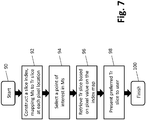
l'obtention d'une pluralité d'images de tomosynthèse (Tr) de la poitrine d'une patiente, les images de tomosynthèse (Tr) étant des coupes de coordonnées X, Y (Tr) à différents emplacements de l'axe Z de la poitrine, les images (Tr) ayant un ou plusieurs emplacement de coordonnées X, Y correspondants ; dans lequel le procédé comprend les étapes de :génération d'une image 2D synthétisée (Ms) de la poitrine de la patiente uniquement à partir de la pluralité obtenue d'images de tomosynthèse (Tr), l'image 2D synthétisée (Ms) semblable à celle d'une image mammographique 2D conventionnelle (Mp), dans lequel la génération de l'image 2D synthétisée (Ms) comprend la construction d'une image fusionnée (Imerge) en important une ou plusieurs régions d'une ou de plusieurs de la pluralité d'images de tomosynthèse (Tr) dans l'image fusionnée (Imerge), dans laquelle l'une ou les régions (35) importées dans l'image fusionnée (Imerge) comprennent au moins une partie d'une ou de plusieurs images sources de tomosynthèse (Tr) pour cette région, et dans lequel les régions (35) sont importées dans l'image fusionnée (Imerge) à des emplacements de coordonnées X, Y (35I) correspondant aux emplacements de coordonnées X, Y des régions respectives dans leur image source (Tr), dans lequel chaque image de la pluralité d'images de tomosynthèse (Tr) contient une ou des régions (35) définies par leurs emplacements de coordonnées X, Y qui sont communs à toutes les images (Tr) de la pluralité, et dans lequel l'une de chacune desdites régions communes (35) est importée de la pluralité d'images dans l'image fusionnée sur la base d'une comparaison d'un ou des attributs (134, 144, 154) de la région commune respective (35) de chaque image (Tr) ;affichage de l'image 2D synthétisée (Ms) ;réception d'une indication d'utilisateur (94) d'une région d'intérêt (134, 144, 154) dans l'image 2D synthétisée (Ms) ; etaffichage d'au moins la partie d'une ou de plusieurs images sources de tomosynthèse (Tr). - Procédé selon la revendication 1, dans lequel au moins l'un du ou des attributs (134, 144, 154) est choisi par l'utilisateur (94).
- Procédé selon l'une quelconque des revendications précédentes, comprenant en outre la génération d'une carte d'indice (120) comprenant l'identification d'informations des images sources de tomosynthèse qui forment l'image fusionnée (Imerge).
- Procédé selon l'une quelconque des revendications précédentes, dans lequel la région (35) est automatiquement mise en surbrillance dans l'image 2D synthétisée et/ou affichée dans au moins une partie de la ou des images de la pluralité d'images sources de tomosynthèse (Tr) ; et/ou
comprenant en outre la mise en surbrillance de la région dans l'image 2D synthétisée et/ou l'affichage dans au moins une partie de la ou des images de la pluralité d'images sources de tomosynthèse (Tr) en réponse à une autre instruction de l'utilisateur reçue ou à une certaine activité de l'utilisateur détectée par le biais de l'interface d'utilisateur,
dans lequel de préférence la région est mise en surbrillance par une ligne de contour (108) représentant une limite de l'objet ou de la région en surbrillance ou dans lequel la région est mise en surbrillance d'une manière indiquant que la région en surbrillance est ou contient un type spécifié de structure tissulaire (134, 144, 154). - Procédé selon l'une quelconque des revendications précédentes, dans lequel la ou les régions sont basées sur l'application d'un ou de plusieurs algorithmes de CAO aux images sources de tomosynthèse (Tr).
- Procédé selon la revendication 5, dans lequel le ou les algorithmes de CAO attribuent des valeurs numériques, des pondérations ou des seuils aux pixels ou aux régions des images sources de tomosynthèse respectives (Tr).
- Procédé selon la revendication 6, dans lequel l'affichage comprend l'affichage des pixels de la partie de la ou des images sources de tomosynthèse (Tr).
- Procédé selon l'une quelconque des revendications précédentes, dans lesquelles l'écran de la partie de la ou des images sources de tomosynthèse (Tr) s'affiche simultanément avec les images 2D synthétisées (Ms).
- Système de traitement, d'affichage et de navigation d'informations sur les tissus mammaires, comprenant :un ordinateur de traitement d'image (3, 4) ;au moins un moniteur d'affichage d'image (5) ; etune interface utilisateur couplée fonctionnellement à l'ordinateur de traitement d'image, dans lequel l'ordinateur de traitement d'image est configuré pour générer une image 2D synthétisée (Ms) de la poitrine d'une patiente à partir seulement d'une pluralité d'images de tomosynthèse (Tr) de la poitrine, l'image 2D synthétisée (Ms) étant semblable à celle d'une image mammographique 2D conventionnelle (Mp), dans lequel les images de tomosynthèse (Tr) sont des coupes de coordonnées X, Y (Tr) à différents emplacements de l'axe Z de la poitrine, les images (Tr) ont un ou plusieurs emplacements de coordonnées X, Y correspondants dans lesquels l'image 2D synthétisée (Ms) est générée en construisant une image fusionnée (Imerge) par importation d'une ou de plusieurs régions (35) d'une ou de plusieurs de la pluralité d'images de tomosynthèse (Tr) dans l'image fusionnée, dans laquelle la ou les régions importées dans l'image fusionnée (Imerge) comprennent au moins une partie d'une ou de plusieurs images sources de tomosynthèse (Tr) pour cette région (35), et dans lequel les régions (35) sont importées dans l'image fusionnée (Imerge) aux emplacements de coordonnées X, Y (351) correspondant aux emplacements de coordonnées X, Y des régions respectives dans leur image source (Tr), dans lequel chaque image de la pluralité d'images de tomosynthèse (Tr) contient une ou plusieurs régions (35) définies par leurs emplacements de coordonnées X, Y qui sont communs à toutes les images (Tr) de la pluralité, et dans lequel l'une de chacune desdites régions communes (35) est importée de la pluralité d'images dans l'image fusionnée sur la base d'une comparaison d'un ou de plusieurs attributs (134, 144, 154) de la région commune respective (35) de chaque image (Tr),dans lequel l'ordinateur de traitement d'image est configuré pour afficher l'image 2D synthétisée (Ms) sur l'au moins une image d'un moniteur d'affichage (5), etdans lequel l'ordinateur de traitement d'image est en outre configuré pour provoquer l'affichage sur un moniteur d'affichage identique ou différent du ou des moniteurs d'affichage au moins de la partie de l'une des images sources de tomosynthèse (Tr) en réponse à un choix ou à une indication par l'utilisateur (94) d'une région d'intérêt (134, 144, 154) dans l'image 2D synthétisée (Ms).
- Système selon la revendication 9, l'ordinateur de traitement d'image étant en outre configuré pour générer une carte d'indice (120) comprenant des informations d'identification d'images choisies parmi la pluralité d'images de tomosynthèse (Tr) qui sont des images sources (Tr) ou qui contiennent autrement une représentation de régions très semblable et/ou des objets affichés dans l'image 2D synthétisée (Ms).
- Système selon la revendication 9 ou 10, dans lequel l'ordinateur de traitement d'image est en outre configuré pour mettre en surbrillance un objet ou une région d'intérêt dans l'image 2D synthétisée (Ms), et/ou
dans lequel l'ordinateur de traitement d'image est configuré pour mettre en surbrillance des objets ou des régions dans l'image 2D synthétisée affichée (Ms) en réponse à une instruction de l'utilisateur reçue ou à une certaine activité de l'utilisateur détectée par le biais de l'interface utilisateur,
dans lequel de préférence l'ordinateur de traitement d'image est configuré pour mettre en surbrillance des objets ou des régions (134, 144, 154) dans l'image 2D synthétisée affichée (Ms) avec une ligne de contour (108) représentant une limite de l'objet ou de la région en surbrillance ou l'ordinateur de traitement d'image est configuré pour mettre en surbrillance des objets ou des régions dans l'image 2D synthétisée affichée (Ms) d'une manière indiquant que l'objet ou la région en surbrillance est ou contient un type spécifique de structure tissulaire (134, 144, 154). - Système selon l'une quelconque des revendications 9 à 11, dans lequel l'ordinateur du processeur d'image est agencé pour baser une ou plusieurs régions sur l'application d'un ou de plusieurs algorithmes de CAO aux images sources de tomosynthèse (Tr),
dans lequel de préférence l'ordinateur du processeur d'image utilisant le ou les algorithmes de CAO est agencé pour attribuer des valeurs numériques, des pondérations ou des seuils à des pixels ou à des régions des images sources de tomosynthèse respectives (Tr),
dans lequel plus préférablement l'ordinateur du processeur d'image est agencé pour afficher les pixels de la partie d'une ou de plusieurs images sources de tomosynthèse (Tr). - Système selon l'une quelconque des revendications 9 à 12, dans lequel l'ordinateur du processeur d'image est agencé pour afficher la partie de la ou des images sources de tomosynthèse (Tr) simultanément avec les images 2D synthétisées (Ms).
Applications Claiming Priority (3)
| Application Number | Priority Date | Filing Date | Title |
|---|---|---|---|
| US201261597958P | 2012-02-13 | 2012-02-13 | |
| PCT/US2013/025993 WO2013123091A1 (fr) | 2012-02-13 | 2013-02-13 | Système et procédé pour naviguer dans une pile de tomosynthèse par utilisation de données d'images synthétisées |
| EP13749870.5A EP2814396B1 (fr) | 2012-02-13 | 2013-02-13 | Système et procédé pour naviguer dans une pile de tomosynthèse par utilisation de données d'images synthétisées |
Related Parent Applications (1)
| Application Number | Title | Priority Date | Filing Date |
|---|---|---|---|
| EP13749870.5A Division EP2814396B1 (fr) | 2012-02-13 | 2013-02-13 | Système et procédé pour naviguer dans une pile de tomosynthèse par utilisation de données d'images synthétisées |
Publications (2)
| Publication Number | Publication Date |
|---|---|
| EP3315072A1 EP3315072A1 (fr) | 2018-05-02 |
| EP3315072B1 true EP3315072B1 (fr) | 2020-04-29 |
Family
ID=48984668
Family Applications (2)
| Application Number | Title | Priority Date | Filing Date |
|---|---|---|---|
| EP17176956.5A Active EP3315072B1 (fr) | 2012-02-13 | 2013-02-13 | Système et procédé pour naviguer dans une pile de tomosynthèse par utilisation de données d'images synthétisées |
| EP13749870.5A Active EP2814396B1 (fr) | 2012-02-13 | 2013-02-13 | Système et procédé pour naviguer dans une pile de tomosynthèse par utilisation de données d'images synthétisées |
Family Applications After (1)
| Application Number | Title | Priority Date | Filing Date |
|---|---|---|---|
| EP13749870.5A Active EP2814396B1 (fr) | 2012-02-13 | 2013-02-13 | Système et procédé pour naviguer dans une pile de tomosynthèse par utilisation de données d'images synthétisées |
Country Status (6)
| Country | Link |
|---|---|
| US (5) | US9805507B2 (fr) |
| EP (2) | EP3315072B1 (fr) |
| JP (2) | JP6240097B2 (fr) |
| CN (1) | CN104135935A (fr) |
| ES (1) | ES2641456T3 (fr) |
| WO (1) | WO2013123091A1 (fr) |
Families Citing this family (52)
| Publication number | Priority date | Publication date | Assignee | Title |
|---|---|---|---|---|
| US8571289B2 (en) | 2002-11-27 | 2013-10-29 | Hologic, Inc. | System and method for generating a 2D image from a tomosynthesis data set |
| WO2007095330A2 (fr) | 2006-02-15 | 2007-08-23 | Hologic Inc | Biopsie mammaire et localisation a l'aiguille a l'aide de systemes de tomosynthese |
| WO2011043838A1 (fr) | 2009-10-08 | 2011-04-14 | Hologic, Inc . | Système de ponction-biopsie du sein et méthode d'utilisation |
| WO2012071429A1 (fr) | 2010-11-26 | 2012-05-31 | Hologic, Inc. | Interface d'utilisateur pour station de travail de visualisation d'images médicales |
| AU2012225398B2 (en) | 2011-03-08 | 2017-02-02 | Hologic, Inc. | System and method for dual energy and/or contrast enhanced breast imaging for screening, diagnosis and biopsy |
| KR102109588B1 (ko) | 2011-11-27 | 2020-05-12 | 홀로직, 인크. | 유방 조직 이미지를 프로세싱하고, 디스플레잉하고, 네비게이팅하기 위한 방법 |
| ES2641456T3 (es) * | 2012-02-13 | 2017-11-10 | Hologic, Inc. | Sistema y método para navegar por una pila de tomosíntesis usando datos de imágenes sintetizadas |
| US20130342577A1 (en) * | 2012-06-20 | 2013-12-26 | Carestream Health, Inc. | Image synthesis for diagnostic review |
| WO2014110283A1 (fr) | 2013-01-10 | 2014-07-17 | Hologic, Inc. | Système et procédé de réduction du volume de transmission de données en tomosynthèse |
| US10092358B2 (en) | 2013-03-15 | 2018-10-09 | Hologic, Inc. | Tomosynthesis-guided biopsy apparatus and method |
| KR102340594B1 (ko) * | 2013-10-24 | 2021-12-20 | 앤드류 피 스미스 | 엑스레이 유도 유방 생검을 네비게이팅하기 위한 시스템 및 방법 |
| CN105979875B (zh) * | 2014-02-04 | 2019-12-31 | 皇家飞利浦有限公司 | 用于生成乳房参数图的医学成像设备、方法、装置和计算机可读介质 |
| US9613440B2 (en) | 2014-02-12 | 2017-04-04 | General Electric Company | Digital breast Tomosynthesis reconstruction using adaptive voxel grid |
| US20170169609A1 (en) * | 2014-02-19 | 2017-06-15 | Koninklijke Philips N.V. | Motion adaptive visualization in medical 4d imaging |
| WO2015130916A1 (fr) | 2014-02-28 | 2015-09-03 | Hologic, Inc. | Système et procédé de production et d'affichage de dalles d'image de tomosynthèse |
| US10146424B2 (en) * | 2014-02-28 | 2018-12-04 | Dell Products, Lp | Display of objects on a touch screen and their selection |
| EP3662840B1 (fr) * | 2014-09-16 | 2023-12-13 | Sirona Dental, Inc. | Procédés, systèmes, appareils et programmes informatiques pour le traitement d'images tomographiques |
| JP6126058B2 (ja) * | 2014-09-30 | 2017-05-10 | 富士フイルム株式会社 | 画像表示装置、画像処理装置、放射線画像撮影システム、断層画像表示方法、及び断層画像表示プログラム。 |
| JP6766045B2 (ja) * | 2014-11-20 | 2020-10-07 | コーニンクレッカ フィリップス エヌ ヴェKoninklijke Philips N.V. | トモシンセシスデータから合成マンモグラムを生成する方法 |
| CN107405126B (zh) * | 2015-03-10 | 2022-01-25 | 皇家飞利浦有限公司 | 检索成对的医学图像的对应结构 |
| US9792703B2 (en) * | 2015-07-06 | 2017-10-17 | Siemens Healthcare Gmbh | Generating a synthetic two-dimensional mammogram |
| JP6456550B2 (ja) | 2015-09-01 | 2019-01-23 | コーニンクレッカ フィリップス エヌ ヴェKoninklijke Philips N.V. | 体の部位の医療像データを表示するための装置 |
| US9990713B2 (en) * | 2016-06-09 | 2018-06-05 | Definiens Ag | Detecting and visualizing correlations between measured correlation values and correlation reference values of a pathway |
| JP6827739B2 (ja) * | 2016-08-31 | 2021-02-10 | キヤノン株式会社 | 画像表示装置、画像表示方法及びプログラム |
| CN106407687A (zh) * | 2016-09-22 | 2017-02-15 | 沈阳东软医疗系统有限公司 | 一种dicom图像组合的方法及装置 |
| US10157460B2 (en) | 2016-10-25 | 2018-12-18 | General Electric Company | Interpolated tomosynthesis projection images |
| US10096106B2 (en) | 2016-11-10 | 2018-10-09 | General Electric Company | Combined medical imaging |
| US10463333B2 (en) | 2016-12-13 | 2019-11-05 | General Electric Company | Synthetic images for biopsy control |
| JP7169986B2 (ja) * | 2017-03-30 | 2022-11-11 | ホロジック, インコーポレイテッド | オブジェクトグリッド増強を用いて高次元画像データから低次元画像データを合成するためのシステムおよび方法 |
| JP7174710B2 (ja) * | 2017-03-30 | 2022-11-17 | ホロジック, インコーポレイテッド | 合成乳房組織画像を生成するための標的オブジェクト増強のためのシステムおよび方法 |
| EP3600047A1 (fr) | 2017-03-30 | 2020-02-05 | Hologic, Inc. | Système et procédé de synthèse et de représentation d'image de caractéristique multiniveau hiérarchique |
| US11704850B2 (en) | 2017-04-07 | 2023-07-18 | Formus Labs Limited | System for transmitting and viewing a series of images |
| US11403483B2 (en) | 2017-06-20 | 2022-08-02 | Hologic, Inc. | Dynamic self-learning medical image method and system |
| US10702217B2 (en) * | 2017-08-24 | 2020-07-07 | General Electric Company | System and method for imaging a patient |
| CN109840898A (zh) * | 2017-11-24 | 2019-06-04 | 通用电气公司 | 一种医学图像的成像显示方法和成像设备 |
| EP3518182B1 (fr) * | 2018-01-26 | 2022-05-18 | Siemens Healthcare GmbH | Tranches inclinées en dbt |
| US11442591B2 (en) * | 2018-04-09 | 2022-09-13 | Lockheed Martin Corporation | System, method, computer readable medium, and viewer-interface for prioritized selection of mutually occluding objects in a virtual environment |
| US11204677B2 (en) * | 2018-10-22 | 2021-12-21 | Acclarent, Inc. | Method for real time update of fly-through camera placement |
| JP7236948B2 (ja) | 2019-07-16 | 2023-03-10 | 富士フイルム株式会社 | 画像処理システム、画像処理方法、及び画像処理プログラム |
| US11883206B2 (en) | 2019-07-29 | 2024-01-30 | Hologic, Inc. | Personalized breast imaging system |
| CN114746953A (zh) | 2019-09-27 | 2022-07-12 | 豪洛捷公司 | 用于预测审查2d/3d乳房图像的阅读时间和阅读复杂度的ai系统 |
| JP2022554190A (ja) * | 2019-10-25 | 2022-12-28 | ディープヘルス, インコーポレイテッド | 三次元画像データを解析するためのシステム及び方法 |
| CN110991050B (zh) * | 2019-12-06 | 2022-10-14 | 万翼科技有限公司 | Cad叠图方法及相关产品 |
| WO2021157181A1 (fr) * | 2020-02-04 | 2021-08-12 | 富士フイルム株式会社 | Dispositif de réglage d'image, procédé et programme |
| JP7270782B2 (ja) * | 2020-02-04 | 2023-05-10 | 富士フイルム株式会社 | 画像設定装置、画像設定装置の作動方法および画像設定プログラム |
| WO2021182076A1 (fr) * | 2020-03-09 | 2021-09-16 | 富士フイルム株式会社 | Dispositif de commande d'affichage, procédé de commande d'affichage et programme de commande d'affichage |
| CN115297778A (zh) * | 2020-03-18 | 2022-11-04 | 富士胶片株式会社 | 图像处理装置、方法及程序 |
| US11481038B2 (en) | 2020-03-27 | 2022-10-25 | Hologic, Inc. | Gesture recognition in controlling medical hardware or software |
| CN111430011B (zh) * | 2020-03-31 | 2023-08-29 | 杭州依图医疗技术有限公司 | 细胞染色图像的显示方法、处理方法及存储介质 |
| WO2022018943A1 (fr) * | 2020-07-22 | 2022-01-27 | 富士フイルム株式会社 | Dispositif de traitement d'image, procédé, et programme |
| CN113052166A (zh) * | 2021-02-05 | 2021-06-29 | 杭州依图医疗技术有限公司 | 病理图像的显示方法及装置 |
| WO2023140475A1 (fr) * | 2022-01-21 | 2023-07-27 | 주식회사 루닛 | Appareil et procédé de liaison et de fourniture d'emplacement de lésion entre une image de guidage et une image de tomosynthèse 3d comprenant une pluralité de tranches d'image 3d |
Family Cites Families (406)
| Publication number | Priority date | Publication date | Assignee | Title |
|---|---|---|---|---|
| JP4054402B2 (ja) | 1997-04-25 | 2008-02-27 | 株式会社東芝 | X線断層撮影装置 |
| US3502878A (en) | 1967-09-22 | 1970-03-24 | Us Health Education & Welfare | Automatic x-ray apparatus for limiting the field size of a projected x-ray beam in response to film size and to source-to-film distance |
| US7050611B2 (en) | 2001-05-29 | 2006-05-23 | Mevis Breastcare Gmbh Co. Kg | Method and computer system for screening of medical cases |
| US3863073A (en) | 1973-04-26 | 1975-01-28 | Machlett Lab Inc | Automatic system for precise collimation of radiation |
| US3971950A (en) | 1975-04-14 | 1976-07-27 | Xerox Corporation | Independent compression and positioning device for use in mammography |
| US4160906A (en) | 1977-06-23 | 1979-07-10 | General Electric Company | Anatomically coordinated user dominated programmer for diagnostic x-ray apparatus |
| DE2838901C2 (de) | 1978-09-06 | 1986-11-06 | Siemens AG, 1000 Berlin und 8000 München | Katapultrasterlade |
| FR2512024A1 (fr) | 1981-08-27 | 1983-03-04 | Adir | Ethers tricycliques, leur preparation et les compositions pharmaceutiques les contenant |
| FR2549248B1 (fr) | 1983-06-24 | 1986-01-31 | Thomson Csf | Porte-cassette escamotable pour appareil d'examen radiologique et radiographique |
| DE3339775A1 (de) | 1983-11-03 | 1985-05-15 | Siemens AG, 1000 Berlin und 8000 München | Roentgendiagnostikgeraet mit strahlenfiltern |
| JPS60129034A (ja) | 1983-12-16 | 1985-07-10 | 横河メディカルシステム株式会社 | X線断層撮像装置の操作卓 |
| US4706269A (en) | 1985-03-11 | 1987-11-10 | Reina Leo J | Anti-scatter grid structure |
| US4773087A (en) | 1986-04-14 | 1988-09-20 | University Of Rochester | Quality of shadowgraphic x-ray images |
| USRE33634E (en) | 1986-09-23 | 1991-07-09 | Method and structure for optimizing radiographic quality by controlling X-ray tube voltage, current focal spot size and exposure time | |
| US4821727A (en) | 1986-10-30 | 1989-04-18 | Elscint Ltd. | Mammographic biopsy needle holder system |
| US4819258A (en) | 1986-11-28 | 1989-04-04 | Bennett X-Ray Corp. | Auto-setting of KV in an x-ray machine after selection of technic factors |
| US4907156A (en) | 1987-06-30 | 1990-03-06 | University Of Chicago | Method and system for enhancement and detection of abnormal anatomic regions in a digital image |
| US5051904A (en) | 1988-03-24 | 1991-09-24 | Olganix Corporation | Computerized dynamic tomography system |
| US5740270A (en) | 1988-04-08 | 1998-04-14 | Neuromedical Systems, Inc. | Automated cytological specimen classification system and method |
| DK654488A (da) | 1988-11-23 | 1990-05-24 | Nordisk Roentgen Tech App | Roentgenapparat |
| US5099846A (en) | 1988-12-23 | 1992-03-31 | Hardy Tyrone L | Method and apparatus for video presentation from a variety of scanner imaging sources |
| FR2645006A1 (fr) | 1989-03-29 | 1990-10-05 | Gen Electric Cgr | Mammographe equipe d'un dispositif de vue stereotaxiques integre et procede d'utilisation d'un tel mammographe |
| FR2646340A1 (fr) | 1989-04-28 | 1990-11-02 | Gen Electric Cgr | Porte-cassette adaptable en dimension et en position pour mammographie |
| EP0406455B1 (fr) | 1989-07-03 | 1994-09-21 | Siemens Aktiengesellschaft | Appareil diagnostique à rayons X pour les mammographies |
| US5133020A (en) | 1989-07-21 | 1992-07-21 | Arch Development Corporation | Automated method and system for the detection and classification of abnormal lesions and parenchymal distortions in digital medical images |
| US4969174A (en) | 1989-09-06 | 1990-11-06 | General Electric Company | Scanning mammography system with reduced scatter radiation |
| CA2014918A1 (fr) | 1989-09-06 | 1991-03-06 | James A. Mcfaul | Systeme de mammographie par balyage avec meilleure visualisation de la ligne cutanee |
| US5078142A (en) | 1989-11-21 | 1992-01-07 | Fischer Imaging Corporation | Precision mammographic needle biopsy system |
| US5240011A (en) | 1991-11-27 | 1993-08-31 | Fischer Imaging Corporation | Motorized biopsy needle positioner |
| US5415169A (en) | 1989-11-21 | 1995-05-16 | Fischer Imaging Corporation | Motorized mammographic biopsy apparatus |
| AU645535B2 (en) | 1989-11-27 | 1994-01-20 | Bard International, Inc. | Puncture guide for computer tomography |
| US5199056A (en) | 1989-11-28 | 1993-03-30 | Darrah Carol J | Mammography compression paddle |
| US5481623A (en) | 1990-04-19 | 1996-01-02 | Fuji Photo Film Co., Ltd. | Apparatus for determining an image position on imaging media |
| FR2668359B1 (fr) | 1990-10-24 | 1998-02-20 | Gen Electric Cgr | Mammographe muni d'un porte-aiguille perfectionne. |
| US5409497A (en) | 1991-03-11 | 1995-04-25 | Fischer Imaging Corporation | Orbital aiming device for mammo biopsy |
| US5129911A (en) | 1991-03-11 | 1992-07-14 | Siczek Bernard W | Orbital aiming device |
| US5279309A (en) | 1991-06-13 | 1994-01-18 | International Business Machines Corporation | Signaling device and method for monitoring positions in a surgical operation |
| US5163075A (en) | 1991-08-08 | 1992-11-10 | Eastman Kodak Company | Contrast enhancement of electrographic imaging |
| US5941832A (en) | 1991-09-27 | 1999-08-24 | Tumey; David M. | Method and apparatus for detection of cancerous and precancerous conditions in a breast |
| US5289520A (en) | 1991-11-27 | 1994-02-22 | Lorad Corporation | Stereotactic mammography imaging system with prone position examination table and CCD camera |
| US5594769A (en) | 1991-11-27 | 1997-01-14 | Thermotrex Corporation | Method and apparatus for obtaining stereotactic mammographic guided needle breast biopsies |
| US5343390A (en) | 1992-02-28 | 1994-08-30 | Arch Development Corporation | Method and system for automated selection of regions of interest and detection of septal lines in digital chest radiographs |
| US5359637A (en) | 1992-04-28 | 1994-10-25 | Wake Forest University | Self-calibrated tomosynthetic, radiographic-imaging system, method, and device |
| US5386447A (en) | 1992-09-23 | 1995-01-31 | Fischer Imaging Corporation | Mammographic screening and biopsy apparatus |
| US5596200A (en) | 1992-10-14 | 1997-01-21 | Primex | Low dose mammography system |
| FR2703237B1 (fr) | 1993-03-29 | 1995-05-19 | Ge Medical Syst Sa | Mammographe équipé d'un dispositif de prises en vues stéréotaxiques à détecteur numérique et procédé d'utilisation d'un tel mammographe . |
| US5491627A (en) | 1993-05-13 | 1996-02-13 | Arch Development Corporation | Method and system for the detection of microcalcifications in digital mammograms |
| US5878746A (en) | 1993-08-25 | 1999-03-09 | Lemelson; Jerome H. | Computerized medical diagnostic system |
| US5365562A (en) | 1993-09-20 | 1994-11-15 | Fischer Imaging Corporation | Digital imaging apparatus |
| US6075879A (en) | 1993-09-29 | 2000-06-13 | R2 Technology, Inc. | Method and system for computer-aided lesion detection using information from multiple images |
| US5526394A (en) | 1993-11-26 | 1996-06-11 | Fischer Imaging Corporation | Digital scan mammography apparatus |
| US5452367A (en) | 1993-11-29 | 1995-09-19 | Arch Development Corporation | Automated method and system for the segmentation of medical images |
| CA2113752C (fr) | 1994-01-19 | 1999-03-02 | Stephen Michael Rooks | Systeme d'inspection pour l'imagerie de coupe |
| DE4414689C2 (de) | 1994-04-26 | 1996-08-29 | Siemens Ag | Röntgendiagnostikeinrichtung |
| US5499097A (en) | 1994-09-19 | 1996-03-12 | Neopath, Inc. | Method and apparatus for checking automated optical system performance repeatability |
| AU3371395A (en) | 1994-09-20 | 1996-04-19 | Neopath, Inc. | Biological specimen analysis system processing integrity checking apparatus |
| US5647025A (en) | 1994-09-20 | 1997-07-08 | Neopath, Inc. | Automatic focusing of biomedical specimens apparatus |
| US5557097A (en) | 1994-09-20 | 1996-09-17 | Neopath, Inc. | Cytological system autofocus integrity checking apparatus |
| US5553111A (en) | 1994-10-26 | 1996-09-03 | The General Hospital Corporation | Apparatus and method for improved tissue imaging |
| US5506877A (en) | 1994-11-23 | 1996-04-09 | The General Hospital Corporation | Mammography breast compression device and method |
| US5712890A (en) * | 1994-11-23 | 1998-01-27 | Thermotrex Corp. | Full breast digital mammography device |
| US5657362A (en) | 1995-02-24 | 1997-08-12 | Arch Development Corporation | Automated method and system for computerized detection of masses and parenchymal distortions in medical images |
| US5671288A (en) | 1995-05-31 | 1997-09-23 | Neopath, Inc. | Method and apparatus for assessing slide and specimen preparation quality |
| US6216540B1 (en) | 1995-06-06 | 2001-04-17 | Robert S. Nelson | High resolution device and method for imaging concealed objects within an obscuring medium |
| US5820623A (en) | 1995-06-20 | 1998-10-13 | Ng; Wan Sing | Articulated arm for medical procedures |
| US5642433A (en) | 1995-07-31 | 1997-06-24 | Neopath, Inc. | Method and apparatus for image contrast quality evaluation |
| US5642441A (en) | 1995-10-24 | 1997-06-24 | Neopath, Inc. | Separation apparatus and method for measuring focal plane |
| US5818898A (en) | 1995-11-07 | 1998-10-06 | Kabushiki Kaisha Toshiba | X-ray imaging apparatus using X-ray planar detector |
| US5693948A (en) | 1995-11-21 | 1997-12-02 | Loral Fairchild Corporation | Advanced CCD-based x-ray image sensor system |
| US5627869A (en) | 1995-11-22 | 1997-05-06 | Thermotrex Corporation | Mammography apparatus with proportional collimation |
| FI955636A0 (fi) | 1995-11-23 | 1995-11-23 | Planmed Oy | Foerfarande och system foer styrning av funktionerna av en mammografiaanordning |
| US6215892B1 (en) | 1995-11-30 | 2001-04-10 | Chromavision Medical Systems, Inc. | Method and apparatus for automated image analysis of biological specimens |
| US5769086A (en) | 1995-12-06 | 1998-06-23 | Biopsys Medical, Inc. | Control system and method for automated biopsy device |
| JPH09198490A (ja) | 1996-01-22 | 1997-07-31 | Hitachi Medical Corp | 3次元離散データ投影装置 |
| JPH09238934A (ja) | 1996-03-11 | 1997-09-16 | Toshiba Medical Eng Co Ltd | 画像表示システム |
| DE19619924A1 (de) | 1996-05-17 | 1997-11-20 | Siemens Ag | Verfahren zur Erstellung von Tomosyntheseaufnahmen |
| DE19619925C2 (de) | 1996-05-17 | 1999-09-09 | Sirona Dental Systems Gmbh | Röntgendiagnostikgerät für Tomosynthese |
| DE19619915A1 (de) | 1996-05-17 | 1997-11-20 | Siemens Ag | Verfahren zur Erstellung von Tomosyntheseaufnahmen |
| DE19619913C2 (de) | 1996-05-17 | 2001-03-15 | Sirona Dental Systems Gmbh | Röntgendiagnostikgerät für Tomosynthese |
| US6067079A (en) | 1996-06-13 | 2000-05-23 | International Business Machines Corporation | Virtual pointing device for touchscreens |
| US5835079A (en) | 1996-06-13 | 1998-11-10 | International Business Machines Corporation | Virtual pointing device for touchscreens |
| US5841124A (en) | 1996-06-19 | 1998-11-24 | Neopath, Inc. | Cytological system autofocus integrity checking apparatus |
| DE69739995D1 (de) | 1996-07-23 | 2010-10-28 | Gen Hospital Corp | Anordnung zur mammographie mittels tomosynthese |
| JPH1033523A (ja) | 1996-07-24 | 1998-02-10 | Hitachi Medical Corp | X線ct装置 |
| US5776062A (en) | 1996-10-15 | 1998-07-07 | Fischer Imaging Corporation | Enhanced breast imaging/biopsy system employing targeted ultrasound |
| US5986662A (en) | 1996-10-16 | 1999-11-16 | Vital Images, Inc. | Advanced diagnostic viewer employing automated protocol selection for volume-rendered imaging |
| US6293282B1 (en) | 1996-11-05 | 2001-09-25 | Jerome Lemelson | System and method for treating select tissue in living being |
| JP3878259B2 (ja) | 1996-11-13 | 2007-02-07 | 東芝医用システムエンジニアリング株式会社 | 医用画像処理装置 |
| US6137527A (en) | 1996-12-23 | 2000-10-24 | General Electric Company | System and method for prompt-radiology image screening service via satellite |
| US7117098B1 (en) | 1997-02-27 | 2006-10-03 | Cellomics, Inc. | Machine-readable storage medium for analyzing distribution of macromolecules between the cell membrane and the cell cytoplasm |
| US5999639A (en) | 1997-09-04 | 1999-12-07 | Qualia Computing, Inc. | Method and system for automated detection of clustered microcalcifications from digital mammograms |
| US20030135115A1 (en) | 1997-11-24 | 2003-07-17 | Burdette Everette C. | Method and apparatus for spatial registration and mapping of a biopsy needle during a tissue biopsy |
| US6442288B1 (en) | 1997-12-17 | 2002-08-27 | Siemens Aktiengesellschaft | Method for reconstructing a three-dimensional image of an object scanned in the context of a tomosynthesis, and apparatus for tomosynthesis |
| JP3554172B2 (ja) | 1998-01-09 | 2004-08-18 | キヤノン株式会社 | 放射線撮影装置 |
| US6175117B1 (en) | 1998-01-23 | 2001-01-16 | Quanta Vision, Inc. | Tissue analysis apparatus |
| US6289235B1 (en) | 1998-03-05 | 2001-09-11 | Wake Forest University | Method and system for creating three-dimensional images using tomosynthetic computed tomography |
| US6081577A (en) | 1998-07-24 | 2000-06-27 | Wake Forest University | Method and system for creating task-dependent three-dimensional images |
| US6375352B1 (en) | 1999-10-01 | 2002-04-23 | General Electric Company | Apparatus and method for obtaining x-ray tomosynthesis data for mammography |
| US6141398A (en) | 1998-08-25 | 2000-10-31 | General Electric Company | Protocol driven image reconstruction, display, and processing in a multislice imaging system |
| US6101236A (en) | 1998-10-02 | 2000-08-08 | University Of Iowa Research Foundation | Iterative method and apparatus for x-ray computed tomographic fluoroscopy |
| WO2000051484A2 (fr) | 1998-11-25 | 2000-09-08 | Fischer Imaging Corporation | Systeme d'interface utilisateur pour imageur mammographique |
| FR2786388B1 (fr) | 1998-11-27 | 2001-02-16 | Ge Medical Syst Sa | Procede de detection d'un tissu de nature determinee en radiologie numerique et son utilisation pour le reglage des parametres d'exposition |
| US6149301A (en) | 1998-12-30 | 2000-11-21 | General Electric Company | X-ray target centering apparatus for radiographic imaging system |
| JP2000200340A (ja) | 1999-01-06 | 2000-07-18 | Ge Yokogawa Medical Systems Ltd | 画像表示方法、画像表示装置およびct装置 |
| US6424332B1 (en) | 1999-01-29 | 2002-07-23 | Hunter Innovations, Inc. | Image comparison apparatus and method |
| CA2359322C (fr) | 1999-01-29 | 2007-09-18 | American Superconductor Corporation | Systeme de distribution d'electricite a stockage d'energie magnetique dans des supraconducteurs |
| US6233473B1 (en) | 1999-02-16 | 2001-05-15 | Hologic, Inc. | Determining body composition using fan beam dual-energy x-ray absorptiometry |
| US6272207B1 (en) | 1999-02-18 | 2001-08-07 | Creatv Microtech, Inc. | Method and apparatus for obtaining high-resolution digital X-ray and gamma ray images |
| US6256370B1 (en) | 2000-01-24 | 2001-07-03 | General Electric Company | Method and apparatus for performing tomosynthesis |
| US6689142B1 (en) | 1999-04-26 | 2004-02-10 | Scimed Life Systems, Inc. | Apparatus and methods for guiding a needle |
| US6292530B1 (en) | 1999-04-29 | 2001-09-18 | General Electric Company | Method and apparatus for reconstructing image data acquired by a tomosynthesis x-ray imaging system |
| DE19922346C2 (de) | 1999-05-14 | 2003-06-18 | Siemens Ag | Röntgendiagnostikeinrichtung für Tomosynthese oder Schichtung |
| US6243441B1 (en) | 1999-07-13 | 2001-06-05 | Edge Medical Devices | Active matrix detector for X-ray imaging |
| US20020173721A1 (en) | 1999-08-20 | 2002-11-21 | Novasonics, Inc. | User interface for handheld imaging devices |
| US6480565B1 (en) | 1999-11-18 | 2002-11-12 | University Of Rochester | Apparatus and method for cone beam volume computed tomography breast imaging |
| US6987831B2 (en) | 1999-11-18 | 2006-01-17 | University Of Rochester | Apparatus and method for cone beam volume computed tomography breast imaging |
| US6633674B1 (en) | 1999-11-24 | 2003-10-14 | General Electric Company | Picture archiving and communication system employing improved data compression |
| US6245028B1 (en) | 1999-11-24 | 2001-06-12 | Marconi Medical Systems, Inc. | Needle biopsy system |
| US6645520B2 (en) | 1999-12-16 | 2003-11-11 | Dermatrends, Inc. | Transdermal administration of nonsteroidal anti-inflammatory drugs using hydroxide-releasing agents as permeation enhancers |
| FR2803069B1 (fr) | 1999-12-28 | 2002-12-13 | Ge Medical Syst Sa | Procede et systeme de compensation de l'epaisseur d'un organe |
| US6411836B1 (en) | 1999-12-30 | 2002-06-25 | General Electric Company | Method and apparatus for user preferences configuring in an image handling system |
| US8352289B2 (en) | 1999-12-30 | 2013-01-08 | Dhi Computing, Inc. | Systems and methods for providing and maintaining electronic medical records |
| US6901156B2 (en) | 2000-02-04 | 2005-05-31 | Arch Development Corporation | Method, system and computer readable medium for an intelligent search workstation for computer assisted interpretation of medical images |
| US6744848B2 (en) | 2000-02-11 | 2004-06-01 | Brandeis University | Method and system for low-dose three-dimensional imaging of a scene |
| GB0006598D0 (en) | 2000-03-17 | 2000-05-10 | Isis Innovation | Three-dimensional reconstructions from images |
| US6351660B1 (en) | 2000-04-18 | 2002-02-26 | Litton Systems, Inc. | Enhanced visualization of in-vivo breast biopsy location for medical documentation |
| US6683934B1 (en) | 2000-06-05 | 2004-01-27 | General Electric Company | Dual energy x-ray imaging system and method for radiography and mammography |
| US6327336B1 (en) | 2000-06-05 | 2001-12-04 | Direct Radiography Corp. | Radiogram showing location of automatic exposure control sensor |
| US6389104B1 (en) | 2000-06-30 | 2002-05-14 | Siemens Corporate Research, Inc. | Fluoroscopy based 3-D neural navigation based on 3-D angiography reconstruction data |
| JP2002052018A (ja) | 2000-08-11 | 2002-02-19 | Canon Inc | 画像表示装置、画像表示方法および記憶媒体 |
| JP2002109510A (ja) | 2000-09-27 | 2002-04-12 | Fuji Photo Film Co Ltd | 異常陰影候補検出処理システム |
| JP2004512081A (ja) | 2000-10-20 | 2004-04-22 | コーニンクレッカ フィリップス エレクトロニクス エヌ ヴィ | 限られた角度範囲での断層写真合成法 |
| US6758824B1 (en) | 2000-11-06 | 2004-07-06 | Suros Surgical Systems, Inc. | Biopsy apparatus |
| WO2002069808A2 (fr) | 2000-11-06 | 2002-09-12 | Suros Surgical Systems, Inc. | Appareil de biopsie |
| US6468226B1 (en) | 2000-11-22 | 2002-10-22 | Mcintyre, Iv John J. | Remote tissue biopsy apparatus and associated methods |
| US7597663B2 (en) | 2000-11-24 | 2009-10-06 | U-Systems, Inc. | Adjunctive ultrasound processing and display for breast cancer screening |
| US7103205B2 (en) | 2000-11-24 | 2006-09-05 | U-Systems, Inc. | Breast cancer screening with ultrasound image overlays |
| US7615008B2 (en) | 2000-11-24 | 2009-11-10 | U-Systems, Inc. | Processing and displaying breast ultrasound information |
| US7556602B2 (en) | 2000-11-24 | 2009-07-07 | U-Systems, Inc. | Breast cancer screening with adjunctive ultrasound mammography |
| US6650928B1 (en) | 2000-11-27 | 2003-11-18 | Ge Medical Systems Global Technology Company, Llc | Color parametric and composite maps for CT perfusion |
| US6501819B2 (en) | 2000-12-18 | 2002-12-31 | Ge Medical Systems Global Technology Company, Llc | Medical diagnostic method and apparatus to control dual energy exposure techniques based on image information |
| FR2818116B1 (fr) | 2000-12-19 | 2004-08-27 | Ge Med Sys Global Tech Co Llc | Appareil de mammographie |
| EP1346322A1 (fr) | 2000-12-22 | 2003-09-24 | Koninklijke Philips Electronics N.V. | Visualisation st r oscopique d'une r gion entre deux plans de d coupe |
| US6463181B2 (en) | 2000-12-22 | 2002-10-08 | The United States Of America As Represented By The Secretary Of The Navy | Method for optimizing visual display of enhanced digital images |
| EP1364374A4 (fr) | 2001-02-01 | 2006-11-22 | Creatv Microtech Inc | Modeles de collimateurs et de grilles antidiffusion, et leur deplacement, fabrication et assemblage |
| US7030861B1 (en) | 2001-02-10 | 2006-04-18 | Wayne Carl Westerman | System and method for packing multi-touch gestures onto a hand |
| US6486764B2 (en) | 2001-02-16 | 2002-11-26 | Delphi Technologies, Inc. | Rotary position sensor |
| US20020188466A1 (en) | 2001-04-18 | 2002-12-12 | Barrette Pierre Philip | Secure digital medical intellectual property (IP) distribution, market applications, and mobile devices |
| US6620111B2 (en) | 2001-04-20 | 2003-09-16 | Ethicon Endo-Surgery, Inc. | Surgical biopsy device having automatic rotation of the probe for taking multiple samples |
| US6954667B2 (en) | 2001-06-28 | 2005-10-11 | Chemimage Corporation | Method for Raman chemical imaging and characterization of calcification in tissue |
| US6611575B1 (en) | 2001-07-27 | 2003-08-26 | General Electric Company | Method and system for high resolution 3D visualization of mammography images |
| US20030048260A1 (en) | 2001-08-17 | 2003-03-13 | Alec Matusis | System and method for selecting actions based on the identification of user's fingers |
| AU2002332758A1 (en) | 2001-08-31 | 2003-03-18 | Analogic Corporation | Image positioning method and system for tomosynthesis in a digital x-ray radiography system |
| US20030072478A1 (en) | 2001-10-12 | 2003-04-17 | Claus Bernhard Erich Hermann | Reconstruction method for tomosynthesis |
| JP4363833B2 (ja) * | 2001-10-16 | 2009-11-11 | 株式会社東芝 | 局所血流動態に関するインデックスを演算する方法及び装置 |
| WO2003037046A2 (fr) | 2001-10-19 | 2003-05-01 | Hologic, Inc. | Systeme et procede de mammographie utilisant des palpateurs de compression excentres, la collimation automatique et une grille anti-diffusion retractable |
| US6626849B2 (en) | 2001-11-01 | 2003-09-30 | Ethicon Endo-Surgery, Inc. | MRI compatible surgical biopsy device |
| DE60135559D1 (de) | 2001-11-19 | 2008-10-09 | St Microelectronics Srl | Verfahren zur Mischung von digitalen Bildern zur Erzeugung eines digitalen Bildes mit erweitertem Dynamikbereich |
| US6895077B2 (en) | 2001-11-21 | 2005-05-17 | University Of Massachusetts Medical Center | System and method for x-ray fluoroscopic imaging |
| US20030097055A1 (en) | 2001-11-21 | 2003-05-22 | Philips Medical Systems(Cleveland), Inc. | Method of reviewing tomographic scans with a large number of images |
| US6751285B2 (en) | 2001-11-21 | 2004-06-15 | General Electric Company | Dose management system for mammographic tomosynthesis |
| JP4099984B2 (ja) * | 2001-12-14 | 2008-06-11 | コニカミノルタホールディングス株式会社 | 異常陰影検出装置および画像出力装置 |
| US6978040B2 (en) | 2001-12-19 | 2005-12-20 | Canon Kabushiki Kaisha | Optical recovery of radiographic geometry |
| US6647092B2 (en) | 2002-01-18 | 2003-11-11 | General Electric Company | Radiation imaging system and method of collimation |
| SE524458C2 (sv) | 2002-03-01 | 2004-08-10 | Mamea Imaging Ab | Skyddsanordning vid en röntgenapparat |
| US7346381B2 (en) | 2002-11-01 | 2008-03-18 | Ge Medical Systems Global Technology Company Llc | Method and apparatus for medical intervention procedure planning |
| US6878115B2 (en) | 2002-03-28 | 2005-04-12 | Ultrasound Detection Systems, Llc | Three-dimensional ultrasound computed tomography imaging system |
| US7218766B2 (en) | 2002-04-15 | 2007-05-15 | General Electric Company | Computer aided detection (CAD) for 3D digital mammography |
| US20030194050A1 (en) | 2002-04-15 | 2003-10-16 | General Electric Company | Multi modality X-ray and nuclear medicine mammography imaging system and method |
| US6882700B2 (en) | 2002-04-15 | 2005-04-19 | General Electric Company | Tomosynthesis X-ray mammogram system and method with automatic drive system |
| US6752767B2 (en) | 2002-04-16 | 2004-06-22 | Vivant Medical, Inc. | Localization element with energized tip |
| US7139000B2 (en) | 2002-05-13 | 2006-11-21 | Ge Medical Systems Global Technology Company, Llc | Method, system and computer product for displaying axial images |
| US7295691B2 (en) | 2002-05-15 | 2007-11-13 | Ge Medical Systems Global Technology Company, Llc | Computer aided diagnosis of an image set |
| US11275405B2 (en) | 2005-03-04 | 2022-03-15 | Apple Inc. | Multi-functional hand-held device |
| US7599579B2 (en) | 2002-07-11 | 2009-10-06 | Ge Medical Systems Global Technology Company, Llc | Interpolated image filtering method and apparatus |
| US7450747B2 (en) | 2002-07-12 | 2008-11-11 | Ge Medical Systems Global Technology Company, Llc | System and method for efficiently customizing an imaging system |
| US20040036680A1 (en) | 2002-08-26 | 2004-02-26 | Mark Davis | User-interface features for computers with contact-sensitive displays |
| US6898331B2 (en) | 2002-08-28 | 2005-05-24 | Bae Systems Aircraft Controls, Inc. | Image fusion system and method |
| US6574304B1 (en) | 2002-09-13 | 2003-06-03 | Ge Medical Systems Global Technology Company, Llc | Computer aided acquisition of medical images |
| US6748044B2 (en) | 2002-09-13 | 2004-06-08 | Ge Medical Systems Global Technology Company, Llc | Computer assisted analysis of tomographic mammography data |
| US7260249B2 (en) * | 2002-09-27 | 2007-08-21 | Confirma Incorporated | Rules-based approach for processing medical images |
| US6940943B2 (en) | 2002-10-07 | 2005-09-06 | General Electric Company | Continuous scan tomosynthesis system and method |
| US7347829B2 (en) | 2002-10-07 | 2008-03-25 | Suros Surgical Systems, Inc. | Introduction system for minimally invasive surgical instruments |
| US6825838B2 (en) * | 2002-10-11 | 2004-11-30 | Sonocine, Inc. | 3D modeling system |
| US20040171933A1 (en) | 2002-11-25 | 2004-09-02 | Milton Stoller | Mammography needle biopsy system and method |
| US7616801B2 (en) | 2002-11-27 | 2009-11-10 | Hologic, Inc. | Image handling and display in x-ray mammography and tomosynthesis |
| US7123684B2 (en) | 2002-11-27 | 2006-10-17 | Hologic, Inc. | Full field mammography with tissue exposure control, tomosynthesis, and dynamic field of view processing |
| US6597762B1 (en) | 2002-11-27 | 2003-07-22 | Ge Medical Systems Global Technology Co., Llc | Method and apparatus of lesion detection and validation based on multiple reviews of a CT image |
| US7760924B2 (en) | 2002-11-27 | 2010-07-20 | Hologic, Inc. | System and method for generating a 2D image from a tomosynthesis data set |
| US8571289B2 (en) | 2002-11-27 | 2013-10-29 | Hologic, Inc. | System and method for generating a 2D image from a tomosynthesis data set |
| US7831296B2 (en) | 2002-11-27 | 2010-11-09 | Hologic, Inc. | X-ray mammography with tomosynthesis |
| US7577282B2 (en) | 2002-11-27 | 2009-08-18 | Hologic, Inc. | Image handling and display in X-ray mammography and tomosynthesis |
| US7406150B2 (en) | 2002-11-29 | 2008-07-29 | Hologic, Inc. | Distributed architecture for mammographic image acquisition and processing |
| US7904824B2 (en) | 2002-12-10 | 2011-03-08 | Siemens Medical Solutions Usa, Inc. | Medical imaging programmable custom user interface system and method |
| US7110490B2 (en) | 2002-12-10 | 2006-09-19 | General Electric Company | Full field digital tomosynthesis method and apparatus |
| US7634308B2 (en) | 2002-12-17 | 2009-12-15 | Kabushiki Kaisha Toshiba | Method and system for X-ray diagnosis of object in which X-ray contrast agent is injected |
| US7356113B2 (en) | 2003-02-12 | 2008-04-08 | Brandeis University | Tomosynthesis imaging system and method |
| US7333644B2 (en) | 2003-03-11 | 2008-02-19 | Siemens Medical Solutions Usa, Inc. | Systems and methods for providing automatic 3D lesion segmentation and measurements |
| JP4497837B2 (ja) | 2003-05-12 | 2010-07-07 | キヤノン株式会社 | 放射線画像撮影装置 |
| JP2007524461A (ja) | 2003-06-25 | 2007-08-30 | シーメンス メディカル ソリューションズ ユーエスエー インコーポレイテッド | 乳房撮像の自動診断及び決定支援システム及び方法 |
| US6885724B2 (en) | 2003-08-22 | 2005-04-26 | Ge Medical Systems Global Technology Company, Llc | Radiographic tomosynthesis image acquisition utilizing asymmetric geometry |
| US8090164B2 (en) | 2003-08-25 | 2012-01-03 | The University Of North Carolina At Chapel Hill | Systems, methods, and computer program products for analysis of vessel attributes for diagnosis, disease staging, and surgical planning |
| US7424141B2 (en) | 2003-08-29 | 2008-09-09 | Agilent Technologies, Inc. | System and method for performing auto-focused tomosynthesis |
| US7578781B2 (en) | 2003-09-18 | 2009-08-25 | Wisconsin Alumni Research Foundation | Device for placement of needles and radioactive seeds in radiotherapy |
| JP2005110843A (ja) * | 2003-10-06 | 2005-04-28 | Canon Inc | 放射線画像処理装置及び処理方法 |
| US7869862B2 (en) | 2003-10-15 | 2011-01-11 | Varian Medical Systems, Inc. | Systems and methods for functional imaging using contrast-enhanced multiple-energy computed tomography |
| US20050089205A1 (en) | 2003-10-23 | 2005-04-28 | Ajay Kapur | Systems and methods for viewing an abnormality in different kinds of images |
| JP2005149107A (ja) | 2003-11-14 | 2005-06-09 | Konica Minolta Medical & Graphic Inc | 医用画像管理システム |
| DE10353611B4 (de) | 2003-11-17 | 2013-01-17 | Siemens Aktiengesellschaft | Röntgendiagnostikgerät für Mammographieuntersuchungen |
| WO2005051197A2 (fr) | 2003-11-26 | 2005-06-09 | Koninklijke Philips Electronics, N.V. | Optimisation du deroulement des operations pour un environnement d'imagerie a haut debit |
| US8768026B2 (en) | 2003-11-26 | 2014-07-01 | Hologic, Inc. | X-ray imaging with x-ray markers that provide adjunct information but preserve image quality |
| US8265728B2 (en) | 2003-11-26 | 2012-09-11 | University Of Chicago | Automated method and system for the evaluation of disease and registration accuracy in the subtraction of temporally sequential medical images |
| US7727151B2 (en) | 2003-11-28 | 2010-06-01 | U-Systems Inc. | Navigation among multiple breast ultrasound volumes |
| US7773721B2 (en) | 2003-12-03 | 2010-08-10 | The General Hospital Corporation | Multi-segment cone-beam reconstruction system and method for tomosynthesis imaging |
| US20050135555A1 (en) | 2003-12-23 | 2005-06-23 | Claus Bernhard Erich H. | Method and system for simultaneously viewing rendered volumes |
| US7653229B2 (en) | 2003-12-23 | 2010-01-26 | General Electric Company | Methods and apparatus for reconstruction of volume data from projection data |
| CN1933787A (zh) | 2004-01-23 | 2007-03-21 | 特拉克斯医疗有限公司 | 在体内目标位置上完成某些程序的方法和装置 |
| US7298881B2 (en) | 2004-02-13 | 2007-11-20 | University Of Chicago | Method, system, and computer software product for feature-based correlation of lesions from multiple images |
| US7289825B2 (en) | 2004-03-15 | 2007-10-30 | General Electric Company | Method and system for utilizing wireless voice technology within a radiology workflow |
| US7142633B2 (en) | 2004-03-31 | 2006-11-28 | General Electric Company | Enhanced X-ray imaging system and method |
| US7699783B2 (en) | 2004-04-08 | 2010-04-20 | Techniscan, Inc. | Method for imaging and treating a breast |
| EP1750584B1 (fr) | 2004-05-14 | 2020-10-14 | Philips Intellectual Property & Standards GmbH | Systeme et methode de diagnostic du cancer du sein |
| GB0411402D0 (en) | 2004-05-21 | 2004-06-23 | Tissuomics Ltd | Penetrating radiation measurements |
| US7835562B2 (en) | 2004-07-23 | 2010-11-16 | General Electric Company | Methods and apparatus for noise reduction filtering of images |
| AU2005266901B2 (en) | 2004-07-23 | 2010-04-08 | Learning Tree International | System and method for electronic presentations |
| FR2873835A1 (fr) | 2004-07-29 | 2006-02-03 | Gen Electric | Procede et dispositif d'imagerie aux rayons x avec produit de contraste permettant une visualisation amelioree |
| WO2006020874A2 (fr) | 2004-08-10 | 2006-02-23 | The Research Foundation | Detecteur a ecran plat presentant un gain d'avalanche |
| US7725153B2 (en) | 2004-10-04 | 2010-05-25 | Hologic, Inc. | Estimating visceral fat by dual-energy x-ray absorptiometry |
| WO2006055830A2 (fr) | 2004-11-15 | 2006-05-26 | Hologic, Inc. | Generation et affichage geometriques de mise en correspondance de cliches mammaires et d'images de tomosynthese |
| EP1816965B1 (fr) | 2004-11-26 | 2016-06-29 | Hologic, Inc. | Systeme radiographique multimode integrant mammographie/tomosynthese |
| US20060132508A1 (en) | 2004-12-16 | 2006-06-22 | Navid Sadikali | Multi-planar image viewing system and method |
| US7616793B2 (en) | 2004-12-30 | 2009-11-10 | Hologic, Inc. | Medical image review workstation with integrated content-based resource retrieval |
| US9760214B2 (en) | 2005-02-23 | 2017-09-12 | Zienon, Llc | Method and apparatus for data entry input |
| US7859549B2 (en) | 2005-03-08 | 2010-12-28 | Agfa Inc. | Comparative image review system and method |
| US20060210131A1 (en) | 2005-03-15 | 2006-09-21 | Wheeler Frederick W Jr | Tomographic computer aided diagnosis (CAD) with multiple reconstructions |
| JP5038643B2 (ja) | 2005-04-06 | 2012-10-03 | 株式会社東芝 | 画像表示装置 |
| US8373652B2 (en) | 2005-04-06 | 2013-02-12 | Kabushiki Kaisha Toshiba | Image display apparatus and image display method |
| US10492749B2 (en) | 2005-05-03 | 2019-12-03 | The Regents Of The University Of California | Biopsy systems for breast computed tomography |
| DE102005022543A1 (de) | 2005-05-17 | 2006-11-23 | Siemens Ag | Mammographieverfahren und Mammographiegerät |
| US7606801B2 (en) | 2005-06-07 | 2009-10-20 | Varonis Inc. | Automatic management of storage access control |
| US7809175B2 (en) | 2005-07-01 | 2010-10-05 | Hologic, Inc. | Displaying and navigating computer-aided detection results on a review workstation |
| US7245694B2 (en) | 2005-08-15 | 2007-07-17 | Hologic, Inc. | X-ray mammography/tomosynthesis of patient's breast |
| WO2007027610A2 (fr) | 2005-08-30 | 2007-03-08 | Bruce Reiner | Dispositif et procede de navigation multifonctionnel |
| DE202005013910U1 (de) | 2005-09-02 | 2005-11-24 | Siemens Ag | Mammographiegerät mit Gesichtsschild |
| US20070052700A1 (en) | 2005-09-07 | 2007-03-08 | Wheeler Frederick W | System and method for 3D CAD using projection images |
| US10008184B2 (en) | 2005-11-10 | 2018-06-26 | Hologic, Inc. | System and method for generating a 2D image using mammography and/or tomosynthesis image data |
| US7342233B2 (en) | 2005-11-18 | 2008-03-11 | Sectra Mamea Ab | Method and arrangement relating to x-ray imaging |
| US20070118400A1 (en) | 2005-11-22 | 2007-05-24 | General Electric Company | Method and system for gesture recognition to drive healthcare applications |
| US20070236490A1 (en) | 2005-11-25 | 2007-10-11 | Agfa-Gevaert | Medical image display and review system |
| EP1966762A2 (fr) | 2005-12-29 | 2008-09-10 | Carestream Health, Inc. | Diagnostic medical a croisement de temps et de modalites |
| US20070156451A1 (en) | 2006-01-05 | 2007-07-05 | Gering David T | System and method for portable display of relevant healthcare information |
| US7581399B2 (en) | 2006-01-05 | 2009-09-01 | United Technologies Corporation | Damped coil pin for attachment hanger hinge |
| WO2007095330A2 (fr) | 2006-02-15 | 2007-08-23 | Hologic Inc | Biopsie mammaire et localisation a l'aiguille a l'aide de systemes de tomosynthese |
| US20070223651A1 (en) | 2006-03-21 | 2007-09-27 | Wagenaar Douglas J | Dual modality mammography device |
| US7489761B2 (en) | 2006-03-27 | 2009-02-10 | Hologic, Inc. | Breast compression for digital mammography, tomosynthesis and other modalities |
| EP2046223B1 (fr) | 2006-04-12 | 2011-03-02 | NAVAB, Nassir | Dispositif de miroir transfixiant virtuel et procédé permettant de visualiser des objets virtuels dans des applications angiographiques |
| US7945083B2 (en) | 2006-05-25 | 2011-05-17 | Carestream Health, Inc. | Method for supporting diagnostic workflow from a medical imaging apparatus |
| JP2007330334A (ja) | 2006-06-12 | 2007-12-27 | Toshiba Corp | X線撮影装置及びその方法 |
| US7974924B2 (en) | 2006-07-19 | 2011-07-05 | Mvisum, Inc. | Medical data encryption for communication over a vulnerable system |
| CN100444800C (zh) | 2006-07-25 | 2008-12-24 | 倪湘申 | 微创手术x线穿刺定位装置及方法 |
| US20090080602A1 (en) | 2006-08-03 | 2009-03-26 | Kenneth Brooks | Dedicated breast radiation imaging/therapy system |
| US20080043905A1 (en) | 2006-08-21 | 2008-02-21 | Bamdad Hassanpourgol | Portable Prone Stereotactic Mammography System for Biopsies, Image Guided Lumpectomies, and Radiation Treatment |
| JP2008068032A (ja) | 2006-09-15 | 2008-03-27 | Toshiba Corp | 画像表示装置 |
| US20080139896A1 (en) | 2006-10-13 | 2008-06-12 | Siemens Medical Solutions Usa, Inc. | System and Method for Graphical Annotation of Anatomical Images Using a Touch Screen Display |
| RU2471239C2 (ru) | 2006-10-17 | 2012-12-27 | Конинклейке Филипс Электроникс Н.В. | Визуализация трехмерных изображений в комбинации с двумерными проекционными изображениями |
| JP4851296B2 (ja) | 2006-10-26 | 2012-01-11 | 富士フイルム株式会社 | 放射線断層画像取得装置および放射線断層画像取得方法 |
| US20080114614A1 (en) | 2006-11-15 | 2008-05-15 | General Electric Company | Methods and systems for healthcare application interaction using gesture-based interaction enhanced with pressure sensitivity |
| US8280488B2 (en) | 2006-11-24 | 2012-10-02 | Huisman Henkjan J | Processing and displaying dynamic contrast-enhanced magnetic resonance imaging information |
| US7769219B2 (en) | 2006-12-11 | 2010-08-03 | Cytyc Corporation | Method for assessing image focus quality |
| US8044972B2 (en) | 2006-12-21 | 2011-10-25 | Sectra Mamea Ab | Synchronized viewing of tomosynthesis and/or mammograms |
| US8051386B2 (en) | 2006-12-21 | 2011-11-01 | Sectra Ab | CAD-based navigation of views of medical image data stacks or volumes |
| US8091045B2 (en) | 2007-01-07 | 2012-01-03 | Apple Inc. | System and method for managing lists |
| US10682107B2 (en) | 2007-01-31 | 2020-06-16 | Philips Digital Mammography Sweden Ab | Method and arrangement relating to x-ray imaging |
| US20080221444A1 (en) | 2007-03-07 | 2008-09-11 | Ritchie Paul G | Integrated Imaging and Biopsy System with Integrated Surgical, Therapy, and Diagnostic Devices |
| JP4888165B2 (ja) | 2007-03-12 | 2012-02-29 | 富士ゼロックス株式会社 | 画像処理装置及びプログラム |
| US8155417B2 (en) | 2007-03-27 | 2012-04-10 | Hologic, Inc. | Post-acquisition adaptive reconstruction of MRI data |
| JP5656339B2 (ja) | 2007-03-28 | 2015-01-21 | Jsr株式会社 | タンパク質固定化担体およびその製造方法 |
| US7936341B2 (en) | 2007-05-30 | 2011-05-03 | Microsoft Corporation | Recognizing selection regions from multiple simultaneous inputs |
| US7889175B2 (en) | 2007-06-28 | 2011-02-15 | Panasonic Corporation | Touchpad-enabled remote controller and user interaction methods |
| US9427201B2 (en) | 2007-06-30 | 2016-08-30 | Accuray Incorporated | Non-invasive method for using 2D angiographic images for radiosurgical target definition |
| FR2919747B1 (fr) | 2007-08-02 | 2009-11-06 | Gen Electric | Procede et systeme d'affichage d'images de tomosynthese |
| EP2219525B1 (fr) | 2007-08-23 | 2017-01-04 | Bearf, Llc | Mammographie par tomodensitométrie calculée améliorée et système de biopsie |
| CN101861563B (zh) | 2007-09-14 | 2014-03-12 | 松下航空电子公司 | 用于交通工具信息系统的便携式用户控制设备和方法 |
| US8126226B2 (en) * | 2007-09-20 | 2012-02-28 | General Electric Company | System and method to generate a selected visualization of a radiological image of an imaged subject |
| US7630533B2 (en) | 2007-09-20 | 2009-12-08 | Hologic, Inc. | Breast tomosynthesis with display of highlighted suspected calcifications |
| US7929743B2 (en) | 2007-10-02 | 2011-04-19 | Hologic, Inc. | Displaying breast tomosynthesis computer-aided detection results |
| US8107700B2 (en) | 2007-11-21 | 2012-01-31 | Merge Cad Inc. | System and method for efficient workflow in reading medical image data |
| FR2924010B1 (fr) | 2007-11-23 | 2010-11-26 | Gen Electric | Perfectionnements aux appareils de mammographie |
| US20090138280A1 (en) | 2007-11-26 | 2009-05-28 | The General Electric Company | Multi-stepped default display protocols |
| ES2576644T3 (es) | 2007-12-21 | 2016-07-08 | Koning Corporation | Aparato de haz cónico para imagen CT |
| US20090167702A1 (en) | 2008-01-02 | 2009-07-02 | Nokia Corporation | Pointing device detection |
| JP5294654B2 (ja) | 2008-02-29 | 2013-09-18 | 富士フイルム株式会社 | 画像表示方法および装置 |
| JP5558672B2 (ja) * | 2008-03-19 | 2014-07-23 | 株式会社東芝 | 画像処理装置及びx線コンピュータ断層撮影装置 |
| KR100977385B1 (ko) | 2008-04-10 | 2010-08-20 | 주식회사 팬택 | 위젯형 대기화면을 제어할 수 있는 이동 단말기 및 그를이용한 대기화면 제어 방법 |
| US20110178389A1 (en) | 2008-05-02 | 2011-07-21 | Eigen, Inc. | Fused image moldalities guidance |
| US20100177053A2 (en) | 2008-05-09 | 2010-07-15 | Taizo Yasutake | Method and apparatus for control of multiple degrees of freedom of a display |
| JP5224451B2 (ja) | 2008-06-03 | 2013-07-03 | 富士フイルム株式会社 | 投影画像作成装置、方法およびプログラム |
| US8031835B2 (en) | 2008-08-07 | 2011-10-04 | Xcision Medical Systems Llc | Method and system for translational digital tomosynthesis mammography |
| US9848849B2 (en) | 2008-08-21 | 2017-12-26 | General Electric Company | System and method for touch screen control of an ultrasound system |
| US7991106B2 (en) | 2008-08-29 | 2011-08-02 | Hologic, Inc. | Multi-mode tomosynthesis/mammography gain calibration and image correction using gain map information from selected projection angles |
| CN102196772A (zh) | 2008-09-04 | 2011-09-21 | 霍罗吉克公司 | 集成多模式乳房x射线照相术/体层合成x射线系统和方法 |
| US8284170B2 (en) | 2008-09-30 | 2012-10-09 | Apple Inc. | Touch screen device, method, and graphical user interface for moving on-screen objects without using a cursor |
| JP2010082270A (ja) * | 2008-09-30 | 2010-04-15 | Toshiba Corp | X線画像診断装置及び画像表示装置 |
| US20100088346A1 (en) | 2008-10-08 | 2010-04-08 | General Electric Company | Method and system for attaching objects to a data repository |
| US7940891B2 (en) | 2008-10-22 | 2011-05-10 | Varian Medical Systems, Inc. | Methods and systems for treating breast cancer using external beam radiation |
| US8543415B2 (en) | 2008-11-26 | 2013-09-24 | General Electric Company | Mobile medical device image and series navigation |
| US20100131482A1 (en) | 2008-11-26 | 2010-05-27 | General Electric Company | Adaptive user interface systems and methods for healthcare applications |
| WO2010062979A2 (fr) | 2008-11-28 | 2010-06-03 | Fujifilm Medical Systems Usa, Inc. | Système de surcouche active et procédé pour accéder et manipuler des dispositifs d'affichage d'imagerie |
| US9146663B2 (en) | 2008-12-08 | 2015-09-29 | Hologic, Inc. | Displaying computer-aided detection information with associated breast tomosynthesis image information |
| JP2010137004A (ja) | 2008-12-15 | 2010-06-24 | Fujifilm Corp | 放射線画像処理システム及び処理方法 |
| US8184890B2 (en) | 2008-12-26 | 2012-05-22 | Three Palm Software | Computer-aided diagnosis and visualization of tomosynthesis mammography data |
| JP2010188003A (ja) | 2009-02-19 | 2010-09-02 | Fujifilm Corp | 画像表示システム及び画像撮影表示システム |
| DE102009015007A1 (de) * | 2009-03-26 | 2010-10-21 | Siemens Aktiengesellschaft | Verfahren zur Auswertung einer Zeitserie von zweidimensionalen Bildern einer Testbolusmessung und medizinische Bildaufnahmeeinrichtung |
| JP5373450B2 (ja) | 2009-03-31 | 2013-12-18 | 富士フイルム株式会社 | 生検装置及び生検装置の動作方法 |
| US8300023B2 (en) | 2009-04-10 | 2012-10-30 | Qualcomm Incorporated | Virtual keypad generator with learning capabilities |
| US8217357B2 (en) | 2009-04-13 | 2012-07-10 | Hologic, Inc. | Integrated breast X-ray and molecular imaging system |
| US8413054B2 (en) * | 2009-04-13 | 2013-04-02 | Cisco Technology, Inc. | Graphical user interface for still image capture from video footage |
| US8677282B2 (en) | 2009-05-13 | 2014-03-18 | International Business Machines Corporation | Multi-finger touch adaptations for medical imaging systems |
| US8639056B2 (en) | 2009-06-29 | 2014-01-28 | Thomson Licensing | Contrast enhancement |
| FR2948481B1 (fr) | 2009-07-27 | 2012-01-20 | Gen Electric | Procede d'imagerie pour la realisation d'une modelisation en triple energie, et dispositif pour la mise en oeuvre d'un tel procede |
| US8644644B2 (en) | 2009-09-14 | 2014-02-04 | Adobe Systems Incorporation | Methods and apparatus for blending images |
| JP5572440B2 (ja) | 2009-09-15 | 2014-08-13 | 富士フイルム株式会社 | 診断支援システム、診断支援プログラムおよび診断支援方法 |
| KR101616874B1 (ko) | 2009-09-23 | 2016-05-02 | 삼성전자주식회사 | 다중 영상 합성 방법 및 그 장치 |
| US9053565B2 (en) | 2009-10-05 | 2015-06-09 | Koninklijke Philips N.V. | Interactive selection of a region of interest in an image |
| WO2011044295A2 (fr) * | 2009-10-07 | 2011-04-14 | Hologic, Inc. | Traitement et affichage d'informations de détection assistée par ordinateur, associées à des images radiologiques du sein |
| WO2011043838A1 (fr) | 2009-10-08 | 2011-04-14 | Hologic, Inc . | Système de ponction-biopsie du sein et méthode d'utilisation |
| JP2011110175A (ja) | 2009-11-26 | 2011-06-09 | Konica Minolta Medical & Graphic Inc | 医用画像表示システム及びプログラム |
| US9289183B2 (en) | 2009-11-27 | 2016-03-22 | Qview Medical, Inc. | Interactive display of computer aided detection results in combination with quantitative prompts |
| US20120014578A1 (en) | 2010-07-19 | 2012-01-19 | Qview Medical, Inc. | Computer Aided Detection Of Abnormalities In Volumetric Breast Ultrasound Scans And User Interface |
| US9087400B2 (en) * | 2009-12-17 | 2015-07-21 | Koninklijke Philips N.V. | Reconstructing an object of interest |
| US8027582B2 (en) | 2009-12-21 | 2011-09-27 | Sony Corporation | Autofocus with confidence measure |
| US9451924B2 (en) | 2009-12-30 | 2016-09-27 | General Electric Company | Single screen multi-modality imaging displays |
| US9201627B2 (en) | 2010-01-05 | 2015-12-01 | Rovi Guides, Inc. | Systems and methods for transferring content between user equipment and a wireless communications device |
| WO2011091300A2 (fr) | 2010-01-24 | 2011-07-28 | Mistretta Medical, Llc | Système et procédé de mise en œuvre d'une tomodensitométrie à soustraction temps-énergie 4d |
| US8559590B2 (en) | 2010-01-28 | 2013-10-15 | Varian Medical Systems, Inc. | Imaging breast cancerous lesions with microcalcifications |
| DE102010009295B4 (de) | 2010-02-25 | 2019-02-21 | Siemens Healthcare Gmbh | Verfahren zur Darstellung eines zu untersuchenden und/oder behandelnden Bereichs |
| JP5340213B2 (ja) | 2010-03-30 | 2013-11-13 | 富士フイルム株式会社 | 画像表示システム |
| WO2011148371A1 (fr) | 2010-05-23 | 2011-12-01 | Technion Research And Development Foundation Ltd. | Détection, stade et classe de tumeurs bénignes et malignes |
| US20110310126A1 (en) | 2010-06-22 | 2011-12-22 | Emil Markov Georgiev | Method and system for interacting with datasets for display |
| CN102985009B (zh) | 2010-06-28 | 2016-08-17 | 皇家飞利浦电子股份有限公司 | 医学断层合成系统 |
| JP5654787B2 (ja) | 2010-06-30 | 2015-01-14 | 富士フイルム株式会社 | 放射線画像撮影表示方法およびシステム |
| KR101687971B1 (ko) | 2010-07-19 | 2016-12-21 | 삼성전자주식회사 | 유방 촬영 장치 및 그 방법 |
| JP5650467B2 (ja) | 2010-08-27 | 2015-01-07 | 富士フイルム株式会社 | 放射線画像撮影システム |
| JP2012061196A (ja) | 2010-09-17 | 2012-03-29 | Fujifilm Corp | 断層画像表示方法および装置 |
| DE102010041920A1 (de) | 2010-10-04 | 2012-04-05 | Siemens Aktiengesellschaft | Verfahren zur Darstellung einer Konzentration eines Kontrastmittels in einem vorbestimmten Volumenabschnitt mittels Tomosynthese und entsprechendes Tomosynthesegerät |
| JPWO2012063653A1 (ja) | 2010-11-12 | 2014-05-12 | 株式会社日立メディコ | 医用画像表示装置及び医用画像表示方法 |
| ES2663617T3 (es) | 2010-11-18 | 2018-04-16 | Hologic Inc. | Mesa para realizar procedimientos médicos |
| US9146674B2 (en) | 2010-11-23 | 2015-09-29 | Sectra Ab | GUI controls with movable touch-control objects for alternate interactions |
| WO2012071429A1 (fr) | 2010-11-26 | 2012-05-31 | Hologic, Inc. | Interface d'utilisateur pour station de travail de visualisation d'images médicales |
| JP5170226B2 (ja) | 2010-12-10 | 2013-03-27 | カシオ計算機株式会社 | 画像処理装置、画像処理方法、及びプログラム |
| DE102011003137A1 (de) | 2011-01-25 | 2012-07-26 | Siemens Aktiengesellschaft | Bildgebungsverfahren mit einer verbesserten Darstellung eines Gewebebereichs |
| US9392986B2 (en) | 2011-02-14 | 2016-07-19 | University Of Rochester | Method and apparatus for cone beam breast CT image-based computer-aided detection and diagnosis |
| FR2971412B1 (fr) | 2011-02-15 | 2014-01-17 | Gen Electric | Methode d'acquisition de la morphologie d'un sein. |
| IT1404617B1 (it) | 2011-02-25 | 2013-11-29 | I M S Internaz Medicoscientifica S R L | Apparecchiatura per la tomosintesi e la mammografia. |
| AU2012225398B2 (en) | 2011-03-08 | 2017-02-02 | Hologic, Inc. | System and method for dual energy and/or contrast enhanced breast imaging for screening, diagnosis and biopsy |
| DE202011004071U1 (de) | 2011-03-16 | 2011-07-27 | Siemens Aktiengesellschaft | Kompressionsplatte für Tomosynthese |
| US8526763B2 (en) | 2011-05-27 | 2013-09-03 | Adobe Systems Incorporated | Seamless image composition |
| EP2724274A2 (fr) | 2011-06-27 | 2014-04-30 | Koninklijke Philips N.V. | Procédé de marquage anatomique d'observations dans des données d'image |
| KR101477543B1 (ko) | 2011-07-22 | 2014-12-31 | 삼성전자주식회사 | 엑스선 촬영 장치 및 방법 |
| EP2745266B1 (fr) | 2011-09-07 | 2020-05-13 | Koninklijke Philips N.V. | Segmentation interactive comprenant une sélection automatique optimale d'une coupe tomographique |
| JP5439453B2 (ja) | 2011-10-20 | 2014-03-12 | 株式会社東芝 | 画像表示装置 |
| DE102011087127B4 (de) | 2011-11-25 | 2015-11-19 | Siemens Aktiengesellschaft | Bestimmung von Aufnahmeparametern bei einer Dual-Energy Tomosynthese |
| KR102109588B1 (ko) | 2011-11-27 | 2020-05-12 | 홀로직, 인크. | 유방 조직 이미지를 프로세싱하고, 디스플레잉하고, 네비게이팅하기 위한 방법 |
| EP3181052A1 (fr) | 2011-11-30 | 2017-06-21 | Rush University Medical Center | Systèmes et procédés d'identification de dispositifs médicaux implantés et/ou de détection de corps étrangers chirurgicaux retenus dans des images médicales |
| US8594407B2 (en) | 2012-02-03 | 2013-11-26 | Siemens Aktiengesellschaft | Plane-by-plane iterative reconstruction for digital breast tomosynthesis |
| ES2641456T3 (es) | 2012-02-13 | 2017-11-10 | Hologic, Inc. | Sistema y método para navegar por una pila de tomosíntesis usando datos de imágenes sintetizadas |
| JP5745444B2 (ja) | 2012-03-05 | 2015-07-08 | 富士フイルム株式会社 | 医用画像表示装置および医用画像表示方法、並びに、医用画像表示プログラム |
| EP2823464B1 (fr) | 2012-03-08 | 2016-10-19 | Koninklijke Philips N.V. | Sélection intelligente de points de repère pour accroître la précision de superposition lors de la fusion d'images multimodale |
| JP5244250B1 (ja) | 2012-03-28 | 2013-07-24 | パナソニック株式会社 | 給電装置 |
| US8842806B2 (en) | 2012-04-03 | 2014-09-23 | Carestream Health, Inc. | Apparatus and method for breast imaging |
| ITBO20120227A1 (it) | 2012-04-24 | 2013-10-25 | I M S Internaz Medico Scient Ifica S R L | Apparecchiatura per eseguire un esame sul seno di un paziente. |
| JP2013244211A (ja) | 2012-05-25 | 2013-12-09 | Toshiba Corp | 医用画像処理装置、医用画像処理方法及び制御プログラム |
| DE102012213910A1 (de) | 2012-08-06 | 2014-02-06 | Siemens Aktiengesellschaft | Steuerungsmodul und -verfahren zur Perspektivenbestimmung beim Rendern von medizinischen Bilddatensätzen |
| KR101479212B1 (ko) | 2012-09-05 | 2015-01-06 | 삼성전자 주식회사 | 엑스선 영상 장치 및 엑스선 영상 생성 방법 |
| US8983156B2 (en) | 2012-11-23 | 2015-03-17 | Icad, Inc. | System and method for improving workflow efficiences in reading tomosynthesis medical image data |
| US9113781B2 (en) | 2013-02-07 | 2015-08-25 | Siemens Aktiengesellschaft | Method and system for on-site learning of landmark detection models for end user-specific diagnostic medical image reading |
| CN105451657A (zh) | 2013-03-15 | 2016-03-30 | 霍罗吉克公司 | 用于导航断层合成堆叠的包括自动聚焦的系统和方法 |
| US10092358B2 (en) | 2013-03-15 | 2018-10-09 | Hologic, Inc. | Tomosynthesis-guided biopsy apparatus and method |
| US9129362B2 (en) | 2013-05-22 | 2015-09-08 | Siemens Aktiengesellschaft | Semantic navigation and lesion mapping from digital breast tomosynthesis |
| JP6368779B2 (ja) | 2013-06-28 | 2018-08-01 | コーニンクレッカ フィリップス エヌ ヴェKoninklijke Philips N.V. | トモシンセシスデータからエッジ保存合成マンモグラムを生成するための方法 |
| KR20150010515A (ko) | 2013-07-19 | 2015-01-28 | 삼성전자주식회사 | 의료 영상 획득 방법 및 장치 |
| US9668699B2 (en) | 2013-10-17 | 2017-06-06 | Siemens Healthcare Gmbh | Method and system for anatomical object detection using marginal space deep neural networks |
| KR102340594B1 (ko) | 2013-10-24 | 2021-12-20 | 앤드류 피 스미스 | 엑스레이 유도 유방 생검을 네비게이팅하기 위한 시스템 및 방법 |
| US10978184B2 (en) | 2013-11-04 | 2021-04-13 | Terarecon, Inc. | Evolving contextual clinical data engine for medical information |
| US10610182B2 (en) | 2014-01-15 | 2020-04-07 | Alara Systems, Inc | Converting low-dose to higher dose 3D tomosynthesis images through machine-learning processes |
| JP6283850B2 (ja) | 2014-01-31 | 2018-02-28 | 国立大学法人 熊本大学 | 画像処理方法、画像処理装置及びプログラム |
| JP6495003B2 (ja) | 2014-02-04 | 2019-04-03 | キヤノンメディカルシステムズ株式会社 | 医用画像処理装置、医用画像診断装置及び医用画像処理方法 |
| WO2015130916A1 (fr) | 2014-02-28 | 2015-09-03 | Hologic, Inc. | Système et procédé de production et d'affichage de dalles d'image de tomosynthèse |
| US20150331995A1 (en) | 2014-05-14 | 2015-11-19 | Tiecheng Zhao | Evolving contextual clinical data engine for medical data processing |
| US20160000399A1 (en) | 2014-07-02 | 2016-01-07 | General Electric Company | Method and apparatus for ultrasound needle guidance |
| US9569864B2 (en) | 2014-09-12 | 2017-02-14 | Siemens Aktiengesellschaft | Method and apparatus for projection image generation from tomographic images |
| JP2018500997A (ja) | 2014-12-24 | 2018-01-18 | コーニンクレッカ フィリップス エヌ ヴェKoninklijke Philips N.V. | 標的生検の針軌道予測 |
| JP6383321B2 (ja) | 2015-04-08 | 2018-08-29 | 株式会社エクスメディオ | 診断支援システム |
| WO2016184746A1 (fr) | 2015-05-18 | 2016-11-24 | Koninklijke Philips N.V. | Rétroaction de précision pendant un processus de biopsie guidée par image |
| KR101822404B1 (ko) | 2015-11-30 | 2018-01-26 | 임욱빈 | Dnn 학습을 이용한 세포이상 여부 진단시스템 |
| WO2017156547A1 (fr) | 2016-03-11 | 2017-09-14 | Magic Leap, Inc. | Apprentissage de structure dans des réseaux neuronaux à convolution |
| TWI609674B (zh) | 2016-05-12 | 2018-01-01 | 太豪生醫股份有限公司 | 乳房影像對位方法與影像處理裝置 |
| US9589374B1 (en) | 2016-08-01 | 2017-03-07 | 12 Sigma Technologies | Computer-aided diagnosis system for medical images using deep convolutional neural networks |
| US20180144244A1 (en) | 2016-11-23 | 2018-05-24 | Vital Images, Inc. | Distributed clinical workflow training of deep learning neural networks |
| EP3326535B1 (fr) | 2016-11-25 | 2019-06-12 | ScreenPoint Medical | Système d'affichage permettant d'afficher des données numériques de tomosynthèse du sein |
| JP7169986B2 (ja) | 2017-03-30 | 2022-11-11 | ホロジック, インコーポレイテッド | オブジェクトグリッド増強を用いて高次元画像データから低次元画像データを合成するためのシステムおよび方法 |
| EP3600047A1 (fr) | 2017-03-30 | 2020-02-05 | Hologic, Inc. | Système et procédé de synthèse et de représentation d'image de caractéristique multiniveau hiérarchique |
| JP7174710B2 (ja) | 2017-03-30 | 2022-11-17 | ホロジック, インコーポレイテッド | 合成乳房組織画像を生成するための標的オブジェクト増強のためのシステムおよび方法 |
| US11403483B2 (en) | 2017-06-20 | 2022-08-02 | Hologic, Inc. | Dynamic self-learning medical image method and system |
| US20210000553A1 (en) | 2018-05-04 | 2021-01-07 | Hologic, Inc. | Introducer and localization wire visualization |
| EP3787520A1 (fr) | 2018-05-04 | 2021-03-10 | Hologic, Inc. | Visualisation d'aiguille de biopsie |
-
2013
- 2013-02-13 ES ES13749870.5T patent/ES2641456T3/es active Active
- 2013-02-13 EP EP17176956.5A patent/EP3315072B1/fr active Active
- 2013-02-13 JP JP2014556824A patent/JP6240097B2/ja active Active
- 2013-02-13 US US14/376,530 patent/US9805507B2/en active Active
- 2013-02-13 EP EP13749870.5A patent/EP2814396B1/fr active Active
- 2013-02-13 WO PCT/US2013/025993 patent/WO2013123091A1/fr active Application Filing
- 2013-02-13 CN CN201380009018.1A patent/CN104135935A/zh active Pending
-
2017
- 2017-10-26 US US15/794,635 patent/US10410417B2/en active Active
- 2017-11-02 JP JP2017212428A patent/JP6377827B2/ja active Active
-
2019
- 2019-08-29 US US16/555,925 patent/US10977863B2/en active Active
-
2021
- 2021-03-12 US US17/200,754 patent/US11663780B2/en active Active
-
2023
- 2023-04-11 US US18/298,663 patent/US20230394769A1/en active Pending
Non-Patent Citations (1)
| Title |
|---|
| None * |
Also Published As
| Publication number | Publication date |
|---|---|
| US20230394769A1 (en) | 2023-12-07 |
| JP6377827B2 (ja) | 2018-08-22 |
| JP2015506794A (ja) | 2015-03-05 |
| US10977863B2 (en) | 2021-04-13 |
| US20180047211A1 (en) | 2018-02-15 |
| EP2814396B1 (fr) | 2017-06-21 |
| US20190392637A1 (en) | 2019-12-26 |
| US10410417B2 (en) | 2019-09-10 |
| WO2013123091A1 (fr) | 2013-08-22 |
| US11663780B2 (en) | 2023-05-30 |
| EP2814396A1 (fr) | 2014-12-24 |
| ES2641456T3 (es) | 2017-11-10 |
| US20220005277A1 (en) | 2022-01-06 |
| EP2814396A4 (fr) | 2015-11-04 |
| EP3315072A1 (fr) | 2018-05-02 |
| US9805507B2 (en) | 2017-10-31 |
| JP6240097B2 (ja) | 2017-11-29 |
| CN104135935A (zh) | 2014-11-05 |
| JP2018047256A (ja) | 2018-03-29 |
| US20150052471A1 (en) | 2015-02-19 |
Similar Documents
| Publication | Publication Date | Title |
|---|---|---|
| US11663780B2 (en) | System and method for navigating a tomosynthesis stack using synthesized image data | |
| US11801025B2 (en) | System and method for generating and displaying tomosynthesis image slabs | |
| US20230008465A1 (en) | System and method for navigating x-ray guided breast biopsy | |
| US20240138792A1 (en) | System and method for generating and displaying tomosynthesis image slabs |
Legal Events
| Date | Code | Title | Description |
|---|---|---|---|
| PUAI | Public reference made under article 153(3) epc to a published international application that has entered the european phase |
Free format text: ORIGINAL CODE: 0009012 |
|
| STAA | Information on the status of an ep patent application or granted ep patent |
Free format text: STATUS: THE APPLICATION HAS BEEN PUBLISHED |
|
| AC | Divisional application: reference to earlier application |
Ref document number: 2814396 Country of ref document: EP Kind code of ref document: P |
|
| AK | Designated contracting states |
Kind code of ref document: A1 Designated state(s): AL AT BE BG CH CY CZ DE DK EE ES FI FR GB GR HR HU IE IS IT LI LT LU LV MC MK MT NL NO PL PT RO RS SE SI SK SM TR |
|
| STAA | Information on the status of an ep patent application or granted ep patent |
Free format text: STATUS: REQUEST FOR EXAMINATION WAS MADE |
|
| 17P | Request for examination filed |
Effective date: 20181106 |
|
| RBV | Designated contracting states (corrected) |
Designated state(s): AL AT BE BG CH CY CZ DE DK EE ES FI FR GB GR HR HU IE IS IT LI LT LU LV MC MK MT NL NO PL PT RO RS SE SI SK SM TR |
|
| STAA | Information on the status of an ep patent application or granted ep patent |
Free format text: STATUS: EXAMINATION IS IN PROGRESS |
|
| 17Q | First examination report despatched |
Effective date: 20190129 |
|
| RAP1 | Party data changed (applicant data changed or rights of an application transferred) |
Owner name: HOLOGIC, INC. |
|
| GRAP | Despatch of communication of intention to grant a patent |
Free format text: ORIGINAL CODE: EPIDOSNIGR1 |
|
| STAA | Information on the status of an ep patent application or granted ep patent |
Free format text: STATUS: GRANT OF PATENT IS INTENDED |
|
| INTG | Intention to grant announced |
Effective date: 20191107 |
|
| GRAS | Grant fee paid |
Free format text: ORIGINAL CODE: EPIDOSNIGR3 |
|
| GRAJ | Information related to disapproval of communication of intention to grant by the applicant or resumption of examination proceedings by the epo deleted |
Free format text: ORIGINAL CODE: EPIDOSDIGR1 |
|
| GRAL | Information related to payment of fee for publishing/printing deleted |
Free format text: ORIGINAL CODE: EPIDOSDIGR3 |
|
| STAA | Information on the status of an ep patent application or granted ep patent |
Free format text: STATUS: EXAMINATION IS IN PROGRESS |
|
| GRAR | Information related to intention to grant a patent recorded |
Free format text: ORIGINAL CODE: EPIDOSNIGR71 |
|
| STAA | Information on the status of an ep patent application or granted ep patent |
Free format text: STATUS: GRANT OF PATENT IS INTENDED |
|
| GRAA | (expected) grant |
Free format text: ORIGINAL CODE: 0009210 |
|
| STAA | Information on the status of an ep patent application or granted ep patent |
Free format text: STATUS: THE PATENT HAS BEEN GRANTED |
|
| INTG | Intention to grant announced |
Effective date: 20200319 |
|
| AC | Divisional application: reference to earlier application |
Ref document number: 2814396 Country of ref document: EP Kind code of ref document: P |
|
| AK | Designated contracting states |
Kind code of ref document: B1 Designated state(s): AL AT BE BG CH CY CZ DE DK EE ES FI FR GB GR HR HU IE IS IT LI LT LU LV MC MK MT NL NO PL PT RO RS SE SI SK SM TR |
|
| REG | Reference to a national code |
Ref country code: GB Ref legal event code: FG4D |
|
| REG | Reference to a national code |
Ref country code: CH Ref legal event code: EP |
|
| REG | Reference to a national code |
Ref country code: AT Ref legal event code: REF Ref document number: 1262119 Country of ref document: AT Kind code of ref document: T Effective date: 20200515 |
|
| REG | Reference to a national code |
Ref country code: DE Ref legal event code: R096 Ref document number: 602013068602 Country of ref document: DE |
|
| REG | Reference to a national code |
Ref country code: IE Ref legal event code: FG4D |
|
| REG | Reference to a national code |
Ref country code: NL Ref legal event code: FP |
|
| REG | Reference to a national code |
Ref country code: LT Ref legal event code: MG4D |
|
| PG25 | Lapsed in a contracting state [announced via postgrant information from national office to epo] |
Ref country code: SE Free format text: LAPSE BECAUSE OF FAILURE TO SUBMIT A TRANSLATION OF THE DESCRIPTION OR TO PAY THE FEE WITHIN THE PRESCRIBED TIME-LIMIT Effective date: 20200429 Ref country code: IS Free format text: LAPSE BECAUSE OF FAILURE TO SUBMIT A TRANSLATION OF THE DESCRIPTION OR TO PAY THE FEE WITHIN THE PRESCRIBED TIME-LIMIT Effective date: 20200829 Ref country code: PT Free format text: LAPSE BECAUSE OF FAILURE TO SUBMIT A TRANSLATION OF THE DESCRIPTION OR TO PAY THE FEE WITHIN THE PRESCRIBED TIME-LIMIT Effective date: 20200831 Ref country code: GR Free format text: LAPSE BECAUSE OF FAILURE TO SUBMIT A TRANSLATION OF THE DESCRIPTION OR TO PAY THE FEE WITHIN THE PRESCRIBED TIME-LIMIT Effective date: 20200730 Ref country code: LT Free format text: LAPSE BECAUSE OF FAILURE TO SUBMIT A TRANSLATION OF THE DESCRIPTION OR TO PAY THE FEE WITHIN THE PRESCRIBED TIME-LIMIT Effective date: 20200429 Ref country code: FI Free format text: LAPSE BECAUSE OF FAILURE TO SUBMIT A TRANSLATION OF THE DESCRIPTION OR TO PAY THE FEE WITHIN THE PRESCRIBED TIME-LIMIT Effective date: 20200429 Ref country code: NO Free format text: LAPSE BECAUSE OF FAILURE TO SUBMIT A TRANSLATION OF THE DESCRIPTION OR TO PAY THE FEE WITHIN THE PRESCRIBED TIME-LIMIT Effective date: 20200729 |
|
| REG | Reference to a national code |
Ref country code: AT Ref legal event code: MK05 Ref document number: 1262119 Country of ref document: AT Kind code of ref document: T Effective date: 20200429 |
|
| PG25 | Lapsed in a contracting state [announced via postgrant information from national office to epo] |
Ref country code: HR Free format text: LAPSE BECAUSE OF FAILURE TO SUBMIT A TRANSLATION OF THE DESCRIPTION OR TO PAY THE FEE WITHIN THE PRESCRIBED TIME-LIMIT Effective date: 20200429 Ref country code: LV Free format text: LAPSE BECAUSE OF FAILURE TO SUBMIT A TRANSLATION OF THE DESCRIPTION OR TO PAY THE FEE WITHIN THE PRESCRIBED TIME-LIMIT Effective date: 20200429 Ref country code: BG Free format text: LAPSE BECAUSE OF FAILURE TO SUBMIT A TRANSLATION OF THE DESCRIPTION OR TO PAY THE FEE WITHIN THE PRESCRIBED TIME-LIMIT Effective date: 20200729 Ref country code: RS Free format text: LAPSE BECAUSE OF FAILURE TO SUBMIT A TRANSLATION OF THE DESCRIPTION OR TO PAY THE FEE WITHIN THE PRESCRIBED TIME-LIMIT Effective date: 20200429 |
|
| PG25 | Lapsed in a contracting state [announced via postgrant information from national office to epo] |
Ref country code: AL Free format text: LAPSE BECAUSE OF FAILURE TO SUBMIT A TRANSLATION OF THE DESCRIPTION OR TO PAY THE FEE WITHIN THE PRESCRIBED TIME-LIMIT Effective date: 20200429 |
|
| PG25 | Lapsed in a contracting state [announced via postgrant information from national office to epo] |
Ref country code: AT Free format text: LAPSE BECAUSE OF FAILURE TO SUBMIT A TRANSLATION OF THE DESCRIPTION OR TO PAY THE FEE WITHIN THE PRESCRIBED TIME-LIMIT Effective date: 20200429 Ref country code: EE Free format text: LAPSE BECAUSE OF FAILURE TO SUBMIT A TRANSLATION OF THE DESCRIPTION OR TO PAY THE FEE WITHIN THE PRESCRIBED TIME-LIMIT Effective date: 20200429 Ref country code: SM Free format text: LAPSE BECAUSE OF FAILURE TO SUBMIT A TRANSLATION OF THE DESCRIPTION OR TO PAY THE FEE WITHIN THE PRESCRIBED TIME-LIMIT Effective date: 20200429 Ref country code: DK Free format text: LAPSE BECAUSE OF FAILURE TO SUBMIT A TRANSLATION OF THE DESCRIPTION OR TO PAY THE FEE WITHIN THE PRESCRIBED TIME-LIMIT Effective date: 20200429 Ref country code: RO Free format text: LAPSE BECAUSE OF FAILURE TO SUBMIT A TRANSLATION OF THE DESCRIPTION OR TO PAY THE FEE WITHIN THE PRESCRIBED TIME-LIMIT Effective date: 20200429 Ref country code: CZ Free format text: LAPSE BECAUSE OF FAILURE TO SUBMIT A TRANSLATION OF THE DESCRIPTION OR TO PAY THE FEE WITHIN THE PRESCRIBED TIME-LIMIT Effective date: 20200429 Ref country code: ES Free format text: LAPSE BECAUSE OF FAILURE TO SUBMIT A TRANSLATION OF THE DESCRIPTION OR TO PAY THE FEE WITHIN THE PRESCRIBED TIME-LIMIT Effective date: 20200429 |
|
| REG | Reference to a national code |
Ref country code: DE Ref legal event code: R097 Ref document number: 602013068602 Country of ref document: DE |
|
| PG25 | Lapsed in a contracting state [announced via postgrant information from national office to epo] |
Ref country code: PL Free format text: LAPSE BECAUSE OF FAILURE TO SUBMIT A TRANSLATION OF THE DESCRIPTION OR TO PAY THE FEE WITHIN THE PRESCRIBED TIME-LIMIT Effective date: 20200429 Ref country code: SK Free format text: LAPSE BECAUSE OF FAILURE TO SUBMIT A TRANSLATION OF THE DESCRIPTION OR TO PAY THE FEE WITHIN THE PRESCRIBED TIME-LIMIT Effective date: 20200429 |
|
| PLBE | No opposition filed within time limit |
Free format text: ORIGINAL CODE: 0009261 |
|
| STAA | Information on the status of an ep patent application or granted ep patent |
Free format text: STATUS: NO OPPOSITION FILED WITHIN TIME LIMIT |
|
| 26N | No opposition filed |
Effective date: 20210201 |
|
| PG25 | Lapsed in a contracting state [announced via postgrant information from national office to epo] |
Ref country code: SI Free format text: LAPSE BECAUSE OF FAILURE TO SUBMIT A TRANSLATION OF THE DESCRIPTION OR TO PAY THE FEE WITHIN THE PRESCRIBED TIME-LIMIT Effective date: 20200429 |
|
| PG25 | Lapsed in a contracting state [announced via postgrant information from national office to epo] |
Ref country code: MC Free format text: LAPSE BECAUSE OF FAILURE TO SUBMIT A TRANSLATION OF THE DESCRIPTION OR TO PAY THE FEE WITHIN THE PRESCRIBED TIME-LIMIT Effective date: 20200429 |
|
| REG | Reference to a national code |
Ref country code: BE Ref legal event code: MM Effective date: 20210228 |
|
| PG25 | Lapsed in a contracting state [announced via postgrant information from national office to epo] |
Ref country code: LI Free format text: LAPSE BECAUSE OF NON-PAYMENT OF DUE FEES Effective date: 20210228 Ref country code: LU Free format text: LAPSE BECAUSE OF NON-PAYMENT OF DUE FEES Effective date: 20210213 Ref country code: CH Free format text: LAPSE BECAUSE OF NON-PAYMENT OF DUE FEES Effective date: 20210228 |
|
| PG25 | Lapsed in a contracting state [announced via postgrant information from national office to epo] |
Ref country code: IE Free format text: LAPSE BECAUSE OF NON-PAYMENT OF DUE FEES Effective date: 20210213 |
|
| PG25 | Lapsed in a contracting state [announced via postgrant information from national office to epo] |
Ref country code: BE Free format text: LAPSE BECAUSE OF NON-PAYMENT OF DUE FEES Effective date: 20210228 |
|
| PGFP | Annual fee paid to national office [announced via postgrant information from national office to epo] |
Ref country code: NL Payment date: 20230226 Year of fee payment: 11 |
|
| PGFP | Annual fee paid to national office [announced via postgrant information from national office to epo] |
Ref country code: FR Payment date: 20230223 Year of fee payment: 11 |
|
| PGFP | Annual fee paid to national office [announced via postgrant information from national office to epo] |
Ref country code: IT Payment date: 20230221 Year of fee payment: 11 Ref country code: GB Payment date: 20230227 Year of fee payment: 11 Ref country code: DE Payment date: 20230223 Year of fee payment: 11 |
|
| PG25 | Lapsed in a contracting state [announced via postgrant information from national office to epo] |
Ref country code: CY Free format text: LAPSE BECAUSE OF FAILURE TO SUBMIT A TRANSLATION OF THE DESCRIPTION OR TO PAY THE FEE WITHIN THE PRESCRIBED TIME-LIMIT Effective date: 20200429 |
|
| P01 | Opt-out of the competence of the unified patent court (upc) registered |
Effective date: 20230526 |
|
| PG25 | Lapsed in a contracting state [announced via postgrant information from national office to epo] |
Ref country code: HU Free format text: LAPSE BECAUSE OF FAILURE TO SUBMIT A TRANSLATION OF THE DESCRIPTION OR TO PAY THE FEE WITHIN THE PRESCRIBED TIME-LIMIT; INVALID AB INITIO Effective date: 20130213 |
|
| PGFP | Annual fee paid to national office [announced via postgrant information from national office to epo] |
Ref country code: NL Payment date: 20240226 Year of fee payment: 12 |