EP2961771B1 - Bispecific t cell activating antigen binding molecules specific to cd3 and cea - Google Patents
Bispecific t cell activating antigen binding molecules specific to cd3 and cea Download PDFInfo
- Publication number
- EP2961771B1 EP2961771B1 EP14708507.0A EP14708507A EP2961771B1 EP 2961771 B1 EP2961771 B1 EP 2961771B1 EP 14708507 A EP14708507 A EP 14708507A EP 2961771 B1 EP2961771 B1 EP 2961771B1
- Authority
- EP
- European Patent Office
- Prior art keywords
- antigen binding
- seq
- domain
- amino acid
- activating bispecific
- Prior art date
- Legal status (The legal status is an assumption and is not a legal conclusion. Google has not performed a legal analysis and makes no representation as to the accuracy of the status listed.)
- Active
Links
Images













Classifications
-
- C—CHEMISTRY; METALLURGY
- C07—ORGANIC CHEMISTRY
- C07K—PEPTIDES
- C07K16/00—Immunoglobulins [IG], e.g. monoclonal or polyclonal antibodies
- C07K16/18—Immunoglobulins [IG], e.g. monoclonal or polyclonal antibodies against material from animals or humans
- C07K16/28—Immunoglobulins [IG], e.g. monoclonal or polyclonal antibodies against material from animals or humans against receptors, cell surface antigens or cell surface determinants
- C07K16/2803—Immunoglobulins [IG], e.g. monoclonal or polyclonal antibodies against material from animals or humans against receptors, cell surface antigens or cell surface determinants against the immunoglobulin superfamily
- C07K16/2809—Immunoglobulins [IG], e.g. monoclonal or polyclonal antibodies against material from animals or humans against receptors, cell surface antigens or cell surface determinants against the immunoglobulin superfamily against the T-cell receptor (TcR)-CD3 complex
-
- A—HUMAN NECESSITIES
- A61—MEDICAL OR VETERINARY SCIENCE; HYGIENE
- A61P—SPECIFIC THERAPEUTIC ACTIVITY OF CHEMICAL COMPOUNDS OR MEDICINAL PREPARATIONS
- A61P35/00—Antineoplastic agents
-
- A—HUMAN NECESSITIES
- A61—MEDICAL OR VETERINARY SCIENCE; HYGIENE
- A61P—SPECIFIC THERAPEUTIC ACTIVITY OF CHEMICAL COMPOUNDS OR MEDICINAL PREPARATIONS
- A61P37/00—Drugs for immunological or allergic disorders
- A61P37/02—Immunomodulators
- A61P37/04—Immunostimulants
-
- A—HUMAN NECESSITIES
- A61—MEDICAL OR VETERINARY SCIENCE; HYGIENE
- A61P—SPECIFIC THERAPEUTIC ACTIVITY OF CHEMICAL COMPOUNDS OR MEDICINAL PREPARATIONS
- A61P43/00—Drugs for specific purposes, not provided for in groups A61P1/00-A61P41/00
-
- C—CHEMISTRY; METALLURGY
- C07—ORGANIC CHEMISTRY
- C07K—PEPTIDES
- C07K16/00—Immunoglobulins [IG], e.g. monoclonal or polyclonal antibodies
- C07K16/18—Immunoglobulins [IG], e.g. monoclonal or polyclonal antibodies against material from animals or humans
- C07K16/28—Immunoglobulins [IG], e.g. monoclonal or polyclonal antibodies against material from animals or humans against receptors, cell surface antigens or cell surface determinants
- C07K16/30—Immunoglobulins [IG], e.g. monoclonal or polyclonal antibodies against material from animals or humans against receptors, cell surface antigens or cell surface determinants from tumour cells
- C07K16/3007—Carcino-embryonic Antigens
-
- C—CHEMISTRY; METALLURGY
- C07—ORGANIC CHEMISTRY
- C07K—PEPTIDES
- C07K16/00—Immunoglobulins [IG], e.g. monoclonal or polyclonal antibodies
- C07K16/18—Immunoglobulins [IG], e.g. monoclonal or polyclonal antibodies against material from animals or humans
- C07K16/28—Immunoglobulins [IG], e.g. monoclonal or polyclonal antibodies against material from animals or humans against receptors, cell surface antigens or cell surface determinants
- C07K16/30—Immunoglobulins [IG], e.g. monoclonal or polyclonal antibodies against material from animals or humans against receptors, cell surface antigens or cell surface determinants from tumour cells
- C07K16/3053—Skin, nerves, brain
-
- A—HUMAN NECESSITIES
- A61—MEDICAL OR VETERINARY SCIENCE; HYGIENE
- A61K—PREPARATIONS FOR MEDICAL, DENTAL OR TOILETRY PURPOSES
- A61K39/00—Medicinal preparations containing antigens or antibodies
- A61K2039/505—Medicinal preparations containing antigens or antibodies comprising antibodies
-
- C—CHEMISTRY; METALLURGY
- C07—ORGANIC CHEMISTRY
- C07K—PEPTIDES
- C07K2317/00—Immunoglobulins specific features
- C07K2317/30—Immunoglobulins specific features characterized by aspects of specificity or valency
- C07K2317/31—Immunoglobulins specific features characterized by aspects of specificity or valency multispecific
-
- C—CHEMISTRY; METALLURGY
- C07—ORGANIC CHEMISTRY
- C07K—PEPTIDES
- C07K2317/00—Immunoglobulins specific features
- C07K2317/30—Immunoglobulins specific features characterized by aspects of specificity or valency
- C07K2317/33—Crossreactivity, e.g. for species or epitope, or lack of said crossreactivity
-
- C—CHEMISTRY; METALLURGY
- C07—ORGANIC CHEMISTRY
- C07K—PEPTIDES
- C07K2317/00—Immunoglobulins specific features
- C07K2317/50—Immunoglobulins specific features characterized by immunoglobulin fragments
- C07K2317/52—Constant or Fc region; Isotype
- C07K2317/526—CH3 domain
-
- C—CHEMISTRY; METALLURGY
- C07—ORGANIC CHEMISTRY
- C07K—PEPTIDES
- C07K2317/00—Immunoglobulins specific features
- C07K2317/50—Immunoglobulins specific features characterized by immunoglobulin fragments
- C07K2317/52—Constant or Fc region; Isotype
- C07K2317/53—Hinge
-
- C—CHEMISTRY; METALLURGY
- C07—ORGANIC CHEMISTRY
- C07K—PEPTIDES
- C07K2317/00—Immunoglobulins specific features
- C07K2317/50—Immunoglobulins specific features characterized by immunoglobulin fragments
- C07K2317/55—Fab or Fab'
-
- C—CHEMISTRY; METALLURGY
- C07—ORGANIC CHEMISTRY
- C07K—PEPTIDES
- C07K2317/00—Immunoglobulins specific features
- C07K2317/60—Immunoglobulins specific features characterized by non-natural combinations of immunoglobulin fragments
- C07K2317/64—Immunoglobulins specific features characterized by non-natural combinations of immunoglobulin fragments comprising a combination of variable region and constant region components
-
- C—CHEMISTRY; METALLURGY
- C07—ORGANIC CHEMISTRY
- C07K—PEPTIDES
- C07K2317/00—Immunoglobulins specific features
- C07K2317/60—Immunoglobulins specific features characterized by non-natural combinations of immunoglobulin fragments
- C07K2317/66—Immunoglobulins specific features characterized by non-natural combinations of immunoglobulin fragments comprising a swap of domains, e.g. CH3-CH2, VH-CL or VL-CH1
-
- C—CHEMISTRY; METALLURGY
- C07—ORGANIC CHEMISTRY
- C07K—PEPTIDES
- C07K2317/00—Immunoglobulins specific features
- C07K2317/70—Immunoglobulins specific features characterized by effect upon binding to a cell or to an antigen
- C07K2317/71—Decreased effector function due to an Fc-modification
-
- C—CHEMISTRY; METALLURGY
- C07—ORGANIC CHEMISTRY
- C07K—PEPTIDES
- C07K2317/00—Immunoglobulins specific features
- C07K2317/70—Immunoglobulins specific features characterized by effect upon binding to a cell or to an antigen
- C07K2317/73—Inducing cell death, e.g. apoptosis, necrosis or inhibition of cell proliferation
-
- C—CHEMISTRY; METALLURGY
- C07—ORGANIC CHEMISTRY
- C07K—PEPTIDES
- C07K2317/00—Immunoglobulins specific features
- C07K2317/70—Immunoglobulins specific features characterized by effect upon binding to a cell or to an antigen
- C07K2317/73—Inducing cell death, e.g. apoptosis, necrosis or inhibition of cell proliferation
- C07K2317/732—Antibody-dependent cellular cytotoxicity [ADCC]
-
- C—CHEMISTRY; METALLURGY
- C07—ORGANIC CHEMISTRY
- C07K—PEPTIDES
- C07K2317/00—Immunoglobulins specific features
- C07K2317/90—Immunoglobulins specific features characterized by (pharmaco)kinetic aspects or by stability of the immunoglobulin
- C07K2317/92—Affinity (KD), association rate (Ka), dissociation rate (Kd) or EC50 value
-
- C—CHEMISTRY; METALLURGY
- C07—ORGANIC CHEMISTRY
- C07K—PEPTIDES
- C07K2317/00—Immunoglobulins specific features
- C07K2317/90—Immunoglobulins specific features characterized by (pharmaco)kinetic aspects or by stability of the immunoglobulin
- C07K2317/94—Stability, e.g. half-life, pH, temperature or enzyme-resistance
-
- C—CHEMISTRY; METALLURGY
- C07—ORGANIC CHEMISTRY
- C07K—PEPTIDES
- C07K2319/00—Fusion polypeptide
Definitions
- the present invention generally relates to bispecific antigen binding molecules for activating T cells.
- the present invention relates to polynucleotides encoding such bispecific antigen binding molecules, and vectors and host cells comprising such polynucleotides.
- the invention further relates to methods for producing such bispecific antigen binding molecules, and to methods of using such bispecific antigen binding molecules in the treatment of disease.
- the selective destruction of an individual cell or a specific cell type is often desirable in a variety of clinical settings. For example, it is a primary goal of cancer therapy to specifically destroy tumor cells, while leaving healthy cells and tissues intact and undamaged.
- CTLs constitute the most potent effector cells of the immune system, however they cannot be activated by the effector mechanism mediated by the Fc domain of conventional therapeutic antibodies.
- bispecific antibodies designed to bind with one "arm” to a surface antigen on target cells, and with the second "arm” to an activating, invariant component of the T cell receptor (TCR) complex, have become of interest in recent years.
- TCR T cell receptor
- the simultaneous binding of such an antibody to both of its targets will force a temporary interaction between target cell and T cell, causing activation of any cytotoxic T cell and subsequent lysis of the target cell.
- the immune response is re-directed to the target cells and is independent of peptide antigen presentation by the target cell or the specificity of the T cell as would be relevant for normal MHC-restricted activation of CTLs.
- CTLs are only activated when a target cell is presenting the bispecific antibody to them, i.e. the immunological synapse is mimicked.
- bispecific antibodies that do not require lymphocyte preconditioning or co-stimulation in order to elicit efficient lysis of target cells.
- BiTE bispecific T cell engager
- BiTEs are tandem scFv molecules wherein two scFv molecules are fused by a flexible linker.
- Further bispecific formats being evaluated for T cell engagement include diabodies ( Holliger et al., Prot Eng 9, 299-305 (1996 )) and derivatives thereof, such as tandem diabodies ( Kipriyanov et al., J Mol Biol 293, 41-66 (1999 )).
- DART dual affinity retargeting
- Multispecific Fab fusion proteins have also been described ( WO 2012/158818 ).
- triomabs which are whole hybrid mouse/rat IgG molecules and also currently being evaluated in clinical trials, represent a larger sized format (reviewed in Seimetz et al., Cancer Treat Rev 36, 458-467 (2010 )).
- BiTE molecules Small constructs such as, for example, BiTE molecules - while being able to efficiently crosslink effector and target cells - have a very short serum half life requiring them to be administered to patients by continuous infusion.
- An exemplary BiTE molecule targeting CEA and CD3 is disclosed in WO 2007/071426 .
- IgG-like formats on the other hand - while having the great benefit of a long half life - suffer from toxicity associated with the native effector functions inherent to IgG molecules.
- Their immunogenic potential constitutes another unfavorable feature of IgG-like bispecific antibodies, especially non-human formats, for successful therapeutic development.
- bispecific antibodies a major challenge in the general development of bispecific antibodies has been the production of bispecific antibody constructs at a clinically sufficient quantity and purity, due to the mispairing of antibody heavy and light chains of different specificities upon co-expression, which decreases the yield of the correctly assembled construct and results in a number of non-functional side products from which the desired bispecific antibody may be difficult to separate.
- the present invention provides bispecific antigen binding molecules designed for T cell activation and re-direction that combine good efficacy and produceability with low toxicity and favorable pharmacokinetic properties.
- the present invention provides a T cell activating bispecific antigen binding molecule comprising
- the first antigen binding moiety comprises a heavy chain variable region comprising an amino acid sequence that is at least 95%, 96%, 97%, 98%, 99% or 100% identical to the amino acid sequence of SEQ ID NO: 3 and a light chain variable region comprising an amino acid sequence that is at least 95%, 96%, 97%, 98%, 99% or 100% identical to the amino acid sequence of SEQ ID NO: 7.
- the second and the third antigen binding moiety each comprise a heavy chain variable region comprising an amino acid sequence that is at least 95%, 96%, 97%, 98%, 99% or 100% identical to the amino acid sequence of SEQ ID NO: 23 and a light chain variable region comprising an amino acid sequence that is at least 95%, 96%, 97%, 98%, 99% or 100% identical to the amino acid sequence of SEQ ID NO: 27.
- the second and the third antigen binding moiety are each a conventional Fab molecule.
- the Fc domain of the T cell activating bispecific antigen binding molecule is an IgG Fc domain.
- the Fc domain is an IgG 1 Fc domain.
- the Fc domain is a human Fc domain.
- the Fc domain comprises a modification promoting the association of the first and the second Fc domain subunit.
- an amino acid residue in the CH3 domain of the first subunit of the Fc domain is replaced with an amino acid residue having a larger side chain volume, thereby generating a protuberance within the CH3 domain of the first subunit which is positionable in a cavity within the CH3 domain of the second subunit, and an amino acid residue in the CH3 domain of the second subunit of the Fc domain is replaced with an amino acid residue having a smaller side chain volume, thereby generating a cavity within the CH3 domain of the second subunit within which the protuberance within the CH3 domain of the first subunit is positionable.
- the threonine residue at position 366 is replaced with a tryptophan residue (T366W), and in the CH3 domain of the second subunit of the Fc domain the tyrosine residue at position 407 is replaced with a valine residue (Y407V); and optionally (a) in the second subunit of the Fc domain additionally the threonine residue at position 366 is replaced with a serine residue (T366S) and the leucine residue at position 368 is replaced with an alanine residue (L368A); and/or (b) in the first subunit of the Fc domain additionally the serine residue at position 354 is replaced with a cysteine residue (S354C), and in the second subunit of the Fc domain additionally the tyrosine residue at position 349 is replaced by a cysteine residue (Y349C) (EU numbering according to Kabat).
- the Fc domain comprises one or more amino acid substitution that reduces binding to an Fc receptor and/or effector function.
- the one or more amino acid substitution in the Fc domain that reduces binding to an Fc receptor and/or effector function is at one or more position selected from the group of L234, L235, and P329 (EU numbering).
- each subunit of the Fc domain comprises the amino acid substitutions L234A, L235A and P329G.
- the Fc receptor is an Fc ⁇ receptor.
- the effector function is antibody-dependent cell-mediated cytotoxicity (ADCC).
- the T cell activating bispecific antigen binding molecule consists of the first, the second and the third antigen binding moiety, the Fc domain, and optionally one or more peptide linkers.
- the T cell activating bispecific antigen binding molecule comprises a polypeptide sequence that is at least 95%, 96%, 97%, 98%, 99% or 100% identical to SEQ ID NO: 22, a polypeptide sequence that is at least 95%, 96%, 97%, 98%, 99% or 100% identical to SEQ ID NO: 56, a polypeptide sequence that is at least 95%, 96%, 97%, 98%, 99% or 100% identical to SEQ ID NO: 57, and a polypeptide sequence that is at least 95%, 96%, 97%, 98%, 99% or 100% identical to SEQ ID NO: 58.
- an isolated polynucleotide or plurality of polynucleotides encoding a T cell activating bispecific antigen binding molecule of the invention.
- the invention further provides a host cell comprising the isolated polynucleotide or plurality of polynucleotides of the invention.
- a method of producing a T cell activating bispecific antigen binding molecule capable of specific binding to CD3 and CEA comprising the steps of a) culturing the host cell of the invention under conditions suitable for the expression of the T cell activating bispecific antigen binding molecule and b) recovering the T cell activating bispecific antigen binding molecule.
- the invention also encompasses a T cell activating bispecific antigen binding molecule produced by the method of the invention.
- the invention further provides a pharmaceutical composition comprising the T cell activating bispecific antigen binding molecule of the invention and a pharmaceutically acceptable carrier.
- the invention provides a T cell activating bispecific antigen binding molecule or a pharmaceutical composition of the invention for use as a medicament.
- a T cell activating bispecific antigen binding molecule or a pharmaceutical composition according to the invention for use in the treatment of a disease in an individual in need thereof.
- the disease is cancer.
- antigen binding molecule refers in its broadest sense to a molecule that specifically binds an antigenic determinant.
- antigen binding molecules are immunoglobulins and derivatives, e.g. fragments, thereof.
- bispecific means that the antigen binding molecule is able to specifically bind to at least two distinct antigenic determinants.
- a bispecific antigen binding molecule comprises two antigen binding sites, each of which is specific for a different antigenic determinant.
- the bispecific antigen binding molecule is capable of simultaneously binding two antigenic determinants, particularly two antigenic determinants expressed on two distinct cells.
- valent denotes the presence of a specified number of antigen binding sites in an antigen binding molecule.
- monovalent binding to an antigen denotes the presence of one (and not more than one) antigen binding site specific for the antigen in the antigen binding molecule.
- an “antigen binding site” refers to the site, i.e. one or more amino acid residues, of an antigen binding molecule which provides interaction with the antigen.
- the antigen binding site of an antibody comprises amino acid residues from the complementarity determining regions (CDRs).
- CDRs complementarity determining regions
- a native immunoglobulin molecule typically has two antigen binding sites, a Fab molecule typically has a single antigen binding site.
- an antigen binding moiety refers to a polypeptide molecule that specifically binds to an antigenic determinant.
- an antigen binding moiety is able to direct the entity to which it is attached (e.g. a second antigen binding moiety) to a target site, for example to a specific type of tumor cell or tumor stroma bearing the antigenic determinant.
- an antigen binding moiety is able to activate signaling through its target antigen, for example a T cell receptor complex antigen.
- Antigen binding moieties include antibodies and fragments thereof as further defined herein. Particular antigen binding moieties include an antigen binding domain of an antibody, comprising an antibody heavy chain variable region and an antibody light chain variable region.
- the antigen binding moieties may comprise antibody constant regions as further defined herein and known in the art.
- Useful heavy chain constant regions include any of the five isotypes: ⁇ , ⁇ , ⁇ , ⁇ , or ⁇ .
- Useful light chain constant regions include any of the two isotypes: ⁇ and ⁇ .
- antigenic determinant is synonymous with “antigen” and “epitope,” and refers to a site (e.g. a contiguous stretch of amino acids or a conformational configuration made up of different regions of non-contiguous amino acids) on a polypeptide macromolecule to which an antigen binding moiety binds, forming an antigen binding moiety-antigen complex.
- Useful antigenic determinants can be found, for example, on the surfaces of tumor cells, on the surfaces of virus-infected cells, on the surfaces of other diseased cells, on the surface of immune cells, free in blood serum, and/or in the extracellular matrix (ECM).
- ECM extracellular matrix
- MCSP, CEA, CD3 can be any native form the proteins from any vertebrate source, including mammals such as primates (e.g. humans) and rodents (e.g. mice and rats), unless otherwise indicated.
- the antigen is a human protein.
- the term encompasses the "full-length", unprocessed protein as well as any form of the protein that results from processing in the cell.
- the term also encompasses naturally occurring variants of the protein, e.g. splice variants or allelic variants.
- Exemplary human proteins useful as antigens include, but are not limited to: Melanoma-associated Chondroitin Sulfate Proteoglycan (MCSP), also known as Chondroitin Sulfate Proteoglycan 4 (CSPG4, UniProt no. Q6UVK1 (version 70), NCBI RefSeq no. NP_001888.2); Carcinoembroynic antigen (CEA), also known as Carcinoembryonic antigen-related cell adhesion molecule 5 (CEACAM5, UniProt no. P06731 (version 119), NCBI RefSeq no. NP_004354.2); and CD3, particularly the epsilon subunit of CD3 (see UniProt no.
- MCSP Melanoma-associated Chondroitin Sulfate Proteoglycan
- CSPG4 Chondroitin Sulfate Proteoglycan 4
- CEA Carcinoembroynic antigen
- the T cell activating bispecific antigen binding molecule described herein binds to an epitope of CD3 or a target cell antigen that is conserved among the CD3 or target antigen from different species. In certain embodiments the T cell activating bispecific antigen binding molecule described herein binds to CD3 and CEACAM5, but does not bind to CEACAM1 or CEACAM6.
- ELISA enzyme-linked immunosorbent assay
- SPR surface plasmon resonance
- an antigen binding moiety that binds to the antigen, or an antigen binding molecule comprising that antigen binding moiety has a dissociation constant (K D ) of ⁇ 1 ⁇ M, ⁇ 100 nM, ⁇ 10 nM, ⁇ 1 nM, ⁇ 0.1 nM, ⁇ 0.01 nM, or ⁇ 0.001 nM (e.g. 10 -8 M or less, e.g. from 10 -8 M to 10 -13 M, e.g., from 10 -9 M to 10 -13 M).
- K D dissociation constant
- Binding affinity refers to intrinsic binding affinity which reflects a 1:1 interaction between members of a binding pair (e.g., an antigen binding moiety and an antigen, or a receptor and its ligand).
- the affinity of a molecule X for its partner Y can generally be represented by the dissociation constant (K D ), which is the ratio of dissociation and association rate constants (k off and k on , respectively).
- affinities may comprise different rate constants, as long as the ratio of the rate constants remains the same.
- Affinity can be measured by well established methods known in the art, including those described herein.
- a particular method for measuring affinity is Surface Plasmon Resonance (SPR).
- Reduced binding for example reduced binding to an Fc receptor, refers to a decrease in affinity for the respective interaction, as measured for example by SPR.
- the term includes also reduction of the affinity to zero (or below the detection limit of the analytic method), i.e. complete abolishment of the interaction.
- increased binding refers to an increase in binding affinity for the respective interaction.
- T cell activation refers to one or more cellular response of a T lymphocyte, particularly a cytotoxic T lymphocyte, selected from: proliferation, differentiation, cytokine secretion, cytotoxic effector molecule release, cytotoxic activity, and expression of activation markers.
- the T cell activating bispecific antigen binding molecules described herein are capable of inducing T cell activation. Suitable assays to measure T cell activation are known in the art described herein.
- target cell antigen refers to an antigenic determinant presented on the surface of a target cell, for example a cell in a tumor such as a cancer cell or a cell of the tumor stroma.
- first and second with respect to antigen binding moieties etc., are used for convenience of distinguishing when there is more than one of each type of moiety. Use of these terms is not intended to confer a specific order or orientation of the T cell activating bispecific antigen binding molecule unless explicitly so stated.
- a “Fab molecule” refers to a protein consisting of the VH and CH1 domain of the heavy chain (the “Fab heavy chain”) and the VL and CL domain of the light chain (the “Fab light chain”) of an immunoglobulin.
- fused is meant that the components (e.g. a Fab molecule and an Fc domain subunit) are linked by peptide bonds, either directly or via one or more peptide linkers.
- single-chain refers to a molecule comprising amino acid monomers linearly linked by peptide bonds.
- one of the antigen binding moieties is a single-chain Fab molecule, i.e. a Fab molecule wherein the Fab light chain and the Fab heavy chain are connected by a peptide linker to form a single peptide chain.
- the C-terminus of the Fab light chain is connected to the N-terminus of the Fab heavy chain in the single-chain Fab molecule.
- crossover Fab molecule also termed “Crossfab” is meant a Fab molecule wherein either the variable regions or the constant regions of the Fab heavy and light chain are exchanged, i.e. the crossover Fab molecule comprises a peptide chain composed of the light chain variable region and the heavy chain constant region, and a peptide chain composed of the heavy chain variable region and the light chain constant region.
- the peptide chain comprising the heavy chain constant region is referred to herein as the "heavy chain” of the crossover Fab molecule.
- the peptide chain comprising the heavy chain variable region is referred to herein as the "heavy chain" of the crossover Fab molecule.
- a "conventional" Fab molecule is meant a Fab molecule in its natural format, i.e. comprising a heavy chain composed of the heavy chain variable and constant regions (VH-CH1), and a light chain composed of the light chain variable and constant regions (VL-CL).
- VH-CH1 heavy chain variable and constant regions
- VL-CL light chain variable and constant regions
- immunoglobulin molecule refers to a protein having the structure of a naturally occurring antibody.
- immunoglobulins of the IgG class are heterotetrameric glycoproteins of about 150,000 daltons, composed of two light chains and two heavy chains that are disulfide-bonded.
- each heavy chain has a variable region (VH), also called a variable heavy domain or a heavy chain variable domain, followed by three constant domains (CH1, CH2, and CH3), also called a heavy chain constant region.
- VH variable region
- CH1, CH2, and CH3 constant domains
- each light chain has a variable region (VL), also called a variable light domain or a light chain variable domain, followed by a constant light (CL) domain, also called a light chain constant region.
- VH variable heavy domain
- VL variable region
- CL constant light domain
- the heavy chain of an immunoglobulin may be assigned to one of five types, called ⁇ (IgA), ⁇ (IgD), ⁇ (IgE), ⁇ (IgG), or ⁇ (IgM), some of which may be further divided into subtypes, e.g.
- the light chain of an immunoglobulin may be assigned to one of two types, called kappa ( ⁇ ) and lambda ( ⁇ ), based on the amino acid sequence of its constant domain.
- An immunoglobulin essentially consists of two Fab molecules and an Fc domain, linked via the immunoglobulin hinge region.
- antibody herein is used in the broadest sense and encompasses various antibody structures, including but not limited to monoclonal antibodies, polyclonal antibodies, and antibody fragments so long as they exhibit the desired antigen-binding activity.
- antibody fragment refers to a molecule other than an intact antibody that comprises a portion of an intact antibody that binds the antigen to which the intact antibody binds.
- antibody fragments include but are not limited to Fv, Fab, Fab', Fab'-SH, F(ab') 2 , diabodies, linear antibodies, single-chain antibody molecules (e.g. scFv), and single-domain antibodies.
- scFv single-chain antibody molecules
- Diabodies are antibody fragments with two antigen-binding sites that may be bivalent or bispecific.
- Single-domain antibodies are antibody fragments comprising all or a portion of the heavy chain variable domain or all or a portion of the light chain variable domain of an antibody.
- a single-domain antibody is a human single-domain antibody (Domantis, Inc., Waltham, MA; see e.g. U.S. Patent No.
- Antibody fragments can be made by various techniques, including but not limited to proteolytic digestion of an intact antibody as well as production by recombinant host cells (e.g. E. coli or phage), as described herein.
- recombinant host cells e.g. E. coli or phage
- an antigen binding domain refers to the part of an antibody that comprises the area which specifically binds to and is complementary to part or all of an antigen.
- An antigen binding domain may be provided by, for example, one or more antibody variable domains (also called antibody variable regions).
- an antigen binding domain comprises an antibody light chain variable region (VL) and an antibody heavy chain variable region (VH).
- variable region refers to the domain of an antibody heavy or light chain that is involved in binding the antibody to antigen.
- the variable domains of the heavy chain and light chain (VH and VL, respectively) of a native antibody generally have similar structures, with each domain comprising four conserved framework regions (FRs) and three hypervariable regions (HVRs). See, e.g., Kindt et al., Kuby Immunology, 6th ed., W.H. Freeman and Co., page 91 (2007 ). A single VH or VL domain may be sufficient to confer antigen-binding specificity.
- hypervariable region refers to each of the regions of an antibody variable domain which are hypervariable in sequence and/or form structurally defined loops ("hypervariable loops").
- native four-chain antibodies comprise six HVRs; three in the VH (HI, H2, H3), and three in the VL (L1, L2, L3).
- HVRs generally comprise amino acid residues from the hypervariable loops and/or from the complementarity determining regions (CDRs), the latter being of highest sequence variability and/or involved in antigen recognition. With the exception of CDR1 in VH, CDRs generally comprise the amino acid residues that form the hypervariable loops.
- Hypervariable regions are also referred to as "complementarity determining regions” (CDRs), and these terms are used herein interchangeably in reference to portions of the variable region that form the antigen binding regions.
- CDRs complementarity determining regions
- This particular region has been described by Kabat et al., U.S. Dept. of Health and Human Services, Sequences of Proteins of Immunological Interest (1983 ) and by Chothia et al., J Mol Biol 196:901-917 (1987 ), where the definitions include overlapping or subsets of amino acid residues when compared against each other. Nevertheless, application of either definition to refer to a CDR of an antibody or variants thereof is intended to be within the scope of the term as defined and used herein.
- CDR Definitions 1 CDR Kabat Chothia AbM 2 V H CDR1 31-35 26-32 26-35 V H CDR2 50-65 52-58 50-58 V H CDR3 95-102 95-102 95-102 V L CDR1 24-34 26-32 24-34 V L CDR2 50-56 50-52 50-56 V L CDR3 89-97 91-96 89-97 1 Numbering of all CDR definitions in Table A is according to the numbering conventions set forth by Kabat et al. (see below). 2 "AbM” with a lowercase “b” as used in Table A refers to the CDRs as defined by Oxford Molecular's "AbM” antibody modeling software.
- Kabat et al. also defined a numbering system for variable region sequences that is applicable to any antibody.
- One of ordinary skill in the art can unambiguously assign this system of "Kabat numbering" to any variable region sequence, without reliance on any experimental data beyond the sequence itself.
- Kabat numbering refers to the numbering system set forth by Kabat et al., U.S. Dept. of Health and Human Services, "Sequence of Proteins of Immunological Interest" (1983 ). Unless otherwise specified, references to the numbering of specific amino acid residue positions in an antibody variable region are according to the Kabat numbering system.
- polypeptide sequences of the sequence listing are not numbered according to the Kabat numbering system. However, it is well within the ordinary skill of one in the art to convert the numbering of the sequences of the Sequence Listing to Kabat numbering.
- FR Framework or "FR” refers to variable domain residues other than hypervariable region (HVR) residues.
- the FR of a variable domain generally consists of four FR domains: FR1, FR2, FR3, and FR4. Accordingly, the HVR and FR sequences generally appear in the following sequence in VH (or VL): FR1-H1(L1)-FR2-H2(L2)-FR3-H3(L3)-FR4.
- the "class" of an antibody or immunoglobulin refers to the type of constant domain or constant region possessed by its heavy chain.
- the heavy chain constant domains that correspond to the different classes of immunoglobulins are called ⁇ , ⁇ , ⁇ , ⁇ , and ⁇ , respectively.
- Fc domain or "Fc region” herein is used to define a C-terminal region of an immunoglobulin heavy chain that contains at least a portion of the constant region.
- the term includes native sequence Fc regions and variant Fc regions.
- the boundaries of the Fc region of an IgG heavy chain might vary slightly, the human IgG heavy chain Fc region is usually defined to extend from Cys226, or from Pro230, to the carboxyl-terminus of the heavy chain.
- the C-terminal lysine (Lys447) of the Fc region may or may not be present.
- a "subunit" of an Fc domain as used herein refers to one of the two polypeptides forming the dimeric Fc domain, i.e. a polypeptide comprising C-terminal constant regions of an immunoglobulin heavy chain, capable of stable self-association.
- a subunit of an IgG Fc domain comprises an IgG CH2 and an IgG CH3 constant domain.
- a "modification promoting the association of the first and the second subunit of the Fc domain” is a manipulation of the peptide backbone or the post-translational modifications of an Fc domain subunit that reduces or prevents the association of a polypeptide comprising the Fc domain subunit with an identical polypeptide to form a homodimer.
- a modification promoting association as used herein particularly includes separate modifications made to each of the two Fc domain subunits desired to associate (i.e. the first and the second subunit of the Fc domain), wherein the modifications are complementary to each other so as to promote association of the two Fc domain subunits.
- a modification promoting association may alter the structure or charge of one or both of the Fc domain subunits so as to make their association sterically or electrostatically favorable, respectively.
- (hetero)dimerization occurs between a polypeptide comprising the first Fc domain subunit and a polypeptide comprising the second Fc domain subunit, which might be non-identical in the sense that further components fused to each of the subunits (e.g. antigen binding moieties) are not the same.
- the modification promoting association comprises an amino acid mutation in the Fc domain, specifically an amino acid substitution.
- the modification promoting association comprises a separate amino acid mutation, specifically an amino acid substitution, in each of the two subunits of the Fc domain.
- effector functions refers to those biological activities attributable to the Fc region of an antibody, which vary with the antibody isotype.
- antibody effector functions include: C1q binding and complement dependent cytotoxicity (CDC), Fc receptor binding, antibody-dependent cell-mediated cytotoxicity (ADCC), antibody-dependent cellular phagocytosis (ADCP), cytokine secretion, immune complex-mediated antigen uptake by antigen presenting cells, down regulation of cell surface receptors (e.g. B cell receptor), and B cell activation.
- engine engineered, engineering
- engineering includes modifications of the amino acid sequence, of the glycosylation pattern, or of the side chain group of individual amino acids, as well as combinations of these approaches.
- amino acid mutation as used herein is meant to encompass amino acid substitutions, deletions, insertions, and modifications. Any combination of substitution, deletion, insertion, and modification can be made to arrive at the final construct, provided that the final construct possesses the desired characteristics, e.g., reduced binding to an Fc receptor, or increased association with another peptide.
- Amino acid sequence deletions and insertions include amino- and/or carboxy-terminal deletions and insertions of amino acids.
- Particular amino acid mutations are amino acid substitutions.
- non-conservative amino acid substitutions i.e. replacing one amino acid with another amino acid having different structural and/or chemical properties, are particularly preferred.
- Amino acid substitutions include replacement by non-naturally occurring amino acids or by naturally occurring amino acid derivatives of the twenty standard amino acids (e.g. 4-hydroxyproline, 3-methylhistidine, ornithine, homoserine, 5-hydroxylysine).
- Amino acid mutations can be generated using genetic or chemical methods well known in the art. Genetic methods may include site-directed mutagenesis, PCR, gene synthesis and the like. It is contemplated that methods of altering the side chain group of an amino acid by methods other than genetic engineering, such as chemical modification, may also be useful. Various designations may be used herein to indicate the same amino acid mutation. For example, a substitution from proline at position 329 of the Fc domain to glycine can be indicated as 329G, G329, G 329 , P329G, or Pro329Gly.
- polypeptide refers to a molecule composed of monomers (amino acids) linearly linked by amide bonds (also known as peptide bonds).
- polypeptide refers to any chain of two or more amino acids, and does not refer to a specific length of the product.
- peptides, dipeptides, tripeptides, oligopeptides, "protein,” “amino acid chain,” or any other term used to refer to a chain of two or more amino acids are included within the definition of "polypeptide,” and the term “polypeptide” may be used instead of, or interchangeably with any of these terms.
- polypeptide is also intended to refer to the products of post-expression modifications of the polypeptide, including without limitation glycosylation, acetylation, phosphorylation, amidation, derivatization by known protecting/blocking groups, proteolytic cleavage, or modification by non-naturally occurring amino acids.
- a polypeptide may be derived from a natural biological source or produced by recombinant technology, but is not necessarily translated from a designated nucleic acid sequence. It may be generated in any manner, including by chemical synthesis.
- a polypeptide as described herein may be of a size of about 3 or more, 5 or more, 10 or more, 20 or more, 25 or more, 50 or more, 75 or more, 100 or more, 200 or more, 500 or more, 1,000 or more, or 2,000 or more amino acids.
- Polypeptides may have a defined three-dimensional structure, although they do not necessarily have such structure. Polypeptides with a defined three-dimensional structure are referred to as folded, and polypeptides which do not possess a defined three-dimensional structure, but rather can adopt a large number of different conformations, and are referred to as unfolded.
- an “isolated” polypeptide or a variant, or derivative thereof is intended a polypeptide that is not in its natural milieu. No particular level of purification is required.
- an isolated polypeptide can be removed from its native or natural environment.
- Recombinantly produced polypeptides and proteins expressed in host cells are considered isolated for the purpose of the present disclosure, as are native or recombinant polypeptides which have been separated, fractionated, or partially or substantially purified by any suitable technique.
- Percent (%) amino acid sequence identity with respect to a reference polypeptide sequence is defined as the percentage of amino acid residues in a candidate sequence that are identical with the amino acid residues in the reference polypeptide sequence, after aligning the sequences and introducing gaps, if necessary, to achieve the maximum percent sequence identity, and not considering any conservative substitutions as part of the sequence identity. Alignment for purposes of determining percent amino acid sequence identity can be achieved in various ways that are within the skill in the art, for instance, using publicly available computer software such as BLAST, BLAST-2, ALIGN or Megalign (DNASTAR) software. Those skilled in the art can determine appropriate parameters for aligning sequences, including any algorithms needed to achieve maximal alignment over the full length of the sequences being compared.
- % amino acid sequence identity values are generated using the sequence comparison computer program ALIGN-2.
- the ALIGN-2 sequence comparison computer program was authored by Genentech, Inc., and the source code has been filed with user documentation in the U.S. Copyright Office, Washington D.C., 20559, where it is registered under U.S. Copyright Registration No. TXU510087.
- the ALIGN-2 program is publicly available from Genentech, Inc., South San Francisco, California, or may be compiled from the source code.
- the ALIGN-2 program should be compiled for use on a UNIX operating system, including digital UNIX V4.0D. All sequence comparison parameters are set by the ALIGN-2 program and do not vary.
- the % amino acid sequence identity of a given amino acid sequence A to, with, or against a given amino acid sequence B is calculated as follows: 100 times the fraction X/Y where X is the number of amino acid residues scored as identical matches by the sequence alignment program ALIGN-2 in that program's alignment of A and B, and where Y is the total number of amino acid residues in B.
- polynucleotide refers to an isolated nucleic acid molecule or construct, e.g. messenger RNA (mRNA), virally-derived RNA, or plasmid DNA (pDNA).
- mRNA messenger RNA
- pDNA virally-derived RNA
- a polynucleotide may comprise a conventional phosphodiester bond or a non-conventional bond (e.g. an amide bond, such as found in peptide nucleic acids (PNA).
- PNA peptide nucleic acids
- nucleic acid molecule refers to any one or more nucleic acid segments, e.g. DNA or RNA fragments, present in a polynucleotide.
- isolated nucleic acid molecule or polynucleotide is intended a nucleic acid molecule, DNA or RNA, which has been removed from its native environment.
- a recombinant polynucleotide encoding a polypeptide contained in a vector is considered isolated for the purposes of the present disclosure.
- Further examples of an isolated polynucleotide include recombinant polynucleotides maintained in heterologous host cells or purified (partially or substantially) polynucleotides in solution.
- An isolated polynucleotide includes a polynucleotide molecule contained in cells that ordinarily contain the polynucleotide molecule, but the polynucleotide molecule is present extrachromosomally or at a chromosomal location that is different from its natural chromosomal location.
- Isolated RNA molecules include in vivo or in vitro RNA transcripts, as well as positive and negative strand forms, and double-stranded forms. Isolated polynucleotides or nucleic acids according to the present disclosure further include such molecules produced synthetically.
- a polynucleotide or a nucleic acid may be or may include a regulatory element such as a promoter, ribosome binding site, or a transcription terminator.
- nucleic acid or polynucleotide having a nucleotide sequence at least, for example, 95% "identical" to a reference nucleotide sequence it is intended that the nucleotide sequence of the polynucleotide is identical to the reference sequence except that the polynucleotide sequence may include up to five point mutations per each 100 nucleotides of the reference nucleotide sequence.
- a polynucleotide having a nucleotide sequence at least 95% identical to a reference nucleotide sequence up to 5% of the nucleotides in the reference sequence may be deleted or substituted with another nucleotide, or a number of nucleotides up to 5% of the total nucleotides in the reference sequence may be inserted into the reference sequence.
- These alterations of the reference sequence may occur at the 5' or 3' terminal positions of the reference nucleotide sequence or anywhere between those terminal positions, interspersed either individually among residues in the reference sequence or in one or more contiguous groups within the reference sequence.
- any particular polynucleotide sequence is at least 80%, 85%, 90%, 95%, 96%, 97%, 98% or 99% identical to a nucleotide sequence described herein can be determined conventionally using known computer programs, such as the ones discussed above for polypeptides (e.g. ALIGN-2).
- expression cassette refers to a polynucleotide generated recombinantly or synthetically, with a series of specified nucleic acid elements that permit transcription of a particular nucleic acid in a target cell.
- the recombinant expression cassette can be incorporated into a plasmid, chromosome, mitochondrial DNA, plastid DNA, virus, or nucleic acid fragment.
- the recombinant expression cassette portion of an expression vector includes, among other sequences, a nucleic acid sequence to be transcribed and a promoter.
- the expression cassette comprises polynucleotide sequences that encode bispecific antigen binding molecules as described herein or fragments thereof.
- vector or "expression vector” is synonymous with "expression construct” and refers to a DNA molecule that is used to introduce and direct the expression of a specific gene to which it is operably associated in a target cell.
- the term includes the vector as a self-replicating nucleic acid structure as well as the vector incorporated into the genome of a host cell into which it has been introduced.
- the expression vector described herein comprises an expression cassette. Expression vectors allow transcription of large amounts of stable mRNA. Once the expression vector is inside the target cell, the ribonucleic acid molecule or protein that is encoded by the gene is produced by the cellular transcription and/or translation machinery.
- the expression vector described herein comprises an expression cassette that comprises polynucleotide sequences that encode bispecific antigen binding molecules as described herein or fragments thereof.
- host cell refers to cells into which exogenous nucleic acid has been introduced, including the progeny of such cells.
- Host cells include “transformants” and “transformed cells,” which include the primary transformed cell and progeny derived therefrom without regard to the number of passages. Progeny may not be completely identical in nucleic acid content to a parent cell, but may contain mutations. Mutant progeny that have the same function or biological activity as screened or selected for in the originally transformed cell are included herein.
- a host cell is any type of cellular system that can be used to generate the bispecific antigen binding molecules described herein.
- Host cells include cultured cells, e.g.
- mammalian cultured cells such as CHO cells, BHK cells, NS0 cells, SP2/0 cells, YO myeloma cells, P3X63 mouse myeloma cells, PER cells, PER.C6 cells or hybridoma cells, yeast cells, insect cells, and plant cells, to name only a few, but also cells comprised within a transgenic animal, transgenic plant or cultured plant or animal tissue.
- an “activating Fc receptor” is an Fc receptor that following engagement by an Fc domain of an antibody elicits signaling events that stimulate the receptor-bearing cell to perform effector functions.
- Human activating Fc receptors include FcyRIIIa (CD16a), FcyRI (CD64), FcyRIIa (CD32), and FcaRI (CD89).
- Antibody-dependent cell-mediated cytotoxicity is an immune mechanism leading to the lysis of antibody-coated target cells by immune effector cells.
- the target cells are cells to which antibodies or derivatives thereof comprising an Fc region specifically bind, generally via the protein part that is N-terminal to the Fc region.
- reduced ADCC is defined as either a reduction in the number of target cells that are lysed in a given time, at a given concentration of antibody in the medium surrounding the target cells, by the mechanism of ADCC defined above, and/or an increase in the concentration of antibody in the medium surrounding the target cells, required to achieve the lysis of a given number of target cells in a given time, by the mechanism of ADCC.
- the reduction in ADCC is relative to the ADCC mediated by the same antibody produced by the same type of host cells, using the same standard production, purification, formulation and storage methods (which are known to those skilled in the art), but that has not been engineered.
- the reduction in ADCC mediated by an antibody comprising in its Fc domain an amino acid substitution that reduces ADCC is relative to the ADCC mediated by the same antibody without this amino acid substitution in the Fc domain.
- Suitable assays to measure ADCC are well known in the art (see e.g. PCT publication no. WO 2006/082515 or PCT publication no. WO 2012/130831 ).
- an “effective amount” of an agent refers to the amount that is necessary to result in a physiological change in the cell or tissue to which it is administered.
- a “therapeutically effective amount” of an agent refers to an amount effective, at dosages and for periods of time necessary, to achieve the desired therapeutic or prophylactic result.
- a therapeutically effective amount of an agent for example eliminates, decreases, delays, minimizes or prevents adverse effects of a disease.
- mammals include, but are not limited to, domesticated animals (e.g. cows, sheep, cats, dogs, and horses), primates (e.g. humans and non-human primates such as monkeys), rabbits, and rodents (e.g. mice and rats). Particularly, the individual or subject is a human.
- domesticated animals e.g. cows, sheep, cats, dogs, and horses
- primates e.g. humans and non-human primates such as monkeys
- rabbits e.g. mice and rats
- rodents e.g. mice and rats
- composition refers to a preparation which is in such form as to permit the biological activity of an active ingredient contained therein to be effective, and which contains no additional components which are unacceptably toxic to a subject to which the formulation would be administered.
- a “pharmaceutically acceptable carrier” refers to an ingredient in a pharmaceutical composition, other than an active ingredient, which is nontoxic to a subject.
- a pharmaceutically acceptable carrier includes, but is not limited to, a buffer, excipient, stabilizer, or preservative.
- treatment refers to clinical intervention in an attempt to alter the natural course of a disease in the individual being treated, and can be performed either for prophylaxis or during the course of clinical pathology. Desirable effects of treatment include, but are not limited to, preventing occurrence or recurrence of disease, alleviation of symptoms, diminishment of any direct or indirect pathological consequences of the disease, preventing metastasis, decreasing the rate of disease progression, amelioration or palliation of the disease state, and remission or improved prognosis.
- T cell activating bispecific antigen binding molecules described herein are used to delay development of a disease or to slow the progression of a disease.
- package insert is used to refer to instructions customarily included in commercial packages of therapeutic products, that contain information about the indications, usage, dosage, administration, combination therapy, contraindications and/or warnings concerning the use of such therapeutic products.
- the present disclosure provides a T cell activating bispecific antigen binding molecule comprising
- the first antigen binding moiety comprises a heavy chain variable region comprising an amino acid sequence that is at least about 95%, 96%, 97%, 98%, 99% or 100% identical to an amino acid sequence selected from the group of: SEQ ID NO: 3, SEQ ID NO: 32 and SEQ ID NO: 33 and a light chain variable region comprising an amino acid sequence that is at least about 95%, 96%, 97%, 98%, 99% or 100% identical to an amino acid sequence selected from the group of: SEQ ID NO: 7 and SEQ ID NO: 31.
- the first antigen binding moiety comprises a heavy chain variable region comprising an amino acid sequence that is at least about 95%, 96%, 97%, 98%, 99% or 100% identical to the amino acid sequenceof SEQ ID NO: 3 and a light chain variable region comprising an amino acid sequence that is at least about 95%, 96%, 97%, 98%, 99% or 100% identical to the amino acid sequence of SEQ ID NO: 7.
- the second antigen binding moiety is capable of specific binding to CEA and comprises at least one heavy chain complementarity determining region (CDR) selected from the group consisting of SEQ ID NO: 24, SEQ ID NO: 25 and SEQ ID NO: 26 and at least one light chain CDR selected from the group of SEQ ID NO: 28, SEQ ID NO: 29 and SEQ ID NO: 30.
- CDR heavy chain complementarity determining region
- the second antigen binding moiety is capable of specific binding to CEA and comprises a heavy chain variable region comprising an amino acid sequence that is at least about 95%, 96%, 97%, 98%, 99% or 100% identical to the amino acid sequence of SEQ ID NO: 23 and a light chain variable region comprising an amino acid sequence that is at least about 95%, 96%, 97%, 98%, 99% or 100% identical to the amino acid sequence of SEQ ID NO: 27.
- the second antigen binding moiety is capable of specific binding to MCSP and comprises at least one heavy chain complementarity determining region (CDR) selected from the group consisting of SEQ ID NO: 14, SEQ ID NO: 15, SEQ ID NO: 16, SEQ ID NO: 35, SEQ ID NO: 37, SEQ ID NO: 38 and SEQ ID NO: 40 and at least one light chain CDR selected from the group of SEQ ID NO: 18, SEQ ID NO: 19, SEQ ID NO: 20, SEQ ID NO: 44, SEQ ID NO: 45, SEQ ID NO: 48, SEQ ID NO: 49 and SEQ ID NO: 50.
- CDR heavy chain complementarity determining region
- the second antigen binding moiety is capable of specific binding to MCSP and comprises at least one heavy chain complementarity determining region (CDR) selected from the group consisting of SEQ ID NO: 14, SEQ ID NO: 15 and SEQ ID NO: 16 and at least one light chain CDR selected from the group of SEQ ID NO: 18, SEQ ID NO: 19 and SEQ ID NO: 20.
- CDR heavy chain complementarity determining region
- the second antigen binding moiety is capable of specific binding to MCSP and comprises a heavy chain variable region comprising an amino acid sequence that is at least about 95%, 96%, 97%, 98%, 99% or 100% identical to an amino acid sequence selected from the group of SEQ ID NO: 13, SEQ ID NO: 34, SEQ ID NO: 36, SEQ ID NO: 39 and SEQ ID NO: 41 and a light chain variable region comprising an amino acid sequence that is at least about 95%, 96%, 97%, 98%, 99% or 100% identical to an amino acid sequence selected from the group of SEQ ID NO: 17, SEQ ID NO: 43, SEQ ID NO: 46, SEQ ID NO: 47 and SEQ ID NO: 51.
- the second antigen binding moiety is capable of specific binding to MCSP and comprises a heavy chain variable region comprising an amino acid sequence that is at least about 95%, 96%, 97%, 98%, 99% or 100% identical to the amino acid sequence of SEQ ID NO: 13 and a light chain variable region comprising an amino acid sequence that is at least about 95%, 96%, 97%, 98%, 99% or 100% identical to the amino acid sequence of SEQ ID NO: 17.
- the first antigen binding moiety is a crossover Fab molecule wherein either the variable or the constant regions of the Fab light chain and the Fab heavy chain are exchanged.
- the second antigen binding moiety is a conventional Fab molecule.
- the first antigen binding moiety is a crossover Fab molecule wherein the constant regions of the Fab light chain and the Fab heavy chain are exchanged, and the second antigen binding moiety is a conventional Fab molecule.
- the first and the second antigen binding moiety are fused to each other, optionally through a peptide linker.
- the T cell activating bispecific antigen binding molecule further comprises an Fc domain composed of a first and a second subunit capable of stable association.
- not more than one antigen binding moiety capable of specific binding to CD3 is present in the T cell activating bispecific antigen binding molecule (i.e. the T cell activating bispecific antigen binding molecule provides monovalent binding to CD3).
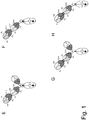
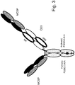
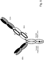
- the components of the T cell activating bispecific antigen binding molecule can be fused to each other in a variety of configurations. Exemplary configurations are depicted in Figures 1 , 3 and 5 .
- the T cell activating bispecific antigen binding molecule comprises an Fc domain composed of a first and a second subunit capable of stable association.
- the second antigen binding moiety is fused at the C-terminus of the Fab heavy chain to the N-terminus of the first or the second subunit of the Fc domain.
- the first antigen binding moiety is fused at the C-terminus of the Fab heavy chain to the N-terminus of the Fab heavy chain of the second antigen binding moiety.
- the T cell activating bispecific antigen binding molecule essentially consists of a first and a second antigen binding moiety, an Fc domain composed of a first and a second subunit, and optionally one or more peptide linkers, wherein the first antigen binding moiety is fused at the C-terminus of the Fab heavy chain to the N-terminus of the Fab heavy chain of the second antigen binding moiety, and the second antigen binding moiety is fused at the C-terminus of the Fab heavy chain to the N-terminus of the first or the second subunit of the Fc domain.
- the Fab light chain of the first antigen binding moiety and the Fab light chain of the second antigen binding moiety may additionally be fused to each other.
- the first antigen binding moiety is fused at the C-terminus of the Fab heavy chain to the N-terminus of the first or second subunit of the Fc domain.
- the T cell activating bispecific antigen binding molecule essentially consists of a first and a second antigen binding moiety, an Fc domain composed of a first and a second subunit, and optionally one or more peptide linkers, wherein the first and the second antigen binding moiety are each fused at the C-terminus of the Fab heavy chain to the N-terminus of one of the subunits of the Fc domain.
- the first antigen binding moiety is fused at the C-terminus of the Fab heavy chain to the N-terminus of the first or second subunit of the Fc domain.
- the second antigen binding moiety is fused at the C-terminus of the Fab heavy chain to the N-terminus of the Fab heavy chain of the first antigen binding moiety.
- the T cell activating bispecific antigen binding molecule essentially consists of a first and a second antigen binding moiety, an Fc domain composed of a first and a second subunit, and optionally one or more peptide linkers, wherein the second antigen binding moiety is fused at the C-terminus of the Fab heavy chain to the N-terminus of the Fab heavy chain of the first antigen binding moiety, and the first antigen binding moiety is fused at the C-terminus of the Fab heavy chain to the N-terminus of the first or the second subunit of the Fc domain.
- the Fab light chain of the first antigen binding moiety and the Fab light chain of the second antigen binding moiety may additionally be fused to each other.
- the antigen binding moieties may be fused to the Fc domain or to each other directly or through a peptide linker, comprising one or more amino acids, typically about 2-20 amino acids.
- Peptide linkers are known in the art and are described herein. Suitable, non-immunogenic peptide linkers include, for example, (G 4 S) n , (SG 4 ) n , (G 4 S) n or G 4 (SG 4 ) n peptide linkers, "n" is generally a number between 1 and 10, typically between 2 and 4.
- a particularly suitable peptide linker for fusing the Fab light chains of the first and the second antigen binding moiety to each other is (G 4 S) 2 .
- An exemplary peptide linker suitable for connecting the Fab heavy chains of the first and the second antigen binding moiety is EPKSC(D)-(G 4 S) 2 (SEQ ID NOs 105 and 106).
- linkers may comprise (a portion of) an immunoglobulin hinge region. Particularly where an antigen binding moiety is fused to the N-terminus of an Fc domain subunit, it may be fused via an immunoglobulin hinge region or a portion thereof, with or without an additional peptide linker.
- a T cell activating bispecific antigen binding molecule with a single antigen binding moiety capable of specific binding to a target cell antigen is useful, particularly in cases where internalization of the target cell antigen is to be expected following binding of a high affinity antigen binding moiety.
- the presence of more than one antigen binding moiety specific for the target cell antigen may enhance internalization of the target cell antigen, thereby reducing its availablity.
- T cell activating bispecific antigen binding molecule comprising two or more antigen binding moieties specific for a target cell antigen (see examples in shown in Figure 1B, 1C , 1E or 1G ), for example to optimize targeting to the target site or to allow crosslinking of target cell antigens.
- the T cell activating bispecific antigen binding molecule described herein further comprises a third antigen binding moiety which is a Fab molecule capable of specific binding to a target cell antigen.
- the third antigen binding moiety is a conventional Fab molecule.
- the third antigen binding moiety is capable of specific binding to the same target cell antigen as the second antigen binding moiety.
- the first antigen binding moiety is capable of specific binding to CD3, and the second and third antigen binding moieties are capable of specific binding to a target cell antigen.
- the second and the third antigen binding moiety are identical (i.e. they comprise the same amino acid sequences).
- the first antigen binding moiety is capable of specific binding to CD3, and the second and third antigen binding moieties are capable of specific binding to CEA, wherein the second and third antigen binding moieties comprise at least one heavy chain complementarity determining region (CDR) selected from the group consisting of SEQ ID NO: 24, SEQ ID NO: 25 and SEQ ID NO: 26 and at least one light chain CDR selected from the group of SEQ ID NO: 28, SEQ ID NO: 29 and SEQ ID NO: 30.
- CDR heavy chain complementarity determining region
- the first antigen binding moiety is capable of specific binding to CD3, and comprises at least one heavy chain complementarity determining region (CDR) selected from the group consisting of SEQ ID NO: 4, SEQ ID NO: 5 and SEQ ID NO: 6 and at least one light chain CDR selected from the group of SEQ ID NO: 8, SEQ ID NO: 9, SEQ ID NO: 10; and the second and third antigen binding moieties are capable of specific binding to CEA, wherein the second and third antigen binding moieties comprise at least one heavy chain complementarity determining region (CDR) selected from the group consisting of SEQ ID NO: 24, SEQ ID NO: 25 and SEQ ID NO: 26 and at least one light chain CDR selected from the group of SEQ ID NO: 28, SEQ ID NO: 29 and SEQ ID NO: 30.
- CDR heavy chain complementarity determining region
- the first antigen binding moiety is capable of specific binding to CD3, and comprises at least one heavy chain complementarity determining region (CDR) selected from the group consisting of SEQ ID NO: 4, SEQ ID NO: 5 and SEQ ID NO: 6 and at least one light chain CDR selected from the group of SEQ ID NO: 8, SEQ ID NO: 9, SEQ ID NO: 10; and the second and third antigen binding moieties are capable of specific binding to CEA, wherein the second and third antigen binding moieties comprise at least one heavy chain complementarity determining region (CDR) selected from the group consisting of SEQ ID NO: 24, SEQ ID NO: 25 and SEQ ID NO: 26 and at least one light chain CDR selected from the group of SEQ ID NO: 28, SEQ ID NO: 29 and SEQ ID NO: 30.
- CDR heavy chain complementarity determining region
- the first antigen binding moiety is capable of specific binding to CD3, and comprises a heavy chain variable region comprising an amino acid sequence that is at least about 95%, 96%, 97%, 98%, 99% or 100% identical to an amino acid sequence selected from the group of: SEQ ID NO: 3, SEQ ID NO: 32 and SEQ ID NO: 33 and a light chain variable region comprising an amino acid sequence that is at least about 95%, 96%, 97%, 98%, 99% or 100% identical to an amino acid sequence selected from the group of: SEQ ID NO: 7 and SEQ ID NO: 31, and the second and third antigen binding moieties are capable of specific binding to CEA, wherein the second and third antigen binding moieties comprise a heavy chain variable region comprising an amino acid sequence that is at least about 95%, 96%, 97%, 98%, 99% or 100% identical to the amino acid sequence of SEQ ID NO: 23 and a light chain variable region comprising an amino acid sequence that is at least about 95%, 96%, 97%, 98%,
- the first antigen binding moiety is capable of specific binding to CD3, and comprises a heavy chain variable region comprising an amino acid sequence that is at least about 95%, 96%, 97%, 98%, 99% or 100% identical to the amino acid sequence of SEQ ID NO: 3, and a light chain variable region comprising an amino acid sequence that is at least about 95%, 96%, 97%, 98%, 99% or 100% identical to the amino acid sequence of SEQ ID NO: 7, and the second and third antigen binding moieties are capable of specific binding to CEA, wherein the second and third antigen binding moieties comprise a heavy chain variable region comprising an amino acid sequence that is at least about 95%, 96%, 97%, 98%, 99% or 100% identical to the amino acid sequence of SEQ ID NO: 23 and a light chain variable region comprising an amino acid sequence that is at least about 95%, 96%, 97%, 98%, 99% or 100% identical to the amino acid sequence of SEQ ID NO: 27.
- the first antigen binding moiety is capable of specific binding to CD3, and the second and third antigen binding moieties are capable of specific binding to MCSP, wherein the second and third antigen binding moieties comprise at least one heavy chain complementarity determining region (CDR) selected from the group consisting of SEQ ID NO: 14, SEQ ID NO: 15, SEQ ID NO: 16, SEQ ID NO: 35, SEQ ID NO: 37, SEQ ID NO: 38 and SEQ ID NO: 40 and at least one light chain CDR selected from the group of SEQ ID NO: 18, SEQ ID NO: 19, SEQ ID NO: 20, SEQ ID NO: 44, SEQ ID NO: 45, SEQ ID NO: 48, SEQ ID NO: 49 and SEQ ID NO: 50.
- CDR heavy chain complementarity determining region
- the first antigen binding moiety is capable of specific binding to CD3, and comprises at least one heavy chain complementarity determining region (CDR) selected from the group consisting of SEQ ID NO: 4, SEQ ID NO: 5 and SEQ ID NO: 6 and at least one light chain CDR selected from the group of SEQ ID NO: 8, SEQ ID NO: 9, SEQ ID NO: 10; and the second and third antigen binding moieties are capable of specific binding to MCSP, wherein the second and third antigen binding moieties comprise at least one heavy chain complementarity determining region (CDR) selected from the group consisting of SEQ ID NO: 14, SEQ ID NO: 15, SEQ ID NO: 16, SEQ ID NO: 35, SEQ ID NO: 37, SEQ ID NO: 38 and SEQ ID NO: 40 and at least one light chain CDR selected from the group of SEQ ID NO: 18, SEQ ID NO: 19, SEQ ID NO: 20, SEQ ID NO: 44, SEQ ID NO: 45, SEQ ID NO: 48, SEQ ID NO: 49
- CDR heavy
- the first antigen binding moiety is capable of specific binding to CD3, and comprises at least one heavy chain complementarity determining region (CDR) selected from the group consisting of SEQ ID NO: 4, SEQ ID NO: 5 and SEQ ID NO: 6 and at least one light chain CDR selected from the group of SEQ ID NO: 8, SEQ ID NO: 9, SEQ ID NO: 10; and the second and third antigen binding moieties are capable of specific binding to MCSP, wherein the second and third antigen binding moieties comprise at least one heavy chain complementarity determining region (CDR) selected from the group consisting of SEQ ID NO: 14, SEQ ID NO: 15 and SEQ ID NO: 16 and at least one light chain CDR selected from the group of SEQ ID NO: 18, SEQ ID NO: 19 and SEQ ID NO: 20.
- CDR heavy chain complementarity determining region
- the first antigen binding moiety is capable of specific binding to CD3, and comprises a heavy chain variable region comprising an amino acid sequence that is at least about 95%, 96%, 97%, 98%, 99% or 100% identical to an amino acid sequence selected from the group of: SEQ ID NO: 3, SEQ ID NO: 32 and SEQ ID NO: 33 and a light chain variable region comprising an amino acid sequence that is at least about 95%, 96%, 97%, 98%, 99% or 100% identical to an amino acid sequence selected from the group of: SEQ ID NO: 7 and SEQ ID NO: 31, and the second and third antigen binding moieties are capable of specific binding to MCSP, wherein the second and third antigen binding moieties comprise a heavy chain variable region comprising an amino acid sequence that is at least about 95%, 96%, 97%, 98%, 99% or 100% identical to an amino acid sequence selected from the group of SEQ ID NO: 13, SEQ ID NO: 34, SEQ ID NO: 36, SEQ ID NO: 39 and SEQ ID NO: 41
- the first antigen binding moiety is capable of specific binding to CD3, and comprises a a heavy chain variable region comprising an amino acid sequence that is at least about 95%, 96%, 97%, 98%, 99% or 100% identical to the amino acid sequence of SEQ ID NO: 3 and a light chain variable region comprising an amino acid sequence that is at least about 95%, 96%, 97%, 98%, 99% or 100% identical to the amino acid sequence of SEQ ID NO: 7, and the second and third antigen binding moieties are capable of specific binding to MCSP, wherein the second and third antigen binding moieties comprise a heavy chain variable region comprising an amino acid sequence that is at least about 95%, 96%, 97%, 98%, 99% or 100% identical to the amino acid sequence of SEQ ID NO: 13 and a light chain variable region comprising an amino acid sequence that is at least about 95%, 96%, 97%, 98%, 99% or 100% identical to the amino acid sequence of SEQ ID NO: 17.
- the third antigen binding moiety is fused at the C-terminus of the Fab heavy chain to the N-terminus of the first or second subunit of the Fc domain.
- the second and the third antigen binding moiety are each fused at the C-terminus of the Fab heavy chain to the N-terminus of one of the subunits of the Fc domain
- the first antigen binding moiety is fused at the C-terminus of the Fab heavy chain to the N-terminus of the Fab heavy chain of the second antigen binding moiety.
- the Fab light chain of the first antigen binding moiety and the Fab light chain of the second antigen binding moiety may additionally be fused to each other.
- the second and the third antigen binding moiety may be fused to the Fc domain directly or through a peptide linker.
- the second and the third antigen binding moiety are each fused to the Fc domain through an immunoglobulin hinge region.
- the immunoglobulin hinge region is a human IgG 1 hinge region.
- the second and the third antigen binding moiety and the Fc domain are part of an immunoglobulin molecule.
- the immunoglobulin molecule is an IgG class immunoglobulin.
- the immunoglobulin is an IgG 1 subclass immunoglobulin.
- the immunoglobulin is an IgG 4 subclass immunoglobulin.
- the immunoglobulin is a human immunoglobulin. In other embodiments the immunoglobulin is a chimeric immunoglobulin or a humanized immunoglobulin.
- the T cell activating bispecific antigen binding molecule essentially consists of an immunoglobulin molecule capable of specific binding to a target cell antigen, and an antigen binding moiety capable of specific binding to CD3 wherein the antigen binding moiety is a Fab molecule, particularly a crossover Fab molecule, fused to the N-terminus of one of the immunoglobulin heavy chains, optionally via a peptide linker.
- the first and the third antigen binding moiety are each fused at the C-terminus of the Fab heavy chain to the N-terminus of one of the subunits of the Fc domain, and the second antigen binding moiety is fused at the C-terminus of the Fab heavy chain to the N-terminus of the Fab heavy chain of the first antigen binding moiety.
- the T cell activating bispecific antigen binding molecule essentially consists of a first, a second and a third antigen binding moiety, an Fc domain composed of a first and a second subunit, and optionally one or more peptide linkers, wherein the second antigen binding moiety is fused at the C-terminus of the Fab heavy chain to the N-terminus of the Fab heavy chain of the first antigen binding moiety, and the first antigen binding moiety is fused at the C-terminus of the Fab heavy chain to the N-terminus of the first subunit of the Fc domain, and wherein the third antigen binding moiety is fused at the C-terminus of the Fab heavy chain to the N-terminus of the second subunit of the Fc domain.
- the Fab light chain of the first antigen binding moiety and the Fab light chain of the second antigen binding moiety may additionally be fused to each other.
- the T cell activating bispecific antigen binding molecule may further comprise (iii) an Fc domain composed of a first and a second subunit capable of stable association, wherein the second antigen binding moiety is fused at the C-terminus of the Fab heavy chain to the N-terminus of the Fab heavy chain of the first antigen binding moiety, and the first antigen binding moiety is fused at the C-terminus of the Fab heavy chain to the N-terminus of the first subunit of the Fc domain, and wherein the third antigen binding moiety is fused at the C-terminus of the Fab heavy chain to the N-terminus of the second subunit of the Fc domain.
- the Fab light chain of the first antigen binding moiety and the Fab light chain of the second antigen binding moiety are fused to each other, optionally via a linker peptide.
- the Fab light chain of the first antigen binding moiety may be fused at its C-terminus to the N-terminus of the Fab light chain of the second antigen binding moiety, or the Fab light chain of the second antigen binding moiety may be fused at its C-terminus to the N-terminus of the Fab light chain of the first antigen binding moiety.
- Fusion of the Fab light chains of the first and the second antigen binding moiety further reduces mispairing of unmatched Fab heavy and light chains, and also reduces the number of plasmids needed for expression of some of the T cell activating bispecific antigen binding molecules described herein.
- the T cell activating bispecific antigen binding molecule comprises a polypeptide wherein the Fab light chain variable region of the first antigen binding moiety shares a carboxy-terminal peptide bond with the Fab heavy chain constant region of the first antigen binding moiety (i.e.
- a the first antigen binding moiety comprises a crossover Fab heavy chain, wherein the heavy chain variable region is replaced by a light chain variable region), which in turn shares a carboxy-terminal peptide bond with an Fc domain subunit (VL (1) -CH1 (1) -CH2-CH3(-CH4)), and a polypeptide wherein a the Fab heavy chain of the second antigen binding moiety shares a carboxy-terminal peptide bond with an Fc domain subunit (VH (2) -CH1 (2) -CH2-CH3(-CH4)).
- the T cell activating bispecific antigen binding molecule further comprises a polypeptide wherein the Fab heavy chain variable region of the first antigen binding moiety shares a carboxy-terminal peptide bond with the Fab light chain constant region of the first antigen binding moiety (VH (1) -CL (1) ) and the Fab light chain polypeptide of the second antigen binding moiety (VL (2) -CL (2) ).
- the polypeptides are covalently linked, e.g., by a disulfide bond.
- the T cell activating bispecific antigen binding molecule comprises a polypeptide wherein the Fab heavy chain variable region of the first antigen binding moiety shares a carboxy-terminal peptide bond with the Fab light chain constant region of the first antigen binding moiety (i.e.
- the first antigen binding moiety comprises a crossover Fab heavy chain, wherein the heavy chain constant region is replaced by a light chain constant region), which in turn shares a carboxy-terminal peptide bond with an Fc domain subunit (VH (1) -CL (1) -CH2-CH3(-CH4)), and a polypeptide wherein the Fab heavy chain of the second antigen binding moiety shares a carboxy-terminal peptide bond with an Fc domain subunit (VH (2) -CH1 (2) -CH2-CH3(-CH4)).
- the T cell activating bispecific antigen binding molecule further comprises a polypeptide wherein the Fab light chain variable region of the first antigen binding moiety shares a carboxy-terminal peptide bond with the Fab heavy chain constant region of the first antigen binding moiety (VL (1) -CH1 (1) ) and the Fab light chain polypeptide of the second antigen binding moiety (VL (2) -CL (2) ).
- the polypeptides are covalently linked, e.g., by a disulfide bond.
- the T cell activating bispecific antigen binding molecule comprises a polypeptide wherein the Fab light chain variable region of the first antigen binding moiety shares a carboxy-terminal peptide bond with the Fab heavy chain constant region of the first antigen binding moiety (i.e. the first antigen binding moiety comprises a crossover Fab heavy chain, wherein the heavy chain variable region is replaced by a light chain variable region), which in turn shares a carboxy-terminal peptide bond with the Fab heavy chain of the second antigen binding moiety, which in turn shares a carboxy-terminal peptide bond with an Fc domain subunit (VL (1) -CH1 (1) -VH (2) -CH1 (2) -CH2-CH3(-CH4)).
- VL (1) -CH1 (1) -VH (2) -CH1 (2) -CH2-CH3(-CH4) an Fc domain subunit
- the T cell activating bispecific antigen binding molecule comprises a polypeptide wherein the Fab heavy chain variable region of the first antigen binding moiety shares a carboxy-terminal peptide bond with the Fab light chain constant region of the first antigen binding moiety (i.e. the first antigen binding moiety comprises a crossover Fab heavy chain, wherein the heavy chain constant region is replaced by a light chain constant region), which in turn shares a carboxy-terminal peptide bond with the Fab heavy chain of the second antigen binding moiety, which in turn shares a carboxy-terminal peptide bond with an Fc domain subunit (VH (1) -CL (1) -VH (2) -CH1 (2) -CH2-CH3(-CH4)).
- VH (1) -CL (1) -VH (2) -CH1 (2) -CH2-CH3(-CH4) an Fc domain subunit
- the T cell activating bispecific antigen binding molecule comprises a polypeptide wherein the Fab heavy chain of the second antigen binding moiety shares a carboxy-terminal peptide bond with the Fab light chain variable region of the first antigen binding moiety which in turn shares a carboxy-terminal peptide bond with the Fab heavy chain constant region of the first antigen binding moiety (i.e. the first antigen binding moiety comprises a crossover Fab heavy chain, wherein the heavy chain variable region is replaced by a light chain variable region), which in turn shares a carboxy-terminal peptide bond with an Fc domain subunit (VH (2) -CH1 (2) -VL (1) -CH1 (1) -CH2-CH3(-CH4)).
- VH (2) -CH1 (2) -VL (1) -CH1 (1) -CH2-CH3(-CH4) an Fc domain subunit
- the T cell activating bispecific antigen binding molecule comprises a polypeptide wherein the Fab heavy chain of the second antigen binding moiety shares a carboxy-terminal peptide bond with the Fab heavy chain variable region of the first antigen binding moiety which in turn shares a carboxy-terminal peptide bond with the Fab light chain constant region of the first antigen binding moiety (i.e. the first antigen binding moiety comprises a crossover Fab heavy chain, wherein the heavy chain constant region is replaced by a light chain constant region), which in turn shares a carboxy-terminal peptide bond with an Fc domain subunit (VH (2) -CH1 (2) -VH (1) -CL (1) -CH2-CH3(-CH4)).
- VH (2) -CH1 (2) -VH (1) -CL (1) -CH2-CH3(-CH4) an Fc domain subunit
- the T cell activating bispecific antigen binding molecule further comprises a crossover Fab light chain polypeptide of the first antigen binding moiety, wherein the Fab heavy chain variable region of the first antigen binding moiety shares a carboxy-terminal peptide bond with the Fab light chain constant region of the first antigen binding moiety (VH (1) -CL (1) ), and the Fab light chain polypeptide of the second antigen binding moiety (VL (2) -CL (2) ).
- the T cell activating bispecific antigen binding molecule further comprises a crossover Fab light chain polypeptide, wherein the Fab light chain variable region of the first antigen binding moiety shares a carboxy-terminal peptide bond with the Fab heavy chain constant region of the first antigen binding moiety (VL (1) -CH1 (1) ), and the Fab light chain polypeptide of the second antigen binding moiety (VL (2) -CL (2) ).
- the T cell activating bispecific antigen binding molecule further comprises a polypeptide wherein the Fab light chain variable region of the first antigen binding moiety shares a carboxy-terminal peptide bond with the Fab heavy chain constant region of the first antigen binding moiety which in turn shares a carboxy-terminal peptide bond with the Fab light chain polypeptide of the second antigen binding moiety (VL (1) -CH1 (1) -VL (2) -CL (2) ), a polypeptide wherein the Fab heavy chain variable region of the first antigen binding moiety shares a carboxy-terminal peptide bond with the Fab light chain constant region of the first antigen binding moiety which in turn shares a carboxy-terminal peptide bond with the Fab light chain polypeptide of the second antigen binding moiety (VH (1) -CL (1) -VL (2) -CL (2) ), a polypeptide wherein the Fab light chain polypeptide of the second antigen binding moiety shares a carboxy-terminal
- the T cell activating bispecific antigen binding molecule may further comprise (i) an Fc domain subunit polypeptide (CH2-CH3(-CH4)), or (ii) a polypeptide wherein the Fab heavy chain of a third antigen binding moiety shares a carboxy-terminal peptide bond with an Fc domain subunit (VH (3) -CH1 (3) -CH2-CH3(-CH4)) and the Fab light chain polypeptide of a third antigen binding moiety (VL (3) -CL (3) ).
- the polypeptides are covalently linked, e.g., by a disulfide bond.
- components of the T cell activating bispecific antigen binding molecule may be fused directly or through various linkers, particularly peptide linkers comprising one or more amino acids, typically about 2-20 amino acids, that are described herein or are known in the art.
- Suitable, non-immunogenic peptide linkers include, for example, (G 4 S) n , (SG 4 ) n , (G 4 S) n or G 4 (SG 4 ) n peptide linkers, wherein n is generally a number between 1 and 10, typically between 2 and 4.
- the Fc domain of the T cell activating bispecific antigen binding molecule consists of a pair of polypeptide chains comprising heavy chain domains of an immunoglobulin molecule.
- the Fc domain of an immunoglobulin G (IgG) molecule is a dimer, each subunit of which comprises the CH2 and CH3 IgG heavy chain constant domains.
- the two subunits of the Fc domain are capable of stable association with each other.
- the T cell activating bispecific antigen binding molecule described herein comprises not more than one Fc domain.
- the Fc domain of the T cell activating bispecific antigen binding molecule is an IgG Fc domain.
- the Fc domain is an IgG 1 Fc domain.
- the Fc domain is an IgG 4 Fc domain.
- the Fc domain is an IgG 4 Fc domain comprising an amino acid substitution at position S228 (EU numbering), particularly the amino acid substitution S228P. This amino acid substitution reduces in vivo Fab arm exchange of IgG 4 antibodies (see Stubenrauch et al., Drug Metabolism and Disposition 38, 84-91 (2010 )).
- the Fc domain is human. An exemplary sequence of a human IgG 1 Fc region is given in SEQ ID NO: 107.
- T cell activating bispecific antigen binding molecules described herein comprise different antigen binding moieties, fused to one or the other of the two subunits of the Fc domain, thus the two subunits of the Fc domain are typically comprised in two non-identical polypeptide chains. Recombinant co-expression of these polypeptides and subsequent dimerization leads to several possible combinations of the two polypeptides. To improve the yield and purity of T cell activating bispecific antigen binding molecules in recombinant production, it will thus be advantageous to introduce in the Fc domain of the T cell activating bispecific antigen binding molecule a modification promoting the association of the desired polypeptides.
- the Fc domain of the T cell activating bispecific antigen binding molecule described herein comprises a modification promoting the association of the first and the second subunit of the Fc domain.
- the site of most extensive protein-protein interaction between the two subunits of a human IgG Fc domain is in the CH3 domain of the Fc domain.
- said modification is in the CH3 domain of the Fc domain.
- said modification is a so-called "knob-into-hole” modification, comprising a "knob” modification in one of the two subunits of the Fc domain and a "hole” modification in the other one of the two subunits of the Fc domain.
- the method involves introducing a protuberance ("knob") at the interface of a first polypeptide and a corresponding cavity ("hole") in the interface of a second polypeptide, such that the protuberance can be positioned in the cavity so as to promote heterodimer formation and hinder homodimer formation.
- Protuberances are constructed by replacing small amino acid side chains from the interface of the first polypeptide with larger side chains (e.g. tyrosine or tryptophan).
- Compensatory cavities of identical or similar size to the protuberances are created in the interface of the second polypeptide by replacing large amino acid side chains with smaller ones (e.g. alanine or threonine).
- an amino acid residue is replaced with an amino acid residue having a larger side chain volume, thereby generating a protuberance within the CH3 domain of the first subunit which is positionable in a cavity within the CH3 domain of the second subunit, and in the CH3 domain of the second subunit of the Fc domain an amino acid residue is replaced with an amino acid residue having a smaller side chain volume, thereby generating a cavity within the CH3 domain of the second subunit within which the protuberance within the CH3 domain of the first subunit is positionable.
- the protuberance and cavity can be made by altering the nucleic acid encoding the polypeptides, e.g. by site-specific mutagenesis, or by peptide synthesis.
- the threonine residue at position 366 in the CH3 domain of the first subunit of the Fc domain the threonine residue at position 366 is replaced with a tryptophan residue (T366W), and in the CH3 domain of the second subunit of the Fc domain the tyrosine residue at position 407 is replaced with a valine residue (Y407V).
- the threonine residue at position 366 in the second subunit of the Fc domain additionally the threonine residue at position 366 is replaced with a serine residue (T366S) and the leucine residue at position 368 is replaced with an alanine residue (L368A).
- the serine residue at position 354 is replaced with a cysteine residue (S354C)
- the tyrosine residue at position 349 is replaced by a cysteine residue (Y349C).
- the antigen binding moiety capable of binding to CD3 is fused (optionally via the antigen binding moiety capable of binding to a target cell antigen) to the first subunit of the Fc domain (comprising the "knob" modification).
- fusion of the antigen binding moiety capable of binding to CD3 to the knob-containing subunit of the Fc domain will (further) minimize the generation of antigen binding molecules comprising two antigen binding moieties capable of binding to CD3 (steric clash of two knob-containing polypeptides).
- a modification promoting association of the first and the second subunit of the Fc domain comprises a modification mediating electrostatic steering effects, e.g. as described in PCT publication WO 2009/089004 .
- this method involves replacement of one or more amino acid residues at the interface of the two Fc domain subunits by charged amino acid residues so that homodimer formation becomes electrostatically unfavorable but heterodimerization electrostatically favorable.
- the Fc domain confers to the T cell activating bispecific antigen binding molecule favorable pharmacokinetic properties, including a long serum half-life which contributes to good accumulation in the target tissue and a favorable tissue-blood distribution ratio. At the same time it may, however, lead to undesirable targeting of the T cell activating bispecific antigen binding molecule to cells expressing Fc receptors rather than to the preferred antigen-bearing cells. Moreover, the co-activation of Fc receptor signaling pathways may lead to cytokine release which, in combination with the T cell activating properties and the long half-life of the antigen binding molecule, results in excessive activation of cytokine receptors and severe side effects upon systemic administration. Activation of (Fc receptor-bearing) immune cells other than T cells may even reduce efficacy of the T cell activating bispecific antigen binding molecule due to the potential destruction of T cells e.g. by NK cells.
- the Fc domain of the T cell activating bispecific antigen binding molecules described herein exhibits reduced binding affinity to an Fc receptor and/or reduced effector function, as compared to a native IgG 1 Fc domain.
- the Fc domain (or the T cell activating bispecific antigen binding molecule comprising said Fc domain) exhibits less than 50%, preferably less than 20%, more preferably less than 10% and most preferably less than 5% of the binding affinity to an Fc receptor, as compared to a native IgG 1 Fc domain (or a T cell activating bispecific antigen binding molecule comprising a native IgG 1 Fc domain), and/or less than 50%, preferably less than 20%, more preferably less than 10% and most preferably less than 5% of the effector function, as compared to a native IgG 1 Fc domain domain (or a T cell activating bispecific antigen binding molecule comprising a native IgG 1 Fc domain).
- the Fc domain domain (or the T cell activating bispecific antigen binding molecule comprising said Fc domain) does not substantially bind to an Fc receptor and/or induce effector function.
- the Fc receptor is an Fc ⁇ receptor.
- the Fc receptor is a human Fc receptor.
- the Fc receptor is an activating Fc receptor.
- the Fc receptor is an activating human Fc ⁇ receptor, more specifically human Fc ⁇ RIIIa, Fc ⁇ RI or Fc ⁇ RIIa, most specifically human Fc ⁇ RIIIa.
- the effector function is one or more selected from the group of CDC, ADCC, ADCP, and cytokine secretion. In a particular embodiment the effector function is ADCC.
- the Fc domain domain exhibits substantially similar binding affinity to neonatal Fc receptor (FcRn), as compared to a native IgG 1 Fc domain domain.
- FcRn neonatal Fc receptor
- Substantially similar binding to FcRn is achieved when the Fc domain (or the T cell activating bispecific antigen binding molecule comprising said Fc domain) exhibits greater than about 70%, particularly greater than about 80%, more particularly greater than about 90% of the binding affinity of a native IgG 1 Fc domain (or the T cell activating bispecific antigen binding molecule comprising a native IgG 1 Fc domain) to FcRn.
- the Fc domain is engineered to have reduced binding affinity to an Fc receptor and/or reduced effector function, as compared to a non-engineered Fc domain.
- the Fc domain of the T cell activating bispecific antigen binding molecule comprises one or more amino acid mutation that reduces the binding affinity of the Fc domain to an Fc receptor and/or effector function. Typically, the same one or more amino acid mutation is present in each of the two subunits of the Fc domain.
- the amino acid mutation reduces the binding affinity of the Fc domain to an Fc receptor.
- the amino acid mutation reduces the binding affinity of the Fc domain to an Fc receptor by at least 2-fold, at least 5-fold, or at least 10-fold.
- the combination of these amino acid mutations may reduce the binding affinity of the Fc domain to an Fc receptor by at least 10-fold, at least 20-fold, or even at least 50-fold.
- the T cell activating bispecific antigen binding molecule comprising an engineered Fc domain exhibits less than 20%, particularly less than 10%, more particularly less than 5% of the binding affinity to an Fc receptor as compared to a T cell activating bispecific antigen binding molecule comprising a non-engineered Fc domain.
- the Fc receptor is an Fc ⁇ receptor.
- the Fc receptor is a human Fc receptor.
- the Fc receptor is an activating Fc receptor.
- the Fc receptor is an activating human Fc ⁇ receptor, more specifically human Fc ⁇ RIIIa, Fc ⁇ RI or Fc ⁇ RIIa, most specifically human Fc ⁇ RIIIa.
- binding to each of these receptors is reduced.
- binding affinity to a complement component, specifically binding affinity to C1q is also reduced.
- binding affinity to neonatal Fc receptor (FcRn) is not reduced. Substantially similar binding to FcRn, i.e.
- the Fc domain (or the T cell activating bispecific antigen binding molecule comprising said Fc domain) exhibits greater than about 70% of the binding affinity of a non-engineered form of the Fc domain (or the T cell activating bispecific antigen binding molecule comprising said non-engineered form of the Fc domain) to FcRn.
- the Fc domain, or T cell activating bispecific antigen binding molecules described herein comprising said Fc domain may exhibit greater than about 80% and even greater than about 90% of such affinity.
- the Fc domain of the T cell activating bispecific antigen binding molecule is engineered to have reduced effector function, as compared to a non-engineered Fc domain.
- the reduced effector function can include, but is not limited to, one or more of the following: reduced complement dependent cytotoxicity (CDC), reduced antibody-dependent cell-mediated cytotoxicity (ADCC), reduced antibody-dependent cellular phagocytosis (ADCP), reduced cytokine secretion, reduced immune complex-mediated antigen uptake by antigen-presenting cells, reduced binding to NK cells, reduced binding to macrophages, reduced binding to monocytes, reduced binding to polymorphonuclear cells, reduced direct signaling inducing apoptosis, reduced crosslinking of target-bound antibodies, reduced dendritic cell maturation, or reduced T cell priming.
- CDC complement dependent cytotoxicity
- ADCC reduced antibody-dependent cell-mediated cytotoxicity
- ADCP reduced antibody-dependent cellular phagocytosis
- reduced immune complex-mediated antigen uptake by antigen-presenting cells reduced binding to NK cells, reduced binding to macrophages, reduced binding to monocytes, reduced binding to polymorphonuclear cells, reduced direct signaling inducing
- the reduced effector function is one or more selected from the group of reduced CDC, reduced ADCC, reduced ADCP, and reduced cytokine secretion. In a particular embodiment the reduced effector function is reduced ADCC. In one embodiment the reduced ADCC is less than 20% of the ADCC induced by a non-engineered Fc domain (or a T cell activating bispecific antigen binding molecule comprising a non-engineered Fc domain).
- the amino acid mutation that reduces the binding affinity of the Fc domain to an Fc receptor and/or effector function is an amino acid substitution.
- the Fc domain comprises an amino acid substitution at a position selected from the group of E233, L234, L235, N297, P331 and P329.
- the Fc domain comprises an amino acid substitution at a position selected from the group of L234, L235 and P329.
- the Fc domain comprises the amino acid substitutions L234A and L235A.
- the Fc domain is an IgG 1 Fc domain, particularly a human IgG 1 Fc domain.
- the Fc domain comprises an amino acid substitution at position P329.
- the amino acid substitution is P329A or P329G, particularly P329G.
- the Fc domain comprises an amino acid substitution at position P329 and a further amino acid substitution at a position selected from E233, L234, L235, N297 and P331.
- the further amino acid substitution is E233P, L234A, L235A, L235E, N297A, N297D or P331S.
- the Fc domain comprises amino acid substitutions at positions P329, L234 and L235.
- the Fc domain comprises the amino acid mutations L234A, L235A and P329G ("P329G LALA").
- the Fc domain is an IgG 1 Fc domain, particularly a human IgG 1 Fc domain.
- the "P329G LALA" combination of amino acid substitutions almost completely abolishes Fc ⁇ receptor (as well as complement) binding of a human IgG 1 Fc domain, as described in PCT publication no. WO 2012/130831 .
- WO 2012/130831 also describes methods of preparing such mutant Fc domains and methods for determining its properties such as Fc receptor binding or effector functions.
- the Fc domain of the T cell activating bispecific antigen binding molecules described herein is an IgG 4 Fc domain, particularly a human IgG 4 Fc domain.
- the IgG 4 Fc domain comprises amino acid substitutions at position S228, specifically the amino acid substitution S228P.
- the IgG 4 Fc domain comprises an amino acid substitution at position L235, specifically the amino acid substitution L235E.
- the IgG 4 Fc domain comprises an amino acid substitution at position P329, specifically the amino acid substitution P329G.
- the IgG 4 Fc domain comprises amino acid substitutions at positions S228, L235 and P329, specifically amino acid substitutions S228P, L235E and P329G.
- IgG 4 Fc domain mutants and their Fc ⁇ receptor binding properties are described in PCT publication no. WO 2012/130831 .
- the Fc domain exhibiting reduced binding affinity to an Fc receptor and/or reduced effector function, as compared to a native IgG 1 Fc domain is a human IgG 1 Fc domain comprising the amino acid substitutions L234A, L235A and optionally P329G, or a human IgG 4 Fc domain comprising the amino acid substitutions S228P, L235E and optionally P329G.
- N-glycosylation of the Fc domain has been eliminated.
- the Fc domain comprises an amino acid mutation at position N297, particularly an amino acid substitution replacing asparagine by alanine (N297A) or aspartic acid (N297D).
- Fc domains with reduced Fc receptor binding and/or effector function also include those with substitution of one or more of Fc domain residues 238, 265, 269, 270, 297, 327 and 329 ( U.S. Patent No. 6,737,056 ).
- Such Fc mutants include Fc mutants with substitutions at two or more of amino acid positions 265, 269, 270, 297 and 327, including the so-called "DANA" Fc mutant with substitution of residues 265 and 297 to alanine ( US Patent No. 7,332,581 ).
- Mutant Fc domains can be prepared by amino acid deletion, substitution, insertion or modification using genetic or chemical methods well known in the art. Genetic methods may include site-specific mutagenesis of the encoding DNA sequence, PCR, gene synthesis, and the like. The correct nucleotide changes can be verified for example by sequencing.
- Binding to Fc receptors can be easily determined e.g. by ELISA, or by Surface Plasmon Resonance (SPR) using standard instrumentation such as a BIAcore instrument (GE Healthcare), and Fc receptors such as may be obtained by recombinant expression. A suitable such binding assay is described herein. Alternatively, binding affinity of Fc domains or cell activating bispecific antigen binding molecules comprising an Fc domain for Fc receptors may be evaluated using cell lines known to express particular Fc receptors, such as human NK cells expressing Fc ⁇ IIIa receptor.
- Effector function of an Fc domain, or a T cell activating bispecific antigen binding molecule comprising an Fc domain can be measured by methods known in the art.
- a suitable assay for measuring ADCC is described herein.
- Other examples of in vitro assays to assess ADCC activity of a molecule of interest are described in U.S. Patent No. 5,500,362 ; Hellstrom et al. Proc Natl Acad Sci USA 83, 7059-7063 (1986 ) and Hellstrom et al., Proc Natl Acad Sci USA 82, 1499-1502 (1985 ); U.S. Patent No. 5,821,337 ; Bruggemann et al., J Exp Med 166, 1351-1361 (1987 ).
- non-radioactive assays methods may be employed (see, for example, ACTITM non-radioactive cytotoxicity assay for flow cytometry (CellTechnology, Inc. Mountain View, CA); and CytoTox 96® non-radioactive cytotoxicity assay (Promega, Madison, WI)).
- Useful effector cells for such assays include peripheral blood mononuclear cells (PBMC) and Natural Killer (NK) cells.
- PBMC peripheral blood mononuclear cells
- NK Natural Killer
- ADCC activity of the molecule of interest may be assessed in vivo, e.g. in a animal model such as that disclosed in Clynes et al., Proc Natl Acad Sci USA 95, 652-656 (1998 ).
- binding of the Fc domain to a complement component, specifically to C1q is reduced.
- said reduced effector function includes reduced CDC.
- C1q binding assays may be carried out to determine whether the T cell activating bispecific antigen binding molecule is able to bind C1q and hence has CDC activity. See e.g., C1q and C3c binding ELISA in WO 2006/029879 and WO 2005/100402 .
- a CDC assay may be performed (see, for example, Gazzano-Santoro et al., J Immunol Methods 202, 163 (1996 ); Cragg et al., Blood 101, 1045-1052 (2003 ); and Cragg and Glennie, Blood 103, 2738-2743 (2004 )).
- the antigen binding molecule described herein is bispecific, i.e. it comprises at least two antigen binding moieties capable of specific binding to two distinct antigenic determinants.
- the antigen binding moieties are Fab molecules (i.e. antigen binding domains composed of a heavy and a light chain, each comprising a variable and a constant region).
- said Fab molecules are human.
- said Fab molecules are humanized.
- said Fab molecules comprise human heavy and light chain constant regions.
- At least one of the antigen binding moieties is a crossover Fab molecule.
- Such modification prevents mispairing of heavy and light chains from different Fab molecules, thereby improving the yield and purity of the T cell activating bispecific antigen binding molecule described herein in recombinant production.
- the constant regions of the Fab light chain and the Fab heavy chain are exchanged.
- the variable regions of the Fab light chain and the Fab heavy chain are exchanged.
- the T cell activating bispecific antigen binding molecule is capable of simultaneous binding to a target cell antigen, particularly a tumor cell antigen, and CD3.
- the T cell activating bispecific antigen binding molecule is capable of crosslinking a T cell and a target cell by simultaneous binding to a target cell antigen and CD3.
- simultaneous binding results in lysis of the target cell, particularly a tumor cell.
- simultaneous binding results in activation of the T cell.
- such simultaneous binding results in a cellular response of a T lymphocyte, particularly a cytotoxic T lymphocyte, selected from the group of: proliferation, differentiation, cytokine secretion, cytotoxic effector molecule release, cytotoxic activity, and expression of activation markers.
- a T lymphocyte particularly a cytotoxic T lymphocyte, selected from the group of: proliferation, differentiation, cytokine secretion, cytotoxic effector molecule release, cytotoxic activity, and expression of activation markers.
- binding of the T cell activating bispecific antigen binding molecule to CD3 without simultaneous binding to the target cell antigen does not result in T cell activation.
- the T cell activating bispecific antigen binding molecule is capable of redirecting cytotoxic activity of a T cell to a target cell.
- said re-direction is independent of MHC-mediated peptide antigen presentation by the target cell and and/or specificity of the T cell.
- a T cell according to any of the embodiments described herein is a cytotoxic T cell.
- the T cell is a CD4 + or a CD8 + T cell, particularly a CD8 + T cell.
- the T cell activating bispecific antigen binding molecule described herein comprises at least one antigen binding moiety capable of binding to CD3 (also referred to herein as an "CD3 antigen binding moiety" or "first antigen binding moiety”).
- the T cell activating bispecific antigen binding molecule comprises not more than one antigen binding moiety capable of specific binding to CD3.
- the T cell activating bispecific antigen binding molecule provides monovalent binding to CD3.
- the CD3 antigen binding is a crossover Fab molecule, i.e. a Fab molecule wherein either the variable or the constant regions of the Fab heavy and light chains are exchanged.
- the antigen binding moiety capable of specific binding to CD3 preferably is a crossover Fab molecule and the antigen binding moieties capable of specific binding to a target cell antigen are conventional Fab molecules.
- CD3 is human CD3 (SEQ ID NO: 103) or cynomolgus CD3 (SEQ ID NO: 104), most particularly human CD3.
- CD3 antigen binding moiety is cross-reactive for (i.e. specifically binds to) human and cynomolgus CD3.
- the first antigen binding moiety is capable of specific binding to the epsilon subunit of CD3.
- the CD3 antigen binding moiety comprises at least one heavy chain complementarity determining region (CDR) selected from the group consisting of SEQ ID NO: 4, SEQ ID NO: 5 and SEQ ID NO: 6 and at least one light chain CDR selected from the group of SEQ ID NO: 8, SEQ ID NO: 9, SEQ ID NO: 10.
- CDR heavy chain complementarity determining region
- the CD3 antigen binding moiety comprises the heavy chain CDR1 of SEQ ID NO: 4, the heavy chain CDR2 of SEQ ID NO: 5, the heavy chain CDR3 of SEQ ID NO: 6, the light chain CDR1 of SEQ ID NO: 8, the light chain CDR2 of SEQ ID NO: 9, and the light chain CDR3 of SEQ ID NO: 10.
- the CD3 antigen binding moiety comprises a heavy chain variable region sequence that is at least about 95%, 96%, 97%, 98%, 99% or 100% identical to an amino acid sequence selected from the group of: SEQ ID NO: 3, SEQ ID NO: 32 and SEQ ID NO: 33, and a light chain variable region sequence that is at least about 95%, 96%, 97%, 98%, 99% or 100% identical to an amino acid sequence selected from the group of: SEQ ID NO: 7 and SEQ ID NO: 31.
- the CD3 antigen binding moiety comprises a heavy chain variable region comprising an amino acid sequence selected from the group of: SEQ ID NO: 3, SEQ ID NO: 32 and SEQ ID NO: 33 and a light chain variable region comprising an amino acid sequence selected from the group of: SEQ ID NO: 7 and SEQ ID NO: 31.
- the CD3 antigen binding moiety comprises a heavy chain variable region sequence that is at least about 95%, 96%, 97%, 98%, 99% or 100% identical to SEQ ID NO: 3 and a light chain variable region sequence that is at least about 95%, 96%, 97%, 98%, 99% or 100% identical to SEQ ID NO: 7.
- the CD3 antigen binding moiety comprises a heavy chain variable region comprising the amino acid sequence of SEQ ID NO: 3 and a light chain variable region comprising the amino acid sequence of SEQ ID NO: 7.
- the CD3 antigen binding moiety comprises the heavy chain variable region sequence of SEQ ID NO: 3 and the light chain variable region sequence of SEQ ID NO: 7.
- the T cell activating bispecific antigen binding molecule described herein comprises at least one antigen binding moiety capable of binding to a target cell antigen (also referred to herein as an "target cell antigen binding moiety" or "second" or “third” antigen binding moiety).
- the T cell activating bispecific antigen binding molecule comprises two antigen binding moieties capable of binding to a target cell antigen.
- each of these antigen binding moieties specifically binds to the same antigenic determinant.
- all of these antigen binding moieties are identical.
- the T cell activating bispecific antigen binding molecule comprises an immunoglobulin molecule capable of specific binding to a target cell antigen.
- the T cell activating bispecific antigen binding molecule comprises not more than two antigen binding moieties capable of binding to a target cell antigen.
- the target cell antigen binding moiety is generally a Fab molecule, particularly a conventional Fab molecule that binds to a specific antigenic determinant and is able to direct the T cell activating bispecific antigen binding molecule to a target site, for example to a specific type of tumor cell that bears the antigenic determinant.
- the target cell antigen binding moiety specifically binds to a cell surface antigen.
- the target cell antigen binding moiety specifically binds to a membrane-proximal region of a cell surface antigen.
- the cell surface antigen is Carcinoembryonic Antigen (CEA) and the membrane-proximal region is the B3 domain of CEA (residues 208-286 of SEQ ID NO: 119).
- the cell surface antigen is Melanoma-associated Chondroitin Sulfate Proteoglycan (MCSP) and the membrane-proximal region is the D3 domain of MSCP (SEQ ID NO: 118).
- the target cell antigen binding moiety is directed to an antigen associated with a pathological condition, such as an antigen presented on a tumor cell or on a virus-infected cell.
- Suitable antigens are cell surface antigens, for example, but not limited to, cell surface receptors.
- the antigen is a human antigen.
- the target cell antigen is selected from Melanoma-associated Chondroitin Sulfate Proteoglycan (MCSP, CSPG4) and Carcinoembryonic Antigen (CEA, CEACAM5).
- the T cell activating bispecific antigen binding molecule comprises at least one antigen binding moiety that is specific for Melanoma-associated Chondroitin Sulfate Proteoglycan (MCSP).
- the antigen binding moiety that is specific for MCSP comprises at least one heavy chain complementarity determining region (CDR) selected from the group consisting of SEQ ID NO: 14, SEQ ID NO: 15, SEQ ID NO: 16, SEQ ID NO: 35, SEQ ID NO: 37, SEQ ID NO: 38 and SEQ ID NO: 40 and at least one light chain CDR selected from the group of SEQ ID NO: 18, SEQ ID NO: 19, SEQ ID NO: 20, SEQ ID NO: 44, SEQ ID NO: 45, SEQ ID NO: 48, SEQ ID NO: 49 and SEQ ID NO: 50.
- CDR heavy chain complementarity determining region
- the antigen binding moiety that is specific for MCSP comprises at least one heavy chain complementarity determining region (CDR) selected from the group consisting of SEQ ID NO: 14, SEQ ID NO: 15 and SEQ ID NO: 16 and at least one light chain CDR selected from the group of SEQ ID NO: 18, SEQ ID NO: 19 and SEQ ID NO: 20.
- CDR heavy chain complementarity determining region
- the antigen binding moiety that is specific for MCSP comprises the heavy chain CDR1 of SEQ ID NO: 14, the heavy chain CDR2 of SEQ ID NO: 15, the heavy chain CDR3 of SEQ ID NO: 16, the light chain CDR1 of SEQ ID NO: 18, the light chain CDR2 of SEQ ID NO: 19, and the light chain CDR3 of SEQ ID NO: 20.
- the antigen binding moiety that is specific for MCSP comprises a heavy chain variable region sequence that is at least about 95%, 96%, 97%, 98%, 99% or 100% identical to an amino acid sequence selected from the group of SEQ ID NO: 13, SEQ ID NO: 34, SEQ ID NO: 36, SEQ ID NO: 39 and SEQ ID NO: 41 and a light chain variable region sequence that is at least about 95%, 96%, 97%, 98%, 99% or 100% identical to an amino acid sequence selected from the group of SEQ ID NO: 17, SEQ ID NO: 43, SEQ ID NO: 46, SEQ ID NO: 47 and SEQ ID NO: 51.
- the antigen binding moiety that is specific for MCSP comprises a heavy chain variable region comprising an amino acid sequence selected from the group of SEQ ID NO: 13, SEQ ID NO: 34, SEQ ID NO: 36, SEQ ID NO: 39 and SEQ ID NO: 41 and a light chain variable region comprising an amino acid sequence selected from the group of SEQ ID NO: 17, SEQ ID NO: 43, SEQ ID NO: 46, SEQ ID NO: 47 and SEQ ID NO: 51.
- the antigen binding moiety that is specific for MCSP comprises a heavy chain variable region sequence that is at least about 95%, 96%, 97%, 98%, 99% or 100% identical to SEQ ID NO:13 and a light chain variable region sequence that is at least about 95%, 96%, 97%, 98%, 99% or 100% identical to SEQ ID NO: 17 or variants thereof that retain functionality.
- the antigen binding moiety that is specific for MCSP comprises a heavy chain variable region comprising an amino acid sequence of SEQ ID NO: 13 and a light chain variable region comprising an amino acid sequence of SEQ ID NO: 17.
- the antigen binding moiety that is specific for MCSP comprises the heavy chain variable region sequence of SEQ ID NO: 13 and the light chain variable region sequence of SEQ ID NO: 17.
- the T cell activating bispecific antigen binding molecule comprises a polypeptide sequence that is at least about 95%, 96%, 97%, 98%, 99% or 100% identical to SEQ ID NO: 12, a polypeptide sequence that is at least about 95%, 96%, 97%, 98%, 99% or 100% identical to SEQ ID NO: 53, a polypeptide sequence that is at least about 95%, 96%, 97%, 98%, 99% or 100% identical to SEQ ID NO: 54, and a polypeptide sequence that is at least about 95%, 96%, 97%, 98%, 99% or 100% identical to SEQ ID NO: 55.
- the T cell activating bispecific antigen binding molecule comprises at least one antigen binding moiety that is specific for Carcinoembryonic Antigen (CEA).
- CEA Carcinoembryonic Antigen
- the antigen binding moiety that is specific for CEA comprises at least one heavy chain complementarity determining region (CDR) selected from the group consisting of SEQ ID NO: 24, SEQ ID NO: 25 and SEQ ID NO: 26 and at least one light chain CDR selected from the group of SEQ ID NO: 28, SEQ ID NO: 29 and SEQ ID NO: 30.
- CDR heavy chain complementarity determining region
- the antigen binding moiety that is specific for CEA comprises the heavy chain CDR1 of SEQ ID NO: 24, the heavy chain CDR2 of SEQ ID NO: 25, the heavy chain CDR3 of SEQ ID NO: 26, the light chain CDR1 of SEQ ID NO: 28, the light chain CDR2 of SEQ ID NO: 29, and the light chain CDR3 of SEQ ID NO: 30.
- the antigen binding moiety that is specific for CEA comprises a heavy chain variable region sequence that is at least about 95%, 96%, 97%, 98%, 99% or 100% identical to SEQ ID NO: 23 and a light chain variable region sequence that is at least about 95%, 96%, 97%, 98%, 99% or 100% identical to SEQ ID NO: 27, or variants thereof that retain functionality.
- the antigen binding moiety that is specific for CEA comprises a heavy chain variable region comprising an amino acid sequence of SEQ ID NO: 23 and a light chain variable region comprising an amino acid sequence of SEQ ID NO: 27.
- the antigen binding moiety that is specific for CEA comprises the heavy chain variable region sequence of SEQ ID NO: 23 and the light chain variable region sequence of SEQ ID NO: 27.
- the T cell activating bispecific antigen binding molecule comprises a polypeptide sequence that is at least about 95%, 96%, 97%, 98%, 99% or 100% identical to SEQ ID NO: 22, a polypeptide sequence that is at least about 95%, 96%, 97%, 98%, 99% or 100% identical to SEQ ID NO: 56, a polypeptide sequence that is at least about 95%, 96%, 97%, 98%, 99% or 100% identical to SEQ ID NO: 57, and a polypeptide sequence that is at least about 95%, 96%, 97%, 98%, 99% or 100% identical to SEQ ID NO: 58.
- the disclosure further provides isolated polynucleotides encoding a T cell activating bispecific antigen binding molecule as described herein or a fragment thereof.
- said fragment is an antigen binding fragment.
- Polynucleotides as described herein include those that are at least about 80%, 85%, 90%, 95%, 96%, 97%, 98%, 99%, or 100% identical to the sequences set forth in SEQ ID NOs 63, 64, 65, 66, 67, 68, 69, 70, 71, 72, 73, 74 ,75, 76, 77, 78, 79, 80, 81, 82, 83, 84, 85, 86, 87, 88, 89, 90, 91, 92 ,93, 94, 95, 96, 97 and 98 including functional fragments or variants thereof.
- the polynucleotides encoding T cell activating bispecific antigen binding molecules as described herein may be expressed as a single polynucleotide that encodes the entire T cell activating bispecific antigen binding molecule or as multiple (e.g., two or more) polynucleotides that are co-expressed. Polypeptides encoded by polynucleotides that are co-expressed may associate through, e.g., disulfide bonds or other means to form a functional T cell activating bispecific antigen binding molecule.
- the light chain portion of an antigen binding moiety may be encoded by a separate polynucleotide from the portion of the T cell activating bispecific antigen binding molecule comprising the heavy chain portion of the antigen binding moiety, an Fc domain subunit and optionally (part of) another antigen binding moiety.
- the heavy chain polypeptides When co-expressed, the heavy chain polypeptides will associate with the light chain polypeptides to form the antigen binding moiety.
- the portion of the T cell activating bispecific antigen binding molecule comprising one of the two Fc domain subunits and optionally (part of) one or more antigen binding moieties could be encoded by a separate polynucleotide from the portion of the T cell activating bispecific antigen binding molecule comprising the the other of the two Fc domain subunits and optionally (part of) an antigen binding moiety. When co-expressed, the Fc domain subunits will associate to form the Fc domain.
- the isolated polynucleotide encodes the entire T cell activating bispecific antigen binding molecule as described herein. In other embodiments, the isolated polynucleotide encodes a polypeptides comprised in the T cell activating bispecific antigen binding molecule as described herein.
- the present disclosure is directed to an isolated polynucleotide encoding a T cell activating bispecific antigen binding molecule as described herein or a fragment thereof, wherein the polynucleotide comprises a sequence that encodes a variable region sequence as shown in SEQ ID NOs 3, 7, 13, 17, 23, 27, 31, 32, 33, 34, 36, 39, 41, 43, 46, 47 or 51
- the present disclosure is directed to an isolated polynucleotide encoding a T cell activating bispecific antigen binding molecule or fragment thereof, wherein the polynucleotide comprises a sequence that encodes a polypeptide sequence as shown in SEQ ID NOs 22, 56, 57, 58, 12, 53, 54 and 55
- the disclosure is further directed to an isolated polynucleotide encoding a T cell activating bispecific antigen binding molecule as described herein or a fragment thereof, wherein the polynucleotide comprises a sequence that is at least about 80%, 85%
- the disclosure is directed to an isolated polynucleotide encoding a T cell activating bispecific antigen binding molecule as described herein or a fragment thereof, wherein the polynucleotide comprises the nucleic acid sequence shown in SEQ ID NOs 63, 64, 65, 66, 67, 68, 69, 70, 71, 72, 73, 74 ,75, 76, 77, 78, 79, 80, 81, 82, 83, 84, 85, 86, 87, 88, 89, 90, 91, 92 ,93, 94, 95, 96, 97 or 98.
- the disclosure is directed to an isolated polynucleotide encoding a T cell activating bispecific antigen binding molecule as described herein or a fragment thereof, wherein the polynucleotide comprises a sequence that encodes a variable region sequence that is at least about 80%, 85%, 90%, 95%, 96%, 97%, 98%, or 99% identical to the amino acid sequence in SEQ ID NOs 3, 7, 13, 17, 23, 27, 31, 32, 33, 34, 36, 39, 41, 43, 46, 47 or 51.
- the disclosure is directed to an isolated polynucleotide encoding a T cell activating bispecific antigen binding molecule or a fragment thereof, wherein the polynucleotide comprises a sequence that encodes a polypeptide sequence that is at least 80%, 85%, 90%, 95%, 96%, 97%, 98%, or 99% identical to the amino acid sequence in SEQ ID NOs 22, 56, 57, 58, 12, 53, 54 or 55.
- the disclosure encompasses an isolated polynucleotide encoding a T cell activating bispecific antigen binding molecule as described herein or a fragment thereof, wherein the polynucleotide comprises a sequence that encodes the variable region sequence of SEQ ID NOs SEQ ID NOs 3, 7, 13, 17, 23, 27, 31, 32, 33, 34, 36, 39, 41, 43, 46, 47 or 51 with conservative amino acid substitutions.
- the disclosure also encompasses an isolated polynucleotide encoding a T cell activating bispecific antigen binding molecule as described herein or a fragment thereof, wherein the polynucleotide comprises a sequence that encodes the polypeptide sequence of SEQ ID NOs 22, 56, 57, 58, 12, 53, 54 or 55 with conservative amino acid substitutions.
- RNA for example, in the form of messenger RNA (mRNA).
- mRNA messenger RNA
- RNA as described herein may be single stranded or double stranded.
- T cell activating bispecific antigen binding molecules as described herein may be obtained, for example, by solid-state peptide synthesis (e.g. Merrifield solid phase synthesis) or recombinant production.
- one or more polynucleotide encoding the T cell activating bispecific antigen binding molecule (fragment), e.g., as described above, is isolated and inserted into one or more vectors for further cloning and/or expression in a host cell.
- Such polynucleotide may be readily isolated and sequenced using conventional procedures.
- a vector, preferably an expression vector, comprising one or more of the polynucleotides as described herein is provided.
- Methods which are well known to those skilled in the art can be used to construct expression vectors containing the coding sequence of a T cell activating bispecific antigen binding molecule (fragment) along with appropriate transcriptional/translational control signals.
- These methods include in vitro recombinant DNA techniques, synthetic techniques and in vivo recombination/genetic recombination. See, for example, the techniques described in Maniatis et al., MOLECULAR CLONING: A LABORATORY MANUAL, Cold Spring Harbor Laboratory, N.Y. (1989 ); and Ausubel et al., CURRENT PROTOCOLS IN MOLECULAR BIOLOGY, Greene Publishing Associates and Wiley Interscience, N.Y (1989 ).
- the expression vector can be part of a plasmid, virus, or may be a nucleic acid fragment.
- the expression vector includes an expression cassette into which the polynucleotide encoding the T cell activating bispecific antigen binding molecule (fragment) (i.e. the coding region) is cloned in operable association with a promoter and/or other transcription or translation control elements.
- a "coding region" is a portion of nucleic acid which consists of codons translated into amino acids.
- a "stop codon" (TAG, TGA, or TAA) is not translated into an amino acid, it may be considered to be part of a coding region, if present, but any flanking sequences, for example promoters, ribosome binding sites, transcriptional terminators, introns, 5' and 3' untranslated regions, and the like, are not part of a coding region.
- Two or more coding regions can be present in a single polynucleotide construct, e.g. on a single vector, or in separate polynucleotide constructs, e.g. on separate (different) vectors.
- any vector may contain a single coding region, or may comprise two or more coding regions, e.g.
- a vector as described herein may encode one or more polypeptides, which are post- or co-translationally separated into the final proteins via proteolytic cleavage.
- a vector, polynucleotide, or nucleic acid as described herein may encode heterologous coding regions, either fused or unfused to a polynucleotide encoding the T cell activating bispecific antigen binding molecule (fragment) described herein, or variant or derivative thereof.
- Heterologous coding regions include without limitation specialized elements or motifs, such as a secretory signal peptide or a heterologous functional domain. An operable association is when a coding region for a gene product, e.g.
- a polypeptide is associated with one or more regulatory sequences in such a way as to place expression of the gene product under the influence or control of the regulatory sequence(s).
- Two DNA fragments (such as a polypeptide coding region and a promoter associated therewith) are "operably associated” if induction of promoter function results in the transcription of mRNA encoding the desired gene product and if the nature of the linkage between the two DNA fragments does not interfere with the ability of the expression regulatory sequences to direct the expression of the gene product or interfere with the ability of the DNA template to be transcribed.
- a promoter region would be operably associated with a nucleic acid encoding a polypeptide if the promoter was capable of effecting transcription of that nucleic acid.
- the promoter may be a cell-specific promoter that directs substantial transcription of the DNA only in predetermined cells.
- Other transcription control elements besides a promoter, for example enhancers, operators, repressors, and transcription termination signals, can be operably associated with the polynucleotide to direct cell-specific transcription.
- Suitable promoters and other transcription control regions are disclosed herein.
- a variety of transcription control regions are known to those skilled in the art. These include, without limitation, transcription control regions, which function in vertebrate cells, such as, but not limited to, promoter and enhancer segments from cytomegaloviruses (e.g. the immediate early promoter, in conjunction with intron-A), simian virus 40 (e.g.
- transcription control regions include those derived from vertebrate genes such as actin, heat shock protein, bovine growth hormone and rabbit â-globin, as well as other sequences capable of controlling gene expression in eukaryotic cells. Additional suitable transcription control regions include tissue-specific promoters and enhancers as well as inducible promoters (e.g. promoters inducible tetracyclins). Similarly, a variety of translation control elements are known to those of ordinary skill in the art.
- the expression cassette may also include other features such as an origin of replication, and/or chromosome integration elements such as retroviral long terminal repeats (LTRs), or adeno-associated viral (AAV) inverted terminal repeats (ITRs).
- LTRs retroviral long terminal repeats
- AAV adeno-associated viral
- Polynucleotide and nucleic acid coding regions as described herein may be associated with additional coding regions which encode secretory or signal peptides, which direct the secretion of a polypeptide encoded by a polynucleotide described herein.
- additional coding regions which encode secretory or signal peptides, which direct the secretion of a polypeptide encoded by a polynucleotide described herein.
- DNA encoding a signal sequence may be placed upstream of the nucleic acid encoding a T cell activating bispecific antigen binding molecule as described herein or a fragment thereof.
- proteins secreted by mammalian cells have a signal peptide or secretory leader sequence which is cleaved from the mature protein once export of the growing protein chain across the rough endoplasmic reticulum has been initiated.
- polypeptides secreted by vertebrate cells generally have a signal peptide fused to the N-terminus of the polypeptide, which is cleaved from the translated polypeptide to produce a secreted or "mature" form of the polypeptide.
- the native signal peptide e.g.
- an immunoglobulin heavy chain or light chain signal peptide is used, or a functional derivative of that sequence that retains the ability to direct the secretion of the polypeptide that is operably associated with it.
- a heterologous mammalian signal peptide, or a functional derivative thereof may be used.
- the wild-type leader sequence may be substituted with the leader sequence of human tissue plasminogen activator (TPA) or mouse ⁇ -glucuronidase.
- TPA tissue plasminogen activator
- Exemplary amino acid and polynucleotide sequences of secretory signal peptides are given in SEQ ID NOs 108-116.
- DNA encoding a short protein sequence that could be used to facilitate later purification (e.g. a histidine tag) or assist in labeling the T cell activating bispecific antigen binding molecule may be included within or at the ends of the T cell activating bispecific antigen binding molecule (fragment) encoding polynucleotide.
- a host cell comprising one or more polynucleotides as described herein.
- a host cell comprising one or more vectors as described herein is provided.
- the polynucleotides and vectors may incorporate any of the features, singly or in combination, described herein in relation to polynucleotides and vectors, respectively.
- a host cell comprises (e.g. has been transformed or transfected with) a vector comprising a polynucleotide that encodes (part of) a T cell activating bispecific antigen binding molecule as described herein.
- the term "host cell” refers to any kind of cellular system which can be engineered to generate the T cell activating bispecific antigen binding molecules described herein or fragments thereof.
- Host cells suitable for replicating and for supporting expression of T cell activating bispecific antigen binding molecules are well known in the art. Such cells may be transfected or transduced as appropriate with the particular expression vector and large quantities of vector containing cells can be grown for seeding large scale fermenters to obtain sufficient quantities of the T cell activating bispecific antigen binding molecule for clinical applications.
- Suitable host cells include prokaryotic microorganisms, such as E. coli, or various eukaryotic cells, such as Chinese hamster ovary cells (CHO), insect cells, or the like.
- polypeptides may be produced in bacteria in particular when glycosylation is not needed. After expression, the polypeptide may be isolated from the bacterial cell paste in a soluble fraction and can be further purified.
- eukaryotic microbes such as filamentous fungi or yeast are suitable cloning or expression hosts for polypeptide-encoding vectors, including fungi and yeast strains whose glycosylation pathways have been "humanized", resulting in the production of a polypeptide with a partially or fully human glycosylation pattern. See Gerngross, Nat Biotech 22, 1409-1414 (2004 ), and Li et al., Nat Biotech 24, 210-215 (2006 ).
- Suitable host cells for the expression of (glycosylated) polypeptides are also derived from multicellular organisms (invertebrates and vertebrates).
- invertebrate cells include plant and insect cells. Numerous baculoviral strains have been identified which may be used in conjunction with insect cells, particularly for transfection of Spodoptera frugiperda cells. Plant cell cultures can also be utilized as hosts. See e.g. US Patent Nos. 5,959,177 , 6,040,498 , 6,420,548 , 7,125,978 , and 6,417,429 (describing PLANTIBODIESTM technology for producing antibodies in transgenic plants). Vertebrate cells may also be used as hosts.
- mammalian cell lines that are adapted to grow in suspension may be useful.
- useful mammalian host cell lines are monkey kidney CV1 line transformed by SV40 (COS-7); human embryonic kidney line (293 or 293T cells as described, e.g., in Graham et al., J Gen Virol 36, 59 (1977 )), baby hamster kidney cells (BHK), mouse sertoli cells (TM4 cells as described, e.g., in Mather, Biol Reprod 23, 243-251 (1980 )), monkey kidney cells (CV1), African green monkey kidney cells (VERO-76), human cervical carcinoma cells (HELA), canine kidney cells (MDCK), buffalo rat liver cells (BRL 3A), human lung cells (W138), human liver cells (Hep G2), mouse mammary tumor cells (MMT 060562), TRI cells (as described, e.g., in Mather et al., Annals N.Y.
- CHO Chinese hamster ovary
- dhfr - CHO cells Urlaub et al., Proc Natl Acad Sci USA 77, 4216 (1980 )
- myeloma cell lines such as YO, NS0, P3X63 and Sp2/0.
- Yazaki and Wu Methods in Molecular Biology, Vol. 248 (B.K.C. Lo, ed., Humana Press, Totowa, NJ), pp. 255-268 (2003 ).
- Host cells include cultured cells, e.g., mammalian cultured cells, yeast cells, insect cells, bacterial cells and plant cells, to name only a few, but also cells comprised within a transgenic animal, transgenic plant or cultured plant or animal tissue.
- the host cell is a eukaryotic cell, preferably a mammalian cell, such as a Chinese Hamster Ovary (CHO) cell, a human embryonic kidney (HEK) cell or a lymphoid cell (e.g., Y0, NS0, Sp20 cell).
- CHO Chinese Hamster Ovary
- HEK human embryonic kidney
- a lymphoid cell e.g., Y0, NS0, Sp20 cell.
- Cells expressing a polypeptide comprising either the heavy or the light chain of an antigen binding domain such as an antibody may be engineered so as to also express the other of the antibody chains such that the expressed product is an antibody that has both a heavy and a light chain.
- a method of producing a T cell activating bispecific antigen binding molecule as described herein comprises culturing a host cell comprising a polynucleotide encoding the T cell activating bispecific antigen binding molecule, as provided herein, under conditions suitable for expression of the T cell activating bispecific antigen binding molecule, and recovering the T cell activating bispecific antigen binding molecule from the host cell (or host cell culture medium).
- T cell activating bispecific antigen binding molecule The components of the T cell activating bispecific antigen binding molecule are genetically fused to each other.
- T cell activating bispecific antigen binding molecule can be designed such that its components are fused directly to each other or indirectly through a linker sequence.
- the composition and length of the linker may be determined in accordance with methods well known in the art and may be tested for efficacy. Examples of linker sequences between different components of T cell activating bispecific antigen binding molecules are found in the sequences provided herein. Additional sequences may also be included to incorporate a cleavage site to separate the individual components of the fusion if desired, for example an endopeptidase recognition sequence.
- the one or more antigen binding moieties of the T cell activating bispecific antigen binding molecules comprise at least an antibody variable region capable of binding an antigenic determinant.
- Variable regions can form part of and be derived from naturally or non-naturally occurring antibodies and fragments thereof.
- Methods to produce polyclonal antibodies and monoclonal antibodies are well known in the art (see e.g. Harlow and Lane, "Antibodies, a laboratory manual", Cold Spring Harbor Laboratory, 1988 ).
- Non-naturally occurring antibodies can be constructed using solid phase-peptide synthesis, can be produced recombinantly (e.g. as described in U.S. patent No. 4,186,567 ) or can be obtained, for example, by screening combinatorial libraries comprising variable heavy chains and variable light chains (see e.g. U.S. Patent. No. 5,969,108 to McCafferty ).
- any animal species of antibody, antibody fragment, antigen binding domain or variable region can be used in the T cell activating bispecific antigen binding molecules described herein.
- Non-limiting antibodies, antibody fragments, antigen binding domains or variable regions useful according to the present disclosure can be of murine, primate, or human origin. If the T cell activating bispecific antigen binding molecule is intended for human use, a chimeric form of antibody may be used wherein the constant regions of the antibody are from a human.
- a humanized or fully human form of the antibody can also be prepared in accordance with methods well known in the art (see e. g. U.S. Patent No. 5,565,332 to Winter ).
- Humanization may be achieved by various methods including, but not limited to (a) grafting the non-human (e.g., donor antibody) CDRs onto human (e.g. recipient antibody) framework and constant regions with or without retention of critical framework residues (e.g. those that are important for retaining good antigen binding affinity or antibody functions), (b) grafting only the non-human specificity-determining regions (SDRs or a-CDRs; the residues critical for the antibody-antigen interaction) onto human framework and constant regions, or (c) transplanting the entire non-human variable domains, but "cloaking" them with a human-like section by replacement of surface residues.
- a grafting the non-human (e.g., donor antibody) CDRs onto human (e.g. recipient antibody) framework and constant regions with or without retention of critical framework residues (e.g. those that are important for retaining good antigen binding affinity or antibody functions)
- SDRs or a-CDRs the residues critical for the antibody-antigen interaction
- Human antibodies and human variable regions can be produced using various techniques known in the art. Human antibodies are described generally in van Dijk and van de Winkel, Curr Opin Pharmacol 5, 368-74 (2001 ) and Lonberg, Curr Opin Immunol 20, 450-459 (2008 ). Human variable regions can form part of and be derived from human monoclonal antibodies made by the hybridoma method (see e.g. Monoclonal Antibody Production Techniques and Applications, pp. 51-63 (Marcel Dekker, Inc., New York, 1987 )). Human antibodies and human variable regions may also be prepared by administering an immunogen to a transgenic animal that has been modified to produce intact human antibodies or intact antibodies with human variable regions in response to antigenic challenge (see e.g.
- Human antibodies and human variable regions may also be generated by isolating Fv clone variable region sequences selected from human-derived phage display libraries (see e.g., Hoogenboom et al. in Methods in Molecular Biology 178, 1-37 (O'Brien et al., ed., Human Press, Totowa, NJ, 2001 ); and McCafferty et al., Nature 348, 552-554 ; Clackson et al., Nature 352, 624-628 (1991 )). Phage typically display antibody fragments, either as single-chain Fv (scFv) fragments or as Fab fragments.
- scFv single-chain Fv
- the antigen binding moieties useful according to the present disclosure are engineered to have enhanced binding affinity according to, for example, the methods disclosed in U.S. Pat. Appl. Publ. No. 2004/0132066 .
- the ability of the T cell activating bispecific antigen binding molecule described herein to bind to a specific antigenic determinant can be measured either through an enzyme-linked immunosorbent assay (ELISA) or other techniques familiar to one of skill in the art, e.g.
- Competition assays may be used to identify an antibody, antibody fragment, antigen binding domain or variable domain that competes with a reference antibody for binding to a particular antigen, e.g. an antibody that competes with the V9 antibody for binding to CD3.
- a competing antibody binds to the same epitope (e.g. a linear or a conformational epitope) that is bound by the reference antibody.
- immobilized antigen e.g. CD3
- a first labeled antibody that binds to the antigen (e.g. V9 antibody, described in US 6,054,297 ) and a second unlabeled antibody that is being tested for its ability to compete with the first antibody for binding to the antigen.
- the second antibody may be present in a hybridoma supernatant.
- immobilized antigen is incubated in a solution comprising the first labeled antibody but not the second unlabeled antibody. After incubation under conditions permissive for binding of the first antibody to the antigen, excess unbound antibody is removed, and the amount of label associated with immobilized antigen is measured. If the amount of label associated with immobilized antigen is substantially reduced in the test sample relative to the control sample, then that indicates that the second antibody is competing with the first antibody for binding to the antigen. See Harlow and Lane (1988) Antibodies: A Laboratory Manual ch.14 (Cold Spring Harbor Laboratory, Cold Spring Harbor, NY ).
- T cell activating bispecific antigen binding molecules prepared as described herein may be purified by art-known techniques such as high performance liquid chromatography, ion exchange chromatography, gel electrophoresis, affinity chromatography, size exclusion chromatography, and the like.
- the actual conditions used to purify a particular protein will depend, in part, on factors such as net charge, hydrophobicity, hydrophilicity etc., and will be apparent to those having skill in the art.
- affinity chromatography purification an antibody, ligand, receptor or antigen can be used to which the T cell activating bispecific antigen binding molecule binds.
- a matrix with protein A or protein G may be used for affinity chromatography purification of T cell activating bispecific antigen binding molecules as described herein.
- Sequential Protein A or G affinity chromatography and size exclusion chromatography can be used to isolate a T cell activating bispecific antigen binding molecule essentially as described in the Examples.
- the purity of the T cell activating bispecific antigen binding molecule can be determined by any of a variety of well known analytical methods including gel electrophoresis, high pressure liquid chromatography, and the like.
- the heavy chain fusion proteins expressed as described in the Examples were shown to be intact and properly assembled as demonstrated by reducing SDS-PAGE (see e.g. Figure 4 ). Three bands were resolved at approximately Mr 25,000, Mr 50,000 and Mr 75,000, corresponding to the predicted molecular weights of the T cell activating bispecific antigen binding molecule light chain, heavy chain and heavy chain/light chain fusion protein.
- T cell activating bispecific antigen binding molecules provided herein may be identified, screened for, or characterized for their physical/chemical properties and/or biological activities by various assays known in the art.
- the affinity of the T cell activating bispecific antigen binding molecule for an Fc receptor or a target antigen can be determined in accordance with the methods set forth in the Examples by surface plasmon resonance (SPR), using standard instrumentation such as a BIAcore instrument (GE Healthcare), and receptors or target proteins such as may be obtained by recombinant expression.
- SPR surface plasmon resonance
- BIAcore instrument GE Healthcare
- receptors or target proteins such as may be obtained by recombinant expression.
- binding of T cell activating bispecific antigen binding molecules for different receptors or target antigens may be evaluated using cell lines expressing the particular receptor or target antigen, for example by flow cytometry (FACS).
- FACS flow cytometry
- K D is measured by surface plasmon resonance using a BIACORE® T100 machine (GE Healthcare) at 25 °C.
- CM5 chips To analyze the interaction between the Fc-portion and Fc receptors, His-tagged recombinant Fc-receptor is captured by an anti-Penta His antibody (Qiagen) immobilized on CM5 chips and the bispecific constructs are used as analytes. Briefly, carboxymethylated dextran biosensor chips (CM5, GE Healthcare) are activated with N-ethyl-N'-(3-dimethylaminopropyl)-carbodiimide hydrochloride (EDC) and N-hydroxysuccinimide (NHS) according to the supplier's instructions.
- EDC N-ethyl-N'-(3-dimethylaminopropyl)-carbodiimide hydrochloride
- NHS N-hydroxysuccinimide
- Anti Penta-His antibody is diluted with 10 mM sodium acetate, pH 5.0, to 40 ⁇ g/ml before injection at a flow rate of 5 ⁇ l/min to achieve approximately 6500 response units (RU) of coupled protein. Following the injection of the ligand, 1 M ethanolamine is injected to block unreacted groups. Subsequently the Fc-receptor is captured for 60 s at 4 or 10 nM.
- HBS-EP GE Healthcare, 10 mM HEPES, 150 mM NaCl, 3 mM EDTA, 0.05 % Surfactant P20, pH 7.4
- HBS-EP GE Healthcare, 10 mM HEPES, 150 mM NaCl, 3 mM EDTA, 0.05 % Surfactant P20, pH 7.4
- bispecific constructs are captured by an anti human Fab specific antibody (GE Healthcare) that is immobilized on an activated CM5-sensor chip surface as described for the anti Penta-His antibody.
- the final amount of coupled protein is approximately 12000 RU.
- the bispecific constructs are captured for 90 s at 300 nM.
- the target antigens are passed through the flow cells for 180 s at a concentration range from 250 to 1000 nM with a flowrate of 30 ⁇ l/min. The dissociation is monitored for 180 s.
- Biological activity of the T cell activating bispecific antigen binding molecules described herein can be measured by various assays as described in the Examples. Biological activities may for example include the induction of proliferation of T cells, the induction of signaling in T cells, the induction of expression of activation markers in T cells, the induction of cytokine secretion by T cells, the induction of lysis of target cells such as tumor cells, and the induction of tumor regression and/or the improvement of survival.
- compositions Compositions, Formulations, and Routes of Administration
- compositions comprising any of the T cell activating bispecific antigen binding molecules provided herein, e.g., for use in any of the below therapeutic methods.
- a pharmaceutical composition comprises any of the T cell activating bispecific antigen binding molecules provided herein and a pharmaceutically acceptable carrier.
- a pharmaceutical composition comprises any of the T cell activating bispecific antigen binding molecules provided herein and at least one additional therapeutic agent, e.g., as described below.
- a method of producing a T cell activating bispecific antigen binding molecule as described herein in a form suitable for administration in vivo comprising (a) obtaining a T cell activating bispecific antigen binding molecule as described herein, and (b) formulating the T cell activating bispecific antigen binding molecule with at least one pharmaceutically acceptable carrier, whereby a preparation of T cell activating bispecific antigen binding molecule is formulated for administration in vivo.
- compositions described herein comprise a therapeutically effective amount of one or more T cell activating bispecific antigen binding molecule dissolved or dispersed in a pharmaceutically acceptable carrier.
- pharmaceutically acceptable refers to molecular entities and compositions that are generally non-toxic to recipients at the dosages and concentrations employed, i.e. do not produce an adverse, allergic or other untoward reaction when administered to an animal, such as, for example, a human, as appropriate.
- the preparation of a pharmaceutical composition that contains at least one T cell activating bispecific antigen binding molecule and optionally an additional active ingredient will be known to those of skill in the art in light of the present disclosure, as exemplified by Remington's Pharmaceutical Sciences, 18th Ed.
- compositions are lyophilized formulations or aqueous solutions.
- pharmaceutically acceptable carrier includes any and all solvents, buffers, dispersion media, coatings, surfactants, antioxidants, preservatives (e.g.
- antibacterial agents antifungal agents
- isotonic agents absorption delaying agents, salts, preservatives, antioxidants, proteins, drugs, drug stabilizers, polymers, gels, binders, excipients, disintegration agents, lubricants, sweetening agents, flavoring agents, dyes, such like materials and combinations thereof, as would be known to one of ordinary skill in the art (see, for example, Remington's Pharmaceutical Sciences, 18th Ed. Mack Printing Company, 1990, pp. 1289-1329 ). Except insofar as any conventional carrier is incompatible with the active ingredient, its use in the therapeutic or pharmaceutical compositions is contemplated.
- composition may comprise different types of carriers depending on whether it is to be administered in solid, liquid or aerosol form, and whether it need to be sterile for such routes of administration as injection.
- T cell activating bispecific antigen binding molecules described herein can be administered intravenously, intradermally, intraarterially, intraperitoneally, intralesionally, intracranially, intraarticularly, intraprostatically, intrasplenically, intrarenally, intrapleurally, intratracheally, intranasally, intravitreally, intravaginally, intrarectally, intratumorally, intramuscularly, intraperitoneally, subcutaneously, subconjunctivally, intravesicularlly, mucosally, intrapericardially, intraumbilically, intraocularally, orally, topically, locally, by inhalation (e.g.
- aerosol inhalation injection, infusion, continuous infusion, localized perfusion bathing target cells directly, via a catheter, via a lavage, in cremes, in lipid compositions (e.g. liposomes), or by other method or any combination of the forgoing as would be known to one of ordinary skill in the art (see, for example, Remington's Pharmaceutical Sciences, 18th Ed. Mack Printing Company, 1990 ).
- Parenteral administration in particular intravenous injection, is most commonly used for administering polypeptide molecules such as the T cell activating bispecific antigen binding molecules described herein.
- compositions include those designed for administration by injection, e.g. subcutaneous, intradermal, intralesional, intravenous, intraarterial intramuscular, intrathecal or intraperitoneal injection.
- the T cell activating bispecific antigen binding molecules described herein may be formulated in aqueous solutions, preferably in physiologically compatible buffers such as Hanks' solution, Ringer's solution, or physiological saline buffer.
- the solution may contain formulatory agents such as suspending, stabilizing and/or dispersing agents.
- the T cell activating bispecific antigen binding molecules may be in powder form for constitution with a suitable vehicle, e.g., sterile pyrogen-free water, before use.
- Sterile injectable solutions are prepared by incorporating the T cell activating bispecific antigen binding molecules described herein in the required amount in the appropriate solvent with various of the other ingredients enumerated below, as required. Sterility may be readily accomplished, e.g., by filtration through sterile filtration membranes. Generally, dispersions are prepared by incorporating the various sterilized active ingredients into a sterile vehicle which contains the basic dispersion medium and/or the other ingredients. In the case of sterile powders for the preparation of sterile injectable solutions, suspensions or emulsion, the preferred methods of preparation are vacuum-drying or freeze-drying techniques which yield a powder of the active ingredient plus any additional desired ingredient from a previously sterile-filtered liquid medium thereof.
- the liquid medium should be suitably buffered if necessary and the liquid diluent first rendered isotonic prior to injection with sufficient saline or glucose.
- the composition must be stable under the conditions of manufacture and storage, and preserved against the contaminating action of microorganisms, such as bacteria and fungi. It will be appreciated that endotoxin contamination should be kept minimally at a safe level, for example, less that 0.5 ng/mg protein.
- Suitable pharmaceutically acceptable carriers include, but are not limited to: buffers such as phosphate, citrate, and other organic acids; antioxidants including ascorbic acid and methionine; preservatives (such as octadecyldimethylbenzyl ammonium chloride; hexamethonium chloride; benzalkonium chloride; benzethonium chloride; phenol, butyl or benzyl alcohol; alkyl parabens such as methyl or propyl paraben; catechol; resorcinol; cyclohexanol; 3-pentanol; and m-cresol); low molecular weight (less than about 10 residues) polypeptides; proteins, such as serum albumin, gelatin, or immunoglobulins; hydrophilic polymers such as polyvinylpyrrolidone; amino acids such as glycine, glutamine, asparagine, histidine, arginine, or lysine; monosaccharides
- Aqueous injection suspensions may contain compounds which increase the viscosity of the suspension, such as sodium carboxymethyl cellulose, sorbitol, dextran, or the like.
- the suspension may also contain suitable stabilizers or agents which increase the solubility of the compounds to allow for the preparation of highly concentrated solutions.
- suspensions of the active compounds may be prepared as appropriate oily injection suspensions.
- Suitable lipophilic solvents or vehicles include fatty oils such as sesame oil, or synthetic fatty acid esters, such as ethyl cleats or triglycerides, or liposomes.
- Active ingredients may be entrapped in microcapsules prepared, for example, by coacervation techniques or by interfacial polymerization, for example, hydroxymethylcellulose or gelatin-microcapsules and poly-(methylmethacylate) microcapsules, respectively, in colloidal drug delivery systems (for example, liposomes, albumin microspheres, microemulsions, nanoparticles and nanocapsules) or in macroemulsions.
- colloidal drug delivery systems for example, liposomes, albumin microspheres, microemulsions, nanoparticles and nanocapsules
- Sustained-release preparations may be prepared.
- sustained-release preparations include semipermeable matrices of solid hydrophobic polymers containing the polypeptide, which matrices are in the form of shaped articles, e.g. films, or microcapsules.
- prolonged absorption of an injectable composition can be brought about by the use in the compositions of agents delaying absorption, such as, for example, aluminum monostearate, gelatin or combinations thereof.
- the T cell activating bispecific antigen binding molecules may also be formulated as a depot preparation. Such long acting formulations may be administered by implantation (for example subcutaneously or intramuscularly) or by intramuscular injection.
- the T cell activating bispecific antigen binding molecules may be formulated with suitable polymeric or hydrophobic materials (for example as an emulsion in an acceptable oil) or ion exchange resins, or as sparingly soluble derivatives, for example, as a sparingly soluble salt.
- compositions comprising the T cell activating bispecific antigen binding molecules described herein may be manufactured by means of conventional mixing, dissolving, emulsifying, encapsulating, entrapping or lyophilizing processes.
- Pharmaceutical compositions may be formulated in conventional manner using one or more physiologically acceptable carriers, diluents, excipients or auxiliaries which facilitate processing of the proteins into preparations that can be used pharmaceutically. Proper formulation is dependent upon the route of administration chosen.
- the T cell activating bispecific antigen binding molecules may be formulated into a composition in a free acid or base, neutral or salt form.
- Pharmaceutically acceptable salts are salts that substantially retain the biological activity of the free acid or base. These include the acid addition salts, e.g., those formed with the free amino groups of a proteinaceous composition, or which are formed with inorganic acids such as for example, hydrochloric or phosphoric acids, or such organic acids as acetic, oxalic, tartaric or mandelic acid.
- Salts formed with the free carboxyl groups can also be derived from inorganic bases such as for example, sodium, potassium, ammonium, calcium or ferric hydroxides; or such organic bases as isopropylamine, trimethylamine, histidine or procaine. Pharmaceutical salts tend to be more soluble in aqueous and other protic solvents than are the corresponding free base forms.
- T cell activating bispecific antigen binding molecules may be used in therapeutic methods.
- T cell activating bispecific antigen binding molecules as described herein can be used as immunotherapeutic agents, for example in the treatment of cancers.
- T cell activating bispecific antigen binding molecules described herein would be formulated, dosed, and administered in a fashion consistent with good medical practice.
- Factors for consideration in this context include the particular disorder being treated, the particular mammal being treated, the clinical condition of the individual patient, the cause of the disorder, the site of delivery of the agent, the method of administration, the scheduling of administration, and other factors known to medical practitioners.
- T cell activating bispecific antigen binding molecules as described herein for use as a medicament are provided.
- T cell activating bispecific antigen binding molecules as described herein for use in treating a disease are provided.
- T cell activating bispecific antigen binding molecules as described herein for use in a method of treatment are provided.
- a T cell activating bispecific antigen binding molecule as described herein for use in the treatment of a disease in an individual in need thereof is provided.
- a T cell activating bispecific antigen binding molecule as described herein for use in a method of treating an individual having a disease comprising administering to the individual a therapeutically effective amount of the T cell activating bispecific antigen binding molecule is provided.
- the disease to be treated is a proliferative disorder.
- the disease is cancer.
- the method further comprises administering to the individual a therapeutically effective amount of at least one additional therapeutic agent, e.g., an anti-cancer agent if the disease to be treated is cancer.
- a T cell activating bispecific antigen binding molecule as described herein for use in inducing lysis of a target cell, particularly a tumor cell is provided.
- a T cell activating bispecific antigen binding molecule as described herein for use in a method of inducing lysis of a target cell, particularly a tumor cell, in an individual comprising administering to the individual an effective amount of the T cell activating bispecific antigen binding molecule to induce lysis of a target cell is provided.
- An "individual" according to any of the above embodiments is a mammal, preferably a human.
- the disclosure provides for the use of a T cell activating bispecific antigen binding molecule as described herein in the manufacture or preparation of a medicament.
- the medicament is for the treatment of a disease in an individual in need thereof.
- the medicament is for use in a method of treating a disease comprising administering to an individual having the disease a therapeutically effective amount of the medicament.
- the disease to be treated is a proliferative disorder.
- the disease is cancer.
- the method further comprises administering to the individual a therapeutically effective amount of at least one additional therapeutic agent, e.g., an anti-cancer agent if the disease to be treated is cancer.
- the medicament is for inducing lysis of a target cell, particularly a tumor cell.
- the medicament is for use in a method of inducing lysis of a target cell, particularly a tumor cell, in an individual comprising administering to the individual an effective amount of the medicament to induce lysis of a target cell.
- An "individual" according to any of the above embodiments may be a mammal, preferably a human.
- the disclosure provides a method for treating a disease.
- the method comprises administering to an individual having such disease a therapeutically effective amount of a T cell activating bispecific antigen binding molecule as described herein.
- a composition is administered to said invididual, comprising the T cell activating bispecific antigen binding molecule described herein in a pharmaceutically acceptable form.
- the disease to be treated is a proliferative disorder.
- the disease is cancer.
- the method further comprises administering to the individual a therapeutically effective amount of at least one additional therapeutic agent, e.g., an anti-cancer agent if the disease to be treated is cancer.
- An "individual" according to any of the above embodiments may be a mammal, preferably a human.
- the disclosure provides a method for inducing lysis of a target cell, particularly a tumor cell.
- the method comprises contacting a target cell with a T cell activating bispecific antigen binding molecule as described herein in the presence of a T cell, particularly a cytotoxic T cell.
- a method for inducing lysis of a target cell, particularly a tumor cell, in an individual is provided.
- the method comprises administering to the individual an effective amount of a T cell activating bispecific antigen binding molecule to induce lysis of a target cell.
- an "individual" is a human.
- the disease to be treated is a proliferative disorder, particularly cancer.
- cancers include bladder cancer, brain cancer, head and neck cancer, pancreatic cancer, lung cancer, breast cancer, ovarian cancer, uterine cancer, cervical cancer, endometrial cancer, esophageal cancer, colon cancer, colorectal cancer, rectal cancer, gastric cancer, prostate cancer, blood cancer, skin cancer, squamous cell carcinoma, bone cancer, and kidney cancer.
- T cell proliferation disorders that can be treated using a T cell activating bispecific antigen binding molecule as described herein include, but are not limited to neoplasms located in the: abdomen, bone, breast, digestive system, liver, pancreas, peritoneum, endocrine glands (adrenal, parathyroid, pituitary, testicles, ovary, thymus, thyroid), eye, head and neck, nervous system (central and peripheral), lymphatic system, pelvic, skin, soft tissue, spleen, thoracic region, and urogenital system. Also included are pre-cancerous conditions or lesions and cancer metastases.
- the cancer is chosen from the group consisting of renal cell cancer, skin cancer, lung cancer, colorectal cancer, breast cancer, brain cancer, head and neck cancer.
- T cell activating bispecific antigen binding molecule may not provide a cure but may only provide partial benefit.
- a physiological change having some benefit is also considered therapeutically beneficial.
- an amount of T cell activating bispecific antigen binding molecule that provides a physiological change is considered an "effective amount" or a "therapeutically effective amount".
- the subject, patient, or individual in need of treatment is typically a mammal, more specifically a human.
- an effective amount of a T cell activating bispecific antigen binding molecule described herein is administered to a cell. In other embodiments, a therapeutically effective amount of a T cell activating bispecific antigen binding molecule described herein is administered to an individual for the treatment of disease.
- the appropriate dosage of a T cell activating bispecific antigen binding molecule described herein (when used alone or in combination with one or more other additional therapeutic agents) will depend on the type of disease to be treated, the route of administration, the body weight of the patient, the type of T cell activating bispecific antigen binding molecule, the severity and course of the disease, whether the T cell activating bispecific antigen binding molecule is administered for preventive or therapeutic purposes, previous or concurrent therapeutic interventions, the patient's clinical history and response to the T cell activating bispecific antigen binding molecule, and the discretion of the attending physician.
- the practitioner responsible for administration will, in any event, determine the concentration of active ingredient(s) in a composition and appropriate dose(s) for the individual subject.
- Various dosing schedules including but not limited to single or multiple administrations over various time-points, bolus administration, and pulse infusion are contemplated herein.
- the T cell activating bispecific antigen binding molecule is suitably administered to the patient at one time or over a series of treatments.
- about 1 ⁇ g/kg to 15 mg/kg (e.g. 0.1 mg/kg - 10 mg/kg) of T cell activating bispecific antigen binding molecule can be an initial candidate dosage for administration to the patient, whether, for example, by one or more separate administrations, or by continuous infusion.
- One typical daily dosage might range from about 1 ⁇ g/kg to 100 mg/kg or more, depending on the factors mentioned above.
- the treatment would generally be sustained until a desired suppression of disease symptoms occurs.
- a dose may also comprise from about 1 microgram/kg body weight, about 5 microgram/kg body weight, about 10 microgram/kg body weight, about 50 microgram/kg body weight, about 100 microgram/kg body weight, about 200 microgram/kg body weight, about 350 microgram/kg body weight, about 500 microgram/kg body weight, about 1 milligram/kg body weight, about 5 milligram/kg body weight, about 10 milligram/kg body weight, about 50 milligram/kg body weight, about 100 milligram/kg body weight, about 200 milligram/kg body weight, about 350 milligram/kg body weight, about 500 milligram/kg body weight, to about 1000 mg/kg body weight or more per administration, and any range derivable therein.
- a range of about 5 mg/kg body weight to about 100 mg/kg body weight, about 5 microgram/kg body weight to about 500 milligram/kg body weight, etc. can be administered, based on the numbers described above.
- one or more doses of about 0.5 mg/kg, 2.0 mg/kg, 5.0 mg/kg or 10 mg/kg (or any combination thereof) may be administered to the patient.
- Such doses may be administered intermittently, e.g. every week or every three weeks (e.g. such that the patient receives from about two to about twenty, or e.g. about six doses of the T cell activating bispecific antigen binding molecule).
- An initial higher loading dose, followed by one or more lower doses may be administered.
- other dosage regimens may be useful. The progress of this therapy is easily monitored by conventional techniques and assays.
- the T cell activating bispecific antigen binding molecules described herein will generally be used in an amount effective to achieve the intended purpose.
- the T cell activating bispecific antigen binding molecules described herein, or pharmaceutical compositions thereof are administered or applied in a therapeutically effective amount. Determination of a therapeutically effective amount is well within the capabilities of those skilled in the art, especially in light of the detailed disclosure provided herein.
- a therapeutically effective dose can be estimated initially from in vitro assays, such as cell culture assays.
- a dose can then be formulated in animal models to achieve a circulating concentration range that includes the IC 50 as determined in cell culture. Such information can be used to more accurately determine useful doses in humans.
- Initial dosages can also be estimated from in vivo data, e.g., animal models, using techniques that are well known in the art. One having ordinary skill in the art could readily optimize administration to humans based on animal data.
- Dosage amount and interval may be adjusted individually to provide plasma levels of the T cell activating bispecific antigen binding molecules which are sufficient to maintain therapeutic effect.
- Usual patient dosages for administration by injection range from about 0.1 to 50 mg/kg/day, typically from about 0.5 to 1 mg/kg/day.
- Therapeutically effective plasma levels may be achieved by administering multiple doses each day. Levels in plasma may be measured, for example, by HPLC.
- the effective local concentration of the T cell activating bispecific antigen binding molecules may not be related to plasma concentration.
- One having skill in the art will be able to optimize therapeutically effective local dosages without undue experimentation.
- a therapeutically effective dose of the T cell activating bispecific antigen binding molecules described herein will generally provide therapeutic benefit without causing substantial toxicity.
- Toxicity and therapeutic efficacy of a T cell activating bispecific antigen binding molecule can be determined by standard pharmaceutical procedures in cell culture or experimental animals. Cell culture assays and animal studies can be used to determine the LD 50 (the dose lethal to 50% of a population) and the ED 50 (the dose therapeutically effective in 50% of a population). The dose ratio between toxic and therapeutic effects is the therapeutic index, which can be expressed as the ratio LD 50 /ED 50 .
- T cell activating bispecific antigen binding molecules that exhibit large therapeutic indices are preferred. In one embodiment, the T cell activating bispecific antigen binding molecule described herein exhibits a high therapeutic index.
- the data obtained from cell culture assays and animal studies can be used in formulating a range of dosages suitable for use in humans.
- the dosage lies preferably within a range of circulating concentrations that include the ED 50 with little or no toxicity.
- the dosage may vary within this range depending upon a variety of factors, e.g., the dosage form employed, the route of administration utilized, the condition of the subject, and the like.
- the exact formulation, route of administration and dosage can be chosen by the individual physician in view of the patient's condition (see, e.g., Fingl et al., 1975, in: The Pharmacological Basis of Therapeutics, Ch. 1, p. 1 ).
- the attending physician for patients treated with T cell activating bispecific antigen binding molecules as described herein would know how and when to terminate, interrupt, or adjust administration due to toxicity, organ dysfunction, and the like. Conversely, the attending physician would also know to adjust treatment to higher levels if the clinical response were not adequate (precluding toxicity).
- the magnitude of an administered dose in the management of the disorder of interest will vary with the severity of the condition to be treated, with the route of administration, and the like. The severity of the condition may, for example, be evaluated, in part, by standard prognostic evaluation methods. Further, the dose and perhaps dose frequency will also vary according to the age, body weight, and response of the individual patient.
- T cell activating bispecific antigen binding molecules described herein may be administered in combination with one or more other agents in therapy.
- a T cell activating bispecific antigen binding molecule as described herein may be co-administered with at least one additional therapeutic agent.
- therapeutic agent encompasses any agent administered to treat a symptom or disease in an individual in need of such treatment.
- additional therapeutic agent may comprise any active ingredients suitable for the particular indication being treated, preferably those with complementary activities that do not adversely affect each other.
- an additional therapeutic agent is an immunomodulatory agent, a cytostatic agent, an inhibitor of cell adhesion, a cytotoxic agent, an activator of cell apoptosis, or an agent that increases the sensitivity of cells to apoptotic inducers.
- the additional therapeutic agent is an anti-cancer agent, for example a microtubule disruptor, an antimetabolite, a topoisomerase inhibitor, a DNA intercalator, an alkylating agent, a hormonal therapy, a kinase inhibitor, a receptor antagonist, an activator of tumor cell apoptosis, or an antiangiogenic agent.
- Such other agents are suitably present in combination in amounts that are effective for the purpose intended.
- the effective amount of such other agents depends on the amount of T cell activating bispecific antigen binding molecule used, the type of disorder or treatment, and other factors discussed above.
- the T cell activating bispecific antigen binding molecules are generally used in the same dosages and with administration routes as described herein, or about from 1 to 99% of the dosages described herein, or in any dosage and by any route that is empirically/clinically determined to be appropriate.
- combination therapies noted above encompass combined administration (where two or more therapeutic agents are included in the same or separate compositions), and separate administration, in which case, administration of the T cell activating bispecific antigen binding molecule described herein can occur prior to, simultaneously, and/or following, administration of the additional therapeutic agent and/or adjuvant.
- T cell activating bispecific antigen binding molecules as described herein can also be used in combination with radiation therapy.
- an article of manufacture containing materials useful for the treatment, prevention and/or diagnosis of the disorders described above comprises a container and a label or package insert on or associated with the container.
- Suitable containers include, for example, bottles, vials, syringes, IV solution bags, etc.
- the containers may be formed from a variety of materials such as glass or plastic.
- the container holds a composition which is by itself or combined with another composition effective for treating, preventing and/or diagnosing the condition and may have a sterile access port (for example the container may be an intravenous solution bag or a vial having a stopper pierceable by a hypodermic injection needle).
- At least one active agent in the composition is a T cell activating bispecific antigen binding molecule as described herein.
- the label or package insert indicates that the composition is used for treating the condition of choice.
- the article of manufacture may comprise (a) a first container with a composition contained therein, wherein the composition comprises a T cell activating bispecific antigen binding molecule as described herein; and (b) a second container with a composition contained therein, wherein the composition comprises a further cytotoxic or otherwise therapeutic agent.
- the article of manufacture in this embodiment of the disclosure may further comprise a package insert indicating that the compositions can be used to treat a particular condition.
- the article of manufacture may further comprise a second (or third) container comprising a pharmaceutically-acceptable buffer, such as bacteriostatic water for injection (BWFI), phosphate-buffered saline, Ringer's solution and dextrose solution. It may further include other materials desirable from a commercial and user standpoint, including other buffers, diluents, filters, needles, and syringes.
- BWFI bacteriostatic water for injection
- phosphate-buffered saline such as bacteriostatic water for injection (BWFI), phosphate-buffered saline, Ringer's solution and dextrose solution.
- BWFI bacteriostatic water for injection
- phosphate-buffered saline such as bacteriostatic water for injection (BWFI), phosphate-buffered saline, Ringer's solution and dextrose solution.
- BWFI bacteriostatic water for injection
- Ringer's solution such as phosphate
- DNA sequences were determined by double strand sequencing.
- Desired gene segments where required were either generated by PCR using appropriate templates or were synthesized by Geneart AG (Regensburg, Germany) from synthetic oligonucleotides and PCR products by automated gene synthesis. In cases where no exact gene sequence was available, oligonucleotide primers were designed based on sequences from closest homologues and the genes were isolated by RT-PCR from RNA originating from the appropriate tissue. The gene segments flanked by singular restriction endonuclease cleavage sites were cloned into standard cloning / sequencing vectors. The plasmid DNA was purified from transformed bacteria and concentration determined by UV spectroscopy. The DNA sequence of the subcloned gene fragments was confirmed by DNA sequencing.
- PBMCs Peripheral blood mononuclear cells
- enriched lymphocyte preparations obtained from local blood banks or from fresh blood from healthy human donors. Briefly, blood was diluted with sterile PBS and carefully layered over a Histopaque gradient (Sigma, H8889). After centrifugation for 30 minutes at 450 x g at room temperature (brake switched off), part of the plasma above the PBMC containing interphase was discarded. The PBMCs were transferred into new 50 ml Falcon tubes and tubes were filled up with PBS to a total volume of 50 ml. The mixture was centrifuged at room temperature for 10 minutes at 400 x g (brake switched on).
- the supernatant was discarded and the PBMC pellet washed twice with sterile PBS (centrifugation steps at 4°C for 10 minutes at 350 x g).
- the resulting PBMC population was counted automatically (ViCell) and stored in RPMI1640 medium, containing 10% FCS and 1% L-alanyl-L-glutamine (Biochrom, K0302) at 37°C, 5% CO 2 in the incubator until assay start.
- T cell enrichment from PBMCs was performed using the Pan T Cell Isolation Kit II (Miltenyi Biotec #130-091-156), according to the manufacturer's instructions. Briefly, the cell pellets were diluted in 40 ⁇ l cold buffer per 10 million cells (PBS with 0.5% BSA, 2 mM EDTA, sterile filtered) and incubated with 10 ⁇ l Biotin-Antibody Cocktail per 10 million cells for 10 min at 4°C. 30 ⁇ l cold buffer and 20 ⁇ l Anti-Biotin magnetic beads per 10 million cells were added, and the mixture incubated for another 15 min at 4°C. Cells were washed by adding 10-20x the current volume of buffer and a subsequent centrifugation step at 300 x g for 10 min.
- PBMCs Peripheral blood mononuclar cells
- enriched lymphocyte preparations obtained from local blood banks or from fresh blood from healthy human donors.
- T-cell enrichment from PBMCs was performed using the Naive CD8 + T cell isolation Kit from Miltenyi Biotec (#130-093-244), according to the manufacturer's instructions, but skipping the last isolation step of CD8 + T cells (also see description for the isolation of primary human pan T cells).
- Spleens were isolated from C57BL/6 mice, transferred into a GentleMACS C-tube (Miltenyi Biotech #130-093-237) containing MACS buffer (PBS + 0.5% BSA + 2 mM EDTA) and dissociated with the GentleMACS Dissociator to obtain single-cell suspensions according to the manufacturer's instructions.
- the cell suspension was passed through a pre-separation filter to remove remaining undissociated tissue particles. After centrifugation at 400 x g for 4 min at 4°C, ACK Lysis Buffer was added to lyse red blood cells (incubation for 5 min at room temperature). The remaining cells were washed with MACS buffer twice, counted and used for the isolation of murine pan T cells.
- the negative (magnetic) selection was performed using the Pan T Cell Isolation Kit from Miltenyi Biotec (#130-090-861), following the manufacturer's instructions.
- the resulting T cell population was automatically counted (ViCell) and immediately used for further assays.
- PBMCs Peripheral blood mononuclar cells
- Heparinized blood was diluted 1:3 with sterile PBS
- Lymphoprep medium (Axon Lab #1114545) was diluted to 90% with sterile PBS.
- Two volumes of the diluted blood were layered over one volume of the diluted density gradient and the PBMC fraction was separated by centrifugation for 30 min at 520 x g, without brake, at room temperature.
- the PBMC band was transferred into a fresh 50 ml Falcon tube and washed with sterile PBS by centrifugation for 10 min at 400 x g at 4°C.
- One low-speed centrifugation was performed to remove the platelets (15 min at 150 x g, 4°C), and the resulting PBMC population was automatically counted (ViCell) and immediately used for further assays.
- the following tumor cell lines were used: the human melanoma cell line WM266-4 (ATCC #CRL-1676), derived from a metastatic site of a malignant melanoma and expressing high levels of human MCSP; the human melanoma cell line MV-3 (a kind gift from The Radboud University Nijmegen Medical Centre), expressing medium levels of human MCSP; the human malignant melanoma (primary tumour) cell line A375 (ECACC #88113005) expressing high levels of MCSP; the human colon carcinoma cell line HCT-116 (ATCC #CCL-247) that does not express MCSP; and the human Caucasian colon adenocarcinoma cell line LS180 (ECACC #87021202) that does not express MCSP.
- the human melanoma cell line WM266-4 ATCC #CRL-1676
- MV-3 a kind gift from The Radboud University Nijmegen Medical Centre
- the following tumor cell lines were used: the human gastric cancer cell line MKN45 (DSMZ #ACC 409), expressing very high levels of human CEA; the human pancreas adenocarcinoma cell line HPAF-II (kind gift of Roche Nutley), expressing high levels of human CEA; the human primary pancreatic adenocarcinoma cell line BxPC-3 (ECACC #93120816) expressing medium levels of human CEA; the human female Caucasian colon adenocarcinoma cell line LS-174T (ECACC #87060401), expressing medium levels of human CEA; the human pancreas adenocarcinoma cell line ASPC-1 (ECACC #96020930) expressing low levels of human CEA; the human epithelioid pancreatic carcinoma cell line Panc-1 (ATCC #CRL-1469), expressing (very) low levels of human CEA; the human colon carcinoma cell line HCT-116
- a human T cell leukaemia cell line Jurkat (ATCC #TIB-152), was used to assess binding of different bispecific constructs to human CD3 on cells.
- Affinity maturation was performed via the oligonucleotide-directed mutagenesis procedure.
- the heavy chain variant M4-3, and the light chain variant ML2 were cloned into a phagemid vector, similar to those described by Hoogenboom, ( Hoogenboom et al., Nucleic Acids Res. 1991, 19, 4133-4137 ).
- Residues to be randomized were identified by first generating a 3D model of that antibody via classical homology modeling and then identifying the solvent accessible residues of the complementary determining regions (CDRs) of heavy and light chain.
- Oligonucleotides with randomization based on trinucleotide synthesis as shown in Table 1 were purchased from Ella Biotech (Munich, Germany).
- CDR-H1 Three independent sublibraries were generated via classical PCR, and comprised randomization in CDR-H1 together with CDR-H2, or CDR-L1 together with CDR-L2.
- CDR-L3 was randomized in a separate approach. The DNA fragments of those libraries were cloned into the phagemid via restriction digest and ligation, and subsequently electroporated into TG1 bacteria.
- the antibody variants thus generated were displayed in a monovalent fashion from filamentous phage particles as fusions to the gene III product of M13 packaged within each particle.
- the phage-displayed variants were then screened for their biological activities (here: binding affinity) and candidates that have one or more improved activities were used for further development.
- Methods for making phage display libraries can be found in Lee et al., J. Mol. Biol. (2004) 340, 1073-1093 .
- binders were identified by ELISA as follows: 100 ⁇ l of 10 nM biotinylated hu-MCSP(D3 domain)-avi-his per well were coated on neutravidin plates. Fab-containing bacterial supernatants were added and binding Fabs were detected via their Flag-tags by using an anti-Flag/HRP secondary antibody. ELISA-positive clones were bacterially expressed as soluble Fab fragments in 96-well format and supernatants were subjected to a kinetic screening experiment by SPR-analysis using ProteOn XPR36 (BioRad). Clones expressing Fabs with the highest affinity constants were identified and the corresponding phagemids were sequenced.
- Figure 2 shows an alignment of affinity matured anti-MCSP clones compared to the non-matured parental clone (M4-3 ML2). Heavy chain randomization was performed only in the CDR1 and 2. Light chain randomization was performed in CDR1 and 2, and independently in CDR3.
- variable region of heavy and light chain DNA sequences of the affinity matured variants were subcloned in frame with either the constant heavy chain or the constant light chain pre-inserted into the respective recipient mammalian expression vector.
- the antibody expression was driven by an MPSV promoter and carries a synthetic polyA signal sequence at the 3' end of the CDS.
- each vector contained an EBV OriP sequence.
- the molecule was produced by co-transfecting HEK293-EBNA cells with the mammalian expression vectors using polyethylenimine (PEI). The cells were transfected with the corresponding expression vectors in a 1:1 ratio.
- HEK293 EBNA cells were cultivated in suspension serum-free in CD CHO culture medium.
- 400 million HEK293 EBNA cells were seeded 24 hours before transfection.
- For transfection cells were centrifuged for 5 min at 210 x g, supernatant was replaced by pre-warmed 20 ml CD CHO medium.
- Expression vectors were mixed in 20 ml CD CHO medium to a final amount of 200 ⁇ g DNA.
- the secreted protein was purified from cell culture supernatants by affinity chromatography using Protein A.
- Target protein was eluted during a gradient over 20 column volumes from 20 mM sodium citrate, 0.5 M sodium chloride, pH 7.5 to 20 mM sodium citrate, 0.5 M sodium chloride, pH 2.5.
- Protein solution was neutralized by adding 1/10 of 0.5 M sodium phosphate, pH 8.
- Target protein was concentrated and filtrated prior loading on a HiLoad Superdex 200 column (GE Healthcare) equilibrated with 20 mM histidine, 140 mM sodium chloride solution of pH 6.0.
- the protein concentration of purified protein samples was determined by measuring the optical density (OD) at 280 nm, using the molar extinction coefficient calculated on the basis of the amino acid sequence. Purity and molecular weight of molecules were analyzed by CE-SDS analyses in the presence and absence of a reducing agent.
- the Caliper LabChip GXII system (Caliper Life Sciences) was used according to the manufacturer's instruction. 2 ⁇ g sample was used for analyses.
- the aggregate content of antibody samples was analyzed using a TSKgel G3000 SW XL analytical size-exclusion column (Tosoh) in 25 mM K 2 HPO 4 , 125 mM NaCl, 200 mM L-arginine monohydrochloride, 0.02 % (w/v) NaN 3 , pH 6.7 running buffer at 25°C. Table 2.
- K D was measured by surface plasmon resonance using a ProteOn XPR36 machine (BioRad) at 25°C with anti-human F(ab')2 fragment specific capture antibody (Jackson ImmunoResearch #109-005-006) immobilized by amine coupling on CM5 chips and subsequent capture of Fabs from bacterial supernatant or from purified Fab preparations.
- carboxymethylated dextran biosensor chips CM5, GE Healthcare
- EDC N-ethyl-N'-(3-dimethylaminopropyl)-carbodiimide hydrochloride
- NHS N-hydroxysuccinimide
- Anti-human F(ab')2 fragment specific capture antibody was diluted with 10 mM sodium acetate, pH 5.0 at 50 ⁇ g/ml before injection at a flow rate of 10 ⁇ l/minute to achieve approximately up to 10.000 response units (RU) of coupled capture antibody. Following the injection of the capture antibody, 1 M ethanolamine is injected to block unreacted groups. For kinetics measurements, Fabs from bacterial supernatant or purified Fabs were injected at a flow rate of 10 ⁇ l/minute for 300 s and a dissociation of 300 s for capture baseline stabilization. Capture levels were in the range of 100-500 RU.
- human MCSP(D3 domain)-avi-his analyte was injected either as a single concentration or as a concentration series (depending of clone affinity in a range between 100 nM and 250 pM) diluted into HBS-EP+ (GE Healthcare, 10 mM HEPES, 150 mM NaCl, 3 mM EDTA, 0.05% Surfactant P20, pH 7.4) at 25°C at a flow rate of 50 ⁇ l/min.
- HBS-EP+ GE Healthcare, 10 mM HEPES, 150 mM NaCl, 3 mM EDTA, 0.05% Surfactant P20, pH 7.4
- the surface of the sensorchip was regenerated by injection of glycine pH 1.5 for 30 s at 90 ⁇ l/min followed by injection of NaOH for 20 s at the same flow rate.
- association rates (k on ) and dissociation rates (k off ) were calculated using a simple one-to-one Langmuir binding model (ProteOn XPR36 Evaluation Software or Scrubber software (BioLogic)) by simultaneously fitting the association and dissociation sensorgrams.
- the equilibrium dissociation constant (K D ) was calculated as the ratio k off /k on . This data was used to determine the comparative binding affinity of the affinity matured variants with the parental antibody. Table 3a shows the data generated from these assays.
- G3, E10, C5 for the light chain, and D6, A7, B7, B8, C1 for the heavy chain were chosen for conversion into human IgG 1 format. Since CDR1 and 2 of the light chain were randomized independent from CDR3, the obtained CDRs were combined during IgG conversion.
- IgGs were flown over a surface with immobilized anti-Penta His antibody but on which HBS-EP has been injected rather than human MCSP D3 or cynomolgus MCSP D3.
- IgGs were captured on a CM5 sensorchip surface with immobilized anti human Fc. Capture IgG was coupled to the sensorchip surface by direct immobilization of around 9,500 resonance units (RU) at pH 5.0 using the standard amine coupling kit (Biacore, Freiburg/Germany). IgGs are captured for 25 s at 10 nM with 30 ⁇ l/min.
- Human and cynomolgus MCSP D3 were passed at a concentration of 2-500 nM with a flowrate of 30 ⁇ l/min through the flow cells over 120 s. The dissociation was monitored for 60 s. Association and dissociation for concentration 166 and 500 nM was monitored for 1200 and 600 s, respectively. Bulk refractive index differences were corrected for by subtracting the response obtained on reference flow cell.
- the antigens were flown over a surface with immobilized anti-human Fc antibody but on which HBS-EP has been injected rather than anti-MCSP IgGs.
- variable region of heavy and light chain DNA sequences were subcloned in frame with either the constant heavy chain or the constant light chain pre-inserted into the respective recipient mammalian expression vector.
- the antibody expression was driven by an MPSV promoter and carries a synthetic polyA signal sequence at the 3' end of the CDS.
- each vector contains an EBV OriP sequence.
- the molecule was produced by co-transfecting HEK293-EBNA cells with the mammalian expression vectors using polyethylenimine (PEI). The cells were transfected with the corresponding expression vectors in a 1:2:1:1 ratio ("vector heavy chain Fc(hole)” : “vector light chain” : “vector light chain Crossfab” : “vector heavy chain Fc(knob)-FabCrossfab”).
- PEI polyethylenimine
- HEK293 EBNA cells were cultivated in suspension serum-free in CD CHO culture medium. For the production in 500 ml shake flask 400 million HEK293 EBNA cells were seeded 24 hours before transfection. For transfection cells were centrifuged for 5 min at 210 x g, supernatant was replaced by pre-warmed 20 ml CD CHO medium. Expression vectors were mixed in 20 ml CD CHO medium to a final amount of 200 ⁇ g DNA. After addition of 540 ⁇ l PEI solution the mixture was vortexed for 15 s and subsequently incubated for 10 min at room temperature.
- the secreted protein was purified from cell culture supernatants by affinity chromatography using Protein A.
- Target protein was eluted during a gradient over 20 column volumes from 20 mM sodium citrate, 0.5 M sodium chloride, pH 7.5 to 20 mM sodium citrate, 0.5 M sodium chloride, pH 2.5.
- Protein solution was neutralized by adding 1/10 of 0.5 M sodium phosphate, pH 8.
- Target protein was concentrated and filtrated prior loading on a HiLoad Superdex 200 column (GE Healthcare) equilibrated with 20 mM histidine, 140 mM sodium chloride solution of pH 6.0.
- the protein concentration of purified protein samples was determined by measuring the optical density (OD) at 280 nm, using the molar extinction coefficient calculated on the basis of the amino acid sequence.
- Figure 3 shows a schematic drawing of the MCSP TCB (2+1 Crossfab-IgG P329G LALA inverted) molecule.
- Figure 4 and Table 4b show CE-SDS analyses of a MCSP TCB (2+1 Crossfab-IgG P329G LALA inverted) molecule (SEQ ID NOs: 12, 53, 54 and 55).
- Table 4b CE-SDS analyses of MCSP TCB. Peak kDa Corresponding Chain MCSP TCB non reduced (A) 1 206.47 MCSP TCB reduced (B) 1 29.15 Light chain ML2 (C1) 2 37.39 Light chain huCH2527 3 66.07 Fc(hole) 4 94.52 Fc(knob)
- variable region of heavy and light chain DNA sequences were subcloned in frame with either the constant heavy chain or the constant light chain pre-inserted into the respective recipient mammalian expression vector.
- the antibody expression was driven by an MPSV promoter and carries a synthetic polyA signal sequence at the 3' end of the CDS.
- each vector contains an EBV OriP sequence.
- the molecule was produced by co-transfecting HEK293 EBNA cells with the mammalian expression vectors using polyethylenimine (PEI). The cells were transfected with the corresponding expression vectors in a 1:2:1:1 ratio ("vector heavy chain Fc(hole)” : “vector light chain” : “vector light chain Crossfab” : “vector heavy chain Fc(knob)-FabCrossfab”).
- PEI polyethylenimine
- HEK293 EBNA cells were cultivated in suspension serum-free in CD CHO culture medium. For the production in 500 ml shake flask 400 million HEK293 EBNA cells were seeded 24 hours before transfection. For transfection cells were centrifuged for 5 min at 210 x g, supernatant was replaced by pre-warmed 20 ml CD CHO medium. Expression vectors were mixed in 20 ml CD CHO medium to a final amount of 200 ⁇ g DNA. After addition of 540 ⁇ l PEI solution the mixture was vortexed for 15 s and subsequently incubated for 10 min at room temperature.
- the secreted protein was purified from cell culture supernatants by affinity chromatography using Protein A.
- Target protein was eluted during a gradient over 20 column volumes from 20 mM sodium citrate, 0.5 M sodium chloride, pH 7.5 to 20 mM sodium citrate, 0.5 M sodium chloride, pH 2.5.
- Protein solution was neutralized by adding 1/10 of 0.5 M sodium phosphate, pH 8.
- Target protein was concentrated and filtrated prior loading on a HiLoad Superdex 200 column (GE Healthcare) equilibrated with 20 mM histidine, 140 mM sodium chloride solution of pH 6.0.
- the protein concentration of purified protein samples was determined by measuring the optical density (OD) at 280 nm, using the molar extinction coefficient calculated on the basis of the amino acid sequence.
- Figure 5 shows a schematic drawing of CEA TCB (2+1 Crossfab-IgG P329G LALA inverted) molecule.
- Figure 6 and Table 6 show CE-SDS analyses of a CEA TCB (2+1 Crossfab-IgG P329G LALA inverted) molecule (SEQ ID NOs: 22, 56, 57 and 58). Table 6.
- CE-SDS analyses of CEA TCB Peak kDa Corresponding Chain CEA TCB non reduced (A) 1 205.67 Correct molecule CEA TCB reduced (B) 1 28.23 Light chain CH1A1A 98/99 x 2F1 2 36.31 Light chain CH2527 3 63.48 Fc(hole) 4 90.9 Fc(knob)
- the CEA TCB was captured from harvested and clarified fermentation supernatant by Protein A affinity chromatography (MabSelect SuRe).
- the Protein A eluate was then submitted to cation exchange chromatography (Poros 50 HS) and subsequently fractionated and analyzed by means of SE-HPLC and capillary electrophoresis.
- the product containing fractions were pooled and subjected to hydrophobic interaction chromatography (Butyl-Sepharose 4FF) at room temperature in a bind-elute mode.
- the eluate therefrom was then fractionated and analyzed by means of SE-HPLC and capillary electrophoresis.
- the product containing fractions were pooled and subsequently anion exchange chromatography (Q-Sepharose FF) in flow-through mode was performed.
- the material obtained using this purification method had a monomer content of >98%.
- MCSP TCB The binding of MCSP TCB was tested on a MCSP-expressing human malignant melanoma cell line (A375) and a CD3-expressing immortalized T lymphocyte line (Jurkat). Briefly, cells were harvested, counted, checked for viability and resuspended at 2 x 10 6 cells/ml in FACS buffer (100 ⁇ l PBS 0.1% BSA).
- PBMCs Peripheral blood mononuclear cells
- Fresh blood was diluted with sterile PBS and layered over Histopaque gradient (Sigma, #H8889). After centrifugation (450 x g, 30 minutes, room temperature), the plasma above the PBMC-containing interphase was discarded and PBMCs transferred in a new falcon tube subsequently filled with 50 ml of PBS. The mixture was centrifuged (400 x g, 10 minutes, room temperature), the supernatant discarded and the PBMC pellet washed twice with sterile PBS (centrifugation steps 350 x g, 10 minutes).
- PBMC population was counted automatically (ViCell) and stored in RPMI1640 medium containing 10% FCS and 1% L-alanyl-L-glutamine (Biochrom, K0302) at 37°C, 5% CO 2 in cell incubator until further use (no longer than 24 h).
- the antibody was added at the indicated concentrations (range of 1 pM-10 nM in triplicates).
- PBMCs were added to target cells at final effector to target (E:T) ratio of 10:1.
- EC 50 values related to the killing assays are given in Table 7. Table 7.
- Cell line MCSP receptor copy number EC 50 [pM] A375 387 058 12.3 MV-3 260 000 9.4 HCT-116 36770 3.7 LS180 Negative n.d.
- Activation of CD8 + and CD4 + T cells after T-cell killing of MCSP-expressing MV-3 tumor cells mediated by the MCSP TCB antibody was assessed by FACS analysis using antibodies recognizing the T cell activation markers CD25 (late activation marker) and CD69 (early activation marker).
- the antibody and the killing assay conditions were essentially as described above (Example 5), using the same antibody concentration range (1 pM-10 nM in triplicates), E:T ratio 10:1 and an incubation time 24 h.
- PBMCs were transferred to a round-bottom 96-well plate, centrifuged at 350 x g for 5 min and washed twice with PBS containing 0.1% BSA.
- Cells were washed twice with 150 ⁇ l/well PBS containing 0.1% BSA and fixed for 15 min at 4°C using 100 ⁇ l/well fixation buffer (BD #554655).
- Cytokine secretion by human PBMCs after T-cell killing of MCSP-expressing MV-3 tumor cells induced by the MCSP TCB antibody was assessed by FACS analysis of cell supernatants after the killing assay.
- Example 5 The same antibody was used and the killing assay was performed essentially as described above (Example 5 and 6), using an E:T ratio of 10:1 and an incubation time of 24 h.
- the plate was centrifuged for 5 min at 350 x g, the supernatant transferred in a new 96-well plate and stored at -20°C until subsequent analysis.
- Granzyme B, TNF ⁇ , IFN- ⁇ , IL-2, IL-4 and IL-10 secreted into in cell supernatants were detected using the BD CBA Human Soluble Protein Flex Set, according to manufacturer's instructions on a FACS CantoII.
- kits were used: BD CBA human Granzyme B BD CBA human Granzyme B Flex Set #BD 560304; BD CBA human TNF Flex Set #BD 558273; BD CBA human IFN- ⁇ Flex Set #BD 558269; BD CBA human IL-2 Flex Set #BD 558270; BD CBA human IL-4 Flex Set #BD 558272; BD CBA human IL-10 Flex Set #BD 558274.
- CEA TCB The binding of CEA TCB was tested on transfected CEA-expressing lung adenocarcinoma cells (A549-huCEA) and CD3-expressing immortalized human and cynomolgus T lymphocyte lines (Jurkat and HSC-F, respectively).
- An untargeted TCB (SEQ ID NOs: 59, 60, 61 and 62; see example 24) was used as control. Briefly, cells were harvested, counted, checked for viability and resuspended at 2 x 10 6 cells/ml in FACS buffer (100 ⁇ l PBS 0.1% BSA).
- T cell-mediated killing of target cells induced by CEA TCB antibody was assessed on HPAFII (high CEA), BxPC-3 (medium CEA) and ASPC-1 (low CEA) human tumor cells.
- HCT-116 CEA negative tumor cell line
- Human PBMCs were used as effectors and killing detected 24 h and 48 h after incubation with the bispecific antibody. Briefly, target cells were harvested with Trypsin/EDTA, washed, and plated at density of 25 000 cells/well using flat-bottom 96-well plates. Cells were left to adhere overnight.
- PBMCs Peripheral blood mononuclear cells
- enriched lymphocyte preparations obtained from healthy human donors.
- Fresh blood was diluted with sterile PBS and layered over Histopaque gradient (Sigma, #H8889). After centrifugation (450 x g, 30 minutes, room temperature), the plasma above the PBMC-containing interphase was discarded and PBMCs transferred in a new falcon tube subsequently filled with 50 ml of PBS. The mixture was centrifuged (400 x g, 10 minutes, room temperature), the supernatant discarded and the PBMC pellet washed twice with sterile PBS (centrifugation steps 350 x g, 10 minutes).
- the resulting PBMC population was counted automatically (ViCell) and kept in RPMI1640 medium containing 10% FCS and 1% L-alanyl-L-glutamine (Biochrom, K0302) in cell incubator (37°C, 5% CO 2 ) until further use (no longer than 24 h).
- the antibodies were added at indicated concentrations (range of 6 pM-100 nM in triplicates).
- PBMCs were added to target cells at the final E:T ratio of 10:1.
- LDH lactate dehydrogenase
- CEA receptor copy number and EC 50 values (pM) for T-cell mediated killing of CEA-expressing tumor cells induced by CEA TCB antibody are given in Table 8.
- Table 8. CEA receptor copy number and EC 50 values (pM) for T-cell mediated killing of CEA-expressing tumor cells induced by CEA TCB antibody.
- T cell proliferation and activation was detected 5 days after CEA TCB-mediated killing of CEA-expressing tumor target cells assessed on HPAFII (high CEA), BxPC-3 (medium CEA) and ASPC-1 (low CEA) cells.
- HCT-116 CEA negative tumor cell line
- the experimental conditions for the proliferation assay were similar to the ones described in Example 9, but only 10 000 target cells were plated per well of a 96-flat bottom well plate.
- CFSE Sigma #21888. Briefly, CFSE stock solution was diluted to obtain a working solution of 100 ⁇ M.
- PBMC cells 90 x 10 6 PBMC cells were re-suspended in 90 ml pre-warmed PBS and supplemented with 90 ⁇ l of the CFSE working solution. Cells were mixed immediately and incubated 15 min at 37°C. 10 ml of pre-warmed FCS were added to cells to stop the reaction. The cells were centrifuged for 10 min at 400 g, re-suspended in 50 ml medium and incubated for 30 min at 37°C. After incubation, cells were washed once with warm medium, counted, re-suspended in medium and added to target cells for the killing assay and subsequent measurement of cell proliferation and activation at an E:T of 10:1.
- Proliferation was assessed 5 days after killing on CD4 and CD8 positive T cells by quantification of the CFSE dye dilution.
- CD25 expression was assessed on the same T cell subsets using the anti-human CD25 antibody. Briefly, after centrifugation (400 x g for 4 min), cells were resuspended, washed with FACS buffer and incubated with 25 ⁇ l of the diluted CD4/CD8/CD25 antibody mix for 30 min at 4°C (APC/Cy7 anti-human CD4 #317418, APC anti-human CD8 #301014, PE/Cy7 anti-human CD25 #302612).
- Cytokine secretion by human PBMCs after T cell-mediated killing of CEA-expressing MKN45 tumor cells induced by the CEA TCB was assessed by FACS analysis (CBA kit) of cell supernatants 48 h after killing.
- the experimental conditions were identical to the ones described in Example 9. At the end of the incubation time, the plate was centrifuged for 5 min at 350 x g, the supernatant transferred into a new 96-well plate and stored at -20°C until subsequent analysis.
- A IFN- ⁇ , (B) TNF ⁇ , (C) Granzyme B, (D) IL-2, (E) IL-6 and (F) IL-10 secreted into cell supernatants were detected using the BD CBA Human Soluble Protein Flex Set, according to the manufacturer's instructions on a FACS CantoII.
- kits were used: BD CBA human IL-2 BD Flex Set #BD 558270; BD CBA human Granzyme B BD Flex Set #BD 560304; BD CBA human TNF Flex Set #BD 558273; BD CBA human IFN- ⁇ Flex Set #BD 558269; BD CBA human IL-4 Flex Set #BD 558272; BD CBA human IL-10 Flex Set #BD 558274.
- PBMCs peripheral blood mononuclear cells
- Fresh blood was diluted with sterile PBS and layered over Histopaque gradient (Sigma, #H8889). After centrifugation (450 x g, 30 minutes, room temperature), the plasma above the PBMC-containing interphase was discarded and PBMCs transferred in a new Falcon tube subsequently filled with 50 ml of PBS. The mixture was centrifuged (400 x g, 10 minutes, room temperature), the supernatant discarded and the PBMC pellet washed twice with sterile PBS (centrifugation steps 350 x g, 10 minutes).
- the resulting PBMC population was counted automatically (ViCell) and kept in RPMI1640 medium containing 10% FCS and 1% L-alanyl-L-glutamine (Biochrom, K0302) in cell incubator (37°C, 5% CO 2 ) until further use (no longer than 24 h).
- the CEA TCB antibody was used at a fixed concentration of 1 nM and sCEA was spiked into the experiment at a concentration range of 2.5 ng-5 ⁇ g/ml.
- PBMCs were added to target cells at the final E:T ratio of 10:1.
- LDH lactate dehydrogenase
- Results show that sCEA had only a minor impact on CEA TCB-mediated killing of CEA-expressing target cells ( Figure 15A, B ). No effect on T cell killing was detected up to 0.2 ⁇ g/ml of sCEA. The sCEA concentrations above 0.2 ⁇ g/ml had only a minor impact on overall killing (10-50% reduction).
- A549 (lung adenocarcinoma) cells overexpressing human CEA assessed 21 h and 40 h after incubation with CEA TCB antibody and human PBMCs or cynomolgus PBMCs as effector cells was assessed. Briefly, target cells were harvested with Trypsin/EDTA, washed, and plated at density of 25 000 cells/well using flat-bottom 96-well plates. Cells were left to adhere for several hours. Peripheral blood mononuclear cells (PBMCs) were prepared by Histopaque density centrifugation of enriched lymphocyte preparations (buffy coats) obtained from healthy human donors or healthy cynomolgus monkey.
- PBMCs Peripheral blood mononuclear cells
- the mixture was centrifuged (400 x g, 10 minutes, room temperature), the supernatant discarded and the PBMC pellet washed twice with sterile PBS (centrifugation steps 350 x g, 10 minutes).
- an additional low-speed centrifugation step was performed at 150 x g for 15 min.
- the resulting PBMC population was counted automatically (ViCell) and kept in RPMI1640 medium containing 10% FCS and 1% L-alanyl-L-glutamine (Biochrom, K0302) in cell incubator (37°C, 5% CO 2 ) until further use (up to 4h).
- the antibodies were added at indicated concentrations (range of 6 pM-100 nM in triplicates).
- PBMCs were added to target cells at the final E:T ratio of 10:1.
- LDH lactate dehydrogenase
- Results show that CEA TCB mediates target-specific killing of CEA-positive target cells using both human ( Figure 16 , A, C) and cynomolgus ( Figure 16 , B, D) effector cells (PBMCs).
- the EC 50 values related to 40 h of killing, calculated using GraphPadPrism5 are 306 pM for human PBMCs and 102 pM for cynomolgus PBMCs.
- PBMCs were isolated from leukocyte cones obtained from single healthy donors. Cells were diluted with PBS (1:10) and layered on Lymphoprep in 50 mL Falcon tubes. After centrifugation (1800 rpm for 25 min), the PBMC layer was withdrawn from the interface and washed 4x with PBS. PBMCs were counted, frozen in 10% DMSO in FCS under controlled-rate freezing conditions at 40 x 10 6 cells/mL and stored in liquid nitrogen until further use.
- tumor cells were plated directly into 96-well plates from frozen stocks. Cells were warmed quickly and transferred immediately into pre-warmed medium, centrifuged, and re-suspended in complete medium (DMEM, Iscoves or RPMI-1640, all supplemented with 10% FCS and 1% penicillin/streptomycin) and plated at a density of 2.5 x 10 4 cells/well.
- DMEM complete medium
- FCS 1% penicillin/streptomycin
- Figure 17 , E shows the correlation between CEA expression and EC 50 of CEA TCB.
- the Qifikit (DakoCytomation, Glostrup, Denmark) was used to calibrate the fluorescent signals and determine the number of binding sites per cell.
- Cells were incubated on ice for 30 min with a mouse anti-human CEACAM5 monoclonal antibody (05 ⁇ g for 5 x 10⁵ cells, clone: CI-P83-1, sc-23928, Santa Cruz), washed twice with PBS1X-BSA 0.1% followed by a 45 min incubation with polyclonal fluorescein isothiocyanate-conjugated goat anti-mouse antibody provided with the Qifikit.
- Dead cells were excluded from the analysis using 4',6-diamidino-2-phenylindole (DAPI) staining.
- Samples were analysed on a CyAnTM ADP Analyzer (Beckman Coulter). All mean fluorescence intensities (MFIs) were obtained after data analyses using Summit 4.3 software. These MFIs were used to determine the relative number of antibody binding sites on the cell lines (named as CEA copy number on the results) using the equation obtained from the calibration curve (Qifikit calibration beads).
- the colorectal cancer cell lines used for the T-cell killing assays and CEA surface expression quantification were seeded from cryovials. The method used to maintain the frozen stock was as described in Bracht et al. ( Bracht et al. (2010), Br J Cancer 103, 340-346 ).
- LS174T-fluc2 cells have been engineered to express luciferase, which allows monitoring tumor progression by bioluminescence (BLI) in a non-invasive and highly sensitive manner.
- BLI bioluminescence
- mice received bi-weekly i.v. injections of 2.5 mg/kg of a control TCB that had the same format as CEA TCB (in this case the MCSP TCB served as untargeted control since LS174T-fluc2 cells do not express MCSP), and an extra control group received only PBS (vehicle) starting at day 1. Tumor volume was measured once a week by digital caliper. Furthermore, mice were injected i.p.
- Results show average and SEM from 12 mice of tumor volume measured by caliper (A and C) and by bioluminescence (Total Flux, B and D) in the different study groups ((A, B) early treatment, (C, D) delayed treatment).
- mice received bi-weekly i.v. injections of 2.5 mg/kg of the CEA TCB starting at day 1 (early treatment) or day 7 (delayed treatment) after tumor cell inoculation.
- day 1 early treatment
- day 7 delayed treatment
- huCD3 ⁇ /huCEA transgenic mice received an intra-pancreatic injection of 2 x 10 5 Panco2-huCEA cells in a total volume of 10 ⁇ l in PBS.
- the murine pancreatic carcinoma cell line Panco2 was engineered to overexpress human CEA as the target antigen for the CEA TCB.
- CEA TCB was captured on a CM5 sensorchip surface with immobilized anti human Fab (GE Healthcare #28-9583-25). Capture IgG was coupled to the sensorchip surface by direct immobilization of around 10,000 resonance units (RU) at pH 5.0 using the standard amine coupling kit (Biacore, Freiburg/Germany).
- CEA TCB was captured for 30 s at 50 nM with 10 ⁇ l/min.
- CD3 ⁇ /CD3 ⁇ was passed at a concentration of 0.68-500 nM with a flowrate of 30 ⁇ l/min through the flow cells over 360 s. The dissociation was monitored for 360 s.
- the K D value of the interaction between CEA TCB and the recombinant tumor target antigen human NABA-avi-his (containing the B3 domain of human CEA (CEACAM5) surrounded by the N, A1 and A2 domain of human CEACAM1 with a C-terminal avi 6his tag; see SEQ ID NO: 119) was determined by capturing the TCB molecule for 40 s at 10 ⁇ l/min. The antigen was flown over the flow cell for 240 s in a concentration range from 0.68 to 500 nM at a flow rate of 30 ⁇ l/min. The dissociation was measured over 240 s.
- the CEA TCB binds to the tumor target and CD3 ⁇ /CD3 ⁇ in the nM-range with K D values of 62 nM for the human NABA and 75.3 nM for the human CD3 ⁇ /CD3 ⁇ .
- the half-life of the monovalent binding to NABA is 5.3 minutes, the half-life of the binding to CD3 ⁇ /CD3 ⁇ is 5.7 minutes.
- the kinetic values are summarized in Table 9.
- Table 9. Affinity of CEA TCB to human NABA and human CD3 ⁇ /CD3 ⁇ (T 25°C).
- MCSP TCB was captured on a CM5 sensorchip surface with immobilized anti human Fab (GE Healthcare #28-9583-25). Capture IgG was coupled to the sensorchip surface by direct immobilization of around 7,500 resonance units (RU) at pH 5.0 using the standard amine coupling kit (Biacore, Freiburg/Germany). MCSP TCB was captured for 60 s at 30 nM with 10 ⁇ l/min. Human and cynomolgus MCSP D3 (see SEQ ID NOs 118 and 117, respectively) were passed at a concentration of 0.024-50 nM with a flowrate of 30 ⁇ l/min through the flow cells over 90 s.
- the concentration range for human and cynomolgus CD3 ⁇ stalk-Fc (knob)-Avi/CD3 ⁇ -stalk-Fc(hole) was 1.17-600 nM. Since the interaction with murine MCSP D3 (SEQ ID NO: 122) was expected to be weak the concentration range for this antigen was chosen between 3.9 and 500 nM. The dissociation for all interactions was monitored for 120 s. Bulk refractive index differences were corrected for by subtracting the response obtained on a reference flow cell. Here, the antigens were flown over a surface with immobilized anti-human Fab antibody but on which HBS-EP has been injected rather than MCSP TCB.
- the MCSP TCB binds to the tumor target in pM-range with K D values of 0.15 nM for the human and 0.12 nM for the cynomolgus antigen.
- Recombinant CD3 ⁇ /CD3 ⁇ is bound by the MCSP TCB with a K D value of 78 nM (human) and 104 nM (cynomolgus).
- the half-life of the monovalent binding is up to 260 minutes for the tumor target and 2.9 minutes for the CD3e/CD3d.
- affinity maturation the MCSP antibody obtained some binding to recombinant murine MCSP D3.
- K D value for this interaction is in mM range (1.6 mM).
- Table 10 The kinetic values are summarized in Table 10. Table 10.
- Thermal stability of the CEA TCB was monitored by Dynamic Light Scattering (DLS). 30 ⁇ g of filtered protein sample with a protein concentration of 0.5 mg/ml was applied in duplicate to a Dynapro plate reader (Wyatt Technology Corporation; USA). The temperature was ramped from 25 to75°C at 0.05°C/min, with the radius and total scattering intensity being collected.
- DLS Dynamic Light Scattering
- the result is shown in Figure 21 .
- the aggregation temperature of the CEA TCB was measured at 55°C.
- Thermal stability of the MCSP TCB was monitored by Dynamic Light Scattering (DLS). 30 ⁇ g of filtered protein sample with a protein concentration of 0.5 mg/ml was applied in duplicate to a Dynapro plate reader (Wyatt Technology Corporation; USA). The temperature was ramped from 25 to75°C at 0.05°C/min, with the radius and total scattering intensity being collected.
- DLS Dynamic Light Scattering
- T cell-mediated killing of target cells induced by MCSP TCB and MCSP 1+1 CrossMab TCB (a T cell activating bispecific antibody having the same CD3 and MCSP binding sequences as the MCSP TCB, with the molecular format shown in Figure 1D ) antibodies was assessed on A375 (high MCSP), MV-3 (medium MCSP) and HCT-116 (low MCSP) tumor target cells. LS180 (MCSP negative tumor cell line) was used as negative control. Tumor cell killing was assessed 24 h and 48 h post incubation of target cells with the antibodies and effector cells (human PBMCs).
- PBMCs Peripheral blood mononuclear cells
- Histopaque density centrifugation of enriched lymphocyte preparations (buffy coats) obtained from healthy human donors. Fresh blood was diluted with sterile PBS and layered over Histopaque gradient (Sigma, #H8889). After centrifugation (450 x g, 30 minutes, room temperature), the plasma above the PBMC-containing interphase was discarded and PBMCs transferred in a new Falcon tube subsequently filled with 50 ml of PBS.
- the mixture was centrifuged (400 x g, 10 minutes, room temperature), the supernatant discarded and the PBMC pellet washed twice with sterile PBS (centrifugation steps 350 x g, 10 minutes).
- the resulting PBMC population was counted automatically (ViCell) and kept in RPMI1640 medium containing 10% FCS and 1% L-alanyl-L-glutamine (Biochrom, K0302) in cell incubator (37°C, 5% CO 2 ) until further use (no longer than 24 h).
- the antibodies were added at indicated concentrations (range of 0.01 pM-10 nM in triplicates).
- PBMCs were added to target cells at the final E:T ratio of 10:1.
- LDH lactate dehydrogenase
- Activation of CD8 + and CD4 + T cells after T-cell killing of MCSP-expressing tumor cells (A375 and MV-3) mediated by the MCSP TCB and MCSP 1+1 CrossMab antibodies was assessed by FACS analysis using antibodies recognizing T cell activation markers CD25 (late activation marker) and CD69 (early activation marker).
- the antibody and the killing assay conditions were essentially as described above (Example 22), using the same antibody concentration range (0.01 pM-10 nM in triplicates), E:T ratio 10:1 and an incubation time of 48 h.
- PBMCs were transferred to a round-bottom 96-well plate, centrifuged at 350 x g for 5 min and washed twice with PBS containing 0.1% BSA.
- Cells were washed twice with 150 ⁇ l/well PBS containing 0.1% BSA and fixed for 15 min at 4°C using 100 ⁇ l/well fixation buffer (BD #554655).
- the "untargeted TCB” was used as a control in the above experiments.
- the bispecific antibody engages CD3 ⁇ but does not bind to any other antigen and therefore cannot crosslink T cells to any target cells (and subsequently cannot induce any killing). It was therefore used as negative control in the assays to monitor any unspecific T cell activation.
- variable region of heavy and light chain DNA sequences were subcloned in frame with either the constant heavy chain or the constant light chain pre-inserted into the respective recipient mammalian expression vector.
- the antibody expression is driven by an MPSV promoter and carries a synthetic polyA signal sequence at the 3' end of the CDS.
- each vector contains an EBV OriP sequence.
- the molecule was produced by co-transfecting HEK293 EBNA cells with the mammalian expression vectors using polyethylenimine (PEI). The cells were transfected with the corresponding expression vectors in a 1:2:1:1 ratio ("vector heavy chain Fc(hole)” : “vector light chain” : “vector light chain Crossfab” : “vector heavy chain Fc(knob)-FabCrossfab”).
- PEI polyethylenimine
- HEK293 EBNA cells were cultivated in suspension serum-free in CD CHO culture medium. For the production in 500 ml shake flask 400 million HEK293 EBNA cells were seeded 24 hours before transfection. For transfection cells were centrifuged for 5 min at 210 x g, supernatant is replaced by pre-warmed 20 ml CD CHO medium. Expression vectors were mixed in 20 ml CD CHO medium to a final amount of 200 ⁇ g DNA. After addition of 540 ⁇ l PEI solution the mixture was vortexed for 15 s and subsequently incubated for 10 min at room temperature.
- the secreted protein was purified from cell culture supernatants by affinity chromatography using Protein A.
- Target protein was eluted during a gradient over 20 column volumes from 20 mM sodium citrate, 0.5 M sodium chloride, pH 7.5 to 20 mM sodium citrate, 0.5 M sodium chloride, pH 2.5.
- Protein solution was neutralized by adding 1/10 of 0.5 M sodium phosphate, pH 8.
- Target protein was concentrated and filtrated prior loading on a HiLoad Superdex 200 column (GE Healthcare) equilibrated with 20 mM histidine, 140 mM sodium chloride solution of pH 6.0.
- the protein concentration of purified protein samples was determined by measuring the optical density (OD) at 280 nm, using the molar extinction coefficient calculated on the basis of the amino acid sequence.
- Figure 25 and Table 13 show CE-SDS analyses of the DP47 GS TCB (2+1 Crossfab-IgG P329G LALA inverted) containing DP47 GS as non-binding antibody and humanized CH2527 as anti-CD3 antibody. (SEQ ID NOs: 59, 60, 61 and 62). Table 13. CE-SDS analyses of DP47 GS TCB.
- Peak kDa Corresponding Chain DP47 GS TCB non reduced (A) 1 165.22 Molecule with 2 missing light chains 2 181.35 Molecule with 1 missing light chain 3 190.58 Correct molecule without N-linked glycosylation 4 198.98 Correct molecule DP47 GS TCB reduced (B) 1 27.86 Light chain DP47 GS 2 35.74 Light chain huCH2527 3 63.57 Fc(hole) 4 93.02 Fc(knob)
Landscapes
- Health & Medical Sciences (AREA)
- Chemical & Material Sciences (AREA)
- Immunology (AREA)
- Organic Chemistry (AREA)
- Life Sciences & Earth Sciences (AREA)
- Medicinal Chemistry (AREA)
- General Health & Medical Sciences (AREA)
- Biophysics (AREA)
- Genetics & Genomics (AREA)
- Biochemistry (AREA)
- Molecular Biology (AREA)
- Proteomics, Peptides & Aminoacids (AREA)
- Cell Biology (AREA)
- Engineering & Computer Science (AREA)
- Public Health (AREA)
- Veterinary Medicine (AREA)
- Pharmacology & Pharmacy (AREA)
- Animal Behavior & Ethology (AREA)
- Chemical Kinetics & Catalysis (AREA)
- General Chemical & Material Sciences (AREA)
- Nuclear Medicine, Radiotherapy & Molecular Imaging (AREA)
- Oncology (AREA)
- Neurology (AREA)
- Biomedical Technology (AREA)
- Bioinformatics & Cheminformatics (AREA)
- Peptides Or Proteins (AREA)
- Micro-Organisms Or Cultivation Processes Thereof (AREA)
- Medicines Containing Antibodies Or Antigens For Use As Internal Diagnostic Agents (AREA)
- Preparation Of Compounds By Using Micro-Organisms (AREA)
- Microbiology (AREA)
- Mycology (AREA)
- Epidemiology (AREA)
- Pharmaceuticals Containing Other Organic And Inorganic Compounds (AREA)
- Medicines Containing Material From Animals Or Micro-Organisms (AREA)
- Organic Low-Molecular-Weight Compounds And Preparation Thereof (AREA)
Priority Applications (6)
| Application Number | Priority Date | Filing Date | Title |
|---|---|---|---|
| RS20200230A RS59992B1 (sr) | 2013-02-26 | 2014-02-24 | Bispecifični antigen vezujući molekuli koji aktiviraju t ćelije specifični za cd3 i cea |
| EP19217855.6A EP3708584A1 (en) | 2013-02-26 | 2014-02-24 | Bispecific t cell activating antigen binding molecules |
| EP14708507.0A EP2961771B1 (en) | 2013-02-26 | 2014-02-24 | Bispecific t cell activating antigen binding molecules specific to cd3 and cea |
| PL14708507T PL2961771T3 (pl) | 2013-02-26 | 2014-02-24 | Dwuswoiste cząsteczki wiążące antygen swoisty dla CD3 i CEA aktywujące limfocyty T |
| SI201431517T SI2961771T1 (sl) | 2013-02-26 | 2014-02-24 | Bispecifične molekule, ki aktivirajo celice T in vežejo antigene, specifične za CD3 in CEA |
| HRP20200306TT HRP20200306T1 (hr) | 2013-02-26 | 2014-02-24 | Bispecifične molekule vezane na antigene koji aktiviraju t stanice specifične za cd3 i cea |
Applications Claiming Priority (3)
| Application Number | Priority Date | Filing Date | Title |
|---|---|---|---|
| EP13156686 | 2013-02-26 | ||
| PCT/EP2014/053490 WO2014131712A1 (en) | 2013-02-26 | 2014-02-24 | Bispecific t cell activating antigen binding molecules |
| EP14708507.0A EP2961771B1 (en) | 2013-02-26 | 2014-02-24 | Bispecific t cell activating antigen binding molecules specific to cd3 and cea |
Related Child Applications (1)
| Application Number | Title | Priority Date | Filing Date |
|---|---|---|---|
| EP19217855.6A Division EP3708584A1 (en) | 2013-02-26 | 2014-02-24 | Bispecific t cell activating antigen binding molecules |
Publications (2)
| Publication Number | Publication Date |
|---|---|
| EP2961771A1 EP2961771A1 (en) | 2016-01-06 |
| EP2961771B1 true EP2961771B1 (en) | 2020-01-01 |
Family
ID=47748528
Family Applications (2)
| Application Number | Title | Priority Date | Filing Date |
|---|---|---|---|
| EP14708507.0A Active EP2961771B1 (en) | 2013-02-26 | 2014-02-24 | Bispecific t cell activating antigen binding molecules specific to cd3 and cea |
| EP19217855.6A Withdrawn EP3708584A1 (en) | 2013-02-26 | 2014-02-24 | Bispecific t cell activating antigen binding molecules |
Family Applications After (1)
| Application Number | Title | Priority Date | Filing Date |
|---|---|---|---|
| EP19217855.6A Withdrawn EP3708584A1 (en) | 2013-02-26 | 2014-02-24 | Bispecific t cell activating antigen binding molecules |
Country Status (32)
| Country | Link |
|---|---|
| US (12) | US10155815B2 (pl) |
| EP (2) | EP2961771B1 (pl) |
| JP (4) | JP6133444B2 (pl) |
| KR (5) | KR20230004901A (pl) |
| CN (2) | CN110437337B (pl) |
| AR (1) | AR094896A1 (pl) |
| AU (3) | AU2014222779B2 (pl) |
| BR (1) | BR112015015824B1 (pl) |
| CA (1) | CA2891493C (pl) |
| CL (1) | CL2015001988A1 (pl) |
| CR (1) | CR20150393A (pl) |
| DK (1) | DK2961771T3 (pl) |
| EA (2) | EA201891502A1 (pl) |
| ES (1) | ES2775207T3 (pl) |
| HR (1) | HRP20200306T1 (pl) |
| HU (1) | HUE047925T2 (pl) |
| IL (2) | IL240045B (pl) |
| LT (1) | LT2961771T (pl) |
| MA (1) | MA38308B1 (pl) |
| MX (2) | MX375359B (pl) |
| MY (2) | MY192312A (pl) |
| NZ (1) | NZ708182A (pl) |
| PE (1) | PE20151410A1 (pl) |
| PH (1) | PH12015501340B1 (pl) |
| PL (1) | PL2961771T3 (pl) |
| PT (1) | PT2961771T (pl) |
| RS (1) | RS59992B1 (pl) |
| SG (1) | SG11201504497TA (pl) |
| SI (1) | SI2961771T1 (pl) |
| TW (2) | TWI534155B (pl) |
| UA (1) | UA119320C2 (pl) |
| WO (1) | WO2014131712A1 (pl) |
Families Citing this family (121)
| Publication number | Priority date | Publication date | Assignee | Title |
|---|---|---|---|---|
| PE20141017A1 (es) * | 2011-03-02 | 2014-08-25 | Roche Glycart Ag | Anticuerpos del cea |
| WO2013026839A1 (en) | 2011-08-23 | 2013-02-28 | Roche Glycart Ag | Bispecific antibodies specific for t-cell activating antigens and a tumor antigen and methods of use |
| MX360352B (es) | 2012-02-15 | 2018-10-30 | Hoffmann La Roche | Cromatografia de afinidad basada en receptores fc. |
| US20150231241A1 (en) | 2012-08-14 | 2015-08-20 | Ibc Pharmaceuticals, Inc. | Combination therapy for inducing immune response to disease |
| US9682143B2 (en) | 2012-08-14 | 2017-06-20 | Ibc Pharmaceuticals, Inc. | Combination therapy for inducing immune response to disease |
| CN104379169A (zh) | 2012-08-14 | 2015-02-25 | Ibc药品公司 | 用于治疗疾病的t-细胞重定向双特异性抗体 |
| RU2015117393A (ru) | 2012-10-08 | 2016-12-10 | Роше Гликарт Аг | Лишенные fc антитела, содержащие два Fab-фрагмента, и способы их применения |
| LT2961771T (lt) * | 2013-02-26 | 2020-03-10 | Roche Glycart Ag | Bispecifinės t ląstelę aktyvinančios antigeną surišančios molekulės, specifinės cd3 ir cea antigenams |
| EP2961773B1 (en) | 2013-02-26 | 2019-03-20 | Roche Glycart AG | Bispecific t cell activating antigen binding molecules |
| WO2015080981A1 (en) | 2013-11-27 | 2015-06-04 | Baylor College Of Medicine | Csgp4-specific chimeric antigen receptor for cancer |
| US9212225B1 (en) | 2014-07-01 | 2015-12-15 | Amphivena Therapeutics, Inc. | Bispecific CD33 and CD3 binding proteins |
| EP3177643B1 (en) * | 2014-08-04 | 2019-05-08 | F.Hoffmann-La Roche Ag | Bispecific t cell activating antigen binding molecules |
| EP3204415B1 (en) | 2014-10-09 | 2020-06-17 | EngMab Sàrl | Bispecific antibodies against cd3epsilon and ror1 |
| CA2963696A1 (en) * | 2014-10-09 | 2016-04-14 | Engmab Ag | Bispecific antibodies against cd3epsilon and ror1 for use in the treatment of ovarian cancer |
| DK3215528T3 (da) | 2014-11-06 | 2019-10-07 | Hoffmann La Roche | Fc-regionvarianter med modificeret FcRn-binding og anvendelsesfremgangsmåder |
| US11091547B2 (en) | 2014-11-12 | 2021-08-17 | Memorial Sloan Kettering Cancer Center | Anti-chondroitin sulfate proteoglycan 4 antibodies and uses thereof |
| EP3224275B1 (en) * | 2014-11-14 | 2020-03-04 | F.Hoffmann-La Roche Ag | Antigen binding molecules comprising a tnf family ligand trimer |
| DK3221357T3 (da) | 2014-11-20 | 2020-08-10 | Hoffmann La Roche | Fælles letkæder og fremgangsmåder til anvendelse |
| PT3221356T (pt) | 2014-11-20 | 2020-10-29 | Hoffmann La Roche | Moléculas de ligação ao antigénio biespecíficas ativadoras das células t contra folr1 e cd3 |
| EP4141032B1 (en) | 2014-11-20 | 2024-05-29 | F. Hoffmann-La Roche AG | Combination therapy of t cell activating bispecific antigen binding molecules and pd-1 axis binding antagonists |
| EP3029068A1 (en) | 2014-12-03 | 2016-06-08 | EngMab AG | Bispecific antibodies against CD3epsilon and BCMA for use in the treatment of diseases |
| ES2744540T3 (es) * | 2014-12-05 | 2020-02-25 | Hoffmann La Roche | Anticuerpos anti-CD79b y procedimientos de uso |
| WO2016103205A1 (en) * | 2014-12-23 | 2016-06-30 | Universita' Degli Studi Di Parma | Pro-apoptotic anti-ng2/cspg4 antibodies and their uses for disease therapy |
| US10738118B2 (en) | 2015-05-29 | 2020-08-11 | Amphivena Therapeutics, Inc. | Methods of using bispecific CD33 and CD3 binding proteins |
| EA039859B1 (ru) | 2015-07-31 | 2022-03-21 | Эмджен Рисерч (Мюник) Гмбх | Биспецифические конструкты антител, связывающие egfrviii и cd3 |
| TWI744242B (zh) | 2015-07-31 | 2021-11-01 | 德商安美基研究(慕尼黑)公司 | Egfrviii及cd3抗體構築體 |
| TWI829617B (zh) | 2015-07-31 | 2024-01-21 | 德商安美基研究(慕尼黑)公司 | Flt3及cd3抗體構築體 |
| TWI796283B (zh) | 2015-07-31 | 2023-03-21 | 德商安美基研究(慕尼黑)公司 | Msln及cd3抗體構築體 |
| AR106188A1 (es) | 2015-10-01 | 2017-12-20 | Hoffmann La Roche | Anticuerpos anti-cd19 humano humanizados y métodos de utilización |
| PE20180484A1 (es) | 2015-10-02 | 2018-03-07 | Hoffmann La Roche | Moleculas biespecificas de union a antigeno activadoras de celulas t |
| JP2018536389A (ja) * | 2015-10-02 | 2018-12-13 | エフ・ホフマン−ラ・ロシュ・アクチェンゲゼルシャフト | メソテリンとcd3に結合する二重特異性細胞活性化抗原結合分子 |
| JP2018533930A (ja) * | 2015-10-02 | 2018-11-22 | エフ・ホフマン−ラ・ロシュ・アクチェンゲゼルシャフト | 二重特異性t細胞活性化抗原結合分子 |
| ES2895034T3 (es) | 2015-10-02 | 2022-02-17 | Hoffmann La Roche | Anticuerpos anti-PD1 y procedimientos de uso |
| RS62437B1 (sr) * | 2015-10-25 | 2021-11-30 | Sanofi Sa | Trispecifični i/ili trivalentni vezujući proteini za prevenciju ili lečenje hiv infekcije |
| EP3178848A1 (en) | 2015-12-09 | 2017-06-14 | F. Hoffmann-La Roche AG | Type ii anti-cd20 antibody for reducing formation of anti-drug antibodies |
| IL313608A (en) | 2015-12-09 | 2024-08-01 | Hoffmann La Roche | Antibody against CD20 type II to reduce the formation of antibodies against drugs |
| KR20180097615A (ko) * | 2016-01-08 | 2018-08-31 | 에프. 호프만-라 로슈 아게 | Pd-1 축 결합 길항물질 및 항-cea/항-cd3 이중특이성 항체를 사용하는 cea-양성 암의 치료 방법 |
| EA201891753A1 (ru) | 2016-02-03 | 2019-01-31 | Эмджен Рисерч (Мюник) Гмбх | Биспецифические конструкции антител к psma и cd3, вовлекающие т-клетки |
| JP7015244B2 (ja) | 2016-03-22 | 2022-02-02 | エフ・ホフマン-ラ・ロシュ・アクチェンゲゼルシャフト | プロテアーゼ活性化t細胞二重特異性分子 |
| CA3020633A1 (en) * | 2016-04-13 | 2017-10-19 | Sanofi | Trispecific and/or trivalent binding proteins |
| LT3443006T (lt) | 2016-04-13 | 2023-10-25 | Sanofi | Trispecifiniai ir (arba) trivalenčiai rišantieji baltymai |
| AU2017291262A1 (en) | 2016-06-30 | 2019-01-03 | F. Hoffmann-La Roche Ag | Improved adoptive T-cell therapy |
| TWI781108B (zh) | 2016-07-20 | 2022-10-21 | 比利時商健生藥品公司 | 抗gprc5d抗體、結合gprc5d與cd3之雙特異性抗原結合分子及其用途 |
| US10882918B2 (en) | 2016-09-30 | 2021-01-05 | Hoffmann-La Roche Inc. | Bispecific T cell activating antigen binding molecules |
| AU2017380981B2 (en) * | 2016-12-19 | 2025-01-30 | F. Hoffmann-La Roche Ag | Combination therapy with targeted 4-1BB (CD137) agonists |
| AU2018219887B2 (en) | 2017-02-08 | 2024-08-15 | Dragonfly Therapeutics, LLC | Multi-specific binding proteins for activation of natural killer cells and therapeutic uses thereof to treat cancer |
| CN110944661A (zh) | 2017-02-20 | 2020-03-31 | 蜻蜓疗法股份有限公司 | 结合her2、nkg2d和cd16的蛋白质 |
| CA3177692A1 (en) * | 2017-02-27 | 2018-08-30 | Dragonfly Therapeutics, Inc. | Multispecific binding proteins targeting caix, ano1, mesothelin, trop2, or claudin-18.2 |
| JP7021245B2 (ja) | 2017-03-10 | 2022-02-16 | エフ.ホフマン-ラ ロシュ アーゲー | 多重特異性抗体を生産するための方法 |
| EP4516809A3 (en) | 2017-04-05 | 2025-09-03 | F. Hoffmann-La Roche AG | Bispecific antibodies specifically binding to pd1 and lag3 |
| US11555056B2 (en) | 2017-04-26 | 2023-01-17 | Mayo Foundation for Medical Education and Research and Board of Regents | Methods and materials for treating cancer |
| EP3409322A1 (en) | 2017-06-01 | 2018-12-05 | F. Hoffmann-La Roche AG | Treatment method |
| WO2018226887A1 (en) * | 2017-06-07 | 2018-12-13 | Spark Therapeutics, Inc. | ENHANCING AGENTS FOR IMPROVED CELL TRANSFECTION AND/OR rAAV VECTOR PRODUCTION |
| LT3428194T (lt) * | 2017-07-14 | 2021-12-10 | Immatics Biotechnologies Gmbh | Pagerinta dvejopo savitumo polipeptido molekulė |
| WO2019020745A1 (en) | 2017-07-28 | 2019-01-31 | F. Hoffmann-La Roche Ag | Bispecific antibody formulation |
| JP7543134B2 (ja) * | 2017-10-03 | 2024-09-02 | メルク パテント ゲゼルシャフト ミット ベシュレンクテル ハフツング | システイン操作された抗原結合分子 |
| BR112020007630A2 (pt) * | 2017-11-01 | 2020-11-17 | F. Hoffmann-La Roche Ag | anticorpo biespecífico ox40, produto farmacêutico, composição farmacêutica e anticorpos biespecíficos anti-fap/ anti-ox40 |
| KR20200099135A (ko) * | 2017-12-14 | 2020-08-21 | 에프. 호프만-라 로슈 아게 | Cea cd3 이중특이성 항체 및 pd-1 축 결합 길항제의 암 치료를 위한 투약 요법에서의 용도 |
| EP3502140A1 (en) | 2017-12-21 | 2019-06-26 | F. Hoffmann-La Roche AG | Combination therapy of tumor targeted icos agonists with t-cell bispecific molecules |
| IL325995A (en) | 2018-02-08 | 2026-03-01 | Genentech Inc | Bispecific antigen binding molecules and methods of use |
| BR112020015994A2 (pt) | 2018-02-08 | 2020-12-15 | Dragonfly Therapeutics, Inc. | Terapia de combinação do câncer que envolve proteínas de ligação multiespecíficas que ativam células natural killer |
| CA3237846A1 (en) | 2018-02-08 | 2019-08-15 | Dragonfly Therapeutics, Inc. | Antibody variable domains targeting the nkg2d receptor |
| TWI829667B (zh) * | 2018-02-09 | 2024-01-21 | 瑞士商赫孚孟拉羅股份公司 | 結合gprc5d之抗體 |
| WO2019164930A1 (en) | 2018-02-20 | 2019-08-29 | Dragonfly Therapeutics, Inc. | Multi-specific binding proteins that bind cd33, nkg2d, and cd16, and methods of use |
| JP2021514648A (ja) * | 2018-03-01 | 2021-06-17 | エフ・ホフマン−ラ・ロシュ・アクチェンゲゼルシャフト | 新規の標的抗原結合部分のための特異性アッセイ |
| SG11202007961QA (en) * | 2018-04-13 | 2020-09-29 | Hoffmann La Roche | Her2-targeting antigen binding molecules comprising 4-1bbl |
| CN112384243A (zh) * | 2018-04-17 | 2021-02-19 | 英温拉公司 | 三价三特异性抗体构建体 |
| EP3802599B1 (en) | 2018-06-03 | 2023-12-20 | LamKap Bio beta AG | Bispecific antibodies against ceacam5 and cd47 |
| KR102835308B1 (ko) | 2018-08-08 | 2025-07-21 | 드래곤플라이 쎄라퓨틱스, 인크. | Nkg2d, cd16 및 종양 관련 항원에 결합하는 단백질 |
| EA202091888A1 (ru) | 2018-08-08 | 2020-10-23 | Драгонфлай Терапьютикс, Инк. | Вариабельные домены антител, нацеленные на рецептор nkg2d |
| EP3833392A4 (en) | 2018-08-08 | 2022-05-18 | Dragonfly Therapeutics, Inc. | MULTISPECIFIC BINDING PROTEINS FOR BINDING CD33, NKG2D AND CD16 AND METHODS OF USE |
| JP7090780B2 (ja) * | 2018-12-21 | 2022-06-24 | エフ・ホフマン-ラ・ロシュ・アクチェンゲゼルシャフト | Cd3に結合する抗体 |
| CR20210330A (es) * | 2018-12-21 | 2021-07-20 | Hoffmann La Roche | ANTICUERPOS QUE SE UNEN A CD3 (Divisional 2021-0325) |
| UA128001C2 (uk) | 2018-12-21 | 2024-03-06 | Ф. Хоффманн-Ля Рош Аг | Націлені на пухлину агоністичні cd28-антигензв'язувальні молекули |
| WO2020169698A1 (en) * | 2019-02-21 | 2020-08-27 | F. Hoffmann-La Roche Ag | Sensitization of cancer cells to tnf by bet inhibition |
| US11613576B2 (en) | 2019-04-09 | 2023-03-28 | Sanofi | Trispecific binding proteins, methods, and uses thereof |
| EP3953386A1 (en) | 2019-04-12 | 2022-02-16 | F. Hoffmann-La Roche AG | Treatment of cancer using a cea cd3 bispecific antibody and a wnt signaling inhibitor |
| EP3986924A1 (en) * | 2019-06-19 | 2022-04-27 | F. Hoffmann-La Roche AG | Method for the generation of a multivalent, multispecific antibody expressing cell by targeted integration of multiple expression cassettes in a defined organization |
| AU2020294879A1 (en) * | 2019-06-19 | 2021-12-16 | F. Hoffmann-La Roche Ag | Method for the generation of a bivalent, bispecific antibody expressing cell by targeted integration of multiple expression cassettes in a defined organization |
| CN114008212B (zh) * | 2019-06-19 | 2024-12-06 | 豪夫迈·罗氏有限公司 | 通过以限定的组织形式靶向整合多个表达盒来产生三价抗体表达细胞的方法 |
| WO2020260326A1 (en) | 2019-06-27 | 2020-12-30 | F. Hoffmann-La Roche Ag | Novel icos antibodies and tumor-targeted antigen binding molecules comprising them |
| AR119382A1 (es) | 2019-07-12 | 2021-12-15 | Hoffmann La Roche | Anticuerpos de pre-direccionamiento y métodos de uso |
| CA3248329A1 (en) | 2019-07-31 | 2025-11-29 | F. Hoffmann-La Roche Ag | Antibodies binding to gprc5d |
| CN114174338A (zh) | 2019-07-31 | 2022-03-11 | 豪夫迈·罗氏有限公司 | 与gprc5d结合的抗体 |
| WO2021053587A1 (en) | 2019-09-18 | 2021-03-25 | Klaus Strein | Bispecific antibodies against ceacam5 and cd3 |
| EP3831849A1 (en) | 2019-12-02 | 2021-06-09 | LamKap Bio beta AG | Bispecific antibodies against ceacam5 and cd47 |
| WO2021154534A1 (en) * | 2020-01-28 | 2021-08-05 | Promab Biotechnologies, Inc. | Plap-cd3 epsilon bispecific antibodies |
| AR121706A1 (es) | 2020-04-01 | 2022-06-29 | Hoffmann La Roche | Moléculas de unión a antígeno biespecíficas dirigidas a ox40 y fap |
| JP7512394B2 (ja) | 2020-05-06 | 2024-07-08 | ドラゴンフライ セラピューティクス, インコーポレイテッド | Nkg2d、cd16及びclec12aに結合するタンパク質 |
| WO2021255146A1 (en) | 2020-06-19 | 2021-12-23 | F. Hoffmann-La Roche Ag | Antibodies binding to cd3 and cea |
| KR20230025667A (ko) | 2020-06-19 | 2023-02-22 | 에프. 호프만-라 로슈 아게 | 프로테아제 활성화된 t 세포 이중특이성 항체 |
| PH12022553489A1 (en) | 2020-06-19 | 2024-04-22 | Hoffmann La Roche | Antibodies binding to cd3 and folr1 |
| PE20230835A1 (es) * | 2020-06-19 | 2023-05-19 | Hoffmann La Roche | Anticuerpos que se unen a cd3 |
| WO2021255155A1 (en) | 2020-06-19 | 2021-12-23 | F. Hoffmann-La Roche Ag | Antibodies binding to cd3 and cd19 |
| PE20230470A1 (es) | 2020-06-19 | 2023-03-14 | Hoffmann La Roche | Moleculas de union al dominio fc de activacion inmunitaria |
| CN115916830A (zh) * | 2020-06-25 | 2023-04-04 | 豪夫迈·罗氏有限公司 | 抗cd3/抗cd28双特异性抗原结合分子 |
| CR20230076A (es) | 2020-07-10 | 2023-03-13 | Hoffmann La Roche | Anticuerpos que se unen a células cancerosas y dirigen radionucleótidos a dichas células |
| JP7518286B2 (ja) | 2020-08-25 | 2024-07-17 | ギリアード サイエンシーズ, インコーポレイテッド | Hivを標的とする多重特異性抗原結合分子及び使用方法 |
| KR20230074146A (ko) | 2020-09-24 | 2023-05-26 | 에프. 호프만-라 로슈 아게 | T 세포 이중특이성 항체 관련 역효과의 예방 또는 완화 |
| WO2022090439A1 (en) | 2020-10-30 | 2022-05-05 | F. Hoffmann-La Roche Ag | TREATMENT OF CANCER USING A CEA CD3 BISPECIFIC ANTIBODY AND A TGFβ SIGNALING INHIBITOR |
| WO2022101120A1 (en) | 2020-11-10 | 2022-05-19 | F. Hoffmann-La Roche Ag | Prevention or mitigation of t-cell engaging agent-related adverse effects |
| MX2023007133A (es) | 2020-12-17 | 2023-06-27 | Hoffmann La Roche | Anticuerpos anti-hla-g y uso de estos. |
| HRP20240213T1 (hr) | 2020-12-18 | 2024-04-26 | Lamkap Bio Beta Ag | Bispecifična antitijela protiv ceacam5 i cd47 |
| JP2024504931A (ja) | 2021-01-12 | 2024-02-02 | エフ・ホフマン-ラ・ロシュ・アクチェンゲゼルシャフト | がん細胞に結合し、放射性核種を前記細胞に標的化するスプリット抗体 |
| CA3204291A1 (en) | 2021-01-13 | 2022-07-21 | F. Hoffmann-La Roche Ag | Combination therapy |
| EP4301774A4 (en) | 2021-03-03 | 2025-08-13 | Dragonfly Therapeutics Inc | METHODS OF TREATING CANCER USING MULTI-SPECIFIC BINDING PROTEINS THAT BIND TO NKG2D, CD16, AND A TUMOR-ASSOCIATED ANTIGEN |
| WO2022189377A1 (en) | 2021-03-09 | 2022-09-15 | F. Hoffmann-La Roche Ag | Combination therapy of pd-1-targeted il-2 variant immunoconjugates and fap/4-1bb binding molecules |
| CA3213632A1 (en) | 2021-04-30 | 2022-11-03 | F. Hoffmann-La Roche Ag | Dosing for combination treatment with anti-cd20/anti-cd3 bispecific antibody and anti-cd79b antibody drug conjugate |
| AR126009A1 (es) | 2021-06-02 | 2023-08-30 | Hoffmann La Roche | Moléculas agonistas de unión al antígeno cd28 que se dirigen a epcam |
| KR20240109605A (ko) * | 2021-09-13 | 2024-07-11 | 산둥심시어 바이오파마슈티칼 씨오., 엘티디. | 항-인간 cd3 항체 및 이의 응용 |
| AR127887A1 (es) | 2021-12-10 | 2024-03-06 | Hoffmann La Roche | Anticuerpos que se unen a cd3 y plap |
| IL314916A (en) | 2022-03-07 | 2024-10-01 | Novimmune Sa | Bispecific CD28 antibodies for targeted T cell activation |
| EP4493592A1 (en) | 2022-03-14 | 2025-01-22 | LamKap Bio gamma AG | Bispecific gpc3xcd28 and gpc3xcd3 antibodies and their combination for targeted killing of gpc3 positive malignant cells |
| WO2023192876A1 (en) * | 2022-03-28 | 2023-10-05 | Reilly Steve E | Composition to increase cellular longevity |
| AU2023291779A1 (en) | 2022-06-16 | 2024-10-17 | Lamkap Bio Beta Ltd | Combination therapy of bispecific antibodies against ceacam5 and cd47 and bispecific antibodies against ceacam5 and cd3 |
| WO2024088987A1 (en) | 2022-10-26 | 2024-05-02 | F. Hoffmann-La Roche Ag | Combination therapy for the treatment of cancer |
| CN120265282A (zh) | 2022-11-29 | 2025-07-04 | 豪夫迈·罗氏有限公司 | 改善的癌症免疫疗法 |
| US20250043001A1 (en) | 2023-03-14 | 2025-02-06 | Xencor, Inc. | Anti-cd28 compositions |
| KR20260026086A (ko) | 2023-06-21 | 2026-02-25 | 에프. 호프만-라 로슈 아게 | Fap 표적화 림포톡신 베타 수용체 작용제를 이용한 조합 요법 |
| WO2025181189A1 (en) | 2024-03-01 | 2025-09-04 | F. Hoffmann-La Roche Ag | Antibodies binding to cd3 |
| WO2026041568A1 (en) | 2024-08-20 | 2026-02-26 | F. Hoffmann-La Roche Ag | Antibodies binding to cd3 and dotam |
Citations (1)
| Publication number | Priority date | Publication date | Assignee | Title |
|---|---|---|---|---|
| WO2007071426A1 (en) * | 2005-12-21 | 2007-06-28 | Micromet Ag | Pharmaceutical compositions with resistance to soluble cea |
Family Cites Families (143)
| Publication number | Priority date | Publication date | Assignee | Title |
|---|---|---|---|---|
| FR2388385B1 (fr) | 1977-04-18 | 1982-01-08 | Hitachi Metals Ltd | Piece d'ornement fixee par des aimants permanents |
| US6548640B1 (en) | 1986-03-27 | 2003-04-15 | Btg International Limited | Altered antibodies |
| IL85035A0 (en) | 1987-01-08 | 1988-06-30 | Int Genetic Eng | Polynucleotide molecule,a chimeric antibody with specificity for human b cell surface antigen,a process for the preparation and methods utilizing the same |
| EP0368684B2 (en) | 1988-11-11 | 2004-09-29 | Medical Research Council | Cloning immunoglobulin variable domain sequences. |
| DE3920358A1 (de) | 1989-06-22 | 1991-01-17 | Behringwerke Ag | Bispezifische und oligospezifische, mono- und oligovalente antikoerperkonstrukte, ihre herstellung und verwendung |
| WO1991003493A1 (en) | 1989-08-29 | 1991-03-21 | The University Of Southampton | Bi-or trispecific (fab)3 or (fab)4 conjugates |
| US5959177A (en) | 1989-10-27 | 1999-09-28 | The Scripps Research Institute | Transgenic plants expressing assembled secretory antibodies |
| GB9015198D0 (en) | 1990-07-10 | 1990-08-29 | Brien Caroline J O | Binding substance |
| US5571894A (en) | 1991-02-05 | 1996-11-05 | Ciba-Geigy Corporation | Recombinant antibodies specific for a growth factor receptor |
| EP1400536A1 (en) | 1991-06-14 | 2004-03-24 | Genentech Inc. | Method for making humanized antibodies |
| WO1994004679A1 (en) | 1991-06-14 | 1994-03-03 | Genentech, Inc. | Method for making humanized antibodies |
| GB9114948D0 (en) | 1991-07-11 | 1991-08-28 | Pfizer Ltd | Process for preparing sertraline intermediates |
| US5565332A (en) | 1991-09-23 | 1996-10-15 | Medical Research Council | Production of chimeric antibodies - a combinatorial approach |
| FI941572A7 (fi) | 1991-10-07 | 1994-05-27 | Oncologix Inc | Anti-erbB-2-monoklonaalisten vasta-aineiden yhdistelmä ja käyttömenete lmä |
| CA2372813A1 (en) | 1992-02-06 | 1993-08-19 | L.L. Houston | Biosynthetic binding protein for cancer marker |
| US5798100A (en) * | 1994-07-06 | 1998-08-25 | Immunomedics, Inc. | Multi-stage cascade boosting vaccine |
| US5731168A (en) | 1995-03-01 | 1998-03-24 | Genentech, Inc. | Method for making heteromultimeric polypeptides |
| US5869046A (en) | 1995-04-14 | 1999-02-09 | Genentech, Inc. | Altered polypeptides with increased half-life |
| JPH11507535A (ja) | 1995-06-07 | 1999-07-06 | イムクローン システムズ インコーポレイテッド | 腫瘍の成長を抑制する抗体および抗体フラグメント類 |
| US7423125B2 (en) | 1995-08-01 | 2008-09-09 | Vegenics Limited | Antibodies to VEGF-C |
| FR2757863B1 (fr) * | 1996-12-27 | 1999-03-26 | Inovat Sarl | Nouvelles substances a activite biologique, leur procede d'obtention et les compositions en renfermant |
| ATE299938T1 (de) | 1997-05-02 | 2005-08-15 | Genentech Inc | Ein verfahren zur herstellung multispezifischer antikörper die heteromultimere und gemeinsame komponenten besitzen |
| US6040498A (en) | 1998-08-11 | 2000-03-21 | North Caroline State University | Genetically engineered duckweed |
| DK1034298T3 (da) | 1997-12-05 | 2012-01-30 | Scripps Research Inst | Humanisering af murint antistof |
| PT1049787E (pt) | 1998-01-23 | 2005-04-29 | Vlaams Interuniv Inst Biotech | Derivados de anticorpos multipropositos |
| US6737056B1 (en) | 1999-01-15 | 2004-05-18 | Genentech, Inc. | Polypeptide variants with altered effector function |
| US7125978B1 (en) | 1999-10-04 | 2006-10-24 | Medicago Inc. | Promoter for regulating expression of foreign genes |
| ES2248127T3 (es) | 1999-10-04 | 2006-03-16 | Medicago Inc. | Metodo para regular la transcripcion de genes foraneos en presencia de nigtrogeno. |
| JP2003531588A (ja) * | 2000-04-11 | 2003-10-28 | ジェネンテック・インコーポレーテッド | 多価抗体とその用途 |
| US6773883B2 (en) | 2000-07-31 | 2004-08-10 | The Brigham & Women's Hospital, Inc. | Prognostic classification of endometrial cancer |
| US7432063B2 (en) | 2002-02-14 | 2008-10-07 | Kalobios Pharmaceuticals, Inc. | Methods for affinity maturation |
| US7317091B2 (en) * | 2002-03-01 | 2008-01-08 | Xencor, Inc. | Optimized Fc variants |
| US9296820B2 (en) | 2003-11-05 | 2016-03-29 | Roche Glycart Ag | Polynucleotides encoding anti-CD20 antigen binding molecules with increased Fc receptor binding affinity and effector function |
| US7235641B2 (en) | 2003-12-22 | 2007-06-26 | Micromet Ag | Bispecific antibodies |
| US7273608B2 (en) | 2004-03-11 | 2007-09-25 | City Of Hope | Humanized anti-CEA T84.66 antibody and uses thereof |
| EP1740615B1 (en) | 2004-03-31 | 2014-11-05 | Genentech, Inc. | Humanized anti-tgf-beta antibodies |
| PT1737891E (pt) | 2004-04-13 | 2013-04-16 | Hoffmann La Roche | Anticorpos anti p-selectina |
| TWI380996B (zh) | 2004-09-17 | 2013-01-01 | Hoffmann La Roche | 抗ox40l抗體 |
| AR051836A1 (es) * | 2004-11-30 | 2007-02-14 | Centocor Inc | Antagonistas de receptor 3 simil toll metodos y usos |
| PL1871805T3 (pl) | 2005-02-07 | 2020-03-31 | Roche Glycart Ag | Cząsteczki wiążące antygen, które wiążą egfr, wektory kodujące te cząsteczki oraz ich zastosowania |
| AU2006232287B2 (en) | 2005-03-31 | 2011-10-06 | Chugai Seiyaku Kabushiki Kaisha | Methods for producing polypeptides by regulating polypeptide association |
| PL1874821T3 (pl) | 2005-04-26 | 2013-09-30 | Trion Pharma Gmbh | Kombinacja przeciwciał i glikokortykoidów do leczenia raka |
| BRPI0611445A2 (pt) | 2005-05-09 | 2010-09-08 | Glycart Biotechnology Ag | molécula de ligação a antìgeno glicomanipulada, polinucleotìdeo, polipeptìdeo, vetor, célula hospedeira, método para produção, uso e composição farmacêutica |
| NZ612578A (en) | 2005-08-19 | 2014-11-28 | Abbvie Inc | Dual variable domain immunoglobin and uses thereof |
| US7612181B2 (en) * | 2005-08-19 | 2009-11-03 | Abbott Laboratories | Dual variable domain immunoglobulin and uses thereof |
| EP1940881B1 (en) | 2005-10-11 | 2016-11-30 | Amgen Research (Munich) GmbH | Compositions comprising cross-species-specific antibodies and uses thereof |
| SG153825A1 (en) | 2005-12-16 | 2009-07-29 | Ibc Pharmaceuticals Inc | Multivalent immunoglobulin-based bioactive assemblies |
| PT1999154E (pt) | 2006-03-24 | 2013-01-24 | Merck Patent Gmbh | Domínios proteicos heterodiméricos modificados |
| NZ573646A (en) | 2006-06-12 | 2012-04-27 | Wyeth Llc | Single-chain multivalent binding proteins with effector function |
| JP2009541275A (ja) | 2006-06-22 | 2009-11-26 | ノボ・ノルデイスク・エー/エス | 二重特異性抗体の生産 |
| MX2009003306A (es) * | 2006-10-02 | 2009-04-23 | Medarex Inc | Anticuerpos humanos que se unen a cxcr4 y sus usos. |
| BRPI0717638A2 (pt) | 2006-10-27 | 2013-11-12 | Genentech Inc | Anticorpors e imunoconjugados e usos para os mesmos |
| MX2009010611A (es) | 2007-04-03 | 2010-03-26 | Micromet Ag | Enlazadores biespecificos, especificos para especies. |
| RU2769948C2 (ru) | 2007-04-03 | 2022-04-11 | Эмджен Рисерч (Мьюник) Гмбх | CD3-Эпсилон-связывающий домен с межвидовой специфичностью |
| RS53008B2 (sr) | 2007-04-03 | 2022-12-30 | Amgen Res Munich Gmbh | Interspecijski specifičan cd3-epsilon vezujući domen |
| WO2009070642A1 (en) | 2007-11-28 | 2009-06-04 | Medimmune, Llc | Protein formulation |
| US8242247B2 (en) | 2007-12-21 | 2012-08-14 | Hoffmann-La Roche Inc. | Bivalent, bispecific antibodies |
| US9266967B2 (en) | 2007-12-21 | 2016-02-23 | Hoffmann-La Roche, Inc. | Bivalent, bispecific antibodies |
| US8227577B2 (en) | 2007-12-21 | 2012-07-24 | Hoffman-La Roche Inc. | Bivalent, bispecific antibodies |
| US20090162359A1 (en) | 2007-12-21 | 2009-06-25 | Christian Klein | Bivalent, bispecific antibodies |
| JP6157046B2 (ja) | 2008-01-07 | 2017-07-05 | アムジェン インコーポレイテッド | 静電的ステアリング(electrostaticsteering)効果を用いた抗体Fcヘテロ二量体分子を作製するための方法 |
| CN102164960A (zh) | 2008-09-26 | 2011-08-24 | 罗氏格黎卡特股份公司 | 双特异性抗-egfr/抗-igf-1r抗体 |
| CA2756244A1 (en) | 2009-04-02 | 2010-10-07 | Roche Glycart Ag | Multispecific antibodies comprising full length antibodies and single chain fab fragments |
| SG175077A1 (en) | 2009-04-07 | 2011-11-28 | Roche Glycart Ag | Trivalent, bispecific antibodies |
| WO2010129304A2 (en) | 2009-04-27 | 2010-11-11 | Oncomed Pharmaceuticals, Inc. | Method for making heteromultimeric molecules |
| PE20120540A1 (es) * | 2009-05-27 | 2012-05-09 | Hoffmann La Roche | Anticuerpos triespecificos o tetraespecificos |
| EP2261258A1 (de) | 2009-06-08 | 2010-12-15 | Schuler, Gerold | MCSP-gerichtete scFv-Antikörperfragmente und deren Verwendung |
| US9676845B2 (en) | 2009-06-16 | 2017-06-13 | Hoffmann-La Roche, Inc. | Bispecific antigen binding proteins |
| US8703132B2 (en) | 2009-06-18 | 2014-04-22 | Hoffmann-La Roche, Inc. | Bispecific, tetravalent antigen binding proteins |
| KR101528013B1 (ko) | 2009-08-31 | 2015-06-16 | 로슈 글리카트 아게 | 친화성-성숙된 인간화된 항-cea 단일클론 항체 |
| WO2011028952A1 (en) | 2009-09-02 | 2011-03-10 | Xencor, Inc. | Compositions and methods for simultaneous bivalent and monovalent co-engagement of antigens |
| CA2785907A1 (en) | 2009-12-29 | 2011-07-28 | Emergent Product Development Seattle, Llc | Ron binding constructs and methods of use thereof |
| TW201138821A (en) | 2010-03-26 | 2011-11-16 | Roche Glycart Ag | Bispecific antibodies |
| US9527926B2 (en) | 2010-05-14 | 2016-12-27 | Rinat Neuroscience Corp. | Heterodimeric proteins and methods for producing and purifying them |
| ES2758994T3 (es) | 2010-11-05 | 2020-05-07 | Zymeworks Inc | Diseño anticuerpo heterodimérico estable con mutaciones en el dominio Fc |
| PT3434767T (pt) | 2010-11-30 | 2026-01-23 | Chugai Pharmaceutical Co Ltd | Agente terapêutico indutor de citotoxicidade |
| SG192673A1 (en) | 2011-02-10 | 2013-09-30 | Roche Glycart Ag | Mutant interleukin-2 polypeptides |
| PE20141017A1 (es) | 2011-03-02 | 2014-08-25 | Roche Glycart Ag | Anticuerpos del cea |
| ES2692268T5 (en) | 2011-03-29 | 2025-02-26 | Roche Glycart Ag | Antibody fc variants |
| EA201892619A1 (ru) | 2011-04-29 | 2019-04-30 | Роше Гликарт Аг | Иммуноконъюгаты, содержащие мутантные полипептиды интерлейкина-2 |
| WO2012158818A2 (en) * | 2011-05-16 | 2012-11-22 | Fabion Pharmaceuticals, Inc. | Multi-specific fab fusion proteins and methods of use |
| KR102060389B1 (ko) | 2011-05-21 | 2019-12-31 | 마크로제닉스, 인크. | 사람 및 비-사람 cd3에 결합할 수 있는 cd3-결합 분자 |
| WO2013010955A1 (en) * | 2011-07-15 | 2013-01-24 | Morphosys Ag | Antibodies that are cross-reactive for macrophage migration inhibitory factor (mif) and d-dopachrome tautomerase (d-dt) |
| US20140242081A1 (en) | 2011-07-18 | 2014-08-28 | Micromet Ag | Dosing regimens for treatment of cea-expressing cancers |
| KR20140068877A (ko) * | 2011-08-17 | 2014-06-09 | 제넨테크, 인크. | 불응성 종양에서의 혈관신생의 억제 |
| TW201326212A (zh) | 2011-08-23 | 2013-07-01 | Roche Glycart Ag | 抗-mcsp抗體 |
| JP6060162B2 (ja) | 2011-08-23 | 2017-01-11 | ロシュ グリクアート アーゲー | 2つのFabフラグメントを含むFc不含抗体および使用方法 |
| EP2748202B1 (en) | 2011-08-23 | 2018-07-04 | Roche Glycart AG | Bispecific antigen binding molecules |
| WO2013026839A1 (en) | 2011-08-23 | 2013-02-28 | Roche Glycart Ag | Bispecific antibodies specific for t-cell activating antigens and a tumor antigen and methods of use |
| WO2013026837A1 (en) | 2011-08-23 | 2013-02-28 | Roche Glycart Ag | Bispecific t cell activating antigen binding molecules |
| NO2748201T3 (pl) * | 2011-08-23 | 2018-05-12 | ||
| RU2014109038A (ru) * | 2011-08-23 | 2015-09-27 | Рош Гликарт Аг | Антитела к хондроитинсульфат протеогликану меланомы |
| EP2578230A1 (en) | 2011-10-04 | 2013-04-10 | Trion Pharma Gmbh | Removal of Tumor Cells from Intraoperative Autologous Blood Salvage |
| US10851178B2 (en) * | 2011-10-10 | 2020-12-01 | Xencor, Inc. | Heterodimeric human IgG1 polypeptides with isoelectric point modifications |
| RS60499B1 (sr) | 2011-12-20 | 2020-08-31 | Medimmune Llc | Modifikovani polipeptidi za bispecifične skelete antitela |
| US9248181B2 (en) | 2012-04-20 | 2016-02-02 | Merus B.V. | Methods and means for the production of Ig-like molecules |
| US20150203591A1 (en) | 2012-08-02 | 2015-07-23 | Regeneron Pharmaceuticals, Inc. | Mutivalent antigen-binding proteins |
| MY175687A (en) | 2012-08-07 | 2020-07-06 | Roche Glycart Ag | Composition comprising two antibodies engineered to have reduced and increased effector function |
| CN104379169A (zh) | 2012-08-14 | 2015-02-25 | Ibc药品公司 | 用于治疗疾病的t-细胞重定向双特异性抗体 |
| JOP20200236A1 (ar) | 2012-09-21 | 2017-06-16 | Regeneron Pharma | الأجسام المضادة لمضاد cd3 وجزيئات ربط الأنتيجين ثنائية التحديد التي تربط cd3 وcd20 واستخداماتها |
| RU2015117393A (ru) | 2012-10-08 | 2016-12-10 | Роше Гликарт Аг | Лишенные fc антитела, содержащие два Fab-фрагмента, и способы их применения |
| UY35148A (es) | 2012-11-21 | 2014-05-30 | Amgen Inc | Immunoglobulinas heterodiméricas |
| US9701759B2 (en) | 2013-01-14 | 2017-07-11 | Xencor, Inc. | Heterodimeric proteins |
| KR102211837B1 (ko) | 2013-01-14 | 2021-02-03 | 젠코어 인코포레이티드 | 신규한 이형이량체 단백질 |
| US9605084B2 (en) | 2013-03-15 | 2017-03-28 | Xencor, Inc. | Heterodimeric proteins |
| US10487155B2 (en) | 2013-01-14 | 2019-11-26 | Xencor, Inc. | Heterodimeric proteins |
| US10131710B2 (en) | 2013-01-14 | 2018-11-20 | Xencor, Inc. | Optimized antibody variable regions |
| US10968276B2 (en) | 2013-03-12 | 2021-04-06 | Xencor, Inc. | Optimized anti-CD3 variable regions |
| WO2014122143A1 (en) | 2013-02-05 | 2014-08-14 | Engmab Ag | Method for the selection of antibodies against bcma |
| JO3529B1 (ar) | 2013-02-08 | 2020-07-05 | Amgen Res Munich Gmbh | مضاد التصاق خلايا الدم البيض من أجل التخفيف من الاثار السلبية الممكنة الناتجة عن مجالات ارتباط cd3- المحدد |
| EP2961770A1 (en) * | 2013-02-26 | 2016-01-06 | Roche Glycart AG | Bispecific t cell activating antigen binding molecules |
| LT2961771T (lt) | 2013-02-26 | 2020-03-10 | Roche Glycart Ag | Bispecifinės t ląstelę aktyvinančios antigeną surišančios molekulės, specifinės cd3 ir cea antigenams |
| EP2961773B1 (en) | 2013-02-26 | 2019-03-20 | Roche Glycart AG | Bispecific t cell activating antigen binding molecules |
| CN105283201B (zh) | 2013-03-14 | 2019-08-02 | 斯克利普斯研究所 | 靶向剂抗体偶联物及其用途 |
| AU2014229282B2 (en) | 2013-03-15 | 2017-02-02 | Glaxosmithkline Intellectual Property (No.2) Limited | Low concentration antibody formulations |
| US10858417B2 (en) | 2013-03-15 | 2020-12-08 | Xencor, Inc. | Heterodimeric proteins |
| EP2789630A1 (en) | 2013-04-09 | 2014-10-15 | EngMab AG | Bispecific antibodies against CD3e and ROR1 |
| SG11201509361TA (en) | 2013-05-28 | 2015-12-30 | Numab Ag | Novel antibodies |
| JP2017507900A (ja) | 2013-12-17 | 2017-03-23 | ジェネンテック, インコーポレイテッド | Pd−1軸結合アンタゴニスト及び抗her2抗体を使用してher2陽性がんを治療する方法 |
| IL263466B2 (en) | 2013-12-17 | 2023-10-01 | Genentech Inc | Anti-CD3 antibodies and methods of using them |
| UA117289C2 (uk) | 2014-04-02 | 2018-07-10 | Ф. Хоффманн-Ля Рош Аг | Мультиспецифічне антитіло |
| EP3177643B1 (en) * | 2014-08-04 | 2019-05-08 | F.Hoffmann-La Roche Ag | Bispecific t cell activating antigen binding molecules |
| EP2982692A1 (en) | 2014-08-04 | 2016-02-10 | EngMab AG | Bispecific antibodies against CD3epsilon and BCMA |
| WO2016019969A1 (en) | 2014-08-08 | 2016-02-11 | Ludwig-Maximilians-Universität München | Subcutaneously administered bispecific antibodies for use in the treatment of cancer |
| DK3186283T3 (da) | 2014-08-29 | 2020-03-02 | Hoffmann La Roche | Kombinationsbehandling med tumormålrettede IL-2- immuncytokinervarianter og antistoffer mod humant PD-L1 |
| WO2016036678A1 (en) | 2014-09-02 | 2016-03-10 | Medimmune, Llc | Formulations of bispecific antibodies |
| MA40764A (fr) | 2014-09-26 | 2017-08-01 | Chugai Pharmaceutical Co Ltd | Agent thérapeutique induisant une cytotoxicité |
| CA2963696A1 (en) | 2014-10-09 | 2016-04-14 | Engmab Ag | Bispecific antibodies against cd3epsilon and ror1 for use in the treatment of ovarian cancer |
| EP3204415B1 (en) | 2014-10-09 | 2020-06-17 | EngMab Sàrl | Bispecific antibodies against cd3epsilon and ror1 |
| EP3224275B1 (en) | 2014-11-14 | 2020-03-04 | F.Hoffmann-La Roche Ag | Antigen binding molecules comprising a tnf family ligand trimer |
| DK3221357T3 (da) | 2014-11-20 | 2020-08-10 | Hoffmann La Roche | Fælles letkæder og fremgangsmåder til anvendelse |
| EP3023437A1 (en) | 2014-11-20 | 2016-05-25 | EngMab AG | Bispecific antibodies against CD3epsilon and BCMA |
| PT3221356T (pt) | 2014-11-20 | 2020-10-29 | Hoffmann La Roche | Moléculas de ligação ao antigénio biespecíficas ativadoras das células t contra folr1 e cd3 |
| EP3029068A1 (en) | 2014-12-03 | 2016-06-08 | EngMab AG | Bispecific antibodies against CD3epsilon and BCMA for use in the treatment of diseases |
| EP3288981A1 (en) | 2015-05-01 | 2018-03-07 | Genentech, Inc. | Masked anti-cd3 antibodies and methods of use |
| DK3310814T5 (da) | 2015-06-16 | 2024-10-07 | Hoffmann La Roche | Humaniserede og affinitetsmodnede antistoffer mod FcRH5 og fremgangsmåder til anvendelse |
| SI3331910T1 (sl) | 2015-08-03 | 2020-07-31 | Engmab Sarl | Monoklonska protitelesa proti humani antigen dozorevanja limfocitov B (BCMA) |
| JP2018536389A (ja) | 2015-10-02 | 2018-12-13 | エフ・ホフマン−ラ・ロシュ・アクチェンゲゼルシャフト | メソテリンとcd3に結合する二重特異性細胞活性化抗原結合分子 |
| WO2017055314A1 (en) | 2015-10-02 | 2017-04-06 | F. Hoffmann-La Roche Ag | Bispecific anti-cd19xcd3 t cell activating antigen binding molecules |
| PE20180484A1 (es) | 2015-10-02 | 2018-03-07 | Hoffmann La Roche | Moleculas biespecificas de union a antigeno activadoras de celulas t |
| JP2018533930A (ja) | 2015-10-02 | 2018-11-22 | エフ・ホフマン−ラ・ロシュ・アクチェンゲゼルシャフト | 二重特異性t細胞活性化抗原結合分子 |
| WO2017055318A1 (en) | 2015-10-02 | 2017-04-06 | F. Hoffmann-La Roche Ag | Cd33xcd3 bispecific t cell activating antigen binding molecules |
| IL313608A (en) | 2015-12-09 | 2024-08-01 | Hoffmann La Roche | Antibody against CD20 type II to reduce the formation of antibodies against drugs |
-
2014
- 2014-02-24 LT LTEP14708507.0T patent/LT2961771T/lt unknown
- 2014-02-24 NZ NZ708182A patent/NZ708182A/en not_active IP Right Cessation
- 2014-02-24 CA CA2891493A patent/CA2891493C/en active Active
- 2014-02-24 SI SI201431517T patent/SI2961771T1/sl unknown
- 2014-02-24 SG SG11201504497TA patent/SG11201504497TA/en unknown
- 2014-02-24 KR KR1020227043458A patent/KR20230004901A/ko not_active Ceased
- 2014-02-24 MX MX2015008920A patent/MX375359B/es active IP Right Grant
- 2014-02-24 EP EP14708507.0A patent/EP2961771B1/en active Active
- 2014-02-24 HU HUE14708507A patent/HUE047925T2/hu unknown
- 2014-02-24 MY MYPI2015702806A patent/MY192312A/en unknown
- 2014-02-24 BR BR112015015824-2A patent/BR112015015824B1/pt not_active IP Right Cessation
- 2014-02-24 PL PL14708507T patent/PL2961771T3/pl unknown
- 2014-02-24 HR HRP20200306TT patent/HRP20200306T1/hr unknown
- 2014-02-24 EA EA201891502A patent/EA201891502A1/ru unknown
- 2014-02-24 KR KR1020217023163A patent/KR20210094673A/ko not_active Ceased
- 2014-02-24 UA UAA201509085A patent/UA119320C2/uk unknown
- 2014-02-24 CN CN201910633487.XA patent/CN110437337B/zh active Active
- 2014-02-24 KR KR1020187005120A patent/KR20180023035A/ko not_active Ceased
- 2014-02-24 EA EA201500876A patent/EA031214B1/ru not_active IP Right Cessation
- 2014-02-24 KR KR1020157022986A patent/KR101833602B1/ko not_active Expired - Fee Related
- 2014-02-24 MA MA38308A patent/MA38308B1/fr unknown
- 2014-02-24 KR KR1020207007753A patent/KR102282761B1/ko active Active
- 2014-02-24 WO PCT/EP2014/053490 patent/WO2014131712A1/en not_active Ceased
- 2014-02-24 EP EP19217855.6A patent/EP3708584A1/en not_active Withdrawn
- 2014-02-24 MY MYPI2018002552A patent/MY199162A/en unknown
- 2014-02-24 AU AU2014222779A patent/AU2014222779B2/en not_active Ceased
- 2014-02-24 PE PE2015001762A patent/PE20151410A1/es active IP Right Grant
- 2014-02-24 CN CN201480005567.6A patent/CN104936986B/zh active Active
- 2014-02-24 ES ES14708507T patent/ES2775207T3/es active Active
- 2014-02-24 DK DK14708507.0T patent/DK2961771T3/da active
- 2014-02-24 PT PT147085070T patent/PT2961771T/pt unknown
- 2014-02-24 RS RS20200230A patent/RS59992B1/sr unknown
- 2014-02-24 US US14/188,486 patent/US10155815B2/en active Active
- 2014-02-24 JP JP2015558476A patent/JP6133444B2/ja active Active
- 2014-02-25 TW TW103106320A patent/TWI534155B/zh not_active IP Right Cessation
- 2014-02-25 TW TW105104385A patent/TWI638832B/zh active
- 2014-02-26 AR ARP140100602A patent/AR094896A1/es unknown
-
2015
- 2015-06-15 PH PH12015501340A patent/PH12015501340B1/en unknown
- 2015-07-09 MX MX2020009692A patent/MX2020009692A/es unknown
- 2015-07-14 CL CL2015001988A patent/CL2015001988A1/es unknown
- 2015-07-20 IL IL240045A patent/IL240045B/en active IP Right Grant
- 2015-07-28 CR CR20150393A patent/CR20150393A/es unknown
-
2017
- 2017-04-13 JP JP2017080029A patent/JP2017158565A/ja not_active Withdrawn
-
2018
- 2018-07-12 US US16/034,012 patent/US20180312589A1/en not_active Abandoned
- 2018-09-18 US US16/134,391 patent/US10781257B2/en active Active
- 2018-09-18 US US16/134,494 patent/US10781258B2/en active Active
- 2018-11-29 AU AU2018271329A patent/AU2018271329A1/en not_active Abandoned
-
2019
- 2019-02-22 US US16/282,708 patent/US20190177413A1/en not_active Abandoned
- 2019-11-05 JP JP2019200472A patent/JP6913145B2/ja active Active
- 2019-12-02 IL IL271102A patent/IL271102A/en unknown
-
2020
- 2020-08-18 US US16/996,600 patent/US20200385470A1/en not_active Abandoned
- 2020-10-22 AU AU2020257119A patent/AU2020257119B9/en not_active Ceased
- 2020-12-18 US US17/127,291 patent/US20210101979A1/en not_active Abandoned
-
2021
- 2021-05-03 US US17/306,079 patent/US20210253704A1/en not_active Abandoned
- 2021-05-03 US US17/306,068 patent/US20210253703A1/en not_active Abandoned
- 2021-05-07 JP JP2021079198A patent/JP2021129573A/ja active Pending
- 2021-11-12 US US17/525,101 patent/US20220064296A1/en not_active Abandoned
-
2022
- 2022-05-25 US US17/824,628 patent/US20230076791A1/en not_active Abandoned
-
2025
- 2025-08-07 US US19/293,319 patent/US20260116973A1/en active Pending
Patent Citations (1)
| Publication number | Priority date | Publication date | Assignee | Title |
|---|---|---|---|---|
| WO2007071426A1 (en) * | 2005-12-21 | 2007-06-28 | Micromet Ag | Pharmaceutical compositions with resistance to soluble cea |
Also Published As
Similar Documents
| Publication | Publication Date | Title |
|---|---|---|
| US20260116973A1 (en) | Bispecific t cell activating antigen binding molecules | |
| US12139553B2 (en) | T cell activating bispecific antigen binding molecules | |
| EP2748201B1 (en) | Bispecific t cell activating antigen binding molecules | |
| EP3356407B1 (en) | Bispecific anti-cd19xcd3 t cell activating antigen binding molecules | |
| EP3221357B1 (en) | Common light chains and methods of use | |
| EP3356410B1 (en) | Bispecific anti-ceaxcd3 t cell activating antigen binding molecules | |
| EP2961773B1 (en) | Bispecific t cell activating antigen binding molecules | |
| US20140242080A1 (en) | Bispecific t cell activating antigen binding molecules | |
| US20130078250A1 (en) | Bispecific t cell activating antigen binding molecules | |
| US20260125470A1 (en) | Bispecific t cell activating antigen binding molecules |
Legal Events
| Date | Code | Title | Description |
|---|---|---|---|
| PUAI | Public reference made under article 153(3) epc to a published international application that has entered the european phase |
Free format text: ORIGINAL CODE: 0009012 |
|
| 17P | Request for examination filed |
Effective date: 20150928 |
|
| AK | Designated contracting states |
Kind code of ref document: A1 Designated state(s): AL AT BE BG CH CY CZ DE DK EE ES FI FR GB GR HR HU IE IS IT LI LT LU LV MC MK MT NL NO PL PT RO RS SE SI SK SM TR |
|
| AX | Request for extension of the european patent |
Extension state: BA ME |
|
| DAX | Request for extension of the european patent (deleted) | ||
| STAA | Information on the status of an ep patent application or granted ep patent |
Free format text: STATUS: EXAMINATION IS IN PROGRESS |
|
| 17Q | First examination report despatched |
Effective date: 20170112 |
|
| REG | Reference to a national code |
Ref country code: DE Ref legal event code: R079 Ref document number: 602014059237 Country of ref document: DE Free format text: PREVIOUS MAIN CLASS: C07K0016280000 Ipc: A61K0039000000 |
|
| RIC1 | Information provided on ipc code assigned before grant |
Ipc: A61K 39/00 20060101AFI20180201BHEP Ipc: C07K 16/30 20060101ALI20180201BHEP Ipc: C07K 16/28 20060101ALI20180201BHEP Ipc: A61K 39/395 20060101ALI20180201BHEP Ipc: A61P 35/00 20060101ALI20180201BHEP |
|
| GRAP | Despatch of communication of intention to grant a patent |
Free format text: ORIGINAL CODE: EPIDOSNIGR1 |
|
| STAA | Information on the status of an ep patent application or granted ep patent |
Free format text: STATUS: GRANT OF PATENT IS INTENDED |
|
| INTG | Intention to grant announced |
Effective date: 20190227 |
|
| RAP1 | Party data changed (applicant data changed or rights of an application transferred) |
Owner name: ROCHE GLYCART AG |
|
| GRAJ | Information related to disapproval of communication of intention to grant by the applicant or resumption of examination proceedings by the epo deleted |
Free format text: ORIGINAL CODE: EPIDOSDIGR1 |
|
| STAA | Information on the status of an ep patent application or granted ep patent |
Free format text: STATUS: EXAMINATION IS IN PROGRESS |
|
| GRAP | Despatch of communication of intention to grant a patent |
Free format text: ORIGINAL CODE: EPIDOSNIGR1 |
|
| STAA | Information on the status of an ep patent application or granted ep patent |
Free format text: STATUS: GRANT OF PATENT IS INTENDED |
|
| INTC | Intention to grant announced (deleted) | ||
| RAP1 | Party data changed (applicant data changed or rights of an application transferred) |
Owner name: ROCHE GLYCART AG |
|
| RIN1 | Information on inventor provided before grant (corrected) |
Inventor name: BACAC, MARINA Inventor name: HOFER, THOMAS Inventor name: MOESSNER, EKKEHARD Inventor name: NEUMANN, CHRISTIANE Inventor name: WEINZIERL, TINA Inventor name: HOSSE, RALF Inventor name: UMANA, PABLO Inventor name: KLEIN, CHRISTIAN |
|
| INTG | Intention to grant announced |
Effective date: 20190717 |
|
| GRAS | Grant fee paid |
Free format text: ORIGINAL CODE: EPIDOSNIGR3 |
|
| GRAA | (expected) grant |
Free format text: ORIGINAL CODE: 0009210 |
|
| STAA | Information on the status of an ep patent application or granted ep patent |
Free format text: STATUS: THE PATENT HAS BEEN GRANTED |
|
| AK | Designated contracting states |
Kind code of ref document: B1 Designated state(s): AL AT BE BG CH CY CZ DE DK EE ES FI FR GB GR HR HU IE IS IT LI LT LU LV MC MK MT NL NO PL PT RO RS SE SI SK SM TR |
|
| REG | Reference to a national code |
Ref country code: GB Ref legal event code: FG4D |
|
| REG | Reference to a national code |
Ref country code: CH Ref legal event code: EP Ref country code: AT Ref legal event code: REF Ref document number: 1218938 Country of ref document: AT Kind code of ref document: T Effective date: 20200115 |
|
| REG | Reference to a national code |
Ref country code: IE Ref legal event code: FG4D |
|
| REG | Reference to a national code |
Ref country code: DE Ref legal event code: R096 Ref document number: 602014059237 Country of ref document: DE |
|
| REG | Reference to a national code |
Ref country code: HR Ref legal event code: TUEP Ref document number: P20200306 Country of ref document: HR |
|
| REG | Reference to a national code |
Ref country code: FI Ref legal event code: FGE |
|
| REG | Reference to a national code |
Ref country code: RO Ref legal event code: EPE |
|
| REG | Reference to a national code |
Ref country code: PT Ref legal event code: SC4A Ref document number: 2961771 Country of ref document: PT Date of ref document: 20200228 Kind code of ref document: T Free format text: AVAILABILITY OF NATIONAL TRANSLATION Effective date: 20200218 |
|
| REG | Reference to a national code |
Ref country code: DK Ref legal event code: T3 Effective date: 20200227 |
|
| REG | Reference to a national code |
Ref country code: NO Ref legal event code: T2 Effective date: 20200101 |
|
| REG | Reference to a national code |
Ref country code: SE Ref legal event code: TRGR |
|
| REG | Reference to a national code |
Ref country code: NL Ref legal event code: FP |
|
| REG | Reference to a national code |
Ref country code: GR Ref legal event code: EP Ref document number: 20200400601 Country of ref document: GR Effective date: 20200511 |
|
| REG | Reference to a national code |
Ref country code: HU Ref legal event code: AG4A Ref document number: E047925 Country of ref document: HU |
|
| REG | Reference to a national code |
Ref country code: HR Ref legal event code: ODRP Ref document number: P20200306 Country of ref document: HR Payment date: 20200227 Year of fee payment: 7 |
|
| REG | Reference to a national code |
Ref country code: SK Ref legal event code: T3 Ref document number: E 33677 Country of ref document: SK |
|
| REG | Reference to a national code |
Ref country code: HR Ref legal event code: T1PR Ref document number: P20200306 Country of ref document: HR |
|
| REG | Reference to a national code |
Ref country code: ES Ref legal event code: FG2A Ref document number: 2775207 Country of ref document: ES Kind code of ref document: T3 Effective date: 20200724 |
|
| PG25 | Lapsed in a contracting state [announced via postgrant information from national office to epo] |
Ref country code: LV Free format text: LAPSE BECAUSE OF FAILURE TO SUBMIT A TRANSLATION OF THE DESCRIPTION OR TO PAY THE FEE WITHIN THE PRESCRIBED TIME-LIMIT Effective date: 20200101 Ref country code: IS Free format text: LAPSE BECAUSE OF FAILURE TO SUBMIT A TRANSLATION OF THE DESCRIPTION OR TO PAY THE FEE WITHIN THE PRESCRIBED TIME-LIMIT Effective date: 20200501 |
|
| REG | Reference to a national code |
Ref country code: DE Ref legal event code: R097 Ref document number: 602014059237 Country of ref document: DE |
|
| PG25 | Lapsed in a contracting state [announced via postgrant information from national office to epo] |
Ref country code: MC Free format text: LAPSE BECAUSE OF FAILURE TO SUBMIT A TRANSLATION OF THE DESCRIPTION OR TO PAY THE FEE WITHIN THE PRESCRIBED TIME-LIMIT Effective date: 20200101 Ref country code: LU Free format text: LAPSE BECAUSE OF NON-PAYMENT OF DUE FEES Effective date: 20200224 Ref country code: EE Free format text: LAPSE BECAUSE OF FAILURE TO SUBMIT A TRANSLATION OF THE DESCRIPTION OR TO PAY THE FEE WITHIN THE PRESCRIBED TIME-LIMIT Effective date: 20200101 Ref country code: SM Free format text: LAPSE BECAUSE OF FAILURE TO SUBMIT A TRANSLATION OF THE DESCRIPTION OR TO PAY THE FEE WITHIN THE PRESCRIBED TIME-LIMIT Effective date: 20200101 |
|
| PLBE | No opposition filed within time limit |
Free format text: ORIGINAL CODE: 0009261 |
|
| STAA | Information on the status of an ep patent application or granted ep patent |
Free format text: STATUS: NO OPPOSITION FILED WITHIN TIME LIMIT |
|
| 26N | No opposition filed |
Effective date: 20201002 |
|
| REG | Reference to a national code |
Ref country code: HR Ref legal event code: ODRP Ref document number: P20200306 Country of ref document: HR Payment date: 20210209 Year of fee payment: 8 |
|
| REG | Reference to a national code |
Ref country code: HR Ref legal event code: ODRP Ref document number: P20200306 Country of ref document: HR Payment date: 20220210 Year of fee payment: 9 |
|
| PG25 | Lapsed in a contracting state [announced via postgrant information from national office to epo] |
Ref country code: MT Free format text: LAPSE BECAUSE OF FAILURE TO SUBMIT A TRANSLATION OF THE DESCRIPTION OR TO PAY THE FEE WITHIN THE PRESCRIBED TIME-LIMIT Effective date: 20200101 Ref country code: CY Free format text: LAPSE BECAUSE OF FAILURE TO SUBMIT A TRANSLATION OF THE DESCRIPTION OR TO PAY THE FEE WITHIN THE PRESCRIBED TIME-LIMIT Effective date: 20200101 |
|
| PG25 | Lapsed in a contracting state [announced via postgrant information from national office to epo] |
Ref country code: MK Free format text: LAPSE BECAUSE OF FAILURE TO SUBMIT A TRANSLATION OF THE DESCRIPTION OR TO PAY THE FEE WITHIN THE PRESCRIBED TIME-LIMIT Effective date: 20200101 Ref country code: AL Free format text: LAPSE BECAUSE OF FAILURE TO SUBMIT A TRANSLATION OF THE DESCRIPTION OR TO PAY THE FEE WITHIN THE PRESCRIBED TIME-LIMIT Effective date: 20200101 |
|
| PGFP | Annual fee paid to national office [announced via postgrant information from national office to epo] |
Ref country code: RS Payment date: 20221222 Year of fee payment: 10 |
|
| PGFP | Annual fee paid to national office [announced via postgrant information from national office to epo] |
Ref country code: PL Payment date: 20221220 Year of fee payment: 10 Ref country code: HR Payment date: 20221229 Year of fee payment: 10 |
|
| REG | Reference to a national code |
Ref country code: HR Ref legal event code: ODRP Ref document number: P20200306 Country of ref document: HR Payment date: 20221229 Year of fee payment: 10 |
|
| PGFP | Annual fee paid to national office [announced via postgrant information from national office to epo] |
Ref country code: RO Payment date: 20230127 Year of fee payment: 10 Ref country code: NO Payment date: 20230127 Year of fee payment: 10 Ref country code: IE Payment date: 20230127 Year of fee payment: 10 Ref country code: CZ Payment date: 20230112 Year of fee payment: 10 |
|
| PGFP | Annual fee paid to national office [announced via postgrant information from national office to epo] |
Ref country code: TR Payment date: 20230209 Year of fee payment: 10 Ref country code: SK Payment date: 20230117 Year of fee payment: 10 Ref country code: HU Payment date: 20230125 Year of fee payment: 10 Ref country code: GR Payment date: 20230127 Year of fee payment: 10 |
|
| P01 | Opt-out of the competence of the unified patent court (upc) registered |
Effective date: 20230507 |
|
| REG | Reference to a national code |
Ref country code: DE Ref legal event code: R082 Ref document number: 602014059237 Country of ref document: DE Representative=s name: KRAUS & LEDERER PARTGMBB, DE |
|
| PGFP | Annual fee paid to national office [announced via postgrant information from national office to epo] |
Ref country code: NL Payment date: 20240123 Year of fee payment: 11 |
|
| PGFP | Annual fee paid to national office [announced via postgrant information from national office to epo] |
Ref country code: LT Payment date: 20240123 Year of fee payment: 11 |
|
| PGFP | Annual fee paid to national office [announced via postgrant information from national office to epo] |
Ref country code: ES Payment date: 20240301 Year of fee payment: 11 |
|
| PGFP | Annual fee paid to national office [announced via postgrant information from national office to epo] |
Ref country code: AT Payment date: 20240125 Year of fee payment: 11 |
|
| PGFP | Annual fee paid to national office [announced via postgrant information from national office to epo] |
Ref country code: FI Payment date: 20240123 Year of fee payment: 11 Ref country code: BG Payment date: 20240125 Year of fee payment: 11 Ref country code: PT Payment date: 20240124 Year of fee payment: 11 |
|
| PGFP | Annual fee paid to national office [announced via postgrant information from national office to epo] |
Ref country code: SI Payment date: 20240212 Year of fee payment: 11 |
|
| REG | Reference to a national code |
Ref country code: AT Ref legal event code: UEP Ref document number: 1218938 Country of ref document: AT Kind code of ref document: T Effective date: 20200101 |
|
| PGFP | Annual fee paid to national office [announced via postgrant information from national office to epo] |
Ref country code: SE Payment date: 20240123 Year of fee payment: 11 Ref country code: IT Payment date: 20240123 Year of fee payment: 11 Ref country code: DK Payment date: 20240123 Year of fee payment: 11 Ref country code: BE Payment date: 20240123 Year of fee payment: 11 |
|
| REG | Reference to a national code |
Ref country code: HR Ref legal event code: PBON Ref document number: P20200306 Country of ref document: HR Effective date: 20240224 |
|
| REG | Reference to a national code |
Ref country code: SK Ref legal event code: MM4A Ref document number: E 33677 Country of ref document: SK Effective date: 20240224 |
|
| PG25 | Lapsed in a contracting state [announced via postgrant information from national office to epo] |
Ref country code: HR Free format text: LAPSE BECAUSE OF NON-PAYMENT OF DUE FEES Effective date: 20240224 |
|
| PG25 | Lapsed in a contracting state [announced via postgrant information from national office to epo] |
Ref country code: GR Free format text: LAPSE BECAUSE OF NON-PAYMENT OF DUE FEES Effective date: 20240904 |
|
| PG25 | Lapsed in a contracting state [announced via postgrant information from national office to epo] |
Ref country code: CZ Free format text: LAPSE BECAUSE OF NON-PAYMENT OF DUE FEES Effective date: 20240224 |
|
| PG25 | Lapsed in a contracting state [announced via postgrant information from national office to epo] |
Ref country code: SK Free format text: LAPSE BECAUSE OF NON-PAYMENT OF DUE FEES Effective date: 20240224 Ref country code: HU Free format text: LAPSE BECAUSE OF NON-PAYMENT OF DUE FEES Effective date: 20240225 |
|
| PG25 | Lapsed in a contracting state [announced via postgrant information from national office to epo] |
Ref country code: SK Free format text: LAPSE BECAUSE OF NON-PAYMENT OF DUE FEES Effective date: 20240224 Ref country code: RO Free format text: LAPSE BECAUSE OF NON-PAYMENT OF DUE FEES Effective date: 20240224 Ref country code: NO Free format text: LAPSE BECAUSE OF NON-PAYMENT OF DUE FEES Effective date: 20240229 Ref country code: HU Free format text: LAPSE BECAUSE OF NON-PAYMENT OF DUE FEES Effective date: 20240225 Ref country code: HR Free format text: LAPSE BECAUSE OF NON-PAYMENT OF DUE FEES Effective date: 20240224 Ref country code: GR Free format text: LAPSE BECAUSE OF NON-PAYMENT OF DUE FEES Effective date: 20240904 Ref country code: CZ Free format text: LAPSE BECAUSE OF NON-PAYMENT OF DUE FEES Effective date: 20240224 Ref country code: RS Free format text: LAPSE BECAUSE OF NON-PAYMENT OF DUE FEES Effective date: 20240224 |
|
| PG25 | Lapsed in a contracting state [announced via postgrant information from national office to epo] |
Ref country code: IE Free format text: LAPSE BECAUSE OF NON-PAYMENT OF DUE FEES Effective date: 20240224 |
|
| PG25 | Lapsed in a contracting state [announced via postgrant information from national office to epo] |
Ref country code: IE Free format text: LAPSE BECAUSE OF NON-PAYMENT OF DUE FEES Effective date: 20240224 |
|
| PG25 | Lapsed in a contracting state [announced via postgrant information from national office to epo] |
Ref country code: PL Free format text: LAPSE BECAUSE OF NON-PAYMENT OF DUE FEES Effective date: 20240224 |
|
| REG | Reference to a national code |
Ref country code: DK Ref legal event code: EBP Effective date: 20250228 |
|
| REG | Reference to a national code |
Ref country code: LT Ref legal event code: MM4D Effective date: 20250224 |
|
| REG | Reference to a national code |
Ref country code: SE Ref legal event code: EUG |
|
| REG | Reference to a national code |
Ref country code: NL Ref legal event code: MM Effective date: 20250301 |
|
| PG25 | Lapsed in a contracting state [announced via postgrant information from national office to epo] |
Ref country code: PT Free format text: LAPSE BECAUSE OF NON-PAYMENT OF DUE FEES Effective date: 20250825 |
|
| PG25 | Lapsed in a contracting state [announced via postgrant information from national office to epo] |
Ref country code: LT Free format text: LAPSE BECAUSE OF NON-PAYMENT OF DUE FEES Effective date: 20250224 Ref country code: FI Free format text: LAPSE BECAUSE OF NON-PAYMENT OF DUE FEES Effective date: 20250224 |
|
| REG | Reference to a national code |
Ref country code: AT Ref legal event code: MM01 Ref document number: 1218938 Country of ref document: AT Kind code of ref document: T Effective date: 20250224 |
|
| PG25 | Lapsed in a contracting state [announced via postgrant information from national office to epo] |
Ref country code: BG Free format text: LAPSE BECAUSE OF NON-PAYMENT OF DUE FEES Effective date: 20250224 |
|
| PG25 | Lapsed in a contracting state [announced via postgrant information from national office to epo] |
Ref country code: AT Free format text: LAPSE BECAUSE OF NON-PAYMENT OF DUE FEES Effective date: 20250224 |
|
| PG25 | Lapsed in a contracting state [announced via postgrant information from national office to epo] |
Ref country code: NL Free format text: LAPSE BECAUSE OF NON-PAYMENT OF DUE FEES Effective date: 20250301 |
|
| REG | Reference to a national code |
Ref country code: BE Ref legal event code: MM Effective date: 20250228 |
|
| PG25 | Lapsed in a contracting state [announced via postgrant information from national office to epo] |
Ref country code: DK Free format text: LAPSE BECAUSE OF NON-PAYMENT OF DUE FEES Effective date: 20250228 |
|
| PG25 | Lapsed in a contracting state [announced via postgrant information from national office to epo] |
Ref country code: IT Free format text: LAPSE BECAUSE OF NON-PAYMENT OF DUE FEES Effective date: 20250224 |
|
| PG25 | Lapsed in a contracting state [announced via postgrant information from national office to epo] |
Ref country code: BE Free format text: LAPSE BECAUSE OF NON-PAYMENT OF DUE FEES Effective date: 20250228 |
|
| REG | Reference to a national code |
Ref country code: CH Ref legal event code: U11 Free format text: ST27 STATUS EVENT CODE: U-0-0-U10-U11 (AS PROVIDED BY THE NATIONAL OFFICE) Effective date: 20260301 |
|
| REG | Reference to a national code |
Ref country code: ES Ref legal event code: FD2A Effective date: 20260407 |
|
| PGFP | Annual fee paid to national office [announced via postgrant information from national office to epo] |
Ref country code: GB Payment date: 20260121 Year of fee payment: 13 |
|
| PG25 | Lapsed in a contracting state [announced via postgrant information from national office to epo] |
Ref country code: ES Free format text: LAPSE BECAUSE OF NON-PAYMENT OF DUE FEES Effective date: 20250225 |
|
| PGFP | Annual fee paid to national office [announced via postgrant information from national office to epo] |
Ref country code: DE Payment date: 20260121 Year of fee payment: 13 |
|
| PGFP | Annual fee paid to national office [announced via postgrant information from national office to epo] |
Ref country code: FR Payment date: 20260121 Year of fee payment: 13 |
|
| PGFP | Annual fee paid to national office [announced via postgrant information from national office to epo] |
Ref country code: CH Payment date: 20260301 Year of fee payment: 13 |