EP2916827B1 - Procédés pour le traitement de maladies oculaires chez des sujets humains - Google Patents
Procédés pour le traitement de maladies oculaires chez des sujets humains Download PDFInfo
- Publication number
- EP2916827B1 EP2916827B1 EP13853777.4A EP13853777A EP2916827B1 EP 2916827 B1 EP2916827 B1 EP 2916827B1 EP 13853777 A EP13853777 A EP 13853777A EP 2916827 B1 EP2916827 B1 EP 2916827B1
- Authority
- EP
- European Patent Office
- Prior art keywords
- drug
- microneedle
- eye
- drug formulation
- scs
- Prior art date
- Legal status (The legal status is an assumption and is not a legal conclusion. Google has not performed a legal analysis and makes no representation as to the accuracy of the status listed.)
- Active
Links
- 238000000034 method Methods 0.000 title claims description 250
- 238000011282 treatment Methods 0.000 title claims description 113
- 208000022873 Ocular disease Diseases 0.000 title claims description 68
- 208000024304 Choroidal Effusions Diseases 0.000 claims description 282
- 239000003814 drug Substances 0.000 claims description 253
- 229940079593 drug Drugs 0.000 claims description 250
- 239000013583 drug formulation Substances 0.000 claims description 212
- 239000005557 antagonist Substances 0.000 claims description 122
- 108010019530 Vascular Endothelial Growth Factors Proteins 0.000 claims description 92
- YNDXUCZADRHECN-JNQJZLCISA-N triamcinolone acetonide Chemical compound C1CC2=CC(=O)C=C[C@]2(C)[C@]2(F)[C@@H]1[C@@H]1C[C@H]3OC(C)(C)O[C@@]3(C(=O)CO)[C@@]1(C)C[C@@H]2O YNDXUCZADRHECN-JNQJZLCISA-N 0.000 claims description 89
- GFNANZIMVAIWHM-OBYCQNJPSA-N triamcinolone Chemical compound O=C1C=C[C@]2(C)[C@@]3(F)[C@@H](O)C[C@](C)([C@@]([C@H](O)C4)(O)C(=O)CO)[C@@H]4[C@@H]3CCC2=C1 GFNANZIMVAIWHM-OBYCQNJPSA-N 0.000 claims description 88
- 229960002117 triamcinolone acetonide Drugs 0.000 claims description 84
- 229960005294 triamcinolone Drugs 0.000 claims description 82
- 238000003780 insertion Methods 0.000 claims description 62
- 230000037431 insertion Effects 0.000 claims description 62
- 238000012377 drug delivery Methods 0.000 claims description 44
- 208000002780 macular degeneration Diseases 0.000 claims description 29
- 206010064930 age-related macular degeneration Diseases 0.000 claims description 26
- 230000004797 therapeutic response Effects 0.000 claims description 24
- 239000012634 fragment Substances 0.000 claims description 15
- 108091023037 Aptamer Proteins 0.000 claims description 14
- 239000011859 microparticle Substances 0.000 claims description 14
- 208000001344 Macular Edema Diseases 0.000 claims description 13
- 206010025415 Macular oedema Diseases 0.000 claims description 12
- 201000010230 macular retinal edema Diseases 0.000 claims description 12
- 108091008605 VEGF receptors Proteins 0.000 claims description 10
- 102000009484 Vascular Endothelial Growth Factor Receptors Human genes 0.000 claims description 10
- 102000005962 receptors Human genes 0.000 claims description 10
- 108020003175 receptors Proteins 0.000 claims description 10
- 230000014759 maintenance of location Effects 0.000 claims description 9
- 239000002105 nanoparticle Substances 0.000 claims description 9
- 239000000725 suspension Substances 0.000 claims description 9
- 108010081667 aflibercept Proteins 0.000 claims description 6
- 150000003384 small molecules Chemical class 0.000 claims description 6
- 108700022150 Designed Ankyrin Repeat Proteins Proteins 0.000 claims description 5
- 206010012688 Diabetic retinal oedema Diseases 0.000 claims description 5
- 201000011190 diabetic macular edema Diseases 0.000 claims description 5
- SMWDFEZZVXVKRB-UHFFFAOYSA-N Quinoline Chemical compound N1=CC=CC2=CC=CC=C21 SMWDFEZZVXVKRB-UHFFFAOYSA-N 0.000 claims description 4
- 229960002833 aflibercept Drugs 0.000 claims description 3
- ZOBPZXTWZATXDG-UHFFFAOYSA-N 1,3-thiazolidine-2,4-dione Chemical compound O=C1CSC(=O)N1 ZOBPZXTWZATXDG-UHFFFAOYSA-N 0.000 claims description 2
- 206010012692 Diabetic uveitis Diseases 0.000 claims description 2
- 229940123464 Thiazolidinedione Drugs 0.000 claims description 2
- 230000007246 mechanism Effects 0.000 claims description 2
- 102000005789 Vascular Endothelial Growth Factors Human genes 0.000 claims 4
- 230000009885 systemic effect Effects 0.000 claims 2
- 210000001508 eye Anatomy 0.000 description 217
- -1 polyetheresters Polymers 0.000 description 188
- 210000001519 tissue Anatomy 0.000 description 101
- 108010073929 Vascular Endothelial Growth Factor A Proteins 0.000 description 89
- 102100039037 Vascular endothelial growth factor A Human genes 0.000 description 89
- 239000003112 inhibitor Substances 0.000 description 78
- 108010038512 Platelet-Derived Growth Factor Proteins 0.000 description 73
- 102000010780 Platelet-Derived Growth Factor Human genes 0.000 description 73
- 239000004037 angiogenesis inhibitor Substances 0.000 description 41
- 229940121369 angiogenesis inhibitor Drugs 0.000 description 41
- 230000000694 effects Effects 0.000 description 41
- VEEGZPWAAPPXRB-BJMVGYQFSA-N (3e)-3-(1h-imidazol-5-ylmethylidene)-1h-indol-2-one Chemical compound O=C1NC2=CC=CC=C2\C1=C/C1=CN=CN1 VEEGZPWAAPPXRB-BJMVGYQFSA-N 0.000 description 38
- 229940124599 anti-inflammatory drug Drugs 0.000 description 38
- 239000012530 fluid Substances 0.000 description 35
- 239000002260 anti-inflammatory agent Substances 0.000 description 34
- 210000003786 sclera Anatomy 0.000 description 33
- 230000008728 vascular permeability Effects 0.000 description 33
- 229940125721 immunosuppressive agent Drugs 0.000 description 32
- 238000002347 injection Methods 0.000 description 32
- 239000007924 injection Substances 0.000 description 32
- 239000003018 immunosuppressive agent Substances 0.000 description 31
- 239000000203 mixture Substances 0.000 description 31
- 206010061218 Inflammation Diseases 0.000 description 30
- 230000004054 inflammatory process Effects 0.000 description 30
- 239000002158 endotoxin Substances 0.000 description 29
- 238000001802 infusion Methods 0.000 description 29
- 230000004410 intraocular pressure Effects 0.000 description 29
- 229920006008 lipopolysaccharide Polymers 0.000 description 29
- 229940090044 injection Drugs 0.000 description 28
- 230000003902 lesion Effects 0.000 description 28
- 230000000699 topical effect Effects 0.000 description 25
- 230000003247 decreasing effect Effects 0.000 description 24
- 208000016615 Central areolar choroidal dystrophy Diseases 0.000 description 22
- 208000033810 Choroidal dystrophy Diseases 0.000 description 22
- 206010046851 Uveitis Diseases 0.000 description 22
- 208000024891 symptom Diseases 0.000 description 22
- 239000003981 vehicle Substances 0.000 description 22
- 108091034117 Oligonucleotide Proteins 0.000 description 21
- 230000002757 inflammatory effect Effects 0.000 description 21
- 208000005590 Choroidal Neovascularization Diseases 0.000 description 20
- 206010060823 Choroidal neovascularisation Diseases 0.000 description 20
- 230000001225 therapeutic effect Effects 0.000 description 20
- CGIGDMFJXJATDK-UHFFFAOYSA-N indomethacin Chemical compound CC1=C(CC(O)=O)C2=CC(OC)=CC=C2N1C(=O)C1=CC=C(Cl)C=C1 CGIGDMFJXJATDK-UHFFFAOYSA-N 0.000 description 19
- 239000002245 particle Substances 0.000 description 19
- 210000001525 retina Anatomy 0.000 description 19
- PMATZTZNYRCHOR-CGLBZJNRSA-N Cyclosporin A Chemical compound CC[C@@H]1NC(=O)[C@H]([C@H](O)[C@H](C)C\C=C\C)N(C)C(=O)[C@H](C(C)C)N(C)C(=O)[C@H](CC(C)C)N(C)C(=O)[C@H](CC(C)C)N(C)C(=O)[C@@H](C)NC(=O)[C@H](C)NC(=O)[C@H](CC(C)C)N(C)C(=O)[C@H](C(C)C)NC(=O)[C@H](CC(C)C)N(C)C(=O)CN(C)C1=O PMATZTZNYRCHOR-CGLBZJNRSA-N 0.000 description 18
- 208000032843 Hemorrhage Diseases 0.000 description 18
- HEFNNWSXXWATRW-UHFFFAOYSA-N Ibuprofen Chemical compound CC(C)CC1=CC=C(C(C)C(O)=O)C=C1 HEFNNWSXXWATRW-UHFFFAOYSA-N 0.000 description 17
- 208000010164 Multifocal Choroiditis Diseases 0.000 description 17
- 230000000740 bleeding effect Effects 0.000 description 17
- 210000003161 choroid Anatomy 0.000 description 17
- UREBDLICKHMUKA-CXSFZGCWSA-N dexamethasone Chemical compound C1CC2=CC(=O)C=C[C@]2(C)[C@]2(F)[C@@H]1[C@@H]1C[C@@H](C)[C@@](C(=O)CO)(O)[C@@]1(C)C[C@@H]2O UREBDLICKHMUKA-CXSFZGCWSA-N 0.000 description 17
- CMWTZPSULFXXJA-VIFPVBQESA-N naproxen Chemical compound C1=C([C@H](C)C(O)=O)C=CC2=CC(OC)=CC=C21 CMWTZPSULFXXJA-VIFPVBQESA-N 0.000 description 16
- 230000004304 visual acuity Effects 0.000 description 16
- 208000003569 Central serous chorioretinopathy Diseases 0.000 description 15
- 208000037265 diseases, disorders, signs and symptoms Diseases 0.000 description 15
- XOFYZVNMUHMLCC-ZPOLXVRWSA-N prednisone Chemical compound O=C1C=C[C@]2(C)[C@H]3C(=O)C[C@](C)([C@@](CC4)(O)C(=O)CO)[C@@H]4[C@@H]3CCC2=C1 XOFYZVNMUHMLCC-ZPOLXVRWSA-N 0.000 description 15
- 230000001186 cumulative effect Effects 0.000 description 14
- OIGNJSKKLXVSLS-VWUMJDOOSA-N prednisolone Chemical compound O=C1C=C[C@]2(C)[C@H]3[C@@H](O)C[C@](C)([C@@](CC4)(O)C(=O)CO)[C@@H]4[C@@H]3CCC2=C1 OIGNJSKKLXVSLS-VWUMJDOOSA-N 0.000 description 14
- 108090000765 processed proteins & peptides Proteins 0.000 description 14
- 230000009467 reduction Effects 0.000 description 14
- 230000002829 reductive effect Effects 0.000 description 14
- FEBLZLNTKCEFIT-VSXGLTOVSA-N fluocinolone acetonide Chemical compound C1([C@@H](F)C2)=CC(=O)C=C[C@]1(C)[C@]1(F)[C@@H]2[C@@H]2C[C@H]3OC(C)(C)O[C@@]3(C(=O)CO)[C@@]2(C)C[C@@H]1O FEBLZLNTKCEFIT-VSXGLTOVSA-N 0.000 description 13
- JYGXADMDTFJGBT-VWUMJDOOSA-N hydrocortisone Chemical compound O=C1CC[C@]2(C)[C@H]3[C@@H](O)C[C@](C)([C@@](CC4)(O)C(=O)CO)[C@@H]4[C@@H]3CCC2=C1 JYGXADMDTFJGBT-VWUMJDOOSA-N 0.000 description 13
- 230000001965 increasing effect Effects 0.000 description 13
- DKYWVDODHFEZIM-UHFFFAOYSA-N ketoprofen Chemical compound OC(=O)C(C)C1=CC=CC(C(=O)C=2C=CC=CC=2)=C1 DKYWVDODHFEZIM-UHFFFAOYSA-N 0.000 description 13
- 108091032973 (ribonucleotides)n+m Proteins 0.000 description 12
- 208000033379 Chorioretinopathy Diseases 0.000 description 12
- ZRVUJXDFFKFLMG-UHFFFAOYSA-N Meloxicam Chemical compound OC=1C2=CC=CC=C2S(=O)(=O)N(C)C=1C(=O)NC1=NC=C(C)S1 ZRVUJXDFFKFLMG-UHFFFAOYSA-N 0.000 description 12
- 108060008682 Tumor Necrosis Factor Proteins 0.000 description 12
- 102000000852 Tumor Necrosis Factor-alpha Human genes 0.000 description 12
- 210000004027 cell Anatomy 0.000 description 12
- DCOPUUMXTXDBNB-UHFFFAOYSA-N diclofenac Chemical compound OC(=O)CC1=CC=CC=C1NC1=C(Cl)C=CC=C1Cl DCOPUUMXTXDBNB-UHFFFAOYSA-N 0.000 description 12
- 230000008569 process Effects 0.000 description 12
- 229960003876 ranibizumab Drugs 0.000 description 12
- YTZALCGQUPRCGW-ZSFNYQMMSA-N verteporfin Chemical compound N1C(C=C2C(=C(CCC(O)=O)C(C=C3C(CCC(=O)OC)=C(C)C(N3)=C3)=N2)C)=C(C=C)C(C)=C1C=C1C2=CC=C(C(=O)OC)[C@@H](C(=O)OC)[C@@]2(C)C3=N1 YTZALCGQUPRCGW-ZSFNYQMMSA-N 0.000 description 12
- RZVAJINKPMORJF-UHFFFAOYSA-N Acetaminophen Chemical compound CC(=O)NC1=CC=C(O)C=C1 RZVAJINKPMORJF-UHFFFAOYSA-N 0.000 description 11
- MNIPYSSQXLZQLJ-UHFFFAOYSA-N Biofenac Chemical compound OC(=O)COC(=O)CC1=CC=CC=C1NC1=C(Cl)C=CC=C1Cl MNIPYSSQXLZQLJ-UHFFFAOYSA-N 0.000 description 11
- 229960001259 diclofenac Drugs 0.000 description 11
- 229940021182 non-steroidal anti-inflammatory drug Drugs 0.000 description 11
- 210000003583 retinal pigment epithelium Anatomy 0.000 description 11
- 239000003053 toxin Substances 0.000 description 11
- 231100000765 toxin Toxicity 0.000 description 11
- 208000002691 Choroiditis Diseases 0.000 description 10
- 206010025421 Macule Diseases 0.000 description 10
- 208000003971 Posterior uveitis Diseases 0.000 description 10
- 229940076783 lucentis Drugs 0.000 description 10
- 210000001328 optic nerve Anatomy 0.000 description 10
- 108090000623 proteins and genes Proteins 0.000 description 10
- 208000004644 retinal vein occlusion Diseases 0.000 description 10
- QFJCIRLUMZQUOT-HPLJOQBZSA-N sirolimus Chemical compound C1C[C@@H](O)[C@H](OC)C[C@@H]1C[C@@H](C)[C@H]1OC(=O)[C@@H]2CCCCN2C(=O)C(=O)[C@](O)(O2)[C@H](C)CC[C@H]2C[C@H](OC)/C(C)=C/C=C/C=C/[C@@H](C)C[C@@H](C)C(=O)[C@H](OC)[C@H](O)/C(C)=C/[C@@H](C)C(=O)C1 QFJCIRLUMZQUOT-HPLJOQBZSA-N 0.000 description 10
- 238000002560 therapeutic procedure Methods 0.000 description 10
- 102000019034 Chemokines Human genes 0.000 description 9
- 108010012236 Chemokines Proteins 0.000 description 9
- FQISKWAFAHGMGT-SGJOWKDISA-M Methylprednisolone sodium succinate Chemical compound [Na+].C([C@@]12C)=CC(=O)C=C1[C@@H](C)C[C@@H]1[C@@H]2[C@@H](O)C[C@]2(C)[C@@](O)(C(=O)COC(=O)CCC([O-])=O)CC[C@H]21 FQISKWAFAHGMGT-SGJOWKDISA-M 0.000 description 9
- BLXXJMDCKKHMKV-UHFFFAOYSA-N Nabumetone Chemical compound C1=C(CCC(C)=O)C=CC2=CC(OC)=CC=C21 BLXXJMDCKKHMKV-UHFFFAOYSA-N 0.000 description 9
- 206010029113 Neovascularisation Diseases 0.000 description 9
- 206010030113 Oedema Diseases 0.000 description 9
- 241000283973 Oryctolagus cuniculus Species 0.000 description 9
- QJJXYPPXXYFBGM-LFZNUXCKSA-N Tacrolimus Chemical compound C1C[C@@H](O)[C@H](OC)C[C@@H]1\C=C(/C)[C@@H]1[C@H](C)[C@@H](O)CC(=O)[C@H](CC=C)/C=C(C)/C[C@H](C)C[C@H](OC)[C@H]([C@H](C[C@H]2C)OC)O[C@@]2(O)C(=O)C(=O)N2CCCC[C@H]2C(=O)O1 QJJXYPPXXYFBGM-LFZNUXCKSA-N 0.000 description 9
- 239000003795 chemical substances by application Substances 0.000 description 9
- KPHWPUGNDIVLNH-UHFFFAOYSA-M diclofenac sodium Chemical compound [Na+].[O-]C(=O)CC1=CC=CC=C1NC1=C(Cl)C=CC=C1Cl KPHWPUGNDIVLNH-UHFFFAOYSA-M 0.000 description 9
- 238000001647 drug administration Methods 0.000 description 9
- 230000008030 elimination Effects 0.000 description 9
- 238000003379 elimination reaction Methods 0.000 description 9
- 238000009472 formulation Methods 0.000 description 9
- QYSPLQLAKJAUJT-UHFFFAOYSA-N piroxicam Chemical compound OC=1C2=CC=CC=C2S(=O)(=O)N(C)C=1C(=O)NC1=CC=CC=N1 QYSPLQLAKJAUJT-UHFFFAOYSA-N 0.000 description 9
- 229960002702 piroxicam Drugs 0.000 description 9
- 208000000208 Wet Macular Degeneration Diseases 0.000 description 8
- LMEKQMALGUDUQG-UHFFFAOYSA-N azathioprine Chemical compound CN1C=NC([N+]([O-])=O)=C1SC1=NC=NC2=C1NC=N2 LMEKQMALGUDUQG-UHFFFAOYSA-N 0.000 description 8
- 150000001875 compounds Chemical class 0.000 description 8
- 230000007423 decrease Effects 0.000 description 8
- 201000010099 disease Diseases 0.000 description 8
- 230000006870 function Effects 0.000 description 8
- VYMDGNCVAMGZFE-UHFFFAOYSA-N phenylbutazonum Chemical compound O=C1C(CCCC)C(=O)N(C=2C=CC=CC=2)N1C1=CC=CC=C1 VYMDGNCVAMGZFE-UHFFFAOYSA-N 0.000 description 8
- 208000011931 total central choroidal atrophy Diseases 0.000 description 8
- 108010036949 Cyclosporine Proteins 0.000 description 7
- VVNCNSJFMMFHPL-VKHMYHEASA-N D-penicillamine Chemical compound CC(C)(S)[C@@H](N)C(O)=O VVNCNSJFMMFHPL-VKHMYHEASA-N 0.000 description 7
- 108010074328 Interferon-gamma Proteins 0.000 description 7
- FBOZXECLQNJBKD-ZDUSSCGKSA-N L-methotrexate Chemical compound C=1N=C2N=C(N)N=C(N)C2=NC=1CN(C)C1=CC=C(C(=O)N[C@@H](CCC(O)=O)C(O)=O)C=C1 FBOZXECLQNJBKD-ZDUSSCGKSA-N 0.000 description 7
- 206010038910 Retinitis Diseases 0.000 description 7
- 229960000397 bevacizumab Drugs 0.000 description 7
- 230000000295 complement effect Effects 0.000 description 7
- 230000002939 deleterious effect Effects 0.000 description 7
- 229960003957 dexamethasone Drugs 0.000 description 7
- 208000035475 disorder Diseases 0.000 description 7
- NOPFSRXAKWQILS-UHFFFAOYSA-N docosan-1-ol Chemical compound CCCCCCCCCCCCCCCCCCCCCCO NOPFSRXAKWQILS-UHFFFAOYSA-N 0.000 description 7
- NNYBQONXHNTVIJ-UHFFFAOYSA-N etodolac Chemical compound C1COC(CC)(CC(O)=O)C2=C1C(C=CC=C1CC)=C1N2 NNYBQONXHNTVIJ-UHFFFAOYSA-N 0.000 description 7
- 229960000905 indomethacin Drugs 0.000 description 7
- VJZLQIPZNBPASX-OJJGEMKLSA-L prednisolone sodium phosphate Chemical compound [Na+].[Na+].O=C1C=C[C@]2(C)[C@H]3[C@@H](O)C[C@](C)([C@@](CC4)(O)C(=O)COP([O-])([O-])=O)[C@@H]4[C@@H]3CCC2=C1 VJZLQIPZNBPASX-OJJGEMKLSA-L 0.000 description 7
- ZAHRKKWIAAJSAO-UHFFFAOYSA-N rapamycin Natural products COCC(O)C(=C/C(C)C(=O)CC(OC(=O)C1CCCCN1C(=O)C(=O)C2(O)OC(CC(OC)C(=CC=CC=CC(C)CC(C)C(=O)C)C)CCC2C)C(C)CC3CCC(O)C(C3)OC)C ZAHRKKWIAAJSAO-UHFFFAOYSA-N 0.000 description 7
- 230000000717 retained effect Effects 0.000 description 7
- 229960002930 sirolimus Drugs 0.000 description 7
- 239000007787 solid Substances 0.000 description 7
- KBPHJBAIARWVSC-XQIHNALSSA-N trans-lutein Natural products CC(=C/C=C/C=C(C)/C=C/C=C(C)/C=C/C1=C(C)CC(O)CC1(C)C)C=CC=C(/C)C=CC2C(=CC(O)CC2(C)C)C KBPHJBAIARWVSC-XQIHNALSSA-N 0.000 description 7
- HKVAMNSJSFKALM-GKUWKFKPSA-N Everolimus Chemical compound C1C[C@@H](OCCO)[C@H](OC)C[C@@H]1C[C@@H](C)[C@H]1OC(=O)[C@@H]2CCCCN2C(=O)C(=O)[C@](O)(O2)[C@H](C)CC[C@H]2C[C@H](OC)/C(C)=C/C=C/C=C/[C@@H](C)C[C@@H](C)C(=O)[C@H](OC)[C@H](O)/C(C)=C/[C@@H](C)C(=O)C1 HKVAMNSJSFKALM-GKUWKFKPSA-N 0.000 description 6
- 101000611183 Homo sapiens Tumor necrosis factor Proteins 0.000 description 6
- 102000008070 Interferon-gamma Human genes 0.000 description 6
- 206010038848 Retinal detachment Diseases 0.000 description 6
- 208000017442 Retinal disease Diseases 0.000 description 6
- 241000282898 Sus scrofa Species 0.000 description 6
- 102100040247 Tumor necrosis factor Human genes 0.000 description 6
- RITAVMQDGBJQJZ-FMIVXFBMSA-N axitinib Chemical compound CNC(=O)C1=CC=CC=C1SC1=CC=C(C(\C=C\C=2N=CC=CC=2)=NN2)C2=C1 RITAVMQDGBJQJZ-FMIVXFBMSA-N 0.000 description 6
- 229960001265 ciclosporin Drugs 0.000 description 6
- ALEXXDVDDISNDU-JZYPGELDSA-N cortisol 21-acetate Chemical compound C1CC2=CC(=O)CC[C@]2(C)[C@@H]2[C@@H]1[C@@H]1CC[C@@](C(=O)COC(=O)C)(O)[C@@]1(C)C[C@@H]2O ALEXXDVDDISNDU-JZYPGELDSA-N 0.000 description 6
- 208000015181 infectious disease Diseases 0.000 description 6
- 229960003130 interferon gamma Drugs 0.000 description 6
- 229940043355 kinase inhibitor Drugs 0.000 description 6
- MPVGZUGXCQEXTM-UHFFFAOYSA-N linifanib Chemical compound CC1=CC=C(F)C(NC(=O)NC=2C=CC(=CC=2)C=2C=3C(N)=NNC=3C=CC=2)=C1 MPVGZUGXCQEXTM-UHFFFAOYSA-N 0.000 description 6
- 210000002901 mesenchymal stem cell Anatomy 0.000 description 6
- HPNSFSBZBAHARI-UHFFFAOYSA-N micophenolic acid Natural products OC1=C(CC=C(C)CCC(O)=O)C(OC)=C(C)C2=C1C(=O)OC2 HPNSFSBZBAHARI-UHFFFAOYSA-N 0.000 description 6
- HPNSFSBZBAHARI-RUDMXATFSA-N mycophenolic acid Chemical compound OC1=C(C\C=C(/C)CCC(O)=O)C(OC)=C(C)C2=C1C(=O)OC2 HPNSFSBZBAHARI-RUDMXATFSA-N 0.000 description 6
- 239000000041 non-steroidal anti-inflammatory agent Substances 0.000 description 6
- 238000012014 optical coherence tomography Methods 0.000 description 6
- OFPXSFXSNFPTHF-UHFFFAOYSA-N oxaprozin Chemical compound O1C(CCC(=O)O)=NC(C=2C=CC=CC=2)=C1C1=CC=CC=C1 OFPXSFXSNFPTHF-UHFFFAOYSA-N 0.000 description 6
- 239000003757 phosphotransferase inhibitor Substances 0.000 description 6
- ISWRGOKTTBVCFA-UHFFFAOYSA-N pirfenidone Chemical compound C1=C(C)C=CC(=O)N1C1=CC=CC=C1 ISWRGOKTTBVCFA-UHFFFAOYSA-N 0.000 description 6
- 229960005205 prednisolone Drugs 0.000 description 6
- 239000004055 small Interfering RNA Substances 0.000 description 6
- 150000003431 steroids Chemical class 0.000 description 6
- 229960003895 verteporfin Drugs 0.000 description 6
- 229940061392 visudyne Drugs 0.000 description 6
- VHRSUDSXCMQTMA-PJHHCJLFSA-N 6alpha-methylprednisolone Chemical compound C([C@@]12C)=CC(=O)C=C1[C@@H](C)C[C@@H]1[C@@H]2[C@@H](O)C[C@]2(C)[C@@](O)(C(=O)CO)CC[C@H]21 VHRSUDSXCMQTMA-PJHHCJLFSA-N 0.000 description 5
- 208000023275 Autoimmune disease Diseases 0.000 description 5
- CMSMOCZEIVJLDB-UHFFFAOYSA-N Cyclophosphamide Chemical compound ClCCN(CCCl)P1(=O)NCCCO1 CMSMOCZEIVJLDB-UHFFFAOYSA-N 0.000 description 5
- 102000004127 Cytokines Human genes 0.000 description 5
- 108090000695 Cytokines Proteins 0.000 description 5
- 208000003556 Dry Eye Syndromes Diseases 0.000 description 5
- 208000010412 Glaucoma Diseases 0.000 description 5
- 102000014150 Interferons Human genes 0.000 description 5
- 108010050904 Interferons Proteins 0.000 description 5
- 102000003810 Interleukin-18 Human genes 0.000 description 5
- 108090000171 Interleukin-18 Proteins 0.000 description 5
- 102000016943 Muramidase Human genes 0.000 description 5
- 108010014251 Muramidase Proteins 0.000 description 5
- 108091008606 PDGF receptors Proteins 0.000 description 5
- 102000011653 Platelet-Derived Growth Factor Receptors Human genes 0.000 description 5
- 230000000340 anti-metabolite Effects 0.000 description 5
- 229940100197 antimetabolite Drugs 0.000 description 5
- 239000002256 antimetabolite Substances 0.000 description 5
- 210000001742 aqueous humor Anatomy 0.000 description 5
- 239000003855 balanced salt solution Substances 0.000 description 5
- QRZAKQDHEVVFRX-UHFFFAOYSA-N biphenyl-4-ylacetic acid Chemical compound C1=CC(CC(=O)O)=CC=C1C1=CC=CC=C1 QRZAKQDHEVVFRX-UHFFFAOYSA-N 0.000 description 5
- FBHSPRKOSMHSIF-GRMWVWQJSA-N deflazacort Chemical compound C1CC2=CC(=O)C=C[C@]2(C)[C@@H]2[C@@H]1[C@@H]1C[C@H]3OC(C)=N[C@@]3(C(=O)COC(=O)C)[C@@]1(C)C[C@@H]2O FBHSPRKOSMHSIF-GRMWVWQJSA-N 0.000 description 5
- 238000002716 delivery method Methods 0.000 description 5
- PLCQGRYPOISRTQ-FCJDYXGNSA-L dexamethasone sodium phosphate Chemical compound [Na+].[Na+].C1CC2=CC(=O)C=C[C@]2(C)[C@]2(F)[C@@H]1[C@@H]1C[C@@H](C)[C@@](C(=O)COP([O-])([O-])=O)(O)[C@@]1(C)C[C@@H]2O PLCQGRYPOISRTQ-FCJDYXGNSA-L 0.000 description 5
- 239000000032 diagnostic agent Substances 0.000 description 5
- 229940039227 diagnostic agent Drugs 0.000 description 5
- MNJVRJDLRVPLFE-UHFFFAOYSA-N etoricoxib Chemical compound C1=NC(C)=CC=C1C1=NC=C(Cl)C=C1C1=CC=C(S(C)(=O)=O)C=C1 MNJVRJDLRVPLFE-UHFFFAOYSA-N 0.000 description 5
- SYTBZMRGLBWNTM-UHFFFAOYSA-N flurbiprofen Chemical compound FC1=CC(C(C(O)=O)C)=CC=C1C1=CC=CC=C1 SYTBZMRGLBWNTM-UHFFFAOYSA-N 0.000 description 5
- XXSMGPRMXLTPCZ-UHFFFAOYSA-N hydroxychloroquine Chemical compound ClC1=CC=C2C(NC(C)CCCN(CCO)CC)=CC=NC2=C1 XXSMGPRMXLTPCZ-UHFFFAOYSA-N 0.000 description 5
- 229960001680 ibuprofen Drugs 0.000 description 5
- 229960004657 indocyanine green Drugs 0.000 description 5
- MOFVSTNWEDAEEK-UHFFFAOYSA-M indocyanine green Chemical compound [Na+].[O-]S(=O)(=O)CCCCN1C2=CC=C3C=CC=CC3=C2C(C)(C)C1=CC=CC=CC=CC1=[N+](CCCCS([O-])(=O)=O)C2=CC=C(C=CC=C3)C3=C2C1(C)C MOFVSTNWEDAEEK-UHFFFAOYSA-M 0.000 description 5
- 229940079322 interferon Drugs 0.000 description 5
- 239000003446 ligand Substances 0.000 description 5
- 239000001656 lutein Substances 0.000 description 5
- 235000012680 lutein Nutrition 0.000 description 5
- 229960005375 lutein Drugs 0.000 description 5
- KBPHJBAIARWVSC-RGZFRNHPSA-N lutein Chemical compound C([C@H](O)CC=1C)C(C)(C)C=1\C=C\C(\C)=C\C=C\C(\C)=C\C=C\C=C(/C)\C=C\C=C(/C)\C=C\[C@H]1C(C)=C[C@H](O)CC1(C)C KBPHJBAIARWVSC-RGZFRNHPSA-N 0.000 description 5
- ORAKUVXRZWMARG-WZLJTJAWSA-N lutein Natural products CC(=C/C=C/C=C(C)/C=C/C=C(C)/C=C/C1=C(C)CCCC1(C)C)C=CC=C(/C)C=CC2C(=CC(O)CC2(C)C)C ORAKUVXRZWMARG-WZLJTJAWSA-N 0.000 description 5
- HYYBABOKPJLUIN-UHFFFAOYSA-N mefenamic acid Chemical compound CC1=CC=CC(NC=2C(=CC=CC=2)C(O)=O)=C1C HYYBABOKPJLUIN-UHFFFAOYSA-N 0.000 description 5
- KBOPZPXVLCULAV-UHFFFAOYSA-N mesalamine Chemical compound NC1=CC=C(O)C(C(O)=O)=C1 KBOPZPXVLCULAV-UHFFFAOYSA-N 0.000 description 5
- 229910052751 metal Inorganic materials 0.000 description 5
- 239000002184 metal Substances 0.000 description 5
- 229960002009 naproxen Drugs 0.000 description 5
- HYWYRSMBCFDLJT-UHFFFAOYSA-N nimesulide Chemical compound CS(=O)(=O)NC1=CC=C([N+]([O-])=O)C=C1OC1=CC=CC=C1 HYWYRSMBCFDLJT-UHFFFAOYSA-N 0.000 description 5
- 238000012148 non-surgical treatment Methods 0.000 description 5
- 229940126701 oral medication Drugs 0.000 description 5
- 229950008499 plitidepsin Drugs 0.000 description 5
- UUSZLLQJYRSZIS-LXNNNBEUSA-N plitidepsin Chemical compound CN([C@H](CC(C)C)C(=O)N[C@@H]1C(=O)N[C@@H]([C@H](CC(=O)O[C@H](C(=O)[C@H](C)C(=O)N[C@@H](CC(C)C)C(=O)N2CCC[C@H]2C(=O)N(C)[C@@H](CC=2C=CC(OC)=CC=2)C(=O)O[C@@H]1C)C(C)C)O)[C@@H](C)CC)C(=O)[C@@H]1CCCN1C(=O)C(C)=O UUSZLLQJYRSZIS-LXNNNBEUSA-N 0.000 description 5
- 108010049948 plitidepsin Proteins 0.000 description 5
- 102000004196 processed proteins & peptides Human genes 0.000 description 5
- 102000004169 proteins and genes Human genes 0.000 description 5
- 230000003637 steroidlike Effects 0.000 description 5
- 230000006496 vascular abnormality Effects 0.000 description 5
- FJHBOVDFOQMZRV-XQIHNALSSA-N xanthophyll Natural products CC(=C/C=C/C=C(C)/C=C/C=C(C)/C=C/C1=C(C)CC(O)CC1(C)C)C=CC=C(/C)C=CC2C=C(C)C(O)CC2(C)C FJHBOVDFOQMZRV-XQIHNALSSA-N 0.000 description 5
- VUAFHZCUKUDDBC-SCSAIBSYSA-N (2s)-2-[(2-methyl-2-sulfanylpropanoyl)amino]-3-sulfanylpropanoic acid Chemical compound CC(C)(S)C(=O)N[C@H](CS)C(O)=O VUAFHZCUKUDDBC-SCSAIBSYSA-N 0.000 description 4
- YAHLWSGIQJATGG-UHFFFAOYSA-N 1-(4-chlorophenyl)cyclopropane-1-carboxylic acid Chemical compound C=1C=C(Cl)C=CC=1C1(C(=O)O)CC1 YAHLWSGIQJATGG-UHFFFAOYSA-N 0.000 description 4
- SPMVMDHWKHCIDT-UHFFFAOYSA-N 1-[2-chloro-4-[(6,7-dimethoxy-4-quinolinyl)oxy]phenyl]-3-(5-methyl-3-isoxazolyl)urea Chemical compound C=12C=C(OC)C(OC)=CC2=NC=CC=1OC(C=C1Cl)=CC=C1NC(=O)NC=1C=C(C)ON=1 SPMVMDHWKHCIDT-UHFFFAOYSA-N 0.000 description 4
- CQOQDQWUFQDJMK-SSTWWWIQSA-N 2-methoxy-17beta-estradiol Chemical compound C([C@@H]12)C[C@]3(C)[C@@H](O)CC[C@H]3[C@@H]1CCC1=C2C=C(OC)C(O)=C1 CQOQDQWUFQDJMK-SSTWWWIQSA-N 0.000 description 4
- COVZYZSDYWQREU-UHFFFAOYSA-N Busulfan Chemical compound CS(=O)(=O)OCCCCOS(C)(=O)=O COVZYZSDYWQREU-UHFFFAOYSA-N 0.000 description 4
- 241000947912 Cardiobacteriales Species 0.000 description 4
- 206010013774 Dry eye Diseases 0.000 description 4
- 206010016654 Fibrosis Diseases 0.000 description 4
- 208000008069 Geographic Atrophy Diseases 0.000 description 4
- 102100039620 Granulocyte-macrophage colony-stimulating factor Human genes 0.000 description 4
- ANMATWQYLIFGOK-UHFFFAOYSA-N Iguratimod Chemical compound CS(=O)(=O)NC1=CC=2OC=C(NC=O)C(=O)C=2C=C1OC1=CC=CC=C1 ANMATWQYLIFGOK-UHFFFAOYSA-N 0.000 description 4
- 108060003951 Immunoglobulin Proteins 0.000 description 4
- 201000006165 Kuhnt-Junius degeneration Diseases 0.000 description 4
- 239000005517 L01XE01 - Imatinib Substances 0.000 description 4
- 239000003798 L01XE11 - Pazopanib Substances 0.000 description 4
- 108010064548 Lymphocyte Function-Associated Antigen-1 Proteins 0.000 description 4
- NWIBSHFKIJFRCO-WUDYKRTCSA-N Mytomycin Chemical compound C1N2C(C(C(C)=C(N)C3=O)=O)=C3[C@@H](COC(N)=O)[C@@]2(OC)[C@@H]2[C@H]1N2 NWIBSHFKIJFRCO-WUDYKRTCSA-N 0.000 description 4
- 108010062010 N-Acetylmuramoyl-L-alanine Amidase Proteins 0.000 description 4
- 206010063381 Polypoidal choroidal vasculopathy Diseases 0.000 description 4
- LRJOMUJRLNCICJ-JZYPGELDSA-N Prednisolone acetate Chemical compound C1CC2=CC(=O)C=C[C@]2(C)[C@@H]2[C@@H]1[C@@H]1CC[C@@](C(=O)COC(=O)C)(O)[C@@]1(C)C[C@@H]2O LRJOMUJRLNCICJ-JZYPGELDSA-N 0.000 description 4
- 206010039705 Scleritis Diseases 0.000 description 4
- 108020004459 Small interfering RNA Proteins 0.000 description 4
- 208000035307 Vitreous adhesions Diseases 0.000 description 4
- XLOMVQKBTHCTTD-UHFFFAOYSA-N Zinc monoxide Chemical compound [Zn]=O XLOMVQKBTHCTTD-UHFFFAOYSA-N 0.000 description 4
- 230000001154 acute effect Effects 0.000 description 4
- 239000000556 agonist Substances 0.000 description 4
- 230000003110 anti-inflammatory effect Effects 0.000 description 4
- 229960003005 axitinib Drugs 0.000 description 4
- 229960002170 azathioprine Drugs 0.000 description 4
- UREBDLICKHMUKA-DVTGEIKXSA-N betamethasone Chemical compound C1CC2=CC(=O)C=C[C@]2(C)[C@]2(F)[C@@H]1[C@@H]1C[C@H](C)[C@@](C(=O)CO)(O)[C@@]1(C)C[C@@H]2O UREBDLICKHMUKA-DVTGEIKXSA-N 0.000 description 4
- SNHRLVCMMWUAJD-SUYDQAKGSA-N betamethasone valerate Chemical compound C1CC2=CC(=O)C=C[C@]2(C)[C@]2(F)[C@@H]1[C@@H]1C[C@H](C)[C@@](C(=O)CO)(OC(=O)CCCC)[C@@]1(C)C[C@@H]2O SNHRLVCMMWUAJD-SUYDQAKGSA-N 0.000 description 4
- 239000013060 biological fluid Substances 0.000 description 4
- 229960004272 bucillamine Drugs 0.000 description 4
- RZEKVGVHFLEQIL-UHFFFAOYSA-N celecoxib Chemical compound C1=CC(C)=CC=C1C1=CC(C(F)(F)F)=NN1C1=CC=C(S(N)(=O)=O)C=C1 RZEKVGVHFLEQIL-UHFFFAOYSA-N 0.000 description 4
- 201000006270 central gyrate choroidal dystrophy Diseases 0.000 description 4
- NCEXYHBECQHGNR-UHFFFAOYSA-N chembl421 Chemical compound C1=C(O)C(C(=O)O)=CC(N=NC=2C=CC(=CC=2)S(=O)(=O)NC=2N=CC=CC=2)=C1 NCEXYHBECQHGNR-UHFFFAOYSA-N 0.000 description 4
- 230000001684 chronic effect Effects 0.000 description 4
- 238000000576 coating method Methods 0.000 description 4
- 210000004087 cornea Anatomy 0.000 description 4
- 229930182912 cyclosporin Natural products 0.000 description 4
- 229960000735 docosanol Drugs 0.000 description 4
- 238000005553 drilling Methods 0.000 description 4
- 229960005293 etodolac Drugs 0.000 description 4
- 229960004945 etoricoxib Drugs 0.000 description 4
- 201000005884 exanthem Diseases 0.000 description 4
- 239000000284 extract Substances 0.000 description 4
- 230000004761 fibrosis Effects 0.000 description 4
- 229960001347 fluocinolone acetonide Drugs 0.000 description 4
- 230000002962 histologic effect Effects 0.000 description 4
- 229960002411 imatinib Drugs 0.000 description 4
- KTUFNOKKBVMGRW-UHFFFAOYSA-N imatinib Chemical compound C1CN(C)CCN1CC1=CC=C(C(=O)NC=2C=C(NC=3N=C(C=CN=3)C=3C=NC=CC=3)C(C)=CC=2)C=C1 KTUFNOKKBVMGRW-UHFFFAOYSA-N 0.000 description 4
- 102000018358 immunoglobulin Human genes 0.000 description 4
- 229960000991 ketoprofen Drugs 0.000 description 4
- 229960002202 lornoxicam Drugs 0.000 description 4
- OXROWJKCGCOJDO-JLHYYAGUSA-N lornoxicam Chemical compound O=C1C=2SC(Cl)=CC=2S(=O)(=O)N(C)\C1=C(\O)NC1=CC=CC=N1 OXROWJKCGCOJDO-JLHYYAGUSA-N 0.000 description 4
- BAZQYVYVKYOAGO-UHFFFAOYSA-M loxoprofen sodium hydrate Chemical compound O.O.[Na+].C1=CC(C(C([O-])=O)C)=CC=C1CC1C(=O)CCC1 BAZQYVYVKYOAGO-UHFFFAOYSA-M 0.000 description 4
- 235000010335 lysozyme Nutrition 0.000 description 4
- WJEOLQLKVOPQFV-UHFFFAOYSA-N masitinib Chemical compound C1CN(C)CCN1CC1=CC=C(C(=O)NC=2C=C(NC=3SC=C(N=3)C=3C=NC=CC=3)C(C)=CC=2)C=C1 WJEOLQLKVOPQFV-UHFFFAOYSA-N 0.000 description 4
- 239000000463 material Substances 0.000 description 4
- 238000005259 measurement Methods 0.000 description 4
- 229960001929 meloxicam Drugs 0.000 description 4
- 210000004379 membrane Anatomy 0.000 description 4
- 239000012528 membrane Substances 0.000 description 4
- 229960004270 nabumetone Drugs 0.000 description 4
- 210000003733 optic disk Anatomy 0.000 description 4
- MQHIQUBXFFAOMK-UHFFFAOYSA-N pazopanib hydrochloride Chemical compound Cl.C1=CC2=C(C)N(C)N=C2C=C1N(C)C(N=1)=CC=NC=1NC1=CC=C(C)C(S(N)(=O)=O)=C1 MQHIQUBXFFAOMK-UHFFFAOYSA-N 0.000 description 4
- 230000035515 penetration Effects 0.000 description 4
- PHXJVRSECIGDHY-UHFFFAOYSA-N ponatinib Chemical compound C1CN(C)CCN1CC(C(=C1)C(F)(F)F)=CC=C1NC(=O)C1=CC=C(C)C(C#CC=2N3N=CC=CC3=NC=2)=C1 PHXJVRSECIGDHY-UHFFFAOYSA-N 0.000 description 4
- FKKAEMQFOIDZNY-CODXZCKSSA-M prednisolone sodium succinate Chemical compound [Na+].O=C1C=C[C@]2(C)[C@H]3[C@@H](O)C[C@](C)([C@@](CC4)(O)C(=O)COC(=O)CCC([O-])=O)[C@@H]4[C@@H]3CCC2=C1 FKKAEMQFOIDZNY-CODXZCKSSA-M 0.000 description 4
- MKFWBVKQDGNXDW-SPIKMXEPSA-N proglumetacin dimaleate Chemical compound OC(=O)\C=C/C(O)=O.OC(=O)\C=C/C(O)=O.C=1C=CC=CC=1C(=O)NC(C(=O)N(CCC)CCC)CCC(=O)OCCCN(CC1)CCN1CCOC(=O)CC(C1=CC(OC)=CC=C11)=C(C)N1C(=O)C1=CC=C(Cl)C=C1 MKFWBVKQDGNXDW-SPIKMXEPSA-N 0.000 description 4
- 206010037844 rash Diseases 0.000 description 4
- 229940044551 receptor antagonist Drugs 0.000 description 4
- 239000002464 receptor antagonist Substances 0.000 description 4
- WVYADZUPLLSGPU-UHFFFAOYSA-N salsalate Chemical compound OC(=O)C1=CC=CC=C1OC(=O)C1=CC=CC=C1O WVYADZUPLLSGPU-UHFFFAOYSA-N 0.000 description 4
- 108010038132 serratiopeptidase Proteins 0.000 description 4
- 229940007115 shark cartilage extract Drugs 0.000 description 4
- 229940083542 sodium Drugs 0.000 description 4
- 239000011734 sodium Substances 0.000 description 4
- 229910052708 sodium Inorganic materials 0.000 description 4
- 239000000126 substance Substances 0.000 description 4
- HHVIBTZHLRERCL-UHFFFAOYSA-N sulfonyldimethane Chemical compound CS(C)(=O)=O HHVIBTZHLRERCL-UHFFFAOYSA-N 0.000 description 4
- 229960000894 sulindac Drugs 0.000 description 4
- 238000013268 sustained release Methods 0.000 description 4
- 239000012730 sustained-release form Substances 0.000 description 4
- 229960001967 tacrolimus Drugs 0.000 description 4
- 208000029257 vision disease Diseases 0.000 description 4
- 229950001212 volociximab Drugs 0.000 description 4
- MZOFCQQQCNRIBI-VMXHOPILSA-N (3s)-4-[[(2s)-1-[[(2s)-1-[[(1s)-1-carboxy-2-hydroxyethyl]amino]-4-methyl-1-oxopentan-2-yl]amino]-5-(diaminomethylideneamino)-1-oxopentan-2-yl]amino]-3-[[2-[[(2s)-2,6-diaminohexanoyl]amino]acetyl]amino]-4-oxobutanoic acid Chemical compound OC[C@@H](C(O)=O)NC(=O)[C@H](CC(C)C)NC(=O)[C@H](CCCN=C(N)N)NC(=O)[C@H](CC(O)=O)NC(=O)CNC(=O)[C@@H](N)CCCCN MZOFCQQQCNRIBI-VMXHOPILSA-N 0.000 description 3
- 102000040650 (ribonucleotides)n+m Human genes 0.000 description 3
- ANMLJLFWUCQGKZ-UHFFFAOYSA-N 2-[3-(trifluoromethyl)anilino]-3-pyridinecarboxylic acid (3-oxo-1H-isobenzofuran-1-yl) ester Chemical compound FC(F)(F)C1=CC=CC(NC=2C(=CC=CN=2)C(=O)OC2C3=CC=CC=C3C(=O)O2)=C1 ANMLJLFWUCQGKZ-UHFFFAOYSA-N 0.000 description 3
- QKICWELGRMTQCR-UHFFFAOYSA-N 4-[(7-chloroquinolin-4-yl)azaniumyl]pentyl-diethylazanium;dihydrogen phosphate Chemical compound OP(O)(O)=O.OP(O)(O)=O.ClC1=CC=C2C(NC(C)CCCN(CC)CC)=CC=NC2=C1 QKICWELGRMTQCR-UHFFFAOYSA-N 0.000 description 3
- JRWROCIMSDXGOZ-UHFFFAOYSA-N 4-tert-butyl-n-[4-chloro-2-(1-oxidopyridin-1-ium-4-carbonyl)phenyl]benzenesulfonamide Chemical compound C1=CC(C(C)(C)C)=CC=C1S(=O)(=O)NC1=CC=C(Cl)C=C1C(=O)C1=CC=[N+]([O-])C=C1 JRWROCIMSDXGOZ-UHFFFAOYSA-N 0.000 description 3
- WLCZTRVUXYALDD-IBGZPJMESA-N 7-[[(2s)-2,6-bis(2-methoxyethoxycarbonylamino)hexanoyl]amino]heptoxy-methylphosphinic acid Chemical compound COCCOC(=O)NCCCC[C@H](NC(=O)OCCOC)C(=O)NCCCCCCCOP(C)(O)=O WLCZTRVUXYALDD-IBGZPJMESA-N 0.000 description 3
- MROJXXOCABQVEF-UHFFFAOYSA-N Actarit Chemical compound CC(=O)NC1=CC=C(CC(O)=O)C=C1 MROJXXOCABQVEF-UHFFFAOYSA-N 0.000 description 3
- 108020004774 Alkaline Phosphatase Proteins 0.000 description 3
- 102000002260 Alkaline Phosphatase Human genes 0.000 description 3
- 102000004411 Antithrombin III Human genes 0.000 description 3
- 108090000935 Antithrombin III Proteins 0.000 description 3
- CIWBSHSKHKDKBQ-JLAZNSOCSA-N Ascorbic acid Chemical compound OC[C@H](O)[C@H]1OC(=O)C(O)=C1O CIWBSHSKHKDKBQ-JLAZNSOCSA-N 0.000 description 3
- BSYNRYMUTXBXSQ-UHFFFAOYSA-N Aspirin Chemical compound CC(=O)OC1=CC=CC=C1C(O)=O BSYNRYMUTXBXSQ-UHFFFAOYSA-N 0.000 description 3
- KUVIULQEHSCUHY-XYWKZLDCSA-N Beclometasone Chemical compound C1CC2=CC(=O)C=C[C@]2(C)[C@]2(Cl)[C@@H]1[C@@H]1C[C@H](C)[C@@](C(=O)COC(=O)CC)(OC(=O)CC)[C@@]1(C)C[C@@H]2O KUVIULQEHSCUHY-XYWKZLDCSA-N 0.000 description 3
- HNNIWKQLJSNAEQ-UHFFFAOYSA-N Benzydamine hydrochloride Chemical compound Cl.C12=CC=CC=C2C(OCCCN(C)C)=NN1CC1=CC=CC=C1 HNNIWKQLJSNAEQ-UHFFFAOYSA-N 0.000 description 3
- 108010004032 Bromelains Proteins 0.000 description 3
- VOVIALXJUBGFJZ-KWVAZRHASA-N Budesonide Chemical compound C1CC2=CC(=O)C=C[C@]2(C)[C@@H]2[C@@H]1[C@@H]1C[C@H]3OC(CCC)O[C@@]3(C(=O)CO)[C@@]1(C)C[C@@H]2O VOVIALXJUBGFJZ-KWVAZRHASA-N 0.000 description 3
- 102100031168 CCN family member 2 Human genes 0.000 description 3
- 108010067225 Cell Adhesion Molecules Proteins 0.000 description 3
- 102000016289 Cell Adhesion Molecules Human genes 0.000 description 3
- 208000008515 Choroidal sclerosis Diseases 0.000 description 3
- 102100031506 Complement C5 Human genes 0.000 description 3
- 108010039419 Connective Tissue Growth Factor Proteins 0.000 description 3
- ITRJWOMZKQRYTA-RFZYENFJSA-N Cortisone acetate Chemical compound C1CC2=CC(=O)CC[C@]2(C)[C@@H]2[C@@H]1[C@@H]1CC[C@@](C(=O)COC(=O)C)(O)[C@@]1(C)CC2=O ITRJWOMZKQRYTA-RFZYENFJSA-N 0.000 description 3
- 206010011224 Cough Diseases 0.000 description 3
- 108010037462 Cyclooxygenase 2 Proteins 0.000 description 3
- 108020004414 DNA Proteins 0.000 description 3
- 206010012689 Diabetic retinopathy Diseases 0.000 description 3
- 101000827763 Drosophila melanogaster Fibroblast growth factor receptor homolog 1 Proteins 0.000 description 3
- 241000434709 Dusona group Species 0.000 description 3
- 241000607473 Edwardsiella <enterobacteria> Species 0.000 description 3
- 108010008165 Etanercept Proteins 0.000 description 3
- LFQSCWFLJHTTHZ-UHFFFAOYSA-N Ethanol Chemical compound CCO LFQSCWFLJHTTHZ-UHFFFAOYSA-N 0.000 description 3
- 208000010201 Exanthema Diseases 0.000 description 3
- WJOHZNCJWYWUJD-IUGZLZTKSA-N Fluocinonide Chemical compound C1([C@@H](F)C2)=CC(=O)C=C[C@]1(C)[C@]1(F)[C@@H]2[C@@H]2C[C@H]3OC(C)(C)O[C@@]3(C(=O)COC(=O)C)[C@@]2(C)C[C@@H]1O WJOHZNCJWYWUJD-IUGZLZTKSA-N 0.000 description 3
- 208000036893 GUCY2D-related dominant retinopathy Diseases 0.000 description 3
- 102100039619 Granulocyte colony-stimulating factor Human genes 0.000 description 3
- 206010020565 Hyperaemia Diseases 0.000 description 3
- DGAQECJNVWCQMB-PUAWFVPOSA-M Ilexoside XXIX Chemical compound C[C@@H]1CC[C@@]2(CC[C@@]3(C(=CC[C@H]4[C@]3(CC[C@@H]5[C@@]4(CC[C@@H](C5(C)C)OS(=O)(=O)[O-])C)C)[C@@H]2[C@]1(C)O)C)C(=O)O[C@H]6[C@@H]([C@H]([C@@H]([C@H](O6)CO)O)O)O.[Na+] DGAQECJNVWCQMB-PUAWFVPOSA-M 0.000 description 3
- 229940123038 Integrin antagonist Drugs 0.000 description 3
- 108010065805 Interleukin-12 Proteins 0.000 description 3
- 102000013462 Interleukin-12 Human genes 0.000 description 3
- 108010065637 Interleukin-23 Proteins 0.000 description 3
- 102000013264 Interleukin-23 Human genes 0.000 description 3
- 102000015696 Interleukins Human genes 0.000 description 3
- 108010063738 Interleukins Proteins 0.000 description 3
- 208000008839 Kidney Neoplasms Diseases 0.000 description 3
- 239000002147 L01XE04 - Sunitinib Substances 0.000 description 3
- 239000002139 L01XE22 - Masitinib Substances 0.000 description 3
- NNJVILVZKWQKPM-UHFFFAOYSA-N Lidocaine Chemical compound CCN(CC)CC(=O)NC1=C(C)C=CC=C1C NNJVILVZKWQKPM-UHFFFAOYSA-N 0.000 description 3
- 102000043131 MHC class II family Human genes 0.000 description 3
- 108091054438 MHC class II family Proteins 0.000 description 3
- 241001465754 Metazoa Species 0.000 description 3
- CMWTZPSULFXXJA-UHFFFAOYSA-N Naproxen Natural products C1=C(C(C)C(O)=O)C=CC2=CC(OC)=CC=C21 CMWTZPSULFXXJA-UHFFFAOYSA-N 0.000 description 3
- 206010065700 Ocular sarcoidosis Diseases 0.000 description 3
- 208000003435 Optic Neuritis Diseases 0.000 description 3
- MITFXPHMIHQXPI-UHFFFAOYSA-N Oraflex Chemical compound N=1C2=CC(C(C(O)=O)C)=CC=C2OC=1C1=CC=C(Cl)C=C1 MITFXPHMIHQXPI-UHFFFAOYSA-N 0.000 description 3
- 208000002193 Pain Diseases 0.000 description 3
- 206010034960 Photophobia Diseases 0.000 description 3
- 102100038280 Prostaglandin G/H synthase 2 Human genes 0.000 description 3
- 108091030071 RNAI Proteins 0.000 description 3
- 206010038923 Retinopathy Diseases 0.000 description 3
- 102000011011 Sphingosine 1-phosphate receptors Human genes 0.000 description 3
- 108050001083 Sphingosine 1-phosphate receptors Proteins 0.000 description 3
- 108060008245 Thrombospondin Proteins 0.000 description 3
- 102000002938 Thrombospondin Human genes 0.000 description 3
- TZIZWYVVGLXXFV-FLRHRWPCSA-N Triamcinolone hexacetonide Chemical compound C1CC2=CC(=O)C=C[C@]2(C)[C@]2(F)[C@@H]1[C@@H]1C[C@H]3OC(C)(C)O[C@@]3(C(=O)COC(=O)CC(C)(C)C)[C@@]1(C)C[C@@H]2O TZIZWYVVGLXXFV-FLRHRWPCSA-N 0.000 description 3
- 206010064996 Ulcerative keratitis Diseases 0.000 description 3
- 108010053096 Vascular Endothelial Growth Factor Receptor-1 Proteins 0.000 description 3
- 102100033178 Vascular endothelial growth factor receptor 1 Human genes 0.000 description 3
- 208000034705 Vogt-Koyanagi-Harada syndrome Diseases 0.000 description 3
- 208000029977 White Dot Syndromes Diseases 0.000 description 3
- MUXFZBHBYYYLTH-UHFFFAOYSA-N Zaltoprofen Chemical compound O=C1CC2=CC(C(C(O)=O)C)=CC=C2SC2=CC=CC=C21 MUXFZBHBYYYLTH-UHFFFAOYSA-N 0.000 description 3
- 230000009471 action Effects 0.000 description 3
- 229940100198 alkylating agent Drugs 0.000 description 3
- 239000002168 alkylating agent Substances 0.000 description 3
- 230000000735 allogeneic effect Effects 0.000 description 3
- FPHLBGOJWPEVME-UHFFFAOYSA-N alminoprofen Chemical compound OC(=O)C(C)C1=CC=C(NCC(C)=C)C=C1 FPHLBGOJWPEVME-UHFFFAOYSA-N 0.000 description 3
- 229940044094 angiotensin-converting-enzyme inhibitor Drugs 0.000 description 3
- 238000013459 approach Methods 0.000 description 3
- METKIMKYRPQLGS-UHFFFAOYSA-N atenolol Chemical compound CC(C)NCC(O)COC1=CC=C(CC(N)=O)C=C1 METKIMKYRPQLGS-UHFFFAOYSA-N 0.000 description 3
- 229940120638 avastin Drugs 0.000 description 3
- XUZMWHLSFXCVMG-UHFFFAOYSA-N baricitinib Chemical compound C1N(S(=O)(=O)CC)CC1(CC#N)N1N=CC(C=2C=3C=CNC=3N=CN=2)=C1 XUZMWHLSFXCVMG-UHFFFAOYSA-N 0.000 description 3
- 229960002537 betamethasone Drugs 0.000 description 3
- PLCQGRYPOISRTQ-LWCNAHDDSA-L betamethasone sodium phosphate Chemical compound [Na+].[Na+].C1CC2=CC(=O)C=C[C@]2(C)[C@]2(F)[C@@H]1[C@@H]1C[C@H](C)[C@@](C(=O)COP([O-])([O-])=O)(O)[C@@]1(C)C[C@@H]2O PLCQGRYPOISRTQ-LWCNAHDDSA-L 0.000 description 3
- 229960002092 busulfan Drugs 0.000 description 3
- 229960000590 celecoxib Drugs 0.000 description 3
- 201000005667 central retinal vein occlusion Diseases 0.000 description 3
- PIQCTGMSNWUMAF-UHFFFAOYSA-N chembl522892 Chemical compound C1CN(C)CCN1C1=CC=C(NC(=N2)C=3C(NC4=CC=CC(F)=C4C=3N)=O)C2=C1 PIQCTGMSNWUMAF-UHFFFAOYSA-N 0.000 description 3
- 201000004709 chorioretinitis Diseases 0.000 description 3
- 208000027129 choroid disease Diseases 0.000 description 3
- 239000011248 coating agent Substances 0.000 description 3
- 238000004891 communication Methods 0.000 description 3
- 201000007717 corneal ulcer Diseases 0.000 description 3
- 229960000265 cromoglicic acid Drugs 0.000 description 3
- 229960004397 cyclophosphamide Drugs 0.000 description 3
- 229960000766 danazol Drugs 0.000 description 3
- POZRVZJJTULAOH-LHZXLZLDSA-N danazol Chemical compound C1[C@]2(C)[C@H]3CC[C@](C)([C@](CC4)(O)C#C)[C@@H]4[C@@H]3CCC2=CC2=C1C=NO2 POZRVZJJTULAOH-LHZXLZLDSA-N 0.000 description 3
- 229960001145 deflazacort Drugs 0.000 description 3
- HEFNNWSXXWATRW-JTQLQIEISA-N dexibuprofen Chemical compound CC(C)CC1=CC=C([C@H](C)C(O)=O)C=C1 HEFNNWSXXWATRW-JTQLQIEISA-N 0.000 description 3
- HUPFGZXOMWLGNK-UHFFFAOYSA-N diflunisal Chemical compound C1=C(O)C(C(=O)O)=CC(C=2C(=CC(F)=CC=2)F)=C1 HUPFGZXOMWLGNK-UHFFFAOYSA-N 0.000 description 3
- VLARUOGDXDTHEH-UHFFFAOYSA-L disodium cromoglycate Chemical compound [Na+].[Na+].O1C(C([O-])=O)=CC(=O)C2=C1C=CC=C2OCC(O)COC1=CC=CC2=C1C(=O)C=C(C([O-])=O)O2 VLARUOGDXDTHEH-UHFFFAOYSA-L 0.000 description 3
- 208000011325 dry age related macular degeneration Diseases 0.000 description 3
- 229960005167 everolimus Drugs 0.000 description 3
- SWZTYAVBMYWFGS-UHFFFAOYSA-N fingolimod hydrochloride Chemical compound Cl.CCCCCCCCC1=CC=C(CCC(N)(CO)CO)C=C1 SWZTYAVBMYWFGS-UHFFFAOYSA-N 0.000 description 3
- 230000009368 gene silencing by RNA Effects 0.000 description 3
- 229910052737 gold Inorganic materials 0.000 description 3
- 239000010931 gold Substances 0.000 description 3
- 238000000227 grinding Methods 0.000 description 3
- VWQWXZAWFPZJDA-CGVGKPPMSA-N hydrocortisone succinate Chemical compound O=C1CC[C@]2(C)[C@H]3[C@@H](O)C[C@](C)([C@@](CC4)(O)C(=O)COC(=O)CCC(O)=O)[C@@H]4[C@@H]3CCC2=C1 VWQWXZAWFPZJDA-CGVGKPPMSA-N 0.000 description 3
- 229950003909 iguratimod Drugs 0.000 description 3
- 229940029200 iluvien Drugs 0.000 description 3
- 230000001506 immunosuppresive effect Effects 0.000 description 3
- 230000001976 improved effect Effects 0.000 description 3
- 230000001939 inductive effect Effects 0.000 description 3
- 230000004968 inflammatory condition Effects 0.000 description 3
- 229960000598 infliximab Drugs 0.000 description 3
- 108010042414 interferon gamma-1b Proteins 0.000 description 3
- 229940117681 interleukin-12 Drugs 0.000 description 3
- 229960000681 leflunomide Drugs 0.000 description 3
- VHOGYURTWQBHIL-UHFFFAOYSA-N leflunomide Chemical compound O1N=CC(C(=O)NC=2C=CC(=CC=2)C(F)(F)F)=C1C VHOGYURTWQBHIL-UHFFFAOYSA-N 0.000 description 3
- 230000000670 limiting effect Effects 0.000 description 3
- 239000007788 liquid Substances 0.000 description 3
- 229960000994 lumiracoxib Drugs 0.000 description 3
- KHPKQFYUPIUARC-UHFFFAOYSA-N lumiracoxib Chemical compound OC(=O)CC1=CC(C)=CC=C1NC1=C(F)C=CC=C1Cl KHPKQFYUPIUARC-UHFFFAOYSA-N 0.000 description 3
- 238000004519 manufacturing process Methods 0.000 description 3
- 229960004655 masitinib Drugs 0.000 description 3
- 229960004963 mesalazine Drugs 0.000 description 3
- 229960000485 methotrexate Drugs 0.000 description 3
- PLBHSZGDDKCEHR-LFYFAGGJSA-N methylprednisolone acetate Chemical compound C([C@@]12C)=CC(=O)C=C1[C@@H](C)C[C@@H]1[C@@H]2[C@@H](O)C[C@]2(C)[C@@](O)(C(=O)COC(C)=O)CC[C@H]21 PLBHSZGDDKCEHR-LFYFAGGJSA-N 0.000 description 3
- 229940051921 muramidase Drugs 0.000 description 3
- 229940014456 mycophenolate Drugs 0.000 description 3
- 229960004866 mycophenolate mofetil Drugs 0.000 description 3
- RTGDFNSFWBGLEC-SYZQJQIISA-N mycophenolate mofetil Chemical compound COC1=C(C)C=2COC(=O)C=2C(O)=C1C\C=C(/C)CCC(=O)OCCN1CCOCC1 RTGDFNSFWBGLEC-SYZQJQIISA-N 0.000 description 3
- ONDPWWDPQDCQNJ-UHFFFAOYSA-N n-(3,3-dimethyl-1,2-dihydroindol-6-yl)-2-(pyridin-4-ylmethylamino)pyridine-3-carboxamide;phosphoric acid Chemical compound OP(O)(O)=O.OP(O)(O)=O.C=1C=C2C(C)(C)CNC2=CC=1NC(=O)C1=CC=CN=C1NCC1=CC=NC=C1 ONDPWWDPQDCQNJ-UHFFFAOYSA-N 0.000 description 3
- CDBRNDSHEYLDJV-FVGYRXGTSA-M naproxen sodium Chemical compound [Na+].C1=C([C@H](C)C([O-])=O)C=CC2=CC(OC)=CC=C21 CDBRNDSHEYLDJV-FVGYRXGTSA-M 0.000 description 3
- 229960000965 nimesulide Drugs 0.000 description 3
- 229960004378 nintedanib Drugs 0.000 description 3
- XZXHXSATPCNXJR-ZIADKAODSA-N nintedanib Chemical compound O=C1NC2=CC(C(=O)OC)=CC=C2\C1=C(C=1C=CC=CC=1)\NC(C=C1)=CC=C1N(C)C(=O)CN1CCN(C)CC1 XZXHXSATPCNXJR-ZIADKAODSA-N 0.000 description 3
- 208000013441 ocular lesion Diseases 0.000 description 3
- 230000003287 optical effect Effects 0.000 description 3
- 102000002574 p38 Mitogen-Activated Protein Kinases Human genes 0.000 description 3
- 108010068338 p38 Mitogen-Activated Protein Kinases Proteins 0.000 description 3
- 230000036407 pain Effects 0.000 description 3
- 230000036961 partial effect Effects 0.000 description 3
- 229960000639 pazopanib Drugs 0.000 description 3
- CUIHSIWYWATEQL-UHFFFAOYSA-N pazopanib Chemical compound C1=CC2=C(C)N(C)N=C2C=C1N(C)C(N=1)=CC=NC=1NC1=CC=C(C)C(S(N)(=O)=O)=C1 CUIHSIWYWATEQL-UHFFFAOYSA-N 0.000 description 3
- 230000000149 penetrating effect Effects 0.000 description 3
- 229960001639 penicillamine Drugs 0.000 description 3
- KAICRBBQCRKMPO-UHFFFAOYSA-N phosphoric acid;pyridine-3,4-diamine Chemical compound OP(O)(O)=O.NC1=CC=NC=C1N KAICRBBQCRKMPO-UHFFFAOYSA-N 0.000 description 3
- 229960003073 pirfenidone Drugs 0.000 description 3
- 229920000642 polymer Polymers 0.000 description 3
- MZKKJVZIFIQOPP-UHFFFAOYSA-M potassium;4-aminobenzoate Chemical compound [K+].NC1=CC=C(C([O-])=O)C=C1 MZKKJVZIFIQOPP-UHFFFAOYSA-M 0.000 description 3
- 229960000825 proglumetacin Drugs 0.000 description 3
- 229940044601 receptor agonist Drugs 0.000 description 3
- 239000000018 receptor agonist Substances 0.000 description 3
- 230000004264 retinal detachment Effects 0.000 description 3
- 230000002207 retinal effect Effects 0.000 description 3
- RZJQGNCSTQAWON-UHFFFAOYSA-N rofecoxib Chemical compound C1=CC(S(=O)(=O)C)=CC=C1C1=C(C=2C=CC=CC=2)C(=O)OC1 RZJQGNCSTQAWON-UHFFFAOYSA-N 0.000 description 3
- 230000011664 signaling Effects 0.000 description 3
- 210000002301 subretinal fluid Anatomy 0.000 description 3
- NCEXYHBECQHGNR-QZQOTICOSA-N sulfasalazine Chemical compound C1=C(O)C(C(=O)O)=CC(\N=N\C=2C=CC(=CC=2)S(=O)(=O)NC=2N=CC=CC=2)=C1 NCEXYHBECQHGNR-QZQOTICOSA-N 0.000 description 3
- MLKXDPUZXIRXEP-MFOYZWKCSA-N sulindac Chemical compound CC1=C(CC(O)=O)C2=CC(F)=CC=C2\C1=C/C1=CC=C(S(C)=O)C=C1 MLKXDPUZXIRXEP-MFOYZWKCSA-N 0.000 description 3
- 230000002459 sustained effect Effects 0.000 description 3
- QJJXYPPXXYFBGM-SHYZHZOCSA-N tacrolimus Natural products CO[C@H]1C[C@H](CC[C@@H]1O)C=C(C)[C@H]2OC(=O)[C@H]3CCCCN3C(=O)C(=O)[C@@]4(O)O[C@@H]([C@H](C[C@H]4C)OC)[C@@H](C[C@H](C)CC(=C[C@@H](CC=C)C(=O)C[C@H](O)[C@H]2C)C)OC QJJXYPPXXYFBGM-SHYZHZOCSA-N 0.000 description 3
- 229960005262 talniflumate Drugs 0.000 description 3
- 229960002871 tenoxicam Drugs 0.000 description 3
- LZNWYQJJBLGYLT-UHFFFAOYSA-N tenoxicam Chemical compound OC=1C=2SC=CC=2S(=O)(=O)N(C)C=1C(=O)NC1=CC=CC=N1 LZNWYQJJBLGYLT-UHFFFAOYSA-N 0.000 description 3
- 229940124597 therapeutic agent Drugs 0.000 description 3
- 238000002604 ultrasonography Methods 0.000 description 3
- LNPDTQAFDNKSHK-UHFFFAOYSA-N valdecoxib Chemical compound CC=1ON=C(C=2C=CC=CC=2)C=1C1=CC=C(S(N)(=O)=O)C=C1 LNPDTQAFDNKSHK-UHFFFAOYSA-N 0.000 description 3
- VJYDOJXJUCJUHL-UHFFFAOYSA-N varespladib methyl Chemical compound CCC1=C(C(=O)C(N)=O)C2=C(OCC(=O)OC)C=CC=C2N1CC1=CC=CC=C1 VJYDOJXJUCJUHL-UHFFFAOYSA-N 0.000 description 3
- YCOYDOIWSSHVCK-UHFFFAOYSA-N vatalanib Chemical compound C1=CC(Cl)=CC=C1NC(C1=CC=CC=C11)=NN=C1CC1=CC=NC=C1 YCOYDOIWSSHVCK-UHFFFAOYSA-N 0.000 description 3
- 210000004127 vitreous body Anatomy 0.000 description 3
- BICRTLVBTLFLRD-PTWUADNWSA-N voclosporin Chemical compound CC[C@@H]1NC(=O)[C@H]([C@H](O)[C@H](C)C\C=C\C=C)N(C)C(=O)[C@H](C(C)C)N(C)C(=O)[C@H](CC(C)C)N(C)C(=O)[C@H](CC(C)C)N(C)C(=O)[C@@H](C)NC(=O)[C@H](C)NC(=O)[C@H](CC(C)C)N(C)C(=O)[C@H](C(C)C)NC(=O)[C@H](CC(C)C)N(C)C(=O)CN(C)C1=O BICRTLVBTLFLRD-PTWUADNWSA-N 0.000 description 3
- RJNRORZRFGUAKL-ADMBVFOFSA-N (1r)-1-[(3ar,5r,6s,6ar)-6-[3-(dimethylamino)propoxy]-2,2-dimethyl-3a,5,6,6a-tetrahydrofuro[2,3-d][1,3]dioxol-5-yl]ethane-1,2-diol;hydrochloride Chemical compound Cl.O1C(C)(C)O[C@@H]2[C@@H](OCCCN(C)C)[C@@H]([C@H](O)CO)O[C@@H]21 RJNRORZRFGUAKL-ADMBVFOFSA-N 0.000 description 2
- LRLWXBHFPGSUOX-GJQYOBCGSA-N (1r,3z,5s)-3-[(2e)-2-[(3as,7as)-1-[(e,2s)-6-ethyl-6-hydroxyoct-4-en-2-yl]-7a-methyl-3a,5,6,7-tetrahydro-3h-inden-4-ylidene]ethylidene]-5-fluoro-4-methylidenecyclohexan-1-ol Chemical compound C1(/[C@@H]2CC=C([C@]2(CCC1)C)[C@@H](C)C/C=C/C(O)(CC)CC)=C\C=C1\C[C@@H](O)C[C@H](F)C1=C LRLWXBHFPGSUOX-GJQYOBCGSA-N 0.000 description 2
- SFGFYNXPJMOUHK-PKAFTLKUSA-N (2r)-2-[[(2r)-2-amino-5-(diaminomethylideneamino)pentanoyl]amino]-n-[(2r)-1-[[(2r)-1-[[(2r)-1-[[(2r)-1-[[(2r)-1-[[(2r)-1-[[2-[[(2r)-1-amino-3-(4-hydroxyphenyl)-1-oxopropan-2-yl]amino]-2-oxoethyl]amino]-1-oxohexan-2-yl]amino]-1-oxohexan-2-yl]amino]-1-oxohe Chemical compound NC(N)=NCCC[C@@H](N)C(=O)N[C@H](CCCC)C(=O)N[C@H](CCCC)C(=O)N[C@H](CCCC)C(=O)N[C@H](CCCN=C(N)N)C(=O)N[C@H](CCCC)C(=O)N[C@H](CCCC)C(=O)N[C@H](CCCC)C(=O)NCC(=O)N[C@@H](C(N)=O)CC1=CC=C(O)C=C1 SFGFYNXPJMOUHK-PKAFTLKUSA-N 0.000 description 2
- GFUITADOEPNRML-SJORKVTESA-N (2r,3r)-3-(cyclopentylmethyl)-n-hydroxy-4-oxo-4-piperidin-1-yl-2-[(3,4,4-trimethyl-2,5-dioxoimidazolidin-1-yl)methyl]butanamide Chemical compound O=C1C(C)(C)N(C)C(=O)N1C[C@H](C(=O)NO)[C@H](C(=O)N1CCCCC1)CC1CCCC1 GFUITADOEPNRML-SJORKVTESA-N 0.000 description 2
- YOKBGCTZYPOSQM-HPSWDUTRSA-N (2s)-2-acetamido-n-[(3s,9s,12s,15r,18s)-15-(cyclohexylmethyl)-9-[3-(diaminomethylideneamino)propyl]-12-(1h-indol-3-ylmethyl)-2,8,11,14,17-pentaoxo-1,7,10,13,16-pentazabicyclo[16.3.0]henicosan-3-yl]-3-phenylpropanamide Chemical compound C([C@H](NC(=O)C)C(=O)N[C@@H]1C(N2CCC[C@H]2C(=O)N[C@H](CC2CCCCC2)C(=O)N[C@@H](CC=2C3=CC=CC=C3NC=2)C(=O)N[C@@H](CCCN=C(N)N)C(=O)NCCC1)=O)C1=CC=CC=C1 YOKBGCTZYPOSQM-HPSWDUTRSA-N 0.000 description 2
- JKQXZKUSFCKOGQ-JLGXGRJMSA-N (3R,3'R)-beta,beta-carotene-3,3'-diol Chemical compound C([C@H](O)CC=1C)C(C)(C)C=1/C=C/C(/C)=C/C=C/C(/C)=C/C=C/C=C(C)C=CC=C(C)C=CC1=C(C)C[C@@H](O)CC1(C)C JKQXZKUSFCKOGQ-JLGXGRJMSA-N 0.000 description 2
- KCOYQXZDFIIGCY-CZIZESTLSA-N (3e)-4-amino-5-fluoro-3-[5-(4-methylpiperazin-1-yl)-1,3-dihydrobenzimidazol-2-ylidene]quinolin-2-one Chemical compound C1CN(C)CCN1C1=CC=C(N\C(N2)=C/3C(=C4C(F)=CC=CC4=NC\3=O)N)C2=C1 KCOYQXZDFIIGCY-CZIZESTLSA-N 0.000 description 2
- UUTKICFRNVKFRG-WDSKDSINSA-N (4R)-3-[oxo-[(2S)-5-oxo-2-pyrrolidinyl]methyl]-4-thiazolidinecarboxylic acid Chemical compound OC(=O)[C@@H]1CSCN1C(=O)[C@H]1NC(=O)CC1 UUTKICFRNVKFRG-WDSKDSINSA-N 0.000 description 2
- CXAGHAZMQSCAKJ-WAHHBDPQSA-N (4s,7s)-n-[(2r,3s)-2-ethoxy-5-oxooxolan-3-yl]-7-(isoquinoline-1-carbonylamino)-6,10-dioxo-2,3,4,7,8,9-hexahydro-1h-pyridazino[1,2-a]diazepine-4-carboxamide Chemical compound CCO[C@@H]1OC(=O)C[C@@H]1NC(=O)[C@H]1N(C(=O)[C@H](CCC2=O)NC(=O)C=3C4=CC=CC=C4C=CN=3)N2CCC1 CXAGHAZMQSCAKJ-WAHHBDPQSA-N 0.000 description 2
- YCHYFHOSGQABSW-RTBURBONSA-N (6ar,10ar)-1-hydroxy-6,6-dimethyl-3-(2-methyloctan-2-yl)-6a,7,10,10a-tetrahydrobenzo[c]chromene-9-carboxylic acid Chemical compound C1C(C(O)=O)=CC[C@H]2C(C)(C)OC3=CC(C(C)(C)CCCCCC)=CC(O)=C3[C@@H]21 YCHYFHOSGQABSW-RTBURBONSA-N 0.000 description 2
- SSNHGLKFJISNTR-DYSNNVSPSA-N (6ar,10ar)-6,6,9-trimethyl-3-pentyl-6a,7,8,10a-tetrahydrobenzo[c]chromen-1-ol;2-[(1r,6r)-3-methyl-6-prop-1-en-2-ylcyclohex-2-en-1-yl]-5-pentylbenzene-1,3-diol Chemical class OC1=CC(CCCCC)=CC(O)=C1[C@H]1[C@H](C(C)=C)CCC(C)=C1.C1=C(C)CC[C@H]2C(C)(C)OC3=CC(CCCCC)=CC(O)=C3[C@@H]21 SSNHGLKFJISNTR-DYSNNVSPSA-N 0.000 description 2
- VHZXNQKVFDBFIK-NBBHSKLNSA-N (8r,9s,10r,13s,14s,16r)-16-fluoro-10,13-dimethyl-1,2,3,4,7,8,9,11,12,14,15,16-dodecahydrocyclopenta[a]phenanthren-17-one Chemical compound C1CCC[C@]2(C)[C@H]3CC[C@](C)(C([C@H](F)C4)=O)[C@@H]4[C@@H]3CC=C21 VHZXNQKVFDBFIK-NBBHSKLNSA-N 0.000 description 2
- NSMXQKNUPPXBRG-SECBINFHSA-N (R)-lisofylline Chemical compound O=C1N(CCCC[C@H](O)C)C(=O)N(C)C2=C1N(C)C=N2 NSMXQKNUPPXBRG-SECBINFHSA-N 0.000 description 2
- DOUMFZQKYFQNTF-WUTVXBCWSA-N (R)-rosmarinic acid Chemical compound C([C@H](C(=O)O)OC(=O)\C=C\C=1C=C(O)C(O)=CC=1)C1=CC=C(O)C(O)=C1 DOUMFZQKYFQNTF-WUTVXBCWSA-N 0.000 description 2
- IAKHMKGGTNLKSZ-INIZCTEOSA-N (S)-colchicine Chemical compound C1([C@@H](NC(C)=O)CC2)=CC(=O)C(OC)=CC=C1C1=C2C=C(OC)C(OC)=C1OC IAKHMKGGTNLKSZ-INIZCTEOSA-N 0.000 description 2
- GVJHHUAWPYXKBD-UHFFFAOYSA-N (±)-α-Tocopherol Chemical compound OC1=C(C)C(C)=C2OC(CCCC(C)CCCC(C)CCCC(C)C)(C)CCC2=C1C GVJHHUAWPYXKBD-UHFFFAOYSA-N 0.000 description 2
- DTOUUUZOYKYHEP-UHFFFAOYSA-N 1,3-bis(2-ethylhexyl)-5-methyl-1,3-diazinan-5-amine Chemical compound CCCCC(CC)CN1CN(CC(CC)CCCC)CC(C)(N)C1 DTOUUUZOYKYHEP-UHFFFAOYSA-N 0.000 description 2
- LFKQSJNCVRGFCC-UHFFFAOYSA-N 1-(2,4-difluorophenyl)-3-[4-[(6,7-dimethoxy-4-quinolinyl)oxy]-2-fluorophenyl]urea Chemical compound C=12C=C(OC)C(OC)=CC2=NC=CC=1OC(C=C1F)=CC=C1NC(=O)NC1=CC=C(F)C=C1F LFKQSJNCVRGFCC-UHFFFAOYSA-N 0.000 description 2
- DEEOXSOLTLIWMG-UHFFFAOYSA-N 1-[2-[5-(2-methoxyethoxy)-1-benzimidazolyl]-8-quinolinyl]-4-piperidinamine Chemical compound C1=NC2=CC(OCCOC)=CC=C2N1C(N=C12)=CC=C1C=CC=C2N1CCC(N)CC1 DEEOXSOLTLIWMG-UHFFFAOYSA-N 0.000 description 2
- VPBYZLCHOKSGRX-UHFFFAOYSA-N 1-[2-chloro-4-(6,7-dimethoxyquinazolin-4-yl)oxyphenyl]-3-propylurea Chemical compound C1=C(Cl)C(NC(=O)NCCC)=CC=C1OC1=NC=NC2=CC(OC)=C(OC)C=C12 VPBYZLCHOKSGRX-UHFFFAOYSA-N 0.000 description 2
- ZIMLRKWQDLVPEK-UHFFFAOYSA-N 1-[4-(4-chloro-3-methoxyphenyl)piperazin-1-yl]-2-[3-(1H-imidazol-2-yl)pyrazolo[3,4-b]pyridin-1-yl]ethanone Chemical compound C1=C(Cl)C(OC)=CC(N2CCN(CC2)C(=O)CN2C3=NC=CC=C3C(C=3NC=CN=3)=N2)=C1 ZIMLRKWQDLVPEK-UHFFFAOYSA-N 0.000 description 2
- KIHYPELVXPAIDH-HNSNBQBZSA-N 1-[[4-[(e)-n-[[4-cyclohexyl-3-(trifluoromethyl)phenyl]methoxy]-c-methylcarbonimidoyl]-2-ethylphenyl]methyl]azetidine-3-carboxylic acid Chemical compound CCC1=CC(C(\C)=N\OCC=2C=C(C(C3CCCCC3)=CC=2)C(F)(F)F)=CC=C1CN1CC(C(O)=O)C1 KIHYPELVXPAIDH-HNSNBQBZSA-N 0.000 description 2
- UEJJHQNACJXSKW-UHFFFAOYSA-N 2-(2,6-dioxopiperidin-3-yl)-1H-isoindole-1,3(2H)-dione Chemical compound O=C1C2=CC=CC=C2C(=O)N1C1CCC(=O)NC1=O UEJJHQNACJXSKW-UHFFFAOYSA-N 0.000 description 2
- PDNNUMNEXITLCZ-UHFFFAOYSA-N 2-(9-chloro-2,3-dimethylindolo[3,2-b]quinoxalin-6-yl)-n-[2-(dimethylamino)ethyl]acetamide Chemical compound CC1=C(C)C=C2N=C3N(CC(=O)NCCN(C)C)C4=CC=C(Cl)C=C4C3=NC2=C1 PDNNUMNEXITLCZ-UHFFFAOYSA-N 0.000 description 2
- OMPATGZMNFWVOH-UHFFFAOYSA-N 2-[2,6-difluoro-4-(3-methoxyphenyl)anilino]pyridine-3-carboxylic acid Chemical compound COC1=CC=CC(C=2C=C(F)C(NC=3C(=CC=CN=3)C(O)=O)=C(F)C=2)=C1 OMPATGZMNFWVOH-UHFFFAOYSA-N 0.000 description 2
- AINBLHPPOHIAEC-UHFFFAOYSA-N 2-[2-(2,6-dichloroanilino)phenyl]acetic acid;2-(dimethylamino)ethanol Chemical compound CN(C)CCO.OC(=O)CC1=CC=CC=C1NC1=C(Cl)C=CC=C1Cl AINBLHPPOHIAEC-UHFFFAOYSA-N 0.000 description 2
- XILVEPYQJIOVNB-UHFFFAOYSA-N 2-[3-(trifluoromethyl)anilino]benzoic acid 2-(2-hydroxyethoxy)ethyl ester Chemical compound OCCOCCOC(=O)C1=CC=CC=C1NC1=CC=CC(C(F)(F)F)=C1 XILVEPYQJIOVNB-UHFFFAOYSA-N 0.000 description 2
- OQDPVLVUJFGPGQ-UHFFFAOYSA-N 2-[4-(1,3-benzodioxol-5-ylmethyl)-1-piperazinyl]pyrimidine Chemical compound C=1C=C2OCOC2=CC=1CN(CC1)CCN1C1=NC=CC=N1 OQDPVLVUJFGPGQ-UHFFFAOYSA-N 0.000 description 2
- XPRDUGXOWVXZLL-UHFFFAOYSA-N 2-[[2-fluoro-4-(3-methoxyphenyl)phenyl]carbamoyl]cyclopentene-1-carboxylic acid Chemical compound COC1=CC=CC(C=2C=C(F)C(NC(=O)C=3CCCC=3C(O)=O)=CC=2)=C1 XPRDUGXOWVXZLL-UHFFFAOYSA-N 0.000 description 2
- PZEUTLIKVUEDLB-UHFFFAOYSA-N 2-[[[2-[[6-amino-2-[[2-[[6-amino-2-[[2-[[2-[[2-[[2-[[2-[2-[[1-[2-[[2-[[2-[[2-[[2-[[2-[[6-amino-2-[[2-[[2-[2-[[2-[[2-[(2-aminoacetyl)amino]-3-methylpentanoyl]amino]acetyl]amino]propanoylamino]-3-methylbutanoyl]amino]-4-methylpentanoyl]amino]hexanoyl]amino]-3-methylbutanoyl]amino]-4-methylpentanoyl]amino]-3-hydroxybutanoyl]amino]-3-methylbutanoyl]amino]acetyl]amino]-4-methylpentanoyl]pyrrolidine-2-carbonyl]amino]propanoylamino]-4-methylpentanoyl]amino]-3-methylpentanoyl]amino]-3-hydroxypropanoyl]amino]-3-(1H-indol-3-yl)propanoyl]amino]-3-methylpentanoyl]amino]hexanoyl]amino]-5-carbamimidamidopentanoyl]amino]hexanoyl]amino]-3-(carbamoylamino)propanoyl]-(3-amino-3-oxopropyl)carbamoyl]amino]pentanediamide Chemical compound CCC(C)C(NC(=O)CN)C(=O)NCC(=O)NC(C)C(=O)NC(C(C)C)C(=O)NC(CC(C)C)C(=O)NC(CCCCN)C(=O)NC(C(C)C)C(=O)NC(CC(C)C)C(=O)NC(C(C)O)C(=O)NC(C(C)C)C(=O)NCC(=O)NC(CC(C)C)C(=O)N1CCCC1C(=O)NC(C)C(=O)NC(CC(C)C)C(=O)NC(C(C)CC)C(=O)NC(CO)C(=O)NC(Cc1c[nH]c2ccccc12)C(=O)NC(C(C)CC)C(=O)NC(CCCCN)C(=O)NC(CCCNC(N)=N)C(=O)NC(CCCCN)C(=O)NC(CNC(N)=O)C(=O)N(CCC(N)=O)C(=O)NC(CCC(N)=O)C(N)=O PZEUTLIKVUEDLB-UHFFFAOYSA-N 0.000 description 2
- FIDMEHCRMLKKPZ-YSMBQZINSA-N 2-amino-2-(hydroxymethyl)propane-1,3-diol;[2-methoxy-5-[(z)-2-(3,4,5-trimethoxyphenyl)ethenyl]phenyl] dihydrogen phosphate Chemical compound OCC(N)(CO)CO.C1=C(OP(O)(O)=O)C(OC)=CC=C1\C=C/C1=CC(OC)=C(OC)C(OC)=C1 FIDMEHCRMLKKPZ-YSMBQZINSA-N 0.000 description 2
- YTRMTPPVNRALON-UHFFFAOYSA-N 2-phenyl-4-quinolinecarboxylic acid Chemical compound N=1C2=CC=CC=C2C(C(=O)O)=CC=1C1=CC=CC=C1 YTRMTPPVNRALON-UHFFFAOYSA-N 0.000 description 2
- 102100039377 28 kDa heat- and acid-stable phosphoprotein Human genes 0.000 description 2
- HXHAJRMTJXHJJZ-UHFFFAOYSA-N 3-[(4-bromo-2,6-difluorophenyl)methoxy]-5-(4-pyrrolidin-1-ylbutylcarbamoylamino)-1,2-thiazole-4-carboxamide Chemical compound S1N=C(OCC=2C(=CC(Br)=CC=2F)F)C(C(=O)N)=C1NC(=O)NCCCCN1CCCC1 HXHAJRMTJXHJJZ-UHFFFAOYSA-N 0.000 description 2
- NHFDRBXTEDBWCZ-ZROIWOOFSA-N 3-[2,4-dimethyl-5-[(z)-(2-oxo-1h-indol-3-ylidene)methyl]-1h-pyrrol-3-yl]propanoic acid Chemical compound OC(=O)CCC1=C(C)NC(\C=C/2C3=CC=CC=C3NC\2=O)=C1C NHFDRBXTEDBWCZ-ZROIWOOFSA-N 0.000 description 2
- TYNLGDBUJLVSMA-UHFFFAOYSA-N 4,5-diacetyloxy-9,10-dioxo-2-anthracenecarboxylic acid Chemical compound O=C1C2=CC(C(O)=O)=CC(OC(C)=O)=C2C(=O)C2=C1C=CC=C2OC(=O)C TYNLGDBUJLVSMA-UHFFFAOYSA-N 0.000 description 2
- ZGFJFBOLVLFLLN-ZNLRHDTNSA-N 4-(4-chlorophenyl)-1-[(3e)-3-[9-(2-hydroxypropan-2-yl)-5h-[1]benzoxepino[3,4-b]pyridin-11-ylidene]propyl]-3,3-dimethylpiperidin-4-ol Chemical compound C12=CC(C(C)(O)C)=CC=C2OCC2=NC=CC=C2\C1=C/CCN(CC1(C)C)CCC1(O)C1=CC=C(Cl)C=C1 ZGFJFBOLVLFLLN-ZNLRHDTNSA-N 0.000 description 2
- GDTQLZHHDRRBEB-UHFFFAOYSA-N 4-[5-(cyclopropylcarbamoyl)-2-methylanilino]-5-methyl-n-propylpyrrolo[2,1-f][1,2,4]triazine-6-carboxamide Chemical compound C12=C(C)C(C(=O)NCCC)=CN2N=CN=C1NC(C(=CC=1)C)=CC=1C(=O)NC1CC1 GDTQLZHHDRRBEB-UHFFFAOYSA-N 0.000 description 2
- QFCXANHHBCGMAS-UHFFFAOYSA-N 4-[[4-(4-chloroanilino)furo[2,3-d]pyridazin-7-yl]oxymethyl]-n-methylpyridine-2-carboxamide Chemical compound C1=NC(C(=O)NC)=CC(COC=2C=3OC=CC=3C(NC=3C=CC(Cl)=CC=3)=NN=2)=C1 QFCXANHHBCGMAS-UHFFFAOYSA-N 0.000 description 2
- XXLPVQZYQCGXOV-UHFFFAOYSA-N 4-amino-5-fluoro-3-[6-(4-methylpiperazin-1-yl)-1H-benzimidazol-2-yl]-1H-quinolin-2-one 2-hydroxypropanoic acid Chemical compound CC(O)C(O)=O.CC(O)C(O)=O.CN1CCN(CC1)c1ccc2nc([nH]c2c1)-c1c(N)c2c(F)cccc2[nH]c1=O XXLPVQZYQCGXOV-UHFFFAOYSA-N 0.000 description 2
- PLIKAWJENQZMHA-UHFFFAOYSA-N 4-aminophenol Chemical compound NC1=CC=C(O)C=C1 PLIKAWJENQZMHA-UHFFFAOYSA-N 0.000 description 2
- HWGAOXKGTHUMFZ-UHFFFAOYSA-N 4-butyl-1,2-diphenylpyrazolidine-3,5-dione;piperazine Chemical compound C1CNCCN1.O=C1C(CCCC)C(=O)N(C=2C=CC=CC=2)N1C1=CC=CC=C1 HWGAOXKGTHUMFZ-UHFFFAOYSA-N 0.000 description 2
- DVEQCIBLXRSYPH-UHFFFAOYSA-N 5-butyl-1-cyclohexylbarbituric acid Chemical compound O=C1C(CCCC)C(=O)NC(=O)N1C1CCCCC1 DVEQCIBLXRSYPH-UHFFFAOYSA-N 0.000 description 2
- BSYNRYMUTXBXSQ-FOQJRBATSA-N 59096-14-9 Chemical compound CC(=O)OC1=CC=CC=C1[14C](O)=O BSYNRYMUTXBXSQ-FOQJRBATSA-N 0.000 description 2
- XDBHURGONHZNJF-UHFFFAOYSA-N 6-[2-(3,4-diethoxyphenyl)-1,3-thiazol-4-yl]pyridine-2-carboxylic acid Chemical compound C1=C(OCC)C(OCC)=CC=C1C1=NC(C=2N=C(C=CC=2)C(O)=O)=CS1 XDBHURGONHZNJF-UHFFFAOYSA-N 0.000 description 2
- KKYABQBFGDZVNQ-UHFFFAOYSA-N 6-[5-[(cyclopropylamino)-oxomethyl]-3-fluoro-2-methylphenyl]-N-(2,2-dimethylpropyl)-3-pyridinecarboxamide Chemical compound CC1=C(F)C=C(C(=O)NC2CC2)C=C1C1=CC=C(C(=O)NCC(C)(C)C)C=N1 KKYABQBFGDZVNQ-UHFFFAOYSA-N 0.000 description 2
- AFNHHLILYQEHKK-BDAKNGLRSA-N 7-[[(3r,4r)-3-hydroxy-4-(hydroxymethyl)pyrrolidin-1-yl]methyl]-1,5-dihydropyrrolo[3,2-d]pyrimidin-4-one Chemical compound C1[C@H](O)[C@@H](CO)CN1CC1=CNC2=C1NC=NC2=O AFNHHLILYQEHKK-BDAKNGLRSA-N 0.000 description 2
- YEAHTLOYHVWAKW-UHFFFAOYSA-N 8-(1-hydroxyethyl)-2-methoxy-3-[(4-methoxyphenyl)methoxy]benzo[c]chromen-6-one Chemical compound C1=CC(OC)=CC=C1COC(C(=C1)OC)=CC2=C1C1=CC=C(C(C)O)C=C1C(=O)O2 YEAHTLOYHVWAKW-UHFFFAOYSA-N 0.000 description 2
- JGEBLDKNWBUGRZ-HXUWFJFHSA-N 9-[[[(2r)-1,4-dioxan-2-yl]methyl-methylsulfamoyl]amino]-2-(1-methylpyrazol-4-yl)-11-oxobenzo[1,2]cyclohepta[2,4-b]pyridine Chemical compound C=1C=C2C=CC3=NC=C(C4=CN(C)N=C4)C=C3C(=O)C2=CC=1NS(=O)(=O)N(C)C[C@@H]1COCCO1 JGEBLDKNWBUGRZ-HXUWFJFHSA-N 0.000 description 2
- 208000030507 AIDS Diseases 0.000 description 2
- 108010067717 AT-1001 Proteins 0.000 description 2
- 108010082162 AZX 100 Proteins 0.000 description 2
- 108010048280 AcPhe(ornithine-Pro-cyclohexylamine-Trp-Arg) Proteins 0.000 description 2
- 102100022749 Aminopeptidase N Human genes 0.000 description 2
- BOJKULTULYSRAS-OTESTREVSA-N Andrographolide Chemical compound C([C@H]1[C@]2(C)CC[C@@H](O)[C@]([C@H]2CCC1=C)(CO)C)\C=C1/[C@H](O)COC1=O BOJKULTULYSRAS-OTESTREVSA-N 0.000 description 2
- YUWPMEXLKGOSBF-GACAOOTBSA-N Anecortave acetate Chemical compound O=C1CC[C@]2(C)C3=CC[C@]4(C)[C@](C(=O)COC(=O)C)(O)CC[C@H]4[C@@H]3CCC2=C1 YUWPMEXLKGOSBF-GACAOOTBSA-N 0.000 description 2
- 241000045403 Astragalus propinquus Species 0.000 description 2
- 206010003571 Astrocytoma Diseases 0.000 description 2
- 208000037260 Atherosclerotic Plaque Diseases 0.000 description 2
- WPTTVJLTNAWYAO-KPOXMGGZSA-N Bardoxolone methyl Chemical group C([C@@]12C)=C(C#N)C(=O)C(C)(C)[C@@H]1CC[C@]1(C)C2=CC(=O)[C@@H]2[C@@H]3CC(C)(C)CC[C@]3(C(=O)OC)CC[C@]21C WPTTVJLTNAWYAO-KPOXMGGZSA-N 0.000 description 2
- 102100037086 Bone marrow stromal antigen 2 Human genes 0.000 description 2
- 102100025074 C-C chemokine receptor-like 2 Human genes 0.000 description 2
- 101710155857 C-C motif chemokine 2 Proteins 0.000 description 2
- 102100025248 C-X-C motif chemokine 10 Human genes 0.000 description 2
- 201000009030 Carcinoma Diseases 0.000 description 2
- 102000000018 Chemokine CCL2 Human genes 0.000 description 2
- 206010008783 Choroidal detachment Diseases 0.000 description 2
- 208000032544 Cicatrix Diseases 0.000 description 2
- 108010028780 Complement C3 Proteins 0.000 description 2
- 102000016918 Complement C3 Human genes 0.000 description 2
- 108010028773 Complement C5 Proteins 0.000 description 2
- 102100035436 Complement factor D Human genes 0.000 description 2
- 108090000059 Complement factor D Proteins 0.000 description 2
- 102100035432 Complement factor H Human genes 0.000 description 2
- VPGRYOFKCNULNK-ACXQXYJUSA-N Deoxycorticosterone acetate Chemical compound C1CC2=CC(=O)CC[C@]2(C)[C@@H]2[C@@H]1[C@@H]1CC[C@H](C(=O)COC(=O)C)[C@@]1(C)CC2 VPGRYOFKCNULNK-ACXQXYJUSA-N 0.000 description 2
- AKUJBENLRBOFTD-RPRRAYFGSA-N Dexamethasone acetate Chemical compound C1CC2=CC(=O)C=C[C@]2(C)[C@]2(F)[C@@H]1[C@@H]1C[C@@H](C)[C@@](C(=O)COC(C)=O)(O)[C@@]1(C)C[C@@H]2O AKUJBENLRBOFTD-RPRRAYFGSA-N 0.000 description 2
- 102000001301 EGF receptor Human genes 0.000 description 2
- 108060006698 EGF receptor Proteins 0.000 description 2
- 102000004190 Enzymes Human genes 0.000 description 2
- 108090000790 Enzymes Proteins 0.000 description 2
- 102400001368 Epidermal growth factor Human genes 0.000 description 2
- 101800003838 Epidermal growth factor Proteins 0.000 description 2
- 108010029961 Filgrastim Proteins 0.000 description 2
- 102000016970 Follistatin Human genes 0.000 description 2
- 108010014612 Follistatin Proteins 0.000 description 2
- 208000007465 Giant cell arteritis Diseases 0.000 description 2
- 108010072051 Glatiramer Acetate Proteins 0.000 description 2
- 208000009329 Graft vs Host Disease Diseases 0.000 description 2
- 108010017080 Granulocyte Colony-Stimulating Factor Proteins 0.000 description 2
- AXSPHUWXYSZPBG-UHFFFAOYSA-N Gusperimus hydrochloride Chemical compound Cl.Cl.Cl.NCCCNCCCCNC(=O)C(O)NC(=O)CCCCCCN=C(N)N AXSPHUWXYSZPBG-UHFFFAOYSA-N 0.000 description 2
- 239000008961 HMPL-004 Substances 0.000 description 2
- WZUVPPKBWHMQCE-UHFFFAOYSA-N Haematoxylin Chemical compound C12=CC(O)=C(O)C=C2CC2(O)C1C1=CC=C(O)C(O)=C1OC2 WZUVPPKBWHMQCE-UHFFFAOYSA-N 0.000 description 2
- YCISZOVUHXIOFY-HKXOFBAYSA-N Halopredone acetate Chemical compound C1([C@H](F)C2)=CC(=O)C(Br)=C[C@]1(C)[C@]1(F)[C@@H]2[C@@H]2CC[C@](OC(C)=O)(C(=O)COC(=O)C)[C@@]2(C)C[C@@H]1O YCISZOVUHXIOFY-HKXOFBAYSA-N 0.000 description 2
- 208000007514 Herpes zoster Diseases 0.000 description 2
- 208000017604 Hodgkin disease Diseases 0.000 description 2
- 208000021519 Hodgkin lymphoma Diseases 0.000 description 2
- 208000010747 Hodgkins lymphoma Diseases 0.000 description 2
- 101000695054 Homo sapiens Bombesin receptor subtype-3 Proteins 0.000 description 2
- 101000737574 Homo sapiens Complement factor H Proteins 0.000 description 2
- 102000002265 Human Growth Hormone Human genes 0.000 description 2
- 108010000521 Human Growth Hormone Proteins 0.000 description 2
- 239000000854 Human Growth Hormone Substances 0.000 description 2
- 108010021625 Immunoglobulin Fragments Proteins 0.000 description 2
- 102000008394 Immunoglobulin Fragments Human genes 0.000 description 2
- 206010062016 Immunosuppression Diseases 0.000 description 2
- 102100025323 Integrin alpha-1 Human genes 0.000 description 2
- 102100022341 Integrin alpha-E Human genes 0.000 description 2
- 102100037872 Intercellular adhesion molecule 2 Human genes 0.000 description 2
- 108010047761 Interferon-alpha Proteins 0.000 description 2
- 102000006992 Interferon-alpha Human genes 0.000 description 2
- 102000000589 Interleukin-1 Human genes 0.000 description 2
- 108010002352 Interleukin-1 Proteins 0.000 description 2
- 102000051628 Interleukin-1 receptor antagonist Human genes 0.000 description 2
- 108700021006 Interleukin-1 receptor antagonist Proteins 0.000 description 2
- 102000003814 Interleukin-10 Human genes 0.000 description 2
- 108090000174 Interleukin-10 Proteins 0.000 description 2
- 102000013691 Interleukin-17 Human genes 0.000 description 2
- 108050003558 Interleukin-17 Proteins 0.000 description 2
- 102100035012 Interleukin-17 receptor C Human genes 0.000 description 2
- 102100021596 Interleukin-31 Human genes 0.000 description 2
- 101710181613 Interleukin-31 Proteins 0.000 description 2
- 102000004889 Interleukin-6 Human genes 0.000 description 2
- 108090001005 Interleukin-6 Proteins 0.000 description 2
- 102000004890 Interleukin-8 Human genes 0.000 description 2
- 108090001007 Interleukin-8 Proteins 0.000 description 2
- XEEYBQQBJWHFJM-UHFFFAOYSA-N Iron Chemical compound [Fe] XEEYBQQBJWHFJM-UHFFFAOYSA-N 0.000 description 2
- 108010024121 Janus Kinases Proteins 0.000 description 2
- 102000015617 Janus Kinases Human genes 0.000 description 2
- 108010038142 KAI 9803 Proteins 0.000 description 2
- 239000002137 L01XE24 - Ponatinib Substances 0.000 description 2
- 239000002177 L01XE27 - Ibrutinib Substances 0.000 description 2
- 102000006835 Lamins Human genes 0.000 description 2
- 108010047294 Lamins Proteins 0.000 description 2
- 206010025323 Lymphomas Diseases 0.000 description 2
- 229940122696 MAP kinase inhibitor Drugs 0.000 description 2
- 108010026255 MP0112 Proteins 0.000 description 2
- 208000035719 Maculopathy Diseases 0.000 description 2
- TWRXJAOTZQYOKJ-UHFFFAOYSA-L Magnesium chloride Chemical compound [Mg+2].[Cl-].[Cl-] TWRXJAOTZQYOKJ-UHFFFAOYSA-L 0.000 description 2
- 102100027159 Membrane primary amine oxidase Human genes 0.000 description 2
- 101710132836 Membrane primary amine oxidase Proteins 0.000 description 2
- HZQDCMWJEBCWBR-UUOKFMHZSA-N Mizoribine Chemical compound OC1=C(C(=O)N)N=CN1[C@H]1[C@H](O)[C@H](O)[C@@H](CO)O1 HZQDCMWJEBCWBR-UUOKFMHZSA-N 0.000 description 2
- DJEIHHYCDCTAAH-UHFFFAOYSA-N Mofezolac (TN) Chemical compound C1=CC(OC)=CC=C1C1=NOC(CC(O)=O)=C1C1=CC=C(OC)C=C1 DJEIHHYCDCTAAH-UHFFFAOYSA-N 0.000 description 2
- 208000033776 Myeloid Acute Leukemia Diseases 0.000 description 2
- FOFDIMHVKGYHRU-UHFFFAOYSA-N N-(1,3-benzodioxol-5-ylmethyl)-4-(4-benzofuro[3,2-d]pyrimidinyl)-1-piperazinecarbothioamide Chemical compound C12=CC=CC=C2OC2=C1N=CN=C2N(CC1)CCN1C(=S)NCC1=CC=C(OCO2)C2=C1 FOFDIMHVKGYHRU-UHFFFAOYSA-N 0.000 description 2
- YALNUENQHAQXEA-UHFFFAOYSA-N N-[4-[(hydroxyamino)-oxomethyl]phenyl]carbamic acid [6-(diethylaminomethyl)-2-naphthalenyl]methyl ester Chemical compound C1=CC2=CC(CN(CC)CC)=CC=C2C=C1COC(=O)NC1=CC=C(C(=O)NO)C=C1 YALNUENQHAQXEA-UHFFFAOYSA-N 0.000 description 2
- SNFYYXUGUBUECJ-UHFFFAOYSA-N N-{4-[2-ethyl-1-(1,2,4-triazol-1-yl)butyl]phenyl}-1,3-benzothiazol-2-amine Chemical compound C=1C=C(NC=2SC3=CC=CC=C3N=2)C=CC=1C(C(CC)CC)N1C=NC=N1 SNFYYXUGUBUECJ-UHFFFAOYSA-N 0.000 description 2
- 206010056677 Nerve degeneration Diseases 0.000 description 2
- PXHVJJICTQNCMI-UHFFFAOYSA-N Nickel Chemical compound [Ni] PXHVJJICTQNCMI-UHFFFAOYSA-N 0.000 description 2
- MWUXSHHQAYIFBG-UHFFFAOYSA-N Nitric oxide Chemical compound O=[N] MWUXSHHQAYIFBG-UHFFFAOYSA-N 0.000 description 2
- 102000004473 OX40 Ligand Human genes 0.000 description 2
- 108010042215 OX40 Ligand Proteins 0.000 description 2
- 206010030043 Ocular hypertension Diseases 0.000 description 2
- 108010081689 Osteopontin Proteins 0.000 description 2
- 102100040557 Osteopontin Human genes 0.000 description 2
- 108010027220 PEGylated soluble tumor necrosis factor receptor I Proteins 0.000 description 2
- NVRXTLZYXZNATH-UHFFFAOYSA-N PP121 Chemical compound N1=C(C=2C=C3C=CNC3=NC=2)C=2C(N)=NC=NC=2N1C1CCCC1 NVRXTLZYXZNATH-UHFFFAOYSA-N 0.000 description 2
- 101150046816 PRPH2 gene Proteins 0.000 description 2
- 108090000526 Papain Proteins 0.000 description 2
- 208000004788 Pars Planitis Diseases 0.000 description 2
- QGMRQYFBGABWDR-UHFFFAOYSA-M Pentobarbital sodium Chemical compound [Na+].CCCC(C)C1(CC)C(=O)NC(=O)[N-]C1=O QGMRQYFBGABWDR-UHFFFAOYSA-M 0.000 description 2
- 206010034962 Photopsia Diseases 0.000 description 2
- 102100040990 Platelet-derived growth factor subunit B Human genes 0.000 description 2
- 101710103494 Platelet-derived growth factor subunit B Proteins 0.000 description 2
- 208000006664 Precursor Cell Lymphoblastic Leukemia-Lymphoma Diseases 0.000 description 2
- SBQAKZYUNWNIRL-WIPKXTQKSA-N Prednisolone farnesylate Chemical compound C1CC2=CC(=O)C=C[C@]2(C)[C@@H]2[C@@H]1[C@@H]1CC[C@@](C(=O)COC(=O)/C=C(C)/CC/C=C(C)/CCC=C(C)C)(O)[C@@]1(C)C[C@@H]2O SBQAKZYUNWNIRL-WIPKXTQKSA-N 0.000 description 2
- 206010063664 Presumed ocular histoplasmosis syndrome Diseases 0.000 description 2
- 239000004365 Protease Substances 0.000 description 2
- 101710137312 Protein orai Proteins 0.000 description 2
- 208000003670 Pure Red-Cell Aplasia Diseases 0.000 description 2
- 206010038389 Renal cancer Diseases 0.000 description 2
- 208000032398 Retinal pigment epitheliopathy Diseases 0.000 description 2
- 208000007014 Retinitis pigmentosa Diseases 0.000 description 2
- YASAKCUCGLMORW-UHFFFAOYSA-N Rosiglitazone Chemical compound C=1C=CC=NC=1N(C)CCOC(C=C1)=CC=C1CC1SC(=O)NC1=O YASAKCUCGLMORW-UHFFFAOYSA-N 0.000 description 2
- 244000143590 Salvia chinensis Species 0.000 description 2
- 235000007154 Salvia chinensis Nutrition 0.000 description 2
- 240000006079 Schisandra chinensis Species 0.000 description 2
- 235000008422 Schisandra chinensis Nutrition 0.000 description 2
- 208000021386 Sjogren Syndrome Diseases 0.000 description 2
- 108091027967 Small hairpin RNA Proteins 0.000 description 2
- FAPWRFPIFSIZLT-UHFFFAOYSA-M Sodium chloride Chemical compound [Na+].[Cl-] FAPWRFPIFSIZLT-UHFFFAOYSA-M 0.000 description 2
- 208000027073 Stargardt disease Diseases 0.000 description 2
- 108091008874 T cell receptors Proteins 0.000 description 2
- 102000016266 T-Cell Antigen Receptors Human genes 0.000 description 2
- 210000001744 T-lymphocyte Anatomy 0.000 description 2
- 239000004098 Tetracycline Substances 0.000 description 2
- FOCVUCIESVLUNU-UHFFFAOYSA-N Thiotepa Chemical compound C1CN1P(N1CC1)(=S)N1CC1 FOCVUCIESVLUNU-UHFFFAOYSA-N 0.000 description 2
- 108010005246 Tissue Inhibitor of Metalloproteinases Proteins 0.000 description 2
- 102000005876 Tissue Inhibitor of Metalloproteinases Human genes 0.000 description 2
- 102100039360 Toll-like receptor 4 Human genes 0.000 description 2
- 108060008683 Tumor Necrosis Factor Receptor Proteins 0.000 description 2
- 102100024598 Tumor necrosis factor ligand superfamily member 10 Human genes 0.000 description 2
- 101710097160 Tumor necrosis factor ligand superfamily member 10 Proteins 0.000 description 2
- 208000001445 Uveomeningoencephalitic Syndrome Diseases 0.000 description 2
- 108010000134 Vascular Cell Adhesion Molecule-1 Proteins 0.000 description 2
- 108010053099 Vascular Endothelial Growth Factor Receptor-2 Proteins 0.000 description 2
- 102100033177 Vascular endothelial growth factor receptor 2 Human genes 0.000 description 2
- 208000034699 Vitreous floaters Diseases 0.000 description 2
- JKQXZKUSFCKOGQ-LQFQNGICSA-N Z-zeaxanthin Natural products C([C@H](O)CC=1C)C(C)(C)C=1C=CC(C)=CC=CC(C)=CC=CC=C(C)C=CC=C(C)C=CC1=C(C)C[C@@H](O)CC1(C)C JKQXZKUSFCKOGQ-LQFQNGICSA-N 0.000 description 2
- 240000008042 Zea mays Species 0.000 description 2
- 235000007244 Zea mays Nutrition 0.000 description 2
- QOPRSMDTRDMBNK-RNUUUQFGSA-N Zeaxanthin Natural products CC(=C/C=C/C=C(C)/C=C/C=C(C)/C=C/C1=C(C)CCC(O)C1(C)C)C=CC=C(/C)C=CC2=C(C)CC(O)CC2(C)C QOPRSMDTRDMBNK-RNUUUQFGSA-N 0.000 description 2
- JLCPHMBAVCMARE-UHFFFAOYSA-N [3-[[3-[[3-[[3-[[3-[[3-[[3-[[3-[[3-[[3-[[3-[[5-(2-amino-6-oxo-1H-purin-9-yl)-3-[[3-[[3-[[3-[[3-[[3-[[5-(2-amino-6-oxo-1H-purin-9-yl)-3-[[5-(2-amino-6-oxo-1H-purin-9-yl)-3-hydroxyoxolan-2-yl]methoxy-hydroxyphosphoryl]oxyoxolan-2-yl]methoxy-hydroxyphosphoryl]oxy-5-(5-methyl-2,4-dioxopyrimidin-1-yl)oxolan-2-yl]methoxy-hydroxyphosphoryl]oxy-5-(6-aminopurin-9-yl)oxolan-2-yl]methoxy-hydroxyphosphoryl]oxy-5-(6-aminopurin-9-yl)oxolan-2-yl]methoxy-hydroxyphosphoryl]oxy-5-(6-aminopurin-9-yl)oxolan-2-yl]methoxy-hydroxyphosphoryl]oxy-5-(6-aminopurin-9-yl)oxolan-2-yl]methoxy-hydroxyphosphoryl]oxyoxolan-2-yl]methoxy-hydroxyphosphoryl]oxy-5-(5-methyl-2,4-dioxopyrimidin-1-yl)oxolan-2-yl]methoxy-hydroxyphosphoryl]oxy-5-(4-amino-2-oxopyrimidin-1-yl)oxolan-2-yl]methoxy-hydroxyphosphoryl]oxy-5-(5-methyl-2,4-dioxopyrimidin-1-yl)oxolan-2-yl]methoxy-hydroxyphosphoryl]oxy-5-(5-methyl-2,4-dioxopyrimidin-1-yl)oxolan-2-yl]methoxy-hydroxyphosphoryl]oxy-5-(6-aminopurin-9-yl)oxolan-2-yl]methoxy-hydroxyphosphoryl]oxy-5-(6-aminopurin-9-yl)oxolan-2-yl]methoxy-hydroxyphosphoryl]oxy-5-(4-amino-2-oxopyrimidin-1-yl)oxolan-2-yl]methoxy-hydroxyphosphoryl]oxy-5-(4-amino-2-oxopyrimidin-1-yl)oxolan-2-yl]methoxy-hydroxyphosphoryl]oxy-5-(4-amino-2-oxopyrimidin-1-yl)oxolan-2-yl]methoxy-hydroxyphosphoryl]oxy-5-(6-aminopurin-9-yl)oxolan-2-yl]methoxy-hydroxyphosphoryl]oxy-5-(4-amino-2-oxopyrimidin-1-yl)oxolan-2-yl]methyl [5-(6-aminopurin-9-yl)-2-(hydroxymethyl)oxolan-3-yl] hydrogen phosphate Polymers Cc1cn(C2CC(OP(O)(=O)OCC3OC(CC3OP(O)(=O)OCC3OC(CC3O)n3cnc4c3nc(N)[nH]c4=O)n3cnc4c3nc(N)[nH]c4=O)C(COP(O)(=O)OC3CC(OC3COP(O)(=O)OC3CC(OC3COP(O)(=O)OC3CC(OC3COP(O)(=O)OC3CC(OC3COP(O)(=O)OC3CC(OC3COP(O)(=O)OC3CC(OC3COP(O)(=O)OC3CC(OC3COP(O)(=O)OC3CC(OC3COP(O)(=O)OC3CC(OC3COP(O)(=O)OC3CC(OC3COP(O)(=O)OC3CC(OC3COP(O)(=O)OC3CC(OC3COP(O)(=O)OC3CC(OC3COP(O)(=O)OC3CC(OC3COP(O)(=O)OC3CC(OC3COP(O)(=O)OC3CC(OC3COP(O)(=O)OC3CC(OC3CO)n3cnc4c(N)ncnc34)n3ccc(N)nc3=O)n3cnc4c(N)ncnc34)n3ccc(N)nc3=O)n3ccc(N)nc3=O)n3ccc(N)nc3=O)n3cnc4c(N)ncnc34)n3cnc4c(N)ncnc34)n3cc(C)c(=O)[nH]c3=O)n3cc(C)c(=O)[nH]c3=O)n3ccc(N)nc3=O)n3cc(C)c(=O)[nH]c3=O)n3cnc4c3nc(N)[nH]c4=O)n3cnc4c(N)ncnc34)n3cnc4c(N)ncnc34)n3cnc4c(N)ncnc34)n3cnc4c(N)ncnc34)O2)c(=O)[nH]c1=O JLCPHMBAVCMARE-UHFFFAOYSA-N 0.000 description 2
- 229960004420 aceclofenac Drugs 0.000 description 2
- YQNQNVDNTFHQSW-UHFFFAOYSA-N acetic acid [2-[[(5-nitro-2-thiazolyl)amino]-oxomethyl]phenyl] ester Chemical compound CC(=O)OC1=CC=CC=C1C(=O)NC1=NC=C([N+]([O-])=O)S1 YQNQNVDNTFHQSW-UHFFFAOYSA-N 0.000 description 2
- RVEUYBMXVVDLFO-UHFFFAOYSA-N acetic acid;3-(1h-indol-3-yl)-4-[2-(4-methylpiperazin-1-yl)quinazolin-4-yl]pyrrole-2,5-dione Chemical compound CC(O)=O.C1CN(C)CCN1C1=NC(C=2C(NC(=O)C=2C=2C3=CC=CC=C3NC=2)=O)=C(C=CC=C2)C2=N1 RVEUYBMXVVDLFO-UHFFFAOYSA-N 0.000 description 2
- 229940099550 actimmune Drugs 0.000 description 2
- RJURFGZVJUQBHK-UHFFFAOYSA-N actinomycin D Natural products CC1OC(=O)C(C(C)C)N(C)C(=O)CN(C)C(=O)C2CCCN2C(=O)C(C(C)C)NC(=O)C1NC(=O)C1=C(N)C(=O)C(C)=C2OC(C(C)=CC=C3C(=O)NC4C(=O)NC(C(N5CCCC5C(=O)N(C)CC(=O)N(C)C(C(C)C)C(=O)OC4C)=O)C(C)C)=C3N=C21 RJURFGZVJUQBHK-UHFFFAOYSA-N 0.000 description 2
- 208000019672 acute posterior multifocal placoid pigment epitheliopathy Diseases 0.000 description 2
- UCTWMZQNUQWSLP-UHFFFAOYSA-N adrenaline Chemical compound CNCC(O)C1=CC=C(O)C(O)=C1 UCTWMZQNUQWSLP-UHFFFAOYSA-N 0.000 description 2
- 229940042992 afinitor Drugs 0.000 description 2
- 229950000920 aganirsen Drugs 0.000 description 2
- DJHCCTTVDRAMEH-DUUJBDRPSA-N alclometasone dipropionate Chemical compound C([C@H]1Cl)C2=CC(=O)C=C[C@]2(C)[C@@H]2[C@@H]1[C@@H]1C[C@@H](C)[C@@](C(=O)COC(=O)CC)(OC(=O)CC)[C@@]1(C)C[C@@H]2O DJHCCTTVDRAMEH-DUUJBDRPSA-N 0.000 description 2
- JKQXZKUSFCKOGQ-LOFNIBRQSA-N all-trans-Zeaxanthin Natural products CC(=C/C=C/C=C(C)/C=C/C=C(C)/C=C/C1=C(C)CC(O)CC1(C)C)C=CC=C(/C)C=CC2=C(C)CC(O)CC2(C)C JKQXZKUSFCKOGQ-LOFNIBRQSA-N 0.000 description 2
- FPIPGXGPPPQFEQ-OVSJKPMPSA-N all-trans-retinol Chemical compound OC\C=C(/C)\C=C\C=C(/C)\C=C\C1=C(C)CCCC1(C)C FPIPGXGPPPQFEQ-OVSJKPMPSA-N 0.000 description 2
- POJWUDADGALRAB-UHFFFAOYSA-N allantoin Chemical compound NC(=O)NC1NC(=O)NC1=O POJWUDADGALRAB-UHFFFAOYSA-N 0.000 description 2
- 229960004663 alminoprofen Drugs 0.000 description 2
- 108010050122 alpha 1-Antitrypsin Proteins 0.000 description 2
- 102000015395 alpha 1-Antitrypsin Human genes 0.000 description 2
- 229940024142 alpha 1-antitrypsin Drugs 0.000 description 2
- 102000004139 alpha-Amylases Human genes 0.000 description 2
- 108090000637 alpha-Amylases Proteins 0.000 description 2
- DTOSIQBPPRVQHS-PDBXOOCHSA-N alpha-linolenic acid Chemical compound CC\C=C/C\C=C/C\C=C/CCCCCCCC(O)=O DTOSIQBPPRVQHS-PDBXOOCHSA-N 0.000 description 2
- 229960003099 amcinonide Drugs 0.000 description 2
- ILKJAFIWWBXGDU-MOGDOJJUSA-N amcinonide Chemical compound O([C@@]1([C@H](O2)C[C@@H]3[C@@]1(C[C@H](O)[C@]1(F)[C@@]4(C)C=CC(=O)C=C4CC[C@H]13)C)C(=O)COC(=O)C)C12CCCC1 ILKJAFIWWBXGDU-MOGDOJJUSA-N 0.000 description 2
- 229960004012 amifampridine Drugs 0.000 description 2
- 229960004050 aminobenzoic acid Drugs 0.000 description 2
- LSNWBKACGXCGAJ-UHFFFAOYSA-N ampiroxicam Chemical compound CN1S(=O)(=O)C2=CC=CC=C2C(OC(C)OC(=O)OCC)=C1C(=O)NC1=CC=CC=N1 LSNWBKACGXCGAJ-UHFFFAOYSA-N 0.000 description 2
- 229950011249 ampiroxicam Drugs 0.000 description 2
- CWJNMKKMGIAGDK-UHFFFAOYSA-N amtolmetin guacil Chemical compound COC1=CC=CC=C1OC(=O)CNC(=O)CC(N1C)=CC=C1C(=O)C1=CC=C(C)C=C1 CWJNMKKMGIAGDK-UHFFFAOYSA-N 0.000 description 2
- 229950009545 amuvatinib Drugs 0.000 description 2
- 229960001232 anecortave Drugs 0.000 description 2
- 208000007502 anemia Diseases 0.000 description 2
- 206010002449 angioimmunoblastic T-cell lymphoma Diseases 0.000 description 2
- 210000002159 anterior chamber Anatomy 0.000 description 2
- 239000000427 antigen Substances 0.000 description 2
- 108091007433 antigens Proteins 0.000 description 2
- 102000036639 antigens Human genes 0.000 description 2
- 229940111121 antirheumatic drug quinolines Drugs 0.000 description 2
- 229960005348 antithrombin iii Drugs 0.000 description 2
- 229960003982 apatinib Drugs 0.000 description 2
- IMOZEMNVLZVGJZ-QGZVFWFLSA-N apremilast Chemical compound C1=C(OC)C(OCC)=CC([C@@H](CS(C)(=O)=O)N2C(C3=C(NC(C)=O)C=CC=C3C2=O)=O)=C1 IMOZEMNVLZVGJZ-QGZVFWFLSA-N 0.000 description 2
- 235000006533 astragalus Nutrition 0.000 description 2
- KBWWFTIQBJUOQR-UHFFFAOYSA-L aurotioprol Chemical compound [Na+].[Au+].[S-]CC(O)CS([O-])(=O)=O KBWWFTIQBJUOQR-UHFFFAOYSA-L 0.000 description 2
- 229960001671 azapropazone Drugs 0.000 description 2
- WOIIIUDZSOLAIW-NSHDSACASA-N azapropazone Chemical compound C1=C(C)C=C2N3C(=O)[C@H](CC=C)C(=O)N3C(N(C)C)=NC2=C1 WOIIIUDZSOLAIW-NSHDSACASA-N 0.000 description 2
- CUFNKYGDVFVPHO-UHFFFAOYSA-N azulene Chemical compound C1=CC=CC2=CC=CC2=C1 CUFNKYGDVFVPHO-UHFFFAOYSA-N 0.000 description 2
- 229950000971 baricitinib Drugs 0.000 description 2
- 229960004669 basiliximab Drugs 0.000 description 2
- 239000003659 bee venom Substances 0.000 description 2
- 229940022836 benlysta Drugs 0.000 description 2
- 229960005430 benoxaprofen Drugs 0.000 description 2
- 229960001102 betamethasone dipropionate Drugs 0.000 description 2
- CIWBQSYVNNPZIQ-XYWKZLDCSA-N betamethasone dipropionate Chemical compound C1CC2=CC(=O)C=C[C@]2(C)[C@]2(F)[C@@H]1[C@@H]1C[C@H](C)[C@@](C(=O)COC(=O)CC)(OC(=O)CC)[C@@]1(C)C[C@@H]2O CIWBQSYVNNPZIQ-XYWKZLDCSA-N 0.000 description 2
- 229960005354 betamethasone sodium phosphate Drugs 0.000 description 2
- JKXXLIACMWFHSH-FIIWBELYSA-A bevasiranib sodium Chemical compound [Na+].[Na+].[Na+].[Na+].[Na+].[Na+].[Na+].[Na+].[Na+].[Na+].[Na+].[Na+].[Na+].[Na+].[Na+].[Na+].[Na+].[Na+].[Na+].[Na+].O=C1NC(=O)C(C)=CN1[C@@H]1O[C@H](COP([O-])(=O)O[C@@H]2[C@H](O[C@H](C2)N2C(NC(=O)C(C)=C2)=O)COP([O-])(=O)O[C@H]2[C@H]([C@@H](O[C@@H]2COP([O-])(=O)O[C@H]2[C@H]([C@@H](O[C@@H]2COP([O-])(=O)O[C@H]2[C@H]([C@@H](O[C@@H]2COP([O-])(=O)O[C@H]2[C@H]([C@@H](O[C@@H]2COP([O-])(=O)O[C@H]2[C@H]([C@@H](O[C@@H]2COP([O-])(=O)O[C@H]2[C@H]([C@@H](O[C@@H]2COP([O-])(=O)O[C@H]2[C@H]([C@@H](O[C@@H]2COP([O-])(=O)O[C@H]2[C@H]([C@@H](O[C@@H]2COP([O-])(=O)O[C@H]2[C@H]([C@@H](O[C@@H]2COP([O-])(=O)O[C@H]2[C@H]([C@@H](O[C@@H]2COP([O-])(=O)O[C@H]2[C@H]([C@@H](O[C@@H]2COP([O-])(=O)O[C@H]2[C@H]([C@@H](O[C@@H]2COP([O-])(=O)O[C@H]2[C@H]([C@@H](O[C@@H]2COP([O-])(=O)O[C@H]2[C@H]([C@@H](O[C@@H]2COP([O-])(=O)O[C@H]2[C@H]([C@@H](O[C@@H]2COP([O-])(=O)O[C@H]2[C@H]([C@@H](O[C@@H]2COP([O-])(=O)O[C@H]2[C@H]([C@@H](O[C@@H]2COP([O-])(=O)O[C@H]2[C@H]([C@@H](O[C@@H]2COP([O-])(=O)O[C@H]2[C@H]([C@@H](O[C@@H]2CO)N2C3=NC=NC(N)=C3N=C2)O)N2C(N=C(N)C=C2)=O)O)N2C(N=C(N)C=C2)=O)O)N2C(NC(=O)C=C2)=O)O)N2C(N=C(N)C=C2)=O)O)N2C3=NC=NC(N)=C3N=C2)O)N2C(N=C(N)C=C2)=O)O)N2C(N=C(N)C=C2)=O)O)N2C3=NC=NC(N)=C3N=C2)O)N2C3=NC=NC(N)=C3N=C2)O)N2C3=C(C(NC(N)=N3)=O)N=C2)O)N2C3=C(C(NC(N)=N3)=O)N=C2)O)N2C(N=C(N)C=C2)=O)O)N2C(N=C(N)C=C2)=O)O)N2C3=NC=NC(N)=C3N=C2)O)N2C3=C(C(NC(N)=N3)=O)N=C2)O)N2C(N=C(N)C=C2)=O)O)N2C3=NC=NC(N)=C3N=C2)O)N2C(N=C(N)C=C2)=O)O)[C@@H](O)C1 JKXXLIACMWFHSH-FIIWBELYSA-A 0.000 description 2
- 229920002988 biodegradable polymer Polymers 0.000 description 2
- 239000004621 biodegradable polymer Substances 0.000 description 2
- 230000015572 biosynthetic process Effects 0.000 description 2
- 229960003735 brodalumab Drugs 0.000 description 2
- 229950003872 bucolome Drugs 0.000 description 2
- 229960001838 canakinumab Drugs 0.000 description 2
- 210000000845 cartilage Anatomy 0.000 description 2
- 229960003115 certolizumab pegol Drugs 0.000 description 2
- 230000008859 change Effects 0.000 description 2
- KXKPYJOVDUMHGS-OSRGNVMNSA-N chondroitin sulfate Chemical compound CC(=O)N[C@H]1[C@H](O)O[C@H](OS(O)(=O)=O)[C@H](O)[C@@H]1O[C@H]1[C@H](O)[C@@H](O)[C@H](O)[C@@H](C(O)=O)O1 KXKPYJOVDUMHGS-OSRGNVMNSA-N 0.000 description 2
- AHMIRVCNZZUANP-LPBAWZRYSA-N chrysalin Chemical compound CC(O)=O.CC(O)=O.C([C@@H](C(=O)N[C@@H](C(C)C)C(N)=O)NC(=O)[C@H]1N(CCC1)C(=O)CNC(=O)CNC(=O)[C@H](CO)NC(=O)[C@H](CC(O)=O)NC(=O)CNC(=O)[C@H](CCC(O)=O)NC(=O)[C@H](CS)NC(=O)[C@H](C)NC(=O)[C@H](CC(O)=O)NC(=O)CNC(=O)[C@H](CCCNC(N)=N)NC(=O)[C@H](CCCCN)NC(=O)CNC(=O)[C@H](CCC(O)=O)NC(=O)[C@H](CC(O)=O)NC(=O)[C@H]1N(CCC1)C(=O)[C@H](CCCCN)NC(=O)[C@H](CC=1C=CC(O)=CC=1)NC(=O)CNC(=O)[C@H](C)N)C1=CC=CC=C1.C([C@@H](C(=O)N[C@@H](C(C)C)C(N)=O)NC(=O)[C@H]1N(CCC1)C(=O)CNC(=O)CNC(=O)[C@H](CO)NC(=O)[C@H](CC(O)=O)NC(=O)CNC(=O)[C@H](CCC(O)=O)NC(=O)[C@H](CS)NC(=O)[C@H](C)NC(=O)[C@H](CC(O)=O)NC(=O)CNC(=O)[C@H](CCCNC(N)=N)NC(=O)[C@H](CCCCN)NC(=O)CNC(=O)[C@H](CCC(O)=O)NC(=O)[C@H](CC(O)=O)NC(=O)[C@H]1N(CCC1)C(=O)[C@H](CCCCN)NC(=O)[C@H](CC=1C=CC(O)=CC=1)NC(=O)CNC(=O)[C@H](C)N)C1=CC=CC=C1.C([C@@H](C(=O)N[C@@H](C(C)C)C(N)=O)NC(=O)[C@H]1N(CCC1)C(=O)CNC(=O)CNC(=O)[C@H](CO)NC(=O)[C@H](CC(O)=O)NC(=O)CNC(=O)[C@H](CCC(O)=O)NC(=O)[C@H](CS)NC(=O)[C@H](C)NC(=O)[C@H](CC(O)=O)NC(=O)CNC(=O)[C@H](CCCNC(N)=N)NC(=O)[C@H](CCCCN)NC(=O)CNC(=O)[C@H](CCC(O)=O)NC(=O)[C@H](CC(O)=O)NC(=O)[C@H]1N(CCC1)C(=O)[C@H](CCCCN)NC(=O)[C@H](CC=1C=CC(O)=CC=1)NC(=O)CNC(=O)[C@H](C)N)C1=CC=CC=C1 AHMIRVCNZZUANP-LPBAWZRYSA-N 0.000 description 2
- 210000004240 ciliary body Anatomy 0.000 description 2
- AQIXAKUUQRKLND-UHFFFAOYSA-N cimetidine Chemical compound N#C/N=C(/NC)NCCSCC=1N=CNC=1C AQIXAKUUQRKLND-UHFFFAOYSA-N 0.000 description 2
- MYSWGUAQZAJSOK-UHFFFAOYSA-N ciprofloxacin Chemical compound C12=CC(N3CCNCC3)=C(F)C=C2C(=O)C(C(=O)O)=CN1C1CC1 MYSWGUAQZAJSOK-UHFFFAOYSA-N 0.000 description 2
- WDDPHFBMKLOVOX-AYQXTPAHSA-N clofarabine Chemical compound C1=NC=2C(N)=NC(Cl)=NC=2N1[C@@H]1O[C@H](CO)[C@@H](O)[C@@H]1F WDDPHFBMKLOVOX-AYQXTPAHSA-N 0.000 description 2
- 108700005721 conestat alfa Proteins 0.000 description 2
- 210000000795 conjunctiva Anatomy 0.000 description 2
- 230000001276 controlling effect Effects 0.000 description 2
- 229920001577 copolymer Polymers 0.000 description 2
- 208000029078 coronary artery disease Diseases 0.000 description 2
- BMCQMVFGOVHVNG-TUFAYURCSA-N cortisol 17-butyrate Chemical compound C1CC2=CC(=O)CC[C@]2(C)[C@@H]2[C@@H]1[C@@H]1CC[C@@](C(=O)CO)(OC(=O)CCC)[C@@]1(C)C[C@@H]2O BMCQMVFGOVHVNG-TUFAYURCSA-N 0.000 description 2
- RYJIRNNXCHOUTQ-OJJGEMKLSA-L cortisol sodium phosphate Chemical compound [Na+].[Na+].O=C1CC[C@]2(C)[C@H]3[C@@H](O)C[C@](C)([C@@](CC4)(O)C(=O)COP([O-])([O-])=O)[C@@H]4[C@@H]3CCC2=C1 RYJIRNNXCHOUTQ-OJJGEMKLSA-L 0.000 description 2
- 229960003290 cortisone acetate Drugs 0.000 description 2
- ZOEFCCMDUURGSE-SQKVDDBVSA-N cosyntropin Chemical compound C([C@@H](C(=O)N[C@@H](CO)C(=O)N[C@@H](CCSC)C(=O)N[C@@H](CCC(O)=O)C(=O)N[C@@H](CC=1NC=NC=1)C(=O)N[C@@H](CC=1C=CC=CC=1)C(=O)N[C@@H](CCCNC(N)=N)C(=O)N[C@@H](CC=1C2=CC=CC=C2NC=1)C(=O)NCC(=O)N[C@@H](CCCCN)C(=O)N1[C@@H](CCC1)C(=O)N[C@@H](C(C)C)C(=O)NCC(=O)N[C@@H](CCCCN)C(=O)N[C@@H](CCCCN)C(=O)N[C@@H](CCCNC(N)=N)C(=O)N[C@@H](CCCNC(N)=N)C(=O)N1[C@@H](CCC1)C(=O)N[C@@H](C(C)C)C(=O)N[C@@H](CCCCN)C(=O)N[C@@H](C(C)C)C(=O)N[C@@H](CC=1C=CC(O)=CC=1)C(=O)N1[C@@H](CCC1)C(O)=O)NC(=O)[C@@H](N)CO)C1=CC=C(O)C=C1 ZOEFCCMDUURGSE-SQKVDDBVSA-N 0.000 description 2
- 229950009240 crenolanib Drugs 0.000 description 2
- DYNHJHQFHQTFTP-UHFFFAOYSA-N crenolanib Chemical compound C=1C=C2N(C=3N=C4C(N5CCC(N)CC5)=CC=CC4=CC=3)C=NC2=CC=1OCC1(C)COC1 DYNHJHQFHQTFTP-UHFFFAOYSA-N 0.000 description 2
- VFLDPWHFBUODDF-FCXRPNKRSA-N curcumin Chemical compound C1=C(O)C(OC)=CC(\C=C\C(=O)CC(=O)\C=C\C=2C=C(OC)C(O)=CC=2)=C1 VFLDPWHFBUODDF-FCXRPNKRSA-N 0.000 description 2
- 229960002806 daclizumab Drugs 0.000 description 2
- 229940026692 decadron Drugs 0.000 description 2
- 208000001309 degenerative myopia Diseases 0.000 description 2
- 230000004340 degenerative myopia Effects 0.000 description 2
- 108010017271 denileukin diftitox Proteins 0.000 description 2
- 229960001251 denosumab Drugs 0.000 description 2
- 229960003657 dexamethasone acetate Drugs 0.000 description 2
- 229950000812 dexamethasone palmitate Drugs 0.000 description 2
- VQODGRNSFPNSQE-CXSFZGCWSA-N dexamethasone phosphate Chemical compound C1CC2=CC(=O)C=C[C@]2(C)[C@]2(F)[C@@H]1[C@@H]1C[C@@H](C)[C@@](C(=O)COP(O)(O)=O)(O)[C@@]1(C)C[C@@H]2O VQODGRNSFPNSQE-CXSFZGCWSA-N 0.000 description 2
- 229960002344 dexamethasone sodium phosphate Drugs 0.000 description 2
- 229960003428 dexibuprofen Drugs 0.000 description 2
- 229960002783 dexketoprofen Drugs 0.000 description 2
- DKYWVDODHFEZIM-NSHDSACASA-N dexketoprofen Chemical compound OC(=O)[C@@H](C)C1=CC=CC(C(=O)C=2C=CC=CC=2)=C1 DKYWVDODHFEZIM-NSHDSACASA-N 0.000 description 2
- 229960004590 diacerein Drugs 0.000 description 2
- 238000003745 diagnosis Methods 0.000 description 2
- KXZOIWWTXOCYKR-UHFFFAOYSA-M diclofenac potassium Chemical compound [K+].[O-]C(=O)CC1=CC=CC=C1NC1=C(Cl)C=CC=C1Cl KXZOIWWTXOCYKR-UHFFFAOYSA-M 0.000 description 2
- 229960000616 diflunisal Drugs 0.000 description 2
- IBYZHOSMLJYFCH-UHFFFAOYSA-N dihydroxy-(2-methoxyethoxy)-sulfanylidene-$l^{5}-phosphane Chemical compound COCCOP(O)(O)=S IBYZHOSMLJYFCH-UHFFFAOYSA-N 0.000 description 2
- VXIHRIQNJCRFQX-UHFFFAOYSA-K disodium aurothiomalate Chemical compound [Na+].[Na+].[O-]C(=O)CC(S[Au])C([O-])=O VXIHRIQNJCRFQX-UHFFFAOYSA-K 0.000 description 2
- NJDNXYGOVLYJHP-UHFFFAOYSA-L disodium;2-(3-oxido-6-oxoxanthen-9-yl)benzoate Chemical compound [Na+].[Na+].[O-]C(=O)C1=CC=CC=C1C1=C2C=CC(=O)C=C2OC2=CC([O-])=CC=C21 NJDNXYGOVLYJHP-UHFFFAOYSA-L 0.000 description 2
- AUZONCFQVSMFAP-UHFFFAOYSA-N disulfiram Chemical compound CCN(CC)C(=S)SSC(=S)N(CC)CC AUZONCFQVSMFAP-UHFFFAOYSA-N 0.000 description 2
- MVCOAUNKQVWQHZ-UHFFFAOYSA-N doramapimod Chemical compound C1=CC(C)=CC=C1N1C(NC(=O)NC=2C3=CC=CC=C3C(OCCN3CCOCC3)=CC=2)=CC(C(C)(C)C)=N1 MVCOAUNKQVWQHZ-UHFFFAOYSA-N 0.000 description 2
- 229950005778 dovitinib Drugs 0.000 description 2
- 238000002651 drug therapy Methods 0.000 description 2
- 229960002224 eculizumab Drugs 0.000 description 2
- 230000005684 electric field Effects 0.000 description 2
- 229940088598 enzyme Drugs 0.000 description 2
- 210000000981 epithelium Anatomy 0.000 description 2
- 229940017733 esbriet Drugs 0.000 description 2
- 229950009569 etaracizumab Drugs 0.000 description 2
- 229960001493 etofenamate Drugs 0.000 description 2
- 239000003889 eye drop Substances 0.000 description 2
- 229940012356 eye drops Drugs 0.000 description 2
- 208000024519 eye neoplasm Diseases 0.000 description 2
- 229940051306 eylea Drugs 0.000 description 2
- 229960001395 fenbufen Drugs 0.000 description 2
- ZPAKPRAICRBAOD-UHFFFAOYSA-N fenbufen Chemical compound C1=CC(C(=O)CCC(=O)O)=CC=C1C1=CC=CC=C1 ZPAKPRAICRBAOD-UHFFFAOYSA-N 0.000 description 2
- LZPBLUATTGKZBH-UHFFFAOYSA-L fenoprofen calcium Chemical compound O.O.[Ca+2].[O-]C(=O)C(C)C1=CC=CC(OC=2C=CC=CC=2)=C1.[O-]C(=O)C(C)C1=CC=CC(OC=2C=CC=CC=2)=C1 LZPBLUATTGKZBH-UHFFFAOYSA-L 0.000 description 2
- 229960001048 fluorometholone Drugs 0.000 description 2
- FAOZLTXFLGPHNG-KNAQIMQKSA-N fluorometholone Chemical compound C([C@@]12C)=CC(=O)C=C1[C@@H](C)C[C@@H]1[C@]2(F)[C@@H](O)C[C@]2(C)[C@@](O)(C(C)=O)CC[C@H]21 FAOZLTXFLGPHNG-KNAQIMQKSA-N 0.000 description 2
- 229960002390 flurbiprofen Drugs 0.000 description 2
- 150000002224 folic acids Chemical class 0.000 description 2
- IECPWNUMDGFDKC-MZJAQBGESA-N fusidic acid Chemical compound O[C@@H]([C@@H]12)C[C@H]3\C(=C(/CCC=C(C)C)C(O)=O)[C@@H](OC(C)=O)C[C@]3(C)[C@@]2(C)CC[C@@H]2[C@]1(C)CC[C@@H](O)[C@H]2C IECPWNUMDGFDKC-MZJAQBGESA-N 0.000 description 2
- 229940069042 gamunex Drugs 0.000 description 2
- 230000002496 gastric effect Effects 0.000 description 2
- 230000014509 gene expression Effects 0.000 description 2
- 150000004676 glycans Chemical class 0.000 description 2
- PCHJSUWPFVWCPO-UHFFFAOYSA-N gold Chemical compound [Au] PCHJSUWPFVWCPO-UHFFFAOYSA-N 0.000 description 2
- 229960001743 golimumab Drugs 0.000 description 2
- 230000012010 growth Effects 0.000 description 2
- 239000003102 growth factor Substances 0.000 description 2
- 229950004611 halopredone acetate Drugs 0.000 description 2
- 210000003958 hematopoietic stem cell Anatomy 0.000 description 2
- POOYFSLWYLDQMD-UHFFFAOYSA-N heptacalcium;zinc Chemical compound [Ca+2].[Ca+2].[Ca+2].[Ca+2].[Ca+2].[Ca+2].[Ca+2].[Zn+2] POOYFSLWYLDQMD-UHFFFAOYSA-N 0.000 description 2
- 108700014293 human ALK1-Fc fusion Proteins 0.000 description 2
- 102000045556 human ALK1-Fc fusion Human genes 0.000 description 2
- 229960000890 hydrocortisone Drugs 0.000 description 2
- 229960001524 hydrocortisone butyrate Drugs 0.000 description 2
- 230000002209 hydrophobic effect Effects 0.000 description 2
- 229960004171 hydroxychloroquine Drugs 0.000 description 2
- 229960001507 ibrutinib Drugs 0.000 description 2
- XYFPWWZEPKGCCK-GOSISDBHSA-N ibrutinib Chemical compound C1=2C(N)=NC=NC=2N([C@H]2CN(CCC2)C(=O)C=C)N=C1C(C=C1)=CC=C1OC1=CC=CC=C1 XYFPWWZEPKGCCK-GOSISDBHSA-N 0.000 description 2
- 229940027897 ichthammol Drugs 0.000 description 2
- 229940071829 ilaris Drugs 0.000 description 2
- 238000003384 imaging method Methods 0.000 description 2
- 229960003685 imatinib mesylate Drugs 0.000 description 2
- YLMAHDNUQAMNNX-UHFFFAOYSA-N imatinib methanesulfonate Chemical compound CS(O)(=O)=O.C1CN(C)CCN1CC1=CC=C(C(=O)NC=2C=C(NC=3N=C(C=CN=3)C=3C=NC=CC=3)C(C)=CC=2)C=C1 YLMAHDNUQAMNNX-UHFFFAOYSA-N 0.000 description 2
- 230000006872 improvement Effects 0.000 description 2
- 230000006698 induction Effects 0.000 description 2
- 230000028709 inflammatory response Effects 0.000 description 2
- 230000005764 inhibitory process Effects 0.000 description 2
- 208000014674 injury Diseases 0.000 description 2
- 229940076144 interleukin-10 Drugs 0.000 description 2
- 229940100601 interleukin-6 Drugs 0.000 description 2
- 229940096397 interleukin-8 Drugs 0.000 description 2
- XKTZWUACRZHVAN-VADRZIEHSA-N interleukin-8 Chemical compound C([C@H](NC(=O)[C@H](CC(O)=O)NC(=O)[C@H](CC=1C2=CC=CC=C2NC=1)NC(=O)[C@@H](NC(C)=O)CCSC)C(=O)N[C@@H](CC(O)=O)C(=O)N[C@@H](CC(O)=O)C(=O)N[C@@H](CC(C)C)C(=O)N[C@@H](CC(N)=O)C(=O)N[C@@H](CC=1C=CC=CC=1)C(=O)N[C@@H]([C@@H](C)O)C(=O)NCC(=O)N[C@@H](CCSC)C(=O)N1[C@H](CCC1)C(=O)N1[C@H](CCC1)C(=O)N[C@@H](C)C(=O)N[C@H](CC(O)=O)C(=O)N[C@H](CCC(O)=O)C(=O)N[C@H](CC(O)=O)C(=O)N[C@H](CC=1C=CC(O)=CC=1)C(=O)N[C@H](CO)C(=O)N1[C@H](CCC1)C(N)=O)C1=CC=CC=C1 XKTZWUACRZHVAN-VADRZIEHSA-N 0.000 description 2
- XXUPXHKCPIKWLR-JHUOEJJVSA-N isopropyl unoprostone Chemical compound CCCCCCCC(=O)CC[C@H]1[C@H](O)C[C@H](O)[C@@H]1C\C=C/CCCC(=O)OC(C)C XXUPXHKCPIKWLR-JHUOEJJVSA-N 0.000 description 2
- 229960004752 ketorolac Drugs 0.000 description 2
- OZWKMVRBQXNZKK-UHFFFAOYSA-N ketorolac Chemical compound OC(=O)C1CCN2C1=CC=C2C(=O)C1=CC=CC=C1 OZWKMVRBQXNZKK-UHFFFAOYSA-N 0.000 description 2
- BWHLPLXXIDYSNW-UHFFFAOYSA-N ketorolac tromethamine Chemical compound OCC(N)(CO)CO.OC(=O)C1CCN2C1=CC=C2C(=O)C1=CC=CC=C1 BWHLPLXXIDYSNW-UHFFFAOYSA-N 0.000 description 2
- 201000010982 kidney cancer Diseases 0.000 description 2
- 208000017169 kidney disease Diseases 0.000 description 2
- 210000005053 lamin Anatomy 0.000 description 2
- GKWPCEFFIHSJOE-UHFFFAOYSA-N laquinimod Chemical compound OC=1C2=C(Cl)C=CC=C2N(C)C(=O)C=1C(=O)N(CC)C1=CC=CC=C1 GKWPCEFFIHSJOE-UHFFFAOYSA-N 0.000 description 2
- GOTYRUGSSMKFNF-UHFFFAOYSA-N lenalidomide Chemical compound C1C=2C(N)=CC=CC=2C(=O)N1C1CCC(=O)NC1=O GOTYRUGSSMKFNF-UHFFFAOYSA-N 0.000 description 2
- 208000032839 leukemia Diseases 0.000 description 2
- 229960004194 lidocaine Drugs 0.000 description 2
- YECIFGHRMFEPJK-UHFFFAOYSA-N lidocaine hydrochloride monohydrate Chemical compound O.[Cl-].CC[NH+](CC)CC(=O)NC1=C(C)C=CC=C1C YECIFGHRMFEPJK-UHFFFAOYSA-N 0.000 description 2
- 239000006194 liquid suspension Substances 0.000 description 2
- 230000007774 longterm Effects 0.000 description 2
- DMKSVUSAATWOCU-HROMYWEYSA-N loteprednol etabonate Chemical compound C1CC2=CC(=O)C=C[C@]2(C)[C@@H]2[C@@H]1[C@@H]1CC[C@@](C(=O)OCCl)(OC(=O)OCC)[C@@]1(C)C[C@@H]2O DMKSVUSAATWOCU-HROMYWEYSA-N 0.000 description 2
- 229960002373 loxoprofen Drugs 0.000 description 2
- GSNHKUDZZFZSJB-QYOOZWMWSA-N maraviroc Chemical compound CC(C)C1=NN=C(C)N1[C@@H]1C[C@H](N2CC[C@H](NC(=O)C3CCC(F)(F)CC3)C=3C=CC=CC=3)CC[C@H]2C1 GSNHKUDZZFZSJB-QYOOZWMWSA-N 0.000 description 2
- 229940064748 medrol Drugs 0.000 description 2
- PSGAAPLEWMOORI-PEINSRQWSA-N medroxyprogesterone acetate Chemical compound C([C@@]12C)CC(=O)C=C1[C@@H](C)C[C@@H]1[C@@H]2CC[C@]2(C)[C@@](OC(C)=O)(C(C)=O)CC[C@H]21 PSGAAPLEWMOORI-PEINSRQWSA-N 0.000 description 2
- 229960003464 mefenamic acid Drugs 0.000 description 2
- 201000001441 melanoma Diseases 0.000 description 2
- PIDANAQULIKBQS-RNUIGHNZSA-N meprednisone Chemical compound C1CC2=CC(=O)C=C[C@]2(C)[C@@H]2[C@@H]1[C@@H]1C[C@H](C)[C@@](C(=O)CO)(O)[C@@]1(C)CC2=O PIDANAQULIKBQS-RNUIGHNZSA-N 0.000 description 2
- GLVAUDGFNGKCSF-UHFFFAOYSA-N mercaptopurine Chemical compound S=C1NC=NC2=C1NC=N2 GLVAUDGFNGKCSF-UHFFFAOYSA-N 0.000 description 2
- 229960001428 mercaptopurine Drugs 0.000 description 2
- 150000002739 metals Chemical class 0.000 description 2
- BDAGIHXWWSANSR-UHFFFAOYSA-N methanoic acid Natural products OC=O BDAGIHXWWSANSR-UHFFFAOYSA-N 0.000 description 2
- OSWPMRLSEDHDFF-UHFFFAOYSA-N methyl salicylate Chemical compound COC(=O)C1=CC=CC=C1O OSWPMRLSEDHDFF-UHFFFAOYSA-N 0.000 description 2
- 229960004584 methylprednisolone Drugs 0.000 description 2
- 229960001293 methylprednisolone acetate Drugs 0.000 description 2
- 108010068982 microplasmin Proteins 0.000 description 2
- 239000002829 mitogen activated protein kinase inhibitor Substances 0.000 description 2
- 229960004857 mitomycin Drugs 0.000 description 2
- 229950000844 mizoribine Drugs 0.000 description 2
- WOFMFGQZHJDGCX-ZULDAHANSA-N mometasone furoate Chemical compound O([C@]1([C@@]2(C)C[C@H](O)[C@]3(Cl)[C@@]4(C)C=CC(=O)C=C4CC[C@H]3[C@@H]2C[C@H]1C)C(=O)CCl)C(=O)C1=CC=CO1 WOFMFGQZHJDGCX-ZULDAHANSA-N 0.000 description 2
- 238000012544 monitoring process Methods 0.000 description 2
- FABPRXSRWADJSP-MEDUHNTESA-N moxifloxacin Chemical compound COC1=C(N2C[C@H]3NCCC[C@H]3C2)C(F)=CC(C(C(C(O)=O)=C2)=O)=C1N2C1CC1 FABPRXSRWADJSP-MEDUHNTESA-N 0.000 description 2
- 229960003816 muromonab-cd3 Drugs 0.000 description 2
- 229960000951 mycophenolic acid Drugs 0.000 description 2
- 206010028537 myelofibrosis Diseases 0.000 description 2
- MYTIJGWONQOOLC-HNNXBMFYSA-N n'-[3-[(1s)-1-(3-fluoro-4-phenylphenyl)ethyl]-1,2-oxazol-5-yl]morpholine-4-carboximidamide Chemical compound O1N=C([C@@H](C)C=2C=C(F)C(=CC=2)C=2C=CC=CC=2)C=C1\N=C(/N)N1CCOCC1 MYTIJGWONQOOLC-HNNXBMFYSA-N 0.000 description 2
- PWDYHMBTPGXCSN-VCBMUGGBSA-N n,n'-bis[3,5-bis[(e)-n-(diaminomethylideneamino)-c-methylcarbonimidoyl]phenyl]decanediamide Chemical compound NC(N)=N/N=C(\C)C1=CC(C(=N/N=C(N)N)/C)=CC(NC(=O)CCCCCCCCC(=O)NC=2C=C(C=C(C=2)C(\C)=N\N=C(N)N)C(\C)=N\N=C(N)N)=C1 PWDYHMBTPGXCSN-VCBMUGGBSA-N 0.000 description 2
- CFRXVFRHMZLQBS-UHFFFAOYSA-N n-(3,5-dichloro-1-hydroxypyridin-4-ylidene)-6-(difluoromethoxy)-[1]benzofuro[3,2-c]pyridine-9-carboxamide Chemical compound ClC1=CN(O)C=C(Cl)C1=NC(=O)C1=CC=C(OC(F)F)C2=C1C1=CN=CC=C1O2 CFRXVFRHMZLQBS-UHFFFAOYSA-N 0.000 description 2
- LBWFXVZLPYTWQI-IPOVEDGCSA-N n-[2-(diethylamino)ethyl]-5-[(z)-(5-fluoro-2-oxo-1h-indol-3-ylidene)methyl]-2,4-dimethyl-1h-pyrrole-3-carboxamide;(2s)-2-hydroxybutanedioic acid Chemical compound OC(=O)[C@@H](O)CC(O)=O.CCN(CC)CCNC(=O)C1=C(C)NC(\C=C/2C3=CC(F)=CC=C3NC\2=O)=C1C LBWFXVZLPYTWQI-IPOVEDGCSA-N 0.000 description 2
- WPEWQEMJFLWMLV-UHFFFAOYSA-N n-[4-(1-cyanocyclopentyl)phenyl]-2-(pyridin-4-ylmethylamino)pyridine-3-carboxamide Chemical compound C=1C=CN=C(NCC=2C=CN=CC=2)C=1C(=O)NC(C=C1)=CC=C1C1(C#N)CCCC1 WPEWQEMJFLWMLV-UHFFFAOYSA-N 0.000 description 2
- 229960001002 nepafenac Drugs 0.000 description 2
- QEFAQIPZVLVERP-UHFFFAOYSA-N nepafenac Chemical compound NC(=O)CC1=CC=CC(C(=O)C=2C=CC=CC=2)=C1N QEFAQIPZVLVERP-UHFFFAOYSA-N 0.000 description 2
- 230000004112 neuroprotection Effects 0.000 description 2
- 210000001331 nose Anatomy 0.000 description 2
- 102000039446 nucleic acids Human genes 0.000 description 2
- 108020004707 nucleic acids Proteins 0.000 description 2
- 150000007523 nucleic acids Chemical class 0.000 description 2
- 201000008106 ocular cancer Diseases 0.000 description 2
- 229960002450 ofatumumab Drugs 0.000 description 2
- 108010046821 oprelvekin Proteins 0.000 description 2
- 229960002739 oxaprozin Drugs 0.000 description 2
- 229960000649 oxyphenbutazone Drugs 0.000 description 2
- HFHZKZSRXITVMK-UHFFFAOYSA-N oxyphenbutazone Chemical compound O=C1C(CCCC)C(=O)N(C=2C=CC=CC=2)N1C1=CC=C(O)C=C1 HFHZKZSRXITVMK-UHFFFAOYSA-N 0.000 description 2
- JYYLVUFNAHSSFE-UHFFFAOYSA-N pamapimod Chemical compound O=C1N(C)C2=NC(NC(CCO)CCO)=NC=C2C=C1OC1=CC=C(F)C=C1F JYYLVUFNAHSSFE-UHFFFAOYSA-N 0.000 description 2
- 229940055729 papain Drugs 0.000 description 2
- 235000019834 papain Nutrition 0.000 description 2
- 229960005489 paracetamol Drugs 0.000 description 2
- 229960004662 parecoxib Drugs 0.000 description 2
- TZRHLKRLEZJVIJ-UHFFFAOYSA-N parecoxib Chemical compound C1=CC(S(=O)(=O)NC(=O)CC)=CC=C1C1=C(C)ON=C1C1=CC=CC=C1 TZRHLKRLEZJVIJ-UHFFFAOYSA-N 0.000 description 2
- 230000037361 pathway Effects 0.000 description 2
- 229960005492 pazopanib hydrochloride Drugs 0.000 description 2
- 229940005014 pegaptanib sodium Drugs 0.000 description 2
- 229950000867 pegsunercept Drugs 0.000 description 2
- FPVKHBSQESCIEP-JQCXWYLXSA-N pentostatin Chemical compound C1[C@H](O)[C@@H](CO)O[C@H]1N1C(N=CNC[C@H]2O)=C2N=C1 FPVKHBSQESCIEP-JQCXWYLXSA-N 0.000 description 2
- 238000011458 pharmacological treatment Methods 0.000 description 2
- 239000002587 phosphodiesterase IV inhibitor Substances 0.000 description 2
- 229960001163 pidotimod Drugs 0.000 description 2
- ASFKKFRSMGBFRO-UHFFFAOYSA-N piketoprofen Chemical compound C=1C=CC(C(=O)C=2C=CC=CC=2)=CC=1C(C)C(=O)NC1=CC(C)=CC=N1 ASFKKFRSMGBFRO-UHFFFAOYSA-N 0.000 description 2
- 229960001503 piketoprofen Drugs 0.000 description 2
- KASDHRXLYQOAKZ-XDSKOBMDSA-N pimecrolimus Chemical compound C/C([C@H]1OC(=O)[C@@H]2CCCCN2C(=O)C(=O)[C@]2(O)O[C@@H]([C@H](C[C@H]2C)OC)[C@@H](OC)C[C@@H](C)C/C(C)=C/[C@H](C(C[C@H](O)[C@H]1C)=O)CC)=C\[C@@H]1CC[C@H](Cl)[C@H](OC)C1 KASDHRXLYQOAKZ-XDSKOBMDSA-N 0.000 description 2
- 229960004310 piribedil Drugs 0.000 description 2
- YIQPUIGJQJDJOS-UHFFFAOYSA-N plerixafor Chemical compound C=1C=C(CN2CCNCCCNCCNCCC2)C=CC=1CN1CCCNCCNCCCNCC1 YIQPUIGJQJDJOS-UHFFFAOYSA-N 0.000 description 2
- 235000010482 polyoxyethylene sorbitan monooleate Nutrition 0.000 description 2
- 239000000244 polyoxyethylene sorbitan monooleate Substances 0.000 description 2
- 229920001282 polysaccharide Polymers 0.000 description 2
- 239000005017 polysaccharide Substances 0.000 description 2
- 229920000053 polysorbate 80 Polymers 0.000 description 2
- 229940068968 polysorbate 80 Drugs 0.000 description 2
- 229920002635 polyurethane Polymers 0.000 description 2
- 239000004814 polyurethane Substances 0.000 description 2
- 229960001131 ponatinib Drugs 0.000 description 2
- 229960002794 prednicarbate Drugs 0.000 description 2
- FNPXMHRZILFCKX-KAJVQRHHSA-N prednicarbate Chemical compound C1CC2=CC(=O)C=C[C@]2(C)[C@@H]2[C@@H]1[C@@H]1CC[C@@](C(=O)COC(=O)CC)(OC(=O)OCC)[C@@]1(C)C[C@@H]2O FNPXMHRZILFCKX-KAJVQRHHSA-N 0.000 description 2
- 229960002176 prednisolone sodium succinate Drugs 0.000 description 2
- 150000003248 quinolines Chemical class 0.000 description 2
- 229940099538 rapamune Drugs 0.000 description 2
- 230000001172 regenerating effect Effects 0.000 description 2
- FNHKPVJBJVTLMP-UHFFFAOYSA-N regorafenib Chemical compound C1=NC(C(=O)NC)=CC(OC=2C=C(F)C(NC(=O)NC=3C=C(C(Cl)=CC=3)C(F)(F)F)=CC=2)=C1 FNHKPVJBJVTLMP-UHFFFAOYSA-N 0.000 description 2
- 229940116176 remicade Drugs 0.000 description 2
- KQDRVXQXKZXMHP-LLVKDONJSA-N reparixin Chemical compound CC(C)CC1=CC=C([C@@H](C)C(=O)NS(C)(=O)=O)C=C1 KQDRVXQXKZXMHP-LLVKDONJSA-N 0.000 description 2
- 230000004044 response Effects 0.000 description 2
- 201000007714 retinoschisis Diseases 0.000 description 2
- NZCRJKRKKOLAOJ-XRCRFVBUSA-N rifaximin Chemical compound OC1=C(C(O)=C2C)C3=C4N=C5C=C(C)C=CN5C4=C1NC(=O)\C(C)=C/C=C/[C@H](C)[C@H](O)[C@@H](C)[C@@H](O)[C@@H](C)[C@H](OC(C)=O)[C@H](C)[C@@H](OC)\C=C\O[C@@]1(C)OC2=C3C1=O NZCRJKRKKOLAOJ-XRCRFVBUSA-N 0.000 description 2
- 229960004641 rituximab Drugs 0.000 description 2
- 108010018091 rusalatide acetate Proteins 0.000 description 2
- HFNKQEVNSGCOJV-OAHLLOKOSA-N ruxolitinib Chemical compound C1([C@@H](CC#N)N2N=CC(=C2)C=2C=3C=CNC=3N=CN=2)CCCC1 HFNKQEVNSGCOJV-OAHLLOKOSA-N 0.000 description 2
- 108091008601 sVEGFR Proteins 0.000 description 2
- 229960000953 salsalate Drugs 0.000 description 2
- 108010038379 sargramostim Proteins 0.000 description 2
- 231100000241 scar Toxicity 0.000 description 2
- 230000036573 scar formation Effects 0.000 description 2
- 230000037387 scars Effects 0.000 description 2
- 229960004540 secukinumab Drugs 0.000 description 2
- 239000002412 selectin antagonist Substances 0.000 description 2
- BLQSIKSVAXFNRH-ZMWZJJJDSA-N semparatide Chemical compound C([C@@H](C(=O)N[C@@H](CC(O)=O)C(=O)N[C@@H](CCCCN)C(=O)NCC(=O)N[C@@H](CCCCN)C(=O)N[C@@H](CO)C(=O)N[C@@H]([C@@H](C)CC)C(=O)N[C@@H](CCC(N)=O)C(=O)N[C@@H](CC(O)=O)C(=O)N[C@@H](CC(C)C)C(=O)N[C@@H](CCCNC(N)=N)C(=O)N[C@@H](CCCNC(N)=N)C(=O)N[C@@H](CCCNC(N)=N)C(=O)N[C@@H](CCC(O)=O)C(=O)N[C@@H](CC(C)C)C(=O)N[C@@H](CC(C)C)C(=O)N[C@@H](CCC(O)=O)C(=O)N[C@@H](CCCCN)C(=O)N[C@@H](CC(C)C)C(=O)N[C@@H](CC(C)C)C(=O)N[C@@H](CCC(O)=O)C(=O)N[C@@H](CCCCN)C(=O)N[C@@H](CC(C)C)C(=O)N[C@@H](CC=1N=CNC=1)C(=O)N[C@@H]([C@@H](C)O)C(=O)N[C@@H](C)C(N)=O)NC(=O)[C@H](CC(C)C)NC(=O)[C@H](CC(C)C)NC(=O)[C@H](CCC(N)=O)NC(=O)[C@H](CC=1N=CNC=1)NC(=O)[C@H](CCC(O)=O)NC(=O)[C@H](CO)NC(=O)[C@@H](NC(=O)[C@H](C)N)C(C)C)C1=CNC=N1 BLQSIKSVAXFNRH-ZMWZJJJDSA-N 0.000 description 2
- 229950009144 semparatide Drugs 0.000 description 2
- 229950000112 serrapeptase Drugs 0.000 description 2
- YWIVKILSMZOHHF-QJZPQSOGSA-N sodium;(2s,3s,4s,5r,6r)-6-[(2s,3r,4r,5s,6r)-3-acetamido-2-[(2s,3s,4r,5r,6r)-6-[(2r,3r,4r,5s,6r)-3-acetamido-2,5-dihydroxy-6-(hydroxymethyl)oxan-4-yl]oxy-2-carboxy-4,5-dihydroxyoxan-3-yl]oxy-5-hydroxy-6-(hydroxymethyl)oxan-4-yl]oxy-3,4,5-trihydroxyoxane-2- Chemical compound [Na+].CC(=O)N[C@H]1[C@H](O)O[C@H](CO)[C@@H](O)[C@@H]1O[C@H]1[C@H](O)[C@@H](O)[C@H](O[C@H]2[C@@H]([C@@H](O[C@H]3[C@@H]([C@@H](O)[C@H](O)[C@H](O3)C(O)=O)O)[C@H](O)[C@@H](CO)O2)NC(C)=O)[C@@H](C(O)=O)O1 YWIVKILSMZOHHF-QJZPQSOGSA-N 0.000 description 2
- 229940055944 soliris Drugs 0.000 description 2
- 239000000243 solution Substances 0.000 description 2
- 239000010935 stainless steel Substances 0.000 description 2
- 229910001220 stainless steel Inorganic materials 0.000 description 2
- 239000000758 substrate Substances 0.000 description 2
- 229960001940 sulfasalazine Drugs 0.000 description 2
- 229960001796 sunitinib Drugs 0.000 description 2
- WINHZLLDWRZWRT-ATVHPVEESA-N sunitinib Chemical compound CCN(CC)CCNC(=O)C1=C(C)NC(\C=C/2C3=CC(F)=CC=C3NC\2=O)=C1C WINHZLLDWRZWRT-ATVHPVEESA-N 0.000 description 2
- 230000002889 sympathetic effect Effects 0.000 description 2
- 229940099293 synalar Drugs 0.000 description 2
- ZMELOYOKMZBMRB-DLBZAZTESA-N talmapimod Chemical compound C([C@@H](C)N(C[C@@H]1C)C(=O)C=2C(=CC=3N(C)C=C(C=3C=2)C(=O)C(=O)N(C)C)Cl)N1CC1=CC=C(F)C=C1 ZMELOYOKMZBMRB-DLBZAZTESA-N 0.000 description 2
- 230000008685 targeting Effects 0.000 description 2
- RCINICONZNJXQF-MZXODVADSA-N taxol Chemical compound O([C@@H]1[C@@]2(C[C@@H](C(C)=C(C2(C)C)[C@H](C([C@]2(C)[C@@H](O)C[C@H]3OC[C@]3([C@H]21)OC(C)=O)=O)OC(=O)C)OC(=O)[C@H](O)[C@@H](NC(=O)C=1C=CC=CC=1)C=1C=CC=CC=1)O)C(=O)C1=CC=CC=C1 RCINICONZNJXQF-MZXODVADSA-N 0.000 description 2
- 229960002444 teduglutide Drugs 0.000 description 2
- CILIXQOJUNDIDU-ASQIGDHWSA-N teduglutide Chemical compound C([C@@H](C(=O)N[C@H](C(=O)N[C@@H](CC(N)=O)C(=O)N[C@@H](CC=1C2=CC=CC=C2NC=1)C(=O)N[C@@H](CC(C)C)C(=O)N[C@@H]([C@@H](C)CC)C(=O)N[C@@H](CCC(N)=O)C(=O)N[C@@H]([C@@H](C)O)C(=O)N[C@@H](CCCCN)C(=O)N[C@@H]([C@@H](C)CC)C(=O)N[C@@H]([C@@H](C)O)C(=O)N[C@@H](CC(O)=O)C(O)=O)[C@@H](C)CC)NC(=O)[C@H](CC(O)=O)NC(=O)[C@H](CCCNC(N)=N)NC(=O)[C@H](C)NC(=O)[C@H](C)NC(=O)[C@H](CC(C)C)NC(=O)[C@H](CC(N)=O)NC(=O)[C@H](CC(O)=O)NC(=O)[C@H](CC(C)C)NC(=O)[C@@H](NC(=O)[C@@H](NC(=O)[C@H](CC(N)=O)NC(=O)[C@H](CCSC)NC(=O)[C@H](CCC(O)=O)NC(=O)[C@H](CC(O)=O)NC(=O)[C@H](CO)NC(=O)[C@H](CC=1C=CC=CC=1)NC(=O)[C@H](CO)NC(=O)CNC(=O)[C@H](CC(O)=O)NC(=O)CNC(=O)[C@@H](N)CC=1NC=NC=1)[C@@H](C)O)[C@@H](C)CC)C1=CC=CC=C1 CILIXQOJUNDIDU-ASQIGDHWSA-N 0.000 description 2
- 229950004186 telatinib Drugs 0.000 description 2
- 206010043207 temporal arteritis Diseases 0.000 description 2
- 229950003046 tesevatinib Drugs 0.000 description 2
- 235000019364 tetracycline Nutrition 0.000 description 2
- 150000003522 tetracyclines Chemical class 0.000 description 2
- 229960001196 thiotepa Drugs 0.000 description 2
- 230000002992 thymic effect Effects 0.000 description 2
- MIMJSJSRRDZIPW-UHFFFAOYSA-N tilmacoxib Chemical compound C=1C=C(S(N)(=O)=O)C(F)=CC=1C=1OC(C)=NC=1C1CCCCC1 MIMJSJSRRDZIPW-UHFFFAOYSA-N 0.000 description 2
- 229960000940 tivozanib Drugs 0.000 description 2
- UJLAWZDWDVHWOW-YPMHNXCESA-N tofacitinib Chemical compound C[C@@H]1CCN(C(=O)CC#N)C[C@@H]1N(C)C1=NC=NC2=C1C=CN2 UJLAWZDWDVHWOW-YPMHNXCESA-N 0.000 description 2
- UPSPUYADGBWSHF-UHFFFAOYSA-N tolmetin Chemical compound C1=CC(C)=CC=C1C(=O)C1=CC=C(CC(O)=O)N1C UPSPUYADGBWSHF-UHFFFAOYSA-N 0.000 description 2
- 201000008827 tuberculosis Diseases 0.000 description 2
- 102000003298 tumor necrosis factor receptor Human genes 0.000 description 2
- 229950008081 unoprostone isopropyl Drugs 0.000 description 2
- VBEQCZHXXJYVRD-GACYYNSASA-N uroanthelone Chemical compound C([C@@H](C(=O)N[C@H](C(=O)N[C@@H](CS)C(=O)N[C@@H](CC(N)=O)C(=O)N[C@@H](CS)C(=O)N[C@H](C(=O)N[C@@H]([C@@H](C)CC)C(=O)NCC(=O)N[C@@H](CC=1C=CC(O)=CC=1)C(=O)N[C@@H](CO)C(=O)NCC(=O)N[C@@H](CC(O)=O)C(=O)N[C@@H](CCCNC(N)=N)C(=O)N[C@@H](CS)C(=O)N[C@@H](CCC(N)=O)C(=O)N[C@@H]([C@@H](C)O)C(=O)N[C@@H](CCCNC(N)=N)C(=O)N[C@@H](CC(O)=O)C(=O)N[C@@H](CC(C)C)C(=O)N[C@@H](CCCNC(N)=N)C(=O)N[C@@H](CC=1C2=CC=CC=C2NC=1)C(=O)N[C@@H](CC=1C2=CC=CC=C2NC=1)C(=O)N[C@@H](CCC(O)=O)C(=O)N[C@@H](CC(C)C)C(=O)N[C@@H](CCCNC(N)=N)C(O)=O)C(C)C)[C@@H](C)O)NC(=O)[C@H](CO)NC(=O)[C@H](CC(O)=O)NC(=O)[C@H](CC(C)C)NC(=O)[C@H](CO)NC(=O)[C@H](CCC(O)=O)NC(=O)[C@@H](NC(=O)[C@H](CC=1NC=NC=1)NC(=O)[C@H](CCSC)NC(=O)[C@H](CS)NC(=O)[C@@H](NC(=O)CNC(=O)CNC(=O)[C@H](CC(N)=O)NC(=O)[C@H](CC(C)C)NC(=O)[C@H](CS)NC(=O)[C@H](CC=1C=CC(O)=CC=1)NC(=O)CNC(=O)[C@H](CC(O)=O)NC(=O)[C@H](CC=1C=CC(O)=CC=1)NC(=O)[C@H](CO)NC(=O)[C@H](CO)NC(=O)[C@H]1N(CCC1)C(=O)[C@H](CS)NC(=O)CNC(=O)[C@H]1N(CCC1)C(=O)[C@H](CC=1C=CC(O)=CC=1)NC(=O)[C@H](CO)NC(=O)[C@@H](N)CC(N)=O)C(C)C)[C@@H](C)CC)C1=CC=C(O)C=C1 VBEQCZHXXJYVRD-GACYYNSASA-N 0.000 description 2
- 229960002004 valdecoxib Drugs 0.000 description 2
- 229950000578 vatalanib Drugs 0.000 description 2
- 230000004393 visual impairment Effects 0.000 description 2
- 238000012800 visualization Methods 0.000 description 2
- 210000002395 vitreous cell Anatomy 0.000 description 2
- 108010057559 voclosporin Proteins 0.000 description 2
- 229960005289 voclosporin Drugs 0.000 description 2
- 229950004227 zaltoprofen Drugs 0.000 description 2
- 235000010930 zeaxanthin Nutrition 0.000 description 2
- 229940043269 zeaxanthin Drugs 0.000 description 2
- 239000001775 zeaxanthin Substances 0.000 description 2
- 239000011787 zinc oxide Substances 0.000 description 2
- 229960001296 zinc oxide Drugs 0.000 description 2
- XRASPMIURGNCCH-UHFFFAOYSA-N zoledronic acid Chemical compound OP(=O)(O)C(P(O)(O)=O)(O)CN1C=CN=C1 XRASPMIURGNCCH-UHFFFAOYSA-N 0.000 description 2
- YKPUWZUDDOIDPM-VURMDHGXSA-N zucapsaicin Chemical compound COC1=CC(CNC(=O)CCCC\C=C/C(C)C)=CC=C1O YKPUWZUDDOIDPM-VURMDHGXSA-N 0.000 description 2
- 229960002860 zucapsaicin Drugs 0.000 description 2
- QCHFTSOMWOSFHM-WPRPVWTQSA-N (+)-Pilocarpine Chemical compound C1OC(=O)[C@@H](CC)[C@H]1CC1=CN=CN1C QCHFTSOMWOSFHM-WPRPVWTQSA-N 0.000 description 1
- NOOLISFMXDJSKH-KXUCPTDWSA-N (-)-Menthol Chemical compound CC(C)[C@@H]1CC[C@@H](C)C[C@H]1O NOOLISFMXDJSKH-KXUCPTDWSA-N 0.000 description 1
- AKNNEGZIBPJZJG-MSOLQXFVSA-N (-)-noscapine Chemical compound CN1CCC2=CC=3OCOC=3C(OC)=C2[C@@H]1[C@@H]1C2=CC=C(OC)C(OC)=C2C(=O)O1 AKNNEGZIBPJZJG-MSOLQXFVSA-N 0.000 description 1
- DQHAZEKGLAMVFA-UHFFFAOYSA-M (1,1-dimethylpyrrolidin-1-ium-2-yl)methyl 2-hydroxy-2,2-diphenylacetate;methyl sulfate Chemical compound COS([O-])(=O)=O.C[N+]1(C)CCCC1COC(=O)C(O)(C=1C=CC=CC=1)C1=CC=CC=C1 DQHAZEKGLAMVFA-UHFFFAOYSA-M 0.000 description 1
- WJIGGYYSZBWCGC-MRXNPFEDSA-N (1r)-3-amino-1-[3-(cyclohexylmethoxy)phenyl]propan-1-ol Chemical compound NCC[C@@H](O)C1=CC=CC(OCC2CCCCC2)=C1 WJIGGYYSZBWCGC-MRXNPFEDSA-N 0.000 description 1
- MDEJTPWQNNMAQF-BVMLLJBZSA-N (1s,3s,4r)-4-[(3as,4r,5s,7as)-4-(aminomethyl)-7a-methyl-1-methylidene-3,3a,4,5,6,7-hexahydro-2h-inden-5-yl]-3-(hydroxymethyl)-4-methylcyclohexan-1-ol Chemical compound C[C@]1([C@H]2CC[C@]3([C@H]([C@@H]2CN)CCC3=C)C)CC[C@H](O)C[C@@H]1CO MDEJTPWQNNMAQF-BVMLLJBZSA-N 0.000 description 1
- MYZAXBZLEILEBR-RVFOSREFSA-N (2S)-1-[(2S,3R)-2-[[(2R)-2-[[2-[[(2S)-2-[(2-aminoacetyl)amino]-5-(diaminomethylideneamino)pentanoyl]amino]acetyl]amino]-3-sulfopropanoyl]amino]-3-hydroxybutanoyl]pyrrolidine-2-carboxylic acid Chemical compound C[C@@H](O)[C@H](NC(=O)[C@H](CS(O)(=O)=O)NC(=O)CNC(=O)[C@H](CCCN=C(N)N)NC(=O)CN)C(=O)N1CCC[C@H]1C(O)=O MYZAXBZLEILEBR-RVFOSREFSA-N 0.000 description 1
- NXUWTKIOMJSLSV-DEEZXRHXSA-N (2S)-2-[[(2S)-2-[[(2S)-2-[[2-[[(2S)-2-[[(2S)-5-amino-2-[[(2S)-2-[[(2S)-2-[[(2S)-2-[[(2S)-2-[[(2S)-6-amino-2-[[(2S)-2-[[(2S)-2-[[(2S)-2-[[(2S)-5-amino-2-[[(2S)-2-[[(2S)-2-[[(2S)-2-[[(2S)-2-[[(2S)-2-[[(2S)-2-[[(2S)-2-amino-3-(4-hydroxyphenyl)propanoyl]amino]propanoyl]amino]-5-carbamimidamidopentanoyl]amino]propanoyl]amino]propanoyl]amino]propanoyl]amino]-5-carbamimidamidopentanoyl]amino]-5-oxopentanoyl]amino]propanoyl]amino]-5-carbamimidamidopentanoyl]amino]propanoyl]amino]hexanoyl]amino]propanoyl]amino]-4-methylpentanoyl]amino]propanoyl]amino]-5-carbamimidamidopentanoyl]amino]-5-oxopentanoyl]amino]-4-methylpentanoyl]amino]acetyl]amino]-3-methylbutanoyl]amino]propanoyl]amino]propanoic acid Chemical compound CC(C)C[C@H](NC(=O)[C@H](CCC(N)=O)NC(=O)[C@H](CCCNC(N)=N)NC(=O)[C@H](C)NC(=O)[C@H](CC(C)C)NC(=O)[C@H](C)NC(=O)[C@H](CCCCN)NC(=O)[C@H](C)NC(=O)[C@H](CCCNC(N)=N)NC(=O)[C@H](C)NC(=O)[C@H](CCC(N)=O)NC(=O)[C@H](CCCNC(N)=N)NC(=O)[C@H](C)NC(=O)[C@H](C)NC(=O)[C@H](C)NC(=O)[C@H](CCCNC(N)=N)NC(=O)[C@H](C)NC(=O)[C@@H](N)Cc1ccc(O)cc1)C(=O)NCC(=O)N[C@@H](C(C)C)C(=O)N[C@@H](C)C(=O)N[C@@H](C)C(O)=O NXUWTKIOMJSLSV-DEEZXRHXSA-N 0.000 description 1
- KIUKXJAPPMFGSW-DNGZLQJQSA-N (2S,3S,4S,5R,6R)-6-[(2S,3R,4R,5S,6R)-3-Acetamido-2-[(2S,3S,4R,5R,6R)-6-[(2R,3R,4R,5S,6R)-3-acetamido-2,5-dihydroxy-6-(hydroxymethyl)oxan-4-yl]oxy-2-carboxy-4,5-dihydroxyoxan-3-yl]oxy-5-hydroxy-6-(hydroxymethyl)oxan-4-yl]oxy-3,4,5-trihydroxyoxane-2-carboxylic acid Chemical compound CC(=O)N[C@H]1[C@H](O)O[C@H](CO)[C@@H](O)[C@@H]1O[C@H]1[C@H](O)[C@@H](O)[C@H](O[C@H]2[C@@H]([C@@H](O[C@H]3[C@@H]([C@@H](O)[C@H](O)[C@H](O3)C(O)=O)O)[C@H](O)[C@@H](CO)O2)NC(C)=O)[C@@H](C(O)=O)O1 KIUKXJAPPMFGSW-DNGZLQJQSA-N 0.000 description 1
- QVNZBDLTUKCPGJ-SHQCIBLASA-N (2r)-2-[(3r)-3-amino-3-[4-[(2-methylquinolin-4-yl)methoxy]phenyl]-2-oxopyrrolidin-1-yl]-n-hydroxy-4-methylpentanamide Chemical compound O=C1N([C@H](CC(C)C)C(=O)NO)CC[C@@]1(N)C(C=C1)=CC=C1OCC1=CC(C)=NC2=CC=CC=C12 QVNZBDLTUKCPGJ-SHQCIBLASA-N 0.000 description 1
- UHXXJVYBIFPFFQ-DKWTVANSSA-N (2r)-2-amino-3-sulfanylpropanoic acid;zinc Chemical compound [Zn].SC[C@H](N)C(O)=O UHXXJVYBIFPFFQ-DKWTVANSSA-N 0.000 description 1
- CATMPQFFVNKDEY-DGCLKSJQSA-N (2r)-2-amino-5-[[(1r)-1-carboxy-2-(1h-indol-3-yl)ethyl]amino]-5-oxopentanoic acid Chemical compound C1=CC=C2C(C[C@@H](NC(=O)CC[C@@H](N)C(O)=O)C(O)=O)=CNC2=C1 CATMPQFFVNKDEY-DGCLKSJQSA-N 0.000 description 1
- WDXGFWJKUKJXAT-CYBMUJFWSA-N (2r)-5-(diaminomethylideneamino)-2-[5-[6-(dimethylamino)purin-9-yl]pentoxycarbonylamino]pentanoic acid Chemical compound CN(C)C1=NC=NC2=C1N=CN2CCCCCOC(=O)N[C@H](CCCNC(N)=N)C(O)=O WDXGFWJKUKJXAT-CYBMUJFWSA-N 0.000 description 1
- CBOJBBMQJBVCMW-BTVCFUMJSA-N (2r,3r,4s,5r)-2-amino-3,4,5,6-tetrahydroxyhexanal;hydrochloride Chemical compound Cl.O=C[C@H](N)[C@@H](O)[C@H](O)[C@H](O)CO CBOJBBMQJBVCMW-BTVCFUMJSA-N 0.000 description 1
- SMZPWUUYPYYHIV-HNJRGHQBSA-N (2r,3s)-3-[formyl(hydroxy)amino]-4-methyl-n-[(2s,3s)-3-methyl-1-oxo-1-(pyridin-2-ylamino)pentan-2-yl]-2-(2-methylpropyl)pentanamide Chemical compound O=CN(O)[C@@H](C(C)C)[C@@H](CC(C)C)C(=O)N[C@@H]([C@@H](C)CC)C(=O)NC1=CC=CC=N1 SMZPWUUYPYYHIV-HNJRGHQBSA-N 0.000 description 1
- BLZRQJYPLPCJTR-KWCYVHTRSA-N (2r,3s)-n-[(1s)-2,2-dimethyl-1-(5-thiophen-2-yl-1,2,4-oxadiazol-3-yl)propyl]-n',3-dihydroxy-2-(2-methylpropyl)butanediamide Chemical compound ONC(=O)[C@@H](O)[C@@H](CC(C)C)C(=O)N[C@@H](C(C)(C)C)C1=NOC(C=2SC=CC=2)=N1 BLZRQJYPLPCJTR-KWCYVHTRSA-N 0.000 description 1
- RDJGLLICXDHJDY-NSHDSACASA-N (2s)-2-(3-phenoxyphenyl)propanoic acid Chemical compound OC(=O)[C@@H](C)C1=CC=CC(OC=2C=CC=CC=2)=C1 RDJGLLICXDHJDY-NSHDSACASA-N 0.000 description 1
- GUHPRPJDBZHYCJ-SECBINFHSA-N (2s)-2-(5-benzoylthiophen-2-yl)propanoic acid Chemical compound S1C([C@H](C(O)=O)C)=CC=C1C(=O)C1=CC=CC=C1 GUHPRPJDBZHYCJ-SECBINFHSA-N 0.000 description 1
- YLFZHHDVRSYTKT-NRFANRHFSA-N (2s)-2-[(2,6-difluorobenzoyl)amino]-3-[4-[4-(ethoxymethyl)-2,6-dimethoxyphenyl]phenyl]propanoic acid Chemical compound COC1=CC(COCC)=CC(OC)=C1C(C=C1)=CC=C1C[C@@H](C(O)=O)NC(=O)C1=C(F)C=CC=C1F YLFZHHDVRSYTKT-NRFANRHFSA-N 0.000 description 1
- QCLDSHDOWCMFBV-AWEZNQCLSA-N (2s)-2-[[4-[2-(2,4-diaminopteridin-6-yl)ethyl]benzoyl]amino]-4-methylidenepentanedioic acid Chemical compound C1=NC2=NC(N)=NC(N)=C2N=C1CCC1=CC=C(C(=O)N[C@@H](CC(=C)C(O)=O)C(O)=O)C=C1 QCLDSHDOWCMFBV-AWEZNQCLSA-N 0.000 description 1
- GQCXGHHHNACOGE-SKDRFNHKSA-N (2s)-2-[[5-[2-[(6r)-2-amino-4-oxo-5,6,7,8-tetrahydro-1h-pyrido[2,3-d]pyrimidin-6-yl]ethyl]thiophene-2-carbonyl]amino]pentanedioic acid Chemical compound C([C@@H]1CC=2C(=O)N=C(NC=2NC1)N)CC1=CC=C(C(=O)N[C@@H](CCC(O)=O)C(O)=O)S1 GQCXGHHHNACOGE-SKDRFNHKSA-N 0.000 description 1
- MOLOJNHYNHBPCW-ZETCQYMHSA-N (2s)-2-amino-4-[2-(1-aminoethylideneamino)ethylsulfanyl]butanoic acid Chemical compound CC(=N)NCCSCC[C@H](N)C(O)=O MOLOJNHYNHBPCW-ZETCQYMHSA-N 0.000 description 1
- WQAVPPWWLLVGFK-FRDWYVIJSA-N (2s)-3-(4-methoxyphenyl)-n-[(2s)-1-[(2s)-2-methyloxiran-2-yl]-1-oxo-3-phenylpropan-2-yl]-2-[[(2s)-2-[(2-morpholin-4-ylacetyl)amino]propanoyl]amino]propanamide Chemical compound C1=CC(OC)=CC=C1C[C@@H](C(=O)N[C@@H](CC=1C=CC=CC=1)C(=O)[C@@]1(C)OC1)NC(=O)[C@H](C)NC(=O)CN1CCOCC1 WQAVPPWWLLVGFK-FRDWYVIJSA-N 0.000 description 1
- KMVFNYHZVZVJIU-QMMMGPOBSA-N (2s)-3-hydroxy-2-[3-(2-methoxyethoxy)propylamino]propanoic acid Chemical compound COCCOCCCN[C@@H](CO)C(O)=O KMVFNYHZVZVJIU-QMMMGPOBSA-N 0.000 description 1
- IUYPEUHIWDMJLM-SWHDLQTQSA-N (2s)-4-amino-2-[[(2s)-5-amino-2-[[(2s)-2-[[(2s)-2-[[(2s)-2-[[(2s)-2-[[(2s,3s)-2-[[(2s,3s)-2-[[(2s)-2-[[(2s)-2-[[(2s)-2-[[(2s)-2-[[(2s)-2-[[(2s,3r)-2-amino-3-hydroxybutanoyl]amino]-3-hydroxypropanoyl]amino]-4-methylpentanoyl]amino]-3-carboxypropanoyl]amino Chemical compound C1=CC=C2C(C[C@H](NC(=O)[C@H]([C@@H](C)CC)NC(=O)[C@@H](NC(=O)[C@H](CO)NC(=O)[C@H](C)NC(=O)[C@H](CC(O)=O)NC(=O)[C@H](CC(C)C)NC(=O)[C@H](CO)NC(=O)[C@@H](N)[C@@H](C)O)[C@@H](C)CC)C(=O)N[C@@H](C)C(=O)N[C@@H](CCSC)C(=O)N[C@@H](CCSC)C(=O)N[C@@H](CCC(N)=O)C(=O)N[C@@H](CC(N)=O)C(O)=O)=CNC2=C1 IUYPEUHIWDMJLM-SWHDLQTQSA-N 0.000 description 1
- LOGFVTREOLYCPF-KXNHARMFSA-N (2s,3r)-2-[[(2r)-1-[(2s)-2,6-diaminohexanoyl]pyrrolidine-2-carbonyl]amino]-3-hydroxybutanoic acid Chemical compound C[C@@H](O)[C@@H](C(O)=O)NC(=O)[C@H]1CCCN1C(=O)[C@@H](N)CCCCN LOGFVTREOLYCPF-KXNHARMFSA-N 0.000 description 1
- FYGDTMLNYKFZSV-URKRLVJHSA-N (2s,3r,4s,5s,6r)-2-[(2r,4r,5r,6s)-4,5-dihydroxy-2-(hydroxymethyl)-6-[(2r,4r,5r,6s)-4,5,6-trihydroxy-2-(hydroxymethyl)oxan-3-yl]oxyoxan-3-yl]oxy-6-(hydroxymethyl)oxane-3,4,5-triol Chemical compound O[C@@H]1[C@@H](O)[C@H](O)[C@@H](CO)O[C@H]1OC1[C@@H](CO)O[C@@H](OC2[C@H](O[C@H](O)[C@H](O)[C@H]2O)CO)[C@H](O)[C@H]1O FYGDTMLNYKFZSV-URKRLVJHSA-N 0.000 description 1
- LJRDOKAZOAKLDU-UDXJMMFXSA-N (2s,3s,4r,5r,6r)-5-amino-2-(aminomethyl)-6-[(2r,3s,4r,5s)-5-[(1r,2r,3s,5r,6s)-3,5-diamino-2-[(2s,3r,4r,5s,6r)-3-amino-4,5-dihydroxy-6-(hydroxymethyl)oxan-2-yl]oxy-6-hydroxycyclohexyl]oxy-4-hydroxy-2-(hydroxymethyl)oxolan-3-yl]oxyoxane-3,4-diol;sulfuric ac Chemical compound OS(O)(=O)=O.N[C@@H]1[C@@H](O)[C@H](O)[C@H](CN)O[C@@H]1O[C@H]1[C@@H](O)[C@H](O[C@H]2[C@@H]([C@@H](N)C[C@@H](N)[C@@H]2O)O[C@@H]2[C@@H]([C@@H](O)[C@H](O)[C@@H](CO)O2)N)O[C@@H]1CO LJRDOKAZOAKLDU-UDXJMMFXSA-N 0.000 description 1
- YFESOSRPNPYODN-RSMWSHJLSA-N (2s,3s,4s,5r,6r)-6-[[(4s,6ar,6bs,8r,8ar,9r,10r,14br)-9-acetyloxy-8-hydroxy-4,8a-bis(hydroxymethyl)-4,6a,6b,11,11,14b-hexamethyl-10-[(z)-2-methylbut-2-enoyl]oxy-1,2,3,4a,5,6,7,8,9,10,12,12a,14,14a-tetradecahydropicen-3-yl]oxy]-4-hydroxy-3,5-bis[[(2s,3r,4s, Chemical compound O([C@@H]1[C@H](O[C@H]([C@@H]([C@H]1O)O[C@H]1[C@@H]([C@@H](O)[C@H](O)[C@@H](CO)O1)O)OC1CC[C@]2(C)C3CC=C4[C@@]([C@@]3(CCC2[C@]1(CO)C)C)(C)C[C@@H](O)[C@@]1(CO)[C@@H](OC(C)=O)[C@@H](C(CC14)(C)C)OC(=O)C(\C)=C/C)C(O)=O)[C@@H]1O[C@H](CO)[C@@H](O)[C@H](O)[C@H]1O.O([C@@H]1[C@H](O[C@H]([C@@H]([C@H]1O)O[C@H]1[C@@H]([C@@H](O)[C@H](O)[C@@H](CO)O1)O)OC1CC[C@]2(C)C3CC=C4[C@@]([C@@]3(CCC2[C@]1(CO)C)C)(C)C[C@@H](O)[C@@]1(CO)[C@@H](OC(C)=O)[C@@H](C(CC14)(C)C)OC(=O)C(/C)=C/C)C(O)=O)[C@@H]1O[C@H](CO)[C@@H](O)[C@H](O)[C@H]1O YFESOSRPNPYODN-RSMWSHJLSA-N 0.000 description 1
- RUDATBOHQWOJDD-UHFFFAOYSA-N (3beta,5beta,7alpha)-3,7-Dihydroxycholan-24-oic acid Natural products OC1CC2CC(O)CCC2(C)C2C1C1CCC(C(CCC(O)=O)C)C1(C)CC2 RUDATBOHQWOJDD-UHFFFAOYSA-N 0.000 description 1
- CSGQVNMSRKWUSH-IAGOWNOFSA-N (3r,4r)-4-amino-1-[[4-(3-methoxyanilino)pyrrolo[2,1-f][1,2,4]triazin-5-yl]methyl]piperidin-3-ol Chemical compound COC1=CC=CC(NC=2C3=C(CN4C[C@@H](O)[C@H](N)CC4)C=CN3N=CN=2)=C1 CSGQVNMSRKWUSH-IAGOWNOFSA-N 0.000 description 1
- SCVHJVCATBPIHN-SJCJKPOMSA-N (3s)-3-[[(2s)-2-[[2-(2-tert-butylanilino)-2-oxoacetyl]amino]propanoyl]amino]-4-oxo-5-(2,3,5,6-tetrafluorophenoxy)pentanoic acid Chemical compound N([C@@H](C)C(=O)N[C@@H](CC(O)=O)C(=O)COC=1C(=C(F)C=C(F)C=1F)F)C(=O)C(=O)NC1=CC=CC=C1C(C)(C)C SCVHJVCATBPIHN-SJCJKPOMSA-N 0.000 description 1
- VCONVNPAMVXEOA-ZOPSOCCDSA-N (3s)-n-(6-chloro-9h-pyrido[3,4-b]indol-8-yl)-4-[2-[(2r,6s)-2,6-dimethylmorpholin-4-yl]-2-oxoethyl]-6,6-dimethylmorpholine-3-carboxamide;methanesulfonic acid Chemical compound CS(O)(=O)=O.C1[C@@H](C)O[C@@H](C)CN1C(=O)CN1[C@H](C(=O)NC=2C=3NC4=CN=CC=C4C=3C=C(Cl)C=2)COC(C)(C)C1 VCONVNPAMVXEOA-ZOPSOCCDSA-N 0.000 description 1
- JJKOQZHWYLMASZ-FJWDNACWSA-N (3s,7r,8r,9s,10r,13s,14s,17r)-17-ethynyl-10,13-dimethyl-1,2,3,4,7,8,9,11,12,14,15,16-dodecahydrocyclopenta[a]phenanthrene-3,7,17-triol Chemical compound C1[C@@H](O)CC[C@]2(C)[C@H]3CC[C@](C)([C@](CC4)(O)C#C)[C@@H]4[C@@H]3[C@@H](O)C=C21 JJKOQZHWYLMASZ-FJWDNACWSA-N 0.000 description 1
- SRSHBZRURUNOSM-DEOSSOPVSA-N (4-chlorophenyl) (1s)-6-chloro-1-(4-methoxyphenyl)-1,3,4,9-tetrahydropyrido[3,4-b]indole-2-carboxylate Chemical compound C1=CC(OC)=CC=C1[C@H]1C(NC=2C3=CC(Cl)=CC=2)=C3CCN1C(=O)OC1=CC=C(Cl)C=C1 SRSHBZRURUNOSM-DEOSSOPVSA-N 0.000 description 1
- WZTIQQBMSJTRBR-WYKNNRPVSA-N (4S)-5-[[(2S)-5-amino-1-[[(2S)-1-[[(2S)-1-[[(2S)-1-[[(2S)-1-[[(2S)-1-[[(2S)-4-amino-1-[[(2S)-1-[[(1S)-1-carboxy-2-hydroxyethyl]amino]-3-hydroxy-1-oxopropan-2-yl]amino]-1,4-dioxobutan-2-yl]amino]-4-methyl-1-oxopentan-2-yl]amino]-1-oxopropan-2-yl]amino]-5-(diaminomethylideneamino)-1-oxopentan-2-yl]amino]-4-carboxy-1-oxobutan-2-yl]amino]-4-methyl-1-oxopentan-2-yl]amino]-1,5-dioxopentan-2-yl]amino]-5-oxo-4-[[(2S)-5-oxopyrrolidine-2-carbonyl]amino]pentanoic acid Chemical compound OC[C@@H](C(O)=O)NC(=O)[C@H](CO)NC(=O)[C@H](CC(N)=O)NC(=O)[C@H](CC(C)C)NC(=O)[C@H](C)NC(=O)[C@H](CCCN=C(N)N)NC(=O)[C@H](CCC(O)=O)NC(=O)[C@H](CC(C)C)NC(=O)[C@H](CCC(N)=O)NC(=O)[C@H](CCC(O)=O)NC(=O)[C@@H]1CCC(=O)N1 WZTIQQBMSJTRBR-WYKNNRPVSA-N 0.000 description 1
- DOAUQKRTILFGHV-PDCMDPCFSA-N (4S)-5-[[2-[[(2S,3R)-1-[[(2S)-1-[[(2S)-1-[[(2S)-1-[[(2S)-1-[[(2S)-1-[[(2S)-1-[[(2S,3R)-1-[[(2S,3S)-1-[[(2S)-1-[[(2S)-1-[[(2S)-1-[[(2S)-1-[[(2S)-1-[[(2S)-1-[[(2S)-1-[[(2S)-1-[[(2S)-1-[[(2S,3S)-1-[[(2S)-1-[[(2S)-1-[[(2S)-1-[[(2S,3S)-1-[[(2S)-1-[[(2S,3R)-1-[[(2S)-6-amino-1-[[(2S,3S)-1-[[(2S,3R)-1-[[(2S)-1-[[(2S)-6-amino-1-[[(2S)-6-amino-1-[[(2S)-6-amino-1-[[(2S)-6-amino-1-[[(2S)-6-amino-1-[[(2S)-1,6-diamino-1-oxohexan-2-yl]amino]-1-oxohexan-2-yl]amino]-1-oxohexan-2-yl]amino]-1-oxohexan-2-yl]amino]-1-oxohexan-2-yl]amino]-1-oxohexan-2-yl]amino]-3-carboxy-1-oxopropan-2-yl]amino]-3-hydroxy-1-oxobutan-2-yl]amino]-3-methyl-1-oxopentan-2-yl]amino]-1-oxohexan-2-yl]amino]-3-hydroxy-1-oxobutan-2-yl]amino]-1-oxopropan-2-yl]amino]-3-methyl-1-oxopentan-2-yl]amino]-4-methyl-1-oxopentan-2-yl]amino]-3-(1H-indol-3-yl)-1-oxopropan-2-yl]amino]-1-oxopropan-2-yl]amino]-3-methyl-1-oxopentan-2-yl]amino]-1-oxo-3-phenylpropan-2-yl]amino]-3-carboxy-1-oxopropan-2-yl]amino]-5-carbamimidamido-1-oxopentan-2-yl]amino]-1-oxopropan-2-yl]amino]-1-oxopropan-2-yl]amino]-4-methyl-1-oxopentan-2-yl]amino]-1-oxopropan-2-yl]amino]-3-carboxy-1-oxopropan-2-yl]amino]-4-methyl-1-oxopentan-2-yl]amino]-3-methyl-1-oxopentan-2-yl]amino]-3-hydroxy-1-oxobutan-2-yl]amino]-1-oxopropan-2-yl]amino]-4-methyl-1-oxopentan-2-yl]amino]-4-carboxy-1-oxobutan-2-yl]amino]-3-hydroxy-1-oxopropan-2-yl]amino]-3-hydroxy-1-oxopropan-2-yl]amino]-1-oxo-3-phenylpropan-2-yl]amino]-3-hydroxy-1-oxobutan-2-yl]amino]-2-oxoethyl]amino]-4-[[2-[[(2S)-2-amino-3-(1H-imidazol-4-yl)propanoyl]amino]acetyl]amino]-5-oxopentanoic acid Chemical compound CC[C@H](C)[C@H](NC(=O)[C@H](CC(C)C)NC(=O)[C@H](Cc1c[nH]c2ccccc12)NC(=O)[C@H](C)NC(=O)[C@@H](NC(=O)[C@H](Cc1ccccc1)NC(=O)[C@H](CC(O)=O)NC(=O)[C@H](CCCNC(N)=N)NC(=O)[C@H](C)NC(=O)[C@H](C)NC(=O)[C@H](CC(C)C)NC(=O)[C@H](C)NC(=O)[C@H](CC(O)=O)NC(=O)[C@H](CC(C)C)NC(=O)[C@@H](NC(=O)[C@@H](NC(=O)[C@H](C)NC(=O)[C@H](CC(C)C)NC(=O)[C@H](CCC(O)=O)NC(=O)[C@H](CO)NC(=O)[C@H](CO)NC(=O)[C@H](Cc1ccccc1)NC(=O)[C@@H](NC(=O)CNC(=O)[C@H](CCC(O)=O)NC(=O)CNC(=O)[C@@H](N)Cc1c[nH]cn1)[C@@H](C)O)[C@@H](C)O)[C@@H](C)CC)[C@@H](C)CC)C(=O)N[C@@H](C)C(=O)N[C@@H]([C@@H](C)O)C(=O)N[C@@H](CCCCN)C(=O)N[C@@H]([C@@H](C)CC)C(=O)N[C@@H]([C@@H](C)O)C(=O)N[C@@H](CC(O)=O)C(=O)N[C@@H](CCCCN)C(=O)N[C@@H](CCCCN)C(=O)N[C@@H](CCCCN)C(=O)N[C@@H](CCCCN)C(=O)N[C@@H](CCCCN)C(=O)N[C@@H](CCCCN)C(N)=O DOAUQKRTILFGHV-PDCMDPCFSA-N 0.000 description 1
- QIQSYARFOIKJJR-LUTWCBITSA-N (4z,7z,10z,13z,16z,19z)-docosa-4,7,10,13,16,19-hexaenoic acid;(4z,7z,10z,13z,16z)-docosa-4,7,10,13,16-pentaenoic acid;(7z,10z,13z,16z,19z)-docosa-7,10,13,16,19-pentaenoic acid;(6z,9z,12z,15z,18z)-henicosa-6,9,12,15,18-pentaenoic acid;(5z,8z,11z,14z,17z)-i Chemical compound CCCCC\C=C/C\C=C/CCCCCCCC(O)=O.CCCCC\C=C/C\C=C/C\C=C/CCCCC(O)=O.CC\C=C/C\C=C/C\C=C/CCCCCCCC(O)=O.CC\C=C/C\C=C/C\C=C/C\C=C/CCCCCCC(O)=O.CC\C=C/C\C=C/C\C=C/C\C=C/C\C=C/CCCC(O)=O.CC\C=C/C\C=C/C\C=C/C\C=C/C\C=C/CCCCC(O)=O.CCCCC\C=C/C\C=C/C\C=C/C\C=C/C\C=C/CCC(O)=O.CC\C=C/C\C=C/C\C=C/C\C=C/C\C=C/CCCCCC(O)=O.CC\C=C/C\C=C/C\C=C/C\C=C/C\C=C/C\C=C/CCC(O)=O QIQSYARFOIKJJR-LUTWCBITSA-N 0.000 description 1
- MTDHILKWIRSIHB-UHFFFAOYSA-N (5-azaniumyl-3,4,6-trihydroxyoxan-2-yl)methyl sulfate Chemical compound NC1C(O)OC(COS(O)(=O)=O)C(O)C1O MTDHILKWIRSIHB-UHFFFAOYSA-N 0.000 description 1
- HUQJRYMLJBBEDO-UHFFFAOYSA-N (5-chloro-1h-indol-2-yl)-(4-methylpiperazin-1-yl)methanone Chemical compound C1CN(C)CCN1C(=O)C1=CC2=CC(Cl)=CC=C2N1 HUQJRYMLJBBEDO-UHFFFAOYSA-N 0.000 description 1
- IXPBPUPDRDCRSY-WNRKZQPVSA-N (5e)-8-[4-(2-butoxyethoxy)phenyl]-1-(2-methylpropyl)-n-[4-[(3-propylimidazol-4-yl)methylsulfinyl]phenyl]-3,4-dihydro-2h-1-benzazocine-5-carboxamide;methanesulfonic acid Chemical compound CS(O)(=O)=O.C1=CC(OCCOCCCC)=CC=C1C1=CC=C(N(CC(C)C)CCC\C(=C/2)C(=O)NC=3C=CC(=CC=3)[S+]([O-])CC=3N(C=NC=3)CCC)C\2=C1 IXPBPUPDRDCRSY-WNRKZQPVSA-N 0.000 description 1
- CVYPRDPBCXSVBN-WDZFZDKYSA-N (5z)-5-[[5-[(4-chlorophenyl)methylsulfanyl]-1-methyl-3-(trifluoromethyl)pyrazol-4-yl]methylidene]-2-sulfanylidene-1,3-thiazolidin-4-one Chemical compound C=1C=C(Cl)C=CC=1CSC=1N(C)N=C(C(F)(F)F)C=1\C=C1/SC(=S)NC1=O CVYPRDPBCXSVBN-WDZFZDKYSA-N 0.000 description 1
- HMLGSIZOMSVISS-ONJSNURVSA-N (7r)-7-[[(2z)-2-(2-amino-1,3-thiazol-4-yl)-2-(2,2-dimethylpropanoyloxymethoxyimino)acetyl]amino]-3-ethenyl-8-oxo-5-thia-1-azabicyclo[4.2.0]oct-2-ene-2-carboxylic acid Chemical compound N([C@@H]1C(N2C(=C(C=C)CSC21)C(O)=O)=O)C(=O)\C(=N/OCOC(=O)C(C)(C)C)C1=CSC(N)=N1 HMLGSIZOMSVISS-ONJSNURVSA-N 0.000 description 1
- GMVPRGQOIOIIMI-UHFFFAOYSA-N (8R,11R,12R,13E,15S)-11,15-Dihydroxy-9-oxo-13-prostenoic acid Natural products CCCCCC(O)C=CC1C(O)CC(=O)C1CCCCCCC(O)=O GMVPRGQOIOIIMI-UHFFFAOYSA-N 0.000 description 1
- FPVKHBSQESCIEP-UHFFFAOYSA-N (8S)-3-(2-deoxy-beta-D-erythro-pentofuranosyl)-3,6,7,8-tetrahydroimidazo[4,5-d][1,3]diazepin-8-ol Natural products C1C(O)C(CO)OC1N1C(NC=NCC2O)=C2N=C1 FPVKHBSQESCIEP-UHFFFAOYSA-N 0.000 description 1
- KVBWUPVGVUIAIX-FQEVSTJZSA-N (8as)-7-[5-(4-fluorophenyl)-4-pyridin-4-yl-1h-pyrrol-3-yl]-1,2,3,5,6,8a-hexahydroindolizine Chemical compound C1=CC(F)=CC=C1C1=C(C=2C=CN=CC=2)C(C=2CCN3CCC[C@H]3C=2)=CN1 KVBWUPVGVUIAIX-FQEVSTJZSA-N 0.000 description 1
- SYTBZMRGLBWNTM-SNVBAGLBSA-N (R)-flurbiprofen Chemical compound FC1=CC([C@H](C(O)=O)C)=CC=C1C1=CC=CC=C1 SYTBZMRGLBWNTM-SNVBAGLBSA-N 0.000 description 1
- AGBQKNBQESQNJD-SSDOTTSWSA-N (R)-lipoic acid Chemical compound OC(=O)CCCC[C@@H]1CCSS1 AGBQKNBQESQNJD-SSDOTTSWSA-N 0.000 description 1
- NDAUXUAQIAJITI-LBPRGKRZSA-N (R)-salbutamol Chemical compound CC(C)(C)NC[C@H](O)C1=CC=C(O)C(CO)=C1 NDAUXUAQIAJITI-LBPRGKRZSA-N 0.000 description 1
- PPKXEPBICJTCRU-XMZRARIVSA-N (R,R)-tramadol hydrochloride Chemical compound Cl.COC1=CC=CC([C@]2(O)[C@H](CCCC2)CN(C)C)=C1 PPKXEPBICJTCRU-XMZRARIVSA-N 0.000 description 1
- JCTRJRHLGOKMCF-ZSOIEALJSA-N (Z)-4-methoxy-2,2-bipyridine-6-carbaldehyde oxime Chemical compound COC1=CC(\C=N/O)=NC(C=2N=CC=CC=2)=C1 JCTRJRHLGOKMCF-ZSOIEALJSA-N 0.000 description 1
- 101150084750 1 gene Proteins 0.000 description 1
- IWEGDQUCWQFKHS-UHFFFAOYSA-N 1-(1,3-dioxolan-2-ylmethyl)-4-(4,4,5,5-tetramethyl-1,3,2-dioxaborolan-2-yl)pyrazole Chemical compound O1C(C)(C)C(C)(C)OB1C1=CN(CC2OCCO2)N=C1 IWEGDQUCWQFKHS-UHFFFAOYSA-N 0.000 description 1
- BIJNHUAPTJVVNQ-UHFFFAOYSA-N 1-Hydroxypyrene Chemical compound C1=C2C(O)=CC=C(C=C3)C2=C2C3=CC=CC2=C1 BIJNHUAPTJVVNQ-UHFFFAOYSA-N 0.000 description 1
- GTDPZONCGOCXOD-JPYJTQIMSA-N 1-[(2r)-1-[(4s)-4-(4-chlorophenyl)-4-hydroxy-3,3-dimethylpiperidin-1-yl]-3-methyl-1-oxobutan-2-yl]-3-(2-hydroxy-2-methylpropyl)urea Chemical compound CC1(C)CN(C(=O)[C@H](NC(=O)NCC(C)(C)O)C(C)C)CC[C@]1(O)C1=CC=C(Cl)C=C1 GTDPZONCGOCXOD-JPYJTQIMSA-N 0.000 description 1
- JTUQXGZRVLWBCR-UHFFFAOYSA-N 1-[1-[2-(phenylmethyl)phenoxy]propan-2-yl]piperidine Chemical group C1CCCCN1C(C)COC1=CC=CC=C1CC1=CC=CC=C1 JTUQXGZRVLWBCR-UHFFFAOYSA-N 0.000 description 1
- BJHCYTJNPVGSBZ-YXSASFKJSA-N 1-[4-[6-amino-5-[(Z)-methoxyiminomethyl]pyrimidin-4-yl]oxy-2-chlorophenyl]-3-ethylurea Chemical compound CCNC(=O)Nc1ccc(Oc2ncnc(N)c2\C=N/OC)cc1Cl BJHCYTJNPVGSBZ-YXSASFKJSA-N 0.000 description 1
- LNMRSSIMGCDUTP-UHFFFAOYSA-N 1-[5-tert-butyl-2-(4-methylphenyl)pyrazol-3-yl]-3-[[5-fluoro-2-[1-(2-hydroxyethyl)indazol-5-yl]oxyphenyl]methyl]urea Chemical compound C1=CC(C)=CC=C1N1C(NC(=O)NCC=2C(=CC=C(F)C=2)OC=2C=C3C=NN(CCO)C3=CC=2)=CC(C(C)(C)C)=N1 LNMRSSIMGCDUTP-UHFFFAOYSA-N 0.000 description 1
- GELMBLHNDOQTJU-UHFFFAOYSA-N 1-chloro-4-[2,2,2-trichloro-1-(4-chlorophenyl)ethyl]benzene;2-dimethoxyphosphinothioylsulfanyl-n-methylacetamide Chemical compound CNC(=O)CSP(=S)(OC)OC.C1=CC(Cl)=CC=C1C(C(Cl)(Cl)Cl)C1=CC=C(Cl)C=C1 GELMBLHNDOQTJU-UHFFFAOYSA-N 0.000 description 1
- CPKVUHPKYQGHMW-UHFFFAOYSA-N 1-ethenylpyrrolidin-2-one;molecular iodine Chemical compound II.C=CN1CCCC1=O CPKVUHPKYQGHMW-UHFFFAOYSA-N 0.000 description 1
- 102100024341 10 kDa heat shock protein, mitochondrial Human genes 0.000 description 1
- PIDANAQULIKBQS-UHFFFAOYSA-N 17-hydroxy-17-(2-hydroxyacetyl)-10,13,16-trimethyl-6,7,8,9,12,14,15,16-octahydrocyclopenta[a]phenanthrene-3,11-dione Chemical compound C1CC2=CC(=O)C=CC2(C)C2C1C1CC(C)C(C(=O)CO)(O)C1(C)CC2=O PIDANAQULIKBQS-UHFFFAOYSA-N 0.000 description 1
- NVKAWKQGWWIWPM-ABEVXSGRSA-N 17-β-hydroxy-5-α-Androstan-3-one Chemical compound C1C(=O)CC[C@]2(C)[C@H]3CC[C@](C)([C@H](CC4)O)[C@@H]4[C@@H]3CC[C@H]21 NVKAWKQGWWIWPM-ABEVXSGRSA-N 0.000 description 1
- VOXZDWNPVJITMN-ZBRFXRBCSA-N 17β-estradiol Chemical compound OC1=CC=C2[C@H]3CC[C@](C)([C@H](CC4)O)[C@@H]4[C@@H]3CCC2=C1 VOXZDWNPVJITMN-ZBRFXRBCSA-N 0.000 description 1
- MPDGHEJMBKOTSU-YKLVYJNSSA-N 18beta-glycyrrhetic acid Chemical compound C([C@H]1C2=CC(=O)[C@H]34)[C@@](C)(C(O)=O)CC[C@]1(C)CC[C@@]2(C)[C@]4(C)CC[C@@H]1[C@]3(C)CC[C@H](O)C1(C)C MPDGHEJMBKOTSU-YKLVYJNSSA-N 0.000 description 1
- LATZVDXOTDYECD-UFTFXDLESA-N 2,3-dihydroxybutanedioic acid (3S,4R)-3-ethyl-4-(1,5,7,10-tetrazatricyclo[7.3.0.02,6]dodeca-2(6),3,7,9,11-pentaen-12-yl)-N-(2,2,2-trifluoroethyl)pyrrolidine-1-carboxamide tetrahydrate Chemical compound O.O.O.O.OC(C(O)C(O)=O)C(O)=O.CC[C@@H]1CN(C[C@@H]1c1cnc2cnc3[nH]ccc3n12)C(=O)NCC(F)(F)F LATZVDXOTDYECD-UFTFXDLESA-N 0.000 description 1
- VPMMJSPGZSFEAH-UHFFFAOYSA-N 2,4-diaminophenol;hydrochloride Chemical compound [Cl-].NC1=CC=C(O)C([NH3+])=C1 VPMMJSPGZSFEAH-UHFFFAOYSA-N 0.000 description 1
- ZAVJTSLIGAGALR-UHFFFAOYSA-N 2-(2,2,2-trifluoroacetyl)cyclooctan-1-one Chemical compound FC(F)(F)C(=O)C1CCCCCCC1=O ZAVJTSLIGAGALR-UHFFFAOYSA-N 0.000 description 1
- BFUUJUGQJUTPAF-UHFFFAOYSA-N 2-(3-amino-4-propoxybenzoyl)oxyethyl-diethylazanium;chloride Chemical compound [Cl-].CCCOC1=CC=C(C(=O)OCC[NH+](CC)CC)C=C1N BFUUJUGQJUTPAF-UHFFFAOYSA-N 0.000 description 1
- MUFJHYRCIHHATF-UHFFFAOYSA-N 2-(3-phenyl-4,5-dihydro-1,2-oxazol-5-yl)acetic acid Chemical compound O1C(CC(=O)O)CC(C=2C=CC=CC=2)=N1 MUFJHYRCIHHATF-UHFFFAOYSA-N 0.000 description 1
- NXMZBNYLCVTRGB-UHFFFAOYSA-N 2-(4-ethoxyphenyl)-3-(4-methylsulfonylphenyl)pyrazolo[1,5-b]pyridazine Chemical compound C1=CC(OCC)=CC=C1C1=NN(N=CC=C2)C2=C1C1=CC=C(S(C)(=O)=O)C=C1 NXMZBNYLCVTRGB-UHFFFAOYSA-N 0.000 description 1
- KSEJZTWORQXILM-UHFFFAOYSA-N 2-(4-methylsulfinylphenyl)-3-pyridin-4-yl-6,7-dihydro-5h-pyrrolo[1,2-a]imidazole Chemical compound C1=CC(S(=O)C)=CC=C1C1=C(C=2C=CN=CC=2)N2CCCC2=N1 KSEJZTWORQXILM-UHFFFAOYSA-N 0.000 description 1
- SAYGKHKXGCPTLX-UHFFFAOYSA-N 2-(carbamoylamino)-5-(4-fluorophenyl)-3-thiophenecarboxamide Chemical compound NC(=O)C1=C(NC(=O)N)SC(C=2C=CC(F)=CC=2)=C1 SAYGKHKXGCPTLX-UHFFFAOYSA-N 0.000 description 1
- DIDYGLSKVUKRRP-UHFFFAOYSA-N 2-(diethylamino)ethyl 2,2-diphenylpropanoate Chemical compound C=1C=CC=CC=1C(C)(C(=O)OCCN(CC)CC)C1=CC=CC=C1 DIDYGLSKVUKRRP-UHFFFAOYSA-N 0.000 description 1
- RVLCUCVJZVRNDC-IMJSIDKUSA-N 2-[(2s,5s)-5-methyl-3,6-dioxopiperazin-2-yl]acetic acid Chemical compound C[C@@H]1NC(=O)[C@H](CC(O)=O)NC1=O RVLCUCVJZVRNDC-IMJSIDKUSA-N 0.000 description 1
- MVGWUTBTXDYMND-QGZVFWFLSA-N 2-[(3r)-7-[[4-cyclopentyl-3-(trifluoromethyl)phenyl]methoxy]-1,2,3,4-tetrahydrocyclopenta[b]indol-3-yl]acetic acid Chemical compound C([C@@H]1CC(=O)O)CC(C2=C3)=C1NC2=CC=C3OCC(C=C1C(F)(F)F)=CC=C1C1CCCC1 MVGWUTBTXDYMND-QGZVFWFLSA-N 0.000 description 1
- STOVYWBRBMYHPC-KAVGSWPWSA-N 2-[(E)-[(E)-(2-hydroxyphenyl)methylidenehydrazinylidene]methyl]phenol Chemical compound OC1=CC=CC=C1\C=N\N=C\C1=CC=CC=C1O STOVYWBRBMYHPC-KAVGSWPWSA-N 0.000 description 1
- KTBSXLIQKWEBRB-UHFFFAOYSA-N 2-[1-[1-[3-fluoro-2-(trifluoromethyl)pyridine-4-carbonyl]piperidin-4-yl]-3-[4-(7h-pyrrolo[2,3-d]pyrimidin-4-yl)pyrazol-1-yl]azetidin-3-yl]acetonitrile Chemical compound C1=CN=C(C(F)(F)F)C(F)=C1C(=O)N1CCC(N2CC(CC#N)(C2)N2N=CC(=C2)C=2C=3C=CNC=3N=CN=2)CC1 KTBSXLIQKWEBRB-UHFFFAOYSA-N 0.000 description 1
- HVTUHSABWJPWNK-UHFFFAOYSA-N 2-[2-chloro-5-[3-(5-chlorospiro[3h-1-benzofuran-2,4'-piperidine]-1'-yl)-2-hydroxypropoxy]-4-(methylcarbamoyl)phenoxy]-2-methylpropanoic acid Chemical compound CNC(=O)C1=CC(Cl)=C(OC(C)(C)C(O)=O)C=C1OCC(O)CN1CCC2(OC3=CC=C(Cl)C=C3C2)CC1 HVTUHSABWJPWNK-UHFFFAOYSA-N 0.000 description 1
- JIEKMACRVQTPRC-UHFFFAOYSA-N 2-[4-(4-chlorophenyl)-2-phenyl-5-thiazolyl]acetic acid Chemical compound OC(=O)CC=1SC(C=2C=CC=CC=2)=NC=1C1=CC=C(Cl)C=C1 JIEKMACRVQTPRC-UHFFFAOYSA-N 0.000 description 1
- JCBIVZZPXRZKTI-UHFFFAOYSA-N 2-[4-[(7-chloroquinolin-4-yl)amino]pentyl-ethylamino]ethanol;sulfuric acid Chemical compound OS(O)(=O)=O.ClC1=CC=C2C(NC(C)CCCN(CCO)CC)=CC=NC2=C1 JCBIVZZPXRZKTI-UHFFFAOYSA-N 0.000 description 1
- AUZUGWXLBGZUPP-GXDHUFHOSA-N 2-[4-[(e)-(2-oxocyclohexylidene)methyl]phenyl]propanoic acid Chemical compound C1=CC(C(C(O)=O)C)=CC=C1\C=C/1C(=O)CCCC\1 AUZUGWXLBGZUPP-GXDHUFHOSA-N 0.000 description 1
- JBNWDYGOTHQHOZ-UHFFFAOYSA-N 2-[5-[4-[(4-fluorophenyl)methyl]piperidine-1-carbonyl]-6-methoxy-1-methylindol-3-yl]-n,n-dimethyl-2-oxoacetamide Chemical compound COC1=CC=2N(C)C=C(C(=O)C(=O)N(C)C)C=2C=C1C(=O)N(CC1)CCC1CC1=CC=C(F)C=C1 JBNWDYGOTHQHOZ-UHFFFAOYSA-N 0.000 description 1
- LNXXSBRGLBOASF-UHFFFAOYSA-N 2-[[2-(4-chlorophenyl)-4-methyl-1,3-oxazol-5-yl]methoxy]-2-methylpropanoic acid Chemical compound O1C(COC(C)(C)C(O)=O)=C(C)N=C1C1=CC=C(Cl)C=C1 LNXXSBRGLBOASF-UHFFFAOYSA-N 0.000 description 1
- JJBCTCGUOQYZHK-UHFFFAOYSA-N 2-acetyloxybenzoate;(5-amino-1-carboxypentyl)azanium Chemical compound OC(=O)C(N)CCCC[NH3+].CC(=O)OC1=CC=CC=C1C([O-])=O JJBCTCGUOQYZHK-UHFFFAOYSA-N 0.000 description 1
- JJBCTCGUOQYZHK-ZSCHJXSPSA-N 2-acetyloxybenzoic acid;(2s)-2,6-diaminohexanoic acid Chemical compound NCCCC[C@H](N)C(O)=O.CC(=O)OC1=CC=CC=C1C(O)=O JJBCTCGUOQYZHK-ZSCHJXSPSA-N 0.000 description 1
- QUZMDHVOUNDEKW-MERQFXBCSA-N 2-amino-2-(hydroxymethyl)propane-1,3-diol;(2s)-2-(3-benzoylphenyl)propanoic acid Chemical compound OCC(N)(CO)CO.OC(=O)[C@@H](C)C1=CC=CC(C(=O)C=2C=CC=CC=2)=C1 QUZMDHVOUNDEKW-MERQFXBCSA-N 0.000 description 1
- IINUNQPYJGJCJI-UHFFFAOYSA-N 2-amino-2-[2-[2-chloro-4-(3-phenylmethoxyphenyl)sulfanylphenyl]ethyl]propane-1,3-diol Chemical compound C1=C(Cl)C(CCC(CO)(CO)N)=CC=C1SC1=CC=CC(OCC=2C=CC=CC=2)=C1 IINUNQPYJGJCJI-UHFFFAOYSA-N 0.000 description 1
- MSWZFWKMSRAUBD-IVMDWMLBSA-N 2-amino-2-deoxy-D-glucopyranose Chemical compound N[C@H]1C(O)O[C@H](CO)[C@@H](O)[C@@H]1O MSWZFWKMSRAUBD-IVMDWMLBSA-N 0.000 description 1
- SSRWZZHWIPKMCQ-UHFFFAOYSA-N 2-phenylprop-2-enylhydrazine;hydrochloride Chemical compound Cl.NNCC(=C)C1=CC=CC=C1 SSRWZZHWIPKMCQ-UHFFFAOYSA-N 0.000 description 1
- 101710176122 28 kDa heat- and acid-stable phosphoprotein Proteins 0.000 description 1
- HRGUSFBJBOKSML-UHFFFAOYSA-N 3',5'-di-O-methyltricetin Chemical compound COC1=C(O)C(OC)=CC(C=2OC3=CC(O)=CC(O)=C3C(=O)C=2)=C1 HRGUSFBJBOKSML-UHFFFAOYSA-N 0.000 description 1
- PYSICVOJSJMFKP-UHFFFAOYSA-N 3,5-dibromo-2-chloropyridine Chemical compound ClC1=NC=C(Br)C=C1Br PYSICVOJSJMFKP-UHFFFAOYSA-N 0.000 description 1
- WONYMNWUJVKVII-UHFFFAOYSA-N 3,5-diiodothyropropionic acid Chemical compound IC1=CC(CCC(=O)O)=CC(I)=C1OC1=CC=C(O)C=C1 WONYMNWUJVKVII-UHFFFAOYSA-N 0.000 description 1
- AQQJKZQCFJQLOU-UHFFFAOYSA-N 3-(8,8-dipropyl-2-azaspiro[4.5]decan-2-yl)-n,n-dimethylpropan-1-amine Chemical compound C1CC(CCC)(CCC)CCC11CN(CCCN(C)C)CC1 AQQJKZQCFJQLOU-UHFFFAOYSA-N 0.000 description 1
- FQJISUPNMFRIFZ-HXUWFJFHSA-N 3-[3-[(2r)-3-[[1-(2,3-dihydro-1h-inden-2-yl)-2-methylpropan-2-yl]amino]-2-hydroxypropoxy]-4,5-difluorophenyl]propanoic acid Chemical compound C([C@H](O)CNC(C)(CC1CC2=CC=CC=C2C1)C)OC1=CC(CCC(O)=O)=CC(F)=C1F FQJISUPNMFRIFZ-HXUWFJFHSA-N 0.000 description 1
- ULNYPYSSPODXCS-UHFFFAOYSA-N 3-[3-amino-4-(2,3-dihydro-1h-inden-2-yloxy)-5-(1-methylindazol-5-yl)phenyl]propanoic acid Chemical compound C1C2=CC=CC=C2CC1OC1=C(N)C=C(CCC(O)=O)C=C1C1=CC=C2N(C)N=CC2=C1 ULNYPYSSPODXCS-UHFFFAOYSA-N 0.000 description 1
- AULFVKUUIUIWQU-UHFFFAOYSA-N 3-[[2-(diaminomethylideneamino)-1,3-thiazol-4-yl]methylsulfanyl]-n'-sulfamoylpropanimidamide;2-[4-(2-methylpropyl)phenyl]propanoic acid Chemical compound CC(C)CC1=CC=C(C(C)C(O)=O)C=C1.NC(N)=NC1=NC(CSCC\C(N)=N\S(N)(=O)=O)=CS1 AULFVKUUIUIWQU-UHFFFAOYSA-N 0.000 description 1
- ASYYOZSDALANRF-UHFFFAOYSA-K 3-bis[(2-methyl-4-oxopyran-3-yl)oxy]gallanyloxy-2-methylpyran-4-one Chemical compound [Ga+3].CC=1OC=CC(=O)C=1[O-].CC=1OC=CC(=O)C=1[O-].CC=1OC=CC(=O)C=1[O-] ASYYOZSDALANRF-UHFFFAOYSA-K 0.000 description 1
- FRBAOMHMZCGBOJ-UHFFFAOYSA-N 3-{[(9-cyano-10-methyl-9,10-dihydroacridin-9-yl)carbonyl]amino}propanoic acid Chemical compound C1=CC=C2N(C)C3=CC=CC=C3C(C#N)(C(=O)NCCC(O)=O)C2=C1 FRBAOMHMZCGBOJ-UHFFFAOYSA-N 0.000 description 1
- VOBDXTSTTMAKHK-VHDCPBDGSA-N 3870-07-3 Chemical compound C1CC2=CC(=O)C=C[C@]2(C)[C@]2(F)[C@@H]1[C@@H]1C[C@H]3OC(C)(C)O[C@@]3(C(=O)COC(=O)C)[C@@]1(C)C[C@@H]2O VOBDXTSTTMAKHK-VHDCPBDGSA-N 0.000 description 1
- 101150033839 4 gene Proteins 0.000 description 1
- OSWFIVFLDKOXQC-UHFFFAOYSA-N 4-(3-methoxyphenyl)aniline Chemical compound COC1=CC=CC(C=2C=CC(N)=CC=2)=C1 OSWFIVFLDKOXQC-UHFFFAOYSA-N 0.000 description 1
- BGLPECHZZQDNCD-UHFFFAOYSA-N 4-(cyclopropylamino)-2-[4-(4-ethylsulfonylpiperazin-1-yl)anilino]pyrimidine-5-carboxamide Chemical compound C1CN(S(=O)(=O)CC)CCN1C(C=C1)=CC=C1NC1=NC=C(C(N)=O)C(NC2CC2)=N1 BGLPECHZZQDNCD-UHFFFAOYSA-N 0.000 description 1
- AKJHMTWEGVYYSE-AIRMAKDCSA-N 4-HPR Chemical compound C=1C=C(O)C=CC=1NC(=O)/C=C(\C)/C=C/C=C(C)C=CC1=C(C)CCCC1(C)C AKJHMTWEGVYYSE-AIRMAKDCSA-N 0.000 description 1
- PNAWUIKCVQSLFG-UHFFFAOYSA-N 4-[(4-chloro-3-hydroxy-5,5,8,8-tetramethyl-6,7-dihydronaphthalene-2-carbonyl)amino]-2,6-difluorobenzoic acid Chemical compound C1=C2C(C)(C)CCC(C)(C)C2=C(Cl)C(O)=C1C(=O)NC1=CC(F)=C(C(O)=O)C(F)=C1 PNAWUIKCVQSLFG-UHFFFAOYSA-N 0.000 description 1
- XXJWYDDUDKYVKI-UHFFFAOYSA-N 4-[(4-fluoro-2-methyl-1H-indol-5-yl)oxy]-6-methoxy-7-[3-(1-pyrrolidinyl)propoxy]quinazoline Chemical compound COC1=CC2=C(OC=3C(=C4C=C(C)NC4=CC=3)F)N=CN=C2C=C1OCCCN1CCCC1 XXJWYDDUDKYVKI-UHFFFAOYSA-N 0.000 description 1
- ORQFDHFZSMXRLM-UHFFFAOYSA-N 4-[4-(3,4-dimethoxyphenyl)-2,3-dimethylbutyl]-1,2-dimethoxybenzene Chemical compound C1=C(OC)C(OC)=CC=C1CC(C)C(C)CC1=CC=C(OC)C(OC)=C1 ORQFDHFZSMXRLM-UHFFFAOYSA-N 0.000 description 1
- MPMKMQHJHDHPBE-RUZDIDTESA-N 4-[[(2r)-1-(1-benzothiophene-3-carbonyl)-2-methylazetidine-2-carbonyl]-[(3-chlorophenyl)methyl]amino]butanoic acid Chemical compound O=C([C@@]1(N(CC1)C(=O)C=1C2=CC=CC=C2SC=1)C)N(CCCC(O)=O)CC1=CC=CC(Cl)=C1 MPMKMQHJHDHPBE-RUZDIDTESA-N 0.000 description 1
- IBGLGMOPHJQDJB-MOKVOYLWSA-N 4-[[9-[(3s)-oxolan-3-yl]-8-(2,4,6-trifluoroanilino)purin-2-yl]amino]cyclohexan-1-ol Chemical compound C1CC(O)CCC1NC1=NC=C(N=C(NC=2C(=CC(F)=CC=2F)F)N2[C@@H]3COCC3)C2=N1 IBGLGMOPHJQDJB-MOKVOYLWSA-N 0.000 description 1
- WNWVKZTYMQWFHE-UHFFFAOYSA-N 4-ethylmorpholine Chemical compound [CH2]CN1CCOCC1 WNWVKZTYMQWFHE-UHFFFAOYSA-N 0.000 description 1
- JFUAWXPBHXKZGA-IBGZPJMESA-N 4-fluoro-2-[(4r)-5,5,5-trifluoro-4-hydroxy-2-methyl-4-(1h-pyrrolo[2,3-c]pyridin-2-ylmethyl)pentan-2-yl]phenol Chemical compound C([C@@](O)(CC=1NC2=CN=CC=C2C=1)C(F)(F)F)C(C)(C)C1=CC(F)=CC=C1O JFUAWXPBHXKZGA-IBGZPJMESA-N 0.000 description 1
- LAMQVIQMVKWXOC-UHFFFAOYSA-N 4-methyl-n-[2-[3-(morpholin-4-ylmethyl)imidazo[2,1-b][1,3]thiazol-6-yl]phenyl]-2-pyridin-3-yl-1,3-thiazole-5-carboxamide Chemical compound CC=1N=C(C=2C=NC=CC=2)SC=1C(=O)NC1=CC=CC=C1C(N=C1SC=2)=CN1C=2CN1CCOCC1 LAMQVIQMVKWXOC-UHFFFAOYSA-N 0.000 description 1
- QTQGHKVYLQBJLO-UHFFFAOYSA-N 4-methylbenzenesulfonate;(4-methyl-1-oxo-1-phenylmethoxypentan-2-yl)azanium Chemical compound CC1=CC=C(S(O)(=O)=O)C=C1.CC(C)CC(N)C(=O)OCC1=CC=CC=C1 QTQGHKVYLQBJLO-UHFFFAOYSA-N 0.000 description 1
- AEUAEICGCMSYCQ-UHFFFAOYSA-N 4-n-(7-chloroquinolin-1-ium-4-yl)-1-n,1-n-diethylpentane-1,4-diamine;dihydrogen phosphate Chemical compound OP(O)(O)=O.ClC1=CC=C2C(NC(C)CCCN(CC)CC)=CC=NC2=C1 AEUAEICGCMSYCQ-UHFFFAOYSA-N 0.000 description 1
- HKOWATVSFKRXRW-UHFFFAOYSA-N 4-n-[[4-(aminomethyl)cyclohexyl]methyl]-5-nitro-2-n-[[2-(trifluoromethoxy)phenyl]methyl]pyrimidine-2,4-diamine Chemical compound C1CC(CN)CCC1CNC1=NC(NCC=2C(=CC=CC=2)OC(F)(F)F)=NC=C1[N+]([O-])=O HKOWATVSFKRXRW-UHFFFAOYSA-N 0.000 description 1
- JIFCFQDXHMUPGP-UHFFFAOYSA-N 4-tert-butyl-n-[2-methyl-3-[4-methyl-6-[4-(morpholine-4-carbonyl)anilino]-5-oxopyrazin-2-yl]phenyl]benzamide Chemical compound C1=CC=C(C=2N=C(NC=3C=CC(=CC=3)C(=O)N3CCOCC3)C(=O)N(C)C=2)C(C)=C1NC(=O)C1=CC=C(C(C)(C)C)C=C1 JIFCFQDXHMUPGP-UHFFFAOYSA-N 0.000 description 1
- PJJGZPJJTHBVMX-UHFFFAOYSA-N 5,7-Dihydroxyisoflavone Chemical compound C=1C(O)=CC(O)=C(C2=O)C=1OC=C2C1=CC=CC=C1 PJJGZPJJTHBVMX-UHFFFAOYSA-N 0.000 description 1
- HUQKUJNSVHEHIH-UHFFFAOYSA-N 5,7-dihydroxy-4-phenylchromen-2-one Chemical compound C=1C(=O)OC2=CC(O)=CC(O)=C2C=1C1=CC=CC=C1 HUQKUJNSVHEHIH-UHFFFAOYSA-N 0.000 description 1
- NODCQQSEMCESEC-UHFFFAOYSA-N 5-(1h-pyrrolo[2,3-b]pyridin-3-ylmethyl)-n-[[4-(trifluoromethyl)phenyl]methyl]pyridin-2-amine Chemical compound C1=CC(C(F)(F)F)=CC=C1CNC(N=C1)=CC=C1CC1=CNC2=NC=CC=C12 NODCQQSEMCESEC-UHFFFAOYSA-N 0.000 description 1
- IFGWYHGYNVGVRB-UHFFFAOYSA-N 5-(2,4-difluorophenoxy)-n-[2-(dimethylamino)ethyl]-1-(2-methylpropyl)indazole-6-carboxamide Chemical compound CN(C)CCNC(=O)C=1C=C2N(CC(C)C)N=CC2=CC=1OC1=CC=C(F)C=C1F IFGWYHGYNVGVRB-UHFFFAOYSA-N 0.000 description 1
- RXRZPHQBTHQXSV-UHFFFAOYSA-N 5-(2-amino-8-fluoro-[1,2,4]triazolo[1,5-a]pyridin-6-yl)-n-tert-butylpyridine-3-sulfonamide Chemical compound CC(C)(C)NS(=O)(=O)C1=CN=CC(C2=CN3N=C(N)N=C3C(F)=C2)=C1 RXRZPHQBTHQXSV-UHFFFAOYSA-N 0.000 description 1
- FCRFVPZAXGJLPW-UHFFFAOYSA-N 5-(4,6-dimethyl-1h-benzimidazol-2-yl)-4-methyl-n-[3-(1-methylpiperidin-4-yl)propyl]pyrimidin-2-amine Chemical compound C1CN(C)CCC1CCCNC(N=C1C)=NC=C1C1=NC2=C(C)C=C(C)C=C2N1 FCRFVPZAXGJLPW-UHFFFAOYSA-N 0.000 description 1
- SRHSMXLXWORYJK-SSDOTTSWSA-N 5-[(2,3-difluorophenyl)methylsulfanyl]-7-[[(2r)-1-hydroxypropan-2-yl]amino]-3h-[1,3]thiazolo[4,5-d]pyrimidin-2-one Chemical compound N=1C=2NC(=O)SC=2C(N[C@@H](CO)C)=NC=1SCC1=CC=CC(F)=C1F SRHSMXLXWORYJK-SSDOTTSWSA-N 0.000 description 1
- JOPSSWGWLCLPPF-RUDMXATFSA-N 5-[2-(2-carboxyethyl)-3-[(e)-6-(4-methoxyphenyl)hex-5-enoxy]phenoxy]pentanoic acid Chemical compound C1=CC(OC)=CC=C1\C=C\CCCCOC1=CC=CC(OCCCCC(O)=O)=C1CCC(O)=O JOPSSWGWLCLPPF-RUDMXATFSA-N 0.000 description 1
- NXNKJLOEGWSJGI-UHFFFAOYSA-N 5-[[9-(4-cyanophenyl)-3-(3,5-dichlorophenyl)-1-methyl-2,4-dioxo-1,3,7-triazaspiro[4.4]nonan-7-yl]methyl]thiophene-3-carboxylic acid Chemical compound CN1C(=O)N(C=2C=C(Cl)C=C(Cl)C=2)C(=O)C1(C(C1)C=2C=CC(=CC=2)C#N)CN1CC1=CC(C(O)=O)=CS1 NXNKJLOEGWSJGI-UHFFFAOYSA-N 0.000 description 1
- MNWOBDDXRRBONM-UHFFFAOYSA-N 5-amino-1-[[3,5-dichloro-4-(4-chlorobenzoyl)phenyl]methyl]triazole-4-carboxamide;2,4-dioxo-1h-pyrimidine-6-carboxylic acid Chemical compound OC(=O)C1=CC(=O)NC(=O)N1.NC1=C(C(=O)N)N=NN1CC(C=C1Cl)=CC(Cl)=C1C(=O)C1=CC=C(Cl)C=C1 MNWOBDDXRRBONM-UHFFFAOYSA-N 0.000 description 1
- SQDAZGGFXASXDW-UHFFFAOYSA-N 5-bromo-2-(trifluoromethoxy)pyridine Chemical compound FC(F)(F)OC1=CC=C(Br)C=N1 SQDAZGGFXASXDW-UHFFFAOYSA-N 0.000 description 1
- FYBPKUIKUPJSGB-WRGLNDQXSA-N 5-chloro-2-(2,4-dichlorophenoxy)phenol;2-(diethylamino)-n-(2,6-dimethylphenyl)acetamide;(8s,9s,10r,11s,13s,14s,17r)-11,17-dihydroxy-17-(2-hydroxyacetyl)-10,13-dimethyl-7,8,9,11,12,14,15,16-octahydro-6h-cyclopenta[a]phenanthren-3-one;(2r)-2,4-dihydroxy-n-( Chemical compound Cl.OCC(C)(C)[C@@H](O)C(=O)NCCCO.CCN(CC)CC(=O)NC1=C(C)C=CC=C1C.OC1=CC(Cl)=CC=C1OC1=CC=C(Cl)C=C1Cl.O=C1C=C[C@]2(C)[C@H]3[C@@H](O)C[C@](C)([C@@](CC4)(O)C(=O)CO)[C@@H]4[C@@H]3CCC2=C1 FYBPKUIKUPJSGB-WRGLNDQXSA-N 0.000 description 1
- BNVPFDRNGHMRJS-UHFFFAOYSA-N 5-cyano-n-[2-(4,4-dimethylcyclohexen-1-yl)-6-(2,2,6,6-tetramethyloxan-4-yl)pyridin-3-yl]-1h-imidazole-2-carboxamide Chemical compound C1C(C)(C)CCC(C=2C(=CC=C(N=2)C2CC(C)(C)OC(C)(C)C2)NC(=O)C=2NC=C(N=2)C#N)=C1 BNVPFDRNGHMRJS-UHFFFAOYSA-N 0.000 description 1
- MJZJYWCQPMNPRM-UHFFFAOYSA-N 6,6-dimethyl-1-[3-(2,4,5-trichlorophenoxy)propoxy]-1,6-dihydro-1,3,5-triazine-2,4-diamine Chemical compound CC1(C)N=C(N)N=C(N)N1OCCCOC1=CC(Cl)=C(Cl)C=C1Cl MJZJYWCQPMNPRM-UHFFFAOYSA-N 0.000 description 1
- KKKRKRMVJRHDMG-UHFFFAOYSA-N 6-(2,4-difluorophenoxy)-8-methyl-2-(oxan-4-ylamino)pyrido[2,3-d]pyrimidin-7-one Chemical compound N=1C=C2C=C(OC=3C(=CC(F)=CC=3)F)C(=O)N(C)C2=NC=1NC1CCOCC1 KKKRKRMVJRHDMG-UHFFFAOYSA-N 0.000 description 1
- YOELZIQOLWZLQC-UHFFFAOYSA-N 6-(4-fluorophenyl)-5-pyridin-4-yl-2,3-dihydroimidazo[2,1-b]thiazole Chemical compound C1=CC(F)=CC=C1C1=C(C=2C=CN=CC=2)N2CCSC2=N1 YOELZIQOLWZLQC-UHFFFAOYSA-N 0.000 description 1
- BLQYVHBZHAISJM-CMDGGOBGSA-N 6-(4-methylpiperazin-1-yl)-n-(5-methyl-1h-pyrazol-3-yl)-2-[(e)-2-phenylethenyl]pyrimidin-4-amine Chemical compound C1CN(C)CCN1C1=CC(NC=2NN=C(C)C=2)=NC(\C=C\C=2C=CC=CC=2)=N1 BLQYVHBZHAISJM-CMDGGOBGSA-N 0.000 description 1
- NSMOZFXKTHCPTQ-UHFFFAOYSA-N 6-fluoro-n-[(5-fluoro-2-methoxypyridin-3-yl)methyl]-5-[(5-methyl-1h-pyrrolo[2,3-b]pyridin-3-yl)methyl]pyridin-2-amine Chemical compound COC1=NC=C(F)C=C1CNC(N=C1F)=CC=C1CC1=CNC2=NC=C(C)C=C12 NSMOZFXKTHCPTQ-UHFFFAOYSA-N 0.000 description 1
- ZXXHOPNSTZKWRI-UHFFFAOYSA-N 7-[2-[4-[3-(trifluoromethyl)phenyl]-3,6-dihydro-2h-pyridin-1-yl]ethyl]isoquinoline Chemical compound FC(F)(F)C1=CC=CC(C=2CCN(CCC=3C=C4C=NC=CC4=CC=3)CC=2)=C1 ZXXHOPNSTZKWRI-UHFFFAOYSA-N 0.000 description 1
- ORVNHOYNEHYKJG-UHFFFAOYSA-N 8-(2,6-difluorophenyl)-2-(1,3-dihydroxypropan-2-ylamino)-4-(4-fluoro-2-methylphenyl)pyrido[2,3-d]pyrimidin-7-one Chemical compound CC1=CC(F)=CC=C1C1=NC(NC(CO)CO)=NC2=C1C=CC(=O)N2C1=C(F)C=CC=C1F ORVNHOYNEHYKJG-UHFFFAOYSA-N 0.000 description 1
- ZLEZHGHFWIHCGU-UHFFFAOYSA-N 8-[[5-chloro-2-(4-methylpiperazin-1-yl)pyridine-4-carbonyl]amino]-1-(4-fluorophenyl)-4,5-dihydrobenzo[g]indazole-3-carboxamide Chemical compound C1CN(C)CCN1C1=CC(C(=O)NC=2C=C3C=4N(N=C(C=4CCC3=CC=2)C(N)=O)C=2C=CC(F)=CC=2)=C(Cl)C=N1 ZLEZHGHFWIHCGU-UHFFFAOYSA-N 0.000 description 1
- SJVQHLPISAIATJ-ZDUSSCGKSA-N 8-chloro-2-phenyl-3-[(1S)-1-(7H-purin-6-ylamino)ethyl]-1-isoquinolinone Chemical compound C1([C@@H](NC=2C=3N=CNC=3N=CN=2)C)=CC2=CC=CC(Cl)=C2C(=O)N1C1=CC=CC=C1 SJVQHLPISAIATJ-ZDUSSCGKSA-N 0.000 description 1
- GSDSWSVVBLHKDQ-UHFFFAOYSA-N 9-fluoro-3-methyl-10-(4-methylpiperazin-1-yl)-7-oxo-2,3-dihydro-7H-[1,4]oxazino[2,3,4-ij]quinoline-6-carboxylic acid Chemical compound FC1=CC(C(C(C(O)=O)=C2)=O)=C3N2C(C)COC3=C1N1CCN(C)CC1 GSDSWSVVBLHKDQ-UHFFFAOYSA-N 0.000 description 1
- 108010029945 ABT-122 Proteins 0.000 description 1
- 239000005541 ACE inhibitor Substances 0.000 description 1
- 108010075480 ADI PEG20 Proteins 0.000 description 1
- 108010093667 ALX-0061 Proteins 0.000 description 1
- 108010005042 AMG-220 Proteins 0.000 description 1
- 108010009522 AMG623 peptibody Proteins 0.000 description 1
- 208000006678 Abdominal Neoplasms Diseases 0.000 description 1
- 208000004998 Abdominal Pain Diseases 0.000 description 1
- 229920004435 Aclon® Polymers 0.000 description 1
- 208000002874 Acne Vulgaris Diseases 0.000 description 1
- 108010075348 Activated-Leukocyte Cell Adhesion Molecule Proteins 0.000 description 1
- 208000004476 Acute Coronary Syndrome Diseases 0.000 description 1
- 208000017612 Acute Hemorrhagic Pancreatitis Diseases 0.000 description 1
- 208000024893 Acute lymphoblastic leukemia Diseases 0.000 description 1
- 208000014697 Acute lymphocytic leukaemia Diseases 0.000 description 1
- 208000031261 Acute myeloid leukaemia Diseases 0.000 description 1
- 208000003200 Adenoma Diseases 0.000 description 1
- 206010001233 Adenoma benign Diseases 0.000 description 1
- 208000002845 Adenomyoepithelioma Diseases 0.000 description 1
- ZKHQWZAMYRWXGA-UHFFFAOYSA-N Adenosine triphosphate Natural products C1=NC=2C(N)=NC=NC=2N1C1OC(COP(O)(=O)OP(O)(=O)OP(O)(O)=O)C(O)C1O ZKHQWZAMYRWXGA-UHFFFAOYSA-N 0.000 description 1
- 208000029411 Adnexal disease Diseases 0.000 description 1
- AXNVHPCVMSNXNP-GKTCLTPXSA-N Aescin Natural products O=C(O[C@H]1[C@@H](OC(=O)C)[C@]2(CO)[C@@H](O)C[C@@]3(C)[C@@]4(C)[C@@H]([C@]5(C)[C@H]([C@](CO)(C)[C@@H](O[C@@H]6[C@@H](O[C@H]7[C@@H](O)[C@@H](O)[C@H](O)[C@@H](CO)O7)[C@@H](O)[C@H](O[C@H]7[C@H](O)[C@@H](O)[C@H](O)[C@H](CO)O7)[C@@H](C(=O)O)O6)CC5)CC4)CC=C3[C@@H]2CC1(C)C)/C(=C/C)/C AXNVHPCVMSNXNP-GKTCLTPXSA-N 0.000 description 1
- 102100038568 Age-related maculopathy susceptibility protein 2 Human genes 0.000 description 1
- 201000010000 Agranulocytosis Diseases 0.000 description 1
- 108010088751 Albumins Proteins 0.000 description 1
- 102000009027 Albumins Human genes 0.000 description 1
- XKJMBINCVNINCA-UHFFFAOYSA-N Alfalone Chemical compound CON(C)C(=O)NC1=CC=C(Cl)C(Cl)=C1 XKJMBINCVNINCA-UHFFFAOYSA-N 0.000 description 1
- POJWUDADGALRAB-PVQJCKRUSA-N Allantoin Natural products NC(=O)N[C@@H]1NC(=O)NC1=O POJWUDADGALRAB-PVQJCKRUSA-N 0.000 description 1
- 201000004384 Alopecia Diseases 0.000 description 1
- 241000947840 Alteromonadales Species 0.000 description 1
- 102000013455 Amyloid beta-Peptides Human genes 0.000 description 1
- 108010090849 Amyloid beta-Peptides Proteins 0.000 description 1
- 206010002091 Anaesthesia Diseases 0.000 description 1
- 206010073478 Anaplastic large-cell lymphoma Diseases 0.000 description 1
- 241001530105 Anax Species 0.000 description 1
- 241000746375 Andrographis Species 0.000 description 1
- 206010002383 Angina Pectoris Diseases 0.000 description 1
- 206010051810 Angiomyolipoma Diseases 0.000 description 1
- 108010048154 Angiopoietin-1 Proteins 0.000 description 1
- 102000009088 Angiopoietin-1 Human genes 0.000 description 1
- 102100034608 Angiopoietin-2 Human genes 0.000 description 1
- 108010048036 Angiopoietin-2 Proteins 0.000 description 1
- 102400000068 Angiostatin Human genes 0.000 description 1
- 108010079709 Angiostatins Proteins 0.000 description 1
- 201000002862 Angle-Closure Glaucoma Diseases 0.000 description 1
- 108090000663 Annexin A1 Proteins 0.000 description 1
- 102100040006 Annexin A1 Human genes 0.000 description 1
- 108090000672 Annexin A5 Proteins 0.000 description 1
- 102000004121 Annexin A5 Human genes 0.000 description 1
- 206010053781 Anterior chamber cell Diseases 0.000 description 1
- 241000256836 Apis Species 0.000 description 1
- 102000013918 Apolipoproteins E Human genes 0.000 description 1
- 108010025628 Apolipoproteins E Proteins 0.000 description 1
- 101100064324 Arabidopsis thaliana DTX48 gene Proteins 0.000 description 1
- 101100236399 Arabidopsis thaliana MAPKKK20 gene Proteins 0.000 description 1
- 101100298227 Arabidopsis thaliana POT4 gene Proteins 0.000 description 1
- 101100208110 Arabidopsis thaliana TRX4 gene Proteins 0.000 description 1
- 239000004475 Arginine Substances 0.000 description 1
- 108010082340 Arginine deiminase Proteins 0.000 description 1
- 102000003916 Arrestin Human genes 0.000 description 1
- 108090000328 Arrestin Proteins 0.000 description 1
- 208000031104 Arterial Occlusive disease Diseases 0.000 description 1
- 206010003210 Arteriosclerosis Diseases 0.000 description 1
- 241001076939 Artines Species 0.000 description 1
- JEBFVOLFMLUKLF-IFPLVEIFSA-N Astaxanthin Natural products CC(=C/C=C/C(=C/C=C/C1=C(C)C(=O)C(O)CC1(C)C)/C)C=CC=C(/C)C=CC=C(/C)C=CC2=C(C)C(=O)C(O)CC2(C)C JEBFVOLFMLUKLF-IFPLVEIFSA-N 0.000 description 1
- 241001669060 Astyanax anterior Species 0.000 description 1
- 201000001320 Atherosclerosis Diseases 0.000 description 1
- 102100039339 Atrial natriuretic peptide receptor 1 Human genes 0.000 description 1
- 206010003694 Atrophy Diseases 0.000 description 1
- 208000010839 B-cell chronic lymphocytic leukemia Diseases 0.000 description 1
- 208000003950 B-cell lymphoma Diseases 0.000 description 1
- 102100024222 B-lymphocyte antigen CD19 Human genes 0.000 description 1
- 102100022005 B-lymphocyte antigen CD20 Human genes 0.000 description 1
- MLDQJTXFUGDVEO-UHFFFAOYSA-N BAY-43-9006 Chemical compound C1=NC(C(=O)NC)=CC(OC=2C=CC(NC(=O)NC=3C=C(C(Cl)=CC=3)C(F)(F)F)=CC=2)=C1 MLDQJTXFUGDVEO-UHFFFAOYSA-N 0.000 description 1
- 208000032791 BCR-ABL1 positive chronic myelogenous leukemia Diseases 0.000 description 1
- 108010072524 BKT140 Proteins 0.000 description 1
- KPYSYYIEGFHWSV-UHFFFAOYSA-N Baclofen Chemical compound OC(=O)CC(CN)C1=CC=C(Cl)C=C1 KPYSYYIEGFHWSV-UHFFFAOYSA-N 0.000 description 1
- 208000009137 Behcet syndrome Diseases 0.000 description 1
- 229920002498 Beta-glucan Polymers 0.000 description 1
- 108010006654 Bleomycin Proteins 0.000 description 1
- 201000004569 Blindness Diseases 0.000 description 1
- 108010051118 Bone Marrow Stromal Antigen 2 Proteins 0.000 description 1
- 206010005949 Bone cancer Diseases 0.000 description 1
- 208000018084 Bone neoplasm Diseases 0.000 description 1
- 101100208237 Bos taurus THBS2 gene Proteins 0.000 description 1
- 229940104754 Bradykinin B1 receptor antagonist Drugs 0.000 description 1
- 208000003174 Brain Neoplasms Diseases 0.000 description 1
- 208000014644 Brain disease Diseases 0.000 description 1
- 102100036301 C-C chemokine receptor type 7 Human genes 0.000 description 1
- 108010074051 C-Reactive Protein Proteins 0.000 description 1
- 102100028990 C-X-C chemokine receptor type 3 Human genes 0.000 description 1
- 102100025277 C-X-C motif chemokine 13 Human genes 0.000 description 1
- 102100032752 C-reactive protein Human genes 0.000 description 1
- QWOJMRHUQHTCJG-UHFFFAOYSA-N CC([CH2-])=O Chemical compound CC([CH2-])=O QWOJMRHUQHTCJG-UHFFFAOYSA-N 0.000 description 1
- VSEIDZLLWQQJGK-CHOZPQDDSA-N CCC1=C(C)C2=N\C\1=C/C1=C(C)C(C(O)=O)=C(N1)\C(CC(=O)N[C@@H](CC(O)=O)C(O)=O)=C1/N=C(/C=C3\N/C(=C\2)C(C=C)=C3C)[C@@H](C)[C@@H]1CCC(O)=O Chemical compound CCC1=C(C)C2=N\C\1=C/C1=C(C)C(C(O)=O)=C(N1)\C(CC(=O)N[C@@H](CC(O)=O)C(O)=O)=C1/N=C(/C=C3\N/C(=C\2)C(C=C)=C3C)[C@@H](C)[C@@H]1CCC(O)=O VSEIDZLLWQQJGK-CHOZPQDDSA-N 0.000 description 1
- 102100024210 CD166 antigen Human genes 0.000 description 1
- 108010046080 CD27 Ligand Proteins 0.000 description 1
- 102000017420 CD3 protein, epsilon/gamma/delta subunit Human genes 0.000 description 1
- 108050005493 CD3 protein, epsilon/gamma/delta subunit Proteins 0.000 description 1
- 108010029697 CD40 Ligand Proteins 0.000 description 1
- 102100032937 CD40 ligand Human genes 0.000 description 1
- 102100032912 CD44 antigen Human genes 0.000 description 1
- 102100025221 CD70 antigen Human genes 0.000 description 1
- 229940127562 CSL324 Drugs 0.000 description 1
- 108010061299 CXCR4 Receptors Proteins 0.000 description 1
- 102000012000 CXCR4 Receptors Human genes 0.000 description 1
- 102100024155 Cadherin-11 Human genes 0.000 description 1
- UXVMQQNJUSDDNG-UHFFFAOYSA-L Calcium chloride Chemical compound [Cl-].[Cl-].[Ca+2] UXVMQQNJUSDDNG-UHFFFAOYSA-L 0.000 description 1
- 108091022898 Calcium release-activated calcium channel Proteins 0.000 description 1
- 102000020167 Calcium release-activated calcium channel Human genes 0.000 description 1
- 102100029968 Calreticulin Human genes 0.000 description 1
- 108090000549 Calreticulin Proteins 0.000 description 1
- 241000222122 Candida albicans Species 0.000 description 1
- 101800000626 Canstatin Proteins 0.000 description 1
- 102400000730 Canstatin Human genes 0.000 description 1
- 241001250090 Capra ibex Species 0.000 description 1
- BQENDLAVTKRQMS-SBBGFIFASA-L Carbenoxolone sodium Chemical compound [Na+].[Na+].C([C@H]1C2=CC(=O)[C@H]34)[C@@](C)(C([O-])=O)CC[C@]1(C)CC[C@@]2(C)[C@]4(C)CC[C@@H]1[C@]3(C)CC[C@H](OC(=O)CCC([O-])=O)C1(C)C BQENDLAVTKRQMS-SBBGFIFASA-L 0.000 description 1
- OKTJSMMVPCPJKN-UHFFFAOYSA-N Carbon Chemical compound [C] OKTJSMMVPCPJKN-UHFFFAOYSA-N 0.000 description 1
- 206010007270 Carcinoid syndrome Diseases 0.000 description 1
- 206010007275 Carcinoid tumour Diseases 0.000 description 1
- 102000053642 Catalytic RNA Human genes 0.000 description 1
- 108090000994 Catalytic RNA Proteins 0.000 description 1
- 229940122805 Cathepsin S inhibitor Drugs 0.000 description 1
- 102100025064 Cellular tumor antigen p53 Human genes 0.000 description 1
- ZKLPARSLTMPFCP-UHFFFAOYSA-N Cetirizine Chemical compound C1CN(CCOCC(=O)O)CCN1C(C=1C=CC(Cl)=CC=1)C1=CC=CC=C1 ZKLPARSLTMPFCP-UHFFFAOYSA-N 0.000 description 1
- CXRFDZFCGOPDTD-UHFFFAOYSA-M Cetrimide Chemical compound [Br-].CCCCCCCCCCCCCC[N+](C)(C)C CXRFDZFCGOPDTD-UHFFFAOYSA-M 0.000 description 1
- 108091006146 Channels Proteins 0.000 description 1
- 108010059013 Chaperonin 10 Proteins 0.000 description 1
- 108010008978 Chemokine CXCL10 Proteins 0.000 description 1
- 229940105129 Chemotaxis inhibitor Drugs 0.000 description 1
- 102100038196 Chitinase-3-like protein 1 Human genes 0.000 description 1
- 101100530489 Chlamydia abortus (strain DSM 27085 / S26/3) rpsQ gene Proteins 0.000 description 1
- GHXZTYHSJHQHIJ-UHFFFAOYSA-N Chlorhexidine Chemical compound C=1C=C(Cl)C=CC=1NC(N)=NC(N)=NCCCCCCN=C(N)N=C(N)NC1=CC=C(Cl)C=C1 GHXZTYHSJHQHIJ-UHFFFAOYSA-N 0.000 description 1
- VEXZGXHMUGYJMC-UHFFFAOYSA-M Chloride anion Chemical compound [Cl-] VEXZGXHMUGYJMC-UHFFFAOYSA-M 0.000 description 1
- 239000004099 Chlortetracycline Substances 0.000 description 1
- 229920001287 Chondroitin sulfate Polymers 0.000 description 1
- 208000033825 Chorioretinal atrophy Diseases 0.000 description 1
- 201000005072 Chorioretinal scar Diseases 0.000 description 1
- 208000004139 Choroid Plexus Neoplasms Diseases 0.000 description 1
- 206010070957 Choroidal haemangioma Diseases 0.000 description 1
- 206010008786 Choroidal haemorrhage Diseases 0.000 description 1
- VYZAMTAEIAYCRO-UHFFFAOYSA-N Chromium Chemical compound [Cr] VYZAMTAEIAYCRO-UHFFFAOYSA-N 0.000 description 1
- 102100031186 Chromogranin-A Human genes 0.000 description 1
- 208000010833 Chronic myeloid leukaemia Diseases 0.000 description 1
- 239000009413 Chuanxinlian Substances 0.000 description 1
- 108090000317 Chymotrypsin Proteins 0.000 description 1
- 108010005939 Ciliary Neurotrophic Factor Proteins 0.000 description 1
- 206010052129 Ciliary hyperaemia Diseases 0.000 description 1
- 206010009185 Ciliary muscle spasm Diseases 0.000 description 1
- 102100031614 Ciliary neurotrophic factor Human genes 0.000 description 1
- PTOAARAWEBMLNO-KVQBGUIXSA-N Cladribine Chemical compound C1=NC=2C(N)=NC(Cl)=NC=2N1[C@H]1C[C@H](O)[C@@H](CO)O1 PTOAARAWEBMLNO-KVQBGUIXSA-N 0.000 description 1
- 208000030808 Clear cell renal carcinoma Diseases 0.000 description 1
- ACTIUHUUMQJHFO-UHFFFAOYSA-N Coenzym Q10 Natural products COC1=C(OC)C(=O)C(CC=C(C)CCC=C(C)CCC=C(C)CCC=C(C)CCC=C(C)CCC=C(C)CCC=C(C)CCC=C(C)CCC=C(C)CCC=C(C)C)=C(C)C1=O ACTIUHUUMQJHFO-UHFFFAOYSA-N 0.000 description 1
- 241000723375 Colchicum Species 0.000 description 1
- 108010035532 Collagen Proteins 0.000 description 1
- 102000008186 Collagen Human genes 0.000 description 1
- 108010048623 Collagen Receptors Proteins 0.000 description 1
- 102000012432 Collagen Type V Human genes 0.000 description 1
- 108010022514 Collagen Type V Proteins 0.000 description 1
- 108010001463 Collagen Type XVIII Proteins 0.000 description 1
- 102000047200 Collagen Type XVIII Human genes 0.000 description 1
- 102100031162 Collagen alpha-1(XVIII) chain Human genes 0.000 description 1
- 208000019399 Colonic disease Diseases 0.000 description 1
- 208000001333 Colorectal Neoplasms Diseases 0.000 description 1
- 108090000056 Complement factor B Proteins 0.000 description 1
- 102000003712 Complement factor B Human genes 0.000 description 1
- 208000034656 Contusions Diseases 0.000 description 1
- 206010010904 Convulsion Diseases 0.000 description 1
- RYGMFSIKBFXOCR-UHFFFAOYSA-N Copper Chemical compound [Cu] RYGMFSIKBFXOCR-UHFFFAOYSA-N 0.000 description 1
- QPLDLSVMHZLSFG-UHFFFAOYSA-N Copper oxide Chemical compound [Cu]=O QPLDLSVMHZLSFG-UHFFFAOYSA-N 0.000 description 1
- 239000005751 Copper oxide Substances 0.000 description 1
- 208000001778 Coronary Occlusion Diseases 0.000 description 1
- 201000000054 Coronary Restenosis Diseases 0.000 description 1
- 201000000057 Coronary Stenosis Diseases 0.000 description 1
- 206010011086 Coronary artery occlusion Diseases 0.000 description 1
- 206010056489 Coronary artery restenosis Diseases 0.000 description 1
- 206010011091 Coronary artery thrombosis Diseases 0.000 description 1
- 102400000739 Corticotropin Human genes 0.000 description 1
- 101800000414 Corticotropin Proteins 0.000 description 1
- 108010091893 Cosyntropin Proteins 0.000 description 1
- 229940081624 Cot kinase inhibitor Drugs 0.000 description 1
- 244000241257 Cucumis melo Species 0.000 description 1
- 241000959617 Cyathus Species 0.000 description 1
- 229940093444 Cyclooxygenase 2 inhibitor Drugs 0.000 description 1
- 229930105110 Cyclosporin A Natural products 0.000 description 1
- OJLOPKGSLYJEMD-LNQMSSPSSA-N Cyotec Chemical compound CCCCC(C)(O)CC=C[C@H]1[C@H](O)CC(=O)[C@@H]1CCCCCCC(=O)OC OJLOPKGSLYJEMD-LNQMSSPSSA-N 0.000 description 1
- 201000003883 Cystic fibrosis Diseases 0.000 description 1
- 206010058202 Cystoid macular oedema Diseases 0.000 description 1
- 206010048843 Cytomegalovirus chorioretinitis Diseases 0.000 description 1
- ZZZCUOFIHGPKAK-UHFFFAOYSA-N D-erythro-ascorbic acid Natural products OCC1OC(=O)C(O)=C1O ZZZCUOFIHGPKAK-UHFFFAOYSA-N 0.000 description 1
- 102100026139 DNA damage-inducible transcript 4 protein Human genes 0.000 description 1
- 108010092160 Dactinomycin Proteins 0.000 description 1
- MQJKPEGWNLWLTK-UHFFFAOYSA-N Dapsone Chemical compound C1=CC(N)=CC=C1S(=O)(=O)C1=CC=C(N)C=C1 MQJKPEGWNLWLTK-UHFFFAOYSA-N 0.000 description 1
- 206010051055 Deep vein thrombosis Diseases 0.000 description 1
- OLAMGHNQGZIWHZ-YIKYYZBWSA-N Deltonin Chemical compound O([C@@H]1[C@@H](CO)O[C@H]([C@@H]([C@H]1O)O[C@H]1[C@@H]([C@H](O)[C@@H](O)[C@H](C)O1)O)O[C@@H]1CC2=CC[C@H]3[C@@H]4C[C@H]5[C@@H]([C@]4(CC[C@@H]3[C@@]2(C)CC1)C)[C@@H]([C@]1(OC[C@H](C)CC1)O5)C)[C@@H]1O[C@H](CO)[C@@H](O)[C@H](O)[C@H]1O OLAMGHNQGZIWHZ-YIKYYZBWSA-N 0.000 description 1
- 201000004624 Dermatitis Diseases 0.000 description 1
- BQTXJHAJMDGOFI-NJLPOHDGSA-N Dexamethasone 21-(4-Pyridinecarboxylate) Chemical compound O=C([C@]1(O)[C@@]2(C)C[C@H](O)[C@]3(F)[C@@]4(C)C=CC(=O)C=C4CC[C@H]3[C@@H]2C[C@H]1C)COC(=O)C1=CC=NC=C1 BQTXJHAJMDGOFI-NJLPOHDGSA-N 0.000 description 1
- WDPYZTKOEFDTCU-WDJQFAPHSA-N Dexamethasone palmitate Chemical compound C1CC2=CC(=O)C=C[C@]2(C)[C@]2(F)[C@@H]1[C@@H]1C[C@@H](C)[C@@](C(=O)COC(=O)CCCCCCCCCCCCCCC)(O)[C@@]1(C)C[C@@H]2O WDPYZTKOEFDTCU-WDJQFAPHSA-N 0.000 description 1
- 241000531375 Dexamine Species 0.000 description 1
- 229920004937 Dexon® Polymers 0.000 description 1
- 208000007342 Diabetic Nephropathies Diseases 0.000 description 1
- 206010012735 Diarrhoea Diseases 0.000 description 1
- 101100425276 Dictyostelium discoideum trxD gene Proteins 0.000 description 1
- 208000002699 Digestive System Neoplasms Diseases 0.000 description 1
- RWSOTUBLDIXVET-UHFFFAOYSA-N Dihydrogen sulfide Chemical compound S RWSOTUBLDIXVET-UHFFFAOYSA-N 0.000 description 1
- 101800001224 Disintegrin Proteins 0.000 description 1
- 241000332309 Dolomedes Species 0.000 description 1
- 206010067557 Dysmetropsia Diseases 0.000 description 1
- 208000000059 Dyspnea Diseases 0.000 description 1
- 206010013975 Dyspnoeas Diseases 0.000 description 1
- CUDVHEFYRIWYQD-UHFFFAOYSA-N E-3810 free base Chemical compound C=1C=C2C(C(=O)NC)=CC=CC2=CC=1OC(C1=CC=2OC)=CC=NC1=CC=2OCC1(N)CC1 CUDVHEFYRIWYQD-UHFFFAOYSA-N 0.000 description 1
- 108010024212 E-Selectin Proteins 0.000 description 1
- 102100023471 E-selectin Human genes 0.000 description 1
- 108700034225 EC 3.4.21.104 Proteins 0.000 description 1
- 208000019878 Eales disease Diseases 0.000 description 1
- 102100023795 Elafin Human genes 0.000 description 1
- 108010015972 Elafin Proteins 0.000 description 1
- 102000016942 Elastin Human genes 0.000 description 1
- 108010014258 Elastin Proteins 0.000 description 1
- YHFUPQNXMNMQAK-UHFFFAOYSA-N Elsibucol Chemical compound CC(C)(C)C1=C(O)C(C(C)(C)C)=CC(SC(C)(C)SC=2C=C(C(OCCCC(O)=O)=C(C=2)C(C)(C)C)C(C)(C)C)=C1 YHFUPQNXMNMQAK-UHFFFAOYSA-N 0.000 description 1
- URJQOOISAKEBKW-UHFFFAOYSA-N Emorfazone Chemical compound C1=NN(C)C(=O)C(OCC)=C1N1CCOCC1 URJQOOISAKEBKW-UHFFFAOYSA-N 0.000 description 1
- 208000001976 Endocrine Gland Neoplasms Diseases 0.000 description 1
- 102100037241 Endoglin Human genes 0.000 description 1
- 108010036395 Endoglin Proteins 0.000 description 1
- 208000027666 Endometrial Stromal Tumors Diseases 0.000 description 1
- 206010014759 Endometrial neoplasm Diseases 0.000 description 1
- 108010079505 Endostatins Proteins 0.000 description 1
- 108010029984 Enkorten Proteins 0.000 description 1
- 241000305071 Enterobacterales Species 0.000 description 1
- 102100031983 Ephrin type-B receptor 4 Human genes 0.000 description 1
- 208000001351 Epiretinal Membrane Diseases 0.000 description 1
- 101000867232 Escherichia coli Heat-stable enterotoxin II Proteins 0.000 description 1
- YRSSFEUQNAXQMX-UHFFFAOYSA-N Esonarimod Chemical compound CC(=O)SCC(C(O)=O)CC(=O)C1=CC=C(C)C=C1 YRSSFEUQNAXQMX-UHFFFAOYSA-N 0.000 description 1
- 229940123177 Estrogen receptor beta agonist Drugs 0.000 description 1
- 102100026761 Eukaryotic translation initiation factor 5A-1 Human genes 0.000 description 1
- 208000006168 Ewing Sarcoma Diseases 0.000 description 1
- 101150064015 FAS gene Proteins 0.000 description 1
- 101150089023 FASLG gene Proteins 0.000 description 1
- 206010016100 Faeces discoloured Diseases 0.000 description 1
- 108010088842 Fibrinolysin Proteins 0.000 description 1
- 102000003974 Fibroblast growth factor 2 Human genes 0.000 description 1
- 108090000379 Fibroblast growth factor 2 Proteins 0.000 description 1
- 229940123256 Fibroblast growth factor antagonist Drugs 0.000 description 1
- 208000036646 First trimester pregnancy Diseases 0.000 description 1
- GHASVSINZRGABV-UHFFFAOYSA-N Fluorouracil Chemical compound FC1=CNC(=O)NC1=O GHASVSINZRGABV-UHFFFAOYSA-N 0.000 description 1
- 229940124894 Fluzone Drugs 0.000 description 1
- 241000182328 Foraminitermes group Species 0.000 description 1
- 206010017533 Fungal infection Diseases 0.000 description 1
- 241000233866 Fungi Species 0.000 description 1
- IECPWNUMDGFDKC-UHFFFAOYSA-N Fusicsaeure Natural products C12C(O)CC3C(=C(CCC=C(C)C)C(O)=O)C(OC(C)=O)CC3(C)C1(C)CCC1C2(C)CCC(O)C1C IECPWNUMDGFDKC-UHFFFAOYSA-N 0.000 description 1
- 102000003688 G-Protein-Coupled Receptors Human genes 0.000 description 1
- 108090000045 G-Protein-Coupled Receptors Proteins 0.000 description 1
- 102100023416 G-protein coupled receptor 15 Human genes 0.000 description 1
- 229940123344 GPR antagonist Drugs 0.000 description 1
- 229940090399 GPR183 antagonist Drugs 0.000 description 1
- 108010001517 Galectin 3 Proteins 0.000 description 1
- 102100039558 Galectin-3 Human genes 0.000 description 1
- 229940126043 Galectin-3 inhibitor Drugs 0.000 description 1
- 102000007563 Galectins Human genes 0.000 description 1
- 108010046569 Galectins Proteins 0.000 description 1
- 240000008397 Ganoderma lucidum Species 0.000 description 1
- 235000001637 Ganoderma lucidum Nutrition 0.000 description 1
- 208000005577 Gastroenteritis Diseases 0.000 description 1
- 208000018522 Gastrointestinal disease Diseases 0.000 description 1
- 206010017993 Gastrointestinal neoplasms Diseases 0.000 description 1
- CEAZRRDELHUEMR-URQXQFDESA-N Gentamicin Chemical compound O1[C@H](C(C)NC)CC[C@@H](N)[C@H]1O[C@H]1[C@H](O)[C@@H](O[C@@H]2[C@@H]([C@@H](NC)[C@@](C)(O)CO2)O)[C@H](N)C[C@@H]1N CEAZRRDELHUEMR-URQXQFDESA-N 0.000 description 1
- 229930182566 Gentamicin Natural products 0.000 description 1
- 208000032612 Glial tumor Diseases 0.000 description 1
- 206010018338 Glioma Diseases 0.000 description 1
- 102000006395 Globulins Human genes 0.000 description 1
- 108010044091 Globulins Proteins 0.000 description 1
- FAEKWTJYAYMJKF-QHCPKHFHSA-N GlucoNorm Chemical compound C1=C(C(O)=O)C(OCC)=CC(CC(=O)N[C@@H](CC(C)C)C=2C(=CC=CC=2)N2CCCCC2)=C1 FAEKWTJYAYMJKF-QHCPKHFHSA-N 0.000 description 1
- 206010018498 Goitre Diseases 0.000 description 1
- UQRCJCNVNUFYDX-UHFFFAOYSA-N Golvatinib Chemical compound C1CN(C)CCN1C1CCN(C(=O)NC=2N=CC=C(OC=3C=C(F)C(NC(=O)C4(CC4)C(=O)NC=4C=CC(F)=CC=4)=CC=3)C=2)CC1 UQRCJCNVNUFYDX-UHFFFAOYSA-N 0.000 description 1
- 241000434710 Gonotypus group Species 0.000 description 1
- 108010017213 Granulocyte-Macrophage Colony-Stimulating Factor Proteins 0.000 description 1
- 208000007698 Gyrate Atrophy Diseases 0.000 description 1
- RZWGLLGWRRVAKM-UHFFFAOYSA-N Haloxin Natural products CCCC(=O)C1C2NCCCC2C3CCCCN3C1=O RZWGLLGWRRVAKM-UHFFFAOYSA-N 0.000 description 1
- 208000002250 Hematologic Neoplasms Diseases 0.000 description 1
- 208000024659 Hemostatic disease Diseases 0.000 description 1
- 229920001499 Heparinoid Polymers 0.000 description 1
- 206010019864 Hereditary choroidal dystrophy Diseases 0.000 description 1
- 206010019899 Hereditary retinal dystrophy Diseases 0.000 description 1
- 208000009889 Herpes Simplex Diseases 0.000 description 1
- 102100026122 High affinity immunoglobulin gamma Fc receptor I Human genes 0.000 description 1
- 102100037907 High mobility group protein B1 Human genes 0.000 description 1
- 102000007625 Hirudins Human genes 0.000 description 1
- 108010007267 Hirudins Proteins 0.000 description 1
- 229940119240 Histamine H4 receptor antagonist Drugs 0.000 description 1
- 201000002563 Histoplasmosis Diseases 0.000 description 1
- 241001272567 Hominoidea Species 0.000 description 1
- 101000808726 Homo sapiens Age-related maculopathy susceptibility protein 2 Proteins 0.000 description 1
- 101000961044 Homo sapiens Atrial natriuretic peptide receptor 1 Proteins 0.000 description 1
- 101000980825 Homo sapiens B-lymphocyte antigen CD19 Proteins 0.000 description 1
- 101000897405 Homo sapiens B-lymphocyte antigen CD20 Proteins 0.000 description 1
- 101000740785 Homo sapiens Bone marrow stromal antigen 2 Proteins 0.000 description 1
- 101000716068 Homo sapiens C-C chemokine receptor type 6 Proteins 0.000 description 1
- 101000716065 Homo sapiens C-C chemokine receptor type 7 Proteins 0.000 description 1
- 101000934394 Homo sapiens C-C chemokine receptor-like 2 Proteins 0.000 description 1
- 101000916050 Homo sapiens C-X-C chemokine receptor type 3 Proteins 0.000 description 1
- 101000858088 Homo sapiens C-X-C motif chemokine 10 Proteins 0.000 description 1
- 101000858064 Homo sapiens C-X-C motif chemokine 13 Proteins 0.000 description 1
- 101000868273 Homo sapiens CD44 antigen Proteins 0.000 description 1
- 101000721661 Homo sapiens Cellular tumor antigen p53 Proteins 0.000 description 1
- 101000883515 Homo sapiens Chitinase-3-like protein 1 Proteins 0.000 description 1
- 101000776612 Homo sapiens Cilia- and flagella-associated protein 73 Proteins 0.000 description 1
- 101000912753 Homo sapiens DNA damage-inducible transcript 4 protein Proteins 0.000 description 1
- 101000829794 Homo sapiens G-protein coupled receptor 15 Proteins 0.000 description 1
- 101000746373 Homo sapiens Granulocyte-macrophage colony-stimulating factor Proteins 0.000 description 1
- 101000913074 Homo sapiens High affinity immunoglobulin gamma Fc receptor I Proteins 0.000 description 1
- 101001025337 Homo sapiens High mobility group protein B1 Proteins 0.000 description 1
- 101001045846 Homo sapiens Histone-lysine N-methyltransferase 2A Proteins 0.000 description 1
- 101001078158 Homo sapiens Integrin alpha-1 Proteins 0.000 description 1
- 101000935040 Homo sapiens Integrin beta-2 Proteins 0.000 description 1
- 101001076418 Homo sapiens Interleukin-1 receptor type 1 Proteins 0.000 description 1
- 101000998146 Homo sapiens Interleukin-17A Proteins 0.000 description 1
- 101001055144 Homo sapiens Interleukin-2 receptor subunit alpha Proteins 0.000 description 1
- 101001053430 Homo sapiens Iroquois-class homeodomain protein IRX-3 Proteins 0.000 description 1
- 101000605522 Homo sapiens Kallikrein-1 Proteins 0.000 description 1
- 101000984199 Homo sapiens Leukocyte immunoglobulin-like receptor subfamily A member 4 Proteins 0.000 description 1
- 101000917826 Homo sapiens Low affinity immunoglobulin gamma Fc region receptor II-a Proteins 0.000 description 1
- 101000917824 Homo sapiens Low affinity immunoglobulin gamma Fc region receptor II-b Proteins 0.000 description 1
- 101000917858 Homo sapiens Low affinity immunoglobulin gamma Fc region receptor III-A Proteins 0.000 description 1
- 101000917839 Homo sapiens Low affinity immunoglobulin gamma Fc region receptor III-B Proteins 0.000 description 1
- 101001137987 Homo sapiens Lymphocyte activation gene 3 protein Proteins 0.000 description 1
- 101000916644 Homo sapiens Macrophage colony-stimulating factor 1 receptor Proteins 0.000 description 1
- 101001056015 Homo sapiens Mannan-binding lectin serine protease 2 Proteins 0.000 description 1
- 101001012669 Homo sapiens Melanoma inhibitory activity protein 2 Proteins 0.000 description 1
- 101001014566 Homo sapiens Membrane-spanning 4-domains subfamily A member 3 Proteins 0.000 description 1
- 101000669513 Homo sapiens Metalloproteinase inhibitor 1 Proteins 0.000 description 1
- 101000645296 Homo sapiens Metalloproteinase inhibitor 2 Proteins 0.000 description 1
- 101000831266 Homo sapiens Metalloproteinase inhibitor 4 Proteins 0.000 description 1
- 101001082142 Homo sapiens Pentraxin-related protein PTX3 Proteins 0.000 description 1
- 101000610652 Homo sapiens Peripherin-2 Proteins 0.000 description 1
- 101001081555 Homo sapiens Plasma protease C1 inhibitor Proteins 0.000 description 1
- 101001009517 Homo sapiens Probable G-protein coupled receptor 32 Proteins 0.000 description 1
- 101001069595 Homo sapiens Probable G-protein coupled receptor 83 Proteins 0.000 description 1
- 101000693429 Homo sapiens Putative sodium-coupled neutral amino acid transporter 11 Proteins 0.000 description 1
- 101001012157 Homo sapiens Receptor tyrosine-protein kinase erbB-2 Proteins 0.000 description 1
- 101001041393 Homo sapiens Serine protease HTRA1 Proteins 0.000 description 1
- 101000884271 Homo sapiens Signal transducer CD24 Proteins 0.000 description 1
- 101000716102 Homo sapiens T-cell surface glycoprotein CD4 Proteins 0.000 description 1
- 101000669447 Homo sapiens Toll-like receptor 4 Proteins 0.000 description 1
- 101000800483 Homo sapiens Toll-like receptor 8 Proteins 0.000 description 1
- 101000830596 Homo sapiens Tumor necrosis factor ligand superfamily member 15 Proteins 0.000 description 1
- 101000851376 Homo sapiens Tumor necrosis factor receptor superfamily member 8 Proteins 0.000 description 1
- 101000934996 Homo sapiens Tyrosine-protein kinase JAK3 Proteins 0.000 description 1
- 101000887051 Homo sapiens Ubiquitin-like-conjugating enzyme ATG3 Proteins 0.000 description 1
- 101000854875 Homo sapiens V-type proton ATPase 116 kDa subunit a 3 Proteins 0.000 description 1
- 241000829111 Human polyomavirus 1 Species 0.000 description 1
- 108010003272 Hyaluronate lyase Proteins 0.000 description 1
- 102100021102 Hyaluronidase PH-20 Human genes 0.000 description 1
- 102000001974 Hyaluronidases Human genes 0.000 description 1
- VEXZGXHMUGYJMC-UHFFFAOYSA-N Hydrochloric acid Chemical compound Cl VEXZGXHMUGYJMC-UHFFFAOYSA-N 0.000 description 1
- HHZQLQREDATOBM-CODXZCKSSA-M Hydrocortisone Sodium Succinate Chemical compound [Na+].O=C1CC[C@]2(C)[C@H]3[C@@H](O)C[C@](C)([C@@](CC4)(O)C(=O)COC(=O)CCC([O-])=O)[C@@H]4[C@@H]3CCC2=C1 HHZQLQREDATOBM-CODXZCKSSA-M 0.000 description 1
- 208000002682 Hyperkalemia Diseases 0.000 description 1
- 206010020772 Hypertension Diseases 0.000 description 1
- 241000191917 Hyphomicrobiaceae Species 0.000 description 1
- 208000019025 Hypokalemia Diseases 0.000 description 1
- 108060006678 I-kappa-B kinase Proteins 0.000 description 1
- 229940124790 IL-6 inhibitor Drugs 0.000 description 1
- 229940127590 IRAK4 inhibitor Drugs 0.000 description 1
- PVHLMTREZMEJCG-GDTLVBQBSA-N Ile(5)-angiotensin II (1-7) Chemical compound C([C@@H](C(=O)N[C@@H]([C@@H](C)CC)C(=O)N[C@@H](CC=1NC=NC=1)C(=O)N1[C@@H](CCC1)C([O-])=O)NC(=O)[C@@H](NC(=O)[C@H](CCCNC(N)=[NH2+])NC(=O)[C@@H]([NH3+])CC([O-])=O)C(C)C)C1=CC=C(O)C=C1 PVHLMTREZMEJCG-GDTLVBQBSA-N 0.000 description 1
- 108010057763 Iminoral Proteins 0.000 description 1
- 206010065042 Immune reconstitution inflammatory syndrome Diseases 0.000 description 1
- 208000002971 Immunoblastic Lymphadenopathy Diseases 0.000 description 1
- 108010091135 Immunoglobulin Fc Fragments Proteins 0.000 description 1
- 102000018071 Immunoglobulin Fc Fragments Human genes 0.000 description 1
- 108010067060 Immunoglobulin Variable Region Proteins 0.000 description 1
- 102000017727 Immunoglobulin Variable Region Human genes 0.000 description 1
- 208000007746 Immunologic Deficiency Syndromes Diseases 0.000 description 1
- 208000026350 Inborn Genetic disease Diseases 0.000 description 1
- 206010061216 Infarction Diseases 0.000 description 1
- 229940122390 Inflammasome inhibitor Drugs 0.000 description 1
- 206010065390 Inflammatory pain Diseases 0.000 description 1
- 102100021854 Inhibitor of nuclear factor kappa-B kinase subunit beta Human genes 0.000 description 1
- 108010041341 Integrin alpha1 Proteins 0.000 description 1
- 102000000507 Integrin alpha2 Human genes 0.000 description 1
- 108010017642 Integrin alpha2beta1 Proteins 0.000 description 1
- 102100025390 Integrin beta-2 Human genes 0.000 description 1
- 108010064600 Intercellular Adhesion Molecule-3 Proteins 0.000 description 1
- 101710148794 Intercellular adhesion molecule 2 Proteins 0.000 description 1
- 102100037871 Intercellular adhesion molecule 3 Human genes 0.000 description 1
- 102100026720 Interferon beta Human genes 0.000 description 1
- 108010005716 Interferon beta-1a Proteins 0.000 description 1
- 102100037850 Interferon gamma Human genes 0.000 description 1
- 108090000467 Interferon-beta Proteins 0.000 description 1
- 102000003777 Interleukin-1 beta Human genes 0.000 description 1
- 108090000193 Interleukin-1 beta Proteins 0.000 description 1
- 102000019223 Interleukin-1 receptor Human genes 0.000 description 1
- 108050006617 Interleukin-1 receptor Proteins 0.000 description 1
- 102100026016 Interleukin-1 receptor type 1 Human genes 0.000 description 1
- 102100030694 Interleukin-11 Human genes 0.000 description 1
- 102100026011 Interleukin-13 Human genes 0.000 description 1
- 108090000176 Interleukin-13 Proteins 0.000 description 1
- 102000003812 Interleukin-15 Human genes 0.000 description 1
- 108090000172 Interleukin-15 Proteins 0.000 description 1
- 108010017525 Interleukin-17 Receptors Proteins 0.000 description 1
- 101710186068 Interleukin-17 receptor C Proteins 0.000 description 1
- 102100033461 Interleukin-17A Human genes 0.000 description 1
- 108010038453 Interleukin-2 Receptors Proteins 0.000 description 1
- 102000010789 Interleukin-2 Receptors Human genes 0.000 description 1
- 102100026878 Interleukin-2 receptor subunit alpha Human genes 0.000 description 1
- 102100030704 Interleukin-21 Human genes 0.000 description 1
- 102100036672 Interleukin-23 receptor Human genes 0.000 description 1
- 101710195550 Interleukin-23 receptor Proteins 0.000 description 1
- 102100033499 Interleukin-34 Human genes 0.000 description 1
- 101710181549 Interleukin-34 Proteins 0.000 description 1
- 102100021150 Interleukin-36 receptor antagonist protein Human genes 0.000 description 1
- 101710089409 Interleukin-36 receptor antagonist protein Proteins 0.000 description 1
- 108090000978 Interleukin-4 Proteins 0.000 description 1
- 102000004388 Interleukin-4 Human genes 0.000 description 1
- 108010038501 Interleukin-6 Receptors Proteins 0.000 description 1
- 102100037792 Interleukin-6 receptor subunit alpha Human genes 0.000 description 1
- 102100021592 Interleukin-7 Human genes 0.000 description 1
- 108010002586 Interleukin-7 Proteins 0.000 description 1
- 102000010782 Interleukin-7 Receptors Human genes 0.000 description 1
- 108010038498 Interleukin-7 Receptors Proteins 0.000 description 1
- 208000005016 Intestinal Neoplasms Diseases 0.000 description 1
- 208000032984 Intraoperative Complications Diseases 0.000 description 1
- 206010022941 Iridocyclitis Diseases 0.000 description 1
- 102100024374 Iroquois-class homeodomain protein IRX-3 Human genes 0.000 description 1
- PWWVAXIEGOYWEE-UHFFFAOYSA-N Isophenergan Chemical compound C1=CC=C2N(CC(C)N(C)C)C3=CC=CC=C3SC2=C1 PWWVAXIEGOYWEE-UHFFFAOYSA-N 0.000 description 1
- 229940125772 JTE-052 Drugs 0.000 description 1
- UETNIIAIRMUTSM-UHFFFAOYSA-N Jacareubin Natural products CC1(C)OC2=CC3Oc4c(O)c(O)ccc4C(=O)C3C(=C2C=C1)O UETNIIAIRMUTSM-UHFFFAOYSA-N 0.000 description 1
- 229940123241 Janus kinase 3 inhibitor Drugs 0.000 description 1
- 229940122245 Janus kinase inhibitor Drugs 0.000 description 1
- 206010023126 Jaundice Diseases 0.000 description 1
- 102100038297 Kallikrein-1 Human genes 0.000 description 1
- 241001026509 Kata Species 0.000 description 1
- 229930194542 Keto Natural products 0.000 description 1
- 239000005511 L01XE05 - Sorafenib Substances 0.000 description 1
- 239000002144 L01XE18 - Ruxolitinib Substances 0.000 description 1
- 239000002138 L01XE21 - Regorafenib Substances 0.000 description 1
- CZQHHVNHHHRRDU-UHFFFAOYSA-N LY294002 Chemical compound C1=CC=C2C(=O)C=C(N3CCOCC3)OC2=C1C1=CC=CC=C1 CZQHHVNHHHRRDU-UHFFFAOYSA-N 0.000 description 1
- 206010023644 Lacrimation increased Diseases 0.000 description 1
- ORFLZNAGUTZRLQ-ZMBVWFSWSA-N Larazotide Chemical compound NCC(=O)NCC(=O)N[C@@H](C(C)C)C(=O)N[C@@H](CC(C)C)C(=O)N[C@@H](C(C)C)C(=O)N[C@@H](CCC(N)=O)C(=O)N1CCC[C@H]1C(=O)NCC(O)=O ORFLZNAGUTZRLQ-ZMBVWFSWSA-N 0.000 description 1
- 206010023804 Large intestine perforation Diseases 0.000 description 1
- 208000032004 Large-Cell Anaplastic Lymphoma Diseases 0.000 description 1
- 102100025555 Leukocyte immunoglobulin-like receptor subfamily A member 4 Human genes 0.000 description 1
- 108010000817 Leuprolide Proteins 0.000 description 1
- 102100029204 Low affinity immunoglobulin gamma Fc region receptor II-a Human genes 0.000 description 1
- 102100029205 Low affinity immunoglobulin gamma Fc region receptor II-b Human genes 0.000 description 1
- 102100029185 Low affinity immunoglobulin gamma Fc region receptor III-B Human genes 0.000 description 1
- 208000019693 Lung disease Diseases 0.000 description 1
- 208000018501 Lymphatic disease Diseases 0.000 description 1
- 102100020862 Lymphocyte activation gene 3 protein Human genes 0.000 description 1
- 208000031422 Lymphocytic Chronic B-Cell Leukemia Diseases 0.000 description 1
- 208000030289 Lymphoproliferative disease Diseases 0.000 description 1
- 102000004083 Lymphotoxin-alpha Human genes 0.000 description 1
- 108090000542 Lymphotoxin-alpha Proteins 0.000 description 1
- 102000003959 Lymphotoxin-beta Human genes 0.000 description 1
- 108090000362 Lymphotoxin-beta Proteins 0.000 description 1
- 102000004137 Lysophosphatidic Acid Receptors Human genes 0.000 description 1
- 108090000642 Lysophosphatidic Acid Receptors Proteins 0.000 description 1
- 102100034069 MAP kinase-activated protein kinase 2 Human genes 0.000 description 1
- 108010041955 MAP-kinase-activated kinase 2 Proteins 0.000 description 1
- 108700012928 MAPK14 Proteins 0.000 description 1
- 102000034655 MIF Human genes 0.000 description 1
- 229940124789 MK2 inhibitor Drugs 0.000 description 1
- 108010083015 MMI-0100 Proteins 0.000 description 1
- 108010048043 Macrophage Migration-Inhibitory Factors Proteins 0.000 description 1
- 102100028198 Macrophage colony-stimulating factor 1 receptor Human genes 0.000 description 1
- 102100037791 Macrophage migration inhibitory factor Human genes 0.000 description 1
- 208000031471 Macular fibrosis Diseases 0.000 description 1
- 102100026046 Mannan-binding lectin serine protease 2 Human genes 0.000 description 1
- 102000009112 Mannose-Binding Lectin Human genes 0.000 description 1
- 108010087870 Mannose-Binding Lectin Proteins 0.000 description 1
- 229920000339 Marlex Polymers 0.000 description 1
- 241000343235 Maso Species 0.000 description 1
- 241000489861 Maximus Species 0.000 description 1
- SBDNJUWAMKYJOX-UHFFFAOYSA-N Meclofenamic Acid Chemical compound CC1=CC=C(Cl)C(NC=2C(=CC=CC=2)C(O)=O)=C1Cl SBDNJUWAMKYJOX-UHFFFAOYSA-N 0.000 description 1
- 102100029778 Melanoma inhibitory activity protein 2 Human genes 0.000 description 1
- 102100032517 Membrane-spanning 4-domains subfamily A member 3 Human genes 0.000 description 1
- 108010006035 Metalloproteases Proteins 0.000 description 1
- 102000005741 Metalloproteases Human genes 0.000 description 1
- 102100039364 Metalloproteinase inhibitor 1 Human genes 0.000 description 1
- 102100026262 Metalloproteinase inhibitor 2 Human genes 0.000 description 1
- 102100026261 Metalloproteinase inhibitor 3 Human genes 0.000 description 1
- 102100024289 Metalloproteinase inhibitor 4 Human genes 0.000 description 1
- 206010063341 Metamorphopsia Diseases 0.000 description 1
- DUGOZIWVEXMGBE-UHFFFAOYSA-N Methylphenidate Chemical compound C=1C=CC=CC=1C(C(=O)OC)C1CCCCN1 DUGOZIWVEXMGBE-UHFFFAOYSA-N 0.000 description 1
- 102000054819 Mitogen-activated protein kinase 14 Human genes 0.000 description 1
- 102100030608 Mothers against decapentaplegic homolog 7 Human genes 0.000 description 1
- 208000003445 Mouth Neoplasms Diseases 0.000 description 1
- 208000034578 Multiple myelomas Diseases 0.000 description 1
- 101100335081 Mus musculus Flt3 gene Proteins 0.000 description 1
- 101000686934 Mus musculus Prolactin-7D1 Proteins 0.000 description 1
- 101100154863 Mus musculus Txndc2 gene Proteins 0.000 description 1
- 101100268066 Mus musculus Zap70 gene Proteins 0.000 description 1
- 208000021642 Muscular disease Diseases 0.000 description 1
- 208000000112 Myalgia Diseases 0.000 description 1
- RTGDFNSFWBGLEC-UHFFFAOYSA-N Mycophenolate mofetil Chemical compound COC1=C(C)C=2COC(=O)C=2C(O)=C1CC=C(C)CCC(=O)OCCN1CCOCC1 RTGDFNSFWBGLEC-UHFFFAOYSA-N 0.000 description 1
- 208000031888 Mycoses Diseases 0.000 description 1
- 201000003793 Myelodysplastic syndrome Diseases 0.000 description 1
- 208000033761 Myelogenous Chronic BCR-ABL Positive Leukemia Diseases 0.000 description 1
- 208000033833 Myelomonocytic Chronic Leukemia Diseases 0.000 description 1
- 208000014767 Myeloproliferative disease Diseases 0.000 description 1
- 201000007224 Myeloproliferative neoplasm Diseases 0.000 description 1
- 201000009623 Myopathy Diseases 0.000 description 1
- PSPFQEBFYXJZEV-UHFFFAOYSA-N N'-(1,8-dimethyl-4-imidazo[1,2-a]quinoxalinyl)ethane-1,2-diamine Chemical compound C1=C(C)C=C2N3C(C)=CN=C3C(NCCN)=NC2=C1 PSPFQEBFYXJZEV-UHFFFAOYSA-N 0.000 description 1
- 108010065002 N-(4-morpholine)carbonyl-beta-(1-naphthyl)-alanyl-leucine boronic acid Proteins 0.000 description 1
- WHNWPMSKXPGLAX-UHFFFAOYSA-N N-Vinyl-2-pyrrolidone Chemical compound C=CN1CCCC1=O WHNWPMSKXPGLAX-UHFFFAOYSA-N 0.000 description 1
- HEKAIDKUDLCBRU-UHFFFAOYSA-N N-[4-[2-ethyl-4-(3-methylphenyl)-5-thiazolyl]-2-pyridinyl]benzamide Chemical compound S1C(CC)=NC(C=2C=C(C)C=CC=2)=C1C(C=1)=CC=NC=1NC(=O)C1=CC=CC=C1 HEKAIDKUDLCBRU-UHFFFAOYSA-N 0.000 description 1
- AJRGHIGYPXNABY-UHFFFAOYSA-N N-hydroxy-1-[(4-methoxyphenyl)methyl]-6-indolecarboxamide Chemical compound C1=CC(OC)=CC=C1CN1C2=CC(C(=O)NO)=CC=C2C=C1 AJRGHIGYPXNABY-UHFFFAOYSA-N 0.000 description 1
- JOOXLOJCABQBSG-UHFFFAOYSA-N N-tert-butyl-3-[[5-methyl-2-[4-[2-(1-pyrrolidinyl)ethoxy]anilino]-4-pyrimidinyl]amino]benzenesulfonamide Chemical compound N1=C(NC=2C=C(C=CC=2)S(=O)(=O)NC(C)(C)C)C(C)=CN=C1NC(C=C1)=CC=C1OCCN1CCCC1 JOOXLOJCABQBSG-UHFFFAOYSA-N 0.000 description 1
- LYPFDBRUNKHDGX-SOGSVHMOSA-N N1C2=CC=C1\C(=C1\C=CC(=N1)\C(=C1\C=C/C(/N1)=C(/C1=N/C(/CC1)=C2/C1=CC(O)=CC=C1)C1=CC(O)=CC=C1)\C1=CC(O)=CC=C1)C1=CC(O)=CC=C1 Chemical compound N1C2=CC=C1\C(=C1\C=CC(=N1)\C(=C1\C=C/C(/N1)=C(/C1=N/C(/CC1)=C2/C1=CC(O)=CC=C1)C1=CC(O)=CC=C1)\C1=CC(O)=CC=C1)C1=CC(O)=CC=C1 LYPFDBRUNKHDGX-SOGSVHMOSA-N 0.000 description 1
- 108010002998 NADPH Oxidases Proteins 0.000 description 1
- 102000004722 NADPH Oxidases Human genes 0.000 description 1
- 108010057466 NF-kappa B Proteins 0.000 description 1
- 102000003945 NF-kappa B Human genes 0.000 description 1
- 206010028813 Nausea Diseases 0.000 description 1
- 229930193140 Neomycin Natural products 0.000 description 1
- 206010028980 Neoplasm Diseases 0.000 description 1
- 208000009277 Neuroectodermal Tumors Diseases 0.000 description 1
- 201000004404 Neurofibroma Diseases 0.000 description 1
- 102100028762 Neuropilin-1 Human genes 0.000 description 1
- 108090000772 Neuropilin-1 Proteins 0.000 description 1
- 101100202932 Neurospora crassa (strain ATCC 24698 / 74-OR23-1A / CBS 708.71 / DSM 1257 / FGSC 987) tsp-4 gene Proteins 0.000 description 1
- 101100202938 Neurospora crassa (strain ATCC 24698 / 74-OR23-1A / CBS 708.71 / DSM 1257 / FGSC 987) tsp-5 gene Proteins 0.000 description 1
- 108090000099 Neurotrophin-4 Proteins 0.000 description 1
- 208000033755 Neutrophilic Chronic Leukemia Diseases 0.000 description 1
- JZFPYUNJRRFVQU-UHFFFAOYSA-N Niflumic acid Chemical compound OC(=O)C1=CC=CN=C1NC1=CC=CC(C(F)(F)F)=C1 JZFPYUNJRRFVQU-UHFFFAOYSA-N 0.000 description 1
- 208000015914 Non-Hodgkin lymphomas Diseases 0.000 description 1
- 101710202583 Nuclear pore complex protein NUP85 Proteins 0.000 description 1
- 102100027582 Nuclear pore complex protein Nup85 Human genes 0.000 description 1
- 101100509792 Oncorhynchus mykiss tck1 gene Proteins 0.000 description 1
- 206010030348 Open-Angle Glaucoma Diseases 0.000 description 1
- 208000001388 Opportunistic Infections Diseases 0.000 description 1
- 208000009702 Optic Disk Drusen Diseases 0.000 description 1
- 208000036584 Optic disc drusen Diseases 0.000 description 1
- 102100034925 P-selectin glycoprotein ligand 1 Human genes 0.000 description 1
- 101710137390 P-selectin glycoprotein ligand 1 Proteins 0.000 description 1
- 102100037602 P2X purinoceptor 7 Human genes 0.000 description 1
- 101710189965 P2X purinoceptor 7 Proteins 0.000 description 1
- 108700007160 PA401 Proteins 0.000 description 1
- 108010070503 PAR-2 Receptor Proteins 0.000 description 1
- 239000012661 PARP inhibitor Substances 0.000 description 1
- 229940124780 PI3K delta inhibitor Drugs 0.000 description 1
- 239000012828 PI3K inhibitor Substances 0.000 description 1
- 102000038030 PI3Ks Human genes 0.000 description 1
- 108091007960 PI3Ks Proteins 0.000 description 1
- 101150095793 PICALM gene Proteins 0.000 description 1
- 108010016731 PPAR gamma Proteins 0.000 description 1
- 108010079844 PR-957 Proteins 0.000 description 1
- 108010089484 PRM-151 Proteins 0.000 description 1
- 229930012538 Paclitaxel Natural products 0.000 description 1
- 206010033650 Pancreatitis haemorrhagic Diseases 0.000 description 1
- 206010033661 Pancytopenia Diseases 0.000 description 1
- BFHAYPLBUQVNNJ-UHFFFAOYSA-N Pectenotoxin 3 Natural products OC1C(C)CCOC1(O)C1OC2C=CC(C)=CC(C)CC(C)(O3)CCC3C(O3)(O4)CCC3(C=O)CC4C(O3)C(=O)CC3(C)C(O)C(O3)CCC3(O3)CCCC3C(C)C(=O)OC2C1 BFHAYPLBUQVNNJ-UHFFFAOYSA-N 0.000 description 1
- 102100027351 Pentraxin-related protein PTX3 Human genes 0.000 description 1
- CWRVKFFCRWGWCS-UHFFFAOYSA-N Pentrazole Chemical compound C1CCCCC2=NN=NN21 CWRVKFFCRWGWCS-UHFFFAOYSA-N 0.000 description 1
- 229940122344 Peptidase inhibitor Drugs 0.000 description 1
- 102100040375 Peripherin-2 Human genes 0.000 description 1
- 102100038825 Peroxisome proliferator-activated receptor gamma Human genes 0.000 description 1
- 229940123932 Phosphodiesterase 4 inhibitor Drugs 0.000 description 1
- 108091000080 Phosphotransferase Proteins 0.000 description 1
- PIJVFDBKTWXHHD-UHFFFAOYSA-N Physostigmine Natural products C12=CC(OC(=O)NC)=CC=C2N(C)C2C1(C)CCN2C PIJVFDBKTWXHHD-UHFFFAOYSA-N 0.000 description 1
- IDDMFNIRSJVBHE-UHFFFAOYSA-N Piscigenin Natural products COC1=C(O)C(OC)=CC(C=2C(C3=C(O)C=C(O)C=C3OC=2)=O)=C1 IDDMFNIRSJVBHE-UHFFFAOYSA-N 0.000 description 1
- 206010035226 Plasma cell myeloma Diseases 0.000 description 1
- 208000007452 Plasmacytoma Diseases 0.000 description 1
- 102000004211 Platelet factor 4 Human genes 0.000 description 1
- 108090000778 Platelet factor 4 Proteins 0.000 description 1
- 102000001393 Platelet-Derived Growth Factor alpha Receptor Human genes 0.000 description 1
- 108010068588 Platelet-Derived Growth Factor alpha Receptor Proteins 0.000 description 1
- 108010051742 Platelet-Derived Growth Factor beta Receptor Proteins 0.000 description 1
- 102100030485 Platelet-derived growth factor receptor alpha Human genes 0.000 description 1
- 101710148465 Platelet-derived growth factor receptor alpha Proteins 0.000 description 1
- 102100026547 Platelet-derived growth factor receptor beta Human genes 0.000 description 1
- 229920003171 Poly (ethylene oxide) Polymers 0.000 description 1
- 229940121906 Poly ADP ribose polymerase inhibitor Drugs 0.000 description 1
- 229920002732 Polyanhydride Polymers 0.000 description 1
- 229920000954 Polyglycolide Polymers 0.000 description 1
- 108010040201 Polymyxins Proteins 0.000 description 1
- 229920001710 Polyorthoester Polymers 0.000 description 1
- 239000004793 Polystyrene Substances 0.000 description 1
- 229920000153 Povidone-iodine Polymers 0.000 description 1
- 102100030321 Probable G-protein coupled receptor 32 Human genes 0.000 description 1
- 102100033865 Probable G-protein coupled receptor 83 Human genes 0.000 description 1
- 102000003946 Prolactin Human genes 0.000 description 1
- 108010057464 Prolactin Proteins 0.000 description 1
- 108010059712 Pronase Proteins 0.000 description 1
- 241000241413 Propolis Species 0.000 description 1
- 101710096361 Prostaglandin E synthase Proteins 0.000 description 1
- 229940118430 Protease-activated receptor-2 antagonist Drugs 0.000 description 1
- 108010009736 Protein Hydrolysates Proteins 0.000 description 1
- 229940123573 Protein synthesis inhibitor Drugs 0.000 description 1
- 102000004022 Protein-Tyrosine Kinases Human genes 0.000 description 1
- 108090000412 Protein-Tyrosine Kinases Proteins 0.000 description 1
- 102100037132 Proteinase-activated receptor 2 Human genes 0.000 description 1
- 102100027378 Prothrombin Human genes 0.000 description 1
- 108010094028 Prothrombin Proteins 0.000 description 1
- 102000016971 Proto-Oncogene Proteins c-kit Human genes 0.000 description 1
- 108010014608 Proto-Oncogene Proteins c-kit Proteins 0.000 description 1
- 208000003251 Pruritus Diseases 0.000 description 1
- 241000588733 Pseudescherichia vulneris Species 0.000 description 1
- 102100025793 Putative sodium-coupled neutral amino acid transporter 11 Human genes 0.000 description 1
- VNYBTNPBYXSMOO-UHFFFAOYSA-M Pyridostigmine bromide Chemical compound [Br-].CN(C)C(=O)OC1=CC=C[N+](C)=C1 VNYBTNPBYXSMOO-UHFFFAOYSA-M 0.000 description 1
- 101100208249 Rattus norvegicus Thbs4 gene Proteins 0.000 description 1
- 102000004278 Receptor Protein-Tyrosine Kinases Human genes 0.000 description 1
- 108090000873 Receptor Protein-Tyrosine Kinases Proteins 0.000 description 1
- 102000005622 Receptor for Advanced Glycation End Products Human genes 0.000 description 1
- 108010045108 Receptor for Advanced Glycation End Products Proteins 0.000 description 1
- 102100030086 Receptor tyrosine-protein kinase erbB-2 Human genes 0.000 description 1
- 208000006265 Renal cell carcinoma Diseases 0.000 description 1
- 206010038540 Renal tubular necrosis Diseases 0.000 description 1
- 208000037656 Respiratory Sounds Diseases 0.000 description 1
- 208000018569 Respiratory Tract disease Diseases 0.000 description 1
- 206010057190 Respiratory tract infections Diseases 0.000 description 1
- 208000037111 Retinal Hemorrhage Diseases 0.000 description 1
- 201000007737 Retinal degeneration Diseases 0.000 description 1
- 208000016624 Retinal neoplasm Diseases 0.000 description 1
- 201000000582 Retinoblastoma Diseases 0.000 description 1
- 206010038933 Retinopathy of prematurity Diseases 0.000 description 1
- 206010038935 Retinopathy sickle cell Diseases 0.000 description 1
- 208000032458 Retinopathy solar Diseases 0.000 description 1
- IIDJRNMFWXDHID-UHFFFAOYSA-N Risedronic acid Chemical compound OP(=O)(O)C(P(O)(O)=O)(O)CC1=CC=CN=C1 IIDJRNMFWXDHID-UHFFFAOYSA-N 0.000 description 1
- ZZAFFYPNLYCDEP-HNNXBMFYSA-N Rosmarinsaeure Natural products OC(=O)[C@H](Cc1cccc(O)c1O)OC(=O)C=Cc2ccc(O)c(O)c2 ZZAFFYPNLYCDEP-HNNXBMFYSA-N 0.000 description 1
- GBFLZEXEOZUWRN-VKHMYHEASA-N S-carboxymethyl-L-cysteine Chemical compound OC(=O)[C@@H](N)CSCC(O)=O GBFLZEXEOZUWRN-VKHMYHEASA-N 0.000 description 1
- 108010084054 SBI-087 Proteins 0.000 description 1
- 108010005173 SERPIN-B5 Proteins 0.000 description 1
- QCHFTSOMWOSFHM-UHFFFAOYSA-N SJ000285536 Natural products C1OC(=O)C(CC)C1CC1=CN=CN1C QCHFTSOMWOSFHM-UHFFFAOYSA-N 0.000 description 1
- 102100029198 SLAM family member 7 Human genes 0.000 description 1
- 101710083287 SLAM family member 7 Proteins 0.000 description 1
- 101700026522 SMAD7 Proteins 0.000 description 1
- 101000935814 Salmonella typhimurium (strain LT2 / SGSC1412 / ATCC 700720) Periplasmic beta-glucosidase Proteins 0.000 description 1
- 241000593989 Scardinius erythrophthalmus Species 0.000 description 1
- 208000034189 Sclerosis Diseases 0.000 description 1
- 241000405965 Scomberomorus brasiliensis Species 0.000 description 1
- 108010086019 Secretin Proteins 0.000 description 1
- 102100037505 Secretin Human genes 0.000 description 1
- 229940123578 Selectin antagonist Drugs 0.000 description 1
- 206010040047 Sepsis Diseases 0.000 description 1
- 102100021119 Serine protease HTRA1 Human genes 0.000 description 1
- 102000008847 Serpin Human genes 0.000 description 1
- 108050000761 Serpin Proteins 0.000 description 1
- 102100030333 Serpin B5 Human genes 0.000 description 1
- 101100080331 Serpula lacrymans var. lacrymans (strain S7.9) nps3 gene Proteins 0.000 description 1
- SWDKDXWGADMDSP-UHFFFAOYSA-N Serratin Natural products C1C(=O)C23CCCN2CCCC23C1CC(C)(O)CC2O SWDKDXWGADMDSP-UHFFFAOYSA-N 0.000 description 1
- 102100038081 Signal transducer CD24 Human genes 0.000 description 1
- 241000700584 Simplexvirus Species 0.000 description 1
- 206010050207 Skin fibrosis Diseases 0.000 description 1
- VMHLLURERBWHNL-UHFFFAOYSA-M Sodium acetate Chemical compound [Na+].CC([O-])=O VMHLLURERBWHNL-UHFFFAOYSA-M 0.000 description 1
- ABBQHOQBGMUPJH-UHFFFAOYSA-M Sodium salicylate Chemical compound [Na+].OC1=CC=CC=C1C([O-])=O ABBQHOQBGMUPJH-UHFFFAOYSA-M 0.000 description 1
- 102100025750 Sphingosine 1-phosphate receptor 1 Human genes 0.000 description 1
- 101710155454 Sphingosine 1-phosphate receptor 1 Proteins 0.000 description 1
- 102100024471 Stabilin-1 Human genes 0.000 description 1
- 101710164042 Stabilin-1 Proteins 0.000 description 1
- LKAJKIOFIWVMDJ-IYRCEVNGSA-N Stanazolol Chemical compound C([C@@H]1CC[C@H]2[C@@H]3CC[C@@]([C@]3(CC[C@@H]2[C@@]1(C)C1)C)(O)C)C2=C1C=NN2 LKAJKIOFIWVMDJ-IYRCEVNGSA-N 0.000 description 1
- 241000242730 Stichodactyla helianthus Species 0.000 description 1
- 108010086613 Streptodornase and Streptokinase Proteins 0.000 description 1
- 206010042674 Swelling Diseases 0.000 description 1
- 108010016672 Syk Kinase Proteins 0.000 description 1
- 206010042742 Sympathetic ophthalmia Diseases 0.000 description 1
- 239000000150 Sympathomimetic Substances 0.000 description 1
- 229940123155 T cell inhibitor Drugs 0.000 description 1
- 208000031673 T-Cell Cutaneous Lymphoma Diseases 0.000 description 1
- 102100036011 T-cell surface glycoprotein CD4 Human genes 0.000 description 1
- 108700002718 TACI receptor-IgG Fc fragment fusion Proteins 0.000 description 1
- BHTRKEVKTKCXOH-UHFFFAOYSA-N Taurochenodesoxycholsaeure Natural products OC1CC2CC(O)CCC2(C)C2C1C1CCC(C(CCC(=O)NCCS(O)(=O)=O)C)C1(C)CC2 BHTRKEVKTKCXOH-UHFFFAOYSA-N 0.000 description 1
- 102100036407 Thioredoxin Human genes 0.000 description 1
- 208000009832 Thoracic Neoplasms Diseases 0.000 description 1
- 102000036693 Thrombopoietin Human genes 0.000 description 1
- 108010041111 Thrombopoietin Proteins 0.000 description 1
- 101800001703 Thymopentin Proteins 0.000 description 1
- 102400000160 Thymopentin Human genes 0.000 description 1
- ATJFFYVFTNAWJD-UHFFFAOYSA-N Tin Chemical compound [Sn] ATJFFYVFTNAWJD-UHFFFAOYSA-N 0.000 description 1
- 108010031429 Tissue Inhibitor of Metalloproteinase-3 Proteins 0.000 description 1
- RTAQQCXQSZGOHL-UHFFFAOYSA-N Titanium Chemical compound [Ti] RTAQQCXQSZGOHL-UHFFFAOYSA-N 0.000 description 1
- ZWUKMNZJRDGCTQ-UHFFFAOYSA-N Tizanidine hydrochloride Chemical compound Cl.ClC=1C=CC2=NSN=C2C=1NC1=NCCN1 ZWUKMNZJRDGCTQ-UHFFFAOYSA-N 0.000 description 1
- 239000004012 Tofacitinib Substances 0.000 description 1
- 108010060804 Toll-Like Receptor 4 Proteins 0.000 description 1
- 102000002689 Toll-like receptor Human genes 0.000 description 1
- 108020000411 Toll-like receptor Proteins 0.000 description 1
- 102100033110 Toll-like receptor 8 Human genes 0.000 description 1
- 206010044269 Toxocariasis Diseases 0.000 description 1
- 201000005485 Toxoplasmosis Diseases 0.000 description 1
- QHMBSVQNZZTUGM-UHFFFAOYSA-N Trans-Cannabidiol Natural products OC1=CC(CCCCC)=CC(O)=C1C1C(C(C)=C)CCC(C)=C1 QHMBSVQNZZTUGM-UHFFFAOYSA-N 0.000 description 1
- 102100035100 Transcription factor p65 Human genes 0.000 description 1
- 101710102923 Transcription factor p65 Proteins 0.000 description 1
- 102000004887 Transforming Growth Factor beta Human genes 0.000 description 1
- 108090001012 Transforming Growth Factor beta Proteins 0.000 description 1
- 108010009583 Transforming Growth Factors Proteins 0.000 description 1
- 102000009618 Transforming Growth Factors Human genes 0.000 description 1
- XGMPVBXKDAHORN-RBWIMXSLSA-N Triamcinolone diacetate Chemical compound C1CC2=CC(=O)C=C[C@]2(C)[C@]2(F)[C@@H]1[C@@H]1C[C@@H](OC(C)=O)[C@@](C(=O)COC(=O)C)(O)[C@@]1(C)C[C@@H]2O XGMPVBXKDAHORN-RBWIMXSLSA-N 0.000 description 1
- DFBIRQPKNDILPW-CIVMWXNOSA-N Triptolide Chemical compound O=C1OCC([C@@H]2C3)=C1CC[C@]2(C)[C@]12O[C@H]1[C@@H]1O[C@]1(C(C)C)[C@@H](O)[C@]21[C@H]3O1 DFBIRQPKNDILPW-CIVMWXNOSA-N 0.000 description 1
- 102000004903 Troponin Human genes 0.000 description 1
- 108090001027 Troponin Proteins 0.000 description 1
- 108010065729 Troponin I Proteins 0.000 description 1
- 102000013394 Troponin I Human genes 0.000 description 1
- 102100031988 Tumor necrosis factor ligand superfamily member 6 Human genes 0.000 description 1
- 101710187743 Tumor necrosis factor receptor superfamily member 1A Proteins 0.000 description 1
- 102100033732 Tumor necrosis factor receptor superfamily member 1A Human genes 0.000 description 1
- 102100033733 Tumor necrosis factor receptor superfamily member 1B Human genes 0.000 description 1
- 101710187830 Tumor necrosis factor receptor superfamily member 1B Proteins 0.000 description 1
- 102100036857 Tumor necrosis factor receptor superfamily member 8 Human genes 0.000 description 1
- 102000009270 Tumour necrosis factor alpha Human genes 0.000 description 1
- 108050000101 Tumour necrosis factor alpha Proteins 0.000 description 1
- 229940123371 Tyrosine kinase 2 inhibitor Drugs 0.000 description 1
- 102100029823 Tyrosine-protein kinase BTK Human genes 0.000 description 1
- 102100025387 Tyrosine-protein kinase JAK3 Human genes 0.000 description 1
- 102100038183 Tyrosine-protein kinase SYK Human genes 0.000 description 1
- 102100039930 Ubiquitin-like-conjugating enzyme ATG3 Human genes 0.000 description 1
- 241001106462 Ulmus Species 0.000 description 1
- 102100020738 V-type proton ATPase 116 kDa subunit a 3 Human genes 0.000 description 1
- 108010073925 Vascular Endothelial Growth Factor B Proteins 0.000 description 1
- 108010073923 Vascular Endothelial Growth Factor C Proteins 0.000 description 1
- 108010073919 Vascular Endothelial Growth Factor D Proteins 0.000 description 1
- 108010053100 Vascular Endothelial Growth Factor Receptor-3 Proteins 0.000 description 1
- 102100038217 Vascular endothelial growth factor B Human genes 0.000 description 1
- 102100038232 Vascular endothelial growth factor C Human genes 0.000 description 1
- 102100038234 Vascular endothelial growth factor D Human genes 0.000 description 1
- 102100033179 Vascular endothelial growth factor receptor 3 Human genes 0.000 description 1
- 206010053649 Vascular rupture Diseases 0.000 description 1
- 206010047115 Vasculitis Diseases 0.000 description 1
- 206010047249 Venous thrombosis Diseases 0.000 description 1
- 241000700605 Viruses Species 0.000 description 1
- 206010047513 Vision blurred Diseases 0.000 description 1
- 206010047571 Visual impairment Diseases 0.000 description 1
- 229930003268 Vitamin C Natural products 0.000 description 1
- 229930003427 Vitamin E Natural products 0.000 description 1
- 206010047663 Vitritis Diseases 0.000 description 1
- 108010032227 Vivaglobin Proteins 0.000 description 1
- 208000025749 Vogt-Koyanagi-Harada disease Diseases 0.000 description 1
- 206010047700 Vomiting Diseases 0.000 description 1
- 208000016025 Waldenstroem macroglobulinemia Diseases 0.000 description 1
- 208000033559 Waldenström macroglobulinemia Diseases 0.000 description 1
- 206010047924 Wheezing Diseases 0.000 description 1
- 108010022189 XENP 1595 Proteins 0.000 description 1
- 101000998548 Yersinia ruckeri Alkaline proteinase inhibitor Proteins 0.000 description 1
- VWQVUPCCIRVNHF-OUBTZVSYSA-N Yttrium-90 Chemical compound [90Y] VWQVUPCCIRVNHF-OUBTZVSYSA-N 0.000 description 1
- IHHXIUAEPKVVII-ZSCHJXSPSA-N [(1s)-5-amino-1-carboxypentyl]azanium;2-[4-(2-methylpropyl)phenyl]propanoate Chemical compound OC(=O)[C@@H](N)CCCC[NH3+].CC(C)CC1=CC=C(C(C)C([O-])=O)C=C1 IHHXIUAEPKVVII-ZSCHJXSPSA-N 0.000 description 1
- WPVRNXUYVXQXPY-YFAYRSKXSA-N [(1s,3r,7s,8s,8ar)-8-[2-[(2s,4r)-4-hydroxy-1-[[5-(hydroxymethyl)-6-methoxynaphthalen-2-yl]methyl]-6-oxopiperidin-2-yl]ethyl]-3,7-dimethyl-1,2,3,7,8,8a-hexahydronaphthalen-1-yl] (2s)-2-methylbutanoate Chemical compound OCC1=C(OC)C=CC2=CC(CN3C(=O)C[C@H](O)C[C@@H]3CC[C@H]3[C@@H](C)C=CC4=C[C@H](C)C[C@@H]([C@H]34)OC(=O)[C@@H](C)CC)=CC=C21 WPVRNXUYVXQXPY-YFAYRSKXSA-N 0.000 description 1
- XGCZRGJUCNQVCO-XFAJEYMZSA-N [(1s,3r,7s,8s,8ar)-8-[2-[(4r,6r)-3-[(4-hydroxy-3-methoxyphenyl)methyl]-4-[2-(methylamino)-2-oxoethyl]-2-oxo-1,3-oxazinan-6-yl]ethyl]-3,7-dimethyl-1,2,3,7,8,8a-hexahydronaphthalen-1-yl] (2s)-2-methylbutanoate Chemical compound N1([C@@H](CC(=O)NC)C[C@H](OC1=O)CC[C@H]1[C@@H](C)C=CC2=C[C@H](C)C[C@@H]([C@H]12)OC(=O)[C@@H](C)CC)CC1=CC=C(O)C(OC)=C1 XGCZRGJUCNQVCO-XFAJEYMZSA-N 0.000 description 1
- LZNJRRFPDQIZDJ-MRSBGTAOSA-N [(1s,3r,7s,8s,8ar)-8-[2-[(4r,6r)-4-[2-(2-hydroxyethylamino)-2-oxoethyl]-3-[(4-hydroxy-3-methoxyphenyl)methyl]-2-oxo-1,3-oxazinan-6-yl]ethyl]-3,7-dimethyl-1,2,3,7,8,8a-hexahydronaphthalen-1-yl] (2s)-2-methylbutanoate Chemical compound N1([C@@H](CC(=O)NCCO)C[C@H](OC1=O)CC[C@H]1[C@@H](C)C=CC2=C[C@H](C)C[C@@H]([C@H]12)OC(=O)[C@@H](C)CC)CC1=CC=C(O)C(OC)=C1 LZNJRRFPDQIZDJ-MRSBGTAOSA-N 0.000 description 1
- IXYNFLOLUBKHQU-FZCWJHTDSA-N [(2R,3S,5R)-3-[[(2R,3S,5R)-3-[[(2R,3S,5R)-5-(2-amino-6-oxo-1H-purin-9-yl)-3-[[(2R,3S,5R)-3-[[(2R,3S,5R)-3-[[(2R,3S,5R)-3-[[(2R,3S,5R)-5-(2-amino-6-oxo-1H-purin-9-yl)-3-[[(2R,3S,5R)-3-[[(2R,3S,5R)-3-[[(2R,3S,5R)-3-[[(2R,3S,5R)-3-[[(2R,3S,5R)-3-[[(2R,3S,5R)-5-(2-amino-6-oxo-1H-purin-9-yl)-3-[[(2R,3S,5R)-5-(2-amino-6-oxo-1H-purin-9-yl)-3-[[(2R,3S,5R)-5-(4-amino-2-oxopyrimidin-1-yl)-3-hydroxyoxolan-2-yl]methoxy-hydroxyphosphinothioyl]oxyoxolan-2-yl]methoxy-hydroxyphosphinothioyl]oxyoxolan-2-yl]methoxy-hydroxyphosphinothioyl]oxy-5-(5-methyl-2,4-dioxopyrimidin-1-yl)oxolan-2-yl]methoxy-hydroxyphosphoryl]oxy-5-(6-aminopurin-9-yl)oxolan-2-yl]methoxy-hydroxyphosphoryl]oxy-5-(4-amino-2-oxopyrimidin-1-yl)oxolan-2-yl]methoxy-hydroxyphosphoryl]oxy-5-(4-amino-2-oxopyrimidin-1-yl)oxolan-2-yl]methoxy-hydroxyphosphoryl]oxy-5-(5-methyl-2,4-dioxopyrimidin-1-yl)oxolan-2-yl]methoxy-hydroxyphosphoryl]oxyoxolan-2-yl]methoxy-hydroxyphosphoryl]oxy-5-(4-amino-2-oxopyrimidin-1-yl)oxolan-2-yl]methoxy-hydroxyphosphoryl]oxy-5-(5-methyl-2,4-dioxopyrimidin-1-yl)oxolan-2-yl]methoxy-hydroxyphosphoryl]oxy-5-(5-methyl-2,4-dioxopyrimidin-1-yl)oxolan-2-yl]methoxy-hydroxyphosphoryl]oxyoxolan-2-yl]methoxy-hydroxyphosphoryl]oxy-5-(6-aminopurin-9-yl)oxolan-2-yl]methoxy-hydroxyphosphoryl]oxy-5-(4-amino-2-oxopyrimidin-1-yl)oxolan-2-yl]methyl [(2R,3S,5R)-2-[[[(2R,3S,5R)-2-[[[(2R,3S,5R)-5-(2-amino-6-oxo-1H-purin-9-yl)-2-[[[(2R,3S,5R)-5-(2-amino-6-oxo-1H-purin-9-yl)-2-(hydroxymethyl)oxolan-3-yl]oxy-hydroxyphosphinothioyl]oxymethyl]oxolan-3-yl]oxy-hydroxyphosphinothioyl]oxymethyl]-5-(6-aminopurin-9-yl)oxolan-3-yl]oxy-hydroxyphosphinothioyl]oxymethyl]-5-(6-aminopurin-9-yl)oxolan-3-yl] hydrogen phosphate Chemical compound Cc1cn([C@H]2C[C@H](OP(O)(=O)OC[C@H]3O[C@H](C[C@@H]3OP(O)(=O)OC[C@H]3O[C@H](C[C@@H]3OP(O)(=O)OC[C@H]3O[C@H](C[C@@H]3OP(O)(=O)OC[C@H]3O[C@H](C[C@@H]3OP(O)(=S)OC[C@H]3O[C@H](C[C@@H]3OP(O)(=S)OC[C@H]3O[C@H](C[C@@H]3OP(O)(=S)OC[C@H]3O[C@H](C[C@@H]3O)n3ccc(N)nc3=O)n3cnc4c3nc(N)[nH]c4=O)n3cnc4c3nc(N)[nH]c4=O)n3cc(C)c(=O)[nH]c3=O)n3cnc4c(N)ncnc34)n3ccc(N)nc3=O)n3ccc(N)nc3=O)[C@@H](COP(O)(=O)O[C@H]3C[C@@H](O[C@@H]3COP(O)(=O)O[C@H]3C[C@@H](O[C@@H]3COP(O)(=O)O[C@H]3C[C@@H](O[C@@H]3COP(O)(=O)O[C@H]3C[C@@H](O[C@@H]3COP(O)(=O)O[C@H]3C[C@@H](O[C@@H]3COP(O)(=O)O[C@H]3C[C@@H](O[C@@H]3COP(O)(=O)O[C@H]3C[C@@H](O[C@@H]3COP(O)(=O)O[C@H]3C[C@@H](O[C@@H]3COP(O)(=S)O[C@H]3C[C@@H](O[C@@H]3COP(O)(=S)O[C@H]3C[C@@H](O[C@@H]3COP(O)(=S)O[C@H]3C[C@@H](O[C@@H]3CO)n3cnc4c3nc(N)[nH]c4=O)n3cnc4c3nc(N)[nH]c4=O)n3cnc4c(N)ncnc34)n3cnc4c(N)ncnc34)n3ccc(N)nc3=O)n3cnc4c(N)ncnc34)n3cnc4c3nc(N)[nH]c4=O)n3cc(C)c(=O)[nH]c3=O)n3cc(C)c(=O)[nH]c3=O)n3ccc(N)nc3=O)n3cnc4c3nc(N)[nH]c4=O)O2)c(=O)[nH]c1=O IXYNFLOLUBKHQU-FZCWJHTDSA-N 0.000 description 1
- JMNXSNUXDHHTKQ-QVMSTPCGSA-N [(3r,6r)-6-[(3s,5r,7r,8r,9s,10s,13r,14s,17r)-3-[3-(4-aminobutylamino)propylamino]-7-hydroxy-10,13-dimethyl-2,3,4,5,6,7,8,9,11,12,14,15,16,17-tetradecahydro-1h-cyclopenta[a]phenanthren-17-yl]-2-methylheptan-3-yl] hydrogen sulfate;(2s)-2-hydroxypropanoic ac Chemical compound C[C@H](O)C(O)=O.C([C@@H]1C[C@H]2O)[C@@H](NCCCNCCCCN)CC[C@]1(C)[C@@H]1[C@@H]2[C@@H]2CC[C@H]([C@H](C)CC[C@H](C(C)C)OS(O)(=O)=O)[C@@]2(C)CC1 JMNXSNUXDHHTKQ-QVMSTPCGSA-N 0.000 description 1
- VHIORVCHBUEWEP-ZSCHJXSPSA-N [(5s)-5-amino-5-carboxypentyl]azanium;2-(3-benzoylphenyl)propanoate Chemical compound NCCCC[C@H](N)C(O)=O.OC(=O)C(C)C1=CC=CC(C(=O)C=2C=CC=CC=2)=C1 VHIORVCHBUEWEP-ZSCHJXSPSA-N 0.000 description 1
- DERCOWNWEPPIHD-UHFFFAOYSA-N [17-(2-chloroacetyl)-9-fluoro-11-hydroxy-10,13,16-trimethyl-3-oxo-6,7,8,11,12,14,15,16-octahydrocyclopenta[a]phenanthren-17-yl] butanoate Chemical compound C1CC2=CC(=O)C=CC2(C)C2(F)C1C1CC(C)C(C(=O)CCl)(OC(=O)CCC)C1(C)CC2O DERCOWNWEPPIHD-UHFFFAOYSA-N 0.000 description 1
- FEROCCAEIIKMJT-OMPPIWKSSA-N [2-[(8s,9r,10s,11s,13s,14s,16r,17r)-9-fluoro-11,17-dihydroxy-10,13,16-trimethyl-3-oxo-6,7,8,11,12,14,15,16-octahydrocyclopenta[a]phenanthren-17-yl]-2-oxoethyl] pentanoate Chemical compound C1CC2=CC(=O)C=C[C@]2(C)[C@]2(F)[C@@H]1[C@@H]1C[C@@H](C)[C@@](C(=O)COC(=O)CCCC)(O)[C@@]1(C)C[C@@H]2O FEROCCAEIIKMJT-OMPPIWKSSA-N 0.000 description 1
- BRDUXOHVEZVAHI-UHFFFAOYSA-N [4-(5-sulfanylidenedithiol-3-yl)phenyl] 2-[2-(2,6-dichloroanilino)phenyl]acetate Chemical compound ClC1=CC=CC(Cl)=C1NC1=CC=CC=C1CC(=O)OC1=CC=C(C=2SSC(=S)C=2)C=C1 BRDUXOHVEZVAHI-UHFFFAOYSA-N 0.000 description 1
- JMGXJHWTVBGOKG-UHFFFAOYSA-N [4-chloro-3-[5-methyl-3-[4-(2-pyrrolidin-1-ylethoxy)anilino]-1,2,4-benzotriazin-7-yl]phenyl] benzoate Chemical compound N1=C2C(C)=CC(C=3C(=CC=C(OC(=O)C=4C=CC=CC=4)C=3)Cl)=CC2=NN=C1NC(C=C1)=CC=C1OCCN1CCCC1 JMGXJHWTVBGOKG-UHFFFAOYSA-N 0.000 description 1
- 229940088541 a-hydrocort Drugs 0.000 description 1
- 229960003697 abatacept Drugs 0.000 description 1
- 230000002159 abnormal effect Effects 0.000 description 1
- 229950008347 abrilumab Drugs 0.000 description 1
- 238000010521 absorption reaction Methods 0.000 description 1
- 238000009825 accumulation Methods 0.000 description 1
- 229960004892 acemetacin Drugs 0.000 description 1
- FSQKKOOTNAMONP-UHFFFAOYSA-N acemetacin Chemical compound CC1=C(CC(=O)OCC(O)=O)C2=CC(OC)=CC=C2N1C(=O)C1=CC=C(Cl)C=C1 FSQKKOOTNAMONP-UHFFFAOYSA-N 0.000 description 1
- 229960000571 acetazolamide Drugs 0.000 description 1
- BZKPWHYZMXOIDC-UHFFFAOYSA-N acetazolamide Chemical compound CC(=O)NC1=NN=C(S(N)(=O)=O)S1 BZKPWHYZMXOIDC-UHFFFAOYSA-N 0.000 description 1
- 150000001242 acetic acid derivatives Chemical class 0.000 description 1
- FHEAIOHRHQGZPC-KIWGSFCNSA-N acetic acid;(2s)-2-amino-3-(4-hydroxyphenyl)propanoic acid;(2s)-2-aminopentanedioic acid;(2s)-2-aminopropanoic acid;(2s)-2,6-diaminohexanoic acid Chemical compound CC(O)=O.C[C@H](N)C(O)=O.NCCCC[C@H](N)C(O)=O.OC(=O)[C@@H](N)CCC(O)=O.OC(=O)[C@@H](N)CC1=CC=C(O)C=C1 FHEAIOHRHQGZPC-KIWGSFCNSA-N 0.000 description 1
- DPXJVFZANSGRMM-UHFFFAOYSA-N acetic acid;2,3,4,5,6-pentahydroxyhexanal;sodium Chemical compound [Na].CC(O)=O.OCC(O)C(O)C(O)C(O)C=O DPXJVFZANSGRMM-UHFFFAOYSA-N 0.000 description 1
- 108010021611 acetyl-phenylalanyl-threonyl-leucyl-aspartyl-alanyl-aspartyl-phenylalanine Proteins 0.000 description 1
- 239000002253 acid Substances 0.000 description 1
- 206010000496 acne Diseases 0.000 description 1
- NIXOWILDQLNWCW-UHFFFAOYSA-N acrylic acid group Chemical group C(C=C)(=O)O NIXOWILDQLNWCW-UHFFFAOYSA-N 0.000 description 1
- 229950003218 actarit Drugs 0.000 description 1
- 229940119059 actemra Drugs 0.000 description 1
- RJURFGZVJUQBHK-IIXSONLDSA-N actinomycin D Chemical compound C[C@H]1OC(=O)[C@H](C(C)C)N(C)C(=O)CN(C)C(=O)[C@@H]2CCCN2C(=O)[C@@H](C(C)C)NC(=O)[C@H]1NC(=O)C1=C(N)C(=O)C(C)=C2OC(C(C)=CC=C3C(=O)N[C@@H]4C(=O)N[C@@H](C(N5CCC[C@H]5C(=O)N(C)CC(=O)N(C)[C@@H](C(C)C)C(=O)O[C@@H]4C)=O)C(C)C)=C3N=C21 RJURFGZVJUQBHK-IIXSONLDSA-N 0.000 description 1
- 239000008186 active pharmaceutical agent Substances 0.000 description 1
- 239000013543 active substance Substances 0.000 description 1
- 229940112258 acular Drugs 0.000 description 1
- 125000002252 acyl group Chemical group 0.000 description 1
- 229960002964 adalimumab Drugs 0.000 description 1
- 208000009956 adenocarcinoma Diseases 0.000 description 1
- 229960003001 adenosine triphosphate disodium Drugs 0.000 description 1
- 229960001686 afatinib Drugs 0.000 description 1
- ULXXDDBFHOBEHA-CWDCEQMOSA-N afatinib Chemical compound N1=CN=C2C=C(O[C@@H]3COCC3)C(NC(=O)/C=C/CN(C)C)=CC2=C1NC1=CC=C(F)C(Cl)=C1 ULXXDDBFHOBEHA-CWDCEQMOSA-N 0.000 description 1
- NDAUXUAQIAJITI-UHFFFAOYSA-N albuterol Chemical compound CC(C)(C)NCC(O)C1=CC=C(O)C(CO)=C1 NDAUXUAQIAJITI-UHFFFAOYSA-N 0.000 description 1
- ARHWPKZXBHOEEE-UHFFFAOYSA-N alclofenac Chemical compound OC(=O)CC1=CC=C(OCC=C)C(Cl)=C1 ARHWPKZXBHOEEE-UHFFFAOYSA-N 0.000 description 1
- 229960004229 alclometasone dipropionate Drugs 0.000 description 1
- 108700025316 aldesleukin Proteins 0.000 description 1
- 229960005310 aldesleukin Drugs 0.000 description 1
- 239000003288 aldose reductase inhibitor Substances 0.000 description 1
- 229940090865 aldose reductase inhibitors used in diabetes Drugs 0.000 description 1
- 229960002459 alefacept Drugs 0.000 description 1
- 229960000548 alemtuzumab Drugs 0.000 description 1
- 235000010443 alginic acid Nutrition 0.000 description 1
- 229920000615 alginic acid Polymers 0.000 description 1
- JMLGXYWHNOKLBE-HOTXNYTESA-A alicaforsen sodium Chemical compound [Na+].[Na+].[Na+].[Na+].[Na+].[Na+].[Na+].[Na+].[Na+].[Na+].[Na+].[Na+].[Na+].[Na+].[Na+].[Na+].[Na+].[Na+].[Na+].O=C1NC(=O)C(C)=CN1[C@@H]1O[C@H](COP([O-])(=S)O[C@@H]2[C@H](O[C@H](C2)N2C(N=C(N)C=C2)=O)COP([O-])(=S)O[C@@H]2[C@H](O[C@H](C2)N2C3=C(C(NC(N)=N3)=O)N=C2)COP([O-])(=S)O[C@@H]2[C@H](O[C@H](C2)N2C3=NC=NC(N)=C3N=C2)COP([O-])(=S)O[C@@H]2[C@H](O[C@H](C2)N2C3=NC=NC(N)=C3N=C2)COP([O-])(=S)O[C@@H]2[C@H](O[C@H](C2)N2C(N=C(N)C=C2)=O)COP([O-])(=S)O[C@@H]2[C@H](O[C@H](C2)N2C(N=C(N)C=C2)=O)COP([O-])(=S)O[C@@H]2[C@H](O[C@H](C2)N2C(N=C(N)C=C2)=O)COP([O-])(=S)O[C@@H]2[C@H](O[C@H](C2)N2C3=C(C(NC(N)=N3)=O)N=C2)CO)[C@@H](OP([O-])(=S)OC[C@@H]2[C@H](C[C@@H](O2)N2C3=C(C(NC(N)=N3)=O)N=C2)OP([O-])(=S)OC[C@@H]2[C@H](C[C@@H](O2)N2C3=C(C(NC(N)=N3)=O)N=C2)OP([O-])(=S)OC[C@@H]2[C@H](C[C@@H](O2)N2C(N=C(N)C=C2)=O)OP([O-])(=S)OC[C@@H]2[C@H](C[C@@H](O2)N2C3=NC=NC(N)=C3N=C2)OP([O-])(=S)OC[C@@H]2[C@H](C[C@@H](O2)N2C(NC(=O)C(C)=C2)=O)OP([O-])(=S)OC[C@@H]2[C@H](C[C@@H](O2)N2C(N=C(N)C=C2)=O)OP([O-])(=S)OC[C@@H]2[C@H](C[C@@H](O2)N2C(N=C(N)C=C2)=O)OP([O-])(=S)OC[C@@H]2[C@H](C[C@@H](O2)N2C3=C(C(NC(N)=N3)=O)N=C2)OP([O-])(=S)OC[C@@H]2[C@H](C[C@@H](O2)N2C(NC(=O)C(C)=C2)=O)OP([O-])(=S)OC[C@@H]2[C@H](C[C@@H](O2)N2C(N=C(N)C=C2)=O)OP([O-])(=S)OC[C@@H]2[C@H](C[C@@H](O2)N2C3=NC=NC(N)=C3N=C2)O)C1 JMLGXYWHNOKLBE-HOTXNYTESA-A 0.000 description 1
- 229940054685 alinia Drugs 0.000 description 1
- 239000011717 all-trans-retinol Substances 0.000 description 1
- 235000019169 all-trans-retinol Nutrition 0.000 description 1
- 229960000458 allantoin Drugs 0.000 description 1
- 108010004614 allotrap Proteins 0.000 description 1
- 239000000956 alloy Substances 0.000 description 1
- 229910045601 alloy Inorganic materials 0.000 description 1
- HUMHYXGDUOGHTG-HEZXSMHISA-N alpha-D-GalpNAc-(1->3)-[alpha-L-Fucp-(1->2)]-D-Galp Chemical compound O[C@H]1[C@H](O)[C@H](O)[C@H](C)O[C@H]1O[C@@H]1[C@@H](O[C@@H]2[C@@H]([C@@H](O)[C@@H](O)[C@@H](CO)O2)NC(C)=O)[C@@H](O)[C@@H](CO)OC1O HUMHYXGDUOGHTG-HEZXSMHISA-N 0.000 description 1
- 108010026331 alpha-Fetoproteins Proteins 0.000 description 1
- 102000013529 alpha-Fetoproteins Human genes 0.000 description 1
- AGBQKNBQESQNJD-UHFFFAOYSA-N alpha-Lipoic acid Natural products OC(=O)CCCCC1CCSS1 AGBQKNBQESQNJD-UHFFFAOYSA-N 0.000 description 1
- 229940024171 alpha-amylase Drugs 0.000 description 1
- 108010027597 alpha-chymotrypsin Proteins 0.000 description 1
- 235000020661 alpha-linolenic acid Nutrition 0.000 description 1
- AKNNEGZIBPJZJG-UHFFFAOYSA-N alpha-noscapine Natural products CN1CCC2=CC=3OCOC=3C(OC)=C2C1C1C2=CC=C(OC)C(OC)=C2C(=O)O1 AKNNEGZIBPJZJG-UHFFFAOYSA-N 0.000 description 1
- 229960000711 alprostadil Drugs 0.000 description 1
- OSQHGAXCVAYZPZ-UHFFFAOYSA-K aluminum;2-[3-(trifluoromethyl)anilino]benzoate Chemical compound [Al+3].[O-]C(=O)C1=CC=CC=C1NC1=CC=CC(C(F)(F)F)=C1.[O-]C(=O)C1=CC=CC=C1NC1=CC=CC(C(F)(F)F)=C1.[O-]C(=O)C1=CC=CC=C1NC1=CC=CC(C(F)(F)F)=C1 OSQHGAXCVAYZPZ-UHFFFAOYSA-K 0.000 description 1
- 230000001668 ameliorated effect Effects 0.000 description 1
- 229950003227 amtolmetin guacil Drugs 0.000 description 1
- 230000037005 anaesthesia Effects 0.000 description 1
- 229960004238 anakinra Drugs 0.000 description 1
- 206010002224 anaplastic astrocytoma Diseases 0.000 description 1
- 229940072359 anaprox Drugs 0.000 description 1
- 229960003473 androstanolone Drugs 0.000 description 1
- 108010089411 angiocal protein Proteins 0.000 description 1
- 230000033115 angiogenesis Effects 0.000 description 1
- 238000002583 angiography Methods 0.000 description 1
- 108010021281 angiotensin I (1-7) Proteins 0.000 description 1
- 229940089918 ansaid Drugs 0.000 description 1
- 239000000730 antalgic agent Substances 0.000 description 1
- 229960002469 antazoline Drugs 0.000 description 1
- REYFJDPCWQRWAA-UHFFFAOYSA-N antazoline Chemical compound N=1CCNC=1CN(C=1C=CC=CC=1)CC1=CC=CC=C1 REYFJDPCWQRWAA-UHFFFAOYSA-N 0.000 description 1
- 201000004612 anterior uveitis Diseases 0.000 description 1
- ACPOUJIDANTYHO-UHFFFAOYSA-N anthra[1,9-cd]pyrazol-6(2H)-one Chemical compound C1=CC(C(=O)C=2C3=CC=CC=2)=C2C3=NNC2=C1 ACPOUJIDANTYHO-UHFFFAOYSA-N 0.000 description 1
- 229940045799 anthracyclines and related substance Drugs 0.000 description 1
- 230000003266 anti-allergic effect Effects 0.000 description 1
- 230000000844 anti-bacterial effect Effects 0.000 description 1
- 230000000843 anti-fungal effect Effects 0.000 description 1
- 230000000845 anti-microbial effect Effects 0.000 description 1
- 229940088710 antibiotic agent Drugs 0.000 description 1
- 229940121375 antifungal agent Drugs 0.000 description 1
- 229940006133 antiglaucoma drug and miotics carbonic anhydrase inhibitors Drugs 0.000 description 1
- 229940111131 antiinflammatory and antirheumatic product propionic acid derivative Drugs 0.000 description 1
- 239000004599 antimicrobial Chemical class 0.000 description 1
- 239000003963 antioxidant agent Substances 0.000 description 1
- 230000003078 antioxidant effect Effects 0.000 description 1
- 235000006708 antioxidants Nutrition 0.000 description 1
- 239000000074 antisense oligonucleotide Substances 0.000 description 1
- 238000012230 antisense oligonucleotides Methods 0.000 description 1
- 229940098152 anusol hc Drugs 0.000 description 1
- MPHPHYZQRGLTBO-UHFFFAOYSA-N apazone Chemical compound CC1=CC=C2N=C(N(C)C)N3C(=O)C(CCC)C(=O)N3C2=C1 MPHPHYZQRGLTBO-UHFFFAOYSA-N 0.000 description 1
- 229960001164 apremilast Drugs 0.000 description 1
- JTMITOKKUMVWRT-UHFFFAOYSA-N apricoxib Chemical compound C1=CC(OCC)=CC=C1C1=CC(C)=CN1C1=CC=C(S(N)(=O)=O)C=C1 JTMITOKKUMVWRT-UHFFFAOYSA-N 0.000 description 1
- 229950008049 apricoxib Drugs 0.000 description 1
- 229940059756 arava Drugs 0.000 description 1
- 229940094361 arcalyst Drugs 0.000 description 1
- ODKSFYDXXFIFQN-UHFFFAOYSA-N arginine Natural products OC(=O)C(N)CCCNC(N)=N ODKSFYDXXFIFQN-UHFFFAOYSA-N 0.000 description 1
- TZIZWYVVGLXXFV-UHFFFAOYSA-N aristospan Chemical compound C1CC2=CC(=O)C=CC2(C)C2(F)C1C1CC3OC(C)(C)OC3(C(=O)COC(=O)CC(C)(C)C)C1(C)CC2O TZIZWYVVGLXXFV-UHFFFAOYSA-N 0.000 description 1
- 229940087508 aristospan Drugs 0.000 description 1
- 208000011775 arteriosclerosis disease Diseases 0.000 description 1
- 229940097776 arthrotec Drugs 0.000 description 1
- 239000000607 artificial tear Substances 0.000 description 1
- 229940072224 asacol Drugs 0.000 description 1
- 108010038239 aspartyl-alanyl-diketopiperazine Proteins 0.000 description 1
- FZCSTZYAHCUGEM-UHFFFAOYSA-N aspergillomarasmine B Natural products OC(=O)CNC(C(O)=O)CNC(C(O)=O)CC(O)=O FZCSTZYAHCUGEM-UHFFFAOYSA-N 0.000 description 1
- 235000013793 astaxanthin Nutrition 0.000 description 1
- 239000001168 astaxanthin Substances 0.000 description 1
- MQZIGYBFDRPAKN-ZWAPEEGVSA-N astaxanthin Chemical compound C([C@H](O)C(=O)C=1C)C(C)(C)C=1/C=C/C(/C)=C/C=C/C(/C)=C/C=C/C=C(C)C=CC=C(C)C=CC1=C(C)C(=O)[C@@H](O)CC1(C)C MQZIGYBFDRPAKN-ZWAPEEGVSA-N 0.000 description 1
- 229940022405 astaxanthin Drugs 0.000 description 1
- 229950009925 atacicept Drugs 0.000 description 1
- 229940092117 atgam Drugs 0.000 description 1
- 229950003462 atiprimod Drugs 0.000 description 1
- SERHTTSLBVGRBY-UHFFFAOYSA-N atiprimod Chemical compound C1CC(CCC)(CCC)CCC11CN(CCCN(CC)CC)CC1 SERHTTSLBVGRBY-UHFFFAOYSA-N 0.000 description 1
- 230000037444 atrophy Effects 0.000 description 1
- AUJRCFUBUPVWSZ-XTZHGVARSA-M auranofin Chemical compound CCP(CC)(CC)=[Au]S[C@@H]1O[C@H](COC(C)=O)[C@@H](OC(C)=O)[C@H](OC(C)=O)[C@H]1OC(C)=O AUJRCFUBUPVWSZ-XTZHGVARSA-M 0.000 description 1
- 229960005207 auranofin Drugs 0.000 description 1
- 229940072304 aurotioprol Drugs 0.000 description 1
- 229940062310 avandia Drugs 0.000 description 1
- 229940062316 avelox Drugs 0.000 description 1
- 229940022777 azasan Drugs 0.000 description 1
- 229960004099 azithromycin Drugs 0.000 description 1
- MQTOSJVFKKJCRP-BICOPXKESA-N azithromycin Chemical compound O([C@@H]1[C@@H](C)C(=O)O[C@@H]([C@@]([C@H](O)[C@@H](C)N(C)C[C@H](C)C[C@@](C)(O)[C@H](O[C@H]2[C@@H]([C@H](C[C@@H](C)O2)N(C)C)O)[C@H]1C)(C)O)CC)[C@H]1C[C@@](C)(OC)[C@@H](O)[C@H](C)O1 MQTOSJVFKKJCRP-BICOPXKESA-N 0.000 description 1
- 229940064856 azulfidine Drugs 0.000 description 1
- 210000003719 b-lymphocyte Anatomy 0.000 description 1
- FXOMQOAAJCQQCO-UHFFFAOYSA-N balanitin-3 Natural products CC1CCC2(OC1)OC3CC4C5CC=C6CC(CCC6(C)C5CCC4(C)C3C2C)OC7OC(CO)C(OC8OC(CO)C(O)C(O)C8OC9OC(C)C(O)C(O)C9O)C(O)C7O FXOMQOAAJCQQCO-UHFFFAOYSA-N 0.000 description 1
- 108010014874 balixafortide Proteins 0.000 description 1
- 108010027346 baminercept Proteins 0.000 description 1
- 229950008926 baminercept Drugs 0.000 description 1
- 239000010231 banlangen Substances 0.000 description 1
- 230000004888 barrier function Effects 0.000 description 1
- 229950000210 beclometasone dipropionate Drugs 0.000 description 1
- 229960005347 belatacept Drugs 0.000 description 1
- 229960003270 belimumab Drugs 0.000 description 1
- 230000008901 benefit Effects 0.000 description 1
- 229960004277 benorilate Drugs 0.000 description 1
- FEJKLNWAOXSSNR-UHFFFAOYSA-N benorilate Chemical compound C1=CC(NC(=O)C)=CC=C1OC(=O)C1=CC=CC=C1OC(C)=O FEJKLNWAOXSSNR-UHFFFAOYSA-N 0.000 description 1
- CNBGNNVCVSKAQZ-UHFFFAOYSA-N benzidamine Natural products C12=CC=CC=C2C(OCCCN(C)C)=NN1CC1=CC=CC=C1 CNBGNNVCVSKAQZ-UHFFFAOYSA-N 0.000 description 1
- 229960001689 benzydamine hydrochloride Drugs 0.000 description 1
- 229940075791 berinert Drugs 0.000 description 1
- 229950010015 bertilimumab Drugs 0.000 description 1
- 102000012740 beta Adrenergic Receptors Human genes 0.000 description 1
- 108010079452 beta Adrenergic Receptors Proteins 0.000 description 1
- 239000002876 beta blocker Substances 0.000 description 1
- 229940059173 beta med Drugs 0.000 description 1
- MSWZFWKMSRAUBD-UHFFFAOYSA-N beta-D-galactosamine Natural products NC1C(O)OC(CO)C(O)C1O MSWZFWKMSRAUBD-UHFFFAOYSA-N 0.000 description 1
- MSWZFWKMSRAUBD-QZABAPFNSA-N beta-D-glucosamine Chemical compound N[C@H]1[C@H](O)O[C@H](CO)[C@@H](O)[C@@H]1O MSWZFWKMSRAUBD-QZABAPFNSA-N 0.000 description 1
- WHGYBXFWUBPSRW-FOUAGVGXSA-N beta-cyclodextrin Chemical compound OC[C@H]([C@H]([C@@H]([C@H]1O)O)O[C@H]2O[C@@H]([C@@H](O[C@H]3O[C@H](CO)[C@H]([C@@H]([C@H]3O)O)O[C@H]3O[C@H](CO)[C@H]([C@@H]([C@H]3O)O)O[C@H]3O[C@H](CO)[C@H]([C@@H]([C@H]3O)O)O[C@H]3O[C@H](CO)[C@H]([C@@H]([C@H]3O)O)O3)[C@H](O)[C@H]2O)CO)O[C@@H]1O[C@H]1[C@H](O)[C@@H](O)[C@@H]3O[C@@H]1CO WHGYBXFWUBPSRW-FOUAGVGXSA-N 0.000 description 1
- 229960004853 betadex Drugs 0.000 description 1
- 229950006991 betamethasone phosphate Drugs 0.000 description 1
- 229960004311 betamethasone valerate Drugs 0.000 description 1
- 229940046858 betnovate Drugs 0.000 description 1
- 229940110331 bextra Drugs 0.000 description 1
- 230000002146 bilateral effect Effects 0.000 description 1
- 230000003115 biocidal effect Effects 0.000 description 1
- 239000000560 biocompatible material Substances 0.000 description 1
- 229940047585 bivigam Drugs 0.000 description 1
- 238000005422 blasting Methods 0.000 description 1
- 229960001561 bleomycin Drugs 0.000 description 1
- OYVAGSVQBOHSSS-UAPAGMARSA-O bleomycin A2 Chemical compound N([C@H](C(=O)N[C@H](C)[C@@H](O)[C@H](C)C(=O)N[C@@H]([C@H](O)C)C(=O)NCCC=1SC=C(N=1)C=1SC=C(N=1)C(=O)NCCC[S+](C)C)[C@@H](O[C@H]1[C@H]([C@@H](O)[C@H](O)[C@H](CO)O1)O[C@@H]1[C@H]([C@@H](OC(N)=O)[C@H](O)[C@@H](CO)O1)O)C=1N=CNC=1)C(=O)C1=NC([C@H](CC(N)=O)NC[C@H](N)C(N)=O)=NC(N)=C1C OYVAGSVQBOHSSS-UAPAGMARSA-O 0.000 description 1
- 229950004201 blisibimod Drugs 0.000 description 1
- 210000004369 blood Anatomy 0.000 description 1
- 239000008280 blood Substances 0.000 description 1
- 208000015294 blood coagulation disease Diseases 0.000 description 1
- 208000016363 blood protein disease Diseases 0.000 description 1
- 210000004204 blood vessel Anatomy 0.000 description 1
- 208000027503 bloody stool Diseases 0.000 description 1
- ULTCRVJUAZCGPP-UHFFFAOYSA-N bms-066 Chemical compound C=1C=2C=3N(C)C=NC=3C(NC)=NC=2NC=1C1=CC=CC(CNC(=O)COC)=N1 ULTCRVJUAZCGPP-UHFFFAOYSA-N 0.000 description 1
- GULVVTHHUIIMRL-WUFINQPMSA-N bms-470,539 Chemical compound C([C@H](C(=O)N1CCC(CC1)(C(=O)CCC)C=1C=CC=CC=1)NC(=O)[C@@H](N)CC=1N(C=NC=1)C)C1=CC=C(OC)C=C1 GULVVTHHUIIMRL-WUFINQPMSA-N 0.000 description 1
- XPXZTVWPRKJTAA-PJKOMPQUSA-N bms-875944 Chemical compound C([C@@H](C(=O)N[C@@H](CCC(O)=O)C(=O)N[C@@H](CC(C)C)C(=O)NCC(=O)N[C@@H](CO)C(=O)N[C@@H](CC(C)C)C(O)=O)NC(=O)[C@H](CO)NC(=O)[C@H](CC(N)=O)NC(=O)[C@H](CC=1C=CC=CC=1)NC(=O)[C@H](CO)NC(=O)[C@@H](N)CSSC[C@H](N)C(=O)N[C@@H](CC=1C=CC(O)=CC=1)C(=O)NCC(=O)N[C@@H](CCCNC(N)=N)C(=O)N[C@@H](CCCCN)C(=O)N[C@@H](CCCCN)C(=O)N[C@@H](CCCNC(N)=N)C(=O)N[C@@H](CCCNC(N)=N)C(=O)N[C@@H](CCC(N)=O)C(=O)N[C@@H](CCCNC(N)=N)C(=O)N[C@@H](CCCNC(N)=N)C(=O)N[C@@H](CCCNC(N)=N)C(O)=O)C1=CC=C(O)C=C1 XPXZTVWPRKJTAA-PJKOMPQUSA-N 0.000 description 1
- 208000015322 bone marrow disease Diseases 0.000 description 1
- 239000003144 bradykinin B1 receptor antagonist Substances 0.000 description 1
- 210000000481 breast Anatomy 0.000 description 1
- 229960002874 briakinumab Drugs 0.000 description 1
- 229960001724 brimonidine tartrate Drugs 0.000 description 1
- 229950005901 briobacept Drugs 0.000 description 1
- 235000019835 bromelain Nutrition 0.000 description 1
- 229960001780 bromelains Drugs 0.000 description 1
- 229960003655 bromfenac Drugs 0.000 description 1
- ZBPLOVFIXSTCRZ-UHFFFAOYSA-N bromfenac Chemical compound NC1=C(CC(O)=O)C=CC=C1C(=O)C1=CC=C(Br)C=C1 ZBPLOVFIXSTCRZ-UHFFFAOYSA-N 0.000 description 1
- 201000005242 bronchial neoplasm Diseases 0.000 description 1
- 229960004436 budesonide Drugs 0.000 description 1
- 229940112133 busulfex Drugs 0.000 description 1
- 210000004900 c-terminal fragment Anatomy 0.000 description 1
- 229940009550 c1 esterase inhibitor Drugs 0.000 description 1
- JCTRJRHLGOKMCF-UHFFFAOYSA-N caerulomycin A Natural products COC1=CC(C=NO)=NC(C=2N=CC=CC=2)=C1 JCTRJRHLGOKMCF-UHFFFAOYSA-N 0.000 description 1
- 239000001110 calcium chloride Substances 0.000 description 1
- 229910001628 calcium chloride Inorganic materials 0.000 description 1
- 229940112129 campath Drugs 0.000 description 1
- 229940095731 candida albicans Drugs 0.000 description 1
- 229950011318 cannabidiol Drugs 0.000 description 1
- QHMBSVQNZZTUGM-ZWKOTPCHSA-N cannabidiol Chemical compound OC1=CC(CCCCC)=CC(O)=C1[C@H]1[C@H](C(C)=C)CCC(C)=C1 QHMBSVQNZZTUGM-ZWKOTPCHSA-N 0.000 description 1
- ZTGXAWYVTLUPDT-UHFFFAOYSA-N cannabidiol Natural products OC1=CC(CCCCC)=CC(O)=C1C1C(C(C)=C)CC=C(C)C1 ZTGXAWYVTLUPDT-UHFFFAOYSA-N 0.000 description 1
- 239000003556 cannabinoid 2 receptor agonist Substances 0.000 description 1
- 229960000830 captopril Drugs 0.000 description 1
- FAKRSMQSSFJEIM-RQJHMYQMSA-N captopril Chemical group SC[C@@H](C)C(=O)N1CCC[C@H]1C(O)=O FAKRSMQSSFJEIM-RQJHMYQMSA-N 0.000 description 1
- AIXAANGOTKPUOY-UHFFFAOYSA-N carbachol Chemical compound [Cl-].C[N+](C)(C)CCOC(N)=O AIXAANGOTKPUOY-UHFFFAOYSA-N 0.000 description 1
- 229960004484 carbachol Drugs 0.000 description 1
- 229960004399 carbocisteine Drugs 0.000 description 1
- 239000003489 carbonate dehydratase inhibitor Substances 0.000 description 1
- 229920003123 carboxymethyl cellulose sodium Polymers 0.000 description 1
- 229940063834 carboxymethylcellulose sodium Drugs 0.000 description 1
- 208000002458 carcinoid tumor Diseases 0.000 description 1
- 229940055240 carimune Drugs 0.000 description 1
- OFZCIYFFPZCNJE-UHFFFAOYSA-N carisoprodol Chemical compound NC(=O)OCC(C)(CCC)COC(=O)NC(C)C OFZCIYFFPZCNJE-UHFFFAOYSA-N 0.000 description 1
- 229940047475 cataflam Drugs 0.000 description 1
- 229960002412 cediranib Drugs 0.000 description 1
- 229940047495 celebrex Drugs 0.000 description 1
- 229940087110 celestone Drugs 0.000 description 1
- 230000030833 cell death Effects 0.000 description 1
- 229940107810 cellcept Drugs 0.000 description 1
- 229920002301 cellulose acetate Polymers 0.000 description 1
- PNDKCRDVVKJPKG-WHERJAGFSA-N cenicriviroc Chemical compound C1=CC(OCCOCCCC)=CC=C1C1=CC=C(N(CC(C)C)CCC\C(=C/2)C(=O)NC=3C=CC(=CC=3)[S@@](=O)CC=3N(C=NC=3)CCC)C\2=C1 PNDKCRDVVKJPKG-WHERJAGFSA-N 0.000 description 1
- 208000007832 central areolar 3 choroidal dystrophy Diseases 0.000 description 1
- 208000015114 central nervous system disease Diseases 0.000 description 1
- 208000025997 central nervous system neoplasm Diseases 0.000 description 1
- AEULIVPVIDOLIN-UHFFFAOYSA-N cep-11981 Chemical compound C1=C2C3=C4CNC(=O)C4=C4C5=CN(C)N=C5CCC4=C3N(CC(C)C)C2=CC=C1NC1=NC=CC=N1 AEULIVPVIDOLIN-UHFFFAOYSA-N 0.000 description 1
- 239000000919 ceramic Substances 0.000 description 1
- 229950006295 cerdulatinib Drugs 0.000 description 1
- 229960001927 cetylpyridinium chloride Drugs 0.000 description 1
- YMKDRGPMQRFJGP-UHFFFAOYSA-M cetylpyridinium chloride Chemical compound [Cl-].CCCCCCCCCCCCCCCC[N+]1=CC=CC=C1 YMKDRGPMQRFJGP-UHFFFAOYSA-M 0.000 description 1
- 229940125400 channel inhibitor Drugs 0.000 description 1
- IBGLGMOPHJQDJB-IHRRRGAJSA-N chembl1950289 Chemical compound C1C[C@@H](O)CC[C@@H]1NC1=NC=C(N=C(NC=2C(=CC(F)=CC=2F)F)N2[C@@H]3COCC3)C2=N1 IBGLGMOPHJQDJB-IHRRRGAJSA-N 0.000 description 1
- DREIJXJRTLTGJC-ZLBJMMTISA-N chembl3137308 Chemical compound C([C@H]1C[C@@](O)(C2)C3)C2C[C@H]3[C@H]1NC1=C2C=CNC2=NC=C1C(=O)N DREIJXJRTLTGJC-ZLBJMMTISA-N 0.000 description 1
- 238000006243 chemical reaction Methods 0.000 description 1
- 239000002576 chemokine receptor CXCR4 antagonist Substances 0.000 description 1
- 239000002819 chemotaxis inhibitor Substances 0.000 description 1
- 229960005091 chloramphenicol Drugs 0.000 description 1
- WIIZWVCIJKGZOK-RKDXNWHRSA-N chloramphenicol Chemical compound ClC(Cl)C(=O)N[C@H](CO)[C@H](O)C1=CC=C([N+]([O-])=O)C=C1 WIIZWVCIJKGZOK-RKDXNWHRSA-N 0.000 description 1
- QZXCCPZJCKEPSA-UHFFFAOYSA-N chlorfenac Chemical compound OC(=O)CC1=C(Cl)C=CC(Cl)=C1Cl QZXCCPZJCKEPSA-UHFFFAOYSA-N 0.000 description 1
- 229960003333 chlorhexidine gluconate Drugs 0.000 description 1
- YZIYKJHYYHPJIB-UUPCJSQJSA-N chlorhexidine gluconate Chemical compound OC[C@@H](O)[C@@H](O)[C@H](O)[C@@H](O)C(O)=O.OC[C@@H](O)[C@@H](O)[C@H](O)[C@@H](O)C(O)=O.C1=CC(Cl)=CC=C1NC(=N)NC(=N)NCCCCCCNC(=N)NC(=N)NC1=CC=C(Cl)C=C1 YZIYKJHYYHPJIB-UUPCJSQJSA-N 0.000 description 1
- 229960002328 chloroquine phosphate Drugs 0.000 description 1
- CYDMQBQPVICBEU-UHFFFAOYSA-N chlorotetracycline Natural products C1=CC(Cl)=C2C(O)(C)C3CC4C(N(C)C)C(O)=C(C(N)=O)C(=O)C4(O)C(O)=C3C(=O)C2=C1O CYDMQBQPVICBEU-UHFFFAOYSA-N 0.000 description 1
- 229960004475 chlortetracycline Drugs 0.000 description 1
- CYDMQBQPVICBEU-XRNKAMNCSA-N chlortetracycline Chemical compound C1=CC(Cl)=C2[C@](O)(C)[C@H]3C[C@H]4[C@H](N(C)C)C(O)=C(C(N)=O)C(=O)[C@@]4(O)C(O)=C3C(=O)C2=C1O CYDMQBQPVICBEU-XRNKAMNCSA-N 0.000 description 1
- 235000019365 chlortetracycline Nutrition 0.000 description 1
- TZFWDZFKRBELIQ-UHFFFAOYSA-N chlorzoxazone Chemical compound ClC1=CC=C2OC(O)=NC2=C1 TZFWDZFKRBELIQ-UHFFFAOYSA-N 0.000 description 1
- 229960003633 chlorzoxazone Drugs 0.000 description 1
- 229940059329 chondroitin sulfate Drugs 0.000 description 1
- 229910052804 chromium Inorganic materials 0.000 description 1
- 239000011651 chromium Substances 0.000 description 1
- 208000032852 chronic lymphocytic leukemia Diseases 0.000 description 1
- 201000010902 chronic myelomonocytic leukemia Diseases 0.000 description 1
- 201000010903 chronic neutrophilic leukemia Diseases 0.000 description 1
- 229960002376 chymotrypsin Drugs 0.000 description 1
- 108010088797 cibinetide Proteins 0.000 description 1
- 230000001886 ciliary effect Effects 0.000 description 1
- 229940090100 cimzia Drugs 0.000 description 1
- 229960002468 cinchophen Drugs 0.000 description 1
- NKPPORKKCMYYTO-DHZHZOJOSA-N cinmetacin Chemical compound CC1=C(CC(O)=O)C2=CC(OC)=CC=C2N1C(=O)\C=C\C1=CC=CC=C1 NKPPORKKCMYYTO-DHZHZOJOSA-N 0.000 description 1
- 229950011171 cinmetacin Drugs 0.000 description 1
- GPUVGQIASQNZET-CCEZHUSRSA-N cinnoxicam Chemical compound C=1C=CC=CC=1/C=C/C(=O)OC=1C2=CC=CC=C2S(=O)(=O)N(C)C=1C(=O)NC1=CC=CC=N1 GPUVGQIASQNZET-CCEZHUSRSA-N 0.000 description 1
- 229950001983 cinnoxicam Drugs 0.000 description 1
- 229940088949 cinryze Drugs 0.000 description 1
- 229950000157 cipemastat Drugs 0.000 description 1
- 229960003405 ciprofloxacin Drugs 0.000 description 1
- 230000002060 circadian Effects 0.000 description 1
- 229960002436 cladribine Drugs 0.000 description 1
- 229940032122 claris Drugs 0.000 description 1
- 229950001565 clazakizumab Drugs 0.000 description 1
- 206010073251 clear cell renal cell carcinoma Diseases 0.000 description 1
- 229960005507 clevegen Drugs 0.000 description 1
- 229960004703 clobetasol propionate Drugs 0.000 description 1
- CBGUOGMQLZIXBE-XGQKBEPLSA-N clobetasol propionate Chemical compound C1CC2=CC(=O)C=C[C@]2(C)[C@]2(F)[C@@H]1[C@@H]1C[C@H](C)[C@@](C(=O)CCl)(OC(=O)CC)[C@@]1(C)C[C@@H]2O CBGUOGMQLZIXBE-XGQKBEPLSA-N 0.000 description 1
- 229960001146 clobetasone Drugs 0.000 description 1
- XXIFVOHLGBURIG-OZCCCYNHSA-N clobetasone Chemical compound C1CC2=CC(=O)C=C[C@]2(C)[C@]2(F)[C@@H]1[C@@H]1C[C@H](C)[C@@](C(=O)CCl)(O)[C@@]1(C)CC2=O XXIFVOHLGBURIG-OZCCCYNHSA-N 0.000 description 1
- 229960000928 clofarabine Drugs 0.000 description 1
- 229940103380 clolar Drugs 0.000 description 1
- CVNFYQCHAWFYQI-ZSCHJXSPSA-N clonixin lysine salt Chemical compound NCCCC[C@H](N)C(O)=O.CC1=C(Cl)C=CC=C1NC1=NC=CC=C1C(O)=O CVNFYQCHAWFYQI-ZSCHJXSPSA-N 0.000 description 1
- YTJIBEDMAQUYSZ-FDNPDPBUSA-N cloprednol Chemical compound O=C1C=C[C@]2(C)[C@H]3[C@@H](O)C[C@](C)([C@@](CC4)(O)C(=O)CO)[C@@H]4[C@@H]3C=C(Cl)C2=C1 YTJIBEDMAQUYSZ-FDNPDPBUSA-N 0.000 description 1
- QZUDBNBUXVUHMW-UHFFFAOYSA-N clozapine Chemical compound C1CN(C)CCN1C1=NC2=CC(Cl)=CC=C2NC2=CC=CC=C12 QZUDBNBUXVUHMW-UHFFFAOYSA-N 0.000 description 1
- 235000017471 coenzyme Q10 Nutrition 0.000 description 1
- ACTIUHUUMQJHFO-UPTCCGCDSA-N coenzyme Q10 Chemical compound COC1=C(OC)C(=O)C(C\C=C(/C)CC\C=C(/C)CC\C=C(/C)CC\C=C(/C)CC\C=C(/C)CC\C=C(/C)CC\C=C(/C)CC\C=C(/C)CC\C=C(/C)CCC=C(C)C)=C(C)C1=O ACTIUHUUMQJHFO-UPTCCGCDSA-N 0.000 description 1
- 229960001338 colchicine Drugs 0.000 description 1
- 229920001436 collagen Polymers 0.000 description 1
- 239000002442 collagenase inhibitor Substances 0.000 description 1
- 239000000084 colloidal system Substances 0.000 description 1
- 208000029742 colonic neoplasm Diseases 0.000 description 1
- 229960005020 conestat alfa Drugs 0.000 description 1
- 238000004624 confocal microscopy Methods 0.000 description 1
- 210000002808 connective tissue Anatomy 0.000 description 1
- 239000002872 contrast media Substances 0.000 description 1
- 238000013270 controlled release Methods 0.000 description 1
- 230000036461 convulsion Effects 0.000 description 1
- 229940038717 copaxone Drugs 0.000 description 1
- 229910052802 copper Inorganic materials 0.000 description 1
- 239000010949 copper Substances 0.000 description 1
- 229910000431 copper oxide Inorganic materials 0.000 description 1
- 201000001891 corneal deposit Diseases 0.000 description 1
- 208000002528 coronary thrombosis Diseases 0.000 description 1
- MOVRKLZUVNCBIP-RFZYENFJSA-N cortancyl Chemical compound C1CC2=CC(=O)C=C[C@]2(C)[C@@H]2[C@@H]1[C@@H]1CC[C@@](C(=O)COC(=O)C)(O)[C@@]1(C)CC2=O MOVRKLZUVNCBIP-RFZYENFJSA-N 0.000 description 1
- 229940064332 cortef Drugs 0.000 description 1
- IDLFZVILOHSSID-OVLDLUHVSA-N corticotropin Chemical compound C([C@@H](C(=O)N[C@@H](CO)C(=O)N[C@@H](CCSC)C(=O)N[C@@H](CCC(O)=O)C(=O)N[C@@H](CC=1NC=NC=1)C(=O)N[C@@H](CC=1C=CC=CC=1)C(=O)N[C@@H](CCCNC(N)=N)C(=O)N[C@@H](CC=1C2=CC=CC=C2NC=1)C(=O)NCC(=O)N[C@@H](CCCCN)C(=O)N1[C@@H](CCC1)C(=O)N[C@@H](C(C)C)C(=O)NCC(=O)N[C@@H](CCCCN)C(=O)N[C@@H](CCCCN)C(=O)N[C@@H](CCCNC(N)=N)C(=O)N[C@@H](CCCNC(N)=N)C(=O)N1[C@@H](CCC1)C(=O)N[C@@H](C(C)C)C(=O)N[C@@H](CCCCN)C(=O)N[C@@H](C(C)C)C(=O)N[C@@H](CC=1C=CC(O)=CC=1)C(=O)N1[C@@H](CCC1)C(=O)N[C@@H](CC(N)=O)C(=O)NCC(=O)N[C@@H](C)C(=O)N[C@@H](CCC(O)=O)C(=O)N[C@@H](CC(O)=O)C(=O)N[C@@H](CCC(O)=O)C(=O)N[C@@H](CO)C(=O)N[C@@H](C)C(=O)N[C@@H](CCC(O)=O)C(=O)N[C@@H](C)C(=O)N[C@@H](CC=1C=CC=CC=1)C(=O)N1[C@@H](CCC1)C(=O)N[C@@H](CC(C)C)C(=O)N[C@@H](CCC(O)=O)C(=O)N[C@@H](CC=1C=CC=CC=1)C(O)=O)NC(=O)[C@@H](N)CO)C1=CC=C(O)C=C1 IDLFZVILOHSSID-OVLDLUHVSA-N 0.000 description 1
- 229960000258 corticotropin Drugs 0.000 description 1
- 229960004544 cortisone Drugs 0.000 description 1
- 229940064774 cuprimine Drugs 0.000 description 1
- 229940109262 curcumin Drugs 0.000 description 1
- 235000012754 curcumin Nutrition 0.000 description 1
- 239000004148 curcumin Substances 0.000 description 1
- 201000007241 cutaneous T cell lymphoma Diseases 0.000 description 1
- 208000030381 cutaneous melanoma Diseases 0.000 description 1
- 229940121384 cxc chemokine receptor type 4 (cxcr4) antagonist Drugs 0.000 description 1
- 239000003255 cyclooxygenase 2 inhibitor Substances 0.000 description 1
- AFDPFLDWOXXHQM-NRFANRHFSA-N cyclopentyl (2s)-2-cyclohexyl-2-[[6-[3-(hydroxyamino)-3-oxopropyl]pyridin-3-yl]methylamino]acetate Chemical compound C1=NC(CCC(=O)NO)=CC=C1CN[C@H](C(=O)OC1CCCC1)C1CCCCC1 AFDPFLDWOXXHQM-NRFANRHFSA-N 0.000 description 1
- 201000010206 cystoid macular edema Diseases 0.000 description 1
- 208000001763 cytomegalovirus retinitis Diseases 0.000 description 1
- 239000000824 cytostatic agent Substances 0.000 description 1
- 230000001085 cytostatic effect Effects 0.000 description 1
- 231100000433 cytotoxic Toxicity 0.000 description 1
- 230000001472 cytotoxic effect Effects 0.000 description 1
- MTZXHKAPOZYPCQ-NHVFNMKISA-N d05511 Chemical compound OC=1C2=CC=CC=C2S(=O)(=O)N(C)C=1C(=O)NC1=CC=CC=N1.OC=1C2=CC=CC=C2S(=O)(=O)N(C)C=1C(=O)NC1=CC=CC=N1.OC[C@H]([C@H]([C@@H]([C@H]1O)O)O[C@H]2O[C@@H]([C@@H](O[C@H]3O[C@H](CO)[C@H]([C@@H]([C@H]3O)O)O[C@H]3O[C@H](CO)[C@H]([C@@H]([C@H]3O)O)O[C@H]3O[C@H](CO)[C@H]([C@@H]([C@H]3O)O)O[C@H]3O[C@H](CO)[C@H]([C@@H]([C@H]3O)O)O3)[C@H](O)[C@H]2O)CO)O[C@@H]1O[C@H]1[C@H](O)[C@@H](O)[C@@H]3O[C@@H]1CO.OC[C@H]([C@H]([C@@H]([C@H]1O)O)O[C@H]2O[C@@H]([C@@H](O[C@H]3O[C@H](CO)[C@H]([C@@H]([C@H]3O)O)O[C@H]3O[C@H](CO)[C@H]([C@@H]([C@H]3O)O)O[C@H]3O[C@H](CO)[C@H]([C@@H]([C@H]3O)O)O[C@H]3O[C@H](CO)[C@H]([C@@H]([C@H]3O)O)O3)[C@H](O)[C@H]2O)CO)O[C@@H]1O[C@H]1[C@H](O)[C@@H](O)[C@@H]3O[C@@H]1CO.OC[C@H]([C@H]([C@@H]([C@H]1O)O)O[C@H]2O[C@@H]([C@@H](O[C@H]3O[C@H](CO)[C@H]([C@@H]([C@H]3O)O)O[C@H]3O[C@H](CO)[C@H]([C@@H]([C@H]3O)O)O[C@H]3O[C@H](CO)[C@H]([C@@H]([C@H]3O)O)O[C@H]3O[C@H](CO)[C@H]([C@@H]([C@H]3O)O)O3)[C@H](O)[C@H]2O)CO)O[C@@H]1O[C@H]1[C@H](O)[C@@H](O)[C@@H]3O[C@@H]1CO.OC[C@H]([C@H]([C@@H]([C@H]1O)O)O[C@H]2O[C@@H]([C@@H](O[C@H]3O[C@H](CO)[C@H]([C@@H]([C@H]3O)O)O[C@H]3O[C@H](CO)[C@H]([C@@H]([C@H]3O)O)O[C@H]3O[C@H](CO)[C@H]([C@@H]([C@H]3O)O)O[C@H]3O[C@H](CO)[C@H]([C@@H]([C@H]3O)O)O3)[C@H](O)[C@H]2O)CO)O[C@@H]1O[C@H]1[C@H](O)[C@@H](O)[C@@H]3O[C@@H]1CO.OC[C@H]([C@H]([C@@H]([C@H]1O)O)O[C@H]2O[C@@H]([C@@H](O[C@H]3O[C@H](CO)[C@H]([C@@H]([C@H]3O)O)O[C@H]3O[C@H](CO)[C@H]([C@@H]([C@H]3O)O)O[C@H]3O[C@H](CO)[C@H]([C@@H]([C@H]3O)O)O[C@H]3O[C@H](CO)[C@H]([C@@H]([C@H]3O)O)O3)[C@H](O)[C@H]2O)CO)O[C@@H]1O[C@H]1[C@H](O)[C@@H](O)[C@@H]3O[C@@H]1CO MTZXHKAPOZYPCQ-NHVFNMKISA-N 0.000 description 1
- 229960000640 dactinomycin Drugs 0.000 description 1
- 229950009076 dalantercept Drugs 0.000 description 1
- 230000006378 damage Effects 0.000 description 1
- 229960000860 dapsone Drugs 0.000 description 1
- 229940070230 daypro Drugs 0.000 description 1
- DIOQZVSQGTUSAI-UHFFFAOYSA-N decane Chemical compound CCCCCCCCCC DIOQZVSQGTUSAI-UHFFFAOYSA-N 0.000 description 1
- 230000004452 decreased vision Effects 0.000 description 1
- 229950006252 delcasertib Drugs 0.000 description 1
- LOWWYYZBZNSPDT-ZBEGNZNMSA-N delgocitinib Chemical compound C[C@H]1CN(C(=O)CC#N)[C@@]11CN(C=2C=3C=CNC=3N=CN=2)CC1 LOWWYYZBZNSPDT-ZBEGNZNMSA-N 0.000 description 1
- 229950001881 delmitide Drugs 0.000 description 1
- 229940027008 deltasone Drugs 0.000 description 1
- OLAMGHNQGZIWHZ-CRMXYRPKSA-N deltonin Natural products O([C@@H]1[C@@H](O)[C@H](O[C@H]2[C@H](O)[C@@H](O)[C@H](O)[C@@H](CO)O2)[C@@H](CO)O[C@@H]1O[C@@H]1CC=2[C@@](C)([C@@H]3[C@H]([C@H]4[C@@](C)([C@H]5[C@H](C)[C@@]6(O[C@H]5C4)OC[C@H](C)CC6)CC3)CC=2)CC1)[C@H]1[C@H](O)[C@H](O)[C@@H](O)[C@H](C)O1 OLAMGHNQGZIWHZ-CRMXYRPKSA-N 0.000 description 1
- 210000004443 dendritic cell Anatomy 0.000 description 1
- 229960002923 denileukin diftitox Drugs 0.000 description 1
- CFCUWKMKBJTWLW-UHFFFAOYSA-N deoliosyl-3C-alpha-L-digitoxosyl-MTM Natural products CC=1C(O)=C2C(O)=C3C(=O)C(OC4OC(C)C(O)C(OC5OC(C)C(O)C(OC6OC(C)C(O)C(C)(O)C6)C5)C4)C(C(OC)C(=O)C(O)C(C)O)CC3=CC2=CC=1OC(OC(C)C1O)CC1OC1CC(O)C(O)C(C)O1 CFCUWKMKBJTWLW-UHFFFAOYSA-N 0.000 description 1
- 239000005547 deoxyribonucleotide Substances 0.000 description 1
- 125000002637 deoxyribonucleotide group Chemical group 0.000 description 1
- 230000035614 depigmentation Effects 0.000 description 1
- 229940003382 depo-medrol Drugs 0.000 description 1
- 238000000151 deposition Methods 0.000 description 1
- 230000008021 deposition Effects 0.000 description 1
- 210000002555 descemet membrane Anatomy 0.000 description 1
- 238000013461 design Methods 0.000 description 1
- WZJZMXBKUWKXTQ-UHFFFAOYSA-N desmedipham Chemical compound CCOC(=O)NC1=CC=CC(OC(=O)NC=2C=CC=CC=2)=C1 WZJZMXBKUWKXTQ-UHFFFAOYSA-N 0.000 description 1
- 229960003662 desonide Drugs 0.000 description 1
- WBGKWQHBNHJJPZ-LECWWXJVSA-N desonide Chemical compound C1CC2=CC(=O)C=C[C@]2(C)[C@@H]2[C@@H]1[C@@H]1C[C@H]3OC(C)(C)O[C@@]3(C(=O)CO)[C@@]1(C)C[C@@H]2O WBGKWQHBNHJJPZ-LECWWXJVSA-N 0.000 description 1
- VWVSBHGCDBMOOT-IIEHVVJPSA-N desoximetasone Chemical compound C1CC2=CC(=O)C=C[C@]2(C)[C@]2(F)[C@@H]1[C@@H]1C[C@@H](C)[C@H](C(=O)CO)[C@@]1(C)C[C@@H]2O VWVSBHGCDBMOOT-IIEHVVJPSA-N 0.000 description 1
- 229960004486 desoxycorticosterone acetate Drugs 0.000 description 1
- 238000001514 detection method Methods 0.000 description 1
- CIWBQSYVNNPZIQ-PKWREOPISA-N dexamethasone dipropionate Chemical compound C1CC2=CC(=O)C=C[C@]2(C)[C@]2(F)[C@@H]1[C@@H]1C[C@@H](C)[C@@](C(=O)COC(=O)CC)(OC(=O)CC)[C@@]1(C)C[C@@H]2O CIWBQSYVNNPZIQ-PKWREOPISA-N 0.000 description 1
- 229950000250 dexamethasone dipropionate Drugs 0.000 description 1
- 229960004833 dexamethasone phosphate Drugs 0.000 description 1
- RPBJOYICBFNIMN-RDWMNNCQSA-M dexamethasone sodium m-sulfobenzoate Chemical compound [Na+].O=C([C@]1(O)[C@@]2(C)C[C@H](O)[C@]3(F)[C@@]4(C)C=CC(=O)C=C4CC[C@H]3[C@@H]2C[C@H]1C)COC(=O)C1=CC=CC(S([O-])(=O)=O)=C1 RPBJOYICBFNIMN-RDWMNNCQSA-M 0.000 description 1
- 229940087410 dexasone Drugs 0.000 description 1
- 229960005448 dexketoprofen trometamol Drugs 0.000 description 1
- 229960000633 dextran sulfate Drugs 0.000 description 1
- 206010012601 diabetes mellitus Diseases 0.000 description 1
- 208000033679 diabetic kidney disease Diseases 0.000 description 1
- 238000010586 diagram Methods 0.000 description 1
- 239000010432 diamond Substances 0.000 description 1
- BIXZHMJUSMUDOQ-UHFFFAOYSA-N dichloran Chemical compound NC1=C(Cl)C=C([N+]([O-])=O)C=C1Cl BIXZHMJUSMUDOQ-UHFFFAOYSA-N 0.000 description 1
- 229960000942 diclofenac epolamine Drugs 0.000 description 1
- DCERVXIINVUMKU-UHFFFAOYSA-N diclofenac epolamine Chemical compound OCC[NH+]1CCCC1.[O-]C(=O)CC1=CC=CC=C1NC1=C(Cl)C=CC=C1Cl DCERVXIINVUMKU-UHFFFAOYSA-N 0.000 description 1
- 229960004515 diclofenac potassium Drugs 0.000 description 1
- 229960001193 diclofenac sodium Drugs 0.000 description 1
- 229940004812 dicloran Drugs 0.000 description 1
- VFLDPWHFBUODDF-UHFFFAOYSA-N diferuloylmethane Natural products C1=C(O)C(OC)=CC(C=CC(=O)CC(=O)C=CC=2C=C(OC)C(O)=CC=2)=C1 VFLDPWHFBUODDF-UHFFFAOYSA-N 0.000 description 1
- 238000009792 diffusion process Methods 0.000 description 1
- PCXRACLQFPRCBB-ZWKOTPCHSA-N dihydrocannabidiol Natural products OC1=CC(CCCCC)=CC(O)=C1[C@H]1[C@H](C(C)C)CCC(C)=C1 PCXRACLQFPRCBB-ZWKOTPCHSA-N 0.000 description 1
- KCFYHBSOLOXZIF-UHFFFAOYSA-N dihydrochrysin Natural products COC1=C(O)C(OC)=CC(C2OC3=CC(O)=CC(O)=C3C(=O)C2)=C1 KCFYHBSOLOXZIF-UHFFFAOYSA-N 0.000 description 1
- 229950009763 dilmapimod Drugs 0.000 description 1
- 230000003292 diminished effect Effects 0.000 description 1
- OCUJLLGVOUDECM-UHFFFAOYSA-N dipivefrin Chemical compound CNCC(O)C1=CC=C(OC(=O)C(C)(C)C)C(OC(=O)C(C)(C)C)=C1 OCUJLLGVOUDECM-UHFFFAOYSA-N 0.000 description 1
- 229960000966 dipivefrine Drugs 0.000 description 1
- 229940042399 direct acting antivirals protease inhibitors Drugs 0.000 description 1
- 229950009903 disitertide Drugs 0.000 description 1
- BNIILDVGGAEEIG-UHFFFAOYSA-L disodium hydrogen phosphate Chemical compound [Na+].[Na+].OP([O-])([O-])=O BNIILDVGGAEEIG-UHFFFAOYSA-L 0.000 description 1
- ZQGJCHHKJNSPMS-UHFFFAOYSA-L disodium;[6-[[5-fluoro-2-(3,4,5-trimethoxyanilino)pyrimidin-4-yl]amino]-2,2-dimethyl-3-oxopyrido[3,2-b][1,4]oxazin-4-yl]methyl phosphate;hexahydrate Chemical compound O.O.O.O.O.O.[Na+].[Na+].COC1=C(OC)C(OC)=CC(NC=2N=C(NC=3N=C4N(COP([O-])([O-])=O)C(=O)C(C)(C)OC4=CC=3)C(F)=CN=2)=C1 ZQGJCHHKJNSPMS-UHFFFAOYSA-L 0.000 description 1
- 208000032625 disorder of ear Diseases 0.000 description 1
- 201000005412 disseminated chorioretinitis Diseases 0.000 description 1
- 238000009826 distribution Methods 0.000 description 1
- 229940072701 dolobid Drugs 0.000 description 1
- 229950005521 doramapimod Drugs 0.000 description 1
- 229960003933 dorzolamide Drugs 0.000 description 1
- IAVUPMFITXYVAF-XPUUQOCRSA-N dorzolamide Chemical compound CCN[C@H]1C[C@H](C)S(=O)(=O)C2=C1C=C(S(N)(=O)=O)S2 IAVUPMFITXYVAF-XPUUQOCRSA-N 0.000 description 1
- 229960001850 droxicam Drugs 0.000 description 1
- OEHFRZLKGRKFAS-UHFFFAOYSA-N droxicam Chemical compound C12=CC=CC=C2S(=O)(=O)N(C)C(C2=O)=C1OC(=O)N2C1=CC=CC=N1 OEHFRZLKGRKFAS-UHFFFAOYSA-N 0.000 description 1
- 208000017574 dry cough Diseases 0.000 description 1
- 229940051106 duexis Drugs 0.000 description 1
- JGMOKGBVKVMRFX-HQZYFCCVSA-N dydrogesterone Chemical compound C1=CC2=CC(=O)CC[C@@]2(C)[C@H]2[C@@H]1[C@@H]1CC[C@H](C(=O)C)[C@@]1(C)CC2 JGMOKGBVKVMRFX-HQZYFCCVSA-N 0.000 description 1
- 206010013990 dysuria Diseases 0.000 description 1
- 229940045811 echinacea purpurea extract Drugs 0.000 description 1
- 229960000284 efalizumab Drugs 0.000 description 1
- MDCUNMLZLNGCQA-HWOAGHQOSA-N elafin Chemical compound N([C@H](C(=O)N[C@@H](CCCCN)C(=O)NCC(=O)N1CCC[C@H]1C(=O)N[C@H](C(=O)N[C@@H](CO)C(=O)N[C@H](C(=O)N[C@@H](CCCCN)C(=O)N1CCC[C@H]1C(=O)NCC(=O)N[C@@H](CO)C(=O)N[C@@H]1C(=O)N2CCC[C@H]2C(=O)N[C@H](C(=O)N[C@H](C(=O)N[C@@H](CC(C)C)C(=O)N[C@H](C(=O)N[C@@H](CCCNC(N)=N)C(=O)N[C@H]2CSSC[C@H]3C(=O)NCC(=O)N[C@@H](CCSC)C(=O)N[C@@H](C)C(=O)N[C@@H](CSSC[C@H]4C(=O)N5CCC[C@H]5C(=O)NCC(=O)N[C@H](C(N[C@@H](CCCCN)C(=O)N[C@@H](CCCCN)C(=O)N[C@@H](CSSC[C@H](NC(=O)[C@H](CCCNC(N)=N)NC(=O)[C@H](CC(N)=O)NC(=O)[C@H]5N(CCC5)C(=O)[C@H]5N(CCC5)C(=O)[C@H](CC(N)=O)NC(=O)[C@H](CC(C)C)NC(=O)[C@H](CCSC)NC(=O)[C@H](C)NC2=O)C(=O)N[C@@H](CC(C)C)C(=O)N[C@@H](CCCCN)C(=O)N[C@@H](CC(O)=O)C(=O)N[C@@H]([C@@H](C)O)C(=O)N[C@@H](CC(O)=O)C(=O)N4)C(=O)N[C@@H](CSSC1)C(=O)N[C@@H](CCC(O)=O)C(=O)NCC(=O)N[C@@H](CO)C(=O)N3)=O)[C@@H](C)CC)C(=O)N[C@@H](CC=1C=CC=CC=1)C(=O)N[C@@H](C(C)C)C(=O)N1[C@@H](CCC1)C(=O)N[C@@H](CCC(N)=O)C(O)=O)[C@@H](C)CC)[C@@H](C)CC)[C@@H](C)CC)[C@@H](C)O)C(C)C)C(C)C)C(=O)[C@@H]1CCCN1C(=O)[C@H](CCC(O)=O)NC(=O)[C@H](CCC(N)=O)NC(=O)[C@H](C)N MDCUNMLZLNGCQA-HWOAGHQOSA-N 0.000 description 1
- 229920002549 elastin Polymers 0.000 description 1
- 230000005611 electricity Effects 0.000 description 1
- 230000005672 electromagnetic field Effects 0.000 description 1
- 238000005370 electroosmosis Methods 0.000 description 1
- 238000001962 electrophoresis Methods 0.000 description 1
- 229940020485 elidel Drugs 0.000 description 1
- 229950001861 elocalcitol Drugs 0.000 description 1
- 229950007970 elsibucol Drugs 0.000 description 1
- 210000001671 embryonic stem cell Anatomy 0.000 description 1
- 229950010243 emorfazone Drugs 0.000 description 1
- 229950000234 emricasan Drugs 0.000 description 1
- 229940073621 enbrel Drugs 0.000 description 1
- 238000005538 encapsulation Methods 0.000 description 1
- 208000030172 endocrine system disease Diseases 0.000 description 1
- 210000002889 endothelial cell Anatomy 0.000 description 1
- 230000003511 endothelial effect Effects 0.000 description 1
- 210000003038 endothelium Anatomy 0.000 description 1
- 230000002708 enhancing effect Effects 0.000 description 1
- 229960003720 enoxolone Drugs 0.000 description 1
- 229940002671 entocort Drugs 0.000 description 1
- 229950004136 entospletinib Drugs 0.000 description 1
- YQGOJNYOYNNSMM-UHFFFAOYSA-N eosin Chemical compound [Na+].OC(=O)C1=CC=CC=C1C1=C2C=C(Br)C(=O)C(Br)=C2OC2=C(Br)C(O)=C(Br)C=C21 YQGOJNYOYNNSMM-UHFFFAOYSA-N 0.000 description 1
- 229940035000 epanova Drugs 0.000 description 1
- 229940116977 epidermal growth factor Drugs 0.000 description 1
- 102000052116 epidermal growth factor receptor activity proteins Human genes 0.000 description 1
- 108700015053 epidermal growth factor receptor activity proteins Proteins 0.000 description 1
- YJGVMLPVUAXIQN-UHFFFAOYSA-N epipodophyllotoxin Natural products COC1=C(OC)C(OC)=CC(C2C3=CC=4OCOC=4C=C3C(O)C3C2C(OC3)=O)=C1 YJGVMLPVUAXIQN-UHFFFAOYSA-N 0.000 description 1
- 229950009760 epratuzumab Drugs 0.000 description 1
- 229940011399 escin Drugs 0.000 description 1
- 229930186222 escin Natural products 0.000 description 1
- 229950004843 esonarimod Drugs 0.000 description 1
- AJIPIJNNOJSSQC-NYLIRDPKSA-N estetrol Chemical compound OC1=CC=C2[C@H]3CC[C@](C)([C@H]([C@H](O)[C@@H]4O)O)[C@@H]4[C@@H]3CCC2=C1 AJIPIJNNOJSSQC-NYLIRDPKSA-N 0.000 description 1
- 229950009589 estetrol Drugs 0.000 description 1
- 229960000403 etanercept Drugs 0.000 description 1
- QCYAXXZCQKMTMO-QFIPXVFZSA-N ethyl (2s)-2-[(2-bromo-3-oxospiro[3.5]non-1-en-1-yl)amino]-3-[4-(2,7-naphthyridin-1-ylamino)phenyl]propanoate Chemical compound N([C@@H](CC=1C=CC(NC=2C3=CN=CC=C3C=CN=2)=CC=1)C(=O)OCC)C1=C(Br)C(=O)C11CCCCC1 QCYAXXZCQKMTMO-QFIPXVFZSA-N 0.000 description 1
- JTXYEERBIZXLJC-DCJXKKNWSA-N ethyl (8s,9s,10r,11s,13s,14s,17r)-11,17-dihydroxy-10,13-dimethyl-3-oxo-7,8,9,11,12,14,15,16-octahydro-6h-cyclopenta[a]phenanthrene-17-carboxylate Chemical compound C1CC2=CC(=O)C=C[C@]2(C)[C@@H]2[C@@H]1[C@@H]1CC[C@@](C(=O)OCC)(O)[C@@]1(C)C[C@@H]2O JTXYEERBIZXLJC-DCJXKKNWSA-N 0.000 description 1
- QAIOVDNCIZSSSF-RFAJLIJZSA-N etiprednol dicloacetate Chemical compound C1CC2=CC(=O)C=C[C@]2(C)[C@@H]2[C@@H]1[C@@H]1CC[C@](OC(=O)C(Cl)Cl)(C(=O)OCC)[C@@]1(C)C[C@@H]2O QAIOVDNCIZSSSF-RFAJLIJZSA-N 0.000 description 1
- 229950006990 etiprednol dicloacetate Drugs 0.000 description 1
- 108010085279 eukaryotic translation initiation factor 5A Proteins 0.000 description 1
- 238000011156 evaluation Methods 0.000 description 1
- 238000013265 extended release Methods 0.000 description 1
- 229960000192 felbinac Drugs 0.000 description 1
- 229940065410 feldene Drugs 0.000 description 1
- ZWJINEZUASEZBH-UHFFFAOYSA-N fenamic acid Chemical class OC(=O)C1=CC=CC=C1NC1=CC=CC=C1 ZWJINEZUASEZBH-UHFFFAOYSA-N 0.000 description 1
- 239000010248 fenbid Substances 0.000 description 1
- IDKAXRLETRCXKS-UHFFFAOYSA-N fenclofenac Chemical compound OC(=O)CC1=CC=CC=C1OC1=CC=C(Cl)C=C1Cl IDKAXRLETRCXKS-UHFFFAOYSA-N 0.000 description 1
- 229950006236 fenclofenac Drugs 0.000 description 1
- ZNOLGFHPUIJIMJ-UHFFFAOYSA-N fenitrothion Chemical compound COP(=S)(OC)OC1=CC=C([N+]([O-])=O)C(C)=C1 ZNOLGFHPUIJIMJ-UHFFFAOYSA-N 0.000 description 1
- 229960001419 fenoprofen Drugs 0.000 description 1
- 229960005341 fenoprofen calcium Drugs 0.000 description 1
- VHUXSAWXWSTUOD-UHFFFAOYSA-L fenoprofen calcium (anhydrous) Chemical compound [Ca+2].[O-]C(=O)C(C)C1=CC=CC(OC=2C=CC=CC=2)=C1.[O-]C(=O)C(C)C1=CC=CC(OC=2C=CC=CC=2)=C1 VHUXSAWXWSTUOD-UHFFFAOYSA-L 0.000 description 1
- 229950003662 fenretinide Drugs 0.000 description 1
- 229960002679 fentiazac Drugs 0.000 description 1
- PVOOBRUZWPQOER-UHFFFAOYSA-N fepradinol Chemical compound OCC(C)(C)NCC(O)C1=CC=CC=C1 PVOOBRUZWPQOER-UHFFFAOYSA-N 0.000 description 1
- 229950008205 fepradinol Drugs 0.000 description 1
- 229950010512 fezakinumab Drugs 0.000 description 1
- 229950008085 figitumumab Drugs 0.000 description 1
- 229960004177 filgrastim Drugs 0.000 description 1
- 229960000556 fingolimod Drugs 0.000 description 1
- 229960004967 fingolimod hydrochloride Drugs 0.000 description 1
- 229950005849 firategrast Drugs 0.000 description 1
- FULAPETWGIGNMT-UHFFFAOYSA-N firocoxib Chemical compound C=1C=C(S(C)(=O)=O)C=CC=1C=1C(C)(C)OC(=O)C=1OCC1CC1 FULAPETWGIGNMT-UHFFFAOYSA-N 0.000 description 1
- 229960002524 firocoxib Drugs 0.000 description 1
- 229930003935 flavonoid Natural products 0.000 description 1
- 150000002215 flavonoids Chemical class 0.000 description 1
- 235000017173 flavonoids Nutrition 0.000 description 1
- 229940028757 flebogamma Drugs 0.000 description 1
- 229940034975 flo-pred Drugs 0.000 description 1
- BYZCJOHDXLROEC-RBWIMXSLSA-N fluazacort Chemical compound C1CC2=CC(=O)C=C[C@]2(C)[C@]2(F)[C@@H]1[C@@H]1C[C@H]3OC(C)=N[C@@]3(C(=O)COC(=O)C)[C@@]1(C)C[C@@H]2O BYZCJOHDXLROEC-RBWIMXSLSA-N 0.000 description 1
- 229950002335 fluazacort Drugs 0.000 description 1
- SYWHXTATXSMDSB-GSLJADNHSA-N fludrocortisone acetate Chemical compound C1CC2=CC(=O)CC[C@]2(C)[C@]2(F)[C@@H]1[C@@H]1CC[C@@](C(=O)COC(=O)C)(O)[C@@]1(C)C[C@@H]2O SYWHXTATXSMDSB-GSLJADNHSA-N 0.000 description 1
- 229960004369 flufenamic acid Drugs 0.000 description 1
- LPEPZBJOKDYZAD-UHFFFAOYSA-N flufenamic acid Chemical compound OC(=O)C1=CC=CC=C1NC1=CC=CC(C(F)(F)F)=C1 LPEPZBJOKDYZAD-UHFFFAOYSA-N 0.000 description 1
- 229960003469 flumetasone Drugs 0.000 description 1
- WXURHACBFYSXBI-GQKYHHCASA-N flumethasone Chemical compound C1([C@@H](F)C2)=CC(=O)C=C[C@]1(C)[C@]1(F)[C@@H]2[C@@H]2C[C@@H](C)[C@@](C(=O)CO)(O)[C@@]2(C)C[C@@H]1O WXURHACBFYSXBI-GQKYHHCASA-N 0.000 description 1
- JWRMHDSINXPDHB-OJAGFMMFSA-N flumethasone pivalate Chemical compound C1([C@@H](F)C2)=CC(=O)C=C[C@]1(C)[C@]1(F)[C@@H]2[C@@H]2C[C@@H](C)[C@@](C(=O)COC(=O)C(C)(C)C)(O)[C@@]2(C)C[C@@H]1O JWRMHDSINXPDHB-OJAGFMMFSA-N 0.000 description 1
- NOOCSNJCXJYGPE-UHFFFAOYSA-N flunixin Chemical compound C1=CC=C(C(F)(F)F)C(C)=C1NC1=NC=CC=C1C(O)=O NOOCSNJCXJYGPE-UHFFFAOYSA-N 0.000 description 1
- 229960000588 flunixin Drugs 0.000 description 1
- 229940043075 fluocinolone Drugs 0.000 description 1
- 229960000785 fluocinonide Drugs 0.000 description 1
- 229960003973 fluocortolone Drugs 0.000 description 1
- GAKMQHDJQHZUTJ-ULHLPKEOSA-N fluocortolone Chemical compound C1([C@@H](F)C2)=CC(=O)C=C[C@]1(C)[C@@H]1[C@@H]2[C@@H]2C[C@@H](C)[C@H](C(=O)CO)[C@@]2(C)C[C@@H]1O GAKMQHDJQHZUTJ-ULHLPKEOSA-N 0.000 description 1
- 238000013534 fluorescein angiography Methods 0.000 description 1
- 229960003336 fluorocortisol acetate Drugs 0.000 description 1
- 229960002949 fluorouracil Drugs 0.000 description 1
- 229960002714 fluticasone Drugs 0.000 description 1
- MGNNYOODZCAHBA-GQKYHHCASA-N fluticasone Chemical compound C1([C@@H](F)C2)=CC(=O)C=C[C@]1(C)[C@]1(F)[C@@H]2[C@@H]2C[C@@H](C)[C@@](C(=O)SCF)(O)[C@@]2(C)C[C@@H]1O MGNNYOODZCAHBA-GQKYHHCASA-N 0.000 description 1
- 229960000289 fluticasone propionate Drugs 0.000 description 1
- WMWTYOKRWGGJOA-CENSZEJFSA-N fluticasone propionate Chemical compound C1([C@@H](F)C2)=CC(=O)C=C[C@]1(C)[C@]1(F)[C@@H]2[C@@H]2C[C@@H](C)[C@@](C(=O)SCF)(OC(=O)CC)[C@@]2(C)C[C@@H]1O WMWTYOKRWGGJOA-CENSZEJFSA-N 0.000 description 1
- 201000001258 focal chorioretinitis Diseases 0.000 description 1
- 201000003444 follicular lymphoma Diseases 0.000 description 1
- 229950004923 fontolizumab Drugs 0.000 description 1
- 235000019253 formic acid Nutrition 0.000 description 1
- JQONJQKKVAHONF-UHFFFAOYSA-N fosmanogepix Chemical compound C1=CC=[N+](COP(O)([O-])=O)C(N)=C1C1=CC(CC=2C=CC(COC=3N=CC=CC=3)=CC=2)=NO1 JQONJQKKVAHONF-UHFFFAOYSA-N 0.000 description 1
- 229960003704 framycetin Drugs 0.000 description 1
- PGBHMTALBVVCIT-VCIWKGPPSA-N framycetin Chemical compound N[C@@H]1[C@@H](O)[C@H](O)[C@H](CN)O[C@@H]1O[C@H]1[C@@H](O)[C@H](O[C@H]2[C@@H]([C@@H](N)C[C@@H](N)[C@@H]2O)O[C@@H]2[C@@H]([C@@H](O)[C@H](O)[C@@H](CN)O2)N)O[C@@H]1CO PGBHMTALBVVCIT-VCIWKGPPSA-N 0.000 description 1
- 229940083579 fusidate sodium Drugs 0.000 description 1
- 229960004675 fusidic acid Drugs 0.000 description 1
- 108020001507 fusion proteins Proteins 0.000 description 1
- 102000037865 fusion proteins Human genes 0.000 description 1
- WIGCFUFOHFEKBI-UHFFFAOYSA-N gamma-tocopherol Natural products CC(C)CCCC(C)CCCC(C)CCCC1CCC2C(C)C(O)C(C)C(C)C2O1 WIGCFUFOHFEKBI-UHFFFAOYSA-N 0.000 description 1
- 229940009600 gammagard Drugs 0.000 description 1
- 239000000499 gel Substances 0.000 description 1
- 238000001415 gene therapy Methods 0.000 description 1
- 238000002695 general anesthesia Methods 0.000 description 1
- 208000016361 genetic disease Diseases 0.000 description 1
- 229940062737 gengraf Drugs 0.000 description 1
- 210000004392 genitalia Anatomy 0.000 description 1
- 229940063135 genotropin Drugs 0.000 description 1
- 229960002518 gentamicin Drugs 0.000 description 1
- 229950003717 gevokizumab Drugs 0.000 description 1
- 229950010415 givinostat Drugs 0.000 description 1
- 239000011521 glass Substances 0.000 description 1
- 229960003776 glatiramer acetate Drugs 0.000 description 1
- 229950007540 glesatinib Drugs 0.000 description 1
- 208000005017 glioblastoma Diseases 0.000 description 1
- 208000002409 gliosarcoma Diseases 0.000 description 1
- 239000003862 glucocorticoid Substances 0.000 description 1
- 229940124750 glucocorticoid receptor agonist Drugs 0.000 description 1
- 229960002442 glucosamine Drugs 0.000 description 1
- 229960001911 glucosamine hydrochloride Drugs 0.000 description 1
- 229960002849 glucosamine sulfate Drugs 0.000 description 1
- ZNNLBTZKUZBEKO-UHFFFAOYSA-N glyburide Chemical compound COC1=CC=C(Cl)C=C1C(=O)NCCC1=CC=C(S(=O)(=O)NC(=O)NC2CCCCC2)C=C1 ZNNLBTZKUZBEKO-UHFFFAOYSA-N 0.000 description 1
- 229940076085 gold Drugs 0.000 description 1
- 229940015045 gold sodium thiomalate Drugs 0.000 description 1
- ZBKIUFWVEIBQRT-UHFFFAOYSA-N gold(1+) Chemical compound [Au+] ZBKIUFWVEIBQRT-UHFFFAOYSA-N 0.000 description 1
- IRPYFWIZKIOHQN-XTZHGVARSA-N gold;[(2r,3r,4s,5r,6s)-3,4,5-triacetyloxy-6-sulfanyloxan-2-yl]methyl acetate;triethylphosphane Chemical compound [Au].CC[PH+](CC)CC.CC(=O)OC[C@H]1O[C@@H]([S-])[C@H](OC(C)=O)[C@@H](OC(C)=O)[C@@H]1OC(C)=O IRPYFWIZKIOHQN-XTZHGVARSA-N 0.000 description 1
- 208000024908 graft versus host disease Diseases 0.000 description 1
- 229960002706 gusperimus Drugs 0.000 description 1
- 208000024963 hair loss Diseases 0.000 description 1
- 230000003676 hair loss Effects 0.000 description 1
- 125000001475 halogen functional group Chemical group 0.000 description 1
- 229940102290 hecoria Drugs 0.000 description 1
- 208000035861 hematochezia Diseases 0.000 description 1
- 208000014951 hematologic disease Diseases 0.000 description 1
- 230000002489 hematologic effect Effects 0.000 description 1
- 208000006750 hematuria Diseases 0.000 description 1
- 208000031169 hemorrhagic disease Diseases 0.000 description 1
- 229940047537 hemril Drugs 0.000 description 1
- 239000002554 heparinoid Substances 0.000 description 1
- 230000002440 hepatic effect Effects 0.000 description 1
- 231100000304 hepatotoxicity Toxicity 0.000 description 1
- 229960004867 hexetidine Drugs 0.000 description 1
- 229940006607 hirudin Drugs 0.000 description 1
- WQPDUTSPKFMPDP-OUMQNGNKSA-N hirudin Chemical compound C([C@@H](C(=O)N[C@@H](CCC(O)=O)C(=O)N[C@@H](CCC(O)=O)C(=O)N[C@@H]([C@@H](C)CC)C(=O)N1[C@@H](CCC1)C(=O)N[C@@H](CCC(O)=O)C(=O)N[C@@H](CCC(O)=O)C(=O)N[C@@H](CC=1C=CC(OS(O)(=O)=O)=CC=1)C(=O)N[C@@H](CC(C)C)C(=O)N[C@@H](CCC(N)=O)C(O)=O)NC(=O)[C@H](CC(O)=O)NC(=O)CNC(=O)[C@H](CC(O)=O)NC(=O)[C@H](CC(N)=O)NC(=O)[C@H](CC=1NC=NC=1)NC(=O)[C@H](CO)NC(=O)[C@H](CCC(N)=O)NC(=O)[C@H]1N(CCC1)C(=O)[C@H](CCCCN)NC(=O)[C@H]1N(CCC1)C(=O)[C@@H](NC(=O)CNC(=O)[C@H](CCC(O)=O)NC(=O)CNC(=O)[C@@H](NC(=O)[C@@H](NC(=O)[C@H]1NC(=O)[C@H](CCC(N)=O)NC(=O)[C@H](CC(N)=O)NC(=O)[C@H](CCCCN)NC(=O)[C@H](CCC(O)=O)NC(=O)CNC(=O)[C@H](CC(O)=O)NC(=O)[C@H](CO)NC(=O)CNC(=O)[C@H](CC(C)C)NC(=O)[C@H]([C@@H](C)CC)NC(=O)[C@@H]2CSSC[C@@H](C(=O)N[C@@H](CCC(O)=O)C(=O)NCC(=O)N[C@@H](CO)C(=O)N[C@@H](CC(N)=O)C(=O)N[C@H](C(=O)N[C@H](C(NCC(=O)N[C@@H](CCC(N)=O)C(=O)NCC(=O)N[C@@H](CC(N)=O)C(=O)N[C@@H](CCCCN)C(=O)N2)=O)CSSC1)C(C)C)NC(=O)[C@H](CC(C)C)NC(=O)[C@H]1NC(=O)[C@H](CC(C)C)NC(=O)[C@H](CC(N)=O)NC(=O)[C@H](CCC(N)=O)NC(=O)CNC(=O)[C@H](CO)NC(=O)[C@H](CCC(O)=O)NC(=O)[C@H]([C@@H](C)O)NC(=O)[C@@H](NC(=O)[C@H](CC(O)=O)NC(=O)[C@@H](NC(=O)[C@H](CC=2C=CC(O)=CC=2)NC(=O)[C@@H](NC(=O)[C@@H](N)C(C)C)C(C)C)[C@@H](C)O)CSSC1)C(C)C)[C@@H](C)O)[C@@H](C)O)C1=CC=CC=C1 WQPDUTSPKFMPDP-OUMQNGNKSA-N 0.000 description 1
- 239000003396 histamine H4 receptor antagonist Substances 0.000 description 1
- 229940121372 histone deacetylase inhibitor Drugs 0.000 description 1
- 239000003276 histone deacetylase inhibitor Substances 0.000 description 1
- 230000003118 histopathologic effect Effects 0.000 description 1
- 229940010510 hizentra Drugs 0.000 description 1
- 229940066369 honey bee venom Drugs 0.000 description 1
- 102000044507 human SERPING1 Human genes 0.000 description 1
- 229940048921 humira Drugs 0.000 description 1
- 208000013653 hyalitis Diseases 0.000 description 1
- 229920002674 hyaluronan Polymers 0.000 description 1
- 229960003160 hyaluronic acid Drugs 0.000 description 1
- 229960002773 hyaluronidase Drugs 0.000 description 1
- 108010048296 hyaluronidase PH-20 Proteins 0.000 description 1
- 229960001067 hydrocortisone acetate Drugs 0.000 description 1
- 229960004204 hydrocortisone sodium phosphate Drugs 0.000 description 1
- 229960001401 hydrocortisone sodium succinate Drugs 0.000 description 1
- 229950006240 hydrocortisone succinate Drugs 0.000 description 1
- UUADYKVKJIMIPA-UHFFFAOYSA-N hydron;3-(1-methylindol-3-yl)-4-[1-[1-(pyridin-2-ylmethyl)piperidin-4-yl]indol-3-yl]pyrrole-2,5-dione;chloride Chemical compound Cl.C12=CC=CC=C2N(C)C=C1C(C(NC1=O)=O)=C1C(C1=CC=CC=C11)=CN1C(CC1)CCN1CC1=CC=CC=N1 UUADYKVKJIMIPA-UHFFFAOYSA-N 0.000 description 1
- MSYBLBLAMDYKKZ-UHFFFAOYSA-N hydron;pyridine-3-carbonyl chloride;chloride Chemical compound Cl.ClC(=O)C1=CC=CN=C1 MSYBLBLAMDYKKZ-UHFFFAOYSA-N 0.000 description 1
- 229960002927 hydroxychloroquine sulfate Drugs 0.000 description 1
- 108010023571 hylase Proteins 0.000 description 1
- 201000001421 hyperglycemia Diseases 0.000 description 1
- 201000005991 hyperphosphatemia Diseases 0.000 description 1
- FVRWRGBTEHAPDI-UHFFFAOYSA-N ibuprofen guaiacol ester Chemical compound COC1=CC=CC=C1OC(=O)C(C)C1=CC=C(CC(C)C)C=C1 FVRWRGBTEHAPDI-UHFFFAOYSA-N 0.000 description 1
- 229950001392 ilodecakin Drugs 0.000 description 1
- 230000001900 immune effect Effects 0.000 description 1
- 229940027941 immunoglobulin g Drugs 0.000 description 1
- 229940072221 immunoglobulins Drugs 0.000 description 1
- 229940073062 imuran Drugs 0.000 description 1
- 208000033065 inborn errors of immunity Diseases 0.000 description 1
- 229940089536 indocin Drugs 0.000 description 1
- VBCVPMMZEGZULK-NRFANRHFSA-N indoxacarb Chemical compound C([C@@]1(OC2)C(=O)OC)C3=CC(Cl)=CC=C3C1=NN2C(=O)N(C(=O)OC)C1=CC=C(OC(F)(F)F)C=C1 VBCVPMMZEGZULK-NRFANRHFSA-N 0.000 description 1
- 230000007574 infarction Effects 0.000 description 1
- 208000027866 inflammatory disease Diseases 0.000 description 1
- 229940037993 inflectra Drugs 0.000 description 1
- 239000004615 ingredient Substances 0.000 description 1
- 208000017532 inherited retinal dystrophy Diseases 0.000 description 1
- 229940005319 inlyta Drugs 0.000 description 1
- 229940125798 integrin inhibitor Drugs 0.000 description 1
- 108010044426 integrins Proteins 0.000 description 1
- 102000006495 integrins Human genes 0.000 description 1
- 229950000038 interferon alfa Drugs 0.000 description 1
- 229940028862 interferon gamma-1b Drugs 0.000 description 1
- 108010045851 interleukin 2 inhibitor Proteins 0.000 description 1
- 102000004114 interleukin 20 Human genes 0.000 description 1
- 108090000681 interleukin 20 Proteins 0.000 description 1
- 102000044166 interleukin-18 binding protein Human genes 0.000 description 1
- 108010070145 interleukin-18 binding protein Proteins 0.000 description 1
- 108010074108 interleukin-21 Proteins 0.000 description 1
- 229940124829 interleukin-23 Drugs 0.000 description 1
- 229940100994 interleukin-7 Drugs 0.000 description 1
- 208000028774 intestinal disease Diseases 0.000 description 1
- 229940126181 ion channel inhibitor Drugs 0.000 description 1
- 229910052742 iron Inorganic materials 0.000 description 1
- 208000028867 ischemia Diseases 0.000 description 1
- YYUAYBYLJSNDCX-UHFFFAOYSA-N isoxicam Chemical compound OC=1C2=CC=CC=C2S(=O)(=O)N(C)C=1C(=O)NC=1C=C(C)ON=1 YYUAYBYLJSNDCX-UHFFFAOYSA-N 0.000 description 1
- 229950002252 isoxicam Drugs 0.000 description 1
- 229940010484 iveegam Drugs 0.000 description 1
- UHEBDUAFKQHUBV-UHFFFAOYSA-N jspy-st000261 Chemical compound C1=CC=C2C3=C(C(=O)NC4)C4=C(C=4C(=CC=C(C=4)COC(C)C)N4CCCOC(=O)CN(C)C)C4=C3CC2=C1 UHEBDUAFKQHUBV-UHFFFAOYSA-N 0.000 description 1
- WXNQMDPKECZMAO-ASGAITCASA-N kai9803 Chemical compound C([C@@H](C(=O)N[C@@H](CCC(O)=O)C(=O)N[C@@H](CC(C)C)C(=O)N[C@@H](CO)C(=O)N[C@@H](CC(C)C)C(O)=O)NC(=O)[C@H](CO)NC(=O)[C@H](CC(N)=O)NC(=O)[C@H](CC=1C=CC=CC=1)NC(=O)[C@H](CO)NC(=O)[C@@H](N)CSSC[C@H](N)C(=O)N[C@@H](CC=1C=CC(O)=CC=1)C(=O)NCC(=O)N[C@@H](CCCNC(N)=N)C(=O)N[C@@H](CCCCN)C(=O)N[C@@H](CCCCN)C(=O)N[C@@H](CCCNC(N)=N)C(=O)N[C@@H](CCCNC(N)=N)C(=O)N[C@@H](CCC(N)=O)C(=O)N[C@@H](CCCNC(N)=N)C(=O)N[C@@H](CCCNC(N)=N)C(=O)N[C@@H](CCCNC(N)=N)C(O)=O)C1=CC=C(O)C=C1 WXNQMDPKECZMAO-ASGAITCASA-N 0.000 description 1
- 229960000194 kebuzone Drugs 0.000 description 1
- LGYTZKPVOAIUKX-UHFFFAOYSA-N kebuzone Chemical compound O=C1C(CCC(=O)C)C(=O)N(C=2C=CC=CC=2)N1C1=CC=CC=C1 LGYTZKPVOAIUKX-UHFFFAOYSA-N 0.000 description 1
- 229950010828 keliximab Drugs 0.000 description 1
- 229940063199 kenalog Drugs 0.000 description 1
- 229940012215 ketofen Drugs 0.000 description 1
- 125000000468 ketone group Chemical group 0.000 description 1
- 229960004384 ketorolac tromethamine Drugs 0.000 description 1
- 229940054136 kineret Drugs 0.000 description 1
- 230000004317 lacrimation Effects 0.000 description 1
- 229960004577 laquinimod Drugs 0.000 description 1
- 229960004942 lenalidomide Drugs 0.000 description 1
- 229950007278 lenercept Drugs 0.000 description 1
- WOSKHXYHFSIKNG-UHFFFAOYSA-N lenvatinib Chemical compound C=12C=C(C(N)=O)C(OC)=CC2=NC=CC=1OC(C=C1Cl)=CC=C1NC(=O)NC1CC1 WOSKHXYHFSIKNG-UHFFFAOYSA-N 0.000 description 1
- HWLFIUUAYLEFCT-UHFFFAOYSA-N lenvatinib mesylate Chemical compound CS(O)(=O)=O.C=12C=C(C(N)=O)C(OC)=CC2=NC=CC=1OC(C=C1Cl)=CC=C1NC(=O)NC1CC1 HWLFIUUAYLEFCT-UHFFFAOYSA-N 0.000 description 1
- 229960001429 lenvatinib mesylate Drugs 0.000 description 1
- 229940087875 leukine Drugs 0.000 description 1
- 210000000265 leukocyte Anatomy 0.000 description 1
- 230000023404 leukocyte cell-cell adhesion Effects 0.000 description 1
- 201000002364 leukopenia Diseases 0.000 description 1
- GFIJNRVAKGFPGQ-LIJARHBVSA-N leuprolide Chemical compound CCNC(=O)[C@@H]1CCCN1C(=O)[C@H](CCCNC(N)=N)NC(=O)[C@H](CC(C)C)NC(=O)[C@@H](CC(C)C)NC(=O)[C@@H](NC(=O)[C@H](CO)NC(=O)[C@H](CC=1C2=CC=CC=C2NC=1)NC(=O)[C@H](CC=1N=CNC=1)NC(=O)[C@H]1NC(=O)CC1)CC1=CC=C(O)C=C1 GFIJNRVAKGFPGQ-LIJARHBVSA-N 0.000 description 1
- 229960004338 leuprorelin Drugs 0.000 description 1
- 229960004873 levomenthol Drugs 0.000 description 1
- 229950008204 levosalbutamol Drugs 0.000 description 1
- 229940096636 lidamantle Drugs 0.000 description 1
- 229960004393 lidocaine hydrochloride Drugs 0.000 description 1
- 229950002216 linifanib Drugs 0.000 description 1
- 229960004488 linolenic acid Drugs 0.000 description 1
- 150000002632 lipids Chemical class 0.000 description 1
- 235000019136 lipoic acid Nutrition 0.000 description 1
- 239000006193 liquid solution Substances 0.000 description 1
- 229950011606 lisofylline Drugs 0.000 description 1
- 102000004311 liver X receptors Human genes 0.000 description 1
- 108090000865 liver X receptors Proteins 0.000 description 1
- 201000007270 liver cancer Diseases 0.000 description 1
- 208000019423 liver disease Diseases 0.000 description 1
- 208000014018 liver neoplasm Diseases 0.000 description 1
- 239000003589 local anesthetic agent Substances 0.000 description 1
- 229960005015 local anesthetics Drugs 0.000 description 1
- 230000033001 locomotion Effects 0.000 description 1
- 229940063718 lodine Drugs 0.000 description 1
- 229960004305 lodoxamide Drugs 0.000 description 1
- RVGLGHVJXCETIO-UHFFFAOYSA-N lodoxamide Chemical compound OC(=O)C(=O)NC1=CC(C#N)=CC(NC(=O)C(O)=O)=C1Cl RVGLGHVJXCETIO-UHFFFAOYSA-N 0.000 description 1
- 229960003768 lonazolac Drugs 0.000 description 1
- XVUQHFRQHBLHQD-UHFFFAOYSA-N lonazolac Chemical compound OC(=O)CC1=CN(C=2C=CC=CC=2)N=C1C1=CC=C(Cl)C=C1 XVUQHFRQHBLHQD-UHFFFAOYSA-N 0.000 description 1
- WLHQHAUOOXYABV-UHFFFAOYSA-N lornoxicam Chemical compound OC=1C=2SC(Cl)=CC=2S(=O)(=O)N(C)C=1C(=O)NC1=CC=CC=N1 WLHQHAUOOXYABV-UHFFFAOYSA-N 0.000 description 1
- 229950003265 losmapimod Drugs 0.000 description 1
- 229960001798 loteprednol Drugs 0.000 description 1
- 229960003744 loteprednol etabonate Drugs 0.000 description 1
- 229950004231 lucitanib Drugs 0.000 description 1
- HWYHZTIRURJOHG-UHFFFAOYSA-N luminol Chemical compound O=C1NNC(=O)C2=C1C(N)=CC=C2 HWYHZTIRURJOHG-UHFFFAOYSA-N 0.000 description 1
- 201000011649 lymphoblastic lymphoma Diseases 0.000 description 1
- 108010022542 lymphoglobuline Proteins 0.000 description 1
- 229960000274 lysozyme Drugs 0.000 description 1
- 239000004325 lysozyme Substances 0.000 description 1
- 229940073475 lysozyme hydrochloride Drugs 0.000 description 1
- 229940124302 mTOR inhibitor Drugs 0.000 description 1
- 229920002521 macromolecule Polymers 0.000 description 1
- 229940092110 macugen Drugs 0.000 description 1
- 229910001629 magnesium chloride Inorganic materials 0.000 description 1
- 239000003628 mammalian target of rapamycin inhibitor Substances 0.000 description 1
- 229960004710 maraviroc Drugs 0.000 description 1
- 239000011159 matrix material Substances 0.000 description 1
- 229950007254 mavrilimumab Drugs 0.000 description 1
- IMYZQPCYWPFTAG-IQJOONFLSA-N mecamylamine Chemical compound C1C[C@@H]2C(C)(C)[C@@](NC)(C)[C@H]1C2 IMYZQPCYWPFTAG-IQJOONFLSA-N 0.000 description 1
- 229960002525 mecamylamine Drugs 0.000 description 1
- 229960003803 meclofenamic acid Drugs 0.000 description 1
- 230000001404 mediated effect Effects 0.000 description 1
- 229960002985 medroxyprogesterone acetate Drugs 0.000 description 1
- 229960004296 megestrol acetate Drugs 0.000 description 1
- RQZAXGRLVPAYTJ-GQFGMJRRSA-N megestrol acetate Chemical compound C1=C(C)C2=CC(=O)CC[C@]2(C)[C@@H]2[C@@H]1[C@@H]1CC[C@@](C(C)=O)(OC(=O)C)[C@@]1(C)CC2 RQZAXGRLVPAYTJ-GQFGMJRRSA-N 0.000 description 1
- 230000003821 menstrual periods Effects 0.000 description 1
- 230000003340 mental effect Effects 0.000 description 1
- 229960001810 meprednisone Drugs 0.000 description 1
- 230000002503 metabolic effect Effects 0.000 description 1
- 239000003475 metalloproteinase inhibitor Substances 0.000 description 1
- 229940101532 meted Drugs 0.000 description 1
- GAJWNIKZLYZYSY-OKUPSQOASA-N methanesulfonic acid;n-[(e)-(3-methylphenyl)methylideneamino]-6-morpholin-4-yl-2-(2-pyridin-2-ylethoxy)pyrimidin-4-amine Chemical compound CS(O)(=O)=O.CS(O)(=O)=O.CC1=CC=CC(\C=N\NC=2N=C(OCCC=3N=CC=CC=3)N=C(C=2)N2CCOCC2)=C1 GAJWNIKZLYZYSY-OKUPSQOASA-N 0.000 description 1
- DASQOOZCTWOQPA-GXKRWWSZSA-L methotrexate disodium Chemical compound [Na+].[Na+].C=1N=C2N=C(N)N=C(N)C2=NC=1CN(C)C1=CC=C(C(=O)N[C@@H](CCC([O-])=O)C([O-])=O)C=C1 DASQOOZCTWOQPA-GXKRWWSZSA-L 0.000 description 1
- 229960003058 methotrexate sodium Drugs 0.000 description 1
- MMHHPKCJJIFLBQ-QFIPXVFZSA-N methyl (2s)-2-[(2,6-dichlorobenzoyl)amino]-3-[4-[6-(dimethylamino)-1-methyl-2,4-dioxoquinazolin-3-yl]phenyl]propanoate Chemical compound N([C@@H](CC=1C=CC(=CC=1)N1C(C2=CC(=CC=C2N(C)C1=O)N(C)C)=O)C(=O)OC)C(=O)C1=C(Cl)C=CC=C1Cl MMHHPKCJJIFLBQ-QFIPXVFZSA-N 0.000 description 1
- 229960001047 methyl salicylate Drugs 0.000 description 1
- 229960000334 methylprednisolone sodium succinate Drugs 0.000 description 1
- 229950009831 methylprednisolone succinate Drugs 0.000 description 1
- 229960000282 metronidazole Drugs 0.000 description 1
- VAOCPAMSLUNLGC-UHFFFAOYSA-N metronidazole Chemical compound CC1=NC=C([N+]([O-])=O)N1CCO VAOCPAMSLUNLGC-UHFFFAOYSA-N 0.000 description 1
- 238000010603 microCT Methods 0.000 description 1
- 206010062198 microangiopathy Diseases 0.000 description 1
- 239000011325 microbead Substances 0.000 description 1
- 238000000386 microscopy Methods 0.000 description 1
- 229950002356 milodistim Drugs 0.000 description 1
- 230000003547 miosis Effects 0.000 description 1
- 239000003604 miotic agent Substances 0.000 description 1
- CFCUWKMKBJTWLW-BKHRDMLASA-N mithramycin Chemical compound O([C@@H]1C[C@@H](O[C@H](C)[C@H]1O)OC=1C=C2C=C3C[C@H]([C@@H](C(=O)C3=C(O)C2=C(O)C=1C)O[C@@H]1O[C@H](C)[C@@H](O)[C@H](O[C@@H]2O[C@H](C)[C@H](O)[C@H](O[C@@H]3O[C@H](C)[C@@H](O)[C@@](C)(O)C3)C2)C1)[C@H](OC)C(=O)[C@@H](O)[C@@H](C)O)[C@H]1C[C@@H](O)[C@H](O)[C@@H](C)O1 CFCUWKMKBJTWLW-BKHRDMLASA-N 0.000 description 1
- ZAHQPTJLOCWVPG-UHFFFAOYSA-N mitoxantrone dihydrochloride Chemical compound Cl.Cl.O=C1C2=C(O)C=CC(O)=C2C(=O)C2=C1C(NCCNCCO)=CC=C2NCCNCCO ZAHQPTJLOCWVPG-UHFFFAOYSA-N 0.000 description 1
- 229940112801 mobic Drugs 0.000 description 1
- 229950007856 mofetil Drugs 0.000 description 1
- 229960000429 mofezolac Drugs 0.000 description 1
- 108010032806 molgramostim Proteins 0.000 description 1
- 229960003063 molgramostim Drugs 0.000 description 1
- 229950008814 momelotinib Drugs 0.000 description 1
- ZVHNDZWQTBEVRY-UHFFFAOYSA-N momelotinib Chemical compound C1=CC(C(NCC#N)=O)=CC=C1C1=CC=NC(NC=2C=CC(=CC=2)N2CCOCC2)=N1 ZVHNDZWQTBEVRY-UHFFFAOYSA-N 0.000 description 1
- 229960001664 mometasone Drugs 0.000 description 1
- QLIIKPVHVRXHRI-CXSFZGCWSA-N mometasone Chemical compound C1CC2=CC(=O)C=C[C@]2(C)[C@]2(Cl)[C@@H]1[C@@H]1C[C@@H](C)[C@@](C(=O)CCl)(O)[C@@]1(C)C[C@@H]2O QLIIKPVHVRXHRI-CXSFZGCWSA-N 0.000 description 1
- 229960002744 mometasone furoate Drugs 0.000 description 1
- 229960003251 morniflumate Drugs 0.000 description 1
- LDXSPUSKBDTEKA-UHFFFAOYSA-N morniflumate Chemical compound FC(F)(F)C1=CC=CC(NC=2C(=CC=CN=2)C(=O)OCCN2CCOCC2)=C1 LDXSPUSKBDTEKA-UHFFFAOYSA-N 0.000 description 1
- RAHBGWKEPAQNFF-UHFFFAOYSA-N motesanib Chemical compound C=1C=C2C(C)(C)CNC2=CC=1NC(=O)C1=CC=CN=C1NCC1=CC=NC=C1 RAHBGWKEPAQNFF-UHFFFAOYSA-N 0.000 description 1
- WIQKYZYFTAEWBF-UHFFFAOYSA-L motexafin lutetium hydrate Chemical compound O.[Lu+3].CC([O-])=O.CC([O-])=O.C1=C([N-]2)C(CC)=C(CC)C2=CC(C(=C2C)CCCO)=NC2=CN=C2C=C(OCCOCCOCCOC)C(OCCOCCOCCOC)=CC2=NC=C2C(C)=C(CCCO)C1=N2 WIQKYZYFTAEWBF-UHFFFAOYSA-L 0.000 description 1
- JJVZSYKFCOBILL-MKMRYRNGSA-N motixafortide Chemical compound NCCCC[C@@H]1NC(=O)[C@H](CCCNC(N)=O)NC(=O)[C@H](Cc2ccc(O)cc2)NC(=O)[C@H](CSSC[C@H](NC(=O)[C@H](CCCNC(N)=O)NC(=O)[C@H](CCCNC(N)=N)NC(=O)[C@H](Cc2ccc(O)cc2)NC(=O)[C@@H]2CCCN2C(=O)[C@H](CCCCN)NC1=O)C(=O)N[C@@H](CCCNC(N)=N)C(N)=O)NC(=O)[C@H](Cc1ccc2ccccc2c1)NC(=O)[C@H](CCCNC(N)=N)NC(=O)[C@H](CCCNC(N)=N)NC(=O)c1ccc(F)cc1 JJVZSYKFCOBILL-MKMRYRNGSA-N 0.000 description 1
- 210000000214 mouth Anatomy 0.000 description 1
- 208000018962 mouth sore Diseases 0.000 description 1
- IDIIJJHBXUESQI-DFIJPDEKSA-N moxifloxacin hydrochloride Chemical compound Cl.COC1=C(N2C[C@H]3NCCC[C@H]3C2)C(F)=CC(C(C(C(O)=O)=C2)=O)=C1N2C1CC1 IDIIJJHBXUESQI-DFIJPDEKSA-N 0.000 description 1
- 229960005112 moxifloxacin hydrochloride Drugs 0.000 description 1
- 229940074923 mozobil Drugs 0.000 description 1
- 201000006417 multiple sclerosis Diseases 0.000 description 1
- 208000013465 muscle pain Diseases 0.000 description 1
- 230000035772 mutation Effects 0.000 description 1
- 201000005962 mycosis fungoides Diseases 0.000 description 1
- 229940074734 mydriatics and cycloplegics Drugs 0.000 description 1
- 210000003643 myeloid progenitor cell Anatomy 0.000 description 1
- 229940083410 myfortic Drugs 0.000 description 1
- 229940090001 myochrysine Drugs 0.000 description 1
- 208000001491 myopia Diseases 0.000 description 1
- 230000004379 myopia Effects 0.000 description 1
- DIKSYHCCYVYKRO-UHFFFAOYSA-N n,5-diethyl-4-hydroxy-1-methyl-2-oxo-n-phenylquinoline-3-carboxamide Chemical compound OC=1C2=C(CC)C=CC=C2N(C)C(=O)C=1C(=O)N(CC)C1=CC=CC=C1 DIKSYHCCYVYKRO-UHFFFAOYSA-N 0.000 description 1
- MAHASPGBAIQZLY-RTQZJKMDSA-N n,n'-bis[3,5-bis[(e)-n-(diaminomethylideneamino)-c-methylcarbonimidoyl]phenyl]decanediamide;hydron;tetrachloride Chemical compound Cl.Cl.Cl.Cl.NC(N)=N/N=C(\C)C1=CC(C(=N/N=C(N)N)/C)=CC(NC(=O)CCCCCCCCC(=O)NC=2C=C(C=C(C=2)C(\C)=N\N=C(N)N)C(\C)=N\N=C(N)N)=C1 MAHASPGBAIQZLY-RTQZJKMDSA-N 0.000 description 1
- HSQAARMBHJCUOK-UHFFFAOYSA-N n-(1-adamantylmethyl)-2-chloro-5-[3-(3-hydroxypropylamino)propyl]benzamide Chemical compound OCCCNCCCC1=CC=C(Cl)C(C(=O)NCC23CC4CC(CC(C4)C2)C3)=C1 HSQAARMBHJCUOK-UHFFFAOYSA-N 0.000 description 1
- INTZVPKROJBQQJ-UHFFFAOYSA-N n-(8-ethoxyquinolin-5-yl)benzamide Chemical compound C12=CC=CN=C2C(OCC)=CC=C1NC(=O)C1=CC=CC=C1 INTZVPKROJBQQJ-UHFFFAOYSA-N 0.000 description 1
- HSKAZIJJKRAJAV-KOEQRZSOSA-N n-[(e)-(3-methylphenyl)methylideneamino]-6-morpholin-4-yl-2-(2-pyridin-2-ylethoxy)pyrimidin-4-amine Chemical compound CC1=CC=CC(\C=N\NC=2N=C(OCCC=3N=CC=CC=3)N=C(C=2)N2CCOCC2)=C1 HSKAZIJJKRAJAV-KOEQRZSOSA-N 0.000 description 1
- RFZQYGBLRIKROZ-PCLIKHOPSA-N n-[(e)-(3-methylphenyl)methylideneamino]-7-morpholin-4-yl-2-pyridin-4-ylpyrazolo[1,5-a]pyrimidin-5-amine Chemical compound CC1=CC=CC(\C=N\NC2=NC3=CC(=NN3C(N3CCOCC3)=C2)C=2C=CN=CC=2)=C1 RFZQYGBLRIKROZ-PCLIKHOPSA-N 0.000 description 1
- QOSMEMHKXNNIGG-SSEXGKCCSA-N n-[1-[(3r)-3-(3,5-difluorophenyl)-3-(1-methylsulfonylpiperidin-4-yl)propyl]piperidin-4-yl]-n-ethyl-2-(4-methylsulfonylphenyl)acetamide Chemical compound C1([C@@H](CCN2CCC(CC2)N(CC)C(=O)CC=2C=CC(=CC=2)S(C)(=O)=O)C=2C=C(F)C=C(F)C=2)CCN(S(C)(=O)=O)CC1 QOSMEMHKXNNIGG-SSEXGKCCSA-N 0.000 description 1
- NXZNYBUBXWWKCP-JMOWIOHXSA-N n-[2-[[(3r)-1-[4-hydroxy-4-(6-methoxypyridin-3-yl)cyclohexyl]pyrrolidin-3-yl]amino]-2-oxoethyl]-3-(trifluoromethyl)benzamide Chemical compound C1=NC(OC)=CC=C1C1(O)CCC(N2C[C@@H](CC2)NC(=O)CNC(=O)C=2C=C(C=CC=2)C(F)(F)F)CC1 NXZNYBUBXWWKCP-JMOWIOHXSA-N 0.000 description 1
- MZEOSVPWMSEFPW-XYCDVDSTSA-N n-[2-[[(3s,4s)-1-[4-(1,3-benzodioxol-5-yl)-4-hydroxycyclohexyl]-4-ethoxypyrrolidin-3-yl]amino]-2-oxoethyl]-3-(trifluoromethyl)benzamide Chemical compound N([C@H]1CN(C[C@@H]1OCC)C1CCC(O)(CC1)C=1C=C2OCOC2=CC=1)C(=O)CNC(=O)C1=CC=CC(C(F)(F)F)=C1 MZEOSVPWMSEFPW-XYCDVDSTSA-N 0.000 description 1
- JQLBBYLGWHUHRW-KUBAVDMBSA-N n-[2-[[(4z,7z,10z,13z,16z,19z)-docosa-4,7,10,13,16,19-hexaenoyl]amino]ethyl]-2-hydroxybenzamide Chemical compound CC\C=C/C\C=C/C\C=C/C\C=C/C\C=C/C\C=C/CCC(=O)NCCNC(=O)C1=CC=CC=C1O JQLBBYLGWHUHRW-KUBAVDMBSA-N 0.000 description 1
- RDSACQWTXKSHJT-UHFFFAOYSA-N n-[3,4-difluoro-2-(2-fluoro-4-iodoanilino)-6-methoxyphenyl]-1-(2,3-dihydroxypropyl)cyclopropane-1-sulfonamide Chemical compound C1CC1(CC(O)CO)S(=O)(=O)NC=1C(OC)=CC(F)=C(F)C=1NC1=CC=C(I)C=C1F RDSACQWTXKSHJT-UHFFFAOYSA-N 0.000 description 1
- RDSACQWTXKSHJT-NSHDSACASA-N n-[3,4-difluoro-2-(2-fluoro-4-iodoanilino)-6-methoxyphenyl]-1-[(2s)-2,3-dihydroxypropyl]cyclopropane-1-sulfonamide Chemical compound C1CC1(C[C@H](O)CO)S(=O)(=O)NC=1C(OC)=CC(F)=C(F)C=1NC1=CC=C(I)C=C1F RDSACQWTXKSHJT-NSHDSACASA-N 0.000 description 1
- DNDVBYOVESXLDM-UHFFFAOYSA-N n-[3-(7-tert-butyl-1h-indol-3-yl)-1-[4-(cyclopropanecarbonyl)-3-methylpiperazin-1-yl]-1-oxopropan-2-yl]-4-nitrobenzamide Chemical compound CC1CN(C(=O)C(CC=2C3=CC=CC(=C3NC=2)C(C)(C)C)NC(=O)C=2C=CC(=CC=2)[N+]([O-])=O)CCN1C(=O)C1CC1 DNDVBYOVESXLDM-UHFFFAOYSA-N 0.000 description 1
- IJMHHZDBRUGXNO-UHFFFAOYSA-N n-[3-(8-anilinoimidazo[1,2-a]pyrazin-6-yl)phenyl]-4-tert-butylbenzamide Chemical compound C1=CC(C(C)(C)C)=CC=C1C(=O)NC1=CC=CC(C=2N=C(NC=3C=CC=CC=3)C3=NC=CN3C=2)=C1 IJMHHZDBRUGXNO-UHFFFAOYSA-N 0.000 description 1
- YOHYSYJDKVYCJI-UHFFFAOYSA-N n-[3-[[6-[3-(trifluoromethyl)anilino]pyrimidin-4-yl]amino]phenyl]cyclopropanecarboxamide Chemical compound FC(F)(F)C1=CC=CC(NC=2N=CN=C(NC=3C=C(NC(=O)C4CC4)C=CC=3)C=2)=C1 YOHYSYJDKVYCJI-UHFFFAOYSA-N 0.000 description 1
- SXNJFOWDRLKDSF-XKHVUIRMSA-N n-[4-[4-(cyclopropylmethyl)piperazin-1-yl]cyclohexyl]-4-[[(7r)-7-ethyl-5-methyl-6-oxo-8-propan-2-yl-7h-pteridin-2-yl]amino]-3-methoxybenzamide Chemical compound CC(C)N([C@@H](C(N(C)C1=CN=2)=O)CC)C1=NC=2NC(C(=C1)OC)=CC=C1C(=O)NC(CC1)CCC1N(CC1)CCN1CC1CC1 SXNJFOWDRLKDSF-XKHVUIRMSA-N 0.000 description 1
- DZFZXPPHBWCXPQ-UHFFFAOYSA-N n-[5-[2-(cyclopropanecarbonylamino)imidazo[1,2-b]pyridazin-6-yl]oxy-2-methylphenyl]-2,5-dimethylpyrazole-3-carboxamide Chemical compound CN1N=C(C)C=C1C(=O)NC1=CC(OC2=NN3C=C(NC(=O)C4CC4)N=C3C=C2)=CC=C1C DZFZXPPHBWCXPQ-UHFFFAOYSA-N 0.000 description 1
- MPYACSQFXVMWNO-UHFFFAOYSA-N n-[5-[4-(3,3-dimethylazetidine-1-carbonyl)phenyl]-[1,2,4]triazolo[1,5-a]pyridin-2-yl]cyclopropanecarboxamide Chemical compound C1C(C)(C)CN1C(=O)C1=CC=C(C=2N3N=C(NC(=O)C4CC4)N=C3C=CC=2)C=C1 MPYACSQFXVMWNO-UHFFFAOYSA-N 0.000 description 1
- RIJLVEAXPNLDTC-UHFFFAOYSA-N n-[5-[4-[(1,1-dioxo-1,4-thiazinan-4-yl)methyl]phenyl]-[1,2,4]triazolo[1,5-a]pyridin-2-yl]cyclopropanecarboxamide Chemical compound C1CC1C(=O)NC(=NN12)N=C1C=CC=C2C(C=C1)=CC=C1CN1CCS(=O)(=O)CC1 RIJLVEAXPNLDTC-UHFFFAOYSA-N 0.000 description 1
- YRCHYHRCBXNYNU-UHFFFAOYSA-N n-[[3-fluoro-4-[2-[5-[(2-methoxyethylamino)methyl]pyridin-2-yl]thieno[3,2-b]pyridin-7-yl]oxyphenyl]carbamothioyl]-2-(4-fluorophenyl)acetamide Chemical compound N1=CC(CNCCOC)=CC=C1C1=CC2=NC=CC(OC=3C(=CC(NC(=S)NC(=O)CC=4C=CC(F)=CC=4)=CC=3)F)=C2S1 YRCHYHRCBXNYNU-UHFFFAOYSA-N 0.000 description 1
- ZMAZXHICVRYLQN-UHFFFAOYSA-N n-cyclopropyl-4-methyl-3-[6-(4-methylpiperazin-1-yl)-4-oxoquinazolin-3-yl]benzamide Chemical compound C1CN(C)CCN1C1=CC=C(N=CN(C=2C(=CC=C(C=2)C(=O)NC2CC2)C)C2=O)C2=C1 ZMAZXHICVRYLQN-UHFFFAOYSA-N 0.000 description 1
- OHDXDNUPVVYWOV-UHFFFAOYSA-N n-methyl-1-(2-naphthalen-1-ylsulfanylphenyl)methanamine Chemical compound CNCC1=CC=CC=C1SC1=CC=CC2=CC=CC=C12 OHDXDNUPVVYWOV-UHFFFAOYSA-N 0.000 description 1
- LMTGCJANOQOGPI-UHFFFAOYSA-N n-methyl-n-phenylacetamide Chemical compound CC(=O)N(C)C1=CC=CC=C1 LMTGCJANOQOGPI-UHFFFAOYSA-N 0.000 description 1
- 229940089466 nalfon Drugs 0.000 description 1
- 229950007708 namilumab Drugs 0.000 description 1
- 229960004719 nandrolone Drugs 0.000 description 1
- NPAGDVCDWIYMMC-IZPLOLCNSA-N nandrolone Chemical compound O=C1CC[C@@H]2[C@H]3CC[C@](C)([C@H](CC4)O)[C@@H]4[C@@H]3CCC2=C1 NPAGDVCDWIYMMC-IZPLOLCNSA-N 0.000 description 1
- 229940100605 naprelan Drugs 0.000 description 1
- 229940090008 naprosyn Drugs 0.000 description 1
- 229960003940 naproxen sodium Drugs 0.000 description 1
- CMWTZPSULFXXJA-VIFPVBQESA-M naproxen(1-) Chemical compound C1=C([C@H](C)C([O-])=O)C=CC2=CC(OC)=CC=C21 CMWTZPSULFXXJA-VIFPVBQESA-M 0.000 description 1
- PLPRGLOFPNJOTN-UHFFFAOYSA-N narcotine Natural products COc1ccc2C(OC(=O)c2c1OC)C3Cc4c(CN3C)cc5OCOc5c4OC PLPRGLOFPNJOTN-UHFFFAOYSA-N 0.000 description 1
- 229960005027 natalizumab Drugs 0.000 description 1
- 230000008693 nausea Effects 0.000 description 1
- 230000017128 negative regulation of NF-kappaB transcription factor activity Effects 0.000 description 1
- 229960004927 neomycin Drugs 0.000 description 1
- 229940063121 neoral Drugs 0.000 description 1
- 229940082926 neumega Drugs 0.000 description 1
- 229940029345 neupogen Drugs 0.000 description 1
- 230000004770 neurodegeneration Effects 0.000 description 1
- 208000015122 neurodegenerative disease Diseases 0.000 description 1
- 201000011519 neuroendocrine tumor Diseases 0.000 description 1
- 208000029986 neuroepithelioma Diseases 0.000 description 1
- 208000004235 neutropenia Diseases 0.000 description 1
- 229910052759 nickel Inorganic materials 0.000 description 1
- 239000000181 nicotinic agonist Substances 0.000 description 1
- 229940109551 nipent Drugs 0.000 description 1
- 229960002480 nitazoxanide Drugs 0.000 description 1
- OSTGTTZJOCZWJG-UHFFFAOYSA-N nitrosourea Chemical compound NC(=O)N=NO OSTGTTZJOCZWJG-UHFFFAOYSA-N 0.000 description 1
- 201000011330 nonpapillary renal cell carcinoma Diseases 0.000 description 1
- 229960001180 norfloxacin Drugs 0.000 description 1
- OGJPXUAPXNRGGI-UHFFFAOYSA-N norfloxacin Chemical compound C1=C2N(CC)C=C(C(O)=O)C(=O)C2=CC(F)=C1N1CCNCC1 OGJPXUAPXNRGGI-UHFFFAOYSA-N 0.000 description 1
- NVGOPFQZYCNLDU-UHFFFAOYSA-N norflurazon Chemical compound O=C1C(Cl)=C(NC)C=NN1C1=CC=CC(C(F)(F)F)=C1 NVGOPFQZYCNLDU-UHFFFAOYSA-N 0.000 description 1
- 229960004708 noscapine Drugs 0.000 description 1
- 229940072177 novacort Drugs 0.000 description 1
- 229940048591 nucort Drugs 0.000 description 1
- 229940017335 nulojix Drugs 0.000 description 1
- 229920001778 nylon Polymers 0.000 description 1
- 229950009090 ocaratuzumab Drugs 0.000 description 1
- 229960001905 ocriplasmin Drugs 0.000 description 1
- 238000013536 ocular coherence tomography Methods 0.000 description 1
- 201000005111 ocular hyperemia Diseases 0.000 description 1
- 229960001699 ofloxacin Drugs 0.000 description 1
- 239000002674 ointment Substances 0.000 description 1
- 229950010006 olokizumab Drugs 0.000 description 1
- 229940055175 omni gel Drugs 0.000 description 1
- 230000000771 oncological effect Effects 0.000 description 1
- 229950010444 onercept Drugs 0.000 description 1
- 229940100027 ontak Drugs 0.000 description 1
- 229950001933 opebacan Drugs 0.000 description 1
- 229960001840 oprelvekin Drugs 0.000 description 1
- 208000020911 optic nerve disease Diseases 0.000 description 1
- 229940067003 orabase Drugs 0.000 description 1
- 229940035567 orencia Drugs 0.000 description 1
- 208000014380 ornithine aminotransferase deficiency Diseases 0.000 description 1
- MMMNTDFSPSQXJP-UHFFFAOYSA-N orphenadrine citrate Chemical compound OC(=O)CC(O)(C(O)=O)CC(O)=O.C=1C=CC=C(C)C=1C(OCCN(C)C)C1=CC=CC=C1 MMMNTDFSPSQXJP-UHFFFAOYSA-N 0.000 description 1
- 229940029358 orthoclone okt3 Drugs 0.000 description 1
- 108010000953 osteoblast cadherin Proteins 0.000 description 1
- 208000008798 osteoma Diseases 0.000 description 1
- 229950002610 otelixizumab Drugs 0.000 description 1
- 229940094443 oxytocics prostaglandins Drugs 0.000 description 1
- 229950004327 ozoralizumab Drugs 0.000 description 1
- 229960001592 paclitaxel Drugs 0.000 description 1
- 229950001749 pamapimod Drugs 0.000 description 1
- 108010021609 panodin Proteins 0.000 description 1
- 229950002111 paquinimod Drugs 0.000 description 1
- 244000045947 parasite Species 0.000 description 1
- 229950003522 pateclizumab Drugs 0.000 description 1
- 244000052769 pathogen Species 0.000 description 1
- 229940097097 pediapred Drugs 0.000 description 1
- 229950007069 pegacaristim Drugs 0.000 description 1
- DOHVAKFYAHLCJP-UHFFFAOYSA-N peldesine Chemical compound C1=2NC(N)=NC(=O)C=2NC=C1CC1=CC=CN=C1 DOHVAKFYAHLCJP-UHFFFAOYSA-N 0.000 description 1
- 229950000039 peldesine Drugs 0.000 description 1
- 229950010552 pelubiprofen Drugs 0.000 description 1
- 229940072223 pentasa Drugs 0.000 description 1
- 229960002340 pentostatin Drugs 0.000 description 1
- 108010056229 pepsinated immunoglobulin G Proteins 0.000 description 1
- 239000000137 peptide hydrolase inhibitor Substances 0.000 description 1
- 229940023041 peptide vaccine Drugs 0.000 description 1
- 229940097134 periochip Drugs 0.000 description 1
- 230000002093 peripheral effect Effects 0.000 description 1
- 201000003841 peripheral retinal degeneration Diseases 0.000 description 1
- WCNLCIJMFAJCPX-UHFFFAOYSA-N pethidine hydrochloride Chemical compound Cl.C=1C=CC=CC=1C1(C(=O)OCC)CCN(C)CC1 WCNLCIJMFAJCPX-UHFFFAOYSA-N 0.000 description 1
- JGWRKYUXBBNENE-UHFFFAOYSA-N pexidartinib Chemical compound C1=NC(C(F)(F)F)=CC=C1CNC(N=C1)=CC=C1CC1=CNC2=NC=C(Cl)C=C12 JGWRKYUXBBNENE-UHFFFAOYSA-N 0.000 description 1
- HCTVWSOKIJULET-LQDWTQKMSA-M phenoxymethylpenicillin potassium Chemical compound [K+].N([C@H]1[C@H]2SC([C@@H](N2C1=O)C([O-])=O)(C)C)C(=O)COC1=CC=CC=C1 HCTVWSOKIJULET-LQDWTQKMSA-M 0.000 description 1
- 229960002895 phenylbutazone Drugs 0.000 description 1
- 229940043441 phosphoinositide 3-kinase inhibitor Drugs 0.000 description 1
- 102000020233 phosphotransferase Human genes 0.000 description 1
- 229960001697 physostigmine Drugs 0.000 description 1
- PIJVFDBKTWXHHD-HIFRSBDPSA-N physostigmine Chemical compound C12=CC(OC(=O)NC)=CC=C2N(C)[C@@H]2[C@@]1(C)CCN2C PIJVFDBKTWXHHD-HIFRSBDPSA-N 0.000 description 1
- 239000000049 pigment Substances 0.000 description 1
- 229960001416 pilocarpine Drugs 0.000 description 1
- 229960005330 pimecrolimus Drugs 0.000 description 1
- 210000002826 placenta Anatomy 0.000 description 1
- 210000005059 placental tissue Anatomy 0.000 description 1
- 229940072689 plaquenil Drugs 0.000 description 1
- 230000036470 plasma concentration Effects 0.000 description 1
- 229940012957 plasmin Drugs 0.000 description 1
- 229960002169 plerixafor Drugs 0.000 description 1
- 229960003171 plicamycin Drugs 0.000 description 1
- 229940068585 podofilox Drugs 0.000 description 1
- YJGVMLPVUAXIQN-XVVDYKMHSA-N podophyllotoxin Chemical compound COC1=C(OC)C(OC)=CC([C@@H]2C3=CC=4OCOC=4C=C3[C@H](O)[C@@H]3[C@@H]2C(OC3)=O)=C1 YJGVMLPVUAXIQN-XVVDYKMHSA-N 0.000 description 1
- 229920002006 poly(N-vinylimidazole) polymer Polymers 0.000 description 1
- 229920000111 poly(butyric acid) Polymers 0.000 description 1
- 229920001200 poly(ethylene-vinyl acetate) Polymers 0.000 description 1
- 229920000747 poly(lactic acid) Polymers 0.000 description 1
- 229920000058 polyacrylate Polymers 0.000 description 1
- 229920001610 polycaprolactone Polymers 0.000 description 1
- 229920000515 polycarbonate Polymers 0.000 description 1
- 239000004417 polycarbonate Substances 0.000 description 1
- 229920000728 polyester Polymers 0.000 description 1
- 229920006149 polyester-amide block copolymer Polymers 0.000 description 1
- 229940096015 polygam s/d Drugs 0.000 description 1
- 102000040430 polynucleotide Human genes 0.000 description 1
- 108091033319 polynucleotide Proteins 0.000 description 1
- 239000002157 polynucleotide Substances 0.000 description 1
- 229920000098 polyolefin Polymers 0.000 description 1
- 229920001184 polypeptide Polymers 0.000 description 1
- 229920002223 polystyrene Polymers 0.000 description 1
- 239000004800 polyvinyl chloride Substances 0.000 description 1
- 229920000915 polyvinyl chloride Polymers 0.000 description 1
- 229920002620 polyvinyl fluoride Polymers 0.000 description 1
- 229920000036 polyvinylpyrrolidone Polymers 0.000 description 1
- 235000013855 polyvinylpyrrolidone Nutrition 0.000 description 1
- 229940068989 potaba Drugs 0.000 description 1
- 229940057199 potassium aminobenzoate Drugs 0.000 description 1
- 208000024896 potassium deficiency disease Diseases 0.000 description 1
- VLSHYHUKASKGPF-UHFFFAOYSA-M potassium;2-aminobenzoate Chemical compound [K+].NC1=CC=CC=C1C([O-])=O VLSHYHUKASKGPF-UHFFFAOYSA-M 0.000 description 1
- QTAQWNSMRSLSCG-UHFFFAOYSA-M potassium;3-(4,5-diphenyl-1,3-oxazol-2-yl)propanoate Chemical compound [K+].O1C(CCC(=O)[O-])=NC(C=2C=CC=CC=2)=C1C1=CC=CC=C1 QTAQWNSMRSLSCG-UHFFFAOYSA-M 0.000 description 1
- 229940069328 povidone Drugs 0.000 description 1
- 229960001621 povidone-iodine Drugs 0.000 description 1
- 229950000362 pralnacasan Drugs 0.000 description 1
- 229940096058 prandin Drugs 0.000 description 1
- VWBQYTRBTXKKOG-IYNICTALSA-M pravastatin sodium Chemical compound [Na+].C1=C[C@H](C)[C@H](CC[C@@H](O)C[C@@H](O)CC([O-])=O)[C@H]2[C@@H](OC(=O)[C@@H](C)CC)C[C@H](O)C=C21 VWBQYTRBTXKKOG-IYNICTALSA-M 0.000 description 1
- 229960002800 prednisolone acetate Drugs 0.000 description 1
- 229960002943 prednisolone sodium phosphate Drugs 0.000 description 1
- WVKSUFYQOHQCMM-YGZHYJPASA-N prednisolone sulfobenzoate Chemical compound O=C([C@@]1(O)CC[C@H]2[C@H]3[C@@H]([C@]4(C=CC(=O)C=C4CC3)C)[C@@H](O)C[C@@]21C)COC(=O)C1=CC=CC(S(O)(=O)=O)=C1 WVKSUFYQOHQCMM-YGZHYJPASA-N 0.000 description 1
- 229960004618 prednisone Drugs 0.000 description 1
- 238000002203 pretreatment Methods 0.000 description 1
- 230000002265 prevention Effects 0.000 description 1
- 229950003700 priliximab Drugs 0.000 description 1
- 201000004616 primary angle-closure glaucoma Diseases 0.000 description 1
- 208000025638 primary cutaneous T-cell non-Hodgkin lymphoma Diseases 0.000 description 1
- 201000006366 primary open angle glaucoma Diseases 0.000 description 1
- 229950001082 prinaberel Drugs 0.000 description 1
- FCXYSEXZEGPLGG-DHDCSXOGSA-N prinaberel Chemical compound O1C=2C(C=C)=CC(O)=CC=2N\C1=C1/C=CC(=O)C(F)=C1 FCXYSEXZEGPLGG-DHDCSXOGSA-N 0.000 description 1
- 229940117323 privigen Drugs 0.000 description 1
- WIKYUJGCLQQFNW-UHFFFAOYSA-N prochlorperazine Chemical compound C1CN(C)CCN1CCCN1C2=CC(Cl)=CC=C2SC2=CC=CC=C21 WIKYUJGCLQQFNW-UHFFFAOYSA-N 0.000 description 1
- 229940002612 prodrug Drugs 0.000 description 1
- 239000000651 prodrug Substances 0.000 description 1
- PTXGHCGBYMQQIG-UHFFFAOYSA-N proglumetacin Chemical compound C=1C=CC=CC=1C(=O)NC(C(=O)N(CCC)CCC)CCC(=O)OCCCN(CC1)CCN1CCOC(=O)CC(C1=CC(OC)=CC=C11)=C(C)N1C(=O)C1=CC=C(Cl)C=C1 PTXGHCGBYMQQIG-UHFFFAOYSA-N 0.000 description 1
- 229940072288 prograf Drugs 0.000 description 1
- 229940097325 prolactin Drugs 0.000 description 1
- 230000002062 proliferating effect Effects 0.000 description 1
- 230000035755 proliferation Effects 0.000 description 1
- 230000002035 prolonged effect Effects 0.000 description 1
- 229960002244 promethazine hydrochloride Drugs 0.000 description 1
- XXPDBLUZJRXNNZ-UHFFFAOYSA-N promethazine hydrochloride Chemical compound Cl.C1=CC=C2N(CC(C)N(C)C)C3=CC=CC=C3SC2=C1 XXPDBLUZJRXNNZ-UHFFFAOYSA-N 0.000 description 1
- YSQDQEOIFWWVHA-UHFFFAOYSA-A promune Chemical compound [Na+].[Na+].[Na+].[Na+].[Na+].[Na+].[Na+].[Na+].[Na+].[Na+].[Na+].[Na+].[Na+].[Na+].[Na+].[Na+].[Na+].[Na+].[Na+].[Na+].[Na+].[Na+].[Na+].CC1=C(C(=O)NC(N)=C)N=CN1C1OC(COP([O-])(=S)OC2C(OC(C2)N2C(N=C(N)C=C2)=O)COP([O-])(=S)OC2C(OC(C2)N2C(NC(=O)C(C)=C2)=O)COP([O-])(=S)OC2C(OC(C2)N2C3=C(C(NC(N)=N3)=O)N=C2)COP([O-])(=S)OC2C(OC(C2)N2C(N=C(N)C=C2)=O)COP([O-])(=S)OC2C(OC(C2)N2C(NC(=O)C(C)=C2)=O)CO)C(OP([O-])(=S)OCC2C(CC(O2)N2C(N=C(N)C(C)=C2)=O)OP([O-])(=S)OCC2C(CC(O2)N2C(N=C(N)C(C)=C2)=O)OP([O-])(=S)OCC2C(CC(O2)N2C(NC(=O)C(C)=C2)=O)OP([O-])(=S)OCC2C(CC(O2)N2C(NC(=O)C(C)=C2)=O)OP([O-])(=S)OCC2C(CC(O2)N2C3=C(C(NC(N)=N3)=O)N=C2)OP([O-])(=S)OCC2C(CC(O2)N2C(NC(=O)C(C)=C2)=O)OP([O-])(=S)OCC2C(CC(O2)N2C(N=C(N)C=C2)=O)OP([O-])(=S)OCC2C(CC(O2)N2C3=C(C(NC(N)=N3)=O)N=C2)OP([O-])(=S)OCC2C(CC(O2)N2C(N=C(N)C(C)=C2)=O)OP([O-])(=S)OCC2C(CC(O2)N2C(N=C(N)C(C)=C2)=O)OP([O-])(=S)OCC2C(CC(O2)N2C(N=C(N)C(C)=C2)=O)OP([O-])(=S)OCC2C(CC(O2)N2C(N=C(N)C(C)=C2)=O)OP([O-])(=S)OCC2C(CC(O2)N2C3=C(C(NC(N)=N3)=O)N=C2)OP([O-])(=S)OCC2C(CC(O2)N2C(NC(=O)C(C)=C2)=O)OP([O-])(=S)OCC2C(CC(O2)N2C(N=C(N)C=C2)=O)OP([O-])(=S)OCC2C(CC(O2)N2C3=C(C(NC(N)=N3)=O)N=C2)OP([O-])(=S)OCC2C(CC(O2)N2C(NC(=O)C(C)=C2)=O)OP([O-])(=S)OCC2C(CC(O2)N2C(NC(=O)C(C)=C2)=O)O)C1 YSQDQEOIFWWVHA-UHFFFAOYSA-A 0.000 description 1
- 229960003761 propamidine Drugs 0.000 description 1
- WTFXJFJYEJZMFO-UHFFFAOYSA-N propamidine Chemical compound C1=CC(C(=N)N)=CC=C1OCCCOC1=CC=C(C(N)=N)C=C1 WTFXJFJYEJZMFO-UHFFFAOYSA-N 0.000 description 1
- 229960003981 proparacaine Drugs 0.000 description 1
- 230000000069 prophylactic effect Effects 0.000 description 1
- 150000005599 propionic acid derivatives Chemical class 0.000 description 1
- 229940069949 propolis Drugs 0.000 description 1
- CLAOCVVWIKGTOP-UHFFFAOYSA-N propyl pyridine-3-carboxylate Chemical compound CCCOC(=O)C1=CC=CN=C1 CLAOCVVWIKGTOP-UHFFFAOYSA-N 0.000 description 1
- GMVPRGQOIOIIMI-DWKJAMRDSA-N prostaglandin E1 Chemical compound CCCCC[C@H](O)\C=C\[C@H]1[C@H](O)CC(=O)[C@@H]1CCCCCCC(O)=O GMVPRGQOIOIIMI-DWKJAMRDSA-N 0.000 description 1
- 150000003180 prostaglandins Chemical class 0.000 description 1
- 239000000007 protein synthesis inhibitor Substances 0.000 description 1
- 229940039716 prothrombin Drugs 0.000 description 1
- 230000000541 pulsatile effect Effects 0.000 description 1
- 238000005086 pumping Methods 0.000 description 1
- 208000034503 punctate inner choroidopathy Diseases 0.000 description 1
- 150000003212 purines Chemical class 0.000 description 1
- 150000003230 pyrimidines Chemical class 0.000 description 1
- 229960004986 pyritinol Drugs 0.000 description 1
- SIXLXDIJGIWWFU-UHFFFAOYSA-N pyritinol Chemical compound OCC1=C(O)C(C)=NC=C1CSSCC1=CN=C(C)C(O)=C1CO SIXLXDIJGIWWFU-UHFFFAOYSA-N 0.000 description 1
- 229950006043 rabeximod Drugs 0.000 description 1
- 235000008001 rakum palm Nutrition 0.000 description 1
- 229940020549 rayos Drugs 0.000 description 1
- 229940038850 rebif Drugs 0.000 description 1
- 229940075993 receptor modulator Drugs 0.000 description 1
- 108091008598 receptor tyrosine kinases Proteins 0.000 description 1
- 102000027426 receptor tyrosine kinases Human genes 0.000 description 1
- 229940107023 reclast Drugs 0.000 description 1
- 108010003189 recombinant human tumor necrosis factor-binding protein-1 Proteins 0.000 description 1
- 210000000664 rectum Anatomy 0.000 description 1
- 229960004836 regorafenib Drugs 0.000 description 1
- 230000001105 regulatory effect Effects 0.000 description 1
- 229940087462 relafen Drugs 0.000 description 1
- 229950005650 reparixin Drugs 0.000 description 1
- 229920003987 resole Polymers 0.000 description 1
- AOPOCGPBAIARAV-OTBJXLELSA-N resolvin E1 Chemical compound CC[C@@H](O)\C=C\C=C/C[C@@H](O)\C=C\C=C\C=C/[C@@H](O)CCCC(O)=O AOPOCGPBAIARAV-OTBJXLELSA-N 0.000 description 1
- 230000000241 respiratory effect Effects 0.000 description 1
- 230000004258 retinal degeneration Effects 0.000 description 1
- 210000000844 retinal pigment epithelial cell Anatomy 0.000 description 1
- 229950010037 revamilast Drugs 0.000 description 1
- 230000002441 reversible effect Effects 0.000 description 1
- 229940120975 revlimid Drugs 0.000 description 1
- 201000009410 rhabdomyosarcoma Diseases 0.000 description 1
- 229940061969 rheumatrex Drugs 0.000 description 1
- 108091092562 ribozyme Proteins 0.000 description 1
- 229940063638 ridaura Drugs 0.000 description 1
- 229960003040 rifaximin Drugs 0.000 description 1
- 108010046141 rilonacept Proteins 0.000 description 1
- 229960001886 rilonacept Drugs 0.000 description 1
- 229950008022 rimacalib Drugs 0.000 description 1
- 229960000759 risedronic acid Drugs 0.000 description 1
- 108700002400 risuteganib Proteins 0.000 description 1
- 229920002477 rna polymer Polymers 0.000 description 1
- 229960000371 rofecoxib Drugs 0.000 description 1
- 229950001166 romazarit Drugs 0.000 description 1
- 229950002534 ronacaleret Drugs 0.000 description 1
- DOUMFZQKYFQNTF-MRXNPFEDSA-N rosemarinic acid Natural products C([C@H](C(=O)O)OC(=O)C=CC=1C=C(O)C(O)=CC=1)C1=CC=C(O)C(O)=C1 DOUMFZQKYFQNTF-MRXNPFEDSA-N 0.000 description 1
- 229960004586 rosiglitazone Drugs 0.000 description 1
- SUFUKZSWUHZXAV-BTJKTKAUSA-N rosiglitazone maleate Chemical compound [H+].[H+].[O-]C(=O)\C=C/C([O-])=O.C=1C=CC=NC=1N(C)CCOC(C=C1)=CC=C1CC1SC(=O)NC1=O SUFUKZSWUHZXAV-BTJKTKAUSA-N 0.000 description 1
- TVHVQJFBWRLYOD-UHFFFAOYSA-N rosmarinic acid Natural products OC(=O)C(Cc1ccc(O)c(O)c1)OC(=Cc2ccc(O)c(O)c2)C=O TVHVQJFBWRLYOD-UHFFFAOYSA-N 0.000 description 1
- 102200031812 rs1065489 Human genes 0.000 description 1
- 102210039666 rs1329428 Human genes 0.000 description 1
- 102220005752 rs1410996 Human genes 0.000 description 1
- 102200031793 rs800292 Human genes 0.000 description 1
- 229960000215 ruxolitinib Drugs 0.000 description 1
- 150000003873 salicylate salts Chemical class 0.000 description 1
- YGSDEFSMJLZEOE-UHFFFAOYSA-N salicylic acid Chemical compound OC(=O)C1=CC=CC=C1O YGSDEFSMJLZEOE-UHFFFAOYSA-N 0.000 description 1
- 239000012266 salt solution Substances 0.000 description 1
- 239000000523 sample Substances 0.000 description 1
- 201000000306 sarcoidosis Diseases 0.000 description 1
- 229960002530 sargramostim Drugs 0.000 description 1
- 229940060041 satralizumab Drugs 0.000 description 1
- 230000037390 scarring Effects 0.000 description 1
- 229960002101 secretin Drugs 0.000 description 1
- OWMZNFCDEHGFEP-NFBCVYDUSA-N secretin human Chemical compound C([C@@H](C(=O)N[C@H](C(=O)N[C@@H](CO)C(=O)N[C@@H](CCC(O)=O)C(=O)N[C@@H](CC(C)C)C(=O)N[C@@H](CO)C(=O)N[C@@H](CCCNC(N)=N)C(=O)N[C@@H](CC(C)C)C(=O)N[C@@H](CCCNC(N)=N)C(=O)N[C@@H](CCC(O)=O)C(=O)NCC(=O)N[C@@H](C)C(=O)N[C@@H](CCCNC(N)=N)C(=O)N[C@@H](CC(C)C)C(=O)N[C@@H](CCC(N)=O)C(=O)N[C@@H](CCCNC(N)=N)C(=O)N[C@@H](CC(C)C)C(=O)N[C@@H](CC(C)C)C(=O)N[C@@H](CCC(N)=O)C(=O)NCC(=O)N[C@@H](CC(C)C)C(=O)N[C@@H](C(C)C)C(N)=O)[C@@H](C)O)NC(=O)[C@@H](NC(=O)CNC(=O)[C@H](CC(O)=O)NC(=O)[C@H](CO)NC(=O)[C@@H](N)CC=1NC=NC=1)[C@@H](C)O)C1=CC=CC=C1 OWMZNFCDEHGFEP-NFBCVYDUSA-N 0.000 description 1
- 229940031307 selzentry Drugs 0.000 description 1
- 229950011005 semapimod Drugs 0.000 description 1
- 239000004065 semiconductor Substances 0.000 description 1
- 230000035945 sensitivity Effects 0.000 description 1
- 239000003001 serine protease inhibitor Substances 0.000 description 1
- 239000003215 serotonin 5-HT2 receptor antagonist Substances 0.000 description 1
- 229940000634 serratiopeptidase Drugs 0.000 description 1
- RGYQPQARIQKJKH-UHFFFAOYSA-N setanaxib Chemical compound CN(C)C1=CC=CC(C2=C3C(=O)N(C=4C(=CC=CC=4)Cl)NC3=CC(=O)N2C)=C1 RGYQPQARIQKJKH-UHFFFAOYSA-N 0.000 description 1
- PEGQOIGYZLJMIB-UHFFFAOYSA-N setogepram Chemical compound CCCCCC1=CC=CC(CC(O)=O)=C1 PEGQOIGYZLJMIB-UHFFFAOYSA-N 0.000 description 1
- 208000018316 severe headache Diseases 0.000 description 1
- QZRSVBDWRWTHMT-UHFFFAOYSA-M silver;3-carboxy-3,5-dihydroxy-5-oxopentanoate Chemical compound [Ag+].OC(=O)CC(O)(C([O-])=O)CC(O)=O QZRSVBDWRWTHMT-UHFFFAOYSA-M 0.000 description 1
- 229940115586 simulect Drugs 0.000 description 1
- 229950005693 siponimod Drugs 0.000 description 1
- 229950006094 sirukumab Drugs 0.000 description 1
- 229940126586 small molecule drug Drugs 0.000 description 1
- 239000001632 sodium acetate Substances 0.000 description 1
- 235000017281 sodium acetate Nutrition 0.000 description 1
- 229960001315 sodium aurothiomalate Drugs 0.000 description 1
- 239000011780 sodium chloride Substances 0.000 description 1
- 239000001509 sodium citrate Substances 0.000 description 1
- NLJMYIDDQXHKNR-UHFFFAOYSA-K sodium citrate Chemical compound O.O.[Na+].[Na+].[Na+].[O-]C(=O)CC(O)(CC([O-])=O)C([O-])=O NLJMYIDDQXHKNR-UHFFFAOYSA-K 0.000 description 1
- 229950002760 sodium gualenate Drugs 0.000 description 1
- 229960004025 sodium salicylate Drugs 0.000 description 1
- HJHVQCXHVMGZNC-JCJNLNMISA-M sodium;(2z)-2-[(3r,4s,5s,8s,9s,10s,11r,13r,14s,16s)-16-acetyloxy-3,11-dihydroxy-4,8,10,14-tetramethyl-2,3,4,5,6,7,9,11,12,13,15,16-dodecahydro-1h-cyclopenta[a]phenanthren-17-ylidene]-6-methylhept-5-enoate Chemical compound [Na+].O[C@@H]([C@@H]12)C[C@H]3\C(=C(/CCC=C(C)C)C([O-])=O)[C@@H](OC(C)=O)C[C@]3(C)[C@@]2(C)CC[C@@H]2[C@]1(C)CC[C@@H](O)[C@H]2C HJHVQCXHVMGZNC-JCJNLNMISA-M 0.000 description 1
- JGMJQSFLQWGYMQ-UHFFFAOYSA-M sodium;2,6-dichloro-n-phenylaniline;acetate Chemical compound [Na+].CC([O-])=O.ClC1=CC=CC(Cl)=C1NC1=CC=CC=C1 JGMJQSFLQWGYMQ-UHFFFAOYSA-M 0.000 description 1
- CETFXLWWGIUWAI-UHFFFAOYSA-M sodium;2-(4-chlorophenoxy)acetate Chemical compound [Na+].[O-]C(=O)COC1=CC=C(Cl)C=C1 CETFXLWWGIUWAI-UHFFFAOYSA-M 0.000 description 1
- GEYJUFBPCGDENK-UHFFFAOYSA-M sodium;3,8-dimethyl-5-propan-2-ylazulene-1-sulfonate Chemical compound [Na+].CC(C)C1=CC=C(C)C2=C(S([O-])(=O)=O)C=C(C)C2=C1 GEYJUFBPCGDENK-UHFFFAOYSA-M 0.000 description 1
- RWFZSORKWFPGNE-VDYYWZOJSA-M sodium;3-[2-[(8s,9s,10r,11s,13s,14s,17r)-11,17-dihydroxy-10,13-dimethyl-3-oxo-7,8,9,11,12,14,15,16-octahydro-6h-cyclopenta[a]phenanthren-17-yl]-2-oxoethoxy]carbonylbenzenesulfonate Chemical compound [Na+].O=C([C@@]1(O)CC[C@H]2[C@H]3[C@@H]([C@]4(C=CC(=O)C=C4CC3)C)[C@@H](O)C[C@@]21C)COC(=O)C1=CC=CC(S([O-])(=O)=O)=C1 RWFZSORKWFPGNE-VDYYWZOJSA-M 0.000 description 1
- LJZITGOQTCTZFY-NJOYYUEASA-M sodium;6-[2-[(8s,9s,10r,11s,13s,14s,17r)-11,17-dihydroxy-10,13-dimethyl-3-oxo-7,8,9,11,12,14,15,16-octahydro-6h-cyclopenta[a]phenanthren-17-yl]-2-oxoethoxy]carbonylcyclohex-3-ene-1-carboxylate Chemical compound [Na+].O=C([C@@]1(O)CC[C@H]2[C@H]3[C@@H]([C@]4(C=CC(=O)C=C4CC3)C)[C@@H](O)C[C@@]21C)COC(=O)C1CC=CCC1C([O-])=O LJZITGOQTCTZFY-NJOYYUEASA-M 0.000 description 1
- AGHLUVOCTHWMJV-UHFFFAOYSA-J sodium;gold(3+);2-sulfanylbutanedioate Chemical compound [Na+].[Au+3].[O-]C(=O)CC(S)C([O-])=O.[O-]C(=O)CC(S)C([O-])=O AGHLUVOCTHWMJV-UHFFFAOYSA-J 0.000 description 1
- 201000000824 solar retinopathy Diseases 0.000 description 1
- 229940088542 solu-cortef Drugs 0.000 description 1
- 229940087854 solu-medrol Drugs 0.000 description 1
- 239000002904 solvent Substances 0.000 description 1
- 229960004532 somatropin Drugs 0.000 description 1
- 229960003787 sorafenib Drugs 0.000 description 1
- 241000894007 species Species 0.000 description 1
- 230000003595 spectral effect Effects 0.000 description 1
- 238000001228 spectrum Methods 0.000 description 1
- 206010041823 squamous cell carcinoma Diseases 0.000 description 1
- HJSQVJOROCIILI-UHFFFAOYSA-N ssr-180,575 Chemical compound O=C1C=2N(C)C3=CC(Cl)=CC=C3C=2C(CC(=O)N(C)C)=NN1C1=CC=CC=C1 HJSQVJOROCIILI-UHFFFAOYSA-N 0.000 description 1
- 229960000912 stanozolol Drugs 0.000 description 1
- 229940071598 stelara Drugs 0.000 description 1
- 210000000130 stem cell Anatomy 0.000 description 1
- 229940090374 stivarga Drugs 0.000 description 1
- IHCDKJZZFOUARO-UHFFFAOYSA-M sulfacetamide sodium Chemical compound O.[Na+].CC(=O)[N-]S(=O)(=O)C1=CC=C(N)C=C1 IHCDKJZZFOUARO-UHFFFAOYSA-M 0.000 description 1
- 125000000465 sulfonothioyl group Chemical group *S(*)(=O)=S 0.000 description 1
- XTHPWXDJESJLNJ-UHFFFAOYSA-N sulfurochloridic acid Chemical compound OS(Cl)(=O)=O XTHPWXDJESJLNJ-UHFFFAOYSA-N 0.000 description 1
- 229960002812 sunitinib malate Drugs 0.000 description 1
- 229960004492 suprofen Drugs 0.000 description 1
- MDKGKXOCJGEUJW-UHFFFAOYSA-N suprofen Chemical compound C1=CC(C(C(O)=O)C)=CC=C1C(=O)C1=CC=CS1 MDKGKXOCJGEUJW-UHFFFAOYSA-N 0.000 description 1
- 238000011477 surgical intervention Methods 0.000 description 1
- 229940034785 sutent Drugs 0.000 description 1
- 230000008961 swelling Effects 0.000 description 1
- 230000001975 sympathomimetic effect Effects 0.000 description 1
- 229940064707 sympathomimetics Drugs 0.000 description 1
- 208000011580 syndromic disease Diseases 0.000 description 1
- 208000006379 syphilis Diseases 0.000 description 1
- 239000006188 syrup Substances 0.000 description 1
- 235000020357 syrup Nutrition 0.000 description 1
- 238000007910 systemic administration Methods 0.000 description 1
- 229950010265 tabalumab Drugs 0.000 description 1
- QJJXYPPXXYFBGM-WESPKWAGSA-N tacrobell Chemical compound C1C[C@@H](O)[C@@H](OC)C[C@@H]1\C=C(/C)[C@@H]1[C@H](C)[C@@H](O)CC(=O)[C@H](CC=C)/C=C(C)/C[C@H](C)C[C@H](OC)[C@H]([C@@H](C[C@H]2C)OC)O[C@@]2(O)C(=O)C(=O)N2CCCC[C@H]2C(=O)O1 QJJXYPPXXYFBGM-WESPKWAGSA-N 0.000 description 1
- 229940121503 tafasitamab Drugs 0.000 description 1
- 229950010924 talaporfin Drugs 0.000 description 1
- 229950009878 talarozole Drugs 0.000 description 1
- 229950008389 talmapimod Drugs 0.000 description 1
- 239000001648 tannin Substances 0.000 description 1
- 235000018553 tannin Nutrition 0.000 description 1
- 229920001864 tannin Polymers 0.000 description 1
- 229950002902 tanzisertib Drugs 0.000 description 1
- 229950005628 tarenflurbil Drugs 0.000 description 1
- BHTRKEVKTKCXOH-LBSADWJPSA-N tauroursodeoxycholic acid Chemical compound C([C@H]1C[C@@H]2O)[C@H](O)CC[C@]1(C)[C@@H]1[C@@H]2[C@@H]2CC[C@H]([C@@H](CCC(=O)NCCS(O)(=O)=O)C)[C@@]2(C)CC1 BHTRKEVKTKCXOH-LBSADWJPSA-N 0.000 description 1
- 108010073046 teduglutide Proteins 0.000 description 1
- 229960002197 temoporfin Drugs 0.000 description 1
- 229960003676 tenidap Drugs 0.000 description 1
- LXIKEPCNDFVJKC-QXMHVHEDSA-N tenidap Chemical compound C12=CC(Cl)=CC=C2N(C(=O)N)C(=O)\C1=C(/O)C1=CC=CS1 LXIKEPCNDFVJKC-QXMHVHEDSA-N 0.000 description 1
- 210000001760 tenon capsule Anatomy 0.000 description 1
- ORQFDHFZSMXRLM-IYBDPMFKSA-N terameprocol Chemical compound C1=C(OC)C(OC)=CC=C1C[C@H](C)[C@H](C)CC1=CC=C(OC)C(OC)=C1 ORQFDHFZSMXRLM-IYBDPMFKSA-N 0.000 description 1
- 229950004034 terameprocol Drugs 0.000 description 1
- 229950002896 tetomilast Drugs 0.000 description 1
- GKCBAIGFKIBETG-UHFFFAOYSA-N tetracaine Chemical compound CCCCNC1=CC=C(C(=O)OCCN(C)C)C=C1 GKCBAIGFKIBETG-UHFFFAOYSA-N 0.000 description 1
- 229960002372 tetracaine Drugs 0.000 description 1
- 229960001423 tetracosactide Drugs 0.000 description 1
- 229960002180 tetracycline Drugs 0.000 description 1
- 229930101283 tetracycline Natural products 0.000 description 1
- 229940040944 tetracyclines Drugs 0.000 description 1
- ZRKFYGHZFMAOKI-QMGMOQQFSA-N tgfbeta Chemical compound C([C@H](NC(=O)[C@H](C(C)C)NC(=O)CNC(=O)[C@H](CCC(O)=O)NC(=O)[C@H](CCCNC(N)=N)NC(=O)[C@H](CC(N)=O)NC(=O)[C@H](CC(C)C)NC(=O)[C@H]([C@@H](C)O)NC(=O)[C@H](CCC(O)=O)NC(=O)[C@H]([C@@H](C)O)NC(=O)[C@H](CC(C)C)NC(=O)CNC(=O)[C@H](C)NC(=O)[C@H](CO)NC(=O)[C@H](CCC(N)=O)NC(=O)[C@@H](NC(=O)[C@H](C)NC(=O)[C@H](C)NC(=O)[C@@H](NC(=O)[C@H](CC(C)C)NC(=O)[C@@H](N)CCSC)C(C)C)[C@@H](C)CC)C(=O)N[C@@H]([C@@H](C)O)C(=O)N[C@@H](C(C)C)C(=O)N[C@@H](CC=1C=CC=CC=1)C(=O)N[C@@H](C)C(=O)N1[C@@H](CCC1)C(=O)N[C@@H]([C@@H](C)O)C(=O)N[C@@H](CC(N)=O)C(=O)N[C@@H](CCC(O)=O)C(=O)N[C@@H](C)C(=O)N[C@@H](CC=1C=CC=CC=1)C(=O)N[C@@H](CCCNC(N)=N)C(=O)N[C@@H](C)C(=O)N[C@@H](CC(C)C)C(=O)N1[C@@H](CCC1)C(=O)N1[C@@H](CCC1)C(=O)N[C@@H](CCCNC(N)=N)C(=O)N[C@@H](CCC(O)=O)C(=O)N[C@@H](CCCNC(N)=N)C(=O)N[C@@H](CO)C(=O)N[C@@H](CCCNC(N)=N)C(=O)N[C@@H](CC(C)C)C(=O)N[C@@H](CC(C)C)C(O)=O)C1=CC=C(O)C=C1 ZRKFYGHZFMAOKI-QMGMOQQFSA-N 0.000 description 1
- 229960003433 thalidomide Drugs 0.000 description 1
- 229940034915 thalomid Drugs 0.000 description 1
- 229920001169 thermoplastic Polymers 0.000 description 1
- 239000004416 thermosoftening plastic Substances 0.000 description 1
- 150000008634 thiazolopyrimidines Chemical class 0.000 description 1
- 229960002663 thioctic acid Drugs 0.000 description 1
- 229940035247 thrombate iii Drugs 0.000 description 1
- 206010043554 thrombocytopenia Diseases 0.000 description 1
- 108010025174 thymodepressin Proteins 0.000 description 1
- 229940107955 thymoglobulin Drugs 0.000 description 1
- 229940027755 thymomodulin Drugs 0.000 description 1
- 229960004517 thymopentin Drugs 0.000 description 1
- PSWFFKRAVBDQEG-YGQNSOCVSA-N thymopentin Chemical compound NC(N)=NCCC[C@H](N)C(=O)N[C@@H](CCCCN)C(=O)N[C@@H](CC(O)=O)C(=O)N[C@@H](C(C)C)C(=O)N[C@H](C(O)=O)CC1=CC=C(O)C=C1 PSWFFKRAVBDQEG-YGQNSOCVSA-N 0.000 description 1
- 229960001312 tiaprofenic acid Drugs 0.000 description 1
- GUHPRPJDBZHYCJ-UHFFFAOYSA-N tiaprofenic acid Chemical compound S1C(C(C(O)=O)C)=CC=C1C(=O)C1=CC=CC=C1 GUHPRPJDBZHYCJ-UHFFFAOYSA-N 0.000 description 1
- 229960004749 tibezonium iodide Drugs 0.000 description 1
- YTSPICCNZMNDQT-UHFFFAOYSA-M tibezonium iodide Chemical compound [I-].C1C(SCC[N+](C)(CC)CC)=NC2=CC=CC=C2N=C1C(C=C1)=CC=C1SC1=CC=CC=C1 YTSPICCNZMNDQT-UHFFFAOYSA-M 0.000 description 1
- 229950001953 tilmacoxib Drugs 0.000 description 1
- 229910052718 tin Inorganic materials 0.000 description 1
- 239000011135 tin Substances 0.000 description 1
- 230000000451 tissue damage Effects 0.000 description 1
- 231100000827 tissue damage Toxicity 0.000 description 1
- 239000010936 titanium Substances 0.000 description 1
- 229910052719 titanium Inorganic materials 0.000 description 1
- 229960002388 tizanidine hydrochloride Drugs 0.000 description 1
- 229960000707 tobramycin Drugs 0.000 description 1
- NLVFBUXFDBBNBW-PBSUHMDJSA-N tobramycin Chemical compound N[C@@H]1C[C@H](O)[C@@H](CN)O[C@@H]1O[C@H]1[C@H](O)[C@@H](O[C@@H]2[C@@H]([C@@H](N)[C@H](O)[C@@H](CO)O2)O)[C@H](N)C[C@@H]1N NLVFBUXFDBBNBW-PBSUHMDJSA-N 0.000 description 1
- 229960003989 tocilizumab Drugs 0.000 description 1
- 229960001350 tofacitinib Drugs 0.000 description 1
- 229960002905 tolfenamic acid Drugs 0.000 description 1
- YEZNLOUZAIOMLT-UHFFFAOYSA-N tolfenamic acid Chemical compound CC1=C(Cl)C=CC=C1NC1=CC=CC=C1C(O)=O YEZNLOUZAIOMLT-UHFFFAOYSA-N 0.000 description 1
- 229960001017 tolmetin Drugs 0.000 description 1
- 229960002044 tolmetin sodium Drugs 0.000 description 1
- 238000003325 tomography Methods 0.000 description 1
- 229940035306 topicort Drugs 0.000 description 1
- 229950001802 toralizumab Drugs 0.000 description 1
- 231100000331 toxic Toxicity 0.000 description 1
- 230000002588 toxic effect Effects 0.000 description 1
- 230000002110 toxicologic effect Effects 0.000 description 1
- 231100000027 toxicology Toxicity 0.000 description 1
- 210000001585 trabecular meshwork Anatomy 0.000 description 1
- 238000012549 training Methods 0.000 description 1
- 229960003107 tramadol hydrochloride Drugs 0.000 description 1
- NZHGWWWHIYHZNX-CSKARUKUSA-N tranilast Chemical compound C1=C(OC)C(OC)=CC=C1\C=C\C(=O)NC1=CC=CC=C1C(O)=O NZHGWWWHIYHZNX-CSKARUKUSA-N 0.000 description 1
- 229960005342 tranilast Drugs 0.000 description 1
- 238000012546 transfer Methods 0.000 description 1
- 230000008733 trauma Effects 0.000 description 1
- 230000008736 traumatic injury Effects 0.000 description 1
- 108010075758 trebananib Proteins 0.000 description 1
- 229940111528 trexall Drugs 0.000 description 1
- 229940126307 triamcinolone acetate Drugs 0.000 description 1
- 229960004320 triamcinolone diacetate Drugs 0.000 description 1
- 229960004221 triamcinolone hexacetonide Drugs 0.000 description 1
- 229940106758 trianex Drugs 0.000 description 1
- QORWJWZARLRLPR-UHFFFAOYSA-H tricalcium bis(phosphate) Chemical compound [Ca+2].[Ca+2].[Ca+2].[O-]P([O-])([O-])=O.[O-]P([O-])([O-])=O QORWJWZARLRLPR-UHFFFAOYSA-H 0.000 description 1
- BMCJATLPEJCACU-UHFFFAOYSA-N tricin Natural products COc1cc(OC)c(O)c(c1)C2=CC(=O)c3c(O)cc(O)cc3O2 BMCJATLPEJCACU-UHFFFAOYSA-N 0.000 description 1
- 229940055757 triderm Drugs 0.000 description 1
- 229940078279 trilisate Drugs 0.000 description 1
- IEDVJHCEMCRBQM-UHFFFAOYSA-N trimethoprim Chemical compound COC1=C(OC)C(OC)=CC(CC=2C(=NC(N)=NC=2)N)=C1 IEDVJHCEMCRBQM-UHFFFAOYSA-N 0.000 description 1
- FMMTUUIZTMQIFC-UHFFFAOYSA-M trimethyl(prop-2-enyl)azanium;bromide Chemical compound [Br-].C[N+](C)(C)CC=C FMMTUUIZTMQIFC-UHFFFAOYSA-M 0.000 description 1
- YKUJZZHGTWVWHA-UHFFFAOYSA-N triptolide Natural products COC12CC3OC3(C(C)C)C(O)C14OC4CC5C6=C(CCC25C)C(=O)OC6 YKUJZZHGTWVWHA-UHFFFAOYSA-N 0.000 description 1
- FQCQGOZEWWPOKI-UHFFFAOYSA-K trisalicylate-choline Chemical compound [Mg+2].C[N+](C)(C)CCO.OC1=CC=CC=C1C([O-])=O.OC1=CC=CC=C1C([O-])=O.OC1=CC=CC=C1C([O-])=O FQCQGOZEWWPOKI-UHFFFAOYSA-K 0.000 description 1
- UEVAMYPIMMOEFW-UHFFFAOYSA-N trolamine salicylate Chemical compound OCCN(CCO)CCO.OC(=O)C1=CC=CC=C1O UEVAMYPIMMOEFW-UHFFFAOYSA-N 0.000 description 1
- 229940030300 trolamine salicylate Drugs 0.000 description 1
- 239000012588 trypsin Substances 0.000 description 1
- 229940046728 tumor necrosis factor alpha inhibitor Drugs 0.000 description 1
- 239000002452 tumor necrosis factor alpha inhibitor Substances 0.000 description 1
- 229940121358 tyrosine kinase inhibitor Drugs 0.000 description 1
- 229940079023 tysabri Drugs 0.000 description 1
- 229960004747 ubidecarenone Drugs 0.000 description 1
- 229950002816 ulodesine Drugs 0.000 description 1
- 238000004506 ultrasonic cleaning Methods 0.000 description 1
- AQLJVWUFPCUVLO-UHFFFAOYSA-N urea hydrogen peroxide Chemical compound OO.NC(N)=O AQLJVWUFPCUVLO-UHFFFAOYSA-N 0.000 description 1
- 208000019206 urinary tract infection Diseases 0.000 description 1
- RUDATBOHQWOJDD-UZVSRGJWSA-N ursodeoxycholic acid Chemical compound C([C@H]1C[C@@H]2O)[C@H](O)CC[C@]1(C)[C@@H]1[C@@H]2[C@@H]2CC[C@H]([C@@H](CCC(O)=O)C)[C@@]2(C)CC1 RUDATBOHQWOJDD-UZVSRGJWSA-N 0.000 description 1
- 229960001661 ursodiol Drugs 0.000 description 1
- 229960003824 ustekinumab Drugs 0.000 description 1
- 210000001745 uvea Anatomy 0.000 description 1
- 229960005486 vaccine Drugs 0.000 description 1
- 210000001215 vagina Anatomy 0.000 description 1
- 229950000386 vapaliximab Drugs 0.000 description 1
- 208000019553 vascular disease Diseases 0.000 description 1
- 210000005166 vasculature Anatomy 0.000 description 1
- 108010060757 vasostatin Proteins 0.000 description 1
- 239000013598 vector Substances 0.000 description 1
- 229960004914 vedolizumab Drugs 0.000 description 1
- 229950000815 veltuzumab Drugs 0.000 description 1
- 229950007805 vercirnon Drugs 0.000 description 1
- 229950010644 vidofludimus Drugs 0.000 description 1
- 229940103438 vimovo Drugs 0.000 description 1
- 229940087652 vioxx Drugs 0.000 description 1
- 229950004393 visilizumab Drugs 0.000 description 1
- 235000019154 vitamin C Nutrition 0.000 description 1
- 239000011718 vitamin C Substances 0.000 description 1
- 235000019165 vitamin E Nutrition 0.000 description 1
- 229940046009 vitamin E Drugs 0.000 description 1
- 239000011709 vitamin E Substances 0.000 description 1
- 229940054330 vivaglobin Drugs 0.000 description 1
- 229940063674 voltaren Drugs 0.000 description 1
- 230000008673 vomiting Effects 0.000 description 1
- 229950006959 vorsetuzumab Drugs 0.000 description 1
- 229940069559 votrient Drugs 0.000 description 1
- XLYOFNOQVPJJNP-UHFFFAOYSA-N water Substances O XLYOFNOQVPJJNP-UHFFFAOYSA-N 0.000 description 1
- 230000004584 weight gain Effects 0.000 description 1
- 235000019786 weight gain Nutrition 0.000 description 1
- 229940064406 xifaxan Drugs 0.000 description 1
- 229940036061 zaltrap Drugs 0.000 description 1
- 235000014692 zinc oxide Nutrition 0.000 description 1
- 229940002911 zipsor Drugs 0.000 description 1
- 229950009083 ziralimumab Drugs 0.000 description 1
- 229960004276 zoledronic acid Drugs 0.000 description 1
- 229940043785 zortress Drugs 0.000 description 1
Images


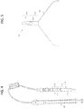


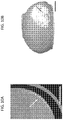
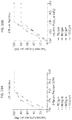
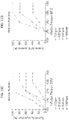
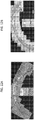


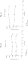


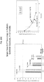
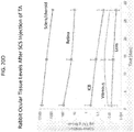
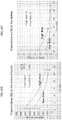
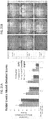
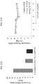

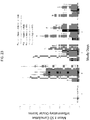


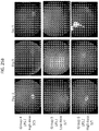
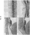

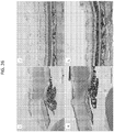

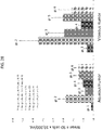
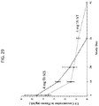


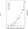
Classifications
-
- A—HUMAN NECESSITIES
- A61—MEDICAL OR VETERINARY SCIENCE; HYGIENE
- A61K—PREPARATIONS FOR MEDICAL, DENTAL OR TOILETRY PURPOSES
- A61K9/00—Medicinal preparations characterised by special physical form
- A61K9/0012—Galenical forms characterised by the site of application
- A61K9/0019—Injectable compositions; Intramuscular, intravenous, arterial, subcutaneous administration; Compositions to be administered through the skin in an invasive manner
-
- A—HUMAN NECESSITIES
- A61—MEDICAL OR VETERINARY SCIENCE; HYGIENE
- A61K—PREPARATIONS FOR MEDICAL, DENTAL OR TOILETRY PURPOSES
- A61K31/00—Medicinal preparations containing organic active ingredients
- A61K31/33—Heterocyclic compounds
- A61K31/395—Heterocyclic compounds having nitrogen as a ring hetero atom, e.g. guanethidine or rifamycins
- A61K31/435—Heterocyclic compounds having nitrogen as a ring hetero atom, e.g. guanethidine or rifamycins having six-membered rings with one nitrogen as the only ring hetero atom
- A61K31/44—Non condensed pyridines; Hydrogenated derivatives thereof
- A61K31/4427—Non condensed pyridines; Hydrogenated derivatives thereof containing further heterocyclic ring systems
- A61K31/4439—Non condensed pyridines; Hydrogenated derivatives thereof containing further heterocyclic ring systems containing a five-membered ring with nitrogen as a ring hetero atom, e.g. omeprazole
-
- A—HUMAN NECESSITIES
- A61—MEDICAL OR VETERINARY SCIENCE; HYGIENE
- A61K—PREPARATIONS FOR MEDICAL, DENTAL OR TOILETRY PURPOSES
- A61K31/00—Medicinal preparations containing organic active ingredients
- A61K31/56—Compounds containing cyclopenta[a]hydrophenanthrene ring systems; Derivatives thereof, e.g. steroids
- A61K31/57—Compounds containing cyclopenta[a]hydrophenanthrene ring systems; Derivatives thereof, e.g. steroids substituted in position 17 beta by a chain of two carbon atoms, e.g. pregnane or progesterone
- A61K31/573—Compounds containing cyclopenta[a]hydrophenanthrene ring systems; Derivatives thereof, e.g. steroids substituted in position 17 beta by a chain of two carbon atoms, e.g. pregnane or progesterone substituted in position 21, e.g. cortisone, dexamethasone, prednisone or aldosterone
-
- A—HUMAN NECESSITIES
- A61—MEDICAL OR VETERINARY SCIENCE; HYGIENE
- A61K—PREPARATIONS FOR MEDICAL, DENTAL OR TOILETRY PURPOSES
- A61K39/00—Medicinal preparations containing antigens or antibodies
- A61K39/395—Antibodies; Immunoglobulins; Immune serum, e.g. antilymphocytic serum
-
- A—HUMAN NECESSITIES
- A61—MEDICAL OR VETERINARY SCIENCE; HYGIENE
- A61K—PREPARATIONS FOR MEDICAL, DENTAL OR TOILETRY PURPOSES
- A61K45/00—Medicinal preparations containing active ingredients not provided for in groups A61K31/00 - A61K41/00
- A61K45/06—Mixtures of active ingredients without chemical characterisation, e.g. antiphlogistics and cardiaca
-
- A—HUMAN NECESSITIES
- A61—MEDICAL OR VETERINARY SCIENCE; HYGIENE
- A61K—PREPARATIONS FOR MEDICAL, DENTAL OR TOILETRY PURPOSES
- A61K47/00—Medicinal preparations characterised by the non-active ingredients used, e.g. carriers or inert additives; Targeting or modifying agents chemically bound to the active ingredient
- A61K47/06—Organic compounds, e.g. natural or synthetic hydrocarbons, polyolefins, mineral oil, petrolatum or ozokerite
- A61K47/08—Organic compounds, e.g. natural or synthetic hydrocarbons, polyolefins, mineral oil, petrolatum or ozokerite containing oxygen, e.g. ethers, acetals, ketones, quinones, aldehydes, peroxides
- A61K47/12—Carboxylic acids; Salts or anhydrides thereof
-
- A—HUMAN NECESSITIES
- A61—MEDICAL OR VETERINARY SCIENCE; HYGIENE
- A61K—PREPARATIONS FOR MEDICAL, DENTAL OR TOILETRY PURPOSES
- A61K47/00—Medicinal preparations characterised by the non-active ingredients used, e.g. carriers or inert additives; Targeting or modifying agents chemically bound to the active ingredient
- A61K47/06—Organic compounds, e.g. natural or synthetic hydrocarbons, polyolefins, mineral oil, petrolatum or ozokerite
- A61K47/26—Carbohydrates, e.g. sugar alcohols, amino sugars, nucleic acids, mono-, di- or oligo-saccharides; Derivatives thereof, e.g. polysorbates, sorbitan fatty acid esters or glycyrrhizin
-
- A—HUMAN NECESSITIES
- A61—MEDICAL OR VETERINARY SCIENCE; HYGIENE
- A61K—PREPARATIONS FOR MEDICAL, DENTAL OR TOILETRY PURPOSES
- A61K47/00—Medicinal preparations characterised by the non-active ingredients used, e.g. carriers or inert additives; Targeting or modifying agents chemically bound to the active ingredient
- A61K47/30—Macromolecular organic or inorganic compounds, e.g. inorganic polyphosphates
- A61K47/36—Polysaccharides; Derivatives thereof, e.g. gums, starch, alginate, dextrin, hyaluronic acid, chitosan, inulin, agar or pectin
- A61K47/38—Cellulose; Derivatives thereof
-
- A—HUMAN NECESSITIES
- A61—MEDICAL OR VETERINARY SCIENCE; HYGIENE
- A61K—PREPARATIONS FOR MEDICAL, DENTAL OR TOILETRY PURPOSES
- A61K9/00—Medicinal preparations characterised by special physical form
- A61K9/0012—Galenical forms characterised by the site of application
- A61K9/0048—Eye, e.g. artificial tears
-
- A—HUMAN NECESSITIES
- A61—MEDICAL OR VETERINARY SCIENCE; HYGIENE
- A61K—PREPARATIONS FOR MEDICAL, DENTAL OR TOILETRY PURPOSES
- A61K9/00—Medicinal preparations characterised by special physical form
- A61K9/10—Dispersions; Emulsions
-
- A—HUMAN NECESSITIES
- A61—MEDICAL OR VETERINARY SCIENCE; HYGIENE
- A61K—PREPARATIONS FOR MEDICAL, DENTAL OR TOILETRY PURPOSES
- A61K9/00—Medicinal preparations characterised by special physical form
- A61K9/48—Preparations in capsules, e.g. of gelatin, of chocolate
-
- A—HUMAN NECESSITIES
- A61—MEDICAL OR VETERINARY SCIENCE; HYGIENE
- A61M—DEVICES FOR INTRODUCING MEDIA INTO, OR ONTO, THE BODY; DEVICES FOR TRANSDUCING BODY MEDIA OR FOR TAKING MEDIA FROM THE BODY; DEVICES FOR PRODUCING OR ENDING SLEEP OR STUPOR
- A61M37/00—Other apparatus for introducing media into the body; Percutany, i.e. introducing medicines into the body by diffusion through the skin
- A61M37/0015—Other apparatus for introducing media into the body; Percutany, i.e. introducing medicines into the body by diffusion through the skin by using microneedles
-
- A—HUMAN NECESSITIES
- A61—MEDICAL OR VETERINARY SCIENCE; HYGIENE
- A61P—SPECIFIC THERAPEUTIC ACTIVITY OF CHEMICAL COMPOUNDS OR MEDICINAL PREPARATIONS
- A61P25/00—Drugs for disorders of the nervous system
-
- A—HUMAN NECESSITIES
- A61—MEDICAL OR VETERINARY SCIENCE; HYGIENE
- A61P—SPECIFIC THERAPEUTIC ACTIVITY OF CHEMICAL COMPOUNDS OR MEDICINAL PREPARATIONS
- A61P27/00—Drugs for disorders of the senses
- A61P27/02—Ophthalmic agents
-
- A—HUMAN NECESSITIES
- A61—MEDICAL OR VETERINARY SCIENCE; HYGIENE
- A61P—SPECIFIC THERAPEUTIC ACTIVITY OF CHEMICAL COMPOUNDS OR MEDICINAL PREPARATIONS
- A61P27/00—Drugs for disorders of the senses
- A61P27/02—Ophthalmic agents
- A61P27/06—Antiglaucoma agents or miotics
-
- A—HUMAN NECESSITIES
- A61—MEDICAL OR VETERINARY SCIENCE; HYGIENE
- A61P—SPECIFIC THERAPEUTIC ACTIVITY OF CHEMICAL COMPOUNDS OR MEDICINAL PREPARATIONS
- A61P27/00—Drugs for disorders of the senses
- A61P27/02—Ophthalmic agents
- A61P27/10—Ophthalmic agents for accommodation disorders, e.g. myopia
-
- A—HUMAN NECESSITIES
- A61—MEDICAL OR VETERINARY SCIENCE; HYGIENE
- A61P—SPECIFIC THERAPEUTIC ACTIVITY OF CHEMICAL COMPOUNDS OR MEDICINAL PREPARATIONS
- A61P29/00—Non-central analgesic, antipyretic or antiinflammatory agents, e.g. antirheumatic agents; Non-steroidal antiinflammatory drugs [NSAID]
-
- A—HUMAN NECESSITIES
- A61—MEDICAL OR VETERINARY SCIENCE; HYGIENE
- A61P—SPECIFIC THERAPEUTIC ACTIVITY OF CHEMICAL COMPOUNDS OR MEDICINAL PREPARATIONS
- A61P3/00—Drugs for disorders of the metabolism
- A61P3/08—Drugs for disorders of the metabolism for glucose homeostasis
- A61P3/10—Drugs for disorders of the metabolism for glucose homeostasis for hyperglycaemia, e.g. antidiabetics
-
- A—HUMAN NECESSITIES
- A61—MEDICAL OR VETERINARY SCIENCE; HYGIENE
- A61P—SPECIFIC THERAPEUTIC ACTIVITY OF CHEMICAL COMPOUNDS OR MEDICINAL PREPARATIONS
- A61P31/00—Antiinfectives, i.e. antibiotics, antiseptics, chemotherapeutics
- A61P31/12—Antivirals
- A61P31/20—Antivirals for DNA viruses
-
- A—HUMAN NECESSITIES
- A61—MEDICAL OR VETERINARY SCIENCE; HYGIENE
- A61P—SPECIFIC THERAPEUTIC ACTIVITY OF CHEMICAL COMPOUNDS OR MEDICINAL PREPARATIONS
- A61P37/00—Drugs for immunological or allergic disorders
- A61P37/02—Immunomodulators
-
- A—HUMAN NECESSITIES
- A61—MEDICAL OR VETERINARY SCIENCE; HYGIENE
- A61P—SPECIFIC THERAPEUTIC ACTIVITY OF CHEMICAL COMPOUNDS OR MEDICINAL PREPARATIONS
- A61P43/00—Drugs for specific purposes, not provided for in groups A61P1/00-A61P41/00
-
- A—HUMAN NECESSITIES
- A61—MEDICAL OR VETERINARY SCIENCE; HYGIENE
- A61P—SPECIFIC THERAPEUTIC ACTIVITY OF CHEMICAL COMPOUNDS OR MEDICINAL PREPARATIONS
- A61P7/00—Drugs for disorders of the blood or the extracellular fluid
- A61P7/02—Antithrombotic agents; Anticoagulants; Platelet aggregation inhibitors
-
- A—HUMAN NECESSITIES
- A61—MEDICAL OR VETERINARY SCIENCE; HYGIENE
- A61P—SPECIFIC THERAPEUTIC ACTIVITY OF CHEMICAL COMPOUNDS OR MEDICINAL PREPARATIONS
- A61P9/00—Drugs for disorders of the cardiovascular system
- A61P9/10—Drugs for disorders of the cardiovascular system for treating ischaemic or atherosclerotic diseases, e.g. antianginal drugs, coronary vasodilators, drugs for myocardial infarction, retinopathy, cerebrovascula insufficiency, renal arteriosclerosis
-
- C—CHEMISTRY; METALLURGY
- C07—ORGANIC CHEMISTRY
- C07K—PEPTIDES
- C07K16/00—Immunoglobulins [IGs], e.g. monoclonal or polyclonal antibodies
- C07K16/18—Immunoglobulins [IGs], e.g. monoclonal or polyclonal antibodies against material from animals or humans
- C07K16/22—Immunoglobulins [IGs], e.g. monoclonal or polyclonal antibodies against material from animals or humans against growth factors ; against growth regulators
-
- A—HUMAN NECESSITIES
- A61—MEDICAL OR VETERINARY SCIENCE; HYGIENE
- A61K—PREPARATIONS FOR MEDICAL, DENTAL OR TOILETRY PURPOSES
- A61K39/00—Medicinal preparations containing antigens or antibodies
- A61K2039/505—Medicinal preparations containing antigens or antibodies comprising antibodies
-
- A—HUMAN NECESSITIES
- A61—MEDICAL OR VETERINARY SCIENCE; HYGIENE
- A61K—PREPARATIONS FOR MEDICAL, DENTAL OR TOILETRY PURPOSES
- A61K39/00—Medicinal preparations containing antigens or antibodies
- A61K2039/54—Medicinal preparations containing antigens or antibodies characterised by the route of administration
-
- A—HUMAN NECESSITIES
- A61—MEDICAL OR VETERINARY SCIENCE; HYGIENE
- A61K—PREPARATIONS FOR MEDICAL, DENTAL OR TOILETRY PURPOSES
- A61K2300/00—Mixtures or combinations of active ingredients, wherein at least one active ingredient is fully defined in groups A61K31/00 - A61K41/00
-
- A—HUMAN NECESSITIES
- A61—MEDICAL OR VETERINARY SCIENCE; HYGIENE
- A61K—PREPARATIONS FOR MEDICAL, DENTAL OR TOILETRY PURPOSES
- A61K9/00—Medicinal preparations characterised by special physical form
- A61K9/14—Particulate form, e.g. powders, Processes for size reducing of pure drugs or the resulting products, Pure drug nanoparticles
- A61K9/16—Agglomerates; Granulates; Microbeadlets ; Microspheres; Pellets; Solid products obtained by spray drying, spray freeze drying, spray congealing,(multiple) emulsion solvent evaporation or extraction
-
- A—HUMAN NECESSITIES
- A61—MEDICAL OR VETERINARY SCIENCE; HYGIENE
- A61M—DEVICES FOR INTRODUCING MEDIA INTO, OR ONTO, THE BODY; DEVICES FOR TRANSDUCING BODY MEDIA OR FOR TAKING MEDIA FROM THE BODY; DEVICES FOR PRODUCING OR ENDING SLEEP OR STUPOR
- A61M37/00—Other apparatus for introducing media into the body; Percutany, i.e. introducing medicines into the body by diffusion through the skin
- A61M37/0015—Other apparatus for introducing media into the body; Percutany, i.e. introducing medicines into the body by diffusion through the skin by using microneedles
- A61M2037/0023—Drug applicators using microneedles
-
- A—HUMAN NECESSITIES
- A61—MEDICAL OR VETERINARY SCIENCE; HYGIENE
- A61M—DEVICES FOR INTRODUCING MEDIA INTO, OR ONTO, THE BODY; DEVICES FOR TRANSDUCING BODY MEDIA OR FOR TAKING MEDIA FROM THE BODY; DEVICES FOR PRODUCING OR ENDING SLEEP OR STUPOR
- A61M37/00—Other apparatus for introducing media into the body; Percutany, i.e. introducing medicines into the body by diffusion through the skin
- A61M37/0015—Other apparatus for introducing media into the body; Percutany, i.e. introducing medicines into the body by diffusion through the skin by using microneedles
- A61M2037/0061—Methods for using microneedles
Definitions
- This invention is generally in the field of ophthalmic therapies, and more particularly to the use of a microneedle for infusion of a fluid drug formulation into ocular tissues for targeted, local drug delivery.
- the suprachoroidal space (SCS) of the eye has been studied, and its cannulation described as a possible route for drug delivery. See, e.g ., Olsen, et al., American J. Ophthalmology 142(5): 777-87 (Nov. 2006 ).; PCT Patent Application Publication No. WO 2007/100745 .
- US 2007/202186 discloses the treatment of the superior chamber of the eye with triamcinolone via injection into the eye.
- Any method of treatment is to be understood to refer to the drug formulation being used in such a method of treatment.
- the present invention relates to drug formulations for use in non-surgical ophthalmic therapies in human patients in need of such treatment, and more particularly to the infusion of a drug formulation comprising triamcinolone into the suprachoroidal space of the eye for targeted, local drug delivery, for the treatment of posterior ocular disorders.
- a method for treating a posterior ocular disorder in a human subject in need of treatment comprises non-surgically administering an effective amount of a triamcinolone formulation to the suprachoroidal space (SCS) of the eye of the subject in need of treatment of the posterior ocular disorder.
- SCS suprachoroidal space
- the drug formulation flows away from the insertion site and is substantially localized to the posterior segment of the eye.
- the posterior ocular disorder is an ocular inflammatory condition such as uveitis, scleritis, glaucoma, ocular sarcoidosis, optic neuritis, macular edema, diabetic retinopathy, macular degeneration, a corneal ulcer, an autoimmune disorder, ophthalmic manifestations of AIDS, optic nerve degeneration, geographic atrophy, choroidal disease or retinitis.
- the condition in one embodiment is acute. In another embodiment, the condition is chronic.
- a method for the treatment of a choroidal malady e.g ., ocular neovascularization, polypoidal choroidal vasculopathy, choroidal sclerosis, central sirrus choroidopathy, a multi-focal choroidopathy or a choroidal dystrophy (e.g., central gyrate choroidal dystrophy, serpiginous choroidal dystrophy, total central choroidal atrophy).
- the method comprises non-surgically administering a drug formulation comprising an effective amount of an anti-inflammatory drug, a vascular endothelial growth factor (VEGF) modulator, a platelet derived growth factor (PDGF) modulator, an angiogenesis inhibitor, an immunosuppressive agent, a vascular permeability inhibitor, or a combination thereof, to the SCS of the patient in need of treatment.
- VEGF vascular endothelial growth factor
- PDGF platelet derived growth factor
- angiogenesis inhibitor an immunosuppressive agent
- a vascular permeability inhibitor or a combination thereof
- the effective amount of the drug administered to the SCS provides higher efficacy or a greater therapeutic effect of the drug, compared to the identical drug dose administered intravitreally, intracamerally, topically, parenterally or orally.
- the patient undergoing treatment via SCS drug therapy was not previously responsive to a different type of therapy for the same condition.
- a method for decreasing subretinal exudation and bleeding in a subject comprises non-surgically administering a drug formulation comprising an effective amount of an effective amount of an anti-inflammatory drug, a vascular endothelial growth factor (VEGF) modulator, a platelet derived growth factor (PDGF) modulator, an angiogenesis inhibitor, an immunosuppressive agent, a vascular permeability inhibitor, or a combination thereof, to the SCS of the patient in need of treatment, wherein administration of the drug formulation reduces subretinal exudation and bleeding experienced by the patient, as compared to the identical dosage of the drug administered intravitreally to the patient.
- VEGF vascular endothelial growth factor
- PDGF platelet derived growth factor
- a method for treating a posterior ocular disorder in a human patient comprises non-surgically administering an effective amount of a drug formulation comprising triamcinolone to the suprachoroidal space (SCS) of the eye of the subject in need of treatment of the posterior ocular disorder.
- the intraocular elimination half life (t 1/2 ) of the drug administered to the SCS is greater than the intraocular t 1/2 of the drug, when administered intravitreally, intracamerally, topically, parenterally or orally.
- the mean intraocular maximum concentration (C max ) of the drug, when administered to the SCS via the methods described herein, is greater than the intraocular C max of the drug, when administered intravitreally, intracamerally, topically, parenterally or orally.
- the mean intraocular area under the curve (AUC 0-t ) of the drug, when administered to the SCS via the methods described herein is greater than the intraocular AUC 0-t of the drug, when administered intravitreally, intracamerally, topically, parenterally or orally.
- the intraocular time to peak concentration (t max ) of the drug when administered to the SCS via the methods described herein, is greater than the intraocular t max of the drug, when the same drug dose is administered intravitreally, intracamerally, topically, parenterally or orally.
- the formulation comprises an effective amount of an anti-inflammatory drug (e.g., a steroid or NSAID), a VEGF modulator (e.g., VEGF antagonist), a platelet derived growth factor (PDGF) modulator, an angiogenesis inhibitor, an immunosuppressive agent, a vascular permeability inhibitor, or a combination thereof.
- the drug formulation comprises triamcinolone.
- the method for treating a posterior ocular disorder in a human subject comprises delivering a drug formulation via a hollow microneedle to the SCS of the eye of the human subject in need of treatment, whereby the needle has a diameter of 28 gauge or smaller.
- delivering the drug formulation comprises inserting a hollow microneedle into the eye of the human subject at an insertion site, the microneedle having a tip end with an opening; and infusing over a period of time a drug formulation through the inserted microneedle and into the SCS space away from the insertion site.
- the drug formulation administered to the SCS flows away from the insertion site and is substantially localized to the posterior segment of the eye, thereby increasing the therapeutic efficacy of the dose of the drug compared to the therapeutic efficacy of the same drug dose administered by another means (e.g. intravitreally, intracamerally, topically, parenterally, and/or orally).
- the dose of the drug sufficient to elicit a therapeutic response when administered to the SCS is less than the dosage of the drug sufficient to elicit a therapeutic response when administered intravitreally, topically, parenterally or orally.
- the drug formulation is delivered to the SCS by a hollow microneedle inserted into the sclera at the equator of the eye or between the equator and the limbus of the eye.
- the hollow microneedle is inserted in the insertion site at a 90 degree angle (perpendicular).
- the drug formulation delivered by the methods described herein, according to the invention comprises an effective amount of an anti-inflammatory drug, namely triamcinolone.
- an additional drug delivered to the SCS via the methods described herein is a immunosuppressive, antimetabolite, T-cell inhibitor, alkylating agent, biologic, TNF ⁇ antagonist, interleukin antagonist, neuroprotectant, vascular endothelial growth factor (VEGF) antagonist, platelet derived growth factor (PDGF) antagonist, or a combination thereof.
- the drug affects inflammation, neuroprotection, complement inhibition, drusen formation, scar formation, reduction in choriocapillaris or choroidal neocasvularization.
- the drug formulation comprises microparticles and/or nanoparticles of the drug.
- the drug formulation comprises microparticles having a D 50 of 1 ⁇ m or less and/or a D 99 of 10 ⁇ m or less.
- the drug formulation comprises triamcinolone.
- the invention includes a method for treating a posterior ocular disorder in a human subject in need thereof comprising non-surgically administering a drug formulation comprising triamcinolone to the SCS of the eye of the human subject, wherein upon administration, the drug formulation flows away from the insertion site and is substantially localized to the posterior segment.
- the intraocular pressure of the eye remains substantially constant during administration of the drug formulation to the SCS.
- administration of the drug formulation to the SCS of the eye results in a decreased number of side effects, or a reduced severity of one or more side effects, compared to administration of the same drug dose intravitreally, intracamerally, topically, orally or parenterally.
- the present disclosure relates to a method for treating a choroidal malady in a human patient in need of treatment.
- the method comprises non-surgically administering a drug formulation comprising an effective amount of an anti-inflammatory drug, a vascular endothelial growth factor (VEGF) modulator, a platelet derived growth factor (PDGF) modulator, an angiogenesis inhibitor, an immunosuppressive agent or a vascular permeability inhibitor, to the suprachoroidal space (SCS) of the eye of the patient.
- VEGF vascular endothelial growth factor
- PDGF platelet derived growth factor
- angiogenesis inhibitor angiogenesis inhibitor
- an immunosuppressive agent or a vascular permeability inhibitor an immunosuppressive agent or a vascular permeability inhibitor
- the present disclosure relates to a method for treating ocular neovascularization in a human patient in need of treatment.
- the method comprises non-surgically administering a drug formulation comprising an effective amount of an anti-inflammatory drug, a vascular endothelial growth factor (VEGF) modulator (e.g., a VEGF antagonist), a platelet derived growth factor (PDGF) modulator (e.g., a PDGF antagonist), an angiogenesis inhibitor, an immunosuppressive agent or a vascular permeability inhibitor, to the suprachoroidal space (SCS) of the eye of the patient.
- VEGF vascular endothelial growth factor
- PDGF platelet derived growth factor
- the ocular neovascularization is a choroidal neovascularization.
- the human patient being treated for the ocular neovascularization, prior to administration of the drug formulation, was previously treated for the ocular neovascularization and was not properly responsive to the treatment.
- the drug formulation delivered by the methods described herein comprises an effective amount of an anti-inflammatory drug, namely a steroidal compound, defined to be triamcinolone.
- an additional drug delivered to the SCS via the methods described herein is a vascular permeability inhibitor, an angiogenesis inhibitor or a VEGF modulator, e.g ., a VEGF antagonist.
- the VEGF antagonist is a VEGF receptor antagonist or a soluble VEGF receptor.
- the drug formulation comprises drug microparticles having a D 50 of 1 ⁇ m or less and/or a D 99 of 10 ⁇ m or less.
- the drug formulation comprises triamcinolone.
- a method for treating a posterior ocular disorder in a human subject in need thereof comprising non-surgically administering a drug formulation to the SCS of the eye of the human subject, wherien, the intraocular pressure of the eye remains substantially constant during administration of the drug formulation to the SCS,
- administration of the drug formulation comprising triamcinolone to the SCS of the eye of the patient in need of treatment of the posterior ocular disorder results in a decreased number of side effects, or a reduced severity of one or more side effects, compared to administration of the same drug dose intravitreally, intracamerally, topically, orally or parenterally.
- the side effect reduced by the methods described herein is subretinal exudation and/or bleeding.
- Drug formulations comprising triamcinolone are provided herein for treating posterior ocular disorders in human subjects in need thereof. They allow for effective posterior segment drug delivery to treat posterior ocular disorders and generally embody the following characteristics: (1) the methods are non-surgical and thus minimally invasive and safe; (2) the drug formulations are administered in such a way that they are well targeted to the posterior segment of the eye and/or the suprachoroidal space (SCS) of the eye while simultaneously limiting drug exposure to the anterior segment or other regions of the eye; (3) the methods and formulations are capable of delivering drug in a sustained and/or controlled manner; (4) the methods and devices are user-friendly.
- the non-surgical SCS delivery methods, devices for implementing the methods, and drug formulations for SCS delivery set forth herein achieve these desired characteristics.
- non-surgical ocular drug delivery methods refer to methods of drug delivery that do not require general anesthesia and/or retrobulbar anesthesia (also referred to as a retrobulbar block). A non-surgical ocular drug delivery method is performed with an instrument having a diameter of 28 gauge or smaller. Additionally, “non-surgical" ocular drug delivery methods do not require a guidance mechanism that is typically required for ocular drug delivery via a shunt or cannula.
- the non-surgical posterior ocular disorder treatment methods described herein are particularly useful for the local delivery of drugs to the posterior region of the eye, for example the retinochoroidal tissue, macula, retinal pigment epithelium (RPE) and optic nerve in the posterior segment of the eye.
- the non-surgical methods and microneedles provided herein can be used to target drug delivery to specific posterior ocular tissues or regions within the eye or in neighboring tissue.
- the methods described herein deliver drug specifically to the sclera, the choroid, the Brach's membrane, the retinal pigment epithelium, the subretinal space, the retina, the macula, the optic disk, the optic nerve, the ciliary body, the trabecular meshwork, the aqueous humor, the vitreous humor, and/or other ocular tissue or neighboring tissue in the eye of a human subject in need of treatment.
- the methods and microneedles provided herein in one embodiment, can be used to target drug delivery to specific posterior ocular tissues or regions within the eye or in neighboring tissue.
- non-surgical delivery of a drug e.g., an anti-inflammatory drug (e.g., triamcinolone), a vascular endothelial growth factor (VEGF) modulator (e.g., VEGF antagonist), a platelet derived growth factor (PDGF) antagonist to the suprachoroidal space for treatment of a posterior ocular disorder or choroidal malady
- a drug e.g., an anti-inflammatory drug (e.g., triamcinolone), a vascular endothelial growth factor (VEGF) modulator (e.g., VEGF antagonist), a platelet derived growth factor (PDGF) antagonist
- the effective amount of the drug administered to the SCS provides higher thereapeutic efficacy of the drug, compared to the therapeutic efficacy of the drug when the identical dosage is administered intravitreally, topically, intracamerally, parenterally or orally.
- the microneedle drug delivery methods described herein precisely deliver the drug into the SCS for subsequent local delivery to nearby posterior ocular tissues in need of treatment.
- the drug may be released into the ocular tissues from the infused volume (or, e.g., from microparticles or nanoparticles in the drug formulation) for an extended period, e.g., several hours or days or weeks or months, after the non-surgical drug administration has been completed.
- This beneficially can provide increased bioavailability of the drug relative, for example, to delivery by topical application of the drug formulation to ocular tissue surfaces, or increased bioavailability compared to oral, parenteral on intravitreal administration of the same drug dosage.
- the SCS drug delivery methods advantageously include precise control of the depth of insertion into the ocular tissue, so that the microneedle tip can be placed into the eye so that the drug formulation flows into the suprachoroidal space and in some embodiments to the posterior ocular tissues surrounding the SCS.
- insertion of the microneedle is in the sclera of the eye.
- drug flow into the SCS is accomplished without contacting underlying tissues with the microneedle, such as choroid and retina tissues.
- the methods provided herein achieve delivery of drug to the suprachoroidal space, thereby allowing drug access to posterior ocular tissues not obtainable via topical, parenteral, intracameral or intravitreal drug delivery. Because the methods provided herein deliver drug to the posterior ocular tissue for the treatment of a posterior ocular disorder or choroidal malady, the suprachoroidal drug dose sufficient to achieve a therapeutic response in a human subject treated with the methods provided herein is less than the intravitreal, topical, parenteral or oral drug dose sufficient to elicit the same or substantially the same therapeutic response.
- the SCS delivery methods described herein allow for decreased drug dose of the posterior ocular disorder treating drug, or the choroidal malady treating drug, compared to the intravitreal, topical, intracameral parenteral or oral drug dose sufficient to elicit the same or substantially the same therapeutic response.
- the suprachoroidal drug dose sufficient to elicit a therapeutic response is 75% or less, or 50% or less, or 25% or less than the intravitreal, topical parenteral or oral drug dose sufficient to elicit a therapeutic response.
- the therapeutic response in one embodiment, is a reduction in severity of a symptom/clinical manifestation of the posterior ocular disorder for which the patient is undergoing treatment, or a reduction in number of symptom(s)/clinical manifestation(s) of the posterior ocular disorder for which the patient is undergoing treatment.
- suprachoroidal space is used interchangeably with suprachoroidal, SCS, suprachoroid and suprachoroidia, and describes the potential space in the region of the eye disposed between the sclera and choroid. This region primarily is composed of closely packed layers of long pigmented processes derived from each of the two adjacent tissues; however, a space can develop in this region as a result of fluid or other material buildup in the suprachoroidal space and the adjacent tissues. Those skilled in the art will appreciate that, the suprachoroidal space frequently is expanded by fluid buildup because of some disease state in the eye or as a result of some trauma or surgical intervention.
- the fluid buildup is intentionally created by infusion of a drug formulation into the suprachoroid to create the suprachoroidal space (which is filled with drug formulation).
- the SCS region serves as a pathway for uveoscleral outflow (i.e., a natural process of the eye moving fluid from one region of the eye to the other through) and becomes a real space in instances of choroidal detachment from the sclera.
- anterior segment 12 and “eye” 10 include both the anterior segment 12 of the eye (i.e., the portion of the eye in front of the lens) and the posterior segment 14 of the eye (i.e., the portion of the eye behind the lens), as illustrated in FIG. 1A .
- the anterior segment 12 is bounded by the cornea 16 and the lens 18
- the posterior segment 14 is bounded by the sclera 20 and the lens 18 .
- the anterior segment 12 is further subdivided into the anterior chamber 22 , between the iris 24 and the cornea 16 , and the posterior chamber 26 , between the lens 18 and the iris 24 .
- FIG. 1B illustrates the cornea 16 , which is composed of the epithelium 30, the Bowman's layer 32 , the stroma 34 , the Descemet's membrane 36 , and the endothelium 38.
- 1D illustrate the sclera 20 with surrounding Tenon's Capsule 40 or conjunctiva 41 , suprachoroidal space 42 , choroid 28 , and retina 27 , both without and with a fluid in the suprachoroidal space, respectively.
- microneedle refers to a conduit body having a base, a shaft, and a tip end suitable for insertion into the sclera and other ocular tissue and has dimensions suitable for minimally invasive insertion and drug formulation infusion as described herein. That is, the microneedle has a length or effective length that does not exceed about 2000 microns and a diameter that does not exceed about 600 microns. Both the "length” and “effective length” of the microneedle encompass the length of the shaft of the microneedle and the bevel height of the microneedle. The microneedle has a diameter of 28 gauge or smaller.
- a hollow microneedle has a structure that includes one or more continuous pathways from the base of the microneedle to an exit point (opening) in the shaft and/or tip portion of the microneedle distal to the base.
- FIGS. 2-5 illustrate exemplary embodiments of microneedle devices.
- the microneedle device 110 includes a hollow microneedle 114 having a hollow bore 140 through which a fluid drug formulation (not shown) can be delivered to the eye or through which a biological fluid can be withdrawn from the eye.
- the microneedle includes a proximal portion 116 and a tip portion 118 .
- the microneedle 114 may extend from a base comprising, for example, an elongated body 112 having a distal end from which the proximal portion 116 and tip portion 118 of the microneedle extends.
- the elongated body may further comprise a means for securing 111 a base portion of the microneedle extending beyond the distal end of the base 112 , such as a screw or pin.
- a means for securing 111 a base portion of the microneedle extending beyond the distal end of the base 112 such as a screw or pin.
- An exemplary embodiment of the elongated body 112 for securing the microneedle is illustrated in FIG. 3 , and comprises a cap portion 113 and a base portion 115 having a hollow bore 117 therein.
- the cap portion 113 and base portion 115 of the elongated body 112 desirably comprise a means for manually adjusting the length of needle (i.e., the proximal portion and tip portion of the microneedle extending from the base 112 ) protruding out of the cap portion of the elongated body.
- Such means may include, for example, threads 119 allowing the cap portion 113 to be screwed in and out of the base portion 115 of the elongated body.
- the base portion 115 of the elongated body may be operably connected to an actuator 120 for controlled infusion of the fluid drug formulation through the microneedle into the suprachoroidal space.
- the microneedle device may further comprise a fluid reservoir for containing the drug formulation, e,g. , as a solution or suspension, and the drug reservoir being in operable communication with the bore of the microneedle at a location distal to the tip end of the microneedle.
- the fluid reservoir may be integral with the microneedle, integral with the elongated body, or separate from both the microneedle and elongated body.
- the microneedle can be formed/constructed of different biocompatible materials, including metals, glasses, semi-conductor materials, ceramics, or polymers.
- suitable metals include pharmaceutical grade stainless steel, gold, titanium, nickel, iron, gold, tin, chromium, copper, and alloys thereof.
- the polymer can be biodegradable or non-biodegradable.
- suitable biocompatible, biodegradable polymers include polylactides, polyglycolides, polylactide-co-glycolides (PLGA), polyanhydrides, polyorthoesters, polyetheresters, polycaprolactones, polyesteramides, poly(butyric acid), poly(valeric acid), polyurethanes and copolymers and blends thereof.
- non-biodegradable polymers include various thermoplastics or other polymeric structural materials known in the fabrication of medical devices. Examples include nylons, polyesters, polycarbonates, polyacrylates, polymers of ethylene-vinyl acetates and other acyl substituted cellulose acetates, non-degradable polyurethanes, polystyrenes, polyvinyl chloride, polyvinyl fluoride, poly(vinyl imidazole), chlorosulphonate polyolefins, polyethylene oxide, blends and copolymers thereof.
- Biodegradable microneedles can provide an increased level of safety compared to non-biodegradable ones, such that they are essentially harmless even if inadvertently broken off into the ocular tissue.
- the microneedle can be fabricated by a variety of methods known in the art or as described in the Examples below.
- the hollow microneedle is fabricated using a laser or similar optical energy source.
- a microcannula may be cut using a laser to represent the desired microneedle length.
- the laser may also be use to shape single or multiple tip openings. Single or multiple cuts may be performed on a single microncannula to shape the desired microneedle structure.
- the microcannula may be made of metal such as stainless steel and cut using a laser with a wavelength in the infrared region of the light spectrum ( e.g. , from about 0.7 to about 300 ⁇ m).
- the microneedle length and optional bevel is formed by a physical grinding process, which for example may include grinding a metal cannula against a moving abrasive surface.
- the fabrication process may further include precision grinding, micro-bead jet blasting and ultrasonic cleaning to form the shape of the desired precise tip of the microneedle.
- the methods and devices provided herein allow for suprachoroidal drug delivery to be accomplished in a minimally invasive, non-surgical manner, superior to other non-surgical (e.g., conventional needle) and surgical approaches.
- the methods provided herein are carried out via the use of one or more microneedles.
- the microneedles are be inserted perpendicular, or at an angle from about 80° to about 100°, into the eye, e.g., into the sclera, reaching the suprachoroidal space in a short penetration distance.
- the microneedle in one embodiment, is part of an array of two or more microneedles such that the method further includes inserting at least a second microneedle into the sclera without penetrating across the sclera.
- the drug formulation of each of the two or more microneedles may be identical to or different from one another, in drug, formulation, volume/quantity of drug formulation, or a combination of these parameters.
- different types of drug formulations may be injected via the one or more microneedles. For example, inserting a second hollow microneedle comprising a second drug formulation into the ocular tissue will result in delivery of the second drug formulation into the ocular tissue.
- microneedle devices described herein are adapted to remove substances, such as a fluid, tissue, or molecule sample, from the eye.
- microneedles e.g., solid microneedles
- Non-limiting examples include dissolving, at least in part, a coating of a drug formulation off of a microneedle; detaching, at least in part, a coating of a drug formulation (e.g., as a substantially intact sleeve or in fragments) off of a microneedle; breaking or dissolving a microneedle off of a base to which the microneedle is integrally formed or is connected; or any combination thereof.
- the microneedle devices described herein also may be adapted to use the one or more microneedles as a sensor to detect analytes, electrical activity, and optical or other signals.
- the sensor may include sensors of pressure, temperature, chemicals, and/or electromagnetic fields (e.g., light).
- Biosensors can be located on or within the microneedle, or inside a device in communication with the body tissue via the microneedle.
- the microneedle biosensor can be any of the four classes of principal transducers: potentiometric, amperometric, optical, and physiochemical.
- a hollow microneedle is filled with a substance, such as a gel, that has a sensing functionality associated with it.
- the substrate or enzyme can be immobilized in the needle interior.
- a wave guide can be incorporated into the microneedle device to direct light to a specific location, or for detection, for example, using means such as a pH dye for color evaluation.
- heat, electricity, light, ultrasound or other energy forms may be precisely transmitted to directly stimulate, damage, or heal a specific tissue or for diagnostic purposes.
- the microneedle device for non-surgically delivering drug to the suprachoroidal space of the eye of a human subject comprises a hollow microneedle.
- the device may include an elongated housing for holding the proximal end of the microneedle.
- the device may further include a means for conducting a drug formulation through the microneedle.
- the means may be a flexible or rigid conduit in fluid connection with the base or proximal end of the microneedle.
- the means may also include a pump or other devices for creating a pressure gradient for inducing fluid flow through the device.
- the conduit may in operable connection with a source of the drug formulation.
- the source may be any suitable container. In one embodiment, the source may be in the form of a conventional syringe.
- the source may be a disposable unit dose container.
- the microneedle has an effective length of about 50 ⁇ m to about 2000 ⁇ m. In another particular embodiment, the microneedle has an effective length of from about 150 ⁇ m to about 1500 ⁇ m, or from about 300 ⁇ m to about 1250 ⁇ m, or from about 500 ⁇ m to about 1250 ⁇ m, or from about 500 ⁇ m to about 1500 ⁇ m, or from about 600 ⁇ m to about 1000 ⁇ m, or from about 700 ⁇ m to about 1000 ⁇ m. In one embodiment, the effective length of the microneedle is about 600 ⁇ m, or about 700 ⁇ m, or about 800 ⁇ m or about 1000 ⁇ m.
- the proximal portion of the microneedle has a maximum width or cross-sectional dimension of from about 50 ⁇ m to 600 ⁇ m, or from about 50 ⁇ m to about 400 ⁇ m, or from about 50 ⁇ m to about 500 ⁇ m, or from about 100 ⁇ m to about 400 ⁇ m, or from about 200 ⁇ m to about 600 ⁇ m, or from about 100 ⁇ m to about 250 ⁇ m, with an aperture diameter of about 5 ⁇ m to about 400 ⁇ m.
- the proximal portion of the microneedle has a maximum width or cross-sectional dimension of about 600 ⁇ m.
- the aperture diameter may be greater than the outer diameter of the proximal portion of the microneedle.
- the microneedle may be fabricated to have an aspect ratio (width: length) of about 1:1.5 to about 1:10. In one embodiment, the aspect ratio of the microneedle is about 1:3 to about 1:5. In another embodiment, the aspect ratio of the microneedle is about 1:4 to about 1:10.
- the microneedle can have a straight or tapered shaft.
- the diameter of the microneedle is greatest at the base end of the microneedle and tapers to a point at the end distal the base.
- the microneedle can also be fabricated to have a shaft that includes both a straight (i.e., untapered) portion and a tapered (e.g., beveled) portion.
- the microneedle has a bevel angle of about 5 degrees to about 30 degrees, of about 5 degrees to about 25 degrees, about 5 degrees to about 20 degrees, about 10 degrees to about 20 degrees, and about 10 degrees to about 30 degrees.
- the microneedles can be formed with shafts that have a circular cross-section in the perpendicular, or the cross-section can be non-circular.
- the tip portion of the microneedles can have a variety of configurations.
- the tip of the microneedle can be symmetrical or asymmetrical about the longitudinal axis of the shaft.
- the tips may be beveled, tapered, squared-off, or rounded.
- the microneedle has a bevel height from about 50 ⁇ m to 500 ⁇ m, about 100 ⁇ m to about 500 ⁇ m, about 100 ⁇ m to about 400 ⁇ m, about 200 ⁇ m to about 400 ⁇ m and about 300 ⁇ m to about 500 ⁇ m.
- the microneedle may be designed such that the tip portion of the microneedle is substantially the only portion of the microneedle inserted into the ocular tissue (i.e., the tip portion is greater than 75% of the total length of the microneedle, greater than 85% of the total length of the microneedle, or greater than about 95% of the total length of the microneedle).
- the microneedle may be designed such that the tip portion is only a portion of the microneedle that is inserted into the ocular tissue and generally has a length that is less than about 75% of the total length of the microneedle, less than about 50% of the total length of the microneedle, or less than about 25%) of the total length of the microneedle.
- the microneedle has a total effective length between 500 ⁇ m and 1500 ⁇ m, wherein the tip portion has a length that is less than about 400 ⁇ m, less than about 300 ⁇ m, or less than about 200 ⁇ m.
- the height of the bevel is about 100 ⁇ m to about 500 ⁇ m. In another embodiment, the height of the bevel is about 500 ⁇ m or less, about 450 ⁇ m or less, about 400 ⁇ m or less or about 350 ⁇ m or less. In another embodiment, the height of the bevel is from about 200 ⁇ m to about 500 ⁇ m, or from about 100 ⁇ m to about 700 ⁇ m, or from about 200 ⁇ m to about 700 ⁇ m. In still other embodiments, the height of the bevel is from about 500 ⁇ m to about 900 ⁇ m, or from about 500 ⁇ m to about 800 ⁇ m, or from about 500 ⁇ m to about 700 ⁇ m.
- the arrangement of the bevel can be such that the distal edge is sufficiently sharp such as to pierce a target tissue and penetrate into the vitreous without (i) substantially causing the target tissue to elastically deform or (ii) damaging internal structures of the eye, e.g., the lens or retina.
- the microneedle extends from a base.
- the base may be integral with or separate from the microneedle.
- the base may be rigid or flexible.
- the base may be substantially planar or it may be curved, for example, in the shape of the ocular tissue surface at the site of injection or, for example, curved away from the ocular surface (e.g., convex) so as to minimize contact between the base and the ocular tissue.
- the base is shaped to provide minimal contact with the surface of the eye at the point of insertion.
- the base may extend only a minimal distance from the microneedle shaft substantially perpendicular.
- the base may be shaped so as to elevate the ocular tissue towards the microneedle so as to counteract the deflection of the ocular tissue and facilitate insertion of the microneedle into the ocular tissue (e.g., the base may extend from the microneedle toward the tip portion of the microneedle so as to "pinch" the ocular tissue).
- the microneedle device has a single microneedle.
- the microneedle device 130 includes a convex base 132 and a hollow microneedle 134 which has a bore 140 through which a fluid drug formulation (not shown) can be delivered to the eye or through which a biological fluid can be withdrawn from the eye.
- the hollow microneedle 134 includes a proximal portion 136 and a tip portion 138 .
- the microneedle may extend from the base of the microneedle device at any angle suitable for insertion into the eye.
- the microneedle extends from the base at an angle of about 90 degrees to provide approximately perpendicular insertion of the microneedles into the surface of the eye.
- the microneedle extends from the base at an angle from about 60 to about 110 degrees, or from about 70 degrees to about 100 degrees, or from about 80 degrees to about 90 degrees, or from about 85 degrees to about 95 degrees.
- the microneedle device may comprise a means for controllably inserting, and optionally retracting, the microneedle into the ocular tissue.
- the microneedle device may include means of controlling the angle at which the at least one microneedle is inserted into the ocular tissue (e.g ., by inserting the at least one microneedle into the surface of the ocular tissue at an angle of about 90 degrees).
- the depth of microneedle insertion into the ocular tissue can be controlled by the length of the microneedle, as well as other geometric features of the microneedle. For example, a flange or other a sudden change in microneedle width can be used to limit the depth of microneedle insertion.
- the microneedle insertion can also be controlled using a mechanical micropositioning system involving gears or other mechanical components that move the microneedle into the ocular tissue a controlled distance and, likewise, can be operated, for example, in reverse, to retract the microneedle a controlled distance.
- the depth of insertion can also be controlled by the velocity at which the microneedle is inserted into the ocular tissue.
- the retraction distance can be controlled by elastic recoil of the ocular tissue into which the microneedle is inserted or by including an elastic element within the microneedle device that pulls the microneedle back a specified distance after the force of insertion is released.
- the angle of insertion can be directed by positioning the microneedle at a first angle relative to the microneedle base and positioning the base at a second angle relative to the ocular surface.
- the first angle can be about 90° and the second angle can be about 0°.
- the angle of insertion can also be directed by having the microneedle protrude from a device housing through a channel in that housing that is oriented at a specified angle.
- microneedle insertion may be manually operable, electromechanically operable, or a combination thereof.
- the transport of drug formulation or biological fluid through a hollow microneedle can be controlled or monitored using, for example, one or more valves, pumps, sensors, actuators, and microprocessors.
- the microneedle device may include a micropump, microvalve, and positioner, with a microprocessor programmed to control a pump or valve to control the rate of delivery of a drug formulation through the microneedle and into the ocular tissue.
- the flow through a microneedle may be driven by diffusion, capillary action, a mechanical pump, electroosmosis, electrophoresis, convection or other driving forces. Devices and microneedle designs can be tailored using known pumps and other devices to utilize these drivers.
- the microneedle device may further include an iontophoretic apparatus, similar to that described in U.S. Patent 6,319,240 to Beck , for enhancing the delivery of the drug formulation to the ocular tissue.
- the microneedle devices can further include a flowmeter or other means to monitor flow through the microneedles and to coordinate use of the pumps and valves.
- the flow of drug formulation or biological fluid can be regulated using various valves or gates known in the art.
- the valve may be one which can be selectively and repeatedly opened and closed, or it may be a single-use type, such as a fracturable barrier.
- Other valves or gates used in the microneedle devices can be activated thermally, electrochemically, mechanically, or magnetically to selectively initiate, modulate, or stop the flow of material through the microneedles.
- the flow is controlled with a rate-limiting membrane acting as the valve.
- the device includes an array of two or more microneedles.
- the device may include an array of from 2 to 1000 (e.g., from 2 to 100) microneedles.
- a device includes between 1 and 10 microneedles.
- An array of microneedles may include a mixture of different microneedles.
- an array may include microneedles having various lengths, base portion diameters, tip portion shapes, spacings between microneedles, drug coatings, etc.
- the angle at which a single microneedle extends from the base may be independent from the angle at which another microneedle in the array extends from the base.
- the SCS drug delivery methods provided herein allow for the delivery of drug formulation over a larger tissue area and to more difficult to target tissue in a single administration as compared to previously known needle devices.
- the drug formulation flows circumferentially from the insertion site toward the retinochoroidal tissue, macula, and optic nerve in the posterior segment of the eye as well as anteriorly toward the uvea and ciliary body.
- a portion of the infused drug formulation may remain in the SCS as a depot, or remain in tissue overlying the SCS, for example the sclera, near the microneedle insertion site, serving as additional depot of the drug formulation that subsequently can diffuse into the SCS and into other adjacent posterior tissues.
- Devices are not part of the invention.
- the microneedle devices and non-surgical methods described herein may be used to deliver drug formulations to the eye of a human subject, particularly for the treatment, diagnosis, or prevention of a posterior ocular disorder.
- the drug formulation comprises an effective amount of triamcinolone, or , additionally, an immunosuppressive agent, a VEGF modulator (e.g., a VEGF antagonist), an angiogenesis inhibitor (e.g., a PDGF antagonist) or a vascular permeability inhibitor.
- the drug formulation is a triamcinolone formulation, e.g., a triamcinolone acetonide formulation.
- the present disclosure relates to the treatment of a choroidal malady in a human patient in need thereof.
- the method comprises non-surgically delivering a drug formulation comprising an effective amount of a choroidal malady treating drug to the suprachoroidal space of one or both eyes of the patient in need of treatment. It should be understood that a patient having one eye will undergo treatment in only one eye.
- the methods and microneedles described herein relate to the non-surgical administration of a drug formulation for the treatment of a posterior ocular disorder, wherein the majority of the drug formulation is retained in the SCS in one or both eyes of a patient in need of treatment of the posterior ocular disorder, for a period of time after the non-surgical treatment method is completed.
- drug formulation retention in the SCS contributes to the sustained release profile of the drug formulations described herein.
- the human subject treated with the methods provided herein may be an adult or a child.
- a wide range of posterior ocular disorders are treatable with the methods, devices and drug formulations described herein.
- posterior ocular disorders amenable for treatment by the methods, devices and drug formulations described herein include, but are not limited to, uveitis, glaucoma, macular edema, diabetic macular edema, retinopathy, age-related macular degeneration (for example, wet AMD or dry AMD), scleritis, optic nerve degeneration, geographic atrophy, choroidal disease, ocular sarcoidosis, optic neuritis, choroidal neovascularization, ocular cancer, genetic disease(s), autoimmune diseases affecting the posterior segment of the eye, retinitis (e.g., cytomegalovirus retinitis) and corneal ulcers.
- uveitis glaucoma
- macular edema diabetic macular edema
- retinopathy for example, wet AMD or dry AMD
- scleritis optic nerve degeneration
- geographic atrophy choroidal disease
- ocular sarcoidosis optic neuriti
- the posterior ocular disorders amenable for treatment by the methods, devices, and drug formulations described herein may be acute or chronic.
- the ocular disease may be acute or chronic uveitis.
- Uveitis can be caused by infection with viruses, fungi, or parasites; the presence of noninfectious foreign substances in the eye; autoimmune diseases; or surgical or traumatic injury.
- disorders caused by pathogenic organisms that can lead to uveitis or other types of ocular inflammation include, but are not limited to, toxoplasmosis, toxocariasis, histoplasmosis, herpes simplex or herpes zoster infection, tuberculosis, syphilis, sarcoidosis, Vogt-Koyanagi-Harada syndrome, Behcet's disease, idiopathic retinal vasculitis, Vogt-Koyanagi-Harada Syndrome, acute posterior multifocal placoid pigment epitheliopathy (APMPPE), presumed ocular histoplasmosis syndrome (POHS), birdshot chroidopathy, Multiple Sclerosis, sympathetic opthalmia, punctate inner choroidopathy, pars planitis, or iridocyclitis. Acute uveitis occurs suddenly and may last for up to about six weeks. Chronic uveitis is a form of uveitis in which the
- Signs of uveitis include ciliary injection, aqueous flare, the accumulation of cells visible on ophthalmic examination, such as aqueous cells, retrolental cells, and vitreouscells, keratic precipitates, and hypema.
- Symptoms of uveitis include pain (such as ciliary spasm), redness, photophobia, increased lacrimation, and decreased vision.
- Posterior uveitis affects the posterior or choroid part of the eye. Inflammation of the choroid part of the eye is also often referred to as choroiditis.
- Posterior uveitis is may also be associated with inflammation that occurs in the retina (retinitis) or in the blood vessels in the posterior segment of the eye (vasculitis).
- the methods provided herein comprise non-surgically administering to a uveitis patient in need thereof, an effective amount of a uveitis treating drug to the SCS of the eye of the patient.
- the patient experiences a reduction in the severity of the symptoms, after administration of a uveitis treating drug to the SCS.
- the drug formulation delivered to the SCS results in the patient experiencing a reduction in inflammation, neuroprotection, complement inhibition, drusen formation, scar formation, and/or a reduction in choriocapillaris or choroidal neocasvularization.
- the non-surgical methods described herein are particularly useful for the local delivery of drugs to the posterior region of the eye, for example the retinochoroidal tissue, macula, and optic nerve in the posterior segment of the eye.
- the non-surgical treatment methods and devices described herein may be used in gene-based therapy applications.
- the method in one embodiment, comprises administering a drug formulation into the suprachoroidal space to deliver select DNA, RNA, or oligonucleotides to targeted ocular tissues. Only drug formulations comprising triamcinolone form part of the invention.
- the methods described herein, but which are not claimed, are also amenable for the treatment of a choroidal malady in a patient in need of such treatment.
- the patient in need of choroidal malady treatment has been unresponsive to a previous non-SCS method for treating the choroidal malady.
- choroidal maladies amenable for treatment by the methods, devices and drug formulations described herein include, but are not limited to, choroidal neovascularization, polypoidal choroidal vasculopathy, central sirrus choroidopathy, a multi-focal choroidopathy or a choroidal dystrophy (e.g., central gyrate choroidal dystrophy, serpiginous choroidal dystrophy or total central choroidal atrophy). Choroidal maladies are described in further detail below.
- the choroidal malady treating drug is an angiogenesis inhibitor, a vascular permeability inhibitor or an anti-inflammatory drug.
- the angiogenesis inhibitor in one embodiment, is a vascular endothelial growth factor (VEGF) modulator or a platelet derived growth factor (PDGF) modulator.
- the choroidal malady treatment method in one embodiment, comprises administering the drug formulation to the SCS of one or both eyes of the patient in need of treatment via a microneedle.
- the microneedle is a hollow microneedle having a tip and an opening, and the drug formulation is infused into the SCS of one or both eyes through the tip of the hollow microneedle.
- the method of treating a posterior ocular disorder in a human subject in need thereof comprises, in one embodiment, non-surgically administering a drug formulation to the suprachoroidal space of the eye of the human subject, wherein upon administration, the drug formulation flows away from the insertion site and is substantially localized to the posterior segment of the eye.
- the non-surgical methods provided herein allow for longer retention of the drug in the eye, as compared to intravitreal, topical, parenteral, intracameral or oral administration of the same drug dose.
- the suprachoroidal drug dose sufficient to achieve a therapeutic response in a human subject treated with the non-surgical SCS drug delivery method is less than the intravitreal, parenteral, intracameral, topical, or oral drug dose sufficient to elicit the identical or substantially identical therapeutic response.
- the suprachoroidal drug dose is at least 10 percent less than the oral, parenteral or intravitreal dose sufficient to achieve the identical or substantially identical therapeutic response.
- the suprachoroidal dose is about 10 percent to about 25 percent less, or about 10 percent to about 50 percent less than the oral, parenteral, intracameral, topical, or intravitreal dose sufficient to achieve the identical or substantially identical therapeutic response.
- the method of treating a posterior ocular disorder or choroidal malady described herein achieves a greater therapeutic efficacy than other routes of administration.
- the non-surgical method provided herein comprises inserting a hollow microneedle into the sclera of the eye of the human subject and infusing a drug formulation through the hollow microneedle and into the suprachoroidal space of the eye.
- the drug formulation in one embodiment, is a solution or suspension of the drug.
- the non-surgical method for treating a posterior ocular disorder in a human subject comprises drug delivery to the SCS of one or both eyes of the patient via a microneedle (hollow or solid), the microneedle insertion site is between the equator and the limbus of the respective eye.
- the insertion site is between about 2 mm and about 10 mm posterior to the limbus of the eye.
- the microneedle insertion site is at the pars plana. However, in other embodiments the insertion site is outside the pars plana. In one embodiment, the insertion site of the microneedle is at about the equator of the eye.
- the insertion site is from 2 to 10 mm anterior to the limbus of the eye, for example, about 5 mm anterior to the limbus.
- the drug formulation is introduced into the SCS at the site of injection (i.e., at the tip of the microneedle) and then flows through the SCS away from the site of injection while the injection occurs.
- the site of injection i.e., at the tip of the microneedle
- the site of injection is anterior to the equator of the eye and at least a portion of the drug formulation flows posterior to the equator of the eye during the injection (i.e., while drug formulation continues to flow out of the microneedle).
- the site of injection i.e., at the tip of the microneedle
- the site of injection is anterior to the equator of the eye and at least a portion of the drug formulation flows near the macular during the injection (i.e., while drug formulation continues to flow out of the microneedle).
- the depth of insertion of the microneedle into the ocular tissue is precisely controlled.
- Various methods can be used to control the insertion depth of the microneedles described herein.
- the insertion depth is limited by the selected length or effective length of the microneedle.
- the "effective length" is that portion available for tissue insertion, i.e., the length that extends from the base and would be inserted if there were zero tissue deformation.
- the "effective length” neglects any proximal portion of the microneedle that extends into or through the base and thus cannot be inserted in the tissue, and includes both the microneedle shaft length and bevel length. That is, the microneedle may have an effective length approximately equal to the desired penetration depth.
- the microneedle is short enough that the tip of the microneedle may be inserted substantially to the base of the sclera (i.e., near the interface of the sclera and choroid) without completely penetrating across the sclera.
- the tip of the microneedle is inserted through the sclera into the suprachoroidal space without penetrating through the choroid.
- the microneedle is designed to have a length longer than the desired penetration depth, but the microneedle is controllably inserted only part way into the tissue. Partial insertion may be controlled by the mechanical properties of the tissue, which bends and dimples during the microneedle insertion process. In this way, as a microneedle is inserted into the tissue, its movement partially elastically deforms the tissue and partially penetrates into the tissue. By controlling the degree to which the tissue deforms, the depth of microneedle insertion into the tissue can be controlled.
- the microneedle is inserted into the eye of the human patient using a rotational/drilling technique and/or a vibrating action.
- the microneedle can be inserted to a desired depth by, for example, drilling the microneedles a desired number of rotations, which corresponds to a desired depth into the tissue. See, e.g., U.S. Patent Application Publication No. 2005/0137525 for a description of drilling microneedles.
- the rotational/drilling technique and/or a vibrating action may be applied during the insertion step, retraction step, or both.
- the drug formulation is infused into the suprachoroidal space through a hollow microneedle by driving the drug formulation from a source reservoir into the ocular tissue using a pressure gradient (e.g., pumping, syringe).
- a pressure gradient e.g., pumping, syringe
- the drug formulation is driven from a source reservoir into the ocular tissue using an electric field (e.g., iontophoresis) or another externally applied energy (e.g., ultrasound/acoustic energy).
- an electric field e.g., iontophoresis
- another externally applied energy e.g., ultrasound/acoustic energy
- the amount of drug formulation infused into the suprachoroidal space from the non-surgical drug delivery methods described herein is from about 10 ⁇ L to about 200 ⁇ L, e.g., from about 50 ⁇ L to about 150 ⁇ L. In another embodiment, from about 10 ⁇ L to about 500 ⁇ L, e.g., from about 50 ⁇ L to about 250 ⁇ L, is non-surgically administered to the suprachoroidal space.
- the non-surgical method comprises inserting a hollow microneedle into the sclera at an insertion site, the microneedle having a tip end with an opening, and infusing the drug formulation through a hollow microneedle and into the suprachoroidal space.
- from about 10 ⁇ L to about 200 uL, or from about 50 ⁇ L to about 150 ⁇ L or from about 10 ⁇ L to about 500 ⁇ L or from about 50 ⁇ L to about 250 ⁇ L can be delivered via one or more hollow microneedles described herein.
- the driving force or pressure infusing the drug formulation through the hollow microneedle causes the infused drug formulation to flow within the suprachoroidal space and reach the back of the eye during the administration (i.e., during the infusion) process. This may occur in less than one or two minutes, such as from about 1 second to about 100 seconds, e.g., from about 10 seconds to about 30 seconds.
- the drug formulation flows away from the insertion site during and after infusing the drug into the SCS.
- the drug flows circumferentially within the suprachoroidal space during the infusion process to a site that is at least 2.5 mm away from the insertion site, or to a site that is at least 5 mm away from the insertion site, or to a site that is at least 7.5 mm away from the insertion site, or to a site that is at least 10 mm away from the insertion site.
- the drug formulation flows circumferentially within the suprachoroidal space from the insertion site toward the back (posterior segment) of the eye (i.e., the retinochoroidal tissue, macula, and optic nerve in the posterior segment of the eye).
- the amount of drug delivered within the SCS also may be controlled, in part, by the type of microneedle used and how it is used.
- a hollow microneedle is inserted into the ocular tissue and progressively retracted from the ocular tissue after insertion to deliver a fluid drug, where after achieving a certain dosage, the delivery could be stopped by deactivating the fluid driving force, such as pressure (e.g., from a mechanical device such as a syringe) or an electric field, to avoid leakage/uncontrolled deliver of drug.
- the amount of drug being delivered is controlled by driving the fluid drug formulation at a suitable infusion pressure.
- the infusion pressure may be at least 150 kPa, at least 250 kPa, or at least 300 kPa. In another embodiment, the infusion pressure is about 150 kPa to about 300 kPa. Suitable infusion pressures may vary with the particular patient or species.
- the desired infusion pressure to deliver a suitable amount of drug formulation might be influenced by the depth of insertion of the microneedle and the composition of the drug formulation.
- a greater infusion pressure may be required in embodiments wherein the drug formulation for delivery into the eye is in the form of or includes nanoparticles or microparticles encapsulating the active agent or microbubbles. Nanoparticle or microparticle encapsulation techniques are well known in the art.
- the drug formulation is comprised of drug particles in suspension with a D 99 of 10 ⁇ m or less. In one embodiment, the drug formulation is comprised of drug particles in suspension with a D 99 of 7 ⁇ m or less.
- the drug formulation is comprised of drug particles in suspension with aD 99 of 3 ⁇ m or less. In another embodiment, the drug formulation is comprised of drug particles in suspension with a D 50 of 5 ⁇ m or less. In one embodiment, the drug formulation is comprised of drug particles in suspension with a D 50 1 ⁇ m or less.
- the non-surgical method of administering a drug to the SCS further includes partially retracting the hollow microneedle after insertion of the microneedle into the eye, and before and/or during the infusion of the drug formulation into the suprachoroidal space.
- the partial retraction of the microneedle occurs prior to the step of infusing the drug formulation into the ocular tissue. This insertion/retraction step may form a pocket and beneficially permits the drug formulation to flow out of the microneedle unimpeded or less impeded by ocular tissue at the opening at the tip portion of the microneedle.
- FIG. 6A shows a hollow microneedle 130 inserted into the sclera 20, with drug formulation 131 temporarily positioned in the hollow bore of the microneedle. (The fluid communication to a reservoir of the drug formulation is not shown.)
- FIG. 6B shows the microneedle 130 following partial retraction and infusion of the drug formulation 131 into the suprachoroidal space. Arrows show the circumferential flow of the drug formulation through the suprachoroidal space.
- the microneedle infuses a drug formulation through the sclera into the suprachoroidal space for controlled (i.e ., sustained, extended, or modulated over time) release of a drug to one or more ocular or neighboring tissues.
- controlled release or “sustained release” or “extended release” or “modulated release” is generally more prolonged than that obtainable by topical application or intravitreal injection of the drug formulation to the ocular tissue.
- This delivery method can be particularly advantageous with ocular tissues, where it is desirable for the insertion and withdrawal process to occur over as short a period as possible to minimize patient discomfort-in contrast to transdermal microneedle patch applications, where patches may more likely be worn (with microneedles inserted) over an extended period without patient discomfort.
- the method of treating a posterior ocular disorder by non-surgically administering a drug to the suprachoroidal space of the eye of a human subject includes monitoring the insertion of the microneedle and/or infusion of the fluid drug formulation to ensure precise delivery of the fluid drug formulation to the SCS ( see, e.g., FIG. 18 ).
- Such monitoring may be achieved using imaged-guided feedback methods during one or more of these steps, non-limiting examples of which include conventional microscopy, MRI, x-ray, confocal microscopy, ocular coherence tomography (e.g., anterior segment optical coherence tomography, Heidelberg retina tomography, spectral domain optical coherence tomography), fluorescein angiography, indocyanine green angiography, high resolution stereoscopic fundus photography, autofluorescence imaging, ultra-wide field imaging, and various ultrasound techniques.
- the method may further comprise determining whether an initial infusion of the fluid drug formulation has flowed into the suprachoroidal space of the eye and away from the insertion site.
- a desired volume of the fluid drug formulation can be infused and the infusion discontinued by removing the fluid driving force, such as pressure, and retracting the microneedle from the eye. If, however, it is determined that the initial infusion of the fluid drug formulation has been unsuccessful (i.e., substantially none of the drug formulation has flowed into the suprachoroidal space of the eye and away from the insertion site), then the microneedle may be repositioned and the process repeated until a successful delivery is achieved.
- Targeting a drug formulation to the SCS and the posterior ocular tissues allows for high concentrations of the drug to be delivered to the choroid/sclera and the retina, with little to no drug being delivered to the aqueous humor of the anterior chamber. Additionally, the methods provided herein allow for greater drug retention in the eye compared to other drug delivery methods, for example, a greater amount of drug is retained in the eye when delivered via the methods provided herein as compared to the same dose delivered via intracameral, intravitreal, topical, parenteral or oral drug delivery methods.
- the intraocular elimination half life (t 1/2 ) of the drug when delivered via the methods described herein is greater than the intraocular t 1/2 of the drug when the same drug dose is administered intravitreally, intracamerally, topically, parenterally or orally.
- the intraocular C max of the drug, when delivered via the methods described herein is greater than the intraocular C max of the drug when the same drug dose is administered intravitreally, intracamerally, topically, parenterally or orally.
- the mean intraocular area under the curve (AUC 0-t ) of the drug, when administered to the SCS via the methods described herein, is greater than the intraocular AUC 0-t of the drug, when administered intravitreally, intracamerally, topically, parenterally or orally.
- the intraocular time to peak concentration (t max ) of the drug, when administered to the SCS via the methods described herein is greater than the intraocular t max of the drug, when the same drug dose is administered intravitreally, intracamerally, topically, parenterally or orally.
- the drug is triamcino lone.
- the intraocular t 1/2 of the drug when administered via the non-surgical SCS drug delivery methods provided herein is longer than the intraocular t 1/2 of the drug when the identical dose is administered topically, intracamerally, intravitreally, orally or parenterally.
- the intraocular t 1/2 of the drug when administered via the non-surgical SCS drug delivery methods provided herein is from about 1.1 times to about 10 times longer, or from about 1.25 times to about 10 times longer, or from about 1.5 times to about 10 times longer, or about 2 times to about 5 times longer, than the intraocular t 1/2 of the drug when the identical dosage is administered topically, intracamerally, intravitreally, orally or parenterally.
- the drug is an angiogenesis inhibitor, an anti-inflammatory drug (e.g., a steroid or NSAID), a VEGF modulator (e.g., a VEGF antagonist), a PDGF modulator (e.g., a PDGF antagonist), an immunosuppressive agent, or a vascular permeability inhibitor.
- an anti-inflammatory drug e.g., a steroid or NSAID
- a VEGF modulator e.g., a VEGF antagonist
- a PDGF modulator e.g., a PDGF antagonist
- an immunosuppressive agent e.g., an immunosuppressive agent, or a vascular permeability inhibitor.
- the intraocular C max of the drug when delivered via the methods described herein, is greater than the intraocular C max of the drug when the same drug dose is administered intravitreally, intracamerally, topically, parenterally or orally.
- the intraocular C max of the drug when administered via the non-surgical SCS drug delivery methods provided herein is at least 1.1 times greater, or at least 1.25 times greater, or at least 1.5 times greater, or at least 2 times greater, or at least 5 times greater, than the intraocular C max of the drug when the identical dose is administered topically, intracamerally, intravitreally, orally or parenterally.
- the intraocular C max of the drug when administered via the non-surgical SCS drug delivery methods provided herein is about 1 to about 2 times greater, or about 1.25 to about 2 times greater, or about 1 to about 5 times greater, or about 1 to about 10 times greater, or about 2 to about 5 times greater, or about 2 to about 10 times greater, than the intraocular C max of the drug when the identical dose is administered topically, intracameraliy, intravitreally, orally or parenterally.
- the drug is triamcino lone.
- the mean intraocular area under the curve (AUC 0-t ) of the drug, when administered to the SCS via the methods described herein, is greater than the intraocular AUC 0-t of the drug, when administered intravitreally, intracamerally, topically, parenterally or orally.
- the intraocular AUC 0-t of the drug when administered via the non-surgical SCS drug delivery methods provided herein is at least 1.1 times greater, or at least 1.25 times greater, or at least 1.5 times greater, or at least 2 times greater, or at least 5 times greater, than the intraocular AUC 0-t of the drug when the identical dose is administered topically, intracamerally, intravitreally, orally or parenterally.
- the intraocular AUC 0-t of the drug when administered via the non-surgical SCS drug delivery methods provided herein is about 1 to about 2 times greater, or about 1.25 to about 2 times greater, or about 1 to about 5 times greater, or about 1 to about 10 times greater, or about 2 to about 5 times greater, or about 2 to about 10 times greater, than the intraocular AUC 0-t of the drug when the identical dose is administered topically, intracamerally, intravitreally, orally or parenterally.
- the drug is triamcino lone.
- the drug formulation comprising triamcinolone once delivered to the SCS, is substantially retained in the SCS over a period of time.
- about 80% of the drug formulation is retained in the SCS for about 30 minutes, or about 1 hour, or about 4 hours or about 24 hours or about 48 hours or about 72 hours.
- a depot of drug is formed in the SCS and/or surrounding tissue, to allow for sustained release of the drug over a period of time.
- the suprachoroidal space once loaded with drug (e.g., drug microparticles or nanoparticles), provides a sustained release of drug to the retina or other posterior ocular tissues over a period of time.
- drug e.g., drug microparticles or nanoparticles
- the targeting of the drug to the posterior ocular tissues via the methods described herein allows for a greater therapeutic efficacy in the treatment of one or more posterior ocular disorders or choroidal maladies (e.g., PCV), as compared to other administration methods of the same drug dose, such as intravitreal, intracameral, oral, parenteral and topical delivery of the same drug dose.
- the therapeutic effect of the drug delivered to the SCS is achieved with a lower dose than the intravitreal, intracameral, topical, parenteral or oral dose sufficient to achieve the same therapeutic effect in the human subject.
- the lower doses achievable with the methods provided herein result in reduced number of side effects of the drug, and/or reduced severity of one or more side effect(s), compared to higher doses of the drug, or the same drug dose delivered to the human patient via non-suprachoroidal routes of administration (e.g., intravitreal, intracameral, topical, parenteral, oral).
- the methods provided herein provide a reduced number of side effects, or reduced severity of one or more side effects, or clinical manifestations, as compared to oral, topical, intracameral, parenteral or intravitreal administration of the same drug at the same dose.
- the side effect or clinical manifestation that is lessened in the treated patient is subretinal exudation and/or subretinal bleeding.
- the non-surgical suprachoroidal drug delivery methods provided herein result in an increased therapeutic efficacy and/or improved therapeutic response, as compared to oral, parenteral and/or intravitreal drug delivery methods of the identical or similar drug dose.
- the SCS drug dose sufficient to provide a therapeutic response is about 90%, or about 75%, or about one-half ( e.g ., about one half or less) the intravitreal, intracameral, topical, oral or parenteral drug dose sufficient to provide the same or substantially the same therapeutic response.
- the SCS dose sufficient to provide a therapeutic response is about one-fourth the intravitreal, intracameral, topical, oral or parenteral drug dose sufficient to provide the same or substantially the same therapeutic response.
- the SCS dose sufficient to provide a therapeutic response is one-tenth the intravitreal, intracameral, topical, oral or parenteral drug dose sufficient to provide the same or substantially the same therapeutic response.
- the therapeutic response is a decrease in inflammation, as measured by methods known to those of skill in the art.
- the therapeutic response is a decrease in number of ocular lesions, or decrease in ocular lesion size.
- the total amount of the effective amount of the drug in the drug formulation is about 0.05 mg to about 5 mg. In one embodiment, the total amount of the drug in the drug formulation is about 0.2 mg to about 4 mg. In another embodiment, the total amount of the drug in the drug formulation is about 1 mg to about 4 mg.
- Drug doses can be varied according to methods known to those of skill in the art and will vary, for example, based on patient age and clinical manifestation of the posterior ocular disorder.
- the therapeutic efficacy of the drug formulations delivered by the methods described herein and therapeutic response of the human subject can be assayed by standard means in the art, as known to those of skill in the art.
- the therapeutic efficacy of any particular drug can be assessed by measuring the response of the human subject after administration of the drug; a drug with a high therapeutic efficacy will show a greater amelioration and/or discontinuation of symptoms than a drug with a lower therapeutic efficacy.
- the efficacy of the drug formulations comprising triamcinolone provided herein can be measured, for example, by observing changes in pain intensity, changes in ocular lesions (size or number), intraocular pressure, inflammation (e.g., by measuring changes in the hackett/McDonald ocular score), ocular hypertension, and/or visual acuity.
- the efficacy of the drug can be measured, for example, by observing changes in the measurements according to the Hackett/McDonald ocular scores, inflammation, visual acuity, and/or edema.
- the efficacy of triamcinolone can be measured, for example, by observing changes in the measurements according to the Hackett/McDonald ocular scores, inflammation, visual acuity, and/or edema.
- the therapeutic efficacy of the drug for example sorafenib and/or axitinib, can be measured, for example, by observing changes in lesion growth and/or number.
- the efficacy of the drug for example infliximab (Remicade®)
- infliximab can be measured, for example, by observing changes in retinal thickness, inflammation, visual acuity, photophobia, typical time between flares, corneal ulceration, and/or edema.
- the efficacy of the drug for example nepafenac
- OCT optical coherence tomography
- the efficacy of the drug for example azathiopine
- the efficacy of the drug can be measured, for example, by observing changes in visual acuity, macular edema, intraocular pressure, inflammation, and/or measurements on the SF-36 Physical Component Score.
- the efficacy of the drug for example an anti-inflammatory drug such as an TNF family antagonist, for example, TNF- ⁇ antagonist, lymphotoxin- ⁇ antagonist, lymphotoxin- ⁇ antagonist, CD27L antagonist, CD20L antagonist, FASL antagonist, 4-BBL antagonist, OX40L antagonist, TNF-related apoptosis inducing ligand (TRAIL) antagonist, a Janus kinase (JAK) antagonist, or interleukin antagonist, can be measured, for example, by observing changes in inflammation, lesions, cell death, and/or visual acuity.
- TNF family antagonist for example, TNF- ⁇ antagonist, lymphotoxin- ⁇ antagonist, lymphotoxin- ⁇ antagonist, CD27L antagonist, CD20L antagonist, FASL antagonist, 4-BBL antagonist, OX40L antagonist, TNF-related apoptosis inducing ligand (TRAIL) antagonist, a Janus kinase (JAK) antagonist, or interleukin antagonist
- TNF family antagonist
- the therapeutic efficacy of cyclophosphamide can be measured, for example, by observing changes in lesion size and/or number, lesion growth, visual acuity, macular edema, intraocular pressure and/or inflammation.
- the non-surgical administration of an effective amount of a drug formulation according to the invention to the SCS results in a decreased number of deleterious side effects or clinical manifestations as compared to the number of side effects or clinical manifestations caused by the same drug dose administered intravitreally, intracamerally, orally or parenterally.
- the non-surgical administration of an effective amount of the claimed drug formulation to the SCS results in a decreased number of one or more deleterious side effects or clinical manifestations, as compared to the deleterious side effects or clinical manifestations caused by the same drug dose administered intravitreally, intracamerally, orally or parenterally.
- side effects and clinical manifestations include, but are not limited to, inflammation, gastrointestinal side effects (e.g., diarrhea, nausea, gastroenteritis, vomiting, gastrointestinal, rectal, and hemorrhage, hemorrhagic pancreatitis, large intestine perforation black or bloody stools, and/or coughing up blood); hematologic side effects (e.g., leucopenia, anemia, pancytopenia and agranulocytosis, thrombocytopenia, neutropenia, pure red cell aplasia (PRCA), deep venous thrombosis easy bruising, and/or unusual bleeding from the nose, mouth, vagina, or rectum); immunologic side effects/clinical manifestations (e.g., immunosuppression, immunosuppression resulting in sepsis, opportunistic infections (herpes simplex virus, herpes zoster, and invasive candidal infections), and/or increased infection); oncologic side effects (e.g.,
- dysuria urgency, urinary tract infections, hematuria, kidney tubular necrosis, and/or BK virus-associated nephropathy
- metabolic side effects/clinical manifestations e.g. edema, hyperphosphatemia, hypokalemia, hyperglycemia, hyperkalemia, swelling, rapid weight gain, and/or enlarged thyroid
- respiratory side effects/clinical manifestations e.g. respiratory infection, dyspnea, increased cough, primary tuberculosis dry cough, wheezing, and/or stuffy nose
- dermatologic side effects/clinical manifestations e.g.
- myopathy and/or muscle pain e.g. myopathy and/or muscle pain
- hepatic side effects/clinical manifestations e.g. hepatoxicity and/or jaundice
- abdominal pain e.g. myopathy and/or muscle pain
- hepatic side effects/clinical manifestations e.g. hepatoxicity and/
- the reduction or amelioration of the side effect or clinical manifestation is a reduction or amelioration, as compared to the severity of the side effect or clinical manifestation prior to administration of the drug formulation to the SCS of the eye of the patient, or a reduction or amelioration of the side effect or clinical manifestation in the patient, as compared to the reduction or amelioration experienced upon intravitreal, intracameral, parenteral or oral administration of the same drug.
- the non-surgical administration of an effective amount of a drug formulation to the SCS results in a decreased number of choroidal malady symptoms, as compared to the number of symptoms experienced by the patient prior to administration of the drug to the SCS, or compared to the number of symptoms experienced by the patient after treatment with the same drug dose administered intravitreally, intracamerally, orally or parenterally.
- the non-surgical administration of the drug formulation comprising an effective amount of a choroidal malady treating drug to the SCS of one or both eyes of the patient results in a decreased number of deleterious side effects or deleterious clinical manifestations, as compared to the number of deleterious side effects or clinical manifestations caused by the same drug dose administered intravitreally, intracamerally, orally, topically or parenterally.
- the non-surgical administration of an effective amount of a drug formulation to the SCS results in a decreased severity of a side effect or clinical manifestation in a patient suffering from a choroidal malady.
- the severity of the side effect or clinical manifestation is decreased, compared to the severity of the deleterious side effect or clinical manifestation caused by the same drug dose administered intravitreally, intracamerally, orally, topically or parenterally.
- subretinal exudation is reduced and/or subretinal bleeding is reduced in a patient suffering from a choroidal malady, upon administration of the drug formulation to the SCS of the patient's eye, as compared to the subretinal exudation and/or subretinal bleeding, prior to administration of the drug to the SCS.
- the subretinal exudation is reduced and/or subretinal bleeding is reduced in the patient, compared to the reduction in severity experienced when the same drug is administered intravitreally, intracamerally, orally, topically or parenterally.
- the concentration of the drug can be achieved at greater than 10 ⁇ g/g posterior ocular tissue for 28 days or longer after a single dose. In another embodiment, the concentration of the drug can be achieved at greater than 100 ⁇ g/g posterior ocular tissue for 28 days or longer after a single dose. In another embodiment, the concentration of the drug can be achieved at greater than 1000 ⁇ g/g tissue for longer than 28 days after a single dose. It has been found that more hydrophobic drugs clear slowly from the SCS compared to more water soluble drugs. In one embodiment, the drug formulation administered to the SCS comprises a more hydrophobic drug.
- a method for treating a patient for a choroidal malady comprises non-surgically administering a drug formulation comprising an effective amount of a choroidal malady treating drug (e.g ., an angiogenesis inhibitor such as a VEGF modulator) to the SCS of one or both eyes of the patient in need of treatment, wherein upon administration, the drug formulation is substantially localized to the posterior segment of the eye.
- a choroidal malady treating drug e.g ., an angiogenesis inhibitor such as a VEGF modulator
- the drug formulation is substantially localized to the posterior segment of the eye.
- the drug formulation is substantially localized to the RPE.
- the drug is substantially localized to the macula or the subretinal space.
- One or more of the microneedles described herein, in one exemplary embodiment, are used to carry out the method.
- the method of treating a choroidal malady in a human subject in need thereof comprises, in one exemplary embodiment, non-surgically administering a drug formulation comprising an effective amount of a choroidal malady treating drug to the suprachoroidal space of one or both eyes of the human subject.
- the effective amount of the choroidal malady treating drug comprises an effective amount of an anti-inflammatory drug, a vascular endothelial growth factor (VEGF) modulator, a platelet derived growth factor (PDGF) modulator, an angiogenesis inhibitor, an immunosuppressive agent or a vascular permeability inhibitor.
- VEGF vascular endothelial growth factor
- PDGF platelet derived growth factor
- the choroidal malady treating drug formulation flows away from the insertion site and is substantially localized to the posterior segment of the eye.
- the non-surgical methods provided herein allow for longer retention of the drug in the eye, as compared to intravitreal, topical, parenteral or oral administration of the same drug dose.
- the intraocular pressure (IOP) of the patient's eye undergoing treatment for the posterior ocular disorder or choroidal maladay, 2 minutes, 10 minutes, 15 minutes or 30 minutes after suprachoroidal drug administration is substantially the same IOP, compared to the IOP of the patient's eye prior to administration of the posterior ocular disorder or choroidal malady treating drug.
- the IOP of the patient's eye undergoing treatment for the posterior ocular disorder 2 minutes, 10 minutes, 15 minutes or 30 minutes after suprachoroidal drug administration varies by no more than 10%, compared to the IOP of the patient's eye prior to administration of the posterior ocular disorder treating drug. In one embodiment, the IOP of the patient's eye undergoing treatment for the posterior ocular disorder 2 minutes, 10 minutes, 15 minutes or 30 minutes after suprachoroidal drug administration, varies by no more than 20%, compared to the IOP of the patient's eye prior to administration of the posterior ocular disorder treating drug.
- the IOP of the patient's eye undergoing treatment for the posterior ocular disorder 2 minutes, 10 minutes, 15 minutes or 30 minutes after suprachoroidal drug administration varies by no more than 10%-30%, compared to the IOP of the patient's eye prior to administration of the posterior ocular disorder treating drug.
- the posterior ocular disorder treating drug comprises an effective amount of triamcinolone.
- the choroidal malady amenable for treatment with the methods described herein, which is not claimed, is a choroidal neovascularization, choroidal sclerosis, polypoidal choroidal vasculopathy, central sirrus choroidopathy, a multi-focal choroidopathy or a choroidal dystrophy.
- the choroidal dystrophy for example, is central gyrate choroidal dystrophy, serpiginous choroidal dystrophy or total central choroidal atrophy.
- the patient in need of treatment of the choroidal malady experiences subretinal exudation and bleeding, and the methods provided herein lessen the subretinal exudation and/or bleeding, compared to the subretinal exudation and/or bleeding experienced by the patient prior to administration of the drug formulation to the SCS.
- the patient in need of treatment experiences subretinal exudation and bleeding, and the subretinal exudation and bleeding experienced by the patient after undergoing one of the non-surgical treatment methods provided herein is less than the subretinal exudation and bleeding experienced by the patient after intravitreal therapy with the same drug at the same dose.
- the methods provided herein provide for effective treatment of a patient who had previously undergone posterior ocular disorder treatment, but was unresponsive, or not properly responsive to the prior treatment.
- a patient undergoing a posterior ocular disorder treatment method of the present invention was previously treated for the same posterior ocular disorder, but was unresponsive or not properly responsive.
- a patient unresponsive or not properly responsive to treatment does not exhibit an improvement in a symptom or improvement in a clinical manifestation of the posterior ocular disorder.
- the symptom or clinical manifestation is lesion size, inflammation, edema, visual acuity or vitreous haze.
- a patient in need of treatment of polypoidal choroidal vasculopathy is treated with one of the non-surgical SCS drug delivery methods provided herein.
- a patient in need of treatment is administered a drug formulation comprising an effective amount of a PCV treating drug to the SCS of one or both eyes.
- drug formulation administration is carried out with a microneedle device described herein.
- the effective amount of the PCV treating drug comprises an effective amount of an anti-inflammatory drug, a vascular endothelial growth factor (VEGF) modulator, a platelet derived growth factor (PDGF) modulator, an angiogenesis inhibitor, an immunosuppressive agent or a vascular permeability inhibitor.
- VEGF vascular endothelial growth factor
- PDGF platelet derived growth factor
- angiogenesis inhibitor angiogenesis inhibitor
- an immunosuppressive agent or a vascular permeability inhibitor.
- PCV is an abnormal choroidal vasculopathy that is believed to be a variant of type 1 neovascularization, although it has been proposed that PCV is a distinct vascular abnormality of choroidal vessels ( Imamura et al. (2010). Survey of Ophthalmology, volume 55, pp. 501-515 ). PCV has been reported to occur at a higher frequency in pigmented races, although it has also been reported to be present in Caucasian patients. ( Imamura et al. (2010). Survey of Ophthalmology, volume 55, pp. 501-515 ). The methods described herein are employable in patients of both pigmented and non-pigmented patients.
- the patient receiving treatment for PCV is of African, Hispanic, Middle Eastern or Asian descent. In another embodiment, the patient receiving treatment is Caucasian.
- Clinical manifestations of patients with PCV include vascular abnormalities and variably sized serous, serosanguineous detachments of the neurosensory retina and pigment epithelium around the optic nerve or in the central macula. Subretinal exudation and/or bleeding can also be experienced by patients with PCV.
- the PCV patient has lipid depositions in the eye.
- the present invention provides for a reduction in occurrence and/or severity of a PCV clinical manifestation experienced by the PCV patient treated with the methods described herein, compared to the occurrence and/or severity of the clinical manifestation prior to treatment.
- a patient receiving treatment for PCV with one of the non-surgical treatment methods provided herein experiences a reduction in the occurrence and/or severity of a vascular abnormality, as compared to the occurrence and/or severity of the vascular abnormality manifested prior to undergoing treatment with the non-surgical SCS drug delivery method.
- the severity of subretinal exudation and/or bleeding is reduced in the PCV patient, compared to the severity of the subretinal exudation and/or bleeding prior to undergoing treatment with one of the non-surgical SCS drug delivery methods described herein.
- PCV treating drugs e.g. , angiogenesis inhibitors, VEGF modulators, PDGF modulators, anti-inflammatory drugs, vascular permeability inhibitors, are described in more detail below. Methods for treating patients with PCV do not form part of the invention.
- the patient being treated for PCV with one of the non-surgical methods described herein is also treated for a second ocular disease.
- the additional ocular disease is drusen, sickle cell retinopathy, central serous chorioretinopathy, typical neovascular (type 1 or 2) age related macular degeneration, melanocytoma of the optic nerve, circumscribed choroidal hemangioma, the tilted disk syndrome, pathological myopia, choroidal osteoma, retinal microangiopathy.
- the treatment of the second ocular disease can be performed with the non-surgical SCS drug delivery methods described herein, or other methods known in the art, for example, intravitreal or topical drug administration.
- the method for treating a choroidal malady described herein is a method for treating a patient for central serous chorioretinopathy (CSC) (also known as central serous retinopathy (CSR)).
- CSC central serous chorioretinopathy
- CSR is an exudative chorioreditopathy, and is characterized by an exudative neurosensory retinal detachment with or without an associated detachment of the retinal pigment epithelium (RPE).
- RPE retinal pigment epithelium
- CSR results in metamorphopsia and micropsia.
- CSR is characterized by leakage of fluid under the retina.
- a method for treating a patient for CSR comprising non-surgically administering a drug formulation comprising an effective amount of a CSR treating drug to the SCS of one or both eyes of the patient.
- the drug is administered, in one embodiment, with one of the microneedles described herein.
- the CSR treating drug is an anti-inflammatory drug, a vascular endothelial growth factor (VEGF) modulator (e.g., a VEGF antagonist), a platelet derived growth factor (PDGF) modulator (e.g., a PDGF antagonist), an angiogenesis inhibitor, an immunosuppressive agent or a vascular permeability inhibitor.
- VEGF vascular endothelial growth factor
- PDGF platelet derived growth factor
- the patient treated for CSR by one of the methods described herein experiences an increase in visual acuity, compared to the patient's visual acuity prior to undergoing the treatment.
- the patient, after undergoing treatment for CSR experiences a decrease in leakage of fluid under the retina, compared to the leakage of fluid under the retina experienced by the patient prior to undergoing treatment via non-surgical SCS drug delivery.
- Methods for treating choroidal malady do not form part of the invention.
- a method for treating a patient for multi-focal choroiditis comprises non-surgically administering a drug formulation comprising an effective amount of a MFC treating drug to the SCS of one or both eyes of the patient in need of MFC treatment.
- the drug formulation is administered, in one embodiment, with one of the microneedles described herein.
- the effective amount of the MFC treating drug comprises an effective amount of an anti-inflammatory drug, a vascular endothelial growth factor (VEGF) modulator (e.g., a VEGF antagonist), a platelet derived growth factor (PDGF) modulator (e.g., a PDGF antagonist), an angiogenesis inhibitor, an immunosuppressive agent or a vascular permeability inhibitor.
- VEGF vascular endothelial growth factor
- PDGF platelet derived growth factor
- angiogenesis inhibitor e.g., a vascular permeability inhibitor.
- the MFC patient in one embodiment, is moderately myopic, and in one embodiment, is affected bilaterally.
- the MFC patient in some embodiments, presents with symptoms of posterior uveitis including decreased visual acuity, floaters, photopsias, as well as with anterior segment symptoms such as photophobia.
- the MFC patient presents with vitreous cell and/or anterior chamber cell.
- Funduscopic findings in MFC patients consist of yellow to gray lesions at the level of the retinal pigment epithelium (RPE) and choriocapillaris.
- the lesions range in size from about 50 ⁇ m to about 1,000 ⁇ m and have a distribution in the peripapillary region.
- the methods provided herein reduce the lesion size and/or number in the patient receiving treatment.
- Active lesions in some instances are associated with subretinal fluid and fluffy borders.
- Active disease may be also associated with optic nerve head hyperemia and edema, cystoid macular edema and macular and peripapillary choroidal neovascularization. Methods of treating MFC do not form part of the invention.
- the method for treating a patient for MFC comprises non-surgically administering a drug formulation comprising an effective amount of a MFC treating drug to the SCS of one or both eyes of the patient in need of treatment.
- the method comprises administration of the drug formulation to the SCS of one or both eyes of the patient with one of the microneedles described herein.
- a drug formulation is delivered in one embodiment to the SCS of an eye of the patient via a hollow microneedle with a tip and an opening, through the opening and into the SCS.
- the effective amount of the MFC treating drug is an effective amount of an anti-inflammatory drug, angiogenesis inhibitor, VEGF modulator or vascular permeability inhibitor.
- the patient treated for MFC experiences a decreased number of lesion(s) (e.g ., decreased number of lesion(s) in the RPE), decreased size of lesion(s) (e.g ., decreased size of lesion(s) present in the RPE), decreased amount of subretinal fluid, an increase in visual acuity, or an attenuation in choroidal neovascularization, compared to the number of lesion(s), the size of lesion(s), amount of subretinal fluid, visual acuity and choroidal neovascularization in the eye of the patient, prior to undergoing the MFC treatment method of the invention.
- a decreased number of lesion(s) e.g ., decreased number of lesion(s) in the RPE
- decreased size of lesion(s) e.g ., decreased size of lesion(s) present in the RPE
- decreased amount of subretinal fluid e.g ., an increase in visual acuity, or an atten
- the patient treated for MFC experiences an attenuation of optic nerve head hyperemia and edema, compared to the optic nerve head hyperemia and edema experienced by the patient prior to undergoing treatment via the non-surgical SCS drug delivery method of the invention.
- a patient in need of treatment of choroidal neovascularization is treated with one of the non-surgical SCS drug delivery methods provided herein.
- a patient in need of treatment is administered a drug formulation comprising an effective amount of a choroidal neovascularization treating drug to the SCS of one or both eyes.
- drug formulation administration is carried out with a microneedle device described herein.
- the effective amount of the choroidal neovascularization treating drug is an anti-inflammatory drug, angiogenesis inhibitor, VEGF modulator, ( e.g ., a VEGF antagonist), a platelet derived growth factor (FDGF) modulator (e.g ., a PDGF antagonist), an angiogenesis inhibitor, an immunosuppressive agent or a vascular permeability inhibitor.
- VEGF modulator e.g ., a VEGF antagonist
- FDGF platelet derived growth factor
- angiogenesis inhibitor e.g a PDGF antagonist
- angiogenesis inhibitor e.g a vascular permeability inhibitor
- Methods for treating choroidal neovascularization do not form part of the invention.
- a patient in need of treatment of choroidal dystrophy is treated with one of the non-surgical SCS drug delivery methods provided herein.
- a patient in need of treatment is administered a drug formulation comprising an effective amount of a choroidal dystrophy treating drug to the SCS of one or both eyes.
- the effective amount of the choroidal dystrophy treating drug comprises an effective amount of an anti-inflammatory drug, a vascular endothelial growth factor (VEGF) modulator, a platelet derived growth factor (PDGF) modulator, an angiogenesis inhibitor, an immunosuppressive agent or a vascular permeability inhibitor.
- drug formulation administration is carried out with a microneedle device described herein.
- choroidal dystrophy methods in one embodiment, improve a symptom or clinical manifestation of the choroidal dystrophy to a greater extent, compared to the identical drug administered to the patient via a topical, oral, parenteral, intravitreal or intracameral route. Methods for treating choroidal dystrophy do not form part of the invention.
- the method for treating a choroidal malady described herein is a method for treating a patient for punctuate inner choroidopathy (PIC).
- the method for treating a patient for PIC comprises non-surgically administering a drug formulation comprising an effective amount of a PIC treating drug to the SCS of one or two eyes of the patient.
- the method comprises drug delivery with one of the microneedles described herein.
- the effective amount, of the PIC treating drug is an anti-inflammatory drug, angiogenesis inhibitor, immunosuppressive agent, VEGF modulator (e.g., a VEGF antagonist), a PDGF modulator (e.g., a PDGF antagonist) or vascular permeability inhibitor.
- VEGF modulator e.g., a VEGF antagonist
- PDGF modulator e.g., a PDGF antagonist
- vascular permeability inhibitor vascular permeability inhibitor.
- a PIC patient undergoing one of the SCS treatment methods provided herein experiences an improved PIC symptom/clinical manifestation, or a decreased number of PIC symptoms/clinical manifestations, compared to the symptoms/clinical manifestations experienced prior to SCS drug administration.
- the symptom/clinical manifestation is inflammation, blurred vision, photopsia, central and/or peripheral scotomatas or metamorphopsias.
- the symptom/clinical manifestation is decreased visual acuity, bilateral white-yellow chorioretinal lesion(s) (e.g ., from about 100 ⁇ m to about 200 ⁇ m in diameter) at the level of the inner choroid and retinal pigment epithelium.
- the lesion(s) typically is not associated with vitritis and typically does not extend to the midperiphery. PIC lesions progress to atrophic scars and leave a halo of depigmentation.
- the PIC patient treated with one of the non-surgical SCS drug delivery methods described herein experiences decreased inflammation, a decreased number of lesion(s), or decreased size of lesion(s), as compared to the inflammation, number of lesion(s), or size of lesion(s) manifested by the patient prior to therapy.
- PIC patient treated with one of the non-surgical SCS drug delivery methods described herein experiences decreased inflammation, a decreased number of lesion(s), or decreased size of lesion(s), as compared to the inflammation, number of lesion(s), or size of lesion(s) after intravitreal, oral, topical, parenteral or intracameral drug therapy with the same drug dose as administered to the SCS.
- Methods for treating PIC patients do not form part of the invention.
- the method for treating a choroidal malady described herein is a method for treating a patient for choroidal dystrophy.
- the method for treating a patient for a choroidal dystrophy comprises non-surgically administering a drug formulation comprising an effective amount of a choroidal dystrophy treating drug to the SCS of one or both eyes of the patient in need of treatment.
- the method comprises delivering the drug formulation to the SCS of one or both eyes of the patient via a hollow microneedle having a tip and an opening.
- the effective amount of the choroidal dystrophy treating drug comprises an effective amount of an anti-inflammatory drug, a vascular endothelial growth factor (VEGF) modulator (e.g ., a VEGF antagonist), a platelet derived growth factor (PDGF) modulator ( e.g ., a PDGF antagonist), an angiogenesis inhibitor, an immunosuppressive agent or a vascular permeability inhibitor.
- VEGF vascular endothelial growth factor
- PDGF platelet derived growth factor
- angiogenesis inhibitor e.g a vascular permeability inhibitor
- the microneedle is inserted into the sclera and the drug formulation is infused into the SCS through the opening of the inserted microneedle.
- the choroidal dystrophy in one embodiment, is central areolar chroidal dystrophy (CACD), central gyrate choroidal dystrophy, serpiginous choroidal dystrophy, or total central choroidal atrophy.
- CACD central areolar chroidal dystrophy
- the patient is treated for CACD, and the CACD is CACD1, CACD2 or CACD3.
- the CACD in one embodiment, is with drusen. In another embodiment, the CACD is without drusen.
- the choroidal dystrophy treatment methods provided herein decrease the number of choroidal dystrophy symptom(s) experienced by the patient prior to therapy. In another embodiment, the choroidal dystrophy treatment methods provided herein decrease the severity of choroidal dystrophy symptom(s) experienced by the patient prior to therapy.
- non-surgical delivery of a drug formulation comprising an effective amount of a choroidal malady treating drug for example an effective amount of an anti-inflammatory drug (e.g., a steroidal compound or an NSAID), a vascular endothelial growth factor (VEGF) modulator (e.g., a VEGF antagonist), an immunosuppressive agent, an angiogenesis inhibitor (e.g ., a platelet derived growth factor (PDGF) antagonist), or a vascular permeability inhibitor, to the SCS of one or both eyes of the patient in need of treatment, is achieved by inserting a microneedle into the eye of a patient, and infusing the drug into the SCS through the microneedle, or infused into the SCS via a coating on a solid or hollow microneedle.
- an anti-inflammatory drug e.g., a steroidal compound or an NSAID
- VEGF vascular endothelial growth factor
- an immunosuppressive agent e.g.
- the solid or hollow microneedle in one embodiment, is inserted into the sclera.
- a hollow microneedle with a tip and an opening is inserted into the sclera, and the drug formulation is injected or infused through the inserted microneedle and into the suprachoroidal space of the eye.
- a solid microneedle with an effective amount of a choroidal malady treating drug coated on the microneedle is inserted into the sclera, and the drug diffuses into the SCS of the eye of the patient.
- the drug formulation delivered to the suprachoroidal space of the eye of a human subject for the treatment of a posterior ocular disorder may be in the form of a liquid drug, a liquid solution that includes a drug in a suitable solvent, or liquid suspension.
- the liquid suspension may include microparticles or nanoparticles dispersed in a suitable liquid vehicle for infusion.
- the drug is included in a liquid vehicle, in microparticles or nanoparticles, or in both the vehicle and particles.
- the drug formulation is sufficiently fluid to flow into and within the suprachoroidal space, as well as into the surrounding posterior ocular tissues.
- the viscosity of the fluid drug formulation is about 0,001 Pa ⁇ s (1 cP) at 37°C.
- the drug formulation of the invention comprises triamcinolone.
- drug refers to any prophylactic, therapeutic, or diagnostic agent, i.e., an ingredient useful for medical applications.
- the drug may be selected from small molecules, proteins, peptides and fragments thereof, which can be naturally occurring, synthesized or recombinantly produced.
- the drug delivered to the suprachoroidal space with the non-surgical methods described herein is an antibody or a fragment thereof (e.g ., a Fab, Fv or Fc fragment).
- the drug is a sub-immunoglobulin antigen-binding molecule, such as Fv immunoglobulin fragment, minibody, diabody, and the like, as described in U.S. Patent No. 6,773,916 , incorporated herein by reference in its entirety for all purposes.
- the drug is a humanized antibody or a fragment thereof.
- the drug is selected from a suitable oligonucleotide (e.g ., antisense oligonucleotide agents), polynucleotide (e.g ., therapeutic DNA), ribozyme, dsRNA, siRNA, RNAi, gene therapy vectors, and/or vaccine.
- the drug is an aptamer (e.g ., an oligonucleotide or peptide molecule that binds to a specific target molecule).
- the drug formulation delivered via the methods provided herein comprises a small molecule drug, an endogenous protein or fragment thereof, or an endogenous peptide or fragment thereof. Only drug formulations comprising triamcinolone are according to the invention.
- drugs for delivery to ocular tissues include anti-inflammatory drugs, including, but not limited to steroids, immunosuppressives, antimetabolites, T-cell inhibitors, alkylating agents, biologies, TNF antagonists (e.g., TNF- ⁇ antagonists), VEGF antagonists, and/or non-steroidal anti-inflammatory drugs (NSAIDs).
- anti-inflammatory drugs including, but not limited to steroids, immunosuppressives, antimetabolites, T-cell inhibitors, alkylating agents, biologies, TNF antagonists (e.g., TNF- ⁇ antagonists), VEGF antagonists, and/or non-steroidal anti-inflammatory drugs (NSAIDs).
- Non-limiting examples of specific drugs and classes of drugs that can be delivered to the suprachoroidal space to treat posterior ocular disorders include miotics (e.g., pilocarpine, carbachol, physostigmine), sympathomimetics (e.g., adrenaline, dipivefrine), carbonic anhydrase inhibitors (e.g., acetazolamide, dorzolamide), VEGF antagonists, PDGF antagonists, NSAIDs, steroids, prostaglandins, anti-microbial compounds, including anti-bacterials and anti-fungals (e.g., chloramphenicol, chlortetracycline, ciprofloxacin, framycetin, fusidic acid, gentamicin, neomycin, norfloxacin, ofloxacin, polymyxin, propamidine, tetracycline, tobramycin, quinolines), aldose reductase inhibitors, anti-inflammatory and/or anti-allergy compounds (e.g
- methods of delivering a drug formulation comprising an effective amount of an angiogenesis inhibitor, an anti-inflammatory drug (e.g. , a steroid or NSAID), a VEGF modulator (e.g., a VEGF antagonist), a PDGF modulator (e.g., a PDGF antagonist), an immunosuppressive agent or a vascular permeability inhibitor, to the SCS of an eye of a patient in need thereof are provided, but only claimed for formulations comprising triamcinolone.
- an anti-inflammatory drug e.g. , a steroid or NSAID
- a VEGF modulator e.g., a VEGF antagonist
- a PDGF modulator e.g., a PDGF antagonist
- an immunosuppressive agent or a vascular permeability inhibitor e.g., an immunosuppressive agent or a vascular permeability inhibitor
- the angiogenesis inhibitor is an integrin antagonist, a selectin antagonist, an adhesion molecule antagonist (e.g. , antagonist of intercellular adhesion molecule (ICAM)-1, ICAM-2, ICAM-3, platelet endothelial adhesion molecule (PCAM), vascular cell adhesion molecule (VCAM)), lymphocyte function-associated antigen 1 (LFA-1)), a basic fibroblast growth factor antagonist, a vascular endothelial growth factor (VEGF) modulator, or a platelet derived growth factor (PDGF) modulator (e.g. , a PDGF antagonist).
- an adhesion molecule antagonist e.g. , antagonist of intercellular adhesion molecule (ICAM)-1, ICAM-2, ICAM-3, platelet endothelial adhesion molecule (PCAM), vascular cell adhesion molecule (VCAM)), lymphocyte function-associated antigen 1 (LFA-1)
- a basic fibroblast growth factor antagonist e.g. ,
- the integrin antagonist delivered via the methods described herein is a small molecule integrin antagonist, for example, an antagonist described by Paolillo et al. (Mini Rev Med Chem, 2009, volume 12, pp. 1439-1446 ), or a leukocyte adhesion-inducing cytokine or growth factor antagonist (e.g . , tumor necrosis factor- ⁇ (TNF- ⁇ ), interleukin-1 ⁇ (IL-1 ⁇ ), monocyte chemotactic protein-1 (MCP-1) and a vascular endothelial growth factor (VEGF)), as described in U.S. Patent No. 6,524,581 .
- the drug may be a diagnostic agent, such as a contrast agent, known in the art.
- the intraocular elimination half life (t 1/2 ) of the angiogenesis inhibitor administered to the SCS is greater than the intraocular t 1/2 of the angiogenesis inhibitor, when the identical dosage of the angiogenesis inhibitor is administered intravitreally, intracamerally, topically, parenterally or orally.
- the mean intraocular maximum concentration (C max ) of the angiogenesis inhibitor when the identical dosage of the angiogenesis inhibitor is administered to the SCS via the methods described herein, is greater than the intraocular C max of the angiogenesis inhibitor, when the identical dosage is administered intravitreally, intracamerally, topically, parenterally or orally.
- the mean intraocular area under the curve (AUC 0-t ) of the angiogenesis inhibitor when administered to the SCS via the methods described herein is greater than the intraocular AUC 0-t of the angiogenesis inhibitor, when the identical dosage of the angiogenesis inhibitor is administered intravitreally, intracamerally, topically, parenterally or orally.
- the angiogenesis inhibitor delivered via the methods and devices described herein is interferon gamma 1 ⁇ , interferon gamma 1 ⁇ (Actimmune®) with pirfenidone, ACUHTR028, ⁇ V ⁇ 5, aminobenzoate potassium, amyloid P, ANG1122, ANG1170, ANG3062, ANG3281, ANG3298, ANG4011, anti-CTGF RNAi, Aplidin, astragalus membranaceus extract with salvia and schisandra chinensis, atherosclerotic plaque blocker, Azol, AZX100, BB3, connective tissue growth factor antibody, CT140, danazol, Esbriet, EXC001, EXC002, EXC003, EXC004, EXC005, F647, FG3019, Fibrocorin, Follistatin, FT011, a galectin-3 inhibitor, GKT137831, GMCT01, GMCT02,
- angiogenesis inhibitors for delivery via the methods described herein include endostatin, a 20 kDa C-terminal fragment derived from type XVIII collagen, angiostatin (a 38 kDa fragment of plasmin), or a member of the thrombospondin (TSP) family of proteins.
- the angiogenesis inhibitor is a TSP-1, TSP-2, TSP-3, TSP-4 and TSP-5.
- angiogenesis inhibitors that are amenable for delivery via the choroidal malady treatment methods described herein include a soluble VEGF receptor, e.g ., soluble VEGFR-1 and neuropilin 1 (NPR1), angiopoietin-1, angiopoietin-2, vasostatin, calreticulin, platelet factor-4, a tissue inhibitor of metalloproteinase (TIMP) (e.g. , TIMP1, TIMP2, TIMP3, TIMP4), cartilage-derived angiogenesis inhibitor (e.g.
- peptide troponin I and chrondomodulin I a disintegrin and metalloproteinase with thrombospondin motif 1
- an interferon IFN
- IFN interferon
- chemokine e.g. , a chemokine having the C-X-C motif (e.g. , CXCL10, also known as interferon gamma-induced protein 10 or small inducible cytokine B10)
- an interleukin cytokine e.g.
- the angiogenesis inhibitor delivered via the methods described herein to treat a choroidal malady is an antibody.
- the antibody is a humanized monoclonal antibody.
- the humanized monoclonal antibody is bevacizumab.
- the non-surgical treatment methods and devices described herein may be used in gene-based therapy applications.
- the method in one embodiment, comprises administering a fluid drug formulation into the suprachoroidal space to deliver select DNA, RNA, or oligonucleotides to targeted ocular tissues.
- the drug is useful in treating a choroidal malady.
- the choroidal malady treating drug is a nucleic acid administered to inhibit gene expression.
- the nucleic acid in one embodiment, is a micro-ribonucleic acid (microRNA), a small interfering RNA (siRNA), a small hairpin RNA (shRNA) or a double stranded RNA (dsRNA), that targets a gene involved in angiogenesis.
- the methods provided herein to treat a choroidal malady comprise administering an RNA molecule to the SCS of a patient in need thereof.
- the RNA molecule is delivered to the SCS via one of the microneedles described herein.
- the patient is being treated for PCV, and the RNA molecule targets HTRA1, CFH, elastin or ARMS2, such that the expression of the targeted gene is downregulated in the patient, upon administration of the RNA.
- the targeted gene is CFH
- the RNA molecule targets a polymorphism selected from rs3753394, rs800292, rs3753394, rs6680396, rs1410996, rs2284664, rs1329428, and rs1065489.
- the patient is being treated for a choroidal dystrophy, and the RNA molecule targets the PRPH2 gene.
- the RNA molecule targets a mutation in the PRPH2 gene.
- the drug delivered to the suprachoroidal space using the non-surgical methods is sirolimus (Rapamycin®, Rapamune®).
- the non-surgical drug delivery methods disclosed herein are used in conjunction with rapamycin to treat, prevent and/or ameliorate a wide range of diseases or disorders including, but not limited to: abdominal neoplasms, acquired immunodeficiency syndrome, acute coronary syndrome, acute lymphoblastic leukemia, acute myelocytic leukemia, acute non-lymphoblastic leukemia, adenocarcinoma, adenoma, adenomyoepithelioma, adnexal diseases, anaplastic astrocytoma, anaplastic large cell lymphoma, anaplastic plasmacytoma, anemia, angina pectoris, angioimmunoblastic lymphadenopathy with dysproteinemia, angiomyolipoma, arterial occlusive diseases, ar
- the angiogenesis inhibitor delivered to the suprachoroidal space using the non-surgical methods described herein is an antagonist of a member of the platelet derived growth factor (PDGF) family, for example, a drug that inhibits, reduces or modulates the signaling and/or activity of PDGF-receptors (PDGFR).
- PDGF platelet derived growth factor
- the PDGF antagonist delivered to the suprachoroidal space for the treatment of a choroidal malady in one embodiment, is an anti-PDGF aptamer, an anti-PDGF antibody or fragment thereof, an anti-PDGFR antibody or fragment thereof, or a small molecule antagonist.
- the PDGF antagonist is an antagonist of the PDGFR- ⁇ or PDGFR- ⁇ .
- the PDGF antagonist is the anti-PDGF- ⁇ aptamer E10030, sunitinib, axitinib, sorefenib, imatinib, imatinib mesylate, nintedanib, pazopanib BC1, ponatinib, MK-2461, dovitinib, pazopanib, crenolanib, PP-121, telatinib, imatinib, KRN 633, CP 673451, TSU-68, Ki8751, amuvatinib, tivozanib, masitinib, motesanib diphosphate, dovitinib dilactic acid, linifanib (ABT-869).
- the invention only encompasses drug formulations comprising triamcinolone.
- the intraocular elimination half life (t 1/2 ) of the PDGF antagonist administered to the SCS is greater than the intraocular t 1/2 of the PDGF antagonist, when the identical dosage of the PDGF antagonist is administered intravitreally, intracamerally, topically, parenterally or orally.
- the mean intraocular maximum concentration (C max ) of the PDGF antagonist, when administered to the SCS via the methods described herein, is greater than the intraocular C max of the PDGF antagonist, when the identical dosage of the PDGF antagonist is administered intravitreally, intracamerally, topically, parenterally or orally.
- the mean intraocular area under the curve (AUC 0-t ) of the PDGF antagonist when administered to the SCS via the methods described herein, is greater than the intraocular AUC 0-t of the PDGF antagonist, when the identical dosage of the PDGF antagonist is administered intravitreally, intracamerally, topically, parenterally or orally.
- the drug delivered to the suprachoroidal space using the microneedle devices and methods disclosed herein is vascular endothelial growth factor (VEGF) modulator.
- the VEGF modulator is a VEGF antagonist.
- the VEGF modulator is a VEGF-receptor kinase antagonist, an anti-VEGF antibody or fragment thereof, an anti-VEGF receptor antibody, an anti-VEGF aptamer, a small molecule VEGF antagonist, a thiazolidinedione, a quinoline or a designed ankyrin repeat protein (DARPin).
- the invention only encompasses drug formulations comprising triamcinolone.
- the VEGF antagonist delivered via the non-surgical methods described herein is an antagonist of a VEGF receptor (VEGFR), i.e., a drug that inhibits, reduces, or modulates the signaling and/or activity of a VEGFR.
- VEGFR may be a membrane-bound or soluble VEGFR.
- the VEGFR is VEGFR-1, VEGFR-2 or VEGFR-3.
- the VEGF antagonist targets the VEGF-C protein.
- the VEGF modulator is an antagonist of a tyrosine kinase or a tyrosine kinase receptor.
- the VEGF modulator is a modulator of the VEGF-A protein.
- the VEGF antagonist is a monoclonal antibody.
- the monoclonal antibody is a humanized monoclonal antibody.
- the intraocular elimination half life (t 1/2 ) of the VEGF antagonist administered to the SCS is greater than the intraocular t 1/2 of the VEGF antagonist, when the identical dosage of the VEGF antagonist is administered intravitreally, intracamerally, topically, parenterally or orally.
- the mean intraocular maximum concentration (C max ) of the VEGF antagonist, when administered to the SCS via the methods described herein, is greater than the intraocular C max of the VEGF antagonist, when the identical dosage of the VEGF antagonist is administered intravitreally, intracamerally, topically, parenterally or orally.
- the mean intraocular area under the curve (AUC 0-t ) of the VEGF antagonist when administered to the SCS via the methods described herein, is greater than the intraocular AUC 0-t of the VEGF antagonist, when the identical dosage of the VEGF antagonist is administered intravitreally, intracamerally, topically, parenterally or orally.
- the non-surgical methods, needles and/or devices described herein are used to deliver one of the following VEGF modulators to the suprachoroidal space of the eye to treat one or more posterior ocular disorders or choroidal maladies described herein: AL8326, 2C3 antibody, AT001 antibody, HyBEV, bevacizumab (Avastin®), ANG3070, APX003 antibody, APX004 antibody, ponatinib (AP24534), BDM-E, VGX100 antibody (VGX100 CIRCADIAN), VGX200 (c-fos induced growth factor monoclonal antibody), VGX300, COSMIX, DLX903/1008 antibody, ENMD2076, sunitinib malate (Sutent®), INDUS815C, R84 antibody, KD019, NM3, allogenic mesenchymal precursor cells combined with an anti-VEGF antagonist (e.g., anti-VEGF antibody), MGCD265, MG516,
- delivery of a VEGF antagonist to the suprachoroidal space of the eye of a human subject using the microneedle devices and methods disclosed herein is used to treat, prevent and/or ameliorate one or more posterior ocular disorder or choroidal malady.
- the posterior ocular disorder in one embodiment, is a disease of the retina.
- the posterior ocular disorder is a disease of the choroid.
- the posterior ocular disorder is an optic nerve disease.
- the posterior ocular disorder or disorder is selected from macular degeneration, age related macular degeneration, neovascular age-related macular degeneration, subfoveal neovascular age related macular degeneration, macular edema, macular edema following retinal vein occlusion, macular edema with retinal vein occlusion (RVO), diabetic macular edema, macular edema secondary to branch retinal vein occlusion, macular edema secondary to central retinal vein occlusion, branch retinal vein occlusion, central retinal vein occlusion, neovascularization, choroidal neovascularization, subfoveal choroidal neovascularization, visual impairment due to diabetic macular edema, diabetic retinopathy, dry eye syndrome (inflammation and corneal tissue damage of dry eye), retinal detachment, retinal disorders, retinitis pigmentosa, retinal vein occlusion,
- the methods provided herein to treat a choroidal malady comprise administering to a patient in need thereof, a drug formulation comprising an effective amount of an anti-inflammatory drug to the suprachoroidal space of the eye of the patient
- the drug delivered to the suprachoroidal space of the eye of a human patient via the non-surgical posterior ocular disorder or choroidal malady treatment methods described herein reduces, inhibits, prevents and/or ameliorates inflammation, i.e. , is an anti-inflammatory drug.
- the drug formulation delivered to the SCS of an eye of a patient in need thereof via the methods described herein comprises an effective amount of an immunosuppressive agent.
- the immunosuppressive agent is a glucocorticoid, cytokine inhibitor, cytostatic, alkylating agent, anti-metabolite, folic acid analogue, cytotoxic antibiotic, interferon, opioid, T-cell receptor directed antibody or an IL-2 receptor directed antibody.
- the immunosuppressive agent is an anti-metabolite and the anti-metabolite is a purine analog, pyrimidine analogue, folic acid analogue or a protein synthesis inhibitor.
- the immunosuppressive agent is an interleukin-2 inhibitor (e.g., basiliximab or daclizumab).
- immunosuppressive agents amenable for use with the methods and formulations described herein include, but are not limited to cyclophosphamide, nitrosourea, methotrexate, azathioprine, mercaptopurine, fluorouracil, dactinomycin, anthracycline, mitomycin C, bleomycin, mithramycin, muromonab-CD3, cyclosporine, tacrolimus, sirolimus or mycophenolate.
- the drug formulation comprises an effective amount mycophenolate.
- the invention only encompasses drug formulations comprising triamcinolone.
- the intraocular elimination half life (t 1/2 ) of the immunosuppressive agent administered to the SCS is greater than the intraocular t 1/2 of the immunosuppressive agent, when the identical dosage of the immunosuppressive agent is administered intravitreally, intracamerally, topically, parenterally or orally.
- the mean intraocular maximum concentration (C max ) of the immunosuppressive agent, when administered to the SCS via the methods described herein is greater than the intraocular C max of the immunosuppressive agent, when the identical dosage of the immunosuppressive agent is administered intravitreally, intracamerally, topically, parenterally or orally.
- the mean intraocular area under the curve (AUC 0-t ) of the immunosuppressive agent when administered to the SCS via the methods described herein, is greater than the intraocular AUC 0-t of the immunosuppressive agent, when the identical dosage of the immunosuppressive agent is administered intravitreally, intracamerally, topically, parenterally or orally.
- the drug formulation delivered to the SCS of an eye of a patient in need thereof via the methods described herein comprises an effective amount of vascular permeability inhibitor.
- the vascular permeability inhibitor is a vascular endothelial growth factor (VEGF) antagonist or an angiotensin converting enzyme (ACE) inhibitor.
- the vascular permeability inhibitor is an angiotensin converting enzyme (ACE) inhibitor and the ACE inhibitor is captopril.
- VEGF vascular endothelial growth factor
- ACE an angiotensin converting enzyme
- the invention only encompasses drug formulations comprising triamcinolone.
- the intraocular elimination half life (t 1/2 ) of the vascular permeability inhibitor administered to the SCS is greater than the intraocular t 1/2 of the vascular permeability inhibitor, when the identical dosage of the vascular permeability inhibitor is administered intravitreally, intracamerally, topically, parenterally or orally.
- the mean intraocular maximum concentration (C max ) of the vascular permeability inhibitor, when administered to the SCS via the methods described herein, is greater than the intraocular C max of the vascular permeability inhibitor, when the identical dosage of the vascular permeability inhibitor is administered intravitreally, intracamerally, topically, parenterally or orally.
- the mean intraocular area under the curve (AUC 0-t ) of the vascular permeability inhibitor when administered to the SCS via the methods described herein, is greater than the intraocular AUC 0-t of the vascular permeability inhibitor, when the identical dosage of the vascular permeability inhibitor is administered intravitreally, intracamerally, topically, parenterally or orally.
- the drug is a steroid or a non-steroid anti-inflammatory drug (NSAID).
- the anti-inflammatory drug is an antibody or fragment thereof, an anti-inflammatory peptide(s) or an anti-inflammatory aptamer(s).
- the delivery of the anti-inflammatory drug to the suprachoroidal space results in benefits over administration of the same drug delivered via oral, intravitreal, intracameral, topical and/or a parenteral route of administration.
- the therapeutic effect of the drug delivered to the suprachoroidal space is greater than the therapeutic effect of the same drug, delivered at the same dosage, when the drug is delivered via oral, intravitreal, topical or parenteral route.
- the intraocular elimination half life (t 1/2 ) of the anti-inflammatory drug administered to the SCS is greater than the intraocular t 1/2 of the anti-inflammatory drug, when the identical dosage of the anti-inflammatory drug is administered intravitreally, intracamerally, topically, parenterally or orally.
- the mean intraocular maximum concentration (C max ) of the anti-inflammatory drug, when administered to the SCS via the methods described herein is greater than the intraocular C max of the anti-inflammatory drug, when administered intravitreally, intracamerally, topically, parenterally or orally.
- the mean intraocular area under the curve (AUC 0-t ) of the anti-inflammatory drug when administered to the SCS via the methods described herein, is greater than the intraocular AUC 0-t of the anti-inflammatory drug, when the identical dosage of the anti-inflammatory drug is administered intravitreally, intracamerally, topically, parenterally or orally.
- the invention only encompasses drug formulations comprising triamcinolone.
- Steroidal compounds that are administered via the methods provided herein include triamcinolone and triamcinolone acetonide.
- NSAIDs that can be administered via the methods provided herein include, salicylates, propionic acid derivatives, acetic acid derivatives, enolic acid derivatives, fenamic acid derivatives and cyclooxygenase-2 (COX-2) inhibitors.
- the methods provided herein are used to deliver one or more of the following NSAIDs to the SCS of an eye of a patient in need thereof: acetylsalicylic acid, diflunisal, salsalate, ibuprofen, dexibuprofen, naproxen, fenoprofen, keotoprofen, dexketoprofen, flurbiprofen, oxaprozin, loxaprofen, indomethacin, tolmetin, sulindac, etodolac, ketorolac, diclofenac or nabumetone, piroxicam, meloxicam, tenoxicam, droxicam, lornoxicam or isoxicam, mefanamic acid, meclofenamic acid, flufenamic acid, tolfenamic acid, celecoxib, refecoxib, valdecoxib, parecoxib, lumiracoxib, e
- anti-inflammatory drugs that can be used in the methods provided herein to treat a posterior ocular disorder or a choroidal malady, choroidal neovascularization, or subretinal exudation, include, but are not limited to: mycophenolate, remicase, nepafenac, 19AV agonist(s), 19GJ agonists, 2MD analogs, 4SC101, 4SC102, 57-57, 5-HT2 receptor antagonist, 64G12, A804598, A967079, AAD2004, AB1010, AB224050, abatacept, etaracizumab (AbegrinTM), Abevac®, AbGn134, AbGn168, Abki, ABN912, ABR215062, ABM24050, cyclosporine (Abrammune®), docosanol (behenyl alcohol, Abreva®), ABS15, ABS4, ABS6, ABT122, ABT325, ABT494, ABT
- triamcinolone is non-surgically delivered to the suprachoroidal space of the eye using the microneedle devices and methods disclosed herein, and is used to treat, prevent and/or ameliorate a posterior ocular disorder in a human patient in need thereof
- the posterior ocular disorder or disorder selected from macular degeneration (e.g., age related macular degeneration, dry age related macular degeneration, exudative age-related macular degeneration, geographic atrophy associated with age related macular degeneration, neovascular (wet) age-related macular degeneration, neovascular maculopathy and age related macular degeneration, occult with no classic choroidal neovascularization (CNV) in age-related macular degeneration, Stargardt's disease, subfoveal wet age-related macular degeneration, and Vitreomacular Adhesion (VMA) associated with neovascular age related macular degeneration), macular edema, diabetic macular edema,
- the drug delivered to the suprachoroidal space using the non-surgical methods described herein is an antagonist of a member of the platelet derived growth factor (PDGF) family, for example, a drug that inhibits, reduces or modulates the signaling and/or activity of PDGF-receptors (PDGFR).
- PDGF platelet derived growth factor
- the PDGF antagonist delivered to the suprachoroidal space for the treatment of one or more posterior ocular disorders or choroidal maladies in one embodiment, is an anti-PDGF aptamer, an anti-PDGF antibody or fragment thereof an anti-PDGFR antibody or fragment thereof, or a small molecule antagonist.
- the PDGF antagonist is an antagonist of the PDGFR ⁇ or PDGFR ⁇ .
- the PDGF antagonist is the anti-PDGF- ⁇ aptamer E10030, sunitinib, axitinib, sorefenib, imatinib, imatinib mesylate, nintedanib, pazopanib HCl, ponatinib , MK-2461, Dovitinib, pazopanib, crenolanib, PP-121, telatinib, imatinib, KRN 633, CP 673451, TSU-68, Ki8751, amuvatinib, tivozanib, masitinib, motesanib diphosphate, dovitinib dilactic acid, linifanib (ABT-869).
- the intraocular elimination half life (t 1/2 ) of the PDGF antagonist administered to the SCS is greater than the intraocular t 1/2 of the PDGF antagonist, when administered intravitreally, intracamerally, topically, parenterally or orally.
- the mean intraocular maximum concentration (C max ) of the PDGF antagonist, when administered to the SCS via the methods described herein is greater than the intraocular C max of the PDGF antagonist, when administered intravitreally, intracamerally, topically, parenterally or orally.
- the mean intraocular area under the curve (AUC 0-t ) of the PDGF antagonist when administered to the SCS via the methods described herein, is greater than the intraocular AUC 0-t of the PDGF antagonist, when administered intravitreally, intracamerally, topically, parenterally or orally.
- the invention only encompasses drug formulations comprising triamcinolone.
- the drug non-surgically delivered to the suprachoroidal space using devices and methods disclosed herein treats, prevents, and/or ameliorates the posterior ocular disorder macular degeneration, or a disease or disorder associated with macular degeneration.
- the method described herein is used to treat or ameliorate age related macular degeneration, dry age related macular degeneration, exudative age-related macular degeneration, geographic atrophy associated with age related macular degeneration, neovascular (wet) age-related macular degeneration, neovascular maculopathy and age related macular degeneration, occult with no classic choroidal neovascularization (CNV) in age-related macular degeneration, Stargardt's disease, Subfoveal wet age-related macular degeneration or vitreomacular adhesion (VMA) associated with neovascular age related macular degeneration in a human patient in need of treatment.
- VMA vitreomacular adhesion
- drugs that treat, prevent and/or ameliorate macular degeneration that can be delivered to the suprachoroidal space via the devices and methods described herein include, but are not limited to: A0003, A36 peptide, AAV2-sFLT01, ACE041, ACU02, ACU3223, ACU4429, AdPEDF, aflibercept, AG13958, aganirsen, AGN150998, AGN745, AL39324, AL78898A, AL8309B, ALN-VEG01, alprostadil, AM1101, amyloid beta antibody, anecortave acetate, Anti-VEGFR-2 Alterase, Aptocine, APX003, ARC1905, ARC1905 with Lucentis, ATG3, ATP-binding cassette, sub-family A, member 4 gene, ATXS10, Avastin with Visudyne, AVT101, AVT2, bertilimumab, bevacizumab with verteporfin, bevasi
- the methods and devices provided herein are used to deliver triamcinolone or triamcinolone acetonide to the suprachoroidal space of an eye of a human subject in need of treatment of a posterior ocular disorder.
- the triamcinolone or triamcinolone acetonide is delivered for the treatment of sympathetic ophthalmia, temporal arteritis, uveitis and/or other posterior ocular inflammatory conditions.
- triamcinolone or triamcinolone acetonide is delivered to the suprachoroidal space of the eye of a human subject in need of treatment of sympathetic opthalmia with the methods and devices described herein.
- triamcinolone or triamcinolone acetonide is delivered to the suprachoroidal space of the eye in a human subject in need of treatment of temporal arteritis with the methods and devices described herein.
- triamcinolone or triamcinolone acetonide is delivered to the suprachoroidal space of the eye in a human subject in need of treatment of uveitis, with the methods and devices described herein.
- triamcinolone or triamcinolone acetonide is delivered to the suprachoroidal space of the eye in a human subject in need of treatment of one or more posterior ocular inflammatory conditions, with the methods and devices described herein.
- triamcinolone or triamcinolone acetonide is delivered via one of the methods described herein.
- the triamcinolone composition provided herein is a suspension comprising microparticles or nanoparticles of triamcinolone or triamcinolone acetonide.
- the microparticles in one embodiment, have a D 50 of about 3 ⁇ m or less. In a further embodiment, the D 50 is about 2 ⁇ m. In another embodiment, the D 50 is about 2 ⁇ m or less. In even another embodiment, the D 50 is about 1000 nm or less.
- the microparticles in one embodiment, have a D 99 of about 10 ⁇ m or less. In another embodiment, the D 99 is about 10 ⁇ m. In another embodiment, the D 99 is about 10 ⁇ m or less, or about 9 ⁇ m or less.
- triamcinolone is present in the composition at from about 1 mg/mL to about 400 mg/mL. In a further embodiment, triamcinolone is present in the composition at from about 2 mg/mL to about 300 mg/mL. In a further embodiment, triamcinolone is present in the composition at from about 5 mg/mL to about 200 mg/mL. In a further embodiment, triamcinolone is present in the composition at from about 10 mg/mL to about 100 mg/mL. In a further embodiment, triamcinolone is present in the composition at from about 20 mg/mL to about 75 mg/mL. In a further embodiment, triamcinolone is present in the composition at from about 30 mg/mL to about 50 mg/mL.
- triamcinolone is present in the composition at about 10, about 20, about 25, about 30, about 35, about 40, about 45, about 50, about 55 about 60, or about 75 mg/mL. In one embodiment, triamcinolone is present in the composition at about 40 mg/mL.
- the triamcinolone composition comprises sodium chloride. In another embodiment, the triamcinolone composition comprises carboxymethylcellulose sodium.
- the triamcinolone composition comprises triamcinolone microparticles. In a further embodiment, the composition comprises polysorbate 80. In another embodiment, the triamcinolone composition comprises one or more of CaCl 2 , MgCl 2 , sodium acetate and sodium citrate. In one embodiment, the composition comprises polysorbate 80 at a w/v% of 0.02% or about 0.02%, 0.015% or about 0.015%.
- the pH of the composition is from about 5.0 to about 8.5. In a further embodiment, the pH of the composition is from about 5.5 to about 8.0. In a yet further embodiment, the pH of the composition os from about 6.0 to about 7.5
- the present invention provides a method for diagnosing a patient for a choroidal malady.
- the method comprises administering a choroidal malady diagnostic agent to the SCS of one or both eyes of the patient, visualizing the diagnostic agent, and making a determination based on the visualization whether the patient has the choroidal malady.
- the diagnostic agent is delivered to the SCS of one or both eyes of the patient via one of the microneedles described herein.
- a method for diagnosing a patient for a choroidal malady comprises administering to the SCS of one or both eyes of the patient, an indocyanine green (ICG) molecule.
- ICG indocyanine green
- the ICG molecule is stimulated by the absorption of infrared light in the range from about 790 nm to about 805 nm.
- the ICG molecule allows for visualization of the choroidal vasculatures.
- a positive diagnosis of a choroidal malady is provided when pulsatile polypoidal vessels are visualized in the macula.
- the choroidal malady is PCV.
- one or more drugs provided herein are delivered to the suprachoroidal space of the eye of a patient in need thereof, for the treatment of one or more posterior ocular disorders, using the microneedle devices and methods disclosed herein treats, prevents, and/or ameliorates fibrosis in the posterior segment of the eye (e.g. myelofibrosis, fibrosis in diabetic nephropathy, cystic fibrosis, scarring, and skin fibrosis).
- fibrosis in the posterior segment of the eye e.g. myelofibrosis, fibrosis in diabetic nephropathy, cystic fibrosis, scarring, and skin fibrosis.
- one or more drugs provided herein are delivered to the suprachoroidal space of the eye of a patient in need thereof, for the treatment of choroidal malady, using the microneedle devices and methods disclosed herein treats, prevents, and/or ameliorates choroidal dystrophy.
- a drug that treats, prevents and/or ameliorates fibrosis is used in conjunction with the devices and methods described herein and is delivered to the suprachoroidal space of the eye.
- the drug is interferon gamma 1b (Actimmune®) with pirfenidone, ACUHTR028, AlphaVBeta5, aminobenzoate potassium, amyloid P, ANG1122, ANG1170, ANG3062, ANG3281, ANG3298, ANG4011, Anti-CTGF RNAi, Aplidin, astragalus membranaceus extract with salvia and schisandra chinensis, atherosclerotic plaque blocker, Azol, AZX100, BB3, connective tissue growth factor antibody, CT140, danazol, Esbriet, EXC001, EXC002, EXC003, EXC004, EXC005, F647, FG3019, Fibrocorin, Follistatin, FT
- a drug that treats, prevents and/or ameliorates diabetic macular edema is used in conjunction with the devices and methods described herein and is delivered to the suprachoroidal space of the eye.
- the drug is AKB9778, bevasiranib sodium, Cand5, choline fenofibrate, Cortiject, c-raf 2-methoxyethyl phosphorothioate oligonucleotide, DE109, dexamethasone, DNA damage inducible transcript 4 oligonucleotide, FOV2304, iCo007, KH902, MP0112, NCX434, Optina, Ozurdex, PF4523655, SAR1118, sirolimus, SK0503 or TriLipix.
- one or more of the diabetic macular edema treating drugs described above is combined with one or more agents listed above or herein or with other agents known in the art. The invention only encompasses drug formulations
- a drug that treats, prevents and/or ameliorates macular edema is used in conjunction with the devices and methods described herein and is delivered to the suprachoroidal space of the eye.
- the drug is delivered to the suprachoroidal space of a human subject in need of treatment of a posterior ocular disorder or choroidal malady via a hollow microneedle.
- the drug is denufosol tetrasodium, dexamethasone, ecallantide, pegaptanib sodium, ranibizumab or triamcinolone.
- the drugs delivered to ocular tissues using the microneedle devices and methods disclosed herein which treat, prevent, and/or ameliorate macular edema, as listed above may be combined with one or more agents listed above or herein or with other agents known in the art.
- a drug that treats, prevents and/or ameliorates ocular hypertension is used in conjunction with the devices and methods described herein and is delivered to the suprachoroidal space of the eye.
- the drug is 2-MeS-beta gamma-CCl2-ATP, Aceta Diazol, acetazolamide, Aristomol, Arteoptic, AZD4017, Betalmic, betaxolol hydrochloride, Betimol, Betoptic S, Brimodin, Brimonal, brimonidine, brimonidine tartrate, Brinidin, Calte, carteolol hydrochloride, Cosopt, CS088, DE092, DE104, DE111, dorzolamide, dorzolamide hydrochloride, Dorzolamide hydrochloride with Timolol maleate, Droptimol, Fortinol, Glaumol, Hypadil, Ismotic, isopropyl unoprostone, isosorbide, Latalux, latan
- the drugs delivered to the suprachoroidal space using the microneedle devices and methods disclosed herein which treat, prevent, and/or ameliorate ocular hypertension, as listed above, may be combined with one or more agents listed above or herein or with other agents known in the art.
- the drug delivered to the suprachoroidal space via the non-surgical methods described herein, is present as a drug formulation.
- the "drug formulation” in one embodiment, is an aqueous solution or suspension, and comprises an effective amount of the drug. Accordingly, in some embodiments, the drug formulation is a fluid drug formulation.
- the "drug formulation” is a formulation of a drug, which typically includes one or more pharmaceutically acceptable excipient materials known in the art.
- excipient refers to any non-active ingredient of the formulation intended to facilitate handling, stability, dispersibility, wettability, release kinetics, and/or injection of the drug. In one embodiment, the excipient may include or consist of water or saline.
- the drug formulation (e.g., fluid drug formulation) includes microparticles or nanoparticles, either of which includes at least one drug.
- the microparticles or nanoparticles provide for the controlled release of drug into the suprachoroidal space and surrounding posterior ocular tissue.
- the term "microparticle” encompasses microspheres, microcapsules, microparticles, and beads, having a number average diameter of from about 1 ⁇ m to about 100 ⁇ m, for example from about 1 to about 25 ⁇ m, or from about 1 ⁇ m to about 7 ⁇ m.
- Nanoparticles are particles having an average diameter of from about 1 nm to about 1000 nm.
- the microparticles in one embodiment, have a D 50 of about 3 ⁇ m or less. In a further embodiment, the D 50 is about 2 ⁇ m. In another embodiment, the D 50 of the particles in the drug formulation is about 2 ⁇ m or less. In another embodiment, the D 50 of the particles in the drug formulation is about 1000 nm or less. In one embodiment, the drug formulation comprises microparticles having a D 99 of about 10 ⁇ m or less. The microparticles, in one embodiment, have a D 50 of about 3 ⁇ m or less. In a further embodiment, the D 50 is about 2 ⁇ m. In another embodiment, the D 50 of the particles in the drug formulation is about 2 ⁇ m or less.
- the D 50 of the particles in the drug formulation is about 1000 nm or less.
- the drug formulation comprises microparticles having a D 99 of about 10 ⁇ m or less.
- the microparticles in one embodiment, have a D 50 of about 3 ⁇ m or less.
- the D 50 is about 2 ⁇ m.
- the D 50 of the particles in the drug formulation is about 2 ⁇ m or less.
- the D 50 of the particles in the drug formulation is about 100 nm to about 1000 nm.
- the drug formulation comprises microparticles having a D 99 of about 1000 nm to about 10 ⁇ m.
- the microparticles in one embodiment, have a D 50 of about 1 ⁇ m to about 5 ⁇ m or less.
- the drug formulation comprises particles having a D 99 of about 10 ⁇ m.
- the D 99 of the particles in the formulation is less than about 10 ⁇ m, or less than about 9 ⁇ m, or less than about 7 ⁇ m or less than about 3 ⁇ m.
- the microparticles or nanoparticles comprise an anti-inflammatory drug.
- the anti-inflammatory drug is triamcinolone.
- Microparticles and nanoparticles may or may not be spherical in shape.
- Microcapsules and “nanocapsules” are defined as microparticles and nanoparticles having an outer shell surrounding a core of another material.
- the core can be liquid, gel, solid, gas, or a combination thereof.
- the microcapsule or nanocapsule may be a "microbubble” or “nanobubble” having an outer shell surrounding a core of gas, wherein the drug is disposed on the surface of the outer shell, in the outer shell itself, or in the core.
- Microbubbles and nanobubles may be respond to accoustic vibrations as known in the art for diagnosis or to burst the microbubble to release its payload at/into a select ocular tissue site.
- "Microspheres" and “nanospheres” can be solid spheres, can be porous and include a sponge-like or honeycomb structure formed by pores or voids in a matrix material or shell, or can include multiple discrete voids in a matrix material or shell.
- the microparticles or nanoparticles may further include a matrix material.
- the shell or matrix material may be a polymer, amino acid, saccharride, or other material known in the art of microencapsulation.
- the drug-containing microparticles or nanoparticles may be suspended in an aqueous or non-aqueous liquid vehicle.
- the liquid vehicle may be a pharmaceutically acceptable aqueous solution, and optionally may further include a surfactant.
- the microparticles or nanoparticles of drug themselves may include an excipient material, such as a polymer, a polysaccharide, a surfactant, etc., which are known in the art to control the kinetics of drug release from particles.
- the drug formulation further includes an agent effective to degrade collagen or GAG fibers in the sclera, which may enhance penetration/release of the drug into the ocular tissues.
- This agent may be, for example, an enzyme, such a hyaluronidase, a collagenase, or a combination thereof.
- the enzyme is administered to the ocular tissue in a separate step from-preceding or following-infusion of the drug. The enzyme and drug are administered at the same site.
- the drug formulation is one which undergoes a phase change upon administration.
- a liquid drug formulation may be injected through hollow microneedles into the suprachoroidal space, where it then gels and the drug diffuses out from the gel for controlled release.
- the drugs delivered to the suprachoroidal space via the methods described herein, i.e., for the treatment of one or more posterior ocular can be administered with one or more additional drugs.
- the one or more additional drugs in one embodiment, are present in the same formulation as the first posterior ocular disorder treating drug or the choroidal malady treating drug.
- the one or more additional drugs are delivered intravitreally, orally, topically or parenterally to the human subject in need of treatment of the posterior ocular disorder or choroidal malady.
- a VEGF antagonist is delivered to the suprachoroidal space of the eye of a human subject in need of treatment of a posterior ocular disorder or choroidal malady via one of the methods disclosed herein, in conjunction with a PDGF antagonist.
- the PDGF antagonist is administered, for example, intravitreally, or to the suprachoroidal space.
- a PDGF antagonist is delivered to the suprachoroidal space of the eye of a human subject via the methods described herein, in conjunction with a VEGF antagonist.
- the PDGF antagonist and VEGF antagonist can be administered in the same formulation or separate formulations.
- the one or more additional drugs delivered to the human subject can be delivered via intravitreal (IVT) administration (e.g., intravitreal injection, intravitreal implant or eye drops).
- IVT intravitreal
- IVT administration e.g., intravitreal injection, intravitreal implant or eye drops.
- drugs that can be administered via IVT include, but are not limited to: A0003, A0006, Acedolone, AdPEDF, aflibercept, AG13958, aganirsen, AGN208397, AKB9778, AL78898A, amyloid P, Angiogenesis Inhibitor Gene Therapy, ARC1905, Aurocort, bevasiranib sodium, brimonidine, Brimonidine, brimonidine tartrate, bromfenac sodium, Cand5, CERE140, Ciganclor, CLT001, CLT003, CLT004, CLT005, complement component 5 aptamer (pegylated), complement factor D antibody, Cortiject, c-raf
- a custom, pen-like device with a threaded cap was fabricated to position the microneedle and allow precise adjustment of its length.
- This device was attached to a micropipette holder (MMP-KIT, World Precision instruments, Sarasota, FL) with tubing that was connected to a carbon dioxide gas cylinder for application of infusion pressure.
- the holder was attached to a micromanipulator (KITE, World Precision Instruments) which was used to control insertion of the microneedle into the sclera.
- Carboxylate-modified FluoSpheres® (Invitrogen, Carlsbad, CA) were injected as 2 wt % solids suspension of 20 nm, 100 nm, 500 nm, and 1000 nm diameter particles.
- Tween 80 (Sigma-Aldrich, St. Louis, MO) at a final concentration of 0.5 wt %, was added to the suspension and sonicated prior to use.
- Sulforhodamine B (Sigma-Aldrich) was dissolved in Hanks' balanced salt solution (Mediatech, Manassas, VA) to make a sulforhodmine solution of 10 -4 M.
- Barium sulfate particles (Fisher Scientific, Waltham, MA) measuring 1 ⁇ m in diameter were suspended in balanced salt solution (BSS Plus, Alcon, Fort Worth, TX) to form a 1.5 wt % suspension.
- a catheter was inserted through the optic nerve into the vitreous and connected to a bottle of BSS Plus raised to a height to generate internal eye pressure (18 or 36 mm Hg). Suction was applied to a channel within the mold to hold the external surface of the eye steady during microneedle insertion and manipulation.
- Each microneedle was prefilled with a desired volume of the material to be injected. The microneedle was placed in the device holder at a set microneedle length, attached to the micromanipulator and connected to the constant pressure source.
- Microneedles were then inserted perpendicular to the sclera tissue 5-7 mm posterior from the limbus. A set pressure was applied to induce infusion. Thirty seconds were allowed to see if infusion of the solution began. If infusion occurred, the pressure was stopped immediately upon injection of the specified volume. If visual observation of the injected material showed localization in the suprachoroidal space, the injection was considered a success. If infusion had not begun within that timeframe, then the applied pressure was stopped and the needle was retracted. This was considered an unsuccessful delivery.
- Eyes to be imaged using microscopy were detached from the set-up within minutes after delivery was completed.
- the eyes were placed in acetone or isopentane kept on dry ice or liquid nitrogen, causing the eye to freeze completely within minutes after placement.
- the frozen eye was removed from the liquid and portions of the eye were hand cut using a razor blade for imaging of injected material. Imaging was performed using a stereo microscope using brightfield and fluorescence optics (model SZX12, Olympus America, Center Valley, PA).
- the portions containing the sclera, choroid and retina were placed in Optimal Cutting Temperature media (Sakura Finetek, Torrance, CA) and frozen under dry ice or liquid nitrogen.
- Scanco ⁇ CT40 desktop conebeam system Scanco Medical AG, Brüttisellen, Switzerland
- Example 1 Delivery of a model compound to the suprachoroidal space using a hollow microneedle
- Red-fluorescent sulforhodamine B was used as a model compound and injected into pig eyes ex vivo using a single hollow microneedle inserted just to the base of the sclera in order to target the suprachoroidal space.
- the normal ocular tissue FIG. 8A
- FIG. 8A can be distinguished to identify the sclera, choroid, retina, and vitreous humor.
- the sulforhodamine solution can be seen just below the sclera and above the choroid in the suprachoroidal space, confirming that the solution was injected and spread within the suprachoroidal space from the initial injection site.
- Volumes up to 35 ⁇ L were able to be injected without leakage, but larger volumes leaked out from openings on the surface of the eye where vortex veins would be attached in vivo.
- subsequent experiments in pigs and rabbits in vivo have demonstrated suprachoroidal delivery of up to 100 ⁇ L without leakage through these openings (data not shown).
- Particles with diameters of 500 nm or 1000 nm were injected into the suprachoroidal space of rabbit, pig and human eyes ex vivo and imaged to evaluate the distribution and localization of the particles just below the sclera.
- the sclera (1), choroid (2), and retina (3) were identified in a fluoroscopic image of a cryosection of a pig eye with no infusion into the suprachoroidal space ( FIG. 9A , Scale bar: 500 ⁇ m).
- Fluoroscopic images of cryosections of a rabbit eye after injection of 500 nm particles were taken in the axial plane and the images were collaged to form a panoramic view ( FIG. 9B , Scale bar: 500 ⁇ m).
- the spread of the fluorescent particles (which appear as the bright white regions in the images) was observed along the equator of the eye in a thin sheath just below the sclera. A volume of 15 ⁇ L was injected and, in this particular cross-section taken in the plane of the insertion site, the injection had spread approximately 20 mm, which corresponds to about 36% of the total circumference of the eye.
- Fluoroscopic images of cryosections of pig and human eyes were taken in the saggittal directions so that the images show the anterior of the eye to the right and the posterior of the eye to the left ( FIG. 9C and 9D , respectively). These images show the ability of microinjected particles (which appear bright white) to spread in the suprachoroidal space both in the anterior and posterior direction of the eye from the injection site.
- a single microneedle delivered 30 ⁇ L of a 2 wt % particle suspension into the suprachoroidal space of both species. Leakage was observed at the vortex vein openings away from the injection site similar to what was observed with sulforhodamine injections.
- the insets in these images show magnified views of the microneedle insertion site.
- the insertion site within the sclera was filled with particles.
- the retina was still attached and visible, and it was clear that the microneedle had not penetrated to the retina.
- the retina In the case of the rabbit ( FIG. 9B ), the retina separated during the cryosectioning procedure and was not visible.
- Microcomputed tomography was utilized to image the circumferential spread and localization of injected material in the suprachoroidal space in three dimensions using a noninvasive method.
- ⁇ CT Microcomputed tomography
- the particles spread was approximately 5 mm in radius, although asymmetrically distributed around the injection site, and covered an approximate area of 70 mm 2 (which represents 7% of the surface area of the back of the eye). This further confirmed the ability of microneedles to spread particles over a significant portion of the posterior segment of the eye by targeting the suprachoroidal space.
- Particles of 20, 100, 500, and 1000 nm diameter were injected into pig eyes ex vivo using a range of different microneedle lengths and infusion pressures to determine the success rate of suprachoroidal delivery.
- An attempted injection was considered to be either fully successful (complete injection of the 25 ⁇ L particle suspension into the suprachoroidal space) or fully unsuccessful (an inability to inject at all). No partial injections were observed.
- the effect of infusion pressure and microneedle length on the success rate of suprachoroidal delivery of particles are shown for 20 nm ( FIG. 11A ), 100 nm ( FIG. 11B ), 500 nm ( FIG. 11C ), and 1000 nm ( FIG. 11D ) particles into pig eyes.
- the success rate increased with greater infusion pressure and with greater microneedle length (ANOVA, p ⁇ 0.05).
- ANOVA p ⁇ 0.05
- the larger particles (500 and 1000 nm) ( FIG. 11C and 11D , respectively), the effects of pressure generally plateued at 300 kPa and success rate significantly decreased for shorter microneedles.
- short microneedles lengths inject within the sclera, such that particles must be forced through a portion of the sclera to reach the suprachoroidal space.
- Smaller particles (20 and 100 nm) can more easily force through a portion of the sclera to reach the suprachoroidal space because the spacing of collagen fiber bundles in the sclera is on the order of 300 nm.
- Intraocular Pressure is the internal pressure within the eye that keeps the eye inflated. It provides a back pressure that can counteract the infusion pressure.
- IOP Intraocular Pressure
- 1 000 nm particles were injected at two different levels of IOP, 18 and 36 mmHg.
- the effect of infusion pressure and microneedle length on the success rate of suprachoroidal delivery of 1 000 nm particles at simulated IOP levels of 18 mmHg and 36 mmHg is shown in FIG. 13A and FIG . 13B , respectively.
- the delivery success rate generally increased with an increase in IOP.
- microneedle insertion may be more effective at elevated IOP because the microneedles insert deeper into the sclera and thereby increase infusion success rate.
- a fluorescent molecule sodium fluorescein
- the delivery of a fluorescent molecule (sodium fluorescein) to the suprachoroidal space was evaluated using rabbits according to approved live animal experimental protocols.
- a one dimensional scan of the eye (through line of sight) was taken within the first five minutes after injection to determine the dispersion of the fluorescent molecule in the eye ( FIG. 14 ).
- the y-axis indicates the fluorescent intensity (i.e., the concentration) and the x-axis represents the position in the eye from front (160) to back (0).
- the results illustrate that within the first 5 minutes after injection, the fluorescein had already flowed through the suprachoroidal space to the back of the eye, with some remaining at the initial insertion site.
- Live animal experiments also were conducted to evaluate the delivery of particles to the suprachoroidal space. Fluorescent particles having a diameter of 20 nm and 500 nm were infused into rabbit eyes and the fluorescent intensity was evaluated to determine the length of time the particles remained in two regions of the eye (the suprachoroidal space and mid-vitreous region).
- the smaller particles ( FIG. 16 ) were successfully delivered to the suprachoroidal space and remained in the suprachoroidal space for at least 35 days.
- the larger particles ( FIG . 17 ) also were successfully delivered to the suprachoroidal space and remained in the suprachoroidal space for at least 24 days. Notably, both the smaller and larger particles were well localized as indicated by the low level of fluorescence in the mid-vitreous region.
- Triamcinolone is delivered to the suprachoroidal space using the methods and devices provided herein.
- the triamcinolone formulation in one embodiment, is selected from one of the following three formulations.
- Formulation E Triamcinolone acetonide 40 mg/mL 40 mg/mL 40 mg/mL 40 mg/mL 40 mg/mL Particle Size D 50 : ⁇ 2 ⁇ m) D 50 : ⁇ 2 ⁇ m D 50 : ⁇ 2 ⁇ m D 50 : ⁇ 2 ⁇ m D 50 : ⁇ 2 ⁇ m D 99 : ⁇ 10 ⁇ m D 99 : ⁇ 10 ⁇ m D 99 : ⁇ 10 ⁇ m D 99 : ⁇ 10 ⁇ m D 99 : ⁇ 10 ⁇ m Sodium Chloride 0.64% w/v 0.64% w/v 0.64% w/v 0.64% w/v 0.64% w/v 0.64% w/v 0.64%
- Example 8 Comparison of the total amount of triamcinolone acetonide delivered via suprachoroidal or intravitreal administration
- TA triamcinolone acetonide
- TA Triesence® 2mg/100 ⁇ l TA Triesence® (TA; Alcon Labs). Intravitreal injections were performed using a 30 g needle (Becton-Dickinson) and suprachoroidal injections were performed using a 33g 750 ⁇ m microneedle. The residual amount of TA present in the syringe/needle assembly after injection was determined by RP-HPLC. Clinical observations, slit lamp biomicroscopy with McDonald-Shadduck scoring, intraocular pressure assessment (IOP), electroretinography (ERG), and systemic exposure were assessed up to 120 days post-dose. Animals were sacrificed at 30 minutes, 7 days, 30 days, 60 days, and 120 days for macroscopic observations, ocular toxicokinetics, and ocular histopathology.
- IOP intraocular pressure assessment
- ERP electroretinography
- the TA was retained longer in the eye after administration to the SCS than after intravitreal administration.
- the t 1/2 of intravitreally administered drug was 13.3 days, while the drug delivered to the SCS had a t 1/2 of 39.8 days.
- FIG. 19B shows that a higher concentration of TA found in the choroid and retina when the drug was administered to the SCS.
- SCS administration not only directed the drug to the posterior portion of the eye, but also limited the amount of drug found in anterior portions, such as the lens, and in the vitreous fluid.
- the drug was administered intravitreally, the opposite was observed: the highest concentrations of the drug were found in the vitreous, with lower doses being found in the choroid and retina.
- Higher TA concentrations in the anterior of the eye after intravitreal administration are also shown in FIG. 19C . This figures shows that the ratio of TA administered intravitreally was approximately 1:1 for both Lens:Chroroid and Lens:Retina while the ratio for SCS delivered TA was much lower.
- Example 9 Suprachoroidal microinjection of triamcinolone acetonide in the New Zealand white rabbit
- New Zealand White (NZW) rabbits were administered a single bilateral suprachoroidal injection of vehicle, 3.2 mg (80 ⁇ L) or 5.2 mg (130 ⁇ L) of TA (Triesence®, Alcon labs) using a 33g 750 ⁇ m microneedle.
- Clinical observations, slit lamp biomicroscopy with McDonald-Shadduck scoring, intraocular pressure assessment (IOP), electroretinography (ERG), and systemic exposure were assessed up to 26 weeks post-dose. Animals were sacrificed on Day 1, Week 13, and Week 26 for macroscopic observations, ocular toxicokinetics, and ocular histopathology. Results of ophthalmic examinations at 13 weeks post administration are provided in Table 1, below.
- Electroretinography results Dose level (mg TA/eye) Time point # of eyes Protocol Scotopic OdB wave A OdB wave B Right & left eye Right & left eye Mean SD Mean SD 0 Baseline 60 Time (ms) 11,7 0.7 42.5 8.7 Amplitude ( ⁇ V) -126.4 27.1 273.7 68.6 Day 1 60 Time (ms) 11.7 0.9 42.4 8.2 Amplitude ( ⁇ V) -121 25.8 255.2 57 Week 13 20 Time (ms) 11.7 0,6 41.9 8.2 Amplitude ( ⁇ V) -101.1 11 248.1 60.2 3.2 Baseline 60 Time (ms) 11.6 0.8 42.1 8.5 Amplitude ( ⁇ V) -133.2 24.5 285.8 59.5 Day 1 60 Time (ms) 11.4 0,8 47.5 6.7 Amplitude ( ⁇ V) -132 22.2 276.5 58.7 Week 13 20 Time (ms) 11.7 0.6 51.1 1.1 Amplitude ( ⁇ V) -132.6 17.2 299.8 72.8 5.2 Baseline 60
- Plasma and ocular matrixes (aqueous humor (AH), lens, iris/ciliary body (ICB), vitreous humor (VH), sclera/choroid (SC), and retina) were sampled on Days 1, 14, 28, and 60.
- TA in plasma peaked on Day 1 around 12 ng/mL in both the high and low TA dose groups.
- SCS TA TA was observed (in decreasing order) in SC>retina > ICB >VH >lens>AH.
- TA was observed at high concentrations in the sclera/choroid and retina, to a lesser extent in the iris/ciliary body, and was present only at low concentrations in the aqueous humor, lens, and vitreous ( FIG. 20D ).
- sclera/choroid tissue concentration of TA can be achieved at greater than 100 ⁇ g/g tissue for longer than 28 days after a single dose.
- the majority of the dose delivered is retained in the posterior ocular tissue with very little drug delivered to the anterior tissues.
- the levels of TA in the sclera/choroid were 6 orders of magnitude different from the levels found in the vitreous. This is the opposite of what one would expect to find after administration of the agent intravitreally.
- the concentration of TA observed in the posterior portion of the eye provide efficacy in animal models of posterior inflammation even though there is very little drug concentration in the vitreous.
- the amount of TA retained in the sclera/choroid and the retina delivered to the SCS did not significantly differ depending on the dose.
- Comparison of the mass of TA ( ⁇ g) found in the sclera/choroid over time showed that, by day 30 the amount of TA retained in the sclera/choroid of animals treated with the high dose of TA and those treated with the low dose of TA did not significantly differ ( FIG. 20E ).
- the amount of TA retained in the retina did not significantly differ by day 15 ( FIG. 20F ).
- Example 10 Suprachoroidal microinjection delivers TA to therapeutically-relevant posterior ocular structures and limits exposure in the anterior segment
- PK ocular and systemic pharmacokinetics
- Plasma and ocular matrixes were sampled on Days 1, 14, 28, 56, and 91.
- Plasma (LLOQ 0.5 ng/mL) and ocular matrixes (LLOQ 2 - 15 ng/mL) were analyzed using LC-MS/MS, and resulting data were assessed for noncompartmental PK parameters.
- TA in plasma peaked on Day 1 at 4 ng/mL in both groups, and TA was quantifiable in all ocular matrixes through Day 91.
- C max and AUC 0-t values were greatest in the sclera/choroid, followed by the retina, vitreous humor, iris, lens, aqueous humor, and plasma, in order of decreasing values.
- C max and AUC 0-t values were greatest in the vitreous humor, followed by the iris, retina, lens, sclera/choroid, aqueous humor, and plasma, in order of decreasing values.
- SCS TA C max and AUC area under the concentration curve was increased in the sclera/choroid (C max : 10-fold, AUC: 11-fold) compared with IVT TA (Table 5).
- SCS and IVT TA retina C max and AUC were roughly equivalent (Table 5), but SCS TA peaked more quickly (Day 1) compared with IVT TA (Day 14). Exposure to TA was higher in the sclera/choroid following SCS injection compared to IVT injection.
- the SCS:IVT C max and AUC 0-t ratios for the sclera/choroid were 12.2 and 11.7, respectively (Table 5).
- IVT TA C max and AUC was increased in lens (C max : 290-fold, AUC: 690-fold), aqueous Humor (C max : 250-fold, AUC: 63-fold), Iris/ciliary body (C max : 24-fold, AUC: 44-fold) and VH (C max : 4-fold, AUC: 52-fold) compared with SCS TA (Table 5).
- Table 5 Pharmacokinetics of IVT vs. SCS treatment.
- Example 11 Evaluation of suprachoroidal microinjection of triamcinolone acetonide in a model of posterior uveitis in New Zealand white rabbits
- each animal received a single unilateral subretinal injection of lipopolysaccharide (LPS, 20-30 ⁇ L, 203 ⁇ g/g of 1% sodium hylauronate) to induce ocular inflammation in the treated eye. Animals were monitored for 22 days following dose administration.
- LPS lipopolysaccharide
- Endpoints included body weights, ocular observations, slit lamp biomicroscopy with McDonald-Shadduck scoring and photography, indirect ophthalmoscopy, fundus photography, intraocular pressure (IOP), and histopathology.
- IOP intraocular pressure
- FIG. 21D There were no test article- or administration-related effects on mortality, body weights, or ocular observations. Additionally, no significant increase in IOP was observed after SCS TA administration ( FIG. 21D ). Thirteen days following LPS injection, eyes that were administered the SCS vehicle displayed greater panuveitis than SCS TA or IVT TA eyes ( FIG. 21A ). SCS administration of TA caused a reduction in the overall inflammatory response as compared to control ( FIG. 21A ). Vitritis, aqueous flare, and cellularity were substantially less severe in both SCS and IVT TA groups of eyes compared to SCS vehicle eyes ( FIG. 21B ).
- Iris vessel dilation and tortuosity was reduced in SCS TA animals and reduced to a lesser extent in IVT TA animals when compared with the SCS vehicle group.
- SCS TA caused a significant reduction in inflammatory endpoints when compared with the vehicle group throughout the study. There was a marked reduction in inflammation as assessed histopathologically in eyes administered either SCS or IVT TA when compared with the vehicle group ( FIG. 21C ).
- Example 12 Treatment of acute posterior uveitis in a porcine model by injection of triamcinolone acetonide into the suprachoroidal space using microneedles versus triamcinolone acetonide intravitreal injection
- FIG. 22A shows that a reduction in inflammation was observed with in 3 days with a dose of TA administered to the SCS that was ten percent of the dose of TA required when administered intravitreally.
- Example 13 Treatment of acute posterior uveitis in a porcine model by injection of triamcinolone acetonide into the suprachoroidal space using microneedles
- a total of 20 domestic weanling pigs ( Sus scrofa domesticus ), male or female, (12-20 kg) were used in this study. Only the left eye was tested in this study; the right eye was not injected, examined, or treated. All injections were performed with the pigs anesthetized (intramuscular Telazol-Ketamine-Xylazine and isoflorane in oxygen via mask) and the eye prepared aseptically (sterile 5% betadine solution followed by irrigation with sterile eyewash). Immediately following the injections, 1 drop of moxifloxacin ophthalmic solution (Vigamox®, Alcon Laboratories, Fort Worth, TX) was applied topically.
- moxifloxacin ophthalmic solution (Vigamox®, Alcon Laboratories, Fort Worth, TX) was applied topically.
- LPS lipopolysaccharide
- TA commercially-available TA
- vehicle was injected either intravitreally (27 gauge needle) or into the SCS (33 gauge, 850 ⁇ m microneedle) in eyes prepared aseptically ( Table 7 ).
- the dose of TA was selected to represent a typical therapeutic dose (e.g., 2.0 mg) and a dose 10 times less to compare therapeutic effect.
- a Winett-McDonald microscopic ocular inflammatory scoring system was used to evaluate the ocular anterior segment and anterior vitreous. Scores of the conjunctiva (congestion, swelling, discharge, 0-4); aqueous flare (0-3); pupillary light reflex (0-2); iris involvement (0-4); cornea (involvement and area, 0-4); pannus (vascularization, 0-2); and anterior vitreal cellular infiltrate (0-4) were summed to provide a single inflammatory score for each animal for each examination. Using a portable slit lamp biomicroscope (Zeiss HSO-10, Carl Zeiss Meditec, Inc. USA), ocular inflammatory scores were evaluated at Day -1 (prior to LPS injection), at Day 0 (prior to vehicle or TA injection), then at 1, 2 and 3 days after injection.
- Intraocular pressure was measured at -6, -4, -1, 0, 1, 2, and 3 days using a TonoVet Tonometer (iCare, Finland). In addition, IOP was measured 1, 3, and 6 hours after SCS or IVT injections on Day 0. The measurements were collected without use of topical anesthetic, per manufacturer recommendation. Conditioning of the pigs during acclimation permitted routine ocular examinations and IOP measurements to be done with minimal manual restraint. The tip of the tonometer probe was directed to contact the central cornea and 6 measurements were made consecutively. After the six measurements, the mean IOP was shown on the display providing the IOP that was recorded.
- Electroretinography ECG
- ERGs were recorded from the left eye prior to injections. All animals were dark adapted for 15 minutes prior to ERG.
- a monopolar contact lens electrode (ERG-jet, La Chaux des Fonds, Switzerland) was placed on the cornea to serve as an active electrode.
- a subdermal electrode at the lateral canthus served as the indifferent electrode.
- a Barraquer eyelid speculum was placed to maintain open eyelids and a subdermal needle electrode was inserted dorsally as the ground electrode.
- ERGs were elicited by brief flashes at 0.33 Hz delivered with a mini-ganzfeld photostimulator (Roland Instruments, Wiesbaden, Germany) at maximal intensity. Twenty responses were amplified, filtered, and averaged (Retiport Electrophysiologic Diagnostic Systems, Roland Instruments, Wiesbaden, Germany). B wave amplitudes were recorded from each pig at the designated times.
- the central retina was imaged with spectral-domain optical coherence tomography (SD-OCT) (Bioptigen SDOCT EnVisu Ophthalmic Imaging System, Durham, NC) using 6 imaging protocols (including rectangular volume scans of 6, 8, and 12 mm, and doppler rectangular volume).
- SD-OCT spectral-domain optical coherence tomography
- 6 imaging protocols including rectangular volume scans of 6, 8, and 12 mm, and doppler rectangular volume.
- the SD-OCT allowed in. vivo assessment of retinal pathology and retinal thickness was measured, using internal calipers, of three representative areas one disc diameter superior to the optic disc then averaged to provide a mean value retinal thickness per eye per time period.
- Pigs were euthanized on study day 3 after clinical scoring, OCT, ERG, and wide-field ocular fundus photography was completed. After euthanasia with an overdose of intravenous barbiturate, both eyes were removed. Aqueous humor (AH) was aspirated and a 1 mL sample of vitreous humor (VH) was collected from each eye immediately after euthanasia. The globe was then fixed in Davidson's solution for 24 hours, followed by alcohol. Central, saggital sections of each globe, including the optic nerve, were stained with hematoxylin and eosin and examined by light microscopy.
- AH Aqueous humor
- VH vitreous humor
- 0 no evidence of cellular infiltrate
- 1 a few cells infiltrated (mild) - focal
- 2 a few cells infiltrated (mild) - diffuse
- 3 moderate number of cells infiltrated
- 4 high amount of cellular infiltrate.
- AH and VH were immediately placed on ice, transferred to the laboratory, then frozen at -80°C until processing. Samples where thawed at room temperature, vortexed, and total cell counts were performed with a hemocytometer. Total protein concentration was measured using the Bradford Assay (Pierce BCA Protein Assay Kit, Thermo Scientific Pierce, Rockford, IL).
- Injections of TA or vehicle into the SCS were accomplished using microneedles without difficulty or adverse effect. Eyes were examined via slit lamp biomicroscopy and indirect ophthalmoscopy following each injection. No evidence of back-leakage of treatment materials through the microneedle scleral perforation or leakage of the white drug suspension into the vitreous was observed following SCS injection. Intravitreal TA injections were visible as central vitreal white depots on indirect ophthalmoscopy. Furthermore, there was no evidence of injection site or vitreal hemorrhage following any injections (SCS or IVT).
- Intraocular pressure ranged from 19 to 24 mmHg during acclimation and decreased slightly over time as pigs became accustomed to being handled.
- the IOP decreased by time 0 to between 12 and 16 mmHg in groups receiving LPS.
- IOP remained low in all groups through 6 hours post injection, then returned to baseline.
- Group 1 eyes, which did not receive LPS, had significantly higher IOP 1 and 3 hours after treatment injections than Group 2 eyes ( P 0.01; 0.04). Otherwise, there were no significant differences between the groups ( FIG. 24 ) and there were no acute elevations in IOP noted immediately (i.e., 1, 3, 6 hours) after injections.
- Scotopic B wave amplitudes were not significantly different between any of the groups evaluated at each time point (i.e., Days -1, 0, and 3), except for Group 4 at Day -1, which was significantly higher than Groups 1, 3, 5, and 6 ( P ⁇ 0.007). This pretreatment result was likely a result of biologic variation and is not clinically significant. However, no evidence of retinal dysfunction (i.e., decrease in b wave amplitude) was noted after injections.
- Group 1 BSS intravitreal / vehicle SCS
- Group 2 eyes LPS intravitreal / vehicle SCS
- LPS intravitreal / vehicle SCS had moderate to severe neutrophilic infiltrate in the iris, iris root and iridocorneal angles.
- FIG. 27 Review of ocular histopathologic inflammatory scores ( FIG. 27 ) of the anterior and posterior segment revealed that Group 1 eyes (BSS intravitreal / vehicle SCS) had mean histologic inflammatory scores that were significantly lower than the other groups ( P ⁇ 0.04). Eyes in Group 5 (LPS intravitreal / low dose TA IVT) had mean histologic inflammatory scores in the anterior segment that were significantly higher than eyes receiving high dose TA either in the SCS (Group 4) or intravitreally (Group 6) ( P ⁇ 0.04).
- Mean aqueous humor (AH) cell counts ranged from 2,000 cells/ ml in Group 1 (BSS intravitreal / vehicle SCS) eyes to 27,800 ⁇ SD 530 cells/ml in Group 2 (LPS intravitreal / vehicle SCS) eyes, which were significantly higher than each other group ( P ⁇ 0.0023).
- VH cell counts ranged from 6,300 cells/ml in Group 1 (BSS intravitreal / vehicle SCS) eyes to 55,000 ⁇ SD 1,620 cells/ml in Group 2 (LPS intravitreal / vehicle SCS) eyes, which was significantly higher than each other group ( P ⁇ 0.018).
- Mean VH cell count of Group 3 (LPS intravitreal / low dose TA SCS) was not significantly different than VH cell counts in Groups 5 (LPS intravitreal / low dose TA IVT) and 6 (LPS intravitreal / high dose TA IVT).
- mean VH cell counts of Group 4 (LPS intravitreal / high dose TA SCS) were not significantly different than the VH cell counts of Group 1, the untreated control ( FIG. 28 ).
- Mean aqueous humor (AH) protein concentration ranged from 0.0 mg/ml in Group 1 (BSS intravitreal / vehicle SCS) eyes to 3.0 ⁇ SD 3.5 mg/ml in Group 6 eyes. There were no significant differences in mean aqueous humor protein concentration among the groups.
- Mean vitreous humor (VH) protein concentration ranged from 0.0 mg/ml in Group 1 (BSS intravitreal / vehicle SCS) eyes to 4.0 ⁇ SD 0.8 mg/ml in Group 6 (LPS intravitreal / high dose TA IVT) eyes.
- Group 1 BSS intravitreal / vehicle SCS
- Group 4 LPS intravitreal / high dose TA SCS
- Groups 3 LPS intravitreal / low dose TA SCS
- 5 LPS intravitreal / low dose TA IVT
- 6 LPS intravitreal / high dose TA IVT
- TA delivery of TA to the SCS provides effective therapy to reduce acute posterior uveitis in a model that is similar in anatomy, size, and retinal vascular pattern to the human eye. There were no adverse effects, increased IOP, or evidence of procedural or acute drug toxicity following injection of TA into the SCS in porcine eyes.
- Example 14 Suprachoroidal microinjection of bevacizumab is well tolerated in human patients
- the microneedle was inserted into the sclera approximately 8-12 mm posterior to the limbus in the superior temporal quadrant.
- Treated patients remained in the clinic for 4 hours for observation and then returned multiple times for follow-up during a 2 month period.
- Major safety examinations included intraocular pressure (IOP), angiograms, biomicroscopy, indirect ophthalmoscopy, fundus photography, optical coherence tomography (OCT), visual acuity (VA) (Table 8), and assessment of pain.
- IOP intraocular pressure
- OCT optical coherence tomography
- VA visual acuity
- Example 15 Comparison of the total amount of triamcinolone acetonide delivered via suprachoroidal or intravitreal administration
- TA triamcinolone acetonide
- Intravitreal and suprachoroidal injections of TA were performed using Triesence® (TA; Alcon Labs). Intravitreal injections were performed using a 30 g needle (Becton-Dickinson) and suprachoroidal injections were performed using a hollow microneedle. 1mL syringes (Becton-Dickinson) were loaded with the required amount of TA at each of the three volumes assessed: 50, 100, and 150 ⁇ L (2, 4, and 6 mg, respectively). The residual amount of TA present in the syringe/needle assembly after injection was determined by RP-HPLC. The total amount of TA delivered to the eye for each dose volume was determined as the difference in the total amount loaded into a syringe before injection into the pig eye versus the residual amount of TA recovered from the syringe/needle assembly after injection.
- Average total dose administered following 50, 100 and 150 ⁇ L TA injected into the suprachoroidal space ranged from 86-92% of the target dose level, while average total dose administered following 50 and 100 ⁇ L TA injected into the vitreous ranged from 88-89%. Virtually no difference was observed between the two routes of administration and needles for each volume. The results of the study showed that the target dose level of TA can be consistently delivered into the SCS using a microneedle or into the vitreous using a 30 g needle. Total amount of TA delivered was similar between the two administration routes.
- Example 16 Suprachoroidal microinjection of 4mg triamcinolone acetonide in the New Zealand white rabbit
- New Zealand White (NZW) rabbits (4/sex/group/terminal time point, 48 total) were administered a single bilateral suprachoroidal injection of 100 ⁇ L of 4 mg TA (40 mg/mL) or vehicle, using a 33g 750 ⁇ m microneedle.
- Clinical observations, body weights, food consumption, slit lamp biomicroscopy with McDonald-Shadduck scoring, indirect ophthalmoscopy, intraocular pressure assessment (IOP), central corneal thickness (CCT), electroretinography (ERG), serum chemistry and hematology, and systemic exposure were assessed up to 13 weeks post-dose. Animals were sacrificed on Day 1 or Week 13 and were evaluated for macroscopic observations at necropsy and ocular histopathology. Four animals/sex were given an additional suprachoroidal administration on Day 90, and will be followed for an additional 13 weeks.
- TA injection into the suprachoroidal space using the microneedle was performed successfully in 96 eyes. There were no administration- or TA-related adverse effects on clinical observations, body weight, body weight gain, food consumption, or serum chemistry and hematology.
- IOP intraocular pressure
- CCT central corneal thickness
- FIG. 33 shows the mean TA concentration in plasma, ⁇ SD, as measured in 8 rabbits. Systemic exposure to TA following suprachoroidal administration was minimal with a mean C max of 12 ng/mL on Day 1.
Claims (15)
- Formulation de médicament comprenant un médicament à utiliser dans un procédé de traitement d'un trouble oculaire postérieur chez un sujet humain en ayant besoin, le procédé comprenant :administrer de manière non chirurgicale une quantité efficace de la formulation de médicament à l'espace suprachoroïdien (SCS) de l'œil du sujet humain ayant besoin de traitement pour le trouble oculaire postérieur, où l'administration non chirurgicale est effectuée avec un instrument à micro-aiguille ayant une aiguille d'un diamètre de 28 gauge ou plus petit,où le médicament comprend de la triamcinolone, où lors de l'administration, la formulation de médicament coule en s'éloignant du site d'introduction et est sensiblement localisée au segment postérieur de l'œil.
- Formulation de médicament à utiliser selon la revendication 1, où le trouble oculaire postérieur est une dégénérescence maculaire, une dégénérescence maculaire liée à l'âge, un œdème maculaire, un œdème maculaire diabétique ou l'uvéite.
- Formulation de médicament à utiliser selon la revendication 1, où le médicament est de l'acétonide de triamcinolone.
- Formulation de médicament à utiliser selon la revendication 1 ou 2, où l'instrument à micro-aiguille a une longueur de 500 µm à 1500 µm.
- Formulation de médicament à utiliser selon l'une quelconque des revendications 1-4, où la formulation de médicament comprend une suspension de microparticules ou de nanoparticules.
- Formulation de médicament à utiliser selon l'une quelconque des revendications 1-5, où la quantité efficace du médicament suffisante pour provoquer une réponse thérapeutique lorsque administrée au SCS est moindre que la quantité efficace du médicament suffisante pour provoquer une réponse thérapeutique lorsque administrée par voie intravitréenne, intracamérale, topique, parentérale ou orale.
- Formulation de médicament à utiliser selon l'une quelconque des revendications 1-6, où la rétention de médicament dans le segment postérieur de l'œil est plus grande que la rétention du segment postérieur de l'œil lorsque le médicament est administré par voie intravitréenne, intracamérale, topique, parentérale ou orale.
- Formulation de médicament à utiliser selon l'une quelconque des revendications 1-7, où l'exposition systémique du médicament est moindre que l'exposition systémique du médicament lorsque le médicament est administré par voie parentérale ou orale.
- Formulation de médicament à utiliser selon la revendication 1, où le procédé comprend en outre administrer de manière non chirurgicale un deuxième médicament à l'œil du sujet.
- Formulation de médicament à utiliser selon la revendication 9, où le deuxième médicament comprend un antagoniste du VEGF.
- Formulation de médicament à utiliser selon la revendication 10, où l'antagoniste du VEGF est sélectionné parmi un antagoniste de kinase associée au récepteur du VEGF, un anticorps anti-VEGF ou un fragment de celui-ci, un anticorps anti-récepteur du VEGF, un aptamère anti-VEGF, une petite molécule antagoniste du VEGF, une thiazolidinédione, une quinoline ou une protéine de répétition d'ankyrine conçue (DARPin).
- Formulation de médicament à utiliser selon la revendication 10, où l'antagoniste du VEGF est l'aflibercept.
- Formulation de médicament à utiliser selon la revendication 9, où le deuxième médicament est un antagoniste du PDGF.
- Formulation de médicament à utiliser selon la revendication 1, où la triamcinolone est présente dans la formulation de médicament à une concentration d'environ 10 mg/ml à environ 100 mg/ml, et où le volume de la formulation de médicament est d'environ 10 µl à environ 200 µl.
- Formulation de médicament à utiliser selon la revendication 1, où l'administration non chirurgicale est effectuée sans l'utilisation d'un mécanisme de guidage qui est typiquement requis pour la délivrance oculaire de médicament via un shunt ou une canule.
Priority Applications (1)
| Application Number | Priority Date | Filing Date | Title |
|---|---|---|---|
| EP20178612.6A EP3721872A1 (fr) | 2012-11-08 | 2013-11-08 | Procédés pour le traitement de maladies oculaires chez des sujets humains |
Applications Claiming Priority (9)
| Application Number | Priority Date | Filing Date | Title |
|---|---|---|---|
| US201261724144P | 2012-11-08 | 2012-11-08 | |
| US201261734872P | 2012-12-07 | 2012-12-07 | |
| US201261745237P | 2012-12-21 | 2012-12-21 | |
| US201361773124P | 2013-03-05 | 2013-03-05 | |
| US201361785229P | 2013-03-14 | 2013-03-14 | |
| US201361819388P | 2013-05-03 | 2013-05-03 | |
| US201361873660P | 2013-09-04 | 2013-09-04 | |
| US201361898926P | 2013-11-01 | 2013-11-01 | |
| PCT/US2013/069156 WO2014074823A1 (fr) | 2012-11-08 | 2013-11-08 | Procédés et dispositifs pour le traitement de maladies oculaires chez des sujets humains |
Related Child Applications (1)
| Application Number | Title | Priority Date | Filing Date |
|---|---|---|---|
| EP20178612.6A Division EP3721872A1 (fr) | 2012-11-08 | 2013-11-08 | Procédés pour le traitement de maladies oculaires chez des sujets humains |
Publications (3)
| Publication Number | Publication Date |
|---|---|
| EP2916827A1 EP2916827A1 (fr) | 2015-09-16 |
| EP2916827A4 EP2916827A4 (fr) | 2016-12-07 |
| EP2916827B1 true EP2916827B1 (fr) | 2020-06-10 |
Family
ID=50685178
Family Applications (2)
| Application Number | Title | Priority Date | Filing Date |
|---|---|---|---|
| EP20178612.6A Pending EP3721872A1 (fr) | 2012-11-08 | 2013-11-08 | Procédés pour le traitement de maladies oculaires chez des sujets humains |
| EP13853777.4A Active EP2916827B1 (fr) | 2012-11-08 | 2013-11-08 | Procédés pour le traitement de maladies oculaires chez des sujets humains |
Family Applications Before (1)
| Application Number | Title | Priority Date | Filing Date |
|---|---|---|---|
| EP20178612.6A Pending EP3721872A1 (fr) | 2012-11-08 | 2013-11-08 | Procédés pour le traitement de maladies oculaires chez des sujets humains |
Country Status (16)
| Country | Link |
|---|---|
| US (10) | US20150258120A1 (fr) |
| EP (2) | EP3721872A1 (fr) |
| JP (4) | JP6487848B2 (fr) |
| KR (3) | KR20210133321A (fr) |
| CN (2) | CN110893188A (fr) |
| AU (2) | AU2013342275B2 (fr) |
| BR (1) | BR112015010566A2 (fr) |
| CA (2) | CA3121763A1 (fr) |
| EA (1) | EA034963B1 (fr) |
| ES (1) | ES2813869T3 (fr) |
| HK (1) | HK1213192A1 (fr) |
| IL (1) | IL238432B (fr) |
| MX (2) | MX2015005839A (fr) |
| PH (1) | PH12015500986A1 (fr) |
| SG (1) | SG11201503637SA (fr) |
| WO (1) | WO2014074823A1 (fr) |
Families Citing this family (158)
| Publication number | Priority date | Publication date | Assignee | Title |
|---|---|---|---|---|
| JP5042215B2 (ja) | 2005-05-26 | 2012-10-03 | ニューロン システムズ, インコーポレイテッド | 網膜疾患を処置するための組成物および方法 |
| US8128933B2 (en) | 2005-11-23 | 2012-03-06 | Acceleron Pharma, Inc. | Method of promoting bone growth by an anti-activin B antibody |
| CA3045808C (fr) | 2005-11-23 | 2022-08-16 | Acceleron Pharma, Inc. | Antagonistes de l'activine-actriia et utilisations pour activer la croissance osseuse |
| US20070202186A1 (en) | 2006-02-22 | 2007-08-30 | Iscience Interventional Corporation | Apparatus and formulations for suprachoroidal drug delivery |
| MX2008012991A (es) | 2006-04-07 | 2008-10-17 | Procter & Gamble | Anticuerpos que ligan proteina tirosina fosfatasa humana beta (hptpbeta) y los usos de estos. |
| US7622593B2 (en) | 2006-06-27 | 2009-11-24 | The Procter & Gamble Company | Human protein tyrosine phosphatase inhibitors and methods of use |
| US8895016B2 (en) | 2006-12-18 | 2014-11-25 | Acceleron Pharma, Inc. | Antagonists of activin-actriia and uses for increasing red blood cell levels |
| ES2415666T3 (es) | 2007-02-01 | 2013-07-26 | Acceleron Pharma, Inc. | Composiciones farmacéuticas que comprenden antagonistas de Activina-ActRIIa para uso en la prevención o el tratamiento de metástasis de cáncer de mama o pérdida ósea relacionada con el cáncer de mama |
| TW201907946A (zh) | 2007-02-02 | 2019-03-01 | 美商艾瑟勒朗法瑪公司 | 衍生自ActRIIB的變體與其用途 |
| EP2484372A1 (fr) | 2007-02-09 | 2012-08-08 | Acceleron Pharma, Inc. | Antagonistes de L'Activine-ActRIIa et Utilisations pour la Promotion de la Croissance Osseuse Chez Les Patients Cancereux |
| CA2699936A1 (fr) | 2007-09-18 | 2009-03-26 | Acceleron Pharma Inc. | Antagonistes de l'activine-actriia et utilisations pour reduire ou empecher la secretion de fsh |
| US8216997B2 (en) | 2008-08-14 | 2012-07-10 | Acceleron Pharma, Inc. | Methods for increasing red blood cell levels and treating anemia using a combination of GDF traps and erythropoietin receptor activators |
| HRP20230761T1 (hr) | 2008-08-14 | 2023-10-13 | Acceleron Pharma Inc. | Gdf zamke |
| US8293881B2 (en) | 2009-06-12 | 2012-10-23 | Acceleron Pharma Inc. | Isolated nucleic acid encoding a truncated ActRIIB fusion protein |
| CA2748765C (fr) | 2009-07-06 | 2014-07-22 | Akebia Therapeutics Inc. | Composes, compositions et methodes de prevention de la metastase cancereuse |
| ES2658292T3 (es) | 2009-11-17 | 2018-03-09 | Acceleron Pharma, Inc. | Proteínas ActRIIB y variantes y usos de las mismas con respecto a la inducción de la utrofina para el tratamiento de la distrofia muscular |
| EP2600901B1 (fr) | 2010-08-06 | 2019-03-27 | ModernaTX, Inc. | Compositions pharmaceutiques a base d'acides nucléiques modifiés et leur utilisation medicale |
| ES2737960T3 (es) | 2010-10-01 | 2020-01-17 | Modernatx Inc | Nucleósidos, nucleótidos y ácidos nucleicos modificados y sus usos |
| EP3520749A1 (fr) | 2010-10-15 | 2019-08-07 | Clearside Biomedical, Inc. | Dispositif d'accès oculaire |
| EP2638065A4 (fr) | 2010-11-08 | 2014-04-09 | Acceleron Pharma Inc | Agents de liaison à actriia et leurs utilisations |
| AU2012236099A1 (en) | 2011-03-31 | 2013-10-03 | Moderna Therapeutics, Inc. | Delivery and formulation of engineered nucleic acids |
| US9464124B2 (en) | 2011-09-12 | 2016-10-11 | Moderna Therapeutics, Inc. | Engineered nucleic acids and methods of use thereof |
| EP3682905B1 (fr) | 2011-10-03 | 2021-12-01 | ModernaTX, Inc. | Nucléosides, nucléotides et acides nucléiques modifiés et leurs utilisations |
| MX363351B (es) | 2011-10-13 | 2019-03-20 | Aerpio Therapeutics Inc | Un agente de unión a hptp?-ecd para usarse en el tratamiento de síndrome de fuga vascular. |
| EP2791160B1 (fr) | 2011-12-16 | 2022-03-02 | ModernaTX, Inc. | Compositions de mrna modifiés |
| US9303079B2 (en) | 2012-04-02 | 2016-04-05 | Moderna Therapeutics, Inc. | Modified polynucleotides for the production of cytoplasmic and cytoskeletal proteins |
| AU2013243948A1 (en) | 2012-04-02 | 2014-10-30 | Moderna Therapeutics, Inc. | Modified polynucleotides for the production of proteins associated with human disease |
| US9283287B2 (en) | 2012-04-02 | 2016-03-15 | Moderna Therapeutics, Inc. | Modified polynucleotides for the production of nuclear proteins |
| US9572897B2 (en) | 2012-04-02 | 2017-02-21 | Modernatx, Inc. | Modified polynucleotides for the production of cytoplasmic and cytoskeletal proteins |
| WO2014071158A1 (fr) | 2012-11-02 | 2014-05-08 | Celgene Corporation | Antagonistes d'activine-actrii et utilisations pour le traitement de troubles osseux et autres |
| BR112015010566A2 (pt) * | 2012-11-08 | 2017-07-11 | Clearside Biomedical Inc | métodos e dispositivos para o tratamento de doenças oculares em indivíduos humanos |
| EP4074834A1 (fr) | 2012-11-26 | 2022-10-19 | ModernaTX, Inc. | Arn à terminaison modifiée |
| RU2015127211A (ru) * | 2013-01-18 | 2017-03-03 | Адвенчен Фармасьютикалс, Ллс | Способ получения противоопухолевого агента 6-(7-((1-аминоциклопропил) метокси)-6-метоксихинолин-4-илокси)-n-метил-1-нафтамида и его кристаллической структуры |
| US20150050277A1 (en) | 2013-03-15 | 2015-02-19 | Aerpio Therapeutics Inc. | Compositions and methods for treating ocular diseases |
| US8980864B2 (en) | 2013-03-15 | 2015-03-17 | Moderna Therapeutics, Inc. | Compositions and methods of altering cholesterol levels |
| WO2014179698A2 (fr) | 2013-05-03 | 2014-11-06 | Clearside Biomedical, Inc. | Appareil et procédés pour une injection oculaire |
| EP3003480A1 (fr) * | 2013-05-24 | 2016-04-13 | Bayer Healthcare LLC | Traitement de la vasculopathie polypoïdale choroïdienne |
| US10188550B2 (en) | 2013-06-03 | 2019-01-29 | Clearside Biomedical, Inc. | Apparatus and methods for drug delivery using multiple reservoirs |
| EP3010525A1 (fr) * | 2013-06-20 | 2016-04-27 | Novartis AG | Utilisation d'un antagoniste du vegf dans le traitement de la néovascularisation choroïdienne |
| USD756524S1 (en) * | 2013-08-05 | 2016-05-17 | Xialing Zhang | Protective cover for a subdermal needle electrode cable assembly |
| WO2015048744A2 (fr) | 2013-09-30 | 2015-04-02 | Moderna Therapeutics, Inc. | Polynucléotides codant des polypeptides de modulation immunitaire |
| CA2926218A1 (fr) | 2013-10-03 | 2015-04-09 | Moderna Therapeutics, Inc. | Polynucleotides codant pour un recepteur de lipoproteines de faible densite |
| KR101660370B1 (ko) | 2013-12-12 | 2016-09-27 | 삼성메디슨 주식회사 | 초음파 영상 표시 방법 및 장치 |
| US10010447B2 (en) | 2013-12-18 | 2018-07-03 | Novartis Ag | Systems and methods for subretinal delivery of therapeutic agents |
| CA2936239A1 (fr) * | 2014-01-16 | 2015-07-23 | Ontogenesis, Llc | Compositions et methodes pour le traitement d'une neovascularisation et/ou d'une fuite intraoculaire |
| US10226379B2 (en) | 2014-02-12 | 2019-03-12 | Orbit Biomedical Limited | Method and apparatus for subretinal administration of therapeutic agent |
| EP3116503A4 (fr) | 2014-03-14 | 2017-08-23 | Aerpio Therapeutics, Inc. | Inhibiteurs de hptp-bêta |
| US20170056469A1 (en) * | 2014-05-02 | 2017-03-02 | Mayo Foundation For Medical Education And Research | Individualized treatment of eye disease |
| US9949874B2 (en) | 2014-06-06 | 2018-04-24 | Janssen Biotech, Inc. | Therapeutic agent delivery device with convergent lumen |
| US9925088B2 (en) | 2014-06-06 | 2018-03-27 | Janssen Biotech, Inc. | Sub-retinal tangential needle catheter guide and introducer |
| CA2951926C (fr) | 2014-06-13 | 2023-01-10 | Acceleron Pharma, Inc. | Methodes et compositions de traitement d'ulceres |
| EP3157463A4 (fr) * | 2014-06-17 | 2018-02-21 | Clearside Biomedical, Inc. | Procédés et dispositifs de traitement de troubles oculaires postérieurs |
| RU2710491C2 (ru) | 2014-06-20 | 2019-12-26 | Клиасайд Байомедикал, Инк. | Устройство для инъекции лекарственного средства в глазную ткань и способ инъекции лекарственного средства в глазную ткань |
| US9840553B2 (en) | 2014-06-28 | 2017-12-12 | Kodiak Sciences Inc. | Dual PDGF/VEGF antagonists |
| ES2835499T3 (es) | 2014-07-30 | 2021-06-22 | Massachusetts Eye & Ear Infirmary | Metotrexato para la vitreorretinopatía proliferativa |
| US10219936B2 (en) | 2014-09-11 | 2019-03-05 | Orbit Biomedical Limited | Therapeutic agent delivery device with advanceable cannula and needle |
| US10258502B2 (en) | 2014-09-18 | 2019-04-16 | Orbit Biomedical Limited | Therapeutic agent delivery device |
| WO2016075036A1 (fr) | 2014-11-10 | 2016-05-19 | F. Hoffmann-La Roche Ag | Anticorps anti-pdgf-b et leurs méthodes d'utilisation |
| RU2017120358A (ru) | 2014-11-10 | 2018-12-13 | Ф.Хоффманн-Ля Рош Аг | Анти-il-1-бета антитела и способы их применения |
| KR20170082594A (ko) | 2014-11-10 | 2017-07-14 | 에프. 호프만-라 로슈 아게 | 항-ang2 항체 및 사용 방법 |
| BR112017009764A2 (pt) | 2014-11-10 | 2018-02-20 | Hoffmann La Roche | anticorpos biespecíficos e métodos de uso em oftalmologia |
| EP3231427A4 (fr) * | 2014-12-10 | 2018-07-11 | Senju Pharmaceutical Co., Ltd. | Agent liquide aqueux |
| TWI597051B (zh) * | 2015-04-16 | 2017-09-01 | 林伯剛 | 高半胱胺酸用於偵測及診斷人類眼部之息肉狀脈絡膜血管病變的用途及方法 |
| TWI773641B (zh) * | 2015-05-08 | 2022-08-11 | 日商活效製藥股份有限公司 | 含有糖皮質類固醇(glucocorticoids)之奈米微粒子之水性懸浮液劑 |
| CN114010787A (zh) * | 2015-06-06 | 2022-02-08 | 拨云生物医药科技(广州)有限公司 | 用于治疗翼状胬肉的组合物和方法 |
| EP3103491A1 (fr) * | 2015-06-10 | 2016-12-14 | Sanofi-Aventis Deutschland GmbH | Dispositif de capteur destiné à être fixé à un dispositif d'administration de médicament |
| TWI664965B (zh) | 2015-06-22 | 2019-07-11 | 新源生物科技股份有限公司 | 酪胺酸激酶抑制劑之眼用調配物、其使用方法、及其製備方法 |
| KR101778004B1 (ko) * | 2015-06-22 | 2017-09-15 | (주) 에빅스젠 | 이마티닙을 유효성분으로 포함하는 안구 건조 질환 예방 및 치료용 약학 조성물 |
| US20190004048A1 (en) * | 2015-06-26 | 2019-01-03 | Amgen Inc. | Biomarker of Survival in the Treatment of Renal Cell Carcinoma with a VEGFR Inhibitor and an Ang2 Inhibitor |
| CA2993340C (fr) * | 2015-07-23 | 2024-04-30 | Aerie Pharmaceuticals, Inc. | Systemes d'administration de medicaments intravitreens pour le traitement d'etats oculaires |
| MX2018000778A (es) * | 2015-07-31 | 2018-08-15 | Medimmune Ltd | Metodos para tratar trastornos mediados por hepcidinas. |
| EP3337486B1 (fr) | 2015-08-21 | 2024-04-03 | Aldeyra Therapeutics, Inc. | Composés deutérés et leurs utilisations |
| JP6959924B2 (ja) | 2015-09-23 | 2021-11-05 | エアーピオ ファーマシューティカルズ, インコーポレイテッド | Tie−2の活性化物質を用いる眼内圧を処置する方法 |
| JP2018534930A (ja) * | 2015-10-30 | 2018-11-29 | ジェネンテック, インコーポレイテッド | 抗d因子抗体及びコンジュゲート |
| CN108295072A (zh) * | 2015-12-09 | 2018-07-20 | 瑞阳(苏州)生物科技有限公司 | 尼达尼布防治眼部疾病的用途 |
| US10761087B2 (en) * | 2015-12-17 | 2020-09-01 | Link Genomics, Inc. | Choroidal neovascularization suppressor or drusen formation suppressor, and method for assessing or screening for same |
| CN105496957A (zh) * | 2015-12-22 | 2016-04-20 | 中山大学中山眼科中心 | 一种吡非尼酮缓释滴眼液 |
| KR20180104635A (ko) | 2015-12-30 | 2018-09-21 | 코디악 사이언시스 인코포레이티드 | 항체 및 이의 접합체 |
| WO2017120601A1 (fr) * | 2016-01-08 | 2017-07-13 | Clearside Biomedical, Inc. | Méthodes et dispositifs pour le traitement de troubles oculaires postérieurs avec l'aflibercept et d'autres substances biologiques |
| WO2017120600A1 (fr) * | 2016-01-08 | 2017-07-13 | Clearside Biomedical, Inc. | Compositions et méthodes de traitement de la dégénérescence maculaire humide liée à l'âge |
| EP3411399A4 (fr) * | 2016-02-04 | 2019-10-16 | Jinsong Ni | Technologie de synergie anticorps-médicament permettant le traitement de maladies |
| CA3016759A1 (fr) | 2016-02-28 | 2017-08-31 | Aldeyra Therapeutics, Inc. | Traitement des pathologies oculaires allergiques avec des cyclodextrines |
| US10478553B2 (en) | 2016-03-09 | 2019-11-19 | Orbit Biomedical Limited | Apparatus for subretinal administration of therapeutic agent via a curved needle |
| RU2756109C2 (ru) * | 2016-03-10 | 2021-09-28 | Виела Байо, Инк. | Связывающие ilt7 молекулы и способы их применения |
| EP3452103A1 (fr) | 2016-04-15 | 2019-03-13 | The Trustees Of The University Of Pennsylvania | Composition pour le traitement de la dégénérescence maculaire liée a l'âge exsudative |
| WO2017192565A1 (fr) | 2016-05-02 | 2017-11-09 | Clearside Biomedical, Inc. | Systèmes et méthodes pour l'administration de médicaments par voie ophtalmique |
| AU2017264697A1 (en) * | 2016-05-09 | 2018-11-22 | Aldeyra Therapeutics, Inc. | Combination treatment of ocular inflammatory disorders and diseases |
| MX2018014868A (es) | 2016-06-02 | 2019-09-13 | Cloudbreak Therapeutics Llc | Composiciones y metodos de uso de nintedanib para mejorar el exito de la cirugia del glaucoma. |
| US10806629B2 (en) | 2016-06-17 | 2020-10-20 | Gyroscope Therapeutics Limited | Injection device for subretinal delivery of therapeutic agent |
| US10646374B2 (en) | 2016-06-17 | 2020-05-12 | Orbit Biomedical Limited | Apparatus and method to form entry bleb for subretinal delivery of therapeutic agent |
| US11000410B2 (en) | 2016-06-17 | 2021-05-11 | Gyroscope Therapeutics Limited | Guide apparatus for tangential entry into suprachoroidal space |
| CA3030298A1 (fr) | 2016-07-20 | 2018-01-25 | Aerpio Therapeutics, Inc. | Anticorps monoclonaux humanises ciblant ve-ptp (hptp-ss) |
| US11278546B2 (en) * | 2016-07-22 | 2022-03-22 | Aiviva Biopharma, Inc. | Multikinase inhibitors and uses in ocular fibrosis |
| IL264764B2 (en) | 2016-08-12 | 2024-02-01 | Clearside Biomedical Inc | Devices and methods for adjusting the insertion depth of a drug administration needle |
| WO2018054077A1 (fr) * | 2016-09-26 | 2018-03-29 | Reyoung (Suzhou) Biology Science & Technology Co., Ltd | Composition destinée au traitement de maladies oculaires, et méthodes d'utilisation et procédés de fabrication |
| JP2020502149A (ja) * | 2016-12-15 | 2020-01-23 | プロゲン・ピージー500・シリーズ・プロプライエタリー・リミテッドProgen Pg500 Series Pty Ltd | 組成物およびその使用 |
| WO2018112305A1 (fr) | 2016-12-16 | 2018-06-21 | The Brigham And Women's Hospital, Inc. | Système et procédé de pénétration médicale autorégulée, dépendant de la résistance |
| US11273072B2 (en) | 2017-01-13 | 2022-03-15 | Gyroscope Therapeutics Limited | Suprachoroidal injection device |
| AU2018214554C1 (en) | 2017-02-01 | 2022-12-15 | Novo Nordisk A/S | Treatment of diuretic resistance |
| WO2018162271A1 (fr) * | 2017-03-05 | 2018-09-13 | Rita Dobmeyer | Nanoparticules pour le traitement de la dégénérescence maculaire |
| US11076984B2 (en) | 2017-03-13 | 2021-08-03 | Gyroscope Therapeutics Limited | Method of performing subretinal drainage and agent delivery |
| WO2018170476A1 (fr) | 2017-03-16 | 2018-09-20 | Aldeyra Therapeutics, Inc. | Composés polymorphes et leurs utilisations |
| RU2643955C1 (ru) * | 2017-04-19 | 2018-02-06 | Галина Мэлсовна Чернакова | Способ отбора и подготовки пробы клеток конъюнктивы для проведения бактериологического, вирусологического и иммунологического исследований |
| US20200061357A1 (en) * | 2017-05-02 | 2020-02-27 | Georgia Tech Research Corporation | Targeted drug delivery methods using a microneedle |
| US20200172974A1 (en) * | 2017-05-08 | 2020-06-04 | Hadasit Medical Research Services & Development Ltd. | Biomarker for anti-tnf therapy in retinal diseases |
| CA3065474A1 (fr) * | 2017-05-30 | 2018-12-06 | Eximore Ltd. | Compositions et procedes de traitement du syndrome de la keratoconjonctivite seche par administration d'un antibiotique macrolide |
| US11786607B2 (en) | 2017-06-15 | 2023-10-17 | Modernatx, Inc. | RNA formulations |
| CN112996486A (zh) * | 2017-07-17 | 2021-06-18 | K·罗伊兹曼 | 包含细胞穿透肽的治疗剂的局部递送用于治疗年龄相关性黄斑变性及其它眼病 |
| WO2019046809A1 (fr) | 2017-08-31 | 2019-03-07 | Modernatx, Inc. | Procédés de fabrication de nanoparticules lipidiques |
| WO2019055594A1 (fr) | 2017-09-13 | 2019-03-21 | North Carolina State University | Brunissement de tissu adipeux induit localement par timbre à micro-aiguilles pour le traitement de l'obésité |
| WO2019070727A2 (fr) | 2017-10-02 | 2019-04-11 | The Johns Hopkins University | Transfert de gène non viral vers l'espace suprachoroïdien |
| EP3694500A4 (fr) | 2017-10-10 | 2021-06-30 | Aldeyra Therapeutics, Inc. | Traitement de troubles inflammatoires |
| CN107815453B (zh) * | 2017-10-23 | 2020-08-07 | 中山大学中山眼科中心 | 一种核酸适配子及其用途 |
| CA3083743C (fr) | 2017-11-29 | 2023-09-26 | Copernicus Therapeutics, Inc. | Therapie genique pour l'amelioration des fonctions oculaires |
| AU2018390167A1 (en) * | 2017-12-21 | 2020-06-11 | F. Hoffmann-La Roche Ag | Companion diagnostic for HTRA1 RNA antagonists |
| US11844919B2 (en) | 2018-01-03 | 2023-12-19 | The Trustees Of Columbia University In The City Of New York | Microneedle for local delivery of therapeutic agent |
| CA3087699A1 (fr) | 2018-01-05 | 2019-07-11 | Corvidia Therapeutics, Inc. | Methodes de traitement d'une inflammation mediee par il-6 sans immunosuppression |
| US20200345637A1 (en) * | 2018-01-19 | 2020-11-05 | Aiviva Biopharma, Inc. | Suspension compositions of multi-target inhibitors |
| US11524053B2 (en) | 2018-01-26 | 2022-12-13 | The Regents Of The University Of California | Methods and compositions for treatment of angiogenic disorders using anti-VEGF agents |
| US11724941B2 (en) | 2018-02-15 | 2023-08-15 | North Carolina State University | Synthesis of micron and nanoscale carbon spheres and structures using hydrothemal carbonization |
| CN108452309B (zh) * | 2018-03-05 | 2019-08-23 | 佳木斯大学附属第一医院 | 一种用于预防或治疗视神经炎的药物组合物及其制备方法 |
| WO2019203310A1 (fr) * | 2018-04-18 | 2019-10-24 | 株式会社ニコン | Procédé de traitement d'images, programme et dispositif de traitement d'images |
| US20210236336A1 (en) * | 2018-05-09 | 2021-08-05 | North Carolina State University | Applicator for corneal therapeutics |
| US20210299218A1 (en) * | 2018-08-01 | 2021-09-30 | Nanoneuron Therapeutics, Inc. | Neuroprotective compositions and use thereof |
| JP2021533154A (ja) | 2018-08-06 | 2021-12-02 | アルデイラ セラピューティクス, インコーポレイテッド | 多形化合物およびその使用 |
| CN109187987B (zh) * | 2018-08-23 | 2021-05-11 | 中国人民解放军第三0九医院 | Ms4a3蛋白作为标志物在诊断活动性结核病中的应用 |
| CN115869258A (zh) | 2018-09-25 | 2023-03-31 | 奥尔德拉医疗公司 | 用于治疗干眼病的调配物 |
| CN109172128A (zh) * | 2018-09-28 | 2019-01-11 | 宜邻医疗科技(上海)有限公司 | 一种用于细胞和基因药物注入治疗的精准注射系统 |
| CN110840893A (zh) | 2018-12-13 | 2020-02-28 | 安罗格制药有限责任公司 | 含克莱拉尼的药物组合物及其用途 |
| EP3902525A4 (fr) * | 2018-12-27 | 2022-09-28 | Surface Ophthalmics, Inc. | Compositions pharmaceutiques ophtalmiques et procédés de traitement d'une maladie de surface oculaire |
| CN109799216B (zh) * | 2018-12-29 | 2021-11-12 | 佛山科学技术学院 | 一种基于吲哚箐绿纳米的荧光oct双模成像方法和装置 |
| EP3920950A1 (fr) | 2019-02-08 | 2021-12-15 | CureVac AG | Arn codant administré dans l'espace suprachoroïdien pour le traitement de maladies ophtalmiques |
| WO2020198064A1 (fr) | 2019-03-26 | 2020-10-01 | Aldeyra Therapeutics, Inc. | Formulations ophtalmiques et leurs utilisations |
| EP3953395A4 (fr) * | 2019-04-08 | 2023-01-18 | Biocryst Pharmaceuticals, Inc. | Inhibiteurs de la kallicréine plasmatique et leurs procédés d'utilisation dans des troubles oculaires |
| CA3136814A1 (fr) * | 2019-04-16 | 2020-10-22 | Clearside Biomedical, Inc. | Formulations injectables de triamcinolone |
| US11253502B2 (en) | 2019-04-29 | 2022-02-22 | EyePoint Pharmaceuticals, Inc. | Tie-2 activators targeting the Schlemm's canal |
| CN109999032A (zh) * | 2019-04-30 | 2019-07-12 | 南昌大学 | 雷帕霉素在制备降眼内压的局部滴眼给药药物中的应用 |
| WO2020227341A1 (fr) * | 2019-05-06 | 2020-11-12 | 9 Meters Biopharma, Inc. | Compositions et procédés de traitement du syndrome de sjögren |
| WO2020243608A1 (fr) * | 2019-05-31 | 2020-12-03 | Dose Medical Corporation | Implants vertébraux en polyester bioérodable et procédés d'utilisation associés |
| CN114786731A (zh) | 2019-10-10 | 2022-07-22 | 科达制药股份有限公司 | 治疗眼部病症的方法 |
| CA3168512A1 (fr) | 2019-11-25 | 2021-06-03 | Napoleone Ferrara | Inhibiteurs de vegf a action prolongee pour neovascularisation intraoculaire |
| BR112022009192A2 (pt) * | 2019-12-19 | 2022-07-26 | Active Biotech Ab | Composição, método para tratar uma doença ocular ou distúrbio ocular, e, uso de um composto |
| WO2021140424A1 (fr) * | 2020-01-06 | 2021-07-15 | Shilpa Medicare Limited | Suspension injectable à libération prolongée de fingolimod |
| JP7451732B2 (ja) | 2020-02-06 | 2024-03-18 | オキュラ セラピューティクス,インコーポレイテッド | 眼疾患を治療するための組成物及び方法 |
| US20230089914A1 (en) * | 2020-02-19 | 2023-03-23 | Clearside Biomedical, Inc. | Compositions comprising axitinib and methods of treating ocular disorders |
| WO2021195163A1 (fr) | 2020-03-25 | 2021-09-30 | Ocular Therapeutix, Inc. | Implant oculaire contenant un inhibiteur de tyrosine kinase |
| CN113797164B (zh) * | 2020-06-17 | 2023-07-14 | 成都瑞沐生物医药科技有限公司 | 一种眼用制剂的载体或辅料及其制备方法和应用 |
| EP4282401A1 (fr) * | 2021-01-22 | 2023-11-29 | Chengdu Ruimu Biopharmaceuticals Co., Ltd. | Préparation ophtalmique administrée par collyre et utilisée pour la prévention et le traitement de la dégénérescence maculaire sèche et de la détérioration de la lumière rétinienne |
| WO2022226347A1 (fr) * | 2021-04-23 | 2022-10-27 | AmMax Bio, Inc. | Traitement de maladies ophtalmiques |
| WO2023004072A1 (fr) * | 2021-07-21 | 2023-01-26 | Slbst Pharma, Inc. | Formulations d'hydroxychloroquine, formes posologiques et procédés d'utilisation |
| WO2023039168A1 (fr) * | 2021-09-09 | 2023-03-16 | The Board Of Trustees Of The Leland Stanford Junior University | Utilisation de la vertéporfine pour moduler la cicatrisation de plaies après une intervention chirurgicale oculaire ou une lésion oculaire |
| EP4147709A1 (fr) * | 2021-09-14 | 2023-03-15 | The Provost, Fellows, Foundation Scholars, and The Other Members of Board, of The College of The Holy and Undivided Trinity of Queen Elizabeth | Composition comprenant de l'il-36 et/ou de l'il-18 pour une utilisation dans le traitement de troubles oculaires |
| CN114134195A (zh) * | 2021-11-03 | 2022-03-04 | 南方科技大学 | 预防前列腺癌的药剂的筛选方法、硝唑尼特在制药中的应用 |
| CN114099515A (zh) * | 2021-12-27 | 2022-03-01 | 中山大学中山眼科中心 | 氨甲蝶呤在制备治疗视网膜色素变性的药物方面的应用 |
| WO2023141251A1 (fr) * | 2022-01-24 | 2023-07-27 | Celanese Eva Performance Polymers Llc | Procédé pour prévenir et/ou traiter une affection oculaire |
| WO2023220136A1 (fr) * | 2022-05-10 | 2023-11-16 | The Regents Of The University Of California | Acide zolédronique à dosage ultra faible pour le traitement d'une maladie rétinienne |
| US11911385B1 (en) | 2022-12-14 | 2024-02-27 | Aldeyra Therapeutics, Inc. | Methotrexate treatment methods |
| CN117224314B (zh) * | 2023-08-28 | 2024-04-26 | 叶绿体(北京)生物医药有限公司 | 一种可降解式自密封眼球给药微针及其制备方法与应用 |
Family Cites Families (379)
| Publication number | Priority date | Publication date | Assignee | Title |
|---|---|---|---|---|
| US2008700A (en) | 1930-05-12 | 1935-07-23 | Jr John Hays Hammond | Reediting of talking films |
| US2187259A (en) | 1936-07-11 | 1940-01-16 | George E Barnhart | Hypodermic needle |
| US2841145A (en) | 1956-05-07 | 1958-07-01 | John A Epps | Syringe |
| US2939459A (en) | 1957-01-11 | 1960-06-07 | Jorge A Lazarte | Tandem syringe |
| US3477432A (en) | 1964-07-21 | 1969-11-11 | Joseph Denman Shaw | Combination mixing and injecting medical syringe |
| US3376999A (en) | 1967-05-31 | 1968-04-09 | Gen Dynamics Corp | Packaging, mixing and dispensing apparatus |
| US3892311A (en) | 1969-01-31 | 1975-07-01 | Vincent R Sneider | Disposable syringe kit |
| AT318132B (de) | 1969-08-01 | 1974-09-25 | Dentaire Ivoclar Ets | Mischbehälter für die Aufnahme von miteinander reagierenden Substanzen für die Herstellung von gebrauchsfertigen pastenförmigen Dentalpräparaten |
| US3762540A (en) | 1970-05-19 | 1973-10-02 | Dentaire Ivoclar Ets | Receptacle having at least three chambers |
| US3964482A (en) | 1971-05-17 | 1976-06-22 | Alza Corporation | Drug delivery device |
| US3788320A (en) | 1972-02-25 | 1974-01-29 | Kendall & Co | Spinal needle |
| US4226328A (en) | 1979-04-09 | 1980-10-07 | Baxter Travenol Laboratories, Inc. | Catheterization package |
| US4383530A (en) | 1981-06-05 | 1983-05-17 | John Bruno | Hypodermic needle and method of making needles |
| US4377897A (en) | 1981-08-04 | 1983-03-29 | Ocular Associates | Ophthalmic needle and method for manufacturing the same |
| US4525346A (en) * | 1981-09-28 | 1985-06-25 | Alcon Laboratories, Inc. | Aqueous antimicrobial ophthalmic solutions |
| JPS5875563A (ja) | 1981-10-30 | 1983-05-07 | テルモ株式会社 | 留置カテ−テル用接続具 |
| US4564016A (en) | 1982-05-24 | 1986-01-14 | The Board Of Trustees Of The Leland Stanford Junior University | Apparatus for introducing ionized drugs into the posterior segment of the eye and method |
| WO1985000097A1 (fr) | 1983-06-28 | 1985-01-17 | Sterimed Gesellschaft Für Medizinischen Bedarf Mbh | Appareils medicaux avec dispositif d'aide d'introduction |
| US4501363A (en) | 1983-10-25 | 1985-02-26 | Isbey Jr Edward K | Surgical kit |
| NZ212899A (en) | 1984-07-31 | 1987-10-30 | Phillips Pty Ltd N J | Piston operated adjustable volume dose injector for animals |
| US4708147A (en) | 1985-02-25 | 1987-11-24 | Haaga John R | Universal biopsy needle |
| US4826871A (en) | 1985-03-13 | 1989-05-02 | Gressel Philip D | Topical ophthalmic compositions containing one or more retinoids |
| US4689040A (en) | 1985-04-29 | 1987-08-25 | Thompson Robert J | Tip for a phacoemulsification needle |
| US4689042A (en) | 1985-05-20 | 1987-08-25 | Survival Technology, Inc. | Automatic medicament ingredient mixing and injecting apparatus |
| US4826490A (en) | 1985-07-29 | 1989-05-02 | National Research Development Corporation | Safety device for hypodermic needle or the like |
| US4601708A (en) | 1985-09-09 | 1986-07-22 | Pavel Jordan | Automatic injection for syringe needle, and assembly |
| US4736850A (en) | 1986-10-17 | 1988-04-12 | W. L. Gore & Associates, Inc. | Endothelial cell harvesting kit |
| US4966773A (en) | 1986-11-25 | 1990-10-30 | Alcon Laboratories, Inc. | Topical ophthalmic compositions containing microfine retinoid particles |
| US4795432A (en) | 1987-02-19 | 1989-01-03 | Karczmer Claude M | Shield assembly for hypodermic injection devices |
| US4804371A (en) | 1987-05-06 | 1989-02-14 | Vaillancourt Vincent L | Post-injection needle sheath |
| DE3886779D1 (de) | 1987-07-10 | 1994-02-10 | Braun Melsungen Ag | Kanüle. |
| DE3802158A1 (de) | 1987-08-11 | 1989-02-23 | Hoechst Ag | Vorrichtung zur applikation von implantaten |
| US5547467A (en) | 1988-01-21 | 1996-08-20 | Massachusettes Institute Of Technology | Method for rapid temporal control of molecular transport across tissue |
| US5066276A (en) | 1988-06-21 | 1991-11-19 | Alcon Laboratories, Inc. | Method and apparatus for injecting viscous fluid into the eye to lift pre-retinal and post-retinal membrane with linear pressure control |
| WO1990003162A1 (fr) | 1988-09-23 | 1990-04-05 | Frank Hunter | Ameliorations de l'hygiene buccale |
| US5817075A (en) | 1989-08-14 | 1998-10-06 | Photogenesis, Inc. | Method for preparation and transplantation of planar implants and surgical instrument therefor |
| US5164188A (en) | 1989-11-22 | 1992-11-17 | Visionex, Inc. | Biodegradable ocular implants |
| US5015240A (en) | 1990-05-01 | 1991-05-14 | Ian Campbell Cree | Hypodermic needle shield |
| US5098389A (en) | 1990-06-28 | 1992-03-24 | Becton, Dickinson And Company | Hypodermic needle assembly |
| ES2023597A6 (es) | 1990-11-05 | 1992-01-16 | Rodiera Olive Jose Javier | Aguja para uso medico, en especial para funcion lumbar y anestesia regional. |
| US5273530A (en) | 1990-11-14 | 1993-12-28 | The University Of Rochester | Intraretinal delivery and withdrawal instruments |
| EP0501034A1 (fr) | 1991-01-30 | 1992-09-02 | CeramOptec GmbH | Dispositif de guidage illuminé |
| US5206267A (en) * | 1991-05-09 | 1993-04-27 | Morton Shulman | Pain controlling composition and method of preparation |
| US5181909A (en) | 1991-05-15 | 1993-01-26 | Mcfarlane Richard H | Ampule-container medical syringe and methods |
| DE4120173A1 (de) | 1991-06-19 | 1992-12-24 | Hoechst Ag | Positiv arbeitendes strahlungsempfindliches gemisch und damit hergestelltes strahlungsempfindliches aufzeichnungsmaterial |
| US5312361A (en) | 1991-09-13 | 1994-05-17 | Zadini Filiberto P | Automatic cannulation device |
| US5172807A (en) | 1991-09-30 | 1992-12-22 | Centrix, Inc. | Cement mixing capsule |
| AU4282793A (en) | 1992-04-10 | 1993-11-18 | State Of Oregon Acting By And Through The Oregon State Board Of Higher Education On Behalf Of The Oregon Health Sciences University | A microneedle for injection of ocular blood vessels |
| JPH08502033A (ja) | 1992-07-08 | 1996-03-05 | セルトリックス ファーマシューティカルズ,インコーポレイテッド | TGF−βを用いる眼疾患の治療方法 |
| US5767079A (en) | 1992-07-08 | 1998-06-16 | Celtrix Pharmaceuticals, Inc. | Method of treating ophthalmic disorders using TGF -β |
| US5295972A (en) | 1992-08-04 | 1994-03-22 | Metatech Corporation | Hypodermic syringe with protective cap |
| US5279564A (en) | 1992-09-11 | 1994-01-18 | Edward Weck Incorporated | Cannula retention device |
| US5300084A (en) | 1992-11-23 | 1994-04-05 | United States Surgical Corporation | Pneumoperitoneum needle |
| AU672862B2 (en) | 1992-12-02 | 1996-10-17 | Insite Vision Incorporated | Cyclodextrin and polymer based drug delivery system |
| US5681825A (en) | 1993-03-15 | 1997-10-28 | Lindqvist; Bengt | Surgical method |
| US5399159A (en) | 1993-03-30 | 1995-03-21 | Origin Medsystems, Inc. | Apparatus and method for hand-held insufflation |
| US5407070A (en) | 1993-08-18 | 1995-04-18 | Bascos; Christine M. | One-time resealable package for needled medical devices |
| US5415629A (en) | 1993-09-15 | 1995-05-16 | Henley; Julian L. | Programmable apparatus for the transdermal delivery of drugs and method |
| US5443505A (en) | 1993-11-15 | 1995-08-22 | Oculex Pharmaceuticals, Inc. | Biocompatible ocular implants |
| US5354286A (en) | 1993-12-07 | 1994-10-11 | Survival Technology, Inc. | Injection device having polyparaxylylene coated container |
| US5397313A (en) | 1994-01-27 | 1995-03-14 | The Kendall Company | Low friction syringe |
| US5658256A (en) | 1994-08-22 | 1997-08-19 | Shields; Jack W. | Universal sharps shield |
| SE9402816D0 (sv) | 1994-08-24 | 1994-08-24 | Pharmacia Ab | Method and meams for drug administration |
| AU3584895A (en) | 1994-09-28 | 1996-04-19 | Celtrix Pharmaceuticals, Inc. | Method of treating macular degeneration using tgf-beta |
| US5792099A (en) | 1995-02-14 | 1998-08-11 | Decamp; Dennis | Syringe and cannula for insertion of viscoelastic material into an eye and method of using same |
| US5779668A (en) | 1995-03-29 | 1998-07-14 | Abbott Laboratories | Syringe barrel for lyophilization, reconstitution and administration |
| US5968022A (en) | 1995-04-28 | 1999-10-19 | Saito; Yoshikuni | Medical hollow needle and method of production |
| USD383049S (en) | 1995-06-30 | 1997-09-02 | Fiskars Inc. | Tool handle grip |
| US6280470B1 (en) | 1995-10-20 | 2001-08-28 | Gholam A. Peyman | Intrastromal corneal modification |
| US5788679A (en) | 1996-06-26 | 1998-08-04 | Gravlee, Jr.; Joseph F. | Phacoemulsification needle |
| US5976573A (en) * | 1996-07-03 | 1999-11-02 | Rorer Pharmaceutical Products Inc. | Aqueous-based pharmaceutical composition |
| US5911223A (en) | 1996-08-09 | 1999-06-15 | Massachusetts Institute Of Technology | Introduction of modifying agents into skin by electroporation |
| EA199901013A1 (ru) | 1997-05-16 | 2000-06-26 | Амген Инк. | Гели пролонгированного действия |
| AUPO940697A0 (en) | 1997-09-23 | 1997-10-16 | Kaal, Joseph Hermes | Retractable syringe |
| WO1999034858A1 (fr) | 1998-01-12 | 1999-07-15 | Georgia Tech Research Corporation | Estimation et regulation des effet de l'energie acoustique sur des tissus |
| AU2895899A (en) | 1998-03-04 | 1999-09-20 | Medtronic, Inc. | Medical device packaging system |
| US6309347B1 (en) | 1998-03-17 | 2001-10-30 | Fuji Photo Optical Co., Ltd. | Air and water supply system for endoscopes |
| US6391005B1 (en) | 1998-03-30 | 2002-05-21 | Agilent Technologies, Inc. | Apparatus and method for penetration with shaft having a sensor for sensing penetration depth |
| US6540725B1 (en) | 1998-06-04 | 2003-04-01 | Biosense Webster, Inc. | Injection catheter with controllably extendable injection needle |
| US6503231B1 (en) | 1998-06-10 | 2003-01-07 | Georgia Tech Research Corporation | Microneedle device for transport of molecules across tissue |
| US6936053B1 (en) | 1998-07-02 | 2005-08-30 | Jeffrey N. Weiss | Ocular implant needle |
| US6378526B1 (en) | 1998-08-03 | 2002-04-30 | Insite Vision, Incorporated | Methods of ophthalmic administration |
| US6309374B1 (en) | 1998-08-03 | 2001-10-30 | Insite Vision Incorporated | Injection apparatus and method of using same |
| FR2784034B1 (fr) | 1998-10-01 | 2000-12-15 | Marc Brunel | Dispositif d'injection a usage unique destine a etre pre-rempli |
| US6219575B1 (en) | 1998-10-23 | 2001-04-17 | Babak Nemati | Method and apparatus to enhance optical transparency of biological tissues |
| US20040141925A1 (en) * | 1998-11-12 | 2004-07-22 | Elan Pharma International Ltd. | Novel triamcinolone compositions |
| US6670321B1 (en) | 1998-12-30 | 2003-12-30 | The Children's Medical Center Corporation | Prevention and treatment for retinal ischemia and edema |
| WO2000040262A1 (fr) | 1999-01-05 | 2000-07-13 | The Flinders University Of South Australia | Nouveaux agents et methodes pour le traitement et le diagnostic de troubles oculaires |
| US6699210B2 (en) | 1999-04-27 | 2004-03-02 | The Arizona Board Of Regents | Glaucoma shunt and a method of making and surgically implanting the same |
| US6319240B1 (en) | 1999-05-25 | 2001-11-20 | Iomed, Inc. | Methods and apparatus for ocular iontophoresis |
| US6743211B1 (en) | 1999-11-23 | 2004-06-01 | Georgia Tech Research Corporation | Devices and methods for enhanced microneedle penetration of biological barriers |
| US6611707B1 (en) | 1999-06-04 | 2003-08-26 | Georgia Tech Research Corporation | Microneedle drug delivery device |
| US6256533B1 (en) | 1999-06-09 | 2001-07-03 | The Procter & Gamble Company | Apparatus and method for using an intracutaneous microneedle array |
| FR2794650B1 (fr) | 1999-06-10 | 2001-09-14 | Marc Brunel | Dispositif d'injection a usage unique |
| FR2801795B1 (fr) | 1999-12-07 | 2002-07-05 | Plastef Investissements | Dispositif de support de securite pour une seringue et ensemble d'un tel dispositif et d'une seringue |
| KR100771150B1 (ko) | 1999-12-10 | 2007-10-29 | 아이싸이언스 인터벤셔날 코포레이션 | 안 질환의 치료방법 |
| US20010051798A1 (en) | 1999-12-28 | 2001-12-13 | Hochman Mark N. | Method of performing an injection using a bi-directional rotational insertion technique |
| US6726676B2 (en) | 2000-01-05 | 2004-04-27 | Grieshaber & Co. Ag Schaffhausen | Method of and device for improving the flow of aqueous humor within the eye |
| JP4372310B2 (ja) | 2000-04-10 | 2009-11-25 | ニプロ株式会社 | 混注用アダプター |
| US6622864B1 (en) | 2000-06-01 | 2003-09-23 | Osteotech, Inc. | Moisture resistant package for storing sterile items |
| JP4187922B2 (ja) | 2000-09-14 | 2008-11-26 | テルモ株式会社 | 液体注入針および液体注入装置 |
| US20020112981A1 (en) | 2000-10-31 | 2002-08-22 | Cooper Michael B. | Package and delivery system for bone graft particles |
| US20030060447A1 (en) | 2002-04-24 | 2003-03-27 | Mutlu Karakelle | Non-aspirating transitional viscoelastics for use in surgery |
| US6915901B2 (en) | 2000-12-11 | 2005-07-12 | Marc Feinberg | Packaging assembly for surgical use |
| WO2002064193A2 (fr) | 2000-12-14 | 2002-08-22 | Georgia Tech Research Corporation | Appareils a microaiguilles et fabrication |
| US6387078B1 (en) | 2000-12-21 | 2002-05-14 | Gillespie, Iii Richard D. | Automatic mixing and injecting apparatus |
| US20030171722A1 (en) | 2001-01-04 | 2003-09-11 | Michel Paques | Microsurgical injection and/or distending instruments and surgical method and apparatus utilizing same |
| AU2002243612A1 (en) | 2001-01-18 | 2002-07-30 | The Regents Of The University Of California | Minimally invasive glaucoma surgical instrument and method |
| US6378849B1 (en) * | 2001-04-13 | 2002-04-30 | General Electric Company | Methods and apparatus for mounting motors |
| US20090088721A1 (en) | 2001-04-17 | 2009-04-02 | Eyegate Pharma S.A.S. | Enhanced retinal delivery of a nucleic acid through iontophoresis |
| US7025774B2 (en) | 2001-06-12 | 2006-04-11 | Pelikan Technologies, Inc. | Tissue penetration device |
| US8337419B2 (en) | 2002-04-19 | 2012-12-25 | Sanofi-Aventis Deutschland Gmbh | Tissue penetration device |
| US20030073609A1 (en) | 2001-06-29 | 2003-04-17 | Pinkerton Thomas C. | Enhanced pharmacokinetic profile of intradermally delivered substances |
| US6749792B2 (en) | 2001-07-09 | 2004-06-15 | Lifescan, Inc. | Micro-needles and methods of manufacture and use thereof |
| WO2003009884A2 (fr) | 2001-07-24 | 2003-02-06 | Artes Medical Usa, Inc. | Seringue allongee |
| JP2003052819A (ja) | 2001-08-10 | 2003-02-25 | Seikagaku Kogyo Co Ltd | 薬剤充填注射器包装物およびその滅菌または殺菌方法 |
| WO2003024507A2 (fr) | 2001-09-19 | 2003-03-27 | Biovalve Technologies, Inc. | Microaiguilles, jeux de microaiguilles, et systemes et procedes associes |
| AU2002330083A1 (en) | 2001-09-21 | 2003-04-01 | Biovalve Technologies, Inc. | Gas pressure actuated microneedle arrays, and systems and methods relating to same |
| US7429258B2 (en) | 2001-10-26 | 2008-09-30 | Massachusetts Institute Of Technology | Microneedle transport device |
| US7569035B1 (en) | 2001-11-02 | 2009-08-04 | Meridian Medical Technologies, Inc. | Automatic injector with anti-coring needle |
| EP1446079B1 (fr) | 2001-11-22 | 2007-07-25 | Eduard Anton Haefliger | Dispositif permettant d'effectuer des interventions ophtalmologiques |
| US7204823B2 (en) | 2001-12-19 | 2007-04-17 | Medtronic Minimed, Inc. | Medication delivery system and monitor |
| US6821286B1 (en) | 2002-01-23 | 2004-11-23 | Cardica, Inc. | System for preparing a graft vessel for anastomosis |
| US20050033230A1 (en) | 2002-02-15 | 2005-02-10 | Alchas Paul G. | Prefillable intradermal delivery device with hidden needle and passive shielding |
| KR20100102749A (ko) | 2002-02-22 | 2010-09-24 | 산텐 세이야꾸 가부시키가이샤 | 미립자 결막하 투여를 위한 약물 수송 시스템 |
| US7226461B2 (en) | 2002-04-19 | 2007-06-05 | Pelikan Technologies, Inc. | Method and apparatus for a multi-use body fluid sampling device with sterility barrier release |
| EP1511528A4 (fr) | 2002-05-16 | 2008-07-02 | Scott Lab Inc | Mecanismes de penetration dans un contenant a medicaments, et procede associe |
| US6945952B2 (en) | 2002-06-25 | 2005-09-20 | Theraject, Inc. | Solid solution perforator for drug delivery and other applications |
| US7316676B2 (en) | 2002-08-20 | 2008-01-08 | Gholam A. Peyman | Treatment of retinal detachment |
| DE10242984B4 (de) | 2002-09-17 | 2010-09-23 | Sanatis Gmbh | Vorrichtung zum Herstellen von Gemischen aus zwei Komponenten |
| CN1301692C (zh) | 2002-09-18 | 2007-02-28 | 阿勒根公司 | 眼植入物导入的器械 |
| AU2003277306A1 (en) | 2002-10-07 | 2004-05-04 | Biovalve Technologies, Inc. | Microneedle array patch |
| US6883222B2 (en) | 2002-10-16 | 2005-04-26 | Bioject Inc. | Drug cartridge assembly and method of manufacture |
| JP2004196787A (ja) | 2002-12-04 | 2004-07-15 | Santen Pharmaceut Co Ltd | 結膜下デポによるドラッグデリバリーシステム |
| CN100453066C (zh) | 2002-12-04 | 2009-01-21 | 参天制药株式会社 | 利用结膜下储存库的药物释放系统 |
| EP1589901A4 (fr) | 2002-12-20 | 2006-08-09 | Generipharm Inc | Injection intracutanee |
| WO2004064903A1 (fr) | 2003-01-21 | 2004-08-05 | Carmel Pharma Ab | Aiguille de penetration d'une membrane |
| GB0301934D0 (en) | 2003-01-28 | 2003-02-26 | Sundar Satish | Delivery apparatus and location method |
| AU2003227802A1 (en) | 2003-02-24 | 2004-10-11 | Becton Dickinson France | Plate for holding a group of syringe body objects |
| US20040199130A1 (en) | 2003-04-03 | 2004-10-07 | Chornenky Victor I. | Apparatus and method for treatment of macular degeneration |
| US7533514B2 (en) | 2003-04-25 | 2009-05-19 | Boston Scientific Scimed, Inc. | Method and apparatus for automated handling of medical devices during manufacture |
| US8403941B2 (en) | 2003-06-02 | 2013-03-26 | Abbott Medical Optics Inc. | Intraocular lens and cartridge packaging with lens-loading function |
| US7435237B2 (en) | 2003-06-02 | 2008-10-14 | Boston Scientific Scimed, Inc. | Mixing syringes with breakable septums |
| EP1633250A2 (fr) | 2003-06-04 | 2006-03-15 | Georgia Tech Research Corporation | Dispositif a micro-aiguille de forage |
| DE10327119A1 (de) | 2003-06-13 | 2004-12-30 | Aventis Pharma Deutschland Gmbh | Kappe für Injektionsvorrichtungen |
| US7914803B2 (en) | 2003-06-13 | 2011-03-29 | Alcon, Inc. | Ophthalmic compositions containing a synergistic combination of three polymers |
| JP2007526013A (ja) | 2003-06-16 | 2007-09-13 | ソルクス インコーポレイテッド | 緑内障を治療するためのシャント装置 |
| DK1638468T3 (da) | 2003-06-30 | 2007-12-17 | Alza Corp | Fremgangsmåde til coating af hudpiercingmikrofremspring |
| USD499153S1 (en) | 2003-07-08 | 2004-11-30 | Test Rite International Company, Ltd | Handle for jump rope |
| US20050009910A1 (en) | 2003-07-10 | 2005-01-13 | Allergan, Inc. | Delivery of an active drug to the posterior part of the eye via subconjunctival or periocular delivery of a prodrug |
| JP2007506678A (ja) * | 2003-09-23 | 2007-03-22 | アルコン,インコーポレイテッド | 注入のためのトリアムシノロンアセトアニドおよび酢酸アネコルタブの処方物 |
| US20070224278A1 (en) | 2003-11-12 | 2007-09-27 | Lyons Robert T | Low immunogenicity corticosteroid compositions |
| US20060141049A1 (en) * | 2003-11-12 | 2006-06-29 | Allergan, Inc. | Triamcinolone compositions for intravitreal administration to treat ocular conditions |
| US20050101582A1 (en) | 2003-11-12 | 2005-05-12 | Allergan, Inc. | Compositions and methods for treating a posterior segment of an eye |
| US20050101882A1 (en) | 2003-11-12 | 2005-05-12 | Leira Enrique C. | Safety pressure device for body fluid extraction |
| US20090148527A1 (en) | 2007-12-07 | 2009-06-11 | Robinson Michael R | Intraocular formulation |
| KR20060130648A (ko) | 2004-01-12 | 2006-12-19 | 아이싸이언스 인터벤셔날 코포레이션 | 점성 물질용 인젝터 |
| CA2553381C (fr) | 2004-01-20 | 2011-03-22 | Allergan, Inc. | Compositions pour traitement local de l'oeil, comprenant de preference de l'acetonide de triamcinolone et de l'acide hyaluronique |
| ES2424797T3 (es) | 2004-01-23 | 2013-10-08 | Iscience Interventional Corporation | Microcánula oftálmica compuesta |
| US7678077B2 (en) | 2004-02-20 | 2010-03-16 | Boston Scientific Scimed, Inc. | Variable depth injection device and method |
| US7361168B2 (en) * | 2004-04-21 | 2008-04-22 | Acclarent, Inc. | Implantable device and methods for delivering drugs and other substances to treat sinusitis and other disorders |
| US20100173866A1 (en) | 2004-04-29 | 2010-07-08 | Iscience Interventional Corporation | Apparatus and method for ocular treatment |
| US20080058704A1 (en) | 2004-04-29 | 2008-03-06 | Michael Hee | Apparatus and Method for Ocular Treatment |
| MXPA06012461A (es) | 2004-04-29 | 2007-08-07 | Iscience Surgical Corp | Metodo y aparato para tratamiento ocular. |
| US20050244469A1 (en) * | 2004-04-30 | 2005-11-03 | Allergan, Inc. | Extended therapeutic effect ocular implant treatments |
| EP1758598B1 (fr) | 2004-04-30 | 2016-04-13 | Eyegate Pharma SAS | Dispositif d'iontophorèse oculaire diminuant les irritations |
| US20050244463A1 (en) | 2004-04-30 | 2005-11-03 | Allergan, Inc. | Sustained release intraocular implants and methods for treating ocular vasculopathies |
| US8685435B2 (en) | 2004-04-30 | 2014-04-01 | Allergan, Inc. | Extended release biodegradable ocular implants |
| US20050244462A1 (en) | 2004-04-30 | 2005-11-03 | Allergan, Inc. | Devices and methods for treating a mammalian eye |
| US8690865B2 (en) | 2004-05-28 | 2014-04-08 | Georgia Tech Research Corporation | Methods and devices for thermal treatment |
| US8323227B2 (en) | 2004-07-02 | 2012-12-04 | C. R. Bard, Inc. | Tip configurations for a multi-lumen catheter |
| EP2216026B1 (fr) | 2004-07-12 | 2016-04-20 | Allergan, Inc. | Compositions ophthalmologiques et leur utilisation pour le traitement des maladies oculaires. |
| US7615041B2 (en) | 2004-07-29 | 2009-11-10 | Boston Scientific Scimed, Inc. | Vial adaptor |
| US7503920B2 (en) | 2004-08-11 | 2009-03-17 | Tzony Siegal | Spinal surgery system and method |
| US7722669B2 (en) | 2004-08-13 | 2010-05-25 | Richard Foulkes | Method and insert for modifying eye color |
| US8114110B2 (en) | 2004-09-22 | 2012-02-14 | St. Jude Medical, Atrial Fibrillation Division, Inc. | Transseptal puncture needle and needle assemblies |
| US7947267B2 (en) | 2004-10-08 | 2011-05-24 | Potentia Pharmaceuticals, Inc. | Viral complement control proteins for eye disorders |
| US7627938B2 (en) | 2004-10-15 | 2009-12-08 | Board Of Regents, The Univeristy Of Texas System | Tapered hollow metallic microneedle array assembly and method of making and using the same |
| US7097776B2 (en) | 2004-10-22 | 2006-08-29 | Hewlett-Packard Development Company, L.P. | Method of fabricating microneedles |
| US8262715B2 (en) | 2004-11-23 | 2012-09-11 | Eye Delivery System, Llc | Medical device and method for temperature control and treatment of the eye and surrounding tissues via magnetic drug therapy |
| DE602005026978D1 (de) | 2004-11-23 | 2011-04-28 | Timothy L Lee | Medizinische vorrichtung zur temperaturregelung und behandlung des auges sowie des umgebenden gewebes |
| US20060173418A1 (en) | 2004-12-13 | 2006-08-03 | Arrow International, Inc. | Loss of resistance syringe |
| US20060169664A1 (en) | 2004-12-28 | 2006-08-03 | Holopack International Corp. | Vial for filling a syringe |
| US7803142B2 (en) | 2005-02-02 | 2010-09-28 | Summit Access Llc | Microtaper needle and method of use |
| WO2007059187A1 (fr) | 2005-11-14 | 2007-05-24 | Pactiv Corporation | Recipient muni d'un reservoir interne |
| TW200640443A (en) | 2005-02-23 | 2006-12-01 | Alcon Inc | Methods for treating ocular angiogenesis, retinal edema, retinal ischemia, and diabetic retinopathy using selective RTK inhibitors |
| US20060233858A1 (en) | 2005-03-08 | 2006-10-19 | Allergan, Inc. | Systems and methods providing targeted intraocular drug delivery |
| BRPI0609206A2 (pt) | 2005-03-11 | 2010-03-02 | Alcon Inc | inibiÇço mediada por rnai de proteÍna relacionada frizzled-1 para tratamento de glaucoma |
| CA2601864A1 (fr) | 2005-04-08 | 2006-10-19 | Surmodics, Inc. | Implants a liberation prolongee et methodes d'administration sous-retinienne d'agents bioactifs pour traiter ou prevenir les maladies de la retine |
| US7722581B2 (en) | 2005-04-11 | 2010-05-25 | Gholam A. Peyman | Crystalline lens drug delivery |
| US7645264B2 (en) | 2005-04-11 | 2010-01-12 | Becton, Dickinson And Company | Injection device with secondary reservoir |
| US7665467B2 (en) | 2005-04-26 | 2010-02-23 | Biolase Technology, Inc. | Methods for treating eye conditions |
| US20060259008A1 (en) | 2005-04-27 | 2006-11-16 | Allergan, Inc. | Apparatus and methods useful for intravitreal injection of drugs |
| CN100463668C (zh) | 2005-05-09 | 2009-02-25 | 凌沛学 | 可逆性热凝胶化水性药物组合物 |
| US20080269666A1 (en) | 2005-05-25 | 2008-10-30 | Georgia Tech Research Corporation | Microneedles and Methods for Microinfusion |
| CA2612005C (fr) | 2005-06-17 | 2013-10-29 | Georgia Tech Research Corporation | Microstructures revetues et procede de fabrication de celles-ci |
| CN100430069C (zh) | 2005-07-28 | 2008-11-05 | 凌沛学 | 一种中药及其复方的可逆性热凝胶化水性药物组合物 |
| US20070082841A1 (en) | 2005-09-27 | 2007-04-12 | Aciont, Inc. | Ocular administration of immunosuppressive agents |
| US20070093877A1 (en) | 2005-10-26 | 2007-04-26 | Beecham Michael C | System for maintaining normal health of retinal cells and promoting regeneration of retinal cells |
| US20070123947A1 (en) | 2005-11-30 | 2007-05-31 | Wenger William K | Medical device packaging system |
| JP4886789B2 (ja) | 2005-12-23 | 2012-02-29 | キャスリックス リミテッド | 洗浄カテーテル |
| WO2007084582A2 (fr) | 2006-01-17 | 2007-07-26 | Forsight Labs, Llc | Dispositif de traitement d’administration de medicament |
| US20070185495A1 (en) | 2006-01-30 | 2007-08-09 | Howmedica International S. De R. L. | Plug-in syringe stand |
| US20070202186A1 (en) | 2006-02-22 | 2007-08-30 | Iscience Interventional Corporation | Apparatus and formulations for suprachoroidal drug delivery |
| US8197435B2 (en) * | 2006-05-02 | 2012-06-12 | Emory University | Methods and devices for drug delivery to ocular tissue using microneedle |
| US7918814B2 (en) | 2006-05-02 | 2011-04-05 | Georgia Tech Research Corporation | Method for drug delivery to ocular tissue using microneedle |
| US20070270768A1 (en) | 2006-05-17 | 2007-11-22 | Bruno Dacquay | Mechanical Linkage Mechanism For Ophthalmic Injection Device |
| US20070270745A1 (en) | 2006-05-18 | 2007-11-22 | Camran Nezhat | Vacuum actuated tissue lifting device |
| USD598543S1 (en) | 2006-06-13 | 2009-08-18 | Angiomed Gmbh & Co. Medizintechnik Kg | Handle for a medical delivery device |
| US8668676B2 (en) | 2006-06-19 | 2014-03-11 | Allergan, Inc. | Apparatus and methods for implanting particulate ocular implants |
| US8802128B2 (en) | 2006-06-23 | 2014-08-12 | Allergan, Inc. | Steroid-containing sustained release intraocular implants and related methods |
| US8852137B2 (en) | 2010-11-15 | 2014-10-07 | Aquesys, Inc. | Methods for implanting a soft gel shunt in the suprachoroidal space |
| US8974511B2 (en) | 2010-11-15 | 2015-03-10 | Aquesys, Inc. | Methods for treating closed angle glaucoma |
| US8663303B2 (en) | 2010-11-15 | 2014-03-04 | Aquesys, Inc. | Methods for deploying an intraocular shunt from a deployment device and into an eye |
| US20080152694A1 (en) | 2006-07-20 | 2008-06-26 | Neurosystec Corporation | Devices, Systems and Methods for Ophthalmic Drug Delivery |
| US20080097335A1 (en) | 2006-08-04 | 2008-04-24 | Allergan, Inc. | Ocular implant delivery assemblies |
| US20080058717A1 (en) | 2006-09-04 | 2008-03-06 | Spector David M | Oral cavity liquid delivery system including pre-angled needle guidance assembly and method for using the same |
| US8944069B2 (en) | 2006-09-12 | 2015-02-03 | Vidacare Corporation | Assemblies for coupling intraosseous (IO) devices to powered drivers |
| US7998108B2 (en) | 2006-09-12 | 2011-08-16 | Psivida Us, Inc. | Injector apparatus and method of use |
| US20080097346A1 (en) | 2006-09-19 | 2008-04-24 | Alcon, Inc. | Trocar cannula |
| US20080097390A1 (en) | 2006-09-27 | 2008-04-24 | Alcon Manufacturing, Ltd. | Spring actuated delivery system |
| EP2088976B1 (fr) | 2006-11-10 | 2019-07-03 | Glaukos Corporation | Dérivation uvéosclérale |
| US8969415B2 (en) | 2006-12-01 | 2015-03-03 | Allergan, Inc. | Intraocular drug delivery systems |
| WO2008070479A2 (fr) | 2006-12-01 | 2008-06-12 | Allergan, Inc. | Procédé pour déterminer des emplacements oculaires optimaux pour des systèmes d'administration de médicament |
| RU2344767C2 (ru) | 2006-12-18 | 2009-01-27 | Наталья Александровна Уракова | Способ внутриартериальной инъекции по а.л. уракову |
| TWI457336B (zh) * | 2006-12-28 | 2014-10-21 | Kinex Pharmaceuticals Llc | 調節激酶級聯之組成物及方法 |
| US9308125B2 (en) | 2007-01-09 | 2016-04-12 | Fovea Pharmaceuticals | Apparatus for intra-ocular injection |
| US20080177239A1 (en) | 2007-01-18 | 2008-07-24 | Yong Li | Trocar cannula system |
| DE102007018696A1 (de) | 2007-04-18 | 2008-10-23 | Sanofi-Aventis Deutschland Gmbh | Injektionsvorrichtung zur Abgabe eines Medikaments |
| USD590690S1 (en) | 2007-05-24 | 2009-04-21 | Domino S.P.A. | Handlebar hand grip |
| US20090030381A1 (en) | 2007-07-23 | 2009-01-29 | Lind Casey J | Arced Hypodermic Needle |
| US20110202012A1 (en) | 2007-08-16 | 2011-08-18 | Bartlett Edwin C | Smart Injection Syring Systems Providing Real-Time User Feedback of Correct Needle Position |
| AU2008300018A1 (en) | 2007-09-07 | 2009-03-19 | Qlt Inc. | Insertion and extraction tools for lacrimal implants |
| CA2639322C (fr) | 2007-09-07 | 2016-11-08 | Becton, Dickinson And Company | Collet d'aiguille pour stylo injecteur presentant une surface de contact accrue |
| US20090076463A1 (en) | 2007-09-17 | 2009-03-19 | Jurg Attinger | Trocar Cannula |
| US20090081277A1 (en) | 2007-09-21 | 2009-03-26 | Allergan, Inc. | Pharmaceutical formulations and methods for treating ocular conditions |
| US8631935B2 (en) | 2009-06-03 | 2014-01-21 | Medline Industries, Inc. | Catheter tray, packaging system, and associated methods |
| US8448786B2 (en) | 2009-06-30 | 2013-05-28 | Medline Industries, Inc. | Catheter tray, packaging system, instruction insert, and associated methods |
| US8602959B1 (en) | 2010-05-21 | 2013-12-10 | Robert Park | Methods and devices for delivery of radiation to the posterior portion of the eye |
| JP2009183441A (ja) | 2008-02-06 | 2009-08-20 | Ken Kondo | 静脈留置針組立体 |
| BRPI0908502A2 (pt) | 2008-02-21 | 2017-05-23 | Ista Pharmaceuticals | aines oftálmicos como adjuvantes |
| US20100152646A1 (en) | 2008-02-29 | 2010-06-17 | Reshma Girijavallabhan | Intravitreal injection device and method |
| MX2010009974A (es) * | 2008-03-11 | 2010-09-30 | Alcon Res Ltd | Suspensiones de acetonida de triamcinolona de baja viscosidad, altamente floculadas para inyeccion intravitrea. |
| US20090259180A1 (en) | 2008-04-11 | 2009-10-15 | Jong Soo Choi | Injection method using injector with length-adjustable needle and injection apparatus using the same |
| US20090287161A1 (en) | 2008-05-15 | 2009-11-19 | Allergan, Inc | Metered, multiple dose/aliquot syringe |
| US20090312782A1 (en) | 2008-06-13 | 2009-12-17 | Maxwell Choongwon Park | Method and apparatus for repairing tendons |
| US8821870B2 (en) | 2008-07-18 | 2014-09-02 | Allergan, Inc. | Method for treating atrophic age related macular degeneration |
| US9022940B2 (en) | 2008-07-18 | 2015-05-05 | Joseph H. Meier | Handheld imaging devices and related methods |
| JP5635984B2 (ja) | 2008-07-21 | 2014-12-03 | スミスズ メディカル エイエスディー インコーポレイテッド | トレイ組立品 |
| DK2346551T3 (da) | 2008-08-30 | 2021-03-01 | Sanofi Aventis Deutschland | Cylinderampul og kanylesystem til denne |
| US8221353B2 (en) | 2008-10-21 | 2012-07-17 | KMG Pharma, Inc | Intravitreal injection device and system |
| US7678078B1 (en) | 2008-10-21 | 2010-03-16 | KMG Pharma LLC | Intravitreal injection device, system and method |
| WO2010054660A1 (fr) | 2008-11-11 | 2010-05-20 | Herlev Hospital | Système à double canule pour une aiguille d'anesthésie |
| WO2010065970A1 (fr) | 2008-12-05 | 2010-06-10 | Ivantis, Inc. | Procédés et appareils de placement d'implants oculaires dans l'oeil |
| WO2010067319A2 (fr) | 2008-12-09 | 2010-06-17 | Nanopass Technologies Ltd. | Dispositif pour injecter un fluide à partir d'un raccord de micro-aiguille comportant un élément d'insertion réduisant l'espace mort |
| US8545554B2 (en) | 2009-01-16 | 2013-10-01 | Allergan, Inc. | Intraocular injector |
| US8425473B2 (en) | 2009-01-23 | 2013-04-23 | Iscience Interventional Corporation | Subretinal access device |
| ES2378012B1 (es) | 2009-01-29 | 2013-02-12 | Innova Salud Desarrollos Sanitarios S.L. | Dispositivo para la administración de productos inyectables con caudal controlado. |
| JP5969212B2 (ja) | 2009-02-10 | 2016-08-17 | シヴィダ・ユーエス・インコーポレイテッドPsivida Us,Inc. | 眼用トロカール組立体 |
| US8057483B2 (en) | 2009-02-14 | 2011-11-15 | Ocular Transplantation Llc | Subretinal implantation instrument |
| CN201356711Y (zh) | 2009-02-23 | 2009-12-09 | 徐学农 | 玻璃体腔冲洗针头 |
| CA2753724C (fr) | 2009-03-02 | 2018-01-16 | Sanofi-Aventis Deutschland Gmbh | Dispositif d'administration de medicament avec support de doigt pliable |
| EP2403567B1 (fr) | 2009-03-05 | 2014-12-10 | Sanofi-Aventis Deutschland GmbH | Ensemble d'aiguille |
| AU2010220257B2 (en) | 2009-03-05 | 2015-01-15 | Sanofi-Aventis Deutschland Gmbh | Needle unit |
| CA2753939A1 (fr) | 2009-03-05 | 2010-09-10 | Sanofi-Aventis Deutschland Gmbh | Dispositif d'administration de medicament au moyen d'une aiguille retractable |
| EP2403570B1 (fr) | 2009-03-05 | 2015-07-15 | Sanofi-Aventis Deutschland GmbH | Dispositif d'administration de médicaments |
| EP2403571A1 (fr) | 2009-03-06 | 2012-01-11 | Sanofi-Aventis Deutschland GmbH | Dispositif de distribution de médicament muni d'une aiguille rétractable |
| US8287494B2 (en) | 2009-03-23 | 2012-10-16 | Colin Ma | Intravitreal injection devices and methods of injecting a substance into the vitreous chamber of the eye |
| SE0900371A1 (sv) | 2009-03-24 | 2010-09-25 | Istvan Bartha | Anordning för distribution av flytande läkemedel |
| EP2419005B1 (fr) | 2009-04-15 | 2014-11-19 | Koninklijke Philips N.V. | Aiguille avec des fibres intégrées dans les facettes coupantes du biseau |
| US20120130207A1 (en) | 2009-04-29 | 2012-05-24 | Janisys Limited | micro-needle device and apparatus and a method for applying a micro-needle element to a site on the skin of a subject |
| US20120191064A1 (en) | 2009-05-15 | 2012-07-26 | Iscience Interventional Corporation | Methods and apparatus for sub-retinal catheterization |
| WO2010149734A1 (fr) | 2009-06-25 | 2010-12-29 | Sanofi-Aventis Deutschland Gmbh | Ensemble de canule pour co-administration de médicaments |
| RU2428956C2 (ru) | 2009-07-13 | 2011-09-20 | Александр Дмитриевич Ромащенко | Способ лечения атрофии зрительного нерва посредством трансплантации аутологичных стволовых клеток |
| US8535333B2 (en) | 2009-07-29 | 2013-09-17 | Transcend Medical, Inc. | Ocular implant applier and methods of use |
| JP5865835B2 (ja) | 2009-08-12 | 2016-02-17 | サノフィ−アベンティス・ドイチュラント・ゲゼルシャフト・ミット・ベシュレンクテル・ハフツング | 携帯用医薬品送達デバイス用キャップ及びそのような医薬品送達デバイス |
| US8221753B2 (en) * | 2009-09-30 | 2012-07-17 | Tracon Pharmaceuticals, Inc. | Endoglin antibodies |
| WO2011023738A1 (fr) | 2009-08-28 | 2011-03-03 | Sanofi-Aventis Deutschland Gmbh | Kit de montage pour la préparation dune seringue pré-remplie |
| US20130041265A1 (en) | 2009-09-21 | 2013-02-14 | Ron Sostek | Methods and apparatus for introducing cells at a tissue site |
| WO2011057065A1 (fr) | 2009-11-06 | 2011-05-12 | The Uab Research Foundation | Appareils et procédés pour administrer des substances dans l'oeil interne |
| US9254365B2 (en) | 2009-11-11 | 2016-02-09 | Unitract Syringe Pty Ltd | Clinical syringe with replaceable retractable needle |
| AU2010336003B2 (en) | 2009-12-22 | 2014-07-03 | Unitract Syringe Pty Ltd | Retractable syringe with improved delivery efficiency and locking system |
| US8529492B2 (en) | 2009-12-23 | 2013-09-10 | Trascend Medical, Inc. | Drug delivery devices and methods |
| CA2786366A1 (fr) | 2010-01-05 | 2011-07-14 | Allergan, Inc. | Seringue comprenant deux plongeurs pourvus de moyens de verrouillage |
| WO2011109388A2 (fr) | 2010-03-01 | 2011-09-09 | Medical Instrument Development Laboratories, Inc. | Canule pour chirurgie intraoculaire |
| EP2364740A1 (fr) | 2010-03-09 | 2011-09-14 | Sanofi-Aventis Deutschland GmbH | Agencement pour transférer une translation de moyen de commande à un piston |
| GB201004102D0 (en) | 2010-03-12 | 2010-04-28 | Liversidge Barry P | Syringe barrels and handling systems |
| US9408746B2 (en) | 2010-03-31 | 2016-08-09 | Ocuject, Llc | Device and method for intraocular drug delivery |
| US9320647B2 (en) | 2010-03-31 | 2016-04-26 | Ocuject, Llc | Device and method for intraocular drug delivery |
| US8430862B2 (en) | 2010-04-08 | 2013-04-30 | KMG Pharma LLC | Subconjunctival agent delivery apparatus, system and method |
| US8652118B2 (en) | 2010-04-08 | 2014-02-18 | Kmg Pharma, Llc | Sub-mucosal agent delivery, apparatus, system and method |
| US8574217B2 (en) | 2010-04-08 | 2013-11-05 | Kmg Pharma, Llc | Sub-mucosal agent delivery method for the eye |
| WO2011137488A1 (fr) | 2010-05-04 | 2011-11-10 | Unitract Syringe Pty Ltd | Adaptateur de cylindre de seringue et ensemble aiguille |
| US8632589B2 (en) | 2010-05-04 | 2014-01-21 | Abbott Medical Optics Inc. | IOL insertion system with semi-automatic trailing haptic configuration management |
| US8932258B2 (en) | 2010-05-14 | 2015-01-13 | C. R. Bard, Inc. | Catheter placement device and method |
| US8545430B2 (en) | 2010-06-09 | 2013-10-01 | Transcend Medical, Inc. | Expandable ocular devices |
| US9987435B2 (en) | 2010-06-10 | 2018-06-05 | Anthony J. Colantonio | Apparatus and method for safely inserting an introducer needle into epidural space |
| WO2012003141A1 (fr) | 2010-07-02 | 2012-01-05 | Alcon Research, Ltd. | Composés pour le traitement de troubles et maladies de segment postérieur |
| KR101180032B1 (ko) | 2010-07-12 | 2012-09-05 | 인싸이토(주) | 외형 조절이 가능한 중공형 마이크로니들의 제조방법 |
| TW201216948A (en) | 2010-07-19 | 2012-05-01 | Sanofi Aventis Deutschland | Medicament cartridges with non-standard dimensions |
| AU2011308629A1 (en) | 2010-09-30 | 2013-05-23 | Evonik Corporation | Drug delivery blade and methods for delivering a drug depot to a target site |
| EP2621563A1 (fr) | 2010-10-01 | 2013-08-07 | Sanofi-Aventis Deutschland GmbH | Ensemble aiguille comprenant un mécanisme de libération |
| EP2438939A1 (fr) | 2010-10-08 | 2012-04-11 | Sanofi-Aventis Deutschland GmbH | Agencement pour coupler un piston avec une seringue ou un butoir |
| EP3520749A1 (fr) | 2010-10-15 | 2019-08-07 | Clearside Biomedical, Inc. | Dispositif d'accès oculaire |
| US20120101475A1 (en) | 2010-10-21 | 2012-04-26 | Meridian Medical Technologies, Inc. | High Efficiency Auto-Injector |
| JP2013540028A (ja) | 2010-10-25 | 2013-10-31 | サノフィ−アベンティス・ドイチュラント・ゲゼルシャフト・ミット・ベシュレンクテル・ハフツング | 注射針の刺入深さを制御するためのデバイス |
| EP2446866A1 (fr) | 2010-11-02 | 2012-05-02 | Fovea Pharmaceuticals | Appareil pour injection dans un oeil |
| CN103313744B (zh) | 2010-11-03 | 2015-06-10 | 赛诺菲-安万特德国有限公司 | 容纳药物的针插管 |
| US9241701B2 (en) | 2010-11-11 | 2016-01-26 | Depuy Mitek, Inc. | Cannula system and method for partial thickness rotator cuff repair |
| JP5704389B2 (ja) | 2010-11-29 | 2015-04-22 | ニプロ株式会社 | 医療用中空針および医療用中空針の製造方法 |
| US8608710B2 (en) | 2010-12-09 | 2013-12-17 | Becton Dickinson & Company | Pen needle assembly with different gauge needle cannulas |
| US9968337B2 (en) | 2010-12-20 | 2018-05-15 | Cook Medical Technologies Llc | Coring tissue biopsy needle and method of use |
| CA2828831C (fr) * | 2011-03-02 | 2019-05-07 | Aquilus Pharmaceuticals, Inc. | Composes et methodes de traitement de la douleur et d'autres troubles |
| US20140303013A1 (en) * | 2011-03-15 | 2014-10-09 | University Of Utah Research Foundation | Methods of diagnosing and treating vascular associated maculopathy and symptoms thereof |
| EP2500002A1 (fr) | 2011-03-17 | 2012-09-19 | Sanofi-Aventis Deutschland GmbH | Appareil pour injection intraoculaire |
| USD667111S1 (en) | 2011-03-17 | 2012-09-11 | Surgical Innovations Limited | Handle for surgical instrument |
| EP2510911A1 (fr) | 2011-04-13 | 2012-10-17 | Sanofi-Aventis Deutschland GmbH | Appareil pour injection intraoculaire |
| EP2510902A1 (fr) | 2011-04-15 | 2012-10-17 | Sanofi-Aventis Deutschland GmbH | Dispositif d'injection intraoculaire |
| EP2514684A1 (fr) | 2011-04-20 | 2012-10-24 | Becton Dickinson France | Conditionnement pour récipients médicaux |
| EP2514461A1 (fr) | 2011-04-20 | 2012-10-24 | Sanofi-Aventis Deutschland GmbH | Ensemble d'aiguille pour dispositif médical |
| EP2522318A1 (fr) | 2011-05-12 | 2012-11-14 | Sanofi-Aventis Deutschland GmbH | Guide device for intraocular injection |
| WO2012162459A1 (fr) | 2011-05-26 | 2012-11-29 | Pepose Jay | Dispositif et procédé pour administrer des médications oculaires |
| EP2540261A1 (fr) | 2011-06-30 | 2013-01-02 | Sanofi-Aventis Deutschland GmbH | Dispositif d'administration intra-oculaire de médicaments |
| EP2739333A1 (fr) | 2011-08-05 | 2014-06-11 | Unitract Syringe Pty Ltd | Canule présentant une profondeur d'introduction régulée |
| JP5933719B2 (ja) | 2011-09-13 | 2016-06-15 | サノフィ−アベンティス・ドイチュラント・ゲゼルシャフト・ミット・ベシュレンクテル・ハフツング | 注射デバイス |
| EP2574355A1 (fr) | 2011-09-27 | 2013-04-03 | Sanofi-Aventis Deutschland GmbH | Conditionnement pour dispositif d'administration de médicaments |
| EP2578190A1 (fr) | 2011-10-07 | 2013-04-10 | Sanofi-Aventis Deutschland GmbH | Dispositif d'injection intraoculaire |
| EP2578189A1 (fr) | 2011-10-07 | 2013-04-10 | Sanofi-Aventis Deutschland GmbH | Appareil pour injection intraoculaire |
| EP2578191A1 (fr) | 2011-10-07 | 2013-04-10 | Sanofi-Aventis Deutschland GmbH | Appareil pour injection intraoculaire |
| EP2578260A1 (fr) | 2011-10-07 | 2013-04-10 | Sanofi-Aventis Deutschland GmbH | Appareil pour injection intraoculaire |
| EP2586407A1 (fr) | 2011-10-31 | 2013-05-01 | Sanofi-Aventis Deutschland GmbH | Dispositif d'aide pour injection intraoculaire |
| EP2596826A1 (fr) | 2011-11-24 | 2013-05-29 | Sanofi-Aventis Deutschland GmbH | Seringue de sécurité |
| EP2601990A1 (fr) | 2011-12-08 | 2013-06-12 | Sanofi-Aventis Deutschland GmbH | Support de seringue |
| EP2601992A1 (fr) | 2011-12-08 | 2013-06-12 | Sanofi-Aventis Deutschland GmbH | Support de seringue |
| MX351572B (es) | 2011-12-08 | 2017-10-18 | Unitract Syringe Pty Ltd | Mecanismos de control de dosis exacta y jeringas de administración de fármacos. |
| EP2601988A1 (fr) | 2011-12-08 | 2013-06-12 | Sanofi-Aventis Deutschland GmbH | Support de seringue |
| AU2012351522A1 (en) | 2011-12-15 | 2014-07-24 | Sanofi-Aventis Deutschland Gmbh | Reservoir or medicated module for a drug delivery system and method of and assembly for filling the same |
| EP2609952B1 (fr) | 2011-12-30 | 2015-01-14 | Q-Med AB | Canule sans pinçon |
| AU344164S (en) | 2012-02-02 | 2012-09-04 | Terumo Corp | Safety device for indwelling needle |
| EP2626096A1 (fr) | 2012-02-10 | 2013-08-14 | Sanofi-Aventis Deutschland GmbH | Dispositif d'administration de médicaments doté d'un mécanisme de retrait de l'ensemble formant aiguille |
| IN2014DN07800A (fr) | 2012-02-20 | 2015-05-15 | Terumo Corp | |
| US9474594B2 (en) | 2012-02-22 | 2016-10-25 | Ira H. Schachar | Device and method for treatment of retinal detachment and other maladies of the eye |
| JP6109203B2 (ja) | 2012-02-23 | 2017-04-12 | ユニトラクト シリンジ プロプライエタリイ リミテッドUnitract Syringe Pty Ltd | 治療用インプラントの標的送達の為の器具 |
| USD672506S1 (en) | 2012-02-28 | 2012-12-11 | Idea Village Products, Inc. | Exfoliating device |
| US20150013835A1 (en) | 2012-02-29 | 2015-01-15 | Sanofi-Aventis Deutschland Gmbh | Extraction device for a single extraction of a medicament from a container |
| WO2013138392A1 (fr) | 2012-03-12 | 2013-09-19 | Unitract Syringe Pty Ltd | Cartouches de remplissage-finition pour ensembles parcours stériles de fluide et dispositifs de distribution de médicaments incorporant des cartouches de remplissage-finition |
| EP2641629A1 (fr) | 2012-03-22 | 2013-09-25 | Sanofi-Aventis Deutschland GmbH | Dispositif d'injection avec dispositif de réduction de la douleur au site d'injection |
| US9421129B2 (en) | 2012-04-02 | 2016-08-23 | Ocuject, Llc | Intraocular delivery devices and methods therefor |
| US9833523B2 (en) | 2012-04-24 | 2017-12-05 | The Ohio State University | Pharmacokinetic determination of intravitreal agents |
| US9889248B2 (en) | 2012-05-21 | 2018-02-13 | Baxalta GmbH | Syringe storage tray |
| US9956341B2 (en) | 2012-07-03 | 2018-05-01 | Milestone Scientific, Inc. | Drug infusion with pressure sensing and non-continuous flow for identification of and injection into fluid-filled anatomic spaces |
| US8727117B2 (en) | 2012-07-16 | 2014-05-20 | Becton, Dickinson And Company | Package for syringe |
| WO2014028285A1 (fr) | 2012-08-13 | 2014-02-20 | The Brigham And Women's Hospital, Inc. | Procédés et dispositifs d'introduction d'une aiguille |
| CA2882184C (fr) | 2012-08-27 | 2021-09-07 | Clearside Biomedical, Inc. | Appareil et procedes d'administration de medicaments a l'aide de micro-aiguilles |
| USD715125S1 (en) | 2012-10-08 | 2014-10-14 | The Ames Companies, Inc. | Grip |
| BR112015010566A2 (pt) * | 2012-11-08 | 2017-07-11 | Clearside Biomedical Inc | métodos e dispositivos para o tratamento de doenças oculares em indivíduos humanos |
| US9022968B2 (en) | 2013-01-08 | 2015-05-05 | University Of South Florida | Auto-regulation system for intraocular pressure |
| EP2968113B8 (fr) | 2013-03-14 | 2020-10-28 | Forsight Vision4, Inc. | Systèmes pour l'administration intra-oculaire entretenue de composés à faible solubilité provenant d'un implant de système de pose d'orifice |
| USD740098S1 (en) | 2013-03-19 | 2015-10-06 | Tong Yah Electronic Technology Co., Ltd. | Vehicular handle |
| US9987163B2 (en) | 2013-04-16 | 2018-06-05 | Novartis Ag | Device for dispensing intraocular substances |
| WO2014179698A2 (fr) | 2013-05-03 | 2014-11-06 | Clearside Biomedical, Inc. | Appareil et procédés pour une injection oculaire |
| US10188550B2 (en) | 2013-06-03 | 2019-01-29 | Clearside Biomedical, Inc. | Apparatus and methods for drug delivery using multiple reservoirs |
| WO2015015467A1 (fr) | 2013-08-02 | 2015-02-05 | Tel Hashomer Medical Research Infrastructure And Services Ltd. | Dispositif pour l'administration de compositions à l'œil |
| WO2015021261A1 (fr) | 2013-08-07 | 2015-02-12 | Unitract Syringe Pty Ltd | Adaptateurs de raccord luer pour seringues à aiguille rétractable |
| WO2015021169A1 (fr) | 2013-08-07 | 2015-02-12 | Unitract Syringe Pty Ltd | Adaptateurs de raccord luer pour seringues |
| USD718602S1 (en) | 2013-09-09 | 2014-12-02 | Midwest Tool And Cutlery Company | Hand grip for hand tools |
| USD713958S1 (en) | 2013-12-05 | 2014-09-23 | Becton, Dickinson And Company | Pen needle outer cover |
| WO2015095772A2 (fr) | 2013-12-20 | 2015-06-25 | Emory University | Préparations et procédés servant à introduire de manière ciblée des agents thérapeutiques dans l'œil |
| USD726908S1 (en) | 2014-02-06 | 2015-04-14 | St. Jude Medical, Cardiology Division, Inc. | Catheter handle |
| EP3157463A4 (fr) | 2014-06-17 | 2018-02-21 | Clearside Biomedical, Inc. | Procédés et dispositifs de traitement de troubles oculaires postérieurs |
| RU2710491C2 (ru) | 2014-06-20 | 2019-12-26 | Клиасайд Байомедикал, Инк. | Устройство для инъекции лекарственного средства в глазную ткань и способ инъекции лекарственного средства в глазную ткань |
| WO2016042163A2 (fr) | 2014-09-19 | 2016-03-24 | Medterials, Inc. | Compositions médicamenteuses ophtalmiques |
| US11096822B2 (en) | 2014-09-19 | 2021-08-24 | Oxular Limited | Ophthalmic delivery device |
| USD750223S1 (en) | 2014-10-14 | 2016-02-23 | Clearside Biomedical, Inc. | Medical injector for ocular injection |
| WO2017120601A1 (fr) | 2016-01-08 | 2017-07-13 | Clearside Biomedical, Inc. | Méthodes et dispositifs pour le traitement de troubles oculaires postérieurs avec l'aflibercept et d'autres substances biologiques |
| WO2017120600A1 (fr) | 2016-01-08 | 2017-07-13 | Clearside Biomedical, Inc. | Compositions et méthodes de traitement de la dégénérescence maculaire humide liée à l'âge |
| US10390901B2 (en) | 2016-02-10 | 2019-08-27 | Clearside Biomedical, Inc. | Ocular injection kit, packaging, and methods of use |
| WO2017190142A1 (fr) | 2016-04-29 | 2017-11-02 | Clearside Biomedical, Inc. | Formulations et méthodes de réduction de la pression intraoculaire |
| WO2017192565A1 (fr) | 2016-05-02 | 2017-11-09 | Clearside Biomedical, Inc. | Systèmes et méthodes pour l'administration de médicaments par voie ophtalmique |
-
2013
- 2013-11-08 BR BR112015010566A patent/BR112015010566A2/pt not_active Application Discontinuation
- 2013-11-08 JP JP2015540925A patent/JP6487848B2/ja active Active
- 2013-11-08 CA CA3121763A patent/CA3121763A1/fr active Pending
- 2013-11-08 CN CN201911001650.7A patent/CN110893188A/zh active Pending
- 2013-11-08 CA CA2890471A patent/CA2890471C/fr active Active
- 2013-11-08 CN CN201380069089.0A patent/CN104884049A/zh active Pending
- 2013-11-08 EP EP20178612.6A patent/EP3721872A1/fr active Pending
- 2013-11-08 ES ES13853777T patent/ES2813869T3/es active Active
- 2013-11-08 EP EP13853777.4A patent/EP2916827B1/fr active Active
- 2013-11-08 KR KR1020217034816A patent/KR20210133321A/ko not_active Application Discontinuation
- 2013-11-08 KR KR1020217008160A patent/KR102467714B1/ko active IP Right Grant
- 2013-11-08 US US14/441,151 patent/US20150258120A1/en not_active Abandoned
- 2013-11-08 KR KR1020157015062A patent/KR20150083117A/ko active Application Filing
- 2013-11-08 WO PCT/US2013/069156 patent/WO2014074823A1/fr active Application Filing
- 2013-11-08 EA EA201590902A patent/EA034963B1/ru unknown
- 2013-11-08 MX MX2015005839A patent/MX2015005839A/es active IP Right Grant
- 2013-11-08 SG SG11201503637SA patent/SG11201503637SA/en unknown
- 2013-11-08 MX MX2020005126A patent/MX2020005126A/es unknown
- 2013-11-08 AU AU2013342275A patent/AU2013342275B2/en active Active
-
2015
- 2015-04-22 IL IL238432A patent/IL238432B/en active IP Right Grant
- 2015-05-04 PH PH12015500986A patent/PH12015500986A1/en unknown
-
2016
- 2016-01-20 US US15/001,610 patent/US9636332B2/en active Active
- 2016-02-02 HK HK16101221.1A patent/HK1213192A1/zh unknown
- 2016-03-31 US US15/086,485 patent/US9572800B2/en active Active
-
2017
- 2017-03-09 US US15/454,636 patent/US20180028516A1/en not_active Abandoned
- 2017-08-09 US US15/673,073 patent/US9931330B2/en active Active
- 2017-12-04 US US15/830,727 patent/US20180092897A1/en not_active Abandoned
- 2017-12-05 AU AU2017272182A patent/AU2017272182A1/en not_active Abandoned
-
2018
- 2018-03-13 JP JP2018045358A patent/JP2018090632A/ja not_active Withdrawn
- 2018-07-20 US US16/041,067 patent/US20180325884A1/en not_active Abandoned
- 2018-09-07 US US16/124,407 patent/US20190240208A1/en not_active Abandoned
-
2019
- 2019-11-29 JP JP2019216125A patent/JP7082599B2/ja active Active
-
2020
- 2020-01-08 US US16/737,010 patent/US20200383965A1/en not_active Abandoned
-
2021
- 2021-05-07 US US17/314,272 patent/US20210260047A1/en active Pending
-
2022
- 2022-05-27 JP JP2022086839A patent/JP2022119912A/ja active Pending
Non-Patent Citations (1)
| Title |
|---|
| None * |
Also Published As
Similar Documents
| Publication | Publication Date | Title |
|---|---|---|
| US20210260047A1 (en) | Methods and devices for the treatment of ocular diseases in human subjects | |
| US20220280341A1 (en) | Apparatus and methods for drug delivery using microneedles | |
| US20180042765A1 (en) | Methods and devices for treating posterior ocular disorders | |
| US20160310417A1 (en) | Formulations and Methods For Targeted Ocular Delivery of Therapeutic Agents | |
| AU2017206114A1 (en) | Methods and devices for treating posterior ocular disorderswith aflibercept and other biologics |
Legal Events
| Date | Code | Title | Description |
|---|---|---|---|
| PUAI | Public reference made under article 153(3) epc to a published international application that has entered the european phase |
Free format text: ORIGINAL CODE: 0009012 |
|
| 17P | Request for examination filed |
Effective date: 20150514 |
|
| AK | Designated contracting states |
Kind code of ref document: A1 Designated state(s): AL AT BE BG CH CY CZ DE DK EE ES FI FR GB GR HR HU IE IS IT LI LT LU LV MC MK MT NL NO PL PT RO RS SE SI SK SM TR |
|
| AX | Request for extension of the european patent |
Extension state: BA ME |
|
| DAX | Request for extension of the european patent (deleted) | ||
| REG | Reference to a national code |
Ref country code: HK Ref legal event code: DE Ref document number: 1213192 Country of ref document: HK |
|
| RIC1 | Information provided on ipc code assigned before grant |
Ipc: A61K 39/00 20060101ALI20160628BHEP Ipc: A61K 9/48 20060101ALI20160628BHEP Ipc: A61K 9/16 20060101AFI20160628BHEP Ipc: A61M 37/00 20060101ALI20160628BHEP Ipc: C07K 16/22 20060101ALI20160628BHEP Ipc: A61K 31/573 20060101ALI20160628BHEP Ipc: A61K 47/38 20060101ALI20160628BHEP Ipc: A61P 27/02 20060101ALI20160628BHEP Ipc: A61K 9/00 20060101ALI20160628BHEP Ipc: A61K 45/06 20060101ALI20160628BHEP Ipc: A61K 9/10 20060101ALI20160628BHEP |
|
| RA4 | Supplementary search report drawn up and despatched (corrected) |
Effective date: 20161104 |
|
| RIC1 | Information provided on ipc code assigned before grant |
Ipc: A61K 47/26 20060101ALI20161028BHEP Ipc: A61P 27/02 20060101ALI20161028BHEP Ipc: A61K 47/38 20060101ALI20161028BHEP Ipc: A61K 31/573 20060101ALI20161028BHEP Ipc: A61K 9/48 20060101ALI20161028BHEP Ipc: A61K 9/10 20060101ALI20161028BHEP Ipc: A61K 9/16 20060101AFI20161028BHEP Ipc: A61K 39/00 20060101ALI20161028BHEP Ipc: A61K 45/06 20060101ALI20161028BHEP Ipc: A61M 37/00 20060101ALI20161028BHEP Ipc: A61K 31/4439 20060101ALI20161028BHEP Ipc: A61K 9/00 20060101ALI20161028BHEP Ipc: A61K 47/12 20060101ALI20161028BHEP Ipc: C07K 16/22 20060101ALI20161028BHEP |
|
| STAA | Information on the status of an ep patent application or granted ep patent |
Free format text: STATUS: EXAMINATION IS IN PROGRESS |
|
| 17Q | First examination report despatched |
Effective date: 20180410 |
|
| GRAP | Despatch of communication of intention to grant a patent |
Free format text: ORIGINAL CODE: EPIDOSNIGR1 |
|
| STAA | Information on the status of an ep patent application or granted ep patent |
Free format text: STATUS: GRANT OF PATENT IS INTENDED |
|
| INTG | Intention to grant announced |
Effective date: 20191218 |
|
| GRAS | Grant fee paid |
Free format text: ORIGINAL CODE: EPIDOSNIGR3 |
|
| GRAA | (expected) grant |
Free format text: ORIGINAL CODE: 0009210 |
|
| STAA | Information on the status of an ep patent application or granted ep patent |
Free format text: STATUS: THE PATENT HAS BEEN GRANTED |
|
| AK | Designated contracting states |
Kind code of ref document: B1 Designated state(s): AL AT BE BG CH CY CZ DE DK EE ES FI FR GB GR HR HU IE IS IT LI LT LU LV MC MK MT NL NO PL PT RO RS SE SI SK SM TR |
|
| REG | Reference to a national code |
Ref country code: GB Ref legal event code: FG4D |
|
| REG | Reference to a national code |
Ref country code: CH Ref legal event code: EP Ref country code: AT Ref legal event code: REF Ref document number: 1278652 Country of ref document: AT Kind code of ref document: T Effective date: 20200615 |
|
| REG | Reference to a national code |
Ref country code: DE Ref legal event code: R096 Ref document number: 602013069817 Country of ref document: DE |
|
| REG | Reference to a national code |
Ref country code: IE Ref legal event code: FG4D |
|
| REG | Reference to a national code |
Ref country code: LT Ref legal event code: MG4D |
|
| PG25 | Lapsed in a contracting state [announced via postgrant information from national office to epo] |
Ref country code: SE Free format text: LAPSE BECAUSE OF FAILURE TO SUBMIT A TRANSLATION OF THE DESCRIPTION OR TO PAY THE FEE WITHIN THE PRESCRIBED TIME-LIMIT Effective date: 20200610 Ref country code: LT Free format text: LAPSE BECAUSE OF FAILURE TO SUBMIT A TRANSLATION OF THE DESCRIPTION OR TO PAY THE FEE WITHIN THE PRESCRIBED TIME-LIMIT Effective date: 20200610 Ref country code: NO Free format text: LAPSE BECAUSE OF FAILURE TO SUBMIT A TRANSLATION OF THE DESCRIPTION OR TO PAY THE FEE WITHIN THE PRESCRIBED TIME-LIMIT Effective date: 20200910 Ref country code: GR Free format text: LAPSE BECAUSE OF FAILURE TO SUBMIT A TRANSLATION OF THE DESCRIPTION OR TO PAY THE FEE WITHIN THE PRESCRIBED TIME-LIMIT Effective date: 20200911 Ref country code: FI Free format text: LAPSE BECAUSE OF FAILURE TO SUBMIT A TRANSLATION OF THE DESCRIPTION OR TO PAY THE FEE WITHIN THE PRESCRIBED TIME-LIMIT Effective date: 20200610 |
|
| RAP2 | Party data changed (patent owner data changed or rights of a patent transferred) |
Owner name: CLEARSIDE BIOMEDICAL, INC. |
|
| REG | Reference to a national code |
Ref country code: NL Ref legal event code: MP Effective date: 20200610 |
|
| PG25 | Lapsed in a contracting state [announced via postgrant information from national office to epo] |
Ref country code: LV Free format text: LAPSE BECAUSE OF FAILURE TO SUBMIT A TRANSLATION OF THE DESCRIPTION OR TO PAY THE FEE WITHIN THE PRESCRIBED TIME-LIMIT Effective date: 20200610 Ref country code: HR Free format text: LAPSE BECAUSE OF FAILURE TO SUBMIT A TRANSLATION OF THE DESCRIPTION OR TO PAY THE FEE WITHIN THE PRESCRIBED TIME-LIMIT Effective date: 20200610 Ref country code: RS Free format text: LAPSE BECAUSE OF FAILURE TO SUBMIT A TRANSLATION OF THE DESCRIPTION OR TO PAY THE FEE WITHIN THE PRESCRIBED TIME-LIMIT Effective date: 20200610 Ref country code: BG Free format text: LAPSE BECAUSE OF FAILURE TO SUBMIT A TRANSLATION OF THE DESCRIPTION OR TO PAY THE FEE WITHIN THE PRESCRIBED TIME-LIMIT Effective date: 20200910 |
|
| REG | Reference to a national code |
Ref country code: AT Ref legal event code: MK05 Ref document number: 1278652 Country of ref document: AT Kind code of ref document: T Effective date: 20200610 |
|
| PG25 | Lapsed in a contracting state [announced via postgrant information from national office to epo] |
Ref country code: NL Free format text: LAPSE BECAUSE OF FAILURE TO SUBMIT A TRANSLATION OF THE DESCRIPTION OR TO PAY THE FEE WITHIN THE PRESCRIBED TIME-LIMIT Effective date: 20200610 Ref country code: AL Free format text: LAPSE BECAUSE OF FAILURE TO SUBMIT A TRANSLATION OF THE DESCRIPTION OR TO PAY THE FEE WITHIN THE PRESCRIBED TIME-LIMIT Effective date: 20200610 |
|
| PG25 | Lapsed in a contracting state [announced via postgrant information from national office to epo] |
Ref country code: RO Free format text: LAPSE BECAUSE OF FAILURE TO SUBMIT A TRANSLATION OF THE DESCRIPTION OR TO PAY THE FEE WITHIN THE PRESCRIBED TIME-LIMIT Effective date: 20200610 Ref country code: CZ Free format text: LAPSE BECAUSE OF FAILURE TO SUBMIT A TRANSLATION OF THE DESCRIPTION OR TO PAY THE FEE WITHIN THE PRESCRIBED TIME-LIMIT Effective date: 20200610 Ref country code: PT Free format text: LAPSE BECAUSE OF FAILURE TO SUBMIT A TRANSLATION OF THE DESCRIPTION OR TO PAY THE FEE WITHIN THE PRESCRIBED TIME-LIMIT Effective date: 20201012 Ref country code: AT Free format text: LAPSE BECAUSE OF FAILURE TO SUBMIT A TRANSLATION OF THE DESCRIPTION OR TO PAY THE FEE WITHIN THE PRESCRIBED TIME-LIMIT Effective date: 20200610 Ref country code: SM Free format text: LAPSE BECAUSE OF FAILURE TO SUBMIT A TRANSLATION OF THE DESCRIPTION OR TO PAY THE FEE WITHIN THE PRESCRIBED TIME-LIMIT Effective date: 20200610 Ref country code: EE Free format text: LAPSE BECAUSE OF FAILURE TO SUBMIT A TRANSLATION OF THE DESCRIPTION OR TO PAY THE FEE WITHIN THE PRESCRIBED TIME-LIMIT Effective date: 20200610 |
|
| PG25 | Lapsed in a contracting state [announced via postgrant information from national office to epo] |
Ref country code: SK Free format text: LAPSE BECAUSE OF FAILURE TO SUBMIT A TRANSLATION OF THE DESCRIPTION OR TO PAY THE FEE WITHIN THE PRESCRIBED TIME-LIMIT Effective date: 20200610 Ref country code: PL Free format text: LAPSE BECAUSE OF FAILURE TO SUBMIT A TRANSLATION OF THE DESCRIPTION OR TO PAY THE FEE WITHIN THE PRESCRIBED TIME-LIMIT Effective date: 20200610 Ref country code: IS Free format text: LAPSE BECAUSE OF FAILURE TO SUBMIT A TRANSLATION OF THE DESCRIPTION OR TO PAY THE FEE WITHIN THE PRESCRIBED TIME-LIMIT Effective date: 20201010 |
|
| REG | Reference to a national code |
Ref country code: DE Ref legal event code: R097 Ref document number: 602013069817 Country of ref document: DE |
|
| REG | Reference to a national code |
Ref country code: ES Ref legal event code: FG2A Ref document number: 2813869 Country of ref document: ES Kind code of ref document: T3 Effective date: 20210325 |
|
| PLBE | No opposition filed within time limit |
Free format text: ORIGINAL CODE: 0009261 |
|
| STAA | Information on the status of an ep patent application or granted ep patent |
Free format text: STATUS: NO OPPOSITION FILED WITHIN TIME LIMIT |
|
| PG25 | Lapsed in a contracting state [announced via postgrant information from national office to epo] |
Ref country code: DK Free format text: LAPSE BECAUSE OF FAILURE TO SUBMIT A TRANSLATION OF THE DESCRIPTION OR TO PAY THE FEE WITHIN THE PRESCRIBED TIME-LIMIT Effective date: 20200610 |
|
| 26N | No opposition filed |
Effective date: 20210311 |
|
| PG25 | Lapsed in a contracting state [announced via postgrant information from national office to epo] |
Ref country code: SI Free format text: LAPSE BECAUSE OF FAILURE TO SUBMIT A TRANSLATION OF THE DESCRIPTION OR TO PAY THE FEE WITHIN THE PRESCRIBED TIME-LIMIT Effective date: 20200610 |
|
| PG25 | Lapsed in a contracting state [announced via postgrant information from national office to epo] |
Ref country code: MC Free format text: LAPSE BECAUSE OF FAILURE TO SUBMIT A TRANSLATION OF THE DESCRIPTION OR TO PAY THE FEE WITHIN THE PRESCRIBED TIME-LIMIT Effective date: 20200610 |
|
| REG | Reference to a national code |
Ref country code: CH Ref legal event code: PL |
|
| PG25 | Lapsed in a contracting state [announced via postgrant information from national office to epo] |
Ref country code: LU Free format text: LAPSE BECAUSE OF NON-PAYMENT OF DUE FEES Effective date: 20201108 |
|
| REG | Reference to a national code |
Ref country code: BE Ref legal event code: MM Effective date: 20201130 |
|
| PG25 | Lapsed in a contracting state [announced via postgrant information from national office to epo] |
Ref country code: CH Free format text: LAPSE BECAUSE OF NON-PAYMENT OF DUE FEES Effective date: 20201130 Ref country code: LI Free format text: LAPSE BECAUSE OF NON-PAYMENT OF DUE FEES Effective date: 20201130 |
|
| PG25 | Lapsed in a contracting state [announced via postgrant information from national office to epo] |
Ref country code: IE Free format text: LAPSE BECAUSE OF NON-PAYMENT OF DUE FEES Effective date: 20201108 |
|
| PG25 | Lapsed in a contracting state [announced via postgrant information from national office to epo] |
Ref country code: TR Free format text: LAPSE BECAUSE OF FAILURE TO SUBMIT A TRANSLATION OF THE DESCRIPTION OR TO PAY THE FEE WITHIN THE PRESCRIBED TIME-LIMIT Effective date: 20200610 Ref country code: MT Free format text: LAPSE BECAUSE OF FAILURE TO SUBMIT A TRANSLATION OF THE DESCRIPTION OR TO PAY THE FEE WITHIN THE PRESCRIBED TIME-LIMIT Effective date: 20200610 Ref country code: CY Free format text: LAPSE BECAUSE OF FAILURE TO SUBMIT A TRANSLATION OF THE DESCRIPTION OR TO PAY THE FEE WITHIN THE PRESCRIBED TIME-LIMIT Effective date: 20200610 |
|
| PG25 | Lapsed in a contracting state [announced via postgrant information from national office to epo] |
Ref country code: MK Free format text: LAPSE BECAUSE OF FAILURE TO SUBMIT A TRANSLATION OF THE DESCRIPTION OR TO PAY THE FEE WITHIN THE PRESCRIBED TIME-LIMIT Effective date: 20200610 |
|
| PG25 | Lapsed in a contracting state [announced via postgrant information from national office to epo] |
Ref country code: BE Free format text: LAPSE BECAUSE OF NON-PAYMENT OF DUE FEES Effective date: 20201130 |
|
| PGFP | Annual fee paid to national office [announced via postgrant information from national office to epo] |
Ref country code: ES Payment date: 20230125 Year of fee payment: 10 |
|
| P01 | Opt-out of the competence of the unified patent court (upc) registered |
Effective date: 20230512 |
|
| PGFP | Annual fee paid to national office [announced via postgrant information from national office to epo] |
Ref country code: GB Payment date: 20231123 Year of fee payment: 11 |
|
| PGFP | Annual fee paid to national office [announced via postgrant information from national office to epo] |
Ref country code: IT Payment date: 20231124 Year of fee payment: 11 Ref country code: FR Payment date: 20231120 Year of fee payment: 11 Ref country code: DE Payment date: 20231121 Year of fee payment: 11 |
|
| PGFP | Annual fee paid to national office [announced via postgrant information from national office to epo] |
Ref country code: ES Payment date: 20240129 Year of fee payment: 11 |