EP2100618B1 - Ein anti-PDGFR-alpha Antikörper zur Behandlung von metastasierendem Knochenkarzinom - Google Patents
Ein anti-PDGFR-alpha Antikörper zur Behandlung von metastasierendem Knochenkarzinom Download PDFInfo
- Publication number
- EP2100618B1 EP2100618B1 EP09075102.5A EP09075102A EP2100618B1 EP 2100618 B1 EP2100618 B1 EP 2100618B1 EP 09075102 A EP09075102 A EP 09075102A EP 2100618 B1 EP2100618 B1 EP 2100618B1
- Authority
- EP
- European Patent Office
- Prior art keywords
- pdgfrα
- antibody
- igf
- seq
- cells
- Prior art date
- Legal status (The legal status is an assumption and is not a legal conclusion. Google has not performed a legal analysis and makes no representation as to the accuracy of the status listed.)
- Active
Links
- 238000011282 treatment Methods 0.000 title claims description 102
- 208000018084 Bone neoplasm Diseases 0.000 title claims description 27
- 206010061289 metastatic neoplasm Diseases 0.000 title claims description 18
- 230000001394 metastastic effect Effects 0.000 title claims description 15
- 206010005949 Bone cancer Diseases 0.000 title claims description 9
- 102000001393 Platelet-Derived Growth Factor alpha Receptor Human genes 0.000 claims description 144
- 108010068588 Platelet-Derived Growth Factor alpha Receptor Proteins 0.000 claims description 144
- 206010028980 Neoplasm Diseases 0.000 claims description 134
- 239000005557 antagonist Substances 0.000 claims description 80
- 239000012634 fragment Substances 0.000 claims description 44
- 208000000236 Prostatic Neoplasms Diseases 0.000 claims description 21
- 206010060862 Prostate cancer Diseases 0.000 claims description 18
- 208000026310 Breast neoplasm Diseases 0.000 claims description 11
- 206010006187 Breast cancer Diseases 0.000 claims description 9
- 101001126417 Homo sapiens Platelet-derived growth factor receptor alpha Proteins 0.000 claims description 6
- 208000020816 lung neoplasm Diseases 0.000 claims description 5
- 206010058467 Lung neoplasm malignant Diseases 0.000 claims description 3
- 201000005202 lung cancer Diseases 0.000 claims description 3
- 210000004027 cell Anatomy 0.000 description 149
- 108010031794 IGF Type 1 Receptor Proteins 0.000 description 103
- 102100039688 Insulin-like growth factor 1 receptor Human genes 0.000 description 102
- 241000282414 Homo sapiens Species 0.000 description 94
- ZDZOTLJHXYCWBA-VCVYQWHSSA-N N-debenzoyl-N-(tert-butoxycarbonyl)-10-deacetyltaxol Chemical compound O([C@H]1[C@H]2[C@@](C([C@H](O)C3=C(C)[C@@H](OC(=O)[C@H](O)[C@@H](NC(=O)OC(C)(C)C)C=4C=CC=CC=4)C[C@]1(O)C3(C)C)=O)(C)[C@@H](O)C[C@H]1OC[C@]12OC(=O)C)C(=O)C1=CC=CC=C1 ZDZOTLJHXYCWBA-VCVYQWHSSA-N 0.000 description 72
- 229960003668 docetaxel Drugs 0.000 description 72
- 102000005962 receptors Human genes 0.000 description 55
- 108020003175 receptors Proteins 0.000 description 55
- 108020004414 DNA Proteins 0.000 description 51
- 241001465754 Metazoa Species 0.000 description 51
- 239000003446 ligand Substances 0.000 description 51
- 238000006366 phosphorylation reaction Methods 0.000 description 46
- 230000026731 phosphorylation Effects 0.000 description 43
- 108090000623 proteins and genes Proteins 0.000 description 42
- 210000001185 bone marrow Anatomy 0.000 description 39
- 108091008611 Protein Kinase B Proteins 0.000 description 37
- 239000002246 antineoplastic agent Substances 0.000 description 36
- 230000005764 inhibitory process Effects 0.000 description 36
- 230000004913 activation Effects 0.000 description 33
- 108010017843 platelet-derived growth factor A Proteins 0.000 description 32
- 210000002966 serum Anatomy 0.000 description 31
- 230000000694 effects Effects 0.000 description 30
- 230000014509 gene expression Effects 0.000 description 30
- 238000000034 method Methods 0.000 description 30
- 210000004881 tumor cell Anatomy 0.000 description 29
- 241000699670 Mus sp. Species 0.000 description 27
- 108010081589 Becaplermin Proteins 0.000 description 26
- 229950006647 cixutumumab Drugs 0.000 description 26
- 229940034982 antineoplastic agent Drugs 0.000 description 24
- 108010021625 Immunoglobulin Fragments Proteins 0.000 description 23
- 102000008394 Immunoglobulin Fragments Human genes 0.000 description 23
- 230000004614 tumor growth Effects 0.000 description 22
- 108090000765 processed proteins & peptides Proteins 0.000 description 21
- 230000001419 dependent effect Effects 0.000 description 20
- 210000000988 bone and bone Anatomy 0.000 description 19
- 230000012010 growth Effects 0.000 description 19
- 229950008516 olaratumab Drugs 0.000 description 19
- 108010047041 Complementarity Determining Regions Proteins 0.000 description 18
- 239000013598 vector Substances 0.000 description 18
- 201000011510 cancer Diseases 0.000 description 17
- 102000004169 proteins and genes Human genes 0.000 description 17
- 230000019491 signal transduction Effects 0.000 description 17
- 239000003098 androgen Substances 0.000 description 15
- 206010027476 Metastases Diseases 0.000 description 14
- 239000003112 inhibitor Substances 0.000 description 14
- 150000003384 small molecules Chemical class 0.000 description 14
- 210000001519 tissue Anatomy 0.000 description 14
- 102000004218 Insulin-Like Growth Factor I Human genes 0.000 description 13
- 102000048143 Insulin-Like Growth Factor II Human genes 0.000 description 13
- 108090001117 Insulin-Like Growth Factor II Proteins 0.000 description 13
- 240000004808 Saccharomyces cerevisiae Species 0.000 description 13
- 208000037265 diseases, disorders, signs and symptoms Diseases 0.000 description 13
- 230000002401 inhibitory effect Effects 0.000 description 13
- 238000006386 neutralization reaction Methods 0.000 description 13
- 102000004196 processed proteins & peptides Human genes 0.000 description 13
- 108090000723 Insulin-Like Growth Factor I Proteins 0.000 description 12
- 201000010099 disease Diseases 0.000 description 12
- 239000013604 expression vector Substances 0.000 description 12
- 239000005556 hormone Substances 0.000 description 12
- 229940088597 hormone Drugs 0.000 description 12
- 230000000638 stimulation Effects 0.000 description 12
- 102000004022 Protein-Tyrosine Kinases Human genes 0.000 description 11
- 108090000412 Protein-Tyrosine Kinases Proteins 0.000 description 11
- 238000011579 SCID mouse model Methods 0.000 description 11
- 210000004408 hybridoma Anatomy 0.000 description 11
- 229920001184 polypeptide Polymers 0.000 description 11
- 230000011664 signaling Effects 0.000 description 11
- 238000002560 therapeutic procedure Methods 0.000 description 11
- 210000002303 tibia Anatomy 0.000 description 11
- AOJJSUZBOXZQNB-TZSSRYMLSA-N Doxorubicin Chemical compound O([C@H]1C[C@@](O)(CC=2C(O)=C3C(=O)C=4C=CC=C(C=4C(=O)C3=C(O)C=21)OC)C(=O)CO)[C@H]1C[C@H](N)[C@H](O)[C@H](C)O1 AOJJSUZBOXZQNB-TZSSRYMLSA-N 0.000 description 10
- 230000006907 apoptotic process Effects 0.000 description 10
- 230000000903 blocking effect Effects 0.000 description 10
- 230000009467 reduction Effects 0.000 description 10
- 230000002829 reductive effect Effects 0.000 description 10
- 230000004044 response Effects 0.000 description 10
- 108091054455 MAP kinase family Proteins 0.000 description 9
- 102000043136 MAP kinase family Human genes 0.000 description 9
- 206010027452 Metastases to bone Diseases 0.000 description 9
- FAPWRFPIFSIZLT-UHFFFAOYSA-M Sodium chloride Chemical compound [Na+].[Cl-] FAPWRFPIFSIZLT-UHFFFAOYSA-M 0.000 description 9
- IQFYYKKMVGJFEH-XLPZGREQSA-N Thymidine Chemical compound O=C1NC(=O)C(C)=CN1[C@@H]1O[C@H](CO)[C@@H](O)C1 IQFYYKKMVGJFEH-XLPZGREQSA-N 0.000 description 9
- 239000000427 antigen Substances 0.000 description 9
- 102000036639 antigens Human genes 0.000 description 9
- 108091007433 antigens Proteins 0.000 description 9
- 239000003795 chemical substances by application Substances 0.000 description 9
- 230000006870 function Effects 0.000 description 9
- 239000003102 growth factor Substances 0.000 description 9
- 238000011534 incubation Methods 0.000 description 9
- 238000007912 intraperitoneal administration Methods 0.000 description 9
- 230000001404 mediated effect Effects 0.000 description 9
- 239000013612 plasmid Substances 0.000 description 9
- 208000023958 prostate neoplasm Diseases 0.000 description 9
- 238000007920 subcutaneous administration Methods 0.000 description 9
- 230000003442 weekly effect Effects 0.000 description 9
- 229940122558 EGFR antagonist Drugs 0.000 description 8
- 101000599951 Homo sapiens Insulin-like growth factor I Proteins 0.000 description 8
- 102100037852 Insulin-like growth factor I Human genes 0.000 description 8
- 101001055320 Myxine glutinosa Insulin-like growth factor Proteins 0.000 description 8
- 108091028043 Nucleic acid sequence Proteins 0.000 description 8
- 238000003556 assay Methods 0.000 description 8
- 238000006243 chemical reaction Methods 0.000 description 8
- 238000001727 in vivo Methods 0.000 description 8
- 230000003834 intracellular effect Effects 0.000 description 8
- 239000002609 medium Substances 0.000 description 8
- 230000009401 metastasis Effects 0.000 description 8
- 230000001105 regulatory effect Effects 0.000 description 8
- 241000588724 Escherichia coli Species 0.000 description 7
- 102000003746 Insulin Receptor Human genes 0.000 description 7
- 108010001127 Insulin Receptor Proteins 0.000 description 7
- 101150038994 PDGFRA gene Proteins 0.000 description 7
- 108091008605 VEGF receptors Proteins 0.000 description 7
- 102000009484 Vascular Endothelial Growth Factor Receptors Human genes 0.000 description 7
- 229940127089 cytotoxic agent Drugs 0.000 description 7
- 231100000135 cytotoxicity Toxicity 0.000 description 7
- 239000000539 dimer Substances 0.000 description 7
- 230000001582 osteoblastic effect Effects 0.000 description 7
- 239000000047 product Substances 0.000 description 7
- 230000005855 radiation Effects 0.000 description 7
- 229940124676 vascular endothelial growth factor receptor Drugs 0.000 description 7
- 108060003951 Immunoglobulin Proteins 0.000 description 6
- 241000124008 Mammalia Species 0.000 description 6
- 125000000539 amino acid group Chemical group 0.000 description 6
- 150000001413 amino acids Chemical group 0.000 description 6
- 230000035578 autophosphorylation Effects 0.000 description 6
- 230000022131 cell cycle Effects 0.000 description 6
- 239000012636 effector Substances 0.000 description 6
- 208000005017 glioblastoma Diseases 0.000 description 6
- 102000018358 immunoglobulin Human genes 0.000 description 6
- 238000010348 incorporation Methods 0.000 description 6
- 238000002347 injection Methods 0.000 description 6
- 239000007924 injection Substances 0.000 description 6
- 210000002414 leg Anatomy 0.000 description 6
- 238000004519 manufacturing process Methods 0.000 description 6
- 230000003472 neutralizing effect Effects 0.000 description 6
- 230000002441 reversible effect Effects 0.000 description 6
- 239000012679 serum free medium Substances 0.000 description 6
- 102000004127 Cytokines Human genes 0.000 description 5
- 108090000695 Cytokines Proteins 0.000 description 5
- 208000018142 Leiomyosarcoma Diseases 0.000 description 5
- 241000699666 Mus <mouse, genus> Species 0.000 description 5
- 108010053099 Vascular Endothelial Growth Factor Receptor-2 Proteins 0.000 description 5
- 230000003321 amplification Effects 0.000 description 5
- 230000033115 angiogenesis Effects 0.000 description 5
- 230000001413 cellular effect Effects 0.000 description 5
- 238000010367 cloning Methods 0.000 description 5
- 230000003013 cytotoxicity Effects 0.000 description 5
- 230000007423 decrease Effects 0.000 description 5
- 238000006731 degradation reaction Methods 0.000 description 5
- 238000011161 development Methods 0.000 description 5
- 230000018109 developmental process Effects 0.000 description 5
- 231100000673 dose–response relationship Toxicity 0.000 description 5
- 229960004679 doxorubicin Drugs 0.000 description 5
- 238000005516 engineering process Methods 0.000 description 5
- 238000002474 experimental method Methods 0.000 description 5
- 238000000338 in vitro Methods 0.000 description 5
- 210000004962 mammalian cell Anatomy 0.000 description 5
- 238000005259 measurement Methods 0.000 description 5
- 238000003199 nucleic acid amplification method Methods 0.000 description 5
- 230000003076 paracrine Effects 0.000 description 5
- 108091033319 polynucleotide Proteins 0.000 description 5
- 102000040430 polynucleotide Human genes 0.000 description 5
- 239000002157 polynucleotide Substances 0.000 description 5
- 229940044551 receptor antagonist Drugs 0.000 description 5
- 239000002464 receptor antagonist Substances 0.000 description 5
- 108091008598 receptor tyrosine kinases Proteins 0.000 description 5
- 230000028327 secretion Effects 0.000 description 5
- 239000011780 sodium chloride Substances 0.000 description 5
- 239000000126 substance Substances 0.000 description 5
- 238000012360 testing method Methods 0.000 description 5
- BJHCYTJNPVGSBZ-YXSASFKJSA-N 1-[4-[6-amino-5-[(Z)-methoxyiminomethyl]pyrimidin-4-yl]oxy-2-chlorophenyl]-3-ethylurea Chemical compound CCNC(=O)Nc1ccc(Oc2ncnc(N)c2\C=N/OC)cc1Cl BJHCYTJNPVGSBZ-YXSASFKJSA-N 0.000 description 4
- WOVKYSAHUYNSMH-RRKCRQDMSA-N 5-bromodeoxyuridine Chemical compound C1[C@H](O)[C@@H](CO)O[C@H]1N1C(=O)NC(=O)C(Br)=C1 WOVKYSAHUYNSMH-RRKCRQDMSA-N 0.000 description 4
- 241000283707 Capra Species 0.000 description 4
- 239000006144 Dulbecco’s modified Eagle's medium Substances 0.000 description 4
- WSFSSNUMVMOOMR-UHFFFAOYSA-N Formaldehyde Chemical compound O=C WSFSSNUMVMOOMR-UHFFFAOYSA-N 0.000 description 4
- WZUVPPKBWHMQCE-UHFFFAOYSA-N Haematoxylin Chemical compound C12=CC(O)=C(O)C=C2CC2(O)C1C1=CC=C(O)C(O)=C1OC2 WZUVPPKBWHMQCE-UHFFFAOYSA-N 0.000 description 4
- 241000699660 Mus musculus Species 0.000 description 4
- 108700020796 Oncogene Proteins 0.000 description 4
- 206010061535 Ovarian neoplasm Diseases 0.000 description 4
- 102000003992 Peroxidases Human genes 0.000 description 4
- 229920001213 Polysorbate 20 Polymers 0.000 description 4
- 108010076504 Protein Sorting Signals Proteins 0.000 description 4
- 206010039491 Sarcoma Diseases 0.000 description 4
- 108010053096 Vascular Endothelial Growth Factor Receptor-1 Proteins 0.000 description 4
- 102100033178 Vascular endothelial growth factor receptor 1 Human genes 0.000 description 4
- 102100033177 Vascular endothelial growth factor receptor 2 Human genes 0.000 description 4
- 239000002671 adjuvant Substances 0.000 description 4
- 238000004458 analytical method Methods 0.000 description 4
- 230000015572 biosynthetic process Effects 0.000 description 4
- 230000037182 bone density Effects 0.000 description 4
- 239000000872 buffer Substances 0.000 description 4
- 230000015556 catabolic process Effects 0.000 description 4
- 230000004663 cell proliferation Effects 0.000 description 4
- 230000008859 change Effects 0.000 description 4
- 239000002299 complementary DNA Substances 0.000 description 4
- 238000001514 detection method Methods 0.000 description 4
- 238000006471 dimerization reaction Methods 0.000 description 4
- 230000007783 downstream signaling Effects 0.000 description 4
- GTTBEUCJPZQMDZ-UHFFFAOYSA-N erlotinib hydrochloride Chemical group [H+].[Cl-].C=12C=C(OCCOC)C(OCCOC)=CC2=NC=NC=1NC1=CC=CC(C#C)=C1 GTTBEUCJPZQMDZ-UHFFFAOYSA-N 0.000 description 4
- -1 glucose or lactose) Chemical class 0.000 description 4
- 102000005396 glutamine synthetase Human genes 0.000 description 4
- 108020002326 glutamine synthetase Proteins 0.000 description 4
- 229940072221 immunoglobulins Drugs 0.000 description 4
- 238000003364 immunohistochemistry Methods 0.000 description 4
- 230000002779 inactivation Effects 0.000 description 4
- UWKQSNNFCGGAFS-XIFFEERXSA-N irinotecan Chemical compound C1=C2C(CC)=C3CN(C(C4=C([C@@](C(=O)OC4)(O)CC)C=4)=O)C=4C3=NC2=CC=C1OC(=O)N(CC1)CCC1N1CCCCC1 UWKQSNNFCGGAFS-XIFFEERXSA-N 0.000 description 4
- 230000003902 lesion Effects 0.000 description 4
- 238000011068 loading method Methods 0.000 description 4
- 230000007246 mechanism Effects 0.000 description 4
- 108040007629 peroxidase activity proteins Proteins 0.000 description 4
- 108010000685 platelet-derived growth factor AB Proteins 0.000 description 4
- 108010017992 platelet-derived growth factor C Proteins 0.000 description 4
- 239000000256 polyoxyethylene sorbitan monolaurate Substances 0.000 description 4
- 235000010486 polyoxyethylene sorbitan monolaurate Nutrition 0.000 description 4
- XJMOSONTPMZWPB-UHFFFAOYSA-M propidium iodide Chemical compound [I-].[I-].C12=CC(N)=CC=C2C2=CC=C(N)C=C2[N+](CCC[N+](C)(CC)CC)=C1C1=CC=CC=C1 XJMOSONTPMZWPB-UHFFFAOYSA-M 0.000 description 4
- 210000002307 prostate Anatomy 0.000 description 4
- 239000000758 substrate Substances 0.000 description 4
- 230000001629 suppression Effects 0.000 description 4
- 230000009466 transformation Effects 0.000 description 4
- 102100021663 Baculoviral IAP repeat-containing protein 5 Human genes 0.000 description 3
- 102100035359 Cerebellar degeneration-related protein 2-like Human genes 0.000 description 3
- 102000053602 DNA Human genes 0.000 description 3
- 102100033215 DNA nucleotidylexotransferase Human genes 0.000 description 3
- 238000002965 ELISA Methods 0.000 description 3
- 102000009024 Epidermal Growth Factor Human genes 0.000 description 3
- LFQSCWFLJHTTHZ-UHFFFAOYSA-N Ethanol Chemical compound CCO LFQSCWFLJHTTHZ-UHFFFAOYSA-N 0.000 description 3
- 206010051066 Gastrointestinal stromal tumour Diseases 0.000 description 3
- 102000009465 Growth Factor Receptors Human genes 0.000 description 3
- 108010009202 Growth Factor Receptors Proteins 0.000 description 3
- 101000737792 Homo sapiens Cerebellar degeneration-related protein 2-like Proteins 0.000 description 3
- 101001044927 Homo sapiens Insulin-like growth factor-binding protein 3 Proteins 0.000 description 3
- 101001012157 Homo sapiens Receptor tyrosine-protein kinase erbB-2 Proteins 0.000 description 3
- MHAJPDPJQMAIIY-UHFFFAOYSA-N Hydrogen peroxide Chemical compound OO MHAJPDPJQMAIIY-UHFFFAOYSA-N 0.000 description 3
- 102100022708 Insulin-like growth factor-binding protein 3 Human genes 0.000 description 3
- OUYCCCASQSFEME-QMMMGPOBSA-N L-tyrosine Chemical compound OC(=O)[C@@H](N)CC1=CC=C(O)C=C1 OUYCCCASQSFEME-QMMMGPOBSA-N 0.000 description 3
- OKKJLVBELUTLKV-UHFFFAOYSA-N Methanol Chemical compound OC OKKJLVBELUTLKV-UHFFFAOYSA-N 0.000 description 3
- 102000016979 Other receptors Human genes 0.000 description 3
- 108091000080 Phosphotransferase Proteins 0.000 description 3
- 206010035226 Plasma cell myeloma Diseases 0.000 description 3
- 238000010240 RT-PCR analysis Methods 0.000 description 3
- 102100030086 Receptor tyrosine-protein kinase erbB-2 Human genes 0.000 description 3
- 208000006265 Renal cell carcinoma Diseases 0.000 description 3
- 101150006914 TRP1 gene Proteins 0.000 description 3
- 229940123237 Taxane Drugs 0.000 description 3
- 102400001320 Transforming growth factor alpha Human genes 0.000 description 3
- 101800004564 Transforming growth factor alpha Proteins 0.000 description 3
- 102000005789 Vascular Endothelial Growth Factors Human genes 0.000 description 3
- 108010019530 Vascular Endothelial Growth Factors Proteins 0.000 description 3
- 238000000246 agarose gel electrophoresis Methods 0.000 description 3
- 230000006909 anti-apoptosis Effects 0.000 description 3
- 102000025171 antigen binding proteins Human genes 0.000 description 3
- 108091000831 antigen binding proteins Proteins 0.000 description 3
- 230000003305 autocrine Effects 0.000 description 3
- 239000012148 binding buffer Substances 0.000 description 3
- 230000033228 biological regulation Effects 0.000 description 3
- 230000037396 body weight Effects 0.000 description 3
- 210000000481 breast Anatomy 0.000 description 3
- 238000002512 chemotherapy Methods 0.000 description 3
- KRKNYBCHXYNGOX-UHFFFAOYSA-N citric acid Chemical compound OC(=O)CC(O)(C(O)=O)CC(O)=O KRKNYBCHXYNGOX-UHFFFAOYSA-N 0.000 description 3
- 150000001875 compounds Chemical class 0.000 description 3
- 238000007796 conventional method Methods 0.000 description 3
- 230000006378 damage Effects 0.000 description 3
- 238000010790 dilution Methods 0.000 description 3
- 239000012895 dilution Substances 0.000 description 3
- 239000003814 drug Substances 0.000 description 3
- 229940121647 egfr inhibitor Drugs 0.000 description 3
- 238000000684 flow cytometry Methods 0.000 description 3
- 201000011243 gastrointestinal stromal tumor Diseases 0.000 description 3
- YLMAHDNUQAMNNX-UHFFFAOYSA-N imatinib methanesulfonate Chemical compound CS(O)(=O)=O.C1CN(C)CCN1CC1=CC=C(C(=O)NC=2C=C(NC=3N=C(C=CN=3)C=3C=NC=CC=3)C(C)=CC=2)C=C1 YLMAHDNUQAMNNX-UHFFFAOYSA-N 0.000 description 3
- 230000000977 initiatory effect Effects 0.000 description 3
- 230000009545 invasion Effects 0.000 description 3
- 210000004072 lung Anatomy 0.000 description 3
- 208000037841 lung tumor Diseases 0.000 description 3
- 239000006166 lysate Substances 0.000 description 3
- 201000001441 melanoma Diseases 0.000 description 3
- 239000012528 membrane Substances 0.000 description 3
- 230000002297 mitogenic effect Effects 0.000 description 3
- 230000008747 mitogenic response Effects 0.000 description 3
- 201000000050 myeloid neoplasm Diseases 0.000 description 3
- 230000001613 neoplastic effect Effects 0.000 description 3
- 150000007523 nucleic acids Chemical group 0.000 description 3
- 239000012188 paraffin wax Substances 0.000 description 3
- 238000002823 phage display Methods 0.000 description 3
- 102000020233 phosphotransferase Human genes 0.000 description 3
- DCWXELXMIBXGTH-UHFFFAOYSA-N phosphotyrosine Chemical compound OC(=O)C(N)CC1=CC=C(OP(O)(O)=O)C=C1 DCWXELXMIBXGTH-UHFFFAOYSA-N 0.000 description 3
- 238000011533 pre-incubation Methods 0.000 description 3
- 229940002612 prodrug Drugs 0.000 description 3
- 239000000651 prodrug Substances 0.000 description 3
- 230000035755 proliferation Effects 0.000 description 3
- 238000001959 radiotherapy Methods 0.000 description 3
- 238000003753 real-time PCR Methods 0.000 description 3
- 230000033300 receptor internalization Effects 0.000 description 3
- 102000027426 receptor tyrosine kinases Human genes 0.000 description 3
- 230000003248 secreting effect Effects 0.000 description 3
- 230000004083 survival effect Effects 0.000 description 3
- 208000024891 symptom Diseases 0.000 description 3
- OUYCCCASQSFEME-UHFFFAOYSA-N tyrosine Natural products OC(=O)C(N)CC1=CC=C(O)C=C1 OUYCCCASQSFEME-UHFFFAOYSA-N 0.000 description 3
- 238000001262 western blot Methods 0.000 description 3
- JKMHFZQWWAIEOD-UHFFFAOYSA-N 2-[4-(2-hydroxyethyl)piperazin-1-yl]ethanesulfonic acid Chemical compound OCC[NH+]1CCN(CCS([O-])(=O)=O)CC1 JKMHFZQWWAIEOD-UHFFFAOYSA-N 0.000 description 2
- KDOPAZIWBAHVJB-UHFFFAOYSA-N 5h-pyrrolo[3,2-d]pyrimidine Chemical compound C1=NC=C2NC=CC2=N1 KDOPAZIWBAHVJB-UHFFFAOYSA-N 0.000 description 2
- STQGQHZAVUOBTE-UHFFFAOYSA-N 7-Cyan-hept-2t-en-4,6-diinsaeure Natural products C1=2C(O)=C3C(=O)C=4C(OC)=CC=CC=4C(=O)C3=C(O)C=2CC(O)(C(C)=O)CC1OC1CC(N)C(O)C(C)O1 STQGQHZAVUOBTE-UHFFFAOYSA-N 0.000 description 2
- IJGRMHOSHXDMSA-UHFFFAOYSA-N Atomic nitrogen Chemical compound N#N IJGRMHOSHXDMSA-UHFFFAOYSA-N 0.000 description 2
- 241000271566 Aves Species 0.000 description 2
- 108091007065 BIRCs Proteins 0.000 description 2
- DWRXFEITVBNRMK-UHFFFAOYSA-N Beta-D-1-Arabinofuranosylthymine Natural products O=C1NC(=O)C(C)=CN1C1C(O)C(O)C(CO)O1 DWRXFEITVBNRMK-UHFFFAOYSA-N 0.000 description 2
- 102100023995 Beta-nerve growth factor Human genes 0.000 description 2
- 229940122361 Bisphosphonate Drugs 0.000 description 2
- 206010061728 Bone lesion Diseases 0.000 description 2
- 108091003079 Bovine Serum Albumin Proteins 0.000 description 2
- 241000699802 Cricetulus griseus Species 0.000 description 2
- NBSCHQHZLSJFNQ-QTVWNMPRSA-N D-Mannose-6-phosphate Chemical compound OC1O[C@H](COP(O)(O)=O)[C@@H](O)[C@H](O)[C@@H]1O NBSCHQHZLSJFNQ-QTVWNMPRSA-N 0.000 description 2
- 108010008286 DNA nucleotidylexotransferase Proteins 0.000 description 2
- 102000004190 Enzymes Human genes 0.000 description 2
- 108090000790 Enzymes Proteins 0.000 description 2
- 102000018233 Fibroblast Growth Factor Human genes 0.000 description 2
- 108050007372 Fibroblast Growth Factor Proteins 0.000 description 2
- 108010010803 Gelatin Proteins 0.000 description 2
- 239000007995 HEPES buffer Substances 0.000 description 2
- 101710201824 Insulin receptor substrate 1 Proteins 0.000 description 2
- 102100025087 Insulin receptor substrate 1 Human genes 0.000 description 2
- 108010002350 Interleukin-2 Proteins 0.000 description 2
- 102000000588 Interleukin-2 Human genes 0.000 description 2
- 102000004388 Interleukin-4 Human genes 0.000 description 2
- 108090000978 Interleukin-4 Proteins 0.000 description 2
- XEEYBQQBJWHFJM-UHFFFAOYSA-N Iron Chemical compound [Fe] XEEYBQQBJWHFJM-UHFFFAOYSA-N 0.000 description 2
- 238000012449 Kunming mouse Methods 0.000 description 2
- QIVBCDIJIAJPQS-VIFPVBQESA-N L-tryptophane Chemical compound C1=CC=C2C(C[C@H](N)C(O)=O)=CNC2=C1 QIVBCDIJIAJPQS-VIFPVBQESA-N 0.000 description 2
- 239000005551 L01XE03 - Erlotinib Substances 0.000 description 2
- 241000829100 Macaca mulatta polyomavirus 1 Species 0.000 description 2
- 241001529936 Murinae Species 0.000 description 2
- 108010025020 Nerve Growth Factor Proteins 0.000 description 2
- 108091008606 PDGF receptors Proteins 0.000 description 2
- 102000038030 PI3Ks Human genes 0.000 description 2
- 108091007960 PI3Ks Proteins 0.000 description 2
- 102000011653 Platelet-Derived Growth Factor Receptors Human genes 0.000 description 2
- REFJWTPEDVJJIY-UHFFFAOYSA-N Quercetin Chemical compound C=1C(O)=CC(O)=C(C(C=2O)=O)C=1OC=2C1=CC=C(O)C(O)=C1 REFJWTPEDVJJIY-UHFFFAOYSA-N 0.000 description 2
- 108020004511 Recombinant DNA Proteins 0.000 description 2
- 102000006382 Ribonucleases Human genes 0.000 description 2
- 108010083644 Ribonucleases Proteins 0.000 description 2
- 108010003723 Single-Domain Antibodies Proteins 0.000 description 2
- 102000004584 Somatomedin Receptors Human genes 0.000 description 2
- 108010017622 Somatomedin Receptors Proteins 0.000 description 2
- 108010002687 Survivin Proteins 0.000 description 2
- 101710120037 Toxin CcdB Proteins 0.000 description 2
- 239000013504 Triton X-100 Substances 0.000 description 2
- 229920004890 Triton X-100 Polymers 0.000 description 2
- QIVBCDIJIAJPQS-UHFFFAOYSA-N Tryptophan Natural products C1=CC=C2C(CC(N)C(O)=O)=CNC2=C1 QIVBCDIJIAJPQS-UHFFFAOYSA-N 0.000 description 2
- 108060008682 Tumor Necrosis Factor Proteins 0.000 description 2
- 102000000852 Tumor Necrosis Factor-alpha Human genes 0.000 description 2
- 241000700605 Viruses Species 0.000 description 2
- SXEHKFHPFVVDIR-UHFFFAOYSA-N [4-(4-hydrazinylphenyl)phenyl]hydrazine Chemical compound C1=CC(NN)=CC=C1C1=CC=C(NN)C=C1 SXEHKFHPFVVDIR-UHFFFAOYSA-N 0.000 description 2
- 238000011446 adjuvant hormonal therapy Methods 0.000 description 2
- 229940100198 alkylating agent Drugs 0.000 description 2
- 239000002168 alkylating agent Substances 0.000 description 2
- 238000009167 androgen deprivation therapy Methods 0.000 description 2
- 230000003042 antagnostic effect Effects 0.000 description 2
- 230000002280 anti-androgenic effect Effects 0.000 description 2
- 230000009949 anti-apoptotic pathway Effects 0.000 description 2
- 230000000340 anti-metabolite Effects 0.000 description 2
- 230000000259 anti-tumor effect Effects 0.000 description 2
- 239000000051 antiandrogen Substances 0.000 description 2
- 238000009175 antibody therapy Methods 0.000 description 2
- 229940100197 antimetabolite Drugs 0.000 description 2
- 239000002256 antimetabolite Substances 0.000 description 2
- 230000001640 apoptogenic effect Effects 0.000 description 2
- 238000011717 athymic nude mouse Methods 0.000 description 2
- IQFYYKKMVGJFEH-UHFFFAOYSA-N beta-L-thymidine Natural products O=C1NC(=O)C(C)=CN1C1OC(CO)C(O)C1 IQFYYKKMVGJFEH-UHFFFAOYSA-N 0.000 description 2
- 150000004663 bisphosphonates Chemical class 0.000 description 2
- 210000004369 blood Anatomy 0.000 description 2
- 239000008280 blood Substances 0.000 description 2
- 210000004204 blood vessel Anatomy 0.000 description 2
- 238000010804 cDNA synthesis Methods 0.000 description 2
- 239000012830 cancer therapeutic Substances 0.000 description 2
- OMZCMEYTWSXEPZ-UHFFFAOYSA-N canertinib Chemical compound C1=C(Cl)C(F)=CC=C1NC1=NC=NC2=CC(OCCCN3CCOCC3)=C(NC(=O)C=C)C=C12 OMZCMEYTWSXEPZ-UHFFFAOYSA-N 0.000 description 2
- 230000021164 cell adhesion Effects 0.000 description 2
- 230000025084 cell cycle arrest Effects 0.000 description 2
- 230000024245 cell differentiation Effects 0.000 description 2
- 239000013592 cell lysate Substances 0.000 description 2
- 230000009087 cell motility Effects 0.000 description 2
- 239000006285 cell suspension Substances 0.000 description 2
- 230000010307 cell transformation Effects 0.000 description 2
- VVIPLSCLYCWUQT-XMMPIXPASA-N chembl1094164 Chemical compound C1([C@H](O)CNC2=C(C(NC=C2)=O)C=2NC=3C=C(C=C(C=3N=2)C)N2CCN(CCC#N)CC2)=CC=CC(Cl)=C1 VVIPLSCLYCWUQT-XMMPIXPASA-N 0.000 description 2
- USVCWSAJUAARAL-MEMLXQNLSA-N chembl551064 Chemical compound C1=2C(N)=NC=NC=2N([C@@H]2C[C@H](C2)N2CCC2)C=C1C(C=1)=CC=CC=1OCC1=CC=CC=C1 USVCWSAJUAARAL-MEMLXQNLSA-N 0.000 description 2
- 239000003153 chemical reaction reagent Substances 0.000 description 2
- 230000002759 chromosomal effect Effects 0.000 description 2
- 238000002648 combination therapy Methods 0.000 description 2
- 230000000295 complement effect Effects 0.000 description 2
- 230000001086 cytosolic effect Effects 0.000 description 2
- STQGQHZAVUOBTE-VGBVRHCVSA-N daunorubicin Chemical compound O([C@H]1C[C@@](O)(CC=2C(O)=C3C(=O)C=4C=CC=C(C=4C(=O)C3=C(O)C=21)OC)C(C)=O)[C@H]1C[C@H](N)[C@H](O)[C@H](C)O1 STQGQHZAVUOBTE-VGBVRHCVSA-N 0.000 description 2
- 230000003247 decreasing effect Effects 0.000 description 2
- 230000002950 deficient Effects 0.000 description 2
- 230000004069 differentiation Effects 0.000 description 2
- 239000003534 dna topoisomerase inhibitor Substances 0.000 description 2
- 230000003828 downregulation Effects 0.000 description 2
- 230000002124 endocrine Effects 0.000 description 2
- 210000003527 eukaryotic cell Anatomy 0.000 description 2
- 238000011156 evaluation Methods 0.000 description 2
- 238000010228 ex vivo assay Methods 0.000 description 2
- 239000000284 extract Substances 0.000 description 2
- 229940126864 fibroblast growth factor Drugs 0.000 description 2
- MHMNJMPURVTYEJ-UHFFFAOYSA-N fluorescein-5-isothiocyanate Chemical compound O1C(=O)C2=CC(N=C=S)=CC=C2C21C1=CC=C(O)C=C1OC1=CC(O)=CC=C21 MHMNJMPURVTYEJ-UHFFFAOYSA-N 0.000 description 2
- 239000008273 gelatin Substances 0.000 description 2
- 229920000159 gelatin Polymers 0.000 description 2
- 235000019322 gelatine Nutrition 0.000 description 2
- 235000011852 gelatine desserts Nutrition 0.000 description 2
- ZDXPYRJPNDTMRX-UHFFFAOYSA-N glutamine Natural products OC(=O)C(N)CCC(N)=O ZDXPYRJPNDTMRX-UHFFFAOYSA-N 0.000 description 2
- 230000009036 growth inhibition Effects 0.000 description 2
- 230000006698 induction Effects 0.000 description 2
- 229910052500 inorganic mineral Inorganic materials 0.000 description 2
- NOESYZHRGYRDHS-UHFFFAOYSA-N insulin Chemical compound N1C(=O)C(NC(=O)C(CCC(N)=O)NC(=O)C(CCC(O)=O)NC(=O)C(C(C)C)NC(=O)C(NC(=O)CN)C(C)CC)CSSCC(C(NC(CO)C(=O)NC(CC(C)C)C(=O)NC(CC=2C=CC(O)=CC=2)C(=O)NC(CCC(N)=O)C(=O)NC(CC(C)C)C(=O)NC(CCC(O)=O)C(=O)NC(CC(N)=O)C(=O)NC(CC=2C=CC(O)=CC=2)C(=O)NC(CSSCC(NC(=O)C(C(C)C)NC(=O)C(CC(C)C)NC(=O)C(CC=2C=CC(O)=CC=2)NC(=O)C(CC(C)C)NC(=O)C(C)NC(=O)C(CCC(O)=O)NC(=O)C(C(C)C)NC(=O)C(CC(C)C)NC(=O)C(CC=2NC=NC=2)NC(=O)C(CO)NC(=O)CNC2=O)C(=O)NCC(=O)NC(CCC(O)=O)C(=O)NC(CCCNC(N)=N)C(=O)NCC(=O)NC(CC=3C=CC=CC=3)C(=O)NC(CC=3C=CC=CC=3)C(=O)NC(CC=3C=CC(O)=CC=3)C(=O)NC(C(C)O)C(=O)N3C(CCC3)C(=O)NC(CCCCN)C(=O)NC(C)C(O)=O)C(=O)NC(CC(N)=O)C(O)=O)=O)NC(=O)C(C(C)CC)NC(=O)C(CO)NC(=O)C(C(C)O)NC(=O)C1CSSCC2NC(=O)C(CC(C)C)NC(=O)C(NC(=O)C(CCC(N)=O)NC(=O)C(CC(N)=O)NC(=O)C(NC(=O)C(N)CC=1C=CC=CC=1)C(C)C)CC1=CN=CN1 NOESYZHRGYRDHS-UHFFFAOYSA-N 0.000 description 2
- 229940028885 interleukin-4 Drugs 0.000 description 2
- 230000004068 intracellular signaling Effects 0.000 description 2
- 229960004768 irinotecan Drugs 0.000 description 2
- 238000002955 isolation Methods 0.000 description 2
- 229940043355 kinase inhibitor Drugs 0.000 description 2
- 238000002372 labelling Methods 0.000 description 2
- 239000012139 lysis buffer Substances 0.000 description 2
- SGDBTWWWUNNDEQ-LBPRGKRZSA-N melphalan Chemical compound OC(=O)[C@@H](N)CC1=CC=C(N(CCCl)CCCl)C=C1 SGDBTWWWUNNDEQ-LBPRGKRZSA-N 0.000 description 2
- 229960001924 melphalan Drugs 0.000 description 2
- 230000006510 metastatic growth Effects 0.000 description 2
- 230000025086 microtubule-based movement Effects 0.000 description 2
- 239000011707 mineral Substances 0.000 description 2
- 235000010755 mineral Nutrition 0.000 description 2
- 239000003226 mitogen Substances 0.000 description 2
- 239000000178 monomer Substances 0.000 description 2
- LBWFXVZLPYTWQI-IPOVEDGCSA-N n-[2-(diethylamino)ethyl]-5-[(z)-(5-fluoro-2-oxo-1h-indol-3-ylidene)methyl]-2,4-dimethyl-1h-pyrrole-3-carboxamide;(2s)-2-hydroxybutanedioic acid Chemical compound OC(=O)[C@@H](O)CC(O)=O.CCN(CC)CCNC(=O)C1=C(C)NC(\C=C/2C3=CC(F)=CC=C3NC\2=O)=C1C LBWFXVZLPYTWQI-IPOVEDGCSA-N 0.000 description 2
- 230000035407 negative regulation of cell proliferation Effects 0.000 description 2
- 230000017066 negative regulation of growth Effects 0.000 description 2
- 230000027405 negative regulation of phosphorylation Effects 0.000 description 2
- 229940053128 nerve growth factor Drugs 0.000 description 2
- 230000007935 neutral effect Effects 0.000 description 2
- 108020004707 nucleic acids Proteins 0.000 description 2
- 102000039446 nucleic acids Human genes 0.000 description 2
- 230000000010 osteolytic effect Effects 0.000 description 2
- 210000001672 ovary Anatomy 0.000 description 2
- 230000037361 pathway Effects 0.000 description 2
- 238000003566 phosphorylation assay Methods 0.000 description 2
- 239000003757 phosphotransferase inhibitor Substances 0.000 description 2
- 239000013600 plasmid vector Substances 0.000 description 2
- 230000023603 positive regulation of transcription initiation, DNA-dependent Effects 0.000 description 2
- 238000002360 preparation method Methods 0.000 description 2
- 230000002265 prevention Effects 0.000 description 2
- 238000012545 processing Methods 0.000 description 2
- 230000001737 promoting effect Effects 0.000 description 2
- 210000005267 prostate cell Anatomy 0.000 description 2
- 238000000159 protein binding assay Methods 0.000 description 2
- 230000007115 recruitment Effects 0.000 description 2
- 230000022983 regulation of cell cycle Effects 0.000 description 2
- 238000002415 sodium dodecyl sulfate polyacrylamide gel electrophoresis Methods 0.000 description 2
- 239000007790 solid phase Substances 0.000 description 2
- 238000010186 staining Methods 0.000 description 2
- 230000004936 stimulating effect Effects 0.000 description 2
- 239000006228 supernatant Substances 0.000 description 2
- 238000003786 synthesis reaction Methods 0.000 description 2
- 229940120982 tarceva Drugs 0.000 description 2
- 230000008685 targeting Effects 0.000 description 2
- DKPFODGZWDEEBT-QFIAKTPHSA-N taxane Chemical class C([C@]1(C)CCC[C@@H](C)[C@H]1C1)C[C@H]2[C@H](C)CC[C@@H]1C2(C)C DKPFODGZWDEEBT-QFIAKTPHSA-N 0.000 description 2
- NRUKOCRGYNPUPR-QBPJDGROSA-N teniposide Chemical compound COC1=C(O)C(OC)=CC([C@@H]2C3=CC=4OCOC=4C=C3[C@@H](O[C@H]3[C@@H]([C@@H](O)[C@@H]4O[C@@H](OC[C@H]4O3)C=3SC=CC=3)O)[C@@H]3[C@@H]2C(OC3)=O)=C1 NRUKOCRGYNPUPR-QBPJDGROSA-N 0.000 description 2
- 229940104230 thymidine Drugs 0.000 description 2
- 229940044693 topoisomerase inhibitor Drugs 0.000 description 2
- 230000009261 transgenic effect Effects 0.000 description 2
- 238000011830 transgenic mouse model Methods 0.000 description 2
- 230000014616 translation Effects 0.000 description 2
- 229940121358 tyrosine kinase inhibitor Drugs 0.000 description 2
- 239000005483 tyrosine kinase inhibitor Substances 0.000 description 2
- 150000004917 tyrosine kinase inhibitor derivatives Chemical class 0.000 description 2
- 241000701161 unidentified adenovirus Species 0.000 description 2
- 241001430294 unidentified retrovirus Species 0.000 description 2
- MZOFCQQQCNRIBI-VMXHOPILSA-N (3s)-4-[[(2s)-1-[[(2s)-1-[[(1s)-1-carboxy-2-hydroxyethyl]amino]-4-methyl-1-oxopentan-2-yl]amino]-5-(diaminomethylideneamino)-1-oxopentan-2-yl]amino]-3-[[2-[[(2s)-2,6-diaminohexanoyl]amino]acetyl]amino]-4-oxobutanoic acid Chemical compound OC[C@@H](C(O)=O)NC(=O)[C@H](CC(C)C)NC(=O)[C@H](CCCN=C(N)N)NC(=O)[C@H](CC(O)=O)NC(=O)CNC(=O)[C@@H](N)CCCCN MZOFCQQQCNRIBI-VMXHOPILSA-N 0.000 description 1
- FDKXTQMXEQVLRF-ZHACJKMWSA-N (E)-dacarbazine Chemical compound CN(C)\N=N\c1[nH]cnc1C(N)=O FDKXTQMXEQVLRF-ZHACJKMWSA-N 0.000 description 1
- GZCWLCBFPRFLKL-UHFFFAOYSA-N 1-prop-2-ynoxypropan-2-ol Chemical compound CC(O)COCC#C GZCWLCBFPRFLKL-UHFFFAOYSA-N 0.000 description 1
- QKNYBSVHEMOAJP-UHFFFAOYSA-N 2-amino-2-(hydroxymethyl)propane-1,3-diol;hydron;chloride Chemical compound Cl.OCC(N)(CO)CO QKNYBSVHEMOAJP-UHFFFAOYSA-N 0.000 description 1
- 108020005065 3' Flanking Region Proteins 0.000 description 1
- NDMPLJNOPCLANR-UHFFFAOYSA-N 3,4-dihydroxy-15-(4-hydroxy-18-methoxycarbonyl-5,18-seco-ibogamin-18-yl)-16-methoxy-1-methyl-6,7-didehydro-aspidospermidine-3-carboxylic acid methyl ester Natural products C1C(CC)(O)CC(CC2(C(=O)OC)C=3C(=CC4=C(C56C(C(C(O)C7(CC)C=CCN(C67)CC5)(O)C(=O)OC)N4C)C=3)OC)CN1CCC1=C2NC2=CC=CC=C12 NDMPLJNOPCLANR-UHFFFAOYSA-N 0.000 description 1
- XMTQQYYKAHVGBJ-UHFFFAOYSA-N 3-(3,4-DICHLOROPHENYL)-1,1-DIMETHYLUREA Chemical compound CN(C)C(=O)NC1=CC=C(Cl)C(Cl)=C1 XMTQQYYKAHVGBJ-UHFFFAOYSA-N 0.000 description 1
- OSJPPGNTCRNQQC-UWTATZPHSA-N 3-phospho-D-glyceric acid Chemical compound OC(=O)[C@H](O)COP(O)(O)=O OSJPPGNTCRNQQC-UWTATZPHSA-N 0.000 description 1
- XRYJULCDUUATMC-CYBMUJFWSA-N 4-[4-[[(1r)-1-phenylethyl]amino]-7h-pyrrolo[2,3-d]pyrimidin-6-yl]phenol Chemical compound N([C@H](C)C=1C=CC=CC=1)C(C=1C=2)=NC=NC=1NC=2C1=CC=C(O)C=C1 XRYJULCDUUATMC-CYBMUJFWSA-N 0.000 description 1
- 108020005029 5' Flanking Region Proteins 0.000 description 1
- RONQPWQYDRPRGG-UHFFFAOYSA-N 5,6-bis(4-fluoroanilino)isoindole-1,3-dione Chemical compound C1=CC(F)=CC=C1NC(C(=C1)NC=2C=CC(F)=CC=2)=CC2=C1C(=O)NC2=O RONQPWQYDRPRGG-UHFFFAOYSA-N 0.000 description 1
- FHIDNBAQOFJWCA-UAKXSSHOSA-N 5-fluorouridine Chemical compound O[C@@H]1[C@H](O)[C@@H](CO)O[C@H]1N1C(=O)NC(=O)C(F)=C1 FHIDNBAQOFJWCA-UAKXSSHOSA-N 0.000 description 1
- FUXVKZWTXQUGMW-FQEVSTJZSA-N 9-Aminocamptothecin Chemical compound C1=CC(N)=C2C=C(CN3C4=CC5=C(C3=O)COC(=O)[C@]5(O)CC)C4=NC2=C1 FUXVKZWTXQUGMW-FQEVSTJZSA-N 0.000 description 1
- 102000013563 Acid Phosphatase Human genes 0.000 description 1
- 108010051457 Acid Phosphatase Proteins 0.000 description 1
- 102000007469 Actins Human genes 0.000 description 1
- 108010085238 Actins Proteins 0.000 description 1
- GUBGYTABKSRVRQ-XLOQQCSPSA-N Alpha-Lactose Chemical compound O[C@@H]1[C@@H](O)[C@@H](O)[C@@H](CO)O[C@H]1O[C@@H]1[C@@H](CO)O[C@H](O)[C@H](O)[C@H]1O GUBGYTABKSRVRQ-XLOQQCSPSA-N 0.000 description 1
- 102000007299 Amphiregulin Human genes 0.000 description 1
- 108010033760 Amphiregulin Proteins 0.000 description 1
- 108091093088 Amplicon Proteins 0.000 description 1
- 108090001008 Avidin Proteins 0.000 description 1
- 241000193830 Bacillus <bacterium> Species 0.000 description 1
- 244000063299 Bacillus subtilis Species 0.000 description 1
- 235000014469 Bacillus subtilis Nutrition 0.000 description 1
- 241000894006 Bacteria Species 0.000 description 1
- 108700003785 Baculoviral IAP Repeat-Containing 3 Proteins 0.000 description 1
- 102100021662 Baculoviral IAP repeat-containing protein 3 Human genes 0.000 description 1
- 102400001242 Betacellulin Human genes 0.000 description 1
- 101800001382 Betacellulin Proteins 0.000 description 1
- 206010065687 Bone loss Diseases 0.000 description 1
- OYPRJOBELJOOCE-UHFFFAOYSA-N Calcium Chemical compound [Ca] OYPRJOBELJOOCE-UHFFFAOYSA-N 0.000 description 1
- KLWPJMFMVPTNCC-UHFFFAOYSA-N Camptothecin Natural products CCC1(O)C(=O)OCC2=C1C=C3C4Nc5ccccc5C=C4CN3C2=O KLWPJMFMVPTNCC-UHFFFAOYSA-N 0.000 description 1
- 101710132601 Capsid protein Proteins 0.000 description 1
- OKTJSMMVPCPJKN-UHFFFAOYSA-N Carbon Chemical compound [C] OKTJSMMVPCPJKN-UHFFFAOYSA-N 0.000 description 1
- 208000005623 Carcinogenesis Diseases 0.000 description 1
- 102000014914 Carrier Proteins Human genes 0.000 description 1
- 102000052052 Casein Kinase II Human genes 0.000 description 1
- 108010010919 Casein Kinase II Proteins 0.000 description 1
- 102000000844 Cell Surface Receptors Human genes 0.000 description 1
- 108010001857 Cell Surface Receptors Proteins 0.000 description 1
- 102100027047 Cell division control protein 6 homolog Human genes 0.000 description 1
- 102000019034 Chemokines Human genes 0.000 description 1
- 108010012236 Chemokines Proteins 0.000 description 1
- 208000005243 Chondrosarcoma Diseases 0.000 description 1
- 101710094648 Coat protein Proteins 0.000 description 1
- 208000001333 Colorectal Neoplasms Diseases 0.000 description 1
- 108020004635 Complementary DNA Proteins 0.000 description 1
- 108091035707 Consensus sequence Proteins 0.000 description 1
- XZMCDFZZKTWFGF-UHFFFAOYSA-N Cyanamide Chemical compound NC#N XZMCDFZZKTWFGF-UHFFFAOYSA-N 0.000 description 1
- 102100025191 Cyclin-A2 Human genes 0.000 description 1
- 102100038250 Cyclin-G2 Human genes 0.000 description 1
- CMSMOCZEIVJLDB-UHFFFAOYSA-N Cyclophosphamide Chemical compound ClCCN(CCCl)P1(=O)NCCCO1 CMSMOCZEIVJLDB-UHFFFAOYSA-N 0.000 description 1
- UHDGCWIWMRVCDJ-CCXZUQQUSA-N Cytarabine Chemical compound O=C1N=C(N)C=CN1[C@H]1[C@@H](O)[C@H](O)[C@@H](CO)O1 UHDGCWIWMRVCDJ-CCXZUQQUSA-N 0.000 description 1
- 101710112752 Cytotoxin Proteins 0.000 description 1
- 102000003915 DNA Topoisomerases Human genes 0.000 description 1
- 108090000323 DNA Topoisomerases Proteins 0.000 description 1
- 230000004544 DNA amplification Effects 0.000 description 1
- 230000006820 DNA synthesis Effects 0.000 description 1
- 229940123780 DNA topoisomerase I inhibitor Drugs 0.000 description 1
- 229940124087 DNA topoisomerase II inhibitor Drugs 0.000 description 1
- WEAHRLBPCANXCN-UHFFFAOYSA-N Daunomycin Natural products CCC1(O)CC(OC2CC(N)C(O)C(C)O2)c3cc4C(=O)c5c(OC)cccc5C(=O)c4c(O)c3C1 WEAHRLBPCANXCN-UHFFFAOYSA-N 0.000 description 1
- AHCYMLUZIRLXAA-SHYZEUOFSA-N Deoxyuridine 5'-triphosphate Chemical compound O1[C@H](COP(O)(=O)OP(O)(=O)OP(O)(O)=O)[C@@H](O)C[C@@H]1N1C(=O)NC(=O)C=C1 AHCYMLUZIRLXAA-SHYZEUOFSA-N 0.000 description 1
- BWGNESOTFCXPMA-UHFFFAOYSA-N Dihydrogen disulfide Chemical compound SS BWGNESOTFCXPMA-UHFFFAOYSA-N 0.000 description 1
- KCXVZYZYPLLWCC-UHFFFAOYSA-N EDTA Chemical compound OC(=O)CN(CC(O)=O)CCN(CC(O)=O)CC(O)=O KCXVZYZYPLLWCC-UHFFFAOYSA-N 0.000 description 1
- 102000001301 EGF receptor Human genes 0.000 description 1
- 108060006698 EGF receptor Proteins 0.000 description 1
- 238000012286 ELISA Assay Methods 0.000 description 1
- 101150029707 ERBB2 gene Proteins 0.000 description 1
- 241001131785 Escherichia coli HB101 Species 0.000 description 1
- 241001302584 Escherichia coli str. K-12 substr. W3110 Species 0.000 description 1
- 241000701959 Escherichia virus Lambda Species 0.000 description 1
- 201000008808 Fibrosarcoma Diseases 0.000 description 1
- 238000012413 Fluorescence activated cell sorting analysis Methods 0.000 description 1
- 241000233866 Fungi Species 0.000 description 1
- 230000037057 G1 phase arrest Effects 0.000 description 1
- 230000037060 G2 phase arrest Effects 0.000 description 1
- 108700028146 Genetic Enhancer Elements Proteins 0.000 description 1
- 108700039691 Genetic Promoter Regions Proteins 0.000 description 1
- 229930182566 Gentamicin Natural products 0.000 description 1
- CEAZRRDELHUEMR-URQXQFDESA-N Gentamicin Chemical compound O1[C@H](C(C)NC)CC[C@@H](N)[C@H]1O[C@H]1[C@H](O)[C@@H](O[C@@H]2[C@@H]([C@@H](NC)[C@@](C)(O)CO2)O)[C@H](N)C[C@@H]1N CEAZRRDELHUEMR-URQXQFDESA-N 0.000 description 1
- WQZGKKKJIJFFOK-GASJEMHNSA-N Glucose Natural products OC[C@H]1OC(O)[C@H](O)[C@@H](O)[C@@H]1O WQZGKKKJIJFFOK-GASJEMHNSA-N 0.000 description 1
- 102100021181 Golgi phosphoprotein 3 Human genes 0.000 description 1
- 108010051696 Growth Hormone Proteins 0.000 description 1
- 208000001258 Hemangiosarcoma Diseases 0.000 description 1
- HTTJABKRGRZYRN-UHFFFAOYSA-N Heparin Chemical compound OC1C(NC(=O)C)C(O)OC(COS(O)(=O)=O)C1OC1C(OS(O)(=O)=O)C(O)C(OC2C(C(OS(O)(=O)=O)C(OC3C(C(O)C(O)C(O3)C(O)=O)OS(O)(=O)=O)C(CO)O2)NS(O)(=O)=O)C(C(O)=O)O1 HTTJABKRGRZYRN-UHFFFAOYSA-N 0.000 description 1
- MCAHMSDENAOJFZ-UHFFFAOYSA-N Herbimycin A Natural products N1C(=O)C(C)=CC=CC(OC)C(OC(N)=O)C(C)=CC(C)C(OC)C(OC)CC(C)C(OC)C2=CC(=O)C=C1C2=O MCAHMSDENAOJFZ-UHFFFAOYSA-N 0.000 description 1
- 101000896234 Homo sapiens Baculoviral IAP repeat-containing protein 5 Proteins 0.000 description 1
- 101000914465 Homo sapiens Cell division control protein 6 homolog Proteins 0.000 description 1
- 101000934320 Homo sapiens Cyclin-A2 Proteins 0.000 description 1
- 101000884216 Homo sapiens Cyclin-G2 Proteins 0.000 description 1
- 101000868333 Homo sapiens Cyclin-dependent kinase 1 Proteins 0.000 description 1
- 101000909198 Homo sapiens DNA polymerase delta catalytic subunit Proteins 0.000 description 1
- 101000599940 Homo sapiens Interferon gamma Proteins 0.000 description 1
- 101000624631 Homo sapiens M-phase inducer phosphatase 2 Proteins 0.000 description 1
- 101000593405 Homo sapiens Myb-related protein B Proteins 0.000 description 1
- 101001001487 Homo sapiens Phosphatidylinositol-glycan biosynthesis class F protein Proteins 0.000 description 1
- 101000595923 Homo sapiens Placenta growth factor Proteins 0.000 description 1
- 101000692455 Homo sapiens Platelet-derived growth factor receptor beta Proteins 0.000 description 1
- 101000585703 Homo sapiens Protein L-Myc Proteins 0.000 description 1
- 101000764357 Homo sapiens Protein Tob1 Proteins 0.000 description 1
- 101000838456 Homo sapiens Tubulin alpha-1B chain Proteins 0.000 description 1
- 108010001336 Horseradish Peroxidase Proteins 0.000 description 1
- DGAQECJNVWCQMB-PUAWFVPOSA-M Ilexoside XXIX Chemical compound C[C@@H]1CC[C@@]2(CC[C@@]3(C(=CC[C@H]4[C@]3(CC[C@@H]5[C@@]4(CC[C@@H](C5(C)C)OS(=O)(=O)[O-])C)C)[C@@H]2[C@]1(C)O)C)C(=O)O[C@H]6[C@@H]([C@H]([C@@H]([C@H](O6)CO)O)O)O.[Na+] DGAQECJNVWCQMB-PUAWFVPOSA-M 0.000 description 1
- 108010054477 Immunoglobulin Fab Fragments Proteins 0.000 description 1
- 102000001706 Immunoglobulin Fab Fragments Human genes 0.000 description 1
- 108700005091 Immunoglobulin Genes Proteins 0.000 description 1
- 102000012745 Immunoglobulin Subunits Human genes 0.000 description 1
- 108010079585 Immunoglobulin Subunits Proteins 0.000 description 1
- 108010067060 Immunoglobulin Variable Region Proteins 0.000 description 1
- 102000017727 Immunoglobulin Variable Region Human genes 0.000 description 1
- 102000004877 Insulin Human genes 0.000 description 1
- 108090001061 Insulin Proteins 0.000 description 1
- FBOZXECLQNJBKD-ZDUSSCGKSA-N L-methotrexate Chemical compound C=1N=C2N=C(N)N=C(N)C2=NC=1CN(C)C1=CC=C(C(=O)N[C@@H](CCC(O)=O)C(O)=O)C=C1 FBOZXECLQNJBKD-ZDUSSCGKSA-N 0.000 description 1
- 239000005517 L01XE01 - Imatinib Substances 0.000 description 1
- 239000005411 L01XE02 - Gefitinib Substances 0.000 description 1
- 239000002147 L01XE04 - Sunitinib Substances 0.000 description 1
- UIARLYUEJFELEN-LROUJFHJSA-N LSM-1231 Chemical compound C12=C3N4C5=CC=CC=C5C3=C3C(=O)NCC3=C2C2=CC=CC=C2N1[C@]1(C)[C@](CO)(O)C[C@H]4O1 UIARLYUEJFELEN-LROUJFHJSA-N 0.000 description 1
- GUBGYTABKSRVRQ-QKKXKWKRSA-N Lactose Natural products OC[C@H]1O[C@@H](O[C@H]2[C@H](O)[C@@H](O)C(O)O[C@@H]2CO)[C@H](O)[C@@H](O)[C@H]1O GUBGYTABKSRVRQ-QKKXKWKRSA-N 0.000 description 1
- 206010025323 Lymphomas Diseases 0.000 description 1
- 102100023325 M-phase inducer phosphatase 2 Human genes 0.000 description 1
- 102100025354 Macrophage mannose receptor 1 Human genes 0.000 description 1
- FYYHWMGAXLPEAU-UHFFFAOYSA-N Magnesium Chemical compound [Mg] FYYHWMGAXLPEAU-UHFFFAOYSA-N 0.000 description 1
- 101710125418 Major capsid protein Proteins 0.000 description 1
- PWHULOQIROXLJO-UHFFFAOYSA-N Manganese Chemical compound [Mn] PWHULOQIROXLJO-UHFFFAOYSA-N 0.000 description 1
- 108010031099 Mannose Receptor Proteins 0.000 description 1
- 102000002274 Matrix Metalloproteinases Human genes 0.000 description 1
- 108010000684 Matrix Metalloproteinases Proteins 0.000 description 1
- 101100335081 Mus musculus Flt3 gene Proteins 0.000 description 1
- 102100034670 Myb-related protein B Human genes 0.000 description 1
- BKAYIFDRRZZKNF-VIFPVBQESA-N N-acetylcarnosine Chemical compound CC(=O)NCCC(=O)N[C@H](C(O)=O)CC1=CN=CN1 BKAYIFDRRZZKNF-VIFPVBQESA-N 0.000 description 1
- 101710204212 Neocarzinostatin Proteins 0.000 description 1
- 206010061309 Neoplasm progression Diseases 0.000 description 1
- 238000000636 Northern blotting Methods 0.000 description 1
- 101710141454 Nucleoprotein Proteins 0.000 description 1
- 239000004677 Nylon Substances 0.000 description 1
- 108700026244 Open Reading Frames Proteins 0.000 description 1
- 241000283973 Oryctolagus cuniculus Species 0.000 description 1
- 208000003076 Osteolysis Diseases 0.000 description 1
- 208000001132 Osteoporosis Diseases 0.000 description 1
- 102400001093 PAK-2p27 Human genes 0.000 description 1
- 238000009004 PCR Kit Methods 0.000 description 1
- 101150012394 PHO5 gene Proteins 0.000 description 1
- 229910019142 PO4 Inorganic materials 0.000 description 1
- 229930012538 Paclitaxel Natural products 0.000 description 1
- 239000001888 Peptone Substances 0.000 description 1
- 108010080698 Peptones Proteins 0.000 description 1
- 102100035194 Placenta growth factor Human genes 0.000 description 1
- 108010038512 Platelet-Derived Growth Factor Proteins 0.000 description 1
- 102000010780 Platelet-Derived Growth Factor Human genes 0.000 description 1
- 102100030485 Platelet-derived growth factor receptor alpha Human genes 0.000 description 1
- 101710148465 Platelet-derived growth factor receptor alpha Proteins 0.000 description 1
- 102100026547 Platelet-derived growth factor receptor beta Human genes 0.000 description 1
- 102100037596 Platelet-derived growth factor subunit A Human genes 0.000 description 1
- 239000004793 Polystyrene Substances 0.000 description 1
- ZLMJMSJWJFRBEC-UHFFFAOYSA-N Potassium Chemical compound [K] ZLMJMSJWJFRBEC-UHFFFAOYSA-N 0.000 description 1
- 101710083689 Probable capsid protein Proteins 0.000 description 1
- 102000001253 Protein Kinase Human genes 0.000 description 1
- 102100030128 Protein L-Myc Human genes 0.000 description 1
- 241000589516 Pseudomonas Species 0.000 description 1
- ZVOLCUVKHLEPEV-UHFFFAOYSA-N Quercetagetin Natural products C1=C(O)C(O)=CC=C1C1=C(O)C(=O)C2=C(O)C(O)=C(O)C=C2O1 ZVOLCUVKHLEPEV-UHFFFAOYSA-N 0.000 description 1
- 101100372762 Rattus norvegicus Flt1 gene Proteins 0.000 description 1
- 102000004278 Receptor Protein-Tyrosine Kinases Human genes 0.000 description 1
- 108090000873 Receptor Protein-Tyrosine Kinases Proteins 0.000 description 1
- HWTZYBCRDDUBJY-UHFFFAOYSA-N Rhynchosin Natural products C1=C(O)C(O)=CC=C1C1=C(O)C(=O)C2=CC(O)=C(O)C=C2O1 HWTZYBCRDDUBJY-UHFFFAOYSA-N 0.000 description 1
- 108010039491 Ricin Proteins 0.000 description 1
- IIDJRNMFWXDHID-UHFFFAOYSA-N Risedronic acid Chemical compound OP(=O)(O)C(P(O)(O)=O)(O)CC1=CC=CN=C1 IIDJRNMFWXDHID-UHFFFAOYSA-N 0.000 description 1
- 101100379220 Saccharomyces cerevisiae (strain ATCC 204508 / S288c) API2 gene Proteins 0.000 description 1
- 108020004682 Single-Stranded DNA Proteins 0.000 description 1
- 208000021712 Soft tissue sarcoma Diseases 0.000 description 1
- 102100038803 Somatotropin Human genes 0.000 description 1
- 238000002105 Southern blotting Methods 0.000 description 1
- 241000187747 Streptomyces Species 0.000 description 1
- QAOWNCQODCNURD-UHFFFAOYSA-N Sulfuric acid Chemical compound OS(O)(=O)=O QAOWNCQODCNURD-UHFFFAOYSA-N 0.000 description 1
- 241000011102 Thera Species 0.000 description 1
- 108010022394 Threonine synthase Proteins 0.000 description 1
- 101710183280 Topoisomerase Proteins 0.000 description 1
- 239000000365 Topoisomerase I Inhibitor Substances 0.000 description 1
- 239000000317 Topoisomerase II Inhibitor Substances 0.000 description 1
- 102000004142 Trypsin Human genes 0.000 description 1
- 108090000631 Trypsin Proteins 0.000 description 1
- 102100028969 Tubulin alpha-1B chain Human genes 0.000 description 1
- 102000007537 Type II DNA Topoisomerases Human genes 0.000 description 1
- 108010046308 Type II DNA Topoisomerases Proteins 0.000 description 1
- HCHKCACWOHOZIP-UHFFFAOYSA-N Zinc Chemical compound [Zn] HCHKCACWOHOZIP-UHFFFAOYSA-N 0.000 description 1
- 238000002679 ablation Methods 0.000 description 1
- 230000002159 abnormal effect Effects 0.000 description 1
- 239000002253 acid Substances 0.000 description 1
- 150000007513 acids Chemical class 0.000 description 1
- 230000006978 adaptation Effects 0.000 description 1
- 150000003863 ammonium salts Chemical class 0.000 description 1
- 238000000540 analysis of variance Methods 0.000 description 1
- 229940045799 anthracyclines and related substance Drugs 0.000 description 1
- 230000002424 anti-apoptotic effect Effects 0.000 description 1
- 230000001093 anti-cancer Effects 0.000 description 1
- 230000001621 anti-mitogenic effect Effects 0.000 description 1
- 210000002403 aortic endothelial cell Anatomy 0.000 description 1
- 238000003782 apoptosis assay Methods 0.000 description 1
- 229940120638 avastin Drugs 0.000 description 1
- 230000008901 benefit Effects 0.000 description 1
- WQZGKKKJIJFFOK-VFUOTHLCSA-N beta-D-glucose Chemical compound OC[C@H]1O[C@@H](O)[C@H](O)[C@@H](O)[C@@H]1O WQZGKKKJIJFFOK-VFUOTHLCSA-N 0.000 description 1
- 229960000397 bevacizumab Drugs 0.000 description 1
- 108091008324 binding proteins Proteins 0.000 description 1
- 238000001574 biopsy Methods 0.000 description 1
- 230000008468 bone growth Effects 0.000 description 1
- 238000007469 bone scintigraphy Methods 0.000 description 1
- 229940098773 bovine serum albumin Drugs 0.000 description 1
- 238000002725 brachytherapy Methods 0.000 description 1
- 210000004556 brain Anatomy 0.000 description 1
- 239000007853 buffer solution Substances 0.000 description 1
- 210000004899 c-terminal region Anatomy 0.000 description 1
- 239000011575 calcium Substances 0.000 description 1
- 229910052791 calcium Inorganic materials 0.000 description 1
- 229930195731 calicheamicin Natural products 0.000 description 1
- HXCHCVDVKSCDHU-LULTVBGHSA-N calicheamicin Chemical compound C1[C@H](OC)[C@@H](NCC)CO[C@H]1O[C@H]1[C@H](O[C@@H]2C\3=C(NC(=O)OC)C(=O)C[C@](C/3=C/CSSSC)(O)C#C\C=C/C#C2)O[C@H](C)[C@@H](NO[C@@H]2O[C@H](C)[C@@H](SC(=O)C=3C(=C(OC)C(O[C@H]4[C@@H]([C@H](OC)[C@@H](O)[C@H](C)O4)O)=C(I)C=3C)OC)[C@@H](O)C2)[C@@H]1O HXCHCVDVKSCDHU-LULTVBGHSA-N 0.000 description 1
- 229940127093 camptothecin Drugs 0.000 description 1
- VSJKWCGYPAHWDS-FQEVSTJZSA-N camptothecin Chemical compound C1=CC=C2C=C(CN3C4=CC5=C(C3=O)COC(=O)[C@]5(O)CC)C4=NC2=C1 VSJKWCGYPAHWDS-FQEVSTJZSA-N 0.000 description 1
- 230000036952 cancer formation Effects 0.000 description 1
- 229950002826 canertinib Drugs 0.000 description 1
- 150000001720 carbohydrates Chemical class 0.000 description 1
- 235000014633 carbohydrates Nutrition 0.000 description 1
- 229910052799 carbon Inorganic materials 0.000 description 1
- 150000004649 carbonic acid derivatives Chemical class 0.000 description 1
- 125000003178 carboxy group Chemical group [H]OC(*)=O 0.000 description 1
- 231100000504 carcinogenesis Toxicity 0.000 description 1
- 238000004113 cell culture Methods 0.000 description 1
- 238000003783 cell cycle assay Methods 0.000 description 1
- 230000012820 cell cycle checkpoint Effects 0.000 description 1
- 230000006369 cell cycle progression Effects 0.000 description 1
- 230000032823 cell division Effects 0.000 description 1
- 230000010261 cell growth Effects 0.000 description 1
- 230000004709 cell invasion Effects 0.000 description 1
- 210000000170 cell membrane Anatomy 0.000 description 1
- 230000024856 cell surface receptor signaling pathway Effects 0.000 description 1
- 230000005754 cellular signaling Effects 0.000 description 1
- 229960005395 cetuximab Drugs 0.000 description 1
- 238000012512 characterization method Methods 0.000 description 1
- 230000035605 chemotaxis Effects 0.000 description 1
- 230000000973 chemotherapeutic effect Effects 0.000 description 1
- 210000004978 chinese hamster ovary cell Anatomy 0.000 description 1
- JCKYGMPEJWAADB-UHFFFAOYSA-N chlorambucil Chemical compound OC(=O)CCCC1=CC=C(N(CCCl)CCCl)C=C1 JCKYGMPEJWAADB-UHFFFAOYSA-N 0.000 description 1
- 229960004630 chlorambucil Drugs 0.000 description 1
- 238000004587 chromatography analysis Methods 0.000 description 1
- DQLATGHUWYMOKM-UHFFFAOYSA-L cisplatin Chemical compound N[Pt](N)(Cl)Cl DQLATGHUWYMOKM-UHFFFAOYSA-L 0.000 description 1
- 229960004316 cisplatin Drugs 0.000 description 1
- ACSIXWWBWUQEHA-UHFFFAOYSA-N clodronic acid Chemical compound OP(O)(=O)C(Cl)(Cl)P(O)(O)=O ACSIXWWBWUQEHA-UHFFFAOYSA-N 0.000 description 1
- 229960002286 clodronic acid Drugs 0.000 description 1
- 230000002860 competitive effect Effects 0.000 description 1
- 230000001010 compromised effect Effects 0.000 description 1
- 238000010276 construction Methods 0.000 description 1
- 238000011109 contamination Methods 0.000 description 1
- 230000009260 cross reactivity Effects 0.000 description 1
- 210000004748 cultured cell Anatomy 0.000 description 1
- 238000012258 culturing Methods 0.000 description 1
- 125000004122 cyclic group Chemical group 0.000 description 1
- 230000001351 cycling effect Effects 0.000 description 1
- 229960004397 cyclophosphamide Drugs 0.000 description 1
- 230000021953 cytokinesis Effects 0.000 description 1
- 210000000172 cytosol Anatomy 0.000 description 1
- 231100000433 cytotoxic Toxicity 0.000 description 1
- 239000002254 cytotoxic agent Substances 0.000 description 1
- 231100000599 cytotoxic agent Toxicity 0.000 description 1
- 230000001472 cytotoxic effect Effects 0.000 description 1
- 239000002619 cytotoxin Substances 0.000 description 1
- 229960003901 dacarbazine Drugs 0.000 description 1
- 229960000975 daunorubicin Drugs 0.000 description 1
- 230000034994 death Effects 0.000 description 1
- 231100000517 death Toxicity 0.000 description 1
- 239000007857 degradation product Substances 0.000 description 1
- 238000012217 deletion Methods 0.000 description 1
- 230000037430 deletion Effects 0.000 description 1
- 230000009274 differential gene expression Effects 0.000 description 1
- 102000004419 dihydrofolate reductase Human genes 0.000 description 1
- AIUDWMLXCFRVDR-UHFFFAOYSA-N dimethyl 2-(3-ethyl-3-methylpentyl)propanedioate Chemical class CCC(C)(CC)CCC(C(=O)OC)C(=O)OC AIUDWMLXCFRVDR-UHFFFAOYSA-N 0.000 description 1
- 229940042399 direct acting antivirals protease inhibitors Drugs 0.000 description 1
- 208000035475 disorder Diseases 0.000 description 1
- VSJKWCGYPAHWDS-UHFFFAOYSA-N dl-camptothecin Natural products C1=CC=C2C=C(CN3C4=CC5=C(C3=O)COC(=O)C5(O)CC)C4=NC2=C1 VSJKWCGYPAHWDS-UHFFFAOYSA-N 0.000 description 1
- 239000012154 double-distilled water Substances 0.000 description 1
- 230000009977 dual effect Effects 0.000 description 1
- 238000004520 electroporation Methods 0.000 description 1
- 230000008030 elimination Effects 0.000 description 1
- 238000003379 elimination reaction Methods 0.000 description 1
- 230000013020 embryo development Effects 0.000 description 1
- 239000006274 endogenous ligand Substances 0.000 description 1
- YQGOJNYOYNNSMM-UHFFFAOYSA-N eosin Chemical compound [Na+].OC(=O)C1=CC=CC=C1C1=C2C=C(Br)C(=O)C(Br)=C2OC2=C(Br)C(O)=C(Br)C=C21 YQGOJNYOYNNSMM-UHFFFAOYSA-N 0.000 description 1
- 210000002919 epithelial cell Anatomy 0.000 description 1
- 230000008472 epithelial growth Effects 0.000 description 1
- 229940082789 erbitux Drugs 0.000 description 1
- SIHZWGODIRRSRA-ONEGZZNKSA-N erbstatin Chemical compound OC1=CC=C(O)C(\C=C\NC=O)=C1 SIHZWGODIRRSRA-ONEGZZNKSA-N 0.000 description 1
- AZHSSKPUVBVXLK-UHFFFAOYSA-N ethane-1,1-diol Chemical compound CC(O)O AZHSSKPUVBVXLK-UHFFFAOYSA-N 0.000 description 1
- VJJPUSNTGOMMGY-MRVIYFEKSA-N etoposide Chemical compound COC1=C(O)C(OC)=CC([C@@H]2C3=CC=4OCOC=4C=C3[C@@H](O[C@H]3[C@@H]([C@@H](O)[C@@H]4O[C@H](C)OC[C@H]4O3)O)[C@@H]3[C@@H]2C(OC3)=O)=C1 VJJPUSNTGOMMGY-MRVIYFEKSA-N 0.000 description 1
- 229950009429 exatecan Drugs 0.000 description 1
- ZVYVPGLRVWUPMP-FYSMJZIKSA-N exatecan Chemical compound C1C[C@H](N)C2=C(CN3C4=CC5=C(C3=O)COC(=O)[C@]5(O)CC)C4=NC3=CC(F)=C(C)C1=C32 ZVYVPGLRVWUPMP-FYSMJZIKSA-N 0.000 description 1
- 239000013613 expression plasmid Substances 0.000 description 1
- 238000002710 external beam radiation therapy Methods 0.000 description 1
- 239000012894 fetal calf serum Substances 0.000 description 1
- 230000004578 fetal growth Effects 0.000 description 1
- 238000001914 filtration Methods 0.000 description 1
- 239000000834 fixative Substances 0.000 description 1
- 229960005277 gemcitabine Drugs 0.000 description 1
- SDUQYLNIPVEERB-QPPQHZFASA-N gemcitabine Chemical compound O=C1N=C(N)C=CN1[C@H]1C(F)(F)[C@H](O)[C@@H](CO)O1 SDUQYLNIPVEERB-QPPQHZFASA-N 0.000 description 1
- 229940045109 genistein Drugs 0.000 description 1
- 235000006539 genistein Nutrition 0.000 description 1
- TZBJGXHYKVUXJN-UHFFFAOYSA-N genistein Natural products C1=CC(O)=CC=C1C1=COC2=CC(O)=CC(O)=C2C1=O TZBJGXHYKVUXJN-UHFFFAOYSA-N 0.000 description 1
- ZCOLJUOHXJRHDI-CMWLGVBASA-N genistein 7-O-beta-D-glucoside Chemical compound O[C@@H]1[C@@H](O)[C@H](O)[C@@H](CO)O[C@H]1OC1=CC(O)=C2C(=O)C(C=3C=CC(O)=CC=3)=COC2=C1 ZCOLJUOHXJRHDI-CMWLGVBASA-N 0.000 description 1
- 229940080856 gleevec Drugs 0.000 description 1
- 239000008103 glucose Substances 0.000 description 1
- 230000002414 glycolytic effect Effects 0.000 description 1
- 239000000122 growth hormone Substances 0.000 description 1
- 239000001963 growth medium Substances 0.000 description 1
- 229960002897 heparin Drugs 0.000 description 1
- 229920000669 heparin Polymers 0.000 description 1
- MCAHMSDENAOJFZ-BVXDHVRPSA-N herbimycin Chemical compound N1C(=O)\C(C)=C\C=C/[C@H](OC)[C@@H](OC(N)=O)\C(C)=C\[C@H](C)[C@@H](OC)[C@@H](OC)C[C@H](C)[C@@H](OC)C2=CC(=O)C=C1C2=O MCAHMSDENAOJFZ-BVXDHVRPSA-N 0.000 description 1
- 238000005734 heterodimerization reaction Methods 0.000 description 1
- 230000002962 histologic effect Effects 0.000 description 1
- 239000000710 homodimer Substances 0.000 description 1
- 229910052739 hydrogen Inorganic materials 0.000 description 1
- 206010020718 hyperplasia Diseases 0.000 description 1
- 230000002390 hyperplastic effect Effects 0.000 description 1
- 229960002411 imatinib Drugs 0.000 description 1
- 229960003685 imatinib mesylate Drugs 0.000 description 1
- 230000028993 immune response Effects 0.000 description 1
- 238000002649 immunization Methods 0.000 description 1
- 230000003053 immunization Effects 0.000 description 1
- 238000003018 immunoassay Methods 0.000 description 1
- 238000013388 immunohistochemistry analysis Methods 0.000 description 1
- 238000001114 immunoprecipitation Methods 0.000 description 1
- 238000007901 in situ hybridization Methods 0.000 description 1
- 150000002475 indoles Chemical class 0.000 description 1
- 230000006882 induction of apoptosis Effects 0.000 description 1
- 229940125396 insulin Drugs 0.000 description 1
- 230000003993 interaction Effects 0.000 description 1
- 102000027411 intracellular receptors Human genes 0.000 description 1
- 108091008582 intracellular receptors Proteins 0.000 description 1
- 238000007918 intramuscular administration Methods 0.000 description 1
- 238000001990 intravenous administration Methods 0.000 description 1
- 229910052742 iron Inorganic materials 0.000 description 1
- 230000002262 irrigation Effects 0.000 description 1
- 238000003973 irrigation Methods 0.000 description 1
- MWDZOUNAPSSOEL-UHFFFAOYSA-N kaempferol Natural products OC1=C(C(=O)c2cc(O)cc(O)c2O1)c3ccc(O)cc3 MWDZOUNAPSSOEL-UHFFFAOYSA-N 0.000 description 1
- 210000003734 kidney Anatomy 0.000 description 1
- 210000003292 kidney cell Anatomy 0.000 description 1
- 230000006651 lactation Effects 0.000 description 1
- 239000008101 lactose Substances 0.000 description 1
- 239000007788 liquid Substances 0.000 description 1
- 210000004185 liver Anatomy 0.000 description 1
- 230000033001 locomotion Effects 0.000 description 1
- 210000002751 lymph Anatomy 0.000 description 1
- 210000001165 lymph node Anatomy 0.000 description 1
- 230000002934 lysing effect Effects 0.000 description 1
- 208000029791 lytic metastatic bone lesion Diseases 0.000 description 1
- 239000011777 magnesium Substances 0.000 description 1
- 229910052749 magnesium Inorganic materials 0.000 description 1
- 238000012423 maintenance Methods 0.000 description 1
- 230000036210 malignancy Effects 0.000 description 1
- 230000003211 malignant effect Effects 0.000 description 1
- 210000005075 mammary gland Anatomy 0.000 description 1
- 229910052748 manganese Inorganic materials 0.000 description 1
- 239000011572 manganese Substances 0.000 description 1
- 239000003550 marker Substances 0.000 description 1
- 108010082117 matrigel Proteins 0.000 description 1
- 239000011159 matrix material Substances 0.000 description 1
- 238000011880 melting curve analysis Methods 0.000 description 1
- 208000010658 metastatic prostate carcinoma Diseases 0.000 description 1
- 229960000485 methotrexate Drugs 0.000 description 1
- 238000002493 microarray Methods 0.000 description 1
- 244000005700 microbiome Species 0.000 description 1
- 235000013336 milk Nutrition 0.000 description 1
- 239000008267 milk Substances 0.000 description 1
- 210000004080 milk Anatomy 0.000 description 1
- 230000011278 mitosis Effects 0.000 description 1
- 230000000394 mitotic effect Effects 0.000 description 1
- 239000000203 mixture Substances 0.000 description 1
- 238000010369 molecular cloning Methods 0.000 description 1
- 229940126619 mouse monoclonal antibody Drugs 0.000 description 1
- 210000003205 muscle Anatomy 0.000 description 1
- 230000035772 mutation Effects 0.000 description 1
- 239000013642 negative control Substances 0.000 description 1
- QZGIWPZCWHMVQL-UIYAJPBUSA-N neocarzinostatin chromophore Chemical compound O1[C@H](C)[C@H](O)[C@H](O)[C@@H](NC)[C@H]1O[C@@H]1C/2=C/C#C[C@H]3O[C@@]3([C@@H]3OC(=O)OC3)C#CC\2=C[C@H]1OC(=O)C1=C(O)C=CC2=C(C)C=C(OC)C=C12 QZGIWPZCWHMVQL-UIYAJPBUSA-N 0.000 description 1
- 210000005036 nerve Anatomy 0.000 description 1
- 210000004498 neuroglial cell Anatomy 0.000 description 1
- 229910052757 nitrogen Inorganic materials 0.000 description 1
- 208000002154 non-small cell lung carcinoma Diseases 0.000 description 1
- 239000002773 nucleotide Substances 0.000 description 1
- 125000003729 nucleotide group Chemical group 0.000 description 1
- 238000011580 nude mouse model Methods 0.000 description 1
- 229920001778 nylon Polymers 0.000 description 1
- 210000000056 organ Anatomy 0.000 description 1
- 230000011164 ossification Effects 0.000 description 1
- 210000000963 osteoblast Anatomy 0.000 description 1
- 210000002997 osteoclast Anatomy 0.000 description 1
- 201000008968 osteosarcoma Diseases 0.000 description 1
- 230000002611 ovarian Effects 0.000 description 1
- 238000012856 packing Methods 0.000 description 1
- 229960001592 paclitaxel Drugs 0.000 description 1
- 230000036961 partial effect Effects 0.000 description 1
- WVUNYSQLFKLYNI-AATRIKPKSA-N pelitinib Chemical compound C=12C=C(NC(=O)\C=C\CN(C)C)C(OCC)=CC2=NC=C(C#N)C=1NC1=CC=C(F)C(Cl)=C1 WVUNYSQLFKLYNI-AATRIKPKSA-N 0.000 description 1
- 239000000137 peptide hydrolase inhibitor Substances 0.000 description 1
- 235000019319 peptone Nutrition 0.000 description 1
- 229940066779 peptones Drugs 0.000 description 1
- 210000005105 peripheral blood lymphocyte Anatomy 0.000 description 1
- 230000035699 permeability Effects 0.000 description 1
- 230000000144 pharmacologic effect Effects 0.000 description 1
- 239000012071 phase Substances 0.000 description 1
- 235000021317 phosphate Nutrition 0.000 description 1
- 150000003013 phosphoric acid derivatives Chemical class 0.000 description 1
- 230000036470 plasma concentration Effects 0.000 description 1
- 229910052697 platinum Inorganic materials 0.000 description 1
- BASFCYQUMIYNBI-UHFFFAOYSA-N platinum Substances [Pt] BASFCYQUMIYNBI-UHFFFAOYSA-N 0.000 description 1
- 229920002223 polystyrene Polymers 0.000 description 1
- 230000035409 positive regulation of cell proliferation Effects 0.000 description 1
- 230000009596 postnatal growth Effects 0.000 description 1
- 239000011591 potassium Substances 0.000 description 1
- 229910052700 potassium Inorganic materials 0.000 description 1
- 230000003389 potentiating effect Effects 0.000 description 1
- 238000002203 pretreatment Methods 0.000 description 1
- 230000000861 pro-apoptotic effect Effects 0.000 description 1
- 210000001236 prokaryotic cell Anatomy 0.000 description 1
- 230000002035 prolonged effect Effects 0.000 description 1
- 238000002731 protein assay Methods 0.000 description 1
- 230000003947 protein internalization Effects 0.000 description 1
- 108060006633 protein kinase Proteins 0.000 description 1
- 230000017854 proteolysis Effects 0.000 description 1
- 238000000746 purification Methods 0.000 description 1
- BWESROVQGZSBRX-UHFFFAOYSA-N pyrido[3,2-d]pyrimidine Chemical compound C1=NC=NC2=CC=CN=C21 BWESROVQGZSBRX-UHFFFAOYSA-N 0.000 description 1
- 235000005875 quercetin Nutrition 0.000 description 1
- 229960001285 quercetin Drugs 0.000 description 1
- 150000003246 quinazolines Chemical class 0.000 description 1
- 238000003653 radioligand binding assay Methods 0.000 description 1
- 239000011541 reaction mixture Substances 0.000 description 1
- 230000006798 recombination Effects 0.000 description 1
- 238000005215 recombination Methods 0.000 description 1
- 238000011084 recovery Methods 0.000 description 1
- 230000034932 regulation of DNA biosynthetic process Effects 0.000 description 1
- 230000025915 regulation of apoptotic process Effects 0.000 description 1
- 230000021014 regulation of cell growth Effects 0.000 description 1
- 230000014493 regulation of gene expression Effects 0.000 description 1
- 238000011160 research Methods 0.000 description 1
- 230000004043 responsiveness Effects 0.000 description 1
- 108091008146 restriction endonucleases Proteins 0.000 description 1
- 229940089617 risedronate Drugs 0.000 description 1
- 150000003839 salts Chemical class 0.000 description 1
- 238000013391 scatchard analysis Methods 0.000 description 1
- 208000011581 secondary neoplasm Diseases 0.000 description 1
- 230000035945 sensitivity Effects 0.000 description 1
- 239000004017 serum-free culture medium Substances 0.000 description 1
- 239000013605 shuttle vector Substances 0.000 description 1
- 239000011734 sodium Substances 0.000 description 1
- 229910052708 sodium Inorganic materials 0.000 description 1
- 239000007787 solid Substances 0.000 description 1
- 239000000243 solution Substances 0.000 description 1
- 210000004989 spleen cell Anatomy 0.000 description 1
- 210000004988 splenocyte Anatomy 0.000 description 1
- 238000003153 stable transfection Methods 0.000 description 1
- 210000002536 stromal cell Anatomy 0.000 description 1
- 238000006467 substitution reaction Methods 0.000 description 1
- 150000003467 sulfuric acid derivatives Chemical class 0.000 description 1
- WINHZLLDWRZWRT-ATVHPVEESA-N sunitinib Chemical compound CCN(CC)CCNC(=O)C1=C(C)NC(\C=C/2C3=CC(F)=CC=C3NC\2=O)=C1C WINHZLLDWRZWRT-ATVHPVEESA-N 0.000 description 1
- 229960002812 sunitinib malate Drugs 0.000 description 1
- 239000013589 supplement Substances 0.000 description 1
- 238000002198 surface plasmon resonance spectroscopy Methods 0.000 description 1
- 238000001356 surgical procedure Methods 0.000 description 1
- 230000004654 survival pathway Effects 0.000 description 1
- 229940034785 sutent Drugs 0.000 description 1
- RCINICONZNJXQF-MZXODVADSA-N taxol Chemical compound O([C@@H]1[C@@]2(C[C@@H](C(C)=C(C2(C)C)[C@H](C([C@]2(C)[C@@H](O)C[C@H]3OC[C@]3([C@H]21)OC(C)=O)=O)OC(=O)C)OC(=O)[C@H](O)[C@@H](NC(=O)C=1C=CC=CC=1)C=1C=CC=CC=1)O)C(=O)C1=CC=CC=C1 RCINICONZNJXQF-MZXODVADSA-N 0.000 description 1
- 210000002435 tendon Anatomy 0.000 description 1
- 229960001278 teniposide Drugs 0.000 description 1
- 230000001225 therapeutic effect Effects 0.000 description 1
- 229960000303 topotecan Drugs 0.000 description 1
- UCFGDBYHRUNTLO-QHCPKHFHSA-N topotecan Chemical compound C1=C(O)C(CN(C)C)=C2C=C(CN3C4=CC5=C(C3=O)COC(=O)[C@]5(O)CC)C4=NC2=C1 UCFGDBYHRUNTLO-QHCPKHFHSA-N 0.000 description 1
- 231100000167 toxic agent Toxicity 0.000 description 1
- 239000003440 toxic substance Substances 0.000 description 1
- 239000011573 trace mineral Substances 0.000 description 1
- 235000013619 trace mineral Nutrition 0.000 description 1
- 238000001890 transfection Methods 0.000 description 1
- 230000007704 transition Effects 0.000 description 1
- 238000013519 translation Methods 0.000 description 1
- 239000013638 trimer Substances 0.000 description 1
- IHIXIJGXTJIKRB-UHFFFAOYSA-N trisodium vanadate Chemical compound [Na+].[Na+].[Na+].[O-][V]([O-])([O-])=O IHIXIJGXTJIKRB-UHFFFAOYSA-N 0.000 description 1
- 239000012588 trypsin Substances 0.000 description 1
- 230000005747 tumor angiogenesis Effects 0.000 description 1
- 230000004565 tumor cell growth Effects 0.000 description 1
- 230000005751 tumor progression Effects 0.000 description 1
- UGGWPQSBPIFKDZ-KOTLKJBCSA-N vindesine Chemical compound C([C@@H](C[C@]1(C(=O)OC)C=2C(=CC3=C([C@]45[C@H]([C@@]([C@H](O)[C@]6(CC)C=CCN([C@H]56)CC4)(O)C(N)=O)N3C)C=2)OC)C[C@@](C2)(O)CC)N2CCC2=C1N=C1[C]2C=CC=C1 UGGWPQSBPIFKDZ-KOTLKJBCSA-N 0.000 description 1
- 229960004355 vindesine Drugs 0.000 description 1
- 230000003612 virological effect Effects 0.000 description 1
- 230000004580 weight loss Effects 0.000 description 1
- 238000012447 xenograft mouse model Methods 0.000 description 1
- 210000005253 yeast cell Anatomy 0.000 description 1
- 239000011701 zinc Substances 0.000 description 1
- 229910052725 zinc Inorganic materials 0.000 description 1
- 229950009268 zinostatin Drugs 0.000 description 1
- XRASPMIURGNCCH-UHFFFAOYSA-N zoledronic acid Chemical compound OP(=O)(O)C(P(O)(O)=O)(O)CN1C=CN=C1 XRASPMIURGNCCH-UHFFFAOYSA-N 0.000 description 1
- 229960004276 zoledronic acid Drugs 0.000 description 1
Images


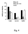


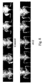


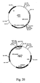
Classifications
-
- A—HUMAN NECESSITIES
- A61—MEDICAL OR VETERINARY SCIENCE; HYGIENE
- A61K—PREPARATIONS FOR MEDICAL, DENTAL OR TOILETRY PURPOSES
- A61K38/00—Medicinal preparations containing peptides
- A61K38/16—Peptides having more than 20 amino acids; Gastrins; Somatostatins; Melanotropins; Derivatives thereof
- A61K38/17—Peptides having more than 20 amino acids; Gastrins; Somatostatins; Melanotropins; Derivatives thereof from animals; from humans
-
- A—HUMAN NECESSITIES
- A61—MEDICAL OR VETERINARY SCIENCE; HYGIENE
- A61K—PREPARATIONS FOR MEDICAL, DENTAL OR TOILETRY PURPOSES
- A61K38/00—Medicinal preparations containing peptides
- A61K38/16—Peptides having more than 20 amino acids; Gastrins; Somatostatins; Melanotropins; Derivatives thereof
-
- A—HUMAN NECESSITIES
- A61—MEDICAL OR VETERINARY SCIENCE; HYGIENE
- A61K—PREPARATIONS FOR MEDICAL, DENTAL OR TOILETRY PURPOSES
- A61K39/00—Medicinal preparations containing antigens or antibodies
- A61K39/395—Antibodies; Immunoglobulins; Immune serum, e.g. antilymphocytic serum
-
- A—HUMAN NECESSITIES
- A61—MEDICAL OR VETERINARY SCIENCE; HYGIENE
- A61K—PREPARATIONS FOR MEDICAL, DENTAL OR TOILETRY PURPOSES
- A61K39/00—Medicinal preparations containing antigens or antibodies
- A61K39/395—Antibodies; Immunoglobulins; Immune serum, e.g. antilymphocytic serum
- A61K39/39533—Antibodies; Immunoglobulins; Immune serum, e.g. antilymphocytic serum against materials from animals
- A61K39/39558—Antibodies; Immunoglobulins; Immune serum, e.g. antilymphocytic serum against materials from animals against tumor tissues, cells, antigens
-
- A—HUMAN NECESSITIES
- A61—MEDICAL OR VETERINARY SCIENCE; HYGIENE
- A61P—SPECIFIC THERAPEUTIC ACTIVITY OF CHEMICAL COMPOUNDS OR MEDICINAL PREPARATIONS
- A61P1/00—Drugs for disorders of the alimentary tract or the digestive system
- A61P1/16—Drugs for disorders of the alimentary tract or the digestive system for liver or gallbladder disorders, e.g. hepatoprotective agents, cholagogues, litholytics
-
- A—HUMAN NECESSITIES
- A61—MEDICAL OR VETERINARY SCIENCE; HYGIENE
- A61P—SPECIFIC THERAPEUTIC ACTIVITY OF CHEMICAL COMPOUNDS OR MEDICINAL PREPARATIONS
- A61P11/00—Drugs for disorders of the respiratory system
-
- A—HUMAN NECESSITIES
- A61—MEDICAL OR VETERINARY SCIENCE; HYGIENE
- A61P—SPECIFIC THERAPEUTIC ACTIVITY OF CHEMICAL COMPOUNDS OR MEDICINAL PREPARATIONS
- A61P13/00—Drugs for disorders of the urinary system
- A61P13/08—Drugs for disorders of the urinary system of the prostate
-
- A—HUMAN NECESSITIES
- A61—MEDICAL OR VETERINARY SCIENCE; HYGIENE
- A61P—SPECIFIC THERAPEUTIC ACTIVITY OF CHEMICAL COMPOUNDS OR MEDICINAL PREPARATIONS
- A61P13/00—Drugs for disorders of the urinary system
- A61P13/12—Drugs for disorders of the urinary system of the kidneys
-
- A—HUMAN NECESSITIES
- A61—MEDICAL OR VETERINARY SCIENCE; HYGIENE
- A61P—SPECIFIC THERAPEUTIC ACTIVITY OF CHEMICAL COMPOUNDS OR MEDICINAL PREPARATIONS
- A61P15/00—Drugs for genital or sexual disorders; Contraceptives
-
- A—HUMAN NECESSITIES
- A61—MEDICAL OR VETERINARY SCIENCE; HYGIENE
- A61P—SPECIFIC THERAPEUTIC ACTIVITY OF CHEMICAL COMPOUNDS OR MEDICINAL PREPARATIONS
- A61P17/00—Drugs for dermatological disorders
-
- A—HUMAN NECESSITIES
- A61—MEDICAL OR VETERINARY SCIENCE; HYGIENE
- A61P—SPECIFIC THERAPEUTIC ACTIVITY OF CHEMICAL COMPOUNDS OR MEDICINAL PREPARATIONS
- A61P19/00—Drugs for skeletal disorders
-
- A—HUMAN NECESSITIES
- A61—MEDICAL OR VETERINARY SCIENCE; HYGIENE
- A61P—SPECIFIC THERAPEUTIC ACTIVITY OF CHEMICAL COMPOUNDS OR MEDICINAL PREPARATIONS
- A61P19/00—Drugs for skeletal disorders
- A61P19/08—Drugs for skeletal disorders for bone diseases, e.g. rachitism, Paget's disease
-
- A—HUMAN NECESSITIES
- A61—MEDICAL OR VETERINARY SCIENCE; HYGIENE
- A61P—SPECIFIC THERAPEUTIC ACTIVITY OF CHEMICAL COMPOUNDS OR MEDICINAL PREPARATIONS
- A61P21/00—Drugs for disorders of the muscular or neuromuscular system
-
- A—HUMAN NECESSITIES
- A61—MEDICAL OR VETERINARY SCIENCE; HYGIENE
- A61P—SPECIFIC THERAPEUTIC ACTIVITY OF CHEMICAL COMPOUNDS OR MEDICINAL PREPARATIONS
- A61P35/00—Antineoplastic agents
-
- A—HUMAN NECESSITIES
- A61—MEDICAL OR VETERINARY SCIENCE; HYGIENE
- A61P—SPECIFIC THERAPEUTIC ACTIVITY OF CHEMICAL COMPOUNDS OR MEDICINAL PREPARATIONS
- A61P35/00—Antineoplastic agents
- A61P35/04—Antineoplastic agents specific for metastasis
-
- A—HUMAN NECESSITIES
- A61—MEDICAL OR VETERINARY SCIENCE; HYGIENE
- A61P—SPECIFIC THERAPEUTIC ACTIVITY OF CHEMICAL COMPOUNDS OR MEDICINAL PREPARATIONS
- A61P43/00—Drugs for specific purposes, not provided for in groups A61P1/00-A61P41/00
-
- C—CHEMISTRY; METALLURGY
- C07—ORGANIC CHEMISTRY
- C07K—PEPTIDES
- C07K16/00—Immunoglobulins [IGs], e.g. monoclonal or polyclonal antibodies
- C07K16/18—Immunoglobulins [IGs], e.g. monoclonal or polyclonal antibodies against material from animals or humans
- C07K16/28—Immunoglobulins [IGs], e.g. monoclonal or polyclonal antibodies against material from animals or humans against receptors, cell surface antigens or cell surface determinants
- C07K16/2863—Immunoglobulins [IGs], e.g. monoclonal or polyclonal antibodies against material from animals or humans against receptors, cell surface antigens or cell surface determinants against receptors for growth factors, growth regulators
-
- A—HUMAN NECESSITIES
- A61—MEDICAL OR VETERINARY SCIENCE; HYGIENE
- A61K—PREPARATIONS FOR MEDICAL, DENTAL OR TOILETRY PURPOSES
- A61K39/00—Medicinal preparations containing antigens or antibodies
- A61K2039/505—Medicinal preparations containing antigens or antibodies comprising antibodies
-
- C—CHEMISTRY; METALLURGY
- C07—ORGANIC CHEMISTRY
- C07K—PEPTIDES
- C07K2317/00—Immunoglobulins specific features
- C07K2317/20—Immunoglobulins specific features characterized by taxonomic origin
- C07K2317/21—Immunoglobulins specific features characterized by taxonomic origin from primates, e.g. man
-
- C—CHEMISTRY; METALLURGY
- C07—ORGANIC CHEMISTRY
- C07K—PEPTIDES
- C07K2317/00—Immunoglobulins specific features
- C07K2317/70—Immunoglobulins specific features characterized by effect upon binding to a cell or to an antigen
- C07K2317/73—Inducing cell death, e.g. apoptosis, necrosis or inhibition of cell proliferation
-
- C—CHEMISTRY; METALLURGY
- C07—ORGANIC CHEMISTRY
- C07K—PEPTIDES
- C07K2317/00—Immunoglobulins specific features
- C07K2317/70—Immunoglobulins specific features characterized by effect upon binding to a cell or to an antigen
- C07K2317/76—Antagonist effect on antigen, e.g. neutralization or inhibition of binding
Definitions
- the disclosure provides methods of treating bone cancer, particularly metastatic bone cancer, by administering an IGF-IR antagonist and/or a PDGFR ⁇ antagonist.
- the present disclosure also provides antibodies that bind to human PDGFR ⁇ and neutralize activation of the receptor.
- the present disclosure further provides a methods for neutralizing activation of PDGFR ⁇ , and a methods of treating a mammal with a neoplastic disease using the antibodies alone or in combination with other agents.
- Prostate cancer is the most common cancer among men, with approximately 220,000 cases and 29,000 deaths annually in the United States. A significant proportion of men diagnosed with prostate cancer have metastatic disease. Further, metastases eventually develop in many other prostate cancer patients despite treatment with surgery or radiotherapy. Bone is the most common site of prostate cancer metastasis, and is also a site to which breast cancers and lung cancers often metastasize. Most prostate cancer metastases are androgen-dependent, so that there is a rapid response to surgical or medical castration, but in virtually all patients, the tumor eventually becomes androgen-independent, leading to significant morbidity and mortality. Once bone metastases occur, currently available therapies have limited effect.
- IGF-IR insulin-like growth factor receptor
- IGF-IR insulin-like growth factor receptor
- IGF-IR insulin-like growth factor receptor
- IGF-IR can stimulate cell proliferation, cell differentiation, changes in cell size, and protect cells from apoptosis. It has also been considered to be quasi-obligatory for cell transformation (reviewed in Adams et al., Cell. Mol. Life Sci. 57:1050-93 (2000 ); Baserga, Oncogene 19:5574-81 (2000 )). High levels of expression of IGF-IR have been reported in tissue samples from prostate cancer bone metastases. Bone contains the largest store of IGFs in the body.
- IGF-IR is a pre-formed hetero-tetramer containing two alpha and two beta chains covalently linked by disulfide bonds.
- the receptor subunits are synthesized as part of a single polypeptide chain of 180kd, which is then proteolytically processed into alpha (130kd) and beta (95kd) subunits.
- the entire alpha chain is extracellular and contains the site for ligand binding.
- the beta chain possesses the transmembrane domain, the tyrosine kinase domain, and a C-terminal extension that is necessary for cell differentiation and transformation, but is dispensable for mitogen signaling and protection from apoptosis.
- IGF-IR insulin receptor
- IR insulin receptor
- beta chain sequence 70% homology
- IGF-IIR mannose-6-phosphate
- M6P mannose-6-phosphate
- PDGFR ⁇ and PDGFR ⁇ are type III receptor tyrosine kinases.
- PDGFR ⁇ is critical for development and fulfills important functions into adulthood. For example, mice homozygous for a null mutation die during embryogenesis. At later stages of development, PDGFR ⁇ is expressed in many mesenchymal structures, whereas adjacent epithelial cells produce platelet derived growth factors (PDGFs). Tissue samples from normal or hyperplastic prostate glands test negative for PDGFR ⁇ , whereas primary prostate tumors and skeletal masses from matched subjects express PDGFR ⁇ . Further, of prostate cell lines obtained from different metastatic sites, PDGFR ⁇ is found in bone metastasis-derived PC3 cells, but not in cell lines obtained from lymph node (LNCaP) and brain (DU-145) metastases.
- LNCaP lymph node
- DU-145 brain metastases.
- the platelet-derived growth factor family of growth factors consists of five different disulphide-linked dimers, PDGF-AA, -BB, -AB, -CC, and -DD, that act via PDGFR ⁇ and PDGFR ⁇ .
- These growth factors are dimeric molecules composed of disulfide-linked polypeptide chains that bind to two receptor proteins simultaneously and induce receptor dimerization, autophosphorylation, and intracellular signaling.
- PDGFR ⁇ and PDGFR ⁇ are structurally similar and can form heterodimers as well as homodimers. Because PDGFR ⁇ does not bind the PDGF-A chain with high affinity, PDGF-AA activates only ⁇ receptor dimers, whereas PDGF-AB and PDGF-CC activates ⁇ and ⁇ receptor heterodimers.
- This disclosure relates to treatment of primary and metastatic bone tumors, including tumors that originate from prostate, breast, or lung and express insulin-like growth factor-I receptor (IGF-IR) and/or the alpha platelet derived growth factor receptor (PDGFR ⁇ ).
- IGF-IR insulin-like growth factor-I receptor
- PDGFR ⁇ alpha platelet derived growth factor receptor
- the tumors to be treated can be hormone/androgen-dependent or hormone/androgen independent, and can have originated, for example, from prostate, breast, or lung.
- the present disclosure provides methods of treating a subject having a bone tumor, and methods of inhibiting growth of a bone tumor.
- the methods comprise administering an effective amount of an IGF-IR antagonist or an effective amount of a PDGFR ⁇ antagonist.
- the receptor antagonists include antibodies and antibody fragments as well as intracellular small molecule inhibitors.
- the present disclosure provides anti-IGF-IR or anti-PDGFR ⁇ antibodies that bind to their target receptor and inhibit ligand binding.
- the disclosure also provides antibodies and other antagonists that neutralize activation of IGF-IR or PDGFR ⁇ . Further certain antibodies promotes down-regulation of their target receptor, for example by internalization and/or degradation. Accordingly, the antibodies and small molecule antagonists function to inhibit activation of downstream signaling molecules such as Akt, p42/p44, and MAPK.
- the methods include use of IGF-IR or PDGFR ⁇ antagonists alone, in combination with each other or in combination with other cancer therapeutics, such as chemotherapeutics and radiation.
- the present disclosure also provides antibodies and antibody fragments that bind to PDGFR ⁇ as well as nucleotides and host cell for production of the antibodies.
- the antibodies block ligand binding and neutralize receptor activation.
- the disclosure also provides for use of the antibodies alone, in combination with other receptor antagonists or antineoplastic agents, or as conjugates for treatment of neoplastic disease.
- Anti-PDGFR ⁇ antibodies are used to treat, for example, ovarian tumors, breast tumors, lung tumors, hepatocellular tumors, gastrointestinal stromal tumors, melanomas, renal cell carcinomas, prostate tumors, and soft tissue sarcomas.
- a PDGFR ⁇ antagonist for use in the treatment of a subject having metastatic bone cancer, wherein said PDGFR ⁇ antagonist is an antibody or fragment thereof which is specific for human PDGFR ⁇ and which comprises SSSYY (SEQ ID NO:2) at CDRH1; SFFYTGSTYYNPSLRS (SEQ ID NO:4) at CDRH2; QSTYYYGSGNYYGWFDR (SEQ ID NO:6) at CDRH3; RASQSVSSYLA (SEQ ID NO:10) at CDRL1; DASNRAT (SEQ ID NO:12) at CDRL2; and QQRSNWPPA (SEQ ID NO:14) at CDRL3.
- SSSYY SEQ ID NO:2
- SFFYTGSTYYNPSLRS SEQ ID NO:4
- QSTYYYGSGNYYGWFDR SEQ ID NO:6
- RASQSVSSYLA SEQ ID NO:10
- DASNRAT SEQ ID NO:12
- the PDGFR ⁇ antagonist is an antibody or fragment thereof comprising a heavy chain variable domain having QLQLQESGPGLVKPSETLSLTCTVSGGSINSSSYYWGWLRQSPGKGLEWIGSFFY TGSTYYNPSLRSRLTISVDTSKNQFSLMLSSVTAADTAVYYCARQSTYYYGSGNY YGWFDRWDQGTLVTVSS (SEQ ID NO: 8) or a light chain variable domain having EIVLTQSPATLSLSPGERATLSCRASQSVSSYLAWYQQKPGQAPRLLIYDASNRAT GIPARFSGSGSGTDFTLTISSLEPEDFAVYYCQQRSNWPPAFGQGTKVEIK (SEQ ID NO: 16).
- the PDGFR ⁇ antagonist is an antibody or fragment thereof comprising a heavy chain variable domain having
- the PDGFR ⁇ antagonist is an antibody or fragment thereof comprising a heavy chain having MGWSCIILFLVATATGVHSQLQLQESGPGLVKPSETLSLTCTVSGGSINSSSYYWG WLRQSPGKGLEWIGSFFYTGSTYYNPSLRSRLTISVDTSKNQFSLMLSSVTAADT AVYYCARQSTYYYGSGNYYGWFDRWDQGTLVTVSSASTKGPSVFPLAPSSKSTS GGTAALGCLVKDYFPEPVTVSWNSGALTSGVHTFPAVLQSSGLYSLSSVVTVPSS SLGTQTYICNVNHKPSNTKVDKRVEPKSCDKTHTCPPCPAPELLGGPSVFLFPPKP KDTLMISRTPEVTCVVVDVSHEDPEVKFNWYVDGVEVHNAKTKPREEQYNSTY RVVSVLTVLHQDWLNGKEYKCKVSNKALPAPIEKTISKAKGQPREPQVYTLPPSR EEM
- a PDGFR ⁇ antagonist for use in the treatment of a subject having metastatic bone cancer, wherein said PDGFR ⁇ antagonist is an antibody or fragment thereof which is specific for human PDGFR ⁇ , wherein the antibody or antibody fragment competes for binding to PDGFR ⁇ with an antibody of the present invention.
- the tumour is metastasized from a prostate cancer, breast cancer or lung cancer.
- the PDGFR ⁇ antagonist of the present invention is preferably one which inhibits binding of PDGFR ⁇ or neutralizes PDGFR ⁇ .
- the present disclosure relates to treatment of bone tumors with antibodies or antibody fragments that bind to insulin-like growth factor-I receptor (IGF-IR).
- IGF-IR insulin-like growth factor-I receptor
- Endocrine expression of IGF-I is regulated primarily by growth hormone and produced in the liver, but other tissue types are also capable of expressing IGF-I, including bone which contains a large store of growth factors.
- IGF-I is involved in endocrine, paracrine, and/or autocrine regulation ( Yu, H. and Rohan, J., J. Natl. Cancer Inst. 92:1472-89 (2000 )).
- antibodies that bind IGF-IR are useful in therapies for treatment of bone tumors that express IGF-IR.
- the antibodies can be use alone, or in combination with other cancer therapeutics, particularly chemotherapeutics.
- Anti-IGF-IR therapy alone or in combination with therapy with one or more anti-neoplastic agents (such as, for example, chemotherapy or radiation therapy) has significant therapeutic efficacy. Suppression of tumor growth is often accompanied with an increase in apoptosis and persists after all treatment is discontinued and tumors have again begun to grow in animals treated with chemotherapy alone.
- PDGFR ⁇ plays an important role in growth of bone tumors.
- certain tumor cell lines that express PDGFR ⁇ preferentially metastasize to bone display increased PDGFR ⁇ activation and phosphorylation of downstream signaling molecules in response to soluble factors present in bone marrow.
- PDGFR ⁇ activation by bone marrow is reduced or completely inhibited by PDGFR ⁇ antagonists, and phosphorylation of downstream signaling molecules that are commonly activated by signaling through PDGFR ⁇ and other receptor tyrosine kinase systems is greatly reduced.
- Primary bone tumors to be treated according to the invention include, but are not limited to, osteosarcomas, chondrosarcomas, fibrosarcomas, and hemangiosarcomas.
- malignant secondary (metastastic) tumors are far more common than primary bone tumors.
- Metastatic bone tumors to be treated according to the invention can arise from a variety of sources, the most common of which are cancers of the prostate, breast, or lung. The source of a metastatic bone cancer will usually be apparent from a patients history.
- the tumors can be osteoblastic or osteolytic.
- the tumors may be dependent on IGF-IR stimulation when they arise, or may transition to IGF-IR dependence.
- prostate cancers or metastases of prostate cancers may become hormone/androgen-independent through increased sensitivity to stimulation through IGF-IR.
- the invention can be useful for treating hormone/androgen-dependent bone tumors without reliance on suppression of androgen or hormone production, for example, by coadministering IGF-IR antibodies with anti-neoplastic agents.
- tumors would include metastatic bone tumors that are stimulated through IFG-IR in the IGF-rich environment of the bone, which may be sensitive to hormone stimulation but not sensitive enough to grow without IGF involvement. Hormone ablation might not be necessary for such tumors.
- Bone tumors that are PDGF-dependent can also be treated, as well as tumors that are "bone marrow" dependent.
- Bone marrow dependent tumors display PDGFR ⁇ activation in response to soluble factors present in bone marrow.
- a human metastatic PDGFR ⁇ -expressing cancer cell line undergoes PDGFR ⁇ activation and Akt+ phosphorylation upon exposure to bone marrow aspirate.
- An anti-PDGFR ⁇ antibody and a small molecule PDGFR ⁇ antagonist each inhibit PDGFR ⁇ activation and Akt+ phosphorylation in the cell line.
- Soluble bone marrow factors that activate PDGFR ⁇ include, but are not limited to, PDGF-AA and -BB.
- PDGFR ⁇ activation by defined ligands PDGF-AA or -BB
- Akt+ phosphorylation diminishes with increased incubation time.
- Bone tumors to be treated with a PDGFR ⁇ antagonist can arise as metastases of prostate cancer cells, and, as above, may be hormone/androgen dependent, or have transitioned to hormone/androgen independence. Such tumors can arise as metastases of non-prostate cancers as well.
- One skilled in the art would easily be able to diagnose such conditions and disorders using known, conventional tests.
- Treatment means any treatment of a disease in an animal and includes: (1) preventing the disease from occurring in a mammal which may be predisposed to the disease but does not yet experience or display symptoms of the disease; e.g. , prevention of the outbreak of the clinical symptoms; (2) inhibiting the disease, e.g. , arresting its development; or (3) relieving the disease, e.g., causing regression of the symptoms of the disease. Inhibiting tumor growth includes slowing or stopping growth, as well as causing tumor regression.
- An effective amount for the treatment of a disease means that amount which, when administered to a mammal in need thereof, is sufficient to effect treatment, as defined above, for that disease.
- IGF-IR antagonists and PDGFR ⁇ antagonist may be administered alone, in combination with one another, or in combination with one or more antineoplastic agents such as, for example, a chemotherapeutic or radologic agent.
- IGF-IR and/or PDGFR ⁇ may be desirable to determine the level of expression of IGF-IR and/or PDGFR ⁇ in a tumor to be treated.
- tumor biopsies can be collected and analyzed by methods well known in the art.
- An IGF-IR antagonist or PDGFR ⁇ antagonist is administered on the basis that the corresponding receptor is commonly expressed or activated in a particular tumor type or invariably becomes expressed of activated as the disease progresses.
- An IGF-IR antagonist can be an extracellular antagonist or an intracellular antagonist and more than one antagonist may be employed.
- Extracellular antagonists include, but are not limited to proteins or other biological molecules that bind to IGF-IR or one or more of its ligands (e.g , IGF-I and IGF-II are natural ligands of IGF-IR).
- An extracellular antagonist inhibits binding of IGF-IR to its ligands.
- the antagonist is an anti- IGF-IR antibody, such as, for example, IMC-A12.
- the antagonist is a soluble ligand binding fragment of IGF-IR.
- Intracellular IGF-IR antagonists can be biological molecules, but are usually small molecules.
- Examples include, but are not limited to, tyrosine kinase inhibitor AG1024 (Calbiochem), insulin-like growth factor-I receptor kinase inhibitor NVP-AEW541 (Novartis), and insulin-like growth factor-I/insulin receptor inhibitor BMS-554417 (Bristol Myers Squibb). It will be appreciated that useful small molecule to be used in the invention are inhibitors of IGF-IR, but need not be completely specific for IGF-IR.
- Anti-IGF-IR antibodies to be used according to the present disclosure exhibit one or more of following properties:
- the antibodies bind to the external domain of IGF-IR and inhibit binding of IGF-I or IGF-II to IGF-IR. Inhibition can be determined, for example, by a direct binding assay using purified or membrane bound receptor.
- the antibodies or fragments thereof preferably bind IGF-IR at least as strongly as the natural ligands of IGF-IR (IGF-I and IGF-II).
- the antibodies neutralize IGF-IR. Binding of a ligand, e.g. , IGF-I or IGF-II, to an external, extracellular domain of IGF-IR stimulates autophosphorylation of the beta subunit and downstream signaling molecules, including MAPK, Akt, and IRS-1.
- a ligand e.g. , IGF-I or IGF-II
- Neutralization of IGF-IR includes inhibition, diminution, inactivation and/or disruption of one or more of these activities normally associated with signal transduction. Neutralization can be determined in vivo, ex vivo, or in vitro using, for example, tissues, cultured cell, or purified cellular components. Neutralization includes inhibition of IGF-IR / IR heterodimers as well as IGF-IR homodimers. Thus, neutralizing IGF-IR has various effects, including inhibition, diminution, inactivation and/or disruption of growth (proliferation and differentiation), angiogenesis (blood vessel recruitment, invasion, and metastasis), and cell motility and metastasis (cell adhesion and invasiveness).
- IGF-IR neutralization is inhibition of the tyrosine kinase activity of the receptor.
- Tyrosine kinase inhibition can be determined using well-known methods; for example, by measuring the autophosphorylation level of recombinant kinase receptor, and/or phosphorylation of natural or synthetic substrates.
- phosphorylation assays are useful in determining neutralizing antibodies in the context of the present invention. Phosphorylation can be detected, for example, using an antibody specific for phosphotyrosine in an ELISA assay or on a western blot.
- Antibodies of the invention cause a decrease in tyrosine phosphorylation of IGF-IR of at least about 75%, preferably at least about 85%, and more preferably at least about 90% in cells that respond to ligand.
- IGF-IR neutralization Another measure of IGF-IR neutralization is inhibition of phosphorylation of downstream substrates of IGF-IR. Accordingly, the level of phosphorylation of MAPK, Akt, or IRS-1 can be measured. The decrease in phosphorylation is at least about 40%, and can be at least about 60%, or at least about 80%.
- IGF-IR neutralization methods for detection of protein expression can be utilized to determine IGF-IR neutralization, wherein the proteins being measured are regulated by IGF-IR tyrosine kinase activity.
- IHC immunohistochemistry
- FISH fluorescence in situ hybridization
- competitive radioligand binding assays solid matrix blotting techniques, such as Northern and Southern blots, reverse transcriptase polymerase chain reaction (RT-PCR) and ELISA.
- Ex vivo assays can also be utilized to determine IGF-IR neutralization.
- receptor tyrosine kinase inhibition can be observed by mitogenic assays using cell lines stimulated with receptor ligand in the presence and absence of inhibitor.
- the MCF7 breast cancer line (American Type Culture Collection (ATCC), Rockville, MD) is such a cell line that expresses IGF-IR and is stimulated by IGF-I or IGF-II.
- Another method involves testing for inhibition of growth of IGF-IR -expressing tumor cells or cells transfected to express IGF-IR. Inhibition can also be observed using tumor models, for example, human tumor cells injected into a mouse.
- the antibodies are not limited by any particular mechanism of IGF-IR neutralization.
- the anti-IGF-IR antibodies can bind externally to the IGF-IR cell surface receptor, block binding of ligand (e.g., IGF-I or IGF-II) and subsequent signal transduction mediated via the receptor-associated tyrosine kinase, and prevent phosphorylation of the IGF-IR and other downstream proteins in the signal transduction cascade.
- ligand e.g., IGF-I or IGF-II
- the antibodies down modulate IGF-IR.
- the amount of IGF-IR present on the surface of a cell depends on receptor protein production, internalization, and degradation.
- the amount of IGF-IR present on the surface of a cell can be measured indirectly, by detecting internalization of the receptor or a molecule bound to the receptor.
- receptor internalization can be measured by contacting cells that express IGF-IR with a labeled antibody. Membrane-bound antibody is then stripped, collected and counted. Internalized antibody is determined by lysing the cells and detecting label in the lysates.
- Another way is to directly measure the amount of the receptor present on the cell following treatment with an anti-IGF-IR antibody or other substance, for example, by fluorescence-activated cell-sorting analysis of cells stained for surface expression of IGF-IR. Stained cells are incubated at 37°C and fluorescence intensity measured over time. As a control, part of the stained population can be incubated at 4°C (conditions under which receptor internalization is halted).
- Cell surface IGF-IR can be detected and measured using a different antibody that is specific for IGF-IR and that does not block or compete with binding of the antibody being tested. ( Burtrum, et al. Cancer Res. 63:8912-21 (2003 )) Treatment of an IGF-IR expressing cell with an antibody results in reduction of cell surface IGF-IR. In a preferred embodiment, the reduction is at least about 70%, more preferably at least about 80%, and even more preferably at least about 90% in response to treatment with an antibody. A significant decrease can be observed in as little as four hours.
- Another measure of down-modulation is reduction of the total receptor protein present in a cell, and reflects degradation of internal receptors. Accordingly, treatment of cells (particularly cancer cells) with antibodies results in a reduction in total cellular IGF-IR. In a preferred aspect, the reduction is at least about 70%, more preferably at least about 80%, and even more preferably at least about 90%.
- antibodies are preferably human.
- the antibodies can be from non-human primates or other mammals, or be humanized or chimeric antibodies.
- An anti-IGF-IR antibody comprises one, two, three, four, five, and/or six complementarity determining regions (CDRs) selected from the group consisting of SEQ ID NO:35, SEQ ID NO:37, SEQ ID NO:39, SEQ ID NO:45, SEQ ID NO:47, and SEQ ID NO:49 (CDR1H, CDR2H, CDR3H, CDR1L, CDR2L, CDR3L, respectively).
- the anti-IGF-IR antibody comprises one, two, three, four, five, and/or six complementarity determining regions (CDRs) selected from the group consisting of SEQ ID NO:35, SEQ ID NO:37, SEQ ID NO:39, SEQ ID NO: 55, SEQ ID NO:57, and SEQ ID NO:59 (CDR1H, CDR2H, CDR3H, CDR1L, CDR2L, CDR3L, respectively).
- the antibodies (or fragments thereof) have heavy chain CDRs of SEQ ID NO:35, SEQ ID NO:37 and SEQ ID NO:39.
- the present antibodies including fragments thereof have light chain CDRs of SEQ ID NO:45, SEQ ID NO:47 and SEQ ID NO:49 or SEQ ID NO:55, SEQ ID NO:57 and SEQ ID NO:59.
- One such anti-IGF-IR antibody is the human IgG1 antibody IMC-A12 ( WO2005016970 ), having a heavy chain variable domain represented by SEQ ID NO:41 and a light chain variable domain represented by SEQ ID NO:51.
- Another preferred human antibody is IMC-2F8 ( WO2005016970 ), having a heavy chain variable domain identical to IMC-A12 and a light chain variable domain represented by SEQ ID NO:61.
- Useful antibodies further include anti-IGF-IR antibodies that compete with IMC-A12 or IMC-2F8 for binding to IGF-IR, as well as antibodies that bind to other epitopes (i.e., antibodies that bind to other epitopes and exhibit properties as previously described such as ligand blocking, receptor internalization, etc., but do not compete with IMC-A12 or IMC-2F8).
- PDGFR ⁇ antagonists can also be used for treatment.
- a PDGFR ⁇ antagonist can be an extracellular antagonist or an intracellular antagonist and more than one antagonist may be employed.
- Extracellular antagonists include, but are not limited to proteins or other biological molecules that bind to PDGFR ⁇ or one or more of its ligands ( e.g , PDGF-AA, -AB, -BB, -CC).
- An extracellular antagonist is inhibits binding of PDGFR ⁇ to its ligands.
- the antagonist is an anti-PDGFR ⁇ antibody, such as, for example, IMC-3G3.
- the binding protein is a soluble ligand binding fragment of PDGFR ⁇ .
- Intracellular IGF-IR antagonists can be biological molecules, but are usually small molecules.
- the intracellular PDGFR ⁇ antagonist is AG1296.
- AG1296 (Calbiochem) is an inhibitor of PDGF ⁇ , PDGF ⁇ s, and c-KIT, and also reacts with Flt3.
- Other small molecules that target PDGFRs include STI-571 (imatinib mesylate, Gleevec®, Novartis) and SU11248 (sunitinib malate, SUTENT®, Pfizer).
- An anti-PDGFR ⁇ antibody comprises one, two, three, four, five, and/or six complementarity determining regions (CDRs) selected from the group consisting of SEQ ID NO:2, SEQ ID NO:4, SEQ ID NO:6, SEQ ID NO:10, SEQ ID NO:12, and SEQ ID NO:14 (CDR1H, CDR2H, CDR3H, CDR1L, CDR2L, CDR3L, respectively).
- the antibodies (or fragments thereof) have CDRs of SEQ ID NO:2, SEQ ID NO:4 and SEQ ID NO:6.
- the present antibodies, or fragments thereof have CDRs of SEQ ID NO:10, SEQ ID NO:12 and SEQ ID NO:14.
- the anti-PDGFR ⁇ antibody, or fragment thereof has a human heavy chain variable region of SEQ ID NO:8 and/or a human light chain variable region of SEQ ID NO:16.
- IMC-3G3 is such an antibody and is exemplified in the present disclosure.
- the antibodies, or fragments thereof neutralize PDGFR ⁇ .
- Binding of a ligand, e.g., PDGF-AA, PDGF-AB, PDGF-BB or PDGF-CC, to an extracellular domain of PDGFR ⁇ stimulates receptor dimerization, autophosphorylation, activation of the receptor's internal, cytoplasmic tyrosine kinase domain, and initiation of multiple signal transduction and transactivation pathways involved in regulation of DNA synthesis (gene activation) and cell cycle progression or division.
- the anti-PDGFR ⁇ antibodies typically block ligand binding and/or receptor dimerization, and inhibit one or more of autophosphorylation, activation of tyrosine kinase activity and signal transduction.
- the anti-PDGFR ⁇ antibodies can be specific for the extracellular ligand binding region of PDGFR ⁇ and prevent binding of a ligand of PDGFR ⁇ .
- such anti- PDGFR ⁇ antibodies, or fragments thereof bind PDGFR ⁇ at least as strongly as the natural ligands of PDGFR ⁇ .
- the antibodies can be specific for a region of the receptor monomer that would otherwise form a receptor dimer interface. Such antibodies block dimer formation, though ligand binding to a receptor monomer might or might not be blocked.
- receptor neutralization can be determined by a variety of in vivo, in vitro, and ex vivo methods.
- the anti-PDGFR ⁇ antibodies reduce phosphorylation of PDGFR ⁇ by at least about 75%. In other aspects, phosphorylation is reduced by at least about 85% or at least about 90%.
- phosphorylation or a downstream signal transduction pathway component e.g. , Akt, p42/p44, etc.
- Receptor neutralization can be determined using defined ligands (e.g. , PDGF-AA,-AB, -BB, -CC), mixtures of such ligands, or preparations such as bone marrow aspirates that comprise PDGFs as well as other stimulatory growth factors.
- Neutralization of PDGFR ⁇ includes inhibition, diminution, inactivation and/or disruption of one or more of these activities normally associated with signal transduction.
- neutralizing PDGFR ⁇ has various effects, including inhibition, diminution, inactivation and/or disruption of growth (proliferation and differentiation), angiogenesis (blood vessel recruitment, invasion, and metastasis), and cell motility and metastasis (cell adhesion and invasiveness).
- Ex vivo assays can also be utilized to determine PDGFR ⁇ neutralization.
- human SKLMS-1 leiomyosarcoma cells American Type Culture Collection (ATCC), Rockville, MD; ATCC HTB-88TM) or U118 glioblastoma cells (ATCC HTB-15TM) stimulated with PDGF-AA can be used to assay PDGFR ⁇ inhibition.
- Growth inhibition can be ascertained using PDGFR ⁇ -expressing human tumor cells injected into a SCID mouse.
- the present disclosure is not limited by any particular mechanism of PDGFR ⁇ neutralization.
- the anti-PDGFR ⁇ antibodies bind externally to the PDGFR ⁇ cell surface receptor, block binding of ligand (e.g ., PDGF-AA, PDGF-AB, PDGF-BB, PDGF-CC), inhibit phosphorylation of the PDGFR ⁇ , inhibit signal transduction mediated via the receptor-associated tyrosine kinase, and modulate activity of downstream signal transduction components.
- the receptor-antibody complex can also be internalized and degraded, resulting in cell surface receptor downregulation.
- Matrix metalloproteinases which function in tumor cell invasion and metastasis, can also be downregulated by the antibodies.
- antibodies may exhibit inhibition of growth factor production and angiogenesis.
- PDGFR ⁇ antagonists are useful for treating bone tumors, including metastatic bone tumors.
- Other tumor types that express PDGFR ⁇ and can be treated include, but are not limited to, ovarian tumors, breast tumors, lung tumors, hepatocellular tumors, gastrointestinal stromal tumors, melanoma, renal cell carcinoma, prostate tumors, and soft tissue sarcomas.
- Soft tissue sarcomas originate in such tissues as fat, muscles, nerves, tendons, and blood and lymph vessels.
- the tumor cells overexpress PDGFR ⁇ PDGFR ⁇ expression can be determined, for example, by histochemistry or RNA analysis. For example, a scatchard analysis of binding of radiolabeled IMC-3G3 to U118 cells and SKLMS-1 tumor cells indicates the number of PDGFR ⁇ molecules on the cells to be about 500 and 2500, respectively.
- PDGFR ⁇ antagonists function by inhibiting signal transduction by PDGFR ⁇ expressed on the tumor cells themselves, or by inhibiting PDGFR ⁇ expressed on surrounding stromal cells that otherwise undergo paracrine stimulation by PDGFs expressed from tumor cells.
- antibodies such as IMC-3G3 and other PDGFR ⁇ antagonists are useful for treating tumors characterized by autocrine and/or paracrine stimulation of PDGFR ⁇
- Antibody fragments can be produced by cleaving a whole antibody, or by expressing DNA that encodes the fragment. Fragments of antibodies may be prepared by methods described by Lamoyi et al., J. Immunol. Methods, 56: 235-243 (1983 ) and by Parham, J. Immunol. 131: 2895-2902 (1983 ). Such fragments may contain one or both Fab fragments or the F(ab') 2 fragment. Such fragments may also contain single-chain fragment variable region antibodies, i.e. scFv, dibodies, or other antibody fragments. Methods of producing such functional equivalents are disclosed in PCT Application WO 93/21319 , European Patent Application No. EP 239400 ; PCT Application WO 89/09622 ; European Patent Application EP 338745 ; and European Patent Application EP 332424 .
- Preferred host cells for transformation of vectors and expression of the antibodies of the present invention are mammalian cells, e.g., COS-7 cells, Chinese hamster ovary (CHO) cells, and cell lines of lymphoid origin such as lymphoma, myeloma (e.g. NS0), or hybridoma cells.
- Other eukaryotic hosts, such as yeasts, can be alternatively used.
- a suitable selection gene for use in yeast is the trp1 gene present in the yeast plasmid YRp7. Stinchcomb et al. Nature, 282: 39 (1979 ); Kingsman et al., Gene, 7: 141 (1979 ).
- the trp1 gene provides a selection marker for a mutant strain of yeast lacking the ability to grow in tryptophan, for example, ATCC No. 44076 or PEP4-1. Jones, Genetics, 85: 12 (1977 ).
- the presence of the trp1 lesion in the yeast host cell genome then provides an effective environment for detecting transformation by growth in the absence of tryptophan.
- Leu2-deficient yeast strains (ATCC 20,622 or 38,626) are complemented by known plasmids bearing the Leu2 gene.
- the transformed host cells are cultured by methods known in the art in a liquid medium containing assimilable sources of carbon (carbohydrates such as glucose or lactose), nitrogen (amino acids, peptides, proteins or their degradation products such as peptones, ammonium salts or the like), and inorganic salts (sulfates, phosphates and/or carbonates of sodium, potassium, magnesium and calcium).
- the medium furthermore contains, for example, growth-promoting substances, such as trace elements, for example iron, zinc, manganese and the like.
- High affinity anti-PDGFR ⁇ and anti-IGF-IR antibodies according to the present invention can be isolated from a phage display library constructed from human heavy chain and light chain variable region genes.
- a variable domain of the invention can be obtained from a peripheral blood lymphocyte that contains a rearranged variable region gene.
- variable domain portions such as CDR and FW regions, can be obtained from different sources and recombined.
- portions of the variable domains e . g ., FW regions
- Antibodies and antibody fragments can be obtained, for example, from naturally occurring antibodies, or Fab or scFv phage display libraries. It is understood that, to make a single domain antibody from an antibody comprising a V H and a V L domain, certain amino acid substitutions outside the CDRs can be desired to enhance binding, expression or solubility. For example, it can be desirable to modify amino acid residues that would otherwise be buried in the V H -V L interface.
- antibodies and antibody fragments can be obtained by standard hybridoma technology ( Harlow & Lane, ed., Antibodies: A Laboratory Manual, Cold Spring Harbor, 211-213 (1998 ),) using transgenic mice (e.g. , KM mice from Medarex, San Jose, Calif.) that produce human immunoglobulin gamma heavy and kappa light chains.
- transgenic mice e.g. , KM mice from Medarex, San Jose, Calif.
- a substantial portion of the human antibody producing genome is inserted into the genome of the mouse, and is rendered deficient in the production of endogenous murine antibodies.
- Such mice may be immunized subcutaneously (s.c.) with PDGFR ⁇ (usually in complete Freund's adjuvant) with boosts as needed. Immunization methods are well known in the art.
- the protein used to identify IGF-IR binding antibodies is preferably IGF-IR and, more preferably, is the extracellular domain of IGF-IR.
- the protein used to identify PDGFR ⁇ binding antibodies is preferably PDGFR ⁇ and, more preferably, is the extracellular domain of PDGFR ⁇ . Such extracellular domains can be free or conjugated to other molecules.
- the present disclosure also provides isolated polynucleotides encoding the antibodies, or fragments thereof, described previously. Details of the IMC-A12 anti-IGF-IR antibody are disclosed in WO2005016970 . Table 2 sets forth the nucleic acid sequences for IMC-3G3.
- DNA encoding human antibodies can be prepared by recombining DNA encoding human constant regions and variable regions, other than the CDRs, derived substantially or exclusively from the corresponding human antibody regions and DNA encoding CDRs derived from a human (SEQ ID NOS:1, 3, and 5 for the heavy chain variable domain CDRs and SEQ ID NOS:9, 11, and 13 for the light chain variable domain CDRs).
- Suitable sources of DNAs that encode fragments of antibodies include any cell, such as hybridomas and spleen cells, that express the full-length antibody.
- the fragments may be used by themselves as antibody equivalents, or may be recombined into equivalents, as described above.
- the DNA deletions and recombinations described in this section may be carried out by known methods, such as those described in the publications listed above with regard to equivalents of antibodies and/or other standard recombinant DNA techniques, such as those described below.
- Another source of DNAs are single chain antibodies produced from a phage display library, as is known in the art.
- the present disclosure provides expression vectors containing the polynucleotide sequences previously described operably linked to an expression sequence, a promoter and an enhancer sequence.
- a variety of expression vectors for the efficient synthesis of antibody polypeptide in prokaryotic, such as bacteria and eukaryotic systems, including but not limited to yeast and mammalian cell culture systems have been developed.
- the vectors of the present disclosure can comprise segments of chromosomal, non-chromosomal and synthetic DNA sequences.
- prokaryotic cloning vectors include plasmids from E . coli, such as colE1 , pCR1 , pBR322, pMB9, pUC, pKSM, and RP4.
- Prokaryotic vectors also include derivatives of phage DNA such as M13 and other filamentous single-stranded DNA phages.
- An example of a vector useful in yeast is the 2 ⁇ plasmid.
- Suitable vectors for expression in mammalian cells include well-known derivatives of SV40, adenovirus, retrovirus-derived DNA sequences and shuttle vectors derived from combination of functional mammalian vectors, such as those described above, and functional plasmids and phage DNA.
- Additional eukaryotic expression vectors are known in the art (e.g., P.J. Southern and P. Berg, J. Mol. Appl. Genet., 1, 327-341 (1982 ); Subramani et al., Mol. Cell. Biol., 1: 854-864 (1981 ); Kaufmann and Sharp, "Amplification And Expression of Sequences Cotransfected with a Modular Dihydrofolate Reductase Complementary DNA Gene," J. Mol. Biol. 159, 601-621 (1982 ); Kaufmann and Sharp, Mol. Cell. Biol.
- the expression vectors useful in the present disclosure contain at least one expression control sequence that is operatively linked to the DNA sequence or fragment to be expressed.
- the control sequence is inserted in the vector in order to control and to regulate the expression of the cloned DNA sequence.
- useful expression control sequences are the lac system, the trp system, the tac system, the trc system, major operator and promoter regions of phage lambda, the control region of fd coat protein, the glycolytic promoters of yeast, e.g., the promoter for 3-phosphoglycerate kinase, the promoters of yeast acid phosphatase, e.g., Pho5, the promoters of the yeast alpha-mating factors, and promoters derived from polyoma, adenovirus, retrovirus, and simian virus, e.g., the early and late promoters or SV40, and other sequences known to control the expression of genes of prokaryotic or eukaryotic cells and their
- the present disclosure also provides recombinant host cells containing the expression vectors previously described.
- Antibodies can be expressed in cell lines other than in hybridomas.
- Nucleic acids, which comprise a sequence encoding a polypeptide, can be used for transformation of a suitable mammalian host cell.
- Cell lines of particular preference are selected based on high level of expression, constitutive expression of protein of interest and minimal contamination from host proteins.
- Mammalian cell lines available as hosts for expression are well known in the art and include many immortalized cell lines, such as but not limited to, NS0 cells, Chinese Hamster Ovary (CHO) cells, Baby Hamster Kidney (BHK) cells and many others. Suitable additional eukaryotic cells include yeast and other fungi.
- Useful prokaryotic hosts include, for example, E. coli, such as E . coli SG-936, E. coli HB 101, E . coli W3110, E . coli X1776, E. coli X2282, E. coli DHI, and E . coli MRC1, Pseudomonas, Bacillus, such as Bacillus subtilis, and Streptomyces.
- present recombinant host cells can be used to produce an antibody, or fragment thereof, by culturing the cells under conditions permitting expression of the antibody or fragment thereof and purifying the antibody or fragment thereof from the host cell or medium surrounding the host cell.
- Targeting of the expressed antibody or fragment for secretion in the recombinant host cells can be facilitated by inserting a signal or secretory leader peptide-encoding sequence (see, Shokri et al., Appl Microbiol Biotechnol. 60(6):654-64 (2003 ), Nielsen et al., Prot. Eng. 10:1-6 (1997 ) and von Heinje et al., Nucl. Acids Res.
- secretory leader peptide elements can be derived from either prokaryotic or eukaryotic sequences. Accordingly suitably, secretory leader peptides are used, being amino acids joined to the N-terminal end of a polypeptide to direct movement of the polypeptide out of the host cell cytosol and secretion into the medium.
- the antibodies of this disclosure can be fused to additional amino acid residues.
- Such amino acid residues can be a peptide tag, perhaps to facilitate isolation.
- Other amino acid residues for homing of the antibodies to specific organs or tissues are also contemplated.
- an antibody of the present disclosure is made by expressing a nucleic acid encoding the antibody in a transgenic animal, such that the antibody is expressed and can be recovered.
- the antibody can be expressed in a tissue specific manner that facilitates recovery and purification.
- an antibody is expressed in the mammary gland for secretion during lactation.
- Transgenic animals include but are not limited to mice, goat, and rabbit.
- Antibodies that can be used include complete immunoglobulins, antigen binding fragments of immunoglobulins, as well as antigen binding proteins that comprise antigen binding domains of immunoglobulins.
- Antigen binding fragments of immunoglobulins include, for example, Fab, Fab', and F(ab') 2 .
- Other antibody formats have been developed which retain binding specificity, but have other characteristics that may be desirable, including for example, bispecificity, multivalence (more than two binding sites), compact size (e.g., binding domains alone).
- Single chain antibodies lack some or all of the constant domains of the whole antibodies from which they are derived. Therefore, they can overcome some of the problems associated with the use of whole antibodies. For example, single-chain antibodies tend to be free of certain undesired interactions between heavy-chain constant regions and other biological molecules. Additionally, single-chain antibodies are considerably smaller than whole antibodies and can have greater permeability than whole antibodies, allowing single-chain antibodies to localize and bind to target antigen-binding sites more efficiently. Furthermore, the relatively small size of single-chain antibodies makes them less likely to provoke an unwanted immune response in a recipient than whole antibodies.
- Each single chain having one V H and one V L domain covalently linked by a first peptide linker can be covalently linked by at least one or more peptide linker to form a multivalent single chain antibodies, which can be monospecific or multispecific.
- Each chain of a multivalent single chain antibody includes a variable light chain fragment and a variable heavy chain fragment, and is linked by a peptide linker to at least one other chain.
- the peptide linker is composed of at least fifteen amino acid residues. The maximum number of amino acid residues is about one hundred.
- Two single chain antibodies can be combined to form a diabody, also known as a bivalent dimer.
- Diabodies have two chains and two binding sites, and can be monospecific or bispecific.
- Each chain of the diabody includes a V H domain connected to a V L domain.
- the domains are connected with linkers that are short enough to prevent pairing between domains on the same chain, thus driving the pairing between complementary domains on different chains to recreate the two antigen-binding sites.
- Triabodies are constructed with the amino acid terminus of a V L or V H domain directly fused to the carboxyl terminus of a V L or V H domain, i.e., without any linker sequence.
- the triabody has three Fv heads with the polypeptides arranged in a cyclic, head-to-tail fashion. A possible conformation of the triabody is planar with the three binding sites located in a plane at an angle of 120 degrees from one another.
- Triabodies can be monospecific, bispecific or trispecific.
- antibodies of the present disclosure and fragments thereof include, but are not limited to, naturally occurring antibodies, bivalent fragments such as (Fab') 2 , monovalent fragments such as Fab, single chain antibodies, single chain Fv (scFv), single domain antibodies, multivalent single chain antibodies, diabodies, triabodies, and the like that bind specifically with antigens.
- bivalent fragments such as (Fab') 2
- monovalent fragments such as Fab
- single chain antibodies single chain Fv (scFv)
- scFv single domain antibodies
- multivalent single chain antibodies diabodies, triabodies, and the like that bind specifically with antigens.
- anti-IGF-IR and anti-PDGFR ⁇ antibodies or antibody fragments which may be internalized upon binding to cells bearing IGF-IR ( WO2005016970 ) or PDGFR ⁇ , can be chemically or biosynthetically linked to anti-tumor agents.
- Anti-tumor agents linked to such an antibody include any agents which destroy or damage a tumor to which the antibody has bound or in the environment of the cell to which the antibody has bound.
- an anti-tumor agent is a toxic agent such as a chemotherapeutic agent or a radioisotope. Suitable chemotherapeutic agents are known to those skilled in the art and include anthracyclines (e.g.
- chemotherapeutic agents are conjugated to the antibody using conventional methods (See, e.g., Hermentin and Seiler, Behring Inst. Mitt. 82:197-215(1988 )).
- Suitable radioisotopes for use as anti-tumor agents are also known to those skilled in the art. For example, 131 I or 211 At is used. These isotopes are attached to the antibody using conventional techniques (See, e.g., Pedley et al., Br. J Cancer 68, 69-73(1993 )).
- the anti-tumor agent which is attached to the antibody is an enzyme which activates a prodrug.
- a prodrug is administered which remains in its inactive form until it reaches the target site where it is converted to its cytotoxin form.
- the antibody-enzyme conjugate is administered to the patient and allowed to localize in the region of the tissue to be treated. The prodrug is then administered to the patient so that conversion to the cytotoxic drug occurs in the region of the tissue to be treated.
- cytokines such as interleukin-2 (IL-2), interleukin-4 (IL-4) or tumor necrosis factor alpha (TNF- ⁇ ).
- IL-2 interleukin-2
- IL-4 interleukin-4
- TNF- ⁇ tumor necrosis factor alpha
- the antibody targets the cytokine to the tumor so that the cytokine mediates damage to or destruction of the tumor without affecting other tissues.
- the cytokine can be conjugated to the antibody at the DNA level using conventional recombinant DNA techniques.
- anti-IGF-IR or anti-PDGFR ⁇ antibodies are administered in combination with one or more anti-neoplastic agents.
- combination therapies see, e.g., U.S. Patent No. 6,217,866 (Schlessinger et al. ) (Anti-EGFR antibodies in combination with anti-neoplastic agents); WO 99/60023 (Waksal et al. ) (Anti-EGFR antibodies in combination with radiation).
- Any suitable anti-neoplastic agent can be used, such as a chemotherapeutic agent, radiation or combinations thereof.
- the anti-neoplastic agent can be an alkylating agent or an anti-metabolite.
- alkylating agents include, but are not limited to, cisplatin, cyclophosphamide, melphalan, and dacarbazine.
- anti-metabolites include, but not limited to, doxorubicin, daunorubicin, and paclitaxel, gemcitabine.
- Useful anti-neoplastic agents also include mitotic inibitors, such as taxanes docetaxel and paclitaxil.
- Topoisomerase inhibitors are another class of anti-neoplastic agents that can be used in combination with antibodies of the invention. These include inhibitors of topoisomerase I or topoisomerase II. Topoisomerase I inhibitors include irinotecan (CPT-11), aminocamptothecin, camptothecin, DX-8951f, topotecan. Topoisomerase II inhibitors include etoposide (VP-16), and teniposide (VM-26). Other substances are currently being evaluated with respect to topoisomerase inhibitory activity and effectiveness as anti-neoplastic agents. In a preferred aspect, the topoisomerase inhibitor is irinotecan (CPT-11).
- an anti-IGF-IR antibody is administered in combination with docetaxel.
- An anti-PDGFR ⁇ antibody is administered in combination with doxorubicin.
- the source of the radiation can be either external (external beam radiation therapy - EBRT) or internal (brachytherapy - BT) to the patient being treated.
- the dose of anti-neoplastic agent administered depends on numerous factors, including, for example, the type of agent, the type and severity tumor being treated and the route of administration of the agent. It should be emphasized, however, that the present invention is not limited to any particular dose.
- the antibody (anti-IGF-IR or anti-PDGFR ⁇ ) and antibody plus anti-neoplastic agent treatments can also be used for patients who receive adjuvant hormonal therapy (e.g ., for breast cancer) or androgen-deprivation therapy (e.g ., for prostate cancer).
- adjuvant hormonal therapy e.g ., for breast cancer
- androgen-deprivation therapy e.g ., for prostate cancer
- Anti-IGF-IR and anti-PDGFR ⁇ antagonists can be coadministered, or administered with receptor antagonists that neutralize other receptors involved in tumor growth or angiogenesis.
- receptor antagonists that neutralize other receptors involved in tumor growth or angiogenesis.
- an anti-IGF-IR antibody and an anti-PDGFR ⁇ antibody are coadministered.
- common signal transduction elements are activated by signal transduction through each receptor. Although inhibition of one receptor will generally result in decreased activation of the common downstream components, inhibition of both receptors will decrease activation further.
- certain cells in a tumor or surrounding tissue express significant amounts of one receptor, and other cells express significant amounts of the second receptor. Coadministration of the antagonists reduces growth of the tumor cell and paracrine stimulation of surrounding cells.
- a bispecific antibody can be provided as an alternatative to coadministration.
- CDC complement dependent cytoxicity
- ADCC antibody dependent cellular cytoxicity
- WO 01/90192 describes IgG-like tetravalent antibodies
- WO2006/02025 describes a tetravalent antibody that incorporates two diabodies and retains effector functions.
- an anti-IGF-IR antibody or an anti-PDGFR ⁇ antibody or other antagonist is used in combination with a receptor antagonist that binds specifically to an epidermal growth factor receptor (e.g ., EGFR, Her2/erbB2, erbB3, erbB4).
- a receptor antagonist that binds specifically to an epidermal growth factor receptor (e.g ., EGFR, Her2/erbB2, erbB3, erbB4).
- EGFR antagonists also include antibodies that bind to a ligand of EGFR and inhibits binding of EGFR to its ligand.
- Ligands for EGFR include, for example, EGF, TGF- ⁇ amphiregulin, heparin-binding EGF (HB-EGF) and betacellulin.
- EGF and TGF- ⁇ are thought to be the main endogenous ligands that result in EGFR-mediated stimulation, although TGF- ⁇ has been shown to be more potent in promoting angiogenesis.
- EGFR antagonists also include substances that inhibit EGFR dimerization with other EGFR receptor subunits (i.e., EGFR homodimers) or heterodimerization with other growth factor receptors (e.g ., HER2).
- EGFR antagonists further include biological molecules and small molecules, such as synthetic kinase inhibitors that act directly on the cytoplasmic domain of EGFR to inhibit EGFR-mediated signal transduction.
- Erbitux® cetuximab
- IRESSA TM ZD1939
- IRESSA TM ZD1939
- EGFR antagonist is Tarceva® (OSI-774), which is a 4-(substitutedphenylamino)quinozaline derivative [6,7-Bis(2-methoxy-ethoxy)-quinazolin-4-yl]- (3-ethynyl-phenyl)amine hydrochloride] EGFR inhibitor.
- Tarceva® may function by inhibiting phosphorylation of EGFR and its downstream PI3/Akt and MAP (mitogen activated protein) kinase signal transduction pathways resulting in p27-mediated cell-cycle arrest. See Hidalgo et al., Abstract 281 presented at the 37th Annual Meeting of ASCO, San Francisco, CA, 12-15 May 2001.
- EGFR antagonists are described in WO 91/116051 , WO 96/30347 , WO 96/33980 , WO 97/27199 (Zeneca Limited).
- WO 97/30034 Zeneca Limited
- WO 97/42187 Zeneca Limited
- WO 97/49688 Pfizer Inc.
- WO 98/33798 Warner Lambert Company
- WO 00/18761 American Cyanamid Company
- WO 00/31048 Warner Lambert Company
- EGFR antagonists examples include CI-1033 (Pfizer), which is a quinozaline (N-[4-(3-chloro-4-fluoro-phenylamino)-7-(3-morpholin-4-yl-propoxy)-quinazolin-6-yl]-acrylamide) inhibitor of tyrosine kinases, particularly EGFR and is described in WO 00/31048 at page 8, lines 22-6; PKI166 (Novartis), which is a pyrrolopyrimidine inhibitor of EGFR and is described in WO 97/27199 at pages 10-12; GW2016 (GlaxoSmithKline), which is an inhibitor of EGFR and HER2; EKB569 (Wyeth), which is reported to inhibit the growth of tumor cells that overexpress EGFR or HER2 in vitro and in vivo ; AG-1478 (Tryphostin), which is a quinazoline small molecule that inhibits signaling from both EGFR
- VEGFR vascular endothelial growth factor receptor
- An anti-IGF-IR antibody or anti-PDGFR ⁇ antibody is used in combination with a VEGFR antagonist.
- an antagonist is used that binds specifically to VEGFR-1/Flt-1 receptor.
- the VEGFR antagonist binds specifically to VEGFR-2/KDR receptor.
- antigen-binding proteins that bind to the extracellular domain of VEGFR-1 or VEGFR-2 and block binding by their ligands
- VEGFR-2 is stimulated most strongly by VEGF
- VEGFR-1 is stimulated most strongly by PIGF, but also by VEGF
- IMC-1121 is a human antibody that binds to and neutralizes VEGFR-2 ( WO 03/075840 ; Zhu).
- MAb 6.12 that binds to soluble and cell surface-expressed VEGFR-1.
- ScFv 6.12 comprises the V L and V H domains of mouse monoclonal antibody MAb 6.12.
- VEGFR antagonist binds to a VEGFR ligand and blocks activation of a VEGFR by the ligand.
- Avastin® is an antibody that binds VEGF.
- growth factor receptors involved in tumorigenesis are nerve growth factor (NGFR), and fibroblast growth factor (FGFR).
- NGFR nerve growth factor
- FGFR fibroblast growth factor
- the anti-IGF-IR and anti-PDGFR ⁇ antibodies or can be administered in combination with one or more suitable adjuvants, such as, for example, cytokines (IL-10 and IL-13, for example) or other immune stimulators, such as, but not limited to, chemokine, tumor-associated antigens, and peptides. See, e.g., L asked et al., supra. It should be appreciated, however, that administration of only an anti-IGF-IR or anti-PDGFR ⁇ antibody is sufficient to prevent, inhibit, or reduce the progression of the tumor in a therapeutically effective manner.
- suitable adjuvants such as, for example, cytokines (IL-10 and IL-13, for example) or other immune stimulators, such as, but not limited to, chemokine, tumor-associated antigens, and peptides.
- the anti-IGF-IR or anti-PDGFR ⁇ antibody is administered before, during, or after commencing therapy with another agent, as well as any combination thereof, i.e., before and during, before and after, during and after, or before, during and after commencing the anti-neoplastic agent therapy.
- the antibody antibody can be administered between 1 and 30 days, preferably 3 and 20 days, more preferably between 5 and 12 days before commencing radiation therapy.
- chemotherapy is administered concurrently with or, more preferably, subsequent to antibody therapy.
- any suitable method or route can be used to administer antibodies of the invention, and optionally, to co-administer anti-neoplastic agents and/or antagonists of other receptors.
- the anti-neoplastic agent regimens utilized according to the invention include any regimen believed to be optimally suitable for the treatment of the patient's neoplastic condition. Different malignancies can require use of specific anti-tumor antibodies and specific anti-neoplastic agents, which will be determined on a patient to patient basis.
- Routes of administration include, for example, oral, intravenous, intraperitoneal, subcutaneous, or intramuscular administration.
- the dose of antagonist administered depends on numerous factors, including, for example, the type of antagonists, the type and severity tumor being treated and the route of administration of the antagonists. It should be emphasized, however, that the present disclosure is not limited to any particular method or route of administration.
- a loading dose for both the anti-IGF-IR and anti-PDGFR ⁇ antibodies can range, for example, from about 10 to about 1400 mg/m 2 , preferably from about 200 to about 400 mg/m 2 . This can be followed by several additional daily or weekly dosages ranging, for example, from about 200 to about 400 mg/m 2 .
- the patient is monitored for side effects and the treatment is stopped when such side effects are severe.
- One of skill in the art would also know how to monitor the progress of the treatment in order to determine an effective dose.
- For bone metastases from prostate cancer, one such way is to monitor PSA levels.
- Other ways to monitor bone metastases include bone scans and MRI.
- CTIBL cancer-treatment-induced bone loss
- the any aforementioned treatment may be supplemented by administration of agents for prevention of CTIBL, such as bisphosphonates.
- Bisphosphonates include, for example, clodronate, risedronate, and zoledronic acid.
- mice Twenty to 30 mm 3 ) of androgen-independent (AI) LuCaP 35V were implanted subcutaneously (s.c.) into 32 six-week-old castrated SCID mice respectively as previously described (4).
- animals were randomized into four groups for treatment studies.
- Group 1 animals received docetaxel treatment at a dose of 20 mg/kg.
- Group 2 animals received docetaxel treatment at a dose of 10 mg/kg.
- Group 3 animals received combined treatment of 10 mg/kg docetaxel and 40 mg/kg A12.
- Group 4 animals received combined treatment of 20 mg/kg docetaxel and 40 mg/kg A12.
- tumors were collected and halved. A portion of the tumors were fixed in 10% neutral buffer formalin (NFB) and embedded in paraffin. Five micron sections were prepared for immunohistochemistry (IHC) staining. The remaining portion of the tumors was separated into single cells mechanically by mincing and filtering through 70 ⁇ m nylon sieves.
- IHC immunohistochemistry
- LuCaP 35V xenograft grew aggressively in mice at an average growth rate of 362.0 ⁇ 72.0 mm 3 /week without any treatment. All animals in the non-treated group had to be sacrificed within three weeks after treatment initiation in experimental groups, due to tumor volume exceeds 1000 mm 3 . When animals were treated with 40 ⁇ g/kg A12 alone, tumor growth rate was reduced to 192.7 ⁇ 35.6 mm 3 /week during treatment. When docetaxel was given to the animals at a dose of 10 mg/kg, LuCaP 35V tumor growth rate was reduced to an average of 29.6 ⁇ 6.1 mm 3 /week.
- tumor volumes were increasing at an average rate of 27.0 ⁇ 16.1 mm 3 /week in the group of animals treated with docetaxel alone.
- PSA is a commonly used clinical parameter to assess prostate tumor growth. Serum PSA levels were measured in animals during and after the treatments. As shown in Fig. 1c , in animals treated with A12 and docetaxel or 20 mg/kg docetaxel alone, no significant change was seen in serum levels of PSA during the four-week treatment, consistent with the suppressed tumor growth. After treatment termination, serum PSA level was shown increased in animals treated with docetaxel alone and, in contrast, to be consistent or even decreased in animals treated with docetaxel in combination with A12. These data are consistent with continued post-treatment inhibition of tumor growth in animals treated with docetaxel and A12.
- cells were permeablized with 0.1% Triton X-100 and incubated with FITC-conjugated dUTP and terminal deoxynucleotidyl transferase enzyme (TdT) at 37°C for 1 h, followed by an incubation with PI/RNase buffer (100 ⁇ g/ml of PI, 50 ⁇ g/ml RNase) at room temperature for 60 min. Samples were analyzed by flow cytometry using a BD FACscan. Data were analyzed with CellQuest PRO software.
- TdT terminal deoxynucleotidyl transferase enzyme
- apoptosis was detected in a significant percentage of tumors from animals that had been treated with docetaxel (66.7% in 10 mg/kg docetaxel treated group and 77.8% in 20mg/kg docetaxel treated group) in combination with A12 ( Fig. 2b and Table 1), regardless of the dosage of docetaxel being used.
- the average apoptotic events in these tumors occurred at a rate of 15.0 ⁇ 4.3%.
- No apoptosis in tumors was detected in animals that were treated with docetaxel alone. Instead, a majority (88% in 10 mg/kg docetaxel treated group and 100% in 20mg/kg docetaxel treated group) of the tumors proceeded to normal cell cycle ( Fig.
- Table 3 Tumor cell cycle and survival activities at time of sacrifice Treatment Apoptosis (%) G1 arrest (%) G2 arrest (%) Normal cycle (%) None 0 0 0 100 Doc (20) 0 0 0 100 Doc (20) ⁇ A12 66.7 33.3 0 0 Doc (10) 0 0 12 88 Doc (10) + A12 77.8 0 0 12.2
- slides were incubated with mouse anti-BrdU antibody (1 ⁇ g/ml) for 1 h followed by sequential incubation with biotinylated goat anti-mouse IgG for 30 min, peroxidase-labeled avidin for 30 min (Santa Cruz Biotechnology) and diaminobenzidine (DAB) / hydrogen peroxide chromogen substrate (Vector Laboratories, Burlingame, CA) for 5-10 min. All incubation steps were performed at room temperature. Slides were counterstained with hematoxylin (Sigma), and mounted with permount (Fisher Scientific, Fair Lawn, New Jersey).
- mouse IgG (Vector Laboratories) was used instead of the primary anti-BrdU antibody. Slides were examined under a Zeiss Microscope and digital images were obtained. Numbers of BrdU-labeled nucleus and total nucleus were collected from 10 random views of each section. Proliferation index was calculated by the number of BrdU-positive nuclei divided by the total number of nuclei. Ten fields were counted per slide. H&E stained were performed by using hematoxylin and eosin (Richard Allen, Kalamazoo, MI).
- RNA from each group of pooled tumor was used for first-strand cDNA synthesis using Superscript First Strand Synthesis System (Invitrogen).
- Real-time RT-PCR was performed in 20 ⁇ l of reaction mixture containing 1 ⁇ l of first strand of cDNA, specific primers sets, and_Lightcycler FastStart DNA Master Plus SYBR Green using a Roche Lightcycler following the manufacturer's protocol (Roche, Nutley, NJ).
- RT-PCR products were subjected to melting curve analysis using Lightcycler software v3.5. The amplicon sizes were confirmed by agarose gel electrophoresis. Each sample was assayed in duplicate. The results are shown in Fig. 4 .
- TUBB has been shown to result in resistance to docetaxel ( Tanaka et al., 2004, Int. J. Cancer 111, 617-26 ), and increased expression of BIRC 5 (survivin) has been shown to be associated with aggressive prostate cancer and resistance to antiandrogen therapy ( de Angelis et al., 2004, Int. J. Oncol. 24, 1279-88 ; Zhang et al., 2005, Oncogene 24, 2474-82 ) Further , TUBB is an IGF-IR-regulated gene that is involved with IGF-IR mediated transformation ( Loughran et al., 2005, Oncogene 24, 6185-93 ).
- IGFBP3 has been shown to inhibit IGF-ligand signaling as well as to induce apoptosis in prostate tumor cells in a ligand dependent manner ( Grimberg et al., 2000, J. Cell. Physiol. 183, 1-9 ).
- Serum levels of A12 were measured in animals that had received docetaxel combined with A12. Serum A12 levels declined 100-fold two weeks after treatment cessation and was detected at a very low level four weeks after treatment cessation ( Fig. 5 ).
- Anti-IGF-IR antibody therapy for bone metastases The effectiveness of treatment with anti-IGF-IR antibodies on metastatic growth of prostate cancer cells in bone was evaluated using prostate cancer cells injected directly into the tibia of SCID mice. By this method, metastatic tumors are established directly without reliance on chemotaxis dependent invasion from the circulation. A variety of tumor lines are available for establishing bone metastases. These include PC-3, LuCaP35, and LnCaP cells which produce osteolytic lesions and LuCaP 23.1 cells which produce osterblastic lesions.
- LuCaP 23.1 cells which express IGF-1R, have a take rate of ⁇ 80% in the bone environment and result in osteoblastic reactions.
- the results observed for the intra-tibial xenografts of LuCaP 23.1 are indicative of tumor cell-stimulated de novo bone formation. Further, the tumors show many similarities to human samples of osteoblastic bone metastasis, including large numbers of tumor foci and increased amounts of mineralized bone.
- LuCaP 23.1 xenograft tumors were engrafted in SCID mice, and serum PSA levels were measured biweekly to evaluate tumor growth.
- All animals were castrated two weeks prior to tibial tumor cell engraftment.
- Administration of IMC-A12 to test mice was begun when serum PSA levels reached 5-10 ng/ml (indicating established tumors).
- 40 mg/kg IMC-A12 was injected i.p. three times a week for six weeks.
- Bone mineral density (BMD) of the tumored tibiae and the contralateral tibiae without tumor was measured by Dual X-ray absorptiometry (PIXImus Lunar densitometer) performed on a 2.5 mm x 2.5 mm area at the tumor cell injection site, or the corresponding site of the contralateral tibia at the time of engraftment. Biweekly assessment of lesions was made by serum PSA measurements.
- Table 5 Bone mineral density (BMD) Treatment A12 Control Tumored Leg Non-tumored leg Tumored leg Non tumored leg Mean 0.060 0.045 0.098 0.053 P value compared to control tumor .0057 P value compared to non-tumored leg .0004 .0049
- Serum PSA levels were significantly lower in IMC-A12-treated mice ( Fig. 7 ), and the increase in BMD associated with growth of osteoblastic metastatic tumors was significantly reduced as well (Table 5). BMD measurements of the non-tumored legs indicated that IMC-A12 treatment did not cause a loss of bone density (osteoporosis). Radiographs of IMC-A12-treated and untreated mice show that tumor progression was significantly reduced or prevented in treated mice ( Fig. 8 ).
- SCID mice are castrated 2 weeks prior to tibial tumor injections. Bone metastases are generated by direct injection of LuCaP 23.1 prostate cancer cells into the tibia of the mice, giving rise to osteoblastic lesions.
- the xenografts express IGF-IR.
- Serum PSA levels are measured biweekly to evaluate tumor growth. When serum PSA levels reach 5-10 ng/ml indicating established tumor, animals are randomized into four groups.
- IMC-A12 In two groups, 40 mg/kg of IMC-A12 are injected i.p. three times a week for six weeks with one group receiving IMC-A1 2 + docetaxel 20 mg/kg i.p once a week for 6 weeks and a second group IMC-A12 + docetaxel 10 mg i.p. three times a week for 6 weeks. Control groups receivel0 or 20 mg docetaxel i.p. without IMC-A12.
- BMD measurements are made at the time of engraftment and at sacrifice, and radiographs are taken just prior to sacrifice.
- the IMC-A12 + docataxel-treated groups show little or no increase in BMD, and radiographs show little or no sign of osteoblastic activity.
- LuCaP 23.1 human prostate tumor bits (20 to 30 mm 3 ) were mechanically digested. 2-5 x 10 5 viable LuCaP 23.1 cells were injected into the tibiae of 6 - 8 wk old SCID mice. 21 mice randomized into three groups were used for the study. After tumor injection, serum PSA was monitored weekly. Treatment started when serum PSA level reached 5-10 ng/ml, an indication of tumor growth. Group 1 received control vehicle saline buffer. Group 2 received 20 mg/kg of docetaxel i.p once a week for 4 weeks.
- Group 3 received 20mg/kg of docetaxel once a week and 40mg/kg of A12 i.p. three times a week for 4 weeks. To determine whether the response to treatment was osteoblastic or osteolytic, BMD was measured by Dexa-scan and x-rays of the animals at the end point of all treatments.
- serum PSA began to increase in animals that had been treated with docetaxel alone, indicating a regrowth of the tumor; whereas continued suppression of serum PSA levels were observed in animals that received combined treatment, indicating a prolonged period of post-treatment tumor quiescence.
- Serum PSA levels were shown to correlate with bone density (BMD) and radiographed tumored bone sizes ( Fig. 9b ).
- Bone marrow aspirate-induced Akt phosphorylation Bone marrow samples from normal male donors (ages 18-45) were supplied by Cambrex (PoieticsTM Donor Program). Samples were centrifuged at 1,500 rpm in order to separate the soluble and cellular phases. The supernatant was filtered using 0.8 ⁇ m and 0.22 ⁇ m filters in succession. 50 ⁇ l of bone marrow aspirate was administered to cells in 1 ml of medium (1:20 final dilution).
- PDGFR ⁇ -mediated Akt phosphorylation Osteoblasts and osteoclasts, which secrete both PDGF-AA and PDGF-BB, are thought to provide these growth factors in the soluble milieu of bone marrow.
- PC3-ML cells were exposed to bone marrow aspirate in the absence or presence of 20 ⁇ M AG-1296. This concentration of AG-1296 completely inhibits PDGF-BB induced Akt activation.
- Fig. 11A AG-1296 inhibited bone marrow aspirate induced Akt activation by more than 40%.
- Fig. 11B and D This indicates that PDGFR ⁇ signaling is responsible for a significant proportion of bone marrow induced Akt activation.
- PDGF-AA and -BB The direct contribution of PDGF-AA and -BB to PDGFR ⁇ signaling relative to other components of bone marrow aspirates was also evaluated. It was determined that the concentrations of PDGF-AA and -BB in bone marrow aspirates from three different donors ranged from 400 pg/ml to 2 ng/ml. Given the 20-fold dilution of the bone marrow aspirates, test cells were actually being exposed to PDGF-AA and -BB concentrations between 20 and 100 pg/ml. Accordingly, PC3-ML cells were treated with 100 pg/ml each of PDGF-AA and - BB. Akt phosphorylation was less than 10% of that obtained with bone marrow aspirates. Fig.
- activation of the Akt pathway by PDGFR ⁇ signaling may involve PDGFR ⁇ ligands other than PDGF-AA and -BB and/or mechanisms other than activation of PDGFR ⁇ by direct binding of a ligand.
- IMC-3G3 induces internalization of PDGFR ⁇ , and that its inhibitory effect is related not only to blocking of ligand binding, but also to the removal of the receptor from the plasma membrane.
- Human anti-PDGFR ⁇ monoclonal antibodies were generated by a standard hybridoma technology (Harlow & Lane, ed., Antibodies: A Laboratory Manual, Cold Spring Harbor, 211-213 (1998), which is incorporated by reference herein) using transgenic mice (Medarex Inc., Sunnyvale, CA) that express human gamma heavy and kappa light immunoglobulin chains.
- Human PDGFRa extracellular domain (ECD) was purchased from purchased from R&D Systems (Minneapolis, MN).
- KM mice were immunized subcutaneously (s.c.) with 3x10 7 porcine aortic endothelial cells stably expressing PDGFRa (PAE Ra).
- mice were boosted s.c. with 50 ⁇ g PDGFRa ECD in complete Freund's adjuvant plus 3 x 10 7 PAE Ra cells given i.p. Mice were boosted two more times, 3 weeks apart, with 25 ⁇ g PDGFRa ECD in incomplete Freund's adjuvant.
- Splenocytes from mice with high serum binding and blocking titers were isolated and fused with myeloma cells.
- Hybridoma cultures displaying blocking activity were subcloned and antibodies from these hybridomas were purified by protein G chromatography.
- IgGs were evaluated for binding to PDGFR ⁇ in a direct binding assay.
- PDGFRa ECD in PBS was immobilized onto a 96-well plate (100 ng/well). Plates were then washed with PBST (PBS + 0.05% Tween 20) and blocked with PBSM (3% milk in PBS, 200 ⁇ L/well) for 2 hours at 25°C. IgGs diluted in PBSM were incubated with the immobilized PDGFRa ECD for 1 hr at 25°C, and the plates were washed with PBST.
- a secondary antibody (goat F(ab') 2 antihuman IgG-horseradish peroxidase conjugate; BioSource International, Camarillo, CA) diluted 1:5,000 in PBSM was added for 1 hour at 25°C. After the plates were washed with PBST, a TMB peroxidase substrate (KPL, Gaithersburg, MD) was added and the reaction was stopped with 100 ⁇ L of 1 mol/L H 2 SO 4 . Plates were read at A450 nm using a microplate reader (Molecular Devices, Sunnyvale, CA).
- PDGF blocking was evaluated using a solid-phase PDGF blocking assay (see Duan et al., 1991, J. Biol. Chem. 266:413-8 , which is incorporated by reference).
- PDGFR ⁇ ECD was diluted in PBS and coated on 96-well microtiter plates (Immulon 2HB flat-bottomed 1 x 12 Removawell strips of irradiated protein binding polystyrene; Dynex Technologies, Chantilly, VA). Each well was coated with 60 ng PDGFR ⁇ for 3 hours at 25°C in a total volume of 100 ⁇ L.
- the kinetics of antibody binding to PDGFR ⁇ was measured using a BIAcore 3000 instrument (BIAcore, Inc., Piscataway, NJ).
- PDGFR ⁇ ECD was immobilized onto a sensor chip and antibody was injected at various concentrations. Sensograms were obtained at each concentration and evaluated using the BIA Evaluation 2.0 program to determine the rate constants.
- the affinity constant, K d was calculated from the ratio of rate constants K off / K on .
- Fig. 13 shows dose-dependent binding of the human monoclonal antibody IMC-3G3 to immobilized PDGFR ⁇ ECD in the ELISA.
- the antibody concentration required for 50% maximum binding to PDGFR ⁇ ECD was 0.06 nmol/L (Table 6).
- the ED 50 is consistent with the Kd for the antibody as determined by surface plasmon resonance on a BIAcore instrument (Table 1).
- the monoclonal antibody also blocked [ 125 I]PDGF-BB binding to immobilized receptor, with an IC 50 of 0.43 nmol/L.
- the binding sites for PDGF-AA and PDGF-BB on PDGFR ⁇ are not structurally coincident. The data suggests that the epitope for 3G3 spatially overlaps both growth factor binding sites.
- IMC-3G3 Inhibition of receptor phosphorylation and activation of downstream effector molecules.
- the effects on PDGF-induced intracellular signaling by IMC-3G3 was determined using PAE R ⁇ cells. Cells were seeded in six-well Falcon tissue culture plates (250,000 cells per well) and allowed to grow overnight. Wells were then rinsed and incubated in serum-free medium. After an overnight incubation to render cells quiescent, the cells were treated with antibodies for 30 minutes at 37°C followed by addition of PDGF-AA or PDGF-BB and incubation for an additional 10 minutes at 37°C.
- Cells were then detached and lysed in 200 ⁇ L lysis buffer (50 mmol/L Tris-HCl (pH 8.0), 1 % Triton X-100, 150 mmol/L NaCl, 1 mmol/L EDTA, 0.1% SDS, 1 mmol/L sodium orthovanadate, and protease inhibitors (Complete Mini, Roche, Mannheim, Germany)). Cell lysates were analyzed by SDS-PAGE and Western blotting using enhanced chemiginaneseexice reagents and Hyperfilm (Amersham Biosciences).
- lysis buffer 50 mmol/L Tris-HCl (pH 8.0), 1 % Triton X-100, 150 mmol/L NaCl, 1 mmol/L EDTA, 0.1% SDS, 1 mmol/L sodium orthovanadate, and protease inhibitors (Complete Mini, Roche, Mannheim, Germany)
- Cell lysates were analyzed by SDS-PAGE and Western
- the antibody was tested for the ability to inhibit ligand-induced receptor tyrosine phosphorylation.
- PDGF-AA and PDGF-BB increase PDGFRa tyrosine phosphorylation about 5-fold at 1 and 3 nmol/L concentrations, respectively. Higher concentrations of ligand (10 nmol/L) resulted in less phosphorylated receptor possibly due to ligand-induced degradation.
- the antibody inhibited PDGF-BB-induced receptor to near background levels ( Fig. 14A , top row). Similar data were obtained using PDGF-AA to induce receptor phosphorylation.
- PDGFs transduce mitogenic signals and exert antiapoptotic effects on receptor-expressing cells through downstream effector protein. Accordingly, the monoclonal antibody was tested for its ability to inhibit activation of MAPKs p44/p42 and Akt (involved in cell growth and antiapoptotic pathways, respectively).
- the anti-PDGFR ⁇ antibody inhibited phosphorylation of both MAPKs and Akt in response to PDGF-BB ( Fig. 2A ) and PDGF-AA (not shown). Inhibition of PDGFR ⁇ phosphorylation was dose dependent, with 50% inhibition achieved at 0.25 nmol/L ( Fig. 14B ).
- the ant-PDGFR ⁇ monoclonal antibody was tested for its ability to block PDGFAA-induced mitogenesis of PAE R ⁇ cells.
- Cells were seeded in 96-well tissue culture plates (1 x 10 4 cells per well) and grown overnight in 100 ⁇ L medium per well. The wells were then rinsed with serum-free medium and cells were serum starved overnight with 75 ⁇ L serum-free medium added to each well. IgG was added (25 ⁇ L/well) and the plates were incubated for 30 minutes at 37°C.
- PDGF-AA or PDGF-BB 25 ⁇ L/well was then added and plates were incubated for 18 to 20 hours at 37°C.
- SKLMS-1 leiomyosarcoma
- U118 glioblastoma
- SKLMS-1 has the potential for not only paracrine but also autocrine stimulation.
- SKLMS-1 was shown to express PDGF-AA protein when grown in culture using a quantitative sandwich enzyme immunoassay technique (R&D Systems).
- IMC-3G3 inhibited the phosphorylation of both Akt and MAPKs in response to PDGF-AA stimulation of SKLMS-1 cells.
- the inhibition of Akt phosphorylation was 100% and that of MAPKs was about 80%.
- the antibody is also an effective inhibitor of phosphorylation in U118 cells ( Fig. 16B ).
- Ligand-induced mitogenesis of tumor cells was also blocked.
- the anti-PDGFR ⁇ antibody was added to serum-starved U118 cells, PDGF-AA-induced thymidine incorporation was specifically inhibited ( Fig. 17A ) with an EC 50 of 3.4 nmol/L.
- the antibody also inhibited the PDGF-AA-induced mitogenic response of SKLMS-1 cells with an EC 50 of 5 nmol/L ( Fig. 17B ), as well as the PDGF-BB-stimulated mitogenic response ( Fig. 17C ). Only partial inhibition (40% at 66 nmol/L; Fig. 17D ) of the PDGF-BB-stimulated mitogenic response was observed for U118 cells. This is attributed to the expression of both PDGFR ⁇ and PDGFR ⁇ in those cells (data not shown).
- IMC-3G3 Inhibition of tumor xenograft growth.
- IMC-3G3 was tested in vivo in glioblastoma (U118) and leiomyosarcoma (SKLMS-1) subcutaneous (s.c.) xenograft models in athymic nude mice.
- S.c. tumor xenografts were established by injecting 10 x 10 6 SKLMS-1 or U118 cells mixed in Matrigel (Collaborative Research Biochemicals, Bedford, MA) into female athymic nude mice (Crl:NU/NU-nuBR, Charles River Laboratories, Wilmington, MA).
- Group 1 mice were treated with vehicle control (0.9% NaCl, USP for Irrigation, B/Braun).
- Groups 2 to 4 mice were treated with 6, 20, and 60 mg/kg of the instant anti-PDGFR ⁇ antibody.
- Group 5 mice were treated with 60 mg/kg human IgG (Sigma).
- Groups treated with 6, 20, or 60 mg/kg anti-PDGFR ⁇ antibody or human IgG were given 21.4, 71.4, and 214 mg/kg loading doses, respectively.
- the loading doses were calculated to achieve a steady state plasma concentration from the first dose (elimination half-life, 7 days) using a dosing regimen of twice weekly. Tumor volumes were evaluated twice weekly and tumor growth in the treatment groups was compared with a repeated measures ANOVA.
- the %T/C [(average tumor volume for the 3G3-treated group at conclusion of study / average tumor volume at beginning of treatment) / (average tumor volume for control-treated group at conclusion of study / average tumor volume at beginning of treatment) x 100] values were 67%, 63%, and 35% for 6, 20, and 60 mg/kg 3G3-treated dose groups, respectively. Further, tumor regression was observed in 4 of 12, 5 of 11, and 10 of 12 animals in the 6, 20, and 60 mg/kg treatment groups. There were no regressions in either control group.
- the final %T/C values were 66%, 57%, and 31 % for the 6, 20, and 60 mg/kg treatment groups, respectively with no tumor regressions.
- Histologic examination of xenografts at the end of treatment showed marked differences in tumors from treated animals as compared with tumors from animals receiving control therapy. Resected tumors were fixed in QDL fixative at 4°C for 24 hours. After paraffin embedding and sectioning at 4 ⁇ m, formalin-fixed sections were stained with Mayer's H&E (Richard Allen, Kalamazoo, MI).
- Treated SKLMS-1 xenografts at day 25 also showed a reduction in the amount of viable tumor cells and cellular packing compared with the saline-control group ( Fig. 18D ).
- lysates were centrifuged twice at 14,000 rpm and the protein concentration for the collected supernatant was determined (Bio-Rad protein assay, Bio-Rad, Hercules, CA). Lysate (4 mg) from each sample was immunoprecipitation using anti-PDGFR ⁇ antibody. Immunoprecipitated human PDGFR ⁇ was then immunoblotted with either an anti-PDGFR or anti-phosphotyrosine antibody. Fig. 19 shows that administration of anti-PDGFR ⁇ antibody resulted in reduction in the level of PDGFR ⁇ phosphotyrosine relative to a human IgG control in these tumors.
- the cloned variable regions were PCR amplified and ligated in two steps into expression vectors containing constant region genes.
- Primary PCR heavy chain amplification utilized 25ng of plasmid AB88-1 as template for primers IPHF5 (forward) and IPHR5 (reverse).
- Secondary PCR heavy chain amplification utilized 5 ⁇ l primary reaction as template and the primers OPSIF and IPHR5.
- the combination of the two forward primers add a 57 base pair sequence to the 5' end of the immunoglobulin genes encoding a 19 amino acid mouse heavy chain gene signal sequence (MGWSCIILFLVATATGVHS; SEQ ID NO:24) for efficient immunoglobulin processing and secretion.
- the forward primer OPSIF adds a consensus "Kozak" sequence ( J. Mol. Biol. 196 :947) for efficient initiation of translation of these genes in mammalian cells and a 5' HindIII restriction endonuclease site for cloning of the amplified product into the suitable expression vector.
- the heavy chain reverse primer contains an inframe NheI site for cloning into the constant region vector.
- PCR was performed in two steps utilizing the Expand PCR kit (Boehringer Mannheim Inc.) according to manufacturer's specifications using Expand Buffer system #3 in 50 ⁇ l reactions with the following cycling conditions: 1 cycle 94°, 2 minutes 5 cycles 94°, 20 seconds 48°, 60 seconds 68°, 2 minutes 20 cycles 94°, 20 seconds 65°, 60 seconds 68°, 2 minutes 1 cycle 68°, 5 minutes
- the product was purified following agarose gel electrophoresis and cloned as a HindIII-NheI digested fragment into vector pDFc ( Fig. 8 ), which contains the human gamma 1 constant region.
- Primary PCR light chain amplification utilized 25ng of pAB182-3 plasmid as template primers IPLF4 (forward) and IPLR2 (reverse). Secondary PCR light chain amplification utilized 5 ⁇ l primary reaction as template and the primers OPSIF and IPLR2, As for the heavy chain, the two forward primers provide a secretion signal sequence.
- the light chain reverse primer contains an in-frame BsiWI site for cloning into the kappa constant region vector pLck ( Fig. 8 ). PCR reactions were performed as for the heavy chain above. After two rounds of PCR, the product was purified following agarose gel electrophoresis and cloned into pLck, which contains the human kappa light chain constant region.
- the heavy chain expression cassette containing the CMV promoter, heavy chain coding region, and polyA element were cloned into the light chain vector as a NotI-SalI fragment ( Fig. 20 ).
- NS0 cells were transfected with the expression plasmid via electroporation using the BioRad Gene Pulser II. Prior to transfection, the plasmid DNA was linearized with PvuI, ethanol precipitated, and resuspended at a concentration of 0.4mg/ml (40ug in 100ul dH 2 O). Cells were electroporated with the 40ug of DNA in a final volume of 800ul by a single pulse of 250 volts, 400 ⁇ Fd.
- Electroporated cells were dispersed in 50ul aliquots in DMEM medium (JRH Biosciences Inc.) containing 10% dialysed fetal calf serum (dFCS) (Hyclone, Lot#: AHA7675) and 2mM glutamine (Invitrogen/Life Technologies) into wells of approximately eighteen 96 well plates at a density of 5,000 -10,000 cells per well. Selection for glutamine synthetase (GS) positive transfectants was initiated 24 hours later by the addition of glutamine free DMEM containing 10% dFCS and supplemented with 1x GS supplement (JRH Biosciences Inc.). Cells were cultured for 2-4 weeks at 37°C, 5% CO 2 to enable the growth and expansion of colonies.
- DMEM medium JRH Biosciences Inc.
- dFCS dialysed fetal calf serum
- 2mM glutamine Invitrogen/Life Technologies
- a method of treating a subject having a bone tumor which comprises administering an effective amount of an IGF-IR antagonist.
- a method of inhibiting growth of a bone tumor which comprises administering an effective amount of an IGF-IR antagonist.
- the bone tumor according to these methods may be a primary or secondary tumor.
- the tumor cell growth may be androgen-dependent or androgen-independent.
- the tumor may be metastasized from a prostate cancer, a breast cancer or a lung cancer.
- the antagonists may be coadministered with a second receptor tyrosine kinase antagonist.
- the antagonists may be coadministered with an effective amount of an anti-neoplastic agent.
- the anti-neoplastic agent may be docetaxel, doxorubicin or radiation.
- the IGF-IR antagonist may be an antibody or antibody fragment. Such antibody or antibody fragments may compete for binding to IGF-IR with an antibody comprising a heavy chain variable domain having SEQ ID NO:41 and a light chain variable domain having SEQ ID NO:51.
- the IGF-IR antagonist may be an intracellular inhibitor of IGF-IR and may be selected from the group consisting of AG1024, NVP-AEW541, and BMS-554417.
- the IGF-IR antagonist may inhibit binding of IGF-I or IGF-II to IGF-IR, neutralize activation of IGF-IR by IGF-I or IGF-II, reduce IGF-IR surface receptor concentration, or inhibit phosphorylation of a signaling molecule downstream of IFG-IR.
- the IGF-IR antagonist may be an antibody that binds to IGF-IR with a K d of about 3 x 10 -10 M -1 or less.
- the IGF-IR antagonist may be a bispecific antibody.
- the bispecific antibody may be specific for IGF-IR and PDGFR ⁇ ; or IGF-IR and EGFR.
- An isolated human antibody or antibody fragment specific for PDGFR ⁇ comprising one or more complementarity determining regions selected from the group consisting of SEQ ID NO:2 at CDRH1; SEQ ID NO:4 at CDRH2; SEQ ID NO:6 at CDRH3; SEQ ID NO:10 at CDRL1; SEQ ID NO:12 at CDRL2; and SEQ ID NO:14 at CDRL3.
- the antibody or antibody fragment preferably comprises:
- the antibody or antibody fragment more preferably comprises SEQ ID NO:2 at CDRH1; SEQ ID NO:4 at CDRH2; SEQ ID NO:6 at CDRH3; SEQ ID NO:10 at CDRL1; SEQ ID NO:12 at CDRL2; and SEQ ID NO:14 at CDRL3.
- the antibody or antibody fragment may comprise SEQ ID NO: 8 or SEQ ID NO:16 and more preferably comprises SEQ ID NO:8 and SEQ ID NO:16.
- the antibody or antibody fragment may:
- the antibody fragment may be selected from the group consisting of a single chain antibody, an Fab, a single chain Fv, a diabody, and a triabody.
- a further embodiment of the invention relates to a conjugate of the antibody or antibody fragment discussed above.
- the conjugate may comprises an anti-neoplastic agent, a target moiety or a reporter moiety.
- a further embodiment of the invention relates to polynucleotide which encodes an antibody or antibody fragment and comprises one or more nucleotide sequences selected from the group consisting of SEQ ID NO: 1 at CDRH1; SEQ ID NO:3 at CDRH2; SEQ ID NO:5 at CDRH3; SEQ ID NO:9 at CDRL1; SEQ ID NO:11 at CDRL2; and SEQ ID NO:13 at CDRL3.
- the isolated polynucleotide may comprise SEQ ID NO:7 or SEQ ID NO:15.
- a further embodiment of the invention relates to an expression vector comprising the polynucleotide.
- a further embodiment of the invention relates to a recombinant host cell comprising the expression vector.
- the recombinant host cell may produce:
- a further embodiment of the invention relates to an effective amount of the antibody described above for use as a medicament for neutralizing activation of PDGFR ⁇ in a mammal.
- a still further embodiment of the invention relates to a therapeutically effective amount of the antibody described above for use as a medicament for inhibiting tumor growth in a mammal.
- the antibody is used to inhibit growth of a tumor that expresses or overexpresses PDGFR ⁇ .
- the tumor is a preferably a primary tumor, a metastatic tumor, a refractory tumor or a vascularized tumor.
- the tumor may be selected from the group consisting of an ovarian tumor, a breast tumor, a lung tumor, a hepatocellular tumor, a gastrointestinal stromal tumor, a melanoma, a renal cell carcinoma, a prostate tumor, and a soft tissue sarcoma.
- the antibody or antibody fragment may be administered in combination with an anti-neoplastic agent.
- the antineoplastic agent is preferably a chemotherapeutic agent.
- the antineoplastic agent may also preferably be doxorubicin or radiation.
- the antibody or antibody fragment may be administered with a second PDGFR ⁇ antagonist.
- the PDGFR ⁇ antagonist is preferably an intracellular PDGFR ⁇ antagonist.
- the antibody or antibody fragment may be administered with a therapeutically effective amount of an epithelial growth factor receptor (EGFR) antagonist or a therapeutically effective amount of an insulin like growth factor receptor (IGF-IR) antagonist.
- EGFR epithelial growth factor receptor
- IGF-IR insulin like growth factor receptor
Claims (6)
- PDGFRα-Antagonist, wobei der PDGFRα-Antagonist ein Antikörper oder Fragment hiervon ist, der bzw. das für humanen PDGFRα spezifisch ist und SSSYY (SEQ ID Nr.: 2) an CDRH1, SFFYTGSTYYNPSLRS (SEQ ID Nr.:4) an CDRH2, QSTYYYGSGNYYGWFDR (SEQ ID Nr.: 6) an CDRH3, RASQSVSSYLA (SEQ ID Nr.: 10) an CDRL1, DASNRAT (SEQ ID Nr.: 12) an CDRL2 und QQRSNWPPA (SEQ ID Nr.: 14) an CDRL3 umfasst, zur Verwendung bei der Behandlung von metastatischem Knochenkrebs bei einem Individuum.
- PDGFRα-Antagonist zur Verwendung gemäß Anspruch 1, wobei der Antikörper oder das Fragment hiervon eine variable Domäne einer schweren Kette mit
QLQLQESGPGLVKPSETLSLTCTVSGGSINSSSYYWGWLRQSPGKGLEWIGSFFYTGSTYY NPSLRSRLTISVDTSKNQFSLMLSSVTAADTAVYYCARQSTYYYGSGNYYGWFDRWDQGTL VTVSS (SEQ ID Nr.:8) oder eine variable Domäne einer leichten Kette mit
EIVLTQSPATLSLSPGERATLSCRASQSVSSYLAWYQQKPGQAPRLLIYDASNRATGIPARFS GSGSGTDFTLTISSLEPEDFAVYYCQQRSNWPPAFGQGTKVEIK (SEQ ID Nr.: 16) umfasst. - PDGFRα-Antagonist zur Verwendung gemäß Anspruch 1, wobei der Antikörper oder das Fragment hiervon eine variable Domäne einer schweren Kette mit
QLQLQESGPGLVKPSETLSLTCTVSGGSINSSSYYWGWLRQSPGKGLEWIGSFFYTGSTYY NPSLRSRLTISVDTSKNQFSLMLSSVTAADTAVYYCARQSTYYYGSGNYYGWFDRWDQGTL VTVSS (SEQ ID Nr.: 8) und eine variable Domäne einer leichten Kette mit
EIVLTQSPATLSLSPGERATLSCRASQSVSSYLAWYQQKPGQAPRLLIYDASNRATGIPARFS GSGSGTDFTLTISSLEPEDFAVYYCQQRSNWPPAFGQGTKVEIK (SEQ ID Nr.: 16) umfasst. - PDGFRα-Antagonist zur Verwendung gemäß Anspruch 3, wobei der Antikörper oder das Fragment hiervon eine schwere Kette mit
MGWSCIILFLVATATGVHSQLQLQESGPGLVKPSETLSLTCTVSGGSINSSSYYWGWLRQSP GKGLEWIGSFFYTGSTYYNPSLRSRLTISVDTSKNQFSLMLSSVTAADTAVYYCARQSTYYYG SGNYYGWFDRWDQGTLVTVSSASTKGPSVFPLAPSSKSTSGGTAALGCLVKDYFPEPVTVS WNSGALTSGVHTFPAVLQSSGLYSLSSVVTVPSSSLGTQTYICNVNHKPSNTKVDKRVEPKS CDKTHTCPPCPAPELLGGPSVFLFPPKPKDTLMISRTPEVTCVVVDVSHEDPEVKFNWYVDG VEVHNAKTKPREEQYNSTYRVVSVLTVLHQDWLNGKEYKCKVSNKALPAPIEKTISKAKGQP REPQVYTLPPSREEMTKNQVSLTCLVKGFYPSDIAVEWESNGQPENNYKTTPPVLDSDGSFF LYSKLTVDKSRWQQGNVFSCSVMHEALHNHYTQKSLSLSPGK (SEQ ID Nr.: 31)
und eine leichte Kette mit
MGWSCIILFLVATATGVHSEIVLTQSPATLSLSPGERATLSCRASQSVSSYLAWYQQKPGQAP RLLIYDASNRATGIPARFSGSGSGTDFTLTISSLEPEDFAVYYCQQRSNWPPAFGQGTKVEIK RTVAAPSVFIFPPSDEQLKSGTASVVCLLNNFYPREAKVQWKVDNALQSGNSQESVTEQDSK DSTYSLSSTLTLSKADYEKHKVYACEVTHQGLSSPVTKSFNRGEC (SEQ ID Nr.: 33)
umfasst. - PDGFRα-Antagonist zur Verwendung gemäß einem der vorhergehenden Ansprüche, wobei der Tumor aus einem Prostatakrebs, Brustkrebs oder Lungenkrebs metastasiert ist.
- PDGFRα-Antagonist zur Verwendung gemäß einem der Ansprüche 1 bis 4, wobei der Antikörper oder das Fragment hiervon ein Binden von PDGFRα hemmt oder PDGFRα neutralisiert.
Priority Applications (3)
| Application Number | Priority Date | Filing Date | Title |
|---|---|---|---|
| PL09075102T PL2100618T3 (pl) | 2005-06-17 | 2006-06-19 | Przeciwciało anty-PDGFR alfa do zastosowania w leczeniu przerzutowego raka kości |
| SI200631744T SI2100618T1 (sl) | 2005-06-17 | 2006-06-19 | Antagonisti receptorja za zdravljenje metastaznega kostnega raka |
| CY20141100261T CY1115076T1 (el) | 2005-06-17 | 2014-04-04 | ΕΝΑ ANTΙ-PDGFRα ΑΝΤΙΣΩΜΑ ΓΙΑ ΧΡΗΣΗ ΣΤΗΝ ΑΝΤΙΜΕΤΩΠΙΣΗ ΜΕΤΑΣΤΑΤΙΚΟΥ ΚΑΡΚΙΝΟΥ ΤΩΝ ΟΣΤΩΝ |
Applications Claiming Priority (2)
| Application Number | Priority Date | Filing Date | Title |
|---|---|---|---|
| US69192005P | 2005-06-17 | 2005-06-17 | |
| EP06785127A EP1909819A4 (de) | 2005-06-17 | 2006-06-19 | Rezeptor-antagonisten zur behandlung von metastatischem knochenkrebs |
Related Parent Applications (2)
| Application Number | Title | Priority Date | Filing Date |
|---|---|---|---|
| EP06785127.9 Division | 2006-06-19 | ||
| EP06785127A Division EP1909819A4 (de) | 2005-06-17 | 2006-06-19 | Rezeptor-antagonisten zur behandlung von metastatischem knochenkrebs |
Publications (3)
| Publication Number | Publication Date |
|---|---|
| EP2100618A2 EP2100618A2 (de) | 2009-09-16 |
| EP2100618A3 EP2100618A3 (de) | 2010-06-09 |
| EP2100618B1 true EP2100618B1 (de) | 2014-02-12 |
Family
ID=37571297
Family Applications (4)
| Application Number | Title | Priority Date | Filing Date |
|---|---|---|---|
| EP06785127A Withdrawn EP1909819A4 (de) | 2005-06-17 | 2006-06-19 | Rezeptor-antagonisten zur behandlung von metastatischem knochenkrebs |
| EP12162951A Withdrawn EP2505205A1 (de) | 2005-06-17 | 2006-06-19 | Anti-PDGFR-alpha-Antikörper |
| EP09075103.3A Active EP2100614B8 (de) | 2005-06-17 | 2006-06-19 | Antikörper gegen PDGFR-alpha zur Verwendung bei der Behandlung von Tumoren |
| EP09075102.5A Active EP2100618B1 (de) | 2005-06-17 | 2006-06-19 | Ein anti-PDGFR-alpha Antikörper zur Behandlung von metastasierendem Knochenkarzinom |
Family Applications Before (3)
| Application Number | Title | Priority Date | Filing Date |
|---|---|---|---|
| EP06785127A Withdrawn EP1909819A4 (de) | 2005-06-17 | 2006-06-19 | Rezeptor-antagonisten zur behandlung von metastatischem knochenkrebs |
| EP12162951A Withdrawn EP2505205A1 (de) | 2005-06-17 | 2006-06-19 | Anti-PDGFR-alpha-Antikörper |
| EP09075103.3A Active EP2100614B8 (de) | 2005-06-17 | 2006-06-19 | Antikörper gegen PDGFR-alpha zur Verwendung bei der Behandlung von Tumoren |
Country Status (21)
| Country | Link |
|---|---|
| US (3) | US8128929B2 (de) |
| EP (4) | EP1909819A4 (de) |
| JP (4) | JP2009501141A (de) |
| KR (3) | KR20080047529A (de) |
| CN (3) | CN101613409B (de) |
| BR (2) | BRPI0622073A8 (de) |
| CA (3) | CA2612449A1 (de) |
| CY (3) | CY1114603T1 (de) |
| DK (2) | DK2100618T3 (de) |
| ES (2) | ES2435727T3 (de) |
| HK (1) | HK1135917A1 (de) |
| HU (1) | HUS1700015I1 (de) |
| IL (3) | IL188151A0 (de) |
| LT (1) | LTC2100614I2 (de) |
| LU (1) | LUC00012I2 (de) |
| NL (1) | NL300869I2 (de) |
| PL (2) | PL2100618T3 (de) |
| PT (2) | PT2100618E (de) |
| RU (3) | RU2502523C2 (de) |
| SI (2) | SI2100618T1 (de) |
| WO (1) | WO2006138729A2 (de) |
Families Citing this family (111)
| Publication number | Priority date | Publication date | Assignee | Title |
|---|---|---|---|---|
| CN101613409B (zh) * | 2005-06-17 | 2014-06-04 | 英克隆有限责任公司 | 抗-PDGFRα抗体 |
| CA2641310C (en) * | 2006-02-03 | 2013-08-20 | Imclone Systems Incorporated | Igf-ir antagonists as adjuvants for treatment of prostate cancer |
| CL2007002225A1 (es) | 2006-08-03 | 2008-04-18 | Astrazeneca Ab | Agente de union especifico para un receptor del factor de crecimiento derivado de plaquetas (pdgfr-alfa); molecula de acido nucleico que lo codifica; vector y celula huesped que la comprenden; conjugado que comprende al agente; y uso del agente de un |
| CL2008001071A1 (es) * | 2007-04-17 | 2009-05-22 | Smithkline Beecham Corp | Metodo para obtener anticuerpo penta-especifico contra il-8/cxcl8, gro-alfa/cxcl1, gro-beta/cxcl2), gro-gama/cxcl3 y ena-78/cxcl5 humanas; anticuerpo penta-especifico; proceso de produccion del mismo; vector, hbridoma o celela que lo comprende; composicion farmceutica; uso para tratar copd, otras enfermedades. |
| US8435510B2 (en) * | 2007-08-08 | 2013-05-07 | Sutter West Bay Hospitals | Platelet derived growth factor receptor supports cytomegalovirus infectivity |
| EP2205280B1 (de) | 2007-09-27 | 2019-09-04 | Amgen Inc. | Pharmazeutische formulierungen |
| WO2009064838A1 (en) | 2007-11-15 | 2009-05-22 | Amgen, Inc. | Aqueous formulation of erythropoiesis stimulating protein stablised by antioxidants for parenteral administration |
| US20090280112A1 (en) * | 2008-05-05 | 2009-11-12 | The Regents Of The University Of California | Inhibitory effects of nordihydroguaiaretic acid (ndga) on the igf-1 receptor and androgen dependent growth of lapc-4 prostate cancer cells |
| TW201000119A (en) * | 2008-06-10 | 2010-01-01 | Oncotherapy Science Inc | MYBL2 epitope peptides and vaccines containing the same |
| US9662271B2 (en) | 2009-10-23 | 2017-05-30 | Amgen Inc. | Vial adapter and system |
| EA032537B1 (ru) | 2010-06-07 | 2019-06-28 | Эмджен Инк. | Способ работы устройства для доставки лекарственного средства |
| JP2013177317A (ja) * | 2010-06-24 | 2013-09-09 | Meneki Seibutsu Kenkyusho:Kk | 抗pdgf受容体抗体 |
| AU2011274363A1 (en) * | 2010-07-02 | 2013-01-24 | Medimmune, Llc | Antibody formulations |
| JP2014510265A (ja) | 2011-02-02 | 2014-04-24 | アムジェン インコーポレイテッド | Igf−1rの阻害に関する方法および組成物 |
| WO2012135315A1 (en) | 2011-03-31 | 2012-10-04 | Amgen Inc. | Vial adapter and system |
| LT2699293T (lt) | 2011-04-20 | 2019-04-25 | Amgen Inc. | Automatinio purškimo prietaisas |
| CN103857700A (zh) | 2011-08-26 | 2014-06-11 | 梅里麦克制药股份有限公司 | 串联fc双特异性抗体 |
| EP3335747B1 (de) | 2011-10-14 | 2021-04-07 | Amgen Inc. | Injektor und verfahren zur montage |
| EP2776042B1 (de) | 2011-11-11 | 2019-03-20 | Duke University | Wirkstoffkombinationstherapie zur behandlung von soliden tumoren |
| US8980259B2 (en) | 2012-07-20 | 2015-03-17 | Novartis Ag | Combination therapy |
| ES2780395T3 (es) | 2012-11-21 | 2020-08-25 | Amgen Inc | Dispositivo de administración de fármacos |
| WO2014134202A1 (en) | 2013-02-26 | 2014-09-04 | Triact Therapeutics, Inc. | Cancer therapy |
| US9458245B2 (en) | 2013-03-06 | 2016-10-04 | Merrimack Pharmaceuticals, Inc. | ANTI-C-MET tandem Fc bispecific antibodies |
| US10492990B2 (en) | 2013-03-15 | 2019-12-03 | Amgen Inc. | Drug cassette, autoinjector, and autoinjector system |
| JP6336564B2 (ja) | 2013-03-15 | 2018-06-06 | アムゲン・インコーポレーテッド | 薬物カセット、自動注入器、および自動注入器システム |
| CN113559363B (zh) | 2013-03-22 | 2023-10-31 | 美国安进公司 | 注射器及装配方法 |
| US9381246B2 (en) | 2013-09-09 | 2016-07-05 | Triact Therapeutics, Inc. | Cancer therapy |
| JP7051293B2 (ja) | 2013-10-24 | 2022-04-11 | アムジエン・インコーポレーテツド | 温度感知制御を伴う薬剤送達システム |
| US11097055B2 (en) | 2013-10-24 | 2021-08-24 | Amgen Inc. | Injector and method of assembly |
| CN106029150B (zh) * | 2013-12-20 | 2020-01-14 | 波士顿科学国际有限公司 | 集成导管系统 |
| WO2015119906A1 (en) | 2014-02-05 | 2015-08-13 | Amgen Inc. | Drug delivery system with electromagnetic field generator |
| SG11201609219QA (en) | 2014-05-07 | 2016-12-29 | Amgen Inc | Autoinjector with shock reducing elements |
| KR102506249B1 (ko) | 2014-06-03 | 2023-03-03 | 암겐 인코포레이티드 | 약물 전달 시스템 및 사용 방법 |
| TWI646974B (zh) * | 2014-07-03 | 2019-01-11 | 應克隆公司 | 組合療法 |
| CN106714832A (zh) * | 2014-07-03 | 2017-05-24 | 英克隆有限责任公司 | 用于胃肠道间质瘤的疗法 |
| CA2957960C (en) | 2014-10-14 | 2023-08-22 | Amgen, Inc. | Drug injection device with visual and audible indicators |
| GB201419084D0 (en) | 2014-10-27 | 2014-12-10 | Agency Science Tech & Res | Anti-PD-1 antibodies |
| KR20170075778A (ko) | 2014-10-27 | 2017-07-03 | 에이전시 포 사이언스, 테크놀로지 앤드 리서치 | 항-tim-3 항체 |
| GB201419094D0 (en) | 2014-10-27 | 2014-12-10 | Agency Science Tech & Res | Anti-TIM-3-antibodies |
| EP3233159B1 (de) | 2014-12-19 | 2020-03-04 | Amgen Inc. | Medikamentenabgabevorrichtung mit beweglicher taste oder bedienfeld |
| JP6716566B2 (ja) | 2014-12-19 | 2020-07-01 | アムジエン・インコーポレーテツド | 近接センサ付き薬物送達装置 |
| ES2748750T3 (es) | 2015-02-17 | 2020-03-17 | Amgen Inc | Dispositivo de administración de fármacos con sujeción asistida por vacío y/o realimentación |
| ES2905870T3 (es) | 2015-02-27 | 2022-04-12 | Amgen Inc | Dispositivo de suministro de fármacos que tiene un mecanismo de protección de aguja con un umbral de resistencia ajustable al movimiento de la protección de aguja |
| WO2017039786A1 (en) | 2015-09-02 | 2017-03-09 | Amgen Inc. | Syringe assembly adapter for a syringe |
| US11351308B2 (en) | 2015-12-09 | 2022-06-07 | Amgen Inc. | Auto-injector with signaling cap |
| CN114404371A (zh) | 2015-12-16 | 2022-04-29 | 瑞泽恩制药公司 | 制造蛋白质微粒的组合物和方法 |
| US11154661B2 (en) | 2016-01-06 | 2021-10-26 | Amgen Inc. | Auto-injector with signaling electronics |
| EP3721922B1 (de) | 2016-03-15 | 2022-05-04 | Amgen Inc. | Verringerung der wahrscheinlichkeit auf glasbruch in arzneimittelabgabevorrichtungen |
| WO2017189089A1 (en) | 2016-04-29 | 2017-11-02 | Amgen Inc. | Drug delivery device with messaging label |
| WO2017192287A1 (en) | 2016-05-02 | 2017-11-09 | Amgen Inc. | Syringe adapter and guide for filling an on-body injector |
| AU2017263558B2 (en) | 2016-05-13 | 2022-12-22 | Amgen Inc. | Vial sleeve assembly |
| US11238150B2 (en) | 2016-05-16 | 2022-02-01 | Amgen Inc. | Data encryption in medical devices with limited computational capability |
| US11541176B2 (en) | 2016-06-03 | 2023-01-03 | Amgen Inc. | Impact testing apparatuses and methods for drug delivery devices |
| MA45493A (fr) | 2016-06-27 | 2019-05-01 | Aicuris Anti Infective Cures Gmbh | Inhibiteurs d'entrée de hcmv. |
| WO2018004842A1 (en) | 2016-07-01 | 2018-01-04 | Amgen Inc. | Drug delivery device having minimized risk of component fracture upon impact events |
| CA3031724C (en) * | 2016-07-28 | 2021-07-06 | Elanco Us Inc. | Anti-canine platelet derived growth factor receptor alpha antibody |
| US20190328965A1 (en) | 2016-08-17 | 2019-10-31 | Amgen Inc. | Drug delivery device with placement detection |
| US20200261643A1 (en) | 2016-10-25 | 2020-08-20 | Amgen Inc. | On-body injector |
| AU2018210301A1 (en) | 2017-01-17 | 2019-08-01 | Amgen Inc. | Injection devices and related methods of use and assembly |
| JP7064501B2 (ja) | 2017-02-17 | 2022-05-10 | アムジエン・インコーポレーテツド | 無菌流体流路を備える薬物送達デバイスおよび関連する組立方法 |
| AU2018221351B2 (en) | 2017-02-17 | 2023-02-23 | Amgen Inc. | Insertion mechanism for drug delivery device |
| CA3050927A1 (en) | 2017-03-06 | 2018-09-13 | Brian Stonecipher | Drug delivery device with activation prevention feature |
| IL268478B2 (en) | 2017-03-07 | 2023-10-01 | Amgen Inc | Inserting a needle using super pressure |
| JP2020509837A (ja) | 2017-03-09 | 2020-04-02 | アムジエン・インコーポレーテツド | 薬剤送達装置のための挿入機構 |
| WO2018169779A1 (en) | 2017-03-17 | 2018-09-20 | ImClone, LLC | Combination therapy for pancreatic cancer |
| CN110446512B (zh) | 2017-03-28 | 2022-03-18 | 美国安进公司 | 柱塞杆和注射器组件系统以及方法 |
| EP3634546A1 (de) | 2017-06-08 | 2020-04-15 | Amgen Inc. | Drehmomentangetriebene arzneimittelabgabevorrichtung |
| US11590294B2 (en) | 2017-06-08 | 2023-02-28 | Amgen Inc. | Syringe assembly for a drug delivery device and method of assembly |
| MX2019015472A (es) | 2017-06-22 | 2020-02-19 | Amgen Inc | Reduccion del impacto/choque de la activacion del mecanismo. |
| WO2018237225A1 (en) | 2017-06-23 | 2018-12-27 | Amgen Inc. | ELECTRONIC DRUG DELIVERY DEVICE COMPRISING A CAP ACTIVATED BY A SWITCH ASSEMBLY |
| EP3651832B1 (de) | 2017-07-14 | 2023-12-13 | Amgen Inc. | Nadeleinführ-retraktionssystem mit doppeltorsionsfedersystem |
| MA49626A (fr) | 2017-07-21 | 2020-05-27 | Amgen Inc | Élément d'étanchéité perméable aux gaz pour récipient à médicament et procédés d'assemblage |
| WO2019022951A1 (en) | 2017-07-25 | 2019-01-31 | Amgen Inc. | DRUG DELIVERY DEVICE WITH GEAR MODULE AND ASSEMBLY METHOD THEREOF |
| WO2019022950A1 (en) | 2017-07-25 | 2019-01-31 | Amgen Inc. | DRUG DELIVERY DEVICE WITH CONTAINER ACCESS SYSTEM AND ASSEMBLY METHOD THEREOF |
| EP3664863A2 (de) | 2017-08-09 | 2020-06-17 | Amgen Inc. | Hydraulisch-pneumatisches druckkammer-arzneimittelabgabesystem |
| MA49897A (fr) | 2017-08-18 | 2020-06-24 | Amgen Inc | Injecteur sur-corps avec patch adhésif stérile |
| US11103636B2 (en) | 2017-08-22 | 2021-08-31 | Amgen Inc. | Needle insertion mechanism for drug delivery device |
| WO2019070497A1 (en) | 2017-10-04 | 2019-04-11 | Imclone Llc | POLY THERAPY AGAINST CANCER |
| WO2019070472A1 (en) | 2017-10-04 | 2019-04-11 | Amgen Inc. | FLOW ADAPTER FOR MEDICATION DELIVERY DEVICE |
| CN111132711B (zh) | 2017-10-06 | 2022-07-01 | 安进公司 | 带有联锁组件的药物递送装置及相关组装方法 |
| EP3694578A1 (de) | 2017-10-09 | 2020-08-19 | Amgen Inc. | Arzneimittelabgabevorrichtung mit antriebsanordnung sowie zugehöriges verfahren zur montage |
| WO2019090079A1 (en) | 2017-11-03 | 2019-05-09 | Amgen Inc. | System and approaches for sterilizing a drug delivery device |
| WO2019089178A1 (en) | 2017-11-06 | 2019-05-09 | Amgen Inc. | Drug delivery device with placement and flow sensing |
| WO2019090303A1 (en) | 2017-11-06 | 2019-05-09 | Amgen Inc. | Fill-finish assemblies and related methods |
| CA3079665A1 (en) | 2017-11-10 | 2019-05-16 | Amgen Inc. | Plungers for drug delivery devices |
| MX2020005066A (es) | 2017-11-16 | 2020-08-20 | Amgen Inc | Autoinyector con deteccion de detencion y punto final. |
| WO2019099324A1 (en) | 2017-11-16 | 2019-05-23 | Amgen Inc. | Door latch mechanism for drug delivery device |
| US10835685B2 (en) | 2018-05-30 | 2020-11-17 | Amgen Inc. | Thermal spring release mechanism for a drug delivery device |
| WO2019231803A1 (en) | 2018-05-31 | 2019-12-05 | Imclone Llc | Combination therapy for soft tissue sarcoma |
| US11083840B2 (en) | 2018-06-01 | 2021-08-10 | Amgen Inc. | Modular fluid path assemblies for drug delivery devices |
| EP3826701A1 (de) | 2018-07-24 | 2021-06-02 | Amgen Inc. | Ausgabevorrichtung zur verabreichung von arzneimitteln |
| US20210228815A1 (en) | 2018-07-24 | 2021-07-29 | Amgen Inc. | Hybrid drug delivery devices with grip portion |
| CA3103682A1 (en) | 2018-07-24 | 2020-01-30 | Amgen Inc. | Delivery devices for administering drugs |
| WO2020023220A1 (en) | 2018-07-24 | 2020-01-30 | Amgen Inc. | Hybrid drug delivery devices with tacky skin attachment portion and related method of preparation |
| EP3829692A1 (de) | 2018-07-31 | 2021-06-09 | Amgen Inc. | Flüssigkeitsweganordnungen für wirkstofffreisetzungsvorrichtung |
| MA53724A (fr) | 2018-09-24 | 2021-12-29 | Amgen Inc | Systèmes et procédés de dosage interventionnel |
| AU2019350660A1 (en) | 2018-09-28 | 2021-03-18 | Amgen Inc. | Muscle wire escapement activation assembly for a drug delivery device |
| AR116679A1 (es) | 2018-10-02 | 2021-06-02 | Amgen Inc | Sistemas de inyección para la administración de fármacos con transmisión de fuerza interna |
| EP3860686A1 (de) | 2018-10-05 | 2021-08-11 | Amgen Inc. | Medikamentenabgabevorrichtung mit dosisindikator |
| CA3109988A1 (en) | 2018-10-15 | 2020-04-23 | Amgen Inc. | Platform assembly process for drug delivery device |
| SG11202103800RA (en) | 2018-10-15 | 2021-05-28 | Amgen Inc | Drug delivery device having damping mechanism |
| MA54057A (fr) | 2018-11-01 | 2022-02-09 | Amgen Inc | Dispositifs d'administration de médicament à rétraction partielle d'élément d'administration de médicament |
| WO2020092056A1 (en) | 2018-11-01 | 2020-05-07 | Amgen Inc. | Drug delivery devices with partial needle retraction |
| EP3873566A1 (de) | 2018-11-01 | 2021-09-08 | Amgen Inc. | Vorrichtung zur verabreichung von medikamenten mit teilweisem rückzug des medikamentenabgabeelements |
| EP3883610A4 (de) | 2018-11-20 | 2022-11-02 | Cornell University | Makrocyclische komplexe von radionukliden und deren verwendung in der radiotherapie von krebs |
| CN109134655B (zh) * | 2018-11-28 | 2019-02-26 | 上海复宏汉霖生物技术股份有限公司 | 一种抗PDGFRα的单克隆抗体及其制备方法和应用 |
| US20220160972A1 (en) | 2019-04-24 | 2022-05-26 | Amgen Inc. | Syringe sterilization verification assemblies and methods |
| CA3148261A1 (en) | 2019-08-23 | 2021-03-04 | Amgen Inc. | Drug delivery device with configurable needle shield engagement components and related methods |
| EP4045641A1 (de) | 2019-10-15 | 2022-08-24 | Eli Lilly and Company | Rekombinant hergestellte säugetierzelllinien mit lipase-/esterasemangel |
| WO2022246055A1 (en) | 2021-05-21 | 2022-11-24 | Amgen Inc. | Method of optimizing a filling recipe for a drug container |
| WO2023081923A1 (en) | 2021-11-08 | 2023-05-11 | Frequency Therapeutics, Inc. | Platelet-derived growth factor receptor (pdgfr) alpha inhibitors and uses thereof |
Family Cites Families (108)
| Publication number | Priority date | Publication date | Assignee | Title |
|---|---|---|---|---|
| US5585344A (en) | 1984-03-19 | 1996-12-17 | The Rockefeller University | Liver-derived receptors for advanced glycosylation endproducts and uses thereof |
| GB8607679D0 (en) | 1986-03-27 | 1986-04-30 | Winter G P | Recombinant dna product |
| US4774321A (en) | 1986-12-01 | 1988-09-27 | Massachusetts Institute Of Technology | DP100 EGF and insulin-binding protein from Drosophila cells |
| US5200509A (en) | 1987-04-06 | 1993-04-06 | Celtrix Pharmaceuticals, Inc. | Human somatomedin carrier protein subunits and process for producing them; recombinant DNA molecules, hosts, processes and human somatomedin carrier protein-like polypeptides |
| WO1988007863A1 (en) | 1987-04-06 | 1988-10-20 | Biogrowth, Inc. | Human somatomedin carrier protein subunits and process for producing them |
| WO1989006692A1 (en) | 1988-01-12 | 1989-07-27 | Genentech, Inc. | Method of treating tumor cells by inhibiting growth factor receptor function |
| IL89489A0 (en) | 1988-03-09 | 1989-09-10 | Hybritech Inc | Chimeric antibodies directed against human carcinoembryonic antigen |
| WO1989009622A1 (en) | 1988-04-15 | 1989-10-19 | Protein Design Labs, Inc. | Il-2 receptor-specific chimeric antibodies |
| DE68921364T2 (de) | 1988-04-16 | 1995-06-29 | Celltech Ltd | Verfahren zur Herstellung von Proteinen mittels rekombinanter DNA. |
| AU4128089A (en) | 1988-09-15 | 1990-03-22 | Rorer International (Overseas) Inc. | Monoclonal antibodies specific to human epidermal growth factor receptor and therapeutic methods employing same |
| GB8826451D0 (en) | 1988-11-11 | 1988-12-14 | Sandoz Ltd | Improvements in/relating to organic compounds |
| IL92816A0 (en) | 1988-12-22 | 1990-09-17 | Biogrowth Inc | Recombinant dna molecules,hosts,processes and human somatomedin carrier protein-like polypeptides |
| US5571714A (en) | 1988-12-22 | 1996-11-05 | Celtrix Pharmaceuticals, Inc. | Monoclonal antibodies which bind both transforming growth factors β1 and β2 and methods of use |
| US6228600B1 (en) | 1992-07-20 | 2001-05-08 | The United States Of America As Represented By The Department Of Health And Human Services | Immunoassays for the alpha platelet-derived growth factor receptor |
| ATE397659T1 (de) | 1989-02-09 | 2008-06-15 | Us Health | Polyklonale antikörper gegen den menschlichen alpha-pdgf-rezeptor und dessen verwendung |
| US7252929B2 (en) | 1989-02-09 | 2007-08-07 | United States Of America, As Represented By The Secretary, Dept Of Health & Human Services | Methods for identifying alpha PDGFR agonists and antagonists |
| US5468468A (en) | 1989-02-09 | 1995-11-21 | The United States Of America, As Represented By The Secretary Of The Department Of Health & Human Services | Method for making a monoclonal antibody, monoclonal antibodies to α PD |
| US5705157A (en) | 1989-07-27 | 1998-01-06 | The Trustees Of The University Of Pennsylvania | Methods of treating cancerous cells with anti-receptor antibodies |
| US5977307A (en) | 1989-09-07 | 1999-11-02 | Alkermes, Inc. | Transferrin receptor specific ligand-neuropharmaceutical agent fusion proteins |
| IL97872A (en) | 1990-04-16 | 1996-07-23 | Rhone Poulenc Rorer Int | Pharmaceutical compositions containing styryl-substituted monocylic and bicyclic heteroaryl compounds which inhibit egf receptor tyrosine kinase and a compounds of this type |
| US5196446A (en) | 1990-04-16 | 1993-03-23 | Yissum Research Development Company Of The Hebrew University Of Jerusalem | Certain indole compounds which inhibit EGF receptor tyrosine kinase |
| EP0546081A4 (en) | 1990-08-28 | 1994-08-17 | Chiron Corp | New insulin-like growth factor binding protein (igfbp-4) |
| ES2147729T3 (es) | 1991-01-31 | 2000-10-01 | Cor Therapeutics Inc | Dominios de la region extracelular de polipeptidos receptores de factor de crecimiento derivado de plaquetas humano. |
| AU661533B2 (en) | 1992-01-20 | 1995-07-27 | Astrazeneca Ab | Quinazoline derivatives |
| US5262308A (en) | 1992-01-28 | 1993-11-16 | Thomas Jefferson University | Cell lines which constitutively express IGF-1 and IGF-1 R |
| AU4025193A (en) | 1992-04-08 | 1993-11-18 | Cetus Oncology Corporation | Humanized C-erbB-2 specific antibodies |
| US5597563A (en) | 1992-09-04 | 1997-01-28 | Beschorner; William E. | Method induction of antigen-specific immune tolerance |
| US5869337A (en) | 1993-02-12 | 1999-02-09 | President And Fellows Of Harvard College | Regulated transcription of targeted genes and other biological events |
| DK0686044T3 (da) | 1993-02-25 | 2000-10-09 | Zymogenetics Inc | Hæmning af intimal hyperplasi under anvendelse af antistoffer mod PDGF-receptorer |
| US5976534A (en) | 1993-02-25 | 1999-11-02 | Zymogenetics, Inc. | Inhibition of intimal hyperplasia using antibodies to PDGF receptors and heparin |
| US5620687A (en) | 1993-02-25 | 1997-04-15 | Zymogenetics, Inc. | Inhibition of intimal hyperplasia using antibodies to PDGF beta receptors |
| WO1994023034A2 (en) | 1993-04-06 | 1994-10-13 | Cedars-Sinai Medical Center | Variant insulin-like growth factor i receptor subunits and methods for use thereof |
| WO1995019570A1 (en) | 1994-01-14 | 1995-07-20 | Genentech, Inc. | Antagonists to insulin receptor tyrosine kinase inhibitor |
| US5654307A (en) | 1994-01-25 | 1997-08-05 | Warner-Lambert Company | Bicyclic compounds capable of inhibiting tyrosine kinases of the epidermal growth factor receptor family |
| US5532159A (en) | 1994-04-01 | 1996-07-02 | The Ohio State University | Monoclonal antibody to canine placental oncofetal protein for detecting cancer |
| US5597700A (en) | 1994-04-28 | 1997-01-28 | California Research, Llc | Method for detecting free insulin-like growth-factor-binding protein 1 and a test device for detecting the ruptures of fetal membranes using the above method |
| JPH10505819A (ja) | 1994-06-24 | 1998-06-09 | ヴラディミール ピー. トーチリン | 腫瘍の治療及び予防のための自己抗体の使用方法 |
| US5939269A (en) | 1994-12-28 | 1999-08-17 | The Regents Of The University Of California | Antagonists to insulin receptor tyrosine kinase inhibitor |
| EP2295415A1 (de) | 1995-03-30 | 2011-03-16 | OSI Pharmaceuticals, Inc. | Chinazolinderivate |
| GB9508538D0 (en) | 1995-04-27 | 1995-06-14 | Zeneca Ltd | Quinazoline derivatives |
| US6084085A (en) | 1995-11-14 | 2000-07-04 | Thomas Jefferson University | Inducing resistance to tumor growth with soluble IGF-1 receptor |
| AU1441497A (en) | 1996-01-23 | 1997-08-20 | Novartis Ag | Pyrrolopyrimidines and processes for their preparation |
| GB9603095D0 (en) | 1996-02-14 | 1996-04-10 | Zeneca Ltd | Quinazoline derivatives |
| GB9707800D0 (en) | 1996-05-06 | 1997-06-04 | Zeneca Ltd | Chemical compounds |
| AUPN999096A0 (en) | 1996-05-22 | 1996-06-13 | Northstar Biologicals Pty Ltd | Peptides, antibodies, vaccines & uses thereof |
| EP0907642B1 (de) | 1996-06-24 | 2005-11-02 | Pfizer Inc. | Phenylamino-substituierte triicyclische derivate zur behandlung hyperproliferativer krankheiten |
| US5798266A (en) | 1996-08-27 | 1998-08-25 | K-Quay Enterprises, Llc | Methods and kits for obtaining and assaying mammary fluid samples for breast diseases, including cancer |
| US6071891A (en) | 1996-11-22 | 2000-06-06 | Regents Of The University Of Minnesota | Insulin-like growth factor 1 receptors (IGF-1R) antisense oligonucleotide cells composition |
| EP0964864B1 (de) | 1997-02-05 | 2008-04-09 | Warner-Lambert Company LLC | Pyrido (2,3-d) pyrimidine und 4-amino-primidine als inhibitoren der zellulären proliferation |
| ZA200007412B (en) | 1998-05-15 | 2002-03-12 | Imclone Systems Inc | Treatment of human tumors with radiation and inhibitors of growth factor receptor tyrosine kinases. |
| MXPA00011248A (es) | 1998-05-15 | 2004-09-06 | Imclone Systems Inc | Tratamientos de tumores humanos con radiacion e inhibidores de tirosina cinasas de recptor de factor de crecimiento. |
| US6875741B2 (en) | 1998-09-02 | 2005-04-05 | Renuka Pillutla | Insulin and IGF-1 receptor agonists and antagonists |
| BR9914167B1 (pt) | 1998-09-29 | 2011-03-09 | compostos e composições farmacêuticas compreendendo 3-ciano quinolinas substituìdas. | |
| MXPA01003503A (es) | 1998-10-05 | 2005-01-14 | Pharmexa As | Nuevos metodos terapeuticos de vacunacion. |
| ES2188254T3 (es) | 1998-11-19 | 2003-06-16 | Warner Lambert Co | N-(4-(3-chloro-4-fluoro-fenilamino)-7-(3-morfolin-4-il-propoxi)-quin azolin-6-il)-acrilamada, un inhibidor irreversible de tirosina quinasas. |
| EP1006184A1 (de) | 1998-12-03 | 2000-06-07 | F. Hoffmann-La Roche Ag | Mit dem IGF-1 Rezeptor wechselwirkende Proteine (IIPs), Gene, die für diese kodieren, und deren Verwendungen |
| US6316462B1 (en) | 1999-04-09 | 2001-11-13 | Schering Corporation | Methods of inducing cancer cell death and tumor regression |
| CA2378365A1 (en) | 1999-07-27 | 2001-02-01 | Michael Miravet Sorribes | A device for non-invasively correcting the shape of a human external ear |
| EP1299419A2 (de) | 2000-05-24 | 2003-04-09 | Imclone Systems, Inc. | Bispezifische immunglobulinartige antigenbindende proteine und verfahren zur deren herstellung |
| RU2183461C2 (ru) * | 2000-05-29 | 2002-06-20 | Ростовский научно-исследовательский онкологический институт МЗ РФ | Способ лечения сарком мягких тканей |
| US7329745B2 (en) | 2000-06-13 | 2008-02-12 | City Of Hope | Single-chain antibodies against human insulin-like growth factor I receptor: expression, purification, and effect on tumor growth |
| US20030165502A1 (en) | 2000-06-13 | 2003-09-04 | City Of Hope | Single-chain antibodies against human insulin-like growth factor I receptor: expression, purification, and effect on tumor growth |
| US7071160B2 (en) | 2000-06-15 | 2006-07-04 | Kyowa Hakko Kogyo Co., Ltd. | Insulin-like growth factor-binding protein |
| US8153121B2 (en) | 2000-10-06 | 2012-04-10 | Los Angeles Biomedical Research Institute at Harbor—UCLA Medical Center | Diagnosis and therapy of antibody-mediated inflammatory autoimmune disorders |
| AR032028A1 (es) | 2001-01-05 | 2003-10-22 | Pfizer | Anticuerpos contra el receptor del factor de crecimiento similar a insulina |
| JP4117731B2 (ja) | 2001-03-14 | 2008-07-16 | ジェネンテック・インコーポレーテッド | Igfアンタゴニストペプチド |
| US20040116330A1 (en) | 2001-04-27 | 2004-06-17 | Kenichiro Naito | Preventive/therapeutic method for cancer |
| US7135174B2 (en) * | 2002-01-07 | 2006-11-14 | Amgen Fremont, Inc. | Antibodies directed to PDGFD and uses thereof |
| US7553485B2 (en) | 2002-01-18 | 2009-06-30 | Pierre Fabre Medicament | Anti-IGF-IR and/or anti-insulin/IGF-I hybrid receptors antibodies and uses thereof |
| US7241444B2 (en) * | 2002-01-18 | 2007-07-10 | Pierre Fabre Medicament | Anti-IGF-IR antibodies and uses thereof |
| JP4606739B2 (ja) | 2002-01-18 | 2011-01-05 | ピエール、ファーブル、メディカマン | 新規抗igf−ir抗体およびその使用 |
| ATE475431T1 (de) | 2002-03-04 | 2010-08-15 | Imclone Llc | Kdr-spezifische humane antikörper und deren anwendung |
| WO2003100008A2 (en) | 2002-05-24 | 2003-12-04 | Schering Corporation | Neutralizing human anti-igfr antibody |
| US7538195B2 (en) * | 2002-06-14 | 2009-05-26 | Immunogen Inc. | Anti-IGF-I receptor antibody |
| US8034904B2 (en) | 2002-06-14 | 2011-10-11 | Immunogen Inc. | Anti-IGF-I receptor antibody |
| PT1517921E (pt) | 2002-06-28 | 2006-09-29 | Domantis Ltd | Ligandos duplamente especificos com semi-vida no soro aumentada |
| EP1546203B1 (de) | 2002-08-01 | 2012-06-20 | Immunomedics, Inc. | Alpha-fetoprotein-immu31-antikörper und fusionsproteine und verfahren zu deren anwendung |
| US20040102360A1 (en) | 2002-10-30 | 2004-05-27 | Barnett Stanley F. | Combination therapy |
| NZ540971A (en) | 2003-02-13 | 2008-04-30 | Pfizer Prod Inc | Uses of anti-insulin-like growth factor I receptor antibodies |
| EP1603948A1 (de) | 2003-03-14 | 2005-12-14 | Pharmacia Corporation | Antikörper gegen den igf-1 rezeptor zur behandlung von krebs |
| JP4473257B2 (ja) | 2003-04-02 | 2010-06-02 | エフ.ホフマン−ラ ロシュ アーゲー | インスリン様成長因子i受容体に対する抗体及びその使用 |
| WO2005016970A2 (en) | 2003-05-01 | 2005-02-24 | Imclone Systems Incorporated | Fully human antibodies directed against the human insulin-like growth factor-1 receptor |
| RU2241452C1 (ru) * | 2003-06-11 | 2004-12-10 | Московский областной научно-исследовательский клинический институт | Способ лечения метастазов в кости |
| AR046071A1 (es) | 2003-07-10 | 2005-11-23 | Hoffmann La Roche | Anticuerpos contra el receptor i del factor de crecimiento de tipo insulinico y los usos de los mismos |
| AU2004263538B2 (en) | 2003-08-08 | 2009-09-17 | Immunomedics, Inc. | Bispecific antibodies for inducing apoptosis of tumor and diseased cells |
| KR100825156B1 (ko) | 2003-08-13 | 2008-04-24 | 화이자 프로덕츠 인코포레이티드 | 변형된 인간 igf-ir 항체 |
| CA2536288A1 (en) | 2003-08-21 | 2005-03-03 | Kyowa Hakko Kogyo Co., Ltd. | Cancer metastasis inhibitor |
| JPWO2005027970A1 (ja) | 2003-09-24 | 2007-11-15 | 協和醗酵工業株式会社 | 癌治療用医薬 |
| TW200526684A (en) | 2003-11-21 | 2005-08-16 | Schering Corp | Anti-IGFR1 antibody therapeutic combinations |
| WO2005082415A2 (en) | 2004-02-25 | 2005-09-09 | Dana Farber Cancer Institute, Inc. | Inhibitors of insulin-like growth factor receptor-1 for inhibiting tumor cell growth |
| AU2005222384A1 (en) * | 2004-03-05 | 2005-09-22 | Vegenics Limited | Growth factor binding constructs materials and methods |
| JP4493485B2 (ja) | 2004-04-28 | 2010-06-30 | シャープ株式会社 | 太陽電池モジュール用配線部材、それを用いた太陽電池モジュールおよび太陽電池モジュール用配線部材の製造方法 |
| CA2573821A1 (en) | 2004-07-16 | 2006-01-26 | Pfizer Products Inc. | Combination treatment for non-hematologic malignancies using an anti-igf-1r antibody |
| JP2008512352A (ja) | 2004-07-17 | 2008-04-24 | イムクローン システムズ インコーポレイティド | 新規な四価の二重特異性抗体 |
| FR2873699B1 (fr) | 2004-07-29 | 2009-08-21 | Pierre Fabre Medicament Sa | Nouveaux anticorps anti igf ir rt leurs utilisations |
| WO2006060419A2 (en) | 2004-12-03 | 2006-06-08 | Schering Corporation | Biomarkers for pre-selection of patients for anti-igf1r therapy |
| WO2006068829A1 (en) | 2004-12-21 | 2006-06-29 | Alcon, Inc. | Agents which regulate, inhibit, or modulate the activity and/or expression of lysyl oxidase (lox) and lox-like proteases as a unique means to both lower intraocular pressure and treat glaucomatous retinopathies/optic neuropathies |
| MY146381A (en) | 2004-12-22 | 2012-08-15 | Amgen Inc | Compositions and methods relating relating to anti-igf-1 receptor antibodies |
| CN102603770A (zh) | 2005-04-15 | 2012-07-25 | 免疫基因公司 | 消除肿瘤中的异质或混合细胞群体 |
| CN101613409B (zh) * | 2005-06-17 | 2014-06-04 | 英克隆有限责任公司 | 抗-PDGFRα抗体 |
| WO2007000328A1 (en) | 2005-06-27 | 2007-01-04 | Istituto Di Ricerche Di Biologia Molecolare P Angeletti Spa | Antibodies that bind to an epitope on insulin-like growth factor 1 receptor and uses thereof |
| US20070009970A1 (en) | 2005-07-08 | 2007-01-11 | Predicant Biosciences, Inc. | Biological patterns for diagnosis and treatment of cancer |
| FR2888850B1 (fr) | 2005-07-22 | 2013-01-11 | Pf Medicament | Nouveaux anticorps anti-igf-ir et leurs applications |
| US20070071745A1 (en) | 2005-08-26 | 2007-03-29 | Pablo Umana | Modified antigen binding molecules with altered cell signaling activity |
| RS52357B (en) | 2005-12-13 | 2012-12-31 | Medimmune Limited | BINDING PROTEINS SPECIFIC TO INSULIN SIMILAR GROWTH FACTORS AND THEIR USE |
| WO2010000008A1 (en) | 2008-06-30 | 2010-01-07 | Desi Raymond Richardson | Thiosemicarbazone compounds and use thereof |
| DK200900548A (en) | 2008-07-18 | 2010-01-19 | Univ Koebenhavn | Processes for manufacture of products from plants |
| WO2010006970A1 (en) | 2008-07-18 | 2010-01-21 | F. Hoffmann-La Roche Ag | Novel phenylimidazopyrazines |
-
2006
- 2006-06-19 CN CN200910126431.1A patent/CN101613409B/zh active Active
- 2006-06-19 CN CN2009101264326A patent/CN101612399B/zh not_active Expired - Fee Related
- 2006-06-19 PT PT90751025T patent/PT2100618E/pt unknown
- 2006-06-19 EP EP06785127A patent/EP1909819A4/de not_active Withdrawn
- 2006-06-19 BR BRPI0622073A patent/BRPI0622073A8/pt not_active Application Discontinuation
- 2006-06-19 BR BRPI0611984-0A patent/BRPI0611984A2/pt not_active IP Right Cessation
- 2006-06-19 CA CA002612449A patent/CA2612449A1/en not_active Abandoned
- 2006-06-19 KR KR1020087001415A patent/KR20080047529A/ko not_active Application Discontinuation
- 2006-06-19 PL PL09075102T patent/PL2100618T3/pl unknown
- 2006-06-19 EP EP12162951A patent/EP2505205A1/de not_active Withdrawn
- 2006-06-19 US US11/917,890 patent/US8128929B2/en active Active
- 2006-06-19 JP JP2008517219A patent/JP2009501141A/ja active Pending
- 2006-06-19 DK DK09075102.5T patent/DK2100618T3/en active
- 2006-06-19 WO PCT/US2006/023856 patent/WO2006138729A2/en active Application Filing
- 2006-06-19 KR KR1020097003171A patent/KR101246504B1/ko active Protection Beyond IP Right Term
- 2006-06-19 SI SI200631744T patent/SI2100618T1/sl unknown
- 2006-06-19 CA CA2678008A patent/CA2678008C/en not_active Expired - Fee Related
- 2006-06-19 DK DK09075103.3T patent/DK2100614T3/da active
- 2006-06-19 RU RU2009115364/15A patent/RU2502523C2/ru not_active IP Right Cessation
- 2006-06-19 CA CA2680945A patent/CA2680945C/en active Active
- 2006-06-19 KR KR1020097003170A patent/KR101246428B1/ko active IP Right Grant
- 2006-06-19 PT PT90751033T patent/PT2100614E/pt unknown
- 2006-06-19 EP EP09075103.3A patent/EP2100614B8/de active Active
- 2006-06-19 ES ES09075103T patent/ES2435727T3/es active Active
- 2006-06-19 EP EP09075102.5A patent/EP2100618B1/de active Active
- 2006-06-19 ES ES09075102.5T patent/ES2452115T3/es active Active
- 2006-06-19 SI SI200631722T patent/SI2100614T1/sl unknown
- 2006-06-19 RU RU2008101772/14A patent/RU2008101772A/ru not_active Application Discontinuation
- 2006-06-19 CN CNA2006800284670A patent/CN101500597A/zh active Pending
- 2006-06-19 PL PL09075103T patent/PL2100614T3/pl unknown
-
2007
- 2007-12-16 IL IL188151A patent/IL188151A0/en unknown
-
2008
- 2008-07-28 HK HK10102590.8A patent/HK1135917A1/xx unknown
-
2009
- 2009-02-17 JP JP2009033517A patent/JP5436883B2/ja active Active
- 2009-02-17 JP JP2009033512A patent/JP5154470B2/ja not_active Expired - Fee Related
- 2009-04-22 RU RU2009115363A patent/RU2455026C3/ru active Protection Beyond IP Right Term
- 2009-05-21 IL IL198869A patent/IL198869A/en not_active IP Right Cessation
- 2009-05-21 IL IL198870A patent/IL198870A/en active Protection Beyond IP Right Term
-
2011
- 2011-10-19 US US13/276,358 patent/US8425911B2/en not_active Expired - Fee Related
- 2011-10-19 US US13/276,353 patent/US8574578B2/en active Active
-
2012
- 2012-06-13 JP JP2012134040A patent/JP2012179060A/ja active Pending
-
2013
- 2013-11-14 CY CY20131101013T patent/CY1114603T1/el unknown
-
2014
- 2014-04-04 CY CY20141100261T patent/CY1115076T1/el unknown
-
2017
- 2017-04-05 LU LU00012C patent/LUC00012I2/fr unknown
- 2017-04-05 CY CY2017013C patent/CY2017013I2/el unknown
- 2017-04-14 NL NL300869C patent/NL300869I2/nl unknown
- 2017-04-18 LT LTPA2017011C patent/LTC2100614I2/lt unknown
- 2017-04-18 HU HUS1700015C patent/HUS1700015I1/hu unknown
Also Published As
Similar Documents
| Publication | Publication Date | Title |
|---|---|---|
| EP2100618B1 (de) | Ein anti-PDGFR-alpha Antikörper zur Behandlung von metastasierendem Knochenkarzinom | |
| JP5638988B2 (ja) | ヒトインシュリン様成長因子−1受容体に対する完全ヒト抗体 | |
| MX2007016228A (en) | Receptor antagonists for treatment of metastatic bone cancer | |
| BRPI0622074B1 (pt) | Anticorpo ou fragmento de anticorpo humano isolado específico para pdgfr-alfa, polinucleotídeo e vetor de expressão |
Legal Events
| Date | Code | Title | Description |
|---|---|---|---|
| PUAI | Public reference made under article 153(3) epc to a published international application that has entered the european phase |
Free format text: ORIGINAL CODE: 0009012 |
|
| AC | Divisional application: reference to earlier application |
Ref document number: 1909819 Country of ref document: EP Kind code of ref document: P |
|
| AK | Designated contracting states |
Kind code of ref document: A2 Designated state(s): AT BE BG CH CY CZ DE DK EE ES FI FR GB GR HU IE IS IT LI LT LU LV MC NL PL PT RO SE SI SK TR |
|
| RIN1 | Information on inventor provided before grant (corrected) |
Inventor name: FATATIS, ALESSANDRO Inventor name: HUBER, JIM Inventor name: LOIZOS, NICK |
|
| RAP1 | Party data changed (applicant data changed or rights of an application transferred) |
Owner name: UNIVERSITY OF WASHINGTON Owner name: IMCLONE LLC Owner name: DREXEL UNIVERSITY COLLEGE OF MEDICINE |
|
| RIC1 | Information provided on ipc code assigned before grant |
Ipc: C07K 16/28 20060101ALI20100111BHEP Ipc: A61K 38/17 20060101ALI20100111BHEP Ipc: A61K 39/395 20060101AFI20090806BHEP Ipc: A61P 35/00 20060101ALI20100111BHEP |
|
| RIN1 | Information on inventor provided before grant (corrected) |
Inventor name: FATATIS, ALESSANDRO Inventor name: LOIZOS, NICK Inventor name: DOLLOFF, NATHAN GRAEME |
|
| PUAL | Search report despatched |
Free format text: ORIGINAL CODE: 0009013 |
|
| AK | Designated contracting states |
Kind code of ref document: A3 Designated state(s): AT BE BG CH CY CZ DE DK EE ES FI FR GB GR HU IE IS IT LI LT LU LV MC NL PL PT RO SE SI SK TR |
|
| REG | Reference to a national code |
Ref country code: HK Ref legal event code: DE Ref document number: 1135916 Country of ref document: HK |
|
| RAP1 | Party data changed (applicant data changed or rights of an application transferred) |
Owner name: IMCLONE LLC Owner name: PHILADELPHIA HEALTH AND EDUCATION CORPORATION D/B/ |
|
| 17P | Request for examination filed |
Effective date: 20101209 |
|
| AKX | Designation fees paid |
Designated state(s): AT BE BG CH CY CZ DE DK EE ES FI FR GB GR HU IE IS IT LI LT LU LV MC NL PL PT RO SE SI SK TR |
|
| 17Q | First examination report despatched |
Effective date: 20110223 |
|
| RAP1 | Party data changed (applicant data changed or rights of an application transferred) |
Owner name: PHILADELPHIA HEALTH AND EDUCATION CORPORATION D/B/ Owner name: IMCLONE LLC |
|
| GRAP | Despatch of communication of intention to grant a patent |
Free format text: ORIGINAL CODE: EPIDOSNIGR1 |
|
| INTG | Intention to grant announced |
Effective date: 20130717 |
|
| GRAS | Grant fee paid |
Free format text: ORIGINAL CODE: EPIDOSNIGR3 |
|
| GRAA | (expected) grant |
Free format text: ORIGINAL CODE: 0009210 |
|
| AC | Divisional application: reference to earlier application |
Ref document number: 1909819 Country of ref document: EP Kind code of ref document: P |
|
| AK | Designated contracting states |
Kind code of ref document: B1 Designated state(s): AT BE BG CH CY CZ DE DK EE ES FI FR GB GR HU IE IS IT LI LT LU LV MC NL PL PT RO SE SI SK TR |
|
| REG | Reference to a national code |
Ref country code: GB Ref legal event code: FG4D |
|
| REG | Reference to a national code |
Ref country code: CH Ref legal event code: EP |
|
| REG | Reference to a national code |
Ref country code: AT Ref legal event code: REF Ref document number: 651842 Country of ref document: AT Kind code of ref document: T Effective date: 20140215 |
|
| REG | Reference to a national code |
Ref country code: CH Ref legal event code: NV Representative=s name: E. BLUM AND CO. AG PATENT- UND MARKENANWAELTE , CH |
|
| REG | Reference to a national code |
Ref country code: DK Ref legal event code: T3 Effective date: 20140227 |
|
| REG | Reference to a national code |
Ref country code: DE Ref legal event code: R096 Ref document number: 602006040309 Country of ref document: DE Effective date: 20140320 |
|
| REG | Reference to a national code |
Ref country code: IE Ref legal event code: FG4D Ref country code: NL Ref legal event code: T3 |
|
| REG | Reference to a national code |
Ref country code: ES Ref legal event code: FG2A Ref document number: 2452115 Country of ref document: ES Kind code of ref document: T3 Effective date: 20140331 |
|
| REG | Reference to a national code |
Ref country code: PT Ref legal event code: SC4A Free format text: AVAILABILITY OF NATIONAL TRANSLATION Effective date: 20140331 |
|
| REG | Reference to a national code |
Ref country code: SE Ref legal event code: TRGR |
|
| REG | Reference to a national code |
Ref country code: RO Ref legal event code: EPE |
|
| REG | Reference to a national code |
Ref country code: GR Ref legal event code: EP Ref document number: 20140400634 Country of ref document: GR Effective date: 20140416 |
|
| REG | Reference to a national code |
Ref country code: EE Ref legal event code: FG4A Ref document number: E009064 Country of ref document: EE Effective date: 20140407 |
|
| REG | Reference to a national code |
Ref country code: SK Ref legal event code: T3 Ref document number: E 16169 Country of ref document: SK |
|
| REG | Reference to a national code |
Ref country code: PL Ref legal event code: T3 |
|
| REG | Reference to a national code |
Ref country code: HU Ref legal event code: AG4A Ref document number: E020167 Country of ref document: HU |
|
| REG | Reference to a national code |
Ref country code: DE Ref legal event code: R097 Ref document number: 602006040309 Country of ref document: DE |
|
| PLBE | No opposition filed within time limit |
Free format text: ORIGINAL CODE: 0009261 |
|
| STAA | Information on the status of an ep patent application or granted ep patent |
Free format text: STATUS: NO OPPOSITION FILED WITHIN TIME LIMIT |
|
| 26N | No opposition filed |
Effective date: 20141113 |
|
| REG | Reference to a national code |
Ref country code: DE Ref legal event code: R097 Ref document number: 602006040309 Country of ref document: DE Effective date: 20141113 |
|
| REG | Reference to a national code |
Ref country code: HK Ref legal event code: WD Ref document number: 1135916 Country of ref document: HK |
|
| REG | Reference to a national code |
Ref country code: FR Ref legal event code: PLFP Year of fee payment: 11 |
|
| REG | Reference to a national code |
Ref country code: FR Ref legal event code: PLFP Year of fee payment: 12 |
|
| REG | Reference to a national code |
Ref country code: FR Ref legal event code: PLFP Year of fee payment: 13 |
|
| PGFP | Annual fee paid to national office [announced via postgrant information from national office to epo] |
Ref country code: BG Payment date: 20190327 Year of fee payment: 14 |
|
| REG | Reference to a national code |
Ref country code: FR Ref legal event code: PLFP Year of fee payment: 14 |
|
| PGFP | Annual fee paid to national office [announced via postgrant information from national office to epo] |
Ref country code: NL Payment date: 20190528 Year of fee payment: 14 |
|
| PGFP | Annual fee paid to national office [announced via postgrant information from national office to epo] |
Ref country code: LU Payment date: 20190606 Year of fee payment: 14 Ref country code: CY Payment date: 20190614 Year of fee payment: 14 Ref country code: IE Payment date: 20190524 Year of fee payment: 14 Ref country code: MC Payment date: 20190528 Year of fee payment: 14 Ref country code: PL Payment date: 20190517 Year of fee payment: 14 Ref country code: PT Payment date: 20190529 Year of fee payment: 14 Ref country code: CZ Payment date: 20190522 Year of fee payment: 14 Ref country code: IT Payment date: 20190626 Year of fee payment: 14 Ref country code: DE Payment date: 20190515 Year of fee payment: 14 Ref country code: FI Payment date: 20190524 Year of fee payment: 14 Ref country code: DK Payment date: 20190528 Year of fee payment: 14 Ref country code: LT Payment date: 20190617 Year of fee payment: 14 |
|
| PGFP | Annual fee paid to national office [announced via postgrant information from national office to epo] |
Ref country code: RO Payment date: 20190603 Year of fee payment: 14 Ref country code: SI Payment date: 20190527 Year of fee payment: 14 Ref country code: BE Payment date: 20190516 Year of fee payment: 14 Ref country code: HU Payment date: 20190529 Year of fee payment: 14 Ref country code: LV Payment date: 20190628 Year of fee payment: 14 Ref country code: EE Payment date: 20190521 Year of fee payment: 14 Ref country code: SE Payment date: 20190610 Year of fee payment: 14 Ref country code: TR Payment date: 20190528 Year of fee payment: 14 Ref country code: FR Payment date: 20190522 Year of fee payment: 14 Ref country code: GR Payment date: 20190524 Year of fee payment: 14 |
|
| PGFP | Annual fee paid to national office [announced via postgrant information from national office to epo] |
Ref country code: SK Payment date: 20190517 Year of fee payment: 14 Ref country code: IS Payment date: 20190514 Year of fee payment: 14 Ref country code: CH Payment date: 20190425 Year of fee payment: 14 |
|
| PGFP | Annual fee paid to national office [announced via postgrant information from national office to epo] |
Ref country code: ES Payment date: 20190701 Year of fee payment: 14 Ref country code: AT Payment date: 20190524 Year of fee payment: 14 Ref country code: GB Payment date: 20190524 Year of fee payment: 14 |
|
| REG | Reference to a national code |
Ref country code: DE Ref legal event code: R119 Ref document number: 602006040309 Country of ref document: DE |
|
| REG | Reference to a national code |
Ref country code: FI Ref legal event code: MAE |
|
| REG | Reference to a national code |
Ref country code: EE Ref legal event code: MM4A Ref document number: E009064 Country of ref document: EE Effective date: 20200630 |
|
| REG | Reference to a national code |
Ref country code: DK Ref legal event code: EBP Effective date: 20200630 |
|
| PG25 | Lapsed in a contracting state [announced via postgrant information from national office to epo] |
Ref country code: FI Free format text: LAPSE BECAUSE OF NON-PAYMENT OF DUE FEES Effective date: 20200619 Ref country code: CY Free format text: LAPSE BECAUSE OF NON-PAYMENT OF DUE FEES Effective date: 20200619 Ref country code: RO Free format text: LAPSE BECAUSE OF NON-PAYMENT OF DUE FEES Effective date: 20200619 Ref country code: MC Free format text: LAPSE BECAUSE OF NON-PAYMENT OF DUE FEES Effective date: 20200630 Ref country code: CZ Free format text: LAPSE BECAUSE OF NON-PAYMENT OF DUE FEES Effective date: 20200619 |
|
| REG | Reference to a national code |
Ref country code: CH Ref legal event code: PL |
|
| REG | Reference to a national code |
Ref country code: NL Ref legal event code: MM Effective date: 20200701 |
|
| REG | Reference to a national code |
Ref country code: SK Ref legal event code: MM4A Ref document number: E 16169 Country of ref document: SK Effective date: 20200619 |
|
| REG | Reference to a national code |
Ref country code: AT Ref legal event code: MM01 Ref document number: 651842 Country of ref document: AT Kind code of ref document: T Effective date: 20200619 |
|
| PG25 | Lapsed in a contracting state [announced via postgrant information from national office to epo] |
Ref country code: LV Free format text: LAPSE BECAUSE OF NON-PAYMENT OF DUE FEES Effective date: 20200619 |
|
| GBPC | Gb: european patent ceased through non-payment of renewal fee |
Effective date: 20200619 |
|
| PG25 | Lapsed in a contracting state [announced via postgrant information from national office to epo] |
Ref country code: LU Free format text: LAPSE BECAUSE OF NON-PAYMENT OF DUE FEES Effective date: 20200619 |
|
| REG | Reference to a national code |
Ref country code: LT Ref legal event code: MM4D Effective date: 20200619 |
|
| REG | Reference to a national code |
Ref country code: BE Ref legal event code: MM Effective date: 20200630 |
|
| PG25 | Lapsed in a contracting state [announced via postgrant information from national office to epo] |
Ref country code: LI Free format text: LAPSE BECAUSE OF NON-PAYMENT OF DUE FEES Effective date: 20200630 Ref country code: HU Free format text: LAPSE BECAUSE OF NON-PAYMENT OF DUE FEES Effective date: 20200620 Ref country code: IE Free format text: LAPSE BECAUSE OF NON-PAYMENT OF DUE FEES Effective date: 20200619 Ref country code: GR Free format text: LAPSE BECAUSE OF NON-PAYMENT OF DUE FEES Effective date: 20210111 Ref country code: CH Free format text: LAPSE BECAUSE OF NON-PAYMENT OF DUE FEES Effective date: 20200630 Ref country code: EE Free format text: LAPSE BECAUSE OF NON-PAYMENT OF DUE FEES Effective date: 20200630 Ref country code: GB Free format text: LAPSE BECAUSE OF NON-PAYMENT OF DUE FEES Effective date: 20200619 Ref country code: FR Free format text: LAPSE BECAUSE OF NON-PAYMENT OF DUE FEES Effective date: 20200630 Ref country code: LT Free format text: LAPSE BECAUSE OF NON-PAYMENT OF DUE FEES Effective date: 20200619 Ref country code: PT Free format text: LAPSE BECAUSE OF NON-PAYMENT OF DUE FEES Effective date: 20210122 Ref country code: NL Free format text: LAPSE BECAUSE OF NON-PAYMENT OF DUE FEES Effective date: 20200701 |
|
| REG | Reference to a national code |
Ref country code: SI Ref legal event code: KO00 Effective date: 20210318 |
|
| PG25 | Lapsed in a contracting state [announced via postgrant information from national office to epo] |
Ref country code: SE Free format text: LAPSE BECAUSE OF NON-PAYMENT OF DUE FEES Effective date: 20200620 Ref country code: SI Free format text: LAPSE BECAUSE OF NON-PAYMENT OF DUE FEES Effective date: 20200620 Ref country code: DE Free format text: LAPSE BECAUSE OF NON-PAYMENT OF DUE FEES Effective date: 20210101 Ref country code: BE Free format text: LAPSE BECAUSE OF NON-PAYMENT OF DUE FEES Effective date: 20200630 Ref country code: AT Free format text: LAPSE BECAUSE OF NON-PAYMENT OF DUE FEES Effective date: 20200619 Ref country code: BG Free format text: LAPSE BECAUSE OF NON-PAYMENT OF DUE FEES Effective date: 20201231 |
|
| PG25 | Lapsed in a contracting state [announced via postgrant information from national office to epo] |
Ref country code: SK Free format text: LAPSE BECAUSE OF NON-PAYMENT OF DUE FEES Effective date: 20200619 |
|
| PG25 | Lapsed in a contracting state [announced via postgrant information from national office to epo] |
Ref country code: DK Free format text: LAPSE BECAUSE OF NON-PAYMENT OF DUE FEES Effective date: 20200630 |
|
| REG | Reference to a national code |
Ref country code: SE Ref legal event code: EUG |
|
| PG25 | Lapsed in a contracting state [announced via postgrant information from national office to epo] |
Ref country code: IT Free format text: LAPSE BECAUSE OF NON-PAYMENT OF DUE FEES Effective date: 20200619 |
|
| REG | Reference to a national code |
Ref country code: ES Ref legal event code: FD2A Effective date: 20211105 |
|
| PG25 | Lapsed in a contracting state [announced via postgrant information from national office to epo] |
Ref country code: ES Free format text: LAPSE BECAUSE OF NON-PAYMENT OF DUE FEES Effective date: 20200620 |
|
| PG25 | Lapsed in a contracting state [announced via postgrant information from national office to epo] |
Ref country code: TR Free format text: LAPSE BECAUSE OF NON-PAYMENT OF DUE FEES Effective date: 20200619 |
|
| PG25 | Lapsed in a contracting state [announced via postgrant information from national office to epo] |
Ref country code: PL Free format text: LAPSE BECAUSE OF NON-PAYMENT OF DUE FEES Effective date: 20200619 |