EP0881906B2 - Pharmazeutische zusammensetzung für die immunmodulation beruhend auf peptiden und adjuvantien - Google Patents
Pharmazeutische zusammensetzung für die immunmodulation beruhend auf peptiden und adjuvantien Download PDFInfo
- Publication number
- EP0881906B2 EP0881906B2 EP97905068A EP97905068A EP0881906B2 EP 0881906 B2 EP0881906 B2 EP 0881906B2 EP 97905068 A EP97905068 A EP 97905068A EP 97905068 A EP97905068 A EP 97905068A EP 0881906 B2 EP0881906 B2 EP 0881906B2
- Authority
- EP
- European Patent Office
- Prior art keywords
- peptide
- cells
- peptides
- pharmaceutical composition
- protein
- Prior art date
- Legal status (The legal status is an assumption and is not a legal conclusion. Google has not performed a legal analysis and makes no representation as to the accuracy of the status listed.)
- Expired - Lifetime
Links
- 108090000765 processed proteins & peptides Proteins 0.000 title claims abstract description 381
- 239000002671 adjuvant Substances 0.000 title claims abstract description 67
- 239000008194 pharmaceutical composition Substances 0.000 title claims abstract description 27
- 230000002519 immonomodulatory effect Effects 0.000 title claims abstract description 13
- 102000004196 processed proteins & peptides Human genes 0.000 title claims description 120
- 206010028980 Neoplasm Diseases 0.000 claims abstract description 79
- 239000000203 mixture Substances 0.000 claims abstract description 72
- 108090000623 proteins and genes Proteins 0.000 claims abstract description 69
- 102000004169 proteins and genes Human genes 0.000 claims abstract description 68
- 239000000427 antigen Substances 0.000 claims abstract description 46
- 108091007433 antigens Proteins 0.000 claims abstract description 45
- 102000036639 antigens Human genes 0.000 claims abstract description 45
- 229920000724 poly(L-arginine) polymer Polymers 0.000 claims abstract description 29
- 108010011110 polyarginine Proteins 0.000 claims abstract description 28
- 239000012634 fragment Substances 0.000 claims abstract description 24
- 230000001717 pathogenic effect Effects 0.000 claims abstract description 10
- 239000005557 antagonist Substances 0.000 claims description 7
- 230000006800 cellular catabolic process Effects 0.000 claims description 7
- 230000002163 immunogen Effects 0.000 claims description 7
- 208000023275 Autoimmune disease Diseases 0.000 claims description 5
- 239000000017 hydrogel Substances 0.000 claims description 5
- 239000003795 chemical substances by application Substances 0.000 claims description 4
- 108010067390 Viral Proteins Proteins 0.000 claims description 3
- 239000000725 suspension Substances 0.000 claims description 3
- 230000000699 topical effect Effects 0.000 claims description 3
- 108010077805 Bacterial Proteins Proteins 0.000 claims description 2
- 239000003937 drug carrier Substances 0.000 claims description 2
- 229920000656 polylysine Polymers 0.000 abstract description 86
- 108010039918 Polylysine Proteins 0.000 abstract description 78
- 229960005486 vaccine Drugs 0.000 abstract description 74
- 239000003446 ligand Substances 0.000 abstract description 15
- 229920001308 poly(aminoacid) Polymers 0.000 abstract description 14
- 230000001413 cellular effect Effects 0.000 abstract description 11
- 244000052769 pathogen Species 0.000 abstract description 11
- 102000004338 Transferrin Human genes 0.000 abstract description 3
- 108090000901 Transferrin Proteins 0.000 abstract description 3
- 239000012581 transferrin Substances 0.000 abstract description 3
- 238000010521 absorption reaction Methods 0.000 abstract 1
- 125000000837 carbohydrate group Chemical group 0.000 abstract 1
- 210000004027 cell Anatomy 0.000 description 141
- 241001465754 Metazoa Species 0.000 description 52
- 108700018351 Major Histocompatibility Complex Proteins 0.000 description 42
- 230000020382 suppression by virus of host antigen processing and presentation of peptide antigen via MHC class I Effects 0.000 description 41
- 210000000612 antigen-presenting cell Anatomy 0.000 description 31
- 238000002474 experimental method Methods 0.000 description 28
- 241000699670 Mus sp. Species 0.000 description 24
- 210000004881 tumor cell Anatomy 0.000 description 24
- 238000002255 vaccination Methods 0.000 description 19
- 238000000034 method Methods 0.000 description 18
- 150000001413 amino acids Chemical group 0.000 description 17
- 230000024932 T cell mediated immunity Effects 0.000 description 16
- 238000011534 incubation Methods 0.000 description 16
- 239000007924 injection Substances 0.000 description 16
- 238000002347 injection Methods 0.000 description 16
- 230000000694 effects Effects 0.000 description 15
- 230000032258 transport Effects 0.000 description 15
- 210000000987 immune system Anatomy 0.000 description 14
- 238000011282 treatment Methods 0.000 description 14
- 210000001744 T-lymphocyte Anatomy 0.000 description 13
- MHMNJMPURVTYEJ-UHFFFAOYSA-N fluorescein-5-isothiocyanate Chemical compound O1C(=O)C2=CC(N=C=S)=CC=C2C21C1=CC=C(O)C=C1OC1=CC(O)=CC=C21 MHMNJMPURVTYEJ-UHFFFAOYSA-N 0.000 description 13
- 238000011068 loading method Methods 0.000 description 13
- 230000028993 immune response Effects 0.000 description 12
- 230000005847 immunogenicity Effects 0.000 description 12
- 239000002609 medium Substances 0.000 description 12
- 238000012360 testing method Methods 0.000 description 12
- 206010027476 Metastases Diseases 0.000 description 11
- 229940023041 peptide vaccine Drugs 0.000 description 11
- 241000699666 Mus <mouse, genus> Species 0.000 description 10
- 201000006512 mast cell neoplasm Diseases 0.000 description 10
- 208000006971 mastocytoma Diseases 0.000 description 10
- 201000001441 melanoma Diseases 0.000 description 10
- 102000003425 Tyrosinase Human genes 0.000 description 9
- 108060008724 Tyrosinase Proteins 0.000 description 9
- 229940030156 cell vaccine Drugs 0.000 description 9
- 102000054766 genetic haplotypes Human genes 0.000 description 9
- 230000001965 increasing effect Effects 0.000 description 9
- 239000006144 Dulbecco’s modified Eagle's medium Substances 0.000 description 7
- 229930191564 Monensin Natural products 0.000 description 7
- GAOZTHIDHYLHMS-UHFFFAOYSA-N Monensin A Natural products O1C(CC)(C2C(CC(O2)C2C(CC(C)C(O)(CO)O2)C)C)CCC1C(O1)(C)CCC21CC(O)C(C)C(C(C)C(OC)C(C)C(O)=O)O2 GAOZTHIDHYLHMS-UHFFFAOYSA-N 0.000 description 7
- 238000000684 flow cytometry Methods 0.000 description 7
- 238000009169 immunotherapy Methods 0.000 description 7
- 229960005358 monensin Drugs 0.000 description 7
- GAOZTHIDHYLHMS-KEOBGNEYSA-N monensin A Chemical compound C([C@@](O1)(C)[C@H]2CC[C@@](O2)(CC)[C@H]2[C@H](C[C@@H](O2)[C@@H]2[C@H](C[C@@H](C)[C@](O)(CO)O2)C)C)C[C@@]21C[C@H](O)[C@@H](C)[C@@H]([C@@H](C)[C@@H](OC)[C@H](C)C(O)=O)O2 GAOZTHIDHYLHMS-KEOBGNEYSA-N 0.000 description 7
- 230000001681 protective effect Effects 0.000 description 7
- 210000004989 spleen cell Anatomy 0.000 description 7
- 230000001225 therapeutic effect Effects 0.000 description 7
- 108010017213 Granulocyte-Macrophage Colony-Stimulating Factor Proteins 0.000 description 6
- 102000004457 Granulocyte-Macrophage Colony-Stimulating Factor Human genes 0.000 description 6
- 230000000259 anti-tumor effect Effects 0.000 description 6
- 238000003556 assay Methods 0.000 description 6
- 210000000170 cell membrane Anatomy 0.000 description 6
- 238000001514 detection method Methods 0.000 description 6
- 230000003053 immunization Effects 0.000 description 6
- 238000002649 immunization Methods 0.000 description 6
- 210000002540 macrophage Anatomy 0.000 description 6
- 239000013641 positive control Substances 0.000 description 6
- 230000003248 secreting effect Effects 0.000 description 6
- 208000005623 Carcinogenesis Diseases 0.000 description 5
- WQZGKKKJIJFFOK-GASJEMHNSA-N Glucose Natural products OC[C@H]1OC(O)[C@H](O)[C@@H](O)[C@@H]1O WQZGKKKJIJFFOK-GASJEMHNSA-N 0.000 description 5
- 108010002350 Interleukin-2 Proteins 0.000 description 5
- 230000004913 activation Effects 0.000 description 5
- 210000003719 b-lymphocyte Anatomy 0.000 description 5
- 230000036952 cancer formation Effects 0.000 description 5
- 231100000504 carcinogenesis Toxicity 0.000 description 5
- 210000004443 dendritic cell Anatomy 0.000 description 5
- 239000008103 glucose Substances 0.000 description 5
- 230000028996 humoral immune response Effects 0.000 description 5
- IAZDPXIOMUYVGZ-UHFFFAOYSA-N Dimethylsulphoxide Chemical compound CS(C)=O IAZDPXIOMUYVGZ-UHFFFAOYSA-N 0.000 description 4
- 108010033040 Histones Proteins 0.000 description 4
- 102100037850 Interferon gamma Human genes 0.000 description 4
- 108010074328 Interferon-gamma Proteins 0.000 description 4
- 206010053613 Type IV hypersensitivity reaction Diseases 0.000 description 4
- 238000004458 analytical method Methods 0.000 description 4
- 238000006243 chemical reaction Methods 0.000 description 4
- 231100000433 cytotoxic Toxicity 0.000 description 4
- 230000001472 cytotoxic effect Effects 0.000 description 4
- 239000012636 effector Substances 0.000 description 4
- 210000001163 endosome Anatomy 0.000 description 4
- GNBHRKFJIUUOQI-UHFFFAOYSA-N fluorescein Chemical compound O1C(=O)C2=CC=CC=C2C21C1=CC=C(O)C=C1OC1=CC(O)=CC=C21 GNBHRKFJIUUOQI-UHFFFAOYSA-N 0.000 description 4
- 238000009472 formulation Methods 0.000 description 4
- 210000002865 immune cell Anatomy 0.000 description 4
- 230000036039 immunity Effects 0.000 description 4
- 230000006698 induction Effects 0.000 description 4
- 230000003993 interaction Effects 0.000 description 4
- 230000007246 mechanism Effects 0.000 description 4
- 238000010172 mouse model Methods 0.000 description 4
- 230000007935 neutral effect Effects 0.000 description 4
- 230000008823 permeabilization Effects 0.000 description 4
- 230000004044 response Effects 0.000 description 4
- 210000002966 serum Anatomy 0.000 description 4
- 239000000243 solution Substances 0.000 description 4
- 210000001519 tissue Anatomy 0.000 description 4
- 230000005951 type IV hypersensitivity Effects 0.000 description 4
- 208000027930 type IV hypersensitivity disease Diseases 0.000 description 4
- WEEGYLXZBRQIMU-UHFFFAOYSA-N 1,8-cineole Natural products C1CC2CCC1(C)OC2(C)C WEEGYLXZBRQIMU-UHFFFAOYSA-N 0.000 description 3
- 108090000695 Cytokines Proteins 0.000 description 3
- 102000004127 Cytokines Human genes 0.000 description 3
- KCXVZYZYPLLWCC-UHFFFAOYSA-N EDTA Chemical compound OC(=O)CN(CC(O)=O)CCN(CC(O)=O)CC(O)=O KCXVZYZYPLLWCC-UHFFFAOYSA-N 0.000 description 3
- YCKRFDGAMUMZLT-UHFFFAOYSA-N Fluorine atom Chemical compound [F] YCKRFDGAMUMZLT-UHFFFAOYSA-N 0.000 description 3
- CEAZRRDELHUEMR-URQXQFDESA-N Gentamicin Chemical compound O1[C@H](C(C)NC)CC[C@@H](N)[C@H]1O[C@H]1[C@H](O)[C@@H](O[C@@H]2[C@@H]([C@@H](NC)[C@@](C)(O)CO2)O)[C@H](N)C[C@@H]1N CEAZRRDELHUEMR-URQXQFDESA-N 0.000 description 3
- 229930182566 Gentamicin Natural products 0.000 description 3
- 108060003951 Immunoglobulin Proteins 0.000 description 3
- ZDXPYRJPNDTMRX-VKHMYHEASA-N L-glutamine Chemical compound OC(=O)[C@@H](N)CCC(N)=O ZDXPYRJPNDTMRX-VKHMYHEASA-N 0.000 description 3
- 229930182816 L-glutamine Natural products 0.000 description 3
- ZMXDDKWLCZADIW-UHFFFAOYSA-N N,N-Dimethylformamide Chemical compound CN(C)C=O ZMXDDKWLCZADIW-UHFFFAOYSA-N 0.000 description 3
- 101150003479 Parg gene Proteins 0.000 description 3
- 239000002202 Polyethylene glycol Substances 0.000 description 3
- DNIAPMSPPWPWGF-UHFFFAOYSA-N Propylene glycol Chemical compound CC(O)CO DNIAPMSPPWPWGF-UHFFFAOYSA-N 0.000 description 3
- 239000002253 acid Substances 0.000 description 3
- 230000001464 adherent effect Effects 0.000 description 3
- 238000010171 animal model Methods 0.000 description 3
- 239000008365 aqueous carrier Substances 0.000 description 3
- 210000001185 bone marrow Anatomy 0.000 description 3
- 238000004113 cell culture Methods 0.000 description 3
- 230000006378 damage Effects 0.000 description 3
- 238000011161 development Methods 0.000 description 3
- 229960001760 dimethyl sulfoxide Drugs 0.000 description 3
- 230000001804 emulsifying effect Effects 0.000 description 3
- 239000000839 emulsion Substances 0.000 description 3
- 210000002950 fibroblast Anatomy 0.000 description 3
- 102000018358 immunoglobulin Human genes 0.000 description 3
- 229940072221 immunoglobulins Drugs 0.000 description 3
- 238000000338 in vitro Methods 0.000 description 3
- 206010022000 influenza Diseases 0.000 description 3
- 238000011835 investigation Methods 0.000 description 3
- 210000001821 langerhans cell Anatomy 0.000 description 3
- 230000009401 metastasis Effects 0.000 description 3
- 238000002156 mixing Methods 0.000 description 3
- 239000002674 ointment Substances 0.000 description 3
- 239000013612 plasmid Substances 0.000 description 3
- 229920001223 polyethylene glycol Polymers 0.000 description 3
- 238000002360 preparation method Methods 0.000 description 3
- 238000012552 review Methods 0.000 description 3
- 210000003491 skin Anatomy 0.000 description 3
- 230000008961 swelling Effects 0.000 description 3
- 230000009885 systemic effect Effects 0.000 description 3
- 230000004614 tumor growth Effects 0.000 description 3
- XLYOFNOQVPJJNP-UHFFFAOYSA-N water Substances O XLYOFNOQVPJJNP-UHFFFAOYSA-N 0.000 description 3
- XMGQYMWWDOXHJM-JTQLQIEISA-N (+)-α-limonene Chemical compound CC(=C)[C@@H]1CCC(C)=CC1 XMGQYMWWDOXHJM-JTQLQIEISA-N 0.000 description 2
- GWEHVDNNLFDJLR-UHFFFAOYSA-N 1,3-diphenylurea Chemical compound C=1C=CC=CC=1NC(=O)NC1=CC=CC=C1 GWEHVDNNLFDJLR-UHFFFAOYSA-N 0.000 description 2
- 102100025475 Carcinoembryonic antigen-related cell adhesion molecule 5 Human genes 0.000 description 2
- SHZGCJCMOBCMKK-UHFFFAOYSA-N D-mannomethylose Natural products CC1OC(O)C(O)C(O)C1O SHZGCJCMOBCMKK-UHFFFAOYSA-N 0.000 description 2
- SNPLKNRPJHDVJA-ZETCQYMHSA-N D-panthenol Chemical compound OCC(C)(C)[C@@H](O)C(=O)NCCCO SNPLKNRPJHDVJA-ZETCQYMHSA-N 0.000 description 2
- 238000002965 ELISA Methods 0.000 description 2
- 229910052693 Europium Inorganic materials 0.000 description 2
- 238000012413 Fluorescence activated cell sorting analysis Methods 0.000 description 2
- PNNNRSAQSRJVSB-SLPGGIOYSA-N Fucose Natural products C[C@H](O)[C@@H](O)[C@H](O)[C@H](O)C=O PNNNRSAQSRJVSB-SLPGGIOYSA-N 0.000 description 2
- DHMQDGOQFOQNFH-UHFFFAOYSA-N Glycine Chemical compound NCC(O)=O DHMQDGOQFOQNFH-UHFFFAOYSA-N 0.000 description 2
- 108010088652 Histocompatibility Antigens Class I Proteins 0.000 description 2
- 102000008949 Histocompatibility Antigens Class I Human genes 0.000 description 2
- 102000018713 Histocompatibility Antigens Class II Human genes 0.000 description 2
- 108010027412 Histocompatibility Antigens Class II Proteins 0.000 description 2
- 102000006947 Histones Human genes 0.000 description 2
- 101000914324 Homo sapiens Carcinoembryonic antigen-related cell adhesion molecule 5 Proteins 0.000 description 2
- 101000914321 Homo sapiens Carcinoembryonic antigen-related cell adhesion molecule 7 Proteins 0.000 description 2
- 101001133056 Homo sapiens Mucin-1 Proteins 0.000 description 2
- 101000617725 Homo sapiens Pregnancy-specific beta-1-glycoprotein 2 Proteins 0.000 description 2
- SHZGCJCMOBCMKK-DHVFOXMCSA-N L-fucopyranose Chemical compound C[C@@H]1OC(O)[C@@H](O)[C@H](O)[C@@H]1O SHZGCJCMOBCMKK-DHVFOXMCSA-N 0.000 description 2
- 102000003855 L-lactate dehydrogenase Human genes 0.000 description 2
- 108700023483 L-lactate dehydrogenases Proteins 0.000 description 2
- 206010025323 Lymphomas Diseases 0.000 description 2
- 102100034256 Mucin-1 Human genes 0.000 description 2
- 241001467552 Mycobacterium bovis BCG Species 0.000 description 2
- SECXISVLQFMRJM-UHFFFAOYSA-N N-Methylpyrrolidone Chemical compound CN1CCCC1=O SECXISVLQFMRJM-UHFFFAOYSA-N 0.000 description 2
- 206010057249 Phagocytosis Diseases 0.000 description 2
- 238000010240 RT-PCR analysis Methods 0.000 description 2
- 230000006044 T cell activation Effects 0.000 description 2
- 102000016266 T-Cell Antigen Receptors Human genes 0.000 description 2
- 101150006914 TRP1 gene Proteins 0.000 description 2
- LVTKHGUGBGNBPL-UHFFFAOYSA-N Trp-P-1 Chemical compound N1C2=CC=CC=C2C2=C1C(C)=C(N)N=C2C LVTKHGUGBGNBPL-UHFFFAOYSA-N 0.000 description 2
- 241000700605 Viruses Species 0.000 description 2
- 125000000539 amino acid group Chemical group 0.000 description 2
- 210000003423 ankle Anatomy 0.000 description 2
- 230000006023 anti-tumor response Effects 0.000 description 2
- 230000000840 anti-viral effect Effects 0.000 description 2
- 230000001363 autoimmune Effects 0.000 description 2
- 229960000190 bacillus calmette–guérin vaccine Drugs 0.000 description 2
- 210000004369 blood Anatomy 0.000 description 2
- 239000008280 blood Substances 0.000 description 2
- 239000000872 buffer Substances 0.000 description 2
- 201000011510 cancer Diseases 0.000 description 2
- 210000000234 capsid Anatomy 0.000 description 2
- ULDHMXUKGWMISQ-UHFFFAOYSA-N carvone Chemical compound CC(=C)C1CC=C(C)C(=O)C1 ULDHMXUKGWMISQ-UHFFFAOYSA-N 0.000 description 2
- 230000000295 complement effect Effects 0.000 description 2
- 239000002299 complementary DNA Substances 0.000 description 2
- 210000000805 cytoplasm Anatomy 0.000 description 2
- 230000001086 cytosolic effect Effects 0.000 description 2
- 239000007857 degradation product Substances 0.000 description 2
- 230000001419 dependent effect Effects 0.000 description 2
- XXJWXESWEXIICW-UHFFFAOYSA-N diethylene glycol monoethyl ether Chemical compound CCOCCOCCO XXJWXESWEXIICW-UHFFFAOYSA-N 0.000 description 2
- 201000010099 disease Diseases 0.000 description 2
- 208000037265 diseases, disorders, signs and symptoms Diseases 0.000 description 2
- 230000009881 electrostatic interaction Effects 0.000 description 2
- OGPBJKLSAFTDLK-UHFFFAOYSA-N europium atom Chemical compound [Eu] OGPBJKLSAFTDLK-UHFFFAOYSA-N 0.000 description 2
- -1 fatty acid esters Chemical class 0.000 description 2
- 238000000799 fluorescence microscopy Methods 0.000 description 2
- 238000001943 fluorescence-activated cell sorting Methods 0.000 description 2
- 239000007850 fluorescent dye Substances 0.000 description 2
- 229910052731 fluorine Inorganic materials 0.000 description 2
- 239000011737 fluorine Substances 0.000 description 2
- 238000001727 in vivo Methods 0.000 description 2
- 230000000977 initiatory effect Effects 0.000 description 2
- 210000002510 keratinocyte Anatomy 0.000 description 2
- 239000002502 liposome Substances 0.000 description 2
- 239000003550 marker Substances 0.000 description 2
- 230000001404 mediated effect Effects 0.000 description 2
- 239000012528 membrane Substances 0.000 description 2
- 230000035772 mutation Effects 0.000 description 2
- 239000013642 negative control Substances 0.000 description 2
- 230000008782 phagocytosis Effects 0.000 description 2
- 238000006116 polymerization reaction Methods 0.000 description 2
- 238000012545 processing Methods 0.000 description 2
- 230000000069 prophylactic effect Effects 0.000 description 2
- 238000000159 protein binding assay Methods 0.000 description 2
- 230000010837 receptor-mediated endocytosis Effects 0.000 description 2
- 239000011435 rock Substances 0.000 description 2
- 239000000523 sample Substances 0.000 description 2
- 238000010561 standard procedure Methods 0.000 description 2
- 239000006228 supernatant Substances 0.000 description 2
- 229940021747 therapeutic vaccine Drugs 0.000 description 2
- 238000011200 topical administration Methods 0.000 description 2
- 239000012049 topical pharmaceutical composition Substances 0.000 description 2
- 230000001960 triggered effect Effects 0.000 description 2
- 239000003981 vehicle Substances 0.000 description 2
- 230000003612 virological effect Effects 0.000 description 2
- NFLGAXVYCFJBMK-RKDXNWHRSA-N (+)-isomenthone Natural products CC(C)[C@H]1CC[C@@H](C)CC1=O NFLGAXVYCFJBMK-RKDXNWHRSA-N 0.000 description 1
- LZOIGVDSAMDBIO-LXWJMTKESA-N (2S)-2-[[(2S,3R)-2-[[(2S)-2-[[(2S,3S)-2-[[(2S)-4-amino-2-[[(2S,3S)-2-[[(2S)-2-[[(2S)-2-[[(2S)-2-amino-4-methylsulfanylbutanoyl]amino]-3-(4-hydroxyphenyl)propanoyl]amino]-3-phenylpropanoyl]amino]-3-methylpentanoyl]amino]-4-oxobutanoyl]amino]-3-methylpentanoyl]amino]-4-methylpentanoyl]amino]-3-hydroxybutanoyl]amino]-4-methylpentanoic acid Chemical compound C([C@@H](C(=O)N[C@H](C(=O)N[C@@H](CC(N)=O)C(=O)N[C@@H]([C@@H](C)CC)C(=O)N[C@@H](CC(C)C)C(=O)N[C@@H]([C@@H](C)O)C(=O)N[C@@H](CC(C)C)C(O)=O)[C@@H](C)CC)NC(=O)[C@H](CC=1C=CC(O)=CC=1)NC(=O)[C@@H](N)CCSC)C1=CC=CC=C1 LZOIGVDSAMDBIO-LXWJMTKESA-N 0.000 description 1
- KIUKXJAPPMFGSW-DNGZLQJQSA-N (2S,3S,4S,5R,6R)-6-[(2S,3R,4R,5S,6R)-3-Acetamido-2-[(2S,3S,4R,5R,6R)-6-[(2R,3R,4R,5S,6R)-3-acetamido-2,5-dihydroxy-6-(hydroxymethyl)oxan-4-yl]oxy-2-carboxy-4,5-dihydroxyoxan-3-yl]oxy-5-hydroxy-6-(hydroxymethyl)oxan-4-yl]oxy-3,4,5-trihydroxyoxane-2-carboxylic acid Chemical compound CC(=O)N[C@H]1[C@H](O)O[C@H](CO)[C@@H](O)[C@@H]1O[C@H]1[C@H](O)[C@@H](O)[C@H](O[C@H]2[C@@H]([C@@H](O[C@H]3[C@@H]([C@@H](O)[C@H](O)[C@H](O3)C(O)=O)O)[C@H](O)[C@@H](CO)O2)NC(C)=O)[C@@H](C(O)=O)O1 KIUKXJAPPMFGSW-DNGZLQJQSA-N 0.000 description 1
- VZQHRKZCAZCACO-PYJNHQTQSA-N (2s)-2-[[(2s)-2-[2-[[(2s)-2-[[(2s)-2-amino-5-(diaminomethylideneamino)pentanoyl]amino]propanoyl]amino]prop-2-enoylamino]-3-methylbutanoyl]amino]propanoic acid Chemical compound OC(=O)[C@H](C)NC(=O)[C@H](C(C)C)NC(=O)C(=C)NC(=O)[C@H](C)NC(=O)[C@@H](N)CCCNC(N)=N VZQHRKZCAZCACO-PYJNHQTQSA-N 0.000 description 1
- CCEFMUBVSUDRLG-KXUCPTDWSA-N (4R)-limonene 1,2-epoxide Natural products C1[C@H](C(=C)C)CC[C@@]2(C)O[C@H]21 CCEFMUBVSUDRLG-KXUCPTDWSA-N 0.000 description 1
- ULDHMXUKGWMISQ-VIFPVBQESA-N (S)-(+)-Carvone Natural products CC(=C)[C@H]1CC=C(C)C(=O)C1 ULDHMXUKGWMISQ-VIFPVBQESA-N 0.000 description 1
- 125000003088 (fluoren-9-ylmethoxy)carbonyl group Chemical group 0.000 description 1
- YLOGJPLTYYDQCN-UHFFFAOYSA-N 1,3-didodecylurea Chemical compound CCCCCCCCCCCCNC(=O)NCCCCCCCCCCCC YLOGJPLTYYDQCN-UHFFFAOYSA-N 0.000 description 1
- WDQFELCEOPFLCZ-UHFFFAOYSA-N 1-(2-hydroxyethyl)pyrrolidin-2-one Chemical compound OCCN1CCCC1=O WDQFELCEOPFLCZ-UHFFFAOYSA-N 0.000 description 1
- KISWVXRQTGLFGD-UHFFFAOYSA-N 2-[[2-[[6-amino-2-[[2-[[2-[[5-amino-2-[[2-[[1-[2-[[6-amino-2-[(2,5-diamino-5-oxopentanoyl)amino]hexanoyl]amino]-5-(diaminomethylideneamino)pentanoyl]pyrrolidine-2-carbonyl]amino]-3-hydroxypropanoyl]amino]-5-oxopentanoyl]amino]-5-(diaminomethylideneamino)p Chemical compound C1CCN(C(=O)C(CCCN=C(N)N)NC(=O)C(CCCCN)NC(=O)C(N)CCC(N)=O)C1C(=O)NC(CO)C(=O)NC(CCC(N)=O)C(=O)NC(CCCN=C(N)N)C(=O)NC(CO)C(=O)NC(CCCCN)C(=O)NC(C(=O)NC(CC(C)C)C(O)=O)CC1=CC=C(O)C=C1 KISWVXRQTGLFGD-UHFFFAOYSA-N 0.000 description 1
- BUOYTFVLNZIELF-UHFFFAOYSA-N 2-phenyl-1h-indole-4,6-dicarboximidamide Chemical compound N1C2=CC(C(=N)N)=CC(C(N)=N)=C2C=C1C1=CC=CC=C1 BUOYTFVLNZIELF-UHFFFAOYSA-N 0.000 description 1
- FWBHETKCLVMNFS-UHFFFAOYSA-N 4',6-Diamino-2-phenylindol Chemical compound C1=CC(C(=N)N)=CC=C1C1=CC2=CC=C(C(N)=N)C=C2N1 FWBHETKCLVMNFS-UHFFFAOYSA-N 0.000 description 1
- ODHCTXKNWHHXJC-UHFFFAOYSA-N 5-oxoproline Chemical compound OC(=O)C1CCC(=O)N1 ODHCTXKNWHHXJC-UHFFFAOYSA-N 0.000 description 1
- CKLDHDOIYBVUNP-KBIXCLLPSA-N Ala-Ile-Glu Chemical compound [H]N[C@@H](C)C(=O)N[C@@H]([C@@H](C)CC)C(=O)N[C@@H](CCC(O)=O)C(O)=O CKLDHDOIYBVUNP-KBIXCLLPSA-N 0.000 description 1
- 241001136792 Alle Species 0.000 description 1
- 108700028369 Alleles Proteins 0.000 description 1
- 239000004475 Arginine Substances 0.000 description 1
- 241000894006 Bacteria Species 0.000 description 1
- 102100026189 Beta-galactosidase Human genes 0.000 description 1
- OBMZMSLWNNWEJA-XNCRXQDQSA-N C1=CC=2C(C[C@@H]3NC(=O)[C@@H](NC(=O)[C@H](NC(=O)N(CC#CCN(CCCC[C@H](NC(=O)[C@@H](CC4=CC=CC=C4)NC3=O)C(=O)N)CC=C)NC(=O)[C@@H](N)C)CC3=CNC4=C3C=CC=C4)C)=CNC=2C=C1 Chemical compound C1=CC=2C(C[C@@H]3NC(=O)[C@@H](NC(=O)[C@H](NC(=O)N(CC#CCN(CCCC[C@H](NC(=O)[C@@H](CC4=CC=CC=C4)NC3=O)C(=O)N)CC=C)NC(=O)[C@@H](N)C)CC3=CNC4=C3C=CC=C4)C)=CNC=2C=C1 OBMZMSLWNNWEJA-XNCRXQDQSA-N 0.000 description 1
- 210000003359 CD4-positive helper T lymphocyte Anatomy 0.000 description 1
- 101710132601 Capsid protein Proteins 0.000 description 1
- 108010059892 Cellulase Proteins 0.000 description 1
- 241000186216 Corynebacterium Species 0.000 description 1
- 229920000858 Cyclodextrin Polymers 0.000 description 1
- 229920002307 Dextran Polymers 0.000 description 1
- 238000008157 ELISA kit Methods 0.000 description 1
- 102000004190 Enzymes Human genes 0.000 description 1
- 108090000790 Enzymes Proteins 0.000 description 1
- WSFSSNUMVMOOMR-UHFFFAOYSA-N Formaldehyde Chemical compound O=C WSFSSNUMVMOOMR-UHFFFAOYSA-N 0.000 description 1
- AUTNXSQEVVHSJK-YVNDNENWSA-N Glu-Glu-Ile Chemical compound CC[C@H](C)[C@@H](C(O)=O)NC(=O)[C@H](CCC(O)=O)NC(=O)[C@@H](N)CCC(O)=O AUTNXSQEVVHSJK-YVNDNENWSA-N 0.000 description 1
- VSVZIEVNUYDAFR-YUMQZZPRSA-N Gly-Ala-Leu Chemical compound CC(C)C[C@@H](C(O)=O)NC(=O)[C@H](C)NC(=O)CN VSVZIEVNUYDAFR-YUMQZZPRSA-N 0.000 description 1
- IBYOLNARKHMLBG-WHOFXGATSA-N Gly-Phe-Ile Chemical compound CC[C@H](C)[C@@H](C(O)=O)NC(=O)[C@@H](NC(=O)CN)CC1=CC=CC=C1 IBYOLNARKHMLBG-WHOFXGATSA-N 0.000 description 1
- 239000004471 Glycine Substances 0.000 description 1
- 206010018691 Granuloma Diseases 0.000 description 1
- 108010075704 HLA-A Antigens Proteins 0.000 description 1
- 102000011786 HLA-A Antigens Human genes 0.000 description 1
- 108010035452 HLA-A1 Antigen Proteins 0.000 description 1
- 108010074032 HLA-A2 Antigen Proteins 0.000 description 1
- 101710154606 Hemagglutinin Proteins 0.000 description 1
- 241000700721 Hepatitis B virus Species 0.000 description 1
- 108700039791 Hepatitis C virus nucleocapsid Proteins 0.000 description 1
- 241000282412 Homo Species 0.000 description 1
- 101001012157 Homo sapiens Receptor tyrosine-protein kinase erbB-2 Proteins 0.000 description 1
- 101000997835 Homo sapiens Tyrosine-protein kinase JAK1 Proteins 0.000 description 1
- 241000701806 Human papillomavirus Species 0.000 description 1
- 241000341655 Human papillomavirus type 16 Species 0.000 description 1
- DGAQECJNVWCQMB-PUAWFVPOSA-M Ilexoside XXIX Chemical compound C[C@@H]1CC[C@@]2(CC[C@@]3(C(=CC[C@H]4[C@]3(CC[C@@H]5[C@@]4(CC[C@@H](C5(C)C)OS(=O)(=O)[O-])C)C)[C@@H]2[C@]1(C)O)C)C(=O)O[C@H]6[C@@H]([C@H]([C@@H]([C@H](O6)CO)O)O)O.[Na+] DGAQECJNVWCQMB-PUAWFVPOSA-M 0.000 description 1
- 238000012404 In vitro experiment Methods 0.000 description 1
- 241001289721 Lethe Species 0.000 description 1
- AIRUUHAOKGVJAD-JYJNAYRXSA-N Leu-Phe-Glu Chemical compound [H]N[C@@H](CC(C)C)C(=O)N[C@@H](CC1=CC=CC=C1)C(=O)N[C@@H](CCC(O)=O)C(O)=O AIRUUHAOKGVJAD-JYJNAYRXSA-N 0.000 description 1
- CCEFMUBVSUDRLG-XNWIYYODSA-N Limonene-1,2-epoxide Chemical compound C1[C@H](C(=C)C)CCC2(C)OC21 CCEFMUBVSUDRLG-XNWIYYODSA-N 0.000 description 1
- 239000004472 Lysine Substances 0.000 description 1
- KDXKERNSBIXSRK-UHFFFAOYSA-N Lysine Natural products NCCCCC(N)C(O)=O KDXKERNSBIXSRK-UHFFFAOYSA-N 0.000 description 1
- 108010066345 MHC binding peptide Proteins 0.000 description 1
- NFLGAXVYCFJBMK-UHFFFAOYSA-N Menthone Chemical compound CC(C)C1CCC(C)CC1=O NFLGAXVYCFJBMK-UHFFFAOYSA-N 0.000 description 1
- 241001529936 Murinae Species 0.000 description 1
- 101000773004 Mus musculus 5,6-dihydroxyindole-2-carboxylic acid oxidase Proteins 0.000 description 1
- 101000746372 Mus musculus Granulocyte-macrophage colony-stimulating factor Proteins 0.000 description 1
- 101000608782 Mus musculus Tyrosinase Proteins 0.000 description 1
- 241000204031 Mycoplasma Species 0.000 description 1
- 102000047918 Myelin Basic Human genes 0.000 description 1
- 101710107068 Myelin basic protein Proteins 0.000 description 1
- 208000003788 Neoplasm Micrometastasis Diseases 0.000 description 1
- 241000283973 Oryctolagus cuniculus Species 0.000 description 1
- 101710093908 Outer capsid protein VP4 Proteins 0.000 description 1
- 101710135467 Outer capsid protein sigma-1 Proteins 0.000 description 1
- 229910019142 PO4 Inorganic materials 0.000 description 1
- 229930040373 Paraformaldehyde Natural products 0.000 description 1
- 101710176384 Peptide 1 Proteins 0.000 description 1
- 108010033276 Peptide Fragments Proteins 0.000 description 1
- 102000007079 Peptide Fragments Human genes 0.000 description 1
- 108010067902 Peptide Library Proteins 0.000 description 1
- FENSZYFJQOFSQR-FIRPJDEBSA-N Phe-Phe-Ile Chemical compound C([C@@H](C(=O)N[C@@H]([C@@H](C)CC)C(O)=O)NC(=O)[C@@H](N)CC=1C=CC=CC=1)C1=CC=CC=C1 FENSZYFJQOFSQR-FIRPJDEBSA-N 0.000 description 1
- 241000224016 Plasmodium Species 0.000 description 1
- 229920002535 Polyethylene Glycol 1500 Polymers 0.000 description 1
- 229920002556 Polyethylene Glycol 300 Polymers 0.000 description 1
- 101710176177 Protein A56 Proteins 0.000 description 1
- 108010009736 Protein Hydrolysates Proteins 0.000 description 1
- 102220519838 Putative neutrophil cytosol factor 1B_P91A_mutation Human genes 0.000 description 1
- 102100030086 Receptor tyrosine-protein kinase erbB-2 Human genes 0.000 description 1
- FAPWRFPIFSIZLT-UHFFFAOYSA-M Sodium chloride Chemical compound [Na+].[Cl-] FAPWRFPIFSIZLT-UHFFFAOYSA-M 0.000 description 1
- 229930006000 Sucrose Natural products 0.000 description 1
- CZMRCDWAGMRECN-UGDNZRGBSA-N Sucrose Chemical compound O[C@H]1[C@H](O)[C@@H](CO)O[C@@]1(CO)O[C@@H]1[C@H](O)[C@@H](O)[C@H](O)[C@@H](CO)O1 CZMRCDWAGMRECN-UGDNZRGBSA-N 0.000 description 1
- 108091008874 T cell receptors Proteins 0.000 description 1
- 108010092262 T-Cell Antigen Receptors Proteins 0.000 description 1
- 208000036142 Viral infection Diseases 0.000 description 1
- 238000002441 X-ray diffraction Methods 0.000 description 1
- 239000006096 absorbing agent Substances 0.000 description 1
- 230000002378 acidificating effect Effects 0.000 description 1
- 230000009471 action Effects 0.000 description 1
- 239000000654 additive Substances 0.000 description 1
- 239000000443 aerosol Substances 0.000 description 1
- 238000000246 agarose gel electrophoresis Methods 0.000 description 1
- 125000001931 aliphatic group Chemical group 0.000 description 1
- 230000000735 allogeneic effect Effects 0.000 description 1
- 230000003321 amplification Effects 0.000 description 1
- 238000013459 approach Methods 0.000 description 1
- ODKSFYDXXFIFQN-UHFFFAOYSA-N arginine Natural products OC(=O)C(N)CCCNC(N)=N ODKSFYDXXFIFQN-UHFFFAOYSA-N 0.000 description 1
- 230000008901 benefit Effects 0.000 description 1
- WQZGKKKJIJFFOK-VFUOTHLCSA-N beta-D-glucose Chemical compound OC[C@H]1O[C@@H](O)[C@H](O)[C@@H](O)[C@@H]1O WQZGKKKJIJFFOK-VFUOTHLCSA-N 0.000 description 1
- 108010005774 beta-Galactosidase Proteins 0.000 description 1
- 238000004166 bioassay Methods 0.000 description 1
- 230000033228 biological regulation Effects 0.000 description 1
- 230000015572 biosynthetic process Effects 0.000 description 1
- 210000002798 bone marrow cell Anatomy 0.000 description 1
- 239000007975 buffered saline Substances 0.000 description 1
- 239000006172 buffering agent Substances 0.000 description 1
- 229940022399 cancer vaccine Drugs 0.000 description 1
- 238000009566 cancer vaccine Methods 0.000 description 1
- 239000000969 carrier Substances 0.000 description 1
- 230000015556 catabolic process Effects 0.000 description 1
- 125000002091 cationic group Chemical group 0.000 description 1
- 239000006143 cell culture medium Substances 0.000 description 1
- 230000007969 cellular immunity Effects 0.000 description 1
- 229940106157 cellulase Drugs 0.000 description 1
- 239000013522 chelant Substances 0.000 description 1
- 239000007795 chemical reaction product Substances 0.000 description 1
- 150000003841 chloride salts Chemical class 0.000 description 1
- 238000004587 chromatography analysis Methods 0.000 description 1
- 238000011260 co-administration Methods 0.000 description 1
- 238000003501 co-culture Methods 0.000 description 1
- 238000003271 compound fluorescence assay Methods 0.000 description 1
- 150000001875 compounds Chemical class 0.000 description 1
- 238000011109 contamination Methods 0.000 description 1
- 238000007796 conventional method Methods 0.000 description 1
- 238000012258 culturing Methods 0.000 description 1
- 229940097362 cyclodextrins Drugs 0.000 description 1
- 230000009089 cytolysis Effects 0.000 description 1
- 210000001151 cytotoxic T lymphocyte Anatomy 0.000 description 1
- 230000003013 cytotoxicity Effects 0.000 description 1
- 231100000135 cytotoxicity Toxicity 0.000 description 1
- 230000003247 decreasing effect Effects 0.000 description 1
- 230000002950 deficient Effects 0.000 description 1
- 230000007850 degeneration Effects 0.000 description 1
- 238000006731 degradation reaction Methods 0.000 description 1
- 235000014113 dietary fatty acids Nutrition 0.000 description 1
- 229940075557 diethylene glycol monoethyl ether Drugs 0.000 description 1
- 238000009792 diffusion process Methods 0.000 description 1
- 239000006185 dispersion Substances 0.000 description 1
- WRNOAELBRPKVHC-UHFFFAOYSA-N dodecylurea Chemical compound CCCCCCCCCCCCNC(N)=O WRNOAELBRPKVHC-UHFFFAOYSA-N 0.000 description 1
- 239000000975 dye Substances 0.000 description 1
- 210000005069 ears Anatomy 0.000 description 1
- 230000002500 effect on skin Effects 0.000 description 1
- 238000005516 engineering process Methods 0.000 description 1
- 229940088598 enzyme Drugs 0.000 description 1
- 210000003743 erythrocyte Anatomy 0.000 description 1
- 238000011156 evaluation Methods 0.000 description 1
- 229930195729 fatty acid Natural products 0.000 description 1
- 239000000194 fatty acid Substances 0.000 description 1
- 238000002073 fluorescence micrograph Methods 0.000 description 1
- 239000006260 foam Substances 0.000 description 1
- 210000002683 foot Anatomy 0.000 description 1
- 238000004108 freeze drying Methods 0.000 description 1
- 239000012737 fresh medium Substances 0.000 description 1
- 238000005227 gel permeation chromatography Methods 0.000 description 1
- 230000009395 genetic defect Effects 0.000 description 1
- 108010000434 glycyl-alanyl-leucine Proteins 0.000 description 1
- BCQZXOMGPXTTIC-UHFFFAOYSA-N halothane Chemical compound FC(F)(F)C(Cl)Br BCQZXOMGPXTTIC-UHFFFAOYSA-N 0.000 description 1
- 229960003132 halothane Drugs 0.000 description 1
- 230000035876 healing Effects 0.000 description 1
- 239000000185 hemagglutinin Substances 0.000 description 1
- 229920002674 hyaluronan Polymers 0.000 description 1
- 229960003160 hyaluronic acid Drugs 0.000 description 1
- 230000007124 immune defense Effects 0.000 description 1
- 239000012642 immune effector Substances 0.000 description 1
- 230000008105 immune reaction Effects 0.000 description 1
- 229940121354 immunomodulator Drugs 0.000 description 1
- 230000003308 immunostimulating effect Effects 0.000 description 1
- 230000008676 import Effects 0.000 description 1
- 238000010874 in vitro model Methods 0.000 description 1
- 230000001939 inductive effect Effects 0.000 description 1
- 239000013546 insoluble monolayer Substances 0.000 description 1
- 239000007928 intraperitoneal injection Substances 0.000 description 1
- 238000002955 isolation Methods 0.000 description 1
- 150000002540 isothiocyanates Chemical class 0.000 description 1
- 239000010410 layer Substances 0.000 description 1
- 239000007788 liquid Substances 0.000 description 1
- 210000004698 lymphocyte Anatomy 0.000 description 1
- 210000003563 lymphoid tissue Anatomy 0.000 description 1
- 125000003588 lysine group Chemical group [H]N([H])C([H])([H])C([H])([H])C([H])([H])C([H])([H])C([H])(N([H])[H])C(*)=O 0.000 description 1
- 230000034701 macropinocytosis Effects 0.000 description 1
- 230000003211 malignant effect Effects 0.000 description 1
- 238000004519 manufacturing process Methods 0.000 description 1
- 238000004949 mass spectrometry Methods 0.000 description 1
- 239000000463 material Substances 0.000 description 1
- 229940115256 melanoma vaccine Drugs 0.000 description 1
- 229930007503 menthone Natural products 0.000 description 1
- 239000000693 micelle Substances 0.000 description 1
- 239000004530 micro-emulsion Substances 0.000 description 1
- 238000012986 modification Methods 0.000 description 1
- 230000004048 modification Effects 0.000 description 1
- 239000003068 molecular probe Substances 0.000 description 1
- 210000001616 monocyte Anatomy 0.000 description 1
- 239000007922 nasal spray Substances 0.000 description 1
- 229940097496 nasal spray Drugs 0.000 description 1
- 238000003199 nucleic acid amplification method Methods 0.000 description 1
- 210000003463 organelle Anatomy 0.000 description 1
- 230000003204 osmotic effect Effects 0.000 description 1
- 244000045947 parasite Species 0.000 description 1
- 238000007911 parenteral administration Methods 0.000 description 1
- 230000036961 partial effect Effects 0.000 description 1
- 239000002245 particle Substances 0.000 description 1
- 102000014187 peptide receptors Human genes 0.000 description 1
- 108010011903 peptide receptors Proteins 0.000 description 1
- 238000010647 peptide synthesis reaction Methods 0.000 description 1
- 210000005259 peripheral blood Anatomy 0.000 description 1
- 239000011886 peripheral blood Substances 0.000 description 1
- 230000035699 permeability Effects 0.000 description 1
- 239000012466 permeate Substances 0.000 description 1
- 239000000546 pharmaceutical excipient Substances 0.000 description 1
- 235000021317 phosphate Nutrition 0.000 description 1
- 150000003904 phospholipids Chemical class 0.000 description 1
- 150000003013 phosphoric acid derivatives Chemical class 0.000 description 1
- 230000008884 pinocytosis Effects 0.000 description 1
- 229920002401 polyacrylamide Polymers 0.000 description 1
- 229920000642 polymer Polymers 0.000 description 1
- 238000003752 polymerase chain reaction Methods 0.000 description 1
- 229920001184 polypeptide Polymers 0.000 description 1
- 229920000036 polyvinylpyrrolidone Polymers 0.000 description 1
- 239000001267 polyvinylpyrrolidone Substances 0.000 description 1
- 235000013855 polyvinylpyrrolidone Nutrition 0.000 description 1
- 159000000001 potassium salts Chemical class 0.000 description 1
- 230000003389 potentiating effect Effects 0.000 description 1
- 230000035755 proliferation Effects 0.000 description 1
- 230000002035 prolonged effect Effects 0.000 description 1
- 229940021993 prophylactic vaccine Drugs 0.000 description 1
- 239000003531 protein hydrolysate Substances 0.000 description 1
- 230000004844 protein turnover Effects 0.000 description 1
- HNJBEVLQSNELDL-UHFFFAOYSA-N pyrrolidin-2-one Chemical compound O=C1CCCN1 HNJBEVLQSNELDL-UHFFFAOYSA-N 0.000 description 1
- 150000004040 pyrrolidinones Chemical class 0.000 description 1
- 230000002829 reductive effect Effects 0.000 description 1
- 238000011160 research Methods 0.000 description 1
- 230000000717 retained effect Effects 0.000 description 1
- 238000010839 reverse transcription Methods 0.000 description 1
- 230000002441 reversible effect Effects 0.000 description 1
- 150000003839 salts Chemical class 0.000 description 1
- 238000012216 screening Methods 0.000 description 1
- 230000028327 secretion Effects 0.000 description 1
- 239000012679 serum free medium Substances 0.000 description 1
- 210000004927 skin cell Anatomy 0.000 description 1
- 229910052708 sodium Inorganic materials 0.000 description 1
- 239000011734 sodium Substances 0.000 description 1
- 239000011780 sodium chloride Substances 0.000 description 1
- 239000007790 solid phase Substances 0.000 description 1
- 239000002904 solvent Substances 0.000 description 1
- 210000000952 spleen Anatomy 0.000 description 1
- 239000007858 starting material Substances 0.000 description 1
- 238000011146 sterile filtration Methods 0.000 description 1
- 239000008174 sterile solution Substances 0.000 description 1
- 230000000638 stimulation Effects 0.000 description 1
- 239000007929 subcutaneous injection Substances 0.000 description 1
- 238000010254 subcutaneous injection Methods 0.000 description 1
- 239000000126 substance Substances 0.000 description 1
- 238000006467 substitution reaction Methods 0.000 description 1
- 239000005720 sucrose Substances 0.000 description 1
- 150000003462 sulfoxides Chemical class 0.000 description 1
- 230000000475 sunscreen effect Effects 0.000 description 1
- 239000000516 sunscreening agent Substances 0.000 description 1
- 230000009897 systematic effect Effects 0.000 description 1
- 230000008685 targeting Effects 0.000 description 1
- 150000003505 terpenes Chemical class 0.000 description 1
- 235000007586 terpenes Nutrition 0.000 description 1
- VUYXVWGKCKTUMF-UHFFFAOYSA-N tetratriacontaethylene glycol monomethyl ether Chemical compound COCCOCCOCCOCCOCCOCCOCCOCCOCCOCCOCCOCCOCCOCCOCCOCCOCCOCCOCCOCCOCCOCCOCCOCCOCCOCCOCCOCCOCCOCCOCCOCCOCCOCCO VUYXVWGKCKTUMF-UHFFFAOYSA-N 0.000 description 1
- 238000002560 therapeutic procedure Methods 0.000 description 1
- 238000001269 time-of-flight mass spectrometry Methods 0.000 description 1
- 238000003146 transient transfection Methods 0.000 description 1
- 230000007723 transport mechanism Effects 0.000 description 1
- AVBGNFCMKJOFIN-UHFFFAOYSA-N triethylammonium acetate Chemical compound CC(O)=O.CCN(CC)CC AVBGNFCMKJOFIN-UHFFFAOYSA-N 0.000 description 1
- 108010060175 trypsinogen activation peptide Proteins 0.000 description 1
- 230000005740 tumor formation Effects 0.000 description 1
- 230000037455 tumor specific immune response Effects 0.000 description 1
- 241000712461 unidentified influenza virus Species 0.000 description 1
- 210000000689 upper leg Anatomy 0.000 description 1
- 150000003672 ureas Chemical class 0.000 description 1
- 239000013598 vector Substances 0.000 description 1
- 230000009385 viral infection Effects 0.000 description 1
- 238000005406 washing Methods 0.000 description 1
- 230000003442 weekly effect Effects 0.000 description 1
- 238000001262 western blot Methods 0.000 description 1
- 238000010626 work up procedure Methods 0.000 description 1
Images


Classifications
-
- A—HUMAN NECESSITIES
- A61—MEDICAL OR VETERINARY SCIENCE; HYGIENE
- A61K—PREPARATIONS FOR MEDICAL, DENTAL OR TOILETRY PURPOSES
- A61K38/00—Medicinal preparations containing peptides
- A61K38/02—Peptides of undefined number of amino acids; Derivatives thereof
-
- A—HUMAN NECESSITIES
- A61—MEDICAL OR VETERINARY SCIENCE; HYGIENE
- A61K—PREPARATIONS FOR MEDICAL, DENTAL OR TOILETRY PURPOSES
- A61K39/00—Medicinal preparations containing antigens or antibodies
- A61K39/12—Viral antigens
- A61K39/145—Orthomyxoviridae, e.g. influenza virus
-
- A—HUMAN NECESSITIES
- A61—MEDICAL OR VETERINARY SCIENCE; HYGIENE
- A61K—PREPARATIONS FOR MEDICAL, DENTAL OR TOILETRY PURPOSES
- A61K39/00—Medicinal preparations containing antigens or antibodies
- A61K39/0005—Vertebrate antigens
- A61K39/0011—Cancer antigens
-
- A—HUMAN NECESSITIES
- A61—MEDICAL OR VETERINARY SCIENCE; HYGIENE
- A61K—PREPARATIONS FOR MEDICAL, DENTAL OR TOILETRY PURPOSES
- A61K39/00—Medicinal preparations containing antigens or antibodies
- A61K39/12—Viral antigens
-
- A—HUMAN NECESSITIES
- A61—MEDICAL OR VETERINARY SCIENCE; HYGIENE
- A61K—PREPARATIONS FOR MEDICAL, DENTAL OR TOILETRY PURPOSES
- A61K39/00—Medicinal preparations containing antigens or antibodies
- A61K39/39—Medicinal preparations containing antigens or antibodies characterised by the immunostimulating additives, e.g. chemical adjuvants
-
- A—HUMAN NECESSITIES
- A61—MEDICAL OR VETERINARY SCIENCE; HYGIENE
- A61P—SPECIFIC THERAPEUTIC ACTIVITY OF CHEMICAL COMPOUNDS OR MEDICINAL PREPARATIONS
- A61P31/00—Antiinfectives, i.e. antibiotics, antiseptics, chemotherapeutics
- A61P31/04—Antibacterial agents
-
- A—HUMAN NECESSITIES
- A61—MEDICAL OR VETERINARY SCIENCE; HYGIENE
- A61P—SPECIFIC THERAPEUTIC ACTIVITY OF CHEMICAL COMPOUNDS OR MEDICINAL PREPARATIONS
- A61P31/00—Antiinfectives, i.e. antibiotics, antiseptics, chemotherapeutics
- A61P31/12—Antivirals
-
- A—HUMAN NECESSITIES
- A61—MEDICAL OR VETERINARY SCIENCE; HYGIENE
- A61P—SPECIFIC THERAPEUTIC ACTIVITY OF CHEMICAL COMPOUNDS OR MEDICINAL PREPARATIONS
- A61P35/00—Antineoplastic agents
-
- A—HUMAN NECESSITIES
- A61—MEDICAL OR VETERINARY SCIENCE; HYGIENE
- A61P—SPECIFIC THERAPEUTIC ACTIVITY OF CHEMICAL COMPOUNDS OR MEDICINAL PREPARATIONS
- A61P37/00—Drugs for immunological or allergic disorders
-
- A—HUMAN NECESSITIES
- A61—MEDICAL OR VETERINARY SCIENCE; HYGIENE
- A61P—SPECIFIC THERAPEUTIC ACTIVITY OF CHEMICAL COMPOUNDS OR MEDICINAL PREPARATIONS
- A61P37/00—Drugs for immunological or allergic disorders
- A61P37/02—Immunomodulators
-
- A—HUMAN NECESSITIES
- A61—MEDICAL OR VETERINARY SCIENCE; HYGIENE
- A61P—SPECIFIC THERAPEUTIC ACTIVITY OF CHEMICAL COMPOUNDS OR MEDICINAL PREPARATIONS
- A61P37/00—Drugs for immunological or allergic disorders
- A61P37/02—Immunomodulators
- A61P37/04—Immunostimulants
-
- A—HUMAN NECESSITIES
- A61—MEDICAL OR VETERINARY SCIENCE; HYGIENE
- A61K—PREPARATIONS FOR MEDICAL, DENTAL OR TOILETRY PURPOSES
- A61K39/00—Medicinal preparations containing antigens or antibodies
- A61K2039/51—Medicinal preparations containing antigens or antibodies comprising whole cells, viruses or DNA/RNA
- A61K2039/515—Animal cells
- A61K2039/5154—Antigen presenting cells [APCs], e.g. dendritic cells or macrophages
-
- A—HUMAN NECESSITIES
- A61—MEDICAL OR VETERINARY SCIENCE; HYGIENE
- A61K—PREPARATIONS FOR MEDICAL, DENTAL OR TOILETRY PURPOSES
- A61K39/00—Medicinal preparations containing antigens or antibodies
- A61K2039/51—Medicinal preparations containing antigens or antibodies comprising whole cells, viruses or DNA/RNA
- A61K2039/515—Animal cells
- A61K2039/5156—Animal cells expressing foreign proteins
-
- A—HUMAN NECESSITIES
- A61—MEDICAL OR VETERINARY SCIENCE; HYGIENE
- A61K—PREPARATIONS FOR MEDICAL, DENTAL OR TOILETRY PURPOSES
- A61K39/00—Medicinal preparations containing antigens or antibodies
- A61K2039/555—Medicinal preparations containing antigens or antibodies characterised by a specific combination antigen/adjuvant
- A61K2039/55511—Organic adjuvants
- A61K2039/55516—Proteins; Peptides
-
- A—HUMAN NECESSITIES
- A61—MEDICAL OR VETERINARY SCIENCE; HYGIENE
- A61K—PREPARATIONS FOR MEDICAL, DENTAL OR TOILETRY PURPOSES
- A61K39/00—Medicinal preparations containing antigens or antibodies
- A61K2039/555—Medicinal preparations containing antigens or antibodies characterised by a specific combination antigen/adjuvant
- A61K2039/55511—Organic adjuvants
- A61K2039/55522—Cytokines; Lymphokines; Interferons
-
- A—HUMAN NECESSITIES
- A61—MEDICAL OR VETERINARY SCIENCE; HYGIENE
- A61K—PREPARATIONS FOR MEDICAL, DENTAL OR TOILETRY PURPOSES
- A61K39/00—Medicinal preparations containing antigens or antibodies
- A61K2039/555—Medicinal preparations containing antigens or antibodies characterised by a specific combination antigen/adjuvant
- A61K2039/55511—Organic adjuvants
- A61K2039/55561—CpG containing adjuvants; Oligonucleotide containing adjuvants
-
- A—HUMAN NECESSITIES
- A61—MEDICAL OR VETERINARY SCIENCE; HYGIENE
- A61K—PREPARATIONS FOR MEDICAL, DENTAL OR TOILETRY PURPOSES
- A61K39/00—Medicinal preparations containing antigens or antibodies
- A61K2039/555—Medicinal preparations containing antigens or antibodies characterised by a specific combination antigen/adjuvant
- A61K2039/55511—Organic adjuvants
- A61K2039/55566—Emulsions, e.g. Freund's adjuvant, MF59
-
- A—HUMAN NECESSITIES
- A61—MEDICAL OR VETERINARY SCIENCE; HYGIENE
- A61K—PREPARATIONS FOR MEDICAL, DENTAL OR TOILETRY PURPOSES
- A61K39/00—Medicinal preparations containing antigens or antibodies
- A61K2039/70—Multivalent vaccine
-
- C—CHEMISTRY; METALLURGY
- C12—BIOCHEMISTRY; BEER; SPIRITS; WINE; VINEGAR; MICROBIOLOGY; ENZYMOLOGY; MUTATION OR GENETIC ENGINEERING
- C12N—MICROORGANISMS OR ENZYMES; COMPOSITIONS THEREOF; PROPAGATING, PRESERVING, OR MAINTAINING MICROORGANISMS; MUTATION OR GENETIC ENGINEERING; CULTURE MEDIA
- C12N2760/00—MICROORGANISMS OR ENZYMES; COMPOSITIONS THEREOF; PROPAGATING, PRESERVING, OR MAINTAINING MICROORGANISMS; MUTATION OR GENETIC ENGINEERING; CULTURE MEDIA ssRNA viruses negative-sense
- C12N2760/00011—Details
- C12N2760/16011—Orthomyxoviridae
- C12N2760/16111—Influenzavirus A, i.e. influenza A virus
- C12N2760/16122—New viral proteins or individual genes, new structural or functional aspects of known viral proteins or genes
-
- C—CHEMISTRY; METALLURGY
- C12—BIOCHEMISTRY; BEER; SPIRITS; WINE; VINEGAR; MICROBIOLOGY; ENZYMOLOGY; MUTATION OR GENETIC ENGINEERING
- C12N—MICROORGANISMS OR ENZYMES; COMPOSITIONS THEREOF; PROPAGATING, PRESERVING, OR MAINTAINING MICROORGANISMS; MUTATION OR GENETIC ENGINEERING; CULTURE MEDIA
- C12N2760/00—MICROORGANISMS OR ENZYMES; COMPOSITIONS THEREOF; PROPAGATING, PRESERVING, OR MAINTAINING MICROORGANISMS; MUTATION OR GENETIC ENGINEERING; CULTURE MEDIA ssRNA viruses negative-sense
- C12N2760/00011—Details
- C12N2760/16011—Orthomyxoviridae
- C12N2760/16111—Influenzavirus A, i.e. influenza A virus
- C12N2760/16134—Use of virus or viral component as vaccine, e.g. live-attenuated or inactivated virus, VLP, viral protein
Definitions
- the invention relates to the field of immunomodulation.
- the invention is a further development of a therapeutic vaccine based on tumor cells based essentially on the following conditions: there are qualitative or quantitative differences between tumor cells and normal cells; the immune system basically has the ability to recognize these differences; The immune system can be stimulated - by active immunization with vaccines - to recognize tumor cells based on these differences and to induce their rejection.
- the tumor cells In order to bring about an anti-tumor response, at least two prerequisites must be met: first, the tumor cells must express antigens that do not occur on normal cells or only so limited that a qualitative distinction between normal and tumor tissue by the immune system is possible. Second, the immune system must be appropriately activated to respond to these antigens.
- a major obstacle in the immunotherapy of tumors is their low immunogenicity, especially in humans. More recently, tumor-associated and tumor-specific antigens have been discovered that represent such neo-epitopes and thus should be potential targets for an immune system attack.
- Tumor vaccines based on active immunotherapy have been prepared in several ways; an example of this is irradiated tumor cells that are supplemented with immunostimulatory adjuvants such as Corynebacterium parvum or Bacillus Calmette Guerin (BCG) to elicit immune responses against tumor antigens (Oettgen and Old, 1991).
- immunostimulatory adjuvants such as Corynebacterium parvum or Bacillus Calmette Guerin (BCG) to elicit immune responses against tumor antigens (Oettgen and Old, 1991).
- BCG Bacillus Calmette Guerin
- tumor antigens and tumor-associated antigens TAs
- peptides derived therefrom eg described by Wölfel et al., 1994 a) and 1994 b
- WO 95/00159 described was the precondition for a further strategy in which tumor antigens are used as immunogens for tumor vaccines, both in the form of proteins and of peptides.
- a third strategy of active immunotherapy to increase the efficacy of tumor vaccines based on xenogenized (alienated) autologous tumor cells This concept is based on the assumption that the immune system reacts to tumor cells which express a foreign protein and that in the course of this reaction an immune response is also produced against those tumor antigens which are presented by the tumor cells of the vaccine.
- a central role in the regulation of the specific immune response plays a trimolecular complex consisting of the components T cell antigen receptor, MHC (Major Histocompatibility Complex) molecule and its ligands, which is a protein-derived peptide fragment.
- T cell antigen receptor MHC (Major Histocompatibility Complex) molecule
- ligands which is a protein-derived peptide fragment.
- MHC molecules are peptide receptors that allow the binding of numerous different ligands with stringent specificity.
- the prerequisite for this are allele-specific peptide motifs which have the following specificity criteria:
- the peptides have, depending on the MHC haplotype, a defined length, with the MHC-I haplotype usually eight to ten amino acid residues. typically, Two of the amino acid positions are so-called “anchors" that can be occupied by only a single amino acid or by amino acid residues with closely related side chains.
- anchors Two of the amino acid positions are so-called “anchors” that can be occupied by only a single amino acid or by amino acid residues with closely related side chains.
- anchor amino acids in the peptide and the requirements for their properties vary with the MHC haplotypes.
- the C-terminus of the peptide ligands is often an aliphatic or a charged radical.
- MHC-I peptide-ligand motifs have hitherto been used, inter alia, for H-2K d , K b , K k , K km1 , D b , HLA-A * 0201, A * 0205 and B * 2705 haplotypes.
- APCs including macrophages, dendritic cells, Langerhans cells, B cells, and possibly the recently discovered biphenotypic cells that have both B-cell and macrophage properties; Tykocinski et al., 1996) are activated, a new, generates specific immunity and eliminates the cell.
- tumor cells contain the respective tumor-specific tumor antigens, they are as such inadequate vaccines because they are ignored by the immune system due to their low immunogenicity.
- alienation with a peptide can be achieved that is directed by the foreign peptides cellular immune response directed against the tumor antigens.
- the cause of the low immunogenicity of tumor cells can be not only a qualitative, but a quantitative problem.
- a peptide derived from a tumor antigen this may mean that, while it is presented by MHC molecules, it is in a concentration that is too low to induce a cellular tumor-specific immune response.
- An increase in the number of tumor-specific peptides on the tumor cell should thus also cause an alienation of the tumor cell, which leads to the triggering of a cellular immune response.
- WO 94/05304 WO 94/23031 and WO 95/00159 , described.
- a cellular vaccine containing as an active component tumor cells loaded with one or more peptides such that the tumor cells in the context of the peptides are recognized as foreign by the patient's immune system and induce a cellular immune response.
- An essential feature of the peptides is that they are ligands for the MHC haplotype of the patient.
- the peptides are therefore recognized as foreign by the patient's immune system because, on the one hand, they may be "foreign peptides" or "xenopeptides", ie they are different from peptides derived from proteins expressed by tumor cells of the patient.
- Another category of peptides is derived from tumor antigens that are expressed by cells of the patient. These cause an increase in immunogenicity in that they are present in a concentration on the tumor cells of the vaccine, which is higher than the concentration of the same peptide on the tumor cells of the patient.
- a pharmaceutical composition which contains immunomodulatory peptides not in the context of cells, but together with an adjuvant in order to produce a cellular and / or humoral, preferably a systemic, immune response to pathogenic pathogens Trigger or enhance anti-tumor response or induce tolerance to autoimmune proteins.
- the invention relates to a pharmaceutical composition which is characterized in the claims 1 to 14.
- the composition is characterized in that the adjuvant has the ability to increase the binding of the peptide or of the protein or protein fragment to APC cells of the individual to be treated or to enter the cells and to enhance the immunomodulatory effect of the peptide or the Effect protein or protein fragments.
- peptides representatives of said immunomodulatory peptides, proteins or protein fragments, are referred to as "peptides".
- the pharmaceutical composition according to the invention acts as a vaccine.
- the peptides are ligands for at least one MHC molecule expressed by the subject to be treated.
- HLA Human Leucocyte Antigen
- Cellular immune response is to be understood primarily as the cytotoxic T cell immunity which, as a consequence of the generation of cytotoxic CD8-positive T cells and CD4-positive helper T cells, causes the destruction of the cells affected by the pathogenic pathogen.
- humoral immune response is meant the production of immunoglobulins that selectively recognize structures derived from pathogenic agents and subsequently, together with other systems, such as e.g. Complement, ADCC (Antibody dependent Cytotoxicity) or phagocytosis, the destruction of the pathogenic agents or the cells affected by it.
- ADCC Antibody dependent Cytotoxicity
- phagocytosis the destruction of the pathogenic agents or the cells affected by it.
- the peptide contained in the vaccine is derived from an antigen or, in the case of proteins, represents an antigen against which a cellular and / or humoral immune response is to be triggered. This will cause T cells or other cytotoxic effector cells that recognize the pathogens that have the antigen to be recognized and / or to generate antibodies.
- pathogens such as bacteria, viruses, parasites, proteins or peptides
- pathogens such as bacteria, viruses, parasites, proteins or peptides
- Particularly suitable proteins are those which remain veschont from the high general mutation rate of these pathogens.
- Published examples are HPV16 / 17 (Human Papilloma Virus, Feltkamp et al., 1995), Hepatitis B Virus Core Antigen (Vitiello et al., 1995), Plasmodium Bergheii (Widmann et al., 1992), Influenza virus nucleoprotein, Hepatitis C virus.
- the pharmaceutical composition is used to induce a tolerance to proteins or their fragments that trigger autoimmune-induced diseases, ie for the treatment of autoimmune diseases.
- the peptides used in this embodiment of the invention are derived from proteins that cause autoimmune diseases.
- Suitable peptide antagonists are both "natural" antagonists discovered in viruses (Bertoletti et al., 1994) and antagonists that have been screened by systematic research, e.g. by screening peptide libraries.
- An example of peptide antagonists are peptides that can knock out T cells specific for myelin basic protein, which have been tested in animal experiments for their effectiveness. (Brocke et al., 1996).
- a peptide designed to induce a cellular immune response must be able to bind to an MHC molecule.
- the immune response in the patient is triggered, thus the individual to be treated must have a corresponding HLA molecule in its repertoire.
- the determination of the HLA subtype of the patient thus constitutes, with regard to the triggering of a cellular immune response, one of the essential prerequisites for the effective use of a peptide in this patient.
- the patient's HLA subtype can be determined by standard methods such as the micro-lymphotoxicity test (Practical Immunology, 1989). This assay relies on the principle of initially targeting lymphocytes isolated from patient blood with antiserum or a monoclonal antibody against a particular HLA molecule in the presence of rabbit complement (C). Positive cells are lysed and take an indicator dye while undamaged cells remain unstained.
- C rabbit complement
- RT-PCR reverse transcriptase polymerase chain reaction
- RNA is first subjected to reverse transcription, resulting in cDNA of the patient.
- the cDNA serves as a template for the polymerase chain reaction with primer pairs that specifically effect the amptification of a DNA fragment that represents a particular HLA haplotype. If a DNA band appears after agarose gel electrophoresis, the patient expresses the corresponding HLA molecule. If the gang does not appear, the patient is negative.
- a peptide according to the invention by an HLA molecule determines this with respect to its anchor amino acids and its length; defined anchor positions and length ensure that a peptide fits into the peptide binding groove of the patient's HLA molecule.
- the immune system is stimulated and a cellular immune response is produced which, in the case of using a tumor antigen-derived peptide, is directed against the patient's tumor cells.
- Peptides suitable for use in the present invention are available in a wide range. Their sequence may be derived from naturally occurring immunogenic proteins or their cellular degradation products, e.g. or may be antagonists to peptides derived from autoimmune disease-inducing proteins.
- Suitable peptides may be e.g. be selected on the basis of peptide-known peptide sequences.
- the peptides can be determined, for example, using the method described by Rammensee et. al., 1993, Rammensee et al., 1995, Falk et al., 1991, for which different HLA motifs are described, derived from immunogenic proteins of different origin derived peptides that fit into the binding grooves of the molecules of the respective HLA subtypes.
- peptides which have a partial sequence of a protein having an immunogenic effect it can be determined by means of sequence matching, taking into account the HLA-specific requirements, which peptides are suitable candidates from the already known or, if appropriate, yet to be determined polypeptide sequences.
- suitable peptides can be found eg in Rammensee et al., 1993, Falk et al., 1991, and Rammensee, 1995; as well as in the WO 91/09869 (HIV peptides); Peptides derived from tumor antigens have been described in international published inter alia Patent Applications WO 95/00159 . WO 94/05304 described.
- Preferred candidates include peptides whose immunogenicity has already been demonstrated, ie peptides derived from known immunogens, eg viral or bacterial proteins.
- the amino acids which are not anchor amino acids changed, starting from the H2-K d ligands Leu Phe Glu Ala Ile Glu Gly Phe Ile (LFEAIEGFI) to the peptide of the sequence Phe Phe Ile Gly Ala Leu Glu Glu Ile (FFIGALEEI ) to obtain;
- the anchor amino acid Ile at position 9 can be replaced by Leu.
- the determination of epitopes of MHC-I or MHC-II ligands or their variation can be carried out, for example, according to the principle described by Rammensee et al., 1995.
- the length of the peptide in the case of its matching to MHC-I molecules preferably corresponds to a minimum sequence of 8 to 10 amino acids with the required anchor amino acids.
- the MHC II binding motif extending across new amino acids has a higher degree of degeneration in the anchor positions.
- the peptide may also be extended at the C and / or at the N-terminus, provided that extension extends the binding ability.
- extension extends the binding ability. to the MHC molecule is not affected or the extended peptide can be processed on the minimal sequence cellular.
- the peptide is negatively charged.
- the peptide may be extended with negatively charged amino acids, or negatively charged amino acids may be incorporated into the peptide, preferably at positions not required for recognition by specific CTLs or as anchor amino acids, to provide electrostatic binding of the peptide to achieve a polycationic adjuvant, such as polylysine.
- the antigen is not used in the form of a peptide, but as a protein or protein fragment or as a mixture of proteins or protein fragments. In the context of the present invention, larger protein fragments or whole proteins are suitable, which are guaranteed to be processed after application by the APCs of the patient to peptides which match the MHC molecule.
- the protein represents an antigen from which the fragments obtained after processing are derived.
- the adjuvant serves to facilitate or enhance the loading ("transloading") of cells, in particular APCs such as dendritic cells or macrophages, with the fragment (s).
- APCs such as dendritic cells or macrophages
- the proteins or protein fragments absorbed in this way are processed by the cells and can then be presented in the MHC context to the immune effector cells and thus trigger or amplify an immune response (Braciale and Braciale, 1991, Kovacsovics Bankowski and Rock, 1995, York and Rock, 1996). ,
- the embodiment of the invention in which proteins or larger protein fragments are used as antigens has the advantage that there is less dependence on the HLA type of the patient, because the protein is processed into multiple fragments and thus a greater variability in the "fit given the peptides.
- the identity of the processed end products can be determined by chemical analysis (Edman degradation or mass spectrometry of processed fragments, see the review by Rammensee et al., 1995 and the original literature cited therein) or biological assays (Ability the APCs to stimulate T cells specific for the processed fragments).
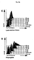
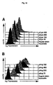
- peptide candidates which are suitable for triggering a cellular immune response takes place in principle in several stages: In general, the candidates, advantageously in serial experiments, are first tested in a peptide binding assay for their ability to bind to an MHC molecule.
- a suitable method of investigation is based on the ability of peptides to stabilize empty MHC molecules, as described, for example, by Stuber et al., 1994, and McIntyre et al., 1996. This is the peptide applied to cells capable of expressing the particular MHC molecule, but due to a genetic defect, do not bind endogenous peptides in the MHC context. Suitable cell lines of this type are RMA-S (mouse) and T2 (human), or their transfected variants.
- MHC molecules stabilized by the respective peptide are then detectable, preferably by means of the flow cytometry-based FACS analysis (Flow Cytometry, 1989, FACS Vantage TM User's Guide, 1994, CELL Quest TM User's Guide, 1994).
- Stable MHC molecules are detected with a suitable anti-MHC antibody and with a second (eg polyclonal) antibody labeled with fluorescent dye, eg FITC (fluorescein isothiocyanate).
- fluorescent dye eg FITC (fluorescein isothiocyanate).
- Another method for determining the amount of peptide bound is the Scatchard plot as described by Sette et al., 1994. To do this, the peptide used, for example, with
- the induction of a cellular immune response can be confirmed by the detection of peptide-specific CTLs, e.g. by the method described in Current Protocols in Immunology, Chapter 3, or by Blomberg et al., 1993. Further evidence for the presence of a cellular immune response is given when no immune response occurs in the absence of T cells in an experimental animal (which is achieved by treating the animal with antibodies that deplete CD4 or CD8 cells) ( Current Protocols in Immunology, Chapter 3).
- a cellular immune response can also be demonstrated by the detection of a delayed-type hypersensitivity (DTH) response in immunized animals.
- DTH delayed-type hypersensitivity
- peptides can be injected into the sole of the foot of a mouse, whereupon the swelling of the injected site is measured (Grohman et al., 1995, Puccetti et al., 1994).
- the induction of a humoral immune response by peptides which are foreign antigens for the organism or antigens which are expressed in low concentration by the organism to be treated can be determined by detection of specific antibodies in the serum.
- a suitable method for antibody titer determination in serum is Enzyme Linked Immunoassay (ELISA).
- ELISA Enzyme Linked Immunoassay
- the specific antibodies are detected after binding to the peptide used for immunization with a color reaction.
- An alternative method is the Western Blot.
- specific serum antibodies bind to the immobilized on a membrane peptide.
- bound antibodies are detected by a color reaction (reference for both methods: Current Protocols in Immunology, Editors: Coligan et al., 1991).
- T cells that can recognize the desired peptide when presented by MHC molecules.
- T cells are obtained analogously from the peripheral blood. T cells are incubated with cell lines such as T2 (Alexander et al., 1989) or RMA-S (Kärre et al., 1986) spiked with the respective peptide and lysed, if immunogenic Peptide acts.
- T2 cells Another possibility for assaying MHC-binding peptide candidates for their immunogenicity is to examine the binding of the peptides to T2 cells.
- T2 cells Alexander et al., 1989
- RMA-S cells Kärre et al., 1986
- T2 cells or RMA-S cells stably transfected with an HLA gene, eg with HLA-A1 and / or HLA-A2 genes, are used for the assay.
- HLA molecules When cells are spiked with peptides, they are good HLA ligands by being presented in an HLA context such that they can be recognized as foreign by the immune system, such peptides cause the HLA molecules to appear in significant amounts on the cell surface.
- the detection of HLAs on the cell surface allows the identification of suitable peptides (Malnati et al., 1995, Sykulev et al., 1994). Again, a standard peptide with known good HLA binding ability is suitably used.
- a mixture of several peptides each of which can bind to another MHC molecule, preferably to one of two or three of the most frequently represented MHC subtypes.
- a vaccine based on a mixture of peptides capable of binding to these haplotypes, a broad population of patients can be detected.
- the vaccine may have multiple peptide sequences.
- the peptides used in this case may differ, on the one hand, in that they bind to different HLA subtypes. This can be achieved that several or all HLA subtypes of a patient or a larger group of patients are detected.
- variability in the peptides used may be that peptides that bind to a particular HLA subtype differ in their non-HLA binding sequence, e.g. derived from different proteins of the same pathogen or from different pathogens. From such a variability, an enhancement of the stimulation of the immune response or, respectively, an immunization against various pathogens can be achieved.
- the amount of active peptide in the composition of the invention can vary over a wide range.
- the amount of peptide depends i.a. on the route of administration and the respective formulation.
- the amount of peptide to be administered may be about 1.0 ⁇ g to about 5000 ⁇ g per dose, generally 1.0 ⁇ g to about 1000 ⁇ g, in particular about 10 ⁇ g to about 500 ⁇ g.
- the administration can be one or more times, with repeated administration expedient at least three times. In particular, in the therapeutic application, an application at intervals (for example, from once a week to once a month) over an arbitrarily long period, which is due to the specific immune status of the patient or the course of the disease.
- the pharmaceutical composition according to the invention can also be applied ex vivo :
- the principle of a possible ex vivo application consists in cultivating APCs, eg dendritic cells, ex vivo , incubating the cell culture with the composition according to the invention and the APCs, which now contain the peptide in the MHC Context to administer to the individual to be treated.
- Literature-known methods can be used for this application, as for example by Porgador and Gilboa, 1995; Young and Inabe, 1996.
- the adjuvant contained in the composition according to the invention has the property of facilitating the entry of the peptide into the cells or the binding of the peptide to the cells of the patient and to enhance the immunogenicity of the peptide.
- the adjuvant may e.g. make the membranes of target cells into which the peptide is to pass, at least in the short term permeable, in order in this way to transport the peptide into the cell. It would be advantageous, but not essential, for the peptide to bind to the adjuvant, e.g. on electrostatic interaction between electronegative peptide and polycationic adjuvant.
- Import of the peptide into the cell may also be effected by allowing the peptide to pass therethrough due to its proximity to the cell membrane once the adjuvant has caused its permeability.
- the action of the adjuvant may also be due to increasing the cell surface cell concentration critical for uptake into the cell, or to causing the phagocytosis or liquid transport (pinocytosis) of the peptide into the cell.
- the presence of the adjuvant of the invention not only enhances the uptake of the peptide into the cell, but also results in an enhancement of the immunomodulatory effect of the peptide, which may be due to a correct presentation of the peptide by MHC molecules.
- the adjuvant of the invention is a basic polyamino acid.
- the degree of polymerization of the polyamino acids can vary over a wide range. It is about 5 to about 1000, especially about 15 to about 500.
- polyarginine is used as adjuvant.
- LDH lactate dehydrogenase
- the effect of the pharmaceutical composition according to the invention is likely to be that the peptide, with the aid of the adjuvant, penetrates into the target cells or binds to cells which occur in the endodermal region of the skin.
- Target cells are u.a. Antigen-presenting cells from which the peptide, optionally after processing, the B and / or T cells is presented. Examples of target cells are macrophages, fibroblasts, keratinocytes, Langerhans cells, dendritic cells or B cells.
- APCs are believed to be the cell type in vivo that captures the peptides and presents them to other immune cells.
- Results of in vitro experiments demonstrating that APCs endocytose increased levels of peptide antigens in the presence of the adjuvants tested are one Indication that these adjuvants are also suitable in vivo to enhance the presentation of the peptides to the cytotoxic effector cells and their activation, resulting in an overall enhanced immune response against the target contained in the vaccine.
- the testing of adjuvants can, in principle, be carried out by the same methods as the testing of the peptides, if appropriate in several steps:
- an adjuvant to increase the binding and / or internalization of a peptide to APCs may be e.g. in a first step, by incubating APCs with fluorescently labeled peptides and adjuvant. Increased uptake or binding by the adjuvant can be determined by flow cytometry by comparison with cells that have been spiked with peptide alone.
- the adjuvants to be tested in vitro can be screened for whether and to what extent their presence results in a presentation of a peptide on APCs, measuring the MHC concentration on the cells according to the methods described above for the testing of the peptides can be.
- Another way to test the efficiency of an adjuvant is to use an in vitro model system.
- APCs are incubated together with adjuvant and peptide and the relative activation of a T-cell clone that specifically recognizes the peptide used is measured (Coligan et al., 1991, Lopez et al., 1993).
- the efficiency of the formulation can also be demonstrated via the cellular immune response by detecting a delayed-type hypersensitivity (DTH) response in immunized animals.
- DTH delayed-type hypersensitivity
- the immunomodulatory effect of the formulation is measured in animal experiments.
- established tumor models in which peptide sequences recognized by immune cells are known can be used.
- the vaccine containing peptide and adjuvant is administered in varying ratios, based on peptide to adjuvant and total.
- the protection against tumor growth is a measure of the effectiveness of a tumor vaccine.
- the pharmaceutical composition can be administered parenterally, topically, orally or locally. Preferably it becomes parenteral, e.g. administered subcutaneously, intradermally or intramuscularly, preferably subcutaneously or intradermally, in order to achieve primarily skin cells (keratinocytes, fibroblasts), dendritic cells, Langerhans cells or macrophages as target cells.
- the tumor vaccine can also be administered intratumorally.
- composition for parenteral administration of the invention is generally present as a solution or suspension of the peptide and the adjuvant in a pharmaceutically acceptable carrier, preferably an aqueous carrier.
- a pharmaceutically acceptable carrier e.g. Water, buffered water, saline (0.4%), glycine solution (0.3%), hyaluronic acid and similar known carriers.
- solvents such as dimethyl sulfoxide, propylene glycol; Dimethylformamide and mixtures thereof can be used.
- the composition may also contain pharmaceutically acceptable excipients such as buffering agents as well as inorganic salts to achieve normal osmotic pressure and / or lyophilization.
- Such additives are sodium and potassium salts, e.g. Chlorides and phosphates, sucrose, glucose, protein hydrolysates, dextran, polyvinyl pyrrolidone or polyethylene glycol.
- the compositions can be sterilized by conventional techniques, e.g. by sterile filtration.
- the composition may be immediately bottled or lyophilized in this form and mixed with a sterile solution prior to use.
- the pharmaceutical composition of the invention is as a topical formulation, e.g. for dermal or transdermal application.
- the pharmaceutical composition may be present, for example, as a polyacrylic or polyacrylamide-based hydrogel (such as Dolobene®, Merckle), as an ointment, e.g. with polyethylene glycol (PEG) as a vehicle, such as the standard ointment DAB 8 (50% PEG 300, 50% PEG 1500), or as an emulsion, especially as a water-in-oil or oil-in-water microemulsion, optionally also with the addition of liposomes.
- PEG polyethylene glycol
- DAB 8 50% PEG 300
- emulsion especially as a water-in-oil or oil-in-water microemulsion, optionally also with the addition of liposomes.
- Permeationsbelix are u.a.
- Sulfoxide derivatives such as dimethysulfoxide (DMSO) or decylmethysulfoxide (decyl-MSO) and transcutol (diethylene glycol monoethyl ether) or cyclodextrins, further pyrrolidones, e.g.
- 2-pyrrolidone N-methyl-2-pyrrolidone, 2-pyrrolidone-5-carboxylic acid or the biodegradable N- (2-hydroxyethyl) -2-pyrrolidone and their fatty acid esters, urea derivatives such as dodecylurea, 1,3-didodecylurea and 1,3-diphenylurea, terpenes, eg D-limonene, menthone, a-terpinol, carvol, limonene oxide, or 1,8-cineole.
- urea derivatives such as dodecylurea, 1,3-didodecylurea and 1,3-diphenylurea
- terpenes eg D-limonene, menthone, a-terpinol, carvol, limonene oxide, or 1,8-cineole.
- aerosols e.g. for administration as a nasal spray or for inhalation.
- composition of the invention may also be administered by means of liposomes, which may be in the form of emulsions, foams, micelles, insoluble monolayers, phospholipid dispersions, lamellar layers and the like. These serve as a vehicle to target the peptides to a particular tissue, e.g. lymphoid tissue or tumor tissue, or to extend the half-life of the peptides.
- liposomes may be in the form of emulsions, foams, micelles, insoluble monolayers, phospholipid dispersions, lamellar layers and the like.
- composition of the present invention may also contain UV absorbers, e.g. to act simultaneously as a sunscreen in the prophylactic use against melanoma.
- the mouse melanoma cell line Cloudman S91 (clone M-3, ATCC No. CCL 53.1), the mastocytoma cell line P815 (ATCC No. TIB 64) and the monocyte-macrophage cell line P388D1 (ATCC TIB 63) were purchased from ATCC.
- the cells were cultured in High Glucose DMEM medium (Life Technologies) supplemented with 10% FCS, 2 mM L-glutamine, and 20 ⁇ / ml gentamycin The cells were routinely tested for absence of mycoplasma contamination (PCR-Mycoplasma Detection Kit, Stratagene).
- the murine cell line RMA / S (mouse lymphoma) has been described by Kärre et al., 1986 and by Ljunggren et al., 1990.
- the femurs of DBA / 2 mice were rinsed.
- the bone marrow cells were transfected into high glucose DMEM medium containing 10% FCS, 5% horse serum, 2 mM L-glutamine and 20 ⁇ g / ml gentamycin in the presence of 200 units / ml mouse GM-CSF (Li et al., 1989 Genzyme, Cambridge, MA, USA).
- two-thirds of the medium was changed every 24 hours to remove nonadherent granocytes and B cells (Inaba et al., 1992).
- Both adherent and loosely adherent cells were harvested between days 8 and 10 by incubation with PBS / 5 mM EDTA and seeded at a cell density of 3 x 10 4 cells per well on 8-well microscope slides (Nunc, Roskilde, Denmark) , More than 90% of the cells showed a positive reaction with the antibody F4 / 80 (Endogen, Cambridge, MA, USA).
- the peptides were assayed on a peptide synthesizer (model 433A with feedback monitor, Applied Biosystems, Foster City, Canada) using TentaGel S PHB (Rapp, Tübingen) as the solid phase according to the Fmoc method (HBTU activation, Fastmoc TM , Scale 0:25 mmol).
- the peptides were dissolved in 1 M TEAA, pH 7.3 and purified by reverse chromatography on a Vydac C 18 column. The sequences were confirmed by time-of-flight mass spectrometry on a MAT Lasermat (Finnigan, San Jose, Canada).
- mice Prior to subcutaneous injection, the mice were anesthetized in groups of up to eight animals in an isolated air chamber. After 3.5 min halothane treatment (4% in O 2 , flow rate 4) the mice were anesthetized for about 1 min; during this time, the vaccine was injected subcutaneously.
- the intraperitoneal injection was carried out without previous anesthetization.
- the injection volume was 100 ⁇ l per animal for each vaccine, which corresponds to 100 ⁇ g of single peptide or peptide mixture I per animal.
- peptide mixture III 125 ⁇ g total peptide amount per mouse was applied.
- a cellular tumor vaccine was prepared as follows:
- 160 ⁇ g peptide kpep 118 were mixed with 3 ⁇ g transferrin polylysine (TfpL), 10 ⁇ g pL and 6 ⁇ g plasmid psp65 (LPS-free) in 500 ⁇ l HBS buffer. After 30 min at room temperature, the above solution was added to a T 75 cell culture flask containing 1.5 x 10 6 cells of the allogeneic fibroblast cell line NIH3T3 (ATCC No. CRL 1658) in 20 ml DMEM medium (10% FCS, 20 mM glucose) and 37 ° C incubated. After 3 h, the cells were mixed with 15 ml of fresh medium and incubated overnight at 37 ° C and 5% CO 2 .
- TfpL transferrin polylysine
- 10 ⁇ g pL 10 ⁇ g pL
- 6 ⁇ g plasmid psp65 LPS-free
- the cells were irradiated with 20 Gy.
- the workup of the vaccine was carried out as in the WO 94/21808 described. Preimmunization with this vaccine was done one week apart with 10 5 cells; after another week, tumor replacement was performed as described above ( Fig. 1 : filled triangles). It was found that the vaccine containing the peptide combined with polylysine best protected the mice from tumor formation.
- GM-CSF secreting P815 cells were used as a positive control.
- 100 ⁇ g of kpep118 were injected per animal once with polylysine (group B), once with transferrin-polylysine (group C) and once with transferrin-polylysine / DNA (group D).
- group B polylysine
- group C transferrin-polylysine
- group D transferrin-polylysine / DNA
- kpep118 with IFA was used. In this experiment, kpep118 alone showed no protective effect against tumorigenesis.
- Example 4 The experiments carried out in Example 4 have shown that a vaccine containing a peptide mixture of melanoma peptides has a protective effect against melanoma. It was therefore tested in this example whether the concept of the peptide mixture is also suitable for P815.
- the peptide mixture III was administered once only with polylysine (group E), once with transferrin-polylysine (group F) and once with transferrin-polylysine / DNA (group G).
- group E polylysine
- group F transferrin-polylysine
- group G transferrin-polylysine / DNA
- peptide mixture III was used in IFA.
- naive mice were used; as a positive control GM-CSF transfected P815 cells (10 5 cells per mouse).
- a prophylactic vaccine containing a mixture of melanoma peptides (peptide mixture 1, section D2) was used.
- the protocol of preimmunization with the vaccine and the setting of the tumors corresponded to the protocol described in Example 2, with the difference that the tumor was made with M3 cells (10 5 cells per animal).
- M3 cells 10 5 cells per animal.
- a whole cell vaccine of M-3 cells was used, which was secreted and produced an optimal amount of IL-2 (1000-2000 units per 10 5 cells) as described by Schmidt et al., 1995. Under the chosen experimental conditions, this vaccine achieved 100 % protection ( Fig. 4 ).
- Four groups of experimental animals were administered the vaccines with the peptide mixture. Two groups, either sc or ip, received the peptides emulsified in IFA. The other two groups, either sc or ip, received the peptides together with polylysine (pL240).
- Fig. 4 illustrates the protective effect of the peptide vaccine with polylysine (pL240) as adjuvant; 50% of the treated mice were protected against the M-3 tumor challenge compared to untreated animals, which rapidly developed solid tumors.
- This effect could be achieved if the peptide-polylysine vaccine was injected subcutaneously or applied to the skin as a hydrogel ( Fig. 5 ).
- the vaccine was essentially ineffective.
- the set tumors were not rejected, and the tumors grew with only a slight delay compared to the untreated control animals.
- Fig. 7c shows the repetition of this effect achieved by polyarginine in an independent experiment. Again, vaccination with the peptide mixture in combination with polyarginine showed antitumor activity in four out of eight treated animals.
- This example served as an experiment to demonstrate that polylysine is useful as an adjuvant for loading cells with larger protein fragments or whole proteins, using M-3 cells as a proxy.
- FITC-labeled tyrosinase EC 1.14.18.1, Sigma
- 3 ⁇ g of polylysine pL240
- the resulting solution was placed in a T75 cell culture flask containing 2 x 10 6 M-3 cells and incubated at 37 ° C. Thereafter, the cells were washed twice with PBS, detached with PBS / 2 mM EDTA and taken up in 1 ml PBS / 5% FCS for FACS analysis.
- Fig. 8 shows the loading of M-3 cells with tyrosinase (the left curve shows the control, the right the loading with tyrosinase).
- Single cell spleen suspensions were prepared from vaccinated and untreated animals, followed by erythrocyte lysis with hypotonic buffer (0.15 NH 4 Cl, 1 mM KHCO 3 , 0.1 mM EDTA, pH 7.4).
- Adherent cells were removed by incubating 3 x 10 6 spleen cells per ml of DMEM medium (10% FCS) in Petri dishes for 90 min at 37 ° C. Non-adherent cells were co-cultured by careful pipetting and co-culturing with 1 x 10 3 parental cells in various proportions.
- the cells were cultured in 200 ⁇ l of DMEM medium (10% FCS), 2 mM L-glutamine and 20 ⁇ g / ml gentamycin in 96-well flat-bottom tissue culture dishes. On day 9, 100 ⁇ l of supernatant was harvested and the respective IFN- ⁇ content was measured using a commercially available ELISA kit (Endogen, Cambridge, MA, USA) according to the manufacturer's instructions. It was found that after nine days of incubation only spleen cells from vaccinated animals secreted larger amounts of IFN- ⁇ into the medium, while in the co-cultures of spleen cells from untreated animals and M3 cells virtually no IFN- ⁇ was detectable. The result of these experiments is in Fig. 9 shown.
- a vaccine containing 75 ⁇ g fpLys per 1 mg peptide ASNENMETM was used.
- the vaccine was administered by a single injection as indicated in the Methods section, injecting 100 ⁇ g peptide / 7.5 ⁇ g fpLys per animal.
- For control the injection of 100 ug peptide alone (PBS) or no injection was made (naive control).
- RMA-S mouse lymphoma cells were incubated overnight in serum-free medium at 26 ° C with 10 ⁇ g / ml peptide ASNENMETM.
- spleen cells were isolated from the vaccinated animals, mixed with the peptide-loaded RMA-S cells in a 5: 1 ratio and further cultured for five days (Stuber et al., 1994). The number of effector effector spleen cells surviving in the different cultures was determined, then the cells were analyzed for the determination of CTL activity by standard 4 h europium release assay (Blomberg et al., 1993) in various ratios with RMA-S Cells previously mixed with the peptide ASNENMETM and with europium chelate. As Fig. 10 The specific immunity in this experiment was only achieved by vaccination with peptide and fpLys, but not when the peptide was administered alone.
- a fluorescence assay was used: A model peptide antigen of the sequence LFEAIEGFI (MHC Kd-restricted) was labeled with the fluorescent dye fluorescein isothiocyanate (FITC) according to the manufacturer's instructions (Molecular Probes). The uptake or binding of FITC-labeled peptide alone (“pulsed") or together with various concentrations of basic amino acids (polylysine with a chain length of 16 to 490, polyarginine with a chain length of 15 to 720) through the MHC Kd-restricted monocytes Macrophage cell line P388D1 was measured by flow cytometry.
- FITC fluorescent dye fluorescein isothiocyanate
- the cells were incubated with a mixture of polyarginine (average molecular weight range 100,000, chain length 490) and fluorescently labeled peptide at 4 ° C or 37 ° C. An aliquot of the samples incubated at 37 ° C was retained and treated with 50 ⁇ M monensin at 4 ° C prior to flow cytometry analysis.
- Fig. 13 showed only a slight increase in fluorescence in cells treated with peptide alone and with monensin. In contrast, the fluorescence signals in samples treated with monensin and a mixture of polyarginine and peptide were greatly increased. No peptide uptake was observed when the samples were incubated at 4 ° C. A significant increase in fluorescence after monensin treatment indicates that the pArg-induced loading of the peptides causes them to accumulate in vesicles within the cell (Midoux et al., 1993; Fig. 13 ). As expected, only a small increase in fluorescence was observed after monensin treatment of polylysine-loaded samples. Loading with polylysine at 4 ° C caused a measurable increase in fluorescence, which is another indication that the effect of polylysine is mainly due to permeabilization of cell membranes ( Fig. 12b ).
- Bone marrow-derived GMCF-derived APCs were probed with a combination of a fluorescently labeled peptide plus polylysine (pL200; Sigma) or with peptide alone by fluorescence microscopy.
- the APCs were seeded onto slides and incubated with 40 ug fluorescein-labeled peptide LFEAIEGFI alone or with a mixture of 50 ug / ml polylysine (pL200) and 40 ug / ml peptide LFEAIEGFI at 37 ° C for 30 min.
- the cells were fixed with 4% para-formaldehyde, covered with anti-Fadent (Dako, Glostrup, Denmark) and fluorescence microscopy (Zeiss). The cores were counterstained with 4,6-diamidino-2-phenylindole (DAPI; Sigma).
- DAPI 4,6-diamidino-2-phenylindole
- Example 11 After the experiments carried out in Example 11 showed that APCs are excellent target cells for small peptide loading, in vitro FACS assays were performed to identify suitable adjuvants for peptide vaccines. This assay allows rapid quantitative testing of fluorescently labeled peptides; as model peptide, the peptide of the sequence LFEAIEGFI was used. In these experiments, the mouse cell line P388D1 was used as APCs. 1 x 10 6 cells were incubated in a final volume of 1 ml of high glucose DMEM medium and 10% FCS for 30 min at 37 ° C with 5 ⁇ g of peptide at a fluorescein final concentration of 5 nmol / ml.
- the cells were treated either with peptide alone or with a combination of peptide and polycations or of peptide and histones at increasing concentrations (3 to 50 ⁇ g / ml). as in Fig. 15 indicated, treated.
- the following compounds were used: A: polyomithine (average molecular weight range 110,000, chain length 580); B: arginine-rich histone; C: lysine-rich histone; D: polyarginine (average molecular weight range 100,000, chain length 490); E: polylysine (average molecular weight 94,000, chain length 450).
- A polyomithine (average molecular weight range 110,000, chain length 580);
- B arginine-rich histone; C: lysine-rich histone;
- D polyarginine (average molecular weight range 100,000, chain length 490); E: polylysine (average molecular weight 94,000, chain length 450).
- Prolonged treatment (4 or 8 h) showed no significant increase in the fluorescence signal.
- the cells Prior to analysis, the cells were washed 5x with a large volume of PBS containing 0.2% BSA. The cells were resuspended in 1 ml of ice-cold PBS / 0.2% BSA and analyzed by flow cytometry (FACScan, Becton Dickinson, San Jose, CA, USA). Polyarginine and polylysine proved to be the most effective adjuvants; Polyomithin showed a cytotoxic effect under the chosen conditions. The increase in peptide uptake by polyarginine and polylysine correlates with the concentration ( Fig. 15 d . e ); the recording increases with the chain length ( Fig. 16 ).
- polyarginine has an increase of 3 orders of magnitude over the control has a broader suitable concentration range than polylysine, which showed a maximum increase of less than 2 orders of magnitude, and that it is more effective for the transport of proteins at all chain lengths used than polylysine ( Fig. 16 ): Polyarginine allows efficient transport at concentrations as low as 3 ⁇ g / ml, whereas polylysine concentrations> 25 ⁇ g / ml were required to achieve a significant increase in fluorescence ( Fig. 15 d . e ).
- Peptide 1 is derived from mouse TRP ("tyrosinase related protein")
- peptide 2 is derived from influenza hemagglutinin (Schmidt et al., 1996)
- peptide 3 from mouse tyrosinase
- peptide 4 from the P198 tumor antigen
- peptide 5 from the beta-galactosidase (Gavin et al., 1993).
Landscapes
- Health & Medical Sciences (AREA)
- Life Sciences & Earth Sciences (AREA)
- Chemical & Material Sciences (AREA)
- Public Health (AREA)
- Medicinal Chemistry (AREA)
- Immunology (AREA)
- Veterinary Medicine (AREA)
- Pharmacology & Pharmacy (AREA)
- Animal Behavior & Ethology (AREA)
- General Health & Medical Sciences (AREA)
- Epidemiology (AREA)
- Mycology (AREA)
- Microbiology (AREA)
- Chemical Kinetics & Catalysis (AREA)
- General Chemical & Material Sciences (AREA)
- Virology (AREA)
- Nuclear Medicine, Radiotherapy & Molecular Imaging (AREA)
- Organic Chemistry (AREA)
- Oncology (AREA)
- Engineering & Computer Science (AREA)
- Bioinformatics & Cheminformatics (AREA)
- Pulmonology (AREA)
- Communicable Diseases (AREA)
- Proteomics, Peptides & Aminoacids (AREA)
- Medicines Containing Antibodies Or Antigens For Use As Internal Diagnostic Agents (AREA)
- Peptides Or Proteins (AREA)
- Medicines That Contain Protein Lipid Enzymes And Other Medicines (AREA)
- Medicinal Preparation (AREA)
- Pharmaceuticals Containing Other Organic And Inorganic Compounds (AREA)
Abstract
Description
- Die Erfindung bezieht sich auf das Gebiet der Immunmodulation.
- Die Erfindung ist eine Weiterentwicklung einer therapeutischen Vakzine auf der Grundlage von Tumorzellen, die im wesentlichen auf den folgenden Voraussetzungen beruht: es bestehen qualitative oder quantitative Unterschiede zwischen Tumorzellen und normalen Zellen; das Immunsystem hat prinzipiell die Fähigkeit, diese Unterschiede zu erkennen; das Immunsystem kann - durch aktive spezifische Immunisierung mit Vakzinen - dazu stimuliert werden, Tumorzellen anhand dieser Unterschiede zu erkennen und deren Abstoßung herbeizuführen.
- Um eine Anti-Tumorantwort herbeizuführen, müssen zumindest zwei Voraussetzungen erfüllt sein: erstens müssen die Tumorzellen Antigene exprimieren, die auf normalen Zellen nicht oder nur derart begrenzt vorkommen, daß eine qualitative Unterscheidung zwischen Normal- und Tumorgewebe durch das Immunsystem möglich ist. Zweitens muß das Immunsystem entsprechend aktiviert werden, um auf diese Antigene zu reagieren. Ein wesentliches Hindernis bei der Immuntherapie von Tumoren ist deren geringe Immunogenität, vor allem im Menschen.
In jüngerer Zeit wurden Tumor-assoziierte und Tumor-spezifische Antigene entdeckt, die solche Neo-Epitope darstellen und somit potentielle Ziele für einen Angriff des Immunsystems sein sollten. Daß es dem Immunsystem dennoch nicht gelingt, Tumoren zu eliminieren, die diese Neo-Epitope exprimieren, dürfte demnach offensichtlich nicht am Fehlen von Neo-Epitopen gelegen sein, sondern daran, daß die immunologische Antwort auf diese Neo-Antigene unzureichend ist. - Für die Immuntherapie von Krebs auf zellulärer Basis wurden zwei allgemeine Strategien entwickelt: Einerseits die adoptive Immuntherapie, die sich der in vitro Expansion von tumorreaktiven T-Lymphozyten und deren Wiedereinführung in den Patienten bedient; andererseits die aktive Immuntherapie, welche Tumorzellen verwendet, in der Erwartung, daß damit entweder neue oder verstärkte Immunantworten gegen Tumorantigene hervorgerufen werden, die zu einer systemischen Tumorantwort führen. Tumorvakzine auf der Grundlage der aktiven Immuntherapie wurden auf verschiedene Arten hergestellt; ein Beispiel dafür sind bestrahlte Tumorzellen, die mit immunstimulierenden Adjuvantien wie Corynebacterium parvum oder Bacillus Calmette Guerin (BCG) versetzt werden, um Immunreaktionen gegen Tumorantigene hervorzurufen (Oettgen und Old, 1991)."Immunomodulation by adjuvants", siehe NATO ASI SER. A, 215 (VACCINES), 25-32, 1991.
- In den letzten Jahren wurden vor allem genetisch modifizierte Tumorzellen für eine aktive Immuntherapie gegen Krebs verwendet. Eine Übersicht über diese verschiedenen Ansätze, bei denen Tumorzellen im Hinblick auf eine verstärkte Immunogenität durch Einführung verschiedener Gene verfremdet werden, wird von Zatloukal et al., 1993, gegeben. Eine der bisher eingesetzten Strategien verwendet Tumorzellen, die genetisch modifiziert werden, um Zytokine zu produzieren.
- Die Identifizierung und Isolierung von Tumorantigenen und Tumor-assoziierten Antigenen (TAs) bzw. davon abgleiteter Peptide (z.B. beschrieben von Wölfel et al., 1994 a) und 1994 b); Carrel et al., 1993, Lehmann et al., 1989, Tibbets et al., 1993, oder in den veröffentlichten internationalen
Anmeldungen WO 92/20356 WO 94/05304 WO 94/23031 WO 95/00159 - Eine dritte Strategie der aktiven Immuntherapie zur Steigerung der Wirksamkeit von Tumorvakzinen basiert auf xenogenisierten (verfremdeten) autologen Tumorzellen. Diesem Konzept liegt die Annahme zugrunde, daß das Immunsystem auf Tumorzellen reagiert, die ein Fremdprotein exprimieren und daß im Zuge dieser Reaktion auch eine Immunantwort gegen diejenigen Tumorantigene hervorgerufen wird, die von den Tumorzellen der Vakzine präsentiert werden.
- Eine zentrale Rolle bei der Regulierung der spezifischen Immunantwort spielt ein trimolekularer Komplex, bestehend aus den Komponenten T-Zell-Antigenrezeptor, MHC ("Major Histocompatibility Complex")-Molekül und dessen Liganden, der ein von einem Protein abgeleitetes Peptidfragment ist.
- MHC-Moleküle (bzw. die entsprechenden humanen Moleküle, die HLAs) sind Peptidrezeptoren, die bei stringenter Spezifität die Bindung zahlreicher verschiedener Liganden erlauben. Die Voraussetzung dafür stellen Allelspezifische Peptidmotive dar, die folgende Spezifitätskriterien aufweisen: Die Peptide haben, in Abhängigkeit vom MHC-Haplotyp, eine definierte Länge, beimMHC-I Haplotyp in der Regel acht bis zehn Aminosäurereste. Typischerweise stellen zwei der Aminosäurepositionen sog. "Anker" dar, die nur durch eine einzige Aminosäure oder durch Aminosäure-Reste mit eng verwandten Seitenketten besetzt werden können. Die genaue Lage der Ankeraminosäuren im Peptid und die Anforderungen an deren Eigenschaften variieren mit den MHC-Haplotypen. Der C-Terminus der Peptid-Liganden ist häufig ein aliphatischer oder ein geladener Rest. Solche MHC-I-Peptid-Ligandenmotive sind bisher u.a. für H-2Kd, Kb, Kk, Kkm1, Db, HLA-A*0201, A*0205 und B*2705 Haplotypen bekannt.
- Im Rahmen des Proteinumsatzes innerhalb der Zelle werden reguläre, entartete und fremde Genprodukte, z.B. virale Proteine oder Tumorantigene, in kleine Peptide zerlegt; einige davon stellen potentielle Liganden für MHC-Moleküle dar. Damit ist die Voraussetzung für deren Präsentation durch MHC-Moleküle und als Folge davon die Auslösung einer zellulären Immununatwort gegeben, wobei noch nicht im einzelnen aufgeklärt ist, wie die Peptide als MHC-Liganden in der Zelle produziert werden. Fremde oder verfremdete Proteine und deren Bruchstücke können auch durch Immunglobuline, die die humorale Immunantwort darstellen, erkannt, gebunden und beseitigt werden. Das gilt auch für sämtliche Tumorantigene: am Beispiel der Tumor-assoziierten Antigene MUC1, CEA und HER2/neu hat man bewiesen, daß Immunglobuline, welche für diese Proteine Spezifität aufweisen, die proteintragenden Zellen erkennen und abtöten können. Um eine Tumorantigen-spezifische humorale Immunantwort auszulösen, wurden deshalb verschiedene Formen von MUC1 und CEA als Immunogene (z.B. in rekombinanten Poxvektoren; Bronte at al., J.Immunol. 154:5282 1995) in Tiermodellen und klinischen Vorversuchen erprobt.
- Im Rahmen der vorliegenden Erfindung wurden Überlegungen weitergeführt, die bei der Entwicklung einer zellulären Tumorvakzine angestellt wurden: während nicht-maligne, normale Körperzellen vom Immunsystem toleriert werden, reagiert der Körper auf eine normale Zelle, wenn sie, z.B. aufgrund einer Virusinfektion, körperfremde Proteine synthetisiert, mit einer Immunabwehr. Die Ursache dafür ist darin gelegen, daß die MHC-Moleküle Fremdpeptide präsentieren, die von den körperfremden Proteinen stammen. Als Folge davon registriert das Immunsystem, daß etwas Unerwünschtes, Fremdes mit dieser Zelle geschehen ist. APCs (dazu zählen Makrophagen, dendritische Zellen, Langerhans-Zellen, B-Zellen sowie möglicherweise die kürzlich entdeckten biphänotypischen Zellen, die sowohl Eigenschaften von B-Zellen als auch von Makrophagen aufweisen; Tykocinski et al., 1996) werden aktiviert, eine neue, spezifische Immunität generiert und die Zelle eliminiert.
- Tumorzellen enthalten zwar die jeweiligen tumorspezifischen Tumorantigene, sind aber als solche unzulängliche Vakzine, weil sie aufgrund ihrer geringen Immunogenität vom Immunsystem ignoriert werden. Belädt man nun eine Tumorzelle nicht mit einem Fremdprotein, sondern mit einem Fremdpeptid, so werden zusätzlich zu den Fremdpeptiden auch die zelleigenen Tumorantigene dieser Zelle als fremd wahrgenommen. Durch die Verfremdung mit einem Peptid kann erreicht werden, daß sich die durch die Fremdpeptide ausgelöste zelluläre Immunantwort gegen die Tumorantigene richtet.
- Die Ursache für die geringe Immunogenität von Tumorzellen kann nicht nur ein qualitatives, sondern ein quantitatives Problem sein. Für ein von einem Tumorantigen abgeleitetes Peptid kann das bedeuten, daß es zwar von MHC-Molekülen präsentiert wird, jedoch in einer Konzentration, die zu gering ist, um eine zelluläre tumorspezifische Immunantwort auszulösen. Eine Erhöhung der Zahl von tumorspezifischen Peptiden auf der Tumorzelle sollte somit ebenfalls eine Verfremdung der Tumorzelle bewirken, die zur Auslösung einer zellulären Immunantwort führt. Es wurde vorgeschlagen, das Tumorantigen bzw. das davon abgeleitete Peptid dadurch auf der Zelloberfläche zu präsentieren, daß es mit einer für das betreffende Protein bzw. Peptid kodierenden DNA transfiziert wird, wie in den internationalen Veröffentlichungen
WO 92/20356 WO 94/05304 WO 94/23031 WO 95/00159 - In der
deutschen Patentanmeldung P 195 43 649.0 ist eine zelluläre Vakzine geoffenbart, die als aktive Komponente Tumorzellen enthält, die mit einem oder mehreren Peptiden derart beladen sind, daß die Tumorzellen im Kontext mit den Peptiden vom Immunsystem des Patienten als fremd erkannt werden und eine zelluläre Immunantwort auslösen. Ein wesentliches Merkmal der Peptide ist, daß sie Liganden für den MHC-Haplotyp des Patienten sind. Die Peptide werden deshalb vom Immunsystem des Patienten als fremd erkannt, weil sie einerseits "Fremdpeptide" oder "Xenopeptide" sein können, d.h. sie sind verschieden von Peptiden, die abgeleitet von Proteinen sind, die von Tumorzellen des Patienten exprimiert werden. Eine andere Kategorie von Peptiden ist von Tumorantigenen abgeleitet, die von Zellen des Patienten exprimiert werden. Diese bewirken eine Steigerung der Immunogenität dadurch, daß sie in einer Konzentration auf den Tumorzellen der Vakzine vorliegen, die höher ist als die Konzentration desselben Peptids auf den Tumorzellen des Patienten. - Der vorliegenden Erfindung lag die Aufgabe zugrunde, eine neue immunmodulatorisch wirkende pharmazeutische Zusammensetzung, insbesondere eine Vakzine, bereitzustellen.
- In Weiterführung des Konzepts der in der
deutschen Patentanmeldung P 195 43 649.0 geoffenbarten zellulären Vakzine wurde im Rahmen der vorliegenden Erfindung eine pharmazeutische Zusammensetzung entwickelt, die immunmodulatorisch wirkende Peptide nicht im Kontext mit Zellen, sondern zusammen mit einem Adjuvans enthält, um eine zelluläre und/oder humorale, vorzugsweise eine systemische, Immunantwort gegen pathogene Erreger bzw. eine Anti-Tumorantwort auszulösen bzw. zu verstärken oder eine Toleranz gegen autoimmun wirkende Proteine hervorzurufen. - Es wurde überraschend festgestellt, daß bestimmte Adjuvantien, z.B. Polykationen, von denen erstmals bereits 1965 gezeigt wurde, daß sie eine Steigerung des Transports von Proteinen in Zellen bewirken können (Ryser et al., 1965; Ryser et al., 1978; Shen et al., 1981) die Immunogenität von Peptiden steigern.
- Die Erfindung betrifft eine pharmazeutische Zusammensetzung die in den Patentansprüchen 1 bis 14 charakterisiert ist. Die Zusammensetzung ist dadurch gekennzeichnet, daß das Adjuvans die Fähigkeit aufweist, die Bindung des Peptids bzw. des Proteins oder Proteinfragments an APC Zellen des zu behandelnden Individuums bzw. den Eintritt in die Zellen zu steigern und eine Verstärkung der immunmodulatorischen Wirkung des Peptids bzw. des Proteins oder Proteinfragments zu bewirken.
- Im folgenden werden im allgemeinen, der Einfachheit halber, Vertreter der genannten immunmodulatorisch wirkenden Peptide, Proteine oder Proteinfragmente, als "Peptide" bezeichnet. Der Begriff "Peptid" steht, wenn er sich nicht ausdrücklich auf den Liganden bezieht, auch stellvertretend für größere Proteinfragmente oder Proteine, von denen das Peptid abgeleitet ist bzw. ein zelluläres Abbauprodukt darstellt.
- Unter "immunmodulatorischer Wirkung" wird einerseits die Auslösung oder Verstärkung einer zellulären und/oder humoralen, vorzugsweise systemischen Immunreaktion verstanden. In dieser Ausführungsform wirkt die erfindungsgemäße pharmazeutische Zusammensetzung als Vakzine.
- In einer bevorzugten Ausführungsform sind die Peptide Liganden für mindestens ein MHC-Molekül, das vom zu behandelnden Individuum exprimiert wird.
- Die humanen MHC-Moleküle werden, gemäß den internationalen Gepflogenheiten, im folgenden auch als "HLA" ("Human Leucocyte Antigen") bezeichnet.
- Unter "zelluläre Immunantwort" ist vor allem die zytotoxische T-Zellimmunität zu verstehen, die als Folge der Generierung von zytotoxischen CD8-positiven T-Zellen und CD4-positiven Helfer-T-Zellen die Zerstörung der vom pathogenen Erreger befallenen Zellen bewirkt.
- Unter "humorale Immunantwort" ist die Produktion von Imunglobulinen zu verstehen, die selektiv von pathogenen Erregern abgeleitete Strukturen erkennen und in der Folge zusammen mit anderen Systemen, wie z.B. Komplement, ADCC (Antibody dependent Cytotoxicity) oder Phagozytose, die Zerstörung der pathogenen Erreger oder der davon befallenen Zellen bewirken.
- Das in der Vakzine enthaltene Peptid ist abgeleitet von einem Antigen bzw. stellt im Fall von Proteinen ein Antigen dar, gegen das eine zelluläre und/oder humorale Immunantwort ausgelöst werden soll. Damit wird bewirkt, daß T-Zellen bzw. andere zytotoxische Effektorzellen, die den Krankheitserreger die das Antigen aufweisen, erkennen, und/oder Antikörper generiert werden.
- Für die Immunisierung gegen Krankheitserreger, wie Bakterien, Viren, Parasiten, werden Proteine bzw. Peptide verwendet, die ein Protein des bzw. der jeweiligen Erreger darstellen bzw. davon abgeleitet sind. Geeignet sind dabei vor allem Proteine, die von der hohen allgemeinen Mutationsrate dieser Erreger veschont bleiben. Publizierte Beispiele sind HPV16/17 (Human Papilloma Virus; Feltkamp et al., 1995), Hepatitis B Virus Core Antigen (Vitiello et al., 1995), Plasmodium Bergheii (Widmann et al., 1992.), Influenzavirus-Nukleoprotein, Hepatitis C Virus.
- In einer Ausführungsform der Erfindung dient die pharmazeutische Zusammensetzung dazu, eine Toleranz gegen Proteine bzw. deren Fragmente herbeizuführen, die autoimmun induzierte Erkrankungen auslösen, also zur Therapie von Autoimmunerkrankungen. Die in dieser Ausführungsform der Erfindung verwendeten Peptide sind von Proteinen abgeleitet, die Autoimmunerkrankungen verursachen.
- Im Gegensatz zu der Anwendung als als Vakzine gegen pathogene Erreger, bei der die Peptide mit einem Abschnitt des Ursprungproteins (Protein des Pathogens) im wesentlichen derart übereinstimmen, daß das Peptid als das "Original-Antigen" erkannt wird, werden bei der Anwendung der Erfindung zur Behandlung von Autoimmunerkrankungen u.a. Peptide verwendet, die zur Aminosäuresequenz des Ursprungproteins kritische Unterschiede aufweisen. Diese Peptide binden zwar aufgrund ihrer Ankerpositionen an das MHC-Molekül, weisen jedoch in ihrer Sequenz Mutationen auf, die bewirken, daß diese Peptide als Antagonisten funktionieren, welche die aktivierten, spezifischen T-Zellen wieder abschalten (Kersh und Allen, 1996).
- Als Peptid-Antagonisten eignen sich sowohl "natürliche" Antagonisten, die in Viren entdeckt wurden (Bertoletti et al., 1994), als auch Antagonisten, die durch systematische Forschung, z.B. durch Screenen von Peptid-Libraries, gefunden wurden. Ein Beispiel für Peptid-Antagonisten sind Peptide, die T-Zellen ausschalten können, welche spezifisch für Myelin Basic Protein sind, diese wurden in Tierexperimenten auf ihre Wirksamkeit erprobt. (Brocke et al., 1996).
- Ein Peptid, das eine zelluläre Immunantwort auslösen soll, muß an ein MHC-Molekül binden können. Damit die Immunantwort im Patienten ausgelöst wird, muß somit das zu behandelnde Individuum ein entsprechendes HLA-Molekül in seinem Repertoire aufweisen. Die Bestimmung des HLA-Subtyps des Patienten stellt somit, im Hinblick auf die Auslösung einer zellulären Immunantwort, eine der wesentlichen Voraussetzungen für die wirksame Anwendung eines Peptids an diesem Patienten dar.
- Der HLA-Subtyp des Patienten kann mit Standardmethoden, wie dem Mikro-Lymphotoxizitätstest bestimmt werden (Practical lmmunology., 1989). Dieser Test beruht auf dem Prinzip, aus Patientenblut isolierte Lymphozyten zunächst mit Antiserum oder einem monoklonalen Antikörper gegen ein bestimmtes HLA-Molekül in Gegenwart von Kaninchen-Komplement (C) zu versetzen. Positive Zellen werden lysiert und nehmen einen IndikatorFarbstoff auf, während unbeschädigte Zellen ungefärbt bleiben.
- Zur Bestimmung des HLA-I- oder HLA-II-Haplotyps eines Patienten kann auch die RT-PCR ("Reverse Transcriptase Polymerase Chain Reaction") herangezogen werden (Curr. Prot. Mol. Biol. Kapitel 2 und 15). Dazu entnimmt man einem Patienten Blut und isoliert daraus RNA. Diese RNA unterwirft man zunächst einer Reversen Transkription, wodurch cDNA des Patienten entsteht. Die cDNA dient als Matrize für die Polymerasekettenreaktion mit Primerpaaren, die spezifisch die Amptifikation eines DNA-Fragmentes bewirken, das für einen bestimmten HLA-Haplotyp steht. Erscheint nach Agarosegelelektrophorese eine DNA-Bande, exprimiert der Patient das entsprechende HLA-Molekül. Erscheint die Bande nicht, ist der Patient dafür negativ.
- Die Definition eines erfindungsgemäß verwendeten Peptids durch ein HLA-Molekül bestimmt dieses hinsichtlich seiner Ankeraminosäuren und seiner Länge; definierte Ankerpositionen und Länge gewährleisten, daß ein Peptid in die Peptid-Bindungsfurche des jeweiligen HLA-Moleküls des Patienten paßt. Dies hat zur Folge, daß das Immunsystem stimuliert wird und eine zelluläre Immunreaktion erzeugt wird, die sich, im Falle der Verwendung eines von einem Tumorantigen abgeleiteten Peptids, gegen die Tumorzellen des Patienten richtet.
- Peptide, die zur Anwendung im Rahmen der vorliegenden Erfindung geeignet sind, sind in einer großen Bandbreite verfügbar. Ihre Sequenz kann von natürlich vorkommenden immunogenen Proteinen bzw. deren zellulären Abbauprodukten, z.B. von viralen oder bakteriellen Peptiden, von Tumorantigenen, abgeleitet sein, oder sie können Antagonisten zu Peptiden sein, die von Autoimmunerkrankungen induzierenden Proteinen abgeleitet sind.
- Geeignete Peptide können z.B. auf der Grundlage von literaturbekannten Peptidsequenzen ausgewählt werden.
- Im Hinblick auf die Auslösung einer zellulären Immunantwort können die Peptide z.B. anhand der von Rammensee et. al., 1993, Rammensee et al., 1995, Falk et al., 1991, für die unterschiedlichen HLA-Motive beschriebenen, von immunogenen Proteinen verschiedenen Ursprungs abgeleiteten Peptide definiert werden, die in die Bindungsfurchen der Moleküle der jeweiligen HLA-Subtypen passen.
- Für Peptide, die eine Teilsequenz eines Proteins mit immunogener Wirkung aufweisen, kann anhand der bereits bekannten oder gegebenfalls noch zu bestimmenden Polypeptidsequenzen durch Sequenzabgleich unter Berücksichtigung der HLA-spezifischen Anforderungen festgestellt werden, welche Peptide geeignete Kandidaten darstellen. Beispiele für geeignete Peptide finden sich z.B. bei Rammensee et al., 1993, Falk et al., 1991, und Rammensee, 1995; sowie in der
WO 91/09869 Patentanmeldungen WO 95/00159 WO 94/05304 - Statt die Originalpeptide zu verwenden, die in die Bindungsfurche von MHC-Ioder MHC-II-Molekülen passen, also Peptide, die unverändert von natürlichen Proteinen abgeleitet sind, können anhand der auf der Grundlage der Originalpeptidsequenz angegebenen Minimalanforderungen bezüglich Ankerpositionen und Länge Variationen vorgenommen werden, sofern durch diese Variationen die effektive Immunogenität des Peptids, die sich zusammensetzt aus seiner Bindungsaffinität an das MHC-Molekül und seiner Fähigkeit, T-Zell-Rezeptoren zu stimulieren, nicht nur nicht beeinträchtigt ist, sondern vorzugsweise verstärkt wird. In diesem Fall werden also erfindungsgemäß künstliche Peptide verwendet, die entsprechend den Anforderungen der Bindungsfähigkeit an ein MHC-Molekül entworfen sind.
So können z.B. ausgehend vom H2-Kd-Liganden Leu Phe Glu Ala Ile Glu Gly Phe Ile (LFEAIEGFI) die Aminosäuren, die keine Ankeraminosäuren darstellen, geändert werden, um das Peptid der Sequenz Phe Phe Ile Gly Ala Leu Glu Glu Ile (FFIGALEEI) zu erhalten; außerdem kann die Ankeraminosäure Ile an Position 9 durch Leu ersetzt werden. Die Bestimmung von Epitopen von MHC-I- bzw. MHC-II-Liganden bzw. deren Variation kann z.B. nach dem von Rammensee et al., 1995, beschriebenen Prinzip vorgenommen werden. Die Länge des Peptids entspricht im Falle seiner Abstimmung auf MHC-I Moleküle vorzugsweise einer Minimalsequenz von 8 bis 10 Aminosäuren mit den erforderlichen Ankeraminosäuren. Das MHC-II-Bindungsmotiv, das sich über neuen Aminosäuren erstreckt, weist einen höheren Grad an Degeneration in den Ankerpositionen auf. Es wurden kürzlich, ausgehend von der Röntgenstrukturanalyse von MHC-II-Molekülen, Methoden entwickelt, die die genaue Analyse der MHC-II-Bindungsmotive, und ausgehend davon, Variationen der Peptidsequenz erlauben (Rammensee et al., 1995, und die dort zitierte Originalliteratur). - Gegebenenfalls kann das Peptid auch am C- und/oder am N-Terminus verlängert sein, sofern diese Verlängerung die Bindungsfähigkeit. an das MHC-Molekül nicht beeinträchtigt bzw. das verlängerte Peptid auf die Minimalsequenz zellulär prozessiert werden kann.
- In einer Ausführungsform der Erfindung ist das Peptid negativ geladen. In dieser Ausführungsform kann das Peptid mit negativ geladenen Aminosäuren verlängert werden, oder es können negativ geladene Aminosäuren in das Peptid, vorzugsweise an Positionen, die für die Erkennung durch spezifische CTLs oder als Ankeraminosäuren nicht erforderlich sind, eingebaut werden, um eine elektrostatische Bindung des Peptids an ein polykationisches Adjuvans, wie Polylysin, zu erreichen.
In einer Ausführungsform der Erfindung wird das Antigen nicht in Form eines Peptids, sondern als Protein oder Proteinfragment bzw. als Gemisch von Proteinen oder Proteinfragmenten eingesetzt. Im Rahmen der vorliegenden Erfindung sind größere Proteinfragmente bzw, ganze Proteine geeignet, von denen gewährleistet ist, daß sie nach der Applikation von den APCs des Patienten zu Peptiden prozessiert werden, die an das MHC-Molekül passen. - Das Protein stellt ein Antigen dar, von dem die nach Prozessierung erhaltenen Bruchstücke abgeleitet sind. In dieser. Ausführungsform dient das Adjuvans dazu, die Beladung ("Transloading") von Zellen, insbesondere APCs wie dendritischen Zellen oder Makrophagen, mit dem bzw. den Fragmenten zu ermöglichen oder zu verstärken. So aufgenommene Proteine bzw. Proteinfragmente werden von den Zellen prozessiert und können danach im MHC-Kontext den Immuneffektorzellen präsentiert werden und somit eine Immunantwort auslösen bzw. verstärken (Braciale und Braciale, 1991; Kovacsovics Bankowski und Rock, 1995; York und Rock, 1996).
- Die Ausführungsform der Erfindung, in der Proteine bzw. größere Proteinfragmente als Antigene eingesetzt werden, hat den Vorteil, daß eine geringere Abhängigkeit vom HLA-Typ des Patienten besteht, weil das Protein in mehrere Bruchstücke prozessiert wird und somit eine größere Variabilität hinsichtlich der "Paßform" der Peptide gegeben ist.
- Im Fall der Verabreichung von Proteinen oder Proteinfragmenten kann man die Identität der prozessierten Endprodukte mittels chemischer Analyse (Edman-Abbau oder Massenspektrometrie von prozessierten Fragmenten; vgl. den Übersichtsartikel von Rammensee et al., 1995 sowie die darin zitierte Originalliteratur) oder biologischen Assays (Fähigkeit der APCs zur Stimulation von T-Zellen, die für die prozessierten Fragmente spezifisch sind), nachweisen.
- Die Auswahl von Peptid-Kandidaten, die geeignet sind, eine zelluläre Immunantwort auszulösen, erfolgt prinzipiell in mehreren Stufen: Im allgemeinen werden die Kandidaten, zweckmäßig in Serienversuchen, zunächst in einem Peptid-Bindungstest auf ihre Bindungsfähigkeit an ein MHC-Molekül getestet.
- Eine dafür geeignete Untersuchungsmethode beruht auf der Fähigkeit von Peptiden, leere MHC-Moleküle stabilisieren zu können, wie z.B. von Stuber et al., 1994, und Mclntyre et al., 1996, beschrieben. Dabei wird das Peptid auf Zellen aufgebracht, die in der Lage sind, das jeweilige MHC-Molekül zu exprimieren, aber wegen eines genetischen Defekts keine endogenen Peptide im MHC-Kontext binden. Geeignete Zellinien dieses Typs sind RMA-S (Maus) und T2 (human), bzw. deren transfizierte Varianten. Es sind dann nur die vom jeweiligen Peptid stabilisierten MHC-Moleküle nachweisbar, vorzugsweise mittels der auf Durchflußzytometrie beruhenden FACS-Analyse (Flow Cytometry, 1989; FACS Vantage TM User's Guide, 1994; CELL QuestTM User's Guide, 1994). Stabile MHC-Moleküle werden mit einem geeigneten anti-MHC-Antikörper und mit einem mit Fluoreszenzfarbstoff, z.B. FITC (Fluoresceinisothiocyanat) markierten zweiten (z.B. polyklonalen) Antikörper nachgewiesen. Im Durchfluß werden dann einzelne Zellen von einem Laser einer bestimmten Wellenlänge angeregt; die emittierte Fluoreszenz wird gemessen, sie ist abhängig von der an die Zelle gebundenen Peptidmenge.
- Eine weitere Methode zur Bestimmung der gebundenen Peptidmenge ist der Scatchard-Plot, wie beschrieben von Sette et al., 1994. Man benutzt dazu das Peptid, das z.B. mit |125 markiert ist, und inkubiert es über Nacht mit isolierten oder rekombinant hergestellten MHC-Molekülen bei 4°C mit verschiedenen definierten Konzentrationen von Peptid. Zur Bestimmung unspezifischer Wechselwirkung des Peptids wird zu einigen Proben ein Überschuß nicht-markierten Peptids zugesetzt, der die unspezifische Interaktion des markierten Peptids unterbindet. Anschließend entfernt man das unspezifisch gebundene Peptid, z.B. mittels Gelchromatographie. Die Menge des gebundenen Peptids wird nun in einem Szintillationszähler anhand der emittierten Radioaktivität ermittelt. Die Auswertung der so gewonnenen Daten erfolgt nach Standardmethoden.
- Eine Übersicht über Methoden zur Charakterisierung der MHC/Peptid-Wechselwirkung, der Analyse von MHC-Liganden und Peptid-Bindungs-Assays, die im Rahmen der vorliegenden Erfindung verwendet werden können, wurde von Rammensee et al., 1995, gegeben.
- In einem nächsten Schritt werden Peptid-Kandidaten mit guten Bindungsqualitäten auf ihre Immunogenität geprüft:
- Die Auslösung einer zellulären Immunantwort kann durch den Nachweis peptidspezifischer CTLs bestätigt werden, z.B. mittels der in Current Protocols in Immunology, Kapitel 3, oder von Blomberg et al., 1993, beschriebenen Methode. Ein weiterer Nachweis für das Vorliegen einer zellulären Immunantwort ist dann gegeben, wenn in Abwesenheit von T-Zellen in einem Versuchstier (welche dadurch erzielt wird, daß man das Tier mit Antikörpern behandelt, die CD4- oder CD8-Zellen depletieren) keine Immunantwort auftritt (Current Protocols in Immunology, Kapitel 3).
- Eine zelluläre Immunantwort kann auch durch den Nachweis einer "delayed-type hypersensitivity" (DTH)-Reaktion in immunisierten Tieren gezeigt werden. Hierzu können Peptide in die Fußsohle von Mäusen injiziert werden, worauf die Anschwellung der injizierten Stelle gemessen wird (Grohman et al., 1995; Puccetti et al., 1994).
- Die Induktion einer humoralen Immunantwort durch Peptide, die Fremdantigene für den Organismus sind bzw. Antigene, die vom zu behandelnden Organismus in geringer Konzentration exprimiert werden, kann durch Nachweis von spezifischen Antikörpern im Serum bestimmt werden. Eine geeignete Methode zur Antikörpertiterbestimmung im Serum ist der Enzyme Linked Immunoassay (ELISA). Dabei werden die spezifischen Antikörper nach Bindung an das zur Immunisierung verwendeten Peptids mit einer Farbreaktion nachgewiesen. Eine alternative Methode ist der Western Blot. Hierbei binden spezifische Serumantikörper an das auf einer Membran immobilisierte Peptid. Gebundene Antikörper werden schließlich wiederum mit einer Farbreaktion nachgewiesen (Referenz für beide Methoden: Current Protocols in Immunology. Editors: Coligan et al.,1991,).
- Vor allem nach der Vakzinierung mit Fremdantigenen, z.B. viralen Ursprungs, ist eine Bildung von Antikörpern zu erwarten.
- Die Auslösung einer zellulären Immunantwort durch Peptide, die abgeleitet sind von Proteinen, deren immunogene Wirkung nicht bekannt ist, kann z.B. getestet werden, wie von Rivoltini et al. 1995, oder Kawakami et al., 1994a, beschrieben. Dazu benötigt man T-Zellen, die das gewünschte Peptid erkennen können, wenn es von MHC-Molekülen präsentiert wird. Im Fall von Fremdpeptiden gewinnt man solche T-Zellen analog aus dem peripheren Blut. Die T-Zellen werden mit Zellinien wie T2 (Alexander et al., 1989) oder RMA-S (Kärre et al., 1986), die mit dem jeweiligen Peptid versetzt worden sind, inkubiert und lysieren diese, falls es sich um ein immunogenes Peptid handelt.
- Eine weitere Möglichkeit für die Testung von MHC-bindenden Peptidkandidaten auf ihre immunogenität besteht darin, die Bindung der Peptide an T2-Zellen zu untersuchen. Ein solcher Test beruht auf der Eigenart von T2-Zellen (Alexander et al., 1989) oder RMA-S-Zellen (Kärre et al., 1986), defekt im TAP-Peptid-Transportmechanismus zu sein und erst dann stabil MHC-Moleküle zu präsentieren, wenn man auf sie Peptide aufbringt, die im MHC-Kontext präsentiert werden. Für den Test werden z.B. T2-Zellen oder RMA-S-Zellen verwendet, die stabil mit einem HLA-Gen, z.B. mit HLA-A1- und/oder HLA-A2-Genen transfiziert sind. Werden die Zellen mit Peptiden versetzt, die gute HLA-Liganden sind, indem sie im HLA-Kontext so präsentiert werden, daß sie vom immunsystem als fremd erkannt werden können, bewirken solche Peptide, daß die HLA-Moleküle in signifikanter Menge auf der Zelloberfläche aufscheinen. Der Nachweis der HLAs auf der Zelloberfläche, z.B. mittels monoklonaler Antikörper, erlaubt die Identifizierung geeigneter Peptide (Malnati et al., 1995; Sykulev et al., 1994). Auch hier wird zweckmäßig ein Standardpeptid mit bekannt guter HLA-Bindungsfähigkeit verwendet.
- Im Hinblick auf eine möglichst breite Anwendbarkeit der erfindungsgemäßen pharmazeutischen Zusammensetzung wird zweckmäßig eine Mischung mehrerer Peptide verwendet, von denen jedes an ein anderes MHC-Molekül binden kann, vorzugsweise an einen von zwei oder drei der am häufigsten vertretenen MHC-Subtypen. Mit einer Vakzine auf der Grundlage einer Mischung von Peptiden, die an diese Haplotypen binden können, kann eine breite Patientenpopulation erfaßt werden.
- In einer Ausführungsform der Erfindung kann die Vakzine mehrere Peptide Sequenz aufweisen. Die verwendeten Peptide können sich in diesem Fall einerseits dadurch unterscheiden, daß sie an unterschiedliche HLA-Subtypen binden. Damit kann erreicht werden, daß mehrere bzw. sämtliche HLA-Subtypen eines Patienten oder einer größeren Gruppe von Patienten erfaßt werden.
- Eine weitere, gegebenenfalls zusätzliche, Variabilität hinsichtlich der verwendeten Peptide kann darin bestehen, daß Peptide, die an einen bestimmten HLA-Subtyp binden, sich hinsichtlich ihrer nicht für die HLA-Bindung maßgeblichen Sequenz unterscheiden, indem sie z.B. von unterschiedlichen Proteinen desselben pathogenen Erregers oder von verschiedenen Erregern abgeleitet sind. Von einer solchen Variabilität kann eine Verstärkung der Stimulierung der Immunantwort bzw, eine Immunisierung gegen verschiedene Erreger erreicht werden.
- Die Menge an wirksamem Peptid in der erfindungsgemäßen Zusammensetzung kann über einen breiten Bereich variieren. Die Menge an Peptid hängt u.a. von der Verabreichungsart und der jeweiligen Formulierung ab. Die zu verabreichende Menge an Peptid kann ca. 1.0 µg bis ca. 5000 µg pro Dosis betragen, im allgemeinen 1.0 µg bis ca. 1000 µg, insbesondere ca. 10 µg bis ca. 500 µg. Die Verabreichung kann ein- oder mehrmals erfolgen, bei mehrmaliger Verabreichung zweckmäßig mindestens dreimal. Insbesondere bei der therapeutischen Anwendung kann eine Applikation in Intervallen (z.B. von 1x pro Woche bis 1x pro Monat) über einen beliebig langen Zeitraum erfolgen, der durch den spezifischen Immunstatus des Patienten bzw. den Krankheitsverlauf bedingt ist.
- Die erfindungsgemäße pharmazeutische Zusammensetzung kann auch ex vivo angewendet werden: Das Prinzip einer möglichen ex vivo Anwendung besteht darin, APCs, z.B. dendritische Zellen, ex vivo zu kultivieren, die Zellkultur mit der erfindungsgemäßen Zusammensetzung zu inkubieren und die APCs, die nun das Peptid im MHC-Kontext präsentieren, dem zu behandelnden Individuum zu verabreichen. Für diese Anwendungsmöglichkeit können literaturbekannte Methoden verwendet werden, wie z.B. von Porgador und Gilboa, 1995; Young und Inabe, 1996, beschrieben.
- Das in der erfindungsgemäßen Zusammensetzung enthaltene Adjuvans hat die die Eigenschaft, den Eintritt des Peptids in die Zellen bzw. die Bindung des Peptides an die Zellen des Patienten zu erleichtern und die Immunogenität des Peptids zu verstärken. Das Adjuvans kann z.B. die Membranen von Zielzellen, in die das Peptid gelangen soll, zumindest kurzfristig durchlässig machen, um auf diese Weise das Peptid in die Zelle zu befördern. Dafür dürfte es von Vorteil, aber nicht unbedingt erforderlich sein, daß das Peptid an das Adjuvans gebunden wird, z.B. über elektrostatische Wechselwirkung zwischen elektronegativem Peptid und polykationischem Adjuvans. Es kann ein Import des Peptids in die Zelle auch dadurch bewirkt werden, daß das Peptid aufgrund seiner räumlichen Nähe zur Zellmembran durch diese hindurch gelangen kann, sobald das Adjuvans deren Durchlässigkeit bewirkt hat. Die Wirkung des Adjuvans kann auch darauf beruhen, daß es die für die Aufnahme in die Zelle kritische Konzentration des Peptids an der Zelloberfläche erhöht oder daß es die Phagozytose oder den Flüssigtransport (Pinozytose) des Peptids in die Zelle bewerkstelligt.
- Überraschenderweise verstärkt die Gegenwart des erfindungsgemäßen Adjuvans nicht nur die Aufnahme des Peptids in die Zelle, sondern resultiert auch in einer Verstärkung der immunmodulatorischen Wirkung des Peptids, die auf eine korrekte Präsentation des Peptids durch MHC-Moleküle zurückzuführen sein dürfte.
- Das erfindungsgemäße Adjuvans ist eine basische Polyaminosäure.
- Der Polymerisationsgrad der Polyaminosäuren kann über einen weiten Bereich variieren. Er beträgt ca. 5 bis ca. 1000, insbesondere ca. 15 bis ca. 500.
- Im Rahmen der vorliegenden Erfindung wird Polyarginin als Adjuvans verwendet.
- Um den Mechanismus des durch basische Polyaminosäuren vermittelten Peptidtransports zu untersuchen, wurde im Rahmen der vorliegenden Erfindung die Freisetzung von Lactatdehydrogenase (LDH) gemessen. Während in Polyarginin-behandelten Proben die Konzentrationen an freigesetzter LDH praktisch nicht nachweisbar waren, wurden nach Inkubation mit Polylysin in den Zellüberständen hohe LDH-Konzentrationen nachgewiesen. Diese Ergebnisse lassen darauf schließen, daß die Wirkung von Polylysin auf eine Permeabilisierung der Zellmembran zurückzuführen sein dürfte.
- Ohne auf die Theorie festgelegt sein zu wollen, dürfte die Wirkung der erfindungsgemäßen pharmazeutischen Zusammensetzung darin bestehen, daß das Peptid mit Hilfe des Adjuvans in die Zielzellen eindringt oder an Zellen bindet, die im endodermalen Bereich der Haut vorkommen. Zielzellen sind u.a. Antigen-präsentierende Zellen, von denen das Peptid, gegebenenfalls nach Prozessierung, den B- und/oder T-Zellen präsentiert wird. Beispiele für Zielzellen sind Makrophagen, Fibroblasten, Keratinozyten, Langerhanszellen, dendritische Zellen oder B-Zellen.
- Im Rahmen der vorliegenden Erfindung wurde untersucht, ob kleine Peptide in Gegenwart von basischen Polyaminosäuren oder glykosylierten Formen von Polykation in verstärktem Ausmaß von Makrophagen-artigen Antigenpräsentierenden Zellen (APCs) aufgenommen werden. Von den verwendeten Zuckerresten ist bekannt, daß sie von Makrophagen über Rezeptor-vermittelte Endozytose aufgenommen werden. (Von APCs wird angenommen, daß sie in vivo den Zelltyp darstellen, der die Peptide aufnimmt und sie anderen Immunzellen präsentiert. Ergebnisse von in vitro Versuchen, die zeigen, daß APCs in Gegenwart der getesteten Adjuvantien erhöhte Mengen von Peptid-Antigenen endozytieren, sind ein Hinweis darauf, daß diese Adjuvantien auch in vivo geeignet sind, die Präsentation der Peptide gegenüber den zytotoxischen Effektorzellen sowie deren Aktivierung zu verstärken, was zu einer insgesamt verstärkten Immunantwort gegen das in der Vakzine enthaltene Target führt.)
- Die Testung von Adjuvantien kann prinzipiell nach denselben Methoden erfolgen wie die Testung der Peptide, gegebenenfalls in mehreren Schritten:
- Die Fähigkeit eines Adjuvans, die Bindung und/oder Internalisierung eines Peptids an APCs zu erhöhen, kann z.B. in einem ersten Schritt gemessen werden, indem APCs mit fluoreszenzmarkierten Peptiden und Adjuvans inkubiert werden. Eine durch das Adjuvans bewirkte erhöhte Aufnahme bzw. Bindung kann durch Vergleich mit Zellen, die mit Peptid allein versetzt wurden, mittels Durchflußzytometrie bestimmt werden.
- In einem zweiten Schritt können die zu testenden Adjuvantien in vitro daraufhin untersucht werden, ob und in welchem Ausmaß ihre Gegenwart in einer Präsentation eines Peptids auf APCs resultiert, wobei nach den oben für die Testung der Peptide beschriebenen Methoden die MHC-Konzentration auf den Zellen gemessen werden kann.
- Eine weitere Möglichkeit zur Testung der Effizienz eines Adjuvans ist die Verwendung eines in vitro Modellsystems. Hierbei werden APCs zusammen mit Adjuvans und Peptid inkubiert und die relative Aktivierung eines T-Zellklons, der das verwendete Peptid spezifisch erkennt, gemessen (Coligan et al.,1991; Lopez et al.,1993)
- Die Effizienz der Formulierung kann auch über die zelluläre immunantwort durch den Nachweis einer "delayed-type hypersensitivity" (DTH)-Reaktion in immunisierten Tieren gezeigt werden.
- Letztlich wird die immunmodulatorische Wirkung der Formulierung im Tierversuch gemessen. Hierzu können u.a. etablierte Tumormodelle, bei denen von Immunzellen erkannte Peptidsequenzen bekannt sind, eingesetzt werden. Die Vakzine, enthaltend Peptid und Adjuvans, wird in variierenden Verhältnissen, bezogen auf Peptid zu Adjuvans und Gesamtmenge, appliziert. Der Schutz vor Tumorwachstum ist ein Maß für die Wirksamkeit einer Tumorvakzine.
- Die pharmazeutische Zusammensetzung kann parenteral, topisch, oral oder lokal verabreicht werden. Vorzugsweise wird sie parenteral, z.B. subkutan, intradermal oder intramuskulär verabreicht, vorzugsweise subkutan oder intradermal, um vor allem Hautzellen (Keratinozyten, Fibroblasten), dendritische Zellen, Langerhanszellen oder Makrophagen als Zielzellen zu erreichen. Im Rahmen einer Tumortherapie kann die Tumorvakzine auch intratumoral verabreicht werden.
- Die erfindungsgemäße Zusammensetzung für die parenterale Verabreichung liegt im allgemeinen als Lösung oder Suspension des Peptids und des Adjuvans in einem pharmazeutisch annehmbaren Träger vor, vorzugsweise einem wässerigen Träger. Als wässerige Träger können z.B. Wasser, gepuffertes Wasser, Salzlösung (0.4 %) Glycinlösung (0.3 %), Hyaluronsäure und ähnliche bekannte Träger verwendet werden. Neben wässerigen Trägern können auch Lösungsmittel wie Dimethylsulfoxid, Propylenglycol; Dimethylformamid und Mischungen davon verwendet werden. Die Zusammensetzung kann außerdem pharmazeutisch annehmbare Hilfsstoffe enthalten, wie Puffersubstanzen sowie anorganische Salze, um einen normalen osmotischen Druck und/oder eine wirksame Lyophilisierung zu erreichen. Beispiele für derartige Zusätze sind Natrium- und Kaliumsalze, z.B. Chloride und Phosphate, Saccharose, Glukose, Proteinhydrolisate, Dextran, Polyvinylpyrrolidon oder Polyethylenglycol. Die Zusammensetzungen können mittels herkömmlicher Techniken sterilisiert werden, z.B. mittels Sterilfiltration. Die Zusammensetzung kann in dieser Form unmittelbar abgefüllt oder auch lyophilisiert und vor dem Gebrauch mit einer sterilen Lösung gemischt werden.
- In einer Ausführungsform liegt die erfindungsgemäße pharmazeutische Zusammensetzung als topische Formulierung, z.B. zur dermalen bzw. transdermalen Applikation vor. Die pharmazeutische Zusammensetzung kann z.B als Hydrogel auf Polyacrylsäure- oder Polyacrylamid-Basis vorliegen (wie z.B. Dolobene®, Merckle), als Salbe, z.B. mit Polyäthylenglykol (PEG) als Vehikel, wie die Standardsalbe DAB 8 (50 % PEG 300, 50 % PEG 1500), oder als Emulsion, vor allem als Mikroemulsion auf Wasser-in-Öl- bzw. Öl-in-Wasser-Basis, gegebenenfalls auch mit Zusatz von Liposomen. Als Permeationsbeschleuniger ("Schlepper") eignen sich u.a. Sulfoxid-Derivate wie Dimethysulfoxid (DMSO) oder Decylmethysulfoxid (Decyl-MSO) sowie Transcutol (Diethylenglykolmonoethylether) oder Cyclodextrine, des weiteren Pyrrolidone, z.B. 2-Pyrrolidon, N-Methy-2-pyrrolidon, 2-Pyrrolidon-5-carbonsäure oder das biologisch-abbaubare N-(2-Hydroxyethyl)-2-pyrrolidon und deren Fettsäureester, Harnstoffderivate, wie Dodecylharnstoff, 1,3-Didodecylharnstoff und 1,3-Diphenylharnstoff, Terpene, z.B. D-Limonen, Menthon, a-Terpinol, Carvol, Limonenoxid, oder 1,8-Cineol.
- Weitere Anwendungsformen sind Aerosole, z.B. zur Verabreichung als Nasenspray oder zur Inhalation.
- Die erfindungsgemäße Zusammensetzung kann auch mittels Liposomen verabreicht werden, die in Form von Emulsionen, Schäumen, Micellen, unlöslichen Monolayers, Phospholipiddispersionen, Lamellarschichten und ähnlichem vorliegen können. Diese dienen als Vehikel, um die Peptide zielgerichtet zu einem bestimmten Gewebe, z.B. lymphoidem Gewebe oder Tumorgewebe, zu befördern bzw. die Halbwertszeit der Peptide zu verlängern.
- Im Falle des Vorliegens der erfindungsgemäßen Zusammensetzung als topische Formulierung kann diese auch UV-Absorber enthalten, um z.B. bei der prophylaktischen Anwendung gegen Melanom gleichzeitig als Sonnenschutzmittel zu wirken.
- Der Fachmann kann geeignete Hilfsstoffe und Formulierungen Standardwerken, wie "Remington's Pharmaceutical Sciences", 1990, entnehmen.
-
- Fig. 1:
- Vakzinierung von DBA/2 Mäusen gegen Mastozytom P815 mit Peptid KYQAVTTTL
- Fig. 2:
- Vakzinierung von DBA/2 Mäusen gegen Mastozytom P815 mit
- Fig. 3:
- Peptid SYFPEITHI Vakzinierung von DBA2-Mäusen gegen Mastozytom P815 mit
- Fig. 4:
- Peptid SYFPEITHI Vakzinierung von DBA/2-Mäusen gegen Melanom M-3 mit einer Mischung von Peptiden
- Fig. 5:
- Vakzinierung von DBA/2-Mäusen gegen Melanom M-3 mittels topischer Applikation
- Fig. 6:
- Vakzinierung von DBA/2 Mäusen gegen Melamom M-3-Metastasen
- Fig. 7:
- Heilung von M-3 Mikrometastasen tragenden Mäusen nach Vakzinierung mit einer Peptidmischung
- Fig. 8:
- Polylysin-vermittelte Beladung von Zellen mit Tyrosinase
- Fig. 9:
- T-Zell-Aktivierung nach Vakzinierung im therapeutischen Modell (IFN-γ-Freisetzung von Milzzellen vakzinierter Tiere als Reaktion auf M-3-Zellen)
- Fig. 10:
- Induktion antiviraler Immunität mittels lnfluenzanukleokapsid-Peptid ASNENMETM und fukosyliertem Polylysin als Adjuvans (CTL-Aktivierung)
- Fig. 11:
- Verstärkung der Bindung von Peptiden an APCs durch basische Polyaminosäuren
- Fig. 12:
- Permeabilisierung der Zellmembran durch basische Polyaminosäuren (LDH-Freisetzung nach Behandlung von Zellen mit Polylysin oder Polyarginin)
- Fig. 13:
- Internalisierung der Peptide durch Polyarginin
- Fig. 14:
- Transport von Peptiden in Antigen-präsentierende Zellen aus Knochenmark
- Fig. 15:
- Steigerung des Transports von Peptiden in Antigen-präsentierende Zellen mittels polykationischen Polyaminosäuren und Histonen
- - Fig. 16:
- Transporteffizienz in Abhängigkeit vom Polymerisationsgrad basischer Aminosäuren
- Fig. 17:
- Transport von Peptiden mit Polyargininen niedrigen Molekulargewichts
- Fig. 18:
- Beeinflussung der Transporteffizienz in Abhängigkeit der Ladung des Peptids
- In den folgenden Beispielen wurden, wenn nicht anders angegeben, die folgenden Materialien und Methoden verwendet:
- Die Maus-Melanomzellinie Cloudman S91 (Klon M-3; ATCC No. CCL 53.1 ), die Mastozytomzellinie P815 (ATCC Nr. TIB 64) und die Monozyten-Makrophagen-Zellinie P388D1 (ATCC TIB 63) wurden von ATCC erworben. Die Zellen wurden in DMEM-Medium mit hohem Glukosegehalt ("High Glucose DMEM, Life Technologies), ergänzt mit 10 % FCS, 2 mM L-Glutamin und 20 µ/ml Gentamycin kultiviert. Die Zellen wurden routinemäßig auf Fehlen von Mycoplasmen-Kontamination getestet (PCR-Mycoplasma Detection Kit, Stratagene).
- Die murine Zellinie RMA/S (Maus-Lymphom) wurde von Kärre et al, 1986 und von Ljunggren et al., 1990, beschrieben.
- Zunächst wurden die Oberschenkelknochen von DBA/2-Mäusen gespült. Die Knochenmarkszellen wurden in DMEM-Medium mit hohem Glukosegehalt, enthaltend 10 % FCS, 5 % Pferdeserum, 2 mM L-Glutamin und 20 µg/ml Gentamycin in Gegenwart von 200 Einheiten/ml Maus-GM-CSF (Li et al., 1989; Genzyme, Cambridge, MA, USA) kultiviert. Während der ersten fünf Tage wurden alle 24 h zwei Drittel des Mediums ausgetauscht, um nichtadhärente Granutozyten und B-Zellen zu entfernen (Inaba et al., 1992). Sowohl adhärente als auch lose anhaftende Zellen wurden zwischen den Tagen 8 und 10 durch Inkubation mit PBS/5 mM EDTA geerntet und bei einer Zelldichte von 3 x10⁴ Zellen pro Weil auf 8-Well-Mikroskop-Objektträger (Nunc, Roskilde, Dänemark) ausgesät. Mehr als 90 % der Zellen zeigten eine positive Reaktion mit dem Antikörper F4/80 (Endogen, Cambridge, MA, USA).
- Die Peptide wurden auf einem Peptid-Synthesizer (Modell 433 A mit Feedbackmonitor, Applied Biosystems, Foster City, Kanada) unter Verwendung von TentaGel S PHB (Rapp, Tübingen) als Festphase nach der Fmoc-Methode (HBTU-Aktivierung, FastmocTM, Maßstab 0:25 mmol) synthetisiert. Die Peptide wurden in 1 M TEAA, pH 7.3 aufgelöst und mittels reverser Chromatographie auf einer Vydac C 18-Säule gereinigt. Die Sequenzen wurden mittels Flugzeitmassenspektrometrie auf einem MAT Lasermat (Finnigan, San Jose, Kanada) bestätigt.
-
Peptid Bezeichung Sequenz zugehöriges Antigen Aminosäure Numerierung im Protein MHC-Haplotyp kpep117 SYFPEITHI Tyrosin-kinase JAK1 355-363 H2-Kd kpep118 KYQAVTTTL Tum-P198 14-22 H2-Kd Kpep162 GPPHSNNF GY Tum-P35B 4-13 H2-Dd Kpep163 ISTQNHRAL P91A 12-20 H2-Ld Kpep164 LPYLGWLVF P815 35-43 H2-Ld kpep143 RYAEDYEEL trp-1 H2-Kd kpep145 PYLEQASRI tyrosinase H2-Kd kpep146 YYVSRDTLL tyrosinase H2-Kd kpep150 YYSVKKTFL trp-1 H2-Kd ASNENMETM InfluenzanukleokapsidPeptid H2-Kb -
- Peptidmischung I für M-3 Melanomvakzine:
- kpep143, kpep145, kpep146, kpep150.
- Peptidmischung III für Mastozytom P815 Vakzine:
- kpep117, kpep118, Kpep162, Kpep163, Kpep164
-
- a) Einzelpeptidkontrollvakzinen ohne Adjuvans wurden herstellt, indem das Peptid in einer Konzentration von 1 mg/ml in PBS aufgenommen wurde. Die Inkubationszeit bis zur Injektion betrug 4 h bei Raumtemperatur.
- b) Einzelpeptidvakzinen mit Polylysin (wenn im folgenden nicht anders angegeben, wurde Polylysin einer Kettenlänge von 200 verwendet) als Adjuvans wurden herstellt, indem Peptid und Polylysin in den angegebenen Mengen in HBS gemischt wurden. Die Inkubationszeit bis zur Injektion betrug 4 h bei Raumtemperatur.
- i) Um eine Vakzine mit einem Gehalt von 16 µg wirksamen Peptids zu erhalten, wurden 11.8 µg Polylysin mit 160 µg Peptid kpep117 in einem Gesamtvolumen von 1 ml HBS gemischt.
- ii) Um eine Vakzine mit einem Gehalt von 100 µg wirksamen Peptids zu erhalten, wurden 74 µg Polylysin mit 1 mg Peptid kpep117 in einem Gesamtvolumen von 1 ml HBS gemischt.
- c) Einzelpeptidkontrollvakzinen mit Incomplete Freund's Adjuvans (IFA) wurden herstellt, indem Peptid und IFA in den angegebenen Mengen emulgiert wurden. Die Inkubationszeit bis zur Injektion betrug 30 min bei Raumtemperatur.
- i) Für eine Kontrollvakzine, enthaltend 16 µg wirksames Peptid, wurden 192 µg Peptid kpep117 in 600 µl HBS mit 600 µl IFA emulgiert.
- ii) Für eine Kontrollvakzine, enthaltend 100 µg wirksames Peptid, wurden 1.2 mg Peptid kpep117 in 600 µl HBS mit 600 µl IFA emulgiert.
-
- a) Peptidmischung I als Kontrollvakzine ohne Adjuvans enthielt 250 µg jedes der Peptide kpep143, kpep145, kpep146, kpep150 in einem Gesamtvolumen von 1 ml PBS.
- b) Peptidmischung III als Kontrollvakzine ohne Adjuvans enthielt 250 µg jedes der Peptide kpep117, kpep118, Kpep162, Kpep163, Kpep164 in einem Gesamtvolumen von 1 ml PBS.
- c) Peptidmischung 1 als Vakzine mit Polylysin als Adjuvans wurde herstellt, indem 1 mg Peptidmischung I (enthaltend 250 µg jedes Peptides) mit 74 µg Polylysin in HBS gemischt wurden. Die Inkubationszeit bis zur Injektion betrug 4 h bei Raumtemperatur.
- d) Peptidmischung III als Vakzine mit Polylysin als Adjuvans wurde herstellt, indem 1.25 mg Peptidmischung III (enthaltend 250 µg jedes Peptides) mit 93 µg Polylysin in HBS gemischt wurden. Die Inkubationszeit bis zur Injektion betrug 4 h bei Raumtemperatur.
- e) Peptidmischung I als Kontrollvakzine mit Incomplete Freund's Adjuvans wurde herstellt, indem 1.2 mg Peptidmischung I in 600 µl HBS (enthaltend 300 µg jedes Peptides) mit 600 µl IFA emulgiert wurden. Die Inkubationszeit bis zur Injektion betrug 30 min bei Raumtemperatur.
- f) Peptidmischung III als Kontrollvakzine mit Incomplete Freund's Adjuvans wurde herstellt, indem 1.5 mg Peptidmischung III in 600 µl HBS (enthaltend 300 µg jedes Peptids) mit 600 µl IFA emulgiert wurden. Die Inkubationszeit bis zur Injektion betrug 30 min bei bei Raumtemperatur.
- g) Für die topische Verabreichung mit Polylysin als Adjuvans wurden 1 mg Peptidmischung I (enthaltend 250 µg jedes Peptids) mit 74 µg Polylysin 4 h lang in einem Gesamtvolumen von 400 µl HBS inkubiert. Die erhaltene Mischung wurde in 1.6 g des Hydrogels DOLOBENE (Merckle) eingerührt.
- h) Für die topische Verabreichung einer Kontrollvakzine ohne Adjuvans wurde 1 mg Peptidmischung I (enthaltend 250 µg jedes Peptids) in einem Gesamtvolumen von 200 µl HBS in 1.8 g des Hydrogels DOLOBENE (Merckle) eingerührt.
- i) Die Herstellung von Fukose-gekoppeltem Polylysin (Kettenlänge: 240 bzw. 200) wurde nach der von MacBroom et al., 1992, beschriebenen Methode vorgenommen, wobei eine Substituierung von ca. 40% erzielt wurde (die Ausgangsmaterialien β-L-Fucopyranosylphenyl-isothiocyanat und Polylysin wurden von Sigma bezogen).
- j) Wenn Transferrin/Polylysin-Konjugate (hergestellt, wie in der
WO 93/07283 - Vor der subkutanen Injektion wurden die Mäuse in Gruppen von bis zu acht Tieren in einer isolierten Luftkammer anästhesiert. Nach 3.5 min Halothan-Behandlung (4 % in O2, Flußrate 4) waren die Mäuse ca. 1 min lang betäubt; in dieser Zeit wurde die Vakzine subkutan injiziert.
- Die intraperitoneale Injektion erfolgte ohne vorherige Anästhesierung. Das Injektionsvolumen war für jede Vakzine 100 µl pro Tier, das entspricht 100 µg Einzelpeptid oder Peptidmischung I pro Tier. Im Fall von Peptidmischung III wurden 125 µg Gesamtpeptidmenge pro Maus appliziert.
- Pro Maus wurden 200 mg Salbe, enthaltend 100 µg Peptid oder Peptidmischung I bzw. 125 µg Peptidmischung I, in die Haut der rasierten Tiere eingerieben, und zwar in den gesamten Rücken und in die Ohren. Die richtige Menge wurde mit einer Waage kontrolliert.
- Das Protokoll der Testung der Wirksamkeit der Krebsvakzinen im prophylaktischen oder im therapeutischen Mausmodell entsprach, wenn nicht anders angegeben, dem in der
WO 94/21808 - 160 µg des Peptids der Sequenz KYQAVTTTL (kpep118), abgeleitet von dem von Lethe et al., 1992, beschriebenen Tumorantigen P815, einem Liganden von H2-Kd, wurden mit 11.8 µg Polylysin300 in 500 µl HBS gemischt und 4 h bei Raumtemperatur inkubiert. Dann wurden 500 µl EBSS (Earl's gepufferte Salzlösung) beigegeben. Je 100 µl der erhaltenen Mischung wurden 8 Mäusen in einwöchigem Abstand subkutan verabreicht. Nach dieser Vorimmunisierung wurden nach einer weiteren Woche Tumoren gesetzt, indem jeder Maus contralateral 5 x 10⁴ Zellen der Mastozytomzellinie P815 (ATCC Nr. TIB 64; diese Zellen exprimieren das Tumorantigen, von dem das Peptid P815 abgeleitet ist) in 100 µl EBSS injiziert wurden. Das Ergebnis dieser Versuche ist in
Fig. 1 (gefüllte Quadrate) dargestellt . - In einem Parallelversuch wurden 200 µg des Peptids mit 500 µl HBS gemischt und anschließend mit 500 µl Freund's Adjuvans emulgiert. Mit je 100 µl der erhaltenen Emulsion wurden 8 Mäuse vorimmunisiert und anschließend mit P815-Zellen Tumoren gesetzt, wie oben angegeben (
Fig. 1 : gefüllte Kreise). - Für einen weiteren Parallelversuch wurde eine zelluläre Tumorvakzine wie folgt hergestellt:
- 160 µg Peptid kpep 118 wurden mit 3 µg Transferrin-Polylysin (TfpL), 10 µg pL und 6 µg Plasmid psp65 (LPS-frei) in 500 µl HBS-Puffer gemischt. Nach 30 min bei Raumtemperatur wurde die obige Lösung in eine T 75 Zellkulturflasche mit 1.5 x 10⁶ Zellen der allogenen Fibroblastenzellinie NIH3T3 (ATCC Nr. CRL 1658) in 20 ml DMEM-Medium (10 % FCS, 20 mM Glukose) gegeben und bei 37°C inkubiert. Nach 3 h wurden die Zellen mit 15 ml frischem Medium versetzt und über Nacht bei 37°C und 5 % CO2 inkubiert. 4 h vor der Applikation wurden die Zellen mit 20 Gy bestrahlt. Die Aufarbeitung der Vakzine erfolgte, wie in der
WO 94/21808 Fig. 1 : gefüllte Dreiecke). Es zeigte sich, daß die Vakzine, die das Peptid mit Polylysin vereinigt enthielt, die Mäuse am besten vor Tumorbildung schützte. -
- a) Drei Einzelpeptid-Vakzinen, enthaltend entweder Peptid kpep117 allein in PBS (
Fig. 2a ), Peptid kpep117, emulgiert in IFA (Fig. 2b ), oder Peptid kpep117 mit Polylysin (Kettenlänge: 240) als Adjuvans (Fig. 2c ), wurden auf ihre protektive Wirkung gegen eine P815-Tumorsetzung getestet. Die Vakzinen wurden, wie oben in Abschnitt D beschrieben, hergestellt. Das Injektionsvolumen betrug jedesmal 100 µl; die Injektion wurde subkutan (sc) oder intraperitoneal (ip) vorgenommen. Naive Mäuse dienten als Negativkontrolle, eine Ganzzellvakzine, bestehend aus GM-CSF sekretierenden P815 Zellen als Positivkontrolle (P815-GM-CSF; 105 Zellen in 100 µl wurden pro Tier subkutan injiziert). Jede Versuchsgruppe bestand aus acht Tieren, es wurden drei Vakzinierungen (sc) in siebentägigen Abständen vorgenommen. Eine Woche nach der letzten Vakzinierung erhielten die Tiere eine contralaterale Tumorchallenge mit 5 x 10⁴ P815-Zellen. Die Tiere wurden täglich inspiziert, das Auftreten von Tumoren wurde in wöchentlichem Abstand kontrolliert.
Das Peptid kpep117 mit Polylysin als Adjuvans erzielte die beste Anti-Tumorwirkung, wenn 100 µg pro Tier subkutan injiziert wurden (drei von acht Tieren geschützt). Dieser Effekt war annähernd so gut wie der mit der Ganzzellvakzine erzielte (vier von acht Tieren geschützt). 16 µg Peptid zusammen mit Polylysin pro Tier war weniger wirksam (zwei Tiere geschützt), aber deutlich besser als 100 µg Peptid in PBS (Fig. 2a , keine Schutzwirkung). Auch emulgiert in IFA erzielte das Peptid nicht die Wirkung, die es zusammen mit Polylysin leistet (Fig. 2c ). - b) In einem weiteren Experiment im P815 Mastozytom-Modell wurden zwei unmodifizierte Polylysine unterschiedlicher Kettenlänge miteinander verglichen, ein kurzes mit nur 16 Lysinresten (pL16) und ein langes mit 240 Resten (pL240). Den Tieren der Kontrollgruppen wurden 100 µg Peptid kpep 117, entweder in PBS gelöst oder in IFA emulgiert, injiziert. Zwei GM-CSF sekretierende zellulläre Kontrollvakzine wurden als Positivkontrolle verwendet (vgl. a)), wobei eine Vakzine aus stabil transfizierten P815-Zellen und die zweite Vakzine mittels transienter Transfektion unter Verwendung der von Wagner et al., 1992, beschriebenen Methode (AVET) erhalten wurde. Die beiden Ganzzellvakzinen verliehen vier bzw. fünf von insgesamt acht Tieren Schutz. Die Vakzinen auf der Grundlage von Peptiden, die aus Peptid allein oder dem in IFA emulgierten Peptid bestanden, zeigten keine Schutzwirkung; alle Tiere entwickelten bald nach der Tumorsetzung Tumore. . Wenn jedoch das Peptid gemeinsam mit Polylysin verabreicht wurde, schützte die Peptidvakzine die Tiere gegen die Tumorsetzung: zwei von acht Tieren waren geschützt, wenn das lange Polylysin (pL240) verwendet wurde, und vier von acht Tieren im Fall des kurzen Polylysins. Diese Ergebnisse, die in
Fig. 3 dargestellt sind, zeigen, daß ein einzelnes Peptid, wenn es mit Polylysin als Adjuvans verabreicht wird, einen effizienten Antitumorschutz bewirkt, der vergleichbar ist mit dem einer der wirkungsvollsten Zytokinsekretierenden Ganzzellvakzine, die in der Literatur als Standard für Antitumor-Vakzinierung herangezogen wird (Dranoff et al., 1993; Schmidt et al., 1995). - Für die Herstellung der Vakzine wurden die folgenden Peptide verwendet:
- kpep118 (100 µg pro Injektion)
- Peptidmischung III (kpep117, kpep118, kpep162, kpep163, kpep164) diese Peptidmischung enthielt alle bisher bekannten P815-Peptide; pro Injektion wurden 25 µg jedes Peptids verabreicht).
- Als positive Kontrolle wurden GM-CSF sekretierende P815-Zellen verwendet.
- In Vorversuchen hatte sich kpep117 als das Peptid mit der besten Schutzwirkung gegen eine P815-Tumorsetzung erwiesen, wenn 100 µg Peptid zusammen mit Polylysin (7.5 µg Polylysin/100 µg Peptid, entsprechend dem Standardverhältnis; Polylysin: Kettenlänge = 200) verwendet wurden. Eine geringere Menge (16 µg) kpep117 war weniger wirksam gewesen. In diesem Beispiel wurden 100 µg kpep118 pro Tier injiziert, und zwar einmal nur mit Polylysin (Gruppe B), einmal mit Transferrin-Polylysin (Gruppe C) und einmal mit Transferrin-Polylysin/DNA (Gruppe D). Als Kontrolle wurde kpep118 mit IFA verwendet. In diesem Experiment zeigte kpep118 allein keine Schutzwirkung gegen die Tumorsetzung.
- In den in Beispiel 4 durchgeführten Versuchen wurde gezeigt, däß eine Vakzine, enthaltend eine Peptidmischung aus Melanompeptiden, eine Schutzwirkung gegen Melanom aufweist. Es wurde daher in diesem Beispiel getestet, ob sich das Konzept der Peptidmischung auch für P815 eignet.
- Die Peptidmischung III wurde einmal nur mit Polylysin (Gruppe E), einmal mit Transferrin-Polylysin (Gruppe F) und einmal mit Transferrin-Polylysin/DNA (Gruppe G) verabreicht. Als Kontrolle wurde Peptidmischung III in IFA verwendet. Als Negativkontrolle wurden naive Mäuse verwendet; als positive Kontrolle GM-CSF-transfizierte P815-Zellen (105 Zellen pro Maus).
- Die in diesem Beispiel durchgeführten Versuche entwickelten sich, im Vergleich zu den anderen Versuchen, eher untypisch: in der positiven Kontrollgruppe (GM-CSF sekretierende Zellen) entwickelten alle Tiere bald nach der Tumorsetzung Tumoren, wobei die meisten dieser Tumoren ebenso schnell verschwanden, wie sie entstanden waren. Eine mögliche Erklärung dafür ist, daß der Tumor eine Weile wuchs, bevor er zerstört wurde. Eine zweite mögliche Erklärung wäre, daß die als Tumor diagnostizierte Schwellung nicht vom Tumorwachstum herrührte, sondern von einem starken Immunzellinfiltrat (Granulom). Da die Tiere nicht seziert wurden, konnte die Ursache nicht definitiv festgestellt werden; jedenfalls wurden die gesetzten Tumoren letztlich zerstört. Ein weiteres interessantes Ergebnis wurde in Gruppe G erhalten, in der die Tiere mit einer Kombination aus Peptidmischung III und Polylysin behandelt wurden. Alle Tiere entwickelten Tumoren, aber in zwei der Tiere war die Größe der Schwellung des Tumors (bzw. des Immunzellinfiltrats) relativ gering, nahm nicht zu, und die Mäuse sahen nicht ungesund aus. Diese beiden Tiere wurden nicht getötet, sondern weiter unter Beobachtung gehalten. Überraschenderweise waren die Tumoren neun Wochen nach der Tumorsetzung nicht mehr nachweisbar, ein bisher nicht beobachtetes Ergebnis. Zwei von acht Tieren zerstörten schließlich ihre Tumorlast. Die Zerstörung der Tumoren dürfte auf den Gehalt an kpep117 in der Peptidmischung zurückzuführen sein, was in Analogie zu Beispiel 2 stünde, wo zwei von acht Tieren mit 16 µg kpep117 und drei von acht Tieren mit 100 µg kpep117 geschützt waren. Die Schutzwirkung könnte jedoch auch durch mehr als ein Peptid der Mischung hervorgerufen worden sein.
- Es wurde eine prophylaktische Vakzine verwendet, die eine Mischung von Melanompeptiden (Peptidmischung 1, Abschnitt D2) enthielt.
- Das Protokoll der Vorimmunisierung mit der Vakzine sowie die Setzung der Tumoren entsprach dem in Beispiel 2 beschriebenen Protokoll, mit dem Unterschied, daß die Tumorsetzung mit M3-Zellen (105 Zellen pro Tier) vorgenommen wurde. Als Kontrollvakzine wurde eine Ganzzellvakzine aus M-3-Zellen benutzt, die eine optimale Menge IL-2 (1000 - 2000 Einheiten pro 105 Zellen) sekretiert und hergestellt wurde, wie von Schmidt et al., 1995, beschrieben. Unter den gewählten Versuchsbedingungen erzielte diese Vakzine einen 100%igen Schutz (
Fig. 4 ). Vier Gruppen von Versuchstieren wurden die Vakzinen mit der Peptidmischung verabreicht. Zwei Gruppen erhielten, entweder s.c. oder i.p., die Peptide emulgiert in IFA. Die anderen beiden Gruppen erhielten, entweder s.c. oder i.p., die Peptide zusammen mit Polylysin (pL240). -
Fig. 4 illustriert den Schutzeffekt der Peptidvakzine mit Polylysin (pL240) als Adjuvans; 50 % der behandelten Mäuse waren gegen die M-3 Tumorchallenge geschützt im Vergleich zu unbehandelten Tieren, in denen sich rasch solide Tumoren entwickelten. Diese Wirkung konnte erzielt werden, wenn die Peptid-Polylysin-Vakzine subkutan injiziert oder als Hydrogel auf die Haut aufgetragen wurde (Fig. 5 ). In den anderen drei Kontrollgruppen, die mit den Peptid/Polylysin-Vakzinen i.p. oder mit Peptiden in IFA behandelt wurden, war die Vakzine im wesentlichen unwirksam. Hier wurden die gesetzten Tumoren nicht abgestoßen, und die Tumoren wuchsen im Vergleich zu den unbehandelten Kontrolltieren mit einer nur geringen Verzögerung. Diese Ergebnisse zeigen, daß die eine Peptidmischung enthaltende Vakzine eine Antitumor-Schutzwirkung erzielt, wenn sie Polylysin enthält. Unter den gewählten Versuchsbedingungen war diese Peptid-Vakzine nur halb so wirksam wie die zelluläre IL-2-Vakzine, welche, in Übereinstimmung mit kürzlich erschienenen Berichten (Zatloukal, 1993, Zatloukal, 1995), bis zu 100 % der Tiere gegen die Tumorsetzung mit 105 lebenden M3-Zellen schützte. -
- a) Es wurde eine therapeutische Vakzine verwendet, die eine Mischung von Melanompeptiden (Peptidmischung I, beschrieben in Abschnitt D2) enthielt. Es wurden drei Vakzinierungen (sc) in einwöchigem Abstand, verabreicht. Die erste Vakzinierung erfolgte fünf Tage nach der Metastasensetzung, es wurde demnach gegen eine Fünftagesmetastase geimpft. 1.2 x 10⁴ M-3 Zellen wurden für die Metastasensetzung injiziert, es wurde das in der
WO 94/21808
Als Vakzine wurde Peptidmischung 1 verwendet: ohne Adjuvans (pepmix1 PBS), mit IFA als Adjuvans (IFA pepmix1) oder mit Fukose-modifiziertem Polylysin (fpL pepmix1). Kontrollgruppen erhielten keine Vakzine (naive) oder die in Beispiel 4 aufgeführten IL-2 produzierende M-3 Ganzzellenvakzine.Fig. 6 zeigt, daß der beste Schutz in der Gruppe erzielt wurde, die mit Peptidmischung I mit Fukose-modifiziertem Polylysin als Adjuvans behandelt wurde (Fig. 6a ). 50 % der Mäuse konnten die Metastasen abstoßen (4/8). Diese Behandlung war sogar wirksamer als die mit der Ganzzellvakzine, die nur 33 % der Mäuse schützte (3/9). Die Peptidvakzine mit IFA oder ohne Adjuvans führte lediglich zu einer Verzögerung beim Auswachsen der Metastasen zum Tumor (Fig. 6b ). - b) In einem weiteren Experiment wurde im therapeutischen Modell die Schutzwirkung einer Vakzine untersucht, die die Peptidmischung I enthielt und subkutan verabreicht wurde. Kontrollgruppen erhielten die Peptidmischung in PBS (ohne Adjuvans) oder mit IFA. Als Adjuvans wurde unmodifiziertes Polylysin 240 verwendet. Außerdem wurde als, Adjuvans Fukose-modifiziertes Polylysin 200 (fpL 200) verwendet. Als Kontrolle wurde eine Gruppe von Tieren mit der IL-2 exprimierenden zellulären Vakzine in dieses Experiment aufgenommen. Wie in
Fig. 7a gezeigt wird, war die Peptid/Polylysin-Vakzine bei der Behandlung von M3-Metastasen wirksam. Eine signifikante Heilungsrate wurde in diesem Versuch nur mit dem Fukose-modifizierten Polylysin erreicht. Bei dieser Behandlung stießen 50 % der Tiere die Metastasen ab, was im Vergleich zur IL-2-Vakzine, die in diesem Fall 70 % der Tiere heilte, ein guter Wert ist. - c) In einem weiteren Experiment wurde im therapeutischen Modell getestet, wie sich Änderungen und Modifikationen des Polykations auf die Antitumorwirkung auswirken. Dabei wurden zusätzlich zum nichtmodifizierten und zum Fukose-modifizierten Polylysin 200 das kurze, nichtmodifizierte Polylysin pL16, ein langes Polylysin pL450 und ein weiteres Polykation, nämlich Polyarginin (pArg720) getestet. Als positive Kontrolle wurde eine zelluläre, eine optimale Menge (≥ 10 ng pro 105 Zellen) GM-CSF sekretierende M3-Vakzine verwendet (Schmidt, 1995). Auch in diesem Versuch enthielten Kontrollgruppen die Peptidmischung in IFA oder ohne jedes Adjuvans. Wie auch im Beispiel 5 b) wurde die beste Wirkung erzielt, wenn Fukose-Polylysin als Adjuvans verwendet wurde
Fig. 7b . In diesen Gruppen stießen 40 % der Tiere die Metastasen ab, im Vergleich zu 30 % in der Gruppe mit dem kurzen Polylysin pL16. Außer in der Gruppe, die die Peptide zusammen mit Polyarginin erhalten hatten, zeigten die Tiere der anderen Gruppen, denen eine Peptid-Vakzine verabreicht worden war, nur eine kurze Verzögerung bei der Entstehung von Tumoren. Das nichtmodifizierte Polyarginin war in diesem Versuch genauso wirksam wie das Fukose-modifizierte Polylysin, es führte zur Abstoßung der Metastasen in vier von zehn Tieren. -
Fig. 7c zeigt die Wiederholung dieses durch Polyarginin erzielten Effekts in einem unabhängigen Experiment. Auch hier zeigte die Vakzinierung mit der Peptidmischung in Verbindung mit Polyarginin in vier von acht behandelten Tieren eine Antitumorwirkung. - Dieses Beispiel diente als Versuch, der zeigen sollte, daß Polylysin als Adjuvans für die Beladung von Zellen mit größeren Proteinfragmenten oder ganzen Proteinen geeignet ist, wobei als Zellen stellvertretend M-3-Zellen verwendet wurden.
- Für die Beladung der Zellen wurden zunächst 160 µg FITC-markierte Tyrosinase (EC 1.14.18.1; Sigma) mit 3 µg Polylysin (pL240) versetzt und für 3 h bei Raumtemperatur inkubiert. Dannach wurde die erhaltene Lösung in eine T 75 Zellkulturflasche mit 2 x 10⁶ M-3-Zellen gegeben und bei 37°C inkubiert. Danach wurden die Zellen zweimal mit PBS gewaschen, mit PBS/2 mM EDTA abelöst und in 1 ml PBS/5 % FCS zur FACS-Analyse aufgenommen.
Fig. 8 zeigt die Beladung von M-3-Zellen mit Tyrosinase (die linke Kurve zeigt die Kontrolle, die rechte die Beladung mit Tyrosinase). - Nachdem die Peptid/Polykation-Vakzine im therapeutischen Mausmodell (vgl. Beispiel 5) eindeutig eine Wirkung gezeigt hatte, wurde untersucht, ob diese Behandlung auch zur Aktivierung von T-Zellen führt. Dafür wurde Zytokinsekretion nach Co-Inkubation von Milzzellen vakzinierter Tiere mit parentalen M3-Zellen stellvertretend als Marker herangezogen (Kawakami et al., 1994b).
- Es wurden Einzelzell-Milz-Suspensionen aus vakzinierten und unbehandelten Tieren hergestellt, gefolgt von Erythrozytenlyse mit hypotonischem Puffer (0.15 NH4Cl, 1 mM KHCO3, 0.1 mM EDTA, pH 7.4). Adhärente Zellen wurden entfernt, indem 3 x 10⁶ Milzzellen pro ml DMEM-Medium (10 % FCS) in Petrischalen 90 min lang bei 37°C inkubiert wurden. Nicht-adhärente Zellen wurden durch vorsichtiges Pipettieren und Co-Kultivieren mit 1 x 10³ parentalen Zellen in verschiedenen Mengenverhältnissen co-kultiviert. Die Zellen wurden in 200 µl DMEM-Medium (10 % FCS), 2 mM L-Glutamin und 20 µg/ml Gentamycin in 96 Well-Flachboden-Gewebekultuschalen kultiviert. Am Tag 9 wurden 100 µl Überstand geerntet und der jeweilige IFN-γ-Gehalt unter Verwendung eines kommerziell erhältlichen ELISA-Kits (Endogen, Cambridge, MA, USA) nach Vorschrift des Herstellers gemessen. Es zeigte sich, daß nach neun Tagen Inkubation nur Milzzellen von vakzinierten Tieren größere Mengen an IFN-γ ins Medium sekretierten, während in den Co-Kulturen von Milzzellen unbehandelter Tiere und M3-Zellen praktisch kein IFN-γ nachweisbar war. Das Ergebnis dieser Versuche ist in
Fig. 9 dargestellt. - Es wurde eine Vakzine eingesetzt, die pro 1 mg Peptid ASNENMETM 75 µg fpLys enthielt. Die Vakzine wurde durch einmalige Injektion, wie im Methodenteil angegeben, verabreicht, wobei pro Tier 100 µg Peptid/7.5 µg fpLys injiziert wurden. Zur Kontrolle erfolgte die Injektion von 100 µg Peptid allein (PBS) beziehungsweise wurde keinerlei Injektion vorgenommen (naive Kontrolle).
- RMA-S Maus-Lymphom-Zellen wurden über Nacht in serumfreiem Medium bei 26°C mit 10 µg/ml Peptid ASNENMETM inkubiert.
- 10 Tage nach der Vakzinierung wurden Milzzellen aus den vakzinierten Tieren isoliert, mit den Peptid-beladenen RMA-S Zellen im Verhältnis 5:1 gemischt und-fünf Tage lang weiterkultiviert (Stuber et al., 1994). Die Zahl der überlebenden Effektor-Milzzellen in den verschiedenen Kulturen wurde bestimmt, dann wurden die Zellen für die Bestimmung der CTL-Aktivität mittels Standard 4 h - Europium-Freisetzungs-Assay (Blomberg et al., 1993) in verschiedenen Verhältnissen mit RMA-S Zellen vermischt, die zuvor mit dem Peptid ASNENMETM und mit Europium-Chelat versetzt worden waren. Wie
Fig. 10 zeigt, wurde die spezifische Immunität in diesem Versuch nur durch Vakzinierung mit Peptid und fpLys erzielt, nicht jedoch, wenn das Peptid allein verabreicht wurde. - Für diese Tests wurde ein Fluoreszenzassay verwendet: Ein Modell-Peptidantigen der Sequenz LFEAIEGFI (MHC Kd-restringiert) wurde mit dem Fluoreszenzfarbstoff Fluoresceinisothiocyanat (FITC) nach Vorschrift des Herstellers (Molecular Probes) markiert. Die Aufnahme bzw. Bindung von FITC-markiertem Peptid allein ("pulsed") oder zusammen mit verschiedenen Konzentrationen von basischen Aminosäuren (Polylysin mit einer Kettenlänge von 16 bis 490, Polyarginin mit einer Kettenlänge von 15 bis 720) durch die MHC Kd-restringierte Monozyten-Makrophagen-Zellinie P388D1 wurde mittels Durchflußzytometrie gemessen. Dazu wurden 1 x 10⁶ P388D1-Zellen in einem Endvolumen von 1 ml Medium (DMEM/10 % FCS) in Zentrifugenröhrchen mit 5 µg FITC-markiertem Peptid allein oder mit einer Mischung aus Peptid und Polyaminosäure 30 min bei 37°C inkubiert und anschließend intensiv gewaschen, um freies Peptid zu entfernen. Die Polyaminosäuren wurden in einer Konzentration von 50, 25, 12, 6 und 3 µg pro ml Medium, enthaltend 5 µg FITC-markiertes Peptid zugesetzt. Die relative Fluoreszenzintensität der verschiedenen Proben wurde verglichen, um die Effizienz der Aufnahme und/oder Bindung des Peptids zu beurteilen. Das Ergebnis dieser Versuche ist in
Fig. 11 dargestellt; die Versuche wurden mit jeweils 25 µg pL450 bzw. pArg450 durchgeführt. Unter den gewählten Bedingungen zeigte sich Polyarginin ca. fünfmal effizienter als Polylysin. - Peptide können von APCs durch spezifische Mechanismen wie Makropinozytose oder Rezeptor-vermittelte Endozytose (Lanzavecchia, 1996) aufgenommen werden. Ein alternativer Mechanismus kann darin bestehen, daß die Polyaminosäuren die Zellmembran durchlässig machen können und auf diese Weise die Diffusion von Peptiden vom Medium ins Zytoplasma ermöglichen.
- a) Ob eine Permeabilisierung der Zellmembran stattfindet, wurde getestet, indem die Freisetzung des zytoplasmatischen Enzyms Lactatdehydrogenase (LDH) nach Inkubation von P388D1-Zellen mit Polyaminosäuren (Polylysin oder Polyarginin) unter isotonischen Bedingungen unter Verwendung des käuflich erhältlichen Kits (Cytotox 96, Promega, Madison, Wisconsin, USA) nach Vorschrift des Herstellers gemessen wurde. Aufgrund der in
Fig. 12a erhaltenen Ergebnisse ist anzunehmen, daß die Wirkung von pLys darin besteht, daß es die Zellmembranen durchlässig macht, was sich in hohen Konzentrationen von unter isotonischen Bedingungen freigesetztem zytoplasmatischem Enzym äußert. Im Gegensatz dazu wurde nach pArg-Behandlung (Fig. 12b ) praktisch kein LDH nachgewiesen. Beim Vergleich von Proben, die mit Polyaminosäuren allein behandelt worden waren, wurde im Vergleich zur Behandlung mit einer Mischung von Polylysin oder Polyarginin mit Peptid kein Unterschied in der LDH-Freisetzung festgestellt. Nach Inkubation mit Peptid allein wurde keine meßbare LDH-Aktivität nachgewiesen. - b) Ob eine Intemalisierung von FITC-markierten Peptiden in Gegenwart oder Abwesenheit von basischen Polyaminosäuren stattfindet, wurde auf der Grundlage des von Midoux et al., 1993, veröffentlichten Prinzips untersucht: Von Zellen internalisierte Partikel werden in Endosomen transportiert. Im Vergleich zum Zytoplasma oder Zellkultur-Medium, die neutrale pH-Werte aufweisen, sind diese Organellen mit einem pH-Wert von ca. 5 sauer. Die von FITC emittierte Fluoreszenz ist stark pH-abhängig. In einer Umgebung mit pH-Bedingungen, wie sie in Endosomen zu finden sind, wird die Fluoreszenz unterdrückt. Daher zeigen FITC-markierte Peptide, die von den Zellen in die Endosomen aufgenommen werden, eine verminderte Fluoreszenz. Bei Zugabe von Monensin wird der niedrige pH-Wert der Endosomen neutralisiert, was zu einer meßbaren verstärkten Fluoreszenz der internalisierten FITC-markierten Peptide führt.
- Die Zellen wurden mit einer Mischung aus Polyarginin (durchschnittlicher Molekulargewichtsbereich 100.000, Kettenlänge 490) und Fluoreszenzmarkiertem Peptid bei 4°C oder 37°C inkubiert. Ein Aliquot der bei 37°C inkubierten Proben wurde zurückgehalten und vor der Durchflußzytometrieanalyse bei 4°C mit 50 µM Monensin behandelt.
- Es zeigte sich, daß die Inkubation von APCs mit bestimmten basischen Polyaminosäuren wie Polylysin (pLys) und Polyarginin (pArg) die Aufnahme bzw. Bindung der Peptide an APCs verstärkt.
- Wie sich aus
Fig. 13 ergibt, wurde in Zellen, die mit Peptid allein und mit Monensin behandelt worden waren, nur ein geringer Anstieg der Fluoreszenz beobachtet. Im Gegensatz dazu waren die Fluoreszenzsignale in Proben, die mit Monensin und einer Mischung aus Polyarginin und Peptid behandelt worden waren, stark erhöht. Keine Peptidaufnahme wurde beobachtet, wenn die Proben bei 4°C inkubiert wurden. Ein signifikanter Anstieg der Fluoreszenz nach Monensinbehandlung weist darauf hin, daß die mit pArg bewirkte Beladung der Peptide dazu führt, daß diese in Vesikeln innerhalb der Zelle akkumuliert werden (Midoux et al., 1993;Fig. 13 ). Wie erwartet, wurde nach Monensin-Behandlung von Polylysin-beladenen Proben nur ein geringer Anstieg der Fluoreszenz beobachtet. Das Beladen mit Polylysin bei 4°C bewirkte einen meßbaren Anstieg der Fluoreszenz, was ein weiterer Hinweis dafür ist, daß die Wirkung von Polylysin hauptsächlich auf eine Permeabilisierung der Zellmembranen zurückzuführen ist (Fig. 12b ). - Aus Knochenmark stammende, mit GM-CSF erhaltene APCs wurden mit einer Kombination eines fiuoreszenzmärkierten Peptids plus Polylysin (pL200; Sigma) oder mit Peptid allein mittels Fluoreszenzmikroskopie untersucht. Für den mikroskopischen Nachweis der Peptidaufnahme wurden die APCs auf Objektträger ausgesät und mit 40 µg Fluorescein-markiertem Peptid LFEAIEGFI allein oder mit einer Mischung mit 50 µg/ml Polylysin (pL200) und 40 µg/ml Peptid LFEAIEGFI 30 min bei 37°C inkubiert. Nach ausgiebigem Waschen wurden die Zellen mit 4 % para-Formaldehyd fixiert, mit anti-Fadent (Dako, Glostrup, Dänemark) eingedeckt und fluoreszenzmikroskopiert (Zeiss). Die Kerne wurden mit 4.6-Diamidino-2-phenylindol (DAPI; Sigma) gegengefärbt. Wie in den Fluoreszenzmikrofotografien von
Fig. 14 gezeigt wird, weisen die Zellen, die mit Peptid und Polylysin inkubiert worden waren (A), gegenüber den Zellen, die mit dem Peptid allein (B) behandelt worden waren, eine deutlich verstärkte Aufnahme des Peptids auf. Während die Fluoreszenz bei den Zellen, die nur mit Peptid allein behandelt ("pulsed") worden waren, nur vereinzelt und teilchenförmig auftrat, zeigte sich die intensive Fluoreszenz der in Gegenwart von Polylysin mit Peptid beladenen Zellen als nicht lokalisiert und im allgemeinen gleichmäßiger über die ganze Zelle verteilt. -
a) Nachdem sich in den im Beispiel 11 durchgeführten Versuchen gezeigt hatte, daß APCs für das Beladen mit kleinen Peptiden hervorragende Zielzellen sind, wurden in vitro FACS-Assays durchgeführt, um geeignete Adjuvantien für Peptid-Vakzine zu identifizieren. Dieser Assay erlaubt eine rasche quantitative Testung fluoreszenzmarkierter Peptide; als Modell-Peptid wurde das Peptid der Sequenz LFEAIEGFI verwendet. In diesen Versuchen wurden als APCs die Mauszellinie P388D1 verwendet. 1 x 10⁶ Zellen, wurden in einem Endvolumen von 1 ml DMEM-Medium mit hohem Glukosegehalt und 10 % FCS 30 min bei 37°C mit 5 µg Peptid bei einer Fluorescein-Endkonzentration von 5 nmol/ml inkubiert. Die Zellen wurden entweder mit Peptid allein oder mit einer Kombination von Peptid und Polykationen oder von Peptid und Histonen bei steigenden Konzentrationen (3 bis 50 µg/ml). wie inFig. 15 angegeben, behandelt. Es wurden die folgenden Verbindungen verwendet: A: Polyomithin (durchschnittlicher Molekulargewichtsbereich 110,000, Kettenlänge 580); B: argininreiches Histon; C: lysinreiches Histon; D: Polyarginin (durchschnittlicher Molekulargewichtsbereich 100,000, Kettenlänge 490); E: Polylysin (durchschnittlicher Molekulargewichtsbereich 94,000, Kettenlänge 450). In Vorversuchen wurde ermittelt, daß eine Inkubationszeit von 30 min eine maximale Peptidaufnahme ergab. Eine längere Behandlung (4 bzw. 8 h) ergab keine signifikante Steigerung des Fluoreszenzsignals. Vor der Analyse wurden die Zellen 5 x mit einem großen Volumen PBS, enthaltend 0.2 % BSA, gewaschen. Die Zellen wurden in 1 ml eiskaltem PBS/0.2 % BSA wieder aufgenommen und mittels Durchflußzytometrie (FACScan; Becton Dickinson, San Jose, CA, USA) untersucht.
Polyarginin und Polylysin erwiesen sich als die wirksamsten Adjuvantien; Polyomithin zeigte unter den gewählten Bedingungen eine zytotoxische Wirkung. Die Verstärkung der Peptidaufnahme durch Polyarginin und Polylysin korreliert mit der Konzentration (Fig. 15 d ,e ); die Aufnahme nimmt mit der Kettenlänge zu (Fig. 16 ).
Es wurde beobachtet, daß Polyarginin mit einer Steigerung von 3 Zehnerpotenzen gegenüber der Kontrolle einen breiteren geeigneten Konzentrationsbereich aufweist als Polylysin, das eine maximale Steigerung von weniger als 2 Zehnerpotenzen zeigte, und daß es bei allen verwendeten Kettenlängen für den Transport von Proteinen wirkungsvoller ist, als Polylysin (Fig. 16 ): Polyarginin ermöglicht einen effizienten Transport bei so niedrigen Konzentrationen wie 3 µg/ml, während für Polylysin Konzentrationen von > 25 µg/ml erforderlich waren, um eine signifikante Steigerung der Fluoreszenz zu bewirken (Fig. 15 d ,e ).
b) Um festzustellen, ob es für den Peptidtransport eine untere Grenze der Kettenlänge gibt, wurden Polyarginine verschiedener Kettenlänge (10-30 Reste) synthetisiert und auf ihre Fähigkeit untersucht, den Peptidtransport bei hohen Konzentrationen der Polykationen zu steigern (Fig. 17 ). Für diese Versuche wurde das Peptid LFEAIEGFI verwendet, die getesteten Polyargininpolymeren wurden in einer Konzentration von 100 µg/ml eingesetzt.
Eine, wenn auch geringfügige, Steigerung des Peptidtransports wurde bereits mit dem kürzesten getesteten Polyarginin beobachtet.
c) Basische Aminosäuren sind positiv geladene Moleküle. Man kann daher annehmen, daß negativ geladene Peptide über elektrostatische Wechselwirkungen an diese Polykationen binden könnten, was möglicherweise zu einer verstärkten Peptidaufnahme führt. Um diese Hypothese zu testen, wurde die Fähigkeit kationischer Polyaminosäuren, kurze Peptide in Abhängigkeit ihrer Ladung in P388D1-Zellen aufzunehmen, verglichen. In nachstehender Tabelle sind die verwendeten negativ geladenen Peptide, die alle die für die MHC-I-bindung erforderlichen Bedingungen erfüllen (Rammensee et al., 1995) aufgelistet. Peptid 1 ist vom Maus-TRP ("tyrosinase related protein") abgeleitet, Peptid 2 stammt vom Influenza-Hämagglutinin (Schmidt et al., 1996), Peptid 3 von Maus-Tyrosinase, Peptid 4 vom P198-Tumorantigen, Peptid 5 von der beta-Galaktosidase (Gavin et al., 1993). ("Mr" steht für Molekulargewichtsbereich, "fluor" steht für Fluoreszein).Tabelle Sequenz Mr Mr fluor Ladung Ladung + fluor YAEDYEEL 1031 1389 4 x negativ 6 x negativ LFEAIEGFI 1038 1396 2 x negativ 4 x negativ IFMNGTMSQV 1127 1485 neutral 2 x negativ KYQAVTTTL 1024 1382 1 x positiv 1 x negativ TPHPARIGL 961 1319 2 x positiv neutral - Aufgrund der zur Markierung des Peptids mit Fluoreszein verwendeten Methode werden zwei negative Ladungen eingeführt. Es zeigte sich, daß nach Inkubation mit Polyarginin die Peptide (5 nmol pro Probe) mit der höchsten Zahl an negativen Ladungen am effizientesten in P388D1-Zellen transportiert werden, was darauf hinweist, daß eine ionische Wechselwirkung zwischen Peptid und Polykation den Peptidtransport in Zellen noch weiter steigert (
Fig. 18 ). Dennoch wurden im Vergleich zu Zellen, die mit Peptid allein behandelt wurden, auch im Fall neutraler Peptide in Gegenwart der Polykationen größere Mengen aufgenommen. Die Behandlung mit Peptid allein resultierte bei allen getesteten Peptiden in beinahe identischen Fluoreszenzsignalen; aus Darstellungsgründen wird inFig.18 als "Peptid allein" stellvertretend das mit Peptid LFEAIEGFI erhaltene Fluoreszenzsignal gezeigt. -
- Alexander, J. et af., 1989, Immunogenetics 29, 380
- Allred, D.C. et al.,1992, J. Clin. Oncol. 10 (4), 599-605
- Avrameas, A. et al.,1996, Eur. J. Immunol. 26, 394-400
- Behr, J.P., 1994, Bioconjug-Chem., Sept-Oct, 5(5), 382-9
- Bertoletti, A. et al., 1994, Nature 369, 407-410
- Biologic Therapy of Cancer, Editors: DeVita, V.T.Jr., Hellman, S., Rosenberg, S.A., Verlag J.B. Lippincott Company, Philadelphia, New York, London, Hagerstown
- Blomberg, K. und Ulfstedt, A.C., 1993, J.Immunol. Methods 160:27-34
- Boon, T., 1992, Adv Cancer Res 58.177-210
- Boon, T., 1993, Spektrum der Wissenschaft (Mai), 58-66
- Boon, T. et al., 1994, Annu. Rev. Immunol. 12, 337-65
- Boon, T. und van der Bruggen, P., 1996, J Exp Med 183, 725-729
- Braciale, T.J. und Braciale, V.L., 1991, Immunol. Today 12,124-129
- Brocke, S. et al., 1996, Nature 379 (6563), 343-346
- Bronte, et al., 1995, J. Immunol. 154, 5282
- Carrel, S. und Johnson, J.P., 1993, Current Opinion in Oncology 5, 383-389
- Coligan, J.E., et al., 1991, Nature 351, 290-296
- Coligan, J.E., et al., 1991, Current Prot. in Immunol., Wiley, New York
- Coulie, P.G. et al., 1992, Int. J. Cancer, 50, 289-297
- Coulie, P. G. et al., 1995, Proc Natl Acad Sci U S A 92, 7976-80
- Coulie, P.G. et al., 1994, J. Exp. Med. 180, 35-42
- Cox, A.L. et al., 1994, Science 264, 5159, 716-9
- Current Protocols im Molecular Biology, 1995, Herausgeber: Ausubel F.M., et al., John Wiley & Sons, Inc.
- Dranoff, G. et al., 1993, Proc. Natl. Acad. Sci. USA 90, 3539-3543
- Dranoff, G. und Mulligan, R.C., 1995, Advances in Immunology 58, 417
- Falk, K. et al., 1991, Nature 351,290-296
- Felgner, J.H. et al., 1994, J. Biol. Chem. 269, 2550-2561
- Feltkamp, M.C. et al., 1995, Eur. J. Immunol. 25 (9), 2638-2642
- Fenton, R.G. et al., 1993, J. Natl. Cancer Inst. 85, 96, 1294-302
- Fisk, B. et al., 1995, J, Exp. Med. 1881, 2109-2117
- Flow Cytometry, Acad. Press, Methods in Cell Biology, 1989, Vol. 33, Herausgeber: Darzynkiewicz, Z. und Crissman, H.A.
- Gedde Dahl, T. et al., 1992, Hum Immunol. 33, 4, 266-74
- Grohmann, U. et al., 1995, Eur. J. Immunol. 25, 2797-2802
- Guarini, A. et al., 1995, Cytokines and Molecular Therapy 1, 57-64
- Han, X.K. et al., 1995, PNAS 92, 9747-9751
- Handbuch: FACS VantageTM User's Guide, April 1994, Becton Dickinson
- Handbuch: CELL Quest TM Software User's Guide, June 1994, Becton Dickinson
- Henderson, R. A., und Finn, O. J.. 1996, Advances in Immunology 62, 217-256
- Hérin M. et al., 1987, Int. J. Cancer, 39, 390
- Hock, H. et al., 1993, Cancer Research 53, 714-716
- Houbiers, J. G., et al., 1993; Eur J Immunol 23, 2072-7.
- Huang, A. Y. C., und Pardoll, D. M. (1996). Proc Natl Acad Sci U S A 93, 9730-5
- Inaba, K., et al., 1992, J Exp Med 176, 1693-1702
- Jung, S. et al., 1991, J. Exp. Med. 173, 1, 273-6
- Kawakami, Y. et al., 1994, Proc. Natl. Acad. Sci. USA 91, 6458-62
- Kawakami, Y. et al., 1994a, Proc. Natl. Acad. Sci. USA 91, 9, 3515-9
- Kawakami, Y. et al., 1994b, J. Exp. Med. 180, 1, 347-52
- Kawakami, Y. et al., 1995, The Journal of Immunol. 154, 3961-3968
- Kärre, K. et al., 1986, Nature 319, 20. Feb., 675
- Kersh, G.J. et al., 1996, Nature 380 (6574), 495-498
- Kovacsovics Bankowski, M. und Rock, K.L., 1995, Science 267, 243-246
- Lanzavecchia, A., 1996, Curr. Opin. Immunol. 8, 348-354
- Lehmann, J.M. et al., 1989, Proc. Natl. Acad. Sci. USA 86, 9891-9895
- Lethe, B. et al., 1992, Eur. J. Immunol. 22, 2283-2288
- Li, H., et al., 1989, J Exp Med 169, 973-986
- Lill, N. L., Tevethia, M. J., Hendrickson, W. G., und Tevethia, S. S. (1992). J Exp Med 176, 449-57
- Ljunggren, H.G., et al., 1990, Nature 346:476-480
- Loeffler, J.-P. et al., 1993, Methods Enzymol. 217, 599-618
- Lopez, J.A., et al., 1993, Eur. J. Immunol. 23, 217-223
- MacBroom, C.R. et al., 1972, Meth. Enzymol. 28, 212-219
- Mackiewicz, A. et al., 1995, Human Gene Therapy 6,805-811
- Malnati, M.S. et al., 1995, Science 267, 1016-1018
- Mandelboim, O. et al., 1994, Nature 369, 5.May, 67-71
- Mandelboim, O. et al., 1995, Nature Medicine 1, 11, 1179-1183
- Marchand, M., et al., 1995, Int J of Cancer 63, 883-5
- Mclntyre, C.A.,et al.,1996 Cancer Immunol.Immunother. 42:246-250.
- Midoux, P., et al., 1993, NATO ASI Series H67, 49-64
- Morishita, R. et al., 1993, J. Clin. Invest. 91, 6, 2580-5
- Nabel, G.J. et al., 1993, Proc. Natl. Acad. Sci. USA 90,11307-11311
- Noguchi, Y. et al., 1994. Proc Natl Acad Sci U S A 91, 3171-3175
- Oettgen, H.F. und Old, L.J., 1991, Biologic Therapy of Cancer, Editors: DeVita, V.T.Jr., Hellman, S., Rosenberg, S.A., Verlag J.B. Lippincott Company, Philadelphia, New York, London, Hagerstown, 87-119
- Ostrand-Rosenberg, S., 1994, Current Opinion in Immunology 6, 722-727
- Pardoll, D.M., 1993, Immunology Today 14, 6, 310
- Practical Immunology, Editors: Leslie Hudson and Frank C. Hay, Blackwell Scientific Publications, Oxford, London, Edinburgh, Boston, Melbourne
- Peace, D.J. et al., 1991, J. Immunol. 146, 6, 2059-65
- Peoples, G.E. et al., 1994, J. Immunol. 152.10,4993-9
- Plautz, G.E. et al., 1993, Proc. Natl. Acad. Sci. USA 90, 4645-4649
- Porgador, A., Gilboa, E., 1995, J. Exp. Med. 182, 255-260
- Puccetti, P. et al., 1995, Eur. J. Immunol. 24, 1446-1452
- Rammensee, H.G., et al., 1993, Annu Rev Immunol 11, 213-44
- Rammensee, H.G. et al., 1993, Current Opinion in Immunology 5, 35-44
- Rammensee, H.G., et al., 1995, Current Biology 7, 85-96
- Rammensee, H.G., 1995, Current Opinion in Immunology 7, 85-96
- Rammensee, H.G., et al., 1995, Immunogenetics 41, 178-228
- Remington's Pharmaceutical Sciences, 18. Auflage 1990, Mack Publishing Company, Easton, Penn. 1990
- Remy, J.S. et al., 1994, Bioconjug-Chem., Nov-Dec, 5(6), 647-54
- Rennie, J. und Rusting, R., 1996, Scientific American September, 28-30
- Rivoltini, L. et al., 1995, The Journal of Immunology 154, 5 2257-2265
- Robbins, P.F., et al., 1994, Cancer Res 54, 3124-6
- Robbins, P. F., et al., 1995, J Immunol 154, 5944-50
- Robbins, P.F. und Rosenberg, S.A., 1996, Journ. Exp. Med. 183, 1185-92.
- Robbins, P.F. und Kawakami, Y., 1996, Curr Opin Immunol 8, 628-636
- Roitt I.M., Brostoff J., Male D.K. Immunology, Churchill Livingstone
- Rosenberg, S. A., 1996, Annual Reviews of Medicine, 47, 481 - 491
- Ryser, H.J. und Hancock, R., 1965, Science 150, 501-503
- Ryser, H.J. und Shen, W.C., 1978, Proc Natl Acad Sci USA 75, 3867-3870
- Schmidt, W., et al., May 1995, Proc. Natl. Acad. Sci. USA, 92,4711-4714
- Schmidt, W., et al., 1996, Proc Natl Acad Sci USA, 93, 9759-63
- Sette, A. et al., 1994, Mol.Immunol. 31(11):813-822,
- Shen, W.C. und Ryser, H.J., 1978, Proc. Natl. Acad. Sci. USA, 75, 1872-1876
- Shen, W.C. und Ryser, H.J., 1979, Mol. Pharmacol. 16, 614-622
- Shen, W.C. und Ryser, H.J., 1981, Proc. Natl. Acad. Sci. USA, 78, 7589-7593
- Skipper, J., und Stauss, H.J., 1993, J. Exp. Med. 177, 5, 1493-8
- Slingluff, C.L. et al., 1994, Current Opinion in Immunology 6, 733-740
- Stein, D. et al., 1994, EMBO Journal, 13, 6, 1331-40
- Stuber, G. et al., 1994, Eur. J. Immunol 24, 765-768
- Sykulev, Y. et al., 1994, Immunity 1, 15-22
- Theobald, M., Levine, A. J., und Sherman, L. A. (1995) PNAS 92, 11993-7
- Tibbets, L. M. et al., 1993, Cancer, Jan. 15., Vo1.71, 2, 315-321
- Tykocinski, M.L. et al., 1996, Am. J. Pathol. 148, 1-16
- van der Bruggen, P. et al., 1994, Eur. J. Immunol. 24, 9, 2134-40 Issn: 0014-2980
- Van der Eynde, B. und Brichard, V.G., 1995, Current Opinion Immunol. 7, 674-81
- Van Pel, A. und Boon, T., 1982, Proc. Natl. Acad. Sci. USA 79, 4718-4722
- Van Pel, A., et al., 1995, Immunological Reviews 145,229-250
- Vitiello, A. et al, 1995, J. Clin. Inv. 95, 1, 341-349
- Wagner, E., et al., 1990, Proc Natl Acad Sci USA 87, 3410-4
- Wagner, E., et al., 1992, Proc Natl Acad Sci USA 89, 6099-103
- Wang, R.F., et al., 1995, J Exp Med 181, 799-804
- Weynants, P. et al., 1994, Int. J. Cancer 56, 826-829
- Widmann, C. et al., 1992, J. Immunol. Methods 155 (1), 95-99
- Wölfel, T. et al., 1994 a), Int. J. Cancer 57, 413-418
- Wölfel, T. et al., 1994 b), Eur. J. immunol. 24, 759-764
- York, I.A. und Rock, K.L., 1996, Ann. Rev. Immunol. 14, 369-396
- Yoshino, I. et al., 1994 a), J. Immunol. 152, 5, 2393-400
- Yoshino, I. et al., 1994 b), Cancer Res., 54,13, 3387-90
- Young, J.W., Inaba, K., 1996, J. Exp. Med., 183, 7-11
- Zatloukal, K. et al., 1993, Gene 135,199-20
- Zatloukal, K. et al., 1995, J. Immun. 154, 3406-3419
Claims (14)
- Pharmazeutische Zusammensetzung, enthaltend- mindestens ein immunmodulatorisch wirkendes Peptid, Protein oder Proteinfragment, ausgenommen Peptide, die von Tumoraneigenen des mit der Zusammensetzung zu behandelnden Patienten abgeleitet sind und an ein MHC-I- oder an ein MHC-II-Molekül des Patienten binden,- zusammen mit einem Adjuvans, wobei das Adjuvans Polyarginin ist.
- Pharmazeutische Zusammensetzung nach Anspruch 1, dadurch gekennzeichnet, daß das Peptid bzw. das zelluläre Abbauprodukt des Proteins oder Proteinfragments an mindestens ein MHC-Molekül bindet, das von dem zu behandelnden Individuum exprimiert wird.
- Pharmazeutische Zusammensetzung nach Anspruch 2, dadurch gekennzeichnet, daß das Peptid bzw. das zelluläre Abbauprodukt des Proteins oder Proteinfragments an ein MHC-I-Molekül bindet.
- Pharmazeutische Zusammensetzung nach Anspruch 2, dadurch gekennzeichnet, daß das Peptid bzw. das zelluläre Abbauprodukt des Proteins oder Proteinfragments an ein MHC-II-Molekül bindet.
- Pharmazeutische Zusammensetzung nach einem der vorhergehenden Ansprüche, dadurch gekennzeichnet, daß sie ein Peptid enthält, das von einem Protein eines pathogenen Erregers abgeleitet ist.
- Pharmazeutische Zusammensetzung nach Anspruch 5, dadurch gekennzeichnet, daß das Peptid von einem bakteriellen Protein abgeleitet ist.
- Pharmazeutische Zusammensetzung nach Anspruch 5, dadurch gekennzeichnet, daß das Peptid von einem viralen Protein abgeleitet ist.
- Pharmazeutische Zusammensetzung nach einem der Ansprüche 2 bis 7, dadurch gekennzeichnet, daß sie mehrere Peptide enthält, die sich dadurch unterscheiden, daß sie an unterschiedliche MHC-Subtypen des zu behandelnden Individuums binden.
- Pharmazeutische Zusammensetzung nach einem der Ansprüche 2 bis 8, dadurch gekennzeichnet, daß sie ein oder mehrere Peptide enthält, die von einem natürlich vorkommenden immunogenen Protein bzw. einem zellulären Abbauprodukt davon abgeleitet sind.
- Pharmazeutische Zusammensetzung nach einem der Ansprüche 2 bis 8, dadurch gekennzeichnet, daß sie ein oder mehrere Peptide enthält, die verschieden sind von Peptiden, die von natürlich vorkommenden immunogenen Protein(en) oder Tumorantigen(en) bzw. eine zelluläre Abbauprodukt(en) davon abgeleitet sind.
- Pharmazeutische Zusammensetzung nach Anspruch 1, dadurch gekennzeichnet, daß das Peptid ein Antagonist eines Peptids ist, das von einem Protein abgeleitet ist, das eine Autoimmunerkrankung verursacht.
- Pharmazeutische Zusammensetzung nach einem der Ansprüche 1 bis 11, dadurch gekennzeichnet, daß sie als Lösung oder Suspension des Peptids und Polyarginin in einem pharmazeutisch annehmbaren Träger vorliegt.
- Pharmazeutische Zusammensetzung nach einem der Ansprüche 1 bis 12 für die topische Verabreichung.
- Pharmazeutische Zusammensetzung nach Anspruch 13 in Form eines Hydrogels.
Priority Applications (1)
| Application Number | Priority Date | Filing Date | Title |
|---|---|---|---|
| SI9730672T SI0881906T2 (sl) | 1996-02-24 | 1997-02-21 | Farmacevtski sestavek za imunomodulacijo, ki temelji na peptidih in adjuvansih |
Applications Claiming Priority (7)
| Application Number | Priority Date | Filing Date | Title |
|---|---|---|---|
| DE19607044A DE19607044A1 (de) | 1996-02-24 | 1996-02-24 | Tumorvakzine und Verfahren zu ihrer Herstellung |
| DE19607044 | 1996-02-24 | ||
| DE19638313A DE19638313C2 (de) | 1996-09-19 | 1996-09-19 | Pharmazeutische Zusammensetzung für die Immunmodulation |
| DE19638313 | 1996-09-19 | ||
| DE19648687A DE19648687A1 (de) | 1996-11-25 | 1996-11-25 | Pharmazeutische Zusammensetzung für die Immunmodulation |
| DE19648687 | 1996-11-25 | ||
| PCT/EP1997/000828 WO1997030721A1 (de) | 1996-02-24 | 1997-02-21 | Pharmazeutische zusammensetzung für die immunmodulation beruhend auf peptiden und adjuvantien |
Publications (3)
| Publication Number | Publication Date |
|---|---|
| EP0881906A1 EP0881906A1 (de) | 1998-12-09 |
| EP0881906B1 EP0881906B1 (de) | 2004-08-25 |
| EP0881906B2 true EP0881906B2 (de) | 2009-02-04 |
Family
ID=27215945
Family Applications (1)
| Application Number | Title | Priority Date | Filing Date |
|---|---|---|---|
| EP97905068A Expired - Lifetime EP0881906B2 (de) | 1996-02-24 | 1997-02-21 | Pharmazeutische zusammensetzung für die immunmodulation beruhend auf peptiden und adjuvantien |
Country Status (34)
| Country | Link |
|---|---|
| US (1) | US7105162B1 (de) |
| EP (1) | EP0881906B2 (de) |
| JP (1) | JP4184433B2 (de) |
| KR (1) | KR100457647B1 (de) |
| CN (1) | CN1177610C (de) |
| AR (1) | AR005945A1 (de) |
| AT (1) | ATE274350T1 (de) |
| AU (1) | AU722264B2 (de) |
| BG (1) | BG63682B1 (de) |
| BR (1) | BR9707694A (de) |
| CA (1) | CA2243559C (de) |
| CO (1) | CO4600681A1 (de) |
| CZ (1) | CZ295396B6 (de) |
| DE (1) | DE59711873D1 (de) |
| DK (1) | DK0881906T4 (de) |
| EE (1) | EE04481B1 (de) |
| ES (1) | ES2225951T5 (de) |
| HR (1) | HRP970100B1 (de) |
| HU (1) | HU224410B1 (de) |
| ID (1) | ID16038A (de) |
| IL (1) | IL125361A (de) |
| MY (1) | MY119276A (de) |
| NO (1) | NO983850L (de) |
| NZ (1) | NZ332020A (de) |
| PE (1) | PE55398A1 (de) |
| PL (1) | PL189413B1 (de) |
| PT (1) | PT881906E (de) |
| RO (1) | RO119344B1 (de) |
| RS (1) | RS50101B (de) |
| SI (1) | SI0881906T2 (de) |
| SK (1) | SK284582B6 (de) |
| TR (1) | TR199801649T2 (de) |
| TW (1) | TW585774B (de) |
| WO (1) | WO1997030721A1 (de) |
Families Citing this family (64)
| Publication number | Priority date | Publication date | Assignee | Title |
|---|---|---|---|---|
| CO4600681A1 (es) | 1996-02-24 | 1998-05-08 | Boehringer Ingelheim Pharma | Composicion farmaceutica para la modulacion inmunitaria |
| DE19803453A1 (de) | 1998-01-30 | 1999-08-12 | Boehringer Ingelheim Int | Vakzine |
| BR9908278A (pt) * | 1998-02-25 | 2002-01-02 | Iomai Corp | Uso de agentes de melhoria de penetração e de rompimento de barreiras da pele, melhorar a resposta transcutânea imune induzida pela etoxina de ribosilatação de adp |
| WO1999053912A1 (en) * | 1998-04-20 | 1999-10-28 | Torrey Pines Institute For Molecular Studies | Topical immunostimulation to induce langerhans cell migration |
| ATE464907T1 (de) * | 1999-02-17 | 2010-05-15 | Csl Ltd | Immunogene komplexe und methoden in bezug auf diese |
| DE19909503A1 (de) * | 1999-03-04 | 2000-09-07 | Boehringer Ingelheim Int | Tumorassoziiertes Antigen |
| US6809179B1 (en) | 1999-08-04 | 2004-10-26 | Boehringer Ingelheim International Gmbh | Tumor-associated antigen (R11) |
| AT408721B (de) | 1999-10-01 | 2002-02-25 | Cistem Biotechnologies Gmbh | Pharmazeutische zusammensetzung enthaltend ein antigen |
| AT409085B (de) * | 2000-01-28 | 2002-05-27 | Cistem Biotechnologies Gmbh | Pharmazeutische zusammensetzung zur immunmodulation und herstellung von vakzinen |
| US20020009491A1 (en) * | 2000-02-14 | 2002-01-24 | Rothbard Jonathan B. | Compositions and methods for enhancing drug delivery across biological membranes and tissues |
| AT409762B (de) * | 2000-04-13 | 2002-11-25 | Cistem Biotechnologies Gmbh | Verfahren zur identifizierung von zytokin sekretierenden zellen |
| AT410173B (de) | 2000-06-08 | 2003-02-25 | Cistem Biotechnologies Gmbh | Antigene zusammensetzung |
| KR20020014459A (ko) * | 2000-08-18 | 2002-02-25 | 서진호 | 양이온성 아미노산이 부가된 융합단백질 및 이를 이용한생물공정의 개선방법 |
| JP2004519453A (ja) * | 2001-01-05 | 2004-07-02 | インターツェル・アクチェンゲゼルシャフト | ポリカチオン性化合物の用途 |
| US7244438B2 (en) | 2001-01-05 | 2007-07-17 | Intercell Ag | Uses for polycationic compounds |
| JP2004519452A (ja) * | 2001-01-05 | 2004-07-02 | インターツェル・アクチェンゲゼルシャフト | ポリカチオン性化合物の用途 |
| AT410798B (de) | 2001-01-26 | 2003-07-25 | Cistem Biotechnologies Gmbh | Verfahren zur identifizierung, isolierung und herstellung von antigenen gegen ein spezifisches pathogen |
| FR2824326B1 (fr) * | 2001-05-04 | 2004-03-19 | Commissariat Energie Atomique | Melange de peptides issus des proteines e6 et/ou e7 de papillomavirus et leurs applications |
| WO2003047602A1 (en) * | 2001-12-07 | 2003-06-12 | Intercell Ag | Immunostimulatory oligodeoxynucleotides |
| US20050221350A1 (en) * | 2002-05-29 | 2005-10-06 | Toni Weinschenk | Method for identifying immunoreactive peptides |
| WO2004011650A2 (en) | 2002-07-24 | 2004-02-05 | Intercell Ag | Antigens encoded by alternative reading frame from pathogenic viruses |
| WO2004024182A2 (en) | 2002-09-13 | 2004-03-25 | Intercell Ag | Method for isolating hepatitis c virus peptides |
| EP2314604A3 (de) | 2002-10-15 | 2011-05-25 | Intercell AG | Bindungdsfaktoren von Gruppe B-Streptococcus, dafür kodierende Nukleinsäuren und Verwendungen davon |
| EP2287314A1 (de) | 2003-03-04 | 2011-02-23 | Intercell AG | Streptococcus-pyogenes-Antigene |
| US7704514B2 (en) | 2003-03-24 | 2010-04-27 | Intercell Ag | Vaccines |
| ATE449177T1 (de) | 2003-03-31 | 2009-12-15 | Intercell Ag | Staphylococcus epidermidis antigene |
| JP2007525157A (ja) | 2003-04-15 | 2007-09-06 | インターツェル・アクチェンゲゼルシャフト | 肺炎連鎖球菌抗原 |
| CN101864431A (zh) | 2003-05-07 | 2010-10-20 | 英特塞尔股份公司 | 无乳链球菌抗原ⅰ和ⅱ |
| WO2004106367A2 (en) | 2003-05-30 | 2004-12-09 | Intercell Ag | Enterococcus antigens |
| ATE489967T1 (de) | 2003-07-11 | 2010-12-15 | Intercell Ag | Hcv-vakzin |
| ATE381945T1 (de) | 2004-03-12 | 2008-01-15 | Intercell Ag | Verfahren zur solubilisierung von peptid- mischungen |
| DE102004038134B4 (de) | 2004-08-05 | 2013-07-25 | Johann Wolfgang Goethe-Universität Frankfurt am Main | Multivalente Chelatoren zum Modifizieren und Organisieren von Zielmolekülen, Verfahren zu ihrer Herstellung sowie ihre Verwendung |
| DE602005020815D1 (de) | 2004-09-24 | 2010-06-02 | Intercell Ag | Verändertes vp-1capsidprotein von parvovirus b19 |
| US20090060875A1 (en) * | 2004-10-29 | 2009-03-05 | Intercell Ag | HCV Vaccines for Chronic HCV Patients |
| ES2396240T3 (es) * | 2005-04-20 | 2013-02-20 | Mitsuru Akashi | Poliaminoácido para usar como adyuvante |
| EP1887084A1 (de) * | 2006-08-10 | 2008-02-13 | International Investment and Patents SA | Plasmide mit immunologischer Wirkung |
| CA2661224A1 (en) | 2006-09-15 | 2008-03-20 | Intercell Ag | Borrelia antigens |
| JP2008174490A (ja) * | 2007-01-18 | 2008-07-31 | Nitto Denko Corp | 抗原性ペプチドを主成分とする薬剤 |
| EP2152731A2 (de) | 2007-05-02 | 2010-02-17 | Intercell AG | Klebsiella-antigene |
| KR100748265B1 (ko) * | 2007-06-15 | 2007-08-10 | (주)성문엔지니어링 | 공동주택에 배선된 전선의 합선방지구조 |
| AU2008265218A1 (en) | 2007-06-18 | 2008-12-24 | Intercell Ag | Chlamydia antigens |
| MX2010001284A (es) | 2007-08-02 | 2010-08-31 | Biondvax Pharmaceuticals Ltd | Vacunas contra la influenza con multiepitope multimerico. |
| MX2010010569A (es) * | 2008-03-27 | 2010-11-04 | Nestec Sa | Metodos para incrementar la absorcion de peptidos, peptidomimeticos y otros sustratos de proteina de transporte gastrointestinal. |
| CN102239253A (zh) | 2008-12-03 | 2011-11-09 | 普罗蒂亚维仕尼科技有限公司 | 谷氨酰tRNA合成酶(GtS)片段 |
| WO2010092176A2 (en) | 2009-02-13 | 2010-08-19 | Intercell Ag | Nontypable haemophilus influenzae antigens |
| WO2011024071A1 (en) * | 2009-08-27 | 2011-03-03 | Novartis Ag | Adjuvant comprising aluminium, oligonucleotide and polycation |
| JP2013509963A (ja) | 2009-11-09 | 2013-03-21 | スポットライト テクノロジー パートナーズ エルエルシー | 断片化ヒドロゲル |
| JP5864429B2 (ja) | 2009-11-09 | 2016-02-17 | スポットライト テクノロジー パートナーズ エルエルシーSpotlight Technology Partners Llc | 架橋ヒドロゲル組成物、ヒドロゲル組成物の形成方法、及びキット |
| WO2011067758A2 (en) | 2009-12-02 | 2011-06-09 | Protea Vaccine Technologies Ltd. | Immunogenic fragments and multimers from streptococcus pneumoniae proteins |
| US8765148B2 (en) | 2010-02-19 | 2014-07-01 | Valneva Austria Gmbh | 1C31 nanoparticles |
| AU2011360572B2 (en) | 2011-02-22 | 2017-03-02 | Biondvax Pharmaceuticals Ltd. | Multimeric multiepitope polypeptides in improved seasonal and pandemic influenza vaccines |
| RU2478399C2 (ru) * | 2011-06-16 | 2013-04-10 | Государственное научное учреждение Всероссийский научно-исследовательский институт бруцеллеза и туберкулеза животных Российской академии сельскохозяйственных наук (ГНУ ВНИИБТЖ Россельхозакадемии) | Способ получения специфического иммуномодулятора |
| US20140271723A1 (en) * | 2013-03-15 | 2014-09-18 | Saint Louis University | Adjuvant compositions and methods of using thereof |
| BR112017006969A2 (pt) | 2014-10-07 | 2017-12-19 | Cytlimic Inc | peptídeo derivado de hsp70, composição farmacêutica para o tratamento ou a prevenção de câncer usando o mesmo, indutor de imunidade e método de produção de célula apresentadora de antígeno |
| EP3294324A1 (de) | 2015-05-13 | 2018-03-21 | Agenus Inc. | Impfstoffe zur behandlung und prävention von krebs |
| US20200138939A1 (en) * | 2015-12-16 | 2020-05-07 | Huvet Bio, Inc. | Vaccine adjuvant composition based on amphiphilic polyamino acid polymer, containing squalene |
| JP6710004B2 (ja) * | 2016-03-15 | 2020-06-17 | Repertoire Genesis株式会社 | 免疫療法のためのモニタリングまたは診断ならびに治療剤の設計 |
| EP3463440A4 (de) * | 2016-05-27 | 2020-04-15 | Etubics Corporation | Neoepitop-impfstoffzusammensetzungen und verfahren zur verwendung davon |
| BR112019006075A2 (pt) | 2016-10-11 | 2019-06-18 | Cytlimic Inc | medicamento |
| CN107469067B (zh) * | 2017-09-05 | 2018-06-19 | 浙江大学 | 一种多肽及其改造体在制备免疫调节药物中的应用 |
| US11065317B2 (en) | 2018-04-26 | 2021-07-20 | Agenus Inc. | Heat shock protein-binding peptide compositions and methods of use thereof |
| CN110870913A (zh) * | 2018-08-31 | 2020-03-10 | 成都夸常奥普医疗科技有限公司 | 氨基酸类营养素作为疫苗佐剂的应用以及包含氨基酸营养素作为佐剂的疫苗 |
| CN110870912A (zh) * | 2018-08-31 | 2020-03-10 | 成都夸常奥普医疗科技有限公司 | 亚甲蓝类染料作为疫苗佐剂的应用、包含该佐剂的疫苗及其应用 |
| CN117860879A (zh) * | 2024-01-10 | 2024-04-12 | 广州医科大学附属肿瘤医院 | 一种纳米疫苗递送系统及其制备方法和应用 |
Citations (1)
| Publication number | Priority date | Publication date | Assignee | Title |
|---|---|---|---|---|
| WO1997019169A1 (de) † | 1995-11-23 | 1997-05-29 | Boehringer Ingelheim International Gmbh | Tumorvakzine und verfahren zu ihrer herstellung |
Family Cites Families (20)
| Publication number | Priority date | Publication date | Assignee | Title |
|---|---|---|---|---|
| GB1290141A (de) | 1968-05-31 | 1972-09-20 | ||
| US4847240A (en) * | 1978-01-16 | 1989-07-11 | The Trustees Of Boston University | Method of effecting cellular uptake of molecules |
| US4728639A (en) * | 1982-07-27 | 1988-03-01 | University Of Tennessee Research Corporation | Type-specific opsonic antibodies evoked with a synthetic peptide of streptococcal M protein conjugated to polylysine without adjuvant |
| US6022544A (en) * | 1983-01-24 | 2000-02-08 | The John Hopkins University | Therapeutic suppression of specific immune responses by administration of oligomeric forms of antigen of controlled chemistry |
| US4956273A (en) * | 1985-10-24 | 1990-09-11 | Southwest Foundation For Biomedical Research | Synthetic peptides and method of use for diagnosis and vaccination for AIDS and ARC |
| IL82841A (en) * | 1986-06-13 | 1992-11-15 | Oncogen | Ligands and in vitro methods for augmenting b-cell proliferation |
| GB2220939B (en) * | 1988-06-10 | 1992-02-26 | Medical Res Council | Peptide fragments of hiv and their use in vaccines and diagnosis |
| JP2955314B2 (ja) * | 1988-10-27 | 1999-10-04 | リージェンツ・オブ・ザ・ユニバーシティー・オブ・ミネソタ | Il―2含有リポソーム免疫アジュバント |
| WO1991009869A1 (en) | 1990-01-05 | 1991-07-11 | Medical Research Council | Hiv-1 core protein fragments |
| US6235525B1 (en) | 1991-05-23 | 2001-05-22 | Ludwig Institute For Cancer Research | Isolated nucleic acid molecules coding for tumor rejection antigen precursor MAGE-3 and uses thereof |
| US5342774A (en) | 1991-05-23 | 1994-08-30 | Ludwig Institute For Cancer Research | Nucleotide sequence encoding the tumor rejection antigen precursor, MAGE-1 |
| DK0636028T3 (da) | 1992-04-03 | 2004-07-12 | Univ California | Selvorganiserende polynucleotidleveringssystem omfattende et amfipatisk kationisk peptid |
| DE69333670T2 (de) | 1992-08-31 | 2005-03-10 | Ludwig Institute For Cancer Research | Vom mage-3-gen abgeleitetes und von hla-a1 präsentiertes, isoliertes nonapeptid und dessen anwendungen |
| US5571711A (en) * | 1993-06-17 | 1996-11-05 | Ludwig Institute For Cancer Research | Isolated nucleic acid molecules coding for BAGE tumor rejection antigen precursors |
| US5409703A (en) * | 1993-06-24 | 1995-04-25 | Carrington Laboratories, Inc. | Dried hydrogel from hydrophilic-hygroscopic polymer |
| CZ281462B6 (cs) * | 1993-07-12 | 1996-10-16 | Aliatros Medical, A.S. | Farmaceutický prostředek pro imunomodulační a adjuvantní léčbu |
| JP2828391B2 (ja) * | 1993-10-29 | 1998-11-25 | 東燃株式会社 | オリゴ糖を表面に有するリポソーム |
| US6660276B1 (en) * | 1994-02-16 | 2003-12-09 | The University Of Virginia Patent Foundation | Peptides recognized by melanoma-specific cytotoxic lymphocytes, and uses therefor |
| US6001809A (en) * | 1994-07-11 | 1999-12-14 | Elan Pharmaceuticals, Inc. | Inhibitors of leukocyte adhesion |
| CO4600681A1 (es) | 1996-02-24 | 1998-05-08 | Boehringer Ingelheim Pharma | Composicion farmaceutica para la modulacion inmunitaria |
-
1997
- 1997-02-20 CO CO97009092A patent/CO4600681A1/es unknown
- 1997-02-20 RS YUP-62/97A patent/RS50101B/sr unknown
- 1997-02-21 EP EP97905068A patent/EP0881906B2/de not_active Expired - Lifetime
- 1997-02-21 DK DK97905068T patent/DK0881906T4/da active
- 1997-02-21 MY MYPI97000683A patent/MY119276A/en unknown
- 1997-02-21 PT PT97905068T patent/PT881906E/pt unknown
- 1997-02-21 RO RO98-01305A patent/RO119344B1/ro unknown
- 1997-02-21 CZ CZ19982689A patent/CZ295396B6/cs not_active IP Right Cessation
- 1997-02-21 CN CNB971925186A patent/CN1177610C/zh not_active Expired - Fee Related
- 1997-02-21 WO PCT/EP1997/000828 patent/WO1997030721A1/de not_active Ceased
- 1997-02-21 IL IL12536197A patent/IL125361A/en not_active IP Right Cessation
- 1997-02-21 TR TR1998/01649T patent/TR199801649T2/xx unknown
- 1997-02-21 AT AT97905068T patent/ATE274350T1/de active
- 1997-02-21 AR ARP970100701A patent/AR005945A1/es active IP Right Grant
- 1997-02-21 HU HU9901186A patent/HU224410B1/hu not_active IP Right Cessation
- 1997-02-21 BR BR9707694A patent/BR9707694A/pt not_active IP Right Cessation
- 1997-02-21 HR HR970100A patent/HRP970100B1/xx not_active IP Right Cessation
- 1997-02-21 US US09/125,672 patent/US7105162B1/en not_active Expired - Fee Related
- 1997-02-21 SI SI9730672T patent/SI0881906T2/sl unknown
- 1997-02-21 KR KR10-1998-0706339A patent/KR100457647B1/ko not_active Expired - Fee Related
- 1997-02-21 PL PL97328455A patent/PL189413B1/pl unknown
- 1997-02-21 JP JP52980697A patent/JP4184433B2/ja not_active Expired - Fee Related
- 1997-02-21 NZ NZ332020A patent/NZ332020A/en not_active IP Right Cessation
- 1997-02-21 DE DE59711873T patent/DE59711873D1/de not_active Expired - Lifetime
- 1997-02-21 AU AU18759/97A patent/AU722264B2/en not_active Ceased
- 1997-02-21 SK SK1145-98A patent/SK284582B6/sk not_active IP Right Cessation
- 1997-02-21 CA CA2243559A patent/CA2243559C/en not_active Expired - Fee Related
- 1997-02-21 ES ES97905068T patent/ES2225951T5/es not_active Expired - Lifetime
- 1997-02-21 EE EE9800255A patent/EE04481B1/xx unknown
- 1997-02-24 TW TW086102221A patent/TW585774B/zh not_active IP Right Cessation
- 1997-02-24 ID IDP970541A patent/ID16038A/id unknown
- 1997-02-24 PE PE1997000130A patent/PE55398A1/es not_active IP Right Cessation
-
1998
- 1998-08-21 BG BG102714A patent/BG63682B1/bg unknown
- 1998-08-21 NO NO983850A patent/NO983850L/no not_active Application Discontinuation
Patent Citations (1)
| Publication number | Priority date | Publication date | Assignee | Title |
|---|---|---|---|---|
| WO1997019169A1 (de) † | 1995-11-23 | 1997-05-29 | Boehringer Ingelheim International Gmbh | Tumorvakzine und verfahren zu ihrer herstellung |
Non-Patent Citations (3)
| Title |
|---|
| BUSCHLE ET AL.: "Transloading of tumor antigen-derived peptides into antigen-presenting cells", PROC.NATL.ACAD.SCI., vol. 94, April 1997 (1997-04-01) - April 1997 (1997-04-01), USA, pages 3256 - 3261 † |
| GUPTA ET AL: "Adjuvants for human vaccines - current status, problems and future prospects", VACCINE, vol. 13, no. 14, 1995 - 1995, pages 1263 - 1276 † |
| WALKER ET AL.: "Cationic lipids direct a viral glycoprotein into the class I major histocompatibility complex antigen-presentation pathway", PROC.NATL.ACAD.SCI., vol. 89, September 1992 (1992-09-01) - September 1992 (1992-09-01), USA, pages 7915 - 7918 † |
Also Published As
Similar Documents
| Publication | Publication Date | Title |
|---|---|---|
| EP0881906B2 (de) | Pharmazeutische zusammensetzung für die immunmodulation beruhend auf peptiden und adjuvantien | |
| EP2370101B1 (de) | Verwendung von flt3-ligand zur verstärkung von immunreaktionen bei rna-immunisierung | |
| EP0866851A1 (de) | Tumorvakzine und verfahren zu ihrer herstellung | |
| DE102004026135A1 (de) | An MHC-Moleküle bindende Tumor-assoziierte Peptide | |
| DE10225144A1 (de) | An MHC-Moleküle bindende Tumor-assoziierte Peptide | |
| DE102004049223A1 (de) | Zubereitung zum Impfen, Impfverfahren und Verwendung einer Impf-Zubereitung | |
| US10925960B2 (en) | Antigen delivery system | |
| DE69014543T2 (de) | Multivalentes immunogen lmi. | |
| EP1308167A1 (de) | Antigenpräsentierende Vesikel | |
| DE69838324T2 (de) | Ph-sensitive liposomen und andere typen immunomodulatoren enthaltender gekapselter impfstoffe sowie herstellungs- und anwendungsverfahren dafür | |
| DE19638313C2 (de) | Pharmazeutische Zusammensetzung für die Immunmodulation | |
| DE69428984T2 (de) | Stimulierung der immunantwort durch virales protein | |
| DE19648687A1 (de) | Pharmazeutische Zusammensetzung für die Immunmodulation | |
| DE19543649C2 (de) | Tumorvakzine und Verfahren zu ihrer Herstellung | |
| DE19607044A1 (de) | Tumorvakzine und Verfahren zu ihrer Herstellung |
Legal Events
| Date | Code | Title | Description |
|---|---|---|---|
| PUAI | Public reference made under article 153(3) epc to a published international application that has entered the european phase |
Free format text: ORIGINAL CODE: 0009012 |
|
| 17P | Request for examination filed |
Effective date: 19980924 |
|
| AK | Designated contracting states |
Kind code of ref document: A1 Designated state(s): AT BE CH DE DK ES FI FR GB GR IE IT LI LU MC NL PT SE |
|
| AX | Request for extension of the european patent |
Free format text: LT PAYMENT 980924;LV PAYMENT 980924;SI PAYMENT 980924 |
|
| RAP1 | Party data changed (applicant data changed or rights of an application transferred) |
Owner name: BOEHRINGER INGELHEIM INTERNATIONAL GMBH |
|
| 17Q | First examination report despatched |
Effective date: 20030318 |
|
| GRAP | Despatch of communication of intention to grant a patent |
Free format text: ORIGINAL CODE: EPIDOSNIGR1 |
|
| GRAS | Grant fee paid |
Free format text: ORIGINAL CODE: EPIDOSNIGR3 |
|
| GRAA | (expected) grant |
Free format text: ORIGINAL CODE: 0009210 |
|
| AK | Designated contracting states |
Kind code of ref document: B1 Designated state(s): AT BE CH DE DK ES FI FR GB GR IE IT LI LU MC NL PT SE |
|
| AX | Request for extension of the european patent |
Extension state: LT LV SI |
|
| REG | Reference to a national code |
Ref country code: GB Ref legal event code: FG4D Free format text: NOT ENGLISH |
|
| REG | Reference to a national code |
Ref country code: CH Ref legal event code: EP |
|
| REG | Reference to a national code |
Ref country code: IE Ref legal event code: FG4D Free format text: GERMAN |
|
| REF | Corresponds to: |
Ref document number: 59711873 Country of ref document: DE Date of ref document: 20040930 Kind code of ref document: P |
|
| REG | Reference to a national code |
Ref country code: PT Ref legal event code: SC4A Free format text: AVAILABILITY OF NATIONAL TRANSLATION Effective date: 20040826 |
|
| REG | Reference to a national code |
Ref country code: SE Ref legal event code: TRGR |
|
| REG | Reference to a national code |
Ref country code: GR Ref legal event code: EP Ref document number: 20040403763 Country of ref document: GR |
|
| REG | Reference to a national code |
Ref country code: DK Ref legal event code: T3 |
|
| REG | Reference to a national code |
Ref country code: SI Ref legal event code: IF |
|
| GBT | Gb: translation of ep patent filed (gb section 77(6)(a)/1977) | ||
| REG | Reference to a national code |
Ref country code: ES Ref legal event code: FG2A Ref document number: 2225951 Country of ref document: ES Kind code of ref document: T3 |
|
| ET | Fr: translation filed | ||
| PLAQ | Examination of admissibility of opposition: information related to despatch of communication + time limit deleted |
Free format text: ORIGINAL CODE: EPIDOSDOPE2 |
|
| PLBI | Opposition filed |
Free format text: ORIGINAL CODE: 0009260 |
|
| PLBQ | Unpublished change to opponent data |
Free format text: ORIGINAL CODE: EPIDOS OPPO |
|
| PLAX | Notice of opposition and request to file observation + time limit sent |
Free format text: ORIGINAL CODE: EPIDOSNOBS2 |
|
| 26 | Opposition filed |
Opponent name: INNOGENETICS N.V. Effective date: 20050525 |
|
| NLR1 | Nl: opposition has been filed with the epo |
Opponent name: INNOGENETICS N.V. |
|
| PLAF | Information modified related to communication of a notice of opposition and request to file observations + time limit |
Free format text: ORIGINAL CODE: EPIDOSCOBS2 |
|
| PLAF | Information modified related to communication of a notice of opposition and request to file observations + time limit |
Free format text: ORIGINAL CODE: EPIDOSCOBS2 |
|
| PLBB | Reply of patent proprietor to notice(s) of opposition received |
Free format text: ORIGINAL CODE: EPIDOSNOBS3 |
|
| PUAH | Patent maintained in amended form |
Free format text: ORIGINAL CODE: 0009272 |
|
| STAA | Information on the status of an ep patent application or granted ep patent |
Free format text: STATUS: PATENT MAINTAINED AS AMENDED |
|
| 27A | Patent maintained in amended form |
Effective date: 20090204 |
|
| AK | Designated contracting states |
Kind code of ref document: B2 Designated state(s): AT BE CH DE DK ES FI FR GB GR IE IT LI LU MC NL PT SE |
|
| AX | Request for extension of the european patent |
Extension state: LT LV SI |
|
| REG | Reference to a national code |
Ref country code: CH Ref legal event code: AEN Free format text: AUFRECHTERHALTUNG DES PATENTES IN GEAENDERTER FORM |
|
| REG | Reference to a national code |
Ref country code: SE Ref legal event code: RPEO |
|
| NLR2 | Nl: decision of opposition |
Effective date: 20090204 |
|
| REG | Reference to a national code |
Ref country code: GR Ref legal event code: EP Ref document number: 20090400961 Country of ref document: GR |
|
| REG | Reference to a national code |
Ref country code: DK Ref legal event code: T4 |
|
| NLR3 | Nl: receipt of modified translations in the netherlands language after an opposition procedure | ||
| REG | Reference to a national code |
Ref country code: ES Ref legal event code: DC2A Date of ref document: 20090429 Kind code of ref document: T5 |
|
| REG | Reference to a national code |
Ref country code: FR Ref legal event code: PLFP Year of fee payment: 19 |
|
| PGFP | Annual fee paid to national office [announced via postgrant information from national office to epo] |
Ref country code: NL Payment date: 20150218 Year of fee payment: 19 |
|
| PGFP | Annual fee paid to national office [announced via postgrant information from national office to epo] |
Ref country code: ES Payment date: 20150225 Year of fee payment: 19 Ref country code: IT Payment date: 20150226 Year of fee payment: 19 Ref country code: DE Payment date: 20150219 Year of fee payment: 19 Ref country code: LU Payment date: 20150225 Year of fee payment: 19 Ref country code: FI Payment date: 20150211 Year of fee payment: 19 Ref country code: MC Payment date: 20150212 Year of fee payment: 19 Ref country code: IE Payment date: 20150219 Year of fee payment: 19 Ref country code: PT Payment date: 20150211 Year of fee payment: 19 Ref country code: DK Payment date: 20150218 Year of fee payment: 19 Ref country code: CH Payment date: 20150218 Year of fee payment: 19 |
|
| PGFP | Annual fee paid to national office [announced via postgrant information from national office to epo] |
Ref country code: GR Payment date: 20150212 Year of fee payment: 19 Ref country code: FR Payment date: 20150219 Year of fee payment: 19 Ref country code: SE Payment date: 20150218 Year of fee payment: 19 Ref country code: GB Payment date: 20150218 Year of fee payment: 19 Ref country code: AT Payment date: 20150219 Year of fee payment: 19 |
|
| PGFP | Annual fee paid to national office [announced via postgrant information from national office to epo] |
Ref country code: BE Payment date: 20150218 Year of fee payment: 19 |
|
| PG25 | Lapsed in a contracting state [announced via postgrant information from national office to epo] |
Ref country code: BE Free format text: LAPSE BECAUSE OF NON-PAYMENT OF DUE FEES Effective date: 20160229 |
|
| REG | Reference to a national code |
Ref country code: DE Ref legal event code: R119 Ref document number: 59711873 Country of ref document: DE |
|
| REG | Reference to a national code |
Ref country code: LT Ref legal event code: MM9D Effective date: 20160221 |
|
| REG | Reference to a national code |
Ref country code: DK Ref legal event code: EBP Effective date: 20160229 |
|
| PG25 | Lapsed in a contracting state [announced via postgrant information from national office to epo] |
Ref country code: MC Free format text: LAPSE BECAUSE OF NON-PAYMENT OF DUE FEES Effective date: 20160229 Ref country code: LU Free format text: LAPSE BECAUSE OF NON-PAYMENT OF DUE FEES Effective date: 20160221 |
|
| REG | Reference to a national code |
Ref country code: CH Ref legal event code: PL |
|
| REG | Reference to a national code |
Ref country code: SE Ref legal event code: EUG |
|
| REG | Reference to a national code |
Ref country code: AT Ref legal event code: MM01 Ref document number: 274350 Country of ref document: AT Kind code of ref document: T Effective date: 20160221 |
|
| GBPC | Gb: european patent ceased through non-payment of renewal fee |
Effective date: 20160221 |
|
| PG25 | Lapsed in a contracting state [announced via postgrant information from national office to epo] |
Ref country code: LI Free format text: LAPSE BECAUSE OF NON-PAYMENT OF DUE FEES Effective date: 20160229 Ref country code: FI Free format text: LAPSE BECAUSE OF NON-PAYMENT OF DUE FEES Effective date: 20160221 Ref country code: CH Free format text: LAPSE BECAUSE OF NON-PAYMENT OF DUE FEES Effective date: 20160229 |
|
| REG | Reference to a national code |
Ref country code: NL Ref legal event code: MM Effective date: 20160301 |
|
| REG | Reference to a national code |
Ref country code: FR Ref legal event code: ST Effective date: 20161028 |
|
| PG25 | Lapsed in a contracting state [announced via postgrant information from national office to epo] |
Ref country code: PT Free format text: LAPSE BECAUSE OF NON-PAYMENT OF DUE FEES Effective date: 20160822 Ref country code: GR Free format text: LAPSE BECAUSE OF NON-PAYMENT OF DUE FEES Effective date: 20160905 Ref country code: AT Free format text: LAPSE BECAUSE OF NON-PAYMENT OF DUE FEES Effective date: 20160221 Ref country code: SE Free format text: LAPSE BECAUSE OF NON-PAYMENT OF DUE FEES Effective date: 20160222 |
|
| REG | Reference to a national code |
Ref country code: IE Ref legal event code: MM4A Ref country code: SI Ref legal event code: KO00 Effective date: 20161013 |
|
| PG25 | Lapsed in a contracting state [announced via postgrant information from national office to epo] |
Ref country code: IT Free format text: LAPSE BECAUSE OF NON-PAYMENT OF DUE FEES Effective date: 20160221 |
|
| PG25 | Lapsed in a contracting state [announced via postgrant information from national office to epo] |
Ref country code: GB Free format text: LAPSE BECAUSE OF NON-PAYMENT OF DUE FEES Effective date: 20160221 Ref country code: DK Free format text: LAPSE BECAUSE OF NON-PAYMENT OF DUE FEES Effective date: 20160229 Ref country code: DE Free format text: LAPSE BECAUSE OF NON-PAYMENT OF DUE FEES Effective date: 20160901 Ref country code: FR Free format text: LAPSE BECAUSE OF NON-PAYMENT OF DUE FEES Effective date: 20160229 Ref country code: IE Free format text: LAPSE BECAUSE OF NON-PAYMENT OF DUE FEES Effective date: 20160221 Ref country code: NL Free format text: LAPSE BECAUSE OF NON-PAYMENT OF DUE FEES Effective date: 20160301 |
|
| REG | Reference to a national code |
Ref country code: GR Ref legal event code: ML Ref document number: 20090400961 Country of ref document: GR Effective date: 20160905 |
|
| PG25 | Lapsed in a contracting state [announced via postgrant information from national office to epo] |
Ref country code: PT Free format text: LAPSE BECAUSE OF EXPIRATION OF PROTECTION Effective date: 20170228 |
|
| REG | Reference to a national code |
Ref country code: ES Ref legal event code: FD2A Effective date: 20180507 |
|
| PG25 | Lapsed in a contracting state [announced via postgrant information from national office to epo] |
Ref country code: ES Free format text: LAPSE BECAUSE OF NON-PAYMENT OF DUE FEES Effective date: 20160222 |
|
| PG25 | Lapsed in a contracting state [announced via postgrant information from national office to epo] |
Ref country code: PT Free format text: LAPSE BECAUSE OF EXPIRATION OF PROTECTION Effective date: 20150221 |