EP4450076A2 - Differenzierung pluripotenter stammzellen zu intestinalen mitteldarmendodermzellen - Google Patents
Differenzierung pluripotenter stammzellen zu intestinalen mitteldarmendodermzellen Download PDFInfo
- Publication number
- EP4450076A2 EP4450076A2 EP24179748.9A EP24179748A EP4450076A2 EP 4450076 A2 EP4450076 A2 EP 4450076A2 EP 24179748 A EP24179748 A EP 24179748A EP 4450076 A2 EP4450076 A2 EP 4450076A2
- Authority
- EP
- European Patent Office
- Prior art keywords
- cells
- intestinal midgut
- endoderm cells
- intestinal
- cell
- Prior art date
- Legal status (The legal status is an assumption and is not a legal conclusion. Google has not performed a legal analysis and makes no representation as to the accuracy of the status listed.)
- Withdrawn
Links
- 210000004039 endoderm cell Anatomy 0.000 title claims abstract description 206
- 230000000968 intestinal effect Effects 0.000 title claims abstract description 201
- 230000004069 differentiation Effects 0.000 title claims description 74
- 210000001778 pluripotent stem cell Anatomy 0.000 title claims description 48
- 210000004027 cell Anatomy 0.000 claims abstract description 396
- 238000000034 method Methods 0.000 claims abstract description 89
- 102100029284 Hepatocyte nuclear factor 3-beta Human genes 0.000 claims description 69
- 230000014509 gene expression Effects 0.000 claims description 69
- 101001062347 Homo sapiens Hepatocyte nuclear factor 3-beta Proteins 0.000 claims description 67
- 210000001647 gastrula Anatomy 0.000 claims description 44
- 239000002356 single layer Substances 0.000 claims description 33
- 239000001963 growth medium Substances 0.000 claims description 32
- 101000687905 Homo sapiens Transcription factor SOX-2 Proteins 0.000 claims description 31
- CIWBSHSKHKDKBQ-JLAZNSOCSA-N Ascorbic acid Chemical compound OC[C@H](O)[C@H]1OC(=O)C(O)=C1O CIWBSHSKHKDKBQ-JLAZNSOCSA-N 0.000 claims description 30
- 101001139130 Homo sapiens Krueppel-like factor 5 Proteins 0.000 claims description 28
- 102100020680 Krueppel-like factor 5 Human genes 0.000 claims description 28
- 102100041030 Pancreas/duodenum homeobox protein 1 Human genes 0.000 claims description 28
- 102100024270 Transcription factor SOX-2 Human genes 0.000 claims description 28
- 101000711846 Homo sapiens Transcription factor SOX-9 Proteins 0.000 claims description 27
- 102100034204 Transcription factor SOX-9 Human genes 0.000 claims description 27
- 101710183548 Pyridoxal 5'-phosphate synthase subunit PdxS Proteins 0.000 claims description 26
- SHGAZHPCJJPHSC-YCNIQYBTSA-N all-trans-retinoic acid Chemical compound OC(=O)\C=C(/C)\C=C\C=C(/C)\C=C\C1=C(C)CCCC1(C)C SHGAZHPCJJPHSC-YCNIQYBTSA-N 0.000 claims description 26
- 238000012258 culturing Methods 0.000 claims description 26
- 229930002330 retinoic acid Natural products 0.000 claims description 26
- 229960001727 tretinoin Drugs 0.000 claims description 26
- 102000040945 Transcription factor Human genes 0.000 claims description 25
- 108091023040 Transcription factor Proteins 0.000 claims description 25
- 102100024506 Bone morphogenetic protein 2 Human genes 0.000 claims description 24
- 102100020762 Homeobox protein Hox-C5 Human genes 0.000 claims description 24
- 101000762366 Homo sapiens Bone morphogenetic protein 2 Proteins 0.000 claims description 24
- 101001002966 Homo sapiens Homeobox protein Hox-C5 Proteins 0.000 claims description 24
- 102100037878 Pancreas transcription factor 1 subunit alpha Human genes 0.000 claims description 24
- 102100031036 Leucine-rich repeat-containing G-protein coupled receptor 5 Human genes 0.000 claims description 23
- 230000002378 acidificating effect Effects 0.000 claims description 22
- 102100027211 Albumin Human genes 0.000 claims description 21
- 150000001875 compounds Chemical class 0.000 claims description 21
- 102100030307 Homeobox protein Hox-A13 Human genes 0.000 claims description 19
- 108010021685 homeobox protein HOXA13 Proteins 0.000 claims description 19
- 102100028071 Fibroblast growth factor 7 Human genes 0.000 claims description 15
- 102100039939 Growth/differentiation factor 8 Human genes 0.000 claims description 15
- 101001060261 Homo sapiens Fibroblast growth factor 7 Proteins 0.000 claims description 15
- 235000010323 ascorbic acid Nutrition 0.000 claims description 14
- 239000011668 ascorbic acid Substances 0.000 claims description 14
- 229960005070 ascorbic acid Drugs 0.000 claims description 14
- 108010083123 CDX2 Transcription Factor Proteins 0.000 claims description 11
- 102000006277 CDX2 Transcription Factor Human genes 0.000 claims description 11
- 239000003112 inhibitor Substances 0.000 claims description 7
- 230000001939 inductive effect Effects 0.000 claims description 6
- 102000019058 Glycogen Synthase Kinase 3 beta Human genes 0.000 claims description 3
- 108010051975 Glycogen Synthase Kinase 3 beta Proteins 0.000 claims description 3
- 108010056852 Myostatin Proteins 0.000 claims description 3
- 102100024505 Bone morphogenetic protein 4 Human genes 0.000 claims 3
- 101000762379 Homo sapiens Bone morphogenetic protein 4 Proteins 0.000 claims 3
- 101000693913 Homo sapiens Albumin Proteins 0.000 claims 1
- 101001065295 Homo sapiens Fas-binding factor 1 Proteins 0.000 claims 1
- 101001063456 Homo sapiens Leucine-rich repeat-containing G-protein coupled receptor 5 Proteins 0.000 claims 1
- 101000738523 Homo sapiens Pancreas transcription factor 1 subunit alpha Proteins 0.000 claims 1
- 210000001900 endoderm Anatomy 0.000 abstract description 83
- 206010012601 diabetes mellitus Diseases 0.000 abstract description 13
- 102100031671 Homeobox protein CDX-2 Human genes 0.000 description 91
- 101000777812 Homo sapiens Homeobox protein CDX-2 Proteins 0.000 description 91
- 108090000623 proteins and genes Proteins 0.000 description 41
- 102000004169 proteins and genes Human genes 0.000 description 39
- 235000018102 proteins Nutrition 0.000 description 37
- 239000002609 medium Substances 0.000 description 34
- 210000003750 lower gastrointestinal tract Anatomy 0.000 description 25
- 101710161360 Pancreas transcription factor 1 subunit alpha Proteins 0.000 description 23
- 239000003550 marker Substances 0.000 description 23
- 101710174256 Leucine-rich repeat-containing G-protein coupled receptor 5 Proteins 0.000 description 22
- UIIMBOGNXHQVGW-UHFFFAOYSA-M Sodium bicarbonate Chemical compound [Na+].OC([O-])=O UIIMBOGNXHQVGW-UHFFFAOYSA-M 0.000 description 22
- 108010088751 Albumins Proteins 0.000 description 20
- 210000001519 tissue Anatomy 0.000 description 19
- 238000005516 engineering process Methods 0.000 description 17
- 210000000130 stem cell Anatomy 0.000 description 17
- 238000011529 RT qPCR Methods 0.000 description 16
- 238000004458 analytical method Methods 0.000 description 16
- 210000003158 enteroendocrine cell Anatomy 0.000 description 16
- WQZGKKKJIJFFOK-GASJEMHNSA-N Glucose Natural products OC[C@H]1OC(O)[C@H](O)[C@@H](O)[C@@H]1O WQZGKKKJIJFFOK-GASJEMHNSA-N 0.000 description 15
- 210000001671 embryonic stem cell Anatomy 0.000 description 15
- 101000905239 Homo sapiens Heart- and neural crest derivatives-expressed protein 1 Proteins 0.000 description 14
- 238000010790 dilution Methods 0.000 description 14
- 239000012895 dilution Substances 0.000 description 14
- 238000001943 fluorescence-activated cell sorting Methods 0.000 description 14
- MGXWVYUBJRZYPE-YUGYIWNOSA-N incretin Chemical class C([C@@H](C(=O)N[C@@H](CO)C(=O)N[C@@H]([C@@H](C)CC)C(=O)N[C@@H](C)C(=O)N[C@@H](CCSC)C(=O)N[C@@H](CC(O)=O)C(=O)N[C@@H](CCCCN)C(=O)N[C@@H]([C@@H](C)CC)C(=O)N[C@@H](CC=1NC=NC=1)C(=O)N[C@@H](CCC(N)=O)C(=O)N[C@@H](CCC(N)=O)C(=O)N[C@@H](CC(O)=O)C(=O)N[C@@H](CC=1C=CC=CC=1)C(=O)N[C@@H](C(C)C)C(=O)N[C@@H](CC(N)=O)C(=O)N[C@@H](CC=1C2=CC=CC=C2NC=1)C(=O)N[C@@H](CC(C)C)C(=O)N[C@@H](CC(C)C)C(=O)N[C@@H](C)C(=O)N[C@@H](CCC(N)=O)C(=O)N[C@@H](CCCCN)C(=O)NCC(=O)N[C@@H](CCCCN)C(=O)N[C@@H](CCCCN)C(=O)N[C@@H](CC(N)=O)C(=O)N[C@@H](CC(O)=O)C(=O)N[C@@H](CC=1C2=CC=CC=C2NC=1)C(=O)N[C@@H](CCCCN)C(=O)N[C@@H](CC=1NC=NC=1)C(=O)N[C@@H](CC(N)=O)C(=O)N[C@@H]([C@@H](C)CC)C(=O)N[C@@H]([C@@H](C)O)C(=O)N[C@@H](CCC(N)=O)C(O)=O)NC(=O)[C@H](CC(O)=O)NC(=O)[C@H](CO)NC(=O)[C@@H](NC(=O)[C@H](CC=1C=CC=CC=1)NC(=O)[C@@H](NC(=O)CNC(=O)[C@H](CCC(O)=O)NC(=O)[C@H](C)NC(=O)[C@@H](N)CC=1C=CC(O)=CC=1)[C@@H](C)O)[C@@H](C)CC)C1=CC=C(O)C=C1 MGXWVYUBJRZYPE-YUGYIWNOSA-N 0.000 description 14
- 102100023855 Heart- and neural crest derivatives-expressed protein 1 Human genes 0.000 description 13
- 238000010166 immunofluorescence Methods 0.000 description 13
- 239000000859 incretin Substances 0.000 description 13
- 230000006698 induction Effects 0.000 description 13
- 101000886562 Homo sapiens Growth/differentiation factor 8 Proteins 0.000 description 12
- 229910000030 sodium bicarbonate Inorganic materials 0.000 description 11
- 102100039994 Gastric inhibitory polypeptide Human genes 0.000 description 10
- 235000017557 sodium bicarbonate Nutrition 0.000 description 10
- 108700005087 Homeobox Genes Proteins 0.000 description 9
- 229940088597 hormone Drugs 0.000 description 9
- 239000005556 hormone Substances 0.000 description 9
- 238000001727 in vivo Methods 0.000 description 9
- 150000003384 small molecules Chemical class 0.000 description 9
- 210000002438 upper gastrointestinal tract Anatomy 0.000 description 9
- 102100028072 Fibroblast growth factor 4 Human genes 0.000 description 8
- 108010004460 Gastric Inhibitory Polypeptide Proteins 0.000 description 8
- 101001060274 Homo sapiens Fibroblast growth factor 4 Proteins 0.000 description 8
- 241000283973 Oryctolagus cuniculus Species 0.000 description 8
- 230000001143 conditioned effect Effects 0.000 description 8
- 230000003750 conditioning effect Effects 0.000 description 8
- 238000011161 development Methods 0.000 description 8
- 230000018109 developmental process Effects 0.000 description 8
- 239000003102 growth factor Substances 0.000 description 8
- 210000003716 mesoderm Anatomy 0.000 description 8
- 239000000203 mixture Substances 0.000 description 8
- 210000000496 pancreas Anatomy 0.000 description 8
- 230000002062 proliferating effect Effects 0.000 description 8
- 102100040918 Pro-glucagon Human genes 0.000 description 7
- 230000004186 co-expression Effects 0.000 description 7
- 239000000306 component Substances 0.000 description 7
- 210000004185 liver Anatomy 0.000 description 7
- 239000002953 phosphate buffered saline Substances 0.000 description 7
- 230000008569 process Effects 0.000 description 7
- 108091003079 Bovine Serum Albumin Proteins 0.000 description 6
- 102100031650 C-X-C chemokine receptor type 4 Human genes 0.000 description 6
- 108091016366 Histone-lysine N-methyltransferase EHMT1 Proteins 0.000 description 6
- 101000954805 Homo sapiens Protein Wnt-3a Proteins 0.000 description 6
- 102100037051 Protein Wnt-3a Human genes 0.000 description 6
- 238000000338 in vitro Methods 0.000 description 6
- 239000012533 medium component Substances 0.000 description 6
- 238000011002 quantification Methods 0.000 description 6
- 101150058750 ALB gene Proteins 0.000 description 5
- 241000283707 Capra Species 0.000 description 5
- 101000922348 Homo sapiens C-X-C chemokine receptor type 4 Proteins 0.000 description 5
- 101000652324 Homo sapiens Transcription factor SOX-17 Proteins 0.000 description 5
- 102100030243 Transcription factor SOX-17 Human genes 0.000 description 5
- -1 and ultimately Substances 0.000 description 5
- 230000024245 cell differentiation Effects 0.000 description 5
- 239000012091 fetal bovine serum Substances 0.000 description 5
- 210000003754 fetus Anatomy 0.000 description 5
- 238000002513 implantation Methods 0.000 description 5
- 239000000463 material Substances 0.000 description 5
- 239000002243 precursor Substances 0.000 description 5
- ZAINTDRBUHCDPZ-UHFFFAOYSA-M Alexa Fluor 546 Chemical compound [H+].[Na+].CC1CC(C)(C)NC(C(=C2OC3=C(C4=NC(C)(C)CC(C)C4=CC3=3)S([O-])(=O)=O)S([O-])(=O)=O)=C1C=C2C=3C(C(=C(Cl)C=1Cl)C(O)=O)=C(Cl)C=1SCC(=O)NCCCCCC(=O)ON1C(=O)CCC1=O ZAINTDRBUHCDPZ-UHFFFAOYSA-M 0.000 description 4
- 239000012114 Alexa Fluor 647 Substances 0.000 description 4
- 102000018233 Fibroblast Growth Factor Human genes 0.000 description 4
- 108050007372 Fibroblast Growth Factor Proteins 0.000 description 4
- 238000012413 Fluorescence activated cell sorting analysis Methods 0.000 description 4
- 101000976622 Homo sapiens Zinc finger protein 42 homolog Proteins 0.000 description 4
- ZDXPYRJPNDTMRX-VKHMYHEASA-N L-glutamine Chemical compound OC(=O)[C@@H](N)CCC(N)=O ZDXPYRJPNDTMRX-VKHMYHEASA-N 0.000 description 4
- 229930182816 L-glutamine Natural products 0.000 description 4
- 101150111723 PDX1 gene Proteins 0.000 description 4
- 102100035423 POU domain, class 5, transcription factor 1 Human genes 0.000 description 4
- 101150106167 SOX9 gene Proteins 0.000 description 4
- FAPWRFPIFSIZLT-UHFFFAOYSA-M Sodium chloride Chemical compound [Na+].[Cl-] FAPWRFPIFSIZLT-UHFFFAOYSA-M 0.000 description 4
- 102100031278 Undifferentiated embryonic cell transcription factor 1 Human genes 0.000 description 4
- 102100023550 Zinc finger protein 42 homolog Human genes 0.000 description 4
- 238000004113 cell culture Methods 0.000 description 4
- 230000022131 cell cycle Effects 0.000 description 4
- 238000012512 characterization method Methods 0.000 description 4
- 238000010195 expression analysis Methods 0.000 description 4
- 229940126864 fibroblast growth factor Drugs 0.000 description 4
- 210000001035 gastrointestinal tract Anatomy 0.000 description 4
- 210000001654 germ layer Anatomy 0.000 description 4
- 238000012606 in vitro cell culture Methods 0.000 description 4
- 210000003734 kidney Anatomy 0.000 description 4
- 238000004519 manufacturing process Methods 0.000 description 4
- 230000007935 neutral effect Effects 0.000 description 4
- 210000000056 organ Anatomy 0.000 description 4
- 238000010186 staining Methods 0.000 description 4
- 229960005322 streptomycin Drugs 0.000 description 4
- 241000283074 Equus asinus Species 0.000 description 3
- 102100020856 Forkhead box protein F1 Human genes 0.000 description 3
- 102100031181 Glyceraldehyde-3-phosphate dehydrogenase Human genes 0.000 description 3
- 108010087745 Hepatocyte Nuclear Factor 3-beta Proteins 0.000 description 3
- 101150068639 Hnf4a gene Proteins 0.000 description 3
- 102100030634 Homeobox protein OTX2 Human genes 0.000 description 3
- 241000282412 Homo Species 0.000 description 3
- 101000931494 Homo sapiens Forkhead box protein F1 Proteins 0.000 description 3
- 101000584400 Homo sapiens Homeobox protein OTX2 Proteins 0.000 description 3
- 101000819074 Homo sapiens Transcription factor GATA-4 Proteins 0.000 description 3
- 101000777245 Homo sapiens Undifferentiated embryonic cell transcription factor 1 Proteins 0.000 description 3
- 108010090306 Member 2 Subfamily G ATP Binding Cassette Transporter Proteins 0.000 description 3
- 102000013013 Member 2 Subfamily G ATP Binding Cassette Transporter Human genes 0.000 description 3
- WCUXLLCKKVVCTQ-UHFFFAOYSA-M Potassium chloride Chemical compound [Cl-].[K+] WCUXLLCKKVVCTQ-UHFFFAOYSA-M 0.000 description 3
- 102100021380 Transcription factor GATA-4 Human genes 0.000 description 3
- 102000004887 Transforming Growth Factor beta Human genes 0.000 description 3
- 108090001012 Transforming Growth Factor beta Proteins 0.000 description 3
- 108010023082 activin A Proteins 0.000 description 3
- 210000004369 blood Anatomy 0.000 description 3
- 239000008280 blood Substances 0.000 description 3
- 210000000988 bone and bone Anatomy 0.000 description 3
- 230000003247 decreasing effect Effects 0.000 description 3
- 239000003814 drug Substances 0.000 description 3
- 210000001198 duodenum Anatomy 0.000 description 3
- 210000003981 ectoderm Anatomy 0.000 description 3
- 230000007045 gastrulation Effects 0.000 description 3
- 108020004445 glyceraldehyde-3-phosphate dehydrogenase Proteins 0.000 description 3
- 210000003958 hematopoietic stem cell Anatomy 0.000 description 3
- 210000004263 induced pluripotent stem cell Anatomy 0.000 description 3
- 238000002347 injection Methods 0.000 description 3
- 239000007924 injection Substances 0.000 description 3
- 210000004072 lung Anatomy 0.000 description 3
- 210000001161 mammalian embryo Anatomy 0.000 description 3
- 102000039446 nucleic acids Human genes 0.000 description 3
- 108020004707 nucleic acids Proteins 0.000 description 3
- 150000007523 nucleic acids Chemical class 0.000 description 3
- 210000002747 omentum Anatomy 0.000 description 3
- 230000037361 pathway Effects 0.000 description 3
- 238000003753 real-time PCR Methods 0.000 description 3
- 238000007920 subcutaneous administration Methods 0.000 description 3
- 238000012360 testing method Methods 0.000 description 3
- YBJHBAHKTGYVGT-ZKWXMUAHSA-N (+)-Biotin Chemical compound N1C(=O)N[C@@H]2[C@H](CCCCC(=O)O)SC[C@@H]21 YBJHBAHKTGYVGT-ZKWXMUAHSA-N 0.000 description 2
- IDDDVXIUIXWAGJ-DDSAHXNVSA-N 4-[(1r)-1-aminoethyl]-n-pyridin-4-ylcyclohexane-1-carboxamide;dihydrochloride Chemical compound Cl.Cl.C1CC([C@H](N)C)CCC1C(=O)NC1=CC=NC=C1 IDDDVXIUIXWAGJ-DDSAHXNVSA-N 0.000 description 2
- 108010069241 Connexin 43 Proteins 0.000 description 2
- 102000001045 Connexin 43 Human genes 0.000 description 2
- 108700039887 Essential Genes Proteins 0.000 description 2
- 102100037060 Forkhead box protein D3 Human genes 0.000 description 2
- 102100039290 Gap junction gamma-1 protein Human genes 0.000 description 2
- 101800000224 Glucagon-like peptide 1 Proteins 0.000 description 2
- DHMQDGOQFOQNFH-UHFFFAOYSA-N Glycine Chemical compound NCC(O)=O DHMQDGOQFOQNFH-UHFFFAOYSA-N 0.000 description 2
- 102100040227 Homeobox protein Hox-D13 Human genes 0.000 description 2
- 102100024208 Homeobox protein MIXL1 Human genes 0.000 description 2
- 101001029308 Homo sapiens Forkhead box protein D3 Proteins 0.000 description 2
- 101000886868 Homo sapiens Gastric inhibitory polypeptide Proteins 0.000 description 2
- 101001037168 Homo sapiens Homeobox protein Hox-D13 Proteins 0.000 description 2
- 101001052462 Homo sapiens Homeobox protein MIXL1 Proteins 0.000 description 2
- 101000612089 Homo sapiens Pancreas/duodenum homeobox protein 1 Proteins 0.000 description 2
- CSNNHWWHGAXBCP-UHFFFAOYSA-L Magnesium sulfate Chemical compound [Mg+2].[O-][S+2]([O-])([O-])[O-] CSNNHWWHGAXBCP-UHFFFAOYSA-L 0.000 description 2
- 101100369076 Mus musculus Tdgf1 gene Proteins 0.000 description 2
- AUNGANRZJHBGPY-SCRDCRAPSA-N Riboflavin Chemical compound OC[C@@H](O)[C@@H](O)[C@@H](O)CN1C=2C=C(C)C(C)=CC=2N=C2C1=NC(=O)NC2=O AUNGANRZJHBGPY-SCRDCRAPSA-N 0.000 description 2
- 108700032475 Sex-Determining Region Y Proteins 0.000 description 2
- 102100022978 Sex-determining region Y protein Human genes 0.000 description 2
- IQFYYKKMVGJFEH-XLPZGREQSA-N Thymidine Chemical compound O=C1NC(=O)C(C)=CN1[C@@H]1O[C@H](CO)[C@@H](O)C1 IQFYYKKMVGJFEH-XLPZGREQSA-N 0.000 description 2
- 108700013515 Wnt3A Proteins 0.000 description 2
- 102000044880 Wnt3A Human genes 0.000 description 2
- 238000004115 adherent culture Methods 0.000 description 2
- 210000004504 adult stem cell Anatomy 0.000 description 2
- 239000000556 agonist Substances 0.000 description 2
- 239000000427 antigen Substances 0.000 description 2
- 108091007433 antigens Proteins 0.000 description 2
- 102000036639 antigens Human genes 0.000 description 2
- 230000009286 beneficial effect Effects 0.000 description 2
- 230000015572 biosynthetic process Effects 0.000 description 2
- 210000002459 blastocyst Anatomy 0.000 description 2
- 239000000872 buffer Substances 0.000 description 2
- 239000003153 chemical reaction reagent Substances 0.000 description 2
- 239000003795 chemical substances by application Substances 0.000 description 2
- 210000001136 chorion Anatomy 0.000 description 2
- 230000008045 co-localization Effects 0.000 description 2
- 201000010897 colon adenocarcinoma Diseases 0.000 description 2
- 208000029742 colonic neoplasm Diseases 0.000 description 2
- 108010015426 connexin 45 Proteins 0.000 description 2
- 229910000366 copper(II) sulfate Inorganic materials 0.000 description 2
- 230000013020 embryo development Effects 0.000 description 2
- 210000002308 embryonic cell Anatomy 0.000 description 2
- 238000005538 encapsulation Methods 0.000 description 2
- 210000003890 endocrine cell Anatomy 0.000 description 2
- 210000003238 esophagus Anatomy 0.000 description 2
- 210000002219 extraembryonic membrane Anatomy 0.000 description 2
- 230000000921 morphogenic effect Effects 0.000 description 2
- 210000002894 multi-fate stem cell Anatomy 0.000 description 2
- 239000008194 pharmaceutical composition Substances 0.000 description 2
- 229920001184 polypeptide Polymers 0.000 description 2
- 230000035935 pregnancy Effects 0.000 description 2
- 108090000765 processed proteins & peptides Proteins 0.000 description 2
- 102000004196 processed proteins & peptides Human genes 0.000 description 2
- 230000009467 reduction Effects 0.000 description 2
- 230000001105 regulatory effect Effects 0.000 description 2
- 238000011160 research Methods 0.000 description 2
- 230000003248 secreting effect Effects 0.000 description 2
- 230000028327 secretion Effects 0.000 description 2
- 239000011780 sodium chloride Substances 0.000 description 2
- DAEPDZWVDSPTHF-UHFFFAOYSA-M sodium pyruvate Chemical compound [Na+].CC(=O)C([O-])=O DAEPDZWVDSPTHF-UHFFFAOYSA-M 0.000 description 2
- 239000007787 solid Substances 0.000 description 2
- 239000000243 solution Substances 0.000 description 2
- 210000001082 somatic cell Anatomy 0.000 description 2
- 241000894007 species Species 0.000 description 2
- 239000007858 starting material Substances 0.000 description 2
- 210000002784 stomach Anatomy 0.000 description 2
- 239000013589 supplement Substances 0.000 description 2
- 238000002560 therapeutic procedure Methods 0.000 description 2
- 210000001685 thyroid gland Anatomy 0.000 description 2
- XDIYNQZUNSSENW-UUBOPVPUSA-N (2R,3S,4R,5R)-2,3,4,5,6-pentahydroxyhexanal Chemical compound OC[C@@H](O)[C@@H](O)[C@H](O)[C@@H](O)C=O.OC[C@@H](O)[C@@H](O)[C@H](O)[C@@H](O)C=O XDIYNQZUNSSENW-UUBOPVPUSA-N 0.000 description 1
- 239000001763 2-hydroxyethyl(trimethyl)azanium Substances 0.000 description 1
- FWBHETKCLVMNFS-UHFFFAOYSA-N 4',6-Diamino-2-phenylindol Chemical compound C1=CC(C(=N)N)=CC=C1C1=CC2=CC=C(C(N)=N)C=C2N1 FWBHETKCLVMNFS-UHFFFAOYSA-N 0.000 description 1
- IDDDVXIUIXWAGJ-UHFFFAOYSA-N 4-(1-aminoethyl)-n-pyridin-4-ylcyclohexane-1-carboxamide;hydron;dichloride Chemical compound Cl.Cl.C1CC(C(N)C)CCC1C(=O)NC1=CC=NC=C1 IDDDVXIUIXWAGJ-UHFFFAOYSA-N 0.000 description 1
- 229930024421 Adenine Natural products 0.000 description 1
- GFFGJBXGBJISGV-UHFFFAOYSA-N Adenine Chemical compound NC1=NC=NC2=C1N=CN2 GFFGJBXGBJISGV-UHFFFAOYSA-N 0.000 description 1
- 101710150365 Albumin-1 Proteins 0.000 description 1
- 239000012103 Alexa Fluor 488 Substances 0.000 description 1
- 239000004475 Arginine Substances 0.000 description 1
- DWRXFEITVBNRMK-UHFFFAOYSA-N Beta-D-1-Arabinofuranosylthymine Natural products O=C1NC(=O)C(C)=CN1C1C(O)C(O)C(CO)O1 DWRXFEITVBNRMK-UHFFFAOYSA-N 0.000 description 1
- 101710082513 C-X-C chemokine receptor type 4 Proteins 0.000 description 1
- 102100037904 CD9 antigen Human genes 0.000 description 1
- 108060001253 CD99 Proteins 0.000 description 1
- 102000024905 CD99 Human genes 0.000 description 1
- 101150040224 CDX2 gene Proteins 0.000 description 1
- OYPRJOBELJOOCE-UHFFFAOYSA-N Calcium Chemical compound [Ca] OYPRJOBELJOOCE-UHFFFAOYSA-N 0.000 description 1
- UXVMQQNJUSDDNG-UHFFFAOYSA-L Calcium chloride Chemical compound [Cl-].[Cl-].[Ca+2] UXVMQQNJUSDDNG-UHFFFAOYSA-L 0.000 description 1
- 241000202252 Cerberus Species 0.000 description 1
- 102100025745 Cerberus Human genes 0.000 description 1
- 101710010675 Cerberus Proteins 0.000 description 1
- 241001227713 Chiron Species 0.000 description 1
- 235000019743 Choline chloride Nutrition 0.000 description 1
- AUNGANRZJHBGPY-UHFFFAOYSA-N D-Lyxoflavin Natural products OCC(O)C(O)C(O)CN1C=2C=C(C)C(C)=CC=2N=C2C1=NC(=O)NC2=O AUNGANRZJHBGPY-UHFFFAOYSA-N 0.000 description 1
- ZZZCUOFIHGPKAK-UHFFFAOYSA-N D-erythro-ascorbic acid Natural products OCC1OC(=O)C(O)=C1O ZZZCUOFIHGPKAK-UHFFFAOYSA-N 0.000 description 1
- 239000006144 Dulbecco’s modified Eagle's medium Substances 0.000 description 1
- 229910005390 FeSO4-7H2O Inorganic materials 0.000 description 1
- 229910005444 FeSO4—7H2O Inorganic materials 0.000 description 1
- 102100035290 Fibroblast growth factor 13 Human genes 0.000 description 1
- 108090000379 Fibroblast growth factor 2 Proteins 0.000 description 1
- 102000051325 Glucagon Human genes 0.000 description 1
- 108060003199 Glucagon Proteins 0.000 description 1
- DTHNMHAUYICORS-KTKZVXAJSA-N Glucagon-like peptide 1 Chemical compound C([C@@H](C(=O)N[C@@H]([C@@H](C)CC)C(=O)N[C@@H](C)C(=O)N[C@@H](CC=1C2=CC=CC=C2NC=1)C(=O)N[C@@H](CC(C)C)C(=O)N[C@@H](C(C)C)C(=O)N[C@@H](CCCCN)C(=O)NCC(=O)N[C@@H](CCCNC(N)=N)C(N)=O)NC(=O)[C@H](CCC(O)=O)NC(=O)[C@H](CCCCN)NC(=O)[C@H](C)NC(=O)[C@H](C)NC(=O)[C@H](CCC(N)=O)NC(=O)CNC(=O)[C@H](CCC(O)=O)NC(=O)[C@H](CC(C)C)NC(=O)[C@H](CC=1C=CC(O)=CC=1)NC(=O)[C@H](CO)NC(=O)[C@H](CO)NC(=O)[C@@H](NC(=O)[C@H](CC(O)=O)NC(=O)[C@H](CO)NC(=O)[C@@H](NC(=O)[C@H](CC=1C=CC=CC=1)NC(=O)[C@@H](NC(=O)CNC(=O)[C@H](CCC(O)=O)NC(=O)[C@H](C)NC(=O)[C@@H](N)CC=1N=CNC=1)[C@@H](C)O)[C@@H](C)O)C(C)C)C1=CC=CC=C1 DTHNMHAUYICORS-KTKZVXAJSA-N 0.000 description 1
- 229940089838 Glucagon-like peptide 1 receptor agonist Drugs 0.000 description 1
- WHUUTDBJXJRKMK-UHFFFAOYSA-N Glutamic acid Natural products OC(=O)C(N)CCC(O)=O WHUUTDBJXJRKMK-UHFFFAOYSA-N 0.000 description 1
- 239000004471 Glycine Substances 0.000 description 1
- 108700031316 Goosecoid Proteins 0.000 description 1
- 102000050057 Goosecoid Human genes 0.000 description 1
- 102000009094 Hepatocyte Nuclear Factor 3-beta Human genes 0.000 description 1
- 102100035043 Histone-lysine N-methyltransferase EHMT1 Human genes 0.000 description 1
- 101000738354 Homo sapiens CD9 antigen Proteins 0.000 description 1
- 101000599951 Homo sapiens Insulin-like growth factor I Proteins 0.000 description 1
- 101001139134 Homo sapiens Krueppel-like factor 4 Proteins 0.000 description 1
- 101000655352 Homo sapiens Telomerase reverse transcriptase Proteins 0.000 description 1
- 206010022489 Insulin Resistance Diseases 0.000 description 1
- 108090000723 Insulin-Like Growth Factor I Proteins 0.000 description 1
- 102000004218 Insulin-Like Growth Factor I Human genes 0.000 description 1
- 102100037852 Insulin-like growth factor I Human genes 0.000 description 1
- 102100020677 Krueppel-like factor 4 Human genes 0.000 description 1
- 108010017123 Kruppel-Like Transcription Factors Proteins 0.000 description 1
- 102000004434 Kruppel-Like Transcription Factors Human genes 0.000 description 1
- ONIBWKKTOPOVIA-BYPYZUCNSA-N L-Proline Chemical compound OC(=O)[C@@H]1CCCN1 ONIBWKKTOPOVIA-BYPYZUCNSA-N 0.000 description 1
- QNAYBMKLOCPYGJ-REOHCLBHSA-N L-alanine Chemical compound C[C@H](N)C(O)=O QNAYBMKLOCPYGJ-REOHCLBHSA-N 0.000 description 1
- DCXYFEDJOCDNAF-REOHCLBHSA-N L-asparagine Chemical compound OC(=O)[C@@H](N)CC(N)=O DCXYFEDJOCDNAF-REOHCLBHSA-N 0.000 description 1
- CKLJMWTZIZZHCS-REOHCLBHSA-N L-aspartic acid Chemical compound OC(=O)[C@@H](N)CC(O)=O CKLJMWTZIZZHCS-REOHCLBHSA-N 0.000 description 1
- AGPKZVBTJJNPAG-WHFBIAKZSA-N L-isoleucine Chemical compound CC[C@H](C)[C@H](N)C(O)=O AGPKZVBTJJNPAG-WHFBIAKZSA-N 0.000 description 1
- ROHFNLRQFUQHCH-YFKPBYRVSA-N L-leucine Chemical compound CC(C)C[C@H](N)C(O)=O ROHFNLRQFUQHCH-YFKPBYRVSA-N 0.000 description 1
- BVHLGVCQOALMSV-JEDNCBNOSA-N L-lysine hydrochloride Chemical compound Cl.NCCCC[C@H](N)C(O)=O BVHLGVCQOALMSV-JEDNCBNOSA-N 0.000 description 1
- COLNVLDHVKWLRT-QMMMGPOBSA-N L-phenylalanine Chemical compound OC(=O)[C@@H](N)CC1=CC=CC=C1 COLNVLDHVKWLRT-QMMMGPOBSA-N 0.000 description 1
- QIVBCDIJIAJPQS-VIFPVBQESA-N L-tryptophane Chemical compound C1=CC=C2C(C[C@H](N)C(O)=O)=CNC2=C1 QIVBCDIJIAJPQS-VIFPVBQESA-N 0.000 description 1
- KZSNJWFQEVHDMF-BYPYZUCNSA-N L-valine Chemical compound CC(C)[C@H](N)C(O)=O KZSNJWFQEVHDMF-BYPYZUCNSA-N 0.000 description 1
- ROHFNLRQFUQHCH-UHFFFAOYSA-N Leucine Natural products CC(C)CC(N)C(O)=O ROHFNLRQFUQHCH-UHFFFAOYSA-N 0.000 description 1
- FYYHWMGAXLPEAU-UHFFFAOYSA-N Magnesium Chemical compound [Mg] FYYHWMGAXLPEAU-UHFFFAOYSA-N 0.000 description 1
- 241000124008 Mammalia Species 0.000 description 1
- 102100027754 Mast/stem cell growth factor receptor Kit Human genes 0.000 description 1
- 241001465754 Metazoa Species 0.000 description 1
- 229910017621 MgSO4-7H2O Inorganic materials 0.000 description 1
- DFPAKSUCGFBDDF-UHFFFAOYSA-N Nicotinamide Chemical compound NC(=O)C1=CC=CN=C1 DFPAKSUCGFBDDF-UHFFFAOYSA-N 0.000 description 1
- 238000000636 Northern blotting Methods 0.000 description 1
- 101710126211 POU domain, class 5, transcription factor 1 Proteins 0.000 description 1
- BELBBZDIHDAJOR-UHFFFAOYSA-N Phenolsulfonephthalein Chemical compound C1=CC(O)=CC=C1C1(C=2C=CC(O)=CC=2)C2=CC=CC=C2S(=O)(=O)O1 BELBBZDIHDAJOR-UHFFFAOYSA-N 0.000 description 1
- ONIBWKKTOPOVIA-UHFFFAOYSA-N Proline Natural products OC(=O)C1CCCN1 ONIBWKKTOPOVIA-UHFFFAOYSA-N 0.000 description 1
- MTCFGRXMJLQNBG-UHFFFAOYSA-N Serine Natural products OCC(N)C(O)=O MTCFGRXMJLQNBG-UHFFFAOYSA-N 0.000 description 1
- 229930006000 Sucrose Natural products 0.000 description 1
- CZMRCDWAGMRECN-UGDNZRGBSA-N Sucrose Chemical compound O[C@H]1[C@H](O)[C@@H](CO)O[C@@]1(CO)O[C@@H]1[C@H](O)[C@@H](O)[C@H](O)[C@@H](CO)O1 CZMRCDWAGMRECN-UGDNZRGBSA-N 0.000 description 1
- 108091008874 T cell receptors Proteins 0.000 description 1
- 102000016266 T-Cell Antigen Receptors Human genes 0.000 description 1
- AYFVYJQAPQTCCC-UHFFFAOYSA-N THREONINE Chemical compound CC(O)C(N)C(O)=O AYFVYJQAPQTCCC-UHFFFAOYSA-N 0.000 description 1
- 108010048992 Transcription Factor 4 Proteins 0.000 description 1
- 102100023489 Transcription factor 4 Human genes 0.000 description 1
- 108050000630 Transcription factor SOX-2 Proteins 0.000 description 1
- QIVBCDIJIAJPQS-UHFFFAOYSA-N Tryptophan Natural products C1=CC=C2C(CC(N)C(O)=O)=CNC2=C1 QIVBCDIJIAJPQS-UHFFFAOYSA-N 0.000 description 1
- 206010067584 Type 1 diabetes mellitus Diseases 0.000 description 1
- 101710174353 Undifferentiated embryonic cell transcription factor 1 Proteins 0.000 description 1
- KZSNJWFQEVHDMF-UHFFFAOYSA-N Valine Natural products CC(C)C(N)C(O)=O KZSNJWFQEVHDMF-UHFFFAOYSA-N 0.000 description 1
- 241000251539 Vertebrata <Metazoa> Species 0.000 description 1
- 229930003779 Vitamin B12 Natural products 0.000 description 1
- 229930003268 Vitamin C Natural products 0.000 description 1
- 229960000643 adenine Drugs 0.000 description 1
- 235000004279 alanine Nutrition 0.000 description 1
- 210000001643 allantois Anatomy 0.000 description 1
- 235000001014 amino acid Nutrition 0.000 description 1
- 229940024606 amino acid Drugs 0.000 description 1
- 150000001413 amino acids Chemical class 0.000 description 1
- 210000001691 amnion Anatomy 0.000 description 1
- 230000003321 amplification Effects 0.000 description 1
- 230000036528 appetite Effects 0.000 description 1
- 235000019789 appetite Nutrition 0.000 description 1
- 238000013459 approach Methods 0.000 description 1
- ODKSFYDXXFIFQN-UHFFFAOYSA-N arginine Natural products OC(=O)C(N)CCCNC(N)=N ODKSFYDXXFIFQN-UHFFFAOYSA-N 0.000 description 1
- 238000003491 array Methods 0.000 description 1
- 235000003704 aspartic acid Nutrition 0.000 description 1
- 238000003556 assay Methods 0.000 description 1
- 230000003416 augmentation Effects 0.000 description 1
- IQFYYKKMVGJFEH-UHFFFAOYSA-N beta-L-thymidine Natural products O=C1NC(=O)C(C)=CN1C1OC(CO)C(O)C1 IQFYYKKMVGJFEH-UHFFFAOYSA-N 0.000 description 1
- OQFSQFPPLPISGP-UHFFFAOYSA-N beta-carboxyaspartic acid Natural products OC(=O)C(N)C(C(O)=O)C(O)=O OQFSQFPPLPISGP-UHFFFAOYSA-N 0.000 description 1
- 229960002685 biotin Drugs 0.000 description 1
- 235000020958 biotin Nutrition 0.000 description 1
- 239000011616 biotin Substances 0.000 description 1
- 229940098773 bovine serum albumin Drugs 0.000 description 1
- 239000011575 calcium Substances 0.000 description 1
- 229910052791 calcium Inorganic materials 0.000 description 1
- FAPWYRCQGJNNSJ-UBKPKTQASA-L calcium D-pantothenic acid Chemical compound [Ca+2].OCC(C)(C)[C@@H](O)C(=O)NCCC([O-])=O.OCC(C)(C)[C@@H](O)C(=O)NCCC([O-])=O FAPWYRCQGJNNSJ-UBKPKTQASA-L 0.000 description 1
- 239000001110 calcium chloride Substances 0.000 description 1
- 229910001628 calcium chloride Inorganic materials 0.000 description 1
- KVUAALJSMIVURS-ZEDZUCNESA-L calcium folinate Chemical compound [Ca+2].C1NC=2NC(N)=NC(=O)C=2N(C=O)C1CNC1=CC=C(C(=O)N[C@@H](CCC([O-])=O)C([O-])=O)C=C1 KVUAALJSMIVURS-ZEDZUCNESA-L 0.000 description 1
- 239000002775 capsule Substances 0.000 description 1
- 210000000845 cartilage Anatomy 0.000 description 1
- 230000003915 cell function Effects 0.000 description 1
- 239000006285 cell suspension Substances 0.000 description 1
- 238000002659 cell therapy Methods 0.000 description 1
- 230000001413 cellular effect Effects 0.000 description 1
- SGMZJAMFUVOLNK-UHFFFAOYSA-M choline chloride Chemical compound [Cl-].C[N+](C)(C)CCO SGMZJAMFUVOLNK-UHFFFAOYSA-M 0.000 description 1
- 229960003178 choline chloride Drugs 0.000 description 1
- AGVAZMGAQJOSFJ-WZHZPDAFSA-M cobalt(2+);[(2r,3s,4r,5s)-5-(5,6-dimethylbenzimidazol-1-yl)-4-hydroxy-2-(hydroxymethyl)oxolan-3-yl] [(2r)-1-[3-[(1r,2r,3r,4z,7s,9z,12s,13s,14z,17s,18s,19r)-2,13,18-tris(2-amino-2-oxoethyl)-7,12,17-tris(3-amino-3-oxopropyl)-3,5,8,8,13,15,18,19-octamethyl-2 Chemical compound [Co+2].N#[C-].[N-]([C@@H]1[C@H](CC(N)=O)[C@@]2(C)CCC(=O)NC[C@@H](C)OP(O)(=O)O[C@H]3[C@H]([C@H](O[C@@H]3CO)N3C4=CC(C)=C(C)C=C4N=C3)O)\C2=C(C)/C([C@H](C\2(C)C)CCC(N)=O)=N/C/2=C\C([C@H]([C@@]/2(CC(N)=O)C)CCC(N)=O)=N\C\2=C(C)/C2=N[C@]1(C)[C@@](C)(CC(N)=O)[C@@H]2CCC(N)=O AGVAZMGAQJOSFJ-WZHZPDAFSA-M 0.000 description 1
- 210000001072 colon Anatomy 0.000 description 1
- 210000002808 connective tissue Anatomy 0.000 description 1
- ARUVKPQLZAKDPS-UHFFFAOYSA-L copper(II) sulfate Chemical compound [Cu+2].[O-][S+2]([O-])([O-])[O-] ARUVKPQLZAKDPS-UHFFFAOYSA-L 0.000 description 1
- 235000018417 cysteine Nutrition 0.000 description 1
- XUJNEKJLAYXESH-UHFFFAOYSA-N cysteine Natural products SCC(N)C(O)=O XUJNEKJLAYXESH-UHFFFAOYSA-N 0.000 description 1
- 210000005151 decidua basalis Anatomy 0.000 description 1
- 235000014113 dietary fatty acids Nutrition 0.000 description 1
- 229940090124 dipeptidyl peptidase 4 (dpp-4) inhibitors for blood glucose lowering Drugs 0.000 description 1
- LOKCTEFSRHRXRJ-UHFFFAOYSA-I dipotassium trisodium dihydrogen phosphate hydrogen phosphate dichloride Chemical compound P(=O)(O)(O)[O-].[K+].P(=O)(O)([O-])[O-].[Na+].[Na+].[Cl-].[K+].[Cl-].[Na+] LOKCTEFSRHRXRJ-UHFFFAOYSA-I 0.000 description 1
- ASIYFCYUCMQNGK-JZGIKJSDSA-L disodium L-tyrosinate Chemical compound [Na+].[Na+].[O-]C(=O)[C@@H](N)CC1=CC=C([O-])C=C1 ASIYFCYUCMQNGK-JZGIKJSDSA-L 0.000 description 1
- BNIILDVGGAEEIG-UHFFFAOYSA-L disodium hydrogen phosphate Chemical compound [Na+].[Na+].OP([O-])([O-])=O BNIILDVGGAEEIG-UHFFFAOYSA-L 0.000 description 1
- 229910000397 disodium phosphate Inorganic materials 0.000 description 1
- 235000019800 disodium phosphate Nutrition 0.000 description 1
- 229940079593 drug Drugs 0.000 description 1
- HKSZLNNOFSGOKW-UHFFFAOYSA-N ent-staurosporine Natural products C12=C3N4C5=CC=CC=C5C3=C3CNC(=O)C3=C2C2=CC=CC=C2N1C1CC(NC)C(OC)C4(C)O1 HKSZLNNOFSGOKW-UHFFFAOYSA-N 0.000 description 1
- 238000002474 experimental method Methods 0.000 description 1
- 210000000646 extraembryonic cell Anatomy 0.000 description 1
- 229930195729 fatty acid Natural products 0.000 description 1
- 239000000194 fatty acid Substances 0.000 description 1
- 230000004720 fertilization Effects 0.000 description 1
- 210000000604 fetal stem cell Anatomy 0.000 description 1
- 238000000684 flow cytometry Methods 0.000 description 1
- 239000012530 fluid Substances 0.000 description 1
- 210000000232 gallbladder Anatomy 0.000 description 1
- 210000004602 germ cell Anatomy 0.000 description 1
- MASNOZXLGMXCHN-ZLPAWPGGSA-N glucagon Chemical compound C([C@@H](C(=O)N[C@H](C(=O)N[C@@H](CCC(N)=O)C(=O)N[C@@H](CC=1C2=CC=CC=C2NC=1)C(=O)N[C@@H](CC(C)C)C(=O)N[C@@H](CCSC)C(=O)N[C@@H](CC(N)=O)C(=O)N[C@@H]([C@@H](C)O)C(O)=O)C(C)C)NC(=O)[C@H](CC(O)=O)NC(=O)[C@H](CCC(N)=O)NC(=O)[C@H](C)NC(=O)[C@H](CCCNC(N)=N)NC(=O)[C@H](CCCNC(N)=N)NC(=O)[C@H](CO)NC(=O)[C@H](CC(O)=O)NC(=O)[C@H](CC(C)C)NC(=O)[C@H](CC=1C=CC(O)=CC=1)NC(=O)[C@H](CCCCN)NC(=O)[C@H](CO)NC(=O)[C@H](CC=1C=CC(O)=CC=1)NC(=O)[C@H](CC(O)=O)NC(=O)[C@H](CO)NC(=O)[C@@H](NC(=O)[C@H](CC=1C=CC=CC=1)NC(=O)[C@@H](NC(=O)CNC(=O)[C@H](CCC(N)=O)NC(=O)[C@H](CO)NC(=O)[C@@H](N)CC=1NC=NC=1)[C@@H](C)O)[C@@H](C)O)C1=CC=CC=C1 MASNOZXLGMXCHN-ZLPAWPGGSA-N 0.000 description 1
- 229960004666 glucagon Drugs 0.000 description 1
- 230000004110 gluconeogenesis Effects 0.000 description 1
- 239000008103 glucose Substances 0.000 description 1
- 235000013922 glutamic acid Nutrition 0.000 description 1
- 239000004220 glutamic acid Substances 0.000 description 1
- XLYOFNOQVPJJNP-ZSJDYOACSA-N heavy water Substances [2H]O[2H] XLYOFNOQVPJJNP-ZSJDYOACSA-N 0.000 description 1
- 230000002440 hepatic effect Effects 0.000 description 1
- HNDVDQJCIGZPNO-UHFFFAOYSA-N histidine Natural products OC(=O)C(N)CC1=CN=CN1 HNDVDQJCIGZPNO-UHFFFAOYSA-N 0.000 description 1
- 210000003405 ileum Anatomy 0.000 description 1
- 238000003384 imaging method Methods 0.000 description 1
- 238000003018 immunoassay Methods 0.000 description 1
- 238000002991 immunohistochemical analysis Methods 0.000 description 1
- 238000007901 in situ hybridization Methods 0.000 description 1
- 238000011065 in-situ storage Methods 0.000 description 1
- 229910052500 inorganic mineral Inorganic materials 0.000 description 1
- CDAISMWEOUEBRE-GPIVLXJGSA-N inositol Chemical compound O[C@H]1[C@H](O)[C@@H](O)[C@H](O)[C@H](O)[C@@H]1O CDAISMWEOUEBRE-GPIVLXJGSA-N 0.000 description 1
- 230000004608 intestinal differentiation Effects 0.000 description 1
- 210000000936 intestine Anatomy 0.000 description 1
- 230000003834 intracellular effect Effects 0.000 description 1
- RUTXIHLAWFEWGM-UHFFFAOYSA-H iron(3+) sulfate Chemical compound [Fe+3].[Fe+3].[O-]S([O-])(=O)=O.[O-]S([O-])(=O)=O.[O-]S([O-])(=O)=O RUTXIHLAWFEWGM-UHFFFAOYSA-H 0.000 description 1
- 229910000360 iron(III) sulfate Inorganic materials 0.000 description 1
- 238000002955 isolation Methods 0.000 description 1
- AGPKZVBTJJNPAG-UHFFFAOYSA-N isoleucine Natural products CCC(C)C(N)C(O)=O AGPKZVBTJJNPAG-UHFFFAOYSA-N 0.000 description 1
- 229960000310 isoleucine Drugs 0.000 description 1
- 210000001630 jejunum Anatomy 0.000 description 1
- 210000002429 large intestine Anatomy 0.000 description 1
- 239000010410 layer Substances 0.000 description 1
- AGBQKNBQESQNJD-UHFFFAOYSA-M lipoate Chemical compound [O-]C(=O)CCCCC1CCSS1 AGBQKNBQESQNJD-UHFFFAOYSA-M 0.000 description 1
- 235000019136 lipoic acid Nutrition 0.000 description 1
- 239000007788 liquid Substances 0.000 description 1
- 230000001926 lymphatic effect Effects 0.000 description 1
- 229960005337 lysine hydrochloride Drugs 0.000 description 1
- 239000011777 magnesium Substances 0.000 description 1
- 229910052749 magnesium Inorganic materials 0.000 description 1
- 229910052943 magnesium sulfate Inorganic materials 0.000 description 1
- 235000019341 magnesium sulphate Nutrition 0.000 description 1
- 230000008774 maternal effect Effects 0.000 description 1
- 230000010534 mechanism of action Effects 0.000 description 1
- 239000012528 membrane Substances 0.000 description 1
- 210000001704 mesoblast Anatomy 0.000 description 1
- 230000002503 metabolic effect Effects 0.000 description 1
- 235000010755 mineral Nutrition 0.000 description 1
- 239000011707 mineral Substances 0.000 description 1
- 239000013642 negative control Substances 0.000 description 1
- 229960003966 nicotinamide Drugs 0.000 description 1
- 235000005152 nicotinamide Nutrition 0.000 description 1
- 239000011570 nicotinamide Substances 0.000 description 1
- 238000010899 nucleation Methods 0.000 description 1
- 238000003199 nucleic acid amplification method Methods 0.000 description 1
- 235000015097 nutrients Nutrition 0.000 description 1
- 210000004923 pancreatic tissue Anatomy 0.000 description 1
- 230000032696 parturition Effects 0.000 description 1
- 230000002093 peripheral effect Effects 0.000 description 1
- 229960003531 phenolsulfonphthalein Drugs 0.000 description 1
- COLNVLDHVKWLRT-UHFFFAOYSA-N phenylalanine Natural products OC(=O)C(N)CC1=CC=CC=C1 COLNVLDHVKWLRT-UHFFFAOYSA-N 0.000 description 1
- 210000002826 placenta Anatomy 0.000 description 1
- 239000013641 positive control Substances 0.000 description 1
- 239000001103 potassium chloride Substances 0.000 description 1
- 235000011164 potassium chloride Nutrition 0.000 description 1
- GCYXWQUSHADNBF-AAEALURTSA-N preproglucagon 78-108 Chemical compound C([C@@H](C(=O)N[C@@H]([C@@H](C)CC)C(=O)N[C@@H](C)C(=O)N[C@@H](CC=1C2=CC=CC=C2NC=1)C(=O)N[C@@H](CC(C)C)C(=O)N[C@@H](C(C)C)C(=O)N[C@@H](CCCCN)C(=O)NCC(=O)N[C@@H](CCCNC(N)=N)C(=O)NCC(O)=O)NC(=O)[C@H](CCC(O)=O)NC(=O)[C@H](CCCCN)NC(=O)[C@H](C)NC(=O)[C@H](C)NC(=O)[C@H](CCC(N)=O)NC(=O)CNC(=O)[C@H](CCC(O)=O)NC(=O)[C@H](CC(C)C)NC(=O)[C@H](CC=1C=CC(O)=CC=1)NC(=O)[C@H](CO)NC(=O)[C@H](CO)NC(=O)[C@@H](NC(=O)[C@H](CC(O)=O)NC(=O)[C@H](CO)NC(=O)[C@@H](NC(=O)[C@H](CC=1C=CC=CC=1)NC(=O)[C@@H](NC(=O)CNC(=O)[C@H](CCC(O)=O)NC(=O)[C@H](C)NC(=O)[C@@H](N)CC=1N=CNC=1)[C@@H](C)O)[C@@H](C)O)C(C)C)C1=CC=CC=C1 GCYXWQUSHADNBF-AAEALURTSA-N 0.000 description 1
- 210000001811 primitive streak Anatomy 0.000 description 1
- 230000002250 progressing effect Effects 0.000 description 1
- 239000012474 protein marker Substances 0.000 description 1
- ZUFQODAHGAHPFQ-UHFFFAOYSA-N pyridoxine hydrochloride Chemical compound Cl.CC1=NC=C(CO)C(CO)=C1O ZUFQODAHGAHPFQ-UHFFFAOYSA-N 0.000 description 1
- 229960004172 pyridoxine hydrochloride Drugs 0.000 description 1
- 235000019171 pyridoxine hydrochloride Nutrition 0.000 description 1
- 239000011764 pyridoxine hydrochloride Substances 0.000 description 1
- 238000004445 quantitative analysis Methods 0.000 description 1
- 238000003762 quantitative reverse transcription PCR Methods 0.000 description 1
- 239000000985 reactive dye Substances 0.000 description 1
- 229960002477 riboflavin Drugs 0.000 description 1
- 235000019192 riboflavin Nutrition 0.000 description 1
- 239000002151 riboflavin Substances 0.000 description 1
- 150000003839 salts Chemical class 0.000 description 1
- 238000007790 scraping Methods 0.000 description 1
- CDAISMWEOUEBRE-UHFFFAOYSA-N scyllo-inosotol Natural products OC1C(O)C(O)C(O)C(O)C1O CDAISMWEOUEBRE-UHFFFAOYSA-N 0.000 description 1
- 230000011664 signaling Effects 0.000 description 1
- 210000000813 small intestine Anatomy 0.000 description 1
- 239000011734 sodium Substances 0.000 description 1
- 229940054269 sodium pyruvate Drugs 0.000 description 1
- 230000000920 spermatogeneic effect Effects 0.000 description 1
- 230000003068 static effect Effects 0.000 description 1
- HKSZLNNOFSGOKW-FYTWVXJKSA-N staurosporine Chemical compound C12=C3N4C5=CC=CC=C5C3=C3CNC(=O)C3=C2C2=CC=CC=C2N1[C@H]1C[C@@H](NC)[C@@H](OC)[C@]4(C)O1 HKSZLNNOFSGOKW-FYTWVXJKSA-N 0.000 description 1
- CGPUWJWCVCFERF-UHFFFAOYSA-N staurosporine Natural products C12=C3N4C5=CC=CC=C5C3=C3CNC(=O)C3=C2C2=CC=CC=C2N1C1CC(NC)C(OC)C4(OC)O1 CGPUWJWCVCFERF-UHFFFAOYSA-N 0.000 description 1
- 239000005720 sucrose Substances 0.000 description 1
- 230000001629 suppression Effects 0.000 description 1
- 239000000725 suspension Substances 0.000 description 1
- 238000004114 suspension culture Methods 0.000 description 1
- 230000009885 systemic effect Effects 0.000 description 1
- 230000002123 temporal effect Effects 0.000 description 1
- ZRKFYGHZFMAOKI-QMGMOQQFSA-N tgfbeta Chemical compound C([C@H](NC(=O)[C@H](C(C)C)NC(=O)CNC(=O)[C@H](CCC(O)=O)NC(=O)[C@H](CCCNC(N)=N)NC(=O)[C@H](CC(N)=O)NC(=O)[C@H](CC(C)C)NC(=O)[C@H]([C@@H](C)O)NC(=O)[C@H](CCC(O)=O)NC(=O)[C@H]([C@@H](C)O)NC(=O)[C@H](CC(C)C)NC(=O)CNC(=O)[C@H](C)NC(=O)[C@H](CO)NC(=O)[C@H](CCC(N)=O)NC(=O)[C@@H](NC(=O)[C@H](C)NC(=O)[C@H](C)NC(=O)[C@@H](NC(=O)[C@H](CC(C)C)NC(=O)[C@@H](N)CCSC)C(C)C)[C@@H](C)CC)C(=O)N[C@@H]([C@@H](C)O)C(=O)N[C@@H](C(C)C)C(=O)N[C@@H](CC=1C=CC=CC=1)C(=O)N[C@@H](C)C(=O)N1[C@@H](CCC1)C(=O)N[C@@H]([C@@H](C)O)C(=O)N[C@@H](CC(N)=O)C(=O)N[C@@H](CCC(O)=O)C(=O)N[C@@H](C)C(=O)N[C@@H](CC=1C=CC=CC=1)C(=O)N[C@@H](CCCNC(N)=N)C(=O)N[C@@H](C)C(=O)N[C@@H](CC(C)C)C(=O)N1[C@@H](CCC1)C(=O)N1[C@@H](CCC1)C(=O)N[C@@H](CCCNC(N)=N)C(=O)N[C@@H](CCC(O)=O)C(=O)N[C@@H](CCCNC(N)=N)C(=O)N[C@@H](CO)C(=O)N[C@@H](CCCNC(N)=N)C(=O)N[C@@H](CC(C)C)C(=O)N[C@@H](CC(C)C)C(O)=O)C1=CC=C(O)C=C1 ZRKFYGHZFMAOKI-QMGMOQQFSA-N 0.000 description 1
- 229940124597 therapeutic agent Drugs 0.000 description 1
- DPJRMOMPQZCRJU-UHFFFAOYSA-M thiamine hydrochloride Chemical compound Cl.[Cl-].CC1=C(CCO)SC=[N+]1CC1=CN=C(C)N=C1N DPJRMOMPQZCRJU-UHFFFAOYSA-M 0.000 description 1
- 229960000344 thiamine hydrochloride Drugs 0.000 description 1
- 235000019190 thiamine hydrochloride Nutrition 0.000 description 1
- 239000011747 thiamine hydrochloride Substances 0.000 description 1
- 229960002663 thioctic acid Drugs 0.000 description 1
- 229940104230 thymidine Drugs 0.000 description 1
- 210000001541 thymus gland Anatomy 0.000 description 1
- 230000009466 transformation Effects 0.000 description 1
- 238000002054 transplantation Methods 0.000 description 1
- 208000001072 type 2 diabetes mellitus Diseases 0.000 description 1
- 210000003954 umbilical cord Anatomy 0.000 description 1
- 239000004474 valine Substances 0.000 description 1
- 229940088594 vitamin Drugs 0.000 description 1
- 229930003231 vitamin Natural products 0.000 description 1
- 235000013343 vitamin Nutrition 0.000 description 1
- 239000011782 vitamin Substances 0.000 description 1
- 235000019163 vitamin B12 Nutrition 0.000 description 1
- 239000011715 vitamin B12 Substances 0.000 description 1
- 235000019154 vitamin C Nutrition 0.000 description 1
- 239000011718 vitamin C Substances 0.000 description 1
- 239000011534 wash buffer Substances 0.000 description 1
- 238000001262 western blot Methods 0.000 description 1
- 210000001325 yolk sac Anatomy 0.000 description 1
- NWONKYPBYAMBJT-UHFFFAOYSA-L zinc sulfate Chemical compound [Zn+2].[O-]S([O-])(=O)=O NWONKYPBYAMBJT-UHFFFAOYSA-L 0.000 description 1
- 229960001763 zinc sulfate Drugs 0.000 description 1
- 229910000368 zinc sulfate Inorganic materials 0.000 description 1
Images


Classifications
-
- C—CHEMISTRY; METALLURGY
- C12—BIOCHEMISTRY; BEER; SPIRITS; WINE; VINEGAR; MICROBIOLOGY; ENZYMOLOGY; MUTATION OR GENETIC ENGINEERING
- C12N—MICROORGANISMS OR ENZYMES; COMPOSITIONS THEREOF; PROPAGATING, PRESERVING, OR MAINTAINING MICROORGANISMS; MUTATION OR GENETIC ENGINEERING; CULTURE MEDIA
- C12N5/00—Undifferentiated human, animal or plant cells, e.g. cell lines; Tissues; Cultivation or maintenance thereof; Culture media therefor
- C12N5/06—Animal cells or tissues; Human cells or tissues
- C12N5/0602—Vertebrate cells
- C12N5/0603—Embryonic cells ; Embryoid bodies
-
- A—HUMAN NECESSITIES
- A61—MEDICAL OR VETERINARY SCIENCE; HYGIENE
- A61K—PREPARATIONS FOR MEDICAL, DENTAL OR TOILETRY PURPOSES
- A61K35/00—Medicinal preparations containing materials or reaction products thereof with undetermined constitution
- A61K35/12—Materials from mammals; Compositions comprising non-specified tissues or cells; Compositions comprising non-embryonic stem cells; Genetically modified cells
- A61K35/37—Digestive system
- A61K35/38—Stomach; Intestine; Goblet cells; Oral mucosa; Saliva
-
- A—HUMAN NECESSITIES
- A61—MEDICAL OR VETERINARY SCIENCE; HYGIENE
- A61K—PREPARATIONS FOR MEDICAL, DENTAL OR TOILETRY PURPOSES
- A61K35/00—Medicinal preparations containing materials or reaction products thereof with undetermined constitution
- A61K35/12—Materials from mammals; Compositions comprising non-specified tissues or cells; Compositions comprising non-embryonic stem cells; Genetically modified cells
- A61K35/48—Reproductive organs
- A61K35/54—Ovaries; Ova; Ovules; Embryos; Foetal cells; Germ cells
- A61K35/545—Embryonic stem cells; Pluripotent stem cells; Induced pluripotent stem cells; Uncharacterised stem cells
-
- A—HUMAN NECESSITIES
- A61—MEDICAL OR VETERINARY SCIENCE; HYGIENE
- A61P—SPECIFIC THERAPEUTIC ACTIVITY OF CHEMICAL COMPOUNDS OR MEDICINAL PREPARATIONS
- A61P3/00—Drugs for disorders of the metabolism
- A61P3/08—Drugs for disorders of the metabolism for glucose homeostasis
- A61P3/10—Drugs for disorders of the metabolism for glucose homeostasis for hyperglycaemia, e.g. antidiabetics
-
- C—CHEMISTRY; METALLURGY
- C12—BIOCHEMISTRY; BEER; SPIRITS; WINE; VINEGAR; MICROBIOLOGY; ENZYMOLOGY; MUTATION OR GENETIC ENGINEERING
- C12N—MICROORGANISMS OR ENZYMES; COMPOSITIONS THEREOF; PROPAGATING, PRESERVING, OR MAINTAINING MICROORGANISMS; MUTATION OR GENETIC ENGINEERING; CULTURE MEDIA
- C12N5/00—Undifferentiated human, animal or plant cells, e.g. cell lines; Tissues; Cultivation or maintenance thereof; Culture media therefor
- C12N5/06—Animal cells or tissues; Human cells or tissues
- C12N5/0602—Vertebrate cells
- C12N5/0679—Cells of the gastro-intestinal tract
-
- C—CHEMISTRY; METALLURGY
- C12—BIOCHEMISTRY; BEER; SPIRITS; WINE; VINEGAR; MICROBIOLOGY; ENZYMOLOGY; MUTATION OR GENETIC ENGINEERING
- C12N—MICROORGANISMS OR ENZYMES; COMPOSITIONS THEREOF; PROPAGATING, PRESERVING, OR MAINTAINING MICROORGANISMS; MUTATION OR GENETIC ENGINEERING; CULTURE MEDIA
- C12N2500/00—Specific components of cell culture medium
- C12N2500/30—Organic components
- C12N2500/38—Vitamins
-
- C—CHEMISTRY; METALLURGY
- C12—BIOCHEMISTRY; BEER; SPIRITS; WINE; VINEGAR; MICROBIOLOGY; ENZYMOLOGY; MUTATION OR GENETIC ENGINEERING
- C12N—MICROORGANISMS OR ENZYMES; COMPOSITIONS THEREOF; PROPAGATING, PRESERVING, OR MAINTAINING MICROORGANISMS; MUTATION OR GENETIC ENGINEERING; CULTURE MEDIA
- C12N2500/00—Specific components of cell culture medium
- C12N2500/60—Buffer, e.g. pH regulation, osmotic pressure
-
- C—CHEMISTRY; METALLURGY
- C12—BIOCHEMISTRY; BEER; SPIRITS; WINE; VINEGAR; MICROBIOLOGY; ENZYMOLOGY; MUTATION OR GENETIC ENGINEERING
- C12N—MICROORGANISMS OR ENZYMES; COMPOSITIONS THEREOF; PROPAGATING, PRESERVING, OR MAINTAINING MICROORGANISMS; MUTATION OR GENETIC ENGINEERING; CULTURE MEDIA
- C12N2501/00—Active agents used in cell culture processes, e.g. differentation
- C12N2501/10—Growth factors
- C12N2501/117—Keratinocyte growth factors (KGF-1, i.e. FGF-7; KGF-2, i.e. FGF-12)
-
- C—CHEMISTRY; METALLURGY
- C12—BIOCHEMISTRY; BEER; SPIRITS; WINE; VINEGAR; MICROBIOLOGY; ENZYMOLOGY; MUTATION OR GENETIC ENGINEERING
- C12N—MICROORGANISMS OR ENZYMES; COMPOSITIONS THEREOF; PROPAGATING, PRESERVING, OR MAINTAINING MICROORGANISMS; MUTATION OR GENETIC ENGINEERING; CULTURE MEDIA
- C12N2501/00—Active agents used in cell culture processes, e.g. differentation
- C12N2501/10—Growth factors
- C12N2501/119—Other fibroblast growth factors, e.g. FGF-4, FGF-8, FGF-10
-
- C—CHEMISTRY; METALLURGY
- C12—BIOCHEMISTRY; BEER; SPIRITS; WINE; VINEGAR; MICROBIOLOGY; ENZYMOLOGY; MUTATION OR GENETIC ENGINEERING
- C12N—MICROORGANISMS OR ENZYMES; COMPOSITIONS THEREOF; PROPAGATING, PRESERVING, OR MAINTAINING MICROORGANISMS; MUTATION OR GENETIC ENGINEERING; CULTURE MEDIA
- C12N2501/00—Active agents used in cell culture processes, e.g. differentation
- C12N2501/10—Growth factors
- C12N2501/155—Bone morphogenic proteins [BMP]; Osteogenins; Osteogenic factor; Bone inducing factor
-
- C—CHEMISTRY; METALLURGY
- C12—BIOCHEMISTRY; BEER; SPIRITS; WINE; VINEGAR; MICROBIOLOGY; ENZYMOLOGY; MUTATION OR GENETIC ENGINEERING
- C12N—MICROORGANISMS OR ENZYMES; COMPOSITIONS THEREOF; PROPAGATING, PRESERVING, OR MAINTAINING MICROORGANISMS; MUTATION OR GENETIC ENGINEERING; CULTURE MEDIA
- C12N2501/00—Active agents used in cell culture processes, e.g. differentation
- C12N2501/10—Growth factors
- C12N2501/16—Activin; Inhibin; Mullerian inhibiting substance
-
- C—CHEMISTRY; METALLURGY
- C12—BIOCHEMISTRY; BEER; SPIRITS; WINE; VINEGAR; MICROBIOLOGY; ENZYMOLOGY; MUTATION OR GENETIC ENGINEERING
- C12N—MICROORGANISMS OR ENZYMES; COMPOSITIONS THEREOF; PROPAGATING, PRESERVING, OR MAINTAINING MICROORGANISMS; MUTATION OR GENETIC ENGINEERING; CULTURE MEDIA
- C12N2501/00—Active agents used in cell culture processes, e.g. differentation
- C12N2501/10—Growth factors
- C12N2501/19—Growth and differentiation factors [GDF]
-
- C—CHEMISTRY; METALLURGY
- C12—BIOCHEMISTRY; BEER; SPIRITS; WINE; VINEGAR; MICROBIOLOGY; ENZYMOLOGY; MUTATION OR GENETIC ENGINEERING
- C12N—MICROORGANISMS OR ENZYMES; COMPOSITIONS THEREOF; PROPAGATING, PRESERVING, OR MAINTAINING MICROORGANISMS; MUTATION OR GENETIC ENGINEERING; CULTURE MEDIA
- C12N2501/00—Active agents used in cell culture processes, e.g. differentation
- C12N2501/30—Hormones
- C12N2501/38—Hormones with nuclear receptors
- C12N2501/385—Hormones with nuclear receptors of the family of the retinoic acid recptor, e.g. RAR, RXR; Peroxisome proliferator-activated receptor [PPAR]
-
- C—CHEMISTRY; METALLURGY
- C12—BIOCHEMISTRY; BEER; SPIRITS; WINE; VINEGAR; MICROBIOLOGY; ENZYMOLOGY; MUTATION OR GENETIC ENGINEERING
- C12N—MICROORGANISMS OR ENZYMES; COMPOSITIONS THEREOF; PROPAGATING, PRESERVING, OR MAINTAINING MICROORGANISMS; MUTATION OR GENETIC ENGINEERING; CULTURE MEDIA
- C12N2501/00—Active agents used in cell culture processes, e.g. differentation
- C12N2501/40—Regulators of development
- C12N2501/415—Wnt; Frizzeled
-
- C—CHEMISTRY; METALLURGY
- C12—BIOCHEMISTRY; BEER; SPIRITS; WINE; VINEGAR; MICROBIOLOGY; ENZYMOLOGY; MUTATION OR GENETIC ENGINEERING
- C12N—MICROORGANISMS OR ENZYMES; COMPOSITIONS THEREOF; PROPAGATING, PRESERVING, OR MAINTAINING MICROORGANISMS; MUTATION OR GENETIC ENGINEERING; CULTURE MEDIA
- C12N2501/00—Active agents used in cell culture processes, e.g. differentation
- C12N2501/60—Transcription factors
- C12N2501/602—Sox-2
-
- C—CHEMISTRY; METALLURGY
- C12—BIOCHEMISTRY; BEER; SPIRITS; WINE; VINEGAR; MICROBIOLOGY; ENZYMOLOGY; MUTATION OR GENETIC ENGINEERING
- C12N—MICROORGANISMS OR ENZYMES; COMPOSITIONS THEREOF; PROPAGATING, PRESERVING, OR MAINTAINING MICROORGANISMS; MUTATION OR GENETIC ENGINEERING; CULTURE MEDIA
- C12N2501/00—Active agents used in cell culture processes, e.g. differentation
- C12N2501/70—Enzymes
- C12N2501/72—Transferases [EC 2.]
- C12N2501/727—Kinases (EC 2.7.)
-
- C—CHEMISTRY; METALLURGY
- C12—BIOCHEMISTRY; BEER; SPIRITS; WINE; VINEGAR; MICROBIOLOGY; ENZYMOLOGY; MUTATION OR GENETIC ENGINEERING
- C12N—MICROORGANISMS OR ENZYMES; COMPOSITIONS THEREOF; PROPAGATING, PRESERVING, OR MAINTAINING MICROORGANISMS; MUTATION OR GENETIC ENGINEERING; CULTURE MEDIA
- C12N2506/00—Differentiation of animal cells from one lineage to another; Differentiation of pluripotent cells
- C12N2506/02—Differentiation of animal cells from one lineage to another; Differentiation of pluripotent cells from embryonic cells
-
- C—CHEMISTRY; METALLURGY
- C12—BIOCHEMISTRY; BEER; SPIRITS; WINE; VINEGAR; MICROBIOLOGY; ENZYMOLOGY; MUTATION OR GENETIC ENGINEERING
- C12N—MICROORGANISMS OR ENZYMES; COMPOSITIONS THEREOF; PROPAGATING, PRESERVING, OR MAINTAINING MICROORGANISMS; MUTATION OR GENETIC ENGINEERING; CULTURE MEDIA
- C12N2506/00—Differentiation of animal cells from one lineage to another; Differentiation of pluripotent cells
- C12N2506/45—Differentiation of animal cells from one lineage to another; Differentiation of pluripotent cells from artificially induced pluripotent stem cells
Definitions
- the invention relates to the field of cell-based therapy for conditions such as diabetes.
- the invention relates to cell differentiation, including directing differentiation of human pluripotent stem cells to generate a population of intestinal midgut endoderm cells.
- the invention provides cells or a cell population and methods of producing the cells that express markers characteristic of intestinal midgut endoderm.
- pluripotent stem cells such as human embryonic stem cells (“hESC”) or induced pluripotent stem cells (“iPS").
- GLP-1 glucagon-like peptide 1
- GIP glucose-dependent insulinotropic polypeptide
- Incretin hormones have systemic effects beneficial for the treatment of diabetes mellitus (Type 1 and Type 2) ( Unger, J., Curr Diab Rep., 2013; 13(5):663-668 ).
- Benefits may include augmentation of many aspects of beta ( ⁇ ) cell function and number, suppression of glucagon secretion, increases in the insulin sensitivity of peripheral metabolic tissues, reduction of hepatic gluconeogenesis, and reduction of appetite.
- incretin-based therapeutic agents Two classes of incretin-based therapeutic agents have been identified for the treatment of diabetes mellitus (GLP-1 receptor agonists and dipeptidyl peptidase 4 (DPP-4) inhibitors).
- GLP-1 receptor agonists and dipeptidyl peptidase 4 (DPP-4) inhibitors.
- DPP-4 dipeptidyl peptidase 4
- incretin-based cell therapy option that would encompass an endogenous and cellular barometer for improved and efficient GLP1-based diabetes treatment.
- current incretin-based therapies are not regulated by circulating blood glucose levels and thus provide non-physiologically regulated GLP production.
- a pluripotent cell gives rise to a group of cells comprising three germ layers (ectoderm, mesoderm, and endoderm) in a process known as gastrulation.
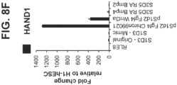
- the mesenchyme tissue is derived from the mesoderm, and is marked by the genes heart and neural crest derivatives expressed 1 (HAND1), and forkhead box F1 (FOXF1), among others.
- HAND1 heart and neural crest derivatives expressed 1
- FOXF1 forkhead box F1
- the endoderm is partitioned into anterior-posterior domains that can be recognized by the expression of a panel of factors that uniquely mark anterior foregut, posterior foregut, midgut, and hindgut regions of the endoderm.
- TFs specific transcription factors
- FOXA2 marks the entire endoderm along the anterior-posterior axis.
- the anterior foregut is marked broadly by the high expression of SOX2, and encompasses organ domains such as the thyroid, lung, and esophagus.
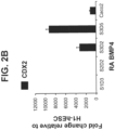
- the midgut (includes the duodenum, ileum, jejunum) and hindgut (includes the colon) are marked by high expression of caudal type homeobox 2 (CDX2).
- CDX2 caudal type homeobox 2
- the SOX2-CDX2 boundary occurs within the posterior foregut, within which additional TFs mark specific organ domains.
- the regionalized pancreas domain within the posterior foregut shows a very high expression of PDX1 and very low expression of CDX2 and SOX2.
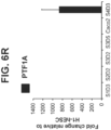
- PTF1A is highly expressed in pancreatic tissue.
- Low PDX1 expression, together with high CDX2 expression marks the duodenum domain.
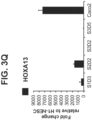
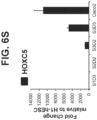
- the intestinal endoderm is patterned by specific homeobox (HOX) genes. For example, HOXC5 is preferentially expressed in midgut endoderm cells.
- HOXA13 and HOXD13 are restricted to hindgut endoderm cells.
- Strides have been made in improving protocols to generate intestinal endoderm cells from human pluripotent stem cells.
- the following publications ( Spence et al., Nature, 2011; 470(7332):105-109 ; Watson et al., Nature Medicine, 2014; 20(11):1310-1314 ; and Kauffman et al., Front Pharmacol, 2013; 4(79):1-18 ) outline differentiation protocols using either fibroblast growth factor (FGF)-4, Wingless-type MMTV integration site family, member 3A (WNT3A), Chiron 99021, or retinoic acid (RA) and FGF7 starting at the definitive endoderm stage, that generate mid-/hindgut spheroids, containing not only a CDX2 + /FOXA2 + endodermal population, but also a significant mesenchymal CDX2 + cell population.
- FGF fibroblast growth factor
- WNT3A Wingless-type MMTV integration site family
- Chiron 99021 Chiron
- the invention provides cells, cell populations and methods of generating the cells by differentiating human pluripotent stem cells.
- the invention features methods of directed differentiation of human pluripotent stem cells, to generate intestinal midgut endoderm cells, more particularly an endodermal monolayer of intestinal midgut endoderm cells.
- One aspect of the invention is a method of producing a population of intestinal midgut endoderm cells comprising culturing human pluripotent stem cells in culture media.
- the method comprises inducing differentiation of human pluripotent stem cells to intestinal midgut endoderm cells.
- a population of intestinal midgut endoderm cells is produced.
- a population of substantially intestinal midgut endoderm cells is produced.
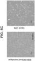
- the intestinal midgut endoderm cells form and are stable as a monolayer in culture.
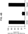
- greater than 50% of the differentiated cells express markers characteristic of intestinal midgut endoderm, preferably greater than 60% of the differentiated cells express markers characteristic of intestinal midgut endoderm, more preferably greater than 70%, greater than 80%, and greater than 90% express markers characteristic of intestinal midgut endoderm.
- differentiated cells express markers characteristic of intestinal midgut endoderm are intestinal midgut endoderm cells.
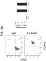
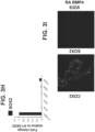
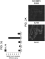
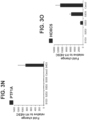
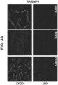
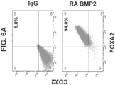
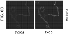
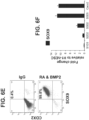
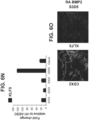
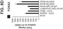
- the intestinal midgut endoderm cells express CDX2 and FOXA2.
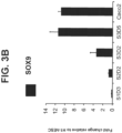
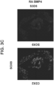
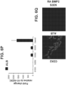
- the intestinal midgut endoderm cells express transcription factors selected from SOX9, PDX1, KLF5 and HOXC5.
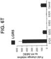
- the intestinal midgut endoderm cells do not express transcription factors selected from SOX2, ALB, PTF1A, HOXA13 and LGR5.
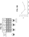
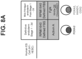
- human pluripotent stem cells are differentiated to intestinal midgut endoderm cells by steps including: a) culturing the human pluripotent stem cells in a first culture media containing GDF-8 and a GSK30 inhibitor, such as MCX compound, to induce differentiation into definitive endoderm cells; b) culturing the definitive endoderm cells in a second culture media containing ascorbic acid and FGF7 to induce differentiation into primitive gut tube cells; and c) culturing the primitive gut tube cells in a third culture media containing retinoic acid and BMP2 or BMP4 in acidic conditions to induce differentiation into intestinal midgut endoderm cells.
- acidic conditions is culture with BLAR medium.
- the pH of the acidic culture can range from 6.8 to 7.2.
- the intestinal midgut endoderm cells form a monolayer in culture. In embodiments, the monolayer of intestinal midgut endoderm cells is maintained in culture.
- Another embodiment of the invention is a method of treating a patient suffering from or at risk of developing diabetes comprising differentiating human pluripotent stem cells to intestinal midgut endoderm cells, and administering the differentiated intestinal midgut endoderm cells in a patient with diabetes.
- diabetes is Type 1 or Type 2.
- administering the cells may be via implantation, injection or otherwise administration directly or indirectly to the site of treatment.
- the intestinal midgut endoderm cells are implanted in the body, such as in subcutaneous space, omentum, liver, kidney, etc. Further embodiments encompass encapsulated delivery of the cells including encapsulation of macro- or micro-encapsulation devices.
- a further embodiments of the invention is a method of producing intestinal midgut endoderm cells comprising inducing differentiation of definitive endoderm cells in culture to primitive gut tube cells.
- the definitive endoderm cells are cultured in culture media containing ascorbic acid and FGF7.
- the primitive gut tube cells are cultured in culture media containing retinoic acid and BMP2 or BMP4.
- the primitive gut tube cells are differentiated to intestinal midgut endoderm cells.
- primitive gut tube cells are differentiated to intestinal midgut endoderm cells in acidic conditions (acidic culture media).
- acidic conditions is culture with BLAR medium.
- the pH of the acidic culture can range from 6.8 to 7.2.
- the intestinal midgut endoderm cells form and maintain a monolayer in culture.
- human pluripotent stem cells are human embryonic stem cells or induced pluripotent stem cells.
- the intestinal midgut endoderm cells express CDX2 and FOXA2.
- the intestinal midgut endoderm cells express transcription factors selected from SOX9, PDX1, KLF5 and HOXC5.
- the intestinal midgut endoderm cells do not express transcription factors selected from SOX2, ALB, PTF1A, HOXA13 and LGR5.
- the intestinal midgut endoderm cells express CDX2, FOXA2, SOX9, PDX1, KLF5 and HOXC5.
- the intestinal midgut endoderm cells do not express SOX2, ALB, PTF1A, HOXA13 and LGR5. In embodiments, greater than 50% of the differentiated cells express markers characteristic of intestinal midgut endoderm, preferably greater than 60% of the differentiated cells express markers characteristic of intestinal midgut endoderm, more preferably greater than 70%, greater than 80%, and greater than 90% express markers characteristic of intestinal midgut endoderm. In embodiments, differentiated cells express markers characteristic of intestinal midgut endoderm are intestinal midgut endoderm cells. In embodiments, the intestinal midgut endoderm cells express CDX2 and FOXA2. In embodiments, the intestinal midgut endoderm cells express transcription factors selected from SOX9, PDX1, KLF5 and HOXC5. In embodiments, the intestinal midgut endoderm cells do not express transcription factors selected from SOX2, ALB, PTF1A, HOXA13 and LGR5. In the embodiments, intestinal midgut endoderm cells do not express HAND1.
- the population of intestinal midgut endoderm cells is substantially intestinal midgut endoderm cells.
- the population of intestinal midgut endoderm cells comprises greater than 70% intestinal midgut endoderm cells, preferably greater than 80%, greater than 90%, and greater than 95% of intestinal midgut endoderm cells.
- the population of intestinal midgut endoderm cells comprises less than 20% mesenchymal cells, preferably less than 15%, more preferably less than 10%, less than 5%, less than 2%, less than 1%, less than 0.5%.
- intestinal midgut endoderm cells lack expression of HAND1.
- differentiation is induced in vitro.
- intestinal midgut endoderm cells differentiate further in vivo.
- Another embodiment relates to the intestinal midgut endoderm cells further differentiating into enteroendocrine cells in vivo.
- the enteroendocrine cells express or secrete incretin hormones.
- the incretin hormones are GLP1 and GIP.
- intestinal midgut endoderm cells serve as starting material for the identification of small molecules that promote at high efficiency the in vitro differentiation of intestinal midgut endoderm cells into, first, enteroendocrine precursors, and ultimately, incretin expressing or secreting enteroendocrine cells.
- the present invention pertains to the generation of intestinal midgut endoderm cells.
- the cells were generated using a specific culturing sequence.
- the present invention provides an in vitro cell culture for differentiating cells derived from pluripotent stem cells into cells expressing markers characteristic of the intestinal midgut endoderm cell lineage, such as expression of CDX2 and FOXA2.
- the invention further provides a method for obtaining and maintaining such cells in a monolayer via an in vitro cell culture.
- the invention is based on the discovery that the inclusion of retinoic acid and BMP4 or BMP2 or analogues thereof, act to induce CDX2 and maintain FOXA2 protein expression in differentiating cells to facilitate differentiation towards intestinal midgut endoderm cells.
- CDX2 is not expressed at the protein level at definitive endoderm (Stage 1) or primitive gut tube (Stage 2). Accordingly, the present invention provides methods of differentiating pluripotent stem cells to generate intestinal midgut endoderm cells that express CDX2 and FOXA2.
- Stem cells are undifferentiated cells defined by the ability of a single cell both to self-renew, and to differentiate to produce progeny cells, including self-renewing progenitors, non-renewing progenitors, and terminally differentiated cells. Stem cells are also characterized by their ability to differentiate in vitro into functional cells of various cell lineages from multiple germ layers (endoderm, mesoderm and ectoderm), as well as to give rise to tissues of multiple germ layers following transplantation, and to contribute substantially to most, if not all, tissues following injection into blastocysts.
- Stem cells are classified according to their developmental potential as: (1) totipotent; (2) pluripotent; (3) multipotent; (4) oligopotent; and (5) unipotent.
- Totipotent cells are able to give rise to all embryonic and extraembryonic cell types.
- Pluripotent cells are able to give rise to all embryonic cell types.
- Multipotent cells include those able to give rise to a subset of cell lineages, but all within a particular tissue, organ, or physiological system (for example, hematopoietic stem cells (HSC) can produce progeny that include HSC (self-renewal), blood cell-restricted oligopotent progenitors, and all cell types and elements (e.g., platelets) that are normal components of the blood).
- HSC hematopoietic stem cells
- Cells that are oligopotent can give rise to a more restricted subset of cell lineages than multipotent stem cells; and cells that are unipotent are able to give rise to a single cell lineage (e.g., spermatogenic stem cells).
- Stem cells are also categorized on the basis of the source from which they may be obtained.
- An adult stem cell is generally a multipotent undifferentiated cell found in tissue comprising multiple differentiated cell types. The adult stem cell can renew itself. Under normal circumstances, it can also differentiate to yield the specialized cell types of the tissue from which it originated, and possibly other tissue types.
- Induced pluripotent stem cells iPS cells
- iPS cells are adult cells that are converted into pluripotent stem cells.
- An embryonic stem cell is a pluripotent cell from the inner cell mass of a blastocyst-stage embryo.
- a fetal stem cell is one that originates from fetal tissues or membranes.
- Embryonic tissue is typically defined as tissue originating from the embryo (which in humans refers to the period from fertilization to about six weeks of development). Fetal tissue refers to tissue originating from the fetus, which in humans refers to the period from about six weeks of development to parturition. Extraembryonic tissue is tissue associated with, but not originating from, the embryo or fetus. Extraembryonic tissues include extraembryonic membranes (chorion, amnion, yolk sac and allantois), umbilical cord and placenta (which itself forms from the chorion and the maternal decidua basalis).
- Differentiation is the process by which an unspecialized ("uncommitted") or less specialized cell acquires the features of a specialized cell, such as an intestinal cell or pancreatic cell, for example.
- a differentiated cell is one that has taken on a more specialized ("committed") position within the lineage of a cell.
- the term committed, when applied to the process of differentiation, refers to a cell that has proceeded in the differentiation pathway to a point where, under normal circumstances, it will continue to differentiate into a specific cell type or subset of cell types, and cannot, under normal circumstances, differentiate into a different cell type or revert to a less differentiated cell type.
- De-differentiation refers to the process by which a cell reverts to a less specialized (or committed) position within the lineage of a cell.
- the lineage of a cell defines the heredity of the cell, i.e. which cells it came from and what cells it can give rise to.
- the lineage of a cell places the cell within a hereditary scheme of development and differentiation.
- a progenitor cell is a cell that has the capacity to create progeny that are more differentiated than itself, and yet retains the capacity to replenish the pool of progenitors.
- stem cells themselves are also progenitor cells, as are the more immediate precursors to terminally differentiated cells.
- a progenitor cell is often defined as a cell that is intermediate in the differentiation pathway, i.e., it arises from a stem cell and is intermediate in the production of a mature cell type or subset of cell types. This type of progenitor cell is generally not able to self-renew. Accordingly, if this type of cell is referred to herein, it will be referred to as a non-renewing progenitor cell or as an intermediate progenitor or precursor cell.
- Markers are nucleic acid or polypeptide molecules that are differentially expressed in a cell of interest.
- differential expression means an increased level for a positive marker and a decreased level for a negative marker as compared to an undifferentiated cell.
- the detectable level of the marker nucleic acid or polypeptide is sufficiently higher or lower in the cells of interest compared to other cells, such that the cell of interest can be identified and distinguished from other cells using any of a variety of methods known in the art.
- a cell is "positive for” a specific marker or “positive” when the specific marker is sufficiently detected in the cell.
- the cell is “negative for” a specific marker, or “negative” when the specific marker is not sufficiently detected in the cell.
- positive by fluorescence-activated flow cytometry (FACS) is usually greater than 2%, whereas the negative threshold by FACS is usually less than 1%.
- RT-PCR real-time PCR
- TLDA Taqman ® Low Density Array
- Stage 1 refers to the first step in the differentiation process, the differentiation of pluripotent stem cells into cells expressing markers characteristic of definitive endoderm cells (hereinafter referred to alternatively as “Stage 1 cells”).
- Stage 2 refers to the second step, the differentiation of cells expressing markers characteristic of definitive endoderm cells into cells expressing markers characteristic of primitive gut tube cells (hereinafter referred to alternatively as “Stage 2 cells”).
- Stage 3 refers to the third step, the differentiation of cells expressing markers characteristic of gut tube cells into cells expressing markers characteristic of intestinal midgut endoderm cells (hereinafter referred to alternatively as “Stage 3 cells”).
- Definitive endoderm cells refers to cells which bear the characteristics of cells arising from the epiblast during gastrulation and which form the gastrointestinal tract and its derivatives. Definitive endoderm cells express at least one of the following markers: FOXA2 (also known as hepatocyte nuclear factor 3- ⁇ (“HNF3 ⁇ ”)), GATA4, SOX17, CXCR4, Brachyury, Cerberus, OTX2, goosecoid, C-Kit, CD99, and MIXL1. Markers characteristic of the definitive endoderm cells include CXCR4, FOXA2 and SOX17. Thus, definitive endoderm cells may be characterized by their expression of CXCR4, FOXA2 and SOX17. In addition, depending on the length of time cells are allowed to remain in Stage 1, an increase in HNF4 ⁇ may be observed.
- FOXA2 also known as hepatocyte nuclear factor 3- ⁇
- GATA4 also known as hepatocyte nuclear factor 3- ⁇
- SOX17 also known as hepatocyte nuclear factor 3- ⁇
- Primary gut tube cells refers to endoderm cells derived from definitive endoderm that can give rise to all endodermal organs, such as lungs, liver, pancreas, stomach, and intestine. Primitive gut tube cells may be characterized by their substantially increased expression of HNF4 ⁇ over that expressed by definitive endoderm cells.
- Formgut endoderm cells refers to endoderm cells that give rise to the esophagus, lungs, stomach, liver, pancreas, gall bladder, and the most anterior portion of the duodenum. Foregut endoderm cells may be characterized by their expression of SOX2, PDX1, ALB, SOX17 and FOXA2, among others.
- Intestinal midgut endoderm cell refers to endoderm cells that give rise to small intestine. Intestinal midgut endoderm cells may be characterized by their expression of CDX2, FOXA2, and low expression of PDX1 (PDX1 LO ). The expression of certain HOX genes can distinguish between midgut and hindgut endoderm. For example, HOXC5 is preferentially expressed in midgut endoderm cells.
- Hindgut endodermal cell refers to endoderm cells that give rise to large intestine. Hindgut endoderm cells may be characterized by their expression of CDX2, FOXA2, HOXA13 and HOXD13.
- Mesenchyme cell refers to mesoderm cells that give rise to connective tissues, such as bones, cartilage, lymphatic, and circulatory systems. Expression of HAND1 and FOXF1 define mesenchyme cells.
- patient or “subject” or “host” refers to animals, including mammals, preferably humans, who are treated with compositions or pharmaceutical compositions, or in accordance with the methods described herein.
- an agent or compound including but not limited to a growth factor which is sufficient to promote and differentiate human pluripotent stem cells to a differentiated cell population, for example, to a definitive endoderm, foregut endoderm, intestinal midgut endoderm, hindgut endoderm, pancreatice endoderm and the like.
- administering and “administration” are used interchangeably herein and mean the cells may be implanted, injected, transplanted or otherwise administered directly or indirectly to the site of treatment.
- implantation is a suitable means of delivery, particularly surgical implantation into a precise location in the body, such as into subcutaneous space, omentum, liver, kidney (kidney capsule).
- Liquid or fluid pharmaceutical compositions may be administered to a more general location.
- Pluripotent stem cells have the potential to differentiate into cells of all three germinal layers: endoderm, mesoderm, and ectoderm tissues.
- Exemplary types of pluripotent stem cells that may be used include established lines of pluripotent cells, including pre-embryonic tissue (such as, for example, a blastocyst), embryonic tissue, or fetal tissue taken any time during gestation, typically but not necessarily, before approximately 10 to 12 weeks gestation.
- pre-embryonic tissue such as, for example, a blastocyst
- embryonic tissue such as, for example, a blastocyst
- fetal tissue fetal tissue taken any time during gestation, typically but not necessarily, before approximately 10 to 12 weeks gestation.
- Non-limiting examples are established lines of human embryonic stem cells or human embryonic germ cells, such as, for example the human embryonic stem cell lines H1, H7, and H9 (WiCell Research Institute, Madison, WI, USA).
- iPS reprogrammed pluripotent cells
- derived from adult somatic cells using forced expression of a number of pluripotent related transcription factors such as OCT4, NANOG, SOX2, KLF4, and ZFP42 ( Annu Rev Genomics Hum Genet 2011, 12:165-185 ; see also iPS, Cell, 126(4): 663-676 ) may also be used.
- the human embryonic stem cells used in the methods of the invention may also be prepared as described by Thomson et al. (U.S. Patent No.
- pluripotent stem cells suitable for use in the present invention may be derived according to the methods described in: Li et al. (Cell Stem Cell 4: 16-19, 2009 ); Maherali et al.
- the pluripotent stem cells may be of non-embryonic origins. All of these references, patents, and patent applications are herein incorporated by reference in their entirety, in particular, as they pertain to the isolation, culture, expansion and differentiation of pluripotent cells.
- Pluripotent stem cells differentiate through various stages each of which may be characterized by the presence or absence of particular markers. Differentiation of the cells into these stages is achieved by the specific culturing conditions, including the presence or lack of certain factors added to the culture media. In general, this differentiation may involve differentiation of pluripotent stem cells into definitive endoderm cells, referred to herein as Stage 1. These definitive endoderm cells may then be further differentiated into primitive gut tube cells, referred to herein as Stage 2. Primitive gut tube cells in turn may then be differentiated into intestinal midgut endoderm cells, referred to herein as Stage 3.
- pluripotent stem cells include the human embryonic stem cell line H1 (NIH code: WA01), the human embryonic stem cell line H9 (NIH code: WA09), the human embryonic stem cell line H7 (NIH code: WA07), and the human embryonic stem cell line SA002 (Cellartis, Sweden). Also suitable are cells that express at least one of the following markers characteristic of pluripotent cells: ABCG2, Cripto, CD9, FOXD3, CONNEXIN43, CONNEXIN45, OCT4, SOX2, NANOG, hTERT, UTF1, ZFP42, SSEA-3, SSEA-4, TRA-1-60, and TRA-1-81.
- a cell that expresses at least one of the markers characteristic of the definitive endoderm lineage is a primitive streak precursor cell.
- a cell expressing markers characteristic of the definitive endoderm lineage is a mesendoderm cell.
- a cell expressing markers characteristic of the definitive endoderm lineage is a definitive endoderm cell.
- Methods for assessing expression of protein and nucleic acid markers in cultured or isolated cells are standard in the art. These methods include RT-PCR, Northern blots, in situ hybridization (see, e.g., Current Protocols in Molecular Biology (Ausubel et al., eds. 2001 supplement)), and immunoassays (such as immunohistochemical analysis of sectioned material), Western blotting, and for markers that are accessible in intact cells, FACS ( see, e.g., Harlow and Lane, Using Antibodies: A Laboratory Manual, New York: Cold Spring Harbor Laboratory Press (1998 )).
- the efficiency of differentiation may be determined by exposing a treated cell population to an agent (such as an antibody) that specifically recognizes a protein marker expressed by cells expressing markers characteristic of the cell type of interest.
- Pluripotent stem cells may be differentiated into cells expressing markers characteristic of definitive endoderm cells by any suitable method known in the art, or by any method proposed in this invention.
- pluripotent stem cells are treated with a medium, such as MCDB-131 medium (Life Technologies, Carlsbad, California) supplemented with factors including GDF8 and a GSK30 inhibitor (such as the cyclic aniline-pyridinotriazine compounds disclosed in U.S. Patent App. Pub. No. 2010/0015711 ; incorporated herein by reference in its entirety) to induce differentiation into cells expressing markers characteristic of definitive endoderm cells.
- a medium such as MCDB-131 medium (Life Technologies, Carlsbad, California) supplemented with factors including GDF8 and a GSK30 inhibitor (such as the cyclic aniline-pyridinotriazine compounds disclosed in U.S. Patent App. Pub. No. 2010/0015711 ; incorporated herein by reference in its entirety
- GSK30 inhibitors such as staurosporine
- GSK30 inhibitors such as staurosporine
- the preferred GSK30 inhibitor 14-Prop-2-en-1-yl-3,5,7,14,17,23,27-heptaazatetracyclo [19.3.1.1-2,6-.1-8,12-]heptacosa-1(25),2(27),3,5,8(26),9,11,21,23-nonaen-16-one, referred to herein as "MCX Compound”.
- Other components of the medium may include: Sodium bicarbonate at about 2.7g/1000ml to 3.6g/1000ml, preferably 2.7g/1000ml; FAF-BSA at about 0.1% to 2.0%, preferably about 0.5%; GlutaMAX TM (Life Technologies Corporation, Carlsbad, California) at 1:100 dilution ("1X concentration"); and D-Glucose at a concentration range of about 2 mM to 20 mM, preferably 4.5mM to obtain a concentration of 10mM D-Glucose.
- the pluripotent cells may be cultured for approximately two to five days, preferably about three to four days, to facilitate their differentiation into definitive endoderm cells.
- the pluripotent cells are cultured in the presence of an effective amount of TGF ⁇ signaling molecule and/or GSK30 inhibitor, for example, an effective amount of GDF8 and MCX compound for one day, followed by culturing in the presence of GDF8 and a lower concentration of MCX compound for one day, followed by culturing in the presence of GDF8 for one day in the absence of the MCX compound.
- the cells may be cultured in the presence of GDF8 and about 1.5 ⁇ M of MCX compound for one day, followed by culturing in the presence of GDF8 and about 0.1 ⁇ M MCX compound for one day, followed by culturing in the presence of GDF8 for one day in the absence of the MCX compound.
- the cells may be cultured in the presence of GDF8 and about 1.5 ⁇ M of MCX compound for one day, followed by culturing in the presence of GDF8 and about 0.1 ⁇ M MCX compound for one day.
- Generation of cells expressing markers characteristic of definitive endoderm cells may be determined by testing for the presence of the markers before and after following a particular protocol. Pluripotent stem cells typically do not express such markers. Thus, differentiation of pluripotent cells can be detected when the cells begin to express markers characteristic of definitive endoderm cells, such as CXCR4, FOXA2 and SOX17. In the embodiments, cells expressing markers characteristic of definitive endoderm cells are definitive endoderm cells.
- the cells expressing markers characteristic of definitive endoderm cells may be further differentiated into cells expressing markers characteristic of primitive gut tube cells.
- the formation of cells expressing markers characteristic of primitive gut tube cells includes culturing cells expressing markers characteristic of definitive endoderm cells with a medium, such as MCDB-131, containing FGF7 to differentiate these cells.
- the culture medium may include from about 10 ng/ml to 100 ng/ml, preferably about 25 ng/ml to about 75 ng/ml, alternatively from about 30 ng/ml to about 60 ng/ml, alternatively about 50 ng/ml of FGF7.
- the cells may be cultured under these conditions for about two to three days, preferably about two days.
- differentiation into cells expressing markers characteristic of primitive gut tube cells includes culturing cells expressing markers characteristic of definitive endoderm cells with FGF7 and ascorbic acid (vitamin C).
- the culture medium such as MCDB-131, may include from about 0.1 mM to about 1.0 mM ascorbic acid, preferably about 0.1 mM to about 1.0 mM, alternatively from about 0.2 mM to about 0.4 mM, alternatively about 0.25 mM of ascorbic acid.
- the culture medium may also include from about 10 ng/ml to 100 ng/ml, preferably about 10 ng/ml to about 50 ng/ml, alternatively from about 15 ng/ml to about 30 ng/ml, alternatively about 50 ng/ml or about 25 ng/ml of FGF7.
- the culture medium may include about 0.25 mM ascorbic acid and about 50 ng/ml FGF7.
- Other components of the medium may include: sodium bicarbonate at about 2.7g/1000ml to 3.6g/1000ml, preferably 2.7g/1000ml; FAF-BSA at about 0.1% to 2.0%, preferably about 0.5%; GlutaMAX TM at 1:100 dilution ("1X concentration"); and D-Glucose at a concentration range of about 2 mM to 20 mM, preferably 4.5mM to obtain a concentration of 10mM D-Glucose.
- cells expressing markers characteristic of definitive endoderm cells are treated for 2 days with FGF7 and ascorbic acid.
- Differentiation of definitive endoderm cells can be detected when the cells begin to express markers characteristic of primitive gut tube cells, such as expression of FOXA2 and increased expression of HNF4 ⁇ .
- markers characteristic of primitive gut tube cells such as expression of FOXA2 and increased expression of HNF4 ⁇ .
- cells expressing markers characteristic of primitive gut tube cells are primitive gut tube cells.
- Cells expressing markers characteristic of primitive gut tube cells may be further differentiated into cells expressing markers characteristic of intestinal midgut endoderm cells.
- primitive gut tube cells are further differentiated into intestinal midgut endoderm cells by culturing the primitive gut tube cells in a culture medium, such as BLAR medium (Life Technologies, Corporation, Carlsbad, California), supplemented with retinoic acid and a BMP4 or BMP2.
- BLAR medium Life Technologies, Corporation, Carlsbad, California
- the medium is supplemented with from about 0.5 ⁇ M to about 5 ⁇ M of retinoic acid, preferably about 1 ⁇ M, and from about 10 ng/ml to about 100 ng/ml BMP4 or BMP2, preferably about 50 ng/ml of BMP4 or BMP2.
- Other supplements to the medium may include: FAF-BSA at about 0.1% to 2.0%, preferably about 0.5%; GlutaMAX TM ; and D-Glucose at a concentration range of about 2 mM to 20 mM, preferably 4.5mM to obtain a concentration of 10mM D-Glucose.
- cells expressing markers characteristic of primitive gut cells are treated for 3 to 5 days, preferably for 5 days with BMP4 or BMP2 and retinoic acid.
- the pH of the culture can range from 6.8 to 7.2 during the 5-day Stage 3 conditioning period (compared to normal pH at S2D2 being 7.3 or more).
- the invention relates to a method of producing a population of intestinal midgut endoderm cells by culturing human pluripotent stem cells in selected culture media for generating intestinal midgut endoderm cells.
- the method induces differentiation of human pluripotent stem cell to intestinal midgut endoderm cells in a staged process.
- a population of intestinal midgut endoderm cells is produced.
- a population of substantially intestinal midgut endoderm cells is produced.
- the intestinal midgut endoderm cells form and maintain a monolayer on planar culture.
- the intestinal midgut endoderm cells are stable as a monolayer in culture. Cells stable as a monolayer, or remain stable as a monolayer, herein refers to a monolayer of cells that do not form spheroids in culture.
- greater than 50% of the differentiated cells express markers characteristic of intestinal midgut endoderm. In embodiments, greater than 60%, greater than 70%, greater than 80%, greater than 90% or greater than 95% of the differentiated cells express markers characteristic of intestinal midgut endoderm. In embodiments, differentiated cells express markers characteristic of intestinal midgut endoderm are intestinal midgut endoderm cells. In embodiments, the intestinal midgut endoderm cells express CDX2 and FOXA2 as determined by FACS analysis and qPCR. In some embodiments, intestinal midgut endoderm cells express transcription factors selected from SOX9, PDX1, KLF5 and HOXC5 as determined by IF analysis and qPCR. In embodiments, the intestinal midgut endoderm cells do not express transcription factors selected from SOX2, ALB, PTF1A as determined by IF analysis and qPCR, and HOXA13 and LGR5 as determined by qPCR.
- the pH of the acidic culture can range from 6.8 to 7.2 during the 5-day differentiation conditioning period from primitive gut tube cells to intestinal midgut endoderm cells (compared to normal pH at S2D2 being 7.3 or more).
- the intestinal midgut endoderm cells form and maintain a monolayer in culture.
- human pluripotent stem cells are human hESC or iPS cells.
- the intestinal midgut endoderm cells express CDX2 and FOXA2 as determined by FACS analysis and qPCR.
- the intestinal midgut endoderm cells express transcription factors selected from SOX9, PDX1, KLF5 and HOXC5 as determined by IF analysis and qPCR.
- the intestinal midgut endoderm cells do not express transcription factors selected from SOX2, ALB, PTF1A as determined by IF analysis and qPCR, and HOXA13 and LGR5 as determined by qPCR.
- the intestinal midgut endoderm cells express CDX2, FOXA2, SOX9, PDX1, KLF5 and HOXC5 by IF analysis and qPCR.
- the intestinal midgut endoderm cells do not express SOX2, ALB and PTF1A as determined by IF analysis and qPCR, and HOXA13 and LGR5 as determined by qPCR.
- the population of intestinal midgut endoderm cells is substantially intestinal midgut endoderm cells.
- the population of intestinal midgut endoderm cells comprises greater than 70% intestinal midgut endoderm cells, preferably greater than 80%, greater than 90%, and greater than 95% of intestinal midgut endoderm cells.
- the population of intestinal midgut endoderm cells comprises less than 20% mesenchymal cells, preferably less than 15%, more preferably less than 10%, less than 5%, less than 2%, less than 1%, less than 0.5%.
- intestinal midgut endoderm cells lack expression of HAND1.
- intestinal midgut endoderm cells are differentiated in vitro.
- intestinal midgut endoderm cells further differentiate and mature in vivo.
- Another embodiment relates to the intestinal midgut endoderm cells further differentiating into enteroendocrine cells in vivo or in a mixture with enteroendocrine cells in vivo.
- enteroendocrine cells express or secrete incretin hormones.
- the enteroendocrine cell-secreted incretin hormones include GLP1 and GIP.
- intestinal midgut endoderm cells serve as starting material for the identification of small molecules that promote at high efficiency the in vitro differentiation of intestinal midgut endoderm cell type into, first, enteroendocrine precursors, and then to incretin expressing or secreting enteroendocrine cells.
- Cells and cell populations and mixtures such as those described herein may be micro- or macro-encapsulated and subsequently transplanted into a mammalian host. Encapsulated cells or cells alone may be transplanted (administered) subcutaneously or anywhere else in the body whereby the cells may be vascularized and differentiate and mature in vivo.
- Intestinal midgut endoderm refers to a corresponding in vivo or in situ cell type which is CDX2-positive and FOXA2-positive endoderm cells present at about embryonic day 8.5 (“E8.5”) during mouse development, or at about the 3-4 week time point during human embryonic development.
- H1-hESC human embryonic stem cell line H1
- EZ8 media Cat#A1516901 Gibco, Thermo Fisher Scientific
- GlutaMAX TM 1:100 dilution (“1X concentration") (Life Technologies Corporation, Carlsbad, California, Catalog No. 35050-079) Ascorbic acid 0.25mM (Sigma Aldrich Co. LLC, St. Louis, Missouri, Catalog No. A4544)
- FGF2 100ng/ml (R & D Systems Inc., Minneapolis, Minnesota, Catalog No. 233-FB-025) Transforming Growth Factor beta (“TGF ⁇ ") 1 ng/ml (R & D Systems Inc., Minneapolis, Minnesota, Catalog No. 240-B-002) insulin-transferrin-selenium-ethanolamine (“ITS-X”) 1:200 dilution (Life Technologies, Carlsbad, California, Catalog No.
- IGF-1 Insulin-like Growth Factor-1
- Y-compound Rock Inhibitor Y-27632
- the cultures were differentiated using the following protocol. During Stages 1 through 3 of the protocol, cultures were maintained on planar adherent cultures. Others, however, have described differentiation using suspension culture including US US2014/0242693 , which is incorporated by reference in its entirety; the protocol described herein can be modified and performed in suspension, which provides for scalability of manufacturing.
- the following nomenclature, S#D# specifies exact time during Stages 1 through 3. For example, S1D3 is stage 1 day 3. Briefly, each stage defines differentiation towards definitive endoderm (stage 1), primitive gut tube (stage 2), and intestinal midgut endoderm (stage 3). a.
- Stage 1 (3 days): Cells were cultured for one day in the following Stage 1 media: MCDB-131 medium (Life Technologies, Carlsbad, California, Catalog No. ME120219L2) Sodium bicarbonate 2.7 g/1000 ml (Sigma-Aldrich Co. LLC, St. Louis, Missouri, Catalog No. 5761) FAF-BSA 0.5% GlutaMAX TM 1:100 dilution ("1X concentration") D-Glucose 4.5mM to obtain a concentration of 10mM D-Glucose (Sigma-Aldrich Co. LLC, St. Louis, Missouri, Catalog No. G8769) GDF8 100 ng/ml (Peprotech, Rocky Hill, New Jersey, Catalog No.
- Stage 2 Cells were treated for two days with the following medium: MCDB-131 medium Sodium bicarbonate 2.7g/1000ml FAF-BSA 0.5% GlutaMAX TM 1X concentration D-Glucose 4.5mM to obtain a concentration of 10mM D-Glucose Ascorbic Acid 0.25 mM FGF7 50 ng/ml (R&D Systems, Inc., Minneapolis, Minnesota, Catalog No. 251-K c.
- Stage 3 Cells were treated for five day with BLAR 001 custom medium: BLAR medium (custom manufactured by Life Technologies, Corporation, Carlsbad, California, Catalog No.
- ME120123L2 components listed on Table I) FAF-BSA 0.5% GlutaMAX TM 1X concentration D-Glucose 4.5mM to obtain a concentration of 10mM D-Glucose Retinoic Acid 1 ⁇ M (Sigma Aldrich, St. Louis, Missouri, Catalog No. R2625) BMP4 compound (R&D Systems, Inc., Minneapolis, Minnesota, Catalog No. 314-BP 50 ng/ml OR BMP2 compound (R&D Systems, Inc., Minneapolis, Minnesota, Catalog No. 355-BM),
- Intracellular antibody staining was accomplished by utilizing the LIVE/DEAD Violet Fluorescent reactive dye (Life Technologies, Carlsbad, California, Catalog No. L34955) at 4°C for 30 minutes followed by a single wash in cold PBS. Fixing of cells was in 300 ⁇ l of Cytofix/Cytoperm Buffer (BD Biosciences, San Jose, California, Catalog No. 554723) followed by two washes in Perm/Wash Buffer (BD Biosciences, San Jose, California, Catalog No. 554722).
- FIG. 1A A summary of a differentiation method, including the important medium components, growth factors and small molecules that were added to each stage, and key stage-specific markers of the differentiating intestinal midgut endoderm cells (FOXA2; CDX2; KI,FS; SOX9; PDX1 LO ) is depicted in Figure 1A .
- S2D2 7.35 ⁇ 0.04
- FIG. 1B As compared to the neutral pH noted for S2D2 (7.35 ⁇ 0.04), cells were exposed to slightly acidic conditions in the BLAR medium, during the entirety of stage 3 (pH; S3D1, 6.98 ⁇ 0.05; S3D2, 7.02 ⁇ 0.04; S3D5, 7.18 ⁇ 0.03) ( Figure 1B ), and as a result of lower sodium bicarbonate levels in BLAR medium.
- FIG. 1C depicts representative phase-contrast images of a S3D5 monolayer ( left ) , and human epithelial colon adenocarcinoma cell line ("Caco-2") ( right ) , which was used as a benchmark for characterization of differentiation. A uniform morphology at S3D5 was observed. Characterization of cell number using the Nucleocounter ® NC-100 (Chemometec, Alleroed, Denmark , Catalog No. 900-004) shows that one hESC differentiated into 4.56 ⁇ 2.60 S3D5 hindgut endoderm cells ( Figure 1D ).
- the differentiation method utilizing BMP4, efficiently generates and maintains intestinal midgut endoderm cells in monolayer, each comprising both CDX2 and FOXA2 on transcript and protein levels.
- Figure 2A bottom ) shows that 90.0 ⁇ 5.85 percent of S3D5 cells were co-present for both CDX2 and FOXA2 protein, similar to percentage observed in Caco-2 cells (86.0 ⁇ 6.67).
- definitive endoderm (DE - S1D3) cells were devoid of CDX2 and FOXA2 co-presence (2.3 ⁇ 1.2).
- Gene expression analysis shows CDX2 induced ( Figure 2B ), and FOXA2 maintained ( Figure 2C ) during Stage 3.
- Figure 2D shows that the induction of CDX2 protein levels and CDX2/FOXA2 protein co-presence after the establishment of the FOXA2-positive primitive gut endoderm stage, S2D2 ( Figure 2D -i), progressively increased through S3D2 ( Figure 2D -ii), and at S3D5 ( Figure 2D -iii) reached similar levels as seen in Caco-2 cells ( Figure 2D -iv).
- CDX2 protein is depicted on the bottom row
- FOXA2 protein is depicted on the top row.
- S3D5 cells did not express the anterior endoderm TF SOX2, and only 1.45 ⁇ 0.15 of S3D5 cells exhibited SOX2 and CDX2 co-presence ( Figure 3G - bottom; 3I), and gene expression was below levels seen in hESC and Caco-2 cells ( Figure 3H ).
- Protein co-presence of KLF5 within CDX2-positive cells at S3D5 was observed ( Figure 3K ).
- ALB gene expression Figure 3L
- protein presence Figure 3M
- FIG. 3N shows that the gene expression of PTF1A, a pancreas lineage marker, was not induced in S3D5 cells, as compared to pancreas-biased S4D3 cells.
- Figure 3P shows that LGR5, a marker of embryonic intestinal endoderm beginning at mid-gestation in the mouse, was not induced in S3D5 cells ( Figure 3P ).
- FIG. 3Q shows that HOXA13, a marker of the intestinal hindgut endoderm, was not induced in S3D5 cells ( FIG. 3P ).
- Figures 4A - 4B illustrate the proliferative profile of differentiating S3D5 cells.
- Figure 4A depicts Caco-2 cells, where most of CDX2-protein positive cells were in active cell cycle (as indicated by the co-expression with KI67 protein) ( left ) , and the proliferative index of the H1-hESC-derived cells during Stage 3 that decreased over time (S3D2 - middle; S3D5 - right ) .
- CDX2 ( top row) and KI67 ( bottom row) protein levels are depicted as single channel images.
- BMP2 can be used as an alternative to BMP4, during Stage 3 in this method to produce a monolayer of intestinal midgut endoderm cells with CDX2 and FOXA2 protein co-presence.
- Figure 5A depicts a summary of a differentiation method, including the medium components, growth factors and small molecules that were added to each stage, and key stage-specific markers of the differentiating intestinal midgut endoderm cells (FOXA2, CDX2, KLF5, SOX9, and PDX1 LO ).
- FIG. 5C depicts representative phase-contrast images of a S3D5 monolayer ( left ) , and Caco-2 cells ( right ) . A uniform morphology at S3D5 was observed.
- the differentiation method generates and maintains proper intestinal midgut endoderm cells in monolayer, each comprising of CDX2, FOXA2, KLF5, SOX9, PDX1 LO and HOXC5 on transcript and protein levels. All TF protein levels are depicted as single channel IF images.
- Figure 6A bottom ) shows that 94 percent of S3D5 cells were co-present for both CDX2 and FOXA2 protein, similar to percentage seen in Caco-2 cells (86.0 ⁇ 6.67).
- Gene expression analysis shows that CDX2 was induced ( Figure 6B ), and FOXA2 maintained ( Figure 6C ) during Stage 3.
- Figure 6D shows that CDX2 protein levels and complete CDX2/FOXA2 protein co-presence were induced at S3D5 ( Figure 6D ), which is comparable to levels observed in Caco-2 cells ( Figure 2D -iv).
- Figure 6E bottom ) shows that 99.8 percent of cells were co-present for both CDX2 and SOX9 at S3D5. Strong induction of SOX9 gene expression was observed similar to levels in Caco-2 cells ( Figure 6F ), and protein presence as assessed by IF-analysis was observed ( Figure 6G ). 45.5 percent of cells were co-positive for both CDX2 and PDX1 ( Figure 6H - bottom ) .
- PDX1 gene expression was induced at low levels, when compared to pancreas-biased S4D3 cells ( Figure 6I ), and low to absent protein levels was reflected in the IF-analysis ( Figure 6J ).
- Anterior endoderm TF SOX2 was not observed in S3D5 cells, as 0.8 percent of S3D5 cells exhibited SOX2 and CDX2 co-presence ( Figure 6K - bottom; 6M), and gene expression was below levels seen in hESC and Caco-2 cells ( Figure 6L ).
- the gene expression of KI,FS an essential marker for demonstrating proper development of hindgut endoderm, was strongly upregulated at S3D5 ( Figure 6N ).
- FIG. 6O Protein co-presence of KLF5 within CDX2-positive cells at S3D5 was observed ( Figure 6O ).
- ALB gene expression ( Figure 6P ), and protein presence ( Figure 6Q ) was not observed in S3D5 cells.
- the gene expression of pancreas lineage allocating TF, PTF1A was not induced in S3D5 cells, as compared to pancreas-biased S4D3 cells ( Figure 6R ).
- the homeobox gene HOXC5 present in the embryonic intestinal midgut endoderm, was strongly induced in S3D5 cells ( Figure 6S ).
- Figure 6T demonstrates that LGR5, a marker of embryonic intestinal endoderm beginning at mid-gestation, was not induced in S3D5 cells.
- FIG. 6U demonstrates that HOXA13, a marker of the intestinal hindgut endoderm, was not induced in S3D5 cells ( FIG. 6U ).
- Figure 7 characterizes the proliferative profile of differentiating S3D5 cells, showing Caco-2 cells, where most of CDX2-protein positive cells were in active cell cycle (as indicated by the co-expression with KI67 protein) ( left ) , compared to the proliferative index of the H1-hESC-derived cells during Stage 3 that was lower (S3D5 - right ) .
- CDX2 ( top row) and KI67 (bottom row) protein levels are depicted as single channel images.
- This example demonstrates the endoderm-mesenchyme-mixed quality of the CDX2 + mid-/hindgut cells generated from intestinal culturing beginning at the definitive endoderm stage using FGF4 and WNT-agonists ( Spence et al., Nature, 2011; 470:105-109 ; Watson et al. Nature Med, 2014; 11:1310-1314 ).
- FGF4 and WNT-agonists Spence et al., Nature, 2011; 470:105-109 ; Watson et al. Nature Med, 2014; 11:1310-1314 .
- Example 2 the differentiation conditions outlined in this Example differ from Example 1 by the following: (i) intestinal condition starting point begins at the definitive endoderm stage; (ii) different growth factors and small molecules are used than RA and BMP4 or BMP2; and (iii) acidic culture conditions are not used.
- H1-hESC cells were cultured and maintained as described in Example 1.
- Stage 1-Mimic (3 days) Cells were cultured for one day in the following Stage 1 medium:
- RMPI 1640 medium (Thermo Fisher Scientific, Catalog No. 11875) Penicillin-Streptomycin 1X concentration (1:100 dilution from stock concentration) (Thermo Fisher Scientific, Catalog No. 15140122) L-Glutamine 2mM (Thermo Fisher Scientific, Catalog No. 25030081) Activin A 100 ng/ml (R&D Systems, Inc., Minneapolis, Minnesota, Catalog No. 338-AC)
- FBS Fetal Bovine Serum
- Post-Stage 1 (2 days): For example, pS1d1 is post-Stage 1 day 1 and pS1d2 is post-Stage 1 day 2.
- Cells were cultured for two days in the following Post-Stage 1 medium : RMPI 1640 medium Penicillin-Streptomycin 1X concentration (1:100 dilution from stock concentration) L-Glutamine 2mM FGF4 500 ng/ml (R&D Systems, Inc., Minneapolis, Minnesota, Catalog No. 235-F4) Defined Fetal Bovine Serum (FBS) 2.0% WNT3A 500 ng/ml (R&D Systems, Inc., Minneapolis, Minnesota, Catalog No. 5036-WN) OR Chiron99201 (Stemgent, Catalog No. 0400041) 3 ⁇ M
- FIG. 8A A summary of the differentiation methods, including medium components, growth factors and small molecules added to each stage, and signature or key stage-specific markers of the differentiating intestinal mid-/hindgut endoderm cells (HAND1) is depicted in FIG. 8A .
- Intestinal conditioning (post-Stage 1), started at the definitive endoderm stage, with 500 ng/ml FGF4, and either 3 ⁇ M Chiron99021 (Watson et al .), or 500 ng/ml Wnt3A (Spence et al .).
- the term "Stage 1-mimic” refers to the definitive endoderm differentiation protocol in this example that differs from the "S1D3-Original” that refers to Stage 1 conditioning described in Example 1.
- FIG. 1 A summary of the differentiation methods, including medium components, growth factors and small molecules added to each stage, and signature or key stage-specific markers of the differentiating intestinal mid-/hindgut endoderm cells (HAND1) is depicted in FIG. 8A
- FIG. 8B shows phase-contrast images of H1-hESC cells ( top row, left ) , post-Stage 1 cells conditioned two days with 500 ng/ml FGF4 and 3 ⁇ M Chiron99021 ( top row, middle ) , post-Stage 1 cells conditioned two days with 500 ng/ml FGF4 and 500 ng/ml Wnt3A ( top row, right ) , along with a S3D5 monolayer conditioned by RA/BMP4 ( bottom row, left ) , and a S3D5 monolayer conditioned by RA/BMP2 ( bottom row, right ) (from Example 1).
- the present invention relates to the following items:
Landscapes
- Health & Medical Sciences (AREA)
- Life Sciences & Earth Sciences (AREA)
- Engineering & Computer Science (AREA)
- Biomedical Technology (AREA)
- Cell Biology (AREA)
- Chemical & Material Sciences (AREA)
- Biotechnology (AREA)
- Zoology (AREA)
- General Health & Medical Sciences (AREA)
- Bioinformatics & Cheminformatics (AREA)
- Organic Chemistry (AREA)
- Wood Science & Technology (AREA)
- Genetics & Genomics (AREA)
- Developmental Biology & Embryology (AREA)
- Public Health (AREA)
- Pharmacology & Pharmacy (AREA)
- Animal Behavior & Ethology (AREA)
- Veterinary Medicine (AREA)
- Medicinal Chemistry (AREA)
- Epidemiology (AREA)
- Virology (AREA)
- Immunology (AREA)
- Microbiology (AREA)
- Biochemistry (AREA)
- General Engineering & Computer Science (AREA)
- Diabetes (AREA)
- Reproductive Health (AREA)
- Gastroenterology & Hepatology (AREA)
- Nutrition Science (AREA)
- Physiology (AREA)
- Gynecology & Obstetrics (AREA)
- Nuclear Medicine, Radiotherapy & Molecular Imaging (AREA)
- General Chemical & Material Sciences (AREA)
- Chemical Kinetics & Catalysis (AREA)
- Obesity (AREA)
- Hematology (AREA)
- Endocrinology (AREA)
- Emergency Medicine (AREA)
- Micro-Organisms Or Cultivation Processes Thereof (AREA)
- Medicines Containing Material From Animals Or Micro-Organisms (AREA)
Applications Claiming Priority (3)
| Application Number | Priority Date | Filing Date | Title |
|---|---|---|---|
| US201662322636P | 2016-04-14 | 2016-04-14 | |
| PCT/US2017/025847 WO2017180361A1 (en) | 2016-04-14 | 2017-04-04 | Differentiation of pluripotent stem cells to intestinal midgut endoderm cells |
| EP17782848.0A EP3443073B1 (de) | 2016-04-14 | 2017-04-04 | Differenzierung pluripotenter stammzellen in mitteldarmendodermzellen |
Related Parent Applications (1)
| Application Number | Title | Priority Date | Filing Date |
|---|---|---|---|
| EP17782848.0A Division EP3443073B1 (de) | 2016-04-14 | 2017-04-04 | Differenzierung pluripotenter stammzellen in mitteldarmendodermzellen |
Publications (1)
| Publication Number | Publication Date |
|---|---|
| EP4450076A2 true EP4450076A2 (de) | 2024-10-23 |
Family
ID=60039713
Family Applications (2)
| Application Number | Title | Priority Date | Filing Date |
|---|---|---|---|
| EP24179748.9A Withdrawn EP4450076A2 (de) | 2016-04-14 | 2017-04-04 | Differenzierung pluripotenter stammzellen zu intestinalen mitteldarmendodermzellen |
| EP17782848.0A Active EP3443073B1 (de) | 2016-04-14 | 2017-04-04 | Differenzierung pluripotenter stammzellen in mitteldarmendodermzellen |
Family Applications After (1)
| Application Number | Title | Priority Date | Filing Date |
|---|---|---|---|
| EP17782848.0A Active EP3443073B1 (de) | 2016-04-14 | 2017-04-04 | Differenzierung pluripotenter stammzellen in mitteldarmendodermzellen |
Country Status (17)
| Country | Link |
|---|---|
| US (2) | US10420803B2 (de) |
| EP (2) | EP4450076A2 (de) |
| JP (3) | JP6705911B2 (de) |
| KR (4) | KR102619151B1 (de) |
| CN (2) | CN115449506A (de) |
| AR (1) | AR108134A1 (de) |
| AU (1) | AU2017251651B2 (de) |
| BR (1) | BR112018070293A2 (de) |
| CA (1) | CA3020905A1 (de) |
| DK (1) | DK3443073T3 (de) |
| MA (1) | MA45479A (de) |
| MX (1) | MX2018012629A (de) |
| PH (1) | PH12018502061A1 (de) |
| RU (2) | RU2021100063A (de) |
| SG (1) | SG11201807915QA (de) |
| TW (1) | TW201803983A (de) |
| WO (1) | WO2017180361A1 (de) |
Families Citing this family (7)
| Publication number | Priority date | Publication date | Assignee | Title |
|---|---|---|---|---|
| EP4450076A2 (de) * | 2016-04-14 | 2024-10-23 | Janssen Biotech, Inc. | Differenzierung pluripotenter stammzellen zu intestinalen mitteldarmendodermzellen |
| WO2020243665A1 (en) | 2019-05-31 | 2020-12-03 | W. L. Gore & Associates, Inc. | A biocompatible membrane composite |
| AU2020283150B2 (en) | 2019-05-31 | 2023-08-17 | Viacyte, Inc. | Cell encapsulation devices with controlled oxygen diffusion distances |
| EP3976236A1 (de) | 2019-05-31 | 2022-04-06 | W.L. Gore & Associates Inc. | Biokompatibler membranverbundstoff |
| JP2022535239A (ja) | 2019-05-31 | 2022-08-05 | ダブリュ.エル.ゴア アンド アソシエイツ,インコーポレイティド | 生体適合性メンブレン複合体 |
| CN110540962B (zh) * | 2019-08-28 | 2022-02-25 | 北京协同创新研究院 | 一种制备人定型内胚层细胞的方法 |
| CA3190424A1 (en) * | 2020-07-30 | 2022-02-03 | Riken | Bladder organoid and method for producing same |
Citations (4)
| Publication number | Priority date | Publication date | Assignee | Title |
|---|---|---|---|---|
| US5843780A (en) | 1995-01-20 | 1998-12-01 | Wisconsin Alumni Research Foundation | Primate embryonic stem cells |
| US20100015711A1 (en) | 2008-06-30 | 2010-01-21 | Janet Davis | Differentiation of Pluripotent Stem Cells |
| US20110104805A1 (en) | 2009-10-29 | 2011-05-05 | Centocor Ortho Biotech Inc. | Pluripotent Stem Cells |
| US20140242693A1 (en) | 2012-12-31 | 2014-08-28 | Janssen Biotech, Inc. | Suspension and clustering of human pluripotent stem cells for differentiation into pancreatic endocrine cells |
Family Cites Families (264)
| Publication number | Priority date | Publication date | Assignee | Title |
|---|---|---|---|---|
| US3209652A (en) | 1961-03-30 | 1965-10-05 | Burgsmueller Karl | Thread whirling method |
| AT326803B (de) | 1968-08-26 | 1975-12-29 | Binder Fa G | Maschenware sowie verfahren zur herstellung derselben |
| US3935067A (en) | 1974-11-22 | 1976-01-27 | Wyo-Ben Products, Inc. | Inorganic support for culture media |
| CA1201400A (en) | 1982-04-16 | 1986-03-04 | Joel L. Williams | Chemically specific surfaces for influencing cell activity during culture |
| US4499802A (en) | 1982-09-29 | 1985-02-19 | Container Graphics Corporation | Rotary cutting die with scrap ejection |
| US4537773A (en) | 1983-12-05 | 1985-08-27 | E. I. Du Pont De Nemours And Company | α-Aminoboronic acid derivatives |
| US4557264A (en) | 1984-04-09 | 1985-12-10 | Ethicon Inc. | Surgical filament from polypropylene blended with polyethylene |
| US5215893A (en) | 1985-10-03 | 1993-06-01 | Genentech, Inc. | Nucleic acid encoding the ba chain prodomains of inhibin and method for synthesizing polypeptides using such nucleic acid |
| US5089396A (en) | 1985-10-03 | 1992-02-18 | Genentech, Inc. | Nucleic acid encoding β chain prodomains of inhibin and method for synthesizing polypeptides using such nucleic acid |
| US4737578A (en) | 1986-02-10 | 1988-04-12 | The Salk Institute For Biological Studies | Human inhibin |
| US5863531A (en) | 1986-04-18 | 1999-01-26 | Advanced Tissue Sciences, Inc. | In vitro preparation of tubular tissue structures by stromal cell culture on a three-dimensional framework |
| CA1340581C (en) | 1986-11-20 | 1999-06-08 | Joseph P. Vacanti | Chimeric neomorphogenesis of organs by controlled cellular implantation using artificial matrices |
| US5759830A (en) | 1986-11-20 | 1998-06-02 | Massachusetts Institute Of Technology | Three-dimensional fibrous scaffold containing attached cells for producing vascularized tissue in vivo |
| US5567612A (en) | 1986-11-20 | 1996-10-22 | Massachusetts Institute Of Technology | Genitourinary cell-matrix structure for implantation into a human and a method of making |
| NZ229354A (en) | 1988-07-01 | 1990-09-26 | Becton Dickinson Co | Treating polymer surfaces with a gas plasma and then applying a layer of endothelial cells to the surface |
| EP0363125A3 (de) | 1988-10-03 | 1990-08-16 | Hana Biologics Inc. | Produkt, enthaltend vermehrungsfähige Bauchspeicheldrüsen-Endokrinezellen, und Verfahren |
| SU1767433A1 (ru) | 1989-11-27 | 1992-10-07 | Пермский государственный медицинский институт | Способ определени инсулинорезистентности имунного генеза у больных сахарным диабетом I типа |
| US5837539A (en) | 1990-11-16 | 1998-11-17 | Osiris Therapeutics, Inc. | Monoclonal antibodies for human mesenchymal stem cells |
| KR100249937B1 (ko) | 1991-04-25 | 2000-04-01 | 나가야마 오사무 | 인간 인터루킨-6 수용체에 대한 재구성 인간 항체 |
| US5449383A (en) | 1992-03-18 | 1995-09-12 | Chatelier; Ronald C. | Cell growth substrates |
| GB9206861D0 (en) | 1992-03-28 | 1992-05-13 | Univ Manchester | Wound healing and treatment of fibrotic disorders |
| CA2114282A1 (en) | 1993-01-28 | 1994-07-29 | Lothar Schilder | Multi-layered implant |
| JP3525221B2 (ja) | 1993-02-17 | 2004-05-10 | 味の素株式会社 | 免疫抑制剤 |
| WO1994023572A1 (en) | 1993-04-08 | 1994-10-27 | Human Cell Cultures, Inc. | Cell culturing method and medium |
| US5523226A (en) | 1993-05-14 | 1996-06-04 | Biotechnology Research And Development Corp. | Transgenic swine compositions and methods |
| GB9310557D0 (en) | 1993-05-21 | 1993-07-07 | Smithkline Beecham Plc | Novel process and apparatus |
| TW257671B (de) | 1993-11-19 | 1995-09-21 | Ciba Geigy | |
| US6001647A (en) | 1994-04-28 | 1999-12-14 | Ixion Biotechnology, Inc. | In vitro growth of functional islets of Langerhans and in vivo uses thereof |
| US6703017B1 (en) | 1994-04-28 | 2004-03-09 | Ixion Biotechnology, Inc. | Reversal of insulin-dependent diabetes by islet-producing stem cells, islet progenitor cells and islet-like structures |
| US5834308A (en) | 1994-04-28 | 1998-11-10 | University Of Florida Research Foundation, Inc. | In vitro growth of functional islets of Langerhans |
| US6083903A (en) | 1994-10-28 | 2000-07-04 | Leukosite, Inc. | Boronic ester and acid compounds, synthesis and uses |
| CN1075387C (zh) | 1994-12-29 | 2001-11-28 | 中外制药株式会社 | 含有il-6拮抗剂的抗肿瘤剂的作用增强剂 |
| US5718922A (en) | 1995-05-31 | 1998-02-17 | Schepens Eye Research Institute, Inc. | Intravitreal microsphere drug delivery and method of preparation |
| US5908782A (en) | 1995-06-05 | 1999-06-01 | Osiris Therapeutics, Inc. | Chemically defined medium for human mesenchymal stem cells |
| US5681561A (en) | 1995-06-07 | 1997-10-28 | Life Medical Sciences, Inc. | Compositions and methods for improving autologous fat grafting |
| AU5734998A (en) | 1997-01-10 | 1998-08-03 | Life Technologies, Inc. | Embryonic stem cell serum replacement |
| HUP0002842A3 (en) | 1997-04-24 | 2002-01-28 | Ortho Mcneil Pharm Inc | Substituted imidazoles, process for producing them, pharmaceutical compositions containing them, their use and their intermediates |
| ES2285779T3 (es) | 1997-07-03 | 2007-11-16 | Osiris Therapeutics, Inc. | Celulas madre mesenquimatosas humanas de sangre periferica. |
| DK1015576T3 (da) | 1997-09-16 | 2005-08-29 | Egea Biosciences Llc | Fremgangsmåde til fuldstændig kemisk syntese og aggregering af gener og genomer |
| US6670127B2 (en) | 1997-09-16 | 2003-12-30 | Egea Biosciences, Inc. | Method for assembly of a polynucleotide encoding a target polypeptide |
| EP1025204A4 (de) | 1997-10-23 | 2001-02-28 | Geron Corp | Methoden und materialien zur kultivierung von von primaten abgeleiteten primordialen stammzellen |
| CO4980885A1 (es) | 1997-12-29 | 2000-11-27 | Ortho Mcneil Pharm Inc | Compuestos de trifenilpropanamida utiles en el tratamiento de inflamaciones y metodos para preparar dicho compuesto |
| EP1066052B1 (de) | 1998-03-18 | 2006-02-01 | Osiris Therapeutics, Inc. | Mesenchymale stammzellen für die prävention und behandlung von immunantworten bei transplantationen |
| MY132496A (en) | 1998-05-11 | 2007-10-31 | Vertex Pharma | Inhibitors of p38 |
| US6413773B1 (en) | 1998-06-01 | 2002-07-02 | The Regents Of The University Of California | Phosphatidylinositol 3-kinase inhibitors as stimulators of endocrine differentiation |
| US7410798B2 (en) | 2001-01-10 | 2008-08-12 | Geron Corporation | Culture system for rapid expansion of human embryonic stem cells |
| US6667176B1 (en) | 2000-01-11 | 2003-12-23 | Geron Corporation | cDNA libraries reflecting gene expression during growth and differentiation of human pluripotent stem cells |
| US6610540B1 (en) | 1998-11-18 | 2003-08-26 | California Institute Of Technology | Low oxygen culturing of central nervous system progenitor cells |
| US6413556B1 (en) | 1999-01-08 | 2002-07-02 | Sky High, Llc | Aqueous anti-apoptotic compositions |
| US6458593B1 (en) | 1999-01-21 | 2002-10-01 | Vitro Diagnostics, Inc. | Immortalized cell lines and methods of making the same |
| US6800460B1 (en) | 1999-03-11 | 2004-10-05 | Schering Corporation | Mammalian cytokine complexes |
| US6815203B1 (en) | 1999-06-23 | 2004-11-09 | Joslin Diabetes Center, Inc. | Methods of making pancreatic islet cells |
| US6306424B1 (en) | 1999-06-30 | 2001-10-23 | Ethicon, Inc. | Foam composite for the repair or regeneration of tissue |
| US6333029B1 (en) | 1999-06-30 | 2001-12-25 | Ethicon, Inc. | Porous tissue scaffoldings for the repair of regeneration of tissue |
| EP1224259A4 (de) | 1999-09-27 | 2005-04-27 | Univ Florida | Rückbildung von insulinabhängigem diabetes durch inselbildende stammzellen, inselvorläuferzellen und inselähnliche strukturen |
| US6685936B2 (en) | 1999-10-12 | 2004-02-03 | Osiris Therapeutics, Inc. | Suppressor cells induced by culture with mesenchymal stem cells for treatment of immune responses in transplantation |
| US20030082155A1 (en) | 1999-12-06 | 2003-05-01 | Habener Joel F. | Stem cells of the islets of langerhans and their use in treating diabetes mellitus |
| EP1240518A4 (de) | 1999-12-13 | 2006-05-17 | Scripps Research Inst | Marker zur identifizierung und zur isolierung von vorläuferzellen von a und b pankreasinselzellen |
| US7439064B2 (en) | 2000-03-09 | 2008-10-21 | Wicell Research Institute, Inc. | Cultivation of human embryonic stem cells in the absence of feeder cells or without conditioned medium |
| US7005252B1 (en) | 2000-03-09 | 2006-02-28 | Wisconsin Alumni Research Foundation | Serum free cultivation of primate embryonic stem cells |
| US6436704B1 (en) | 2000-04-10 | 2002-08-20 | Raven Biotechnologies, Inc. | Human pancreatic epithelial progenitor cells and methods of isolation and use thereof |
| US6458589B1 (en) | 2000-04-27 | 2002-10-01 | Geron Corporation | Hepatocyte lineage cells derived from pluripotent stem cells |
| CN1449439A (zh) | 2000-06-26 | 2003-10-15 | 株式会社雷诺再生医学研究所 | 细胞级分包括能分化为神经细胞的细胞 |
| CN100525768C (zh) | 2000-10-23 | 2009-08-12 | 史密丝克莱恩比彻姆公司 | 新化合物 |
| MXPA03005139A (es) | 2000-12-08 | 2004-01-29 | Ortho Mcneil Pharm Inc | Compuestos macroheterociclicos utiles como inhibidores de cinasa. |
| DE60119790T2 (de) | 2000-12-08 | 2007-01-25 | Ortho-Mcneil Pharmaceutical, Inc. | Indazolyl-substituierte pyrrolidin-verbindungen als kinase inhibitoren |
| US6599323B2 (en) | 2000-12-21 | 2003-07-29 | Ethicon, Inc. | Reinforced tissue implants and methods of manufacture and use |
| CA2435826A1 (en) | 2001-01-24 | 2002-08-01 | The Government Of The United States Of America | Differentiation of stem cells to pancreatic endocrine cells |
| ES2571219T3 (es) | 2001-01-25 | 2024-09-23 | The United States Of America Represented By The Sec Dep Of Health And Human Services | Formulación de compuestos de ácido borónico |
| US6656488B2 (en) | 2001-04-11 | 2003-12-02 | Ethicon Endo-Surgery, Inc. | Bioabsorbable bag containing bioabsorbable materials of different bioabsorption rates for tissue engineering |
| WO2002086107A2 (en) | 2001-04-19 | 2002-10-31 | DeveloGen Aktiengesellschaft für entwicklungsbiologische Forschung | A method for differentiating stem cells into insulin-producing cells |
| CN100516204C (zh) | 2001-04-24 | 2009-07-22 | 味之素株式会社 | 干细胞及其分离方法 |
| EP1393066A4 (de) | 2001-05-15 | 2006-01-25 | Rappaport Family Inst For Res | Von menschlichen embryonalen stammzellen abgeleitete insulinproduzierende zellen |
| US6626950B2 (en) | 2001-06-28 | 2003-09-30 | Ethicon, Inc. | Composite scaffold with post anchor for the repair and regeneration of tissue |
| KR100418195B1 (ko) | 2001-07-05 | 2004-02-11 | 주식회사 우리기술 | 전력케이블의 다중절연진단장치 및 그 방법 |
| GB0117583D0 (en) | 2001-07-19 | 2001-09-12 | Astrazeneca Ab | Novel compounds |
| CA2456981C (en) | 2001-08-06 | 2012-02-28 | Bresagen, Inc. | Alternative compositions and methods for the culture of stem cells |
| US6617152B2 (en) | 2001-09-04 | 2003-09-09 | Corning Inc | Method for creating a cell growth surface on a polymeric substrate |
| EP1298201A1 (de) | 2001-09-27 | 2003-04-02 | Cardion AG | Verfahren zur Herstellung von Zellen, die einen pankreatischen Beta Inselzellen gleichen Status aufweisen |
| EP1444345A4 (de) | 2001-10-18 | 2004-12-08 | Ixion Biotechnology Inc | Umwandlung von stamm- und vorläuferzellen der leber zu funktionsfähigen bauchspeicheldrüsenzellen |
| CA2468171C (en) | 2001-11-15 | 2015-10-06 | Children's Medical Center Corporation | Methods of isolation, expansion and differentiation of fetal stem cells from chorionic villus, amniotic fluid, and placenta and therapeutic uses thereof |
| CA2470539C (en) | 2001-12-07 | 2011-10-04 | Geron Corporation | Islet cells from human embryonic stem cells |
| KR101150666B1 (ko) | 2001-12-07 | 2012-07-05 | 사이토리 테라퓨틱스, 인크. | 처리된 리포애스퍼레이트 세포로 환자를 치료하기 위한 시스템 및 방법 |
| AU2002218893A1 (en) | 2001-12-21 | 2003-07-09 | Thromb-X Nv | Compositions for the in vitro derivation and culture of embryonic stem (es) cell lines with germline transmission capability |
| CA2471540A1 (en) | 2001-12-28 | 2003-07-10 | Cellartis Ab | A method for the establishment of a pluripotent human blastocyst-derived stem cell line |
| US20030162290A1 (en) | 2002-01-25 | 2003-08-28 | Kazutomo Inoue | Method for inducing differentiation of embryonic stem cells into functioning cells |
| US20030180268A1 (en) | 2002-02-05 | 2003-09-25 | Anthony Atala | Tissue engineered construct for supplementing or replacing a damaged organ |
| EP1498478A1 (de) | 2002-04-17 | 2005-01-19 | Otsuka Pharmaceutical Co., Ltd. | Verfahren zur bildung pankreatischer beta-zellen aus mesenchymzellen |
| US20040161419A1 (en) | 2002-04-19 | 2004-08-19 | Strom Stephen C. | Placental stem cells and uses thereof |
| ATE387444T1 (de) | 2002-05-08 | 2008-03-15 | Janssen Pharmaceutica Nv | Substituierte pyrroline als kinase inhibitoren |
| GB0210539D0 (en) | 2002-05-08 | 2002-06-19 | Univ Edinburgh | Control of es cell self renewal and lineage specification, and medium therefor |
| US20060003446A1 (en) | 2002-05-17 | 2006-01-05 | Gordon Keller | Mesoderm and definitive endoderm cell populations |
| BR0311413A (pt) | 2002-05-28 | 2005-03-22 | Becton Dickinson Co | Desenvolvimento e transdiferenciação de células acinares humanas |
| US6987110B2 (en) | 2002-06-05 | 2006-01-17 | Janssen Pharmaceutica N.V. | Substituted pyrrolines as kinase inhibitors |
| GB0212976D0 (en) | 2002-06-06 | 2002-07-17 | Tonejet Corp Pty Ltd | Ejection method and apparatus |
| CN1171991C (zh) | 2002-07-08 | 2004-10-20 | 徐如祥 | 人神经干细胞的培养方法 |
| US6877147B2 (en) | 2002-07-22 | 2005-04-05 | Broadcom Corporation | Technique to assess timing delay by use of layout quality analyzer comparison |
| US7838290B2 (en) | 2002-07-25 | 2010-11-23 | The Scripps Research Institute | Hematopoietic stem cells and methods of treatment of neovascular eye diseases therewith |
| JP2005534345A (ja) | 2002-07-29 | 2005-11-17 | エス セル インターナショナル ピーティーイー リミテッド | インスリン陽性、グルコース応答性細胞の分化のための多段階方法 |
| US20040063204A1 (en) | 2002-08-14 | 2004-04-01 | Lijun Yang | Bone marrow cell differentiation |
| EP1539928A4 (de) | 2002-09-06 | 2006-09-06 | Amcyte Inc | Cd56-positive menschliche adulte pankreatische endokrine vorläuferzellen |
| US9969977B2 (en) | 2002-09-20 | 2018-05-15 | Garnet Biotherapeutics | Cell populations which co-express CD49c and CD90 |
| US20040062753A1 (en) | 2002-09-27 | 2004-04-01 | Alireza Rezania | Composite scaffolds seeded with mammalian cells |
| AU2003285172A1 (en) | 2002-11-08 | 2004-06-03 | The Johns Hopkins University | Human embryonic stem cell cultures, and compositions and methods for growing same |
| US7144999B2 (en) | 2002-11-23 | 2006-12-05 | Isis Pharmaceuticals, Inc. | Modulation of hypoxia-inducible factor 1 alpha expression |
| EP1567639A4 (de) | 2002-12-05 | 2005-12-21 | Technion Res & Dev Foundation | Kultivierte menschliche pankreasinselnund deren verwendungen |
| US7592175B2 (en) | 2002-12-16 | 2009-09-22 | Technion Research & Development Foundation Ltd. | Methods of preparing feeder cells-free, xeno-free human embryonic stem cells and cell cultures prepared using same |
| US20050118148A1 (en) | 2002-12-20 | 2005-06-02 | Roland Stein | Compositions and methods related to mammalian Maf-A |
| RU2359671C2 (ru) | 2003-01-29 | 2009-06-27 | Такеда Фармасьютикал Компани Лимитед | Способ получения препарата с покрытием |
| KR101114808B1 (ko) | 2003-01-29 | 2012-02-15 | 다케다 야쿠힌 고교 가부시키가이샤 | 피복 제제의 제조법 |
| WO2004073633A2 (en) | 2003-02-14 | 2004-09-02 | The Board Of Trustees Of The Leland Stanford Junior University | Methods and compositions for modulating the development of stem cells |
| US20070154981A1 (en) | 2003-02-14 | 2007-07-05 | The Board Of Trustees Of The Leland Stanford Junior University | Insulin-producing cells derived from stem cells |
| US20070020242A1 (en) | 2003-03-27 | 2007-01-25 | Ixion Biotechnology, Inc. | Method for transdifferentiation of non-pancreatic stem cells to the pancreatic pathway |
| US20060194315A1 (en) | 2003-03-31 | 2006-08-31 | Condie Brian G | Compositions and methods for the control, differentiaton and/or manipulation of pluripotent cells through a gamma-secretase signaling pathway |
| US20090203141A1 (en) | 2003-05-15 | 2009-08-13 | Shi-Lung Lin | Generation of tumor-free embryonic stem-like pluripotent cells using inducible recombinant RNA agents |
| CA2530732C (en) | 2003-06-27 | 2015-03-31 | DePuy Synthes Products, Inc. | Regeneration and repair of neural tissue using postpartum-derived cells |
| IL161903A0 (en) | 2003-07-17 | 2005-11-20 | Gamida Cell Ltd | Ex vivo progenitor and stem cell expansion for usein the treatment of disease of endodermally- deri ved organs |
| ITRM20030395A1 (it) | 2003-08-12 | 2005-02-13 | Istituto Naz Per Le Malattie Infettive Lazz | Terreno di coltura per il mantenimento, la proliferazione e il differenziamento di cellule di mammifero. |
| WO2005017117A2 (en) | 2003-08-14 | 2005-02-24 | Martin Haas | Multipotent amniotic fetal stem cells (mafsc) and banking of same |
| US7157275B2 (en) | 2003-08-15 | 2007-01-02 | Becton, Dickinson And Company | Peptides for enhanced cell attachment and growth |
| CA2536067A1 (en) | 2003-08-27 | 2005-03-10 | Stemcells California, Inc. | Enriched pancreatic stem cell and progenitor cell populations, and methods for identifying, isolating and enriching for these populations |
| EP1696899A1 (de) | 2003-12-17 | 2006-09-06 | Allergan, Inc. | Verfahren zur behandlung von auf retinoide ansprechenden erkrankungen mit selektiven hemmern von cyp26a und cyp26b |
| US20060030042A1 (en) | 2003-12-19 | 2006-02-09 | Ali Brivanlou | Maintenance of embryonic stem cells by the GSK-3 inhibitor 6-bromoindirubin-3'-oxime |
| SG149061A1 (en) | 2003-12-23 | 2009-01-29 | Cythera Inc | Definitive endoderm |
| US20050266554A1 (en) * | 2004-04-27 | 2005-12-01 | D Amour Kevin A | PDX1 expressing endoderm |
| US7625753B2 (en) | 2003-12-23 | 2009-12-01 | Cythera, Inc. | Expansion of definitive endoderm cells |
| US7541185B2 (en) * | 2003-12-23 | 2009-06-02 | Cythera, Inc. | Methods for identifying factors for differentiating definitive endoderm |
| CN112813019A (zh) | 2003-12-23 | 2021-05-18 | 维亚希特公司 | 定形内胚层 |
| TWI334443B (en) | 2003-12-31 | 2010-12-11 | Ind Tech Res Inst | Method of single cell culture of undifferentiated human embryonic stem cells |
| US20050233446A1 (en) | 2003-12-31 | 2005-10-20 | Parsons Xuejun H | Defined media for stem cell culture |
| WO2005071066A1 (en) | 2004-01-23 | 2005-08-04 | Board Of Regents, The University Of Texas System | Methods and compositions for preparing pancreatic insulin secreting cells |
| US7794704B2 (en) | 2004-01-23 | 2010-09-14 | Advanced Cell Technology, Inc. | Methods for producing enriched populations of human retinal pigment epithelium cells for treatment of retinal degeneration |
| GB2441530B (en) | 2004-02-12 | 2009-09-23 | Univ Newcastle | Stem Cells |
| JP4901471B2 (ja) | 2004-02-19 | 2012-03-21 | 国立大学法人京都大学 | 体細胞核初期化物質のスクリーニング方法 |
| EP1737944A4 (de) | 2004-03-09 | 2010-03-24 | Lifescan Inc | Verfahren zur erzeugung von insulinproduzierenden zellen |
| WO2005086845A2 (en) | 2004-03-10 | 2005-09-22 | Regents Of The University Of California | Compositions and methods for growth of embryonic stem cells |
| WO2005090557A1 (ja) | 2004-03-23 | 2005-09-29 | Daiichi Asubio Pharma Co., Ltd. | 多能性幹細胞の増殖方法 |
| WO2005097980A2 (en) | 2004-03-26 | 2005-10-20 | Geron Corporation | New protocols for making hepatocytes from embryonic stem cells |
| WO2005097977A2 (en) | 2004-04-01 | 2005-10-20 | Wisconsin Alumni Research Foundation | Differentiation of stem cells to endoderm and pancreatic lineage |
| EA011953B1 (ru) | 2004-04-27 | 2009-06-30 | Китера, Инк. | Pdx1-экспрессирующая энтодерма |
| CA2573283C (en) * | 2004-07-09 | 2023-03-14 | Cythera, Inc. | Methods for identifying factors for differentiating definitive endoderm |
| EP1791952A4 (de) | 2004-08-13 | 2008-06-11 | Univ Georgia Res Found | Zusammensetzungen und verfahren zur selbsterneuerung und differenzierung bei menschlichen embryonalen stammzellen |
| US20080268533A1 (en) | 2004-08-25 | 2008-10-30 | University Of Georgia Research Foundation, Inc. | Methods and Compositions Utilizing Myc and Gsk3Beta to Manipulate the Pluripotency of Embryonic Stem Cells |
| DE102004043256B4 (de) | 2004-09-07 | 2013-09-19 | Rheinische Friedrich-Wilhelms-Universität Bonn | Skalierbarer Prozess zur Kultivierung undifferenzierter Stammzellen in Suspension |
| JP5420837B2 (ja) | 2004-09-08 | 2014-02-19 | ウィスコンシン アラムニ リサーチ ファンデーション | 胚幹細胞の培地及び培養 |
| KR20070058584A (ko) | 2004-09-08 | 2007-06-08 | 위스콘신 얼럼나이 리서어치 화운데이션 | 인간 배아 줄기 세포의 배양 |
| JP4824036B2 (ja) * | 2004-12-23 | 2011-11-24 | ヴィアサイト,インコーポレイテッド | 胚体内胚葉細胞の増殖 |
| WO2006073911A1 (en) | 2004-12-30 | 2006-07-13 | Stemlifeline, Inc. | Methods and compositions relating to embryonic stem cell lines |
| AU2006208944A1 (en) | 2005-01-28 | 2006-08-03 | Imperial College Innovations Limited | Methods for embryonic stem cell culture |
| US20060194321A1 (en) | 2005-01-31 | 2006-08-31 | Alan Colman | Directed differentiation of embryonic stem cells and uses thereof |
| US20060182724A1 (en) | 2005-02-15 | 2006-08-17 | Riordan Neil H | Method for expansion of stem cells |
| EP1860950B1 (de) | 2005-03-04 | 2017-04-19 | Lifescan, Inc. | Vom pankreas erwachsener abgeleitete stromazellen |
| GB0505970D0 (en) | 2005-03-23 | 2005-04-27 | Univ Edinburgh | Culture medium containing kinase inhibitor, and uses thereof |
| US7998938B2 (en) | 2005-04-15 | 2011-08-16 | Geron Corporation | Cancer treatment by combined inhibition of proteasome and telomerase activities |
| CN100425694C (zh) | 2005-04-15 | 2008-10-15 | 北京大学 | 诱导胚胎干细胞向胰腺细胞分化的方法 |
| US20080208351A1 (en) | 2005-04-26 | 2008-08-28 | Aarhus Universitet | Biocompatible Material for Surgical Implants and Cell Guiding Tissue Culture Surfaces |
| WO2006126574A1 (ja) | 2005-05-24 | 2006-11-30 | Kumamoto University | Es細胞の分化誘導方法 |
| AU2006202209B2 (en) | 2005-05-27 | 2011-04-14 | Lifescan, Inc. | Amniotic fluid derived cells |
| KR20080024194A (ko) | 2005-06-10 | 2008-03-17 | 아이알엠 엘엘씨 | 배아 줄기 세포의 다능성을 유지하는 화합물 |
| WO2006138433A2 (en) | 2005-06-14 | 2006-12-28 | The Regents Of The University Of California | Induction of cell differentiation by class i bhlh polypeptides |
| WO2006137787A1 (en) | 2005-06-21 | 2006-12-28 | Ge Healthcare Bio-Sciences Ab | Method for cell culture |
| US9074181B2 (en) | 2005-06-22 | 2015-07-07 | Asterias Biotherapeutics, Inc. | Suspension culture of human embryonic stem cells |
| BRPI0612888B8 (pt) | 2005-06-30 | 2022-12-27 | Janssen Pharmaceutica Nv | anilino-piridinotriazinas cíclicas como inibidoras de gsk-3, seus usos e composição farmacêutica, intermediário, seu uso e composição farmacêutica |
| WO2007016485A2 (en) | 2005-07-29 | 2007-02-08 | Athersys, Inc. | Use of a gsk-3 inhibitor to maintain potency of cultured cells |
| US20090087907A1 (en) | 2005-07-29 | 2009-04-02 | Alice Pebay | Compositions and Methods for Growth of Pluripotent Cells |
| WO2007025234A2 (en) | 2005-08-26 | 2007-03-01 | The Trustees Of Columbia University In The City Of New York | Generation of pancreatic endocrine cells from primary duct cell cultures and methods of use for treatment of diabetes |
| EP3354723B1 (de) | 2005-08-29 | 2023-12-13 | Technion Research & Development Foundation Ltd. | Medien zur züchtung von stammzellen |
| EP1943334A1 (de) | 2005-09-02 | 2008-07-16 | Agency for Science, Technology and Research | Verfahren zur gewinnung einer vorläuferzellinie |
| US9422521B2 (en) | 2005-09-12 | 2016-08-23 | Es Cell International Pte Ltd. | Differentiation of pluripotent stem cells with a kinase inhibitor or PGI2 |
| WO2008048671A1 (en) | 2006-10-18 | 2008-04-24 | University Of Illinois | Embryonic-like stem cells derived from adult human peripheral blood and methods of use |
| US8409859B2 (en) | 2005-10-14 | 2013-04-02 | Regents Of The University Of Minnesota | Differentiation of non-embryonic stem cells to cells having a pancreatic phenotype |
| US7732202B2 (en) | 2005-10-21 | 2010-06-08 | International Stem Cell Corporation | Oxygen tension for the parthenogenic activation of human oocytes for the production of human embryonic stem cells |
| CA2627645C (en) | 2005-10-27 | 2015-07-07 | Cythera, Inc. | Pdx1-expressing dorsal and ventral foregut endoderm |
| CN103773804A (zh) | 2005-12-13 | 2014-05-07 | 国立大学法人京都大学 | 核重新编程因子 |
| WO2007082963A1 (es) | 2006-01-18 | 2007-07-26 | Fundación Instituto Valenciano De Infertilidad | Líneas de células madre embrionarias humanas y métodos para usar las mismas |
| JP5830217B2 (ja) | 2006-02-23 | 2015-12-09 | ヴィアサイト インコーポレイテッド | 分化可能細胞の培養に有用な組成物及び方法 |
| US7695965B2 (en) | 2006-03-02 | 2010-04-13 | Cythera, Inc. | Methods of producing pancreatic hormones |
| DK2650360T3 (da) | 2006-03-02 | 2019-10-07 | Viacyte Inc | Endokrine prekursorceller, pancreatiske hormon udtrykkende celler og fremgangsmåder til fremstilling |
| GB0615327D0 (en) | 2006-03-30 | 2006-09-13 | Univ Edinburgh | Culture medium containing kinase inhibitors and uses thereof |
| EP4438720A3 (de) | 2006-04-28 | 2025-02-26 | Janssen Biotech, Inc. | Differenzierung humaner embryonaler stammzellen |
| US8741643B2 (en) | 2006-04-28 | 2014-06-03 | Lifescan, Inc. | Differentiation of pluripotent stem cells to definitive endoderm lineage |
| US8685730B2 (en) | 2006-05-02 | 2014-04-01 | Wisconsin Alumni Research Foundation | Methods and devices for differentiating pluripotent stem cells into cells of the pancreatic lineage |
| WO2007130474A2 (en) | 2006-05-02 | 2007-11-15 | Wisconsin Alumni Research Foundation | Method of differentiating stem cells into cells of the endoderm and pancreatic lineage |
| US9598673B2 (en) | 2006-05-19 | 2017-03-21 | Creative Medical Health | Treatment of disc degenerative disease |
| US7964402B2 (en) | 2006-05-25 | 2011-06-21 | Sanford-Burnham Medical Research Institute | Methods for culture and production of single cell populations of human embryonic stem cells |
| CN101541953A (zh) | 2006-06-02 | 2009-09-23 | 佐治亚大学研究基金会 | 通过从人胚胎干细胞获得的定形内胚层细胞的分化得到胰和肝内胚层细胞及组织 |
| WO2007143193A1 (en) | 2006-06-02 | 2007-12-13 | University Of Georgia Research Foundation, Inc. | Pancreatic and liver endoderm cells and tissue by differentiation of definitive endoderm cells obtained from human embryonic stems |
| WO2007149182A2 (en) | 2006-06-19 | 2007-12-27 | Geron Corporation | Differentiation and enrichment of islet-like cells from human pluripotent stem cells |
| CN100494359C (zh) | 2006-06-23 | 2009-06-03 | 中日友好医院 | 神经干细胞三维立体培养体外扩增的方法 |
| CA2893679C (en) | 2006-06-26 | 2018-10-30 | Lifescan, Inc. | Conditioned medium for pluripotent stem cell culture |
| US20080003676A1 (en) | 2006-06-26 | 2008-01-03 | Millipore Corporation | Growth of embryonic stem cells |
| GB2454386B (en) | 2006-07-06 | 2011-07-06 | Es Cell Int Pte Ltd | Method for embryonic stem cell culture on a positively charged support surface |
| WO2008013664A2 (en) | 2006-07-26 | 2008-01-31 | Cythera, Inc. | Methods of producing pancreatic hormones |
| EP3441459B1 (de) | 2006-08-02 | 2021-03-17 | Technion Research & Development Foundation Limited | Verfahren zur expansion embryonaler stammzellen in einer suspensionskultur |
| KR101331510B1 (ko) | 2006-08-30 | 2013-11-20 | 재단법인서울대학교산학협력재단 | 저농도의 포도당을 함유하는 인간 배아줄기세포용 배지조성물 및 이를 이용한 인간 배아 줄기세포로부터 인슐린생산 세포 또는 세포괴로 분화시키는 방법, 그리고그로부터 유도된 인슐린 생산 세포 또는 세포괴 |
| JP2008099662A (ja) | 2006-09-22 | 2008-05-01 | Institute Of Physical & Chemical Research | 幹細胞の培養方法 |
| WO2008039521A2 (en) | 2006-09-26 | 2008-04-03 | Nmt Medical, Inc. | Method for modifying a medical implant surface for promoting tissue growth |
| WO2008048647A1 (en) | 2006-10-17 | 2008-04-24 | Cythera, Inc. | Modulation of the phosphatidylinositol-3-kinase pathway in the differentiation of human embryonic stem cells |
| EP2114895A4 (de) | 2006-10-17 | 2011-06-22 | Stiefel Laboratories | Talarazolmetaboliten |
| US20110151554A1 (en) | 2006-11-09 | 2011-06-23 | Akira Yuo | Method for culturing and subculturing primate embryonic stem cell, as well as method for inducing differentiation thereof |
| WO2008086005A1 (en) | 2007-01-09 | 2008-07-17 | University Of South Florida | Compositions including triciribine and bortezomib and derivatives thereof and methods of use thereof |
| CN101641436A (zh) * | 2007-01-30 | 2010-02-03 | 佐治亚大学研究基金会 | 用于产生内胚层和中胚层细胞系及多能游走细胞(mmc)的早期中胚层细胞即稳定的中内胚层细胞群 |
| GB0703188D0 (en) | 2007-02-19 | 2007-03-28 | Roger Land Building | Large scale production of stem cells |
| WO2008148105A1 (en) | 2007-05-25 | 2008-12-04 | Medistem Laboratories, Inc. | Endometrial stem cells and methods of making and using same |
| US8815585B2 (en) | 2007-06-29 | 2014-08-26 | Cellular Dynamics International, Inc. | Automated method and apparatus for embryonic stem cell culture |
| CN101861386A (zh) | 2007-07-18 | 2010-10-13 | 生命扫描有限公司 | 人胚胎干细胞的分化 |
| MX2010001335A (es) | 2007-07-31 | 2010-06-02 | Lifescan Inc | Diferenciacion de celulas madre embrionarias humanas. |
| CA2696622C (en) | 2007-08-24 | 2016-07-19 | Stichting Het Nederlands Kanker Instituut | Compositions for the treatment of neoplastic diseases |
| US20110151447A1 (en) | 2007-11-06 | 2011-06-23 | Children's Medical Center Corporation | Method to produce induced pluripotent stem (ips) cells from non-embryonic human cells |
| CN107574142B (zh) | 2007-11-27 | 2021-07-06 | 生命扫描有限公司 | 人胚胎干细胞的分化 |
| SG154367A1 (en) | 2008-01-31 | 2009-08-28 | Es Cell Int Pte Ltd | Method of differentiating stem cells |
| WO2009096049A1 (ja) | 2008-02-01 | 2009-08-06 | Kyoto University | 人工多能性幹細胞由来分化細胞 |
| US20100330677A1 (en) | 2008-02-11 | 2010-12-30 | Cambridge Enterprise Limited | Improved Reprogramming of Mammalian Cells, and Cells Obtained |
| WO2009110215A1 (ja) | 2008-03-03 | 2009-09-11 | 独立行政法人 科学技術振興機構 | 繊毛細胞の分化誘導方法 |
| US8716018B2 (en) | 2008-03-17 | 2014-05-06 | Agency For Science, Technology And Research | Microcarriers for stem cell culture |
| RU2359030C1 (ru) | 2008-03-19 | 2009-06-20 | Общество С Ограниченной Ответственностью "Лаборатория Клеточных Технологий" | Способ получения эндотелиальных клеток из эмбриональных стволовых клеток человека (варианты) |
| DK2727998T3 (da) | 2008-04-21 | 2019-08-26 | Viacyte Inc | Fremgangsmåder til oprensning af pancreatiske endodermceller afledt fra humane embryoniske stamceller |
| US8338170B2 (en) | 2008-04-21 | 2012-12-25 | Viacyte, Inc. | Methods for purifying endoderm and pancreatic endoderm cells derived from human embryonic stem cells |
| WO2009132083A2 (en) | 2008-04-22 | 2009-10-29 | President And Fellows Of Harvard College | Compositions and methods for promoting the generation of pdx1+ pancreatic cells |
| US8623648B2 (en) | 2008-04-24 | 2014-01-07 | Janssen Biotech, Inc. | Treatment of pluripotent cells |
| US7939322B2 (en) | 2008-04-24 | 2011-05-10 | Centocor Ortho Biotech Inc. | Cells expressing pluripotency markers and expressing markers characteristic of the definitive endoderm |
| WO2009154606A1 (en) | 2008-06-03 | 2009-12-23 | Cythera, Inc. | Growth factors for production of definitive endoderm |
| US20090298178A1 (en) | 2008-06-03 | 2009-12-03 | D Amour Kevin Allen | Growth factors for production of definitive endoderm |
| DE102008032236A1 (de) | 2008-06-30 | 2010-04-01 | Eberhard-Karls-Universität Tübingen | Isolierung und/oder Identifizierung von Stammzellen mit adipozytärem, chondrozytärem und pankreatischem Differenzierungspotential |
| US20100028307A1 (en) | 2008-07-31 | 2010-02-04 | O'neil John J | Pluripotent stem cell differentiation |
| WO2010022395A2 (en) | 2008-08-22 | 2010-02-25 | President And Fellows Of Harvard College | Methods of reprogramming cells |
| EP2350265B1 (de) | 2008-10-31 | 2019-04-17 | Janssen Biotech, Inc. | Differenzierung menschlicher embryonaler stammzellen in die pankreatisch-endokrine linie |
| WO2010051223A1 (en) | 2008-10-31 | 2010-05-06 | Centocor Ortho Biotech Inc. | Differentiation of human embryonic stem cells to the pancreatic endocrine lineage |
| ES2743204T3 (es) | 2008-11-04 | 2020-02-18 | Viacyte Inc | Composiciones de suspensión de agregados de células madre y métodos para su diferenciación |
| US8008075B2 (en) | 2008-11-04 | 2011-08-30 | Viacyte, Inc. | Stem cell aggregate suspension compositions and methods of differentiation thereof |
| PL2356227T3 (pl) * | 2008-11-14 | 2018-07-31 | Viacyte, Inc. | Kapsułkowanie komórek trzustki pochodzących z ludzkich pluripotencjalnych komórek macierzystych |
| CA2744234C (en) | 2008-11-20 | 2018-11-06 | Centocor Ortho Biotech Inc. | Pluripotent stem cell culture on micro-carriers |
| JP5604766B2 (ja) | 2008-12-05 | 2014-10-15 | アンスティチュ ナショナル ドゥ ラ サンテ エ ドゥ ラ ルシェルシュ メディカル | 多能性細胞の神経分化のための方法および培地 |
| JP2012527880A (ja) * | 2009-05-29 | 2012-11-12 | ノヴォ ノルディスク アー/エス | hPS細胞由来の胚体内胚葉からの特定の内胚葉の派生 |
| GB2485113B (en) | 2009-07-20 | 2016-12-28 | Janssen Biotech Inc | Differentiation of human embryonic stem cells into cells of the pancreatic endoderm lineage |
| EP2456862A4 (de) | 2009-07-20 | 2013-02-27 | Janssen Biotech Inc | Differenzierung menschlicher embryonischer stammzellen |
| FI20096288A0 (fi) | 2009-12-04 | 2009-12-04 | Kristiina Rajala | Formulations and methods for culturing stem cells |
| MX339669B (es) | 2009-12-23 | 2016-06-02 | Janssen Biotech Inc | Diferenciacion de celulas madres embrionarias humanas. |
| RU2701335C2 (ru) | 2009-12-23 | 2019-09-25 | Янссен Байотек, Инк. | Способ получения популяции панкреатических эндокринных клеток, соэкспрессирующих nkx6.1 и инсулин, и способ лечения диабета |
| JP5812492B2 (ja) | 2010-02-03 | 2015-11-11 | 国立研究開発法人国立がん研究センター | 誘導肝幹細胞及びその製造方法、並びに、該細胞の応用 |
| WO2011108993A1 (en) | 2010-03-02 | 2011-09-09 | National University Of Singapore | Culture additives to boost stem cell proliferation and differentiation response |
| CN102959076B (zh) | 2010-03-31 | 2015-09-16 | 斯克里普斯研究所 | 重编程细胞 |
| WO2011139628A1 (en) | 2010-04-25 | 2011-11-10 | Mount Sinai School Of Medicine | Generation of anterior foregut endoderm from pluripotent cells |
| SG185511A1 (en) | 2010-05-12 | 2012-12-28 | Centocor Ortho Biotech Inc | Differentiation of human embryonic stem cells |
| EP2601288B1 (de) | 2010-08-05 | 2016-04-06 | Wisconsin Alumni Research Foundation | Vereinfachte basismedien für menschliche pluripotente zellkultur |
| CA2809305C (en) | 2010-08-31 | 2019-06-11 | Janssen Biotech, Inc. | Differentiation of pluripotent stem cells |
| EP2853589B1 (de) | 2010-08-31 | 2017-12-27 | Janssen Biotech, Inc. | Differenzierung menschlicher embryonaler Stammzellen |
| CA2821562A1 (en) * | 2010-11-02 | 2012-05-10 | National University Corporation Kumamoto University | Method of producing intestinal cells |
| WO2012117333A1 (en) | 2011-02-28 | 2012-09-07 | Stempeutics Research Malaysia Sdn Bhd | Isolation and expansion of adult stem cells, their therapeutic composition and uses thereof |
| WO2013055834A2 (en) | 2011-10-11 | 2013-04-18 | The New York Stem Cell Foundation | Er stress relievers in beta cell protection |
| WO2013055397A1 (en) | 2011-10-14 | 2013-04-18 | Children's Medical Center Corporation | Inhibition and enhancement of reprogramming by chromatin modifying enzymes |
| EP2794857B1 (de) * | 2011-12-22 | 2026-02-11 | Janssen Biotech, Inc. | Differenzierung menschlicher embryonaler stammzellen in einzig insulin-hormon-positive zellen |
| US10519422B2 (en) | 2012-02-29 | 2019-12-31 | Riken | Method of producing human retinal pigment epithelial cells |
| KR102468315B1 (ko) | 2012-06-08 | 2022-11-16 | 얀센 바이오테크 인코포레이티드 | 인간 배아 줄기 세포의 췌장 내분비 세포로의 분화 |
| EP2893000B1 (de) | 2012-09-03 | 2019-04-10 | Novo Nordisk A/S | Erzeugung von pankreatischem endoderm aus pluripotenten stammzellen mit kleinen molekülen |
| SG11201503045YA (en) * | 2012-10-19 | 2015-05-28 | Agency Science Tech & Res | Methods of differentiating stem cells into one or more cell lineages |
| BR112015015701A2 (pt) | 2012-12-31 | 2017-07-11 | Janssen Biotech Inc | diferenciação de células-tronco embrionárias humanas em células pancreáticas endócrinas com o uso de reguladores hb9 |
| CA3212301A1 (en) * | 2013-02-06 | 2014-08-14 | Viacyte, Inc. | Cell compositions derived from dedifferentiated reprogrammed cells |
| US8859286B2 (en) | 2013-03-14 | 2014-10-14 | Viacyte, Inc. | In vitro differentiation of pluripotent stem cells to pancreatic endoderm cells (PEC) and endocrine cells |
| SG11201507510PA (en) | 2013-03-15 | 2015-10-29 | Jackson Lab | Isolation of non-embryonic stem cells and uses thereof |
| EP2981605B1 (de) * | 2013-04-03 | 2019-06-19 | FUJIFILM Cellular Dynamics, Inc. | Verfahren und zusammensetzungen zur kultivierung von endoderm-vorläuferzellen in einer suspension |
| SG11201609473XA (en) * | 2014-05-16 | 2016-12-29 | Janssen Biotech Inc | Use of small molecules to enhance mafa expression in pancreatic endocrine cells |
| JP2017534269A (ja) * | 2014-10-08 | 2017-11-24 | エージェンシー フォー サイエンス,テクノロジー アンド リサーチ | 幹細胞を肝臓細胞系譜に分化させる方法 |
| EP4450076A2 (de) * | 2016-04-14 | 2024-10-23 | Janssen Biotech, Inc. | Differenzierung pluripotenter stammzellen zu intestinalen mitteldarmendodermzellen |
-
2017
- 2017-04-04 EP EP24179748.9A patent/EP4450076A2/de not_active Withdrawn
- 2017-04-04 EP EP17782848.0A patent/EP3443073B1/de active Active
- 2017-04-04 KR KR1020237008352A patent/KR102619151B1/ko active Active
- 2017-04-04 CA CA3020905A patent/CA3020905A1/en active Pending
- 2017-04-04 SG SG11201807915QA patent/SG11201807915QA/en unknown
- 2017-04-04 MA MA045479A patent/MA45479A/fr unknown
- 2017-04-04 US US15/478,881 patent/US10420803B2/en active Active
- 2017-04-04 CN CN202211105679.1A patent/CN115449506A/zh active Pending
- 2017-04-04 KR KR1020187029579A patent/KR102162505B1/ko active Active
- 2017-04-04 RU RU2021100063A patent/RU2021100063A/ru unknown
- 2017-04-04 WO PCT/US2017/025847 patent/WO2017180361A1/en not_active Ceased
- 2017-04-04 BR BR112018070293A patent/BR112018070293A2/pt not_active IP Right Cessation
- 2017-04-04 AU AU2017251651A patent/AU2017251651B2/en not_active Ceased
- 2017-04-04 KR KR1020227017510A patent/KR102509926B1/ko active Active
- 2017-04-04 KR KR1020207027896A patent/KR102403165B1/ko not_active Expired - Fee Related
- 2017-04-04 RU RU2018139710A patent/RU2741114C2/ru active
- 2017-04-04 CN CN201780023425.6A patent/CN109563478B/zh not_active Expired - Fee Related
- 2017-04-04 JP JP2018553183A patent/JP6705911B2/ja not_active Expired - Fee Related
- 2017-04-04 MX MX2018012629A patent/MX2018012629A/es unknown
- 2017-04-04 DK DK17782848.0T patent/DK3443073T3/da active
- 2017-04-12 TW TW106112138A patent/TW201803983A/zh unknown
- 2017-04-12 AR ARP170100944A patent/AR108134A1/es unknown
-
2018
- 2018-09-26 PH PH12018502061A patent/PH12018502061A1/en unknown
-
2019
- 2019-08-14 US US16/540,657 patent/US20190365823A1/en not_active Abandoned
-
2020
- 2020-05-14 JP JP2020084976A patent/JP2020146042A/ja active Pending
-
2022
- 2022-06-10 JP JP2022094028A patent/JP2022132249A/ja active Pending
Patent Citations (4)
| Publication number | Priority date | Publication date | Assignee | Title |
|---|---|---|---|---|
| US5843780A (en) | 1995-01-20 | 1998-12-01 | Wisconsin Alumni Research Foundation | Primate embryonic stem cells |
| US20100015711A1 (en) | 2008-06-30 | 2010-01-21 | Janet Davis | Differentiation of Pluripotent Stem Cells |
| US20110104805A1 (en) | 2009-10-29 | 2011-05-05 | Centocor Ortho Biotech Inc. | Pluripotent Stem Cells |
| US20140242693A1 (en) | 2012-12-31 | 2014-08-28 | Janssen Biotech, Inc. | Suspension and clustering of human pluripotent stem cells for differentiation into pancreatic endocrine cells |
Non-Patent Citations (22)
| Title |
|---|
| ANNU REV GENOMICS HUM GENET, vol. 12, 2011, pages 165 - 185 |
| AUSUBEL ET AL.: "Current Protocols in Molecular Biology", 2001 |
| CELL, vol. 126, no. 4, pages 663 - 676 |
| CURR TOP DEV BIOL, vol. 38, 1998, pages 133 - 165 |
| GRAPIN-BOTTEN ET AL., TRENDS GENET, vol. 16, no. 3, 2000, pages 124 - 130 |
| HARLOWLANE: "Using Antibodies: A Laboratory Manual", 1998, COLD SPRING HARBOR LABORATORY PRESS |
| KAUFFMAN ET AL., FRONT PHARMACOL, vol. 4, no. 79, 2013, pages 1 - 18 |
| LI ET AL., CELL STEM CELL, vol. 4, 2009, pages 16 - 19 |
| LIFE TECHNOLOGIES, no. 12604 |
| MAHERALI ET AL., CELL STEM CELL, vol. 1, 2007, pages 55 - 70 |
| NAKAGAWA ET AL., NATURE BIOTECHNOL, vol. 26, 2008, pages 101 - 106 |
| NATURE BIOTECHNOLOGY, vol. 11, no. 32, 2014, pages 1121 - 1133 |
| PROC NATL ACAD SCI U.S.A., vol. 92, 1995, pages 7844 - 7848 |
| SCIENCE, vol. 282, 1998, pages 1145 - 1147 |
| SPENCE ET AL., NATURE, vol. 470, no. 7332, 2011, pages 105 - 109 |
| STADTFELD ET AL., CELL STEM CELL, vol. 2, pages 230 - 240 |
| TAKAHASHI ET AL., CELL, vol. 126, no. 4, 2006, pages 663 - 676 |
| TAKAHASHI ET AL., CELL, vol. 131, 2007, pages 861 - 872 |
| UNGER, J., CURR DIAB REP., vol. 13, no. 5, 2013, pages 663 - 668 |
| WATSON ET AL., NATURE MED, vol. 11, 2014, pages 1310 - 1314 |
| WATSON ET AL., NATURE MEDICINE, vol. 20, no. 11, 2014, pages 1310 - 1314 |
| ZARET ET AL., CURR TOP DEV BIOL, vol. 117, 2016, pages 647 - 669 |
Also Published As
Similar Documents
| Publication | Publication Date | Title |
|---|---|---|
| EP3443073B1 (de) | Differenzierung pluripotenter stammzellen in mitteldarmendodermzellen | |
| US20240417692A1 (en) | Differentiation of human embryonic stem cells into pancreatic endocrine cells using hb9 regulators | |
| US20240417693A1 (en) | Generation of human pluripotent stem cell derived functional beta cells showing a glucose-dependent mitochondrial respiration and two-phase insulin secretion response | |
| RU2658488C2 (ru) | Способ получения клеток, экспрессирующих маркеры, характерные для панкреатических эндокринных клеток | |
| US10000740B2 (en) | In vitro production of foregut stem cells |
Legal Events
| Date | Code | Title | Description |
|---|---|---|---|
| PUAI | Public reference made under article 153(3) epc to a published international application that has entered the european phase |
Free format text: ORIGINAL CODE: 0009012 |
|
| STAA | Information on the status of an ep patent application or granted ep patent |
Free format text: STATUS: THE APPLICATION HAS BEEN PUBLISHED |
|
| AC | Divisional application: reference to earlier application |
Ref document number: 3443073 Country of ref document: EP Kind code of ref document: P |
|
| AK | Designated contracting states |
Kind code of ref document: A2 Designated state(s): AL AT BE BG CH CY CZ DE DK EE ES FI FR GB GR HR HU IE IS IT LI LT LU LV MC MK MT NL NO PL PT RO RS SE SI SK SM TR |
|
| STAA | Information on the status of an ep patent application or granted ep patent |
Free format text: STATUS: THE APPLICATION HAS BEEN WITHDRAWN |
|
| 18W | Application withdrawn |
Effective date: 20250103 |