EP3985015A1 - Sclerostin binding agents - Google Patents
Sclerostin binding agents Download PDFInfo
- Publication number
- EP3985015A1 EP3985015A1 EP21188463.0A EP21188463A EP3985015A1 EP 3985015 A1 EP3985015 A1 EP 3985015A1 EP 21188463 A EP21188463 A EP 21188463A EP 3985015 A1 EP3985015 A1 EP 3985015A1
- Authority
- EP
- European Patent Office
- Prior art keywords
- seq
- cdr
- sclerostin
- nos
- cdr sequences
- Prior art date
- Legal status (The legal status is an assumption and is not a legal conclusion. Google has not performed a legal analysis and makes no representation as to the accuracy of the status listed.)
- Ceased
Links
Images


Classifications
-
- C—CHEMISTRY; METALLURGY
- C07—ORGANIC CHEMISTRY
- C07K—PEPTIDES
- C07K16/00—Immunoglobulins [IGs], e.g. monoclonal or polyclonal antibodies
- C07K16/18—Immunoglobulins [IGs], e.g. monoclonal or polyclonal antibodies against material from animals or humans
- C07K16/22—Immunoglobulins [IGs], e.g. monoclonal or polyclonal antibodies against material from animals or humans against growth factors ; against growth regulators
-
- C—CHEMISTRY; METALLURGY
- C07—ORGANIC CHEMISTRY
- C07K—PEPTIDES
- C07K16/00—Immunoglobulins [IGs], e.g. monoclonal or polyclonal antibodies
- C07K16/18—Immunoglobulins [IGs], e.g. monoclonal or polyclonal antibodies against material from animals or humans
- C07K16/28—Immunoglobulins [IGs], e.g. monoclonal or polyclonal antibodies against material from animals or humans against receptors, cell surface antigens or cell surface determinants
-
- C—CHEMISTRY; METALLURGY
- C07—ORGANIC CHEMISTRY
- C07K—PEPTIDES
- C07K16/00—Immunoglobulins [IGs], e.g. monoclonal or polyclonal antibodies
- C07K16/18—Immunoglobulins [IGs], e.g. monoclonal or polyclonal antibodies against material from animals or humans
-
- A—HUMAN NECESSITIES
- A61—MEDICAL OR VETERINARY SCIENCE; HYGIENE
- A61K—PREPARATIONS FOR MEDICAL, DENTAL OR TOILETRY PURPOSES
- A61K39/00—Medicinal preparations containing antigens or antibodies
- A61K39/395—Antibodies; Immunoglobulins; Immune serum, e.g. antilymphocytic serum
-
- A—HUMAN NECESSITIES
- A61—MEDICAL OR VETERINARY SCIENCE; HYGIENE
- A61K—PREPARATIONS FOR MEDICAL, DENTAL OR TOILETRY PURPOSES
- A61K39/00—Medicinal preparations containing antigens or antibodies
- A61K39/395—Antibodies; Immunoglobulins; Immune serum, e.g. antilymphocytic serum
- A61K39/39533—Antibodies; Immunoglobulins; Immune serum, e.g. antilymphocytic serum against materials from animals
- A61K39/3955—Antibodies; Immunoglobulins; Immune serum, e.g. antilymphocytic serum against materials from animals against proteinaceous materials, e.g. enzymes, hormones, lymphokines
-
- A—HUMAN NECESSITIES
- A61—MEDICAL OR VETERINARY SCIENCE; HYGIENE
- A61K—PREPARATIONS FOR MEDICAL, DENTAL OR TOILETRY PURPOSES
- A61K45/00—Medicinal preparations containing active ingredients not provided for in groups A61K31/00 - A61K41/00
- A61K45/06—Mixtures of active ingredients without chemical characterisation, e.g. antiphlogistics and cardiaca
-
- A—HUMAN NECESSITIES
- A61—MEDICAL OR VETERINARY SCIENCE; HYGIENE
- A61K—PREPARATIONS FOR MEDICAL, DENTAL OR TOILETRY PURPOSES
- A61K47/00—Medicinal preparations characterised by the non-active ingredients used, e.g. carriers or inert additives; Targeting or modifying agents chemically bound to the active ingredient
- A61K47/50—Medicinal preparations characterised by the non-active ingredients used, e.g. carriers or inert additives; Targeting or modifying agents chemically bound to the active ingredient the non-active ingredient being chemically bound to the active ingredient, e.g. polymer-drug conjugates
- A61K47/51—Medicinal preparations characterised by the non-active ingredients used, e.g. carriers or inert additives; Targeting or modifying agents chemically bound to the active ingredient the non-active ingredient being chemically bound to the active ingredient, e.g. polymer-drug conjugates the non-active ingredient being a modifying agent
- A61K47/56—Medicinal preparations characterised by the non-active ingredients used, e.g. carriers or inert additives; Targeting or modifying agents chemically bound to the active ingredient the non-active ingredient being chemically bound to the active ingredient, e.g. polymer-drug conjugates the non-active ingredient being a modifying agent the modifying agent being an organic macromolecular compound, e.g. an oligomeric, polymeric or dendrimeric molecule
- A61K47/59—Medicinal preparations characterised by the non-active ingredients used, e.g. carriers or inert additives; Targeting or modifying agents chemically bound to the active ingredient the non-active ingredient being chemically bound to the active ingredient, e.g. polymer-drug conjugates the non-active ingredient being a modifying agent the modifying agent being an organic macromolecular compound, e.g. an oligomeric, polymeric or dendrimeric molecule obtained otherwise than by reactions only involving carbon-to-carbon unsaturated bonds, e.g. polyureas or polyurethanes
- A61K47/60—Medicinal preparations characterised by the non-active ingredients used, e.g. carriers or inert additives; Targeting or modifying agents chemically bound to the active ingredient the non-active ingredient being chemically bound to the active ingredient, e.g. polymer-drug conjugates the non-active ingredient being a modifying agent the modifying agent being an organic macromolecular compound, e.g. an oligomeric, polymeric or dendrimeric molecule obtained otherwise than by reactions only involving carbon-to-carbon unsaturated bonds, e.g. polyureas or polyurethanes the organic macromolecular compound being a polyoxyalkylene oligomer, polymer or dendrimer, e.g. PEG, PPG, PEO or polyglycerol
-
- A—HUMAN NECESSITIES
- A61—MEDICAL OR VETERINARY SCIENCE; HYGIENE
- A61P—SPECIFIC THERAPEUTIC ACTIVITY OF CHEMICAL COMPOUNDS OR MEDICINAL PREPARATIONS
- A61P1/00—Drugs for disorders of the alimentary tract or the digestive system
- A61P1/02—Stomatological preparations, e.g. drugs for caries, aphtae, periodontitis
-
- A—HUMAN NECESSITIES
- A61—MEDICAL OR VETERINARY SCIENCE; HYGIENE
- A61P—SPECIFIC THERAPEUTIC ACTIVITY OF CHEMICAL COMPOUNDS OR MEDICINAL PREPARATIONS
- A61P1/00—Drugs for disorders of the alimentary tract or the digestive system
- A61P1/04—Drugs for disorders of the alimentary tract or the digestive system for ulcers, gastritis or reflux esophagitis, e.g. antacids, inhibitors of acid secretion, mucosal protectants
-
- A—HUMAN NECESSITIES
- A61—MEDICAL OR VETERINARY SCIENCE; HYGIENE
- A61P—SPECIFIC THERAPEUTIC ACTIVITY OF CHEMICAL COMPOUNDS OR MEDICINAL PREPARATIONS
- A61P1/00—Drugs for disorders of the alimentary tract or the digestive system
- A61P1/16—Drugs for disorders of the alimentary tract or the digestive system for liver or gallbladder disorders, e.g. hepatoprotective agents, cholagogues, litholytics
-
- A—HUMAN NECESSITIES
- A61—MEDICAL OR VETERINARY SCIENCE; HYGIENE
- A61P—SPECIFIC THERAPEUTIC ACTIVITY OF CHEMICAL COMPOUNDS OR MEDICINAL PREPARATIONS
- A61P15/00—Drugs for genital or sexual disorders; Contraceptives
-
- A—HUMAN NECESSITIES
- A61—MEDICAL OR VETERINARY SCIENCE; HYGIENE
- A61P—SPECIFIC THERAPEUTIC ACTIVITY OF CHEMICAL COMPOUNDS OR MEDICINAL PREPARATIONS
- A61P15/00—Drugs for genital or sexual disorders; Contraceptives
- A61P15/12—Drugs for genital or sexual disorders; Contraceptives for climacteric disorders
-
- A—HUMAN NECESSITIES
- A61—MEDICAL OR VETERINARY SCIENCE; HYGIENE
- A61P—SPECIFIC THERAPEUTIC ACTIVITY OF CHEMICAL COMPOUNDS OR MEDICINAL PREPARATIONS
- A61P17/00—Drugs for dermatological disorders
-
- A—HUMAN NECESSITIES
- A61—MEDICAL OR VETERINARY SCIENCE; HYGIENE
- A61P—SPECIFIC THERAPEUTIC ACTIVITY OF CHEMICAL COMPOUNDS OR MEDICINAL PREPARATIONS
- A61P19/00—Drugs for skeletal disorders
-
- A—HUMAN NECESSITIES
- A61—MEDICAL OR VETERINARY SCIENCE; HYGIENE
- A61P—SPECIFIC THERAPEUTIC ACTIVITY OF CHEMICAL COMPOUNDS OR MEDICINAL PREPARATIONS
- A61P19/00—Drugs for skeletal disorders
- A61P19/02—Drugs for skeletal disorders for joint disorders, e.g. arthritis, arthrosis
-
- A—HUMAN NECESSITIES
- A61—MEDICAL OR VETERINARY SCIENCE; HYGIENE
- A61P—SPECIFIC THERAPEUTIC ACTIVITY OF CHEMICAL COMPOUNDS OR MEDICINAL PREPARATIONS
- A61P19/00—Drugs for skeletal disorders
- A61P19/08—Drugs for skeletal disorders for bone diseases, e.g. rachitism, Paget's disease
-
- A—HUMAN NECESSITIES
- A61—MEDICAL OR VETERINARY SCIENCE; HYGIENE
- A61P—SPECIFIC THERAPEUTIC ACTIVITY OF CHEMICAL COMPOUNDS OR MEDICINAL PREPARATIONS
- A61P19/00—Drugs for skeletal disorders
- A61P19/08—Drugs for skeletal disorders for bone diseases, e.g. rachitism, Paget's disease
- A61P19/10—Drugs for skeletal disorders for bone diseases, e.g. rachitism, Paget's disease for osteoporosis
-
- A—HUMAN NECESSITIES
- A61—MEDICAL OR VETERINARY SCIENCE; HYGIENE
- A61P—SPECIFIC THERAPEUTIC ACTIVITY OF CHEMICAL COMPOUNDS OR MEDICINAL PREPARATIONS
- A61P25/00—Drugs for disorders of the nervous system
-
- A—HUMAN NECESSITIES
- A61—MEDICAL OR VETERINARY SCIENCE; HYGIENE
- A61P—SPECIFIC THERAPEUTIC ACTIVITY OF CHEMICAL COMPOUNDS OR MEDICINAL PREPARATIONS
- A61P25/00—Drugs for disorders of the nervous system
- A61P25/08—Antiepileptics; Anticonvulsants
-
- A—HUMAN NECESSITIES
- A61—MEDICAL OR VETERINARY SCIENCE; HYGIENE
- A61P—SPECIFIC THERAPEUTIC ACTIVITY OF CHEMICAL COMPOUNDS OR MEDICINAL PREPARATIONS
- A61P25/00—Drugs for disorders of the nervous system
- A61P25/30—Drugs for disorders of the nervous system for treating abuse or dependence
- A61P25/32—Alcohol-abuse
-
- A—HUMAN NECESSITIES
- A61—MEDICAL OR VETERINARY SCIENCE; HYGIENE
- A61P—SPECIFIC THERAPEUTIC ACTIVITY OF CHEMICAL COMPOUNDS OR MEDICINAL PREPARATIONS
- A61P29/00—Non-central analgesic, antipyretic or antiinflammatory agents, e.g. antirheumatic agents; Non-steroidal antiinflammatory drugs [NSAID]
-
- A—HUMAN NECESSITIES
- A61—MEDICAL OR VETERINARY SCIENCE; HYGIENE
- A61P—SPECIFIC THERAPEUTIC ACTIVITY OF CHEMICAL COMPOUNDS OR MEDICINAL PREPARATIONS
- A61P3/00—Drugs for disorders of the metabolism
-
- A—HUMAN NECESSITIES
- A61—MEDICAL OR VETERINARY SCIENCE; HYGIENE
- A61P—SPECIFIC THERAPEUTIC ACTIVITY OF CHEMICAL COMPOUNDS OR MEDICINAL PREPARATIONS
- A61P3/00—Drugs for disorders of the metabolism
- A61P3/02—Nutrients, e.g. vitamins, minerals
-
- A—HUMAN NECESSITIES
- A61—MEDICAL OR VETERINARY SCIENCE; HYGIENE
- A61P—SPECIFIC THERAPEUTIC ACTIVITY OF CHEMICAL COMPOUNDS OR MEDICINAL PREPARATIONS
- A61P3/00—Drugs for disorders of the metabolism
- A61P3/04—Anorexiants; Antiobesity agents
-
- A—HUMAN NECESSITIES
- A61—MEDICAL OR VETERINARY SCIENCE; HYGIENE
- A61P—SPECIFIC THERAPEUTIC ACTIVITY OF CHEMICAL COMPOUNDS OR MEDICINAL PREPARATIONS
- A61P3/00—Drugs for disorders of the metabolism
- A61P3/08—Drugs for disorders of the metabolism for glucose homeostasis
- A61P3/10—Drugs for disorders of the metabolism for glucose homeostasis for hyperglycaemia, e.g. antidiabetics
-
- A—HUMAN NECESSITIES
- A61—MEDICAL OR VETERINARY SCIENCE; HYGIENE
- A61P—SPECIFIC THERAPEUTIC ACTIVITY OF CHEMICAL COMPOUNDS OR MEDICINAL PREPARATIONS
- A61P37/00—Drugs for immunological or allergic disorders
-
- A—HUMAN NECESSITIES
- A61—MEDICAL OR VETERINARY SCIENCE; HYGIENE
- A61P—SPECIFIC THERAPEUTIC ACTIVITY OF CHEMICAL COMPOUNDS OR MEDICINAL PREPARATIONS
- A61P43/00—Drugs for specific purposes, not provided for in groups A61P1/00-A61P41/00
-
- A—HUMAN NECESSITIES
- A61—MEDICAL OR VETERINARY SCIENCE; HYGIENE
- A61P—SPECIFIC THERAPEUTIC ACTIVITY OF CHEMICAL COMPOUNDS OR MEDICINAL PREPARATIONS
- A61P5/00—Drugs for disorders of the endocrine system
- A61P5/14—Drugs for disorders of the endocrine system of the thyroid hormones, e.g. T3, T4
- A61P5/16—Drugs for disorders of the endocrine system of the thyroid hormones, e.g. T3, T4 for decreasing, blocking or antagonising the activity of the thyroid hormones
-
- A—HUMAN NECESSITIES
- A61—MEDICAL OR VETERINARY SCIENCE; HYGIENE
- A61P—SPECIFIC THERAPEUTIC ACTIVITY OF CHEMICAL COMPOUNDS OR MEDICINAL PREPARATIONS
- A61P5/00—Drugs for disorders of the endocrine system
- A61P5/18—Drugs for disorders of the endocrine system of the parathyroid hormones
-
- A—HUMAN NECESSITIES
- A61—MEDICAL OR VETERINARY SCIENCE; HYGIENE
- A61P—SPECIFIC THERAPEUTIC ACTIVITY OF CHEMICAL COMPOUNDS OR MEDICINAL PREPARATIONS
- A61P7/00—Drugs for disorders of the blood or the extracellular fluid
-
- A—HUMAN NECESSITIES
- A61—MEDICAL OR VETERINARY SCIENCE; HYGIENE
- A61P—SPECIFIC THERAPEUTIC ACTIVITY OF CHEMICAL COMPOUNDS OR MEDICINAL PREPARATIONS
- A61P7/00—Drugs for disorders of the blood or the extracellular fluid
- A61P7/06—Antianaemics
-
- C—CHEMISTRY; METALLURGY
- C07—ORGANIC CHEMISTRY
- C07K—PEPTIDES
- C07K14/00—Peptides having more than 20 amino acids; Gastrins; Somatostatins; Melanotropins; Derivatives thereof
- C07K14/435—Peptides having more than 20 amino acids; Gastrins; Somatostatins; Melanotropins; Derivatives thereof from animals; from humans
- C07K14/76—Albumins
-
- C—CHEMISTRY; METALLURGY
- C07—ORGANIC CHEMISTRY
- C07K—PEPTIDES
- C07K14/00—Peptides having more than 20 amino acids; Gastrins; Somatostatins; Melanotropins; Derivatives thereof
- C07K14/435—Peptides having more than 20 amino acids; Gastrins; Somatostatins; Melanotropins; Derivatives thereof from animals; from humans
- C07K14/79—Transferrins, e.g. lactoferrins, ovotransferrins
-
- C—CHEMISTRY; METALLURGY
- C07—ORGANIC CHEMISTRY
- C07K—PEPTIDES
- C07K16/00—Immunoglobulins [IGs], e.g. monoclonal or polyclonal antibodies
-
- C—CHEMISTRY; METALLURGY
- C12—BIOCHEMISTRY; BEER; SPIRITS; WINE; VINEGAR; MICROBIOLOGY; ENZYMOLOGY; MUTATION OR GENETIC ENGINEERING
- C12N—MICROORGANISMS OR ENZYMES; COMPOSITIONS THEREOF; PROPAGATING, PRESERVING, OR MAINTAINING MICROORGANISMS; MUTATION OR GENETIC ENGINEERING; CULTURE MEDIA
- C12N15/00—Mutation or genetic engineering; DNA or RNA concerning genetic engineering, vectors, e.g. plasmids, or their isolation, preparation or purification; Use of hosts therefor
- C12N15/09—Recombinant DNA-technology
- C12N15/63—Introduction of foreign genetic material using vectors; Vectors; Use of hosts therefor; Regulation of expression
-
- A—HUMAN NECESSITIES
- A61—MEDICAL OR VETERINARY SCIENCE; HYGIENE
- A61K—PREPARATIONS FOR MEDICAL, DENTAL OR TOILETRY PURPOSES
- A61K39/00—Medicinal preparations containing antigens or antibodies
- A61K2039/505—Medicinal preparations containing antigens or antibodies comprising antibodies
-
- C—CHEMISTRY; METALLURGY
- C07—ORGANIC CHEMISTRY
- C07K—PEPTIDES
- C07K14/00—Peptides having more than 20 amino acids; Gastrins; Somatostatins; Melanotropins; Derivatives thereof
- C07K14/435—Peptides having more than 20 amino acids; Gastrins; Somatostatins; Melanotropins; Derivatives thereof from animals; from humans
- C07K14/475—Growth factors; Growth regulators
- C07K14/51—Bone morphogenetic factor; Osteogenins; Osteogenic factor; Bone-inducing factor
-
- C—CHEMISTRY; METALLURGY
- C07—ORGANIC CHEMISTRY
- C07K—PEPTIDES
- C07K2317/00—Immunoglobulins specific features
- C07K2317/20—Immunoglobulins specific features characterized by taxonomic origin
- C07K2317/24—Immunoglobulins specific features characterized by taxonomic origin containing regions, domains or residues from different species, e.g. chimeric, humanized or veneered
-
- C—CHEMISTRY; METALLURGY
- C07—ORGANIC CHEMISTRY
- C07K—PEPTIDES
- C07K2317/00—Immunoglobulins specific features
- C07K2317/30—Immunoglobulins specific features characterized by aspects of specificity or valency
- C07K2317/33—Crossreactivity, e.g. for species or epitope, or lack of said crossreactivity
-
- C—CHEMISTRY; METALLURGY
- C07—ORGANIC CHEMISTRY
- C07K—PEPTIDES
- C07K2317/00—Immunoglobulins specific features
- C07K2317/30—Immunoglobulins specific features characterized by aspects of specificity or valency
- C07K2317/34—Identification of a linear epitope shorter than 20 amino acid residues or of a conformational epitope defined by amino acid residues
-
- C—CHEMISTRY; METALLURGY
- C07—ORGANIC CHEMISTRY
- C07K—PEPTIDES
- C07K2317/00—Immunoglobulins specific features
- C07K2317/50—Immunoglobulins specific features characterized by immunoglobulin fragments
- C07K2317/56—Immunoglobulins specific features characterized by immunoglobulin fragments variable (Fv) region, i.e. VH and/or VL
- C07K2317/565—Complementarity determining region [CDR]
-
- C—CHEMISTRY; METALLURGY
- C07—ORGANIC CHEMISTRY
- C07K—PEPTIDES
- C07K2317/00—Immunoglobulins specific features
- C07K2317/70—Immunoglobulins specific features characterized by effect upon binding to a cell or to an antigen
- C07K2317/76—Antagonist effect on antigen, e.g. neutralization or inhibition of binding
-
- C—CHEMISTRY; METALLURGY
- C07—ORGANIC CHEMISTRY
- C07K—PEPTIDES
- C07K2317/00—Immunoglobulins specific features
- C07K2317/90—Immunoglobulins specific features characterized by (pharmaco)kinetic aspects or by stability of the immunoglobulin
- C07K2317/92—Affinity (KD), association rate (Ka), dissociation rate (Kd) or EC50 value
-
- C—CHEMISTRY; METALLURGY
- C07—ORGANIC CHEMISTRY
- C07K—PEPTIDES
- C07K2319/00—Fusion polypeptide
- C07K2319/30—Non-immunoglobulin-derived peptide or protein having an immunoglobulin constant or Fc region, or a fragment thereof, attached thereto
Definitions
- the present invention relates generally to epitopes of sclerostin protein, including human sclerostin protein, and binding agents (such as antibodies) capable of binding to sclerostin or fragments thereof.
- the first phase occurs in both men and women and proceeds to attainment of a peak bone mass. This first phase is achieved through linear growth of the endochondral growth plates and radial growth due to a rate of periosteal apposition.
- the second phase begins around age 30 for trabecular bone (flat bones such as the vertebrae and pelvis) and about age 40 for cortical bone (e.g., long bones found in the limbs) and continues to old age. This phase is characterized by slow bone loss and occurs in both men and women.
- Osteoporosis is a debilitating disease in humans and is characterized by marked decreases in skeletal bone mass and mineral density, structural deterioration of bone, including degradation of bone microarchitecture and corresponding increases in bone fragility (i.e., decreases in bone strength), and susceptibility to fracture in afflicted individuals.
- Osteoporosis in humans is generally preceded by clinical osteopenia (bone mineral density that is greater than one standard deviation but less than 2.5 standard deviations below the mean value for young adult bone), a condition found in approximately 25 million people in the United States.
- osteoporosis defined as bone mineral content greater than 2.5 standard deviations below that of mature young adult bone.
- the frequency of osteoporosis in the human population increases with age.
- osteoporosis is predominant in women who, in the United States, comprise 80% of the osteoporosis patient pool.
- the increased fragility and susceptibility to fracture of skeletal bone in the aged is aggravated by the greater risk of accidental falls in this population.
- Fractured hips, wrists, and vertebrae are among the most common injuries associated with osteoporosis. Hip fractures in particular are extremely uncomfortable and expensive for the patient, and for women, correlate with high rates of mortality and morbidity.
- osteoporosis has been regarded as an increase in the risk of fracture due to decreased bone mass
- few of the presently available treatments for skeletal disorders can increase the bone density of adults, and most of the presently available treatments work primarily by inhibiting further bone resorption rather than stimulating new bone formation.
- Estrogen is now being prescribed to retard bone loss.
- use of estrogen is believed to increase the risk of breast and endometrial cancer.
- Calcitonin, osteocalcin with vitamin K, or high doses of dietary calcium, with or without vitamin D have also been suggested for postmenopausal women. High doses of calcium, however, often have undesired gastrointestinal side effects, and serum and urinary calcium levels must be continuously monitored ( e.g., Khosla and Riggs, Mayo Clin. Proc. 70:978982, 1995 ).
- Sclerostin the product of the SOST gene, is absent in sclerosteosis, a skeletal disease characterized by bone overgrowth and strong dense bones ( Brunkow et al., Am. J. Hum. Genet., 68:577-589, 2001 ; Balemans et al., Hum. Mol. Genet., 10:537-543, 2001 ).
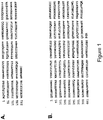
- the amino acid sequence of human sclerostin is reported by Brunkow et al. ibid and is disclosed herein as SEQ ID NO:1.
- compositions and methods that can be used to increase at least one of bone formation, bone mineral density, bone mineral content, bone mass, bone quality and bone strength, and that therefore may be used to treat a wide variety of conditions in which an increase in at least one of bone formation, bone mineral density, bone mineral content, bone mass, bone quality and bone strength is desirable.
- the present invention also offers other related advantages described herein.
- the invention relates to regions (epitopes) of human sclerostin recognized by the binding agents disclosed herein, methods of using these epitopes, and methods of making such epitopes.
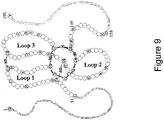
- the invention also relates to epitopes specific to the region of sclerostin identified as Loop 2, and binding agents which specifically bind to that region.
- the invention also relates to epitopes specific to the cystine-knot region of sclerostin, and binding agents such as antibodies specifically binding to that region.
- the invention relates to binding agents, such as antibodies, that specifically bind to sclerostin.
- the binding agents can be characterized by their ability to cross-block the binding of at least one antibody disclosed herein to sclerostin and/or to be cross-blocked from binding sclerostin by at least one antibody disclosed herein.
- the antibodies and other binding agents can also be characterized by their binding pattern to human sclerostin peptides in a "human sclerostin peptide epitope competition binding assay" as disclosed herein.
- the invention relates to binding agents, such as antibodies, that can increase at least one of bone formation, bone mineral density, bone mineral content, bone mass, bone quality and bone strength in a mammal.
- the invention relates to binding agents, such as antibodies, that can block the inhibitory effect of sclerostin in a cell based mineralization assay.
- the invention further relates to polypeptide constructs comprising two, three, or four polypeptide fragments linked by at least one disulfide bond, representing a core region of the cystine-knot of sclerostin, and antibodies capable of specifically binding thereto.
- the invention relates to methods of obtaining epitopes suitable for use as immunogens for generating, in mammals, binding agents, such as antibodies capable of binding specifically to sclerostin; in certain embodiments the binding agents generated are capable of neutralizing sclerostin activity in vivo.
- the invention relates to a composition for eliciting an antibody specific for sclerostin when the composition is administered to an animal, the composition comprising a polypeptide having the amino acid sequence of SEQ ID NO:6, SEQ ID NO:63, SEQ ID NO:64, SEQ ID NO:65, SEQ ID NO:66, SEQ ID NO:67, SEQ ID NO:68, or SEQ ID NO:69.
- the invention also relates to a composition for eliciting an antibody specific for sclerostin when the composition is administered to an animal, the composition comprising at least one polypeptide consisting essentially of the amino acid sequence of SEQ ID NO:2, SEQ ID NO:3, SEQ ID NO:4 or SEQ ID NO:5; the composition may comprise at least two or at least three of the amino acid sequences of SEQ ID NO:2, SEQ ID NO:3, SEQ ID NO:4 and SEQ ID NO:5, and the composition may comprise all four of the amino acid sequences of SEQ ID NO:2, SEQ ID NO:3, SEQ ID NO:4 and SEQ ID NO:5.
- the invention further relates to a composition for eliciting an antibody specific for sclerostin when the composition is administered to an animal, the composition comprising a polypeptide having the amino acid sequences of SEQ ID NO:2, SEQ ID NO:3, SEQ ID NO:4 and SEQ ID NO:5, wherein SEQ ID NO:2 and 4 are joined by a disulfide bond at amino acid positions 57 and 111 with reference to SEQ ID NO:1, and SEQ ID NO:3 and 5 are joined by at least one of (a) a disulfide bond at amino acid positions 82 and 142 with reference to SEQ ID NO:1, and (b) a disulfide bond at amino acid positions 86 and 144 with reference to SEQ ID NO:1; the polypeptide may retain the tertiary structure of the corresponding polypeptide region of human sclerostin of SEQ ID NO:1.
- polypeptide T20.6 consisting essentially of a multiply truncated human sclerostin protein of SEQ ID NO:1, wherein amino acids 1-50, 65-72, 91-100, 118-137, and 150-190 of SEQ ID NO:1 are absent from the polypeptide; this polypeptide may be obtained by tryptic digestion of human sclerostin, and the protein may be isolated by HPLC fractionation.
- the invention further relates to immunogenic portion T20.6 of human sclerostin comprising amino acids 51-64, 73-90, 101-117, and 138-149 of SEQ ID NO:1, wherein the immunogenic portion comprises at least one of:
- the invention further relates to an immunogenic portion T20.6 derivative of human sclerostin comprising amino acids 57-64, 73-86, 111-117, and 138-144 of SEQ ID NO:1, wherein the immunogenic portion comprises at least one of:
- the invention yet further relates to a polypeptide consisting essentially of a human sclerostin protein of SEQ ID NO:1 truncated at the C-terminal and N-terminal ends, wherein amino acids 1-85 and 112-190 of SEQ ID NO:1 are absent from the polypeptide.
- the invention also relates to an immunogenic portion of human sclerostin, comprising amino acids 86-111 of SEQ ID NO:1; the immunogenic portion may consist essentially of contiguous amino acids CGPARLLPNAIGRGKWWRPSGPDFRC (SEQ ID NO:6).
- the invention further relates to an immunogenic portion of rat sclerostin, comprising amino acids 92-109 of SEQ ID NO:98; the immunogenic portion may consist essentially of contiguous amino acids PNAIGRVKWWRPNGPDFR (SEQ ID NO:96).
- the invention still further relates to an immunogenic portion of rat sclerostin, comprising amino acids 99-120 of SEQ ID NO:98; the immunogenic portion may consist essentially of contiguous amino acids KWWRPNGPDFRCIPDRYRAQRV (SEQ ID NO:97).
- the invention relates to a method of producing an immunogenic portion of human sclerostin, comprising the steps of:
- the invention relates to a method of generating an antibody capable of specifically binding to sclerostin, comprising:
- the invention also relates to a method of generating an antibody capable of specifically binding to sclerostin, the method comprising:
- the invention further relates to a method of detecting an anti-sclerostin antibody in a biological sample, comprising the steps of
- the invention also relates to a method of detecting an anti-sclerostin antibody in a biological sample, comprising the steps of
- the invention further relates to a sclerostin binding agent, such as an antibody, that cross-blocks the binding of at least one of antibodies Ab-A, Ab-B, Ab-C, or Ab-D to a sclerostin protein.
- the sclerostin binding agent may also be cross-blocked from binding to sclerostin by at least one of antibodies Ab-A, Ab-B, Ab-C, or Ab-D.
- the isolated antibody, or an antigen-binding fragment thereof may be a polyclonal antibody, a monoclonal antibody, a humanized antibody, a human antibody, a chimeric antibody or the like.
- the invention further relates to a sclerostin binding agent, such as an antibody, that is cross-blocked from binding to sclerostin by at least one of antibodies Ab-A, Ab-B, Ab-C, or Ab-D.
- a sclerostin binding agent such as an antibody
- the isolated antibody, or an antigen-binding fragment thereof may be a polyclonal antibody, a monoclonal antibody, a humanized antibody, a human antibody, a chimeric antibody or the like.
- the invention further relates to a sclerostin binding agent, such as an isolated antibody, that cross-blocks the binding of at least one of antibodies 1-24 (Ab-1 to Ab-24) to a sclerostin protein.
- the sclerostin binding agent may also be cross-blocked from binding to sclerostin by at least one of antibodies 1-24 (Ab-1 to Ab-24).
- the isolated antibody, or an antigen-binding fragment thereof may be a polyclonal antibody, a monoclonal antibody, a humanized antibody, a human antibody, or a chimeric antibody.
- the invention further relates to a sclerostin binding agent, such as an isolated antibody, that is cross-blocked from binding to sclerostin by at least one of antibodies 1-24 (Ab-1 to Ab-24); the isolated antibody, or an antigen-binding fragment thereof, may be a polyclonal antibody, a monoclonal antibody, a humanized antibody, a human antibody, or a chimeric antibody.
- a sclerostin binding agent such as an isolated antibody, that is cross-blocked from binding to sclerostin by at least one of antibodies 1-24 (Ab-1 to Ab-24); the isolated antibody, or an antigen-binding fragment thereof, may be a polyclonal antibody, a monoclonal antibody, a humanized antibody, a human antibody, or a chimeric antibody.
- the invention further relates to a binding agent, such as an isolated antibody that exhibits a similar binding pattern to human sclerostin peptides in a "human sclerostin peptide epitope competition binding assay" as that exhibited by at least one of the antibodies Ab-A, Ab-B, Ab-C or Ab-D; the isolated antibody, or an antigen-binding fragment thereof, may be a polyclonal antibody, a monoclonal antibody, a humanized antibody, a human antibody, or a chimeric antibody.
- a binding agent such as an isolated antibody that exhibits a similar binding pattern to human sclerostin peptides in a "human sclerostin peptide epitope competition binding assay" as that exhibited by at least one of the antibodies Ab-A, Ab-B, Ab-C or Ab-D; the isolated antibody, or an antigen-binding fragment thereof, may be a polyclonal antibody, a monoclonal antibody, a humanized antibody,
- the invention still further relates to a method for treating a bone disorder associated with at least one of low bone formation, low bone mineral density, low bone mineral content, low bone mass, low bone quality and low bone strength in a mammalian subject which comprises providing to a subject in need of such treatment an amount of an anti-sclerostin binding agent sufficient to increase at least one of bone formation, bone mineral density, bone mineral content, bone mass, bone quality and bone strength wherein the anti-sclerostin binding agent comprises an antibody, or sclerostin-binding fragment thereof.
- the invention also relates to an isolated sclerostin polypeptide or fragments thereof, wherein the polypeptide contains 6 conserved cysteine residues and the fragments thereof comprise from 7 to 14 amino acids of SEQ ID NO:2; 8 to 17 amino acids of SEQ ID NO:3; 8 to 18 residues of SEQ ID NO:4; and 6 to 12 residues of SEQ ID NO:5, and the polypeptide or fragments thereof are stabilized by disulfide bonds between SEQ ID NO:2 and 4, and between SEQ ID NO:3 and 5; the polypeptide or fragments may comprise 10-14 amino acids of SEQ ID NO:2; 14 to 17 amino acids of SEQ ID NO:3; 13 to 18 amino acids of SEQ ID NO:4;, and 8 to 12 residues of SEQ ID NO:5; and the polypeptide or fragments may comprise SEQ ID NO:2, SEQ ID NO:3, SEQ ID NO:4, and SEQ ID NO:5.
- antibodies that specifically bind to human sclerostin.
- the antibodies are characterized by their ability to cross-block the binding of at least one antibody disclosed herein to human sclerostin and/or to be cross-blocked from binding human sclerostin by at least one antibody disclosed herein.
- an isolated antibody or an antigen-binding fragment thereof, that can increase at least one of bone formation, bone mineral density, bone mineral content, bone mass, bone quality and bone strength in a mammal.
- an isolated antibody or an antigen-binding fragment thereof, that can block the inhibitory effect of sclerostin in a cell based mineralization assay.
- a binding agent such as an antibody, that specifically binds to human sclerostin and has at least one CDR sequence selected from SEQ ID NOs: 39, 40, 41, 42, 43, 44, 45, 46, 47, 48, 49, 50, 51, 52, 53, 54, 55, 56, 57, 58, 59, 60, 61, 62, 78, 79, 80, 81, 99, 100, 101, 102, 103, 104, 105, 106, 107, 108, 109, 110, 111, 112, 113, 114, 115, 116, 237, 238, 239, 240, 241, 242, 243, 244, 245, 246, 247, 248, 249, 250, 251, 252, 253, 254, 255, 256, 257, 258, 259, 260, 261, 262, 263, 264, 265, 266, 267, 268, 269, 270, 271, 272, 273, 274, 275, 276, 277, 27
- a binding agent such as an antibody, that specifically binds to human sclerostin and has at least one CDR sequence selected from SEQ ID NOs:39, 40, 41, 42, 43, 44, 45, 46, 47, 48, 49, 50, 51, 52, 53, 54, 55, 56, 57, 58, 59, 60, 61, 62, 78, 79, 80, 81, 99, 100, 101, 102, 103, 104, 105, 106, 107, 108, 109, 110, 111, 112, 113, 114, 115, 116, 237, 238, 239, 240, 241, 242, 243, 244, 245, 246, 247, 248, 249, 250, 251, 252, 253, 254, 255, 256, 257, 258, 259, 260, 261, 262, 263, 264, 265, 266, 267, 268, 269, 270, 271, 272, 273, 274, 275, 276, 277, 27
- regions of human sclerostin which are important for the in vivo activity of the protein.
- the present invention relates to regions of the human sclerostin protein that contain epitopes recognized by antibodies that also bind to full-length sclerostin, and methods of making and using these epitopes.
- the invention also provides binding agents (such as antibodies) that specifically bind to sclerostin or portions of sclerostin, and methods for using such binding agents.
- the binding agents are useful to block or impair binding of human sclerostin to one or more ligand.
- Recombinant human sclerostin/SOST is commercially available from R&D Systems (Minneapolis, MN, USA; 2006 cat# 1406-ST-025). Additionally, recombinant mouse sclerostin/SOST is commercially available from R&D Systems (Minneapolis, MN, USA; 2006 cat# 1589-ST-025). Research grade sclerostin binding monoclonal antibodies are commercially available from R&D Systems (Minneapolis, MN, USA; mouse monoclonal: 2006 cat# MAB1406; rat monoclonal: 2006 cat# MAB1589).
- U.S. Patent Nos. 6,395,511 and 6,803,453 and U.S. Patent Publications 20040009535 and 20050106683 refer to anti-sclerostin antibodies generally.
- human sclerostin is intended to include the protein of SEQ ID NO:1 and allelic variants thereof.
- Sclerostin can be purified from 293T host cells that have been transfected by a gene encoding sclerostin by elution of filtered supernatant of host cell culture fluid using a Heparin HP column, using a salt gradient. The preparation and further purification using cation exchange chromatography are described in Examples 1 and 2.
- Binding agents of the invention are preferably antibodies, as defined herein.
- the term "antibody” refers to an intact antibody, or a binding fragment thereof.
- An antibody may comprise a complete antibody molecule (including polyclonal, monoclonal, chimeric, humanized, or human versions having full length heavy and/or light chains), or comprise an antigen binding fragment thereof.
- Antibody fragments include F(ab') 2 , Fab, Fab', Fv, Fc, and Fd fragments, and can be incorporated into single domain antibodies, single-chain antibodies, maxibodies, minibodies, intrabodies, diabodies, triabodies, tetrabodies, v-NAR and bis-scFv (See e.g .,, Hollinger and Hudson, 2005, Nature Biotechnology, 23, 9, 1126-1136 ).
- Antibody polypeptides are also disclosed in U. S. Patent No. 6,703,199 , including fibronectin polypeptide monobodies. Other antibody polypeptides are disclosed in U.S. Patent Publication 2005/0238646 , which are single-chain polypeptides.
- Antigen binding fragments derived from an antibody can be obtained, for example, by proteolytic hydrolysis of the antibody, for example, pepsin or papain digestion of whole antibodies according to conventional methods.
- antibody fragments can be produced by enzymatic cleavage of antibodies with pepsin to provide a 5S fragment termed F(ab') 2 .
- This fragment can be further cleaved using a thiol reducing agent to produce 3.5S Fab' monovalent fragments.
- the cleavage reaction can be performed using a blocking group for the sulfhydryl groups that result from cleavage of disulfide linkages.
- an enzymatic cleavage using papain produces two monovalent Fab fragments and an Fc fragment directly.
- These methods are described, for example, by Goldenberg, U.S. Patent No. 4,331,647 , Nisonoff et ⁇ l., Arch. Biochem. Biophys. 89:230, 1960 ; Porter, Biochem. J. 73:119, 1959 ; Edelman et al., in Methods in Enzymology 1:422 (Academic Press 1967 ); and by Andrews, S.M. and Titus, J.A. in Current Protocols in Immunology (Coligan J.E., et al., eds), John Wiley & Sons, New York (2003).
- antibody fragment may also be any synthetic or genetically engineered protein.
- antibody fragments include isolated fragments consisting of the light chain variable region, "Fv" fragments consisting of the variable regions of the heavy and light chains, recombinant single chain polypeptide molecules in which light and heavy variable regions are connected by a peptide linker (scFv proteins).
- CDRs complementarity determining regions
- Such polynucleotides are prepared, for example, by using the polymerase chain reaction to synthesize the variable region using mRNA of antibody-producing cells as a template (see, for example, Larrick et al., Methods: A Companion to Methods in Enzymology 2:106, 1991 ; Courtenay-Luck, "Genetic Manipulation of Monoclonal Antibodies,” in Monoclonal Antibodies: Production, Engineering and Clinical Application, Ritter et al.
- the binding agent comprises at least one CDR as described herein.
- the binding agent may comprise at least two, three, four, five or six CDR's as described herein.
- the binding agent further may comprise at least one variable region domain of an antibody described herein.
- the variable region domain may be of any size or amino acid composition and will generally comprise at least one CDR sequence responsible for binding to human sclerostin, for example CDR-H1, CDR-H2, CDR-H3 and/or the light chain CDRs specifically described herein and which is adjacent to or in frame with one or more framework sequences.
- the variable (V) region domain may be any suitable arrangement of immunoglobulin heavy (V H ) and/or light (V L ) chain variable domains.
- the V region domain may be monomeric and be a V H or V L domain, which is capable of independently binding human sclerostin with an affinity at least equal to 1 x 10 -7 M or less as describedbelow.
- the V region domain may be dimeric and contain V H -V H , V H -V L , or V L -V L , dimers.
- the V region dimer comprises at least one V H and at least one V L chain that may be non-covalently associated (hereinafter referred to as F V ).
- the chains may be covalently coupled either directly, for example via a disulfide bond between the two variable domains, or through a linker, for example a peptide linker, to form a single chain Fv (scF v ).
- variable region domain may be any naturally occurring variable domain or an engineered version thereof.
- engineered version is meant a variable region domain that has been created using recombinant DNA engineering techniques.
- engineered versions include those created, for example, from a specific antibody variable region by insertions, deletions, or changes in or to the amino acid sequences of the specific antibody.
- Particular examples include engineered variable region domains containing at least one CDR and optionally one or more framework amino acids from a first antibody and the remainder of the variable region domain from a second antibody.
- variable region domain may be covalently attached at a C-terminal amino acid to at least one other antibody domain or a fragment thereof.
- a VH domain that is present in the variable region domain may be linked to an immunoglobulin CH1 domain, or a fragment thereof.
- a V L domain may be linked to a C K domain or a fragment thereof.
- the antibody may be a Fab fragment wherein the antigen binding domain contains associated V H and V L domains covalently linked at their C-termini to a CH1 and C K domain, respectively.
- the CH1 domain may be extended with further amino acids, for example to provide a hinge region or a portion of a hinge region domain as found in a Fab' fragment, or to provide further domains, such as antibody CH2 and CH3 domains.
- binding agents comprise at least one of these CDRs.
- one or more CDR may be incorporated into known antibody framework regions (IgG1, IgG2, etc.), or conjugated to a suitable vehicle to enhance the half-life thereof.
- suitable vehicles include, but are not limited to Fc, polyethylene glycol (PEG), albumin, transferrin, and the like. These and other suitable vehicles are known in the art.
- conjugated CDR peptides may be in monomeric, dimeric, tetrameric, or other form.
- one or more water-soluble polymer is bonded at one or more specific position, for example at the amino terminus, of a binding agent.
- a binding agent comprises one or more water soluble polymer attachments, including, but not limited to, polyethylene glycol, polyoxyethylene glycol, or polypropylene glycol. See, e.g., U.S. Pat. Nos. 4,640,835 , 4,496,689 , 4,301,144 , 4,670,417 , 4,791,192 and 4,179,337 .
- a derivative binding agent comprises one or more of monomethoxy-polyethylene glycol, dextran, cellulose, or other carbohydrate based polymers, poly-(N-vinyl pyrrolidone)-polyethylene glycol, propylene glycol homopolymers, a polypropylene oxide/ethylene oxide co-polymer, polyoxyethylated polyols (e.g., glycerol) and polyvinyl alcohol, as well as mixtures of such polymers.
- one or more water-soluble polymer is randomly attached to one or more side chains.
- PEG can act to improve the therapeutic capacity for a binding agent, such as an antibody. Certain such methods are discussed, for example, in U.S. Pat. No. 6,133,426 , which is hereby incorporated by reference for any purpose.
- a binding agent of the present invention may have at least one amino acid substitution, providing that the binding agent retains binding specificity. Therefore, modifications to the binding agent structures are encompassed within the scope of the invention. These may include amino acid substitutions, which may be conservative or non-conservative, that do not destroy the sclerostin binding capability of a binding agent. Conservative amino acid substitutions may encompass non-naturally occurring amino acid residues, which are typically incorporated by chemical peptide synthesis rather than by synthesis in biological systems. These include peptidomimetics and other reversed or inverted forms of amino acid moieties. A conservative amino acid substitution may also involve a substitution of a native amino acid residue with a normative residue such that there is little or no effect on the polarity or charge of the amino acid residue at that position.
- Non-conservative substitutions may involve the exchange of a member of one class of amino acids or amino acid mimetics for a member from another class with different physical properties (e.g. size, polarity, hydrophobicity, charge). Such substituted residues may be introduced into regions of the human antibody that are homologous with non-human antibodies, or into the non-homologous regions of the molecule.
- test variants containing a single amino acid substitution at each desired amino acid residue.
- the variants can then be screened using activity assays known to those skilled in the art.
- Such variants could be used to gather information about suitable variants. For example, if one discovered that a change to a particular amino acid residue resulted in destroyed, undesirably reduced, or unsuitable activity, variants with such a change may be avoided. In other words, based on information gathered from such routine experiments, one skilled in the art can readily determine the amino acids where further substitutions should be avoided either alone or in combination with other mutations.
- polypeptide structure A skilled artisan will be able to determine suitable variants of the polypeptide as set forth herein using well-known techniques.
- one skilled in the art may identify suitable areas of the molecule that may be changed without destroying activity by targeting regions not believed to be important for activity.
- even areas that may be important for biological activity or for structure may be subject to conservative amino acid substitutions without destroying the biological activity or without adversely affecting the polypeptide structure.
- One skilled in the art can also analyze the three-dimensional structure and amino acid sequence in relation to that structure in similar polypeptides. In view of such information, one skilled in the art may predict the alignment of amino acid residues of an antibody with respect to its three dimensional structure. In certain embodiments, one skilled in the art may choose not to make radical changes to amino acid residues predicted to be on the surface of the protein, since such residues may be involved in important interactions with other molecules.
- One method of predicting secondary structure is based upon homology modeling. For example, two polypeptides or proteins which have a sequence identity of greater than 30%, or similarity greater than 40% often have similar structural topologies.
- the recent growth of the protein structural database (PDB) has provided enhanced predictability of secondary structure, including the potential number of folds within a polypeptide's or protein's structure. See Holm et al., Nucl. Acid. Res., 27(1):244-247 (1999 ). It has been suggested ( Brenner et al., Curr. Op. Struct. Biol., 7(3):369-376 (1997 )) that there are a limited number of folds in a given polypeptide or protein and that once a critical number of structures have been resolved, structural prediction will become dramatically more accurate.
- Additional methods of predicting secondary structure include “threading” ( Jones, D., Curr. Opin. Struct. Biol., 7(3):377-87 (1997 ); Sippl et al., Structure, 4(1): 15-19 (1996 )), “profile analysis” ( Bowie et al., Science, 253:164-170 (1991 ); Gribskov et al., Meth. Enzym., 183:146-159 (1990 ); Gribskov et al., Proc. Nat. Acad. Sci., 84(13):4355-4358 (1987 )), and “evolutionary linkage” (See Holm, supra (1999), and Brenner, supra (1997)).
- variants of binding agents include glycosylation variants wherein the number and/or type of glycosylation site has been altered compared to the amino acid sequences of a parent polypeptide.
- variants comprise a greater or a lesser number of N-linked glycosylation sites than the native protein.
- An N-linked glycosylation site is characterized by the sequence: Asn-X-Ser or Asn-X-Thr, wherein the amino acid residue designated as X may be any amino acid residue except proline.
- the substitution of amino acid residues to create this sequence provides a potential new site for the addition of an N-linked carbohydrate chain. Alternatively, substitutions which eliminate this sequence will remove an existing N-linked carbohydrate chain.
- N-linked carbohydrate chains wherein one or more N-linked glycosylation sites (typically those that are naturally occurring) are eliminated and one or more new N-linked sites are created.
- Additional preferred antibody variants include cysteine variants wherein one or more cysteine residues are deleted from or substituted for another amino acid (e.g., serine) as compared to the parent amino acid sequence. Cysteine variants may be useful when antibodies must be refolded into a biologically active conformation such as after the isolation of insoluble inclusion bodies. Cysteine variants generally have fewer cysteine residues than the native protein, and typically have an even number to minimize interactions resulting from unpaired cysteines.
- amino acid substitutions can be used to identify important residues of antibodies to sclerostin, or to increase or decrease the affinity of the antibodies to sclerostin described herein.
- preferred amino acid substitutions are those which: (1) reduce susceptibility to proteolysis, (2) reduce susceptibility to oxidation, (3) alter binding affinity for forming protein complexes, (4) alter binding affinities, and/or (4) confer or modify other physiochemical or functional properties on such polypeptides.
- single or multiple amino acid substitutions may be made in the naturally-occurring sequence (in certain embodiments, in the portion of the polypeptide outside the domain(s) forming intermolecular contacts).
- a conservative amino acid substitution typically may not substantially change the structural characteristics of the parent sequence (e.g., a replacement amino acid should not tend to break a helix that occurs in the parent sequence, or disrupt other types of secondary structure that characterizes the parent sequence).
- a replacement amino acid should not tend to break a helix that occurs in the parent sequence, or disrupt other types of secondary structure that characterizes the parent sequence.
- Examples of art-recognized polypeptide secondary and tertiary structures are described in Proteins, Structures and Molecular Principles (Creighton, Ed., W. H. Freeman and Company, New York (1984 )); Introduction to Protein Structure (C. Branden and J. Tooze, eds., Garland Publishing, New York, N.Y. (1991 )); and Thornton et al. Nature 354:105 (1991 ), which are each incorporated herein by reference.
- binding agents of the invention may be chemically bonded with polymers, lipids, or other moieties.
- the binding agents may comprise at least one of the CDRs described herein incorporated into a biocompatible framework structure.
- the biocompatible framework structure comprises a polypeptide or portion thereof that is sufficient to form a conformationally stable structural support, or framework, or scaffold, which is able to display one or more sequences of amino acids that bind to an antigen (e.g., CDRs, a variable region, etc.) in a localized surface region.
- an antigen e.g., CDRs, a variable region, etc.
- Such structures can be a naturally occurring polypeptide or polypeptide "fold" (a structural motif), or can have one or more modifications, such as additions, deletions or substitutions of amino acids, relative to a naturally occurring polypeptide or fold.
- These scaffolds can be derived from a polypeptide of any species (or of more than one species), such as a human, other mammal, other vertebrate, invertebrate, plant, bacteria or virus.
- the biocompatible framework structures are based on protein scaffolds or skeletons other than immunoglobulin domains.
- those based on fibronectin, ankyrin, lipocalin, neocarzinostain, cytochrome b, CP1 zinc finger, PST1, coiled coil, LACI-D1, Z domain and tendramisat domains may be used (See e.g., , Nygren and Uhlen, 1997, Current Opinion in Structural Biology, 7, 463-469 ).
- the binding agents of the invention include the humanized antibodies described herein.
- Humanized antibodies such as those described herein can be produced using techniques known to those skilled in the art ( Zhang, W., et al., Molecular Immunology. 42(12):1445-1451, 2005 ; Hwang W. et al., Methods. 36(1):35-42, 2005 ; Dall'Acqua WF, et al., Methods 36(1):43-60 , 2005; and Clark, M., Immunology Today. 21(8):397-402, 2000 ).
- suitable binding agents include portions of these antibodies, such as one or more of CDR-H1, CDR-H2, CDR-H3, CDR-L1, CDR-L2 and CDR-L3 as specifically disclosed herein. At least one of the regions of CDR-H1, CDR-H2, CDR-H3, CDR-L1, CDR-L2 and CDR-L3 may have at least one amino acid substitution, provided that the binding agent retains the binding specificity of the non-substituted CDR.
- the non-CDR portion of the binding agent may be a non-protein molecule, wherein the binding agent cross-blocks the binding of an antibody disclosed herein to sclerostin and/or neutralizes sclerostin.
- the non-CDR portion of the binding agent may be a non-protein molecule in which the binding agent exhibits a similar binding pattern to human sclerostin peptides in a "human sclerostin peptide epitope competition binding assay" as that exhibited by at least one of antibodies Ab-A, Ab-B, Ab-C, Ab-D, Ab-1, Ab-2, Ab-3, Ab-4, Ab-5, Ab-6, Ab-7, Ab-8, Ab-9, Ab-10, Ab-11, Ab-12, Ab-13, Ab-14, Ab-15, Ab-16, Ab-17, Ab-18, Ab-19, Ab-20, Ab-21, Ab-22, Ab-23, and Ab-24, and/or neutralizes sclerostin.
- the non-CDR portion of the binding agent may be composed of amino acids, wherein the binding agent is a recombinant binding protein or a synthetic peptide, and the recombinant binding protein cross-blocks the binding of an antibody disclosed herein to sclerostin and/or neutralizes sclerostin.
- the non-CDR portion of the binding agent may be composed of amino acids, wherein the binding agent is a recombinant binding protein, and the recombinant binding protein exhibits a similar binding pattern to human sclerostin peptides in the human sclerostin peptide epitope competition binding assay (described hereinbelow) as that exhibited by at least one of the antibodies Ab-A, Ab-B, Ab-C, Ab-D, Ab-1, Ab-2, Ab-3, Ab-4, Ab-5, Ab-6, Ab-7, Ab-8, Ab-9, Ab-10, Ab-11, Ab-12, Ab-13, Ab-14, Ab-15, Ab-16, Ab-17, Ab-18, Ab-19, Ab-20, Ab-21, Ab-22, Ab-23, and Ab-24, and/or neutralizes sclerostin.
- the binding agent is a recombinant binding protein
- the recombinant binding protein exhibits a similar binding pattern to human sclerostin peptides in the human sclerost
- an antibody comprises one or more of CDR-H1, CDR-H2, CDR-H3, CDR-L1, CDR-L2 and CDR-L3 as described above, it may be obtained by expression from a host cell containing DNA coding for these sequences.
- a DNA coding for each CDR sequence may be determined on the basis of the amino acid sequence of the CDR and synthesized together with any desired antibody variable region framework and constant region DNA sequences using oligonucleotide synthesis techniques, site-directed mutagenesis and polymerase chain reaction (PCR) techniques as appropriate.
- DNA coding for variable region frameworks and constant regions is widely available to those skilled in the art from genetic sequences databases such as GenBank ® .
- Each of the above-mentioned CDRs will be typically located in a variable region framework at positions 31-35 (CDR-H1), 50-65 (CDR-H2) and 95-102 (CDR-H3) of the heavy chain and positions 24-34 (CDR-L1), 50-56 (CDR-L2) and 89-97 (CDR-L3) of the light chain according to the Kabat numbering system ( Kabat et al., 1987 in Sequences of Proteins of Immunological Interest, U.S. Department of Health and Human Services, NIH, USA ).
- the DNA encoding an antibody of the invention or fragment thereof may be propagated and expressed according to any of a variety of well-known procedures for nucleic acid excision, ligation, transformation, and transfection using any number of known expression vectors.
- expression of an antibody fragment may be preferred in a prokaryotic host, such as Escherichia coli ( see, e.g., Pluckthun et al., 1989 Methods Enzymol. 178:497-515 ).
- expression of the antibody or a fragment thereof may be preferred in a eukaryotic host cell, including yeast (e.g., Saccharomyces cerevisiae, Schizosaccharomyces pombe, and Pichia pastoris ) , animal cells (including mammalian cells) or plant cells.
- yeast e.g., Saccharomyces cerevisiae, Schizosaccharomyces pombe, and Pichia pastoris
- animal cells including mammalian cells
- suitable animal cells include, but are not limited to, myeloma (such as a mouse NSO line), COS, CHO, or hybridoma cells.
- plant cells include tobacco, corn, soybean, and rice cells.
- One or more replicable expression vectors containing DNA encoding an antibody variable and/or constant region may be prepared and used to transform an appropriate cell line, for example, a non-producing myeloma cell line, such as a mouse NSO line or a bacteria, such as E. coli, in which production of the antibody will occur.
- an appropriate cell line for example, a non-producing myeloma cell line, such as a mouse NSO line or a bacteria, such as E. coli, in which production of the antibody will occur.
- the DNA sequence in each vector should include appropriate regulatory sequences, particularly a promoter and leader sequence operatively linked to the variable domain sequence.
- Particular methods for producing antibodies in this way are generally well-known and routinely used. For example, basic molecular biology procedures are described by Maniatis et al.
- DNA sequencing can be performed as described in Sanger et al. (PNAS 74:5463, (1977 )) and the Amersham International plc sequencing handbook, and site directed mutagenesis can be carried out according to methods known in the art ( Kramer et al., Nucleic Acids Res. 12:9441, (1984 ); Kunkel Proc. Natl. Acad. Sci. USA 82:488-92 (1985 ); Kunkel et al., Methods in Enzymol.
- affinity maturation protocols including maintaining the CDRs ( Yang et al., J. Mol. Biol., 254, 392-403, 1995 ), chain shuffling ( Marks et al., Bio/Technology, 10, 779-783, 1992 ), use of mutation strains of E. coli. ( Low et al., J. Mol. Biol., 250, 350-368, 1996 ), DNA shuffling ( Patten et al., Curr. Opin. Biotechnol., 8, 724-733, 1997 ), phage display ( Thompson et al., J. Mol.
- antibodies according to the invention may be obtained by conventional immunization and cell fusion procedures as described herein and known in the art.
- Monoclonal antibodies of the invention may be generated using a variety of known techniques.
- monoclonal antibodies that bind to specific antigens may be obtained by methods known to those skilled in the art (see, for example, Kohler et al., Nature 256:495, 1975 ; Coligan et al. (eds.), Current Protocols in Immunology, 1:2.5.12.6.7 (John Wiley & Sons 1991 ); U.S. Patent Nos.
- Antibody fragments may be derived therefrom using any suitable standard technique such as proteolytic digestion, or optionally, by proteolytic digestion (for example, using papain or pepsin) followed by mild reduction of disulfide bonds and alkylation. Alternatively, such fragments may also be generated by recombinant genetic engineering techniques as described herein..
- Monoclonal antibodies can be obtained by injecting an animal, for example, a rat, hamster, a rabbit, or preferably a mouse, including for example a transgenic or a knock-out, as known in the art, with an immunogen comprising human sclerostin of SEQ ID NO:1, or a fragment thereof, according to methods known in the art and described herein.
- the presence of specific antibody production may be monitored after the initial injection and/or after a booster injection by obtaining a serum sample and detecting the presence of an antibody that binds to human sclerostin or peptide using any one of several immunodetection methods known in the art and described herein.
- lymphoid cells most commonly cells from the spleen or lymph node, are removed to obtain B-lymphocytes.
- the B lymphocytes are then fused with a drug-sensitized myeloma cell fusion partner, preferably one that is syngeneic with the immunized animal and that optionally has other desirable properties (e.g., inability to express endogenous Ig gene products, e.g., P3X63 - Ag 8.653 (ATCC No. CRL 1580); NSO, SP20) to produce hybridomas, which are immortal eukaryotic cell lines.
- a drug-sensitized myeloma cell fusion partner preferably one that is syngeneic with the immunized animal and that optionally has other desirable properties (e.g., inability to express endogenous Ig gene products, e.g., P3X63 - Ag 8.653 (ATCC No. CRL 1580); NSO, SP20) to produce hybridomas,
- the lymphoid (e.g., spleen) cells and the myeloma cells may be combined for a few minutes with a membrane fusion-promoting agent, such as polyethylene glycol or a nonionic detergent, and then plated at low density on a selective medium that supports the growth of hybridoma cells but not unfused myeloma cells.
- a preferred selection media is HAT (hypoxanthine, aminopterin, thymidine). After a sufficient time, usually about one to two weeks, colonies of cells are observed. Single colonies are isolated, and antibodies produced by the cells may be tested for binding activity to human sclerostin, using any one of a variety of immunoassays known in the art and described herein.
- the hybridomas are cloned (e.g., by limited dilution cloning or by soft agar plaque isolation) and positive clones that produce an antibody specific to sclerostin are selected and cultured.
- the monoclonal antibodies from the hybridoma cultures may be isolated from the supernatants of hybridoma cultures.
- An alternative method for production of a murine monoclonal antibody is to inject the hybridoma cells into the peritoneal cavity of a syngeneic mouse, for example, a mouse that has been treated (e.g., pristane-primed) to promote formation of ascites fluid containing the monoclonal antibody.
- Monoclonal antibodies can be isolated and purified by a variety of well-established techniques.
- Such isolation techniques include affinity chromatography with Protein-A Sepharose, size-exclusion chromatography, and ion-exchange chromatography (see, for example, Coligan at pages 2.7.1-2.7.12 and pages 2.9.1-2.9.3; Baines et al., "Purification of Immunoglobulin G (IgG),” in Methods in Molecular Biology, Vol. 10, pages 79-104 (The Humana Press, Inc. 1992 )).
- Monoclonal antibodies may be purified by affinity chromatography using an appropriate ligand selected based on particular properties of the antibody (e.g., heavy or light chain isotype, binding specificity, etc.).
- a suitable ligand immobilized on a solid support, include Protein A, Protein G, an anticonstant region (light chain or heavy chain) antibody, an anti-idiotype antibody, and a TGF-beta binding protein, or fragment or variant thereof.
- An antibody of the present invention may also be a human monoclonal antibody.
- Human monoclonal antibodies may be generated by any number of techniques with which those having ordinary skill in the art will be familiar. Such methods include, but are not limited to, Epstein Barr Virus (EBV) transformation of human peripheral blood cells (e.g., containing B lymphocytes), in vitro immunization of human B cells, fusion of spleen cells from immunized transgenic mice carrying inserted human immunoglobulin genes, isolation from human immunoglobulin V region phage libraries, or other procedures as known in the art and based on the disclosure herein.
- EBV Epstein Barr Virus
- human monoclonal antibodies may be obtained from transgenic mice that have been engineered to produce specific human antibodies in response to antigenic challenge.
- human immunoglobulin transgenes may be mini-gene constructs, or transloci on yeast artificial chromosomes, which undergo B cell-specific DNA rearrangement and hypermutation in the mouse lymphoid tissue.
- Human monoclonal antibodies may be obtained by immunizing the transgenic mice, which may then produce human antibodies specific for sclerostin. Lymphoid cells of the immunized transgenic mice can be used to produce human antibody-secreting hybridomas according to the methods described herein. Polyclonal sera containing human antibodies may also be obtained from the blood of the immunized animals.
- Another method for generating human antibodies of the invention includes immortalizing human peripheral blood cells by EBV transformation. See, e.g., U.S. Patent No. 4,464,456 .
- Such an immortalized B cell line (or lymphoblastoid cell line) producing a monoclonal antibody that specifically binds to sclerostin can be identified by immunodetection methods as provided herein, for example, an ELISA, and then isolated by standard cloning techniques.
- the stability of the lymphoblastoid cell line producing an anti-sclerostin antibody may be improved by fusing the transformed cell line with a murine myeloma to produce a mouse-human hybrid cell line according to methods known in the art (see, e.g., Glasky et al., Hybridoma 8:377-89 (1989 )).
- Still another method to generate human monoclonal antibodies is in vitro immunization, which includes priming human splenic B cells with human sclerostin, followed by fusion of primed B cells with a heterohybrid fusion partner. See, e.g., Boerner et al., 1991 J. Immunol. 147:86-95 .
- a B cell that is producing an anti-human sclerostin antibody is selected and the light chain and heavy chain variable regions are cloned from the B cell according to molecular biology techniques known in the art ( WO 92/02551 ; US patent 5,627,052 ; Babcook et al., Proc. Natl. Acad. Sci. USA 93:7843-48 (1996 )) and described herein.
- B cells from an immunized animal may be isolated from the spleen, lymph node, or peripheral blood sample by selecting a cell that is producing an antibody that specifically binds to sclerostin.
- B cells may also be isolated from humans, for example, from a peripheral blood . sample.
- Methods for detecting single B cells that are producing an antibody with the desired specificity are well known in the art, for example, by plaque formation, fluorescence-activated cell sorting, in vitro stimulation followed by detection of specific antibody, and the like.
- Methods for selection of specific antibody-producing B cells include, for example, preparing a single cell suspension of B cells in soft agar that contains human sclerostin. Binding of the specific antibody produced by the B cell to the antigen results in the formation of a complex, which may be visible as an immunoprecipitate.
- the specific antibody genes may be cloned by isolating and amplifying DNA or mRNA according to methods known in the art and described herein.
- An additional method for obtaining antibodies of the invention is by phage display. See, e.g., Winter et al., 1994 Annu. Rev. Immunol. 12:433-55 ; Burton et al., 1994 Adv. Immunol. 57:191-280 .
- Human or murine immunoglobulin variable region gene combinatorial libraries may be created in phage vectors that can be screened to select Ig fragments (Fab, Fv, sFv, or multimers thereof) that bind specifically to TGF-beta binding protein or variant or fragment thereof. See, e.g., U.S. Patent No.
- a library containing a plurality of polynucleotide sequences encoding Ig variable region fragments may be inserted into the genome of a filamentous bacteriophage, such as M13 or a variant thereof, in frame with the sequence encoding a phage coat protein.
- a fusion protein may be a fusion of the coat protein with the light chain variable region domain and/or with the heavy chain variable region domain.
- immunoglobulin Fab fragments may also be displayed on a phage particle ( see, e.g., U.S. Patent No. 5,698,426 ).
- Heavy and light chain immunoglobulin cDNA expression libraries may also be prepared in lambda phage, for example, using ⁇ ImmunoZap TM (H) and ⁇ ImmunoZap TM (L) vectors (Stratagene, La Jolla, California). Briefly, mRNA is isolated from a B cell population, and used to create heavy and light chain immunoglobulin cDNA expression libraries in the ⁇ ImmunoZap(H) and ⁇ ImmunoZap(L) vectors. These vectors may be screened individually or co-expressed to form Fab fragments or antibodies (see Huse et al., supra ; see also Sastry et al., supra ) . Positive plaques may subsequently be converted to a non-lytic plasmid that allows high level expression of monoclonal antibody fragments from E. coli.
- variable regions of a gene expressing a monoclonal antibody of interest are amplified using nucleotide primers.
- primers may be synthesized by one of ordinary skill in the art, or may be purchased from commercially available sources. ( See, e.g., Stratagene (La Jolla, California), which sells primers for mouse and human variable regions including, among others, primers for V Ha , V Hb , V Hc , V Hd , C H1 , V L and C L regions.)
- These primers may be used to amplify heavy or light chain variable regions, which may then be inserted into vectors such as ImmunoZAP TM H or ImmunoZAP TM L (Stratagene), respectively.
- vectors may then be introduced into E. coli, yeast, or mammalian-based systems for expression. Large amounts of a single-chain protein containing a fusion of the V H and V L domains may be produced using these methods (see Bird et al., Science 242:423-426, 1988 ).
- the specific antibody genes may be cloned by isolating and amplifying DNA or mRNA therefrom according to standard procedures as described herein.
- the antibodies produced therefrom may be sequenced and the CDRs identified and the DNA coding for the CDRs may be manipulated as described previously to generate other antibodies according to the invention.
- the binding agents specifically bind to sclerostin.
- the term "specifically binds” refers to the ability of a binding agent to bind to sclerostin, preferably human sclerostin, with greater affinity than it binds to an unrelated control protein.
- the control protein is hen egg white lysozyme.
- the binding agents bind to sclerostin with an affinity that is at least, 50, 100, 250, 500, 1000, or 10,000 times greater than the affinity for a control protein.
- a binding agent may have a binding affinity for human sclerostin of less than or equal to 1 x 10 -7 M, less than or equal to 1 x 10 -8 M, less than or equal to 1 x 10 -9 M, less than or equal to 1 x 10 -10 M, less than or equal to 1 x 10 -11 M, or less than or equal to 1 x 10 -12 M.
- affinity may be determined by an affinity ELISA assay.
- affinity may be determined by a BIAcore assay.
- affinity may be determined by a kinetic method.
- affinity may be determined by an equilibrium/solution method. Such methods are described in further detail herein or known in the art.
- Sclerostin binding agents of the present invention preferably modulate sclerostin function in the cell-based assay described herein and/or the in vivo assay described herein and/or bind to one or more of the epitopes described herein and/or cross-block the binding of one of the antibodies described in this application and/or are cross-blocked from binding sclerostin by one of the antibodies described in this application. Accordingly such binding agents can be identified using the assays described herein.
- binding agents are generated by first identifying antibodies that bind to one more of the epitopes provided herein and/or neutralize in the cell-based and/or in vivo assays described herein and/or cross-block the antibodies described in this application and/or are cross-blocked from binding sclerostin by one of the antibodies described in this application.
- the CDR regions from these antibodies are then used to insert into appropriate biocompatible frameworks to generate sclerostin binding agents.
- the non-CDR portion of the binding agent may be composed of amino acids, or may be a non-protein molecule.
- the assays described herein allow the characterization of binding agents.
- the binding agents of the present invention are antibodies as defined herein.
- proteins may undergo a variety of posttranslational modifications.
- the type and extent of these modifications often depends on the host cell line used to express the protein as well as the culture conditions.
- modifications may include variations in glycosylation, methionine oxidation, diketopiperizine formation, aspartate isomerization and asparagine deamidation.
- a frequent modification is the loss of a carboxy-terminal basic residue (such as lysine or arginine) due to the action of carboxypeptidases (as described in Harris, RJ. Journal of Chromatography 705:129-134, 1995 ).
- Antibodies referred to as Ab-A, Ab-B, Ab-C, Ab-D and Ab-1 are described below.
- HC refers to the heavy chain
- LC refers to the light chain.
- CDRs are box shaded and the constant (C) regions are shown in bold italics.
- Antibody D (also referred to herein as Ab-D and Mab-D) is a mouse antibody which exhibits high affinity binding to sclerostin.
- the BIAcore binding pattern of Ab-D is shown in Figure 18 .
- Nucleic acid sequence encoding the mature form (signal peptide removed) of Ab-D LC is as follows:
- the amino acid sequence of Ab-D LC including signal peptide is as follows:
- amino acid sequence of the mature form (signal peptide removed) of Ab-D HC heavy chain is as follows:
- the nucleic acid sequence encoding the mature form (signal peptide removed) of Ab-D HC is:
- the amino acid sequence of Ab-D HC including signal peptide is:
- the nucleic acid sequence of Ab-D HC including signal peptide encoding sequence is:
- the CDR (complementarity determining region) sequences in the variable region of the heavy chain of Ab-D are as follows:
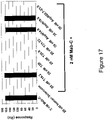
- Antibody C (also referred to herein as Ab-C and Mab-C) is a mouse antibody which exhibits high affinity binding to sclerostin.
- the BIAcore binding pattern of Ab-C is shown in Figure 17 .
- the amino acid sequence of the mature form (signal peptide removed) of Ab-C Light Chain is as follows:
- the nucleic acid sequence encoding the mature form (signal peptide removed) of Ab-C LC is:
- the amino acid sequence of Ab-C LC including signal peptide is:
- the nucleic acid sequence of Ab-C LC including signal peptide encoding sequence is:
- the amino acid sequence of the mature form (signal peptide removed) of Ab-C HC is:
- the nucleic acid sequence encoding the mature form (signal peptide removed) of Ab-C HC is as follows:
- the amino acid sequence of Ab-C HC including signal peptide is:
- the nucleic acid sequence of Ab-C HC including signal peptide encoding sequence is:
- the CDR (complementarity determining region) sequences in the variable region of the heavy chain of Ab-C are as follows:
- Antibody A (also referred to herein as Ab-A and Mab-A) is a rabbit-mouse chimeric antibody which exhibits high affinity binding to sclerostin.
- the BIAcore binding pattern of Ab-A is shown in Figure 15 .
- the amino acid sequence of Ab-A LC including signal peptide is:
- the nucleic acid sequence of Ab-A LC including signal peptide encoding sequence is:
- the amino acid sequence of the mature form (signal peptide removed) of Ab-A HC is:
- the amino acid sequence of the Ab-A HC including signal peptide is:
- the CDR (complementarity determining region) sequences in the variable region of the heavy chain of Ab-A are as follows:
- Ab-A was humanized, and is referred to as Antibody 1 (also referred to herein as Ab-1), having the following sequences:
- the nucleic acid sequence of the Ab-1 LC variable region including signal peptide encoding sequence is
- the amino acid sequence of Ab-1 LC variable region including signal peptide is:
- the nucleic acid sequence of Ab-1 HC variable region including signal peptide encoding sequence is:
- the CDR (complementarity determining region) sequences in the variable region of the heavy chain of Ab-1 are as follows:
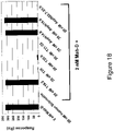
- Antibody B (also referred to herein as Ab-B and Mab-B) is a mouse antibody which exhibits high affinity binding to sclerostin.
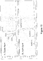
- the BIAcore binding pattern of Ab-B is shown in Figure 16 .
- the amino acid sequence of the mature form (signal peptide removed) of the Ab-B LC is:
- the nucleic acid sequence encoding the mature form (signal peptide removed) of Ab-B LC is:
- the amino acid sequence of Ab-B LC including signal peptide is:
- the nucleic acid sequence of Ab-B LC including signal peptide encoding sequence is:
- the CDR (complementarity determining region) sequences in the variable region of the heavy chain of Ab-B are as follows:
- Antibodies disclosed herein bind to regions of human sclerostin which are important for the in vivo activity of the protein. Binding of an antibody to sclerostin can be correlated with increases in, for example, the bone mineral density achieved by use of the antibody in vivo such as described in Examples 5 and 9 (mice) and Example 12 (monkey). Increases in at least one of bone formation, bone mineral content, bone mass, bone quality and bone strength can also be achieved by use of the antibody in vivo such as described in Examples 5 and 9 (mice) and Example 12 (monkey).
- an antibody for practicing the invention may be generated with all or some of the disclosed CDR sequences in an appropriate framework, wherein the antibody retains the ability to bind specifically to sclerostin, and can be expected to achieve increases in, for example, bone mineral density.
- Such antibodies are useful in the treatment of human or animal conditions that are caused by, associated with, or result in at least one of low bone formation, low bone mineral density, low bone mineral content, low bone mass, low bone quality and low bone strength.
- the present invention therefore relates in one embodiment to an isolated antibody, including Ab-A, or an antigen binding fragment thereof, which specifically binds to sclerostin and wherein the variable domain of the heavy chain comprises at least one CDR having the sequences given in SEQ ID NO:51 for CDR-H1, SEQ ID NO:52 for CDR-H2 and SEQ ID NO:53 for CDR-H3.
- the antibody or antigen binding fragment thereof may comprise a heavy chain variable domain in which the CDRs consist of at least one of the peptides of SEQ ID NO:51 for CDR-H1, SEQ ID NO:52 for CDR-H2 and SEQ ID NO:53 for CDR-H3.
- the light chain may be any suitable complementary chain and may in particular be selected from a light chain wherein the variable domain comprises at least one CDR having the sequences given in SEQ ID NO:54 for CDR-L1, SEQ ID NO:55 for CDR-L2 and SEQ ID NO:56 for CDR-L3.
- the antibody or antigen binding fragment thereof may comprise a light chain variable domain in which the CDRs consist of at least one of the peptides of SEQ ID NO:54 for CDR-L1, SEQ ID NO:55 for CDR-L2 and SEQ ID NO:56 for CDR-L3.
- the present invention further relates to an isolated antibody, including Ab-B, or an antigen binding fragment hereof, which specifically binds to sclerostin and wherein the variable domain of the heavy chain comprises at least one CDR having the sequences given in SEQ ID NO:57 for CDR-H1, SEQ ID NO:58 for CDR-H2 and SEQ ID NO:59 for CDR-H3.
- the antibody or antigen binding fragment thereof may comprise a heavy chain variable domain in which the CDRs consist of at least one of the peptides of SEQ ID NO:57 for CDR-H1, SEQ ID NO:58 for CDR-H2 and SEQ ID NO:59 for CDR-H3.
- the light chain may be any suitable complementary chain and may in particular be selected from a light chain wherein the variable domain comprises at least one CDR having the sequences given in SEQ ID NO:60 for CDR-L1, SEQ ID NO:61 for CDR-L2 and SEQ ID NO:62 for CDR-L3.
- the antibody or antigen binding fragment thereof may comprise a light chain variable domain in which the CDRs consist of at least one of the peptides of SEQ ID NO:60 for CDR-L1, SEQ ID NO:61 for CDR-L2 and SEQ ID NO:62 for CDR-L3.
- the present invention still further relates to an isolated antibody, including Ab-C, or an antigen binding fragment hereof, which specifically binds to sclerostin and wherein the variable domain of the heavy chain comprises at least one CDR having the sequences given in SEQ ID NO:45 for CDR-H1, SEQ ID NO:46 for CDR-H2 and SEQ ID NO:47 for CDR-H3.
- the antibody or antigen binding fragment thereof may comprise a heavy chain variable domain in which the CDRs consist of at least one of the peptides of SEQ ID NO:45 for CDR-H1, SEQ ID NO:46 for CDR-H2 and SEQ ID NO:47 for CDR-H3.
- the light chain may be any suitable complementary chain and may in particular be selected from a light chain wherein the variable domain comprises at least one CDR having the sequences given in SEQ ID NO:48 for CDR-L1, SEQ ID NO:49 for CDR-L2 and SEQ ID NO:50 for CDR-L3.
- the antibody or antigen binding fragment thereof may comprise a light chain variable domain in which the CDRs consist of at least one of the peptides of SEQ ID NO:48 for CDR-L1, SEQ ID NO:49 for CDR-L2 and SEQ ID NO:50 for CDR-L3.
- the present invention also relates to an isolated antibody, including Ab-D, or an antigen binding fragment hereof, which specifically binds to sclerostin and wherein the variable domain of the heavy chain comprises at least one CDR having the sequences given in SEQ ID NO:39 for CDR-H1, SEQ ID NO:40 for CDR-H2 and SEQ ID NO:41 for CDR-H3.
- the antibody or antigen binding fragment thereof may comprise a heavy chain variable domain in which the CDRs consist of at least one of the peptides of SEQ ID NO:39 for CDR-H1, SEQ ID NO:40 for CDR-H2 and SEQ ID NO:41 for CDR-H3.
- the light chain may be any suitable complementary chain and may in particular be selected from a light chain wherein the variable domain comprises at least one CDR having the sequences given in SEQ ID NO:42 for CDR-L1, SEQ ID NO:43 for CDR-L2 and SEQ ID NO:44 for CDR-L3.
- the antibody or antigen binding fragment thereof may comprise a light chain variable domain in which the CDRs consist of at least one of the peptides of SEQ ID NO:42 for CDR-L1, SEQ ID NO:43 for CDR-L2 and SEQ ID NO:44 for CDR-L3.
- CDRs complementarity-determining regions
- the sequences of the Antibody 2 (also referred to as Ab-2) LC and HC are as follows:
- the sequences of the Antibody 3 (also referred to herein as Ab-3) LC and HC are as follows:
- the sequences of the Antibody 4 (also referred to herein as Ab-4) LC and HC are as follows:
- Ab-4 was humanized to generate Ab-5.
- the sequences of the Antibody 5 (also referred to herein as Ab-5) LC and HC are as follows:
- the CDR (complementarity determining region) sequences in the variable region of the heavy chain of Ab-5 are as follows:
- Antibody 6 also referred to herein as Ab-6
- LC and HC are as follows:
- the sequences of the Antibody 7 (also referred to herein as Ab-7) LC and HC are as follows:
- Antibody 8 also referred to herein as Ab-8
- LC and HC are as follows:
- the sequences of the Antibody 9 (also referred to herein as Ab-9) LC and HC are as follows:
- the sequences of the Antibody 10 (also referred to herein as Ab-10) LC and HC are as follows:
- the sequences of the Antibody 11 (also referred to herein as Ab-11) LC and HC are as follows:
- the sequences of the Antibody 12 (also referred to herein as Ab-12) LC and HC are as follows:
- the sequences of the Antibody 13 (also referred to herein as Ab-13) LC and HC are as follows:
- Ab-13 was humanized to generate Ab-14.
- the sequences of the Antibody 14 (also referred to herein as Ab-14) LC and HC are as follows:
- the CDR sequences in the variable region of the heavy chain of Ab-14 are:
- Ab-3 was humanized to generate Ab-15.
- the sequences of the Antibody 15 (also referred to herein as Ab-15) LC and HC are as follows:
- the CDR sequences in the variable region of the heavy chain of Ab-15 are:
- Ab-11 was humanized to generate Ab-16.
- the sequences of the Antibody 16 (also referred to herein as Ab-16) LC and HC are as follows:
- the CDR sequences in the variable region of the heavy chain of Ab-16 are:
- Antibodies 17-22 also referred to herein as Ab-17, Ab-18, Ab-19, Ab-20, Ab-21, and Ab-22.
- the Kappa Constant region for all VK regions of Ab-17, Ab-19, and Ab-21 is as follows:
- the Heavy Constant Region for all VH regions of antibodies 17, 19 and 21 is as follows:
- the boxed-shaded amino acids represent complement-determining regions (CDRs) and the underlined amino acids represent signal peptide.
- Ab-17 was humanized to generate Ab-18.
- Ab-19 was humanized to generate Antibody 20 (also referred to herein as Ab-20) and Antibody 23 (also referred to herein as Ab-23).
- the CDR (complementarity determining region) sequences in the variable region of the heavy chain of Ab-23 are as follows:
- Ab-21 was humanized to yield Ab-22.
- the light chain human kappa constant region is as follows: and the heavy chain human gamma-4 constant region is as follows:
- the hinge region contains the Ser-241-Pro mutation to improve hinge stability ( Angal S et al, (1993), Mol Immunol, 30(1), 105-108 ).
- Antibody 24 also referred to herein as Ab-24
- LC and HC are as follows:
- the CDR sequences in the variable region of the light chain of Ab-24 are as follows:
- the CDR sequences in the variable region of the heavy chain of Ab-24 are as follows:
- Table 1 below provides the SEQ ID NOs and amino acid sequences of the CDR's of Ab-A, Ab-B, Ab-C, Ab-D, Ab-1, Ab-2, Ab-3, Ab-4, Ab-5, Ab-6, Ab-7, Ab-8, Ab-9, Ab-10, Ab-11, Ab-12, Ab-13, Ab-14, Ab-15, Ab-16, Ab-17, Ab-18, Ab-19, Ab-20, Ab-21, Ab-22, Ab-23, and Ab-24.
- L1, L2, and L3 refer to light chain CDR's 1, 2, and 3, and HI, H2, and H3 refer to heavy chain CDR's 1, 2, and 3 according to the Kabat numbering system ( Kabat et al., 1987 in Sequences of Proteins of Immunological Interest, U.S.
- oligopeptide or polypeptide is within the scope of the invention if it has an amino acid sequence that is at least 75%, 76%, 77%, 78%, 79%, 80%, 81%, 82%, 83%, 84%, 85%, 86%, 87%, 88%, 89%, 90%, 91%, 92%, 93%, 94%, 95%, 96%, 97%, 98% or 99% identical to least one of the CDR's of Table 1 above; and/or to a CDR of a sclerostin binding agent that cross-blocks the binding of at least one of antibodies Ab-A, Ab-B, Ab-C, Ab-D, Ab-1, Ab-2, Ab-3, Ab-4, Ab-5, Ab-6, Ab-7, Ab-8, Ab-9, Ab-10, Ab-11, Ab-12, Ab-13, Ab-14, Ab-15, Ab-16, Ab-17, Ab-18, Ab-19, Ab-20, Ab-21, Ab-22, Ab-23, and Ab-24 to sclerostin, and/or is cross-blocked from
- a sclerostin neutralizing binding agent and/or to a CDR of a sclerostin binding agent that binds to a Loop 2 epitope; and/or to a CDR of a sclerostin binding agent that binds to a T20.6 epitope; and/or to a CDR of a sclerostin binding agent that binds to a "T20.6 derivative (cystine-knot + 4 arms)" epitope.
- Sclerostin binding agent polypeptides and antibodies are within the scope of the invention if they have amino acid sequences that are at least 85%, 86%, 87%, 88%, 89%, 90%, 91%, 92%, 93%, 94%, 95%, 96%, 97%, 98% or 99% identical to a variable region of at least one of antibodies Ab-A, Ab-B, Ab-C, Ab-D, Ab-1, Ab-2, Ab-3, Ab-4, Ab-5, Ab-6, Ab-7, Ab-8, Ab-9, Ab-10, Ab-11, Ab-12, Ab-13, Ab-14, Ab-15, Ab-16, Ab-17, Ab-18, Ab-19, Ab-20, Ab-21, Ab-22, Ab-23, and Ab-24, and cross-block the binding of at least one of antibodies Ab-A, Ab-B, Ab-C, Ab-D, Ab-1, Ab-2, Ab-3, Ab-4, Ab-5, Ab-6, Ab-7, Ab-8, Ab-9, Ab-10, Ab-11, Ab-12, Ab-13, Ab-14, Ab-15, Ab-16, Ab-17
- a sclerostin neutralizing binding agent binds to a Loop 2 epitope; and/or bind to a T20.6 epitope; and/or bind to a "T20.6 derivative (cystine-knot + 4 arms)" epitope.
- Polynucleotides encoding sclerostin binding agents are within the scope of the invention if they have polynucleotide sequences that are at least 85%, 86%, 87%, 88%, 89%, 90%, 91%, 92%, 93%, 94%, 95%, 96%, 97%, 98% or 99% identical to a polynucleotide encoding a variable region of at least one of antibodies Ab-A, Ab-B, Ab-C, Ab-D, Ab-1, Ab-2, Ab-3, Ab-4, Ab-5, Ab-6, Ab-7, Ab-8, Ab-9, Ab-10, Ab-11, Ab-12, Ab-13, Ab-14, Ab-15, Ab-16, Ab-17, Ab-18, Ab-19, Ab-20, Ab-21, Ab-22, Ab-23, and Ab-24, and wherein the encoded sclerostin binding agents cross-block the binding of at least one of antibodies Ab-A, Ab-B, Ab-C, Ab-D, Ab-1, Ab-2, Ab-3, Ab-4, Ab-5
- a sclerostin neutralizing binding agent binds to a Loop 2 epitope; and/or bind to a T20.6 epitope; and/or bind to a "T20.6 derivative (cystine-knot + 4 arms)" epitope.
- Antibodies according to the invention may have a binding affinity for human sclerostin of less than or equal to 1 ⁇ 10 -7 M, less than or equal to 1 ⁇ 10 -8 M, less than or equal to 1 ⁇ 10 -9 M, less than or equal to 1 ⁇ 10 -10 M, less than or equal to 1 ⁇ 10 -11 M, or less than or equal to 1 ⁇ 10 -12 M.
- the affinity of a binding agent such as an antibody or binding partner, as well as the extent to which a binding agent (such as an antibody) inhibits binding, can be determined by one of ordinary skill in the art using conventional techniques, for example those described by Scatchard et al. (Ann. N.Y. Acad. Sci. 51:660-672 (1949 )) or by surface plasmon resonance (SPR; BIAcore, Biosensor, Piscataway, NJ).
- SPR surface plasmon resonance
- target molecules are immobilized on a solid phase and exposed to ligands in a mobile phase running along a flow cell.
- the local refractive index changes, leading to a change in SPR angle, which can be monitored in real time by detecting changes in the intensity of the reflected light.
- the rates of change of the SPR signal can be analyzed to yield apparent rate constants for the association and dissociation phases of the binding reaction. The ratio of these values gives the apparent equilibrium constant (affinity) ( see, e.g., Wolff et al., Cancer Res. 53:2560-65 (1993 )).
- An antibody according to the present invention may belong to any immunoglobin class, for example IgG, IgE, IgM, IgD, or IgA. It may be obtained from or derived from an animal, for example, fowl ( e.g ., chicken) and mammals, which includes but is not limited to a mouse, rat, hamster, rabbit, or other rodent, cow, horse, sheep, goat, camel, human, or other primate. The antibody may be an internalizing antibody. Production of antibodies is disclosed generally in U.S. Patent Publication No. 2004/0146888 A1 .
- Mature form human sclerostin is a 190 amino acid glycoprotein with a cystine-knot structure ( Figures 8 and 9 ).
- the protein is characterized as having three loops designated as Loop 1, Loop 2 and Loop 3.
- Human sclerostin was subjected to proteolytic digestion to produce fragments. Briefly, using different proteases, including trypsin, aspN, and lysC, fragments with various cleavage sites and sizes were generated. The sequences and mass for various human sclerostin peptides were determined. Antibody protection was evaluated to determine the effect on accessibility for proteolysis, including clipped site masking and peptide shifting. Finally, a BIAcore-based "human sclerostin peptide epitope competition assay" was performed.
- T20.6 epitope is a complex of four separate peptide sequences which are joined by the three disulfide bonds of the cystine-knot region. Two of the peptides are joined by two disulfide bonds. The other two peptides are linked by one disulfide bond that, schematically, bisects the first two polypeptides.
- the T20.6 epitope that was generated by trypsin digestion retains the cystine-knot structure of the native polypeptide and is recognized by antibodies Ab-C and Ab-D.
- a derivative of epitope T20.6 consists of the cystine-knot region and amino acids 58-64, 73-81, 112-117 and 138-141 in sequence position with reference to SEQ ID NO:1. This derivative epitope is shown in Figure 21 .
- An epitope comprising the cystine-knot region may have one or more amino acids that is present in the T20.6 epitope ( Figure 19B ) but not present in the T20.6 derivative epitope ( Figure 21 ).
- a Loop 2 epitope comprises amino acids 86-111 of SEQ ID NO:1 (C4GPARLLPNAIGRGKWWRPSGPDFRC5, SEQ ID NO:6).
- the Loop 2-containing structure is defined at one end by a disulfide bond between cysteine at position 86 (C4) and cysteine at position 144 (C8), and at the other end by a disulfide bond between cysteine at position 111 (C5) and cysteine at position 57 (C1).
- the peptides generated by aspN cleavage of human sclerostin are shown in Figure 12 .
- these peptides are designated as AspN14.6, AspN18.6, and AspN22.7-23.5, and are also referred to herein as N14.6, N18.6, and N22.7-23.5, respectively.
- One group of antibodies exhibits a specific pattern of binding to certain epitopes as evidenced by a Biacore-based "human sclerostin peptide epitope competition binding assay.” Briefly, the antibody is preincubated with the epitope to be tested, at concentrations that will saturate the epitope-binding sites on the antibody. The antibody is then exposed to sclerostin bound to a chip surface. After the appropriate incubation and washing procedures, a pattern of competitive binding is established. As shown in Figure 18 , exemplary antibody Ab-D bound to sclerostin molecules attached to the surface of the chip.
- Preincubation of antibody Ab-D with sclerostin decreased the binding of the antibody to the sclerostin on the chip to close to zero.
- Preincubation with a peptide consisting of epitope T19.2 showed that T19.2 did not compete with sclerostin for antibody binding.
- preincubation with any one of the epitopes designated T20, T20.6, T21-22, or N22.7-23.5 abolished a large proportion of the binding of antibody to sclerostin on the chip.
- preincubation of the antibody with any one of the epitopes designated T19.2, N14.6 or N18.6 did not abolish the ability of the antibody to bind to sclerostin.
- a second exemplary antibody with this binding profile is Ab-C.
- Antibody Ab-D therefore is exemplary and representative of a group of antibodies that bind to the epitopes T20, T20.6, T21-22, and N22.7-23.5, and have minimal detectable binding to epitopes T19.2, N14.6 and N18.6, as measured by the ability to block antibody binding to sclerostin.
- Antibodies having this characteristic binding pattern may or may not share amino acid sequence in one or more regions of the antibody molecule.
- Antibody similarity is determined functionally such as by the ability to bind to sclerostin following preincubation with each of the epitopes described above.
- Antibodies that exhibit a binding pattern similar or identical to that of antibody Ab-D are included in the invention.
- the antibody will exhibit binding to each of the polypeptides T20, T20.6, T21-22 and N22.7-23.5 whereby this binding will specifically compete out at least 50% of the antibody's binding to sclerostin that would otherwise occur in the absence of preincubation with sclerostin or a sclerostin peptide.
- the antibody will also exhibit little or no detectable binding to polypeptides T19.2, N14.6 and N18.6, resulting in a reduction of 30% or less of the binding that would occur in the absence of preincubation with sclerostin or a sclerostin peptide.
- the antibody binding pattern of Figure 18 suggests that the epitope space to which antibody Ab-D and other antibodies having the epitope binding pattern of Ab-D bind consists of a polypeptide comprising the cystine-knot region of sclerostin.
- an exemplary T20.6 epitope comprises four peptide chains attached via three separate disulfide bonds.
- Peptide chain SAKPVTELVC3SGQC4GPAR (SEQ ID NO:3) is attached to peptide chain LVASC7KC8KRLTR (SEQ ID NO:5) by disulfide bonds from C3 to C7, and from C4 to C8.
- Peptide chain DVSEYSC1RELHFTR (SEQ ID NO:2) is attached to peptide chain WWRPSGPDFRC5IPDRYR (SEQ ID NO:4) by a disulfide bond from C1 to C5.
- an exemplary derivative epitope of T20.6 comprises four peptide chains attached via three separate disulfide bonds.
- Peptide chain SAKPVTELVC3SGQC4 (SEQ ID NO:70) is attached to peptide chain LVASC7KC8 (SEQ ID NO:71) by disulfide bonds from C3 to C7, and from C4 to C8.
- Peptide chain C1RELHFTR (SEQ ID NO:72) is attached to peptide chain C5IPDRYR (SEQ ID NO:73) by a disulfide bond from C1 to C5.
- polypeptides of SEQ ID NOs:70 and 71 remain associated with the polypeptides of SEQ ID NOs:72 and 73 through a steric construct whereby the C1-C5 bond crosses the plane of the C4-C8 and C3-C7 bonds and is located between them, as illustrated in Figure 21 .
- Antibody Ab-A is exemplary and representative of a second group of antibodies that have a characteristic binding pattern to human sclerostin peptides that is distinct from that obtained for antibodies Ab-C and Ab-D.
- Ab-A and the group of antibodies it represents bind to the N22.7-23.5 epitope and have minimal detectable binding to epitopes T19.2, T20, T20.6, T21-22, N14.6 or N18.6, as measured by the ability to block antibody binding to sclerostin ( Fig 15 ).
- a second exemplary antibody with this binding profile ( Fig. 16 ) is Ab-B.
- Antibodies having this characteristic binding pattern may or may not share amino acid sequence in one or more regions of the antibody molecule.
- Antibody similarity is determined functionally such as by the ability to bind to sclerostin following preincubation with each of the epitopes described above.
- Antibodies that exhibit a binding pattern similar or identical to that of antibody Ab-A are included in the invention.
- similar to is meant, for example, the antibody will exhibit binding to the N22.7-23.5 polypeptide whereby this binding will specifically compete out at least 50% of the antibody's binding to sclerostin that would otherwise occur in the absence of preincubation with sclerostin or a sclerostin peptide.
- the antibody will also exhibit little or no detectable binding to polypeptides T19.2, T20, T20.6, T21-22, N14.6 and N18.6, resulting in a reduction of 30% or less of the binding that would occur in the absence of preincubation with sclerostin or a sclerostin peptide.
- Biacore assay for determining whether an antibody or other binding agent cross-blocks or is capable of cross-blocking according to the invention.
- the Biacore machine for example the Biacore 3000 is operated in line with the manufacturer's recommendations.
- the two antibodies (termed A ⁇ and B ⁇ ) to be assessed for their ability to cross-block each other are mixed at a one to one molar ratio of binding sites in a suitable buffer to create the test mixture.
- concentrations on a binding site basis the molecular weight of an antibody is assumed to be the total molecular weight of the antibody divided by the number of sclerostin binding sites on that antibody.
- each antibody in the test mix should be high enough to readily saturate the binding sites for that antibody on the sclerostin molecules captured on the Biacore chip.
- the antibodies in the mixture are at the same molar concentration (on a binding basis) and that concentration would typically be between 1.00 and 1.5 micromolar (on a binding site basis).
- the Biacore assay described above is a primary assay used to determine if antibodies or other binding agents cross-block each other according to the invention. On rare occasions particular antibodies or other binding agents may not bind to sclerostin coupled via amine chemistry to a CM5 Biacore chip (this usually occurs when the relevant binding site on sclerostin is masked or destroyed by the coupling to the chip). In such cases cross-blocking can be determined using a tagged version of Sclerostin, for example N-terminal His-tagged Sclerostin (R & D Systems, Minneapolis, MN, USA; 2005 cat# 1406-ST-025).
- an anti-His antibody would be coupled to the Biacore chip and then the His-tagged Sclerostin would be passed over the surface of the chip and captured by the anti-His antibody.
- the cross blocking analysis would be carried out essentially as described above, except that after each chip regeneration cycle, new His-tagged sclerostin would be loaded back onto the anti-His antibody coated surface.
- C-terminal His-tagged sclerostin could alternatively be used.
- various other tags and tag binding protein combinations that are known in the art could be used for such a cross-blocking analysis (e.g. HA tag with anti-HA antibodies; FLAG tag with anti-FLAG antibodies; biotin tag with streptavidin).
- the following generally describes an ELISA assay for determining whether an anti-sclerostin antibody or other sclerostin binding agent cross-blocks or is capable of cross-blocking according to the invention.
- an ELISA assay for determining whether an anti-sclerostin antibody or other sclerostin binding agent cross-blocks or is capable of cross-blocking according to the invention.
- reference is made to two antibodies (Ab-X and Ab-Y), but it will be appreciated that the assay can be used with any of the sclerostin binding agents described herein.
- the amount of bound sclerostin is then measured using an appropriate sclerostin detection reagent.
- An antibody in solution that is able to cross-block the coated antibody will be able to cause a decrease in the number of sclerostin molecules that the coated antibody can bind relative to the number of sclerostin molecules that the coated antibody can bind in the absence of the second, solution phase, antibody.
- Sclerostin is then added such that the moles of sclerostin added per well are at least 25-fold lower than the moles of Ab-X sclerostin binding sites that were used for coating each well.
- the ELISA plate is washed and a sclerostin detection reagent is added to measure the amount of sclerostin specifically bound by the coated anti-sclerostin antibody (in this case Ab-X).
- the background signal for the assay is defined as the signal obtained in wells with the coated antibody (in this case Ab-X), second solution phase antibody (in this case Ab-Y), sclerostin buffer only ( i.e. no sclerostin) and sclerostin detection reagents.
- the positive control signal for the assay is defined as the signal obtained in wells with the coated antibody (in this case Ab-X), second solution phase antibody buffer only ( i.e. no second solution phase antibody), sclerostin and sclerostin detection reagents.
- the ELISA assay needs to be run in such a manner so as to have the positive control signal be at least 6 times the background signal.
- the cross-blocking assay needs to be run in two formats:
- Example 7 An example of such an ELISA-based cross blocking assay can be found in Example 7 ("ELISA-based cross-blocking assay").
- Mineralization by osteoblast-lineage cells in culture is used as an in vitro model of bone formation. Mineralization takes from about one to six weeks to occur beginning with the induction of osteoblast-lineage cell differentiation by one or more differentiation agents.
- the overall sequence of events involves cell proliferation, differentiation, extracellular matrix production, matrix maturation and finally deposition of mineral, which refers to crystallization and/or deposition of calcium phosphate. This sequence of events starting with cell proliferation and differentiation, and ending with deposition of mineral is referred to herein as mineralization.
- Measurement of calcium (mineral) is the output of the assay.
- MC3T3-E1 cells Sudo H, Kodama H-A, Amagai Y, Yamamoto S, Kasai S. 1983. In vitro differentiation and calcification in a new clonal osteogenic cell line derived from newborn mouse calvaria. J. Cell Biol. 96:191-198 ) and subclones of the original cell line can form mineral in culture upon growth in the presence of differentiating agents. Such subclones include MC3T3-E1-BF ( Smith E, Redman R, Logg C, Coetzee G, Kasahara N, Frenkel B. 2000. Glucocorticoids inhibit developmental stage-specific osteoblast cell cycle. J. Biol. Chem. 275:19992-20001 ).
- the amount of sclerostin used in the assay needs to be the minimum amount of sclerostin that causes at least a 70%, statistically significant, reduction in deposition of calcium phosphate (measured as calcium) in the sclerostin-only group, as compared to the amount of calcium measured in the no sclerostin group.
- An anti-sclerostin neutralizing antibody or an anti-sclerostin neutralizing binding agent is defined as one that causes a statistically significant increase in deposition of calcium phosphate (measured as calcium) as compared to the amount of calcium measured in the sclerostin-only (i.e. no antibody, no binding agent) treatment group.
- the amount of anti-sclerostin antibody or anti-sclerostin binding agent used in the assay needs to be such that there is an excess of moles of sclerostin binding sites per well as compared to the number of moles of sclerostin per well.
- the fold excess that may be required can be 24, 18, 12, 6, 3, or 1.5, and one of skill is familiar with the routine practice of testing more than one concentration of binding agent.
- a very potent anti-sclerostin neutralizing antibody or anti-sclerostin neutralizing binding agent will be able to neutralize sclerostin even when there is less than a 6-fold excess of moles of sclerostin binding sites per well as compared to the number of moles of sclerostin per well.
- a less potent anti-sclerostin neutralizing antibody or anti-sclerostin neutralizing binding agent will be able to neutralize sclerostin only at a 12, 18 or 24 fold excess.
- Sclerostin binding agents within this full range of potencies are suitable as neutralizing sclerostin binding agents. Exemplary cell based mineralization assays are described in detail in Example 8.
- Increases in various parameters associated with, or that result from, the stimulation of new bone formation can be measured as an output from in vivo testing of sclerostin binding agents in order to identify those binding agents that are able to neutralize sclerostin and thus able to cause stimulation of new bone formation.
- Such parameters include various serum anabolic markers [ e . g . osteocalcin, P1NP (n-terminal propeptide of type 1 procollagen)], histomorphometric markers of bone formation (e.g. osteoblast surface/bone surface; bone formation rate/bone surface; trabecular thickness), bone mineral density, bone mineral content, bone mass, bone quality and bone strength.
- compositions disclosed herein may be delivered via oral administration to an animal.
- these compositions may be formulated with an inert diluent or with an assimilable edible carrier, or they may be enclosed in hard- or soft-shell gelatin capsule, or they may be compressed into tablets, or they may be incorporated directly with the food of the diet.
- Proper fluidity may be maintained, for example, by the use of a coating, such as lecithin, by the maintenance of the required particle size in the case of dispersion and/or by the use of surfactants.
- the prevention of the action of microorganisms can be facilitated by various antibacterial and antifungal agents, for example, parabens, chlorobutanol, phenol, sorbic acid, thimerosal, and the like.
- Prolonged absorption of the injectable compositions can be brought about by the use in the compositions of agents delaying absorption, for example, aluminum monostearate and gelatin.
- the solution for parenteral administration in an aqueous solution, the solution should be suitably buffered if necessary and the liquid diluent first rendered isotonic with sufficient saline or glucose.
- aqueous solutions are especially suitable for intravenous, intramuscular, subcutaneous and intraperitoneal administration.
- a sterile aqueous medium that can be employed will be known to those of skill in the art in light of the present disclosure.
- one dosage may be dissolved in 1 ml of isotonic NaCl solution and either added to 1000 ml of hypodermoclysis fluid or injected at the proposed site of infusion, (see for example, Remington's Pharmaceutical Sciences, 15th ed., pp.
- compositions disclosed herein may be formulated in a neutral or salt form.
- Illustrative pharmaceutically-acceptable salts include the acid addition salts (formed with the free amino groups of the protein) and which are formed with inorganic acids such as, for example, hydrochloric or phosphoric acids, or such organic acids as acetic, oxalic, tartaric, mandelic, and the like. Salts formed with the free carboxyl groups can also be derived from inorganic bases such as, for example, sodium, potassium, ammonium, calcium, or ferric hydroxides, and such organic bases as isopropylamine, trimethylamine, histidine, procaine and the like. Upon formulation, solutions will be administered in a manner compatible with the dosage formulation and in such amount as is therapeutically effective.
- the carriers can further comprise any and all solvents, dispersion media, vehicles, coatings, diluents, antibacterial and antifungal agents, isotonic and absorption delaying agents, buffers, carrier solutions, suspensions, colloids, and the like.
- solvents dispersion media, vehicles, coatings, diluents, antibacterial and antifungal agents, isotonic and absorption delaying agents, buffers, carrier solutions, suspensions, colloids, and the like.
- the use of such media and agents for pharmaceutical active substances is well known in the art. Except insofar as any conventional media or agent is incompatible with the active ingredient, its use in the therapeutic compositions is contemplated. Supplementary active ingredients can also be incorporated into the compositions.
- pharmaceutically-acceptable refers to molecular entities and compositions that do not produce an allergic or similar untoward reaction when administered to a human.
- compositions of the present invention are used for the introduction of the compositions of the present invention into suitable host cells/organisms.
- the compositions of the present invention may be formulated for delivery either encapsulated in a lipid particle, a liposome, a vesicle, a nanosphere, or a nanoparticle or the like.
- compositions of the present invention can be bound, either covalently or non-covalently, to the surface of such carrier vehicles.
- liposomes are formed from phospholipids that are dispersed in an aqueous medium and spontaneously form multilamellar concentric bilayer vesicles (also termed multilamellar vesicles (MLVs)).
- MLVs multilamellar vesicles
- the invention provides for pharmaceutically-acceptable nanocapsule formulations of the compositions of the present invention.
- Nanocapsules can generally entrap compounds in a stable and reproducible way (see, for example, Quintanar-Guerrero et al., Drug Dev. Ind. Pharm. 24(12):1113-28, 1998 ).
- ultrafine particles sized around 0.1 ⁇ m
- Such particles can be made as described, for example, by Couvreur et al., Crit. Rev. Ther. Drug Carrier Syst. 5(1):1-20, 1988 ; zur Muhlen et al., Eur. J. Pharm. Biopharm. 45(2):149-55, 1998 ; Zambaux et al., J. Controlled Release 50(1-3):31-40, 1998 ; and U.S. Patent No. 5,145,684 .
- compositions of the present invention may be placed within containers, along with packaging material that provides instructions regarding the use of such pharmaceutical compositions.
- instructions will include a tangible expression describing the reagent concentration, as well as within certain embodiments, relative amounts of excipient ingredients or diluents (e . g ., water, saline or PBS) that may be necessary to reconstitute the pharmaceutical composition.
- the dose administered may range from 0.01 mg/kg to 100 mg/kg of body weight.
- the amount and frequency of administration will depend, of course, on such factors as the nature and severity of the indication being treated, the desired response, the condition of the patient, and so forth.
- the compositions may be administered by a variety of techniques, as noted above.
- Increases in bone mineral content and/or bone mineral density may be determined directly through the use of X-rays (e . g ., Dual Energy X-ray Absorptometry or "DEXA”), or by inference through the measurement of 1) markers of bone formation and/or osteoblast activity, such as, but not limited to, osteoblast specific alkaline phosphatase, osteocalcin, type 1 procollagen C' propeptide (PICP), total alkaline phosphatase ( see Comier, Curr. Opin. in Rheu.
- X-rays e . g ., Dual Energy X-ray Absorptometry or "DEXA”
- markers of bone formation and/or osteoblast activity such as, but not limited to, osteoblast specific alkaline phosphatase, osteocalcin, type 1 procollagen C' propeptide (PICP), total alkaline phosphatase ( see Comier, Curr. Opin. in Rheu.
- compositions of the present invention include dysplasias, wherein growth or development of bone is abnormal and a wide variety of causes of osteopenia, osteoporosis and bone loss.
- Representative examples of such conditions include achondroplasia, cleidocranial dysostosis, enchondromatosis, fibrous dysplasia, Gaucher's Disease, hypophosphatemic rickets, Marfan's syndrome, multiple hereditary exotoses, neurofibromatosis, osteogenesis imperfecta, osteopetrosis, osteopoikilosis, sclerotic lesions, pseudoarthrosis, and pyogenic osteomyelitis, periodontal disease, anti-epileptic drug induced bone loss, primary and secondary hyperparathyroidism, familial hyperparathyroidism syndromes, weightlessness induced bone loss, osteoporosis in men, postmenopausal bone loss, osteoarthritis, renal osteodystrophy, infiltrative disorders of bone, oral bone
- compositions of the present invention may also be useful for improving outcomes in orthopedic procedures, dental procedures, implant surgery, joint replacement, bone grafting, bone cosmetic surgery and bone repair such as fracture healing, nonunion healing, delayed union healing and facial reconstruction.
- One or more compositions may be administered before, during and/or after the procedure, replacement, graft, surgery or repair.
- the invention also provides a diagnostic kit comprising at least one anti-sclerostin binding agent according to the present invention.
- the binding agent may be an antibody.
- such a kit may optionally comprise one or more of the following:
- the binding agent(s) itself can be labeled with one or more of a detectable marker(s), e.g. a chemiluminescent, enzymatic, fluorescent, or radioactive moiety.
- a detectable marker e.g. a chemiluminescent, enzymatic, fluorescent, or radioactive moiety.
- the different species of sclerostin can be expressed transiently in serum-free suspension adapted 293T or 293EBNA cells. Transfections can be performed as 500 mL or 1L cultures. The following reagents and materials are available from Gibco BRL (now Invitrogen, Carlsbad, CA).
- the cells are resuspended in serum-free DMEM and centrifuged again at 2,500 RPM for 10 minutes at 4°C. After aspirating the wash solution, the cells are resuspended in growth medium [DMEM/F12 (3:1) + 1X Insulin-Transferrin-Selenium Supplement + 1X Pen Strep Glut + 2mM L-Glutamine + 20 mM HEPES + 0.01% Pluronic F68] in a 1L or 3L spinner flask culture. The spinner flask culture is maintained on magnetic stir plate at 125 RPM which is placed in a humidified incubator maintained at 37°C and 5% CO 2 .
- the mammalian expression plasmid DNA e . g .
- pcDNA3.1, pCEP4, Invitrogen Life Technologies, Carlsbad, CA containing the complete coding region (and stop codon) of sclerostin with a Kozak consensus sequence (e.g ., CCACC) directly 5' of the start site ATG, is complexed to the transfection reagent in a 50 mL conical tube.
- the DNA-transfection reagent complex can be prepared in 5-10% of the final culture volume in serum-free DMEM or OPTI-MEM.
- the transfection reagents that can be used for this purpose include X-tremeGene RO-1539 (Roche Applied Science, Indianapolis, IN), FuGene6 (Roche Applied Science, Indianapolis, IN), Lipofectamine 2000 (Invitrogen, Carlsbad, CA) and 293fectin (Invitrogen, Carlsbad, CA). 1-5 ⁇ g plasmid DNA/mL culture is first added to serum-free DMEM, followed by 1-5 ⁇ l transfection reagent/mL culture.
- the complexes can be incubated at room temperature for approximately 10-30 minutes and then added to the cells in the spinner flask.
- the transfection/expression can be performed for 4-7 days, after which the conditioned medium (CM) is harvested by centrifugation at 4,000 RPM for 60 minutes at 4°C.
- CM conditioned medium
- Recombinant sclerostin was purified from mammalian host cells as follows. All purification processes were carried out at room temperature. One purification scheme was used to purify various species of sclerostin, including murine and human sclerostin. The purification scheme used affinity chromatography followed by cation exchange chromatography.
- CM mammalian host cell conditioned medium
- Beckman J6-M1 centrifuge at 4000 rpm for 1hour at 4°C to remove cell debris.
- the CM supernatant was then filtered through a sterile 0.2 ⁇ m filter. (At this point the sterile filtered CM may be optionally stored frozen until purification.) If the CM was frozen, it was thawed at the following temperatures, or combination thereof: 4°C, room temperature or warm water. Following thawing the CM was filtered through a sterile 0.2 ⁇ m filter and optionally concentrated by tangential flow ultrafiltration (TFF) using a 10 kD molecular weight cut-off membrane.
- TMF tangential flow ultrafiltration
- CM concentrate was filtered through a sterile 0.2 ⁇ m filter and then loaded onto a Heparin High Performance (Heparin HP) column (GE Healthcare, formerly Amersham Biosciences) equilibrated in PBS.
- Heparin HP Heparin High Performance
- the filtered CM supernatant may be loaded directly onto the Heparin HP column equilibrated in PBS.
- the Heparin HP column was washed with PBS until the absorbance at 280 nm of the flow-through returned to baseline (i . e ., absorbance measured before loading CM supernatant).
- the sclerostin was then eluted from the column using a linear gradient from 150 mM to 2M sodium chloride in PBS.
- the absorbance at 280 nm of the eluate was monitored and fractions containing protein were collected.
- the fractions were then assayed by Coomassie-stained SDS-PAGE to identify fractions containing a polypeptide that migrates at the size of glycosylated sclerostin.
- the appropriate fractions from the column were combined to make the Heparin HP pool.
- the sclerostin eluted from the Heparin HP column was further purified by cation exchange chromatography using SP High Performance (SPHP) chromatography media (GE Healthcare, formerly Amersham Biosciences).
- SPHP SP High Performance
- the Heparin HP pool was buffer exchanged into PBS by dialysis using 10,000 MWCO membranes (Pierce Slide-A-Lyzer).
- the dialyzed Heparin HP pool was then loaded onto an SPHP column equilibrated in PBS. After loading, the column was washed with PBS until the absorbance at 280 nm of the flow-through returned to baseline.
- the sclerostin was then eluted from the SPHP column using a linear gradient from 150 mM to 1 M sodium chloride in PBS.
- the absorbance at 280 nm of the eluate was monitored and the eluted sclerostin was collected in fractions.
- the fractions were then assayed by Coomassie-stained SDS-PAGE to identify fractions containing a polypeptide that migrates at the size of glycosylated sclerostin.
- the appropriate fractions from the column were combined to make the SPHP pool.
- the SPHP pool was formulated in PBS by dialysis using 10,000 MWCO membranes (Pierce Slide-A-Lyzer). If concentration of sclerostin was necessary, a centrifugal device (Amicon Centricon or Centriprep) with a 10,000 MWCO membrane was used. Following formulation the sclerostin was filtered through a sterile 0.2 ⁇ m filter and stored at 4°C or frozen.
- a series of overlapping peptides (each peptide being approximately 20-25 amino acids long) were synthesized based on the known amino acid sequence of rat sclerostin (SEQ ID NO:98).
- the peptides were designed such that they all contained a reduced cysteine residue; an additional cysteine was included at the C-terminus of each peptide which did not already contain one in its sequence. This enabled the peptides to be bound to the assay plates by covalent coupling, using commercially available sulfhydryl binding plates (Costar), at a concentration of 1 ⁇ g/ml, in phosphate buffered saline (PBS: pH 6.5) containing 1 mM EDTA.
- PBS phosphate buffered saline
- a high-affinity neutralizing antibody (Ab-19) bound to two overlapping peptide sequences: PNAIGRVKWWRPNGPDFR (SEQ ID NO:96) and KWWRPNGPDFRCIPDRYRAQRV (SEQ ID NO:97).
- Mature form (signal peptide removed) human sclerostin is a 190 amino acid protein ( Figure 8 ).
- Figure 9 shows a schematic of the general structure of sclerostin with an N-terminal arm (from the N-terminal Q to Cysteine1) and a C-terminal arm (from Cysteine8 to the terminal Y). Sandwiched in between these two arms there is the cystine-knot structure and three loops which are designated Loop1, Loop2 and Loop 3.
- the four disulfide bonds in sclerostin are Cys1 at sequence position 57 linked to Cys5 at sequence position 111 (referred to as C1-C5), Cys2 at sequence position 71 linked to Cys6 at sequence position 125 (referred to as C2-C6), Cys3 at sequence position 82 linked to Cys7 at sequence position 142 (referred to as C3-C7), Cys4 at sequence position 86 linked to Cys8 at sequence position 144 (referred to as C4-C8).
- the eight-membered ring structure is formed via C3-C7 and C4-C8 disulfide bonding.
- This ring structure together with the C1-C5 disulfide bond penetrating through the ring, forms a typical cystine-knot.
- C2-C6 which is not part of the cystine-knot, brings two large loop structures, loop 1 (residues 57 to 82) and loop 3 (residues 111 to 142) close together.
- Loop 2 goes from C4 (residue 86) to C5 (residue 111).
- the general approach for characterizing the epitopes bound by anti-sclerostin monoclonal antibodies involved fragmenting human Sclerostin into peptides with different proteases, determining the sequence of the various human sclerostin peptides, isolating these peptides and testing each of them for their ability to bind to a particular monoclonal antibody using a Biacore-based human sclerostin peptide epitope competition binding assay.”
- the resulting data permitted the location of the binding epitope to be determined.
- HPLC peptide mapping was performed using a reverse-phase C8 column (2.1 mm i.d. ⁇ 15 cm length).
- HPLC peptide mapping was performed with a linear gradient from 0.05% trifloroacetic acid (mobile phase A) to 90% acetonitrile in 0.05% trifuoroacetic acid. Columns were developed over 50 minutes at a flow rate of 0.2 ml/min.
- Mature form human sclerostin was digested with trypsin, which cleaves after arginine and lysine, or with AspN. About 200 ⁇ g of sclerostin at 0.5-1.0 mg/ml was incubated in PBS (pH 7.2) for 20 hrs at 37°C with 8 ⁇ g of either trypsin or AspN.
- T19.2 contains two peptides (one from loop 1 and one from loop 3) linked by the C2-C6 disulfide bond.
- T20 contains two peptides held together by the cystine-knot structure, with intact loops 1 and 3 held together by the C2-C6 disulfide and with most of loop 2 absent.
- T20.6 contains four sequences held together by the cystine-knot structure, but is missing part of loop 1 and 3 (the T19.2 part) and is missing most of loop 2.
- T21-22 is almost identical to T20 but has 3 additional amino acids in the loop 2 region.
- the strategy for characterizing the epitopes was to use these various trypsin and AspN generated human sclerostin peptides and determine which peptides could still be bound by the various Antibodies (Ab-A, Ab-B, Ab-C and Ab-D). Specifically this was tested in a Biacore-based "human sclerostin peptide epitope competition binding assay" where the binding of a particular monoclonal antibody to human sclerostin immobilized on the Biacore chip was determine in the presence or absence of each of the various isolated trypsin and AspN HPLC peptide fractions.
- the particular monoclonal antibody was able to bind the human sclerostin on the chip and produce a resonance unit, RU, response.
- the sclerostin peptide binding pattern for two representative antibodies were virtually identical ( Fig. 15 and Fig. 16 ) and showed that both of these Antibodies could only bind the AspN22.7-23.5 peptide.
- the unique difference between AspN22.7-23.5 and all the other sclerostin peptides is that AspN22.7-23.5 contains an intact loop 2. This shows that Ab-A and Ab-B bind the loop 2 region of sclerostin thus defining the loop 2 epitope ( Fig. 19A ).
- the sclerostin peptide binding pattern for Ab-C and Ab-D were virtually identical to each other ( Fig. 17 and Fig.
- the general principle of this assay is that binding of a Mab to sclerostin can result in protection of certain specific protease cleavage sites and this information can be used to determine the region of sclerostin to where the Mab binds.
- Figure 20 shows the HPLC peptide maps for a human sclerostin Ab-D complex ( Fig 20A : human sclerostin was preincubated at a 1:1 molar ratio with Ab-D prior to digestion with trypsin as described above) and human sclerostin alone ( Fig 20B : human sclerostin was digested with trypsin as described above).
- the peptide peaks of T19.2 and T20.6 in Figure 20A showed a clear reduction in their respective peak height, as compared to Figure 20B . This reduction in peak heights was accompanied by an increase in peak height for peptides T20 and T21-22.
- mice Four week-old BDF1 male mice were obtained from Charles River Laboratories (Raleigh, NC) and housed in clean caging, five animals per cage. Room temperature was maintained between 68 and 72°F, and relative humidity was maintained between 34 and 73%. The laboratory housing the cages had a 12-hour light/dark cycle and met all AAALAC specifications. Clinical observations of all mice on study occurred once daily.
- Purified anti-sclerostin monoclonal antibodies (Ab-A Fig.1 ; Ab-B Fig.2 ; Ab-C Fig.3 ; Ab-D Fig.4 ) were diluted in sterile Dulbecco's phosphate buffered saline. Mice were injected with anti-sclerostin Antibodies or PBS vehicle subcutaneously at 21 ⁇ l per gram body weight, two times per week (Monday and Thursday) at 25 mg/kg.
- Human sclerostin was coupled to a CM5 Biacore chip using standard amine coupling chemistry to generate a sclerostin coated surface. 300 resonance units of sclerostin were coupled to the surface.
- the antibodies to be tested were diluted to a concentration of 200ug/ml in HBS-EP buffer (being 10 mM HEPES pH 7.4, 150 mM NaCl, 3 mM EDTA, 0.005% (v/v) Surfactant P20) and then mixed in a one to one molar ratio (on a binding site basis) to generate the test mixture.
- This test mixture thus contained each antibody at a concentration of 100ug/ml (1.3um on a binding site basis).
- Separate solutions containing each of the antibodies in the test mix alone were also prepared. These solutions contained the individual antibodies in HBS-EP buffer at a concentration of 100ug/ml (1.3um on a binding site basis).
- the table below show the results from cross-blocking assays on a range of different antibodies.
- the values in each square of the table represent the amount of binding (in RU) seen when the antibodies (at 1.3 ⁇ M on a binding site basis) or buffer indicated in the top row of the table were mixed with the antibodies (at 1.3uM on a binding site basis) or buffer indicated in the first column of the table.
- Buffer Ab-4 Ab-13 Ab-A Ab-3 Ab-19 Buffer -0.5 693 428.5 707.3 316.1 649.9 Ab-4 687.7 795.1 1018.2 860.5 869.3 822.5 Ab-13 425.6 1011.3 442.7 1108.4 431.9 1042.4 Ab-A 692.4 833.1 1080.4 738.5 946.2 868.1 Ab-3 305.5 845.1 428.2 952.2 344.4 895.7 Ab-19 618.1 788.6 1022.5 863.3 891.5 658.7
- the background signal for the assay is defined as the signal obtained in wells with the coated antibody (in this case Ab-X), second solution phase antibody (in this case Ab-Y), sclerostin buffer only ( i.e. no sclerostin) and sclerostin detection reagents.
- the positive control signal for the assay is defined as the signal obtained in wells with the coated antibody (in this case Ab-X), second solution phase antibody buffer only ( i.e. no second solution phase antibody), sclerostin and sclerostin detection reagents.
- the ELISA assay needs to be run in such a manner so as to have the positive control signal be at least 6 times the background signal.
- the cross-blocking assay needs to be run in two formats:
- Ab-X and Ab-Y are defined as cross-blocking if, either in format 1 or in format 2, the solution phase anti-sclerostin antibody is able to cause a reduction of between 60% and 100%, specifically between 70% and 100%, and more specifically between 80% and 100%, of the sclerostin detection signal ( i.e. the amount of sclerostin bound by the coated antibody) as compared to the sclerostin detection signal obtained in the absence of the solution phase anti-sclerostin antibody ( i.e. the positive control wells).
- the sclerostin detection signal i.e. the amount of sclerostin bound by the coated antibody
- a tagged version of sclerostin such as a N-terminal His-tagged Sclerostin (R&D Systems, Minneapolis, MN, USA; 2005 cat# 1406-ST-025)
- an appropriate type of sclerostin detection reagent would include an HRP labeled anti-His antibody.
- N-terminal His-tagged Sclerostin one could also use C-terminal His-tagged Sclerostin.
- various other tags and tag binding protein combinations that are known in the art could be used in this ELISA-based cross-blocking assay (e . g ., HA tag with anti-HA antibodies; FLAG tag with anti-FLAG antibodies; biotin tag with streptavidin).
- Mineralization by osteoblast-lineage cells in culture is used as an in vitro model of bone formation. Mineralization takes from about one to six weeks to occur beginning with the induction of osteoblast-lineage cell differentiation by one or more differentiation agents.
- the overall sequence of events involves cell proliferation, differentiation, extracellular matrix production, matrix maturation and finally deposition of mineral, which refers to crystallization and/or deposition of calcium phosphate. This sequence of events starting with cell proliferation and differentiation, and ending with deposition of mineral is referred to herein as mineralization.
- Measurement of calcium (mineral) is the output of the assay.
- Deposition of mineral has a strong biophysical characteristic, in that once mineral "seeds" begin to form, the total amount of mineral that will be deposited in the entire culture can sometimes be deposited quite rapidly, such as within a few days thereafter.
- the timing and extent of mineral deposition in culture is influenced, in part, by the particular osteoblast-lineage cells/cell-line being used, the growth conditions, the choice of differentiation agents and the particular lot number of serum used in the cell culture media. For osteoblast-lineage cell/cell-line mineralization cultures, at least eight to fifteen serum lots from more than one supplier should be tested in order to identify a particular serum lot that allows for mineralization to take place.
- MC3T3-E1-BF cells were used for the mineralization assay. Ascorbic acid and B-glycerophosphate were used to induce MC3T3-E1-BF cell differentiation leading to mineral deposition.
- the specific timing, and extent, of mineral deposition may vary depending, in part, on the particular serum lot number being used. Control experiments will allow such variables to be accounted for, as is well know in the art of cell culture experimentation generally.
- sclerostin inhibited one or more of the sequence of events leading up to and including mineral deposition i.e., sclerostin inhibited mineralization.
- Anti-sclerostin antibodies that were able to neutralize sclerostin's inhibitory activity allowed for mineralization of the culture in the presence of sclerostin such that there was a statistically significant increase in deposition of calcium phosphate (measured as calcium) as compared to the amount of calcium measured in the sclerostin-only (i.e., no antibody) treatment group.
- a 1-way-ANOVA followed by Dunnett's comparison was used to determine differences between groups.
- Figure 23 shows a very similar result using recombinant human sclerostin and two humanized anti-sclerostin Mabs.
- Figure 24 also shows a very similar result using recombinant human sclerostin and mouse and humanized anti-sclerostin Mabs as indicated.
- the antibodies used for the experiments shown in Fig 22 , 23 and 24 have molecular weights of about 145 Kd and have 2 sclerostin binding sites per antibody molecule.
- Alpha-MEM is usually manufactured with a 1 year expiration date. Alpha-MEM that was not older than 6-months post-manufacture date was used for the cell culture.
- Expansion Medium (Alpha-MEM/10%FBS/PenStrepGlu ) was prepared as follows:
- Differentiation Medium was made by supplementing Expansion Medium only on the day that the Differentiation media was going to be used for cell culture.
- the final concentration of ascorbic acid in Differentiation medium is 100 ⁇ g/ml because Alpha-MEM already contains 50 ⁇ g/ml ascorbic acid.
- Ascorbic acid stock solution (10 mg/ml) was made and aliquoted for freezing at -80°C. Each aliquot was only used once ( i.e. not refrozen).
- Beta-glycerophosphate stock solution (1 M) was made and aliquoted for freezing at -20°C. Each aliquot was frozen and thawed a maximum of 5 times before being discarded.
- Cell culture was performed at 37°C and 5% CO 2 .
- a cell bank was generated for the purposes of screening for sclerostin neutralizing antibodies.
- the cell bank was created as follows:
- the cells were trypsinized with trypsin/EDTA (0.05% Trypsin; 0.53 mM EDTA), gently spun down for 5 minutes and then resuspended in 5 mls Alpha-MEM/10%FBS/PenStrepGlu. After determining the number of cells using trypan blue and hemacytometer, cells were plated at 1 ⁇ 10 6 cells in 50 mls Alpha-MEM/10%FBS/PenStrepGlu media per one T175 flask. The number of T175 flasks used for plating at this point depended upon the total cell number available and the desired number of flasks that were to be taken forward to the next passage. Extra cells were frozen down at 1-2 ⁇ 10 6 live cells/ml in 90%FBS/10%DMSO.
- the cells were trypsinized with trypsin/EDTA (0.05% Trypsin; 0.53 mM EDTA), gently spun down for 5 minutes and then resuspended in 5 mls Alpha-MEM/10%FBS/PenStrepGlu. After determining the number of cells using trypan blue and hemacytometer, cells were plated at 1 ⁇ 10 6 cells in 50 mls Alpha-MEM/10%FBS/PenStrepGlu media per one T175 flask. The number of T175 flasks used for plating at this point depended upon the total cell number available and the desired number of flasks that were to be taken forward to the next passage. Extra cells were frozen down at 1-2 ⁇ 10 6 live cells/ml in 90%FBS/10%DMSO.
- the cells When this passage was confluent (about 3-4 days), the cells were trypsinized with trypsin/EDTA (0.05% Trypsin; 0.53 mM EDTA), gently spun down for 5 minutes and then resuspended in 5 mls Alpha-MEM/10%FBS/PenStrepGlu. After determining the number of cells using trypan blue and hemacytometer, the cells were frozen down at 1-2 ⁇ 10 6 live cells/ml in 90%FBS/10%DMSO. This "final passage" of frozen cells was the passage that was used for the screening assay.
- Cell culture was performed at 37°C and 5% CO 2 . It is desirable to minimize temperature and % CO 2 fluctuations during the mineralization cell culture procedure. This can be achieved by minimizing the time that plates spend out of the incubator during feeding and also by minimizing the number of times the incubator door is opened and closed during the mineralization cell culture procedure. In this regard having a tissue culture incubator that is dedicated exclusively for the mineralization cell culture (and thus not opened and closed more than is necessary) can be helpful.
- the starting day for plating the cells is indicated to be a Wednesday. If a different day of the week is used as the starting day for plating the cells, that day will trigger the daily schedule for removing and adding media during the entire process as indicated below. For example, if the cells are plated on a Tuesday, media should not be removed and added on the first Friday and Saturday, nor on the second Friday and Saturday. With a Tuesday start, the plates would be prepared for the calcium assay on the final Sunday.
- CPC Calcium (CPC) Liquicolor
- C.B-17 scid mice Injection of the CD45RB high subset of CD4+ T cells into C.B-17 scid mice results in chronic intestinal inflammation with characteristics similar to those of human inflammatory bowel disease (IBD). Diarrhoea and wasting disease is noted 3-5 weeks after cell transfer with severe leukocyte infiltration into the colon accompanied by epithelial cell hyperplasia and granuloma formation. C.B-17 scid mice which receive the reciprocal subset of CD4+ cells, those which express CD45RB low , do not exhibit colitis and have a weight gain indistinguishable from uninjected scid mice.
- IBD inflammatory bowel disease
- the CD4+ CD45RB high T cell transfer model of colitis is accompanied by a reduction in bone mineral density (BMD), thought to be primarily through inflammatory mechanisms rather than dietary malabsorption ( Byrne, F. R. et al., Gut 54:78-86, 2005 ).
- BMD bone mineral density
- Ab-A IgG was dosed at 10mg/kg s.c. from the day prior to CD4+CD45RB high cell transfer and compared with mice which received the negative control antibody 101.4 also dosed at 10mg/kg s.c.. The antibodies were dosed weekly thereafter. A group of mice which received non-pathogenic CD4+CD45RB low cells and were dosed with 10mg/kg 101.4 was studied as a control. At the termination of the study (day 88) the bone mineral density was measured and sections of the colon taken for analysis of cell infiltration and assessment of histological damage.
- 10 pM, 30 pM, or 100 pM of the antibody was mixed with various concentrations of human sclerostin, ranging in concentration from 0.1 pM to 1 nM and equilibrated at room temperature for over 8 hours in PBS with 0.1 mg/ml BSA and 0.005% P20. The mixtures were then passed over the human sclerostin coated beads. The amount of bead-bound anti-sclerostin antibody was quantified using fluorescent Cy5-labeled goat anti-mouse-IgG or fluorescent Cy5-labeled goat anti-human-IgG antibodies (Jackson Immuno Research, West Grove, PA for the mouse or human antibody samples, respectively.
- the amount of fluorescent signal measured was proportional to the concentration of free anti-sclerostin antibody in each reaction mixture at equilibrium.
- the dissociation equilibrium constant (K D ) was obtained from nonlinear regression of the competition curves using a n-curve one-site homogeneous binding model provided in the KinExA Pro software. Results of the KinExA assays for the selected antibodies are summarized in the table below.
- Antibodies Antigen K D 95% confidence interval Ab-13 Human Sclerostin 0.6 0.4 ⁇ 0.8 pM Ab-4 Human Sclerostin 3 1.8 ⁇ 4 pM Ab-19 Human Sclerostin 3 1.7 ⁇ 4 pM Ab-14 Human Sclerostin 1 0.5 ⁇ 2 pM Ab-5 Human Sclerostin 6 4.3 ⁇ 8 pM Ab-23 Human Sclerostin 4 2.1 ⁇ 8 pM
- the BIAcore technology monitors the binding between biomolecules in real time and without the requirement for labelling.
- One of the interactants termed the ligand
- the analyte flows in solution over the captured surface.
- the sensor detects the change in mass on the sensor surface as the analyte binds to the ligand to form a complex on the surface. This corresponds to the association process.
- the dissociation process is monitored when the analyte is replaced by buffer.
- the ligand is the anti-sclerostin antibody and the analyte is sclerostin.
- CM5 search grade
- Uppsala Sweden. Chips were stored at 4 °C.
- EDC Ethyl-3-(3-dimethylaminopropyl) carbodiimide hydrochloride
- N-Hydroxysuccinimide (NHS). Made up to 11.5 mg/mL in distilled water and stored in 200 ⁇ L aliquots at-70 °C.
- Running buffer for immobilising capture antibody HBS-EP (being 0.01 M HEPES pH 7.4, 0.15 M NaCl, 3 mM EDTA, 0.005 % Surfactant P20). Catalogue Number: BR-1001-88, Biacore AB, Uppsala, Sweden. Buffer stored at 4 °C.
- Immobilisation buffer Acetate 5.0 (being 10 mM sodium acetate pH 5.0). Catalogue number: BR-1003-51, Biacore AB, Uppsala, Sweden. Buffer stored at 4 °C.
- Running buffer for binding assay HBS-EP (being 0.01 M HEPES pH 7.4, 0.15 M NaCl, 3 mM EDTA, 0.005 % Surfactant P20, Catalogue Number: BR-1001-88, Biacore AB, Uppsala, Sweden) with CM-Dextran added at 1 mg/mL (Catalogue Number 27560, Fluka BioChemika, Buchs, Switzerland). Buffer stored at 4 °C.
- the assay format was capture of the anti-sclerostin antibody by immobilised anti-human IgG-Fc then titration of the sclerostin over the captured surface.
- BIA Biamolecular Interaction Analysis
- BIAcore AB Biamolecular Interaction Analysis
- Affinipure F(ab') 2 Fragment goat anti-human IgG, Fc fragment specific was immobilised on a CM5 Sensor Chip via amine coupling chemistry to a capture level of ⁇ 4000 response units (RUs).
- a 10 ⁇ l injection of the anti-sclerostin antibody at ⁇ 5 ⁇ g/mL was used for capture by the immobilised anti-human IgG-Fc.
- Antibody capture levels were typically 100-200 RU.
- Sclerostin was titrated over the captured anti-sclerostin antibody at various concentrations at a flow rate of 30 ⁇ L/min.
- the surface was regenerated by two 10 ⁇ L injections of 40 mM HCl, followed by a 5 ⁇ L injection of 5 mM NaOH at a flowrate of 10 ⁇ L/min.
- PTH 1-34 was dosed everyday, monoclonal antibodies (Mabs) were dosed twice (first dose at the beginning of the study and second dose at the one month time point).
- Mabs monoclonal antibodies
- pQCT peripheral quantitative computed tomography
- DXA dual energy X-ray absorptiometry
- Osteocalcin DSL Osteocalcin Radioimmunoassay Kit; Diagnostic Systems Laboratories, Inc.,Webster, TX, USA
- P1NP Type I Procollagen
- P1NP Radioimmunoassay Kit Orion Diagnostica, Espoo, Finland
- C-telopeptide fragments "of collagen type I al chains sCTXI
- sCTXI Semt Endopeptide fragments "of collagen type I al chains
- pQCT and DXA scans yielded data on various bone parameters (including bone mineral density (BMD) and bone mineral content) across numerous skeletal sites (including tibial metaphysis and diaphysis, radial metaphysis and diaphysis, femur neck, lumbar vertebrae).
- BMD bone mineral density
- skeletal sites including tibial metaphysis and diaphysis, radial metaphysis and diaphysis, femur neck, lumbar vertebrae.
- OC, P1NP anabolic serum biomarker data
- Serum bone formation markers (P1NP, osteocalcin) were increased (p ⁇ 0.05 vs vehicle (VEH)) at various time points and doses, but particularly in the 30 mg/kg groups for Ab-23 and Ab-5. Histomorphometric analysis revealed dramatic increases (p ⁇ 0.05 vs VEH) in bone formation rates in cancellous bone at lumbar vertebra and proximal tibia (up to 5-fold increase), as well as at the endocortical surface of the femur midshaft (up to 10-fold increase) at the higher doses of Ab-23 and Ab-5. Trabecular thickness was increased with high dose Ab-23 and Ab-5 in lumbar vertebrae (>60%, p ⁇ 0.05 vs VEH).
- areal BMD as percent change from baseline, was increased (p ⁇ 0.05 vs VEH) at the femur neck, ultra-distal radius (Ab-23, 30 mg/kg), and lumbar vertebrae (Ab-5, 30 mg/kg).
- the increases in areal BMD at the lumbar vertebrae were accompanied by increases in vertebral strength (97% increase in vertebral maximal load for Ab-23, 30 mg/kg; p ⁇ 0.05 vs VEH); baseline values for lumbar areal BMD prior to Mab dosing were statistically similar across all groups.
- short-term administration of sclerostin-neutralizing Mabs in cynomolgous monkeys resulted, in part, in increases in bone formation, BMD and vertebral bone strength.
Landscapes
- Health & Medical Sciences (AREA)
- Chemical & Material Sciences (AREA)
- Life Sciences & Earth Sciences (AREA)
- Organic Chemistry (AREA)
- Medicinal Chemistry (AREA)
- General Health & Medical Sciences (AREA)
- Engineering & Computer Science (AREA)
- Bioinformatics & Cheminformatics (AREA)
- Public Health (AREA)
- Veterinary Medicine (AREA)
- Pharmacology & Pharmacy (AREA)
- Animal Behavior & Ethology (AREA)
- General Chemical & Material Sciences (AREA)
- Chemical Kinetics & Catalysis (AREA)
- Nuclear Medicine, Radiotherapy & Molecular Imaging (AREA)
- Genetics & Genomics (AREA)
- Physical Education & Sports Medicine (AREA)
- Biochemistry (AREA)
- Biophysics (AREA)
- Molecular Biology (AREA)
- Immunology (AREA)
- Proteomics, Peptides & Aminoacids (AREA)
- Orthopedic Medicine & Surgery (AREA)
- Diabetes (AREA)
- Rheumatology (AREA)
- Zoology (AREA)
- Endocrinology (AREA)
- Epidemiology (AREA)
- Biomedical Technology (AREA)
- Gastroenterology & Hepatology (AREA)
- Microbiology (AREA)
- Hematology (AREA)
- Toxicology (AREA)
- Mycology (AREA)
- Wood Science & Technology (AREA)
- General Engineering & Computer Science (AREA)
- Biotechnology (AREA)
- Obesity (AREA)
- Neurology (AREA)
- Neurosurgery (AREA)
Applications Claiming Priority (9)
| Application Number | Priority Date | Filing Date | Title |
|---|---|---|---|
| US67758305P | 2005-05-03 | 2005-05-03 | |
| US77684706P | 2006-02-24 | 2006-02-24 | |
| US78224406P | 2006-03-13 | 2006-03-13 | |
| US79264506P | 2006-04-17 | 2006-04-17 | |
| US11/411,003 US7592429B2 (en) | 2005-05-03 | 2006-04-25 | Sclerostin-binding antibody |
| EP20100014374 EP2325200A1 (en) | 2005-05-03 | 2006-04-28 | Sclerostin binding agents |
| EP06751903.3A EP1891101B8 (en) | 2005-05-03 | 2006-04-28 | Sclerostin binding agents |
| PCT/US2006/016441 WO2006119107A2 (en) | 2005-05-03 | 2006-04-28 | Sclerostin binding agents |
| EP19211623.4A EP3670528B1 (en) | 2005-05-03 | 2006-04-28 | Sclerostin binding agents |
Related Parent Applications (4)
| Application Number | Title | Priority Date | Filing Date |
|---|---|---|---|
| EP19211623.4A Division EP3670528B1 (en) | 2005-05-03 | 2006-04-28 | Sclerostin binding agents |
| EP19211623.4A Division-Into EP3670528B1 (en) | 2005-05-03 | 2006-04-28 | Sclerostin binding agents |
| EP06751903.3A Division EP1891101B8 (en) | 2005-05-03 | 2006-04-28 | Sclerostin binding agents |
| EP20100014374 Division EP2325200A1 (en) | 2005-05-03 | 2006-04-28 | Sclerostin binding agents |
Publications (1)
| Publication Number | Publication Date |
|---|---|
| EP3985015A1 true EP3985015A1 (en) | 2022-04-20 |
Family
ID=36992590
Family Applications (7)
| Application Number | Title | Priority Date | Filing Date |
|---|---|---|---|
| EP21188463.0A Ceased EP3985015A1 (en) | 2005-05-03 | 2006-04-28 | Sclerostin binding agents |
| EP06751903.3A Active EP1891101B8 (en) | 2005-05-03 | 2006-04-28 | Sclerostin binding agents |
| EP10008795.6A Active EP2264063B1 (en) | 2005-05-03 | 2006-04-28 | Sclerostin binding antibody |
| EP20100014374 Withdrawn EP2325200A1 (en) | 2005-05-03 | 2006-04-28 | Sclerostin binding agents |
| EP19211623.4A Active EP3670528B1 (en) | 2005-05-03 | 2006-04-28 | Sclerostin binding agents |
| EP20100014375 Withdrawn EP2336163A1 (en) | 2005-05-03 | 2006-04-28 | Sclerostin binding agents |
| EP10014362.7A Revoked EP2325199B1 (en) | 2005-05-03 | 2006-04-28 | Sclerostin binding agents |
Family Applications After (6)
| Application Number | Title | Priority Date | Filing Date |
|---|---|---|---|
| EP06751903.3A Active EP1891101B8 (en) | 2005-05-03 | 2006-04-28 | Sclerostin binding agents |
| EP10008795.6A Active EP2264063B1 (en) | 2005-05-03 | 2006-04-28 | Sclerostin binding antibody |
| EP20100014374 Withdrawn EP2325200A1 (en) | 2005-05-03 | 2006-04-28 | Sclerostin binding agents |
| EP19211623.4A Active EP3670528B1 (en) | 2005-05-03 | 2006-04-28 | Sclerostin binding agents |
| EP20100014375 Withdrawn EP2336163A1 (en) | 2005-05-03 | 2006-04-28 | Sclerostin binding agents |
| EP10014362.7A Revoked EP2325199B1 (en) | 2005-05-03 | 2006-04-28 | Sclerostin binding agents |
Country Status (42)
Families Citing this family (221)
| Publication number | Priority date | Publication date | Assignee | Title |
|---|---|---|---|---|
| US20040009535A1 (en) | 1998-11-27 | 2004-01-15 | Celltech R&D, Inc. | Compositions and methods for increasing bone mineralization |
| US6395511B1 (en) * | 1998-11-27 | 2002-05-28 | Darwin Discovery, Ltd. | Nucleic acids encoding a novel family of TGF-β binding proteins from humans |
| US7799523B2 (en) | 2002-04-03 | 2010-09-21 | Celltech R & D, Inc. | Association of polymorphisms in the SOST gene region with bone mineral density |
| US7585501B2 (en) | 2002-06-14 | 2009-09-08 | Stowers Institute For Medical Research | Compositions and methods for treating kidney disease |
| US7893218B2 (en) | 2003-06-16 | 2011-02-22 | Stowers Institute For Medical Research | Antibodies that specifically bind SOST peptides |
| AU2003276430A1 (en) * | 2002-06-14 | 2003-12-31 | Stowers Institute For Medical Research | Wise/sost nucleic acid sequences and amino acid sequences |
| NZ544618A (en) | 2003-06-16 | 2009-02-28 | Celltech R & D Inc | Antibodies specific for sclerostin and methods for increasing bone mineralization |
| US8461155B2 (en) * | 2003-09-22 | 2013-06-11 | University Of Connecticut | Sclerostin and the inhibition of WNT signaling and bone formation |
| ITMI20050739A1 (it) * | 2005-04-22 | 2006-10-23 | Effebi Spa | Piastrina di connsessione valvola-attuatore |
| US7592429B2 (en) * | 2005-05-03 | 2009-09-22 | Ucb Sa | Sclerostin-binding antibody |
| US8003108B2 (en) * | 2005-05-03 | 2011-08-23 | Amgen Inc. | Sclerostin epitopes |
| ITTO20050905A1 (it) * | 2005-12-23 | 2007-06-24 | Univ Degli Studi Torino | Leganti sintetici capaci di legare l'ocratossina e relativi usi |
| US20100036091A1 (en) * | 2006-11-10 | 2010-02-11 | Amgen Inc. | Antibody-based diagnostics and therapeutics |
| EP2460828A3 (en) * | 2006-11-10 | 2012-08-08 | UCB Pharma, S.A. | Antibodies and diagnostics |
| FI3345607T3 (fi) | 2006-12-29 | 2022-11-30 | Menetelmiä luun kasvun muuttamiseksi antamalla SOST- tai WISE-antagonistia tai antagonistia | |
| MY149129A (en) | 2007-03-20 | 2013-07-15 | Lilly Co Eli | Anti-sclerostin antibodies |
| WO2008134046A1 (en) * | 2007-04-27 | 2008-11-06 | Genentech, Inc. | Potent, stable and non-immunosuppressive anti-cd4 antibodies |
| US7977313B2 (en) * | 2007-04-27 | 2011-07-12 | Affinergy, Inc. | Methods and compositions for promoting localization of pharmaceutically active agents to bone |
| MX2010002249A (es) * | 2007-08-31 | 2010-03-17 | Amgen Inc | Formulacion de proteina de estado solido. |
| US7981415B2 (en) * | 2007-09-07 | 2011-07-19 | Cisthera, Inc. | Humanized PAI-1 antibodies |
| CL2008002775A1 (es) | 2007-09-17 | 2008-11-07 | Amgen Inc | Uso de un agente de unión a esclerostina para inhibir la resorción ósea. |
| CA2701032C (en) | 2007-09-27 | 2021-01-26 | Amgen Inc. | Pharmaceutical formulations |
| TWI489993B (zh) | 2007-10-12 | 2015-07-01 | Novartis Ag | 骨硬化素(sclerostin)抗體組合物及使用方法 |
| EP3381445B1 (en) | 2007-11-15 | 2023-10-25 | Amgen Inc. | Aqueous formulation of antibody stablised by antioxidants for parenteral administration |
| BRPI0819688A2 (pt) * | 2007-12-14 | 2015-06-16 | Amgen Inc | Processo para tratamento de fratura óssea com anticorpos anti-esclerostina. |
| PT2708559T (pt) | 2008-04-11 | 2018-05-16 | Chugai Pharmaceutical Co Ltd | Molécula de ligação ao antigénio capaz de se ligar repetidamente a duas ou mais moléculas de antigénio |
| CN102076355B (zh) * | 2008-04-29 | 2014-05-07 | Abbvie公司 | 双重可变结构域免疫球蛋白及其用途 |
| AU2009256246B2 (en) * | 2008-06-03 | 2013-07-18 | Abbvie Inc. | Dual variable domain immunoglobulins and uses thereof |
| SG191639A1 (en) * | 2008-06-03 | 2013-07-31 | Abbott Lab | Dual variable domain immunoglobulins and uses thereof |
| AU2009268585C1 (en) * | 2008-07-08 | 2014-10-02 | Abbvie Inc. | Prostaglandin E2 dual variable domain immunoglobulins and uses thereof |
| AU2010216152B2 (en) * | 2009-02-17 | 2015-05-14 | Ucb Biopharma Sprl | Antibody molecules having specificity for human OX40 |
| WO2010115932A1 (en) | 2009-04-08 | 2010-10-14 | Novartis Ag | Combination for the treatment of bone loss |
| JP2012525441A (ja) * | 2009-05-01 | 2012-10-22 | アボット・ラボラトリーズ | 二重可変ドメイン免疫グロブリンおよびこの使用 |
| US9775819B2 (en) | 2009-09-16 | 2017-10-03 | R.P. Scherer Technologies, Llc | Oral solid dosage form containing nanoparticles and process of formulating the same using fish gelatin |
| UY32948A (es) * | 2009-10-15 | 2011-02-28 | Abbott Lab | Inmunoglobulinas con dominio variable dual y usos de las mismas |
| AU2010310457B2 (en) | 2009-10-23 | 2015-07-02 | Amgen Inc. | Vial adapter and system |
| UY32979A (es) * | 2009-10-28 | 2011-02-28 | Abbott Lab | Inmunoglobulinas con dominio variable dual y usos de las mismas |
| BR112012025568A2 (pt) | 2010-04-07 | 2017-03-28 | Abbvie Inc | proteínas de ligação ao tnf-<244>. |
| AU2011239935A1 (en) | 2010-04-16 | 2012-11-08 | Novartis Ag | Methods and compositions for improving implant osseointegration |
| SMT202000095T1 (it) | 2010-05-14 | 2020-03-13 | Amgen Inc | Formulazioni di anticorpi anti-sclerostina ad alta concentrazione |
| US9617323B2 (en) * | 2010-06-07 | 2017-04-11 | Joshua Rabbani | Sulfonated sclerostin, antibodies, epitopes and methods for identification and use therefor |
| US9403882B2 (en) * | 2010-06-07 | 2016-08-02 | Joshua Rabbani | Sulfation of Wnt pathway proteins |
| US11167011B2 (en) | 2010-06-07 | 2021-11-09 | Enzo Biochem, Inc. | Methods for treating bone loss using sclerostin peptides |
| KR102513760B1 (ko) | 2010-06-07 | 2023-03-27 | 암젠 인크 | 약물 전달 장치 |
| US9493541B2 (en) | 2010-06-07 | 2016-11-15 | Joshua Rabbani | Antibodies specific for sulfated sclerostin |
| TW201206473A (en) | 2010-08-03 | 2012-02-16 | Abbott Lab | Dual variable domain immunoglobulins and uses thereof |
| CN103260639A (zh) | 2010-08-26 | 2013-08-21 | Abbvie公司 | 双重可变结构域免疫球蛋白及其用途 |
| TWI636993B (zh) | 2010-10-27 | 2018-10-01 | 安美基公司 | Dkk1抗體及使用方法 |
| MX2013004979A (es) * | 2010-11-02 | 2013-07-30 | Abbott Lab | Inmunoglobulinas de dominio variable doble y usos de las mismas. |
| HRP20251174T1 (hr) | 2010-11-05 | 2025-11-21 | Novartis Ag | Postupci za liječenje psorijatičnog artritisa uporabom il-17 antagonista |
| MX365235B (es) | 2010-11-30 | 2019-05-28 | Chugai Pharmaceutical Co Ltd | Molécula de unión a antígeno capaz de unir repetidamente a la pluralidad de moléculas de antígeno. |
| CN103429261A (zh) * | 2010-12-22 | 2013-12-04 | 塞法隆澳大利亚股份有限公司 | 半寿期改进的修饰抗体 |
| JP2014509588A (ja) | 2011-03-01 | 2014-04-21 | アムジエン・インコーポレーテツド | 二特異性結合剤 |
| WO2012135035A1 (en) * | 2011-03-25 | 2012-10-04 | Amgen Inc. | Anti - sclerostin antibody crystals and formulations thereof |
| EP2691065B1 (en) | 2011-03-31 | 2017-03-01 | Amgen Inc. | Vial adapter and system |
| EP2699261B1 (en) | 2011-04-19 | 2018-07-11 | Amgen Inc. | Method for treating osteoporosis |
| CA2833748C (en) | 2011-04-20 | 2019-07-16 | Amgen Inc. | Autoinjector apparatus |
| LT2703486T (lt) | 2011-04-25 | 2018-05-25 | Daiichi Sankyo Company, Limited | Anti-b7-h3 antikūnas |
| EP2702408A1 (en) | 2011-04-29 | 2014-03-05 | Novartis AG | Methods of treating squamous cell carcinoma related applications |
| SI2739311T1 (en) | 2011-08-04 | 2018-07-31 | Amgen Inc. | Method for the treatment of bone defect defects |
| EP3335747B1 (en) | 2011-10-14 | 2021-04-07 | Amgen Inc. | Injector and method of assembly |
| CN104093739A (zh) | 2011-10-24 | 2014-10-08 | 艾伯维公司 | 针对tnf的免疫结合剂 |
| UY34410A (es) | 2011-10-24 | 2013-05-31 | Abbvie Inc | Inmunoligantes biespecificos dirigidos contra tnf |
| JP2014533659A (ja) | 2011-10-24 | 2014-12-15 | アッヴィ・インコーポレイテッド | スクレロスチンに対するイムノバインダー |
| UA112203C2 (uk) | 2011-11-11 | 2016-08-10 | Юсб Фарма С.А. | Злитий білок біоспецифічного антитіла, який зв'язується з ox40 людини та сироватковим альбуміном людини |
| ES2813524T3 (es) * | 2011-12-28 | 2021-03-24 | Amgen Inc | Método para tratar la pérdida de hueso alveolar mediante el uso de anticuerpos antiesclerostina |
| WO2013102042A2 (en) | 2011-12-30 | 2013-07-04 | Abbvie Inc. | Dual specific binding proteins directed against il-13 and/or il-17 |
| SG11201405468QA (en) | 2012-03-14 | 2014-10-30 | Regeneron Pharma | Multispecific antigen-binding molecules and uses thereof |
| WO2014006100A1 (en) * | 2012-07-05 | 2014-01-09 | Ucb Pharma S.A. | Treatment for bone diseases |
| HK1212359A1 (en) | 2012-11-01 | 2016-06-10 | Abbvie Inc. | Anti-vegf/dll4 dual variable domain immunoglobulins and uses thereof |
| UY35148A (es) | 2012-11-21 | 2014-05-30 | Amgen Inc | Immunoglobulinas heterodiméricas |
| EP3072548B1 (en) | 2012-11-21 | 2019-03-06 | Amgen, Inc | Drug delivery device |
| WO2014118705A1 (en) | 2013-01-31 | 2014-08-07 | Novartis Ag | Methods of treating chronic kidney disease-mineral and bone disorder using sclerostin antagonists |
| US10492990B2 (en) | 2013-03-15 | 2019-12-03 | Amgen Inc. | Drug cassette, autoinjector, and autoinjector system |
| JP2016522793A (ja) | 2013-03-15 | 2016-08-04 | アッヴィ・インコーポレイテッド | IL−1βおよび/またはIL−17に対して指向された二重特異的結合タンパク質 |
| WO2014144817A2 (en) | 2013-03-15 | 2014-09-18 | Amgen Inc. | Inhibitory polypeptides specific to wnt inhibitors |
| KR20160006168A (ko) * | 2013-03-18 | 2016-01-18 | 바이오서오엑스 프로덕스 비.브이. | 인간화 항-cd134(ox40) 항체 및 이의 용도 |
| PL3312195T3 (pl) * | 2013-03-20 | 2020-04-30 | Genzyme Corporation | Sposoby leczenia wrodzonej łamliwości kości |
| EP2976117B1 (en) | 2013-03-22 | 2020-12-30 | Amgen Inc. | Injector and method of assembly |
| WO2014155278A2 (en) | 2013-03-26 | 2014-10-02 | Novartis Ag | Methods of treating autoimmune diseases using il-17 antagonists |
| WO2015035215A1 (en) | 2013-09-05 | 2015-03-12 | Amgen Inc. | Fc-containing molecules exhibiting predictable, consistent, and reproducible glycoform profiles |
| SG11201602876WA (en) | 2013-10-24 | 2016-05-30 | Amgen Inc | Injector and method of assembly |
| WO2015061389A1 (en) | 2013-10-24 | 2015-04-30 | Amgen Inc. | Drug delivery system with temperature-sensitive control |
| WO2015087187A1 (en) | 2013-12-10 | 2015-06-18 | Rinat Neuroscience Corp. | Anti-sclerostin antibodies |
| US10994112B2 (en) | 2014-02-05 | 2021-05-04 | Amgen Inc. | Drug delivery system with electromagnetic field generator |
| CN104892753B (zh) * | 2014-03-07 | 2019-11-29 | 神州细胞工程有限公司 | 一种中和人感染h7n9甲型流感病毒的抗体及其用途 |
| US10267806B2 (en) | 2014-04-04 | 2019-04-23 | Mayo Foundation For Medical Education And Research | Isotyping immunoglobulins using accurate molecular mass |
| SG10201811702TA (en) | 2014-05-07 | 2019-01-30 | Amgen Inc | Autoinjector with shock reducing elements |
| EP3143404B1 (en) | 2014-05-16 | 2018-08-29 | Amgen Inc. | Assay for detecting th1 and th2 cell populations |
| MX387194B (es) | 2014-06-03 | 2025-03-18 | Amgen Inc | Sistemas y metodos para procesar de manera remota datos recolectados por un dispositivo de suministro de farmaco. |
| EP3943135B1 (en) | 2014-10-14 | 2025-09-24 | Amgen Inc. | Drug injection device with visual and audible indicators |
| MA54716A (fr) | 2014-10-23 | 2021-11-17 | Amgen Inc | Réduction de la viscosité de formulations pharmaceutiques |
| WO2016094881A2 (en) | 2014-12-11 | 2016-06-16 | Abbvie Inc. | Lrp-8 binding proteins |
| MA41142A (fr) | 2014-12-12 | 2017-10-17 | Amgen Inc | Anticorps anti-sclérostine et utilisation de ceux-ci pour traiter des affections osseuses en tant qu'élements du protocole de traitement |
| EP3689394A1 (en) | 2014-12-19 | 2020-08-05 | Amgen Inc. | Drug delivery device with live button or user interface field |
| US10799630B2 (en) | 2014-12-19 | 2020-10-13 | Amgen Inc. | Drug delivery device with proximity sensor |
| AR103173A1 (es) | 2014-12-22 | 2017-04-19 | Novarits Ag | Productos farmacéuticos y composiciones líquidas estables de anticuerpos il-17 |
| ES2748750T3 (es) | 2015-02-17 | 2020-03-17 | Amgen Inc | Dispositivo de administración de fármacos con sujeción asistida por vacío y/o realimentación |
| US11806509B2 (en) | 2015-02-27 | 2023-11-07 | Amgen Inc. | Drug delivery device having a needle guard mechanism with a turnable threshold of resistance to needle guard movement |
| KR102584953B1 (ko) * | 2015-03-13 | 2023-10-04 | 지앙수 헨그루이 파마슈티컬스 컴퍼니 리미티드 | 항 스클레로스틴 항체, 이의 항원 결합 단편 및 의학적 용도 |
| TW201710286A (zh) | 2015-06-15 | 2017-03-16 | 艾伯維有限公司 | 抗vegf、pdgf及/或其受體之結合蛋白 |
| KR20180025865A (ko) | 2015-07-06 | 2018-03-09 | 리제너론 파마슈티칼스 인코포레이티드 | 다중특이적 항원 결합 분자 및 이의 용도 |
| EP3340999A4 (en) | 2015-08-28 | 2019-06-12 | The General Hospital Corporation | AGONISTIC ANTI-TUMOR NECROSIS RECEPTOR 2 ANTIBODIES |
| WO2017039786A1 (en) | 2015-09-02 | 2017-03-09 | Amgen Inc. | Syringe assembly adapter for a syringe |
| US11351308B2 (en) | 2015-12-09 | 2022-06-07 | Amgen Inc. | Auto-injector with signaling cap |
| US11154661B2 (en) | 2016-01-06 | 2021-10-26 | Amgen Inc. | Auto-injector with signaling electronics |
| MX2018010445A (es) | 2016-03-01 | 2019-10-17 | Yissum Res Dev Co Of Hebrew Univ Jerusalem Ltd | Anticuerpos especificos del receptor de poliovirus humano (rvp). |
| GB201604124D0 (en) | 2016-03-10 | 2016-04-27 | Ucb Biopharma Sprl | Pharmaceutical formulation |
| EP3429663B1 (en) | 2016-03-15 | 2020-07-15 | Amgen Inc. | Reducing probability of glass breakage in drug delivery devices |
| HUE052869T2 (hu) * | 2016-03-17 | 2021-05-28 | Tillotts Pharma Ag | TNF-ellenes alfa-antitestek és ezek funkcionális fragmentjei |
| US11352446B2 (en) | 2016-04-28 | 2022-06-07 | Regeneron Pharmaceuticals, Inc. | Methods of making multispecific antigen-binding molecules |
| US11541168B2 (en) | 2016-04-29 | 2023-01-03 | Amgen Inc. | Drug delivery device with messaging label |
| US11389588B2 (en) | 2016-05-02 | 2022-07-19 | Amgen Inc. | Syringe adapter and guide for filling an on-body injector |
| JP7309363B2 (ja) | 2016-05-13 | 2023-07-18 | アムジエン・インコーポレーテツド | バイアル・スリーブ組立体 |
| US11238150B2 (en) | 2016-05-16 | 2022-02-01 | Amgen Inc. | Data encryption in medical devices with limited computational capability |
| WO2017209899A1 (en) | 2016-06-03 | 2017-12-07 | Amgen Inc. | Impact testing apparatuses and methods for drug delivery devices |
| EP3478342B1 (en) | 2016-07-01 | 2025-03-12 | Amgen Inc. | Drug delivery device having minimized risk of component fracture upon impact events |
| CN110214021A (zh) | 2016-08-08 | 2019-09-06 | 安进公司 | 使用抗硬化蛋白抗体改善结缔组织附着的方法 |
| WO2018034784A1 (en) | 2016-08-17 | 2018-02-22 | Amgen Inc. | Drug delivery device with placement detection |
| CN109642899B (zh) * | 2016-08-31 | 2024-03-08 | 荣研化学株式会社 | 使用由不同的方式固定化抗原的抗原负载不溶性载体粒子的抗体测定法、抗体测定用试剂 |
| US20200261643A1 (en) | 2016-10-25 | 2020-08-20 | Amgen Inc. | On-body injector |
| JP7136468B2 (ja) | 2016-11-08 | 2022-09-13 | ユニバーシティ オブ マイアミ | 抗セクレトグラニンiii(scg3)抗体およびその使用 |
| KR20220051269A (ko) * | 2016-12-21 | 2022-04-26 | 메레오 바이오파마 3 리미티드 | 불완전 골형성증 치료에서의 항-스클레로스틴 항체의 용도 |
| JP2020502218A (ja) | 2016-12-21 | 2020-01-23 | メレオ バイオファーマ 3 リミテッド | 骨形成不全症の処置における抗スクレロスチン抗体の使用 |
| MX2019008432A (es) | 2017-01-17 | 2019-11-18 | Amgen Inc | Dispositivos de inyeccion y metodos relacionados de uso y ensamblaje. |
| WO2018139623A1 (en) * | 2017-01-30 | 2018-08-02 | Chugai Seiyaku Kabushiki Kaisha | Anti-sclerostin antibodies and methods of use |
| US11752258B2 (en) | 2017-02-17 | 2023-09-12 | Amgen Inc. | Drug delivery device with sterile fluid flowpath and related method of assembly |
| MX2019009755A (es) | 2017-02-17 | 2019-10-07 | Amgen Inc | Mecanismo de insercion para dispositivo de suministro de farmacos. |
| JP7377596B2 (ja) | 2017-02-22 | 2023-11-10 | アムジエン・インコーポレーテツド | 低粘度、高濃度エボロクマブ製剤及びそれらの製造方法 |
| WO2018165143A1 (en) | 2017-03-06 | 2018-09-13 | Amgen Inc. | Drug delivery device with activation prevention feature |
| WO2018164829A1 (en) | 2017-03-07 | 2018-09-13 | Amgen Inc. | Needle insertion by overpressure |
| IL268386B2 (en) | 2017-03-09 | 2023-11-01 | Amgen Inc | Insertion mechanism for a drug delivery device |
| CN110418846A (zh) | 2017-03-14 | 2019-11-05 | 美国安进公司 | 细胞培养物中产生的抗体的总去岩藻糖基化糖型的控制 |
| PL3600491T3 (pl) | 2017-03-28 | 2024-03-04 | Amgen Inc. | Układ i sposób montażu trzonu tłoka i strzykawki |
| NZ759517A (en) * | 2017-04-27 | 2025-02-28 | Tesaro Inc | Antibody agents directed against lymphocyte activation gene-3 (lag-3) and uses thereof |
| AU2018258676B2 (en) | 2017-04-28 | 2024-09-19 | Amgen Inc. | N-acetylated and non-acetylated dipeptides containing arginine to reduce the viscosity of viscous compositions of therapeutic polypeptides |
| WO2018226515A1 (en) | 2017-06-08 | 2018-12-13 | Amgen Inc. | Syringe assembly for a drug delivery device and method of assembly |
| EP3634546A1 (en) | 2017-06-08 | 2020-04-15 | Amgen Inc. | Torque driven drug delivery device |
| WO2018236619A1 (en) | 2017-06-22 | 2018-12-27 | Amgen Inc. | Device activation impact/shock reduction |
| JP7475860B2 (ja) | 2017-06-23 | 2024-04-30 | アムジエン・インコーポレーテツド | スイッチアセンブリにより起動されるキャップを含む電子薬物送達デバイス |
| EP3651832B1 (en) | 2017-07-14 | 2023-12-13 | Amgen Inc. | Needle insertion-retraction system having dual torsion spring system |
| US11672733B2 (en) | 2017-07-21 | 2023-06-13 | Amgen Inc. | Gas permeable sealing member for drug container and methods of assembly |
| JP7242562B2 (ja) | 2017-07-25 | 2023-03-20 | アムジエン・インコーポレーテツド | 容器アクセスシステムを有する薬物送達デバイス及び関連する組立方法 |
| JP2020528296A (ja) | 2017-07-25 | 2020-09-24 | アムジエン・インコーポレーテツド | ギヤモジュールを有する薬物送達デバイス及び関連する組立方法 |
| MX2020000582A (es) * | 2017-07-27 | 2020-09-10 | Jiangsu Hengrui Medicine Co | Composicion farmaceutica de anticuerpos sost y usos de la misma. |
| EA202090399A1 (ru) | 2017-08-01 | 2020-05-20 | Эмджен Инк. | Системы и способы для подготовки в режиме реального времени образца полипептида для анализа с помощью масс-спектрометрии |
| EA202090380A1 (ru) | 2017-08-01 | 2020-05-21 | Эмджен Инк. | Системы и способы для выполнения анализа гликанов в образце в режиме реального времени |
| US12318593B2 (en) | 2017-08-09 | 2025-06-03 | Amgen Inc. | Hydraulic-pneumatic pressurized chamber drug delivery system |
| EP3668567A1 (en) | 2017-08-18 | 2020-06-24 | Amgen Inc. | Wearable injector with sterile adhesive patch |
| US11103636B2 (en) | 2017-08-22 | 2021-08-31 | Amgen Inc. | Needle insertion mechanism for drug delivery device |
| WO2019051047A1 (en) | 2017-09-07 | 2019-03-14 | University Of Florida Research Foundation, Inc. | CHIMERIC ANTIGEN RECEPTOR T-LYMPHOCYTES EXPRESSING THE INTERLEUKIN-8 RECEPTOR |
| US11946937B2 (en) | 2017-09-13 | 2024-04-02 | Mayo Foundation For Medical Education And Research | Identification and monitoring of apoptosis inhibitor of macrophage |
| US12153052B2 (en) | 2017-09-13 | 2024-11-26 | Mayo Foundation For Medical Education And Research | Identification and monitoring of immunoglobulin J chains |
| US11759565B2 (en) | 2017-10-04 | 2023-09-19 | Amgen Inc. | Flow adapter for drug delivery device |
| EP3691716B1 (en) | 2017-10-06 | 2023-11-29 | Amgen Inc. | Drug delivery device with interlock assembly and related method of assembly |
| IL273323B2 (en) | 2017-10-09 | 2024-10-01 | Amgen Inc | Drug delivery device with drive assembly and related assembly method |
| US11826480B2 (en) | 2017-11-03 | 2023-11-28 | Amgen Inc. | Systems and approaches for sterilizing a drug delivery device |
| MA50553A (fr) | 2017-11-06 | 2020-09-16 | Amgen Inc | Dispositif d'administration de médicament avec détection de positionnement et de débit |
| MA50569A (fr) | 2017-11-06 | 2020-09-16 | Amgen Inc | Ensembles de remplissage-finition et procédés associés |
| SG11202002966QA (en) | 2017-11-10 | 2020-05-28 | Amgen Inc | Plungers for drug delivery devices |
| AU2018368340B2 (en) | 2017-11-16 | 2024-03-07 | Amgen Inc. | Door latch mechanism for drug delivery device |
| JP7747438B2 (ja) | 2017-11-16 | 2025-10-01 | アムジエン・インコーポレーテツド | 失速及び終点検出を有するオートインジェクタ |
| AU2019234846A1 (en) | 2018-03-13 | 2020-09-24 | Amgen Inc. | Sequential digestion of polypeptides for mass spectrometric analysis |
| EP3765856B1 (en) | 2018-03-13 | 2023-09-27 | Amgen Inc. | Methods for the preparation of trypsin-resistant polypeptides for mass spectrometric analysis |
| SG11202009216YA (en) | 2018-03-26 | 2020-10-29 | Amgen Inc | Total afucosylated glycoforms of antibodies produced in cell culture |
| CN112166120B (zh) | 2018-03-30 | 2024-09-10 | 安姆根有限公司 | C末端抗体变体 |
| JP7328990B2 (ja) | 2018-04-30 | 2023-08-17 | リジェネロン・ファーマシューティカルズ・インコーポレイテッド | Her2及び/またはaplp2に結合する抗体及び二重特異性抗原結合分子、ならびにそれらのコンジュゲート及び使用 |
| US10835685B2 (en) | 2018-05-30 | 2020-11-17 | Amgen Inc. | Thermal spring release mechanism for a drug delivery device |
| US11083840B2 (en) | 2018-06-01 | 2021-08-10 | Amgen Inc. | Modular fluid path assemblies for drug delivery devices |
| GB201810746D0 (en) | 2018-06-29 | 2018-08-15 | Mereo Biopharma 3 Ltd | Use of sclerostin antagonist |
| MY208638A (en) | 2018-07-02 | 2025-05-21 | Amgen Inc | Anti-steap1 antigen-binding protein |
| EP3826701A1 (en) | 2018-07-24 | 2021-06-02 | Amgen Inc. | Delivery devices for administering drugs |
| WO2020023220A1 (en) | 2018-07-24 | 2020-01-30 | Amgen Inc. | Hybrid drug delivery devices with tacky skin attachment portion and related method of preparation |
| WO2020023336A1 (en) | 2018-07-24 | 2020-01-30 | Amgen Inc. | Hybrid drug delivery devices with grip portion |
| MX2021000749A (es) | 2018-07-24 | 2021-03-29 | Amgen Inc | Dispositivos de suministro para administrar farmacos. |
| WO2020028009A1 (en) | 2018-07-31 | 2020-02-06 | Amgen Inc. | Fluid path assembly for a drug delivery device |
| EP3833327A1 (en) | 2018-08-10 | 2021-06-16 | Amgen Inc. | Method of preparing an antibody pharmaceutical formulation |
| US20210340268A1 (en) * | 2018-08-20 | 2021-11-04 | The General Hospital Corporation | Antagonistic anti-tumor necrosis factor receptor superfamily polypeptides |
| JP2022500095A (ja) | 2018-09-24 | 2022-01-04 | アムジエン・インコーポレーテツド | インターベンション投薬システム及び方法 |
| IL281469B2 (en) | 2018-09-28 | 2024-08-01 | Amgen Inc | Muscle wire escapement activation assembly for a drug delivery device |
| EP3860685A1 (en) | 2018-10-02 | 2021-08-11 | Amgen Inc. | Injection systems for drug delivery with internal force transmission |
| US12151089B2 (en) | 2018-10-05 | 2024-11-26 | Amgen Inc. | Drug delivery device having dose indicator |
| MA53912A (fr) | 2018-10-15 | 2022-01-19 | Amgen Inc | Dispositif d'administration de médicament comprenant un mécanisme d'amortissement |
| SG11202101824VA (en) | 2018-10-15 | 2021-03-30 | Amgen Inc | Platform assembly process for drug delivery device |
| WO2020092467A1 (en) | 2018-10-30 | 2020-05-07 | Board Of Regents, The University Of Texas System | Anti-cd79b antibodies and chimeric antigen receptors and methods of use thereof |
| TWI831847B (zh) | 2018-11-01 | 2024-02-11 | 美商安進公司 | 部分針頭縮回之藥物遞送裝置及其操作方法 |
| AU2019370159B2 (en) | 2018-11-01 | 2025-05-29 | Amgen Inc. | Drug delivery devices with partial drug delivery member retraction |
| EP3873566B1 (en) | 2018-11-01 | 2024-11-27 | Amgen Inc. | Drug delivery devices with partial drug delivery member retraction |
| AU2019396614B2 (en) | 2018-12-14 | 2025-07-10 | Amgen Inc. | System suitability method for use with protein concentration determination by slope |
| ES2987091T3 (es) | 2019-02-14 | 2024-11-13 | Amgen Inc | Sistemas y métodos para preparar una muestra y realizar un ensayo en tiempo real de la muestra |
| AU2020226619A1 (en) | 2019-02-20 | 2021-08-05 | Amgen Inc. | Methods of determining protein stability |
| US20230035363A1 (en) | 2019-03-04 | 2023-02-02 | Amgen Inc. | In vivo reversibility of high molecular weight species |
| CN109897105B (zh) * | 2019-03-22 | 2022-05-20 | 天承南运(天津)科技有限公司 | Ev71病毒的单克隆抗体、其制备方法及其检测试纸条 |
| US12078701B2 (en) | 2019-03-27 | 2024-09-03 | Amgen Inc. | Methods of fingerprinting therapeutic proteins via a two-dimensional (2D) nuclear magnetic resonance technique at natural abundance for formulated biopharmaceutical products |
| US20220160972A1 (en) | 2019-04-24 | 2022-05-26 | Amgen Inc. | Syringe sterilization verification assemblies and methods |
| ES2997211T3 (en) | 2019-06-05 | 2025-02-14 | Amgen Inc | Methods of identifying attributes of therapeutic proteins |
| WO2021030179A1 (en) | 2019-08-12 | 2021-02-18 | Amgen Inc. | Anti-sclerostin antibody formulations |
| JP7608439B2 (ja) | 2019-08-23 | 2025-01-06 | アムジエン・インコーポレーテツド | 構成可能な針シールド係合構成要素を備えた薬物送達デバイス及び関連方法 |
| CN110499287B (zh) * | 2019-08-30 | 2021-07-23 | 博雅干细胞科技有限公司 | 简易制备胎盘间充质干细胞外泌体的方法 |
| KR20220069982A (ko) | 2019-09-26 | 2022-05-27 | 암젠 인크 | 항체 조성물의 생산 방법 |
| EP4142778A4 (en) * | 2020-04-30 | 2024-06-05 | Board of Regents, The University of Texas System | ANTI-CD79B ANTIBODIES AND CHIMERIC ANTIGEN RECEPTORS AND METHODS OF USE THEREOF |
| CN111537737B (zh) * | 2020-05-18 | 2023-05-12 | 巴迪泰(广西)生物科技有限公司 | 降钙素原与c-反应蛋白联合检测试剂盒 |
| WO2021247892A1 (en) | 2020-06-04 | 2021-12-09 | Amgen Inc. | Assessment of cleaning procedures of a biotherapeutic manufacturing process |
| WO2022061092A1 (en) | 2020-09-18 | 2022-03-24 | Amgen Inc. | Methods of processing a sample for peptide mapping analysis |
| CN115279787B (zh) * | 2020-09-28 | 2024-06-07 | 安济盛生物医药有限公司 | 抗硬骨抑素构建体及其用途 |
| AU2021360897A1 (en) | 2020-10-15 | 2023-05-25 | Amgen Inc. | Relative unpaired glycans in antibody production methods |
| WO2022098595A1 (en) | 2020-11-05 | 2022-05-12 | Amgen Inc. | Materials and methods for protein processing |
| KR102255372B1 (ko) | 2020-11-24 | 2021-05-25 | (주)발맥스기술 | 간편 제작 설치 가능 bog 열교환장치 |
| US20240208680A1 (en) | 2021-05-21 | 2024-06-27 | Amgen Inc. | Method of optimizing a filling recipe for a drug container |
| TW202317614A (zh) | 2021-06-07 | 2023-05-01 | 美商安進公司 | 使用岩藻糖苷酶控制糖基化蛋白的去岩藻糖基化水平 |
| JP2024537100A (ja) | 2021-10-05 | 2024-10-10 | アムジエン・インコーポレーテツド | Fcガンマ受容体II結合、及びグリカン含量 |
| EP4430174A1 (en) | 2021-11-09 | 2024-09-18 | Amgen Inc. | Production of therapeutic proteins |
| CN118139882A (zh) * | 2022-03-24 | 2024-06-04 | 安济盛生物医药有限公司 | 骨骼肌肉系统疾病的治疗 |
| WO2023215725A1 (en) | 2022-05-02 | 2023-11-09 | Fred Hutchinson Cancer Center | Compositions and methods for cellular immunotherapy |
| WO2024220916A1 (en) | 2023-04-20 | 2024-10-24 | Amgen Inc. | Methods of determining relative unpaired glycan content |
| WO2024259275A1 (en) | 2023-06-14 | 2024-12-19 | Bristol-Myers Squibb Company | Near real time sialic acid quantitation of glycoproteins |
| WO2025019728A1 (en) | 2023-07-19 | 2025-01-23 | Amgen Inc. | Methods of buffer preparation for a therapeutic protein formulation |
| WO2025038600A1 (en) | 2023-08-14 | 2025-02-20 | Amgen Inc. | Methods for reducing yellow color |
| WO2025101602A1 (en) | 2023-11-07 | 2025-05-15 | Amgen Inc. | Methods of analyzing amino acid content of a therapeutic protein |
| WO2025101820A1 (en) | 2023-11-08 | 2025-05-15 | Fred Hutchinson Cancer Center | Compositions and methods for cellular immunotherapy |
| WO2025149667A1 (en) | 2024-01-12 | 2025-07-17 | Pheon Therapeutics Ltd | Antibody drug conjugates and uses thereof |
Citations (35)
| Publication number | Priority date | Publication date | Assignee | Title |
|---|---|---|---|---|
| US4179337A (en) | 1973-07-20 | 1979-12-18 | Davis Frank F | Non-immunogenic polypeptides |
| US4301144A (en) | 1979-07-11 | 1981-11-17 | Ajinomoto Company, Incorporated | Blood substitute containing modified hemoglobin |
| US4331647A (en) | 1980-03-03 | 1982-05-25 | Goldenberg Milton David | Tumor localization and therapy with labeled antibody fragments specific to tumor-associated markers |
| US4411993A (en) | 1981-04-29 | 1983-10-25 | Steven Gillis | Hybridoma antibody which inhibits interleukin 2 activity |
| US4464456A (en) | 1982-01-06 | 1984-08-07 | Toray Industries, Inc. | Photosensitive polymer compositions comprising polyether-ester amides |
| US4496689A (en) | 1983-12-27 | 1985-01-29 | Miles Laboratories, Inc. | Covalently attached complex of alpha-1-proteinase inhibitor with a water soluble polymer |
| US4543439A (en) | 1982-12-13 | 1985-09-24 | Massachusetts Institute Of Technology | Production and use of monoclonal antibodies to phosphotyrosine-containing proteins |
| USRE32011E (en) | 1981-12-14 | 1985-10-22 | Scripps Clinic And Research Foundation | Ultrapurification of factor VIII using monoclonal antibodies |
| US4640835A (en) | 1981-10-30 | 1987-02-03 | Nippon Chemiphar Company, Ltd. | Plasminogen activator derivatives |
| US4670417A (en) | 1985-06-19 | 1987-06-02 | Ajinomoto Co., Inc. | Hemoglobin combined with a poly(alkylene oxide) |
| US4791192A (en) | 1986-06-26 | 1988-12-13 | Takeda Chemical Industries, Ltd. | Chemically modified protein with polyethyleneglycol |
| US4902614A (en) | 1984-12-03 | 1990-02-20 | Teijin Limited | Monoclonal antibody to human protein C |
| WO1992001047A1 (en) * | 1990-07-10 | 1992-01-23 | Cambridge Antibody Technology Limited | Methods for producing members of specific binding pairs |
| WO1992002551A1 (en) | 1990-08-02 | 1992-02-20 | B.R. Centre Limited | Methods for the production of proteins with a desired function |
| US5145684A (en) | 1991-01-25 | 1992-09-08 | Sterling Drug Inc. | Surface modified drug nanoparticles |
| US5223409A (en) | 1988-09-02 | 1993-06-29 | Protein Engineering Corp. | Directed evolution of novel binding proteins |
| US5399363A (en) | 1991-01-25 | 1995-03-21 | Eastman Kodak Company | Surface modified anticancer nanoparticles |
| US5466468A (en) | 1990-04-03 | 1995-11-14 | Ciba-Geigy Corporation | Parenterally administrable liposome formulation comprising synthetic lipids |
| US5543158A (en) | 1993-07-23 | 1996-08-06 | Massachusetts Institute Of Technology | Biodegradable injectable nanoparticles |
| US5552157A (en) | 1990-08-27 | 1996-09-03 | Kabushiki Kaisha Vitamin Kenkyusya | Liposome for entrapping gene, liposomal preparation and process for the manufacture of the preparation |
| US5565213A (en) | 1990-07-26 | 1996-10-15 | Taisho Pharmaceutical Co., Ltd. | Stable liposome aqueous suspension |
| US5567434A (en) | 1989-03-31 | 1996-10-22 | The Regents Of The University Of California | Preparation of liposome and lipid complex compositions |
| US5641515A (en) | 1995-04-04 | 1997-06-24 | Elan Corporation, Plc | Controlled release biodegradable nanoparticles containing insulin |
| US5698426A (en) | 1990-09-28 | 1997-12-16 | Ixsys, Incorporated | Surface expression libraries of heteromeric receptors |
| US5738868A (en) | 1995-07-18 | 1998-04-14 | Lipogenics Ltd. | Liposome compositions and kits therefor |
| US5795587A (en) | 1995-01-23 | 1998-08-18 | University Of Pittsburgh | Stable lipid-comprising drug delivery complexes and methods for their production |
| US5877397A (en) | 1990-08-29 | 1999-03-02 | Genpharm International Inc. | Transgenic non-human animals capable of producing heterologous antibodies of various isotypes |
| WO2000032773A1 (en) * | 1998-11-27 | 2000-06-08 | Darwin Discovery Ltd. | Compositions and methods for increasing bone mineralization |
| WO2000044777A1 (en) * | 1999-01-29 | 2000-08-03 | Imclone Systems Incorporated | Antibodies specific to kdr and uses thereof |
| US6133426A (en) | 1997-02-21 | 2000-10-17 | Genentech, Inc. | Humanized anti-IL-8 monoclonal antibodies |
| US20040009535A1 (en) | 1998-11-27 | 2004-01-15 | Celltech R&D, Inc. | Compositions and methods for increasing bone mineralization |
| US6703199B1 (en) | 1997-06-12 | 2004-03-09 | Research Corporation Technologies, Inc. | Artificial antibody polypeptides |
| US20040146888A1 (en) | 2000-06-01 | 2004-07-29 | Paszty Christopher J. | Cystine-knot polypeptides: cloaked-2 molecules and uses thereof |
| WO2005014650A2 (en) * | 2003-06-16 | 2005-02-17 | Celltech R & D, Inc. | Antibodies specific for sclerostin and methods for increasing bone mineralization |
| US20050238646A1 (en) | 2001-01-17 | 2005-10-27 | Trubion Pharmaceuticals, Inc. | Binding domain-immunoglobulin fusion proteins |
Family Cites Families (87)
| Publication number | Priority date | Publication date | Assignee | Title |
|---|---|---|---|---|
| US32011A (en) | 1861-04-09 | Coffee-pot | ||
| US4376110A (en) * | 1980-08-04 | 1983-03-08 | Hybritech, Incorporated | Immunometric assays using monoclonal antibodies |
| US4427115A (en) * | 1981-10-19 | 1984-01-24 | Laipply Thomas C | One piece alcohol preparation device |
| JPS5955608A (ja) * | 1982-09-24 | 1984-03-30 | Hitachi Ltd | 増幅器 |
| US6054561A (en) * | 1984-02-08 | 2000-04-25 | Chiron Corporation | Antigen-binding sites of antibody molecules specific for cancer antigens |
| DE3417525C1 (de) * | 1984-05-11 | 1986-01-09 | Matter + Siegmann Ag, Wohlen | Vorrichtung zur quantitativen und qualitativen Erfassung von kohlenwasserstoffhaltigen Schwebeteilchen in Gasen |
| US5571714A (en) | 1988-12-22 | 1996-11-05 | Celtrix Pharmaceuticals, Inc. | Monoclonal antibodies which bind both transforming growth factors β1 and β2 and methods of use |
| US5530101A (en) | 1988-12-28 | 1996-06-25 | Protein Design Labs, Inc. | Humanized immunoglobulins |
| US5177197A (en) | 1990-02-27 | 1993-01-05 | Ludwig Institute For Cancer Research | Isolated nucleotide sequence expressing human transforming growth factor-β1-binding protein |
| JPH04141095A (ja) | 1990-10-02 | 1992-05-14 | Chemo Sero Therapeut Res Inst | 組換え抗hiv改変抗体および改変抗体の調製方法 |
| US5070108A (en) * | 1990-10-12 | 1991-12-03 | Trustees Of The University Of Pennsylvania | Methods of treating osteoporosis, increasing bone mineral content and preventing the occurrence of compression fractures in a mammal |
| JPH06502311A (ja) | 1990-10-22 | 1994-03-17 | フォックス・チェイス・キャンサー・センター | Rna療法を提供するためのdna構造 |
| US5795965A (en) * | 1991-04-25 | 1998-08-18 | Chugai Seiyaku Kabushiki Kaisha | Reshaped human to human interleukin-6 receptor |
| NZ267838A (en) * | 1993-06-07 | 1997-12-19 | Genentech Inc | Preparation of an hiv gp 120 subunit vaccine involving determining a neutralising epitope in the v2 and/or c4 domains |
| US5453492A (en) * | 1993-07-28 | 1995-09-26 | La Jolla Cancer Research Foundation | 60 kDa transforming growth factor-β-binding protein and its use to detect or purify TGF-β |
| US5605793A (en) * | 1994-02-17 | 1997-02-25 | Affymax Technologies N.V. | Methods for in vitro recombination |
| US5837458A (en) * | 1994-02-17 | 1998-11-17 | Maxygen, Inc. | Methods and compositions for cellular and metabolic engineering |
| ATE378407T1 (de) | 1994-04-29 | 2007-11-15 | Curis Inc | Morphogene protein-spezifische zelloberflächenrezeptoren und ihre verwendungen |
| DE4427221A1 (de) | 1994-08-01 | 1996-02-08 | Gsf Forschungszentrum Umwelt | Retrovirus-induzierte Osteoklasten-Modulation für die Osteoporose-Therapie |
| US5846770A (en) | 1994-11-22 | 1998-12-08 | Genetics Institute, Inc. | DNA molecules encoding human chordin |
| US6057421A (en) * | 1994-11-30 | 2000-05-02 | Immpheron, Inc. | Variable heavy and light chain regions of murine monoclonal antibody 1F7 |
| CA2220912A1 (en) * | 1995-06-05 | 1996-12-12 | Gregg A. Hastings | Human ccn-like growth factor |
| DE69738868D1 (de) * | 1996-05-22 | 2008-09-11 | Viventia Biotech Inc | Odieren, und deren verwendung zur vorbeugung und zum nachweis von krebs |
| US6541212B2 (en) * | 1997-03-10 | 2003-04-01 | The Regents Of The University Of California | Methods for detecting prostate stem cell antigen protein |
| US5989909A (en) | 1997-09-26 | 1999-11-23 | Millennium Biotherapeutics, Inc. | Huchordin and uses thereof |
| AU6959898A (en) * | 1997-04-11 | 1998-11-11 | David J. Grainger | Compounds and therapies for the prevention of vascular and non-vascular pathol ogies |
| US6075007A (en) | 1997-07-17 | 2000-06-13 | Regeneron Pharmaceuticals, Inc. | Modified noggin polypeptide and compositions |
| AU8555798A (en) | 1997-08-01 | 1999-02-22 | Genset | 5' ests for secreted proteins expressed in muscle and other mesodermal tissues |
| US6815201B2 (en) * | 1997-09-08 | 2004-11-09 | The Public Health Research Institute Of The City Of New York, Inc. | HIV-1 gp120 V1/V2 domain epitopes capable of generating neutralizing antibodies |
| GB9818881D0 (en) | 1998-08-28 | 1998-10-21 | Glaxo Group Ltd | Compounds |
| US6544485B1 (en) * | 2001-01-29 | 2003-04-08 | Sharper Image Corporation | Electro-kinetic device with enhanced anti-microorganism capability |
| US20090087878A9 (en) * | 1999-05-06 | 2009-04-02 | La Rosa Thomas J | Nucleic acid molecules associated with plants |
| DE60042799D1 (de) | 1999-06-09 | 2009-10-01 | Genentech Inc | Methoden zur Diagnose von Tumoren |
| JP4141095B2 (ja) | 1999-10-29 | 2008-08-27 | 三洋電機株式会社 | 半導体装置とその製造方法 |
| JP2003525611A (ja) | 2000-03-02 | 2003-09-02 | アムジェン インコーポレーテッド | コルディン様−2分子およびその使用 |
| WO2001098491A2 (en) | 2000-06-19 | 2001-12-27 | F. Hoffmann-La Roche Ag | Osteolevin gene polymorphisms |
| CA2421056A1 (en) | 2000-09-01 | 2002-03-28 | Genentech, Inc. | A polypeptide for use as a diagnostic marker for the presence of colon tumours |
| IL155002A0 (en) | 2000-10-12 | 2003-10-31 | Genentech Inc | Reduced-viscosity concentrated protein formulations |
| CA2374027A1 (en) * | 2001-03-13 | 2002-09-13 | The Minister Of National Defence | Cloning, expression, sequencing, and functional enhancement of monoclonal scfv antibody against venezuelan equine encephalitis virus(vee) |
| DE10145772A1 (de) | 2001-09-17 | 2003-04-10 | Bayer Cropscience Ag | DELTA·1·-Pyrroline |
| JP2005512962A (ja) * | 2001-09-20 | 2005-05-12 | アレクシオン ファーマシューティカルズ, インコーポレイテッド | 抗pdgf抗体および設計抗体の産生方法 |
| AUPR837201A0 (en) | 2001-10-19 | 2001-11-15 | BUKILIC, Aleksandar | Method and apparatus for cleaning and disinfecting a toothbrush |
| CA2469025A1 (en) | 2001-12-06 | 2003-06-19 | Biocontrol Systems, Inc. | Sample collection and testing system |
| US20030186915A1 (en) * | 2002-02-11 | 2003-10-02 | Yang Pan | Regulatory polynucleotides and uses thereof |
| AU2003223214B2 (en) * | 2002-03-01 | 2008-09-18 | Celltech R & D, Inc. | Methods to increase or decrease bone density |
| US7799523B2 (en) | 2002-04-03 | 2010-09-21 | Celltech R & D, Inc. | Association of polymorphisms in the SOST gene region with bone mineral density |
| FR2838379B1 (fr) | 2002-04-12 | 2005-06-24 | Valeo Climatisation | Dispositif de purification de l'air de l'habitacle d'un vehicule automobile |
| US7893218B2 (en) | 2003-06-16 | 2011-02-22 | Stowers Institute For Medical Research | Antibodies that specifically bind SOST peptides |
| AU2003276430A1 (en) | 2002-06-14 | 2003-12-31 | Stowers Institute For Medical Research | Wise/sost nucleic acid sequences and amino acid sequences |
| EP1572122A4 (en) | 2002-11-01 | 2008-04-09 | Us Gov Health & Human Serv | METHOD FOR PREVENTING INFECTIONS OF BIOTERRORISM AGENTS WITH IMMUNOSTIMULATING CPG OLIGONUCLEOTIDES |
| DE10255152A1 (de) | 2002-11-26 | 2004-06-03 | Von Langen Ursula Lang | Schadstoffsauger |
| US7642238B2 (en) * | 2002-12-05 | 2010-01-05 | Shaughnessy John D | Molecular determinants of myeloma bone disease and uses thereof |
| US20040141875A1 (en) * | 2003-01-15 | 2004-07-22 | Rajiv Doshi | System and method for treating microorganisms within motor vehicle heating, ventilation, and air conditioning units |
| ES2379689T3 (es) | 2003-03-14 | 2012-04-30 | Ucb Manufacturing, Inc. A Company Incorporated Under The Laws Of The State Of Delaware | Complejo de esclerostina y nogina o cordina, y agentes que modulan la formación de dicho complejo |
| US20050158303A1 (en) | 2003-04-04 | 2005-07-21 | Genentech, Inc. | Methods of treating IgE-mediated disorders comprising the administration of high concentration anti-IgE antibody formulations |
| CU23403A1 (es) * | 2003-04-23 | 2009-08-04 | Centro Inmunologia Molecular | Anticuerpos recombinantes y fragmentos que reconocen el gangliósido n-glicolil gm3 y su uso para diagnóstico y tratamiento de tumores |
| WO2005018912A1 (de) * | 2003-07-21 | 2005-03-03 | Alpla-Werke Alwin Lehner Gmbh & Co. Kg | Verfahren zur herstellung eines preformlings für kunststoffflaschen |
| US8461155B2 (en) | 2003-09-22 | 2013-06-11 | University Of Connecticut | Sclerostin and the inhibition of WNT signaling and bone formation |
| WO2005058961A2 (en) * | 2003-12-12 | 2005-06-30 | Amgen Inc. | Antibodies specific for human galanin, and uses thereof |
| US20050267233A1 (en) | 2004-05-25 | 2005-12-01 | Joshi Ashok V | Anti-microbial handle system |
| ES2414460T3 (es) | 2004-08-04 | 2013-07-19 | Amgen Inc. | Anticuerpos para Dkk-1 |
| US8003108B2 (en) | 2005-05-03 | 2011-08-23 | Amgen Inc. | Sclerostin epitopes |
| US7592429B2 (en) | 2005-05-03 | 2009-09-22 | Ucb Sa | Sclerostin-binding antibody |
| EP1981910B1 (en) | 2006-01-13 | 2013-06-26 | A Chan Holding B.V. | Method for identifying inhibitor of the glypican-sclerostin interaction |
| EP2460828A3 (en) | 2006-11-10 | 2012-08-08 | UCB Pharma, S.A. | Antibodies and diagnostics |
| US20100036091A1 (en) | 2006-11-10 | 2010-02-11 | Amgen Inc. | Antibody-based diagnostics and therapeutics |
| FI3345607T3 (fi) | 2006-12-29 | 2022-11-30 | Menetelmiä luun kasvun muuttamiseksi antamalla SOST- tai WISE-antagonistia tai antagonistia | |
| MX2009008096A (es) | 2007-02-02 | 2009-08-07 | Novartis Ag | Moduladores de los componentes de enlace de esclerostatina para el tratamiento de trastornos relacionados con los huesos. |
| MY149129A (en) | 2007-03-20 | 2013-07-15 | Lilly Co Eli | Anti-sclerostin antibodies |
| CL2008002775A1 (es) * | 2007-09-17 | 2008-11-07 | Amgen Inc | Uso de un agente de unión a esclerostina para inhibir la resorción ósea. |
| TWI489993B (zh) | 2007-10-12 | 2015-07-01 | Novartis Ag | 骨硬化素(sclerostin)抗體組合物及使用方法 |
| ES2560532T3 (es) | 2007-11-02 | 2016-02-19 | Novartis Ag | Moléculas y métodos de modulación de la proteína 6 relacionada con el receptor de lipoproteína de baja densidad (LRP6) |
| BRPI0819688A2 (pt) | 2007-12-14 | 2015-06-16 | Amgen Inc | Processo para tratamento de fratura óssea com anticorpos anti-esclerostina. |
| SG191639A1 (en) | 2008-06-03 | 2013-07-31 | Abbott Lab | Dual variable domain immunoglobulins and uses thereof |
| WO2010100200A2 (en) | 2009-03-05 | 2010-09-10 | Novartis Ag | Lyophilised antibody formulation |
| WO2010100179A2 (en) | 2009-03-05 | 2010-09-10 | Novartis Ag | Self-forming gel system for sustained drug delivery |
| WO2010115932A1 (en) | 2009-04-08 | 2010-10-14 | Novartis Ag | Combination for the treatment of bone loss |
| WO2010130830A2 (en) | 2009-05-15 | 2010-11-18 | Ablynx N.V. | Amino acid sequences directed against sclerostin and polypeptides comprising the same for the treatment of bone diseases and disorders |
| SMT202000095T1 (it) * | 2010-05-14 | 2020-03-13 | Amgen Inc | Formulazioni di anticorpi anti-sclerostina ad alta concentrazione |
| WO2012028683A1 (en) | 2010-09-02 | 2012-03-08 | Novartis Ag | Antibody gel system for sustained drug delivery |
| TWI636993B (zh) | 2010-10-27 | 2018-10-01 | 安美基公司 | Dkk1抗體及使用方法 |
| JP2014509588A (ja) * | 2011-03-01 | 2014-04-21 | アムジエン・インコーポレーテツド | 二特異性結合剤 |
| WO2012135035A1 (en) * | 2011-03-25 | 2012-10-04 | Amgen Inc. | Anti - sclerostin antibody crystals and formulations thereof |
| SI2739311T1 (en) * | 2011-08-04 | 2018-07-31 | Amgen Inc. | Method for the treatment of bone defect defects |
| ES2813524T3 (es) * | 2011-12-28 | 2021-03-24 | Amgen Inc | Método para tratar la pérdida de hueso alveolar mediante el uso de anticuerpos antiesclerostina |
| UY35148A (es) * | 2012-11-21 | 2014-05-30 | Amgen Inc | Immunoglobulinas heterodiméricas |
| CN112166120B (zh) * | 2018-03-30 | 2024-09-10 | 安姆根有限公司 | C末端抗体变体 |
-
2006
- 2006-04-25 US US11/411,003 patent/US7592429B2/en active Active
- 2006-04-28 ME MEP-2015-373A patent/ME02247B/me unknown
- 2006-04-28 EP EP21188463.0A patent/EP3985015A1/en not_active Ceased
- 2006-04-28 PT PT100143627T patent/PT2325199E/pt unknown
- 2006-04-28 CN CN200680024330.8A patent/CN101287756B/zh active Active
- 2006-04-28 ES ES06751903T patent/ES2412857T3/es active Active
- 2006-04-28 NZ NZ563539A patent/NZ563539A/en unknown
- 2006-04-28 MX MX2007013759A patent/MX2007013759A/es active IP Right Grant
- 2006-04-28 MY MYPI20061977A patent/MY163480A/en unknown
- 2006-04-28 EP EP06751903.3A patent/EP1891101B8/en active Active
- 2006-04-28 SI SI200631623T patent/SI1891101T1/sl unknown
- 2006-04-28 EP EP10008795.6A patent/EP2264063B1/en active Active
- 2006-04-28 SI SI200632414T patent/SI3670528T1/sl unknown
- 2006-04-28 DK DK19211623.4T patent/DK3670528T3/da active
- 2006-04-28 RS RS20130358A patent/RS52919B/sr unknown
- 2006-04-28 DK DK10014362.7T patent/DK2325199T3/da active
- 2006-04-28 KR KR1020077028092A patent/KR101450888B1/ko active Active
- 2006-04-28 EP EP20100014374 patent/EP2325200A1/en not_active Withdrawn
- 2006-04-28 EA EA201400933A patent/EA037044B1/ru unknown
- 2006-04-28 ES ES10014362.7T patent/ES2537278T3/es active Active
- 2006-04-28 KR KR1020147006101A patent/KR101624809B1/ko active Active
- 2006-04-28 CA CA2947080A patent/CA2947080A1/en not_active Abandoned
- 2006-04-28 SI SI200631933T patent/SI2325199T1/sl unknown
- 2006-04-28 GE GEAP200612335A patent/GEP20166454B/en unknown
- 2006-04-28 PL PL06751903T patent/PL1891101T3/pl unknown
- 2006-04-28 BR BR122020023315-0A patent/BR122020023315B1/pt active IP Right Grant
- 2006-04-28 ME MEP-2013-358A patent/ME02085B/me unknown
- 2006-04-28 EP EP19211623.4A patent/EP3670528B1/en active Active
- 2006-04-28 WO PCT/US2006/016441 patent/WO2006119107A2/en not_active Ceased
- 2006-04-28 EP EP20100014375 patent/EP2336163A1/en not_active Withdrawn
- 2006-04-28 PT PT67519033T patent/PT1891101E/pt unknown
- 2006-04-28 CN CN201310191210.9A patent/CN103435698B/zh active Active
- 2006-04-28 LT LTEP19211623.4T patent/LT3670528T/lt unknown
- 2006-04-28 AU AU2006242476A patent/AU2006242476B2/en active Active
- 2006-04-28 UA UAA201202580A patent/UA123695C2/uk unknown
- 2006-04-28 JP JP2008510083A patent/JP5462484B2/ja active Active
- 2006-04-28 PL PL19211623T patent/PL3670528T3/pl unknown
- 2006-04-28 PT PT192116234T patent/PT3670528T/pt unknown
- 2006-04-28 CN CN201710738235.4A patent/CN107446043A/zh active Pending
- 2006-04-28 KR KR1020157026845A patent/KR101653122B1/ko active Active
- 2006-04-28 DK DK06751903.3T patent/DK1891101T3/da active
- 2006-04-28 KR KR1020137029088A patent/KR20130126752A/ko not_active Ceased
- 2006-04-28 AU AU2006242431A patent/AU2006242431B2/en active Active
- 2006-04-28 CA CA2607197A patent/CA2607197C/en active Active
- 2006-04-28 EP EP10014362.7A patent/EP2325199B1/en not_active Revoked
- 2006-04-28 NZ NZ591636A patent/NZ591636A/xx unknown
- 2006-04-28 HU HUE10014362A patent/HUE025530T2/en unknown
- 2006-04-28 GE GEAP200610403A patent/GEP20125594B/en unknown
- 2006-04-28 HR HRP20130803TT patent/HRP20130803T1/hr unknown
- 2006-04-28 HR HRP20211904TT patent/HRP20211904T1/hr unknown
- 2006-04-28 ES ES10008795.6T patent/ES2689143T3/es active Active
- 2006-04-28 RS RS20211488A patent/RS62685B1/sr unknown
- 2006-04-28 CN CN201410042536.XA patent/CN103819558B/zh active Active
- 2006-04-28 SG SG10201403400UA patent/SG10201403400UA/en unknown
- 2006-04-28 HU HUE19211623A patent/HUE057097T2/hu unknown
- 2006-04-28 RS RS20150373A patent/RS54049B1/sr unknown
- 2006-04-28 MX MX2013011425A patent/MX358705B/es unknown
- 2006-04-28 BR BRPI0610197A patent/BRPI0610197B8/pt active IP Right Grant
- 2006-04-28 NO NO20201120A patent/NO346914B1/no unknown
- 2006-04-28 PL PL10014362T patent/PL2325199T3/pl unknown
- 2006-04-28 ES ES19211623T patent/ES2902548T3/es active Active
- 2006-04-28 EA EA200702402A patent/EA021539B1/ru not_active IP Right Cessation
- 2006-05-02 PE PE2006000459A patent/PE20070317A1/es active IP Right Grant
- 2006-05-02 UY UY0001038207A patent/UY38207A/es active IP Right Grant
- 2006-05-02 UY UY29514A patent/UY29514A1/es not_active Application Discontinuation
- 2006-05-03 TW TW095115748A patent/TWI494320B/zh active
- 2006-05-03 TW TW106129848A patent/TWI712613B/zh active
- 2006-05-03 AR ARP060101798A patent/AR053266A1/es active IP Right Grant
- 2006-05-03 TW TW101141376A patent/TW201323438A/zh unknown
- 2006-05-03 GT GT200600186A patent/GT200600186A/es unknown
- 2006-05-03 TW TW109138424A patent/TWI778444B/zh active
- 2006-05-03 TW TW111132950A patent/TWI888751B/zh active
-
2007
- 2007-10-31 TN TNP2007000406A patent/TNSN07406A1/en unknown
- 2007-11-01 IL IL187106A patent/IL187106A/en active Protection Beyond IP Right Term
- 2007-11-01 MX MX2018005512A patent/MX2018005512A/es unknown
- 2007-11-02 ZA ZA2007/09480A patent/ZA200709480B/en unknown
- 2007-11-15 CR CR9524A patent/CR9524A/es unknown
- 2007-11-23 NO NO20076046A patent/NO345294B1/no active Protection Beyond IP Right Term
- 2007-11-29 MA MA30433A patent/MA29528B1/fr unknown
-
2008
- 2008-11-24 US US12/276,889 patent/US7872106B2/en active Active
-
2010
- 2010-11-19 US US12/950,094 patent/US8383801B2/en active Active
-
2012
- 2012-11-30 US US13/691,344 patent/US9296812B2/en active Active
-
2013
- 2013-01-15 US US13/741,887 patent/US8637643B2/en active Active
- 2013-02-27 IL IL224967A patent/IL224967B/en active IP Right Grant
- 2013-02-27 IL IL224968A patent/IL224968B/en active IP Right Grant
- 2013-07-26 JP JP2013155968A patent/JP5409952B2/ja active Active
- 2013-07-26 JP JP2013155969A patent/JP5391360B2/ja active Active
- 2013-08-21 CY CY20131100711T patent/CY1114350T1/el unknown
- 2013-11-28 JP JP2013245908A patent/JP5789652B2/ja active Active
-
2015
- 2015-06-03 HR HRP20150597TT patent/HRP20150597T1/hr unknown
- 2015-06-03 CY CY20151100488T patent/CY1116525T1/el unknown
- 2015-08-20 PH PH12015501848A patent/PH12015501848A1/en unknown
-
2016
- 2016-02-15 US US15/043,925 patent/US10562964B2/en active Active
-
2019
- 2019-10-23 US US16/661,669 patent/US11939372B2/en active Active
-
2020
- 2020-03-11 HU HUS2000007C patent/HUS2000007I1/hu unknown
- 2020-03-16 NL NL301034C patent/NL301034I2/nl unknown
- 2020-03-16 FR FR20C1010C patent/FR20C1010I2/fr active Active
- 2020-03-16 LU LU00151C patent/LUC00151I2/fr unknown
- 2020-04-02 LT LTPA2020506C patent/LTC1891101I2/lt unknown
- 2020-05-04 CY CY2020009C patent/CY2020009I2/el unknown
-
2021
- 2021-03-24 NO NO2021014C patent/NO2021014I1/no unknown
- 2021-12-02 CY CY20211101057T patent/CY1124802T1/el unknown
-
2023
- 2023-01-31 NO NO20230084A patent/NO348331B1/no unknown
-
2024
- 2024-02-15 US US18/442,575 patent/US20240270835A1/en active Pending
Patent Citations (40)
| Publication number | Priority date | Publication date | Assignee | Title |
|---|---|---|---|---|
| US4179337A (en) | 1973-07-20 | 1979-12-18 | Davis Frank F | Non-immunogenic polypeptides |
| US4301144A (en) | 1979-07-11 | 1981-11-17 | Ajinomoto Company, Incorporated | Blood substitute containing modified hemoglobin |
| US4331647A (en) | 1980-03-03 | 1982-05-25 | Goldenberg Milton David | Tumor localization and therapy with labeled antibody fragments specific to tumor-associated markers |
| US4411993A (en) | 1981-04-29 | 1983-10-25 | Steven Gillis | Hybridoma antibody which inhibits interleukin 2 activity |
| US4640835A (en) | 1981-10-30 | 1987-02-03 | Nippon Chemiphar Company, Ltd. | Plasminogen activator derivatives |
| USRE32011E (en) | 1981-12-14 | 1985-10-22 | Scripps Clinic And Research Foundation | Ultrapurification of factor VIII using monoclonal antibodies |
| US4464456A (en) | 1982-01-06 | 1984-08-07 | Toray Industries, Inc. | Photosensitive polymer compositions comprising polyether-ester amides |
| US4543439A (en) | 1982-12-13 | 1985-09-24 | Massachusetts Institute Of Technology | Production and use of monoclonal antibodies to phosphotyrosine-containing proteins |
| US4496689A (en) | 1983-12-27 | 1985-01-29 | Miles Laboratories, Inc. | Covalently attached complex of alpha-1-proteinase inhibitor with a water soluble polymer |
| US4902614A (en) | 1984-12-03 | 1990-02-20 | Teijin Limited | Monoclonal antibody to human protein C |
| US4670417A (en) | 1985-06-19 | 1987-06-02 | Ajinomoto Co., Inc. | Hemoglobin combined with a poly(alkylene oxide) |
| US4791192A (en) | 1986-06-26 | 1988-12-13 | Takeda Chemical Industries, Ltd. | Chemically modified protein with polyethyleneglycol |
| US5223409A (en) | 1988-09-02 | 1993-06-29 | Protein Engineering Corp. | Directed evolution of novel binding proteins |
| US5567434A (en) | 1989-03-31 | 1996-10-22 | The Regents Of The University Of California | Preparation of liposome and lipid complex compositions |
| US5466468A (en) | 1990-04-03 | 1995-11-14 | Ciba-Geigy Corporation | Parenterally administrable liposome formulation comprising synthetic lipids |
| WO1992001047A1 (en) * | 1990-07-10 | 1992-01-23 | Cambridge Antibody Technology Limited | Methods for producing members of specific binding pairs |
| US5565213A (en) | 1990-07-26 | 1996-10-15 | Taisho Pharmaceutical Co., Ltd. | Stable liposome aqueous suspension |
| WO1992002551A1 (en) | 1990-08-02 | 1992-02-20 | B.R. Centre Limited | Methods for the production of proteins with a desired function |
| US5627052A (en) | 1990-08-02 | 1997-05-06 | B.R. Centre, Ltd. | Methods for the production of proteins with a desired function |
| US5552157A (en) | 1990-08-27 | 1996-09-03 | Kabushiki Kaisha Vitamin Kenkyusya | Liposome for entrapping gene, liposomal preparation and process for the manufacture of the preparation |
| US5877397A (en) | 1990-08-29 | 1999-03-02 | Genpharm International Inc. | Transgenic non-human animals capable of producing heterologous antibodies of various isotypes |
| US5698426A (en) | 1990-09-28 | 1997-12-16 | Ixsys, Incorporated | Surface expression libraries of heteromeric receptors |
| US5399363A (en) | 1991-01-25 | 1995-03-21 | Eastman Kodak Company | Surface modified anticancer nanoparticles |
| US5145684A (en) | 1991-01-25 | 1992-09-08 | Sterling Drug Inc. | Surface modified drug nanoparticles |
| US5543158A (en) | 1993-07-23 | 1996-08-06 | Massachusetts Institute Of Technology | Biodegradable injectable nanoparticles |
| US5795587A (en) | 1995-01-23 | 1998-08-18 | University Of Pittsburgh | Stable lipid-comprising drug delivery complexes and methods for their production |
| US5641515A (en) | 1995-04-04 | 1997-06-24 | Elan Corporation, Plc | Controlled release biodegradable nanoparticles containing insulin |
| US5738868A (en) | 1995-07-18 | 1998-04-14 | Lipogenics Ltd. | Liposome compositions and kits therefor |
| US6133426A (en) | 1997-02-21 | 2000-10-17 | Genentech, Inc. | Humanized anti-IL-8 monoclonal antibodies |
| US6703199B1 (en) | 1997-06-12 | 2004-03-09 | Research Corporation Technologies, Inc. | Artificial antibody polypeptides |
| US6395511B1 (en) | 1998-11-27 | 2002-05-28 | Darwin Discovery, Ltd. | Nucleic acids encoding a novel family of TGF-β binding proteins from humans |
| US20040009535A1 (en) | 1998-11-27 | 2004-01-15 | Celltech R&D, Inc. | Compositions and methods for increasing bone mineralization |
| WO2000032773A1 (en) * | 1998-11-27 | 2000-06-08 | Darwin Discovery Ltd. | Compositions and methods for increasing bone mineralization |
| US6803453B1 (en) | 1998-11-27 | 2004-10-12 | Darwin Discovery Ltd. | Antibodies associated with alterations in bone density |
| WO2000044777A1 (en) * | 1999-01-29 | 2000-08-03 | Imclone Systems Incorporated | Antibodies specific to kdr and uses thereof |
| US20040146888A1 (en) | 2000-06-01 | 2004-07-29 | Paszty Christopher J. | Cystine-knot polypeptides: cloaked-2 molecules and uses thereof |
| US20050238646A1 (en) | 2001-01-17 | 2005-10-27 | Trubion Pharmaceuticals, Inc. | Binding domain-immunoglobulin fusion proteins |
| WO2005003158A2 (en) * | 2003-06-16 | 2005-01-13 | Celltech R & D, Inc. | Compositions and methods for increasing bone mineralization |
| WO2005014650A2 (en) * | 2003-06-16 | 2005-02-17 | Celltech R & D, Inc. | Antibodies specific for sclerostin and methods for increasing bone mineralization |
| US20050106683A1 (en) | 2003-06-16 | 2005-05-19 | Celltech R & D Limited | Antibodies specific for sclerostin and methods for increasing bone mineralization |
Non-Patent Citations (88)
| Title |
|---|
| "Current Protocols in Molecular Biology", 1999, WILEY INTERSCIENCE |
| "Monoclonal Antibodies, Hybridomas: A New Dimension in Biological Analyses", 1980, PLENUM PRESS |
| ALTING-MEES ET AL., STRATEGIES IN MOLECULAR BIOLOGY, vol. 3, 1990, pages 1 - 9 |
| ANGAL S ET AL., MOL IMMUNOL, vol. 30, no. 1, 1993, pages 105 - 108 |
| AVSIAN-KRETCHMER ORNA ET AL: "Comparative genomic analysis of the eight-membered ring cystine knot-containing bone morphogenetic protein antagonists.", MOLECULAR ENDOCRINOLOGY (BALTIMORE, MD.) JAN 2004, vol. 18, no. 1, January 2004 (2004-01-01), pages 1 - 12, XP002400896, ISSN: 0888-8809 * |
| BABCOOK ET AL., PROC. NATL. ACAD. SCI. USA, vol. 93, 1996, pages 7843 - 48 |
| BAINES ET AL.: "Methods in Molecular Biology", vol. 10, 1992, THE HUMANA PRESS, INC., article "Purification of Immunoglobulin G (IgG", pages: 79 - 104 |
| BALEMANS ET AL., HUM. MOL. GENET., vol. 10, 2001, pages 537 - 543 |
| BENJAMIN D BROOKS: "The Importance of Epitope Binning for Biological Drug Discovery", CURRENT DRUG DISCOVERY TECHNOLOGIES, vol. 11, no. 2, 1 June 2014 (2014-06-01), pages 109 - 112, XP055735654, DOI: 10.2174/1570163810666131124233827 * |
| BIRD ET AL., SCIENCE, vol. 242, 1988, pages 423 - 426 |
| BOERNER ET AL., J. IMMUNOL., vol. 147, 1991, pages 2.5.12.6.7 - 2.8.10,2.10A.1-2.10A.5 |
| BOWIE ET AL., SCIENCE, vol. 253, 1991, pages 164 - 170 |
| BRENNER ET AL., CURR. OP. STRUCT. BIOL., vol. 7, no. 3, 1997, pages 369 - 376 |
| BROOKS BENJAMIN D. ET AL: "High-throughput epitope binning of therapeutic monoclonal antibodies: why you need to bin the fridge", DRUG DISCOVERY TODAY, vol. 19, no. 8, 1 August 2014 (2014-08-01), AMSTERDAM, NL, pages 1040 - 1044, XP055924170, ISSN: 1359-6446, DOI: 10.1016/j.drudis.2014.05.011 * |
| BRUGGEMANN ET AL., CURR. OPIN. BIOTECHNOL., vol. 8, 1997, pages 455 - 733 |
| BRUGGEMANN ET AL., CURRO OPIN. BIOTECHNOL., vol. 8, 1997, pages 455 - 58 |
| BRUNKOW ET AL., AM. J. HUM. GENET., vol. 68, 2001, pages 577 - 589 |
| BURTON ET AL., ADV. IMMUNOL., vol. 57, 1994, pages 191 - 280 |
| BYRNE, F. R. ET AL., GUT, vol. 54, 2005, pages 78 - 86 |
| CHANDRAN ET AL., INDIAN J. EXP. BIOL., vol. 35, no. 8, 1997, pages 801 - 09 |
| CHOU ET AL., ADV. ENZYMOL. RELAT. AREAS MOL. BIOL., vol. 47, 1978, pages 45 - 148 |
| CHOU ET AL., ANN. REV. BIOCHEM., vol. 47, pages 251 - 276 |
| CHOU ET AL., BIOCHEMISTRY, vol. 113, no. 2, 1974, pages 211 - 222 |
| CHOU ET AL., BIOPHYS. J., vol. 26, 1979, pages 367 - 384 |
| CLARK, M., IMMUNOLOGY TODAY, vol. 21, no. 8, 2000, pages 397 - 402 |
| COURTENAY-LUCK ET AL.: "Monoclonal Antibodies: Production, Engineering and Clinical Application", 1995, CAMBRIDGE UNIVERSITY PRESS, article "Genetic Manipulation of Monoclonal Antibodies", pages: 166 |
| COUVREUR ET AL., CRIT. REV. THER. DRUG CARRIER SYST., vol. 5, no. 1, 1988, pages 1 - 20 |
| CRAMERI ET AL., NATURE, vol. 391, 1998, pages 288 - 291 |
| DALL'ACQUA WF ET AL., METHODS, vol. 36, no. 1, 2005, pages 43 - 60 |
| EDELMAN ET AL.: "Methods in Enzymology", vol. 1, 1967, ACADEMIC PRESS, pages: 422 |
| FROST, H.M.JEE, W.S.S.: "On the rat model of human osteopenias and osteoporosis", BONE AND MINERAL, vol. 18, 1992, pages 227 - 236, XP023072088, DOI: 10.1016/0169-6009(92)90809-R |
| GLASKY ET AL., HYBRIDOMA, vol. 8, 1989, pages 377 - 89 |
| GREEN ET AL., NATURE GENET., vol. 7, 1994, pages 13 |
| GRIBSKOV ET AL., METH. ENZYM., vol. 183, 1990, pages 146 - 159 |
| GRIBSKOV ET AL., PROC. NAT. ACAD. SCI., vol. 84, no. 13, 1987, pages 4355 - 4358 |
| GUINNESS-HEY, METAB. BONE DIS. RELAT. RES., vol. 5, 1984, pages 177 - 181 |
| HARRIS, RJ, JOURNAL OF CHROMATOGRAPHY, vol. 705, 1995, pages 129 - 134 |
| HOLLINGERHUDSON, NATURE BIOTECHNOLOGY, vol. 23, no. 9, 2005, pages 1126 - 1136 |
| HOLM ET AL., NUCL. ACID. RES., vol. 27, no. 1, 1999, pages 244 - 247 |
| HOOGENBOOM ET AL., J. MOLEC. BIOL., vol. 227, 1992, pages 381 - 388 |
| HUSE ET AL., SCIENCE, vol. 246, 1989, pages 1275 - 81 |
| HWANG W. ET AL., METHODS., vol. 36, no. 1, 2005, pages 35 - 42 |
| JAKOBOVITS ET AL., ANN. N. Y. ACAD. SCI., vol. 764, 1995, pages 525 - 35 |
| JEE, W.S.S.YAO, W.: "Overview: animal models of osteopenia and osteoporosis", J. MUSCULOSKEL. NEURON. INTERACT., vol. 1, 2001, pages 193 - 207, XP055573206 |
| JONES, D., CURR. OPIN. STRUCT. BIOL., vol. 7, no. 3, 1997, pages 377 - 87 |
| KALU, D.N., THE OVARIECTORNIZED RAT MODEL OF POSTMENOPAUSAL BONE LOSS. BONE AND MINERAL, vol. 15, 1991, pages 175 - 192 |
| KANG ET AL., PROC. NATL. ACAD. SCI. USA, vol. 88, 1991, pages 4363 - 66 |
| KOHLER ET AL., NATURE, vol. 256, 1975, pages 495 |
| KRAMER ET AL., NUCLEIC ACIDS RES., vol. 12, 1984, pages 9441 |
| KUNKEL ET AL., METHODS IN ENZYMOL., vol. 154, 1987, pages 367 - 82 |
| KUNKEL, PROC. NATL. ACAD. SCI. USA, vol. 82, 1985, pages 488 - 92 |
| LARRICK ET AL., METHODS: A COMPANION TO METHODS IN ENZYMOLOGY, vol. 2, 1991, pages 106 |
| LASIC, TRENDS BIOTECHNOL., vol. 16, no. 7, 1998, pages 1035 - 1038,1570-1580 |
| LONBERG ET AL., NATURE, vol. 368, 1994, pages 856 |
| MARGALIT, CRIT. REV. THER. DRUG CARRIER SYST., vol. 12, no. 2-3, 1995, pages 233 - 61 |
| MARKS ET AL., BIO/TECHNOLOGY, vol. 10, 1992, pages 779 - 783 |
| MOULT J., CURR. OP. IN BIOTECH., vol. 7, no. 4, 1996, pages 422 - 427 |
| MUHLEN ET AL., EUR. J. PHARM. BIOPHARM., vol. 45, no. 2, 1998, pages 149 - 55 |
| NISONOFF ET AL., ARCH. BIOCHEM. BIOPHYS., vol. 89, 1960, pages 230 |
| NYGRENUHLEN, CURRENT OPINION IN STRUCTURAL BIOLOGY, vol. 7, 1997, pages 463 - 469 |
| PICKSLEY ET AL.: "DNA Cloning 2: Expression Systems", 1995, OXFORD UNIVERSITY PRESS, article "Production of monoclonal antibodies against proteins expressed in E. coli", pages: 93 |
| PLUCKTHUN ET AL., METHODS ENZYMOL., vol. 178, 1989, pages 497 - 515 |
| PORTER, BIOCHEM. J., vol. 73, 1959, pages 119 |
| QUINTANAR-GUERRERO ET AL., DRUG DEV. IND. PHARM., vol. 24, no. 12, 1998, pages 1113 - 28 |
| RIGGS, WEST J. MED., vol. 154, 1991, pages 63 - 77 |
| SANGER ET AL., PNAS, vol. 74, 1977, pages 5463 |
| SASTRY ET AL., PROC. NATL. ACAD. SCI. USA, vol. 86, 1989, pages 5728 - 32 |
| SCATCHARD ET AL., ANN. N.Y. ACAD. SCI., vol. 51, 1949, pages 660 - 672 |
| SCHLEBUSCH ET AL., HYBRIDOMA, vol. 16, 1997, pages 47 - 52 |
| SIPPL ET AL., STRUCTURE, vol. 4, no. 1, 1996, pages 15 - 19 |
| SMITH EREDMAN RLOGG CCOETZEE GKASAHARA NFRENKEL B: "Glucocorticoids inhibit developmental stage-specific osteoblast cell cycle", J BIOL CHEM, vol. 275, 2000, pages 19992 - 20001 |
| SMITH EREDMAN RLOGG CCOETZEE GKASAHARA NFRENKEL B: "Glucocorticoids inhibit developmental stage-specific osteoblast cell cycle", J. BIOL. CHEM., vol. 275, 2000, pages 19992 - 20001 |
| SUDO H ET AL.: "In vitro differentiation and calcification in α new clonal osteogenic cell line derived from newborn mouse calvaria", J. CELL BIOL., vol. 96, pages 191 - 198, XP002000687, DOI: 10.1083/jcb.96.1.191 |
| SUDO HKODAMA H-AAMAGAI YYAMAMOTO SKASAI S: "In vitro differentiation and calcification in α new clonal osteogenic cell line derived from newborn mouse calvaria", J. CELL BIOL., vol. 96, 1983, pages 191 - 198, XP002000687, DOI: 10.1083/jcb.96.1.191 |
| SUDO HKODAMA H-AAMAGAI YYAMAMOTO SKASAI S: "In vitro differentiation and calcification in α new clonal osteogenic cell line derivedfrom newborn mouse calvaria", J CELL BIOL, vol. 96, 1983, pages 191 - 198, XP002000687, DOI: 10.1083/jcb.96.1.191 |
| TAKAKURA, NIPPON RINSHO, vol. 56, no. 3, 1998, pages 691 - 95 |
| TAYLOR ET AL., INT. IMMUN., vol. 6, 1994, pages 579 |
| THOMPSON ET AL., J. MOL. BIOL., vol. 256, 1996, pages 350 - 368 |
| THORNTON ET AL., NATURE, vol. 354, 1991, pages 105 |
| VAUGHAN, NATURE BIOTECHNOLOGY, vol. 16, 1998, pages 535 - 539 |
| WARD ET AL.: "Monoclonal Antibodies: Principles and Applications", 1995, WILEY-LISS, INC., article "Genetic Manipulation and Expression of Antibodies", pages: 137 |
| WARMINGTON K ET AL: "Scierostin antagonism in adult rodents, via monoclonal antibody mediated blockade, increases bone mineral density and implicates sclerostin as a key regulator of bone mass during adulthood", JOURNAL OF BONE AND MINERAL RESEARCH, vol. 19, October 2004 (2004-10-01), & 26TH ANNUAL MEETING OF THE AMERICAN-SOCIETY-FOR-BONE-AND-MINERAL-RESE ARCH; SEATTLE, WA, USA; OCTOBER 01 -05, 2004, pages S56 - S57, XP009072609, ISSN: 0884-0431 * |
| WINTER ET AL., ANNU. REV. IMMUNOL., vol. 12, 1994, pages 433 - 55 |
| WOLFF ET AL., CANCER RES., vol. 53, 1993, pages 2560 - 65 |
| YANG ET AL., J. MOL. BIOL., vol. 254, 1995, pages 392 - 403 |
| YASMINA NOUBIA ABDICHE ET AL: "Antibodies Targeting Closely Adjacent or Minimally Overlapping Epitopes Can Displace One Another", PLOS ONE, 1 January 2017 (2017-01-01), United States, pages e0169535 - e0169535, XP055736286, Retrieved from the Internet <URL:https://journals.plos.org/plosone/article/file?id=10.1371/journal.pone.0169535&type=printable> DOI: 10.1371/journal.pone.0169535 * |
| ZAMBAUX ET AL., J. CONTROLLED RELEASE, vol. 50, no. 1-3, 1998, pages 31 - 40 |
| ZHANG, W. ET AL., MOLECULAR IMMUNOLOGY, vol. 42, no. 12, 2005, pages 1445 - 1451 |
Also Published As
Similar Documents
| Publication | Publication Date | Title |
|---|---|---|
| JP7361086B2 (ja) | スクレロスチンエピトープ | |
| EP3670528B1 (en) | Sclerostin binding agents | |
| HK1152954A (en) | Sclerostin binding agents | |
| HK1149021A (en) | Sclerostin epitopes | |
| HK1151544A (en) | Sclerostin binding agents | |
| HK1146068A (en) | Sclerostin binding antibody | |
| HK1149276A (en) | Sclerostin binding antibodies | |
| HK1146068B (en) | Sclerostin binding antibody | |
| HK1145330A (en) | Sclerostin epitopes | |
| HK1153762B (en) | Sclerostin epitopes | |
| HK1149276B (en) | Sclerostin binding antibodies | |
| HK1153762A (en) | Sclerostin epitopes | |
| HK1149771B (en) | Sclerostin epitopes | |
| HK1145330B (en) | Sclerostin epitopes | |
| HK1149771A (en) | Sclerostin epitopes | |
| HK1149021B (en) | Sclerostin epitopes | |
| HK1108705B (en) | Sclerostin binding agents |
Legal Events
| Date | Code | Title | Description |
|---|---|---|---|
| PUAI | Public reference made under article 153(3) epc to a published international application that has entered the european phase |
Free format text: ORIGINAL CODE: 0009012 |
|
| STAA | Information on the status of an ep patent application or granted ep patent |
Free format text: STATUS: THE APPLICATION HAS BEEN PUBLISHED |
|
| AC | Divisional application: reference to earlier application |
Ref document number: 1891101 Country of ref document: EP Kind code of ref document: P Ref document number: 2325200 Country of ref document: EP Kind code of ref document: P Ref document number: 3670528 Country of ref document: EP Kind code of ref document: P |
|
| AK | Designated contracting states |
Kind code of ref document: A1 Designated state(s): AT BE BG CH CY CZ DE DK EE ES FI FR GB GR HU IE IS IT LI LT LU LV MC NL PL PT RO SE SI SK TR |
|
| STAA | Information on the status of an ep patent application or granted ep patent |
Free format text: STATUS: REQUEST FOR EXAMINATION WAS MADE |
|
| 17P | Request for examination filed |
Effective date: 20221020 |
|
| RAX | Requested extension states of the european patent have changed |
Extension state: YU Payment date: 20221020 Extension state: MK Payment date: 20221020 Extension state: HR Payment date: 20221020 Extension state: BA Payment date: 20221020 Extension state: AL Payment date: 20221020 |
|
| RBV | Designated contracting states (corrected) |
Designated state(s): AT BE BG CH CY CZ DE DK EE ES FI FR GB GR HU IE IS IT LI LT LU LV MC NL PL PT RO SE SI SK TR |
|
| STAA | Information on the status of an ep patent application or granted ep patent |
Free format text: STATUS: EXAMINATION IS IN PROGRESS |
|
| 17Q | First examination report despatched |
Effective date: 20230523 |
|
| STAA | Information on the status of an ep patent application or granted ep patent |
Free format text: STATUS: THE APPLICATION HAS BEEN REFUSED |
|
| 18R | Application refused |
Effective date: 20241011 |