EP3517550A1 - Médicament contenant un support dans une cellule pour formation de complexe immunitaire - Google Patents
Médicament contenant un support dans une cellule pour formation de complexe immunitaire Download PDFInfo
- Publication number
- EP3517550A1 EP3517550A1 EP19155577.0A EP19155577A EP3517550A1 EP 3517550 A1 EP3517550 A1 EP 3517550A1 EP 19155577 A EP19155577 A EP 19155577A EP 3517550 A1 EP3517550 A1 EP 3517550A1
- Authority
- EP
- European Patent Office
- Prior art keywords
- amino acid
- antigen
- tyr
- thr
- glu
- Prior art date
- Legal status (The legal status is an assumption and is not a legal conclusion. Google has not performed a legal analysis and makes no representation as to the accuracy of the status listed.)
- Pending
Links
- 239000003814 drug Substances 0.000 title description 5
- 229940079593 drug Drugs 0.000 title 1
- 239000000427 antigen Substances 0.000 claims abstract description 352
- 102000036639 antigens Human genes 0.000 claims abstract description 345
- 108091007433 antigens Proteins 0.000 claims abstract description 344
- 230000027455 binding Effects 0.000 claims abstract description 299
- 230000000890 antigenic effect Effects 0.000 claims abstract description 29
- 230000008030 elimination Effects 0.000 claims abstract description 9
- 238000003379 elimination reaction Methods 0.000 claims abstract description 9
- 150000001413 amino acids Chemical class 0.000 claims description 887
- -1 THANK Proteins 0.000 claims description 123
- 125000003275 alpha amino acid group Chemical group 0.000 claims description 63
- 239000003446 ligand Substances 0.000 claims description 42
- INJRKJPEYSAMPD-UHFFFAOYSA-N aluminum;silicic acid;hydrate Chemical compound O.[Al].[Al].O[Si](O)(O)O INJRKJPEYSAMPD-UHFFFAOYSA-N 0.000 claims description 37
- 102100040247 Tumor necrosis factor Human genes 0.000 claims description 33
- 102100032937 CD40 ligand Human genes 0.000 claims description 31
- 101000868215 Homo sapiens CD40 ligand Proteins 0.000 claims description 30
- 102100036922 Tumor necrosis factor ligand superfamily member 13B Human genes 0.000 claims description 25
- 102100024598 Tumor necrosis factor ligand superfamily member 10 Human genes 0.000 claims description 24
- 101000611183 Homo sapiens Tumor necrosis factor Proteins 0.000 claims description 21
- 108010073807 IgG Receptors Proteins 0.000 claims description 20
- 102000009490 IgG Receptors Human genes 0.000 claims description 20
- 102000014128 RANK Ligand Human genes 0.000 claims description 20
- 108010025832 RANK Ligand Proteins 0.000 claims description 20
- 102100026890 Tumor necrosis factor ligand superfamily member 4 Human genes 0.000 claims description 20
- 101710177940 IgG receptor FcRn large subunit p51 Proteins 0.000 claims description 19
- 102100026120 IgG receptor FcRn large subunit p51 Human genes 0.000 claims description 19
- 102100026894 Lymphotoxin-beta Human genes 0.000 claims description 19
- 102100024587 Tumor necrosis factor ligand superfamily member 15 Human genes 0.000 claims description 19
- 102100035379 Growth/differentiation factor 5 Human genes 0.000 claims description 18
- 102100022203 Tumor necrosis factor receptor superfamily member 25 Human genes 0.000 claims description 18
- 101000679903 Homo sapiens Tumor necrosis factor receptor superfamily member 25 Proteins 0.000 claims description 16
- BHPQYMZQTOCNFJ-UHFFFAOYSA-N Calcium cation Chemical compound [Ca+2] BHPQYMZQTOCNFJ-UHFFFAOYSA-N 0.000 claims description 14
- 102100024584 Tumor necrosis factor ligand superfamily member 12 Human genes 0.000 claims description 14
- 229910001424 calcium ion Inorganic materials 0.000 claims description 14
- 102100025221 CD70 antigen Human genes 0.000 claims description 13
- 101000764294 Homo sapiens Lymphotoxin-beta Proteins 0.000 claims description 13
- 101000830596 Homo sapiens Tumor necrosis factor ligand superfamily member 15 Proteins 0.000 claims description 13
- 101000764263 Homo sapiens Tumor necrosis factor ligand superfamily member 4 Proteins 0.000 claims description 13
- 108060008682 Tumor Necrosis Factor Proteins 0.000 claims description 13
- 102100024568 Tumor necrosis factor ligand superfamily member 11 Human genes 0.000 claims description 13
- 102100032100 Tumor necrosis factor ligand superfamily member 8 Human genes 0.000 claims description 13
- 108010090254 Growth Differentiation Factor 5 Proteins 0.000 claims description 12
- 102100040896 Growth/differentiation factor 15 Human genes 0.000 claims description 12
- 102100035368 Growth/differentiation factor 6 Human genes 0.000 claims description 12
- 102100039939 Growth/differentiation factor 8 Human genes 0.000 claims description 12
- 101000934356 Homo sapiens CD70 antigen Proteins 0.000 claims description 12
- 101000830565 Homo sapiens Tumor necrosis factor ligand superfamily member 10 Proteins 0.000 claims description 12
- 101000851434 Homo sapiens Tumor necrosis factor ligand superfamily member 13B Proteins 0.000 claims description 12
- 101000597779 Homo sapiens Tumor necrosis factor ligand superfamily member 18 Proteins 0.000 claims description 12
- 101000638255 Homo sapiens Tumor necrosis factor ligand superfamily member 8 Proteins 0.000 claims description 12
- 108010056852 Myostatin Proteins 0.000 claims description 12
- 108700012411 TNFSF10 Proteins 0.000 claims description 12
- 102000004887 Transforming Growth Factor beta Human genes 0.000 claims description 12
- 108090001012 Transforming Growth Factor beta Proteins 0.000 claims description 12
- 102100024585 Tumor necrosis factor ligand superfamily member 13 Human genes 0.000 claims description 12
- 102100024586 Tumor necrosis factor ligand superfamily member 14 Human genes 0.000 claims description 12
- 102100035283 Tumor necrosis factor ligand superfamily member 18 Human genes 0.000 claims description 12
- 102100031988 Tumor necrosis factor ligand superfamily member 6 Human genes 0.000 claims description 12
- 102100032101 Tumor necrosis factor ligand superfamily member 9 Human genes 0.000 claims description 12
- 229940003871 calcium ion Drugs 0.000 claims description 12
- 230000007935 neutral effect Effects 0.000 claims description 12
- ZRKFYGHZFMAOKI-QMGMOQQFSA-N tgfbeta Chemical compound C([C@H](NC(=O)[C@H](C(C)C)NC(=O)CNC(=O)[C@H](CCC(O)=O)NC(=O)[C@H](CCCNC(N)=N)NC(=O)[C@H](CC(N)=O)NC(=O)[C@H](CC(C)C)NC(=O)[C@H]([C@@H](C)O)NC(=O)[C@H](CCC(O)=O)NC(=O)[C@H]([C@@H](C)O)NC(=O)[C@H](CC(C)C)NC(=O)CNC(=O)[C@H](C)NC(=O)[C@H](CO)NC(=O)[C@H](CCC(N)=O)NC(=O)[C@@H](NC(=O)[C@H](C)NC(=O)[C@H](C)NC(=O)[C@@H](NC(=O)[C@H](CC(C)C)NC(=O)[C@@H](N)CCSC)C(C)C)[C@@H](C)CC)C(=O)N[C@@H]([C@@H](C)O)C(=O)N[C@@H](C(C)C)C(=O)N[C@@H](CC=1C=CC=CC=1)C(=O)N[C@@H](C)C(=O)N1[C@@H](CCC1)C(=O)N[C@@H]([C@@H](C)O)C(=O)N[C@@H](CC(N)=O)C(=O)N[C@@H](CCC(O)=O)C(=O)N[C@@H](C)C(=O)N[C@@H](CC=1C=CC=CC=1)C(=O)N[C@@H](CCCNC(N)=N)C(=O)N[C@@H](C)C(=O)N[C@@H](CC(C)C)C(=O)N1[C@@H](CCC1)C(=O)N1[C@@H](CCC1)C(=O)N[C@@H](CCCNC(N)=N)C(=O)N[C@@H](CCC(O)=O)C(=O)N[C@@H](CCCNC(N)=N)C(=O)N[C@@H](CO)C(=O)N[C@@H](CCCNC(N)=N)C(=O)N[C@@H](CC(C)C)C(=O)N[C@@H](CC(C)C)C(O)=O)C1=CC=C(O)C=C1 ZRKFYGHZFMAOKI-QMGMOQQFSA-N 0.000 claims description 12
- 102100028785 Tumor necrosis factor receptor superfamily member 14 Human genes 0.000 claims description 10
- 238000006467 substitution reaction Methods 0.000 claims description 10
- 101000917858 Homo sapiens Low affinity immunoglobulin gamma Fc region receptor III-A Proteins 0.000 claims description 9
- 101000917826 Homo sapiens Low affinity immunoglobulin gamma Fc region receptor II-a Proteins 0.000 claims description 8
- 101000801234 Homo sapiens Tumor necrosis factor receptor superfamily member 18 Proteins 0.000 claims description 8
- 108010061593 Member 14 Tumor Necrosis Factor Receptors Proteins 0.000 claims description 8
- 101710097155 Tumor necrosis factor ligand superfamily member 12 Proteins 0.000 claims description 8
- 102100033728 Tumor necrosis factor receptor superfamily member 18 Human genes 0.000 claims description 8
- 230000002378 acidificating effect Effects 0.000 claims description 8
- 230000003213 activating effect Effects 0.000 claims description 8
- 230000002401 inhibitory effect Effects 0.000 claims description 8
- 108050009514 Antigen peptide transporter 1 Proteins 0.000 claims description 7
- 101710097161 Tumor necrosis factor ligand superfamily member 11 Proteins 0.000 claims description 7
- 102000005789 Vascular Endothelial Growth Factors Human genes 0.000 claims description 7
- 108010019530 Vascular Endothelial Growth Factors Proteins 0.000 claims description 7
- 239000000178 monomer Substances 0.000 claims description 7
- MZOFCQQQCNRIBI-VMXHOPILSA-N (3s)-4-[[(2s)-1-[[(2s)-1-[[(1s)-1-carboxy-2-hydroxyethyl]amino]-4-methyl-1-oxopentan-2-yl]amino]-5-(diaminomethylideneamino)-1-oxopentan-2-yl]amino]-3-[[2-[[(2s)-2,6-diaminohexanoyl]amino]acetyl]amino]-4-oxobutanoic acid Chemical compound OC[C@@H](C(O)=O)NC(=O)[C@H](CC(C)C)NC(=O)[C@H](CCCN=C(N)N)NC(=O)[C@H](CC(O)=O)NC(=O)CNC(=O)[C@@H](N)CCCCN MZOFCQQQCNRIBI-VMXHOPILSA-N 0.000 claims description 6
- 108010082808 4-1BB Ligand Proteins 0.000 claims description 6
- 108010028006 B-Cell Activating Factor Proteins 0.000 claims description 6
- 102100040897 Embryonic growth/differentiation factor 1 Human genes 0.000 claims description 6
- 101000759376 Escherichia phage Mu Tail sheath protein Proteins 0.000 claims description 6
- 108010090296 Growth Differentiation Factor 1 Proteins 0.000 claims description 6
- 108010041834 Growth Differentiation Factor 15 Proteins 0.000 claims description 6
- 108010090293 Growth Differentiation Factor 3 Proteins 0.000 claims description 6
- 108010090250 Growth Differentiation Factor 6 Proteins 0.000 claims description 6
- 102000004858 Growth differentiation factor-9 Human genes 0.000 claims description 6
- 108090001086 Growth differentiation factor-9 Proteins 0.000 claims description 6
- 101710194460 Growth/differentiation factor 15 Proteins 0.000 claims description 6
- 102100035364 Growth/differentiation factor 3 Human genes 0.000 claims description 6
- 101710204282 Growth/differentiation factor 5 Proteins 0.000 claims description 6
- 101710204281 Growth/differentiation factor 6 Proteins 0.000 claims description 6
- 102100035363 Growth/differentiation factor 7 Human genes 0.000 claims description 6
- 101710204283 Growth/differentiation factor 7 Proteins 0.000 claims description 6
- 101000764535 Homo sapiens Lymphotoxin-alpha Proteins 0.000 claims description 6
- 101100369999 Homo sapiens TNFSF13 gene Proteins 0.000 claims description 6
- 101000830603 Homo sapiens Tumor necrosis factor ligand superfamily member 11 Proteins 0.000 claims description 6
- 101000830598 Homo sapiens Tumor necrosis factor ligand superfamily member 12 Proteins 0.000 claims description 6
- 101000830600 Homo sapiens Tumor necrosis factor ligand superfamily member 13 Proteins 0.000 claims description 6
- 101000638161 Homo sapiens Tumor necrosis factor ligand superfamily member 6 Proteins 0.000 claims description 6
- 101000638251 Homo sapiens Tumor necrosis factor ligand superfamily member 9 Proteins 0.000 claims description 6
- 108090001007 Interleukin-8 Proteins 0.000 claims description 6
- 102100026238 Lymphotoxin-alpha Human genes 0.000 claims description 6
- 101100175313 Mus musculus Gdf3 gene Proteins 0.000 claims description 6
- 101000597780 Mus musculus Tumor necrosis factor ligand superfamily member 18 Proteins 0.000 claims description 6
- 101800004564 Transforming growth factor alpha Proteins 0.000 claims description 6
- 108010065158 Tumor Necrosis Factor Ligand Superfamily Member 14 Proteins 0.000 claims description 6
- 108090000138 Tumor necrosis factor ligand superfamily member 15 Proteins 0.000 claims description 6
- 108050002568 Tumor necrosis factor ligand superfamily member 6 Proteins 0.000 claims description 6
- 208000035896 Twin-reversed arterial perfusion sequence Diseases 0.000 claims description 6
- 208000006391 Type 1 Hyper-IgM Immunodeficiency Syndrome Diseases 0.000 claims description 6
- 201000001696 X-linked hyper IgM syndrome Diseases 0.000 claims description 6
- 108010027904 cartilage-derived-morphogenetic protein-2 Proteins 0.000 claims description 6
- 208000026095 hyper-IgM syndrome type 1 Diseases 0.000 claims description 6
- 108010021472 Fc gamma receptor IIB Proteins 0.000 claims description 4
- 102100029205 Low affinity immunoglobulin gamma Fc region receptor II-b Human genes 0.000 claims description 4
- 102100030346 Antigen peptide transporter 1 Human genes 0.000 claims 1
- 102000006747 Transforming Growth Factor alpha Human genes 0.000 claims 1
- 230000001419 dependent effect Effects 0.000 abstract description 15
- 235000001014 amino acid Nutrition 0.000 description 688
- 229940024606 amino acid Drugs 0.000 description 687
- WHUUTDBJXJRKMK-UHFFFAOYSA-N Glutamic acid Natural products OC(=O)C(N)CCC(O)=O WHUUTDBJXJRKMK-UHFFFAOYSA-N 0.000 description 222
- AYFVYJQAPQTCCC-GBXIJSLDSA-N L-threonine Chemical compound C[C@@H](O)[C@H](N)C(O)=O AYFVYJQAPQTCCC-GBXIJSLDSA-N 0.000 description 222
- 125000003412 L-alanyl group Chemical group [H]N([H])[C@@](C([H])([H])[H])(C(=O)[*])[H] 0.000 description 205
- CKLJMWTZIZZHCS-REOHCLBHSA-N L-aspartic acid Chemical compound OC(=O)[C@@H](N)CC(O)=O CKLJMWTZIZZHCS-REOHCLBHSA-N 0.000 description 204
- 125000002707 L-tryptophyl group Chemical group [H]C1=C([H])C([H])=C2C(C([C@](N([H])[H])(C(=O)[*])[H])([H])[H])=C([H])N([H])C2=C1[H] 0.000 description 190
- DCXYFEDJOCDNAF-REOHCLBHSA-N L-asparagine Chemical compound OC(=O)[C@@H](N)CC(N)=O DCXYFEDJOCDNAF-REOHCLBHSA-N 0.000 description 180
- 125000000174 L-prolyl group Chemical group [H]N1C([H])([H])C([H])([H])C([H])([H])[C@@]1([H])C(*)=O 0.000 description 172
- MTCFGRXMJLQNBG-REOHCLBHSA-N (2S)-2-Amino-3-hydroxypropansäure Chemical compound OC[C@H](N)C(O)=O MTCFGRXMJLQNBG-REOHCLBHSA-N 0.000 description 169
- ROHFNLRQFUQHCH-YFKPBYRVSA-N L-leucine Chemical compound CC(C)C[C@H](N)C(O)=O ROHFNLRQFUQHCH-YFKPBYRVSA-N 0.000 description 168
- WHUUTDBJXJRKMK-VKHMYHEASA-N L-glutamic acid Chemical compound OC(=O)[C@@H](N)CCC(O)=O WHUUTDBJXJRKMK-VKHMYHEASA-N 0.000 description 81
- COLNVLDHVKWLRT-QMMMGPOBSA-N L-phenylalanine Chemical compound OC(=O)[C@@H](N)CC1=CC=CC=C1 COLNVLDHVKWLRT-QMMMGPOBSA-N 0.000 description 69
- DHMQDGOQFOQNFH-UHFFFAOYSA-N Glycine Chemical compound NCC(O)=O DHMQDGOQFOQNFH-UHFFFAOYSA-N 0.000 description 63
- OUYCCCASQSFEME-QMMMGPOBSA-N L-tyrosine Chemical compound OC(=O)[C@@H](N)CC1=CC=C(O)C=C1 OUYCCCASQSFEME-QMMMGPOBSA-N 0.000 description 60
- 238000000034 method Methods 0.000 description 59
- 125000000393 L-methionino group Chemical group [H]OC(=O)[C@@]([H])(N([H])[*])C([H])([H])C(SC([H])([H])[H])([H])[H] 0.000 description 48
- 210000002381 plasma Anatomy 0.000 description 45
- 238000012360 testing method Methods 0.000 description 31
- ZDXPYRJPNDTMRX-VKHMYHEASA-N L-glutamine Chemical compound OC(=O)[C@@H](N)CCC(N)=O ZDXPYRJPNDTMRX-VKHMYHEASA-N 0.000 description 30
- AGPKZVBTJJNPAG-WHFBIAKZSA-N L-isoleucine Chemical compound CC[C@H](C)[C@H](N)C(O)=O AGPKZVBTJJNPAG-WHFBIAKZSA-N 0.000 description 30
- KZSNJWFQEVHDMF-UHFFFAOYSA-N Valine Chemical compound CC(C)C(N)C(O)=O KZSNJWFQEVHDMF-UHFFFAOYSA-N 0.000 description 30
- 239000008194 pharmaceutical composition Substances 0.000 description 29
- ODKSFYDXXFIFQN-BYPYZUCNSA-N L-arginine Chemical compound OC(=O)[C@@H](N)CCCN=C(N)N ODKSFYDXXFIFQN-BYPYZUCNSA-N 0.000 description 27
- 108090000623 proteins and genes Proteins 0.000 description 26
- 210000004027 cell Anatomy 0.000 description 22
- 239000000243 solution Substances 0.000 description 22
- KDXKERNSBIXSRK-YFKPBYRVSA-N L-lysine Chemical compound NCCCC[C@H](N)C(O)=O KDXKERNSBIXSRK-YFKPBYRVSA-N 0.000 description 21
- 108090000765 processed proteins & peptides Proteins 0.000 description 16
- 150000002500 ions Chemical class 0.000 description 14
- 230000036470 plasma concentration Effects 0.000 description 12
- 235000018102 proteins Nutrition 0.000 description 11
- 102000004169 proteins and genes Human genes 0.000 description 11
- 241000699670 Mus sp. Species 0.000 description 9
- 102100040403 Tumor necrosis factor receptor superfamily member 6 Human genes 0.000 description 9
- 239000012530 fluid Substances 0.000 description 9
- 108010059616 Activins Proteins 0.000 description 8
- 108010032595 Antibody Binding Sites Proteins 0.000 description 8
- 101000798130 Homo sapiens Tumor necrosis factor receptor superfamily member 11B Proteins 0.000 description 8
- 102100026818 Inhibin beta E chain Human genes 0.000 description 8
- 102100032236 Tumor necrosis factor receptor superfamily member 11B Human genes 0.000 description 8
- 239000000488 activin Substances 0.000 description 8
- 102100036705 Interleukin-23 subunit alpha Human genes 0.000 description 7
- 108010068617 neonatal Fc receptor Proteins 0.000 description 7
- 230000015572 biosynthetic process Effects 0.000 description 6
- 102000005962 receptors Human genes 0.000 description 6
- 108020003175 receptors Proteins 0.000 description 6
- 238000002965 ELISA Methods 0.000 description 5
- 101000851370 Homo sapiens Tumor necrosis factor receptor superfamily member 9 Proteins 0.000 description 5
- 241000701024 Human betaherpesvirus 5 Species 0.000 description 5
- 108010065637 Interleukin-23 Proteins 0.000 description 5
- 102000004890 Interleukin-8 Human genes 0.000 description 5
- 206010028980 Neoplasm Diseases 0.000 description 5
- 102400001320 Transforming growth factor alpha Human genes 0.000 description 5
- 102100036856 Tumor necrosis factor receptor superfamily member 9 Human genes 0.000 description 5
- 238000012258 culturing Methods 0.000 description 5
- 239000000539 dimer Substances 0.000 description 5
- 230000006870 function Effects 0.000 description 5
- 238000004519 manufacturing process Methods 0.000 description 5
- 102100025475 Carcinoembryonic antigen-related cell adhesion molecule 5 Human genes 0.000 description 4
- 102100023126 Cell surface glycoprotein MUC18 Human genes 0.000 description 4
- 101000623903 Homo sapiens Cell surface glycoprotein MUC18 Proteins 0.000 description 4
- 101000597785 Homo sapiens Tumor necrosis factor receptor superfamily member 6B Proteins 0.000 description 4
- 102000014154 Interleukin-12 Subunit p35 Human genes 0.000 description 4
- 108010011301 Interleukin-12 Subunit p35 Proteins 0.000 description 4
- 102000014158 Interleukin-12 Subunit p40 Human genes 0.000 description 4
- 108010011429 Interleukin-12 Subunit p40 Proteins 0.000 description 4
- 102100037792 Interleukin-6 receptor subunit alpha Human genes 0.000 description 4
- 102000001399 Kallikrein Human genes 0.000 description 4
- 108060005987 Kallikrein Proteins 0.000 description 4
- 241000699660 Mus musculus Species 0.000 description 4
- 102100035284 Tumor necrosis factor receptor superfamily member 6B Human genes 0.000 description 4
- 238000003556 assay Methods 0.000 description 4
- 238000010586 diagram Methods 0.000 description 4
- 230000000694 effects Effects 0.000 description 4
- 239000000833 heterodimer Substances 0.000 description 4
- 230000004048 modification Effects 0.000 description 4
- 238000012986 modification Methods 0.000 description 4
- 229920001184 polypeptide Polymers 0.000 description 4
- 102000004196 processed proteins & peptides Human genes 0.000 description 4
- 238000012216 screening Methods 0.000 description 4
- 238000011830 transgenic mouse model Methods 0.000 description 4
- 102100033400 4F2 cell-surface antigen heavy chain Human genes 0.000 description 3
- 102100022718 Atypical chemokine receptor 2 Human genes 0.000 description 3
- 102100032367 C-C motif chemokine 5 Human genes 0.000 description 3
- 102100025248 C-X-C motif chemokine 10 Human genes 0.000 description 3
- 102100025277 C-X-C motif chemokine 13 Human genes 0.000 description 3
- 102100031111 Disintegrin and metalloproteinase domain-containing protein 17 Human genes 0.000 description 3
- 101150029707 ERBB2 gene Proteins 0.000 description 3
- 102100037354 Ectodysplasin-A Human genes 0.000 description 3
- 102100020997 Fractalkine Human genes 0.000 description 3
- 102000003886 Glycoproteins Human genes 0.000 description 3
- 108090000288 Glycoproteins Proteins 0.000 description 3
- 101000800023 Homo sapiens 4F2 cell-surface antigen heavy chain Proteins 0.000 description 3
- 101000678892 Homo sapiens Atypical chemokine receptor 2 Proteins 0.000 description 3
- 101000858064 Homo sapiens C-X-C motif chemokine 13 Proteins 0.000 description 3
- 101000880080 Homo sapiens Ectodysplasin-A Proteins 0.000 description 3
- 101001133056 Homo sapiens Mucin-1 Proteins 0.000 description 3
- 101000610604 Homo sapiens Tumor necrosis factor receptor superfamily member 10B Proteins 0.000 description 3
- 101000610602 Homo sapiens Tumor necrosis factor receptor superfamily member 10C Proteins 0.000 description 3
- 101000610609 Homo sapiens Tumor necrosis factor receptor superfamily member 10D Proteins 0.000 description 3
- 101000795167 Homo sapiens Tumor necrosis factor receptor superfamily member 13B Proteins 0.000 description 3
- 101000611023 Homo sapiens Tumor necrosis factor receptor superfamily member 6 Proteins 0.000 description 3
- 101000851376 Homo sapiens Tumor necrosis factor receptor superfamily member 8 Proteins 0.000 description 3
- 102100032818 Integrin alpha-4 Human genes 0.000 description 3
- 108010041012 Integrin alpha4 Proteins 0.000 description 3
- 102100025390 Integrin beta-2 Human genes 0.000 description 3
- 102000013462 Interleukin-12 Human genes 0.000 description 3
- 108010065805 Interleukin-12 Proteins 0.000 description 3
- 108090000028 Neprilysin Proteins 0.000 description 3
- 102000003729 Neprilysin Human genes 0.000 description 3
- 108010025020 Nerve Growth Factor Proteins 0.000 description 3
- 102100027347 Neural cell adhesion molecule 1 Human genes 0.000 description 3
- 241000725643 Respiratory syncytial virus Species 0.000 description 3
- 241000700584 Simplexvirus Species 0.000 description 3
- 102100021669 Stromal cell-derived factor 1 Human genes 0.000 description 3
- 102100040113 Tumor necrosis factor receptor superfamily member 10A Human genes 0.000 description 3
- 102100040112 Tumor necrosis factor receptor superfamily member 10B Human genes 0.000 description 3
- 102100040115 Tumor necrosis factor receptor superfamily member 10C Human genes 0.000 description 3
- 102100040110 Tumor necrosis factor receptor superfamily member 10D Human genes 0.000 description 3
- 102100029675 Tumor necrosis factor receptor superfamily member 13B Human genes 0.000 description 3
- 102100033725 Tumor necrosis factor receptor superfamily member 16 Human genes 0.000 description 3
- 102100033726 Tumor necrosis factor receptor superfamily member 17 Human genes 0.000 description 3
- 102100033760 Tumor necrosis factor receptor superfamily member 19 Human genes 0.000 description 3
- 102100033732 Tumor necrosis factor receptor superfamily member 1A Human genes 0.000 description 3
- 102100033733 Tumor necrosis factor receptor superfamily member 1B Human genes 0.000 description 3
- 102100040245 Tumor necrosis factor receptor superfamily member 5 Human genes 0.000 description 3
- 102100036857 Tumor necrosis factor receptor superfamily member 8 Human genes 0.000 description 3
- 108010053100 Vascular Endothelial Growth Factor Receptor-3 Proteins 0.000 description 3
- 239000013060 biological fluid Substances 0.000 description 3
- 230000000295 complement effect Effects 0.000 description 3
- 210000001163 endosome Anatomy 0.000 description 3
- 238000005516 engineering process Methods 0.000 description 3
- 210000003722 extracellular fluid Anatomy 0.000 description 3
- 239000000710 homodimer Substances 0.000 description 3
- 108040006858 interleukin-6 receptor activity proteins Proteins 0.000 description 3
- 238000002372 labelling Methods 0.000 description 3
- 230000000670 limiting effect Effects 0.000 description 3
- 125000002924 primary amino group Chemical group [H]N([H])* 0.000 description 3
- YBJHBAHKTGYVGT-ZKWXMUAHSA-N (+)-Biotin Chemical compound N1C(=O)N[C@@H]2[C@H](CCCCC(=O)O)SC[C@@H]21 YBJHBAHKTGYVGT-ZKWXMUAHSA-N 0.000 description 2
- PJOHVEQSYPOERL-SHEAVXILSA-N (e)-n-[(4r,4as,7ar,12br)-3-(cyclopropylmethyl)-9-hydroxy-7-oxo-2,4,5,6,7a,13-hexahydro-1h-4,12-methanobenzofuro[3,2-e]isoquinoline-4a-yl]-3-(4-methylphenyl)prop-2-enamide Chemical compound C1=CC(C)=CC=C1\C=C\C(=O)N[C@]1(CCC(=O)[C@@H]2O3)[C@H]4CC5=CC=C(O)C3=C5[C@]12CCN4CC1CC1 PJOHVEQSYPOERL-SHEAVXILSA-N 0.000 description 2
- BGFTWECWAICPDG-UHFFFAOYSA-N 2-[bis(4-chlorophenyl)methyl]-4-n-[3-[bis(4-chlorophenyl)methyl]-4-(dimethylamino)phenyl]-1-n,1-n-dimethylbenzene-1,4-diamine Chemical compound C1=C(C(C=2C=CC(Cl)=CC=2)C=2C=CC(Cl)=CC=2)C(N(C)C)=CC=C1NC(C=1)=CC=C(N(C)C)C=1C(C=1C=CC(Cl)=CC=1)C1=CC=C(Cl)C=C1 BGFTWECWAICPDG-UHFFFAOYSA-N 0.000 description 2
- 102100024321 Alkaline phosphatase, placental type Human genes 0.000 description 2
- 102100022749 Aminopeptidase N Human genes 0.000 description 2
- CJLHTKGWEUGORV-UHFFFAOYSA-N Artemin Chemical compound C1CC2(C)C(O)CCC(=C)C2(O)C2C1C(C)C(=O)O2 CJLHTKGWEUGORV-UHFFFAOYSA-N 0.000 description 2
- 108010008014 B-Cell Maturation Antigen Proteins 0.000 description 2
- 102100038080 B-cell receptor CD22 Human genes 0.000 description 2
- 102100023995 Beta-nerve growth factor Human genes 0.000 description 2
- 108010049870 Bone Morphogenetic Protein 7 Proteins 0.000 description 2
- 102100022544 Bone morphogenetic protein 7 Human genes 0.000 description 2
- 102100022545 Bone morphogenetic protein 8B Human genes 0.000 description 2
- 102100023701 C-C motif chemokine 18 Human genes 0.000 description 2
- 102100021933 C-C motif chemokine 25 Human genes 0.000 description 2
- 102100021936 C-C motif chemokine 27 Human genes 0.000 description 2
- 102100032366 C-C motif chemokine 7 Human genes 0.000 description 2
- 102100025279 C-X-C motif chemokine 11 Human genes 0.000 description 2
- 102100036153 C-X-C motif chemokine 6 Human genes 0.000 description 2
- 102100036170 C-X-C motif chemokine 9 Human genes 0.000 description 2
- 238000011746 C57BL/6J (JAX™ mouse strain) Methods 0.000 description 2
- 102100031168 CCN family member 2 Human genes 0.000 description 2
- 108010022366 Carcinoembryonic Antigen Proteins 0.000 description 2
- 102100024940 Cathepsin K Human genes 0.000 description 2
- 108010055166 Chemokine CCL5 Proteins 0.000 description 2
- 102100035294 Chemokine XC receptor 1 Human genes 0.000 description 2
- 102100022641 Coagulation factor IX Human genes 0.000 description 2
- 102100023804 Coagulation factor VII Human genes 0.000 description 2
- 102100039498 Cytotoxic T-lymphocyte protein 4 Human genes 0.000 description 2
- 102100025012 Dipeptidyl peptidase 4 Human genes 0.000 description 2
- 238000012286 ELISA Assay Methods 0.000 description 2
- 108010009900 Endothelial Protein C Receptor Proteins 0.000 description 2
- 102000009839 Endothelial Protein C Receptor Human genes 0.000 description 2
- 101710121417 Envelope glycoprotein Proteins 0.000 description 2
- 102000004190 Enzymes Human genes 0.000 description 2
- 108090000790 Enzymes Proteins 0.000 description 2
- 102100023688 Eotaxin Human genes 0.000 description 2
- 108010076282 Factor IX Proteins 0.000 description 2
- 108010023321 Factor VII Proteins 0.000 description 2
- 108010073385 Fibrin Proteins 0.000 description 2
- 102000009123 Fibrin Human genes 0.000 description 2
- BWGVNKXGVNDBDI-UHFFFAOYSA-N Fibrin monomer Chemical compound CNC(=O)CNC(=O)CN BWGVNKXGVNDBDI-UHFFFAOYSA-N 0.000 description 2
- 108090000379 Fibroblast growth factor 2 Proteins 0.000 description 2
- 108090000385 Fibroblast growth factor 7 Proteins 0.000 description 2
- 102000003972 Fibroblast growth factor 7 Human genes 0.000 description 2
- 108090000368 Fibroblast growth factor 8 Proteins 0.000 description 2
- 102100028461 Frizzled-9 Human genes 0.000 description 2
- 102000034615 Glial cell line-derived neurotrophic factor Human genes 0.000 description 2
- 108091010837 Glial cell line-derived neurotrophic factor Proteins 0.000 description 2
- 102100041003 Glutamate carboxypeptidase 2 Human genes 0.000 description 2
- 102100034221 Growth-regulated alpha protein Human genes 0.000 description 2
- 102000006354 HLA-DR Antigens Human genes 0.000 description 2
- 108010058597 HLA-DR Antigens Proteins 0.000 description 2
- 101000777558 Homo sapiens C-C chemokine receptor type 10 Proteins 0.000 description 2
- 101000978371 Homo sapiens C-C motif chemokine 18 Proteins 0.000 description 2
- 101000897486 Homo sapiens C-C motif chemokine 25 Proteins 0.000 description 2
- 101000797758 Homo sapiens C-C motif chemokine 7 Proteins 0.000 description 2
- 101000858088 Homo sapiens C-X-C motif chemokine 10 Proteins 0.000 description 2
- 101000777550 Homo sapiens CCN family member 2 Proteins 0.000 description 2
- 101000914324 Homo sapiens Carcinoembryonic antigen-related cell adhesion molecule 5 Proteins 0.000 description 2
- 101000804783 Homo sapiens Chemokine XC receptor 1 Proteins 0.000 description 2
- 101000777461 Homo sapiens Disintegrin and metalloproteinase domain-containing protein 17 Proteins 0.000 description 2
- 101000854520 Homo sapiens Fractalkine Proteins 0.000 description 2
- 101001061405 Homo sapiens Frizzled-9 Proteins 0.000 description 2
- 101000876511 Homo sapiens General transcription and DNA repair factor IIH helicase subunit XPD Proteins 0.000 description 2
- 101000892862 Homo sapiens Glutamate carboxypeptidase 2 Proteins 0.000 description 2
- 101000599951 Homo sapiens Insulin-like growth factor I Proteins 0.000 description 2
- 101000852998 Homo sapiens Interleukin-27 subunit alpha Proteins 0.000 description 2
- 101000852964 Homo sapiens Interleukin-27 subunit beta Proteins 0.000 description 2
- 101000599048 Homo sapiens Interleukin-6 receptor subunit alpha Proteins 0.000 description 2
- 101000582950 Homo sapiens Platelet factor 4 Proteins 0.000 description 2
- 101000650117 Homo sapiens Protein Wnt-9a Proteins 0.000 description 2
- 101100369992 Homo sapiens TNFSF10 gene Proteins 0.000 description 2
- 101000610605 Homo sapiens Tumor necrosis factor receptor superfamily member 10A Proteins 0.000 description 2
- 101000648507 Homo sapiens Tumor necrosis factor receptor superfamily member 14 Proteins 0.000 description 2
- 101000801254 Homo sapiens Tumor necrosis factor receptor superfamily member 16 Proteins 0.000 description 2
- 101000801227 Homo sapiens Tumor necrosis factor receptor superfamily member 19 Proteins 0.000 description 2
- 101000801228 Homo sapiens Tumor necrosis factor receptor superfamily member 1A Proteins 0.000 description 2
- 101000801232 Homo sapiens Tumor necrosis factor receptor superfamily member 1B Proteins 0.000 description 2
- 101000679921 Homo sapiens Tumor necrosis factor receptor superfamily member 21 Proteins 0.000 description 2
- 101000679851 Homo sapiens Tumor necrosis factor receptor superfamily member 4 Proteins 0.000 description 2
- 102000002265 Human Growth Hormone Human genes 0.000 description 2
- 108010000521 Human Growth Hormone Proteins 0.000 description 2
- 239000000854 Human Growth Hormone Substances 0.000 description 2
- 102000004877 Insulin Human genes 0.000 description 2
- 108090001061 Insulin Proteins 0.000 description 2
- 108090000723 Insulin-Like Growth Factor I Proteins 0.000 description 2
- 102000004218 Insulin-Like Growth Factor I Human genes 0.000 description 2
- 102100037852 Insulin-like growth factor I Human genes 0.000 description 2
- 102100034349 Integrase Human genes 0.000 description 2
- 102100025323 Integrin alpha-1 Human genes 0.000 description 2
- 102100032817 Integrin alpha-5 Human genes 0.000 description 2
- 108010041014 Integrin alpha5 Proteins 0.000 description 2
- 102100037877 Intercellular adhesion molecule 1 Human genes 0.000 description 2
- 108010050904 Interferons Proteins 0.000 description 2
- 102000014150 Interferons Human genes 0.000 description 2
- 108010076561 Interleukin-23 Subunit p19 Proteins 0.000 description 2
- 102100036678 Interleukin-27 subunit alpha Human genes 0.000 description 2
- 102100036712 Interleukin-27 subunit beta Human genes 0.000 description 2
- 102100039064 Interleukin-3 Human genes 0.000 description 2
- 108010002386 Interleukin-3 Proteins 0.000 description 2
- 108010002616 Interleukin-5 Proteins 0.000 description 2
- 102000004889 Interleukin-6 Human genes 0.000 description 2
- 108090001005 Interleukin-6 Proteins 0.000 description 2
- 125000003338 L-glutaminyl group Chemical group O=C([*])[C@](N([H])[H])([H])C([H])([H])C([H])([H])C(=O)N([H])[H] 0.000 description 2
- 102100039373 Membrane cofactor protein Human genes 0.000 description 2
- 102100039364 Metalloproteinase inhibitor 1 Human genes 0.000 description 2
- 102100023174 Methionine aminopeptidase 2 Human genes 0.000 description 2
- 108010069196 Neural Cell Adhesion Molecules Proteins 0.000 description 2
- 108010011536 PTEN Phosphohydrolase Proteins 0.000 description 2
- 102100032543 Phosphatidylinositol 3,4,5-trisphosphate 3-phosphatase and dual-specificity protein phosphatase PTEN Human genes 0.000 description 2
- 102100036154 Platelet basic protein Human genes 0.000 description 2
- 102100030304 Platelet factor 4 Human genes 0.000 description 2
- 102100022661 Pro-neuregulin-1, membrane-bound isoform Human genes 0.000 description 2
- 102100027503 Protein Wnt-9a Human genes 0.000 description 2
- 108010038036 Receptor Activator of Nuclear Factor-kappa B Proteins 0.000 description 2
- 102000010498 Receptor Activator of Nuclear Factor-kappa B Human genes 0.000 description 2
- 101710088580 Stromal cell-derived factor 1 Proteins 0.000 description 2
- 102100035721 Syndecan-1 Human genes 0.000 description 2
- 108091008874 T cell receptors Proteins 0.000 description 2
- 102000016266 T-Cell Antigen Receptors Human genes 0.000 description 2
- 108020005038 Terminator Codon Proteins 0.000 description 2
- 108090000190 Thrombin Proteins 0.000 description 2
- 108010000499 Thromboplastin Proteins 0.000 description 2
- 102100030859 Tissue factor Human genes 0.000 description 2
- 102100026144 Transferrin receptor protein 1 Human genes 0.000 description 2
- 101710090322 Truncated surface protein Proteins 0.000 description 2
- 102100029690 Tumor necrosis factor receptor superfamily member 13C Human genes 0.000 description 2
- 102100022205 Tumor necrosis factor receptor superfamily member 21 Human genes 0.000 description 2
- 102100022156 Tumor necrosis factor receptor superfamily member 3 Human genes 0.000 description 2
- 102100022153 Tumor necrosis factor receptor superfamily member 4 Human genes 0.000 description 2
- XSQUKJJJFZCRTK-UHFFFAOYSA-N Urea Chemical compound NC(N)=O XSQUKJJJFZCRTK-UHFFFAOYSA-N 0.000 description 2
- 102000016663 Vascular Endothelial Growth Factor Receptor-3 Human genes 0.000 description 2
- 102100033177 Vascular endothelial growth factor receptor 2 Human genes 0.000 description 2
- SRHNADOZAAWYLV-XLMUYGLTSA-N alpha-L-Fucp-(1->2)-beta-D-Galp-(1->4)-[alpha-L-Fucp-(1->3)]-beta-D-GlcpNAc Chemical compound O[C@H]1[C@H](O)[C@H](O)[C@H](C)O[C@H]1O[C@H]1[C@H](O[C@H]2[C@@H]([C@@H](NC(C)=O)[C@H](O)O[C@@H]2CO)O[C@H]2[C@H]([C@H](O)[C@H](O)[C@H](C)O2)O)O[C@H](CO)[C@H](O)[C@@H]1O SRHNADOZAAWYLV-XLMUYGLTSA-N 0.000 description 2
- 230000004075 alteration Effects 0.000 description 2
- 210000000988 bone and bone Anatomy 0.000 description 2
- 239000000872 buffer Substances 0.000 description 2
- 239000011575 calcium Substances 0.000 description 2
- BFPSDSIWYFKGBC-UHFFFAOYSA-N chlorotrianisene Chemical compound C1=CC(OC)=CC=C1C(Cl)=C(C=1C=CC(OC)=CC=1)C1=CC=C(OC)C=C1 BFPSDSIWYFKGBC-UHFFFAOYSA-N 0.000 description 2
- 230000002860 competitive effect Effects 0.000 description 2
- 238000011161 development Methods 0.000 description 2
- 238000010494 dissociation reaction Methods 0.000 description 2
- 230000005593 dissociations Effects 0.000 description 2
- 230000002708 enhancing effect Effects 0.000 description 2
- 229940088598 enzyme Drugs 0.000 description 2
- 229960004222 factor ix Drugs 0.000 description 2
- 229940012413 factor vii Drugs 0.000 description 2
- 229950003499 fibrin Drugs 0.000 description 2
- 229910052739 hydrogen Inorganic materials 0.000 description 2
- 239000001257 hydrogen Substances 0.000 description 2
- 230000006872 improvement Effects 0.000 description 2
- 102000028416 insulin-like growth factor binding Human genes 0.000 description 2
- 108091022911 insulin-like growth factor binding Proteins 0.000 description 2
- 102000006495 integrins Human genes 0.000 description 2
- 108010044426 integrins Proteins 0.000 description 2
- 230000003993 interaction Effects 0.000 description 2
- 229940079322 interferon Drugs 0.000 description 2
- 210000003712 lysosome Anatomy 0.000 description 2
- 230000001868 lysosomic effect Effects 0.000 description 2
- 230000007246 mechanism Effects 0.000 description 2
- 229940053128 nerve growth factor Drugs 0.000 description 2
- 102000013415 peroxidase activity proteins Human genes 0.000 description 2
- 108040007629 peroxidase activity proteins Proteins 0.000 description 2
- 230000035790 physiological processes and functions Effects 0.000 description 2
- 230000008884 pinocytosis Effects 0.000 description 2
- 108010031345 placental alkaline phosphatase Proteins 0.000 description 2
- 238000004064 recycling Methods 0.000 description 2
- 239000000126 substance Substances 0.000 description 2
- 230000008685 targeting Effects 0.000 description 2
- 230000001225 therapeutic effect Effects 0.000 description 2
- 229960004072 thrombin Drugs 0.000 description 2
- 239000013598 vector Substances 0.000 description 2
- ARLKVQYMFRECLV-JSGCOSHPSA-N (2s)-2-[[(2s)-2-amino-3-(1h-indol-3-yl)propanoyl]amino]-4-methylsulfanylbutanamide Chemical compound C1=CC=C2C(C[C@H](N)C(=O)N[C@@H](CCSC)C(N)=O)=CNC2=C1 ARLKVQYMFRECLV-JSGCOSHPSA-N 0.000 description 1
- PXGPLTODNUVGFL-BRIYLRKRSA-N (E,Z)-(1R,2R,3R,5S)-7-(3,5-Dihydroxy-2-((3S)-(3-hydroxy-1-octenyl))cyclopentyl)-5-heptenoic acid Chemical compound CCCCC[C@H](O)C=C[C@H]1[C@H](O)C[C@H](O)[C@@H]1CC=CCCCC(O)=O PXGPLTODNUVGFL-BRIYLRKRSA-N 0.000 description 1
- VKUYLANQOAKALN-UHFFFAOYSA-N 2-[benzyl-(4-methoxyphenyl)sulfonylamino]-n-hydroxy-4-methylpentanamide Chemical compound C1=CC(OC)=CC=C1S(=O)(=O)N(C(CC(C)C)C(=O)NO)CC1=CC=CC=C1 VKUYLANQOAKALN-UHFFFAOYSA-N 0.000 description 1
- ZOOGRGPOEVQQDX-UUOKFMHZSA-N 3',5'-cyclic GMP Chemical compound C([C@H]1O2)OP(O)(=O)O[C@H]1[C@@H](O)[C@@H]2N1C(N=C(NC2=O)N)=C2N=C1 ZOOGRGPOEVQQDX-UUOKFMHZSA-N 0.000 description 1
- 102100021834 3-hydroxyacyl-CoA dehydrogenase Human genes 0.000 description 1
- QXZBMSIDSOZZHK-DOPDSADYSA-N 31362-50-2 Chemical compound C([C@@H](C(=O)N[C@@H](CC(C)C)C(=O)N[C@@H](CCSC)C(N)=O)NC(=O)CNC(=O)[C@@H](NC(=O)[C@H](C)NC(=O)[C@H](CC=1C2=CC=CC=C2NC=1)NC(=O)[C@H](CCC(N)=O)NC(=O)[C@H](CC(N)=O)NC(=O)CNC(=O)[C@H](CC(C)C)NC(=O)[C@H](CCCNC(N)=N)NC(=O)[C@H](CCC(N)=O)NC(=O)[C@H]1NC(=O)CC1)C(C)C)C1=CNC=N1 QXZBMSIDSOZZHK-DOPDSADYSA-N 0.000 description 1
- AVPYQKSLYISFPO-UHFFFAOYSA-N 4-chlorobenzaldehyde Chemical compound ClC1=CC=C(C=O)C=C1 AVPYQKSLYISFPO-UHFFFAOYSA-N 0.000 description 1
- KFGOFTHODYBSGM-IJCBKZNRSA-N 6-Keto-prostaglandin F1a Chemical compound CCCCC[C@H](O)C=C[C@H]1[C@H](O)C[C@H](O)[C@@H]1CC(=O)CCCCC(O)=O KFGOFTHODYBSGM-IJCBKZNRSA-N 0.000 description 1
- 102100026802 72 kDa type IV collagenase Human genes 0.000 description 1
- 101710151806 72 kDa type IV collagenase Proteins 0.000 description 1
- HCAJQHYUCKICQH-VPENINKCSA-N 8-Oxo-7,8-dihydro-2'-deoxyguanosine Chemical compound C1=2NC(N)=NC(=O)C=2NC(=O)N1[C@H]1C[C@H](O)[C@@H](CO)O1 HCAJQHYUCKICQH-VPENINKCSA-N 0.000 description 1
- ZKRFOXLVOKTUTA-KQYNXXCUSA-N 9-(5-phosphoribofuranosyl)-6-mercaptopurine Chemical compound O[C@@H]1[C@H](O)[C@@H](COP(O)(O)=O)O[C@H]1N1C(NC=NC2=S)=C2N=C1 ZKRFOXLVOKTUTA-KQYNXXCUSA-N 0.000 description 1
- 102100027400 A disintegrin and metalloproteinase with thrombospondin motifs 4 Human genes 0.000 description 1
- 108091022885 ADAM Proteins 0.000 description 1
- 102000029791 ADAM Human genes 0.000 description 1
- 108091007504 ADAM10 Proteins 0.000 description 1
- 108091007507 ADAM12 Proteins 0.000 description 1
- 108091007505 ADAM17 Proteins 0.000 description 1
- 108091022879 ADAMTS Proteins 0.000 description 1
- 102000029750 ADAMTS Human genes 0.000 description 1
- 108091005664 ADAMTS4 Proteins 0.000 description 1
- 102000051389 ADAMTS5 Human genes 0.000 description 1
- 108091005663 ADAMTS5 Proteins 0.000 description 1
- 101150007969 ADORA1 gene Proteins 0.000 description 1
- 102100031585 ADP-ribosyl cyclase/cyclic ADP-ribose hydrolase 1 Human genes 0.000 description 1
- 102100033793 ALK tyrosine kinase receptor Human genes 0.000 description 1
- 101100192359 Acinetobacter johnsonii ptk gene Proteins 0.000 description 1
- 108010075348 Activated-Leukocyte Cell Adhesion Molecule Proteins 0.000 description 1
- 102100034111 Activin receptor type-1 Human genes 0.000 description 1
- 102100034134 Activin receptor type-1B Human genes 0.000 description 1
- 101710173011 Activin receptor type-1B Proteins 0.000 description 1
- 102100034135 Activin receptor type-1C Human genes 0.000 description 1
- 101710173005 Activin receptor type-1C Proteins 0.000 description 1
- ORILYTVJVMAKLC-UHFFFAOYSA-N Adamantane Natural products C1C(C2)CC3CC1CC2C3 ORILYTVJVMAKLC-UHFFFAOYSA-N 0.000 description 1
- 108010054404 Adenylyl-sulfate kinase Proteins 0.000 description 1
- 102100024439 Adhesion G protein-coupled receptor A2 Human genes 0.000 description 1
- 102100026423 Adhesion G protein-coupled receptor E5 Human genes 0.000 description 1
- 102100025677 Alkaline phosphatase, germ cell type Human genes 0.000 description 1
- 102100035248 Alpha-(1,3)-fucosyltransferase 4 Human genes 0.000 description 1
- 102100036523 Anoctamin-6 Human genes 0.000 description 1
- 102100031323 Anthrax toxin receptor 1 Human genes 0.000 description 1
- 108010005853 Anti-Mullerian Hormone Proteins 0.000 description 1
- 102100021253 Antileukoproteinase Human genes 0.000 description 1
- 102000004411 Antithrombin III Human genes 0.000 description 1
- 108090000935 Antithrombin III Proteins 0.000 description 1
- 108010062544 Apoptotic Protease-Activating Factor 1 Proteins 0.000 description 1
- 102100034524 Apoptotic protease-activating factor 1 Human genes 0.000 description 1
- 101100339431 Arabidopsis thaliana HMGB2 gene Proteins 0.000 description 1
- 102100026376 Artemin Human genes 0.000 description 1
- 101710205806 Artemin Proteins 0.000 description 1
- 101000605172 Aspergillus niger (strain CBS 513.88 / FGSC A1513) Probable endopolygalacturonase E Proteins 0.000 description 1
- 101000605171 Aspergillus niger Endopolygalacturonase E Proteins 0.000 description 1
- 101800001288 Atrial natriuretic factor Proteins 0.000 description 1
- 102400001282 Atrial natriuretic peptide Human genes 0.000 description 1
- 101800001890 Atrial natriuretic peptide Proteins 0.000 description 1
- 108090001008 Avidin Proteins 0.000 description 1
- 101710187595 B-cell receptor CD22 Proteins 0.000 description 1
- 102100024222 B-lymphocyte antigen CD19 Human genes 0.000 description 1
- 102100022005 B-lymphocyte antigen CD20 Human genes 0.000 description 1
- 101150033765 BAG1 gene Proteins 0.000 description 1
- PCLCDPVEEFVAAQ-UHFFFAOYSA-N BCA 1 Chemical compound CC(CO)CCCC(C)C1=CCC(C)(O)C1CC2=C(O)C(O)CCC2=O PCLCDPVEEFVAAQ-UHFFFAOYSA-N 0.000 description 1
- 108091007065 BIRCs Proteins 0.000 description 1
- 102100021676 Baculoviral IAP repeat-containing protein 1 Human genes 0.000 description 1
- 102100028239 Basal cell adhesion molecule Human genes 0.000 description 1
- 108010064528 Basigin Proteins 0.000 description 1
- 102000015279 Basigin Human genes 0.000 description 1
- 102100032412 Basigin Human genes 0.000 description 1
- 102100032305 Bcl-2 homologous antagonist/killer Human genes 0.000 description 1
- 102100031109 Beta-catenin-like protein 1 Human genes 0.000 description 1
- 101710164563 Beta-catenin-like protein 1 Proteins 0.000 description 1
- 102100021257 Beta-secretase 1 Human genes 0.000 description 1
- 102000013585 Bombesin Human genes 0.000 description 1
- 108010051479 Bombesin Proteins 0.000 description 1
- 108010049931 Bone Morphogenetic Protein 2 Proteins 0.000 description 1
- 108010049951 Bone Morphogenetic Protein 3 Proteins 0.000 description 1
- 108010049955 Bone Morphogenetic Protein 4 Proteins 0.000 description 1
- 108010049976 Bone Morphogenetic Protein 5 Proteins 0.000 description 1
- 108010049974 Bone Morphogenetic Protein 6 Proteins 0.000 description 1
- 102100037086 Bone marrow stromal antigen 2 Human genes 0.000 description 1
- 102100024506 Bone morphogenetic protein 2 Human genes 0.000 description 1
- 102100024504 Bone morphogenetic protein 3 Human genes 0.000 description 1
- 102100024505 Bone morphogenetic protein 4 Human genes 0.000 description 1
- 102100022526 Bone morphogenetic protein 5 Human genes 0.000 description 1
- 102100022525 Bone morphogenetic protein 6 Human genes 0.000 description 1
- 102100025423 Bone morphogenetic protein receptor type-1A Human genes 0.000 description 1
- 101710120270 Bone morphogenetic protein receptor type-1A Proteins 0.000 description 1
- 101710120271 Bone morphogenetic protein receptor type-1B Proteins 0.000 description 1
- 102100025422 Bone morphogenetic protein receptor type-2 Human genes 0.000 description 1
- 108050008407 Bone morphogenetic protein receptor type-2 Proteins 0.000 description 1
- 108030001720 Bontoxilysin Proteins 0.000 description 1
- 101000941281 Bos taurus Gastric triacylglycerol lipase Proteins 0.000 description 1
- 108090000715 Brain-derived neurotrophic factor Proteins 0.000 description 1
- 102000004219 Brain-derived neurotrophic factor Human genes 0.000 description 1
- 102100031172 C-C chemokine receptor type 1 Human genes 0.000 description 1
- 101710149814 C-C chemokine receptor type 1 Proteins 0.000 description 1
- 102100031151 C-C chemokine receptor type 2 Human genes 0.000 description 1
- 101710149815 C-C chemokine receptor type 2 Proteins 0.000 description 1
- 102100024167 C-C chemokine receptor type 3 Human genes 0.000 description 1
- 101710149862 C-C chemokine receptor type 3 Proteins 0.000 description 1
- 101710149863 C-C chemokine receptor type 4 Proteins 0.000 description 1
- 102100035875 C-C chemokine receptor type 5 Human genes 0.000 description 1
- 101710149870 C-C chemokine receptor type 5 Proteins 0.000 description 1
- 102100036301 C-C chemokine receptor type 7 Human genes 0.000 description 1
- 102100036305 C-C chemokine receptor type 8 Human genes 0.000 description 1
- 102100025074 C-C chemokine receptor-like 2 Human genes 0.000 description 1
- 102100023702 C-C motif chemokine 13 Human genes 0.000 description 1
- 102100023705 C-C motif chemokine 14 Human genes 0.000 description 1
- 102100023703 C-C motif chemokine 15 Human genes 0.000 description 1
- 102100023700 C-C motif chemokine 16 Human genes 0.000 description 1
- 102100023698 C-C motif chemokine 17 Human genes 0.000 description 1
- 102100036842 C-C motif chemokine 19 Human genes 0.000 description 1
- 102100021943 C-C motif chemokine 2 Human genes 0.000 description 1
- 102100036848 C-C motif chemokine 20 Human genes 0.000 description 1
- 102100036846 C-C motif chemokine 21 Human genes 0.000 description 1
- 102100036850 C-C motif chemokine 23 Human genes 0.000 description 1
- 102100036849 C-C motif chemokine 24 Human genes 0.000 description 1
- 102100021935 C-C motif chemokine 26 Human genes 0.000 description 1
- 101710112538 C-C motif chemokine 27 Proteins 0.000 description 1
- 102100021942 C-C motif chemokine 28 Human genes 0.000 description 1
- 102100031092 C-C motif chemokine 3 Human genes 0.000 description 1
- 101710155856 C-C motif chemokine 3 Proteins 0.000 description 1
- 102100034871 C-C motif chemokine 8 Human genes 0.000 description 1
- 102100036166 C-X-C chemokine receptor type 1 Human genes 0.000 description 1
- 102100028989 C-X-C chemokine receptor type 2 Human genes 0.000 description 1
- 102100028990 C-X-C chemokine receptor type 3 Human genes 0.000 description 1
- 102100031650 C-X-C chemokine receptor type 4 Human genes 0.000 description 1
- 102100031658 C-X-C chemokine receptor type 5 Human genes 0.000 description 1
- 102100025618 C-X-C chemokine receptor type 6 Human genes 0.000 description 1
- 101710098275 C-X-C motif chemokine 10 Proteins 0.000 description 1
- 101710098272 C-X-C motif chemokine 11 Proteins 0.000 description 1
- 102100025250 C-X-C motif chemokine 14 Human genes 0.000 description 1
- 102100039396 C-X-C motif chemokine 16 Human genes 0.000 description 1
- 102100039398 C-X-C motif chemokine 2 Human genes 0.000 description 1
- 102100036189 C-X-C motif chemokine 3 Human genes 0.000 description 1
- 102100036150 C-X-C motif chemokine 5 Human genes 0.000 description 1
- 101710085504 C-X-C motif chemokine 6 Proteins 0.000 description 1
- 101710085500 C-X-C motif chemokine 9 Proteins 0.000 description 1
- 108010008629 CA-125 Antigen Proteins 0.000 description 1
- 102000007269 CA-125 Antigen Human genes 0.000 description 1
- 102100024217 CAMPATH-1 antigen Human genes 0.000 description 1
- 108091005932 CCKBR Proteins 0.000 description 1
- 101150049756 CCL6 gene Proteins 0.000 description 1
- 101150011672 CCL9 gene Proteins 0.000 description 1
- 102100032976 CCR4-NOT transcription complex subunit 6 Human genes 0.000 description 1
- 102100024210 CD166 antigen Human genes 0.000 description 1
- 108010059108 CD18 Antigens Proteins 0.000 description 1
- 102100038077 CD226 antigen Human genes 0.000 description 1
- 108010046080 CD27 Ligand Proteins 0.000 description 1
- 102100027207 CD27 antigen Human genes 0.000 description 1
- 102000017420 CD3 protein, epsilon/gamma/delta subunit Human genes 0.000 description 1
- 108050005493 CD3 protein, epsilon/gamma/delta subunit Proteins 0.000 description 1
- 108010029697 CD40 Ligand Proteins 0.000 description 1
- 101150013553 CD40 gene Proteins 0.000 description 1
- 102100032912 CD44 antigen Human genes 0.000 description 1
- 108010065524 CD52 Antigen Proteins 0.000 description 1
- 108010009575 CD55 Antigens Proteins 0.000 description 1
- 108010084313 CD58 Antigens Proteins 0.000 description 1
- 102100027221 CD81 antigen Human genes 0.000 description 1
- 108010021064 CTLA-4 Antigen Proteins 0.000 description 1
- 229940045513 CTLA4 antagonist Drugs 0.000 description 1
- 108090000835 CX3C Chemokine Receptor 1 Proteins 0.000 description 1
- 102100039196 CX3C chemokine receptor 1 Human genes 0.000 description 1
- 102000000905 Cadherin Human genes 0.000 description 1
- 108050007957 Cadherin Proteins 0.000 description 1
- 102100036364 Cadherin-2 Human genes 0.000 description 1
- 102100036360 Cadherin-3 Human genes 0.000 description 1
- 101100436100 Caenorhabditis elegans asp-6 gene Proteins 0.000 description 1
- 101100381481 Caenorhabditis elegans baz-2 gene Proteins 0.000 description 1
- 101100289995 Caenorhabditis elegans mac-1 gene Proteins 0.000 description 1
- 102400000113 Calcitonin Human genes 0.000 description 1
- 108060001064 Calcitonin Proteins 0.000 description 1
- 102100024423 Carbonic anhydrase 9 Human genes 0.000 description 1
- 102000013602 Cardiac Myosins Human genes 0.000 description 1
- 108010051609 Cardiac Myosins Proteins 0.000 description 1
- 102100028892 Cardiotrophin-1 Human genes 0.000 description 1
- 108010059081 Cathepsin A Proteins 0.000 description 1
- 102000005572 Cathepsin A Human genes 0.000 description 1
- 108090000712 Cathepsin B Proteins 0.000 description 1
- 102000004225 Cathepsin B Human genes 0.000 description 1
- 102000003902 Cathepsin C Human genes 0.000 description 1
- 108090000267 Cathepsin C Proteins 0.000 description 1
- 102000003908 Cathepsin D Human genes 0.000 description 1
- 108090000258 Cathepsin D Proteins 0.000 description 1
- 102000004178 Cathepsin E Human genes 0.000 description 1
- 108090000611 Cathepsin E Proteins 0.000 description 1
- 108090000619 Cathepsin H Proteins 0.000 description 1
- 108090000624 Cathepsin L Proteins 0.000 description 1
- 102400001321 Cathepsin L Human genes 0.000 description 1
- 102100026540 Cathepsin L2 Human genes 0.000 description 1
- 101710177066 Cathepsin O Proteins 0.000 description 1
- 108090000613 Cathepsin S Proteins 0.000 description 1
- 102100035654 Cathepsin S Human genes 0.000 description 1
- 102100026657 Cathepsin Z Human genes 0.000 description 1
- 108010061117 Cathepsin Z Proteins 0.000 description 1
- 101150075117 Ccl12 gene Proteins 0.000 description 1
- 102100035360 Cerebellar degeneration-related antigen 1 Human genes 0.000 description 1
- 108010082548 Chemokine CCL11 Proteins 0.000 description 1
- 108010078239 Chemokine CX3CL1 Proteins 0.000 description 1
- 108010038447 Chromogranin A Proteins 0.000 description 1
- 102000010792 Chromogranin A Human genes 0.000 description 1
- 108010038439 Chromogranin B Proteins 0.000 description 1
- 102000010791 Chromogranin B Human genes 0.000 description 1
- 108010005939 Ciliary Neurotrophic Factor Proteins 0.000 description 1
- 102100031614 Ciliary neurotrophic factor Human genes 0.000 description 1
- 101000862089 Clarkia lewisii Glucose-6-phosphate isomerase, cytosolic 1A Proteins 0.000 description 1
- 241000193468 Clostridium perfringens Species 0.000 description 1
- 108020004705 Codon Proteins 0.000 description 1
- 108010048623 Collagen Receptors Proteins 0.000 description 1
- 102100027995 Collagenase 3 Human genes 0.000 description 1
- 108050005238 Collagenase 3 Proteins 0.000 description 1
- 108010053085 Complement Factor H Proteins 0.000 description 1
- 102000016550 Complement Factor H Human genes 0.000 description 1
- 102100025680 Complement decay-accelerating factor Human genes 0.000 description 1
- 108090000056 Complement factor B Proteins 0.000 description 1
- 102000003712 Complement factor B Human genes 0.000 description 1
- 102100032768 Complement receptor type 2 Human genes 0.000 description 1
- 108050006400 Cyclin Proteins 0.000 description 1
- 108010079245 Cystic Fibrosis Transmembrane Conductance Regulator Proteins 0.000 description 1
- 102100035298 Cytokine SCM-1 beta Human genes 0.000 description 1
- 108010037897 DC-specific ICAM-3 grabbing nonintegrin Proteins 0.000 description 1
- 101150082208 DIABLO gene Proteins 0.000 description 1
- 102100033215 DNA nucleotidylexotransferase Human genes 0.000 description 1
- 102100035619 DNA-(apurinic or apyrimidinic site) lyase Human genes 0.000 description 1
- 102100027700 DNA-directed RNA polymerase I subunit RPA2 Human genes 0.000 description 1
- 101100203200 Danio rerio shha gene Proteins 0.000 description 1
- 101100317380 Danio rerio wnt4a gene Proteins 0.000 description 1
- 102100033189 Diablo IAP-binding mitochondrial protein Human genes 0.000 description 1
- 102100030074 Dickkopf-related protein 1 Human genes 0.000 description 1
- LTMHDMANZUZIPE-AMTYYWEZSA-N Digoxin Natural products O([C@H]1[C@H](C)O[C@H](O[C@@H]2C[C@@H]3[C@@](C)([C@@H]4[C@H]([C@]5(O)[C@](C)([C@H](O)C4)[C@H](C4=CC(=O)OC4)CC5)CC3)CC2)C[C@@H]1O)[C@H]1O[C@H](C)[C@@H](O[C@H]2O[C@@H](C)[C@H](O)[C@@H](O)C2)[C@@H](O)C1 LTMHDMANZUZIPE-AMTYYWEZSA-N 0.000 description 1
- 102100029921 Dipeptidyl peptidase 1 Human genes 0.000 description 1
- 101710087078 Dipeptidyl peptidase 1 Proteins 0.000 description 1
- 102100039673 Disintegrin and metalloproteinase domain-containing protein 10 Human genes 0.000 description 1
- 102100031107 Disintegrin and metalloproteinase domain-containing protein 11 Human genes 0.000 description 1
- 101710121366 Disintegrin and metalloproteinase domain-containing protein 11 Proteins 0.000 description 1
- 102100031112 Disintegrin and metalloproteinase domain-containing protein 12 Human genes 0.000 description 1
- 102100031113 Disintegrin and metalloproteinase domain-containing protein 15 Human genes 0.000 description 1
- 102100024364 Disintegrin and metalloproteinase domain-containing protein 8 Human genes 0.000 description 1
- 102100024361 Disintegrin and metalloproteinase domain-containing protein 9 Human genes 0.000 description 1
- 101000968267 Drosophila melanogaster Protein dachsous Proteins 0.000 description 1
- 101000708615 Drosophila melanogaster Protein smoothened Proteins 0.000 description 1
- 101100261976 Drosophila melanogaster trk gene Proteins 0.000 description 1
- 108010024212 E-Selectin Proteins 0.000 description 1
- 102100023471 E-selectin Human genes 0.000 description 1
- 102100037024 E3 ubiquitin-protein ligase XIAP Human genes 0.000 description 1
- 108010059397 ENA-VASP proteins Proteins 0.000 description 1
- 238000004435 EPR spectroscopy Methods 0.000 description 1
- 101150039808 Egfr gene Proteins 0.000 description 1
- 102100038083 Endosialin Human genes 0.000 description 1
- 108050001739 Endothelin receptor Proteins 0.000 description 1
- 102000010180 Endothelin receptor Human genes 0.000 description 1
- 101800004490 Endothelin-1 Proteins 0.000 description 1
- 102100031983 Ephrin type-B receptor 4 Human genes 0.000 description 1
- 102100023721 Ephrin-B2 Human genes 0.000 description 1
- 108010044090 Ephrin-B2 Proteins 0.000 description 1
- 102000009024 Epidermal Growth Factor Human genes 0.000 description 1
- 102000018651 Epithelial Cell Adhesion Molecule Human genes 0.000 description 1
- 108010066687 Epithelial Cell Adhesion Molecule Proteins 0.000 description 1
- 102100036725 Epithelial discoidin domain-containing receptor 1 Human genes 0.000 description 1
- 101710131668 Epithelial discoidin domain-containing receptor 1 Proteins 0.000 description 1
- 101001055311 Equus asinus Immunoglobulin heavy constant alpha Proteins 0.000 description 1
- 241000402754 Erythranthe moschata Species 0.000 description 1
- 101100172469 Escherichia coli (strain K12) envZ gene Proteins 0.000 description 1
- 101150021185 FGF gene Proteins 0.000 description 1
- 108091008794 FGF receptors Proteins 0.000 description 1
- 108010029144 Factor IIa Proteins 0.000 description 1
- 108010048049 Factor IXa Proteins 0.000 description 1
- 108010014172 Factor V Proteins 0.000 description 1
- 108010054218 Factor VIII Proteins 0.000 description 1
- 102000001690 Factor VIII Human genes 0.000 description 1
- 108010061932 Factor VIIIa Proteins 0.000 description 1
- 108010054265 Factor VIIa Proteins 0.000 description 1
- 108010074105 Factor Va Proteins 0.000 description 1
- 108010014173 Factor X Proteins 0.000 description 1
- 108010074864 Factor XI Proteins 0.000 description 1
- 108010080865 Factor XII Proteins 0.000 description 1
- 102000000429 Factor XII Human genes 0.000 description 1
- 108010071289 Factor XIII Proteins 0.000 description 1
- 108010000196 Factor XIIIa Proteins 0.000 description 1
- 108010071241 Factor XIIa Proteins 0.000 description 1
- 108010080805 Factor XIa Proteins 0.000 description 1
- 108010074860 Factor Xa Proteins 0.000 description 1
- 102000008857 Ferritin Human genes 0.000 description 1
- 108050000784 Ferritin Proteins 0.000 description 1
- 238000008416 Ferritin Methods 0.000 description 1
- 108010049003 Fibrinogen Proteins 0.000 description 1
- 102000008946 Fibrinogen Human genes 0.000 description 1
- 108010088842 Fibrinolysin Proteins 0.000 description 1
- 108090000386 Fibroblast Growth Factor 1 Proteins 0.000 description 1
- 102100031706 Fibroblast growth factor 1 Human genes 0.000 description 1
- 102100031734 Fibroblast growth factor 19 Human genes 0.000 description 1
- 102000003974 Fibroblast growth factor 2 Human genes 0.000 description 1
- 102100024785 Fibroblast growth factor 2 Human genes 0.000 description 1
- 102100028043 Fibroblast growth factor 3 Human genes 0.000 description 1
- 102100037680 Fibroblast growth factor 8 Human genes 0.000 description 1
- 101710182396 Fibroblast growth factor receptor 3 Proteins 0.000 description 1
- 102100027842 Fibroblast growth factor receptor 3 Human genes 0.000 description 1
- 108090000652 Flap endonucleases Proteins 0.000 description 1
- 102000004150 Flap endonucleases Human genes 0.000 description 1
- 102000012673 Follicle Stimulating Hormone Human genes 0.000 description 1
- 108010079345 Follicle Stimulating Hormone Proteins 0.000 description 1
- 102100021259 Frizzled-1 Human genes 0.000 description 1
- 102100021261 Frizzled-10 Human genes 0.000 description 1
- 102100021265 Frizzled-2 Human genes 0.000 description 1
- 102100039820 Frizzled-4 Human genes 0.000 description 1
- 102100039818 Frizzled-5 Human genes 0.000 description 1
- 102100039799 Frizzled-6 Human genes 0.000 description 1
- 102100039676 Frizzled-7 Human genes 0.000 description 1
- 102100028466 Frizzled-8 Human genes 0.000 description 1
- 101150022345 GAS6 gene Proteins 0.000 description 1
- 101000934641 Gallus gallus Bone morphogenetic protein receptor type-1B Proteins 0.000 description 1
- 101100181195 Gallus gallus RPS6KA gene Proteins 0.000 description 1
- 101710115997 Gamma-tubulin complex component 2 Proteins 0.000 description 1
- 102100035184 General transcription and DNA repair factor IIH helicase subunit XPD Human genes 0.000 description 1
- 102100039289 Glial fibrillary acidic protein Human genes 0.000 description 1
- 101710193519 Glial fibrillary acidic protein Proteins 0.000 description 1
- 102400000321 Glucagon Human genes 0.000 description 1
- 108060003199 Glucagon Proteins 0.000 description 1
- 102100032530 Glypican-3 Human genes 0.000 description 1
- 102100039619 Granulocyte colony-stimulating factor Human genes 0.000 description 1
- 108010017213 Granulocyte-Macrophage Colony-Stimulating Factor Proteins 0.000 description 1
- 102100039620 Granulocyte-macrophage colony-stimulating factor Human genes 0.000 description 1
- 239000000095 Growth Hormone-Releasing Hormone Substances 0.000 description 1
- 102100030595 HLA class II histocompatibility antigen gamma chain Human genes 0.000 description 1
- 108700010013 HMGB1 Proteins 0.000 description 1
- 101150021904 HMGB1 gene Proteins 0.000 description 1
- 101000691214 Haloarcula marismortui (strain ATCC 43049 / DSM 3752 / JCM 8966 / VKM B-1809) 50S ribosomal protein L44e Proteins 0.000 description 1
- 102100034051 Heat shock protein HSP 90-alpha Human genes 0.000 description 1
- 102100031573 Hematopoietic progenitor cell antigen CD34 Human genes 0.000 description 1
- 102100024025 Heparanase Human genes 0.000 description 1
- 102400001369 Heparin-binding EGF-like growth factor Human genes 0.000 description 1
- 101800001649 Heparin-binding EGF-like growth factor Proteins 0.000 description 1
- 102100031465 Hepatocyte growth factor activator Human genes 0.000 description 1
- 101710085796 Hepatocyte growth factor activator Proteins 0.000 description 1
- 241000238631 Hexapoda Species 0.000 description 1
- 101710121996 Hexon protein p72 Proteins 0.000 description 1
- 102100026122 High affinity immunoglobulin gamma Fc receptor I Human genes 0.000 description 1
- 102100037907 High mobility group protein B1 Human genes 0.000 description 1
- 108010000487 High-Molecular-Weight Kininogen Proteins 0.000 description 1
- 101000777636 Homo sapiens ADP-ribosyl cyclase/cyclic ADP-ribose hydrolase 1 Proteins 0.000 description 1
- 101000779641 Homo sapiens ALK tyrosine kinase receptor Proteins 0.000 description 1
- 101000799140 Homo sapiens Activin receptor type-1 Proteins 0.000 description 1
- 101000833358 Homo sapiens Adhesion G protein-coupled receptor A2 Proteins 0.000 description 1
- 101000718243 Homo sapiens Adhesion G protein-coupled receptor E5 Proteins 0.000 description 1
- 101000574440 Homo sapiens Alkaline phosphatase, germ cell type Proteins 0.000 description 1
- 101001022185 Homo sapiens Alpha-(1,3)-fucosyltransferase 4 Proteins 0.000 description 1
- 101000757160 Homo sapiens Aminopeptidase N Proteins 0.000 description 1
- 101000928362 Homo sapiens Anoctamin-6 Proteins 0.000 description 1
- 101000796095 Homo sapiens Anthrax toxin receptor 1 Proteins 0.000 description 1
- 101000615334 Homo sapiens Antileukoproteinase Proteins 0.000 description 1
- 101000884305 Homo sapiens B-cell receptor CD22 Proteins 0.000 description 1
- 101000980825 Homo sapiens B-lymphocyte antigen CD19 Proteins 0.000 description 1
- 101000897405 Homo sapiens B-lymphocyte antigen CD20 Proteins 0.000 description 1
- 101100325746 Homo sapiens BAK1 gene Proteins 0.000 description 1
- 101000935638 Homo sapiens Basal cell adhesion molecule Proteins 0.000 description 1
- 101000798441 Homo sapiens Basigin Proteins 0.000 description 1
- 101000894895 Homo sapiens Beta-secretase 1 Proteins 0.000 description 1
- 101000740785 Homo sapiens Bone marrow stromal antigen 2 Proteins 0.000 description 1
- 101000899368 Homo sapiens Bone morphogenetic protein 8B Proteins 0.000 description 1
- 101000766294 Homo sapiens Branched-chain-amino-acid aminotransferase, mitochondrial Proteins 0.000 description 1
- 101000716068 Homo sapiens C-C chemokine receptor type 6 Proteins 0.000 description 1
- 101000716065 Homo sapiens C-C chemokine receptor type 7 Proteins 0.000 description 1
- 101000716063 Homo sapiens C-C chemokine receptor type 8 Proteins 0.000 description 1
- 101000716070 Homo sapiens C-C chemokine receptor type 9 Proteins 0.000 description 1
- 101000978379 Homo sapiens C-C motif chemokine 13 Proteins 0.000 description 1
- 101000978381 Homo sapiens C-C motif chemokine 14 Proteins 0.000 description 1
- 101000978376 Homo sapiens C-C motif chemokine 15 Proteins 0.000 description 1
- 101000978375 Homo sapiens C-C motif chemokine 16 Proteins 0.000 description 1
- 101000978362 Homo sapiens C-C motif chemokine 17 Proteins 0.000 description 1
- 101000713106 Homo sapiens C-C motif chemokine 19 Proteins 0.000 description 1
- 101000897480 Homo sapiens C-C motif chemokine 2 Proteins 0.000 description 1
- 101000713099 Homo sapiens C-C motif chemokine 20 Proteins 0.000 description 1
- 101000713085 Homo sapiens C-C motif chemokine 21 Proteins 0.000 description 1
- 101000713081 Homo sapiens C-C motif chemokine 23 Proteins 0.000 description 1
- 101000713078 Homo sapiens C-C motif chemokine 24 Proteins 0.000 description 1
- 101000897493 Homo sapiens C-C motif chemokine 26 Proteins 0.000 description 1
- 101000897494 Homo sapiens C-C motif chemokine 27 Proteins 0.000 description 1
- 101000897477 Homo sapiens C-C motif chemokine 28 Proteins 0.000 description 1
- 101000797762 Homo sapiens C-C motif chemokine 5 Proteins 0.000 description 1
- 101000946794 Homo sapiens C-C motif chemokine 8 Proteins 0.000 description 1
- 101000947174 Homo sapiens C-X-C chemokine receptor type 1 Proteins 0.000 description 1
- 101000916050 Homo sapiens C-X-C chemokine receptor type 3 Proteins 0.000 description 1
- 101000922348 Homo sapiens C-X-C chemokine receptor type 4 Proteins 0.000 description 1
- 101000922405 Homo sapiens C-X-C chemokine receptor type 5 Proteins 0.000 description 1
- 101000856683 Homo sapiens C-X-C chemokine receptor type 6 Proteins 0.000 description 1
- 101000858060 Homo sapiens C-X-C motif chemokine 11 Proteins 0.000 description 1
- 101000858068 Homo sapiens C-X-C motif chemokine 14 Proteins 0.000 description 1
- 101000889133 Homo sapiens C-X-C motif chemokine 16 Proteins 0.000 description 1
- 101000889128 Homo sapiens C-X-C motif chemokine 2 Proteins 0.000 description 1
- 101000947193 Homo sapiens C-X-C motif chemokine 3 Proteins 0.000 description 1
- 101000947186 Homo sapiens C-X-C motif chemokine 5 Proteins 0.000 description 1
- 101000947177 Homo sapiens C-X-C motif chemokine 6 Proteins 0.000 description 1
- 101000947172 Homo sapiens C-X-C motif chemokine 9 Proteins 0.000 description 1
- 101100165850 Homo sapiens CA9 gene Proteins 0.000 description 1
- 101000914211 Homo sapiens CASP8 and FADD-like apoptosis regulator Proteins 0.000 description 1
- 101000884298 Homo sapiens CD226 antigen Proteins 0.000 description 1
- 101000914511 Homo sapiens CD27 antigen Proteins 0.000 description 1
- 101000868273 Homo sapiens CD44 antigen Proteins 0.000 description 1
- 101000914479 Homo sapiens CD81 antigen Proteins 0.000 description 1
- 101000714537 Homo sapiens Cadherin-2 Proteins 0.000 description 1
- 101000714553 Homo sapiens Cadherin-3 Proteins 0.000 description 1
- 101000761509 Homo sapiens Cathepsin K Proteins 0.000 description 1
- 101000983577 Homo sapiens Cathepsin L2 Proteins 0.000 description 1
- 101000910979 Homo sapiens Cathepsin Z Proteins 0.000 description 1
- 101000856022 Homo sapiens Complement decay-accelerating factor Proteins 0.000 description 1
- 101000941929 Homo sapiens Complement receptor type 2 Proteins 0.000 description 1
- 101000856395 Homo sapiens Cullin-9 Proteins 0.000 description 1
- 101000804771 Homo sapiens Cytokine SCM-1 beta Proteins 0.000 description 1
- 101000889276 Homo sapiens Cytotoxic T-lymphocyte protein 4 Proteins 0.000 description 1
- 101001137256 Homo sapiens DNA-(apurinic or apyrimidinic site) lyase Proteins 0.000 description 1
- 101000650600 Homo sapiens DNA-directed RNA polymerase I subunit RPA2 Proteins 0.000 description 1
- 101000864646 Homo sapiens Dickkopf-related protein 1 Proteins 0.000 description 1
- 101000908391 Homo sapiens Dipeptidyl peptidase 4 Proteins 0.000 description 1
- 101000777455 Homo sapiens Disintegrin and metalloproteinase domain-containing protein 15 Proteins 0.000 description 1
- 101000832767 Homo sapiens Disintegrin and metalloproteinase domain-containing protein 8 Proteins 0.000 description 1
- 101000832769 Homo sapiens Disintegrin and metalloproteinase domain-containing protein 9 Proteins 0.000 description 1
- 101001010541 Homo sapiens Electron transfer flavoprotein subunit alpha, mitochondrial Proteins 0.000 description 1
- 101000884275 Homo sapiens Endosialin Proteins 0.000 description 1
- 101000978392 Homo sapiens Eotaxin Proteins 0.000 description 1
- 101000846394 Homo sapiens Fibroblast growth factor 19 Proteins 0.000 description 1
- 101001060280 Homo sapiens Fibroblast growth factor 3 Proteins 0.000 description 1
- 101000819438 Homo sapiens Frizzled-1 Proteins 0.000 description 1
- 101000819451 Homo sapiens Frizzled-10 Proteins 0.000 description 1
- 101000819477 Homo sapiens Frizzled-2 Proteins 0.000 description 1
- 101000819458 Homo sapiens Frizzled-3 Proteins 0.000 description 1
- 101000885581 Homo sapiens Frizzled-4 Proteins 0.000 description 1
- 101000885585 Homo sapiens Frizzled-5 Proteins 0.000 description 1
- 101000885673 Homo sapiens Frizzled-6 Proteins 0.000 description 1
- 101000885797 Homo sapiens Frizzled-7 Proteins 0.000 description 1
- 101001061408 Homo sapiens Frizzled-8 Proteins 0.000 description 1
- 101001014668 Homo sapiens Glypican-3 Proteins 0.000 description 1
- 101000746367 Homo sapiens Granulocyte colony-stimulating factor Proteins 0.000 description 1
- 101001069921 Homo sapiens Growth-regulated alpha protein Proteins 0.000 description 1
- 101001082627 Homo sapiens HLA class II histocompatibility antigen gamma chain Proteins 0.000 description 1
- 101001016865 Homo sapiens Heat shock protein HSP 90-alpha Proteins 0.000 description 1
- 101000777663 Homo sapiens Hematopoietic progenitor cell antigen CD34 Proteins 0.000 description 1
- 101000913074 Homo sapiens High affinity immunoglobulin gamma Fc receptor I Proteins 0.000 description 1
- 101000840258 Homo sapiens Immunoglobulin J chain Proteins 0.000 description 1
- 101000878602 Homo sapiens Immunoglobulin alpha Fc receptor Proteins 0.000 description 1
- 101001078158 Homo sapiens Integrin alpha-1 Proteins 0.000 description 1
- 101001046686 Homo sapiens Integrin alpha-M Proteins 0.000 description 1
- 101000935043 Homo sapiens Integrin beta-1 Proteins 0.000 description 1
- 101000935040 Homo sapiens Integrin beta-2 Proteins 0.000 description 1
- 101001015004 Homo sapiens Integrin beta-3 Proteins 0.000 description 1
- 101000599852 Homo sapiens Intercellular adhesion molecule 1 Proteins 0.000 description 1
- 101001057504 Homo sapiens Interferon-stimulated gene 20 kDa protein Proteins 0.000 description 1
- 101001055144 Homo sapiens Interleukin-2 receptor subunit alpha Proteins 0.000 description 1
- 101000998120 Homo sapiens Interleukin-3 receptor subunit alpha Proteins 0.000 description 1
- 101001055222 Homo sapiens Interleukin-8 Proteins 0.000 description 1
- 101001034314 Homo sapiens Lactadherin Proteins 0.000 description 1
- 101000605020 Homo sapiens Large neutral amino acids transporter small subunit 1 Proteins 0.000 description 1
- 101000945751 Homo sapiens Leukocyte cell-derived chemotaxin-2 Proteins 0.000 description 1
- 101000878605 Homo sapiens Low affinity immunoglobulin epsilon Fc receptor Proteins 0.000 description 1
- 101000917839 Homo sapiens Low affinity immunoglobulin gamma Fc region receptor III-B Proteins 0.000 description 1
- 101000804764 Homo sapiens Lymphotactin Proteins 0.000 description 1
- 101001106413 Homo sapiens Macrophage-stimulating protein receptor Proteins 0.000 description 1
- 101000961414 Homo sapiens Membrane cofactor protein Proteins 0.000 description 1
- 101000694615 Homo sapiens Membrane primary amine oxidase Proteins 0.000 description 1
- 101000669513 Homo sapiens Metalloproteinase inhibitor 1 Proteins 0.000 description 1
- 101000628547 Homo sapiens Metalloreductase STEAP1 Proteins 0.000 description 1
- 101000979001 Homo sapiens Methionine aminopeptidase 2 Proteins 0.000 description 1
- 101000969087 Homo sapiens Microtubule-associated protein 2 Proteins 0.000 description 1
- 101000946889 Homo sapiens Monocyte differentiation antigen CD14 Proteins 0.000 description 1
- 101000616778 Homo sapiens Myelin-associated glycoprotein Proteins 0.000 description 1
- 101000934338 Homo sapiens Myeloid cell surface antigen CD33 Proteins 0.000 description 1
- 101100405240 Homo sapiens NRG1 gene Proteins 0.000 description 1
- 101000581981 Homo sapiens Neural cell adhesion molecule 1 Proteins 0.000 description 1
- 101000613820 Homo sapiens Osteopontin Proteins 0.000 description 1
- 101000595923 Homo sapiens Placenta growth factor Proteins 0.000 description 1
- 101000947178 Homo sapiens Platelet basic protein Proteins 0.000 description 1
- 101001126417 Homo sapiens Platelet-derived growth factor receptor alpha Proteins 0.000 description 1
- 101001096065 Homo sapiens Plexin domain-containing protein 1 Proteins 0.000 description 1
- 101001056707 Homo sapiens Proepiregulin Proteins 0.000 description 1
- 101000684208 Homo sapiens Prolyl endopeptidase FAP Proteins 0.000 description 1
- 101001098868 Homo sapiens Proprotein convertase subtilisin/kexin type 9 Proteins 0.000 description 1
- 101001136592 Homo sapiens Prostate stem cell antigen Proteins 0.000 description 1
- 101000814371 Homo sapiens Protein Wnt-10a Proteins 0.000 description 1
- 101000770799 Homo sapiens Protein Wnt-10b Proteins 0.000 description 1
- 101000781981 Homo sapiens Protein Wnt-11 Proteins 0.000 description 1
- 101000781950 Homo sapiens Protein Wnt-16 Proteins 0.000 description 1
- 101000804728 Homo sapiens Protein Wnt-2b Proteins 0.000 description 1
- 101000804792 Homo sapiens Protein Wnt-5a Proteins 0.000 description 1
- 101000804804 Homo sapiens Protein Wnt-5b Proteins 0.000 description 1
- 101000855002 Homo sapiens Protein Wnt-6 Proteins 0.000 description 1
- 101000855004 Homo sapiens Protein Wnt-7a Proteins 0.000 description 1
- 101000814380 Homo sapiens Protein Wnt-7b Proteins 0.000 description 1
- 101000814350 Homo sapiens Protein Wnt-8a Proteins 0.000 description 1
- 101000650149 Homo sapiens Protein Wnt-8b Proteins 0.000 description 1
- 101000650119 Homo sapiens Protein Wnt-9b Proteins 0.000 description 1
- 101000781955 Homo sapiens Proto-oncogene Wnt-1 Proteins 0.000 description 1
- 101000954762 Homo sapiens Proto-oncogene Wnt-3 Proteins 0.000 description 1
- 101001099199 Homo sapiens RalA-binding protein 1 Proteins 0.000 description 1
- 101000738771 Homo sapiens Receptor-type tyrosine-protein phosphatase C Proteins 0.000 description 1
- 101000606506 Homo sapiens Receptor-type tyrosine-protein phosphatase eta Proteins 0.000 description 1
- 101001092206 Homo sapiens Replication protein A 32 kDa subunit Proteins 0.000 description 1
- 101001133085 Homo sapiens Sialomucin core protein 24 Proteins 0.000 description 1
- 101000652846 Homo sapiens Single Ig IL-1-related receptor Proteins 0.000 description 1
- 101000708614 Homo sapiens Smoothened homolog Proteins 0.000 description 1
- 101000631760 Homo sapiens Sodium channel protein type 1 subunit alpha Proteins 0.000 description 1
- 101000654356 Homo sapiens Sodium channel protein type 10 subunit alpha Proteins 0.000 description 1
- 101000640020 Homo sapiens Sodium channel protein type 11 subunit alpha Proteins 0.000 description 1
- 101000684826 Homo sapiens Sodium channel protein type 2 subunit alpha Proteins 0.000 description 1
- 101000684820 Homo sapiens Sodium channel protein type 3 subunit alpha Proteins 0.000 description 1
- 101000693993 Homo sapiens Sodium channel protein type 4 subunit alpha Proteins 0.000 description 1
- 101000694017 Homo sapiens Sodium channel protein type 5 subunit alpha Proteins 0.000 description 1
- 101000654381 Homo sapiens Sodium channel protein type 8 subunit alpha Proteins 0.000 description 1
- 101000654386 Homo sapiens Sodium channel protein type 9 subunit alpha Proteins 0.000 description 1
- 101000617130 Homo sapiens Stromal cell-derived factor 1 Proteins 0.000 description 1
- 101000874179 Homo sapiens Syndecan-1 Proteins 0.000 description 1
- 101000914496 Homo sapiens T-cell antigen CD7 Proteins 0.000 description 1
- 101000934376 Homo sapiens T-cell differentiation antigen CD6 Proteins 0.000 description 1
- 101000934346 Homo sapiens T-cell surface antigen CD2 Proteins 0.000 description 1
- 101000980827 Homo sapiens T-cell surface glycoprotein CD1a Proteins 0.000 description 1
- 101000716149 Homo sapiens T-cell surface glycoprotein CD1b Proteins 0.000 description 1
- 101000716124 Homo sapiens T-cell surface glycoprotein CD1c Proteins 0.000 description 1
- 101000946860 Homo sapiens T-cell surface glycoprotein CD3 epsilon chain Proteins 0.000 description 1
- 101000716102 Homo sapiens T-cell surface glycoprotein CD4 Proteins 0.000 description 1
- 101000934341 Homo sapiens T-cell surface glycoprotein CD5 Proteins 0.000 description 1
- 101000946843 Homo sapiens T-cell surface glycoprotein CD8 alpha chain Proteins 0.000 description 1
- 101000914514 Homo sapiens T-cell-specific surface glycoprotein CD28 Proteins 0.000 description 1
- 101000914484 Homo sapiens T-lymphocyte activation antigen CD80 Proteins 0.000 description 1
- 101000712674 Homo sapiens TGF-beta receptor type-1 Proteins 0.000 description 1
- 101100207070 Homo sapiens TNFSF8 gene Proteins 0.000 description 1
- 101000845170 Homo sapiens Thymic stromal lymphopoietin Proteins 0.000 description 1
- 101000834948 Homo sapiens Tomoregulin-2 Proteins 0.000 description 1
- 101000638154 Homo sapiens Transmembrane protease serine 2 Proteins 0.000 description 1
- 101000648505 Homo sapiens Tumor necrosis factor receptor superfamily member 12A Proteins 0.000 description 1
- 101000795169 Homo sapiens Tumor necrosis factor receptor superfamily member 13C Proteins 0.000 description 1
- 101000801255 Homo sapiens Tumor necrosis factor receptor superfamily member 17 Proteins 0.000 description 1
- 101000762805 Homo sapiens Tumor necrosis factor receptor superfamily member 19L Proteins 0.000 description 1
- 101000679907 Homo sapiens Tumor necrosis factor receptor superfamily member 27 Proteins 0.000 description 1
- 101000679857 Homo sapiens Tumor necrosis factor receptor superfamily member 3 Proteins 0.000 description 1
- 101000611185 Homo sapiens Tumor necrosis factor receptor superfamily member 5 Proteins 0.000 description 1
- 101000920026 Homo sapiens Tumor necrosis factor receptor superfamily member EDAR Proteins 0.000 description 1
- 101000984551 Homo sapiens Tyrosine-protein kinase Blk Proteins 0.000 description 1
- 101150106931 IFNG gene Proteins 0.000 description 1
- 108010042653 IgA receptor Proteins 0.000 description 1
- 102000009438 IgE Receptors Human genes 0.000 description 1
- 108060003951 Immunoglobulin Proteins 0.000 description 1
- 102100029571 Immunoglobulin J chain Human genes 0.000 description 1
- 102100038005 Immunoglobulin alpha Fc receptor Human genes 0.000 description 1
- 102000002746 Inhibins Human genes 0.000 description 1
- 108010004250 Inhibins Proteins 0.000 description 1
- 108090001117 Insulin-Like Growth Factor II Proteins 0.000 description 1
- 102000048143 Insulin-Like Growth Factor II Human genes 0.000 description 1
- 102100025306 Integrin alpha-IIb Human genes 0.000 description 1
- 101710149643 Integrin alpha-IIb Proteins 0.000 description 1
- 102100022338 Integrin alpha-M Human genes 0.000 description 1
- 102100022297 Integrin alpha-X Human genes 0.000 description 1
- 108010041341 Integrin alpha1 Proteins 0.000 description 1
- 108010055795 Integrin alpha1beta1 Proteins 0.000 description 1
- 102000000507 Integrin alpha2 Human genes 0.000 description 1
- 102000000510 Integrin alpha3 Human genes 0.000 description 1
- 108010041357 Integrin alpha3 Proteins 0.000 description 1
- 108010008212 Integrin alpha4beta1 Proteins 0.000 description 1
- 108010042918 Integrin alpha5beta1 Proteins 0.000 description 1
- 102000000426 Integrin alpha6 Human genes 0.000 description 1
- 108010041100 Integrin alpha6 Proteins 0.000 description 1
- 102100025304 Integrin beta-1 Human genes 0.000 description 1
- 102100032999 Integrin beta-3 Human genes 0.000 description 1
- 102100033016 Integrin beta-7 Human genes 0.000 description 1
- 102000012355 Integrin beta1 Human genes 0.000 description 1
- 108010022222 Integrin beta1 Proteins 0.000 description 1
- 102000008607 Integrin beta3 Human genes 0.000 description 1
- 108010020950 Integrin beta3 Proteins 0.000 description 1
- 108010064593 Intercellular Adhesion Molecule-1 Proteins 0.000 description 1
- 108010064600 Intercellular Adhesion Molecule-3 Proteins 0.000 description 1
- 102100037871 Intercellular adhesion molecule 3 Human genes 0.000 description 1
- 102000008070 Interferon-gamma Human genes 0.000 description 1
- 108010074328 Interferon-gamma Proteins 0.000 description 1
- 108010002352 Interleukin-1 Proteins 0.000 description 1
- 102000000589 Interleukin-1 Human genes 0.000 description 1
- 102000003814 Interleukin-10 Human genes 0.000 description 1
- 108090000174 Interleukin-10 Proteins 0.000 description 1
- 102000003816 Interleukin-13 Human genes 0.000 description 1
- 108090000176 Interleukin-13 Proteins 0.000 description 1
- 102000003812 Interleukin-15 Human genes 0.000 description 1
- 108090000172 Interleukin-15 Proteins 0.000 description 1
- 108050003558 Interleukin-17 Proteins 0.000 description 1
- 102000013691 Interleukin-17 Human genes 0.000 description 1
- 102000003810 Interleukin-18 Human genes 0.000 description 1
- 108090000171 Interleukin-18 Proteins 0.000 description 1
- 108010002350 Interleukin-2 Proteins 0.000 description 1
- 102000000588 Interleukin-2 Human genes 0.000 description 1
- 102100026878 Interleukin-2 receptor subunit alpha Human genes 0.000 description 1
- 102100033493 Interleukin-3 receptor subunit alpha Human genes 0.000 description 1
- 102100021596 Interleukin-31 Human genes 0.000 description 1
- 101710181613 Interleukin-31 Proteins 0.000 description 1
- 108090000978 Interleukin-4 Proteins 0.000 description 1
- 108010038501 Interleukin-6 Receptors Proteins 0.000 description 1
- 102000010781 Interleukin-6 Receptors Human genes 0.000 description 1
- 102100026236 Interleukin-8 Human genes 0.000 description 1
- 108010018951 Interleukin-8B Receptors Proteins 0.000 description 1
- 108010002335 Interleukin-9 Proteins 0.000 description 1
- 102000000585 Interleukin-9 Human genes 0.000 description 1
- 102100027612 Kallikrein-11 Human genes 0.000 description 1
- 101710115807 Kallikrein-11 Proteins 0.000 description 1
- 102100038318 Kallikrein-12 Human genes 0.000 description 1
- 101710115809 Kallikrein-12 Proteins 0.000 description 1
- 102100038298 Kallikrein-14 Human genes 0.000 description 1
- 101710115806 Kallikrein-14 Proteins 0.000 description 1
- 102100038301 Kallikrein-15 Human genes 0.000 description 1
- 101710115873 Kallikrein-15 Proteins 0.000 description 1
- 102100038356 Kallikrein-2 Human genes 0.000 description 1
- 101710176220 Kallikrein-2 Proteins 0.000 description 1
- 102100034868 Kallikrein-5 Human genes 0.000 description 1
- 101710176223 Kallikrein-5 Proteins 0.000 description 1
- 102100034866 Kallikrein-6 Human genes 0.000 description 1
- 101710176224 Kallikrein-6 Proteins 0.000 description 1
- 102000011782 Keratins Human genes 0.000 description 1
- 108010076876 Keratins Proteins 0.000 description 1
- 102100035792 Kininogen-1 Human genes 0.000 description 1
- 108010092694 L-Selectin Proteins 0.000 description 1
- 125000000570 L-alpha-aspartyl group Chemical group [H]OC(=O)C([H])([H])[C@]([H])(N([H])[H])C(*)=O 0.000 description 1
- 125000002059 L-arginyl group Chemical group O=C([*])[C@](N([H])[H])([H])C([H])([H])C([H])([H])C([H])([H])N([H])C(=N[H])N([H])[H] 0.000 description 1
- 125000000010 L-asparaginyl group Chemical group O=C([*])[C@](N([H])[H])([H])C([H])([H])C(=O)N([H])[H] 0.000 description 1
- 125000000415 L-cysteinyl group Chemical group O=C([*])[C@@](N([H])[H])([H])C([H])([H])S[H] 0.000 description 1
- 125000002061 L-isoleucyl group Chemical group [H]N([H])[C@]([H])(C(=O)[*])[C@](C([H])([H])[H])([H])C(C([H])([H])[H])([H])[H] 0.000 description 1
- 125000003440 L-leucyl group Chemical group O=C([*])[C@](N([H])[H])([H])C([H])([H])C(C([H])([H])[H])([H])C([H])([H])[H] 0.000 description 1
- 125000001176 L-lysyl group Chemical group [H]N([H])[C@]([H])(C(=O)[*])C([H])([H])C([H])([H])C([H])([H])C(N([H])[H])([H])[H] 0.000 description 1
- 125000002435 L-phenylalanyl group Chemical group O=C([*])[C@](N([H])[H])([H])C([H])([H])C1=C([H])C([H])=C([H])C([H])=C1[H] 0.000 description 1
- 102100033467 L-selectin Human genes 0.000 description 1
- 125000002842 L-seryl group Chemical group O=C([*])[C@](N([H])[H])([H])C([H])([H])O[H] 0.000 description 1
- 125000000769 L-threonyl group Chemical group [H]N([H])[C@]([H])(C(=O)[*])[C@](O[H])(C([H])([H])[H])[H] 0.000 description 1
- 125000003798 L-tyrosyl group Chemical group [H]N([H])[C@]([H])(C(=O)[*])C([H])([H])C1=C([H])C([H])=C(O[H])C([H])=C1[H] 0.000 description 1
- 125000003580 L-valyl group Chemical group [H]N([H])[C@]([H])(C(=O)[*])C(C([H])([H])[H])(C([H])([H])[H])[H] 0.000 description 1
- 102100039648 Lactadherin Human genes 0.000 description 1
- 101000844802 Lacticaseibacillus rhamnosus Teichoic acid D-alanyltransferase Proteins 0.000 description 1
- 102100027000 Latent-transforming growth factor beta-binding protein 1 Human genes 0.000 description 1
- 101710178954 Latent-transforming growth factor beta-binding protein 1 Proteins 0.000 description 1
- 102100023487 Lens fiber major intrinsic protein Human genes 0.000 description 1
- 102100034762 Leukocyte cell-derived chemotaxin-2 Human genes 0.000 description 1
- 101710089435 Lipopolysaccharide-binding protein Proteins 0.000 description 1
- 102000057248 Lipoprotein(a) Human genes 0.000 description 1
- 108010033266 Lipoprotein(a) Proteins 0.000 description 1
- 108090001030 Lipoproteins Proteins 0.000 description 1
- 102000004895 Lipoproteins Human genes 0.000 description 1
- 102100038007 Low affinity immunoglobulin epsilon Fc receptor Human genes 0.000 description 1
- 102100029185 Low affinity immunoglobulin gamma Fc region receptor III-B Human genes 0.000 description 1
- 102000009151 Luteinizing Hormone Human genes 0.000 description 1
- 108010073521 Luteinizing Hormone Proteins 0.000 description 1
- 108010064548 Lymphocyte Function-Associated Antigen-1 Proteins 0.000 description 1
- 102100035304 Lymphotactin Human genes 0.000 description 1
- 108010091221 Lymphotoxin beta Receptor Proteins 0.000 description 1
- 102000034655 MIF Human genes 0.000 description 1
- 108060004872 MIF Proteins 0.000 description 1
- 108010046938 Macrophage Colony-Stimulating Factor Proteins 0.000 description 1
- 102100028123 Macrophage colony-stimulating factor 1 Human genes 0.000 description 1
- 102100027998 Macrophage metalloelastase Human genes 0.000 description 1
- 101710187853 Macrophage metalloelastase Proteins 0.000 description 1
- 102100021435 Macrophage-stimulating protein receptor Human genes 0.000 description 1
- 108700018351 Major Histocompatibility Complex Proteins 0.000 description 1
- 101710125418 Major capsid protein Proteins 0.000 description 1
- 102100030417 Matrilysin Human genes 0.000 description 1
- 108090000855 Matrilysin Proteins 0.000 description 1
- 102000000380 Matrix Metalloproteinase 1 Human genes 0.000 description 1
- 108010016113 Matrix Metalloproteinase 1 Proteins 0.000 description 1
- 108010076557 Matrix Metalloproteinase 14 Proteins 0.000 description 1
- 108010000684 Matrix Metalloproteinases Proteins 0.000 description 1
- 102000002274 Matrix Metalloproteinases Human genes 0.000 description 1
- 102100030216 Matrix metalloproteinase-14 Human genes 0.000 description 1
- 102100030201 Matrix metalloproteinase-15 Human genes 0.000 description 1
- 108090000560 Matrix metalloproteinase-15 Proteins 0.000 description 1
- 102100024129 Matrix metalloproteinase-24 Human genes 0.000 description 1
- 108050005214 Matrix metalloproteinase-24 Proteins 0.000 description 1
- 102100030412 Matrix metalloproteinase-9 Human genes 0.000 description 1
- 108010015302 Matrix metalloproteinase-9 Proteins 0.000 description 1
- 108050008953 Melanoma-associated antigen Proteins 0.000 description 1
- 108010060408 Member 25 Tumor Necrosis Factor Receptors Proteins 0.000 description 1
- 102100027159 Membrane primary amine oxidase Human genes 0.000 description 1
- 101100366137 Mesembryanthemum crystallinum SODCC.1 gene Proteins 0.000 description 1
- 102000005741 Metalloproteases Human genes 0.000 description 1
- 108010006035 Metalloproteases Proteins 0.000 description 1
- 102100026712 Metalloreductase STEAP1 Human genes 0.000 description 1
- 108090000192 Methionyl aminopeptidases Proteins 0.000 description 1
- 102100025825 Methylated-DNA-protein-cysteine methyltransferase Human genes 0.000 description 1
- 108010047660 Mitochondrial intermediate peptidase Proteins 0.000 description 1
- 102100035877 Monocyte differentiation antigen CD14 Human genes 0.000 description 1
- 102100023123 Mucin-16 Human genes 0.000 description 1
- 108010063954 Mucins Proteins 0.000 description 1
- 102000015728 Mucins Human genes 0.000 description 1
- 101100437777 Mus musculus Bmpr1a gene Proteins 0.000 description 1
- 101000713102 Mus musculus C-C motif chemokine 1 Proteins 0.000 description 1
- 101100222387 Mus musculus Cxcl15 gene Proteins 0.000 description 1
- 101100335081 Mus musculus Flt3 gene Proteins 0.000 description 1
- 101100288960 Mus musculus Lefty1 gene Proteins 0.000 description 1
- 101100153533 Mus musculus Ltbr gene Proteins 0.000 description 1
- 101100346932 Mus musculus Muc1 gene Proteins 0.000 description 1
- 101100239613 Mus musculus Myadm gene Proteins 0.000 description 1
- 101100153523 Mus musculus Tnfrsf22 gene Proteins 0.000 description 1
- 101100153524 Mus musculus Tnfrsf23 gene Proteins 0.000 description 1
- 101100153526 Mus musculus Tnfrsf26 gene Proteins 0.000 description 1
- 101100207071 Mus musculus Tnfsf8 gene Proteins 0.000 description 1
- 101000866339 Mus musculus Transcription factor E2F6 Proteins 0.000 description 1
- 101100264116 Mus musculus Xcl1 gene Proteins 0.000 description 1
- 101710190051 Muscle, skeletal receptor tyrosine protein kinase Proteins 0.000 description 1
- 102100038168 Muscle, skeletal receptor tyrosine-protein kinase Human genes 0.000 description 1
- 102100021831 Myelin-associated glycoprotein Human genes 0.000 description 1
- 102100025243 Myeloid cell surface antigen CD33 Human genes 0.000 description 1
- 101001055320 Myxine glutinosa Insulin-like growth factor Proteins 0.000 description 1
- 238000005481 NMR spectroscopy Methods 0.000 description 1
- 108010032605 Nerve Growth Factor Receptors Proteins 0.000 description 1
- 102000007072 Nerve Growth Factors Human genes 0.000 description 1
- 102400000054 Neuregulin-3 Human genes 0.000 description 1
- 101800000673 Neuregulin-3 Proteins 0.000 description 1
- 108010006696 Neuronal Apoptosis-Inhibitory Protein Proteins 0.000 description 1
- 108090000742 Neurotrophin 3 Proteins 0.000 description 1
- 102000004230 Neurotrophin 3 Human genes 0.000 description 1
- 102000003683 Neurotrophin-4 Human genes 0.000 description 1
- 108090000099 Neurotrophin-4 Proteins 0.000 description 1
- 108090000095 Neurotrophin-6 Proteins 0.000 description 1
- 102100021584 Neurturin Human genes 0.000 description 1
- 108010015406 Neurturin Proteins 0.000 description 1
- 102100030411 Neutrophil collagenase Human genes 0.000 description 1
- 101710118230 Neutrophil collagenase Proteins 0.000 description 1
- 102100022397 Nitric oxide synthase, brain Human genes 0.000 description 1
- 101710111444 Nitric oxide synthase, brain Proteins 0.000 description 1
- 102100028452 Nitric oxide synthase, endothelial Human genes 0.000 description 1
- 101710090055 Nitric oxide synthase, endothelial Proteins 0.000 description 1
- 102100029438 Nitric oxide synthase, inducible Human genes 0.000 description 1
- 101710089543 Nitric oxide synthase, inducible Proteins 0.000 description 1
- 108091060545 Nonsense suppressor Proteins 0.000 description 1
- 108010042215 OX40 Ligand Proteins 0.000 description 1
- 108090000630 Oncostatin M Proteins 0.000 description 1
- 102100031942 Oncostatin-M Human genes 0.000 description 1
- 102100040557 Osteopontin Human genes 0.000 description 1
- 101150044441 PECAM1 gene Proteins 0.000 description 1
- 101150071808 PTHLH gene Proteins 0.000 description 1
- 101100096142 Panax ginseng SODCC gene Proteins 0.000 description 1
- 108090000445 Parathyroid hormone Proteins 0.000 description 1
- 102000003982 Parathyroid hormone Human genes 0.000 description 1
- 208000005228 Pericardial Effusion Diseases 0.000 description 1
- 102100026918 Phospholipase A2 Human genes 0.000 description 1
- 101710096328 Phospholipase A2 Proteins 0.000 description 1
- 102100021768 Phosphoserine aminotransferase Human genes 0.000 description 1
- 108090000113 Plasma Kallikrein Proteins 0.000 description 1
- 102100038124 Plasminogen Human genes 0.000 description 1
- 108010051456 Plasminogen Proteins 0.000 description 1
- 108010022233 Plasminogen Activator Inhibitor 1 Proteins 0.000 description 1
- 102000004179 Plasminogen Activator Inhibitor 2 Human genes 0.000 description 1
- 108090000614 Plasminogen Activator Inhibitor 2 Proteins 0.000 description 1
- 102100039418 Plasminogen activator inhibitor 1 Human genes 0.000 description 1
- 102100030485 Platelet-derived growth factor receptor alpha Human genes 0.000 description 1
- 102100037891 Plexin domain-containing protein 1 Human genes 0.000 description 1
- 101710179684 Poly [ADP-ribose] polymerase Proteins 0.000 description 1
- 102100023712 Poly [ADP-ribose] polymerase 1 Human genes 0.000 description 1
- 229920000776 Poly(Adenosine diphosphate-ribose) polymerase Polymers 0.000 description 1
- 102100025974 Pro-cathepsin H Human genes 0.000 description 1
- 101710098940 Pro-epidermal growth factor Proteins 0.000 description 1
- 102100025498 Proepiregulin Human genes 0.000 description 1
- 108010076181 Proinsulin Proteins 0.000 description 1
- 102100036691 Proliferating cell nuclear antigen Human genes 0.000 description 1
- 102100038567 Properdin Human genes 0.000 description 1
- 108010005642 Properdin Proteins 0.000 description 1
- 102100038955 Proprotein convertase subtilisin/kexin type 9 Human genes 0.000 description 1
- 102100036735 Prostate stem cell antigen Human genes 0.000 description 1
- 108010072866 Prostate-Specific Antigen Proteins 0.000 description 1
- 101800004937 Protein C Proteins 0.000 description 1
- 102100039461 Protein Wnt-10a Human genes 0.000 description 1
- 102100029062 Protein Wnt-10b Human genes 0.000 description 1
- 102100036567 Protein Wnt-11 Human genes 0.000 description 1
- 102100036587 Protein Wnt-16 Human genes 0.000 description 1
- 102100035289 Protein Wnt-2b Human genes 0.000 description 1
- 102100035331 Protein Wnt-5b Human genes 0.000 description 1
- 102100020732 Protein Wnt-6 Human genes 0.000 description 1
- 102100020729 Protein Wnt-7a Human genes 0.000 description 1
- 102100039470 Protein Wnt-7b Human genes 0.000 description 1
- 102100039453 Protein Wnt-8a Human genes 0.000 description 1
- 102100027542 Protein Wnt-8b Human genes 0.000 description 1
- 102100027502 Protein Wnt-9b Human genes 0.000 description 1
- 102100027378 Prothrombin Human genes 0.000 description 1
- 108010094028 Prothrombin Proteins 0.000 description 1
- 102100036385 Protocadherin-12 Human genes 0.000 description 1
- 101710158929 Protocadherin-12 Proteins 0.000 description 1
- 102100031269 Putative peripheral benzodiazepine receptor-related protein Human genes 0.000 description 1
- 108010052562 RELT Proteins 0.000 description 1
- 102000018795 RELT Human genes 0.000 description 1
- 102100038914 RalA-binding protein 1 Human genes 0.000 description 1
- 101100372762 Rattus norvegicus Flt1 gene Proteins 0.000 description 1
- 101100517381 Rattus norvegicus Ntrk1 gene Proteins 0.000 description 1
- 101710100969 Receptor tyrosine-protein kinase erbB-3 Proteins 0.000 description 1
- 101710100963 Receptor tyrosine-protein kinase erbB-4 Proteins 0.000 description 1
- 102100037422 Receptor-type tyrosine-protein phosphatase C Human genes 0.000 description 1
- 102100039808 Receptor-type tyrosine-protein phosphatase eta Human genes 0.000 description 1
- 102400000834 Relaxin A chain Human genes 0.000 description 1
- 101800000074 Relaxin A chain Proteins 0.000 description 1
- 102400000610 Relaxin B chain Human genes 0.000 description 1
- 101710109558 Relaxin B chain Proteins 0.000 description 1
- 102100028255 Renin Human genes 0.000 description 1
- 108090000783 Renin Proteins 0.000 description 1
- 101150110386 SLC2A4 gene Proteins 0.000 description 1
- 101001117144 Saccharomyces cerevisiae (strain ATCC 204508 / S288c) [Pyruvate dehydrogenase (acetyl-transferring)] kinase 1, mitochondrial Proteins 0.000 description 1
- 102100036546 Salivary acidic proline-rich phosphoprotein 1/2 Human genes 0.000 description 1
- 101800001700 Saposin-D Proteins 0.000 description 1
- 101100537955 Schizosaccharomyces pombe (strain 972 / ATCC 24843) trk1 gene Proteins 0.000 description 1
- 102100034201 Sclerostin Human genes 0.000 description 1
- 108050006698 Sclerostin Proteins 0.000 description 1
- 102100030053 Secreted frizzled-related protein 3 Human genes 0.000 description 1
- 108050007990 Secreted frizzled-related protein 3 Proteins 0.000 description 1
- MTCFGRXMJLQNBG-UHFFFAOYSA-N Serine Natural products OCC(N)C(O)=O MTCFGRXMJLQNBG-UHFFFAOYSA-N 0.000 description 1
- 102100034136 Serine/threonine-protein kinase receptor R3 Human genes 0.000 description 1
- 101710082813 Serine/threonine-protein kinase receptor R3 Proteins 0.000 description 1
- 108010071390 Serum Albumin Proteins 0.000 description 1
- 102000007562 Serum Albumin Human genes 0.000 description 1
- 102100034258 Sialomucin core protein 24 Human genes 0.000 description 1
- 102000008115 Signaling Lymphocytic Activation Molecule Family Member 1 Human genes 0.000 description 1
- 108010074687 Signaling Lymphocytic Activation Molecule Family Member 1 Proteins 0.000 description 1
- 102100030929 Single Ig IL-1-related receptor Human genes 0.000 description 1
- 102100032799 Smoothened homolog Human genes 0.000 description 1
- 102100028910 Sodium channel protein type 1 subunit alpha Human genes 0.000 description 1
- 102100031374 Sodium channel protein type 10 subunit alpha Human genes 0.000 description 1
- 102100033974 Sodium channel protein type 11 subunit alpha Human genes 0.000 description 1
- 102100023150 Sodium channel protein type 2 subunit alpha Human genes 0.000 description 1
- 102100023720 Sodium channel protein type 3 subunit alpha Human genes 0.000 description 1
- 102100027195 Sodium channel protein type 4 subunit alpha Human genes 0.000 description 1
- 102100027198 Sodium channel protein type 5 subunit alpha Human genes 0.000 description 1
- 102100031371 Sodium channel protein type 8 subunit alpha Human genes 0.000 description 1
- 102100031367 Sodium channel protein type 9 subunit alpha Human genes 0.000 description 1
- DBMJMQXJHONAFJ-UHFFFAOYSA-M Sodium laurylsulphate Chemical compound [Na+].CCCCCCCCCCCCOS([O-])(=O)=O DBMJMQXJHONAFJ-UHFFFAOYSA-M 0.000 description 1
- 102100022831 Somatoliberin Human genes 0.000 description 1
- 101710142969 Somatoliberin Proteins 0.000 description 1
- 102100039024 Sphingosine kinase 1 Human genes 0.000 description 1
- 101000879712 Streptomyces lividans Protease inhibitor Proteins 0.000 description 1
- 102100030416 Stromelysin-1 Human genes 0.000 description 1
- 101710108790 Stromelysin-1 Proteins 0.000 description 1
- 102100028848 Stromelysin-2 Human genes 0.000 description 1
- 101710108792 Stromelysin-2 Proteins 0.000 description 1
- 102100028847 Stromelysin-3 Human genes 0.000 description 1
- 108050005271 Stromelysin-3 Proteins 0.000 description 1
- 108010021188 Superoxide Dismutase-1 Proteins 0.000 description 1
- 102100038836 Superoxide dismutase [Cu-Zn] Human genes 0.000 description 1
- 108090000058 Syndecan-1 Proteins 0.000 description 1
- 108090000054 Syndecan-2 Proteins 0.000 description 1
- 102100026087 Syndecan-2 Human genes 0.000 description 1
- 108090000068 Syndecan-3 Proteins 0.000 description 1
- 102100026084 Syndecan-3 Human genes 0.000 description 1
- 108010055215 Syndecan-4 Proteins 0.000 description 1
- 102100037220 Syndecan-4 Human genes 0.000 description 1
- 101100342402 Synechocystis sp. (strain PCC 6803 / Kazusa) prk gene Proteins 0.000 description 1
- 102100027208 T-cell antigen CD7 Human genes 0.000 description 1
- 102100025131 T-cell differentiation antigen CD6 Human genes 0.000 description 1
- 102100025237 T-cell surface antigen CD2 Human genes 0.000 description 1
- 102100024219 T-cell surface glycoprotein CD1a Human genes 0.000 description 1
- 102100035794 T-cell surface glycoprotein CD3 epsilon chain Human genes 0.000 description 1
- 102100036011 T-cell surface glycoprotein CD4 Human genes 0.000 description 1
- 102100025244 T-cell surface glycoprotein CD5 Human genes 0.000 description 1
- 102100034922 T-cell surface glycoprotein CD8 alpha chain Human genes 0.000 description 1
- 102100027213 T-cell-specific surface glycoprotein CD28 Human genes 0.000 description 1
- 102100027222 T-lymphocyte activation antigen CD80 Human genes 0.000 description 1
- 102100033456 TGF-beta receptor type-1 Human genes 0.000 description 1
- 101150081494 TMPO gene Proteins 0.000 description 1
- 108091007178 TNFRSF10A Proteins 0.000 description 1
- 101150077103 TPO gene Proteins 0.000 description 1
- 102100036497 Telomeric repeat-binding factor 1 Human genes 0.000 description 1
- 102100026966 Thrombomodulin Human genes 0.000 description 1
- 108010079274 Thrombomodulin Proteins 0.000 description 1
- 108010041111 Thrombopoietin Proteins 0.000 description 1
- 102100034195 Thrombopoietin Human genes 0.000 description 1
- 102100031294 Thymic stromal lymphopoietin Human genes 0.000 description 1
- 102000011923 Thyrotropin Human genes 0.000 description 1
- 108010061174 Thyrotropin Proteins 0.000 description 1
- 102100030951 Tissue factor pathway inhibitor Human genes 0.000 description 1
- 108050006955 Tissue-type plasminogen activator Proteins 0.000 description 1
- 102100026160 Tomoregulin-2 Human genes 0.000 description 1
- 108020004566 Transfer RNA Proteins 0.000 description 1
- 108010033576 Transferrin Receptors Proteins 0.000 description 1
- 108050003222 Transferrin receptor protein 1 Proteins 0.000 description 1
- 102000046299 Transforming Growth Factor beta1 Human genes 0.000 description 1
- 102000011117 Transforming Growth Factor beta2 Human genes 0.000 description 1
- 108010009583 Transforming Growth Factors Proteins 0.000 description 1
- 102000009618 Transforming Growth Factors Human genes 0.000 description 1
- 101800002279 Transforming growth factor beta-1 Proteins 0.000 description 1
- 101800000304 Transforming growth factor beta-2 Proteins 0.000 description 1
- 108090000097 Transforming growth factor beta-3 Proteins 0.000 description 1
- 102000056172 Transforming growth factor beta-3 Human genes 0.000 description 1
- 108050005134 Translocation protein Sec62 Proteins 0.000 description 1
- 101710166801 Translocator protein Proteins 0.000 description 1
- 102100031989 Transmembrane protease serine 2 Human genes 0.000 description 1
- 241000219793 Trifolium Species 0.000 description 1
- 102000000852 Tumor Necrosis Factor-alpha Human genes 0.000 description 1
- 101710181056 Tumor necrosis factor ligand superfamily member 13B Proteins 0.000 description 1
- 102100028786 Tumor necrosis factor receptor superfamily member 12A Human genes 0.000 description 1
- 101710178300 Tumor necrosis factor receptor superfamily member 13C Proteins 0.000 description 1
- 101710187887 Tumor necrosis factor receptor superfamily member 19 Proteins 0.000 description 1
- 102100026716 Tumor necrosis factor receptor superfamily member 19L Human genes 0.000 description 1
- 101710187743 Tumor necrosis factor receptor superfamily member 1A Proteins 0.000 description 1
- 101710187830 Tumor necrosis factor receptor superfamily member 1B Proteins 0.000 description 1
- 102100022202 Tumor necrosis factor receptor superfamily member 27 Human genes 0.000 description 1
- 102100030810 Tumor necrosis factor receptor superfamily member EDAR Human genes 0.000 description 1
- 102100027053 Tyrosine-protein kinase Blk Human genes 0.000 description 1
- 102000003990 Urokinase-type plasminogen activator Human genes 0.000 description 1
- 108090000435 Urokinase-type plasminogen activator Proteins 0.000 description 1
- 102000008790 VE-cadherin Human genes 0.000 description 1
- 108091008605 VEGF receptors Proteins 0.000 description 1
- 238000005411 Van der Waals force Methods 0.000 description 1
- 108010000134 Vascular Cell Adhesion Molecule-1 Proteins 0.000 description 1
- 102100023543 Vascular cell adhesion protein 1 Human genes 0.000 description 1
- 102100033179 Vascular endothelial growth factor receptor 3 Human genes 0.000 description 1
- 241000251539 Vertebrata <Metazoa> Species 0.000 description 1
- 241000700605 Viruses Species 0.000 description 1
- 101150010310 WNT-4 gene Proteins 0.000 description 1
- 101150019524 WNT2 gene Proteins 0.000 description 1
- 102100038258 Wnt inhibitory factor 1 Human genes 0.000 description 1
- 101710194167 Wnt inhibitory factor 1 Proteins 0.000 description 1
- 102000052547 Wnt-1 Human genes 0.000 description 1
- 102000052556 Wnt-2 Human genes 0.000 description 1
- 108700020986 Wnt-2 Proteins 0.000 description 1
- 102000052549 Wnt-3 Human genes 0.000 description 1
- 102000052548 Wnt-4 Human genes 0.000 description 1
- 108700020984 Wnt-4 Proteins 0.000 description 1
- 102000043366 Wnt-5a Human genes 0.000 description 1
- 102000044880 Wnt3A Human genes 0.000 description 1
- 108700013515 Wnt3A Proteins 0.000 description 1
- 108700031544 X-Linked Inhibitor of Apoptosis Proteins 0.000 description 1
- 101100485099 Xenopus laevis wnt2b-b gene Proteins 0.000 description 1
- HCHKCACWOHOZIP-UHFFFAOYSA-N Zinc Chemical compound [Zn] HCHKCACWOHOZIP-UHFFFAOYSA-N 0.000 description 1
- 102100026497 Zinc finger protein 654 Human genes 0.000 description 1
- 230000009471 action Effects 0.000 description 1
- 230000004913 activation Effects 0.000 description 1
- 230000009056 active transport Effects 0.000 description 1
- 108010023082 activin A Proteins 0.000 description 1
- 108010023079 activin B Proteins 0.000 description 1
- 230000009824 affinity maturation Effects 0.000 description 1
- 108010050122 alpha 1-Antitrypsin Proteins 0.000 description 1
- 102000015395 alpha 1-Antitrypsin Human genes 0.000 description 1
- 229940024142 alpha 1-antitrypsin Drugs 0.000 description 1
- MXKCYTKUIDTFLY-ZNNSSXPHSA-N alpha-L-Fucp-(1->2)-beta-D-Galp-(1->4)-[alpha-L-Fucp-(1->3)]-beta-D-GlcpNAc-(1->3)-D-Galp Chemical compound O[C@H]1[C@H](O)[C@H](O)[C@H](C)O[C@H]1O[C@H]1[C@H](O[C@H]2[C@@H]([C@@H](NC(C)=O)[C@H](O[C@H]3[C@H]([C@@H](CO)OC(O)[C@@H]3O)O)O[C@@H]2CO)O[C@H]2[C@H]([C@H](O)[C@H](O)[C@H](C)O2)O)O[C@H](CO)[C@H](O)[C@@H]1O MXKCYTKUIDTFLY-ZNNSSXPHSA-N 0.000 description 1
- 125000000539 amino acid group Chemical group 0.000 description 1
- 238000004458 analytical method Methods 0.000 description 1
- 210000004102 animal cell Anatomy 0.000 description 1
- 239000005557 antagonist Substances 0.000 description 1
- 239000000868 anti-mullerian hormone Substances 0.000 description 1
- 229960005348 antithrombin iii Drugs 0.000 description 1
- 210000001742 aqueous humor Anatomy 0.000 description 1
- 210000003567 ascitic fluid Anatomy 0.000 description 1
- 210000003719 b-lymphocyte Anatomy 0.000 description 1
- 102000055102 bcl-2-Associated X Human genes 0.000 description 1
- 108700000707 bcl-2-Associated X Proteins 0.000 description 1
- 230000008901 benefit Effects 0.000 description 1
- DQEPMTIXHXSFOR-UHFFFAOYSA-N benzo[a]pyrene diol epoxide I Chemical compound C1=C2C(C3OC3C(C3O)O)=C3C=C(C=C3)C2=C2C3=CC=CC2=C1 DQEPMTIXHXSFOR-UHFFFAOYSA-N 0.000 description 1
- 230000004071 biological effect Effects 0.000 description 1
- 229960002685 biotin Drugs 0.000 description 1
- 235000020958 biotin Nutrition 0.000 description 1
- 239000011616 biotin Substances 0.000 description 1
- 229940053031 botulinum toxin Drugs 0.000 description 1
- 210000004556 brain Anatomy 0.000 description 1
- 108010018828 cadherin 5 Proteins 0.000 description 1
- BBBFJLBPOGFECG-VJVYQDLKSA-N calcitonin Chemical compound N([C@H](C(=O)N[C@@H](CC(C)C)C(=O)NCC(=O)N[C@@H](CCCCN)C(=O)N[C@@H](CC(C)C)C(=O)N[C@@H](CO)C(=O)N[C@@H](CCC(N)=O)C(=O)N[C@@H](CCC(O)=O)C(=O)N[C@@H](CC(C)C)C(=O)N[C@@H](CC=1NC=NC=1)C(=O)N[C@@H](CCCCN)C(=O)N[C@@H](CC(C)C)C(=O)N[C@@H](CCC(N)=O)C(=O)N[C@@H]([C@@H](C)O)C(=O)N[C@@H](CC=1C=CC(O)=CC=1)C(=O)N1[C@@H](CCC1)C(=O)N[C@@H](CCCNC(N)=N)C(=O)N[C@@H]([C@@H](C)O)C(=O)N[C@@H](CC(N)=O)C(=O)N[C@@H]([C@@H](C)O)C(=O)NCC(=O)N[C@@H](CO)C(=O)NCC(=O)N[C@@H]([C@@H](C)O)C(=O)N1[C@@H](CCC1)C(N)=O)C(C)C)C(=O)[C@@H]1CSSC[C@H](N)C(=O)N[C@@H](CO)C(=O)N[C@@H](CC(N)=O)C(=O)N[C@@H](CC(C)C)C(=O)N[C@@H](CO)C(=O)N[C@@H]([C@@H](C)O)C(=O)N1 BBBFJLBPOGFECG-VJVYQDLKSA-N 0.000 description 1
- 229960004015 calcitonin Drugs 0.000 description 1
- 201000011510 cancer Diseases 0.000 description 1
- 239000004202 carbamide Substances 0.000 description 1
- 150000001720 carbohydrates Chemical class 0.000 description 1
- 235000014633 carbohydrates Nutrition 0.000 description 1
- 108010041776 cardiotrophin 1 Proteins 0.000 description 1
- NSQLIUXCMFBZME-MPVJKSABSA-N carperitide Chemical compound C([C@H]1C(=O)NCC(=O)NCC(=O)N[C@@H](CCCNC(N)=N)C(=O)N[C@@H](CCSC)C(=O)N[C@@H](CC(O)=O)C(=O)N[C@@H](CCCNC(N)=N)C(=O)N[C@H](C(NCC(=O)N[C@@H](C)C(=O)N[C@@H](CCC(N)=O)C(=O)N[C@@H](CO)C(=O)NCC(=O)N[C@@H](CC(C)C)C(=O)NCC(=O)N[C@@H](CSSC[C@@H](C(=O)N1)NC(=O)[C@H](CO)NC(=O)[C@H](CO)NC(=O)[C@H](CCCNC(N)=N)NC(=O)[C@H](CCCNC(N)=N)NC(=O)[C@H](CC(C)C)NC(=O)[C@@H](N)CO)C(=O)N[C@@H](CC(N)=O)C(=O)N[C@@H](CO)C(=O)N[C@@H](CC=1C=CC=CC=1)C(=O)N[C@@H](CCCNC(N)=N)C(=O)N[C@@H](CC=1C=CC(O)=CC=1)C(O)=O)=O)[C@@H](C)CC)C1=CC=CC=C1 NSQLIUXCMFBZME-MPVJKSABSA-N 0.000 description 1
- 210000000845 cartilage Anatomy 0.000 description 1
- 210000001175 cerebrospinal fluid Anatomy 0.000 description 1
- 230000003196 chaotropic effect Effects 0.000 description 1
- 239000003638 chemical reducing agent Substances 0.000 description 1
- 239000003795 chemical substances by application Substances 0.000 description 1
- 238000004587 chromatography analysis Methods 0.000 description 1
- 239000002299 complementary DNA Substances 0.000 description 1
- 210000002808 connective tissue Anatomy 0.000 description 1
- 238000007796 conventional method Methods 0.000 description 1
- 230000002596 correlated effect Effects 0.000 description 1
- 230000000875 corresponding effect Effects 0.000 description 1
- 108040004564 crotonyl-CoA reductase activity proteins Proteins 0.000 description 1
- 210000002726 cyst fluid Anatomy 0.000 description 1
- 108700001680 des-(1-3)- insulin-like growth factor 1 Proteins 0.000 description 1
- 238000001514 detection method Methods 0.000 description 1
- LTMHDMANZUZIPE-PUGKRICDSA-N digoxin Chemical compound C1[C@H](O)[C@H](O)[C@@H](C)O[C@H]1O[C@@H]1[C@@H](C)O[C@@H](O[C@@H]2[C@H](O[C@@H](O[C@@H]3C[C@@H]4[C@]([C@@H]5[C@H]([C@]6(CC[C@@H]([C@@]6(C)[C@H](O)C5)C=5COC(=O)C=5)O)CC4)(C)CC3)C[C@@H]2O)C)C[C@@H]1O LTMHDMANZUZIPE-PUGKRICDSA-N 0.000 description 1
- 229960005156 digoxin Drugs 0.000 description 1
- LTMHDMANZUZIPE-UHFFFAOYSA-N digoxine Natural products C1C(O)C(O)C(C)OC1OC1C(C)OC(OC2C(OC(OC3CC4C(C5C(C6(CCC(C6(C)C(O)C5)C=5COC(=O)C=5)O)CC4)(C)CC3)CC2O)C)CC1O LTMHDMANZUZIPE-UHFFFAOYSA-N 0.000 description 1
- VHJLVAABSRFDPM-ZXZARUISSA-N dithioerythritol Chemical compound SC[C@H](O)[C@H](O)CS VHJLVAABSRFDPM-ZXZARUISSA-N 0.000 description 1
- VHJLVAABSRFDPM-QWWZWVQMSA-N dithiothreitol Chemical compound SC[C@@H](O)[C@H](O)CS VHJLVAABSRFDPM-QWWZWVQMSA-N 0.000 description 1
- 101150007302 dntt gene Proteins 0.000 description 1
- 239000012636 effector Substances 0.000 description 1
- 238000006911 enzymatic reaction Methods 0.000 description 1
- 102000052116 epidermal growth factor receptor activity proteins Human genes 0.000 description 1
- 108700015053 epidermal growth factor receptor activity proteins Proteins 0.000 description 1
- 238000002474 experimental method Methods 0.000 description 1
- 229940012414 factor viia Drugs 0.000 description 1
- 229960000301 factor viii Drugs 0.000 description 1
- 229940012444 factor xiii Drugs 0.000 description 1
- 230000002349 favourable effect Effects 0.000 description 1
- 229940012952 fibrinogen Drugs 0.000 description 1
- 210000002950 fibroblast Anatomy 0.000 description 1
- 102000052178 fibroblast growth factor receptor activity proteins Human genes 0.000 description 1
- 238000011049 filling Methods 0.000 description 1
- MHMNJMPURVTYEJ-UHFFFAOYSA-N fluorescein-5-isothiocyanate Chemical compound O1C(=O)C2=CC(N=C=S)=CC=C2C21C1=CC=C(O)C=C1OC1=CC(O)=CC=C21 MHMNJMPURVTYEJ-UHFFFAOYSA-N 0.000 description 1
- 229940028334 follicle stimulating hormone Drugs 0.000 description 1
- 238000009472 formulation Methods 0.000 description 1
- 210000001035 gastrointestinal tract Anatomy 0.000 description 1
- 238000002523 gelfiltration Methods 0.000 description 1
- 238000010353 genetic engineering Methods 0.000 description 1
- 230000000762 glandular Effects 0.000 description 1
- 210000005046 glial fibrillary acidic protein Anatomy 0.000 description 1
- MASNOZXLGMXCHN-ZLPAWPGGSA-N glucagon Chemical compound C([C@@H](C(=O)N[C@H](C(=O)N[C@@H](CCC(N)=O)C(=O)N[C@@H](CC=1C2=CC=CC=C2NC=1)C(=O)N[C@@H](CC(C)C)C(=O)N[C@@H](CCSC)C(=O)N[C@@H](CC(N)=O)C(=O)N[C@@H]([C@@H](C)O)C(O)=O)C(C)C)NC(=O)[C@H](CC(O)=O)NC(=O)[C@H](CCC(N)=O)NC(=O)[C@H](C)NC(=O)[C@H](CCCNC(N)=N)NC(=O)[C@H](CCCNC(N)=N)NC(=O)[C@H](CO)NC(=O)[C@H](CC(O)=O)NC(=O)[C@H](CC(C)C)NC(=O)[C@H](CC=1C=CC(O)=CC=1)NC(=O)[C@H](CCCCN)NC(=O)[C@H](CO)NC(=O)[C@H](CC=1C=CC(O)=CC=1)NC(=O)[C@H](CC(O)=O)NC(=O)[C@H](CO)NC(=O)[C@@H](NC(=O)[C@H](CC=1C=CC=CC=1)NC(=O)[C@@H](NC(=O)CNC(=O)[C@H](CCC(N)=O)NC(=O)[C@H](CO)NC(=O)[C@@H](N)CC=1NC=NC=1)[C@@H](C)O)[C@@H](C)O)C1=CC=CC=C1 MASNOZXLGMXCHN-ZLPAWPGGSA-N 0.000 description 1
- 229960004666 glucagon Drugs 0.000 description 1
- 125000003630 glycyl group Chemical group [H]N([H])C([H])([H])C(*)=O 0.000 description 1
- 239000003102 growth factor Substances 0.000 description 1
- 229960000789 guanidine hydrochloride Drugs 0.000 description 1
- PJJJBBJSCAKJQF-UHFFFAOYSA-N guanidinium chloride Chemical compound [Cl-].NC(N)=[NH2+] PJJJBBJSCAKJQF-UHFFFAOYSA-N 0.000 description 1
- 108010037536 heparanase Proteins 0.000 description 1
- 239000005556 hormone Substances 0.000 description 1
- 229940088597 hormone Drugs 0.000 description 1
- 102000052623 human IL6R Human genes 0.000 description 1
- 230000002209 hydrophobic effect Effects 0.000 description 1
- 230000005847 immunogenicity Effects 0.000 description 1
- 102000018358 immunoglobulin Human genes 0.000 description 1
- 238000000338 in vitro Methods 0.000 description 1
- 238000001727 in vivo Methods 0.000 description 1
- 230000001965 increasing effect Effects 0.000 description 1
- 239000000893 inhibin Substances 0.000 description 1
- 230000005764 inhibitory process Effects 0.000 description 1
- ZPNFWUPYTFPOJU-LPYSRVMUSA-N iniprol Chemical compound C([C@H]1C(=O)NCC(=O)NCC(=O)N[C@H]2CSSC[C@H]3C(=O)N[C@@H](CCCCN)C(=O)N[C@@H](C)C(=O)N[C@@H](CCCNC(N)=N)C(=O)N[C@H](C(N[C@H](C(=O)N[C@@H](CCCNC(N)=N)C(=O)N[C@@H](CC=4C=CC(O)=CC=4)C(=O)N[C@@H](CC=4C=CC=CC=4)C(=O)N[C@@H](CC=4C=CC(O)=CC=4)C(=O)N[C@@H](CC(N)=O)C(=O)N[C@@H](C)C(=O)N[C@@H](CCCCN)C(=O)N[C@@H](C)C(=O)NCC(=O)N[C@@H](CC(C)C)C(=O)N[C@@H](CSSC[C@H](NC(=O)[C@H](CC(O)=O)NC(=O)[C@H](CCC(O)=O)NC(=O)[C@H](C)NC(=O)[C@H](CO)NC(=O)[C@H](CCCCN)NC(=O)[C@H](CC=4C=CC=CC=4)NC(=O)[C@H](CC(N)=O)NC(=O)[C@H](CC(N)=O)NC(=O)[C@H](CCCNC(N)=N)NC(=O)[C@H](CCCCN)NC(=O)[C@H](C)NC(=O)[C@H](CCCNC(N)=N)NC2=O)C(=O)N[C@@H](CCSC)C(=O)N[C@@H](CCCNC(N)=N)C(=O)N[C@@H]([C@@H](C)O)C(=O)N[C@@H](CSSC[C@H](NC(=O)[C@H](CC=2C=CC=CC=2)NC(=O)[C@H](CC(O)=O)NC(=O)[C@H]2N(CCC2)C(=O)[C@@H](N)CCCNC(N)=N)C(=O)N[C@@H](CC(C)C)C(=O)N[C@@H](CCC(O)=O)C(=O)N2[C@@H](CCC2)C(=O)N2[C@@H](CCC2)C(=O)N[C@@H](CC=2C=CC(O)=CC=2)C(=O)N[C@@H]([C@@H](C)O)C(=O)NCC(=O)N2[C@@H](CCC2)C(=O)N3)C(=O)NCC(=O)NCC(=O)N[C@@H](C)C(O)=O)C(=O)N[C@@H](CCC(N)=O)C(=O)N[C@H](C(=O)N[C@@H](CC=2C=CC=CC=2)C(=O)N[C@H](C(=O)N1)C(C)C)[C@@H](C)O)[C@@H](C)CC)=O)[C@@H](C)CC)C1=CC=C(O)C=C1 ZPNFWUPYTFPOJU-LPYSRVMUSA-N 0.000 description 1
- 108010021315 integrin beta7 Proteins 0.000 description 1
- 229960003130 interferon gamma Drugs 0.000 description 1
- 108040006732 interleukin-1 receptor activity proteins Proteins 0.000 description 1
- 102000014909 interleukin-1 receptor activity proteins Human genes 0.000 description 1
- 108040001304 interleukin-17 receptor activity proteins Proteins 0.000 description 1
- 102000053460 interleukin-17 receptor activity proteins Human genes 0.000 description 1
- 108040002014 interleukin-18 receptor activity proteins Proteins 0.000 description 1
- 102000008625 interleukin-18 receptor activity proteins Human genes 0.000 description 1
- 108040006849 interleukin-2 receptor activity proteins Proteins 0.000 description 1
- 108040001834 interleukin-20 receptor activity proteins Proteins 0.000 description 1
- 108040006852 interleukin-4 receptor activity proteins Proteins 0.000 description 1
- 108040006859 interleukin-5 receptor activity proteins Proteins 0.000 description 1
- 102000008371 intracellularly ATP-gated chloride channel activity proteins Human genes 0.000 description 1
- 108010028309 kalinin Proteins 0.000 description 1
- 108010012808 leiomyoma-derived growth factor Proteins 0.000 description 1
- VNYSSYRCGWBHLG-AMOLWHMGSA-N leukotriene B4 Chemical compound CCCCC\C=C/C[C@@H](O)\C=C\C=C\C=C/[C@@H](O)CCCC(O)=O VNYSSYRCGWBHLG-AMOLWHMGSA-N 0.000 description 1
- 108010013555 lipoprotein-associated coagulation inhibitor Proteins 0.000 description 1
- 210000004072 lung Anatomy 0.000 description 1
- 229940040129 luteinizing hormone Drugs 0.000 description 1
- 210000002751 lymph Anatomy 0.000 description 1
- 238000013507 mapping Methods 0.000 description 1
- 238000005259 measurement Methods 0.000 description 1
- 230000001404 mediated effect Effects 0.000 description 1
- 108040008770 methylated-DNA-[protein]-cysteine S-methyltransferase activity proteins Proteins 0.000 description 1
- 239000000203 mixture Substances 0.000 description 1
- 101150069922 mug gene Proteins 0.000 description 1
- 230000035772 mutation Effects 0.000 description 1
- YOHYSYJDKVYCJI-UHFFFAOYSA-N n-[3-[[6-[3-(trifluoromethyl)anilino]pyrimidin-4-yl]amino]phenyl]cyclopropanecarboxamide Chemical compound FC(F)(F)C1=CC=CC(NC=2N=CN=C(NC=3C=C(NC(=O)C4CC4)C=CC=3)C=2)=C1 YOHYSYJDKVYCJI-UHFFFAOYSA-N 0.000 description 1
- 238000007857 nested PCR Methods 0.000 description 1
- 239000003900 neurotrophic factor Substances 0.000 description 1
- 229940032018 neurotrophin 3 Drugs 0.000 description 1
- 229940097998 neurotrophin 4 Drugs 0.000 description 1
- 108010071584 oxidized low density lipoprotein Proteins 0.000 description 1
- 239000000199 parathyroid hormone Substances 0.000 description 1
- 229960001319 parathyroid hormone Drugs 0.000 description 1
- SVHOVVJFOWGYJO-UHFFFAOYSA-N pentabromophenol Chemical compound OC1=C(Br)C(Br)=C(Br)C(Br)=C1Br SVHOVVJFOWGYJO-UHFFFAOYSA-N 0.000 description 1
- LUYQYZLEHLTPBH-UHFFFAOYSA-N perfluorobutanesulfonyl fluoride Chemical compound FC(F)(F)C(F)(F)C(F)(F)C(F)(F)S(F)(=O)=O LUYQYZLEHLTPBH-UHFFFAOYSA-N 0.000 description 1
- 210000004912 pericardial fluid Anatomy 0.000 description 1
- 150000003003 phosphines Chemical class 0.000 description 1
- 230000001766 physiological effect Effects 0.000 description 1
- 229940012957 plasmin Drugs 0.000 description 1
- BASFCYQUMIYNBI-UHFFFAOYSA-N platinum Chemical compound [Pt] BASFCYQUMIYNBI-UHFFFAOYSA-N 0.000 description 1
- 210000004910 pleural fluid Anatomy 0.000 description 1
- 108010087851 prorelaxin Proteins 0.000 description 1
- KAQKFAOMNZTLHT-OZUDYXHBSA-N prostaglandin I2 Chemical compound O1\C(=C/CCCC(O)=O)C[C@@H]2[C@@H](/C=C/[C@@H](O)CCCCC)[C@H](O)C[C@@H]21 KAQKFAOMNZTLHT-OZUDYXHBSA-N 0.000 description 1
- UQOQENZZLBSFKO-POPPZSFYSA-N prostaglandin J2 Chemical compound CCCCC[C@H](O)\C=C\[C@@H]1[C@@H](C\C=C/CCCC(O)=O)C=CC1=O UQOQENZZLBSFKO-POPPZSFYSA-N 0.000 description 1
- 229960000856 protein c Drugs 0.000 description 1
- 229940039716 prothrombin Drugs 0.000 description 1
- GPTFURBXHJWNHR-UHFFFAOYSA-N protopine Chemical compound C1=C2C(=O)CC3=CC=C4OCOC4=C3CN(C)CCC2=CC2=C1OCO2 GPTFURBXHJWNHR-UHFFFAOYSA-N 0.000 description 1
- NGVDGCNFYWLIFO-UHFFFAOYSA-N pyridoxal 5'-phosphate Chemical compound CC1=NC=C(COP(O)(O)=O)C(C=O)=C1O NGVDGCNFYWLIFO-UHFFFAOYSA-N 0.000 description 1
- 230000006798 recombination Effects 0.000 description 1
- 230000009467 reduction Effects 0.000 description 1
- 230000002829 reductive effect Effects 0.000 description 1
- 230000001105 regulatory effect Effects 0.000 description 1
- 230000003248 secreting effect Effects 0.000 description 1
- 238000013207 serial dilution Methods 0.000 description 1
- 101150088976 shh gene Proteins 0.000 description 1
- 238000002741 site-directed mutagenesis Methods 0.000 description 1
- 101150017120 sod gene Proteins 0.000 description 1
- 239000012279 sodium borohydride Substances 0.000 description 1
- 229910000033 sodium borohydride Inorganic materials 0.000 description 1
- 235000019333 sodium laurylsulphate Nutrition 0.000 description 1
- 230000004936 stimulating effect Effects 0.000 description 1
- 238000007920 subcutaneous administration Methods 0.000 description 1
- 239000000758 substrate Substances 0.000 description 1
- 230000020382 suppression by virus of host antigen processing and presentation of peptide antigen via MHC class I Effects 0.000 description 1
- 239000004094 surface-active agent Substances 0.000 description 1
- 208000024891 symptom Diseases 0.000 description 1
- 210000001179 synovial fluid Anatomy 0.000 description 1
- 102000013498 tau Proteins Human genes 0.000 description 1
- 108010026424 tau Proteins Proteins 0.000 description 1
- 210000001550 testis Anatomy 0.000 description 1
- XNRNNGPBEPRNAR-JQBLCGNGSA-N thromboxane B2 Chemical compound CCCCC[C@H](O)\C=C\[C@H]1OC(O)C[C@H](O)[C@@H]1C\C=C/CCCC(O)=O XNRNNGPBEPRNAR-JQBLCGNGSA-N 0.000 description 1
- 210000001541 thymus gland Anatomy 0.000 description 1
- 210000001519 tissue Anatomy 0.000 description 1
- 239000003053 toxin Substances 0.000 description 1
- 231100000765 toxin Toxicity 0.000 description 1
- 238000012546 transfer Methods 0.000 description 1
- 108010042974 transforming growth factor beta4 Proteins 0.000 description 1
- 238000013519 translation Methods 0.000 description 1
- 229960005356 urokinase Drugs 0.000 description 1
- 102000009816 urokinase plasminogen activator receptor activity proteins Human genes 0.000 description 1
- 108040001269 urokinase plasminogen activator receptor activity proteins Proteins 0.000 description 1
- 108010047303 von Willebrand Factor Proteins 0.000 description 1
- 102100036537 von Willebrand factor Human genes 0.000 description 1
- 229960001134 von willebrand factor Drugs 0.000 description 1
- XLYOFNOQVPJJNP-UHFFFAOYSA-N water Chemical compound O XLYOFNOQVPJJNP-UHFFFAOYSA-N 0.000 description 1
- 101150068520 wnt3a gene Proteins 0.000 description 1
- 238000002424 x-ray crystallography Methods 0.000 description 1
- 229940099073 xolair Drugs 0.000 description 1
- 210000005253 yeast cell Anatomy 0.000 description 1
- DGVVWUTYPXICAM-UHFFFAOYSA-N β‐Mercaptoethanol Chemical compound OCCS DGVVWUTYPXICAM-UHFFFAOYSA-N 0.000 description 1
Images


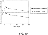


Classifications
-
- C—CHEMISTRY; METALLURGY
- C07—ORGANIC CHEMISTRY
- C07K—PEPTIDES
- C07K16/00—Immunoglobulins [IGs], e.g. monoclonal or polyclonal antibodies
- C07K16/42—Immunoglobulins [IGs], e.g. monoclonal or polyclonal antibodies against immunoglobulins
- C07K16/4283—Immunoglobulins [IGs], e.g. monoclonal or polyclonal antibodies against immunoglobulins against an allotypic or isotypic determinant on Ig
- C07K16/4291—Immunoglobulins [IGs], e.g. monoclonal or polyclonal antibodies against immunoglobulins against an allotypic or isotypic determinant on Ig against IgE
-
- C—CHEMISTRY; METALLURGY
- C12—BIOCHEMISTRY; BEER; SPIRITS; WINE; VINEGAR; MICROBIOLOGY; ENZYMOLOGY; MUTATION OR GENETIC ENGINEERING
- C12N—MICROORGANISMS OR ENZYMES; COMPOSITIONS THEREOF; PROPAGATING, PRESERVING, OR MAINTAINING MICROORGANISMS; MUTATION OR GENETIC ENGINEERING; CULTURE MEDIA
- C12N15/00—Mutation or genetic engineering; DNA or RNA concerning genetic engineering, vectors, e.g. plasmids, or their isolation, preparation or purification; Use of hosts therefor
- C12N15/09—Recombinant DNA-technology
-
- A—HUMAN NECESSITIES
- A61—MEDICAL OR VETERINARY SCIENCE; HYGIENE
- A61P—SPECIFIC THERAPEUTIC ACTIVITY OF CHEMICAL COMPOUNDS OR MEDICINAL PREPARATIONS
- A61P37/00—Drugs for immunological or allergic disorders
- A61P37/02—Immunomodulators
-
- C—CHEMISTRY; METALLURGY
- C07—ORGANIC CHEMISTRY
- C07K—PEPTIDES
- C07K16/00—Immunoglobulins [IGs], e.g. monoclonal or polyclonal antibodies
- C07K16/18—Immunoglobulins [IGs], e.g. monoclonal or polyclonal antibodies against material from animals or humans
- C07K16/24—Immunoglobulins [IGs], e.g. monoclonal or polyclonal antibodies against material from animals or humans against cytokines, lymphokines or interferons
- C07K16/244—Interleukins [IL]
- C07K16/248—IL-6
-
- C—CHEMISTRY; METALLURGY
- C07—ORGANIC CHEMISTRY
- C07K—PEPTIDES
- C07K16/00—Immunoglobulins [IGs], e.g. monoclonal or polyclonal antibodies
- C07K16/18—Immunoglobulins [IGs], e.g. monoclonal or polyclonal antibodies against material from animals or humans
- C07K16/28—Immunoglobulins [IGs], e.g. monoclonal or polyclonal antibodies against material from animals or humans against receptors, cell surface antigens or cell surface determinants
- C07K16/30—Immunoglobulins [IGs], e.g. monoclonal or polyclonal antibodies against material from animals or humans against receptors, cell surface antigens or cell surface determinants from tumour cells
- C07K16/303—Liver or Pancreas
-
- C—CHEMISTRY; METALLURGY
- C07—ORGANIC CHEMISTRY
- C07K—PEPTIDES
- C07K16/00—Immunoglobulins [IGs], e.g. monoclonal or polyclonal antibodies
- C07K16/42—Immunoglobulins [IGs], e.g. monoclonal or polyclonal antibodies against immunoglobulins
- C07K16/4283—Immunoglobulins [IGs], e.g. monoclonal or polyclonal antibodies against immunoglobulins against an allotypic or isotypic determinant on Ig
-
- G—PHYSICS
- G01—MEASURING; TESTING
- G01N—INVESTIGATING OR ANALYSING MATERIALS BY DETERMINING THEIR CHEMICAL OR PHYSICAL PROPERTIES
- G01N33/00—Investigating or analysing materials by specific methods not covered by groups G01N1/00 - G01N31/00
- G01N33/48—Biological material, e.g. blood, urine; Haemocytometers
- G01N33/50—Chemical analysis of biological material, e.g. blood, urine; Testing involving biospecific ligand binding methods; Immunological testing
- G01N33/53—Immunoassay; Biospecific binding assay; Materials therefor
- G01N33/536—Immunoassay; Biospecific binding assay; Materials therefor with immune complex formed in liquid phase
-
- G—PHYSICS
- G01—MEASURING; TESTING
- G01N—INVESTIGATING OR ANALYSING MATERIALS BY DETERMINING THEIR CHEMICAL OR PHYSICAL PROPERTIES
- G01N33/00—Investigating or analysing materials by specific methods not covered by groups G01N1/00 - G01N31/00
- G01N33/48—Biological material, e.g. blood, urine; Haemocytometers
- G01N33/50—Chemical analysis of biological material, e.g. blood, urine; Testing involving biospecific ligand binding methods; Immunological testing
- G01N33/68—Chemical analysis of biological material, e.g. blood, urine; Testing involving biospecific ligand binding methods; Immunological testing involving proteins, peptides or amino acids
- G01N33/6854—Immunoglobulins
- G01N33/686—Anti-idiotype
-
- A—HUMAN NECESSITIES
- A61—MEDICAL OR VETERINARY SCIENCE; HYGIENE
- A61K—PREPARATIONS FOR MEDICAL, DENTAL OR TOILETRY PURPOSES
- A61K39/00—Medicinal preparations containing antigens or antibodies
- A61K2039/505—Medicinal preparations containing antigens or antibodies comprising antibodies
-
- C—CHEMISTRY; METALLURGY
- C07—ORGANIC CHEMISTRY
- C07K—PEPTIDES
- C07K2317/00—Immunoglobulins specific features
- C07K2317/30—Immunoglobulins specific features characterized by aspects of specificity or valency
- C07K2317/31—Immunoglobulins specific features characterized by aspects of specificity or valency multispecific
-
- C—CHEMISTRY; METALLURGY
- C07—ORGANIC CHEMISTRY
- C07K—PEPTIDES
- C07K2317/00—Immunoglobulins specific features
- C07K2317/50—Immunoglobulins specific features characterized by immunoglobulin fragments
- C07K2317/52—Constant or Fc region; Isotype
-
- C—CHEMISTRY; METALLURGY
- C07—ORGANIC CHEMISTRY
- C07K—PEPTIDES
- C07K2317/00—Immunoglobulins specific features
- C07K2317/90—Immunoglobulins specific features characterized by (pharmaco)kinetic aspects or by stability of the immunoglobulin
- C07K2317/92—Affinity (KD), association rate (Ka), dissociation rate (Kd) or EC50 value
-
- C—CHEMISTRY; METALLURGY
- C07—ORGANIC CHEMISTRY
- C07K—PEPTIDES
- C07K2317/00—Immunoglobulins specific features
- C07K2317/90—Immunoglobulins specific features characterized by (pharmaco)kinetic aspects or by stability of the immunoglobulin
- C07K2317/94—Stability, e.g. half-life, pH, temperature or enzyme-resistance
-
- G—PHYSICS
- G01—MEASURING; TESTING
- G01N—INVESTIGATING OR ANALYSING MATERIALS BY DETERMINING THEIR CHEMICAL OR PHYSICAL PROPERTIES
- G01N2500/00—Screening for compounds of potential therapeutic value
- G01N2500/04—Screening involving studying the effect of compounds C directly on molecule A (e.g. C are potential ligands for a receptor A, or potential substrates for an enzyme A)
Definitions
- the present invention provides uses of antigen-binding molecules for eliminating antigens from plasma; methods for eliminating antigens from plasma, which comprise administering antigen-binding molecules; pharmaceutical compositions comprising antigen-binding molecules that are capable of eliminating antigens from plasma; methods of screening for antigen-binding molecules for eliminating antigens from plasma; and methods for producing antigen-binding molecules for eliminating antigens from plasma.
- Antibodies are drawing attention as pharmaceuticals as they have a high stability in plasma and have few side effects.
- a number of IgG-type antibody pharmaceuticals are available on the market and many antibody pharmaceuticals are currently under development (Non-patent Documents 1 and 2).
- various technologies applicable to second-generation antibody pharmaceuticals have been reported, including those that enhance effector function, antigen-binding ability, pharmacokinetics, and stability, and those that reduce the risk of immunogenicity (Non-patent Document 3).
- the requisite dose of an antibody pharmaceutical is very high. This in turn has led to problems such as high production cost, as well as the difficulty in producing subcutaneous formulations.
- the dose of an antibody pharmaceutical may be reduced by improving antibody pharmacokinetics or improving the affinity between antibodies and antigens.
- Non-patent Documents 4 and 5 The literature has reported methods for improving antibody pharmacokinetics using artificial substitution of amino acids in constant regions (Non-patent Documents 4 and 5). Similarly, affinity maturation has been reported as a technology for enhancing antigen-binding ability or antigen-neutralizing activity (Non-patent Document 6). This technology enables enhancement of antigen-binding activity by introducing amino acid mutations into the CDR region of a variable region or such. The enhancement of antigen-binding ability enables improvement of in vitro biological activity or reduction of dosage, and further enables improvement of in vivo (in the body) efficacy (Non-patent Document 7).
- the antigen-neutralizing capacity of a single antibody molecule depends on its affinity. By increasing the affinity, an antigen can be neutralized by a smaller amount of an antibody.
- Various methods can be used to enhance antibody affinity (Non-patent Document 6).
- the affinity could be made infinite by covalently binding the antibody to the antigen, a single antibody molecule could neutralize one antigen molecule (a divalent antibody can neutralize two antigen molecules).
- conventional methods have a limitation in which a single antibody molecule binds to one single antigen molecule (two antigens when it is bivalent).
- a single antigen-binding molecule can bind to a plurality of antigen molecules by using an antigen-binding molecule that binds to antigen in a pH-dependent manner (Patent Document 1 and Non-Patent Document 8).
- a pH-dependent antigen-binding molecule binds strongly to an antigen under the neutral condition in plasma, and releases the antigen under the acidic condition in the endosome. Furthermore, after release of the antigen, the antigen-binding molecule is released into the plasma via FcRn and binds to an antigen again; therefore, a single pH-dependent antigen-binding molecule can repeatedly bind to a number of antigens.
- pH-dependent antigen-binding molecules which have been modified to enhance FcRn binding under a neutral condition (pH7.4) have the advantage of being able to bind repeatedly to antigens and the effect of eliminating antigens from plasma, administration of such antigen-binding molecules has been reported to enable antigen elimination from plasma (Patent Document 2).
- Conventional pH-dependent antigen-binding molecules comprising an IgG antibody Fc region hardly show FcRn binding under neutral conditions. Therefore, uptake of complexes formed between an antigen-binding molecule and an antigen into cells may be mainly through non-specific uptake.
- pH-dependent antigen-binding molecules that have been modified to enhance FcRn binding under a neutral condition (pH7.4) can accelerate antigen elimination more than conventional pH-dependent antigen-binding molecules comprising an IgG antibody Fc region (Patent Document 2).
- antigens have very short retentivity in plasma compared to antibodies having an FcRn-mediated recycling mechanism
- binding of an antigen in plasma to an antibody having a recycling mechanism lengthens the normal retentivity in plasma and increases the plasma antigen concentration.
- the plasma concentration of the antigen may exacerbate symptoms caused by other physiological functions due to antibody binding. From such viewpoint, eliminating plasma antigens is sometimes favorable, and methods similar to those described above for making modifications to the Fc region to enhance FcRn binding with the objective of accelerating antigen elimination have been reported, but other methods for accelerating antigen elimination have not been reported so far.
- An objective of the present invention is to provide uses of antigen-binding molecules for eliminating antigens from plasma; methods for eliminating antigens from plasma, which comprise administering antigen-binding molecules; pharmaceutical compositions comprising antigen-binding molecules that are capable of eliminating antigens from plasma; methods of screening for antigen-binding molecules for eliminating antigens from plasma; and methods for producing antigen-binding molecules for eliminating antigens from plasma.
- antigen-binding molecules comprising (i) an Fc region and (ii) two or more antigen-binding domains where at least one of the domains is an antigen-binding domain whose antigen binding activity varies depending on the ion concentration condition, which are antigen-binding molecules that can form an immune complex comprising (a) two or more of the antigen-binding molecules and (b) two or more antigen molecules comprising two or more antigenic binding units.
- the antigen-binding molecules can be used to eliminate antigens from plasma.
- the present inventors discovered that the antigen-binding molecules are useful as pharmaceutical compositions, and they produced methods for eliminating antigens from plasma, which comprise administering the antigen-binding molecules. Furthermore, the present inventors discovered methods of screening for antigen-binding molecules having the aforementioned properties, and created methods for producing the molecules, thereby completing the present invention.
- the present invention provides the following:
- amino acids are described in one- or three-letter codes or both, for example, Ala/A, Leu/L, Arg/R, Lys/K, Asn/N, Met/M, Asp/D, Phe/F, Cys/C, Pro/P, Gln/Q, Ser/S, Glu/E, Thr/T, Gly/G, Trp/W, His/H, Tyr/Y, Ile/I, or Val/V.
- amino acid modification in the amino acid sequence of an antigen-binding molecule known methods such as site-directed mutagenesis methods ( Kunkel et al. (Proc. Natl. Acad. Sci. USA (1985) 82, 488-492 )) and overlap extension PCR may be appropriately adopted. Furthermore, several known methods may also be adopted as amino acid alteration methods for substitution to non-natural amino acids ( Annu. Rev. Biophys. Biomol. Struct. (2006) 35, 225-249 ; and Proc. Natl. Acad. Sci. U.S.A. (2003) 100 (11), 6353-6357 ).
- a cell-free translation system (Clover Direct (Protein Express)) containing a tRNA that has a non-natural amino acid bound to a complementary amber suppressor tRNA of the UAG codon (amber codon) which is one of the stop codons.
- the meaning of the term "and/or” when describing the site of amino acid alteration includes every combination where “and” and “or” are suitably combined.
- “the amino acids at positions 33, 55, and/or 96 are substituted” includes the following variation of amino acid modifications: amino acid(s) at (a) position 33, (b) position 55, (c) position 96, (d) positions 33 and 55, (e) positions 33 and 96, (f) positions 55 and 96, and (g) positions 33, 55, and 96.
- antigenic binding units refers to "the number of epitopes present in a single antigen molecule in a form normally present in plasma in the absence of an antigen-binding molecule, wherein the antigen molecule comprises an epitope that binds to a monovalent binding unit of an antigen-binding domain contained in an antigen-binding molecule of the present invention.
- antigens including multimers generally present in plasma in the form of homodimers such as GDF, PDGF, or VEGF.
- homodimerized GDF molecules there are two units of epitopes each bound by a monovalent binding unit of a variable region contained in an anti-GDF antibody molecule that comprises two variable regions having the same sequence (i.e., not the bispecific antibody to be described later).
- Immunoglobulin molecules such as IgE are also included as examples of antigens with two antigenic binding units.
- IgE normally exists in plasma as a tetramer comprising a heavy-chain dimer and a light-chain dimer; and in the tetramer, there are two units of epitopes each bound by a single valence of a variable region contained in an anti-IgE antibody molecule that comprises two variable regions having the same sequence (i.e., not the later-described bispecific antibody).
- IgA normally exists in the plasma in two forms: a tetramer comprising a heavy-chain dimer and a light-chain dimer, and an octamer produced when the tetramers further form a complex via a J chain; and the tetramer and octamer respectively have two units and four units of epitopes each bound by a single valence of a variable region contained in an anti-IgA antibody molecule that comprises two variable regions having the same sequence (i.e., not the later-described bispecific antibody).
- antigens with three antigenic binding units are antigens including multimers generally present in plasma in the form of homotrimers such as TNF ⁇ , RANKL, or CD 154 of the TNF superfamily.
- antigen molecules with one antigenic binding unit are molecules generally present in plasma in the form of monomers such as soluble IL-6 receptor (hereinafter also referred to as sIL-6R), IL-6, HMGB-1, and CTGF.
- monomers such as soluble IL-6 receptor (hereinafter also referred to as sIL-6R), IL-6, HMGB-1, and CTGF.
- Heterodimers such as IL-12 comprising IL-12p40 and IL-12p35, IL-23 comprising IL-12p40 and IL-23p19 (also referred to as IL-30B), IL-23 comprising EBI3 and IL27p28, and IL-35 comprising IL-12p35 and EBI3 contain two structurally different subunits molecules.
- an anti-subunit antibody molecule comprising two variable regions that bind to one of these subunits (i.e., not the later-described bispecific antibody)
- the epitope bound by a single valence of the variable regions is one unit; therefore, the antigenic binding unit of these heterodimers is one.
- the antigenic binding unit of heterotrimers such as the multisubunit complex TNF ⁇ -TNF ⁇ -hCG is similarly one unit.
- antigenic binding unit means the number of epitopes bound by a monovalent binding unit of an antigen-binding domain, when there is one type of paratope or multiple types of paratopes present in an antigen-binding molecule, the antigenic binding unit of the antigen will be different even if the antigen bound by these antigen-binding molecules is the same antigen.
- the antigenic binding unit of this antigen is one, whereas when the antigen-binding molecule is a biparatopic or a bispecific antigen-binding molecule comprising two monovalent binding units that bind to each of the subunits forming the heterodimer, the antigenic binding unit of this antigen is two.
- multimer includes both homomultimer and heteromultimer.
- the modes of binding between subunits contained in a multimer are covalent bonds such as peptide bonds or disulfide bonds, and stable non-covalent bonds such as ionic bonds, van der Waals bonds, and hydrogen bonds, but are not limited thereto.
- Homomultimers include a plurality of identical subunits, whereas heteromultimers contain a plurality of different subunits.
- the meaning of the term “dimer” includes both homodimer and heterodimer; and while a homodimer contains two identical subunits, a heterodimer contains two different subunits.
- the term "monomer" which expresses each of the units forming a multimer refers to each of the continuous structural units linked by peptide bonds.
- antigens are not particularly limited in their structure, as long as they comprise epitopes to which antigen-binding domains bind.
- antigens can be inorganic or organic substances.
- Other antigens include, for example, the molecules below: 17-IA, 4-1BB, 4Dc, 6-keto-PGF1a, 8-iso-PGF2a, 8-oxo-dG, A1 adenosine receptor, A33, ACE, ACE-2, activin, activin A, activin AB, activin B, activin C, activin RIA, activin RIA ALK-2, activin RIB ALK-4, activin RIIA, activin RIIB, ADAM, ADAM10, ADAM12, ADAM15, ADAM17/TACE, ADAM8, ADAM9, ADAMTS, ADAMTS4, ADAMTS5, addressin, aFGF, ALCAM, ALK, ALK-1, ALK-7,alpha-1-antitry
- the antigen that can form a complex with the antigen-binding molecule may be any of the antigens exemplified above, or combinations thereof, i.e., monomers or heteromultimers.
- heteromultimers include heterodimers such as IL-12 comprising IL-12p40 and IL-12p35, IL-23 comprising IL-12p40 and IL-23p19 (also referred to as IL-30B), IL-23 comprising EBI-3 and IL27p28, and IL-35 comprising IL-12p35 and EBI-3.
- receptors are recited as examples of the above-mentioned antigens, when these receptors exist in soluble forms in biological fluids such as plasma, they can form complexes with the antigen-binding molecules of the present invention. Therefore, as long as the above-mentioned receptors exist in their soluble forms in biological fluids such as plasma, they may be used as antigens that may form complexes of the present invention by binding to an antigen-binding molecule of the present invention.
- An example of a non-limiting embodiment of such a soluble receptor is soluble IL-6R, which is a protein consisting of the amino acids at positions 1 to 357 in the IL-6R polypeptide sequence of SEQ ID NO: 1 as described in Mullberg et al. (J. Immunol. (1994) 152 (10), 4958-4968 ).
- Soluble antigens are recited as examples of the above-mentioned antigens, and the solutions in which the antigens exist are not limited. Soluble antigens may exist in biological fluids, or more specifically in all fluids filling the space between tissues and cells or vessels in organisms. In a non-limiting embodiment, the antigens to which antigen-binding molecules of the present invention bind may be present in extracellular fluids.
- extracellular fluid is a general term for plasma, interstitial fluid, lymph, compact connective tissue, cerebrospinal fluid, spinal fluid, puncture fluid, synovial fluid, or such components in the bone and cartilage, alveolar fluid (bronchoalveolar lavage fluid), peritoneal fluid, pleural fluid, pericardial fluid, cyst fluid, aqueous humor (hydatoid), or such transcellular fluids (various fluids in the glandular cavities and fluids in the digestive tract cavity and other body cavity fluids produced as a result of active transport / secretory activities of cells).
- alveolar fluid bronchoalveolar lavage fluid
- peritoneal fluid pleural fluid
- pericardial fluid cyst fluid
- aqueous humor aqueous humor
- an antigen that may form a complex of the present invention upon binding with the antigen-binding molecule includes the molecules exemplified below, which include a homodimer, homotrimer, or such, as an antigenic binding unit: GDF, GDF-1, GDF-3 (Vgr-2), GDF-5 (BMP-14, CDMP-1), GDF-6 (BMP-13, CDMP-2), GDF-7 (BMP-12, CDMP-3), GDF-8 (myostatin), GDF-9, GDF-15 (MIC-1), TNF, TNF-alpha, TNF-alphabeta, TNF-beta2, TNFSF10 (TRAIL Apo-2 ligand, TL2), TNFSF11 (TRANCE/RANK ligand ODF, OPG ligand), TNFSF12 (TWEAK Apo-3 ligand, DR3
- Epitope means an antigenic determinant in an antigen, and refers to an antigen site to which the antigen-binding domain of an antigen-binding molecule disclosed herein binds.
- the epitope can be defined according to its structure.
- the epitope may be defined according to the antigen-binding activity of an antigen-binding molecule that recognizes the epitope.
- the antigen is a peptide or polypeptide
- the epitope can be specified by the amino acid residues forming the epitope.
- the epitope is a sugar chain
- the epitope can be specified by its specific sugar chain structure.
- a linear epitope is an epitope that contains an epitope whose primary amino acid sequence has been recognized. Such a linear epitope typically contains at least three and most commonly at least five, for example, about 8 to 10 or 6 to 20 amino acids in a specific sequence.
- a "conformational epitope” is an epitope in which the primary amino acid sequence containing the epitope is not the only determinant of the recognized epitope (for example, the primary amino acid sequence of a conformational epitope is not necessarily recognized by an epitope-defining antibody).
- Conformational epitopes may contain a greater number of amino acids compared to linear epitopes.
- a conformational epitope-recognizing antibody recognizes the three-dimensional structure of a peptide or protein. For example, when a protein molecule folds and forms a three-dimensional structure, amino acids and/or polypeptide main chains that form a conformational epitope become aligned, and the epitope is made recognizable by the antibody.
- Methods for determining epitope conformations include, for example, X ray crystallography, two-dimensional nuclear magnetic resonance, site-specific spin labeling, and electron paramagnetic resonance, but are not limited thereto. See, for example, Epitope Mapping Protocols in Methods in Molecular Biology (1996), Vol. 66, Morris (ed .).
- the structure of the antigen-binding domain which binds to an epitope is called a paratope.
- An epitope and a paratope bind with stability through the action of hydrogen bonds, electrostatic force, van der Waals force, hydrophobic bonds, and such between the epitope and the paratope. This strength of binding between the epitope and paratope is called affinity.
- affinity The total sum of binding strength when a plurality of epitopes and a plurality of paratopes bind is referred to as avidity.
- avidity When an antibody comprising a plurality of paratopes (i.e., multivalent antibody) or such binds to a plurality of epitopes, the affinity acts additively or synergistically, and therefore avidity becomes higher than affinity.
- Examples of a method for assessing epitope binding by a test antigen-binding molecule containing an antigen-binding domain directed to IgA are described below. According to the examples below, methods for assessing epitope binding by a test antigen-binding molecule containing an antigen-binding domain for an antigen other than IgA can also be appropriately conducted.
- a test antigen-binding molecule containing an antigen-binding domain against IgA recognizes a linear epitope in the IgA molecule can be confirmed for example as mentioned below.
- a linear peptide comprising an amino acid sequence forming an IgA constant region is synthesized for the above purpose.
- the peptide can be synthesized chemically, or obtained by genetic engineering techniques using a region encoding the amino acid sequence corresponding to the constant region in an IgA cDNA. Then, a test antigen-binding molecule containing an antigen-binding domain toward IgA is assessed for its binding activity towards a linear peptide comprising the amino acid sequence forming the constant region.
- an ELISA using an immobilized linear peptide as an antigen can be performed to evaluate the binding activity of the antigen-binding molecule towards the peptide.
- the binding activity towards a linear peptide can be assessed based on the level of inhibition by the linear peptide of the binding of the antigen-binding molecule toward IgA cells. These tests can demonstrate the binding activity of the antigen-binding molecule towards the linear peptide.
- a conformational epitope by a test antigen-binding molecule comprising an antigen-binding domain targeting the IgA protein may be confirmed as stated below.
- a general genetic recombination technique is used to transfer an IgA-encoding recombinant gene into host cells (for example, animal cells, insect cells, or yeast cells) that enable formation of the native conformational epitope in the IgA protein.
- IgA containing the conformational epitope is prepared from the culture of recombinant cells produced in this manner.
- Recognition of a conformational epitope by a test antigen-binding molecule comprising an antigen-binding domain targeting IgA is, for example, when the test antigen-binding molecule binds strongly to the IgA molecule when it is contacted with immobilized IgA containing the conformational epitope, while the antigen-binding molecule does not substantively bind to a linear peptide comprising an amino acid sequence constituting the amino acid sequence of the immobilized IgA.
- test IgA-targeting antigen-binding molecule that has been denatured by a reducing agent that cleaves disulfide bonds, such as dithiothreitol, dithioerythritol, ⁇ -mercaptoethanol, phosphines, and sodium borohydride, and/or chaotropic agents such as surfactants including guanidine hydrochloride, urea, and sodium lauryl sulfate.
- a reducing agent that cleaves disulfide bonds such as dithiothreitol, dithioerythritol, ⁇ -mercaptoethanol, phosphines, and sodium borohydride
- chaotropic agents such as surfactants including guanidine hydrochloride, urea, and sodium lauryl sulfate.
- does not substantively bind refers to a binding activity not greater than 80%, normally not greater than 50%, preferably not greater than 30%, or particularly preferably not greater
- Methods for assaying the binding activity toward IgA of a test antigen-binding molecule containing an antigen-binding domain against IgA include, for example, the methods described in Antibodies: A Laboratory Manual (Ed Harlow, David Lane, Cold Spring Harbor Laboratory (1988) 359-420 ). Specifically, the assessment can be performed based on the principle of ELISA or EIA using IgA as antigen.
- the binding activity of a test antigen-binding molecule containing an IgA antigen-binding domain towards IgA can be assessed quantitatively by comparing the levels of signal generated by enzymatic reaction.
- a test antigen-binding molecule is added to an ELISA plate onto which IgA has been immobilized. Then, the test antigen-binding molecule that bound to IgA immobilized on the plate is detected using an enzyme-labeled antibody that recognizes the test antigen-binding molecule.
- a serial dilution of the test antigen-binding molecule can be prepared and the antibody binding titer toward IgA be determined to compare the binding activity of the test antigen-binding molecule towards IgA.
- test antigen-binding molecule The binding of a test antigen-binding molecule towards an antigen expressed on the surface of cells suspended in buffer or the like can be detected using a flow cytometer.
- flow cytometers include, for example, the following devices:
- Preferable methods for assaying the binding activity towards an antigen of a test antigen-binding molecule containing an antigen-binding domain against IgE include, for example, the following method. First, IgE-expressing cells are reacted with a test antigen-binding molecule, and then this is stained with an FITC-labeled secondary antibody that recognizes the antigen-binding molecule.
- the test antigen-binding molecule is appropriately diluted with a suitable buffer to prepare the molecule at a desired concentration. For example, the molecule can be used at a concentration within the range of 10 ⁇ g/ml to 10 ng/ml. Then, the fluorescence intensity and cell count are determined using FACSCalibur (BD).
- the fluorescence intensity obtained by analysis using the CELL QUEST Software (BD), i.e ., the Geometric Mean value, reflects the quantity of antibody bound to cells. That is, the binding activity of a test antigen-binding molecule, which is represented by the quantity of the test antigen-binding molecule bound, can be determined by measuring the Geometric Mean value.
- test antigen-binding molecule containing an IgA antigen-binding domain shares a common epitope with another antigen-binding molecule can be assessed based on the competition between the two molecules for the same epitope.
- the competition between antigen-binding molecules can be detected by cross-blocking assay or the like.
- the competitive ELISA assay is a preferred cross-blocking assay.
- the IgA protein immobilized to the wells of a microtiter plate is pre-incubated in the presence or absence of a candidate competitor antigen-binding molecule, and then a test antigen-binding molecule is added thereto.
- the quantity of test antigen-binding molecule bound to the IgA protein in the wells is indirectly correlated with the binding ability of a candidate competitor antigen-binding molecule that competes for the binding to the same epitope. That is, the greater the affinity of the competitor antigen-binding molecule for the same epitope, the lower the binding activity of the test antigen-binding molecule towards the IgA protein-coated wells.
- the quantity of the test antigen-binding molecule bound to the wells via the IgA protein can be readily determined by labeling the antigen-binding molecule in advance.
- a biotin-labeled antigen-binding molecule is measured using an avidin/peroxidase conjugate and appropriate substrate.
- cross-blocking assay that uses enzyme labels such as peroxidase is called "competitive ELISA assay”.
- the antigen-binding molecule can also be labeled with other labeling substances that enable detection or measurement. Specifically, radiolabels, fluorescent labels, and such are known.
- the candidate competitor antigen-binding molecule can block the binding by a test antigen-binding molecule containing an IgA antigen-binding domain by at least 20%, preferably at least 20 to 50%, and more preferably at least 50% compared to the binding activity in a control experiment conducted in the absence of the competitor antigen-binding molecule, the test antigen-binding molecule is determined to substantially bind to the same epitope bound by the competitor antigen-binding molecule, or compete for the binding to the same epitope.
- test and control antigen-binding molecules share a common epitope can be assessed by comparing the binding activities of the two antigen-binding molecules towards a peptide prepared by introducing amino acid mutations into the peptide forming the epitope.
- the binding activities of test and control antigen-binding molecules towards a linear peptide into which a mutation is introduced are compared in the above ELISA format.
- the binding activity towards the mutant peptide bound to a column can be determined by flowing test and control antigen-binding molecules in the column, and then quantifying the antigen-binding molecule eluted in the elution solution.
- Methods for adsorbing a mutant peptide to a column for example, in the form of a GST fusion peptide, are known.
- test and control antigen-binding molecules share a common epitope can be assessed, for example, by the following method. Explanation is provided below, using the case of IgE as the antigen as an example.
- IgE-expressing cells and cells expressing IgE with a mutation introduced into the epitope are prepared.
- the test and control antigen-binding molecules are added to a cell suspension prepared by suspending these cells in an appropriate buffer such as PBS. Then, the cell suspensions are appropriately washed with a buffer, and an FITC-labeled antibody that recognizes the test and control antigen-binding molecules is added thereto.
- the fluorescence intensity and number of cells stained with the labeled antibody are determined using FACSCalibur (BD).
- the test and control antigen-binding molecules are appropriately diluted using a suitable buffer, and used at desired concentrations. For example, they may be used at a concentration within the range of 10 ⁇ g/ml to 10 ng/ml.
- the fluorescence intensity determined by analysis using the CELL QUEST Software (BD), i.e ., the Geometric Mean value reflects the quantity of labeled antibody bound to cells. That is, the binding activities of the test and control antigen-binding molecules, which are represented by the quantity of labeled antibody bound, can be determined by measuring the Geometric Mean value.
- the Geometric Mean comparison value ( ⁇ Geo-Mean value for the mutant IgE molecule) determined by the above analysis, which reflects the quantity of a test antigen-binding molecule bound to cells expressing mutant IgE, is compared to the ⁇ Geo-Mean comparison value that reflects the quantity of the test antigen-binding molecule bound to IgE-expressing cells.
- concentrations of the test antigen-binding molecule used to determine the ⁇ Geo-Mean comparison values for IgE-expressing cells and cells expressing mutant IgE are particularly preferably adjusted to be equal or substantially equal.
- An antigen-binding molecule that has been confirmed to recognize an epitope in IgE is used as a control antigen-binding molecule.
- the test antigen-binding molecule for cells expressing mutant IgE is smaller than the ⁇ Geo-Mean comparison value of the test antigen-binding molecule for IgE-expressing cells by at least 80%, preferably 50%, more preferably 30%, and particularly preferably 15%, then the test antigen-binding molecule "does not substantially bind to cells expressing mutant IgE".
- the formula for determining the Geo-Mean (Geometric Mean) value is described in the CELL QUEST Software User's Guide (BD biosciences). When the comparison shows that the comparison values are substantially equivalent, the epitope for the test and control antigen-binding molecules can be determined to be the same.
- an "antigen-binding domain” may be of any structure as long as it binds to an antigen of interest.
- Such domains preferably include, for example:
- the antigen-binding domains of antigen-binding molecules of the present invention can bind to an identical epitope.
- Such identical epitope can be present, for example, in a protein comprising the amino acid sequence of SEQ ID NO: 2.
- each of the antigen-binding domains of antigen-binding molecules of the present invention can bind to a different epitope.
- the different epitope can be present in, for example, a protein comprising the amino acid sequence of SEQ ID NO: 2.
- Immune complex refers to a relatively stable structure produced when at least one antigen and at least one antigen-binding molecule bind with each other to form a larger molecular-weight complex.
- a non-limiting embodiment of the immune complex is, for example, an antigen-antibody aggregate.
- a method for evaluating the formation of immune complexes comprising two or more antigen-binding molecules and two or more antigenic binding units will be described later in this specification.
- Specific means that one of the molecules that specifically bind does not show any significant binding to molecules other than the single or plurality of partner molecules it binds to. Furthermore, “specific” is also used when an antigen-binding domain is specific to a particular epitope among multiple epitopes in an antigen. When an epitope bound by an antigen-binding domain is contained in multiple different antigens, antigen-binding molecules containing the antigen-binding domain can bind to various antigens that have the epitope.
- does not show any significant binding means showing no more than 50%, generally no more than 30%, preferably no more than 15%, particularly preferably no more than 10%, and even more preferably no more than 5% of the binding activity to a partner molecule towards molecules other than the partner molecule.
- antibody refers to a natural immunoglobulin or an immunoglobulin produced by partial or complete synthesis.
- Antibodies can be isolated from natural sources such as naturally-occurring plasma and serum, or culture supernatants of antibody-producing hybridomas. Alternatively, antibodies can be partially or completely synthesized using techniques such as genetic recombination.
- Preferred antibodies include, for example, antibodies of an immunoglobulin isotype or subclass belonging thereto.
- Known human immunoglobulins include antibodies of the following nine classes (isotypes): IgG1, IgG2, IgG3, IgG4, IgA1, IgA2, IgD, IgE, and IgM.
- antibodies of the present invention include IgG1, IgG2, IgG3, and IgG4.
- a number of allotype sequences of human IgG1, human IgG2, human IgG3, and human IgG4 constant regions due to gene polymorphisms are described in " Sequences of proteins of immunological interest", NIH Publication No. 91-3242 . Any of such sequences may be used in the present invention.
- the amino acid sequence at positions 356 to 358 as indicated by EU numbering may be DEL or EEM.
- Several allotype sequences due to genetic polymorphisms have been described in " Sequences of proteins of immunological interest", NIH Publication No. 91-3242 for the human Ig ⁇ (Kappa) constant region and human Ig ⁇ (Lambda) constant region, and any of the sequences may be used in the present invention.
- Antibodies that bind to an antigen other than IgA can also be produced according to the example described below.
- Anti-IgA antibodies can be obtained as polyclonal or monoclonal antibodies using known methods.
- the anti-IgA antibodies preferably produced are monoclonal antibodies derived from mammals.
- Such mammal-derived monoclonal antibodies include antibodies produced by hybridomas or host cells transformed with an expression vector carrying an antibody gene by genetic engineering techniques.
- “humanized antibodies” or “chimeric antibodies” are included in the monoclonal antibodies of the present invention.
- Monoclonal antibody-producing hybridomas can be produced using known techniques, for example, as described below. Specifically, mammals are immunized by conventional immunization methods using an IgA protein as a sensitizing antigen. Resulting immune cells are fused with known parental cells by conventional cell fusion methods. Then, hybridomas producing an anti-IgA antibody can be selected by screening for monoclonal antibody-producing cells using conventional screening methods.
- monoclonal antibody production is performed, for example, as shown below.
- a purified native IgA protein may be used as a sensitizing antigen.
- a recombinant protein comprising an IgA polypeptide sequence of SEQ ID NO: 2, which is registered as L00022
- an IgA heavy chain constant region gene of which nucleotide sequence is disclosed in SEQ ID NO: 3 may be expressed to obtain the IgA protein shown in SEQ ID NO: 2, which is used as a sensitizing antigen for obtaining antibodies.
- suitable host cells are transformed by inserting an IgA-encoding gene sequence into a known expression vector.
- the desired IgA protein is purified by known methods from the host cells or from their culture supernatant.
- the polynucleotide sequence of SEQ ID NO: 3 may be operably linked to the 3' end of the signal sequence.
- the polynucleotide sequence of SEQ ID NO: 3 may be operably linked to the 3' end of a polynucleotide sequence encoding a heavy chain variable region comprising a signal sequence at the 5' end.
- a tag peptide for purification may be added appropriately to the amino terminus or carboxy terminus of SEQ ID NO: 2.
- GLNDIFEAQKIEWHE SEQ ID NO: 4 which is an Avi tag linked via the GGGGS (SEQ ID NO: 31) linker is an example of a non-limiting embodiment of such.
- a non-limiting example of the production of a recombinant protein similar to that mentioned above is a method such as that described in Example 1, where a recombinant IgA protein which has been expressed in combination with a light chain is collected from the recombinant host cell culture, and then purified.
- the purified IgA protein may be used as a sensitizing antigen for immunization of mammals.
- a partial IgA peptide may also be used as a sensitizing antigen.
- a partial peptide can be prepared by chemical synthesis based on the amino acid sequence of human IgA, or by inserting a partial IgA gene into an expression vector for expression.
- a partial peptide can be produced by degrading an IgA protein with a protease.
- the length and region of the partial IgA peptide are not limited to particular embodiments.
- a preferred region can be arbitrarily selected from the amino acid sequence of SEQ ID NO: 2.
- the number of amino acids forming a peptide to be used as a sensitizing antigen is preferably at least five or more, six or more, or seven or more. More specifically, a peptide of 8 to 50 residues, more preferably 10 to 30 residues can be used as a sensitizing antigen.
- a fusion protein prepared by fusing a desired partial polypeptide or peptide of the IgA protein with a different polypeptide.
- antibody Fc fragments and peptide tags are preferably used to produce fusion proteins to be used as sensitizing antigens.
- Vectors for expression of such fusion proteins can be constructed by fusing in frame genes encoding two or more desired polypeptide fragments and inserting the fusion gene into an expression vector as described above. Methods for producing fusion proteins are described in Molecular Cloning 2nd ed. ( Sambrook, J et al., Molecular Cloning 2nd ed., 9.47-9.58 (1989) Cold Spring Harbor Lab. Press ). Methods for preparing IgA to be used as a sensitizing antigen, and immunization methods using IgA are specifically described in PCT/JP2011/077619 and such.
- mammals to be immunized with the sensitizing antigen there is no particular limitation on the mammals to be immunized with the sensitizing antigen. However, it is preferable to select the mammals by considering their compatibility with the parent cells to be used for cell fusion. In general, rodents such as mice, rats, and hamsters, rabbits, and monkeys are preferably used.
- the above animals are immunized with a sensitizing antigen by known methods.
- Generally performed immunization methods include, for example, intraperitoneal or subcutaneous injection of a sensitizing antigen into mammals.
- a sensitizing antigen is appropriately diluted with PBS (Phosphate-Buffered Saline), physiological saline, or the like.
- a conventional adjuvant such as Freund's complete adjuvant is mixed with the antigen, and the mixture is emulsified.
- the sensitizing antigen is administered to a mammal several times at 4- to 21-day intervals.
- Appropriate carriers may be used in immunization with the sensitizing antigen.
- sensitizing antigen peptide when a low-molecular-weight partial peptide is used as the sensitizing antigen, it is sometimes desirable to couple the sensitizing antigen peptide to a carrier protein such as albumin or keyhole limpet hemocyanin for immunization.
- a carrier protein such as albumin or keyhole limpet hemocyanin for immunization.
- hybridomas producing a desired antibody can be prepared using DNA immunization as mentioned below.
- DNA immunization is an immunization method that confers immunostimulation by expressing a sensitizing antigen in an animal immunized as a result of administering a vector DNA constructed to allow expression of an antigen protein-encoding gene in the animal.
- DNA immunization is expected to be superior in that:
- a DNA expressing an IgA protein is administered to an animal to be immunized.
- the IgA-encoding DNA can be synthesized by known methods such as PCR.
- the obtained DNA is inserted into an appropriate expression vector, and then this is administered to an animal to be immunized.
- Preferably used expression vectors include, for example, commercially-available expression vectors such as pcDNA3.1.
- Vectors can be administered to an organism using conventional methods. For example, DNA immunization is performed by using a gene gun to introduce expression vector-coated gold particles into cells in the body of an animal to be immunized.
- Antibodies that recognize IgA can also be produced by the methods described in PCT/JP2011/077619 .
- an increase in the titer of an IgA-binding antibody is confirmed in the serum.
- immune cells are collected from the mammal, and then subjected to cell fusion.
- splenocytes are preferably used as immune cells.
- a mammalian myeloma cell is used as a cell to be fused with the above-mentioned immune cells.
- the myeloma cells preferably comprise a suitable selection marker for screening.
- a selection marker confers characteristics to cells for their survival (or death) under a specific culture condition.
- Hypoxanthine-guanine phosphoribosyltransferase deficiency hereinafter abbreviated as HGPRT deficiency
- TK deficiency thymidine kinase deficiency
- HAT-sensitive cells cannot synthesize DNA in a HAT selection medium, and are thus killed. However, when the cells are fused with normal cells, they can continue DNA synthesis using the salvage pathway of the normal cells, and therefore they can grow even in the HAT selection medium.
- HGPRT-deficient and TK-deficient cells can be selected in a medium containing 6-thioguanine, 8-azaguanine (hereinafter abbreviated as 8AG), or 5'-bromodeoxyuridine, respectively.
- 8AG 8-azaguanine
- 5'-bromodeoxyuridine 5'-bromodeoxyuridine
- Normal cells are killed because they incorporate these pyrimidine analogs into their DNA.
- cells that are deficient in these enzymes can survive in the selection medium, since they cannot incorporate these pyrimidine analogs.
- a selection marker referred to as G418 resistance provided by the neomycin-resistant gene confers resistance to 2-deoxystreptamine antibiotics (gentamycin analogs).
- gentamycin analogs gentamycin analogs.
- myeloma cells that are suitable for cell fusion are known.
- myeloma cells including the following cells can be preferably used:
- Cell fusions between the immunocytes and myeloma cells are essentially carried out using known methods, for example, a method by Kohler and Milstein et al. (Methods Enzymol. (1981) 73: 3-46 ).
- cell fusion can be carried out, for example, in a conventional culture medium in the presence of a cell fusion-promoting agent.
- fusion-promoting agents include, for example, polyethylene glycol (PEG) and Sendai virus (HVJ).
- PEG polyethylene glycol
- HVJ Sendai virus
- an auxiliary substance such as dimethyl sulfoxide is also added to improve fusion efficiency.
- the ratio of immune cells to myeloma cells may be determined at one's own discretion, preferably, for example, one myeloma cell for every one to ten immunocytes.
- Culture media to be used for cell fusions include, for example, media that are suitable for the growth of myeloma cell lines, such as RPMI1640 medium and MEM medium, and other conventional culture medium used for this type of cell culture.
- serum supplements such as fetal calf serum (FCS) may be preferably added to the culture medium.
- a PEG solution for example, the average molecular weight is about 1,000 to 6,000
- a PEG solution prewarmed to about 37°C
- This is gently mixed to produce desired fusion cells (hybridomas).
- an appropriate culture medium mentioned above is gradually added to the cells, and this is repeatedly centrifuged to remove the supernatant.
- the hybridomas thus obtained can be selected by culture using a conventional selective medium, for example, HAT medium (a culture medium containing hypoxanthine, aminopterin, and thymidine).
- HAT medium a culture medium containing hypoxanthine, aminopterin, and thymidine.
- Cells other than the desired hybridomas can be killed by continuing culture in the above HAT medium for a sufficient period of time. Typically, the period is several days to several weeks. Then, hybridomas producing the desired antibody are screened and singly cloned by conventional limiting dilution methods.
- the hybridomas thus obtained can be selected using a selection medium based on the selection marker possessed by the myeloma used for cell fusion.
- HGPRT- or TK-deficient cells can be selected by culture using the HAT medium (a culture medium containing hypoxanthine, aminopterin, and thymidine).
- HAT medium a culture medium containing hypoxanthine, aminopterin, and thymidine.
- Desired antibodies can be preferably selected and singly cloned by screening methods based on known antigen/antibody reaction. For example, the activity of an antibody to bind to immobilized IgA can be assessed based on the principle of ELISA. For example, IgA is immobilized to the wells of an ELISA plate. Culture supernatants of hybridomas are contacted with the IgA in the wells, and antibodies that bind to the IgA are detected. When the monoclonal antibodies are derived from mouse, antibodies bound to the cells can be detected using an anti-mouse immunoglobulin antibody. Hybridomas producing a desired antibody having the antigen-binding ability are selected by the above screening, and they can be cloned by a limiting dilution method or the like.
- Monoclonal antibody-producing hybridomas thus prepared can be passaged in a conventional culture medium, and stored in liquid nitrogen for a long period.
- the above hybridomas are cultured by a conventional method, and desired monoclonal antibodies can be prepared from the culture supernatants. Alternatively, the hybridomas are administered to and grown in compatible mammals, and monoclonal antibodies are prepared from the ascites.
- the former method is suitable for preparing antibodies with high purity.
- Antibodies encoded by antibody genes that are cloned from antibody-producing cells such as the above hybridomas can also be preferably used.
- a cloned antibody gene is inserted into an appropriate vector, and this is introduced into a host to express the antibody encoded by the gene.
- Methods for isolating antibody genes, inserting the genes into vectors, and transforming host cells have already been established, for example, by Vandamme et al. (Eur. J. Biochem. (1990) 192(3), 767-775 ). Methods for producing recombinant antibodies are also known as described below.
- a cDNA encoding the variable region (V region) of an anti-IgA antibody is prepared from hybridoma cells producing the anti-IgA antibody.
- total RNA is first extracted from hybridomas. Methods used for extracting mRNAs from cells include, for example:
- Extracted mRNAs can be purified using the mRNA Purification Kit (GE Healthcare Bioscience) or such. Alternatively, kits for extracting total mRNA directly from cells, such as the QuickPrep mRNA Purification Kit (GE Healthcare Bioscience), are also commercially available. mRNAs can be prepared from hybridomas using such kits. cDNAs encoding the antibody V region can be synthesized from the prepared mRNAs using a reverse transcriptase. cDNAs can be synthesized using the AMV Reverse Transcriptase First-strand cDNA Synthesis Kit (Seikagaku Co.) or such. Furthermore, the SMART RACE cDNA amplification kit (Clontech) and the PCR-based 5'-RACE method ( Proc.
- the cDNA fragment of interest is purified from the resulting PCR product, and then this is ligated to a vector DNA.
- a recombinant vector is thus constructed, and introduced into E. coli or such. After colony selection, the desired recombinant vector can be prepared from the colony-forming E. coli. Then, whether the recombinant vector has the cDNA nucleotide sequence of interest is tested by a known method such as the dideoxy nucleotide chain termination method.
- the 5'-RACE method which uses primers to amplify the variable region gene is conveniently used for isolating the gene encoding the variable region.
- a 5'-RACE cDNA library is constructed by cDNA synthesis using RNAs extracted from hybridoma cells as a template.
- a commercially available kit such as the SMART RACE cDNA amplification kit is appropriately used to synthesize the 5'-RACE cDNA library.
- the antibody gene is amplified by PCR using the prepared 5'-RACE cDNA library as a template.
- Primers for amplifying the mouse antibody gene can be designed based on known antibody gene sequences.
- the nucleotide sequences of the primers vary depending on the immunoglobulin subclass. Therefore, it is preferable that the subclass is determined in advance using a commercially available kit such as the Iso Strip mouse monoclonal antibody isotyping kit (Roche Diagnostics).
- primers that allow amplification of genes encoding ⁇ 1, ⁇ 2a, ⁇ 2b, and ⁇ 3 heavy chains and ⁇ and ⁇ light chains are used to isolate mouse IgG-encoding genes.
- a primer that anneals to a constant region site close to the variable region is used as a 3'-side primer to amplify an IgG variable region gene.
- a primer attached to a 5' RACE cDNA library construction kit is used as a 5'-side primer.
- PCR products thus amplified are used to reshape immunoglobulins composed of a combination of heavy and light chains.
- a desired antibody can be selected using the IgA-binding activity of a reshaped immunoglobulin as an indicator. For example, when the objective is to isolate an antibody against IgA, it is more preferred that the binding of the antibody to IgA is specific.
- An IgA-binding antibody can be screened, for example, by the following steps:
- Methods for detecting the binding of an antibody to IgA are known. Specifically, the binding of an antibody to IL-6R-expressing cells can be detected by the above-described techniques such as ELISA.
- Preferred antibody screening methods that use the binding activity as an indicator also include panning methods using phage vectors. Screening methods using phage vectors are advantageous when the antibody genes are isolated from heavy-chain and light-chain subclass libraries from a polyclonal antibody-expressing cell population. Genes encoding the heavy-chain and light-chain variable regions can be linked by an appropriate linker sequence to form a single-chain Fv (scFv). Phages presenting scFv on their surface can be produced by inserting a gene encoding scFv into a phage vector. The phages are contacted with an antigen of interest. Then, a DNA encoding scFv having the binding activity of interest can be isolated by collecting phages bound to the antigen. This process can be repeated as necessary to enrich scFv having the binding activity of interest.
- scFv single-chain Fv
- the cDNA is digested with restriction enzymes that recognize the restriction sites introduced into both ends of the cDNA.
- Preferred restriction enzymes recognize and cleave a nucleotide sequence that occurs in the nucleotide sequence of the antibody gene at a low frequency.
- a restriction site for an enzyme that produces a sticky end is preferably introduced into a vector to insert a single-copy digested fragment in the correct orientation.
- the cDNA encoding the V region of the anti-IgA antibody is digested as described above, and this is inserted into an appropriate expression vector to construct an antibody expression vector.
- a chimeric antibody means that the origin of the constant region is different from that of the variable region.
- human/human allochimeric antibodies are included in the chimeric antibodies of the present invention.
- a chimeric antibody expression vector can be constructed by inserting the above V region gene into an expression vector that already has the constant region. Specifically, for example, a recognition sequence for a restriction enzyme that excises the above V region gene can be appropriately placed on the 5' side of an expression vector carrying a DNA encoding a desired antibody constant region (C region).
- a chimeric antibody expression vector is constructed by fusing in frame the two genes digested with the same combination of restriction enzymes.
- antibody genes are inserted into an expression vector so that the genes are expressed under the control of an expression regulatory region.
- the expression regulatory region for antibody expression includes, for example, enhancers and promoters.
- an appropriate signal sequence may be attached to the amino terminus so that the expressed antibody is secreted to the outside of cells.
- a peptide having the amino acid sequence MGWSCIILFLVATATGVHS (SEQ ID NO: 5) are used as a signal sequence.
- other appropriate signal sequences may be attached.
- the expressed polypeptide is cleaved at the carboxyl terminus of the above sequence, and the resulting polypeptide is secreted to the outside of cells as a mature polypeptide.
- appropriate host cells are transformed with the expression vector, and recombinant cells expressing the anti-IgA antibody-encoding DNA are obtained.
- DNAs encoding the antibody heavy chain (H chain) and light chain (L chain) are separately inserted into different expression vectors to express the antibody gene.
- An antibody molecule having the H and L chains can be expressed by co-transfecting the same host cell with vectors into which the H-chain and L-chain genes are respectively inserted.
- host cells can be transformed with a single expression vector into which DNAs encoding the H and L chains are inserted (see International Publication No. WO 1994/011523 ).
- eukaryotic cells used as host cells include animal cells, plant cells, and fungal cells.
- the animal cells include, for example, the following cells.
- Nicotiana tabacum an antibody gene expression system using cells derived from the Nicotiana genus such as Nicotiana tabacum is known. Callus cultured cells can be appropriately used to transform plant cells.
- antibody gene expression systems that utilize prokaryotic cells are also known.
- E. coli cells E. coli cells, Bacillus subtilis cells, and such can suitably be utilized in the present invention.
- Expression vectors carrying the antibody genes of interest are introduced into these cells by transfection.
- the transfected cells are cultured in vitro, and the desired antibody can be prepared from the culture of transformed cells.
- transgenic animals can also be used to produce a recombinant antibody. That is, the antibody can be obtained from an animal into which the gene encoding the antibody of interest is introduced.
- the antibody gene can be constructed as a fusion gene by inserting in frame into a gene that encodes a protein produced specifically in milk. Goat ⁇ -casein or such can be used, for example, as the protein secreted in milk. DNA fragments containing the fused gene inserted with the antibody gene is injected into a goat embryo, and then this embryo is introduced into a female goat. Desired antibodies can be obtained as a protein fused with the milk protein from milk produced by the transgenic goat born from the embryo-recipient goat (or progeny thereof).
- hormones can be administered to the transgenic goat as necessary ( Ebert, K. M. et al., Bio/Technology (1994) 12 (7), 699-702 ).
- an antigen-binding domain derived from a genetically recombinant antibody that has been artificially modified to reduce the heterologous antigenicity against human and such can be appropriately used as the antigen-binding domain of the antigen-binding molecule.
- Such genetically recombinant antibodies include, for example, humanized antibodies. These modified antibodies are appropriately produced by known methods.
- An antibody variable region used to produce the antigen-binding domain of an antigen-binding molecule described herein is generally formed by three complementarity-determining regions (CDRs) that are separated by four framework regions (FRs).
- CDR is a region that substantially determines the binding specificity of an antibody.
- the amino acid sequences of CDRs are highly diverse.
- the FR-forming amino acid sequences often have high identity even among antibodies with different binding specificities. Therefore, generally, the binding specificity of a certain antibody can be introduced to another antibody by CDR grafting.
- a humanized antibody is also called a reshaped human antibody.
- humanized antibodies prepared by applying the CDR grafting technology which grafts the CDRs of a non-human animal antibody such as a mouse antibody to a human antibody, and such are known.
- Common genetic engineering techniques for obtaining humanized antibodies are also known.
- overlap extension PCR is known as a method for grafting a mouse antibody CDR to a human FR.
- a nucleotide sequence encoding a mouse antibody CDR to be grafted is added to primers for synthesizing a human antibody FR. Primers are prepared for each of the four FRs.
- a human FR that has high identity to a mouse FR is advantageous for maintaining the CDR function. That is, it is generally preferable to use a human FR comprising an amino acid sequence which has high identity to the amino acid sequence of the FR adjacent to the mouse CDR to be grafted.
- Nucleotide sequences to be ligated are designed so that they will be connected to each other in frame. Human FRs are individually synthesized using the respective primers. As a result, products in which the mouse CDR-encoding DNA is attached to the individual FR-encoding DNAs are obtained. Nucleotide sequences encoding the mouse CDR of each product are designed so that they overlap with each other. Then, complementary strand synthesis reaction is conducted to anneal the overlapping CDR regions of the products synthesized using a human antibody gene as template. Human FRs are ligated via the mouse CDR sequences by this reaction.
- the full length V region gene in which three CDRs and four FRs are ultimately ligated, is amplified using primers that anneal to its 5'- or 3'-end, which are added with suitable restriction enzyme recognition sequences.
- An expression vector for humanized antibody can be produced by inserting the DNA obtained as described above and a DNA that encodes a human antibody C region into an expression vector so that they will ligate in frame. After the recombinant vector is transfected into a host to establish recombinant cells, the recombinant cells are cultured, and the DNA encoding the humanized antibody is expressed to produce the humanized antibody in the cell culture (see, European Patent Publication No. EP 239400 and International Patent Publication No. WO 1996/002576 ).
- FRs that allow CDRs to form a favorable antigen-binding site when ligated through the CDRs.
- Amino acid residues in FRs may be substituted as necessary, so that the CDRs of a reshaped human antibody form an appropriate antigen-binding site.
- amino acid sequence mutations can be introduced into FRs by applying the PCR method used for grafting a mouse CDR into a human FR. More specifically, partial nucleotide sequence mutations can be introduced into primers that anneal to the FR.
- Nucleotide sequence mutations are introduced into the FRs synthesized by using such primers. Mutant FR sequences having the desired characteristics can be selected by measuring and evaluating the activity of the amino acid-substituted mutant antibody to bind to the antigen by the above-mentioned method ( Cancer Res. (1993) 53: 851-856 ).
- desired human antibodies can be obtained by immunizing transgenic animals having the entire repertoire of human antibody genes (see International Publication Nos. WO 1993/012227 ; WO 1992/003918 ; WO 1994/002602 ; WO 1994/025585 ; WO 1996/034096 ; WO 1996/033735 ) by DNA immunization.
- the V region of a human antibody is expressed as a single-chain antibody (scFv) on phage surface by the phage display method.
- Phages expressing an scFv that binds to the antigen can be selected.
- the DNA sequence encoding the human antibody V region that binds to the antigen can be determined by analyzing the genes of selected phages.
- the DNA sequence of the scFv that binds to the antigen is determined.
- An expression vector is prepared by fusing the V region sequence in frame with the C region sequence of a desired human antibody, and inserting this into an appropriate expression vector.
- the expression vector is introduced into cells appropriate for expression such as those described above.
- the human antibody can be produced by expressing the human antibody-encoding gene in the cells. These methods are already known (see International Publication Nos. WO 1992/001047 ; WO 1992/020791 ; WO 1993/006213 ; WO 1993/011236 ; WO 1993/019172 ; WO 1995/001438 ; WO 1995/015388 ).
- amino acid positions assigned to antibody CDR and FR are specified according to Kabat's numbering ( Sequences of Proteins of Immunological Interest (National Institute of Health, Bethesda, Md., 1987 and 1991 )).
- Kabat's numbering Sequences of Proteins of Immunological Interest (National Institute of Health, Bethesda, Md., 1987 and 1991 )
- variable region amino acids are indicated according to Kabat's numbering system (Kabat numbering)
- constant region amino acids are indicated according to EU numbering system based on Kabat's amino acid positions.
- the ion concentration refers to a metal ion concentration.
- Metal ions refer to ions of group I elements except hydrogen such as alkaline metals and copper group elements, group II elements such as alkaline earth metals and zinc group elements, group III elements except boron, group IV elements except carbon and silicon, group VIII elements such as iron group and platinum group elements, elements belonging to subgroup A of groups V, VI, and VII, and metal elements such as antimony, bismuth, and polonium.
- Metal atoms have the property of releasing valence electrons to become cations. This is referred to as ionization tendency. Metals with strong ionization tendency are deemed to be chemically active.
- preferred metal ions include, for example, calcium ion.
- Calcium ion is involved in modulation of many biological phenomena, including contraction of muscles such as skeletal, smooth, and cardiac muscles; activation of movement, phagocytosis, and the like of leukocytes; activation of shape change, secretion, and the like of platelets; activation of lymphocytes; activation of mast cells including secretion of histamine; cell responses mediated by catecholamine ⁇ receptor or acetylcholine receptor; exocytosis; release of transmitter substances from neuron terminals; and axoplasmic flow in neurons.
- Known intracellular calcium ion receptors include troponin C, calmodulin, parvalbumin, and myosin light chain, which have several calcium ion-binding sites and are believed to be derived from a common origin in terms of molecular evolution.
- calcium-binding motifs include, for example, cadherin domains, EF-hand of calmodulin, C2 domain of Protein kinase C, Gla domain of blood coagulation protein Factor IX, C-type lectins of acyaroglycoprotein receptor and mannose-binding receptor, A domains of LDL receptors, annexin, thrombospondin type 3 domain, and EGF-like domains.
- the conditions of calcium ion concentration include low calcium ion concentrations and high calcium ion concentrations.
- the binding activity varies depending on calcium ion concentrations means that the antigen-binding activity of an antigen-binding molecule varies due to the difference in the conditions between low and high calcium ion concentrations.
- the antigen-binding activity of an antigen-binding molecule may be higher at a high calcium ion concentration than at a low calcium ion concentration.
- the antigen-binding activity of an antigen-binding molecule may be higher at a low calcium ion concentration than at a high calcium ion concentration.
- the high calcium ion concentration is not particularly limited to a specific value; however, the concentration may preferably be selected between 100 ⁇ M and 10 mM. In another embodiment, the concentration may be selected between 200 ⁇ M and 5 mM. In an alternative embodiment, the concentration may be selected between 400 ⁇ M and 3 mM. In still another embodiment, the concentration may be selected between 200 ⁇ M and 2 mM. Furthermore, the concentration may be selected between 400 ⁇ M and 1 mM. In particular, a concentration selected between 500 ⁇ M and 2.5 mM, which is close to the plasma (blood) concentration of calcium ion in vivo, is preferred.
- the low calcium ion concentration is not particularly limited to a specific value; however, the concentration may preferably be selected between 0.1 ⁇ M and 30 ⁇ M. In another embodiment, the concentration may be selected between 0.2 ⁇ M and 20 ⁇ M. In still another embodiment, the concentration may be selected between 0.5 ⁇ M and 10 ⁇ M. In an alternative embodiment, the concentration may be selected between 1 ⁇ M and 5 ⁇ M. Furthermore, the concentration may be selected between 2 ⁇ M and 4 ⁇ M. In particular, a concentration selected between 1 ⁇ M and 5 ⁇ M, which is close to the concentration of ionized calcium in early endosomes in vivo, is preferred.
- the antigen-binding activity is lower at a low calcium ion concentration than at a high calcium ion concentration
- the antigen-binding activity of an antigen-binding molecule is weaker at a calcium ion concentration selected between 0.1 ⁇ M and 30 ⁇ M than at a calcium ion concentration selected between 100 ⁇ M and 10 mM.
- it means that the antigen-binding activity of an antigen-binding molecule is weaker at a calcium ion concentration selected between 0.5 ⁇ M and 10 ⁇ M than at a calcium ion concentration selected between 200 ⁇ M and 5 mM.
- the antigen-binding activity at the calcium ion concentration in the early endosome in vivo is weaker than that at the in vivo plasma calcium ion concentration; and specifically, it means that the antigen-binding activity of an antigen-binding molecule is weaker at a calcium ion concentration selected between 1 ⁇ M and 5 ⁇ M than at a calcium ion concentration selected between 500 ⁇ M and 2.5 mM.
- Whether the antigen-binding activity of an antigen-binding molecule is changed depending on metal ion concentrations can be determined, for example, by the use of known measurement methods such as those described in the section "Binding Activity" above. For example, in order to confirm that the antigen-binding activity of an antigen-binding molecule becomes higher at a high calcium ion concentration than at a low calcium ion concentration, the antigen-binding activity of the antigen-binding molecule at low and high calcium ion concentrations is compared.
- the expression “the antigen-binding activity is lower at a low calcium ion concentration than at a high calcium ion concentration” can also be expressed as "the antigen-binding activity of an antigen-binding molecule is higher at a high calcium ion concentration than at a low calcium ion concentration".
- “the antigen-binding activity is lower at a low calcium ion concentration than at a high calcium ion concentration” is sometimes written as "the antigen-binding ability is weaker at a low calcium ion concentration than at a high calcium ion concentration".
- the antigen-binding activity at a low calcium ion concentration is reduced to be lower than that at a high calcium ion concentration
- the antigen-binding ability at a low calcium ion concentration is made weaker than that at a high calcium ion concentration
- the conditions other than calcium ion concentration can be appropriately selected by those skilled in the art, and are not particularly limited.
- the activity can be determined at 37°C in HEPES buffer.
- Biacore GE Healthcare
- the antigen-binding activity of an antigen-binding molecule can be assessed by flowing the antigen as an analyte over a chip onto which the antigen-binding molecule is immobilized.
- the antigen is a membrane antigen
- the binding activity of an antigen-binding molecule to the membrane antigen can be assessed by flowing the antigen-binding molecule as an analyte over a chip onto which the antigen is immobilized.
- the ratio of the antigen-binding activity between low and high calcium ion concentrations is not particularly limited.
- the ratio of the KD (dissociation constant) of the antigen-binding molecule for an antigen at a low calcium ion concentration with respect to the KD at a high calcium ion concentration i.e. the value of KD (3 ⁇ M Ca)/KD (2 mM Ca)
- KD (3 ⁇ M Ca)/KD (2 mM Ca) is preferably 2 or more, more preferably 10 or more, and still more preferably 40 or more.
- the upper limit of the KD (3 ⁇ M Ca)/KD (2 mM Ca) value is not particularly limited, and may be any value such as 400, 1000, or 10000 as long as the molecule can be produced by techniques known to those skilled in the art.
- KD dissociation constant
- apparent KD apparent dissociation constant
- KD (dissociation constant) and apparent KD can be determined by methods known to those skilled in the art, for example, using Biacore (GE healthcare), Scatchard plot, or flow cytometer.
- the dissociation rate constant (kd) can also be preferably used as an index to represent the ratio of the antigen-binding activity of an antigen-binding molecule of the present invention between low and high calcium concentrations.
- the ratio of the dissociation rate constant (kd) between low and high calcium concentrations i.e. the value of kd (low calcium concentration)/kd (high calcium concentration) is preferably 2 or more, more preferably 5 or more, still more preferably 10 or more, and yet more preferably 30 or more.
- the upper limit of the Kd (low calcium concentration)/kd (high calcium concentration) value is not particularly limited, and can be any value such as 50, 100, or 200 as long as the molecule can be produced by techniques known to those skilled in the art.
- kd dissociation rate constant
- apparent kd apparent dissociation rate constant
- the kd (dissociation rate constant) and apparent kd can be determined by methods known to those skilled in the art, for example, using Biacore (GE healthcare) or flow cytometer.
- Biacore GE healthcare
- an antigen-binding domain or antigen-binding molecule whose antigen-binding activity is lower at a low calcium ion concentration than at a high calcium ion concentration can be obtained via screening of antigen-binding domains or antibodies including the steps of:
- an antigen-binding domain or antigen-binding molecule whose antigen-binding activity is lower at a low calcium ion concentration than at a high calcium ion concentration which is one embodiment of the present invention, can be obtained via screening of antigen-binding domains or antigen-binding molecules, or a library thereof, including the steps of:
- an antigen-binding domain or antigen-binding molecule whose antigen-binding activity is lower at a low calcium ion concentration than at a high calcium ion concentration which is one embodiment of the present invention, can be obtained via screening of antigen-binding domains or antigen-binding molecules, or a library thereof, including the steps of:
- an antigen-binding domain or antigen-binding molecule whose antigen-binding activity is lower at a low calcium ion concentration than at a high calcium ion concentration which is one embodiment of the present invention, can be obtained by a screening method comprising the steps of:
- an antigen-binding domain or antigen-binding molecule whose antigen-binding activity is lower at a low calcium ion concentration than at a high calcium ion concentration which is one embodiment of the present invention, can be obtained by a screening method comprising the steps of:
- an antigen-binding domain or antigen-binding molecule whose antigen-binding activity is lower at a low calcium ion concentration than at a high calcium ion concentration which is one embodiment of the present invention, can be obtained by a screening method comprising the steps of:
- the present invention provides antigen-binding domains or antigen-binding molecules whose antigen-binding activity is lower at a low calcium ion concentration than at a high calcium ion concentration, which are obtained by screening methods that further comprises the step of repeating twice or more times steps (a) to (c) or (a) to (d) in the above-described screening methods.
- the number of cycles of steps (a) to (c) or (a) to (d) is not particularly limited, but generally is 10 or less.
- the antigen-binding activity of an antigen-binding domain or antigen-binding molecule at a low calcium concentration is not particularly limited as long as it is antigen-binding activity at an ionized calcium concentration of between 0.1 ⁇ M and 30 ⁇ M, but preferably is antigen-binding activity at an ionized calcium concentration of between 0.5 ⁇ M and 10 ⁇ M. More preferably, it is antigen-binding activity at the ionized calcium concentration in the early endosome in vivo, specifically, between 1 ⁇ M and 5 ⁇ M.
- the antigen-binding activity of an antigen-binding domain or antigen-binding molecule at a high calcium concentration is not particularly limited, as long as it is antigen-binding activity at an ionized calcium concentration of between 100 ⁇ M and 10 mM, but preferably is antigen-binding activity at an ionized calcium concentration of between 200 ⁇ M and 5 mM. More preferably, it is antigen-binding activity at the ionized calcium concentration in plasma in vivo, specifically, between 0.5 mM and 2.5 mM.
- the antigen-binding activity of an antigen-binding domain or antigen-binding molecule can be measured by methods known to those skilled in the art. Conditions other than the ionized calcium concentration can be determined by those skilled in the art.
- the antigen-binding activity of an antigen-binding domain or antigen-binding molecule can be evaluated as a dissociation constant (KD), apparent dissociation constant (apparent KD), dissociation rate constant (kd), apparent dissociation constant (apparent kd), and such. These can be determined by methods known to those skilled in the art, for example, using Biacore (GE healthcare), Scatchard plot, or FACS.
- the step of selecting an antigen-binding domain or antigen-binding molecule whose antigen-binding activity is higher at a high calcium concentration than at a low calcium concentration is synonymous with the step of selecting an antigen-binding domain or antigen-binding molecule whose antigen-binding activity is lower at a low calcium concentration than at a high calcium concentration.
- the difference in the antigen-binding activity between high and low calcium concentrations is not particularly limited; however, the antigen-binding activity at a high calcium concentration is preferably twice or more, more preferably 10 times or more, and still more preferably 40 times or more than that at a low calcium concentration.
- Antigen-binding domains or antigen-binding molecules of the present invention to be screened by the screening methods described above may be any antigen-binding domains and antigen-binding molecules.
- antigen-binding domains or antigen-binding molecules having natural sequences or substituted amino acid sequences may be screened.
- an antigen-binding domain or antigen-binding molecule of the present invention can be obtained from a library that is mainly composed of a plurality of antigen-binding molecules whose sequences are different from one another and whose antigen-binding domains have at least one amino acid residue that alters the antigen-binding activity of the antigen-binding molecules depending on ion concentrations.
- the ion concentrations preferably include, for example, metal ion concentration and proton concentration.
- a "library” refers to a plurality of antigen-binding molecules or a plurality of fusion polypeptides containing antigen-binding molecules, or nucleic acids or polynucleotides encoding their sequences.
- the sequences of a plurality of antigen-binding molecules or a plurality of fusion polypeptides containing antigen-binding molecules in a library are not identical, but are different from one another.
- sequences are different from one another in the expression "a plurality of antigen-binding molecules whose sequences are different from one another” means that the sequences of antigen-binding molecules in a library are different from one another.
- the number of sequences different from one another reflects the number of independent clones with different sequences, and may also be referred to as "library size".
- the library size of a conventional phage display library ranges from 10 6 to 10 12 .
- the library size can be increased up to 10 14 by the use of known techniques such as ribosome display.
- the actual number of phage particles used in panning selection of a phage library is in general 10-10000 times greater than the library size.
- sequences are different from one another means that the sequences of independent antigen-binding molecules in a library, excluding library equivalents, are different from one another. More specifically, the above means that there are 10 6 to 10 14 antigen-binding molecules whose sequences are different from one another, preferably 10 7 to 10 12 molecules, more preferably 10 8 to 10 11 molecules, and particularly preferably 10 8 to 10 10 molecules whose sequences are different from one another.
- a plurality of in the expression "a library mainly composed of a plurality of antigen-binding molecules” generally refers to, in the case of, for example, antigen-binding molecules, fusion polypeptides, polynucleotide molecules, vectors, or viruses of the present invention, a group of two or more types of the substance.
- antigen-binding molecules fusion polypeptides, polynucleotide molecules, vectors, or viruses of the present invention
- a group of two or more types of the substance for example, when two or more substances are different from one another in a particular characteristic, this means that there are two or more types of the substance.
- Such examples may include, for example, mutant amino acids observed at specific amino acid positions in an amino acid sequence.
- antigen-binding molecules of the present invention when there are two or more antigen-binding molecules of the present invention whose sequences are substantially the same or preferably the same except for flexible residues or except for particular mutant amino acids at hypervariable positions exposed on the surface, there are a plurality of antigen-binding molecules of the present invention.
- polynucleotide molecules when there are two or more polynucleotide molecules whose sequences are substantially the same or preferably the same except for nucleotides encoding flexible residues or nucleotides encoding mutant amino acids of hypervariable positions exposed on the surface, there are a plurality of polynucleotide molecules of the present invention.
- the phrase "mainly composed of” in the expression "a library mainly composed of a plurality of antigen-binding molecules” reflects the number of antigen-binding molecules whose antigen-binding activity varies depending on ion concentrations, among independent clones with different sequences in a library. Specifically, it is preferable that there are at least 10 4 antigen-binding molecules having such binding activity in a library. More preferably, antigen-binding domains of the present invention can be obtained from a library containing at least 10 5 antigen-binding molecules having such binding activity. Still more preferably, antigen-binding domains of the present invention can be obtained from a library containing at least 10 6 antigen-binding molecules having such binding activity.
- antigen-binding domains of the present invention can be obtained from a library containing at least 10 7 antigen-binding molecules having such binding activity. Yet more preferably, antigen-binding domains of the present invention can be obtained from a library containing at least 10 8 antigen-binding molecules having such binding activity. Alternatively, this may also be preferably expressed as the ratio of the number of antigen-binding molecules whose antigen-binding activity varies depending on ion concentrations with respect to the number of independent clones having different sequences in a library.
- antigen-binding domains of the present invention can be obtained from a library in which antigen-binding molecules having such binding activity account for 0.1% to 80%, preferably 0.5% to 60%, more preferably 1% to 40%, still more preferably 2% to 20%, and particularly preferably 4% to 10% of independent clones with different sequences in the library.
- antigen-binding molecules having such binding activity account for 0.1% to 80%, preferably 0.5% to 60%, more preferably 1% to 40%, still more preferably 2% to 20%, and particularly preferably 4% to 10% of independent clones with different sequences in the library.
- fusion polypeptides polynucleotide molecules, or vectors
- similar expressions may be possible using the number of molecules or the ratio to the total number of molecules.
- similar expressions may also be possible using the number of virions or the ratio to total number of virions.
- Antigen-binding domains or antigen-binding molecules of the present invention to be screened by the above-described screening methods may be prepared in any manner.
- the metal ion is calcium ion
- preexisting antigen-binding molecules preexisting libraries (phage library, etc.)
- antibodies or libraries prepared from hybridomas obtained by immunizing animals or from B cells of immunized animals antibodies or libraries obtained by introducing amino acids capable of chelating calcium (for example, aspartic acid and glutamic acid) or unnatural amino acid mutations into the above-described antibodies or libraries (calcium-cheletable amino acids (such as aspartic acid and glutamic acid), libraries with increased content of unnatural amino acids, libraries prepared by introducing calcium-chelatable amino acids (such as aspartic acid and glutamic acid) or unnatural amino acid mutations at particular positions, or the like.
- amino acids that alter the antigen-binding activity of antigen-binding molecules depending on ion concentrations as described above may be any types of amino acids as long as the amino acids form a calcium-binding motif.
- Calcium-binding motifs are well known to those skilled in the art and have been described in details (for example, Springer et al. (Cell (2000) 102, 275-277 ); Kawasaki and Kretsinger (Protein Prof. (1995) 2, 305-490 ); Moncrief et al. (J. Mol. Evol. (1990) 30, 522-562 ); Chauvaux et al. (Biochem. J. (1990) 265, 261-265 ); Bairoch and Cox (FEBS Lett.
- any known calcium-binding motifs including type C lectins such as ASGPR, CD23, MBR, and DC-SIGN, can be included in antigen-binding molecules of the present invention.
- Preferred examples of such preferred calcium-binding motifs also include, in addition to those described above, for example, the calcium-binding motif in the antigen-binding domain of SEQ ID NO: 6.
- amino acids having metal-chelating activity may also be preferably used.
- metal-chelating amino acids include, for example, serine (Ser(S)), threonine (Thr(T)), asparagine (Asn(N)), glutamine (Gln(Q)), aspartic acid (Asp(D)), and glutamic acid (Glu(E)).
- Positions in the antigen-binding domains at which the above-described amino acids are contained are not particularly limited to particular positions, and may be any positions within the heavy chain variable region or light chain variable region that forms an antigen-binding domain, as long as they alter the antigen-binding activity of antigen-binding molecules depending on calcium ion concentrations.
- antigen-binding domains of the present invention can be obtained from a library mainly composed of antigen-binding molecules whose sequences are different from one another and whose heavy chain antigen-binding domains contain amino acids that alter the antigen-binding activity of the antigen-binding molecules depending on calcium ion concentrations.
- antigen-binding domains of the present invention can be obtained from a library mainly composed of antigen-binding molecules whose sequences are different from one another and whose heavy chain CDR3 domains contain the above-mentioned amino acids.
- antigen-binding domains of the present invention can be obtained from a library mainly composed of antigen-binding molecules whose sequences are different from one another and whose heavy chain CDR3 domains contain the above-mentioned amino acids at positions 95, 96, 100a, and/or 101 as indicated according to the Kabat numbering system.
- antigen-binding domains of the present invention can be obtained from a library mainly composed of antigen-binding molecules whose sequences are different from one another and whose light chain antigen-binding domains contain amino acids that alter the antigen-binding activity of antigen-binding molecules depending on calcium ion concentrations.
- antigen-binding domains of the present invention can be obtained from a library mainly composed of antigen-binding molecules whose sequences are different from one another and whose light chain CDR1 domains contain the above-mentioned amino acids.
- antigen-binding domains of the present invention can be obtained from a library mainly composed of antigen-binding molecules whose sequences are different from one another and whose light chain CDR1 domains contain the above-mentioned amino acids at positions 30, 31, and/or 32 as indicated according to the Kabat numbering system.
- antigen-binding domains of the present invention can be obtained from a library mainly composed of antigen-binding molecules whose sequences are different from one another and whose light chain CDR2 domains contain the above-mentioned amino acid residues.
- the present invention provides libraries mainly composed of antigen-binding molecules whose sequences are different from one another and whose light chain CDR2 domains contain the above-mentioned amino acid residues at position 50 as indicated according to the Kabat numbering system.
- antigen-binding domains of the present invention can be obtained from a library mainly composed of antigen-binding molecules whose sequences are different from one another and whose light chain CDR3 domains contain the above-mentioned amino acid residues.
- antigen-binding domains of the present invention can be obtained from a library mainly composed of antigen-binding molecules whose sequences are different from one another and whose light chain CDR3 domains contain the above-mentioned amino acid residues at position 92 as indicated according to the Kabat numbering system.
- antigen-binding domains of the present invention can be obtained from a library mainly composed of antigen-binding molecules whose sequences are different from one another and in which two or three CDRs selected from the above-described light chain CDR1, CDR2, and CDR3 contain the aforementioned amino acid residues.
- antigen-binding domains of the present invention can be obtained from a library mainly composed of antigen-binding molecules whose sequences are different from one another and whose light chains contain the aforementioned amino acid residues at any one or more of positions 30, 31, 32, 50, and/or 92 as indicated according to the Kabat numbering system.
- the framework sequences of the light chain and/or heavy chain variable region of an antigen-binding molecule preferably contain human germ line framework sequences.
- the framework sequences are completely human sequences, it is expected that when such an antigen-binding molecule of the present invention is administered to humans (for example, to treat diseases), it induces little or no immunogenic response.
- the phrase "containing a germ line sequence" in the present invention means that a part of the framework sequences of the present invention is identical to a part of any human germ line framework sequences.
- the heavy chain FR2 sequence of an antigen-binding molecule of the present invention is a combination of heavy chain FR2 sequences of different human germ line framework sequences, such a molecule is also an antigen-binding molecule of the present invention "containing a germ line sequence".
- the frameworks include, for example, fully human framework region sequences currently known, which are included in the website of V-Base (http://vbase.mrc-cpe.cam.ac.uk/) or others. Those framework region sequences can be appropriately used as a germ line sequence contained in an antigen-binding molecule of the present invention.
- the germ line sequences may be categorized according to their similarity ( Tomlinson et al. (J. Mol. Biol. (1992) 227, 776-798 ); Williams and Winter (Eur. J. Immunol. (1993) 23, 1456-1461 ); Cox et al. (Nat. Genetics (1994) 7, 162-168 )).
- Appropriate germ line sequences can be selected from V ⁇ , which is grouped into seven subgroups; V ⁇ , which is grouped into ten subgroups; and VH, which is grouped into seven subgroups.
- Fully human VH sequences preferably include, but are not limited to, for example, VH sequences of:
- Fully human V ⁇ sequences preferably include, but are not limited to, for example:
- Fully human V ⁇ sequences preferably include, but are not limited to, for example:
- these framework sequences are different from one another at one or more amino acid residues.
- These framework sequences can be used in combination with "at least one amino acid residue that alters the antigen-binding activity of an antigen-binding molecule depending on ion concentrations" of the present invention.
- Other examples of the fully human frameworks used in combination with "at least one amino acid residue that alters the antigen-binding activity of an antigen-binding molecule depending on ion concentrations" of the present invention include, but are not limited to, for example, KOL, NEWM, REI, EU, TUR, TEI, LAY, and POM (for example, Kabat et al. (1991) supra ; Wu et al. (J. Exp. Med. (1970) 132,211-250 )).
- variable region sequences heavy or light chain variable region sequences, CDR sequences, or framework sequences contain amino acids that alter the antigen-binding activity of the antigen-binding molecules depending on calcium ion concentrations.
- a library which contains a plurality of antigen-binding molecules of the present invention whose sequences are different from one another can be constructed by combining heavy chain variable regions prepared as a randomized variable region sequence library with a light chain variable region selected as a framework sequence originally containing at least one amino acid residue that alters the antigen-binding activity of the antigen-binding molecule depending on calcium ion concentrations.
- the ion concentration is calcium ion concentration
- preferred libraries include, for example, those constructed by combining the light chain variable region sequence of SEQ ID NO: 6 (Vk5-2) and the heavy chain variable region produced as a randomized variable region sequence library.
- a light chain variable region sequence selected as a framework region originally containing at least one amino acid residue that alters the antigen-binding activity of an antigen-binding molecule as mentioned above can be design to contain various amino acid residues other than the above amino acid residues.
- residues are referred to as flexible residues.
- the number and position of flexible residues are not particularly limited as long as the antigen-binding activity of the antigen-binding molecule of the present invention varies depending on ion concentrations.
- the CDR sequences and/or FR sequences of the heavy chain and/or light chain may contain one or more flexible residues.
- non-limiting examples of flexible residues to be introduced into the light chain variable region sequence of SEQ ID NO: 6 include the amino acid residues listed in Tables 1 or 2.
- flexible residues refer to amino acid residue variations present at hypervariable positions at which several different amino acids are present on the light chain and heavy chain variable regions when the amino acid sequences of known and/or native antibodies or antigen-binding domains are compared.
- Hypervariable positions are generally located in the CDR regions.
- the data provided by Kabat, Sequences of Proteins of Immunological Interest (National Institute of Health Bethesda Md.) (1987 and 1991 ) is useful to determine hypervariable positions in known and/or native antibodies.
- databases on the Internet http://vbase.mrc-cpe.cam.ac.uk/, http://www.bioinf.org.uk/abs/index.html
- a certain amino acid position has preferably about 2 to about 20 possible amino acid residue variations, preferably about 3 to about 19, preferably about 4 to about 18, preferably 5 to 17, preferably 6 to 16, preferably 7 to 15, preferably 8 to 14, preferably 9 to 13, and preferably 10 to 12 possible amino acid residue variations
- the position is hypervariable.
- a certain amino acid position may have preferably at least about 2, preferably at least about 4, preferably at least about 6, preferably at least about 8, preferably about 10, and preferably about 12 amino acid residue variations.
- a library containing a plurality of antigen-binding molecules of the present invention whose sequences are different from one another can be constructed by combining heavy chain variable regions produced as a randomized variable region sequence library with light chain variable regions into which at least one amino acid residue that alters the antigen-binding activity of antigen-binding molecules depending on ion concentrations as mentioned above is introduced.
- non-limiting examples of such libraries preferably include, for example, libraries in which heavy chain variable regions produced as a randomized variable region sequence library are combined with light chain variable region sequences in which a particular residue(s) in a germ line sequence such as SEQ ID NO: 7 (Vk1), SEQ ID NO: 8 (Vk2), SEQ ID NO: 9 (Vk3), or SEQ ID NO: 10 (Vk4) has been substituted with at least one amino acid residue that alters the antigen-binding activity of an antigen-binding molecule depending on calcium ion concentrations.
- Non-limiting examples of such amino acid residues include amino acid residues in light chain CDR1.
- non-limiting examples of such amino acid residues include amino acid residues in light chain CDR2.
- non-limiting examples of such amino acid residues also include amino acid residues in light chain CDR3.
- Non-limiting examples of such amino acid residues contained in light chain CDR1 include those at positions 30, 31, and/or 32 in the CDR1 of light chain variable region as indicated by EU numbering.
- non-limiting examples of such amino acid residues contained in light chain CDR2 include an amino acid residue at position 50 in the CDR2 of light chain variable region as indicated by Kabat numbering.
- non-limiting examples of such amino acid residues contained in light chain CDR3 include an amino acid residue at position 92 in the CDR3 of light chain variable region as indicated by Kabat numbering.
- amino acid residues can be contained alone or in combination as long as they form a calcium-binding motif and/or as long as the antigen-binding activity of an antigen-binding molecule varies depending on calcium ion concentrations.
- troponin C calmodulin, parvalbumin, and myosin light chain, which have several calcium ion-binding sites and are believed to be derived from a common origin in terms of molecular evolution, are known
- the light chain CDR1, CDR2, and/or CDR3 can be designed to have their binding motifs.
- cadherin domains EF hand of calmodulin, C2 domain of Protein kinase C, Gla domain of blood coagulation protein FactorIX, C type lectins of acyaroglycoprotein receptor and mannose-binding receptor, A domains of LDL receptors, annexin, thrombospondin type 3 domain, and EGF-like domains in an appropriate manner for the above purposes.
- the sequences of the light chain variable regions can be designed to contain flexible residues in the same manner as described above.
- the number and position of such flexible residues are not particularly limited to particular embodiments as long as the antigen-binding activity of antigen-binding molecules of the present invention varies depending on ion concentrations.
- the CDR sequences and/or FR sequences of heavy chain and/or light chain can contain one or more flexible residues.
- the ion concentration is calcium ion concentration
- non-limiting examples of flexible residues to be introduced into the sequence of light chain variable region include the amino acid residues listed in Tables 1 and 2.
- the preferred heavy chain variable regions to be combined include, for example, randomized variable region libraries.
- Known methods are combined as appropriate to produce a randomized variable region library.
- an immune library constructed based on antibody genes derived from lymphocytes of animals immunized with a specific antigen, patients with infections, persons with an elevated antibody titer in blood as a result of vaccination, cancer patients, or auto immune disease patients may be preferably used as a randomized variable region library.
- a synthetic library produced by replacing the CDR sequences of V genes in genomic DNA or functional reshaped V genes with a set of synthetic oligonucleotides containing sequences encoding codon sets of an appropriate length can also be preferably used as a randomized variable region library.
- sequence diversity is observed in the heavy chain CDR3 sequence, it is also possible to replace the CDR3 sequence only.
- a criterion of giving rise to diversity in amino acids in the variable region of an antigen-binding molecule is that diversity is given to amino acid residues at surface-exposed positions in the antigen-binding molecule.
- the surface-exposed position refers to a position that is considered to be able to be exposed on the surface and/or contacted with an antigen, based on structure, ensemble of structures, and/or modeled structure of an antigen-binding molecule. In general, such positions are CDRs. Preferably, surface-exposed positions are determined using coordinates from a three-dimensional model of an antigen-binding molecule using a computer program such as the InsightII program (Accelrys). Surface-exposed positions can be determined using algorithms known in the art (for example, Lee and Richards (J. Mol. Biol. (1971) 55, 379-400 ); Connolly (J. Appl. Cryst. (1983) 16, 548-558 )).
- Determination of surface-exposed positions can be performed using software suitable for protein modeling and three-dimensional structural information obtained from an antibody.
- Software that can be used for these purposes preferably includes SYBYL Biopolymer Module software (Tripos Associates).
- SYBYL Biopolymer Module software Tripos Associates
- the "size" of a probe which is used in the calculation is set at about 1.4 Angstrom or smaller in radius.
- Pacios Comput. Chem. (1994) 18 (4), 377-386 ; J. Mol. Model. (1995) 1, 46-53 ).
- a naive library which is constructed from antibody genes derived from lymphocytes of healthy persons and whose repertoire consists of naive sequences, which are antibody sequences with no bias, can also be particularly preferably used as a randomized variable region library ( Gejima et al. (Human Antibodies (2002) 11, 121-129 ); Cardoso et al. (Scand. J. Immunol. (2000) 51, 337-344 )).
- an amino acid sequence comprising a naive sequence refers to an amino acid sequence obtained from such a naive library.
- an antigen-binding domain of the present invention can be obtained from a library containing a plurality of antigen-binding molecules of the present invention whose sequences are different from one another, prepared by combining light chain variable regions constructed as a randomized variable region sequence library with a heavy chain variable region selected as a framework sequence that originally contains "at least one amino acid residue that alters the antigen-binding activity of an antigen-binding molecule depending on ion concentrations".
- non-limiting examples of such libraries preferably include those constructed by combining light chain variable regions constructed as a randomized variable region sequence library with the sequence of heavy chain variable region of SEQ ID NO: 11 (6RL#9-IgG1) or SEQ ID NO: 12 (6KC4-1#85-IgG1).
- a library can be constructed by selecting appropriate light chain variable regions from those having germ line sequences, instead of light chain variable regions constructed as a randomized variable region sequence library.
- Such preferred libraries include, for example, those in which the sequence of heavy chain variable region of SEQ ID NO: 11 (6RL#9-IgG1) or SEQ ID NO: 12 (6KC4-1#85-IgG1) is combined with light chain variable regions having germ line sequences.
- sequence of a heavy chain variable region selected as a framework sequence that originally contains "at least one amino acid residue that alters the antigen-binding activity of an antigen-binding molecule" as mentioned above can be designed to contain flexible residues.
- the number and position of the flexible residues are not particularly limited as long as the antigen-binding activity of an antigen-binding molecule of the present invention varies depending on ion concentrations.
- the CDR and/or FR sequences of heavy chain and/or light chain can contain one or more flexible residues.
- non-limiting examples of flexible residues to be introduced into the sequence of heavy chain variable region of SEQ ID NO: 11 include all amino acid residues of heavy chain CDR1 and CDR2 and the amino acid residues of the heavy chain CDR3 except those at positions 95, 96, and/or 100a.
- non-limiting examples of flexible residues to be introduced into the sequence of heavy chain variable region of SEQ ID NO: 12 include all amino acid residues of heavy chain CDR1 and CDR2 and the amino acid residues of the heavy chain CDR3 except those at amino acid positions 95 and/or 101.
- a library containing a plurality of antigen-binding molecules whose sequences are different from one another can be constructed by combining light chain variable regions constructed as a randomized variable region sequence library or light chain variable regions having germ line sequences with heavy chain variable regions into which "at least one amino acid residue responsible for the ion concentration-dependent change in the antigen-binding activity of an antigen-binding molecule" has been introduced as mentioned above.
- Non-limiting examples of such amino acid residues of heavy chain CDR3 include the amino acids of positions 95, 96, 100a, and/or 101 in the CDR3 of heavy chain variable region as indicated by the Kabat numbering. Furthermore, these amino acid residues can be contained alone or in combination as long as they form a calcium-binding motif and/or the antigen-binding activity of an antigen-binding molecule varies depending on calcium ion concentrations.
- the sequence of the heavy chain variable region can also be designed to contain flexible residues in the same manner as described above.
- the number and position of flexible residues are not particularly limited as long as the antigen-binding activity of an antigen-binding molecule of the present invention varies depending on ion concentrations.
- the heavy chain CDR and/or FR sequences may contain one or more flexible residues.
- randomized variable region libraries can be preferably used as amino acid sequences of CDR1, CDR2, and/or CDR3 of the heavy chain variable region other than the amino acid residues that alter the antigen-binding activity of an antigen-binding molecule.
- germ line sequences are used as light chain variable regions, non-limiting examples of such sequences include those of SEQ ID NO: 7 (Vk1), SEQ ID NO: 8 (Vk2), SEQ ID NO: 9 (Vk3), and SEQ ID NO: 10 (Vk4).
- amino acids that alter the antigen-binding activity of an antigen-binding molecule depending on calcium ion concentrations can be preferably used, as long as they form a calcium-binding motif.
- amino acids include electron-donating amino acids.
- electron-donating amino acids include serine, threonine, asparagine, glutamic acid, aspartic acid, and glutamic acid.
- the condition of ion concentrations refers to the condition of proton concentrations or pH condition.
- the concentration of proton i.e., the nucleus of hydrogen atom
- pH is defined as -10g10aH+.
- aH+ is nearly equal to the proton strength.
- pH conditions when pH condition is used as the ion concentration condition, pH conditions include high proton concentrations or low pHs, i.e., an acidic pH range, and low proton concentrations or high pHs, i.e., a neutral pH range.
- the binding activity varies depending on pH condition means that the antigen-binding activity of an antigen-binding molecule varies due to the difference in conditions of a high proton concentration or low pH (an acidic pH range) and a low proton concentration or high pH (a neutral pH range).
- neutral pH range is not limited to a specific value and is preferably selected from between pH6.7 and pH10.0.
- the pH can be selected from between pH6.7 and pH 9.5.
- the pH can be selected from between pH7.0 and pH9.0.
- the pH can be selected from between pH7.0 and pH8.0.
- the preferred pH includes pH 7.4, which is close to the pH of plasma (blood) in vivo.
- an acidic pH range is not limited to a specific value and is preferably selected from between pH4.0 and pH6.5.
- the pH can be selected from between pH4.5 and pH6.5.
- the pH can be selected from between pH5.0 and pH6.5.
- the pH can be selected from between pH5.5 and pH6.5.
- the preferred pH includes pH 5.8, which is close to the ionized calcium concentration in the early endosome in vivo.
- the antigen-binding activity of an antigen-binding molecule at a high proton concentration or low pH is lower than that at a low proton concentration or high pH (a neutral pH range)
- the antigen-binding activity of an antigen-binding molecule at a pH selected from between pH4.0 and pH6.5 is weaker than that at a pH selected from between pH6.7 and pH10.0
- Whether the antigen-binding activity of an antigen-binding molecule has changed by the pH condition can be determined, for example, by the use of known measurement methods such as those described in the section "Binding Activity" above. Specifically, the binding activity is measured under different pH conditions using the measurement methods described above. For example, the antigen-binding activity of an antigen-binding molecule is compared under the conditions of acidic pH range and neutral pH range to confirm that the antigen-binding activity of the antigen-binding molecule changes to be higher under the condition of neutral pH range than that under the condition of acidic pH range.
- the expression "the antigen-binding activity at a high proton concentration or low pH, i.e., in an acidic pH range, is lower than that at a low proton concentration or high pH, i.e., in a neutral pH range” can also be expressed as "the antigen-binding activity of an antigen-binding molecule at a low proton concentration or high pH, i.e., in a neutral pH range, is higher than that at a high proton concentration or low pH, i.e., in an acidic pH range".
- the antigen-binding activity at a high proton concentration or low pH, i.e., in an acidic pH range, is lower than that at a low proton concentration or high pH, i.e., in a neutral pH range may be described as "the antigen-binding activity at a high proton concentration or low pH, i.e., in an acidic pH range, is weaker than the antigen-binding ability at a low proton concentration or high pH, i.e., in a neutral pH range".
- the antigen-binding activity at a high proton concentration or low pH, i.e., in an acidic pH range, is reduced to be lower than that at a low proton concentration or high pH, i.e., in a neutral pH range may be described as "the antigen-binding activity at a high proton concentration or low pH, i.e., in an acidic pH range, is reduced to be weaker than the antigen-binding ability at a low proton concentration or high pH, i.e., in a neutral pH range".
- the conditions other than proton concentration or pH for measuring the antigen-binding activity may be suitably selected by those skilled in the art and are not particularly limited. Measurements can be carried out, for example, at 37°C using HEPES buffer. Measurements can be carried out, for example, using Biacore (GE Healthcare).
- the antigen is a soluble antigen
- the antigen-binding activity of an antigen-binding molecule can be determined by assessing the binding activity to the soluble antigen by pouring the antigen as an analyte into a chip immobilized with the antigen-binding molecule.
- the binding activity to the membrane antigen can be assessed by pouring the antigen-binding molecule as an analyte into a chip immobilized with the antigen.
- the ratio of the antigen-binding activity between that at a high proton concentration or low pH, i.e., an acidic pH range, and at a low proton concentration or high pH, i.e., a neutral pH range is not particularly limited, and the value of KD (pH5.8) / KD (pH7.4), which is the ratio of the dissociation constant (KD) for an antigen at a high proton concentration or low pH, i.e., in an acidic pH range to the KD at a low proton concentration or high pH, i.e., in a neutral pH range, is preferably 2 or more; more preferably the value of KD (pH5.8) / KD (p
- the dissociation constant (KD) can be used as the value for antigen-binding activity.
- the antigen is a membrane antigen, the apparent dissociation constant (KD) can be used.
- the dissociation constant (KD) and apparent dissociation constant (KD) can be measured by methods known to those skilled in the art, and Biacore (GE healthcare), Scatchard plot, flow cytometer, and such can be used.
- the value of kd (in an acidic pH range) / kd (in a neutral pH range), which is the ratio of kd (dissociation rate constant) for the antigen at a high proton concentration or low pH, i.e., in an acidic pH range to kd (dissociation rate constant) at a low proton concentration or high pH, i.e., in a neutral pH range, is preferably 2 or more, more preferably 5 or more, still more preferably 10 or more, and yet more preferably 30 or more.
- the upper limit of kd (in an acidic pH range) / kd (in a neutral pH range) value is not particularly limited, and may be any value such as 50, 100, or 200, as long as the molecule can be produced by the techniques of those skilled in the art.
- the dissociation rate constant (kd) can be used as the value for antigen-binding activity and when the antigen is a membrane antigen, the apparent dissociation rate constant (kd) can be used.
- the dissociation rate constant (kd) and apparent dissociation rate constant (kd) can be determined by methods known to those skilled in the art, and Biacore (GE healthcare), flow cytometer, and such may be used.
- Biacore GE healthcare
- an antigen-binding domain or antigen-binding molecule whose antigen-binding activity at a high proton concentration or low pH, i.e., in an acidic pH range is lower than that at a low proton concentration or high pH, i.e., in a neutral pH range, which is one embodiment provided by the present invention, can be obtained via screening of antigen-binding domains or antigen-binding molecules, comprising the following steps (a) to (c):
- An antigen-binding domain or antigen-binding molecule whose antigen-binding activity at a high proton concentration or low pH, i.e., in an acidic pH range is lower than that at a low proton concentration or high pH, i.e., in a neutral pH range, which is another embodiment provided by the present invention, can be obtained via screening of antigen-binding domains or antigen-binding molecules, or a library thereof, comprising the following steps (a) to (d):
- An antigen-binding domain or antigen-binding molecule whose antigen-binding activity at a high proton concentration or low pH, i.e., in an acidic pH range, is lower than that at a low proton concentration or high pH, i.e., in a neutral pH range, which is even another embodiment provided by the present invention, can be obtained by a screening method comprising the following steps (a) to (c):
- An antigen-binding domain or antigen-binding molecule whose antigen-binding activity at a high proton concentration or low pH, i.e., in an acidic pH range, is lower than that at a low proton concentration or high pH, i.e., in a neutral pH range, which is yet another embodiment provided by the present invention, can be obtained by a screening method comprising the following steps (a) to (d):
- the present invention provides antigen-binding domains and antigen-binding molecules whose antigen-binding activity in an acidic pH range is lower than that in a neutral pH range, which are obtained by a screening method that further comprises the steps of repeating twice or more times steps (a) to (c) or (a) to (d) in the above-described screening methods.
- the number of times that steps (a) to (c) or (a) to (d) is repeated is not particularly limited; however, the number is 10 or less in general.
- the antigen-binding activity of an antigen-binding domain or antigen-binding molecule at a high proton concentration or low pH is not particularly limited, as long as it is the antigen-binding activity at a pH of between 4.0 and 6.5, and includes the antigen-binding activity at a pH of between 4.5 and 6.6 as the preferred pH.
- the antigen-binding activity also includes that at a pH of between 5.0 and 6.5, and that at a pH of between 5.5 and 6.5 as another preferred pH.
- the antigen-binding activity also includes that at the pH in the early endosome in vivo as the more preferred pH, and specifically, that at pH5.8.
- the antigen-binding activity of an antigen-binding domain or antigen-binding molecule at a low proton concentration or high pH, i.e., in a neutral pH range is not particularly limited, as long as it is the antigen-binding activity at a pH of between 6.7 and 10, and includes the antigen-binding activity at a pH of between 6.7 and 9.5 as the preferred pH.
- the antigen-binding activity also includes that at a pH of between 7.0 and 9.5 and that at a pH of between 7.0 and 8.0 as another preferred pH.
- the antigen-binding activity also includes that at the pH of plasma in vivo as the more preferred pH, and specifically, that at pH7.4.
- the antigen-binding activity of an antigen-binding domain or antigen-binding molecule can be measured by methods known to those skilled in the art. Those skilled in the art can suitably determine conditions other than ionized calcium concentration.
- the antigen-binding activity of an antigen-binding domain or antigen-binding molecule can be assessed based on the dissociation constant (KD), apparent dissociation constant (KD), dissociation rate constant (kd), apparent dissociation rate constant (kd), and such. These can be determined by methods known to those skilled in the art, for example, using Biacore (GE healthcare), Scatchard plot, or FACS.
- the step of selecting an antigen-binding domain or antigen-binding molecule whose antigen-binding activity at a low proton concentration or high pH, i.e., in a neutral pH range, is higher than that at a high proton concentration or low pH, i.e., in an acidic pH range, is synonymous with the step of selecting an antigen-binding domain or antigen-binding molecule whose antigen-binding activity at a high proton concentration or low pH, i.e., in an acidic pH range, is lower than that at a low proton concentration or high pH, i.e., in a neutral pH range.
- the difference between the antigen-binding activity at a low proton concentration or high pH, i.e., a neutral pH range, and that at a high proton concentration or low pH, i.e., an acidic pH range is not particularly limited; however, the antigen-binding activity at a low proton concentration or high pH, i.e., in a neutral pH range, is preferably twice or more, more preferably 10 times or more, and still more preferably 40 times or more than that at a high proton concentration or low pH, i.e., in an acidic pH range.
- the antigen binding domain or antigen-binding molecule of the present invention that is screened by the screening methods described above may be any antigen-binding domain or antigen-binding molecule, and for example, an above-mentioned antigen-binding domain or antigen-binding molecule may be screened.
- antigen-binding domains or antigen-binding molecules having a native sequence may be screened, and antigen-binding domains or antigen-binding molecules in which their amino acid sequences have been substituted may also be screened.
- the antigen-binding domain or antigen-binding molecule of the present invention to be screened by the above-described screening methods may be prepared in any manner.
- preexisting antigen-binding molecules preexisting libraries (phage library, etc.), antibodies or libraries prepared from B cells of immunized animals or from hybridomas obtained by immunizing animals, antibodies or libraries (libraries with increased content of amino acids with a side chain pKa of 4.0-8.0 (for example, histidine and glutamic acid) or unnatural amino acids, libraries introduced with amino acids with a side chain pKa of 4.0-8.0 (for example, histidine and glutamic acid) or unnatural amino acid mutations at specific positions, etc.) obtained by introducing amino acids with a side chain pKa of 4.0-8.0 (for example, histidine and glutamic acid) or unnatural amino acid mutations into the above-described antibodies or libraries may be used.
- Methods for obtaining an antigen-binding domain or antigen-binding molecule whose antigen-binding activity at a low proton concentration or high pH, i.e., in a neutral pH range, is higher than that at a high proton concentration or low pH, i.e., in an acidic pH range, from an antigen-binding domains or antibodies prepared from hybridomas obtained by immunizing animals or from B cells of immunized animals preferably include, for example, the antigen-binding molecule or antigen-binding molecule in which at least one of the amino acids of the antigen-binding domain or antigen-binding molecule is substituted with an amino acid with a side chain pKa of 4.0-8.0 (for example, histidine and glutamic acid) or an unnatural amino acid mutation, or the antigen-binding domain or antigen-binding molecule inserted with an amino acid with a side chain pKa of 4.0-8.0 (for example, histidine and glutamic acid) or unnatural
- the sites of introducing mutations of amino acids with a side chain pKa of 4.0-8.0 (for example, histidine and glutamic acid) or unnatural amino acids are not particularly limited, and may be any position as long as the antigen-binding activity in an acidic pH range becomes weaker than that in a neutral pH range (the value of KD (in an acidic pH range) / KD (in a neutral pH range) or kd (in an acidic pH range) / kd (in a neutral pH range) is increased) as compared to before substitution or insertion.
- the antigen-binding molecule is an antibody
- antibody variable region and CDRs are suitable.
- Those skilled in the art can appropriately determine the number of amino acids to be substituted with or the number of amino acids with a side chain pKa of 4.0-8.0 (for example, histidine and glutamic acid) or unnatural amino acids to be inserted. It is possible to substitute with a single amino acid having a side chain pKa of 4.0-8.0 (for example, histidine and glutamic acid) or a single unnatural amino acid; it is possible to insert a single amino acid having a side chain pKa of 4.0-8.0 (for example, histidine and glutamic acid) or a single unnatural amino acid; it is possible to substitute with two or more amino acids having a side chain pKa of 4.0-8.0 (for example, histidine and glutamic acid) or two or more unnatural amino acids; and it is possible to insert two or more amino acids having a side chain pKa of 4.0-8.0 (for example, histidine and glutamic acid) or two or more unnatural amino acids; and it is possible to
- amino acids having a side chain pKa of 4.0-8.0 for example, histidine and glutamic acid
- amino acids having a side chain pKa of 4.0-8.0 for example, histidine and glutamic acid
- substitution into or insertion of amino acids with a side chain pKa of 4.0-8.0 (for example, histidine and glutamic acid) or unnatural amino acids can performed randomly by methods such as histidine scanning, in which the alanine of alanine scanning known to those skilled in the art is replaced with histidine.
- Antigen-binding molecules exhibiting a greater value of KD (in an acidic pH range) / KD (in a neutral pH range) or kd (in an acidic pH range) / kd (in a neutral pH range) as compared to before the mutation can be selected from antigen-binding domains or antibodies introduced with random insertions or substitution mutations of amino acids with a side chain pKa of 4.0-8.0 (for example, histidine and glutamic acid) or unnatural amino acids.
- antigen-binding molecules containing the mutation into amino acids with a side chain pKa of 4.0-8.0 for example, histidine and glutamic acid
- unnatural amino acids as described above and whose antigen-binding activity in an acidic pH range is lower than that in a neutral pH range
- an antigen-binding molecule after the mutation with amino acids having a side chain pKa of 4.0-8.0 (for example, histidine and glutamic acid) or unnatural amino acids has an antigen-binding activity comparable to that before the mutation with amino acids having a side chain pKa of 4.0-8.0 (for example, histidine and glutamic acid) or unnatural amino acids
- the antigen-binding activity of an antigen-binding molecule after the mutation with amino acids having a side chain pKa of 4.0-8.0 (for example, histidine and glutamic acid) or unnatural amino acids is at least 10% or more, preferably 50% or more, more preferably 80% or more, and still more preferably 90% or more.
- the antigen-binding activity after the mutation of amino acids with a side chain pKa of 4.0-8.0 (for example, histidine and glutamic acid) or unnatural amino acids at pH 7.4 may be higher than that before the mutation of amino acids with a side chain pKa of 4.0-8.0 (for example, histidine and glutamic acid) or unnatural amino acids at pH 7.4.
- the antigen-binding activity of an antigen-binding molecule is decreased due to insertion of or substitution into amino acids with a side chain pKa of 4.0-8.0 (for example, histidine and glutamic acid) or unnatural amino acids
- the antigen-binding activity can be made to be comparable to that before the insertion of or substitution into amino acids with a side chain pKa of 4.0-8.0 (for example, histidine and glutamic acid) or unnatural amino acids, by introducing a substitution, deletion, addition, and/or insertion of one or more amino acids of the antigen-binding molecule.
- the present invention also includes antigen-binding molecules whose binding activity has been adjusted to be comparable by substitution, deletion, addition, and/or insertion of one or more amino acids after substitution or insertion of amino acids with a side chain pKa of 4.0-8.0 (for example, histidine and glutamic acid) or unnatural amino acids.
- Antigen-binding domains or antigen-binding molecules of the present invention to be screened by the above-described screening methods may be prepared in any manner.
- ion concentration condition is proton concentration condition or pH condition
- preexisting antigen-binding molecules, preexisting libraries (phage library, etc.) antibodies or libraries prepared from B cells of immunized animals or from hybridomas obtained by immunizing animals, antibodies or libraries (libraries with increased content of amino acids with a side chain pKa of 4.0-8.0 (for example, histidine and glutamic acid) or unnatural amino acids, libraries introduced with mutations of amino acids with a side chain pKa of 4.0-8.0 (for example, histidine and glutamic acid) or unnatural amino acids at specific positions, etc.) obtained by introducing mutations of amino acids with a side chain pKa of 4.0-8.0 (for example, histidine and glutamic acid) or unnatural amino acids into the above-described antibodies or libraries may be used.
- a library containing multiple antigen-binding molecules of the present invention whose sequences are different from one another can also be constructed by combining heavy chain variable regions, produced as a randomized variable region sequence library, with light chain variable regions introduced with "at least one amino acid residue that changes the antigen-binding activity of an antigen-binding molecule depending on the proton concentration condition".
- amino acid residues include, but are not limited to, for example, amino acid residues contained in the light chain CDR1.
- the amino acid residues also include, but are not limited to, for example, amino acid residues contained in the light chain CDR2.
- amino acid residues also include, but are not limited to, for example, amino acid residues contained in the light chain CDR3.
- amino acid residues contained in the light chain CDR1 include, but are not limited to, for example, amino acid residues of positions 24, 27, 28, 31, 32, and/or 34 according to Kabat numbering in the CDR1 of light chain variable region.
- amino acid residues contained in the light chain CDR2 include, but are not limited to, for example, amino acid residues of positions 50, 51, 52, 53, 54, 55, and/or 56 according to Kabat numbering in the CDR2 of light chain variable region.
- amino acid residues in the light chain CDR3 include, but are not limited to, for example, amino acid residues of positions 89, 90, 91, 92, 93, 94, and/or 95A according to Kabat numbering in the CDR3 of light chain variable region.
- amino acid residues can be contained alone or can be contained in combination of two or more amino acids as long as they allow the change in the antigen-binding activity of an antigen-binding molecule depending on the proton concentration.
- the heavy chain variable region produced as a randomized variable region sequence library is combined with the above-described light chain variable region introduced with "at least one amino acid residue that changes the antigen-binding activity of an antigen-binding molecule depending on the proton concentration condition"
- the flexible residues are contained in the sequence of the light chain variable region in the same manner as described above.
- the number and position of the flexible residues are not particularly limited to a specific embodiment, as long as the antigen-binding activity of an antigen-binding molecule of the present invention changes depending on the proton concentration condition.
- the CDR and/or FR sequences of heavy chain and/or light chain can contain one or more flexible residues.
- flexible residues to be introduced into the sequences of the light chain variable regions include, but are not limited to, for example, the amino acid residues listed in Tables 3 and 4.
- amino acid sequences of light chain variable regions other than the flexible residues and amino acid residues that change the antigen-binding activity of an antigen-binding molecule depending on the proton concentration condition suitably include, but are not limited to, germ line sequences such as Vk1 (SEQ ID NO: 7), Vk2 (SEQ ID NO: 8), Vk3 (SEQ ID NO: 9), and Vk4 (SEQ ID NO: 10).
- amino acid residues may be suitably used as the above-described amino acid residues that change the antigen-binding activity of an antigen-binding molecule depending on the proton concentration condition.
- amino acid residues include amino acids with a side chain pKa of 4.0-8.0.
- Such electron-releasing amino acids preferably include, for example, naturally occurring amino acids such as histidine and glutamic acid, as well as unnatural amino acids such as histidine analogs ( US2009/0035836 ), m-NO2-Tyr (pKa 7.45), 3,5-Br2-Tyr (pKa 7.21), and 3,5-I2-Tyr (pKa 7.38) ( Bioorg. Med. Chem. (2003) 11 (17), 3761-3768 ).
- Particularly preferred amino acid residues include, for example, amino acids with a side chain pKa of 6.0-7.0.
- Such electron-releasing amino acid residues preferably include, for example, histidine.
- a cell-free translation system (Clover Direct (Protein Express)) containing tRNAs in which amber suppressor tRNA, which is complementary to UAG codon (amber codon) that is a stop codon, is linked with an unnatural amino acid may be suitably used.
- the preferred heavy chain variable region that is used in combination includes, for example, randomized variable region libraries.
- Known methods are appropriately combined as a method for producing a randomized variable region library.
- an immune library constructed based on antibody genes derived from animals immunized with specific antigens, patients with infection or persons with an elevated antibody titer in blood as a result of vaccination, cancer patients, or lymphocytes of auto immune diseases may be suitably used as a randomized variable region library.
- a synthetic library in which the CDR sequences of V genes from genomic DNA or functional reconstructed V genes are replaced with a set of synthetic oligonucleotides containing the sequences encoding codon sets of an appropriate length can also be suitably used as a randomized variable region library.
- the CDR3 sequence alone may be replaced because variety in the gene sequence of heavy chain CDR3 is observed.
- the basis for giving rise to amino acid variations in the variable region of an antigen-binding molecule is to generate variations of amino acid residues of surface-exposed positions of the antigen-binding molecule.
- the surface-exposed position refers to a position where an amino acid is exposed on the surface and/or contacted with an antigen based on the conformation, structural ensemble, and/or modeled structure of an antigen-binding molecule, and in general, such positions are the CDRs.
- the surface-exposed positions are preferably determined using the coordinates derived from a three-dimensional model of the antigen-binding molecule using computer programs such as InsightII program (Accelrys).
- the surface-exposed positions can be determined using algorithms known in the art (for example, Lee and Richards (J. Mol. Biol. (1971) 55, 379-400 ); Connolly (J. Appl. Cryst. (1983) 16, 548-558 )).
- the surface-exposed positions can be determined based on the information on the three dimensional structure of antibodies using software suitable for protein modeling.
- Software which is suitably used for this purpose includes the SYBYL biopolymer module software (Tripos Associates).
- the "size" of probe for use in computation is generally or preferably set at about 1.4 angstrom or less in radius.
- Pacios Comput. Chem. (1994) 18 (4), 377-386 ; and J. Mol. Model. (1995) 1,46-53 ).
- a naive library constructed from antibody genes derived from lymphocytes of healthy persons and consisting of naive sequences, which are unbiased repertoire of antibody sequences can also be particularly suitably used as a randomized variable region library ( Gejima et al. (Human Antibodies (2002) 11, 121-129 ); and Cardoso et al. (Scand. J. Immunol. (2000) 51, 337-344 )).
- An Fc region contains the amino acid sequence derived from the heavy chain constant region of an antibody.
- An Fc region is a portion of the heavy chain constant region of an antibody, starting from the N terminal end of the hinge region, which corresponds to the papain cleavage site at an amino acid around position 216 according to the EU numbering system, and contains the hinge, CH2, and CH3 domains. While the Fc region may be obtained from human IgG1, it is not limited to a particular subclass of IgG.
- a favorable example of the Fc region is an Fc region that has an FcRn-binding activity in an acidic pH range.
- a favorable example of the Fc region is an Fc region that has an Fc ⁇ receptor-binding activity as described later.
- a non-limiting embodiment of such an Fc region is, for example, the Fc region of human IgG1 (SEQ ID NO: 13), IgG2 (SEQ ID NO: 14), IgG3 (SEQ ID NO: 15), or IgG4 (SEQ ID NO: 16).
- human FcRn is structurally similar to polypeptides of major histocompatibility complex (MHC) class I, exhibiting 22% to 29% sequence identity to class I MHC molecules ( Ghetie el al., Immunol. Today (1997) 18 (12): 592-598 ).
- FcRn is expressed as a heterodimer consisting of soluble ⁇ or light chain ( ⁇ 2 microglobulin) complexed with transmembrane ⁇ or heavy chain.
- FcRn ⁇ chain comprises three extracellular domains ( ⁇ , ⁇ 2, and ⁇ 3) and its short cytoplasmic domain anchors the protein onto the cell surface.
- ⁇ 1 and ⁇ 2 domains interact with the FcRn-binding domain of the antibody Fc region ( Raghavan et al., Immunity (1994) 1: 303-315 ).
- FcRn is expressed in maternal placenta and york sac of mammals, and is involved in mother-to-fetus IgG transfer. In addition, in neonatal small intestine of rodents, where FcRn is expressed, FcRn is involved in transfer of maternal IgG across brush border epithelium from ingested colostrum or milk. FcRn is expressed in a variety of other tissues and endothelial cell systems of various species. FcRn is also expressed in adult human endothelia, muscular blood vessels, and hepatic sinusoidal capillaries. FcRn is believed to play a role in maintaining the plasma IgG concentration by mediating recycling of IgG to serum upon binding to IgG. Typically, binding of FcRn to IgG molecules is strictly pH dependent. The optimal binding is observed in an acidic pH range below 7.0.
- Human FcRn whose precursor is a polypeptide having the signal sequence of SEQ ID NO: 17 (the polypeptide with the signal sequence is shown in SEQ ID NO: 18) forms a complex with human ⁇ 2-microglobulin in vivo. Soluble human FcRn complexed with ⁇ 2-microglobulin is produced by using conventional recombinant expression techniques. FcRn regions of the present invention can be assessed for their binding activity to such a soluble human FcRn complexed with ⁇ 2-microglobulin.
- human FcRn refers to a form capable of binding to an FcRn region of the present invention. Examples include a complex between human FcRn and human ⁇ 2-microglobulin. A non-limiting embodiment is exemplified by a complex formed between a mouse FcRn (SEQ ID NO: 73) and a mouse ⁇ 2-microglobulin (SEQ ID NO: 74).
- Binding activity of the Fc region to FcRn in particular, human FcRn
- the binding activity of an Fc region of the present invention to FcRn, human FcRn in particular can be measured by methods known to those skilled in the art, as described in the section "Binding Activity" above. Those skilled in the art can appropriately determine the conditions other than pH.
- the antigen-binding activity and human FcRn-binding activity of an antigen-binding molecule can be assessed based on the dissociation constant (KD), apparent dissociation constant (KD), dissociation rate (kd), apparent dissociation rate (kd), and such. These can be measured by methods known to those skilled in the art. For example, Biacore (GE healthcare), Scatchard plot, or flow cytometer may be used.
- the human FcRn-binding activity of an Fc region of the present invention is measured, conditions other than the pH are not particularly limited, and can be appropriately selected by those skilled in the art. Measurements can be carried out, for example, at 37°C using MES buffer, as described in International Publication No. WO 2009125825 . Alternatively, the human FcRn-binding activity of an Fc region of the present invention can be measured by methods known to those skilled in the art, and may be measured by using, for example, Biacore (GE Healthcare) or such.
- the binding activity of an Fc region of the present invention to human FcRn can be assessed by pouring, as an analyte, human FcRn, an Fc region, or an antigen-binding molecule of the present invention containing the Fc region into a chip immobilized with an Fc region, an antigen-binding molecule of the present invention containing the Fc region, or human FcRn.
- a neutral pH range as the condition where the Fc region contained in an antigen-binding molecule of the present invention has the FcRn-binding activity means pH6.7 to pH10.0 in general.
- the neutral pH range is a range indicated with arbitrary pH values between pH7.0 and pH8.0, and is preferably selected from pH7.0, 7.1, 7.2, 7.3, 7.4, 7.5, 7.6, 7.7, 7.8, 7.9, and 8.0, and is particularly preferably pH7.4 that is close to the pH of plasma (blood) in vivo.
- pH7.0 may be used instead of pH7.4.
- an acidic pH range as the condition where the Fc region contained in an antigen-binding molecule of the present invention has the FcRn-binding activity means pH4.0 to pH6.5 in general.
- the acidic pH range means pH5.5 to pH6.5, particularly preferably pH5.8 to pH6.0 which is close to the pH in the early endosome in vivo.
- the binding affinity between the human FcRn-binding domain and human FcRn may be assessed at any temperature between 10°C and 50°C.
- the binding affinity between the human FcRn-binding domain and human FcRn can be determined at 15°C to 40°C.
- the binding affinity between the human FcRn-binding domain and human FcRn can be determined in the same manner at an arbitrary temperature between 20°C and 35°C, such as any one temperature of 20, 21, 22, 23, 24, 25, 26, 27, 28, 29, 30, 31, 32, 33, 34, and 35°C.
- the temperature includes, but is not limited to, for example, 25°C.
- the human FcRn-binding activity of native human IgG1 in an acidic pH range is 1.7 ⁇ M (KD), and the activity is almost undetectable in a neutral pH range.
- antigen-binding molecules comprising an Fc region of which human FcRn-binding activity in an acidic pH range is 20 ⁇ M (KD) or stronger may be screened.
- the antigen-binding molecules comprising an Fc region of which human FcRn-binding activity in an acidic pH range is 2.0 ⁇ M (KD) or stronger may be screened.
- the antigen-binding molecules comprising an Fc region of which human FcRn-binding activity in an acidic pH range is 0.5 ⁇ M (KD) or stronger may be screened.
- KD 0.5 ⁇ M
- the above-mentioned KD values are determined by the method described in the Journal of Immunology (2009) 182: 7663-7671 (by immobilizing the antigen-binding molecule onto a chip and loading human FcRn as an analyte).
- preferred Fc regions have an FcRn-binding activity in an acidic pH range condition.
- the domain can be used as it is.
- an Fc region having a desired FcRn-binding activity can be obtained by modifying amino acids of an antigen-binding molecule.
- Fc regions having a desired or enhanced FcRn-binding activity under an acidic pH range condition can also be suitably obtained by modifying the amino acids of an Fc region.
- Amino acid modifications of an Fc region that result in such a desired binding activity can be found by comparing the FcRn-binding activity under an acidic pH range condition before and after amino acid modification.
- Those skilled in the art can appropriately modify the amino acids using known techniques similar to the aforementioned techniques used to modify the Fc ⁇ -receptor-binding activity.
- Fc regions comprised in the antigen-binding molecules of the present invention which have an FcRn-binding activity under an acidic pH range condition, can be obtained by any method.
- FcRn-binding domains having an FcRn-binding activity or an enhanced FcRn-binding activity under an acidic pH range condition can be obtained by modifying the amino acids of an IgG-type human immunoglobulin used as a starting Fc region.
- Preferred Fc regions of an IgG-type immunoglobulin for modification include, for example, those of human IgGs (IgG1, IgG2, IgG3, and IgG4, and variants thereof).
- the Fc region has an FcRn-binding activity under an acidic pH range condition or can increase the human FcRn-binding activity under an acidic pH range condition
- amino acids at any position may be modified into other amino acids.
- the antigen-binding molecule contains the Fc region of human IgG1 as the Fc region
- Amino acids that allow such modification include, for example, amino acids of positions 252, 254, 256, 309, 311, 315, 433, and/or 434 according to EU numbering, and their combination amino acids at positions 253, 310, 435, and/or 426 as described in WO 1997/034631 .
- Favorable examples include amino acids of positions 238, 252, 253, 254, 255, 256, 265, 272, 286, 288, 303, 305, 307, 309, 311, 312, 317, 340, 356, 360, 362, 376, 378, 380, 382, 386, 388, 400, 413, 415, 424, 433, 434, 435, 436, 439, and/or 447 as indicated by EU numbering as described in WO 2000/042072 .
- amino acids that allow such modification include, amino acids of positions 251, 252, 254, 255, 256, 308, 309, 311, 312, 385, 386, 387, 389, 428, 433, 434, and/or 436 according to EU numbering as described in WO 2002/060919 .
- amino acids that allow such modification include, for example, amino acids of positions 250, 314, and 428 according to EU numbering as described in WO2004/092219 .
- a non-limiting embodiment of the modification that results in the effect of enhancing FcRn binding under an acidic pH range condition as compared to the binding activity of the starting Fc region of human IgG1 includes at least one or more amino acid modifications selected from the group consisting of:
- a non-limiting embodiment of the modification that results in the effect of enhancing FcRn binding in an acidic pH range condition as compared to the binding activity of the starting Fc region of human IgG1 may be modifications including Ile for the amino acid of position 308, Pro for the amino acid of position 309, and/or Glu for the amino acid of position 311 according to EU numbering.
- Another non-limiting embodiment of this modification may include Thr for the amino acid of position 308, Pro for the amino acid of position 309, Leu for the amino acid of position 311, Ala for the amino acid of position 312, and/or Ala for the amino acid of position 314.
- another non-limiting embodiment of this modification may include Ile or Thr for the amino acid of position 308, Pro for the amino acid of position 309, Glu, Leu, or Ser for the amino acid of position 311, Ala for the amino acid of position 312, and/or Ala or Leu for the amino acid of position 314.
- Another non-limiting embodiment of this modification may include Thr for the amino acid of position 308, Pro for the amino acid of position 309, Ser for the amino acid of position 311, Asp for the amino acid of position 312, and/or Leu for the amino acid of position 314.
- a non-limiting embodiment of the modification that results in the effect of enhancing FcRn binding under an acidic pH range condition as compared to the binding activity of the starting Fc region of human IgG1 may be modifications including Leu for the amino acid of position 251, Tyr for the amino acid of position 252, Ser or Thr for the amino acid of position 254, Arg for the amino acid of position 255, and/or Glu for the amino acid of position 256 according to EU numbering.
- a non-limiting embodiment of the modification that results in the effect of enhancing FcRn binding under an acidic pH range condition as compared to the binding activity of the starting Fc region of human IgG1 may be modifications including Leu, Met, Phe, Ser, or Thr for the amino acid of position 428, Arg, Gln, His, Ile, Lys, Pro, or Ser for the amino acid of position 433, His, Phe, or Tyr for the amino acid of position 434, and/or Arg, Asn, His, Lys, Met, or Thr for the amino acid of position 436 according to EU numbering.
- Another non-limiting embodiment of this modification may include His or Met for the amino acid of position 428, and/or His or Met for the amino acid of position 434.
- a non-limiting embodiment of the modification that results in the effect of enhancing FcRn binding under an acidic pH range condition as compared to the binding activity of the starting Fc region of human IgG1 may be modifications including Arg for the amino acid of position 385, Thr for the amino acid of position 386, Arg for the amino acid of position 387, and/or Pro for the amino acid of position 389 according to EU numbering.
- Another non-limiting embodiment of this modification may include Asp for the amino acid of position 385, Pro for the amino acid of position 386, and/or Ser for the amino acid of position 389.
- a non-limiting embodiment of the modification that results in the effect of enhancing FcRn binding under an acidic pH range condition as compared to the binding activity of the starting Fc region of human IgG1 include at least one or more amino acid modifications selected from the group consisting of:
- a non-limiting embodiment of the modification that results in the effect of enhancing FcRn binding under an acidic pH range condition as compared to the binding activity of the starting Fc region of human IgG1 may be modifications including Gln for the amino acid of position 250, and/or Leu or Phe for the amino acid of position 428 according to EU numbering.
- Another non-limiting embodiment of this modification may include Glu for the amino acid of position 250, and/or Leu or Phe for the amino acid of position 428.
- a non-limiting embodiment of the modification that results in the effect of enhancing FcRn binding under an acidic pH range condition as compared to the binding activity of the starting Fc region of human IgG1 include at least two or more amino acid modifications selected from the group consisting of:
- a non-limiting embodiment of the modification that results in the effect of enhancing FcRn binding under an acidic pH range condition as compared to the binding activity of the starting Fc region of human IgG1 may be modifications including Gln for the amino acid of position 307, and Ala or Ser for the amino acid of position 434 according to EU numbering.
- Another non-limiting embodiment of this modification may include Pro for the amino acid of position 308, and Ala for the amino acid of position 434.
- another non-limiting embodiment of this modification may include Tyr for the amino acid of position 252, and Ala for the amino acid of position 434.
- a different non-limiting embodiment of this modification may include Val for the amino acid of position 378, and Ala for the amino acid of position 434.
- Another different non-limiting embodiment of this modification may include Leu for the amino acid of position 428, and Ala for the amino acid of position 434.
- Another different non-limiting embodiment of this modification may include Ala for the amino acid of position 434, and Ile for the amino acid of position 436.
- another non-limiting embodiment of this modification may include Pro for the amino acid of position 308, and Tyr for the amino acid of position 434.
- another non-limiting embodiment of this modification may include Gln for the amino acid of position 307, and Ile for the amino acid of position 436.
- a non-limiting embodiment of the modification that results in the effect of enhancing FcRn binding under an acidic pH range condition as compared to the binding activity of the starting Fc region of human IgG1 may be modifications including any one of Gln for the amino acid of position 307, Ala for the amino acid of position 380, and Ser for the amino acid of position 434 according to EU numbering.
- Another non-limiting embodiment of this modification may include Gln for the amino acid of position 307, Ala for the amino acid of position 380, and Ala for the amino acid of position 434.
- another non-limiting embodiment of this modification may include Tyr for the amino acid of position 252, Pro for the amino acid of position 308, and Tyr for the amino acid of position 434.
- a different non-limiting embodiment of this modification may include Asp for the amino acid of position 251, Gln for the amino acid of position 307, and His for the amino acid of position 434.
- a non-limiting embodiment of the modification that results in the effect of enhancing FcRn binding under an acidic pH range condition as compared to the binding activity of the starting Fc region of human IgG1 include modification of at least two or more amino acids selected from the group consisting of:
- a non-limiting embodiment of the modification that results in the effect of enhancing FcRn binding under an acidic pH range condition as compared to the binding activity of the starting Fc region of human IgG1 may be modifications including Ile for the amino acid of position 257, and Ile for the amino acid of position 311 according to EU numbering.
- Another non-limiting embodiment of this modification may include Ile for the amino acid of position 257, and His for the amino acid of position 434.
- Another non-limiting embodiment of this modification may include Val for the amino acid of position 376, and His for the amino acid of position 434.
- one may screen for antigen-binding molecules comprising an Fc region whose human FcRn-binding activity in the neutral pH range is 15 ⁇ M (KD) or stronger.
- preferred Fc regions have a human FcRn-binding activity in the acidic pH range and/or neutral pH range.
- an Fc region originally has a human FcRn-binding activity in the acidic pH range and/or neutral pH range, it can be used as it is.
- an Fc region has a weak or no human FcRn-binding activity in the acidic pH range and/or neutral pH range
- antigen-binding molecules comprising an Fc region having a desired human FcRn-binding activity can be obtained by modifying amino acids of the Fc region comprised in the antigen-binding molecules.
- Fc regions having a desired human FcRn-binding activity in the acidic pH range and/or neutral pH range can also be suitably obtained by modifying amino acids of a human Fc region.
- antigen-binding molecules comprising an Fc region having a desired human FcRn-binding activity can be obtained by modifying amino acids of an Fc region that originally has a human FcRn-binding activity in the acidic pH range and/or neutral pH range.
- Amino acid modifications of a human Fc region that result in such a desired binding activity can be found by comparing the human FcRn-binding activity in the acidic pH range and/or neutral pH range before and after amino acid modification.
- Those skilled in the art can appropriately modify amino acids using known methods.
- an altered Fc region modified from a starting Fc region which has been already modified can also be used preferably as an altered Fc region of the present invention.
- the "starting Fc region” can refer to the polypeptide itself, a composition comprising the starting Fc region, or an amino acid sequence encoding the starting Fc region.
- Starting Fc regions can comprise a known IgG antibody Fc region produced via recombination described briefly in section "Antibodies". The origin of starting Fc regions is not limited, and they may be obtained from human or any nonhuman organisms.
- Such organisms preferably include mice, rats, guinea pigs, hamsters, gerbils, cats, rabbits, dogs, goats, sheep, bovines, horses, camels and organisms selected from nonhuman primates.
- starting Fc regions can also be obtained from cynomolgus monkeys, marmosets, rhesus monkeys, chimpanzees, or humans. Starting Fc regions can be obtained preferably from human IgG1; however, they are not limited to any particular IgG subclass.
- an Fc region represented by human IgG1 (SEQ ID NO: 13), IgG2 (SEQ ID NO: 14), IgG3 (SEQ ID NO: 15), or IgG4 (SEQ ID NO: 16) can be used appropriately as a starting Fc region, and herein also means that an Fc region of an arbitrary IgG class or subclass derived from any organisms described above can be preferably used as a starting Fc region. Examples of naturally-occurring IgG variants or modified forms are described in published documents ( Curr. Opin. Biotechnol. (2009) 20 (6): 685-91 ; Curr. Opin. Immunol. (2008) 20 (4), 460-470 ; Protein Eng. Des. Sel. (2010) 23 (4): 195-202 ; International Publication Nos. WO 2009/086320 , WO 2008/092117 , WO 2007/041635 , and WO 2006/105338 ); however, they are not limited to the examples.
- alterations include those with one or more mutations, for example, mutations by substitution of different amino acid residues for amino acids of starting Fc regions, by insertion of one or more amino acid residues into starting Fc regions, or by deletion of one or more amino acids from starting Fc region.
- the amino acid sequences of altered Fc regions comprise at least a part of the amino acid sequence of a non-native Fc region. Such variants necessarily have sequence identity or similarity less than 100% to their starting Fc region.
- the variants have amino acid sequence identity or similarity about 75% to less than 100%, more preferably about 80% to less than 100%, even more preferably about 85% to less than 100%, still more preferably about 90% to less than 100%, and yet more preferably about 95% to less than 100% to the amino acid sequence of their starting Fc region.
- at least one amino acid is different between a modified Fc region of the present invention and its starting Fc region.
- Amino acid difference between a modified Fc region of the present invention and its starting Fc region can also be preferably specified based on amino acid differences at above-described particular amino acid positions according to EU numbering system.
- a cell-free translation system (Clover Direct (Protein Express)) containing tRNAs in which amber suppressor tRNA, which is complementary to UAG codon (amber codon) that is a stop codon, is linked with an unnatural amino acid may be suitably used.
- Fc regions comprised in the antigen-binding molecules of the present invention that have a human FcRn-binding activity in the acidic pH range can be obtained by any method. Specifically, one can screen for antigen-binding molecules comprising an Fc region of which human FcRn-binding activity in the acidic pH range is 20 ⁇ M (KD) or stronger; in a more favorable embodiment, an Fc region of which human FcRn-binding activity in the acidic pH range is 2.0 ⁇ M (KD) or stronger; and in an even more favorable embodiment, an Fc region of which human FcRn-binding activity in the acidic pH range is 0.5 ⁇ M (KD) or stronger as a result of modifying amino acids of an IgG-type human immunoglobulin used as a starting Fc region.
- Preferred Fc regions of IgG-type immunoglobulins for modification include, for example, those of human IgGs such as IgG1, IgG2, IgG3, and IgG4 shown in SEQ ID NOs: 13, 14, 15, and 15, respectively, and variants thereof.
- an antigen-binding molecule comprises the Fc region of human IgG1 as the Fc region
- suitable examples of amino acids that may be modified to achieve the above-mentioned desired effects on FcRn binding under an acidic pH range condition by modifying amino acids of an IgG-type human immunoglobulin as a starting Fc region, include amino acids of positions 238, 252, 253, 254, 255, 256, 265, 272, 286, 288, 303, 305, 307, 309, 311, 312, 317, 340, 356, 360, 362, 376, 378, 380, 382, 386, 388, 400, 413, 415, 424, 433, 434, 435, 436, 439, and/or 447 according to EU numbering as described in WO 2000/042072 .
- amino acids that allow such modification include amino acids of positions 251, 252, 254, 255, 256, 308, 309, 311, 312, 385, 386, 387, 389, 428, 433, 434, and/or 436 according to EU numbering as described in WO 2002/060919 .
- amino acids that allow such modification include, for example, amino acids of positions 250, 314, and 428 according to EU numbering as described in WO2004/092219 .
- favorable examples of amino acids that allow such modification include amino acids of positions 251, 252, 307, 308, 378, 428, 430, 434, and/or 436 according to EU numbering as described in WO 2010/045193 . Modification of these amino acids enhances FcRn binding of the Fc region of an IgG-type immunoglobulin under an acidic pH range condition.
- Fc regions having human FcRn-binding activity in the neutral pH range can also be obtained by modifying amino acids of human immunoglobulin of IgG type used as the starting Fc region.
- the Fc regions of IgG type immunoglobulins adequate for modification include, for example, those of human IgGs such as IgG1, IgG2, IgG3, and IgG4 respectively represented by SEQ ID NOs: 13, 14, 15, and 16, and modified forms thereof. Amino acids of any positions may be modified into other amino acids, as long as the Fc regions have the human FcRn-binding activity in the neutral pH range or can increase the human FcRn-binding activity in the neutral range.
- the antigen-binding molecule contains the Fc region of human IgG1 as the human Fc region, it is preferable that the resulting Fc region contains a modification that results in the effect of enhancing the human FcRn binding in the neutral pH range as compared to the binding activity of the starting Fc region of human IgG 1.
- Amino acids that allow such modification include, for example, one ore more amino acids selected from the group of positions 221 to 225, 227, 228, 230, 232, 233 to 241, 243 to 252, 254 to 260, 262 to 272, 274, 276, 278 to 289, 291 to 312, 315 to 320, 324, 325, 327 to 339, 341, 343, 345, 360, 362, 370, 375 to 378, 380, 382, 385 to 387, 389, 396, 414, 416, 423, 424, 426 to 438, 440, and 442 according to EU numbering. Modification of these amino acids augments the human FcRn binding of the Fc region of IgG-type immunoglobulin in the neutral pH range.
- modifications that augment the human FcRn binding in the neutral pH range are appropriately selected for use in the present invention.
- Particularly preferred amino acids of the modified Fc regions include, for example, amino acids of positions 237, 248, 250, 252, 254, 255, 256, 257, 258, 265, 286, 289, 297, 298, 303, 305, 307, 308, 309, 311, 312, 314, 315, 317, 332, 334, 360, 376, 380, 382, 384, 385, 386, 387, 389, 424, 428, 433, 434, and 436 according to the EU numbering system.
- the human FcRn-binding activity in the neutral pH range of the Fc region contained in an antigen-binding molecule can be increased by substituting at least one amino acid selected from the above amino acids into a different amino acid.
- Table 5-2 is a continuation of Table 5-1.
- Table 5-3 is a continuation of Table 5-2.
- Table 5-4 is a continuation of Table 5-3.
- Table 5-5 is a continuation of Table 5-4.
- Table 5-6 is a continuation of Table 5-5.
- Table 5-7 is a continuation of Table 5-6.
- Table 5-8 is a continuation of Table 5-7.
- Table 5-9 is a continuation of Table 5-8.
- Table 5-10 is a continuation of Table 5-9.
- Table 5-11 is a continuation of Table 5-10.
- Table 5-12 is a continuation of Table 5-11.
- Table 5-13 is a continuation of Table 5-12.
- Table 5-14 is a continuation of Table 5-13.
- Table 5-15 is a continuation of Table 5-14.
- Table 5-16 is a continuation of Table 5-15.
- Table 5-17 is a continuation of Table 5-16.
- Table 5-18 is a continuation of Table 5-17.
- Table 5-19 is a continuation of Table 5-18.
- Table 5-20 is a continuation of Table 5-19.
- Table 5-21 is a continuation of Table 5-20.
- Table 5-22 is a continuation of Table 5-21.
- Table 5-23 is a continuation of Table 5-22.
- Table 5-24 is a continuation of Table 5-23.
- Table 5-25 is a continuation of Table 5-24.
- Table 5-26 is a continuation of Table 5-25.
- Table 5-27 is a continuation of Table 5-26.
- Table 5-28 is a continuation of Table 5-27.
- Table 5-29 is a continuation of Table 5-28.
- Table 5-30 is a continuation of Table 5-29.
- Table 5-31 is a continuation of Table 5-30.
- Table 5-32 is a continuation of Table 5-31.
- Table 5-33 is a continuation of Table 5-32.
- Fc ⁇ receptor refers to a receptor capable of binding to the Fc region of monoclonal IgG1, IgG2, IgG3, or IgG4 antibodies, and includes all members belonging to the family of proteins substantially encoded by an Fc ⁇ receptor gene.
- the family includes Fc ⁇ RI (CD64) including isoforms Fc ⁇ RIa, Fc ⁇ RIb and Fc ⁇ RIc;
- Fc ⁇ receptor is not limited to these examples.
- Fc ⁇ R includes those derived from humans, mice, rats, rabbits, and monkeys.
- Fc ⁇ R may be derived from any organisms.
- Mouse Fc ⁇ R includes, without being limited to, Fc ⁇ RI (CD64), Fc ⁇ RII (CD32), Fc ⁇ RIII (CD16), and Fc ⁇ RIII-2 (Fc ⁇ RIV, CD16-2), as well as all unidentified mouse Fc ⁇ Rs, Fc ⁇ R isoforms, and allotypes thereof.
- Such preferred Fc ⁇ receptors include, for example, human Fc ⁇ RI (CD64), Fc ⁇ RIIa (CD32), Fc ⁇ RIIb (CD32), Fc ⁇ RIIIa (CD16), and/or Fc ⁇ RIIIb (CD16).
- the polynucleotide sequence and amino acid sequence of Fc ⁇ RI are shown in SEQ ID NOs: 19 (NM_000566.3) and 20 (NP000557.1), respectively; the polynucleotide sequence and amino acid sequence of Fc ⁇ RIIa (allotype H131) are shown in SEQ ID NOs: 21 (BC020823.1) and 22 (AAH20823.1) (allotype R131 is a sequence in which amino acid at position 166 of SEQ ID NO: 22 is substituted with Arg), respectively; the polynucleotide sequence and amino acid sequence of Fc ⁇ IIB are shown in SEQ ID NOs: 23 (BC146678.1) and 24 (AAI46679.1), respectively; the polynucleotide sequence and amino acid sequence of Fc ⁇ RIIIa are shown in SEQ ID NOs: 25 (BC033678.1) and 26 (AAH33678.1), respectively; and the polynucleotide sequence and amino acid sequence of Fc ⁇ RIIIb are shown in SEQ
- Fc ⁇ RI CD64 including Fc ⁇ RIa, Fc ⁇ RIb, and Fc ⁇ RIc
- Fc ⁇ RIII CD16
- ⁇ chain that binds to the Fc portion of IgG is associated with common y chain having ITAM responsible for transduction of intracellular activation signal.
- the cytoplasmic domain of Fc ⁇ RII CD32 including isoforms Fc ⁇ RIIa (including allotypes H131 and R131) and Fc ⁇ RIIc contains ITAM.
- Fc ⁇ receptors having the ability to transduce the activation signal as described above are also referred to as activating Fc ⁇ receptors.
- the intracytoplasmic domain of Fc ⁇ RIIb contains ITIM responsible for transduction of inhibitory signals.
- the crosslinking between Fc ⁇ RIIb and B cell receptor (BCR) on B cells suppresses the activation signal from BCR, which results in suppression of antibody production via BCR.
- BCR B cell receptor
- the crosslinking of Fc ⁇ RIII and Fc ⁇ RIIb on macrophages suppresses the phagocytic activity and inflammatory cytokine production.
- Fc ⁇ receptors having the ability to transduce the inhibitory signal as described above are also referred to as inhibitory Fc ⁇ receptor.
- Fc regions having an Fc ⁇ receptor-binding activity are examples of Fc regions comprised in the antigen-binding molecules of the present invention.
- a non-limiting embodiment of such an Fc region includes the Fc region of human IgG1 (SEQ ID NO: 13), IgG2 (SEQ ID NO: 14), IgG3 (SEQ ID NO: 15), or IgG4 (SEQ ID NO: 16).
- an Fc ⁇ receptor has binding activity to the Fc region of a monoclonal IgG1, IgG2, IgG3, or IgG4 antibody can be assessed by ALPHA screen (Amplified Luminescent Proximity Homogeneous Assay), surface plasmon resonance (SPR)-based BIACORE method, and others ( Proc. Natl. Acad. Sci. USA (2006) 103(11), 4005-4010 ), in addition to the above-described FACS and ELISA formats.
- ALPHA screen Aminescent Proximity Homogeneous Assay
- SPR surface plasmon resonance
- ALPHA screen is performed by the ALPHA technology based on the principle described below using two types of beads: donor and acceptor beads.
- a luminescent signal is detected only when molecules linked to the donor beads interact biologically with molecules linked to the acceptor beads and when the two beads are located in close proximity.
- the photosensitizer in a donor bead converts oxygen around the bead into excited singlet oxygen.
- the singlet oxygen diffuses around the donor beads and reaches the acceptor beads located in close proximity, a chemiluminescent reaction within the acceptor beads is induced. This reaction ultimately results in light emission. If molecules linked to the donor beads do not interact with molecules linked to the acceptor beads, the singlet oxygen produced by donor beads do not reach the acceptor beads and chemiluminescent reaction does not occur.
- a biotin-labeled antigen-binding molecule comprising Fc region is immobilized to the donor beads and glutathione S-transferase (GST)-tagged Fc ⁇ receptor is immobilized to the acceptor beads.
- GST glutathione S-transferase
- Fc ⁇ receptor interacts with a polypeptide complex comprising a wild-type Fc region, inducing a signal of 520 to 620 nm as a result.
- the antigen-binding molecule having a non-tagged Fc region variant competes with the antigen-binding molecule comprising a native Fc region for the interaction with Fc ⁇ receptor.
- the relative binding affinity can be determined by quantifying the reduction of fluorescence as a result of competition.
- Methods for biotinylating the antigen-binding molecules such as antibodies using Sulfo-NHS-biotin or the like are known.
- Appropriate methods for adding the GST tag to an Fc ⁇ receptor include methods that involve fusing polypeptides encoding Fc ⁇ and GST in-frame, expressing the fused gene using cells introduced with a vector to which the gene is operablye linked, and then purifying using a glutathione column.
- the induced signal can be preferably analyzed, for example, by fitting to a one-site competition model based on nonlinear regression analysis using software such as GRAPHPAD PRISM (GraphPad; San Diego).
- One of the substances for observing their interaction is immobilized as a ligand onto the gold thin layer of a sensor chip.
- SPR signal When light is shed on the rear surface of the sensor chip so that total reflection occurs at the interface between the gold thin layer and glass, the intensity of reflected light is partially reduced at a certain site (SPR signal).
- the other substance for observing their interaction is injected as an analyte onto the surface of the sensor chip.
- the mass of immobilized ligand molecule increases when the analyte binds to the ligand. This alters the refraction index of solvent on the surface of the sensor chip.
- the change in refraction index causes a positional shift of SPR signal (conversely, the dissociation shifts the signal back to the original position).
- the amount of shift described above i.e ., the change of mass on the sensor chip surface
- Kinetic parameters association rate constant (ka) and dissociation rate constant (kd)
- affinity KD is determined from the ratio between these two constants.
- Inhibition assay is preferably used in the BIACORE methods. Examples of such inhibition assay are described in Proc. Natl. Acad. Sci. USA (2006) 103(11), 4005-4010 .
- an Fc region with modified Fc ⁇ R binding which has a higher Fc ⁇ receptor-binding activity than an Fc region of a native human IgG may be appropriately used as an Fc region included in the present invention.
- Fc region of a native human IgG refers to an Fc region in which the sugar chain bonded to position 297 (EU numbering) of the Fc region of human IgG1, IgG2, IgG3, or IgG4 shown in SEQ ID NOs: 13, 14, 15, or 16 is a fucose-containing sugar chain.
- Such Fc regions with modified Fc ⁇ R binding may be produced by modifying amino acids of the Fc region of a native human IgG. Whether the Fc ⁇ R-binding activity of an Fc region with modified Fc ⁇ R binding is higher than that of an Fc region of a native human IgG can be determined appropriately using methods described in the abovementioned section on binding activity.
- “modification of amino acids” or “amino acid modification” of an Fc region includes modification into an amino acid sequence which is different from that of the starting Fc region.
- the starting Fc region may be any Fc region, as long as a variant modified from the starting Fc region can bind to human Fc ⁇ receptor in a neutral pH range.
- an Fc region modified from a starting Fc region which had been already modified can also be used preferably as an Fc region of the present invention.
- the “starting Fc region” can refer to the polypeptide itself, a composition comprising the starting Fc region, or an amino acid sequence encoding the starting Fc region.
- Starting Fc regions can comprise known Fc regions produced via recombination described briefly in the section "Antibodies".
- starting Fc regions are not limited, and they may be obtained from human or any nonhuman organisms. Such organisms preferably include mice, rats, guinea pigs, hamsters, gerbils, cats, rabbits, dogs, goats, sheep, bovines, horses, camels and organisms selected from nonhuman primates. In another embodiment, starting Fc regions can also be obtained from cynomolgus monkeys, marmosets, rhesus monkeys, chimpanzees, or humans. Starting Fc regions can be obtained preferably from human IgG1; however, they are not limited to any particular IgG class.
- an Fc region of human IgG1, IgG2, IgG3, or IgG4 can be used appropriately as a starting Fc region, and herein also means that an Fc region of an arbitrary IgG class or subclass derived from any organisms described above can be preferably used as a starting Fc region.
- Examples of naturally-occurring IgG variants or modified forms are described in published documents ( Curr. Opin. Biotechnol. (2009) 20 (6): 685-91 ; Curr. Opin. Immunol. (2008) 20 (4), 460-470 ; Protein Eng. Des. Sel. (2010) 23 (4): 195-202 ; International Publication Nos. WO 2009/086320 , WO 2008/092117 , WO 2007/041635 , and WO 2006/105338 ); however, they are not limited to the examples.
- alterations include those with one or more mutations, for example, mutations by substitution of different amino acid residues for amino acids of starting Fc regions, by insertion of one or more amino acid residues into starting Fc regions, or by deletion of one or more amino acids from starting Fc region.
- the amino acid sequences of altered Fc regions comprise at least a part of the amino acid sequence of a non-native Fc region. Such variants necessarily have sequence identity or similarity less than 100% to their starting Fc region.
- the variants have amino acid sequence identity or similarity about 75% to less than 100%, more preferably about 80% to less than 100%, even more preferably about 85% to less than 100%, still more preferably about 90% to less than 100%, and yet more preferably about 95% to less than 100% to the amino acid sequence of their starting Fc region.
- at least one amino acid is different between an FcyR-binding modified Fc region of the present invention and its starting Fc region.
- Amino acid difference between an FcyR-binding modified Fc region of the present invention and its starting Fc region can also be preferably specified based on the specific amino acid differences at the above-described specific amino acid positions according to EU numbering system.
- a cell-free translation system (Clover Direct (Protein Express)) containing tRNAs in which amber suppressor tRNA, which is complementary to UAG codon (amber codon) that is a stop codon, is linked with an unnatural amino acid may be suitably used.
- an Fc region with modified Fc ⁇ R binding which has a higher Fc ⁇ receptor-binding activity than that of an Fc region of a native human IgG, (an Fc ⁇ R binding-modified Fc region) may be obtained by any method.
- the Fc region with modified Fc ⁇ R binding may be obtained by modifying amino acids of an IgG-type human immunoglobulin used as a starting Fc region.
- Preferred Fc regions of the IgG-type immunoglobulins for modification include, for example, those of human IgGs shown in SEQ ID NOs: 13, 14, 15, or 16 (IgG1, IgG2, IgG3, or IgG4, respectively, and variants thereof).
- Amino acids of any positions may be modified into other amino acids, as long as the binding activity toward the Fc ⁇ receptor is higher than that of the Fc region of a native human IgG.
- the antigen-binding molecule contains a human IgG1 Fc region as the human Fc region, it preferably contains a modification that yields the effect of a higher Fc ⁇ receptor-binding activity than that of the Fc region of a native human IgG, in which the sugar chain bound at position 297 (EU numbering) is a fucose-containing sugar chain.
- amino acids examples include at least one or more amino acids selected from the group consisting of positions 221, 222, 223, 224, 225, 227, 228, 230, 231, 232, 233, 234, 235, 236, 237, 238, 239, 240, 241, 243, 244, 245, 246, 247, 249, 250, 251, 254, 255, 256, 258, 260, 262, 263, 264, 265, 266, 267, 268, 269, 270, 271, 272, 273, 274, 275, 276, 278, 279, 280, 281, 282, 283, 284, 285, 286, 288, 290, 291, 292, 293, 294, 295, 296, 297, 298, 299, 300, 301, 302, 303, 304, 305, 311, 313, 315, 317, 318, 320, 322, 323, 324, 325, 326, 327, 328, 329, 330, 331, 332, 333, 334, 3
- Table 6-2 is a continuation of Table 6-1.
- Table 6-3 is a continuation of Table 6-2.
- conditions in an acidic pH range or in a neutral pH range may be suitably used.
- the neutral pH range as a condition to measure the binding activity of the Fc region and the Fc ⁇ receptor contained in the antigen-binding molecule of the present invention, generally indicates pH 6.7 to pH10.0.
- the acidic pH range as a condition for having a binding activity of the Fc region and the Fc ⁇ receptor contained in the antigen-binding molecule of the present invention, generally indicates pH 4.0 to pH6.5.
- the binding affinity between an Fc region and an Fc ⁇ receptor can be evaluated at any temperature between 10°C and 50°C.
- a temperature between 15°C and 40°C is used to determine the binding affinity between an Fc region and an Fc ⁇ receptor.
- any temperature between 20°C and 35°C such as any single temperature from 20°C, 21°C, 22°C, 23°C, 24°C, 25°C, 26°C, 27°C, 28°C, 29°C, 30°C, 31°C, 32°C, 33°C, 34°C, and 35°C, can be similarly used to determine the binding affinity between an Fc region and an Fc ⁇ receptor.
- a temperature of 25°C is a non-limiting example in an embodiment of the present invention.
- Fc region with modified Fc ⁇ R binding has a higher Fc ⁇ receptor-binding activity than the native Fc region
- the human Fc ⁇ receptor-binding activity of the Fc region with modified Fc ⁇ R binding toward any of the human Fc ⁇ receptors of Fc ⁇ RI, Fc ⁇ RIIa, Fc ⁇ RIIb, Fc ⁇ RIIIa, and/or Fc ⁇ RIIIb is higher than the binding activity of the native Fc region toward these human Fc ⁇ receptors.
- the binding activity of the antigen-binding molecule comprising an Fc region with modified Fc ⁇ R binding is 105% or more, preferably 110% or more, 115% or more, 120% or more, 125% or more, particularly preferably 130% or more, 135% or more, 140% or more, 145% or more, 150% or more, 155% or more, 160% or more, 165% or more, 170% or more, 175% or more, 180% or more, 185% or more, 190% or more, 195% or more, 2-fold or more, 2.5-fold or more, 3-fold or more, 3.5-fold or more, 4-fold or more, 4.5-fold or more, 5-fold or more, 7.5-fold or more, 10-fold or more, 20-fold or more, 30-fold or more, 40-fold or more, 50-fold or more, 60-fold or more
- an Fc region of a native human IgG in which the sugar chain bonded to the amino acid at position 297 (EU numbering) is a fucose-containing sugar chain is suitably used as a native Fc region of human IgG to be used as a control.
- Whether or not the sugar chain bonded to the amino acid at position 297 (EU numbering) is a fucose-containing sugar chain can be determined using the technique described in Non-Patent Document 6. For example, it is possible to determine whether or not the sugar chain bonded to the native human IgG Fc region is a fucose-containing sugar chain by a method such as the one below.
- Reagents are removed by solid extraction using a cellulose cartridge, and the fluorescently labeled 2-AP or 2-AB-modified sugar chain is analyzed by normal-phase chromatography. It is possible to determine whether or not the sugar chain bonded to the native Fc region of a human IgG is a fucose-containing sugar chain by observing the detected chromatogram peaks.
- an antigen-binding molecule having an Fc region of a monoclonal IgG antibody may be suitably used.
- the structures of the Fc regions are described in SEQ ID NO: 13 (A is added to the N terminus of RefSeq Accession No. AAC82527.1), SEQ ID NO: 14 (A is added to the N terminus of RefSeq Accession No. AAB59393.1), SEQ ID NO: 15 (RefSeq Accession No. CAA27268.1), and SEQ ID NO: 16 (A is added to the N terminus of RefSeq Accession No. AAB59394.1).
- an antigen-binding molecule containing an Fc region of a particular antibody isotype is used as the test substance
- the effect of the antigen-binding molecule containing the test Fc region on Fc ⁇ receptor-binding activity is tested by using as a control an antigen-binding molecule having an Fc region of a monoclonal IgG antibody of that particular isotype.
- antigen-binding molecules containing an Fc region of which Fc ⁇ receptor-binding activity is demonstrated to be high are suitably selected.
- Fc regions having a selective binding activity to an Fc ⁇ receptor having a selective binding activity to an Fc ⁇ receptor
- Fc regions suitable for use in the present invention include Fc regions having a higher binding activity to a particular Fc ⁇ receptor than to other Fc ⁇ receptors (Fc regions having a selective binding activity to an Fc ⁇ receptor).
- Fc regions having a selective binding activity to an Fc ⁇ receptor When an antibody is used as the antigen-binding molecule, a single antibody molecule can only bind to a single Fc ⁇ receptor molecule. Therefore, a single antigen-binding molecule cannot bind to other activating Fc ⁇ Rs in an inhibitory Fc ⁇ receptor-bound state, and cannot bind to other activating Fc ⁇ receptors or inhibitory Fc ⁇ receptors in an activating Fc ⁇ receptor-bound state.
- suitable examples of activating Fc ⁇ receptors include Fc ⁇ RI (CD64) which includes Fc ⁇ RIa, Fc ⁇ RIb, and Fc ⁇ RIc; Fc ⁇ RIII (CD16) which includes isoforms Fc ⁇ RIIIa (including allotypes V158 or F158) and Fc ⁇ RIIIb (including allotypes Fc ⁇ RIIIb-NA1 and Fc ⁇ RIIIb-NA2); and Fc ⁇ RIIa (including allotypes H131 or R131).
- suitable examples of inhibitory Fc ⁇ receptors include Fc ⁇ RIIb (including Fc ⁇ RIIb-1 or Fc ⁇ RIIb-2).
- the Fc region comprised in an antigen-binding molecule of the present invention that contains a selective Fc ⁇ R-binding domain, and an antigen-binding molecule comprising this Fc region may have a maintained or reduced binding activity to activating Fc ⁇ R (Fc ⁇ RIa, Fc ⁇ RIb, Fc ⁇ RIc, Fc ⁇ RIIIa including allotype V158, Fc ⁇ RIIIa including allotype F158, Fc ⁇ RIIIb including allotype Fc ⁇ RIIIb-NA1, Fc ⁇ RIIIb including allotype Fc ⁇ RIIIb-NA2, Fc ⁇ RIIa including allotype H131, Fc ⁇ RIIa including allotype R131 and/or Fc ⁇ RIIc), when compared to the Fc region of human IgG1 (SEQ ID NO: 13), IgG2 (SEQ ID NO: 14), IgG3 (SEQ ID NO: 15), or IgG4 (SEQ ID NO: 16) (hereinafter referred to as wild-type Fc
- an Fc region comprised in an antigen-binding molecule of the present invention that contains a selective Fc ⁇ R-binding domain, and an antigen-binding molecule comprising this Fc region are reduced in their binding activity to the aforementioned activating Fc ⁇ R at a level of, for example, 99% or less, 98% or less, 97% or less, 96% or less, 95% or less, 94% or less, 93% or less, 92% or less, 91% or less, 90% or less, 88% or less, 86% or less, 84% or less, 82% or less, 80% or less, 78% or less, 76% or less, 74% or less, 72% or less, 70% or less, 68% or less, 66% or less, 64% or less, 62% or less, 60% or less, 58% or less, 56% or less, 54% or less, 52% or less, 50% or less, 45% or less less
- the Fc region comprised in an antigen-binding molecule of the present invention that contains a selective Fc ⁇ R-binding domain, and an antigen-binding molecule comprising this Fc region may have an enhanced binding activity to inhibitory Fc ⁇ R (Fc ⁇ RIIb-1 and/or Fc ⁇ RIIb-2) when compared to the Fc region of human IgG1 (SEQ ID NO: 13), IgG2 (SEQ ID NO: 14), IgG3 (SEQ ID NO: 15), or IgG4 (SEQ ID NO: 16) (hereinafter referred to as wild-type Fc region), and an antigen-binding molecule comprising the wild-type Fc region.
- the Fc region comprised in an antigen-binding molecule of the present invention that contains a selective Fc ⁇ R-binding domain, and an antigen-binding molecule comprising this Fc region are enhanced in their binding activity to the aforementioned inhibitory Fc ⁇ R at a level of, for example, 101% or greater, 102% or greater, 103% or greater, 104% or greater, 105% or greater, 106% or greater, 107% or greater, 108% or greater, 109% or greater, 110% or greater, 112% or greater, 114% or greater, 116% or greater, 118% or greater, 120% or greater, 122% or greater, 124% or greater, 126% or greater, 128% or greater, 130% or greater, 132% or greater, 134% or greater, 136% or greater, 138% or greater, 140% or greater, 142% or greater, 144% or greater, 146% or greater, 148% or greater, 150% or greater
- the Fc region comprised in an antigen-binding molecule of the present invention that contains a selective Fc ⁇ R-binding domain, and an antigen-binding molecule comprising this Fc region may have a maintained or reduced binding activity to activating Fc ⁇ R (Fc ⁇ RIa, Fc ⁇ RIb, Fc ⁇ RIc, Fc ⁇ RIIIa including allotype V158, Fc ⁇ RIIIa including allotype F158, Fc ⁇ RIIIb including allotype Fc ⁇ RIIIb-NA1, Fc ⁇ RIIIb including allotype Fc ⁇ RIIIb-NA2, Fc ⁇ RIIa including allotype H131, Fc ⁇ RIIa including allotype R131, and/or Fc ⁇ RIIc), when compared to the Fc region of human IgG1 (SEQ ID NO: 13), IgG2 (SEQ ID NO: 14), IgG3 (SEQ ID NO: 15), or IgG4 (SEQ ID NO: 16) (hereinafter referred to as wild-type F
- the Fc region comprised in an antigen-binding molecule of the present invention that contains a selective Fc ⁇ R-binding domain, and an antigen-binding molecule comprising this Fc region have a higher level of enhancement in their binding activity to inhibitory Fc ⁇ R (Fc ⁇ RIIb-1 and/or Fc ⁇ RIIb-2) than the level of enhancement in their binding activity to activating Fc ⁇ receptor (Fc ⁇ RIa, Fc ⁇ RIb, Fc ⁇ RIc, Fc ⁇ RIIIa including allotype V158, Fc ⁇ RIIIa including allotype F158, Fc ⁇ RIIIb including allotype Fc ⁇ RIIIb
- the amount of binding of the various Fc ⁇ Rs to the polypeptides refers to values obtained by determining the difference in the RU values of sensorgrams that changed before and after interaction of various Fc ⁇ Rs as the analyte with each polypeptide, and dividing them by differences in the RU values of sensorgrams that changed before and after capturing polypeptides to the sensor chips.
- An exemplary case is when the amount of a polypeptide of the present invention bound to Fc ⁇ RIIb is increased compared to the Fc ⁇ RIIb-binding amount of the parent polypeptide, and the amount of the polypeptide of the present invention bound to Fc ⁇ RIIa (R type and H type) is equivalent to (maintained) or preferably decreased relative to the binding amount of the parent polypeptide to Fc ⁇ RIIa (R type and H type). It is also possible to determine by appropriately combining the Fc ⁇ RIa-binding amount and the Fc ⁇ RIIIa-binding amount of the polypeptide determined by following the above-mentioned examples.
- an example of an Fc region having a higher inhibitory Fc ⁇ receptor-binding activity than an activating Fc ⁇ recptor-binding activity is the Fc region presented in US2009/0136485 or WO 2012/115241 ; and another suitable example is an Fc region in which the amino acid at position 238 or 328 as indicated by EU numbering in the aforementioned Fc region has been modified to an amino acid different from that of the native Fc region.
- an Fc region that has a higher binding activity toward an inhibitory Fc ⁇ receptor than toward an activating Fc ⁇ receptor is an Fc region with one or more of the following modifications in the amino acids (indicated by EU numbering) of the aforementioned Fc region: the amino acid at position 238 is modified to Asp and the amino acid at position 328 is modified to Glu.
- the Fc regions and modifications described in US2009/0136485 or WO 2012/115241 may be selected appropriately as the Fc region having a selective binding activity to an inhibitory Fc ⁇ receptor.
- a suitable example is an Fc region in which one or more of the amino acids indicated by EU numbering at positions 238 and 328 according to EU numbering are respectively modified to Asp or Glu in the aforementioned Fc region.
- suitable examples of the Fc regions are those with substitution of Asp for Pro at position 238 (EU numbering), and one or more modifications selected from among Trp for the amino acid of position 237, Phe for the amino acid of position 237, Val for the amino acid of position 267, Gln for the amino acid of position 267, Asn for the amino acid of position 268, Gly for the amino acid of position 271, Leu for the amino acid of position 326, Gln for the amino acid of position 326, Glu for the amino acid of position 326, Met for the amino acid of position 326, Asp for the amino acid of position 239, Ala for the amino acid of position 267, Trp for the amino acid of position 234, Tyr for the amino acid of position 234, Ala for the amino acid of position 237, Asp for the amino acid of position 237, Glu for the amino acid of position 237, Leu for the amino acid of position 237, Met for the amino acid of position 237, Tyr for the amino acid
- an "antigen-binding molecule” is used in the broadest sense to refer to a molecule containing an antigen-binding domain and an Fc region.
- the antigen-binding molecules include various types of molecules as long as they exhibit the antigen-binding activity.
- Molecules in which an antigen-binding domain is linked to an Fc region include, for example, antibodies.
- Antibodies may include single monoclonal antibodies (including agonistic antibodies and antagonistic antibodies), human antibodies, humanized antibodies, chimeric antibodies, and such. Alternatively, when used as antibody fragments, they preferably include antigen-binding domains and antigen-binding fragments (for example, Fab, F(ab')2, scFv, and Fv).
- an antigen-binding molecule of the present invention may contain at least some portions of an Fc region that mediates the binding to FcRn and Fc ⁇ receptor.
- the antigen-binding molecule includes, for example, antibodies and Fc fusion proteins.
- a fusion protein refers to a chimeric polypeptide comprising a polypeptide having a first amino acid sequence that is linked to a polypeptide having a second amino acid sequence that would not naturally link in nature.
- a fusion protein may comprise the amino acid sequence of at least a portion of an Fc region (for example, a portion of an Fc region responsible for the binding to FcRn or a portion of an Fc region responsible for the binding to Fc ⁇ receptor) and a non-immunoglobulin polypeptide containing, for example, the amino acid sequence of the ligand-binding domain of a receptor or a receptor-binding domain of a ligand.
- the amino acid sequences may be present in separate proteins that are transported together to a fusion protein, or generally may be present in a single protein; however, they are included in a new rearrangement in a fusion polypeptide. Fusion proteins can be produced, for example, by chemical synthesis, or by genetic recombination techniques to express a polynucleotide encoding peptide regions in a desired arrangement.
- Each of the domains of the antigen-binding domain, Rc region, and such of the present invention can be linked together via linkers or directly via polypeptide binding.
- the linkers comprise arbitrary peptide linkers that can be introduced by genetic engineering, synthetic linkers, and linkers disclosed in, for example, Protein Engineering (1996) 9(3), 299-305 .
- peptide linkers are preferred in the present invention.
- the length of the peptide linkers is not particularly limited, and can be suitably selected by those skilled in the art according to the purpose. The length is preferably five amino acids or more (without particular limitation, the upper limit is generally 30 amino acids or less, preferably 20 amino acids or less), and particularly preferably 15 amino acids.
- such peptide linkers preferably include:
- Synthetic linkers (chemical crosslinking agents) is routinely used to crosslink peptides, and for example:
- linkers for linking the respective domains may all be of the same type, or may be of different types.
- linkers with peptide tags such as His tag, HA tag, myc tag, and FLAG tag may also be suitably used.
- linkers with peptide tags such as His tag, HA tag, myc tag, and FLAG tag may also be suitably used.
- hydrogen bonding, disulfide bonding, covalent bonding, ionic interaction, and properties of binding with each other as a result of combination thereof may be suitably used.
- the affinity between CH1 and CL of antibody may be used, and Fc regions originating from the above-described bispecific antibodies may also be used for hetero Fc region association.
- disulfide bonds formed between domains may also be suitably used.
- polynucleotides encoding the domains are linked in frame.
- Known methods for linking polynucleotides in frame include techniques such as ligation of restriction fragments, fusion PCR, and overlapping PCR. Such methods can be appropriately used alone or in combination to produce the antigen-binding molecules of the present invention.
- the terms "linked” and “fused”, or “linkage” and “fusion” are used interchangeably. These terms mean that two or more elements or components such as polypeptides are linked together to form a single structure by any means including the above-described chemical linking means and recombination techniques.
- linking in frame means linking two or more units of reading frames to form a longer continuous reading frame while maintaining the correct reading frames of the polypeptides.
- Fab fragment-binding domain
- an antibody which is an antigen-binding molecule of the present invention in which the antigen-binding domain is linked in frame to an Fc region by a peptide bond and not via a linker can be used as a suitable antigen-binding molecule of the present application.
- Fv4-IgG1 As described in Example 1 (as shown in WO 2011/122011 ), Fv4-IgG1, of which sIL-6R-binding activity changes depending on the pH condition more greatly than that of H54/L28-IgG1, accelerates elimination of sIL-6R but does not accelerate the elimination more than when sIL-6R is used alone.
- antigen-binding molecules comprising an Fc region with enhanced FcRn binding in the neutral pH range (for example, Fv4-IgG1-v2 and such of Example 1) must be used.
- GA2-IgG1 of which human IgA-binding activity varies depending on the Ca ion concentration, was revealed to accelerate the elimination of human IgA more than when human IgA is used alone, even though it contains an Fc region derived from native IgG1, which FcRn binding in the neutral pH range is not enhanced.
- clone 278 of which human IgE-binding activity varies depending on the pH condition was revealed to accelerate the elimination of human IgE more than when human IgE is used alone, even though it contains an Fc region derived from native IgG1, which does not have enhanced FcRn binding in the neutral pH range.
- the following exemplary mechanism is thought to account for what happened with GA2-IgG1 and clone 278.
- the antigenic binding unit is one unit (i.e., a homomonomer), such as in sIL-6R
- two molecules ((i.e., two antigenic binding units) of antigens bind to a single antibody molecule containing a divalent antigen-binding domain, and a complex of one anti-sIL-6R antibody molecule and two antigen molecules containing two antigenic binding units is formed. Therefore, this type of antigen-antibody complex has only one Fc region (native IgG1 Fc region) as shown in Fig. 1 .
- the antigenic binding unit is two units as in human IgA, which is a dimer of a hetero-complex of heavy chains and light chains, there are two units of epitopes to which the antigen-binding domains will bind in the antigenic binding unit.
- bivalent anti-IgA antibody i.e., the antigen-binding domains contained in one anti-IgA antibody molecule bind to a same epitope
- IgA its antigen
- antigen-antibody complexes containing at least four molecules (that is, two IgA molecules which are antigen molecule, and two anti-IgA antibody molecules which are antigen-binding molecules) are formed.
- an antigen-binding molecule such as an antibody binding to an antigen molecule that contains two or more antigenic binding units, forms a large immune complex which is at least a tetramer
- the immune complex can bind strongly with avidity through at least two or more multivalent Fc regions to Fc ⁇ R, FcRn, complement receptors, and such. Therefore, as shown in Fig. 7 , the complex is taken up into cells expressing these receptors with a higher efficiency than native IgG1.
- the antigen-binding molecule such as an antibody that binds to an antigen molecule containing two or more antigenic binding units is an antibody that contains antigen-binding domains of which antigen-binding varies depending on an ion concentration condition such as pH- or Ca-dependent binding and which forms an antigen-antibody complex (immune complex) containing at least four molecules (two antigen molecules and two antibody molecules) in the plasma
- an ion concentration condition such as pH- or Ca-dependent binding
- the antigens dissociate from the antibodies in the endosomes where the ion concentration conditions are different from those in the plasma. Therefore, the immune complex formation is dissolved in the endosomes of cells that have taken up the immune complexes.
- the dissociated antigens cannot bind to FcRn in the endosomes, they are degraded after translocating to the lysosomes.
- the antigen-dissociated antibodies are thought to be recycled to the plasma after binding to FcRn in the endosomes ( Fig. 7 ).
- an antibody that contains a native IgG1-type constant region against a multimeric antigen containing two or more antigenic binding units and shows pH- or Ca-dependent binding can form a large immune complex and bind to Fc ⁇ R, FcRn, complement receptors, and such with avidity, it is thought that antigen elimination alone can be selectively and greatly accelerated. It is thought that when GA2-IgG1 which binds to human IgA is administered, such large immune complexes are formed.
- GA2-IgG1-Fc ⁇ R(-) formed by introducing into GA2-IgG1 modifications that impair binding to mouse Fc ⁇ R could not substantially accelerate elimination of human IgA like GA2-IgG1 when compared to human IgA alone, and showed an equivalent level of elimination as human IgA alone. Therefore, the reason that GA2-IgG1 could accelerate elimination of human IgA is because the immune complex containing GA2-IgG1 and human IgA, which is a multimeric antigen containing two or more antigenic binding units, binds with avidity to Fc ⁇ R and is quickly taken up into cells expressing Fc ⁇ R.
- the IgA that dissociates from the immune complex in the endosomes of cells that have taken up the immune complex is degraded in the lysosomes.
- the IgA-dissociated antibody which was bound to FcRn in the endosomes, is subsequently recycled to the plasma and can bind again to IgA in the plasma. Elimination of human IgA in the plasma is thought to be greatly accelerated in this manner.
- a method using an amino-acid-variant of the Fc region which binds to FcRn in the pH neutral range is described in WO2011/122011 as a method for accelerating elimination of antigens from the plasma.
- the present invention is useful as a method for accelerating the elimination from the plasma of multimeric antigens containing two or more antigenic binding units without using the above-mentioned variants, and as shown using GA2-N434W, can further accelerate the elimination of the multimeric antigens containing two or more antigenic binding units from the plasma through combination with the above-mentioned variants.
- a large immune complex of antigen-binding molecules and antigens are formed, and the Fc regions comprised in the antigen-binding molecules strongly bind to Fc ⁇ R and/or FcRn with avidity.
- the antigen contains two or more antigenic binding units, a large immune complex comprising two or more antigen-binding molecules and two or more antigenic binding units may be formed.
- antigen-binding molecules that further accelerate the elimination of multimeric antigens containing two or more antigenic binding units from plasma may be obtained.
- Examples of methods for evaluating the formation of immune complexes comprising antigen-binding molecules and antigens include techniques in analytical chemistry, including methods that make use of the property that immune complexes become larger molecules than an antigen-binding molecule alone or an antigen molecule alone such as size exclusion (gel filtration) chromatography, ultracentrifugation analysis method, light-scattering method, electron microscopy, and mass spectrometry ( Molecular Immunology (2002), 39, 77-84 ; Molecular Immunology (2009), 47, 357-364 ).
- size exclusion (gel filtration) chromatography is used as shown in Fig. 9 , whether an immune complex is formed is evaluated by observing whether there are molecular species that are larger than those in analyses of the antigen molecule alone or the antigen-binding molecule alone.
- examples include immunochemical methods including methods that use the property of the immune complex to bind more strongly to an Fc receptor or a complement component than the antigen-binding molecule alone or the antigen alone, such as ELISA, FACS, or SPR methods (for example, methods using Biacore) ( The Journal of Biological Chemistry (2001) 276 (9), 6591-6604 ; Journal of Immunological Methods (1982) 50, 109-114 ; Journal of Immunology (2010) 184 (4), 1968-1976 ; mAbs (2009) 1 (5) 491-504 ).
- ELISA is performed by immobilizing an Fc receptor
- formation of an immune complex is evaluated by observing whether the detected signal is increased as compared to when an antigen alone or an antigen-binding molecule alone is evaluated.
- the antigen when the antigen is a multimeric antigen (a non-limiting example is an immunoglobulin such as IgA or IgE, or a member of the TNF superfamily such as TNF or CD154), a large immune complex comprising two or more antigen-binding molecules and two or more antigenic binding units may be formed.
- a multimeric antigen a non-limiting example is an immunoglobulin such as IgA or IgE, or a member of the TNF superfamily such as TNF or CD154
- a large immune complex comprising two or more antigen-binding molecules and two or more antigenic binding units may be formed.
- the antigen even when the antigen is monomeric, a mixture of two or more appropriate antigen-binding molecules that each binds to a separate epitope present on the monomeric antigen, where binding to the epitopes varies depending on an ion concentration condition (such as pH or Ca concentration) could also form a large immune complex containing two or more antigen-binding molecules and two
- the mixture of two or more appropriate antigen-binding molecules that each binds to a separate epitope present on a monomeric antigen, where binding to the epitopes varies depending on an ion concentration condition is called an antigen-binding molecule cocktail.
- an antigen-binding molecule cocktail at least one of (the antigen-binding domains comprised in) the antigen-binding molecules forming the immune complex needs to be an antigen-binding domain of which antigen-binding activity varies depending on an ion concentration condition.
- an antigen-binding molecule containing antigen-binding domains that each binds to a separate epitope present on the monomeric antigen, where binding of the antigen-binding domains to the respective epitopes varies depending on an ion concentration condition (such as pH or Ca concentration) could also form a large immune complex containing two or more antigen-binding molecules and two or more antigenic binding units (monomeric antigens).
- an antigen-binding molecule are multiparatopic antibodies or multispecific antibodies containing appropriate variable regions that bind to epitopes present on the monomeric antigen that differ from each other.
- antibodies whose variable regions show pH- or Ca-dependent binding could also form a large immune complex containing two or more antibodies and two or more antigenic binding units (monomeric antigens).
- Antigen-binding molecules that further accelerate the elimination of monomeric antigens from the plasma can be obtained by screening for combinations of antigen-binding domains that target different epitopes of a monomeric antigen, where the binding activity to each of the epitopes varies depending on an ion concentration condition, and which can bind with avidity to the abovementioned receptors.
- the ion concentration condition that changes the binding activity of a multispecific or multiparatopic antigen-binding domain toward each of the epitopes may be the same ion concentration condition or a different ion concentration condition.
- An antigen-binding molecule containing bispecific or biparatopic antigen-binding domains where the epitope-binding activity of one of the bispecific or biparatopic antigen-binding domains varies depending on the pH condition or a metal ion concentration condition such as the Ca ion concentration, may be presented as an example of a non-limiting embodiment of the antigen-binding molecules of the present invention.
- an antigen-binding molecule containing bispecific or biparatopic antigen-binding domains where the epitope-binding activity of one of the bispecific or biparatopic antigen-binding domains varies depending on the pH condition and the epitope-binding activity of the other antigen-binding domain varies depending on a metal ion concentration condition such as the Ca ion concentration, may also be presented as an example of a non-limiting embodiment of the antigen-binding molecules of the present invention.
- An antigen-binding molecule containing bispecific or biparatopic antigen-binding domains where the epitope-binding activity of one of the bispecific or biparatopic antigen-binding domains varies depending on the pH condition and the epitope-binding activity of the other antigen-binding domain also varies depending on the pH condition, may also be presented as an example of a non-limiting embodiment of the antigen-binding molecules of the present invention.
- An antigen-binding molecule containing bispecific or biparatopic antigen-binding domains where the epitope-binding activity of one of the bispecific or biparatopic antigen-binding domains varies depending on a metal ion concentration condition such as the Ca ion concentration and the epitope-binding activity of the other antigen-binding domain also varies depending on a metal ion concentration condition such as the Ca ion concentration, may also be presented as an example of a non-limiting embodiment of the antigen-binding molecules of the present invention.
- An antigen-binding molecule containing at least two antigen-binding domains, wherein at least one of the antigen-binding domains binds to a first epitope in an antigen molecule and at least another one of the antigen-binding domains binds to a second epitope in the antigen molecule, is called a multispecific antigen-binding molecule from the standpoint of its reaction specificity.
- this antigen-binding molecule is called a bispecific antigen-binding molecule.
- this antigen-binding molecule When a single antigen-binding molecule binds to three different epitopes through three types of antigen-binding domains contained in the antigen-binding molecule, this antigen-binding molecule is called a trispecific antigen-binding molecule.
- an antigen-binding molecule containing at least two antigen-binding domains, wherein at least one of the antigen-binding domains binds to a first epitope in an antigen molecule and at least another one of the antigen-binding domains binds to a second epitope in the antigen molecule is called a multiparatopic antigen-binding molecule from the standpoint of its structure and specificity.
- this antigen-binding molecule When a single antigen-binding molecule binds to two different epitopes through two types of antigen-binding domains contained in the antigen-binding molecule, this antigen-binding molecule is called a biparatopic antigen-binding molecule. When a single antigen-binding molecule binds to three different epitopes through three types of antigen-binding domains contained in the antigen-binding molecule, this antigen-binding molecule is called a triparatopic antigen-binding molecule.
- Multivalent multispecific or multiparatopic antigen-binding molecules comprising one or several antigen-binding domains and methods for preparing them have been described in Non-Patent Documents such as Conrath et al. (J.Biol.Chem. (2001) 276 (10) 7346-7350 ), Muyldermans (Rev. Mol. Biotech. (2001) 74, 277-302 ), and Kontermann R.E. (2011) Bispecific Antibodies (Springer-Verlag ) and in Patent Documents such as WO 1996/034103 and WO 1999/023221 .
- Antigen-binding molecules of the present invention can be produced using the multispecific or multiparatopic antigen-binding molecules and methods for preparing them described in these documents.
- Bispecific antibodies and methods for production thereof are presented below as examples of an embodiment of the aforementioned multispecific or multiparatopic antigen-binding molecules and methods for production thereof.
- Bispecific antibodies are antibodies comprising two types of variable regions that bind specifically to different epitopes.
- IgG-type bispecific antibodies can be secreted from a hybrid hybridoma (quadroma) produced by fusing two types of hybridomas that produce IgG antibodies ( Milstein et al., Nature (1983) 305, 537-540 ).
- bispecific antibody When a bispecific antibody is produced by using recombination techniques such as those described in the above-mentioned section on antibodies, one may adopt a method that introduces genes encoding heavy chains containing the two types of variable regions of interest into cells to co-express them.
- such a co-expression method will produce a mixture of (i) a combination of a pair of heavy chains in which one of the heavy chains contains a variable region that binds to a first epitope and the other heavy chain contains a variable region that binds to a second epitope, (ii) a combination of a pair of heavy chains which include only heavy chains containing a variable region that binds to the first epitope, and (iii) a combination of a pair of heavy chains which include only heavy chains containing a variable region that binds to the second epitope, which are present at a molecular ratio of 2:1:1. It is difficult to purify antigen-binding molecules containing the desired combination of heavy chains from the mixture of three types of heavy chain combinations.
- bispecific antibodies containing a heteromeric combination of heavy chains can be preferentially secreted by adding appropriate amino acid substitutions in the CH3 domains constituting the heavy chains.
- this method is conducted by substituting an amino acid having a larger side chain (knob (which means "bulge")) for an amino acid in the CH3 domain of one of the heavy chains, and substituting an amino acid having a smaller side chain (hole (which means "void")) for an amino acid in the CH3 domain of the other heavy chain so that the knob is placed in the hole.
- knock which means "bulge”
- hole which means "void"
- bispecific antibody by applying methods for controlling polypeptide association, or association of polypeptide-formed heteromeric multimers to the association between heavy chains.
- methods for controlling heavy chain formation may be employed to produce a bispecific antibody (International Publication No. WO 2006/106905 ), in which amino acid residues forming the interface between the heavy chains are altered to inhibit the association between the heavy chains having the same sequence and to allow the formation of heavy chains of different sequences.
- Such methods can be used for generating bispecific antibodies.
- two polypeptides forming an Fc region derived from a bispecific antibody described above can be suitably used as the Fc region contained in an antigen-binding molecule. More specifically, two polypeptides forming an Fc region may be suitably used, in which, of the amino acid sequence of one of the polypeptides, the amino acid at position 349 as indicated by EU numbering is Cys and the amino acid at position 366 is Trp, and of the amino acid sequence of the other of the polypeptides, the amino acid at position 356 as indicated by EU numbering is Cys, the amino acid at position 366 is Ser, the amino acid at position 368 is Ala, and the amino acid at position 407 is Val.
- two polypeptides forming an Fc region in which, of the amino acid sequence of one of the polypeptides, the amino acid at position 409 according to EU numbering is Asp, and of the amino acid sequence of the other of the polypeptides, the amino acid at position 399 according to EU numbering is Lys, may be suitably used as the Fc region.
- the amino acid at position 409 may be Glu instead of Asp
- the amino acid at position 399 may be Arg instead of Lys.
- Asp may suitably be added as amino acid at position 360 or Asp may suitably be added as amino acid at position 392.
- two polypeptides forming an Fc region in which, of the amino acid sequence of one of the polypeptides, the amino acid at position 370 according to EU numbering is Glu, and of the amino acid sequence of the other of the polypeptides, the amino acid at position 357 according to EU numbering is Lys, may be suitably used as the Fc region.
- two polypeptides forming an Fc region in which, of the amino acid sequence of one of the polypeptides, the amino acid at position 439 according to EU numbering is Glu, and of the amino acid sequence of the other of the polypeptides, the amino acid at position 356 according to EU numbering is Lys, may be suitably used as the Fc region.
- any of the embodiments indicated below, in which the above have been combined, may be suitably used as the Fc region:
- two polypeptides forming an Fc region in which, of the amino acid sequence of one of the polypeptides, the amino acid at position 356 according to EU numbering is Lys, and of the amino acid sequence of the other of the polypeptides, the amino acid at position 435 according to EU numbering is Arg and the amino acid at position 439 is Glu, may also be suitably used.
- two polypeptides forming an Fc region in which, of the amino acid sequence of one of the polypeptides, the amino acid at position 356 according to EU numbering is Lys and the amino acid at position 357 is Lys, and of the amino acid sequence of the other of the polypeptides, the amino acid at position 370 according to EU numbering is Glu, the amino acid at position 435 is Arg, and the amino acid at position 439 is Glu, may also be suitably used.
- the CrossMab technology which is known as a technology for associating heterologous light chains, in which a light chain forming a variable region that binds to a first epitope and a light chain forming a variable region that binds to a second epitope are respectively associated with a heavy chain forming a variable region that binds to the first epitope and a heavy chain forming a variable region that binds to the second epitope ( Scaefer et al. (Proc. Natl. Acad. Sci. U.S.A. (2011) 108, 11187-11192 )), may also be used to produce the multispecific or multiparatopic antigen-binding molecules provided by the present invention.
- the present invention provides use of an antigen-binding molecule comprising (i) an Fc region and (ii) an antigen-binding domain of which antigen-binding activity varies depending on an ion concentration condition; wherein the antigen-binding molecule can form an immune complex comprising (a) two or more of the antigen-binding molecules and (b) two or more antigen molecules provided that the antigens comprise two or more antigenic binding units, for eliminating the antigens from the plasma.
- an antigen-binding molecule are not particularly limited as long as antigens can be eliminated from the plasma.
- examples of a non-limiting embodiment of such use are pharmaceutical compositions containing an antigen-binding molecule provided by the present invention, and methods comprising administering to a subject an antigen-binding molecule provided by the present invention.
- An example of another non-limiting embodiment is use of the antigen-binding molecule in an ex vivo method for eliminating antigens from plasma, which includes contacting an immune complex formed through contact of an antigen-binding molecule of the present invention with plasma isolated from a subject, and containing two or more of the antigen-binding molecules and two or more antigens (provided that the antigens include two or more antigenic binding units) with FcRn- or Fc ⁇ receptor-expressing cells.
- the "ability to eliminate antigens in plasma” refers to the ability to eliminate from the plasma antigens that are present in the plasma when the antigen-binding molecules are administered in vivo or when the antigen-binding molecules are secreted in vivo.
- the phrase "the ability of an antigen-binding molecule to eliminate an antigen in the plasma is increased" can be used when the rate of antigen elimination from the plasma is accelerated when the antigen-binding molecule is administered, as compared to administration of antigen-binding molecules that cannot form the immune complexes disclosed in the present invention, antigen-binding molecules containing antigen-binding domains of which antigen-binding activity is independent of the ion concentration, or antigen-binding molecules containing Fc regions with compromised binding activity toward Fc ⁇ R or FcRn.
- Whether or not the ability of an antigen-binding molecule to eliminate an antigen in the plasma is increased can be assessed, for example, by administering a soluble antigen and the antigen-binding molecule in vivo and measuring the plasma concentration of the soluble antigen after administration.
- an antigen-binding molecule to eliminate an antigen in the plasma is judged to be increased if the concentration of a soluble antigen in the plasma is decreased after administration of the soluble antigen, and the antigen-binding molecule which is an antigen-binding molecule capable of forming an immune complex containing two or more antigen-binding molecules and antigens with two or more antigenic binding units, and of which antigen-binding activity changes depending on ion concentrations such as reduction of the antigen-binding activity in the acidic pH range compared to in the neutral pH range (or reduction of the antigen-binding activity at a low calcium ion concentration compared to at a high calcium ion concentration).
- the soluble antigen may be an antigen that is bound to an antigen-binding molecule or an antigen that is not bound to an antigen-binding molecule in the plasma, and its concentration can be determined as a "plasma concentration of the antigen bound to the antigen-binding molecule” or as a "plasma concentration of the antigen not bound to the antigen-binding molecule", respectively (the latter is synonymous with "free antigen concentration in plasma”).
- the “total antigen concentration in plasma” means the sum of concentrations of the antigen-binding molecule-bound antigen and the antigen not bound by an antigen-binding molecule, or the "free antigen concentration in plasma” which is the concentration of the antigen not bound by an antigen-binding molecule.
- the soluble antigen concentration can be determined as the “total antigen concentration in plasma”.
- Various methods for measuring the "total antigen concentration in plasma” or the "free antigen concentration in plasma” are well known in the art as described hereinafter.
- “improvement of pharmacokinetics” means not only prolongation of the period until elimination from the plasma (for example, until the antigen-binding molecule is degraded intracellularly or the like and cannot return to the plasma) after administration of the antigen-binding molecule to humans, or non-human animals such as mice, rats, monkeys, rabbits, and dogs, but also prolongation of the plasma retention of the antigen-binding molecule in a form that allows antigen binding (for example, in an antigen-free form of the antigen-binding molecule) during the period of administration to elimination due to degradation.
- Human IgG having wild-type Fc region can bind to FcRn from non-human animals.
- mouse can be preferably used to be administered in order to confirm the property of the antigen-binding molecule of the invention since human IgG having wild-type Fc region can bind to mouse FcRn stronger than to human FcRn ( Int Immunol. (2001) 13(12): 1551-1559 ).
- mouse in which its native FcRn genes are disrupted and a transgene for human FcRn gene is harbored to be expressed can also be preferably used to be administered in order to confirm the property of the antigen-binding molecule of the invention described hereinafter.
- "improvement of pharmacokinetics” also includes prolongation of the period until elimination due to degradation of the antigen-binding molecule not bound to antigens (the antigen-free form of antigen-binding molecule).
- the antigen-binding molecule in plasma cannot bind to a new antigen if the antigen-binding molecule has already bound to an antigen.
- the longer the period that the antigen-binding molecule is not bound to an antigen the longer the period that it can bind to a new antigen (the higher the chance of binding to another antigen).
- the plasma concentration of the antigen-free form of antigen-binding molecule can be increased and the period that the antigen is bound to the antigen-binding molecule can be prolonged by accelerating the antigen elimination from the plasma by administration of the antigen-binding molecule.
- improvement of the pharmacokinetics of antigen-binding molecule includes the improvement of a pharmacokinetic parameter of the antigen-free form of the antigen-binding molecule (any of prolongation of the half-life in plasma, prolongation of mean retention time in plasma, and impairment of plasma clearance), prolongation of the period that the antigen is bound to the antigen-binding molecule after administration of the antigen-binding molecule, and acceleration of antigen-binding molecule-mediated antigen elimination from the plasma.
- the improvement of pharmacokinetics of antigen-binding molecule can be assessed by determining any one of the parameters, half-life in plasma, mean plasma retention time, and plasma clearance for the antigen-binding molecule or the antigen-free form thereof ("Pharmacokinetics: Enshu-niyoru Rikai (Understanding through practice)" Nanzando).
- the plasma concentration of the antigen-binding molecule or antigen-free form thereof is determined after administration of the antigen-binding molecule to mice, rats, monkeys, rabbits, dogs, or humans. Then, each parameter is determined.
- the plasma half-life or mean plasma retention time is prolonged, the pharmacokinetics of the antigen-binding molecule can be judged to be improved.
- the parameters can be determined by methods known to those skilled in the art.
- the parameters can be appropriately assessed, for example, by noncompartmental analysis using the pharmacokinetics analysis software WinNonlin (Pharsight) according to the appended instruction manual.
- the plasma concentration of antigen-free antigen-binding molecule can be determined by methods known to those skilled in the art, for example, using the assay method described in Clin Pharmacol. 2008 Apr; 48(4): 406-417 .
- “improvement of pharmacokinetics” also includes prolongation of the period that an antigen is bound to an antigen-binding molecule after administration of the antigen-binding molecule. Whether the period that an antigen is bound to the antigen-binding molecule after administration of the antigen-binding molecule is prolonged can be assessed by determining the plasma concentration of free antigen. The prolongation can be judged based on the determined plasma concentration of free antigen or the time period required for an increase in the ratio of free antigen concentration to the total antigen concentration.
- the plasma concentration of free antigen not bound to the antigen-binding molecule or the ratio of free antigen concentration to the total antigen concentration can be determined by methods known to those skilled in the art, for example, by the method used in Pharm Res. 2006 Jan; 23 (1): 95-103 .
- whether the antigen is bound to an antigen-binding molecule that neutralizes the antigen function can be assessed by testing whether the antigen function is neutralized. Whether the antigen function is neutralized can be assessed by assaying an in vivo marker that reflects the antigen function. Whether the antigen is bound to an antigen-binding molecule that activates the antigen function (agonistic molecule) can be assessed by assaying an in vivo marker that reflects the antigen function.
- Determination of the plasma concentration of free antigen and ratio of the amount of free antigen in plasma to the amount of total antigen in plasma, in vivo marker assay, and such measurements are not particularly limited; however, the assays are preferably carried out after a certain period of time has passed after administration of the antigen-binding molecule.
- the period after administration of the antigen-binding molecule is not particularly limited; those skilled in the art can determine the appropriate period depending on the properties and the like of the administered antigen-binding molecule.
- Such periods include, for example, one day after administration of the antigen-binding molecule, three days after administration of the antigen-binding molecule, seven days after administration of the antigen-binding molecule, 14 days after administration of the antigen-binding molecule, and 28 days after administration of the antigen-binding molecule.
- plasma antigen concentration comprises both “total antigen concentration in plasma” which is the sum of antigen-binding molecule bound antigen and non-bound antigen concentration or "free antigen concentration in plasma” which is antigen-binding molecule non-bound antigen concentration.
- the total antigen concentration in the plasma can be lowered by administration, as antigen-binding molecule, of the antigen-binding molecule of the present invention by 2-fold, 5-fold, 10-fold, 20-fold, 50-fold, 100-fold, 200-fold, 500-fold, 1,000-fold, or even higher as compared to administration of an antigen-binding molecule containing an antigen-binding domain whose antigen-binding activity is ion concentration-independent or an antigen-binding molecule containing an Fc region with an impaired Fc ⁇ R-binding activity, or compared to when the antigen-binding domain molecule of the present invention is not administered.
- Molar antigen/antigen-binding molecule ratio can be calculated as shown below:
- Molar antigen/antigen-binding molecule ratio can be calculated as described above.
- Administering an antigen-binding molecule of the present invention can lower the molar antigen/antigen-binding molecule ratio by 2-fold, 5-fold, 10-fold, 20-fold, 50-fold, 100-fold, 200-fold, 500-fold, 1000-fold or more as compared to administration of antigen-binding molecules that cannot form the immune complexes disclosed in the present invention, antigen-binding molecules containing antigen-binding domains of which antigen-binding activity is independent of ion concentrations, or antigen-binding molecules containing Fc regions with compromised binding activity toward Fc ⁇ R or FcRn.
- antigen-binding molecules that cannot form the immune complexes disclosed in the present invention antigen-binding molecules containing antigen-binding domains of which antigen-binding activity is independent of ion concentrations, or antigen-binding molecules containing Fc regions with compromised binding activity toward Fc ⁇ R or FcRn.
- the antigen-binding molecule cross-react with the mouse counterpart, they can also be assessed by simply injecting the antigen-binding molecule to human FcRn transgenic mouse line 32 or line 276 (Jackson Laboratories).
- mixture of the antigen-binding molecule and antigen is administered to mice.
- infusion pump filled with an antigen solution is embedded into mice to achieve a constant plasma antigen concentration, and then the antigen-binding molecule is injected to the mice.
- Test antigen-binding molecules are administered at the same dosage. The total antigen concentration in plasma, free antigen concentration in plasma, and antigen-binding molecule concentration in plasma are measured at appropriate time points using method known to those skilled in the art.
- mice In the co-injection model, a mixture of the antigen-binding molecule and antigen is administered to mice.
- an infusion pump filled with an antigen solution is embedded into mice to achieve a constant plasma antigen concentration, and then the antigen-binding molecule is injected into the mice.
- Test antigen-binding molecules are administered at the same dose.
- the total antigen concentration in plasma, free antigen concentration in plasma, and antigen-binding molecule concentration in plasma are measured at appropriate time points using methods known to those skilled in the art.
- Total or free antigen concentration in plasma and molar antigen/antigen-binding molecule ratio can be measured at 2, 4, 7, 14, 28, 56, or 84 days after administration to evaluate the long-term effect of the present invention.
- a long term plasma antigen concentration is determined by measuring total or free antigen concentration in plasma and molar antigen/ antigen-binding molecule ratio at 2, 4, 7, 14, 28, 56, or 84 days after administration of an antigen-binding molecule in order to evaluate the property of the antigen-binding molecule of the present invention.
- Whether the reduction of plasma antigen concentration or molar antigen/antigen-binding molecule ratio is achieved by antigen-binding molecule described in the present invention can be determined by the evaluation of the reduction at any one or more of the time points described above.
- Total or free antigen concentration in plasma and molar antigen/antigen-binding molecule ratio can be measured at 15 min, 1, 2, 4, 8, 12, or 24hours after administration to evaluate the short-term effect of the present invention.
- a short term plasma antigen concentration is determined by measuring total or free antigen concentration in plasma and molar antigen/antigen-binding molecule ratio at 15 min, 1, 2, 4, 8, 12, or 24 hours after administration of an antigen-binding molecule in order to evaluate the property of the antigen-binding molecule of the present invention.
- the route of administration of an antigen-binding molecule of the present invention can be selected from intradermal, intravenous, intravitreal, subcutaneous, intraperitoneal, parenteral and intramuscular injection.
- the pharmacokinetics of the antigen-binding molecule in human is improved.
- the plasma retention in human is difficult to determine, it may be predicted based on the plasma retention in mice (for example, normal mice, human antigen-expressing transgenic mice, human FcRn-expressing transgenic mice) or monkeys (for example, cynomolgus monkeys).
- the improvement of the pharmacokinetics and prolonged plasma retention of an antigen-binding molecule means improvement of any pharmacokinetic parameter (any of prolongation of the half-life in plasma, prolongation of mean retention time in plasma, reduction of plasma clearance, and bioavailability) after in vivo administration of the antigen-binding molecule, or an increase in the concentration of the antigen-binding molecule in the plasma in an appropriate time after administration. It may be determined by measuring any parameter such as half-life in plasma, mean retention time in plasma, plasma clearance, and bioavailability of the antigen-binding molecule (Pharmacokinetics: Enshu-niyoru Rikai (Understanding through practice), (Nanzando)).
- mice normal mice and human FcRn transgenic mice
- rats, monkeys, rabbits, dogs, humans, and so on the concentration of the antigen-binding molecule in the plasma is determined and each of the parameters is calculated
- the pharmacokinetics of the antigen-binding molecule can be judged to be improved when the plasma half-life or mean retention time in the plasma is prolonged.
- These parameters can be determined by methods known to those skilled in the art. For example, the parameters can be appropriately assessed by non-compartmental analysis using pharmacokinetics analysis software WinNonlin (Pharsight) according to the attached instruction manual.
- the following mechanism is an example of a mechanism that allows elimination of antigenic binding units from the plasma by an antigen-binding molecule of the present invention which comprises (i) an Fc region and (ii) an antigen-binding domain of which antigen-binding activity varies depending on an ion concentration condition, wherein the antigen-binding molecule can form an immune complex containing two or more of the antigen-binding molecules and antigens comprising two or more antigenic binding units.
- the antigenic binding unit is one unit (i.e., a monomer), such as in sIL-6R
- two molecules ((i.e., two antigenic binding units) of antigens bind to a single antibody molecule containing a divalent antigen-binding domain, and a complex of one anti-sIL-6R antibody molecule and two antigen molecules containing two antigenic binding units is formed. Therefore, this type of antigen-antibody complex has only one Fc region (native IgG1 Fc region) as shown in Fig. 1 .
- the antigenic binding unit is two units as in human IgA, which is a dimer of a hetero-complex of heavy chains and light chains, there are two units of epitopes to which the antigen-binding domains will bind in the antigenic binding unit.
- bivalent anti-IgA antibody i.e., the antigen-binding domains contained in one anti-IgA antibody molecule bind to a same epitope
- IgA its antigen
- antigen-antibody complexes containing at least four molecules (that is, two IgA molecules which are antigen molecule, and two anti-IgA antibody molecules which are antigen-binding molecules) are formed.
- an antigen-binding molecule such as an antibody binding to an antigen molecule that contains two or more antigenic binding units, forms a large immune complex which is at least a tetramer
- the immune complex can bind strongly with avidity through at least two or more multivalent Fc regions to Fc ⁇ R, FcRn, complement receptors, and such. Therefore, as shown in Fig. 7 , the complex is taken up efficiently into cells expressing these receptors.
- the antigen-binding molecule such as an antibody that binds to an antigen molecule containing two or more antigenic binding units is an antibody that contains antigen-binding domains of which antigen-binding varies depending on an ion concentration condition such as pH- or Ca-dependent binding and which forms an antigen-antibody complex (immune complex) containing at least four molecules (two antigen molecules and two antibody molecules) in the plasma
- an ion concentration condition such as pH- or Ca-dependent binding
- the antigens dissociate from the antibodies in the endosomes where the ion concentration conditions are different from those in the plasma. Therefore, the immune complex formation is dissolved in the endosomes of cells that have taken up the immune complexes.
- the dissociated antigens cannot bind to FcRn in the endosomes, they are degraded after translocating to the lysosomes.
- the antigen-dissociated antibodies are thought to be recycled to the plasma after binding to FcRn in the endosomes ( Fig. 7 ).
- an antibody that contains a native IgG1-type constant region against a multimeric antigen containing two or more antigenic binding units and shows pH- or Ca-dependent binding can form a large immune complex and bind to Fc ⁇ R, FcRn, complement receptors, and such with avidity, it is thought that antigen elimination alone can be selectively and greatly accelerated. It is thought that when GA2-IgG1 which binds to human IgA is administered, such large immune complexes are formed.
- GA2-IgG1-Fc ⁇ R(-) formed by introducing into GA2-IgG1 modifications that impair binding to mouse Fc ⁇ R could not substantially accelerate elimination of human IgA like GA2-IgG1 when compared to human IgA alone, and showed an equivalent level of elimination as human IgA alone. Therefore, the reason that GA2-IgG1 could accelerate elimination of human IgA is because the immune complex containing GA2-IgG1 and human IgA, which is a multimeric antigen containing two or more antigenic binding units, binds with avidity to Fc ⁇ R and is quickly taken up into cells expressing Fc ⁇ R.
- the IgA that dissociates from the immune complex in the endosomes of cells that have taken up the immune complex is degraded in the lysosomes.
- the IgA-dissociated antibody which was bound to FcRn in the endosomes, is subsequently recycled to the plasma and can bind again to IgA in the plasma. Elimination of human IgA in the plasma is thought to be greatly accelerated in this manner.
- a method using an amino-acid-variant of the Fc region which binds to FcRn in the pH neutral range is described in WO2011/122011 as a method for accelerating elimination of antigens from the plasma.
- the present invention is useful as a method for accelerating the elimination from the plasma of multimeric antigens containing two or more antigenic binding units without using the above-mentioned variants, and can further accelerate the elimination of the multimeric antigens containing two or more antigenic binding units from the plasma through combination with the above-mentioned variants.
- An example of a non-limiting embodiment of the use of an antigen-binding molecule in the method provided by the present invention for eliminating antigens from the plasma includes use of the antigen-binding molecule in a so-called ex vivo method for eliminating antigens from the plasma, which includes forming an immune complex containing two or more of the antigen-binding molecules and two or more antigen molecules (provided that the antigens contain two or more antigenic binding units) by allowing the antigen-binding molecules of the present invention to contact plasma isolated from a subject, and allowing the immune complex to contact cells expressing FcRn and/or Fc ⁇ receptors.
- the speed of antigen elimination from the plasma can also be promoted by substituting or combining a method for administering antigen-binding molecules in vivo with a so-called ex vivo method, in which the plasma containing antigen-binding molecules and antigens that bind to the antigen-binding molecules is temporarily taken out of the body and then contacted with cells expressing FcRn and/or Fc ⁇ receptors for a certain period of time, and the plasma containing extracellularly recycled (or re-secreted or re-circulated) antigen-binding molecules that are not bound to antigen is returned to the body.
- an example of a non-limiting embodiment of the use of an antigen-binding molecule in the method provided by the present invention for eliminating antigens from plasma includes use of the antigen-binding molecule in a so-called ex vivo method for eliminating antigens from the plasma, which includes contacting an immune complex containing two or more of the antigen-binding molecules and two or more antigen molecules (provided that the antigens comprise two or more antigenic binding units) present in the plasma isolated from a subject to whom the antigen-binding molecules of the present invention are administered with cells expressing FcRn and/or Fc ⁇ receptors.
- Whether or not an antigen is eliminated from the plasma can be confirmed, for example, by evaluating whether or not the speed of antigen elimination from the aforementioned plasma is promoted by using as a control, antigen-binding molecules that cannot form the immune complexes disclosed in the present invention, antigen-binding molecules containing antigen-binding domains with ion-concentration-independent antigen-binding activity, or antigen-binding molecules containing an Fc region with impaired binding activity toward Fc ⁇ R or FcRn, instead of the antigen-binding molecules of the present invention.
- the present invention provides a method of screening for an antigen-binding molecule having a function of eliminating antigens from the plasma, the method comprising:
- the polynucleotide after isolating a polynucleotide encoding an antigen-binding domain whose binding activity changes depending on the condition selected as described above, the polynucleotide is inserted into an appropriate expression vector.
- the antigen-binding domain is an antibody variable region
- the cDNA is digested with restriction enzymes that recognize the restriction sites inserted at the two ends of the cDNA.
- the restriction enzymes recognize and digest a nucleotide sequence that appears at a low frequency in the nucleotide sequence composing the gene of the antigen-binding molecule.
- restriction enzymes that provide cohesive ends are preferably inserted to insert a single copy of a digested fragment into the vector in the correct orientation.
- the cDNA encoding a variable region of an antigen-binding molecule digested as described above is inserted into an appropriate expression vector to obtain an expression vector for the antigen-binding molecule of the present invention.
- a gene encoding an antibody constant region (C region) may be fused in frame with the gene encoding the variable region.
- a polynucleotide encoding the antigen-binding molecule is inserted in a manner operably linked to a regulatory sequence into an expression vector. Regulatory sequences include, for example, enhancers and promoters.
- an appropriate signal sequence may be linked to the N terminus so that the expressed antigen-binding molecule is secreted to the outside of the cells.
- signal sequence for example, a peptide having the amino acid sequence MGWSCIILFLVATATGVHS (SEQ ID NO: 5) is used; however, it is also possible to link other appropriate signal sequences.
- the expressed polypeptide is cleaved at the carboxyl terminus of the above-described sequence, and the cleaved polypeptide is secreted as a mature polypeptide to the outside of cells. Then, appropriate host cells are transformed with this expression vector so that recombinant cells expressing the polynucleotide encoding the antigen-binding molecule of interest can be obtained.
- the antigen-binding molecules of the present invention can be produced from the recombinant cells by following the methods described above in the section on antibodies.
- operably linked means that the nucleic acid has a functional relationship with another nucleic acid sequence.
- a DNA encoding a presequence or a secretory leader is operably linked to a DNA encoding a certain polypeptide if it is to be expressed as a precursor protein involved in the secretion of the polypeptide.
- a promoter or enhancer is operably linked to a coding sequence if it affects the transcription of the coding sequence.
- a ribosome binding site is operably linked to a coding sequence if it is in a position that facilitates translation.
- operably linked means that the linked DNA sequences are contiguous, and in the case of a secretory leader, it means that the linked DNA sequences are contiguous and in a reading frame. However, enhancers do not have to be contiguous. Linking is accomplished by ligation at suitable restriction sites. If such sites do not exist, synthetic oligonucleotide adaptors or linkers are used in accordance with conventional practice. Furthermore, linked nucleic acids may be produced by the above-mentioned overlap extension PCR technique.
- a variant of the polynucleotide is inserted into an appropriate expression vector.
- Such variants preferably include those prepared via humanization based on the polynucleotide sequence encoding an antigen-binding molecule of the present invention obtained by screening as a randomized variable region library a synthetic library or an immune library constructed originating from nonhuman animals. The same methods as described above for producing above-described humanized antibodies can be used as a method for producing humanized antigen-binding molecule variants.
- such variants preferably include those obtained by introducing an alteration that increases the antigen affinity (affinity maturation) of an antigen-binding molecule of the present invention into an isolated polynucleotide sequence for the molecule obtained by screening using a synthetic library or a naive library as a randomized variable region library.
- affinity maturation an alteration that increases the antigen affinity (affinity maturation) of an antigen-binding molecule of the present invention into an isolated polynucleotide sequence for the molecule obtained by screening using a synthetic library or a naive library as a randomized variable region library.
- affinity maturation including CDR mutagenesis ( Yang et al. (J. Mol. Biol. (1995) 254, 392-403 )), chain shuffling ( Marks et al. (Bio/Technology (1992) 10, 779-783 )), use of E. coli mutant strains ( Low et al. (J. Mol. Biol.
- antigen-binding molecules that are produced by the production methods of the present invention include antigen-binding molecules having an Fc region.
- Various variants can be used as Fc regions.
- variants of the present invention preferably include polynucleotides encoding antigen-binding molecules having a heavy chain in which a polynucleotide encoding an Fc region variant as described above is linked in frame to a polynucleotide encoding the above-described antigen-binding domain whose binding activity varies depending on a selected condition.
- Fc regions preferably include, for example, Fc constant regions of antibodies such as IgG1 of SEQ ID NO: 13 (Ala is added to the N terminus of AAC82527.1), IgG2 of SEQ ID NO: 14 (Ala is added to the N terminus ofAAB59393.1), IgG3 of SEQ ID NO: 15 (CAA27268.1), and IgG4 of SEQ ID NO: 16 (Ala is added to the N terminus of AAB59394.1).
- the plasma retention of IgG molecules is relatively long (the elimination from plasma is slow) since FcRn, particularly human FcRn, functions as a salvage receptor for IgG molecules.
- IgG molecules incorporated into endosomes by pinocytosis bind under the endosomal acidic condition to FcRn, particularly human FcRn, expressed in endosomes.
- IgG molecules that cannot bind to FcRn, particularly human FcRn are transferred to lysosomes, and degraded there. Meanwhile, IgG molecules bound to FcRn, particularly human FcRn, are transferred to cell surface, and then return to plasma as a result of dissociation from FcRn, particularly human FcRn, under the neutral condition in plasma.
- antibodies comprising a typical Fc region do not have a binding activity to FcRn, particularly to human FcRn, under the plasma neutral pH range condition, typical antibodies and antibody-antigen complexes are incorporated into cells by non-specific endocytosis and transferred to cell surface by binding to FcRn, particularly human FcRn, in the endosomal acidic pH range condition.
- FcRn, particularly human FcRn transports antibodies from the endosome to the cell surface.
- FcRn particularly human FcRn
- FcRn transports antibodies from the endosome to the cell surface.
- FcRn particularly human FcRn
- antibodies are recycled to plasma, since they dissociated from FcRn, particularly human FcRn, in the neutral pH range condition on cell surface.
- Fc regions having human FcRn-binding activity in the neutral pH range which can be included in the antigen-binding molecules of the present invention, can be obtained by any method. Specifically, Fc regions having human FcRn-binding activity in the neutral pH range can be obtained by altering amino acids of human IgG-type immunoglobulin as a starting Fc region. Preferred Fc regions of human IgG-type immunoglobulin for alteration include, for example, those of human IgGs (IgG1, IgG2, IgG3, and IgG4, and variants thereof).
- Amino acids at any positions may be altered to other amino acids as long as the resulting regions have the human FcRn-binding activity in the neutral pH range or increased human FcRn-binding activity in the neutral range.
- an antigen-binding molecule comprises the Fc region of human IgG1 as human Fc region
- the resulting region comprises an alteration that results in the effect to enhance the human FcRn binding in the neutral pH range as compared to the binding activity of the starting Fc region of human IgG1.
- Amino acids that allow such alterations include, for example, one or more amino acids selected from the group of positions 221 to 225, 227, 228, 230, 232, 233 to 241, 243 to 252, 254 to 260, 262 to 272, 274, 276, 278 to 289, 291 to 312, 315 to 320, 324, 325, 327 to 339, 341, 343, 345, 360, 362, 370, 375 to 378, 380, 382, 385 to 387, 389, 396, 414, 416, 423, 424, 426 to 438, 440, and 442 (indicated by EU numbering). More specifically, such amino acid alterations include those listed in Table 5. Alteration of these amino acids enhances the human FcRn binding of the Fc region of IgG-type immunoglobulin in the neutral pH range.
- amino acids for such Fc region variants include, for example, amino acids at positions 237, 248, 250, 252, 254, 255, 256, 257, 258, 265, 286, 289, 297, 298, 303, 305, 307, 308, 309, 311, 312, 314, 315, 317, 332, 334, 360, 376, 380, 382, 384, 385, 386, 387, 389, 424, 428, 433, 434, and 436 (indicated by EU numbering).
- the human FcRn-binding activity of the Fc region included in an antigen-binding molecule can be increased in the neutral pH range by substituting at least one amino acid with a different amino acid.
- Particularly preferred alterations in the Fc region include, for example, at least one or more amino acid alterations selected from the group of:
- Fc regions with modified Fc ⁇ R binding which have a higher Fc ⁇ receptor-binding activity than the Fc region of a native human IgG in which the sugar chain bound at position 297 (EU numbering) is a fucose-containing sugar chain, may be suitably used.
- Fc regions with modified Fc ⁇ R binding may be produced by modifying amino acids in the Fc region of a native human IgG.
- Fc ⁇ R-binding activity of an Fc region is higher than that of the Fc region of a native human IgG, in which the sugar chain bound at position 297 (EU numbering) is a fucose-containing sugar chain, can be appropriately determined using methods such as those described above.
- “modification of amino acids” or “amino acid modification” of an Fc region includes modification into an amino acid sequence which is different from that of the starting Fc region.
- the starting Fc region may be any Fc region, as long as a variant modified from the starting Fc region can bind to human Fc ⁇ receptor in a neutral pH range.
- an Fc region modified from a starting Fc region which had been already modified can also be used preferably as an Fc region of the present invention.
- the “starting Fc region” can refer to the polypeptide itself, a composition comprising the starting Fc region, or an amino acid sequence encoding the starting Fc region.
- Starting Fc regions can comprise a known Fc region produced via recombination described briefly in the section "Antibodies”.
- starting Fc regions are not limited, and they may be obtained from human or any nonhuman organisms. Such organisms preferably include mice, rats, guinea pigs, hamsters, gerbils, cats, rabbits, dogs, goats, sheep, bovines, horses, camels and organisms selected from nonhuman primates. In another embodiment, starting Fc regions can also be obtained from cynomolgus monkeys, marmosets, rhesus monkeys, chimpanzees, or humans. Starting Fc regions can be obtained preferably from human IgG1; however, they are not limited to any particular IgG class.
- an Fc region of human IgG1, IgG2, IgG3, or IgG4 can be used appropriately as a starting Fc region, and herein also means that an Fc region of an arbitrary IgG class or subclass derived from any organisms described above can be preferably used as a starting Fc region.
- Examples of naturally-occurring IgG variants or modified forms are described in published documents ( Curr. Opin. Biotechnol. (2009) 20 (6): 685-91 ; Curr. Opin. Immunol. (2008) 20 (4), 460-470 ; Protein Eng. Des. Sel. (2010) 23 (4): 195-202 ; International Publication Nos. WO 2009/086320 , WO 2008/092117 , WO 2007/041635 , and WO 2006/105338 ); however, they are not limited to the examples.
- alterations include those with one or more mutations, for example, mutations by substitution of different amino acid residues for amino acids of starting Fc regions, by insertion of one or more amino acid residues into starting Fc regions, or by deletion of one or more amino acids from starting Fc region.
- the amino acid sequences of altered Fc regions comprise at least a part of the amino acid sequence of a non-native Fc region. Such variants necessarily have sequence identity or similarity less than 100% to their starting Fc region.
- the variants have amino acid sequence identity or similarity about 75% to less than 100%, more preferably about 80% to less than 100%, even more preferably about 85% to less than 100%, still more preferably about 90% to less than 100%, and yet more preferably about 95% to less than 100% to the amino acid sequence of their starting Fc region.
- at least one amino acid is different between an Fc ⁇ R-binding modified Fc region of the present invention and its starting Fc region.
- the amino acid differences between an Fc ⁇ R-binding modified Fc region of the present invention and its starting Fc region can also be suitably specified based on the amino acid differences at the above-described particular amino acid positions specified by the EU numbering system.
- the Fc region with modified Fc ⁇ R binding which has a higher Fc ⁇ receptor-binding activity than that of the Fc region of a native human IgG in which the sugar chain bound at position 297 (EU numbering) is a fucose-containing sugar chain, contained in the antigen-binding molecules of the present invention may be obtained by any method.
- the Fc region with modified Fc ⁇ R binding can be obtained by modifying amino acids in a human IgG-type immunoglobulin, and is used as the starting Fc region.
- Preferred Fc regions of IgG-type immunoglobulins for modification include, for example, Fc regions of human IgGs (IgG1, IgG2, IgG3, IgG4, and variants thereof).
- Amino acids of any positions may be modified into other amino acids, as long as the binding activity toward the Fc ⁇ receptor is higher than that of the Fc region of a native human IgG, in which the sugar chain bound at position 297 (EU numbering) is a fucose-containing sugar chain.
- the antigen-binding molecule contains a human IgG1 Fc region as the human Fc region, it preferably contains a modification that yields the effect of a higher Fc ⁇ receptor-binding activity than that of the Fc region of a native human IgG, in which the sugar chain bound at position 297 (EU numbering) is a fucose-containing sugar chain.
- amino acids that can be modified include at least one or more amino acids selected from the group of positions 221, 222, 223, 224, 225, 227, 228, 230, 231, 232, 233, 234, 235, 236, 237, 238, 239, 240, 241, 243, 244, 245, 246, 247, 249, 250, 251, 254, 255, 256, 258, 260, 262, 263, 264, 265, 266, 267, 268, 269, 270, 271, 272, 273, 274, 275, 276, 278, 279, 280, 281, 282, 283, 284, 285, 286, 288, 290, 291, 292, 293, 294, 295, 296, 297, 298, 299, 300, 301, 302, 303, 304, 305, 311, 313, 315, 317, 318, 320, 322, 323, 324, 325, 326, 327, 328, 329, 330, 331, 332, 333, 334, 335,
- Modification of these amino acids can yield Fc regions (Fc regions with modified Fc ⁇ R binding) having a higher Fc ⁇ receptor-binding activity than the Fc ⁇ receptor-binding activity of an Fc region of a native human IgG, in which the sugar chain bound at position 297 (EU numbering) is a fucose-containing sugar chain.
- Examples of particularly preferred modifications for use in the present invention include at least one or more amino acid modifications in the Fc region selected from the group of:
- an Fc region that has a higher binding activity toward an inhibitory Fc ⁇ receptor than toward an activating Fc ⁇ receptor i.e., having a selective binding activity toward an inhibitory Fc ⁇ receptor
- an Fc region with the property of having a higher binding activity toward a specific Fc ⁇ receptor than toward other Fc ⁇ receptors i.e., an Fc region having a selective Fc ⁇ receptor-binding activity
- the Fc regions and modifications described in US2009/0136485 may be selected appropriately as the Fc region having a selective binding activity to an inhibitory Fc ⁇ receptor.
- a suitable example is an Fc region in which one or more of the amino acids indicated by EU numbering at positions 238 and 328 according to EU numbering are respectively modified to Asp or Glu in the aforementioned Fc region.
- suitable examples of the Fc regions are those with substitution of Asp for Pro at position 238 (EU numbering), and one or more modifications selected from among Trp for the amino acid of position 237, Phe for the amino acid of position 237, Val for the amino acid of position 267, Gln for the amino acid of position 267, Asn for the amino acid of position 268, Gly for the amino acid of position 271, Leu for the amino acid of position 326, Gln for the amino acid of position 326, Glu for the amino acid of position 326, Met for the amino acid of position 326, Asp for the amino acid of position 239, Ala for the amino acid of position 267, Trp for the amino acid of position 234, Tyr for the amino acid of position 234, Ala for the amino acid of position 237, Asp for the amino acid of position 237, Glu for the amino acid of position 237, Leu for the amino acid of position 237, Met for the amino acid of position 237, Tyr for the amino acid
- a cell-free translation system (Clover Direct (Protein Express)) containing tRNAs in which amber suppressor tRNA, which is complementary to UAG codon (amber codon) that is a stop codon, is linked with an unnatural amino acid may be suitably used.
- the antigen-binding molecules isolated from the culture solution of host cells containing operably linked genes encoding the antigen-binding molecules are contacted with antigens in the screening method of the present invention.
- the conditions described in, e.g., the aforementioned section on antigen binding antigens may be appropriately employed.
- immune complexes containing the test antigen-binding molecule and the test antigen are formed is evaluated.
- methods for evaluating the formation of immune complexes containing the antigen-binding molecules and antigens include methods that make use of the property that the immune complexes become larger molecules than the antigen-binding molecule alone or the antigen molecule alone, such as size exclusion (gel filtration) chromatography, ultracentrifugal analysis method, light-scattering method, electron microscopy, and mass spectrometry ( Molecular Immunology (2002), 39, 77-84 ; Molecular Immunology (2009), 47, 357-364 ).
- size exclusion (gel filtration) chromatography is used as shown in Fig. 9 , whether immune complexes are formed is evaluated based on whether larger molecular species are observed as compared to when the antigen molecule alone or the antigen-binding molecule alone is analyzed.
- examples also include ELISA and FACS which use the property of the immune complex to bind more strongly to an Fc receptor or a complement component than the antigen-binding molecule alone or antigen alone ( The Journal of Biological Chemistry (2001) 276 (9), 6591-6604 ; Journal of Immunological Methods (1982) 50, 109-114 ).
- ELISA is performed by immobilizing an Fc receptor
- formation of an immune complex is evaluated by observing whether the detected signal is increased compared to when an antigen alone or an antigen-binding molecule alone is evaluated.
- the present invention provides a method for producing antigen-binding molecules having a function of eliminating antigens from plasma, the method comprising:
- the present invention provides a method for producing an antigen-binding molecule having a function of eliminating antigens from the plasma, the method comprising:
- the present invention further provides a method for producing an antigen-binding molecule having a function of eliminating antigens from the plasma, the method comprising:
- the polynucleotide after isolating a polynucleotide encoding an antigen-binding domain whose binding activity changes depending on the condition selected as described above, the polynucleotide is inserted into an appropriate expression vector.
- the antigen-binding domain is an antibody variable region
- the cDNA is digested with restriction enzymes that recognize the restriction sites inserted at the two ends of the cDNA.
- the restriction enzymes recognize and digest a nucleotide sequence that appears at a low frequency in the nucleotide sequence composing the gene of the antigen-binding molecule.
- restriction enzymes that provide cohesive ends are preferably inserted to insert a single copy of a digested fragment into the vector in the correct orientation.
- the cDNA encoding a variable region of an antigen-binding molecule digested as described above is inserted into an appropriate expression vector to obtain an expression vector for the antigen-binding molecule of the present invention.
- a gene encoding an antibody constant region (C region) may be fused in frame with the gene encoding the variable region.
- a polynucleotide encoding the antigen-binding molecule is inserted in a manner operably linked to a regulatory sequence into an expression vector. Regulatory sequences include, for example, enhancers and promoters.
- an appropriate signal sequence may be linked to the N terminus so that the expressed antigen-binding molecule is secreted to the outside of the cells.
- signal sequence for example, a peptide having the amino acid sequence MGWSCIILFLVATATGVHS (SEQ ID NO: 5) is used; however, it is also possible to link other appropriate signal sequences.
- the expressed polypeptide is cleaved at the carboxyl terminus of the above-described sequence, and the cleaved polypeptide is secreted as a mature polypeptide to the outside of cells. Then, appropriate host cells are transformed with this expression vector so that recombinant cells expressing the polynucleotide encoding the antigen-binding molecule of interest can be obtained.
- the antigen-binding molecules of the present invention can be produced from the recombinant cells by following the methods described above in the section on antibodies.
- operably linked means that the nucleic acid has a functional relationship with another nucleic acid sequence.
- a DNA encoding a presequence or a secretory leader is operably linked to a DNA encoding a certain polypeptide if it is to be expressed as a precursor protein involved in the secretion of the polypeptide.
- a promoter or enhancer is operably linked to a coding sequence if it affects the transcription of the coding sequence.
- a ribosome binding site is operably linked to a coding sequence if it is in a position that facilitates translation.
- operably linked means that the linked DNA sequences are contiguous, and in the case of a secretory leader, it means that the linked DNA sequences are contiguous and in a reading frame. However, enhancers do not have to be contiguous. Linking is accomplished by ligation at suitable restriction sites. If such sites do not exist, synthetic oligonucleotide adaptors or linkers are used in accordance with conventional practice. Furthermore, linked nucleic acids may be produced by the above-mentioned overlap extension PCR technique.
- a variant of the polynucleotide is inserted into an appropriate expression vector.
- Such variants preferably include those prepared via humanization based on the polynucleotide sequence encoding an antigen-binding molecule of the present invention obtained by screening as a randomized variable region library a synthetic library or an immune library constructed originating from nonhuman animals. The same methods as described above for producing above-described humanized antibodies can be used as a method for producing humanized antigen-binding molecule variants.
- such variants preferably include those obtained by introducing an alteration that increases the antigen affinity (affinity maturation) of an antigen-binding molecule of the present invention into an isolated polynucleotide sequence for the molecule obtained by screening using a synthetic library or a naive library as a randomized variable region library.
- affinity maturation an alteration that increases the antigen affinity (affinity maturation) of an antigen-binding molecule of the present invention into an isolated polynucleotide sequence for the molecule obtained by screening using a synthetic library or a naive library as a randomized variable region library.
- affinity maturation including CDR mutagenesis ( Yang et al. (J. Mol. Biol. (1995) 254, 392-403 )), chain shuffling ( Marks et al. (Bio/Technology (1992) 10, 779-783 )), use of E. coli mutant strains ( Low et al. (J. Mol. Biol.
- antigen-binding molecules that are produced by the production methods of the present invention include antigen-binding molecules having an Fc region.
- variants of the present invention preferably include polynucleotides encoding antigen-binding molecules having a heavy chain in which a polynucleotide encoding an Fc region variant as described above is linked in frame to a polynucleotide encoding the above-described antigen-binding molecule whose binding activity varies depending on a selected condition.
- Fc regions preferably include, for example, Fc constant regions of antibodies such as IgG1 of SEQ ID NO: 13 (Ala is added to the N terminus of AAC82527.1), IgG2 of SEQ ID NO: 14 (Ala is added to the N terminus ofAAB59393.1), IgG3 of SEQ ID NO: 15 (CAA27268.1), and IgG4 of SEQ ID NO: 16 (Ala is added to the N terminus of AAB59394.1).
- the plasma retention of IgG molecules is relatively long (the elimination from plasma is slow) since FcRn, particularly human FcRn, functions as a salvage receptor for IgG molecules.
- IgG molecules incorporated into endosomes by pinocytosis bind under the endosomal acidic condition to FcRn, particularly human FcRn, expressed in endosomes.
- IgG molecules that cannot bind to FcRn, particularly human FcRn are transferred to lysosomes, and degraded there. Meanwhile, IgG molecules bound to FcRn, particularly human FcRn, are transferred to cell surface, and then return to plasma as a result of dissociation from FcRn, particularly human FcRn, under the neutral condition in plasma.
- antibodies comprising a typical Fc region do not have a binding activity to FcRn, particularly to human FcRn, under the plasma neutral pH range condition, typical antibodies and antibody-antigen complexes are incorporated into cells by non-specific endocytosis and transferred to cell surface by binding to FcRn, particularly human FcRn, in the endosomal acidic pH range condition.
- FcRn, particularly human FcRn transports antibodies from the endosome to the cell surface.
- FcRn particularly human FcRn
- FcRn transports antibodies from the endosome to the cell surface.
- FcRn particularly human FcRn
- antibodies are recycled to plasma, since they dissociated from FcRn, particularly human FcRn, in the neutral pH range condition on cell surface.
- Fc regions having human FcRn-binding activity in the neutral pH range which can be included in the antigen-binding molecules of the present invention, can be obtained by any method. Specifically, Fc regions having human FcRn-binding activity in the neutral pH range can be obtained by altering amino acids of human IgG-type immunoglobulin as a starting Fc region. Preferred Fc regions of human IgG-type immunoglobulin for alteration include, for example, those of human IgGs (IgG1, IgG2, IgG3, and IgG4, and variants thereof).
- Amino acids at any positions may be altered to other amino acids as long as the resulting regions have the human FcRn-binding activity in the neutral pH range or increased human FcRn-binding activity in the neutral range.
- an antigen-binding molecule comprises the Fc region of human IgG1 as human Fc region
- the resulting region comprises an alteration that results in the effect to enhance the human FcRn binding in the neutral pH range as compared to the binding activity of the starting Fc region of human IgG1.
- Amino acids that allow such alterations include, for example, at least one amino acid selected from the group of positions 221 to 225, 227, 228, 230, 232, 233 to 241, 243 to 252, 254 to 260, 262 to 272, 274, 276, 278 to 289, 291 to 312, 315 to 320, 324, 325, 327 to 339, 341, 343, 345, 360, 362, 370, 375 to 378, 380, 382, 385 to 387, 389, 396, 414, 416, 423, 424, 426 to 438, 440, and 442 (indicated by EU numbering). More specifically, such amino acid alterations include those listed in Table 5. Alteration of these amino acids enhances the human FcRn binding of the Fc region of IgG-type immunoglobulin in the neutral pH range.
- amino acids for such Fc region variants include, for example, amino acids at positions 237, 248, 250, 252, 254, 255, 256, 257, 258, 265, 286, 289, 297, 298, 303, 305, 307, 308, 309, 311, 312, 314, 315, 317, 332, 334, 360, 376, 380, 382, 384, 385, 386, 387, 389, 424, 428, 433, 434, and 436 (indicated by EU numbering).
- the human FcRn-binding activity of the Fc region included in an antigen-binding molecule can be increased in the neutral pH range by substituting at least one amino acid with a different amino acid.
- Particularly preferred alterations in the Fc region include, for example, at least one or more amino acid alterations selected from the group of:
- Fc regions with modified Fc ⁇ R binding which have a higher Fc ⁇ receptor-binding activity than the Fc region of a native human IgG in which the sugar chain bound at position 297 (EU numbering) is a fucose-containing sugar chain, may be suitably used.
- Fc regions with modified Fc ⁇ R binding may be produced by modifying amino acids in the Fc region of a native human IgG.
- Fc ⁇ R-binding activity of an Fc region is higher than that of the Fc region of a native human IgG, in which the sugar chain bound at position 297 (EU numbering) is a fucose-containing sugar chain, can be appropriately determined using methods such as those described above.
- “modification of amino acids” or “amino acid modification” of an Fc region includes modification into an amino acid sequence which is different from that of the starting Fc region.
- the starting Fc region may be any Fc region, as long as a variant modified from the starting Fc region can bind to human Fc ⁇ receptor in a neutral pH range.
- an Fc region modified from a starting Fc region which had been already modified can also be used preferably as an Fc region of the present invention.
- the “starting Fc region” can refer to the polypeptide itself, a composition comprising the starting Fc region, or an amino acid sequence encoding the starting Fc region.
- Starting Fc regions can comprise known Fc regions produced via recombination described briefly in the section "Antibodies".
- starting Fc regions are not limited, and they may be obtained from human or any nonhuman organisms. Such organisms preferably include mice, rats, guinea pigs, hamsters, gerbils, cats, rabbits, dogs, goats, sheep, bovines, horses, camels and organisms selected from nonhuman primates. In another embodiment, starting Fc regions can also be obtained from cynomolgus monkeys, marmosets, rhesus monkeys, chimpanzees, or humans. Starting Fc regions can be obtained preferably from human IgG1; however, they are not limited to any particular IgG class.
- an Fc region of human IgG1, IgG2, IgG3, or IgG4 can be used appropriately as a starting Fc region, and herein also means that an Fc region of an arbitrary IgG class or subclass derived from any organisms described above can be preferably used as a starting Fc region.
- Examples of naturally-occurring IgG variants or modified forms are described in published documents ( Curr. Opin. Biotechnol. (2009) 20 (6): 685-91 ; Curr. Opin. Immunol. (2008) 20 (4), 460-470 ; Protein Eng. Des. Sel. (2010) 23 (4): 195-202 ; International Publication Nos. WO 2009/086320 , WO 2008/092117 , WO 2007/041635 , and WO 2006/105338 ); however, they are not limited to the examples.
- alterations include those with one or more mutations, for example, mutations by substitution of different amino acid residues for amino acids of starting Fc regions, by insertion of one or more amino acid residues into starting Fc regions, or by deletion of one or more amino acids from starting Fc region.
- the amino acid sequences of altered Fc regions comprise at least a part of the amino acid sequence of a non-native Fc region. Such variants necessarily have sequence identity or similarity less than 100% to their starting Fc region.
- the variants have amino acid sequence identity or similarity about 75% to less than 100%, more preferably about 80% to less than 100%, even more preferably about 85% to less than 100%, still more preferably about 90% to less than 100%, and yet more preferably about 95% to less than 100% to the amino acid sequence of their starting Fc region.
- at least one amino acid is different between an FcyR-binding modified Fc region of the present invention and its starting Fc region.
- Amino acid difference between an FcyR-binding modified Fc region of the present invention and its starting Fc region can also be preferably specified based on the specific amino acid differences at the above-described specific amino acid positions according to EU numbering system.
- the Fc region with modified Fc ⁇ R binding which has a higher Fc ⁇ receptor-binding activity than that of the Fc region of a native human IgG in which the sugar chain bound at position 297 (EU numbering) is a fucose-containing sugar chain, contained in the antigen-binding molecules of the present invention may be obtained by any method.
- the Fc region with modified FcyR binding can be obtained by modifying amino acids in a human IgG-type immunoglobulin, and is used as the starting Fc region.
- Preferred Fc regions of IgG-type immunoglobulins for modification include, for example, Fc regions of human IgGs (IgG1, IgG2, IgG3, IgG4, and variants thereof).
- Amino acids of any positions may be modified into other amino acids, as long as the binding activity toward the Fc ⁇ receptor is higher than that of the Fc region of a native human IgG, in which the sugar chain bound at position 297 (EU numbering) is a fucose-containing sugar chain.
- the antigen-binding molecule contains a human IgG1 Fc region as the human Fc region, it preferably contains a modification that yields the effect of a higher Fc ⁇ receptor-binding activity than that of the Fc region of a native human IgG, in which the sugar chain bound at position 297 (EU numbering) is a fucose-containing sugar chain.
- amino acids that can be modified include at least one or more amino acids selected from the group of positions 221, 222, 223, 224, 225, 227, 228, 230, 231, 232, 233, 234, 235, 236, 237, 238, 239, 240, 241, 243, 244, 245, 246, 247, 249, 250, 251, 254, 255, 256, 258, 260, 262, 263, 264, 265, 266, 267, 268, 269, 270, 271, 272, 273, 274, 275, 276, 278, 279, 280, 281, 282, 283, 284, 285, 286, 288, 290, 291, 292, 293, 294, 295, 296, 297, 298, 299, 300, 301, 302, 303, 304, 305, 311, 313, 315, 317, 318, 320, 322, 323, 324, 325, 326, 327, 328, 329, 330, 331, 332, 333, 334, 335,
- Fc regions Fc regions with modified FcyR binding
- Fc regions with modified FcyR binding with a higher binding activity toward the Fc ⁇ receptor than the binding activity toward the Fc ⁇ receptor of the Fc region of a native human IgG, in which the sugar chain bound at position 297 (EU numbering) is a fucose-containing sugar chain.
- Examples of particularly preferred modifications for use in the present invention includeat least one or more amino acid modifications in the Fc region selected from the group of:
- an Fc region that has a higher binding activity toward an inhibitory Fc ⁇ receptor than toward an activating Fc ⁇ receptor i.e., having a selective binding activity toward an inhibitory Fc ⁇ receptor
- an Fc region with the property of having a higher binding activity toward a specific Fc ⁇ receptor than toward other Fc ⁇ receptors i.e., an Fc region having a selective Fc ⁇ receptor-binding activity
- the Fc regions and modifications described in US2009/0136485 or WO 2012/115241 may be selected appropriately as the Fc region having a selective binding activity to an inhibitory Fc ⁇ receptor.
- a suitable example is an Fc region in which one or more of the amino acids indicated by EU numbering at positions 238 and 328 according to EU numbering are respectively modified to Asp or Glu in the aforementioned Fc region.
- suitable examples of the Fc regions are those with substitution of Asp for Pro at position 238 (EU numbering), and one or more modifications selected from among Trp for the amino acid of position 237, Phe for the amino acid of position 237, Val for the amino acid of position 267, Gln for the amino acid of position 267, Asn for the amino acid of position 268, Gly for the amino acid of position 271, Leu for the amino acid of position 326, Gln for the amino acid of position 326, Glu for the amino acid of position 326, Met for the amino acid of position 326, Asp for the amino acid of position 239, Ala for the amino acid of position 267, Trp for the amino acid of position 234, Tyr for the amino acid of position 234, Ala for the amino acid of position 237, Asp for the amino acid of position 237, Glu for the amino acid of position 237, Leu for the amino acid of position 237, Met for the amino acid of position 237, Tyr for the amino acid
- a cell-free translation system comprising tRNAs in which an unnatural amino acid is linked to an amber suppressor tRNA, which is complementary to UAG stop codon (amber codon).
- polynucleotides encoding antigen-binding molecules which have a heavy chain where a polynucleotide encoding an Fc region modified to have an amino acid mutation as described above is linked in frame to a polynucleotide encoding the above-described antigen-binding domain whose binding activity varies depending on a selected condition.
- the present invention provides methods for producing antigen-binding molecules, comprising collecting the antigen-binding molecules from culture media of cells introduced with vectors in which a polynucleotide encoding an Fc region is operably linked in frame to a polynucleotide encoding an antigen-binding domain whose binding activity varies depending on ion concentration condition.
- the present invention also provides methods for producing antigen-binding molecules, comprising collecting the antigen-binding molecules from culture media of cells introduced with vectors constructed by operably linking a polynucleotide encoding an antigen-binding domain whose binding activity varies depending on ion concentration condition to a polynucleotide encoding an Fc region which is in advance operably linked to a vector.
- a method for producing an antigen-binding molecule includes collecting the antigen-binding molecule from a culture solution of cells containing a vector that carries a polynucleotide encoding an antigen-binding molecule that has a heavy chain in which a polynucleotide encoding an antigen-binding domain capable of forming an immune complex and modified this way is linked in frame to a polynucleotide encoding an Fc region variant in which the above-mentioned amino acid mutations have been added.
- the total plasma antigen concentration refers to a concentration as a total amount of antigen in plasma, i.e ., the sum of concentrations of antibody-bound and antibody-unbound antigens.
- the antibody concentration has to be higher than at least the total plasma antigen concentration to neutralize the soluble antigen.
- the total plasma antigen concentration is increased to 10 to 1,000 times
- the plasma antibody concentration i.e ., antibody dose
- the plasma antibody concentration has to be 10 to 1,000 times higher as compared to when increase in the total plasma antigen concentration does not occur.
- the total plasma antigen concentration can be reduced by 10 to 1,000 times as compared to the existing neutralizing antibody, the antibody dose can also be reduced to similar extent.
- antibodies capable of decreasing the total plasma antigen concentration by eliminating the soluble antigen from plasma are highly useful as compared to existing neutralizing antibodies.
- the following mechanism is an example of a mechanism that can cause elimination of the antigenic binding units from the plasma by an antigen-binding molecule of the present invention, which contains (i) an Fc region and (ii) an antigen-binding domain of which antigen-binding activity varies depending on an ion concentration condition, and where the antigen-binding molecule can form an immune complex containing two or more of the antigen-binding molecules and antigens having two or more antigenic binding units.
- the antigenic binding unit is one unit (i.e., a homomonomer), such as in sIL-6R
- two molecules ((i.e., two antigenic binding units) of antigens bind to a single antibody molecule containing a divalent antibody-binding domain, and a complex of one anti-sIL-6R antibody molecule and two antigen molecules containing two antigenic binding units is formed. Therefore, this type of antigen-antibody complex has only one Fc region (native IgG1 Fc region) as shown in Fig. 1 .
- the antigenic binding unit is two units as in human IgA, which is a dimer of a hetero-complex of heavy chains and light chains, there are two units of epitopes to which the antigen-binding domains will bind in the antigenic binding unit.
- bivalent anti-IgA antibody i.e., the antigen-binding domains contained in one anti-IgA antibody molecule bind to a same epitope
- IgA its antigen
- antigen-antibody complexes containing at least four molecules (that is, two IgA molecules which are antigen molecule, and two anti-IgA antibody molecules which are antigen-binding molecules) are formed.
- an antigen-binding molecule such as an antibody binding to an antigen molecule that contains two or more antigenic binding units, forms a large immune complex which is at least a tetramer
- the immune complex can bind strongly with avidity through at least two or more multivalent Fc regions to FcyR, FcRn, complement receptors, and such. Therefore, as shown in Fig. 7 , the complex is taken up efficiently into cells expressing these receptors.
- the antigen-binding molecule such as an antibody that binds to an antigen molecule containing two or more antigenic binding units is an antibody that contains antigen-binding domains of which antigen-binding varies depending on an ion concentration condition such as pH- or Ca-dependent binding and which forms an antigen-antibody complex (immune complex) containing at least four molecules (two antigen molecules and two antibody molecules) in the plasma
- an ion concentration condition such as pH- or Ca-dependent binding
- the antigens dissociate from the antibodies in the endosomes where the ion concentration conditions are different from those in the plasma. Therefore, the immune complex formation is dissolved in the endosomes of cells that have taken up the immune complexes.
- the dissociated antigens cannot bind to FcRn in the endosomes, they are degraded after translocating to the lysosomes.
- the antigen-dissociated antibodies are thought to be recycled to the plasma after binding to FcRn in the endosomes ( Fig. 7 ).
- an antibody that contains a native IgG1-type constant region against a multimeric antigen containing two or more antigenic binding units and shows pH- or Ca-dependent binding can form a large immune complex and bind to FcyR, FcRn, complement receptors, and such with avidity, it is thought that antigen elimination alone can be selectively and greatly accelerated. It is thought that when GA2-IgG1 which binds to human IgA is administered, such large immune complexes are formed.
- GA2-IgG1-Fc ⁇ R(-) formed by introducing into GA2-IgG1 modifications that impair binding to mouse FcyR could not substantially accelerate elimination of human IgA like GA2-IgG1 when compared to human IgA alone, and showed an equivalent level of elimination as human IgA alone. Therefore, the reason that GA2-IgG1 could accelerate elimination of human IgA is because the immune complex containing GA2-IgG1 and human IgA, which is a multimeric antigen containing two or more antigenic binding units, binds with avidity to FcyR and is quickly taken up into cells expressing FcyR.
- the IgA that dissociates from the immune complex in the endosomes of cells that have taken up the immune complex is degraded in the lysosomes.
- the IgA-dissociated antibody which was bound to FcRn in the endosomes, is subsequently recycled to the plasma and can bind again to IgA in the plasma. Elimination of human IgA in the plasma is thought to be greatly accelerated in this manner.
- a method using an amino-acid-variant of the Fc region which binds to FcRn in the pH neutral range is described in WO2011/122011 as a method for accelerating elimination of antigens from the plasma.
- the present invention is useful as a method for accelerating the elimination from the plasma of multimeric antigens containing two or more antigenic binding units without using the above-mentioned variants, and as shown in GA2-N434W, can further accelerate the elimination of the multimeric antigens containing two or more antigenic binding units from the plasma through combination with the above-mentioned variants.
- multimeric antigens containing two or more antigenic binding units may be eliminated, other than from the plasma, from the interstitial fluid, synovial fluid, peritoneal fluid, pleural fluid, and pericardial fluid, as long as the cells contacting interstitial fluid, synovial fluid, peritoneal fluid, pleural fluid, or pericardial fluid express FcyR or FcRn.
- a non-limiting embodiment of such cells includes immune cells and such present in the interstitial fluid, synovial fluid, peritoneal fluid, pleural fluid, and pericardial fluid.
- the present invention also relates to pharmaceutical compositions comprising antigen-binding molecules of the present invention, antigen-binding molecules produced by alteration methods of the present invention, or antigen-binding molecules produced by production methods of the present invention.
- Antigen-binding molecules of the present invention or antigen-binding molecules produced by production methods of the present invention are useful as pharmaceutical compositions since they, when administered, have the strong effect to reduce the plasma antigen concentration as compared to typical antigen-binding molecules, and exhibit the improved in vivo immune response, pharmacokinetics, and others in animals administered with the molecules.
- the pharmaceutical compositions of the present invention may comprise pharmaceutically acceptable carriers.
- compositions generally refer to agents for treating or preventing, or testing and diagnosing diseases.
- compositions of the present invention can be formulated by methods known to those skilled in the art. For example, they can be used parenterally, in the form of injections of sterile solutions or suspensions including water or other pharmaceutically acceptable liquid.
- such compositions can be formulated by mixing in the form of unit dose required in the generally approved medicine manufacturing practice, by appropriately combining with pharmacologically acceptable carriers or media, specifically with sterile water, physiological saline, vegetable oil, emulsifier, suspension, surfactant, stabilizer, flavoring agent, excipient, vehicle, preservative, binder, or such.
- the amount of active ingredient is adjusted to obtain an appropriate amount in a pre-determined range.
- Sterile compositions for injection can be formulated using vehicles such as distilled water for injection, according to standard formulation practice.
- Aqueous solutions for injection include, for example, physiological saline and isotonic solutions containing dextrose or other adjuvants (for example, D-sorbitol, D-mannose, D-mannitol, and sodium chloride).
- dextrose or other adjuvants for example, D-sorbitol, D-mannose, D-mannitol, and sodium chloride.
- solubilizers for example, alcohols (ethanol and such), polyalcohols (propylene glycol, polyethylene glycol, and such), non-ionic surfactants (polysorbate 80(TM), HCO-50, and such).
- Oils include sesame oil and soybean oils.
- Benzyl benzoate and/or benzyl alcohol can be used in combination as solubilizers. It is also possible to combine buffers (for example, phosphate buffer and sodium acetate buffer), soothing agents (for example, procaine hydrochloride), stabilizers (for example, benzyl alcohol and phenol), and/or antioxidants. Appropriate ampules are filled with the prepared injections.
- compositions of the present invention are preferably administered parenterally.
- the compositions in the dosage form for injections, transnasal administration, transpulmonary administration, or transdermal administration are administered.
- they can be administered systemically or locally by intravenous injection, intramuscular injection, intraperitoneal injection, subcutaneous injection, or such.
- Administration methods can be appropriately selected in consideration of the patient's age and symptoms.
- the dose of a pharmaceutical composition containing an antigen-binding molecule can be, for example, from 0.0001 to 1,000 mg/kg for each administration. Alternatively, the dose can be, for example, from 0.001 to 100,000 mg per patient. However, the present invention is not limited by the numeric values described above.
- the doses and administration methods vary depending on the patient's weight, age, symptoms, and such. Those skilled in the art can set appropriate doses and administration methods in consideration of the factors described above.
- kits for use in the methods of the present invention which comprise at least an antigen-binding molecule of the present invention.
- pharmaceutically acceptable carriers, media, instruction manuals describing the using method, and such may be packaged into the kits.
- the present invention relates to pharmaceutical agents for eliminating, from the plasma, complexes containing two or more antigenic binding units and two or more antigen-binding molecules present in the plasma, which contain as an active ingredient the antigen-binding molecules of the present invention or the antigen-binding molecules produced by the production methods of the present invention.
- the present invention relates to methods for treating a disease, which includes administering to subjects (patients, human subjects, etc.) the antigen-binding molecules of the present invention or the antigen-binding molecules produced by the production methods of the present invention.
- a disease includes cancer and inflammatory diseases.
- the present invention also relates to use of the antigen-binding molecules of the present invention or the antigen-binding molecules produced by the production methods of the present invention in the manufacture of a pharmaceutical agent for eliminating from the plasma complexes containing two or more antigenic binding units and two or more antigen-binding molecules present in the plasma.
- the present invention further relates to use of the antigen-binding molecules of the present invention or the antigen-binding molecules produced by the production methods of the present invention for eliminating, from the plasma, complexes containing two or more antigenic binding units and two or more antigen-binding molecules present in the plasma.
- the present invention relates to antigen-binding molecules of the present invention and antigen-binding molecules produced by the production methods of present invention for use in the methods of the present invention.
- Amino acids contained in the amino acid sequences of the present invention may be post-translationally modified (for example, the modification of an N-terminal glutamine into a pyroglutamic acid by pyroglutamylation is well-known to those skilled in the art). Naturally, such post-translationally modified amino acids are included in the amino acid sequences in the present invention.
- hIgA Human IgA
- H (WT)-IgA1 SEQ ID NO: 49
- L (WT) SEQ ID NO: 50
- H54/L28-IgG1 described in International Publication No. WO 2009/125825 is a humanized anti-IL-6 receptor antibody.
- Fv4-IgG1 is a humanized anti-IL-6 receptor antibody that results from conferring H54/L28-IgG1 with the property of binding to soluble human IL-6 receptor in a pH-dependent manner (i.e., of binding under neutral condition and dissociating under acidic condition). The in vivo test described in International Publication No.
- WO 2009/125825 using mice demonstrated that elimination of soluble human IL-6 receptor is greatly accelerated in a group administered with a mixture of Fv4-IgG1 and soluble human IL-6 receptor as antigen as compared to a group administered with a mixture of H54/L28-IgGl and soluble human IL-6 receptor as antigen.
- Soluble human IL-6 receptor bound to a general antibody that binds to soluble human IL-6 receptor is recycled to the plasma along with the antibody via FcRn.
- an antibody that binds to soluble human IL-6 receptor in a pH-dependent manner dissociates under the acidic conditions in the endosome from the soluble human IL-6 receptor that was bound to the antibody.
- the dissociated soluble human IL-6 receptor is degraded in the lysosome.
- this can greatly accelerate the elimination of soluble human IL-6 receptor from the plasma.
- the antibody that binds to soluble human IL-6 receptor in a pH-dependent manner is recycled to the plasma via FcRn after it has dissociated from the soluble human IL-6 receptor, so that the recycled antibody can bind to a soluble human IL-6 receptor again.
- Antibody that bound to antigens is taken up into cells > the antigens are dissociated from the antibody > the antigens are degraded and the antibody is recycled back to plasma
- a single antibody molecule can repeatedly bind to soluble human IL-6 receptors multiple times ( Fig. 1 ).
- H54/L28-IgG1 is a humanized anti-IL-6 receptor antibody and Fv4-IgG1 is a humanized anti-IL-6 receptor antibody that results from conferring H54/L28-IgG1 with the property of binding to soluble human IL-6 receptor in a pH-dependent manner (i.e., binding under neutral condition and dissociating under acidic condition).
- Fv4-IgG1-v2 is a humanized anti-IL-6 receptor antibody in which FcRn binding is increased over Fv4-IgG1 under neutral conditions.
- WO 2011/122011 using mice demonstrated that elimination of soluble human IL-6 receptor is greatly accelerated in a group administered with a mixture of Fv4-IgG1-v2 and soluble human IL-6 receptor as antigen as compared to a group administered with a mixture of Fv4-IgGl and soluble human IL-6 receptor as antigen.
- FcRn under neutral condition pH7.4
- the effect of the enhanced modified antibody to repeatedly bind to antigens and the effect of promoting elimination of antigens from the plasma can be further improved, and antigens can be eliminated from the plasma through administration of the antibody Fig. 2 .
- the property of the antibodies to strongly bind to antigens in the plasma and to dissociate from the antigens in the endosomes based on the environmental difference between the plasma and endosomes i.e., pH difference (pH 7.4 in the plasma; pH 6.0 in the endosomes)
- pH difference pH 7.4 in the plasma
- the properties of environmental factors in the plasma and endosomes as well as the degree of their difference are important for using such differences to the antigen-binding ability of the antibody that binds in a pH-dependent manner in the plasma and endosomes.
- a pH difference corresponds to a difference in proton concentration.
- the proton concentration in the plasma (pH 7.4) is about 40 nM, while the proton concentration in the endosome (pH 6.0) is about 1,000 nM; thus, the proton concentration which is regarded as an environmental factor in the plasma and endosome differs by about 25 times.
- the present inventors conceived that, in order to achieve the actions illustrated in Figs. 1 and 2 by different embodiments or to achieve these embodiments in combination, it would be beneficial to use an antibody that binds to antigens depending on an environmental factor with a large difference in the plasma and endosome, other than the difference in proton concentration.
- the inventors searched for an environmental factor whose concentration is considerably different between the plasma and the endosome, and as a result, found calcium.
- the ionized calcium concentration is about 1.1 mM to 1.3 mM in the plasma and about 3 ⁇ M in the endosome; thus, the difference in the concentration of calcium ion, which is regarded as an environmental factor in the plasma and endosome, is about 400 times and was found to be greater than the difference in proton concentration (25 times).
- the antibody could dissociate from the antigen in the endosome at an equivalent or higher level as compared to an antibody that binds to the antigen in a pH-dependent manner.
- GA1-IgG1 (heavy chain SEQ ID NO: 37; light chain SEQ ID NO: 38) and GA2-IgG1 (heavy chain SEQ ID NO: 39; light chain SEQ ID NO: 40) are antibodies that bind to hIgA.
- the DNA sequences encoding GA1-IgG1 (heavy chain SEQ ID NO: 37; light chain SEQ ID NO: 38) and GA2-IgG1 (heavy chain SEQ ID NO: 39; light chain SEQ ID NO: 40) were inserted into animal cell expression plasmids by a method known to those skilled in the art. The antibodies were expressed by the method described below.
- the antibodies isolated as described in 1-3 were assessed for their hIgA-binding activity (dissociation constant K D (M)) using Biacore T200 (GE Healthcare). Running buffers used in the measurement were:
- the antibody was allowed to bind to Sensor chip CM5 (GE Healthcare) immobilized with a suitable amount of recombinant Protein A/G (Thermo Scientific) by the amino coupling method. Then, an appropriate concentration of hIgA (described in 1-1) was injected as an analyte to allow interaction with the antibody on the sensor chip. The measurement was carried out at 37°C. After the measurement, 10 mmol/L glycine-HCl (pH 1.5) was injected to regenerate the sensor chip. The dissociation constant K D (M) was calculated from the measurement result by curve fitting analysis and equilibrium parameter analysis using Biacore T200 Evaluation Software (GE Healthcare). The result and obtained sensorgrams are shown in Table 7 and Fig. 3 , respectively.
- GA2-IgG1 bound strongly to hIgA at a Ca 2+ concentration of 1.2 mM whereas the antibody bound weakly to hIgA at a Ca 2+ concentration of 3 ⁇ M. Furthermore, at a Ca 2+ concentration of 1.2 mM, GA2-IgG1 was shown to bind to human IgA strongly at pH 7.4 but weakly at pH 5.8. More specifically, GA2-IgG1 was revealed to bind to human IgA in a pH- and calcium-dependent manner.
- GA2-N434W (heavy chain SEQ ID NO: 41; light chain SEQ ID NO: 40) was constructed by introducing amino acid substitution N434W into GA2-IgG1, which binds to hIgA in a calcium-dependent manner, to potentiate the binding to mouse FcRn at pH 7.4.
- GA2-FcyR(-) (heavy chain SEQ ID NO: 42; light chain SEQ ID NO: 40) was constructed by introducing amino acid substitutions L235R and S239K into GA2-IgG1 to eliminate the FcyR-binding affinity.
- the modified antibodies were expressed by the method described above using animal expression plasmids inserted with DNA sequences encoding GA2-N434W (heavy chain SEQ ID NO: 41; light chain SEQ ID NO: 40) and GA2-FcyR(-) (heavy chain SEQ ID NO: 42; light chain SEQ ID NO: 40) by a method known to those skilled in the art.
- the antibody concentrations were determined after purification.
- GA2-FcyR(-) was assessed for its binding to various mouse FcyR (mFcyRI, mFcyRII, mFcyRIII, and mFcyRIV). The result showed that GA2-FcyR(-) did not bind to any of the receptors.
- hIgA human IgA; prepared as described in Example 1
- an anti-hIgA antibody to normal mice (C57BL/6J mice; Charles River Japan).
- An hIgA solution (80 ⁇ g/mL) or a mixture of hIgA and anti-hIgA antibody was administered once at a dose of 10 mL/kg via the caudal vein.
- Anti-hlgA antibodies used were GA1-IgG1, GA2-IgG1, GA2-N434W, and GA2-FcyR(-) described above.
- the hIgA concentration was 80 ⁇ g/mL.
- the anti-hIgA antibody concentration varied depending on the antibody affinity for hIgA.
- GA1-IgG1 was prepared at 10 mg/mL; GA2-IgG1 at 2.69 mg/mL; GA2-N434W at 1 mg/mL; and GA2-FcyR(-) at 2.69 mg/mL.
- the majority of hIgA is predicted to bind to the antibody since the anti-hIgA antibody is present sufficiently in excess over hIgA.
- Blood was collected from the mice five minutes, seven hours, one day, two days, three days, and seven days after administration. The collected blood was immediately centrifuged at 12,000 rpm and 4°C for 15 minutes to obtain plasma. The separated plasma was stored in a freezer at -20°C or below until measurement.
- Anti-hIgA antibody concentrations in mouse plasma were determined by ELISA.
- Anti-Human IgG-immobilized plates were prepared by aliquoting Anti-Human IgG (y-chain specific) F(ab')2 Fragment of Antibody (SIGMA) to each well of Nunc-Immuno Plate, MaxiSorp (Nalge nunc International) and allowing the plates to stand at 4°C overnight.
- SIGMA y-chain specific F(ab')2 Fragment of Antibody
- Anti-hIgA antibody standard curve samples prepared as standard solutions at plasma concentrations of 0.5, 0.25, 0.125, 0.0625, 0.03125, 0.01563, and 0.07813 ⁇ g/mL and assay samples prepared by diluting mouse plasma samples 100-fold or more were aliquoted into the Anti-Human IgG-immobilized plates, and then the plates were incubated at 25°C for one hour. Next, Goat Anti-Human IgG (y chain specific) Biotin (BIOT) Conjugate (Southern Biotechnology Associates Inc.) was aliquoted into each well of the plates, and then the plates were incubated at 25°C for one hour.
- BIOT Biotin
- Streptavidin-PolyHRP80 (Stereospecific Detection Technologies) was added to each well of the plates, after which the plates were incubated at 25°C for one hour.
- the anti-hIgA antibody concentration in mouse plasma was calculated from the absorbance of the standard curve using analysis software SOFTmax PRO (Molecular Devices).
- the time course of plasma antibody concentrations of GA1-IgG1, GA2-IgG1, GA2-N434W, and GA2-FcyR(-) in normal mice, which were determined by the method described above, is shown in Fig. 4 .
- hIgA concentrations in mouse plasma were measured by ELISA.
- Anti-Human IgA-immobilized plates were prepared by aliquoting Goat anti-Human IgA Antibody (BETHYL) into each well of Nunc-Immuno Plate, MaxiSorp (Nalge nunc International) and allowing the plates to stand at 4°C overnight.
- hIgA standard curve samples prepared as standard solutions at plasma concentrations of 0.4, 0.2, 0.1, 0.05, 0.025, 0.0125, and 0.00625 ⁇ g/mL and assay samples prepared by diluting mouse plasma samples 100-fold or more, were aliquoted at 100 ⁇ L/well into the Anti-Human IgA-immobilized plates, after which 500 ng/mL hsIL-6R was added at 200 ⁇ L/well. The resulting plates were allowed to stand at room temperature for one hour. Next, after adding Biotinylated Anti-human IL-6R Antibody (R&D) into each well of the plates, the plates were incubated at room temperature for one hour.
- R&D Biotinylated Anti-human IL-6R Antibody
- the concentration of free antigen (hIgA) in the group administered with GA2-IgG1 which binds to hIgA in a calcium-dependent manner was lower than that in the group administered with GA1-IgG1.
- the antibody-free antigen (hIgA) (antibody unbound form) can be reduced by accelerating antigen elimination by using antibodies that bind in a calcium-dependent manner.
- GA2-N434W with enhanced FcRn binding at pH 7.4 accelerated the antigen elimination more than GA2-IgG1 did, and seven hours after administration, the plasma hIgA concentration was below the detection limit.
- human IgE (heavy chain SEQ ID NO: 43; light chain SEQ ID NO: 44) (the variable region is derived from an anti-human glypican3 antibody) as an antigen was expressed using FreeStyle293 (Life Technologies). Human IgE was prepared by purifying the expressed human IgE using a conventional chromatographic method known to those skilled in the art.
- An antibody that binds to human IgE in a pH-dependent manner and forms a large immune complex consisting of two or more molecules of anti-IgE antibody and two or more molecules of IgE were selected from a number of obtained antibodies.
- the selected anti-human IgE antibody was expressed using human IgG1 heavy chain constant region and human light chain constant region, and then purified.
- the produced antibody was named clone 278 (heavy chain SEQ ID NO: 45; light chain SEQ ID NO: 46).
- Antibodies capable of dissociating from antigens within the endosome can be created not only by designing them so as to bind to antigens in a pH-dependent manner, but also by designing them so as to bind to antigens in a Ca-dependent manner.
- clone 278 and the control Xolair (omalizumab; Novartis) whose IgE-binding activity does not depend on pH/Ca were assessed for their pH dependency and pH/Ca dependency of the human IgE (hIgE)-binding activity.
- a chemically-synthetized peptide having a human glypican 3 protein-derived sequence (SEQ ID NO: 47) whose C-terminal Lys is biotinylated (hereinafter abbreviated as "biotinylated GPC3 peptide") was added in an appropriate amount and immobilized onto Sensor chip SA (GE Healthcare) based on the affinity between biotin and streptavidin.
- Human IgE was immobilized onto the chip by injecting it at an appropriate concentration so as to be trapped by the biotinylated GPC3 peptide.
- clone 278 was injected at an appropriate concentration and allowed to interact with the human IgE on the sensor chip.
- clone 278 forms large immune complexes of 2:2 or more with human IgE under neutral condition (pH7.4) and whether the immune complexes dissociate under acidic condition (pH5.8) were evaluated using gel filtration chromatography.
- Clone 278 which was dialyzed against 100 mM NaCl was diluted using a 20 mM Tris-HCl, 150 mM NaCl, 1.2 mM CaCl 2 , pH 7.4 buffer for samples under neutral condition and using a 20 mM Bis-tris-HCl, 150 mM NaCl, 3 ⁇ M CaCl 2 , pH 5.8 buffer for samples under acidic condition.
- Fig. 9 Analyses were carried out using a G4000SWxl (TOSOH) column and under conditions of a flow rate of 0.5 mL/min and 25°C. The results are shown in Fig. 9 . As shown in Fig. 9 , it was confirmed that clone 278 and human IgE formed large immune complexes consisting of tetramers (when assuming that one antibody molecule is a monomer) or larger multimers with an apparent molecular weight of about 670 kDa under neutral condition. Moreover, such immune complexes were not observed under acidic condition. Thus, these immune complexes were confirmed to dissociate in a pH-dependent manner, similarly as in the above-described evaluation on binding using Biacore.
- hIgE(Asp6) (the variable region is derived from an anti-human glypican3 antibody), which is a human IgE for in vivo assessment consisting of a heavy chain (SEQ ID NO: 48) and a light chain (SEQ ID NO: 44), was produced by the same method as described in Example 1.
- hIgE(Asp6) is a modified molecule resulting from asparagine-to-aspartic acid alteration at the six N-linked glycosylation sites in human IgE so that the heterogeneity in the N-linked sugar chain of human IgE is not affected by time-dependent changes in the plasma concentration of human IgE as an antigen.
- hIgE(Asp6) and anti-human IgE antibody were assessed after administration of hIgE(Asp6) alone or in combination with an anti-hIgE antibody (clone 278 and Xolair) to C57BL/6J mice (Charles river Japan).
- An hIgE(Asp6) solution (20 ⁇ g/mL) or a mixture of hIgE(Asp6) and anti-human IgE antibody (the concentrations are shown in Table 9) was administered once at a dose of 10 mL/kg via the caudal vein.
- hIgE(Asp6) is predicted to bind almost completely to the antibody since each antibody is present sufficiently in excess over hIgE(Asp6).
- Blood was collected from the mice five minutes, two hours, seven hours, one day, two days, four or five days, seven days, 14 days, 21 days, and 28 days after administration. The collected blood was immediately centrifuged at 15,000 rpm and 4°C for 5 minutes to obtain plasma. The separated plasma was stored in a freezer at -20°C or below until measurement.
- hIgE(Asp6) concentrations in mouse plasma were determined by ELISA. Standard curve samples were prepared at plasma concentrations of 192, 96, 48, 24, 12, 6, and 3 ng/mL. Xolair (Novartis) was added at 10 ⁇ g/mL to the standard curve samples and mouse plasma assay samples to equalize the immune complex of hIgE(Asp6) and anti-hIgE antibody.
- human GPC3 core protein SEQ ID NO: 51
- anti-GPC3 antibody biotinylated with NHS-PEG4-Biotin prepared in Chugai pharmaceutical Co., Ltd.
- Sterptavidin-PolyHRP80 Sterptavidin-PolyHRP80
- SOFTmax PRO Molecular Devices
- Anti-hIgE antibody concentrations in mouse plasma were determined by ELISA. Standard curve samples were prepared at plasma concentrations of 0.4, 0.2, 0.1, 0.05, 0.025, 0.0125, and 0.00625 ⁇ g/mL. hIgE(Asp6) was added at 1 ⁇ g/mL to the standard curve samples and mouse plasma assay samples to equalize the immune complex of hIgE(Asp6) and anti-hIgE antibody.
- mice C57BL/6J mouse; Charles River Japan
- hIgA human IgA: produced in Example (1-1)
- An hIgA solution 80 ⁇ g/mL
- a mixed solution of hIgA and an anti-hIgA antibody was administered once at a dose of 10 mL/kg into the tail vein.
- the anti-hIgA antibodies used were GA2-IgG1 and GA2-F1087 described above.
- the hIgA concentration was 80 ⁇ g/mL in all cases, and the anti-hIgA antibody concentration was 2.69 mg/mL in the mixed solutions.
- the anti-hIgA antibody was sufficiently present in excess with respect to hIgA, and therefore almost all hIgA was considered to be bound by the antibody.
- From the GA-IgG1-administered group blood was collected from the mice 5 minutes, 7 hours, 1 day, 2 days, 3 days, and 7 days after administration.
- From the GA-F1087-administered group blood was collected from the mice 5 minutes, 30 minutes, 1 hour, 2 hours, 1 day, 3 days, and 7 days after administration. The collected blood samples were immediately centrifuged at 4°C and 12,000 rpm for 15 minutes to obtain the plasma samples. The separated plasma samples were stored in a freezer at -20°C or below until measurement.
- the anti-hIgA antibody concentration in mouse plasma was determined by ELISA.
- Anti-Human IgG (y-chain specific) F(ab')2 Fragment of Antibody (SIGMA) was dispensed into each well of Nunc-Immuno Plates, MaxiSorp (Nalge nunc International), and allowed to stand overnight at 4°C to prepare Anti-Human IgG-immobilized plates.
- Standard samples of anti-hIgA antibodies to be used as calibration curve samples were prepared at plasma concentrations of 0.5, 0.25, 0.125, 0.0625, 0.03125, 0.01563, and 0.007813 ⁇ g/mL; and mouse plasma assay samples diluted 100-fold or more were prepared and aliquoted into the above-mentioned Anti-Human IgG-immobilized plates, and then the plates were incubated at 25°C for one hour. Then, Goat Anti-Human IgG (y chain specific) Biotin (BIOT) Conjugate (Southern Biotechnology Associats Inc.) was dispensed into each well of the aforementioned plates, and then the plates were incubated to allow reaction to take place at 25°C for one hour.
- BIOT Biotin
- Streptavidin-PolyHRP80 (Stereospecific Detection Technologies) was dispensed into each well of the aforementioned plates, and then the plates were incubated to allow reaction to take place at 25°C for one hour.
- a color development reaction was carried out using TMB One Component HRP Microwell Substrate (BioFX Laboratories) as a substrate. After stopping the reaction with IN Sulfuric acid (Showa Chemical), absorbance of the reaction solution in each well at 450 nm was measured on a microplate reader. The mouse plasma concentration of the anti-hIgA antibody was calculated based on absorbance from the calibration curve using the analytical software SOFTmax PRO (Molecular Devices).
- hIgA Concentration of hIgA in mouse plasma was determined by ELISA.
- Goat anti-Human IgA Antibody BETHYL
- BETHYL Goat anti-Human IgA Antibody
- the plate was left to stand at 4°C overnight to prepare an anti-human IgA-immobilized plate.
- Standard samples of hIgA to be used as calibration curve samples were prepared at plasma concentrations of 0.4, 0.2, 0.1, 0.05, 0.025, 0.0125, and 0.00625 ⁇ g/mL.
- Mouse plasma assay samples were prepared at 100-fold or greater dilution.
- Streptavidin-PolyHRP80 (Stereospecific Detection Technologies) was aliquoted into each well of the aforementioned plate, and the plate was incubated at room temperature for one hour to allow reaction to take place.
- a color development reaction was carried out using the TMB One Component HRP Microwell Substrate (BioFX Laboratories) as a substrate. After the reaction was terminated with 1 N sulfuric acid (Showa Chemical), absorbance of the reaction solutions in each well at 450 nm was measured using a microplate reader. The concentrations in mouse plasma were calculated based on absorbance from the calibration curve using the analytical software SOFTmax PRO (Molecular Devices). Time-course changes in the plasma concentration of hIgA determined by this method in normal mice subjected to intravenous administration are shown in Fig. 13 .
- mice administered with an anti-hIgA antibody and hIgA forming an immune complex the effect of an antibody with enhanced FcyR binding in removing antigen (hIgA) from plasma is enhanced compared to the effect of antigen (hIgA) removal by an antibody serving as the source for the antibody with enhanced FcyR binding.
- hIgE (in which the variable region is that of an anti-human Glypican 3 antibody), which is a human IgE for use in in vivo assessment.
- Human IgE (Asp6) is a molecule in which asparagine has been modified to aspartic acid at six N-glycosylation sites of human IgE, so that the heterogeneity of the N-glycosides of human IgE will not be affected by change in the plasma concentration of the antigen human IgE.
- Example 7 It was demonstrated in Example 7 that the plasma antigen concentration was significantly reduced in mice administered with a molecule with enhanced binding to mouse FcyR through pH-dependent binding to the antigen human IgA.
- mouse FcyR binding is enhanced, to further verify if the elimination effect on soluble antigen in plasma can be similarly observed in an organism administered with an antibody that shows enhanced binding to mouse FcyR and binds in a pH-dependent manner to antigens other than human IgA, tests using antibodies against the antigen human IgE were also carried out.
- C57BL/6J mice (Charles River Japan) were administered with hIgE (Asp6) alone or co-administered with hIgE (Asp6) and an anti-hIgE antibody (278-IgG1 or 278-F1087), and then assessed for the in vivo dynamics of hIgE (Asp6) and the anti-human IgE antibody.
- An hIgE (Asp6) solution (20 ⁇ g/ml) or a mixed solution of hIgE (Asp6) and an anti-human IgE antibody (all antibody concentrations were adjusted to be the same concentrations as shown in Table 10) was administered once at 10 mL/kg into the tail vein.
- the anti-hIgE antibody concentration in mouse plasma was determined by ELISA. Calibration curve samples were prepared at plasma concentrations of 0.4, 0.2, 0.1, 0.05, 0.025, 0.0125, and 0.00625 ⁇ g/mL. To make the immune complex formed between hIgE(Asp6) and the anti-hIgE antibody homogeneous, hIgE(Asp6) was added at 1 ⁇ g/mL to the calibration curve samples and mouse plasma assay samples; and the 278-hIgG1-administered group and the corresponding calibration curve samples were left to stand at room temperature for 30 minutes.
- the 278-F1087-administered group and the corresponding calibration curve samples were stirred overnight at 37°C.
- the calibration curve samples and mouse plasma assay samples which were left to stand or stirred were aliquoted into the Anti-Human Kappa Light Chain Antibody (Bethyl Laboratories)-immobilized immuno plate (Nunc-Immuno Plate, MaxiSorp (Nalge nunc International)), and were left to stand/stirred at room temperature for two hours (samples of the 278-F1087-administered group and calibration curve samples of 278-F1087) or left to stand overnight at 4°C (samples of the 278-hIgG1-administered group and the calibration curve samples of 278-hIgG1).
- Fig. 14 Time-course changes in the plasma concentrations of antibodies after intravenous administration determined by this method are shown in Fig. 14 .
- the hIgE(Asp6) concentration in mouse plasma was determined by ELISA. Calibration curve samples were prepared at plasma concentrations of 192, 96, 48, 24, 12, 6, and 3 ng/mL. To make the immune complex formed between hIgE(Asp6) and the anti-hIgE antibody homogeneous, Xolair (Novartis) was added at 10 ⁇ g/mL to the calibration curve samples and the mouse plasma assay samples for the 278-hIgG1-administered group, and the samples were left to stand at room temperature for 30 minutes.
- 278-F1087-administered group 278-F1022 (heavy chain SEQ ID NO: 54 and light chain SEQ ID NO: 46, prepared in a similar manner to Example 8) or 278-F760 (heavy chain SEQ ID NO: 55 and light chain SEQ ID NO: 46, prepared in a similar manner to Example 8) was added at 20 ⁇ g/mL, and then stirred at 37°C for 60 hours.
- mice plasma assay samples were aliquoted into the anti-human IgE-immobilized immuno plate (MABTECH) or anti-human IgE (clone 107, MABTECH)-immobilized immuno plate (Nunc F96 MicroWell Plate (Nalge nunc International)); and the samples were left to stand or stirred at room temperature for two hours, or they were left to stand overnight at 4°C. Then, the human GPC3 core protein (SEQ ID NO: 51), an anti-GPC3 antibody (prepared in-house) biotinylated with NHS-PEG4-Biotin (Thermo Fisher Scientific), and Sterptavidin-PolyHRP80 (Stereospecific Detection Technologies) were each reacted sequentially for one hour.
- SEQ ID NO: 51 human GPC3 core protein
- an anti-GPC3 antibody prepared in-house
- biotinylated with NHS-PEG4-Biotin Thermo Fisher Scientific
- the concentration in mouse plasma was determined by the method of carrying out a color development reaction using TMB One Component HRP Microwell Substrate (BioFX Laboratories) as a substrate, stopping the reaction with IN Sulfuric acid (Showa Chemical), and then measuring color development by the absorbance at 450 nm measured on a microplate reader; or the method of carrying out a color development reaction using the SuperSignal(r) ELISA Pico Chemiluminescent Substrate (Thermo Fisher Scientific) as a substrate, and then measuring luminescence intensity on a microplate reader.
- the concentrations in mouse plasma were calculated from the luminescence intensity, or based on absorbance from the calibration curve using the analytical software SOFTmax PRO (Molecular Devices). Time-course changes in plasma concentration of hIgE(Asp6) after intravenous administration determined by this method are shown in Fig. 15 .
- antigen elimination was shown to be accelerated not only in mice administered with anti-IgA antibodies with enhanced FcyR binding discussed so far, but also in mice administered with anti-IgE antibodies with enhanced FcyR binding.
- the above-mentioned results showed that antigen elimination can be further accelerated by enhancing Fc ⁇ R binding in each of the immune complex-forming hIgA-anti-hIgA antibody pair and hIgE-anti-hIgE antibody pair.
- GA2-F760 (heavy chain SEQ ID NO: 57) was produced by substituting Arg for Leu at position 235 and Lys for Ser at position 239 as indicated by EU numbering in GA2-IgG1.
- GA2-F1331 a variant showing stronger FcRn binding at pH 7.4 than GA2-F760, was produced by introducing the following substitutions into GA2-F760: Arg for Gly at position 236, Tyr for Met at position 252, Thr for Ser at position 254, Glu for Thr at position 256, Tyr for Asn at position 434, Val for Tyr at position 436, Arg for Gln at position 438, and Glu for Ser at position 440 as indicated by EU numbering.
- DNA sequences encoding GA2-F760 (heavy chain SEQ ID NO: 57, and light chain SEQ ID NO: 40) and GA2-F1331 (heavy chain SEQ ID NO: 56, and light chain SEQ ID NO: 40) were inserted into animal expression plasmids by a method known to those skilled in the art. These plasmids were used for expression by the method described above, and the concentrations of these antibody variants were determined after purification. Binding activities of GA2-F760 to mouse FcyRs (mFcyRI, mFcyRII, mFcyRIII, and mFcyRIV) were determined. As a result, GA2-F760 did not show significant binding to mouse FcyRs.
- Human FcRn transgenic mice (B6.mFcRn-/-.hFcRn Tg line 32 +/+ mouse, Jackson Laboratories; Methods Mol Biol. (2010) 602, 93-104 ) were administered with hIgA (human IgA: produced in Example (1-1)) alone or co-administered with hIgA and an anti-hIgA antibody; and then the in vivo dynamics of hIgA and the anti-hIgA antibody were assessed.
- An hIgA solution 80 ⁇ g/mL
- a mixed solution of hIgA and the anti-hIgA antibody was administered once at a dose of 10 mL/kg into the tail vein. Any one of the above-mentioned GA2-IgG1, GA2-F760, and GA2-F1331 was used as an anti-hIgA antibody for administration.
- hIgA Concentration of hIgA in the mixed solution was 80 ⁇ g/mL in all cases, and the anti-hIgA antibody concentration was 2.69 mg/mL. Since the anti-hIgA antibody was sufficiently present in excess with respect to hIgA, most of hIgA was considered to be bound by the antibody.
- Blood was collected from the mice 15 minutes, 1 hour, 2 hours, 7 hours, 1 day, 3 days, 7 days, and 14 days after antibody administration. The collected blood samples were immediately centrifuged at 4°C and 12,000 rpm for 15 minutes to obtain the plasma samples. The separated plasma samples were stored in a freezer at -20°C or below until measurement.
- the anti-hIgA antibody concentration in mouse plasma was determined by ELISA.
- Anti-Human IgG (y-chain specific) F(ab')2 Fragment of Antibody (SIGMA) was dispensed into each well of Nunc-Immuno Plates, MaxiSorp (Nalge nunc International), and this was left to stand overnight at 4°C to prepare Anti-Human IgG-immobilized plates.
- Standard samples of the anti-hIgA antibodies to be used as calibration curve samples were prepared at plasma concentrations of 0.5, 0.25, 0.125, 0.0625, 0.03125, 0.01563, and 0.007813 ⁇ g/mL; and mouse plasma assay samples diluted 100-fold or more were prepared and aliquoted into the Anti-Human IgG-immobilized plates, and then the plates were incubated at 25°C for one hour. Then, Goat Anti-Human IgG (y chain specific) Biotin (BIOT) Conjugate (Southern Biotechnology Associates Inc.) was dispensed into each well of the aforementioned plates, and then the plates were incubated at 25°C for one hour to allow reaction to take place.
- BIOT Goat Anti-Human IgG Conjugate
- Streptavidin-PolyHRP80 (Stereospecific Detection Technologies) was dispensed into each well of the aforementioned plates, and then the plates were incubated at 25°C for one hour to allow reaction to take place.
- a color development reaction was carried out using TMB One Component HRP Microwell Substrate (BioFX Laboratories) as a substrate. After stopping the reaction with IN Sulfuric acid (Showa Chemical), absorbance of the reaction solution in each well at 450 nm was measured on a microplate reader.
- the anti-hIgA antibody concentration in mouse plasma was calculated based on absorbance from the calibration curve using the analytical software SOFTmax PRO (Molecular Devices). Time-course changes in plasma concentrations of the GA2-F1331 and GA2-F760 antibodies in human FcRn transgenic mice after intravenous administration determined by this method are shown in Fig. 16 .
- hIgA concentration in mouse plasma was determined by ELISA.
- Goat anti-Human IgA antibody BETHYL
- MaxiSorp Nalge nunc International
- the plate was left to stand overnight at 4°C to prepare an anti-human IgA-immobilized plate.
- Standard samples of hIgA to be used as calibration curve samples were prepared at plasma concentrations of 0.4, 0.2, 0.1, 0.05, 0.025, 0.0125, and 0.00625 ⁇ g/mL.
- Mouse plasma assay samples were prepared at a 100-fold or greater dilution.
- Streptavidin-PolyHRP80 (Stereospecific Detection Technologies) was aliquoted into each well of the aforementioned plate, and then the plate was incubated at room temperature for one hour to allow reaction to take place.
- a color development reaction was carried out using TMB One Component HRP Microwell Substrate (BioFX Laboratories) as a substrate. After the reaction was terminated with 1 N sulfuric acid (Showa Chemical), absorbance of the reaction solution in each well at 450 nm was measured using a microplate reader. The concentrations in mouse plasma were calculated based on absorbance from the calibration curve using the analytical software SOFTmax PRO (Molecular Devices). Time-course changes in plasma concentration of hIgA in human FcRn transgenic mice determined by this method after intravenous administration are shown in Fig. 17 .
- 278-F760 SEQ ID NO: 55 was produced by substituting Arg for Leu at position 235 and Lys for Ser at position 239 as indicated by EU numbering in 278-IgG1.
- 278-F1331 a variant showing stronger FcRn binding at pH 7.4 than 278-F760, was produced by introducing the following substitutions into 278-F760: Arg for Gly at position 236, Tyr for Met at position 252, Thr for Ser at position 254, Glu for Thr at position 256, Tyr for Asn at position 434, Val for Tyr at position 436, Arg for Gln at position 438, and Glu for Ser at position 440 as indicated by EU numbering.
- a DNA sequence encoding 278-F1331 (heavy chain SEQ ID NO: 58, and light chain SEQ ID NO: 46) or 278-F760 (heavy chain SEQ ID NO: 55, and light chain SEQ ID NO: 46) was inserted into an animal expression plasmid by a method known to those skilled in the art. These antibody variants were expressed by the above-mentioned method using animal cells introduced with the plasmid, and their concentrations were determined after purification.
- Human FcRn transgenic mice (B6.mFcRn-/-.hFcRn Tg line 32 +/+ mouse, Jackson Laboratories; Methods Mol Biol. (2010) 602, 93-104 ) were co-administered with hIgE(Asp6) (human IgE(Asp6): produced in Example (5-1)), an anti-hIgE antibody (278-F760 or 278-F1331), and Sanglopor (human normal immunoglobulin, CSL Behring); and then the in vivo dynamics of hIgE(Asp6) and the anti-hIgE antibody were assessed.
- hIgE(Asp6) human IgE(Asp6): produced in Example (5-1)
- an anti-hIgE antibody (278-F760 or 278-F1331)
- Sanglopor human normal immunoglobulin, CSL Behring
- a mixed solution of hIgE(Asp6), an anti-hIgE antibody, and Sanglopor was administered once at a dose of 10 mL/kg into the tail vein.
- the above-mentioned 278-F760 or 278-F1331 was used as the anti-hIgE antibody for administration.
- hIgE(Asp6) Since the anti-hIgE antibody was sufficiently present in excess with respect to hIgE(Asp6), most of the hIgE(Asp6) was considered to be bound by the antibody.
- Blood was collected from the mice 5 minutes, 2 hours, 7 hours, 1 day, 2 days, 4 days, 5 days, 7 days, 14 days, 21 days, and 28 days after antibody administration. The collected blood samples were immediately centrifuged at 4°C and 12,000 rpm for 15 minutes to obtain plasma samples. The separated plasma samples were stored in a freezer at -20°C or below until measurement.
- the anti-hIgE antibody concentration in mouse plasma was determined by electrochemiluminescence (ECL) assay.
- ECL electrochemiluminescence
- Standard samples of the anti-hIgE antibodies to be used as calibration curve samples were prepared at plasma concentrations of 32, 16, 8, 4, 2, 1, 0.5, and 0.25 ⁇ g/mL.
- Each of the calibration curve samples and mouse plasma samples was aliquoted into each well of the hIgE(Asp6)-immobilized ECL plate, and then the plate was incubated at 4°C for one hour / overnight to allow reaction to take place.
- Goat Anti-Human IgG y chain specific Biotin (BIOT) Conjugate (Southern Biotechnology Associates Inc.) was dispensed into each well of the aforementioned plate, and then the plate was incubated at 25°C for one hour to allow reaction to take place.
- Streptavidin-PolyHRP80 (Stereospecific Detection Technologies) was dispensed into each well of the aforementioned plate, and then the plate was incubated at 25°C for one hour to allow reaction to take place.
- each reaction solution in the plate was made to react with a SULFO-tagged goat anti-rabbit antibody (Meso Scale Discovery) at room temperature for one hour.
- Read Buffer T (x4) (Meso Scale Discovery) was dispensed into each reaction solution, and this was immediately followed by luminescence measurement of the reaction solution using the Sector Imager 2400 Reader (Meso Scale Discovery).
- the anti-hIgE antibody concentration in mouse plasma was calculated based on response from the calibration curve using the analytical software SOFTmax PRO (Molecular Devices). Time-course changes in the plasma concentrations of the 278-F1331 and 278-F760 antibodies in human FcRn transgenic mice determined by this method after intravenous administration are shown in Fig. 18 .
- hIgE(Asp6) concentration was determined by ELISA.
- Calibration curve samples were prepared at plasma concentrations of 192, 96, 48, 24, 12, 6, and 3 ng/mL.
- calibration curve samples and mouse plasma assay samples were prepared by adding Xolair (Novartis) at 10 ⁇ g/mL for the 278-hIgG1-administered group, and the samples were left to stand at room temperature for 30 minutes.
- mice plasma assay samples were aliquoted into the anti-human IgE-immobilized immuno plate (MABTECH) or anti-human IgE (clone 107, MABTECH)-immobilized immuno plate (Nunc F96 MicroWell Plate (Nalge nunc International)), and the samples were left to stand at room temperature for two hours, or at 4°C overnight. Then, the human GPC3 core protein (SEQ ID NO: 51), an anti-GPC3 antibody (prepared in-house) biotinylated with NHS-PEG4-Biotin (Thermo Fisher Scientific), and Sterptavidin-PolyHRP80 (Stereospecific Detection Technologies) were each reacted sequentially for one hour.
- SEQ ID NO: 51 human GPC3 core protein
- an anti-GPC3 antibody prepared in-house
- NHS-PEG4-Biotin Thermo Fisher Scientific
- Sterptavidin-PolyHRP80 Stereospecific Detection Technologies
- the concentrations in mouse plasma were determined by the method of carrying out a color development reaction using TMB One Component HRP Microwell Substrate (BioFX Laboratories) as a substrate, stopping the reaction with IN Sulfuric acid (Showa Chemical), and then measuring color development by the absorbance at 450 nm measured on a microplate reader; or the method of carrying out a luminescence reaction using SuperSignal(r) ELISA Pico Chemiluminescent Substrate (Thermo Fisher Scientific) as a substrate, and then measuring luminescence intensity on a microplate reader.
- the concentrations in mouse plasma were calculated from luminescence intensity, or based on absorbance from the calibration curve using the analytical software SOFTmax PRO (Molecular Devices). Time-course changes in the plasma concentration of hIgE(Asp6) determined by this method after intravenous administration are shown in Fig. 19 .
- Fv4-IgG1 (heavy chain SEQ ID NO: 59, and light chain SEQ ID NO: 60) is an anti-human IL6 receptor antibody having the property of binding to human IL6R in a pH-dependent manner (binds under a neutral condition and dissociates under an acidic condition) as described in WO 2011/122011 .
- PHX-IgG1 (heavy chain SEQ ID NO: 61, and light chain SEQ ID NO: 62) is a human IL6R-binding antibody.
- a DNA sequence encoding Fv4-IgG1 (heavy chain SEQ ID NO: 59, and light chain SEQ ID NO: 60), PHX-IgG1 (heavy chain SEQ ID NO: 61, and light chain SEQ ID NO: 62), or PHX-F29 (heavy chain SEQ ID NO: 63, and light chain SEQ ID NO: 62) produced by making amino acid modifications to the heavy chain constant region of PHX-IgG1 was inserted into an animal expression plasmid by a method known to those skilled in the art. These antibody variants were expressed by the aforementioned method (described in Example 1) using the plasmids, and their concentrations were determined after purification.
- Interaction between IL-6R and PHX-IgG1 prepared in 14-1 was analyzed using Biacore T200 (GE Healthcare) to calculate the dissociation constant (KD).
- KD dissociation constant
- a 10 mM ACES, 150 mM NaCl, and 0.05% Tween20, pH 7.4 buffer was used as the running buffer to analyze interactions at 37°C.
- Protein A/G (Thermo Scientific) was immobilized onto Series S Sensor chip CM4 (GE Healthcare) by an amine coupling method, and the chip was allowed to capture an antibody of interest.
- the running buffer and IL-6R which was diluted using the running buffer to 800, 400, 200, 100, 50, 25, and 12.5 nM were made to flow over the antibody-captured chip at a flow rate of 2 ⁇ L/min to allow interaction to take place for 15 minutes.
- Antibodies captured onto the chips were washed by reaction with 10 mM glycine-HCl at pH 1.5; and the washed chips were regenerated, and used repeatedly for interaction analyses.
- the dissociation constant KD (mol/L) of PHX-IgG1 for IL-6R was calculated by performing a steady-state affinity analysis on the sensorgrams obtained as the Biacore measurement results using the Biacore Evaluation Software.
- the dissociation constant (KD) of PHX-IgG1 for IL-6R at pH 7.4 calculated by this method was 1.4 E-7 (M).
- pH dependency of PHX-IgG1 in hIL-6R binding was evaluated using Biacore T100.
- a 10 mM ACES, 150 mM NaCl, 0.05% Tween20, pH 7.4 buffer and a 10 mM ACES, 150 mM NaCl, 0.05% Tween20, pH 6.0 buffer were used as running buffers to determine the binding of PHX-IgG1 to hIL-6R at 37°C under conditions of pH 7.4 and pH6.0, respectively.
- Protein A/G (Thermo Scientific) was immobilized onto Series S Sensor chip CM4 (GE Healthcare) by an amine coupling method, and the chip was allowed to capture an antibody of interest. Then, the running buffer and IL-6R which was diluted using the running buffer to 1000, 250, and 62.5 nM were allowed to interact with the antibody-captured chip.
- Fig. 20 shows the hIL-6R-bound phase and the hIL-6R-dissociated phase of PHX-IgG1 when the amount of antibodies captured is normalized to 100 RU. Comparison of the results shown in Fig. 20 indicates that binding of PHX-IgG1 to hIl-6R is decreased at pH 6.0 when compared to the case at pH 7.4.
- Fv4-IgG1 (1 ⁇ g/mL, 100 ⁇ L) biotinylated using EZ-Link Sulfo-NHS-Biotin (Thermo SCIENTIFIC) was added to a MULTI-ARRAY(r) 96-well Streptavidin Gold Plate, and this was incubated at room temperature for one hour to allow reaction to take place. After washing, 100 ⁇ L of the hsIL-6R solutions prepared at 0, 0.2, 1, 5, and 25 ⁇ g/mL was added, and they were allowed to react at room temperature for one hour.
- mice C57BL/6J mouse; Charles River Japan
- hsIL-6R soluble human IL-6 receptor: prepared in Reference Example 1
- An hsIL-6R solution (5 ⁇ g/ml) or a mixed solution of hsIL-6R and an anti-human IL-6 receptor antibody was administered once at 10 mL/kg into the tail vein.
- the anti-human IL-6 receptor antibodies used were Fv4-IgG1 and PHX-IgG1.
- hsIL-6R Concentration of hsIL-6R in the mixed solution was 5 ⁇ g/mL in every case, and the anti-human IL-6 receptor antibody concentrations were different for each administration group as shown in Table 12. Since the anti-human IL-6 receptor antibody was sufficiently present in excess with respect to hsIL-6R, most of the hsIL-6R was considered to be bound by the antibody. Blood was collected from the mice 5 minutes, 7 hours, 1 day, 2 days, 3 days, 7 days, 14 days, and 21 days after administration. Blood was collected from the Fv4-IgG1(1 mg/kg)-administered group (#1) 5 minutes, 7 hours, 1 day, 2 days, 3 days, 7 days, 15 days, and 22 days after antibody administration.
- the anti-human IL-6 receptor antibody concentration in mouse plasma was determined by an electrochemiluminescence method. First, an anti-human kappa capture antibody (Antibody Solutions) was dispensed onto Multi-ARRAY 96-Well plates (Meso Scale Discovery), and stirred at room temperature for one hour, and then a PBS-Tween solution containing 5% BSA (w/v) was used for blocking at room temperature for two hours to prepare anti-human IgG-immobilized plates.
- Calibration curve samples were prepared at plasma concentrations of 40.0, 13.3, 4.44, 1.48, 0.494, 0.165, and 0.0549 ⁇ g/mL; and mouse plasma assay samples prepared at 500-fold or more dilution were aliquoted into each well of the anti-human IgG-immobilized plates, and then the plates were stirred for one hour at room temperature. Then, an anti-human kappa capture antibody Biotin conjugate (Antibody Solutions) was dispensed into each well of the aforementioned plates, and then the plates were stirred at room temperature for one hour to allow reaction to take place.
- Antibody Solutions Antibody Solutions
- SULFO-TAG Labeled streptavidin (Meso Scale Discovery) was aliquoted into each well of the plates, and then the plates were stirred at room temperature for one hour to allow reaction to take place. After washing each well, Read Buffer T (x1) (Meso Scale Discovery) was dispensed into the wells, and chemiluminescence of the reaction solutions was measured immediately using the Sector Imager 2400 Reader (Meso Scale Discovery). The concentrations in mouse plasma were calculated based on the response from the calibration curve using the analytical software SOFTmax PRO (Molecular Devices). Time-course changes in the anti-human IL-6R antibody concentration are shown in Fig. 22 .
- the hsIL-6R concentration in mouse plasma was determined by an electrochemiluminescence method. Calibration curve samples of hsIL-6R were prepared at plasma concentrations of 12.5, 6.25, 3.13, 1.56, 0.781, 0.391, and 0.195 ng/mL. Mouse plasma assay samples were prepared at 50-fold or greater dilution.
- Fig. 23 The calculated time-course changes in the concentration of human IL6R (which is also described as hsIL6R or hsIL-6R, and refers to the same protein) are shown in Fig. 23 . Elimination of plasma IL6R was accelerated in mice administered with PHX-IgG1 together with Fv4-IgG1 compared to mice administered with Fv4-IgG1 alone.
- multispecific antibodies comprising appropriate variable regions that bind to epitopes which are different from each other and are present on a monomeric antigen, or multiparatopic antibodies in a non-limiting embodiment, which are antibodies whose variable regions show pH- or Ca-dependent binding (bispecific antibodies or biparatopic antibodies comprising a right-arm variable region that recognizes epitope A and a left-arm variable region that recognizes epitope B, as shown in Fig. 8 ) enables acceleration of antigen elimination.
- Example 14 As methods for evaluating formation of immune complexes comprising an antigen-binding molecule and an antigen, gel filtration (size exclusion) chromatography was used in Example 4, and an electrochemiluminescence (ECL) method was used in Example 14.
- ECL electrochemiluminescence
- an immune complex comprising an FcyR-binding domain
- formation of an immune complex comprising an FcyR-binding domain may be confirmed by a method that uses the property of stronger FcyR binding (in particular, the property of slower dissociation) than the antigen-binding molecule alone or the antigen alone ( The Journal of Biological Chemistry (2001) 276(9), 6591-6604 , mAbs (2009) 1 (5), 491-504 ).
- Xolair Novartis
- clone 278-IgG1 prepared in Example 4
- GA2-IgG1 prepared in Example 1
- Fv4-IgG1 prepared in Example 14
- PHX-IgG1 prepared in Example 14
- the hIgE (hIgE(Asp6); prepared in Example 5) and IL6R (Reference Example 1) to be used for assessment were prepared by the aforementioned methods, respectively.
- a recombinant of human IgA, hIgA-v2 (GC-hIgA), was prepared by the method below.
- a gene fragment encoding GC-hIgA-MYC was inserted into an animal cell expression vector.
- the constructed plasmid vector was introduced into FreeStyle 293 (Invitrogen) using 293Fectin (Invitrogen) along with an EBNA1-expressing gene.
- the transfected cells were cultured at 37°C under 8% CO 2 for six days, and the GC-hIgA protein was secreted into the culture supernatant.
- the cell culture containing GC-hIgA-MYC was filtered through a 0.22- ⁇ m bottle top filter to obtain the culture supernatant.
- Purified GC-hIgA-MYC was obtained using ion exchange chromatography and gel filtration chromatography according to methods known to those skilled in the art.
- Histidine tagged human Fc ⁇ R IIIaV was prepared by the method of Reference Example 2.
- Binding of an antigen-antibody immune complex with FcyR was assessed using Biacore T200 (GE Healthcare).
- a suitable amount of the Penta-His Antibody (QIAGEN) was immobilized onto Sensor chip CM5 (GE Healthcare) by an amine coupling method; and a suitable concentration of his-tagged human FcyR IIIaV was injected.
- Human FcyR IIIaV was immobilized onto the chip by allowing the penta-His antibody immobilized on the chip to capture human FcyR IIIaV.
- a solution containing 200 nM antibody or a mixed solution containing 200 nM antibody and 200 nM antigen was injected as the analyte; and it was allowed to interact with human Fc ⁇ R IIIaV immobilized on the sensor chip.
- Fig. 24 shows sensorgrams in which the binding levels have been normalized to 100.
- Fv4-IgG1 binds to IL6R, it does not form an immune complex. Therefore, no differences were observed between dissociation in the reaction of an antibody-containing solution and FcyR, and dissociation when an antigen-antibody mixture was used.
- a mixed solution of hIgE and Xolair J. Pharmacol. Exp. Ther.
- the antigen or antigen-binding molecule comprised in an immune complex contains an FcyR-binding domain such as an immunoglobulin constant region
- the immune complex binds to FcyR with avidity. Therefore, a method for assessing delay in dissociation from FcyR when a mixed solution of an antigen-binding molecule containing an FcyR-binding domain or such and its antigen is allowed to bind to FcyR, or when a mixed solution of an antigen containing an FcyR-binding domain and an antigen-binding molecule that binds to this antigen is allowed to bind to FcyR, as compared to when a molecule containing an FcyR-binding domain such as an immunoglobulin constant region is allowed to bind by itself to FcyR, was found to be an effective method for assessing formation of immune complexes comprising an antigen-binding molecule and an antigen.
- an immune complex comprising an antigen-binding molecule and an antigen
- the antigen or antigen-binding molecule comprised in an immune complex contains an FcRn-binding domain such as an immunoglobulin constant region
- the immune complex binds to FcRn with avidity. Therefore, formation of an immune complex comprising an FcRn-binding domain can be confirmed by a method that uses the property of stronger FcRn binding (in particular, the property of slower dissociation) than the antigen-binding molecule alone or the antigen alone ( Journal of Immunology (2010) 184 (4) 1968-1976 ).
- Fv4-F22 used in the assessment is a heavy-chain constant region variant of Fv4-IgGl (described in Example 14).
- Xolair-F22 is a heavy-chain constant region variant of Xolair (Novartis).
- a DNA sequence encoding Fv4-F22 (heavy chain SEQ ID NO: 68, and light chain SEQ ID NO: 60) and a DNA sequence encoding Xolair-F22 (heavy chain SEQ ID NO: 69, and light chain SEQ ID NO: 70) were inserted into an animal cell expression plasmid by a method known to those skilled in the art.
- Antibody variants were expressed according to the above-described method (described in Example 1) using animal cells introduced with the plasmid, and their concentrations were determined after purification.
- Clone 278-IgG1 prepared in Example 4
- Fv4-IgG1 prepared in Example 14
- PHX-IgG1 prepared in Example 14
- hIgE hIgE(Asp6), prepared in Example 5) and IL6R (prepared in Reference Example 1) used for the assessment were prepared by the respective aforementioned methods.
- Human FcRn and mouse FcRn were prepared by methods described in Reference Examples 3 and 4, respectively.
- Binding of an antigen-antibody immune complex with human FcRn was assessed using Biacore T100 (GE Healthcare).
- a solution containing an antibody alone or a mixed antibody-antigen solution shown in Table 13 was injected as an analyte onto Sensor chip CM4 (GE Healthcare) onto which a suitable amount of human FcRn (hFcRn) had been immobilized by an amine coupling method, and interaction was allowed to take place with hFcRn on the sensor chip. Subsequently, a 20 mM Tris-HCl, 150 mM NaCl, pH 9.0 or pH 9.5 solution was injected to regenerate the sensor chip.
- Fig. 25 shows sensorgrams in which the binding levels have been normalized to 100. While Fv4-F22 binds with IL6R, it does not form an immune complex containing a plurality of molecules that carry immunoglobulin constant regions; and thus no differences were observed between dissociation in the reaction of an antibody-containing solution and FcRn, and dissociation when a mixed antigen-antibody solution was used.
- the antigen-binding molecule or antigen comprised in an immune complex contains an FcRn-binding domain such as an immunoglobulin constant region, the immune complex binds more strongly to FcRn. Therefore, a method for assessing delay in dissociation from FcRn when a mixed solution of an antigen-binding molecule containing an FcRn-binding domain or such and its antigen is allowed to bind to FcRn, or when a mixed solution of an antigen containing an FcRn-binding domain and an antigen-binding molecule that binds to this antigen is allowed to bind to FcRn, as compared to when a molecule containing an FcRn-binding domain such as an immunoglobulin constant region or such is allowed to bind by itself to FcRn, was found to be an effective method for assessing formation of immune complexes comprising an antigen-binding molecule and an antigen.
- Binding of an antigen-antibody immune complex with FcRn was assessed using Biacore T100 (GE Healthcare).
- a solution containing only antibodies or a mixed antibody-antigen solution as shown in Table 14 was injected as an analyte onto Sensor chip CM4 (GE Healthcare) that has been immobilized with a suitable amount of mouse FcRn (mFcRn) by an amine coupling method, and interaction was allowed to take place with mFcRn on the sensor chip.
- mFcRn mouse FcRn
- a 20 mM Tris-HCl, 150 mM NaCl, pH 9.0 or pH 9.5 solution was injected to regenerate the sensor chip.
- Figs. 26 and 27 show sensorgrams in which the binding levels have been normalized to 100.
- a mixed solution of an antigen (hIgE) and clone 278-IgG1 which has been confirmed to form an immune complex by size exclusion (gel filtration) chromatography in Example 4
- was allowed to bind with mFcRn dissociation from mFcRn also became slower than when clone 278-IgG1 alone was allowed to bind to mFcRn.
- the antigenic binding unit is one in IL6R when binding to a variable region contained in Fv4-IgG1; and when Fv4-IgG1 alone is used as the antigen-binding molecule for IL6R, an immune complex comprising two or more Fv4-IgG1 molecules for one molecule of IL6R is not formed.
- the antigenic binding unit is also one in IL6R when binding to a variable region in PHX-IgG1, there is only one epitope in IL6R; and therefore it is thought that when PHX-IgG1 alone is used as an antigen-binding molecule targeting IL6R, an immune complex comprising two or more PHX-IgG1 molecules for one molecule of IL6R is not formed.
- the antigenic binding unit of the antigen can be changed to two or more by using a combination of antibody-binding molecules that have epitopes different from each other as antigen-binding molecules targeting the antigen; and as a result, an immune complex containing two or more antigen-binding molecules can be formed.
- a recombinant form of human IL-6 receptor which is an antigen, was prepared as follows.
- a CHO line that constantly expresses a soluble human IL-6 receptor (hereinafter referred to as hsIL-6R) composed of the amino acid sequence from the first to 357th amino acid from the N terminus as reported in J. Immunol. (1994) 152, 4958-4968 was constructed using a method known to those skilled in the art.
- hsIL-6R was expressed by culturing this CHO line.
- hsIL-6R was purified from the culture supernatant of the resulting CHO line by a two-step process involving the Blue Sepharose 6 FF column chromatography and gel filtration column chromatography. The fraction that eluted as the main peak in the final step was used as the final purified product.
- Histidine-tagged human Fc ⁇ RIIIaV was prepared by the following method. First, a gene of the extracellular domain of Fc ⁇ R was synthesized by a method well known to those skilled in the art. At that time, the sequence of each FcyR was produced based on the information registered at NCBI. Specifically, FcyRIIIa was produced based on the sequence of NCBI Accession No. NM_001127593.1, and a His tag was attached to the C terminus. Moreover, polymorphism is known for Fc ⁇ RIIIa; however, production was carried out by referring to J. Clin. Invest., 1997, 100 (5): 1059-1070 for FcyRIIIa.
- the obtained gene fragments were inserted into an animal cell expression vector, and expression vectors were produced.
- the produced expression vectors were introduced transiently into human embryonic kidney cancer cell line-derived FreeStyle293 cells (Invitrogen) to express the proteins of interest.
- Cells were cultured, and after collection of the obtained culture supernatant, this was passed through a 0.22 ⁇ m filter to obtain the culture supernatant.
- the obtained culture supernatants were purified in the following four steps. The steps carried out were, cation exchange column chromatography (SP Sepharose FF) in step 1, affinity column chromatography (HisTrap HP) for His tag in step 2, gel filtration column chromatography (Superdex200) in step 3, and aseptic chromatography in step 4.
- the purified proteins were subjected to absorbance measurements at 280 nm using a spectrophotometer; and from the obtained values, the concentrations of the purified proteins were calculated using the absorption coefficient calculated using methods such as PACE ( Protein Science 1995; 4: 2411-2423 ).
- Human FcRn forms a complex with ⁇ 2-microglobulin.
- Oligo-DNA primers were designed based on the reported human FcRn gene sequence ( J Exp Med. 1994 Dec 1; 180(6): 2377-81 ).
- a cDNA was amplified by PCR from a human cDNA (Human Placenta Marathon-Ready cDNA, Clontech) using the designed primers.
- the amplified cDNA encoding the extracellular domain containing the signal sequence (Met1-Leu290) was inserted into an animal cell expression vector.
- primers were designed for ⁇ 2-microglobulin based on the reported human ⁇ 2-microglobulin sequence, and the cDNA was amplified by PCR.
- the cDNA fragment of Met1-Met119 containing a signal sequence was amplified and inserted into an animal cell expression vector.
- Soluble human FcRn (called hFcRn) was prepared according to the procedure below.
- the vector for expressing human FcRn (SEQ ID NO: 71) and the vector for expressing ⁇ 2-microglobulin (SEQ ID NO: 72) were transduced into HEK293H cells (Invitrogen) by the lipofection method using PEI (Polyscience).
- the transduced cells were cultured, and the culture solution was collected.
- Human FcRn was purified from the collected culture by each of the chromatographic methods, IgG Sepharose 6 Fast Flow (Amersham Biosciences) and HiTrap Q HP (GE Healthcare) ( J Immunol. 2002 Nov 1; 169(9): 5171-80 ).
- Soluble mouse FcRn was prepared in a similar manner to soluble human FcRn.
- a gene encoding the extracellular domain of mouse FcRn and a gene encoding mouse ⁇ 2-microglobulin were synthesized by methods known to those skilled in the art.
- mouse FcRn and mouse ⁇ 2-microglobulin sequences were produced based on the information registered at NCBI.
- a gene fragment encoding the amino acids of positions 1 to 290 containing the signal sequence was produced as the extracellular domain based on the sequence of NCBI accession # NP_034319.2 (SEQ ID NO: 73).
- a gene fragment was produced based on the sequence of NCBI accession # NP_033865 (SEQ ID NO: 74).
- An expression vector was produced by inserting the obtained gene fragments into an animal cell expression vector.
- the produced expression vector was introduced transiently into human embryonic kidney carcinoma cell-derived FreeStyle 293 cells (Invitrogen) to express the protein of interest.
- the transduced cells were cultured, and the culture solution was collected.
- Mouse FcRn was purified from the collected culture by each of the chromatographic methods, IgG Sepharose 6 Fast Flow (Amersham Biosciences) and Superdex 200 (GE Healthcare).
- VH3-IgG1-F1087 (SEQ ID NO: 75), which is formed by substituting Asp for Lys at position 326 (EU numbering) in VH3-IgG1, and VH3-IgG1-F1182 (SEQ ID NO: 76), which is formed by substituting Asp for Ser at position 239, and Glu for Ile at position 332 (EU numbering) in VH3-IgG1, were produced as antigen-binding molecules with enhanced binding to mouse Fc ⁇ R.
- Fv4-IgG1-F1087 containing VH3-IgG1-F1087 as the heavy chain and VL3-CK (SEQ ID NO: 77) as the light chain and Fv4-IgG1-F1182 containing VH3-IgG1-F1182 as the heavy chain and VL3-CK as the light chain were produced using the method of Reference Example 2.
- VH3/L (WT)-IgG1-F1087 and VH3/L (WT)-IgG1-F1182, which contain VH3-IgG1-F1087 and VH3-IgG1-F1182 respectively as the heavy chain, and L (WT)-CK (SEQ ID NO: 78) as the light chain were produced.
- the mouse FcyR-binding activities of these antibodies and VH3/L (WT)-IgG1-F1022 were assessed, and the results are shown in Table 15.
- the number of folds enhanced in the mouse FcyR-binding activity of each of the variants compared to that of IgG1 before modification is shown in Table 16.
- the antigen-binding molecules of the present invention enable acceleration of elimination of antigens with two or more antigenic binding units from the plasma, by being equipped with the feature of forming large immune complexes comprising antigens with two or more antigenic binding units (epitopes) and two or more antigen-binding molecules (for example, antibodies).
- Antigen-binding molecules of the present invention also enable further acceleration of elimination of the antigens by being equipped with an ion-dependent antigen-binding activity in addition to the above-mentioned features. By being equipped with such technical features, antigen-binding molecules of the present invention become very useful as pharmaceuticals for treating diseases or symptoms (such as cancer and inflammatory disease).
- the present invention relates to the following items:
Applications Claiming Priority (4)
| Application Number | Priority Date | Filing Date | Title |
|---|---|---|---|
| PCT/JP2011/077619 WO2012073992A1 (fr) | 2010-11-30 | 2011-11-30 | Molécule de liaison à l'antigène, apte à se lier de façon répétée à une pluralité de molécules d'antigène |
| JP2012123773 | 2012-05-30 | ||
| EP12853986.3A EP2787081A4 (fr) | 2011-11-30 | 2012-11-30 | Support contenant des médicaments dans une cellule pour former un complexe immunitaire |
| PCT/JP2012/081185 WO2013081143A1 (fr) | 2011-11-30 | 2012-11-30 | Support contenant des médicaments dans une cellule pour former un complexe immunitaire |
Related Parent Applications (1)
| Application Number | Title | Priority Date | Filing Date |
|---|---|---|---|
| EP12853986.3A Division EP2787081A4 (fr) | 2011-11-30 | 2012-11-30 | Support contenant des médicaments dans une cellule pour former un complexe immunitaire |
Publications (1)
| Publication Number | Publication Date |
|---|---|
| EP3517550A1 true EP3517550A1 (fr) | 2019-07-31 |
Family
ID=48535590
Family Applications (2)
| Application Number | Title | Priority Date | Filing Date |
|---|---|---|---|
| EP19155577.0A Pending EP3517550A1 (fr) | 2011-11-30 | 2012-11-30 | Médicament contenant un support dans une cellule pour formation de complexe immunitaire |
| EP12853986.3A Pending EP2787081A4 (fr) | 2011-11-30 | 2012-11-30 | Support contenant des médicaments dans une cellule pour former un complexe immunitaire |
Family Applications After (1)
| Application Number | Title | Priority Date | Filing Date |
|---|---|---|---|
| EP12853986.3A Pending EP2787081A4 (fr) | 2011-11-30 | 2012-11-30 | Support contenant des médicaments dans une cellule pour former un complexe immunitaire |
Country Status (12)
| Country | Link |
|---|---|
| US (2) | US20150056182A1 (fr) |
| EP (2) | EP3517550A1 (fr) |
| JP (4) | JP6124800B2 (fr) |
| KR (3) | KR20210074395A (fr) |
| CN (3) | CN113416256A (fr) |
| BR (1) | BR112014013081A2 (fr) |
| CA (2) | CA2857159A1 (fr) |
| HK (1) | HK1198771A1 (fr) |
| MX (2) | MX358220B (fr) |
| RU (1) | RU2739792C1 (fr) |
| SG (2) | SG11201402750SA (fr) |
| WO (1) | WO2013081143A1 (fr) |
Families Citing this family (49)
| Publication number | Priority date | Publication date | Assignee | Title |
|---|---|---|---|---|
| EP4342995A2 (fr) | 2006-03-31 | 2024-03-27 | Chugai Seiyaku Kabushiki Kaisha | Procédés de régulation de la pharmacocinétique sanguine d'anticorps |
| EP3127921A1 (fr) | 2007-09-26 | 2017-02-08 | Chugai Seiyaku Kabushiki Kaisha | Procédé de modification d'un anticorps par point isoélectrique par substitution d'acide aminé dans cdr |
| KR102269708B1 (ko) | 2008-04-11 | 2021-06-25 | 추가이 세이야쿠 가부시키가이샤 | 복수 분자의 항원에 반복 결합하는 항원 결합 분자 |
| TWI440469B (zh) | 2008-09-26 | 2014-06-11 | Chugai Pharmaceutical Co Ltd | Improved antibody molecules |
| AU2011337704B2 (en) | 2010-11-30 | 2017-06-15 | Chugai Seiyaku Kabushiki Kaisha | Antigen-binding molecule capable of binding to plurality of antigen molecules repeatedly |
| KR102147548B1 (ko) | 2011-02-25 | 2020-08-24 | 추가이 세이야쿠 가부시키가이샤 | FcγRIIb 특이적 Fc 항체 |
| TWI681970B (zh) | 2011-09-30 | 2020-01-11 | 日商中外製藥股份有限公司 | 包含依離子濃度之條件對抗原之結合活性會改變之抗原結合分域、及於pH中性之條件下對FcRn有結合活性之FcRn結合分域、且誘導對標的抗原的免疫反應之抗原結合分子 |
| TW201817744A (zh) | 2011-09-30 | 2018-05-16 | 日商中外製藥股份有限公司 | 具有促進抗原清除之FcRn結合域的治療性抗原結合分子 |
| EP3939996A1 (fr) | 2011-09-30 | 2022-01-19 | Chugai Seiyaku Kabushiki Kaisha | Molécule se liant à l'antigène favorisant la disparition des antigènes ayant une pluralité d'activités biologiques |
| CN113416256A (zh) | 2011-11-30 | 2021-09-21 | 中外制药株式会社 | 包含进入细胞内以形成免疫复合体的搬运体(载体)的药物 |
| MX2014014678A (es) | 2012-05-30 | 2015-02-10 | Chugai Pharmaceutical Co Ltd | Molecula de union al antigeno especifico para el tejido objetivo. |
| US20150353630A1 (en) * | 2012-05-30 | 2015-12-10 | Chugai Seiyaku Kabushiki Kaisha | Antigen-binding molecule for eliminating aggregated antigens |
| TWI717591B (zh) | 2012-08-24 | 2021-02-01 | 日商中外製藥股份有限公司 | FcγRIIb特異性Fc區域變異體 |
| US11236168B2 (en) | 2012-08-24 | 2022-02-01 | Chugai Seiyaku Kabushiki Kaisha | Mouse FcγammaRII-specific Fc antibody |
| KR20210130260A (ko) | 2013-04-02 | 2021-10-29 | 추가이 세이야쿠 가부시키가이샤 | Fc영역 개변체 |
| EP3037525B1 (fr) | 2013-09-30 | 2021-06-02 | Chugai Seiyaku Kabushiki Kaisha | Procédé de production d'une molécule de liaison à l'antigène dans lequel on utilise un phage auxiliaire modifié |
| ES2843546T3 (es) | 2014-04-18 | 2021-07-19 | The Research Foundation Of The State Univ Of New York | Anticuerpos humanizados anti-antígeno-TF |
| EP3233921B1 (fr) | 2014-12-19 | 2021-09-29 | Chugai Seiyaku Kabushiki Kaisha | Anticorps anti-c5 et leurs procédés d'utilisation |
| MA41294A (fr) | 2014-12-19 | 2017-11-08 | Chugai Pharmaceutical Co Ltd | Anticorps anti-myostatine, polypeptides contenant des variants de régions fc, et procédés d'utilisation |
| WO2016117346A1 (fr) | 2015-01-22 | 2016-07-28 | Chugai Seiyaku Kabushiki Kaisha | Combinaison de deux anticorps anti-c5 ou plus et ses procédés d'utilisation |
| CN114773470A (zh) | 2015-02-05 | 2022-07-22 | 中外制药株式会社 | 包含离子浓度依赖性的抗原结合结构域的抗体,fc区变体,il-8-结合抗体及其应用 |
| CA2972393A1 (fr) | 2015-02-27 | 2016-09-01 | Chugai Seiyaku Kabushiki Kaisha | Composition pour le traitement de maladies associees a il-6 |
| MX2018003005A (es) | 2015-09-18 | 2018-04-11 | Chugai Pharmaceutical Co Ltd | Anticuerpos que se unen a interleucina 8 (il-8) y sus usos. |
| JP7141336B2 (ja) | 2015-12-25 | 2022-09-22 | 中外製薬株式会社 | 抗ミオスタチン抗体および使用方法 |
| KR20190039937A (ko) | 2016-07-08 | 2019-04-16 | 스태튼 바이오테크놀로지 비.브이. | 항-ApoC3 항체 및 이의 사용 방법 |
| CN110072887A (zh) | 2016-08-02 | 2019-07-30 | 威特拉公司 | 工程化多肽及其应用 |
| CN116271014A (zh) | 2016-08-05 | 2023-06-23 | 中外制药株式会社 | 用于预防或治疗il-8相关疾病的组合物 |
| SG10201607778XA (en) | 2016-09-16 | 2018-04-27 | Chugai Pharmaceutical Co Ltd | Anti-Dengue Virus Antibodies, Polypeptides Containing Variant Fc Regions, And Methods Of Use |
| JP2020518240A (ja) * | 2017-01-17 | 2020-06-25 | ザ テキサス エー アンド エム ユニバーシティー システム | 標的細胞のエンドリソソーム区画へのカーゴ分子の送達の向上のためのエンドリソソーム標的化コンジュゲート |
| WO2018139623A1 (fr) | 2017-01-30 | 2018-08-02 | Chugai Seiyaku Kabushiki Kaisha | Anticorps anti-sclérostine et procédés d'utilisation |
| CN110506056A (zh) | 2017-04-21 | 2019-11-26 | 斯塔滕生物技术有限公司 | 抗apoc3抗体和其使用方法 |
| US11851486B2 (en) | 2017-05-02 | 2023-12-26 | National Center Of Neurology And Psychiatry | Method for predicting and evaluating therapeutic effect in diseases related to IL-6 and neutrophils |
| US11149094B2 (en) * | 2017-06-05 | 2021-10-19 | Janssen Biotech, Inc. | Engineered multispecific antibodies and other multimeric proteins with asymmetrical CH2-CH3 region mutations |
| WO2019036869A1 (fr) * | 2017-08-21 | 2019-02-28 | 深圳市博奥康生物科技有限公司 | Arnsh du gène tl6 humain et ses applications |
| JP7232190B2 (ja) | 2017-10-20 | 2023-03-02 | 中外製薬株式会社 | 細胞への分子の取り込みを測定する方法 |
| US10538583B2 (en) | 2017-10-31 | 2020-01-21 | Staten Biotechnology B.V. | Anti-APOC3 antibodies and compositions thereof |
| SG11202003980PA (en) | 2017-10-31 | 2020-05-28 | Staten Biotechnology B V | Anti-apoc3 antibodies and methods of use thereof |
| MX2020009296A (es) | 2018-03-15 | 2020-11-13 | Chugai Pharmaceutical Co Ltd | Anticuerpos anti-virus del dengue que tienen reactividad cruzada con el virus zika y metodos de uso. |
| CA3098374A1 (fr) | 2018-04-25 | 2019-10-31 | Prometheus Biosciences, Inc. | Anticorps anti-tl1a optimises |
| US20220048981A1 (en) * | 2018-12-05 | 2022-02-17 | Bica Therapeutics Inc. | Modified product of fc domain of antibody |
| EP3998282A4 (fr) * | 2019-07-08 | 2023-08-09 | Progen Co., Ltd. | Nouvelle protéine de fusion et son utilisation |
| CA3158638A1 (fr) | 2019-10-24 | 2021-04-29 | Prometheus Biosciences, Inc. | Anticorps humanises contre le ligand 1a de type tnf (tl1a) et leurs utilisations |
| AU2020403913A1 (en) | 2019-12-18 | 2022-06-09 | F. Hoffmann-La Roche Ag | Bispecific anti-CCL2 antibodies |
| AU2021248833A1 (en) | 2020-04-01 | 2022-09-22 | Avalo Therapeutics, Inc. | Methods and treatment involving excess free LIGHT |
| KR102151501B1 (ko) * | 2020-05-26 | 2020-09-03 | 박성은 | 모발 또는 두피 건강 개선용 조성물 |
| EP4190801A1 (fr) | 2020-07-29 | 2023-06-07 | Chugai Seiyaku Kabushiki Kaisha | Procédé de mesure de la pharmacocinétique d'un médicament marqué avec une substance non radioactive |
| EP4339213A1 (fr) * | 2021-05-14 | 2024-03-20 | Jiangsu Hengrui Pharmaceuticals Co., Ltd. | Molécule de liaison à l'antigène |
| EP4342984A1 (fr) | 2021-05-19 | 2024-03-27 | Chugai Seiyaku Kabushiki Kaisha | Procédé de prédiction de la pharmacocinétique in vivo d'une molécule |
| WO2022263501A1 (fr) | 2021-06-18 | 2022-12-22 | F. Hoffmann-La Roche Ag | Anticorps anti-ccl2 bispécifiques |
Citations (55)
| Publication number | Priority date | Publication date | Assignee | Title |
|---|---|---|---|---|
| EP0239400A2 (fr) | 1986-03-27 | 1987-09-30 | Medical Research Council | Anticorps recombinants et leurs procédés de production |
| WO1992001047A1 (fr) | 1990-07-10 | 1992-01-23 | Cambridge Antibody Technology Limited | Procede de production de chainon de paires a liaison specifique |
| WO1992003918A1 (fr) | 1990-08-29 | 1992-03-19 | Genpharm International, Inc. | Animaux non humains transgeniques capables de produire des anticorps heterologues |
| WO1992020791A1 (fr) | 1990-07-10 | 1992-11-26 | Cambridge Antibody Technology Limited | Methode de production de chainons de paires de liaison specifique |
| WO1993006213A1 (fr) | 1991-09-23 | 1993-04-01 | Medical Research Council | Production d'anticorps chimeriques - une approche combinatoire |
| WO1993011236A1 (fr) | 1991-12-02 | 1993-06-10 | Medical Research Council | Production d'anticorps anti-auto-antigenes a partir de repertoires de segments d'anticorps affiches sur phage |
| WO1993012227A1 (fr) | 1991-12-17 | 1993-06-24 | Genpharm International, Inc. | Animaux transgeniques non humains capables de produire des anticorps heterologues |
| WO1993019172A1 (fr) | 1992-03-24 | 1993-09-30 | Cambridge Antibody Technology Limited | Procedes de production d'elements de paires de liaison specifiques |
| WO1994002602A1 (fr) | 1992-07-24 | 1994-02-03 | Cell Genesys, Inc. | Production d'anticorps xenogeniques |
| WO1994011523A2 (fr) | 1992-11-13 | 1994-05-26 | Idec Pharmaceuticals Corporation | Sequences kozak consensus totalement alterees destinees a l'expression chez les mammiferes |
| WO1994025585A1 (fr) | 1993-04-26 | 1994-11-10 | Genpharm International, Inc. | Animaux transgeniques capables de produire des anticorps heterologues |
| WO1995001438A1 (fr) | 1993-06-30 | 1995-01-12 | Medical Research Council | Membres d'une paire de liaison specifique dont une fraction chimique est liee par covalence dans le site de liaison; production et selection de ces membres |
| WO1995001937A1 (fr) | 1993-07-09 | 1995-01-19 | Association Gradient | Procede de traitement de residus de combustion et installation de mise en ×uvre dudit procede |
| WO1995015388A1 (fr) | 1993-12-03 | 1995-06-08 | Medical Research Council | Proteines et peptides de liaison recombines |
| WO1996002576A1 (fr) | 1994-07-13 | 1996-02-01 | Chugai Seiyaku Kabushiki Kaisha | Anticorps humain reconstitue contre l'interleukine-8 humaine |
| WO1996027011A1 (fr) | 1995-03-01 | 1996-09-06 | Genentech, Inc. | Procede d'obtention de polypeptides heteromultimeriques |
| WO1996033735A1 (fr) | 1995-04-27 | 1996-10-31 | Abgenix, Inc. | Anticorps humains derives d'une xenosouris immunisee |
| WO1996034096A1 (fr) | 1995-04-28 | 1996-10-31 | Abgenix, Inc. | Anticorps humains derives de xeno-souris immunisees |
| WO1996034103A1 (fr) | 1995-04-25 | 1996-10-31 | Vrije Universiteit Brussel | Fragments variables d'immunoglobulines et leur utilisation dans un but therapeutique ou veterinaire |
| WO1997034631A1 (fr) | 1996-03-18 | 1997-09-25 | Board Of Regents, The University Of Texas System | Domaines analogues a l'immunoglobuline a demi-vies prolongees |
| WO1999023221A2 (fr) | 1997-10-27 | 1999-05-14 | Unilever Plc | Proteines multivalentes de fixation de l'antigene |
| WO2000042072A2 (fr) | 1999-01-15 | 2000-07-20 | Genentech, Inc. | Variants polypeptidiques ayant une fonction effectrice alteree |
| WO2002020565A2 (fr) | 2000-09-08 | 2002-03-14 | Universität Zürich | Groupes de proteines a domaines de repetition comprenant des modules de repetition |
| WO2002032925A2 (fr) | 2000-10-16 | 2002-04-25 | Phylos, Inc. | Echafaudages proteiniques internes pour analogues d'anticorps et autres proteines de liaison |
| WO2002060919A2 (fr) | 2000-12-12 | 2002-08-08 | Medimmune, Inc. | Molecules a demi-vies longues, compositions et utilisations de celles-ci |
| WO2003029462A1 (fr) | 2001-09-27 | 2003-04-10 | Pieris Proteolab Ag | Muteines de la lipocaline neutrophile humaine associee a la gelatinase et de proteines apparentees |
| WO2004029207A2 (fr) | 2002-09-27 | 2004-04-08 | Xencor Inc. | Variants fc optimises et methodes destinees a leur generation |
| WO2004044011A2 (fr) | 2002-11-06 | 2004-05-27 | Avidia Research Institute | Bibliotheques combinatoires de domaines de monomere |
| WO2004092219A2 (fr) | 2003-04-10 | 2004-10-28 | Protein Design Labs, Inc | Modification d'affinites de liaison pour fcrn ou de demi-vies de serum d'anticorps par mutagenese |
| WO2004099249A2 (fr) | 2003-05-02 | 2004-11-18 | Xencor, Inc. | Variants fc optimises et leurs procedes de generation |
| WO2005040229A2 (fr) | 2003-10-24 | 2005-05-06 | Avidia, Inc. | Multimeres et monomeres comprenant des domaines de recepteur de lipoproteines de basse densite de classe a et egf |
| WO2005070963A1 (fr) | 2004-01-12 | 2005-08-04 | Applied Molecular Evolution, Inc | Variants de la region fc |
| WO2006020114A2 (fr) | 2004-08-04 | 2006-02-23 | Applied Molecular Evolution, Inc. | Regions fc de variants |
| WO2006023403A2 (fr) | 2004-08-16 | 2006-03-02 | Medimmune, Inc. | Variants fc de liaison a un recepteur eph presentant une activite cytotoxique cellulaire dependant des anticorps |
| WO2006031370A2 (fr) | 2004-08-19 | 2006-03-23 | Genentech, Inc. | Variants de polypeptides dotes d'une fonction effectrice modifiee |
| WO2006053301A2 (fr) * | 2004-11-12 | 2006-05-18 | Xencor, Inc. | Variants fc presentant une liaison modifiee au fcrn |
| WO2006105338A2 (fr) | 2005-03-31 | 2006-10-05 | Xencor, Inc. | Variants fc presentant des proprietes optimisees |
| WO2006106905A1 (fr) | 2005-03-31 | 2006-10-12 | Chugai Seiyaku Kabushiki Kaisha | Procede pour la production de polypeptide au moyen de la regulation d’un ensemble |
| WO2006116260A2 (fr) | 2005-04-26 | 2006-11-02 | Medimmune, Inc. | Modulation de la fonction d'effecteur d'anticorps par ingenierie de domaine 'charniere' |
| US20070009523A1 (en) * | 1999-01-15 | 2007-01-11 | Genentech, Inc. | Polypeptide variants with altered effector function |
| WO2007021841A2 (fr) | 2005-08-10 | 2007-02-22 | Macrogenics, Inc. | Identification et ingenierie d'anticorps presentant des zones de variants fc et procedes d'utilisation de ces anticorps |
| WO2007024249A2 (fr) | 2004-11-10 | 2007-03-01 | Macrogenics, Inc. | Fonction effectrice obtenue par creation par genie biologique de regions d'anticorps fc |
| WO2007041635A2 (fr) | 2005-10-03 | 2007-04-12 | Xencor, Inc. | Variants de fc dotés de propriétés de liaison aux récepteurs fc optimisées |
| WO2008016854A2 (fr) | 2006-08-02 | 2008-02-07 | The Uab Research Foundation | Procédés et compositions apparentés à des récepteurs de lymphocytes variables monoclonaux solubles d'une spécificité antigénique définie |
| WO2008081008A1 (fr) | 2007-01-05 | 2008-07-10 | University Of Zurich | Procédé pour fournir des molécules de liaison et des cibles spécifiques à une maladie. |
| WO2008092117A2 (fr) | 2007-01-25 | 2008-07-31 | Xencor, Inc. | Nouvelles insertions, délétions et substitutions d'immunoglobulines |
| US20090035836A1 (en) | 2004-03-30 | 2009-02-05 | California Institute Of Technology | Modulating ph-sensitive binding using non-natural amino acids |
| US20090136485A1 (en) | 2007-05-30 | 2009-05-28 | Xencor, Inc. | Methods and compositions for inhibiting CD32B expressing cells |
| WO2009086320A1 (fr) | 2007-12-26 | 2009-07-09 | Xencor, Inc | Variants de fc avec une liaison altérée à fcrn |
| WO2009125825A1 (fr) | 2008-04-11 | 2009-10-15 | 中外製薬株式会社 | Molécule de liaison à l’antigène capable de se lier à deux molécules d’antigène ou plus de manière répétée |
| WO2010045193A1 (fr) | 2008-10-14 | 2010-04-22 | Genentech, Inc. | Variants d'immunoglobuline et leurs utilisations |
| EP2196541A1 (fr) * | 2007-09-28 | 2010-06-16 | Chugai Seiyaku Kabushiki Kaisha | Anticorps anti-glypican-3 dont la cinétique dans le plasma est améliorée |
| WO2011122011A2 (fr) | 2010-03-30 | 2011-10-06 | Chugai Seiyaku Kabushiki Kaisha | Molécules de liaison à l'antigène favorisant la clairance des antigènes |
| WO2012073992A1 (fr) * | 2010-11-30 | 2012-06-07 | 中外製薬株式会社 | Molécule de liaison à l'antigène, apte à se lier de façon répétée à une pluralité de molécules d'antigène |
| WO2012115241A1 (fr) | 2011-02-25 | 2012-08-30 | 中外製薬株式会社 | Anticorps fc spécifique de fcγriib |
Family Cites Families (145)
| Publication number | Priority date | Publication date | Assignee | Title |
|---|---|---|---|---|
| DE3377363D1 (en) | 1982-03-31 | 1988-08-18 | Ajinomoto Kk | Gene coding for interleukin-2 polypeptide, recombinant dna carrying said gene, cell lines possessing the recombinant dna,and method for producing interleukin-2 using said cells |
| ES521371A0 (es) | 1982-04-12 | 1984-05-16 | Hybritech Inc | Un procedimiento para la purificacion de un anticuerpo. |
| US4689299A (en) | 1982-09-30 | 1987-08-25 | University Of Rochester | Human monoclonal antibodies against bacterial toxins |
| US4801687A (en) | 1986-10-27 | 1989-01-31 | Bioprobe International, Inc. | Monoclonal antibody purification process using protein A |
| US4851341A (en) | 1986-12-19 | 1989-07-25 | Immunex Corporation | Immunoaffinity purification system |
| JPH01144991A (ja) | 1987-12-02 | 1989-06-07 | Kagaku Oyobi Ketsusei Riyouhou Kenkyusho | 血液凝固第8因子の精製方法 |
| US5530101A (en) | 1988-12-28 | 1996-06-25 | Protein Design Labs, Inc. | Humanized immunoglobulins |
| JPH0636741B2 (ja) * | 1989-11-08 | 1994-05-18 | 帝人株式会社 | ヒト・プロテインcの分離方法 |
| DE69130561T2 (de) | 1990-02-16 | 1999-06-24 | Boston Biomed Res Inst | Hybride Reagenzien mit der Fähigkeit, Moleküle selektiv in Zellen freizusetzen |
| WO1994029471A1 (fr) | 1993-06-10 | 1994-12-22 | Genetic Therapy, Inc. | Vecteurs adenoviraux pour le traitement de l'hemophilie |
| GB9314271D0 (en) | 1993-07-09 | 1993-08-18 | Inst Of Cancer The Research | Cell growth factor receptors |
| IL107742A0 (en) | 1993-11-24 | 1994-02-27 | Yeda Res & Dev | Chemically-modified binding proteins |
| US6074642A (en) | 1994-05-02 | 2000-06-13 | Alexion Pharmaceuticals, Inc. | Use of antibodies specific to human complement component C5 for the treatment of glomerulonephritis |
| US6277375B1 (en) | 1997-03-03 | 2001-08-21 | Board Of Regents, The University Of Texas System | Immunoglobulin-like domains with increased half-lives |
| GB9809951D0 (en) | 1998-05-08 | 1998-07-08 | Univ Cambridge Tech | Binding molecules |
| US6737056B1 (en) | 1999-01-15 | 2004-05-18 | Genentech, Inc. | Polypeptide variants with altered effector function |
| BRPI0109494B8 (pt) | 2000-03-22 | 2021-05-25 | Octagene Gmbh | muteína de fator viii, sequência de dna, vetor, processo para produção da muteína de fator viii, composição farmacêutica e uso da muteína do fator viii |
| FR2807767B1 (fr) | 2000-04-12 | 2005-01-14 | Lab Francais Du Fractionnement | Anticorps monoclonaux anti-d |
| US20040001822A1 (en) | 2000-12-29 | 2004-01-01 | Avigdor Levanon | Y1-isolated molecules comprising epitopes containing sulfated moieties, antibodies to such epitopes, and uses thereof |
| US20040002450A1 (en) | 2000-12-29 | 2004-01-01 | Janette Lazarovits | Y17 - isolated molecules comprising epitopes containing sulfated moieties, antibodies to such epitopes, and uses thereof |
| US20040001839A1 (en) | 2000-12-29 | 2004-01-01 | Avigdor Levanon | Multimers - isolated molecules comprising epitopes containing sulfated moieties, antibodies to such epitopes, and uses thereof |
| US7667004B2 (en) | 2001-04-17 | 2010-02-23 | Abmaxis, Inc. | Humanized antibodies against vascular endothelial growth factor |
| EP2308888B1 (fr) | 2001-11-14 | 2017-03-01 | Janssen Biotech, Inc. | Anticorps anti-IL-6, compositions, méthodes et utilisations associées |
| AU2002362197A1 (en) | 2001-12-28 | 2003-07-24 | Chugai Seiyaku Kabushiki Kaisha | Method of stabilizing protein |
| AR038568A1 (es) | 2002-02-20 | 2005-01-19 | Hoffmann La Roche | Anticuerpos anti-a beta y su uso |
| US7317091B2 (en) * | 2002-03-01 | 2008-01-08 | Xencor, Inc. | Optimized Fc variants |
| US8188231B2 (en) | 2002-09-27 | 2012-05-29 | Xencor, Inc. | Optimized FC variants |
| US7662925B2 (en) | 2002-03-01 | 2010-02-16 | Xencor, Inc. | Optimized Fc variants and methods for their generation |
| US20080199471A1 (en) | 2002-03-01 | 2008-08-21 | Bernett Matthew J | Optimized cd40 antibodies and methods of using the same |
| AU2003217912A1 (en) | 2002-03-01 | 2003-09-16 | Xencor | Antibody optimization |
| AU2003248652A1 (en) | 2002-06-12 | 2003-12-31 | Genencor International, Inc. | Methods for improving a binding characteristic of a molecule |
| EP1511861A4 (fr) | 2002-06-12 | 2007-12-05 | Genencor Int | Methodes et compositions de liaison dependant du milieu d'un agent cible a une cible |
| ITMI20021527A1 (it) | 2002-07-11 | 2004-01-12 | Consiglio Nazionale Ricerche | Anticorpi anti componente c5 del complemento e loro uso |
| US8193318B2 (en) | 2002-08-14 | 2012-06-05 | Macrogenics, Inc. | FcγRIIB specific antibodies and methods of use thereof |
| RU2325186C2 (ru) | 2002-09-27 | 2008-05-27 | Ксенкор, Инк. | АНТИТЕЛО, СОДЕРЖАЩЕЕ Fc-ВАРИАНТНУЮ ЧАСТЬ (ВАРИАНТЫ), ФАРМАЦЕВТИЧЕСКАЯ КОМПОЗИЦИЯ, СОДЕРЖАЩАЯ АНТИТЕЛО, И СПОСОБ ЛЕЧЕНИЯ МЛЕКОПИТАЮЩЕГО |
| CA2502904C (fr) | 2002-10-15 | 2013-05-28 | Protein Design Labs, Inc. | Modification d'affinites de liaison pour fcrn ou de demi-vies seriques d'anticorps par mutagenese |
| US7960512B2 (en) | 2003-01-09 | 2011-06-14 | Macrogenics, Inc. | Identification and engineering of antibodies with variant Fc regions and methods of using same |
| US8388955B2 (en) | 2003-03-03 | 2013-03-05 | Xencor, Inc. | Fc variants |
| KR100973564B1 (ko) | 2003-05-02 | 2010-08-03 | 젠코어 인코포레이티드 | 최적화된 Fc 변이체 및 그의 제조 방법 |
| CA2534959A1 (fr) | 2003-09-05 | 2005-03-31 | Genentech, Inc. | Anticorps aux fonctions d'effecteur modifiees |
| US8101720B2 (en) | 2004-10-21 | 2012-01-24 | Xencor, Inc. | Immunoglobulin insertions, deletions and substitutions |
| WO2005037867A1 (fr) | 2003-10-15 | 2005-04-28 | Pdl Biopharma, Inc. | Modification de demi-vies seriques de proteines de fusion fc par mutagenese de positions 250, 314 et/ou 428 de la region constante de la chaine lourde des immunoglobulines |
| PT1689777E (pt) | 2003-11-05 | 2007-05-31 | Ares Trading Sa | Processo para a purificação da proteína de ligação a il-18 |
| JP2008504002A (ja) | 2003-11-12 | 2008-02-14 | バイオジェン・アイデック・エムエイ・インコーポレイテッド | 新生児Fcレセプター(FcRn)結合ポリペプチド改変体、ダイマーFc結合タンパク質、およびそれらに関連する方法 |
| DK1711207T3 (da) | 2003-12-10 | 2013-03-11 | Medarex Inc | Interferon-alpha-antistoffer og anvendelse heraf |
| US20050249723A1 (en) | 2003-12-22 | 2005-11-10 | Xencor, Inc. | Fc polypeptides with novel Fc ligand binding sites |
| BRPI0506679A (pt) | 2004-02-11 | 2007-05-15 | Warner Lambert Co | métodos de tratar osteoartrite com antagonistas de il-6 |
| WO2005123780A2 (fr) | 2004-04-09 | 2005-12-29 | Protein Design Labs, Inc. | Modification des affinites de liaison pour le fcrn ou de la demi-vie serique d'anticorps par mutagenese |
| KR20070038453A (ko) | 2004-04-16 | 2007-04-10 | 마크로제닉스, 인크. | FcγRIIB-특이적 항체 및 그의 사용 방법 |
| US7632924B2 (en) | 2004-06-18 | 2009-12-15 | Ambrx, Inc. | Antigen-binding polypeptides and their uses |
| WO2006023144A2 (fr) | 2004-07-06 | 2006-03-02 | Bioren Inc. | Mutagenese de type 'look-through' pour la production de polypeptides modifies presentant des proprietes ameliorees |
| EP2471813B1 (fr) | 2004-07-15 | 2014-12-31 | Xencor, Inc. | Variantes optimisées de Fc |
| JP2008518936A (ja) | 2004-10-29 | 2008-06-05 | メディミューン,インコーポレーテッド | Rsv感染症および関連状態を予防および治療する方法 |
| US8802820B2 (en) | 2004-11-12 | 2014-08-12 | Xencor, Inc. | Fc variants with altered binding to FcRn |
| CN101098890B (zh) | 2004-11-12 | 2012-07-18 | 赞科股份有限公司 | 对FcRn的结合被改变的Fc变体 |
| US8367805B2 (en) | 2004-11-12 | 2013-02-05 | Xencor, Inc. | Fc variants with altered binding to FcRn |
| WO2006130834A2 (fr) | 2005-05-31 | 2006-12-07 | Board Of Regents, The University Of Texas System | Molecules d'immunoglobuline ayant des proprietes ameliorees |
| FR2888850B1 (fr) | 2005-07-22 | 2013-01-11 | Pf Medicament | Nouveaux anticorps anti-igf-ir et leurs applications |
| WO2007056441A2 (fr) | 2005-11-07 | 2007-05-18 | Genentech, Inc. | Polypeptides de liaison comprenant des sequences diversifiees et des sequences consensus hypervariables vh/vl |
| AU2006330807A1 (en) | 2005-11-28 | 2007-07-05 | Medimmune, Llc | Antagonists of HMBG1 and/or rage and methods of use thereof |
| JP5144499B2 (ja) * | 2006-03-31 | 2013-02-13 | 中外製薬株式会社 | 二重特異性抗体を精製するための抗体改変方法 |
| TWI395754B (zh) | 2006-04-24 | 2013-05-11 | Amgen Inc | 人類化之c-kit抗體 |
| EP2021029B1 (fr) | 2006-05-26 | 2014-06-11 | MacroGenics, Inc. | Anticorps spécifiques fc gamma riib humanisés, et leurs procédés d'utilisation |
| EP2032159B1 (fr) | 2006-06-26 | 2015-01-07 | MacroGenics, Inc. | Combinaison d'anticorps de fc riib et d'anticorps spécifiques de cd20 et leurs procédés d'utilisation |
| ME01786B (me) | 2006-08-14 | 2014-09-20 | Xencor Inc | Optimizovana antitela usmerena na cd19 |
| CA3163418A1 (fr) | 2006-10-06 | 2008-05-22 | Jeffrey S. Isenberg | Prevention de l'ischemie tissulaire et methodes et compositions associees |
| US20100034194A1 (en) | 2006-10-11 | 2010-02-11 | Siemens Communications Inc. | Eliminating unreachable subscribers in voice-over-ip networks |
| WO2008140603A2 (fr) | 2006-12-08 | 2008-11-20 | Macrogenics, Inc. | MÉTHODES POUR LE TRAITEMENT DE MALADIE AU MOYEN D'IMMUNOGLOBULINES COMPRENANT DES RÉGIONS FC QUI PRÉSENTENT DES AFFINITÉS ALTÉRÉES POUR FCγR D'ACTIVATION ET FCγR D'INHIBITION |
| AU2008207898B2 (en) | 2007-01-23 | 2012-05-03 | Xencor, Inc | Optimized CD40 antibodies and methods of using the same |
| EP2087111A2 (fr) | 2007-03-19 | 2009-08-12 | Medimmune Limited | Variants polypeptidiques |
| CA2687117A1 (fr) | 2007-05-14 | 2008-11-27 | Biogen Idec Ma Inc. | Regions fc simple chaine, polypeptides de liaison comportant de telles regions, et procedes correspondants |
| JOP20080381B1 (ar) | 2007-08-23 | 2023-03-28 | Amgen Inc | بروتينات مرتبطة بمولدات مضادات تتفاعل مع بروبروتين كونفيرتاز سيتيليزين ككسين من النوع 9 (pcsk9) |
| AR068564A1 (es) | 2007-09-26 | 2009-11-18 | Chugai Pharmaceutical Co Ltd | Anticuerpo anti- receptor de il-6 (interleuquina 6) |
| EP3127921A1 (fr) | 2007-09-26 | 2017-02-08 | Chugai Seiyaku Kabushiki Kaisha | Procédé de modification d'un anticorps par point isoélectrique par substitution d'acide aminé dans cdr |
| KR100888133B1 (ko) | 2007-10-02 | 2009-03-13 | 에스케이에너지 주식회사 | 4종의 금속성분으로 구성된 다성분계 비스무스몰리브데이트 촉매 제조방법 및 상기촉매를 이용하여1,3-부타디엔을 제조하는 방법 |
| CA2702448A1 (fr) | 2007-10-22 | 2009-04-30 | Merck Serono S.A. | Procede de purification de proteines de fusion avec fc |
| US20120030144A1 (en) | 2007-11-08 | 2012-02-02 | Pikamab, Inc. | Methods for doing business using biomarkers |
| US8414893B2 (en) | 2007-12-21 | 2013-04-09 | Amgen Inc. | Anti-amyloid antibodies and uses thereof |
| JP5406213B2 (ja) | 2008-01-18 | 2014-02-05 | スティヒティング・サンクイン・ブルートフォールズィーニング | 免疫グロブリンGクラス3(IgG3)抗体の治療効果を高めるための方法 |
| WO2009095235A1 (fr) | 2008-01-29 | 2009-08-06 | Ablynx N.V. | Procédés de stabilisation de protéines et de polypeptides |
| KR101837329B1 (ko) | 2008-04-25 | 2018-03-09 | 다이액스 코포레이션 | Fcrn에 대한 항체 및 이들의 용도 |
| ES2487846T3 (es) | 2008-05-01 | 2014-08-25 | Amgen, Inc. | Anticuerpos anti-hepcindina y métodos de uso |
| ES2669591T3 (es) | 2008-06-20 | 2018-05-28 | Novartis Ag | Inmunoglobulinas con agregación reducida |
| SG10201405377XA (en) | 2008-08-05 | 2014-12-30 | Novartis Ag | Compositions and methods for antibodies targeting complement protein c5 |
| TWI440469B (zh) | 2008-09-26 | 2014-06-11 | Chugai Pharmaceutical Co Ltd | Improved antibody molecules |
| JP5028372B2 (ja) | 2008-09-26 | 2012-09-19 | 京セラドキュメントソリューションズ株式会社 | 画像処理装置、画像処理方法及び画像処理プログラム |
| WO2010058860A1 (fr) * | 2008-11-18 | 2010-05-27 | 株式会社シノテスト | Procédé de mesure et réactif de mesure pour la protéine c-réactive dans un échantillon |
| JO3672B1 (ar) | 2008-12-15 | 2020-08-27 | Regeneron Pharma | أجسام مضادة بشرية عالية التفاعل الكيماوي بالنسبة لإنزيم سبتيليسين كنفرتيز بروبروتين / كيكسين نوع 9 (pcsk9). |
| JP5851842B2 (ja) | 2009-01-12 | 2016-02-03 | サイトムエックス セラピューティクス, インク.CytomX Therapeutics, Inc. | 改変した抗体組成物、それを作製および使用する方法 |
| CN102369291A (zh) | 2009-01-23 | 2012-03-07 | 比奥根艾迪克Ma公司 | 效应子功能降低的稳定Fc多肽及使用方法 |
| EP2400981A4 (fr) | 2009-02-26 | 2013-02-27 | Lpath Inc | Conception d'anticorps humanisé anti-facteur d'activation plaquettaire à l'aide de modèles d'anticorps anti-lipide |
| JP4885308B2 (ja) | 2009-03-19 | 2012-02-29 | 中外製薬株式会社 | 改良された抗体分子を含有する製剤 |
| TWI646193B (zh) | 2009-03-19 | 2019-01-01 | 中外製藥股份有限公司 | 抗體恆定區域改變體 |
| EP2233500A1 (fr) | 2009-03-20 | 2010-09-29 | LFB Biotechnologies | Variantes Fc optimisées |
| CA2700030C (fr) | 2009-04-16 | 2019-11-05 | Accenture Global Services Gmbh | Systeme de personnalisation par point de service |
| KR101747103B1 (ko) | 2009-06-26 | 2017-06-14 | 리제너론 파마슈티칼스 인코포레이티드 | 천연 면역글로불린 포맷을 가지는 용이하게 분리된 이중특이성 항체 |
| AU2010273763B2 (en) | 2009-06-30 | 2013-11-21 | Research Development Foundation | Immunoglobulin Fc polypeptides |
| US8568726B2 (en) | 2009-10-06 | 2013-10-29 | Medimmune Limited | RSV specific binding molecule |
| RU2540020C2 (ru) | 2009-10-06 | 2015-01-27 | Медиммьюн Лтд | Молекула, специфически связывающаяся с rsv |
| JP5898082B2 (ja) | 2009-10-07 | 2016-04-06 | マクロジェニクス,インコーポレーテッド | フコシル化程度の変更により改良されたエフェクター機能を示すFc領域含有ポリペプチドおよびその使用法 |
| US8362210B2 (en) | 2010-01-19 | 2013-01-29 | Xencor, Inc. | Antibody variants with enhanced complement activity |
| AU2011217770B2 (en) | 2010-02-19 | 2016-02-04 | Xencor, Inc. | Novel CTLA4-IG immunoadhesins |
| JP2011184418A (ja) * | 2010-03-11 | 2011-09-22 | Tokyo Institute Of Technology | 親和性可変抗体 |
| KR20120138241A (ko) | 2010-03-11 | 2012-12-24 | 화이자 인코포레이티드 | pH 의존성 항원 결합을 갖는 항체 |
| RU2745989C9 (ru) | 2010-03-30 | 2022-03-18 | Чугаи Сейяку Кабусики Кайся | АНТИТЕЛА С МОДИФИЦИРОВАННОЙ АФФИННОСТЬЮ К FcRn, КОТОРЫЕ УВЕЛИЧИВАЮТ КЛИРЕНС АНТИГЕНОВ |
| PL2575908T3 (pl) | 2010-06-07 | 2016-07-29 | Univ Missouri | Sprzężony materiał polimerowy i jego zastosowanie |
| CN103201293B (zh) | 2010-09-08 | 2016-04-27 | 哈洛齐梅公司 | 评估和鉴定或发展条件活性治疗蛋白的方法 |
| EP2622074B1 (fr) | 2010-09-30 | 2014-11-12 | Board Of Trustees Of Northern Illinois University | Procédés et compositions basés sur une bibliothèque pour l'introduction d'une fonctionnalité de type interrupteur moléculaire dans des réactifs d'affinité protéinique |
| WO2012132067A1 (fr) | 2011-03-30 | 2012-10-04 | 中外製薬株式会社 | Rétention de molécules de liaison d'antigène dans le plasma sanguin et procédé pour modifier l'immunogénicité |
| JP5972915B2 (ja) * | 2011-03-16 | 2016-08-17 | アムジエン・インコーポレーテツド | Fc変異体 |
| EP3825325A3 (fr) | 2011-03-30 | 2021-10-13 | Chugai Seiyaku Kabushiki Kaisha | Rétention de molécules de liaison à l'antigène dans le plasma sanguin et procédé de modification du caractère immunogène |
| JP2014517846A (ja) * | 2011-05-25 | 2014-07-24 | メルク・シャープ・アンド・ドーム・コーポレーション | 改善された特性を有するFc含有ポリペプチドの製造方法 |
| UA117901C2 (uk) | 2011-07-06 | 2018-10-25 | Ґенмаб Б.В. | Спосіб посилення ефекторної функції вихідного поліпептиду, його варіанти та їх застосування |
| TWI681970B (zh) | 2011-09-30 | 2020-01-11 | 日商中外製藥股份有限公司 | 包含依離子濃度之條件對抗原之結合活性會改變之抗原結合分域、及於pH中性之條件下對FcRn有結合活性之FcRn結合分域、且誘導對標的抗原的免疫反應之抗原結合分子 |
| BR112014007353B1 (pt) | 2011-09-30 | 2022-09-13 | Chugai Seiyaku Kabushiki Kaisha | Biblioteca de anticorpos dependentes de concentração de íon |
| EP3939996A1 (fr) | 2011-09-30 | 2022-01-19 | Chugai Seiyaku Kabushiki Kaisha | Molécule se liant à l'antigène favorisant la disparition des antigènes ayant une pluralité d'activités biologiques |
| TW201817744A (zh) | 2011-09-30 | 2018-05-16 | 日商中外製藥股份有限公司 | 具有促進抗原清除之FcRn結合域的治療性抗原結合分子 |
| CN108144058A (zh) | 2011-09-30 | 2018-06-12 | 中外制药株式会社 | 促进抗原消除的抗原结合分子 |
| US20150299313A1 (en) | 2011-10-05 | 2015-10-22 | Chugai Seiyaku Kabushiki Kaisha | Antigen-binding molecule for promoting clearance from plasma of antigen comprising suger chain receptor-binding domain |
| CN113416256A (zh) | 2011-11-30 | 2021-09-21 | 中外制药株式会社 | 包含进入细胞内以形成免疫复合体的搬运体(载体)的药物 |
| TW202015731A (zh) | 2012-02-24 | 2020-05-01 | 日商中外製藥股份有限公司 | 經FcγRIIB促進抗原消失之抗原結合分子 |
| KR102459666B1 (ko) | 2012-03-16 | 2022-10-27 | 리제너론 파마슈티칼스 인코포레이티드 | pH-민감성 면역글로불린 서열을 발현하는 비-사람 동물 |
| US9301510B2 (en) | 2012-03-16 | 2016-04-05 | Regeneron Pharmaceuticals, Inc. | Mice that produce antigen-binding proteins with pH-dependent binding characteristics |
| EP2825036B1 (fr) | 2012-03-16 | 2018-05-02 | Regeneron Pharmaceuticals, Inc. | Anticorps à chaînes légères modifiés avec de l'histidine et rongeurs génétiquement modifiés pour générer ces anticorps |
| TWI619729B (zh) | 2012-04-02 | 2018-04-01 | 再生元醫藥公司 | 抗-hla-b*27抗體及其用途 |
| MX2014014678A (es) | 2012-05-30 | 2015-02-10 | Chugai Pharmaceutical Co Ltd | Molecula de union al antigeno especifico para el tejido objetivo. |
| US20150353630A1 (en) | 2012-05-30 | 2015-12-10 | Chugai Seiyaku Kabushiki Kaisha | Antigen-binding molecule for eliminating aggregated antigens |
| CA2881679C (fr) | 2012-08-13 | 2021-01-26 | Regeneron Pharmaceuticals, Inc. | Anticorps anti-pcsk9 ayant des caracteristiques de liaison dependantes du ph |
| TWI717591B (zh) | 2012-08-24 | 2021-02-01 | 日商中外製藥股份有限公司 | FcγRIIb特異性Fc區域變異體 |
| US11236168B2 (en) | 2012-08-24 | 2022-02-01 | Chugai Seiyaku Kabushiki Kaisha | Mouse FcγammaRII-specific Fc antibody |
| JO3532B1 (ar) | 2013-03-13 | 2020-07-05 | Regeneron Pharma | الأجسام المضادة لمضاد انترلوكين-33 واستعمالاتها |
| DK2970406T3 (en) | 2013-03-15 | 2018-02-05 | Affibody Ab | New polypeptides |
| ES2657304T3 (es) | 2013-03-15 | 2018-03-02 | Bayer Healthcare Llc | Variantes de anticuerpo anti TFPI con unión diferencial a lo largo del intervalo de pH para mejorar la farmacocinética |
| US20160039945A1 (en) | 2013-03-15 | 2016-02-11 | Amgen Inc. | Human antigen binding proteins that bind to proprotein convertase subtilisin kexin type 9 |
| WO2014144080A2 (fr) | 2013-03-15 | 2014-09-18 | Amgen Inc. | Protéines de liaison à un antigène humain se liant à la proprotéine convertase subtilisine kexine de type 9 |
| KR20210130260A (ko) | 2013-04-02 | 2021-10-29 | 추가이 세이야쿠 가부시키가이샤 | Fc영역 개변체 |
| US10111953B2 (en) | 2013-05-30 | 2018-10-30 | Regeneron Pharmaceuticals, Inc. | Methods for reducing remnant cholesterol and other lipoprotein fractions by administering an inhibitor of proprotein convertase subtilisin kexin-9 (PCSK9) |
| ES2962489T3 (es) | 2013-09-18 | 2024-03-19 | Regeneron Pharma | Anticuerpos de cadena ligera diseñados genéticamente con histidina y animales no humanos modificados genéticamente para generar los mismos |
| CA2931299C (fr) | 2013-11-20 | 2024-03-05 | Regeneron Pharmaceuticals, Inc. | Modulateurs d'aplnr et leurs utilisations |
| NZ711451A (en) | 2014-03-07 | 2016-05-27 | Alexion Pharma Inc | Anti-c5 antibodies having improved pharmacokinetics |
| CN106459192B (zh) | 2014-06-30 | 2021-08-03 | 默克专利股份公司 | 具有pH依赖性抗原结合的抗TNFa抗体 |
| MA41294A (fr) | 2014-12-19 | 2017-11-08 | Chugai Pharmaceutical Co Ltd | Anticorps anti-myostatine, polypeptides contenant des variants de régions fc, et procédés d'utilisation |
| EP3233921B1 (fr) | 2014-12-19 | 2021-09-29 | Chugai Seiyaku Kabushiki Kaisha | Anticorps anti-c5 et leurs procédés d'utilisation |
| CN114773470A (zh) | 2015-02-05 | 2022-07-22 | 中外制药株式会社 | 包含离子浓度依赖性的抗原结合结构域的抗体,fc区变体,il-8-结合抗体及其应用 |
| US11603406B2 (en) | 2017-03-14 | 2023-03-14 | Five Prime Therapeutics, Inc. | Antibodies binding to VISTA at acidic pH |
-
2012
- 2012-11-30 CN CN202110803745.1A patent/CN113416256A/zh active Pending
- 2012-11-30 CA CA2857159A patent/CA2857159A1/fr active Pending
- 2012-11-30 KR KR1020217017021A patent/KR20210074395A/ko active Application Filing
- 2012-11-30 KR KR1020147016787A patent/KR20140100532A/ko not_active IP Right Cessation
- 2012-11-30 WO PCT/JP2012/081185 patent/WO2013081143A1/fr active Application Filing
- 2012-11-30 MX MX2014006558A patent/MX358220B/es active IP Right Grant
- 2012-11-30 EP EP19155577.0A patent/EP3517550A1/fr active Pending
- 2012-11-30 CN CN202110803792.6A patent/CN113416257A/zh active Pending
- 2012-11-30 US US14/361,013 patent/US20150056182A1/en not_active Abandoned
- 2012-11-30 JP JP2013547253A patent/JP6124800B2/ja active Active
- 2012-11-30 SG SG11201402750SA patent/SG11201402750SA/en unknown
- 2012-11-30 SG SG10201609301QA patent/SG10201609301QA/en unknown
- 2012-11-30 BR BR112014013081-7A patent/BR112014013081A2/pt active Search and Examination
- 2012-11-30 KR KR1020237033758A patent/KR20230143201A/ko active Search and Examination
- 2012-11-30 EP EP12853986.3A patent/EP2787081A4/fr active Pending
- 2012-11-30 CN CN201280068428.9A patent/CN104080909A/zh active Pending
- 2012-11-30 RU RU2014126392A patent/RU2739792C1/ru active
- 2012-11-30 CA CA3233142A patent/CA3233142A1/fr active Pending
-
2014
- 2014-05-30 MX MX2018009688A patent/MX2018009688A/es unknown
- 2014-12-04 HK HK14112220.1A patent/HK1198771A1/xx unknown
-
2017
- 2017-04-04 JP JP2017074379A patent/JP2017171660A/ja not_active Withdrawn
-
2018
- 2018-08-22 US US16/108,897 patent/US11820793B2/en active Active
-
2020
- 2020-01-06 JP JP2020000219A patent/JP2020055872A/ja active Pending
-
2022
- 2022-09-13 JP JP2022144969A patent/JP7441284B2/ja active Active
Patent Citations (57)
| Publication number | Priority date | Publication date | Assignee | Title |
|---|---|---|---|---|
| EP0239400A2 (fr) | 1986-03-27 | 1987-09-30 | Medical Research Council | Anticorps recombinants et leurs procédés de production |
| WO1992001047A1 (fr) | 1990-07-10 | 1992-01-23 | Cambridge Antibody Technology Limited | Procede de production de chainon de paires a liaison specifique |
| WO1992020791A1 (fr) | 1990-07-10 | 1992-11-26 | Cambridge Antibody Technology Limited | Methode de production de chainons de paires de liaison specifique |
| WO1992003918A1 (fr) | 1990-08-29 | 1992-03-19 | Genpharm International, Inc. | Animaux non humains transgeniques capables de produire des anticorps heterologues |
| WO1993006213A1 (fr) | 1991-09-23 | 1993-04-01 | Medical Research Council | Production d'anticorps chimeriques - une approche combinatoire |
| WO1993011236A1 (fr) | 1991-12-02 | 1993-06-10 | Medical Research Council | Production d'anticorps anti-auto-antigenes a partir de repertoires de segments d'anticorps affiches sur phage |
| WO1993012227A1 (fr) | 1991-12-17 | 1993-06-24 | Genpharm International, Inc. | Animaux transgeniques non humains capables de produire des anticorps heterologues |
| WO1993019172A1 (fr) | 1992-03-24 | 1993-09-30 | Cambridge Antibody Technology Limited | Procedes de production d'elements de paires de liaison specifiques |
| WO1994002602A1 (fr) | 1992-07-24 | 1994-02-03 | Cell Genesys, Inc. | Production d'anticorps xenogeniques |
| WO1994011523A2 (fr) | 1992-11-13 | 1994-05-26 | Idec Pharmaceuticals Corporation | Sequences kozak consensus totalement alterees destinees a l'expression chez les mammiferes |
| WO1994025585A1 (fr) | 1993-04-26 | 1994-11-10 | Genpharm International, Inc. | Animaux transgeniques capables de produire des anticorps heterologues |
| WO1995001438A1 (fr) | 1993-06-30 | 1995-01-12 | Medical Research Council | Membres d'une paire de liaison specifique dont une fraction chimique est liee par covalence dans le site de liaison; production et selection de ces membres |
| WO1995001937A1 (fr) | 1993-07-09 | 1995-01-19 | Association Gradient | Procede de traitement de residus de combustion et installation de mise en ×uvre dudit procede |
| WO1995015388A1 (fr) | 1993-12-03 | 1995-06-08 | Medical Research Council | Proteines et peptides de liaison recombines |
| WO1996002576A1 (fr) | 1994-07-13 | 1996-02-01 | Chugai Seiyaku Kabushiki Kaisha | Anticorps humain reconstitue contre l'interleukine-8 humaine |
| WO1996027011A1 (fr) | 1995-03-01 | 1996-09-06 | Genentech, Inc. | Procede d'obtention de polypeptides heteromultimeriques |
| WO1996034103A1 (fr) | 1995-04-25 | 1996-10-31 | Vrije Universiteit Brussel | Fragments variables d'immunoglobulines et leur utilisation dans un but therapeutique ou veterinaire |
| WO1996033735A1 (fr) | 1995-04-27 | 1996-10-31 | Abgenix, Inc. | Anticorps humains derives d'une xenosouris immunisee |
| WO1996034096A1 (fr) | 1995-04-28 | 1996-10-31 | Abgenix, Inc. | Anticorps humains derives de xeno-souris immunisees |
| WO1997034631A1 (fr) | 1996-03-18 | 1997-09-25 | Board Of Regents, The University Of Texas System | Domaines analogues a l'immunoglobuline a demi-vies prolongees |
| WO1999023221A2 (fr) | 1997-10-27 | 1999-05-14 | Unilever Plc | Proteines multivalentes de fixation de l'antigene |
| WO2000042072A2 (fr) | 1999-01-15 | 2000-07-20 | Genentech, Inc. | Variants polypeptidiques ayant une fonction effectrice alteree |
| US20070009523A1 (en) * | 1999-01-15 | 2007-01-11 | Genentech, Inc. | Polypeptide variants with altered effector function |
| WO2002020565A2 (fr) | 2000-09-08 | 2002-03-14 | Universität Zürich | Groupes de proteines a domaines de repetition comprenant des modules de repetition |
| WO2002032925A2 (fr) | 2000-10-16 | 2002-04-25 | Phylos, Inc. | Echafaudages proteiniques internes pour analogues d'anticorps et autres proteines de liaison |
| WO2002060919A2 (fr) | 2000-12-12 | 2002-08-08 | Medimmune, Inc. | Molecules a demi-vies longues, compositions et utilisations de celles-ci |
| WO2003029462A1 (fr) | 2001-09-27 | 2003-04-10 | Pieris Proteolab Ag | Muteines de la lipocaline neutrophile humaine associee a la gelatinase et de proteines apparentees |
| WO2004029207A2 (fr) | 2002-09-27 | 2004-04-08 | Xencor Inc. | Variants fc optimises et methodes destinees a leur generation |
| WO2004044011A2 (fr) | 2002-11-06 | 2004-05-27 | Avidia Research Institute | Bibliotheques combinatoires de domaines de monomere |
| WO2004092219A2 (fr) | 2003-04-10 | 2004-10-28 | Protein Design Labs, Inc | Modification d'affinites de liaison pour fcrn ou de demi-vies de serum d'anticorps par mutagenese |
| WO2004099249A2 (fr) | 2003-05-02 | 2004-11-18 | Xencor, Inc. | Variants fc optimises et leurs procedes de generation |
| WO2005040229A2 (fr) | 2003-10-24 | 2005-05-06 | Avidia, Inc. | Multimeres et monomeres comprenant des domaines de recepteur de lipoproteines de basse densite de classe a et egf |
| WO2005070963A1 (fr) | 2004-01-12 | 2005-08-04 | Applied Molecular Evolution, Inc | Variants de la region fc |
| US20090035836A1 (en) | 2004-03-30 | 2009-02-05 | California Institute Of Technology | Modulating ph-sensitive binding using non-natural amino acids |
| WO2006020114A2 (fr) | 2004-08-04 | 2006-02-23 | Applied Molecular Evolution, Inc. | Regions fc de variants |
| WO2006023403A2 (fr) | 2004-08-16 | 2006-03-02 | Medimmune, Inc. | Variants fc de liaison a un recepteur eph presentant une activite cytotoxique cellulaire dependant des anticorps |
| WO2006031370A2 (fr) | 2004-08-19 | 2006-03-23 | Genentech, Inc. | Variants de polypeptides dotes d'une fonction effectrice modifiee |
| WO2007024249A2 (fr) | 2004-11-10 | 2007-03-01 | Macrogenics, Inc. | Fonction effectrice obtenue par creation par genie biologique de regions d'anticorps fc |
| WO2006053301A2 (fr) * | 2004-11-12 | 2006-05-18 | Xencor, Inc. | Variants fc presentant une liaison modifiee au fcrn |
| WO2006105338A2 (fr) | 2005-03-31 | 2006-10-05 | Xencor, Inc. | Variants fc presentant des proprietes optimisees |
| WO2006106905A1 (fr) | 2005-03-31 | 2006-10-12 | Chugai Seiyaku Kabushiki Kaisha | Procede pour la production de polypeptide au moyen de la regulation d’un ensemble |
| WO2006116260A2 (fr) | 2005-04-26 | 2006-11-02 | Medimmune, Inc. | Modulation de la fonction d'effecteur d'anticorps par ingenierie de domaine 'charniere' |
| WO2007021841A2 (fr) | 2005-08-10 | 2007-02-22 | Macrogenics, Inc. | Identification et ingenierie d'anticorps presentant des zones de variants fc et procedes d'utilisation de ces anticorps |
| WO2007041635A2 (fr) | 2005-10-03 | 2007-04-12 | Xencor, Inc. | Variants de fc dotés de propriétés de liaison aux récepteurs fc optimisées |
| WO2008016854A2 (fr) | 2006-08-02 | 2008-02-07 | The Uab Research Foundation | Procédés et compositions apparentés à des récepteurs de lymphocytes variables monoclonaux solubles d'une spécificité antigénique définie |
| WO2008081008A1 (fr) | 2007-01-05 | 2008-07-10 | University Of Zurich | Procédé pour fournir des molécules de liaison et des cibles spécifiques à une maladie. |
| WO2008092117A2 (fr) | 2007-01-25 | 2008-07-31 | Xencor, Inc. | Nouvelles insertions, délétions et substitutions d'immunoglobulines |
| US20090136485A1 (en) | 2007-05-30 | 2009-05-28 | Xencor, Inc. | Methods and compositions for inhibiting CD32B expressing cells |
| EP2196541A1 (fr) * | 2007-09-28 | 2010-06-16 | Chugai Seiyaku Kabushiki Kaisha | Anticorps anti-glypican-3 dont la cinétique dans le plasma est améliorée |
| WO2009086320A1 (fr) | 2007-12-26 | 2009-07-09 | Xencor, Inc | Variants de fc avec une liaison altérée à fcrn |
| EP2275443A1 (fr) * | 2008-04-11 | 2011-01-19 | Chugai Seiyaku Kabushiki Kaisha | Molécule de liaison à l antigène capable de se lier à deux molécules d antigène ou plus de manière répétée |
| WO2009125825A1 (fr) | 2008-04-11 | 2009-10-15 | 中外製薬株式会社 | Molécule de liaison à l’antigène capable de se lier à deux molécules d’antigène ou plus de manière répétée |
| WO2010045193A1 (fr) | 2008-10-14 | 2010-04-22 | Genentech, Inc. | Variants d'immunoglobuline et leurs utilisations |
| WO2011122011A2 (fr) | 2010-03-30 | 2011-10-06 | Chugai Seiyaku Kabushiki Kaisha | Molécules de liaison à l'antigène favorisant la clairance des antigènes |
| WO2012073992A1 (fr) * | 2010-11-30 | 2012-06-07 | 中外製薬株式会社 | Molécule de liaison à l'antigène, apte à se lier de façon répétée à une pluralité de molécules d'antigène |
| EP2647706A1 (fr) * | 2010-11-30 | 2013-10-09 | Chugai Seiyaku Kabushiki Kaisha | Molécule de liaison à l'antigène, apte à se lier de façon répétée à une pluralité de molécules d'antigène |
| WO2012115241A1 (fr) | 2011-02-25 | 2012-08-30 | 中外製薬株式会社 | Anticorps fc spécifique de fcγriib |
Non-Patent Citations (108)
| Title |
|---|
| "Antibodies: A Laboratory Manual", 1988, COLD SPRING HARBOR LABORATORY, pages: 359 - 420 |
| "Epitope Mapping Protocols in Methods in Molecular Biology", vol. 66, 1996 |
| "Methods Mol Biol.", vol. 602, 2010, JACKSON LABORATORIES, pages: 93 - 104 |
| "Sequences of Proteins of Immunological Interest", 1987, NATIONAL INSTITUTE OF HEALTH |
| "Sequences of proteins of immunological interest", NIH |
| AAPS J., vol. 4, 2010, pages 646 - 657 |
| ANAL. BIOCHEM., vol. 162, no. 1, 1987, pages 156 - 159 |
| ANNU. REV. BIOPHYS. BIOMOL. STRUCT., vol. 35, 2006, pages 225 - 249 |
| ARTHRITIS & RHEUMATISM, vol. 54, 2006, pages 2387 - 2392 |
| BAIROCH; COX, FEBS LETT., vol. 269, 1990, pages 454 - 456 |
| BERNASCONI ET AL., SCIENCE, vol. 298, 2002, pages 2199 - 2202 |
| BIGGE ET AL., ANAL. BIOCHEM., vol. 230, no. 2, 1995, pages 229 - 238 |
| BIOCHEMISTRY, vol. 18, no. 24, 1979, pages 5294 - 5299 |
| BIOORG. MED. CHEM., vol. 11, no. 17, 2003, pages 3761 - 3768 |
| BLOOD, vol. 112, no. 10, 2008, pages 3959 - 64 |
| BRENSING-KUPPERS ET AL., GENE, vol. 191, 1997, pages 173 - 181 |
| C. EUR. J. IMMUNOL., vol. 6, no. 7, 1976, pages 511 - 519 |
| CANCER RES., vol. 53, 1993, pages 851 - 856 |
| CARDOSO ET AL., SCAND. J. IMMUNOL., vol. 51, 2000, pages 337 - 344 |
| CARLOS VACCARO ET AL: "Engineering the Fc region of immunoglobulin G to modulate in vivo antibody levels", NATURE BIOTECHNOLOGY, vol. 23, no. 10, 1 October 2005 (2005-10-01), pages 1283 - 1288, XP055049342, ISSN: 1087-0156, DOI: 10.1038/nbt1143 * |
| CELL, vol. 8, no. 3, 1976, pages 405 - 415 |
| CHAUVAUX ET AL., BIOCHEM. J., vol. 265, 1990, pages 261 - 265 |
| CLAMERI ET AL., NATURE, vol. 391, 1998, pages 288 - 291 |
| CLIN PHARMACOL., vol. 48, no. 4, April 2008 (2008-04-01), pages 406 - 417 |
| CONNOLLY, J. APPL. CRYST., vol. 16, 1983, pages 548 - 558 |
| CONRATH ET AL., J.BIOL.CHEM., vol. 276, no. 10, 2001, pages 7346 - 7350 |
| COX ET AL., NAT. GENETICS, vol. 7, 1994, pages 162 - 168 |
| CURR. OPIN. BIOTECHNOL., vol. 20, no. 6, 2009, pages 685 - 91 |
| CURR. OPIN. IMMUNOL., vol. 20, no. 4, 2008, pages 460 - 470 |
| CURRENT PROTOCOLS IN PROTEIN SCIENCE, May 2001 (2001-05-01) |
| CURRENT TOPICS IN MICROBIOLOGY AND IMMUNOLOGY, vol. 81, 1978, pages 1 - 7 |
| DAVIS, NEW BIOL., vol. 2, 1990, pages 410 - 419 |
| EBERT, K. M. ET AL., BIO/TECHNOLOGY, vol. 12, no. 7, 1994, pages 699 - 702 |
| ECONOMOU ET AL., EMBO J., vol. 9, 1990, pages 349 - 354 |
| GEJIMA ET AL., HUMAN ANTIBODIES, vol. 11, 2002, pages 121 - 129 |
| GHETIE V; POPOV S; BORVAK J; RADU C; MATESOI D; MEDESAN C; OBER RJ; WARD ES, NAT. BIOTECHNOL., vol. 15, no. 7, 1997, pages 637 - 640 |
| GHETIE, IMMUNOL. TODAY, vol. 18, no. 12, 1997, pages 592 - 598 |
| HINTON PR; XIONG JM; JOHLFS MG; TANG MT; KELLER S; TSURUSHITA N, J. IMMUNOL., vol. 176, no. 1, 2006, pages 346 - 356 |
| IGAWA T ET AL., NAT. BIOTECHNOL., vol. 28, 2010, pages 1203 - 1207 |
| INT IMMUNOL., vol. 13, no. 12, 2001, pages 1551 - 1559 |
| INT. IMMUNOL., vol. 13, no. 12, 2001, pages 1551 - 1559 |
| J EXP MED., vol. 180, no. 6, 1 December 1994 (1994-12-01), pages 2377 - 81 |
| J IMMUNOL., vol. 169, no. 9, 1 November 2002 (2002-11-01), pages 5171 - 80 |
| J. CLIN. INVEST., vol. 100, no. 5, 1997, pages 1059 - 1070 |
| J. EXP. MED., vol. 148, no. 1, 1978, pages 313 - 323 |
| J. IMMUNOL. METHODS, vol. 35, no. 1-2, 1980, pages 1 - 21 |
| J. IMMUNOL., vol. 123, no. 4, 1979, pages 1548 - 1550 |
| J. IMMUNOL., vol. 152, 1994, pages 4958 - 4968 |
| J. IMMUNOTOXICOL., vol. 3, 2005, pages 131 - 139 |
| J. MOL. MODEL., vol. 1, 1995, pages 46 - 53 |
| J. PHARMACOL. EXP. THER., vol. 279, no. 2, 1996, pages 1000 - 1008 |
| JANICE M REICHERT; CLARK J ROSENSWEIG; LAURA B FADEN; MATTHEW C DEWITZ, NAT. BIOTECHNOL., vol. 23, 2005, pages 1073 - 1078 |
| JOURNAL OF IMMUNOLOGICAL METHODS, vol. 50, 1982, pages 109 - 114 |
| JOURNAL OF IMMUNOLOGY, vol. 182, 2009, pages 7663 - 7671 |
| JOURNAL OF IMMUNOLOGY, vol. 184, no. 4, 2010, pages 1968 - 1976 |
| KAWASAKI ET AL., EUR. J. IMMUNOL., vol. 31, 2001, pages 1017 - 1028 |
| KAWASAKI ET AL., GENOME RES., vol. 7, 1997, pages 250 - 261 |
| KAWASAKI; KRETSINGER, PROTEIN PROF., vol. 2, 1995, pages 305 - 490 |
| KIM SJ; PARK Y; HONG HJ.: "Antibody engineering for the development of therapeutic antibodies", MOL. CELLS., vol. 20, no. 1, 2005, pages 17 - 29, XP055199890 |
| KOHLER; MILSTEIN ET AL., METHODS ENZYMOL., vol. 73, 1981, pages 3 - 46 |
| KONTERMANN R.E.: "Bispecific Antibodies", 2011, SPRINGER-VERLAG |
| KUNKEL ET AL., PROC. NATL. ACAD. SCI. USA, vol. 82, 1985, pages 488 - 492 |
| LEE; RICHARDS, J. MOL. BIOL., vol. 55, 1971, pages 379 - 400 |
| LOW ET AL., J. MOL. BIOL., vol. 250, 1996, pages 359 - 368 |
| MABS, vol. 1, no. 5, 2009, pages 491 - 504 |
| MABS, vol. 2, no. 5, 2010, pages 1 - 13 |
| MARKS ET AL., BIO/TECHNOLOGY, vol. 10, 1992, pages 779 - 783 |
| MATSUDA ET AL., J. EXP. MED., vol. 188, 1998, pages 1973 - 1975 |
| MERCHANT ET AL., NATURE BIOTECHNOLOGY, vol. 16, 1998, pages 677 - 681 |
| METHODS MOL BIOL., vol. 602, 2010, pages 93 - 104 |
| MILSTEIN ET AL., NATURE, vol. 305, 1983, pages 537 - 540 |
| MOLECULAR IMMUNOLOGY, vol. 39, 2002, pages 77 - 84 |
| MOLECULAR IMMUNOLOGY, vol. 47, 2009, pages 357 - 364 |
| MONCRIEF ET AL., J. MOL. EVOL., vol. 30, 1990, pages 522 - 562 |
| MULLBERG ET AL., J. IMMUNOL., vol. 152, no. 10, 1994, pages 4958 - 4968 |
| MUYLDERMANS, REV. MOL. BIOTECH., vol. 74, 2001, pages 277 - 302 |
| NATURE, vol. 276, no. 5685, 1978, pages 269 - 270 |
| NATURE, vol. 277, no. 5692, 1979, pages 131 - 133 |
| NUCLEIC ACIDS RES., vol. 17, no. 8, 1989, pages 2919 - 2932 |
| PACIOS, COMPUT. CHEM., vol. 18, no. 4, 1994, pages 377 - 386 |
| PATTEN ET AL., CURR. OPIN. BIOTECHNOL., vol. 8, 1997, pages 724 - 733 |
| PAVLOU AK; BELSEY MJ.: "The therapeutic antibodies market to 2008", EUR. J. PHARM. BIOPHARM., vol. 59, no. 3, 2005, pages 389 - 396, XP025317626, DOI: doi:10.1016/j.ejpb.2004.11.007 |
| PHARM RES., vol. 23, no. 1, January 2006 (2006-01-01), pages 95 - 103 |
| PROC. NATL. ACAD. SCI. U.S.A., vol. 100, no. 11, 2003, pages 6353 - 6357 |
| PROC. NATL. ACAD. SCI. USA, vol. 103, no. 11, 2006, pages 4005 - 4010 |
| PROC. NATL. ACAD. SCI. USA, vol. 85, no. 23, 1988, pages 8998 - 9002 |
| PROTEIN ENG. DES. SEL., vol. 23, no. 4, 2010, pages 195 - 202 |
| PROTEIN ENGINEERING, vol. 9, no. 3, 1996, pages 299 - 305 |
| PROTEIN SCIENCE, vol. 4, 1995, pages 2411 - 2423 |
| RAGHAVAN ET AL., IMMUNITY, vol. 1, 1994, pages 303 - 315 |
| RAJPAL A; BEYAZ N; HABER L; CAPPUCCILLI G; YEE H; BHATT RR; TAKEUCHI T; LERNER RA; CREA R, PROC. NATL. ACAD. SCI. USA., vol. 102, no. 24, 2005, pages 8466 - 8471 |
| RIDGWAY ET AL., PROTEIN ENGINEERING, vol. 9, 1996, pages 617 - 621 |
| SAMBROOK, J ET AL.: "Molecular Cloning", 1989, COLD SPRING HARBOR LAB. PRESS, pages: 9.47 - 9.58 |
| SCAEFER ET AL., PROC. NATL. ACAD. SCI. U.S.A., vol. 108, 2011, pages 11187 - 11192 |
| SCHABLE; ZACHAU, BIOL. CHEM. HOPPE SEYLER, vol. 374, 1993, pages 1001 - 1022 |
| SCHAEFER ET AL., GENOMICS, vol. 25, 1995, pages 638 - 643 |
| SCHENK ET AL., J. CLIN. INVESTIGATION, vol. 108, no. 11, 2001, pages 1687 - 1695 |
| SPRINGER ET AL., CELL, vol. 102, 2000, pages 275 - 277 |
| THE JOURNAL OF BIOLOGICAL CHEMISTRY, vol. 276, no. 9, 2001, pages 6591 - 6604 |
| THOMPSON ET AL., J. MOL. BIOL., vol. 256, 1996, pages 77 - 88 |
| TOMLINSON ET AL., J. MOL. BIOL., vol. 227, 1992, pages 776 - 798 |
| VANDAMME ET AL., EUR. J. BIOCHEM., vol. 192, no. 3, 1990, pages 767 - 775 |
| WEITZHANDLER ET AL., J. PHARMA. SCIENCES, vol. 83, no. 12, 1994, pages 1670 - 1675 |
| WILLIAMS; WINTER, EUR. J. IMMUNOL., vol. 23, 1993, pages 1456 - 1461 |
| WU ET AL., J. EXP. MED., vol. 132, 1970, pages 211 - 250 |
| WU H; PFARR DS; JOHNSON S; BREWAH YA; WOODS RM; PATEL NK; WHITE WI; YOUNG JF; KIENER PA, J. MOL. BIOL., vol. 368, 2007, pages 652 - 665 |
| WURZBURG ET AL., STRUCTURE, vol. 14, no. 6, 2006, pages 1049 - 1058 |
| YANG ET AL., J. MOL. BIOL., vol. 254, 1995, pages 392 - 403 |
Also Published As
| Publication number | Publication date |
|---|---|
| KR20210074395A (ko) | 2021-06-21 |
| HK1198771A1 (en) | 2015-06-05 |
| JP7441284B2 (ja) | 2024-02-29 |
| MX358220B (es) | 2018-08-10 |
| CN104080909A (zh) | 2014-10-01 |
| CA2857159A1 (fr) | 2013-06-06 |
| EP2787081A4 (fr) | 2015-10-07 |
| JPWO2013081143A1 (ja) | 2015-04-27 |
| SG11201402750SA (en) | 2014-10-30 |
| RU2739792C1 (ru) | 2020-12-28 |
| MX2018009688A (es) | 2020-11-12 |
| MX2014006558A (es) | 2014-10-24 |
| US11820793B2 (en) | 2023-11-21 |
| JP2017171660A (ja) | 2017-09-28 |
| JP2020055872A (ja) | 2020-04-09 |
| US20190218309A1 (en) | 2019-07-18 |
| JP2022180444A (ja) | 2022-12-06 |
| EP2787081A1 (fr) | 2014-10-08 |
| SG10201609301QA (en) | 2016-12-29 |
| CA3233142A1 (fr) | 2013-06-06 |
| US20150056182A1 (en) | 2015-02-26 |
| KR20230143201A (ko) | 2023-10-11 |
| BR112014013081A2 (pt) | 2020-10-20 |
| JP6124800B2 (ja) | 2017-05-10 |
| WO2013081143A1 (fr) | 2013-06-06 |
| KR20140100532A (ko) | 2014-08-14 |
| CN113416256A (zh) | 2021-09-21 |
| CN113416257A (zh) | 2021-09-21 |
Similar Documents
| Publication | Publication Date | Title |
|---|---|---|
| JP7441284B2 (ja) | 免疫複合体を形成する細胞内への運搬体(キャリア)を含む医薬 | |
| JP7411698B2 (ja) | FcγRIIBを介して抗原の消失を促進する抗原結合分子 | |
| JP6826620B2 (ja) | 抗原結合分子の血漿中滞留性と免疫原性を改変する方法 | |
| JP6496702B2 (ja) | 抗原結合分子の血漿中滞留性と免疫原性を改変する方法 | |
| KR102143331B1 (ko) | 항원의 소실을 촉진시키는 항원 결합 분자 | |
| WO2012133782A1 (fr) | Rétention de molécules de liaison à l'antigène dans le plasma sanguin et procédé de modification du caractère immunogène | |
| KR102403848B1 (ko) | 항원 결합 분자의 혈장 체류성과 면역원성을 개변하는 방법 | |
| RU2772771C1 (ru) | Антигенсвязывающая молекула для ускорения элиминации антигенов |
Legal Events
| Date | Code | Title | Description |
|---|---|---|---|
| PUAI | Public reference made under article 153(3) epc to a published international application that has entered the european phase |
Free format text: ORIGINAL CODE: 0009012 |
|
| STAA | Information on the status of an ep patent application or granted ep patent |
Free format text: STATUS: THE APPLICATION HAS BEEN PUBLISHED |
|
| AC | Divisional application: reference to earlier application |
Ref document number: 2787081 Country of ref document: EP Kind code of ref document: P |
|
| AK | Designated contracting states |
Kind code of ref document: A1 Designated state(s): AL AT BE BG CH CY CZ DE DK EE ES FI FR GB GR HR HU IE IS IT LI LT LU LV MC MK MT NL NO PL PT RO RS SE SI SK SM TR |
|
| STAA | Information on the status of an ep patent application or granted ep patent |
Free format text: STATUS: REQUEST FOR EXAMINATION WAS MADE |
|
| 17P | Request for examination filed |
Effective date: 20200131 |
|
| RBV | Designated contracting states (corrected) |
Designated state(s): AL AT BE BG CH CY CZ DE DK EE ES FI FR GB GR HR HU IE IS IT LI LT LU LV MC MK MT NL NO PL PT RO RS SE SI SK SM TR |
|
| STAA | Information on the status of an ep patent application or granted ep patent |
Free format text: STATUS: EXAMINATION IS IN PROGRESS |
|
| STAA | Information on the status of an ep patent application or granted ep patent |
Free format text: STATUS: EXAMINATION IS IN PROGRESS |
|
| 17Q | First examination report despatched |
Effective date: 20201104 |