EP3464318B1 - Agoniste du récepteur des glucocorticoïdes et immunoconjugués de celui-ci - Google Patents
Agoniste du récepteur des glucocorticoïdes et immunoconjugués de celui-ci Download PDFInfo
- Publication number
- EP3464318B1 EP3464318B1 EP17733222.8A EP17733222A EP3464318B1 EP 3464318 B1 EP3464318 B1 EP 3464318B1 EP 17733222 A EP17733222 A EP 17733222A EP 3464318 B1 EP3464318 B1 EP 3464318B1
- Authority
- EP
- European Patent Office
- Prior art keywords
- antibody
- tnf
- alpha
- antibodies
- antigen
- Prior art date
- Legal status (The legal status is an assumption and is not a legal conclusion. Google has not performed a legal analysis and makes no representation as to the accuracy of the status listed.)
- Active
Links
- 229940127121 immunoconjugate Drugs 0.000 title description 32
- 229940124750 glucocorticoid receptor agonist Drugs 0.000 title description 22
- JFUAWXPBHXKZGA-IBGZPJMESA-N 4-fluoro-2-[(4r)-5,5,5-trifluoro-4-hydroxy-2-methyl-4-(1h-pyrrolo[2,3-c]pyridin-2-ylmethyl)pentan-2-yl]phenol Chemical compound C([C@@](O)(CC=1NC2=CN=CC=C2C=1)C(F)(F)F)C(C)(C)C1=CC(F)=CC=C1O JFUAWXPBHXKZGA-IBGZPJMESA-N 0.000 title description 12
- 229960002964 adalimumab Drugs 0.000 claims description 76
- 150000001875 compounds Chemical class 0.000 claims description 45
- 229940049595 antibody-drug conjugate Drugs 0.000 description 141
- 108091022873 acetoacetate decarboxylase Proteins 0.000 description 137
- 239000000427 antigen Substances 0.000 description 122
- 108091007433 antigens Proteins 0.000 description 122
- 102000036639 antigens Human genes 0.000 description 122
- 239000012634 fragment Substances 0.000 description 119
- 150000003431 steroids Chemical class 0.000 description 95
- 210000004027 cell Anatomy 0.000 description 90
- 241000282414 Homo sapiens Species 0.000 description 71
- 238000000034 method Methods 0.000 description 69
- 108090000623 proteins and genes Proteins 0.000 description 69
- 102000004169 proteins and genes Human genes 0.000 description 62
- 235000018102 proteins Nutrition 0.000 description 61
- 235000001014 amino acid Nutrition 0.000 description 59
- 229940024606 amino acid Drugs 0.000 description 55
- 150000001413 amino acids Chemical class 0.000 description 52
- 108090000765 processed proteins & peptides Proteins 0.000 description 52
- 230000000694 effects Effects 0.000 description 50
- 108060008682 Tumor Necrosis Factor Proteins 0.000 description 41
- 102000000852 Tumor Necrosis Factor-alpha Human genes 0.000 description 41
- IAZDPXIOMUYVGZ-UHFFFAOYSA-N Dimethylsulphoxide Chemical compound CS(C)=O IAZDPXIOMUYVGZ-UHFFFAOYSA-N 0.000 description 39
- 102000004196 processed proteins & peptides Human genes 0.000 description 39
- 125000005647 linker group Chemical group 0.000 description 38
- -1 antibody Proteins 0.000 description 37
- 238000011282 treatment Methods 0.000 description 37
- 101000611183 Homo sapiens Tumor necrosis factor Proteins 0.000 description 36
- 229920001184 polypeptide Polymers 0.000 description 34
- 239000000243 solution Substances 0.000 description 31
- 108060008683 Tumor Necrosis Factor Receptor Proteins 0.000 description 29
- 102000003298 tumor necrosis factor receptor Human genes 0.000 description 29
- 241000699670 Mus sp. Species 0.000 description 28
- 125000003275 alpha amino acid group Chemical group 0.000 description 28
- MZOFCQQQCNRIBI-VMXHOPILSA-N (3s)-4-[[(2s)-1-[[(2s)-1-[[(1s)-1-carboxy-2-hydroxyethyl]amino]-4-methyl-1-oxopentan-2-yl]amino]-5-(diaminomethylideneamino)-1-oxopentan-2-yl]amino]-3-[[2-[[(2s)-2,6-diaminohexanoyl]amino]acetyl]amino]-4-oxobutanoic acid Chemical compound OC[C@@H](C(O)=O)NC(=O)[C@H](CC(C)C)NC(=O)[C@H](CCCN=C(N)N)NC(=O)[C@H](CC(O)=O)NC(=O)CNC(=O)[C@@H](N)CCCCN MZOFCQQQCNRIBI-VMXHOPILSA-N 0.000 description 27
- 241001465754 Metazoa Species 0.000 description 27
- 102100040247 Tumor necrosis factor Human genes 0.000 description 27
- 239000003862 glucocorticoid Substances 0.000 description 27
- 239000000203 mixture Substances 0.000 description 25
- 239000012071 phase Substances 0.000 description 25
- 208000037265 diseases, disorders, signs and symptoms Diseases 0.000 description 24
- 239000003814 drug Substances 0.000 description 24
- 108060003951 Immunoglobulin Proteins 0.000 description 23
- 102000018358 immunoglobulin Human genes 0.000 description 23
- OKKJLVBELUTLKV-UHFFFAOYSA-N Methanol Chemical compound OC OKKJLVBELUTLKV-UHFFFAOYSA-N 0.000 description 21
- 238000003556 assay Methods 0.000 description 20
- 230000006303 immediate early viral mRNA transcription Effects 0.000 description 20
- BDAGIHXWWSANSR-UHFFFAOYSA-N methanoic acid Natural products OC=O BDAGIHXWWSANSR-UHFFFAOYSA-N 0.000 description 20
- 229950004327 ozoralizumab Drugs 0.000 description 20
- 239000000872 buffer Substances 0.000 description 19
- 229940079593 drug Drugs 0.000 description 19
- 125000003729 nucleotide group Chemical group 0.000 description 19
- XLYOFNOQVPJJNP-UHFFFAOYSA-N water Substances O XLYOFNOQVPJJNP-UHFFFAOYSA-N 0.000 description 19
- WEVYAHXRMPXWCK-UHFFFAOYSA-N Acetonitrile Chemical compound CC#N WEVYAHXRMPXWCK-UHFFFAOYSA-N 0.000 description 18
- 230000021615 conjugation Effects 0.000 description 18
- 238000004895 liquid chromatography mass spectrometry Methods 0.000 description 18
- 102000040430 polynucleotide Human genes 0.000 description 18
- 108091033319 polynucleotide Proteins 0.000 description 18
- 239000002157 polynucleotide Substances 0.000 description 18
- 230000002829 reductive effect Effects 0.000 description 18
- 241000699666 Mus <mouse, genus> Species 0.000 description 17
- 230000015572 biosynthetic process Effects 0.000 description 17
- 229960003115 certolizumab pegol Drugs 0.000 description 17
- 201000010099 disease Diseases 0.000 description 17
- 238000000338 in vitro Methods 0.000 description 17
- 229960000598 infliximab Drugs 0.000 description 17
- 238000006467 substitution reaction Methods 0.000 description 17
- NFGXHKASABOEEW-UHFFFAOYSA-N 1-methylethyl 11-methoxy-3,7,11-trimethyl-2,4-dodecadienoate Chemical compound COC(C)(C)CCCC(C)CC=CC(C)=CC(=O)OC(C)C NFGXHKASABOEEW-UHFFFAOYSA-N 0.000 description 16
- 241001529936 Murinae Species 0.000 description 16
- 238000003776 cleavage reaction Methods 0.000 description 16
- 239000002773 nucleotide Substances 0.000 description 16
- 230000007017 scission Effects 0.000 description 16
- 150000003384 small molecules Chemical class 0.000 description 16
- 108020004414 DNA Proteins 0.000 description 15
- 238000004458 analytical method Methods 0.000 description 15
- 230000014509 gene expression Effects 0.000 description 15
- 229960001743 golimumab Drugs 0.000 description 15
- 239000002953 phosphate buffered saline Substances 0.000 description 15
- 241000713333 Mouse mammary tumor virus Species 0.000 description 14
- 229960003227 afelimomab Drugs 0.000 description 14
- 239000012453 solvate Substances 0.000 description 14
- 108010047041 Complementarity Determining Regions Proteins 0.000 description 13
- XEKOWRVHYACXOJ-UHFFFAOYSA-N Ethyl acetate Chemical compound CCOC(C)=O XEKOWRVHYACXOJ-UHFFFAOYSA-N 0.000 description 13
- 102100025831 Scavenger receptor cysteine-rich type 1 protein M130 Human genes 0.000 description 13
- 102000005962 receptors Human genes 0.000 description 13
- 108020003175 receptors Proteins 0.000 description 13
- 238000003786 synthesis reaction Methods 0.000 description 13
- QTBSBXVTEAMEQO-UHFFFAOYSA-N Acetic acid Chemical compound CC(O)=O QTBSBXVTEAMEQO-UHFFFAOYSA-N 0.000 description 12
- 108010021625 Immunoglobulin Fragments Proteins 0.000 description 12
- 102000008394 Immunoglobulin Fragments Human genes 0.000 description 12
- 239000013604 expression vector Substances 0.000 description 12
- 238000004191 hydrophobic interaction chromatography Methods 0.000 description 12
- 229950009675 nerelimomab Drugs 0.000 description 12
- 210000002966 serum Anatomy 0.000 description 12
- 239000013598 vector Substances 0.000 description 12
- 238000004128 high performance liquid chromatography Methods 0.000 description 11
- 230000004048 modification Effects 0.000 description 11
- 238000012986 modification Methods 0.000 description 11
- 229950008092 placulumab Drugs 0.000 description 11
- 239000003981 vehicle Substances 0.000 description 11
- 238000005160 1H NMR spectroscopy Methods 0.000 description 10
- OSWFIVFLDKOXQC-UHFFFAOYSA-N 4-(3-methoxyphenyl)aniline Chemical compound COC1=CC=CC(C=2C=CC(N)=CC=2)=C1 OSWFIVFLDKOXQC-UHFFFAOYSA-N 0.000 description 10
- 102000003676 Glucocorticoid Receptors Human genes 0.000 description 10
- 108090000079 Glucocorticoid Receptors Proteins 0.000 description 10
- 206010061218 Inflammation Diseases 0.000 description 10
- XUJNEKJLAYXESH-REOHCLBHSA-N L-Cysteine Chemical compound SC[C@H](N)C(O)=O XUJNEKJLAYXESH-REOHCLBHSA-N 0.000 description 10
- 125000000539 amino acid group Chemical group 0.000 description 10
- BFNBIHQBYMNNAN-UHFFFAOYSA-N ammonium sulfate Chemical compound N.N.OS(O)(=O)=O BFNBIHQBYMNNAN-UHFFFAOYSA-N 0.000 description 10
- 229910052921 ammonium sulfate Inorganic materials 0.000 description 10
- 229910052796 boron Inorganic materials 0.000 description 10
- 235000018417 cysteine Nutrition 0.000 description 10
- 238000010790 dilution Methods 0.000 description 10
- 239000012895 dilution Substances 0.000 description 10
- 235000019253 formic acid Nutrition 0.000 description 10
- 238000011534 incubation Methods 0.000 description 10
- 230000004054 inflammatory process Effects 0.000 description 10
- 239000008194 pharmaceutical composition Substances 0.000 description 10
- 238000002360 preparation method Methods 0.000 description 10
- 208000009386 Experimental Arthritis Diseases 0.000 description 9
- 102000035195 Peptidases Human genes 0.000 description 9
- 108091005804 Peptidases Proteins 0.000 description 9
- YXFVVABEGXRONW-UHFFFAOYSA-N Toluene Chemical compound CC1=CC=CC=C1 YXFVVABEGXRONW-UHFFFAOYSA-N 0.000 description 9
- 235000011130 ammonium sulphate Nutrition 0.000 description 9
- 206010003246 arthritis Diseases 0.000 description 9
- 239000012131 assay buffer Substances 0.000 description 9
- 239000000562 conjugate Substances 0.000 description 9
- XUJNEKJLAYXESH-UHFFFAOYSA-N cysteine Natural products SCC(N)C(O)=O XUJNEKJLAYXESH-UHFFFAOYSA-N 0.000 description 9
- UREBDLICKHMUKA-CXSFZGCWSA-N dexamethasone Chemical compound C1CC2=CC(=O)C=C[C@]2(C)[C@]2(F)[C@@H]1[C@@H]1C[C@@H](C)[C@@](C(=O)CO)(O)[C@@]1(C)C[C@@H]2O UREBDLICKHMUKA-CXSFZGCWSA-N 0.000 description 9
- 230000007062 hydrolysis Effects 0.000 description 9
- 238000006460 hydrolysis reaction Methods 0.000 description 9
- 238000001727 in vivo Methods 0.000 description 9
- 239000011159 matrix material Substances 0.000 description 9
- 238000010603 microCT Methods 0.000 description 9
- 239000000523 sample Substances 0.000 description 9
- 238000001542 size-exclusion chromatography Methods 0.000 description 9
- 239000007787 solid Substances 0.000 description 9
- 239000002904 solvent Substances 0.000 description 9
- 239000000126 substance Substances 0.000 description 9
- OMFXVFTZEKFJBZ-UHFFFAOYSA-N Corticosterone Natural products O=C1CCC2(C)C3C(O)CC(C)(C(CC4)C(=O)CO)C4C3CCC2=C1 OMFXVFTZEKFJBZ-UHFFFAOYSA-N 0.000 description 8
- RTZKZFJDLAIYFH-UHFFFAOYSA-N Diethyl ether Chemical compound CCOCC RTZKZFJDLAIYFH-UHFFFAOYSA-N 0.000 description 8
- VEXZGXHMUGYJMC-UHFFFAOYSA-N Hydrochloric acid Chemical compound Cl VEXZGXHMUGYJMC-UHFFFAOYSA-N 0.000 description 8
- 206010028980 Neoplasm Diseases 0.000 description 8
- 102000007056 Recombinant Fusion Proteins Human genes 0.000 description 8
- 108010008281 Recombinant Fusion Proteins Proteins 0.000 description 8
- HEDRZPFGACZZDS-MICDWDOJSA-N Trichloro(2H)methane Chemical compound [2H]C(Cl)(Cl)Cl HEDRZPFGACZZDS-MICDWDOJSA-N 0.000 description 8
- 238000007792 addition Methods 0.000 description 8
- 239000011324 bead Substances 0.000 description 8
- 238000004422 calculation algorithm Methods 0.000 description 8
- OMFXVFTZEKFJBZ-HJTSIMOOSA-N corticosterone Chemical compound O=C1CC[C@]2(C)[C@H]3[C@@H](O)C[C@](C)([C@H](CC4)C(=O)CO)[C@@H]4[C@@H]3CCC2=C1 OMFXVFTZEKFJBZ-HJTSIMOOSA-N 0.000 description 8
- 230000029087 digestion Effects 0.000 description 8
- 210000002683 foot Anatomy 0.000 description 8
- 102000057041 human TNF Human genes 0.000 description 8
- 125000004435 hydrogen atom Chemical group [H]* 0.000 description 8
- 238000007912 intraperitoneal administration Methods 0.000 description 8
- 239000013612 plasmid Substances 0.000 description 8
- 125000006239 protecting group Chemical group 0.000 description 8
- 239000011541 reaction mixture Substances 0.000 description 8
- 150000003839 salts Chemical class 0.000 description 8
- KZNICNPSHKQLFF-UHFFFAOYSA-N succinimide Chemical compound O=C1CCC(=O)N1 KZNICNPSHKQLFF-UHFFFAOYSA-N 0.000 description 8
- 235000000346 sugar Nutrition 0.000 description 8
- 230000008961 swelling Effects 0.000 description 8
- 108090000712 Cathepsin B Proteins 0.000 description 7
- 102000004225 Cathepsin B Human genes 0.000 description 7
- YMWUJEATGCHHMB-UHFFFAOYSA-N Dichloromethane Chemical compound ClCCl YMWUJEATGCHHMB-UHFFFAOYSA-N 0.000 description 7
- 108010067770 Endopeptidase K Proteins 0.000 description 7
- DTQVDTLACAAQTR-UHFFFAOYSA-N Trifluoroacetic acid Chemical compound OC(=O)C(F)(F)F DTQVDTLACAAQTR-UHFFFAOYSA-N 0.000 description 7
- 238000013459 approach Methods 0.000 description 7
- 239000003153 chemical reaction reagent Substances 0.000 description 7
- 239000003795 chemical substances by application Substances 0.000 description 7
- 238000001514 detection method Methods 0.000 description 7
- 208000035475 disorder Diseases 0.000 description 7
- 238000010494 dissociation reaction Methods 0.000 description 7
- 230000005593 dissociations Effects 0.000 description 7
- 238000000132 electrospray ionisation Methods 0.000 description 7
- 238000002474 experimental method Methods 0.000 description 7
- 239000001963 growth medium Substances 0.000 description 7
- 208000027866 inflammatory disease Diseases 0.000 description 7
- 230000000670 limiting effect Effects 0.000 description 7
- 102000039446 nucleic acids Human genes 0.000 description 7
- 108020004707 nucleic acids Proteins 0.000 description 7
- 150000007523 nucleic acids Chemical class 0.000 description 7
- 239000008363 phosphate buffer Substances 0.000 description 7
- 150000003254 radicals Chemical class 0.000 description 7
- 102100032187 Androgen receptor Human genes 0.000 description 6
- 108090000371 Esterases Proteins 0.000 description 6
- ZDXPYRJPNDTMRX-VKHMYHEASA-N L-glutamine Chemical compound OC(=O)[C@@H](N)CCC(N)=O ZDXPYRJPNDTMRX-VKHMYHEASA-N 0.000 description 6
- 108091028043 Nucleic acid sequence Proteins 0.000 description 6
- HEMHJVSKTPXQMS-UHFFFAOYSA-M Sodium hydroxide Chemical compound [OH-].[Na+] HEMHJVSKTPXQMS-UHFFFAOYSA-M 0.000 description 6
- 108010080146 androgen receptors Proteins 0.000 description 6
- 239000012911 assay medium Substances 0.000 description 6
- 230000003081 coactivator Effects 0.000 description 6
- 238000005516 engineering process Methods 0.000 description 6
- 230000006870 function Effects 0.000 description 6
- 210000004408 hybridoma Anatomy 0.000 description 6
- 229940072221 immunoglobulins Drugs 0.000 description 6
- 210000005259 peripheral blood Anatomy 0.000 description 6
- 239000011886 peripheral blood Substances 0.000 description 6
- 229920000642 polymer Polymers 0.000 description 6
- 239000000047 product Substances 0.000 description 6
- 241000894007 species Species 0.000 description 6
- 230000001225 therapeutic effect Effects 0.000 description 6
- 238000002877 time resolved fluorescence resonance energy transfer Methods 0.000 description 6
- 108091032973 (ribonucleotides)n+m Proteins 0.000 description 5
- 208000023275 Autoimmune disease Diseases 0.000 description 5
- 206010051728 Bone erosion Diseases 0.000 description 5
- CURLTUGMZLYLDI-UHFFFAOYSA-N Carbon dioxide Chemical compound O=C=O CURLTUGMZLYLDI-UHFFFAOYSA-N 0.000 description 5
- 208000011231 Crohn disease Diseases 0.000 description 5
- KCXVZYZYPLLWCC-UHFFFAOYSA-N EDTA Chemical compound OC(=O)CN(CC(O)=O)CCN(CC(O)=O)CC(O)=O KCXVZYZYPLLWCC-UHFFFAOYSA-N 0.000 description 5
- 102100038595 Estrogen receptor Human genes 0.000 description 5
- 239000006146 Roswell Park Memorial Institute medium Substances 0.000 description 5
- UIIMBOGNXHQVGW-UHFFFAOYSA-M Sodium bicarbonate Chemical compound [Na+].OC([O-])=O UIIMBOGNXHQVGW-UHFFFAOYSA-M 0.000 description 5
- FAPWRFPIFSIZLT-UHFFFAOYSA-M Sodium chloride Chemical compound [Na+].[Cl-] FAPWRFPIFSIZLT-UHFFFAOYSA-M 0.000 description 5
- PZBFGYYEXUXCOF-UHFFFAOYSA-N TCEP Chemical compound OC(=O)CCP(CCC(O)=O)CCC(O)=O PZBFGYYEXUXCOF-UHFFFAOYSA-N 0.000 description 5
- 239000002253 acid Substances 0.000 description 5
- 230000001363 autoimmune Effects 0.000 description 5
- 125000001797 benzyl group Chemical group [H]C1=C([H])C([H])=C(C([H])=C1[H])C([H])([H])* 0.000 description 5
- 210000004369 blood Anatomy 0.000 description 5
- 239000008280 blood Substances 0.000 description 5
- 238000004587 chromatography analysis Methods 0.000 description 5
- 230000000295 complement effect Effects 0.000 description 5
- 208000010247 contact dermatitis Diseases 0.000 description 5
- 235000019439 ethyl acetate Nutrition 0.000 description 5
- 238000011156 evaluation Methods 0.000 description 5
- MHMNJMPURVTYEJ-UHFFFAOYSA-N fluorescein-5-isothiocyanate Chemical compound O1C(=O)C2=CC(N=C=S)=CC=C2C21C1=CC=C(O)C=C1OC1=CC(O)=CC=C21 MHMNJMPURVTYEJ-UHFFFAOYSA-N 0.000 description 5
- 230000005847 immunogenicity Effects 0.000 description 5
- 210000004698 lymphocyte Anatomy 0.000 description 5
- 238000004519 manufacturing process Methods 0.000 description 5
- 125000002496 methyl group Chemical group [H]C([H])([H])* 0.000 description 5
- 210000003819 peripheral blood mononuclear cell Anatomy 0.000 description 5
- 235000019833 protease Nutrition 0.000 description 5
- 238000011002 quantification Methods 0.000 description 5
- 229940044601 receptor agonist Drugs 0.000 description 5
- 239000000018 receptor agonist Substances 0.000 description 5
- 238000004007 reversed phase HPLC Methods 0.000 description 5
- 239000006228 supernatant Substances 0.000 description 5
- 238000013518 transcription Methods 0.000 description 5
- 230000002103 transcriptional effect Effects 0.000 description 5
- 239000004475 Arginine Substances 0.000 description 4
- IJGRMHOSHXDMSA-UHFFFAOYSA-N Atomic nitrogen Chemical compound N#N IJGRMHOSHXDMSA-UHFFFAOYSA-N 0.000 description 4
- 108091026890 Coding region Proteins 0.000 description 4
- 206010009900 Colitis ulcerative Diseases 0.000 description 4
- 108010035532 Collagen Proteins 0.000 description 4
- 102000008186 Collagen Human genes 0.000 description 4
- 102000004127 Cytokines Human genes 0.000 description 4
- 108090000695 Cytokines Proteins 0.000 description 4
- 238000002965 ELISA Methods 0.000 description 4
- 206010014025 Ear swelling Diseases 0.000 description 4
- 241000588724 Escherichia coli Species 0.000 description 4
- 108010008165 Etanercept Proteins 0.000 description 4
- LFQSCWFLJHTTHZ-UHFFFAOYSA-N Ethanol Chemical compound CCO LFQSCWFLJHTTHZ-UHFFFAOYSA-N 0.000 description 4
- WSFSSNUMVMOOMR-UHFFFAOYSA-N Formaldehyde Chemical compound O=C WSFSSNUMVMOOMR-UHFFFAOYSA-N 0.000 description 4
- 108010091135 Immunoglobulin Fc Fragments Proteins 0.000 description 4
- 102000018071 Immunoglobulin Fc Fragments Human genes 0.000 description 4
- ODKSFYDXXFIFQN-BYPYZUCNSA-P L-argininium(2+) Chemical compound NC(=[NH2+])NCCC[C@H]([NH3+])C(O)=O ODKSFYDXXFIFQN-BYPYZUCNSA-P 0.000 description 4
- 229930182816 L-glutamine Natural products 0.000 description 4
- COLNVLDHVKWLRT-QMMMGPOBSA-N L-phenylalanine Chemical compound OC(=O)[C@@H](N)CC1=CC=CC=C1 COLNVLDHVKWLRT-QMMMGPOBSA-N 0.000 description 4
- OUYCCCASQSFEME-QMMMGPOBSA-N L-tyrosine Chemical compound OC(=O)[C@@H](N)CC1=CC=C(O)C=C1 OUYCCCASQSFEME-QMMMGPOBSA-N 0.000 description 4
- 241000124008 Mammalia Species 0.000 description 4
- 108091034117 Oligonucleotide Proteins 0.000 description 4
- 239000012124 Opti-MEM Substances 0.000 description 4
- MUBZPKHOEPUJKR-UHFFFAOYSA-N Oxalic acid Chemical compound OC(=O)C(O)=O MUBZPKHOEPUJKR-UHFFFAOYSA-N 0.000 description 4
- 241001111421 Pannus Species 0.000 description 4
- 239000004365 Protease Substances 0.000 description 4
- 201000004681 Psoriasis Diseases 0.000 description 4
- LCTONWCANYUPML-UHFFFAOYSA-M Pyruvate Chemical compound CC(=O)C([O-])=O LCTONWCANYUPML-UHFFFAOYSA-M 0.000 description 4
- 241000700159 Rattus Species 0.000 description 4
- VYPSYNLAJGMNEJ-UHFFFAOYSA-N Silicium dioxide Chemical compound O=[Si]=O VYPSYNLAJGMNEJ-UHFFFAOYSA-N 0.000 description 4
- PMZURENOXWZQFD-UHFFFAOYSA-L Sodium Sulfate Chemical compound [Na+].[Na+].[O-]S([O-])(=O)=O PMZURENOXWZQFD-UHFFFAOYSA-L 0.000 description 4
- 201000006704 Ulcerative Colitis Diseases 0.000 description 4
- RJURFGZVJUQBHK-UHFFFAOYSA-N actinomycin D Natural products CC1OC(=O)C(C(C)C)N(C)C(=O)CN(C)C(=O)C2CCCN2C(=O)C(C(C)C)NC(=O)C1NC(=O)C1=C(N)C(=O)C(C)=C2OC(C(C)=CC=C3C(=O)NC4C(=O)NC(C(N5CCCC5C(=O)N(C)CC(=O)N(C)C(C(C)C)C(=O)OC4C)=O)C(C)C)=C3N=C21 RJURFGZVJUQBHK-UHFFFAOYSA-N 0.000 description 4
- 230000004913 activation Effects 0.000 description 4
- 239000000556 agonist Substances 0.000 description 4
- 239000000611 antibody drug conjugate Substances 0.000 description 4
- 230000000890 antigenic effect Effects 0.000 description 4
- ODKSFYDXXFIFQN-UHFFFAOYSA-N arginine Natural products OC(=O)C(N)CCCNC(N)=N ODKSFYDXXFIFQN-UHFFFAOYSA-N 0.000 description 4
- 230000002917 arthritic effect Effects 0.000 description 4
- 230000001580 bacterial effect Effects 0.000 description 4
- 238000011088 calibration curve Methods 0.000 description 4
- 229960002173 citrulline Drugs 0.000 description 4
- 229920001436 collagen Polymers 0.000 description 4
- 238000004440 column chromatography Methods 0.000 description 4
- 239000013078 crystal Substances 0.000 description 4
- 238000012217 deletion Methods 0.000 description 4
- 230000037430 deletion Effects 0.000 description 4
- 239000010432 diamond Substances 0.000 description 4
- 150000002148 esters Chemical class 0.000 description 4
- 108010038795 estrogen receptors Proteins 0.000 description 4
- 229960000403 etanercept Drugs 0.000 description 4
- 238000000105 evaporative light scattering detection Methods 0.000 description 4
- 230000003053 immunization Effects 0.000 description 4
- 230000001965 increasing effect Effects 0.000 description 4
- 238000002347 injection Methods 0.000 description 4
- 239000007924 injection Substances 0.000 description 4
- 238000003780 insertion Methods 0.000 description 4
- 230000037431 insertion Effects 0.000 description 4
- 239000012528 membrane Substances 0.000 description 4
- 229910052751 metal Inorganic materials 0.000 description 4
- 239000002184 metal Substances 0.000 description 4
- 238000002156 mixing Methods 0.000 description 4
- 239000000178 monomer Substances 0.000 description 4
- 230000007935 neutral effect Effects 0.000 description 4
- 231100000252 nontoxic Toxicity 0.000 description 4
- 230000003000 nontoxic effect Effects 0.000 description 4
- 125000001997 phenyl group Chemical group [H]C1=C([H])C([H])=C(*)C([H])=C1[H] 0.000 description 4
- QLNJFJADRCOGBJ-UHFFFAOYSA-N propionamide Chemical compound CCC(N)=O QLNJFJADRCOGBJ-UHFFFAOYSA-N 0.000 description 4
- 238000000746 purification Methods 0.000 description 4
- 238000003127 radioimmunoassay Methods 0.000 description 4
- 238000001525 receptor binding assay Methods 0.000 description 4
- 230000001105 regulatory effect Effects 0.000 description 4
- 230000004044 response Effects 0.000 description 4
- 235000016491 selenocysteine Nutrition 0.000 description 4
- 239000000741 silica gel Substances 0.000 description 4
- 229910002027 silica gel Inorganic materials 0.000 description 4
- 239000011734 sodium Substances 0.000 description 4
- 229910052708 sodium Inorganic materials 0.000 description 4
- 229910052938 sodium sulfate Inorganic materials 0.000 description 4
- 238000010561 standard procedure Methods 0.000 description 4
- 229960002317 succinimide Drugs 0.000 description 4
- VZCYOOQTPOCHFL-UHFFFAOYSA-N trans-butenedioic acid Natural products OC(=O)C=CC(O)=O VZCYOOQTPOCHFL-UHFFFAOYSA-N 0.000 description 4
- 230000035897 transcription Effects 0.000 description 4
- 239000003643 water by type Substances 0.000 description 4
- 239000012224 working solution Substances 0.000 description 4
- OISVCGZHLKNMSJ-UHFFFAOYSA-N 2,6-dimethylpyridine Chemical compound CC1=CC=CC(C)=N1 OISVCGZHLKNMSJ-UHFFFAOYSA-N 0.000 description 3
- WJQDJDVDXAAXSB-UHFFFAOYSA-N 5-sulfanylidenepyrrolidin-2-one Chemical compound O=C1CCC(=S)N1 WJQDJDVDXAAXSB-UHFFFAOYSA-N 0.000 description 3
- CSCPPACGZOOCGX-UHFFFAOYSA-N Acetone Chemical compound CC(C)=O CSCPPACGZOOCGX-UHFFFAOYSA-N 0.000 description 3
- 206010007710 Cartilage injury Diseases 0.000 description 3
- 102000005600 Cathepsins Human genes 0.000 description 3
- 108010084457 Cathepsins Proteins 0.000 description 3
- 241000282693 Cercopithecidae Species 0.000 description 3
- FDKWRPBBCBCIGA-UWTATZPHSA-N D-Selenocysteine Natural products [Se]C[C@@H](N)C(O)=O FDKWRPBBCBCIGA-UWTATZPHSA-N 0.000 description 3
- 108010016626 Dipeptides Proteins 0.000 description 3
- VZCYOOQTPOCHFL-OWOJBTEDSA-N Fumaric acid Chemical compound OC(=O)\C=C\C(O)=O VZCYOOQTPOCHFL-OWOJBTEDSA-N 0.000 description 3
- PEDCQBHIVMGVHV-UHFFFAOYSA-N Glycerine Chemical compound OCC(O)CO PEDCQBHIVMGVHV-UHFFFAOYSA-N 0.000 description 3
- DHMQDGOQFOQNFH-UHFFFAOYSA-N Glycine Chemical compound NCC(O)=O DHMQDGOQFOQNFH-UHFFFAOYSA-N 0.000 description 3
- 239000007821 HATU Substances 0.000 description 3
- 241000238631 Hexapoda Species 0.000 description 3
- 108090001005 Interleukin-6 Proteins 0.000 description 3
- WHUUTDBJXJRKMK-VKHMYHEASA-N L-glutamic acid Chemical compound OC(=O)[C@@H](N)CCC(O)=O WHUUTDBJXJRKMK-VKHMYHEASA-N 0.000 description 3
- KDXKERNSBIXSRK-YFKPBYRVSA-N L-lysine Chemical compound NCCCC[C@H](N)C(O)=O KDXKERNSBIXSRK-YFKPBYRVSA-N 0.000 description 3
- ZKZBPNGNEQAJSX-REOHCLBHSA-N L-selenocysteine Chemical compound [SeH]C[C@H](N)C(O)=O ZKZBPNGNEQAJSX-REOHCLBHSA-N 0.000 description 3
- AYFVYJQAPQTCCC-GBXIJSLDSA-N L-threonine Chemical compound C[C@@H](O)[C@H](N)C(O)=O AYFVYJQAPQTCCC-GBXIJSLDSA-N 0.000 description 3
- KDXKERNSBIXSRK-UHFFFAOYSA-N Lysine Natural products NCCCCC(N)C(O)=O KDXKERNSBIXSRK-UHFFFAOYSA-N 0.000 description 3
- 238000005481 NMR spectroscopy Methods 0.000 description 3
- 239000007832 Na2SO4 Substances 0.000 description 3
- 241000283973 Oryctolagus cuniculus Species 0.000 description 3
- 206010035226 Plasma cell myeloma Diseases 0.000 description 3
- 239000004698 Polyethylene Substances 0.000 description 3
- KWYUFKZDYYNOTN-UHFFFAOYSA-M Potassium hydroxide Chemical compound [OH-].[K+] KWYUFKZDYYNOTN-UHFFFAOYSA-M 0.000 description 3
- 240000004808 Saccharomyces cerevisiae Species 0.000 description 3
- 229910052771 Terbium Inorganic materials 0.000 description 3
- 108090000631 Trypsin Proteins 0.000 description 3
- 102000004142 Trypsin Human genes 0.000 description 3
- KZSNJWFQEVHDMF-UHFFFAOYSA-N Valine Chemical compound CC(C)C(N)C(O)=O KZSNJWFQEVHDMF-UHFFFAOYSA-N 0.000 description 3
- JLCPHMBAVCMARE-UHFFFAOYSA-N [3-[[3-[[3-[[3-[[3-[[3-[[3-[[3-[[3-[[3-[[3-[[5-(2-amino-6-oxo-1H-purin-9-yl)-3-[[3-[[3-[[3-[[3-[[3-[[5-(2-amino-6-oxo-1H-purin-9-yl)-3-[[5-(2-amino-6-oxo-1H-purin-9-yl)-3-hydroxyoxolan-2-yl]methoxy-hydroxyphosphoryl]oxyoxolan-2-yl]methoxy-hydroxyphosphoryl]oxy-5-(5-methyl-2,4-dioxopyrimidin-1-yl)oxolan-2-yl]methoxy-hydroxyphosphoryl]oxy-5-(6-aminopurin-9-yl)oxolan-2-yl]methoxy-hydroxyphosphoryl]oxy-5-(6-aminopurin-9-yl)oxolan-2-yl]methoxy-hydroxyphosphoryl]oxy-5-(6-aminopurin-9-yl)oxolan-2-yl]methoxy-hydroxyphosphoryl]oxy-5-(6-aminopurin-9-yl)oxolan-2-yl]methoxy-hydroxyphosphoryl]oxyoxolan-2-yl]methoxy-hydroxyphosphoryl]oxy-5-(5-methyl-2,4-dioxopyrimidin-1-yl)oxolan-2-yl]methoxy-hydroxyphosphoryl]oxy-5-(4-amino-2-oxopyrimidin-1-yl)oxolan-2-yl]methoxy-hydroxyphosphoryl]oxy-5-(5-methyl-2,4-dioxopyrimidin-1-yl)oxolan-2-yl]methoxy-hydroxyphosphoryl]oxy-5-(5-methyl-2,4-dioxopyrimidin-1-yl)oxolan-2-yl]methoxy-hydroxyphosphoryl]oxy-5-(6-aminopurin-9-yl)oxolan-2-yl]methoxy-hydroxyphosphoryl]oxy-5-(6-aminopurin-9-yl)oxolan-2-yl]methoxy-hydroxyphosphoryl]oxy-5-(4-amino-2-oxopyrimidin-1-yl)oxolan-2-yl]methoxy-hydroxyphosphoryl]oxy-5-(4-amino-2-oxopyrimidin-1-yl)oxolan-2-yl]methoxy-hydroxyphosphoryl]oxy-5-(4-amino-2-oxopyrimidin-1-yl)oxolan-2-yl]methoxy-hydroxyphosphoryl]oxy-5-(6-aminopurin-9-yl)oxolan-2-yl]methoxy-hydroxyphosphoryl]oxy-5-(4-amino-2-oxopyrimidin-1-yl)oxolan-2-yl]methyl [5-(6-aminopurin-9-yl)-2-(hydroxymethyl)oxolan-3-yl] hydrogen phosphate Polymers Cc1cn(C2CC(OP(O)(=O)OCC3OC(CC3OP(O)(=O)OCC3OC(CC3O)n3cnc4c3nc(N)[nH]c4=O)n3cnc4c3nc(N)[nH]c4=O)C(COP(O)(=O)OC3CC(OC3COP(O)(=O)OC3CC(OC3COP(O)(=O)OC3CC(OC3COP(O)(=O)OC3CC(OC3COP(O)(=O)OC3CC(OC3COP(O)(=O)OC3CC(OC3COP(O)(=O)OC3CC(OC3COP(O)(=O)OC3CC(OC3COP(O)(=O)OC3CC(OC3COP(O)(=O)OC3CC(OC3COP(O)(=O)OC3CC(OC3COP(O)(=O)OC3CC(OC3COP(O)(=O)OC3CC(OC3COP(O)(=O)OC3CC(OC3COP(O)(=O)OC3CC(OC3COP(O)(=O)OC3CC(OC3COP(O)(=O)OC3CC(OC3CO)n3cnc4c(N)ncnc34)n3ccc(N)nc3=O)n3cnc4c(N)ncnc34)n3ccc(N)nc3=O)n3ccc(N)nc3=O)n3ccc(N)nc3=O)n3cnc4c(N)ncnc34)n3cnc4c(N)ncnc34)n3cc(C)c(=O)[nH]c3=O)n3cc(C)c(=O)[nH]c3=O)n3ccc(N)nc3=O)n3cc(C)c(=O)[nH]c3=O)n3cnc4c3nc(N)[nH]c4=O)n3cnc4c(N)ncnc34)n3cnc4c(N)ncnc34)n3cnc4c(N)ncnc34)n3cnc4c(N)ncnc34)O2)c(=O)[nH]c1=O JLCPHMBAVCMARE-UHFFFAOYSA-N 0.000 description 3
- 230000002378 acidificating effect Effects 0.000 description 3
- 125000003277 amino group Chemical group 0.000 description 3
- 238000003149 assay kit Methods 0.000 description 3
- 230000004071 biological effect Effects 0.000 description 3
- 230000037396 body weight Effects 0.000 description 3
- 201000011510 cancer Diseases 0.000 description 3
- 238000006243 chemical reaction Methods 0.000 description 3
- KRKNYBCHXYNGOX-UHFFFAOYSA-N citric acid Chemical compound OC(=O)CC(O)(C(O)=O)CC(O)=O KRKNYBCHXYNGOX-UHFFFAOYSA-N 0.000 description 3
- JXTHNDFMNIQAHM-UHFFFAOYSA-N dichloroacetic acid Chemical compound OC(=O)C(Cl)Cl JXTHNDFMNIQAHM-UHFFFAOYSA-N 0.000 description 3
- 239000013024 dilution buffer Substances 0.000 description 3
- BNIILDVGGAEEIG-UHFFFAOYSA-L disodium hydrogen phosphate Chemical compound [Na+].[Na+].OP([O-])([O-])=O BNIILDVGGAEEIG-UHFFFAOYSA-L 0.000 description 3
- 229910000397 disodium phosphate Inorganic materials 0.000 description 3
- 238000009826 distribution Methods 0.000 description 3
- 231100000673 dose–response relationship Toxicity 0.000 description 3
- 230000009977 dual effect Effects 0.000 description 3
- 210000003527 eukaryotic cell Anatomy 0.000 description 3
- GNBHRKFJIUUOQI-UHFFFAOYSA-N fluorescein Chemical compound O1C(=O)C2=CC=CC=C2C21C1=CC=C(O)C=C1OC1=CC(O)=CC=C21 GNBHRKFJIUUOQI-UHFFFAOYSA-N 0.000 description 3
- 238000002695 general anesthesia Methods 0.000 description 3
- 230000036541 health Effects 0.000 description 3
- 230000001900 immune effect Effects 0.000 description 3
- 230000028993 immune response Effects 0.000 description 3
- 238000002649 immunization Methods 0.000 description 3
- 230000002163 immunogen Effects 0.000 description 3
- 230000008676 import Effects 0.000 description 3
- 230000002401 inhibitory effect Effects 0.000 description 3
- 230000003993 interaction Effects 0.000 description 3
- 238000001294 liquid chromatography-tandem mass spectrometry Methods 0.000 description 3
- 238000004020 luminiscence type Methods 0.000 description 3
- 230000002934 lysing effect Effects 0.000 description 3
- 238000004949 mass spectrometry Methods 0.000 description 3
- 230000001404 mediated effect Effects 0.000 description 3
- 150000002739 metals Chemical class 0.000 description 3
- 238000010172 mouse model Methods 0.000 description 3
- 230000035772 mutation Effects 0.000 description 3
- 201000000050 myeloid neoplasm Diseases 0.000 description 3
- 210000000440 neutrophil Anatomy 0.000 description 3
- 230000036961 partial effect Effects 0.000 description 3
- 239000003208 petroleum Substances 0.000 description 3
- COLNVLDHVKWLRT-UHFFFAOYSA-N phenylalanine Natural products OC(=O)C(N)CC1=CC=CC=C1 COLNVLDHVKWLRT-UHFFFAOYSA-N 0.000 description 3
- 230000003389 potentiating effect Effects 0.000 description 3
- 235000019419 proteases Nutrition 0.000 description 3
- 230000002797 proteolythic effect Effects 0.000 description 3
- 238000003259 recombinant expression Methods 0.000 description 3
- 230000009467 reduction Effects 0.000 description 3
- 206010039073 rheumatoid arthritis Diseases 0.000 description 3
- ZKZBPNGNEQAJSX-UHFFFAOYSA-N selenocysteine Natural products [SeH]CC(N)C(O)=O ZKZBPNGNEQAJSX-UHFFFAOYSA-N 0.000 description 3
- 229940055619 selenocysteine Drugs 0.000 description 3
- 239000001488 sodium phosphate Substances 0.000 description 3
- 229910000162 sodium phosphate Inorganic materials 0.000 description 3
- 125000006850 spacer group Chemical group 0.000 description 3
- 238000001228 spectrum Methods 0.000 description 3
- 239000011550 stock solution Substances 0.000 description 3
- 150000008163 sugars Chemical class 0.000 description 3
- GZCRRIHWUXGPOV-UHFFFAOYSA-N terbium atom Chemical compound [Tb] GZCRRIHWUXGPOV-UHFFFAOYSA-N 0.000 description 3
- 125000005931 tert-butyloxycarbonyl group Chemical group [H]C([H])([H])C(OC(*)=O)(C([H])([H])[H])C([H])([H])[H] 0.000 description 3
- 238000012360 testing method Methods 0.000 description 3
- 230000009261 transgenic effect Effects 0.000 description 3
- 230000014616 translation Effects 0.000 description 3
- RYFMWSXOAZQYPI-UHFFFAOYSA-K trisodium phosphate Chemical compound [Na+].[Na+].[Na+].[O-]P([O-])([O-])=O RYFMWSXOAZQYPI-UHFFFAOYSA-K 0.000 description 3
- OUYCCCASQSFEME-UHFFFAOYSA-N tyrosine Natural products OC(=O)C(N)CC1=CC=C(O)C=C1 OUYCCCASQSFEME-UHFFFAOYSA-N 0.000 description 3
- 238000011179 visual inspection Methods 0.000 description 3
- 239000008215 water for injection Substances 0.000 description 3
- YBJHBAHKTGYVGT-ZKWXMUAHSA-N (+)-Biotin Chemical compound N1C(=O)N[C@@H]2[C@H](CCCCC(=O)O)SC[C@@H]21 YBJHBAHKTGYVGT-ZKWXMUAHSA-N 0.000 description 2
- CYPYTURSJDMMMP-WVCUSYJESA-N (1e,4e)-1,5-diphenylpenta-1,4-dien-3-one;palladium Chemical compound [Pd].[Pd].C=1C=CC=CC=1\C=C\C(=O)\C=C\C1=CC=CC=C1.C=1C=CC=CC=1\C=C\C(=O)\C=C\C1=CC=CC=C1.C=1C=CC=CC=1\C=C\C(=O)\C=C\C1=CC=CC=C1 CYPYTURSJDMMMP-WVCUSYJESA-N 0.000 description 2
- MTCFGRXMJLQNBG-REOHCLBHSA-N (2S)-2-Amino-3-hydroxypropansäure Chemical compound OC[C@H](N)C(O)=O MTCFGRXMJLQNBG-REOHCLBHSA-N 0.000 description 2
- ALBODLTZUXKBGZ-JUUVMNCLSA-N (2s)-2-amino-3-phenylpropanoic acid;(2s)-2,6-diaminohexanoic acid Chemical compound NCCCC[C@H](N)C(O)=O.OC(=O)[C@@H](N)CC1=CC=CC=C1 ALBODLTZUXKBGZ-JUUVMNCLSA-N 0.000 description 2
- YMXIIVIQLHYKOT-UHFFFAOYSA-N 3-(4,4,5,5-tetramethyl-1,3,2-dioxaborolan-2-yl)aniline Chemical compound O1C(C)(C)C(C)(C)OB1C1=CC=CC(N)=C1 YMXIIVIQLHYKOT-UHFFFAOYSA-N 0.000 description 2
- XYPVBKDHERGKJG-UHFFFAOYSA-N 4-(bromomethyl)benzaldehyde Chemical compound BrCC1=CC=C(C=O)C=C1 XYPVBKDHERGKJG-UHFFFAOYSA-N 0.000 description 2
- HBAQYPYDRFILMT-UHFFFAOYSA-N 8-[3-(1-cyclopropylpyrazol-4-yl)-1H-pyrazolo[4,3-d]pyrimidin-5-yl]-3-methyl-3,8-diazabicyclo[3.2.1]octan-2-one Chemical class C1(CC1)N1N=CC(=C1)C1=NNC2=C1N=C(N=C2)N1C2C(N(CC1CC2)C)=O HBAQYPYDRFILMT-UHFFFAOYSA-N 0.000 description 2
- 108010029945 ABT-122 Proteins 0.000 description 2
- 206010002556 Ankylosing Spondylitis Diseases 0.000 description 2
- 101100330725 Arabidopsis thaliana DAR4 gene Proteins 0.000 description 2
- 206010003445 Ascites Diseases 0.000 description 2
- 208000025705 Axial Spondyloarthritis Diseases 0.000 description 2
- 241000894006 Bacteria Species 0.000 description 2
- 206010065687 Bone loss Diseases 0.000 description 2
- VOVIALXJUBGFJZ-KWVAZRHASA-N Budesonide Chemical compound C1CC2=CC(=O)C=C[C@]2(C)[C@@H]2[C@@H]1[C@@H]1C[C@H]3OC(CCC)O[C@@]3(C(=O)CO)[C@@]1(C)C[C@@H]2O VOVIALXJUBGFJZ-KWVAZRHASA-N 0.000 description 2
- 239000000055 Corticotropin-Releasing Hormone Substances 0.000 description 2
- 241000699800 Cricetinae Species 0.000 description 2
- 241000699802 Cricetulus griseus Species 0.000 description 2
- 108010092160 Dactinomycin Proteins 0.000 description 2
- FEWJPZIEWOKRBE-JCYAYHJZSA-N Dextrotartaric acid Chemical compound OC(=O)[C@H](O)[C@@H](O)C(O)=O FEWJPZIEWOKRBE-JCYAYHJZSA-N 0.000 description 2
- IAZDPXIOMUYVGZ-WFGJKAKNSA-N Dimethyl sulfoxide Chemical compound [2H]C([2H])([2H])S(=O)C([2H])([2H])[2H] IAZDPXIOMUYVGZ-WFGJKAKNSA-N 0.000 description 2
- 102000004190 Enzymes Human genes 0.000 description 2
- 108090000790 Enzymes Proteins 0.000 description 2
- WHUUTDBJXJRKMK-UHFFFAOYSA-N Glutamic acid Natural products OC(=O)C(N)CCC(O)=O WHUUTDBJXJRKMK-UHFFFAOYSA-N 0.000 description 2
- WZUVPPKBWHMQCE-UHFFFAOYSA-N Haematoxylin Chemical compound C12=CC(O)=C(O)C=C2CC2(O)C1C1=CC=C(O)C(O)=C1OC2 WZUVPPKBWHMQCE-UHFFFAOYSA-N 0.000 description 2
- GRRNUXAQVGOGFE-UHFFFAOYSA-N Hygromycin-B Natural products OC1C(NC)CC(N)C(O)C1OC1C2OC3(C(C(O)C(O)C(C(N)CO)O3)O)OC2C(O)C(CO)O1 GRRNUXAQVGOGFE-UHFFFAOYSA-N 0.000 description 2
- KFZMGEQAYNKOFK-UHFFFAOYSA-N Isopropanol Chemical compound CC(C)O KFZMGEQAYNKOFK-UHFFFAOYSA-N 0.000 description 2
- 208000003456 Juvenile Arthritis Diseases 0.000 description 2
- 206010059176 Juvenile idiopathic arthritis Diseases 0.000 description 2
- DEFJQIDDEAULHB-IMJSIDKUSA-N L-alanyl-L-alanine Chemical group C[C@H](N)C(=O)N[C@@H](C)C(O)=O DEFJQIDDEAULHB-IMJSIDKUSA-N 0.000 description 2
- DCXYFEDJOCDNAF-REOHCLBHSA-N L-asparagine Chemical compound OC(=O)[C@@H](N)CC(N)=O DCXYFEDJOCDNAF-REOHCLBHSA-N 0.000 description 2
- CKLJMWTZIZZHCS-REOHCLBHSA-N L-aspartic acid Chemical compound OC(=O)[C@@H](N)CC(O)=O CKLJMWTZIZZHCS-REOHCLBHSA-N 0.000 description 2
- HNDVDQJCIGZPNO-YFKPBYRVSA-N L-histidine Chemical compound OC(=O)[C@@H](N)CC1=CN=CN1 HNDVDQJCIGZPNO-YFKPBYRVSA-N 0.000 description 2
- AGPKZVBTJJNPAG-WHFBIAKZSA-N L-isoleucine Chemical compound CC[C@H](C)[C@H](N)C(O)=O AGPKZVBTJJNPAG-WHFBIAKZSA-N 0.000 description 2
- ROHFNLRQFUQHCH-YFKPBYRVSA-N L-leucine Chemical compound CC(C)C[C@H](N)C(O)=O ROHFNLRQFUQHCH-YFKPBYRVSA-N 0.000 description 2
- SNDPXSYFESPGGJ-UHFFFAOYSA-N L-norVal-OH Natural products CCCC(N)C(O)=O SNDPXSYFESPGGJ-UHFFFAOYSA-N 0.000 description 2
- LRQKBLKVPFOOQJ-YFKPBYRVSA-N L-norleucine Chemical compound CCCC[C@H]([NH3+])C([O-])=O LRQKBLKVPFOOQJ-YFKPBYRVSA-N 0.000 description 2
- QIVBCDIJIAJPQS-VIFPVBQESA-N L-tryptophane Chemical compound C1=CC=C2C(C[C@H](N)C(O)=O)=CNC2=C1 QIVBCDIJIAJPQS-VIFPVBQESA-N 0.000 description 2
- KZSNJWFQEVHDMF-BYPYZUCNSA-N L-valine Chemical compound CC(C)[C@H](N)C(O)=O KZSNJWFQEVHDMF-BYPYZUCNSA-N 0.000 description 2
- 108060001084 Luciferase Proteins 0.000 description 2
- 239000005089 Luciferase Substances 0.000 description 2
- 239000004472 Lysine Substances 0.000 description 2
- 101100046526 Mus musculus Tnf gene Proteins 0.000 description 2
- 101000648740 Mus musculus Tumor necrosis factor Proteins 0.000 description 2
- JGFZNNIVVJXRND-UHFFFAOYSA-N N,N-Diisopropylethylamine (DIPEA) Chemical compound CCN(C(C)C)C(C)C JGFZNNIVVJXRND-UHFFFAOYSA-N 0.000 description 2
- 208000005141 Otitis Diseases 0.000 description 2
- 229910019142 PO4 Inorganic materials 0.000 description 2
- 241000577979 Peromyscus spicilegus Species 0.000 description 2
- NBIIXXVUZAFLBC-UHFFFAOYSA-N Phosphoric acid Chemical compound OP(O)(O)=O NBIIXXVUZAFLBC-UHFFFAOYSA-N 0.000 description 2
- 229920001213 Polysorbate 20 Polymers 0.000 description 2
- 102100025803 Progesterone receptor Human genes 0.000 description 2
- 108010076504 Protein Sorting Signals Proteins 0.000 description 2
- 201000001263 Psoriatic Arthritis Diseases 0.000 description 2
- 208000036824 Psoriatic arthropathy Diseases 0.000 description 2
- 108020004511 Recombinant DNA Proteins 0.000 description 2
- 108700008625 Reporter Genes Proteins 0.000 description 2
- 102220492414 Ribulose-phosphate 3-epimerase_H35A_mutation Human genes 0.000 description 2
- 241000283984 Rodentia Species 0.000 description 2
- CDBYLPFSWZWCQE-UHFFFAOYSA-L Sodium Carbonate Chemical compound [Na+].[Na+].[O-]C([O-])=O CDBYLPFSWZWCQE-UHFFFAOYSA-L 0.000 description 2
- VMHLLURERBWHNL-UHFFFAOYSA-M Sodium acetate Chemical compound [Na+].CC([O-])=O VMHLLURERBWHNL-UHFFFAOYSA-M 0.000 description 2
- 201000002661 Spondylitis Diseases 0.000 description 2
- AYFVYJQAPQTCCC-UHFFFAOYSA-N Threonine Natural products CC(O)C(N)C(O)=O AYFVYJQAPQTCCC-UHFFFAOYSA-N 0.000 description 2
- 239000004473 Threonine Substances 0.000 description 2
- 108060008539 Transglutaminase Proteins 0.000 description 2
- 239000007983 Tris buffer Substances 0.000 description 2
- QIVBCDIJIAJPQS-UHFFFAOYSA-N Tryptophan Natural products C1=CC=C2C(CC(N)C(O)=O)=CNC2=C1 QIVBCDIJIAJPQS-UHFFFAOYSA-N 0.000 description 2
- 206010053613 Type IV hypersensitivity reaction Diseases 0.000 description 2
- MZVQCMJNVPIDEA-UHFFFAOYSA-N [CH2]CN(CC)CC Chemical group [CH2]CN(CC)CC MZVQCMJNVPIDEA-UHFFFAOYSA-N 0.000 description 2
- 210000001015 abdomen Anatomy 0.000 description 2
- DZBUGLKDJFMEHC-UHFFFAOYSA-N acridine Chemical compound C1=CC=CC2=CC3=CC=CC=C3N=C21 DZBUGLKDJFMEHC-UHFFFAOYSA-N 0.000 description 2
- RJURFGZVJUQBHK-IIXSONLDSA-N actinomycin D Chemical compound C[C@H]1OC(=O)[C@H](C(C)C)N(C)C(=O)CN(C)C(=O)[C@@H]2CCCN2C(=O)[C@@H](C(C)C)NC(=O)[C@H]1NC(=O)C1=C(N)C(=O)C(C)=C2OC(C(C)=CC=C3C(=O)N[C@@H]4C(=O)N[C@@H](C(N5CCC[C@H]5C(=O)N(C)CC(=O)N(C)[C@@H](C(C)C)C(=O)O[C@@H]4C)=O)C(C)C)=C3N=C21 RJURFGZVJUQBHK-IIXSONLDSA-N 0.000 description 2
- 230000009471 action Effects 0.000 description 2
- 230000001154 acute effect Effects 0.000 description 2
- 108010056243 alanylalanine Proteins 0.000 description 2
- 150000001447 alkali salts Chemical class 0.000 description 2
- 201000010105 allergic rhinitis Diseases 0.000 description 2
- 230000003110 anti-inflammatory effect Effects 0.000 description 2
- 125000003118 aryl group Chemical group 0.000 description 2
- 210000001815 ascending colon Anatomy 0.000 description 2
- 208000006673 asthma Diseases 0.000 description 2
- 125000001584 benzyloxycarbonyl group Chemical group C(=O)(OCC1=CC=CC=C1)* 0.000 description 2
- UCMIRNVEIXFBKS-UHFFFAOYSA-N beta-alanine Chemical compound NCCC(O)=O UCMIRNVEIXFBKS-UHFFFAOYSA-N 0.000 description 2
- 239000000090 biomarker Substances 0.000 description 2
- 229960000074 biopharmaceutical Drugs 0.000 description 2
- 238000004820 blood count Methods 0.000 description 2
- 238000010504 bond cleavage reaction Methods 0.000 description 2
- 229960004436 budesonide Drugs 0.000 description 2
- 210000004899 c-terminal region Anatomy 0.000 description 2
- 150000007942 carboxylates Chemical class 0.000 description 2
- VDTNNGKXZGSZIP-UHFFFAOYSA-N carbutamide Chemical compound CCCCNC(=O)NS(=O)(=O)C1=CC=C(N)C=C1 VDTNNGKXZGSZIP-UHFFFAOYSA-N 0.000 description 2
- 229960003362 carbutamide Drugs 0.000 description 2
- 238000012754 cardiac puncture Methods 0.000 description 2
- 230000001413 cellular effect Effects 0.000 description 2
- BFPSDSIWYFKGBC-UHFFFAOYSA-N chlorotrianisene Chemical compound C1=CC(OC)=CC=C1C(Cl)=C(C=1C=CC(OC)=CC=1)C1=CC=C(OC)C=C1 BFPSDSIWYFKGBC-UHFFFAOYSA-N 0.000 description 2
- 238000010367 cloning Methods 0.000 description 2
- 229960000258 corticotropin Drugs 0.000 description 2
- 229960000640 dactinomycin Drugs 0.000 description 2
- 230000001419 dependent effect Effects 0.000 description 2
- 238000011161 development Methods 0.000 description 2
- 229960003957 dexamethasone Drugs 0.000 description 2
- LOKCTEFSRHRXRJ-UHFFFAOYSA-I dipotassium trisodium dihydrogen phosphate hydrogen phosphate dichloride Chemical compound P(=O)(O)(O)[O-].[K+].P(=O)(O)([O-])[O-].[Na+].[Na+].[Cl-].[K+].[Cl-].[Na+] LOKCTEFSRHRXRJ-UHFFFAOYSA-I 0.000 description 2
- 239000012636 effector Substances 0.000 description 2
- 238000010828 elution Methods 0.000 description 2
- 239000003623 enhancer Substances 0.000 description 2
- 230000002255 enzymatic effect Effects 0.000 description 2
- 229940088598 enzyme Drugs 0.000 description 2
- 238000001914 filtration Methods 0.000 description 2
- 238000009472 formulation Methods 0.000 description 2
- 102000037865 fusion proteins Human genes 0.000 description 2
- 108020001507 fusion proteins Proteins 0.000 description 2
- 239000000499 gel Substances 0.000 description 2
- ZDXPYRJPNDTMRX-UHFFFAOYSA-N glutamine Natural products OC(=O)C(N)CCC(N)=O ZDXPYRJPNDTMRX-UHFFFAOYSA-N 0.000 description 2
- 230000013595 glycosylation Effects 0.000 description 2
- 238000006206 glycosylation reaction Methods 0.000 description 2
- XKUKSGPZAADMRA-UHFFFAOYSA-N glycyl-glycyl-glycine Chemical compound NCC(=O)NCC(=O)NCC(O)=O XKUKSGPZAADMRA-UHFFFAOYSA-N 0.000 description 2
- 239000008241 heterogeneous mixture Substances 0.000 description 2
- 208000002557 hidradenitis Diseases 0.000 description 2
- 201000007162 hidradenitis suppurativa Diseases 0.000 description 2
- HNDVDQJCIGZPNO-UHFFFAOYSA-N histidine Natural products OC(=O)C(N)CC1=CN=CN1 HNDVDQJCIGZPNO-UHFFFAOYSA-N 0.000 description 2
- 230000002962 histologic effect Effects 0.000 description 2
- 125000002887 hydroxy group Chemical group [H]O* 0.000 description 2
- GRRNUXAQVGOGFE-NZSRVPFOSA-N hygromycin B Chemical compound O[C@@H]1[C@@H](NC)C[C@@H](N)[C@H](O)[C@H]1O[C@H]1[C@H]2O[C@@]3([C@@H]([C@@H](O)[C@@H](O)[C@@H](C(N)CO)O3)O)O[C@H]2[C@@H](O)[C@@H](CO)O1 GRRNUXAQVGOGFE-NZSRVPFOSA-N 0.000 description 2
- 229940097277 hygromycin b Drugs 0.000 description 2
- 210000003405 ileum Anatomy 0.000 description 2
- 230000016784 immunoglobulin production Effects 0.000 description 2
- 230000001939 inductive effect Effects 0.000 description 2
- 230000005764 inhibitory process Effects 0.000 description 2
- 238000001990 intravenous administration Methods 0.000 description 2
- 150000002500 ions Chemical class 0.000 description 2
- AGPKZVBTJJNPAG-UHFFFAOYSA-N isoleucine Natural products CCC(C)C(N)C(O)=O AGPKZVBTJJNPAG-UHFFFAOYSA-N 0.000 description 2
- 229960000310 isoleucine Drugs 0.000 description 2
- 201000002215 juvenile rheumatoid arthritis Diseases 0.000 description 2
- 238000011813 knockout mouse model Methods 0.000 description 2
- 238000002372 labelling Methods 0.000 description 2
- 210000000265 leukocyte Anatomy 0.000 description 2
- 238000000670 ligand binding assay Methods 0.000 description 2
- 238000003670 luciferase enzyme activity assay Methods 0.000 description 2
- 230000014759 maintenance of location Effects 0.000 description 2
- VZCYOOQTPOCHFL-UPHRSURJSA-N maleic acid Chemical compound OC(=O)\C=C/C(O)=O VZCYOOQTPOCHFL-UPHRSURJSA-N 0.000 description 2
- 239000000463 material Substances 0.000 description 2
- 125000001360 methionine group Chemical group N[C@@H](CCSC)C(=O)* 0.000 description 2
- 230000000813 microbial effect Effects 0.000 description 2
- 239000002395 mineralocorticoid Substances 0.000 description 2
- 239000007758 minimum essential medium Substances 0.000 description 2
- 210000001616 monocyte Anatomy 0.000 description 2
- 229910052757 nitrogen Inorganic materials 0.000 description 2
- VIKNJXKGJWUCNN-XGXHKTLJSA-N norethisterone Chemical compound O=C1CC[C@@H]2[C@H]3CC[C@](C)([C@](CC4)(O)C#C)[C@@H]4[C@@H]3CCC2=C1 VIKNJXKGJWUCNN-XGXHKTLJSA-N 0.000 description 2
- 239000012044 organic layer Substances 0.000 description 2
- 210000001672 ovary Anatomy 0.000 description 2
- 239000002245 particle Substances 0.000 description 2
- 230000001575 pathological effect Effects 0.000 description 2
- VLTRZXGMWDSKGL-UHFFFAOYSA-N perchloric acid Chemical compound OCl(=O)(=O)=O VLTRZXGMWDSKGL-UHFFFAOYSA-N 0.000 description 2
- 210000004976 peripheral blood cell Anatomy 0.000 description 2
- 239000000546 pharmaceutical excipient Substances 0.000 description 2
- 230000000144 pharmacologic effect Effects 0.000 description 2
- NBIIXXVUZAFLBC-UHFFFAOYSA-K phosphate Chemical compound [O-]P([O-])([O-])=O NBIIXXVUZAFLBC-UHFFFAOYSA-K 0.000 description 2
- 239000010452 phosphate Substances 0.000 description 2
- 239000000256 polyoxyethylene sorbitan monolaurate Substances 0.000 description 2
- 235000010486 polyoxyethylene sorbitan monolaurate Nutrition 0.000 description 2
- SCVFZCLFOSHCOH-UHFFFAOYSA-M potassium acetate Chemical compound [K+].CC([O-])=O SCVFZCLFOSHCOH-UHFFFAOYSA-M 0.000 description 2
- BWHMMNNQKKPAPP-UHFFFAOYSA-L potassium carbonate Chemical compound [K+].[K+].[O-]C([O-])=O BWHMMNNQKKPAPP-UHFFFAOYSA-L 0.000 description 2
- 238000002953 preparative HPLC Methods 0.000 description 2
- 230000008569 process Effects 0.000 description 2
- 108090000468 progesterone receptors Proteins 0.000 description 2
- 230000000069 prophylactic effect Effects 0.000 description 2
- 238000001742 protein purification Methods 0.000 description 2
- ZCCUUQDIBDJBTK-UHFFFAOYSA-N psoralen Chemical compound C1=C2OC(=O)C=CC2=CC2=C1OC=C2 ZCCUUQDIBDJBTK-UHFFFAOYSA-N 0.000 description 2
- 230000005180 public health Effects 0.000 description 2
- 238000010791 quenching Methods 0.000 description 2
- 230000010076 replication Effects 0.000 description 2
- 239000011347 resin Substances 0.000 description 2
- 229920005989 resin Polymers 0.000 description 2
- 230000002441 reversible effect Effects 0.000 description 2
- 230000028327 secretion Effects 0.000 description 2
- 238000000926 separation method Methods 0.000 description 2
- 238000002741 site-directed mutagenesis Methods 0.000 description 2
- 239000001632 sodium acetate Substances 0.000 description 2
- 235000017281 sodium acetate Nutrition 0.000 description 2
- 229910000030 sodium bicarbonate Inorganic materials 0.000 description 2
- 239000011780 sodium chloride Substances 0.000 description 2
- 239000003381 stabilizer Substances 0.000 description 2
- 239000000758 substrate Substances 0.000 description 2
- 238000002198 surface plasmon resonance spectroscopy Methods 0.000 description 2
- 208000024891 symptom Diseases 0.000 description 2
- 230000008685 targeting Effects 0.000 description 2
- 210000001137 tarsal bone Anatomy 0.000 description 2
- 210000000457 tarsus Anatomy 0.000 description 2
- XOAAWQZATWQOTB-UHFFFAOYSA-N taurine Chemical compound NCCS(O)(=O)=O XOAAWQZATWQOTB-UHFFFAOYSA-N 0.000 description 2
- DYHSDKLCOJIUFX-UHFFFAOYSA-N tert-butoxycarbonyl anhydride Chemical compound CC(C)(C)OC(=O)OC(=O)OC(C)(C)C DYHSDKLCOJIUFX-UHFFFAOYSA-N 0.000 description 2
- UZKWTNSUXJOKBU-UHFFFAOYSA-N tert-butyl N-[3-[(4-formylphenyl)methyl]phenyl]carbamate Chemical compound C(=O)C1=CC=C(CC=2C=C(C=CC=2)NC(OC(C)(C)C)=O)C=C1 UZKWTNSUXJOKBU-UHFFFAOYSA-N 0.000 description 2
- ANQAOGOIWVMGCH-UHFFFAOYSA-N tert-butyl n-[3-(4,4,5,5-tetramethyl-1,3,2-dioxaborolan-2-yl)phenyl]carbamate Chemical compound CC(C)(C)OC(=O)NC1=CC=CC(B2OC(C)(C)C(C)(C)O2)=C1 ANQAOGOIWVMGCH-UHFFFAOYSA-N 0.000 description 2
- 229940124597 therapeutic agent Drugs 0.000 description 2
- 125000003396 thiol group Chemical group [H]S* 0.000 description 2
- 210000001519 tissue Anatomy 0.000 description 2
- 231100000419 toxicity Toxicity 0.000 description 2
- 230000001988 toxicity Effects 0.000 description 2
- 239000003053 toxin Substances 0.000 description 2
- 231100000765 toxin Toxicity 0.000 description 2
- 108700012359 toxins Proteins 0.000 description 2
- 238000011830 transgenic mouse model Methods 0.000 description 2
- 102000003601 transglutaminase Human genes 0.000 description 2
- 238000013519 translation Methods 0.000 description 2
- LENZDBCJOHFCAS-UHFFFAOYSA-N tris Chemical compound OCC(N)(CO)CO LENZDBCJOHFCAS-UHFFFAOYSA-N 0.000 description 2
- 239000012588 trypsin Substances 0.000 description 2
- 230000005951 type IV hypersensitivity Effects 0.000 description 2
- 208000027930 type IV hypersensitivity disease Diseases 0.000 description 2
- 238000000108 ultra-filtration Methods 0.000 description 2
- 239000004474 valine Substances 0.000 description 2
- JKHVDAUOODACDU-UHFFFAOYSA-N (2,5-dioxopyrrolidin-1-yl) 3-(2,5-dioxopyrrol-1-yl)propanoate Chemical compound O=C1CCC(=O)N1OC(=O)CCN1C(=O)C=CC1=O JKHVDAUOODACDU-UHFFFAOYSA-N 0.000 description 1
- VXGGBPQPMISJCA-STQMWFEESA-N (2s)-2-[[(2s)-2-(9h-fluoren-9-ylmethoxycarbonylamino)propanoyl]amino]propanoic acid Chemical compound C1=CC=C2C(COC(=O)N[C@@H](C)C(=O)N[C@@H](C)C(O)=O)C3=CC=CC=C3C2=C1 VXGGBPQPMISJCA-STQMWFEESA-N 0.000 description 1
- AGGWFDNPHKLBBV-YUMQZZPRSA-N (2s)-2-[[(2s)-2-amino-3-methylbutanoyl]amino]-5-(carbamoylamino)pentanoic acid Chemical compound CC(C)[C@H](N)C(=O)N[C@H](C(O)=O)CCCNC(N)=O AGGWFDNPHKLBBV-YUMQZZPRSA-N 0.000 description 1
- SEKYBDYVXDAYPY-ILNISADRSA-N (8s,9s,10r,11s,13s,14s,16r,17s)-11,16,17-trihydroxy-17-(2-hydroxyacetyl)-10,13-dimethyl-7,8,9,11,12,14,15,16-octahydro-6h-cyclopenta[a]phenanthren-3-one Chemical compound O=C1C=C[C@]2(C)[C@H]3[C@@H](O)C[C@](C)([C@@]([C@H](O)C4)(O)C(=O)CO)[C@@H]4[C@@H]3CCC2=C1 SEKYBDYVXDAYPY-ILNISADRSA-N 0.000 description 1
- UKAUYVFTDYCKQA-UHFFFAOYSA-N -2-Amino-4-hydroxybutanoic acid Natural products OC(=O)C(N)CCO UKAUYVFTDYCKQA-UHFFFAOYSA-N 0.000 description 1
- RYHBNJHYFVUHQT-UHFFFAOYSA-N 1,4-Dioxane Chemical compound C1COCCO1 RYHBNJHYFVUHQT-UHFFFAOYSA-N 0.000 description 1
- OWEGMIWEEQEYGQ-UHFFFAOYSA-N 100676-05-9 Natural products OC1C(O)C(O)C(CO)OC1OCC1C(O)C(O)C(O)C(OC2C(OC(O)C(O)C2O)CO)O1 OWEGMIWEEQEYGQ-UHFFFAOYSA-N 0.000 description 1
- 238000001644 13C nuclear magnetic resonance spectroscopy Methods 0.000 description 1
- HMBHAQMOBKLWRX-UHFFFAOYSA-N 2,3-dihydro-1,4-benzodioxine-3-carboxylic acid Chemical compound C1=CC=C2OC(C(=O)O)COC2=C1 HMBHAQMOBKLWRX-UHFFFAOYSA-N 0.000 description 1
- UHTQHHLSGVOGQR-UHFFFAOYSA-N 2-[4-(2-hydroxyethyl)piperazin-4-ium-1-yl]ethanesulfonate Chemical compound OCCN1CCN(CCS(O)(=O)=O)CC1.OCC[NH+]1CCN(CCS([O-])(=O)=O)CC1 UHTQHHLSGVOGQR-UHFFFAOYSA-N 0.000 description 1
- BMUXBWLKTHLRQC-UHFFFAOYSA-N 2-azanylethanoic acid Chemical compound NCC(O)=O.NCC(O)=O.NCC(O)=O BMUXBWLKTHLRQC-UHFFFAOYSA-N 0.000 description 1
- XWKFPIODWVPXLX-UHFFFAOYSA-N 2-methyl-5-methylpyridine Natural products CC1=CC=C(C)N=C1 XWKFPIODWVPXLX-UHFFFAOYSA-N 0.000 description 1
- BSKHPKMHTQYZBB-UHFFFAOYSA-N 2-methylpyridine Chemical class CC1=CC=CC=N1 BSKHPKMHTQYZBB-UHFFFAOYSA-N 0.000 description 1
- DHYHYLGCQVVLOQ-UHFFFAOYSA-N 3-bromoaniline Chemical compound NC1=CC=CC(Br)=C1 DHYHYLGCQVVLOQ-UHFFFAOYSA-N 0.000 description 1
- VXGRJERITKFWPL-UHFFFAOYSA-N 4',5'-Dihydropsoralen Natural products C1=C2OC(=O)C=CC2=CC2=C1OCC2 VXGRJERITKFWPL-UHFFFAOYSA-N 0.000 description 1
- UMLFTCYAQPPZER-UHFFFAOYSA-N 4-(bromomethyl)benzonitrile Chemical compound BrCC1=CC=C(C#N)C=C1 UMLFTCYAQPPZER-UHFFFAOYSA-N 0.000 description 1
- OCKGFTQIICXDQW-ZEQRLZLVSA-N 5-[(1r)-1-hydroxy-2-[4-[(2r)-2-hydroxy-2-(4-methyl-1-oxo-3h-2-benzofuran-5-yl)ethyl]piperazin-1-yl]ethyl]-4-methyl-3h-2-benzofuran-1-one Chemical compound C1=C2C(=O)OCC2=C(C)C([C@@H](O)CN2CCN(CC2)C[C@H](O)C2=CC=C3C(=O)OCC3=C2C)=C1 OCKGFTQIICXDQW-ZEQRLZLVSA-N 0.000 description 1
- 208000035657 Abasia Diseases 0.000 description 1
- QTBSBXVTEAMEQO-UHFFFAOYSA-M Acetate Chemical compound CC([O-])=O QTBSBXVTEAMEQO-UHFFFAOYSA-M 0.000 description 1
- HRPVXLWXLXDGHG-UHFFFAOYSA-N Acrylamide Chemical compound NC(=O)C=C HRPVXLWXLXDGHG-UHFFFAOYSA-N 0.000 description 1
- 229920000936 Agarose Polymers 0.000 description 1
- OMNVYXHOSHNURL-WPRPVWTQSA-N Ala-Phe Chemical compound C[C@H](N)C(=O)N[C@H](C(O)=O)CC1=CC=CC=C1 OMNVYXHOSHNURL-WPRPVWTQSA-N 0.000 description 1
- DEFJQIDDEAULHB-UHFFFAOYSA-N Alanyl-alanine Chemical compound CC(N)C(=O)NC(C)C(O)=O DEFJQIDDEAULHB-UHFFFAOYSA-N 0.000 description 1
- 208000035285 Allergic Seasonal Rhinitis Diseases 0.000 description 1
- USFZMSVCRYTOJT-UHFFFAOYSA-N Ammonium acetate Chemical compound N.CC(O)=O USFZMSVCRYTOJT-UHFFFAOYSA-N 0.000 description 1
- 239000005695 Ammonium acetate Substances 0.000 description 1
- ATRRKUHOCOJYRX-UHFFFAOYSA-N Ammonium bicarbonate Chemical compound [NH4+].OC([O-])=O ATRRKUHOCOJYRX-UHFFFAOYSA-N 0.000 description 1
- 229910000013 Ammonium bicarbonate Inorganic materials 0.000 description 1
- 101100330723 Arabidopsis thaliana DAR2 gene Proteins 0.000 description 1
- DCXYFEDJOCDNAF-UHFFFAOYSA-N Asparagine Natural products OC(=O)C(N)CC(N)=O DCXYFEDJOCDNAF-UHFFFAOYSA-N 0.000 description 1
- 208000009137 Behcet syndrome Diseases 0.000 description 1
- ZOXJGFHDIHLPTG-UHFFFAOYSA-N Boron Chemical compound [B] ZOXJGFHDIHLPTG-UHFFFAOYSA-N 0.000 description 1
- 241000283690 Bos taurus Species 0.000 description 1
- 108091003079 Bovine Serum Albumin Proteins 0.000 description 1
- 241000701822 Bovine papillomavirus Species 0.000 description 1
- 206010006187 Breast cancer Diseases 0.000 description 1
- 239000012619 Butyl Sepharose® Substances 0.000 description 1
- QCMYYKRYFNMIEC-UHFFFAOYSA-N COP(O)=O Chemical class COP(O)=O QCMYYKRYFNMIEC-UHFFFAOYSA-N 0.000 description 1
- 241000283707 Capra Species 0.000 description 1
- 102000003902 Cathepsin C Human genes 0.000 description 1
- 108090000267 Cathepsin C Proteins 0.000 description 1
- 108090000258 Cathepsin D Proteins 0.000 description 1
- 102000003908 Cathepsin D Human genes 0.000 description 1
- KRKNYBCHXYNGOX-UHFFFAOYSA-K Citrate Chemical compound [O-]C(=O)CC(O)(CC([O-])=O)C([O-])=O KRKNYBCHXYNGOX-UHFFFAOYSA-K 0.000 description 1
- 108020004705 Codon Proteins 0.000 description 1
- 102000000503 Collagen Type II Human genes 0.000 description 1
- 108010041390 Collagen Type II Proteins 0.000 description 1
- 102000014447 Complement C1q Human genes 0.000 description 1
- 108010078043 Complement C1q Proteins 0.000 description 1
- 108091035707 Consensus sequence Proteins 0.000 description 1
- 208000014997 Crohn colitis Diseases 0.000 description 1
- 241000701022 Cytomegalovirus Species 0.000 description 1
- HMFHBZSHGGEWLO-SOOFDHNKSA-N D-ribofuranose Chemical compound OC[C@H]1OC(O)[C@H](O)[C@@H]1O HMFHBZSHGGEWLO-SOOFDHNKSA-N 0.000 description 1
- 238000011763 DBA/1J (JAX™ mouse strain) Methods 0.000 description 1
- 102000053602 DNA Human genes 0.000 description 1
- 239000003155 DNA primer Substances 0.000 description 1
- 102000016928 DNA-directed DNA polymerase Human genes 0.000 description 1
- 108010014303 DNA-directed DNA polymerase Proteins 0.000 description 1
- 108090000626 DNA-directed RNA polymerases Proteins 0.000 description 1
- 102000004163 DNA-directed RNA polymerases Human genes 0.000 description 1
- 201000004624 Dermatitis Diseases 0.000 description 1
- 229920002307 Dextran Polymers 0.000 description 1
- XBPCUCUWBYBCDP-UHFFFAOYSA-N Dicyclohexylamine Chemical class C1CCCCC1NC1CCCCC1 XBPCUCUWBYBCDP-UHFFFAOYSA-N 0.000 description 1
- 230000010777 Disulfide Reduction Effects 0.000 description 1
- 238000012286 ELISA Assay Methods 0.000 description 1
- 108010007005 Estrogen Receptor alpha Proteins 0.000 description 1
- 108010087819 Fc receptors Proteins 0.000 description 1
- 102000009109 Fc receptors Human genes 0.000 description 1
- 201000008808 Fibrosarcoma Diseases 0.000 description 1
- BDAGIHXWWSANSR-UHFFFAOYSA-M Formate Chemical compound [O-]C=O BDAGIHXWWSANSR-UHFFFAOYSA-M 0.000 description 1
- 108010070675 Glutathione transferase Proteins 0.000 description 1
- 102000005720 Glutathione transferase Human genes 0.000 description 1
- 239000004471 Glycine Substances 0.000 description 1
- 239000012981 Hank's balanced salt solution Substances 0.000 description 1
- 108010093488 His-His-His-His-His-His Proteins 0.000 description 1
- 241000282412 Homo Species 0.000 description 1
- 101000690301 Homo sapiens Aldo-keto reductase family 1 member C4 Proteins 0.000 description 1
- 101000878605 Homo sapiens Low affinity immunoglobulin epsilon Fc receptor Proteins 0.000 description 1
- 101001024703 Homo sapiens Nck-associated protein 5 Proteins 0.000 description 1
- 101001116548 Homo sapiens Protein CBFA2T1 Proteins 0.000 description 1
- CPELXLSAUQHCOX-UHFFFAOYSA-N Hydrogen bromide Chemical compound Br CPELXLSAUQHCOX-UHFFFAOYSA-N 0.000 description 1
- 238000004566 IR spectroscopy Methods 0.000 description 1
- 108010009817 Immunoglobulin Constant Regions Proteins 0.000 description 1
- 102000009786 Immunoglobulin Constant Regions Human genes 0.000 description 1
- 108010079585 Immunoglobulin Subunits Proteins 0.000 description 1
- 102000012745 Immunoglobulin Subunits Human genes 0.000 description 1
- 108010067060 Immunoglobulin Variable Region Proteins 0.000 description 1
- 102000017727 Immunoglobulin Variable Region Human genes 0.000 description 1
- 208000022559 Inflammatory bowel disease Diseases 0.000 description 1
- 108700001097 Insect Genes Proteins 0.000 description 1
- 102000003777 Interleukin-1 beta Human genes 0.000 description 1
- 108090000193 Interleukin-1 beta Proteins 0.000 description 1
- SNDPXSYFESPGGJ-BYPYZUCNSA-N L-2-aminopentanoic acid Chemical compound CCC[C@H](N)C(O)=O SNDPXSYFESPGGJ-BYPYZUCNSA-N 0.000 description 1
- QUOGESRFPZDMMT-UHFFFAOYSA-N L-Homoarginine Natural products OC(=O)C(N)CCCCNC(N)=N QUOGESRFPZDMMT-UHFFFAOYSA-N 0.000 description 1
- AHLPHDHHMVZTML-BYPYZUCNSA-N L-Ornithine Chemical compound NCCC[C@H](N)C(O)=O AHLPHDHHMVZTML-BYPYZUCNSA-N 0.000 description 1
- FADYJNXDPBKVCA-UHFFFAOYSA-N L-Phenylalanyl-L-lysin Natural products NCCCCC(C(O)=O)NC(=O)C(N)CC1=CC=CC=C1 FADYJNXDPBKVCA-UHFFFAOYSA-N 0.000 description 1
- ONIBWKKTOPOVIA-BYPYZUCNSA-N L-Proline Chemical compound OC(=O)[C@@H]1CCCN1 ONIBWKKTOPOVIA-BYPYZUCNSA-N 0.000 description 1
- QNAYBMKLOCPYGJ-REOHCLBHSA-N L-alanine Chemical compound C[C@H](N)C(O)=O QNAYBMKLOCPYGJ-REOHCLBHSA-N 0.000 description 1
- 125000000998 L-alanino group Chemical group [H]N([*])[C@](C([H])([H])[H])([H])C(=O)O[H] 0.000 description 1
- ZGUNAGUHMKGQNY-ZETCQYMHSA-N L-alpha-phenylglycine zwitterion Chemical compound OC(=O)[C@@H](N)C1=CC=CC=C1 ZGUNAGUHMKGQNY-ZETCQYMHSA-N 0.000 description 1
- RHGKLRLOHDJJDR-BYPYZUCNSA-N L-citrulline Chemical compound NC(=O)NCCC[C@H]([NH3+])C([O-])=O RHGKLRLOHDJJDR-BYPYZUCNSA-N 0.000 description 1
- QUOGESRFPZDMMT-YFKPBYRVSA-N L-homoarginine Chemical compound OC(=O)[C@@H](N)CCCCNC(N)=N QUOGESRFPZDMMT-YFKPBYRVSA-N 0.000 description 1
- UKAUYVFTDYCKQA-VKHMYHEASA-N L-homoserine Chemical compound OC(=O)[C@@H](N)CCO UKAUYVFTDYCKQA-VKHMYHEASA-N 0.000 description 1
- FFEARJCKVFRZRR-BYPYZUCNSA-N L-methionine Chemical compound CSCC[C@H](N)C(O)=O FFEARJCKVFRZRR-BYPYZUCNSA-N 0.000 description 1
- 125000000174 L-prolyl group Chemical group [H]N1C([H])([H])C([H])([H])C([H])([H])[C@@]1([H])C(*)=O 0.000 description 1
- 125000000510 L-tryptophano group Chemical group [H]C1=C([H])C([H])=C2N([H])C([H])=C(C([H])([H])[C@@]([H])(C(O[H])=O)N([H])[*])C2=C1[H] 0.000 description 1
- JVTAAEKCZFNVCJ-UHFFFAOYSA-M Lactate Chemical compound CC(O)C([O-])=O JVTAAEKCZFNVCJ-UHFFFAOYSA-M 0.000 description 1
- ROHFNLRQFUQHCH-UHFFFAOYSA-N Leucine Natural products CC(C)CC(N)C(O)=O ROHFNLRQFUQHCH-UHFFFAOYSA-N 0.000 description 1
- 102100038007 Low affinity immunoglobulin epsilon Fc receptor Human genes 0.000 description 1
- 241000219745 Lupinus Species 0.000 description 1
- 102000004083 Lymphotoxin-alpha Human genes 0.000 description 1
- 108090000542 Lymphotoxin-alpha Proteins 0.000 description 1
- 241000282553 Macaca Species 0.000 description 1
- 241000282567 Macaca fascicularis Species 0.000 description 1
- 241000829100 Macaca mulatta polyomavirus 1 Species 0.000 description 1
- GUBGYTABKSRVRQ-PICCSMPSSA-N Maltose Natural products O[C@@H]1[C@@H](O)[C@H](O)[C@@H](CO)O[C@@H]1O[C@@H]1[C@@H](CO)OC(O)[C@H](O)[C@H]1O GUBGYTABKSRVRQ-PICCSMPSSA-N 0.000 description 1
- AFVFQIVMOAPDHO-UHFFFAOYSA-N Methanesulfonic acid Chemical compound CS(O)(=O)=O AFVFQIVMOAPDHO-UHFFFAOYSA-N 0.000 description 1
- 108700005443 Microbial Genes Proteins 0.000 description 1
- 241000699660 Mus musculus Species 0.000 description 1
- 101000974353 Mus musculus Nuclear receptor coactivator 5 Proteins 0.000 description 1
- 241001049988 Mycobacterium tuberculosis H37Ra Species 0.000 description 1
- CBQJSKKFNMDLON-JTQLQIEISA-N N-acetyl-L-phenylalanine Chemical compound CC(=O)N[C@H](C(O)=O)CC1=CC=CC=C1 CBQJSKKFNMDLON-JTQLQIEISA-N 0.000 description 1
- 230000004988 N-glycosylation Effects 0.000 description 1
- VDYZJHUMTCUMFF-RVYSEXHFSA-N N[C@@H](CCCCCN)C(=O)O.N[C@@H](CC1=CC=CC=C1)C(=O)O Chemical compound N[C@@H](CCCCCN)C(=O)O.N[C@@H](CC1=CC=CC=C1)C(=O)O VDYZJHUMTCUMFF-RVYSEXHFSA-N 0.000 description 1
- 108091061960 Naked DNA Proteins 0.000 description 1
- 102100036946 Nck-associated protein 5 Human genes 0.000 description 1
- RHGKLRLOHDJJDR-UHFFFAOYSA-N Ndelta-carbamoyl-DL-ornithine Natural products OC(=O)C(N)CCCNC(N)=O RHGKLRLOHDJJDR-UHFFFAOYSA-N 0.000 description 1
- 108020005497 Nuclear hormone receptor Proteins 0.000 description 1
- 101710163270 Nuclease Proteins 0.000 description 1
- AHLPHDHHMVZTML-UHFFFAOYSA-N Orn-delta-NH2 Natural products NCCCC(N)C(O)=O AHLPHDHHMVZTML-UHFFFAOYSA-N 0.000 description 1
- UTJLXEIPEHZYQJ-UHFFFAOYSA-N Ornithine Natural products OC(=O)C(C)CCCN UTJLXEIPEHZYQJ-UHFFFAOYSA-N 0.000 description 1
- 108010058846 Ovalbumin Proteins 0.000 description 1
- ABLZXFCXXLZCGV-UHFFFAOYSA-N Phosphorous acid Chemical group OP(O)=O ABLZXFCXXLZCGV-UHFFFAOYSA-N 0.000 description 1
- 241000276498 Pollachius virens Species 0.000 description 1
- 239000002202 Polyethylene glycol Substances 0.000 description 1
- 108010050808 Procollagen Proteins 0.000 description 1
- ONIBWKKTOPOVIA-UHFFFAOYSA-N Proline Natural products OC(=O)C1CCCN1 ONIBWKKTOPOVIA-UHFFFAOYSA-N 0.000 description 1
- OFOBLEOULBTSOW-UHFFFAOYSA-N Propanedioic acid Natural products OC(=O)CC(O)=O OFOBLEOULBTSOW-UHFFFAOYSA-N 0.000 description 1
- 241000242743 Renilla reniformis Species 0.000 description 1
- 206010039085 Rhinitis allergic Diseases 0.000 description 1
- 206010039094 Rhinitis perennial Diseases 0.000 description 1
- 208000036284 Rhinitis seasonal Diseases 0.000 description 1
- 108091028664 Ribonucleotide Proteins 0.000 description 1
- PYMYPHUHKUWMLA-LMVFSUKVSA-N Ribose Natural products OC[C@@H](O)[C@@H](O)[C@@H](O)C=O PYMYPHUHKUWMLA-LMVFSUKVSA-N 0.000 description 1
- BUGBHKTXTAQXES-UHFFFAOYSA-N Selenium Chemical group [Se] BUGBHKTXTAQXES-UHFFFAOYSA-N 0.000 description 1
- 238000012300 Sequence Analysis Methods 0.000 description 1
- MTCFGRXMJLQNBG-UHFFFAOYSA-N Serine Natural products OCC(N)C(O)=O MTCFGRXMJLQNBG-UHFFFAOYSA-N 0.000 description 1
- 108010022999 Serine Proteases Proteins 0.000 description 1
- 102000012479 Serine Proteases Human genes 0.000 description 1
- 108020004682 Single-Stranded DNA Proteins 0.000 description 1
- 208000006045 Spondylarthropathies Diseases 0.000 description 1
- 108010090804 Streptavidin Proteins 0.000 description 1
- 241000187747 Streptomyces Species 0.000 description 1
- KDYFGRWQOYBRFD-UHFFFAOYSA-N Succinic acid Natural products OC(=O)CCC(O)=O KDYFGRWQOYBRFD-UHFFFAOYSA-N 0.000 description 1
- QAOWNCQODCNURD-UHFFFAOYSA-L Sulfate Chemical compound [O-]S([O-])(=O)=O QAOWNCQODCNURD-UHFFFAOYSA-L 0.000 description 1
- 210000001744 T-lymphocyte Anatomy 0.000 description 1
- FEWJPZIEWOKRBE-UHFFFAOYSA-N Tartaric acid Natural products [H+].[H+].[O-]C(=O)C(O)C(O)C([O-])=O FEWJPZIEWOKRBE-UHFFFAOYSA-N 0.000 description 1
- RYYWUUFWQRZTIU-UHFFFAOYSA-N Thiophosphoric acid Chemical class OP(O)(S)=O RYYWUUFWQRZTIU-UHFFFAOYSA-N 0.000 description 1
- GSEJCLTVZPLZKY-UHFFFAOYSA-N Triethanolamine Chemical class OCCN(CCO)CCO GSEJCLTVZPLZKY-UHFFFAOYSA-N 0.000 description 1
- ZMANZCXQSJIPKH-UHFFFAOYSA-N Triethylamine Chemical class CCN(CC)CC ZMANZCXQSJIPKH-UHFFFAOYSA-N 0.000 description 1
- DTQVDTLACAAQTR-UHFFFAOYSA-M Trifluoroacetate Chemical compound [O-]C(=O)C(F)(F)F DTQVDTLACAAQTR-UHFFFAOYSA-M 0.000 description 1
- 206010046851 Uveitis Diseases 0.000 description 1
- 108700005077 Viral Genes Proteins 0.000 description 1
- 241000700605 Viruses Species 0.000 description 1
- 229920000392 Zymosan Polymers 0.000 description 1
- IEDXPSOJFSVCKU-HOKPPMCLSA-N [4-[[(2S)-5-(carbamoylamino)-2-[[(2S)-2-[6-(2,5-dioxopyrrolidin-1-yl)hexanoylamino]-3-methylbutanoyl]amino]pentanoyl]amino]phenyl]methyl N-[(2S)-1-[[(2S)-1-[[(3R,4S,5S)-1-[(2S)-2-[(1R,2R)-3-[[(1S,2R)-1-hydroxy-1-phenylpropan-2-yl]amino]-1-methoxy-2-methyl-3-oxopropyl]pyrrolidin-1-yl]-3-methoxy-5-methyl-1-oxoheptan-4-yl]-methylamino]-3-methyl-1-oxobutan-2-yl]amino]-3-methyl-1-oxobutan-2-yl]-N-methylcarbamate Chemical compound CC[C@H](C)[C@@H]([C@@H](CC(=O)N1CCC[C@H]1[C@H](OC)[C@@H](C)C(=O)N[C@H](C)[C@@H](O)c1ccccc1)OC)N(C)C(=O)[C@@H](NC(=O)[C@H](C(C)C)N(C)C(=O)OCc1ccc(NC(=O)[C@H](CCCNC(N)=O)NC(=O)[C@@H](NC(=O)CCCCCN2C(=O)CCC2=O)C(C)C)cc1)C(C)C IEDXPSOJFSVCKU-HOKPPMCLSA-N 0.000 description 1
- 238000010521 absorption reaction Methods 0.000 description 1
- 230000021736 acetylation Effects 0.000 description 1
- 238000006640 acetylation reaction Methods 0.000 description 1
- 239000004480 active ingredient Substances 0.000 description 1
- 125000002015 acyclic group Chemical group 0.000 description 1
- 239000002671 adjuvant Substances 0.000 description 1
- 239000000443 aerosol Substances 0.000 description 1
- 230000009824 affinity maturation Effects 0.000 description 1
- 238000001261 affinity purification Methods 0.000 description 1
- 235000004279 alanine Nutrition 0.000 description 1
- 108010011559 alanylphenylalanine Proteins 0.000 description 1
- 150000001298 alcohols Chemical class 0.000 description 1
- 125000001931 aliphatic group Chemical group 0.000 description 1
- 229910052784 alkaline earth metal Inorganic materials 0.000 description 1
- 150000001342 alkaline earth metals Chemical class 0.000 description 1
- 125000003342 alkenyl group Chemical group 0.000 description 1
- 125000000217 alkyl group Chemical group 0.000 description 1
- 108091005588 alkylated proteins Proteins 0.000 description 1
- 239000002168 alkylating agent Substances 0.000 description 1
- 230000029936 alkylation Effects 0.000 description 1
- 238000005804 alkylation reaction Methods 0.000 description 1
- HMFHBZSHGGEWLO-UHFFFAOYSA-N alpha-D-Furanose-Ribose Natural products OCC1OC(O)C(O)C1O HMFHBZSHGGEWLO-UHFFFAOYSA-N 0.000 description 1
- 230000004075 alteration Effects 0.000 description 1
- 229910000147 aluminium phosphate Inorganic materials 0.000 description 1
- 229940059260 amidate Drugs 0.000 description 1
- 125000006242 amine protecting group Chemical group 0.000 description 1
- 235000019257 ammonium acetate Nutrition 0.000 description 1
- 229940043376 ammonium acetate Drugs 0.000 description 1
- 235000012538 ammonium bicarbonate Nutrition 0.000 description 1
- 239000001099 ammonium carbonate Substances 0.000 description 1
- 238000000540 analysis of variance Methods 0.000 description 1
- 230000003322 aneuploid effect Effects 0.000 description 1
- 208000036878 aneuploidy Diseases 0.000 description 1
- 238000010171 animal model Methods 0.000 description 1
- 239000003957 anion exchange resin Substances 0.000 description 1
- 210000003423 ankle Anatomy 0.000 description 1
- 230000002547 anomalous effect Effects 0.000 description 1
- 229940125644 antibody drug Drugs 0.000 description 1
- 230000010056 antibody-dependent cellular cytotoxicity Effects 0.000 description 1
- 230000006907 apoptotic process Effects 0.000 description 1
- PYMYPHUHKUWMLA-WDCZJNDASA-N arabinose Chemical compound OC[C@@H](O)[C@@H](O)[C@H](O)C=O PYMYPHUHKUWMLA-WDCZJNDASA-N 0.000 description 1
- PYMYPHUHKUWMLA-UHFFFAOYSA-N arabinose Natural products OCC(O)C(O)C(O)C=O PYMYPHUHKUWMLA-UHFFFAOYSA-N 0.000 description 1
- 235000009582 asparagine Nutrition 0.000 description 1
- 229960001230 asparagine Drugs 0.000 description 1
- 235000003704 aspartic acid Nutrition 0.000 description 1
- 238000000065 atmospheric pressure chemical ionisation Methods 0.000 description 1
- 210000003719 b-lymphocyte Anatomy 0.000 description 1
- 244000052616 bacterial pathogen Species 0.000 description 1
- 210000003651 basophil Anatomy 0.000 description 1
- JUHORIMYRDESRB-UHFFFAOYSA-N benzathine Chemical class C=1C=CC=CC=1CNCCNCC1=CC=CC=C1 JUHORIMYRDESRB-UHFFFAOYSA-N 0.000 description 1
- SRSXLGNVWSONIS-UHFFFAOYSA-M benzenesulfonate Chemical compound [O-]S(=O)(=O)C1=CC=CC=C1 SRSXLGNVWSONIS-UHFFFAOYSA-M 0.000 description 1
- 229940077388 benzenesulfonate Drugs 0.000 description 1
- SRBFZHDQGSBBOR-UHFFFAOYSA-N beta-D-Pyranose-Lyxose Natural products OC1COC(O)C(O)C1O SRBFZHDQGSBBOR-UHFFFAOYSA-N 0.000 description 1
- 229940000635 beta-alanine Drugs 0.000 description 1
- OQFSQFPPLPISGP-UHFFFAOYSA-N beta-carboxyaspartic acid Natural products OC(=O)C(N)C(C(O)=O)C(O)=O OQFSQFPPLPISGP-UHFFFAOYSA-N 0.000 description 1
- 239000011230 binding agent Substances 0.000 description 1
- 238000002306 biochemical method Methods 0.000 description 1
- 238000005460 biophysical method Methods 0.000 description 1
- 229960002685 biotin Drugs 0.000 description 1
- 235000020958 biotin Nutrition 0.000 description 1
- 239000011616 biotin Substances 0.000 description 1
- SIPUZPBQZHNSDW-UHFFFAOYSA-N bis(2-methylpropyl)aluminum Chemical compound CC(C)C[Al]CC(C)C SIPUZPBQZHNSDW-UHFFFAOYSA-N 0.000 description 1
- IPWKHHSGDUIRAH-UHFFFAOYSA-N bis(pinacolato)diboron Chemical compound O1C(C)(C)C(C)(C)OB1B1OC(C)(C)C(C)(C)O1 IPWKHHSGDUIRAH-UHFFFAOYSA-N 0.000 description 1
- 238000006664 bond formation reaction Methods 0.000 description 1
- 230000008416 bone turnover Effects 0.000 description 1
- 229940098773 bovine serum albumin Drugs 0.000 description 1
- 229940045348 brown mixture Drugs 0.000 description 1
- 239000008366 buffered solution Substances 0.000 description 1
- KDYFGRWQOYBRFD-NUQCWPJISA-N butanedioic acid Chemical compound O[14C](=O)CC[14C](O)=O KDYFGRWQOYBRFD-NUQCWPJISA-N 0.000 description 1
- TVFDJXOCXUVLDH-UHFFFAOYSA-N caesium atom Chemical class [Cs] TVFDJXOCXUVLDH-UHFFFAOYSA-N 0.000 description 1
- 159000000007 calcium salts Chemical class 0.000 description 1
- 238000004364 calculation method Methods 0.000 description 1
- 239000012482 calibration solution Substances 0.000 description 1
- 239000002775 capsule Substances 0.000 description 1
- 125000002837 carbocyclic group Chemical group 0.000 description 1
- 150000001720 carbohydrates Chemical class 0.000 description 1
- 235000014633 carbohydrates Nutrition 0.000 description 1
- 125000004432 carbon atom Chemical group C* 0.000 description 1
- BVKZGUZCCUSVTD-UHFFFAOYSA-N carbonic acid Chemical compound OC(O)=O BVKZGUZCCUSVTD-UHFFFAOYSA-N 0.000 description 1
- 150000001732 carboxylic acid derivatives Chemical class 0.000 description 1
- 125000002057 carboxymethyl group Chemical group [H]OC(=O)C([H])([H])[*] 0.000 description 1
- 239000000969 carrier Substances 0.000 description 1
- 210000000845 cartilage Anatomy 0.000 description 1
- 238000005341 cation exchange Methods 0.000 description 1
- 125000002091 cationic group Chemical group 0.000 description 1
- 150000001768 cations Chemical class 0.000 description 1
- 238000004113 cell culture Methods 0.000 description 1
- 230000030833 cell death Effects 0.000 description 1
- 230000006037 cell lysis Effects 0.000 description 1
- 239000001913 cellulose Substances 0.000 description 1
- 229920002678 cellulose Polymers 0.000 description 1
- 238000005119 centrifugation Methods 0.000 description 1
- 230000008859 change Effects 0.000 description 1
- 238000012512 characterization method Methods 0.000 description 1
- 239000002738 chelating agent Substances 0.000 description 1
- 238000001311 chemical methods and process Methods 0.000 description 1
- 229940075419 choline hydroxide Drugs 0.000 description 1
- 230000001684 chronic effect Effects 0.000 description 1
- 235000013477 citrulline Nutrition 0.000 description 1
- 230000006957 competitive inhibition Effects 0.000 description 1
- 239000002299 complementary DNA Substances 0.000 description 1
- 238000004590 computer program Methods 0.000 description 1
- 239000012141 concentrate Substances 0.000 description 1
- 238000009833 condensation Methods 0.000 description 1
- 230000005494 condensation Effects 0.000 description 1
- 239000000356 contaminant Substances 0.000 description 1
- 230000001276 controlling effect Effects 0.000 description 1
- 238000001816 cooling Methods 0.000 description 1
- IDLFZVILOHSSID-OVLDLUHVSA-N corticotropin Chemical compound C([C@@H](C(=O)N[C@@H](CO)C(=O)N[C@@H](CCSC)C(=O)N[C@@H](CCC(O)=O)C(=O)N[C@@H](CC=1NC=NC=1)C(=O)N[C@@H](CC=1C=CC=CC=1)C(=O)N[C@@H](CCCNC(N)=N)C(=O)N[C@@H](CC=1C2=CC=CC=C2NC=1)C(=O)NCC(=O)N[C@@H](CCCCN)C(=O)N1[C@@H](CCC1)C(=O)N[C@@H](C(C)C)C(=O)NCC(=O)N[C@@H](CCCCN)C(=O)N[C@@H](CCCCN)C(=O)N[C@@H](CCCNC(N)=N)C(=O)N[C@@H](CCCNC(N)=N)C(=O)N1[C@@H](CCC1)C(=O)N[C@@H](C(C)C)C(=O)N[C@@H](CCCCN)C(=O)N[C@@H](C(C)C)C(=O)N[C@@H](CC=1C=CC(O)=CC=1)C(=O)N1[C@@H](CCC1)C(=O)N[C@@H](CC(N)=O)C(=O)NCC(=O)N[C@@H](C)C(=O)N[C@@H](CCC(O)=O)C(=O)N[C@@H](CC(O)=O)C(=O)N[C@@H](CCC(O)=O)C(=O)N[C@@H](CO)C(=O)N[C@@H](C)C(=O)N[C@@H](CCC(O)=O)C(=O)N[C@@H](C)C(=O)N[C@@H](CC=1C=CC=CC=1)C(=O)N1[C@@H](CCC1)C(=O)N[C@@H](CC(C)C)C(=O)N[C@@H](CCC(O)=O)C(=O)N[C@@H](CC=1C=CC=CC=1)C(O)=O)NC(=O)[C@@H](N)CO)C1=CC=C(O)C=C1 IDLFZVILOHSSID-OVLDLUHVSA-N 0.000 description 1
- 238000012866 crystallographic experiment Methods 0.000 description 1
- 125000004122 cyclic group Chemical group 0.000 description 1
- 230000001351 cycling effect Effects 0.000 description 1
- 125000000392 cycloalkenyl group Chemical group 0.000 description 1
- 125000000753 cycloalkyl group Chemical group 0.000 description 1
- NXQGGXCHGDYOHB-UHFFFAOYSA-L cyclopenta-1,4-dien-1-yl(diphenyl)phosphane;dichloropalladium;iron(2+) Chemical compound [Fe+2].Cl[Pd]Cl.[CH-]1C=CC(P(C=2C=CC=CC=2)C=2C=CC=CC=2)=C1.[CH-]1C=CC(P(C=2C=CC=CC=2)C=2C=CC=CC=2)=C1 NXQGGXCHGDYOHB-UHFFFAOYSA-L 0.000 description 1
- 125000000151 cysteine group Chemical group N[C@@H](CS)C(=O)* 0.000 description 1
- 150000001945 cysteines Chemical class 0.000 description 1
- 231100000135 cytotoxicity Toxicity 0.000 description 1
- 230000003013 cytotoxicity Effects 0.000 description 1
- 238000002784 cytotoxicity assay Methods 0.000 description 1
- 231100000263 cytotoxicity test Toxicity 0.000 description 1
- 230000006378 damage Effects 0.000 description 1
- DEZRYPDIMOWBDS-UHFFFAOYSA-N dcm dichloromethane Chemical compound ClCCl.ClCCl DEZRYPDIMOWBDS-UHFFFAOYSA-N 0.000 description 1
- 230000003247 decreasing effect Effects 0.000 description 1
- 239000005547 deoxyribonucleotide Substances 0.000 description 1
- 125000002637 deoxyribonucleotide group Chemical group 0.000 description 1
- 238000011033 desalting Methods 0.000 description 1
- 238000011026 diafiltration Methods 0.000 description 1
- 239000000032 diagnostic agent Substances 0.000 description 1
- 229940039227 diagnostic agent Drugs 0.000 description 1
- 229940120124 dichloroacetate Drugs 0.000 description 1
- 229960005215 dichloroacetic acid Drugs 0.000 description 1
- 235000005911 diet Nutrition 0.000 description 1
- 230000037213 diet Effects 0.000 description 1
- HPNMFZURTQLUMO-UHFFFAOYSA-N diethylamine Chemical compound CCNCC HPNMFZURTQLUMO-UHFFFAOYSA-N 0.000 description 1
- 239000000539 dimer Substances 0.000 description 1
- UXGNZZKBCMGWAZ-UHFFFAOYSA-N dimethylformamide dmf Chemical compound CN(C)C=O.CN(C)C=O UXGNZZKBCMGWAZ-UHFFFAOYSA-N 0.000 description 1
- 239000012153 distilled water Substances 0.000 description 1
- NAGJZTKCGNOGPW-UHFFFAOYSA-N dithiophosphoric acid Chemical class OP(O)(S)=S NAGJZTKCGNOGPW-UHFFFAOYSA-N 0.000 description 1
- CETRZFQIITUQQL-UHFFFAOYSA-N dmso dimethylsulfoxide Chemical compound CS(C)=O.CS(C)=O CETRZFQIITUQQL-UHFFFAOYSA-N 0.000 description 1
- 210000005069 ears Anatomy 0.000 description 1
- 230000002884 effect on inflammation Effects 0.000 description 1
- 239000000839 emulsion Substances 0.000 description 1
- 230000002124 endocrine Effects 0.000 description 1
- YQGOJNYOYNNSMM-UHFFFAOYSA-N eosin Chemical compound [Na+].OC(=O)C1=CC=CC=C1C1=C2C=C(Br)C(=O)C(Br)=C2OC2=C(Br)C(O)=C(Br)C=C21 YQGOJNYOYNNSMM-UHFFFAOYSA-N 0.000 description 1
- 210000003979 eosinophil Anatomy 0.000 description 1
- 238000011067 equilibration Methods 0.000 description 1
- 150000002169 ethanolamines Chemical class 0.000 description 1
- OJCSPXHYDFONPU-UHFFFAOYSA-N etoac etoac Chemical compound CCOC(C)=O.CCOC(C)=O OJCSPXHYDFONPU-UHFFFAOYSA-N 0.000 description 1
- NPUKDXXFDDZOKR-LLVKDONJSA-N etomidate Chemical compound CCOC(=O)C1=CN=CN1[C@H](C)C1=CC=CC=C1 NPUKDXXFDDZOKR-LLVKDONJSA-N 0.000 description 1
- 230000001747 exhibiting effect Effects 0.000 description 1
- 238000000605 extraction Methods 0.000 description 1
- 230000002349 favourable effect Effects 0.000 description 1
- 210000002082 fibula Anatomy 0.000 description 1
- 239000012467 final product Substances 0.000 description 1
- RFHAOTPXVQNOHP-UHFFFAOYSA-N fluconazole Chemical compound C1=NC=NN1CC(C=1C(=CC(F)=CC=1)F)(O)CN1C=NC=N1 RFHAOTPXVQNOHP-UHFFFAOYSA-N 0.000 description 1
- 229960004884 fluconazole Drugs 0.000 description 1
- 239000012530 fluid Substances 0.000 description 1
- 238000002875 fluorescence polarization Methods 0.000 description 1
- 239000001530 fumaric acid Substances 0.000 description 1
- 125000000524 functional group Chemical group 0.000 description 1
- 230000002538 fungal effect Effects 0.000 description 1
- 230000004927 fusion Effects 0.000 description 1
- 238000002290 gas chromatography-mass spectrometry Methods 0.000 description 1
- 230000002068 genetic effect Effects 0.000 description 1
- 229930195712 glutamate Natural products 0.000 description 1
- 235000013922 glutamic acid Nutrition 0.000 description 1
- 239000004220 glutamic acid Substances 0.000 description 1
- 108010067216 glycyl-glycyl-glycine Proteins 0.000 description 1
- 108010033706 glycylserine Proteins 0.000 description 1
- 238000010231 histologic analysis Methods 0.000 description 1
- 102000054751 human RUNX1T1 Human genes 0.000 description 1
- 230000008348 humoral response Effects 0.000 description 1
- 150000004677 hydrates Chemical class 0.000 description 1
- 229910052739 hydrogen Inorganic materials 0.000 description 1
- 239000001257 hydrogen Substances 0.000 description 1
- 230000002209 hydrophobic effect Effects 0.000 description 1
- 235000010979 hydroxypropyl methyl cellulose Nutrition 0.000 description 1
- 229920003088 hydroxypropyl methyl cellulose Polymers 0.000 description 1
- 238000007654 immersion Methods 0.000 description 1
- 238000003119 immunoblot Methods 0.000 description 1
- 230000009851 immunogenic response Effects 0.000 description 1
- 238000001114 immunoprecipitation Methods 0.000 description 1
- 230000001976 improved effect Effects 0.000 description 1
- 230000006698 induction Effects 0.000 description 1
- 230000008595 infiltration Effects 0.000 description 1
- 238000001764 infiltration Methods 0.000 description 1
- 206010022000 influenza Diseases 0.000 description 1
- 238000001802 infusion Methods 0.000 description 1
- 208000030603 inherited susceptibility to asthma Diseases 0.000 description 1
- 238000007689 inspection Methods 0.000 description 1
- 230000010354 integration Effects 0.000 description 1
- 230000017730 intein-mediated protein splicing Effects 0.000 description 1
- 230000002452 interceptive effect Effects 0.000 description 1
- 239000000543 intermediate Substances 0.000 description 1
- 230000003834 intracellular effect Effects 0.000 description 1
- 238000007918 intramuscular administration Methods 0.000 description 1
- JDNTWHVOXJZDSN-UHFFFAOYSA-N iodoacetic acid Chemical compound OC(=O)CI JDNTWHVOXJZDSN-UHFFFAOYSA-N 0.000 description 1
- 238000005342 ion exchange Methods 0.000 description 1
- 238000004255 ion exchange chromatography Methods 0.000 description 1
- 210000003292 kidney cell Anatomy 0.000 description 1
- 150000002605 large molecules Chemical class 0.000 description 1
- 239000010410 layer Substances 0.000 description 1
- 230000029226 lipidation Effects 0.000 description 1
- 150000002632 lipids Chemical class 0.000 description 1
- 239000002502 liposome Substances 0.000 description 1
- 239000007788 liquid Substances 0.000 description 1
- 238000011068 loading method Methods 0.000 description 1
- 230000004807 localization Effects 0.000 description 1
- 230000002132 lysosomal effect Effects 0.000 description 1
- 210000003712 lysosome Anatomy 0.000 description 1
- 230000001868 lysosomic effect Effects 0.000 description 1
- 150000002671 lyxoses Chemical class 0.000 description 1
- 229920002521 macromolecule Polymers 0.000 description 1
- 159000000003 magnesium salts Chemical class 0.000 description 1
- 238000012423 maintenance Methods 0.000 description 1
- 239000011976 maleic acid Substances 0.000 description 1
- 210000004962 mammalian cell Anatomy 0.000 description 1
- IWYDHOAUDWTVEP-UHFFFAOYSA-M mandelate Chemical compound [O-]C(=O)C(O)C1=CC=CC=C1 IWYDHOAUDWTVEP-UHFFFAOYSA-M 0.000 description 1
- 238000013507 mapping Methods 0.000 description 1
- 238000001819 mass spectrum Methods 0.000 description 1
- 210000003519 mature b lymphocyte Anatomy 0.000 description 1
- COTNUBDHGSIOTA-UHFFFAOYSA-N meoh methanol Chemical compound OC.OC COTNUBDHGSIOTA-UHFFFAOYSA-N 0.000 description 1
- QSHDDOUJBYECFT-UHFFFAOYSA-N mercury Chemical compound [Hg] QSHDDOUJBYECFT-UHFFFAOYSA-N 0.000 description 1
- 229910052753 mercury Inorganic materials 0.000 description 1
- 108020004999 messenger RNA Proteins 0.000 description 1
- 229930182817 methionine Natural products 0.000 description 1
- 150000007522 mineralic acids Chemical class 0.000 description 1
- ZLVYMPOQNJTFSG-QMMMGPOBSA-N monoiodotyrosine Chemical compound OC(=O)[C@@H](NI)CC1=CC=C(O)C=C1 ZLVYMPOQNJTFSG-QMMMGPOBSA-N 0.000 description 1
- 108010093470 monomethyl auristatin E Proteins 0.000 description 1
- DASWEROEPLKSEI-UIJRFTGLSA-N monomethyl auristatin e Chemical compound CN[C@@H](C(C)C)C(=O)N[C@@H](C(C)C)C(=O)N(C)[C@@H]([C@@H](C)CC)[C@H](OC)CC(=O)N1CCC[C@H]1[C@H](OC)[C@@H](C)C(=O)N[C@H](C)[C@@H](O)C1=CC=CC=C1 DASWEROEPLKSEI-UIJRFTGLSA-N 0.000 description 1
- 229940126619 mouse monoclonal antibody Drugs 0.000 description 1
- 238000002703 mutagenesis Methods 0.000 description 1
- 231100000350 mutagenesis Toxicity 0.000 description 1
- 239000007922 nasal spray Substances 0.000 description 1
- 229940097496 nasal spray Drugs 0.000 description 1
- 239000013642 negative control Substances 0.000 description 1
- 238000006386 neutralization reaction Methods 0.000 description 1
- 230000003472 neutralizing effect Effects 0.000 description 1
- 102000006255 nuclear receptors Human genes 0.000 description 1
- 108020004017 nuclear receptors Proteins 0.000 description 1
- 239000002777 nucleoside Substances 0.000 description 1
- 150000003833 nucleoside derivatives Chemical class 0.000 description 1
- 238000011275 oncology therapy Methods 0.000 description 1
- 150000007524 organic acids Chemical class 0.000 description 1
- 239000012074 organic phase Substances 0.000 description 1
- 229960003104 ornithine Drugs 0.000 description 1
- 235000006408 oxalic acid Nutrition 0.000 description 1
- 230000001590 oxidative effect Effects 0.000 description 1
- 239000012188 paraffin wax Substances 0.000 description 1
- 230000007170 pathology Effects 0.000 description 1
- 230000007310 pathophysiology Effects 0.000 description 1
- 239000008188 pellet Substances 0.000 description 1
- 230000035515 penetration Effects 0.000 description 1
- 208000022719 perennial allergic rhinitis Diseases 0.000 description 1
- 238000002823 phage display Methods 0.000 description 1
- 125000002467 phosphate group Chemical group [H]OP(=O)(O[H])O[*] 0.000 description 1
- 150000004713 phosphodiesters Chemical class 0.000 description 1
- 230000026731 phosphorylation Effects 0.000 description 1
- 238000006366 phosphorylation reaction Methods 0.000 description 1
- 230000004962 physiological condition Effects 0.000 description 1
- 230000001766 physiological effect Effects 0.000 description 1
- 239000006187 pill Substances 0.000 description 1
- 230000008488 polyadenylation Effects 0.000 description 1
- 229920001223 polyethylene glycol Polymers 0.000 description 1
- 238000006116 polymerization reaction Methods 0.000 description 1
- 235000010482 polyoxyethylene sorbitan monooleate Nutrition 0.000 description 1
- 229920000136 polysorbate Polymers 0.000 description 1
- 229920000053 polysorbate 80 Polymers 0.000 description 1
- 235000011056 potassium acetate Nutrition 0.000 description 1
- XAEFZNCEHLXOMS-UHFFFAOYSA-M potassium benzoate Chemical compound [K+].[O-]C(=O)C1=CC=CC=C1 XAEFZNCEHLXOMS-UHFFFAOYSA-M 0.000 description 1
- 229910000027 potassium carbonate Inorganic materials 0.000 description 1
- 239000000843 powder Substances 0.000 description 1
- 239000002243 precursor Substances 0.000 description 1
- 229960005205 prednisolone Drugs 0.000 description 1
- OIGNJSKKLXVSLS-VWUMJDOOSA-N prednisolone Chemical compound O=C1C=C[C@]2(C)[C@H]3[C@@H](O)C[C@](C)([C@@](CC4)(O)C(=O)CO)[C@@H]4[C@@H]3CCC2=C1 OIGNJSKKLXVSLS-VWUMJDOOSA-N 0.000 description 1
- 230000002028 premature Effects 0.000 description 1
- 239000003755 preservative agent Substances 0.000 description 1
- 230000002335 preservative effect Effects 0.000 description 1
- 210000004990 primary immune cell Anatomy 0.000 description 1
- 108010080426 procollagen Type I N-terminal peptide Proteins 0.000 description 1
- 230000000770 proinflammatory effect Effects 0.000 description 1
- 230000000644 propagated effect Effects 0.000 description 1
- 238000000159 protein binding assay Methods 0.000 description 1
- 230000006920 protein precipitation Effects 0.000 description 1
- 230000003161 proteinsynthetic effect Effects 0.000 description 1
- 230000017854 proteolysis Effects 0.000 description 1
- 150000003222 pyridines Chemical class 0.000 description 1
- 230000002285 radioactive effect Effects 0.000 description 1
- 210000003370 receptor cell Anatomy 0.000 description 1
- 230000000717 retained effect Effects 0.000 description 1
- 238000003757 reverse transcription PCR Methods 0.000 description 1
- 238000012552 review Methods 0.000 description 1
- 239000002336 ribonucleotide Substances 0.000 description 1
- 125000002652 ribonucleotide group Chemical group 0.000 description 1
- 238000007142 ring opening reaction Methods 0.000 description 1
- 239000012146 running buffer Substances 0.000 description 1
- 238000005185 salting out Methods 0.000 description 1
- 239000012488 sample solution Substances 0.000 description 1
- 238000011012 sanitization Methods 0.000 description 1
- 208000017022 seasonal allergic rhinitis Diseases 0.000 description 1
- 230000003248 secreting effect Effects 0.000 description 1
- 150000003341 sedoheptuloses Chemical class 0.000 description 1
- 229910052711 selenium Inorganic materials 0.000 description 1
- 150000003345 selenocysteines Chemical class 0.000 description 1
- 238000011894 semi-preparative HPLC Methods 0.000 description 1
- 230000001235 sensitizing effect Effects 0.000 description 1
- 238000002864 sequence alignment Methods 0.000 description 1
- 238000012163 sequencing technique Methods 0.000 description 1
- 238000013207 serial dilution Methods 0.000 description 1
- 238000004904 shortening Methods 0.000 description 1
- 238000004513 sizing Methods 0.000 description 1
- 239000002002 slurry Substances 0.000 description 1
- 235000017557 sodium bicarbonate Nutrition 0.000 description 1
- 229910000029 sodium carbonate Inorganic materials 0.000 description 1
- 239000004320 sodium erythorbate Substances 0.000 description 1
- 159000000000 sodium salts Chemical class 0.000 description 1
- 235000011152 sodium sulphate Nutrition 0.000 description 1
- JUJBNYBVVQSIOU-UHFFFAOYSA-M sodium;4-[2-(4-iodophenyl)-3-(4-nitrophenyl)tetrazol-2-ium-5-yl]benzene-1,3-disulfonate Chemical compound [Na+].C1=CC([N+](=O)[O-])=CC=C1N1[N+](C=2C=CC(I)=CC=2)=NC(C=2C(=CC(=CC=2)S([O-])(=O)=O)S([O-])(=O)=O)=N1 JUJBNYBVVQSIOU-UHFFFAOYSA-M 0.000 description 1
- 238000007614 solvation Methods 0.000 description 1
- 238000000527 sonication Methods 0.000 description 1
- 201000005671 spondyloarthropathy Diseases 0.000 description 1
- 238000011146 sterile filtration Methods 0.000 description 1
- 239000003270 steroid hormone Substances 0.000 description 1
- 238000003756 stirring Methods 0.000 description 1
- 229960005322 streptomycin Drugs 0.000 description 1
- 150000003871 sulfonates Chemical class 0.000 description 1
- 229910052717 sulfur Inorganic materials 0.000 description 1
- 125000004434 sulfur atom Chemical group 0.000 description 1
- 229910021653 sulphate ion Inorganic materials 0.000 description 1
- 210000001179 synovial fluid Anatomy 0.000 description 1
- 230000002194 synthesizing effect Effects 0.000 description 1
- 238000010189 synthetic method Methods 0.000 description 1
- 239000003826 tablet Substances 0.000 description 1
- 239000011975 tartaric acid Substances 0.000 description 1
- 235000002906 tartaric acid Nutrition 0.000 description 1
- 229940095064 tartrate Drugs 0.000 description 1
- 229960003080 taurine Drugs 0.000 description 1
- PHCBRBWANGJMHS-UHFFFAOYSA-J tetrasodium;disulfate Chemical compound [Na+].[Na+].[Na+].[Na+].[O-]S([O-])(=O)=O.[O-]S([O-])(=O)=O PHCBRBWANGJMHS-UHFFFAOYSA-J 0.000 description 1
- WROMPOXWARCANT-UHFFFAOYSA-N tfa trifluoroacetic acid Chemical compound OC(=O)C(F)(F)F.OC(=O)C(F)(F)F WROMPOXWARCANT-UHFFFAOYSA-N 0.000 description 1
- 238000011287 therapeutic dose Methods 0.000 description 1
- 238000002560 therapeutic procedure Methods 0.000 description 1
- 150000007970 thio esters Chemical group 0.000 description 1
- 210000002303 tibia Anatomy 0.000 description 1
- JOXIMZWYDAKGHI-UHFFFAOYSA-N toluene-4-sulfonic acid Chemical compound CC1=CC=C(S(O)(=O)=O)C=C1 JOXIMZWYDAKGHI-UHFFFAOYSA-N 0.000 description 1
- 231100000331 toxic Toxicity 0.000 description 1
- 230000002588 toxic effect Effects 0.000 description 1
- 230000005026 transcription initiation Effects 0.000 description 1
- 230000001052 transient effect Effects 0.000 description 1
- 230000014621 translational initiation Effects 0.000 description 1
- 230000032258 transport Effects 0.000 description 1
- 238000009966 trimming Methods 0.000 description 1
- 238000004704 ultra performance liquid chromatography Methods 0.000 description 1
- 238000001946 ultra-performance liquid chromatography-mass spectrometry Methods 0.000 description 1
- 241000701161 unidentified adenovirus Species 0.000 description 1
- 241000701447 unidentified baculovirus Species 0.000 description 1
- 230000035899 viability Effects 0.000 description 1
- 239000013603 viral vector Substances 0.000 description 1
- 230000003612 virological effect Effects 0.000 description 1
- 230000003442 weekly effect Effects 0.000 description 1
- 230000004584 weight gain Effects 0.000 description 1
- 235000019786 weight gain Nutrition 0.000 description 1
- 230000004580 weight loss Effects 0.000 description 1
- 238000002424 x-ray crystallography Methods 0.000 description 1
- UGOMMVLRQDMAQQ-UHFFFAOYSA-N xphos Chemical group CC(C)C1=CC(C(C)C)=CC(C(C)C)=C1C1=CC=CC=C1P(C1CCCCC1)C1CCCCC1 UGOMMVLRQDMAQQ-UHFFFAOYSA-N 0.000 description 1
- 150000003742 xyloses Chemical class 0.000 description 1
Images

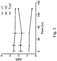
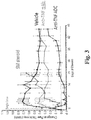
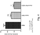

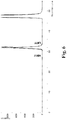
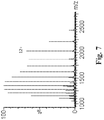
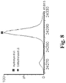
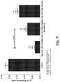
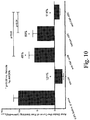


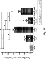
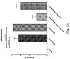

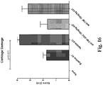
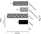
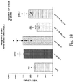

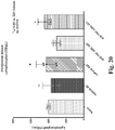
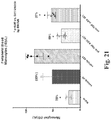

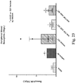
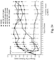
Classifications
-
- A—HUMAN NECESSITIES
- A61—MEDICAL OR VETERINARY SCIENCE; HYGIENE
- A61K—PREPARATIONS FOR MEDICAL, DENTAL OR TOILETRY PURPOSES
- A61K31/00—Medicinal preparations containing organic active ingredients
- A61K31/56—Compounds containing cyclopenta[a]hydrophenanthrene ring systems; Derivatives thereof, e.g. steroids
- A61K31/58—Compounds containing cyclopenta[a]hydrophenanthrene ring systems; Derivatives thereof, e.g. steroids containing heterocyclic rings, e.g. danazol, stanozolol, pancuronium or digitogenin
-
- C—CHEMISTRY; METALLURGY
- C07—ORGANIC CHEMISTRY
- C07K—PEPTIDES
- C07K16/00—Immunoglobulins [IGs], e.g. monoclonal or polyclonal antibodies
- C07K16/18—Immunoglobulins [IGs], e.g. monoclonal or polyclonal antibodies against material from animals or humans
- C07K16/24—Immunoglobulins [IGs], e.g. monoclonal or polyclonal antibodies against material from animals or humans against cytokines, lymphokines or interferons
- C07K16/241—Tumor Necrosis Factors
-
- A—HUMAN NECESSITIES
- A61—MEDICAL OR VETERINARY SCIENCE; HYGIENE
- A61K—PREPARATIONS FOR MEDICAL, DENTAL OR TOILETRY PURPOSES
- A61K47/00—Medicinal preparations characterised by the non-active ingredients used, e.g. carriers or inert additives; Targeting or modifying agents chemically bound to the active ingredient
- A61K47/50—Medicinal preparations characterised by the non-active ingredients used, e.g. carriers or inert additives; Targeting or modifying agents chemically bound to the active ingredient the non-active ingredient being chemically bound to the active ingredient, e.g. polymer-drug conjugates
- A61K47/51—Medicinal preparations characterised by the non-active ingredients used, e.g. carriers or inert additives; Targeting or modifying agents chemically bound to the active ingredient the non-active ingredient being chemically bound to the active ingredient, e.g. polymer-drug conjugates the non-active ingredient being a modifying agent
- A61K47/68—Medicinal preparations characterised by the non-active ingredients used, e.g. carriers or inert additives; Targeting or modifying agents chemically bound to the active ingredient the non-active ingredient being chemically bound to the active ingredient, e.g. polymer-drug conjugates the non-active ingredient being a modifying agent the modifying agent being an antibody, an immunoglobulin or a fragment thereof, e.g. an Fc-fragment
- A61K47/6801—Drug-antibody or immunoglobulin conjugates defined by the pharmacologically or therapeutically active agent
- A61K47/6803—Drugs conjugated to an antibody or immunoglobulin, e.g. cisplatin-antibody conjugates
-
- A—HUMAN NECESSITIES
- A61—MEDICAL OR VETERINARY SCIENCE; HYGIENE
- A61K—PREPARATIONS FOR MEDICAL, DENTAL OR TOILETRY PURPOSES
- A61K47/00—Medicinal preparations characterised by the non-active ingredients used, e.g. carriers or inert additives; Targeting or modifying agents chemically bound to the active ingredient
- A61K47/50—Medicinal preparations characterised by the non-active ingredients used, e.g. carriers or inert additives; Targeting or modifying agents chemically bound to the active ingredient the non-active ingredient being chemically bound to the active ingredient, e.g. polymer-drug conjugates
- A61K47/51—Medicinal preparations characterised by the non-active ingredients used, e.g. carriers or inert additives; Targeting or modifying agents chemically bound to the active ingredient the non-active ingredient being chemically bound to the active ingredient, e.g. polymer-drug conjugates the non-active ingredient being a modifying agent
- A61K47/68—Medicinal preparations characterised by the non-active ingredients used, e.g. carriers or inert additives; Targeting or modifying agents chemically bound to the active ingredient the non-active ingredient being chemically bound to the active ingredient, e.g. polymer-drug conjugates the non-active ingredient being a modifying agent the modifying agent being an antibody, an immunoglobulin or a fragment thereof, e.g. an Fc-fragment
- A61K47/6835—Medicinal preparations characterised by the non-active ingredients used, e.g. carriers or inert additives; Targeting or modifying agents chemically bound to the active ingredient the non-active ingredient being chemically bound to the active ingredient, e.g. polymer-drug conjugates the non-active ingredient being a modifying agent the modifying agent being an antibody, an immunoglobulin or a fragment thereof, e.g. an Fc-fragment the modifying agent being an antibody or an immunoglobulin bearing at least one antigen-binding site
- A61K47/6845—Medicinal preparations characterised by the non-active ingredients used, e.g. carriers or inert additives; Targeting or modifying agents chemically bound to the active ingredient the non-active ingredient being chemically bound to the active ingredient, e.g. polymer-drug conjugates the non-active ingredient being a modifying agent the modifying agent being an antibody, an immunoglobulin or a fragment thereof, e.g. an Fc-fragment the modifying agent being an antibody or an immunoglobulin bearing at least one antigen-binding site the antibody targeting a cytokine, e.g. growth factors, VEGF, TNF, a lymphokine or an interferon
-
- A—HUMAN NECESSITIES
- A61—MEDICAL OR VETERINARY SCIENCE; HYGIENE
- A61K—PREPARATIONS FOR MEDICAL, DENTAL OR TOILETRY PURPOSES
- A61K47/00—Medicinal preparations characterised by the non-active ingredients used, e.g. carriers or inert additives; Targeting or modifying agents chemically bound to the active ingredient
- A61K47/50—Medicinal preparations characterised by the non-active ingredients used, e.g. carriers or inert additives; Targeting or modifying agents chemically bound to the active ingredient the non-active ingredient being chemically bound to the active ingredient, e.g. polymer-drug conjugates
- A61K47/51—Medicinal preparations characterised by the non-active ingredients used, e.g. carriers or inert additives; Targeting or modifying agents chemically bound to the active ingredient the non-active ingredient being chemically bound to the active ingredient, e.g. polymer-drug conjugates the non-active ingredient being a modifying agent
- A61K47/68—Medicinal preparations characterised by the non-active ingredients used, e.g. carriers or inert additives; Targeting or modifying agents chemically bound to the active ingredient the non-active ingredient being chemically bound to the active ingredient, e.g. polymer-drug conjugates the non-active ingredient being a modifying agent the modifying agent being an antibody, an immunoglobulin or a fragment thereof, e.g. an Fc-fragment
- A61K47/6835—Medicinal preparations characterised by the non-active ingredients used, e.g. carriers or inert additives; Targeting or modifying agents chemically bound to the active ingredient the non-active ingredient being chemically bound to the active ingredient, e.g. polymer-drug conjugates the non-active ingredient being a modifying agent the modifying agent being an antibody, an immunoglobulin or a fragment thereof, e.g. an Fc-fragment the modifying agent being an antibody or an immunoglobulin bearing at least one antigen-binding site
- A61K47/6849—Medicinal preparations characterised by the non-active ingredients used, e.g. carriers or inert additives; Targeting or modifying agents chemically bound to the active ingredient the non-active ingredient being chemically bound to the active ingredient, e.g. polymer-drug conjugates the non-active ingredient being a modifying agent the modifying agent being an antibody, an immunoglobulin or a fragment thereof, e.g. an Fc-fragment the modifying agent being an antibody or an immunoglobulin bearing at least one antigen-binding site the antibody targeting a receptor, a cell surface antigen or a cell surface determinant
-
- A—HUMAN NECESSITIES
- A61—MEDICAL OR VETERINARY SCIENCE; HYGIENE
- A61K—PREPARATIONS FOR MEDICAL, DENTAL OR TOILETRY PURPOSES
- A61K47/00—Medicinal preparations characterised by the non-active ingredients used, e.g. carriers or inert additives; Targeting or modifying agents chemically bound to the active ingredient
- A61K47/50—Medicinal preparations characterised by the non-active ingredients used, e.g. carriers or inert additives; Targeting or modifying agents chemically bound to the active ingredient the non-active ingredient being chemically bound to the active ingredient, e.g. polymer-drug conjugates
- A61K47/51—Medicinal preparations characterised by the non-active ingredients used, e.g. carriers or inert additives; Targeting or modifying agents chemically bound to the active ingredient the non-active ingredient being chemically bound to the active ingredient, e.g. polymer-drug conjugates the non-active ingredient being a modifying agent
- A61K47/68—Medicinal preparations characterised by the non-active ingredients used, e.g. carriers or inert additives; Targeting or modifying agents chemically bound to the active ingredient the non-active ingredient being chemically bound to the active ingredient, e.g. polymer-drug conjugates the non-active ingredient being a modifying agent the modifying agent being an antibody, an immunoglobulin or a fragment thereof, e.g. an Fc-fragment
- A61K47/6889—Conjugates wherein the antibody being the modifying agent and wherein the linker, binder or spacer confers particular properties to the conjugates, e.g. peptidic enzyme-labile linkers or acid-labile linkers, providing for an acid-labile immuno conjugate wherein the drug may be released from its antibody conjugated part in an acidic, e.g. tumoural or environment
-
- A—HUMAN NECESSITIES
- A61—MEDICAL OR VETERINARY SCIENCE; HYGIENE
- A61P—SPECIFIC THERAPEUTIC ACTIVITY OF CHEMICAL COMPOUNDS OR MEDICINAL PREPARATIONS
- A61P29/00—Non-central analgesic, antipyretic or antiinflammatory agents, e.g. antirheumatic agents; Non-steroidal antiinflammatory drugs [NSAID]
-
- A—HUMAN NECESSITIES
- A61—MEDICAL OR VETERINARY SCIENCE; HYGIENE
- A61P—SPECIFIC THERAPEUTIC ACTIVITY OF CHEMICAL COMPOUNDS OR MEDICINAL PREPARATIONS
- A61P37/00—Drugs for immunological or allergic disorders
-
- A—HUMAN NECESSITIES
- A61—MEDICAL OR VETERINARY SCIENCE; HYGIENE
- A61P—SPECIFIC THERAPEUTIC ACTIVITY OF CHEMICAL COMPOUNDS OR MEDICINAL PREPARATIONS
- A61P37/00—Drugs for immunological or allergic disorders
- A61P37/02—Immunomodulators
- A61P37/06—Immunosuppressants, e.g. drugs for graft rejection
-
- C—CHEMISTRY; METALLURGY
- C07—ORGANIC CHEMISTRY
- C07J—STEROIDS
- C07J71/00—Steroids in which the cyclopenta(a)hydrophenanthrene skeleton is condensed with a heterocyclic ring
- C07J71/0005—Oxygen-containing hetero ring
- C07J71/0026—Oxygen-containing hetero ring cyclic ketals
- C07J71/0031—Oxygen-containing hetero ring cyclic ketals at positions 16, 17
-
- C—CHEMISTRY; METALLURGY
- C07—ORGANIC CHEMISTRY
- C07K—PEPTIDES
- C07K16/00—Immunoglobulins [IGs], e.g. monoclonal or polyclonal antibodies
- C07K16/18—Immunoglobulins [IGs], e.g. monoclonal or polyclonal antibodies against material from animals or humans
- C07K16/24—Immunoglobulins [IGs], e.g. monoclonal or polyclonal antibodies against material from animals or humans against cytokines, lymphokines or interferons
-
- A—HUMAN NECESSITIES
- A61—MEDICAL OR VETERINARY SCIENCE; HYGIENE
- A61K—PREPARATIONS FOR MEDICAL, DENTAL OR TOILETRY PURPOSES
- A61K39/00—Medicinal preparations containing antigens or antibodies
- A61K2039/505—Medicinal preparations containing antigens or antibodies comprising antibodies
-
- C—CHEMISTRY; METALLURGY
- C07—ORGANIC CHEMISTRY
- C07K—PEPTIDES
- C07K16/00—Immunoglobulins [IGs], e.g. monoclonal or polyclonal antibodies
- C07K16/18—Immunoglobulins [IGs], e.g. monoclonal or polyclonal antibodies against material from animals or humans
-
- C—CHEMISTRY; METALLURGY
- C07—ORGANIC CHEMISTRY
- C07K—PEPTIDES
- C07K2317/00—Immunoglobulins specific features
- C07K2317/20—Immunoglobulins specific features characterized by taxonomic origin
- C07K2317/21—Immunoglobulins specific features characterized by taxonomic origin from primates, e.g. man
-
- C—CHEMISTRY; METALLURGY
- C07—ORGANIC CHEMISTRY
- C07K—PEPTIDES
- C07K2317/00—Immunoglobulins specific features
- C07K2317/20—Immunoglobulins specific features characterized by taxonomic origin
- C07K2317/24—Immunoglobulins specific features characterized by taxonomic origin containing regions, domains or residues from different species, e.g. chimeric, humanized or veneered
-
- C—CHEMISTRY; METALLURGY
- C07—ORGANIC CHEMISTRY
- C07K—PEPTIDES
- C07K2317/00—Immunoglobulins specific features
- C07K2317/30—Immunoglobulins specific features characterized by aspects of specificity or valency
- C07K2317/31—Immunoglobulins specific features characterized by aspects of specificity or valency multispecific
-
- C—CHEMISTRY; METALLURGY
- C07—ORGANIC CHEMISTRY
- C07K—PEPTIDES
- C07K2317/00—Immunoglobulins specific features
- C07K2317/50—Immunoglobulins specific features characterized by immunoglobulin fragments
- C07K2317/55—Fab or Fab'
-
- C—CHEMISTRY; METALLURGY
- C07—ORGANIC CHEMISTRY
- C07K—PEPTIDES
- C07K2317/00—Immunoglobulins specific features
- C07K2317/50—Immunoglobulins specific features characterized by immunoglobulin fragments
- C07K2317/56—Immunoglobulins specific features characterized by immunoglobulin fragments variable (Fv) region, i.e. VH and/or VL
- C07K2317/569—Single domain, e.g. dAb, sdAb, VHH, VNAR or nanobody®
-
- C—CHEMISTRY; METALLURGY
- C07—ORGANIC CHEMISTRY
- C07K—PEPTIDES
- C07K2317/00—Immunoglobulins specific features
- C07K2317/70—Immunoglobulins specific features characterized by effect upon binding to a cell or to an antigen
- C07K2317/73—Inducing cell death, e.g. apoptosis, necrosis or inhibition of cell proliferation
-
- C—CHEMISTRY; METALLURGY
- C07—ORGANIC CHEMISTRY
- C07K—PEPTIDES
- C07K2317/00—Immunoglobulins specific features
- C07K2317/90—Immunoglobulins specific features characterized by (pharmaco)kinetic aspects or by stability of the immunoglobulin
- C07K2317/92—Affinity (KD), association rate (Ka), dissociation rate (Kd) or EC50 value
-
- C—CHEMISTRY; METALLURGY
- C07—ORGANIC CHEMISTRY
- C07K—PEPTIDES
- C07K2318/00—Antibody mimetics or scaffolds
- C07K2318/20—Antigen-binding scaffold molecules wherein the scaffold is not an immunoglobulin variable region or antibody mimetics
-
- C—CHEMISTRY; METALLURGY
- C07—ORGANIC CHEMISTRY
- C07K—PEPTIDES
- C07K2319/00—Fusion polypeptide
- C07K2319/30—Non-immunoglobulin-derived peptide or protein having an immunoglobulin constant or Fc region, or a fragment thereof, attached thereto
Definitions
- the field of the invention generally relates to glucocorticoid receptor agonist immunoconjugates, and methods of making and using the same, e.g., to treat autoimmune or inflammatory diseases.
- Tumor Necrosis Factor alpha plays a central role in the pathophysiology of several human disorders, and anti-TNFa agents (e.g., adalimumab, etanercept, and infliximab) have clinically validated therapeutic utility in the treatment of autoimmune and inflammatory disorders, such as rheumatoid arthritis, psoriasis and inflammatory bowel disease.
- anti-TNFa biologics are still limited in the maximal efficacy they can achieve in patients, necessitating the identification and development of more potent and effective therapeutics. Patients treated with anti-TNFa biologics may also develop an immunogenic response to the therapeutic thus limiting its effectiveness. Therefore anti-TNFa therapies with lower immunogenicity and high efficacy would be useful for further controlling disease.
- Synthetic glucocorticoid receptor agonists are a potent class of small molecules used in the treatment of inflammatory disorders, but their utility in the chronic treatment of disease is limited due to severe side effects.
- Several approaches to retain the anti-inflammatory efficacy of synthetic glucocorticoids while sparing the unwanted toxicities have been described ( Rosen, J and Miner, JN Endocrine Reviews 26: 452-64 (2005 )). However these methodologies have met with little success. There is a need in the field of autoimmune and inflammatory disease therapeutics to develop therapeutics with enhanced efficacy and longer duration of action compared to anti-TNF antibodies and with minimal unwanted effects.
- the present invention provides a compound: wherein n is 2 or 4, and A is an antibody comprising SEQ ID NO: 66 and SEQ ID NO: 73 or adalimumab. In further embodiments of the invention n is 4. In other embodiments of the invention n is 2. In other embodiments of the invention A is adalimumab.
- anti-TNF alpha protein refers to proteins that are capable of (i) binding to TNF alpha and (ii) inhibiting binding of soluble TNF-alpha to cell surface TNF receptors (p55 and/or p75) and/or lysing surface TNF alpha or TNF alpha receptor expressing cells in vitro in the presence of complement.
- Anti-TNF alpha proteins include, for example, anti-TNF antibodies or antigen-binding fragments thereof (e.g., adalimumab or infliximab) as well as soluble TNF receptors (e.g., etanercept).
- antibody and “antibodies” are terms of art and can be used interchangeably herein and refer to a molecule with an antigen-binding site that specifically binds an antigen.
- antibody means an immunoglobulin molecule that recognizes and specifically binds to a target, such as a protein, polypeptide, peptide, carbohydrate, polynucleotide, lipid, or combinations of the foregoing through at least one antigen recognition site within the variable region of the immunoglobulin molecule.
- a target such as a protein, polypeptide, peptide, carbohydrate, polynucleotide, lipid, or combinations of the foregoing through at least one antigen recognition site within the variable region of the immunoglobulin molecule.
- the term “antibody” encompasses intact polyclonal antibodies, intact monoclonal antibodies, chimeric antibodies, humanized antibodies, human antibodies, fusion proteins comprising an antibody, and any other modified immunoglobulin molecule so long as the antibodies exhibit the desired biological activity.
- An antibody can be of any the five major classes of immunoglobulins: IgA, IgD, IgE, IgG, and IgM, or subclasses (isotypes) thereof (e.g. IgG1, IgG2, IgG3, IgG4, IgA1 and IgA2), based on the identity of their heavy-chain constant domains referred to as alpha, delta, epsilon, gamma, and mu, respectively.
- the different classes of immunoglobulins have different and well known subunit structures and three-dimensional configurations.
- Antibodies can be naked or conjugated to other molecules such as toxins, radioisotopes, etc.
- the term "antibody" encompasses bispecific and multispecific antibodies.
- antibody fragment refers to a portion of an intact antibody.
- An "antigen-binding fragment” refers to a portion of an intact antibody that binds to an antigen.
- An antigen-binding fragment can contain the antigenic determining variable regions of an intact antibody. Examples of antibody fragments include, but are not limited to Fab, Fab', F(ab')2, and Fv fragments, linear antibodies, and single chain antibodies.
- An "antigen-binding fragment” can be a bispecific or multispecific antigen-binding fragment.
- anti-TNF-alpha antibody or "an antibody that binds to TNF-alpha” refers to an antibody that is capable of binding TNF-alpha with sufficient affinity such that the antibody is useful as a diagnostic and/or therapeutic agent in targeting TNF-alpha.
- the extent of binding of an anti-TNF-alpha antibody to an unrelated, non-TNF-alpha protein can be less than about 10% of the binding of the antibody to TNF-alpha as measured, e.g., by a radioimmunoassay (RIA).
- RIA radioimmunoassay
- an antibody that binds to TNF-alpha has a dissociation constant (Kd) of ⁇ 1 ⁇ M, ⁇ 100 nM, ⁇ 10 nM, ⁇ 1 nM, or ⁇ 0.1 nM.
- a “monoclonal” antibody or antigen-binding fragment thereof refers to a homogeneous antibody or antigen-binding fragment population involved in the highly specific recognition and binding of a single antigenic determinant, or epitope. This is in contrast to polyclonal antibodies that typically include different antibodies directed against different antigenic determinants.
- the term "monoclonal” antibody or antigen-binding fragment thereof encompasses both intact and full-length monoclonal antibodies as well as antibody fragments (such as Fab, Fab', F(ab')2, Fv), single chain (scFv) mutants, fusion proteins comprising an antibody portion, and any other modified immunoglobulin molecule comprising an antigen recognition site.
- monoclonal antibody or antigen-binding fragment thereof refers to such antibodies and antigen-binding fragments thereof made in any number of manners including but not limited to by hybridoma, phage selection, recombinant expression, and transgenic animals.
- humanized antibody or antigen-binding fragment thereof refers to forms of non-human (e.g. murine) antibodies or antigen-binding fragments that are specific immunoglobulin chains, chimeric immunoglobulins, or fragments thereof that contain minimal non-human (e.g., murine) sequences.
- humanized antibodies or antigen-binding fragments thereof are human immunoglobulins in which residues from the complementary determining region (CDR) are replaced by residues from the CDR of a non-human species (e.g.
- CDR grafted Jones et al., Nature 321:522-525 (1986 ); Riechmann et al., Nature 332:323-327 (1988 ); Verhoeyen et al., Science 239:1534-1536 (1988 )).
- Fv framework region (FR) residues of a human immunoglobulin are replaced with the corresponding residues in an antibody or fragment from a non-human species that has the desired specificity, affinity, and capability.
- the humanized antibody or antigen-binding fragment thereof can be further modified by the substitution of additional residues either in the Fv framework region and/or within the replaced non-human residues to refine and optimize antibody or antigen-binding fragment thereof specificity, affinity, and/or capability.
- the humanized antibody or antigen-binding fragment thereof will comprise substantially all of at least one, and typically two or three, variable domains containing all or substantially all of the CDR regions that correspond to the non-human immunoglobulin whereas all or substantially all of the FR regions are those of a human immunoglobulin consensus sequence.
- the humanized antibody or antigen-binding fragment thereof can also comprise at least a portion of an immunoglobulin constant region or domain (Fc), typically that of a human immunoglobulin.
- a "humanized antibody” is a resurfaced antibody.
- variable region of an antibody refers to the variable region of the antibody light chain or the variable region of the antibody heavy chain, either alone or in combination.
- the variable regions of the heavy and light chain each consist of four framework regions (FR) connected by three complementarity determining regions (CDRs) also known as hypervariable regions.
- the CDRs in each chain are held together in close proximity by the FRs and, with the CDRs from the other chain, contribute to the formation of the antigen-binding site of antibodies.
- CDRs complementarity determining regions
- the Kabat numbering system is generally used when referring to a residue in the variable domain (approximately residues 1-107 of the light chain and residues 1-113 of the heavy chain) (e.g., Kabat et al., Sequences of Immunological Interest. 5th Ed. Public Health Service, National Institutes of Health, Bethesda, Md. (1991)). Unless explicitly indicated otherwise, the numbering system used herein is the Kabat numbering system.
- amino acid position numbering refers to the numbering system used for heavy chain variable domains or light chain variable domains of the compilation of antibodies in Kabat et al., Sequences of Proteins of Immunological Interest, 5th Ed. Public Health Service, National Institutes of Health, Bethesda, Md. (1991). Using this numbering system, the actual linear amino acid sequence can contain fewer or additional amino acids corresponding to a shortening of, or insertion into, a FR or CDR of the variable domain.
- a heavy chain variable domain can include a single amino acid insert (residue 52a according to Kabat) after residue 52 of H2 and inserted residues (e.g.
- residues 82a, 82b, and 82c, etc. according to Kabat after heavy chain FR residue 82.
- the Kabat numbering of residues can be determined for a given antibody by alignment at regions of homology of the sequence of the antibody with a "standard” Kabat numbered sequence.
- Chothia refers instead to the location of the structural loops ( Chothia and Lesk J. Mol. Biol. 196:901-917 (1987 )).
- the end of the Chothia CDR-H1 loop when numbered using the Kabat numbering convention varies between H32 and H34 depending on the length of the loop (this is because the Kabat numbering scheme places the insertions at H35A and H35B; if neither 35A nor 35B is present, the loop ends at 32; if only 35A is present, the loop ends at 33; if both 35A and 35B are present, the loop ends at 34).
- the AbM hypervariable regions represent a compromise between the Kabat CDRs and Chothia structural loops, and are used by Oxford Molecular's AbM antibody modeling software.
- the CDRs of an antibody or antigen-binding fragment thereof can be determined according to the Chothia numbering scheme, which refers to the location of immunoglobulin structural loops (see, e.g., Chothia C & Lesk AM, (1987), J Mol Biol 196: 901-917 ; Al-Lazikani B et al., (1997) J Mol Biol 273: 927-948 ; Chothia C et al., (1992) J Mol Biol 227: 799-817 ; Tramontano A et al., (1990) J Mol Biol 215(1): 175-82 ; and U.S. Patent No. 7,709,226 ).
- Chothia numbering scheme refers to the location of immunoglobulin structural loops
- the Chothia CDR-H1 loop is present at heavy chain amino acids 26 to 32, 33, or 34
- the Chothia CDR-H2 loop is present at heavy chain amino acids 52 to 56
- the Chothia CDR-H3 loop is present at heavy chain amino acids 95 to 102
- the Chothia CDR-L1 loop is present at light chain amino acids 24 to 34
- the Chothia CDR-L2 loop is present at light chain amino acids 50 to 56
- the Chothia CDR-L3 loop is present at light chain amino acids 89 to 97.
- the end of the Chothia CDR-H1 loop when numbered using the Kabat numbering convention varies between H32 and H34 depending on the length of the loop (this is because the Kabat numbering scheme places the insertions at H35A and H35B; if neither 35A nor 35B is present, the loop ends at 32; if only 35A is present, the loop ends at 33; if both 35A and 35B are present, the loop ends at 34).
- the CDRs of an antibody or antigen-binding fragment thereof can be determined according to the IMGT numbering system as described in Lefranc M-P, (1999) The Immunologist 7: 132-136 and Lefranc M-P et al., (1999) Nucleic Acids Res 27: 209-212 .
- VH-CDR1 is at positions 26 to 35
- VH-CDR2 is at positions 51 to 57
- VH-CDR3 is at positions 93 to 102
- VL-CDR1 is at positions 27 to 32
- VL-CDR2 is at positions 50 to 52
- VL-CDR3 is at positions 89 to 97.
- the CDRs of an antibody or antigen-binding fragment thereof can be determined according to MacCallum RM et al., (1996) J Mol Biol 262: 732-745 . See also, e.g., Martin A. "Protein Sequence and Structure Analysis of Antibody Variable Domains," in Antibody Engineering, Kontermann and Dübel, eds., Chapter 31, pp. 422-439, Springer-Verlag, Berlin (2001 ).
- the CDRs of an antibody or antigen-binding fragment thereof can be determined according to the AbM numbering scheme, which refers AbM hypervariable regions which represent a compromise between the Kabat CDRs and Chothia structural loops, and are used by Oxford Molecular's AbM antibody modeling software (Oxford Molecular Group, Inc.).
- human antibody means an antibody produced by a human or an antibody having an amino acid sequence corresponding to an antibody produced by a human made using any technique known in the art. This definition of a human antibody includes intact or full-length antibodies, fragments thereof, and/or antibodies comprising at least one human heavy and/or light chain polypeptide such as, for example, an antibody comprising murine light chain and human heavy chain polypeptides.
- chimeric antibodies refers to antibodies wherein the amino acid sequence of the immunoglobulin molecule is derived from two or more species.
- the variable region of both light and heavy chains corresponds to the variable region of antibodies derived from one species of mammals (e.g. mouse, rat, rabbit, etc.) with the desired specificity, affinity, and capability while the constant regions are homologous to the sequences in antibodies derived from another (usually human) to avoid eliciting an immune response in that species.
- epitopes or "antigenic determinant” are used interchangeably herein and refer to that portion of an antigen capable of being recognized and specifically bound by a particular antibody.
- the antigen is a polypeptide
- epitopes can be formed both from contiguous amino acids and noncontiguous amino acids juxtaposed by tertiary folding of a protein. Epitopes formed from contiguous amino acids are typically retained upon protein denaturing, whereas epitopes formed by tertiary folding are typically lost upon protein denaturing.
- An epitope typically includes at least 3, and more usually, at least 5 or 8-10 amino acids in a unique spatial conformation.
- Binding affinity generally refers to the strength of the sum total of noncovalent interactions between a single binding site of a molecule (e.g., an antibody) and its binding partner (e.g., an antigen). Unless indicated otherwise, as used herein, "binding affinity” refers to intrinsic binding affinity which reflects a 1:1 interaction between members of a binding pair (e.g., antibody and antigen).
- the affinity of a molecule X for its partner Y can generally be represented by the dissociation constant (Kd). Affinity can be measured by common methods known in the art, including those described herein. Low-affinity antibodies generally bind antigen slowly and tend to dissociate readily, whereas high-affinity antibodies generally bind antigen faster and tend to remain bound longer. A variety of methods of measuring binding affinity are known in the art, any of which can be used for purposes of the present disclosure. Specific illustrative embodiments are described in the following.
- binding affinity refers to a stronger binding between a molecule and its binding partner.
- Or better when used herein refers to a stronger binding, represented by a smaller numerical Kd value.
- an antibody which has an affinity for an antigen of "0.6 nM or better” the antibody's affinity for the antigen is ⁇ 0.6 nM, i.e. 0.59 nM, 0.58 nM, 0.57 nM etc. or any value less than 0.6 nM.
- an antibody binds to an epitope via its antigen binding domain, and that the binding entails some complementarity between the antigen binding domain and the epitope. According to this definition, an antibody is said to "specifically bind” to an epitope when it binds to that epitope, via its antigen binding domain more readily than it would bind to a random, unrelated epitope.
- the term “specificity” is used herein to qualify the relative affinity by which a certain antibody binds to a certain epitope.
- antibody “A” may be deemed to have a higher specificity for a given epitope than antibody "B,” or antibody “A” may be said to bind to epitope “C” with a higher specificity than it has for related epitope “D.”
- preferentially binds it is meant that the antibody specifically binds to an epitope more readily than it would bind to a related, similar, homologous, or analogous epitope.
- an antibody which "preferentially binds" to a given epitope would more likely bind to that epitope than to a related epitope, even though such an antibody may cross-react with the related epitope.
- An antibody is said to "competitively inhibit" binding of a reference antibody to a given epitope if the antibody preferentially binds to that epitope or an overlapping epitope to the extent that it blocks, to some degree, binding of the reference antibody to the epitope.
- Competitive inhibition may be determined by any method known in the art, for example, competition ELISA assays.
- An antibody may be said to competitively inhibit binding of the reference antibody to a given epitope by at least 90%, at least 80%, at least 70%, at least 60%, or at least 50%.
- a polypeptide, antibody, polynucleotide, vector, cell, or composition which is "isolated” is a polypeptide, antibody, polynucleotide, vector, cell, or composition which is in a form not found in nature.
- Isolated polypeptides, antibodies, polynucleotides, vectors, cell or compositions include those which have been purified to a degree that they are no longer in a form in which they are found in nature.
- an antibody, polynucleotide, vector, cell, or composition which is isolated is substantially pure.
- substantially pure refers to material which is at least 50% pure (i.e., free from contaminants), at least 90% pure, at least 95% pure, at least 98% pure, or at least 99% pure.
- Immunoconjugates can also be defined by the generic formula in reverse order: A-(Q-L-SM) n .
- the following generic formula shows a immunoconjugate having a dipeptide (Ala-Ala) linker and succinimide thioether-based heterobifunctional group:
- linker refers to any chemical moiety capable of linking a protein, e.g., antibody, antibody fragment (e.g., antigen binding fragments) or functional equivalent to a glucocorticosteroid.
- Linkers may be susceptible to cleavage (a "cleavable linker") thereby facilitating release of the glucocorticosteroid.
- cleavable linkers may be susceptible to acid-induced cleavage, photo-induced cleavage, peptidase-induced cleavage, esterase-induced cleavage, and disulfide bond cleavage, at conditions under which the glucocorticosteroid and/or the antibody remains active.
- linkers may be substantially resistant to cleavage (a "noncleavable linker").
- non-cleavable linkers are any chemical moiety capable of linking a glucocorticosteroid to an antibody in a stable, covalent manner and does not fall off under the categories listed above for cleaveable linkers.
- non-cleavable linkers are substantially resistant to acid-induced cleavage, photo-induced cleavage, peptidase-induced cleavage, esterase-induced cleavage and disulfide bond cleavage.
- non-cleavable refers to the ability of the chemical bond in the linker or adjoining to the linker to withstand cleavage induced by an acid, photolabile-cleaving agent, a peptidase, an esterase, or a chemical or physiological compound that cleaves a disulfide bond, at conditions under which a glucocorticosteroid and/or the antibody does not lose its activity.
- cleavable linkers are cleaved by peptidases ("peptidase cleavable linkers"). Only certain peptides are readily cleaved inside or outside cells, see e.g. Trout et al., 79 Proc. Natl. Acad. Sci. USA, 626-629 (1982 ) and Umemoto et al. 43 Int. J. Cancer, 677-684 (1989 ). Furthermore, peptides are composed of ⁇ -amino acid units and peptidic bonds, which chemically are amide bonds between the carboxylate of one amino acid and the amino group of a second amino acid. Other amide bonds, such as the bond between a carboxylate and the ⁇ -amino acid group of lysine, are understood not to be peptidic bonds and are considered non-cleavable.
- linkers are cleaved by esterases ("esterase cleavable linkers"). Only certain esters can be cleaved by esterases present inside or outside of cells. Esters are formed by the condensation of a carboxylic acid and an alcohol. Simple esters are esters produced with simple alcohols, such as aliphatic alcohols, and small cyclic and small aromatic alcohols.
- the cleavable linker component may comprise a peptide comprising one to ten amino acid residues.
- the peptide allows for cleavage of the linker by a protease, thereby facilitating release of the glucocorticosteroid upon exposure to intracellular proteases, such as lysosomal enzymes ( Doronina et al. (2003) Nat. Biotechnol. 21:778-784 ).
- Exemplary peptides include, but are not limited to, dipeptides, tripeptides, tetrapeptides, and pentapeptides.
- Exemplary dipeptides include, but are not limited to, alanine-alanine (ala-ala), valine-citrulline (vc or val-cit), alanine-phenylalanine (af or ala-phe); phenylalanine-lysine (fk or phe-lys); phenylalanine-homolysine (phe-homolys); and N-methyl-valine-citrulline (Me-val-cit).
- Exemplary tripeptides include, but are not limited to, glycine-valine-citrulline (gly-val-cit) and glycine-glycine-glycine (gly-gly-gly).
- a peptide may comprise naturally-occurring and/or non-natural amino acid residues.
- naturally-occurring amino acid refer to Ala, Asp, Cys, Glu, Phe, Gly, His, He, Lys, Leu, Met, Asn, Pro, Gin, Arg, Ser, Thr, Val, Trp, and Tyr.
- Non-natural amino acids include, by way of non-limiting example, homoserine, homoarginine, citrulline, phenylglycine, taurine, iodotyrosine, seleno- cysteine, norleucine ("Nle”), norvaline (“Nva”), beta-alanine, L- or D-naphthalanine, ornithine ("Orn”), and the like.
- Peptides can be designed and optimized for enzymatic cleavage by a particular enzyme, for example, a tumor-associated protease, cathepsin B, C and D, or a plasmin protease.
- Amino acids also include the D-forms of natural and non-natural amino acids.
- D- designates an amino acid having the “D” (dextrorotary) configuration, as opposed to the configuration in the naturally occurring (“L-") amino acids.
- Natural and non-natural amino acids can be purchased commercially (Sigma Chemical Co., Advanced Chemtech) or synthesized using methods known in the art.
- glucocorticosteroid refers to naturally-occurring or synthetic steroid hormones that interact with glucocorticoid receptors.
- Non-limiting exemplary glucocorticosteroids include: By way of example, the A-, B-, C-, and D-rings of the steroid skeleton are marked for budesonide.
- Glucocorticosteroids are described in WO 2009/069032 .
- a "radical of a glucocorticosteroid” is derived from the removal of one or more hydrogen atoms from a parent glucocorticosteroid.
- the removal of hydrogen atom(s) facilitates the attachment of the parent glucocorticosteroid to a linker.
- the hydrogen atom is removed from any suitable -NH 2 group of the parent glucocorticosteroid.
- the hydrogen atom is removed from any suitable -OH group of the parent glucocorticosteroid.
- the hydrogen atom is removed from any suitable a -SH group of the parent glucocorticosteroid.
- heterotrifunctional group refers to a chemical moiety that connects a linker and protein, e.g., an antibody.
- Heterobi- and trifunctional groups are characterized as having different reactive groups at either end of the chemical moiety.
- Non-limiting exemplary heterobifunctional groups include: A non-limiting exemplary heterotrifunctional group is:
- drug antibody ratio refers to the number of SMs (i.e., radical derived from a small-molecule glucocorticoid receptor agonist, e.g., a glucocorticosteroid) linked to A (i.e., a protein, e.g., an antibody or antigen-binding fragment thereof, an anti-TNF protein, an anti-TNF-alpha antibody or fragment thereof, a soluble receptor, or a soluble TNF receptor).
- SMs small-molecule glucocorticoid receptor agonist
- A i.e., a protein, e.g., an antibody or antigen-binding fragment thereof, an anti-TNF protein, an anti-TNF-alpha antibody or fragment thereof, a soluble receptor, or a soluble TNF receptor.
- the DAR refers to the number of SMs linked to the individual A (e.g., n is an integer of 1 to 10).
- the DAR refers to the average number of SMs linked to the As (e.g., n is an integer or fraction of 1 to 10).
- n is an integer or fraction of 1 to 10.
- a compound having formula (SM-L-Q) n -A comprising a first immunoconjugate with 3 SM per A and a second immunoconjugate with 4 SM per A would have a DAR (i.e., an "n") of 3.5.
- subject refers to any animal (e.g., a mammal), including, but not limited to humans, non-human primates, rodents, and the like, which is to be the recipient of a particular treatment.
- subject and patient are used interchangeably herein in reference to a human subject.
- pharmaceutical formulation refers to a preparation which is in such form as to permit the biological activity of the active ingredient to be effective, and which contains no additional components which are unacceptably toxic to a subject to which the formulation would be administered.
- the formulation can be sterile.
- Terms such as “treating” or “treatment” or “to treat” or “alleviating” or “to alleviate” refer to therapeutic measures that cure, slow down, lessen symptoms of, and/or halt progression of a diagnosed pathologic condition or disorder. Thus, those in need of treatment include those already diagnosed with or suspected of having the disorder.
- Prophylactic or preventative measures refer to measures that prevent and/or slow the development of a targeted pathological condition or disorder. Thus, those in need of prophylactic or preventative measures include those prone to have the disorder and those in whom the disorder is to be prevented.
- Polynucleotide or “nucleic acid,” as used interchangeably herein, refer to polymers of nucleotides of any length, and include DNA and RNA.
- the nucleotides can be deoxyribonucleotides, ribonucleotides, modified nucleotides or bases, and/or their analogs, or any substrate that can be incorporated into a polymer by DNA or RNA polymerase.
- a polynucleotide can comprise modified nucleotides, such as methylated nucleotides and their analogs. If present, modification to the nucleotide structure can be imparted before or after assembly of the polymer.
- the sequence of nucleotides can be interrupted by non-nucleotide components.
- a polynucleotide can be further modified after polymerization, such as by conjugation with a labeling component.
- modifications include, for example, "caps", substitution of one or more of the naturally occurring nucleotides with an analog, internucleotide modifications such as, for example, those with uncharged linkages (e.g., methyl phosphonates, phosphotriesters, phosphoamidates, cabamates, etc.) and with charged linkages (e.g., phosphorothioates, phosphorodithioates, etc.), those containing pendant moieties, such as, for example, proteins (e.g., nucleases, toxins, antibodies, signal peptides, ply-L-lysine, etc.), those with intercalators (e.g., acridine, psoralen, etc.), those containing chelators (e.g., metals, radioactive metals, boron, oxidative metals, etc.), those containing al
- any of the hydroxyl groups ordinarily present in the sugars can be replaced, for example, by phosphonate groups, phosphate groups, protected by standard protecting groups, or activated to prepare additional linkages to additional nucleotides, or can be conjugated to solid supports.
- the 5' and 3' terminal OH can be phosphorylated or substituted with amines or organic capping group moieties of from 1 to 20 carbon atoms.
- Other hydroxyls can also be derivatized to standard protecting groups.
- Polynucleotides can also contain analogous forms of ribose or deoxyribose sugars that are generally known in the art, including, for example, 2'-O-methyl-, 2'-O-allyl, 2'-fluoro- or 2'-azido-ribose, carbocyclic sugar analogs, .alpha.-anomeric sugars, epimeric sugars such as arabinose, xyloses or lyxoses, pyranose sugars, furanose sugars, sedoheptuloses, acyclic analogs and abasic nucleoside analogs such as methyl riboside.
- One or more phosphodiester linkages can be replaced by alternative linking groups.
- linking groups include, but are not limited to, embodiments wherein phosphate is replaced by P(O)S ("thioate”), P(S)S ("dithioate”), "(O)NR 2 ("amidate"), P(O)R, P(O)OR', CO or CH 2 ("formacetal"), in which each R or R' is independently H or substituted or unsubstituted alkyl (1-20 C) optionally containing an ether (--O--) linkage, aryl, alkenyl, cycloalkyl, cycloalkenyl or araldyl. Not all linkages in a polynucleotide need be identical. The preceding description applies to all polynucleotides referred to herein, including RNA and DNA.
- vector means a construct, which is capable of delivering, and optionally expressing, one or more gene(s) or sequence(s) of interest in a host cell.
- vectors include, but are not limited to, viral vectors, naked DNA or RNA expression vectors, plasmid, cosmid or phage vectors, DNA or RNA expression vectors associated with cationic condensing agents, DNA or RNA expression vectors encapsulated in liposomes, and certain eukaryotic cells, such as producer cells.
- polypeptide polypeptide
- peptide protein
- the terms “polypeptide,” “peptide,” and “protein” are used interchangeably herein to refer to polymers of amino acids of any length.
- the polymer can be linear or branched, it can comprise modified amino acids, and it can be interrupted by non-amino acids.
- the terms also encompass an amino acid polymer that has been modified naturally or by intervention; for example, disulfide bond formation, glycosylation, lipidation, acetylation, phosphorylation, or any other manipulation or modification, such as conjugation with a labeling component.
- polypeptides containing one or more analogs of an amino acid including, for example, unnatural amino acids, etc.
- the polypeptides of this disclosure are based upon antibodies, in certain embodiments, the polypeptides can occur as single chains or associated chains.
- nucleic acids or polypeptides refer to two or more sequences or subsequences that are the same or have a specified percentage of nucleotides or amino acid residues that are the same, when compared and aligned (introducing gaps, if necessary) for maximum correspondence, not considering any conservative amino acid substitutions as part of the sequence identity.
- percent identity can be measured using sequence comparison software or algorithms or by visual inspection.
- sequence comparison software or algorithms or by visual inspection.
- Various algorithms and software are known in the art that can be used to obtain alignments of amino acid or nucleotide sequences.
- One such non-limiting example of a sequence alignment algorithm is the algorithm described in Karlin et al, Proc. Natl. Acad.
- BLAST-2 Altschul et al., Methods in Enzymology, 266:460-480 (1996 )
- ALIGN ALIGN-2
- ALIGN-2 Genentech, South San Francisco, California
- Megalign Megalign
- the percent identity between two nucleotide sequences is determined using the GAP program in GCG software (e.g., using a NWSgapdna.CMP matrix and a gap weight of 40, 50, 60, 70, or 90 and a length weight of 1, 2, 3, 4, 5, or 6).
- the GAP program in the GCG software package which incorporates the algorithm of Needleman and Wunsch (J.
- Mol. Biol. (48):444-453 (1970 )) can be used to determine the percent identity between two amino acid sequences (e.g., using either a Blossum 62 matrix or a PAM250 matrix, and a gap weight of 16, 14, 12, 10, 8, 6, or 4 and a length weight of 1, 2, 3, 4, 5).
- the percent identity between nucleotide or amino acid sequences is determined using the algorithm of Myers and Miller (CABIOS, 4:11-17 (1989 )).
- the percent identity can be determined using the ALIGN program (version 2.0) and using a PAM120 with residue table, a gap length penalty of 12 and a gap penalty of 4.
- Appropriate parameters for maximal alignment by particular alignment software can be determined by one skilled in the art.
- the default parameters of the alignment software are used.
- the percentage identity "X" of a first amino acid sequence to a second sequence amino acid is calculated as 100 x (Y/Z), where Y is the number of amino acid residues scored as identical matches in the alignment of the first and second sequences (as aligned by visual inspection or a particular sequence alignment program) and Z is the total number of residues in the second sequence. If the length of a first sequence is longer than the second sequence, the percent identity of the first sequence to the second sequence will be longer than the percent identity of the second sequence to the first sequence.
- whether any particular polynucleotide has a certain percentage sequence identity can, in certain embodiments, be determined using the Bestfit program (Wisconsin Sequence Analysis Package, Version 8 for Unix, Genetics Computer Group, University Research Park, 575 Science Drive, Madison, WI 53711). Bestfit uses the local homology algorithm of Smith and Waterman (Advances in Applied Mathematics 2: 482 489 (1981 )) to find the best segment of homology between two sequences.
- the parameters are set such that the percentage of identity is calculated over the full length of the reference nucleotide sequence and that gaps in homology of up to 5% of the total number of nucleotides in the reference sequence are allowed.
- two nucleic acids or polypeptides of the disclosure are substantially identical, meaning they have at least 70%, at least 75%, at least 80%, at least 85%, at least 90%, and in some embodiments at least 95%, 96%, 97%, 98%, 99% nucleotide or amino acid residue identity, when compared and aligned for maximum correspondence, as measured using a sequence comparison algorithm or by visual inspection.
- Identity can exist over a region of the sequences that is at least about 10, about 20, about 40-60 residues in length or any integral value there between, and can be over a longer region than 60-80 residues, for example, at least about 90-100 residues, and in some embodiments, the sequences are substantially identical over the full length of the sequences being compared, such as the coding region of a nucleotide sequence for example.
- a “conservative amino acid substitution” is one in which one amino acid residue is replaced with another amino acid residue having a similar side chain.
- Families of amino acid residues having similar side chains have been defined in the art, including basic side chains (e.g., lysine, arginine, histidine), acidic side chains (e.g., aspartic acid, glutamic acid), uncharged polar side chains (e.g., glycine, asparagine, glutamine, serine, threonine, tyrosine, cysteine), nonpolar side chains (e.g., alanine, valine, leucine, isoleucine, proline, phenylalanine, methionine, tryptophan), beta-branched side chains (e.g., threonine, valine, isoleucine) and aromatic side chains (e.g., tyrosine, phenylalanine, tryptophan, histidine).
- basic side chains e
- substitution of a phenylalanine for a tyrosine is a conservative substitution.
- conservative substitutions in the sequences of the polypeptides and antibodies of the disclosure do not abrogate the binding of the antibody containing the amino acid sequence, to the antigen(s), e.g., the TNF-alpha to which the antibody binds.
- Methods of identifying nucleotide and amino acid conservative substitutions which do not eliminate antigen binding are well- known in the art (see, e.g., Brummell et al., Biochem. 32: 1180-1 187 (1993 ); Kobayashi et al., Protein Eng. 12(10):879-884 (1999 ); and Burks et al., Proc. Natl. Acad. Sci. USA 94:.412-417 (1997 )).
- protecting group refers to group that blocks, i.e., protects, the amine functionality while reactions are carried out on other functional groups or parts of the molecule.
- PG protecting group
- Those skilled in the art will be familiar with the selection, attachment, and cleavage of amine protecting groups, and will appreciate that many different protective groups are known in the art, the suitability of one protective group or another being dependent on the particular the synthetic scheme planned. Treatises on the subject are available for consultation, such as Wuts, P. G. M.; Greene, T. W., “Greene's Protective Groups in Organic Synthesis", 4th Ed., J. Wiley & Sons, NY, 2007 .
- Suitable protecting groups include the carbobenzyloxy (Cbz), tert -butyloxycarbonyl (BOC), 9-fluorenylmethyloxycarbonyl (FMOC), and benzyl (Bn) group.
- the protecting group is the BOC group.
- the compounds disclosed herein contain asymmetric centers and thus give rise to enantiomers, diastereomers, and other stereoisomeric forms.
- the present disclosure is meant to encompass the use of all such possible forms, as well as their racemic and resolved forms and mixtures thereof.
- the individual enantiomers can be separated according to methods known in the art in view of the present disclosure.
- the compounds described herein contain olefinic double bonds or other centers of geometric asymmetry, and unless specified otherwise, it is intended that they include both E and Z geometric isomers. All tautomers are also intended to be encompassed by the present disclosure.
- solvates typically do not significantly alter the physiological activity or toxicity of the compounds, and as such may function as pharmacological equivalents.
- solvate as used herein is a combination, physical association and/or solvation of a compound of the present disclosure with a solvent molecule such as, e.g. a disolvate, monosolvate or hemisolvate, where the ratio of solvent molecule to compound of the present disclosure is about 2:1, about 1:1 or about 1:2, respectively.
- This physical association involves varying degrees of ionic and covalent bonding, including hydrogen bonding.
- solvate can be isolated, such as when one or more solvent molecules are incorporated into the crystal lattice of a crystalline solid.
- solvate encompasses both solution-phase and isolatable solvates.
- Compounds disclosed herein can be present as solvated forms with a pharmaceutically acceptable solvent, such as water, methanol, ethanol, and the like, and it is intended that the disclosure includes both solvated and unsolvated forms of compounds disclosed herein.
- a pharmaceutically acceptable solvent such as water, methanol, ethanol, and the like
- solvate is a hydrate.
- a "hydrate” relates to a particular subgroup of solvates where the solvent molecule is water.
- Solvates typically can function as pharmacological equivalents. Preparation of solvates is known in the art. See, for example, M.
- a typical, non-limiting, process of preparing a solvate would involve dissolving a compound disclosed herein in a desired solvent (organic, water, or a mixture thereof) at temperatures above 20°C to about 25°C, then cooling the solution at a rate sufficient to form crystals, and isolating the crystals by known methods, e.g., filtration.
- a desired solvent organic, water, or a mixture thereof
- Analytical techniques such as infrared spectroscopy can be used to confirm the presence of the solvent in a crystal of the solvate.
- the present disclosure encompasses the preparation and use of salts of the compounds disclosed herein, including non-toxic pharmaceutically acceptable salts.
- pharmaceutically acceptable addition salts include inorganic and organic acid addition salts and basic salts.
- the pharmaceutically acceptable salts include, but are not limited to, metal salts such as sodium salt, potassium salt, cesium salt and the like; alkaline earth metals such as calcium salt, magnesium salt and the like; organic amine salts such as triethylamine salt, pyridine salt, picoline salt, ethanolamine salt, triethanolamine salt, dicyclohexylamine salt, N,N'-dibenzylethylenediamine salt and the like; inorganic acid salts such as hydrochloride, hydrobromide, phosphate, sulphate and the like; organic acid salts such as citrate, lactate, tartrate, maleate, fumarate, mandelate, acetate, dichloroacetate, trifluoroacetate, oxalate, formate and the like;
- Acid addition salts can be formed by mixing a solution of the particular compound disclosed with a solution of a pharmaceutically acceptable non-toxic acid such as hydrochloric acid, fumaric acid, maleic acid, succinic acid, acetic acid, citric acid, tartaric acid, carbonic acid, phosphoric acid, oxalic acid, dichloroacetic acid, or the like.
- Basic salts can be formed by mixing a solution of the compound of the present disclosure with a solution of a pharmaceutically acceptable non-toxic base such as sodium hydroxide, potassium hydroxide, choline hydroxide, sodium carbonate and the like.
- the present disclosure provides agents immunoconjugates containing glucocorticoid receptor agonists linked to proteins, for example, antibodies or antigen-binding fragments thereof and soluble receptor proteins.
- the antibody or antigen-binding fragment thereof is human, humanized, chimeric, or murine.
- the protein e.g., antibody, antigen-binding fragment thereof, or soluble receptor protein, can bind to a target on the surface of a cell and become internalized.
- the present disclosure also provides immunoconjugates containing glucocorticoid receptor agonists linked to anti-TNF alpha proteins.
- the anti-TNF alpha proteins are antibodies or antigen-binding fragments thereof.
- the anti-TNF alpha proteins are antibodies or antigen-binding fragments thereof that bind to TNF alpha (e.g., soluble TNF alpha and/or membrane bound TNF alpha).
- the anti-TNF alpha proteins are soluble TNF receptor proteins, e.g., soluble TNF receptor proteins fused to a heavy chain constant domain or fragment thereof such as an Fc.
- the anti-TNF alpha protein e.g., anti-TNF antibody, antigen-binding fragment thereof, or soluble TNF receptorcan bind to TNF alpha on the surface of a cell and become internalized.
- anti-TNF antibody e.g., anti-TNF antibody, antigen-binding fragment thereof, or soluble TNF receptorcan bind to TNF alpha on the surface of a cell and become internalized.
- soluble TNF receptor binds to TNF alpha on the surface of a cell and become internalized.
- US 2014/0294813 discloses anti-TNF proteins that exhibit cellular internalization upon binding to cell surface human TNF.
- the antibodies or antigen-binding fragments thereof bind to human and/or mouse TNF-alpha.
- Antibodies and antigen-binding fragments that bind to TNF-alpha are known in the art.
- the full-length amino acid sequence for membrane bound human TNF alpha is: MSTESMIRDVELAEEALPKKTGGPQGSRRCLFLSLFSFLIVAGATTLFCLLHFGVIGPQ REEFPRDLSLISPLAQAVRSSSRTPSDKPVAHVVANPQAEGQLQWLNRRANALLANGVELRDNQ LVVPSEGLYLIYSQVLFKGQGCPSTHVLLTHTISRIAVSYQTKVNLLSAIKSPCQRETPEGAEAKP WYEPIYLGGVFQLEKGDRLSAEINRPDYLDFAESGQVYFGIIAL (SEQ ID NO:1). Soluble human TNF alpha contains amino acids 77-233 of SEQ ID NO:1.
- the full-length amino acid sequence for membrane bound murine TNF-alpha is: MSTESMIRDVELAEEALPQKMGGFQNSRRCLCLSLFSFLLVAGATTLFCLLNFGVIGPQRDEKFPN GLPLISSMAQTLTLRSSSQNSSDKPVAHVVANHQVEEQLEWLSQRANALLANGMDLKDNQLVV PADGLYLVYSQVLFKGQGCPDYVLLTHTVSRFAISYQEKVNLLSAVKSPCPKDTPEGAELKPWY EPIYLGGVFQLEKGDQLSAEVNLPKYLDFAESGQVYFGVIAL (SEQ ID NO:2). Soluble murine TNF alpha contains amino acids 80-235 of SEQ ID NO:2.
- the anti-TNF-alpha antibody or antigen-binding fragment thereof binds to human TNF-alpha. In some embodiments, the anti-TNF-alpha antibody or antigen-binding fragment thereof is human, humanized, or chimeric.
- the anti-TNF-alpha antibody or antigen-binding fragment thereof binds to murine TNF-alpha. In some embodiments, the anti-TNF-alpha antibody or antigen-binding fragment thereof is murine.
- the anti-TNF-alpha antibody or antigen-binding fragment has one or more of the following effects: neutralizes human TNF-alpha cytotoxicity in a in vitro L929 assay with an IC50 of 1X10 -7 M or less; blocks the interaction of TNF-alpha with p55 and p75 cell surface receptors; and/or lyses surface TNF expressing cells in vitro in the presence of complement.
- the anti-TNF-alpha antibody or antigen-binding fragment does not bind to TNF-beta.
- Anti-TNF-alpha antibodies and antigen-binding fragments thereof include, for example, adalimumab, infliximab, certolizumab pegol, afelimomab, nerelimomab, ozoralizumab, placulumab, and golimumab. Additional anti-TNF-alpha antibodies and antigen-binding fragments are provided, for example, in WO 2013/087912 , WO 2014/152247 and WO 2015/073884 .
- Adalimumab is described in U.S. Patent No. 6,258,562 .
- Infliximab is described in U.S. Patent No. 5,656,272 .
- Certolizumab is discussed in WO 01/94585 .
- Afelimomab also known as MAK195
- Ozoralizumab is a nanobody. It contains three heavy chain variable regions fused by GlySer linkers. Variable regions 1 and 3 are identical, and ozoralizumab does not contain a heavy chain.
- Ozoralizumab is discussed in WO 2012/131053 .
- Placulumab (also known as CEP-37247) is a domain antibody consisting of a dimer of VL-pCH1-CH2-CH3 or [V-kappa]2-Fc and is discussed in Gay et al., Mabs 2: 625-638 (2010 ).
- Golimumab (also known as CNTO 148) is discussed in WO2013/087912 , and sequences are provided in GenBank: DI496971.1 and GenBank DI 496970.1.
- Anti-TNF-alpha antibodies and antigen-binding fragments thereof also include antibodies and antigen-binding fragments thereof that competitively inhibit binding of adalimumab, infliximab, certolizumab pegol, afelimomab, nerelimomab, ozoralizumab, placulumab, or golimumab to TNF-alpha.
- Anti-TNF-alpha antibodies and antigen-binding fragments thereof also include antibodies and antigen-binding fragments that bind to the same TNF-alpha epitope as adalimumab, infliximab, certolizumab pegol, afelimomab, nerelimomab, ozoralizumab, placulumab, or golimumab.
- the anti-TNF-alpha antibody or antigen-binding fragment thereof competitively inhibits binding of adalimumab to TNF-alpha. In certain embodiments, the anti-TNF-alpha antibody or antigen-binding fragment thereof binds to the same TNF-alpha epitope as adalimumab. In certain embodiments, the anti-TNF-alpha antibody or antigen-binding fragment thereof is adalimumab or an antigen-binding fragment thereof. In certain embodiments, the anti-TNF-alpha antibody or antigen-binding fragment thereof is adalimumab.
- the anti-TNF-alpha antibody or antigen-binding fragment thereof comprises sequences of adalimumab.Sequences of adalimumab are provided in Tables 1-6.
- Table 1 Variable heavy chain CDR amino acid sequences: Antibody VH-CDR1 VH-CDR2 VH-CDR3 adalimumab DYAMH (SEQ ID NO:3) or GFTFDDYAMH (SEQ ID NO:6) AITWNSGHIDYADSVEG (SEQ ID NO:4)
- Table 2 Variable light chain CDR amino acid sequences Antibody VL-CDR1 VL-CDR2 VL-CDR3 adalimumab RASQGIRNYLA (SEQ ID NO:32) AASTLQS (SEQ ID NO:33) QRYNRAPYT (SEQ ID NO:34)
- Table 3 Variable heavy chain amino acid sequences Antibody VH Amino Acid Sequence (SEQ ID NO) adalimuma
- Monoclonal antibodies can be prepared using hybridoma methods, such as those described by Kohler and Milstein (1975) Nature 256:495 .
- a mouse, hamster, or other appropriate host animal is immunized to elicit the production by lymphocytes of antibodies that will specifically bind to an immunizing antigen.
- Lymphocytes can also be immunized in vitro.
- the lymphocytes are isolated and fused with a suitable myeloma cell line using, for example, polyethylene glycol, to form hybridoma cells that can then be selected away from unfused lymphocytes and myeloma cells.
- Hybridomas that produce monoclonal antibodies directed specifically against a chosen antigen as determined by immunoprecipitation, immunoblotting, or by an in vitro binding assay can then be propagated either in vitro culture using standard methods ( Goding, Monoclonal Antibodies: Principles and Practice, Academic Press, 1986 ) or in vivo as ascites tumors in an animal.
- the monoclonal antibodies can then be purified from the culture medium or ascites fluid as described for polyclonal antibodies.
- monoclonal antibodies can also be made using recombinant DNA methods as described in U.S. Patent 4,816,567 .
- the polynucleotides encoding a monoclonal antibody are isolated from mature B-cells or hybridoma cells, such as by RT-PCR using oligonucleotide primers that specifically amplify the genes encoding the heavy and light chains of the antibody, and their sequence is determined using conventional procedures.
- the isolated polynucleotides encoding the heavy and light chains are then cloned into suitable expression vectors, which when transfected into host cells such as E.
- monoclonal antibodies are generated by the host cells.
- recombinant monoclonal antibodies or fragments thereof of the desired species can be isolated from phage display libraries expressing CDRs of the desired species as described ( McCafferty et al., 1990, Nature, 348:552-554 ; Clackson et al., 1991, Nature, 352:624-628 ; and Marks et al., 1991, J. Mol. Biol., 222:581-597 ).
- the polynucleotide(s) encoding a monoclonal antibody can further be modified in a number of different manners using recombinant DNA technology to generate alternative antibodies.
- the constant domains of the light and heavy chains of, for example, a mouse monoclonal antibody can be substituted 1) for those regions of, for example, a human antibody to generate a chimeric antibody or 2) for a non-immunoglobulin polypeptide to generate a fusion antibody.
- the constant regions are truncated or removed to generate the desired antibody fragment of a monoclonal antibody. Site-directed or high-density mutagenesis of the variable region can be used to optimize specificity, affinity, etc. of a monoclonal antibody.
- the monoclonal antibody against the TNF-alpha is a humanized antibody.
- such antibodies are used therapeutically to reduce antigenicity and HAMA (human anti-mouse antibody) responses when administered to a human subject.
- a humanized, resurfaced or similarly engineered antibody can have one or more amino acid residues from a source that is non-human, e.g., but not limited to, mouse, rat, rabbit, non-human primate or other mammal. These non-human amino acid residues are replaced by residues that are often referred to as "import" residues, which are typically taken from an "import" variable, constant or other domain of a known human sequence.
- CDR residues are directly and most substantially involved in influencing TNF-alpha binding. Accordingly, part or all of the non-human or human CDR sequences are maintained while the non-human sequences of the variable and constant regions can be replaced with human or other amino acids.
- Antibodies can also optionally be humanized, resurfaced, engineered or human antibodies engineered with retention of high affinity for the antigen e.g., TNF-alpha, and other favorable biological properties.
- humanized (or human) or engineered antibodies and resurfaced antibodies can be optionally prepared by a process of analysis of the parental sequences and various conceptual humanized and engineered products using three-dimensional models of the parental, engineered, and humanized sequences. Three-dimensional immunoglobulin models are commonly available and are familiar to those skilled in the art. Computer programs are available which illustrate and display probable three-dimensional conformational structures of selected candidate immunoglobulin sequences.
- Humanization, resurfacing or engineering of antibodies of the present disclosure can be performed using any known method, such as but not limited to those described in, Winter ( Jones et al., Nature 321:522 (1986 ); Riechmann et al., Nature 332:323 (1988 ); Verhoeyen et al., Science 239:1534 (1988 )), Sims et al., J. Immunol. 151: 2296 (1993 ); Chothia and Lesk, J. Mol. Biol. 196:901 (1987 ), Carter et al., Proc. Natl. Acad. Sci. U.S.A. 89:4285 (1992 ); Presta et al., J. Immunol.
- the antibody e.g., an anti-TNFalpha antibody
- the antibody is a human antibody.
- Human antibodies can be directly prepared using various techniques known in the art. Immortalized human B lymphocytes immunized in vitro or isolated from an immunized individual that produce an antibody directed against a target antigen can be generated (See, e.g., Cole et al., Monoclonal Antibodies and Cancer Therapy, Alan R. Liss, p. 77 (1985 ); Boemer et al., 1991, J. Immunol., 147 (1):86-95 ; and U.S. Patent 5,750,373 ).
- the human antibody can be selected from a phage library, where that phage library expresses human antibodies, as described, for example, in Vaughan et al., 1996, Nat. Biotech., 14:309-314 , Sheets et al., 1998, Proc. Nat'l. Acad. Sci., 95:6157-6162 , Hoogenboom and Winter, 1991, J. Mol. Biol., 227:381 , and Marks et al., 1991, J. Mol. Biol., 222:581 ). Techniques for the generation and use of antibody phage libraries are also described in U.S. Patent Nos.
- Humanized antibodies can also be made in transgenic mice containing human immunoglobulin loci that are capable upon immunization of producing the full repertoire of human antibodies in the absence of endogenous immunoglobulin production. This approach is described in U.S. Patents 5,545,807 ; 5,545,806 ; 5,569,825 ; 5,625,126 ; 5,633,425 ; and 5,661,016 .
- an antibody fragment to, for example, increase tumor penetration.
- Various techniques are known for the production of antibody fragments. Traditionally, these fragments are derived via proteolytic digestion of intact antibodies (for example Morimoto et al., 1993, Journal of Biochemical and Biophysical Methods 24:107-117 ; Brennan et al., 1985, Science, 229:81 ).
- antibody fragments are produced recombinantly. Fab, Fv, and scFv antibody fragments can all be expressed in and secreted from E. coli or other host cells, thus allowing the production of large amounts of these fragments.
- Such antibody fragments can also be isolated from antibody phage libraries.
- the antibody fragment can also be linear antibodies as described in U.S. Patent 5,641,870 . Other techniques for the production of antibody fragments will be apparent to the skilled practitioner.
- modified antibodies can comprise any type of variable region that provides for the association of the antibody with the antigen (e.g., TNF alpha).
- the variable region can comprise or be derived from any type of mammal that can be induced to mount a humoral response and generate immunoglobulins against the desired tumor associated antigen.
- the variable region of the modified antibodies can be, for example, of human, murine, non-human primate (e.g., cynomolgus monkeys, macaques, etc.) or lupine origin. In some embodiments both the variable and constant regions of the modified immunoglobulins are human.
- variable regions of compatible antibodies can be engineered or specifically tailored to improve the binding properties or reduce the immunogenicity of the molecule.
- variable regions useful in the present disclosure can be humanized or otherwise altered through the inclusion of imported amino acid sequences.
- Anti-TNF alpha proteins include soluble TNF receptor proteins.
- the anti-TNF alpha protein can be a soluble p75 TNF receptor.
- the anti-TNF alpha protein can be a soluble p55 TNF receptor.
- the soluble TNF receptor can bind to both TNF alpha and TNF beta.
- the soluble TNF receptor can bind to TNF alpha, but not to TNF beta.
- the soluble TNF receptor can inhibit binding of TNF alpha (and optionally TNF beta) to cell surface TNF receptors.
- the soluble TNF receptor can be etanercept.
- An anti-TNF alpha protein e.g., a soluble TNF receptor
- a heavy chain constant domain or fragment thereof or an Fc region or fragment thereof can be fused to a heavy chain constant domain or fragment thereof or an Fc region or fragment thereof.
- the heavy chain constant domain fragment or Fc fragment can be a portion of the constant domain or Fc that is capable of binding to Fc receptor.
- the heavy chain constant domain fragment or Fc fragment can be a portion of the constant domain or Fc that is capable of inducing cell lysis in vitro in the presence of complement.
- the heavy chain constant domain fragment or Fc fragment can be a portion of the constant domain or Fc that is capable of inducing ADCC.
- the heavy chain constant domain or fragment thereof or Fc region or fragment thereof can be a human heavy chain constant domain or fragment thereof or human Fc region or fragment thereof.
- the heavy chain constant domain or fragment thereof or Fc region or fragment thereof can be an IgG1 heavy chain constant domain or fragment thereof or an IgG1 Fc region or fragment thereof.
- the heavy chain constant domain or fragment thereof or Fc region or fragment thereof can be a human IgG1 heavy chain constant domain or fragment thereof or human IgG1 Fc region or fragment thereof.
- the antibodies and antigen-binding fragments thereof of this disclosure and the anti-TNF proteins of this disclosure include antibodies, antigen-binding fragments thereof, and anti-TNF proteins (e.g., full-length antibodies, antigen-binding fragments of antibodies, or soluble TNF receptor proteins) comprising one or more of constant region domains, including domains that have been altered so as to provide desired biochemical characteristics such as reduced serum half-life when compared with an antibody, antigen-binding fragment thereof, or anti-TNF protein of approximately the same immunogenicity comprising a native or unaltered constant region.
- anti-TNF proteins e.g., full-length antibodies, antigen-binding fragments of antibodies, or soluble TNF receptor proteins
- the constant region of the antibody, antigen-binding fragment thereof, or anti-TNF protein (e.g., full-length antibodies, antigen-binding fragments of antibodies, or soluble TNF receptor proteins) will comprise a human constant region.
- Modifications to the constant region compatible with this disclosure comprise additions, deletions, or substitutions of one or more amino acids in one or more domains. That is, the antibody, antigen-binding fragment thereof, or anti-TNF proteins (e.g., full-length antibodies, antigen-binding fragments of antibodies, or soluble TNF receptor proteins) disclosed herein can comprise alterations or modifications to one or more of the three heavy chain constant domains (CH1, CH2 or CH3) and/or to the light chain constant domain (CL).
- modified constant regions wherein one or more domains are partially or entirely deleted are contemplated.
- the antibodies, antigen-binding fragments thereof, or anti-TNF proteins e.g., full-length antibodies, antigen-binding fragments of antibodies, or soluble TNF receptor proteins
- the omitted constant region domain will be replaced by a short amino acid spacer (e.g., 10 residues) that provides some of the molecular flexibility typically imparted by the absent constant region.
- the antibodies, antigen-binding fragments thereof, or anti-TNF proteins can be engineered to fuse the CH3 domain directly to the hinge region of the respective antibodies, antigen-binding fragments thereof, or anti-TNF proteins (e.g., full-length antibodies, antigen-binding fragments of antibodies, or soluble TNF receptor proteins).
- anti-TNF proteins e.g., full-length antibodies, antigen-binding fragments of antibodies, or soluble TNF receptor proteins.
- compatible constructs could be expressed wherein the CH2 domain has been deleted and the remaining CH3 domain (modified or unmodified) is joined to the hinge region with a 5-20 amino acid spacer.
- a spacer can be added, for instance, to ensure that the regulatory elements of the constant domain remain free and accessible or that the hinge region remains flexible.
- amino acid spacers can, in some cases, prove to be immunogenic and elicit an unwanted immune response against the construct.
- any spacer added to the construct will be relatively non-immunogenic, or even omitted altogether, so as to maintain the desired biochemical qualities of the antibodies, antigen-binding fragments thereof, or anti-TNF proteins (e.g., full-length antibodies, antigen-binding fragments of antibodies, or soluble TNF receptor proteins).
- the antibodies, antigen-binding fragments therof, and anti-TNF proteins can be provided by the partial deletion or substitution of a few or even a single amino acid.
- the mutation of a single amino acid in selected areas of the CH2 domain can be enough to substantially reduce Fc binding and thereby increase tumor localization.
- constant regions of the disclosed antibodies, antigen-binding fragments therof, and anti-TNF proteins can be modified through the mutation or substitution of one or more amino acids that enhances the profile of the resulting construct.
- Certain embodiments can comprise the addition of one or more amino acids to the constant region to enhance desirable characteristics such as decreasing or increasing effector function or provide for more glucocorticoid receptor agonist attachment. In such embodiments it can be desirable to insert or replicate specific sequences derived from selected constant region domains.
- the antibodies, antigen-binding fragments therof, and anti-TNF proteins e.g., full-length antibodies, antigen-binding fragments of antibodies, or soluble TNF receptor proteins
- ADA anti-drug immune response
- the present disclosure further embraces variants and equivalents which are substantially homologous to antibodies, antigen-binding fragments therof, and anti-TNF proteins (e.g., full-length antibodies, antigen-binding fragments of antibodies, or soluble TNF receptor proteins) set forth herein.
- These can contain, for example, conservative substitution mutations, i.e., the substitution of one or more amino acids by similar amino acids.
- conservative substitution refers to the substitution of an amino acid with another within the same general class such as, for example, one acidic amino acid with another acidic amino acid, one basic amino acid with another basic amino acid or one neutral amino acid by another neutral amino acid. What is intended by a conservative amino acid substitution is well known in the art.
- polypeptides of the present disclosure can be recombinant polypeptides, natural polypeptides, or synthetic polypeptides of an antibody, antigen-binding fragment thereof, or anti-TNF protein. It will be recognized in the art that some amino acid sequences of the disclosure can be varied without significant effect of the structure or function of the protein. Thus, the disclosure further includes variations of the polypeptides which show substantial activity or which include regions of an antibody, antigen-binding fragment thereof, or anti-TNF alpha protein. Such mutants include deletions, insertions, inversions, repeats, and type substitutions.
- polypeptides and analogs can be further modified to contain additional chemical moieties not normally part of the protein.
- Those derivatized moieties can improve the solubility, the biological half life or absorption of the protein.
- the moieties can also reduce or eliminate any desirable side effects of the proteins and the like. An overview for those moieties can be found in REMINGTON'S PHARMACEUTICAL SCIENCES, 20th ed., Mack Publishing Co., Easton, PA (2000 ).
- the isolated polypeptides described herein can be produced by any suitable method known in the art. Such methods range from direct protein synthetic methods to constructing a DNA sequence encoding isolated polypeptide sequences and expressing those sequences in a suitable transformed host.
- a DNA sequence is constructed using recombinant technology by isolating or synthesizing a DNA sequence encoding a wild-type protein of interest.
- the sequence can be mutagenized by site-specific mutagenesis to provide functional analogs thereof. See, e.g., Zoeller et al., Proc. Nat'l. Acad. Sci. USA 81:5662-5066 (1984 ) and U.S. Pat. No. 4,588,585 .
- a DNA sequence encoding a polypeptide of interest would be constructed by chemical synthesis using an oligonucleotide synthesizer.
- Such oligonucleotides can be designed based on the amino acid sequence of the desired polypeptide and selecting those codons that are favored in the host cell in which the recombinant polypeptide of interest will be produced. Standard methods can be applied to synthesize an isolated polynucleotide sequence encoding an isolated polypeptide of interest. For example, a complete amino acid sequence can be used to construct a back-translated gene.
- a DNA oligomer containing a nucleotide sequence coding for the particular isolated polypeptide can be synthesized. For example, several small oligonucleotides coding for portions of the desired polypeptide can be synthesized and then ligated. The individual oligonucleotides typically contain 5' or 3' overhangs for complementary assembly.
- the polynucleotide sequences encoding a particular isolated polypeptide of interest will be inserted into an expression vector and operatively linked to an expression control sequence appropriate for expression of the protein in a desired host. Proper assembly can be confirmed by nucleotide sequencing, restriction mapping, and expression of a biologically active polypeptide in a suitable host. As is well known in the art, in order to obtain high expression levels of a transfected gene in a host, the gene must be operatively linked to transcriptional and translational expression control sequences that are functional in the chosen expression host.
- recombinant expression vectors are used to amplify and express DNA encoding antibodies, antigen-binding fragments thereof, or anti-TNF proteins (e.g., full-length antibodies, antigen-binding fragments of antibodies, or soluble TNF receptor proteins).
- Recombinant expression vectors are replicable DNA constructs which have synthetic or cDNA-derived DNA fragments encoding a polypeptide chain of an antibody, antigen-binding fragment thereof, or anti-TNF protein (e.g., full-length antibodies, antigen-binding fragments of antibodies, or soluble TNF receptor proteins), operatively linked to suitable transcriptional or translational regulatory elements derived from mammalian, microbial, viral or insect genes.
- a transcriptional unit generally comprises an assembly of (1) a genetic element or elements having a regulatory role in gene expression, for example, transcriptional promoters or enhancers, (2) a structural or coding sequence which is transcribed into mRNA and translated into protein, and (3) appropriate transcription and translation initiation and termination sequences.
- a regulatory element can include an operator sequence to control transcription.
- the ability to replicate in a host, usually conferred by an origin of replication, and a selection gene to facilitate recognition of transformants can additionally be incorporated.
- DNA regions are operatively linked when they are functionally related to each other.
- DNA for a signal peptide is operatively linked to DNA for a polypeptide if it is expressed as a precursor which participates in the secretion of the polypeptide; a promoter is operatively linked to a coding sequence if it controls the transcription of the sequence; or a ribosome binding site is operatively linked to a coding sequence if it is positioned so as to permit translation.
- Structural elements intended for use in yeast expression systems include a leader sequence enabling extracellular secretion of translated protein by a host cell.
- recombinant protein is expressed without a leader or transport sequence, it can include an N-terminal methionine residue. This residue can optionally be subsequently cleaved from the expressed recombinant protein to provide a final product.
- Useful expression vectors for eukaryotic hosts include, for example, vectors comprising expression control sequences from SV40, bovine papilloma virus, adenovirus and cytomegalovirus.
- Useful expression vectors for bacterial hosts include known bacterial plasmids, such as plasmids from Escherichia coli, including pCR 1, pBR322, pMB9 and their derivatives, wider host range plasmids, such as M13 and filamentous single-stranded DNA phages.
- Suitable host cells for expression of antibodies, antigen-binding fragments thereof, and anti-TNF proteins include prokaryotes, yeast, insect or higher eukaryotic cells under the control of appropriate promoters.
- Prokaryotes include gram negative or gram positive organisms, for example E. coli or bacilli.
- Higher eukaryotic cells include established cell lines of mammalian origin. Cell-free translation systems could also be employed. Appropriate cloning and expression vectors for use with bacterial, fungal, yeast, and mammalian cellular hosts are described by Pouwels et al.
- mammalian or insect cell culture systems are also advantageously employed to express recombinant protein.
- Expression of recombinant proteins in mammalian cells can be performed because such proteins are generally correctly folded, appropriately modified and completely functional.
- suitable mammalian host cell lines include HEK-293 and HEK-293T, the COS-7 lines of monkey kidney cells, described by Gluzman ( Cell 23:175, 1981 ), and other cell lines including, for example, L cells, C127, 3T3, Chinese hamster ovary (CHO), HeLa and BHK cell lines.
- Mammalian expression vectors can comprise nontranscribed elements such as an origin of replication, a suitable promoter and enhancer linked to the gene to be expressed, and other 5' or 3' flanking nontranscribed sequences, and 5' or 3' nontranslated sequences, such as necessary ribosome binding sites, a polyadenylation site, splice donor and acceptor sites, and transcriptional termination sequences.
- nontranscribed elements such as an origin of replication, a suitable promoter and enhancer linked to the gene to be expressed, and other 5' or 3' flanking nontranscribed sequences, and 5' or 3' nontranslated sequences, such as necessary ribosome binding sites, a polyadenylation site, splice donor and acceptor sites, and transcriptional termination sequences.
- the proteins produced by a transformed host can be purified according to any suitable method.
- standard methods include chromatography (e.g., ion exchange, affinity and sizing column chromatography), centrifugation, differential solubility, or by any other standard technique for protein purification.
- Affinity tags such as hexahistidine, maltose binding domain, influenza coat sequence and glutathione-S-transferase can be attached to the protein to allow easy purification by passage over an appropriate affinity column.
- Isolated proteins can also be physically characterized using such techniques as proteolysis, nuclear magnetic resonance and x-ray crystallography.
- supernatants from systems which secrete recombinant protein into culture media can be first concentrated using a commercially available protein concentration filter, for example, an Amicon or Millipore Pellicon ultrafiltration unit. Following the concentration step, the concentrate can be applied to a suitable purification matrix.
- a suitable purification matrix for example, an anion exchange resin can be employed, for example, a matrix or substrate having pendant diethylaminoethyl (DEAE) groups.
- the matrices can be acrylamide, agarose, dextran, cellulose or other types commonly employed in protein purification.
- a cation exchange step can be employed. Suitable cation exchangers include various insoluble matrices comprising sulfopropyl or carboxymethyl groups.
- one or more reversed-phase high performance liquid chromatography (RP-HPLC) steps employing hydrophobic RP-HPLC media, e.g., silica gel having pendant methyl or other aliphatic groups, can be employed to further purify anti-TNF proteins (e.g., full-length antibodies, antigen-binding fragments of antibodies, or soluble TNF receptor proteins).
- RP-HPLC reversed-phase high performance liquid chromatography
- Recombinant protein produced in bacterial culture can be isolated, for example, by initial extraction from cell pellets, followed by one or more concentration, salting-out, aqueous ion exchange or size exclusion chromatography steps. High performance liquid chromatography (HPLC) can be employed for final purification steps.
- Microbial cells employed in expression of a recombinant protein can be disrupted by any convenient method, including freeze-thaw cycling, sonication, mechanical disruption, or use of cell lysing agents.
- Methods known in the art for purifying antibodies, antigen-binding fragments thereof, and anti-TNF alpha proteins also include, for example, those described in U.S. Patent Publication Nos. 2008/0312425 , 2008/0177048 , and 2009/0187005 .
- Immunoconjugates containing glucocorticoid receptor agonists are provided herein.
- the invention provides immunconjugates with the formula: wherein n is 2 or 4, and A is an antibody comprising SEQ ID NO: 66 and SEQ ID NO: 73 or adalimumab.
- n is 4.
- n is 2.
- A is adalimumab.
- glucorticoid receptor agonists can be linked to the antibody, antigen-binding fragment thereof, or anti-TNF alpha proteins via any method and at any location that does not prevent the antibody, antigen-binding fragment thereof, or anti-TNF alpha protein from binding antigen (e.g., TNF alpha) or prevent activity of the glucorticoid receptor agonist.
- Methods for achieving such a linkage have been discussed, for example, in Panowski et al., mAbs 6: 34-45 (2014 ), Jain et al., Pharm. Res. 32: 3526-3540 (2015 ), Mack et al., Seminars in Oncology 41: 637-652 (2014 ), U.S. Published Application No. 2008/0305044 , and U.S. Published Application No. 2011/0097322 .
- the glucorticoid receptor agonist can also be linked to a cysteine residue.
- Methods for conjugation via cysteine are know.
- IgG1 antibodies contain four inter-chain disulfide bonds, and conjugation via cysteine can occur after reduction of these bonds creates sulfhydryls available for conjugation.
- the glucorticoid receptor agonists can be linked to the antibody, antigen-binding fragment thereof, or anti-TNF alpha proteins via site-specific conjugation.
- cysteine-based site-specific conjugation One method of site-specific conjugation is cysteine-based site-specific conjugation.
- An example of this method has been reported by Junutula et al., Nat. Biotechnol 26: 925-935 (2008 ); see also Junutula et al., J. Immunol. Methods 332: 41-52 (2008 ).
- antibodies, antigen-binding fragments thereof or anti-TNF alpha proteins can be engineered with additional cysteines that provide reactive thiol groups to conjugate glucocorticoid receptor agonist.
- Selenocysteine is similar to cysteine but conatins a more reactive selenium atom in place of the sulfur atom in cysteine.
- Conditions can be used in which selenocysteines are selectively activated. Hofer et al., Biochemistry 48: 12047-12057 (2009 ) has exemplified this technique.
- Another method of site-specific conjugation makes use of unnatural amino acids, e.g., acetylphenylalanine (pAcPhe) or para-azido phenylalanine (pAF).
- pAcPhe acetylphenylalanine
- pAF para-azido phenylalanine
- Another method of site-specific conjugation makes use of enzymatic approaches, e.g., via glycotransferases or transglutaminases.
- Mutant glycotransferases can be used to attach a chemically active sugar moiety to a glycosylation site on an antibody, antigen-binding fragment thereof, or anti-TNF alpha protein.
- Human IgG antibodies contain an N-glycosylation site at residue Asn-297 of the Fc fragment. The glycans attached at this residue can be degalactosylated so that a mutant glycotransferase is capable of transferring thereto.
- Boeggeman et al., Bioconjug. Chem. 20: 1228-1236 (2009 ) has exemplified this technique.
- Transglutaminases e.g., from Streptoverticillium mobaranse, recognize a glutamine tag, e.g., LLQG, that can be engineered into an anti-TNF alpha protein. Jeger et al., Angew Chem. Int. Ed. Engl. 49: 9995-9997 (2010 ) has exemplified this technique.
- C-terminal attachment via expressed protein ligation can also be used.
- intein mediated C-terminal thioester formation can be used for chemoselective ligation with an anti-TNF alpha protein containing an N-temrinal cysteine peptide.
- Chiang et al., J. Am. Chem. Soc. 136: 3370-3373 (2014 ) has exemplified this technique.
- compositions e.g., pharmaceuticl compositions for certain in vivo uses, comprising a conjugate or a glucocorticoid receptor agonist described herein having the desired degree of purity in a physiologically acceptable carrier, excipient or stabilizer ( Remington's Pharmaceutical Sciences (1990) Mack Publishing Co., Easton, PA ). Acceptable carriers, excipients, or stabilizers are nontoxic to recipients at the dosages and concentrations employed.
- compositions (e.g., pharmaceutical compositions) to be used for in vivo administration can be sterile. This is readily accomplished by filtration through, e.g., sterile filtration membranes.
- compositions (e.g., pharmaceutical compositions) to be used for in vivo administration can comprise a preservative.
- a pharmaceutical composition comprising a glucocorticoid receptor agonist provided herein can be formulated, for example, as a nasal spray, an inhalation aerosol (e.g., for oral inhalation), or a capsule, tablet, or pill (e.g., for oral administration).
- glucocorticoid receptor agonists are compounds, wherein the average number of glucocorticosteroids per antibody (DAR) in the composition is 2 or 4.
- Glucocorticoid receptor agonists and pharmaceutical compositions comprising a glucocorticoid receptor agonist described herein can be useful in inhibiting cytokine release ( in vitro or in vivo ) and/or for the treatment of autoimmune or inflammatory diseases.
- Glucocorticoid receptor agonists and pharmaceutical compositions comprising a glucocorticoid receptor agonist described herein can be used for the treatment of asthma (e.g., bronchial asthma), Crohn's disease (e.g., mild to moderate active Crohn's disease involving the ileum and/or the ascending colon and/or the maintenance of clinical remission of mild to moderate Crohn's disease involving the ileum and/or the ascending colon for up to 3 months), ulcerative colitis (e.g., for the induction of remission in patients with active, mild to moderate ulcerative colitis), allergic rhinitis (e.g. nasal symptoms associated with seasonal allergic rhinitis and/or perennial allergic rhinitis).
- asthma e.g., bronchial asthma
- Crohn's disease e.g., mild to moderate active Crohn's disease involving the ileum and/or the ascending colon and/or the maintenance of clinical remission of mild to moderate Crohn
- the total daily dose of glucocorticoid receptor agonists provided herein is typically in the range of 0.001 mg to 5000 mg, or in the range of 0.01 mg to 1000 mg, depending on the mode of administration.
- oral administration or intravenous, intramuscular, intra-articular, or peri-articular administration can require a total daily dose of from 0.01 mg to 1000 mg, or from 0.1 mg to 100 mg.
- the total daily dose can be administered in single or divided doses.
- a pharmaceutical composition comprising a conjugate provided herein can be formulated, for example, for intravenous administration or infusion.
- Conjugates and pharmaceutical compositions comprising conjugates described herein can be useful in lysing a cell expressing surface TNF-alpha ( in vitro or in vivo ) , for the treatment of diseases or disorders characterized by increased TNF-alpha (e.g., increasead TNF-alpha in synovial fluid), and/or for the treatment of an autoimmune or inflammatory disease.
- a pharmaceutical composition comprising a glucocortic receptor agonist or a conjugate described herein is used for the treatment of rheumatoid arthritis (RA), juvenile idiopathic arthritis (J IA), psoriatic arthritis (PsA), a spondyloarthropathy such as ankylosing spondylitis (AS) or axial spondyloarthritis (axSpA), adult Crohns' disease (CD), pediatric Crohn's disease, ulcerative colitis (UC), plaque psoriasis (Ps), hidradenitis suppurativa (HS), uveitis, Behcets disease, or psoriasis, including plaque psoriasis.
- RA rheumatoid arthritis
- J IA juvenile idiopathic arthritis
- PsA psoriatic arthritis
- a spondyloarthropathy such as ankylosing spondylitis (AS) or
- the total daily dose of conjugate provided herein is typically in the range of from 0.01 ⁇ g to 100 mg per kg of body weight, and can be given once or more daily, weekly, monthly or yearly.
- Embodiments of the present disclosure can be further defined by reference to the following non-limiting examples, which describe in detail preparation of certain antibodies of the present disclosure and methods for using antibodies of the present disclosure.
- Mobile phase A was 0.0375% trifluoroactic acid in water, mobile phase B was 0.018% TFA in MeCN.
- the column used for the chromatography was a 2.0 x 50 mm phenomenex Luna-C18 column (5 ⁇ m particles). Detection methods are diode array (DAD) and evaporative light scattering (ELSD) detection as well as positive electrospray ionization(MS). d The gradient was 5% B for 0.2 min, and to 95% B within 1.7 min then with a hold at 95% B for 1.3 min, back to 5% B within 0.01 min (2.3 mL/min flow rate).
- Mobile phase A was 0.01% TFA in water, and mobile phase B was 0.01% TFA in HPLC grade MeCN.
- the column used for the chromatography was an XBridge C18 column (4.6 x 50 mm, 3.5 ⁇ m particles). Detection methods are diode array (DAD) and evaporative light scattering (ELSD) detection as well as positive/negative electrospray ionization s
- Analytical UPLC-MS was performed on a Waters SQD mass spectrometer and Acquity UPLC system running MassLynx 4.1 and Openlynx 4.1 software. The SQD mass spectrometer was operated under positive APCI ionization conditions.
- the column used was a Waters BEH C8, 1.7 ⁇ m (2.1mm ⁇ 30mm) at a temperature of 55°C.
- Example 2A Synthesis of (6aR,6bS,7S,8aS,8bS,10R,11aR,12aS,12bS)-10-(4-(3-aminobenzyl)phenyl)-7-hydroxy-8b-(2-hydroxyacetyl)-6a,8a-dimethyl-1,2,6a,6b,7,8,8a,8b,11a,12,12a,12b-dodecahydro-4H-naphtho[2',1':4,5]indeno[1,2-d][1,3]dioxol-4-one (Cpd. No. 41).
- Step 3 Synthesis of tert-butyl (3-(4,4,5,5-tetramethyl-1,3,2-dioxaborolan-2-yl)phenyl)carbamate
- Step 4 Synthesis of tert-butyl (3-(4-formylbenzyl)phenyl)carbamate
- Step 5 Synthesis of (6aR,6bS,7S,8aS,8bS,10R,11aR,12aS,12bS)-10-(4-(3-aminobenzyl)phenyl)-7-hydroxy-8b-(2-hydroxyacetyl)-6a,8a-dimethyl-1,2,6a,6b,7,8,8a,8b,11a,12,12a,12b-dodecahydro-4H-naphtho[2',1':4,5]indeno[1,2-d][1,3]dioxol-4-one
- Example 34A Synthesis of 3-(2,5-dioxo-2,5-dihydro-1H-pyrrol-1-yl)- N- ((S)-1-(((S)-1-((3-(4-((6aR,6bS,7S,8aS,8bS,10R,11aR,12aS,12bS)-7-hydroxy-8b-(2-hydroxyacetyl)-6a,8a-dimethyl-4-oxo-2,4,6a,6b,7,8,8a,8b,11a,12,12a,12b-dodecahydro-1H-naphtho[2',1':4,5]indeno[1,2-d][1,3]dioxol-10-yl)benzyl)phenyl)amino)-1-oxopropan-2-yl)amino)-1-oxopropan-2-yl)propanamide (Cpd. No. 88)
- Step 1 Synthesis of (S)-2-aminoN-((S)-1-((3-(4-((6aR,6bS,7S,8aS,8bS,10R,11aR,12aS,12bS)-7-hydroxy-8b-(2-hydroxyacetyl)-6a,8a-dimethyl-4-oxo-2,4,6a,6b,7,8,8a,8b,11a,12,12a,12b-dodecahydro-1H-naphtho[2',1':4,5]indeno[1,2-d][1,3]dioxol-10-yl)benzyl)phenyl)amino)-1-oxopropan-2-yl)propanamide
- HATU (601 mg, 1.580 mmol) and 2,6-lutidine (0.37 mL, 3.16 mmol) were added to a 0 °C solution of (S)-2-((S)-2-((((9H-fluoren-9-yl)methoxy)carbonyl)amino)propanamido)propanoic acid (765 mg, 2.00 mmol), (6aR,6bS,7S,8aS,8bS,10R,11aR,12aS,12bS)-10-(4-(3-aminobenzyl)phenyl)-7-hydroxy-8b-(2-hydroxyacetyl)-6a,8a-dimethyl-6a,6b,7,8,8a,8b,11a,12,12a,12b-decahydro-1H-naphtho[2',1':4,5]indeno[1,2-d][1,3]dioxol-4(2H)-one (
- Step 2 Synthesis of 3-(2,5-dioxo-2,5-dihydro-1H-pyrrol-1-yl)- N -((S)-1-(((S)-1-((3-(4-((6aR,6bS,7S,8aS,8bS,10R,11aR,12aS,12bS)-7-hydroxy-8b-(2-hydroxyacetyl)-6a,8a-dimethyl-4-oxo-2,4,6a,6b,7,8,8a,8b,11a,12,12a,12b-dodecahydro-1H-naphtho[2',1':4,5]indeno[1,2-d][1,3]dioxol-10-yl)benzyl)phenyl)amino)-1-oxopropan-2-yl)amino)-1-oxopropan-2-yl)propanamide
- N,N -Diisopropylethylamine (0.33 mL, 1.875 mmol) was added to a room temperature solution of N -succinimidyl 3-maleimidopropionate (250 mg, 0.938 mmol) and (S)-2-amino- N -((S)-1-((3-(4-((6aR,6bS,7S,8aS,8bS,10R,11aR,12aS,12bS)-7-hydroxy-8b-(2-hydroxyacetyl)-6a,8a-dimethyl-4-oxo-2,4,6a,6b,7,8,8a,8b,11a,12,12a,12b-dodecahydro-1H-naphtho[2',1':4,5]indeno[1,2-d][1,3]dioxol-10-yl)benzyl)phenyl)amino)-1-oxopropan-2
- Hydrolysis of the thiosuccinimide ring of ADCs of the disclosure was accomplished by incubating the ADCs at an elevated pH. Briefly, a 0.7 M arginine, pH 9.0 solution was prepared and added to each ADC in PBS buffer to bring the total arginine concentration to 50 mM (pH ⁇ 8.9). The material was then incubated at 25°C for 72 hours. Hydrolysis of the succinimide ring was then confirmed by reduced mass spectrometry, after which, hydrolysis was quenched with the addition of a 0.1 M acetic acid solution to 12.5 mM total acetic acid (pH ⁇ 7.1).
- ADCs were profiled by hydrophobic interaction chromatography (HIC) to determine degree of conjugation and to calculate approximate drug to antibody drug ratios (DARs). Briefly, 100 ⁇ g of the ADCs were loaded onto an Ultimate 3000 Dual LC system (Thermo Scientific) equipped with a 4.6 X 35 mm butyl-NPR column (Tosoh Bioscience, cat. 14947).
- HIC hydrophobic interaction chromatography
- ADCs were loaded onto the column equilibrated in 100% buffer A and eluted using a linear gradient from 100% buffer A to 100% buffer B over 12 min at 0.8 mL/min, where buffer A is 25 mM sodium phosphate, 1.5 M ammonium sulfate, pH 7.25 and buffer B is 25 mM sodium phosphate, 20% isopropanol, pH 7.25.
- the DAR was determined by taking the sum of each peak percent area multiplied by their corresponding drug load and dividing the weighted sum by 100.
- Size Exclusion Chromatography Size distributions of the ADCs were profiled by size exclusion chromatography (SEC) using an Ultimate 3000 Dual LC system (Thermo Scientific) equipped with a 7.8 X 300 mm TSK-gel 3000SW XL column (Tosoh Bioscience, cat. 08541). 20 ug of each of the ADCs were loaded onto the column and eluted over 17 min using an isocratic gradient at 1mL/min of 100 mM sodium sulfate, 100 mM sodium phosphate, pH 6.8 at 0.8 mL/min.
- Example 74 Preparation of adalimumab conjugated with a glucocorticosteroid to give an ADC
- Adalimumab MP-ala-ala steroid ADC having an average DAR 3.5 was prepared by a two-step chemical process: disulfide reduction of adalimumab followed by alkylation (conjugation) with maleimidopropyl alanine-alanine steroid Cpd. No. 88.
- adalimumab In the first step, a limited number of interchain disulfide bonds of adalimumab are reduced with tris(2-carboxyethyl) phosphine ("TCEP") ( ⁇ 1.8 equiv). Partially-reduced adalimumab is then conjugated to Cpd. No. 88 ( ⁇ 5 equiv) in DMSO.
- TCEP tris(2-carboxyethyl) phosphine
- the ADC is a heterogenous mixture containing antibodies having zero drug linker molecules attached ("E0" peak), two drug linker molecules attached (“E2” peak), four drug linker molecules attached (“E4" peak), six drug linker molecules attached (“E6” peak) and eight drug linker molecules attached (“E8” peak), depending upon the number of interchain disulfide bonds reduced.
- E0 zero drug linker molecules attached
- E2 two drug linker molecules attached
- E4 peak four drug linker molecules attached
- E6 drug linker molecules attached
- E8 eight drug linker molecules attached
- the column was TOSOH Tskgel Butyl-NPR, 4.6 mm x 3.5 cm, 2.5 ⁇ and the column temperature was 30°C. Wavelength was 280 nm, run time was 22 minutes, injection, volume was 40 ⁇ L, flow rate was 0.5 mL/minute.
- Mobile Phase A 25mM Na 2 HPO 4 , pH 7.0 and 1.5M (NH 4 ) 2 SO 4
- Gradient Profile Time (minutes) Mobile Phase A Mobile Phase B 0 90 10 2 85 15 18 5 95 18.1 90 10 22 90 10
- HIC Hydrophobic Interaction Chromatography
- Elution of the product was as follows: 0.72 M ammonium sulfate/25 mM phosphate buffer (3 CV), unconjugated mAb; 0.56 M ammonium sulfate/25 mM phosphate buffer (4.5 CV), DAR2 ADC; 0.32 M ammonium sulfate/25 mM phosphate buffer (6.5 CV), DAR4 ADC.
- the DAR 2 and DAR4 product fractions were then separately concentrated to approximately 30 mg/mL via ultrafiltration (Millipore Ultracel, 30 kD cutoff) followed by diafiltration into WFI (8 CV).
- the succinimide of the purified E4 conjugate was hydrolyzed to provide the stabilized attachment by adjusting the pH of the product solution to ⁇ 9 using an arginine buffer.
- the solution was held at ambient temperature for ⁇ 2 days at which time LC-MS analysis determined the hydrolysis was >90% complete. See Figure 6 for a portion of the LC-MS chromatogram.
- Example 75 In vitro activity of small molecule steroids
- Glucocorticoid receptor (GR) binding Small molecules were tested for glucocorticoid receptor (GR) binding using the PolarscreenTM Glucocorticoid Receptor Assay Kit, Red (ThermoFisher A 15898) according to the manufacturer's protocol. Briefly, compounds were serially diluted in DMSO then transferred into assay kit buffer at a 1:10 dilution. Compounds were further diluted 1:5 in assay kit buffer, and 10 ⁇ l was transferred to a 384 well low volume black walled plate (Corning 4514). 5 ⁇ l of 4X Fluormone GS Red stock solution and 5ul of 4x GR full length stock solution were added to each well containing test compound, and plates were incubated protected from light at room temperature for 4 hours.
- GR glucocorticoid receptor
- MR mineralcorticoid receptor
- PathHunter® NHRPRO CHO-K1 MR cell line DiscoveRx cat# 93-0451C2
- 20,000 cells/well in culture medium were plated in a 96 half-well plate (Costar cat# 3885) overnight at 37°C.
- Media was removed and replaced with serially diluted small molecules in assay medium (30 ⁇ l; 0.3% DMSO final). Plates were incubated overnight at 37°C.
- Media was removed, replaced with detection reagent (DiscoveRx cat# 93-0001; 12 ⁇ l/well), and incubated at room temperature (RT) for 60 minutes.
- Luminescence was measured for each plate using an EnVision Multilabel Plate Reader (Perkinelmer # 2104-0010), and data were analyzed using a four parameter curve fit to generate EC50 values. The results are shown in Table 10 below.
- PR-LBD protein 4nM stock in assay buffer; Thermofisher cat# P2899
- 5 ⁇ l of a prepared mixture of Fluormone AL-Red (12nM) and terbium-labeled anti-GST monoclonal antibody (mAb) (20nM; Thermofisher cat#PV3550) in assay buffer was also added to each well. Plates were incubated at room temperature (RT) for 2 hours, and then TR-FRET emission ratio was measured using an EnVision Multilabel Plate Reader (Perkinelmer # 2104-0010). Data were analyzed using a four parameter curve fit to generate EC50 values. The results are shown in Table 10 below.
- AR-LBD protein 5 ⁇ l of AR-LBD protein (5nM stock in assay buffer; Thermofisher cat#3009) was added to each well.
- K562 parental GRE (pGL4.36[luc2P/MMTV/Hygro]) cells described in Example 78 were plated onto 96 well tissue culture treated white plates (Costar: 3917) at 50,000 cells per well in 50 ⁇ L of assay medium (RPMI, 1% CSFBS, 1% L-glutamine, 1% Na Pyruvate and 1% MEAA).
- assay medium RPMI, 1% CSFBS, 1% L-glutamine, 1% Na Pyruvate and 1% MEAA.
- Small molecule GR agonist compounds were serial diluted at a starting concentration of 100 ⁇ M and serial diluted 4 fold in 100% DMSO. The small molecule compounds were diluted further in assay medium by transferring 2 ⁇ l of serial diluted compounds into 248 ⁇ l assay medium into a secondary dilution plate (1:125 dilution).
- the cells were then treated with 25 ⁇ L of 1:125 diluted GR agonist compound for a final starting concentration of 266.7 nM (1:3) or media alone and incubated for 24 hours at 37°, 5% CO 2 . After 24 hours incubation, cells were treated with 75 ⁇ L of Dual-Glo Luciferase Assay System (Promega-E2920) for 10 minutes and analyzed for luminescence using the TopCount or MicroBeta2 (PerkinElmer).
- Dual-Glo Luciferase Assay System Promega-E2920
- ER-LBD protein 5 ⁇ l of ER-LBD protein (5nM stock in assay buffer; Thermofisher cat#4542) was added to each well.
- 5 ⁇ l of a prepared stock of Fluormone ES2 Green (12nM) and terbium-labeled anti-GST monoclonal antibody (mAb) (8nM; Thermofisher cat#PV3550) in assay buffer was also added to each well. Plates were incubated at room temperature (RT) for 4 hours, and then TR-FRET emission ratio was measured using an EnVision Multilabel Plate Reader (Perkinelmer # 2104-0010). Data were analyzed using a four parameter curve fit to generate EC50 values. The results are shown in Table 10 below.
- Table 10 in vitro activity Cpd. No. Chemical Structure GR binding IC 50 ( ⁇ M) GRE Reporter EC 50 ( ⁇ M) MR (Agonist) EC 50 ( ⁇ M) PR Binding IC 50 ( ⁇ M) ER Binding IC 50 ( ⁇ M) AR Binding IC 50 ( ⁇ M) 3 0.0036 0.0002 0.0846 0.0026 30++ >30 41 0.0028 0.0014 30++ 0.076 30++ >30 42 0.0124 0.0004 10.1 0.0873 9.75 >30 ++ indicates that the reported data is an average of multiple data points and the reported data can be read as "greater than" the reported data NT indicates Not Tested
- Anti-TNFa steroid ADCs were tested for their susceptibility to prematurely release small molecule payload under physiological conditions.
- ADCs were diluted in plasma (human, monkey, mouse, or rat) or buffer in duplicate and incubated for 6 days at 37°C, 5% CO 2 . Each sample was quenched at time 0 minutes and at various time points over the 6-day period. Samples were then analyzed using LC/MS/MS and compared with standard curves for the corresponding small molecule. The % maximum release of small molecule payload over time was calculated. The results are summarized in Table 11 below. Table 11: Stability of anti-TNFa steroid ADCs Matrix stability (% max SM release) Cpd. No. PBS buffer Human plasma Cynomolgous Monkey Plasma Mouse plasma 145 0.00451 0 0 0 0
- ADCs The susceptibility of steroid ADCs to release their payload through protease treatment was compared with an ADC generated using the vcmcMMAE drug linker conjugated to a murine CD-19 antibody.
- ADCs (average DAR of 4) were incubated with either cathepsin B or proteinase K, and payload release was analyzed by LC-MS at various timepoints (0, 1, 4, 7 and 24 hours).
- a 0.2mg/mL stock solution of cathepsin B (Sigma) was prepared in buffer (25mM Tris, 50mM NaCl and 5% glycerol). To generate a 10 ⁇ g/mL working solution of cathepsin B, 5 ⁇ l of 0.2 mg/mL cathepsin B stock was mixed with 95 ⁇ l of activation buffer (50mM sodium acetate pH5, 1mM EDTA, and 5mM DTT) and incubated at 37°C for 15 minutes.
- activation buffer 50mM sodium acetate pH5, 1mM EDTA, and 5mM DTT
- ADC digestion 20 ⁇ l of 100 ug/mL ADC and 20 ⁇ l of cathepsin B working solution were mixed with 160 ⁇ l dilution buffer (50mM sodium acetate, 1mM EDTA). The sample was incubated at 37°C with shaking, and 40 ⁇ l aliquots were removed after 0, 1, 4, 7, and 24 hours. To each aliquot was added 160 ⁇ l of quench solution (0.1% formic acid; 1:1 MeOH:MeCN; 100 nM carbutamide), and released small molecule was detected by LC-MS/MS as previously described.
- quench solution 0.1% formic acid; 1:1 MeOH:MeCN; 100 nM carbutamide
- a 5 mg/mL stock of proteinase K (Sigma) was prepared in deionized (DI) water.
- a 0.25 mg/mL working solution of proteinase K was prepared by mixing 50 ⁇ L of 5 mg/mL proteinase K with 950 ⁇ l dilution buffer (1x HBSS and 1mM EDTA).
- For ADC digestion 20 ⁇ L of 100 ug/mL ADC and 40 ⁇ l of proteinase K working solution were mixed with 140 ⁇ l dilution buffer. The sample was incubated at 37°C with shaking, and 40 ⁇ L aliquots were removed after 0, 1, 4, 7, and 24 hours. To each aliquot was added 160 ⁇ l of quench solution (0.1% formic acid; 1:1 MeOH:MeCN; 100 nM carbutamide), and released small molecule was detected by LC-MS/MS as previously described.
- Example 77 In vivo stability of anti-TNF-alpha immunoconjugates
- the susceptibility of the steroid ADC to undergo drug linker loss was assessed in mice.
- MP-Ala-Ala-steroid was conjugated to human IgG1 mAb (av. DAR 4) and incubated at pH 9 to catalyze ring-opening hydrolysis of the thiosuccinimide ring. After neutralization, the steroid ADC was injected in mice, and the kinetics of drug linker loss were monitored over 7 days by LC-MS.
- ADC formulated in phosphate buffer saline was dosed intravenously to 15 male DBA/1 mice at 5 mg/kg. Three mice were sacrificed at 1 hr, 24 hr, 72 hr, 168 hr and 240 hr post-dose. EDTA whole blood was collected and serum was prepared for in vivo DAR analysis by mass spectrometry.
- Serum samples were diluted in horse serum (Life technologies, 16050-122) based on total antibody concentrations of ADC measured by total antibody ligand binding assay. Dilutions were based on estimations of final concentration to a range of 10-30 ⁇ g/mL, which is suitable for the magnetic beads upper limit of binding capacity.
- Phosphate Buffered Saline with Tween 20 (PBST) buffer was removed by a pipette after pulling the magnetic beads to the side of the LoBind tube by placing the LoBind tube on a magnetic rack. Serum samples after 2 hr incubation with anti-human capturing reagent were transferred to the LoBind tubes containing equilibrated magnetic beads, and incubated at room temperature for 1 hr at 900 rpm on an orbital shaker. Serum was removed after magnetic bead incubation, and the magnetic bead was washed thoroughly with 500 ⁇ L PBST (3 times) followed by 500 ⁇ L 5% MeOH in MilliQ water (3 times). Magnetic bead bound ADC was released by incubating the magnetic beads with 100 ⁇ L 0.5% formic acid in 50% MeOH/MilliQ water for 15 minutes.
- PBST Phosphate Buffered Saline with Tween 20
- Released ADC was reduced by adding 10 ⁇ L reducing reagent (10mM TCEP freshly prepared from powder purchased from Thermo Scientific, with 10mM EDTA in 2M pH7.5 Tris buffer) to 100 ⁇ L of sample and incubated at 37°C for 30 minutes.
- reducing reagent 10mM TCEP freshly prepared from powder purchased from Thermo Scientific, with 10mM EDTA in 2M pH7.5 Tris buffer
- the HPLC gradient was as follows: Time (min) %A %B 0 95 5 0.6 95 5 1.1 10 90 2.2 10 90 2.4 95 5 3.5 95 5
- High resolution MS analysis of reduced ADC was performed on an Agilent 6550 quadruple time-of-flight (Agilent Technology, San Clara, CA) equipped with a Dual Agilent Jet Stream electrospray ionization (ESI) source operated in the positive ion mode.
- Mass spectrometer was operated in the extended dynamic range (2G Hz) mode with a MS range up to 3,200 m/z.
- the primary ESI source was used for LC/MS analysis, and the secondary ESI probe was used for infusing calibration solution at 922.009798 m/z to achieve real time MS calibration.
- the mass spectrometer was calibrated on a daily basis. Typical mass errors of analytes relative to theoretical masses were less than ⁇ 5 parts per million in daily operations. MS data were processed using MassHunter Qual Browser Build 5.0.
- HC DAR 2 ⁇ peak intensity of HC ⁇ + 2 ⁇ peak intensity of HC ⁇ ⁇ + 3 ⁇ peak intensity of HC ⁇ ⁇ ⁇ / peak intensity of HC + peak intensity of HC ⁇ + peak intensity of HC ⁇ ⁇ + peak intensity of HC ⁇ ⁇ + peak intensity of HC ⁇ ⁇ ⁇ HC, HC ⁇ , HC ⁇ and HC ⁇ are heavy chains with zero, one, two and three drug linkers, respectively.
- Total DAR LC DAR + HC DAR
- Example 78 Generation of human and mouse transmembrane TNF-alpha GRE reporter cell lines
- K562 cells were seeded onto a 6 well dish (Costar: 3516) with 2 mL of complete growth medium (RPMI,10%FBS, 1%L-glutamine, 1% Na Pyruvate and 1% MEM NEAA) at 500,000 cells per well for 24 hours at 37°, 5% C0 2 .
- complete growth medium RPMI,10%FBS, 1%L-glutamine, 1% Na Pyruvate and 1% MEM NEAA
- the pGL4.36[ luc2P /MMTV/Hygro] vector contains MMTV LTR (Murine Mammary Tumor Virus Long Terminal Repeat) that drives the transcription of the luciferase reporter gene luc2P in response to activation of several nuclear receptors such as glucocorticoid receptor and androgen receptor.
- the pGL4.75[ hRluc /CMV] Vector encodes the luciferase reporter gene hRluc ( Renilla reniformis ) and is designed for high expression and reduced anomalous transcription.
- diluted DNA solution was pre-incubated with 1:1 Lipofectamine LTX solution (Invitrogen: 94756) (13.2 ⁇ l + 256.8 ⁇ l Opti-MEM) and incubated at room temperature for 25 minutes to form DNA-Lipofectamine LTX complexes. After incubation, 500 ⁇ l of DNA-Lipofectamine complexes were added directly to the well containing cells. K562 cells were transfected for 24 hours at 37°, 5% C0 2 . After incubation, cells were washed with 3 mL of PBS and selected with complete growth medium containing 125 ⁇ g/mL of hygromycin B (Invitrogen: 10687-010) for two weeks. "K562 pGL4.36[Luc2P/MMTV/Hygro]_pGL4.75[hRLuc/CMV]” cells were produced.
- the parental cells K562 pGL4.36[Luc2P/MMTV/Hygro]_pGL4.75[hRLuc/CMV] were seeded onto 6 well dish (Costar: 3516) with 2 mL of complete growth medium (RPMI, 10%FBS, 1%L-glutamine, 1% Na Pyruvate and 1% MEM NEAA) at 500,000 cells per well for 24 hours at 37°, 5% C0 2 .
- complete growth medium RPMI, 10%FBS, 1%L-glutamine, 1% Na Pyruvate and 1% MEM NEAA
- mFL_TNFa DNA (Origene: MC208048), which encodes untagged mouse TNF
- 3 ⁇ l of PLUS reagent (Invitrogen: 10964-021) were diluted into 244 uL Opti-MEM (Gibco: 31985-070) and incubated at room temperature for 15 minutes. After incubation, diluted DNA solution was pre-incubated with 1:1 Lipofectamine LTX solution (Invitrogen: 94756) (13.2 uL + 256.8 uL Opti-MEM) and incubated at room temperature for 25 minutes to form DNA-Lipofectamine LTX complexes.
- Example 79 Activity of anti-TNF-alpha immunoconjugates in GRE transmembrane TNF-alpha reporter assays
- K562 parental GRE (pGL4.36[luc2P/MMTV/Hygro]) cells and K562 mFL-TNF-a or hTNF delta 1-12 GRE (pGL4.36[luc2P/MMTV/Hygro]) cells were plated onto 96 well tissue culture treated white plates (Costar: 3917) at 50,000 cells per well in 50 ⁇ L of assay medium (RPMI, 1% CSFBS, 1% L-glutamine, 1% Na Pyruvate and 1% MEAA). The cells were treated with 25 ⁇ L of 3x serial diluted murine or human anti-TNF-a antibody drug conjugates in assay medium, steroid compound, or media alone and incubated for 48 hours at 37°, 5% CO 2 .
- assay medium RPMI, 1% CSFBS, 1% L-glutamine, 1% Na Pyruvate and 1% MEAA.
- Example 81 Activity of anti-hTNF alpha steroid ADCs in Lipopolysacharride Stimulated Human PBMC Cytokine Release Assay
- PBMCs Primary human peripheral blood mononuclear cells
- PBMCs Primary human peripheral blood mononuclear cells
- the PBMCs were thawed, re-suspended in RPMI supplemented with 2% FBS, and 1% Penicillin-Streptomycin, and plated into a cell assay plate (Costar #3799).
- the cells were incubated with varying concentration of anti-hTNF alpha steroid ADCs at 37 °C and 5% CO 2 for 4 hours.
- Example 82 Activity of anti-TNF-alpha immunoconjugate in TNFa-induced cytotoxicity assay in L929 cells
- L929 is a murine aneuploid fibrosarcoma cell line that is sensitized by pretreatment with actinomycin D. Treatment with TNFa initiates apoptosis and subsequent cell death. L929 cells in log phase were harvested using trypsin 0.05%, washed twice with D-PBS and counted by CEDEX. The cells were resuspended at 1E6 cell/mL in assay media containing 4 ⁇ g/mL actinomycin D and 50 ⁇ L was added to all wells.
- Anti-murine TNF alpha steroid ADC anti-murine TNF alpha 8C11 conjugated to Cpd 71; also referred to as anti-mTNF alpha steroid ADC
- anti-murine TNF mAb 8C11
- Mouse TNF ⁇ was diluted to a 4x concentration of 600 pg/mL.
- the anti-mTNF steroid ADC and anti-mTNF mAb (125 ⁇ L) were added to the mTNF ⁇ (125 ⁇ L) in a 1:2 dilution scheme and allowed to incubate for 1 hour at room temperature, gently shaking.
- the antibody/mTNF ⁇ (or ADC/ mTNFa) mixture was added to wells at 50 ⁇ L/well in triplicate. The plates were incubated for 20 hours at 37 °C, 5 % CO 2 . To quantify viability, 10 ⁇ L of WST-1 reagent (Roche cat# 11644807001) was added to wells. Plates were incubated under assay conditions for 3.5 hours, centrifuged at 500 ⁇ g and 75 ⁇ L supernatant transferred to an ELISA plate (Costar cat#3369). The plates were read at OD 420-600 nm using a Spectromax 190 ELISA plate reader. Data was analyzed and IC 50 values calculated using a sigmoidal dose response (variable slope) fit in GraphPad Prism 5.
- Anti-mTNF alpha steroid ADC had comparable neutralizing potency (IC 50 1.9nM) to unconjugated anti-mTNF alpha mAb (IC 50 1.5nM).
- Example 83 Binding of anti-mTNF-alpha steroid ADC to mouse Fcgamma receptors
- Association phase consisted of titrating parental mAb and ADC over all flow cells at a flow rate of 50 ul/min and concentrations of 4000, 2000, 1000, 500, 250, 125, 62.5, 31.25 and 0 nM for FcgRIIB and FcgRIII and 100, 50, 25, 12.5, 6.25, 3.13, 1.56 and 0 nM for receptors I and IV.
- Dissociation phase consisted of the continuous flow of the running buffer (HBS-EP+, pH 7.4, GE Healthcare)at the flow rate of 50 ul/min. Association and dissociation phases were monitored for 5 min each (receptors I and IV) or 1 min (receptors II and III).
- Chip surfaces were regenerated with a 5s pulse of 100 mM HCl at a flow rate of 100 ul/min after each binding cycle.
- Biacore Evaluation software was used to fit the raw data to 1:1 (FcgRI and IV) or Steady State (receptors IIB and III) binding models. Results are shown in Table 19.
- k a is the association rate constant (1/Ms);
- k d is the dissociation rate constant (1/s);
- K D is the equilibrium dissociation constant (M).
- Example 84 Activity of anti mTNF-alpha steroid ADCs in contact hypersensitivity model
- Anti-mTNF alpha steroid ADCs were evaluated in an acute contact hypersensitivity model, an elicitation of acute skin inflammation using delayed-type hypersensitivity (DTH) response (T-cell driven) via application of a sensitizing agent (fluorescein isothiocyanate (FITC)).
- DTH delayed-type hypersensitivity
- FITC fluorescein isothiocyanate
- the efficacy of anti-mTNF alpha steroid ADCs was measured by the ability to reduce ear swelling.
- the steroid biomarkers corticosterone and procollagen type 1 N-terminal propeptide (P1NP) were included as readouts to assess the putative impact of anti-mTNF alpha steroid ADC treatment on the Hypothalamus-Pituitary-Adrenal (HPA) axis and bone turnover respectively.
- HPA Hypothalamus-Pituitary-Adrenal
- mice On day 0 mice were placed under general anesthesia and the abdomens were shaved. Using a micropipettor, mice were sensitized by epicutaneous applicaton of 400uL of FITC solution (1.5% solution in 1:1 acetone:DBP) on the abdomen. 6 days later mice were dosed with vehicle or therapeutic agent 1 hour prior to ear challenge with FITC.
- FITC solution (1.5% solution in 1:1 acetone:DBP
- mice For ear challenge, mice were placed under general anesthesia and were challenged with 20 ⁇ l FITC applied onto right ear. 24 hours after challenge mice were placed under general anesthesia and their ear thickness is measured by caliper. Difference between challenged and unchallenged ears was calculated. 72 hours after ear challenge, mice were injected with ACTH at 1mpk IP, and terminally bled at 30min post-ACTH. Plasma is collected and analyzed P1NP, corticosterone, free steroid, and large molecule levels.
- Calibration curve of steroid was prepared in mouse plasma with final concentrations from 0.03 nM to 0.1 ⁇ M at 8 different concentration levels.
- Corticosterone calibration curve ranging from 0.3 nM to 1 ⁇ M final corticosterone concentrations was prepared in 70 mg/mL bovine serum albumin solution in PBS buffer.
- a solution of 160 ⁇ L MeCN with 0.1% formic acid was added to 40 ⁇ L study plasma samples or calibration standards.
- Supernatants were diluted with distilled water and 30 ⁇ L final sample solution was injected for LC/MS analysis.
- Quantification of released free steroid and corticosterone was conducted on an AB Sciex 5500 triple quadruple mass spectrometer connected to a Shimadzu AC20 HPLC system interfaced with an electrospray ionization source operating in positive mode.
- a Waters XBridge BEH C18, 2.1x30mm, 3.5 ⁇ m column was used for chromatography separation.
- the mobile phase A was 0.1% formic acid in Milli Q HPLC water
- mobile phase B was 0.1% formic acid in MeCN.
- a linear gradient from 2% of mobile phase B to 98% mobile phase B was applied from 0.6 to 1.2 minutes.
- the total run time was 2.6min at a flow rate of 0.8 mL/min.
- the mass spectrometer was operated in positive MRM mode at source temperature of 700°C.
- Plasma samples were partially precipitated and fully reduced by adding MeCN/0.1M ammonium bicarbonate/DTT mixture. Supernatant was collected and alkylated by adding iodoacetic acid. The alkylated proteins were digested by trypsin and resulting tryptic peptides were analyzed by LC/MS. Calibration curve were generated by using synthetic tryptic peptide spiked into horse serum (non-interfering surrogate matrix). Stable isotope labeled flanking peptide (3-6 amino acids extension on both termini of the tryptic peptide) was used as internal standard added in the MeCN/DTT protein precipitation mixture to normalize both digestion efficiency and LC/MS injection.
- the mobile phase A was 0.1% formic acid in Milli Q HPLC water and mobile phase B was 0.1% formic acid in MeCN.
- a linear gradient from 2% of mobile phase B to 65% mobile phase B was applied from 0.6 to 3 min.
- the total run time was 8min at a flow rate of 0.45 mL/min.
- An AB Sciex 4000Qtrap mass spectrometer was used in positive MRM mode to quantify P1NP peptides, at source temperature of 700°C.
- ADC and backbone mAb Concentrations of total antibody (ADC and backbone mAb) were measured by ligand binding assay using Mesoscale Discovery (MSD) platform. Biotin labeled mouse TNF was used as the capture reagent for anti-mTNF alpha steroid ADCs and Sulfo-TAG conjugated goat anti-mouse detection antibody was used for detection. A calibration curve was generated by serial dilution of the ADC molecule in matching matrix and QC samples were used to qualify the assay
- Table 20 Comparison of anti-mTNF alpha steroid ADC activity on ear swelling and steroid biomarkers in CHS model of inflammation ADC Ear swelling (% inhib @ 10mpk) SEM P1NP (% inhib. @ 10mpk) SEM Corticosterone(% inhib @ 10mpk) SEM Cpd. No. 145 87.8 3.4 19.2 6.3 15.1 9.9
- mice were dosed i.p. once according to the protocol described above with either vehicle, anti-mTNF alpha mAb (10mpk), anti-mTNF alpha steroid ADC (10mpk) (cpd no 139) or a mixture of anti-mTNF alpha mAb co-dosed (concurrently delivered in a single i.p. injection) with an equivalent amount of small molecule steroid to match the ADC stoichiometry.
- anti-mTNF alpha mAb 10mpk
- anti-mTNF alpha steroid ADC 10mpk
- cpd no 139 a mixture of anti-mTNF alpha mAb co-dosed
- Example 85 Activity of anti-mTNF-alpha steroid ADCs in collagen-induced arthritis
- mice Male DBA/1 J mice were obtained from Jackson Labs (Bar Harbor, ME). Mice were used at 6 to 12 weeks of age. All animals were maintained at constant temperature and humidity under a 12-hour light/dark cycle and fed with rodent chow (Lab Diet 5010 PharmaServ, Framingham, MA) and water ad libitum. AbbVie is AAALAC (Association for Assessment and Accreditation of Laboratory Animal Care) accredited, and all procedures were approved by the Institutional Animal Care and Use Committee (IACUC) and monitored by an attending veterinarian. Body weight and condition were monitored, and animals were euthanized if exhibiting >20% weight loss.
- IACUC Institutional Animal Care and Use Committee
- mice were immunized intradermally (i.d.) at the base of the tail with 100 ⁇ L of emulsion containing 100 ⁇ g of type II bovine collagen (MD Biosciences) dissolved in 0.1 N acetic acid and 200 ⁇ g of heat-inactivated Mycobacterium tuberculosis H37Ra (Complete Freund's Adjuvant, Difco, Laurence, KS). Twenty-one days after immunization with collagen, mice were boosted IP with 1 mg of Zymosan A (Sigma, St. Louis, MO) in PBS. Following the boost, mice were monitored 3 to 5 times per week for arthritis. Rear paws were evaluated for paw swelling using Dyer spring calipers (Dyer 310-115)
- mice were enrolled between days 24 and 28 at the first clinical signs of disease and distributed into groups of equivalent arthritic severity. Early therapeutic treatment began at the time of enrollment.
- mice were dosed once orally (p.o.) with steroid (Cpd. No. 3) (10mpk) in a 0.5%HPMC/0.02%Tween80 vehicle [v/] or intraperitoneal (i.p.) with anti-mTNF alpha mAb (10mpk) (8C11) or anti-mTNF alpha steroid ADC (10mpk) (Cpd. No. 137) in 0.9% saline. Blood was collected for antibody exposure by tail nick at 24 and 72 hours after dose. Paws were collected at the terminal timepoint for histopathology. Blood was collected at the terminal timepoint by cardiac puncture for complete blood counts (Sysmex XT-2000iV). Statistical significance was determined by ANOVA.
- mice were dosed once i.p. with anti-mTNF alpha mAb (10mpk) or anti-mTNF alpha steroid ADC (10mpk) (Cpd. No. 145) or with isotype steroid ADC (10mpk) (Cpd. No. 224): which recognizes the hen egg protein ovalbumin, an antigen not expressed in mice. Both ADCs had equivalent drug load. Small molecule steroid (3mpk) was dosed orally once daily (q.d).
- Example 86 Activity of various anti-mTNF alpha steroid ADCs in collagen-induced arthritis
- Example 87 Activity of anti-hTNF-alpha immunoconjugates in human TNF transgenic Tg1278TNF knock-out mouse model of Collagen Antibody Induced Arthritis
- the efficacy of anti-human TNF alpha ADCs was assessed in a human TNFa transgenic mouse model of arthritis.
- the Collagen Antibody-Induced Arthritis (CAIA) model ( Moore, AR J Transl Med 12:285 (2014 )) was performed using the human TNF transgenic Tg1278TNF knock-out mice as previously described ( Moore A et al. J Transl Med 12 (1): 285 (2014 )).
- Eight mgs of a cocktail of monoclonal antibodies that target different epitopes of collagen type II (ArthritoMabTM) were administered intraperitoneally (i.p.) to the mice on day 0. On day 3, the mice were injected i.p. with 10 ⁇ g LPS to boost the disease pathology. Animals were evaluated for arthritic score daily starting from day 3 until day 14 of the study. Eight male mice were used per group and test articles or PBS vehicle were administered i.p. twice a week for two weeks.
- Example 88 Activity of anti-mTNF alpha steroid ADCs on peak inflammation
- mice were enrolled in the study at first clinical signs of disease and dosed 6 days after enrollment.
- a group of animals was sacrificed at day 7 of disease to provide a baseline for arthritic changes by micro-computed tomography ( ⁇ CT) and histologic analysis at the time all other groups were dosed. All animals were dosed once on day 6 with either vehicle (0.9% saline), anti-mTNF alpha mAb (10mpk) (8C11) or anti-mTNF alpha steroid ADC (10mpk) (Cpd. No. 145) and sacrificed on day 21. Arthritic hind paws were collected and ⁇ CT analysis was performed. The same paws were then used for histologic evaluation. At the termination of the experiment, whole blood was collected by cardiac puncture to evaluate complete blood counts (CBCs).
- CBCs complete blood counts
- Rear paws were removed intact at the tibia/fibula and fixed in 10% Formalin. Paws were scanned by ⁇ CT (Scanco Medical AG, Micro-CT40) at 55 kVp at 145 ⁇ A at High Resolution setting (1000 Projections/180° at 2048x2048 Pixel Reconstruction) using Isotropic Voxels and 300 millisecond integration time. A cylindrical contour was manually drawn around region of interest from the tibiotalar junction and extending into the ankle for 100 slices (1.8 mm). Evaluation was performed by Scanco software utilizing 0.8 sigma gauss, with an upper threshold of 1000 and a lower threshold of 320.
- Example 89 Comparison of anti-mTNF-alpha steroid ADCs and anti-CD 163 ADCs
- a parental cell line was created similar to that described in Example 78 but with CHO-K1 cells instead of K562 cells.
- the resulting parental cell line CHO pGL4.36[Luc2P/MMTV/Hygro]_PGL4.75[hRLuc/CMV] was then transfected with a plasmid which encodes mouse CD163 (Origene cat.no. MR216798) under conditions described in Example 78.
- the resulting cell line CHO mCD163 GRE (pGL4.36[luc2P/MMTV/Hygro]) was used to test the in vitro activity of both anti-mTNF-alpha and anti-mouse CD163 immunoconjugates (also referred to as anti-mCD163 immunoconjugates or anti-mCD163 steroid ADC).
- a chimeric rat anti-mouse CD163 mIgG2a/k antibody was generated from the VH and VL sequence for clone 3E10B10 as described (SEQ ID NO: 87/88 from PCT Int. Appl. WO 2011/039510A2 ). This antibody was conjugated to Cpd. No. 99 using conditions highlighted under the general cysteine conjugation protocol in Example 36 to give a drug:antibody ratio (DAR) of 4.
- DAR drug:antibody ratio
- the anti-mouse CD163 immunoconjugate was tested for activity on the CHO mCD163 GRE (pGL4.36[luc2P/MMTV/Hygro]) cell line under conditions described in Example 79.
- An anti-mTNF alpha steroid ADC (Cpd. No. 145) was included as a negative control.
- the results in Table 22 demonstrate the anti-mouse CD163 immunoconjugate (Cpd. No. 223): shows antigen-dependent activity dissociated from the anti-mTNF alpha steroid ADC on the mouse CD163 GRE cell line.
Landscapes
- Health & Medical Sciences (AREA)
- Chemical & Material Sciences (AREA)
- General Health & Medical Sciences (AREA)
- Bioinformatics & Cheminformatics (AREA)
- Engineering & Computer Science (AREA)
- Life Sciences & Earth Sciences (AREA)
- Immunology (AREA)
- Medicinal Chemistry (AREA)
- Organic Chemistry (AREA)
- Public Health (AREA)
- Veterinary Medicine (AREA)
- Animal Behavior & Ethology (AREA)
- Pharmacology & Pharmacy (AREA)
- Epidemiology (AREA)
- Biochemistry (AREA)
- Biophysics (AREA)
- Genetics & Genomics (AREA)
- Molecular Biology (AREA)
- Proteomics, Peptides & Aminoacids (AREA)
- General Chemical & Material Sciences (AREA)
- Chemical Kinetics & Catalysis (AREA)
- Nuclear Medicine, Radiotherapy & Molecular Imaging (AREA)
- Cell Biology (AREA)
- Transplantation (AREA)
- Pain & Pain Management (AREA)
- Rheumatology (AREA)
- Pharmaceuticals Containing Other Organic And Inorganic Compounds (AREA)
- Medicines That Contain Protein Lipid Enzymes And Other Medicines (AREA)
- Peptides Or Proteins (AREA)
- Steroid Compounds (AREA)
- Medicines Containing Antibodies Or Antigens For Use As Internal Diagnostic Agents (AREA)
- Medicinal Preparation (AREA)
Claims (4)
- Composé:

- Composé selon la revendication 1, n représentant 4.
- Composé selon la revendication 1, n représentant 2.
- Composé selon la revendication 2 ou la revendication 3, A représentant l'adalimumab.
Priority Applications (5)
| Application Number | Priority Date | Filing Date | Title |
|---|---|---|---|
| PL17733222T PL3464318T3 (pl) | 2016-06-02 | 2017-06-01 | Agonista receptora glukokortykoidowego i jego immunokoniugaty |
| SI201730791T SI3464318T1 (sl) | 2016-06-02 | 2017-06-01 | Agonist glukokortikoidnega receptorja in njegovi imunokonjugati |
| EP21162656.9A EP3901162A1 (fr) | 2016-06-02 | 2017-06-01 | Agoniste du récepteur des glucocorticoïdes et immunoconjugués de celui-ci |
| RS20210802A RS62040B1 (sr) | 2016-06-02 | 2017-06-01 | Agonist receptora glukokortikoida i njegovi imunokonjugati |
| HRP20210924TT HRP20210924T1 (hr) | 2016-06-02 | 2021-06-09 | Agonist glukokortikoidnog receptora i njegovi imunokonjugati |
Applications Claiming Priority (3)
| Application Number | Priority Date | Filing Date | Title |
|---|---|---|---|
| US201662344948P | 2016-06-02 | 2016-06-02 | |
| US201662371134P | 2016-08-04 | 2016-08-04 | |
| PCT/US2017/035518 WO2017210471A1 (fr) | 2016-06-02 | 2017-06-01 | Agoniste du récepteur des glucocorticoïdes et immunoconjugués de celui-ci |
Related Child Applications (2)
| Application Number | Title | Priority Date | Filing Date |
|---|---|---|---|
| EP21162656.9A Division EP3901162A1 (fr) | 2016-06-02 | 2017-06-01 | Agoniste du récepteur des glucocorticoïdes et immunoconjugués de celui-ci |
| EP21162656.9A Division-Into EP3901162A1 (fr) | 2016-06-02 | 2017-06-01 | Agoniste du récepteur des glucocorticoïdes et immunoconjugués de celui-ci |
Publications (2)
| Publication Number | Publication Date |
|---|---|
| EP3464318A1 EP3464318A1 (fr) | 2019-04-10 |
| EP3464318B1 true EP3464318B1 (fr) | 2021-04-21 |
Family
ID=59215995
Family Applications (2)
| Application Number | Title | Priority Date | Filing Date |
|---|---|---|---|
| EP21162656.9A Pending EP3901162A1 (fr) | 2016-06-02 | 2017-06-01 | Agoniste du récepteur des glucocorticoïdes et immunoconjugués de celui-ci |
| EP17733222.8A Active EP3464318B1 (fr) | 2016-06-02 | 2017-06-01 | Agoniste du récepteur des glucocorticoïdes et immunoconjugués de celui-ci |
Family Applications Before (1)
| Application Number | Title | Priority Date | Filing Date |
|---|---|---|---|
| EP21162656.9A Pending EP3901162A1 (fr) | 2016-06-02 | 2017-06-01 | Agoniste du récepteur des glucocorticoïdes et immunoconjugués de celui-ci |
Country Status (35)
| Country | Link |
|---|---|
| US (4) | US20180126000A1 (fr) |
| EP (2) | EP3901162A1 (fr) |
| JP (2) | JP6843891B2 (fr) |
| KR (2) | KR20220119529A (fr) |
| CN (1) | CN109476699B (fr) |
| AU (2) | AU2017274442B2 (fr) |
| BR (1) | BR112018074922B1 (fr) |
| CA (1) | CA3025377A1 (fr) |
| CL (1) | CL2018003406A1 (fr) |
| CO (1) | CO2018012996A2 (fr) |
| CR (1) | CR20180594A (fr) |
| CY (1) | CY1124293T1 (fr) |
| DK (1) | DK3464318T3 (fr) |
| DO (1) | DOP2018000261A (fr) |
| EC (1) | ECSP18094857A (fr) |
| ES (1) | ES2877548T3 (fr) |
| HR (1) | HRP20210924T1 (fr) |
| HU (1) | HUE054804T2 (fr) |
| IL (1) | IL263214B2 (fr) |
| LT (1) | LT3464318T (fr) |
| MA (1) | MA51586A (fr) |
| MX (2) | MX2018014927A (fr) |
| MY (1) | MY194619A (fr) |
| PE (1) | PE20190622A1 (fr) |
| PH (1) | PH12018502539A1 (fr) |
| PL (1) | PL3464318T3 (fr) |
| PT (1) | PT3464318T (fr) |
| RS (1) | RS62040B1 (fr) |
| RU (1) | RU2745748C2 (fr) |
| SG (2) | SG11201810678WA (fr) |
| SI (1) | SI3464318T1 (fr) |
| TW (1) | TWI728118B (fr) |
| UY (1) | UY37269A (fr) |
| WO (1) | WO2017210471A1 (fr) |
| ZA (1) | ZA201808623B (fr) |
Families Citing this family (30)
| Publication number | Priority date | Publication date | Assignee | Title |
|---|---|---|---|---|
| CA3025377A1 (fr) * | 2016-06-02 | 2017-12-07 | Abbvie Inc. | Agoniste du recepteur des glucocorticoides et immunoconjugues de celui-ci |
| US10711032B2 (en) | 2016-11-08 | 2020-07-14 | Regeneron Pharmaceuticals, Inc. | Steroids and protein-conjugates thereof |
| CA3063871A1 (fr) | 2017-05-18 | 2018-11-22 | Regeneron Pharmaceuticals, Inc. | Conjugues medicamenteux proteiques a base de cyclodextrine |
| RU2020117698A (ru) | 2017-12-01 | 2022-01-04 | Эббви Инк. | Агонист глюкокортикоидного рецептора и его иммуноконъюгаты |
| KR20200108002A (ko) * | 2018-01-08 | 2020-09-16 | 리제너론 파마슈티칼스 인코포레이티드 | 스테로이드 및 이의 항체-접합체 |
| CN110357959B (zh) * | 2018-04-10 | 2023-02-28 | 鸿运华宁(杭州)生物医药有限公司 | Gcgr抗体及其与glp-1的融合蛋白质,以及其药物组合物和应用 |
| JP2021523147A (ja) | 2018-05-09 | 2021-09-02 | レゲネロン ファーマシューティカルス,インコーポレーテッド | 抗msr1抗体及びその使用方法 |
| BR112021013464A2 (pt) * | 2019-01-08 | 2021-09-21 | Regeneron Pharmaceuticals, Inc. | Ligantes sem traço e conjugados de proteína dos mesmos |
| EP3947450A4 (fr) * | 2019-04-05 | 2023-05-31 | The Regents Of The University Of California | Méthodes et compositions impliquant des polypeptides de liaison chimères |
| WO2021161263A1 (fr) * | 2020-02-13 | 2021-08-19 | Abbvie Inc. | Agoniste du récepteur des glucocorticoïdes et immunoconjugués de celui-ci |
| WO2022135332A1 (fr) * | 2020-12-21 | 2022-06-30 | 映恩生物制药(苏州)有限公司 | Conjugué de stéroïdes |
| MX2023008096A (es) * | 2021-01-07 | 2023-09-22 | Immunext Inc | Nuevas cargas utiles de esteroides, enlazadores de esteroides, adc que los contienen y uso de estos. |
| CA3207416A1 (fr) * | 2021-02-04 | 2022-08-11 | Lingjian ZHU | Conjugue medicamenteux d'agoniste du recepteur de glucocorticoide et son utilisation en medecine |
| WO2022171101A1 (fr) * | 2021-02-10 | 2022-08-18 | 映恩生物制药(苏州)有限公司 | Conjugué de stéroïdes |
| AR125079A1 (es) | 2021-03-23 | 2023-06-07 | Lilly Co Eli | Agonistas de receptores de glucocorticoides sustituidos con carboxi |
| AR125084A1 (es) | 2021-03-23 | 2023-06-07 | Lilly Co Eli | Agonistas del receptor de glucocorticoides novedosos |
| TW202304462A (zh) | 2021-03-23 | 2023-02-01 | 美商美國禮來大藥廠 | 糖皮質素受體激動劑 |
| CN117580849A (zh) | 2021-06-24 | 2024-02-20 | 江苏先声药业有限公司 | 甾体类化合物、其药物组合物及其应用 |
| EP4366779A1 (fr) * | 2021-07-09 | 2024-05-15 | Bright Peak Therapeutics AG | Anticorps anti-tnf modifiés et leurs utilisations |
| CN117500816A (zh) * | 2021-08-26 | 2024-02-02 | 映恩生物制药(苏州)有限公司 | 一种甾体化合物及其缀合物 |
| TW202327620A (zh) * | 2021-09-14 | 2023-07-16 | 大陸商映恩生物製藥(蘇州)有限公司 | 一種抗炎症的化合物及其用途 |
| WO2023092048A1 (fr) | 2021-11-18 | 2023-05-25 | Adafre Biosciences, Llc | Anticorps anti-tnf-alpha et compositions |
| TW202340252A (zh) * | 2022-01-29 | 2023-10-16 | 大陸商上海盛迪醫藥有限公司 | 糖皮質激素的藥物偶聯物 |
| TW202345788A (zh) * | 2022-04-01 | 2023-12-01 | 大陸商江蘇先聲藥業有限公司 | 配體-藥物偶聯物及其用途 |
| WO2023220549A1 (fr) | 2022-05-13 | 2023-11-16 | Eli Lilly And Company | Conjugués anticorps anti-facteur de nécrose tumorale alpha humain-glucocorticoïde |
| WO2023235738A1 (fr) | 2022-06-02 | 2023-12-07 | Eli Lilly And Company | Conjugués glucocorticoïdes d'anticorps alpha du récepteur de l'interleukine-4 humaine |
| WO2023250341A2 (fr) | 2022-06-22 | 2023-12-28 | Eli Lilly And Company | Anticorps cd33 humains et conjugués glucocorticoïdes |
| WO2024006681A1 (fr) | 2022-06-28 | 2024-01-04 | Adafre Biosciences, Llc | Anticorps anti-tnf-αlpha et compositions |
| WO2024020164A2 (fr) | 2022-07-21 | 2024-01-25 | Firefly Bio, Inc. | Agonistes du récepteur des glucocorticoïdes et leurs conjugués |
| WO2024064779A1 (fr) | 2022-09-22 | 2024-03-28 | Eli Lilly And Company | Agonistes du récepteur des glucocorticoïdes |
Family Cites Families (164)
| Publication number | Priority date | Publication date | Assignee | Title |
|---|---|---|---|---|
| US3126375A (en) | 1964-03-24 | Chioacyl | ||
| GB933867A (en) | 1958-12-08 | 1963-08-14 | American Cyanamid Co | Fluorinated steroids |
| IT1057859B (it) | 1972-04-28 | 1982-03-30 | Sigma Tao Ind Farmaceutiche Sp | Perivato del triamoinolone |
| US4588585A (en) | 1982-10-19 | 1986-05-13 | Cetus Corporation | Human recombinant cysteine depleted interferon-β muteins |
| US4816567A (en) | 1983-04-08 | 1989-03-28 | Genentech, Inc. | Recombinant immunoglobin preparations |
| DE3546806C2 (fr) | 1985-03-30 | 1991-03-28 | Marc Genf/Geneve Ch Ballivet | |
| US6492107B1 (en) | 1986-11-20 | 2002-12-10 | Stuart Kauffman | Process for obtaining DNA, RNA, peptides, polypeptides, or protein, by recombinant DNA technique |
| US5618920A (en) | 1985-11-01 | 1997-04-08 | Xoma Corporation | Modular assembly of antibody genes, antibodies prepared thereby and use |
| DE3600905A1 (de) | 1986-01-15 | 1987-07-16 | Ant Nachrichtentech | Verfahren zum dekodieren von binaersignalen sowie viterbi-dekoder und anwendungen |
| US5681718A (en) | 1986-03-14 | 1997-10-28 | Celltech Limited | Methods for enhanced production of tissue plasminogen activator in cell culture using alkanoic acids or salts thereof |
| US5225539A (en) | 1986-03-27 | 1993-07-06 | Medical Research Council | Recombinant altered antibodies and methods of making altered antibodies |
| EP0315456B1 (fr) | 1987-11-05 | 1994-06-01 | Hybritech Incorporated | Immunoglobulines modifiées au moyen d'un polysaccharide présentant un potentiel immunogène réduit ou des cinétiques pharmacologiques améliorées |
| GB8823869D0 (en) | 1988-10-12 | 1988-11-16 | Medical Res Council | Production of antibodies |
| US5010176A (en) | 1988-11-10 | 1991-04-23 | Eli Lilly And Company | Antibody-drug conjugates |
| US5530101A (en) | 1988-12-28 | 1996-06-25 | Protein Design Labs, Inc. | Humanized immunoglobulins |
| AU652539B2 (en) | 1989-05-16 | 1994-09-01 | Medical Research Council | Co-expression of heteromeric receptors |
| CA2016842A1 (fr) | 1989-05-16 | 1990-11-16 | Richard A. Lerner | Methode pour puiser dans le repertoire immunologique |
| CA2016841C (fr) | 1989-05-16 | 1999-09-21 | William D. Huse | Methode de production de polymeres ayant une activite choisie |
| US6172197B1 (en) | 1991-07-10 | 2001-01-09 | Medical Research Council | Methods for producing members of specific binding pairs |
| GB9015198D0 (en) | 1990-07-10 | 1990-08-29 | Brien Caroline J O | Binding substance |
| US5545806A (en) | 1990-08-29 | 1996-08-13 | Genpharm International, Inc. | Ransgenic non-human animals for producing heterologous antibodies |
| EP0814159B1 (fr) | 1990-08-29 | 2005-07-27 | GenPharm International, Inc. | Souris transgéniques capables de produire des anticorps hétérologues |
| US5633425A (en) | 1990-08-29 | 1997-05-27 | Genpharm International, Inc. | Transgenic non-human animals capable of producing heterologous antibodies |
| US5661016A (en) | 1990-08-29 | 1997-08-26 | Genpharm International Inc. | Transgenic non-human animals capable of producing heterologous antibodies of various isotypes |
| US5625126A (en) | 1990-08-29 | 1997-04-29 | Genpharm International, Inc. | Transgenic non-human animals for producing heterologous antibodies |
| DK0564531T3 (da) | 1990-12-03 | 1998-09-28 | Genentech Inc | Berigelsesfremgangsmåde for variantproteiner med ændrede bindingsegenskaber |
| GB9109645D0 (en) | 1991-05-03 | 1991-06-26 | Celltech Ltd | Recombinant antibodies |
| US7192584B2 (en) | 1991-03-18 | 2007-03-20 | Centocor, Inc. | Methods of treating psoriasis with anti-TNF antibodies |
| US6284471B1 (en) | 1991-03-18 | 2001-09-04 | New York University Medical Center | Anti-TNFa antibodies and assays employing anti-TNFa antibodies |
| US5656272A (en) | 1991-03-18 | 1997-08-12 | New York University Medical Center | Methods of treating TNF-α-mediated Crohn's disease using chimeric anti-TNF antibodies |
| PT1024191E (pt) | 1991-12-02 | 2008-12-22 | Medical Res Council | Produção de auto-anticorpos a partir de reportórios de segmentos de anticorpo e exibidos em fagos |
| EP1291360A1 (fr) | 1991-12-13 | 2003-03-12 | Xoma Corporation | Procedes et matieres de preparation de domaines variables d'anticorps modifies et leurs utilisations therapeutiques |
| US5639641A (en) | 1992-09-09 | 1997-06-17 | Immunogen Inc. | Resurfacing of rodent antibodies |
| AU683463B2 (en) | 1993-04-02 | 1997-11-13 | Astrazeneca Ab | New prednisolone derivates |
| WO1994025478A1 (fr) | 1993-04-29 | 1994-11-10 | Instytut Farmaceutyczny | DERIVES GLUCOCORTICOSTEROIDAUX DE 16α,17α-ACETAL |
| EP0749438B1 (fr) | 1994-03-09 | 2000-12-13 | Byk Gulden Lomberg Chemische Fabrik GmbH | Nouveaux composes silyle et leur utilisation |
| JP2749778B2 (ja) | 1994-09-05 | 1998-05-13 | 日清食品株式会社 | トリアムシノロン誘導体 |
| US5641870A (en) | 1995-04-20 | 1997-06-24 | Genentech, Inc. | Low pH hydrophobic interaction chromatography for antibody purification |
| ATE219517T1 (de) | 1995-08-18 | 2002-07-15 | Morphosys Ag | Protein-/(poly)peptidbibliotheken |
| US6706484B1 (en) | 1995-08-18 | 2004-03-16 | Morphosys Ag | Protein/(poly)peptide libraries |
| US6090382A (en) | 1996-02-09 | 2000-07-18 | Basf Aktiengesellschaft | Human antibodies that bind human TNFα |
| US5792758A (en) | 1995-12-08 | 1998-08-11 | G. D. Searle & Co. | Steroid nitrite ester derivatives useful as anti-inflammatory drugs |
| CN1300173C (zh) | 1996-02-09 | 2007-02-14 | 艾博特生物技术有限公司 | 结合人TNFα的人抗体 |
| US5714352A (en) | 1996-03-20 | 1998-02-03 | Xenotech Incorporated | Directed switch-mediated DNA recombination |
| US5824669A (en) | 1996-03-22 | 1998-10-20 | Nitromed, Inc. | Nitrosated and nitrosylated compounds and compositions and their use for treating respiratory disorders |
| DE19635498A1 (de) | 1996-09-03 | 1998-03-26 | Byk Gulden Lomberg Chem Fab | Verfahren zur Epimerenanreicherung |
| WO2000049993A2 (fr) | 1999-02-24 | 2000-08-31 | Nitromed, Inc. | Steroides nitroses et nitrosyles destines au traitement de maladies et de troubles cardio-vasculaires |
| US8383081B2 (en) | 1999-05-10 | 2013-02-26 | Immunomedics, Inc. | Anti-CD74 immunoconjugates and methods of use |
| CA2339889C (fr) | 1999-07-02 | 2012-01-31 | Morphosys Ag | Identification de partenaires liants specifiques se liant a des (poly)peptides encodes par des fragments ou des marqueurs de sequence exprimes (est) d'adn genomique |
| GB0013810D0 (en) | 2000-06-06 | 2000-07-26 | Celltech Chiroscience Ltd | Biological products |
| US7468433B2 (en) | 2000-11-10 | 2008-12-23 | Nycomed Gmbh | Process for the production of 16,17-[(cyclohexylmethylen)bis(oxy)]-11,21-dihydroxy-pregna-1,4-dien-3,20-dion or its 21-isobutyrat by transketalisation |
| DE10055820C1 (de) | 2000-11-10 | 2002-07-25 | Byk Gulden Lomberg Chem Fab | Verfahren zur Herstellung eines Glucocorticoids |
| CA2385745C (fr) | 2001-06-08 | 2015-02-17 | Abbott Laboratories (Bermuda) Ltd. | Methodes pour administrer des anticorps anti-tnf.alpha. |
| US7595378B2 (en) | 2001-06-13 | 2009-09-29 | Genmab A/S | Human monoclonal antibodies to epidermal growth factor receptor (EGFR) |
| US20060073141A1 (en) | 2001-06-28 | 2006-04-06 | Domantis Limited | Compositions and methods for treating inflammatory disorders |
| AU2002368077B2 (en) | 2001-07-12 | 2010-03-04 | Jefferson Foote | Super humanized antibodies |
| US7091186B2 (en) | 2001-09-24 | 2006-08-15 | Seattle Genetics, Inc. | p-Amidobenzylethers in drug delivery agents |
| EP2292271A3 (fr) | 2001-10-10 | 2011-09-14 | BioGeneriX AG | Remodelage et glycoconjugation des anticorps |
| CN1585778A (zh) | 2001-11-12 | 2005-02-23 | 默克专利有限公司 | 修饰的抗-TNFα抗体 |
| US7538195B2 (en) | 2002-06-14 | 2009-05-26 | Immunogen Inc. | Anti-IGF-I receptor antibody |
| GB0216648D0 (en) | 2002-07-18 | 2002-08-28 | Lonza Biologics Plc | Method of expressing recombinant protein in CHO cells |
| EP1562627A4 (fr) | 2002-08-23 | 2006-11-02 | Mclean Hospital Corp | Conjugues de corticosteroides et utilisations de ceux-ci |
| CA2528776A1 (fr) | 2002-10-24 | 2004-05-06 | G & R Pharmaceuticals, Llc | Preparations antifongiques |
| MY150740A (en) | 2002-10-24 | 2014-02-28 | Abbvie Biotechnology Ltd | Low dose methods for treating disorders in which tnf? activity is detrimental |
| AU2003285878B2 (en) | 2002-11-07 | 2011-04-28 | Immunogen, Inc. | Anti-CD33 antibodies and method for treatment of acute myeloid leukemia using the same |
| EP1569925A1 (fr) | 2002-12-13 | 2005-09-07 | Neurogen Corporation | Analogues de quinazolin-4-ylamine 2-substituee comme modulateurs des recepteurs de la capsicine |
| JP2006516636A (ja) | 2003-01-30 | 2006-07-06 | メディミューン,インコーポレーテッド | 抗インテグリンαvβ3抗体製剤及びその用途 |
| MY143936A (en) | 2003-03-27 | 2011-07-29 | Nycomed Gmbh | Process for preparing crystalline ciclesonide with defined particle size |
| WO2005044759A2 (fr) | 2003-08-14 | 2005-05-19 | Sun Pharmaceutical Industries Limited | Acetalisation pour preparation de composes steroides |
| PL1670482T5 (pl) | 2003-09-16 | 2022-10-03 | Covis Pharma Gmbh | Zastosowanie cyklezonidu w leczeniu chorób układu oddechowego |
| WO2005028495A1 (fr) | 2003-09-24 | 2005-03-31 | Glaxo Group Limited | Glucocorticoïdes anti-inflammatoires |
| CA2542834C (fr) | 2003-10-21 | 2012-04-24 | Igf Oncology, Llc | Conjugues ou co-administration de ligands du recepteur igf-1 et d'agents chimiotherapeutiques anticancereux |
| ATE356124T1 (de) | 2003-11-13 | 2007-03-15 | Hoffmann La Roche | Hydroxyalkylsubstituierte pyrido-7-pyrimidin-7- one |
| CA2550435A1 (fr) | 2003-12-19 | 2005-07-14 | Bristol-Myers Squibb Company | Heterocycles azabicycliques en tant que modulateurs de recepteur cannabinoide |
| WO2005063777A1 (fr) | 2003-12-23 | 2005-07-14 | Corus Pharma | Promedicaments de benzylphosphate et de benzylphosphate substitue utilises dans le traitement d'une inflammation pulmonaire |
| EP1729724A4 (fr) | 2003-12-31 | 2008-07-23 | Cydex Inc | Formulation inhalable contenant de l'ether sulfoalkyle g-cyclodextrine et un corticosteroide |
| GB0402797D0 (en) | 2004-02-09 | 2004-03-10 | Novartis Ag | Organic compounds |
| ES2533695T3 (es) | 2004-03-02 | 2015-04-14 | Seattle Genetics, Inc. | Anticuerpos cargados parcialmente y métodos para su conjugación |
| US20080187954A1 (en) | 2004-03-10 | 2008-08-07 | Lonza Ltd. | Method For Producing Antibodies |
| WO2006019447A1 (fr) | 2004-07-15 | 2006-02-23 | Xencor, Inc. | Variantes genetiques de fc optimisees |
| WO2006024497A1 (fr) | 2004-08-30 | 2006-03-09 | Lonza Biologics Plc. | Chromatographie par echange d'ions et affinite pour la purification d'anticorps |
| PT2990422T (pt) | 2004-09-03 | 2018-10-18 | Genentech Inc | Antagonistas humanizados anti-beta7 e suas utilizações |
| CA2579007A1 (fr) | 2004-09-10 | 2006-03-16 | Altana Pharma Ag | Combinaison de ciclesonide et d'inhibiteur de syk et procedes d'utilisation |
| JO3000B1 (ar) | 2004-10-20 | 2016-09-05 | Genentech Inc | مركبات أجسام مضادة . |
| JP2008521828A (ja) | 2004-11-29 | 2008-06-26 | シアトル ジェネティックス, インコーポレイテッド | 操作された抗体およびイムノコンジュゲート |
| WO2006097458A1 (fr) | 2005-03-15 | 2006-09-21 | Nycomed Gmbh | Nouvelle combinaison |
| WO2006110593A2 (fr) | 2005-04-07 | 2006-10-19 | Macrogenics, Inc. | Cibles biologiques pour le diagnostic, le traitement et la prevention du cancer |
| EP1712220A1 (fr) | 2005-04-15 | 2006-10-18 | PARI GmbH Spezialisten für effektive Inhalation | Composition d'aérosol pharmaceutique |
| WO2006135479A2 (fr) | 2005-05-10 | 2006-12-21 | Angiotech International Ag | Agents anti-cicatrisation, compositions therapeutiques, et leurs utilisations |
| US20100209508A1 (en) | 2005-06-14 | 2010-08-19 | Corus Pharma, Inc. | Substituted Phenylphosphates as Mutual Prodrugs of Steroids and ß-Agonists for the Treatment of Title Pulmonary Inflammation and Bronchoconstriction |
| DK1912677T3 (da) | 2005-06-20 | 2014-01-13 | Psma Dev Company L L C | PSMA-antistof-lægemiddel-konjugater |
| WO2007054974A2 (fr) | 2005-09-28 | 2007-05-18 | Arch Pharmalab Limited | Procede de chimie verte pour la preparation d’esters de pregnadiene |
| CA2648582C (fr) | 2006-04-07 | 2016-12-06 | Nektar Therapeutics Al, Corporation | Conjugues d'un anticorps anti-tnf-alpha |
| WO2008015696A2 (fr) | 2006-05-23 | 2008-02-07 | Cadila Healthcare Limited | Procédé pour préparer le ciclésonide |
| EP2446904B1 (fr) | 2006-05-30 | 2015-04-29 | Genentech, Inc. | Anti-CD22 anticorps, immuno-conjugués et utilisations associées |
| WO2008035066A2 (fr) | 2006-09-19 | 2008-03-27 | Cipla Limited | Méthodes de préparation de ciclésonide et sa modification cristalline |
| WO2008052350A1 (fr) | 2006-11-03 | 2008-05-08 | Qlt Inc. | Thérapie photodynamique pour le traitement de l'hidradenitis suppurativa |
| US7691980B2 (en) | 2007-01-09 | 2010-04-06 | Bio-Rad Laboratories, Inc. | Enhanced capacity and purification of antibodies by mixed mode chromatography in the presence of aqueous-soluble nonionic organic polymers |
| NO331891B1 (no) | 2007-03-20 | 2012-04-30 | Clavis Pharma Asa | Kjemiske forbindelser, et farmasoytisk preparat inneholdende slike forbindelser, samt anvendelse derav for behandling av kreft, inflammasjon og KOLS |
| UA100120C2 (en) | 2007-04-03 | 2012-11-26 | Анадис Фармасьютикалз, Инк. | 5,6-dihydro-1h-pyridin-2-one compounds |
| EP2173169B1 (fr) | 2007-06-28 | 2014-05-21 | CyDex Pharmaceuticals, Inc. | Administration nasale de solutions aqueuses de corticostéroïdes |
| NZ585434A (en) * | 2007-11-30 | 2012-06-29 | Pfizer Ltd | Novel glucocorticoid receptor agonists |
| CL2008003802A1 (es) | 2007-12-21 | 2010-02-19 | Schering Corp | Compuestos derivados de tioeteres c-21, agonistas del receptor de glucocorticoide; composicion farmaceutica; y uso para el tratamiento o profilaxis de una enfermedad o afeccion inmune, autoinmune o inflamatoria, tales como artritis reumatoide y esclerosis multiple. |
| TW200944540A (en) | 2007-12-21 | 2009-11-01 | Schering Corp | C20-C21 substituted glucocorticoid receptor agonists |
| US8093364B2 (en) | 2008-01-18 | 2012-01-10 | Bio-Rad Laboratories, Inc. | Enhanced purification of antibodies and antibody fragments by apatite chromatography |
| AU2009217824A1 (en) | 2008-02-27 | 2009-09-03 | Astrazeneca Ab | 16 alpha, 17 alpha-acetal glucocorticosteroidal derivatives and their use |
| JP5470817B2 (ja) | 2008-03-10 | 2014-04-16 | 日産自動車株式会社 | 電池用電極およびこれを用いた電池、並びにその製造方法 |
| CA2714653A1 (fr) | 2008-03-13 | 2009-09-17 | Farmabios S.P.A. | Procede pour la preparation de derives de pregnane |
| US20090318396A1 (en) | 2008-06-10 | 2009-12-24 | Gilead Sciences, Inc. | Corticosteroid linked beta-agonist compounds for use in therapy |
| BRPI0914789A2 (pt) | 2008-06-16 | 2015-10-20 | Immunogen Inc | efeitos sinérgicos |
| KR101273765B1 (ko) | 2008-06-24 | 2013-06-12 | 아이알엠 엘엘씨 | G 단백질-커플링된 수용체를 조정하는 화합물 및 방법 |
| US20100120733A1 (en) | 2008-11-07 | 2010-05-13 | Auspex Pharmaceuticals, Inc. | Steroid modulators of glucocorticoid receptor |
| CA2745271A1 (fr) | 2008-12-04 | 2010-06-10 | Abbott Laboratories | Immunoglobulines a double domaine variable et leurs utilisations |
| HUE043084T2 (hu) | 2008-12-19 | 2019-08-28 | Macrogenics Inc | Kovalens diatestek és alkalmazásaik |
| WO2010075249A2 (fr) | 2008-12-22 | 2010-07-01 | Genentech, Inc. | Méthode de traitement de la polyarthrite rhumatoïde avec des antagonistes de cellules b |
| WO2010104187A1 (fr) | 2009-03-09 | 2010-09-16 | 三笠製薬株式会社 | Composé stéroïde |
| WO2010126953A1 (fr) | 2009-04-29 | 2010-11-04 | Gilead Sciences, Inc. | Composés bêta-agonistes liés à des corticostéroïdes pour une utilisation en thérapie |
| WO2010132743A1 (fr) | 2009-05-15 | 2010-11-18 | Gilead Sciences, Inc. | Composés de β-agonistes et de corticostéroïdes destinés à être utilisés en thérapie |
| WO2010136940A1 (fr) | 2009-05-29 | 2010-12-02 | Pfizer Limited | Nouveaux agonistes du récepteur des glucocorticoïdes |
| GB0917044D0 (en) | 2009-09-29 | 2009-11-18 | Cytoguide As | Agents, uses and methods |
| MX2012006406A (es) | 2009-12-04 | 2012-07-25 | Genentech Inc | Anticuerpos multiespecificos, analogos de anticuerpo, composiciones y metodos. |
| CN102665757A (zh) | 2009-12-09 | 2012-09-12 | 免疫医疗公司 | 通过双特异性抗体预靶向的用于细胞毒性药物的递送系统 |
| WO2011081937A1 (fr) | 2009-12-15 | 2011-07-07 | Gilead Sciences, Inc. | Composés de type corticostéroïde-bêta-agoniste-antagoniste muscarinique pour applications thérapeutiques |
| CN102167741B (zh) | 2010-02-25 | 2014-05-14 | 上海百迈博制药有限公司 | 一种全人源抗TNF-α单克隆抗体、其制备方法及用途 |
| SG184473A1 (en) | 2010-04-07 | 2012-11-29 | Abbvie Inc | Tnf-alpha binding proteins |
| TWI586806B (zh) | 2010-04-23 | 2017-06-11 | 建南德克公司 | 異多聚體蛋白質之製造 |
| JP5881254B2 (ja) | 2010-05-18 | 2016-03-09 | セルリアン・ファーマ・インコーポレイテッド | 自己免疫疾患およびその他の疾患の治療のための組成物および方法 |
| MX2012014080A (es) | 2010-06-03 | 2013-05-01 | Abbvie Biotechnology Ltd | Usos y composiciones para el tratamiento de hidradenitis superativa (hs). |
| RU2013103060A (ru) | 2010-06-24 | 2014-07-27 | Эббви Инк. | Иммуноглобулины с двумя вариабельными доменами и их применение |
| AU2011305566A1 (en) | 2010-09-22 | 2013-05-02 | Map Pharmaceuticals, Inc. | Aerosol composition for administering drugs |
| AR084210A1 (es) | 2010-12-08 | 2013-05-02 | Abbott Lab | PROTEINAS DE UNION AL TNF-a |
| WO2012082947A1 (fr) | 2010-12-16 | 2012-06-21 | Irm Llc | Composés et compositions en tant qu'agonistes de tgr5 |
| WO2012089247A1 (fr) | 2010-12-29 | 2012-07-05 | Nokia Siemens Networks Oy | Procédé et appareil permettant de transmettre une identité |
| JP6130350B2 (ja) | 2011-03-30 | 2017-05-17 | アブリンクス エン.ヴェー. | TNFαに対する単一ドメイン抗体で免疫障害を処置する方法 |
| JP5827511B2 (ja) | 2011-07-25 | 2015-12-02 | 新日鉄住金エンジニアリング株式会社 | 石炭ガスの製造方法およびメタンの製造方法 |
| IN2014CN03936A (fr) | 2011-10-24 | 2015-09-04 | Abbvie Inc | |
| AU2012328917B2 (en) | 2011-10-24 | 2017-05-25 | Abbvie Inc. | Bispecific immunobinders directed against TNF and IL-17 |
| US20130115269A1 (en) | 2011-11-04 | 2013-05-09 | Henry John Smith | Anti-tumor necrosis factor alpha (TNF-a) antibody used as a targeting agent to treat arthritis and other diseases |
| US9580501B2 (en) | 2011-12-16 | 2017-02-28 | Synthon Biopharmaceuticals B.V. | Anti-TNF alpha monoclonal secretory IgA antibodies and methods for treating inflammatory diseases |
| US9109005B2 (en) | 2012-02-23 | 2015-08-18 | Boehringer Ingelheim International Gmbh | Method for manufacturing of ciclesonide |
| EP2641900A1 (fr) | 2012-03-20 | 2013-09-25 | Almirall, S.A. | Nouvelles formes polymorphes de héminapadisylate de 5-(2-{[6-(2,2-difluoro-2-phényléthoxy) hexyl]amino}-1-(R)-hydroxyéthyl)-8-hydroxyquinolin-2(1h)-one en tant qu'agoniste du récepteur adrénergique ß2 |
| CN103421075B (zh) | 2012-05-16 | 2017-07-28 | 上海特化医药科技有限公司 | 孕烷衍生物16,17‑缩醛(酮)的制备方法 |
| EP2855745A4 (fr) | 2012-06-01 | 2016-01-20 | Momenta Pharmaceuticals Inc | Méthodes associées à l'adalimumab |
| SG11201502816YA (en) | 2012-10-12 | 2015-05-28 | Agency Science Tech & Res | Optimised heavy chain and light chain signal peptides for the production of recombinant antibody therapeutics |
| ES2701076T3 (es) | 2012-11-24 | 2019-02-20 | Hangzhou Dac Biotech Co Ltd | Enlazadores hidrofílicos y sus usos para la conjugación de fármacos a las moléculas que se unen a las células |
| US9107960B2 (en) | 2012-12-13 | 2015-08-18 | Immunimedics, Inc. | Antibody-SN-38 immunoconjugates with a CL2A linker |
| WO2014096116A1 (fr) | 2012-12-21 | 2014-06-26 | Boehringer Ingelheim Vetmedica Gmbh | Formulation pharmaceutique comprenant du ciclésonide |
| JP6136279B2 (ja) | 2013-01-15 | 2017-05-31 | 株式会社ジェイテクト | 転がり軸受装置 |
| EP2970458A1 (fr) * | 2013-03-15 | 2016-01-20 | AbbVie Inc. | Protéines de liaison au tnf améliorées |
| TWI503850B (zh) | 2013-03-22 | 2015-10-11 | Polytronics Technology Corp | 過電流保護元件 |
| RU2015145491A (ru) * | 2013-03-27 | 2017-05-16 | ОЛЕДВоркс ГмбХ | МОДЕРНИЗИРОВАННЫЙ ИСТОЧНИК СВЕТА НА ОРГАНИЧЕСКИХ СВЕТОИЗЛУЧАЮЩИХ ДИОДАХ (ОСИДах) |
| CA2920192A1 (fr) | 2013-09-27 | 2015-04-02 | Immunomedics, Inc. | Conjugues anticorps anti-trop-2-medicament et leurs utilisations |
| TWI510996B (zh) | 2013-10-03 | 2015-12-01 | Acer Inc | 控制觸控面板的方法以及使用該方法的可攜式電腦 |
| US20150139988A1 (en) | 2013-11-15 | 2015-05-21 | Abbvie, Inc. | Glycoengineered binding protein compositions |
| US10464955B2 (en) | 2014-02-28 | 2019-11-05 | Hangzhou Dac Biotech Co., Ltd. | Charged linkers and their uses for conjugation |
| EP3125943A4 (fr) * | 2014-04-04 | 2017-12-06 | Merck Sharp & Dohme Corp. | Lieurs à base de phosphates pour introduction intracellulaire de conjugués médicamenteux |
| EP3160499A4 (fr) | 2014-06-30 | 2018-03-14 | Immunomedics, Inc. | Anticorps réagissant avec un épitope situé dans la région n-terminale de muc5ac comprenant un sous-domaine 2 riche en cystéine (cys2) |
| WO2016042163A2 (fr) | 2014-09-19 | 2016-03-24 | Medterials, Inc. | Compositions médicamenteuses ophtalmiques |
| US10233210B2 (en) | 2015-01-30 | 2019-03-19 | Coral Drugs Pvt. Ltd. | Process for preparation of glucocorticoid steroids |
| JP2016190798A (ja) | 2015-03-31 | 2016-11-10 | 三笠製薬株式会社 | アレルギー性鼻炎治療用局所点鼻薬 |
| JP2017066075A (ja) | 2015-09-30 | 2017-04-06 | 三笠製薬株式会社 | リウマチ治療薬 |
| CA3025377A1 (fr) * | 2016-06-02 | 2017-12-07 | Abbvie Inc. | Agoniste du recepteur des glucocorticoides et immunoconjugues de celui-ci |
| US9816280B1 (en) | 2016-11-02 | 2017-11-14 | Matthew Reitnauer | Portable floor |
| US10711032B2 (en) | 2016-11-08 | 2020-07-14 | Regeneron Pharmaceuticals, Inc. | Steroids and protein-conjugates thereof |
-
2017
- 2017-06-01 CA CA3025377A patent/CA3025377A1/fr active Pending
- 2017-06-01 RU RU2018145728A patent/RU2745748C2/ru active
- 2017-06-01 PT PT177332228T patent/PT3464318T/pt unknown
- 2017-06-01 DK DK17733222.8T patent/DK3464318T3/da active
- 2017-06-01 MX MX2018014927A patent/MX2018014927A/es unknown
- 2017-06-01 CN CN201780041584.9A patent/CN109476699B/zh active Active
- 2017-06-01 LT LTEP17733222.8T patent/LT3464318T/lt unknown
- 2017-06-01 EP EP21162656.9A patent/EP3901162A1/fr active Pending
- 2017-06-01 SG SG11201810678WA patent/SG11201810678WA/en unknown
- 2017-06-01 JP JP2018562308A patent/JP6843891B2/ja active Active
- 2017-06-01 UY UY0001037269A patent/UY37269A/es not_active Application Discontinuation
- 2017-06-01 AU AU2017274442A patent/AU2017274442B2/en active Active
- 2017-06-01 EP EP17733222.8A patent/EP3464318B1/fr active Active
- 2017-06-01 PL PL17733222T patent/PL3464318T3/pl unknown
- 2017-06-01 MY MYPI2018002250A patent/MY194619A/en unknown
- 2017-06-01 BR BR112018074922-2A patent/BR112018074922B1/pt active IP Right Grant
- 2017-06-01 WO PCT/US2017/035518 patent/WO2017210471A1/fr unknown
- 2017-06-01 ES ES17733222T patent/ES2877548T3/es active Active
- 2017-06-01 HU HUE17733222A patent/HUE054804T2/hu unknown
- 2017-06-01 US US15/611,037 patent/US20180126000A1/en not_active Abandoned
- 2017-06-01 RS RS20210802A patent/RS62040B1/sr unknown
- 2017-06-01 TW TW106118081A patent/TWI728118B/zh active
- 2017-06-01 CR CR20180594A patent/CR20180594A/es unknown
- 2017-06-01 SI SI201730791T patent/SI3464318T1/sl unknown
- 2017-06-01 KR KR1020227028525A patent/KR20220119529A/ko active Application Filing
- 2017-06-01 SG SG10202001787QA patent/SG10202001787QA/en unknown
- 2017-06-01 MA MA051586A patent/MA51586A/fr unknown
- 2017-06-01 PE PE2018003150A patent/PE20190622A1/es unknown
- 2017-06-01 KR KR1020187037929A patent/KR102435599B1/ko active IP Right Grant
-
2018
- 2018-11-22 IL IL263214A patent/IL263214B2/en unknown
- 2018-11-29 PH PH12018502539A patent/PH12018502539A1/en unknown
- 2018-11-29 CL CL2018003406A patent/CL2018003406A1/es unknown
- 2018-11-30 DO DO2018000261A patent/DOP2018000261A/es unknown
- 2018-11-30 MX MX2020012562A patent/MX2020012562A/es unknown
- 2018-11-30 CO CONC2018/0012996A patent/CO2018012996A2/es unknown
- 2018-12-20 ZA ZA2018/08623A patent/ZA201808623B/en unknown
- 2018-12-26 EC ECSENADI201894857A patent/ECSP18094857A/es unknown
-
2019
- 2019-05-10 US US16/408,602 patent/US10668167B2/en active Active
-
2020
- 2020-04-16 US US16/850,866 patent/US20200338208A1/en not_active Abandoned
-
2021
- 2021-02-22 JP JP2021025922A patent/JP2021088582A/ja active Pending
- 2021-05-12 US US17/318,723 patent/US20220354959A1/en active Pending
- 2021-06-09 HR HRP20210924TT patent/HRP20210924T1/hr unknown
- 2021-06-28 CY CY20211100570T patent/CY1124293T1/el unknown
- 2021-11-19 AU AU2021269433A patent/AU2021269433A1/en not_active Abandoned
Non-Patent Citations (1)
| Title |
|---|
| None * |
Also Published As
Similar Documents
| Publication | Publication Date | Title |
|---|---|---|
| EP3464318B1 (fr) | Agoniste du récepteur des glucocorticoïdes et immunoconjugués de celui-ci | |
| JP7018910B2 (ja) | Bcma(cd269/tnfrsf17)結合タンパク質 | |
| CA2819038C (fr) | Anticorps humanises diriges vers liv-1 et leur utilisation pour traiter le cancer | |
| TW202304996A (zh) | 抗-b7-h3抗體及抗體藥物結合物 | |
| KR20190083654A (ko) | Asct2에 특이적인 결합 분자 및 이의 용도 | |
| CN113194985A (zh) | EpCAM抗体、可活化抗体和免疫缀合物及其用途 | |
| KR20230084497A (ko) | 치료용 b7-h4 결합 분자 | |
| EP3374398B1 (fr) | Molécules de liaison spécifiques d'asct2 et leurs utilisations | |
| TW202308699A (zh) | Cd70結合劑、其結合物及其使用方法 | |
| TW202304524A (zh) | Folr1結合劑、其結合物及使用方法 | |
| CN116143918A (zh) | 抗聚泛素多特异性抗体 | |
| CN117015553A (zh) | 包含白细胞介素-18结合蛋白和抗血清白蛋白的抗原结合片段的重组融合蛋白及其组合物和用途 | |
| WO2024015830A1 (fr) | Immunoconjugués epcam et leurs utilisations | |
| TW202400644A (zh) | 針對FRα之結合分子 | |
| EP4366779A1 (fr) | Anticorps anti-tnf modifiés et leurs utilisations | |
| NZ788703A (en) | Glucocorticoid receptor agonist and immunoconjugates thereof | |
| TW202323822A (zh) | 生藥組合物及穩定同位素標記肽之圖譜定位方法 |
Legal Events
| Date | Code | Title | Description |
|---|---|---|---|
| STAA | Information on the status of an ep patent application or granted ep patent |
Free format text: STATUS: UNKNOWN |
|
| STAA | Information on the status of an ep patent application or granted ep patent |
Free format text: STATUS: THE INTERNATIONAL PUBLICATION HAS BEEN MADE |
|
| PUAI | Public reference made under article 153(3) epc to a published international application that has entered the european phase |
Free format text: ORIGINAL CODE: 0009012 |
|
| STAA | Information on the status of an ep patent application or granted ep patent |
Free format text: STATUS: REQUEST FOR EXAMINATION WAS MADE |
|
| 17P | Request for examination filed |
Effective date: 20181207 |
|
| AK | Designated contracting states |
Kind code of ref document: A1 Designated state(s): AL AT BE BG CH CY CZ DE DK EE ES FI FR GB GR HR HU IE IS IT LI LT LU LV MC MK MT NL NO PL PT RO RS SE SI SK SM TR |
|
| AX | Request for extension of the european patent |
Extension state: BA ME |
|
| STAA | Information on the status of an ep patent application or granted ep patent |
Free format text: STATUS: EXAMINATION IS IN PROGRESS |
|
| 17Q | First examination report despatched |
Effective date: 20190911 |
|
| REG | Reference to a national code |
Ref country code: HK Ref legal event code: DE Ref document number: 40007089 Country of ref document: HK |
|
| REG | Reference to a national code |
Ref country code: DE Ref legal event code: R079 Ref document number: 602017037103 Country of ref document: DE Free format text: PREVIOUS MAIN CLASS: C07J0071000000 Ipc: A61K0039000000 |
|
| RIC1 | Information provided on ipc code assigned before grant |
Ipc: C07J 71/00 20060101ALI20201006BHEP Ipc: A61K 47/68 20170101ALI20201006BHEP Ipc: A61P 37/06 20060101ALI20201006BHEP Ipc: C07K 16/24 20060101ALI20201006BHEP Ipc: C07K 16/18 20060101ALI20201006BHEP Ipc: A61K 39/00 20060101AFI20201006BHEP |
|
| GRAP | Despatch of communication of intention to grant a patent |
Free format text: ORIGINAL CODE: EPIDOSNIGR1 |
|
| STAA | Information on the status of an ep patent application or granted ep patent |
Free format text: STATUS: GRANT OF PATENT IS INTENDED |
|
| GRAJ | Information related to disapproval of communication of intention to grant by the applicant or resumption of examination proceedings by the epo deleted |
Free format text: ORIGINAL CODE: EPIDOSDIGR1 |
|
| STAA | Information on the status of an ep patent application or granted ep patent |
Free format text: STATUS: EXAMINATION IS IN PROGRESS |
|
| INTG | Intention to grant announced |
Effective date: 20201120 |
|
| INTC | Intention to grant announced (deleted) | ||
| GRAP | Despatch of communication of intention to grant a patent |
Free format text: ORIGINAL CODE: EPIDOSNIGR1 |
|
| STAA | Information on the status of an ep patent application or granted ep patent |
Free format text: STATUS: GRANT OF PATENT IS INTENDED |
|
| INTG | Intention to grant announced |
Effective date: 20210129 |
|
| GRAS | Grant fee paid |
Free format text: ORIGINAL CODE: EPIDOSNIGR3 |
|
| GRAA | (expected) grant |
Free format text: ORIGINAL CODE: 0009210 |
|
| STAA | Information on the status of an ep patent application or granted ep patent |
Free format text: STATUS: THE PATENT HAS BEEN GRANTED |
|
| AK | Designated contracting states |
Kind code of ref document: B1 Designated state(s): AL AT BE BG CH CY CZ DE DK EE ES FI FR GB GR HR HU IE IS IT LI LT LU LV MC MK MT NL NO PL PT RO RS SE SI SK SM TR |
|
| AX | Request for extension of the european patent |
Extension state: BA ME |
|
| REG | Reference to a national code |
Ref country code: GB Ref legal event code: FG4D |
|
| REG | Reference to a national code |
Ref country code: CH Ref legal event code: EP |
|
| REG | Reference to a national code |
Ref country code: DE Ref legal event code: R096 Ref document number: 602017037103 Country of ref document: DE |
|
| REG | Reference to a national code |
Ref country code: IE Ref legal event code: FG4D |
|
| REG | Reference to a national code |
Ref country code: AT Ref legal event code: REF Ref document number: 1383933 Country of ref document: AT Kind code of ref document: T Effective date: 20210515 |
|
| REG | Reference to a national code |
Ref country code: HR Ref legal event code: TUEP Ref document number: P20210924T Country of ref document: HR |
|
| REG | Reference to a national code |
Ref country code: RO Ref legal event code: EPE Ref country code: FI Ref legal event code: FGE |
|
| REG | Reference to a national code |
Ref country code: DK Ref legal event code: T3 Effective date: 20210623 |
|
| REG | Reference to a national code |
Ref country code: PT Ref legal event code: SC4A Ref document number: 3464318 Country of ref document: PT Date of ref document: 20210702 Kind code of ref document: T Free format text: AVAILABILITY OF NATIONAL TRANSLATION Effective date: 20210628 |
|
| REG | Reference to a national code |
Ref country code: SE Ref legal event code: TRGR |
|
| REG | Reference to a national code |
Ref country code: NL Ref legal event code: FP |
|
| REG | Reference to a national code |
Ref country code: NO Ref legal event code: T2 Effective date: 20210421 |
|
| REG | Reference to a national code |
Ref country code: HR Ref legal event code: ODRP Ref document number: P20210924T Country of ref document: HR Payment date: 20210621 Year of fee payment: 5 |
|
| REG | Reference to a national code |
Ref country code: SK Ref legal event code: T3 Ref document number: E 37570 Country of ref document: SK |
|
| REG | Reference to a national code |
Ref country code: HR Ref legal event code: T1PR Ref document number: P20210924 Country of ref document: HR Ref country code: GR Ref legal event code: EP Ref document number: 20210401680 Country of ref document: GR Effective date: 20210813 |
|
| REG | Reference to a national code |
Ref country code: EE Ref legal event code: FG4A Ref document number: E021101 Country of ref document: EE Effective date: 20210706 |
|
| REG | Reference to a national code |
Ref country code: HU Ref legal event code: AG4A Ref document number: E054804 Country of ref document: HU |
|
| REG | Reference to a national code |
Ref country code: ES Ref legal event code: FG2A Ref document number: 2877548 Country of ref document: ES Kind code of ref document: T3 Effective date: 20211117 |
|
| REG | Reference to a national code |
Ref country code: DE Ref legal event code: R097 Ref document number: 602017037103 Country of ref document: DE |
|
| PG25 | Lapsed in a contracting state [announced via postgrant information from national office to epo] |
Ref country code: SM Free format text: LAPSE BECAUSE OF FAILURE TO SUBMIT A TRANSLATION OF THE DESCRIPTION OR TO PAY THE FEE WITHIN THE PRESCRIBED TIME-LIMIT Effective date: 20210421 |
|
| PLBE | No opposition filed within time limit |
Free format text: ORIGINAL CODE: 0009261 |
|
| STAA | Information on the status of an ep patent application or granted ep patent |
Free format text: STATUS: NO OPPOSITION FILED WITHIN TIME LIMIT |
|
| VS25 | Lapsed in a validation state [announced via postgrant information from nat. office to epo] |
Ref country code: MD Free format text: LAPSE BECAUSE OF FAILURE TO SUBMIT A TRANSLATION OF THE DESCRIPTION OR TO PAY THE FEE WITHIN THE PRESCRIBED TIME-LIMIT Effective date: 20210421 |
|
| 26N | No opposition filed |
Effective date: 20220124 |
|
| REG | Reference to a national code |
Ref country code: HR Ref legal event code: ODRP Ref document number: P20210924 Country of ref document: HR Payment date: 20220531 Year of fee payment: 6 |
|
| REG | Reference to a national code |
Ref country code: AT Ref legal event code: UEP Ref document number: 1383933 Country of ref document: AT Kind code of ref document: T Effective date: 20210421 |
|
| PGFP | Annual fee paid to national office [announced via postgrant information from national office to epo] |
Ref country code: SE Payment date: 20230315 Year of fee payment: 7 Ref country code: RS Payment date: 20230327 Year of fee payment: 7 Ref country code: PL Payment date: 20230320 Year of fee payment: 7 |
|
| REG | Reference to a national code |
Ref country code: HR Ref legal event code: ODRP Ref document number: P20210924 Country of ref document: HR Payment date: 20230515 Year of fee payment: 7 |
|
| P01 | Opt-out of the competence of the unified patent court (upc) registered |
Effective date: 20230331 |
|
| PGFP | Annual fee paid to national office [announced via postgrant information from national office to epo] |
Ref country code: MT Payment date: 20230323 Year of fee payment: 7 Ref country code: LU Payment date: 20230512 Year of fee payment: 7 |
|
| PGFP | Annual fee paid to national office [announced via postgrant information from national office to epo] |
Ref country code: RO Payment date: 20230518 Year of fee payment: 7 Ref country code: PT Payment date: 20230526 Year of fee payment: 7 Ref country code: NO Payment date: 20230526 Year of fee payment: 7 Ref country code: NL Payment date: 20230511 Year of fee payment: 7 Ref country code: LT Payment date: 20230529 Year of fee payment: 7 Ref country code: IT Payment date: 20230613 Year of fee payment: 7 Ref country code: IE Payment date: 20230526 Year of fee payment: 7 Ref country code: FR Payment date: 20230509 Year of fee payment: 7 Ref country code: EE Payment date: 20230510 Year of fee payment: 7 Ref country code: DK Payment date: 20230526 Year of fee payment: 7 Ref country code: DE Payment date: 20230509 Year of fee payment: 7 Ref country code: CZ Payment date: 20230512 Year of fee payment: 7 Ref country code: CY Payment date: 20230413 Year of fee payment: 7 |
|
| PGFP | Annual fee paid to national office [announced via postgrant information from national office to epo] |
Ref country code: TR Payment date: 20230531 Year of fee payment: 7 Ref country code: SK Payment date: 20230518 Year of fee payment: 7 Ref country code: SI Payment date: 20230515 Year of fee payment: 7 Ref country code: LV Payment date: 20230510 Year of fee payment: 7 Ref country code: IS Payment date: 20230524 Year of fee payment: 7 Ref country code: HU Payment date: 20230524 Year of fee payment: 7 Ref country code: HR Payment date: 20230515 Year of fee payment: 7 Ref country code: GR Payment date: 20230526 Year of fee payment: 7 Ref country code: FI Payment date: 20230530 Year of fee payment: 7 Ref country code: AT Payment date: 20230526 Year of fee payment: 7 |
|
| PGFP | Annual fee paid to national office [announced via postgrant information from national office to epo] |
Ref country code: BE Payment date: 20230510 Year of fee payment: 7 |
|
| PGFP | Annual fee paid to national office [announced via postgrant information from national office to epo] |
Ref country code: GB Payment date: 20230510 Year of fee payment: 7 Ref country code: ES Payment date: 20230703 Year of fee payment: 7 Ref country code: CH Payment date: 20230701 Year of fee payment: 7 |
|
| PGFP | Annual fee paid to national office [announced via postgrant information from national office to epo] |
Ref country code: AL Payment date: 20230608 Year of fee payment: 7 |
|
| PGFP | Annual fee paid to national office [announced via postgrant information from national office to epo] |
Ref country code: MK Payment date: 20230515 Year of fee payment: 7 |
|
| PGFP | Annual fee paid to national office [announced via postgrant information from national office to epo] |
Ref country code: MC Payment date: 20240329 Year of fee payment: 8 |
|
| PGFP | Annual fee paid to national office [announced via postgrant information from national office to epo] |
Ref country code: BG Payment date: 20240321 Year of fee payment: 8 |
|
| VS25 | Lapsed in a validation state [announced via postgrant information from nat. office to epo] |
Ref country code: MA Free format text: LAPSE BECAUSE OF FAILURE TO SUBMIT A TRANSLATION OF THE DESCRIPTION OR TO PAY THE FEE WITHIN THE PRESCRIBED TIME-LIMIT Effective date: 20210421 |