EP2744829B1 - Antibodies to carcinoembryonic antigen-related cell adhesion molecule (ceacam) - Google Patents
Antibodies to carcinoembryonic antigen-related cell adhesion molecule (ceacam) Download PDFInfo
- Publication number
- EP2744829B1 EP2744829B1 EP12840002.5A EP12840002A EP2744829B1 EP 2744829 B1 EP2744829 B1 EP 2744829B1 EP 12840002 A EP12840002 A EP 12840002A EP 2744829 B1 EP2744829 B1 EP 2744829B1
- Authority
- EP
- European Patent Office
- Prior art keywords
- seq
- set forth
- antibody
- sequence set
- ceacam1
- Prior art date
- Legal status (The legal status is an assumption and is not a legal conclusion. Google has not performed a legal analysis and makes no representation as to the accuracy of the status listed.)
- Active
Links
- 108010022366 Carcinoembryonic Antigen Proteins 0.000 title description 3
- 108010067225 Cell Adhesion Molecules Proteins 0.000 title description 3
- 102000016289 Cell Adhesion Molecules Human genes 0.000 title description 3
- 102000012406 Carcinoembryonic Antigen Human genes 0.000 title 1
- 210000004027 cell Anatomy 0.000 claims description 197
- 241000282414 Homo sapiens Species 0.000 claims description 112
- 206010028980 Neoplasm Diseases 0.000 claims description 75
- 238000000034 method Methods 0.000 claims description 69
- 201000001441 melanoma Diseases 0.000 claims description 62
- 208000037265 diseases, disorders, signs and symptoms Diseases 0.000 claims description 51
- 239000012634 fragment Substances 0.000 claims description 37
- 102100025475 Carcinoembryonic antigen-related cell adhesion molecule 5 Human genes 0.000 claims description 36
- 201000011510 cancer Diseases 0.000 claims description 36
- 238000011282 treatment Methods 0.000 claims description 34
- 101000914324 Homo sapiens Carcinoembryonic antigen-related cell adhesion molecule 5 Proteins 0.000 claims description 31
- 230000014509 gene expression Effects 0.000 claims description 30
- 108010021625 Immunoglobulin Fragments Proteins 0.000 claims description 28
- 102000008394 Immunoglobulin Fragments Human genes 0.000 claims description 28
- 208000035475 disorder Diseases 0.000 claims description 26
- 201000010099 disease Diseases 0.000 claims description 25
- 206010061902 Pancreatic neoplasm Diseases 0.000 claims description 18
- 108010062802 CD66 antigens Proteins 0.000 claims description 17
- 230000002519 immonomodulatory effect Effects 0.000 claims description 17
- 239000000203 mixture Substances 0.000 claims description 17
- 208000015486 malignant pancreatic neoplasm Diseases 0.000 claims description 15
- 201000002528 pancreatic cancer Diseases 0.000 claims description 15
- 208000008443 pancreatic carcinoma Diseases 0.000 claims description 15
- 239000008194 pharmaceutical composition Substances 0.000 claims description 15
- 230000004913 activation Effects 0.000 claims description 14
- 230000002401 inhibitory effect Effects 0.000 claims description 14
- 210000004881 tumor cell Anatomy 0.000 claims description 14
- 102100025466 Carcinoembryonic antigen-related cell adhesion molecule 3 Human genes 0.000 claims description 12
- 101000914337 Homo sapiens Carcinoembryonic antigen-related cell adhesion molecule 3 Proteins 0.000 claims description 12
- 108091028043 Nucleic acid sequence Proteins 0.000 claims description 12
- 230000001965 increasing effect Effects 0.000 claims description 12
- 230000006870 function Effects 0.000 claims description 11
- 102000040430 polynucleotide Human genes 0.000 claims description 10
- 108091033319 polynucleotide Proteins 0.000 claims description 10
- 239000002157 polynucleotide Substances 0.000 claims description 10
- 239000013612 plasmid Substances 0.000 claims description 9
- 239000012472 biological sample Substances 0.000 claims description 8
- 210000001685 thyroid gland Anatomy 0.000 claims description 8
- 230000004044 response Effects 0.000 claims description 7
- 210000002784 stomach Anatomy 0.000 claims description 7
- 102100024533 Carcinoembryonic antigen-related cell adhesion molecule 1 Human genes 0.000 claims description 6
- 210000000481 breast Anatomy 0.000 claims description 6
- 230000002496 gastric effect Effects 0.000 claims description 6
- 230000002611 ovarian Effects 0.000 claims description 6
- 230000002062 proliferating effect Effects 0.000 claims description 6
- 239000000523 sample Substances 0.000 claims description 6
- 230000004083 survival effect Effects 0.000 claims description 5
- 206010035226 Plasma cell myeloma Diseases 0.000 claims description 3
- 239000003937 drug carrier Substances 0.000 claims description 3
- 210000004072 lung Anatomy 0.000 claims description 3
- 208000020816 lung neoplasm Diseases 0.000 claims description 3
- 201000000050 myeloid neoplasm Diseases 0.000 claims description 3
- 239000000546 pharmaceutical excipient Substances 0.000 claims description 3
- 206010058467 Lung neoplasm malignant Diseases 0.000 claims description 2
- 230000009918 complex formation Effects 0.000 claims description 2
- 201000005202 lung cancer Diseases 0.000 claims description 2
- 230000005012 migration Effects 0.000 claims description 2
- 238000013508 migration Methods 0.000 claims description 2
- 230000004850 protein–protein interaction Effects 0.000 claims description 2
- 102100035360 Cerebellar degeneration-related antigen 1 Human genes 0.000 claims 11
- 230000027455 binding Effects 0.000 description 68
- 108091007433 antigens Proteins 0.000 description 46
- 102000036639 antigens Human genes 0.000 description 46
- 239000000427 antigen Substances 0.000 description 45
- 108010047041 Complementarity Determining Regions Proteins 0.000 description 42
- 230000000694 effects Effects 0.000 description 41
- 108090000765 processed proteins & peptides Proteins 0.000 description 38
- 108090000623 proteins and genes Proteins 0.000 description 38
- 210000003171 tumor-infiltrating lymphocyte Anatomy 0.000 description 38
- 102000004169 proteins and genes Human genes 0.000 description 32
- 108060003951 Immunoglobulin Proteins 0.000 description 31
- 102000018358 immunoglobulin Human genes 0.000 description 31
- 230000002147 killing effect Effects 0.000 description 30
- 235000018102 proteins Nutrition 0.000 description 30
- 102000004196 processed proteins & peptides Human genes 0.000 description 29
- 210000000822 natural killer cell Anatomy 0.000 description 27
- 210000001744 T-lymphocyte Anatomy 0.000 description 26
- 210000004698 lymphocyte Anatomy 0.000 description 25
- 241000699660 Mus musculus Species 0.000 description 24
- 238000001727 in vivo Methods 0.000 description 24
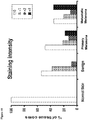
- 238000010186 staining Methods 0.000 description 22
- 238000002474 experimental method Methods 0.000 description 21
- 230000028327 secretion Effects 0.000 description 21
- 102000001398 Granzyme Human genes 0.000 description 20
- 108060005986 Granzyme Proteins 0.000 description 20
- 238000003556 assay Methods 0.000 description 20
- 241000699670 Mus sp. Species 0.000 description 19
- 229920001184 polypeptide Polymers 0.000 description 19
- 210000001519 tissue Anatomy 0.000 description 19
- 238000002347 injection Methods 0.000 description 17
- 239000007924 injection Substances 0.000 description 17
- 239000003814 drug Substances 0.000 description 16
- 230000001225 therapeutic effect Effects 0.000 description 16
- 238000004519 manufacturing process Methods 0.000 description 15
- 238000000338 in vitro Methods 0.000 description 14
- 241000699666 Mus <mouse, genus> Species 0.000 description 13
- 230000003993 interaction Effects 0.000 description 13
- 125000003275 alpha amino acid group Chemical group 0.000 description 12
- 239000003795 chemical substances by application Substances 0.000 description 12
- 230000036210 malignancy Effects 0.000 description 12
- 238000004458 analytical method Methods 0.000 description 11
- 230000012010 growth Effects 0.000 description 11
- 210000000987 immune system Anatomy 0.000 description 11
- 230000035755 proliferation Effects 0.000 description 11
- 108020004414 DNA Proteins 0.000 description 10
- 235000001014 amino acid Nutrition 0.000 description 10
- 230000010056 antibody-dependent cellular cytotoxicity Effects 0.000 description 10
- LOKCTEFSRHRXRJ-UHFFFAOYSA-I dipotassium trisodium dihydrogen phosphate hydrogen phosphate dichloride Chemical compound P(=O)(O)(O)[O-].[K+].P(=O)(O)([O-])[O-].[Na+].[Na+].[Cl-].[K+].[Cl-].[Na+] LOKCTEFSRHRXRJ-UHFFFAOYSA-I 0.000 description 10
- 239000012636 effector Substances 0.000 description 10
- 238000011534 incubation Methods 0.000 description 10
- 208000002154 non-small cell lung carcinoma Diseases 0.000 description 10
- 239000002953 phosphate buffered saline Substances 0.000 description 10
- 238000002965 ELISA Methods 0.000 description 9
- 241001529936 Murinae Species 0.000 description 9
- 150000001413 amino acids Chemical class 0.000 description 9
- 230000004540 complement-dependent cytotoxicity Effects 0.000 description 9
- 125000000151 cysteine group Chemical group N[C@@H](CS)C(=O)* 0.000 description 9
- 210000003819 peripheral blood mononuclear cell Anatomy 0.000 description 9
- 230000009385 viral infection Effects 0.000 description 9
- 239000002246 antineoplastic agent Substances 0.000 description 8
- 230000000875 corresponding effect Effects 0.000 description 8
- 210000004408 hybridoma Anatomy 0.000 description 8
- 208000029729 tumor suppressor gene on chromosome 11 Diseases 0.000 description 8
- 102000004127 Cytokines Human genes 0.000 description 7
- 108090000695 Cytokines Proteins 0.000 description 7
- 206010027476 Metastases Diseases 0.000 description 7
- 208000036142 Viral infection Diseases 0.000 description 7
- 238000003745 diagnosis Methods 0.000 description 7
- 229940079593 drug Drugs 0.000 description 7
- 230000009401 metastasis Effects 0.000 description 7
- 210000000056 organ Anatomy 0.000 description 7
- 230000002018 overexpression Effects 0.000 description 7
- 210000002307 prostate Anatomy 0.000 description 7
- 229940124597 therapeutic agent Drugs 0.000 description 7
- 230000004614 tumor growth Effects 0.000 description 7
- 241000282412 Homo Species 0.000 description 6
- 206010027480 Metastatic malignant melanoma Diseases 0.000 description 6
- 241001465754 Metazoa Species 0.000 description 6
- 108020004511 Recombinant DNA Proteins 0.000 description 6
- 238000003491 array Methods 0.000 description 6
- 230000000295 complement effect Effects 0.000 description 6
- 235000018417 cysteine Nutrition 0.000 description 6
- 231100000433 cytotoxic Toxicity 0.000 description 6
- 229940127089 cytotoxic agent Drugs 0.000 description 6
- 230000001472 cytotoxic effect Effects 0.000 description 6
- 231100000135 cytotoxicity Toxicity 0.000 description 6
- 238000005516 engineering process Methods 0.000 description 6
- 210000002919 epithelial cell Anatomy 0.000 description 6
- 230000003211 malignant effect Effects 0.000 description 6
- 230000001404 mediated effect Effects 0.000 description 6
- 208000021039 metastatic melanoma Diseases 0.000 description 6
- 239000013641 positive control Substances 0.000 description 6
- 238000012413 Fluorescence activated cell sorting analysis Methods 0.000 description 5
- 102000025850 HLA-A2 Antigen Human genes 0.000 description 5
- 108010074032 HLA-A2 Antigen Proteins 0.000 description 5
- 208000032124 Squamous Intraepithelial Lesions Diseases 0.000 description 5
- 235000004279 alanine Nutrition 0.000 description 5
- 230000000903 blocking effect Effects 0.000 description 5
- 230000022534 cell killing Effects 0.000 description 5
- 230000004663 cell proliferation Effects 0.000 description 5
- 230000003013 cytotoxicity Effects 0.000 description 5
- 238000011161 development Methods 0.000 description 5
- 210000004907 gland Anatomy 0.000 description 5
- 210000002865 immune cell Anatomy 0.000 description 5
- 230000028993 immune response Effects 0.000 description 5
- 230000001976 improved effect Effects 0.000 description 5
- 230000005764 inhibitory process Effects 0.000 description 5
- 239000003550 marker Substances 0.000 description 5
- 210000004379 membrane Anatomy 0.000 description 5
- 239000012528 membrane Substances 0.000 description 5
- 230000007170 pathology Effects 0.000 description 5
- 230000003285 pharmacodynamic effect Effects 0.000 description 5
- 239000000243 solution Substances 0.000 description 5
- 241000894007 species Species 0.000 description 5
- 238000012360 testing method Methods 0.000 description 5
- 238000012546 transfer Methods 0.000 description 5
- 102000014150 Interferons Human genes 0.000 description 4
- 108010050904 Interferons Proteins 0.000 description 4
- 108010002350 Interleukin-2 Proteins 0.000 description 4
- 102000000588 Interleukin-2 Human genes 0.000 description 4
- 241000124008 Mammalia Species 0.000 description 4
- 241000283984 Rodentia Species 0.000 description 4
- FAPWRFPIFSIZLT-UHFFFAOYSA-M Sodium chloride Chemical compound [Na+].[Cl-] FAPWRFPIFSIZLT-UHFFFAOYSA-M 0.000 description 4
- 208000005718 Stomach Neoplasms Diseases 0.000 description 4
- 208000024770 Thyroid neoplasm Diseases 0.000 description 4
- 230000009471 action Effects 0.000 description 4
- 230000000118 anti-neoplastic effect Effects 0.000 description 4
- 230000000259 anti-tumor effect Effects 0.000 description 4
- 229940034982 antineoplastic agent Drugs 0.000 description 4
- 210000003719 b-lymphocyte Anatomy 0.000 description 4
- 230000004071 biological effect Effects 0.000 description 4
- 230000037396 body weight Effects 0.000 description 4
- 239000000872 buffer Substances 0.000 description 4
- 238000012512 characterization method Methods 0.000 description 4
- 238000006243 chemical reaction Methods 0.000 description 4
- 210000001151 cytotoxic T lymphocyte Anatomy 0.000 description 4
- 238000000684 flow cytometry Methods 0.000 description 4
- 210000004602 germ cell Anatomy 0.000 description 4
- 230000016784 immunoglobulin production Effects 0.000 description 4
- 230000007246 mechanism Effects 0.000 description 4
- 210000002752 melanocyte Anatomy 0.000 description 4
- 239000013642 negative control Substances 0.000 description 4
- 239000000047 product Substances 0.000 description 4
- 108020003175 receptors Proteins 0.000 description 4
- 102000005962 receptors Human genes 0.000 description 4
- 210000002966 serum Anatomy 0.000 description 4
- 230000009870 specific binding Effects 0.000 description 4
- 231100000331 toxic Toxicity 0.000 description 4
- 230000002588 toxic effect Effects 0.000 description 4
- NFGXHKASABOEEW-UHFFFAOYSA-N 1-methylethyl 11-methoxy-3,7,11-trimethyl-2,4-dodecadienoate Chemical compound COC(C)(C)CCCC(C)CC=CC(C)=CC(=O)OC(C)C NFGXHKASABOEEW-UHFFFAOYSA-N 0.000 description 3
- WEVYAHXRMPXWCK-UHFFFAOYSA-N Acetonitrile Chemical compound CC#N WEVYAHXRMPXWCK-UHFFFAOYSA-N 0.000 description 3
- 208000023275 Autoimmune disease Diseases 0.000 description 3
- 238000009010 Bradford assay Methods 0.000 description 3
- 206010006187 Breast cancer Diseases 0.000 description 3
- 208000026310 Breast neoplasm Diseases 0.000 description 3
- 206010009944 Colon cancer Diseases 0.000 description 3
- 108091035707 Consensus sequence Proteins 0.000 description 3
- LFQSCWFLJHTTHZ-UHFFFAOYSA-N Ethanol Chemical compound CCO LFQSCWFLJHTTHZ-UHFFFAOYSA-N 0.000 description 3
- GHASVSINZRGABV-UHFFFAOYSA-N Fluorouracil Chemical compound FC1=CNC(=O)NC1=O GHASVSINZRGABV-UHFFFAOYSA-N 0.000 description 3
- PEDCQBHIVMGVHV-UHFFFAOYSA-N Glycerine Chemical compound OCC(O)CO PEDCQBHIVMGVHV-UHFFFAOYSA-N 0.000 description 3
- 241000701806 Human papillomavirus Species 0.000 description 3
- 108010054477 Immunoglobulin Fab Fragments Proteins 0.000 description 3
- 102000001706 Immunoglobulin Fab Fragments Human genes 0.000 description 3
- 101001043827 Mus musculus Interleukin-2 Proteins 0.000 description 3
- 206010061535 Ovarian neoplasm Diseases 0.000 description 3
- 229930012538 Paclitaxel Natural products 0.000 description 3
- 108010076504 Protein Sorting Signals Proteins 0.000 description 3
- 241000700605 Viruses Species 0.000 description 3
- 239000004480 active ingredient Substances 0.000 description 3
- 239000013543 active substance Substances 0.000 description 3
- 150000001295 alanines Chemical class 0.000 description 3
- 230000001093 anti-cancer Effects 0.000 description 3
- 230000006907 apoptotic process Effects 0.000 description 3
- 238000013459 approach Methods 0.000 description 3
- 230000008901 benefit Effects 0.000 description 3
- 230000033228 biological regulation Effects 0.000 description 3
- 210000004369 blood Anatomy 0.000 description 3
- 239000008280 blood Substances 0.000 description 3
- 230000001413 cellular effect Effects 0.000 description 3
- 210000001072 colon Anatomy 0.000 description 3
- 210000005220 cytoplasmic tail Anatomy 0.000 description 3
- 231100000673 dose–response relationship Toxicity 0.000 description 3
- 210000002889 endothelial cell Anatomy 0.000 description 3
- 229960002949 fluorouracil Drugs 0.000 description 3
- 210000005260 human cell Anatomy 0.000 description 3
- 230000005847 immunogenicity Effects 0.000 description 3
- 230000008676 import Effects 0.000 description 3
- 230000008611 intercellular interaction Effects 0.000 description 3
- 230000002601 intratumoral effect Effects 0.000 description 3
- 210000003734 kidney Anatomy 0.000 description 3
- 238000002843 lactate dehydrogenase assay Methods 0.000 description 3
- 208000032839 leukemia Diseases 0.000 description 3
- 210000004185 liver Anatomy 0.000 description 3
- 239000011159 matrix material Substances 0.000 description 3
- 238000002493 microarray Methods 0.000 description 3
- 238000012986 modification Methods 0.000 description 3
- 230000004048 modification Effects 0.000 description 3
- 238000010369 molecular cloning Methods 0.000 description 3
- 238000011275 oncology therapy Methods 0.000 description 3
- 229960001592 paclitaxel Drugs 0.000 description 3
- 210000000496 pancreas Anatomy 0.000 description 3
- 238000002823 phage display Methods 0.000 description 3
- 238000010837 poor prognosis Methods 0.000 description 3
- 230000008569 process Effects 0.000 description 3
- 230000002797 proteolythic effect Effects 0.000 description 3
- 238000004445 quantitative analysis Methods 0.000 description 3
- 238000012163 sequencing technique Methods 0.000 description 3
- 238000002415 sodium dodecyl sulfate polyacrylamide gel electrophoresis Methods 0.000 description 3
- 230000000638 stimulation Effects 0.000 description 3
- 238000006467 substitution reaction Methods 0.000 description 3
- 208000024891 symptom Diseases 0.000 description 3
- RCINICONZNJXQF-MZXODVADSA-N taxol Chemical compound O([C@@H]1[C@@]2(C[C@@H](C(C)=C(C2(C)C)[C@H](C([C@]2(C)[C@@H](O)C[C@H]3OC[C@]3([C@H]21)OC(C)=O)=O)OC(=O)C)OC(=O)[C@H](O)[C@@H](NC(=O)C=1C=CC=CC=1)C=1C=CC=CC=1)O)C(=O)C1=CC=CC=C1 RCINICONZNJXQF-MZXODVADSA-N 0.000 description 3
- 230000003612 virological effect Effects 0.000 description 3
- 238000005406 washing Methods 0.000 description 3
- YBJHBAHKTGYVGT-ZKWXMUAHSA-N (+)-Biotin Chemical compound N1C(=O)N[C@@H]2[C@H](CCCCC(=O)O)SC[C@@H]21 YBJHBAHKTGYVGT-ZKWXMUAHSA-N 0.000 description 2
- 108091032973 (ribonucleotides)n+m Proteins 0.000 description 2
- 102100025573 1-alkyl-2-acetylglycerophosphocholine esterase Human genes 0.000 description 2
- VVIAGPKUTFNRDU-UHFFFAOYSA-N 6S-folinic acid Natural products C1NC=2NC(N)=NC(=O)C=2N(C=O)C1CNC1=CC=C(C(=O)NC(CCC(O)=O)C(O)=O)C=C1 VVIAGPKUTFNRDU-UHFFFAOYSA-N 0.000 description 2
- 206010003445 Ascites Diseases 0.000 description 2
- 108010024976 Asparaginase Proteins 0.000 description 2
- 201000009030 Carcinoma Diseases 0.000 description 2
- DLGOEMSEDOSKAD-UHFFFAOYSA-N Carmustine Chemical compound ClCCNC(=O)N(N=O)CCCl DLGOEMSEDOSKAD-UHFFFAOYSA-N 0.000 description 2
- 108010069112 Complement System Proteins Proteins 0.000 description 2
- 102000000989 Complement System Proteins Human genes 0.000 description 2
- 208000023445 Congenital pulmonary airway malformation Diseases 0.000 description 2
- 238000012286 ELISA Assay Methods 0.000 description 2
- 102000004190 Enzymes Human genes 0.000 description 2
- 108090000790 Enzymes Proteins 0.000 description 2
- 102000003886 Glycoproteins Human genes 0.000 description 2
- 108090000288 Glycoproteins Proteins 0.000 description 2
- 101000981093 Homo sapiens Carcinoembryonic antigen-related cell adhesion molecule 1 Proteins 0.000 description 2
- 206010062767 Hypophysitis Diseases 0.000 description 2
- 206010061598 Immunodeficiency Diseases 0.000 description 2
- 102000009786 Immunoglobulin Constant Regions Human genes 0.000 description 2
- 108010009817 Immunoglobulin Constant Regions Proteins 0.000 description 2
- 108700005091 Immunoglobulin Genes Proteins 0.000 description 2
- 108010067060 Immunoglobulin Variable Region Proteins 0.000 description 2
- 102000017727 Immunoglobulin Variable Region Human genes 0.000 description 2
- 102000016844 Immunoglobulin-like domains Human genes 0.000 description 2
- 108050006430 Immunoglobulin-like domains Proteins 0.000 description 2
- QNAYBMKLOCPYGJ-REOHCLBHSA-N L-alanine Chemical compound C[C@H](N)C(O)=O QNAYBMKLOCPYGJ-REOHCLBHSA-N 0.000 description 2
- 206010025323 Lymphomas Diseases 0.000 description 2
- 125000001429 N-terminal alpha-amino-acid group Chemical group 0.000 description 2
- 108091007491 NSP3 Papain-like protease domains Proteins 0.000 description 2
- 102000003992 Peroxidases Human genes 0.000 description 2
- 241000700159 Rattus Species 0.000 description 2
- 206010039491 Sarcoma Diseases 0.000 description 2
- 102000012479 Serine Proteases Human genes 0.000 description 2
- 108010022999 Serine Proteases Proteins 0.000 description 2
- 108010090804 Streptavidin Proteins 0.000 description 2
- NKANXQFJJICGDU-QPLCGJKRSA-N Tamoxifen Chemical compound C=1C=CC=CC=1C(/CC)=C(C=1C=CC(OCCN(C)C)=CC=1)/C1=CC=CC=C1 NKANXQFJJICGDU-QPLCGJKRSA-N 0.000 description 2
- 208000002495 Uterine Neoplasms Diseases 0.000 description 2
- 230000003213 activating effect Effects 0.000 description 2
- 210000004100 adrenal gland Anatomy 0.000 description 2
- 230000002411 adverse Effects 0.000 description 2
- 229940100198 alkylating agent Drugs 0.000 description 2
- 239000002168 alkylating agent Substances 0.000 description 2
- 125000000539 amino acid group Chemical group 0.000 description 2
- 230000033115 angiogenesis Effects 0.000 description 2
- 210000004102 animal cell Anatomy 0.000 description 2
- 239000003242 anti bacterial agent Substances 0.000 description 2
- 229940088710 antibiotic agent Drugs 0.000 description 2
- 210000001185 bone marrow Anatomy 0.000 description 2
- 238000004113 cell culture Methods 0.000 description 2
- 230000010261 cell growth Effects 0.000 description 2
- 210000000170 cell membrane Anatomy 0.000 description 2
- 210000004978 chinese hamster ovary cell Anatomy 0.000 description 2
- 238000003501 co-culture Methods 0.000 description 2
- 208000029742 colonic neoplasm Diseases 0.000 description 2
- 239000002131 composite material Substances 0.000 description 2
- 230000021615 conjugation Effects 0.000 description 2
- 238000010276 construction Methods 0.000 description 2
- 238000007796 conventional method Methods 0.000 description 2
- 230000002596 correlated effect Effects 0.000 description 2
- 230000009260 cross reactivity Effects 0.000 description 2
- 239000012228 culture supernatant Substances 0.000 description 2
- 208000035250 cutaneous malignant susceptibility to 1 melanoma Diseases 0.000 description 2
- 238000002405 diagnostic procedure Methods 0.000 description 2
- 230000029087 digestion Effects 0.000 description 2
- 238000010790 dilution Methods 0.000 description 2
- 239000012895 dilution Substances 0.000 description 2
- 239000003534 dna topoisomerase inhibitor Substances 0.000 description 2
- 229940088598 enzyme Drugs 0.000 description 2
- 239000012530 fluid Substances 0.000 description 2
- 235000008191 folinic acid Nutrition 0.000 description 2
- 239000011672 folinic acid Substances 0.000 description 2
- VVIAGPKUTFNRDU-ABLWVSNPSA-N folinic acid Chemical compound C1NC=2NC(N)=NC(=O)C=2N(C=O)C1CNC1=CC=C(C(=O)N[C@@H](CCC(O)=O)C(O)=O)C=C1 VVIAGPKUTFNRDU-ABLWVSNPSA-N 0.000 description 2
- 238000009472 formulation Methods 0.000 description 2
- 230000004927 fusion Effects 0.000 description 2
- 206010017758 gastric cancer Diseases 0.000 description 2
- 125000003630 glycyl group Chemical group [H]N([H])C([H])([H])C(*)=O 0.000 description 2
- 230000009036 growth inhibition Effects 0.000 description 2
- 230000001900 immune effect Effects 0.000 description 2
- 230000003053 immunization Effects 0.000 description 2
- 229940072221 immunoglobulins Drugs 0.000 description 2
- 230000003308 immunostimulating effect Effects 0.000 description 2
- 239000004615 ingredient Substances 0.000 description 2
- 229940079322 interferon Drugs 0.000 description 2
- 229940047124 interferons Drugs 0.000 description 2
- 229960005386 ipilimumab Drugs 0.000 description 2
- 230000003902 lesion Effects 0.000 description 2
- 229960001691 leucovorin Drugs 0.000 description 2
- 239000003446 ligand Substances 0.000 description 2
- GLVAUDGFNGKCSF-UHFFFAOYSA-N mercaptopurine Chemical compound S=C1NC=NC2=C1NC=N2 GLVAUDGFNGKCSF-UHFFFAOYSA-N 0.000 description 2
- 102000039446 nucleic acids Human genes 0.000 description 2
- 108020004707 nucleic acids Proteins 0.000 description 2
- 150000007523 nucleic acids Chemical class 0.000 description 2
- 210000001672 ovary Anatomy 0.000 description 2
- 238000007911 parenteral administration Methods 0.000 description 2
- 230000001575 pathological effect Effects 0.000 description 2
- 230000037361 pathway Effects 0.000 description 2
- 239000008188 pellet Substances 0.000 description 2
- 108040007629 peroxidase activity proteins Proteins 0.000 description 2
- 230000000144 pharmacologic effect Effects 0.000 description 2
- BASFCYQUMIYNBI-UHFFFAOYSA-N platinum Chemical compound [Pt] BASFCYQUMIYNBI-UHFFFAOYSA-N 0.000 description 2
- 231100000614 poison Toxicity 0.000 description 2
- 238000002360 preparation method Methods 0.000 description 2
- 230000000069 prophylactic effect Effects 0.000 description 2
- 238000011321 prophylaxis Methods 0.000 description 2
- XJMOSONTPMZWPB-UHFFFAOYSA-M propidium iodide Chemical compound [I-].[I-].C12=CC(N)=CC=C2C2=CC=C(N)C=C2[N+](CCC[N+](C)(CC)CC)=C1C1=CC=CC=C1 XJMOSONTPMZWPB-UHFFFAOYSA-M 0.000 description 2
- 238000011002 quantification Methods 0.000 description 2
- 230000002285 radioactive effect Effects 0.000 description 2
- -1 radioactive isotopes Substances 0.000 description 2
- 239000000941 radioactive substance Substances 0.000 description 2
- 239000000700 radioactive tracer Substances 0.000 description 2
- 230000001105 regulatory effect Effects 0.000 description 2
- 238000011160 research Methods 0.000 description 2
- 238000012216 screening Methods 0.000 description 2
- 238000007423 screening assay Methods 0.000 description 2
- 230000011664 signaling Effects 0.000 description 2
- 210000003491 skin Anatomy 0.000 description 2
- 239000011780 sodium chloride Substances 0.000 description 2
- 238000001228 spectrum Methods 0.000 description 2
- 210000000952 spleen Anatomy 0.000 description 2
- 201000011549 stomach cancer Diseases 0.000 description 2
- 239000000126 substance Substances 0.000 description 2
- 239000000758 substrate Substances 0.000 description 2
- 230000008685 targeting Effects 0.000 description 2
- 238000002560 therapeutic procedure Methods 0.000 description 2
- 210000001541 thymus gland Anatomy 0.000 description 2
- 201000002510 thyroid cancer Diseases 0.000 description 2
- 229940044693 topoisomerase inhibitor Drugs 0.000 description 2
- 239000003440 toxic substance Substances 0.000 description 2
- 230000001988 toxicity Effects 0.000 description 2
- 231100000419 toxicity Toxicity 0.000 description 2
- 230000009261 transgenic effect Effects 0.000 description 2
- 238000011269 treatment regimen Methods 0.000 description 2
- 210000005239 tubule Anatomy 0.000 description 2
- 210000001835 viscera Anatomy 0.000 description 2
- 230000003442 weekly effect Effects 0.000 description 2
- MTCFGRXMJLQNBG-REOHCLBHSA-N (2S)-2-Amino-3-hydroxypropansäure Chemical compound OC[C@H](N)C(O)=O MTCFGRXMJLQNBG-REOHCLBHSA-N 0.000 description 1
- MWWSFMDVAYGXBV-MYPASOLCSA-N (7r,9s)-7-[(2r,4s,5s,6s)-4-amino-5-hydroxy-6-methyloxan-2-yl]oxy-6,9,11-trihydroxy-9-(2-hydroxyacetyl)-4-methoxy-8,10-dihydro-7h-tetracene-5,12-dione;hydrochloride Chemical compound Cl.O([C@@H]1C[C@@](O)(CC=2C(O)=C3C(=O)C=4C=CC=C(C=4C(=O)C3=C(O)C=21)OC)C(=O)CO)[C@H]1C[C@H](N)[C@H](O)[C@H](C)O1 MWWSFMDVAYGXBV-MYPASOLCSA-N 0.000 description 1
- OXHOPZLBSSTTBU-UHFFFAOYSA-N 1,3-bis(bromomethyl)benzene Chemical compound BrCC1=CC=CC(CBr)=C1 OXHOPZLBSSTTBU-UHFFFAOYSA-N 0.000 description 1
- 238000010600 3H thymidine incorporation assay Methods 0.000 description 1
- AOJJSUZBOXZQNB-VTZDEGQISA-N 4'-epidoxorubicin Chemical compound O([C@H]1C[C@@](O)(CC=2C(O)=C3C(=O)C=4C=CC=C(C=4C(=O)C3=C(O)C=21)OC)C(=O)CO)[C@H]1C[C@H](N)[C@@H](O)[C@H](C)O1 AOJJSUZBOXZQNB-VTZDEGQISA-N 0.000 description 1
- FWMNVWWHGCHHJJ-SKKKGAJSSA-N 4-amino-1-[(2r)-6-amino-2-[[(2r)-2-[[(2r)-2-[[(2r)-2-amino-3-phenylpropanoyl]amino]-3-phenylpropanoyl]amino]-4-methylpentanoyl]amino]hexanoyl]piperidine-4-carboxylic acid Chemical compound C([C@H](C(=O)N[C@H](CC(C)C)C(=O)N[C@H](CCCCN)C(=O)N1CCC(N)(CC1)C(O)=O)NC(=O)[C@H](N)CC=1C=CC=CC=1)C1=CC=CC=C1 FWMNVWWHGCHHJJ-SKKKGAJSSA-N 0.000 description 1
- STQGQHZAVUOBTE-UHFFFAOYSA-N 7-Cyan-hept-2t-en-4,6-diinsaeure Natural products C1=2C(O)=C3C(=O)C=4C(OC)=CC=CC=4C(=O)C3=C(O)C=2CC(O)(C(C)=O)CC1OC1CC(N)C(O)C(C)O1 STQGQHZAVUOBTE-UHFFFAOYSA-N 0.000 description 1
- ATRRKUHOCOJYRX-UHFFFAOYSA-N Ammonium bicarbonate Chemical compound [NH4+].OC([O-])=O ATRRKUHOCOJYRX-UHFFFAOYSA-N 0.000 description 1
- 229910000013 Ammonium bicarbonate Inorganic materials 0.000 description 1
- 206010004593 Bile duct cancer Diseases 0.000 description 1
- 108010006654 Bleomycin Proteins 0.000 description 1
- 102000039968 CEA family Human genes 0.000 description 1
- 108091069214 CEA family Proteins 0.000 description 1
- 101100506090 Caenorhabditis elegans hil-2 gene Proteins 0.000 description 1
- 101100273797 Caenorhabditis elegans pct-1 gene Proteins 0.000 description 1
- GAGWJHPBXLXJQN-UORFTKCHSA-N Capecitabine Chemical compound C1=C(F)C(NC(=O)OCCCCC)=NC(=O)N1[C@H]1[C@H](O)[C@H](O)[C@@H](C)O1 GAGWJHPBXLXJQN-UORFTKCHSA-N 0.000 description 1
- GAGWJHPBXLXJQN-UHFFFAOYSA-N Capecitabine Natural products C1=C(F)C(NC(=O)OCCCCC)=NC(=O)N1C1C(O)C(O)C(C)O1 GAGWJHPBXLXJQN-UHFFFAOYSA-N 0.000 description 1
- 101710190843 Carcinoembryonic antigen-related cell adhesion molecule 1 Proteins 0.000 description 1
- 102100025472 Carcinoembryonic antigen-related cell adhesion molecule 4 Human genes 0.000 description 1
- 206010008342 Cervix carcinoma Diseases 0.000 description 1
- 102000019034 Chemokines Human genes 0.000 description 1
- 108010012236 Chemokines Proteins 0.000 description 1
- 108010019670 Chimeric Antigen Receptors Proteins 0.000 description 1
- 208000001333 Colorectal Neoplasms Diseases 0.000 description 1
- 108010034753 Complement Membrane Attack Complex Proteins 0.000 description 1
- 102000000529 Costimulatory and Inhibitory T-Cell Receptors Human genes 0.000 description 1
- 108010041504 Costimulatory and Inhibitory T-Cell Receptors Proteins 0.000 description 1
- CMSMOCZEIVJLDB-UHFFFAOYSA-N Cyclophosphamide Chemical compound ClCCN(CCCl)P1(=O)NCCCO1 CMSMOCZEIVJLDB-UHFFFAOYSA-N 0.000 description 1
- UHDGCWIWMRVCDJ-CCXZUQQUSA-N Cytarabine Chemical compound O=C1N=C(N)C=CN1[C@H]1[C@@H](O)[C@H](O)[C@@H](CO)O1 UHDGCWIWMRVCDJ-CCXZUQQUSA-N 0.000 description 1
- 101710112752 Cytotoxin Proteins 0.000 description 1
- 206010061818 Disease progression Diseases 0.000 description 1
- 101100347633 Drosophila melanogaster Mhc gene Proteins 0.000 description 1
- KCXVZYZYPLLWCC-UHFFFAOYSA-N EDTA Chemical compound OC(=O)CN(CC(O)=O)CCN(CC(O)=O)CC(O)=O KCXVZYZYPLLWCC-UHFFFAOYSA-N 0.000 description 1
- 206010014733 Endometrial cancer Diseases 0.000 description 1
- 206010014759 Endometrial neoplasm Diseases 0.000 description 1
- HTIJFSOGRVMCQR-UHFFFAOYSA-N Epirubicin Natural products COc1cccc2C(=O)c3c(O)c4CC(O)(CC(OC5CC(N)C(=O)C(C)O5)c4c(O)c3C(=O)c12)C(=O)CO HTIJFSOGRVMCQR-UHFFFAOYSA-N 0.000 description 1
- 241000588724 Escherichia coli Species 0.000 description 1
- 102000009109 Fc receptors Human genes 0.000 description 1
- 108010087819 Fc receptors Proteins 0.000 description 1
- WQZGKKKJIJFFOK-GASJEMHNSA-N Glucose Natural products OC[C@H]1OC(O)[C@H](O)[C@@H](O)[C@@H]1O WQZGKKKJIJFFOK-GASJEMHNSA-N 0.000 description 1
- DHMQDGOQFOQNFH-UHFFFAOYSA-N Glycine Natural products NCC(O)=O DHMQDGOQFOQNFH-UHFFFAOYSA-N 0.000 description 1
- 239000004471 Glycine Substances 0.000 description 1
- 208000009329 Graft vs Host Disease Diseases 0.000 description 1
- 101000690301 Homo sapiens Aldo-keto reductase family 1 member C4 Proteins 0.000 description 1
- 101000914325 Homo sapiens Carcinoembryonic antigen-related cell adhesion molecule 4 Proteins 0.000 description 1
- 101001116548 Homo sapiens Protein CBFA2T1 Proteins 0.000 description 1
- XDXDZDZNSLXDNA-TZNDIEGXSA-N Idarubicin Chemical compound C1[C@H](N)[C@H](O)[C@H](C)O[C@H]1O[C@@H]1C2=C(O)C(C(=O)C3=CC=CC=C3C3=O)=C3C(O)=C2C[C@@](O)(C(C)=O)C1 XDXDZDZNSLXDNA-TZNDIEGXSA-N 0.000 description 1
- XDXDZDZNSLXDNA-UHFFFAOYSA-N Idarubicin Natural products C1C(N)C(O)C(C)OC1OC1C2=C(O)C(C(=O)C3=CC=CC=C3C3=O)=C3C(O)=C2CC(O)(C(C)=O)C1 XDXDZDZNSLXDNA-UHFFFAOYSA-N 0.000 description 1
- 208000029462 Immunodeficiency disease Diseases 0.000 description 1
- 102000018071 Immunoglobulin Fc Fragments Human genes 0.000 description 1
- 108010091135 Immunoglobulin Fc Fragments Proteins 0.000 description 1
- FBOZXECLQNJBKD-ZDUSSCGKSA-N L-methotrexate Chemical compound C=1N=C2N=C(N)N=C(N)C2=NC=1CN(C)C1=CC=C(C(=O)N[C@@H](CCC(O)=O)C(O)=O)C=C1 FBOZXECLQNJBKD-ZDUSSCGKSA-N 0.000 description 1
- 108010000817 Leuprolide Proteins 0.000 description 1
- GQYIWUVLTXOXAJ-UHFFFAOYSA-N Lomustine Chemical compound ClCCN(N=O)C(=O)NC1CCCCC1 GQYIWUVLTXOXAJ-UHFFFAOYSA-N 0.000 description 1
- 241000721701 Lynx Species 0.000 description 1
- 229930192392 Mitomycin Natural products 0.000 description 1
- NWIBSHFKIJFRCO-WUDYKRTCSA-N Mytomycin Chemical compound C1N2C(C(C(C)=C(N)C3=O)=O)=C3[C@@H](COC(N)=O)[C@@]2(OC)[C@@H]2[C@H]1N2 NWIBSHFKIJFRCO-WUDYKRTCSA-N 0.000 description 1
- ZDZOTLJHXYCWBA-VCVYQWHSSA-N N-debenzoyl-N-(tert-butoxycarbonyl)-10-deacetyltaxol Chemical compound O([C@H]1[C@H]2[C@@](C([C@H](O)C3=C(C)[C@@H](OC(=O)[C@H](O)[C@@H](NC(=O)OC(C)(C)C)C=4C=CC=CC=4)C[C@]1(O)C3(C)C)=O)(C)[C@@H](O)C[C@H]1OC[C@]12OC(=O)C)C(=O)C1=CC=CC=C1 ZDZOTLJHXYCWBA-VCVYQWHSSA-N 0.000 description 1
- 241000588652 Neisseria gonorrhoeae Species 0.000 description 1
- 208000007256 Nevus Diseases 0.000 description 1
- 241000283973 Oryctolagus cuniculus Species 0.000 description 1
- 206010033128 Ovarian cancer Diseases 0.000 description 1
- KHGNFPUMBJSZSM-UHFFFAOYSA-N Perforine Natural products COC1=C2CCC(O)C(CCC(C)(C)O)(OC)C2=NC2=C1C=CO2 KHGNFPUMBJSZSM-UHFFFAOYSA-N 0.000 description 1
- 229920001054 Poly(ethylene‐co‐vinyl acetate) Polymers 0.000 description 1
- 229920002732 Polyanhydride Polymers 0.000 description 1
- 229920002873 Polyethylenimine Polymers 0.000 description 1
- 229920001213 Polysorbate 20 Polymers 0.000 description 1
- 241000288906 Primates Species 0.000 description 1
- 108010029485 Protein Isoforms Proteins 0.000 description 1
- 102000001708 Protein Isoforms Human genes 0.000 description 1
- 108091008109 Pseudogenes Proteins 0.000 description 1
- 102000057361 Pseudogenes Human genes 0.000 description 1
- 238000002123 RNA extraction Methods 0.000 description 1
- MTCFGRXMJLQNBG-UHFFFAOYSA-N Serine Natural products OCC(N)C(O)=O MTCFGRXMJLQNBG-UHFFFAOYSA-N 0.000 description 1
- 208000000453 Skin Neoplasms Diseases 0.000 description 1
- 231100000632 Spindle poison Toxicity 0.000 description 1
- 230000006044 T cell activation Effects 0.000 description 1
- 230000024932 T cell mediated immunity Effects 0.000 description 1
- 230000006052 T cell proliferation Effects 0.000 description 1
- 108091008874 T cell receptors Proteins 0.000 description 1
- 102000016266 T-Cell Antigen Receptors Human genes 0.000 description 1
- 206010052779 Transplant rejections Diseases 0.000 description 1
- 208000006105 Uterine Cervical Neoplasms Diseases 0.000 description 1
- JXLYSJRDGCGARV-WWYNWVTFSA-N Vinblastine Natural products O=C(O[C@H]1[C@](O)(C(=O)OC)[C@@H]2N(C)c3c(cc(c(OC)c3)[C@]3(C(=O)OC)c4[nH]c5c(c4CCN4C[C@](O)(CC)C[C@H](C3)C4)cccc5)[C@@]32[C@H]2[C@@]1(CC)C=CCN2CC3)C JXLYSJRDGCGARV-WWYNWVTFSA-N 0.000 description 1
- 241000863480 Vinca Species 0.000 description 1
- 229940122803 Vinca alkaloid Drugs 0.000 description 1
- 241000981595 Zoysia japonica Species 0.000 description 1
- 239000002253 acid Substances 0.000 description 1
- 230000001154 acute effect Effects 0.000 description 1
- 230000033289 adaptive immune response Effects 0.000 description 1
- 230000001780 adrenocortical effect Effects 0.000 description 1
- 238000001042 affinity chromatography Methods 0.000 description 1
- 230000009824 affinity maturation Effects 0.000 description 1
- 238000003277 amino acid sequence analysis Methods 0.000 description 1
- 235000012538 ammonium bicarbonate Nutrition 0.000 description 1
- 239000001099 ammonium carbonate Substances 0.000 description 1
- 239000003098 androgen Substances 0.000 description 1
- 229940030486 androgens Drugs 0.000 description 1
- 230000002491 angiogenic effect Effects 0.000 description 1
- 230000003042 antagnostic effect Effects 0.000 description 1
- RGHILYZRVFRRNK-UHFFFAOYSA-N anthracene-1,2-dione Chemical group C1=CC=C2C=C(C(C(=O)C=C3)=O)C3=CC2=C1 RGHILYZRVFRRNK-UHFFFAOYSA-N 0.000 description 1
- 230000002280 anti-androgenic effect Effects 0.000 description 1
- 229940046836 anti-estrogen Drugs 0.000 description 1
- 230000001833 anti-estrogenic effect Effects 0.000 description 1
- 230000003302 anti-idiotype Effects 0.000 description 1
- 230000003127 anti-melanomic effect Effects 0.000 description 1
- 230000000340 anti-metabolite Effects 0.000 description 1
- 239000000051 antiandrogen Substances 0.000 description 1
- 230000000890 antigenic effect Effects 0.000 description 1
- 229940100197 antimetabolite Drugs 0.000 description 1
- 239000002256 antimetabolite Substances 0.000 description 1
- 229940045687 antimetabolites folic acid analogs Drugs 0.000 description 1
- 229940045719 antineoplastic alkylating agent nitrosoureas Drugs 0.000 description 1
- 229960003272 asparaginase Drugs 0.000 description 1
- DCXYFEDJOCDNAF-UHFFFAOYSA-M asparaginate Chemical compound [O-]C(=O)C(N)CC(N)=O DCXYFEDJOCDNAF-UHFFFAOYSA-M 0.000 description 1
- OHDRQQURAXLVGJ-HLVWOLMTSA-N azane;(2e)-3-ethyl-2-[(e)-(3-ethyl-6-sulfo-1,3-benzothiazol-2-ylidene)hydrazinylidene]-1,3-benzothiazole-6-sulfonic acid Chemical compound [NH4+].[NH4+].S/1C2=CC(S([O-])(=O)=O)=CC=C2N(CC)C\1=N/N=C1/SC2=CC(S([O-])(=O)=O)=CC=C2N1CC OHDRQQURAXLVGJ-HLVWOLMTSA-N 0.000 description 1
- 239000011324 bead Substances 0.000 description 1
- WQZGKKKJIJFFOK-VFUOTHLCSA-N beta-D-glucose Chemical compound OC[C@H]1O[C@@H](O)[C@H](O)[C@@H](O)[C@@H]1O WQZGKKKJIJFFOK-VFUOTHLCSA-N 0.000 description 1
- 210000000941 bile Anatomy 0.000 description 1
- 239000011230 binding agent Substances 0.000 description 1
- 238000002306 biochemical method Methods 0.000 description 1
- 229920000249 biocompatible polymer Polymers 0.000 description 1
- 230000008236 biological pathway Effects 0.000 description 1
- 239000000090 biomarker Substances 0.000 description 1
- 238000005460 biophysical method Methods 0.000 description 1
- 238000001574 biopsy Methods 0.000 description 1
- 230000015572 biosynthetic process Effects 0.000 description 1
- 229940126587 biotherapeutics Drugs 0.000 description 1
- 229960002685 biotin Drugs 0.000 description 1
- 235000020958 biotin Nutrition 0.000 description 1
- 239000011616 biotin Substances 0.000 description 1
- 201000000053 blastoma Diseases 0.000 description 1
- 229960001561 bleomycin Drugs 0.000 description 1
- OYVAGSVQBOHSSS-UAPAGMARSA-O bleomycin A2 Chemical compound N([C@H](C(=O)N[C@H](C)[C@@H](O)[C@H](C)C(=O)N[C@@H]([C@H](O)C)C(=O)NCCC=1SC=C(N=1)C=1SC=C(N=1)C(=O)NCCC[S+](C)C)[C@@H](O[C@H]1[C@H]([C@@H](O)[C@H](O)[C@H](CO)O1)O[C@@H]1[C@H]([C@@H](OC(N)=O)[C@H](O)[C@@H](CO)O1)O)C=1N=CNC=1)C(=O)C1=NC([C@H](CC(N)=O)NC[C@H](N)C(N)=O)=NC(N)=C1C OYVAGSVQBOHSSS-UAPAGMARSA-O 0.000 description 1
- 210000004204 blood vessel Anatomy 0.000 description 1
- 210000001124 body fluid Anatomy 0.000 description 1
- 239000010839 body fluid Substances 0.000 description 1
- 238000002619 cancer immunotherapy Methods 0.000 description 1
- 229960004117 capecitabine Drugs 0.000 description 1
- 239000004202 carbamide Substances 0.000 description 1
- 229960004562 carboplatin Drugs 0.000 description 1
- 190000008236 carboplatin Chemical compound 0.000 description 1
- 229960005243 carmustine Drugs 0.000 description 1
- 239000000969 carrier Substances 0.000 description 1
- 230000015556 catabolic process Effects 0.000 description 1
- 230000020411 cell activation Effects 0.000 description 1
- 230000006037 cell lysis Effects 0.000 description 1
- 238000001516 cell proliferation assay Methods 0.000 description 1
- 210000001638 cerebellum Anatomy 0.000 description 1
- 210000004720 cerebrum Anatomy 0.000 description 1
- 201000010881 cervical cancer Diseases 0.000 description 1
- 201000006612 cervical squamous cell carcinoma Diseases 0.000 description 1
- 238000002512 chemotherapy Methods 0.000 description 1
- 239000012829 chemotherapy agent Substances 0.000 description 1
- 229960004630 chlorambucil Drugs 0.000 description 1
- JCKYGMPEJWAADB-UHFFFAOYSA-N chlorambucil Chemical compound OC(=O)CCCC1=CC=C(N(CCCl)CCCl)C=C1 JCKYGMPEJWAADB-UHFFFAOYSA-N 0.000 description 1
- 210000000349 chromosome Anatomy 0.000 description 1
- 230000001684 chronic effect Effects 0.000 description 1
- 229960004316 cisplatin Drugs 0.000 description 1
- DQLATGHUWYMOKM-UHFFFAOYSA-L cisplatin Chemical compound N[Pt](N)(Cl)Cl DQLATGHUWYMOKM-UHFFFAOYSA-L 0.000 description 1
- 238000003776 cleavage reaction Methods 0.000 description 1
- 238000010367 cloning Methods 0.000 description 1
- 230000000112 colonic effect Effects 0.000 description 1
- 239000003086 colorant Substances 0.000 description 1
- 201000010989 colorectal carcinoma Diseases 0.000 description 1
- 238000004440 column chromatography Methods 0.000 description 1
- 238000009096 combination chemotherapy Methods 0.000 description 1
- 230000002860 competitive effect Effects 0.000 description 1
- 239000002299 complementary DNA Substances 0.000 description 1
- 238000004590 computer program Methods 0.000 description 1
- 239000003636 conditioned culture medium Substances 0.000 description 1
- 230000008878 coupling Effects 0.000 description 1
- 238000010168 coupling process Methods 0.000 description 1
- 238000005859 coupling reaction Methods 0.000 description 1
- 238000004132 cross linking Methods 0.000 description 1
- 239000013078 crystal Substances 0.000 description 1
- 208000030381 cutaneous melanoma Diseases 0.000 description 1
- 229960004397 cyclophosphamide Drugs 0.000 description 1
- 229960000684 cytarabine Drugs 0.000 description 1
- 102000003675 cytokine receptors Human genes 0.000 description 1
- 108010057085 cytokine receptors Proteins 0.000 description 1
- 230000009089 cytolysis Effects 0.000 description 1
- 210000004395 cytoplasmic granule Anatomy 0.000 description 1
- 239000000824 cytostatic agent Substances 0.000 description 1
- 230000001085 cytostatic effect Effects 0.000 description 1
- 231100000599 cytotoxic agent Toxicity 0.000 description 1
- 239000002619 cytotoxin Substances 0.000 description 1
- 230000006378 damage Effects 0.000 description 1
- 229960000975 daunorubicin Drugs 0.000 description 1
- STQGQHZAVUOBTE-VGBVRHCVSA-N daunorubicin Chemical compound O([C@H]1C[C@@](O)(CC=2C(O)=C3C(=O)C=4C=CC=C(C=4C(=O)C3=C(O)C=21)OC)C(C)=O)[C@H]1C[C@H](N)[C@H](O)[C@H](C)O1 STQGQHZAVUOBTE-VGBVRHCVSA-N 0.000 description 1
- 230000034994 death Effects 0.000 description 1
- 230000007423 decrease Effects 0.000 description 1
- 230000002950 deficient Effects 0.000 description 1
- 238000006731 degradation reaction Methods 0.000 description 1
- 239000003405 delayed action preparation Substances 0.000 description 1
- 230000002939 deleterious effect Effects 0.000 description 1
- 238000012217 deletion Methods 0.000 description 1
- 230000037430 deletion Effects 0.000 description 1
- 238000013461 design Methods 0.000 description 1
- 238000001514 detection method Methods 0.000 description 1
- 239000008121 dextrose Substances 0.000 description 1
- 239000000032 diagnostic agent Substances 0.000 description 1
- 229940039227 diagnostic agent Drugs 0.000 description 1
- 230000004069 differentiation Effects 0.000 description 1
- 230000005750 disease progression Effects 0.000 description 1
- 229960003668 docetaxel Drugs 0.000 description 1
- 238000012377 drug delivery Methods 0.000 description 1
- 239000003596 drug target Substances 0.000 description 1
- 230000002900 effect on cell Effects 0.000 description 1
- 229940096118 ella Drugs 0.000 description 1
- 238000010828 elution Methods 0.000 description 1
- 239000012149 elution buffer Substances 0.000 description 1
- 201000008184 embryoma Diseases 0.000 description 1
- 239000003995 emulsifying agent Substances 0.000 description 1
- 230000002143 encouraging effect Effects 0.000 description 1
- 230000002357 endometrial effect Effects 0.000 description 1
- 210000004696 endometrium Anatomy 0.000 description 1
- 230000003511 endothelial effect Effects 0.000 description 1
- 230000002708 enhancing effect Effects 0.000 description 1
- 230000002255 enzymatic effect Effects 0.000 description 1
- 229960001904 epirubicin Drugs 0.000 description 1
- 210000000981 epithelium Anatomy 0.000 description 1
- 229940011871 estrogen Drugs 0.000 description 1
- 239000000262 estrogen Substances 0.000 description 1
- 239000000328 estrogen antagonist Substances 0.000 description 1
- VJJPUSNTGOMMGY-MRVIYFEKSA-N etoposide Chemical compound COC1=C(O)C(OC)=CC([C@@H]2C3=CC=4OCOC=4C=C3[C@@H](O[C@H]3[C@@H]([C@@H](O)[C@@H]4O[C@H](C)OC[C@H]4O3)O)[C@@H]3[C@@H]2C(OC3)=O)=C1 VJJPUSNTGOMMGY-MRVIYFEKSA-N 0.000 description 1
- 229960005420 etoposide Drugs 0.000 description 1
- 230000000763 evoking effect Effects 0.000 description 1
- 230000001747 exhibiting effect Effects 0.000 description 1
- 238000010195 expression analysis Methods 0.000 description 1
- 239000013604 expression vector Substances 0.000 description 1
- 230000002349 favourable effect Effects 0.000 description 1
- 238000001943 fluorescence-activated cell sorting Methods 0.000 description 1
- 102000034287 fluorescent proteins Human genes 0.000 description 1
- 108091006047 fluorescent proteins Proteins 0.000 description 1
- 229960002074 flutamide Drugs 0.000 description 1
- MKXKFYHWDHIYRV-UHFFFAOYSA-N flutamide Chemical compound CC(C)C(=O)NC1=CC=C([N+]([O-])=O)C(C(F)(F)F)=C1 MKXKFYHWDHIYRV-UHFFFAOYSA-N 0.000 description 1
- 150000002224 folic acids Chemical class 0.000 description 1
- 230000005714 functional activity Effects 0.000 description 1
- 239000000499 gel Substances 0.000 description 1
- 238000003500 gene array Methods 0.000 description 1
- 230000000762 glandular Effects 0.000 description 1
- 230000002518 glial effect Effects 0.000 description 1
- XLXSAKCOAKORKW-AQJXLSMYSA-N gonadorelin Chemical class C([C@@H](C(=O)NCC(=O)N[C@@H](CC(C)C)C(=O)N[C@@H](CCCNC(N)=N)C(=O)N1[C@@H](CCC1)C(=O)NCC(N)=O)NC(=O)[C@H](CO)NC(=O)[C@H](CC=1C2=CC=CC=C2NC=1)NC(=O)[C@H](CC=1N=CNC=1)NC(=O)[C@H]1NC(=O)CC1)C1=CC=C(O)C=C1 XLXSAKCOAKORKW-AQJXLSMYSA-N 0.000 description 1
- 208000024908 graft versus host disease Diseases 0.000 description 1
- 238000003306 harvesting Methods 0.000 description 1
- 230000036541 health Effects 0.000 description 1
- 230000002607 hemopoietic effect Effects 0.000 description 1
- 230000002440 hepatic effect Effects 0.000 description 1
- 210000003630 histaminocyte Anatomy 0.000 description 1
- 239000005556 hormone Substances 0.000 description 1
- 229940088597 hormone Drugs 0.000 description 1
- 102000054751 human RUNX1T1 Human genes 0.000 description 1
- 238000013415 human tumor xenograft model Methods 0.000 description 1
- 239000000017 hydrogel Substances 0.000 description 1
- 229940031574 hydroxymethyl cellulose Drugs 0.000 description 1
- 229920003063 hydroxymethyl cellulose Polymers 0.000 description 1
- 230000002267 hypothalamic effect Effects 0.000 description 1
- 229960000908 idarubicin Drugs 0.000 description 1
- 229960001101 ifosfamide Drugs 0.000 description 1
- HOMGKSMUEGBAAB-UHFFFAOYSA-N ifosfamide Chemical compound ClCCNP1(=O)OCCCN1CCCl HOMGKSMUEGBAAB-UHFFFAOYSA-N 0.000 description 1
- 238000003384 imaging method Methods 0.000 description 1
- 210000001822 immobilized cell Anatomy 0.000 description 1
- 239000012642 immune effector Substances 0.000 description 1
- 201000007294 immune system cancer Diseases 0.000 description 1
- 230000036039 immunity Effects 0.000 description 1
- 238000002649 immunization Methods 0.000 description 1
- 238000003018 immunoassay Methods 0.000 description 1
- 230000007813 immunodeficiency Effects 0.000 description 1
- 230000002163 immunogen Effects 0.000 description 1
- 238000013388 immunohistochemistry analysis Methods 0.000 description 1
- 229940121354 immunomodulator Drugs 0.000 description 1
- 238000012744 immunostaining Methods 0.000 description 1
- 238000009169 immunotherapy Methods 0.000 description 1
- 230000007688 immunotoxicity Effects 0.000 description 1
- 231100000386 immunotoxicity Toxicity 0.000 description 1
- 238000002513 implantation Methods 0.000 description 1
- 238000000099 in vitro assay Methods 0.000 description 1
- 238000010874 in vitro model Methods 0.000 description 1
- 238000005462 in vivo assay Methods 0.000 description 1
- 208000015181 infectious disease Diseases 0.000 description 1
- 230000008595 infiltration Effects 0.000 description 1
- 238000001764 infiltration Methods 0.000 description 1
- 230000002757 inflammatory effect Effects 0.000 description 1
- 239000003112 inhibitor Substances 0.000 description 1
- 108091008042 inhibitory receptors Proteins 0.000 description 1
- 230000000977 initiatory effect Effects 0.000 description 1
- 229910001410 inorganic ion Inorganic materials 0.000 description 1
- 238000007689 inspection Methods 0.000 description 1
- 230000010354 integration Effects 0.000 description 1
- 230000002452 interceptive effect Effects 0.000 description 1
- 210000004347 intestinal mucosa Anatomy 0.000 description 1
- 238000001990 intravenous administration Methods 0.000 description 1
- 230000009545 invasion Effects 0.000 description 1
- 238000011835 investigation Methods 0.000 description 1
- 229960004768 irinotecan Drugs 0.000 description 1
- UWKQSNNFCGGAFS-XIFFEERXSA-N irinotecan Chemical compound C1=C2C(CC)=C3CN(C(C4=C([C@@](C(=O)OC4)(O)CC)C=4)=O)C=4C3=NC2=CC=C1OC(=O)N(CC1)CCC1N1CCCCC1 UWKQSNNFCGGAFS-XIFFEERXSA-N 0.000 description 1
- 238000005304 joining Methods 0.000 description 1
- GFIJNRVAKGFPGQ-LIJARHBVSA-N leuprolide Chemical compound CCNC(=O)[C@@H]1CCCN1C(=O)[C@H](CCCNC(N)=N)NC(=O)[C@H](CC(C)C)NC(=O)[C@@H](CC(C)C)NC(=O)[C@@H](NC(=O)[C@H](CO)NC(=O)[C@H](CC=1C2=CC=CC=C2NC=1)NC(=O)[C@H](CC=1N=CNC=1)NC(=O)[C@H]1NC(=O)CC1)CC1=CC=C(O)C=C1 GFIJNRVAKGFPGQ-LIJARHBVSA-N 0.000 description 1
- 229960004338 leuprorelin Drugs 0.000 description 1
- 230000000670 limiting effect Effects 0.000 description 1
- 239000002502 liposome Substances 0.000 description 1
- 230000007108 local immune response Effects 0.000 description 1
- 229960002247 lomustine Drugs 0.000 description 1
- 230000001926 lymphatic effect Effects 0.000 description 1
- 210000003563 lymphoid tissue Anatomy 0.000 description 1
- 230000014759 maintenance of location Effects 0.000 description 1
- 238000013507 mapping Methods 0.000 description 1
- 238000001819 mass spectrum Methods 0.000 description 1
- 238000005259 measurement Methods 0.000 description 1
- 230000010534 mechanism of action Effects 0.000 description 1
- 229960004961 mechlorethamine Drugs 0.000 description 1
- HAWPXGHAZFHHAD-UHFFFAOYSA-N mechlorethamine Chemical compound ClCCN(C)CCCl HAWPXGHAZFHHAD-UHFFFAOYSA-N 0.000 description 1
- 229960004296 megestrol acetate Drugs 0.000 description 1
- RQZAXGRLVPAYTJ-GQFGMJRRSA-N megestrol acetate Chemical compound C1=C(C)C2=CC(=O)CC[C@]2(C)[C@@H]2[C@@H]1[C@@H]1CC[C@@](C(C)=O)(OC(=O)C)[C@@]1(C)CC2 RQZAXGRLVPAYTJ-GQFGMJRRSA-N 0.000 description 1
- 229960001924 melphalan Drugs 0.000 description 1
- SGDBTWWWUNNDEQ-LBPRGKRZSA-N melphalan Chemical compound OC(=O)[C@@H](N)CC1=CC=C(N(CCCl)CCCl)C=C1 SGDBTWWWUNNDEQ-LBPRGKRZSA-N 0.000 description 1
- 229960001428 mercaptopurine Drugs 0.000 description 1
- 108020004999 messenger RNA Proteins 0.000 description 1
- 206010061289 metastatic neoplasm Diseases 0.000 description 1
- 229960000485 methotrexate Drugs 0.000 description 1
- 239000004530 micro-emulsion Substances 0.000 description 1
- 230000002906 microbiologic effect Effects 0.000 description 1
- 244000005700 microbiome Species 0.000 description 1
- 239000003094 microcapsule Substances 0.000 description 1
- 239000004005 microsphere Substances 0.000 description 1
- 210000000110 microvilli Anatomy 0.000 description 1
- 229960004857 mitomycin Drugs 0.000 description 1
- 229960001156 mitoxantrone Drugs 0.000 description 1
- KKZJGLLVHKMTCM-UHFFFAOYSA-N mitoxantrone Chemical compound O=C1C2=C(O)C=CC(O)=C2C(=O)C2=C1C(NCCNCCO)=CC=C2NCCNCCO KKZJGLLVHKMTCM-UHFFFAOYSA-N 0.000 description 1
- 239000003607 modifier Substances 0.000 description 1
- 230000003990 molecular pathway Effects 0.000 description 1
- 238000012544 monitoring process Methods 0.000 description 1
- HDZGCSFEDULWCS-UHFFFAOYSA-N monomethylhydrazine Chemical class CNN HDZGCSFEDULWCS-UHFFFAOYSA-N 0.000 description 1
- 230000035772 mutation Effects 0.000 description 1
- 210000004165 myocardium Anatomy 0.000 description 1
- 230000001537 neural effect Effects 0.000 description 1
- 230000003472 neutralizing effect Effects 0.000 description 1
- 210000000440 neutrophil Anatomy 0.000 description 1
- 238000007899 nucleic acid hybridization Methods 0.000 description 1
- QIQXTHQIDYTFRH-UHFFFAOYSA-N octadecanoic acid Chemical class CCCCCCCCCCCCCCCCCC(O)=O QIQXTHQIDYTFRH-UHFFFAOYSA-N 0.000 description 1
- 238000002515 oligonucleotide synthesis Methods 0.000 description 1
- 229960001756 oxaliplatin Drugs 0.000 description 1
- DWAFYCQODLXJNR-BNTLRKBRSA-L oxaliplatin Chemical compound O1C(=O)C(=O)O[Pt]11N[C@@H]2CCCC[C@H]2N1 DWAFYCQODLXJNR-BNTLRKBRSA-L 0.000 description 1
- 239000006179 pH buffering agent Substances 0.000 description 1
- 210000002741 palatine tonsil Anatomy 0.000 description 1
- 210000002990 parathyroid gland Anatomy 0.000 description 1
- 239000002245 particle Substances 0.000 description 1
- 238000010647 peptide synthesis reaction Methods 0.000 description 1
- 239000000816 peptidomimetic Substances 0.000 description 1
- 229930192851 perforin Natural products 0.000 description 1
- 230000002093 peripheral effect Effects 0.000 description 1
- 210000000578 peripheral nerve Anatomy 0.000 description 1
- 238000000053 physical method Methods 0.000 description 1
- 230000004962 physiological condition Effects 0.000 description 1
- 239000000049 pigment Substances 0.000 description 1
- 229910052697 platinum Inorganic materials 0.000 description 1
- 229920001200 poly(ethylene-vinyl acetate) Polymers 0.000 description 1
- 238000002264 polyacrylamide gel electrophoresis Methods 0.000 description 1
- 229920000642 polymer Polymers 0.000 description 1
- 239000000256 polyoxyethylene sorbitan monolaurate Substances 0.000 description 1
- 235000010486 polyoxyethylene sorbitan monolaurate Nutrition 0.000 description 1
- 210000004896 polypeptide structure Anatomy 0.000 description 1
- 239000011148 porous material Substances 0.000 description 1
- 230000003389 potentiating effect Effects 0.000 description 1
- 238000011533 pre-incubation Methods 0.000 description 1
- 239000002243 precursor Substances 0.000 description 1
- 230000002265 prevention Effects 0.000 description 1
- 230000000861 pro-apoptotic effect Effects 0.000 description 1
- 238000012545 processing Methods 0.000 description 1
- 239000000583 progesterone congener Substances 0.000 description 1
- 230000009696 proliferative response Effects 0.000 description 1
- 210000005267 prostate cell Anatomy 0.000 description 1
- 210000000064 prostate epithelial cell Anatomy 0.000 description 1
- 230000001681 protective effect Effects 0.000 description 1
- 238000012514 protein characterization Methods 0.000 description 1
- 230000004853 protein function Effects 0.000 description 1
- 238000001742 protein purification Methods 0.000 description 1
- 210000000512 proximal kidney tubule Anatomy 0.000 description 1
- 150000003212 purines Chemical class 0.000 description 1
- 150000003230 pyrimidines Chemical class 0.000 description 1
- 230000035484 reaction time Effects 0.000 description 1
- 230000008707 rearrangement Effects 0.000 description 1
- 238000010188 recombinant method Methods 0.000 description 1
- 238000011084 recovery Methods 0.000 description 1
- 230000007115 recruitment Effects 0.000 description 1
- 230000009467 reduction Effects 0.000 description 1
- 238000009877 rendering Methods 0.000 description 1
- 230000003252 repetitive effect Effects 0.000 description 1
- 230000010076 replication Effects 0.000 description 1
- 230000000284 resting effect Effects 0.000 description 1
- 230000000717 retained effect Effects 0.000 description 1
- 210000001525 retina Anatomy 0.000 description 1
- 238000010839 reverse transcription Methods 0.000 description 1
- 229960004641 rituximab Drugs 0.000 description 1
- 239000012146 running buffer Substances 0.000 description 1
- 210000003079 salivary gland Anatomy 0.000 description 1
- 230000007017 scission Effects 0.000 description 1
- 238000010187 selection method Methods 0.000 description 1
- 239000012679 serum free medium Substances 0.000 description 1
- 210000002027 skeletal muscle Anatomy 0.000 description 1
- 201000000849 skin cancer Diseases 0.000 description 1
- 201000003708 skin melanoma Diseases 0.000 description 1
- 210000000813 small intestine Anatomy 0.000 description 1
- 239000007790 solid phase Substances 0.000 description 1
- 238000000527 sonication Methods 0.000 description 1
- 238000012289 standard assay Methods 0.000 description 1
- 238000010561 standard procedure Methods 0.000 description 1
- 239000007858 starting material Substances 0.000 description 1
- 230000004936 stimulating effect Effects 0.000 description 1
- 238000007920 subcutaneous administration Methods 0.000 description 1
- 239000006228 supernatant Substances 0.000 description 1
- 230000001629 suppression Effects 0.000 description 1
- 239000000725 suspension Substances 0.000 description 1
- 229960001603 tamoxifen Drugs 0.000 description 1
- 210000001550 testis Anatomy 0.000 description 1
- 238000011200 topical administration Methods 0.000 description 1
- 230000000699 topical effect Effects 0.000 description 1
- 229960000303 topotecan Drugs 0.000 description 1
- UCFGDBYHRUNTLO-QHCPKHFHSA-N topotecan Chemical compound C1=C(O)C(CN(C)C)=C2C=C(CN3C4=CC5=C(C3=O)COC(=O)[C@]5(O)CC)C4=NC2=C1 UCFGDBYHRUNTLO-QHCPKHFHSA-N 0.000 description 1
- 239000003053 toxin Substances 0.000 description 1
- 231100000765 toxin Toxicity 0.000 description 1
- 108700012359 toxins Proteins 0.000 description 1
- 238000013518 transcription Methods 0.000 description 1
- 230000035897 transcription Effects 0.000 description 1
- 238000001890 transfection Methods 0.000 description 1
- 230000009466 transformation Effects 0.000 description 1
- 238000011830 transgenic mouse model Methods 0.000 description 1
- 230000001052 transient effect Effects 0.000 description 1
- 230000010474 transient expression Effects 0.000 description 1
- 238000003146 transient transfection Methods 0.000 description 1
- 238000013519 translation Methods 0.000 description 1
- 102000035160 transmembrane proteins Human genes 0.000 description 1
- 108091005703 transmembrane proteins Proteins 0.000 description 1
- 238000002054 transplantation Methods 0.000 description 1
- 230000005740 tumor formation Effects 0.000 description 1
- OOLLAFOLCSJHRE-ZHAKMVSLSA-N ulipristal acetate Chemical compound C1=CC(N(C)C)=CC=C1[C@@H]1C2=C3CCC(=O)C=C3CC[C@H]2[C@H](CC[C@]2(OC(C)=O)C(C)=O)[C@]2(C)C1 OOLLAFOLCSJHRE-ZHAKMVSLSA-N 0.000 description 1
- 241001515965 unidentified phage Species 0.000 description 1
- 230000003827 upregulation Effects 0.000 description 1
- 150000003672 ureas Chemical class 0.000 description 1
- 210000004291 uterus Anatomy 0.000 description 1
- 239000013598 vector Substances 0.000 description 1
- 238000012795 verification Methods 0.000 description 1
- 229960003048 vinblastine Drugs 0.000 description 1
- JXLYSJRDGCGARV-XQKSVPLYSA-N vincaleukoblastine Chemical compound C([C@@H](C[C@]1(C(=O)OC)C=2C(=CC3=C([C@]45[C@H]([C@@]([C@H](OC(C)=O)[C@]6(CC)C=CCN([C@H]56)CC4)(O)C(=O)OC)N3C)C=2)OC)C[C@@](C2)(O)CC)N2CCC2=C1NC1=CC=CC=C21 JXLYSJRDGCGARV-XQKSVPLYSA-N 0.000 description 1
- 229960004528 vincristine Drugs 0.000 description 1
- OGWKCGZFUXNPDA-XQKSVPLYSA-N vincristine Chemical compound C([N@]1C[C@@H](C[C@]2(C(=O)OC)C=3C(=CC4=C([C@]56[C@H]([C@@]([C@H](OC(C)=O)[C@]7(CC)C=CCN([C@H]67)CC5)(O)C(=O)OC)N4C=O)C=3)OC)C[C@@](C1)(O)CC)CC1=C2NC2=CC=CC=C12 OGWKCGZFUXNPDA-XQKSVPLYSA-N 0.000 description 1
- OGWKCGZFUXNPDA-UHFFFAOYSA-N vincristine Natural products C1C(CC)(O)CC(CC2(C(=O)OC)C=3C(=CC4=C(C56C(C(C(OC(C)=O)C7(CC)C=CCN(C67)CC5)(O)C(=O)OC)N4C=O)C=3)OC)CN1CCC1=C2NC2=CC=CC=C12 OGWKCGZFUXNPDA-UHFFFAOYSA-N 0.000 description 1
- GBABOYUKABKIAF-GHYRFKGUSA-N vinorelbine Chemical compound C1N(CC=2C3=CC=CC=C3NC=22)CC(CC)=C[C@H]1C[C@]2(C(=O)OC)C1=CC([C@]23[C@H]([C@]([C@H](OC(C)=O)[C@]4(CC)C=CCN([C@H]34)CC2)(O)C(=O)OC)N2C)=C2C=C1OC GBABOYUKABKIAF-GHYRFKGUSA-N 0.000 description 1
- 229960002066 vinorelbine Drugs 0.000 description 1
- 230000029812 viral genome replication Effects 0.000 description 1
- XLYOFNOQVPJJNP-UHFFFAOYSA-N water Substances O XLYOFNOQVPJJNP-UHFFFAOYSA-N 0.000 description 1
- 238000009736 wetting Methods 0.000 description 1
- 239000000080 wetting agent Substances 0.000 description 1
- DGVVWUTYPXICAM-UHFFFAOYSA-N β‐Mercaptoethanol Chemical compound OCCS DGVVWUTYPXICAM-UHFFFAOYSA-N 0.000 description 1
Images


Classifications
-
- C—CHEMISTRY; METALLURGY
- C07—ORGANIC CHEMISTRY
- C07K—PEPTIDES
- C07K16/00—Immunoglobulins [IGs], e.g. monoclonal or polyclonal antibodies
- C07K16/42—Immunoglobulins [IGs], e.g. monoclonal or polyclonal antibodies against immunoglobulins
-
- A—HUMAN NECESSITIES
- A61—MEDICAL OR VETERINARY SCIENCE; HYGIENE
- A61K—PREPARATIONS FOR MEDICAL, DENTAL OR TOILETRY PURPOSES
- A61K39/00—Medicinal preparations containing antigens or antibodies
- A61K39/395—Antibodies; Immunoglobulins; Immune serum, e.g. antilymphocytic serum
- A61K39/39533—Antibodies; Immunoglobulins; Immune serum, e.g. antilymphocytic serum against materials from animals
- A61K39/39566—Antibodies; Immunoglobulins; Immune serum, e.g. antilymphocytic serum against materials from animals against immunoglobulins, e.g. anti-idiotypic antibodies
-
- A—HUMAN NECESSITIES
- A61—MEDICAL OR VETERINARY SCIENCE; HYGIENE
- A61K—PREPARATIONS FOR MEDICAL, DENTAL OR TOILETRY PURPOSES
- A61K40/00—Cellular immunotherapy
- A61K40/10—Cellular immunotherapy characterised by the cell type used
- A61K40/11—T-cells, e.g. tumour infiltrating lymphocytes [TIL] or regulatory T [Treg] cells; Lymphokine-activated killer [LAK] cells
-
- A—HUMAN NECESSITIES
- A61—MEDICAL OR VETERINARY SCIENCE; HYGIENE
- A61K—PREPARATIONS FOR MEDICAL, DENTAL OR TOILETRY PURPOSES
- A61K40/00—Cellular immunotherapy
- A61K40/40—Cellular immunotherapy characterised by antigens that are targeted or presented by cells of the immune system
- A61K40/41—Vertebrate antigens
- A61K40/42—Cancer antigens
-
- A—HUMAN NECESSITIES
- A61—MEDICAL OR VETERINARY SCIENCE; HYGIENE
- A61K—PREPARATIONS FOR MEDICAL, DENTAL OR TOILETRY PURPOSES
- A61K45/00—Medicinal preparations containing active ingredients not provided for in groups A61K31/00 - A61K41/00
- A61K45/06—Mixtures of active ingredients without chemical characterisation, e.g. antiphlogistics and cardiaca
-
- A—HUMAN NECESSITIES
- A61—MEDICAL OR VETERINARY SCIENCE; HYGIENE
- A61P—SPECIFIC THERAPEUTIC ACTIVITY OF CHEMICAL COMPOUNDS OR MEDICINAL PREPARATIONS
- A61P1/00—Drugs for disorders of the alimentary tract or the digestive system
- A61P1/04—Drugs for disorders of the alimentary tract or the digestive system for ulcers, gastritis or reflux esophagitis, e.g. antacids, inhibitors of acid secretion, mucosal protectants
-
- A—HUMAN NECESSITIES
- A61—MEDICAL OR VETERINARY SCIENCE; HYGIENE
- A61P—SPECIFIC THERAPEUTIC ACTIVITY OF CHEMICAL COMPOUNDS OR MEDICINAL PREPARATIONS
- A61P1/00—Drugs for disorders of the alimentary tract or the digestive system
- A61P1/18—Drugs for disorders of the alimentary tract or the digestive system for pancreatic disorders, e.g. pancreatic enzymes
-
- A—HUMAN NECESSITIES
- A61—MEDICAL OR VETERINARY SCIENCE; HYGIENE
- A61P—SPECIFIC THERAPEUTIC ACTIVITY OF CHEMICAL COMPOUNDS OR MEDICINAL PREPARATIONS
- A61P11/00—Drugs for disorders of the respiratory system
-
- A—HUMAN NECESSITIES
- A61—MEDICAL OR VETERINARY SCIENCE; HYGIENE
- A61P—SPECIFIC THERAPEUTIC ACTIVITY OF CHEMICAL COMPOUNDS OR MEDICINAL PREPARATIONS
- A61P15/00—Drugs for genital or sexual disorders; Contraceptives
-
- A—HUMAN NECESSITIES
- A61—MEDICAL OR VETERINARY SCIENCE; HYGIENE
- A61P—SPECIFIC THERAPEUTIC ACTIVITY OF CHEMICAL COMPOUNDS OR MEDICINAL PREPARATIONS
- A61P17/00—Drugs for dermatological disorders
-
- A—HUMAN NECESSITIES
- A61—MEDICAL OR VETERINARY SCIENCE; HYGIENE
- A61P—SPECIFIC THERAPEUTIC ACTIVITY OF CHEMICAL COMPOUNDS OR MEDICINAL PREPARATIONS
- A61P19/00—Drugs for skeletal disorders
-
- A—HUMAN NECESSITIES
- A61—MEDICAL OR VETERINARY SCIENCE; HYGIENE
- A61P—SPECIFIC THERAPEUTIC ACTIVITY OF CHEMICAL COMPOUNDS OR MEDICINAL PREPARATIONS
- A61P31/00—Antiinfectives, i.e. antibiotics, antiseptics, chemotherapeutics
- A61P31/12—Antivirals
-
- A—HUMAN NECESSITIES
- A61—MEDICAL OR VETERINARY SCIENCE; HYGIENE
- A61P—SPECIFIC THERAPEUTIC ACTIVITY OF CHEMICAL COMPOUNDS OR MEDICINAL PREPARATIONS
- A61P35/00—Antineoplastic agents
-
- A—HUMAN NECESSITIES
- A61—MEDICAL OR VETERINARY SCIENCE; HYGIENE
- A61P—SPECIFIC THERAPEUTIC ACTIVITY OF CHEMICAL COMPOUNDS OR MEDICINAL PREPARATIONS
- A61P37/00—Drugs for immunological or allergic disorders
- A61P37/02—Immunomodulators
-
- A—HUMAN NECESSITIES
- A61—MEDICAL OR VETERINARY SCIENCE; HYGIENE
- A61P—SPECIFIC THERAPEUTIC ACTIVITY OF CHEMICAL COMPOUNDS OR MEDICINAL PREPARATIONS
- A61P43/00—Drugs for specific purposes, not provided for in groups A61P1/00-A61P41/00
-
- A—HUMAN NECESSITIES
- A61—MEDICAL OR VETERINARY SCIENCE; HYGIENE
- A61P—SPECIFIC THERAPEUTIC ACTIVITY OF CHEMICAL COMPOUNDS OR MEDICINAL PREPARATIONS
- A61P5/00—Drugs for disorders of the endocrine system
-
- A—HUMAN NECESSITIES
- A61—MEDICAL OR VETERINARY SCIENCE; HYGIENE
- A61P—SPECIFIC THERAPEUTIC ACTIVITY OF CHEMICAL COMPOUNDS OR MEDICINAL PREPARATIONS
- A61P7/00—Drugs for disorders of the blood or the extracellular fluid
-
- C—CHEMISTRY; METALLURGY
- C07—ORGANIC CHEMISTRY
- C07K—PEPTIDES
- C07K16/00—Immunoglobulins [IGs], e.g. monoclonal or polyclonal antibodies
- C07K16/18—Immunoglobulins [IGs], e.g. monoclonal or polyclonal antibodies against material from animals or humans
- C07K16/28—Immunoglobulins [IGs], e.g. monoclonal or polyclonal antibodies against material from animals or humans against receptors, cell surface antigens or cell surface determinants
- C07K16/30—Immunoglobulins [IGs], e.g. monoclonal or polyclonal antibodies against material from animals or humans against receptors, cell surface antigens or cell surface determinants from tumour cells
- C07K16/3007—Carcino-embryonic Antigens
-
- G—PHYSICS
- G01—MEASURING; TESTING
- G01N—INVESTIGATING OR ANALYSING MATERIALS BY DETERMINING THEIR CHEMICAL OR PHYSICAL PROPERTIES
- G01N33/00—Investigating or analysing materials by specific methods not covered by groups G01N1/00 - G01N31/00
- G01N33/48—Biological material, e.g. blood, urine; Haemocytometers
- G01N33/50—Chemical analysis of biological material, e.g. blood, urine; Testing involving biospecific ligand binding methods; Immunological testing
- G01N33/53—Immunoassay; Biospecific binding assay; Materials therefor
-
- G—PHYSICS
- G01—MEASURING; TESTING
- G01N—INVESTIGATING OR ANALYSING MATERIALS BY DETERMINING THEIR CHEMICAL OR PHYSICAL PROPERTIES
- G01N33/00—Investigating or analysing materials by specific methods not covered by groups G01N1/00 - G01N31/00
- G01N33/48—Biological material, e.g. blood, urine; Haemocytometers
- G01N33/50—Chemical analysis of biological material, e.g. blood, urine; Testing involving biospecific ligand binding methods; Immunological testing
- G01N33/68—Chemical analysis of biological material, e.g. blood, urine; Testing involving biospecific ligand binding methods; Immunological testing involving proteins, peptides or amino acids
- G01N33/6854—Immunoglobulins
- G01N33/686—Anti-idiotype
-
- A—HUMAN NECESSITIES
- A61—MEDICAL OR VETERINARY SCIENCE; HYGIENE
- A61K—PREPARATIONS FOR MEDICAL, DENTAL OR TOILETRY PURPOSES
- A61K39/00—Medicinal preparations containing antigens or antibodies
- A61K2039/505—Medicinal preparations containing antigens or antibodies comprising antibodies
-
- A—HUMAN NECESSITIES
- A61—MEDICAL OR VETERINARY SCIENCE; HYGIENE
- A61K—PREPARATIONS FOR MEDICAL, DENTAL OR TOILETRY PURPOSES
- A61K2239/00—Indexing codes associated with cellular immunotherapy of group A61K40/00
- A61K2239/31—Indexing codes associated with cellular immunotherapy of group A61K40/00 characterized by the route of administration
-
- A—HUMAN NECESSITIES
- A61—MEDICAL OR VETERINARY SCIENCE; HYGIENE
- A61K—PREPARATIONS FOR MEDICAL, DENTAL OR TOILETRY PURPOSES
- A61K2239/00—Indexing codes associated with cellular immunotherapy of group A61K40/00
- A61K2239/38—Indexing codes associated with cellular immunotherapy of group A61K40/00 characterised by the dose, timing or administration schedule
-
- A—HUMAN NECESSITIES
- A61—MEDICAL OR VETERINARY SCIENCE; HYGIENE
- A61K—PREPARATIONS FOR MEDICAL, DENTAL OR TOILETRY PURPOSES
- A61K2239/00—Indexing codes associated with cellular immunotherapy of group A61K40/00
- A61K2239/46—Indexing codes associated with cellular immunotherapy of group A61K40/00 characterised by the cancer treated
- A61K2239/57—Skin; melanoma
-
- C—CHEMISTRY; METALLURGY
- C07—ORGANIC CHEMISTRY
- C07K—PEPTIDES
- C07K2317/00—Immunoglobulins specific features
- C07K2317/20—Immunoglobulins specific features characterized by taxonomic origin
- C07K2317/24—Immunoglobulins specific features characterized by taxonomic origin containing regions, domains or residues from different species, e.g. chimeric, humanized or veneered
-
- C—CHEMISTRY; METALLURGY
- C07—ORGANIC CHEMISTRY
- C07K—PEPTIDES
- C07K2317/00—Immunoglobulins specific features
- C07K2317/70—Immunoglobulins specific features characterized by effect upon binding to a cell or to an antigen
- C07K2317/73—Inducing cell death, e.g. apoptosis, necrosis or inhibition of cell proliferation
-
- C—CHEMISTRY; METALLURGY
- C07—ORGANIC CHEMISTRY
- C07K—PEPTIDES
- C07K2317/00—Immunoglobulins specific features
- C07K2317/70—Immunoglobulins specific features characterized by effect upon binding to a cell or to an antigen
- C07K2317/76—Antagonist effect on antigen, e.g. neutralization or inhibition of binding
-
- C—CHEMISTRY; METALLURGY
- C07—ORGANIC CHEMISTRY
- C07K—PEPTIDES
- C07K2317/00—Immunoglobulins specific features
- C07K2317/90—Immunoglobulins specific features characterized by (pharmaco)kinetic aspects or by stability of the immunoglobulin
- C07K2317/92—Affinity (KD), association rate (Ka), dissociation rate (Kd) or EC50 value
Definitions
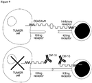
- the present invention relates to therapeutic and diagnostic antibodies, useful in diseases involving Carcinoembryonic Antigen-Related Cell Adhesion Molecule (CEACAM), expression, activation or function.
- CEACAM Carcinoembryonic Antigen-Related Cell Adhesion Molecule
- the present invention provides antibodies having specific complementarity determining regions (CDRs) and improved properties over other antibodies which recognize CEACAM1.
- CEACAM1 Carcinoembryonic antigen-related cell adhesion molecule 1
- BGP biliary glycoprotein
- CD66a and C-CAM1 are members of the carcinoembryonic antigen family (CEA) that also belongs to the immunoglobulin superfamily.
- CEACAM1 interacts with other known CEACAM proteins, including CD66a (CEACAM1), CD66e (CEACAM6) and CD66e (CEACAM5, CEA) proteins. It is expressed on a wide spectrum of cells, ranging from epithelial cells to those of hemopoietic origin (e.g. immune cells).
- CEACAM1 has been attributed to many different functions. It was shown that the CEACAM1 protein is over expressed in some carcinomas of colon, prostate, as well as other types of cancer. Additional data support the central involvement of CEACAM1 in angiogenesis and metastasis. CEACAM1 also plays a role in the modulation of innate and adaptive immune responses. For example, CEACAM1 was shown to be an inhibitory receptor for activated T cells contained within the human intestinal epithelium ( WO99/52552 and Morales et al. J. Immunol. 1999, 163, 1363-1370 ). Additional reports have indicated that CEACAM1 engagement either by T Cell Receptor cross-linking with Monoclonal antibodies (mAbs) or by Neisseria gonorrhoeae Opa proteins inhibits T cell activation and proliferation.
- mAbs Monoclonal antibodies
- Neisseria gonorrhoeae Opa proteins inhibits T cell activation and proliferation.
- MM pigment-producing cells
- MM Metastatic melanoma
- overexpression of CEACAM1 can be correlated with poor prognosis and is detected in the majority of metastatic melanoma cases.
- CEACAM1 is rarely expressed by normal melanocytes, but frequently found on melanoma cells.
- CEACAM1 expression on primary cutaneous melanoma lesions strongly predicts the development of metastatic disease with poor prognosis.
- increased CEACAM1 expression was observed on NK cells derived from some patients with metastatic melanoma compared with healthy donors.
- CEACAM1 may have an important role in virus infections.
- Markel at el. J. Clinical Investigation 2002, 110, 943-953
- lymphocytes isolated from the deciduae of CMV-infected patients express the CEACAM1 protein in increased levels.
- the increased CEACAM1 expression on the decidual lymphocytes might diminish the local immune response and serve as another mechanism developed by the virus to avoid recognition and clearance primarily by activated decidual lymphocytes.
- CEACAM1 immunostaining is significantly increased in high-grade squamous intraepithelial lesions (SIL) in comparison with low-grade SIL and normal cervical tissues.
- SIL squamous intraepithelial lesions
- CEACAM1 upregulation may be related to integration of HPV DNA in high-grade SIL and that CEACAM1 may be an important biological marker in SIL and cervical cancer progression.
- CEACAM1 plays an important role in various viral infections.
- CEACAM1 over expression may serve as marker of various viral infections.
- WO2007/063424 and U.S. Patent Application No. 20070110668 disclose methods for regulating the immune system, and in particular methods for the regulation of a specific immune response, including the regulation of lymphocyte activity. These methods comprise both the negative and positive modulation of CEACAM1 protein function.
- U.S. Patent Application No. 20070071758 teaches methods and compositions for enhancing the efficacy of tumor-infiltrating lymphocyte (TIL) therapy in the treatment of cancer by negatively modulating the activity of the CEACAM1 protein, such as for example, by using an immunoglobulin specific for CEACAM1.
- TIL tumor-infiltrating lymphocyte
- U.S. Patent Application No. 20080108140 discloses methods of modulating specific immune responses to create a protective immunity in the treatment of autoimmune diseases and diseases requiring the transplantation of tissue. In particular, it relates to the suppression of immune responses in a targeted fashion, by increasing the functional concentration of the CEACAM1 protein in the target tissue.
- U.S. Patent Application No. 20040047858 discloses specific antibodies which are capable of modulating T cell activity via CEACAM1 and uses thereof in treating immune response related diseases (e.g. graft versus host disease, autoimmune diseases, cancers etc.).
- immune response related diseases e.g. graft versus host disease, autoimmune diseases, cancers etc.
- U.S. Patent Application Nos. 20020028203 , 20050169922 and 20080102071 disclose compositions which bind T cell inhibitory receptor molecules and modulate (i.e. enhance or suppress) T cell activity (e.g. cytotoxicity and proliferation), such as biliary glycoprotein binding agents, and methods of using such compositions such as for treatment of diseases (e.g. an autoimmune disease, immunodeficiency, cancer etc.).
- T cell inhibitory receptor molecules bind T cell inhibitory receptor molecules and modulate (i.e. enhance or suppress) T cell activity (e.g. cytotoxicity and proliferation), such as biliary glycoprotein binding agents, and methods of using such compositions such as for treatment of diseases (e.g. an autoimmune disease, immunodeficiency, cancer etc.).
- diseases e.g. an autoimmune disease, immunodeficiency, cancer etc.
- WO 2010/125571 discloses a murine monoclonal antibody produced by a specific hybridoma cell.
- the mAb is highly selective to CEACAM1 and does not cross-react with other members of the CEACAM family.
- the present invention discloses monoclonal antibodies which recognize a specific set of CEACAM subtypes.
- the antibodies of the invention show binding to CEACAM1 and at least one additional subtype selected from CEACAM5 and CEACAM3.
- the antibodies of the invention are characterized by having unique CDR sequence and framework combinations and by binding to newly identified epitopes within the CEACAM1 molecule.
- the unique specificity of the monoclonal antibodies of the present invention broaden their therapeutic utility for treatment and diagnosis of additional types of malignancies and viral infections.
- the present invention also provides methods for identifying and isolating such antibodies, methods for their production, and therapeutic and diagnostic uses thereof.
- the monoclonal antibodies according to the present invention have specific combinations of CDRs and possess unique properties and improved specificity and potency over known anti CEACAM1 antibodies.
- the present invention provides a monoclonal antibody which recognizes human CEACAM1, or an antibody fragment thereof, having heavy-chain CDRs comprising heavy chain CDR1 comprising a sequence set forth in SEQ ID NOs: 7 or 13, a heavy chain CDR2 comprising a sequence set forth in SEQ ID NOs: 8 or 14, a heavy chain CDR3 comprising a sequence set forth in SEQ ID NOs: 9 or 15, and light-chain CDRs comprising a light chain CDR1 comprising a sequence set forth in SEQ ID NOs: 10 or 16, a light chain CDR2 comprising a sequence set forth in SEQ ID NOs: 11 or 17, and a light chain CDR3 comprising a sequence set forth in SEQ ID NOs: 12 or 18, as also detailed in claim 1.
- the monoclonal antibody comprises the heavy chain CDRs having the sequences set forth in SEQ ID NOs: 7, 8 and 9.
- the monoclonal antibody comprises the heavy chain CDRs having the sequences set forth in SEQ ID NOs: 13, 14 and 15.
- the monoclonal antibody comprises the light chain CDRs having the sequences set forth in SEQ ID NOs: 10, 11 and 12.
- the monoclonal antibody comprises the light chain CDRs having the sequences set forth in SEQ ID NOs: 16, 17 and 18.
- the monoclonal antibody has the CDR sequences set forth in SEQ ID NOs: 13, 14, 15, 16, 17, and 18.
- the monoclonal antibody has the CDR sequences set forth in SEQ ID NOs: 7, 8, 9, 10, 11, and 12.
- the antibody comprises a heavy chain variable domain sequence having a sequence set forth in SEQ ID NO: 26: QVQLQQSGAELVRPGTSVKVSCKASGYAFTNNLIEWVKQRPGQGLEWIGVINPGSG DTNYNEKFKGKATLTADKSSNTAYMQLSSLTSDDSAVYFCARGDYYGGFAVDYWG QGTSVTVSS, or an analog thereof having at least 97% sequence identity with the heavy chain variable domain sequence.
- the antibody comprises a light chain variable domain sequence having a sequence set forth in SEQ ID NO: 28: DIQMTQTTSSLSASLGDRVTISCRTSQDIGNYLNWYQQKPDGTVKLLIYYTSRLHSG VPSRFSGSGSGTDYSLTISNLEQEDIATYFCQQGKSLPRTFGGGTKLEIK, or an analog thereof having at least 97% sequence identity with the light chain variable domain sequence.
- the antibody or fragment thereof comprises a heavy chain variable domain having a sequence set forth in SEQ ID NO: 26 and a light chain variable domain having a sequence set forth in SEQ ID NO: 28, or an analog thereof having at least 97% sequence identity with the antibody or fragment sequence.
- the present invention encompasses monoclonal antibodies isolated from hybridoma cells or other biological systems, as well as monoclonal antibodies produced recombinantly or synthetically.
- a monoclonal antibody according to the present invention may contain a constant region from any mammalian species, including but not limited to mouse, rat and human.
- a monoclonal antibody according to the present invention includes a chimeric antibody, a humanized antibody, a fully human antibody, a xenogeneic antibody, and an antibody fragment comprising at least the antigen-binding portion of an antibody.
- the antibody fragment may be selected from the group consisting of: Fab, Fab', F(ab') 2 , Fd, Fd', Fv, dAb, isolated CDR region, single chain antibody, "diabodies", and "linear antibodies”.
- the present invention provides a monoclonal antibody, or an antibody fragment comprising:
- the monoclonal antibody may be a chimeric monoclonal antibody.
- Such chimeric antibody may comprise human-derived constant regions.
- Human constant regions of such chimeric antibody may be selected from the group consisting of: human IgG1, human IgG2, and human IgG3
- a chimeric or humanized monoclonal antibody which recognizes CEACAM1 is also disclosed.
- a chimeric monoclonal antibody or a fragment thereof comprising a heavy chain sequence set forth in SEQ ID NO: 30 is provided.
- a chimeric monoclonal antibody or a fragment thereof comprising a light chain sequence set forth in SEQ ID NO: 31 is provided.
- a chimeric monoclonal antibody or a fragment having a heavy chain sequence set forth in SEQ ID NO: 30, and light chain sequence set forth in SEQ ID NO: 31 is provided.
- a monoclonal antibody which recognizes CEACAM1 is provided produced from DNA sequences of the heavy and light chains contained in a plasmid deposited on September 28, 2011 under ATCC Accession Number PTA-12130.
- Monoclonal antibodies of the present invention exhibit specific binding to more than one CEACAM subtype.
- the monoclonal antibody preferably binds at least two different CEACAM subtypes.
- the monoclonal antibody preferably binds to CEACAM1 and at least one of CEACAM3 and CEACAM5.
- the monoclonal antibody preferably binds to CEACAM1 and CEACAM5, or to CEACAM1 and CEACAM3.
- a monoclonal antibody according to the present invention preferably binds to CEACAM subtypes 1, 3 and 5.
- a monoclonal antibody according to the present invention preferably does not bind to CEACAM4 and CEACAM6.
- the monoclonal antibody is reactive with a epitope within residues 17-29 and 68-79 of human CEACAM1 having the sequences VLLLVHNLPQQLF (SEQ ID NO:32) and YPNASLLIQNVT (SEQ ID NO:33) respectively.
- the epitope on the CEACAM1 molecule to which the monoclonal antibody binds is an epitope comprising amino acid residues within the sequences VLLLVHNLPQQLF (SEQ ID NO: 32) and YPNASLLIQNVT (SEQ ID NO: 33).
- the monoclonal antibody according to the invention binds an epitope comprising at least four amino acids of the sequence VLLLVHNLPQQLF (SEQ ID NO: 32).
- the monoclonal antibody according to the invention binds an epitope within sequences VLLLVHNLPQQLF (SEQ ID NO: 32) and PNASLLI (SEQ ID NO: 34).
- nucleic acid molecules encoding an antibody or antibody fragment according to the invention, having affinity and specificity for CEACAM1.
- an isolated polynucleotide encoding a monoclonal antibody according to the present invention is disclosed.
- the isolated polynucleotide sequence may comprise a DNA sequence set forth in SEQ ID NO: 25 or analog thereof having at least 90% sequence identity with said DNA sequence.
- the isolated polynucleotide sequence may comprise a DNA sequence set forth in SEQ ID NO: 27 or analog thereof having at least 90% sequence identity with said DNA sequence.
- Plasmids comprising at least one polynucleotide sequence according to the invention are also disclosed, as well as host cells comprising these plasmids.
- a plasmid comprising polynucleotide sequences set forth in SEQ ID NOs: 25 and 27, deposited on September 28, 2011 under ATCC Accession Number PTA-12130, is disclosed.
- the present invention is related to a pharmaceutical composition useful for preventing, attenuating or treating a disease or disorder associated with CEACAM1, CEACAM3 or CEACAM5 expression, activation or function.
- a pharmaceutical composition according to the invention comprises a therapeutically effective amount of the monoclonal antibody or an antibody fragment thereof according to the present invention; and a pharmaceutically acceptable carrier.
- the disease or disorder associated with CEACAM1, CEACAM3 and/or CEACAM5 expression, activation or function is a cell proliferative disease or disorder.
- the cell proliferative disease or disorder is cancer.
- the cancer associated with over-expression of CEACAM5 is selected from the group consisting of: gastrointestinal, colorectal (CRC) pancreatic non-small cell lung cancer (NSCL), breast, thyroid, stomach, ovarian and uterine.
- CRC colorectal
- NSC pancreatic non-small cell lung cancer
- the cancers associated with over expression of CEACAM1 are melanoma, pancreatic cancer, all types of lung cancers and myeloma.
- the pharmaceutical composition according to the present invention may be administered as a stand alone treatment or in addition to a treatment with any other therapeutic agent.
- Antibodies according to the present invention may be administered to a subject in need thereof as part of a treatment regimen in conjunction with at least one anti-cancer agent.
- the pharmaceutical composition according to the present invention may be administered together with the other agent or separately.
- the present invention provides diagnostic compositions useful for detecting at least one CEACAM subtype selected from the group consisting if: CEACAM1, CEACAM3 and CEACAM5, in a subject.
- a diagnostic composition according to the invention comprises a therapeutically effective amount of the monoclonal antibody or the antibody fragment thereof according to the present invention. of the pharmaceutical composition of the present invention can be used for preventing, attenuating or treating a disease or disorder associated with expression, activation or function of CEACAM 1.
- the disease or disorder is a cell proliferative disease or disorder.
- the cell proliferative disease or disorder is cancer.
- of the pharmaceutical composition of the present invention can be further used in immunomodulation, inhibiting migration of a CEACAM expressing tumor cell, inhibiting CEACAM homotypic or heterotypic protein-protein interaction, or increasing the duration or progression of response or survival of a subject having cancer.
- antibodies of the present invention can also be used in diagnostic applications.
- a method for diagnosing a cancer in a subject in need thereof comprising contacting a biological sample derived from the subject (in-vivo, in vitro or ex-vivo) with a diagnostic composition comprising the antibody or antibody fragment described herein, wherein a complex formation beyond a predetermined threshold is indicative of the cancer in the subject.
- Cells of the cancer may be characterized by over expression of CEACAM as compared to unaffected cells.
- the diagnosed cancer may be selected from the group consisting of: melanoma, pancreatic cancer, lung cancer and myeloma.
- the measured protein may be CEACAM5 and the diagnosed cancer may be selected from the group consisting of: gastrointestinal, colorectal (CRC) pancreatic non-small cell lung cancer (NSCL), breast, thyroid, stomach, ovarian and uterine.
- CRC colorectal
- NSCL pancreatic non-small cell lung cancer
- immunocomplex refers to a complex which comprises the antibody of the invention and the CEACAM. Determining a presence or level of the immunocomplex may be direct or by detecting an identifiable (detectable) moiety which may be attached to the antibody.
- the level of the immunocomplex in the tested cell (e.g., a cell of a subject in need thereof) is compared to a predetermined threshold.
- a predetermined threshold may be determined based on a known reference level and/or a level in a control cell or serum.
- the control cell can be obtained from a control, healthy subject (e.g., a subject not suffering from the cancer) or from the same subject prior to disease initiation or following treatment.
- the control subject may be of the same species e.g. human, preferably matched with the same age, weight, sex etc. as the subject in need thereof.
- Also disclosed but not forming part of the present invention is a method for detecting or quantifying the presence of CEACAM.
- methods for diagnosing conditions associated with CEACAM expression using antibodies which recognizes CEACAM Diagnostic methods may be performed according to specific embodiments, in-vitro or ex-vivo.
- the antibodies according to the present invention may be also used to configure screening methods.
- an ELISA assay can be constructed for measuring secreted or cell associated levels of polypeptide using monoclonal and polyclonal antibodies by standard methods known in the art.
- the biological sample may be a body fluid of a mammalian subject.
- the mammalian subject may be human.
- the antibodies of the present invention may be also used in screening assays for assessing the CEACAM levels in patients and for prediction of the effectiveness of treatment.
- the screening assays with the antibodies of the present invention may allow determination of the levels of CEACAM and therefore prediction of treatment outcome and planning of an appropriate treatment regimen.
- the level of at least one of CEACAM 1, CEACAM3 and CEACAM5 can be assessed.
- the antibody or antibody fragment may be attached to an identifiable moiety.
- the identifiable moiety can be a member of a binding pair, which is identifiable via its interaction with an additional member of the binding pair and a label which is directly visualized.
- the member of the binding pair is an antigen which is identified by a corresponding labeled antibody.
- the label is a fluorescent protein or an enzyme producing a colorimetric reaction.
- the present invention provides antibodies which recognize CEACAM1 comprising specific sets of CDR sequences which possess improved and unique specificity, selectivity, affinity and/or activity.
- Antibodies according to the present invention bind CEACAM1 with higher affinity than other anti-CEACAM1 antibodies, they block the function of CEACAM1, while not all anti CEACAM antibodies do, and more efficiently than polyclonal anti CEACAM antibodies. Furthermore, antibodies according to the present invention are effective against cancer cells, in particular melanoma cells: the antibodies render melanoma cells more susceptible to lymphocytes, inhibit melanoma growth rate in vivo, an effect which is enhanced when the antibody is combined with adoptive T cell transfer in vivo.
- the in vivo anti-melanoma effect of anti-CEACAM1 antibodies according to the invention is a combined direct anti-tumor effect as well as immunomodulatory effect rendering the cells more susceptible to reactive lymphocytes.
- An antibody according to the present invention fragments and derivatives can be used as an effective tool for diagnosis, immunomodulation and cancer treatment.
- the antibody inhibits CEACAM1 homophilic interactions, as determined by co-incubation of immune effector cells and target cells expressing CEACAM1 and assaying IL-2 secretion and by the in vitro killing assays.
- the antibody of the present invention is shown to enhance CM10 enhances HLA restricted T cell killing and to enhance granzyme B (a serine protease involves in mediation apoptosis of the target cells) secretion from effector NK and T cells in the presence of specific target cells, thus enabling the observed enhanced killing of the target cells by the antibody. It is also inhibits the binding between CEACAM1 to CEACAM5 in a dose-dependent manner therefore can be used to treat malignancies that express high level of CEACAM5 and exploit the CEACAM1-CEACAM5 axis in order to suppress the immune cells.
- an antibody according to the invention is effective in inhibiting melanoma cells invasion. Furthermore, in vivo administration of an antibody according to the invention, either alone or in combination with reactive lymphocytes was shown effective in inhibiting growth of melanoma tumors. The combination of adoptive human T cell transfer with monoclonal antibody injections exhibited significant synergism and strongly inhibited xenograft growth compared to the isotype control group.
- isolated antibody or antibody fragment having the same binding specificity and selectivity to an antibody defined herein comprising an antigen recognition domain having specific CDR segments described above.
- isolated antibody or antibody fragment is capable of binding the same epitope determinant of the CEACAM1 protein as does the antibody described above by its specific CDR segments sequences.
- Monoclonal antibodies can be designed to selectively target tumor cells and elicit a variety of responses once bound. These agents can destruct tumor cells in different ways such as blocking tumor cell proliferation or activating the immune system.
- Chimeric monoclonal antibodies according to the present invention were designed to specifically bind and neutralize various functions of the CEACAM1 protein and other CEACAM subtype proteins, and to induce the specific death of tumor cells. Without wishing to be bound to any theory, it is suggested that monoclonal antibodies according to the present invention act also via activation of the immune system against cancerous cells.
- CEACAM1 is not found on normal melanocytes, but undergoes neo-expression and is widely expressed on the vast majority of metastatic melanoma specimens. It has been previously demonstrated mechanistically that CEACAM1 protects melanoma cells by inhibiting effector functions of NK cells and T cells.
- CM10 is a chimeric monoclonal antibody which binds with high affinity to human CEACAM1.
- CM10 efficiently blocked CEACAM1-homophilic interactions in a dose dependent manner and improves CEACAM1 positive melanoma cells killing by T cells and NK cells.
- CM10 significantly inhibited the in- vivo growth of melanoma xenografts when administered systemically along with melanoma-reactive human T lymphocytes (tumor-infiltrating lymphocytes, TILs). Without wishing to be bound to any theory, this is in line with the suggested mechanism of action; abrogation of immune-protective interactions of the tumor cells with the activated lymphocytes.
- CEACAM1 is expressed by a wide variety of epithelial cells, including colon, prostate, breast, kidney etc. Extensive examination of CEACAM1 expression profile on normal and malignant tissues by IHC have been performed. The expression analysis showed a strong staining of melanoma cells, as compared to no staining of the vast majority of the tissues tested in a normal human tissue. Nevertheless, some selective staining was observed in restricted sites of several organs. When more quantitative method was used to quantify the number of CM10 mAb molecules bound to malignant and normal primary cells, very low CM10 molecules could be detected in normal cells, which may indicate that CM10 binds mostly to patient's tumor cells. Furthermore it is shown that CM10 has no effect on primary cells proliferation and is completely or almost completely, unable to induce CDC or ADCC indicating the potential safety of the monoclonal antibody in human subjects.
- CM10 has an immunomodulation activity, possible immune-related side effects, are evaluated. Following PBMC activation, CEACAM1 is upregulated on the activated lymphocytes ( Gray-Owen and Blumberg 2006, Nat Rev Immunol 6, 433-46 ). Ex-vivo human PBMC proliferation assay revealed that CM10 has no effect on naive and activated PBMC proliferative response.
- CEACAM1 blockade over abrogation of generalized inhibitory mechanisms is the expected selectivity to the vicinity of the tumor and therefore fewer adverse events compare to other general immune toxicity agents.
- CM10 shows encouraging activity and safety profile and is a promising candidate for cancer immunotherapy and can be used as a strategy to selectively enhance the anti-tumor properties of the endogenous immune response in several malignancies, such as melanoma and non-small cell lung cancer.
- binding to additional CEACAM subtypes increases the therapeutic profile of the antibody, thus it can be used for diagnosis and treatment of other types of malignancies which do not extensively express CEACAM1 but express CEACAM5, for example.
- CEACAM5 has been found to be over-expressed in a high percentage of many human tumors, including 90% of gastrointestinal, colorectal (CRC) and pancreatic cancers, 70% of non-small cell lung cancer cells and 50% of breast cancers. It is also over-expressed in thyroid, stomach, ovarian and uterine cancers ( Thompson, Grunert et al. 1991, J Clin Lab Anal 5, 344-66 ). CEACAM5 even serves as a clinical marker for liver metastasis in CRC and post-surgical surveillance of colon cancer ( Duffy 2001, Clin Chem 47, 624-30 ). The evidence that CM10 is capable to bind CEACA5 is very important and can expand the possible indications that can be treated by CM10 from 4-5 types of malignancies to above 10.
- anti-CEACAM5 agents that have entered clinical trials include anti-CEACAM5 antibodies conjugated to toxic substances such as radioactive substances for both diagnostic purposes and for the treatment of various malignancies. It seems that even these toxic conjugated forms don't show safety problems, which can indicate that CEACAM5 is a safe target.
- Murine IgG2a and IgG2b and human IgG1 and IgG3 share the ability to fix complement and bind to protein antigens ( Hussain et al., 1995, Clinical and Diagnostic Laboratory Immunology 726-732 ).
- Murine IgG1 and human IgG4 are considered to be similar because of their property of binding to mast cells.
- Human IgG4 is the only human IgG subclass which does not activate complement and the subclasses IgG1 and 3 are the most effective in activating complements. For mouse it is the subclasses IgG2a and IgG2b which are active with IgG1 and possibly IgG3 being inactive ( Clark MR., Chem Immunol. 1997;65:88-110 ).
- chimeric monoclonal antibodies which recognize CEACAM1 are of subtype mouse IgG1.
- IgG4 As the human equivalent of mouse IgG1 is IgG4 it would be expected to create a chimeric antibody comprising the human IgG4 constant framework.
- chimeric monoclonal antibodies comprise a human IgG1 constant framework.
- the present invention provides a monoclonal antibody which recognizes human CEACAM1, or an antibody fragment thereof, according to claim 1.
- a monoclonal antibody which recognizes human CEACAM1, or an antibody fragment thereof, according to claim 1.
- CEACAM1 is used to refer to the protein product of the CEACAM1 gene e.g., NP_001020083.1, NP_001703.2. In humans, 11 different CEACAM1 splice variants have been detected so far. Individual CEACAM1 isoforms differ with respect to the number of extracellular immunoglobulin-like domains (for example, CEACAM1 with four extracellular immunoglobulin-like domains is known as CEACAM1-4), membrane anchorage and/or the length of their cytoplasmic tail (for example, CEACAM1-4 with a long cytoplasmic tail is known as CEACAM1-4L and CEACAM1-4 with a short cytoplasmic tail is known as CEACAM1-4S).
- the N-terminal domain of CEACAM1 starts immediately after the signal peptide and its structure is regarded as IgV-type.
- the N-terminal IgV-type domain is comprised of 108 amino acids, from amino acid 35 to 142. This domain was identified as responsible for the homophilic binding activity ( Watt et al., 2001, Blood. 98, 1469-79 ). All variants, including these splice variants are included within the term "CEACAM1".
- an “anti-CEACAM1 antibody”, “an antibody which recognizes CEACAM1”, “an antibody against CEACAM1”, or “an antibody to CEACAM1” is an antibody that binds to the CEACAM1 protein with sufficient affinity and specificity.
- an antibody according to the present teachings is capable of binding CEACAM1 with a minimal affinity of about 10 -8 or 10 -9 M.
- Some of the monoclonal antibodies of the present invention are capable of binding CEACAM3, 5 and/or 8 with a minimal affinity of about 5x10 -7 M.
- the anti-CEACAM1 antibody of the invention can be used as a diagnostic or therapeutic agent in targeting and interfering with diseases or conditions wherein the CEACAM1 expression or activity is involved.
- an "antigen” is a molecule or a portion of a molecule capable of eliciting antibody formation and being bound by an antibody.
- An antigen may have one or more than one epitope. The specific reaction referred to above is meant to indicate that the antigen will react, in a highly selective manner, with its corresponding antibody and not with the multitude of other antibodies which may be evoked by other antigens.
- An antigen according to the present invention is a CEACAM1 protein or a fragment thereof.
- antigenic determinant or "epitope” according to the invention refers to the region of an antigen molecule that specifically reacts with particular antibody.
- Peptide sequences derived from an epitope can be used, alone or in conjunction with a carrier moiety, applying methods known in the art, to immunize animals and to produce additional polyclonal or monoclonal antibodies. Isolated peptides derived from an epitope may be used in diagnostic methods to detect antibodies and as therapeutic agents when inhibition of said antibodies is required.
- Antibodies, or immunoglobulins comprise two heavy chains linked together by disulfide bonds and two light chains, each light chain being linked to a respective heavy chain by disulfide bonds in a "Y" shaped configuration.
- Proteolytic digestion of an antibody yields Fv (Fragment variable) and Fc (fragment crystalline) domains.
- the antigen binding domains, Fab include regions where the polypeptide sequence varies.
- F(ab') 2 represents two Fab' arms linked together by disulfide bonds.
- the central axis of the antibody is termed the Fc fragment.
- Each heavy chain has at one end a variable domain (V H ) followed by a number of constant domains (C H ).
- Each light chain has a variable domain (V L ) at one end and a constant domain (C L ) at its other end, the light chain variable domain being aligned with the variable domain of the heavy chain and the light chain constant domain being aligned with the first constant domain of the heavy chain (CH1).
- the variable domains of each pair of light and heavy chains form the antigen-binding site.
- the domains on the light and heavy chains have the same general structure and each domain comprises four framework regions, whose sequences are relatively conserved, joined by three hypervariable domains known as complementarity determining regions (CDR1-3). These domains contribute specificity and affinity of the antigen-binding site.
- the isotype of the heavy chain determines immunoglobulin class (IgG, IgA, IgD, IgE or IgM, respectively).
- the light chain is either of two isotypes (kappa, ⁇ or lambda, ⁇ ) found in all antibody classes.
- antibody is used in the broadest sense and includes monoclonal antibodies (including full length or intact monoclonal antibodies), polyclonal antibodies, multivalent antibodies, multispecific antibodies (e.g., bispecific antibodies), and antibody fragments so long as they exhibit the desired biological activity.
- the antibody according to the present invention is a molecule comprising at least the antigen-binding portion of an antibody.
- Antibody or antibodies according to the invention include intact antibodies, such as polyclonal antibodies or monoclonal antibodies (mAbs), as well as proteolytic fragments thereof such as the Fab or F(ab') 2 fragments. Further included within the scope of the invention are chimeric antibodies; human and humanized antibodies; recombinant and engineered antibodies, and fragments thereof. Furthermore, the DNA encoding the variable region of the antibody can be inserted into the DNA encoding other antibodies to produce chimeric antibodies. Single chain antibodies also fall within the scope of the present invention.
- Antibody fragments comprise only a portion of an intact antibody, generally including an antigen binding site of the intact antibody and thus retaining the ability to bind antigen.
- Examples of antibody fragments encompassed by the present definition include: (i) the Fab fragment, having VL, CL, VH and CH1 domains; (ii) the Fab' fragment, which is a Fab fragment having one or more cysteine residues at the C-terminus of the CH1 domain; (iii) the Fd fragment having VH and CH1 domains; (iv) the Fd' fragment having VH and CH1 domains and one or more cysteine residues at the C-terminus of the CH1 domain; (v) the Fv fragment having the VL and VH domains of a single arm of an antibody; (vi) the dAb fragment ( Ward et al., Nature 1989, 341, 544-546 ) which consists of a VH domain; (vii) isolated CDR regions; (viii) F(ab') 2 fragments, a
- Single chain antibodies can be single chain composite polypeptides having antigen binding capabilities and comprising amino acid sequences homologous or analogous to the variable regions of an immunoglobulin light and heavy chain i.e. linked V H -V L or single chain Fv (scFv).
- neutralizing antibody refers to a molecule having an antigen-binding site to a specific receptor or ligand target capable of reducing or inhibiting (blocking) activity or signaling through a receptor, as determined by in vivo or in vitro assays, as per the specification.
- the term "monoclonal antibody” as used herein refers to an antibody obtained from a population of substantially homogeneous antibodies, i.e., the individual antibodies comprising the population are identical except for possible naturally occurring mutations that may be present in minor amounts. Monoclonal antibodies are highly specific, being directed against a single antigen. Furthermore, in contrast to polyclonal antibody preparations that typically include different antibodies directed against different determinants (epitopes), each monoclonal antibody is directed against a single determinant on the antigen.
- the modifier "monoclonal” is not to be construed as requiring production of the antibody by any particular method. mAbs may be obtained by methods known to those skilled in the art.
- the monoclonal antibodies to be used in accordance with the present invention may be made by the hybridoma method first described by Kohler et al., Nature 1975, 256, 495 , or may be made by recombinant DNA methods (see, e.g., U.S. Pat. No. 4,816,567 ).
- the "monoclonal antibodies” may also be isolated from phage antibody libraries using the techniques described in Clackson et al., Nature 1991, 352, 624-628 or Marks et al., J. Mol. Biol., 1991, 222:581-597 , for example.
- the mAbs of the present invention may be of any immunoglobulin class including IgG, IgM, IgE, IgA.
- a hybridoma producing a mAb may be cultivated in vitro or in vivo.
- High titers of mAbs can be obtained in vivo production where cells from the individual hybridomas are injected intraperitoneally into pristine-primed Balb/c mice to produce ascites fluid containing high concentrations of the desired mAbs.
- mAbs of isotype IgM or IgG may be purified from such ascites fluids, or from culture supernatants, using column chromatography methods well known to those of skill in the art.
- the monoclonal antibodies herein specifically include "chimeric" antibodies in which a portion of the heavy and/or light chain is identical with or homologous to corresponding sequences in antibodies derived from a particular species or belonging to a particular antibody class or subclass, while the remainder of the chain(s) is identical with or homologous to corresponding sequences in antibodies derived from another species or belonging to another antibody class or subclass, as well as fragments of such antibodies, so long as they exhibit the desired biological activity ( U.S. Pat. No. 4,816,567 ; and Morrison et al., Proc. Natl. Acad. Sci. USA 81:6851-6855 (1984 )).
- complementarity determining region (CDR) grafting may be performed to alter certain properties of the antibody molecule including affinity or specificity.
- CDR complementarity determining region
- Chimeric antibodies are molecules, the different portions of which are derived from different animal species, such as those having a variable region derived from a murine mAb and a human immunoglobulin constant region.
- Antibodies which have variable region framework residues substantially from human antibody (termed an acceptor antibody) and complementarity determining regions substantially from a mouse antibody (termed a donor antibody) are also referred to as humanized antibodies.
- Chimeric antibodies are primarily used to reduce immunogenicity in application and to increase yields in production, for example, where murine mAbs have higher yields from hybridomas but higher immunogenicity in humans, such that human/murine chimeric mAbs are used.
- Humanized forms of non-human (e.g., murine) antibodies are chimeric antibodies that contain minimal sequence derived from non-human immunoglobulin.
- humanized antibodies are human immunoglobulins (recipient antibody) in which residues from a hypervariable region of the recipient are replaced by residues from a hypervariable region of a non-human species (donor antibody) such as mouse, rat, rabbit or nonhuman primate having the desired specificity, affinity, and capacity.
- donor antibody such as mouse, rat, rabbit or nonhuman primate having the desired specificity, affinity, and capacity.
- framework region (FR) residues of the human immunoglobulin are replaced by corresponding non-human residues.
- humanized antibodies may comprise residues that are not found in the recipient antibody or in the donor antibody. These modifications are made to further refine antibody performance.
- the humanized antibody will comprise substantially all of at least one, and typically two, variable domains, in which all or substantially all of the hypervariable loops correspond to those of a non-human immunoglobulin and all or substantially all of the FRs are those of a human immunoglobulin sequence.
- the humanized antibody optionally will also comprise at least a portion of an immunoglobulin constant region (Fc), typically that of a human immunoglobulin.
- Fc immunoglobulin constant region
- a "human antibody” is one which possesses an amino acid sequence which corresponds to that of an antibody produced by a human and/or has been made using any of the techniques for making human antibodies as disclosed herein. This definition of a human antibody specifically excludes a humanized antibody comprising non-human antigen-binding residues.
- Human antibodies can be produced using various techniques known in the art. The human antibody may be selected from a phage library, where that phage library expresses human antibodies ( Vaughan et al. Nature Biotechnology 1996 14,309-314 ; Sheets et al. PNAS (USA), 1998, 95, 6157-6162 ); Hoogenboom and Winter, J. Mol. Biol., 1991, 227, 381 ; Marks et al., J. Mol.
- Human antibodies can also be made by introducing human immunoglobulin loci into transgenic animals, e.g., mice in which the endogenous immunoglobulin genes have been partially or completely inactivated. Upon challenge, human antibody production is observed, which closely resembles that seen in humans in all respects, including gene rearrangement, assembly, and antibody repertoire. This approach is described, for example, in U.S. Pat. Nos.
- the human antibody may be prepared via immortalization of human B lymphocytes producing an antibody directed against a target antigen (such B lymphocytes may be recovered from an individual or may have been immunized in vitro). See, e.g., Cole et al., Monoclonal Antibodies and Cancer Therapy, Alan R. Liss, p. 77 (1985 ); Boerner et al., J. Immunol., 147 (1):86-95 (1991 ); and U.S. Pat No. 5,750,373 .
- single chain variable fragment scFv
- Single chain antibodies can be single chain composite polypeptides having antigen binding capabilities and comprising amino acid sequences homologous or analogous to the variable regions of an immunoglobulin light and heavy chain (linked V H -V L or single chain Fv (scFv)).
- V H and V L may copy natural monoclonal antibody sequences or one or both of the chains may comprise a CDR-FR construct of the type described in US patent 5,091,513 .
- a "molecule having the antigen-binding portion of an antibody” as used herein is intended to include not only intact immunoglobulin molecules of any isotype and generated by any animal cell line or microorganism, but also the antigen-binding reactive fraction thereof, including, but not limited to, the Fab fragment, the Fab' fragment, the F(ab') 2 fragment, the variable portion of the heavy and/or light chains thereof, Fab mini-antibodies (see WO 93/15210 , US patent application 08/256,790 , WO 96/13583 , US patent application 08/817,788 , WO 96/37621 , US patent application 08/999,554 ), dimeric bispecific mini-antibodies (see Muller et al., 1998) and chimeric or single-chain antibodies incorporating such reactive fraction, as well as any other type of molecule or cell in which such antibody reactive fraction has been physically inserted, such as a chimeric T-cell receptor or a T-cell having such a receptor, or molecules
- Antibodies according to the invention can be obtained by administering CEACAM1, or epitope-bearing fragments, analogs, or cells expressing, to an animal, preferably a nonhuman, using routine protocols.
- any technique known in the art that provides antibodies produced by continuous cell line cultures can be used. Examples include various techniques, such as those in Kohler, G. and Milstein, C., Nature 256: 495-497 (1975 ); Kozbor et al., Immunology Today 4: 72 (1983 ); Cole et al., pg. 77-96 in MONOCLONAL ANTIBODIES AND CANCER THERAPY, Alan R. Liss, Inc. (1985 ).
- antibodies can be generated in vitro using phage display technology.
- Such a production of recombinant antibodies is much faster compared to conventional antibody production and they can be generated against an enormous number of antigens.
- many antigens prove to be non-immunogenic or extremely toxic, and therefore cannot be used to generate antibodies in animals.
- affinity maturation i.e., increasing the affinity and specificity
- large numbers of different antibodies against a specific antigen can be generated in one selection procedure.
- To generate recombinant monoclonal antibodies one can use various methods all based on display libraries to generate a large pool of antibodies with different antigen recognition sites.
- Such a library can be made in several ways: One can generate a synthetic repertoire by cloning synthetic CDR3 regions in a pool of heavy chain germline genes and thus generating a large antibody repertoire, from which recombinant antibody fragments with various specificities can be selected.
- Non-human antibodies may be humanized by any methods known in the art.
- the non-human complementarity determining regions are inserted into a human antibody or consensus antibody framework sequence. Further changes can then be introduced into the antibody framework to modulate affinity or immunogenicity.
- US Patent 5,585,089 of Queen et al. discloses a humanized immunoglobulin and methods of preparing same, wherein the humanized immunoglobulin comprises complementarity determining regions (CDRs) from a donor immunoglobulin and heavy and light chain variable region frameworks from human acceptor immunoglobulin heavy and light chains, wherein said humanized immunoglobulin comprises amino acids from the donor immunoglobulin framework outside the Kabat and Chothia CDRs, wherein the donor amino acids replace corresponding amino acids in the acceptor immunoglobulin heavy or light chain frameworks.
- CDRs complementarity determining regions
- US Patent 5,225,539, of Winter also discloses an altered antibody or antigen-binding fragment thereof and methods of preparing same, wherein a variable domain of the antibody or antigen-binding fragment has the framework regions of a first immunoglobulin heavy or light chain variable domain and the complementarity determining regions of a second immunoglobulin heavy or light chain variable domain, wherein said second immunoglobulin heavy or light chain variable domain is different from said first immunoglobulin heavy or light chain variable domain in antigen binding specificity, antigen binding affinity, species, class or subclass.
- Anti-idiotype antibodies specifically immunoreactive with an antibody of the invention are also comprehended.
- phage display technology can be utilized to select antibody genes with binding activities towards a polypeptide as disclosed hereineither from repertoires of PCR amplified v-genes of lymphocytes from humans screened for possessing anti-CEACAM1 or from libraries ( McCafferty, et al., 1990, Nature 348, 552-554 ; Marks, et al., 1992, Biotechnology 10, 779-783 ).
- the affinity of these antibodies can also be improved by, for example, chain shuffling ( Clackson et al., 1991, Nature 352:628 ).
- the above-described antibodies can be employed to isolate or to identify clones expressing the polypeptides to purify the polypeptides by, for example, affinity chromatography.
- conservative amino acid variants of the antibody molecules are also disclosed. Variants also may be made that conserve the overall molecular structure of the encoded proteins. Given the properties of the individual amino acids comprising the disclosed protein products, some rational substitutions will be recognized by the skilled worker. Amino acid substitutions, i.e. "conservative substitutions,” may be made, for instance, on the basis of similarity in polarity, charge, solubility, hydrophobicity, hydrophilicity, and/or the amphipathic nature of the residues involved.
- a “disorder” is any condition that would benefit from treatment with the antibody. This includes chronic and acute disorders or diseases including those pathological conditions which predispose the mammal to the disorder in question.
- disorders to be treated herein include benign and malignant tumors; leukemias and lymphoid malignancies; neuronal, glial, astrocytal, hypothalamic and other glandular, macrophagal, epithelial, stromal and blastocoelic disorders; and inflammatory, angiogenic, immunologic disorders or hyperpermeability states.
- the term "therapeutically effective amount” refers to an amount of a drug effective to treat a disease or disorder in a mammal.
- the therapeutically effective amount of the drug may reduce the number of cancer cells; reduce the tumor size; inhibit (i.e., slow to some extent and preferably stop) cancer cell infiltration into peripheral organs; inhibit (i.e., slow to some extent and preferably stop) tumor metastasis; inhibit, to some extent, tumor growth; and/or relieve to some extent one or more of the symptoms associated with the disorder.
- the drug may prevent growth and/or kill existing cancer cells, it may be cytostatic and/or cytotoxic.
- efficacy in vivo can, for example, be measured by assessing the duration of survival, time to disease progression (TTP), the response rates (RR), duration of response, and/or quality of life.
- Treatment refers to both therapeutic treatment and prophylactic or preventative measures. Those in need of treatment include those already with the disorder as well as those in which the disorder is to be prevented.
- cancer refers to or describe the physiological condition in mammals that is typically characterized by unregulated cell growth.
- examples of cancer include but are not limited to, carcinoma, lymphoma, blastoma, sarcoma, and leukemia. More particular examples of such cancers include melanoma, lung, thyroid, breast, colon, prostate, hepatic, bladder, renal, cervical, pancreatic, leukemia, lymphoma, myeloid, ovarian, uterus, sarcoma, biliary, or endometrial cancer.
- the antibody of the present invention may be attached to a cytotoxic or therapeutic moiety.
- the cytotoxic or therapeutic moiety can be, for example, a cytotoxic moiety, a toxic moiety, a cytokine moiety, a bi-specific antibody moiety, a cytotoxin, a chemokine, a chemotherapy, a pro-apoptotic, interferon, a radioactive moiety, or combinations thereof, examples of which are provided infra.
- anti-neoplastic composition refers to a composition useful in treating cancer comprising at least one active therapeutic agent capable of inhibiting or preventing tumor growth or function, and/or causing destruction of tumor cells.
- Therapeutic agents suitable in an anti-neoplastic composition for treating cancer include, but not limited to, chemotherapeutic agents, radioactive isotopes, toxins, cytokines such as interferons, and antagonistic agents targeting cytokines, cytokine receptors or antigens associated with tumor cells.
- the therapeutic agent is a chemotherapeutic agent.
- diagnosis refers to determining presence or absence of a pathology, classifying a pathology or a symptom, determining a severity of the pathology, monitoring pathology progression, forecasting an outcome of a pathology and/or prospects of recovery.
- the present invention also contemplates pharmaceutical formulations for human medical use, which comprise as the active agent at least one antibody of the present invention, for the manufacture of a therapeutic or diagnostic composition for the treatment, diagnosis or prophylaxis of the conditions variously described herein.
- the active agent is preferably utilized together with one or more pharmaceutically acceptable carrier(s) and optionally any other therapeutic ingredients.
- the carrier(s) must be pharmaceutically acceptable in the sense of being compatible with the other ingredients of the formulation and not unduly deleterious to the recipient thereof.
- the active agent is provided in an amount effective to achieve the desired pharmacological effect, as described above, and in a quantity appropriate to achieve the desired daily dose.
- the molecules as disclosed herein comprise the antigen binding portion of an antibody or comprising another polypeptide including a peptidomimetic will be suspended in a sterile saline solution for therapeutic uses.
- the pharmaceutical compositions may alternatively be formulated to control release of active ingredient (molecule comprising the antigen binding portion of an antibody) or to prolong its presence in a patient's system.
- suitable drug delivery systems include, e.g., implantable drug release systems, hydrogels, hydroxymethylcellulose, microcapsules, liposomes, microemulsions, microspheres, and the like. Controlled release preparations can be prepared through the use of polymers to complex or adsorb the molecule.
- biocompatible polymers include matrices of poly(ethylene-co-vinyl acetate) and matrices of a polyanhydride copolymer of a stearic acid dimer and sebaric acid.
- the rate of release of the molecule as disclosed herein, i.e., of an antibody or antibody fragment, from such a matrix depends upon the molecular weight of the molecule, the amount of the molecule within the matrix, and the size of dispersed particles.
- composition of this invention may be administered by any suitable means, such as orally, topically, intranasally, subcutaneously, intramuscularly, intravenously, intra-arterially, intraarticulary, intralesionally or parenterally. Ordinarily, intravenous (i.v.), intraarticular, topical or parenteral administration will be preferred.
- a "therapeutically effective amount” refers to the amount of a molecule required to alleviate one or more symptoms associated with a disorder being treated over a period of time.
- the daily dosage can generally be between about 0.01mg to about 500 mg, preferably about 0.01mg to about 50 mg, more preferably about 0.1mg to about 10 mg, per kg body weight.
- the daily dosage can generally be between about 0.001mg to about 100 mg, preferably about 0.001mg to about 10 mg, more preferably about 0.01mg to about 1 mg, per kg body weight.
- the daily dosage can be administered, for example in regimens typical of 1-4 individual administration daily.
- Other preferred methods of administration include intraarticular administration of about 0.01mg to about 100 mg per kg body weight.
- Various considerations in arriving at an effective amount are described, e.g., in Goodman and Gilman's: The Pharmacological Bases of Therapeutics, 8th ed., Pergamon Press, 1990 ; and Remington's Pharmaceutical Sciences, 17th ed., Mack Publishing Co., Easton, Pa., 1990 .
- Suitable dosing regimens of combination chemotherapies are known in the art and described in, for example, Saltz et al. Proc ASCO 1999, 18, 233a and Douillard et al., Lancet 2000, 355, 1041-7 .
- the molecules as disclosed herein as active ingredients are dissolved, dispersed or admixed in an excipient that is pharmaceutically acceptable and compatible with the active ingredient as is well known.
- Suitable excipients are, for example, water, saline, phosphate buffered saline (PBS), dextrose, glycerol, ethanol, or the like and combinations thereof.
- PBS phosphate buffered saline
- dextrose glycerol
- ethanol ethanol
- suitable carriers are well known to those skilled in the art.
- the composition can contain minor amounts of auxiliary substances such as wetting or emulsifying agents, pH buffering agents.
- the pharmaceutical composition according to the present invention may be administered together with an anti-neoplastic composition.
- the anti-neoplastic composition may comprise at least one chemotherapeutic agent.
- the chemotherapy agent which could be administered together with the antibody according to the present invention, or separately, may comprise any such agent known in the art exhibiting anticancer activity, including but not limited to: mitoxantrone, topoisomerase inhibitors, spindle poison vincas: vinblastine, vincristine, vinorelbine (taxol), paclitaxel, docetaxel; alkylating agents: mechlorethamine, chlorambucil, cyclophosphamide, melphalan, ifosfamide; methotrexate; 6-mercaptopurine; 5-fluorouracil, cytarabine, gemcitabin; podophyllotoxins: etoposide, irinotecan, topotecan, dacarbazin; antibiotics: doxorubicin (ad
- the chemotherapeutic agent may be selected from the group consisting of alkylating agents, antimetabolites, folic acid analogs, pyrimidine analogs, purine analogs and related inhibitors, vinca alkaloids, epipodophyllotoxins, antibiotics, L-asparaginase, topoisomerase inhibitor, interferons, platinum coordination complexes, anthracenedione substituted urea, methyl hydrazine derivatives, adrenocortical suppressant, adrenocorticosteroides, progestins, estrogens, antiestrogen, androgens, antiandrogen, and gonadotropin-releasing hormone analog.
- the chemotherapeutic agent may be selected from the group consisting of 5-fluorouracil (5-FU), leucovorin (LV), irenotecan, oxaliplatin, capecitabine, paclitaxel and doxetaxel.
- 5-fluorouracil 5-FU
- LV leucovorin
- irenotecan oxaliplatin
- capecitabine paclitaxel
- doxetaxel Two or more chemotherapeutic agents can be used in a cocktail to be administered in combination with administration of the anti-CEACAM1 antibody.
- Monoclonal antibodies that effectively block the CEACAM1 homophilic interactions in vitro at nanomolar concentrations were generated by immunizing mice with recombinant human CEACAM1 protein.
- Hybridomas producing the CEACAM1-blocking antibodies were produced and re-cloned several times to yield a stable clone.
- the DNA and amino acid sequence of one exemplary monoclonal antibody which recognizes CEACAM1 was determined by Fusion Antibodies Ltd. mRNA was extracted from the hybridoma cell pellets and total RNA was extracted from the pellets using RNA extraction protocol. RT-PCR-cDNA was created from the RNA by reverse-transcription with an oligo(dT) primer. PCR reactions using variable domain primers were used to amplify both the VH and VL regions of the monoclonal antibody DNA.
- VH and VL products were cloned into the Invitrogen sequencing vector pCR2.1 and transformed into TOP10 for positive transfomants. Selected colonies were picked and analyzed through sequencing. The resulted DNA and amino acid sequences determined are:
- N-terminal sequence analysis of the light chain was performed by the Edman degradation method to verify the N-terminus sequence of the light chain of one of the monoclonal antibodies.
- the obtained N-terminal sequence was: DIQMTQTTSS (SEQ ID NO: 29), which is in accordance with the N-terminal expected sequence based on the DNA sequence.
- the CDR segments were identified using two different algorithm methods:
- Table 1 summarizes the determined CDR sequences using the two methods as well as the minimal consensus sequence and combined sequence of sequences identified using both methods.
- Table 1 summarizes the determined CDR sequences using the two methods as well as the minimal consensus sequence and combined sequence of sequences identified using both methods.
- Table 1 summarizes the determined CDR sequences using the two methods as well as the minimal consensus sequence and combined sequence of sequences identified using both methods.
- Table 1 summarizes the determined CDR sequences VH1 VH2 VH3 VL1 VL2 VL3 IMGT GYAFTNNL (SEQ ID NO: 13) INPGSGDT (SEQ ID NO: 14) ARGDYYGG FAVDY (SEQ ID NO: 15) QDIGNY (SEQ ID NO: 16) YTSR (SEQ ID NO: 17) QQGKSLPR T (SEQ ID NO: 18) KABAT NNLIE (SEQ ID NO: 7) VINPGSGDT NYNEKFKG (SEQ ID NO: 8) GDYYGGFA VDY (S
- Example 4 Design and production of a chimeric monoclonal antibody
- variable heavy and light chains (SEQ ID NOs 25 and 27) were used to construct a chimeric antibody, comprising the human IgG1 isotype constant domains and constant light (CL) human IgKappa domain.
- the parent monoclonal antibody is mouse IgG1 and its human equivalent is IgG4, a human IgG1 framework was used to construct some of the chimeric antibodies of the present invention.
- the DNA sequences for the light chain and heavy chain were synthesized and cloned into the expression vector pFUSION-DHFR1 under separate promoters.
- Suspension CHO cells (Invitrogen, UK) were cultivated at 130rpm, 8% CO 2 , 37°C in Pro CHO 5 serum free medium (Lonza, UK) in 250 and 500ml vented Erlenmeyer flasks (Corning, Netherlands). On the day of transfection, cells were seeded at a density of 2.0 X 10⁶ cells/ml, 2.5g/ml of plasmid DNA (Geneart , Germany) was transfected into the cells using Polyethylenimine (Polysciences Inc, PA, US). Transfected cultures were incubated at 130 rpm, 8% CO 2 , 37°C for 9-10 days. Prior to harvest of the culture supernatants were spinned at 4,000 rpm for 40 minutes.
- CM10 has the following amino acid sequence of the heavy and light chains:
- CM10 A plasmid containing the DNA sequences of the heavy and light chains of an exemplary chimeric monoclonal antibody denoted CM10 was deposited on September 28, 2011 under ATCC Accession Number PTA-12130.
- CM10 The binding specificity of CM10 to human CEACAM1 was tested in ELISA assay using purified human CEACAM1. Indirect ELISA using 23 double dilutions of CM10 were used to generate specific binding curve. The results shown in Figure 2 represent average O.D. from triplicate ⁇ SE. Similar results were obtain from other ten independent experiments.
- the chimeric antibody CM10 was evaluated for CEACAM1 binding by competitive ELISA and by BIAcore analysis.
- the apparent affinity values detected for CM10 were 1.2-1.6 nM.
- Each antibody was immobilized onto a single channel of a CM5 sensor chip of Biacore3000 instrument by NHS-EDC coupling chemistry.
- Recombinant CEACAM1 was flowed at 50 ⁇ l/min over the chip in various concentrations (0.19, 0.39, 0.78, 1.56, 3.12, 6.25, 12.5 and 25nM).
- the running buffer was PBS-ET(10mM p-buffer pH7.4, 150mM NaCl, 3.4mM EDTA and 0.005% tween 20).
- the data were analyzed using BIAEvaluation software 3.0 and the KD values of CM10-CEACAM1 affinity, calculated from three independent experiments were (KD): 4.07- 5.05 nM (average 4.56 nM).
- CM10 membrane-bound endogenous CEACAM1
- FACS analysis was performed.
- Several human melanoma cell lines were screened for hCEACAM1 expression while 526mel cell line was used as positive control and 003mel as negative control line.
- 526mel, 003mel, Malme 3M, Skmel 5 and A375 cell lines were stained with CM10.
- Empty histograms represent mAb staining while darker histograms represent background staining. At least 5000 cells were used to analyze CEACAM1 expression in each histogram.
- CM10 detects membrane-bound endogenous CEACAM1.
- Malme3M and Skmel5 cell lines showed high expression of CEACAM1 while no expression could be detected in A375 melanoma cell-line.
- the chimerization process was carried out successfully.
- the chimeric antibody bind CEACAM1 in an affinity of 1.4 nM as validated by two different approaches
- the assay which determines the ability of anti CEACAM1 mAb to blocks CEACAM1 cell-cell interaction uses murine T cells (BW cells) that are stably transfected with a chimeric molecule composed of the extracellullar portion of human CEACMA1 fused to mouse z-chain (BW/CEACAM1). Engagement of CEACAM1 by co-incubation of BW/CEACAM1 cells with B cells stably transfected with CEACAM1 (221/CEACAM1), lead to the secretion of mouse IL-2, mediated by the z-chain. Effectors cells (BW, expressing CEACAM1) were incubated in the presence of CM10 or PBS for 30 minutes on ice.
- TIL s T umor I nfiltrating L ymphocytes, derived from melanoma patients
- TILs were purchased from ELLA Institute at Shiba medical center and were growth according to the clinical lab protocols.
- CFSE-labeled melanoma cells (SKme15) were pre-incubated with CM10 (10 ⁇ g/ml) for 30 minutes on ice. TIL were added for additional 10 hours incubation at 37°C. Percentage of killing was determined by PI-staining of the CFSE labeled melanoma cells. Effector-to-target ratio was 5:1.
- CFSE-labeled melanoma cells were pre-incubated with CM10 for 30 minutes on ice. TIL were added for additional 5 hours incubation at 37°C. Percentage of killing was determined by PI-staining of the CFSE labeled melanoma cells. Results represent average of % specific killing from triplicate wells ⁇ SE per treatment. Effector-to-target ratio was 5:1.
- NK cells Natural killer cells
- NK cells are a type of cytotoxic lymphocyte that can destroy malignant cells by releasing small proteins called perforin and granzyme that cause the target cell to die by apoptosis.
- NK cells can by activated throw several different pathways among them cytokines, FC receptor, and MHC class 1 absence at the target cells.
- cytokines cytokines, FC receptor, and MHC class 1 absence at the target cells.
- Several activation and inhibition receptors to various ligands on target cells regulate the final cytoxicity activity of NK cells.
- NK 92MI cells are IL-2 independent NK cell line that were purchased from the ATCC.
- NK 92MI were incubated with CM10 (0.2 ⁇ g/ml, 1 ⁇ g/ml or 5 ⁇ g/ml) or isotype match control Ab (5 ⁇ g/ml) for 30 minutes at 37°C, target cells expressing CEACAM1 were added for additional 5 hours. Percentage of killing was determined by classical LDH release assay. Results represent average of % cytotoxicity from triplicate wells ⁇ SE per treatment. Effector-to-target ratio was 2.5:1.
- CM10 strongly enhanced the killing activity of NK cells on two melanoma cell lines expressing CEACAM1 (SKMel 5 and G361), compare to PBS or isotype match IgG. Similar results have been demonstrated in two other CEACAM1+ melanoma cell lines and in various effector-to-target (E:T) ratios.
- ADCC Antibody-Dependent Cell-Mediated Cytotoxicity
- CM10 The ability of CM10 to induce ADCC was examined in comparison to a positive control antibody (Polyclonal Ab anti CEACAM1 that showed ADCC activity in preliminary experiment). Isotype matched antibody served as negative control (hIgG1 K). The results indicate that CM10 do not trigger ADCC in the setting tested.
- Complement proteins are found in the blood, and their action "complements" the work of antibodies.
- Complement-dependent cytotoxicity (CDC) is a mechanism of killing cells in which antibody bound to the target cell surface fixes complement, results in assembly of the membrane attack complex that create pores in the target cell membrane and finally lead to cell lyses.
- CM10 In order to access the safety profile of CM10, the ability of CM10 to induce CDC was examined in two melanoma cell lines (SKmel28 and SKmel5) expressing CEACAM1.
- Commercial pooled human serum was used as complement proteins source.
- the commercial monoclonal antibody Rituximab incubated with Daudi cells was used as the assay positive control and commercial IgG1K as isotype match control to CM10.
- Melanoma cell lines - SKMEL5 and SKMEL28 or positive control Daudi cells were incubated with CM10 or Rituxiamab respectively for 1 hour in room temperature followed by the addition of normal human serum at a final concentration of 50% for 2 additional hours in a humidified incubator (37°C, 5% CO 2 ).
- the percent of lysed cells was determined by Propidium iodine (PI) staining.
- PI Propidium iodine
- the purpose of this experiment is to test the direct effect of CM10 on melanoma cells in-vivo, as well as to evaluate the immunomodulatory effect, which is missing in the xenograft setting.
- SKMel5 melanoma cells were injected SC (subcutaneous) to SCID-NOD mice and tumor volume was monitored by physical measurements. When the tumor volume reached 100mm ⁇ 3, the mice where divided into 5 randomized groups.
- the TILs were injected either IT (Intra Tumoral) at two different concentrations or IV (Intra Venus) at one concentration (20X10 ⁇ 6 per mice) while one group received only one injection and the second group received 2 TIL injections.
- TIL injection was followed by 5 days of hIL-2 administration.
- the calibration experiment demonstrated that TIL IV injection has higher effect on tumor size than IT administration.
- repetitive TIL regime provides better tumor growth inhibition over single injection, as could be predicted from T cells half life. Based on this data, TIL will be administrated every 10 days by IV injections, in future xenograft experiments.
- Human CEACAM1 positive SKmel5 melanoma cells were injected SC to SCID-NOD mice. When the tumors reached a volume of approximately 100mm 3 , the mice were randomized to one of the following treatment groups: a) Weekly IV injections of PBS; b) Weekly IV injections of 0.45mg CM10; c) Three IV injection of 20x10 6 anti-tumor reactive human T cells (TIL) and weekly IV injections of PBS; d) Three IV injections of 20x10 6 anti-tumor reactive human T cells and weekly IV injections of 0.45mg CM10. A person blind to the experimental setting measured the tumors volume 2-3 times per week. The results of Figure 8 represent average tumor volume ⁇ SE from 6-10 mice per group.
- CEACAM1 is known as a regulator of lymphocyte activation.
- CM10 is an antibody that blocks the interactions between two CEACAM1 molecules ( Figure 4 ) and therefore eliminates the inhibitory signals mediated by CEACAM1, results in stronger cytotoxic lymphocytes activation against tumor cells ( Figures 6 and 7 ).
- the in-vivo xenograft result ( Figure 8 ) reinforce the immunomostimulatory nature of CM10 and demonstrate significant growth inhibition of tumors in mice treated with CM10 in the presence of TIL.
- the scheme presented in Figure 9 demonstrates a non-limitative theory of the mode of action of CM10 that prevents CEACAM1-CEACAM1 interaction enabling activation of killing signals by immune system cells.
- Example 7 CM10 in-vitro safety assessment
- CEACAM1 In order to evade from the immune system cancer cells alter the expression of many molecules. Several evidences have showed that CEACAM1 expression is increasing during the malignance transformation of melanoma cells. According to the literature CEACAM1 is also expressed on normal cells, therefore it is important to map the possible binding sites of CM10 in the body and to identify if binding of CM10 to normal cells may lead to any undesired outcome.
- Tissue micro array containing 100 cases of malignant melanoma (primary, metastasis) and of benign nevi were analyzed for anti CEACAM1 binding intensity by standard IHC procedure. Each core of tumor was graded from 0 to +3. As shown in Figure 10 , binding intensity of anti-CEACAM1 mAb was seen in more than 50% of the melanoma samples and in 65% of metastatic melanoma samples.
- the multi normal human organ tissue microarray included 33 types of normal organs, each type taken from 3 normal human individuals. The age ranged from 2 - 67 years, 43 specimens were derived females and 57 specimens from males.
- the following tissues were negative for anti-CEACAM1 mAb binding: Cerebrum, cerebellum, ovary, pancreas, parathyroid gland, hypophysis, thyroid gland, tonsil, bone marrow, spleen, thymus, lung, cardiac muscle, stomach, skeletal muscle, skin, peripheral nerves, mesothelium and retina.
- a cell-specific staining was detected in some organs, mainly on the luminal side of epithelial cells forming ducts or glands in hollow visceral organs such as: brush border of small intestine; some apical colonic glands; Breast ductal epithelium; Liver bile canaliuculi; inner surface of renal tubules; few Endometrial glands ;luminal part of Salivary gland.
- some low cellular staining was observed in adrenal gland cortex, apical surface of prostatic glands, Lomme cells of testis and single scattered cells in the pancreas. The only cells of the immune system that found positive were neutrophils within capillaries. No staining of lymphocytes was found in tissues and lymphatic organs.
- weak to moderate positive staining was found in endothelial cells of small blood vessels at selective sites, including: ovary, adrenal gland, kidney, and rarely in pancreas, prostate, hypophysis and endometrium.
- the IHC analysis showed a strong anti CEACAM1 staining of melanoma cells, as compared to no staining of the vast majority of the tissues tested in a normal human tissue. Nevertheless, some selective staining was observed in the luminal aspect of epithelial cells of ducts or glands in hollow viscera. This cellular aspect is generally less accessible to an antibody administered via the peripherally blood.
- the QuantiBRITE kit was used. Using the kit the MFI (mean florescence intensity) was directly translated to the number of molecules bound per cell.
- MFI mean florescence intensity
- CM10 was conjugated to a PE molecule (RPE LYNX Rapid Conjugation Kits Serotec) according to the manufacture's protocol and was used (1 ⁇ g/ml) with the QuantiBRITE PE beads kit (BD) to determine the ABC (antibodies bound per cell) by Flow cytometry.
- RPE LYNX Rapid Conjugation Kits Serotec a PE molecule
- BD QuantiBRITE PE beads kit
- HUVEC and primary prostate cells were grown according to the ATCC protocols and were monitored for cell proliferation using XTT standard assay. No effect on cell-proliferation could be detected.
- CEACAM1 is absence on normal melanocytes but undergoes neo-expression and is widely expressed on the vast majority of metastatic melanoma specimens ( Figure 10 ), in other normal organ there is restricted expression of CEACAM1 in specific cells within several tissues.
- CM10 has no effect on cell proliferation, CDC activity, and negligible or low ADCC activity which suggests that the binding of CM10 to non-target cells, would not result in unwanted cell outcome.
- CM10 is an immunostimulatory antibody that blocks the interaction between two CEACAM1 molecules and by doing so mediates stimulation of lymphocytes against malignant cells. It is important to verify that the antibody will not cause unleashed stimulation of the immune system which can cause severe adverse events. In normal lymphocytes there is a neglect expression of CEACAM1 on the cell membrane, only following cell activation CEACAM1 is mobilized to the membrane, where it rapidly and strongly up-regulated on activated lymphocytes ( Gray-Owen and Blumberg 2006, Nat Rev Immunol 6, 433-46 ). As demonstrated above, no cross reactivity to normal lymphatic tissues (Spleen, Thymus, Bone Marrow) or to lymphocytes was observed; nevertheless various in-vitro and ex-vivo immuno-toxicity analyses were performed to help in predicting potential side effects.
- PBMC peripheral blood mononuclear cells
- Human PBMC from 3 unrelated donors were isolated and incubated with CM10 using 3 different concentrations (2 ⁇ g/ml, 20 ⁇ g/ml or 200 ⁇ g/ml) or with a control mAb IgG1K (200 ⁇ g/ml) for 60 minutes and with eight replicate wells.
- PHA (1 ⁇ g/ml) was added to 4 of the assay replicate wells and cells were incubated for 96 hours. 3H-Thymidine incorporation used to assay cell proliferation.
- CM10 has no effect on the proliferation of naive or activated human lymphocytes.
- cytokine secretion studies define the immunomodulatory effect of CM10 and assist in predicting potential side effects.
- Example 8 CM10 selectivity panel.
- CEACAM1 Characterization of the binding profile was performed using Cell lines over-expressing the different CEACAM family proteins and flow cytometry analysis.
- the CEA family is encoded by 18 genes and 11 pseudogenes on chromosome 19q13.2.
- Several closely related members belong to the CEACAM family (CEACAM1,3,4,5,6,7,8) and are differentially expressed by various human cell types.
- the CEACAM proteins have been implicated in various adhesion mediated effects that govern the growth and differentiation of normal and cancerous cells ( Gray-Owen and Blumberg 2006, Nat Rev Immunol 6, 433-46 ).
- the closely related proteins in the family share a high amino acid similarity that varies from 45% up to 90% similarity between certain members.
- CM10 conjugated to a Biotin molecule and Strep-Avidin APC as secondary agent.
- 721.221 cells expressing CEACAM 1,5,6,8 or HEK 293 T transient expressing CEACAM 3, 4 were stained with biotinylated CM10 (1 ⁇ g/ml) and Strep-Avidin APC as secondary agent.
- Empty histograms represent mAb staining while red histograms represent background staining. At least 10000 cells were used to analyze CM10 binding in each histogram.
- FAB was calculated by dividing the MFI of the stained cells in the MFI of the background staining. The results demonstrated in Figure 13 , clearly indicate that CM10 bind strongly to cells expressing CEACAM1. Moderate staining was observed in cells expressing CEACAM3 and 5. Weak or neglect binding was demonstrated in cells expressing CEACAM 4, 6, 8.
- CM10 is a mAb developed to recognize human CEACAM1, a protein that was found to be associated with cancer, in general, and with Melanoma in particular.
- the over-expression of CEACAM1 has been identified in a few malignancies among them melanoma, NSCLC, Thyroid cancer and gastric cancer. The evidence indicates that over-expression of CEACAM1 can be correlated with poor prognosis in melanoma and NSCLC patients.
- CEACAM5 has been found to be over-expressed in a high percentage of many human tumors, including 90% of gastrointestinal, colorectal (CRC) and pancreatic cancers, 70% of non-small cell lung cancer cells and 50% of breast cancers.
- CEACAM5 even serves as a clinical marker for liver metastasis in CRC and post-surgical surveillance of colon cancer ( Duffy 2001, Clin Chem 47, 624-30 ).
- the evidence that CM10 is capable to bind CEACA5 is very important and can expand the possible indications that can be treated by CM10 from 4-5 types of malignancies to above 10.
- the anti-CEACAM5 agents that have entered clinical trials include anti-CEACAM5 antibodies conjugated to toxic substances such as radioactive substances for both diagnostic purposes and for the treatment of various malignancies.
- CEACAM5 is a safe target ( Liersch, et al. 2005, J Clin Oncol 23(27), 6763-70 ; Ychou, et al. 2008, Clin Cancer Res 14(11), 3487-93 ).
- none of these agents target the immunological regulation of tumors, which can be targeted by an antibody which can bind both CEACAM1 and CEACAM5, such as CM10.
- the target molecule was the N-domain of the human CEACAM1 molecule having the sequence:
- CLIPS Chemically Linked Peptides on Scaffolds
- a 0.5 mM solution of the T2 CLIPS template 1,3-bis (bromomethyl) benzene is dissolved in ammonium bicarbonate (20 mM, pH 7.9)/acetonitrile (1:1(v/v).
- ammonium bicarbonate (20 mM, pH 7.9)/acetonitrile (1:1(v/v).
- This solution is added onto the peptide arrays.
- the CLIPS template will bind to side-chains of two cysteines as present in the solid-phase bound peptides of the peptide-arrays (455 wells plate with 3 ⁇ l wells).
- the peptide arrays are gently shaken in the solution for 30 to 60 minutes while completely covered in solution.
- the peptide arrays are washed extensively with excess of H 2 O and sonicated in disrupt-buffer containing 1 percent SDS/0.1 percent beta-mercaptoethanol in PBS (pH 7.2) at 70°C for 30 minutes, followed by sonication in H 2 O for another 45 minutes.
- the T3 CLIPS carrying peptides were made in a similar way but with three cysteines.
- a total number of 3002 peptides was synthesized that includes: CLIPS matrix set combining two different areas of the entire protein wherein native cysteines are replaced by alanines; Linear peptide sets of length 6,10,15,20,25 and 33; Linear peptide sets of length 15, where the middle residue is replaced by alanine; CLIPS conformational loop set of length 15 wherein native cysteines are replaced by alanines; and CLIPS conformational loop set of length 15 with the middle residue replace by alanine wherein native cysteines are replaced by alanines.
- the binding of antibody to each of the synthesized peptides was tested in a PEPSCAN-based ELISA.
- the peptide arrays were incubated with primary antibody (CM10) solution (overnight at 4°C). After washing, the peptide arrays were incubated with a 1/1000 dilution of an antibody peroxidase conjugate for one hour at 25°C. After washing, the peroxidase substrate 2,2'-azino-di-3-ethylbenzthiazoline sulfonate (ABTS) and 2 microliters/milliliter of 3 percent H 2 O 2 were added. After one hour, the color development was measured. The color development was quantified with a charge coupled device (CCD) - camera and an image processing system.
- CCD charge coupled device
- the values obtained from the CCD camera range from 0 to 3000 mAU, similar to a standard 96-well plate ELISA-reader.
- the results are quantified and stored into the Peplab database.
- the binding values are extracted for analysis using graphical representation of data where the valuses taken by a variable in a two-dimensional map are represented as colors.
- the core of the epitope of CM10.S91 consists of residues 17 VLLLVHNLPQQLF 29 .
- a secondary binding region was observed on sequence around 68 YPNASLLIQNVT 79 .
- the discontinuous epitope for CM10.S91 consists of 17 VLLLVHNLPQQLF 29 (core) together with 68 PNASLLI 75 (support).
- PD markers are often proximal in a molecular pathway to the drug target and are used to measure the effect of a drug regardless of therapeutic effect.
- Granzyme B is a serine protease that is released through cytoplasmic granules of cytotoxic T cells (CTLs) and NK cells upon recognition of specific targets. This in turn mediates the apoptosis of the target cells.
- CTLs cytotoxic T cells
- CM10 enhances granzyme B secretion from effector NK and T cells in the presence of specific target cells, thus enabling the observed enhanced killing of the target cells by the antibody.
- TIL were incubated with CM10 (0.2 ⁇ g/ml, 1 ⁇ g/ml, 5 ⁇ g/ml or 10 ⁇ g/ml) or an isotype matched control Ab (IPI-Ipilimumab 10 ⁇ g/ml) for 30 minutes at 37°C; Target positive melanoma cells expressing CEACAM1 and HLA-A2 - SKMEL5, were added for an overnight incubation at effector-to-target ratio of 2.5:1.
- Granzyme B secretion was measured by ELISA. Results represent the mean ⁇ S.E of granzyme B release values from 6 repeats per treatment. Similar results were seen in another independent experiment.
- TIL secretes low initial levels of granzyme B that remained low in the presence of CM10 alone. Only when the target cells were added to the TIL, granzyme B elevated dramatically. Yet, CM10 was able to significantly increase granzyme B secretion above the high initial levels. Similar results were seen when NK92MI cells were used as effector cells demonstrating that CM10 can enhance granzyme B secretion form NK cells in a similar manner. The use of Granzyme B as PD marker to CM10 treatment is also examined in complementary set of experiments including in vivo models and patients specimens.
- CM10 blocks CEACAM1-CEACAM5 interactions
- Carcinoembryonic antigen (CEA, CEACAM5, and CD66e) has been found to be associated with various types of cancers, particularly colorectal carcinoma, as well as gastrointestinal, pancreatic cancers, non-small cell lung cancer ,breast cancers , thyroid, stomach and ovarian cancer.
- the molecule has been developed to be a target for cancer diagnosis and therapy.
- CEACAM1 interactions between a MHC-I-deficient melanoma cell line and NK cells significantly inhibited killing of the cancer cells by the NK cells ( Markel et al J Immunol. 2002;168:2803-2810 ).
- forced expression of CEACAM5 in HLA class I-defective 721.221 cells also suppressed NK-mediated cytotoxicity ( Stern et al J Immunol. 2005;174:6692-6701 ), indicating CEACAM5-CEACAM1 heterophilic interactions are also able to trigger inhibitory CEACAM1 signaling in immune cells.
- CEACAM1 positive cell line (SKMel 5) was incubated with an isotype control or CM10 in various concentrations, unbound CM10 was washed twice, followed by incubation with or without soluble biotinylated CEACAM5. The binding of the biotinylated CEACAM5 to the cells was detected by a secondary strepavidin-APC and FACS analysis. The results represent the mean of duplicate wells ⁇ SE. Similar results were obtained in two other independent experiments.
- CM10 inhibits the binding between CEACAM1 to CEACAM5 in a dose-dependent manner Therefore CM10 can be used to treat malignancies that express high level of CEACAM5 and exploit the CEACAM1-CEACAM5 axis in order to suppress the immune cells.
- CM10 enhances HLA restricted T cell killing
- Cytotoxic T cells destroy virally infected cells and tumor cells, and are also implicated in transplant rejection. These cells recognize their targets by binding to antigens associated with HLA molecules, which are present on the surface of nearly every cell of the body. The killing of infected or malignant cells requires the recruitment of several pathways in order to generate proper response.
- This set of assays was sought to determine if CM10 enhances the "natural" CTL activity which is HLA restricted or alternatively causes a general stimulation of the immune system.
- TIL were incubated with CM10 (0.2 ⁇ g/ml, 1 ⁇ g/ml, 5 ⁇ g/ml or 10 ⁇ g/ml) or an isotype matched control antibody for 30 minutes at 37°C.
- Target melanoma cells SKMEL5, expressing CEACAM1 and HLA-A2 were pre-incubated with or without HLA-A2 blocking monoclonal antibody for 1 hour and were added for an overnight incubation at effector-to-target ratio of 2.5:1. Killing was determined by standard LDH release assay. Results represent the mean ⁇ S.E of LDH release values from 3 repeats per treatment.
- CM10 will improve the activity of CTLs against malignant cells in vivo and will not facilitate non-specific activation of the immune system.
- CM10 The immunomodulatory activity of CM10 inhibits tumor growth in-vivo
- human tumor xenograft model One of the most widely used models to assess the in vivo action of a novel anti cancer agents is the human tumor xenograft model.
- human tumor cells are transplanted under the skin into immunocompromised mice that do not reject human cells.
- the tumor develops over several weeks, and the response to appropriate therapeutic regimes can be studied in vivo.
- the advantage of a model that involves treatment in early stages of tumor formation is the opportunity to examine the agent's influence in a setting that can be more similar to metastasis spread in patients and to identify the effect of the treatment on initial tumor implantation.
- CM10 immunomodulatory activity of CM10 in vivo was therefore examined in SCID-NOD mice engrafted with human melanoma cells under prophylactic treatment regime.
- SCID-NOD mice were randomized according to the five treatment groups indicated in figure 18 .
- 6X10 6 SKmel5 cells (melanoma cell line) were injected SC in Day 0.
- Mice were treated with antibodies/TIL as indicated in the figure.
- Arrows indicate time of administration; Thin arrows represent antibodies-0.25mg/mouse/injection (CM10, IgG control and Ipilimumab control) and wide arrows for TIL (20X10 ⁇ 6 cell per mouse per injection).
- the tumors' volume was blindly measured 2-3 times per week. The results represent average tumor volumes ⁇ SE from 8-9 mice per group.
- Fig 18 shows that the combination of adoptive human T cell transfer with CM10 injections exhibited significant synergism and strongly inhibited xenograft growth compared to the isotype control group (69% TGI).
- the results demonstrate that CM10 also has an effect as a single agent (45% TGI at day 32) and probably worked directly on the tumor cells or the tumors' surroundings by inhibiting proliferation, angiogenesis or through other biological pathways.
- the tumors' total weight was determined at the experiment termination day.
- the results obtained support the volume measurements, and show a dramatic decrease in the tumor burden of the TIL+ CM10 treated group and also statistically significant reduction in the tumors' weight in the CM10 treated group.
- CM10 enhances the immunomodulatory killing activity of NK cells on CEACAM1-positive pancreatic cancer cell lines
- CM10 can enhance the activity of immune cells against pancreatic cancer cell lines. Killing assays were conducted by co-culture of NK92MI cells that were pre-treated with various concentrations of CM10 with different CEACAM1 positive target pancreatic cancer cell lines. NK 92MI were incubated with CM10 (0.2 ⁇ g/ml, 1 ⁇ g/ml or 10 ⁇ g/ml) or an isotype matched control Ab (10 ⁇ g/ml) for 30 minutes at 37°C; A.
- Target cells expressing CEACAM1 were added for additional 5 hours at effector-to-target ratio of 2.5:1 (COLO-357) or overnight at an effector-to-target ratio of 5:1 (BXPC3). Results represent an average of % cytotoxicity ⁇ SE as determined by classical LDH release assay from triplicate wells per treatment.
- CM10 enhanced this activity by up to 2 folds from the baseline cytotoxicity (PBS or control Ab). Similar results were seen in additional two lines.
- CM10 enhances granzyme B secretion of NK cells in the presence of CEACAM1-positive pancreatic cancer cell lines
- CM10 can enhance the secretion of granzyme B from NK cells in the presence of pancreatic cancer cells.
- NK cells were pre-treated with various concentrations of CM10 and then co-cultured with four different target pancreatic cancer cell lines. The supernatants were analyzed for granzyme B content by a specific ELISA.
- NK 92MI were incubated with CM10 (0.2 ⁇ g/ml, 1 ⁇ g/ml or 10 ⁇ g/ml) or an isotype matched control antibody (10 ⁇ g/ml) for 30 minutes at 37°C.
- Target pancreatic cells expressing CEACAM1 were added for additional 5 hours at effector-to-target ratio of 2.5:1 A. (COLO-357 and ASPC-1) and B. (SU8686) or E:T of 10:1 C. (BXPC3).
- Granzyme B secretion was measured by ELISA. Results represent the mean ⁇ S.E of granzyme B release values from 2-6 repeats per treatment.
- Figure 20 clearly demonstrates that CM10 increases granzyme B secretion from NK cells in the presence of pancreatic cancer cells.
- a humanized antibody typically has a human framework grafted with non human CDRs.
- a humanized antibody has one or more amino acid sequence introduced into it from a source which is non-human. These non-human amino acid residues are often referred to as "import" residues, which are typically taken from an "import” variable domain.
- Humanization can be essentially performed following the method of Winter and co-workers ( Jones et al., Nature, 321:522-525, 1986 ; Riechmann et al., Nature, 332:323-327, 1988 ; Verhoeyen et al., Science, 239:1534-1536, 1988 ), by substituting rodent CDRs or CDR sequences for the corresponding sequences of a human antibody.
- humanized antibodies are chimeric antibodies ( U.S. Pat. No. 4,816,567 ) wherein substantially less than an intact human variable domain has been substituted by the corresponding sequence from a non-human species.
- humanized antibodies are typically human antibodies in which some CDR residues and possibly some FR residues are substituted by residues from analogous sites in rodent antibodies.
- variable domains both light and heavy
- sequence of the variable domain of a rodent antibody is screened against the entire library of known human variable-domain sequences.
- the human sequence which is closest to that of the rodent is then accepted as the human framework (FR) for the humanized antibody ( Sims et al., J. Immunol., 151:2296, 1993 ; Chothia et al., J. Mol. Biol., 196:901, 1987 ).
- Another method uses a particular framework derived from the consensus sequence of all human antibodies of a particular subgroup of light or heavy chains.
- the same framework may be used for several different humanized antibodies ( Carter et al., Proc. Natl. Acad. Sci. USA, 89:4285, 1992 ; Presta et al., J. Immunol., 151:2623, 1993 ).
- humanized antibodies are prepared by a process of analysis of the parental sequences and various conceptual humanized products using three-dimensional models of the parental and humanized sequences.
- Three-dimensional immunoglobulin models are commonly available and are familiar to those skilled in the art.
- Computer programs are available which illustrate and display probable three-dimensional conformational structures of selected candidate immunoglobulin sequences. Inspection of these displays permits analysis of the likely role of the residues in the functioning of the candidate immunoglobulin sequence, i.e., the analysis of residues that influence the ability of the candidate immunoglobulin to bind its antigen.
- FR residues can be selected and combined from the recipient and import sequences so that the desired antibody characteristic, such as increased affinity for the target antigen(s), is achieved.
- the CDR residues are directly and most substantially involved in influencing antigen binding.
- transgenic animals e.g., mice
- transgenic animals e.g., mice
- JH antibody heavy-chain joining region
- transfer of the human germ-line immunoglobulin gene array in such germ-line mutant mice will result in the production of human antibodies upon antigen challenge (e.g., Jakobovits et al., Proc. Natl. Acad. Sci.
- Human antibodies can also be derived from phage-display libraries ( Hoogenboom et al., J. Mol. Biol., 227:381, 1991 ; Marks et al., J. Mol. Biol., 222:581-597, 1991 ; Vaughan et al. Nature Biotech 14:309, 1996 ).
- antibody fragments Traditionally, these fragments were derived via proteolytic digestion of intact antibodies (e.g., Morimoto et al., Journal of Biochemical and Biophysical Methods 24:107-117, 1992 and Brennan et al., Science, 229:81, 1985 ). However, these fragments can now be produced directly by recombinant host cells. For example, the antibody fragments can be isolated from the antibody phage libraries discussed above. Alternatively, Fab'-SH fragments can be directly recovered from E. coli and chemically coupled to form F(ab') 2 fragments ( Carter et al., Bio/Technology 10:163-167, 1992 ).
- F(ab') 2 fragments can be isolated directly from recombinant host cell culture.
- Other techniques for the production of antibody fragments will be apparent to the skilled practitioner.
- the antibody of choice can be a single chain Fv fragment (scFv).
- the following experiments are used to determine the potential of CM10 against viral infection.
- the experiments include different target cells, various virus and several in vivo and in vitro models.
Landscapes
- Health & Medical Sciences (AREA)
- Life Sciences & Earth Sciences (AREA)
- Chemical & Material Sciences (AREA)
- General Health & Medical Sciences (AREA)
- Immunology (AREA)
- Medicinal Chemistry (AREA)
- Organic Chemistry (AREA)
- Public Health (AREA)
- Veterinary Medicine (AREA)
- Animal Behavior & Ethology (AREA)
- Engineering & Computer Science (AREA)
- Pharmacology & Pharmacy (AREA)
- Molecular Biology (AREA)
- Bioinformatics & Cheminformatics (AREA)
- Chemical Kinetics & Catalysis (AREA)
- General Chemical & Material Sciences (AREA)
- Nuclear Medicine, Radiotherapy & Molecular Imaging (AREA)
- Epidemiology (AREA)
- Biochemistry (AREA)
- Proteomics, Peptides & Aminoacids (AREA)
- Cell Biology (AREA)
- Hematology (AREA)
- Biomedical Technology (AREA)
- Urology & Nephrology (AREA)
- Genetics & Genomics (AREA)
- Biophysics (AREA)
- Microbiology (AREA)
- Oncology (AREA)
- Biotechnology (AREA)
- Pathology (AREA)
- Food Science & Technology (AREA)
- General Physics & Mathematics (AREA)
- Analytical Chemistry (AREA)
- Physics & Mathematics (AREA)
- Mycology (AREA)
- Diabetes (AREA)
- Endocrinology (AREA)
- Virology (AREA)
- Reproductive Health (AREA)
- Pulmonology (AREA)
Priority Applications (2)
| Application Number | Priority Date | Filing Date | Title |
|---|---|---|---|
| PL12840002T PL2744829T3 (pl) | 2011-10-11 | 2012-10-10 | Przeciwciała przeciwko cząsteczkom adhezyjnym z rodziny antygenu karcynoembrionalnego (ceacam) |
| EP17200104.2A EP3360899B1 (en) | 2011-10-11 | 2012-10-10 | Antibodies to carcinoembryonic antigen-related cell adhesion molecule (ceacam) |
Applications Claiming Priority (2)
| Application Number | Priority Date | Filing Date | Title |
|---|---|---|---|
| PCT/IL2011/000808 WO2013054320A1 (en) | 2011-10-11 | 2011-10-11 | Antibodies to carcinoembryonic antigen-related cell adhesion molecule (ceacam) |
| PCT/IL2012/050402 WO2013054331A1 (en) | 2011-10-11 | 2012-10-10 | Antibodies to carcinoembryonic antigen-related cell adhesion molecule (ceacam) |
Related Child Applications (2)
| Application Number | Title | Priority Date | Filing Date |
|---|---|---|---|
| EP17200104.2A Division EP3360899B1 (en) | 2011-10-11 | 2012-10-10 | Antibodies to carcinoembryonic antigen-related cell adhesion molecule (ceacam) |
| EP17200104.2A Division-Into EP3360899B1 (en) | 2011-10-11 | 2012-10-10 | Antibodies to carcinoembryonic antigen-related cell adhesion molecule (ceacam) |
Publications (3)
| Publication Number | Publication Date |
|---|---|
| EP2744829A1 EP2744829A1 (en) | 2014-06-25 |
| EP2744829A4 EP2744829A4 (en) | 2015-06-03 |
| EP2744829B1 true EP2744829B1 (en) | 2017-12-13 |
Family
ID=48081453
Family Applications (2)
| Application Number | Title | Priority Date | Filing Date |
|---|---|---|---|
| EP12840002.5A Active EP2744829B1 (en) | 2011-10-11 | 2012-10-10 | Antibodies to carcinoembryonic antigen-related cell adhesion molecule (ceacam) |
| EP17200104.2A Active EP3360899B1 (en) | 2011-10-11 | 2012-10-10 | Antibodies to carcinoembryonic antigen-related cell adhesion molecule (ceacam) |
Family Applications After (1)
| Application Number | Title | Priority Date | Filing Date |
|---|---|---|---|
| EP17200104.2A Active EP3360899B1 (en) | 2011-10-11 | 2012-10-10 | Antibodies to carcinoembryonic antigen-related cell adhesion molecule (ceacam) |
Country Status (15)
Families Citing this family (120)
| Publication number | Priority date | Publication date | Assignee | Title |
|---|---|---|---|---|
| US9795696B2 (en) * | 2005-06-09 | 2017-10-24 | Gal Markel | CEACAM1 based cancer therapy and diagnosis |
| JP5726170B2 (ja) * | 2009-04-30 | 2015-05-27 | テル ハショメール メディカル リサーチ インフラストラクチャー アンド サービシズ リミテッド | 抗ceacam1抗体およびその使用方法 |
| WO2012123755A1 (en) | 2011-03-17 | 2012-09-20 | The University Of Birmingham | Re-directed immunotherapy |
| WO2015075710A1 (en) * | 2013-11-25 | 2015-05-28 | Ccam Biotherapeutics Ltd. | Compositions comprising anti-ceacam1 antibodies, lymphocyte activating agents and activated lymphocytes for cancer therapy |
| HK1225998A1 (zh) | 2013-11-25 | 2017-09-22 | 希凯姆生物治疗有限公司 | 用於癌症治疗的包含抗ceacam1抗体和抗pd抗体的组合物 |
| MX2016008076A (es) | 2013-12-19 | 2016-08-12 | Novartis Ag | Receptores quimericos de antigeno de mesotelina humana y uso de los mismos. |
| EP3087099A4 (en) * | 2013-12-23 | 2017-07-19 | Oncomed Pharmaceuticals, Inc. | Immunotherapy with binding agents |
| US20160376374A1 (en) * | 2014-01-02 | 2016-12-29 | Tel Hashomer Medical Research Infrastructure And Services Ltd. | Antibodies to ceacam1 and kinase inhibitors for treating braf-mutated cells |
| JOP20200094A1 (ar) | 2014-01-24 | 2017-06-16 | Dana Farber Cancer Inst Inc | جزيئات جسم مضاد لـ pd-1 واستخداماتها |
| JOP20200096A1 (ar) | 2014-01-31 | 2017-06-16 | Children’S Medical Center Corp | جزيئات جسم مضاد لـ tim-3 واستخداماتها |
| EA201691765A1 (ru) | 2014-03-14 | 2016-12-30 | Новартис Аг | Молекулы антител против lag-3 и их применения |
| EP3119423B1 (en) | 2014-03-15 | 2022-12-14 | Novartis AG | Treatment of cancer using chimeric antigen receptor |
| SI3888674T1 (sl) | 2014-04-07 | 2024-08-30 | Novartis Ag | Zdravljenje raka z uporabo antigenskega himernega receptorja proti-CD19 |
| EA037613B1 (ru) * | 2014-04-27 | 2021-04-21 | Фэймуэйв Лтд. | Гуманизированные антитела против ceacam1 |
| US11427647B2 (en) | 2014-04-27 | 2022-08-30 | Famewave Ltd. | Polynucleotides encoding humanized antibodies against CEACAM1 |
| US9753036B2 (en) | 2014-04-29 | 2017-09-05 | Edp Biotech Corporation | Methods and compositions for screening and detecting biomarkers |
| EP3172234B1 (en) | 2014-07-21 | 2020-04-08 | Novartis AG | Treatment of cancer using a cd33 chimeric antigen receptor |
| WO2016014553A1 (en) | 2014-07-21 | 2016-01-28 | Novartis Ag | Sortase synthesized chimeric antigen receptors |
| JP2017528433A (ja) | 2014-07-21 | 2017-09-28 | ノバルティス アーゲー | 低い免疫増強用量のmTOR阻害剤とCARの組み合わせ |
| EP3660042B1 (en) | 2014-07-31 | 2023-01-11 | Novartis AG | Subset-optimized chimeric antigen receptor-containing t-cells |
| CA2958200A1 (en) | 2014-08-14 | 2016-02-18 | Novartis Ag | Treatment of cancer using a gfr alpha-4 chimeric antigen receptor |
| EP3183268B1 (en) | 2014-08-19 | 2020-02-12 | Novartis AG | Anti-cd123 chimeric antigen receptor (car) for use in cancer treatment |
| US20170281624A1 (en) | 2014-09-13 | 2017-10-05 | Novartis Ag | Combination therapies of alk inhibitors |
| AU2015317608B2 (en) | 2014-09-17 | 2021-03-11 | Novartis Ag | Targeting cytotoxic cells with chimeric receptors for adoptive immunotherapy |
| EP3200775B1 (en) | 2014-10-03 | 2019-11-20 | Novartis AG | Combination therapies |
| IL279420B2 (en) | 2014-10-08 | 2024-09-01 | Novartis Ag | Biomarkers predictive of therapeutic responsiveness to chimeric antigen receptor therapy and uses thereof |
| JP6877339B2 (ja) | 2014-10-14 | 2021-05-26 | ノバルティス アーゲー | Pd−l1に対する抗体分子およびその使用 |
| WO2016090034A2 (en) | 2014-12-03 | 2016-06-09 | Novartis Ag | Methods for b cell preconditioning in car therapy |
| US20170340733A1 (en) | 2014-12-19 | 2017-11-30 | Novartis Ag | Combination therapies |
| WO2016120331A1 (de) | 2015-01-28 | 2016-08-04 | Karl Sebastian Lang | Agonistische anti-cd66cd66 antikörper für die antiviralen therapie |
| US11161907B2 (en) | 2015-02-02 | 2021-11-02 | Novartis Ag | Car-expressing cells against multiple tumor antigens and uses thereof |
| CN107427590B (zh) | 2015-02-02 | 2021-08-31 | 伯明翰大学 | 具有多个t细胞表位的靶向部分肽表位复合物 |
| ES2876974T3 (es) | 2015-04-07 | 2021-11-15 | Novartis Ag | Combinación de terapia con receptor de antígeno quimérico y derivados de amino pirimidina |
| CA2982996A1 (en) | 2015-04-17 | 2016-10-20 | David Maxwell Barrett | Methods for improving the efficacy and expansion of chimeric antigen receptor-expressing cells |
| EP3286211A1 (en) | 2015-04-23 | 2018-02-28 | Novartis AG | Treatment of cancer using chimeric antigen receptor and protein kinase a blocker |
| AU2016297014B2 (en) | 2015-07-21 | 2021-06-17 | Novartis Ag | Methods for improving the efficacy and expansion of immune cells |
| PL3317301T3 (pl) | 2015-07-29 | 2021-11-15 | Novartis Ag | Terapie skojarzone zawierające cząsteczki przeciwciał przeciw lag-3 |
| EP3878465A1 (en) | 2015-07-29 | 2021-09-15 | Novartis AG | Combination therapies comprising antibody molecules to tim-3 |
| WO2017019896A1 (en) | 2015-07-29 | 2017-02-02 | Novartis Ag | Combination therapies comprising antibody molecules to pd-1 |
| EP3344996A2 (en) | 2015-09-03 | 2018-07-11 | The Trustees Of The University Of Pennsylvania | Biomarkers predictive of cytokine release syndrome |
| CA3004117A1 (en) | 2015-11-03 | 2017-05-11 | Janssen Biotech, Inc. | Antibodies specifically binding pd-1 and their uses |
| WO2017106656A1 (en) | 2015-12-17 | 2017-06-22 | Novartis Ag | Antibody molecules to pd-1 and uses thereof |
| EP3389711A1 (en) | 2015-12-18 | 2018-10-24 | Novartis AG | Antibodies targeting cd32b and methods of use thereof |
| EP3389720A1 (en) | 2015-12-18 | 2018-10-24 | The General Hospital Corporation | Polyacetal polymers, conjugates, particles and uses thereof |
| CN109069597A (zh) | 2015-12-22 | 2018-12-21 | 诺华股份有限公司 | 间皮素嵌合抗原受体(car)和抗pd-l1抗体抑制剂联用于抗癌治疗 |
| US20200281973A1 (en) | 2016-03-04 | 2020-09-10 | Novartis Ag | Cells expressing multiple chimeric antigen receptor (car) molecules and uses therefore |
| CN107345968A (zh) * | 2016-05-05 | 2017-11-14 | 中国医学科学院基础医学研究所 | Cecam1蛋白作为血清标记物在诊断肝脏疾病中的用途 |
| CN107345969A (zh) * | 2016-05-05 | 2017-11-14 | 中国医学科学院基础医学研究所 | 包含afp、gp73和ceacam1的血清标记物在诊断肝脏疾病中的用途 |
| CA3031542A1 (en) | 2016-07-20 | 2018-01-25 | University Of Utah Research Foundation | Cd229 car t cells and methods of use thereof |
| WO2018019380A1 (de) * | 2016-07-28 | 2018-02-01 | Universität Duisburg-Essen | Immunstimulatorische anti-ceacam1-antikörper |
| KR102717188B1 (ko) | 2016-10-07 | 2024-10-16 | 노파르티스 아게 | 암의 치료를 위한 키메라 항원 수용체 |
| CA3045508A1 (en) | 2016-12-03 | 2018-06-07 | Juno Therapeutics, Inc. | Methods for modulation of car-t cells |
| WO2018106738A1 (en) | 2016-12-05 | 2018-06-14 | Massachusetts Institute Of Technology | Brush-arm star polymers, conjugates and particles, and uses thereof |
| KR102019913B1 (ko) * | 2017-03-24 | 2019-09-09 | 재단법인 목암생명과학연구소 | 항-ceacam1 항체 및 이의 용도 |
| RU2754876C2 (ru) * | 2017-03-24 | 2021-09-08 | Могам Инститьют Фор Байомедикал Рисерч | Анти-ceacam1 антитело и его применение |
| KR102325944B1 (ko) * | 2017-11-30 | 2021-11-12 | 재단법인 목암생명과학연구소 | 항-ceacam1 항체 및 이의 용도 |
| EP3615055A1 (en) | 2017-04-28 | 2020-03-04 | Novartis AG | Cells expressing a bcma-targeting chimeric antigen receptor, and combination therapy with a gamma secretase inhibitor |
| US20200179511A1 (en) | 2017-04-28 | 2020-06-11 | Novartis Ag | Bcma-targeting agent, and combination therapy with a gamma secretase inhibitor |
| WO2018223101A1 (en) | 2017-06-02 | 2018-12-06 | Juno Therapeutics, Inc. | Articles of manufacture and methods for treatment using adoptive cell therapy |
| WO2018229715A1 (en) | 2017-06-16 | 2018-12-20 | Novartis Ag | Compositions comprising anti-cd32b antibodies and methods of use thereof |
| EP3644721A1 (en) | 2017-06-29 | 2020-05-06 | Juno Therapeutics, Inc. | Mouse model for assessing toxicities associated with immunotherapies |
| EP3700933A1 (en) | 2017-10-25 | 2020-09-02 | Novartis AG | Antibodies targeting cd32b and methods of use thereof |
| US12031975B2 (en) | 2017-11-01 | 2024-07-09 | Juno Therapeutics, Inc. | Methods of assessing or monitoring a response to a cell therapy |
| CN111902159A (zh) | 2017-11-01 | 2020-11-06 | 朱诺治疗学股份有限公司 | 对b细胞成熟抗原(bcma)具有特异性的嵌合抗原受体 |
| CN111511370A (zh) | 2017-11-01 | 2020-08-07 | 朱诺治疗学股份有限公司 | 对b细胞成熟抗原具有特异性的抗体和嵌合抗原受体 |
| WO2019118937A1 (en) | 2017-12-15 | 2019-06-20 | Juno Therapeutics, Inc. | Anti-cct5 binding molecules and methods of use thereof |
| US12398209B2 (en) | 2018-01-22 | 2025-08-26 | Janssen Biotech, Inc. | Methods of treating cancers with antagonistic anti-PD-1 antibodies |
| MA54118A (fr) | 2018-01-31 | 2021-09-15 | Celgene Corp | Polythérapie utilisant une thérapie cellulaire adoptive et un inhibiteur de point de contrôle |
| US20210047405A1 (en) | 2018-04-27 | 2021-02-18 | Novartis Ag | Car t cell therapies with enhanced efficacy |
| WO2019213282A1 (en) | 2018-05-01 | 2019-11-07 | Novartis Ag | Biomarkers for evaluating car-t cells to predict clinical outcome |
| US20210363244A1 (en) * | 2018-05-09 | 2021-11-25 | Good T Cells, Inc. | Epitope of Regulatory T Cell surface Antigen and Antibody Specifically Biding Thereto |
| MY208825A (en) | 2018-06-13 | 2025-05-30 | Novartis Ag | Bcma chimeric antigen receptors and uses thereof |
| CN110684108B (zh) * | 2018-07-06 | 2021-06-04 | 中国人民解放军第四军医大学 | 抗MG7-Ag的单克隆抗体(CEA37)及其用途 |
| CN110684107B (zh) * | 2018-07-06 | 2021-03-23 | 中国人民解放军第四军医大学 | 抗MG7-Ag的单克隆抗体(MGd1)及其用途 |
| WO2020069405A1 (en) | 2018-09-28 | 2020-04-02 | Novartis Ag | Cd22 chimeric antigen receptor (car) therapies |
| US20210347851A1 (en) | 2018-09-28 | 2021-11-11 | Novartis Ag | Cd19 chimeric antigen receptor (car) and cd22 car combination therapies |
| MX2021005024A (es) | 2018-11-01 | 2021-07-21 | Juno Therapeutics Inc | Metodos de tratamiento que utilizan receptores de antigenos quimericos especificos para antigeno de maduracion de celulas b. |
| MA54079A (fr) | 2018-11-01 | 2021-09-08 | Juno Therapeutics Inc | Récepteurs antigéniques chimériques spécifiques du gprc5d (élément d du groupe 5 de classe c des récepteurs couplés à la protéine g) |
| CN113271963A (zh) | 2018-11-16 | 2021-08-17 | 朱诺治疗学股份有限公司 | 给予工程化t细胞以治疗b细胞恶性肿瘤的方法 |
| CA3120869A1 (en) | 2018-11-30 | 2020-06-04 | Juno Therapeutics, Inc. | Methods for treatment using adoptive cell therapy |
| CA3121580A1 (en) * | 2018-12-07 | 2020-06-11 | The Brigham And Women's Hospital, Inc. | Humanized and affinity-matured anti-ceacam1 antibodies |
| CN113454120A (zh) | 2018-12-20 | 2021-09-28 | 阿尔伯特爱因斯坦医学院 | 针对人免疫检查点CEACAM1(CD66a)的拮抗剂抗体及其制剂,试剂盒,和使用方法 |
| KR102166982B1 (ko) * | 2019-01-24 | 2020-10-16 | 가톨릭대학교 산학협력단 | 간암에서 CEACAM1 및 EpCAM의 상관관계 및 이를 이용한 간암 치료효과에 대한 정보를 제공하는 방법 |
| BR112021014833A2 (pt) | 2019-01-29 | 2021-10-05 | Juno Therapeutics, Inc. | Anticorpos e receptores antigênicos quiméricos específicos para receptor órfão 1 de tipo tirosina quinase de receptor (ror1) |
| KR20220016943A (ko) * | 2019-06-04 | 2022-02-10 | 바이오세우스 인크. | 항 씨이에이씨에이엠5 단클론 항체 및 그의 제조 방법 및 그의 용도 |
| WO2021024020A1 (en) | 2019-08-06 | 2021-02-11 | Astellas Pharma Inc. | Combination therapy involving antibodies against claudin 18.2 and immune checkpoint inhibitors for treatment of cancer |
| MX2022005308A (es) * | 2019-11-07 | 2022-05-26 | Green Cross Corp | Epitopo esterico de ceacam1, y anticuerpo anti-ceacam1, o fragmento del mismo, que se une especificamente al mismo. |
| PH12022551290A1 (en) | 2019-11-26 | 2023-11-29 | Novartis Ag | Cd19 and cd22 chimeric antigen receptors and uses thereof |
| IL297147A (en) | 2020-04-10 | 2022-12-01 | Juno Therapeutics Inc | Methods and Uses Related to Transgenic Cell Therapy with a Chimeric Antigen Receptor Directed to a B-Cell Maturation Antigen |
| EP3909601A1 (en) | 2020-05-11 | 2021-11-17 | LeukoCom GmbH | A novel antibody binding specifically to human ceacam1/3/5 and use thereof |
| CN115698014B (zh) | 2020-05-19 | 2025-07-29 | 法码科思莫斯控股有限公司 | 用于治疗医学病症的细胞周期蛋白依赖性激酶抑制化合物 |
| CN111675750B (zh) * | 2020-06-11 | 2022-08-30 | 中国药科大学 | 针对癌胚抗原相关黏附分子ceacam的肿瘤靶向肽及其应用 |
| IL298993A (en) | 2020-07-07 | 2023-02-01 | BioNTech SE | Therapeutic rna for hpv-positive cancer |
| CN113201070B (zh) * | 2020-08-04 | 2022-04-01 | 中山大学附属第五医院 | 抗ceacam5纳米抗体 |
| KR20230052291A (ko) | 2020-08-20 | 2023-04-19 | 에이투 바이오쎄라퓨틱스, 인크. | Ceacam 양성 암을 치료하기 위한 조성물 및 방법 |
| WO2022040470A1 (en) * | 2020-08-20 | 2022-02-24 | A2 Biotherapeutics, Inc. | Compositions and methods for treating ceacam positive cancers |
| CA3188862A1 (en) | 2020-08-20 | 2022-02-24 | Carl Alexander Kamb | Compositions and methods for treating mesothelin positive cancers |
| US12037409B2 (en) * | 2020-08-21 | 2024-07-16 | Shanghai GenBase Biotechnology Co., Ltd. | Antibody specifically bound to glycosylated CEACAM5 |
| WO2022041745A1 (zh) * | 2020-08-28 | 2022-03-03 | 安源医药科技(上海)有限公司 | 针对SARS-CoV-2冠状病毒S蛋白的抗体及其用途 |
| CN116437913A (zh) | 2020-09-23 | 2023-07-14 | 艾其林医药公司 | 用于治疗补体介导的病症的药物化合物 |
| CN112457402B (zh) * | 2020-10-21 | 2022-08-30 | 北京纽安博生物技术有限公司 | 抗ceacam6单域抗体、人源化单域抗体及其融合蛋白和应用 |
| WO2022135667A1 (en) | 2020-12-21 | 2022-06-30 | BioNTech SE | Therapeutic rna for treating cancer |
| TW202245808A (zh) | 2020-12-21 | 2022-12-01 | 德商拜恩迪克公司 | 用於治療癌症之治療性rna |
| WO2022135666A1 (en) | 2020-12-21 | 2022-06-30 | BioNTech SE | Treatment schedule for cytokine proteins |
| US20240083998A1 (en) * | 2021-01-04 | 2024-03-14 | City Of Hope | Prevention and treatment of steroid-resistant or gut graft-versus-host disease (gvhd) |
| TW202307210A (zh) | 2021-06-01 | 2023-02-16 | 瑞士商諾華公司 | Cd19和cd22嵌合抗原受體及其用途 |
| WO2023285552A1 (en) | 2021-07-13 | 2023-01-19 | BioNTech SE | Multispecific binding agents against cd40 and cd137 in combination therapy for cancer |
| TW202333802A (zh) | 2021-10-11 | 2023-09-01 | 德商拜恩迪克公司 | 用於肺癌之治療性rna(二) |
| WO2023250400A1 (en) | 2022-06-22 | 2023-12-28 | Juno Therapeutics, Inc. | Treatment methods for second line therapy of cd19-targeted car t cells |
| WO2024031091A2 (en) | 2022-08-05 | 2024-02-08 | Juno Therapeutics, Inc. | Chimeric antigen receptors specific for gprc5d and bcma |
| WO2024034580A1 (ja) * | 2022-08-08 | 2024-02-15 | 株式会社先端生命科学研究所 | Ceacam1を検出する方法 |
| JP2025527967A (ja) | 2022-08-10 | 2025-08-26 | メルク・シャープ・アンド・ドーム・エルエルシー | タンパク質結合nkg2d、cd16、及びceacam5 |
| CN120418293A (zh) * | 2022-11-10 | 2025-08-01 | 菲姆威有限公司 | 抑制中性粒细胞细胞外陷阱(net)介导的活性的抗癌胚抗原相关细胞黏附分子1(ceacam1)抗体 |
| AR131320A1 (es) | 2022-12-13 | 2025-03-05 | Juno Therapeutics Inc | Receptores de antígenos quiméricos específicos para baff-r y cd19 y métodos y usos de los mismos |
| WO2024126457A1 (en) | 2022-12-14 | 2024-06-20 | Astellas Pharma Europe Bv | Combination therapy involving bispecific binding agents binding to cldn18.2 and cd3 and immune checkpoint inhibitors |
| WO2024165403A1 (en) | 2023-02-06 | 2024-08-15 | Philogen S.P.A. | Anti-cea antibodies |
| WO2024213096A1 (en) * | 2023-04-13 | 2024-10-17 | Suzhou Neologics Bioscience Co., Ltd. | Ceacam1-targeting antibodies and uses thereof |
| WO2025120866A1 (en) | 2023-12-08 | 2025-06-12 | Astellas Pharma Inc. | Combination therapy involving bispecific binding agents binding to cldn18.2 and cd3 and agents stabilizing or increasing expression of cldn18.2 |
| WO2025120867A1 (en) | 2023-12-08 | 2025-06-12 | Astellas Pharma Inc. | Combination therapy involving bispecific binding agents binding to cldn18.2 and cd3 and anti-vegfr2 antibodies |
| WO2025121445A1 (en) | 2023-12-08 | 2025-06-12 | Astellas Pharma Inc. | Combination therapy involving bispecific binding agents binding to cldn18.2 and cd3 and agents stabilizing or increasing expression of cldn18.2 |
Family Cites Families (80)
| Publication number | Priority date | Publication date | Assignee | Title |
|---|---|---|---|---|
| NL154600B (nl) | 1971-02-10 | 1977-09-15 | Organon Nv | Werkwijze voor het aantonen en bepalen van specifiek bindende eiwitten en hun corresponderende bindbare stoffen. |
| NL154598B (nl) | 1970-11-10 | 1977-09-15 | Organon Nv | Werkwijze voor het aantonen en bepalen van laagmoleculire verbindingen en van eiwitten die deze verbindingen specifiek kunnen binden, alsmede testverpakking. |
| NL154599B (nl) | 1970-12-28 | 1977-09-15 | Organon Nv | Werkwijze voor het aantonen en bepalen van specifiek bindende eiwitten en hun corresponderende bindbare stoffen, alsmede testverpakking. |
| US3901654A (en) | 1971-06-21 | 1975-08-26 | Biological Developments | Receptor assays of biologically active compounds employing biologically specific receptors |
| US3853987A (en) | 1971-09-01 | 1974-12-10 | W Dreyer | Immunological reagent and radioimmuno assay |
| US3867517A (en) | 1971-12-21 | 1975-02-18 | Abbott Lab | Direct radioimmunoassay for antigens and their antibodies |
| NL171930C (nl) | 1972-05-11 | 1983-06-01 | Akzo Nv | Werkwijze voor het aantonen en bepalen van haptenen, alsmede testverpakkingen. |
| US3850578A (en) | 1973-03-12 | 1974-11-26 | H Mcconnell | Process for assaying for biologically active molecules |
| US3935074A (en) | 1973-12-17 | 1976-01-27 | Syva Company | Antibody steric hindrance immunoassay with two antibodies |
| US3996345A (en) | 1974-08-12 | 1976-12-07 | Syva Company | Fluorescence quenching with immunological pairs in immunoassays |
| US4034074A (en) | 1974-09-19 | 1977-07-05 | The Board Of Trustees Of Leland Stanford Junior University | Universal reagent 2-site immunoradiometric assay using labelled anti (IgG) |
| US3984533A (en) | 1975-11-13 | 1976-10-05 | General Electric Company | Electrophoretic method of detecting antigen-antibody reaction |
| US4036945A (en) | 1976-05-03 | 1977-07-19 | The Massachusetts General Hospital | Composition and method for determining the size and location of myocardial infarcts |
| US4098876A (en) | 1976-10-26 | 1978-07-04 | Corning Glass Works | Reverse sandwich immunoassay |
| US4348376A (en) | 1980-03-03 | 1982-09-07 | Goldenberg Milton David | Tumor localization and therapy with labeled anti-CEA antibody |
| US4331647A (en) | 1980-03-03 | 1982-05-25 | Goldenberg Milton David | Tumor localization and therapy with labeled antibody fragments specific to tumor-associated markers |
| US4879219A (en) | 1980-09-19 | 1989-11-07 | General Hospital Corporation | Immunoassay utilizing monoclonal high affinity IgM antibodies |
| US4816567A (en) | 1983-04-08 | 1989-03-28 | Genentech, Inc. | Recombinant immunoglobin preparations |
| US5011771A (en) | 1984-04-12 | 1991-04-30 | The General Hospital Corporation | Multiepitopic immunometric assay |
| US4666828A (en) | 1984-08-15 | 1987-05-19 | The General Hospital Corporation | Test for Huntington's disease |
| GB8422238D0 (en) | 1984-09-03 | 1984-10-10 | Neuberger M S | Chimeric proteins |
| US4683202A (en) | 1985-03-28 | 1987-07-28 | Cetus Corporation | Process for amplifying nucleic acid sequences |
| US4801531A (en) | 1985-04-17 | 1989-01-31 | Biotechnology Research Partners, Ltd. | Apo AI/CIII genomic polymorphisms predictive of atherosclerosis |
| US5225539A (en) | 1986-03-27 | 1993-07-06 | Medical Research Council | Recombinant altered antibodies and methods of making altered antibodies |
| US6013772A (en) | 1986-08-13 | 2000-01-11 | Bayer Corporation | Antibody preparations specifically binding to unique determinants of CEA antigens or fragments thereof and use of the antibody preparations in immunoassays |
| US4946778A (en) | 1987-09-21 | 1990-08-07 | Genex Corporation | Single polypeptide chain binding molecules |
| US5091513A (en) | 1987-05-21 | 1992-02-25 | Creative Biomolecules, Inc. | Biosynthetic antibody binding sites |
| GB8823869D0 (en) | 1988-10-12 | 1988-11-16 | Medical Res Council | Production of antibodies |
| US5272057A (en) | 1988-10-14 | 1993-12-21 | Georgetown University | Method of detecting a predisposition to cancer by the use of restriction fragment length polymorphism of the gene for human poly (ADP-ribose) polymerase |
| US5530101A (en) | 1988-12-28 | 1996-06-25 | Protein Design Labs, Inc. | Humanized immunoglobulins |
| IL162181A (en) | 1988-12-28 | 2006-04-10 | Pdl Biopharma Inc | A method of producing humanized immunoglubulin, and polynucleotides encoding the same |
| US5096815A (en) | 1989-01-06 | 1992-03-17 | Protein Engineering Corporation | Generation and selection of novel dna-binding proteins and polypeptides |
| DE3920358A1 (de) | 1989-06-22 | 1991-01-17 | Behringwerke Ag | Bispezifische und oligospezifische, mono- und oligovalente antikoerperkonstrukte, ihre herstellung und verwendung |
| US5192659A (en) | 1989-08-25 | 1993-03-09 | Genetype Ag | Intron sequence analysis method for detection of adjacent and remote locus alleles as haplotypes |
| US5661016A (en) | 1990-08-29 | 1997-08-26 | Genpharm International Inc. | Transgenic non-human animals capable of producing heterologous antibodies of various isotypes |
| US5633425A (en) | 1990-08-29 | 1997-05-27 | Genpharm International, Inc. | Transgenic non-human animals capable of producing heterologous antibodies |
| DK0546073T3 (da) | 1990-08-29 | 1998-02-02 | Genpharm Int | Frembringelse og anvendelse af transgene, ikke-humane dyr, der er i stand til at danne heterologe antistoffer |
| US5545806A (en) | 1990-08-29 | 1996-08-13 | Genpharm International, Inc. | Ransgenic non-human animals for producing heterologous antibodies |
| US5625126A (en) | 1990-08-29 | 1997-04-29 | Genpharm International, Inc. | Transgenic non-human animals for producing heterologous antibodies |
| CA2095633C (en) | 1990-12-03 | 2003-02-04 | Lisa J. Garrard | Enrichment method for variant proteins with altered binding properties |
| ATE255131T1 (de) | 1991-06-14 | 2003-12-15 | Genentech Inc | Humanisierter heregulin antikörper |
| DE69233528T2 (de) | 1991-11-25 | 2006-03-16 | Enzon, Inc. | Verfahren zur Herstellung von multivalenten antigenbindenden Proteinen |
| HU215180B (hu) | 1992-01-23 | 1998-10-28 | Merck Patent Gmbh. | Eljárás monomer és dimer antitest-fragment fúziós fehérjék előállítására |
| US5281521A (en) | 1992-07-20 | 1994-01-25 | The Trustees Of The University Of Pennsylvania | Modified avidin-biotin technique |
| JP3659261B2 (ja) | 1994-10-20 | 2005-06-15 | モルフォシス・アクチェンゲゼルシャフト | 組換体タンパク質の多機能性複合体への標的化ヘテロ結合 |
| GB9421405D0 (en) | 1994-10-21 | 1994-12-07 | Dow Chemical Co | Low voc laminating formulations |
| US5641870A (en) | 1995-04-20 | 1997-06-24 | Genentech, Inc. | Low pH hydrophobic interaction chromatography for antibody purification |
| WO1996037621A2 (en) | 1995-05-23 | 1996-11-28 | Morphosys Gesellschaft Für Proteinoptimierung Mbh | Multimeric proteins |
| US5719867A (en) | 1995-06-30 | 1998-02-17 | Scientific-Atlanta, Inc. | Plural telephony channel baseband signal demodulator for a broadband communications system |
| EP1073465B1 (en) | 1998-04-15 | 2005-06-22 | The Brigham And Women's Hospital, Inc. | T cell inhibitory receptor compositions and uses thereof |
| DE19852804C1 (de) | 1998-11-16 | 1999-12-23 | Christoph Wagener | Beeinflussung der Angiogenese durch CD66a |
| CA2383562A1 (en) * | 1999-08-26 | 2001-03-01 | Keith M. Skubitz | Peptides capable of modulating the function of cd66 (ceacam) family members |
| DE10016877A1 (de) | 2000-04-05 | 2001-10-18 | Scintec Diagnostics Gmbh Zug | (Glyko-)Proteine mit hoher Immunreaktivität sowie ein Verfahren zu ihrer Herstellung |
| SE0002835D0 (sv) | 2000-08-07 | 2000-08-07 | Karolinska Innovations Ab | Method and kit for production of monoclonal antibodies |
| US20040214184A1 (en) | 2001-02-28 | 2004-10-28 | Skubitz Keith M | Small peptides capable of modulating the function of cd66 (ceacam) family members |
| US20030022292A1 (en) | 2001-06-07 | 2003-01-30 | Gray-Owen Scott D. | Ligation of CEACAM1 |
| US20060058257A1 (en) | 2001-08-03 | 2006-03-16 | Christoph Wagener | Influencing angiogenesis using CD66a |
| US20050107324A1 (en) | 2003-07-12 | 2005-05-19 | Bennett C. F. | Modulation of CEACAM1 expression |
| US20030211477A1 (en) | 2002-04-05 | 2003-11-13 | Holmes Kathryn V. | Carcinoembryonic antigen cell adhesion molecule 1 (CEACAM1) structure and uses thereof in drug identification and screening |
| US20030190600A1 (en) * | 2002-04-05 | 2003-10-09 | Dana-Faber Cancer Institute | Carcinoembryonic antigen cell adhesion molecule 1 (CEACAM1) structure and uses thereof in drug identification and screening |
| CU23228A1 (en) | 2002-04-29 | 2007-09-26 | Ct Ingenieria Genetica Biotech | FRAGMENTS OF SPECIFIC ANTIBODIES FOR THE HUMAN CARCINOEMBRIONARY ANTIGEN (CEA) SEQUENCES OF ITS VARIABLE REGIONS AND VECTORS FOR THE MICROBIAL EXPRESSION OF THE SAME |
| US20040047858A1 (en) | 2002-09-11 | 2004-03-11 | Blumberg Richard S. | Therapeutic anti-BGP(C-CAM1) antibodies and uses thereof |
| EP1558284B1 (en) | 2002-10-08 | 2013-09-11 | Immunomedics, Inc. | Combination therapy with naked class iii anti-cea monoclonal antibodies and therapeutic agents |
| CA2501616C (en) | 2002-10-08 | 2015-05-12 | Immunomedics, Inc. | Antibody therapy |
| KR20060127409A (ko) | 2003-11-13 | 2006-12-12 | 제넨테크, 인크. | 종양 치료 방법 및 스크리닝 검정 |
| WO2006085961A2 (en) * | 2004-06-30 | 2006-08-17 | Centocor, Inc. | Anti-mcp-1 antibodies, compositions, methods and uses |
| US9795696B2 (en) | 2005-06-09 | 2017-10-24 | Gal Markel | CEACAM1 based cancer therapy and diagnosis |
| CN101370827B (zh) | 2005-12-21 | 2015-10-14 | 麦克罗梅特股份公司 | 具有cea抗性的药物组合物 |
| JP5463036B2 (ja) * | 2005-12-21 | 2014-04-09 | アムゲン リサーチ (ミュンヘン) ゲーエムベーハー | 可溶性ceaに対する抵抗性を有する医薬抗体組成物 |
| US8298544B2 (en) | 2006-02-27 | 2012-10-30 | Gal Markel | CEACAM based antibacterial agents |
| EP2565204B1 (en) | 2007-07-27 | 2015-10-07 | immatics biotechnologies GmbH | Novel immunogenic epitopes for immunotherapy |
| EP2208069A4 (en) | 2007-11-05 | 2012-02-01 | Gal Markel | CEACAM1-BASED POINT-OF-CARE CANCER DIAGNOSTIC |
| JP5115561B2 (ja) | 2008-01-17 | 2013-01-09 | 日本電気株式会社 | 無線通信端末、方法、プログラム、記録媒体、および無線通信システム |
| JP5726170B2 (ja) | 2009-04-30 | 2015-05-27 | テル ハショメール メディカル リサーチ インフラストラクチャー アンド サービシズ リミテッド | 抗ceacam1抗体およびその使用方法 |
| EP2457089B1 (en) | 2009-07-21 | 2019-08-07 | Tel HaShomer Medical Research Infrastructure and Services Ltd. | A method of diagnosing cancer |
| KR101528013B1 (ko) * | 2009-08-31 | 2015-06-16 | 로슈 글리카트 아게 | 친화성-성숙된 인간화된 항-cea 단일클론 항체 |
| ES2826883T3 (es) | 2009-10-14 | 2021-05-19 | Janssen Biotech Inc | Métodos de maduración por afinidad de anticuerpos |
| US8256790B2 (en) | 2010-05-26 | 2012-09-04 | William Olen Fortner | Adjustable receiver hitch system |
| EP2800196B1 (en) | 2011-12-27 | 2018-09-26 | LG Chem, Ltd. | Lithium secondary battery and preparation thereof |
| BR112015008118A2 (pt) | 2012-10-12 | 2017-12-05 | Brigham & Womens Hospital Inc | reforço da resposta imune |
-
2011
- 2011-10-11 WO PCT/IL2011/000808 patent/WO2013054320A1/en active Application Filing
-
2012
- 2012-10-10 ES ES12840002.5T patent/ES2661488T3/es active Active
- 2012-10-10 ES ES17200104T patent/ES2942314T3/es active Active
- 2012-10-10 EP EP12840002.5A patent/EP2744829B1/en active Active
- 2012-10-10 EP EP17200104.2A patent/EP3360899B1/en active Active
- 2012-10-10 CN CN201280049869.4A patent/CN103987729B/zh active Active
- 2012-10-10 AU AU2012322272A patent/AU2012322272C1/en not_active Ceased
- 2012-10-10 LT LTEP12840002.5T patent/LT2744829T/lt unknown
- 2012-10-10 IN IN873MUN2014 patent/IN2014MN00873A/en unknown
- 2012-10-10 PL PL17200104.2T patent/PL3360899T3/pl unknown
- 2012-10-10 WO PCT/IL2012/050402 patent/WO2013054331A1/en active Application Filing
- 2012-10-10 RU RU2014118773A patent/RU2650869C2/ru active
- 2012-10-10 PL PL12840002T patent/PL2744829T3/pl unknown
- 2012-10-10 CA CA2851762A patent/CA2851762C/en active Active
- 2012-10-10 US US14/350,970 patent/US9771431B2/en active Active
- 2012-10-10 KR KR1020147010511A patent/KR102189409B1/ko active Active
- 2012-10-10 JP JP2014535228A patent/JP6170926B2/ja active Active
- 2012-10-10 BR BR112014008893-4A patent/BR112014008893B1/pt active IP Right Grant
-
2014
- 2014-04-03 IL IL231931A patent/IL231931B/en active IP Right Grant
-
2017
- 2017-08-22 US US15/683,087 patent/US20170355781A1/en not_active Abandoned
-
2020
- 2020-09-29 US US17/037,136 patent/US11891453B2/en active Active
Non-Patent Citations (1)
| Title |
|---|
| None * |
Also Published As
Similar Documents
| Publication | Publication Date | Title |
|---|---|---|
| US11891453B2 (en) | Antibodies to carcinoembryonic antigen-related cell adhesion molecule (CEACAM) | |
| US12121579B2 (en) | Antibodies specific to human t-cell immunoglobulin and ITIM domain (TIGIT) | |
| KR102291971B1 (ko) | 종양 성장 및 전이를 억제하기 위한 면역 조절 요법과 병용되는 세마포린-4d 억제성 분자의 용도 | |
| KR20180081606A (ko) | 암 치료에서의 단독의, 또는 면역 자극제와 병용한, fgfr2 억제제 | |
| EA037613B1 (ru) | Гуманизированные антитела против ceacam1 | |
| JP2020510435A (ja) | 抗gitr抗体およびその使用方法 | |
| EP3189078B1 (en) | Anti-ck8 antibodies for use in the treatment of cancers | |
| CN113396160A (zh) | 治疗对免疫检查点疗法具有抗性的癌症的方法和药物组合物 | |
| AU2021378152A1 (en) | COMBINATION OF AN ANTIBODY-DRUG CONJUGATE WITH ANTI-SIRPα ANTIBODY | |
| KR20190034238A (ko) | 종양 관련 대식세포를 표적화하는 항체 및 이의 용도 | |
| HK1199646B (en) | Antibodies to carcinoembryonic antigen-related cell adhesion molecule (ceacam) | |
| KR20250112761A (ko) | 항-cd39 항체를 포함하는 암의 신보조 치료를 위한 조성물 및 방법 |
Legal Events
| Date | Code | Title | Description |
|---|---|---|---|
| PUAI | Public reference made under article 153(3) epc to a published international application that has entered the european phase |
Free format text: ORIGINAL CODE: 0009012 |
|
| 17P | Request for examination filed |
Effective date: 20140317 |
|
| AK | Designated contracting states |
Kind code of ref document: A1 Designated state(s): AL AT BE BG CH CY CZ DE DK EE ES FI FR GB GR HR HU IE IS IT LI LT LU LV MC MK MT NL NO PL PT RO RS SE SI SK SM TR |
|
| RIN1 | Information on inventor provided before grant (corrected) |
Inventor name: BEN MOSHE, TEHILA Inventor name: ORTENBERG, RONA Inventor name: SCHACHTER, JACOB Inventor name: MANDEL, ILANA Inventor name: SAPIR, YAIR Inventor name: MARKEL, GAL |
|
| DAX | Request for extension of the european patent (deleted) | ||
| RA4 | Supplementary search report drawn up and despatched (corrected) |
Effective date: 20150504 |
|
| RIC1 | Information provided on ipc code assigned before grant |
Ipc: A61K 39/395 20060101ALI20150424BHEP Ipc: C07K 16/30 20060101ALI20150424BHEP Ipc: A61K 45/06 20060101ALI20150424BHEP Ipc: G01N 33/53 20060101ALI20150424BHEP Ipc: A61K 35/17 20150101ALI20150424BHEP Ipc: A61K 39/00 20060101ALI20150424BHEP Ipc: C07K 16/42 20060101ALI20150424BHEP Ipc: A61P 35/00 20060101ALI20150424BHEP Ipc: C07K 16/28 20060101AFI20150424BHEP Ipc: G01N 33/68 20060101ALI20150424BHEP |
|
| 17Q | First examination report despatched |
Effective date: 20160824 |
|
| STAA | Information on the status of an ep patent application or granted ep patent |
Free format text: STATUS: EXAMINATION IS IN PROGRESS |
|
| GRAP | Despatch of communication of intention to grant a patent |
Free format text: ORIGINAL CODE: EPIDOSNIGR1 |
|
| STAA | Information on the status of an ep patent application or granted ep patent |
Free format text: STATUS: GRANT OF PATENT IS INTENDED |
|
| INTG | Intention to grant announced |
Effective date: 20170714 |
|
| GRAS | Grant fee paid |
Free format text: ORIGINAL CODE: EPIDOSNIGR3 |
|
| GRAA | (expected) grant |
Free format text: ORIGINAL CODE: 0009210 |
|
| STAA | Information on the status of an ep patent application or granted ep patent |
Free format text: STATUS: THE PATENT HAS BEEN GRANTED |
|
| AK | Designated contracting states |
Kind code of ref document: B1 Designated state(s): AL AT BE BG CH CY CZ DE DK EE ES FI FR GB GR HR HU IE IS IT LI LT LU LV MC MK MT NL NO PL PT RO RS SE SI SK SM TR |
|
| REG | Reference to a national code |
Ref country code: GB Ref legal event code: FG4D |
|
| REG | Reference to a national code |
Ref country code: AT Ref legal event code: REF Ref document number: 954272 Country of ref document: AT Kind code of ref document: T Effective date: 20171215 Ref country code: CH Ref legal event code: EP |
|
| REG | Reference to a national code |
Ref country code: IE Ref legal event code: FG4D |
|
| REG | Reference to a national code |
Ref country code: DE Ref legal event code: R096 Ref document number: 602012040970 Country of ref document: DE |
|
| REG | Reference to a national code |
Ref country code: CH Ref legal event code: NV Representative=s name: ROMAN VUILLE, CH |
|
| REG | Reference to a national code |
Ref country code: SE Ref legal event code: TRGR |
|
| REG | Reference to a national code |
Ref country code: NL Ref legal event code: FP |
|
| REG | Reference to a national code |
Ref country code: ES Ref legal event code: FG2A Ref document number: 2661488 Country of ref document: ES Kind code of ref document: T3 Effective date: 20180402 |
|
| PG25 | Lapsed in a contracting state [announced via postgrant information from national office to epo] |
Ref country code: NO Free format text: LAPSE BECAUSE OF FAILURE TO SUBMIT A TRANSLATION OF THE DESCRIPTION OR TO PAY THE FEE WITHIN THE PRESCRIBED TIME-LIMIT Effective date: 20180313 Ref country code: FI Free format text: LAPSE BECAUSE OF FAILURE TO SUBMIT A TRANSLATION OF THE DESCRIPTION OR TO PAY THE FEE WITHIN THE PRESCRIBED TIME-LIMIT Effective date: 20171213 |
|
| REG | Reference to a national code |
Ref country code: AT Ref legal event code: MK05 Ref document number: 954272 Country of ref document: AT Kind code of ref document: T Effective date: 20171213 |
|
| PG25 | Lapsed in a contracting state [announced via postgrant information from national office to epo] |
Ref country code: HR Free format text: LAPSE BECAUSE OF FAILURE TO SUBMIT A TRANSLATION OF THE DESCRIPTION OR TO PAY THE FEE WITHIN THE PRESCRIBED TIME-LIMIT Effective date: 20171213 Ref country code: BG Free format text: LAPSE BECAUSE OF FAILURE TO SUBMIT A TRANSLATION OF THE DESCRIPTION OR TO PAY THE FEE WITHIN THE PRESCRIBED TIME-LIMIT Effective date: 20180313 Ref country code: RS Free format text: LAPSE BECAUSE OF FAILURE TO SUBMIT A TRANSLATION OF THE DESCRIPTION OR TO PAY THE FEE WITHIN THE PRESCRIBED TIME-LIMIT Effective date: 20171213 Ref country code: GR Free format text: LAPSE BECAUSE OF FAILURE TO SUBMIT A TRANSLATION OF THE DESCRIPTION OR TO PAY THE FEE WITHIN THE PRESCRIBED TIME-LIMIT Effective date: 20180314 |
|
| PG25 | Lapsed in a contracting state [announced via postgrant information from national office to epo] |
Ref country code: EE Free format text: LAPSE BECAUSE OF FAILURE TO SUBMIT A TRANSLATION OF THE DESCRIPTION OR TO PAY THE FEE WITHIN THE PRESCRIBED TIME-LIMIT Effective date: 20171213 Ref country code: CY Free format text: LAPSE BECAUSE OF FAILURE TO SUBMIT A TRANSLATION OF THE DESCRIPTION OR TO PAY THE FEE WITHIN THE PRESCRIBED TIME-LIMIT Effective date: 20171213 Ref country code: CZ Free format text: LAPSE BECAUSE OF FAILURE TO SUBMIT A TRANSLATION OF THE DESCRIPTION OR TO PAY THE FEE WITHIN THE PRESCRIBED TIME-LIMIT Effective date: 20171213 Ref country code: SK Free format text: LAPSE BECAUSE OF FAILURE TO SUBMIT A TRANSLATION OF THE DESCRIPTION OR TO PAY THE FEE WITHIN THE PRESCRIBED TIME-LIMIT Effective date: 20171213 |
|
| PG25 | Lapsed in a contracting state [announced via postgrant information from national office to epo] |
Ref country code: SM Free format text: LAPSE BECAUSE OF FAILURE TO SUBMIT A TRANSLATION OF THE DESCRIPTION OR TO PAY THE FEE WITHIN THE PRESCRIBED TIME-LIMIT Effective date: 20171213 Ref country code: AT Free format text: LAPSE BECAUSE OF FAILURE TO SUBMIT A TRANSLATION OF THE DESCRIPTION OR TO PAY THE FEE WITHIN THE PRESCRIBED TIME-LIMIT Effective date: 20171213 Ref country code: IS Free format text: LAPSE BECAUSE OF FAILURE TO SUBMIT A TRANSLATION OF THE DESCRIPTION OR TO PAY THE FEE WITHIN THE PRESCRIBED TIME-LIMIT Effective date: 20180413 Ref country code: RO Free format text: LAPSE BECAUSE OF FAILURE TO SUBMIT A TRANSLATION OF THE DESCRIPTION OR TO PAY THE FEE WITHIN THE PRESCRIBED TIME-LIMIT Effective date: 20171213 |
|
| REG | Reference to a national code |
Ref country code: DE Ref legal event code: R097 Ref document number: 602012040970 Country of ref document: DE |
|
| PLBE | No opposition filed within time limit |
Free format text: ORIGINAL CODE: 0009261 |
|
| STAA | Information on the status of an ep patent application or granted ep patent |
Free format text: STATUS: NO OPPOSITION FILED WITHIN TIME LIMIT |
|
| REG | Reference to a national code |
Ref country code: FR Ref legal event code: PLFP Year of fee payment: 7 |
|
| PGFP | Annual fee paid to national office [announced via postgrant information from national office to epo] |
Ref country code: LT Payment date: 20180925 Year of fee payment: 7 |
|
| 26N | No opposition filed |
Effective date: 20180914 |
|
| PG25 | Lapsed in a contracting state [announced via postgrant information from national office to epo] |
Ref country code: DK Free format text: LAPSE BECAUSE OF FAILURE TO SUBMIT A TRANSLATION OF THE DESCRIPTION OR TO PAY THE FEE WITHIN THE PRESCRIBED TIME-LIMIT Effective date: 20171213 |
|
| PGFP | Annual fee paid to national office [announced via postgrant information from national office to epo] |
Ref country code: IT Payment date: 20190123 Year of fee payment: 18 |
|
| PG25 | Lapsed in a contracting state [announced via postgrant information from national office to epo] |
Ref country code: SI Free format text: LAPSE BECAUSE OF FAILURE TO SUBMIT A TRANSLATION OF THE DESCRIPTION OR TO PAY THE FEE WITHIN THE PRESCRIBED TIME-LIMIT Effective date: 20171213 |
|
| PG25 | Lapsed in a contracting state [announced via postgrant information from national office to epo] |
Ref country code: LU Free format text: LAPSE BECAUSE OF NON-PAYMENT OF DUE FEES Effective date: 20181010 Ref country code: MC Free format text: LAPSE BECAUSE OF FAILURE TO SUBMIT A TRANSLATION OF THE DESCRIPTION OR TO PAY THE FEE WITHIN THE PRESCRIBED TIME-LIMIT Effective date: 20171213 |
|
| REG | Reference to a national code |
Ref country code: DE Ref legal event code: R082 Ref document number: 602012040970 Country of ref document: DE Representative=s name: STRAUS, ALEXANDER, DIPL.-CHEM.UNIV. DR.PHIL., DE Ref country code: DE Ref legal event code: R082 Ref document number: 602012040970 Country of ref document: DE Representative=s name: 2K PATENT- UND RECHTSANWAELTE PARTNERSCHAFT MB, DE |
|
| PG25 | Lapsed in a contracting state [announced via postgrant information from national office to epo] |
Ref country code: MT Free format text: LAPSE BECAUSE OF NON-PAYMENT OF DUE FEES Effective date: 20181010 |
|
| PG25 | Lapsed in a contracting state [announced via postgrant information from national office to epo] |
Ref country code: TR Free format text: LAPSE BECAUSE OF FAILURE TO SUBMIT A TRANSLATION OF THE DESCRIPTION OR TO PAY THE FEE WITHIN THE PRESCRIBED TIME-LIMIT Effective date: 20171213 |
|
| PG25 | Lapsed in a contracting state [announced via postgrant information from national office to epo] |
Ref country code: PT Free format text: LAPSE BECAUSE OF FAILURE TO SUBMIT A TRANSLATION OF THE DESCRIPTION OR TO PAY THE FEE WITHIN THE PRESCRIBED TIME-LIMIT Effective date: 20171213 |
|
| PG25 | Lapsed in a contracting state [announced via postgrant information from national office to epo] |
Ref country code: MK Free format text: LAPSE BECAUSE OF NON-PAYMENT OF DUE FEES Effective date: 20171213 Ref country code: HU Free format text: LAPSE BECAUSE OF FAILURE TO SUBMIT A TRANSLATION OF THE DESCRIPTION OR TO PAY THE FEE WITHIN THE PRESCRIBED TIME-LIMIT; INVALID AB INITIO Effective date: 20121010 |
|
| REG | Reference to a national code |
Ref country code: LT Ref legal event code: MM4D Effective date: 20191010 |
|
| PG25 | Lapsed in a contracting state [announced via postgrant information from national office to epo] |
Ref country code: AL Free format text: LAPSE BECAUSE OF FAILURE TO SUBMIT A TRANSLATION OF THE DESCRIPTION OR TO PAY THE FEE WITHIN THE PRESCRIBED TIME-LIMIT Effective date: 20171213 |
|
| REG | Reference to a national code |
Ref country code: BE Ref legal event code: MM Effective date: 20191031 |
|
| PG25 | Lapsed in a contracting state [announced via postgrant information from national office to epo] |
Ref country code: SE Free format text: LAPSE BECAUSE OF NON-PAYMENT OF DUE FEES Effective date: 20191011 Ref country code: LV Free format text: LAPSE BECAUSE OF NON-PAYMENT OF DUE FEES Effective date: 20191010 Ref country code: BE Free format text: LAPSE BECAUSE OF NON-PAYMENT OF DUE FEES Effective date: 20191031 |
|
| PG25 | Lapsed in a contracting state [announced via postgrant information from national office to epo] |
Ref country code: LT Free format text: LAPSE BECAUSE OF NON-PAYMENT OF DUE FEES Effective date: 20191010 |
|
| REG | Reference to a national code |
Ref country code: GB Ref legal event code: 732E Free format text: REGISTERED BETWEEN 20201022 AND 20201028 |
|
| REG | Reference to a national code |
Ref country code: DE Ref legal event code: R082 Ref document number: 602012040970 Country of ref document: DE Representative=s name: 2K PATENT- UND RECHTSANWAELTE PARTNERSCHAFT MB, DE Ref country code: DE Ref legal event code: R081 Ref document number: 602012040970 Country of ref document: DE Owner name: RAMOT AT TEL AVIV UNIVERSITY LTD., IL Free format text: FORMER OWNERS: CCAM BIOTHERAPEUTICS LTD., MISGAV, IL; RAMOT AT TEL AVIV UNIVERSITY LTD., TEL AVIV, IL; TEL HASHOMER MEDICAL RESEARCH INFRASTRUCTURE AND SERVICES LTD., RAMAT GAN, IL Ref country code: DE Ref legal event code: R081 Ref document number: 602012040970 Country of ref document: DE Owner name: FAMEWAVE LTD., IL Free format text: FORMER OWNERS: CCAM BIOTHERAPEUTICS LTD., MISGAV, IL; RAMOT AT TEL AVIV UNIVERSITY LTD., TEL AVIV, IL; TEL HASHOMER MEDICAL RESEARCH INFRASTRUCTURE AND SERVICES LTD., RAMAT GAN, IL Ref country code: DE Ref legal event code: R081 Ref document number: 602012040970 Country of ref document: DE Owner name: TEL HASHOMER MEDICAL RESEARCH INFRASTRUCTURE A, IL Free format text: FORMER OWNERS: CCAM BIOTHERAPEUTICS LTD., MISGAV, IL; RAMOT AT TEL AVIV UNIVERSITY LTD., TEL AVIV, IL; TEL HASHOMER MEDICAL RESEARCH INFRASTRUCTURE AND SERVICES LTD., RAMAT GAN, IL |
|
| REG | Reference to a national code |
Ref country code: CH Ref legal event code: PUEA Owner name: TEL HASHOMER MEDICAL RESEARCH INFRASTRUCTURE A, IL Free format text: FORMER OWNER: TEL HASHOMER MEDICAL RESEARCH INFRASTRUCTURE AND SERVICES LTD., IL |
|
| P01 | Opt-out of the competence of the unified patent court (upc) registered |
Effective date: 20230514 |
|
| REG | Reference to a national code |
Ref country code: DE Ref legal event code: R082 Ref document number: 602012040970 Country of ref document: DE Representative=s name: STRAUS, ALEXANDER, DIPL.-CHEM.UNIV. DR.PHIL., DE |
|
| PGFP | Annual fee paid to national office [announced via postgrant information from national office to epo] |
Ref country code: NL Payment date: 20241021 Year of fee payment: 13 |
|
| PGFP | Annual fee paid to national office [announced via postgrant information from national office to epo] |
Ref country code: DE Payment date: 20241021 Year of fee payment: 13 |
|
| PGFP | Annual fee paid to national office [announced via postgrant information from national office to epo] |
Ref country code: PL Payment date: 20241004 Year of fee payment: 13 |
|
| PGFP | Annual fee paid to national office [announced via postgrant information from national office to epo] |
Ref country code: GB Payment date: 20241025 Year of fee payment: 13 |
|
| PGFP | Annual fee paid to national office [announced via postgrant information from national office to epo] |
Ref country code: FR Payment date: 20241021 Year of fee payment: 13 |
|
| PGFP | Annual fee paid to national office [announced via postgrant information from national office to epo] |
Ref country code: IE Payment date: 20241028 Year of fee payment: 13 |
|
| PGFP | Annual fee paid to national office [announced via postgrant information from national office to epo] |
Ref country code: ES Payment date: 20241127 Year of fee payment: 13 |
|
| PGFP | Annual fee paid to national office [announced via postgrant information from national office to epo] |
Ref country code: CH Payment date: 20241101 Year of fee payment: 13 |