EP2598882B1 - Safe and functional humanized antibodies for use in treating an amyloidosis - Google Patents
Safe and functional humanized antibodies for use in treating an amyloidosis Download PDFInfo
- Publication number
- EP2598882B1 EP2598882B1 EP11813266.1A EP11813266A EP2598882B1 EP 2598882 B1 EP2598882 B1 EP 2598882B1 EP 11813266 A EP11813266 A EP 11813266A EP 2598882 B1 EP2598882 B1 EP 2598882B1
- Authority
- EP
- European Patent Office
- Prior art keywords
- antibody
- beta amyloid
- igg1
- beta
- amyloid antibody
- Prior art date
- Legal status (The legal status is an assumption and is not a legal conclusion. Google has not performed a legal analysis and makes no representation as to the accuracy of the status listed.)
- Active
Links
Images


Classifications
-
- C—CHEMISTRY; METALLURGY
- C07—ORGANIC CHEMISTRY
- C07K—PEPTIDES
- C07K16/00—Immunoglobulins [IG], e.g. monoclonal or polyclonal antibodies
- C07K16/18—Immunoglobulins [IG], e.g. monoclonal or polyclonal antibodies against material from animals or humans
-
- G—PHYSICS
- G01—MEASURING; TESTING
- G01N—INVESTIGATING OR ANALYSING MATERIALS BY DETERMINING THEIR CHEMICAL OR PHYSICAL PROPERTIES
- G01N33/00—Investigating or analysing materials by specific methods not covered by groups G01N1/00 - G01N31/00
- G01N33/48—Biological material, e.g. blood, urine; Haemocytometers
- G01N33/50—Chemical analysis of biological material, e.g. blood, urine; Testing involving biospecific ligand binding methods; Immunological testing
- G01N33/53—Immunoassay; Biospecific binding assay; Materials therefor
-
- A—HUMAN NECESSITIES
- A61—MEDICAL OR VETERINARY SCIENCE; HYGIENE
- A61P—SPECIFIC THERAPEUTIC ACTIVITY OF CHEMICAL COMPOUNDS OR MEDICINAL PREPARATIONS
- A61P21/00—Drugs for disorders of the muscular or neuromuscular system
- A61P21/02—Muscle relaxants, e.g. for tetanus or cramps
-
- A—HUMAN NECESSITIES
- A61—MEDICAL OR VETERINARY SCIENCE; HYGIENE
- A61P—SPECIFIC THERAPEUTIC ACTIVITY OF CHEMICAL COMPOUNDS OR MEDICINAL PREPARATIONS
- A61P25/00—Drugs for disorders of the nervous system
-
- A—HUMAN NECESSITIES
- A61—MEDICAL OR VETERINARY SCIENCE; HYGIENE
- A61P—SPECIFIC THERAPEUTIC ACTIVITY OF CHEMICAL COMPOUNDS OR MEDICINAL PREPARATIONS
- A61P25/00—Drugs for disorders of the nervous system
- A61P25/14—Drugs for disorders of the nervous system for treating abnormal movements, e.g. chorea, dyskinesia
- A61P25/16—Anti-Parkinson drugs
-
- A—HUMAN NECESSITIES
- A61—MEDICAL OR VETERINARY SCIENCE; HYGIENE
- A61P—SPECIFIC THERAPEUTIC ACTIVITY OF CHEMICAL COMPOUNDS OR MEDICINAL PREPARATIONS
- A61P25/00—Drugs for disorders of the nervous system
- A61P25/28—Drugs for disorders of the nervous system for treating neurodegenerative disorders of the central nervous system, e.g. nootropic agents, cognition enhancers, drugs for treating Alzheimer's disease or other forms of dementia
-
- A—HUMAN NECESSITIES
- A61—MEDICAL OR VETERINARY SCIENCE; HYGIENE
- A61P—SPECIFIC THERAPEUTIC ACTIVITY OF CHEMICAL COMPOUNDS OR MEDICINAL PREPARATIONS
- A61P27/00—Drugs for disorders of the senses
- A61P27/02—Ophthalmic agents
-
- A—HUMAN NECESSITIES
- A61—MEDICAL OR VETERINARY SCIENCE; HYGIENE
- A61P—SPECIFIC THERAPEUTIC ACTIVITY OF CHEMICAL COMPOUNDS OR MEDICINAL PREPARATIONS
- A61P3/00—Drugs for disorders of the metabolism
- A61P3/08—Drugs for disorders of the metabolism for glucose homeostasis
- A61P3/10—Drugs for disorders of the metabolism for glucose homeostasis for hyperglycaemia, e.g. antidiabetics
-
- C—CHEMISTRY; METALLURGY
- C07—ORGANIC CHEMISTRY
- C07K—PEPTIDES
- C07K14/00—Peptides having more than 20 amino acids; Gastrins; Somatostatins; Melanotropins; Derivatives thereof
- C07K14/435—Peptides having more than 20 amino acids; Gastrins; Somatostatins; Melanotropins; Derivatives thereof from animals; from humans
- C07K14/46—Peptides having more than 20 amino acids; Gastrins; Somatostatins; Melanotropins; Derivatives thereof from animals; from humans from vertebrates
- C07K14/47—Peptides having more than 20 amino acids; Gastrins; Somatostatins; Melanotropins; Derivatives thereof from animals; from humans from vertebrates from mammals
- C07K14/4701—Peptides having more than 20 amino acids; Gastrins; Somatostatins; Melanotropins; Derivatives thereof from animals; from humans from vertebrates from mammals not used
- C07K14/4711—Alzheimer's disease; Amyloid plaque core protein
-
- G—PHYSICS
- G01—MEASURING; TESTING
- G01N—INVESTIGATING OR ANALYSING MATERIALS BY DETERMINING THEIR CHEMICAL OR PHYSICAL PROPERTIES
- G01N33/00—Investigating or analysing materials by specific methods not covered by groups G01N1/00 - G01N31/00
- G01N33/48—Biological material, e.g. blood, urine; Haemocytometers
- G01N33/50—Chemical analysis of biological material, e.g. blood, urine; Testing involving biospecific ligand binding methods; Immunological testing
- G01N33/5005—Chemical analysis of biological material, e.g. blood, urine; Testing involving biospecific ligand binding methods; Immunological testing involving human or animal cells
- G01N33/5008—Chemical analysis of biological material, e.g. blood, urine; Testing involving biospecific ligand binding methods; Immunological testing involving human or animal cells for testing or evaluating the effect of chemical or biological compounds, e.g. drugs, cosmetics
- G01N33/5044—Chemical analysis of biological material, e.g. blood, urine; Testing involving biospecific ligand binding methods; Immunological testing involving human or animal cells for testing or evaluating the effect of chemical or biological compounds, e.g. drugs, cosmetics involving specific cell types
- G01N33/5058—Neurological cells
-
- G—PHYSICS
- G01—MEASURING; TESTING
- G01N—INVESTIGATING OR ANALYSING MATERIALS BY DETERMINING THEIR CHEMICAL OR PHYSICAL PROPERTIES
- G01N33/00—Investigating or analysing materials by specific methods not covered by groups G01N1/00 - G01N31/00
- G01N33/48—Biological material, e.g. blood, urine; Haemocytometers
- G01N33/50—Chemical analysis of biological material, e.g. blood, urine; Testing involving biospecific ligand binding methods; Immunological testing
- G01N33/53—Immunoassay; Biospecific binding assay; Materials therefor
- G01N33/563—Immunoassay; Biospecific binding assay; Materials therefor involving antibody fragments
-
- G—PHYSICS
- G01—MEASURING; TESTING
- G01N—INVESTIGATING OR ANALYSING MATERIALS BY DETERMINING THEIR CHEMICAL OR PHYSICAL PROPERTIES
- G01N33/00—Investigating or analysing materials by specific methods not covered by groups G01N1/00 - G01N31/00
- G01N33/48—Biological material, e.g. blood, urine; Haemocytometers
- G01N33/50—Chemical analysis of biological material, e.g. blood, urine; Testing involving biospecific ligand binding methods; Immunological testing
- G01N33/68—Chemical analysis of biological material, e.g. blood, urine; Testing involving biospecific ligand binding methods; Immunological testing involving proteins, peptides or amino acids
-
- C—CHEMISTRY; METALLURGY
- C07—ORGANIC CHEMISTRY
- C07K—PEPTIDES
- C07K2317/00—Immunoglobulins specific features
- C07K2317/20—Immunoglobulins specific features characterized by taxonomic origin
- C07K2317/24—Immunoglobulins specific features characterized by taxonomic origin containing regions, domains or residues from different species, e.g. chimeric, humanized or veneered
-
- G—PHYSICS
- G01—MEASURING; TESTING
- G01N—INVESTIGATING OR ANALYSING MATERIALS BY DETERMINING THEIR CHEMICAL OR PHYSICAL PROPERTIES
- G01N2333/00—Assays involving biological materials from specific organisms or of a specific nature
- G01N2333/435—Assays involving biological materials from specific organisms or of a specific nature from animals; from humans
- G01N2333/46—Assays involving biological materials from specific organisms or of a specific nature from animals; from humans from vertebrates
- G01N2333/47—Assays involving proteins of known structure or function as defined in the subgroups
- G01N2333/4701—Details
- G01N2333/4709—Amyloid plaque core protein
-
- G—PHYSICS
- G01—MEASURING; TESTING
- G01N—INVESTIGATING OR ANALYSING MATERIALS BY DETERMINING THEIR CHEMICAL OR PHYSICAL PROPERTIES
- G01N2500/00—Screening for compounds of potential therapeutic value
- G01N2500/10—Screening for compounds of potential therapeutic value involving cells
-
- G—PHYSICS
- G01—MEASURING; TESTING
- G01N—INVESTIGATING OR ANALYSING MATERIALS BY DETERMINING THEIR CHEMICAL OR PHYSICAL PROPERTIES
- G01N2800/00—Detection or diagnosis of diseases
- G01N2800/28—Neurological disorders
- G01N2800/2814—Dementia; Cognitive disorders
- G01N2800/2821—Alzheimer
-
- G—PHYSICS
- G01—MEASURING; TESTING
- G01N—INVESTIGATING OR ANALYSING MATERIALS BY DETERMINING THEIR CHEMICAL OR PHYSICAL PROPERTIES
- G01N33/00—Investigating or analysing materials by specific methods not covered by groups G01N1/00 - G01N31/00
- G01N33/48—Biological material, e.g. blood, urine; Haemocytometers
- G01N33/50—Chemical analysis of biological material, e.g. blood, urine; Testing involving biospecific ligand binding methods; Immunological testing
- G01N33/5005—Chemical analysis of biological material, e.g. blood, urine; Testing involving biospecific ligand binding methods; Immunological testing involving human or animal cells
- G01N33/5008—Chemical analysis of biological material, e.g. blood, urine; Testing involving biospecific ligand binding methods; Immunological testing involving human or animal cells for testing or evaluating the effect of chemical or biological compounds, e.g. drugs, cosmetics
- G01N33/5044—Chemical analysis of biological material, e.g. blood, urine; Testing involving biospecific ligand binding methods; Immunological testing involving human or animal cells for testing or evaluating the effect of chemical or biological compounds, e.g. drugs, cosmetics involving specific cell types
- G01N33/5047—Cells of the immune system
- G01N33/5055—Cells of the immune system involving macrophages
Definitions
- the present invention is related to the safe and functional treatment of amyloidosis, a group of disorders and abnormalities associated with amyloid protein, such as Alzheimer's disease.
- Amyloidosis is not a single disease entity but rather a diverse group of progressive disease processes characterized by extracellular tissue deposits of a waxy, starch-like protein called amyloid, which accumulates in one or more organs or body systems. As the amyloid deposits accumulate, they begin to interfere with the normal function of the organ or body system. There are at least fifteen different types of amyloidosis. The major forms are primary amyloidosis without known antecedent, secondary amyloidosis following some other condition, and hereditary amyloidosis.
- Secondary amyloidosis occurs during chronic infection or inflammatory disease, such as tuberculosis, a bacterial infection called familial Mediterranean fever, bone infections (osteomyelitis), rheumatoid arthritis, inflammation of the small intestine (granulomatous ileitis), Hodgkin's disease, and leprosy.
- inflammatory disease such as tuberculosis, a bacterial infection called familial Mediterranean fever, bone infections (osteomyelitis), rheumatoid arthritis, inflammation of the small intestine (granulomatous ileitis), Hodgkin's disease, and leprosy.
- Amyloid deposits include amyloid P (pentagonal) component (AP), a glycoprotein related to normal serum amyloid P (SAP), and sulphated glycosaminoglycans (GAG), complex carbohydrates of connective tissue.
- Amyloid protein fibrils which account for about 90% of the amyloid material, comprise one of several different types of proteins. These proteins are capable of folding into so-called “beta-pleated” sheet fibrils, a unique protein configuration which exhibits binding sites for Congo red resulting in the unique staining properties of the amyloid protein.
- AD Alzheimer's Disease
- Lewy body dementia Lewy body dementia
- Down's syndrome hereditary cerebral hemorrhage with amyloidosis
- Dutch type hereditary cerebral hemorrhage with amyloidosis
- amyloid-like proteins are progressive supranuclear palsy, multiple sclerosis; Creutzfeld Jacob disease, Parkinson's disease, HIV-related dementia, ALS (amyotropic lateral sclerosis), Adult Onset Diabetes; senile cardiac amyloidosis; endocrine tumors, and others, including ocular disorders such as macular degeneration.
- pathogenesis of these diseases may be diverse, their characteristic deposits often contain many shared molecular constituents. To a significant degree, this may be attributable to the local activation of pro-inflammatory pathways thereby leading to the concurrent deposition of activated complement components, acute phase reactants, immune modulators, and other inflammatory mediators (McGeer et al., 1994).
- AD Alzheimer's Disease
- amyloid precursor protein APP
- presenilins Presenilin I and presenilin II
- APP amyloid precursor protein
- US 2004/087777 A1 discloses an ex vivo phagocytosis assay using anti-amyloid beta antibody h3D6v2 to test the antibodies ability to stimulate microglia cells. What is determined in this assay is the efficiency of the antibody in inducing phagocytosis of A ⁇ aggregates from PDAPP mouse brain tissue.
- Passive immunization against beta-amyloid has become an increasingly desirable strategy as a therapeutic treatment for AD.
- the effectiveness of passive immunization has been demonstrated in transgenic animal models of AD, where anti-Ab therapies have been shown to reduce plaque burden and reverse behavioral deficits.
- passive immunization still carries the risk of Fg receptor-mediated over-activation of microglia cells and complement activation, which may contribute to an inappropriate pro-inflammatory response and vasogenic edema.
- Anti-amyloid beta antibodies have been described, for example, in WO 2007/068412 published June 21, 2007 ; WO 2008/060364 published May 22, 2008 ; WO 2007/068412 published June 21, 2007 ; WO 2007/068412 published June 21, 2007 ; WO 2007/068412 published June 21, 2007 ; WO 2007/068412 published June 21, 2007 ; WO 2008/156621 published December 24, 2008 ; WO 2008/156621 published December 24, 2008 ; WO 2008/156621 published December 24, 2008 (see also Table 2).
- the present invention relates to a method for testing the safety of a humanized non-IgG1 anti-beta amyloid antibody, wherein the method comprises:
- the method for testing the safety of a humanized non-IgG1 anti-beta amyloid antibody further comprises the step of measuring uptake of amyloid beta A ⁇ 1-42 by the microglia cells in the presence and absence of the humanized non-IgG1 anti-beta amyloid antibody, wherein an increase in uptake of amyloid beta A ⁇ 1-4C by microglia cells in the presence of the anti-beta amyloid antibody as compared to uptake of amyloid beta A ⁇ 1-42 by microglia cells in the absence of the anti-beta amyloid antibody indicates that the humanized non-IgG1 anti-beta amyloid antibody has neuroprotective activity.
- the invention further relates to a method for monitoring the course of treatment of amyloidosis, such as Alzheimer's Disease, in a patient, wherein the method comprises measuring the degree of p38 MAP kinase activation in microglia cells in a sample obtained from the patient, wherein the treatment is with a humanized non-IgG1 anti-beta amyloid antibody, wherein an intermediate level of p38 MAP kinase activation in microglia cells indicates anti-beta amyloid antibody-mediated neuroprotection without generation of a pathological inflammatory state, and wherein an intermediate level of p38 MAP kinase activation in microglia cells is defined as specified in claim 1.
- the invention also relates to a humanized non-IgG1 anti-beta amyloid antibody for use in a method for treating amyloidosis, such as Alzheimer's Disease, so as to achieve anti-beta amyloid antibody-mediated neuroprotection without generation of a pathological inflammatory state, wherein the method comprises:
- adjusting the course of treatment comprises adjusting an administration dosage and/or administration frequency of the humanized non-IgG1 anti-beta amyloid antibody and/or further co-administering a modulator of the p38 MAP kinase signaling pathway to adjust the degree of p38 MAP kinase activation to the intermediate levels.
- the invention relates to the method according to any one of the preceding embodiments, or humanized non-IgG1 anti-beta amyloid antibody for use in a method for treating amyloidosis, such as Alzheimer's Disease, so as to achieve anti-beta amyloid antibody-mediated neuroprotection without generation of a pathological inflammatory state according to the preceding embodiment as described above, wherein the anti-beta amyloid antibody comprises:
- the CDR1, CDR2, and CDR3 of the light chain variable region and/or the CDR1, CDR2, and CDR3 of the heavy chain variable region may all be derived from the same anti-beta amyloid antibody listed in Table 2.
- the invention relates to the method or the humanized non-IgG1 anti-beta amyloid antibody for use according to any one of the preceding embodiments, wherein the humanized non-IgG1 anti-beta amyloid antibody has the effector region of an IgG4 antibody.
- the invention relates to the method or the humanized non-IgG1 anti-beta amyloid antibody for use according to any one of the preceding embodiments, wherein the IgG1 isotype anti-beta amyloid antibody is the same anti-beta amyloid antibody as the humanized non-IgG1 anti-beta amyloid antibody with an effector region of a human IgG1 isotype.
- the invention relates to the method or the humanized non-IgG1 anti-beta amyloid antibody for use according to any one of the preceding embodiments, wherein the humanized non-IgG1 anti-beta amyloid antibody comprises a light chain variable region comprising the amino acid sequence of SEQ ID NO: 7 and a heavy chain variable region comprising the amino acid sequence of SEQ ID NO: 10.
- the invention relates to the method or the humanized non-IgG1 anti-beta amyloid antibody for use according to any one of the preceding embodiments, wherein the non-IgG1 anti-beta amyloid antibody comprises a light chain CDR1 having the amino acid sequence of SEQ ID NO: 4, a light chain CDR2 having the amino acid sequence of SEQ ID NO: 5, a light chain CDR3 having the amino acid sequence of SEQ ID NO: 6, a heavy chain CDR1 having the amino acid sequence of SEQ ID NO: 1, a heavy chain CDR2 having the amino acid sequence of SEQ ID NO: 2, and a heavy chain CDR3 having the amino acid sequence of SEQ ID NO: 3.
- the non-IgG1 anti-beta amyloid antibody comprises a light chain CDR1 having the amino acid sequence of SEQ ID NO: 4, a light chain CDR2 having the amino acid sequence of SEQ ID NO: 5, a light chain CDR3 having the amino acid sequence of SEQ ID NO: 6, a heavy chain C
- the novel compositions of the invention provide a safer therapeutic alternative for passive immunotherapy for amyloidosis such as Alzheimer's Disease (AD).
- AD Alzheimer's Disease
- the invention is based, in part, on the discovery of that an anti-A ⁇ antibody that possesses an effective neutralizing capability as well as a reduced effector function, reduces A ⁇ toxicity while avoiding harmful side effects as compared to previously known A ⁇ monoclonal antibody (mAb) therapeutics.
- mAb monoclonal antibody
- MABT humanized anti-A ⁇ monoclonal antibody
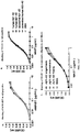
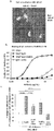
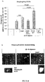
- MABT binds with high affinity to multiple forms of A ⁇ 1-42 and A ⁇ 1-40, protected against A ⁇ 1-42 oligomer-induced cytotoxicity, mediated uptake of neurotoxic A ⁇ by microglia both in vitro and in vivo.
- MABT showed reduced activation of the stress-activated p38 mitogen-activated protein kinase (p38MAPK) in microglia, and induced less release of pro-inflammatory mediators.
- p38MAPK stress-activated p38 mitogen-activated protein kinase
- the present invention is also based in part on the unexpected role of p38MAP kinase activation in microglia cells for anti-A ⁇ antibody-mediated neuroprotection in AD.
- p38MAP kinase activity is generally thought to be pro-inflammatory and thus would have been thought to contribute to the pathogenic inflammatory state of amyloidosis, such as AD.
- intermediate p38MAP kinase activation in microglia cells contributes to anti-A ⁇ antibody-mediated neuroprotection without generation of a pathogenic inflammatory state.
- compositions for the safe treatment and/or prevention of an amyloidosis including but not limited to Alzheimer's Disease.
- Amyloidosis includes, but is not limited to, neurological disorders such as Alzheimer's Disease (AD), Lewy body dementia, Down's syndrome, hereditary cerebral hemorrhage with amyloidosis (Dutch type), the Guam Parkinson-Dementia complex, as well as other diseases which are based on or associated with amyloid-like proteins such as progressive supranuclear palsy, multiple sclerosis.
- Creutzfeld Jacob disease Parkinson's disease, HIV-related dementia, ALS (amyotropic lateral sclerosis), Adult Onset Diabetes, senile cardiac amyloidosis, endocrine tumors, and others, including ocular disorders such as macular degeneration, cortical visual deficits, glaucoma, optic nerve drusen, optic neuropathy, optic neuritis, cataract, ocular amyloidosis and lattice dystrophy.
- anti-A ⁇ antibodies with effector regions that have been selected or modified to trigger an intermediate activation of p38MAP kinase in microglia cells.
- methods for the treatment and prevention of an amyloidosis including but not limited to Alzheimer's Disease, wherein the dose and/or administration regimen are selected such that p38MAP kinase is activated at an intermediate level in microglia cells.
- a safe and functional anti-A ⁇ antibody has the effector region of an IgG4 antibody.
- a safe and functional anti-A ⁇ antibody has the CH2 region of an IgG4 antibody.
- an intermediate level of p38 MAP kinase activation is a level above the level of p38 MAP kinase activation by toxic beta-amyloid oligomers alone but less than the level of p38 MAP kinase activation by an IgG1 anti-A ⁇ antibody in conjunction with the toxic beta-amyloid oligomers.
- the effector region of the anti-A ⁇ antibody is modified such that its effector function is reduced.
- the modification can be any genetic alteration resulting in an amino acid substitution and/or a deletion.
- method for improving the safety of a non-IgG4 anti-A ⁇ antibody comprising replacing the constant region of said non-IgG4 anti-A ⁇ antibody with a constant region derived from an IgG4 antibody.
- a method is provided for improving the safety of a non-IgG4 anti-A ⁇ antibody comprising replacing the constant region of said non-IgG4 antibody with a constant region derived from an non IgG1 antibody.
- the method for improving the safety of a non-IgG4 anti-beta-amyloid antibody is for improving the safety of an IgG1 anti-A ⁇ antibody.
- microglia cells are incubated with toxic beta-amyloid oligomers and the test anti-A ⁇ antibody.
- Anti-A ⁇ antibody-mediated uptake of beta amyloid into the microglia cells demonstrates functionality of the antibody, e.g ., in mediating the clearance of beta amyloid.
- Anti-A ⁇ antibody-mediated p38 MAP kinase activation at intermediate levels in microglia cells indicates both the antibody is functional and safe.
- an intermediate level of p38 MAP kinase activation is a level above the level of p38 MAP kinase activation by toxic beta-amyloid oligomers alone but less than level of p38 MAP kinase activation by an IgG1 anti-A ⁇ antibody having the level of effector function of the wild type (i.e., unmodified IgG1 constant region).
- This cell culture-based assay system can be used to test anti-A ⁇ antibodies for their ability to protect neurons from the neurotoxic effects of A ⁇ . Further, this cell culture-based assay system can be used to test anti-A ⁇ antibodies for their ability to trigger p38 MAP kinase activation at intermediate levels.
- the cell culture-based assay system further includes neurons.
- the survival rate of the neurons upon co-incubation with toxic beta-amyloid oligomers, the test anti-A ⁇ antibody, and microglia cells demonstrates the functionality of the test anti-A ⁇ antibody.
- the cell culture-based assay system is a primary cortical cell culture.
- the primary cortical cell culture is incubated with toxic beta-amyloid oligomers and the test anti-A ⁇ antibody.
- Anti-A ⁇ antibody-mediated uptake of beta amyloid into the microglia cells indicates functionality of the antibody, e.g ., in mediating the clearance of beta amyloid.
- Anti-A ⁇ antibody-mediated p38 MAP kinase activation at intermediate levels in microglia cells demonstrates both the functionality and safety of the antibody.
- variable region of a non-IgG4 humanized antibody which binds to A ⁇ is combined with the constant region of a human IgG4 antibody.
- variable region of an IgG1 humanized antibody which binds to A ⁇ is combined with the constant region of a human IgG4 antibody.
- the CH2 domain of a non-IgG4 humanized antibody which binds to A ⁇ is replaced with the CH2 domain of a human IgG4 antibody.
- the CH2 domain of an IgG1 humanized antibody which binds to A ⁇ is replaced with the CH2 domain of a human IgG4 antibody.
- the constant region of the constant region is derived from an IgG1 antiby wherein the constant region of the IgG1 antibody is modified such that the modified constant region has a reduced or eliminated effector function.
- methods are provided for the treatment and/or prevention of an amyloidosis, including but not limited to Alzheimer's Disease, wherein an IgG1 anti-A ⁇ antibody is administered in combination with an anti-inflammatory agent such that p38 MAP kinase is activated at an intermediate level in microglia cells.
- intermediate levels of p38 MAP kinase activation are levels above the levels of p38 MAP kinase activation by toxic beta-amyloid oligomers alone but less than levels of p38 MAP kinase activation by an IgG1 anti-A ⁇ antibody having normal levels of effector function in the presence of oligomers alone without the anti-inflammatory agent.
- polypeptide As used herein, are interchangeable and are defined to mean a biomolecule composed of amino acids linked by a peptide bond.
- amyloidosis refers to a group of diseases and disorders caused by or associated with amyloid or amyloid-like proteins and includes, but is not limited to, diseases and disorders caused by the presence or activity of amyloid-like proteins in monomeric, fibril, or polymeric state, or any combination of the three, including by amyloid plaques.
- Such diseases include, but are not limited it, secondary amyloidosis and age-related amyloidosis such as diseases including, but not limited to, neurological disorders such as Alzheimer's Disease (AD), diseases or conditions characterized by a loss of cognitive memory capacity such as, for example, mild cognitive impairment (MCI), Lewy body dementia, Down's syndrome, hereditary cerebral hemorrhage with amyloidosis (Dutch type), the Guam Parkinson-Dementia complex and other diseases which are based on or associated with amyloid-like proteins such as progressive supranuclear palsy, multiple sclerosis, Creutzfeld Jacob disease, Parkinson's disease, HIV-related dementia, ALS (amyotropic lateral sclerosis), inclusion-body myositis (IBM), adult onset diabetes, endocrine tumor and senile cardiac amyloidosis; and various eye diseases including macular degeneration, drusen-related optic neuropathy, and cataract due to beta-amyloid deposition.
- MCI mild cognitive impairment
- detecting or “detected” as used herein mean using known techniques for detection of biologic molecules such as immunochemical or histological methods and refer to qualitatively or quantitatively determining the presence or concentration of the biomolecule under investigation.
- Amyloid ⁇ is an art-recognized term and refers to amyloid ⁇ proteins and peptides, as well as modifications, fragments and any functional equivalents thereof, which may be produced by proteolytic cleavage of amyloid precursor protein (APP), and includethose fragments of APP which are involved in or associated with the amyloid pathologies including, but not limited to, A ⁇ 1-38, A ⁇ 1-39, A ⁇ 1-40, A ⁇ 1-41 A ⁇ 1-42 and A ⁇ 1-43.
- APP amyloid precursor protein
- amyloid ⁇ peptides as mentioned above are well known to one of ordinary skill in the art and methods of producing said peptides or of extracting them from brain and other tissues are described, for example, in Glenner and Wong, Biochem Biophys Res Comm129, 885-890 (1984 ). Moreover, amyloid ⁇ peptides are also commercially available in various forms.
- isolated means a biological molecule free from at least some of the components with which it naturally occurs.
- antibody or “antibodies” as used herein are art-recognized terms and are understood to refer to molecules or active fragments of molecules that bind to known antigens, and are used interchangeably with the terms “immunoglobulin” or “immunoglobulin molecules” and immunologically active portions of immunoglobulin molecules, i.e., portions of an immunoglobulin that contain a binding site that specifically binds an antigen.
- An immunoglobulin is a protein comprising one or more polypeptides substantially encoded by the immunoglobulin kappa and lambda, alpha, gamma, delta, epsilon and mu constant region genes, as well as myriad immunoglobulin variable region genes.
- Light chains are classified as either kappa or lambda.
- Heavy chains are classified as gamma, mu, alpha, delta, or epsilon, which in turn define the immunoglobulin classes, IgG, IgM, IgA, IgD and IgE, respectively.
- subclasses of the heavy chain are known.
- IgG heavy chains in humans can be any of the IgG1, IgG2, IgG3 and IgG4 subclasses.
- a typical immunoglobulin structural unit is known to comprise a tetramer.
- Each tetramer is composed of two identical pairs of polypeptide chains, each pair having one "light” (about 25 kD) and one "heavy” chain (about 50-70 kD).
- the N-terminus of each chain defines a variable region of about 100 to 110 or more amino acids primarily responsible for antigen recognition.
- the terms variable light chain (VL) and variable heavy chain (VH) refer to these portions of the light and heavy chains, respectively.
- Antibodies exist as full length intact antibodies or as a number of well-characterized fragments produced by digestion with various peptidases or chemicals.
- pepsin digests an antibody below the disulfide linkages in the hinge region to produce F(ab') 2 , a dimer of Fab which itself is a light chain joined to VH-CH1 by a disulfide bond.
- the F(ab') 2 may be reduced under mild conditions to break the disulfide linkage in the hinge region thereby converting the F(ab') 2 dimer into an Fab' monomer.
- the Fab' monomer is essentially a Fab fragment with part of the hinge region (see, Fundamental Immunology, W. E. Paul, ed., Raven Press, N.Y.
- antibody fragments are defined in terms of the digestion of an intact antibody, one of ordinary skill in the art will appreciate that any of a variety of antibody fragments may be synthesized de novo either chemically or by utilizing recombinant DNA methodology.
- the term antibody as used herein also includes antibody fragments either produced by the modification of whole antibodies or synthesized de novo or antibodies and fragments obtained by using recombinant DNA methodologies.
- antibodies includes monoclonal antibodies, polyclonal antibodies, chimeric, single chain, bispecific, simianized, human and humanized antibodies as well as active fragments thereof.
- active fragments of molecules that bind to known antigens include separated light and heavy chains, Fab, Fab/c, Fv, Fab', and F(ab') 2 fragments, including the products of an Fab immunoglobulin expression library and epitope-binding fragments of any of the antibodies and fragments mentioned above.
- active fragments can be derived by a number of techniques. For example, monoclonal antibodies can be cleaved with an enzyme, such as pepsin, and subjected to HPLC gel filtration.
- Recombinantly made antibodies may be conventional full length antibodies, active antibody fragments known from proteolytic digestion, unique active antibody fragments such as Fv or single chain Fv (scFv), domain deleted antibodies, and the like.
- An Fv antibody is about 50 Kd in size and comprises the variable regions of the light and heavy chain.
- a single chain Fv (“scFv”) polypeptide is a covalently linked VH::VL heterodimer which may be expressed from a nucleic acid including VH- and VL-encoding sequences either joined directly or joined by a peptide-encoding linker. See Huston, et al. (1988) Proc. Nat. Acad. Sci. USA, 85:5879-5883 .
- the combining site refers to the part of an antibody molecule that participates in antigen binding.
- the antigen binding site is formed by amino acid residues of the N-terminal variable ("V") regions of the heavy (“H") and light (“L”) chains.
- V N-terminal variable
- the antibody variable regions comprise three highly divergent stretches referred to as “hypervariable regions” or “complementarity determining regions” (CDRs) which are interposed between more conserved flanking stretches known as “framework regions” (FRs).
- the three hypervariable regions of a light chain (LCDR1, LCDR2, and LCDR3) and the three hypervariable regions of a heavy chain (HCDR1, HCDR2 and HCDR3) are disposed relative to each other in three dimensional space to form an antigen binding surface or pocket.
- the antibody combining site therefore represents the amino acids that make up the CDRs of an antibody and any framework residues that make up the binding site pocket.
- antibody CDRs may be identified as the hypervariable regions originally defined by Kabat et al. (see, " Sequences of Proteins of Immunological Interest,” E. Kabat et al., U.S. Department of Health and Human Services ; Johnson, G and Wu, TT (2001) Kabat Database and its applications: future directions. Nucleic Acids Research, 29: 205-206; http://immuno.bme.nwa.edu ).
- the positions of the CDRs may also be identified as the structural loop structures originally described by Chothia and others (see Chothia and Lesk, J. Mol. Biol.
- Chimeric antibodies are those in which one or more regions of the antibody are from an antibody derived from a first species and one or more regions of the antibody are from an antibody derived from a second, different species.
- a chimeric antibody is one which includes regions from a primate immunoglobulin.
- a chimeric antibody for human clinical use is typically understood to have variable regions from a non-human animal, e.g. a rodent, with the constant regions from a human antibody.
- a humanized antibody uses CDRs from the non-human antibody with most or all of the variable framework regions from and all the constant regions from a human antibody.
- a human chimeric antibody is typically understood to have the variable regions from a rodent antibody.
- a typical human chimeric antibody has human heavy constant regions and human light chain constant regions with the variable regions of both the heavy and light chains derived from a rodent antibody.
- a chimeric antibody may include some changes to a native amino acid sequence of the human constant regions and the native rodent variable region sequence.
- Chimeric and humanized antibodies may be prepared by methods well known in the art including CDR grafting approaches (see, e.g., U.S. Patent Nos. 5,843,708 ; 6,180,370 ; 5,693,762 ; 5,585,089 ; 5,530,101 ), chain shuffling strategies (see e.g., U.S. Patent No. 5,565,332 ; Rader et al., Proc. Natl. Acad. Sci. USA (1998) 95:8910-8915 ), molecular modeling strategies ( U.S. Patent No. 5,639,641 ), and the like.
- a "humanized antibody” as used herein in the case of a two or greater chain antibody is one where at least one chain is humanized.
- a humanized antibody chain has a variable region where one or more of the framework regions are human.
- a humanized antibody which is a single chain is one where the chain has a variable region where one or more of the framework regions are human.
- the non-human portions of the variable region of the humanized antibody chain or fragment thereof is derived from a non-human source, particularly a non-human antibody, typically of rodent origin.
- the non-human contribution to the humanized antibody is typically provided in the form of at least one CDR region which is interspersed among framework regions derived from one (or more) human immunoglobulin(s).
- framework support residues may be altered to preserve binding affinity.
- a “humanized antibody” may further comprise constant regions (e.g ., at least one constant region or portion thereof, in the case of a light chain, and in some embodiments three constant regions in the case of a heavy chain).
- the constant regions of a humanized antibody typically are human in origin. Methods to obtain humanized antibodies are well known to those of ordinary skill in the art. (see, e.g., Queen et al., Proc. Natl Acad Sci USA, 86:10029-10032 (1989 ), Hodgson et al., Bio/Technology, 9:421 (1991 )).
- a humanized antibody may also be obtained by a novel genetic engineering approach that enables production of affinity-matured human-like polyclonal antibodies in large animals such as, for example, rabbits and mice. See, e.g. U.S. Pat No. 6,632,976 .
- constant region or, abbreviated, "CR” as used herein refers to constant regions genes of the immunoglobulin.
- the constant region genes encode the portion of the antibody molecule which confers effector functions.
- Chimeric human antibodies and humanized antibodies, typically non-human ( e.g ., murine), constant regions are substituted by human constant regions.
- the constant regions of the subject chimeric or humanized antibodies are typically derived from human immunoglobulins.
- the heavy chain constant region can be selected from any of the five isotypes: alpha, delta, epsilon, gamma or mu.
- heavy chains of various subclasses are responsible for different effector functions and thus, by choosing the desired heavy chain constant region, antibodies with desired effector function can be produced.
- Constant regions that may be used are gamma 1 (IgG1), particularly an Fc region of the gamma 1 (IgG1) isotype, gamma 3 (IgG3) and especially gamma 4 (IgG4).
- the light chain constant region can be of the kappa or lambda type, preferably of the kappa type.
- the light chain constant region is the human kappa constant chain ( Heiter et al. (1980) Cell 22:197-207 ) and the heavy constant chain is the human IgG4 constant chain.
- Monoclonal antibody is also well recognized in the art and refers to an antibody that is the product of a single cloned antibody producing cell. Monoclonal antibodies are typically made by fusing a normally short-lived, antibody-producing B cell to a fast-growing cell, such as a cancer cell (sometimes referred to as an "immortal" cell). The resulting hybrid cell, or hybridoma, multiplies rapidly, creating a clone that produces the antibody.
- monoclonal antibody is also to be understood to comprise antibodies that are produced by a mother clone which has not yet reached full monoclonality.
- the antibody may be an immunoglobulin or antibody, which is understood to have each of its binding sites identical (if multivalent) or, in the alternative, may be a bispecific antibody or a multispecific antibody.
- a “bispecific” or “bifunctional antibody” is an artificial hybrid antibody having two different heavy/light chain pairs and two different binding sites.
- Bispecific antibodies can be produced by a variety of methods including fusion of hybridomas or linking of Fab' fragments. See, e.g., Songsivilai & Lachmann, Clin. Exp. Immunol. 79:315-321 (1990 ); Kostelny et al., J. Immunol. 148, 1547-1553 (1992 ).
- a “multispecific” or “multifunctional antibody” is an artificial hybrid antibody having more than two different heavy/light chain pairs and more than two different binding sites. Multispecific antibodies can be produced using the same variety of methods as bispecific antibodies.
- fragment refers to a part or portion of an antibody or antibody chain comprising fewer amino acid residues than an intact or complete antibody or antibody chain. Fragments can be obtained via chemical or enzymatic treatment of an intact or complete antibody or antibody chain. Fragments can also be obtained by recombinant means. Exemplary fragments include Fab, Fab', F(ab') 2 , Fabc and/or Fv fragments.
- antigen-binding fragment refers to a polypeptide fragment of an immunoglobulin or antibody that binds antigen or competes with intact antibody (i.e., with the intact antibody from which they were derived) for antigen binding (i.e., specific binding).
- Binding fragments can be produced by recombinant DNA techniques, or by enzymatic or chemical cleavage of intact immunoglobulins. Binding fragments include Fab, Fab', F(ab') 2 , Fabc, Fv, single chains, and single-chain antibodies.
- “Fragment” also refers to a peptide or polypeptide comprising an amino acid sequence of at least 5 contiguous amino acid residues, at least 10 contiguous amino acid residues, at least 15 contiguous amino acid residues, at least 20 contiguous amino acid residues, at least 25 contiguous amino acid residues, at least 40 contiguous amino acid residues, at least 50 contiguous amino acid residues, at least 60 contiguous amino residues, at least 70 contiguous amino acid residues, at least 80 contiguous amino acid residues, at least 90 contiguous amino acid residues, at least 100 contiguous amino acid residues, at least 125 contiguous amino acid residues, at least 150 contiguous amino acid residues, at least 175 contiguous amino acid residues, at least 200 contiguous amino acid residues, or at least 250 contiguous amino acid residues of the amino acid sequence of another polypeptide.
- a fragment of a polypeptide retains at least one function of the polypeptide.
- antigen refers to an entity or fragment thereof which can bind to an antibody.
- An immunogen refers to an antigen which can elicit an immune response in an organism, particularly an animal, more particularly a mammal including a human.
- antigen includes regions known as antigenic determinants or epitopes which refers to a portion of the antigen (which are contacted or which play a significant role in supporting a contact reside in the antigen) responsible for antigenicity.
- soluble means the ability to partially or completely dissolve in an aqueous solution.
- immunogen refers to substances which elicit the production of antibodies, T-cells and other reactive immune cells directed against an antigen of the immunogen.
- immunogenicity refers to a measure of the ability of an antigen to elicit an immune response (humoral or cellular) when administered to a recipient.
- An immune response occurs when an individual produces sufficient antibodies, T-cells and other reactive immune cells against administered immunogenic compositions of the present invention to moderate or alleviate the disorder to be treated.
- a humanized antibody having "reduced immunogenicity” refers to a humanized antibody exhibiting reduced immunogenicity relative to the parent antibody, e.g., the murine antibody.
- a humanized antibody "substantially retaining the binding properties of the parent antibody” refers to a humanized antibody which retains the ability to specifically bind the antigen recognized by the parent antibody used to produce such humanized antibody.
- the humanized antibody will exhibit the same or substantially the same antigen-binding affinity and avidity as the parent antibody.
- the affinity of the antibody will not be less than 10% of the parent antibody affinity, not less than about 30% of the parent antibody affinity, or not less than 50% of the parent antibody affinity.
- Methods for assaying antigen-binding affinity are well known in the art and include half-maximal binding assays, competition assays, and Scatchard analysis. Suitable antigen binding assays are described in this application.
- a “conservative change” refers to alterations that are substantially conformationally or antigenically neutral, producing minimal changes in the tertiary structure of the mutant polypeptides, or producing minimal changes in the antigenic determinants of the mutant polypeptides, respectively, as compared to the native protein.
- a conservative change means an amino acid substitution that does not render the antibody incapable of binding to the subject receptor.
- Factors to be considered that affect the probability of maintaining conformational and antigenic neutrality include, but are not limited to: (a) substitution of hydrophobic amino acids is less likely to affect antigenicity because hydrophobic residues are more likely to be located in a protein's interior; (b) substitution of physiochemically similar amino acids is less likely to affect conformation because the substituted amino acid structurally mimics the native amino acid; and (c) alteration of evolutionarily conserved sequences is likely to adversely affect conformation as such conservation suggests that the amino acid sequences may have functional importance.
- terapéuticaally functional amount refers to the amount of antibody which, when administered to a human or animal, is sufficient to result in a therapeutic effect in said human or animal.
- the functional amount is readily determined by one of ordinary skill in the art following routine procedures.
- the terms “treat,” “prevent,” “preventing,” and “prevention” refer to the prevention of the recurrence or onset of one or more symptoms of a disorder in a subject resulting from the administration of a prophylactic or therapeutic agent.
- safe and functional amount in the context of an anti-beta amyloid antibody refers to that amount of the anti-beta amyloid antibody that, when administered to a patient with Alzheimer's disease, reduces or prevents the formation of new amyloid plaques in the patient, reduces the amyloid plaque load in the patient, and / or reduces or prevents the deterioration of, or improves the cognitive abilities of the patient and wherein no side effects, such as such as inflammatory side effects, e.g., meningitis and meningoencephalitis, and fluid build up in the brain (cerebral edema) are observed or wherein any side effects are not so severe that the treatment of the patient has to be interrupted.
- side effects such as such as inflammatory side effects, e.g., meningitis and meningoencephalitis, and fluid build up in the brain (cerebral edema) are observed or wherein any side effects are not so severe that the treatment of the patient has to be interrupted.
- the formation of new amyloid plaques is reduced by at least 10%, 20%, 30%, 40%, 50%, 60%, 70%, 80%, 90%, 95%, 98%, 99%, or 100% relative to an untreated control.
- the amyloid plaque load is reduced by at least 10%, 20%, 30%, 40%, 50%, 60%, 70%, 80%, 90%, 95%, 98%, 99%, or 100% relative to an untreated control.
- the deterioration of the cognitive abilities of the patient reduced by at least 10%, 20%, 30%, 40%, 50%, 60%, 70%, 80%, 90%, 95%, 98%, 99%, or 100% relative to an untreated control.
- the cognitive abilities of the patient are improved by at least 10%, 20%, 30%, 40%, 50%, 60%, 70%, 80%, 90%, 95%, 98%, 99%, or 100% relative to an untreated control.
- non-IgG1 antibody refers to an antibody with any constant region of one of the following isotypes IgA, IgD, IgE, IgG and IgM except that the non-IgGI antibody does not have an IgG1 constant region that retains its wild type effector function.
- the non-IgG1 antibody is an IgG4 antibody.
- the non-IgG1 antibody has a constant region derived from an IgG1 antibody that has been mutated such that the effector function of the resulting antibody is reduced or eliminated relative to the wild type IgG1 antibody.
- intermediate levels in the context of p38 MAP kinase activation refers to levels of activation of p38 MAP kinase above the level of activation of p38 MAP kinase in the absence of a humanized non-IgG1 anti-beta amyloid antibody but below the level of p38 MAP kinase activation in the presence of the same concentration of an IgG1 anti-beta amyloid antibody which binds to beta amyloid with the same Kd as the humanized non-IgG1 anti-beta amyloid antibody.
- cell-based assay systems for testing anti-amyloid beta antibodies and methods for monitoring and adjusting treatment of a patient with anti-amyloid beta antibodies. Also described below are cell-based assay systems for testing the safety or efficacy of a neuroprotective agent and methods for monitoring and adjusting treatment of a patient with a neuroprotective agent. Further described below are safe and functional antibodies and methods for using such safe and functional antibodies for the treatment of Alzheimer's disease. Pharmaceutial preparations and modes of administration are also described.
- the cells that are affected by the amyloidosis (“target cells”), the amyloid protein in its pathological form, and immune effector cells (e.g., natural killer cells, macrophages, such as microglia cells, neutrophils, and mast cells) are incubated in the presence and absence of the test antibody.
- immune effector cells e.g., natural killer cells, macrophages, such as microglia cells, neutrophils, and mast cells
- Parameters that can be measured to test the safety and functionality of the antibody include survival rate of the target cells, internalization of the amyloid protein into the immune effector cells, and activation of the p38 MAP kinase pathway in the immune effector cells.
- a safe and functional antibody results in maximal internalization and intermediate activation of the p38 MAP kinase pathway.
- the amyloidosis is Alzheimer's disease
- the amyloid protein is beta amyloid
- the immune effector cells are microglia cells
- the target cells are neurons.
- the cells are obtained from existing cell lines to create a mixed culture.
- the mixed cell culture is a primary cortical culture ( Meberg & Miller 2003, Methods Cell Biol 71:111-127 ).
- the mixed cell culture is a primary cortical culture from a rat, mouse, or chimpanzee.
- the primary cortical culture is obtained by cortical biopsy or spinal biopsy from a human patient.
- a mixed cell culture that includes neurons and microglia cells is incubated with beta amyloid protein.
- the beta amyloid protein is provided as beta amyloid oligomer.
- the following parameters can be measured in this assay system: (1) the neuronal survival rate can be determined, e.g ., by metabolic turnover, which metabolic turnover can be determined, e.g ., by mitochondrial oxidation of 3-[4,5-dimethylthiazol-2-yl]-2,5-diphenyltetrazolium bromide (MTT) or ATP release: (2) beta-amyloid oligomer internalization into microglia can be determined, e.g ., by immunocytochemistry against beta amyloid protein or tagging and direct measurement and/or visualization of the beta amyloid protein; and (3) the activation of the p38 MAPK pathway can be determined, e.g ., by ELISA against phosphorylatedp38MAPK ("Phospho-p38").
- the neuronal survival rate in the presence of beta amyloid and a safe and functional antibody is increased by at least 10%, 20%, 30%, 40%, 50%, 75%, 100%, 125%, 150%, 175%, 200%, 225%, 250%, 275%, 300%, 325%, 350%, 375%, 400%, 425%, 450%, 475%, or at least 500% as compared to the neuronal survival rate in the absence of the safe and functional antibody.
- the internalization of beta amyloid into microglia in the presence of a safe and functional antibody is increased by at least 10%, 20%, 30%, 40%, 50%, 75%, 100%, 125%, 150%, 175%, 200%, 225%, 250%, 275%, 300%, 325%, 350%, 375%, 400%, 425%, 450%, 475%, or at least 500% as compared to the internalization rate in the absence of the safe and functional antibody.
- the p38 MAP kinase activation in microglia in the presence of beta amyloid and a safe and functional antibody is increased by between 5%-15%; 10%-20%; 15%-25%; 20%-30%; 35%-45%; 40%-50%; or 45%-55% as compared to p38 MAP kinase activation in the presence of beta amyloid but the absence of a safe and functional antibody.
- the p38 MAP kinase activation in microglia in the presence of beta amyloid and a safe and functional antibody is increased by between 10%-30% as compared to p38 MAP kinase activation in the presence of beta amyloid but the absence of a safe and functional antibody.
- the p38 MAP kinase activation in microglia in the presence of beta amyloid and a safe and functional antibody is increased by 5%-15%; 10%-20%; 15%-25%; 20%-30%; 35%-45%; 40%-50%; or 45%-55% less than p38 MAP kinase is activated in the presence of an IgG1 isotype anti beta amyloid antibody.
- a pathological form of beta amyloid e.g ., oligomeric beta amyloid, a test antibody, and microglia cells are incubated to determine safety and functionality of the antibody.
- the following parameters can be measured in this assay system: (1) beta amyloid oligomer internalization into microglia can be determined, e.g ., by immunocytochemistry against beta amyloid protein or tagging of the beta amyloid protein; and (2) the activation of the p38 MAPK pathway can be determined, e.g ., by ELISA against Phospho-p38.
- p38 MAP kinase activation any method known to one of ordinary skill in the art can be used.
- ELISA, Western blotting, or immunocytochemistry with an antibody that specifically binds to phosphorylated p38 MAP kinase is used to determine the levels of phosphorylated, i.e., activated, p38 MAP kinase.
- p38 MAP kinase activation is determined by immunocytochemistry using an antibody that specifically binds to p38 MAP kinase to determine the degree of nuclear localization of p38 MAP kinase. The higher the degree of nuclear localization of p38 MAP kinase, the higher the level of p38 MAP kinase activation.
- the activation of other components in the p38 MAP kinase signaling pathway can be measured.
- expression levels of a downstream target of p38 MAP kinase can be measured to determine the p38 MAP kinase activation in the patient.
- downstream targets include, but are not limited to, 90-kDa ribosomal S6 kinase (pp90rsk); (RSK) family: RSK1, RSK2, MNK1/2 and MSK1/2; and nuclear translation factors such as Elk-1, ATF2, STAT3 and CREB.
- Expression levels of downstream targets can be determined by any method known to one of ordinary skill in the art, such as, but not limited to, Northern blotting, Western blotting, or polymerase chain reaction.
- p38 MAP kinase activation is monitored in immune effector cells, such as microglia cells, of a patient who is being treated with a safe and functional antibody that specifically binds to an amyloid protein, such as beta amyloid.
- Immune effector cells can be obtained from a patient by any method known to one of ordinary skill in the art.
- microglia cells are obtained by cortical biopsy or spinal biopsy. Immune effector cells can be maintained in culture by any method known to one of ordinary skill in the art.
- p38 MAP kinase activation is monitored in immune effector cells, such as microglia cells, of a patient who is being treated with an agent, such as a neuroprotective agent.
- an agent such as a neuroprotective agent.
- the patient is being treated for an amyloidosis, such as Alzheimer's Disease.
- the patient is being treated with tacrine (COGNEX, Morris Plains, NJ), donepezil (ARICEPT, Tokyo, JP), rivastigmine (EXELON, East Hanover, NJ), galantamine (REMINYL, New Brunswick, NJ), or memantine (NAMENDA, New York, NY).
- the administration dosage and/or administration regimen is adjusted for the patient such that p38 MAP kinase activation in microglia cells is at intermediate levels, i.e., higher than in the absence of the antibody but lower than in the presence of an IgG1 antibody that specifically binds beta amyloid in the presence of an beta amyloid oligomer. If p38 MAP kinase activation is above intermediate levels, the dosage is reduced and/or the administration frequency is reduced. If p38 MAP kinase activation is below intermediate levels, the dosage is increased and/or the administration frequency is increased.
- a modulator of the p38 MAP kinase signaling pathway is co-administered with the safe and functional antibody to adjust p38 MAP kinase activation to intermediate levels. If p38 MAP kinase activation is above intermediate levels, an inhibitor of p38 MAP kinase signaling pathway can be co-administered. If p38 MAP kinase activation is below intermediate levels, an activator of p38 MAP kinase signaling pathway can be co-administered.
- Illustrative inhibitors of the p38 MAP kinase signaling pathway include 4-[4-(4-Fluorophenyl)-5-(4-pyridinyl)-1H-imidazol-2-yl] phenol ("SB 202190”); 4-[5-(4-Fluorophenyl)-2-[4-(methylsulfonyl)phenyl]-1H-imidazol-4-yl]pyridine (“SB 203580”); and trans-4-[4-(4-Fluorophenyl)-5-(2-methoxy-4-pyrimidinyl)-1H-imidazol-1-yl]cyclohexanol (“SB 239063”) and pharmaceutically suitable derivatives thereof.
- Illustrative activators of the p38 MAP kinase signaling pathway include Anisomycin, MKK, Rac, Cdc42 and PAK1, IL-1, IL-1-receptor, TNF, LPS, TRAF6, and TAB1/2.
- p38 MAP kinase intermediate activation levels are between 5%-15%; 10%-20%; 15%-25%; 20%-30%; 35%-45%; 40%-50%; or 45%-55% above p38 MAP kinase activation in microglia cells of the patient in the absence of the safe and functional antibody.
- p38 MAP kinase activation any method known to one of ordinary skill in the art can be used.
- ELISA, Western blotting, or immunocytochemistry with an antibody that specifically binds to phosphorylated p38 MAP kinase is used to determine the levels of phosphorylated, i.e ., activated, p38 MAP kinase.
- p38 MAP kinase activation is determined by immunocytochemistry using an antibody that specifically binds to p38 MAP kinase to determine the degree of nuclear localization of p38 MAP kinase. The higher the degree of nuclear localization of p38 MAP kinase, the higher the level of p38 MAP kinase activation.
- the activation of other components in the p38 MAP kinase signaling pathway can be measured.
- expression levels of a downstream target of p38 MAP kinase can be measured to determine the p38 MAP kinase activation in the patient.
- downstream targets include, but are not limited to, 90-kDa ribosomal S6 kinase (pp90rsk); (RSK) family: RSK1, RSK2, MNK1/2 and MSK1/2; and nuclear translation factors such as Elk-1, ATF2, STAT3 and CREB.
- Expression levels of downstream targets can be determined by any method known to one of ordinary skill in the art, such as, but not limited to, Northern blotting, Western blotting, or detection of gene transcripts by, .e.g, RT-PCR.
- microglia cells can be obtained from the patient.
- microglia cells can be obtained from the patient before treatment of the patient with the anti beta-amyloid antibody has begun.
- the microglia cells from the patient can be maintained in cell culture using standard art-known techniques.
- the microglia cells from the patient are incubated with the anti beta-amyloid antibody and beta-amyloid oligomers.
- the following parameters can be determined: binding of the antibody - beta amyloid complex to the microglia cells; internalization of the antibody - beta amyloid complex into the microglia cells; and / or p38 MAP kinase activation in the microglia cells.
- Controls can be performed by incubating the microglia cells from the patient with the anti beta-amyloid antibody alone (i.e ., without beta-amyloid oligomer); and / or with beta-amyloid oligomers alone ( i.e., without the antibody); and / or with an IgG1 anti beta-amyloid antibody in the presence of beta-amyloid oligomer.
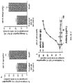
- an anti beta-amyloid antibody is safe and functional if p38 MAP kinase activation is between 5%-15%; 10%-20%; 15%-25%; 20%-30%; 35%-45%; 40%-50%; or 45%-55% above p38 MAP kinase activation in control microglia cells of the patient in the presence of beta-amyloid oligomer but the absence of the safe and functional antibody.
- an anti beta-amyloid antibody is safe and functional if beta-amyloid internalization into microglia cells is between 5%-15%; 10%-20%; 15%-25%; 20%-30%; 35%-45%; 40%-50%; or 45%-55% above or below that of monoclonal antibody MABT (see Section 6).
- the invention relates to, inter alia, humanized non-IgG1 anti beta-amyloid antibodies for use in a method for treating amyloidosis, as defined in the claims.
- the constant region of an antibody that specifically binds the amyloid protein of the amyloidosis to be treated can be modified or replaced to provide a safe and functional antibody.
- the constant region of the resulting antibody binds with intermediate activity to the Fc receptor on the surface of immune effector cells such as natural killer cells, macrophages, neutrophils, and mast cells.
- the resulting antibody activates the p38 MAPK pathway in the immune effector cell at intermediate levels, i.e ., above p38 MAP kinase activation in the absence of the antibody but below p38 MAP kinase activation in the presence of the same concentration of an IgG1 antibody with the same binding specificity.
- the variable regions outside the complementarity determining regions ("CDR") can also be modified to provide a safe and functional antibody. Also provided herein are methods for generating such an antibody and pharmaceutical compositions comprising such an antibody.
- Antigen binding specificity is provided by the complementarity determining regions ("CDR") that are embedded in the variable regions of an antibody's light chain and heavy chain, respectively.
- CDR complementarity determining regions
- Antibody constant regions and the framework regions that surround the CDRs that can be used for the construction of the safe and functional antibodies provided herein include those described in Section 5.3.1.
- CDRs or variable regions that can be used for the construction of the safe and functional antibodies can be derived from the antibodies set forth in Table 2.
- the CDRs of a known antibody that specifically binds human beta amyloid are combined with the constant region of a human IgG4 and the framework regions between the CDRs of the antibody are replaced with the framework regions of a human IgG, such as IgG1, IgG2, IgG3, or IgG4.
- the variable regions of a known humanized anti-beta amyloid antibody e.g ., Bapineuzumab or Solanezumab, are combined with the constant region of a human IgG4.
- Antibody constant regions for the generation of the safe and functional antibodies provided herein can be derived from any isotype, including IgA, IgD, IgE, IgG and IgM.
- antibody constant regions for the generation of the safe and functional antibodies provided herein can be derived from subtype IgA1, IgA2, IgG1, IgG2, IgG3, and IgG4.
- framework regions of antibody heavy chain regions for the generation of the safe and functional antibodies provided herein can be derived from any isotype, including IgA, IgD, IgE, IgG and IgM.
- antibody framework regions for the generation of the safe and functional antibodies provided herein can be derived from subtype IgA1, IgA2, IgG1, IgG2, IgG3, and IgG4.
- the constant region for the generation of a safe and functional antibody provided herein can be derived from isotype IgG. In an even more specific embodiment, the constant region for the generation of a safe and functional antibody provided herein can be derived from isotype IgG4.
- the framework regions for the generation of a safe and functional antibody provided herein can be derived from isotype IgG. In an even more specific embodiment, the framework regions for the generation of a safe and functional antibody provided herein can be derived from isotype IgG4.
- the constant region of the heavy chain of a safe and functional antibody is a chimeric constant region from different isotypes or subtypes.
- the constant region of the heavy chain is a chimera between that portion of the IgG4 heavy chain constant region that is responsible for the effector function of IgG4 and a different subtype of IgG.
- the constant region of the heavy chain is a chimera between the CH2 domain of a human IgG4 heavy chain constant region and a different subtype of IgG.
- the chimeric constant region has an intermediate binding activity to Fc ⁇ as described below.
- the chimeric constant region mediates the internalization into immune effector cells, such as microglia, such that p38 MAP kinase pathway activity is at an intermediate level as determined by a cell-based assay system described in Section 5.1.
- a constant region is optimized by introducing mutations. Mutations can be introduced into the constant region using recombinant DNA technology.
- the resulting antibodies promote internalization of beta amyloid into microglia cells at high levels while activation of the p38 MAP kinase pathway is at intermediate levels. Rate of internalization and p38 MAP kinase activation can be determined using the cell-based assay system described in Section 5.1.
- the antibody with mutated constant region has an intermediate binding activity to an Fc receptor as described below.
- a constant region is optimized by altering the glycosylation pattern.
- the glycosylation pattern can be altered by virtue of the expression system where the antibody is synthesized and / or mutating the amino acid that is being glycosylated ( e.g ., Asn297).
- the resulting antibodies promote internalization of beta amyloid into microglia cells at high levels while activation of the p38 MAP kinase pathway is at intermediate levels. Rate of internalization and p38 MAP kinase activation can be determined using the cell-based assay system described in Section 5.1.
- the antibody with constant region with altered glycosylation pattern has an intermediate binding activity to an Fc receptor as described below.
- the constant region has an intermediate binding activity to its Fc receptor.
- Fc receptor Set forth in Table 1 are illustrative Fc receptors and their respective principal antibody ligand. Binding activity between antibody ligand and Fc receptor can be determined using any method known to one of ordinary skill in the art. Exemplary methods of measuring binding affinity include but are not limited to ELISA assays and BIACORE analysis.
- the Fc receptor is an Fc receptor that mediates internalization of the antibody antigen complex into an immune effector cell.
- the Fc receptor is an Fc ⁇ receptor that is expressed on microglia cells.
- Table 1 Illustrative isotypes or subtypes of antibodies and their respective Fc receptor Receptor Name Principal Antibody Ligand Fc ⁇ RI (CD64) IgG1 and IgG3 Fc ⁇ RIIA (CDR32) IgG Fc ⁇ RIIB1 (CD32) IgG Fc ⁇ RIIB2 (CD32) IgG Fc ⁇ RIIIA (CD16a) IgG Fc ⁇ RIIIB (CD16b) IgG Fc ⁇ RI IgE Fc ⁇ RII (CD23) IgE Fc ⁇ RI (CD89) IgA Fc ⁇ RI/ ⁇ R IgA and IgM FcRn IgG
- the antibody constant region is of the isotype IgG and the Fc receptor is an Fc ⁇ receptor.
- the constant region for generation of a safe and functional antibody has a binding activity to the Fc ⁇ receptor that is between 10% and 30%; between 20% and 40%, between 30% and 50%, between 40% and 60%, between 50% and 70%, between 60% and 80%, or between 70% and 90% of the binding activity of IgG1 to the Fc ⁇ receptor.
- the constant region for the generation of a safe and functional antibody for the treatment of Alzheimer's disease is between 15% and 25%, or more specifically about 20%, of the binding activity of IgG1 to Fc ⁇ .
- the effector region of the anti-A ⁇ antibody is modified such that the effector function of the antibody is reduced or eliminated.
- the modification can be any genetic alteration resulting in an amino acid substituion and / or deletion.
- the constant region is a modified IgG1 constant region with reduced or eliminated effector function. Such modifications can be introduced as described, e.g., in international patent application publication no. WO 00/42072 published on July 20, 2000 .
- the ability of the antibody with the modified constant region to bind to its Fc receptor is reduced relative to the unmodified constant region.
- the binding between the constant region and its Fc receptor is not altered but effector functions such as cytotoxicity in the presence of effector cells, are reduced relative to the unmodified constant region.
- variable regions of an antibody known to bind specifically to beta amyloid and / or its pathological form(s) can be used for the generation of a safe and functional antibody.
- variable regions of a humanized antibody known to bind specifically to beta amyloid and / or its pathological form(s) can be used for the generation of a safe and functional antibody.
- the variable regions are obtained from a non-IgG4 humanized antibody.
- the variable regions are obtained from a non-IgG4 humanized antibody and an IgG4 constant region is used as constant region.
- CDRs of antibodies known to bind to an amyloid protein of interest can be used for the generation of a safe and functional antibody useful in the methods described herein.
- CDRs of an antibody known to bind specifically to beta amyloid and/or its pathological form(s) can be used for the generation of such a safe and functional antibody.
- Illustrative antibodies whose CDRs or variable regions can be used include, but are not limited to those set forth in Table 2.
- Table 2 Illustrative anti-beta amyloid antibodies Antibody Reference / Source mACI-01-Ab7 C2 WO 2007/068412 published June 21, 2007 mACI-01-Ab7 C2 WO 2008/060364 published May 22, 2008 mACI-02-Ab6 WO 2007/068412 published June 21, 2007 mACI-11-Ab9 WO 2007/068412 published June 21, 2007 mACI-12-Ab11 WO 2007/068412 published June 21, 2007 mACI-24-Ab4 WO 2007/068412 published June 21, 2007 ACI-24-Ab-3 WO 2008/156621 published December 24, 2008 ACI-11-Ab-9 WO 2008/156621 published December 24, 2008 ACI-12-Ab-11 WO 2008/156621 published December 24, 2008 20C2 WO 2007/050359 published May 3, 2007 8F5 WO 2007/064972 published June 7, 2007 8C5 WO 2007/
- 6G12 santa cruz biotechnology inc. 20-1 santa cruz biotechnology, inc. 2B9 santa cruz biotechnology, inc. 2C8 santa cruz biotechnology, inc. 6A6 santa cruz biotechnology, inc. B-4 santa cruz biotechnology, inc. DE2B4 santa cruz biotechnology, inc. LN27 santa cruz biotechnology, inc. NAB228 santa cruz biotechnology, inc. 1304.1 santa cruz biotechnology, inc. 5C3 santa cruz biotechnology, inc. BDI350 santa cruz biotechnology, inc.
- the CDRs of a safe and functional antibody are as follows: CDR1 of the heavy chain has the amino acid sequence of SEQ ID NO:1; CDR2 of the heavy chain has the amino acid sequence of SEQ ID NO:2; CDR3 of the heavy chain has the amino acid sequence of SEQ ID NO:3; CDR1 of the light chain has the amino acid sequence of SEQ ID NO:4; CDR2 of the light chain has the amino acid sequence of SEQ ID NO:5; and CDR3 of the light chain has the amino acid sequence of SEQ ID NO:6.
- the light chain variable region of a safe and functional antibody has an amino acid sequence that is at least 90%, 91%, 92%, 93%, 94%, 95%, 96%, 97%, 98%, or at least 99% identical to the amino acid sequence of SEQ ID NO:7. In a more specific embodiment, the light chain variable region of a safe and functional antibody has an amino acid sequence that is 100% identical to the amino acid sequence of SEQ ID NO:7.
- the heavy chain variable region of a safe and functional antibody has an amino acid sequence that is at least 90%, 91%, 92%, 93%, 94%, 95%, 96%, 97%, 98%, or at least 99% identical to the amino acid sequence of SEQ ID NO:10. In a more specific embodiment, the heavy chain variable region of a safe and functional antibody has an amino acid sequence that is 100% identical to the amino acid sequence of SEQ ID NO: 10.
- the light chain variable region of a safe and functional antibody has an amino acid sequence that is at least 90%, 91%, 92%, 93%, 94%, 95%, 96%, 97%, 98%, or at least 99% identical to the amino acid sequence of SEQ ID NO:7 and the heavy chain variable region of the safe and functional antibody has an amino acid sequence that is at least 90%, 91%, 92%, 93%, 94%, 95%, 96%, 97%, 98%, or at least 99% identical to the amino acid sequence of SEQ ID NO: 10.
- the light chain variable region of a safe and functional antibody has an amino acid sequence that is at 100% identical to the amino acid sequence of SEQ ID NO:7 and the heavy chain variable region of a safe and functional antibody has an amino acid sequence that is 100% identical to the amino acid sequence of SEQ ID NO:10.
- the CDRL2 sequence ("KVSNRFS” (SEQ ID NO:5)) of the mMABT antibody may be modified slightly without adversely affecting antibody activity. Conservative substitutions may be made through exchange of R for K at position 50 and S for N at position 53.
- the two alternative CDRL2 sequences are therefore "RVSNRFS” and "KVSSRFS", respectively. These are incorporated into the murine VK sequence with no other changes, as mMABT VK-R and mMABT VK-S, respectively.
- the CDRs of an antibody that binds specifically to an amyloid protein or its pathological form, such as beta amyloid are placed into an antibody with constant regions consistent with a safe and functional antibody as described herein (Section 5.3.1).
- the variable regions of an antibody that binds specifically to an amyloid protein or its pathological form, such as beta amyloid are combined with constant regions consistent with a safe and functional antibody as described herein (Section 5.3.1).
- the invention relates to a method for testing the safety of a humanized non-IgGl anti beta-amyloid antibody, as defined in the claims.
- Side effects observed during treatment of Alzheimer's disease with anti-beta amyloid antibodies include inflammatory side effects, such as meningitis and meningoencephalitis, and fluid build up in the brain (cerebral edema).
- Other side effects include adverse immune reaction, i . e ., an immune reaction by the patient against the administered anti-beta Amyloid antibody.
- One side effect that can be monitored in a patient that has received an anti-beta amyloid antibody is an antibody response to the anitbody. Any technique known to one of ordinary skill in the art to detect, monitor, or quantify the extent of such an adverse immune reaction can be used. Such an antibody response occurs when an antibody binds the anti-beta amyloid antibody.
- soluble antigens When soluble antigens combine with antibodies in the vascular compartment, they may form circulating immune complexes that arc trapped nonspecifically in the vascular beds of various organs, causing so-called immune complex diseases, such as serum sickness, vasculitis, nephritis systemic lupus erythermatosus with vasculitis or glomerulonephritis.
- Immune complex disease can he detected and/or monitored using any method known in the art.
- an immune complex test can be used to demonstrate circulating immune complexes in the blood, to estimate the severity of immune complex disease.
- An immune complex test can be performed by any method known to one of skill in the art.
- an immune complex test can be performed using any one or more of the methods described in U.S. Patent No. 4,141,965 .
- U.S. Patent No, 4,310,622 U.S. Patent No. 4,210,622 , U.S. Patent No. 4,331,649 , U.S. Patent No. 4,544,640 , U.S. Patent No. 4,753,893 , and U.S. Patent No. 5,888,834 .
- an immunoassay such as an enzyme-linked immunosorbent assay (ELISA)
- ELISA enzyme-linked immunosorbent assay
- edema in particular, cerebral edema, which may be assessed, e.g., by MRI scan, DCE-MRI scan, PET scan, and/or CT scan. Any technique known to one of ordinary skill in the art to detect, monitor, or quantify the extent of edema can be used. Edema may also be measured in an animal model of edema. In such methods, the edema volumes (hemisphere volume of the ipsilateral side hemisphere volume of contralateral one) are calculated.
- brain edema can be visualized as a hypodense or hyperintense lesion.
- Brain edema and other structures with a high water content, such as cerebrospinal fluid, are hyperintense on T2-weighted MRI.
- Fluid-attenuated inversion-recovery MR images provide additive information since brain edema is clearly visualized as a hyperintense lesion against an iso- or hyperintense background.
- meningitis One side effect that can be monitored in a patient that has received an an anti-beta amyloid antibody is meningitis. Any technique known to one of ordinary skill in the art to detect, monitor, or quantify the extent of minigitis can be used.
- a neurological examination may also be conducted, involving a series of tests designed to assess: motor and sensory function; nerve function; hearing and speech; vision; coordination and balance; mental status; and changes in mood or behavior.
- the function of the nervous system may be assessed through tests of strength and sensation, with the aid of items such as a tuning fork, small light, reflex hammer, and pins.
- Analyzing the cerebrospinal fluid that surrounds and protects the brain and spinal cord can detect acute and chronic inflammation.
- a spinal tap or lumbar puncture
- a small amount of cerebrospinal fluid is removed by a special needle that is inserted into the lower back.
- the skin is numbed with a local anesthetic prior to the sampling.
- the fluid which is completely clear in healthy people, is tested to detect the presence of bacteria or blood, as well as to measure glucose levels (a low glucose level is a sign of bacterial or fungal meningitis) and white blood cells (elevated white blood cell counts are also a sign of infection).
- the procedure is usually done in a hospital and takes about 45 minutes.
- Noninvasive imaging procedures are routinely used to reach a meningitis diagnosis.
- Such computer-assisted imaging can reveal signs of brain inflammation; internal bleeding or hemorrhage; and other brain abnormalities that may be associated with meningitis.
- Computed tomography also known as a CT scan, combines x-rays and computer technology to produce rapid, clear, two-dimensional images of bones, organs, and tissues.
- a contrast dye is injected into the bloodstream to highlight the different tissues in the brain and to detect inflammation of the meninges.
- Magnetic resonance imaging uses computer-generated radio waves and a strong magnet to produce detailed images of body structures, including tissues, organs, bones, and nerve.
- a contrast dye may be injected prior to the test to reveal more detail.
- Another imaging technique used to assist in meningitis diagnosis is ultrasound.
- Electroencephalography can identify abnormal brain waves associated with meningitis by monitoring electrical activity in the brain through the skill. EEG is used to help diagnose inflammation of the brain.
- Alzheimer's disease treatment can be assessed using one or more neuropsychological tests.
- exemplary neuropsychological tests are described below and include, without limitation, those established by The Consortium to Establish a Registry for Alzheimer's Disease ("CERAD”; see, e.g., Morris JC, Mohs RC, Rogers H, Fillenbaum G, Heyman A. Consortium to establish a registry for Alzheimer's disease (CERAD) clinical and neuropsychological assessment of Alzheimer's disease. Psychopharmacol Ball. 1988:24(4):641-52 .
- an Alzheimer's disease treatment described herein can be assessed using the Alzheimer Disease Assessment Scale-Cognitive test ("ADAS-Cog”; see. e.g., Rosen WG, Mohs RC, Davis KL. A new rating scale for Alzheimer's disease. Am J Psychiatry. 1984 Nov;141(11):1356-6-4 : Ihl R, Brinkmeyer J, Janner M, Kerdar MS. A comparison of ADAS and EEG in the discrimination of patients with dementia of the Alzheimer type from healthy controls. Neuropsychobiology. 2000 Jan;41(2):102-7 ; and Weyer G, Erzmaschine H, Kanowski S, Ihl R, Hadler D. Alzheimer's Disease Assessment Scale: reliability and validity in a multicenter clinical trial. Int Psychogeriatr. 1997 Jun;9(2):123-38 ).
- ADAS-Cog Alzheimer Disease Assessment Scale-Cognitive test
- an Alzheimer's disease treatment described herein can be assessed using the Behavioral Pathology in Alzheimer's Disease Rating Scale ("BEHAVE-AD”; see, e.g., Reisberg B, Borenstein J, Salob SP, Ferris SH, Franssen E, Georgotas A. Behavioral symptoms in Alzheimer's disease: phenomenology and treatment. J Clin Psychiatry. 1987 May;48 Suppl:9-15 ).
- BEHAVE-AD Behavioral Pathology in Alzheimer's Disease Rating Scale
- the functionality of an Alzheimer's disease treatment described herein can be assessed using the Blessed-Dementia Information-Memory-Concentration Test (see, e.g., Screen G, Tomlinson BE, Roth M. The association between quantitative measures of dementia and of senile change in the cerebral grey matter of elderly subjects. Br J Psychiatry. 1968 Jul; 114(512):797-811 ).
- an Alzheimer's disease treatment described herein can be assessed using the Cambridge Neuropsychological Test Automated Battery ("CANTAB"; see, e.g., Swainson R, Hodges JR, Galton CJ, Semple J, Michael A, Dunn BD, Iddon JL, Robbins TW, Sahakian BJ. Early detection and differential diagnosis of Alzheimer's disease and depression with neuropsychological tasks. Dement Geriatr Cogn Disord. 2001;12:265-280 ; Fray PJ, Robbins TW. CANTAB battery: proposed utility in neurotoxicology. Neurotoxicol Teratol.
- CANTAB Cambridge Neuropsychological Test Automated Battery
- an Alzheimer's disease treatment described herein can be assessed using the Clock Draw Test (see, e.g., Sunderland T, Hill JL, Mellow AM, Lawlor BA, Gundersheimer J, Newhouse PA, Grafman JH. Clock drawing in Alzheimer's disease. A novel measure of dementia severity. J Am Geriatr Soc. 1989 Aug;37(8):725-9 ; and Lee H, Swanwick GR, Coen RF, Lawlor BA. Use of the clock drawing task in the diagnosis of mild and very mild Alzheimer's disease. Int Psychogeriatr. 1996 Fall;8(3):469-76 ).
- Clock Draw Test see, e.g., Sunderland T, Hill JL, Mellow AM, Lawlor BA, Gundersheimer J, Newhouse PA, Grafman JH. Clock drawing in Alzheimer's disease. A novel measure of dementia severity. J Am Geriatr Soc. 1989 Aug;37(8):725-9 ; and Lee H, Swanwick GR, Coen RF, Lawlor BA.
- an Alzheimer's disease treatment described herein can be assessed using the Cornell Scale for Depression in Dementia ("CSDD"; Alexopoulos GS, Abrams RC, Young RC, Shamoian CA. Cornell Scale for Depression in Dementia. Biol Psychiatry. 1988 Feb 1;23(3):271-84 ).
- GDS Geriatric Depression Scale
- Raz WJ Roccaforte WH
- Wengel SP The short form of the Geriatric Depression Scale: a comparison with the 30-item form. J Geriatr Psychiatry Neurol. 1991 Jul-Sep;4(3): 173-8 ).
- MMSE Mini Mental State Exam
- MMSE Mini Mental State Exam
- MMSE a practical method for grading the cognitive state of patients for the clinician. J Psychiatr Res. 1975; 12:189-198 ; and Cockrell JR, and Folstein MF. Mini-Mental State Examination (MMSE). Psychopharm Bull. 1988;24(4):689-692 ).
- an Alzheimer's disease treatment described herein can be assessed using the Neuropsychiatric Inventory ("NPI"; Cummings JL, Mega M, Gray K, Rosenberg-Thompson S, Carusi DA, Gornbein J.
- NPI Neuropsychiatric Inventory
- the Neuropsychiatric Inventory comprehensive assessment of psychopathology in dementia. Neurology. 1994 Dec;44(12):2308-14 ; Cummings JL.
- the Neuropsychiatric Inventory assessing psychopathology in dementia patients. Neurology. 1997 May;48(5 Suppl 6):S10-6 ).
- an Alzheimer's disease treatment described herein can be assessed using the 7 Minute Screen (see, e.g., Solomon PR, Pendlebury WW. Recognition of Alzheimer's disease: the 7 Minute Screen. Fam Med. 1998 Apr;30(4):265-71 ; and Solomon PR, Hirschoff A, Kelly B, Relin M, Brush M, DeVeaux RD, Pendlebury WW. A 7 minute neurocognitive screening battery highly sensitive to Alzheimer's disease. Arch Neurol. 1998 Mar;55(3):349-55 ).
- the functionality of an Alzheimer's disease treatment described herein can be assessed using in vivo imaging in subjects.
- Imaging reagents can be administered by intravenous injection into the body of the patient, or directly into the brain by intracranial injection or by drilling a hole through the skull.
- the dosage of reagent should be within the same ranges as for treatment methods.
- the reagent is labeled, although in some methods, the primary reagent with affinity is unlabeled and a secondary labeling agent is used to bind to the primary reagent.
- the choice of label depends on the means of detection. For example, a fluorescent label is suitable for optical detection. Use of paramagnetic labels is suitable for tomographic detection without surgical intervention. Radioactive labels can also be detected using positron emission tomography (PET) or single hoton emission computed tomography (SPECT).
- PET positron emission tomography
- SPECT single hoton emission computed tomography
- base line values can represent the mean levels in a population of individuals that do not have Alzheimer's disease.
- base line values can represent previous levels determined in the same patient. For example, base line values can be determined in a patient before beginning treatment, and measured values thereafter compared with the base line values. A decrease in values relative to base line can signal a positive response to treatment.
- Anti- beta-amyloid antibodies are also useful to determine whether truncated forms of A ⁇ are present in cerebrospinal fluid or other body tissues or fluids. Presence of such forms at lessened levels in a patient relative to the base line can signal a positive response to treatment.
- an Alzheimer's disease treatment described herein can be assessed by using radioactively-labeled tracers as probes with Positron Emission Tomography (PET) to monitor the pathophysiology of Alzheimer's disease (see, e.g., N ⁇ gren et al. 2009, European Journal of Nuclear Medicine and Molecular Imaging, Radiopharmaceuticals for positron emission tomography investigations of Alzheimer's disease, published online December 22, 2009 .)
- PET Positron Emission Tomography
- Carbon-11-labeled Pittsburgh compound B can be used as a radiotracer with PET to image the amyloid plaque burden (see, e.g., Rinne 2010, Lancet Neurology 9:363-372 ).
- tracers for PET imaging of amyloid plaque burden include 18 F-labeled Stilbenes and Styrylpyridines (see, e.g., Kung et al. 2010, Journal of Medicinal Chemistry 53:933-941 ); the 18 F-labeled benzothiazole (BTA)-derivative 3'- 18 FFPIB; an "C- and an 18 F-labeled version of Congo-Red derivative SB-13; and the amino-naphthylderivative 18 FFDDNP (see, e.g., Henriksen et al. 2008, European Journal of Nuclear Medicine and Molecular Imaging Suppl 1:S75-81 ).
- an Alzheimer's disease treatment described herein can be assessed by using Magnetic resonance imaging (MRI).
- MRI Magnetic resonance imaging
- MRI can be used to assess volume changes in the central nervous system (see, e.g., Fagan et al. 2009, Annals of Neurology 65:176-183 ) and to quantify vasogenic edema (see, e.g., Black et al. 2010, Alzheimer Disease and Associated Disorders 24:198-203 ).
- an Alzheimer's disease treatment described herein can be tested in vivo using an model of Alzheimer's disease.
- Such animal models Alzheimer's disease are well-known in the art and include, without limitation, models using mice rats, and primates.
- An exemplary murine model of Alzheimer's disease includes TgCRND8 mice (see, e.g., Chishti, M. A. et al., Early-onset amyloid deposition and cognitive deficits in transgenic mice expressing a double mutant form of amyloid precursor protein 695. J. Biol Chem 276, 21562-21570 (2001 ); Janus, C. et al., A ⁇ peptide immunization reduces behavioural impairment and plaques in a model of Alzheimer's disease. Nature 408, 979-982 (2000 )).
- TgCRND8 mice express a human amyloid precursor protein transgene (APP695) bearing two missense mutations known to cause Alzheimer's disease in humans (KM670/671NL and V717F).
- mice display progressive spatial learning deficits that are accompanied both by rising cerebral A ⁇ levels and by increasing numbers of cerebral extracellular amyloid plaques.
- levels of A ⁇ and the morphology, density and distribution of the amyloid plaques in the brain of TgCRND8 mice are similar to those seen in the brains of humans with well-established Alzheimer's disease.
- the biochemical, behavioral and neuropathological features of the mouse model are accompanied by accelerated mortality.
- Other exemplary murine models of Alzheimer's disease are known in the art and such mice are commercially available (see, e.g., the Alzheimer's Disease Mouse Model Resource available on the website of The Jackson Laboratory).
- the functionality of the treatment can be assessed using methods known in the art including, without limitation, those described below.
- mice can be grouped in a control group and a treatment group(s) and the comparison between the groups can be assessed using, e.g., the Tarone-Ware test.
- the mice can be tested for 3 days with six trials per day.
- the platform can be removed from the pool and each mouse is given one 30-second swim probe trial.
- the mice can be administered a cue test in order to evaluate their swimming ability, eye sight and general cognition.
- the cue test can be composed with the platform being placed in a different quadrant than that used for testing and can be tagged with a flag. Animals can be allowed 60 seconds to find the platform. Animals that do not find the platform are not used in the final analyses of spatial memory. Behavioral data can be analyzed using a mixed model of factorial analysis of variance (ANOVA) with drug or genotype and training sessions as repeated measure factors.
- ANOVA factorial analysis of variance
- Locomotor activity can be assessed using known approaches, e.g., using a rotarod (San Diego Instruments, San Diego, Calif.). For example, mice can be analyzed for 2 days in the rotarod as previously described (see, e.g., Masliah, et al. (2000)). In such analysis, on the first day mice can be trained for 5 trials: the first one at 10 rpm, the second at 20 rpm and the third to fifth at 40 rpm. On the second day, mice can be tested for 7 trials at 40 rpm each. During the trials, the mice can be placed individually on the cylinder and the speed of rotation is increased to the appropriate speed over a period of time. The length of time mice remain on the rod (fall Latency) can be recorded and used as a measure of motor function.
- a rotarod San Diego Instruments, San Diego, Calif.
- Cerebral amyloid burden can be tested as follows: brains from test animals can be removed and one hemisphere can be fixed in 4% paraformaldehyde and embedded in paraffin wax in the mid sagittal plane. To generate sets of systematic uniform random sections, 5 micrometer serial sections can be collected across the entire hemisphere. Sets of sections at 50 micrometer intervals can be used for analyses (10-14 sections/set). Plaques can be identified after antigen retrieval with formic acid, and incubation with primary anti-beta amyloid antibody (e.g., Dako M-0872), followed by secondary antibody (e.g., Dako StreptABCcomplex/horseradish kit).
- primary anti-beta amyloid antibody e.g., Dako M-0872
- secondary antibody e.g., Dako StreptABCcomplex/horseradish kit.
- End products can be visualized with diaminobenzidine (DAB) and can be counter-stained with, e.g., luxol fast blue.
- Amyloid plaque burden can be assessed using, e.g., Leco IA-3001 image analysis software interfaced with Leica microscope and Hitachi KP-MIU CCD video camera. Openlab imaging software (Improvision, Lexington, Mass.) can then used to convert micrographs to binary images for plaque number and plaque area determinations.
- Levels of amyloid beta can be determined in the plasma and brain as follows: Hemi-brain samples can be homogenized in a buffered sucrose solution, followed by either 0.4% diethylamine/100 mM NaCl for soluble amyloid beta levels or cold formic acid for the isolation of total A ⁇ . After neutralization, the samples can be diluted and analyzed for A ⁇ using commercially available kits (e.g., those provided by BIOSOURCE International). Western blot analyses can performed on all fractions using urea gels for A ⁇ species (see, e.g., Wiltfang, J.
- Amyloid beta can be detected using, e.g., 6E10 (BIOSOURCE International) and Enhanced Chemiluminenscence (Amersham).
- APP can be detected in the brain as follows: Mouse hemi-brain samples can be homogenized and spun at 109,000 x g, in 20 mM Tris (pH 7.4), 0.25M sucrose, 1 mM EDTA and I mM EGTA, and a protease inhibitor cocktail, mixed with 0.4% DEA (diethylamine)/100 mM NaCl. The supernatants can be analyzed for APP levels by Western blotting using monoclonal antibody, while the pellets can be analyzed for APP holoprotein with, e.g., monoclonal antibody C1/6.1 as previously described (see, e.g., Chishti, M. A.
- Field potentials can be recorded in the CA1 division of the mouse hippocampus using standard procedures (see, e.g., Sarvey, J M, Burgard E C & Decker G. Long-term potentiation: studies in the hippocampal slice. J Neurosci Methods 28, 109-124 (1989 ); and Stanton, P K & Sarvey J M. Norepinephrine regulates long-term potentiation of both the population spike and dendritic EPSP in hippocampal dentate gyrus. Brain Res Bull. 18, 115-119 (1987 )).
- mice can be removed and the hippocampus from each hemisphere can be isolated, from which, sections can be prepared (e.g., 350 ⁇ m coronal sections can be made).
- the sections can be transferred to a holding chamber containing NaCl-CSF and allowed to recover for more than 1 hour. Once placed in the chamber, the sections can be continuously perfused by a closed loop containing 15 ml of ACSF to conserve the oligomeric A ⁇ . After 20 minutes of stable baseline, 1 ml of 15X concentrated 7PA2 conditioned medium can be added to the perfusion loop.
- a bipolar stimulating electrode e.g., those provided by World Precision Inst.
- a borosilicate glass recording electrode containing ACSF can be positioned approximately 75-200 ⁇ m from the stimulating electrode.
- the intensity of the stimulus (typically between 10-20 ⁇ Amps) can be set to obtain 25-40% of the maximal field potential response.
- Test stimuli can be delivered at 0.05 Hz.
- 4 tetani (100 Hz for 1 second) can be delivered 5 minutes apart.
- Field potential responses can amplified 10 times using, e.g., an Axopatch 200B.
- the data can be sampled at 10 kHz and filtered at 2 kHz. Traces can be analyzed using, e.g., pClamp 9.2.
- the slope of the field potential can be estimated using approximately 10-60% of the total response.
- Synaptophysin immunohistochemical staining can be performed on 3 evenly spaced saggital sections of paraformaldehyde-fixed treated and control mice. Sections can be immunolabelled for synaptophysin with, e.g., anti-synaptophysin IgG (1:40; Roche, Laval, PQ). Digital images can be captured and analyzed. Within each section, three randomly chosen 100 ⁇ m 2 areas of the CA1 region of the hippocampus can be counted for synaptophysin reactive cell bodies and boutons. The results can be expressed as the mean of the number of reactive bodies and boutons per 100 ⁇ m 2 (see, e.g., Chen, F. et al.
- Carboxyl-terminal fragments of Alzheimer beta-amyloid precursor protein accumulate in restricted and unpredicted intracellular compartments in presenilin 1-deficient cells. J Biol. Chem. 275, 36794-36802 (2002 ); Phinney, A. et al., No hippocampal neuron or synaptic bouton loss in learning-impaired aged ⁇ -amyloid precursor protein-null mice. Neuroscience 90, 1207-1216 (1999 )).
- CTA Conditioned taste aversion
- CTA is a very sensitive, well known, standard test used to test an animal's cognitive function before and after administration of treatment.
- CTA is used to test an animal's ability for learning to associate illness with a novel stimuli, such as taste, such that the animals avoid the novel taste upon subsequent re-exposure to the novel stimuli.
- CTA involves the brain at a variety of cortical and subcortical levels. The association which links ascending and descending information together producing aversive behavior can be either attenuated or strengthened by changes affecting any of the interconnecting units.
- the strength of CTA is determined by a large number of variables including novelty of the oral stimulus (e.g., non-novel stimuli cannot be aversively conditioned), degree of "illness” produced (toxicity), number of repetitions (training), countering drives (such as thirst) to name a few.
- novelty of the oral stimulus e.g., non-novel stimuli cannot be aversively conditioned
- degree of "illness” produced toxicity
- number of repetitions training
- countering drives such as thirst
- the Barnes maze consists of a circular table with circular holes around the circumference of the table. Under each hole is a slot for a box, called the drop box.
- the goal of animal in the maze is to reach the drop box, which is a box that has an open top, and can be reached through one of the holes in the top of the table.
- Exposure on the surface of the table serves as negative reinforcement, motivating the test subject to seek shelter.
- the only shelter available is the drop box, to which the test subject must flee. In order to accustom the test subject to the maze, it is guided into the drop box by a sheltering hand. After four to five runs, a normal test subject can quickly make locate the drop hole.
- Fixed visual cues set up around the platform serve to orient the rodent during the trials.
- Performance is typically measured by the number of errors the subject makes, i.e. the number of times it pokes its nose into, or hovers its head over a circular hole that does not contain the drop box. The rate of decline in the number of errors/trial is measured across subjects. Other performance values can also be measured, for example the strategy used by each rodent can be scored as random (randomly checking each hole), systematic (checking each hole in a pattern) or spatial (direct movement to the hole with the drop box).
- the Delayed Matching to Sample (DMTS) procedure is commonly used to evaluate spatial recognition memory in laboratory animals.
- a subject is placed in a chamber equipped with two retractable levers and a food pellet dispenser. After a time period, a sample lever is presented, and the subject has to press the lever to get the food. The lever is then retracted and, after a delay of different duration, both levers are presented again and the subject is required to choose. Under matching conditions, a correct response would constitute pressing the lever which has been presented before and is recompensed by the delivery of a food pellet. An incorrect response is punished with 5 second time-out period during which the house light is turned off.
- Working memory is determined by analyzing the number of correct and incorrect responses of the subject.
- microglia can serve a beneficial role in the treatment of Alzheimer's disease.
- the functionality of an Alzheimer's disease treatment described herein can be assessed using known methods for measuring microglial activation (see, e.g., Higuchi, 2009, Current Alzheimer Research 6:137-143 ).
- imaging techniques such as those described above, including PET imaging, to determine levels of microglia activation of treated subjects as compared to that of control subjects.
- methods for the safe and functional treatment of an amyloidosis comprising, but not limited to, Alzheimer's Disease, using safe and functional antibodies.
- methods comprise the administration of a non-IgG1 antibody that binds specifically to an amyloid protein and/or its pathological form, such as aggregates, using a dose and/or administration regimen such that p38 MAP kinase is activated at intermediate levels in immune effector cells.
- intermediate levels in connection with p38 MAP kinase activation as used in this section means levels higher than in the absence of the antibody but lower than in the presence of an antibody with the same binding specificity but with the constant region of an IgG1 antibody.
- the levels of p38 MAP kinase activation can be determined as set forth in Section 5.1.
- a non-IgG1 antibody that binds specifically to an amyloid protein and/or its pathological form, such as aggregates is administered using a dose and/or administration regimen that results in maximal internalization of the target antigen, such as amyloid beta protein, into immune effector cells, such as microglia cells, and p38 MAP kinase activation at intermediate levels.
- the antibody is administered in combination with a modulator of the p38 MAP kinase pathway such that p38 MAP kinase is activated at intermediate levels.
- the non-IgG1 antibody to be used with the methods herein is an antibody with the constant region of a human IgG4 antibody.
- the non-IgG1 antibody has the constant region of an IgG antibody with the CH2 domain of a human IgG4 antibody.
- the CDRs of a known non-humanized antibody that specifically binds human beta amyloid are combined with the constant region of a human IgG4 antibody and the framework regions between the CDRs of the antibody are replaced with the framework regions of a human IgG antibody, such as IgG1, IgG2, IgG3, or IgG4.
- the variable regions of a known humanized anti-beta amyloid antibody e . g ., Bapineuzumab or Solanezumab, are combined with the constant region of a human IgG4 antibody.
- Beta amyloid internalization into microglia and p38 MAP kinase activation can be measured using any technique known to one of ordinary skill in the art.
- the dosage of a safe and functional antibody as described herein to be used in the treatment of an amyloidosis including, but not limited to, Alzheimer's Disease is determined using a cell-based assay system described in Section 5.1.
- the dosage is adjusted such that internalization of beta amyloid into microglia is maximized while activation of the p38 MAP kinase pathway is activated by 5%-15%; 10%-20%; 15%-25%; 20%-30%; 35%-45%; 40%-50%; or 45%-55% less than p38 MAP kinase is activated in the presence of an IgG1 isotype anti beta amyloid antibody.
- the administration regime of a safe and functional antibody as described herein to be used in the treatment of an amyloidosis including, but not limited to, Alzheimer's Disease is determined using a cell-based assay system described in Section 5.1.
- the dosage is adjusted such that internalization of beta amyloid into microglia is maximized while activation of the p38 MAP kinase pathway is activated by 5%-15%; 10%-20%; 15%-25%; 20%-30%; 35%-45%; 40%-50%; or 45%-55% less than p38 MAP kinase is activated in the presence of an IgG1 isotypes anti beta amyloid antibody.
- the dosage of a safe and functional antibody as described herein is adjusted such that internalization of beta amyloid into microglia is maximized while activation of the p38 MAP kinase pathway is activated by 5%-15%; 10%-20%; 15%-25%; 20%-30%, 35%-45%; 40%-50%; or 45%-55% above p38 MAP kinase activation levels in the absence of the antibody.
- the administration regime of a safe and functional antibody as described herein to be used in the treatment of an amyloidosis including, but not limited to, Alzheimer's Disease is determined using a cell-based assay system described in Section 5.1.
- the dosage is adjusted such that internalization of beta amyloid into microglia is maximized while activation of the p38 MAP kinase pathway is activated by 5%-15%; 10%-20%; 15%-25%; 20%-30%; 35%-45%; 40%-50%; or 45%-55% above p38 MAP kinase activation levels in the absence of the antibody.
- the amyloidosis is Alzheimer's Disease
- the amyloid protein is beta-amyloid
- the immune effector cells are microglia cells.
- these methods and compositions are generally applicable to treatment and prevention of amyloidoses including, but not limited to, secondary amyloidosis and age-related amyloidosis such as diseases including, but not limited to, neurological disorders such as Alzheimer's Disease (AD), including diseases or conditions characterized by a loss of cognitive memory capacity such as, for example, mild cognitive impairment (MCI), Lewy body dementia, Down's syndrome, hereditary cerebral hemorrhage with amyloidosis (Dutch type); the Guam Parkinson-Dementia complex; as well as other diseases which are based on or associated with amyloid-like proteins such as progressive supranuclear palsy, multiple sclerosis; Creutzfeld Jacob disease, Parkinson's disease, HIV-related dementia, ALS (amyotropic
- Alzheimer's disease is treated using a combination of a safe and functional antibody provided herein and one or more of the following drugs currently used for the treatment of Alzheimer's disease, such as tacrine (COGNEX, Morris Plains, NJ), donepezil (ARICEPT, Tokyo, JP), rivastigmine (EXELON, East Hanover, NJ), galantamine (REMINYL, New Brunswick, NJ), and memantine (NAMENDA, New York, NY).
- tacrine COGNEX, Morris Plains, NJ
- donepezil ARICEPT, Tokyo, JP
- rivastigmine EXELON, East Hanover, NJ
- galantamine REMINYL, New Brunswick, NJ
- memantine NAMENDA, New York, NY
- a safe and functional antibody provided herein can be prepared in a physiologically acceptable formulation and may comprise a pharmaceutically acceptable carrier, diluent and/or excipient using known techniques.
- a safe and functional antibody as described herein is combined with a pharmaceutically acceptable carrier, diluent and/or excipient to form a therapeutic composition.
- Suitable pharmaceutical carriers, diluents and/or excipients are well known in the art and include, for example, phosphate buffered saline solutions, water, emulsions such as oil/water emulsions, various types of wetting agents, sterile solutions, etc.
- Formulation of the pharmaceutical compositions provided herein can be accomplished according to standard methodologies known to one of ordinary skill in the art.
- a pharmaceutical composition provided herein may be administered to a subject in the form of a solid, liquid or aerosol at a suitable, pharmaceutically functional dose.
- solid compositions include pills, creams, and implantable dosage units. Pills may be administered orally.
- Therapeutic creams may be administered topically.
- Implantable dosage units may be administered locally, for example, at a tumor site, or may be implanted for systematic release of the therapeutic composition, for example, subcutaneously.
- liquid compositions include formulations adapted for injection intramuscularly, subcutaneously, intravenously, intra-arterially, and formulations for topical and intraocular administration.
- aerosol formulations include inhaler formulations for administration to the lungs.
- compositions may be administered by standard routes of administration.
- the composition may be administered by topical, oral, rectal, nasal, interdermal, intraperitoneal, or parenteral (for example, intravenous, subcutaneous, or intramuscular) routes.
- the composition may be incorporated into sustained release matrices such as biodegradable polymers, the polymers being implanted in the vicinity of where delivery is desired, for example, at the site of a tumor.
- the method includes administration of a single dose, administration of repeated doses at predetermined time intervals, and sustained administration for a predetermined period of time.
- a sustained release matrix is a matrix made of materials, usually polymers which are degradable by enzymatic or acid/base hydrolysis or by dissolution. Once inserted into the body, the matrix is acted upon by enzymes and body fluids.
- the sustained release matrix desirably is chosen by biocompatible materials such as liposomes, polylactides (polylactide acid), polyglycolide (polymer of glycolic acid), polylactide co-glycolide (copolymers of lactic acid and glycolic acid), polyanhydrides, poly(ortho)esters, polypeptides, hyaluronic acid, collagen, chondroitin sulfate, carboxylic acids, fatty acids, phospholipids, polysaccharides, nucleic acids, polyamino acids, amino acids such phenylalanine, tyrosine, isoleucine, polynucleotides, polyvinyl propylene, polyvinylpyrrolidone and silicone.
- the dosage of the composition will depend on various factors such as, for example, the condition being treated, the particular composition used, and other clinical factors such as weight, size, sex and the general health condition of the patient, body surface area, the particular compound or composition to be administered, other drugs being administered concurrently, and the route of administration.
- compositions comprising an biologically active substance or compound, particularly at least one compound selected from the group consisting of compounds against oxidative stress, anti-apoptotic compounds, metal chelators, inhibitors of DNA repair such as pirenzepin and metabolites, 3-amino-1-propanesulfonic acid (3APS), 1,3-propanedisulfonate (1,3PDS), ⁇ -secretase activators, ⁇ - and ⁇ -secretase inhibitors, tau proteins, neurotransmitter, P-sheet breakers, attractants for beta amyloid clearing / depleting cellular components, inhibitors of N-terminal truncated beta amyloid including pyroglutamated amyloid beta 3-42, anti-inflammatory molecules, "atypical antipsychotics” such as, for example clozapine, ziprasidone, risperidone, aripiprazole or olanzapine or cholinesterase inhibitors (ChEIs) such
- Ginkgo biloba acyetyl-L-carnitine, idebenone, propentofylline, or a xanthine derivative, together with an antibody as described herein and, optionally, a pharmaceutically acceptable carrier and/or a diluent and/or an excipient and procedures for the treatment of diseases.
- Parenteral administration will generally be parenterally, e . g ., intravenously.
- Preparations for parenteral administration include sterile aqueous or non-aqueous solutions, suspensions and emulsions.
- Non-aqueous solvents include, but are not limited to, propylene glycol, polyethylene glycol, vegetable oil such as olive oil, and injectable organic esters such as ethyl oleate.
- Aqueous solvents may be chosen from the group consisting of water, alcohol/aqueous solutions, emulsions or suspensions including saline and buffered media.
- Parenteral vehicles include sodium chloride solution, Ringer's dextrose, dextrose and sodium chloride, lactated Ringer's, or fixed oils.
- Intravenous vehicles include fluid and nutrient replenishers, electrolyte replenishers (such as those based on Ringer's dextrose) and others. Preservatives may also be present such as, for example, antimicrobials, anti-oxidants, chelating agents, inert gases, etc .
- the pharmaceutical composition may further comprise proteinaccous carriers such as, for example, serum albumin or immunoglobulin, particularly of human origin. Further biologically active agents may be present in the pharmaceutical composition described herein, depending on its intended use.
- proteinaccous carriers such as, for example, serum albumin or immunoglobulin, particularly of human origin.
- Further biologically active agents may be present in the pharmaceutical composition described herein, depending on its intended use.
- certain embodiments described herein provide for the antibody or active fragment thereof to traverse the blood-brain barrier.
- Certain neurodegenerative diseases are associated with an increase in permeability of the blood-brain barrier, such that the antibody or active fragment thereof can be readily introduced to the brain.
- the blood-brain barrier remains intact, several art-known approaches exist for transporting molecules across it, including, but not limited to, physical methods, lipid-based methods, and receptor and channel-based methods.
- Circumvention methods include, but are not limited to, direct injection into the brain (see, e.g ., Papanastassiou et al., Gene Therapy 9: 398-406 (2002 )) and implanting a delivery device in the brain (see, e.g., Gill et al., Nature Med. 9: 589-595 (2003 ); and Gliadel WafersTM, Guildford Pharmaceutical).
- Methods of creating openings in the barrier include, but are not limited to, ultrasound (see, e.g., U.S. Patent Publication No.
- osmotic pressure e . g ., by administration of hypertonic mannitol ( Neuwelt, E. A., Implication of the Blood-Brain Barrier and its Manipulation, Vols 1 & 2, Plenum Press, N.Y. (1989 )
- permeabilization by, e . g ., bradykinin or permeabilizer A-7 (see, e . g. , U.S. Patent Nos. 5,112,596 , 5,268,164 , 5,506,206 , and 5,686,416 ), and transfection of neurons that straddle the blood-brain barrier with vectors containing genes encoding the antibody or antigen-binding fragment (see, e . g ., U.S. Patent Publication No. 2003/0083299 ).
- Lipid-based methods of transporting the antibody or active fragment thereof across the blood-brain barrier include, but are not limited to, encapsulating the antibody or active fragment thereof in liposomes that arc coupled to antibody binding fragments that bind to receptors on the vascular endothelium of the blood-brain barrier (see, e.g., U.S. Patent Application Publication No. 20020025313 ), and coating the antibody or active fragment thereof in low-density lipoprotein particles (see, e.g ., U.S. Patent Application Publication No. 20040204354 ) or apolipoprotein E (see, e.g ., U.S. Patent Application Publication No. 20040131692 ).
- Receptor and channel-based methods of transporting the antibody or active fragment thereof across the blood-brain barrier include, but are not limited to, using glucocorticoid blockers to increase permeability of the blood-brain barrier (see, e . g ., U.S. Patent Application Publication Nos. 2002/0065259 , 2003/0162695 , and 2005/0124533 ); activating potassium channels (see, e.g., U.S. Patent Application Publication No. 2005/0089473 ), inhibiting ABC drug transporters (see, e.g., U.S. Patent Application Publication No.
- Murine anti-A ⁇ monoclonal antibodies were generated by immunizing mice with an A ⁇ peptide antigen using a liposomal vaccine formulation as previously described (Muhs et al., 2007). Several criteria were used to select candidate antibodies, including the ability to bind multiple A ⁇ species and to inhibit A ⁇ 1-42 assembly into small cytotoxic peptide aggregates.
- a monoclonal murine mAb with an IgG2b backbone (mMABT) was selected for in vivo functionality studies using both single-transgenic murine mutant human APP and double-transgenic murine mutant humanAPP/PSI models of Alzheimer's Disease. Treatment with mMABT improved memory and reduced plaque load (see Fig. 7 ).
- the mMABT was further affinity matured and humanized onto a human IgG4 backbone (the resulting antibody is also referred to as "MABT").
- MABT human IgG4 backbone
- SEC size-exclusion chromatography
- mature A ⁇ 1-42 fibers To test the binding of MABT to A ⁇ in vitro, a series of different A ⁇ 1-42 preparations were made and characterized. These ranged from monomeric and oligomeric A ⁇ 1-42 fractions isolated by size-exclusion chromatography (SEC), mature A ⁇ 1-42 fibers, to a more complex mixture of highly neurotoxic A ⁇ 1-42 oligomer assemblies, ranging in size from dimers and trimers to higher molecular-weight multimers (see Fig. 8 ). The binding of the MABT to different A ⁇ 1-42 peptide preparations was measured by an ELISA, and similar to the mABT, was shown to be highly comparable among the different A ⁇ 1-42 assembly states ( Fig. 1A-C ).
- MABT was subsequently tested for binding to A ⁇ plaques in the brains of transgenic mice expressing the human amyloid precursor protein (hAPP) and to amyloid plaques in human AD brain sections.
- control monoclonal antibody directed against the N-terminus of A ⁇ , had about half the inhibitory activity compared to the anti-mid-domain MABT monoclonal antibody in both assays. Similar results were obtained when the MABT was compared to an anti-C-terminal anti-A ⁇ monoclonal antibody (not shown). These in vitro assays are based on the ability of ThT to bind to extended ⁇ -sheets of the Abetal-42 peptide (Levine, III, 1993).
- MABT inhibited over 80% of the A ⁇ 1-42 and ApoE4 interaction, greater than with any of the other monoclonal antibodies tested (see Fig. 9 ).
- these data show that MABT prevents further oligomerization of A ⁇ 1-42 into toxic biologically active assembly states and dissipates already aggregated or plaque-bound A ⁇ 1-42.
- MABT can compete for the interaction of A ⁇ peptides to other proteins, such as ApoE, thus blocking interactions with A ⁇ that require amino acids in the A ⁇ central domain, including self-association.
- a ⁇ peptides especially aggregation intermediates (Bateman et al., 2007), are known to associate with various lipids and proteins present in cell membranes. Whether MABT may exert its neuroprotective effects by reducing or even blocking the binding of A ⁇ 1-42 oligomers to neurons was tested. An immunofluorescence staining for membrane-bound A ⁇ was performed. A ⁇ 1-42 oligomers were applied to mixed cortical cultures for 30 min or 18 h, after which cultures were stained for A ⁇ and with an antibody to the neuron-specific class III ⁇ -tubulin, TuJ1. Treatment of cortical neurons with A ⁇ 1-42 oligomers resulted in a strong association of A ⁇ with neurons, in particular with neuritic processes ( Fig.
- intraneuronal A ⁇ 1 42 has been shown to significantly contribute to neuronal dysfunction (Casas et al., 2004; Wirths et al., 2001; Cleary et al., 2005; Oddo et al., 2003).
- the intracellular accumulation of A ⁇ 1-42 was determined in trypsin-cleared cells using an ELISA assay. This analysis demonstrated that MABT reduced A ⁇ 1-42 oligomer internalization by more than 60% ( Fig. 3E ).
- observations from immunofluorescence studies also indicated that in the presence of MABT, there was a shift in A ⁇ 1-42 oligomer association away from neuronal processes toward cellular profiles that resembled microglia ( Fig. 3F ).
- a therapeutic anti-A ⁇ antibody In addition to overcoming the direct cytotoxicity of A ⁇ 1-42 oligomers on neurons, a therapeutic anti-A ⁇ antibody ideally would have a significantly reduced proinflammatory response with a significantly reduction in the negative consequences resulting from such a proinflammatory response. Therefore MABT was compared to antibodies carrying the same antigen binding sequences, but harboring different IgG backbones with variable Fc ⁇ R binding affinities, and therefore, different microglia activating potential.
- tagged A ⁇ 1-42 became enriched in Ibal + microglia ( Fig. 4D ).
- microglia had more condensed nuclei and brighter Iba1 staining, features suggesting greater antigen/antibody-mediated microglial activation.
- Fc ⁇ R-binding differences the binding of the different cross-linked IgG mAb to the low-affinity Fc ⁇ RIIIa-V158 was measured, and a hierarchy of binding was identified wherein MABT-IgG > MABT > MABT-IgG1-D265A ( Fig. 4E ). Binding to other members of the Fc ⁇ R family is shown in Fig. 10 .
- the different IgG monoclonal antibody backbones were tested for their ability to reverse A ⁇ 1 42 oligomer-mediated toxicity in mixed primary cortical cultures.
- Functional Fc ⁇ R binding activity present for both the MABT and MABT-IgG1 monoclonal antibodies was required for full reversal of A ⁇ 1-42 oligomer-mediated toxicity ( Fig. 4F ).
- the MABT-IgG1-D265A monoclonal antibody which lacks Fc ⁇ R binding functionality, showed only a non-significant trend towards reversal of A ⁇ 1-42 oligomer-mediated cellular toxicity.
- the MABT-IgG1 wild-type monoclonal antibody which bears greater Fc ⁇ R-binding affinity compared to the IgG4 MABT monoclonal antibody, trended towards a smaller protective effect when compared to MABT.
- the enhanced binding of the MABT-IgG1 backbone to Fc ⁇ Rs as compared to that of a MABT may result in undesired microglia activation, which may translate into reduced overall protection against A ⁇ 1-42 oligomer-mediated neurotoxicity.
- p38MAPK When cells were treated with A ⁇ 1-42 oligomers, p38MAPK was activated within 15 min (not shown) and reached a maximum at 30 min.
- MABT-IgG1 carrying the IgG1 wild-type backbone and having the greatest binding affinity to Fc ⁇ R, increased the A ⁇ 1-42 oligomer-induced p38MAPK activity even further as shown by a phospho-p38MAPK-specific ELISA ( Fig. 5A ). Since the various anti-A ⁇ antibodies bind with similar affinity to A ⁇ 1-42, the MABT-IgG1 monoclonal antibody should neutralize toxic A ⁇ 1-42 oligomers to the same degree as MABT.
- the greater Fc ⁇ R-binding affinity of the IgG1 backbone may result in microglia activation that can be detrimental to cells that are highly susceptible to the actions of A ⁇ 1-42 oligomers, such as neurons.
- the MABT monoclonal antibody complexed to A ⁇ 1-42 oligomers did not reduce the A ⁇ 1-42 oligomer-induced p38MAPK activity, but rather showed a trend towards higher activity, reflecting the partial Fc ⁇ R activation by this antibody.
- microglia To confirm the activation of p38MAPK in microglia, purified microglia were treated in the same way. Under these conditions, A ⁇ 1-42/IgG complex-mediated p38MAPK activation in microglia was readily identified ( Fig. 5C ).
- p38MAPK inhibition had no effect on cells treated with A ⁇ 1-42 oligomers complexed with the MABT-IgG1-D265A monoclonal antibody.
- an anti-A ⁇ IgG4 monoclonal antibody may have a more desirable profile as compared to an anti-A ⁇ IgG1 monoclonal antibody, combining neuroprotective effects and ability to promote A ⁇ engulfment by microglia with limited microglial activation.
- Rat primary cortical cultures were prepared from Sprague-Dawley rats (Charles River Laboratories L'Arbresle, France) at post-natal day 1, as described by Meberg and Miller (Meberg and Miller, 2003). Cerebellum and meninges were removed, and cortices were cut into small pieces and dissociated with enzymatic disruption at 37°C in dissociation buffer (papaine, CaCl 2 , EDTA, and HEPES; all from Invitrogen, Carlsbad, CA). DNase (Invitrogen) was added for 10 min. Following dissociation, dispersed cortical neurons were plated onto poly-L-lysine (0.01%; mol.
- DIV days-in-vitro
- Enriched microglia prepared from cortex and hippocampus were harvested as described for cortical cultures above.
- Cortex and hippocampus were put in DMEM containing high glucose and homogenized by pipetting with a 10 mL pipette and then with a syringe. The homogenate was centrifuged for 3 min at 1,000xg, and then resuspended in pre-warmed DMEM containing high glucose containing 10% FCS and penicillin/streptomycin (microglia media).
- the cell suspension was next transferred to a T75 tissue-culture flask and kept in a humidified incubator at 37°C and 5% CO 2 for 1 week.
- the flask was shaken to separate microglia from adherent cells, and collected and washed in DMEM.
- the resulting cells were resuspended in 1 mL microglia media, counted, and plated at 5x10 4 cells/well.
- cells were stained with the astroglial and microglial markers GFAP and Iba1, respectively. Greater than 60% of cells stained positive for Ibal, with no cells staining for both GFAP and Iba1.
- microglia were prepared from post-natal day 3 CX3CR1-GFP mice (Jackson Laboratories). Cortex and hippocampus were dissected and triturated in DMEM containing high glucose using a 10 mL pipette, and then with an 18 gauge needle. The homogenate was centrifuged for 3 min at 1,000 g, and then resuspended in pre-warmed DMEM containing high glucose, 10% FBS and penicillin/streptomycin (microglia media). The cell suspension was next transferred to a T75 tissue-culture flask and kept in a humidified incubator at 37°C and 5% CO 2 for 7-10 d. Microglia were isolated by shaking, collected and washed in DMEM. The resulting cells were resuspended in 1 mL microglia media, counted, and plated on tissue culture treated glass chamber slides at 5x10 4 cells/well for use in experiments.
- the IgG4 anti-A ⁇ monoclonal antibody MABT is a humanized form of mouse IgG2b monoclonal antibody (mMABT) generated by immunizing mice with a vaccine prepared as previously described (Muhs et al., 2007).
- Fc ⁇ Rs human Fc ⁇ receptors
- ELISA enzyme-linked immunosorbent assay
- Diluted samples were applied as monomeric forms for the high affinity receptor (Fc ⁇ RIa), or multimeric forms for the low affinity receptors (Fc ⁇ RIIa, Fc ⁇ RIIb, and Fc ⁇ RIIIa).
- the multimeric forms of test antibodies were generated by cross linking F(ab') 2 fragment of goat anti human ⁇ -chain (MP Biomedicals, Solon, OH), with test monoclonal antibody at an approximate molar ratio of 3:1.
- the plates were incubated with Fc ⁇ Rs at room temperature for 2 h. Plates were washed three times with wash buffer containing PBS and 0.05% tween-20 after each incubation step.
- the antibodies bound to the Fc ⁇ Rs were detected with horseradish peroxidase (HRP)-conjugated F(ab')2 fragment of goat anti human F(ab') 2 (Jackson ImmunoResearch Laboratories; West Grove, PA). Tetramethylbenzidine (TMB; Kirkegaard & Perry Laboratories, Gaithersburg, MD) served as a substrate. Plates were incubated at room temperature for 15 to 20 min to allow color development. The reaction was terminated with 1 M H3PO4, and absorbance at 450 nm with reference at 650 nm was measured on a plate reader (Molecular Devices, Sunnyvale, CA). Binding curves were generated by plotting the mean absorbance values from duplicates of sample dilutions against the respective sample absorbance.
- HRP horseradish peroxidase
- TMB Tetramethylbenzidine
- a ⁇ 1-42 peptide (Bachem) was dissolved in HFIP, sonicated and shaken overnight at room temperature. Aliquots were then dried under a flow of argon, vacuum dried and stored at -80°C as monomeric A ⁇ 1-42 peptide film until use. A 165 ⁇ g aliquot of peptide film was resuspended in 7 ⁇ L DMSO, 85 ⁇ L PBS, and 9 ⁇ L of 2% SDS and incubated for 6 h at 37°C.
- a ⁇ 1-42 oligomers The cytotoxicity of A ⁇ 1-42 oligomers was tested on mixed cortical cultures at DIV 5. All antibodies, at a final concentration of 100 ⁇ g/mL, were co-incubated with A ⁇ 1-42 oligomers for 30 min in serum-free cell culture medium at 37°C before treatment of cells. For some experiments, mixed cortical cultures were pre-treated for 1 h with 1 ⁇ M SB239063, a potent second-generation p38 inhibitor, before treatment with A ⁇ 1-42 oligomers.
- results are presented as a percentage increase in survival over A ⁇ 1-42 oligomer-treated cells.
- the ability of the MABT monoclonal antibody to protect neurons from A ⁇ 1-42 oligomer-induced degeneration was also assessed in an in vitro assay using immunofluorescence. Embryonic day 17.5 mouse cortical neurons were isolated, dissociated, and cultured in vitro in Neurobasal media with B27 supplement.
- a ⁇ 1-42 was prepared as described above for A ⁇ 1-42 monomeric peptide film, after which 10 ⁇ L of DMSO was added to dissolve the peptide. Then, 78.6 ⁇ L of Ham's-F12 media was added and the A ⁇ 1-42 peptide solution at 25 ⁇ M was incubated at 4°C for 48 h before cell treatment. Cells were grown for nine days in total, and were fed on day 3 and on the day of treatment. For treatment, A ⁇ 1-42 at 2 ⁇ M with or without MABT at 50 ⁇ g/mL was added at day 5 or day 6, with DMSO-F12 alone at the same volume was used as vehicle control. On day 9, following 3 or 4 days of treatment, neurons were fixed and stained with TuJ1, an anti- ⁇ -tubulin antibody. A FITC-labeled secondary antibody was used to visualize microtubules using fluorescence microscopy.
- Paraffin mounted temporal lobe brain sections (20 ⁇ m) from an AD patient and from an age-matched non-AD control (Tissue Solutions, Clydebank, UK) were used for immunohistochemistry staining.
- Deparaffinized sections were subjected to antigen retrieval using formic acid and then labeled with 50 ⁇ g/mL MABT as the primary antibody.
- a goat-antihuman biotinylated IgG was used as a secondary antibody. Staining was done with diaminobenzidin (Dako, Glostrup, Denmark) and mounting using Eukitt mounting medium. Images were acquired on a LSM 700 inverted microscope from Zeiss.
- Cells were grown on a glass coverslips. Following treatment, cells were quickly washed with PBS and then fixed with 4% paraformaldehyde for 20 min. After thorough washes, cells were immersed in 100% methanol for 10 min at -20°C. They were then washed again and incubated in a blocking solution, PBS containing 10% normal goat serum for 1 h at room temperature. After an overnight incubation with the primary antibody, cells were washed and incubated for 2 h with the secondary antibody, then washed and mounted on glass slides using ProLong Gold antifade reagent (Invitrogen). Epifluorescence and confocal images were acquired on a LSM 700 inverted microscope from Zeiss, using a 63x lens.
- Fluorescence intensity was measured in cell bodies delineated by saturated epifluorescence pictures. Z-stacks were rendered into a three-dimensional image using ImageJ 1.42 (National Institutes of Health, freeware) from which an apical to distal slice containing the labeled proteins was obtained. Cells treated with HyLite Fluor-488-tagged A ⁇ 1-42 were treated in the same way except no primary or secondary antibodies were used to label for A ⁇ 1-42.
- Rat cortical cultures were seeded onto poly-L-lysine coated 6-well cell-culture plates (Costar, Cambridge, MA, USA) and used at DIV 5. Unless otherwise indicated, cells were treated with 2 ⁇ M A ⁇ 1-42 oligomers with or without monoclonal antibody at 100 ⁇ g/mL for 30 min. In some assays, cells were pre-treated for 1 h with the second-generation p38 inhibitor SB239063. Anisomycin was used as a positive control. Treatments were stopped by placing cells on ice and aspirating the medium.
- Rat cortical cultures enriched for microglia were treated with 10 ⁇ M A ⁇ 1-42 oligomers with or without 100 ⁇ g/mL antibodies for 6 h and 24 h.
- LPS Sigma
- Cell supernatants were removed at the indicated time-points, passed through a 0.2 ⁇ m filter, and tested for TNF ⁇ with a Quantikine rat TNF ⁇ /TNFSF1A (R&D Systems), following the manufacturer's instructions.
- pure migroglial cultures from CX3CR1-GFP P3 pups are treated with 1 ⁇ g/ml LPS, 10 ⁇ M A ⁇ 1-42 oligomers alone or in combination with 100 ⁇ g/ml anti-A ⁇ MABT, MABT-IgG1, MABT-IgG1-D265A, control IgG1, or antibodies alone for 24 hours.
- Cell supernatants are harvested, passed through a 0.2 ⁇ m filter, and run on a Bio-Plex mouse 23-plex assay (Bio-Rad, Hercules, CA, USA).
- Cranial windows were implanted above the somatosensory cortex of 10-month old APP hAPP(V7171)/PS1 mice, as previously described (Trachtenberg et a / 2002 , Holtmaat et al 2009), 2 weeks before the initial imaging session. Twenty-four hours prior to each imaging session animals were injected with 10mg/kg Methoxy-X04 I.P. to visualize individual amyloid plaques (Klunk et al 2002) and immediately prior to imaging injected I.V. with AngioSense680 (VisEn Medical) to visualize blood vessels. For each imaging session animals were anesthetized with an isoflurane-oxygen mixture and mounted to the microscope using a head post.
Landscapes
- Health & Medical Sciences (AREA)
- Life Sciences & Earth Sciences (AREA)
- Chemical & Material Sciences (AREA)
- Engineering & Computer Science (AREA)
- Biomedical Technology (AREA)
- Immunology (AREA)
- Molecular Biology (AREA)
- Medicinal Chemistry (AREA)
- General Health & Medical Sciences (AREA)
- Organic Chemistry (AREA)
- Hematology (AREA)
- Biochemistry (AREA)
- Urology & Nephrology (AREA)
- Neurology (AREA)
- Bioinformatics & Cheminformatics (AREA)
- Cell Biology (AREA)
- Neurosurgery (AREA)
- Proteomics, Peptides & Aminoacids (AREA)
- Physics & Mathematics (AREA)
- Food Science & Technology (AREA)
- Analytical Chemistry (AREA)
- General Physics & Mathematics (AREA)
- Pathology (AREA)
- Toxicology (AREA)
- Genetics & Genomics (AREA)
- Biophysics (AREA)
- Biotechnology (AREA)
- Microbiology (AREA)
- Nuclear Medicine, Radiotherapy & Molecular Imaging (AREA)
- Public Health (AREA)
- Chemical Kinetics & Catalysis (AREA)
- General Chemical & Material Sciences (AREA)
- Pharmacology & Pharmacy (AREA)
- Animal Behavior & Ethology (AREA)
- Veterinary Medicine (AREA)
- Zoology (AREA)
- Gastroenterology & Hepatology (AREA)
- Tropical Medicine & Parasitology (AREA)
- Diabetes (AREA)
- Psychiatry (AREA)
Applications Claiming Priority (2)
| Application Number | Priority Date | Filing Date | Title |
|---|---|---|---|
| US40065010P | 2010-07-30 | 2010-07-30 | |
| PCT/US2011/045948 WO2012016173A2 (en) | 2010-07-30 | 2011-07-29 | Safe and functional humanized antibodies |
Publications (3)
| Publication Number | Publication Date |
|---|---|
| EP2598882A2 EP2598882A2 (en) | 2013-06-05 |
| EP2598882A4 EP2598882A4 (en) | 2014-03-05 |
| EP2598882B1 true EP2598882B1 (en) | 2017-07-26 |
Family
ID=45530750
Family Applications (1)
| Application Number | Title | Priority Date | Filing Date |
|---|---|---|---|
| EP11813266.1A Active EP2598882B1 (en) | 2010-07-30 | 2011-07-29 | Safe and functional humanized antibodies for use in treating an amyloidosis |
Country Status (15)
| Country | Link |
|---|---|
| US (1) | US9221900B2 (enExample) |
| EP (1) | EP2598882B1 (enExample) |
| JP (2) | JP6081356B2 (enExample) |
| KR (1) | KR101713365B1 (enExample) |
| CN (1) | CN103179981B (enExample) |
| AU (1) | AU2011282536B2 (enExample) |
| BR (1) | BR112013002297A2 (enExample) |
| CA (1) | CA2806909C (enExample) |
| MX (1) | MX341369B (enExample) |
| MY (1) | MY164579A (enExample) |
| NZ (1) | NZ606357A (enExample) |
| RU (1) | RU2607368C2 (enExample) |
| SG (1) | SG187173A1 (enExample) |
| WO (1) | WO2012016173A2 (enExample) |
| ZA (1) | ZA201300569B (enExample) |
Cited By (1)
| Publication number | Priority date | Publication date | Assignee | Title |
|---|---|---|---|---|
| US12016885B2 (en) | 2018-11-06 | 2024-06-25 | Alsatech, Inc. | Method of solubilizing TDP-43 protein aggregates in amyotrophic lateral sclerosis using a microvascular endothelial cell expressing an anti-TDP-43 Fab |
Families Citing this family (25)
| Publication number | Priority date | Publication date | Assignee | Title |
|---|---|---|---|---|
| EP2046833B9 (en) | 2006-07-14 | 2014-02-19 | AC Immune S.A. | Humanized antibody against amyloid beta |
| ES2529174T3 (es) * | 2007-06-12 | 2015-02-17 | Ac Immune S.A. | Anticuerpos humanizados para amiloide beta |
| US8613923B2 (en) | 2007-06-12 | 2013-12-24 | Ac Immune S.A. | Monoclonal antibody |
| US8048420B2 (en) | 2007-06-12 | 2011-11-01 | Ac Immune S.A. | Monoclonal antibody |
| US20100297013A1 (en) * | 2007-10-05 | 2010-11-25 | Andrea Pfeifer | Humanized antibody |
| AU2008311367B2 (en) | 2007-10-05 | 2014-11-13 | Ac Immune S.A. | Use of anti-amyloid beta antibody in ocular diseases |
| EP2598882B1 (en) | 2010-07-30 | 2017-07-26 | AC Immune S.A. | Safe and functional humanized antibodies for use in treating an amyloidosis |
| US20130190606A1 (en) * | 2012-01-25 | 2013-07-25 | Huntington Medical Research Institutes | Combined quantitative mri and quantitative mrs for diagnosis of alzheimers disease and hippocampal sclerosis |
| WO2013177062A2 (en) | 2012-05-21 | 2013-11-28 | Genentech, Inc. | Methods for improving safety of blood-brain barrier transport |
| RU2016107435A (ru) | 2013-09-13 | 2017-10-18 | Дженентек, Инк. | Композиции и способы обнаружения и количественного определения белка клеток-хозяев в клеточных линиях и рекомбинантные полипептидные продукты |
| CN120904322A (zh) | 2013-09-13 | 2025-11-07 | 豪夫迈·罗氏有限公司 | 包含纯化的重组多肽的方法和组合物 |
| CA2938466C (en) * | 2014-02-08 | 2021-11-02 | Genentech, Inc. | Methods of treating alzheimer's disease |
| JP6774952B2 (ja) | 2015-01-16 | 2020-10-28 | エージェンシー フォー サイエンス, テクノロジー アンド リサーチ | 多能性幹細胞のマクロファージへの分化 |
| EP3374381A4 (en) | 2015-11-09 | 2019-05-15 | The University Of British Columbia | EPITOPES IN THE AMYLOID-BETA MEDIUM REGION AND CONFORMITY-SELECTIVE ANTIBODIES THEREOF |
| AU2016353553B2 (en) | 2015-11-09 | 2022-01-20 | The University Of British Columbia | Amyloid beta epitopes and antibodies thereto |
| CN109602895A (zh) * | 2018-12-27 | 2019-04-12 | 中山大学附属第医院 | Mfg-e8在制备用于选择性调控淀粉样蛋白诱导的小胶质细胞极化的药物中的应用 |
| CN111518206B (zh) * | 2019-02-01 | 2022-03-29 | 长春金赛药业有限责任公司 | 人源化抗Aβ单克隆抗体及其应用 |
| US12352719B2 (en) | 2019-09-20 | 2025-07-08 | KYCERA AVX Components Corporation | Somatic cell-based electrical biosensor |
| KR20220084095A (ko) * | 2019-10-22 | 2022-06-21 | 바이오젠 엠에이 인코포레이티드 | 알츠하이머병의 치료를 위한 항-베타-아밀로이드 항체 |
| CN111518793A (zh) * | 2020-05-20 | 2020-08-11 | 谭骏 | 年轻人血源APPα-分泌酶在阿尔茨海默病中的应用 |
| KR20230039734A (ko) | 2020-07-23 | 2023-03-21 | 오타이르 프로테나 리미티드 | 항-Aβ 항체 |
| WO2022126031A1 (en) * | 2020-12-11 | 2022-06-16 | Shinozaki Gen | Devices, systems, and methods for quantifying neuro-inflammation |
| WO2023004386A1 (en) * | 2021-07-22 | 2023-01-26 | Genentech, Inc. | Brain targeting compositions and methods of use thereof |
| CN117586388A (zh) * | 2022-08-09 | 2024-02-23 | 深圳智源生物医药有限公司 | 改进的β淀粉样蛋白寡聚体特异性结合抗体 |
| WO2025134068A1 (en) | 2023-12-22 | 2025-06-26 | Ac Immune Sa | Antibody drug conjugates targeting proteinopathies, and uses thereof |
Family Cites Families (267)
| Publication number | Priority date | Publication date | Assignee | Title |
|---|---|---|---|---|
| GB1590524A (en) | 1976-11-24 | 1981-06-03 | Nat Res Dev | Assay of immune complexes |
| US4210622A (en) | 1977-09-07 | 1980-07-01 | National Research Development Corporation | Kit for assay of immune complexes |
| US4331649A (en) | 1978-10-10 | 1982-05-25 | Burroughs Wellcome Co. | Immune complex assay |
| DE3364347D1 (en) | 1982-04-09 | 1986-08-07 | Fujirebio Kk | Anti immune complex antibody and preparation thereof |
| US4666829A (en) | 1985-05-15 | 1987-05-19 | University Of California | Polypeptide marker for Alzheimer's disease and its use for diagnosis |
| US4753893A (en) | 1985-05-31 | 1988-06-28 | Biostar Medical Products, Inc. | Method and article for detection of immune complexes |
| US5225539A (en) | 1986-03-27 | 1993-07-06 | Medical Research Council | Recombinant altered antibodies and methods of making altered antibodies |
| US5231170A (en) | 1986-08-27 | 1993-07-27 | Paul Averback | Antibodies to dense microspheres |
| US5525339A (en) | 1986-08-27 | 1996-06-11 | Dms Pharmaceutical Inc. | Isolated components of dense microspheres derived from mammalian brain tissue and antibodies thereto |
| DE3702789A1 (de) | 1987-01-30 | 1988-08-18 | Bayer Ag | Vorlaeuferprotein des apc-polypeptids, dafuer codierende dna und diagnostische verwendung der dna und des proteins |
| IT1217123B (it) | 1987-02-05 | 1990-03-14 | Rotta Research Lab | Derivati otticamente attivi dell acido 5 pentilammino 5 oxo pentanoico r ad attivita antagonista della colecistochinina e procedimento per la loro preparazione |
| DE3805744C2 (de) | 1987-03-04 | 1999-09-23 | Novartis Ag | Phenylcarbamate zur Hemmung der Acetylcholinesterase |
| JP3101690B2 (ja) | 1987-03-18 | 2000-10-23 | エス・ビィ・2・インコーポレイテッド | 変性抗体の、または変性抗体に関する改良 |
| FI95572C (fi) | 1987-06-22 | 1996-02-26 | Eisai Co Ltd | Menetelmä lääkeaineena käyttökelpoisen piperidiinijohdannaisten tai sen farmaseuttisen suolan valmistamiseksi |
| CA1340802C (en) | 1987-08-15 | 1999-10-26 | Yasuyuki Takahashi | Senile amyloid precursor protein and an antibody specific for the same |
| US5004697A (en) | 1987-08-17 | 1991-04-02 | Univ. Of Ca | Cationized antibodies for delivery through the blood-brain barrier |
| US5231000A (en) | 1987-10-08 | 1993-07-27 | The Mclean Hospital | Antibodies to A4 amyloid peptide |
| US5843708A (en) | 1988-01-05 | 1998-12-01 | Ciba-Geigy Corporation | Chimeric antibodies |
| AU3204689A (en) | 1988-02-10 | 1989-09-06 | Children's Medical Center Corporation | Amyloid protein precursors, genetic probes, antibodies, and methods of use |
| US6287793B1 (en) | 1988-08-19 | 2001-09-11 | Elan Pharmaceuticals, Inc. | Diagnostic methods for alzheimer's disease |
| US5215889A (en) | 1988-11-18 | 1993-06-01 | The Regents Of The University Of California | Catalytic and reactive polypeptides and methods for their preparation and use |
| US5530101A (en) | 1988-12-28 | 1996-06-25 | Protein Design Labs, Inc. | Humanized immunoglobulins |
| US20040049014A1 (en) | 1988-12-28 | 2004-03-11 | Protein Design Labs, Inc. | Humanized immunoglobulins |
| US5262332A (en) | 1989-04-05 | 1993-11-16 | Brigham And Women's Hospital | Diagnostic method for Alzheimer's disease: examination of non-neural tissue |
| CH676310B5 (enExample) | 1989-04-14 | 1991-07-15 | Dubois & Depraz Sa | |
| WO1990012871A1 (en) | 1989-04-14 | 1990-11-01 | Research Foundation For Mental Hygiene, Inc. | Cerebrovascular amyloid protein-specific monoclonal antibody sv17-6e10 |
| US5234814A (en) | 1989-06-01 | 1993-08-10 | Du Pont Merck Pharmaceutical Company | Diagnostic assay for alzheimer's disease |
| GB8928874D0 (en) | 1989-12-21 | 1990-02-28 | Celltech Ltd | Humanised antibodies |
| US5859205A (en) | 1989-12-21 | 1999-01-12 | Celltech Limited | Humanised antibodies |
| US5268164A (en) | 1990-04-23 | 1993-12-07 | Alkermes, Inc. | Increasing blood-brain barrier permeability with permeabilizer peptides |
| US5112596A (en) | 1990-04-23 | 1992-05-12 | Alkermes, Inc. | Method for increasing blood-brain barrier permeability by administering a bradykinin agonist of blood-brain barrier permeability |
| AT396939B (de) | 1990-05-29 | 1993-12-27 | Alois Dipl Ing Dr Jungbauer | Komplexes virales antigen von hiv-1 bindendes rekombinantes protein |
| US5502658A (en) | 1990-12-27 | 1996-03-26 | Relin; Arkadi | Sampled-continuous probability method of velocity measurement of the object having informatively-structural inhomogeneity |
| US5565332A (en) | 1991-09-23 | 1996-10-15 | Medical Research Council | Production of chimeric antibodies - a combinatorial approach |
| JP3277211B2 (ja) | 1991-11-12 | 2002-04-22 | プラナ・バイオテクノロジー・リミテッド | アルツハイマー病の試験方法と治療方法 |
| US20010029293A1 (en) | 1992-01-27 | 2001-10-11 | Icos Corporation | Icam-related protein |
| US5837822A (en) | 1992-01-27 | 1998-11-17 | Icos Corporation | Humanized antibodies specific for ICAM related protein |
| US5811517A (en) | 1992-01-27 | 1998-09-22 | Icos Corporation | ICAM-related protein variants |
| US5538845A (en) | 1992-02-05 | 1996-07-23 | Athena Neurosciences, Inc. | Beta-amyloid peptide production inhibitors and methods for their identification |
| US5604102A (en) | 1992-04-15 | 1997-02-18 | Athena Neurosciences, Inc. | Methods of screening for β-amyloid peptide production inhibitors |
| US5441870A (en) | 1992-04-15 | 1995-08-15 | Athena Neurosciences, Inc. | Methods for monitoring cellular processing of β-amyloid precursor protein |
| US6610493B1 (en) | 1993-06-17 | 2003-08-26 | Brigham And Women's Hospital | Screening compounds for the ability to alter the production of amyloid-β peptide |
| US5766846A (en) | 1992-07-10 | 1998-06-16 | Athena Neurosciences | Methods of screening for compounds which inhibit soluble β-amyloid peptide production |
| US5837672A (en) | 1992-07-10 | 1998-11-17 | Athena Neurosciences, Inc. | Methods and compositions for the detection of soluble β-amyloid peptide |
| ATE191853T1 (de) | 1992-07-27 | 2000-05-15 | Us Health | Zielgerichte liposome zur blut-hirne schranke |
| US5639641A (en) | 1992-09-09 | 1997-06-17 | Immunogen Inc. | Resurfacing of rodent antibodies |
| US5605811A (en) | 1992-10-26 | 1997-02-25 | Athena Neurosciences, Inc. | Methods and compositions for monitoring cellular processing of beta-amyloid precursor protein |
| US5891623A (en) | 1992-11-09 | 1999-04-06 | Consorzio Per Le Biotecnologie | Diagnosis and treatment of AIDS onset |
| ZA938243B (en) | 1992-11-12 | 1995-05-04 | Hybritech Inc | Altered affinity polypeptides of metal chelate binding antibodies |
| US5955317A (en) | 1993-01-25 | 1999-09-21 | Takeda Chemical Industries, Ltd. | Antibodies to β-amyloids or their derivatives and use thereof |
| ATE239797T1 (de) | 1993-01-25 | 2003-05-15 | Takeda Chemical Industries Ltd | Antikörper gegen beta-amyloid oder derivative davon und seine verwendung |
| CA2115900A1 (en) | 1993-02-22 | 1994-08-23 | Gerald W. Becker | Pharmaceutical screens and antibodies |
| US5840294A (en) | 1993-03-29 | 1998-11-24 | Queen's University At Kingston | Method for treating amyloidosis |
| US6214973B1 (en) | 1993-11-19 | 2001-04-10 | Chugai Seiyaku Kabushiki Kaisha | Reshaped human antibody to human medulloblastoma cells |
| CA2127476C (en) | 1994-07-06 | 1999-12-07 | Daniel G. Pomerleau | Logging or measurement while tripping |
| JPH10507643A (ja) | 1994-10-21 | 1998-07-28 | イノジェネティックス・ナムローゼ・フェンノートシャップ | C型肝炎ウイルス遺伝子型の新規配列、ならびにそれらの予防薬、治療薬および診断薬としての使用 |
| JPH08178926A (ja) | 1994-10-25 | 1996-07-12 | Sumitomo Pharmaceut Co Ltd | イムノアッセイプレートおよびその用途 |
| US6114133A (en) | 1994-11-14 | 2000-09-05 | Elan Pharmaceuticals, Inc. | Methods for aiding in the diagnosis of Alzheimer's disease by measuring amyloid-β peptide (x-≧41) |
| US5589154A (en) | 1994-11-22 | 1996-12-31 | Rutgers, The State University Of New Jersey | Methods for the prevention or treatment of vascular hemorrhaging and Alzheimer's disease |
| US5688651A (en) | 1994-12-16 | 1997-11-18 | Ramot University Authority For Applied Research And Development Ltd. | Prevention of protein aggregation |
| US5786180A (en) | 1995-02-14 | 1998-07-28 | Bayer Corporation | Monoclonal antibody 369.2B specific for β A4 peptide |
| US5756662A (en) | 1995-03-14 | 1998-05-26 | Corixa Corporation | Compounds and methods for the detection of T. cruzi infection |
| US6303567B1 (en) | 1995-03-14 | 2001-10-16 | Praecis Pharmaceuticals, Inc . | Modulators of β-amyloid peptide aggregation comprising D-amino acids |
| AU5752696A (en) | 1995-05-18 | 1996-11-29 | Regents Of The University Of Michigan, The | Dna binding antibodies |
| WO1996040731A1 (en) | 1995-06-07 | 1996-12-19 | Mount Sinai School Of Medicine Of The City University Of New York | Pegylated modified proteins |
| US6632976B1 (en) | 1995-08-29 | 2003-10-14 | Kirin Beer Kabushiki Kaisha | Chimeric mice that are produced by microcell mediated chromosome transfer and that retain a human antibody gene |
| BR9610580A (pt) | 1995-09-14 | 2000-10-24 | Univ California | Anticorpos especìficos para prpse nativos |
| AUPN649395A0 (en) | 1995-11-10 | 1995-12-07 | Ramsay Health Care Pty Ltd | A method for diagnosing alzheimer's disease |
| WO1997021728A1 (en) | 1995-12-12 | 1997-06-19 | Karolinska Innovations Ab | PEPTIDE BINDING THE KLVFF-SEQUENCE OF AMYLOID $g(b) |
| JPH09178743A (ja) | 1995-12-27 | 1997-07-11 | Oriental Yeast Co Ltd | 可溶性appの定量法 |
| CZ295118B6 (cs) | 1996-08-13 | 2005-05-18 | P. N. Gerolymatos S. A. | Farmaceutický prostředek |
| US20030068316A1 (en) | 1997-02-05 | 2003-04-10 | Klein William L. | Anti-ADDL antibodies and uses thereof |
| US6218506B1 (en) | 1997-02-05 | 2001-04-17 | Northwestern University | Amyloid β protein (globular assembly and uses thereof) |
| US20020086847A1 (en) | 1997-04-09 | 2002-07-04 | Mindset Biopharmaceuticals (Usa) | Recombinant antibodies specific for beta-amyloid ends, DNA encoding and methods of use thereof |
| EP1595951A1 (en) | 1997-07-21 | 2005-11-16 | Arpi Matossian-Rogers | Diagnosis and treatment of diseases using peptides which are bound by specific anti-anti-t-cell receptor vbeta antibodies |
| IT1293511B1 (it) | 1997-07-30 | 1999-03-01 | Gentili Ist Spa | Anticorpi monoclonali catalitici ad attivita' proteasica per la lisi selettiva della componente proteica di placche e aggregati correlati |
| US6274603B1 (en) | 1997-09-24 | 2001-08-14 | Mcgill University | Methods for increasing ApoE levels for the treatment of neurodegenerative disease |
| US7179892B2 (en) | 2000-12-06 | 2007-02-20 | Neuralab Limited | Humanized antibodies that recognize beta amyloid peptide |
| US7964192B1 (en) | 1997-12-02 | 2011-06-21 | Janssen Alzheimer Immunotherapy | Prevention and treatment of amyloidgenic disease |
| US6761888B1 (en) | 2000-05-26 | 2004-07-13 | Neuralab Limited | Passive immunization treatment of Alzheimer's disease |
| US6787523B1 (en) | 1997-12-02 | 2004-09-07 | Neuralab Limited | Prevention and treatment of amyloidogenic disease |
| US6913745B1 (en) | 1997-12-02 | 2005-07-05 | Neuralab Limited | Passive immunization of Alzheimer's disease |
| TWI239847B (en) | 1997-12-02 | 2005-09-21 | Elan Pharm Inc | N-terminal fragment of Abeta peptide and an adjuvant for preventing and treating amyloidogenic disease |
| US6905686B1 (en) | 1997-12-02 | 2005-06-14 | Neuralab Limited | Active immunization for treatment of alzheimer's disease |
| US6750324B1 (en) | 1997-12-02 | 2004-06-15 | Neuralab Limited | Humanized and chimeric N-terminal amyloid beta-antibodies |
| US6418338B1 (en) | 1998-02-06 | 2002-07-09 | Phylatron Ltd. | Method for detecting and surgically removing lymphoid tissue involved in tumor progression |
| CA2320224C (en) | 1998-02-11 | 2008-08-12 | Neurochem, Inc. | Method for modulating macrophage activation |
| ES2230848T3 (es) | 1998-04-28 | 2005-05-01 | Smithkline Beecham Corporation | Anticuerpos monoclonales con inmunogenicidad reducida. |
| WO1999059571A1 (en) | 1998-05-15 | 1999-11-25 | Neurochem, Inc. | Use of amyloid inhibitors for modulating neuronal cell death |
| US20030147882A1 (en) | 1998-05-21 | 2003-08-07 | Alan Solomon | Methods for amyloid removal using anti-amyloid antibodies |
| US7083950B2 (en) | 1998-09-25 | 2006-08-01 | Regeneron Pharmaceuticals, Inc. | High affinity fusion proteins and therapeutic and diagnostic methods for use |
| US6737056B1 (en) | 1999-01-15 | 2004-05-18 | Genentech, Inc. | Polypeptide variants with altered effector function |
| PL209392B1 (pl) | 1999-01-15 | 2011-08-31 | Genentech Inc | Przeciwciało, komórka gospodarza, sposób wytwarzania przeciwciała oraz zastosowanie przeciwciała |
| AR022952A1 (es) | 1999-03-19 | 2002-09-04 | Smithkline Beecham Corp | ANTICUERPO MONOCLONAL DE ROEDOR ESPECIFICAMENTE NEUTRALIZANTE PARA LA INTERLEUQUINA-18 HUMANA , UN FRAGMENTO FAB NEUTRALIZANTE o FRAGMENTO F(AB')2, UNA REGION DE COMPLEMENTARIEDAD DE CADENA LIGERA DE INMONOGLOBULINA(CDR), UNA MOLECULA DE ACIDO NUCLEICO, COMPOSICION FARMACEUTICA QUE LO COMPRENDE, EL |
| US6998124B1 (en) | 1999-04-14 | 2006-02-14 | Smithkline Beecham Corporation | Erythropoietin receptor antibodies |
| US6787637B1 (en) | 1999-05-28 | 2004-09-07 | Neuralab Limited | N-Terminal amyloid-β antibodies |
| AU780474B2 (en) | 1999-06-16 | 2005-03-24 | Boston Biomedical Research Institute Incorporated | Immunological control of beta-amyloid levels in vivo |
| DE60016242T2 (de) | 1999-09-01 | 2005-12-01 | Evotec Neurosciences Gmbh | Verfahren zur diagnose oder prognose von altersbedingter maculadegeneration |
| US20040013647A1 (en) | 1999-09-03 | 2004-01-22 | Ramot At Tel-Aviv University Ltd. | Methods and compositions for treating a plaque-forming disease |
| AU784568B2 (en) | 1999-09-03 | 2006-05-04 | Ramot At Tel-Aviv University Ltd | Agents and compositions and methods utilizing same useful in diagnosing and/or treating or preventing plaque forming diseases |
| US6294171B2 (en) | 1999-09-14 | 2001-09-25 | Milkhaus Laboratory, Inc. | Methods for treating disease states comprising administration of low levels of antibodies |
| MXPA02005576A (es) | 1999-11-29 | 2002-12-13 | Neurochem Inc | Vacuna para la prevencion y tratamiento de la enfermedad de alzheimer y otras enfermedades relacionadas con amiloides. |
| US20020094335A1 (en) | 1999-11-29 | 2002-07-18 | Robert Chalifour | Vaccine for the prevention and treatment of alzheimer's and amyloid related diseases |
| EP1251837A2 (en) | 1999-12-23 | 2002-10-30 | Neurochem, Inc. | Compounds and methods for modulating cerebral amyloid angiopathy |
| US20020182660A1 (en) | 2000-02-18 | 2002-12-05 | Fong Kei-Lai L. | N- and C-terminus specific immunoassays for full length beta-amyloid peptide-Abeta(1-40), Abeta(1-39), Abeta(1-40), Abeta(1-42) , and Abeta(1-43) |
| DE1257584T1 (de) | 2000-02-24 | 2003-05-28 | Lilly Co Eli | Humanisierte antikörper, die amyloid beta peptid demarkieren |
| EP1130032A1 (en) | 2000-02-28 | 2001-09-05 | Gesellschaft für biotechnologische Forschung mbH (GBF) | Single-chain antibodies recognizing the human vascular endothelial growth factor receptor-2 (VEGFR-2/KDR) |
| AU2001253020A1 (en) | 2000-03-30 | 2001-10-15 | Elan Pharmaceuticals, Inc. | Screening markers and methods for neurodegenerative disorders |
| WO2001075110A2 (en) | 2000-03-30 | 2001-10-11 | Dyax Corp. | Mucin-1 specific binding members and methods of use thereof |
| US7371365B2 (en) | 2000-04-04 | 2008-05-13 | Mayo Foundation For Medical Education And Research | Methods for detecting parenchymal plaques in vivo |
| US20020009445A1 (en) | 2000-07-12 | 2002-01-24 | Yansheng Du | Human beta-amyloid antibody and use thereof for treatment of alzheimer's disease |
| US6514221B2 (en) | 2000-07-27 | 2003-02-04 | Brigham And Women's Hospital, Inc. | Blood-brain barrier opening |
| EA013564B1 (ru) | 2000-08-03 | 2010-06-30 | Терапеутик Хьюман Поликлоналз Инк. | Гуманизированный иммуноглобулин и содержащая его фармацевтическая композиция |
| US6750210B2 (en) | 2000-08-05 | 2004-06-15 | Smithkline Beecham Corporation | Formulation containing novel anti-inflammatory androstane derivative |
| US20020065259A1 (en) | 2000-08-30 | 2002-05-30 | Schatzberg Alan F. | Glucocorticoid blocking agents for increasing blood-brain barrier permeability |
| AU9063801A (en) | 2000-09-06 | 2002-03-22 | Aventis Pharma Sa | Methods and compositions for diseases associated with amyloidosis |
| US7034036B2 (en) | 2000-10-30 | 2006-04-25 | Pain Therapeutics, Inc. | Inhibitors of ABC drug transporters at the blood-brain barrier |
| US20030083299A1 (en) | 2000-11-04 | 2003-05-01 | Ferguson Ian A. | Non-invasive delivery of polypeptides through the blood-brain barrier |
| PE20020574A1 (es) | 2000-12-06 | 2002-07-02 | Wyeth Corp | Anticuerpos humanizados que reconocen el peptido amiloideo beta |
| US20040191264A1 (en) | 2001-02-19 | 2004-09-30 | Nielsen Klaus Gregorius | Synthetic vaccine agents |
| US6815175B2 (en) | 2001-03-16 | 2004-11-09 | Cornell Research Foundation, Inc. | Anti-amyloid peptide antibody based diagnosis and treatment of a neurological disease or disorder |
| EP2165714B1 (en) | 2001-04-30 | 2013-10-23 | Eli Lilly And Company | Humanized antibodies recognizing the beta-amyloid peptide |
| EP1385544B1 (en) | 2001-04-30 | 2008-09-24 | Eli Lilly And Company | Humanized antibodies |
| DE10121982B4 (de) | 2001-05-05 | 2008-01-24 | Lts Lohmann Therapie-Systeme Ag | Nanopartikel aus Protein mit gekoppeltem Apolipoprotein E zur Überwindung der Blut-Hirn-Schranke und Verfahren zu ihrer Herstellung |
| ATE277952T1 (de) | 2001-06-12 | 2004-10-15 | Wiltfang Jens | Monoklonaler antikörper, mbab 1e8, welcher für die zwei ersten n-terminalen aminosäuren von amyloid-beta-peptiden spezifisch ist und dessen verwendung zum nachweis von amyloid-beta peptiden und/oder sappa |
| US6638722B2 (en) | 2001-06-13 | 2003-10-28 | Invitrogen Corporation | Method for rapid amplification of DNA |
| WO2003000714A2 (en) | 2001-06-22 | 2003-01-03 | Panacea Pharmaceuticals, Inc. | Compositions and methods for preventing protein aggregation in neurodegenerative diseases |
| CN1396183A (zh) | 2001-07-13 | 2003-02-12 | 张小如 | 降低脑内与老年痴呆有关的淀粉样变纤维的融合人抗体 |
| AU2002322720B2 (en) | 2001-07-25 | 2008-11-13 | Raptor Pharmaceutical Inc. | Compositions and methods for modulating blood-brain barrier transport |
| DE60226036T9 (de) | 2001-08-03 | 2016-09-29 | Medical & Biological Laboratories Co., Ltd. | ANTIKÖRPER, DER DAS GM1-GANGLIOSID-GEBUNDENE AMYLOID-b-PROTEIN ERKENNT, UND DNA, DIE FÜR DIESEN ANTIKÖRPER CODIERT |
| WO2003015691A2 (en) | 2001-08-17 | 2003-02-27 | Eli Lilly And Company | RAPID IMPROVEMENT OF COGNITION IN CONDITIONS RELATED TO A$g(b) |
| CA2451998A1 (en) | 2001-08-17 | 2003-02-27 | Eli Lilly And Company | Anti-a.beta. antibodies |
| PT1944040E (pt) | 2001-08-17 | 2012-10-31 | Univ Washington | Método de avaliação para a doença de alzheimer |
| WO2003016467A2 (en) | 2001-08-17 | 2003-02-27 | Eli Lilly And Company | Use of antibodies having high affinity for soluble ass to treat conditions and diseases related to ass |
| US20030082191A1 (en) | 2001-08-29 | 2003-05-01 | Poduslo Joseph F. | Treatment for central nervous system disorders |
| GB0124317D0 (en) | 2001-10-10 | 2001-11-28 | Celltech R&D Ltd | Biological products |
| CA2504349A1 (en) | 2001-11-02 | 2003-05-15 | Diagenics International Corporation | Monoclonal antibodies specific for beta-amyloid |
| US7390885B2 (en) | 2001-11-26 | 2008-06-24 | Cell Matrix, Inc. | Humanized collagen antibodies and related methods |
| US20070128191A1 (en) | 2001-12-26 | 2007-06-07 | Universidad De Zaragoza | Polyclonal antibodies, preparation method thereof and use of same |
| AR038568A1 (es) | 2002-02-20 | 2005-01-19 | Hoffmann La Roche | Anticuerpos anti-a beta y su uso |
| US20030162695A1 (en) | 2002-02-27 | 2003-08-28 | Schatzberg Alan F. | Glucocorticoid blocking agents for increasing blood-brain barrier permeability |
| WO2003076006A2 (en) | 2002-03-07 | 2003-09-18 | Advisys, Inc. | Electrode assembly for constant-current electroporation and use |
| MY139983A (en) | 2002-03-12 | 2009-11-30 | Janssen Alzheimer Immunotherap | Humanized antibodies that recognize beta amyloid peptide |
| US20030190689A1 (en) | 2002-04-05 | 2003-10-09 | Cell Signaling Technology,Inc. | Molecular profiling of disease and therapeutic response using phospho-specific antibodies |
| AU2003226356A1 (en) | 2002-04-12 | 2003-10-27 | Ramot At Tel Aviv University Ltd. | Prevention of brain inflammation as a result of induced autoimmune response |
| ATE514707T1 (de) | 2002-04-19 | 2011-07-15 | Univ Toronto | Immunologisches verfahren und zusammensetzungen für die behandlung der krankheit von alzheimer |
| AU2003223474B2 (en) | 2002-04-25 | 2008-09-04 | Eli Lilly And Company | Method for treating anxiety and mood disorders in older subjects |
| WO2004013172A2 (en) | 2002-07-24 | 2004-02-12 | Innogenetics N.V. | Fragments of beta-amyloid as targets for vaccination against alzheimer disease |
| ATE506072T1 (de) | 2002-09-12 | 2011-05-15 | Univ California | Immunogene und entsprechende antikörper, die spezifisch sind für häufige hochmolekulare aggregations-zwischenprodukte von amyloiden aus proteinen unterschiedlicher sequenz |
| WO2005025516A2 (en) | 2003-09-12 | 2005-03-24 | The Regents Of The University Of California | Monoclonal antibodies specific for conformational epitopes of prefibrillar aggregates |
| WO2004029629A1 (en) | 2002-09-27 | 2004-04-08 | Janssen Pharmaceutica N.V. | N-11 truncated amyloid-beta nomoclonal antibodies, compositions, methods and uses |
| US7541440B2 (en) | 2002-09-30 | 2009-06-02 | Immunomedics, Inc. | Chimeric, human and humanized anti-granulocyte antibodies and methods of use |
| US20070213512A1 (en) | 2002-10-01 | 2007-09-13 | Krafft Grant A | Amyloid beta peptide analogs and assemblies thereof |
| EP1545582A4 (en) | 2002-10-01 | 2008-09-17 | Univ Northwestern | DIFFUSIBLE DERIVATIVES DERIVED FROM AMYLOID BETA (ADDL), ADDL SUBSTITUTE, ADDL BINDING MOLECULES, AND USES THEREOF |
| US20040146512A1 (en) | 2002-10-09 | 2004-07-29 | Arnon Rosenthal | Methods of treating Alzheimer's disease using antibodies directed against amyloid beta peptide and compositions thereof |
| CA2506629A1 (en) | 2002-11-27 | 2004-06-17 | Gtc Biotherapeutics, Inc. | Modified antibodies stably produced in milk and methods of producing same |
| DE10256900A1 (de) | 2002-11-29 | 2004-06-24 | Nemod Immuntherapie Ag | Tumorspezifische Erkennungsmoleküle |
| JP4995423B2 (ja) | 2002-12-03 | 2012-08-08 | ブランシェット・ロックフェラー・ニューロサイエンスィズ・インスティテュート | 物質を血液−脳関門を渡って輸送するための人工低密度リポタンパク質キャリア |
| AU2003303198A1 (en) | 2002-12-19 | 2004-07-14 | New York University | Method for treating amyloid disease |
| EP1439192A1 (en) | 2003-01-15 | 2004-07-21 | Xerion Pharmaceuticals AG | Neuropilin-1 inhibitors |
| AU2003292936A1 (en) | 2002-12-24 | 2004-07-22 | Neurochem (International) Limited | Therapeutic formulations for the treatment of beta-amyloid related diseases |
| US20050031651A1 (en) | 2002-12-24 | 2005-02-10 | Francine Gervais | Therapeutic formulations for the treatment of beta-amyloid related diseases |
| CA2512729C (en) | 2003-01-09 | 2014-09-16 | Macrogenics, Inc. | Identification and engineering of antibodies with variant fc regions and methods of using same |
| AT413945B (de) | 2003-01-14 | 2006-07-15 | Mattner Frank Dr | Impfstoff für die alzheimer-krankheit |
| WO2004065569A2 (en) | 2003-01-23 | 2004-08-05 | The Regents Of The University Of California | Multi-functional antibodies |
| DE10303974A1 (de) | 2003-01-31 | 2004-08-05 | Abbott Gmbh & Co. Kg | Amyloid-β(1-42)-Oligomere, Verfahren zu deren Herstellung und deren Verwendung |
| NZ567324A (en) | 2003-02-01 | 2009-08-28 | Wyeth Corp | Active immunization to generate antibodies to soluble A-beta |
| ATE468886T1 (de) | 2003-02-10 | 2010-06-15 | Applied Molecular Evolution | Abeta-bindende moleküle |
| US8663650B2 (en) | 2003-02-21 | 2014-03-04 | Ac Immune Sa | Methods and compositions comprising supramolecular constructs |
| US20040242845A1 (en) | 2003-02-21 | 2004-12-02 | Nicolau Yves Claude | Methods and compositions comprising supramolecular antigenic constructs and antibodies elicited against them |
| EA200501524A1 (ru) | 2003-03-28 | 2006-06-30 | Сентокор, Инк. | Антитела против амилоида, композиции, способы и применения |
| EP1469312A1 (en) | 2003-04-18 | 2004-10-20 | Friedrich-Alexander-Universität Erlangen-Nürnberg | Diagnosis of Alzheimer's disease |
| US20040223912A1 (en) | 2003-05-07 | 2004-11-11 | Montalto Michael Christopher | Compositions and methods for non-invasive imaging of soluble beta-amyloid |
| ES2246178B1 (es) | 2003-05-08 | 2007-03-01 | Universidad De Zaragoza. | Uso de anticuerpos para el tratamiento de enfermedades amiloideas. |
| US20090005257A1 (en) | 2003-05-14 | 2009-01-01 | Jespers Laurent S | Process for Recovering Polypeptides that Unfold Reversibly from a Polypeptide Repertoire |
| EP1480041A1 (en) | 2003-05-22 | 2004-11-24 | Innogenetics N.V. | Method for the prediction, diagnosis and differential diagnosis of Alzheimer's disease |
| TWI306458B (en) | 2003-05-30 | 2009-02-21 | Elan Pharma Int Ltd | Humanized antibodies that recognize beta amyloid peptide |
| JP2007535905A (ja) | 2003-06-06 | 2007-12-13 | オンコマックス アクイジション コーポレイション | 癌関連抗原sm5−1の特異抗体およびその用途 |
| US20050124016A1 (en) | 2003-08-01 | 2005-06-09 | Enh Research Institute | Antibodies specific for toxic amyloid beta protein oligomers |
| WO2005018424A2 (en) | 2003-08-18 | 2005-03-03 | Research Foundation For Mental Hygiene, Inc. | Antibodies specific for fibrillar amyloid and a procedure to detect fibrillar amyloid deposits |
| EP1666061A1 (en) | 2003-09-09 | 2006-06-07 | Takeda Pharmaceutical Company Limited | Use of antibody |
| JP2007505142A (ja) | 2003-09-10 | 2007-03-08 | セダーズ−シナイ メディカル センター | 血液脳関門を通過する薬剤のカリウムチャネル媒介性送達 |
| CA2542084A1 (en) | 2003-10-14 | 2005-04-28 | University Of South Florida | A method for the separation anti-amyloid beta antibody with amyloid beta peptide |
| JP4870348B2 (ja) | 2003-12-04 | 2012-02-08 | 株式会社ペルセウスプロテオミクス | 細胞表面抗原に対する抗体取得とその抗原同定 |
| JP4696079B2 (ja) | 2003-12-17 | 2011-06-08 | ヤンセン アルツハイマー イミュノセラピー | Aβ免疫原性ペプチド担体結合物およびそれの製造方法 |
| US7807157B2 (en) | 2004-02-20 | 2010-10-05 | Intellect Neurosciences Inc. | Monoclonal antibodies and use thereof |
| SI1720909T1 (sl) | 2004-02-23 | 2012-01-31 | Lilly Co Eli | Anti-abeta protitelo |
| US20040260068A1 (en) | 2004-02-26 | 2004-12-23 | Naoya Tsurushita | Humanized chicken antibodies |
| CA2564432A1 (en) | 2004-04-27 | 2005-11-10 | Juridical Foundation The Chemo-Sero-Therapeutic Research Institute | Human anti-amyloid .beta. peptide antibody and fragment of said antibody |
| US20110142858A1 (en) | 2004-06-07 | 2011-06-16 | Ramot At Tel Aviv University Ltd. | Method of Passsive Immunization Against Disease or Disorder Charcterized by Amyloid Aggregation with Diminished Risk of Neuroinflammation |
| SE0401601D0 (sv) | 2004-06-21 | 2004-06-21 | Bioarctic Neuroscience Ab | Protofibril specific antibodies and uses thereof |
| EP1769002A1 (en) | 2004-07-02 | 2007-04-04 | Northwestern University | Monolocal antibodies that target pathological assemblies of amyloid beta; (abeta) |
| US20090156471A1 (en) | 2004-07-15 | 2009-06-18 | Ramot At Tel Aviv University Ltd. | Use of anti-amyloid agents for treating and typing pathogen infections |
| MX2007000998A (es) | 2004-07-30 | 2007-07-11 | Rinat Neuroscience Corp | Anticuerpos dirigidos peptido beta-amiloide y procedimientos que usan los mismos. |
| CA2576405A1 (en) | 2004-08-11 | 2006-02-16 | Mitsubishi Chemical Corporation | Antibody and utilization of the same |
| US7335491B2 (en) | 2004-08-27 | 2008-02-26 | Wyeth Research Ireland Limited | Production of anti-abeta |
| TWI380996B (zh) | 2004-09-17 | 2013-01-01 | Hoffmann La Roche | 抗ox40l抗體 |
| WO2006039470A2 (en) | 2004-09-29 | 2006-04-13 | Centocor, Inc. | Anti- amyloid antibodies, compositions, methods and uses |
| WO2006039327A2 (en) | 2004-10-01 | 2006-04-13 | Merck & Co., Inc. | Methods of treatment or prophylaxis of amyloidogenic diseases of the eye or optic nerve |
| WO2006037604A1 (en) | 2004-10-01 | 2006-04-13 | MAX-PLANCK-Gesellschaft zur Förderung der Wissenschaften e.V. | Novel antibodies directed to the mammalian eag1 ion channel protein |
| WO2006040153A2 (en) | 2004-10-13 | 2006-04-20 | Ablynx N.V. | Single domain camelide anti -amyloid beta antibodies and polypeptides comprising the same for the treatment and diagnosis of degenarative neural diseases such as alzheimer's disease |
| US7811563B2 (en) | 2004-10-25 | 2010-10-12 | Northwestern University | Anti-addl antibodies and uses thereof |
| WO2006047670A2 (en) | 2004-10-26 | 2006-05-04 | Wyeth | Methods for assessing antibodies to neurodegenerative disease-associated antigens |
| MX2007005053A (es) | 2004-10-28 | 2007-06-19 | Eisai R&D Man Co Ltd | Metodo para evaluar la enfermedad de alzheimer y reactivo de diagnostico. |
| EP1838348B1 (en) | 2004-12-15 | 2013-06-26 | Janssen Alzheimer Immunotherapy | Humanized amyloid beta antibodies for use in improving cognition |
| CA2589017A1 (en) | 2004-12-15 | 2006-06-22 | Neuralab Limited | Amyloid beta antibodies for use in improving cognition |
| US7625560B2 (en) | 2004-12-15 | 2009-12-01 | Janssen Alzheimer Immunotherapy | Humanized antibodies that recognize beta amyloid peptide |
| US20090074775A1 (en) | 2004-12-22 | 2009-03-19 | David Michael Holtzman | Use Of Anti-AB Antibody To Treat Traumatic Brain Injury |
| CA2593846A1 (en) | 2005-01-14 | 2006-08-10 | The Regents Of The University Of California | Compositions and methods for inhibiting drusen formation and for diagnosing or treating drusen-related disorders |
| US7906625B2 (en) | 2005-01-24 | 2011-03-15 | Amgen Inc. | Humanized anti-amyloid antibody |
| GT200600031A (es) | 2005-01-28 | 2006-08-29 | Formulacion anticuerpo anti a beta | |
| WO2006094724A2 (en) | 2005-03-05 | 2006-09-14 | Abbott Gmbh & Co. Kg | Screening method, process for purifying of non-diffusible a-beta oligomers, selective antibodies against said non-diffusible a-beta oligomers and a process for manufacturing of said antibodies |
| ES2259270B1 (es) | 2005-03-09 | 2007-11-01 | Consejo Superior De Investigaciones Cientificas | Metodo de diagnostico in vitro de la enfermedad de alzheimer mediante un anticuerpo monoclonal. |
| ES2318918B1 (es) | 2005-04-01 | 2010-02-16 | Biotherapix Molecular Medicines, S.L.U. | Anticuerpos humanos con capacidad de union al peptido beta-amiloide y sus aplicaciones. |
| UY29504A1 (es) | 2005-04-29 | 2006-10-31 | Rinat Neuroscience Corp | Anticuerpos dirigidos contra el péptido amiloide beta y métodos que utilizan los mismos. |
| CN101193914B (zh) * | 2005-04-29 | 2013-01-23 | 瑞纳神经科学公司 | 抗β-淀粉样肽抗体及其使用方法 |
| EP1879613B1 (en) | 2005-05-05 | 2011-11-30 | Merck Sharp & Dohme Corp. | Peptide conjugate compositions and methods for the prevention and treatment of alzheimer's disease |
| ZA200710330B (en) | 2005-06-17 | 2009-12-30 | Wyeth Corp | Methods of purifying FC region containing proteins |
| US20090297534A1 (en) | 2005-07-13 | 2009-12-03 | Sudhir Paul | Catalytic Immunoglobulins BBK32 and Uses Therefor |
| KR101527334B1 (ko) | 2005-08-11 | 2015-06-09 | 알피 마토씨안-로저스 | 자가면역 질병의 치료 및 진단용 tcr-v-베타 관련 펩티드 |
| EP1937720B1 (en) | 2005-08-18 | 2014-04-09 | Ramot at Tel-Aviv University Ltd. | Single chain antibodies against beta-amyloid peptide |
| JP2007077103A (ja) | 2005-09-16 | 2007-03-29 | Yokohama City Univ | アルツハイマー病の予防又は治療剤 |
| EP1940881B1 (en) | 2005-10-11 | 2016-11-30 | Amgen Research (Munich) GmbH | Compositions comprising cross-species-specific antibodies and uses thereof |
| CN101291692A (zh) | 2005-10-21 | 2008-10-22 | 默克制药公司 | 抗addl单克隆抗体及其应用 |
| US9133267B2 (en) | 2005-11-22 | 2015-09-15 | The Trustees Of The University Of Pennsylvania | Antibody treatment of Alzheimer's and related diseases |
| US8691224B2 (en) | 2005-11-30 | 2014-04-08 | Abbvie Inc. | Anti-Aβ globulomer 5F7 antibodies |
| JP2009518010A (ja) | 2005-11-30 | 2009-05-07 | アボット・ラボラトリーズ | ヒトβアミロイドタンパク質の組み換え形態の調製方法及びこれらのタンパク質の使用 |
| HRP20140240T4 (hr) | 2005-11-30 | 2017-02-24 | Abbvie Inc. | Monoklonalna antitijela protiv amiloidnih beta proteina i njihova upotreba |
| EP1973951A2 (en) | 2005-12-02 | 2008-10-01 | Genentech, Inc. | Binding polypeptides with restricted diversity sequences |
| MY155286A (en) | 2005-12-12 | 2015-09-30 | Hoffmann La Roche | Antibodies against amyloid beta 4 with glycosylated in the variable region |
| EP1959996A2 (en) | 2005-12-12 | 2008-08-27 | AC Immune S.A. | Monoclonal antibody |
| DK1979001T3 (da) | 2005-12-13 | 2012-07-16 | Medimmune Ltd | Bindingsproteiner, der er specifikke for insulinlignende vækstfaktorer og anvendelser deraf |
| US8852585B2 (en) | 2006-01-30 | 2014-10-07 | Grifols Therapeutics Inc. | Method of treatment and prophylaxis of diseases related to amyloid deposition using IgM |
| RU2429244C2 (ru) * | 2006-03-23 | 2011-09-20 | Байоарктик Ньюросайенс Аб | Улучшенные селективные в отношении протофибрилл антитела и их применение |
| JP5103466B2 (ja) | 2006-03-30 | 2012-12-19 | グラクソ グループ リミテッド | アミロイド‐βペプチドに対する抗体 |
| WO2007123345A1 (en) | 2006-04-21 | 2007-11-01 | Peoplebio, Inc. | Method for differentially detecting multimeric form from monomeric form of multimer-forming polypeptides through three-dimensional interactions |
| US7427342B2 (en) | 2006-06-02 | 2008-09-23 | General Electric Company | Method and apparatus for shifting current distribution in electrodeionization systems |
| US20100028351A1 (en) | 2006-06-29 | 2010-02-04 | Marc Mercken | Anti-amyloid antibodies, compositions, methods and uses |
| WO2008008463A2 (en) | 2006-07-14 | 2008-01-17 | Trustees Of Columbia University In The City Of New York | METHODS AND COMPOSITIONS FOR DETECTING AND QUANTIFYING SAPPβ |
| MY153248A (en) * | 2006-07-14 | 2015-01-29 | Ac Immune Sa | Humanized antibody against amyloid beta |
| EP2046833B9 (en) | 2006-07-14 | 2014-02-19 | AC Immune S.A. | Humanized antibody against amyloid beta |
| EP1882944B1 (en) | 2006-07-28 | 2009-03-18 | VISTA VENTURES GmbH | Method for the detection of amyloid-beta oligomers in body fluids |
| WO2008070229A2 (en) | 2006-08-28 | 2008-06-12 | Case Western Reserve University | Detection of pathogenic aggregates of protein in a sample by homologous elisa |
| WO2008030251A1 (en) | 2006-09-08 | 2008-03-13 | Georgetown University | Deglycosylated anti-amyloid beta antibodies |
| SI2074145T1 (sl) | 2006-10-02 | 2017-12-29 | Ac Immune S.A. | Humanizirano protitelo proti amiloid beta |
| US20100104577A1 (en) | 2006-10-10 | 2010-04-29 | Golde Todd E | Methods and materials related to anti-a (beta) antibodies |
| KR101450356B1 (ko) | 2006-11-24 | 2014-10-15 | 에이씨 이뮨 에스.에이. | 알츠하이머와 같은 아밀로이드 또는 아밀로이드-유사 단백질과 관련된 질환의 치료를 위한 n-(메틸)-1h-피라졸-3-아민, n-(메틸)-피리딘-2-아민 및 n-(메틸)-티아졸-2-아민 유도체 |
| JP2008135645A (ja) | 2006-11-29 | 2008-06-12 | Toshiba Corp | 多層プリント配線板および多層プリント配線板の層間接合方法 |
| US8455626B2 (en) | 2006-11-30 | 2013-06-04 | Abbott Laboratories | Aβ conformer selective anti-aβ globulomer monoclonal antibodies |
| WO2008143708A2 (en) | 2006-12-07 | 2008-11-27 | Mayo Foundation For Medical Education And Research | Methods and materials related to anti-amyloid antibodies |
| CL2007003583A1 (es) | 2006-12-11 | 2008-07-18 | Hoffmann La Roche | Formulacion farmaceutica estable, parenteral, del anticuerpo contra el peptido beta amiloide (abeta); y uso de la formulacion para el tratamiento de la enfermedad de alzheimer. |
| US8895004B2 (en) | 2007-02-27 | 2014-11-25 | AbbVie Deutschland GmbH & Co. KG | Method for the treatment of amyloidoses |
| WO2008104385A1 (en) | 2007-02-27 | 2008-09-04 | Abbott Gmbh & Co. Kg | Method for the treatment of amyloidoses |
| US20090022728A1 (en) | 2007-03-09 | 2009-01-22 | Rinat Neuroscience Corporation | Methods of treating ophthalmic diseases |
| JP2007238096A (ja) | 2007-05-09 | 2007-09-20 | Bridgestone Corp | ランフラットタイヤ |
| US20090232801A1 (en) | 2007-05-30 | 2009-09-17 | Abbot Laboratories | Humanized Antibodies Which Bind To AB (1-42) Globulomer And Uses Thereof |
| PE20090329A1 (es) | 2007-05-30 | 2009-03-27 | Abbott Lab | Anticuerpos humanizados contra el globulomero ab(20-42) y sus usos |
| NZ581835A (en) | 2007-06-12 | 2012-09-28 | Ac Immune Sa | Monoclonal anti beta amyloid antibody |
| US8613923B2 (en) | 2007-06-12 | 2013-12-24 | Ac Immune S.A. | Monoclonal antibody |
| ES2529174T3 (es) | 2007-06-12 | 2015-02-17 | Ac Immune S.A. | Anticuerpos humanizados para amiloide beta |
| US8048420B2 (en) | 2007-06-12 | 2011-11-01 | Ac Immune S.A. | Monoclonal antibody |
| US20100297013A1 (en) | 2007-10-05 | 2010-11-25 | Andrea Pfeifer | Humanized antibody |
| BRPI0818623A2 (pt) | 2007-10-05 | 2017-05-23 | Ac Immune Sa | composição farmacêutica, e, métodos para reduzir a carga da placa na camada de célula de gânglio retinal de um animal, para reduzir a quantidade de placas na camada de célula de gânglio retinal de um animal, para diminuir a quantidade total de amilóide-beta solúvel na camada de célula de gânglio retinal de um animal, para prevenir, tratar e/ou aliviar os efeitos de uma doença ocular associada com anormalidades patológicas/mudanças no tecido do sistema visual, para monitorar doença ocular residual mínima associada com anormalidades patológicas/mudanças nos tecidos do sistema visual, para predizer responsividade de um paciente, e para reter ou diminuir a pressão ocular nos olhos de um animal |
| AU2008311367B2 (en) | 2007-10-05 | 2014-11-13 | Ac Immune S.A. | Use of anti-amyloid beta antibody in ocular diseases |
| JO3076B1 (ar) * | 2007-10-17 | 2017-03-15 | Janssen Alzheimer Immunotherap | نظم العلاج المناعي المعتمد على حالة apoe |
| US8921523B2 (en) | 2007-12-11 | 2014-12-30 | Glaxo Group Limited | Humanized anti-beta-amyloid antibodies |
| EP2598882B1 (en) | 2010-07-30 | 2017-07-26 | AC Immune S.A. | Safe and functional humanized antibodies for use in treating an amyloidosis |
-
2011
- 2011-07-29 EP EP11813266.1A patent/EP2598882B1/en active Active
- 2011-07-29 BR BR112013002297A patent/BR112013002297A2/pt not_active IP Right Cessation
- 2011-07-29 NZ NZ606357A patent/NZ606357A/en unknown
- 2011-07-29 CN CN201180047312.2A patent/CN103179981B/zh active Active
- 2011-07-29 MX MX2013000971A patent/MX341369B/es active IP Right Grant
- 2011-07-29 MY MYPI2013700166A patent/MY164579A/en unknown
- 2011-07-29 SG SG2013005186A patent/SG187173A1/en unknown
- 2011-07-29 CA CA2806909A patent/CA2806909C/en active Active
- 2011-07-29 US US13/136,435 patent/US9221900B2/en active Active
- 2011-07-29 JP JP2013522004A patent/JP6081356B2/ja active Active
- 2011-07-29 AU AU2011282536A patent/AU2011282536B2/en active Active
- 2011-07-29 RU RU2013108841A patent/RU2607368C2/ru active
- 2011-07-29 WO PCT/US2011/045948 patent/WO2012016173A2/en not_active Ceased
- 2011-07-29 KR KR1020137005031A patent/KR101713365B1/ko active Active
-
2013
- 2013-01-22 ZA ZA2013/00569A patent/ZA201300569B/en unknown
-
2016
- 2016-09-12 JP JP2016177932A patent/JP2017051188A/ja active Pending
Non-Patent Citations (1)
| Title |
|---|
| None * |
Cited By (2)
| Publication number | Priority date | Publication date | Assignee | Title |
|---|---|---|---|---|
| US12016885B2 (en) | 2018-11-06 | 2024-06-25 | Alsatech, Inc. | Method of solubilizing TDP-43 protein aggregates in amyotrophic lateral sclerosis using a microvascular endothelial cell expressing an anti-TDP-43 Fab |
| US12016886B2 (en) | 2018-11-06 | 2024-06-25 | Alsatech, Inc. | Microvascular endothelial cell comprising a nucleic acid encoding an anti-TDP-43 fab |
Also Published As
| Publication number | Publication date |
|---|---|
| AU2011282536A1 (en) | 2013-02-21 |
| EP2598882A2 (en) | 2013-06-05 |
| BR112013002297A2 (pt) | 2016-05-24 |
| EP2598882A4 (en) | 2014-03-05 |
| JP2013538796A (ja) | 2013-10-17 |
| ZA201300569B (en) | 2016-03-30 |
| CA2806909C (en) | 2019-12-17 |
| KR20130136968A (ko) | 2013-12-13 |
| CN103179981B (zh) | 2017-02-08 |
| WO2012016173A2 (en) | 2012-02-02 |
| MX2013000971A (es) | 2013-06-28 |
| MY164579A (en) | 2018-01-15 |
| CA2806909A1 (en) | 2012-02-02 |
| KR101713365B1 (ko) | 2017-03-08 |
| SG187173A1 (en) | 2013-02-28 |
| JP6081356B2 (ja) | 2017-02-15 |
| JP2017051188A (ja) | 2017-03-16 |
| US20120064065A1 (en) | 2012-03-15 |
| RU2013108841A (ru) | 2014-09-10 |
| AU2011282536B2 (en) | 2015-12-24 |
| US9221900B2 (en) | 2015-12-29 |
| RU2607368C2 (ru) | 2017-01-10 |
| WO2012016173A3 (en) | 2012-05-24 |
| CN103179981A (zh) | 2013-06-26 |
| MX341369B (es) | 2016-08-17 |
| NZ606357A (en) | 2015-05-29 |
Similar Documents
| Publication | Publication Date | Title |
|---|---|---|
| EP2598882B1 (en) | Safe and functional humanized antibodies for use in treating an amyloidosis | |
| TWI453217B (zh) | 抗β類澱粉蛋白單株抗體 | |
| EP2046833B9 (en) | Humanized antibody against amyloid beta | |
| US8916165B2 (en) | Humanized Aβ antibodies for use in improving cognition | |
| TW201809004A (zh) | 特異性針對過度磷酸化τ蛋白之抗體及其使用方法 | |
| KR20080090408A (ko) | 항-Aβ 글로불로머 항체, 이의 항원-결합 잔기, 상응하는하이브리도마, 핵산, 벡터, 숙주 세포, 당해 항체의 제조방법, 당해 항체를 포함하는 조성물, 당해 항체의 용도 및당해 항체의 사용 방법 | |
| TW201410705A (zh) | 失智症治療劑或預防劑 | |
| JP7144755B2 (ja) | 薬剤、使用および方法 | |
| US20220227847A1 (en) | Humanized antibody | |
| Mavoungou et al. | Immunotherapy with Anti-Aβ Monoclonal Antibodies in Alzheimer s Disease: A Critical Review on the Molecules in the Pipelines with Regulatory Considerations | |
| HK1186243A (en) | Safe and functional humanized antibodies for use in treating an amyloidosis | |
| HK1186243B (en) | Safe and functional humanized antibodies for use in treating an amyloidosis | |
| HK1185797A (en) | Safe and functional humanized antibodies | |
| US20210317197A1 (en) | Humanized antibody | |
| EP3988566A1 (en) | Humanized antibody against amyloid beta | |
| HK1140499B (en) | Methods and compositions for diagnosis and treatment of amyloidosis | |
| HK1140499A (en) | Methods and compositions for diagnosis and treatment of amyloidosis |
Legal Events
| Date | Code | Title | Description |
|---|---|---|---|
| PUAI | Public reference made under article 153(3) epc to a published international application that has entered the european phase |
Free format text: ORIGINAL CODE: 0009012 |
|
| 17P | Request for examination filed |
Effective date: 20130228 |
|
| AK | Designated contracting states |
Kind code of ref document: A2 Designated state(s): AL AT BE BG CH CY CZ DE DK EE ES FI FR GB GR HR HU IE IS IT LI LT LU LV MC MK MT NL NO PL PT RO RS SE SI SK SM TR |
|
| DAX | Request for extension of the european patent (deleted) | ||
| A4 | Supplementary search report drawn up and despatched |
Effective date: 20140203 |
|
| RIC1 | Information provided on ipc code assigned before grant |
Ipc: C07K 16/18 20060101ALN20140128BHEP Ipc: G01N 33/50 20060101ALN20140128BHEP Ipc: C07K 14/47 20060101ALN20140128BHEP Ipc: G01N 33/53 20060101AFI20140128BHEP |
|
| REG | Reference to a national code |
Ref country code: HK Ref legal event code: DE Ref document number: 1186243 Country of ref document: HK |
|
| RIN1 | Information on inventor provided before grant (corrected) |
Inventor name: PFEIFER, ANDREA Inventor name: ADOLFSSON, OSKAR Inventor name: WATTS, RYAN Inventor name: MUHS, ANDREAS |
|
| RAP1 | Party data changed (applicant data changed or rights of an application transferred) |
Owner name: GENENTECH, INC. Owner name: AC IMMUNE S.A. |
|
| 17Q | First examination report despatched |
Effective date: 20160301 |
|
| GRAP | Despatch of communication of intention to grant a patent |
Free format text: ORIGINAL CODE: EPIDOSNIGR1 |
|
| INTG | Intention to grant announced |
Effective date: 20170119 |
|
| RIN1 | Information on inventor provided before grant (corrected) |
Inventor name: WATTS, RYAN Inventor name: ADOLFSSON, OSKAR Inventor name: PFEIFER, ANDREA Inventor name: MUHS, ANDREAS |
|
| GRAJ | Information related to disapproval of communication of intention to grant by the applicant or resumption of examination proceedings by the epo deleted |
Free format text: ORIGINAL CODE: EPIDOSDIGR1 |
|
| GRAL | Information related to payment of fee for publishing/printing deleted |
Free format text: ORIGINAL CODE: EPIDOSDIGR3 |
|
| GRAS | Grant fee paid |
Free format text: ORIGINAL CODE: EPIDOSNIGR3 |
|
| GRAR | Information related to intention to grant a patent recorded |
Free format text: ORIGINAL CODE: EPIDOSNIGR71 |
|
| GRAA | (expected) grant |
Free format text: ORIGINAL CODE: 0009210 |
|
| INTC | Intention to grant announced (deleted) | ||
| INTG | Intention to grant announced |
Effective date: 20170615 |
|
| AK | Designated contracting states |
Kind code of ref document: B1 Designated state(s): AL AT BE BG CH CY CZ DE DK EE ES FI FR GB GR HR HU IE IS IT LI LT LU LV MC MK MT NL NO PL PT RO RS SE SI SK SM TR |
|
| REG | Reference to a national code |
Ref country code: GB Ref legal event code: FG4D |
|
| REG | Reference to a national code |
Ref country code: CH Ref legal event code: EP Ref country code: FR Ref legal event code: PLFP Year of fee payment: 7 |
|
| REG | Reference to a national code |
Ref country code: CH Ref legal event code: NV Representative=s name: VOSSIUS AND PARTNER PATENTANWAELTE RECHTSANWAE, CH Ref country code: AT Ref legal event code: REF Ref document number: 912791 Country of ref document: AT Kind code of ref document: T Effective date: 20170815 |
|
| REG | Reference to a national code |
Ref country code: IE Ref legal event code: FG4D |
|
| REG | Reference to a national code |
Ref country code: DE Ref legal event code: R096 Ref document number: 602011040001 Country of ref document: DE |
|
| REG | Reference to a national code |
Ref country code: NL Ref legal event code: MP Effective date: 20170726 |
|
| REG | Reference to a national code |
Ref country code: LT Ref legal event code: MG4D |
|
| REG | Reference to a national code |
Ref country code: AT Ref legal event code: MK05 Ref document number: 912791 Country of ref document: AT Kind code of ref document: T Effective date: 20170726 |
|
| PG25 | Lapsed in a contracting state [announced via postgrant information from national office to epo] |
Ref country code: NO Free format text: LAPSE BECAUSE OF FAILURE TO SUBMIT A TRANSLATION OF THE DESCRIPTION OR TO PAY THE FEE WITHIN THE PRESCRIBED TIME-LIMIT Effective date: 20171026 Ref country code: HR Free format text: LAPSE BECAUSE OF FAILURE TO SUBMIT A TRANSLATION OF THE DESCRIPTION OR TO PAY THE FEE WITHIN THE PRESCRIBED TIME-LIMIT Effective date: 20170726 Ref country code: AT Free format text: LAPSE BECAUSE OF FAILURE TO SUBMIT A TRANSLATION OF THE DESCRIPTION OR TO PAY THE FEE WITHIN THE PRESCRIBED TIME-LIMIT Effective date: 20170726 Ref country code: SE Free format text: LAPSE BECAUSE OF FAILURE TO SUBMIT A TRANSLATION OF THE DESCRIPTION OR TO PAY THE FEE WITHIN THE PRESCRIBED TIME-LIMIT Effective date: 20170726 Ref country code: NL Free format text: LAPSE BECAUSE OF FAILURE TO SUBMIT A TRANSLATION OF THE DESCRIPTION OR TO PAY THE FEE WITHIN THE PRESCRIBED TIME-LIMIT Effective date: 20170726 Ref country code: FI Free format text: LAPSE BECAUSE OF FAILURE TO SUBMIT A TRANSLATION OF THE DESCRIPTION OR TO PAY THE FEE WITHIN THE PRESCRIBED TIME-LIMIT Effective date: 20170726 Ref country code: LT Free format text: LAPSE BECAUSE OF FAILURE TO SUBMIT A TRANSLATION OF THE DESCRIPTION OR TO PAY THE FEE WITHIN THE PRESCRIBED TIME-LIMIT Effective date: 20170726 |
|
| PG25 | Lapsed in a contracting state [announced via postgrant information from national office to epo] |
Ref country code: IS Free format text: LAPSE BECAUSE OF FAILURE TO SUBMIT A TRANSLATION OF THE DESCRIPTION OR TO PAY THE FEE WITHIN THE PRESCRIBED TIME-LIMIT Effective date: 20171126 Ref country code: BG Free format text: LAPSE BECAUSE OF FAILURE TO SUBMIT A TRANSLATION OF THE DESCRIPTION OR TO PAY THE FEE WITHIN THE PRESCRIBED TIME-LIMIT Effective date: 20171026 Ref country code: GR Free format text: LAPSE BECAUSE OF FAILURE TO SUBMIT A TRANSLATION OF THE DESCRIPTION OR TO PAY THE FEE WITHIN THE PRESCRIBED TIME-LIMIT Effective date: 20171027 Ref country code: ES Free format text: LAPSE BECAUSE OF FAILURE TO SUBMIT A TRANSLATION OF THE DESCRIPTION OR TO PAY THE FEE WITHIN THE PRESCRIBED TIME-LIMIT Effective date: 20170726 Ref country code: RS Free format text: LAPSE BECAUSE OF FAILURE TO SUBMIT A TRANSLATION OF THE DESCRIPTION OR TO PAY THE FEE WITHIN THE PRESCRIBED TIME-LIMIT Effective date: 20170726 Ref country code: PL Free format text: LAPSE BECAUSE OF FAILURE TO SUBMIT A TRANSLATION OF THE DESCRIPTION OR TO PAY THE FEE WITHIN THE PRESCRIBED TIME-LIMIT Effective date: 20170726 Ref country code: LV Free format text: LAPSE BECAUSE OF FAILURE TO SUBMIT A TRANSLATION OF THE DESCRIPTION OR TO PAY THE FEE WITHIN THE PRESCRIBED TIME-LIMIT Effective date: 20170726 |
|
| REG | Reference to a national code |
Ref country code: HK Ref legal event code: GR Ref document number: 1186243 Country of ref document: HK |
|
| PG25 | Lapsed in a contracting state [announced via postgrant information from national office to epo] |
Ref country code: RO Free format text: LAPSE BECAUSE OF FAILURE TO SUBMIT A TRANSLATION OF THE DESCRIPTION OR TO PAY THE FEE WITHIN THE PRESCRIBED TIME-LIMIT Effective date: 20170726 Ref country code: CZ Free format text: LAPSE BECAUSE OF FAILURE TO SUBMIT A TRANSLATION OF THE DESCRIPTION OR TO PAY THE FEE WITHIN THE PRESCRIBED TIME-LIMIT Effective date: 20170726 Ref country code: DK Free format text: LAPSE BECAUSE OF FAILURE TO SUBMIT A TRANSLATION OF THE DESCRIPTION OR TO PAY THE FEE WITHIN THE PRESCRIBED TIME-LIMIT Effective date: 20170726 Ref country code: MC Free format text: LAPSE BECAUSE OF FAILURE TO SUBMIT A TRANSLATION OF THE DESCRIPTION OR TO PAY THE FEE WITHIN THE PRESCRIBED TIME-LIMIT Effective date: 20170726 |
|
| REG | Reference to a national code |
Ref country code: DE Ref legal event code: R097 Ref document number: 602011040001 Country of ref document: DE |
|
| REG | Reference to a national code |
Ref country code: IE Ref legal event code: MM4A |
|
| PG25 | Lapsed in a contracting state [announced via postgrant information from national office to epo] |
Ref country code: EE Free format text: LAPSE BECAUSE OF FAILURE TO SUBMIT A TRANSLATION OF THE DESCRIPTION OR TO PAY THE FEE WITHIN THE PRESCRIBED TIME-LIMIT Effective date: 20170726 Ref country code: IT Free format text: LAPSE BECAUSE OF FAILURE TO SUBMIT A TRANSLATION OF THE DESCRIPTION OR TO PAY THE FEE WITHIN THE PRESCRIBED TIME-LIMIT Effective date: 20170726 Ref country code: SM Free format text: LAPSE BECAUSE OF FAILURE TO SUBMIT A TRANSLATION OF THE DESCRIPTION OR TO PAY THE FEE WITHIN THE PRESCRIBED TIME-LIMIT Effective date: 20170726 Ref country code: SK Free format text: LAPSE BECAUSE OF FAILURE TO SUBMIT A TRANSLATION OF THE DESCRIPTION OR TO PAY THE FEE WITHIN THE PRESCRIBED TIME-LIMIT Effective date: 20170726 |
|
| PLBE | No opposition filed within time limit |
Free format text: ORIGINAL CODE: 0009261 |
|
| STAA | Information on the status of an ep patent application or granted ep patent |
Free format text: STATUS: NO OPPOSITION FILED WITHIN TIME LIMIT |
|
| REG | Reference to a national code |
Ref country code: BE Ref legal event code: MM Effective date: 20170731 |
|
| REG | Reference to a national code |
Ref country code: FR Ref legal event code: PLFP Year of fee payment: 8 |
|
| PG25 | Lapsed in a contracting state [announced via postgrant information from national office to epo] |
Ref country code: LU Free format text: LAPSE BECAUSE OF NON-PAYMENT OF DUE FEES Effective date: 20170729 |
|
| 26N | No opposition filed |
Effective date: 20180430 |
|
| PG25 | Lapsed in a contracting state [announced via postgrant information from national office to epo] |
Ref country code: IE Free format text: LAPSE BECAUSE OF NON-PAYMENT OF DUE FEES Effective date: 20170729 |
|
| PG25 | Lapsed in a contracting state [announced via postgrant information from national office to epo] |
Ref country code: BE Free format text: LAPSE BECAUSE OF NON-PAYMENT OF DUE FEES Effective date: 20170731 Ref country code: SI Free format text: LAPSE BECAUSE OF FAILURE TO SUBMIT A TRANSLATION OF THE DESCRIPTION OR TO PAY THE FEE WITHIN THE PRESCRIBED TIME-LIMIT Effective date: 20170726 |
|
| PG25 | Lapsed in a contracting state [announced via postgrant information from national office to epo] |
Ref country code: MT Free format text: LAPSE BECAUSE OF NON-PAYMENT OF DUE FEES Effective date: 20170729 |
|
| PG25 | Lapsed in a contracting state [announced via postgrant information from national office to epo] |
Ref country code: HU Free format text: LAPSE BECAUSE OF FAILURE TO SUBMIT A TRANSLATION OF THE DESCRIPTION OR TO PAY THE FEE WITHIN THE PRESCRIBED TIME-LIMIT; INVALID AB INITIO Effective date: 20110729 |
|
| PG25 | Lapsed in a contracting state [announced via postgrant information from national office to epo] |
Ref country code: CY Free format text: LAPSE BECAUSE OF NON-PAYMENT OF DUE FEES Effective date: 20170726 |
|
| PG25 | Lapsed in a contracting state [announced via postgrant information from national office to epo] |
Ref country code: MK Free format text: LAPSE BECAUSE OF FAILURE TO SUBMIT A TRANSLATION OF THE DESCRIPTION OR TO PAY THE FEE WITHIN THE PRESCRIBED TIME-LIMIT Effective date: 20170726 |
|
| PG25 | Lapsed in a contracting state [announced via postgrant information from national office to epo] |
Ref country code: TR Free format text: LAPSE BECAUSE OF FAILURE TO SUBMIT A TRANSLATION OF THE DESCRIPTION OR TO PAY THE FEE WITHIN THE PRESCRIBED TIME-LIMIT Effective date: 20170726 |
|
| PG25 | Lapsed in a contracting state [announced via postgrant information from national office to epo] |
Ref country code: PT Free format text: LAPSE BECAUSE OF FAILURE TO SUBMIT A TRANSLATION OF THE DESCRIPTION OR TO PAY THE FEE WITHIN THE PRESCRIBED TIME-LIMIT Effective date: 20170726 |
|
| PG25 | Lapsed in a contracting state [announced via postgrant information from national office to epo] |
Ref country code: AL Free format text: LAPSE BECAUSE OF FAILURE TO SUBMIT A TRANSLATION OF THE DESCRIPTION OR TO PAY THE FEE WITHIN THE PRESCRIBED TIME-LIMIT Effective date: 20170726 |
|
| P01 | Opt-out of the competence of the unified patent court (upc) registered |
Effective date: 20230511 |
|
| PGFP | Annual fee paid to national office [announced via postgrant information from national office to epo] |
Ref country code: GB Payment date: 20250619 Year of fee payment: 15 |
|
| PGFP | Annual fee paid to national office [announced via postgrant information from national office to epo] |
Ref country code: FR Payment date: 20250620 Year of fee payment: 15 |
|
| PGFP | Annual fee paid to national office [announced via postgrant information from national office to epo] |
Ref country code: DE Payment date: 20250620 Year of fee payment: 15 |
|
| PGFP | Annual fee paid to national office [announced via postgrant information from national office to epo] |
Ref country code: CH Payment date: 20250801 Year of fee payment: 15 |