WO2022085680A1 - ヒト角膜内皮細胞及び/又はヒト角膜内皮前駆細胞の保存方法 - Google Patents
ヒト角膜内皮細胞及び/又はヒト角膜内皮前駆細胞の保存方法 Download PDFInfo
- Publication number
- WO2022085680A1 WO2022085680A1 PCT/JP2021/038609 JP2021038609W WO2022085680A1 WO 2022085680 A1 WO2022085680 A1 WO 2022085680A1 JP 2021038609 W JP2021038609 W JP 2021038609W WO 2022085680 A1 WO2022085680 A1 WO 2022085680A1
- Authority
- WO
- WIPO (PCT)
- Prior art keywords
- cells
- corneal endothelial
- human corneal
- cell
- human
- Prior art date
- Legal status (The legal status is an assumption and is not a legal conclusion. Google has not performed a legal analysis and makes no representation as to the accuracy of the status listed.)
- Ceased
Links
Images

















Classifications
-
- C—CHEMISTRY; METALLURGY
- C12—BIOCHEMISTRY; BEER; SPIRITS; WINE; VINEGAR; MICROBIOLOGY; ENZYMOLOGY; MUTATION OR GENETIC ENGINEERING
- C12N—MICROORGANISMS OR ENZYMES; COMPOSITIONS THEREOF; PROPAGATING, PRESERVING, OR MAINTAINING MICROORGANISMS; MUTATION OR GENETIC ENGINEERING; CULTURE MEDIA
- C12N5/00—Undifferentiated human, animal or plant cells, e.g. cell lines; Tissues; Cultivation or maintenance thereof; Culture media therefor
- C12N5/06—Animal cells or tissues; Human cells or tissues
- C12N5/0602—Vertebrate cells
- C12N5/0618—Cells of the nervous system
- C12N5/0621—Eye cells, e.g. cornea, iris pigmented cells
-
- A—HUMAN NECESSITIES
- A01—AGRICULTURE; FORESTRY; ANIMAL HUSBANDRY; HUNTING; TRAPPING; FISHING
- A01N—PRESERVATION OF BODIES OF HUMANS OR ANIMALS OR PLANTS OR PARTS THEREOF; BIOCIDES, e.g. AS DISINFECTANTS, AS PESTICIDES OR AS HERBICIDES; PEST REPELLANTS OR ATTRACTANTS; PLANT GROWTH REGULATORS
- A01N1/00—Preservation of bodies of humans or animals, or parts thereof
- A01N1/10—Preservation of living parts
- A01N1/12—Chemical aspects of preservation
- A01N1/122—Preservation or perfusion media
- A01N1/126—Physiologically active agents, e.g. antioxidants or nutrients
-
- A—HUMAN NECESSITIES
- A01—AGRICULTURE; FORESTRY; ANIMAL HUSBANDRY; HUNTING; TRAPPING; FISHING
- A01N—PRESERVATION OF BODIES OF HUMANS OR ANIMALS OR PLANTS OR PARTS THEREOF; BIOCIDES, e.g. AS DISINFECTANTS, AS PESTICIDES OR AS HERBICIDES; PEST REPELLANTS OR ATTRACTANTS; PLANT GROWTH REGULATORS
- A01N1/00—Preservation of bodies of humans or animals, or parts thereof
- A01N1/10—Preservation of living parts
- A01N1/12—Chemical aspects of preservation
- A01N1/122—Preservation or perfusion media
- A01N1/125—Freeze protecting agents, e.g. cryoprotectants or osmolarity regulators
-
- A—HUMAN NECESSITIES
- A01—AGRICULTURE; FORESTRY; ANIMAL HUSBANDRY; HUNTING; TRAPPING; FISHING
- A01N—PRESERVATION OF BODIES OF HUMANS OR ANIMALS OR PLANTS OR PARTS THEREOF; BIOCIDES, e.g. AS DISINFECTANTS, AS PESTICIDES OR AS HERBICIDES; PEST REPELLANTS OR ATTRACTANTS; PLANT GROWTH REGULATORS
- A01N1/00—Preservation of bodies of humans or animals, or parts thereof
- A01N1/10—Preservation of living parts
- A01N1/16—Physical preservation processes
- A01N1/162—Temperature processes, e.g. following predefined temperature changes over time
-
- A—HUMAN NECESSITIES
- A61—MEDICAL OR VETERINARY SCIENCE; HYGIENE
- A61K—PREPARATIONS FOR MEDICAL, DENTAL OR TOILETRY PURPOSES
- A61K35/00—Medicinal preparations containing materials or reaction products thereof with undetermined constitution
- A61K35/12—Materials from mammals; Compositions comprising non-specified tissues or cells; Compositions comprising non-embryonic stem cells; Genetically modified cells
- A61K35/28—Bone marrow; Haematopoietic stem cells; Mesenchymal stem cells of any origin, e.g. adipose-derived stem cells
-
- A—HUMAN NECESSITIES
- A61—MEDICAL OR VETERINARY SCIENCE; HYGIENE
- A61K—PREPARATIONS FOR MEDICAL, DENTAL OR TOILETRY PURPOSES
- A61K35/00—Medicinal preparations containing materials or reaction products thereof with undetermined constitution
- A61K35/12—Materials from mammals; Compositions comprising non-specified tissues or cells; Compositions comprising non-embryonic stem cells; Genetically modified cells
- A61K35/48—Reproductive organs
- A61K35/54—Ovaries; Ova; Ovules; Embryos; Foetal cells; Germ cells
- A61K35/545—Embryonic stem cells; Pluripotent stem cells; Induced pluripotent stem cells; Uncharacterised stem cells
-
- C—CHEMISTRY; METALLURGY
- C12—BIOCHEMISTRY; BEER; SPIRITS; WINE; VINEGAR; MICROBIOLOGY; ENZYMOLOGY; MUTATION OR GENETIC ENGINEERING
- C12N—MICROORGANISMS OR ENZYMES; COMPOSITIONS THEREOF; PROPAGATING, PRESERVING, OR MAINTAINING MICROORGANISMS; MUTATION OR GENETIC ENGINEERING; CULTURE MEDIA
- C12N1/00—Microorganisms; Compositions thereof; Processes of propagating, maintaining or preserving microorganisms or compositions thereof; Processes of preparing or isolating a composition containing a microorganism; Culture media therefor
- C12N1/04—Preserving or maintaining viable microorganisms
-
- C—CHEMISTRY; METALLURGY
- C12—BIOCHEMISTRY; BEER; SPIRITS; WINE; VINEGAR; MICROBIOLOGY; ENZYMOLOGY; MUTATION OR GENETIC ENGINEERING
- C12N—MICROORGANISMS OR ENZYMES; COMPOSITIONS THEREOF; PROPAGATING, PRESERVING, OR MAINTAINING MICROORGANISMS; MUTATION OR GENETIC ENGINEERING; CULTURE MEDIA
- C12N5/00—Undifferentiated human, animal or plant cells, e.g. cell lines; Tissues; Cultivation or maintenance thereof; Culture media therefor
- C12N5/06—Animal cells or tissues; Human cells or tissues
-
- A—HUMAN NECESSITIES
- A61—MEDICAL OR VETERINARY SCIENCE; HYGIENE
- A61K—PREPARATIONS FOR MEDICAL, DENTAL OR TOILETRY PURPOSES
- A61K35/00—Medicinal preparations containing materials or reaction products thereof with undetermined constitution
- A61K35/12—Materials from mammals; Compositions comprising non-specified tissues or cells; Compositions comprising non-embryonic stem cells; Genetically modified cells
- A61K35/30—Nerves; Brain; Eyes; Corneal cells; Cerebrospinal fluid; Neuronal stem cells; Neuronal precursor cells; Glial cells; Oligodendrocytes; Schwann cells; Astroglia; Astrocytes; Choroid plexus; Spinal cord tissue
-
- A—HUMAN NECESSITIES
- A61—MEDICAL OR VETERINARY SCIENCE; HYGIENE
- A61P—SPECIFIC THERAPEUTIC ACTIVITY OF CHEMICAL COMPOUNDS OR MEDICINAL PREPARATIONS
- A61P27/00—Drugs for disorders of the senses
- A61P27/02—Ophthalmic agents
-
- C—CHEMISTRY; METALLURGY
- C12—BIOCHEMISTRY; BEER; SPIRITS; WINE; VINEGAR; MICROBIOLOGY; ENZYMOLOGY; MUTATION OR GENETIC ENGINEERING
- C12N—MICROORGANISMS OR ENZYMES; COMPOSITIONS THEREOF; PROPAGATING, PRESERVING, OR MAINTAINING MICROORGANISMS; MUTATION OR GENETIC ENGINEERING; CULTURE MEDIA
- C12N2501/00—Active agents used in cell culture processes, e.g. differentation
- C12N2501/10—Growth factors
- C12N2501/11—Epidermal growth factor [EGF]
-
- C—CHEMISTRY; METALLURGY
- C12—BIOCHEMISTRY; BEER; SPIRITS; WINE; VINEGAR; MICROBIOLOGY; ENZYMOLOGY; MUTATION OR GENETIC ENGINEERING
- C12N—MICROORGANISMS OR ENZYMES; COMPOSITIONS THEREOF; PROPAGATING, PRESERVING, OR MAINTAINING MICROORGANISMS; MUTATION OR GENETIC ENGINEERING; CULTURE MEDIA
- C12N2501/00—Active agents used in cell culture processes, e.g. differentation
- C12N2501/70—Enzymes
-
- C—CHEMISTRY; METALLURGY
- C12—BIOCHEMISTRY; BEER; SPIRITS; WINE; VINEGAR; MICROBIOLOGY; ENZYMOLOGY; MUTATION OR GENETIC ENGINEERING
- C12N—MICROORGANISMS OR ENZYMES; COMPOSITIONS THEREOF; PROPAGATING, PRESERVING, OR MAINTAINING MICROORGANISMS; MUTATION OR GENETIC ENGINEERING; CULTURE MEDIA
- C12N2501/00—Active agents used in cell culture processes, e.g. differentation
- C12N2501/70—Enzymes
- C12N2501/72—Transferases [EC 2.]
- C12N2501/727—Kinases (EC 2.7.)
-
- C—CHEMISTRY; METALLURGY
- C12—BIOCHEMISTRY; BEER; SPIRITS; WINE; VINEGAR; MICROBIOLOGY; ENZYMOLOGY; MUTATION OR GENETIC ENGINEERING
- C12N—MICROORGANISMS OR ENZYMES; COMPOSITIONS THEREOF; PROPAGATING, PRESERVING, OR MAINTAINING MICROORGANISMS; MUTATION OR GENETIC ENGINEERING; CULTURE MEDIA
- C12N2533/00—Supports or coatings for cell culture, characterised by material
- C12N2533/50—Proteins
- C12N2533/54—Collagen; Gelatin
Definitions
- the present invention relates to a method for preserving human corneal endothelial cells and / or human corneal endothelial progenitor cells, which can differentiate and mature by being stored and then recultured to express human corneal endothelial cell functional characteristics.
- Non-Patent Document 1 As a method for culturing human corneal endothelial cells in vitro, as shown in Patent Document 1, Non-Patent Document 1 and Non-Patent Document 2, epidermal growth factor (EGF) is given and human corneal endothelial cells are given by giving EGF.
- EGF epidermal growth factor
- a method of culturing by further giving another factor that suppresses the adverse effect on the maturation and differentiation of the cornea is known.
- Patent Documents 2 and 3 the present inventors have developed a method for culturing human corneal endothelial cells with as little epidermal growth factor (EGF) as possible.
- EGF epidermal growth factor
- the culture method described in Patent Document 2 or Patent Document 3 the contaminating cells having no human corneal endothelial functional characteristics as compared with the culture methods described in Patent Document 1, Non-Patent Document 1 and Non-Patent Document 2.
- a cell population in which the development is suppressed as much as possible and the content of functional human corneal endothelial cells (hereinafter, also referred to as effector cells) having the same corneal endothelial functional characteristics as those existing in the human body is improved. Obtainable.
- the cell population is described.
- the functional human corneal endothelial cells be stored in a suspended state instead of in a monolayer culture state for at least several days. Therefore, the present inventor has investigated a method of suspending a cell population or functional human corneal endothelial cells cultured by the method described in Patent Document 2 or Patent Document 3 and storing them for at least several days.
- cryopreservation is beneficial for long-term storage of cells for several years or more and cell banking, and we also investigated the establishment of a method for cryopreserving functional human corneal endothelial cells.
- human corneal endothelial cells are proliferated into a cell population having a content of functional human corneal endothelial cells (hereinafter, also referred to as E-ratio) higher than 90%. It is necessary to incubate for 4 to 5 weeks or more in order to mature and differentiate until it becomes mature. Therefore, first, a cell population that has been cultured for 4 weeks or more by the method described in Patent Document 2 or Patent Document 3 and matured and differentiated until the content of functional human corneal endothelial cells becomes a cell population higher than 90% is collected. Attempted to suspend and store.
- E-ratio functional human corneal endothelial cells
- the present inventor stored human functional corneal endothelial cells (cultured by the method described in Patent Document 2 or Patent Document 3) in a suspended state while maintaining an advantageously high survival rate.
- a method for suppressing the development of contaminating cells As a result, the present inventor maintains a high survival rate if the cell population is collected and suspended immediately after the fate of differentiation into functional human corneal endothelial cells is determined and at a time when the proliferative power is high.
- a new and improved method of preserving human corneal endothelial cells and / or human corneal endothelial progenitor cells was established.
- the present inventor if the cell population can be collected and stored immediately after the fate of differentiation into functional human corneal endothelial cells is determined and the cell population is high, the cells can be collected at the same time. We believe that it is possible to expand and cultivate functional human corneal endothelial cells in a very short period of time by collecting and repeating subculture, and we have also established a method for expanding and culturing functional human corneal endothelial cells. I let you.
- the present invention if a cell is destined to become a functional human corneal endothelial cell by being cultured under appropriate culture conditions, the cell is collected and stored in an immature state or stored. It was completed only after the present inventor found that functional human corneal endothelial cells having the objective properties of improving survival rate and reducing contaminating cells could be obtained by subculturing.
- the method for preserving human corneal endothelial cells and / or human corneal endothelial progenitor cells is as follows.
- [Item 1] Human corneal endothelial cells and / or humans grown using a medium containing a ROCK inhibitor and containing no epithelial growth factor (EGF) or having an EGF content of less than a concentration that causes transformation.
- the morphology of the human corneal endothelial cell and / or the human corneal endothelial progenitor cell has a major-minor axis ratio of 1 (for example,) from a spindle-shaped fibroblast-like morphology having irregular elongated protrusions to the length ratio including the protrusions. , About 2: 1 to about 1: 2) Immediately after turning into a paving stone-like (for example, polygonal or circular shape) morphology, and immediately before the boundaries between cells become unclear.
- B The period from when the expression level of CD44 becomes less than half of the maximum value observed after the most recent passage until the plateau is reached.
- the cell density of the human corneal endothelial cell and / or the human corneal endothelial progenitor cell is about 900 cells / mm 2 or more and about 2500 cells / mm 2 or less.
- the number of culture days after sowing is about 4 days or more and about 14 days or less.
- [Item 3] The storage method according to [Item 1] or [Item 2], wherein the human corneal endothelial cells and / or the human corneal endothelial progenitor cells are stored at a temperature of about -30 degrees or less.
- [Item 4] The storage according to any one of [Item 1] to [Item 3], wherein the human corneal endothelial cells and / or the human corneal endothelial progenitor cells are stored in a suspended state for about 24 hours or more. Method.
- [Item 5] The method according to any one of [Item 1] to [Item 4], wherein the transformation comprises epithelial-mesenchymal transformation.
- the human corneal endothelial cells and / or the human corneal endothelial precursor cells are collected from corneal endothelial cell-derived cells, pluripotent stem cells, mesenchymal stem cells, corneal endothelial precursor cells collected from the corneal endothelium, and corneal endothelium.
- a medium in which the human corneal endothelial cells and / or the human corneal endothelial precursor cells after storage contain a ROCK inhibitor and the content of epithelial growth factor (EGF) is less than the concentration that causes transformation.
- the cell population of human corneal endothelial cells obtained by proliferating and / or differentiating and maturing using the above (item 1) satisfies one or more of the following items (1) to (8).
- the cell viability is 70% or more with trypan blue staining.
- PDGF-BB is 100 pg / mL or more in the purity test of the cell supernatant by ELISA.
- the results of the purity test by FACS of cells satisfy all of the following ranges.
- the content rate (E-ratio) of effector cells having human corneal endothelial function is higher than 90%.
- the pump function Na + / K + ATPase
- the barrier function (ZO-1) is positive.
- the human corneal endothelial cell density (ECD) is 1500 cells / mm 2 or more.
- [Item 8] The storage method according to [Item 7], wherein the effector cells do not undergo endothelial mesenchymal transfer.
- the effector cells express the functional protein necessary for exerting the corneal endothelial functional characteristics, or the inhibitory protein that inhibits the corneal endothelial functional characteristics is not expressed or the expression of the inhibitory protein is reduced.
- the functional protein is selected from the group consisting of Na + / K + ATPase, ZO-1, sodium / hydrogen exchanger 1 (NHE1), aquaporin 1 (AQP-1) and carbonic anhydrase 5B (CA5B).
- the functional proteins include citrate synthase (CS), aconitase 2 (ACO2), isocitrate dehydrogenase 2 (IDH2), malic acid dehydrogenase 2 (MDH2), malic acid enzyme 3 (ME3), ACSS1, and acetyl-CoA. It further comprises one or more metabolism-related enzymes selected from the group consisting of acetyl transferase 1 (ACAT1), pyruvate dehydrogenase (PDH), BCAT2, and branched chain keto acid dehydrogenase 2 (BCKDH2).
- ACAT1 acetyl transferase 1
- PDH pyruvate dehydrogenase
- BCAT2 branched chain keto acid dehydrogenase 2
- BCKDH2 branched chain keto acid dehydrogenase 2
- the inhibitory protein is ATP citrate lyase (ACLY), aconitase 1 (ACO1), isocitrate dehydrogenase 1 (IDH1), malate dehydrogenase 1 (MDH1), malate enzyme 1 (ME1), ACSS2, acetyl.
- ACLY ATP citrate lyase
- ACO1 aconitase 1
- IDH1 isocitrate dehydrogenase 1
- MDH1 malate dehydrogenase 1
- ME1 malate enzyme 1
- ACSS2 ACSS2
- the present invention also provides the following inventions.
- [Item 13] Human intracorneal cells and / or human corneal endothelial progenitor cells preserved by the storage method according to any one of [Item 1] to [Item 12].
- [Item 14] A suspension of human intracorneal cells and / or human corneal endothelial progenitor cells preserved by the storage method according to any one of [Item 1] to [Item 12].
- [Item 15 Human corneal endothelial cells and / or human corneal endothelial progenitor cells conserved by the storage method according to any one of [Item 1] to [Item 12] containing a ROCK inhibitor and epithelial.
- a method for preparing human corneal endothelial cells which comprises growing and / or differentiating and maturing using a medium having a growth factor (EGF) content of less than a concentration that causes transformation.
- EGF growth factor
- Human corneal endothelial cells and / or human corneal endothelial precursor cells grown using a medium containing a ROCK inhibitor and having an epithelial growth factor (EGF) content of less than a concentration that causes transformation.
- Human corneal endothelial cells and / or human corneal endothelial precursor cells which are collected at a time satisfying any one or more of the following conditions (a) to (d) and put into a suspended state. Suspension preparation method.
- the morphology of the human corneal endothelial cells and / or the human corneal endothelial progenitor cells has a ratio of major axis and minor axis including the projections from a spindle-shaped fibroblast-like morphology having irregular elongated projections of 1. It is the period from immediately after the transition to a paving stone-like morphology close to that until just before the boundary between cells becomes unclear.
- B The period from when the expression level of CD44 becomes less than half of the maximum value observed after the most recent passage until the plateau is reached.
- the cell density of the human corneal endothelial cell and / or the human corneal endothelial progenitor cell is 900 cells / mm 2 or more and 2500 cells / mm 2 or less.
- the number of culture days from the most recent passage is 4 days or more and 14 days or less.
- EGF epithelial growth factor
- C The cell density of the human corneal endothelial cell and / or the human corneal endothelial progenitor cell is 900 cells / mm 2 or more and 2500 cells / mm 2 or less.
- D The number of culture days from the most recent passage is 4 days or more and 14 days or less.
- human corneal endothelial cells and / or human corneal endothelial precursor cells which can obtain a cell population having a content of functional human corneal endothelial cells higher than about 90% by re-culturing after storage, are high. It can be stored for at least several days while maintaining viability. As a result, by culturing again, it becomes possible to internationally supply a cell population having a content of functional human corneal endothelial cells higher than about 90% as a product for regenerative medicine and the like. Furthermore, cryopreservation makes it possible to create a master cell bank, which makes it possible to more efficiently and systematically manufacture products such as regenerative medicine.
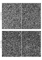
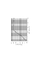
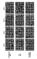
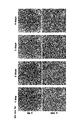
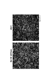
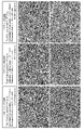
- 3 is a phase-contrast micrograph showing the difference in cell morphology depending on the culture conditions of cultured human corneal endothelial cells (Passage 3).
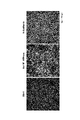
- 3 is a phase-contrast micrograph of cultured human corneal endothelial cells subcultured up to Passage 7 under culture condition 2. It is a graph which shows the analysis result of the cell surface antigen of the cell of Passage 3 cultured under culture condition 1 and the cell of Passage 3 or Passage 7 cultured under culture condition 2. It is a photograph showing the result of examining the expression of Na + / K + ATPase, ZO-1 and N-Cadherin in the cells of Passage 7 cultured under the culture condition 2 by an immunostaining method.
- phase-contrast micrograph of the cell cultured under the culture condition 2 from Passage 0 in Passage 7. It is a photograph and the graph which shows the state of the expression of the cell surface antigen and the like about the cell of FIG. It is a photograph which shows the result of trypan blue staining of the cell after cryopreservation which concerns on one Example of this invention.
- 3 is a phase-contrast micrograph showing changes in cell morphology of cultured human corneal endothelial cells cultured under culture condition 2 after cryopreservation.
- 3 is a phase-contrast micrograph comparing the morphology of cryopreserved cells and cells that have been continuously cultured without cryopreservation.
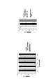
- FIG. 12 is a phase-contrast micrograph showing cell morphology at each collection time in FIG. 12. It is a figure graph and a photograph which show the viability of the cell after cryopreservation at each collection time.
- FIG. 14 is a phase-contrast micrograph comparing the morphology of cells thawed after cryopreservation in FIG. 14 with cells cultured for 35 days under culture condition 2 and cells subcultured without cryopreservation.
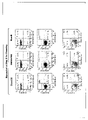
- FIG. 14 is a graph comparing the surface antigens of cells thawed after cryopreservation in FIG. 14 and cells cultured for 35 days under culture condition 2 and cells subcultured without cryopreservation.
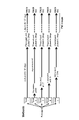
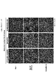
- FIG. 14 is a photograph comparing the results of immunostaining of cells thawed after cryopreservation in FIG. 14 between cells cultured under culture condition 2 for 35 days and cells subcultured without cryopreservation. It is a figure explaining the procedure of comparative analysis of the collection time suitable for cell preservation.
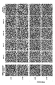
- 6 is a fluorescence micrograph confirming the expression of Na + / K + ATPase, ZO-1 and N-Cadherin in cells collected and cryopreserved at each collection time shown in FIG. 18.
- 6 is a fluorescence micrograph confirming the expression of Na + / K + ATPase, ZO-1 and N-Cadherin in cells collected and cryopreserved at each collection time shown in FIG. 18.
- 6 is a fluorescence micrograph confirming the expression of Na + / K + ATPase, ZO-1 and N-Cadherin in cells collected and cryopreserved at each collection time shown in FIG. 18.
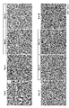
- phase contrast micrograph showing the diurnal change of the cell morphology when culturing under culture condition 2. It is a phase contrast micrograph showing the diurnal change of the cell morphology when culturing under culture condition 2. It is a graph which shows the diurnal change of the expression level of CD44 when culturing under culture condition 2. It is a graph which shows the diurnal change of ECD when culturing under culture condition 2. It is a phase contrast micrograph of cells cultured under culture condition 2 after cryopreservation, a graph showing the analysis result of surface antigen, and a photograph showing the result of immunostaining.
- FIG. 9 is a graph showing a phase-contrast micrograph of cells in which Passage 9 subcultured with the cells of FIG. 29 was cultured under culture condition 2 for 34 days and analysis results of surface antigens. It is a figure explaining the cell culture condition in Example 13. It is a graph which shows the change of the cell yield and the culture scale under each culture condition of FIG. 39.
- FIG. 39 is a phase-contrast micrograph of cell yield and cultured cells under each culture condition of FIG. 39.
- FIG. 39 is a phase-contrast micrograph of cell yield and cultured cells under each culture condition of FIG. 39.
- FIG. 39 is a phase-contrast micrograph of cell yield and cultured cells under each culture condition of FIG. 39.
- FIG. 39 is a phase-contrast micrograph of cell yield and cultured cells under each culture condition of FIG. 39.
- FIG. 39 is a phase-contrast micrograph of cell yield and cultured cells under each culture condition of FIG. 39.
- the preservation method according to the present embodiment is a method for preserving human corneal endothelial cells and / or human corneal endothelial progenitor cells.
- These human corneal endothelial cells and / or human corneal endothelial progenitor cells contain, for example, a ROCK inhibitor and the content of epithelial growth factor (EGF) is transformed according to the method described in Patent Document 2 or Patent Document 3. It was cultivated using a medium having a concentration lower than that causing the above.
- the description of Japanese Patent Application No. 2020-032139 mentioned as Patent Document 2 and the description of International Publication No. 2017/110094 mentioned as Patent Document 3 shall be appropriately referred to and incorporated.
- the human corneal endothelial cell or the human corneal endothelial precursor cell is a human corneal endothelial cell-derived cell, a pluripotent stem cell, a mesenchymal stem cell, a corneal endothelial precursor cell collected from the corneal endothelium, a cell collected from the corneal endothelium, and a direct. It is preferable that the cells are prepared from cells selected from the group consisting of corneal endothelial precursor cells and corneal endothelium-like cells prepared by a programming method.
- the human corneal endothelial progenitor cell is a concept representing a cell destined to become a human corneal endothelial cell by differentiation and maturation, and includes a cell in the middle of determining the differentiation fate.
- a basic medium used for culturing human corneal endothelial cells for example, Opti-MEM-I, Essential 6, DMEM / F12, or the like, to which an additive is appropriately added can be used.
- ROCK inhibitor also referred to as Rho-kinase inhibitor
- examples of the ROCK inhibitor include the following documents: US Pat. 076976, International Publication 02/076977, International Publication 2002/083175, International Publication 02/100833, International Publication 03/059913, International Publication 03/062227, International Publication 2004/09555, International Publication 2004/022541, International Publication 2004/108724, International Publication 2005/003101, International Publication 2005/039564, International Publication 2005/034866, International Publication 2005/037197, International Publication 2005/037198, International Publication 2005/035501, International Publication 2005/035553, International Publication 2005/035556, International Release Examples thereof include the compounds disclosed in 2005/08394, International Publication 2005/1003050, International Publication 2006/057270, International Publication 2007/0266664.
- R)-(+)-Trans- (4-pyridyl) -4- (1-aminoethyl) -cyclohexanecarboxamide dihydrochloride monohydrate and the like can be mentioned. These compounds are Wako Pure Chemical Industries, Ltd. It is also possible to purchase and obtain it from Asahi Kasei Pharma.
- the concentration of the ROCK inhibitor in the medium is, for example, preferably 1 to 100 ⁇ M, more preferably 5 to 20 ⁇ M, and particularly preferably 10 ⁇ M.
- a concentration below the concentration that causes transformation means that the concentration of epidermal growth factor (EGF) is lower than the concentration at which transformation of the corneal endothelial cells of interest (eg, endothelial mesenchymal transition) occurs or the EGF is It means that it is not included at all.
- the EGF concentration lower than the concentration that causes transformation is, for example, about 0 ng / ml to about 5 ng / ml, about 1 ng / ml to about 4 ng / ml, about 2 ng / ml to about 3 ng.
- the concentration less than the concentration that causes EGF transformation is, for example, less than 2 ng / mL, preferably less than 1 ng / mL, more preferably less than 0.5 ng / mL, and the human corneal endothelium due to the addition of EGF. From the viewpoint that adverse effects on the differentiation and maturation process of cells can be completely eliminated, 0 ng / mL (without addition) is particularly preferable.
- the medium is also free of other growth factors that mimic the EGF effect, eg, by acting on upstream or downstream signaling agents.
- transformation also referred to as phase transition
- transformation means that the trait of a cell changes to a state different from the previous state (unusual), and canceration that causes the cell to divide indefinitely. Or it includes dynamic metaplasias that change beyond the walls of the basic form of the tissue.
- transformation include cell state phase transfer (CST) such as epithelial mesenchymal transfer (EMT), fibrosis, aging, endothelial mesenchymal transfer and the like.
- CST cell state phase transfer
- EMT epithelial mesenchymal transfer
- fibrosis fibrosis
- aging endothelial mesenchymal transfer and the like.
- the method for preserving human corneal endothelial cells is human corneal endothelial cells or human corneal endothelial cells grown using a medium containing a ROCK inhibitor and having an EGF content of less than a concentration that causes transformation.
- a cell population containing human corneal endothelial precursor cells is collected at a time specified by one or more of the following indicators (a) to (d) (for example, before a differentiation event occurs). It is characterized in that it is stored in a suspended state. It is preferable that all of these indicators (a) to (d) are satisfied at the time of collecting cells, but it is not necessary to satisfy all of these, and one or more of them (for example, only (a)). It may be one that satisfies.
- the morphology of the human corneal endothelial cells and / or the human corneal endothelial progenitor cells has a ratio of major axis and minor axis including the projections from a spindle-shaped fibroblast-like morphology having irregular elongated projections of 1. It is the period from immediately after the transition to a paving stone-like morphology close to that until just before the boundary between cells becomes unclear.
- B The period from when the expression level of CD44 becomes less than half of the maximum value observed after the most recent passage until the plateau is reached.
- the cell density of the human corneal endothelial cell and / or the human corneal endothelial progenitor cell is 900 cells / mm 2 or more and 2500 cells / mm 2 or less.
- the number of culture days from the most recent passage is 4 days or more and 14 days or less.
- each index (also referred to as an evaluation standard) described in (a) to (d) will be described below.
- (a) will be described.
- human corneal endothelial cells are grown using a medium containing a ROCK inhibitor as described above and the EGF content is less than the concentration that causes transformation, the following cell morphology is obtained during the process of maturation and differentiation. Change.
- human corneal endothelial cells and / or corneal endothelial progenitor cells have a spindle-shaped morphology with irregular elongated protrusions like fibroblasts.
- the ratio of the major axis to the minor axis including the projections was close to 1 (for example, about 1.5: 1, 1.25: 1, 1.1).
- paving stones similar to corneal endothelial cells in human organisms eg hexagonal
- Etc. polygonal shape or circular shape
- the cell morphology that changes depending on the number of days of culture from passage has the above-mentioned period (a), that is, a morphology similar to that of corneal endothelial cells in vivo, but is completely differentiated.
- a a morphology similar to that of corneal endothelial cells in vivo, but is completely differentiated.
- One of the characteristic points of the preservation method according to the present embodiment is that cells are collected before maturity. If cells are collected at this time and stored in a suspended state as described herein, they can be recultured after storage to contain functional human corneal endothelial cells (hereinafter, also referred to as effector cells).
- Human corneal endothelial cells and / or human corneal endothelial precursor cells capable of obtaining a cell population having a rate (hereinafter, also referred to as E-ratio) of about 90% or more are maintained at a high survival rate for at least several days. The above can be saved.
- the expression level of CD44 can be quantified by, for example, flow cytometric analysis using an antibody against CD44. For example, in the case of staining with the antibody-cell ratio of Example 3 using a PE-Cy7 binding CD44 antibody and then analyzing with FACSCanto TM II, the above-mentioned time period is the time when the cells were cultured for 35 days using the medium.
- the relative value of the average fluorescence intensity indicating the expression level of CD44 is larger than 1. It can be said that it is a period of 60 or less, more preferably 50 or less. If cells are collected at this time and stored in a suspended state, a cell population having a content of functional human corneal endothelial cells higher than about 90% can be obtained by culturing again after storage. / Or human corneal endothelial precursor cells can be stored for at least several days or more while maintaining high viability.
- the expression level of CD44 can also be measured by a method other than flow cytometric analysis such as quantitative PCR. However, it should be noted that absolute quantification is not required in all embodiments, but rather the expression pattern can be qualitatively evaluated.
- a cell population having a functional human corneal endothelial cell content of more than 90% can be obtained by culturing again after storage, and / or human corneal endothelial cells and / or Human corneal endothelial precursor cells can be stored for at least several days while maintaining high viability. It is preferable to collect the ECD value at the time of (c) described above (about 900 cells / mm 2 or more and about 2500 cells / mm 2 or less), but about 1000 cells / mm 2 or more and about 2400 cells / mm 2 or less, about 1200 cells /.
- the density of corneal endothelial cells at the start of culture is not particularly limited, but is preferably in the range of, for example, 300 cells / mm 2 or more and 500 cells / mm 2 or less.
- ECD corneal endothelial cell density
- cells are detached from a culture vessel with a cell exfoliating solution such as TrypLE to prepare a cell suspension, and a part of the cells is used to prepare a cell suspension using a blood cell counter.
- ECD corneal endothelial cell density
- Human corneal endothelial cells and / or human corneal endothelial progenitor cells collected at the above-mentioned time may be stored at an appropriate optimum temperature, but for example, low temperature storage such as about 10 ° C or lower, about 4 ° C or lower. It is preferable to store the ice temperature at about 0 ° C. or lower, about ⁇ 10 ° C. or lower, about ⁇ 30 ° C. or lower, about ⁇ 80 ° C. or lower, and freeze storage in a liquid nitrogen tank or the like. The temperature during storage may fluctuate slightly during storage, but for example, it is preferable to keep the temperature within the above-mentioned temperature range from freezing to thawing for use. ..
- the period for storing human corneal endothelial cells in a suspended state can be appropriately set according to the transportation time from the production base to the treatment institution, etc., but regardless of low temperature storage, ice temperature storage, or cryopreservation, 24 It is preferably stored for an hour or longer, and is preferably stored for a long period of time such as 2 days or longer, 3 days or longer, 4 days or longer, or about 1 week. In particular, in the case of cryopreservation, it is considered possible to store for several months to several decades.
- the conditioned medium used for culturing is preferable in the case of ice temperature storage, but the suspension is not limited to this, and for example, cultured cells are generally stored. You can use the preservative solution that is used when you do this.
- a suspension containing a cryoprotectant which is generally widely used for cryopreservation of animal cells, can be used. Examples of such a cryoprotectant include the following, which are cell membrane-permeable cryoprotectants.
- ethylene glycol ethylene glycol
- PG propylene glycol
- 1,2-propanediol 1,2-PD
- 1,3-propanediol 1,3-PD
- butylene glycol BG
- isoprene One or more cryoprotectants selected from the group consisting of glycol (IPG), dipropylene glycol (DPG) and glycerin.
- human corneal endothelial cells and / or human corneal endothelial precursor cells are preserved for a long period of time while maintaining a higher survival rate than expected. can.
- One of the reasons why it can be stored for a long period of time is that cells are collected and stored at a time when ERK (p44 / 42 MAPK) activation (phosphorylation) is enhanced compared to after maturation and differentiation. It is thought that it is one.
- the preserved cells can be administered to a patient as they are, for example.
- the preserved cells or cells recultured after preservation may be suitable for delivery to the eyes of a subject (patient) for the treatment of eye disease or injury, for example by administration or transplantation. ..
- these cells may be administered as they are, or may be used for preparing a drug containing these cells.
- the disease or injury is preferably related to the cornea.
- the preserved human corneal endothelial cells and / or human corneal endothelial progenitor cells are returned from the state of being preserved at a low temperature (for example, about -20 ° C or lower) to about room temperature to about 37 ° C such as 25 ° C. Then, after seeding in the medium again and culturing for a predetermined period (hereinafter, also referred to as after recovery), whether or not a cell population having a content of functional human corneal endothelial cells higher than 90% is obtained is listed below. It can be confirmed by whether or not one or a plurality of conditions (also referred to as evaluation criteria) among (1) to (8) are satisfied.
- the predetermined period may be a period during which human corneal endothelial cells and / or human corneal endothelial progenitor cells are sufficiently differentiated and matured, and is, for example, 4 weeks (for example, 28 days) or more after seeding. Is preferable, and more preferably 5 weeks (for example, 35 days) or more after sowing.
- the functional human corneal endothelial cell is a human corneal endothelial cell having the same corneal endothelial characteristics as the human corneal endothelial cell existing in a human body, or a human anterior chamber.
- the effector cell is at least one of the above-mentioned conditions (1) to (8), particularly CD166 + and CD24 - and CD44 neg to low , which are the conditions of (4). Refers to cells that satisfy. Of these indicators, it has already been confirmed in Patent Document 2 that CD26 + ⁇ 5% is always achieved in a cell population having CD44 high ⁇ 5%.
- Can elicit human corneal function means that it can elicit corneal endothelial properties (eg, improvement of corneal opacity and hydration edema, resulting in sustained long-term maintenance of corneal endothelial cell density, leading to improved visual acuity. Etc.) It is a concept including things.
- the expression of functional proteins linked to corneal endothelial properties is observed in functional human corneal endothelial cells having corneal endothelial properties.
- the functional protein include Na + / K + ATPase, ZO-1, sodium / hydrogen exchanger 1 (NHE1) and / or aquaporin 1 (AQP-1), carbonic anhydrase 5B (CA5B) and the like. ..
- human corneal endothelial cells proliferated and / or differentiated and matured in the presence of a ROCK inhibitor and in the presence of EGF below a concentration that causes transformation are cytoplasmic.
- these functional human corneal endothelial cells have citrate synthase (CS), aconitase 2 (ACO2), isocitrate dehydrogenase 2 (IDH2), pyruvate dehydrogenase 2 (MDH2), and malic acid enzymes 3 in their mitochondria.
- CS citrate synthase
- ACO2 aconitase 2
- IDH2 isocitrate dehydrogenase 2
- MDH2 pyruvate dehydrogenase 2
- malic acid enzymes 3 in their mitochondria.
- Expression of one or more metabolism-related enzymes selected from the group consisting of ME3 ACSS1, acetyl CoA acetyltransferase 1 (ACAT1), pyruvate dehydrogenase (PDH), BCAT2, and branched chain keto acid dehydrogenase 2 (BCKDH2).
- ACAT1 acetyl CoA acetyltransferas
- ATP citrate lyase ACLY
- ACO1 aconitase 1
- IDH1 isocitrate dehydrogenase 1
- MDH1 malate dehydrogenase 1
- ME1 malate enzyme 1
- ACSS2 acetyl-CoA acetyltransferase 2
- the present invention is not limited to that described above.
- the medium used does not necessarily have the composition described in the above embodiment, and human corneal endothelial cells and / or human corneal endothelial precursor cells satisfying the above-mentioned evaluation criteria at the time of cell collection and reculture are obtained. It goes without saying that anything that can be done is good. In addition, various modifications and combinations of embodiments may be made as long as it does not contradict the gist of the present invention.
- Example 1 Confirmation of the effect of culture conditions on the appearance rate of phase-transferred cells
- functional human corneal endothelial cells effector
- a medium containing a ROCK inhibitor and having an EGF content below the concentration that causes transformation It was confirmed that a cell population containing a very high proportion of cells) could be cultured.
- Corneal endothelial cells collected from the same donor-derived cornea were cultured in two types of culture solutions having different additive compositions, and the effects on cell quality such as the content of functional human corneal endothelial cells (effector cells) were investigated.
- the donor cornea used was that of an 18 year old male.
- the medium used was Opti-MEM-I + 8 mass% FBS + 200 mg / ml CaCl 2 + 0.08 mass% chondroitin sulfate + 20 ⁇ g / ml ascorbic acid + 50 ⁇ g / ml gentamicin.
- a medium obtained by adding 0.5 ng / ml EGF, 10 ⁇ M SB 203580, and 10 ⁇ M Y-27632 as an additive to the medium was used.
- a medium in which only 10 ⁇ M Y-27632 was added as an additive and EGF and SB203580 were not added to the medium was used.
- Corneal endothelial cells were exfoliated together with the Descemet's membrane from the donor cornea obtained from Seattle Eye Bank, treated overnight at 37 ° C. with collagenase, suspended in the medium of culture condition 1, and placed in a 6-well plate coated with type I collagen. The seeds were sown at a rate of 1 well per eye. This well was placed in a CO 2 incubator and cultured for 34 days. Medium exchange was performed twice a week. After exfoliating the cells from the culture dish by 10 ⁇ TrypLE TM Select (Thermo Fisher Scientific), the number of recovered cells was measured by a hemocytometer.
- FIG. 1 shows a phase difference photograph of cells of Passage 3 (third generation after being divided into culture condition 1 and culture condition 2) cultured under culture condition 1 or culture condition 2. From FIG. 1, it can be seen that under the culture condition 1, a large number of cell-sized phase transition cells appear. On the other hand, under culture condition 2, it can be seen that the cell size is maintained uniformly even after repeated passages, and the appearance and increase of phase-transferred cells are suppressed.
- FIG. 2 is a phase difference photograph of cells subcultured up to Passage 7 under culture condition 2, but the cell size is maintained uniformly at this stage as in the case of FIG. 1, and the phase is phased. It can be seen that the appearance and increase of transferred cells are suppressed, and there are no foreign substances, discoloration, or other abnormalities.
- FIG. 3 shows the analysis results of the cell surface antigens of the cells of Passage 3 cultured under culture condition 1 or the cells of Passage 3 or Passage 7 cultured under culture condition 2 by flow cytometry.
- the analysis of the cell surface antigen was carried out in the same manner as in Example 4 described later. From the results of FIG. 3, even if the subculture is repeated up to Passage 7 under the culture condition 2, the surface antigens of CD166 + CD24 - CD44 neg to low in human corneal endothelial cells or CD44 neg to low CD90 neg to low It was found that a cell population with a high proportion of effector cells showing the characteristics of the above and a high content of the target functional human corneal endothelial cells was cultured.
- the middle graph shows the CD44.
- the index of CD44 neg to low CD90 neg to low is examined in the lower graph of FIG. 3 independently of the upper and middle graphs of FIG. This also applies to FIGS. 6, 10, 16, 20, 20, 21, 22, 22, 30, 38 and the like.
- the expression level of these surface antigens is defined as follows. By "-" or “neg” is meant that expression is substantially non-observed, i.e. negative.
- the “+” means the above-mentioned non-negative one, that is, the one in which the expression is significantly observed, and the positive one.
- “Low” corresponds to weak positive when the expression is classified into weak positive, medium positive, and strong positive, and the expression level less than this weak positive is negative.
- “High” corresponds to a strong positive when the expression is classified into three stages: weak positive, medium positive, and strong positive.
- the expression intensity of surface antigens such as the above-mentioned CD marker is difficult to define numerically because the fluorescence intensity differs depending on the type of fluorescence of the label and the device setting. Each can be defined in the following range of fluorescence intensity.
- a frame for distinguishing three areas (also referred to as a gate) is described.
- the percentage in the figure indicates the percentage of cells belonging to each of the above-mentioned areas.
- ⁇ -elemen as compounds exhibiting ROCK-inhibiting properties, AT-13148, BA-210, ⁇ -elemen, chromane 1, DJ4, faszil, GSK-576371, GSK492286A, H-1152, hydroxyfaszil, ibuprofen, Selected from the group consisting of LX-7101 [24], Netaluszil, RKI-1447, Ripasudil, TCS-7001, Thiazovibin, Veloszil (AR-12286), Y-27632, Y-30141, Y-33973, and Y-39983. It may contain one or more combinations.
- Example 2 Confirmation of culture conditions that suppress phase transfer
- the cell population cultured from the primary culture (Passage 0) from the donor cornea under the above-mentioned culture condition 2 contained a high proportion of functional human corneal endothelial cells.
- the donor cornea used was that of a 27-year-old man.
- the medium used was the same as that used in the culture condition 2 of Example 1.
- Corneal endothelial cells were exfoliated from the donor cornea obtained from Seattle Eye Bank together with the Descemet's membrane, treated with collagenase at 37 ° C. overnight, suspended in the medium of the culture condition 2, and placed in a 6-well plate coated with type I collagen. The seeds were sown at a rate of 1 well per eye. This well was placed in a CO 2 incubator and cultured for 40 days. Medium exchange was performed once a week. After exfoliating the cells from the culture dish by 10 ⁇ TrypLE TM Select (Thermo Fisher Scientific), the number of recovered cells was measured by a hemocytometer.
- This cell suspension was seeded in a new culture vessel containing a medium of culture condition 2 at a cell density of 400 cells / mm 2 , and cultured again in a CO 2 incubator for 32 days. After exfoliating the cells from the culture dish by 10 ⁇ TrypLE TM Select (Thermo Fisher Scientific), the number of recovered cells was measured with a blood cell calculator, seeded at a cell density of 400 cells / mm 2 , and again in the CO 2 incubator 38. The operation of culturing for ⁇ 44 days was repeated under each culture condition, and the subculture was repeated.
- FIG. 5 shows a phase difference photograph of the cells of Passage 7 cultured as described above. From the phase difference photograph of FIG. 5, it can be seen that in the cells of Passage 7 cultured in this example, the cell size is maintained uniformly, and the appearance and increase of phase-transferred cells are suppressed.
- the graph of FIG. 6B shows the results of surface antigen analysis of the cells of Passage 7 of this example in the same procedure as in Example 1. From this result, the characteristics of the surface antigen in the functional human corneal endothelial cells were shown in most of the cells of Passage 7 of this example. From this result, it was confirmed that in this example as well, under the main culture conditions, a cell population having a high content of the target functional human corneal endothelial cells can be cultured while maintaining a high level even after long-term passage up to Passage 7. did it.
- a ROCK inhibitor such as that used in culture condition 2 from the earliest possible stage such as Passage 0 or Passage 1 is contained, and EGF is contained. It is preferable to culture in a medium in which the content of is less than the concentration that causes transformation.
- Example 3 Cell cryopreservation and confirmation of survival rate
- functional human corneal endothelial cells can be cryopreserved at a high survival rate and thawed by recovering the cell population cultured under the above-mentioned culture condition 2 at a specific culture time after passage. Later, it was confirmed that a cell population having a high content of functional human corneal endothelial cells could be obtained by culturing under the culture condition 2.
- the donor cornea used was that of a 29 year old female.
- the medium used was the same as that used in the culture condition 2 of Example 1.
- Example 3 From the results of Example 3, when the cells were seeded in the medium of the culture condition 2 at a cell density of 400 cells / mm 2 and then collected and cryopreserved on the 5th day, the survival rate was high even after the cryopreservation. It was confirmed that the maintenance was maintained. On the other hand, in the same control example in which cells were collected and stored after maturation and differentiation (after culturing for 136 days), it was found that the survival rate was significantly reduced. In addition, even when the matured and differentiated cells collected on the 35th day after subculture using a cornea derived from a male donor at the age of 14 (Passage 5) are cryopreserved by the same method, the survival rate after thawing. Was 49.5% when stored in liquid nitrogen and 63% when stored at -80 ° C, which were the same levels as the control examples shown in Table 1.
- Example 4 Evaluation of cells recovered after cryopreservation
- Example 4 it was investigated whether or not the content of functional human corneal endothelial cells was maintained at a high content in the cell population obtained by thawing and recovering the cells cryopreserved in Example 3.
- FIG. 8 The observed changes in cell morphology over time are shown in FIG. From the results shown in FIG. 8, it was observed that the cells after cryopreservation proliferated and became a paving stone-like (polygonal shape, circular shape, etc.) morphology having a uniform size. Further, the cells that were cultured until the 35th day without cryopreservation in Example 3 and the cells that were continuously cultured until the 35th day by continuing the medium exchange without collecting the cells on the 4th day in Example 3 were placed. The result of comparison with the phase contrast microscope is shown in FIG. From the results shown in FIG. 9, no morphological difference was observed between the cells cryopreserved and the cells that were continuously cultured without cryopreservation.
- the cells cultured up to the 35th day without cryopreservation in Example 3 and the cells cultured for 35 days in this example were collected from the culture plate by 10 ⁇ TripLE TM Select (Thermo Fisher Scientific) and collected from the culture plate, and FACS buffer (PBS + 0). It was suspended in 5% by mass BSA + 0.05% by mass NaN 3 ) so as to be 4 ⁇ 10 6 cells / ml. 20 ⁇ L of this cell suspension was mixed with 20 ⁇ L of antibody solution 1 or antibody solution 2 and incubated at 4 ° C. for 1.5 to 2 hours under shading.
- the antibody solutions used are as follows. All antibodies not described by the manufacturer were obtained from BD Biosciences.
- mouse anti-Na + / K + ATTase antibody and rabbit anti-ZO-1 antibody were used as the primary antibody
- AlexaFluor488-labeled anti-rabbit IgG antibody and AlexaFluor555-labeled anti-mouse IgG antibody were used as the secondary antibody.
- the results are shown in FIG. 11 after observing with a fluorescence microscope. From this result, it was confirmed that the expression of Na + / K + ATPase and ZO-1, which are functional proteins of human corneal endothelial cells, is normally expressed even in the cryopreserved cells by recovery culture after thawing.
- Example 3 From the above results, it can be confirmed that the cell population thawed and recovered after cryopreservation in Example 3 has a high content of functional human corneal endothelial cells as in the cells that continued to be cultured without cryopreservation. rice field.
- Example 5 cryopreservation and confirmation of survival rate when other media are used
- Example 5 the effect of medium type on survival after cryopreservation was investigated.
- the donor corneas used were those of 18 years old, male and 10 years old, male.
- the medium used was Essential 6 + 8 mass% FBS + 50 ⁇ g / ml gentamicin + 10 ⁇ M Y-27632.
- the Essential 6 medium is based on DMEM / F12 medium widely used for cell culture, to which ascorbic acid, insulin, transferrin, selenium and the like are added, and its composition is open to the public.
- Example 6 Optimization of cell collection time
- Example 6-a Examination of cell collection time
- the donor cornea used was from a 41 year old female.
- the medium used was the same as that used in the culture condition 2 of Example 1.
- the donor cornea obtained from Seattle Eye Bank was cultured to Passage 2 under the same conditions as in Example 2.
- the cell suspension was divided into 5 flasks, one continued to be cultured as it was, and the remaining 4 flasks were cells on days 3, 7, 10 and 14 from the passage, respectively.
- the recovered cells were suspended in CELLBANKER I to 1,000,000 cells per ml. This cell suspension was placed in the air layer portion of a liquid nitrogen tank, frozen, and stored frozen in a liquid nitrogen tank.
- the cells of the first flask which had been continuously cultured, were subcultured to a type I collagen coated 24-well plate on the 35th day after subculture at a cell density of 400 cells / mm 2 .
- the cells that had been cryopreserved in the liquid nitrogen tank were also thawed on the same day as the cells that had been cultured.
- a ThrowSTAR frozen cell thawing station (Model CFTS, Astero Bio) was used.
- the cells washed with the medium of the culture condition 2 were seeded on a type I collagen-coated 24-well plate so as to have a cell density of 400 cells / mm 2 . This was cultured at 37 ° C.
- FIG. 13 shows a phase-contrast micrograph of cell morphology on the day of recovery of each cell lot for cryopreservation.
- FIGS. 14 the photographs of the survival rate after thawing of the cryopreserved cells and the results of trypan blue staining are shown in FIGS. 14, and the results of phase-contrast micrographs, FACS analysis and immuno-cell staining on the 37th day after recovery culture are shown in FIGS. 15 to 15. 17 is shown.
- FACS analysis and immune cell staining were performed in the same procedure as in Example 4.
- the survival rate is about 70%, and the cells collected and cryopreserved on the 7th or 10th day show a higher survival rate, and the survival rate may differ greatly depending on the recovery time after passage. It could be confirmed. From the results shown in FIGS. 15 to 17, the cells collected and cryopreserved on the 7th, 10th or 14th day were equivalent to the cells cultured without cryopreservation by the recovery culture after thawing. It was shown that the cell group contained functional human corneal endothelial cells in proportion. From this result, in order to obtain a cell group having a high survival rate even after cryopreservation and containing functional human corneal endothelial cells at the same ratio as cells cultured without cryopreservation by recovery culture after thawing. It has been found that it is preferable to collect and store cells for a period of 4 days or more and 14 days or less from the latest passage.
- Example 6-b Optimization of cell collection time
- the cell collection time clarified in Example 6-a was further optimized.
- the donor cornea used was from an 8-year-old man.
- the medium used was the same as that used in the culture condition 2 of Example 1.
- the donor cornea obtained from Seattle Eye Bank was cultured up to Passage 4 under the same conditions as in Example 2.
- the cell suspension was divided into 5 flasks, and cells were collected on days 6, 7, 8, 9, and 10 from the passage, respectively.
- Collected cells were suspended in STEMCELL-BANKER, 10% DMSO / 90% FBS, or BAMBANKER hRM (8th day only) to 1,000,000 cells per ml.
- This cell suspension was placed in the air layer portion of a liquid nitrogen tank, frozen, and stored frozen in a liquid nitrogen tank.
- the thawing operation was performed 200 days after the collection and cryopreservation on the 10th day (204 days after the collection and freezing on the 6th day).
- a water bath at 37 ° C. was used for thawing. After thawing, the cells washed with the medium of the culture condition 2 were seeded on a type I collagen-coated 24-well plate so as to have a cell density of 400 cells / mm 2 . This was cultured at 37 ° C. in a humidified atmosphere containing 5% v / v CO 2 . The medium was changed twice a week. Protein expression was comparatively analyzed by FACS analysis and immune cell staining on the 35th day after seeding on a 24-well plate. The procedure is outlined in FIG. The survival rate of the cryopreserved cells after thawing is shown in FIG.
- FIGS. 20 to 25 The survival rate was confirmed by trypan blue staining as in the other examples. FACS analysis and immune cell staining were performed in the same procedure as in Example 4.
- the bar graph on the right side of FIG. 19 shows the survival rate of the cells collected on the 8th day when either STEMCELL-BANKER, 10% DMSO / 90% FBS, or BAMBANKER hRM was used as a cryopreservation solution. ..
- Example 7 About other indicators indicating the time of cell collection
- Example 6 the number of days after seeding (passage) on the plate was examined as an index of the cell collection time.
- Example 7 by examining the characteristics of the cells at the time specified in Example 6, other indicators for specifying the time for collecting human corneal endothelial cells and / or human corneal endothelial progenitor cells were examined.
- Example 7-a Index by cell morphology
- the donor cornea used was that of a 29-year-old man.
- the medium used was the same as that used in the culture condition 2 of Example 1.
- Corneal endothelial cells were exfoliated together with Descemet's membrane from the donor cornea obtained from Seattle Eye Bank, treated overnight at 37 ° C with collagenase, suspended in the medium, and placed on a 6-well plate coated with type I collagen per eye. The seeds were sown at a rate of 1 well.
- the cells were cultured up to the 4th passage in the same procedure as in Example 2 described above. Medium exchange was performed twice a week. After passage to the 5th passage, the cells were cultured under the same conditions, and the cell morphology of each day was photographed with a phase-contrast microscope. The result is shown in FIG. 26, and a partially enlarged view of FIG. 26 is shown in FIG. 27 for further clarification.
- the cell size becomes uniform, and from the 3rd week, the boundary between cells becomes unclear from the state where the boundary between individual cells is clear, and the boundary between cells becomes unclear. It is considered that the cells are in the mature stage where the adhesion is formed and the cells become denser. From the above observations, it is presumed that under the main culture conditions, redifferentiation occurs within 2 weeks (14th day) after sowing, and then the process shifts to the maturation process. In light of the results of Example 6-a, the time when the desired human corneal endothelial cells and / or human corneal endothelial precursor cells can be collected is "the cell morphology is spindle-shaped with irregular elongated protrusions.
- Example 6-a Immediately after changing from a fibroblast-like morphology to a shape similar to the so-called paving stone-like (polygonal or circular) in-vivo corneal endothelial cells having only fine protrusions and a major-to-cell ratio close to 1, cell-cell-to-cell-to-cell It is the period until just before the adhesion of cells is formed and the boundary between cells becomes unclear. "
- the number of days in Example 6-a is used as a guide, but it is considered that the number of culture days required for the cells to mature may vary depending on the cell density at the time of passage and the culture conditions. Even in such a case, it is considered that the cells can be collected at an appropriate time by collecting the cells using the cell morphology obtained in this example as an index.
- Example 7-b Index of collection time based on the expression level of CD44
- the cell collection time can also be confirmed by the expression level of CD44.
- the expression level of CD44 on each day was measured by a flow cytometer for the cells cultured under the same conditions.
- Example 7 (Material used in the experiment) -The same cells / FACS buffer and antibody solution as in Example 7-a were used as in Example 4.
- Example 7-a After passage to the 5th passage of Example 7-a, the cells cultured for a predetermined number of days under the same conditions were washed with PBS (-) and then treated with 10 ⁇ TrypLE TM Select (Thermo Fisher Scientific) to obtain a culture plate. And suspended in FACS buffer to 4 ⁇ 106 cells / ml. 20 ⁇ L of this cell suspension was antibody-stained by the same method as in Example 4, and then analyzed by FACSCanto TM II (BD Biosciences). The results are shown in FIG.
- the average fluorescence intensity of CD44 increased to 15,000 or more immediately after sowing (passage) and then decreased logarithmically by the 11th day. It is considered that the plateau is almost reached on the 17th day, the differentiation is completed about 2 weeks after the subculture, and the maturation process is started.
- the time when the target human corneal endothelial cells and / or human corneal endothelial progenitor cells can be collected is the time when the expression level of CD44 is 1 to 2 days after passage. It is considered that it is the period from the time when it becomes less than half of the maximum value seen in the above to the time when the plateau is reached.
- Example 6-a the number of days in Example 6-a is used as a guide, but it is considered that the number of culture days required for the cells to mature may vary depending on the cell density at the time of passage and the culture conditions. Even in such a case, it is considered that the cells can be collected at an appropriate time by collecting the cells using the expression level of CD44 obtained in this example as an index.
- Example 7-c Index of collection time based on cell density
- ECD corner endothelial cell density
- Example 7-a subcultured to the 5th passage at a density of 400 cells / mm 2 were subjected to PBS (-" at each time point on the 2, 4, 6, 9, 12, 17, and 35 days after the passage. ) Washed and photographed with a phase contrast microscope. The resulting images were analyzed with KSSE-400EB software. The result is shown in FIG.
- ECD corneal endothelial cell density
- Example 6-a the number of days in Example 6-a is used as a guide, but it is considered that the number of culture days required for the cells to mature may vary depending on the cell density at the time of passage and the culture conditions. Even in such a case, it is considered that the cells can be collected at an appropriate time by collecting the cells using the cell density obtained in this example as an index.
- Example 8 Confirmation of viability and quality after recovery culture of cells cryopreserved for a long period of time
- the survival rate and the quality after recovery culture were investigated when the cells cryopreserved under the conditions optimized in Examples 6 and 7 were stored for a long period of time.
- the donor cornea used was from an 8-year-old man.
- the medium used was the same as that used in the culture condition 2 of Example 1.
- Corneal endothelial cells were exfoliated together with Descemet's membrane from the donor cornea obtained from Seattle Eye Bank, treated overnight at 37 ° C with collagenase, suspended in the medium, and placed on a 6-well plate coated with type I collagen per eye. It was sown at a rate of 1 well.
- the cells were cultured up to the second passage in the same procedure as in Example 2 described above. Medium exchange was performed twice a week. The cells were collected on the 7th day after seeding, frozen by the same method as in Example 6, and stored in a liquid nitrogen cell storage container.
- the cells were thawed in a hot water bath on the 99th, 160th, and 171st days after storage, and the survival rate was examined by the trypan blue staining method in the same procedure as in Example 3.
- the recovery rate (the number of cells after cryopreservation / the number of cells subjected to cryopreservation), which is the ratio to the number of cells in the cell, was also investigated. The results are shown in Table 3.
- FIGS. 30A to 30C FACS analysis and immune cell staining were performed in the same procedure as in Example 4.
- the cell density analyzed by KSS-400EB software was 3344 ⁇ 150.8 cells / mm 2
- the PDGF-BB concentration in the culture supernatant on day 32 was 217.5 ⁇ 1.53 pg / mL.
- a cell population capable of obtaining a cell population having a high content of functional human corneal endothelial cells after recovery culture is maintained at a survival rate of 90% or more for a period of 5 months or more. It was confirmed that it can be cryopreserved.
- Example 9 Preservation of cell ice temperature and confirmation of survival rate
- the donor cornea used was from an 8-year-old man.
- the medium used was the same as that used in the culture condition 2 of Example 1.
- Each sample is as follows. Sample 1: Cells cultured in the same procedure as in Example 2 and cultured in Passage 3 for 94 days without cryopreservation. Sample 2: Cells cultured in the same procedure as in Example 2, collected on the 7th day of Passage 2, cryopreserved in the same procedure as in Example 3, thawed, and cultured in Passage 3 for 33 days.
- Sample 3 Cultivate in the same procedure as in Example 2, collect on the 7th day of Passage 2, freeze and store in the same procedure as in Example 3, repeat subculture until Passage 5 after thawing, and incubate in Passage 6 for 9 days. Cells.
- samples 1 to 3 were exfoliated from the culture vessel by 10 ⁇ TypLETM Select (Thermo Fisher Scientific) to prepare a cell suspension. After staining a part (10 ⁇ l) of each cell suspension with trypan blue, the number of live cells and the number of dead cells were measured using a hemocytometer. Based on this measurement result, the cells were suspended in each conditioned medium so as to have a cell density of 3.33 ⁇ 10 6 cells / ml, placed in a commercially available microtube of 1.5 mL, and stored in ice.
- FIG. 31 shows the results of trypan blue staining
- FIG. 32 shows the survival rate calculated from the results of FIG. 31. From the results shown in FIG. 32, the survival rate of sample 1 was lower than 70% on the first day and sample 2 was lower than 70% on the second day, whereas the survival rate of sample 3 was maintained at more than 80% even on the fourth day. I found that it was done.
- Example 10 Effect of preservation solution on preservation of cells at ice temperature
- the influence on the survival rate by the type of the preservation solution used for the preservation at ice temperature was investigated.
- the cells used in the experiment are sample 3 of Example 9.
- the preservation solution used in the experiment is as follows.
- Preservative solution A Acclimated medium
- Preservative solution B Medium of culture condition 2 of Example 1 prepared on the same day
- Preservative solution C Opti-MEM + 100 ⁇ M Y-27632 prepared on the same day
- Example 11 Evaluation of differentiation and maturation period of cells stored at ice temperature into functional human corneal endothelial cells
- Example 11 when cells stored at ice temperature are seeded at a cell density similar to that of transplantation into humans, do they differentiate and mature into functional human corneal endothelial cells at an early stage as in the case of transplantation into humans? It was confirmed.
- Example 9 Evaluation of cell morphology
- 300 ⁇ l of a suspension with a cell density of 3.33 ⁇ 10 6 cells / ml is injected into the anterior chamber. Therefore, in Example 9, a 24-well plate having a cell suspension (having a cell density equivalent to that at the time of transplantation into humans) stored at ice temperature for 4 days using the preservation solution A having an area close to the human endothelial surface. 300 ⁇ l was seeded (diameter about 7.78 mm, bottom area 190 mm 2 , type I collagen coated). After sowing, the cells were cultured under the culture condition 2 of Example 1 for 7 days. The phase difference micrograph at this time is shown in FIG. 35. From FIG. 35, it can be seen that even cells stored at ice temperature for 4 days are in the form of functional human corneal endothelial cells one week after seeding when seeded at the same cell density as transplanted into humans. ..
- FIG. 35 The cells of FIG. 35 were immobilized with ice-cold methanol, and the expression of the functional proteins Na + / K + ATPase and ZO-1 of human corneal endothelial cells was confirmed by immunostaining. The results are shown in FIG. From the results shown in FIG. 36, the expression of Na + / K + ATPase and ZO-1 was confirmed in almost all cells.
- Example 12 Short-term expansion culture of human corneal endothelial cells after freezing storage
- FIG. 38A the phase-contrast micrographs of the cells cultured under culture condition 2 for 34 days are shown in FIG. 38A, and the analysis results of the surface antigen by FACS analysis are shown in FIG. 38B. show.
- the FACS analysis was performed in the same procedure as in Example 4. Cells that were repeatedly passaged at intervals of 8 to 10 days after seeding, which was before differentiation and maturation, also used a medium that finally contained the ROCK inhibitor and the EGF content was less than the concentration that caused transformation. By further culturing, it was confirmed that a cell population having a sufficiently high content of functional human corneal endothelial cells could be obtained.
- Example 13 Short-term expansion culture of human corneal endothelial cells
- the donor corneas used were 29 years old, male, and 8 years old, female.
- FIG. 40 shows the yield and culture scale calculated from the growth rate. It was shown that the short-term expansion culture method of this example, in which passages were repeated at intervals of about 8 days, produced 2 billion cells within 80 days, or 500 flasks of cells when converted to T-25 flasks. .. Further, a photograph of cells cultured up to Passage 4 by the short-term expansion culture method described above and cultured in Passage 5 for about 5 weeks, cells of Control Example 2 cultured for about the same number of days, and cells of Control Example 2 cultured up to Passage 5. And a comparison of yields is shown in FIGS. 41 and 42.
Landscapes
- Health & Medical Sciences (AREA)
- Life Sciences & Earth Sciences (AREA)
- Engineering & Computer Science (AREA)
- Biomedical Technology (AREA)
- Zoology (AREA)
- Chemical & Material Sciences (AREA)
- Wood Science & Technology (AREA)
- Biotechnology (AREA)
- General Health & Medical Sciences (AREA)
- Cell Biology (AREA)
- Organic Chemistry (AREA)
- Bioinformatics & Cheminformatics (AREA)
- Genetics & Genomics (AREA)
- Developmental Biology & Embryology (AREA)
- Medicinal Chemistry (AREA)
- Virology (AREA)
- Microbiology (AREA)
- Biochemistry (AREA)
- General Engineering & Computer Science (AREA)
- Ophthalmology & Optometry (AREA)
- Neurology (AREA)
- Neurosurgery (AREA)
- Immunology (AREA)
- Environmental Sciences (AREA)
- Dentistry (AREA)
- Pharmacology & Pharmacy (AREA)
- Animal Behavior & Ethology (AREA)
- Public Health (AREA)
- Veterinary Medicine (AREA)
- Epidemiology (AREA)
- Reproductive Health (AREA)
- Tropical Medicine & Parasitology (AREA)
- Gynecology & Obstetrics (AREA)
- Hematology (AREA)
- Micro-Organisms Or Cultivation Processes Thereof (AREA)
- Medicines Containing Material From Animals Or Micro-Organisms (AREA)
- Materials For Medical Uses (AREA)
- Biophysics (AREA)
- Physiology (AREA)
- Chemical Kinetics & Catalysis (AREA)
Priority Applications (13)
| Application Number | Priority Date | Filing Date | Title |
|---|---|---|---|
| CA3196110A CA3196110A1 (en) | 2020-10-22 | 2021-10-19 | A storage method of human corneal endothelial cells and/or human corneal endothelial precursor cells |
| MX2023004594A MX2023004594A (es) | 2020-10-22 | 2021-10-19 | Un metodo de almacenamiento de celulas endoteliales de la cornea humanas y/o celulas precursoras endoteliales de la cornea humanas. |
| NZ799955A NZ799955B2 (en) | 2021-10-19 | A storage method of human corneal endothelial cells and/or human corneal endothelial precursor cells | |
| JP2022557558A JP7361431B2 (ja) | 2020-10-22 | 2021-10-19 | ヒト角膜内皮細胞及び/又はヒト角膜内皮前駆細胞の保存方法 |
| EP21882821.8A EP4234682A4 (en) | 2020-10-22 | 2021-10-19 | METHOD FOR PRESERVING HUMAN CORNEAL ENDOTHELIAL CELLS AND/OR HUMAN CORNEAL ENDOTHELIAL PROGENITOR CELLS |
| KR1020237014236A KR20230091107A (ko) | 2020-10-22 | 2021-10-19 | 인간 각막 내피 세포 및/또는 인간 각막 내피 전구세포의 보존 방법 |
| IL302168A IL302168A (en) | 2020-10-22 | 2021-10-19 | Storage method of human corneal endothelial cells and/or human corneal endothelial progenitor cells |
| AU2021366805A AU2021366805B2 (en) | 2020-10-22 | 2021-10-19 | A storage method of human corneal endothelial cells and/or human corneal endothelial precursor cells |
| CN202180072227.5A CN116583593A (zh) | 2020-10-22 | 2021-10-19 | 人角膜内皮细胞和/或人角膜内皮前体细胞的保存方法 |
| US18/032,726 US12550891B2 (en) | 2020-10-22 | 2021-10-19 | Storage method of human corneal endothelial cells and/or human corneal endothelial precursor cells |
| MX2025003724A MX2025003724A (es) | 2020-10-22 | 2023-04-20 | Un metodo de almacenamiento de celulas endoteliales de la cornea humanas y/o celulas precursoras endoteliales de la cornea humanas |
| JP2023119210A JP2023139164A (ja) | 2020-10-22 | 2023-07-21 | ヒト角膜内皮細胞及び/又はヒト角膜内皮前駆細胞の保存方法 |
| AU2025206385A AU2025206385A1 (en) | 2020-10-22 | 2025-07-21 | A storage method of human corneal endothelial cells and/or human corneal endothelial precursor cells |
Applications Claiming Priority (2)
| Application Number | Priority Date | Filing Date | Title |
|---|---|---|---|
| JP2020-177634 | 2020-10-22 | ||
| JP2020177634 | 2020-10-22 |
Publications (1)
| Publication Number | Publication Date |
|---|---|
| WO2022085680A1 true WO2022085680A1 (ja) | 2022-04-28 |
Family
ID=81290627
Family Applications (1)
| Application Number | Title | Priority Date | Filing Date |
|---|---|---|---|
| PCT/JP2021/038609 Ceased WO2022085680A1 (ja) | 2020-10-22 | 2021-10-19 | ヒト角膜内皮細胞及び/又はヒト角膜内皮前駆細胞の保存方法 |
Country Status (10)
| Country | Link |
|---|---|
| US (1) | US12550891B2 (https=) |
| EP (1) | EP4234682A4 (https=) |
| JP (2) | JP7361431B2 (https=) |
| KR (1) | KR20230091107A (https=) |
| CN (1) | CN116583593A (https=) |
| AU (2) | AU2021366805B2 (https=) |
| CA (1) | CA3196110A1 (https=) |
| IL (1) | IL302168A (https=) |
| MX (2) | MX2023004594A (https=) |
| WO (1) | WO2022085680A1 (https=) |
Families Citing this family (1)
| Publication number | Priority date | Publication date | Assignee | Title |
|---|---|---|---|---|
| WO2025145016A1 (en) * | 2023-12-29 | 2025-07-03 | Aurion Biotech, Inc. | Corneal endothelial cell (cec) culturing methods and compositions |
Citations (32)
| Publication number | Priority date | Publication date | Assignee | Title |
|---|---|---|---|---|
| US4678783A (en) | 1983-11-04 | 1987-07-07 | Asahi Kasei Kogyo Kabushiki Kaisha | Substituted isoquinolinesulfonyl compounds |
| JPH0276977A (ja) | 1988-09-12 | 1990-03-16 | Matsushita Electric Ind Co Ltd | 湯水混合装置 |
| JPH0276976A (ja) | 1988-09-12 | 1990-03-16 | Matsushita Electric Ind Co Ltd | 制御弁 |
| JPH02100833A (ja) | 1988-10-07 | 1990-04-12 | Tokai T R W Kk | 転造ダイスの加工方法 |
| JPH0359913A (ja) | 1989-07-28 | 1991-03-14 | Yazaki Corp | 金属溶射による回路体の製造方法 |
| JPH0362227A (ja) | 1989-07-31 | 1991-03-18 | Nec Corp | 高速再翻訳処理方式 |
| JPH04620A (ja) | 1990-04-18 | 1992-01-06 | Nec Off Syst Ltd | キーボード |
| JPH081403A (ja) | 1994-06-22 | 1996-01-09 | Fukui Seisakusho:Kk | Nc装置付き工作機械によるネジ端部バリ取り方法 |
| JP2002083175A (ja) | 2000-09-07 | 2002-03-22 | Cosmo Oil Co Ltd | 競合見積管理システムおよび方法、並びに購買ウエブサーバ |
| JP3421217B2 (ja) | 1995-11-20 | 2003-06-30 | 麒麟麦酒株式会社 | Rho標的タンパク質Rhoキナーゼ |
| JP2004009555A (ja) | 2002-06-07 | 2004-01-15 | Futec Inc | 印刷欠点の検出装置および検出方法 |
| JP2004022541A (ja) | 2002-06-12 | 2004-01-22 | Eastman Kodak Co | 有機発光ダイオード表示装置 |
| JP2004108724A (ja) | 2002-09-20 | 2004-04-08 | Toyotomi Co Ltd | 窓用空気調和機の構造 |
| JP2005003101A (ja) | 2003-06-12 | 2005-01-06 | Toyota Motor Corp | 無段変速機の制御装置 |
| JP2005035506A (ja) | 2003-07-17 | 2005-02-10 | Masuo Kato | パイプを使用した水上、水中液体貯蔵装置及び水上輸送方法 |
| JP2005037197A (ja) | 2003-07-18 | 2005-02-10 | Ricoh Co Ltd | 接触式表面形状測定装置及び測定方法 |
| JP2005035503A (ja) | 2003-07-15 | 2005-02-10 | Okubo Tomio | 自転車等の防寒用ハンドルカバー |
| JP2005039564A (ja) | 2003-07-16 | 2005-02-10 | Nec Saitama Ltd | 画像配信システム、サーバ装置、携帯端末装置及びそれに用いる画像配信方法 |
| JP2005035501A (ja) | 2003-07-14 | 2005-02-10 | Takahisa Tsutsui | 油タンカー船の積付け計算をボタン操作によって調整かつ、連動して海面を表示するプログラム |
| JP2005034866A (ja) | 2003-07-18 | 2005-02-10 | Yamazaki Mazak Corp | レーザ加工機 |
| JP2005037198A (ja) | 2003-07-18 | 2005-02-10 | Kawasaki Heavy Ind Ltd | 内側炉心槽の挿入・引き抜き方法及びその装置と内側炉心槽引き抜き時の燃料集合体の拘束支持方法及びその装置 |
| JP2005080394A (ja) | 2003-08-29 | 2005-03-24 | Rohm Co Ltd | 電源装置 |
| JP2005103050A (ja) | 2003-09-30 | 2005-04-21 | Nohmi Bosai Ltd | 消防用設備の開閉弁装置及びその制御弁 |
| JP2006057270A (ja) | 2004-08-18 | 2006-03-02 | Akimori Taniguchi | 巨大水母類の漂着による、取水口スクリ−ン吸着閉塞防止システム。 |
| JP2007026664A (ja) | 1995-04-14 | 2007-02-01 | Toshiba Corp | 光ディスクからビデオ及びオーディオを再生する再生システム、その再生方法及び光ディスクの記録方法 |
| WO2009028631A1 (ja) | 2007-08-29 | 2009-03-05 | Senju Pharmaceutical Co., Ltd. | 角膜内皮細胞接着促進剤 |
| WO2014038639A1 (ja) * | 2012-09-07 | 2014-03-13 | 日本ケミカルリサーチ株式会社 | 間葉系幹細胞の馴化培地を含有する角膜内皮細胞培養用培地 |
| JP2015155400A (ja) * | 2014-01-16 | 2015-08-27 | Jcrファーマ株式会社 | 角膜内皮細胞を含有する移植用組成物 |
| WO2017110094A1 (ja) | 2015-12-24 | 2017-06-29 | 学校法人同志社 | カスパーゼ阻害剤を含む、TGF-βに起因する障害を治療または予防するための医薬およびその応用 |
| WO2017141926A1 (en) * | 2016-02-15 | 2017-08-24 | Kyoto Prefectural Public University Corporation | Human functional corneal endothelial cell and application thereof |
| WO2020032139A1 (en) | 2018-08-09 | 2020-02-13 | Sharp Kabushiki Kaisha | Integrated Access and Backhaul Node Recognition in Integrated Access and Backhaul Network |
| JP2020032139A (ja) | 2018-08-31 | 2020-03-05 | 株式会社三洋物産 | 遊技機 |
Family Cites Families (29)
| Publication number | Priority date | Publication date | Assignee | Title |
|---|---|---|---|---|
| EP1195372A1 (en) | 1994-04-18 | 2002-04-10 | Mitsubishi Pharma Corporation | N-heterocyclic substituted benzamide derivatives with antihypertensive activity |
| AU9646198A (en) | 1997-10-22 | 1999-05-10 | Nippon Shinyaku Co. Ltd. | Isoquinoline derivative and drug |
| WO1999061403A1 (en) | 1998-05-25 | 1999-12-02 | Santen Pharmaceutical Co., Ltd. | Novel vinylbenzene derivatives |
| PE20020958A1 (es) | 2001-03-23 | 2002-11-14 | Bayer Corp | Inhibidores de la rho-quinasa |
| UY27224A1 (es) | 2001-03-23 | 2002-10-31 | Bayer Corp | Inhibidores de la rho-quinasa |
| US7109208B2 (en) | 2001-04-11 | 2006-09-19 | Senju Pharmaceutical Co., Ltd. | Visual function disorder improving agents |
| WO2002100833A1 (en) | 2001-06-12 | 2002-12-19 | Sumitomo Pharmaceuticals Company, Limited | Rho KINASE INHIBITORS |
| JP4505228B2 (ja) | 2002-01-10 | 2010-07-21 | バイエル・シェーリング・ファルマ・アクチェンゲゼルシャフト | Rho−キナーゼ阻害剤 |
| MXPA04007196A (es) | 2002-01-23 | 2005-06-08 | Bayer Pharmaceuticals Corp | Inhibidores de rho-quinasa. |
| EP1541559A4 (en) | 2002-07-22 | 2007-08-22 | Asahi Kasei Pharma Corp | 5-SUBSTITUTED ISOQUINOLINE DERIVATIVE |
| CA2400996A1 (en) | 2002-09-03 | 2004-03-03 | Lisa Mckerracher | 1,4-substituted cyclohexane derivatives |
| US7160894B2 (en) | 2003-06-06 | 2007-01-09 | Asahi Kasei Pharma Corporation | Tricyclic compound |
| WO2005003101A2 (en) | 2003-07-02 | 2005-01-13 | Biofocus Discovery Limited | Pyrazine and pyridine derivatives as rho kinase inhibitors |
| WO2005039564A1 (en) | 2003-10-02 | 2005-05-06 | Vertex Pharmaceuticals Incorporated | Phthalimide compounds useful as protein kinase inhibitors |
| ES2387780T3 (es) | 2003-10-06 | 2012-10-01 | Glaxosmithkline Llc | Preparación de azabencimidazoles 1,6-disustituidos como inhibidores de quinasas |
| JP2007507547A (ja) | 2003-10-06 | 2007-03-29 | グラクソ グループ リミテッド | キナーゼ阻害剤としての1,7−二置換アザベンゾイミダゾールの調製 |
| WO2005034866A2 (en) | 2003-10-06 | 2005-04-21 | Glaxo Group Limited | Preparation of 1, 6, 7- trisubstituted azabenzimidazoles as kinase inhibitors |
| CN1898377A (zh) * | 2003-10-10 | 2007-01-17 | 细胞生物工程公司 | 人角膜内皮细胞以及获得和培养用于角膜细胞移植的细胞的方法 |
| JP2007015928A (ja) | 2003-10-15 | 2007-01-25 | Ube Ind Ltd | 新規オレフィン誘導体 |
| EP1679308B1 (en) | 2003-10-15 | 2013-07-24 | Ube Industries, Ltd. | Novel indazole derivative |
| JP2007008816A (ja) | 2003-10-15 | 2007-01-18 | Ube Ind Ltd | 新規イソキノリン誘導体 |
| CA2556589A1 (en) | 2004-02-24 | 2005-09-01 | Bioaxone Therapeutique Inc. | 4-substituted piperidine derivatives |
| AR049333A1 (es) | 2004-04-02 | 2006-07-19 | Vertex Pharma | Azaindoles inhibidores de proteinquinasas rock y otras proteinas quinasas. composiciones farmaceuticas. |
| US20060101035A1 (en) | 2004-11-11 | 2006-05-11 | Mustakallio Minna M | System and method for blog functionality |
| WO2006057270A1 (ja) | 2004-11-26 | 2006-06-01 | Asahi Kasei Pharma Corporation | 含窒素3環化合物 |
| CA2621181C (en) | 2005-08-30 | 2011-04-19 | Asahi Kasei Pharma Corporation | 5-isoquinolinesulfonamide compounds |
| WO2016128074A1 (de) | 2015-02-13 | 2016-08-18 | P + L Gmbh & Co. Kg | Verfahren zur lagebestimmung eines werkstücks in einer werkzeugmaschine |
| US20210207088A1 (en) * | 2018-08-31 | 2021-07-08 | The Doshisha | Composition and method for preserving or culturing ocular cells |
| WO2021172554A1 (ja) | 2020-02-27 | 2021-09-02 | 京都府公立大学法人 | ヒト機能性角膜内皮細胞およびその応用 |
-
2021
- 2021-10-19 AU AU2021366805A patent/AU2021366805B2/en active Active
- 2021-10-19 MX MX2023004594A patent/MX2023004594A/es unknown
- 2021-10-19 CN CN202180072227.5A patent/CN116583593A/zh active Pending
- 2021-10-19 IL IL302168A patent/IL302168A/en unknown
- 2021-10-19 US US18/032,726 patent/US12550891B2/en active Active
- 2021-10-19 EP EP21882821.8A patent/EP4234682A4/en active Pending
- 2021-10-19 KR KR1020237014236A patent/KR20230091107A/ko active Pending
- 2021-10-19 JP JP2022557558A patent/JP7361431B2/ja active Active
- 2021-10-19 WO PCT/JP2021/038609 patent/WO2022085680A1/ja not_active Ceased
- 2021-10-19 CA CA3196110A patent/CA3196110A1/en active Pending
-
2023
- 2023-04-20 MX MX2025003724A patent/MX2025003724A/es unknown
- 2023-07-21 JP JP2023119210A patent/JP2023139164A/ja active Pending
-
2025
- 2025-07-21 AU AU2025206385A patent/AU2025206385A1/en active Pending
Patent Citations (33)
| Publication number | Priority date | Publication date | Assignee | Title |
|---|---|---|---|---|
| US4678783B1 (en) | 1983-11-04 | 1995-04-04 | Asahi Chemical Ind | Substituted isoquinolinesulfonyl compounds |
| US4678783A (en) | 1983-11-04 | 1987-07-07 | Asahi Kasei Kogyo Kabushiki Kaisha | Substituted isoquinolinesulfonyl compounds |
| JPH0276977A (ja) | 1988-09-12 | 1990-03-16 | Matsushita Electric Ind Co Ltd | 湯水混合装置 |
| JPH0276976A (ja) | 1988-09-12 | 1990-03-16 | Matsushita Electric Ind Co Ltd | 制御弁 |
| JPH02100833A (ja) | 1988-10-07 | 1990-04-12 | Tokai T R W Kk | 転造ダイスの加工方法 |
| JPH0359913A (ja) | 1989-07-28 | 1991-03-14 | Yazaki Corp | 金属溶射による回路体の製造方法 |
| JPH0362227A (ja) | 1989-07-31 | 1991-03-18 | Nec Corp | 高速再翻訳処理方式 |
| JPH04620A (ja) | 1990-04-18 | 1992-01-06 | Nec Off Syst Ltd | キーボード |
| JPH081403A (ja) | 1994-06-22 | 1996-01-09 | Fukui Seisakusho:Kk | Nc装置付き工作機械によるネジ端部バリ取り方法 |
| JP2007026664A (ja) | 1995-04-14 | 2007-02-01 | Toshiba Corp | 光ディスクからビデオ及びオーディオを再生する再生システム、その再生方法及び光ディスクの記録方法 |
| JP3421217B2 (ja) | 1995-11-20 | 2003-06-30 | 麒麟麦酒株式会社 | Rho標的タンパク質Rhoキナーゼ |
| JP2002083175A (ja) | 2000-09-07 | 2002-03-22 | Cosmo Oil Co Ltd | 競合見積管理システムおよび方法、並びに購買ウエブサーバ |
| JP2004009555A (ja) | 2002-06-07 | 2004-01-15 | Futec Inc | 印刷欠点の検出装置および検出方法 |
| JP2004022541A (ja) | 2002-06-12 | 2004-01-22 | Eastman Kodak Co | 有機発光ダイオード表示装置 |
| JP2004108724A (ja) | 2002-09-20 | 2004-04-08 | Toyotomi Co Ltd | 窓用空気調和機の構造 |
| JP2005003101A (ja) | 2003-06-12 | 2005-01-06 | Toyota Motor Corp | 無段変速機の制御装置 |
| JP2005035501A (ja) | 2003-07-14 | 2005-02-10 | Takahisa Tsutsui | 油タンカー船の積付け計算をボタン操作によって調整かつ、連動して海面を表示するプログラム |
| JP2005035503A (ja) | 2003-07-15 | 2005-02-10 | Okubo Tomio | 自転車等の防寒用ハンドルカバー |
| JP2005039564A (ja) | 2003-07-16 | 2005-02-10 | Nec Saitama Ltd | 画像配信システム、サーバ装置、携帯端末装置及びそれに用いる画像配信方法 |
| JP2005035506A (ja) | 2003-07-17 | 2005-02-10 | Masuo Kato | パイプを使用した水上、水中液体貯蔵装置及び水上輸送方法 |
| JP2005037197A (ja) | 2003-07-18 | 2005-02-10 | Ricoh Co Ltd | 接触式表面形状測定装置及び測定方法 |
| JP2005037198A (ja) | 2003-07-18 | 2005-02-10 | Kawasaki Heavy Ind Ltd | 内側炉心槽の挿入・引き抜き方法及びその装置と内側炉心槽引き抜き時の燃料集合体の拘束支持方法及びその装置 |
| JP2005034866A (ja) | 2003-07-18 | 2005-02-10 | Yamazaki Mazak Corp | レーザ加工機 |
| JP2005080394A (ja) | 2003-08-29 | 2005-03-24 | Rohm Co Ltd | 電源装置 |
| JP2005103050A (ja) | 2003-09-30 | 2005-04-21 | Nohmi Bosai Ltd | 消防用設備の開閉弁装置及びその制御弁 |
| JP2006057270A (ja) | 2004-08-18 | 2006-03-02 | Akimori Taniguchi | 巨大水母類の漂着による、取水口スクリ−ン吸着閉塞防止システム。 |
| WO2009028631A1 (ja) | 2007-08-29 | 2009-03-05 | Senju Pharmaceutical Co., Ltd. | 角膜内皮細胞接着促進剤 |
| WO2014038639A1 (ja) * | 2012-09-07 | 2014-03-13 | 日本ケミカルリサーチ株式会社 | 間葉系幹細胞の馴化培地を含有する角膜内皮細胞培養用培地 |
| JP2015155400A (ja) * | 2014-01-16 | 2015-08-27 | Jcrファーマ株式会社 | 角膜内皮細胞を含有する移植用組成物 |
| WO2017110094A1 (ja) | 2015-12-24 | 2017-06-29 | 学校法人同志社 | カスパーゼ阻害剤を含む、TGF-βに起因する障害を治療または予防するための医薬およびその応用 |
| WO2017141926A1 (en) * | 2016-02-15 | 2017-08-24 | Kyoto Prefectural Public University Corporation | Human functional corneal endothelial cell and application thereof |
| WO2020032139A1 (en) | 2018-08-09 | 2020-02-13 | Sharp Kabushiki Kaisha | Integrated Access and Backhaul Node Recognition in Integrated Access and Backhaul Network |
| JP2020032139A (ja) | 2018-08-31 | 2020-03-05 | 株式会社三洋物産 | 遊技機 |
Non-Patent Citations (4)
| Title |
|---|
| ANONYMOUS: "ATCC® ANIMAL CELL CULTURE GUIDE - tips and techniques for continuous cell lines", ATCC ANIMAL CELL CULTURE BASICS, 1 December 2011 (2011-12-01), US, pages 1 - 40, XP009536304 * |
| OKUMURA N ET AL., PLOS ONE, vol. 14, no. 6, 2019, pages e0218431 |
| See also references of EP4234682A4 |
| VIANNA L.M.M. ET AL., ARQ BRAS OFTALMOL., vol. 79, no. 1, 2016, pages 37 |
Also Published As
| Publication number | Publication date |
|---|---|
| AU2021366805B2 (en) | 2025-04-24 |
| JPWO2022085680A1 (https=) | 2022-04-28 |
| US20230380417A1 (en) | 2023-11-30 |
| US12550891B2 (en) | 2026-02-17 |
| AU2021366805A1 (en) | 2023-06-22 |
| JP2023139164A (ja) | 2023-10-03 |
| JP7361431B2 (ja) | 2023-10-16 |
| CN116583593A (zh) | 2023-08-11 |
| IL302168A (en) | 2023-06-01 |
| AU2025206385A1 (en) | 2025-08-07 |
| MX2025003724A (es) | 2025-05-02 |
| CA3196110A1 (en) | 2022-04-28 |
| EP4234682A4 (en) | 2024-11-27 |
| KR20230091107A (ko) | 2023-06-22 |
| MX2023004594A (es) | 2023-05-08 |
| EP4234682A1 (en) | 2023-08-30 |
| NZ799955A (en) | 2025-05-30 |
Similar Documents
| Publication | Publication Date | Title |
|---|---|---|
| US10104881B2 (en) | Composition comprising plant-derived recombinant human serum albumin, lipids, and plant protein hydrolysates as active ingredients for cryopreservation of stem cells or primary cells | |
| CN105796602A (zh) | 利用分离的胎盘细胞治疗中风 | |
| JP2020516257A (ja) | 凍結保存方法 | |
| Proulx et al. | Methods being developed for preparation, delivery and transplantation of a tissue-engineered corneal endothelium | |
| TW201821610A (zh) | 哺乳動物細胞凍結保存液 | |
| CN106359368A (zh) | 细胞冻存液及冻存方法 | |
| Dulugiac et al. | Comparative studies of mesenchymal stem cells derived from different cord tissue compartments–the influence of cryopreservation and growth media | |
| JP2011501960A (ja) | 月経血幹細胞と共に臍帯血由来細胞を共培養する方法 | |
| CN102226169B (zh) | 山羊毛囊外根鞘细胞系的构建方法 | |
| Baylis et al. | An investigation of donor and culture parameters which influence epithelial outgrowths from cultured human cadaveric limbal explants | |
| Bourland et al. | Isolation and culture of human dermal microvascular endothelial cells | |
| Romanov et al. | Isolation of multipotent mesenchymal stromal cells from cryopreserved human umbilical cord tissue | |
| Li et al. | Enriching new transplantable RGC-like cells from retinal organoids for RGC replacement therapy | |
| CN117701505B (zh) | 肉瘤组织类器官培养基、构建肉瘤组织类器官和肉瘤组织类器官库的方法、构建动物疾病模型的方法 | |
| AU2025206385A1 (en) | A storage method of human corneal endothelial cells and/or human corneal endothelial precursor cells | |
| Schiozer et al. | An outcome analysis and long-term viability of cryopreserved cultured epidermal allografts: assessment of the conservation of transplantable human skin allografts | |
| EP3252143A1 (en) | Method for producing a platelet-lysate-containing gel | |
| CN106417253A (zh) | 一种骨骼肌干细胞的冻存液和冻存方法 | |
| CN109430251A (zh) | 一种脂肪组织的保存液及其制备方法与脂肪组织的保存方法 | |
| Menè et al. | Isolation and propagation of glomerular mesangial cells | |
| JP7706874B2 (ja) | 切除脂肪から間葉系幹細胞を製造する方法 | |
| CN116941606B (zh) | 脐带组织的细胞组织库的构建方法及其相关产品和应用 | |
| CN107312744A (zh) | 一种含血清的口腔黏膜上皮细胞培养液 | |
| US20250263673A1 (en) | Mammalian cell cryopreservation liquid | |
| WO2023282337A1 (ja) | 角膜内皮代替細胞を含有する治療用組成物 |
Legal Events
| Date | Code | Title | Description |
|---|---|---|---|
| 121 | Ep: the epo has been informed by wipo that ep was designated in this application |
Ref document number: 21882821 Country of ref document: EP Kind code of ref document: A1 |
|
| DPE1 | Request for preliminary examination filed after expiration of 19th month from priority date (pct application filed from 20040101) | ||
| ENP | Entry into the national phase |
Ref document number: 2022557558 Country of ref document: JP Kind code of ref document: A |
|
| ENP | Entry into the national phase |
Ref document number: 3196110 Country of ref document: CA |
|
| WWE | Wipo information: entry into national phase |
Ref document number: 202180072227.5 Country of ref document: CN |
|
| ENP | Entry into the national phase |
Ref document number: 20237014236 Country of ref document: KR Kind code of ref document: A |
|
| REG | Reference to national code |
Ref country code: BR Ref legal event code: B01A Ref document number: 112023007365 Country of ref document: BR |
|
| WWE | Wipo information: entry into national phase |
Ref document number: 202317035201 Country of ref document: IN |
|
| NENP | Non-entry into the national phase |
Ref country code: DE |
|
| ENP | Entry into the national phase |
Ref document number: 2021882821 Country of ref document: EP Effective date: 20230522 |
|
| ENP | Entry into the national phase |
Ref document number: 112023007365 Country of ref document: BR Kind code of ref document: A2 Effective date: 20230419 |
|
| ENP | Entry into the national phase |
Ref document number: 2021366805 Country of ref document: AU Date of ref document: 20211019 Kind code of ref document: A |
|
| WWG | Wipo information: grant in national office |
Ref document number: MX/A/2023/004594 Country of ref document: MX |
|
| WWD | Wipo information: divisional of initial pct application |
Ref document number: 820790 Country of ref document: NZ |
|
| WWG | Wipo information: grant in national office |
Ref document number: 799955 Country of ref document: NZ |