EP4073508B1 - Nachweis von blutplättchen in einer blutprobe - Google Patents
Nachweis von blutplättchen in einer blutprobe Download PDFInfo
- Publication number
- EP4073508B1 EP4073508B1 EP20825250.2A EP20825250A EP4073508B1 EP 4073508 B1 EP4073508 B1 EP 4073508B1 EP 20825250 A EP20825250 A EP 20825250A EP 4073508 B1 EP4073508 B1 EP 4073508B1
- Authority
- EP
- European Patent Office
- Prior art keywords
- monolayer
- sample
- platelets
- microscope
- microscopic image
- Prior art date
- Legal status (The legal status is an assumption and is not a legal conclusion. Google has not performed a legal analysis and makes no representation as to the accuracy of the status listed.)
- Active
Links
Images


Classifications
-
- G—PHYSICS
- G01—MEASURING; TESTING
- G01N—INVESTIGATING OR ANALYSING MATERIALS BY DETERMINING THEIR CHEMICAL OR PHYSICAL PROPERTIES
- G01N1/00—Sampling; Preparing specimens for investigation
- G01N1/28—Preparing specimens for investigation including physical details of (bio-)chemical methods covered elsewhere, e.g. G01N33/50, C12Q
- G01N1/2813—Producing thin layers of samples on a substrate, e.g. smearing, spinning-on
-
- G—PHYSICS
- G01—MEASURING; TESTING
- G01N—INVESTIGATING OR ANALYSING MATERIALS BY DETERMINING THEIR CHEMICAL OR PHYSICAL PROPERTIES
- G01N15/00—Investigating characteristics of particles; Investigating permeability, pore-volume or surface-area of porous materials
- G01N15/10—Investigating individual particles
- G01N15/14—Optical investigation techniques, e.g. flow cytometry
- G01N15/1468—Optical investigation techniques, e.g. flow cytometry with spatial resolution of the texture or inner structure of the particle
-
- G—PHYSICS
- G01—MEASURING; TESTING
- G01N—INVESTIGATING OR ANALYSING MATERIALS BY DETERMINING THEIR CHEMICAL OR PHYSICAL PROPERTIES
- G01N15/00—Investigating characteristics of particles; Investigating permeability, pore-volume or surface-area of porous materials
- G01N15/01—Investigating characteristics of particles; Investigating permeability, pore-volume or surface-area of porous materials specially adapted for biological cells, e.g. blood cells
- G01N2015/018—Platelets
-
- G—PHYSICS
- G01—MEASURING; TESTING
- G01N—INVESTIGATING OR ANALYSING MATERIALS BY DETERMINING THEIR CHEMICAL OR PHYSICAL PROPERTIES
- G01N15/00—Investigating characteristics of particles; Investigating permeability, pore-volume or surface-area of porous materials
- G01N15/10—Investigating individual particles
- G01N2015/1006—Investigating individual particles for cytology
-
- G—PHYSICS
- G01—MEASURING; TESTING
- G01N—INVESTIGATING OR ANALYSING MATERIALS BY DETERMINING THEIR CHEMICAL OR PHYSICAL PROPERTIES
- G01N15/00—Investigating characteristics of particles; Investigating permeability, pore-volume or surface-area of porous materials
- G01N15/10—Investigating individual particles
- G01N15/14—Optical investigation techniques, e.g. flow cytometry
- G01N15/1434—Optical arrangements
- G01N2015/144—Imaging characterised by its optical setup
- G01N2015/1445—Three-dimensional imaging, imaging in different image planes, e.g. under different angles or at different depths, e.g. by a relative motion of sample and detector, for instance by tomography
-
- G—PHYSICS
- G01—MEASURING; TESTING
- G01N—INVESTIGATING OR ANALYSING MATERIALS BY DETERMINING THEIR CHEMICAL OR PHYSICAL PROPERTIES
- G01N15/00—Investigating characteristics of particles; Investigating permeability, pore-volume or surface-area of porous materials
- G01N15/10—Investigating individual particles
- G01N15/14—Optical investigation techniques, e.g. flow cytometry
- G01N2015/1486—Counting the particles
Definitions
- Some applications of the presently disclosed subject matter relate generally to analysis of bodily samples, and in particular, to optical density and microscopic measurements that are performed upon blood samples.
- a property of a biological sample is determined by performing an optical measurement.
- the density of a component e.g., a count of the component per unit volume
- the concentration and/or density of a component may be measured by performing optical absorption, transmittance, fluorescence, and/or luminescence measurements upon the sample.
- the sample is placed into a sample carrier and the measurements are performed with respect to a portion of the sample that is contained within a sample chamber of the sample carrier. The measurements that are performed upon the portion of the sample that is contained within the sample chamber of the sample carrier are analyzed in order to determine a property of the sample.
- WO-2013/041951A1 describes an optical platelet counter method.
- US-2013/170730A1 describes a method and apparatus for automate platelet identification within a whole blood sample from microscopy images.
- a portion of a blood sample that comprises a cell suspension is placed within a sample chamber that is a cavity that includes a base surface.
- the cells in the cell suspension are allowed to settle on the base surface of the sample chamber to form a monolayer of cells on the base surface of the sample chamber.
- at least one microscopic image of at least a portion of the monolayer of cells is typically acquired.
- a plurality of images of the monolayer are acquired, each of the images corresponding to an imaging field that is located at a respective, different area within the imaging plane of the monolayer.
- an optimum depth level at which to focus the microscope in order to image the monolayer is determined, e.g., using techniques as described in US Patent US 10,176,565 to Greenfield .
- respective imaging fields have different optimum depth levels from each other.
- platelets that have settled within the monolayer are identified within the at least one microscopic image of at least a portion of the monolayer of cells.
- the inventors of the present application have noticed that it is often the case that, even after having been left to settle such as to form a monolayer using the techniques described herein, not all of the platelets within a blood sample settle within the monolayer, and some cells continue to be suspended within the cell solution. More specifically, the inventors have found that it is typically the case that, even after the sample has been left to settle for approximately two minutes, between 10 percent and 70 percent of the platelets within the sample do not settle within the monolayer focus field. Typically, if the monolayer is allowed to form over a longer time period, then more platelets settle within the monolayer focus field.
- the inventors have found that even if the monolayer is allowed to form over a relatively long time period (e.g., between 15 and 30 minutes), some platelets still remain suspended within the solution such that the platelets are not disposed within the monolayer focus field.
- the number of platelets that remain suspended within the solution is dependent on the time for which the monolayer is allowed to form, in addition to the height of the cavity in which the cell solution is placed.
- platelets that are suspended within the cell solution are identified, in addition to identifying platelets within the monolayer focus field.
- such platelets are identified by focusing the microscope at additional depth levels to the depth level(s) to which the microscope is focused in order to image the monolayer of cells (referred to herein as "the monolayer depth level(s)"), and acquiring images at the additional depth levels.
- the platelets within the images that are acquired at the additional depth levels are identified and counted, and, based upon the count of platelets within those images, a total count of the platelets that are suspended within the cell solution is estimated.
- a count of the platelets that have settled within the monolayer is compared to a count of the platelets that have not settled within the monolayer.
- an output is generated in response to the comparison.
- a clinical condition is derived and outputted to a user, based upon the comparison of the count of the platelets that have settled within the monolayer and the count of the platelets that have not settled within the monolayer.
- the extent of platelet activation within the sample may be derived, at least partially based upon the comparison.
- a clinical condition may be derived based upon the size, shape, settling time, and/or settling dynamics of the platelets.
- the settling dynamics of the platelets are determined by imaging the same imaging field a plurality of times, with a time interval between each of the image acquisitions, and/or by determining until what height within the sample chamber platelets are present at a given time.
- a computer processor generates an output indicating that the sample should be prepared in a different way to how it was prepared (e.g., by modifying the diluent within which the sample is diluted, by adding a platelet activation agent, and/or by adding a coagulation agent), based upon the comparison.
- the portion of the blood sample is invalidated from being used for performing at least some measurements upon the sample. For example, if the ratio of unsettled platelets to settled platelets is greater than a first given threshold, and/or lower than a second given threshold, then this might be interpreted as being indicative of a problem with the sample and/or the preparation thereof.
- a parameter of the sample (such as platelet volume, mean platelet volume, and/or median platelet volume) is determined based upon a measurement that is performed upon the portion of the sample.
- the measurement is calibrated, based upon the comparison between properties of the platelets that have settled within the monolayer and properties of the platelets that have not settled within the monolayer.
- the method further includes deriving an error with the blood sample at least partially based upon comparing the first and second platelet counts to each other, wherein generating the output includes generating an indication of the error.
- the method further includes deriving an error with preparation of the blood sample at least partially based upon comparing the first and second platelet counts to each other, wherein generating the output includes generating an indication of the error.
- the method further includes deriving a clinical condition of the subject at least partially based upon comparing the first and second platelet counts to each other, wherein generating the output includes generating an indication of the clinical condition.
- the method further includes deriving a measure of platelet activation at least partially based upon comparing the first and second platelet counts to each other, wherein generating the output includes generating an indication of the platelet activation.
- identifying platelets that have not settled within the monolayer within the at least one additional microscopic image includes accounting for interference with visibility of platelets by white blood cells that are disposed within the monolayer.
- identifying platelets that have not settled within the monolayer within the at least one additional microscopic image includes accounting for interference with visibility of platelets by red blood cells that are disposed within the monolayer.
- avoiding duplicate counts of platelets within images that are acquired at adjacent depth levels includes accounting for lateral movement of platelets between acquisitions of the images that are acquired at adjacent depth levels.
- apparatus including:
- Fig. 1 is block diagram showing components of a biological sample analysis system 20, in accordance with some applications of the present invention.
- a biological sample e.g., a blood sample
- a sample carrier 22 While the sample is disposed in the sample carrier, optical measurements are performed upon the sample using one or more optical measurement devices 24.
- the optical measurement devices may include a microscope (e.g., a digital microscope), a spectrophotometer, a photometer, a spectrometer, a camera, a spectral camera, a hyperspectral camera, a fluorometer, a spectrofluorometer, and/or a photodetector (such as a photodiode, a photoresistor, and/or a phototransistor).
- the optical measurement devices include dedicated light sources (such as light emitting diodes, incandescent light sources, etc.) and/or optical elements for manipulating light collection and/or light emission (such as lenses, diffusers, filters, etc.).
- a computer processor 28 typically receives and processes optical measurements that are performed by the optical measurement device. Further typically, the computer processor controls the acquisition of optical measurements that are performed by the one or more optical measurement devices. The computer processor communicates with a memory 30.
- a user e.g., a laboratory technician, or an individual from whom the sample was drawn
- the user interface includes a keyboard, a mouse, a joystick, a touchscreen device (such as a smartphone or a tablet computer), a touchpad, a trackball, a voice-command interface, and/or other types of user interfaces that are known in the art.
- the computer processor generates an output via an output device 34.
- the output device includes a display, such as a monitor, and the output includes an output that is displayed on the display.
- the processor generates an output on a different type of visual, text, graphics, tactile, audio, and/or video output device, e.g., speakers, headphones, a smartphone, or a tablet computer.
- user interface 32 acts as both an input interface and an output interface, i.e., it acts as an input/output interface.
- the processor generates an output on a computer-readable medium (e.g., a non-transitory computer-readable medium), such as a disk, or a portable USB drive, and/or generates an output on a printer.
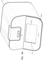
- FIGs. 2A , 2B , and 2C are schematic illustrations of an optical measurement unit 31, in accordance with some applications of the present invention.
- Fig. 2A shows an oblique view of the exterior of the fully assembled device
- Figs. 2B and 2C shows respective oblique views of the device with the cover having been made transparent, such components within the device are visible.
- one or more optical measurement devices 24 (and/or computer processor 28 and memory 30) is housed inside optical measurement unit 31.
- sample carrier 22 is placed inside the optical measurement unit.
- the optical measurement unit may define a slot 36, via which the sample carrier is inserted into the optical measurement unit.
- the optical measurement unit includes a stage 64, which is configured to support sample carrier 22 within the optical measurement unit.
- a screen 63 on the cover of the optical measurement unit e.g., a screen on the front cover of the optical measurement unit, as shown
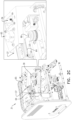
- the optical measurement unit includes microscope system 37 (shown in Figs. 2B-C ) configured to perform microscopic imaging of a portion of the sample.
- the microscope system includes a set of light sources 65 (which typically include a set of brightfield light sources (e.g. light emitting diodes) that are configured to be used for brightfield imaging of the sample, a set of fluorescent light sources (e.g. light emitting diodes) that are configured to be used for fluorescent imaging of the sample), and a camera (e.g., a CCD camera, or a CMOS camera) configured to image the sample.
- the optical measurement unit also includes an optical-density-measurement unit 39 (shown in Fig.
- the optical-density-measurement unit includes a set of optical-density-measurement light sources (e.g., light emitting diodes) and light detectors, which are configured for performing optical density measurements on the sample.
- each of the aforementioned sets of light sources i.e., the set of brightfield light sources, the set of fluorescent light sources, and the set optical-density-measurement light sources
- each of the aforementioned sets of light sources includes a plurality of light sources (e.g. a plurality of light emitting diodes), each of which is configured to emit light at a respective wavelength or at a respective band of wavelengths.
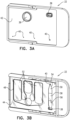
- Figs. 3A and 3B are schematic illustrations of respective views of sample carrier 22, in accordance with some applications of the present invention.
- Fig. 3A shows a top view of the sample carrier (the top cover of the sample carrier being shown as being opaque in Fig. 3A , for illustrative purposes), and
- Fig. 3B shows a bottom view (in which the sample carrier has been rotated around its short edge with respect to the view shown in Fig. 3A ).
- the sample carrier includes a first set 52 of one or more sample chambers, which are used for performing microscopic analysis upon the sample, and a second set 54 of sample chambers, which are used for performing optical density measurements upon the sample.
- the sample chambers of the sample carrier are filled with a bodily sample, such as blood via sample inlet holes 38.
- the sample chambers define one or more outlet holes 40.
- the outlet holes are configured to facilitate filling of the sample chambers with the bodily sample, by allowing air that is present in the sample chambers to be released from the sample chambers.
- the outlet holes are located longitudinally opposite the inlet holes (with respect to a sample chamber of the sample carrier). For some applications, the outlet holes thus provide a more efficient mechanism of air escape than if the outlet holes were to be disposed closer to the inlet holes.
- the sample carrier includes at least three components: a molded component 42, a glass layer 44 (e.g., a glass sheet), and an adhesive layer 46 configured to adhere the glass layer to an underside of the molded component.
- the molded component is typically made of a polymer (e.g., a plastic) that is molded (e.g., via injection molding) to provide the sample chambers with a desired geometrical shape.
- the molded component is typically molded to define inlet holes 38, outlet holes 40, and gutters 48 which surround the central portion of each of the sample chambers.
- the gutters typically facilitate filling of the sample chambers with the bodily sample, by allowing air to flow to the outlet holes, and/or by allowing the bodily sample to flow around the central portion of the sample chamber.
- a sample carrier as shown in Figs. 3A-C is used when performing a complete blood count on a blood sample.
- the sample carrier is used with optical measurement unit 31 configured as generally shown and described with reference to Figs. 2A-C .
- a first portion of the blood sample is placed inside first set 52 of sample chambers (which are used for performing microscopic analysis upon the sample, e.g., using microscope system 37 (shown in Figs. 2B-C )), and a second portion of the blood sample is placed inside second set 54 of sample chambers (which are used for performing optical density measurements upon the sample, e.g., using optical-density-measurement unit 39 (shown in Fig. 2C )).
- first set 52 of sample chambers includes a plurality of sample chambers
- second set 54 of sample chambers includes only a single sample chamber, as shown.
- the scope of the present application includes using any number of sample chambers (e.g., a single sample chamber or a plurality of sample chambers) within either the first set of sample chambers or within the second set of sample chambers, or any combination thereof.
- the first portion of the blood sample is typically diluted with respect to the second portion of the blood sample.
- the diluent may contain pH buffers, stains, fluorescent stains, antibodies, sphering agents, lysing agents, etc.
- the second portion of the blood sample which is placed inside second set 54 of sample chambers is a natural, undiluted blood sample.
- the second portion of the blood sample may be a sample that underwent some modification, including, for example, one or more of dilution (e.g., dilution in a controlled fashion), addition of a component or reagent, or fractionation.
- one or more staining substances are used to stain the first portion of the blood sample (which is placed inside first set 52 of sample chambers) before the sample is imaged microscopically.
- the staining substance may be configured to stain DNA with preference over staining of other cellular components.
- the staining substance may be configured to stain all cellular nucleic acids with preference over staining of other cellular components.
- the sample may be stained with acridine orange reagent, Hoechst reagent, and/or any other staining substance that is configured to preferentially stain DNA and/or RNA within the blood sample.
- the staining substance is configured to stain all cellular nucleic acids but the staining of DNA and RNA are each more prominently visible under some lighting and filter conditions, as is known, for example, for acridine orange.
- Images of the sample may be acquired using imaging conditions that allow detection of cells (e.g., brightfield) and/or imaging conditions that allow visualization of stained bodies (e.g. appropriate fluorescent illumination).
- the first portion of the sample is stained with acridine orange and with a Hoechst reagent.
- the first (diluted) portion of the blood sample may be prepared using techniques as described in US 9,329,129 to Pollak , which is incorporated herein by reference, and which describes a method for preparation of blood samples for analysis that involves a dilution step, the dilution step facilitating the identification and/or counting of components within microscopic images of the sample.
- the first portion of the sample is stained with one or more stains that cause platelets within the sample to be visible under brightfield imaging conditions and/or under fluorescent imaging conditions, e.g., as described hereinabove.
- the first portion of the sample may be stained with methylene blue and/or Romanowsky stains.
- sample carrier 22 is supported within the optical measurement unit by stage 64.
- the stage has a forked design, such that the sample carrier is supported by the stage around the edges of the sample carrier, but such that the stage does not interfere with the visibility of the sample chambers of the sample carrier by the optical measurement devices.
- the sample carrier is held within the stage, such that molded component 42 of the sample carrier is disposed above the glass layer 44, and such that an objective lens 66 of a microscope unit of the optical measurement unit is disposed below the glass layer of the sample carrier.
- At least some light sources 65 that are used during microscopic measurements that are performed upon the sample illuminate the sample carrier from above the molded component.
- at least some additional light sources illuminate the sample carrier from below the sample carrier (e.g., via the objective lens).
- light sources that are used to excite the sample during fluorescent microscopy may illuminate the sample carrier from below the sample carrier (e.g., via the objective lens).
- the first portion of blood (which is placed in first set 52 of sample chambers) is allowed to settle such as to form a monolayer of cells, e.g., using techniques as described in US 9,329,129 to Pollak .
- the term monolayer is used to mean a layer of cells that have settled, such as to be disposed within a single focus field of the microscope.
- the monolayer there may be some overlap of cells, such that within certain areas there are two or more overlapping layers of cells.
- red blood cells may overlap with each other within the monolayer, and/or platelets may overlap with, or be disposed above, red blood cells within the monolayer.
- the microscopic analysis of the first portion of the blood sample is performed with respect to the monolayer of cells.
- the first portion of the blood sample is imaged under brightfield imaging, i.e., under illumination from one or more light sources (e.g., one or more light emitting diodes, which typically emit light at respective spectral bands).
- the first portion of the blood sample is additionally imaged under fluorescent imaging.
- the fluorescent imaging is performed by exciting stained objects (i.e., objects that have absorbed the stain(s)) within the sample by directing light toward the sample at known excitation wavelengths (i.e., wavelengths at which it is known that stained objects emit fluorescent light if excited with light at those wavelengths), and detecting the fluorescent light.
- a separate set of light sources e.g., one or more light emitting diodes
- sample chambers belonging to set 52 have different heights from each other, in order to facilitate different measurands being measured using microscope images of respective sample chambers, and/or different sample chambers being used for microscopic analysis of respective sample types.
- measurements may be performed within a sample chamber of the sample carrier having a greater height (i.e., a sample chamber of the sample carrier having a greater height relative to a different sample chamber having a relatively lower height), such that there is a sufficient density of cells, and/or such that there is a sufficient density of cells within the monolayer formed by the sample, to provide statistically reliable data.
- Such measurements may include, for example red blood cell density measurements, measurements of other cellular attributes, (such as counts of abnormal red blood cells, red blood cells that include intracellular bodies (e.g., pathogens, Howell-Jolly bodies), etc.), and/or hemoglobin concentration.
- a blood sample, and/or a monolayer formed by the sample has a relatively high density of red blood cells
- measurements may be performed upon a sample chamber of the sample carrier having a relatively low height, for example, such that there is a sufficient sparsity of cells, and/or such that there is a sufficient sparsity of cells within the monolayer of cells formed by the sample, that the cells can be identified within microscopic images.
- such methods are performed even without the variation in height between the sample chambers belonging to set 52 being precisely known.
- the sample chamber within the sample carrier upon which to perform optical measurements is selected.
- a sample chamber of the sample carrier having a greater height may be used to perform a white blood cell count (e.g., to reduce statistical errors which may result from a low count in a shallower region), white blood cell differentiation, and/or to detect more rare forms of white blood cells.
- microscopic images may be obtained from a sample chamber of the sample carrier having a relatively low height, since in such sample chambers the cells are relatively sparsely distributed across the area of the region, and/or form a monolayer in which the cells are relatively sparsely distributed.
- microscopic images may be obtained from a sample chamber of the sample carrier having a relatively low height, since within such sample chambers there are fewer red blood cells which overlap (fully or partially) with the platelets in microscopic images, and/or in a monolayer.
- a sample chamber of the sample carrier having a lower height for performing optical measurements for measuring some measurands within a sample (such as a blood sample), whereas it is preferable to use a sample chamber of the sample carrier having a greater height for performing optical measurements for measuring other measurands within such a sample.
- a first measurand within a sample is measured, by performing a first optical measurement upon (e.g., by acquiring microscopic images of) a portion of the sample that is disposed within a first sample chamber belonging to set 52 of the sample carrier, and a second measurand of the same sample is measured, by performing a second optical measurement upon (e.g., by acquiring microscopic images of) a portion of the sample that is disposed within a second sample chamber of set 52 of the sample carrier.
- the first and second measurands are normalized with respect to each other, for example, using techniques as described in US 2019/0145963 to Zait .
- an optical density measurement is performed on the second portion of the sample (which is typically placed into second set 54 of sample chambers in an undiluted form).
- concentration and/or density of a component may be measured by performing optical absorption, transmittance, fluorescence, and/or luminescence measurements upon the sample.
- sample chambers belonging to set 54 (which is used for optical density measurements), define at least a first region 56 (which is typically deeper) and a second region 58 (which is typically shallower), the height of the sample chambers varying between the first and second regions in a predefined manner, e.g., as described in US 2019/0302099 to Pollak .
- first region 56 and second region 58 of the sample chamber are defined by a lower surface that is defined by the glass sheet and by an upper surface that is defined by the molded component.
- the upper surface at the second region is stepped with respect to the upper surface at the first region.
- the step between the upper surface at the first and second regions provides a predefined height difference ⁇ h between the regions, such that even if the absolute height of the regions is not known to a sufficient degree of accuracy (for example, due to tolerances in the manufacturing process), the height difference ⁇ h is known to a sufficient degree of accuracy to determine a parameter of the sample, using the techniques described herein, and as described in US 2019/0302099 to Pollak .
- the height of the sample chamber varies from the first region 56 to the second region 58, and the height then varies again from the second region to a third region 59, such that, along the sample chamber, first region 56 defines a maximum height region, second region 58 defines a medium height region, and third region 59 defines a minimum height region.
- additional variations in height occur along the length of the sample chamber, and/or the height varies gradually along the length of the sample chamber.
- optical measurements are performed upon the sample using one or more optical measurement devices 24.
- the sample is viewed by the optical measurement devices via the glass layer, glass being transparent at least to wavelengths that are typically used by the optical measurement device.
- the sample carrier is inserted into optical measurement unit 31, which houses the optical measurement device while the optical measurements are performed.
- the optical measurement unit houses the sample carrier such that the molded layer is disposed above the glass layer, and such that the optical measurement unit is disposed below the glass layer of the sample carrier and is able to perform optical measurements upon the sample via the glass layer.
- the sample carrier is formed by adhering the glass layer to the molded component.
- the glass layer and the molded component may be bonded to each other during manufacture or assembly (e.g. using thermal bonding, solvent-assisted bonding, ultrasonic welding, laser welding, heat staking, adhesive, mechanical clamping and/or additional substrates).
- the glass layer and the molded component are bonded to each other during manufacture or assembly using adhesive layer 46.
- a portion of a blood sample that comprises a cell suspension is placed within a sample chamber that is a cavity 55 that includes a base surface 57 (shown in Fig. 3C ).
- the cavity is a closed (i.e., covered) cavity, or is an open (i.e., uncovered) cavity.
- the first portion of the blood sample is typically placed in first set 52 of the sample chambers.
- the first portion of the blood sample is placed in first set 52 of the sample chambers, subsequent to the first portion of the blood sample having been diluted.
- the cells in the cell suspension are allowed to settle on the base surface of the sample chamber to form a monolayer of cells on the base surface of the sample chamber.
- the term monolayer is used to mean a layer of cells that have settled, such as to be disposed within a single focus field of the microscope (referred to herein as "the monolayer focus field").
- the monolayer focus field Within the monolayer there may be some overlap of cells, such that within certain areas there are two or more overlapping layers of cells.
- red blood cells may overlap with each other within the monolayer, and/or platelets may overlap with, or be disposed above, red blood cells within the monolayer.
- At least one microscopic image of at least a portion of the monolayer of cells is typically acquired.
- a plurality of images of the monolayer are acquired, each of the images corresponding to an imaging field that is located at a respective, different area within the imaging plane of the monolayer.
- an optimum depth level at which to focus the microscope in order to image the monolayer is determined, e.g., using techniques as described in US 10,176,565 to Greenfield .
- respective imaging fields have different optimum depth levels from each other.
- platelets that have settled within the monolayer are identified within the at least one microscopic image of at least a portion of the monolayer of cells.
- the inventors of the present application have noticed that it is often the case that, even after having been left to settle such as to form a monolayer using the techniques described herein, not all of the platelets within a blood sample settle within the monolayer, and some cells continue to be suspended within the cell solution. More specifically, the inventors have found that it is typically the case that, even after the sample has been left to settle for approximately two minutes, between 10 percent and 70 percent of the platelets within the sample do not settle within the monolayer focus field. Typically, if the monolayer is allowed to form over a longer time period, then more platelets settle within the monolayer focus field.
- the inventors have found that even if the monolayer is allowed to form over a relatively long time period (e.g., between 15 and 30 minutes), some platelets still remain suspended within the solution such that the platelets are not disposed within the monolayer focus field.
- the number of platelets that remain suspended within the solution is dependent on the time for which the monolayer is allowed to form, in addition to the height of the cavity in which the cell solution is placed.
- platelets that are suspended within the cell solution are identified, in addition to identifying platelets within the monolayer focus field.
- such platelets are identified by focusing the microscope at additional depth levels to the depth level(s) to which the microscope is focused in order to image the monolayer of cells (referred to herein as "the monolayer depth level(s)"), and acquiring images at the additional depth levels.
- the platelets within the images that are acquired at the additional depth levels are identified and counted, and, based upon the count of platelets within those images, a total count of the platelets that are suspended within the cell solution is estimated.
- the platelets that are suspended within the cell solution are identified by acquiring microscopic images at additional depth levels.
- the height interval between successive additional depth levels at which the cell solution is imaged is dependent upon the depth of focus of the microscope optical system.
- the microscope may be focused at additional depth levels that are separated from each other by a given height difference (e.g., between 1 micron and 10 microns), over the height of the portion of the sample that is within the sample chamber.
- a given height difference e.g., between 1 micron and 10 microns
- microscopic images of the sample chamber are acquired from a relatively large number of imaging fields, as described hereinabove.
- microscopic images of the monolayer may be acquired from between 100 and 500 imaging fields. Typically, between 5 and 100 (e.g., between 10 and 50) of these imaging fields are optimized for identifying platelets, by acquiring these images at an increased exposure time relative to other images. For some applications, for each of the additional depth levels, microscopic images are acquired from only a subset of the imaging fields, in order to reduce the amount of time that it takes to image the portion of the sample relative to if microscopic images are acquired from all of the imaging fields. For example, microscopic images from between 3 and 10 imaging fields may be acquired within each of the additional depth levels.
- the ratio of (a) the number of imaging fields at which the monolayer is imaged and that are optimized for identifying platelets (e.g., by having an increased exposure time, as described above) to (b) the number of imaging fields at which each of the additional depth levels is imaged is between 2:1 and 10:1. For some applications, the ratio is 1:1.
- the portion of the sample is imaged at the additional depth levels under brightfield imaging, e.g., as described hereinabove.
- the portion of the sample is imaged at the additional depth levels under fluorescent imaging, e.g., as described hereinabove.
- the first portion of the sample is stained with one or more stains that cause platelets within the sample to be visible under brightfield imaging conditions and/or under fluorescent imaging conditions, e.g., as described hereinabove.
- the first portion of the sample may be stained with methylene blue and/or Romanowsky stains.
- images having imaging parameters that are not necessarily optimized for platelet detection are acquired at the additional depth levels, for example, in order to detect other entities, which may otherwise be confused with platelets.
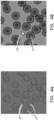
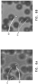
- Figs. 4A and 4B are microscope images acquired from the monolayer depth level under brightfield imaging using respective imaging parameters, in accordance with some applications of the present invention.
- a platelet 60 is identifiable within the images, and the platelet is surrounded by erythrocytes 61.
- the erythrocytes are in focus, since the images were acquired at the monolayer depth level, within which the erythrocytes are settled.
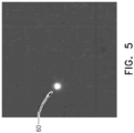
- Fig. 5 which is a fluoroscopic microscope image acquired from the monolayer depth level, in accordance with some applications of the present invention.
- a platelet 60 is identifiable within the images, and the platelet is surrounded by erythrocytes, which are faintly visible.
- Figs. 6A and 6B are microscope images acquired from an additional depth level (i.e., not the monolayer depth level) under brightfield imaging, using respective imaging parameters, in accordance with some applications of the present invention.
- a platelet 60 is identifiable within the images, and erythrocytes are visible in the background. The erythrocytes are out of focus since the images were not acquired at the monolayer depth level within which the erythrocytes have settled.
- Fig. 7 is a fluoroscopic microscope image acquired from the additional depth level, in accordance with some applications of the present invention. Again, a platelet 60 is identifiable within the images.
- platelets that have not settled within the monolayer are identified within the images that are acquired at the additional depth levels. (It is noted the identified platelets may include platelets that are out of focus within the additional depth levels.) Further typically, based upon the platelets that are identified within the images that are acquired at the additional depth levels, a count of the platelets that have not settled within the monolayer is estimated.
- the computer processor is configured to avoid duplicate counts of platelets within images that are acquired at adjacent depth levels, by identifying platelets within images that are acquired at adjacent depth levels that are disposed at similar locations to each other within the imaging plane and determining whether the platelets as identified at each of the depth levels are likely to correspond to a single (i.e., the same) platelet. For some applications, in determining whether the platelets as identified at each of the adjacent depth levels are likely to correspond to a single (i.e., the same) platelet, the computer processor accounts for lateral movement of the platelets between the images acquired at each of the adjacent depth levels.
- the computer processor is configured to account for interference with the visibility of platelets by white blood cells that are disposed within the monolayer and/or that are suspended within the cell solution. For example, within fluorescent images that are acquired at the additional depth levels, white blood cells that fluoresce within the monolayer are typically visible and interfere with the visibility of platelets that are disposed above the white blood cells. Therefore, platelets that are disposed above white blood cells within the monolayer may be excluded from the platelet count. Since platelets within the volumes above white blood cells are excluded from the count, the number of platelets within these volumes may be estimated. Alternatively or additionally, in order to perform the above-described estimate, the computer processor is configured to account for interference with the visibility of platelets by red blood cells that are disposed within the monolayer, and that may interfere with the identification of platelets within brightfield images.
- counts of platelets within the monolayer depth level and/or within the additional depth levels are normalized, and/or other statistical analysis techniques are applied to one or both of these counts.
- the count of the platelets within a given imaging field may be normalized with respect to the red blood cell count within that field, the time dynamics of the settling of the platelets may be incorporated into the estimate of platelets that are not settled within the monolayer, and/or outliers may be removed from the estimates.
- additional imaging fields are imaged, if there is a low platelet count, if there are outlier fields (e.g., with very high or very low platelet counts), and/or if imaging fields that are imaged at the additional depth levels are rejected due to errors.
- a count of the platelets that have settled within the monolayer is compared to a count of the platelets that have not settled within the monolayer.
- an output is generated in response to the comparison.
- a clinical condition is derived and outputted to a user, based upon the comparison of the count of the platelets that have settled within the monolayer and the count of the platelets that have not settled within the monolayer.
- the extent of platelet activation within the sample may be derived, at least partially based upon the comparison.
- a clinical condition may be derived based upon the size, shape, settling time, and/or settling dynamics of the platelets.
- the settling dynamics of the platelets are determined by imaging the same imaging field a plurality of times, with a time interval between each of the image acquisitions, and/or by determining until what height within the sample chamber platelets are present at a given time.
- the computer processor generates an output indicating that the sample should be prepared in a different way to how it was prepared (e.g., by modifying the diluent within which the sample is diluted, by adding a platelet activation agent, and/or by adding a coagulation agent), based upon the comparison.
- the portion of the blood sample is invalidated from being used for performing at least some measurements upon the sample. For example, if the ratio of unsettled platelets to settled platelets is greater than a first given threshold, and/or lower than a second given threshold, then this might be interpreted as being indicative of a problem with the sample and/or the preparation thereof.
- a parameter of the sample (such as platelet volume, mean platelet volume, and/or median platelet volume) is determined based upon a measurement that is performed upon the first portion of the sample.
- the measurement is calibrated, based upon the comparison between properties of the platelets that have settled within the monolayer and properties of the platelets that have not settled within the monolayer.
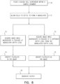
- Fig. 8 and Fig. 9 are flowcharts showing steps of methods that are performed with respect to platelets within a blood sample, in accordance with some applications of the present invention.
- a cell suspension is placed within a sample chamber (step 70) and allowed to settle to form a monolayer (step 71).
- step 71 a monolayer
- step 72 at least one image is acquired while the microscope is focused at the monolayer-depth-level (step 72), and platelets that have settled within the monolayer are identified in the image (step 74).
- At least one microscopic image of the sample is acquired, while the microscope is focused at a different depth level from the monolayer-depth-level (step 73), and platelets that have not settled within the monolayer are identified within the at least one microscopic image at the different depth level from the monolayer-depth-level (step 75).
- An output at least partially based upon the platelets identified as having settled within the monolayer and the platelets identified as not having settled within the monolayer, is generated (step 79), as shown. For some applications, as shown in Fig.
- a first platelet count of platelets that have settled within the monolayer is determined (step 76), and a second count of platelets that have not settled within the monolayer is determined (step 77).
- An output is then generated (step 78) based upon the first and second platelet counts. For example, an output may be generated based on a comparison between the first and second platelet counts, in accordance with the methods described hereinabove.
- the scope of the present invention includes applying the apparatus and methods described herein to a variety of entities within a variety of samples, mutatis mutandis.
- the apparatus and methods that are described with reference to identifying platelets that have not settled within a monolayer may be performed with respect to other entities within a blood sample, mutatis mutandis.
- Such entities may include white blood cells, anomalous white blood cells, circulating tumor cells, red blood cells, reticulocytes, Howell-Jolly bodies, foreign bodies (such as bacteria, fungi, yeast or parasites), entities that are added to a diluent (such as beads), etc.
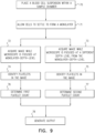
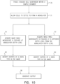
- Fig. 10 is a flowchart showing steps of a method that is performed with respect entities within a blood sample, in accordance with some applications of the present invention.
- a cell suspension is placed within a sample chamber (step 70) and allowed to settle to form a monolayer (step 71).
- step 71 a monolayer
- step 72 a monolayer-depth-level
- At least one microscopic image of the sample is acquired, while the microscope is focused at a different depth level from the monolayer-depth-level (step 73), and entities of the given type that have not settled within the monolayer are identified within the at least one microscopic image at the different depth level from the monolayer-depth-level (step 94).
- An output at least partially based upon the entities of the given type identified as having settled within the monolayer and the entities of the given type identified as not having settled within the monolayer, is generated, as shown (step 96).
- the apparatus and methods described herein are applied to a biological sample, such as, blood, saliva, semen, sweat, sputum, vaginal fluid, stool, breast milk, bronchoalveolar lavage, gastric lavage, tears and/or nasal discharge, mutatis mutandis.
- the biological sample may be from any living creature, and is typically from warm blooded animals.
- the biological sample is a sample from a mammal, e.g., from a human body.
- the sample is taken from any domestic animal, zoo animals and farm animals, including but not limited to dogs, cats, horses, cows and sheep.
- the biological sample is taken from animals that act as disease vectors including deer or rats.
- the apparatus and methods described herein are applied to a non-bodily sample.
- the sample is an environmental sample, such as, a water (e.g. groundwater) sample, surface swab, soil sample, air sample, or any combination thereof, mutatis mutandis.
- the sample is a food sample, such as, a meat sample, dairy sample, water sample, wash-liquid sample, beverage sample, and/or any combination thereof.
- the sample as described herein is a sample that includes blood or components thereof (e.g., a diluted or non-diluted whole blood sample, a sample including predominantly red blood cells, or a diluted sample including predominantly red blood cells), and parameters are determined relating to components in the blood such as platelets, white blood cells, anomalous white blood cells, circulating tumor cells, red blood cells, reticulocytes, Howell-Jolly bodies, etc.
- blood or components thereof e.g., a diluted or non-diluted whole blood sample, a sample including predominantly red blood cells, or a diluted sample including predominantly red blood cells
- parameters are determined relating to components in the blood such as platelets, white blood cells, anomalous white blood cells, circulating tumor cells, red blood cells, reticulocytes, Howell-Jolly bodies, etc.
- a computer-usable or computer-readable medium e.g., a non-transitory computer-readable medium
- a computer-usable or computer readable medium can be any apparatus that can comprise, store, communicate, propagate, or transport the program for use by or in connection with the instruction execution system, apparatus, or device.
- the medium can be an electronic, magnetic, optical, electromagnetic, infrared, or semiconductor system (or apparatus or device) or a propagation medium.
- the computer-usable or computer readable medium is a non-transitory computer-usable or computer readable medium.
- Examples of a computer-readable medium include a semiconductor or solid state memory, magnetic tape, a removable computer diskette, a random-access memory (RAM), a read-only memory (ROM), a rigid magnetic disk and an optical disk.
- Current examples of optical disks include compact disk-read only memory (CD-ROM), compact disk-read/write (CD-R/W) and DVD.
- a data processing system suitable for storing and/or executing program code will include at least one processor (e.g., computer processor 28) coupled directly or indirectly to memory elements (e.g., memory 30) through a system bus.
- the memory elements can include local memory employed during actual execution of the program code, bulk storage, and cache memories which provide temporary storage of at least some program code in order to reduce the number of times code must be retrieved from bulk storage during execution.
- the system can read the inventive instructions on the program storage devices and follow these instructions to execute the methodology of the embodiments of the invention.
- Network adapters may be coupled to the processor to enable the processor to become coupled to other processors or remote printers or storage devices through intervening private or public networks.
- Modems, cable modem and Ethernet cards are just a few of the currently available types of network adapters.
- Computer program code for carrying out operations of the present invention may be written in any combination of one or more programming languages, including an object-oriented programming language such as Java, Smalltalk, C++ or the like and conventional procedural programming languages, such as the C programming language or similar programming languages.
- object-oriented programming language such as Java, Smalltalk, C++ or the like
- conventional procedural programming languages such as the C programming language or similar programming languages.
- These computer program instructions may also be stored in a computer-readable medium (e.g., a non-transitory computer-readable medium) that can direct a computer or other programmable data processing apparatus to function in a particular manner, such that the instructions stored in the computer-readable medium produce an article of manufacture including instruction means which implement the function/act specified in the flowchart blocks and algorithms.
- the computer program instructions may also be loaded onto a computer or other programmable data processing apparatus to cause a series of operational steps to be performed on the computer or other programmable apparatus to produce a computer implemented process such that the instructions which execute on the computer or other programmable apparatus provide processes for implementing the functions/acts specified in the algorithms described in the present application.
- Computer processor 28 is typically a hardware device programmed with computer program instructions to produce a special purpose computer. For example, when programmed to perform the algorithms described herein, computer processor 28 typically acts as a special purpose sample-analysis computer processor. Typically, the operations described herein that are performed by computer processor 28 transform the physical state of memory 30, which is a real physical article, to have a different magnetic polarity, electrical charge, or the like depending on the technology of the memory that is used.
Landscapes
- Chemical & Material Sciences (AREA)
- Biochemistry (AREA)
- Physics & Mathematics (AREA)
- Health & Medical Sciences (AREA)
- Life Sciences & Earth Sciences (AREA)
- Analytical Chemistry (AREA)
- General Health & Medical Sciences (AREA)
- General Physics & Mathematics (AREA)
- Immunology (AREA)
- Pathology (AREA)
- Dispersion Chemistry (AREA)
- Investigating Or Analysing Biological Materials (AREA)
- Investigating Or Analysing Materials By Optical Means (AREA)
Claims (15)
- Verfahren mit den Schritten:Platzieren mindestens eines Teils einer Blutprobe, die eine Zellsuspension ist, in einer Probenkammer (52), die ein Hohlraum (55) mit einer Grundfläche (57) ist;Absetzenlassen der Zellen in der Zellsuspension auf der Grundfläche (57) der Probenkammer (52), um eine Monoschicht von Zellen auf der Grundfläche (57) der Probenkammer (52) zu bilden;Aufnehmen mindestens eines ersten mikroskopischen Bildes von mindestens einem Teil der Monoschicht von Zellen unter Verwendung eines Mikroskops (37), während das Mikroskop (37) auf eine Monoschicht-Tiefenschärfe fokussiert ist, bei der die Monoschicht im Fokus des Mikroskops (37) liegt;Identifizieren von Blutplättchen, die sich in der Monoschicht abgesetzt haben, in dem mindestens einen mikroskopischen Bild;Bestimmen einer ersten Blutplättchenzahl von in der Monoschicht abgesetzten Blutplättchen auf der Grundlage der in dem mindestens einen mikroskopischen Bild identifizierten Blutplättchen;Aufnehmen mindestens eines weiteren mikroskopischen Bildes von dem Teil der Probe unter Verwendung des Mikroskops (37), während das Mikroskop (37) auf eine andere Tiefenebene als die Monoschicht-Tiefenebene fokussiert ist;Identifizieren von Blutplättchen, die sich nicht innerhalb der Monoschicht abgesetzt haben, in dem mindestens einen zusätzlichen mikroskopischen Bild;Bestimmen einer zweiten Blutplättchenzahl von nicht in der Monoschicht abgesetzten Blutplättchen basierend auf den in dem mindestens einen zusätzlichen mikroskopischen Bild identifizierten Blutplättchen;wobei das Verfahren gekennzeichnet ist durchVergleichen der ersten und zweiten Blutplättchenzahl miteinander; undErzeugen einer Ausgabe basierend auf dem Vergleich der ersten und zweiten Blutplättchenzahl miteinander.
- Verfahren nach Anspruch 1 mit dem weiteren Schritt des Ableitens eines Fehlers bei der Blutprobe, der zumindest teilweise auf dem Vergleich der ersten und zweiten Blutplättchenzahl basiert, wobei das Erzeugen der Ausgabe die Erzeugung einer Angabe des Fehlers umfasst.
- Verfahren nach Anspruch 1 mit dem weiteren Schritt des Ableitens eines Fehlers bei der Vorbereitung der Blutprobe, der zumindest teilweise auf dem Vergleich der ersten und zweiten Blutplättchenzahl basiert, wobei das Erzeugen der Ausgabe die Erzeugung einer Angabe des Fehlers umfasst.
- Verfahren nach Anspruch 1, ferner umfassend das Ableiten eines klinischen Zustands des Subjekts zumindest teilweise basierend auf dem Vergleich der ersten und zweiten Thrombozytenzahl miteinander, wobei das Erzeugen der Ausgabe das Erzeugen einer Angabe des klinischen Zustands umfasst.
- Verfahren nach Anspruch 1, ferner umfassend das Ableiten eines Maßes der Blutplättchenaktivierung zumindest teilweise basierend auf dem Vergleich der ersten und zweiten Blutplättchenzahl miteinander, wobei das Erzeugen der Ausgabe das Erzeugen einer Angabe der Blutplättchenaktivierung umfasst.
- Verfahren nach Anspruch 1, bei dem das Identifizieren von in dem mindestens einen zusätzlichen mikroskopischen Bild nicht in der Monoschicht abgesetzten Blutplättchen das Berücksichtigen einer Beeinträchtigung der Sichtbarkeit von Blutplättchen durch in der Monoschicht angeordnete weiße Blutkörperchen umfasst.
- Verfahren nach Anspruch 1, bei dem das Identifizieren von in dem mindestens einen zusätzlichen mikroskopischen Bild nicht in der Monoschicht abgesetzten Blutplättchen das Berücksichtigen einer Beeinträchtigung der Sichtbarkeit von Blutplättchen durch in der Monoschicht angeordnete rote Blutkörperchen umfasst.
- Verfahren nach Anspruch 1, bei dem:das Erfassen des mindestens einen ersten mikroskopischen Bildes des Teils der Monoschicht von Zellen das Erfassen einer ersten Anzahl von Bildern der Monoschicht in jeweiligen Bildfeldern umfasst, während das Mikroskop (37) auf eine oder mehrere Monoschicht-Tiefenniveaus fokussiert ist, bei denen die Monoschicht im Fokus des Mikroskops (37) ist;das Erfassen des mindestens einen zusätzlichen mikroskopischen Bildes des Teils der Probe das Erfassen einer zweiten Anzahl von Bildern in jeweiligen Bildfeldern umfasst, während das Mikroskop (37) auf die von der Monoschicht-Tiefenebene verschiedene Tiefenebene fokussiert ist; undein Verhältnis zwischen der ersten Anzahl und der zweiten Anzahl größer als 2: 1 ist.
- Verfahren nach einem der Ansprüche 1 bis 8, bei dem:das Erfassen des mindestens einen zusätzlichen mikroskopischen Bildes des Teils der Probe das Erfassen einer Mehrzahl zusätzlicher mikroskopischer Bilder umfasst, während das Mikroskop (37) auf jeweils von der Monoschicht-Tiefenebene verschiedene Tiefenebenen fokussiert ist; unddas Identifizieren von Blutplättchen, die sich nicht in der Monoschicht abgesetzt haben, in dem mindestens einen zusätzlichen mikroskopischen Bild das Vermeiden von Doppelzählungen von Blutplättchen in in benachbarten Tiefenebenen aufgenommenen Bildern umfasst.
- Verfahren nach Anspruch 9, bei dem das Vermeiden von Doppelzählungen von Blutplättchen in in benachbarten Tiefenebenen aufgenommenen Bildern das Berücksichtigen der seitlichen Bewegung von Blutplättchen zwischen Aufnahmen der in benachbarten Tiefenebenen aufgenommenen Bilder umfasst.
- Vorrichtung, umfassend:eine Probenkammer (52), die ein Hohlraum (55) mit einer Grundfläche (57) ist und die konfiguriert ist, um mindestens einen Teil einer Blutprobe, die eine Zellsuspension ist, aufzunehmen und es den Zellen in der Zellsuspension zu ermöglichen, sich auf der Grundfläche (57) der Probenkammer (52) abzusetzen, um eine Monoschicht von Zellen auf der Grundfläche (57) der Probenkammer (52) zu bilden;ein Mikroskop (37), das konfiguriert ist zum Erfassen von:mindestens einem ersten mikroskopischen Bild von mindestens einem Teil der Monoschicht von Zellen, während das Mikroskop (37) auf eine Monoschicht-Tiefenschicht fokussiert ist, bei der die Monoschicht im Fokus des Mikroskops (37) liegt, undmindestens einem zusätzlichen mikroskopischen Bild von dem Teil der Probe, während das Mikroskop (37) auf eine andere Tiefenebene als die Monoschicht-Tiefenebene fokussiert ist; undeinen Computerprozessor (28), der konfiguriert ist zum:Identifizieren von Blutplättchen, die sich in der Monoschicht abgesetzt haben, in dem mindestens einen ersten mikroskopischen Bild,Bestimmen einer ersten Blutplättchenzahl von in der Monoschicht abgesetzten Blutplättchen auf der Grundlage der in dem mindestens einen ersten mikroskopischen Bild identifizierten Blutplättchen,Identifizieren von nicht in der Monoschicht abgesetzten Blutplättchen in dem mindestens einen zusätzlichen mikroskopischen Bild,Bestimmen einer zweiten Blutplättchenzahl von nicht in der Monoschicht abgesetzten Blutplättchen auf der Grundlage der in dem mindestens einen zusätzlichen mikroskopischen Bild identifizierten Blutplättchen,dadurch gekennzeichnet, dass der Computerprozessor (28) ferner konfiguriert ist zum:Vergleichen der ersten und zweiten Blutplättchenzahl miteinander; undErzeugen einer Ausgabe basierend auf dem Vergleichen der ersten und zweiten Blutplättchenzahl miteinander.
- Vorrichtung nach Anspruch 11, bei der der Computerprozessor (28) konfiguriert ist, um einen Fehler bei der Blutprobe zumindest teilweise basierend auf dem Vergleich der ersten und zweiten Thrombozytenzahl miteinander abzuleiten, wobei das Erzeugen der Ausgabe das Erzeugen einer Angabe des Fehlers umfasst.
- Vorrichtung nach Anspruch 11, bei der der Computerprozessor (28) konfiguriert ist, um einen Fehler bei der Vorbereitung der Blutprobe zumindest teilweise auf der Grundlage eines Vergleichs der ersten und zweiten Thrombozytenzahl miteinander abzuleiten, wobei das Erzeugen der Ausgabe das Erzeugen einer Angabe des Fehlers umfasst.
- Vorrichtung nach Anspruch 11, bei der der Computerprozessor (28) konfiguriert ist, um einen klinischen Zustand des Subjekts zumindest teilweise basierend auf dem Vergleich der ersten und zweiten Blutplättchenzahl miteinander abzuleiten, wobei das Erzeugen der Ausgabe das Erzeugen einer Angabe des klinischen Zustands umfasst.
- Vorrichtung nach Anspruch 11, bei der der Computerprozessor (28) konfiguriert ist, um ein Maß für die Blutplättchenaktivierung zumindest teilweise basierend auf dem Vergleich der ersten und zweiten Blutplättchenzahl miteinander abzuleiten, wobei das Erzeugen der Ausgabe das Erzeugen einer Angabe der Blutplättchenaktivierung umfasst.
Applications Claiming Priority (2)
| Application Number | Priority Date | Filing Date | Title |
|---|---|---|---|
| US201962946998P | 2019-12-12 | 2019-12-12 | |
| PCT/IB2020/061724 WO2021116955A1 (en) | 2019-12-12 | 2020-12-10 | Detecting platelets in a blood sample |
Publications (3)
| Publication Number | Publication Date |
|---|---|
| EP4073508A1 EP4073508A1 (de) | 2022-10-19 |
| EP4073508C0 EP4073508C0 (de) | 2025-02-05 |
| EP4073508B1 true EP4073508B1 (de) | 2025-02-05 |
Family
ID=73854856
Family Applications (1)
| Application Number | Title | Priority Date | Filing Date |
|---|---|---|---|
| EP20825250.2A Active EP4073508B1 (de) | 2019-12-12 | 2020-12-10 | Nachweis von blutplättchen in einer blutprobe |
Country Status (10)
| Country | Link |
|---|---|
| US (1) | US12523579B2 (de) |
| EP (1) | EP4073508B1 (de) |
| JP (1) | JP2023506416A (de) |
| CN (1) | CN114787625B (de) |
| AU (1) | AU2020400400A1 (de) |
| BR (1) | BR112022011233A2 (de) |
| CA (1) | CA3160688A1 (de) |
| MX (1) | MX2022007119A (de) |
| WO (1) | WO2021116955A1 (de) |
| ZA (1) | ZA202206311B (de) |
Families Citing this family (11)
| Publication number | Priority date | Publication date | Assignee | Title |
|---|---|---|---|---|
| CN110892247B (zh) | 2017-08-17 | 2023-08-25 | 雅培医护站股份有限公司 | 用于执行光学和电化学测定的设备、系统和方法 |
| EP3669178B1 (de) | 2017-08-17 | 2023-07-19 | Abbott Point Of Care Inc | Systeme zur durchführung optischer und elektrochemischer analysen |
| WO2022009104A2 (en) | 2020-07-07 | 2022-01-13 | S.D. Sight Diagnostics Ltd | Focusing a microscope using fluorescent images |
| EP4469204A1 (de) | 2022-01-25 | 2024-12-04 | S.D. Sight Diagnostics Ltd. | Probenträger zur verwendung mit einer körperprobe |
| CN114813522B (zh) * | 2022-06-29 | 2022-10-21 | 深圳安侣医学科技有限公司 | 基于显微放大数字图像的血液细胞分析方法及系统 |
| GB202218615D0 (en) | 2022-12-12 | 2023-01-25 | S D Sight Diagnostics Ltd | System and method for analyzing bodily samples |
| WO2024192493A1 (pt) * | 2023-03-22 | 2024-09-26 | Hi Technologies Ltda. | Analisador de amostras biológicas por microscopia e espectrometria |
| WO2024236537A1 (en) | 2023-05-18 | 2024-11-21 | S.D. Sight Diagnostics Ltd | Apparatus and methods for microscopic analysis of a biological sample |
| WO2025101870A1 (en) * | 2023-11-08 | 2025-05-15 | Idexx Laboratories, Inc. | Systems and methods for biological optical imaging with artificial particles as references |
| WO2025117626A1 (en) * | 2023-11-30 | 2025-06-05 | Idexx Laboratories, Inc. | Quantifying constituents in a sample chamber using images of depth regions |
| US20250180457A1 (en) * | 2023-11-30 | 2025-06-05 | Idexx Laboratories, Inc. | Imaging white blood cells in presence of red blood cells |
Family Cites Families (440)
| Publication number | Priority date | Publication date | Assignee | Title |
|---|---|---|---|---|
| US3203768A (en) | 1961-08-01 | 1965-08-31 | Westinghouse Electric Corp | Apparatus of zone refining and controlling solute segregation in solidifying melts by electromagnetic means |
| US3603156A (en) | 1970-02-04 | 1971-09-07 | Gradko Glass Lab Inc | Disposable dilution system |
| US3676076A (en) | 1970-09-24 | 1972-07-11 | Gradko Glass Lab Inc | Disposable container |
| GB1324323A (en) | 1971-02-05 | 1973-07-25 | Image Analysing Computers Ltd | Automatic focusing of an optical image |
| US3967056A (en) | 1973-02-26 | 1976-06-29 | Minolta Camera Kabushiki Kaisha | Automatic focusing apparatus |
| US3916205A (en) | 1973-05-31 | 1975-10-28 | Block Engineering | Differential counting of leukocytes and other cells |
| JPS5916667B2 (ja) | 1975-02-28 | 1984-04-17 | トウアイヨウデンシ カブシキガイシヤ | 自動血液分析装置 |
| US4076419A (en) | 1976-07-12 | 1978-02-28 | Kleker Richard G | Method and apparatus for hematology |
| US4199748A (en) | 1976-11-01 | 1980-04-22 | Rush-Presbyterian-St. Luke's Medical Center | Automated method and apparatus for classification of cells with application to the diagnosis of anemia |
| US4209548A (en) | 1976-11-01 | 1980-06-24 | Rush-Presbyterian-St. Luke's Medical Center | Method for the preparation of blood samples for automated analysis |
| US4097845A (en) | 1976-11-01 | 1978-06-27 | Rush-Presbyterian-St. Luke's Medical Center | Method of and an apparatus for automatic classification of red blood cells |
| DE2910875C2 (de) | 1979-03-20 | 1985-11-14 | Kernforschungszentrum Karlsruhe Gmbh, 7500 Karlsruhe | Verfahren zur automatischen Scharfeinstellung |
| US4453266A (en) | 1980-04-21 | 1984-06-05 | Rush-Presbyterian-St. Luke's Medical Center | Method and apparatus for measuring mean cell volume of red blood cells |
| ES8307122A1 (es) | 1981-08-27 | 1983-06-16 | Becton Dickinson Co | "perfeccionamientos introducidos en un aparato para introducir reactivos en recipientes de recogida de muestras". |
| US4454235A (en) | 1982-06-01 | 1984-06-12 | Miles Laboratories, Inc. | Capillary tube holder for liquid transfer in immunoassay |
| US4494479A (en) | 1982-10-14 | 1985-01-22 | Coulter Electronics, Inc. | Slide preparation apparatus |
| AU2490284A (en) | 1983-01-10 | 1984-08-02 | Gen-Probe Incorporated | Method for detecting, identitifying, and quantitating organisms and viruses |
| US4580895A (en) | 1983-10-28 | 1986-04-08 | Dynatech Laboratories, Incorporated | Sample-scanning photometer |
| JPS60162955A (ja) | 1984-02-03 | 1985-08-24 | Hitachi Ltd | 血球自動分析装置 |
| EP0183717B1 (de) | 1984-05-15 | 1989-11-23 | Cytocolor Inc. | Einzelner farbstoff zur ersetzung von variablen romanowsky-zusammensetzungen enthaltend mehrere farbstoffe zur identifizierung eines bestandteils in menschlichen biopsieproben |
| US4700298A (en) | 1984-09-14 | 1987-10-13 | Branko Palcic | Dynamic microscope image processing scanner |
| JPS61198204A (ja) | 1985-02-28 | 1986-09-02 | Disco Abrasive Sys Ltd | 顕微鏡のオ−トフオ−カス方法 |
| US5164598A (en) | 1985-08-05 | 1992-11-17 | Biotrack | Capillary flow device |
| US4761381A (en) | 1985-09-18 | 1988-08-02 | Miles Inc. | Volume metering capillary gap device for applying a liquid sample onto a reactive surface |
| US5281517A (en) | 1985-11-04 | 1994-01-25 | Cell Analysis Systems, Inc. | Methods for immunoploidy analysis |
| DE3707487A1 (de) | 1986-05-16 | 1987-11-26 | Reichert Optische Werke Ag | Verfahren zur autofokussierung von mikroskopen und mikroskope mit einer autofokussierung |
| JPH0774856B2 (ja) | 1986-10-16 | 1995-08-09 | オリンパス光学工業株式会社 | 自動焦点調節方法 |
| US4774192A (en) | 1987-01-28 | 1988-09-27 | Technimed Corporation | A dry reagent delivery system with membrane having porosity gradient |
| US4849430A (en) | 1988-03-09 | 1989-07-18 | Monsanto Company | Method of inhibiting virus |
| US6262798B1 (en) | 1992-09-29 | 2001-07-17 | Board Of Regents, The University Of Texas System | Method and apparatus for direct spectrophotometric measurements in unaltered whole blood |
| US5001067A (en) | 1989-09-18 | 1991-03-19 | Nova Biomedical Corporation | Determining the concentration of water soluble species in biological fluid |
| US5064282A (en) | 1989-09-26 | 1991-11-12 | Artel, Inc. | Photometric apparatus and method for measuring hemoglobin |
| US5229265A (en) | 1990-03-13 | 1993-07-20 | Litron Laboratories | Process for analyzing clastogenic agents |
| EP0479231B1 (de) | 1990-10-01 | 1996-03-27 | Canon Kabushiki Kaisha | Vorrichtung und Verfahren zur Messung einer Probe |
| US5784162A (en) | 1993-08-18 | 1998-07-21 | Applied Spectral Imaging Ltd. | Spectral bio-imaging methods for biological research, medical diagnostics and therapy |
| JP3391451B2 (ja) | 1992-02-24 | 2003-03-31 | クールター インターナショナル コーポレイション | 白血球類縁体用の血液対照組成物,およびその調製方法および使用方法 |
| US5376790A (en) | 1992-03-13 | 1994-12-27 | Park Scientific Instruments | Scanning probe microscope |
| US5331958A (en) | 1992-03-31 | 1994-07-26 | University Of Manitoba | Spectrophotometric blood analysis |
| US5430542A (en) | 1992-04-10 | 1995-07-04 | Avox Systems, Inc. | Disposable optical cuvette |
| US5342790A (en) | 1992-10-30 | 1994-08-30 | Becton Dickinson And Company | Apparatus for indirect fluorescent assay of blood samples |
| DE69424079T2 (de) | 1993-02-22 | 2000-09-07 | Sysmex Corp., Kobe | Ein Reagenz zum Nachweis von malariainfizierten Zellen und ein Nachweis-Verfahren für malariainfizierte Zellen unter Verwendung desselben |
| US5483055A (en) | 1994-01-18 | 1996-01-09 | Thompson; Timothy V. | Method and apparatus for performing an automatic focus operation for a microscope |
| JP5161052B2 (ja) | 2008-12-04 | 2013-03-13 | オリンパス株式会社 | 顕微鏡システム、標本観察方法およびプログラム |
| US5590660A (en) | 1994-03-28 | 1997-01-07 | Xillix Technologies Corp. | Apparatus and method for imaging diseased tissue using integrated autofluorescence |
| US5782770A (en) | 1994-05-12 | 1998-07-21 | Science Applications International Corporation | Hyperspectral imaging methods and apparatus for non-invasive diagnosis of tissue for cancer |
| JPH07325247A (ja) | 1994-05-31 | 1995-12-12 | Nikon Corp | 自動合焦装置 |
| MX9606683A (es) | 1994-07-01 | 1997-12-31 | Jeffrey H Price | Sistema de enfoque automatico para microscopia de exploracion. |
| US5932872A (en) | 1994-07-01 | 1999-08-03 | Jeffrey H. Price | Autofocus system for scanning microscopy having a volume image formation |
| US5499097A (en) | 1994-09-19 | 1996-03-12 | Neopath, Inc. | Method and apparatus for checking automated optical system performance repeatability |
| US5978497A (en) | 1994-09-20 | 1999-11-02 | Neopath, Inc. | Apparatus for the identification of free-lying cells |
| US5566249A (en) | 1994-09-20 | 1996-10-15 | Neopath, Inc. | Apparatus for detecting bubbles in coverslip adhesive |
| WO1996012981A1 (en) | 1994-10-21 | 1996-05-02 | Kla Instruments Corporation | Autofocusing apparatus and method for high resolution microscope system |
| AU4131796A (en) | 1994-10-27 | 1996-05-23 | Entremed, Inc | Identification of infection with flow cytometry |
| DE69417900T2 (de) | 1994-11-17 | 1999-11-11 | Chemunex, Maisons-Alfort | Vorrichtung und Verfahren zum schnellen und hochempfindlichen Erkennen und Zählen von Mikroorganismen mittels Fluoreszenz |
| SE504193C2 (sv) | 1995-04-21 | 1996-12-02 | Hemocue Ab | Kapillär mikrokyvett |
| JPH08313340A (ja) | 1995-05-23 | 1996-11-29 | Eresu Trading Kk | 紫外線測定シート及び紫外線測定機能付き日焼け装置 |
| US5671288A (en) | 1995-05-31 | 1997-09-23 | Neopath, Inc. | Method and apparatus for assessing slide and specimen preparation quality |
| US5625706A (en) | 1995-05-31 | 1997-04-29 | Neopath, Inc. | Method and apparatus for continously monitoring and forecasting slide and specimen preparation for a biological specimen population |
| US5958250A (en) * | 1995-06-07 | 1999-09-28 | Baxter International Inc. | Blood processing systems and methods which optically derive the volume of platelets contained in a plasma constituent |
| US5985595A (en) | 1995-06-07 | 1999-11-16 | The University Of Connecticut | Early detection of Borrelia infection |
| JP3640712B2 (ja) | 1995-08-11 | 2005-04-20 | 衛 石澤 | 血小板検査方法及び血小板検査用採血管 |
| US5670240A (en) | 1995-11-09 | 1997-09-23 | Flex Products, Inc. | Embossed substrate and photoreceptor device incorporating the same and method |
| US6005964A (en) | 1996-01-24 | 1999-12-21 | The Board Of Trustees Of The University Of Illinois | Automatic machine vision microscope slide inspection system and method |
| KR0165522B1 (ko) | 1996-05-23 | 1999-03-20 | 김광호 | 혈증성분 무혈진단을 위한 최적지점 검색장치및 이를 이용한 무혈진단기 |
| GB9620934D0 (en) | 1996-10-08 | 1996-11-27 | Molecular Drives Limited | Multi-well containers |
| DE19653413C2 (de) | 1996-12-22 | 2002-02-07 | Stefan Hell | Rastermikroskop, bei dem eine Probe in mehreren Probenpunkten gleichzeitig optisch angeregt wird |
| DE69812928T2 (de) | 1997-05-05 | 2004-03-04 | Chemometec A/S | Bestimmung von partikeln in einer flüssigen probe |
| US5939709A (en) | 1997-06-19 | 1999-08-17 | Ghislain; Lucien P. | Scanning probe optical microscope using a solid immersion lens |
| US6074879A (en) | 1997-06-23 | 2000-06-13 | Bayer Corporation | Synthetic polymer particles for use as standards and calibrators in flow cytometry |
| JPH1173903A (ja) | 1997-08-28 | 1999-03-16 | Jeol Ltd | 走査電子顕微鏡のオートフォーカス方法 |
| GB2329014A (en) | 1997-09-05 | 1999-03-10 | Colin Campbell | Automated identification of tubercle bacilli |
| US6064474A (en) | 1998-02-06 | 2000-05-16 | Optical Sensors, Inc. | Optical measurement of blood hematocrit incorporating a self-calibration algorithm |
| US20020028471A1 (en) | 1998-02-20 | 2002-03-07 | Oberhardt Bruce J. | Cell analysis methods and apparatus |
| US6350613B1 (en) | 1998-03-07 | 2002-02-26 | Belton Dickinson & Co. | Determination of white blood cell differential and reticulocyte counts |
| US5948686A (en) | 1998-03-07 | 1999-09-07 | Robert A. Leuine | Method for performing blood cell counts |
| US6929953B1 (en) | 1998-03-07 | 2005-08-16 | Robert A. Levine | Apparatus for analyzing biologic fluids |
| US6723290B1 (en) | 1998-03-07 | 2004-04-20 | Levine Robert A | Container for holding biologic fluid for analysis |
| US6235536B1 (en) | 1998-03-07 | 2001-05-22 | Robert A. Levine | Analysis of quiescent anticoagulated whole blood samples |
| US6027695A (en) | 1998-04-01 | 2000-02-22 | Dupont Pharmaceuticals Company | Apparatus for holding small volumes of liquids |
| KR20010083041A (ko) | 1998-06-02 | 2001-08-31 | 추후 | 파수 도메인 반사측정과 배경 진폭 감소 및 보상을 사용한공초점 간섭 마이크로스코피용 방법 및 장치 |
| IT1304854B1 (it) | 1998-07-29 | 2001-04-05 | Paolo Fazii | Metodo di differenziazione cromatica dei microrganismi in fluorescenza |
| US6132685A (en) | 1998-08-10 | 2000-10-17 | Caliper Technologies Corporation | High throughput microfluidic systems and methods |
| US6320979B1 (en) | 1998-10-06 | 2001-11-20 | Canon Kabushiki Kaisha | Depth of field enhancement |
| US6496260B1 (en) | 1998-12-23 | 2002-12-17 | Molecular Devices Corp. | Vertical-beam photometer for determination of light absorption pathlength |
| JP2000199845A (ja) | 1999-01-05 | 2000-07-18 | Ricoh Co Ltd | 自動合焦装置及び自動合焦方法 |
| US6330348B1 (en) | 1999-01-21 | 2001-12-11 | Resolution Sciences Corporation | Method and apparatus for measurement of microtome performance |
| US8406498B2 (en) | 1999-01-25 | 2013-03-26 | Amnis Corporation | Blood and cell analysis using an imaging flow cytometer |
| US6344340B1 (en) | 1999-03-01 | 2002-02-05 | Novus International, Inc. | Viability assay for sporocyst-forming protozoa |
| US6582964B1 (en) | 1999-05-12 | 2003-06-24 | Cme Telemetrix Inc. | Method and apparatus for rapid measurement of HbA1c |
| US7034883B1 (en) | 1999-08-10 | 2006-04-25 | Cellavision Ab | Automatic focusing |
| US6949384B2 (en) | 2001-12-21 | 2005-09-27 | Spectromedical Inc. | Method for monitoring degradation of Hb-based blood substitutes |
| US6611777B2 (en) | 1999-11-23 | 2003-08-26 | James Samsoondar | Method for calibrating spectrophotometric apparatus |
| US6711516B2 (en) | 1999-11-23 | 2004-03-23 | Spectromedical Inc. | Method for calibrating spectrophotometric apparatus |
| WO2001049874A1 (en) | 2000-01-06 | 2001-07-12 | Caliper Technologies Corp. | Methods and systems for monitoring intracellular binding reactions |
| US6836559B2 (en) | 2000-03-09 | 2004-12-28 | The Regents Of The University Of California | Automated video-microscopic imaging and data acquisition system for colloid deposition measurements |
| WO2001078005A2 (en) | 2000-04-11 | 2001-10-18 | Cornell Research Foundation, Inc. | System and method for three-dimensional image rendering and analysis |
| US6711283B1 (en) | 2000-05-03 | 2004-03-23 | Aperio Technologies, Inc. | Fully automatic rapid microscope slide scanner |
| US7668362B2 (en) | 2000-05-03 | 2010-02-23 | Aperio Technologies, Inc. | System and method for assessing virtual slide image quality |
| US6799119B1 (en) | 2000-05-15 | 2004-09-28 | Colorado School Of Mines | Method for detection of biological related materials using biomarkers |
| US7630063B2 (en) | 2000-08-02 | 2009-12-08 | Honeywell International Inc. | Miniaturized cytometer for detecting multiple species in a sample |
| JP2003535330A (ja) | 2000-06-02 | 2003-11-25 | メディコメトリクス アンパーツゼルスカブ | 生物試料の分類のための方法及びシステム |
| US6554788B1 (en) | 2000-06-02 | 2003-04-29 | Cobe Cardiovascular, Inc. | Hematocrit sampling system |
| US6632681B1 (en) | 2000-07-24 | 2003-10-14 | Ey Laboratories | Reagent delivery device and method of use |
| US6819408B1 (en) | 2000-09-27 | 2004-11-16 | Becton, Dickinson And Company | Method for obtaining a monolayer of desired particles in a liquid sample |
| US6448024B1 (en) | 2000-10-03 | 2002-09-10 | Roche Diagnostics Corporation | Method, reagent, cartridge, and device for determining fibrinogen |
| US20100261159A1 (en) | 2000-10-10 | 2010-10-14 | Robert Hess | Apparatus for assay, synthesis and storage, and methods of manufacture, use, and manipulation thereof |
| US7027628B1 (en) | 2000-11-14 | 2006-04-11 | The United States Of America As Represented By The Department Of Health And Human Services | Automated microscopic image acquisition, compositing, and display |
| US7155049B2 (en) | 2001-01-11 | 2006-12-26 | Trestle Acquisition Corp. | System for creating microscopic digital montage images |
| WO2002056765A2 (en) | 2001-01-12 | 2002-07-25 | General Hospital Corporation | System and method for enabling simultaneous calibration and imaging of a medium |
| JP4937457B2 (ja) | 2001-03-01 | 2012-05-23 | オリンパス株式会社 | 顕微鏡制御装置、顕微鏡制御システム、顕微鏡の制御方法、プログラム、及び記録媒体 |
| US6898451B2 (en) | 2001-03-21 | 2005-05-24 | Minformed, L.L.C. | Non-invasive blood analyte measuring system and method utilizing optical absorption |
| US6519355B2 (en) | 2001-03-28 | 2003-02-11 | Alan C. Nelson | Optical projection imaging system and method for automatically detecting cells having nuclear and cytoplasmic densitometric features associated with disease |
| EP1381229B1 (de) | 2001-03-30 | 2008-12-10 | National Institute of Advanced Industrial Science and Technology | Echtzeit-omnifokus-mikroskopkamera |
| US7219016B2 (en) | 2001-04-20 | 2007-05-15 | Yale University | Systems and methods for automated analysis of cells and tissues |
| US6884983B2 (en) | 2002-06-10 | 2005-04-26 | Palantyr Research, Llc | Imaging system for examining biological material |
| US7439478B2 (en) | 2001-07-06 | 2008-10-21 | Palantyr Research, Llc | Imaging system, methodology, and applications employing reciprocal space optical design having at least one pixel being scaled to about a size of a diffraction-limited spot defined by a microscopic optical system |
| US7132636B1 (en) | 2001-07-06 | 2006-11-07 | Palantyr Research, Llc | Imaging system and methodology employing reciprocal space optical design |
| US7248716B2 (en) | 2001-07-06 | 2007-07-24 | Palantyr Research, Llc | Imaging system, methodology, and applications employing reciprocal space optical design |
| US7863552B2 (en) | 2001-07-06 | 2011-01-04 | Palantyr Research Llc | Digital images and related methodologies |
| US7105795B2 (en) | 2001-07-06 | 2006-09-12 | Palantyr Research, Llc | Imaging system, methodology, and applications employing reciprocal space optical design |
| US7288751B2 (en) | 2001-07-06 | 2007-10-30 | Palantyr Research, Llc | Imaging system, methodology, and applications employing reciprocal space optical design |
| US7385168B2 (en) | 2001-07-06 | 2008-06-10 | Palantyr Research, Llc | Imaging system, methodology, and applications employing reciprocal space optical design |
| US7338168B2 (en) | 2001-07-06 | 2008-03-04 | Palantyr Research, Llc | Particle analyzing system and methodology |
| US6872930B2 (en) | 2003-03-31 | 2005-03-29 | Palantyr Research, Llc | Imaging system and methodology employing reciprocal space optical design |
| US7151246B2 (en) | 2001-07-06 | 2006-12-19 | Palantyr Research, Llc | Imaging system and methodology |
| US6664528B1 (en) | 2001-07-06 | 2003-12-16 | Palantyr Research, Llc | Imaging system and methodology employing reciprocal space optical design |
| JP4751535B2 (ja) | 2001-07-26 | 2011-08-17 | シスメックス株式会社 | 分画方法とそれを用いた血液分析装置 |
| JP2005518553A (ja) | 2001-09-06 | 2005-06-23 | ジェノミック プロファイリング システムズ インコーポレイティッド | 細胞およびウイルスの迅速かつ高感度な検出方法 |
| US6989891B2 (en) | 2001-11-08 | 2006-01-24 | Optiscan Biomedical Corporation | Device and method for in vitro determination of analyte concentrations within body fluids |
| SE0104443D0 (sv) | 2001-12-28 | 2001-12-28 | Hemocue Ab | Analysis method and cuvette therefor |
| US7713474B2 (en) | 2002-01-15 | 2010-05-11 | Siemens Healthcare Diagnostics Inc. | Liquid permeable composition in dry reagent devices |
| US7133547B2 (en) | 2002-01-24 | 2006-11-07 | Tripath Imaging, Inc. | Method for quantitative video-microscopy and associated system and computer software program product |
| CN1625779A (zh) | 2002-01-28 | 2005-06-08 | 长冈实业株式会社 | 逻辑触发光生物盘的方法和设备 |
| JP5132867B2 (ja) | 2002-02-22 | 2013-01-30 | オリンパス アメリカ インコーポレイテツド | 仮想顕微鏡スライドを形成し使用する方法および装置、ならびにプログラム |
| US20030161514A1 (en) | 2002-02-28 | 2003-08-28 | Curry Bo U. | Bi-directional scanner control system |
| JP4084061B2 (ja) | 2002-03-18 | 2008-04-30 | 独立行政法人科学技術振興機構 | 高安定性光学顕微鏡 |
| DE10246777A1 (de) | 2002-03-21 | 2003-10-02 | Endress & Hauser Wetzer Gmbh | Vorrichtung zur Identifizierung eines Probennehmerbehälters |
| DE10217404A1 (de) | 2002-04-18 | 2003-11-06 | Leica Microsystems | Autofokusverfahren für ein Mikroskop und System zum Einstellen des Fokus für ein Mikroskop |
| US6658143B2 (en) | 2002-04-29 | 2003-12-02 | Amersham Biosciences Corp. | Ray-based image analysis for biological specimens |
| US6945943B2 (en) | 2002-05-01 | 2005-09-20 | Lifescan, Inc. | Analyte concentration determination devices and methods of using the same |
| CA2495787A1 (en) | 2002-05-01 | 2003-11-13 | Chromos Molecular Systems, Inc. | Methods for delivering nucleic acid molecules into cells and assessment thereof |
| US7272252B2 (en) | 2002-06-12 | 2007-09-18 | Clarient, Inc. | Automated system for combining bright field and fluorescent microscopy |
| AU2003251770B9 (en) | 2002-07-01 | 2009-06-04 | H. Lundbeck A/S | Recombinant tissue protective cytokines and encoding nucleic acids thereof for protection, restoration, and enhancement of responsive cells, tissues, and organs |
| JP2004061942A (ja) | 2002-07-30 | 2004-02-26 | Nikon Corp | 顕微鏡システム |
| US7838296B2 (en) | 2002-08-28 | 2010-11-23 | Separation Technology, Inc. | Methods and apparatus for ultrasonic determination of red blood cell indices |
| US7177767B2 (en) | 2002-10-18 | 2007-02-13 | Abaxis, Inc. | Systems and methods for the detection of short and long samples |
| JP3904505B2 (ja) | 2002-10-22 | 2007-04-11 | 独立行政法人科学技術振興機構 | マラリア感染診断剤及びマラリア原虫染色剤 |
| DE10254685A1 (de) | 2002-11-22 | 2004-06-03 | Roche Diagnostics Gmbh | Messeinrichtung zur optischen Untersuchung eines Testelements |
| US7490085B2 (en) | 2002-12-18 | 2009-02-10 | Ge Medical Systems Global Technology Company, Llc | Computer-assisted data processing system and method incorporating automated learning |
| US20040132171A1 (en) | 2003-01-06 | 2004-07-08 | Peter Rule | Wearable device for measuring analyte concentration |
| WO2004069984A2 (en) | 2003-01-31 | 2004-08-19 | Discovery Partners International | Automated imaging system and method |
| JP3869810B2 (ja) | 2003-02-24 | 2007-01-17 | 株式会社堀場製作所 | マイクロ血球カウンタ |
| US6955872B2 (en) | 2003-03-20 | 2005-10-18 | Coulter International Corp. | Dye compositions which provide enhanced differential fluorescence and light scatter characteristics |
| EP1625226A4 (de) | 2003-03-27 | 2007-01-10 | Bartron Medical Imaging Llc | System und verfahren zur schnellen identifizierung von krankheitserregern, bakterien und abnormen zellen |
| GB2401431B (en) | 2003-04-02 | 2005-04-27 | Amersham Biosciences Uk Ltd | Method of, and computer software for, classification of cells into subpopulations |
| US7329537B2 (en) | 2003-04-17 | 2008-02-12 | Nexcelom Bioscience, Llc | Micro-pattern embedded plastic optical film device for cell-based assays |
| US7706862B2 (en) | 2003-04-17 | 2010-04-27 | Research Foundation Of The City University Of New York | Detecting human cancer through spectral optical imaging using key water absorption wavelengths |
| US7324694B2 (en) | 2003-05-23 | 2008-01-29 | International Remote Imaging Systems, Inc. | Fluid sample analysis using class weights |
| DE10324248A1 (de) | 2003-05-28 | 2005-01-05 | Bayerische Motoren Werke Ag | Kunststoffträger und Verfahren zu seiner Herstellung |
| US20040241677A1 (en) | 2003-05-29 | 2004-12-02 | Lin Jeffrey S | Techniques for automated diagnosis of cell-borne anomalies with digital optical microscope |
| ES2920892T3 (es) | 2003-06-12 | 2022-08-11 | Accupath Diagnostic Laboratories Inc | Método para formar matrices de células |
| EP1649038B1 (de) | 2003-06-26 | 2014-07-02 | Litron Laboratories Ltd. | Verfahren zum auszählen von mikronuklei enthaltenden erythrozytenpopulationen mit unterscheidung von blutplättchen und/oder blutplättchenassoziierten aggregaten |
| AU2004273783A1 (en) | 2003-07-12 | 2005-03-31 | Accelr8 Technology Corporation | Sensitive and rapid biodetection |
| KR100573621B1 (ko) | 2003-07-18 | 2006-04-25 | 주식회사 디지탈바이오테크놀러지 | 세포 개체수 계수용 장치 및 그 제조방법 |
| EP1651947B1 (de) | 2003-07-19 | 2015-11-04 | NanoEnTek, Inc. | Vorrichtung zum zählen von mikropartikeln |
| SE0302114D0 (sv) | 2003-07-21 | 2003-07-21 | Cellavision Ab | Sätt att urskilja en objektkontur |
| US20050089208A1 (en) | 2003-07-22 | 2005-04-28 | Rui-Tao Dong | System and method for generating digital images of a microscope slide |
| US7846738B2 (en) | 2003-08-15 | 2010-12-07 | President And Fellows Of Harvard College | Study of polymer molecules and conformations with a nanopore |
| US7998435B2 (en) | 2003-09-19 | 2011-08-16 | Life Technologies Corporation | High density plate filler |
| US20060233671A1 (en) | 2003-09-19 | 2006-10-19 | Beard Nigel P | High density plate filler |
| US7697764B2 (en) | 2003-11-21 | 2010-04-13 | National University Corporation Kochi University | Similar pattern searching apparatus, method of similar pattern searching, program for similar pattern searching, and fractionation apparatus |
| US7030351B2 (en) | 2003-11-24 | 2006-04-18 | Mitutoyo Corporation | Systems and methods for rapidly automatically focusing a machine vision inspection system |
| JP4689171B2 (ja) | 2004-02-06 | 2011-05-25 | オリンパス株式会社 | 培養細胞の状態計測方法及び計測装置 |
| US7723124B2 (en) | 2004-02-09 | 2010-05-25 | Rapid Pathogen Screening, Inc. | Method for the rapid diagnosis of targets in human body fluids |
| WO2005076050A1 (en) | 2004-02-09 | 2005-08-18 | Philips Intellectual Property & Standards Gmbh | Fluorescence microscope arrangement |
| US9176121B2 (en) | 2004-02-13 | 2015-11-03 | Roche Diagnostics Hematology, Inc. | Identification of blood elements using inverted microscopy |
| JP4417143B2 (ja) | 2004-03-11 | 2010-02-17 | シスメックス株式会社 | 試料分析装置、プログラムおよびそのプログラムを記録した記録媒体 |
| US8105554B2 (en) | 2004-03-12 | 2012-01-31 | Life Technologies Corporation | Nanoliter array loading |
| US7925070B2 (en) | 2004-03-30 | 2011-04-12 | Sysmex Corporation | Method for displaying virtual slide and terminal device for displaying virtual slide |
| US20060051778A1 (en) | 2004-03-31 | 2006-03-09 | Kallick Charles A | Diagnosis of systemic lupus erythematosus |
| EP3270156A1 (de) | 2004-04-07 | 2018-01-17 | Abbott Laboratories | Einwegkammer zur analyse biologischer flüssigkeiten |
| CA2566495C (en) | 2004-05-14 | 2010-08-17 | Yingping Deng | Performing hematocrit adjustment in glucose assays |
| WO2005121863A1 (en) | 2004-06-11 | 2005-12-22 | Nicholas Etienne Ross | Automated diagnosis of malaria and other infections |
| EP1761816B1 (de) | 2004-06-17 | 2010-09-01 | Koninklijke Philips Electronics N.V. | Autofocus-mechanismus für ein spektroskopisches system |
| GB0414201D0 (en) | 2004-06-24 | 2004-07-28 | Fujifilm Electronic Imaging | Method and apparatus for forming a multiple focus stack image |
| US8582924B2 (en) | 2004-06-30 | 2013-11-12 | Carl Zeiss Microimaging Gmbh | Data structure of an image storage and retrieval system |
| US20060002817A1 (en) | 2004-06-30 | 2006-01-05 | Sebastian Bohm | Flow modulation devices |
| US7456377B2 (en) | 2004-08-31 | 2008-11-25 | Carl Zeiss Microimaging Ais, Inc. | System and method for creating magnified images of a microscope slide |
| US20060063185A1 (en) | 2004-09-09 | 2006-03-23 | Edouard Vannier | Methods for detection of pathogens in red blood cells |
| US20060095241A1 (en) | 2004-10-29 | 2006-05-04 | Microsoft Corporation | Systems and methods that utilize machine learning algorithms to facilitate assembly of aids vaccine cocktails |
| EP1802959A1 (de) | 2004-10-11 | 2007-07-04 | Thermo Fisher Scientific Oy | Verfahren zum automatischen nachweis von die analyse mit einem photometer störenden faktoren |
| US8045782B2 (en) | 2004-12-07 | 2011-10-25 | Ge Healthcare Niagara, Inc. | Method of, and apparatus and computer software for, implementing image analysis protocols |
| EP2248589A1 (de) | 2004-12-13 | 2010-11-10 | Bayer Healthcare Llc | Unabhängiger Testsensor |
| CN101088003A (zh) | 2004-12-13 | 2007-12-12 | 拜尔保健有限公司 | 用于确定体液中被分析物的透射光谱学系统 |
| EP1836490A2 (de) | 2004-12-14 | 2007-09-26 | University Of The Witwatersrand Johannesburg | Verfahren zur diagnose und beobachtung zellulärer krankheitsreservoirs |
| US20070103678A1 (en) | 2005-02-14 | 2007-05-10 | Sterling Bernhard B | Analyte detection system with interferent identification and correction |
| RU2289133C1 (ru) | 2005-03-03 | 2006-12-10 | Виктор Александрович Клемин | Способ определения гемоглобина, количества эритроцитов, лейкоцитов, тромбоцитов, гематокрита и скорости оседания эритроцитов в цельной крови |
| US20060223052A1 (en) | 2005-03-30 | 2006-10-05 | Kimberly-Clark Worldwide, Inc. | Technique for detecting microorganisms |
| PL3026874T3 (pl) | 2005-04-01 | 2020-05-18 | Polymer Technology Systems, Inc. | Składnik do badania płynu ustrojowego do detekcji analitu |
| JP2006313151A (ja) | 2005-04-07 | 2006-11-16 | Sysmex Corp | 血液分析装置、試料分析装置及びフローサイトメータ |
| JP2006301270A (ja) | 2005-04-20 | 2006-11-02 | Hitachi Kokusai Electric Inc | オートフォーカス装置及びオートフォーカス方法 |
| JP4667944B2 (ja) | 2005-04-20 | 2011-04-13 | シスメックス株式会社 | 画像作成装置 |
| FR2884920B1 (fr) | 2005-04-21 | 2007-08-10 | Horiba Abx Sa Sa | Dispositif et procede d'analyse multiparametrique d'elements microscopiques |
| WO2006119106A1 (en) | 2005-04-29 | 2006-11-09 | Honeywell International Inc. | Cytometer cell counting and size measurement method |
| WO2006121266A1 (en) | 2005-05-06 | 2006-11-16 | Jae Chern Yoo | Digital bio disc(dbd), dbd driver apparatus, and assay method using the same |
| WO2006133460A2 (en) | 2005-06-09 | 2006-12-14 | Yale University | Methods for diagnosing and treating breast cancer based on a her/er ratio |
| AT501944B1 (de) | 2005-06-15 | 2007-06-15 | Tissuegnostics Gmbh | Verfahren zum segmentieren von leukozyten |
| US7417213B2 (en) | 2005-06-22 | 2008-08-26 | Tripath Imaging, Inc. | Apparatus and method for rapid microscopic image focusing having a movable objective |
| EP1904826B1 (de) | 2005-07-14 | 2019-02-20 | Battelle Memorial Institute | Systeme und verfahren für den biologischen und chemischen nachweis |
| US20070031056A1 (en) | 2005-08-02 | 2007-02-08 | Perz Cynthia B | System for and method of focusing in automated microscope systems |
| JP2007040814A (ja) | 2005-08-03 | 2007-02-15 | Matsushita Electric Ind Co Ltd | 吸光度測定用センサ及び吸光度測定方法 |
| WO2007035633A2 (en) | 2005-09-16 | 2007-03-29 | President & Fellows Of Harvard College | Screening assays and methods |
| US7609369B2 (en) | 2005-09-24 | 2009-10-27 | Beckman Coulter, Inc. | Methods of detection of iron deficiency and hemochromatosis |
| US7796797B2 (en) | 2005-09-28 | 2010-09-14 | Sysmex Corporation | Apparatus for obtaining an image of a blood cell and method for obtaining an image of a blood cell |
| US7599893B2 (en) | 2005-10-13 | 2009-10-06 | Aureon Laboratories, Inc. | Methods and systems for feature selection in machine learning based on feature contribution and model fitness |
| GB2431537B (en) | 2005-10-20 | 2011-05-04 | Amersham Biosciences Uk Ltd | Method of processing an image |
| US7344890B2 (en) | 2005-11-09 | 2008-03-18 | Beckman Coulter, Inc. | Method for discriminating platelets from red blood cells |
| US7933435B2 (en) | 2005-11-21 | 2011-04-26 | Vala Sciences, Inc. | System, method, and kit for processing a magnified image of biological material to identify components of a biological object |
| WO2007063131A1 (en) | 2005-12-01 | 2007-06-07 | Evotec Technologies Gmbh | A generic assay for monitoring endocytosis |
| EP1976541B1 (de) | 2006-01-20 | 2011-07-13 | Beckman Coulter, Inc. | Prozentsatz an zellen mit niedriger hämoglobinkonzentration und verfahren für den nachweis von eisenmangel |
| SE529536C2 (sv) | 2006-01-25 | 2007-09-04 | Hemocue Ab | Metod för säkerställande av en provbehållares kvalitet |
| GB2435925A (en) | 2006-03-09 | 2007-09-12 | Cytokinetics Inc | Cellular predictive models for toxicities |
| SE530244C2 (sv) | 2006-05-05 | 2008-04-08 | Hemocue Ab | Förfarande och system för kvantitativ hemoglobinbestämning |
| EP2023127B1 (de) | 2006-05-31 | 2017-12-20 | Olympus Corporation | Verfahren und vorrichtung zur abbildung einer biologischen probe |
| CN101501785A (zh) | 2006-06-02 | 2009-08-05 | 维克托·B·克利 | 针对米到亚纳米长度范围内的高速测量、分析和成像的系统及方法 |
| JP5040191B2 (ja) | 2006-06-29 | 2012-10-03 | 富士通株式会社 | マイクロインジェクション装置及び自動焦点調整方法 |
| WO2008001235A2 (en) | 2006-06-29 | 2008-01-03 | Koninklijke Philips Electronics N. V. | An in vitro method of detecting and/or diagnosing cancer using uv light based dna image cytometry |
| AT503862B1 (de) | 2006-07-05 | 2010-11-15 | Arc Austrian Res Centers Gmbh | Pathogen-identifizierung anhand eines 16s oder 18s-rrna mikroarray |
| DE102006031177A1 (de) | 2006-07-06 | 2008-01-10 | Carl Zeiss Microimaging Gmbh | Verfahren und Vorrichtung zur Erzeugung eines Bildes einer dünnen Schicht eines Objekts |
| JP4664871B2 (ja) | 2006-07-10 | 2011-04-06 | オリンパス株式会社 | 自動焦点検出装置 |
| SE530750C2 (sv) | 2006-07-19 | 2008-09-02 | Hemocue Ab | En mätapparat, en metod och ett datorprogram |
| US8067245B2 (en) | 2006-07-24 | 2011-11-29 | Medica Corporation | Automated microscope for blood cell analysis |
| US7542137B2 (en) | 2006-07-24 | 2009-06-02 | University Of Ottawa | Pathogen detection using coherent anti-stokes Raman scattering microscopy |
| US8428331B2 (en) | 2006-08-07 | 2013-04-23 | Northeastern University | Phase subtraction cell counting method |
| WO2008033393A2 (en) | 2006-09-11 | 2008-03-20 | University Of Florida | Isolation, expansion and uses of tumor stem cells |
| WO2008063135A1 (en) | 2006-11-24 | 2008-05-29 | Agency For Science, Technology And Research | Apparatus for processing a sample in a liquid droplet and method of using the same |
| KR100844350B1 (ko) | 2007-01-09 | 2008-07-07 | 주식회사 디지탈바이오테크놀러지 | 부유 혼합 미세입자 중 특정 미세입자를 광학적인 방법으로계수하기 위한 미세채널 칩 및 이를 이용한 미세입자 계수방법 |
| US8526704B2 (en) | 2007-01-11 | 2013-09-03 | Intellectual Property Mvm B.V. | Measurement of functional microcirculatory geometry and velocity distributions using automated image analysis |
| US7738094B2 (en) | 2007-01-26 | 2010-06-15 | Becton, Dickinson And Company | Method, system, and compositions for cell counting and analysis |
| EP2115424B1 (de) | 2007-02-02 | 2018-10-24 | Canadian Blood Services | Verfahren zum nachweis von bakterienkontamination mittels dynamischer lichtstreuung |
| EP2109856B1 (de) | 2007-02-05 | 2019-01-16 | Siemens Healthcare Diagnostics Inc. | System und verfahren für zellanalyse in der mikroskopie |
| GB0704035D0 (en) | 2007-03-02 | 2007-04-11 | Smiths Detection Watford Ltd | Sample preparation apparatus |
| JP5105945B2 (ja) | 2007-04-19 | 2012-12-26 | キヤノン株式会社 | 雲台装置及びその制御方法 |
| DE102007021387A1 (de) | 2007-05-04 | 2008-11-06 | Eads Deutschland Gmbh | Detektionsvorrichtung zur Detektion von biologischen Mikropartikeln wie Bakterien, Viren, Sporen, Pollen oder biologische Toxine, sowie Detektionsverfahren |
| US20080305514A1 (en) | 2007-06-06 | 2008-12-11 | Alcon Research, Ltd. | Method for detecting microbes |
| KR100900511B1 (ko) | 2007-07-23 | 2009-06-03 | 주식회사 디지탈바이오테크놀러지 | 유체분석용 칩 |
| EP2171420A1 (de) | 2007-07-31 | 2010-04-07 | Micronics, Inc. | Tupferaufnahmesystem, mikrofluidische testvorrichtung und verfahren für diagnosetests |
| CA2604317C (en) | 2007-08-06 | 2017-02-28 | Historx, Inc. | Methods and system for validating sample images for quantitative immunoassays |
| US7936913B2 (en) | 2007-08-07 | 2011-05-03 | Nextslide Imaging Llc | Network image review in clinical hematology |
| US8878923B2 (en) | 2007-08-23 | 2014-11-04 | General Electric Company | System and method for enhanced predictive autofocusing |
| CN101387599B (zh) | 2007-09-13 | 2011-01-26 | 深圳迈瑞生物医疗电子股份有限公司 | 一种区分粒子群落的方法及粒子分析仪 |
| US8331627B2 (en) | 2007-09-26 | 2012-12-11 | Agency For Science, Technology And Research | Method and system for generating an entirely well-focused image of a large three-dimensional scene |
| SG10202100638XA (en) | 2007-10-02 | 2021-02-25 | Theranos Ip Co Llc | Modular Point-Of-Care Devices And Uses Thereof |
| JP2009109198A (ja) | 2007-10-26 | 2009-05-21 | Arkray Inc | 分析装置 |
| KR101009447B1 (ko) | 2007-11-12 | 2011-01-19 | 바디텍메드 주식회사 | 체액 샘플링, 전처리 및 투입장치 및 방법 |
| US8125512B2 (en) | 2007-11-16 | 2012-02-28 | Samsung Electronics Co., Ltd. | System and method for moving object selection in a handheld image capture device |
| EP2227711A4 (de) | 2008-01-02 | 2014-01-22 | Univ California | Telemikroskopievorrichtung mit hoher numerischer apertur |
| US20090185734A1 (en) | 2008-01-18 | 2009-07-23 | Hemocue Ab | Apparatus and method for analysis of particles in a liquid sample |
| JP4558047B2 (ja) | 2008-01-23 | 2010-10-06 | オリンパス株式会社 | 顕微鏡システム、画像生成方法、及びプログラム |
| JP5365011B2 (ja) | 2008-01-29 | 2013-12-11 | 日本電気株式会社 | 病理診断支援装置、病理診断支援方法、およびプログラム |
| WO2009112984A2 (en) | 2008-03-12 | 2009-09-17 | Koninklijke Philips Electronics N. V. | Correction of spot area in measuring brightness of sample in biosensing device |
| EP2554987B1 (de) | 2008-03-21 | 2014-04-16 | Abbott Point Of Care, Inc. | Verfahren und Vorrichtung zur Bestimmung der Indizes roter Blutzellen einer Blutprobe unter Verwendung der intrinsischen Pigmentierung des in den roten Blutzellen enthaltenen Hämoglobins |
| US7951599B2 (en) | 2008-03-21 | 2011-05-31 | Abbott Point Of Care, Inc. | Method and apparatus for determining the hematocrit of a blood sample utilizing the intrinsic pigmentation of hemoglobin contained within the red blood cells |
| WO2009117682A1 (en) | 2008-03-21 | 2009-09-24 | Abbott Point Of Care, Inc. | Method and apparatus for detecting and counting platelets individually and in aggregate clumps |
| EP3109621A1 (de) | 2008-03-21 | 2016-12-28 | Abbott Point Of Care, Inc. | Vorrichtung zur analyse einzelner zellen oder partikeln in einer blutprobe auf der grundlage von fluoreszenzbildern und bildern der optischen dichte der probe |
| JP2009233927A (ja) | 2008-03-26 | 2009-10-15 | Toshiba Tec Corp | インクジェットヘッドの製造方法 |
| AU2009228091B2 (en) | 2008-03-27 | 2015-05-21 | President And Fellows Of Harvard College | Three-dimensional microfluidic devices |
| US8883491B2 (en) | 2008-04-09 | 2014-11-11 | Nexcelom Bioscience Llc | Systems and methods for counting cells and biomolecules |
| WO2012030313A1 (en) | 2008-04-25 | 2012-03-08 | James Winkelman | Method of determining a complete blood count and a white blood cell differential count |
| US8379944B2 (en) | 2008-04-29 | 2013-02-19 | Siemens Healthcare Diagnostics Inc. | Identification, classification and counting of targets of interest in multispectral image data |
| CN102016573B (zh) | 2008-05-09 | 2014-11-12 | 希森美康株式会社 | 血液分析装置,血液分析方法,溶血剂及染色剂 |
| JP2009268432A (ja) | 2008-05-09 | 2009-11-19 | Canon Inc | 標的核酸の測定方法 |
| JP5627457B2 (ja) | 2008-05-09 | 2014-11-19 | シスメックス株式会社 | 血液分析方法 |
| CA3162577C (en) | 2008-05-20 | 2023-09-26 | University Health Network | Device and method for fluorescence-based imaging and monitoring |
| WO2009157385A1 (ja) | 2008-06-27 | 2009-12-30 | 古河電気工業株式会社 | 細胞の識別およびソーティング方法およびその装置 |
| DE102008030874A1 (de) | 2008-06-30 | 2010-01-07 | Fraunhofer-Gesellschaft zur Förderung der angewandten Forschung e.V. | Verfahren und Vorrichtung zum Ermitteln einer Kontur und eines Mittelpunktes eines Objekts |
| EP2145684A3 (de) | 2008-07-14 | 2013-06-26 | Samsung Electronics Co., Ltd. | Mikrofluidische Vorrichtung, Probenanalyseverfahren, das diese Vorrichtung einsetzt, und Verdünnungsverhältnismessverfahren |
| US8385971B2 (en) | 2008-08-19 | 2013-02-26 | Digimarc Corporation | Methods and systems for content processing |
| WO2010025302A2 (en) | 2008-08-27 | 2010-03-04 | Life Technologies Corporation | Apparatus for and method of processing biological samples |
| CA2737116C (en) | 2008-09-16 | 2019-01-15 | Historx, Inc. | Reproducible quantification of biomarker expression |
| US8280134B2 (en) | 2008-09-22 | 2012-10-02 | Cambridge Research & Instrumentation, Inc. | Multi-spectral imaging including at least one common stain |
| AU2009296662B2 (en) | 2008-09-24 | 2016-08-11 | First Light Diagnostics, Inc. | Kits and devices for detecting analytes |
| JP5380973B2 (ja) | 2008-09-25 | 2014-01-08 | 株式会社ニコン | 画像処理装置及び画像処理プログラム |
| US8760756B2 (en) | 2008-10-14 | 2014-06-24 | Burnham Institute For Medical Research | Automated scanning cytometry using chromatic aberration for multiplanar image acquisition |
| US8842900B2 (en) | 2008-10-28 | 2014-09-23 | Sysmex Corporation | Specimen processing system and blood cell image classifying apparatus |
| US8192995B2 (en) | 2008-11-13 | 2012-06-05 | Beckman Coulter, Inc. | Method of correction of particle interference to hemoglobin measurement |
| CN101403650B (zh) | 2008-11-21 | 2010-06-23 | 北京理工大学 | 差动共焦组合超长焦距测量方法与装置 |
| CN102227747B (zh) | 2008-11-27 | 2014-06-04 | 皇家飞利浦电子股份有限公司 | 产生未染色生物标本的多色图像 |
| US20100157086A1 (en) | 2008-12-15 | 2010-06-24 | Illumina, Inc | Dynamic autofocus method and system for assay imager |
| GB0900526D0 (en) | 2009-01-14 | 2009-02-11 | Perkinelmer Ltd | Fluorescence microscopy methods and apparatus |
| JP5426181B2 (ja) | 2009-01-21 | 2014-02-26 | シスメックス株式会社 | 検体処理システム、細胞画像分類装置、及び検体処理方法 |
| EP2216095A1 (de) | 2009-01-27 | 2010-08-11 | Koninklijke Philips Electronics N.V. | Mikrofluidische Vorrichtung für die Vollblutzählung |
| JP2011095225A (ja) | 2009-11-02 | 2011-05-12 | Olympus Corp | 画像処理装置、画像処理方法および顕微鏡システム |
| US9778188B2 (en) | 2009-03-11 | 2017-10-03 | Industrial Technology Research Institute | Apparatus and method for detection and discrimination molecular object |
| KR101728179B1 (ko) | 2009-04-01 | 2017-04-18 | 티어사이언스, 인크. | 눈물막의 촬영, 처리 및/또는 표시, 및/또는 눈물막 층 두께(들)의 측정을 위한 눈 표면 간섭계(osi) 디바이스들, 시스템들 및 방법들 |
| RU2579311C2 (ru) | 2009-04-09 | 2016-04-10 | Конинклейке Филипс Электроникс Н.В. | Получение тонких слоев текучей среды, содержащей клетки для анализа |
| ES2903147T3 (es) | 2009-04-27 | 2022-03-31 | Roche Diagnostics Hematology Inc | Sistemas y procedimientos para analizar líquidos corporales |
| JP5424712B2 (ja) | 2009-05-21 | 2014-02-26 | キヤノン株式会社 | 画像処理装置及びその制御方法とプログラム |
| US20100300563A1 (en) | 2009-05-27 | 2010-12-02 | John Ramunas | Modular device and method for moving fluids to and from a sample delivery element |
| US20120064527A1 (en) | 2009-05-27 | 2012-03-15 | Akira Maekawa | Nucleic acid analysis device, nucleic acid analysis apparatus, and nucleic acid analysis method |
| US8891851B2 (en) | 2009-07-15 | 2014-11-18 | Glenn F. Spaulding | Home healthcare management system and hardware |
| US8481303B2 (en) | 2009-10-12 | 2013-07-09 | Corning Incorporated | Microfluidic device for cell culture |
| US8320655B2 (en) | 2009-10-16 | 2012-11-27 | General Electric Company | Process and system for analyzing the expression of biomarkers in cells |
| SG187478A1 (en) | 2009-10-19 | 2013-02-28 | Ventana Med Syst Inc | Imaging system and techniques |
| WO2011056658A1 (en) | 2009-10-27 | 2011-05-12 | Duke University | Multi-photon microscopy via air interface objective lens |
| JP5394887B2 (ja) | 2009-10-29 | 2014-01-22 | オリンパス株式会社 | 顕微鏡装置および顕微鏡観察方法 |
| US20120058504A1 (en) | 2009-10-30 | 2012-03-08 | Simon Fraser University | Methods and apparatus for dielectrophoretic shuttling and measurement of single cells or other particles in microfluidic chips |
| CN102053051A (zh) | 2009-10-30 | 2011-05-11 | 西门子公司 | 一种体液分析系统和用于体液分析的图像处理设备、方法 |
| US9072772B2 (en) | 2009-11-05 | 2015-07-07 | University of Pittsburgh—of the Commonwealth System of Higher Education | Methods of treating disorders associated with protein aggregation |
| US9119573B2 (en) | 2009-12-10 | 2015-09-01 | Siemens Aktiengesellschaft | Stent marker detection using a learning based classifier in medical imaging |
| WO2011076413A1 (en) | 2009-12-22 | 2011-06-30 | Universität Heidelberg | Means and methods for detecting plasmodia and for screening or diagnosing drug resistance or altered drug responsiveness of plasmodia |
| US8748186B2 (en) | 2009-12-22 | 2014-06-10 | Abbott Laboratories | Method for performing a blood count and determining the morphology of a blood smear |
| US8237786B2 (en) | 2009-12-23 | 2012-08-07 | Applied Precision, Inc. | System and method for dense-stochastic-sampling imaging |
| ES2438841T3 (es) | 2009-12-31 | 2014-01-20 | Abbott Point Of Care, Inc. | Método y aparato para determinar el volumen celular medio de los glóbulos rojos en la sangre |
| US9001200B2 (en) | 2010-01-12 | 2015-04-07 | Bio-Rad Laboratories, Inc. | Cell characterization using multiple focus planes |
| US8906308B2 (en) | 2010-01-15 | 2014-12-09 | Abbott Laboratories | Method for determining volume and hemoglobin content of individual red blood cells |
| EP2526397B1 (de) | 2010-01-20 | 2020-11-25 | Nexcelom Bioscience LLC | Zellzähl- und probenkammer und herstellungsverfahren |
| CN102740976B (zh) | 2010-01-29 | 2016-04-20 | 精密公司 | 取样-应答微流体盒 |
| JP5490568B2 (ja) | 2010-02-26 | 2014-05-14 | オリンパス株式会社 | 顕微鏡システム、標本観察方法およびプログラム |
| SG174650A1 (en) | 2010-03-31 | 2011-10-28 | Agency Science Tech & Res | A method of monitoring parasite development in blood |
| US9199233B2 (en) | 2010-03-31 | 2015-12-01 | Abbott Point Of Care, Inc. | Biologic fluid analysis cartridge with deflecting top panel |
| US8396269B2 (en) | 2010-04-08 | 2013-03-12 | Digital Pathco LLC | Image quality assessment including comparison of overlapped margins |
| US8040517B1 (en) | 2010-04-30 | 2011-10-18 | General Electric Company | Arc flash detection system and method |
| WO2011143075A2 (en) | 2010-05-08 | 2011-11-17 | Veridex, Llc | A simple and affordable method for immuophenotyping using a microfluidic chip sample preparation with image cytometry |
| JP5864559B2 (ja) | 2010-06-04 | 2016-02-17 | ライカ バイオシステムズ イメージング インコーポレイテッドAperio Technologies, Inc. | デジタル化された顕微鏡スライドのスライド品質を判断するシステム及び方法 |
| US9028388B2 (en) | 2010-06-07 | 2015-05-12 | Terumo Bct, Inc. | Multi-unit blood processor with volume prediction |
| WO2012000102A1 (en) | 2010-06-30 | 2012-01-05 | The Governors Of The University Of Alberta | Apparatus and method for microscope-based label-free microflutdic cytometry |
| EP2591434A4 (de) | 2010-07-08 | 2016-07-13 | Life Technologies Corp | Systeme und verfahren zur zuweisung von attributen zu mehreren proben |
| US20150032671A9 (en) | 2010-07-23 | 2015-01-29 | General Electric Company | Systems and methods for selecting and analyzing particles in a biological tissue |
| US10139613B2 (en) | 2010-08-20 | 2018-11-27 | Sakura Finetek U.S.A., Inc. | Digital microscope and method of sensing an image of a tissue sample |
| US8432392B2 (en) | 2010-09-02 | 2013-04-30 | Samsung Electronics Co., Ltd. | Display system with image conversion mechanism and method of operation thereof |
| JP5682975B2 (ja) | 2010-09-02 | 2015-03-11 | 国立大学法人東北大学 | がん発症又はがん発症リスクの判定方法 |
| EP2616990A1 (de) | 2010-09-16 | 2013-07-24 | University Of Kansas | System und verfahren zur digitalen bewertung von zellblockierungspräparaten |
| EP2616818B1 (de) | 2010-09-16 | 2017-03-15 | The General Hospital Corporation | Dynamik roter blutkörperchen für diagnosezwecke |
| US8351676B2 (en) | 2010-10-12 | 2013-01-08 | Sony Corporation | Digital image analysis using multi-step analysis |
| US8542274B2 (en) | 2010-10-18 | 2013-09-24 | Olympus America Inc. | Wide field microscopic imaging system and method |
| USD655421S1 (en) | 2010-11-03 | 2012-03-06 | Life Technologies Corporation | Cell counter |
| US9050595B2 (en) | 2010-12-03 | 2015-06-09 | Abbott Point Of Care Inc. | Assay devices with integrated sample dilution and dilution verification and methods of using same |
| US9522396B2 (en) | 2010-12-29 | 2016-12-20 | S.D. Sight Diagnostics Ltd. | Apparatus and method for automatic detection of pathogens |
| JP5331828B2 (ja) | 2011-01-14 | 2013-10-30 | 株式会社日立ハイテクノロジーズ | 荷電粒子線装置 |
| WO2012123718A1 (en) | 2011-03-14 | 2012-09-20 | The University Of Warwick | Histology analysis |
| US8982206B2 (en) | 2011-04-07 | 2015-03-17 | Uwm Research Foundation, Inc. | High speed microscope with narrow detector and pixel binning |
| US9064301B2 (en) | 2011-04-14 | 2015-06-23 | Abbott Point Of Care, Inc. | Method and apparatus for compressing imaging data of whole blood sample analyses |
| CN104094099B (zh) | 2011-04-15 | 2017-05-03 | 罗氏血液诊断股份有限公司 | 测量细胞体积和成份 |
| JP2014519389A (ja) | 2011-06-14 | 2014-08-14 | ハンジョウ エバーロング バイオテクニクス カンパニー リミテッド | 患者の解毒のための標的特異的な磁気強化システム |
| TWI493505B (zh) | 2011-06-20 | 2015-07-21 | Mstar Semiconductor Inc | 影像處理方法以及影像處理裝置 |
| US10426356B2 (en) | 2011-07-09 | 2019-10-01 | Gauss Surgical, Inc. | Method for estimating a quantity of a blood component in a fluid receiver and corresponding error |
| US8792693B2 (en) | 2011-07-09 | 2014-07-29 | Gauss Surgical | System and method for estimating extracorporeal blood volume in a physical sample |
| BR112014001238A2 (pt) | 2011-07-19 | 2017-06-13 | Ovizio Imaging Systems N V | método para a detecção de células cancerosas e/ou classificação de células numa amostra de células de líquido; sistema para a detecção de células e/ou a classificação de células cancerosas numa amostra de células; método de atualização e/ou melhoria de uma base de dados que compreende os limiares ligados á informação holográfica; e base de dados de objetos |
| EP2734828B1 (de) | 2011-07-22 | 2019-12-18 | Roche Diagnostics Hematology, Inc. | Identifizierung und messung von retikulozyten |
| JP5849498B2 (ja) | 2011-07-29 | 2016-01-27 | 株式会社ニコン | 培養状態評価装置、細胞培養方法およびプログラム |
| JP5959814B2 (ja) | 2011-08-08 | 2016-08-02 | ソニー株式会社 | 血液分析装置および血液分析方法 |
| WO2013040398A1 (en) | 2011-09-15 | 2013-03-21 | The Trustees Of Columbia University In The City Of New York | Measurement of a fluorescent analyte using tissue excitation |
| US9170256B2 (en) | 2011-09-16 | 2015-10-27 | Ave Science & Technology Co., Ltd | Device and method for erythrocyte morphology analysis |
| WO2013041951A1 (en) | 2011-09-22 | 2013-03-28 | Foce Technology International Bv | Optical platelet counter method |
| US9046473B2 (en) | 2011-09-28 | 2015-06-02 | Abbott Point Of Care, Inc. | Method and apparatus for detecting the presence of intraerythrocytic parasites |
| WO2013056379A1 (en) | 2011-10-19 | 2013-04-25 | Biovotion Ag | System for noninvasive optical measurements of physiological properties in tissue |
| TWI431264B (zh) | 2011-10-20 | 2014-03-21 | Lite On It Corp | 光學偵測裝置及光學量測系統 |
| US8885912B2 (en) | 2011-11-03 | 2014-11-11 | General Electric Company | Generate percentage of positive cells for biomarkers by normalizing and autothresholding the image intensity produced by immunohistochemistry technique |
| WO2013101776A2 (en) | 2011-12-28 | 2013-07-04 | Abbott Laboratories | Accelerated wright-giemsa and may-grünwald staining methods |
| CN106840812B (zh) | 2011-12-29 | 2019-12-17 | 思迪赛特诊断有限公司 | 用于检测生物样品中病原体的方法和系统 |
| EP2797695A1 (de) | 2011-12-30 | 2014-11-05 | Abbott Point Of Care, Inc. | Verfahren zur schnellen bildgebung von biologischen flüssigkeitsproben |
| CN107064079A (zh) | 2011-12-30 | 2017-08-18 | 艾博特健康公司 | 用于识别全血样品中的血小板的方法和设备 |
| US9322767B2 (en) | 2012-04-17 | 2016-04-26 | i-calQ, LLC | Device for performing a blood, cell, and/or pathogen count and methods for use thereof |
| WO2013170048A1 (en) | 2012-05-09 | 2013-11-14 | Advanced Animal Diagnostics, Inc. | Rapid detection of analytes in liquid samples |
| WO2013176879A1 (en) | 2012-05-25 | 2013-11-28 | Metavi Labs Inc. | Automated detection, tracking and analysis of cell migration in a 3-d matrix system |
| FR2991457B1 (fr) | 2012-06-01 | 2014-07-18 | Commissariat Energie Atomique | Procede et systeme de caracterisation de la vitesse de deplacement de particules contenues dans un liquide, telles que des particules sanguines |
| US8873827B2 (en) | 2012-06-29 | 2014-10-28 | General Electric Company | Determination of spatial proximity between features of interest in biological tissue |
| US8992750B1 (en) | 2012-07-02 | 2015-03-31 | Roche Diagnostics Operations, Inc. | Biosensor and methods for manufacturing |
| US8744165B2 (en) | 2012-07-19 | 2014-06-03 | Sony Corporation | Method and apparatus for stain separation in digital pathology images |
| JP5333635B1 (ja) | 2012-08-23 | 2013-11-06 | 富士ゼロックス株式会社 | 画像処理装置、プログラム及び画像処理システム |
| JP5464244B2 (ja) | 2012-08-24 | 2014-04-09 | 富士ゼロックス株式会社 | 画像処理装置、プログラム及び画像処理システム |
| EP2731051A1 (de) | 2012-11-07 | 2014-05-14 | bioMérieux | Biologisches Abbildungsverfahren |
| US9400414B2 (en) | 2012-11-19 | 2016-07-26 | Raytheon Company | Methods and apparatus for imaging without retro-reflection using a tilted image plane and structured relay optic |
| NZ708612A (en) | 2012-11-28 | 2016-04-29 | Japan Science & Tech Agency | Cell monitoring device, cell monitoring method and program thereof |
| CN105051531B (zh) | 2012-12-06 | 2018-05-15 | 艾博特健康公司 | 使用预定分布对生物流体成像 |
| US9939453B2 (en) * | 2012-12-31 | 2018-04-10 | Beckman Coulter, Inc. | Immature platelet enumeration systems and methods |
| US10502666B2 (en) | 2013-02-06 | 2019-12-10 | Alentic Microscience Inc. | Sample processing improvements for quantitative microscopy |
| WO2014159692A1 (en) | 2013-03-13 | 2014-10-02 | Tahoe Institute for Rural Health Research, LLC | Portable blood count monitor |
| WO2014159620A1 (en) | 2013-03-14 | 2014-10-02 | Samuels Mark A | Encoded calibration device and systems and methods thereof |
| JP6523246B2 (ja) | 2013-03-15 | 2019-05-29 | アイリス インターナショナル, インコーポレイテッド | 血液試料中粒子分析のための自動合焦システム及び方法 |
| JP6523245B2 (ja) | 2013-03-15 | 2019-05-29 | アイリス インターナショナル, インコーポレイテッド | 血液試料における粒子分析のためのシース液システム及び方法 |
| CA2905564A1 (en) | 2013-03-15 | 2014-09-18 | Richard Harry Turner | A system and methods for the in vitro detection of particles and soluble chemical entities in body fluids |
| US9064304B2 (en) | 2013-03-18 | 2015-06-23 | General Electric Company | Image quality assessment of microscopy images |
| EP3869257B1 (de) | 2013-05-23 | 2024-05-08 | S.D. Sight Diagnostics Ltd. | Verfahren und system zur bildgebung einer zellprobe |
| IL227276A0 (en) | 2013-07-01 | 2014-03-06 | Parasight Ltd | A method and system for preparing a monolayer of cells, particularly suitable for diagnosis |
| JP6411763B2 (ja) | 2013-07-18 | 2018-10-24 | オリンパス株式会社 | 顕微鏡システムおよび対物レンズユニット |
| JP2015021938A (ja) | 2013-07-23 | 2015-02-02 | シスメックス株式会社 | 検体分析装置、疾患監視システムおよび検体分析装置のデータ管理方法 |
| DE102013216362A1 (de) | 2013-08-19 | 2015-02-19 | Siemens Healthcare Diagnostics Products Gmbh | Analyseverfahren zur Klassifikationsunterstützung |
| WO2015029032A1 (en) | 2013-08-26 | 2015-03-05 | Parasight Ltd. | Digital microscopy systems, methods and computer program products |
| GB201318084D0 (en) | 2013-10-11 | 2013-11-27 | Isis Innovation | Malaria vaccination |
| EP3084473B1 (de) | 2013-12-17 | 2023-06-07 | Alentic Microscience Inc. | Dosimeter mit linsenlosen abbildungssystemen |
| JP6194791B2 (ja) | 2013-12-27 | 2017-09-13 | 富士ゼロックス株式会社 | 画像処理装置及びプログラム |
| WO2015189264A1 (en) | 2014-06-10 | 2015-12-17 | Ventana Medical Systems, Inc. | Predicting breast cancer recurrence directly from image features computed from digitized immunohistopathology tissue slides |
| SG11201700021SA (en) | 2014-07-09 | 2017-01-27 | Ntp Nano Tech Projects S R L | Laser optical coupling for nanoparticles detection |
| CN106537144A (zh) | 2014-08-05 | 2017-03-22 | 富士胶片株式会社 | 有核红血球的分选方法 |
| US10482595B2 (en) | 2014-08-27 | 2019-11-19 | S.D. Sight Diagnostics Ltd. | System and method for calculating focus variation for a digital microscope |
| JP6352750B2 (ja) | 2014-09-26 | 2018-07-04 | シスメックス株式会社 | 血液分析装置および血液分析方法 |
| KR20170066498A (ko) | 2014-09-29 | 2017-06-14 | 칩케어 코포레이션 | 카트리지 및 유체 칩을 포함하는 세포의 광학적 검출을 위한 장치 및 그 방법 |
| JP6333145B2 (ja) | 2014-09-30 | 2018-05-30 | 株式会社Screenホールディングス | 画像処理方法および画像処理装置 |
| FR3028318B1 (fr) | 2014-11-12 | 2025-10-03 | Phuong Lan Tran | Procede et dispositif de tri selectif, specifique et simultane de cellules rares cibles dans un echantillon biologique |
| US11680925B2 (en) | 2014-11-25 | 2023-06-20 | Polymer Technology Systems, Inc. | Systems and methods for electrochemical hematocrit determination by alternate current impedance phase angle determinations |
| US20170328924A1 (en) | 2014-11-26 | 2017-11-16 | Ronald Jones | Automated microscopic cell analysis |
| US11478789B2 (en) | 2014-11-26 | 2022-10-25 | Medica Corporation | Automated microscopic cell analysis |
| US10584370B2 (en) | 2014-12-16 | 2020-03-10 | Soft Cell Biological Research, Llc | Screening for L-form bacteria |
| JP2016189701A (ja) | 2015-03-30 | 2016-11-10 | 国立大学法人名古屋大学 | 細胞分析用情報生成装置、細胞分析用情報生成方法及びプログラム |
| US10161890B2 (en) | 2015-04-08 | 2018-12-25 | The Regents Of The University Of California | Cathodoluminescence-activated nanoscale imaging |
| US10061972B2 (en) | 2015-05-28 | 2018-08-28 | Tokitae Llc | Image analysis systems and related methods |
| KR20180091805A (ko) | 2015-06-19 | 2018-08-16 | 앤드류 알리안스 에스. 에이. | 액체의 부피 측정 장치 및 방법 |
| JP6461739B2 (ja) | 2015-07-28 | 2019-01-30 | 富士フイルム株式会社 | 画像処理装置及び内視鏡システム並びに画像処理装置の作動方法 |
| DE102015113557B4 (de) | 2015-08-17 | 2019-05-02 | Gerresheimer Regensburg Gmbh | Probenvorrichtung mit Referenzmarkierung |
| CA2998829A1 (en) | 2015-09-17 | 2017-03-23 | S.D. Sight Diagnostics Ltd | Methods and apparatus for detecting an entity in a bodily sample |
| WO2017046806A1 (en) | 2015-09-17 | 2017-03-23 | Technion Research & Development Foundation Limited | Reflectance confocal microscopy of blood cells |
| JP2017067524A (ja) | 2015-09-29 | 2017-04-06 | 富士フイルム株式会社 | 有核赤血球の選別方法および有核赤血球の選別装置 |
| BE1022771B1 (nl) | 2015-10-14 | 2016-08-31 | Multiplicom Nv | Werkwijze en systeem om te bepalen of een vrouw zwanger is op basis van een bloedstaal |
| JP6559555B2 (ja) | 2015-12-02 | 2019-08-14 | 株式会社日立エルジーデータストレージ | 光計測方法および装置 |
| EP3391285B1 (de) | 2015-12-18 | 2020-10-21 | Abbott Laboratories | Verfahren und systeme zur beurteilung histologischer färbungen |
| US10527635B1 (en) | 2015-12-31 | 2020-01-07 | Cerner Innovation, Inc. | Specimen integrity monitoring device for automated blood sample processing systems |
| US10416087B2 (en) | 2016-01-01 | 2019-09-17 | Kla-Tencor Corporation | Systems and methods for defect detection using image reconstruction |
| WO2017168411A1 (en) | 2016-03-30 | 2017-10-05 | S.D. Sight Diagnostics Ltd | Image processing device for identifying blood parasites |
| EP3455610B1 (de) | 2016-05-11 | 2023-01-04 | S.D. Sight Diagnostics Ltd. | Probenträger für optische messungen |
| US10281386B2 (en) | 2016-05-11 | 2019-05-07 | Bonraybio Co., Ltd. | Automated testing apparatus |
| EP3455626B1 (de) | 2016-05-11 | 2025-08-06 | S.D. Sight Diagnostics Ltd. | Durchführung optischer messungen auf einer probe |
| EP3482189B1 (de) | 2016-07-08 | 2025-02-26 | Medica Corporation | Automatisierte mikroskopische zellanalyse |
| WO2018063914A1 (en) | 2016-09-29 | 2018-04-05 | Animantis, Llc | Methods and apparatus for assessing immune system activity and therapeutic efficacy |
| EP3549099B1 (de) | 2016-12-01 | 2024-08-21 | Bruker Cellular Analysis, Inc. | Automatisierte detektion und umpositionierung von mikrokörpern in mikrofluidischen vorrichtungen |
| US10345218B2 (en) | 2016-12-06 | 2019-07-09 | Abbott Laboratories | Automated slide assessments and tracking in digital microscopy |
| US10908072B2 (en) | 2016-12-15 | 2021-02-02 | The Board Of Regents Of The University Of Texas System | Total internal reflection and transmission illumination fluorescence microscopy imaging system with improved background suppression |
| EP3669171A1 (de) | 2017-08-17 | 2020-06-24 | Abbott Point of Care Inc. | Einwegtestvorrichtung zur abbildung von blutzellen |
| JP6505792B2 (ja) | 2017-08-29 | 2019-04-24 | 富士フイルム株式会社 | 内視鏡用光源装置及び内視鏡システム |
| WO2019097387A1 (en) | 2017-11-14 | 2019-05-23 | S.D. Sight Diagnostics Ltd | Sample carrier for optical measurements |
| WO2019102277A1 (en) | 2017-11-23 | 2019-05-31 | Sigtuple Technologies Private Limited | Method and system for determining hematological parameters in a peripheral blood smear |
| US11911764B2 (en) | 2018-02-21 | 2024-02-27 | Nexcelom Bioscience Llc | System and method for cell imaging, analysis and measurement |
| WO2019198094A1 (en) | 2018-04-09 | 2019-10-17 | Sigtuple Technologies Private Limited | Method and system for estimating total count of blood cells in a blood smear |
| DE102018205894B4 (de) | 2018-04-18 | 2025-08-14 | Carl Zeiss Microscopy Gmbh | Objektivwechselvorrichtung für ein Mikroskopsystem und Verfahren zum Zustellen eines Objektivs |
| JP7678424B2 (ja) | 2018-05-10 | 2025-05-16 | 学校法人順天堂 | 画像解析方法、装置、コンピュータプログラム、及び深層学習アルゴリズムの生成方法 |
| JP7672399B2 (ja) | 2019-10-22 | 2025-05-07 | エス.ディー.サイト ダイアグノスティックス リミテッド | 光学測定におけるエラーの原因の説明 |
| EP4073491A1 (de) | 2019-12-12 | 2022-10-19 | S.D. Sight Diagnostics Ltd. | Nicht fokussierte mikroskopische bilder einer probe |
| CA3160702A1 (en) | 2019-12-12 | 2021-06-17 | S.D. Sight Diagnostics Ltd | Artificial generation of a color blood smear image |
| WO2022009104A2 (en) | 2020-07-07 | 2022-01-13 | S.D. Sight Diagnostics Ltd | Focusing a microscope using fluorescent images |
-
2020
- 2020-12-10 EP EP20825250.2A patent/EP4073508B1/de active Active
- 2020-12-10 WO PCT/IB2020/061724 patent/WO2021116955A1/en not_active Ceased
- 2020-12-10 CA CA3160688A patent/CA3160688A1/en active Pending
- 2020-12-10 BR BR112022011233A patent/BR112022011233A2/pt unknown
- 2020-12-10 AU AU2020400400A patent/AU2020400400A1/en active Pending
- 2020-12-10 MX MX2022007119A patent/MX2022007119A/es unknown
- 2020-12-10 JP JP2022534229A patent/JP2023506416A/ja active Pending
- 2020-12-10 CN CN202080085480.XA patent/CN114787625B/zh active Active
- 2020-12-10 US US17/783,930 patent/US12523579B2/en active Active
-
2022
- 2022-06-07 ZA ZA2022/06311A patent/ZA202206311B/en unknown
Also Published As
| Publication number | Publication date |
|---|---|
| JP2023506416A (ja) | 2023-02-16 |
| EP4073508C0 (de) | 2025-02-05 |
| MX2022007119A (es) | 2022-07-11 |
| BR112022011233A2 (pt) | 2022-08-30 |
| US12523579B2 (en) | 2026-01-13 |
| ZA202206311B (en) | 2024-10-30 |
| AU2020400400A1 (en) | 2022-05-26 |
| WO2021116955A1 (en) | 2021-06-17 |
| CA3160688A1 (en) | 2021-06-17 |
| CN114787625A (zh) | 2022-07-22 |
| US20230003622A1 (en) | 2023-01-05 |
| CN114787625B (zh) | 2026-01-13 |
| EP4073508A1 (de) | 2022-10-19 |
Similar Documents
| Publication | Publication Date | Title |
|---|---|---|
| EP4073508B1 (de) | Nachweis von blutplättchen in einer blutprobe | |
| AU2020370212B2 (en) | Accounting for errors in optical measurements | |
| AU2020400353B2 (en) | Artificial generation of color blood smear image | |
| JP7744905B2 (ja) | サンプルのオフフォーカス顕微鏡画像 | |
| US20230026108A1 (en) | Classification models for analyzing a sample | |
| WO2022009104A2 (en) | Focusing a microscope using fluorescent images | |
| CA3155821C (en) | Accounting for errors in optical measurements | |
| WO2024127207A1 (en) | System and method for analyzing bodily samples |
Legal Events
| Date | Code | Title | Description |
|---|---|---|---|
| STAA | Information on the status of an ep patent application or granted ep patent |
Free format text: STATUS: UNKNOWN |
|
| STAA | Information on the status of an ep patent application or granted ep patent |
Free format text: STATUS: THE INTERNATIONAL PUBLICATION HAS BEEN MADE |
|
| PUAI | Public reference made under article 153(3) epc to a published international application that has entered the european phase |
Free format text: ORIGINAL CODE: 0009012 |
|
| STAA | Information on the status of an ep patent application or granted ep patent |
Free format text: STATUS: REQUEST FOR EXAMINATION WAS MADE |
|
| 17P | Request for examination filed |
Effective date: 20220627 |
|
| AK | Designated contracting states |
Kind code of ref document: A1 Designated state(s): AL AT BE BG CH CY CZ DE DK EE ES FI FR GB GR HR HU IE IS IT LI LT LU LV MC MK MT NL NO PL PT RO RS SE SI SK SM TR |
|
| DAV | Request for validation of the european patent (deleted) | ||
| DAX | Request for extension of the european patent (deleted) | ||
| 111L | Licence recorded |
Designated state(s): AL AT BE BG CH CY CZ DE DK EE ES FI FR GB GR HR HU IE IS IT LT LU LV MC MK MT NL NO PL PT RO RS SE SI SK SM TR Free format text: EXCLUSIVE LICENSE Name of requester: IDEXX LABORATORIES, INC., US Effective date: 20221222 |
|
| GRAP | Despatch of communication of intention to grant a patent |
Free format text: ORIGINAL CODE: EPIDOSNIGR1 |
|
| STAA | Information on the status of an ep patent application or granted ep patent |
Free format text: STATUS: GRANT OF PATENT IS INTENDED |
|
| RIC1 | Information provided on ipc code assigned before grant |
Ipc: G01N 15/01 20240101ALI20240705BHEP Ipc: G01N 15/1434 20240101ALI20240705BHEP Ipc: G01N 1/28 20060101ALI20240705BHEP Ipc: G01N 15/10 20060101ALI20240705BHEP Ipc: G01N 15/00 20060101ALI20240705BHEP Ipc: G01N 15/14 20060101ALI20240705BHEP Ipc: G01N 33/49 20060101AFI20240705BHEP |
|
| INTG | Intention to grant announced |
Effective date: 20240726 |
|
| GRAS | Grant fee paid |
Free format text: ORIGINAL CODE: EPIDOSNIGR3 |
|
| GRAA | (expected) grant |
Free format text: ORIGINAL CODE: 0009210 |
|
| STAA | Information on the status of an ep patent application or granted ep patent |
Free format text: STATUS: THE PATENT HAS BEEN GRANTED |
|
| 111L | Licence recorded |
Designated state(s): AL AT BE BG CH CY CZ DE DK EE ES FI FR GB GR HR HU IE IS IT LT LU LV MC MK MT NL NO PL PT RO RS SE SI SK SM TR Free format text: EXCLUSIVE LICENSE Name of requester: IDEXX LABORATORIES, INC., US Effective date: 20221222 |
|
| AK | Designated contracting states |
Kind code of ref document: B1 Designated state(s): AL AT BE BG CH CY CZ DE DK EE ES FI FR GB GR HR HU IE IS IT LI LT LU LV MC MK MT NL NO PL PT RO RS SE SI SK SM TR |
|
| REG | Reference to a national code |
Ref country code: GB Ref legal event code: FG4D |
|
| REG | Reference to a national code |
Ref country code: CH Ref legal event code: EP |
|
| REG | Reference to a national code |
Ref country code: IE Ref legal event code: FG4D |
|
| REG | Reference to a national code |
Ref country code: DE Ref legal event code: R096 Ref document number: 602020045743 Country of ref document: DE |
|
| U01 | Request for unitary effect filed |
Effective date: 20250228 |
|
| U07 | Unitary effect registered |
Designated state(s): AT BE BG DE DK EE FI FR IT LT LU LV MT NL PT RO SE SI Effective date: 20250310 |
|
| PG25 | Lapsed in a contracting state [announced via postgrant information from national office to epo] |
Ref country code: RS Free format text: LAPSE BECAUSE OF FAILURE TO SUBMIT A TRANSLATION OF THE DESCRIPTION OR TO PAY THE FEE WITHIN THE PRESCRIBED TIME-LIMIT Effective date: 20250505 |
|
| PG25 | Lapsed in a contracting state [announced via postgrant information from national office to epo] |
Ref country code: PL Free format text: LAPSE BECAUSE OF FAILURE TO SUBMIT A TRANSLATION OF THE DESCRIPTION OR TO PAY THE FEE WITHIN THE PRESCRIBED TIME-LIMIT Effective date: 20250205 |
|
| PG25 | Lapsed in a contracting state [announced via postgrant information from national office to epo] |
Ref country code: ES Free format text: LAPSE BECAUSE OF FAILURE TO SUBMIT A TRANSLATION OF THE DESCRIPTION OR TO PAY THE FEE WITHIN THE PRESCRIBED TIME-LIMIT Effective date: 20250205 |
|
| PG25 | Lapsed in a contracting state [announced via postgrant information from national office to epo] |
Ref country code: NO Free format text: LAPSE BECAUSE OF FAILURE TO SUBMIT A TRANSLATION OF THE DESCRIPTION OR TO PAY THE FEE WITHIN THE PRESCRIBED TIME-LIMIT Effective date: 20250505 Ref country code: IS Free format text: LAPSE BECAUSE OF FAILURE TO SUBMIT A TRANSLATION OF THE DESCRIPTION OR TO PAY THE FEE WITHIN THE PRESCRIBED TIME-LIMIT Effective date: 20250605 |
|
| PG25 | Lapsed in a contracting state [announced via postgrant information from national office to epo] |
Ref country code: HR Free format text: LAPSE BECAUSE OF FAILURE TO SUBMIT A TRANSLATION OF THE DESCRIPTION OR TO PAY THE FEE WITHIN THE PRESCRIBED TIME-LIMIT Effective date: 20250205 |
|
| PG25 | Lapsed in a contracting state [announced via postgrant information from national office to epo] |
Ref country code: GR Free format text: LAPSE BECAUSE OF FAILURE TO SUBMIT A TRANSLATION OF THE DESCRIPTION OR TO PAY THE FEE WITHIN THE PRESCRIBED TIME-LIMIT Effective date: 20250506 |
|
| PG25 | Lapsed in a contracting state [announced via postgrant information from national office to epo] |
Ref country code: SM Free format text: LAPSE BECAUSE OF FAILURE TO SUBMIT A TRANSLATION OF THE DESCRIPTION OR TO PAY THE FEE WITHIN THE PRESCRIBED TIME-LIMIT Effective date: 20250205 |
|
| PG25 | Lapsed in a contracting state [announced via postgrant information from national office to epo] |
Ref country code: CZ Free format text: LAPSE BECAUSE OF FAILURE TO SUBMIT A TRANSLATION OF THE DESCRIPTION OR TO PAY THE FEE WITHIN THE PRESCRIBED TIME-LIMIT Effective date: 20250205 |
|
| PG25 | Lapsed in a contracting state [announced via postgrant information from national office to epo] |
Ref country code: SK Free format text: LAPSE BECAUSE OF FAILURE TO SUBMIT A TRANSLATION OF THE DESCRIPTION OR TO PAY THE FEE WITHIN THE PRESCRIBED TIME-LIMIT Effective date: 20250205 |
|
| PLBE | No opposition filed within time limit |
Free format text: ORIGINAL CODE: 0009261 |
|
| STAA | Information on the status of an ep patent application or granted ep patent |
Free format text: STATUS: NO OPPOSITION FILED WITHIN TIME LIMIT |
|
| REG | Reference to a national code |
Ref country code: CH Ref legal event code: L10 Free format text: ST27 STATUS EVENT CODE: U-0-0-L10-L00 (AS PROVIDED BY THE NATIONAL OFFICE) Effective date: 20251217 |
|
| REG | Reference to a national code |
Ref country code: CH Ref legal event code: U11 Free format text: ST27 STATUS EVENT CODE: U-0-0-U10-U11 (AS PROVIDED BY THE NATIONAL OFFICE) Effective date: 20260101 |
|
| PGFP | Annual fee paid to national office [announced via postgrant information from national office to epo] |
Ref country code: GB Payment date: 20251219 Year of fee payment: 6 |
|
| 26N | No opposition filed |
Effective date: 20251106 |