WO2015012415A1 - 細胞の培養方法、細胞培養装置及びキット - Google Patents
細胞の培養方法、細胞培養装置及びキット Download PDFInfo
- Publication number
- WO2015012415A1 WO2015012415A1 PCT/JP2014/070407 JP2014070407W WO2015012415A1 WO 2015012415 A1 WO2015012415 A1 WO 2015012415A1 JP 2014070407 W JP2014070407 W JP 2014070407W WO 2015012415 A1 WO2015012415 A1 WO 2015012415A1
- Authority
- WO
- WIPO (PCT)
- Prior art keywords
- cells
- polyimide porous
- porous membrane
- cell
- cell culture
- Prior art date
Links
Images


Classifications
-
- C—CHEMISTRY; METALLURGY
- C12—BIOCHEMISTRY; BEER; SPIRITS; WINE; VINEGAR; MICROBIOLOGY; ENZYMOLOGY; MUTATION OR GENETIC ENGINEERING
- C12N—MICROORGANISMS OR ENZYMES; COMPOSITIONS THEREOF; PROPAGATING, PRESERVING, OR MAINTAINING MICROORGANISMS; MUTATION OR GENETIC ENGINEERING; CULTURE MEDIA
- C12N5/00—Undifferentiated human, animal or plant cells, e.g. cell lines; Tissues; Cultivation or maintenance thereof; Culture media therefor
- C12N5/0068—General culture methods using substrates
-
- C—CHEMISTRY; METALLURGY
- C12—BIOCHEMISTRY; BEER; SPIRITS; WINE; VINEGAR; MICROBIOLOGY; ENZYMOLOGY; MUTATION OR GENETIC ENGINEERING
- C12M—APPARATUS FOR ENZYMOLOGY OR MICROBIOLOGY; APPARATUS FOR CULTURING MICROORGANISMS FOR PRODUCING BIOMASS, FOR GROWING CELLS OR FOR OBTAINING FERMENTATION OR METABOLIC PRODUCTS, i.e. BIOREACTORS OR FERMENTERS
- C12M25/00—Means for supporting, enclosing or fixing the microorganisms, e.g. immunocoatings
- C12M25/02—Membranes; Filters
-
- C—CHEMISTRY; METALLURGY
- C12—BIOCHEMISTRY; BEER; SPIRITS; WINE; VINEGAR; MICROBIOLOGY; ENZYMOLOGY; MUTATION OR GENETIC ENGINEERING
- C12N—MICROORGANISMS OR ENZYMES; COMPOSITIONS THEREOF; PROPAGATING, PRESERVING, OR MAINTAINING MICROORGANISMS; MUTATION OR GENETIC ENGINEERING; CULTURE MEDIA
- C12N1/00—Microorganisms, e.g. protozoa; Compositions thereof; Processes of propagating, maintaining or preserving microorganisms or compositions thereof; Processes of preparing or isolating a composition containing a microorganism; Culture media therefor
- C12N1/14—Fungi; Culture media therefor
- C12N1/16—Yeasts; Culture media therefor
-
- C—CHEMISTRY; METALLURGY
- C12—BIOCHEMISTRY; BEER; SPIRITS; WINE; VINEGAR; MICROBIOLOGY; ENZYMOLOGY; MUTATION OR GENETIC ENGINEERING
- C12N—MICROORGANISMS OR ENZYMES; COMPOSITIONS THEREOF; PROPAGATING, PRESERVING, OR MAINTAINING MICROORGANISMS; MUTATION OR GENETIC ENGINEERING; CULTURE MEDIA
- C12N1/00—Microorganisms, e.g. protozoa; Compositions thereof; Processes of propagating, maintaining or preserving microorganisms or compositions thereof; Processes of preparing or isolating a composition containing a microorganism; Culture media therefor
- C12N1/20—Bacteria; Culture media therefor
-
- C—CHEMISTRY; METALLURGY
- C12—BIOCHEMISTRY; BEER; SPIRITS; WINE; VINEGAR; MICROBIOLOGY; ENZYMOLOGY; MUTATION OR GENETIC ENGINEERING
- C12N—MICROORGANISMS OR ENZYMES; COMPOSITIONS THEREOF; PROPAGATING, PRESERVING, OR MAINTAINING MICROORGANISMS; MUTATION OR GENETIC ENGINEERING; CULTURE MEDIA
- C12N11/00—Carrier-bound or immobilised enzymes; Carrier-bound or immobilised microbial cells; Preparation thereof
- C12N11/02—Enzymes or microbial cells immobilised on or in an organic carrier
- C12N11/08—Enzymes or microbial cells immobilised on or in an organic carrier the carrier being a synthetic polymer
- C12N11/089—Enzymes or microbial cells immobilised on or in an organic carrier the carrier being a synthetic polymer obtained otherwise than by reactions only involving carbon-to-carbon unsaturated bonds
-
- C—CHEMISTRY; METALLURGY
- C08—ORGANIC MACROMOLECULAR COMPOUNDS; THEIR PREPARATION OR CHEMICAL WORKING-UP; COMPOSITIONS BASED THEREON
- C08L—COMPOSITIONS OF MACROMOLECULAR COMPOUNDS
- C08L79/00—Compositions of macromolecular compounds obtained by reactions forming in the main chain of the macromolecule a linkage containing nitrogen with or without oxygen or carbon only, not provided for in groups C08L61/00 - C08L77/00
- C08L79/04—Polycondensates having nitrogen-containing heterocyclic rings in the main chain; Polyhydrazides; Polyamide acids or similar polyimide precursors
- C08L79/08—Polyimides; Polyester-imides; Polyamide-imides; Polyamide acids or similar polyimide precursors
-
- C—CHEMISTRY; METALLURGY
- C12—BIOCHEMISTRY; BEER; SPIRITS; WINE; VINEGAR; MICROBIOLOGY; ENZYMOLOGY; MUTATION OR GENETIC ENGINEERING
- C12N—MICROORGANISMS OR ENZYMES; COMPOSITIONS THEREOF; PROPAGATING, PRESERVING, OR MAINTAINING MICROORGANISMS; MUTATION OR GENETIC ENGINEERING; CULTURE MEDIA
- C12N2533/00—Supports or coatings for cell culture, characterised by material
- C12N2533/30—Synthetic polymers
Definitions
- the present invention relates to a cell culture method, a cell culture apparatus, and a kit.
- Cell culture Cells generally exist as a three-dimensional population in vivo, but in classic planar culture, cells are cultured in a monolayer form in a form that sticks to a container. It has been reported that the properties of cells vary greatly depending on the culture environment. As for suspension culture in which cells are cultured in a liquid culture medium, some cells are suitable for suspension culture, while others are not suitable.
- NanoCulture (registered trademark) Plate developed by SCIVAX Corporation is an adhesion-type three-dimensional culture multi-plate with a pattern imitating an extracellular matrix (microsquare and microhoneycomb) on the bottom surface by nanoimprint technology.
- the microsquare has a regular repeating structure of a square shape and the microhoneycomb has a hexagonal shape.
- spheroids one in which a large number of cells aggregate to form a three-dimensional state).
- nanopillar cell culture sheet in which a large number of uniform polar cell protrusions (nanopillars) are regularly arranged, a three-dimensional intercellular tissue body (spheroid) whose structure is close to that of living liver tissue has been reported (Takahashi et al., Tissue Engineering Part A. June 2012, Vol. 16, No. 6, p. 1983-1995).
- JP 2009-213421 describes a spheroid manufacturing method and a spheroid manufacturing apparatus using a honeycomb-like porous film (honeycomb film).
- Polyimide Porous Membrane Polyimide is a general term for polymers containing imide bonds in repeating units, and usually means an aromatic polyimide in which aromatic compounds are directly linked by imide bonds.
- Aromatic polyimide has a conjugated structure through the imide bond between aromatic and aromatic, so it has a rigid and strong molecular structure, and the imide bond has a strong intermolecular force, so it has a very high level of heat. Has mechanical, mechanical and chemical properties.
- Polyimide porous membranes have been used for applications such as filters, low dielectric constant films, fuel cell electrolyte membranes, etc., particularly for battery-related applications, prior to the present invention.
- International publications WO2010 / 038873, JP2011-219585, and JP2011-219586 are particularly excellent in permeability of substances such as gases, high porosity, excellent smoothness of both surfaces, and relatively high strength. It describes a polyimide porous membrane having a large number of macrovoids that are high and have a high resistance to compressive stress in the thickness direction, despite a high porosity.
- the present invention relates to a cell culture method, and a cell culture apparatus and kit for use in the culture method.
- the present invention preferably includes the following aspects:
- a method for culturing cells comprising applying cells to a polyimide porous membrane and culturing the cells.
- a cell culture medium, cells and one or more of the polyimide porous membranes are placed in a cell culture container, wherein the polyimide porous membrane is in a suspended state in the cell culture medium.
- the cells are selected from the group consisting of pluripotent stem cells, tissue stem cells, somatic cells and germ cells, The method according to any one of aspects 1-14.
- the present invention is based on the discovery that cells grow when applied to a polyimide porous membrane.
- the cells preferably adhere spontaneously to the polyimide porous membrane and can proliferate on and within the membrane. According to the method of the present invention, cells can be cultured simply, efficiently and stably.
- FIG. 1 shows natural seeding as one embodiment of the seeding form of a cell suspension on a polyimide porous membrane.
- FIG. 2 shows suction seeding as one embodiment of the seeding form of the cell suspension on the polyimide porous membrane.
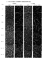
- FIG. 3 shows changes over time according to the seeding surface when natural seeding of human mesenchymal stem cells.
- FIG. 4 shows the time-dependent change according to the seeding surface of natural seeding of human skin fibroblasts.
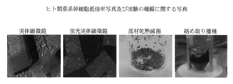
- FIG. 5 shows a fluorescence microscope photograph and a stereoscopic microscope photograph showing observation results (after 24 hours) (wet film method) at the time of human skin fibroblast B-side seeding.
- FIG. 1 shows natural seeding as one embodiment of the seeding form of a cell suspension on a polyimide porous membrane.
- FIG. 2 shows suction seeding as one embodiment of the seeding form of the cell suspension on the polyimide porous membrane.
- FIG. 3 shows changes over time according to the seeding surface when natural seeding of human
- FIG. 6 shows the change over time during the A-side suction seeding of human mesenchymal stem cells (one-point wet method).
- FIG. 7 shows the change over time during B-side suction seeding of human mesenchymal stem cells (one-point wet method).
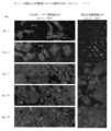
- FIG. 8 shows changes over time according to observation planes when entangled and seeded with human mesenchymal stem cells.
- FIG. 9 is a low-magnification photograph at the time of seeding and seeding with human mesenchymal stem cells and a photograph regarding the overview of the experiment.
- FIG. 10 shows the change over time when PC12 cells are sucked and seeded.
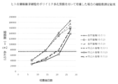
- FIG. 11 shows the number of cells when human skin fibroblasts are cultured using a polyimide porous membrane having a thickness of 25 ⁇ m.
- the horizontal axis indicates the number of days after culture, and the vertical axis indicates the number of cells per square centimeter of membrane.
- FIG. 12 shows the results of culturing human skin keratinocytes by the method of the present invention using a polyimide porous membrane and observing them with a confocal laser microscope and a stereoscopic fluorescence microscope.
- FIG. 13 shows the results of culturing human umbilical vein endothelial cells by the method of the present invention using a polyimide porous membrane and observing them with a confocal laser microscope and a stereoscopic fluorescence microscope.
- FIG. 12 shows the results of culturing human skin keratinocytes by the method of the present invention using a polyimide porous membrane and observing them with a confocal laser microscope and a stere
- FIG. 14-1 shows the results of culturing Vero cells by the method of the present invention using a polyimide porous membrane (25 ⁇ m sheet) and observing them with a confocal laser microscope and a stereoscopic fluorescence microscope.
- FIG. 14-2 shows the results of culturing Vero cells by the method of the present invention using a polyimide porous membrane (40 ⁇ m sheet) and observing them with a confocal laser microscope and a stereoscopic fluorescence microscope.
- FIG. 14-3 shows the results of culturing Vero cells by the method of the present invention using a polyimide porous membrane (75 ⁇ m sheet) and observing them with a confocal laser microscope and a stereoscopic fluorescence microscope.
- FIG. 14-1 shows the results of culturing Vero cells by the method of the present invention using a polyimide porous membrane (25 ⁇ m sheet) and observing them with a confocal laser microscope and a stereoscopic flu
- FIG. 15-1 shows the result of culturing HeLa cells by the method of the present invention using a polyimide porous membrane (25 ⁇ m sheet) and observing with a confocal laser microscope and a stereoscopic fluorescence microscope.
- FIG. 15-2 shows the results of culturing HeLa cells by the method of the present invention using a polyimide porous membrane (40 ⁇ m sheet) and observing them with a confocal laser microscope and a stereoscopic fluorescence microscope.
- FIG. 15-3 shows the results of culturing HeLa cells by the method of the present invention using a polyimide porous membrane (75 ⁇ m sheet) and observing them with a confocal laser microscope and a stereoscopic fluorescence microscope.
- FIG. 16-1 shows the results of culturing CHO cells by the method of the present invention using a polyimide porous membrane (25 ⁇ m sheet) and observing them with a confocal laser microscope and a stereoscopic fluorescence microscope.
- FIG. 16-2 shows the results of culturing CHO cells by the method of the present invention using a polyimide porous membrane (40 ⁇ m sheet) and observing them with a confocal laser microscope and a stereoscopic fluorescence microscope.
- FIG. 16-3 shows the results of culturing CHO cells by the method of the present invention using a polyimide porous membrane (75 ⁇ m sheet) and observing them with a confocal laser microscope and a stereoscopic fluorescence microscope.
- the present invention relates to a cell culture method.
- the cell culture method of the present invention includes applying the cells to a polyimide porous membrane and culturing.
- the present inventors have found that a polyimide porous membrane is suitable for cell adhesion and culture, and have come up with the present invention.
- the method of the present invention includes applying cells to a polyimide porous membrane and culturing the cells on or inside the polyimide membrane.
- the specific process of applying the cell to the polyimide porous membrane is not particularly limited.
- the steps described herein or any method suitable for applying cells to a membrane-like carrier can be employed.
- the method of this invention includes the following aspects, for example.
- An embodiment comprising steps; and (C) Wet one side or both sides of the polyimide porous membrane with a cell culture medium or a sterilized liquid, Loading the wet polyimide porous membrane with a cell suspension; and The cells in the cell suspension are retained in the membrane, and the water flows out.
- a mode comprising the steps.
- the mode includes directly seeding cells and cell clusters on the surface of the polyimide porous membrane. Or the aspect which puts a polyimide porous membrane in a cell suspension and infiltrate a cell culture solution from the surface of a membrane is also included.
- the cells seeded on the surface of the polyimide porous membrane adhere to the polyimide porous membrane and enter the inside of the porous body.
- the cells spontaneously adhere to the polyimide porous membrane without any physical or chemical force applied from the outside.
- Cells seeded on the surface of the polyimide porous membrane can grow and proliferate stably on the surface and / or inside of the membrane. Cells can take a variety of different forms depending on the location of the membrane in which they grow and multiply.
- a cell suspension is placed on the dried surface of the polyimide porous membrane.
- the cell suspension is sucked into the membrane, Cell suspension penetrates into the membrane. Without being bound by theory, it is considered that this is due to the properties derived from the surface shape of the polyimide porous membrane.
- the cells are sucked and seeded at the portion of the membrane where the cell suspension is loaded.
- one or both sides or the whole of the polyimide porous membrane is wetted with a cell culture medium or a sterilized liquid, and then the suspended polyimide porous membrane is subjected to cell suspension.
- the liquid may be loaded. In this case, the passage speed of the cell suspension is greatly improved.
- the one-point wet method described in Example 4 of the present specification is a method of wetting a part of the membrane electrode mainly for the purpose of preventing the scattering of the membrane, and is a dry method ((B) that does not substantially wet the membrane). A mode).
- the membrane permeation of the cell fluid becomes rapid for the wetted small part.
- the wet membrane method described in Example 3 of the present specification is based on a method in which one or both surfaces of a polyimide porous membrane are sufficiently wetted (hereinafter referred to as “wet membrane”). This is a method of loading a turbid liquid. In this case, the passage speed of the cell suspension is greatly improved in the entire polyimide porous membrane.
- the cells in the cell suspension are retained in the membrane and the water is allowed to flow out.
- processing such as concentrating the concentration of cells in the cell suspension or allowing unnecessary components other than cells to flow out together with moisture.
- the mode of (A) may be referred to as “natural sowing” (B) and the mode of (C) as “suction sowing”.
- living cells remain selectively in the polyimide porous membrane. Therefore, in a preferred embodiment of the present invention, living cells remain in the polyimide porous membrane, and dead cells preferentially flow out with moisture.
- the viable cell rate was 90%, but applied to the polyimide porous membrane, In the effluent liquid, the viable cell ratio was 65%.
- the adsorption rate of live cells to the membrane was 88%, the exudation rate was 12%, the adsorption rate of dead cells to the membrane was 40%, and the exudation rate was 60%. It is understood that living cells are selectively adsorbed on the polyimide porous membrane of the present invention.
- the sterilized liquid used in the embodiment (C) is not particularly limited, but is a sterilized buffer or sterilized water.
- the buffer solution include (+) and (-) Dulbecco's PBS, (+) and (-) Hank's Balanced Salt Solution, and the like. Examples of buffer solutions are shown in Table 1 below.
- the present invention further includes an aspect (entanglement) in which cells adhere to the membrane by allowing the adherent cells in a suspended state to coexist with the polyimide porous membrane in a suspended manner.
- a cell culture medium, cells, and one or more of the polyimide porous membranes may be placed in a cell culture container in order to apply the cells to the polyimide porous membrane.
- the cell culture medium is liquid
- the polyimide porous membrane is suspended in the cell culture medium. Due to the nature of the polyimide porous membrane, cells can adhere to the polyimide porous membrane.
- the polyimide porous membrane can be cultured in a suspended state in the cell culture medium.
- the cells spontaneously adhere to the polyimide porous membrane. “Spontaneously adheres” means that the cells remain on or inside the polyimide porous membrane without any physical or chemical force applied from the outside.
- cultured cells can be classified into adhesion culture cells and suspension culture cells depending on the form of cell culture.
- Adherent culture cells are cultured cells that adhere to a culture vessel and proliferate, and the medium is changed during passage.
- Floating culture cells are cultured cells that proliferate in a floating state in a medium. In general, dilution culture is performed without replacing the medium during passage.
- Suspension culture can be cultured in a floating state, that is, in a liquid state, so that it can be cultured in large quantities. Compared with adherent cells that grow only on the surface of the culture vessel, it is a three-dimensional culture. There is an advantage that the number of cells that can be cultured is large.
- the present invention conceptually provides a method for easily cultivating a large number of cells by allowing cells to be grown in a form similar to suspension culture without being limited to cell types. .
- the polyimide porous membrane when used in a suspended state in the cell culture medium, two or more pieces of the polyimide porous membrane may be used. Since the polyimide porous membrane is a flexible thin film, for example, it is possible to bring a polyimide porous membrane having a large surface area into a certain volume of cell culture medium by using small pieces suspended in the culture medium. Become.
- the bottom area of the container is the upper limit of the cell culture area, but in the cell culture using the polyimide porous membrane of the present invention, all of the large surface area of the previously introduced polyimide porous membrane is the cell. It becomes the area that can be cultured. Since the polyimide porous membrane allows the cell culture solution to pass therethrough, for example, nutrients, oxygen and the like can be supplied into the folded membrane.
- the size and shape of the polyimide porous membrane pieces are not particularly limited.
- the shape can take any shape such as a circle, an ellipse, a square, a triangle, a polygon, and a string.
- a square having a side length of about 0.1 mm to about 20 mm, preferably about 0.2 mm to about 10 mm, more preferably about 1 mm to about 5 mm, a triangle, and the like are included.
- it may be circular, preferably having a diameter of about 0.1 mm to about 20 mm, more preferably about 0.5 mm to about 10 mm.
- the polyimide porous membrane of the present invention has flexibility, it can be used by changing its shape.
- the polyimide porous membrane may be processed into a three-dimensional shape instead of a flat shape.
- a polyimide porous membrane is i) folded, ii) rolled into a roll, and iii) connected to a sheet or piece with a thread-like structure, or iv) tied in a rope, in a cell culture vessel May be suspended or fixed in the cell culture medium.
- a polyimide porous membrane is i) folded, ii) rolled into a roll, and iii) connected to a sheet or piece with a thread-like structure, or iv) tied in a rope, in a cell culture vessel May be suspended or fixed in the cell culture medium.
- two or more polyimide porous membranes may be used by being laminated in a cell culture medium vertically or horizontally.
- Lamination also includes a mode in which the polyimide porous membranes partially overlap. Stacked culture enables cells to be cultured at high density in a narrow space. It is also possible to form a multilayer system with different types of cells by further stacking the membranes on the membrane where the cells are already grown. Furthermore, it can be used for drug discovery such as verification of cell-cell interaction in a three-dimensional environment and cell culture method without stress.
- the number of polyimide porous membranes to be laminated is not particularly limited.
- the above-described cell culture method of the present invention may be used in combination of two or more methods.
- the cells may be first applied to the polyimide porous membrane using any one of the methods (A) to (C), and then the polyimide porous membrane to which the cells are adhered may be subjected to suspension culture.
- the method in any one of the said aspect (A)-(C) combining 2 types or more.
- the cells grow and proliferate on and inside the polyimide porous membrane.
- the use of the polyimide porous membrane enables continuous three-dimensional culture of cells.
- the cells of the present invention continue to grow for 2 days or more, more preferably 4 days or more, and even more preferably 6 days or more. In Examples 1 and 4 described herein, an increase in cells was observed for 21 days.
- Cells The types of cells that can be used in the method of the present invention are not particularly limited, and can be used for the growth of arbitrary cells.
- the cells are selected from the group consisting of animal cells, insect cells, plant cells, yeasts and bacteria.
- Animal cells are roughly classified into cells derived from animals belonging to the vertebrate phylum and cells derived from invertebrates (animals other than animals belonging to the vertebrate phylum).
- the origin of the animal cell is not particularly limited.
- Vertebrates include the maxilla and maxilla, and the maxilla includes mammals, birds, amphibians, reptiles, and the like.
- it is a cell derived from an animal belonging to the mammal class generally called a mammal. Mammals are not particularly limited, but preferably include mice, rats, humans, monkeys, pigs, dogs, sheep, goats and the like.
- the origin of the plant cell in this specification is not particularly limited. Plant cells including moss plants, fern plants, and seed plants are targeted.
- Plants from which seed plant cells are derived include both monocotyledonous plants and dicotyledonous plants.
- monocotyledonous plants include orchids, gramineous plants (rice, corn, barley, wheat, sorghum, etc.), cyperaceae plants, and the like.
- Dicotyledonous plants include plants belonging to many subclasses such as Chrysanthemum, Magnolia, and Rose.
- Algae can also be regarded as cell-derived organisms. Different from eubacteria, cyanobacteria (Cyanobacteria), eukaryotes that are unicellular (diatoms, yellow green algae, dinoflagellates, etc.) and multicellular organisms, seaweeds (red algae, brown algae, green algae) Includes groups.
- the archaea and the types of bacteria in this specification are not particularly limited.
- the archaea is composed of a group consisting of methane bacteria, highly halophilic bacteria, thermophilic acidophiles, hyperthermophilic bacteria, and the like.
- the bacterium is selected from the group consisting of lactic acid bacteria, Escherichia coli, Bacillus subtilis, cyanobacteria and the like.
- animal cells or plant cells that can be used in the method of the present invention are not limited, but are preferably selected from the group consisting of pluripotent stem cells, tissue stem cells, somatic cells, and germ cells.
- pluripotent stem cell is intended to be a generic term for stem cells having the ability to differentiate into cells of any tissue (differentiation pluripotency).
- the pluripotent stem cells include, but are not limited to, embryonic stem cells (ES cells), induced pluripotent stem cells (iPS cells), embryonic germ stem cells (EG cells), germ stem cells (GS cells), and the like. .
- ES cells embryonic stem cells
- iPS cells induced pluripotent stem cells
- EG cells embryonic germ stem cells
- GS cells germ stem cells
- Any known pluripotent stem cell can be used.
- the pluripotent stem cell described in International Publication WO2009 / 123349 PCT / JP2009 / 057041
- PCT / JP2009 / 057041 can be used.
- tissue stem cell means a stem cell that has the ability to differentiate into various cell types (differentiated pluripotency) although the cell line that can be differentiated is limited to a specific tissue.
- hematopoietic stem cells in the bone marrow become blood cells, and neural stem cells differentiate into nerve cells.
- the tissue stem cells are selected from mesenchymal stem cells, hepatic stem cells, pancreatic stem cells, neural stem cells, skin stem cells, or hematopoietic stem cells.
- somatic cells refers to cells other than germ cells among the cells constituting multicellular organisms. In sexual reproduction, it is not passed on to the next generation.
- the somatic cells are hepatocytes, pancreatic cells, muscle cells, bone cells, osteoblasts, osteoclasts, chondrocytes, adipocytes, skin cells, fibroblasts, pancreatic cells, kidney cells, lung cells, or , Lymphocytes, erythrocytes, leukocytes, monocytes, macrophages or megakaryocyte blood cells.
- Reproductive cells means cells that have a role in transmitting genetic information to the next generation in reproduction. For example, gametes for sexual reproduction, ie eggs, egg cells, sperm, sperm cells, spores for asexual reproduction, and the like.
- the cells may be selected from the group consisting of sarcoma cells, established cells and transformed cells.
- “Sarcoma” is a cancer that develops in connective tissue cells derived from non-epithelial cells such as bone, cartilage, fat, muscle, blood, etc., and includes soft tissue sarcoma, malignant bone tumor and the like.
- Sarcoma cells are cells derived from sarcomas.
- the “established cell” means a cultured cell that has been maintained outside the body for a long period of time, has a certain stable property, and is capable of semi-permanent subculture.
- PC12 cells derived from rat adrenal medulla
- CHO cells derived from Chinese hamster ovary
- HEK293 cells derived from human fetal kidney
- HL-60 cells derived from human white blood cells
- HeLa cells derived from human cervical cancer
- a “transformed cell” means a cell in which a nucleic acid (DNA or the like) has been introduced from the outside of the cell to change its genetic properties. Appropriate methods are known for transformation of animal cells, plant cells, and bacteria.
- Polyimide Porous Membrane Polyimide is a general term for polymers containing imide bonds in repeating units, and usually means an aromatic polyimide in which aromatic compounds are directly linked by imide bonds.
- Aromatic polyimide has a conjugated structure through the imide bond between aromatic and aromatic, so it has a rigid and strong molecular structure, and the imide bond has a strong intermolecular force, so it has a very high level of heat. Has mechanical, mechanical and chemical properties.
- the polyimide porous membrane of the present invention is preferably a polyimide porous membrane containing a polyimide obtained from tetracarboxylic dianhydride and diamine (as a main component), more preferably tetracarboxylic dianhydride and diamine. It is a polyimide porous membrane which consists of polyimide obtained from these. “Containing as a main component” means that a component other than polyimide obtained from tetracarboxylic dianhydride and diamine may be essentially not included or included as a component of the polyimide porous membrane. It means that it is an additional component that does not affect the properties of the polyimide obtained from tetracarboxylic dianhydride and diamine.
- a colored polyimide porous film obtained by molding a polyamic acid solution composition containing a polyamic acid solution obtained from a tetracarboxylic acid component and a diamine component and a colored precursor and then heat-treating at 250 ° C. or higher. .
- Polyamic acid A polyamic acid is obtained by polymerizing a tetracarboxylic acid component and a diamine component.
- Polyamic acid is a polyimide precursor that can be ring-closed to form polyimide by thermal imidization or chemical imidization.
- the polyamic acid even if a part of the amic acid is imidized, it can be used as long as it does not affect the present invention. That is, the polyamic acid may be partially thermally imidized or chemically imidized.
- fine particles such as an imidization catalyst, an organic phosphorus-containing compound, inorganic fine particles, and organic fine particles can be added to the polyamic acid solution as necessary.
- fine particles such as a chemical imidating agent, a dehydrating agent, inorganic fine particles, and organic fine particles, etc. can be added to a polyamic acid solution as needed. Even when the above components are added to the polyamic acid solution, it is preferable that the coloring precursor is not precipitated.
- the colored precursor means a precursor that is partially or wholly carbonized by heat treatment at 250 ° C. or higher to produce a colored product.
- the colored precursor used in the present invention is uniformly dissolved or dispersed in a polyamic acid solution or a polyimide solution, and heat treatment at 250 ° C. or higher, preferably 260 ° C. or higher, more preferably 280 ° C. or higher, more preferably 300 ° C. or higher.
- heat treatment at 250 ° C. or higher, preferably 260 ° C. or higher, more preferably 280 ° C. or higher, more preferably 300 ° C. or higher in the presence of oxygen such as air to produce a colored product.
- oxygen such as air
- the carbon-based coloring precursor is not particularly limited.
- polymers such as petroleum tar, petroleum pitch, coal tar, coal pitch, or polymers obtained from monomers including pitch, coke, and acrylonitrile, ferrocene compounds (ferrocene and ferrocene derivatives). Etc.
- the polymer and / or ferrocene compound obtained from the monomer containing acrylonitrile are preferable, and polyacrylonitrile is preferable as a polymer obtained from the monomer containing acrylonitrile.
- tetracarboxylic dianhydride any tetracarboxylic dianhydride can be used, and can be appropriately selected according to desired characteristics.
- tetracarboxylic dianhydride include pyromellitic dianhydride, 3,3 ′, 4,4′-biphenyltetracarboxylic dianhydride (s-BPDA), 2,3,3 ′, 4 ′.
- -Biphenyltetracarboxylic dianhydride such as biphenyltetracarboxylic dianhydride (a-BPDA), oxydiphthalic dianhydride, diphenylsulfone-3,4,3 ', 4'-tetracarboxylic dianhydride, bis (3,4-dicarboxyphenyl) sulfide dianhydride, 2,2-bis (3,4-dicarboxyphenyl) -1,1,1,3,3,3-hexafluoropropane dianhydride, 2, 3,3 ′, 4′-benzophenone tetracarboxylic dianhydride, 3,3 ′, 4,4′-benzophenone tetracarboxylic dianhydride, bis (3,4-dicarboxyphenyl) methane dianhydride 2,2-bis (3,4-dicarboxyphenyl) propane dianhydride, p-phenylenebis (trimellitic acid monoester acid an
- At least one aromatic tetracarboxylic dianhydride selected from the group consisting of biphenyltetracarboxylic dianhydride and pyromellitic dianhydride is particularly preferable.
- the biphenyltetracarboxylic dianhydride 3,3 ′, 4,4′-biphenyltetracarboxylic dianhydride can be suitably used.
- diamine Any diamine can be used as the diamine.
- diamines include the following.
- One benzene nuclei such as 1,4-diaminobenzene (paraphenylenediamine), 1,3-diaminobenzene, 2,4-diaminotoluene, 2,6-diaminotoluene;
- Diaminodiphenyl ethers such as 4,4'-diaminodiphenyl ether and 3,4'-diaminodiphenyl ether, 4,4'-diaminodiphenylmethane, 3,3'-dimethyl-4,4'-diaminobiphenyl, 2,2'- Dimethyl-4,4′-diaminobiphenyl, 2,2′-bis (trifluoromethyl) -4,4′-diaminobiphenyl, 3,3′-dimethyl-4,4′-diaminodiphenylmethane, 3,3′- Dicarboxy-4,4′-diaminodiphenylmethane, 3,3 ′, 5,5′-tetramethyl-4,4′-diaminodiphenylmethane, bis (4-aminophenyl) sulfide, 4,4′-diaminobenzanilide, 3,3′-dichlorobenzidine, 3,3
- diamine to be used can be appropriately selected according to desired characteristics.
- aromatic diamine compounds are preferable, and 3,3′-diaminodiphenyl ether, 3,4′-diaminodiphenyl ether, 4,4′-diaminodiphenyl ether and paraphenylenediamine, 1,3-bis (3-aminophenyl) Benzene, 1,3-bis (4-aminophenyl) benzene, 1,4-bis (3-aminophenyl) benzene, 1,4-bis (4-aminophenyl) benzene, 1,3-bis (4-amino) Phenoxy) benzene and 1,4-bis (3-aminophenoxy) benzene can be preferably used.
- at least one diamine selected from the group consisting of benzenediamine, diaminodiphenyl ether and bis (aminophenoxy) phenyl is preferred.
- the polyimide porous membrane has a glass transition temperature of 240 ° C. or higher, or a tetracarboxylic dianhydride and a diamine having a clear transition point of 300 ° C. or higher. It is preferable that it is formed from the polyimide obtained combining these.
- the polyimide porous membrane of the present invention is preferably a polyimide porous membrane made of the following aromatic polyimide from the viewpoints of heat resistance and dimensional stability at high temperatures.
- an aromatic polyimide comprising at least one tetracarboxylic acid unit selected from the group consisting of a biphenyltetracarboxylic acid unit and a pyromellitic acid unit, and an aromatic diamine unit
- an aromatic polyimide comprising a tetracarboxylic acid unit and at least one aromatic diamine unit selected from the group consisting of a benzenediamine unit, a diaminodiphenyl ether unit and a bis (aminophenoxy) phenyl unit
- a polyimide porous film a polyimide having a multilayer structure having at least two surface layers (A surface and B surface) and a macrovoid layer sandwiched between the two surface layers Porous membranes can be used in the method of the present invention.
- the polyimide porous film is a film in which the macrovoid layer is surrounded by a partition wall bonded to the surface layer (A surface and B surface), and the partition wall and the surface layer (A surface and B surface).
- a plurality of macrovoids having an average pore diameter in the plane direction of 10 to 500 ⁇ m, and the partition walls of the macrovoid layer and the surface layers (A surface and B surface) each have a thickness of 0.01 to 20 ⁇ m.
- the polyimide porous film has a total film thickness of 5 to 500 ⁇ m and a porosity of 40% or more and less than 95%.
- the total film thickness is not limited, but may be 25-75 ⁇ m as one aspect. Due to the difference in film thickness, differences in cell growth rate, cell morphology, in-plane cell saturation, etc. can be observed.
- the A surface of the polyimide porous membrane has a mesh structure having small holes with an average pore diameter of 15 ⁇ m or less, and the B surface has a large hole structure with an average pore diameter of 20 ⁇ m or more.
- a polyimide porous membrane described in International Publication WO2010 / 038873, JP2011-219585, or JP2011-219586 can also be used in the method of the present invention.
- Cells seeded on the surface of the polyimide porous membrane can stably grow and proliferate on the surface and / or inside of the membrane.
- Cells can take a variety of different forms depending on where they grow and proliferate in the membrane.
- the cells may proliferate while changing the shape while moving on and inside the polyimide porous membrane.
- the cells can be cultured using any known method. Culture methods suitable for animal cells, plant cells, and bacterial cells are known, and those skilled in the art can culture cells on a polyimide porous membrane using any known method. A cell culture medium can also be suitably prepared according to the kind of cell.
- Animal cell culture methods and cell culture media are described, for example, in the cell culture media catalog of Lonza.
- Plant cell culture methods and cell culture media are described in, for example, the plant tissue culture series from WAKO.
- Bacterial cell culture methods and cell culture media are described in, for example, the general bacterial culture catalog of BD.
- the present invention also relates to a cell culture device for use in the culture method of the present invention, including a polyimide porous membrane.
- the polyimide porous membrane may be fixed and used, or may be used suspended in the cell culture medium.
- two or more polyimide porous membranes may be laminated vertically or horizontally.
- the cell culture device of the present invention can use a known cell culture device as long as it satisfies the requirement that it includes a polyimide porous membrane.
- the shape, scale, etc. of the culture apparatus are not particularly limited and can be appropriately used from petri dishes, test tubes to large tanks.
- a cell culture dish manufactured by BD Falcon, a Nunc cell factory manufactured by Thermo Scientific, and the like are included.
- a polyimide porous membrane in the present invention it has become possible to culture cells in a state similar to suspension culture using a suspension culture apparatus even for cells that were not inherently capable of suspension culture.
- a spinner flask manufactured by Corning can be used as an apparatus for suspension culture.
- Kit for use in cell culture method The present invention further relates to a kit for use in a cell culture method, comprising a polyimide porous membrane.
- the kit of the present invention may appropriately contain components necessary for cell culture in addition to the polyimide porous membrane.
- the cell applicable to a polyimide porous membrane, a cell culture medium, a cell culture apparatus, the instruction manual of a kit, etc. are contained.
- a sterilized polyimide porous membrane is stored alone or in a plurality of sheets in a transparent pouch, and a package containing a form that can be used for cell culture as it is, or the same
- a sterilizing liquid is enclosed in a pouch together with a polyimide porous membrane, and includes an integrated membrane / liquid kit that enables efficient suction seeding.
- polyimide porous membrane refers to a polyimide porous membrane having a thickness of 25 ⁇ m.
- Example 1 Natural seeding of human mesenchymal stem cells on a polyimide porous membrane
- seeding on a polyimide porous membrane was performed using human mesenchymal stem cells.
- Example 2 Natural seeding of human skin fibroblasts on a polyimide porous membrane
- human skin fibroblasts were used to apply cells to a polyimide porous membrane by natural seeding.
- the cells are cultured in a cell culture apparatus, and fixed and stained (DAPI or actin + DAPI) after 1 hour, 5 hours, 24 hours, 48 hours, 4 days, 7 days, 14 days, and 21 days to grow and proliferate the cells. confirmed. Cell proliferation over time, seeding surface and observation surface specific morphology were observed. The results are shown in FIG. From this result, it was shown that human skin fibroblasts, which are one of representative fibroblasts, can also be cultured by the method of the present invention.
- Example 3 Suction seeding of human skin fibroblasts into a polyimide porous membrane (wet membrane method)
- human skin fibroblasts were used to apply cells to a polyimide porous membrane by suction seeding into a wet membrane (fully wet membrane; details will be described below).
- FIG. 5 shows a fluorescence microscope photograph and a stereoscopic fluorescence microscope photograph of a sample fixed and fluorescently stained (DAPI or actin + DAPI) after 24 hours.
- the exudate was collected, and the square plate was washed with 2 ml of the medium and the number of cells remaining on the plate was counted.
- the total number of cells was 6.0 ⁇ 10 4
- the number of living cells was 5.5 ⁇ 10 4
- the number of dead cells was 3.0 ⁇ 10 4
- the viable cell rate was 65%.
- the adsorption rate of live cells to the membrane was 88%
- the exudation rate was 12%
- the adsorption rate of dead cells to the membrane was 40%
- the exudation rate was 60%.
- Example 4 Suction seeding of human mesenchymal stem cells into a polyimide porous membrane (one-point wet method)
- human mesenchymal stem cells are used, and a single-point wet membrane (a membrane that is fixed simply by wetting a droplet at the central point; details will be described below) is obtained by sucking and seeding the cells into a polyimide porous membrane. Applied to.
- This example shows an example in which human mesenchymal stem cells are used to apply a cell to a polyimide porous membrane by sucking and seeding only one central portion of the membrane material into a wet membrane.
- the exudate was collected, and the square plate was washed with 4 ml of the medium to count the cells remaining on the plate.
- the total number of cells was 6.5 ⁇ . 10 4 cells, of which 4.0 ⁇ 10 4 live cells, 2.5 ⁇ 10 4 dead cells, and 62% live cell rate. Comparing cell viability, the adsorption rate of live cells to the membrane was 89%, and the exudation rate was 11%.
- B surface seeding collecting total cell numbers of four 6.5 ⁇ 10, of which living cells 8.0 ⁇ 10 4, dead cells 2.0 ⁇ 10 4, viable cell ratio of 80% Met. Comparing cell viability, the adsorption rate of live cells to the membrane was 79%, and the exudation rate was 21%.
- Example 5 Tangled and Seeded Human Mesenchymal Stem Cells on Polyimide Porous Membrane
- human mesenchymal stem cells were applied to a polyimide porous membrane by entanglement from a cell suspension. Went.
- a square polyimide porous membrane with a side of 10 cm is cut into small pieces of about 2 to 3 mm square using scissors, sterilized at 160 ° C. for 10 minutes, allowed to cool, and wet with 2 ml of sterilized medium. Thereafter, the polyimide porous membrane was taken out with tweezers and transferred into a falcon tube. On the membrane in the falcon tube, 3.6 ⁇ 10 5 cells per ml of medium (of which 3.4 ⁇ 10 5 live cells, 2.0 ⁇ 10 4 dead cells, and 94 live cell rate) %), 1.6 ml of a human mesenchymal stem cell suspension was added (out of a total number of 5.7 ⁇ 10 5 cells, 5.4 ⁇ 10 5 live cells and 3.2 dead cells).
- Example 6 Suction seeding of a cell line to a small piece of a polyimide porous membrane
- the cell line was applied to a small piece of a polyimide porous film by suction using PC12 cells of the cell line.
- the polyimide porous membrane was fixed and stained after 48 hours, 4 days, 6 days, and 9 days, and cell proliferation was confirmed. Staining was performed with a membrane fluorescent stain cell mask (CellMask (trademark) Orange plasma membrane stain (Life Technologies), hereinafter simply referred to as CellMask) + DAPI. The results are shown in FIG. Even in this cell, it was observed that the cells proliferated while forming a population in the polyimide porous membrane, and the possibility of application was confirmed. From this result, it was shown that PC12 cell which is one of typical cell lines can also be cultured by the method of the present invention.
- CellMask trademark
- DAPI membrane fluorescent stain cell mask
- Example 7 Measurement of the number of cultured cells
- human skin fibroblasts were cultured by the method of the present invention using a polyimide porous membrane, and the number of cultured cells was measured.
- the cells are washed twice with a phosphate buffer, treated with a 0.05% trypsin-EDTA solution, and the number of cells is counted.
- a correlation coefficient between the absorbance under the culture conditions and CCK8 concentration conditions and the actual cell number was determined.
- the number of cells surviving on the member is calculated using the conversion coefficient obtained in 1, and then twice in the medium. Wash and return to incubator to continue culturing. This operation was repeated to quantitatively analyze the growth of cells on the polyimide porous membrane over time. The number of experiments was repeated to verify reproducibility.
- FIG. 11 shows the results of natural seeding and suction sowing three times each.
- the growth rate of cells differs depending on the seeding method, but the difference is eliminated by culturing for about one month, and a similar number of cells per unit area is reached.
- the dotted line in the figure indicates the upper limit of the number of cultured cells in a normal adherent culture in a petri dish or the like implemented as a comparative experiment. When cell numbers were compared on an area basis, more cells could be cultured in a unit area than in normal cell culture.
- Example 8 Culture of human skin keratinocytes
- human skin keratinocytes were cultured by the method of the present invention using a polyimide porous membrane and observed with a confocal laser microscope and a stereoscopic fluorescence microscope.
- a 0.5 cm cell culture medium (KGM-Gold keratinocyte growth medium BulletKit (Lonza)) 0.5 ml was added to a 2 cm ⁇ 2 cm sterilized square container, and a sterilized 1.4 cm square square polyimide porous membrane was meshed with A. Dipped with the side up. 4 ⁇ 10 4 human skin keratinocytes were added to the cell culture medium in the square container. That is, 4 ⁇ 10 4 human skin keratinocytes were naturally seeded per 1.4 cm square square polyimide porous membrane.
- the cells were cultured in a cell culture apparatus, and fixed and stained after 1 day, 3 days, and 6 days. Staining was performed only with CellMask + DAPI or CellMask. Thereafter, the growth and morphology of the cells over time were observed using a confocal laser microscope (LSM700 (manufactured by Carl Zeiss)) and a stereoscopic fluorescence microscope (Leica M165 FC (manufactured by Leica)).
- LSM700 confocal laser microscope
- Leica M165 FC stereoscopic fluorescence microscope
- human skin keratinocytes which are primary human cultured cells (primary culture cells), can also be cultured by the method of the present invention.
- Example 9 Culture of Human Umbilical Vein Endothelial Cells
- human umbilical vein endothelial cells were cultured by the method of the present invention using a polyimide porous membrane and observed with a confocal laser microscope and a stereoscopic fluorescence microscope.
- the cells were cultured in a cell culture apparatus, and fixed and stained (CellMask + DAPI and CellMask) after 3 days, 6 days, and 10 days. Staining and observation with a confocal laser microscope and a stereoscopic fluorescence microscope were performed in the same manner as in Example 8.
- human umbilical vein endothelial cells which are human primary cultured cells (primary culture cells), can also be cultured by the method of the present invention.
- Example 10 Culturing of Vero cells
- Vero cells were cultured by the method of the present invention using a polyimide porous membrane and observed with a confocal laser microscope and a stereoscopic fluorescence microscope.
- Three types of polyimide porous membranes of 25 ⁇ m, 40 ⁇ m, and 75 ⁇ m were used.
- the culture period is 1-15 days.
- the cells were cultured in a cell culture apparatus, and fixed and stained after 1 day, 3 days, 7 days, 10 days, and 15 days. Staining was performed with actin + DAPI, and actin was detected with phalloidin. Observation with a confocal laser microscope and a stereoscopic fluorescence microscope was performed in the same manner as in Example 8.
- Vero cells which are one of representative cell lines, can also be cultured by the method of the present invention.
- Example 11 Culture of HeLa cells
- HeLa cells were cultured by the method of the present invention using a polyimide porous membrane and observed with a confocal laser microscope and a stereoscopic fluorescence microscope.
- Three types of polyimide porous membranes of 25 ⁇ m, 40 ⁇ m, and 75 ⁇ m were used. The culture period, microscope used, specific steps, etc. are as described in Example 10.
- FIGS. 15-1 to 15-3 The results are shown in FIGS. 15-1 to 15-3. From this result, it was shown that HeLa cells, which are one of representative cell lines, can also be cultured by the method of the present invention.
- Example 12 Culture of CHO cells
- CHO cells were cultured by the method of the present invention using a polyimide porous membrane and observed with a confocal laser microscope and a stereoscopic fluorescence microscope.
- Three types of polyimide porous membranes of 25 ⁇ m, 40 ⁇ m, and 75 ⁇ m were used.
- the culture period was 1-7 days, and the microscope used, specific steps, etc. were as described in Example 10.
- FIGS. 16-1 to 16-3 The results are shown in FIGS. 16-1 to 16-3. From this result, it was shown that CHO cells, which are one of representative cell lines, can also be cultured by the method of the present invention.
Landscapes
- Life Sciences & Earth Sciences (AREA)
- Health & Medical Sciences (AREA)
- Engineering & Computer Science (AREA)
- Chemical & Material Sciences (AREA)
- Bioinformatics & Cheminformatics (AREA)
- Organic Chemistry (AREA)
- Wood Science & Technology (AREA)
- Zoology (AREA)
- Genetics & Genomics (AREA)
- Biotechnology (AREA)
- Biomedical Technology (AREA)
- General Engineering & Computer Science (AREA)
- General Health & Medical Sciences (AREA)
- Microbiology (AREA)
- Biochemistry (AREA)
- Mycology (AREA)
- Medicinal Chemistry (AREA)
- Tropical Medicine & Parasitology (AREA)
- Virology (AREA)
- Cell Biology (AREA)
- Botany (AREA)
- Immunology (AREA)
- Sustainable Development (AREA)
- Micro-Organisms Or Cultivation Processes Thereof (AREA)
- Apparatus Associated With Microorganisms And Enzymes (AREA)
- Immobilizing And Processing Of Enzymes And Microorganisms (AREA)
Abstract
Description
細胞は生体内では一般に三次元的な集団として存在するが、古典的な平面培養では、細胞が容器に張り付く形で単層状に培養される。培養環境の相違により、細胞の性質も大きく異なることが数多く報告されている。また、液体培養培地中で細胞を培養する浮遊培養については、浮遊培養に適した細胞がある一方、適さない細胞もある。
ポリイミドとは、繰り返し単位にイミド結合を含む高分子の総称であり、通常は、芳香族化合物が直接イミド結合で連結された芳香族ポリイミドを意味する。芳香族ポリイミドは芳香族と芳香族とがイミド結合を介して共役構造を持つため、剛直で強固な分子構造を持ち、かつ、イミド結合が強い分子間力を持つために非常に高いレベルの熱的、機械的、化学的性質を有する。
細胞をポリイミド多孔質膜に適用し、培養することを含む、細胞の培養方法。
細胞を前記ポリイミド多孔質膜の表面に播種する工程を含む、態様1に記載の方法。
前記ポリイミド多孔質膜の乾燥した表面に細胞縣濁液を乗せ、
前記ポリイミド多孔質膜を放置するか、あるいは前記ポリイミド多孔質膜を移動して液の流出を促進するか、あるいは表面の一部を刺激して、細胞縣濁液を前記膜に吸い込ませ、そして、
細胞縣濁液中の細胞を前記膜内に留め、水分は流出させる、
工程を含む、態様1に記載の方法。
前記ポリイミド多孔質膜の片面又は両面を、細胞培養培地又は滅菌された液体で湿潤し、
前記湿潤したポリイミド多孔質膜に細胞縣濁液を装填し、そして、
細胞縣濁液中の細胞を前記膜内に留め、水分は流出させる、
工程を含む、態様1に記載の方法。
生細胞が前記ポリイミド多孔質膜内に留まり、死細胞は水分とともに流出する、態様4に記載の方法。
前記滅菌された液体が、滅菌水若しくは滅菌された緩衝液である、態様4又は5に記載の方法。
細胞培養容器中に、細胞培養培地、細胞及び1又はそれ以上の前記ポリイミド多孔質膜を入れる、ここにおいて、ポリイミド多孔質膜は細胞培養培地中に浮遊した状態である、ことを含む、態様1−6のいずれか1項に記載の方法。
2以上の前記ポリイミド多孔質膜の小片を用いることを特徴とする、態様7に記載の方法。
細胞が、前記ポリイミド多孔質膜に自発的に接着する、態様7又は8に記載の方法。
前記ポリイミド多孔質膜を
i)折り畳んで、
ii)ロール状に巻き込んで、
iii)シートもしくは小片を糸状の構造体で連結させて、あるいは、
iv)縄状に結んで
細胞培養容器中の細胞培養培地中で浮遊もしくは固定させる、態様1−6のいずれか1項に記載の方法。
細胞が、前記ポリイミド多孔質膜に自発的に接着する、態様10に記載の方法。
2以上のポリイミド多孔質膜を、上下又は左右に細胞培養培地中に積層して用いることを含む、態様1に記載の方法。
態様2−12のいずれか1項に記載の方法の2種類又はそれより多くの方法を組み合わせて用いる、態様1に記載の方法。
細胞がポリイミド多孔質膜の表面及び内部に生育し増殖する、態様1−13のいずれか1項に記載の方法。
細胞が、動物細胞、昆虫細胞、植物細胞、酵母菌及び細菌からなる群から選択される、態様1−14のいずれか1項に記載の方法。
動物細胞が、脊椎動物門に属する動物由来の細胞である、態様15に記載の方法。
細菌が、乳酸菌、大腸菌、枯草菌及びシアノバクテリアからなる群から選択される、態様15に記載の方法。
細胞が、多能性幹細胞、組織幹細胞、体細胞及び生殖細胞からなる群から選択される、
態様1−14のいずれか1項に記載の方法。
細胞が、肉腫細胞、株化細胞及び形質転換細胞からなる群から選択される、態様1−13のいずれか1項に記載の方法。
ポリイミド多孔質膜が、テトラカルボン酸二無水物とジアミンとから得られるポリイミドを含む、ポリイミド多孔質膜である、態様1−19のいずれか1項に記載の方法。
ポリイミド多孔質膜を含む、態様1−20のいずれか1項に記載の細胞の培養方法に使用するための細胞培養装置。
2以上のポリイミド多孔質膜が、上下又は左右に積層している、態様21に記載の細胞培養装置。
ポリイミド多孔質膜を含む、態様1ないし20のいずれか1項に記載の細胞の培養方法に使用するためのキット。
本発明は、細胞の培養方法に関する。
細胞のポリイミド多孔質膜への適用の具体的な工程は特に限定されない。本明細書に記載の工程、あるいは、細胞を膜状の担体に適用するのに適した任意の手法を採用することが可能である。限定されるわけではないが、本発明の方法は、例えば、以下のような態様を含む。
(A)細胞を前記ポリイミド多孔質膜の表面に播種する工程を含む、態様;
(B)前記ポリイミド多孔質膜の乾燥した表面に細胞縣濁液を乗せ、
放置するか、あるいは前記ポリイミド多孔質膜を移動して液の流出を促進するか、あるいは表面の一部を刺激して、細胞縣濁液を前記膜に吸い込ませ、そして、
細胞縣濁液中の細胞を前記膜内に留め、水分は流出させる、
工程を含む、態様;並びに、
(C)前記ポリイミド多孔質膜の片面又は両面を、細胞培養液又は滅菌された液体で湿潤し、
前記湿潤したポリイミド多孔質膜に細胞縣濁液を装填し、そして、
細胞縣濁液中の細胞を前記膜内に留め、水分は流出させる、
工程を含む、態様。

本発明の方法に利用し得る細胞の種類は特に限定されず、任意の細胞の増殖に利用可能である。
ポリイミドとは、繰り返し単位にイミド結合を含む高分子の総称であり、通常は、芳香族化合物が直接イミド結合で連結された芳香族ポリイミドを意味する。芳香族ポリイミドは芳香族と芳香族とがイミド結合を介して共役構造を持つため、剛直で強固な分子構造を持ち、かつ、イミド結合が強い分子間力を持つために非常に高いレベルの熱的、機械的、化学的性質を有する。
ポリアミック酸は、テトラカルボン酸成分とジアミン成分とを重合して得られる。ポリアミック酸は、熱イミド化又は化学イミド化することにより閉環してポリイミドとすることができるポリイミド前駆体である。
本発明において着色前駆体とは、250℃以上の熱処理により一部または全部が炭化して着色化物を生成する前駆体を意味する。
(i)ビフェニルテトラカルボン酸単位及びピロメリット酸単位からなる群から選ばれる少なくとも一種のテトラカルボン酸単位と、芳香族ジアミン単位とからなる芳香族ポリイミド、
(ii)テトラカルボン酸単位と、ベンゼンジアミン単位、ジアミノジフェニルエーテル単位及びビス(アミノフェノキシ)フェニル単位からなる群から選ばれる少なくとも一種の芳香族ジアミン単位とからなる芳香族ポリイミド、
及び/又は、
(iii)ビフェニルテトラカルボン酸単位及びピロメリット酸単位からなる群から選ばれる少なくとも一種のテトラカルボン酸単位と、ベンゼンジアミン単位、ジアミノジフェニルエーテル単位及びビス(アミノフェノキシ)フェニル単位からなる群から選ばれる少なくとも一種の芳香族ジアミン単位とからなる芳香族ポリイミド。
本発明の方法において、ポリイミド多孔質膜に細胞を適用した後、細胞を任意の公知の方法を用いて培養することができる。動物細胞、植物細胞、及び細菌の各細胞に適した培養方法が公知であり、当業者は任意の公知の方法を用いてポリイミド多孔質膜に細胞を培養することができる。細胞培養培地も細胞の種類に応じて適宜調製することができる。
本発明はまた、ポリイミド多孔質膜を含む、本発明の培養方法に使用するための細胞培養装置に関する。本発明の細胞培養装置において、ポリイミド多孔質膜は固定されて用いられても良く、あるいは細胞培養培地中に浮遊して用いられても良い。細胞培養装置において、2以上のポリイミド多孔質膜が、上下又は左右に積層してもよい。
本発明はさらに、ポリイミド多孔質膜を含む、細胞の培養方法に使用するためのキットに関する。
本実施例では、ヒト間葉系幹細胞を用いて、ポリイミド多孔質膜への播種を行った。
本実施例では、ヒト皮膚線維芽細胞を用いて、自然播種により細胞のポリイミド多孔質膜への適用を行った。
本実施例では、ヒト皮膚線維芽細胞を用いて、ウェット膜(十分に湿潤した膜;詳細は以下に説明)への吸込み播種により細胞のポリイミド多孔質膜への適用を行った。
本実施例では、ヒト間葉系幹細胞を用いて、一点ウェット膜(中央の一点のみ液滴に濡らして簡単に固定した膜;詳細は以下に説明)への吸込み播種により細胞のポリイミド多孔質膜への適用を行った。
本実施例では、ヒト間葉系幹細胞を用いて、細胞縣濁液からの絡め取りにより細胞のポリイミド多孔質膜への適用を行った。
本実施例では、株化細胞のPC12細胞を用いて、吸込みにより細胞のポリイミド多孔質膜の小片への適用を行った。
本実施例では、ヒト皮膚線維芽細胞をポリイミド多孔質膜を用いた本願発明の方法により培養し、培養細胞数を測定した。
先ず、以下の試薬及び方法を用いて、通常培養における細胞数を計測し、吸光度と実際の細胞数との相関係数を求めた。
[試薬] Cell Countinig Kit8;同人化学研究所製溶液試薬(以下、「CCK8」と記載する。)
[方法] 一定期間5cm2のチャンバー型シャーレ上で培養したヒト皮膚線維芽細胞を用意して、その培養上清を除去し、2%のCCK8を加えた一定量の培地で置換して、2時間インキュベータ内で保存する。その後、着色した上清を抜き出して480nmの波長にて吸光度を測定する(ブランクは、培地のみを用いて測定した条件を使用)。その後、同上清を除去した後にリン酸バッファーで2回細胞を洗浄し、0.05%トリプシン−EDTA溶液で処理して細胞数をカウントする。
この方法で培養条件とCCK8濃度の条件おける吸光度と実際の細胞数との相関係数を求めた。
1と同様に、細胞を培養した2cm2(1.4cm*1.4cm)のポリイミド多孔質膜を5cm2のチャンバー型シャーレに移し、5%のCCK8を加えた培地を一定量加え、1~3時間インキュベータ内で保存し、上清を抜き出して480nmの波長にて吸光度を測定する。この場合、使用するCCK8を2.5倍使用しており、一方で、面積的には2.5倍少ない面積しかポリイミド多孔質膜の面積が無いので、1での読み値との比較を、そのまま行なう事が出来る。(濃度や面積等の条件を変化させた場合には、換算が必要となる。)1、で求めた換算係数を用いて部材上に生存する細胞数の計算を行い、その後、培地で2回洗浄して、インキュベータ内に戻し培養を継続する。この作業を繰り返して、経時的にポリイミド多孔質膜上の細胞が増殖する様子を定量的に解析した。実験数を重ねて、再現性の検証も行なった。
本実施例では、ヒト皮膚角化細胞をポリイミド多孔質膜を用いた本願発明の方法により培養し、共焦点レーザー顕微鏡及び実体蛍光顕微鏡で観察した。
本実施例では、ヒト臍帯静脈内皮細胞をポリイミド多孔質膜を用いた本願発明の方法により培養し、共焦点レーザー顕微鏡及び実体蛍光顕微鏡で観察した。
本実施例では、Vero細胞をポリイミド多孔質膜を用いた本願発明の方法により培養し、共焦点レーザー顕微鏡及び実体蛍光顕微鏡で観察した。ポリイミド多孔質膜は、25μm、40μm、75μmの3種類を用いた。培養期間は1日−15日である。
本実施例では、HeLa細胞をポリイミド多孔質膜を用いた本願発明の方法により培養し、共焦点レーザー顕微鏡及び実体蛍光顕微鏡で観察した。ポリイミド多孔質膜は、25μm、40μm、75μmの3種類を用いた。培養期間、用いた顕微鏡、具体的工程等は実施例10に記載した通りである。
本実施例では、CHO細胞をポリイミド多孔質膜を用いた本願発明の方法により培養し、共焦点レーザー顕微鏡及び実体蛍光顕微鏡で観察した。ポリイミド多孔質膜は、25μm、40μm、75μmの3種類を用いた。培養期間1日−7日間で、用いた顕微鏡、具体的工程等は実施例10に記載した通りである。
Claims (23)
- 細胞をポリイミド多孔質膜に適用し、培養することを含む、細胞の培養方法。
- 細胞を前記ポリイミド多孔質膜の表面に播種する工程を含む、請求項1に記載の方法。
- 前記ポリイミド多孔質膜の乾燥した表面に細胞縣濁液を乗せ、
前記ポリイミド多孔質膜を放置するか、あるいは前記ポリイミド多孔質膜を移動して液の流出を促進するか、あるいは表面の一部を刺激して、細胞縣濁液を前記膜に吸い込ませ、そして、
細胞縣濁液中の細胞を前記膜内に留め、水分は流出させる、
工程を含む、請求項1に記載の方法。 - 前記ポリイミド多孔質膜の片面又は両面を、細胞培養培地又は滅菌された液体で湿潤し、
前記湿潤したポリイミド多孔質膜に細胞縣濁液を装填し、そして、
細胞縣濁液中の細胞を前記膜内に留め、水分は流出させる、
工程を含む、請求項1に記載の方法。 - 生細胞が前記ポリイミド多孔質膜内に留まり、死細胞は水分とともに流出する、請求項4に記載の方法。
- 前記滅菌された液体が、滅菌水若しくは滅菌された緩衝液である、請求項4又は5に記載の方法。
- 細胞培養容器中に、細胞培養培地、細胞及び1又はそれ以上の前記ポリイミド多孔質膜を入れる、ここにおいて、ポリイミド多孔質膜は細胞培養培地中に浮遊した状態である、ことを含む、請求項1−6のいずれか1項に記載の方法。
- 2以上の前記ポリイミド多孔質膜の小片を用いることを特徴とする、請求項7に記載の方法。
- 細胞が、前記ポリイミド多孔質膜に自発的に接着する、請求項7又は8に記載の方法。
- 前記ポリイミド多孔質膜を
i)折り畳んで、
ii)ロール状に巻き込んで、
iii)シートもしくは小片を糸状の構造体で連結させて、あるいは、
iv)縄状に結んで
細胞培養容器中の細胞培養培地中で浮遊もしくは固定させる、請求項1−6のいずれか1項に記載の方法。 - 細胞が、前記ポリイミド多孔質膜に自発的に接着する、請求項10に記載の方法。
- 2以上のポリイミド多孔質膜を、上下又は左右に細胞培養培地中に積層して用いることを含む、請求項1に記載の方法。
- 請求項2−12のいずれか1項に記載の方法の2種類又はそれより多くの方法を組み合わせて用いる、請求項1に記載の方法。
- 細胞がポリイミド多孔質膜の表面及び内部に生育し増殖する、請求項1−13のいずれか1項に記載の方法。
- 細胞が、動物細胞、昆虫細胞、植物細胞、酵母菌及び細菌からなる群から選択される、請求項1−14のいずれか1項に記載の方法。
- 動物細胞が、脊椎動物門に属する動物由来の細胞である、請求項15に記載の方法。
- 細菌が、乳酸菌、大腸菌、枯草菌及びシアノバクテリアからなる群から選択される、請求項15に記載の方法。
- 細胞が、多能性幹細胞、組織幹細胞、体細胞及び生殖細胞からなる群から選択される、
請求項1−14のいずれか1項に記載の方法。 - 細胞が、肉腫細胞、株化細胞及び形質転換細胞からなる群から選択される、請求項1−13のいずれか1項に記載の方法。
- ポリイミド多孔質膜が、テトラカルボン酸二無水物とジアミンとから得られるポリイミドを含む、ポリイミド多孔質膜である、請求項1−19のいずれか1項に記載の方法。
- ポリイミド多孔質膜を含む、請求項1−20のいずれか1項に記載の細胞の培養方法に使用するための細胞培養装置。
- 2以上のポリイミド多孔質膜が、上下又は左右に積層している、請求項21に記載の細胞培養装置。
- ポリイミド多孔質膜を含む、請求項1ないし20のいずれか1項に記載の細胞の培養方法に使用するためのキット。
Priority Applications (8)
| Application Number | Priority Date | Filing Date | Title |
|---|---|---|---|
| KR1020167001673A KR101828720B1 (ko) | 2013-07-26 | 2014-07-25 | 세포의 배양 방법, 세포 배양 장치 및 키트 |
| US14/907,432 US9868933B2 (en) | 2013-07-26 | 2014-07-25 | Cell culturing method, cell culturing apparatus and kit comprising a porous polyimide film |
| CA2919082A CA2919082C (en) | 2013-07-26 | 2014-07-25 | Cell culturing method, cell culturing apparatus and kit |
| CN201480042131.4A CN105452440A (zh) | 2013-07-26 | 2014-07-25 | 细胞的培养方法、细胞培养装置及试剂盒 |
| JP2015528370A JP6225993B2 (ja) | 2013-07-26 | 2014-07-25 | 細胞の培養方法、細胞培養装置及びキット |
| EP14830076.7A EP3026108B1 (en) | 2013-07-26 | 2014-07-25 | Cell culturing method, cell culturing apparatus and kit |
| US15/295,534 US20170029766A1 (en) | 2013-07-26 | 2016-10-17 | Cell culturing method, cell culturing apparatus and kit |
| US15/295,487 US20170037365A1 (en) | 2013-07-26 | 2016-10-17 | Cell culturing method, cell culturing apparatus and kit |
Applications Claiming Priority (2)
| Application Number | Priority Date | Filing Date | Title |
|---|---|---|---|
| JP2013-155550 | 2013-07-26 | ||
| JP2013155550 | 2013-07-26 |
Related Child Applications (3)
| Application Number | Title | Priority Date | Filing Date |
|---|---|---|---|
| US14/907,432 A-371-Of-International US9868933B2 (en) | 2013-07-26 | 2014-07-25 | Cell culturing method, cell culturing apparatus and kit comprising a porous polyimide film |
| US15/295,534 Division US20170029766A1 (en) | 2013-07-26 | 2016-10-17 | Cell culturing method, cell culturing apparatus and kit |
| US15/295,487 Division US20170037365A1 (en) | 2013-07-26 | 2016-10-17 | Cell culturing method, cell culturing apparatus and kit |
Publications (1)
| Publication Number | Publication Date |
|---|---|
| WO2015012415A1 true WO2015012415A1 (ja) | 2015-01-29 |
Family
ID=52393444
Family Applications (1)
| Application Number | Title | Priority Date | Filing Date |
|---|---|---|---|
| PCT/JP2014/070407 WO2015012415A1 (ja) | 2013-07-26 | 2014-07-25 | 細胞の培養方法、細胞培養装置及びキット |
Country Status (7)
| Country | Link |
|---|---|
| US (3) | US9868933B2 (ja) |
| EP (1) | EP3026108B1 (ja) |
| JP (1) | JP6225993B2 (ja) |
| KR (1) | KR101828720B1 (ja) |
| CN (1) | CN105452440A (ja) |
| CA (1) | CA2919082C (ja) |
| WO (1) | WO2015012415A1 (ja) |
Cited By (23)
| Publication number | Priority date | Publication date | Assignee | Title |
|---|---|---|---|---|
| JP2015213497A (ja) * | 2014-04-22 | 2015-12-03 | 株式会社日本触媒 | ポリイミドを表面に含む細胞培養用基材 |
| WO2016121768A1 (ja) * | 2015-01-26 | 2016-08-04 | 宇部興産株式会社 | 物質の産生方法 |
| WO2016121775A1 (ja) * | 2015-01-26 | 2016-08-04 | 宇部興産株式会社 | 細胞の培養方法及びキット |
| WO2016121771A1 (ja) * | 2015-01-26 | 2016-08-04 | 宇部興産株式会社 | 骨髄類似構造を利用した細胞培養法、及び骨損傷部位の治療のためのポリイミド多孔質膜 |
| WO2016121773A1 (ja) * | 2015-01-26 | 2016-08-04 | 宇部興産株式会社 | ポリイミド多孔質膜を用いる細胞の大量培養方法、装置及びキット |
| WO2016121767A1 (ja) * | 2015-01-26 | 2016-08-04 | 宇部興産株式会社 | ポリイミド多孔質膜を用いる細胞の長期培養、及びポリイミド多孔質膜を用いる細胞の凍結保存方法 |
| WO2018021368A1 (ja) * | 2016-07-25 | 2018-02-01 | 宇部興産株式会社 | 細胞培養モジュール |
| WO2018021359A1 (ja) | 2016-07-25 | 2018-02-01 | 宇部興産株式会社 | サイフォン式培養法 |
| WO2018021365A1 (ja) * | 2016-07-25 | 2018-02-01 | 宇部興産株式会社 | 細胞培養装置、及び、それを使用した細胞培養方法 |
| WO2018021366A1 (ja) * | 2016-07-25 | 2018-02-01 | 宇部興産株式会社 | 細胞の調製方法、細胞培養装置及びキット |
| WO2018021364A1 (ja) * | 2016-07-25 | 2018-02-01 | 宇部興産株式会社 | 多重流路培養法 |
| WO2018021363A1 (ja) * | 2016-07-25 | 2018-02-01 | 宇部興産株式会社 | 細胞培養装置、及び、それを使用した細胞培養方法 |
| WO2018021367A1 (ja) * | 2016-07-25 | 2018-02-01 | 宇部興産株式会社 | 細胞の培養方法、懸濁された細胞の除去方法及び懸濁された細胞を死滅させる方法 |
| WO2018021358A1 (ja) * | 2016-07-25 | 2018-02-01 | 宇部興産株式会社 | 細胞の調製方法、細胞培養装置及びキット |
| WO2018021357A1 (ja) * | 2016-07-25 | 2018-02-01 | 宇部興産株式会社 | 幹細胞の分化を抑制する方法、幹細胞を調製する方法、及び幹細胞を分化誘導する方法 |
| WO2018021362A1 (ja) * | 2016-07-25 | 2018-02-01 | 宇部興産株式会社 | 脱分化しやすい細胞の脱分化を抑制する方法、当該細胞の調製方法、及び物質の産生方法 |
| JP2018014898A (ja) * | 2016-07-25 | 2018-02-01 | 宇部興産株式会社 | 細胞培養方法及び細胞培養装置 |
| WO2018021333A1 (ja) * | 2016-07-25 | 2018-02-01 | 宇部興産株式会社 | 骨損傷部位の治療のためのインプラント及びキット、並びに骨損傷部位の治療方法 |
| WO2018174186A1 (ja) | 2017-03-23 | 2018-09-27 | 宇部興産株式会社 | 神経幹細胞の分化を抑制する方法、神経幹細胞を調製する方法、及び神経幹細胞を分化誘導する方法 |
| WO2019146732A1 (ja) * | 2018-01-24 | 2019-08-01 | 宇部興産株式会社 | 細胞培養モジュール |
| JP2019126342A (ja) * | 2018-01-24 | 2019-08-01 | 宇部興産株式会社 | 細胞培養不織布モジュール |
| EP3476933A4 (en) * | 2016-06-24 | 2020-03-04 | Shiseido Company, Ltd. | THREE-DIMENSIONALLY GROWED SKIN RAGS, FOR THE PRODUCTION THEREOF, AND METHOD FOR THE PRODUCTION OF A THREE-DIMENSIONALLY GROWED SKIN RAG |
| WO2021029409A1 (ja) * | 2019-08-09 | 2021-02-18 | 宇部興産株式会社 | 小片多孔膜を適用した細胞培養法 |
Families Citing this family (2)
| Publication number | Priority date | Publication date | Assignee | Title |
|---|---|---|---|---|
| US20180264047A1 (en) * | 2014-12-24 | 2018-09-20 | Ube Industries, Ltd. | Cell culture supernatant fluid derived from lung tissue |
| CN111630150A (zh) * | 2018-01-24 | 2020-09-04 | 宇部兴产株式会社 | 细胞培养装置和使用其的细胞培养方法 |
Citations (9)
| Publication number | Priority date | Publication date | Assignee | Title |
|---|---|---|---|---|
| JPS63196286A (ja) * | 1987-02-12 | 1988-08-15 | Sumitomo Electric Ind Ltd | 細胞培養用基材 |
| JPS63198975A (ja) * | 1987-02-13 | 1988-08-17 | Sumitomo Electric Ind Ltd | 細胞培養用基材 |
| JPS63198978A (ja) * | 1987-02-13 | 1988-08-17 | Sumitomo Electric Ind Ltd | 細胞培養用基材 |
| JPH10507111A (ja) * | 1994-10-07 | 1998-07-14 | バクスター、インターナショナル、インコーポレイテッド | 多孔性の微細製作されたポリマー膜構造物 |
| JP2009213421A (ja) | 2008-03-11 | 2009-09-24 | Fujifilm Corp | 細胞培養方法及び細胞培養装置 |
| WO2009123349A1 (ja) | 2008-03-31 | 2009-10-08 | オリエンタル酵母工業株式会社 | 多能性幹細胞を増殖させる方法 |
| WO2010038873A1 (ja) | 2008-10-02 | 2010-04-08 | 宇部興産株式会社 | 多孔質ポリイミド膜及びその製造方法 |
| JP2011219586A (ja) | 2010-04-07 | 2011-11-04 | Ube Industries Ltd | 多孔質ポリイミド膜及びその製造方法 |
| JP2011219585A (ja) | 2010-04-07 | 2011-11-04 | Ube Industries Ltd | 多孔質ポリイミド膜及びその製造方法 |
Family Cites Families (4)
| Publication number | Priority date | Publication date | Assignee | Title |
|---|---|---|---|---|
| JPS57194787A (en) * | 1981-05-28 | 1982-11-30 | Ajinomoto Co Inc | Culture medium for animal cell |
| JPS63196273A (ja) | 1987-02-10 | 1988-08-15 | Sumitomo Electric Ind Ltd | 細胞培養用基材 |
| EP1456374A4 (en) * | 2001-11-26 | 2005-08-17 | Advanced Cell Tech Inc | METHODS FOR THE PRODUCTION AND USE OF REPROGRAMME HUMAN SOMATIC CELL CORES AND AUTOLOGOUS AND ISOGENIC HUMAN STEM CELLS |
| US8366804B2 (en) * | 2010-05-28 | 2013-02-05 | Uop Llc | High permeance polyimide membranes for air separation |
-
2014
- 2014-07-25 US US14/907,432 patent/US9868933B2/en active Active
- 2014-07-25 JP JP2015528370A patent/JP6225993B2/ja active Active
- 2014-07-25 CN CN201480042131.4A patent/CN105452440A/zh active Pending
- 2014-07-25 EP EP14830076.7A patent/EP3026108B1/en active Active
- 2014-07-25 KR KR1020167001673A patent/KR101828720B1/ko active IP Right Grant
- 2014-07-25 CA CA2919082A patent/CA2919082C/en active Active
- 2014-07-25 WO PCT/JP2014/070407 patent/WO2015012415A1/ja active Application Filing
-
2016
- 2016-10-17 US US15/295,534 patent/US20170029766A1/en not_active Abandoned
- 2016-10-17 US US15/295,487 patent/US20170037365A1/en not_active Abandoned
Patent Citations (9)
| Publication number | Priority date | Publication date | Assignee | Title |
|---|---|---|---|---|
| JPS63196286A (ja) * | 1987-02-12 | 1988-08-15 | Sumitomo Electric Ind Ltd | 細胞培養用基材 |
| JPS63198975A (ja) * | 1987-02-13 | 1988-08-17 | Sumitomo Electric Ind Ltd | 細胞培養用基材 |
| JPS63198978A (ja) * | 1987-02-13 | 1988-08-17 | Sumitomo Electric Ind Ltd | 細胞培養用基材 |
| JPH10507111A (ja) * | 1994-10-07 | 1998-07-14 | バクスター、インターナショナル、インコーポレイテッド | 多孔性の微細製作されたポリマー膜構造物 |
| JP2009213421A (ja) | 2008-03-11 | 2009-09-24 | Fujifilm Corp | 細胞培養方法及び細胞培養装置 |
| WO2009123349A1 (ja) | 2008-03-31 | 2009-10-08 | オリエンタル酵母工業株式会社 | 多能性幹細胞を増殖させる方法 |
| WO2010038873A1 (ja) | 2008-10-02 | 2010-04-08 | 宇部興産株式会社 | 多孔質ポリイミド膜及びその製造方法 |
| JP2011219586A (ja) | 2010-04-07 | 2011-11-04 | Ube Industries Ltd | 多孔質ポリイミド膜及びその製造方法 |
| JP2011219585A (ja) | 2010-04-07 | 2011-11-04 | Ube Industries Ltd | 多孔質ポリイミド膜及びその製造方法 |
Non-Patent Citations (7)
| Title |
|---|
| CHUN-TE T. ET AL.: "Polyetherimide membrane formation by the cononsolvent system and its biocompatibility of MG 63 cell line", JOURNAL OF MEMBRANE SCIENCE, vol. 269, 2006, pages 66 - 74, XP024931301 * |
| HIROYOSHI KAWAKAMI: "Cell Culture on Nano- or Micro-relief pattern Surface", MEMBRANE, vol. 32, no. 5, 2007, pages 266 - 270, XP055313945 * |
| JULIEN S. ET AL.: "Implantation of ultrathin, biofunctionalized polyimide membranes into the subretinal space of rats", BIOMATERIALS, vol. 32, 2011, pages 3890 - 3898, XP028370852 * |
| See also references of EP3026108A4 * |
| SEIFERT B. ET AL., POLYETHERIMIDE: A NEW MEMBRANE-FORMING POLYMER FOR BIOMEDICAL APPLICATIONS ARTIFICIAL ORGANS, vol. 26, no. 2, 2002, pages 189 - 199, XP055131039 * |
| TAKAHASHI ET AL., TISSUE ENGINEERING PART A, vol. 16, no. 6, June 2012 (2012-06-01), pages 1983 - 1995 |
| TAKAHASHI ET AL., TISSUE ENGINEERING PART A., vol. 16, no. 6, June 2012 (2012-06-01), pages 1983 - 1995 |
Cited By (57)
| Publication number | Priority date | Publication date | Assignee | Title |
|---|---|---|---|---|
| JP2015213497A (ja) * | 2014-04-22 | 2015-12-03 | 株式会社日本触媒 | ポリイミドを表面に含む細胞培養用基材 |
| US10696943B2 (en) | 2015-01-26 | 2020-06-30 | Ube Industries, Ltd. | Method, device and kit for mass cultivation of cells using polyimide porous membrane |
| US10479974B2 (en) | 2015-01-26 | 2019-11-19 | Ube Industries, Ltd. | Method, device and kit for mass cultivation of cells using polyimide porous membrane |
| WO2016121771A1 (ja) * | 2015-01-26 | 2016-08-04 | 宇部興産株式会社 | 骨髄類似構造を利用した細胞培養法、及び骨損傷部位の治療のためのポリイミド多孔質膜 |
| WO2016121773A1 (ja) * | 2015-01-26 | 2016-08-04 | 宇部興産株式会社 | ポリイミド多孔質膜を用いる細胞の大量培養方法、装置及びキット |
| WO2016121767A1 (ja) * | 2015-01-26 | 2016-08-04 | 宇部興産株式会社 | ポリイミド多孔質膜を用いる細胞の長期培養、及びポリイミド多孔質膜を用いる細胞の凍結保存方法 |
| KR20170093251A (ko) * | 2015-01-26 | 2017-08-14 | 우베 고산 가부시키가이샤 | 세포의 배양 방법 및 키트 |
| JPWO2016121775A1 (ja) * | 2015-01-26 | 2017-09-21 | 宇部興産株式会社 | 細胞の培養方法及びキット |
| US10626365B2 (en) | 2015-01-26 | 2020-04-21 | Ube Industries, Ltd. | Long-term cell-cultivation using polyimide porous membrane and cell-cryopreservation method using polyimide porous membrane |
| US20180148693A1 (en) * | 2015-01-26 | 2018-05-31 | Ube Industries, Ltd. | Cell culture method using bone marrow-like structure, and polyimide porous membrane for treating bone damage site |
| WO2016121775A1 (ja) * | 2015-01-26 | 2016-08-04 | 宇部興産株式会社 | 細胞の培養方法及びキット |
| US10982186B2 (en) | 2015-01-26 | 2021-04-20 | Ube Industries, Ltd. | Cell culturing method and kit |
| US10590388B2 (en) | 2015-01-26 | 2020-03-17 | Ube Industries, Ltd. | Cell culture method using bone marrow-like structure, and porous polyimide film for healing bone injury site |
| US10738277B2 (en) | 2015-01-26 | 2020-08-11 | Ube Industries, Ltd. | Cell culturing method and kit |
| WO2016121768A1 (ja) * | 2015-01-26 | 2016-08-04 | 宇部興産株式会社 | 物質の産生方法 |
| US10519478B2 (en) | 2015-01-26 | 2019-12-31 | Ube Industries, Ltd. | Method of producing substance |
| KR102059248B1 (ko) * | 2015-01-26 | 2019-12-24 | 우베 고산 가부시키가이샤 | 세포의 배양 방법 및 키트 |
| US10508264B2 (en) | 2015-01-26 | 2019-12-17 | Ube Industries, Ltd. | Cell culture method using bone marrow-like structure, and porous polyimide film for healing bone injury site |
| US20180237750A1 (en) * | 2015-01-26 | 2018-08-23 | Ube Industries, Ltd. | Cell culture method using bone marrow-like structure, and porous polyimide film for healing bone injury site |
| US10443037B2 (en) | 2015-01-26 | 2019-10-15 | Ube Industries, Ltd. | Long-term cell-cultivation using polyimide porous membrane and cell-cryopreservation method using polyimide porous membrane |
| EP3476933A4 (en) * | 2016-06-24 | 2020-03-04 | Shiseido Company, Ltd. | THREE-DIMENSIONALLY GROWED SKIN RAGS, FOR THE PRODUCTION THEREOF, AND METHOD FOR THE PRODUCTION OF A THREE-DIMENSIONALLY GROWED SKIN RAG |
| WO2018021367A1 (ja) * | 2016-07-25 | 2018-02-01 | 宇部興産株式会社 | 細胞の培養方法、懸濁された細胞の除去方法及び懸濁された細胞を死滅させる方法 |
| WO2018021366A1 (ja) * | 2016-07-25 | 2018-02-01 | 宇部興産株式会社 | 細胞の調製方法、細胞培養装置及びキット |
| CN109477055A (zh) * | 2016-07-25 | 2019-03-15 | 宇部兴产株式会社 | 细胞的制备方法、细胞培养装置和试剂盒 |
| JPWO2018021365A1 (ja) * | 2016-07-25 | 2019-05-16 | 宇部興産株式会社 | 細胞培養装置、及び、それを使用した細胞培養方法 |
| JPWO2018021357A1 (ja) * | 2016-07-25 | 2019-05-16 | 宇部興産株式会社 | 幹細胞の分化を抑制する方法、幹細胞を調製する方法、及び幹細胞を分化誘導する方法 |
| JPWO2018021368A1 (ja) * | 2016-07-25 | 2019-05-16 | 宇部興産株式会社 | 細胞培養モジュール |
| JPWO2018021364A1 (ja) * | 2016-07-25 | 2019-05-16 | 宇部興産株式会社 | 多重流路培養法 |
| JPWO2018021333A1 (ja) * | 2016-07-25 | 2019-05-16 | 宇部興産株式会社 | 骨損傷部位の治療のためのインプラント及びキット、並びに骨損傷部位の治療方法 |
| JPWO2018021367A1 (ja) * | 2016-07-25 | 2019-05-16 | 宇部興産株式会社 | 細胞の培養方法、懸濁された細胞の除去方法及び懸濁された細胞を死滅させる方法 |
| JPWO2018021359A1 (ja) * | 2016-07-25 | 2019-05-16 | 宇部興産株式会社 | サイフォン式培養法 |
| JPWO2018021366A1 (ja) * | 2016-07-25 | 2019-05-16 | 宇部興産株式会社 | 細胞の調製方法、細胞培養装置及びキット |
| JPWO2018021362A1 (ja) * | 2016-07-25 | 2019-05-23 | 宇部興産株式会社 | 脱分化しやすい細胞の脱分化を抑制する方法、当該細胞の調製方法、及び物質の産生方法 |
| JPWO2018021358A1 (ja) * | 2016-07-25 | 2019-05-23 | 宇部興産株式会社 | 細胞の調製方法、細胞培養装置及びキット |
| JPWO2018021363A1 (ja) * | 2016-07-25 | 2019-06-13 | 宇部興産株式会社 | 細胞培養装置、及び、それを使用した細胞培養方法 |
| US11932841B2 (en) | 2016-07-25 | 2024-03-19 | Ube Corporation | Cell cultivation module |
| CN109477055B (zh) * | 2016-07-25 | 2022-06-21 | 宇部兴产株式会社 | 细胞的制备方法、细胞培养装置和试剂盒 |
| JP2021061855A (ja) * | 2016-07-25 | 2021-04-22 | 宇部興産株式会社 | 幹細胞の分化を抑制する方法、幹細胞を調製する方法、及び幹細胞を分化誘導する方法 |
| WO2018021368A1 (ja) * | 2016-07-25 | 2018-02-01 | 宇部興産株式会社 | 細胞培養モジュール |
| WO2018021333A1 (ja) * | 2016-07-25 | 2018-02-01 | 宇部興産株式会社 | 骨損傷部位の治療のためのインプラント及びキット、並びに骨損傷部位の治療方法 |
| JP2018014898A (ja) * | 2016-07-25 | 2018-02-01 | 宇部興産株式会社 | 細胞培養方法及び細胞培養装置 |
| WO2018021362A1 (ja) * | 2016-07-25 | 2018-02-01 | 宇部興産株式会社 | 脱分化しやすい細胞の脱分化を抑制する方法、当該細胞の調製方法、及び物質の産生方法 |
| WO2018021357A1 (ja) * | 2016-07-25 | 2018-02-01 | 宇部興産株式会社 | 幹細胞の分化を抑制する方法、幹細胞を調製する方法、及び幹細胞を分化誘導する方法 |
| WO2018021358A1 (ja) * | 2016-07-25 | 2018-02-01 | 宇部興産株式会社 | 細胞の調製方法、細胞培養装置及びキット |
| WO2018021363A1 (ja) * | 2016-07-25 | 2018-02-01 | 宇部興産株式会社 | 細胞培養装置、及び、それを使用した細胞培養方法 |
| WO2018021364A1 (ja) * | 2016-07-25 | 2018-02-01 | 宇部興産株式会社 | 多重流路培養法 |
| JP2021013394A (ja) * | 2016-07-25 | 2021-02-12 | 宇部興産株式会社 | 細胞培養モジュール |
| WO2018021365A1 (ja) * | 2016-07-25 | 2018-02-01 | 宇部興産株式会社 | 細胞培養装置、及び、それを使用した細胞培養方法 |
| WO2018021359A1 (ja) | 2016-07-25 | 2018-02-01 | 宇部興産株式会社 | サイフォン式培養法 |
| WO2018174186A1 (ja) | 2017-03-23 | 2018-09-27 | 宇部興産株式会社 | 神経幹細胞の分化を抑制する方法、神経幹細胞を調製する方法、及び神経幹細胞を分化誘導する方法 |
| KR20190115068A (ko) | 2017-03-23 | 2019-10-10 | 우베 고산 가부시키가이샤 | 신경 줄기세포의 분화를 억제하는 방법, 신경 줄기세포를 조제하는 방법, 및 신경 줄기세포를 분화 유도하는 방법 |
| JPWO2018174186A1 (ja) * | 2017-03-23 | 2019-08-08 | 宇部興産株式会社 | 神経幹細胞の分化を抑制する方法、神経幹細胞を調製する方法、及び神経幹細胞を分化誘導する方法 |
| JP2021090436A (ja) * | 2017-03-23 | 2021-06-17 | 宇部興産株式会社 | 神経幹細胞の分化を抑制する方法、神経幹細胞を調製する方法、及び神経幹細胞を分化誘導する方法 |
| JP2019126342A (ja) * | 2018-01-24 | 2019-08-01 | 宇部興産株式会社 | 細胞培養不織布モジュール |
| JP7279372B2 (ja) | 2018-01-24 | 2023-05-23 | Ube株式会社 | 細胞培養不織布モジュール |
| WO2019146732A1 (ja) * | 2018-01-24 | 2019-08-01 | 宇部興産株式会社 | 細胞培養モジュール |
| WO2021029409A1 (ja) * | 2019-08-09 | 2021-02-18 | 宇部興産株式会社 | 小片多孔膜を適用した細胞培養法 |
Also Published As
| Publication number | Publication date |
|---|---|
| KR20160034303A (ko) | 2016-03-29 |
| US20170037365A1 (en) | 2017-02-09 |
| US20170029766A1 (en) | 2017-02-02 |
| US9868933B2 (en) | 2018-01-16 |
| KR101828720B1 (ko) | 2018-02-12 |
| CN105452440A (zh) | 2016-03-30 |
| EP3026108B1 (en) | 2021-11-10 |
| JP6225993B2 (ja) | 2017-11-08 |
| US20160168560A1 (en) | 2016-06-16 |
| CA2919082A1 (en) | 2015-01-29 |
| CA2919082C (en) | 2021-03-30 |
| EP3026108A4 (en) | 2017-04-05 |
| EP3026108A1 (en) | 2016-06-01 |
| JPWO2015012415A1 (ja) | 2017-03-02 |
Similar Documents
| Publication | Publication Date | Title |
|---|---|---|
| JP6225993B2 (ja) | 細胞の培養方法、細胞培養装置及びキット | |
| JP6288312B2 (ja) | ポリイミド多孔質膜を用いる細胞の大量培養方法、装置及びキット | |
| JP6502392B2 (ja) | 細胞の培養方法及びキット | |
| JP6292323B2 (ja) | ポリイミド多孔質膜を用いる細胞の長期培養、及びポリイミド多孔質膜を用いる細胞の凍結保存方法 | |
| KR102281171B1 (ko) | 세포의 조제 방법, 세포 배양 장치 및 키트 | |
| JP6288311B2 (ja) | 物質の産生方法 | |
| WO2018021362A1 (ja) | 脱分化しやすい細胞の脱分化を抑制する方法、当該細胞の調製方法、及び物質の産生方法 |
Legal Events
| Date | Code | Title | Description |
|---|---|---|---|
| WWE | Wipo information: entry into national phase |
Ref document number: 201480042131.4 Country of ref document: CN |
|
| 121 | Ep: the epo has been informed by wipo that ep was designated in this application |
Ref document number: 14830076 Country of ref document: EP Kind code of ref document: A1 |
|
| ENP | Entry into the national phase |
Ref document number: 2015528370 Country of ref document: JP Kind code of ref document: A |
|
| ENP | Entry into the national phase |
Ref document number: 20167001673 Country of ref document: KR Kind code of ref document: A |
|
| ENP | Entry into the national phase |
Ref document number: 2919082 Country of ref document: CA |
|
| WWE | Wipo information: entry into national phase |
Ref document number: 14907432 Country of ref document: US |
|
| NENP | Non-entry into the national phase |
Ref country code: DE |
|
| WWE | Wipo information: entry into national phase |
Ref document number: 2014830076 Country of ref document: EP |