EP4320271B1 - Verfahren zur erhöhung der auflösung von räumlicher analyse - Google Patents
Verfahren zur erhöhung der auflösung von räumlicher analyse Download PDFInfo
- Publication number
- EP4320271B1 EP4320271B1 EP22728711.7A EP22728711A EP4320271B1 EP 4320271 B1 EP4320271 B1 EP 4320271B1 EP 22728711 A EP22728711 A EP 22728711A EP 4320271 B1 EP4320271 B1 EP 4320271B1
- Authority
- EP
- European Patent Office
- Prior art keywords
- region
- sealant
- capture
- biological sample
- interest
- Prior art date
- Legal status (The legal status is an assumption and is not a legal conclusion. Google has not performed a legal analysis and makes no representation as to the accuracy of the status listed.)
- Active
Links
Images


Classifications
-
- G—PHYSICS
- G01—MEASURING; TESTING
- G01N—INVESTIGATING OR ANALYSING MATERIALS BY DETERMINING THEIR CHEMICAL OR PHYSICAL PROPERTIES
- G01N33/00—Investigating or analysing materials by specific methods not covered by groups G01N1/00 - G01N31/00
- G01N33/48—Biological material, e.g. blood, urine; Haemocytometers
- G01N33/50—Chemical analysis of biological material, e.g. blood, urine; Testing involving biospecific ligand binding methods; Immunological testing
- G01N33/53—Immunoassay; Biospecific binding assay; Materials therefor
- G01N33/543—Immunoassay; Biospecific binding assay; Materials therefor with an insoluble carrier for immobilising immunochemicals
- G01N33/54306—Solid-phase reaction mechanisms
-
- C—CHEMISTRY; METALLURGY
- C12—BIOCHEMISTRY; BEER; SPIRITS; WINE; VINEGAR; MICROBIOLOGY; ENZYMOLOGY; MUTATION OR GENETIC ENGINEERING
- C12Q—MEASURING OR TESTING PROCESSES INVOLVING ENZYMES, NUCLEIC ACIDS OR MICROORGANISMS; COMPOSITIONS OR TEST PAPERS THEREFOR; PROCESSES OF PREPARING SUCH COMPOSITIONS; CONDITION-RESPONSIVE CONTROL IN MICROBIOLOGICAL OR ENZYMOLOGICAL PROCESSES
- C12Q1/00—Measuring or testing processes involving enzymes, nucleic acids or microorganisms; Compositions therefor; Processes of preparing such compositions
- C12Q1/68—Measuring or testing processes involving enzymes, nucleic acids or microorganisms; Compositions therefor; Processes of preparing such compositions involving nucleic acids
- C12Q1/6813—Hybridisation assays
- C12Q1/6841—In situ hybridisation
-
- C—CHEMISTRY; METALLURGY
- C40—COMBINATORIAL TECHNOLOGY
- C40B—COMBINATORIAL CHEMISTRY; LIBRARIES, e.g. CHEMICAL LIBRARIES
- C40B40/00—Libraries per se, e.g. arrays, mixtures
- C40B40/04—Libraries containing only organic compounds
- C40B40/10—Libraries containing peptides or polypeptides, or derivatives thereof
-
- C—CHEMISTRY; METALLURGY
- C12—BIOCHEMISTRY; BEER; SPIRITS; WINE; VINEGAR; MICROBIOLOGY; ENZYMOLOGY; MUTATION OR GENETIC ENGINEERING
- C12Q—MEASURING OR TESTING PROCESSES INVOLVING ENZYMES, NUCLEIC ACIDS OR MICROORGANISMS; COMPOSITIONS OR TEST PAPERS THEREFOR; PROCESSES OF PREPARING SUCH COMPOSITIONS; CONDITION-RESPONSIVE CONTROL IN MICROBIOLOGICAL OR ENZYMOLOGICAL PROCESSES
- C12Q1/00—Measuring or testing processes involving enzymes, nucleic acids or microorganisms; Compositions therefor; Processes of preparing such compositions
- C12Q1/68—Measuring or testing processes involving enzymes, nucleic acids or microorganisms; Compositions therefor; Processes of preparing such compositions involving nucleic acids
- C12Q1/6806—Preparing nucleic acids for analysis, e.g. for polymerase chain reaction [PCR] assay
-
- C—CHEMISTRY; METALLURGY
- C12—BIOCHEMISTRY; BEER; SPIRITS; WINE; VINEGAR; MICROBIOLOGY; ENZYMOLOGY; MUTATION OR GENETIC ENGINEERING
- C12Q—MEASURING OR TESTING PROCESSES INVOLVING ENZYMES, NUCLEIC ACIDS OR MICROORGANISMS; COMPOSITIONS OR TEST PAPERS THEREFOR; PROCESSES OF PREPARING SUCH COMPOSITIONS; CONDITION-RESPONSIVE CONTROL IN MICROBIOLOGICAL OR ENZYMOLOGICAL PROCESSES
- C12Q1/00—Measuring or testing processes involving enzymes, nucleic acids or microorganisms; Compositions therefor; Processes of preparing such compositions
- C12Q1/68—Measuring or testing processes involving enzymes, nucleic acids or microorganisms; Compositions therefor; Processes of preparing such compositions involving nucleic acids
- C12Q1/6844—Nucleic acid amplification reactions
-
- C—CHEMISTRY; METALLURGY
- C12—BIOCHEMISTRY; BEER; SPIRITS; WINE; VINEGAR; MICROBIOLOGY; ENZYMOLOGY; MUTATION OR GENETIC ENGINEERING
- C12Q—MEASURING OR TESTING PROCESSES INVOLVING ENZYMES, NUCLEIC ACIDS OR MICROORGANISMS; COMPOSITIONS OR TEST PAPERS THEREFOR; PROCESSES OF PREPARING SUCH COMPOSITIONS; CONDITION-RESPONSIVE CONTROL IN MICROBIOLOGICAL OR ENZYMOLOGICAL PROCESSES
- C12Q2600/00—Oligonucleotides characterized by their use
- C12Q2600/16—Primer sets for multiplex assays
-
- G—PHYSICS
- G01—MEASURING; TESTING
- G01N—INVESTIGATING OR ANALYSING MATERIALS BY DETERMINING THEIR CHEMICAL OR PHYSICAL PROPERTIES
- G01N2474/00—Immunochemical assays or immunoassays characterised by detection mode or means of detection
- G01N2474/20—Immunohistochemistry assay
Definitions
- a nucleic acid analyte from a first region of interest of a tissue sample on a substrate where the tissue sample comprises the first region of interest and a second region
- the method includes contacting the second region with a sealant in order to create a hydrophobic seal thereby preventing an interaction between an analyte from the second region with a capture domain of a capture probe.
- Cells within a tissue have differences in cell morphology and/or function due to varied analyte levels (e.g., gene and/or protein expression) within the different cells.
- the specific position of a cell within a tissue e.g., the cell's position relative to neighboring cells or the cell's position relative to the tissue microenvironment
- Increasing resolution of spatial heterogeneity can be achieved by selectively analyzing areas of interest on a substrate or in a biological sample. This is usually achieved by mechanically or enzymatically removing or ablating non-areas of interest, both of which can damage the analytes of interest. Therefore, alternative methods are needed that better preserve the captured analytes or decrease the amount of data generated (and thus resources used) for biological samples that are not of interest.
- Capturing analytes from a region of interest while excluding analytes from regions that are not currently of interest would be beneficial in a number of ways. For example, if a researcher stains a tissue to determine that there is one or a few regions of interest in a tissue that are of interest (e.g., not the whole tissue is of interest) it would be advantageous to look at only that one or a few regions of interest. Further, a researcher may be interested in only a specific portion, side, middle, edges, etc. of a tissue, but it is very difficult to isolate and assay those areas from the main tissue.
- methods for capturing an analyte from a first region of interest of a biological sample on a substrate where the biological sample comprises the first region of interest and a second region, and where the method includes contacting the second region with a sealant in order to create a hydrophobic seal thereby preventing an interaction between an analyte from the second region with a capture domain of a capture probe.
- the methods described here also have the advantage of increasing the resolution of the spatial array of the regions of interest. As such, some embodiments of the methods result in greater sequencing depth, greater number of reads per spot, and greater number of unique molecular identifiers (UMIs) per spot for the region of interest as compared to a biological sample not contacted with a sealant. Therefore, the methods described herein further increase resolution of spatial analysis methodologies described herein and known in the art.
- Some embodiments of the methods result in greater sequencing depth for the biological sample regions of interest as compared to the capture efficiency and sequencing depth for a biological sample not contacted with a sealant. Some embodiments of these methods result in a greater number of reads per spot for the biological sample as compared to the number of reads per spot for a biological sample not contacted with a sealant. Some embodiments of any of these methods result in a greater number of unique molecule identifier (UMI) counts per spot for the biological sample as compared to the number of UMI counts per spot for a biological sample not contacted with a sealant.
- UMI unique molecule identifier
- a method for capturing an analyte from a first region of interest of a biological sample comprising: (a) contacting the biological sample with a substrate comprising a plurality of capture probes, wherein the capture probe comprises a spatial barcode and a capture domain; (b) identifying the first region of interest in the biological sample and a second region; (c) contacting the second region with a sealant; and (d) hybridizing the analyte from the first region of interest to the capture probe, thereby capturing the analyte from the first region of interest of the biological sample.
- the sealant generates a hydrophobic seal that covers the second region, thereby preventing an interaction between an analyte from the second region and the capture domain of the capture probe.
- a method for capturing an analyte from a first region of interest of a biological sample comprising, (a) contacting the biological sample with a substrate comprising a plurality of capture probes, wherein the capture probe comprises a spatial barcode and a capture domain, (b) determining a first region of interest in on the biological sample and a second region and contacting the second region with a sealant, and (c) hybridizing the analyte from the first region of interest to the capture probe, thereby capturing the analyte from the first region of interest of the biological sample.
- the sealant generates a hydrophobic seal that covers the second region, thereby preventing an interaction between an analyte from the second region and the capture domain of the capture probe.
- a method for capturing an analyte from a first region of interest of a biological sample comprising, (a) providing a biological sample comprising: (i) the first region of interest, and (ii) the second region covered with a sealant, wherein the sealant generates a hydrophobic seal that covers the second region, thereby preventing an interaction between an analyte from the second region with a capture domain of a capture probe; and (b) contacting the biological sample with a substrate comprising a plurality of capture probes, wherein the capture probe comprises a spatial barcode and a capture domain, and (c) hybridizing the analyte from the first region of interest to the capture probe, thereby capturing the analyte from the first region of interest of the biological sample.
- the methods further comprise determining (i) all or a portion of a sequence corresponding to the analyte bound to the capture domain or a complement thereof, and (ii) the sequence corresponding to the spatial barcode or a complement thereof.
- the method further comprises using the determined sequences of (i) and (ii) to determine the abundance or location of the analyte in the biological sample. In some embodiments of any of the methods described herein, further comprise identifying the first region of interest and the second region for applying a sealant prior to step (a).
- the first region of interest and second region are identified by a tissue detection machine learning module.
- the contacting in step (b) comprises applying the sealant using a method selected from: a brush tip, a dropper, a pipette, a microfluidic device, and a liquid handling instrument.
- the applying the sealant comprises automation.
- the applying the sealant comprises identifying the first region of interest and the second region and applying the sealant using automation to the second region.
- the automation comprises a system comprising a computer implemented method and a liquid handling instrument.
- the sealant comprises a sealant that is capable of generating a hydrophobic seal.
- the sealant is selected from the group consisting of: a coverslip sealant, a liquid coverslip, liquid from a hydrophobic pen, a mounting media, a gel, an adhesive, and a bilayer, or a combination thereof.
- the sealant comprises a coverslip sealant.
- the coverslip sealant is Covergrip TM Coverslip Sealant.
- the methods further comprise contacting the biological sample with a permeabilization agent, wherein the permeabilization agent is selected from an organic solvent, a detergent, and an enzyme, or a combination thereof.
- the permeabilization agent is selected from the group consisting of: an endopeptidase, a protease sodium dodecyl sulfate (SDS), polyethylene glycol tert-octylphenyl ether, polysorbate 80, and polysorbate 20, N-lauroylsarcosine sodium salt solution, saponin, Triton X-100 TM , and Tween-20 TM .
- the endopeptidase is pepsin or proteinase K.
- the permeabilizing step is performed after contacting the biological sample with the substrate.
- removing comprises lifting, peeling, dissolving, liquefying, or decrosslinking the sealant from the biological sample.
- removing comprises contacting the biological sample with a removing agent selected from the group consisting of: a solvent, an acid, a base, and a buffer, or any combinations thereof.
- the removing agent comprises phosphate buffered saline (PBS). In some embodiments, removing comprises contacting the sealant with PBS.
- the biological sample is a tissue sample.
- the tissue sample is a formalin-fixed, paraffin-embedded (FFPE) tissue sample, a fresh tissue sample, or a frozen tissue sample. In some embodiments, the tissue sample is the FFPE tissue sample, and the tissue sample is decrosslinked.
- FFPE formalin-fixed, paraffin-embedded
- the biological sample was previously stained. In some embodiments, the biological sample was previously stained using immunofluorescence or immunohistochemistry. In some embodiments, the biological sample was previously stained using hematoxylin and eosin.
- the method results in greater capture efficiency for the biological sample as compared to the capture efficiency for a biological sample not contacted with a sealant.
- the second region is a region on the biological sample. In some embodiments, the second region is a region surrounding the biological sample on the substrate. In some embodiments, the second region comprises a region on the biological sample and a region surrounding the biological sample.
- a kit comprising: (a) a sealant; (b) a substrate comprising a plurality of capture probes, wherein the capture probes comprises a spatial barcode and a capture domain; and (c) instructions for performing the method of any one of the preceding claims.
- the sealant comprises a coverslip sealant.
- the coverslip sealant is Covergrip TM Coverslip Sealant.
- the kit further comprises a sealant applicator.
- composition comprising: (a) an array, wherein the array comprises a plurality of capture probes and wherein a capture probe of the plurality comprises a spatial barcode and a capture domain, (b) a biological sample positioned on the array, and (c) a sealant applied to a portion of the biological sample.
- composition comprising, (a) an array, wherein the array comprises a plurality of capture probes and wherein a capture probe of the plurality comprises a spatial barcode and a capture domain, (b) a biological sample positioned on the array, and (c) a sealant applied to the area on the array surrounding the biological sample.
- composition comprising, (a) an array, wherein the array comprises a plurality of capture probes and wherein a capture probe of the plurality comprises a spatial barcode and a capture domain, (b) a biological sample positioned on the array, and (c) a sealant applied to a portion of the biological sample and the area on the array surrounding the biological sample.
- a plurality of analytes from the biological sample hybridize to capture domains of capture probes located under the biological sample and do not substantially hybridize or do not hybridize to capture domains of the capture probes under the sealant.
- capturing an analyte from a first region of interest of a biological sample wherein the biological sample comprises the first region of interest and a second region
- the method comprising: (a) providing a biological sample comprising: (i) the first region of interest, and (ii) the second region covered with a sealant, wherein the sealant generates a hydrophobic seal that covers the second region, thereby preventing an interaction between an analyte from the second region with a capture domain of a capture probe; (b) contacting the biological sample with a substrate comprising a plurality of capture probes comprising the capture probe, wherein the capture probe comprises a spatial barcode and a capture domain; (c) applying the sealant to the second region, such that the sealant forms a hydrophobic seal that covers the second region, thereby blocking the interaction between the analyte from the second region and the capture domain of the capture probe; and (d) hybridizing the analyte from the first region of interest to the capture
- the method further includes determining (i) all or a portion of a sequence corresponding to the analyte bound to the capture domain or a complement thereof, and (ii) the sequence corresponding to the spatial barcode or a complement thereof, and using the determined sequences of (i) and (ii) to determine the abundance or location of the analyte in the biological sample.
- a biological sample is placed on a substrate. In some embodiments, a biological sample is placed on the substrate prior to performing the methods described herein.
- a substrate can be used to provide support to a biological sample, particularly, for example, a thin tissue section.
- mounting the biological sample onto the substrate includes sectioning of a tissue sample (e.g., cryostat sectioning) followed by a fixation step.
- the fixation step can include fixation with methanol.
- the fixation step includes formalin (e.g., 2% formalin).
- substrates examples include, but are not limited to, slides (e.g., slides formed from various glasses, slides formed from various polymers), hydrogels, layers and/or films, membranes (e.g., porous membranes), flow cells, cuvettes, wafers, plates, or combinations thereof.
- substrates can optionally include functional elements such as recesses, protruding structures, microfluidic elements (e.g., channels, reservoirs, electrodes, valves, seals), and various markings, as will be discussed in further detail below.
- a substrate can be any suitable support material.
- a “substrate” is a support that is insoluble in aqueous liquid and which allows for positioning of biological samples, analytes, features, and/or capture probes on the substrate.
- features are collectively positioned on a substrate.
- An “array” is a specific arrangement of a plurality of features that is either irregular or forms a regular pattern. Individual features in the array differ from one another based on their relative spatial locations. In general, at least two of the plurality of features in the array include a distinct capture probe (e.g., any of the examples of capture probes described herein).
- a workflow described herein comprises contacting a biological sample on a substrate with at least one feature array of the substrate.
- the substrate can include multiple regions (e.g., at least one, two, three, four, or more) that are identified as "not of interest".
- a biological sample could include one or more regions of interest where determining spatial information is desirous, and one or more other regions where determination of spatial information is not desirous.
- a region of interest corresponds to an anatomical feature within the biological sample.
- a specific type of cell can comprise a region of interest.
- a specific pathology e.g., a region identified as containing cancer cells
- comprises the region of interest e.g., a first region of interest.
- a region of interest corresponds to coordinates on a substrate.
- the coordinates could be identified manually (e.g., visual inspection) or using a trained tissue detection machine learning module.
- a biological sample can include a plurality of regions of interest (e.g., multiple first regions of interest).
- one or more regions that are not of interest can be contacted with a sealant, wherein the sealant creates a hydrophobic seal covering the one or more regions not of interest (e.g., a second region) thereby preventing interaction between an analyte corresponding to the one or more regions not of interest and a capture probe on a substrate.
- the hydrophobic seal covering the one or more regions not of interest can be removed using any of the methods described herein.
- the methods described herein include identifying one or more regions of interest (e.g., the first region of interest; multiple first regions of interest).
- the method includes identifying a first region of interest and not identifying a second region. In such cases where a first region of interest is identified, the remaining biological sample on the substrate can be considered the second region that is not of interest. For example, where the method includes identifying a first region of interest but not a second region, the biological sample that does not correspond to the first region of interest can be considered the second region.
- identifying the first region of interest and the second region is performed prior to contacting the biological sample with a sealant.
- Non-limiting examples of methods of identifying the first region of interest and the second region that may or may not be of interest include identifying manually (e.g., visual inspection) or using a tissue detection machine learning module, for example HALO AI (Indicia Labs) and ONCOTOPIX (Visiopharm), and as described in Tomita et al. ( JAMA Network Open. 2019, 2(11) e1914645 ), Bychkov et al. (Scientific Reports, 2018, 8:3395 ), and Tsai and Tao, Electronics 2021, 10, 1662 .
- identifying a first region of interest and a second region include visual inspection following staining.
- the trained machine learning module includes at least one of a supervised learning module, a semi supervised learning module, an unsupervised learning module, a regression analysis module, a reinforcement learning module, a self-learning module, a feature learning module, a sparse dictionary learning module, an anomaly detection module, a generative adversarial network, a convolutional neural network, or an association rules module.
- a supervised learning module a semi supervised learning module
- an unsupervised learning module e.g., a regression analysis module
- a reinforcement learning module e.g., a reinforcement learning module
- a self-learning module e.g., a feature learning module, a sparse dictionary learning module
- an anomaly detection module e.g., a generative adversarial network
- convolutional neural network e.g., a convolutional neural network, or an association rules module.
- the first region of interest and the second region are identified by a supervised machine learning module.
- the first region of interest and the second region
- identifying a first region of interest and/or a second region include detecting a signal corresponding to one or more analytes of interest.
- the signal corresponding to the one or more analytes in a first region of interest can include a signal from a conjugated antibody bound to the one or more analytes, conjugated secondary antibody bound to a primary antibody bound to the one or more analyte, a labelled nucleotide, a labelled oligonucleotide, a labelled oligonucleotide probe, or any combination thereof.
- the one or more analytes of interest include, without limitation, lipids, carbohydrates, peptides, proteins, glycoproteins (N-linked or O-linked), lipoproteins, phosphoproteins, specific phosphorylated or acetylated variants of proteins, amidation variants of proteins, hydroxylation variants of proteins, methylation variants of proteins, ubiquitylation variants of proteins, sulfation variants of proteins, extracellular and intracellular proteins, antibodies, and antigen binding fragments.
- the signal corresponding to the one or more analytes identifies a first region of interest.
- the methods provided herein include a sealant to the biological sample.
- a sealant is capable of generating a hydrophobic seal.
- hydrophobic seal refers to a seal that is impervious to water.
- the region of interest of the biological sample that is contacted with the sealant can be referred to as being "sealed.”
- the biological sample is contacted with the sealant under conditions such that a hydrophobic seal is generated after a period of time.
- the sealant is an organic adhesive.
- the sealant includes d-limonene, which is a natural solvent that dries to form a clear, hard, durable seal.
- the sealant forms a clear, hard seal on the capture probes.
- the sealant includes one or more polymers and/or one or more polymer resins.
- the polymer is nitrocellulose.
- the sealant is dissolved in a solvent, including but not limited to ethyl acetate or butyl acetate.
- the sealant is selected from the group consisting of: a coverslip sealant, a liquid coverslip, a mounting media, a gel, an adhesive, and a bilayer, or a combination thereof.
- the sealant comprises a coverslip sealant.
- the coverslip sealant is Covergrip TM Coverslip Sealant (Biotium).
- the sealant is a commercially available nail polish, such as a clear nail polish (e.g., Sally Hansen brand nail polish or any other commercially available brand).
- the sealant is formed using the solution from a hydrophobic (PAP) pen traditionally used to draw a hydrophobic circle around the tissue in the setting of immunostaining.
- PAP hydrophobic
- the solution from a hydrophobic pen is insoluble in ethanol, acetone and water.
- contacting the biological sample with the substrate occurs prior to or contemporaneously with the time when the sealant has formed a hydrophobic seal. In some embodiments, contacting the biological sample with the substrate occurs before the sealant has formed a hydrophobic seal.
- the period of time for the sealant to form a hydrophobic seal includes about 15 minutes to about 4 hours (e.g., about 15 minutes to about 3.5 hours, about 15 minutes to about 3 hours, about 15 minutes to about 2.5 hours, about 15 minutes to about 2 hours, about 15 minutes to about 1.5 hours, about 15 minutes to about 1.0 hour, about 15 minutes to about 45 minutes, about 15 minutes to about 30 minutes, about 30 minutes to about 4 hours, about 30 minutes to about 3.5 hours, about 30 minutes to about 3 hours, about 30 minutes to about 2.5 hours, about 30 minutes to about 2 hours, about 30 minutes to about 1.5 hours, about 30 minutes to about 1.0 hour, about 30 minutes to about 45 minutes, about 45 minutes to about 4 hours, about 45 minutes to about 3.5 hours, about 45 minutes to about 3 hours, about 45 minutes to about 2.5 hours, about 45 minutes to about 2 hours, about 45 minutes to about 1.5 hours, about 45 minutes to about 1.0 hour, about 1.0 hour to about 4 hours, about 1.0 hour to about 3.5 hours, about 1.0 hour to about 3 hours, about 45 minutes to about 2.5
- the methods provided herein include contacting the biological sample with a sealant.
- contacting the biological sample with a sealant includes applying the sealant using a method selected from: a brush tip, a dropper, a pipette, a microfluidic device, and a liquid handling instrument.
- the sealant can be applied onto the biological sample.
- the applying the sealant comprises automation.
- applying the sealant includes identifying the first region of interest from the second region; and applying the sealant using automation to the second region. In some embodiments, applying the sealant includes identifying multiple first regions of interest and multiple second regions; and applying the sealant using automation to the multiple second regions.
- the automation comprises a system comprising a computer implemented method and a liquid handling instrument.
- a computer implemented method can be used to train a machine learning module (e.g., a tissue detection machine learning module) and determine, using the machine learning module, a first region of interest and a second region.
- a machine learning module e.g., a tissue detection machine learning module
- a computer implemented method includes: generating a dataset of a plurality of biological samples (e.g., one or more reference samples), wherein the dataset comprises, for each biological sample of the plurality of biological samples: (i) analyte data for a plurality of analytes at a plurality of spatial locations of a reference biological sample; (ii) image data of the reference biological sample; and (iii) registration data of the imaged data linking to the analyte data according to the spatial locations of the reference biological sample; wherein the reference biological sample comprises (1) a first region of interest in the reference biological sample, and (2) a second region that may not be of particular interest; (b) training a machine learning module with the dataset, thereby generating a trained tissue detection machine learning module; and (c) identifying a first region of interest and/or a second region in a biological sample via the trained machine learning module.
- the reference biological sample comprises (1) a first region of interest in the reference biological sample, and (2) a second region that may not be of particular interest;
- the method also includes contacting the biological sample with a permeabilization agent, wherein the permeabilization agent is selected from an organic solvent, a detergent, and an enzyme, or a combination thereof.
- a permeabilization agent include without limitation: an endopeptidase, a protease sodium dodecyl sulfate (SDS), polyethylene glycol tert-octylphenyl ether, polysorbate 80, and polysorbate 20, N-lauroylsarcosine sodium salt solution, saponin, Triton X-100 TM , and Tween-20 TM .
- the permeabilizing step is performed after contacting the biological sample with the substrate. In some embodiments, the permeabilizing step is performed after the sealant is allowed to form a hydrophobic seal.
- permeabilization occurs using a protease.
- the protease is an endopeptidase.
- the endopeptidase is pepsin or proteinase K.
- Other endopeptidases that can be used include but are not limited to trypsin, chymotrypsin, elastase, thermolysin, clostripan, glutamyl endopeptidase (GluC), ArgC, peptidyl-asp endopeptidase (ApsN), endopeptidase LysC and endopeptidase LysN.
- the endopeptidase is pepsin.
- the biological sample is permeabilized.
- methods provided herein include permeabilization of the biological sample such that the capture probe is more readily accessible for hybridizing to the analyte (i.e., compared to no permeabilization).
- reverse transcription (RT) reagents can be added to permeabilized biological samples. Incubation with the RT reagents can produce sequences complementary to the analyte that is hybridized to the capture probe.
- second strand reagents e.g., second strand primers, enzymes, labeled and unlabeled dNTPs
- second strand reagents can be added to the biological sample on the slide to initiate second strand synthesis.
- the permeabilization step includes application of a permeabilization buffer to the biological sample.
- the permeabilization buffer includes a buffer (e.g., Tris pH 7.5), MgCl 2 , sarkosyl detergent (e.g., sodium lauroyl sarcosinate), enzyme (e.g., proteinase K), and nuclease free water.
- the permeabilization buffer includes a ribonuclease inhibitor.
- the ribonuclease inhibitor includes ribonucleoside vanadyl complex (RVC).
- the permeabilization buffer includes a ribonuclease inhibitor and a reducing agent.
- the permeabilization buffer includes RVC and DTT.
- RVC is added to the permeabilization buffer at a final concentration of about 2 mM to about 20 mM (e.g., about 2 mM to about 15 mM, about 2 mM to about10 mM, about 2 mM to about 5 mM, about 5 mM to about 20 mM, about 5 mM to about 15 mM, about 5 mM to about 10 mM, about 10 mM to about 20 mM, about 10 mM to about 15 mM, or about 15 mM to about 20 mM). In some instances, RVC is added to the permeabilization at a final concentration of about 10 mM.
- the permeabilization step is performed at 37°C. In some instances, the permeabilization step is performed for about 5 minutes to 2 hours (e.g., about 5 minutes, 10 minutes, about 20 minutes, about 30 minutes, about 40 minutes, about 50 minutes, about 1 hour, about 1.5 hours, or about 2 hours). In some instances, the permeabilization step is performed for about 40 minutes. In some embodiments, the permeabilization parameters are varied such that the optimal permeabilization conditions for a particular tissue type or sample can be determined for optimal tissue nucleic acid capture by the capture probe on the substrate.
- the method includes removing the sealant from the biological samples (e.g., the second region); contacting the biological sample with a plurality of capture probes, where a capture probe of the plurality of capture probes comprises a spatial barcode and a capture domain, thereby allowing the analyte from the second region to bind to the capture domain of the capture probe from the substrate.
- the sequence of the analytes from the second region (i.e., the analytes corresponding to region from which the sealant was removed) can be determined.
- the method further includes determining (i) all or a portion of a sequence corresponding to the analyte from the second region specifically bound to the capture domain of a capture probe of the substrate or a complement thereof, and (ii) the sequence corresponding to the spatial barcode of the capture probe of the substrate or a complement thereof, and using the determined sequences of (i) and (ii) to determine the abundance or location of the analyte from the second region in the biological sample.
- removing the sealant from the biological sample includes, without limitation, lifting, peeling, dissolving, liquefying, or decrosslinking the sealant from the biological sample.
- removing the sealant from the biological sample includes contacting the biological sample with a removing agent selected from the group consisting of: a solvent, an acid, a base, and a buffer, or any combinations thereof.
- the removing agent comprises phosphate buffered saline (PBS).
- PBS phosphate buffered saline
- removing comprises contacting the sealant with PBS.
- the method includes contacting the sealant with a removing agent for about 15 minutes to about 4 hours (e.g., about 15 minutes to about 3.5 hours, about 15 minutes to about 3 hours, about 15 minutes to about 2.5 hours, about 15 minutes to about 2 hours, about 15 minutes to about 1.5 hours, about 15 minutes to about 1.0 hour, about 15 minutes to about 45 minutes, about 15 minutes to about 30 minutes, about 30 minutes to about 4 hours, about 30 minutes to about 3.5 hours, about 30 minutes to about 3 hours, about 30 minutes to about 2.5 hours, about 30 minutes to about 2 hours, about 30 minutes to about 1.5 hours, about 30 minutes to about 1.0 hour, about 30 minutes to about 45 minutes, about 45 minutes to about 4 hours, about 45 minutes to about 3.5 hours, about 45 minutes to about 3 hours, about 45 minutes to about 2.5 hours, about 45 minutes to about 2 hours, about 45 minutes to about 1.5 hours, about 45 minutes to about 1.0 hour, about 1.0 hour to about 4 hours, about 1.0 hour to about 3.5 hours, about 1.0 hour to about 3 hours, about 45 minutes to about 2.5 hours,
- steps of permeabilizing the biological sample in the second regions and capturing analytes in the second region are performed using the permeabilizing and capturing methods disclosed herein.
- the sample is a fresh tissue.
- the sample is a frozen sample.
- the sample was previously frozen.
- the sample is a formalin-fixed, paraffin embedded (FFPE) sample.
- FFPE formalin-fixed, paraffin embedded
- Subjects from which biological samples can be obtained can be healthy or asymptomatic individuals, individuals that have or are suspected of having a disease (e.g., cancer) or a pre-disposition to a disease, and/or individuals that are in need of therapy or suspected of needing therapy.
- the biological sample can include one or more diseased cells.
- a diseased cell can have altered metabolic properties, gene expression, protein expression, and/or morphologic features. Examples of diseases include inflammatory disorders, metabolic disorders, nervous system disorders, and cancer.
- the biological sample includes cancer or tumor cells. Cancer cells can be derived from solid tumors, hematological malignancies, cell lines, or obtained as circulating tumor cells.
- the biological sample is a heterogenous sample.
- the biological sample is a heterogenous sample that includes tumor or cancer cells and/or stromal cells,
- the cancer is breast cancer.
- the breast cancer is triple positive breast cancer (TPBC).
- the breast cancer is triple negative breast cancer (TNBC).
- the cancer is colorectal cancer. In some instances, the cancer is ovarian cancer. In certain embodiments, the cancer is squamous cell cancer, small-cell lung cancer, non-small cell lung cancer, gastrointestinal cancer, Hodgkin's or non-Hodgkin's lymphoma, pancreatic cancer, glioblastoma, glioma, cervical cancer, ovarian cancer, liver cancer, bladder cancer, breast cancer, colon cancer, colorectal cancer, endometrial carcinoma, myeloma, salivary gland carcinoma, kidney cancer, basal cell carcinoma, melanoma, prostate cancer, vulval cancer, thyroid cancer, testicular cancer, esophageal cancer, or a type of head or neck cancer.
- the cancer treated is desmoplastic melanoma, inflammatory breast cancer, thymoma, rectal cancer, anal cancer, or surgically treatable or non-surgically treatable brain stem glioma.
- the subject is a human.
- FFPE samples generally are heavily cross-linked and fragmented, and therefore this type of sample allows for limited RNA recovery using conventional detection techniques.
- methods of targeted RNA capture provided herein are less affected by RNA degradation associated with FFPE fixation than other methods (e.g., methods that take advantage of oligo-dT capture and reverse transcription of mRNA).
- methods provided herein enable sensitive measurement of specific genes of interest that otherwise might be missed with a whole transcriptomic approach.
- FFPE samples are stained (e.g., using H&E).
- H&E histone deacetylase
- the methods disclosed herein are compatible with H&E will allow for morphological context overlaid with transcriptomic analysis.
- some samples may be stained with only a nuclear stain, such as staining a sample with only hematoxylin and not eosin, when location of a cell nucleus is needed. Staining the sample also allows one to determine regions of interest. For instance, after an H&E stain or protein detection (e.g., IF or IHC) stain, one can detect regions of the sample having increased immune infiltrates or having tumor cells. Then, one can add a sealant to regions that are not of interest.
- H&E stain or protein detection e.g., IF or IHC
- a biological sample e.g. tissue section
- methanol stained with hematoxylin and eosin
- fixing, staining, and imaging occurs before one or more probes are hybridized to the sample.
- a destaining step e.g., a hematoxylin and eosin destaining step
- destaining can be performed by performing one or more (e.g., one, two, three, four, or five) washing steps (e.g., one or more (e.g., one, two, three, four, or five) washing steps performed using a buffer including HCl).
- the images can be used to map spatial gene expression patterns back to the biological sample.
- a permeabilization enzyme can be used to permeabilize the biological sample directly on the slide.
- the FFPE sample is deparaffinized, permeabilized, equilibrated, and blocked before target probe oligonucleotides are added.
- deparaffinization includes multiple washes with xylenes.
- deparaffinization includes multiple washes with xylenes followed by removal of xylenes using multiple rounds of graded alcohol followed by washing the sample with water.
- the water is deionized water.
- equilibrating and blocking includes incubating the sample in a pre-Hyb buffer.
- the pre-Hyb buffer includes yeast tRNA.
- permeabilizing a sample includes washing the sample with a phosphate buffer.
- the buffer is PBS.
- the buffer is PBST.
- the biological sample was previously stained. In some embodiments, the biological sample was previously stained using immunofluorescence or immunohistochemistry. In some embodiments, the biological sample was previously stained using hematoxylin and eosin.
- the barcoded constructs that result from hybridization/association are analyzed.
- the barcoded constructs include an analyte from the first region of interest.
- the barcoded constructs include an analyte from the first region of interest, an analyte from the second region, or combinations thereof.
- the methods provided herein include determining, from one or more first regions of interest, (i) all or a portion of a sequence corresponding to the analyte bound to the capture domain or a complement thereof, and (ii) the spatial barcode or a complement thereof, and using the determined sequences of (i) and (ii) to determine the abundance or location of the analyte in the first region of interest in the biological sample.
- the methods provided herein include determining, from a second region, (i) all or a portion of a sequence corresponding to the analyte bound to the capture domain or a complement thereof, and (ii) the spatial barcode or a complement thereof, and using the determined sequences of (i) and (ii) to determine the abundance or location of the analyte in the second region biological sample.
- the methods include determining all or a portion of a sequence from a second region wherein the second region includes a hydrophobic seal.
- the methods includes: removing the sealant from the second region; contacting the biological sample with a substrate comprising a plurality of capture probes, wherein a capture probe of the plurality of capture probes from the substrate comprises a spatial barcode and a capture domain, thereby allowing the analyte from the second region to bind to the capture domain of the capture probe from the substrate.
- This method also includes determining (i) all or a portion of a sequence corresponding to the analyte from the second region specifically bound to the capture domain of a capture probe of the substrate or a complement thereof, and (ii) the spatial barcode of the capture probe of the substrate or a complement thereof, and using the determined sequences of (i) and (ii) to determine the abundance or location of the analyte from the second region in the biological sample.
- analyte e.g., detecting the location of an analyte, e.g., a biological analyte
- a biological sample e.g., present in a biological sample
- the method comprising: (a) optionally staining and/or imaging a biological sample on a substrate; (b) permeabilizing (e.g., providing a solution comprising a permeabilization reagent to) the biological sample on the substrate; (c) contacting the biological sample with an array comprising a plurality of capture probes, wherein a capture probe of the plurality captures the biological analyte; and (d) analyzing the captured biological analyte, thereby spatially detecting the biological analyte; wherein the biological sample is fully or partially removed from the substrate.
- a biological sample is not removed from the substrate.
- the biological sample is not removed from the substrate prior to releasing a capture probe (e.g., a capture probe bound to an analyte) from the substrate.
- a capture probe e.g., a capture probe bound to an analyte
- such releasing comprises cleavage of the capture probe from the substrate (e.g., via a cleavage domain).
- such releasing does not comprise releasing the capture probe from the substrate (e.g., a copy of the capture probe bound to an analyte can be made and the copy can be released from the substrate, e.g., via denaturation).
- the biological sample is not removed from the substrate prior to analysis of an analyte bound to a capture probe after it is released from the substrate. In some embodiments, the biological sample remains on the substrate during removal of a capture probe from the substrate and/or analysis of an analyte bound to the capture probe after it is released from the substrate. In some embodiments, the biological sample remains on the substrate during removal (e.g., via denaturation) of a copy of the capture probe (e.g., complement).
- a copy of the capture probe e.g., complement
- analysis of an analyte bound to capture probe from the substrate can be performed without subjecting the biological sample to enzymatic and/or chemical degradation of the cells (e.g., permeabilized cells) or ablation of the tissue (e.g., laser ablation).
- the biological sample e.g., permeabilized cells
- ablation of the tissue e.g., laser ablation
- At least a portion of the biological sample is not removed from the substrate.
- a portion of the biological sample can remain on the substrate prior to releasing a capture probe (e.g., a capture prove bound to an analyte) from the substrate and/or analyzing an analyte bound to a capture probe released from the substrate.
- at least a portion of the biological sample is not subjected to enzymatic and/or chemical degradation of the cells (e.g., permeabilized cells) or ablation of the tissue (e.g., laser ablation) prior to analysis of an analyte bound to a capture probe from the substrate.
- analyte e.g., detecting the location of an analyte, e.g., a biological analyte
- a biological sample e.g., present in a biological sample
- permeabilizing e.g., providing a solution comprising a permeabilization reagent to
- the biological sample on the substrate
- contacting the biological sample with an array comprising a plurality of capture probes wherein a capture probe of the plurality captures the biological analyte
- analyzing the captured biological analyte thereby spatially detecting the biological analyte; where the biological sample is not removed from the substrate.
- methods for spatially detecting a biological analyte of interest from a biological sample that include: (a) staining and imaging a biological sample on a substrate; (b) providing a solution comprising a permeabilization reagent to the biological sample on the substrate; (c) contacting the biological sample with an array on a substrate, wherein the array comprises one or more capture probe pluralities thereby allowing the one or more pluralities of capture probes to capture the biological analyte of interest; and (d) analyzing the captured biological analyte, thereby spatially detecting the biological analyte of interest; where the biological sample is not removed from the substrate.
- the method further includes subjecting a region of interest in the biological sample to spatial transcriptomic analysis.
- one or more of the capture probes includes a capture domain.
- one or more of the capture probes comprises a unique molecular identifier (UMI).
- UMI unique molecular identifier
- one or more of the capture probes comprises a cleavage domain.
- the cleavage domain comprises a sequence recognized and cleaved by a uracil-DNA glycosylase, apurinic/apyrimidinic (AP) endonuclease (APE1), U uracil-specific excision reagent (USER), and/or an endonuclease VIII.
- one or more capture probes do not comprise a cleavage domain and is not cleaved from the array.
- a capture probe can be extended (an "extended capture probe," e.g., as described herein).
- extending a capture probe can include generating cDNA from a captured (hybridized) RNA. This process involves synthesis of a complementary strand of the hybridized nucleic acid, e.g., generating cDNA based on the captured RNA template (the RNA hybridized to the capture domain of the capture probe).
- the captured (hybridized) nucleic acid e.g., RNA
- acts as a template for the extension e.g., reverse transcription, step.
- the capture probe is extended using reverse transcription.
- reverse transcription includes synthesizing cDNA (complementary or copy DNA) from RNA, e.g., (messenger RNA), using a reverse transcriptase.
- reverse transcription is performed while the tissue is still in place, generating an analyte library, where the analyte library includes the spatial barcodes from the adjacent capture probes.
- the capture probe is extended using one or more DNA polymerases.
- a capture domain of a capture probe includes a primer for producing the complementary strand of a nucleic acid hybridized to the capture probe, e.g., a primer for DNA polymerase and/or reverse transcription.
- the nucleic acid, e.g., DNA and/or cDNA, molecules generated by the extension reaction incorporate the sequence of the capture probe.
- the extension of the capture probe e.g., a DNA polymerase and/or reverse transcription reaction, can be performed using a variety of suitable enzymes and protocols.
- a full-length DNA (e.g., cDNA) molecule is generated.
- a "full-length" DNA molecule refers to the whole of the captured nucleic acid molecule. However, if a nucleic acid (e.g., RNA) was partially degraded in the tissue sample, then the captured nucleic acid molecules will not be the same length as the initial RNA in the tissue sample.
- the 3' end of the extended probes e.g., first strand cDNA molecules, is modified. For example, a linker or adaptor can be ligated to the 3' end of the extended probes.
- RNA ligase a single stranded ligation enzyme
- Circligase TM available from Lucigen, Middleton, WI.
- template switching oligonucleotides are used to extend cDNA in order to generate a full-length cDNA (or as close to a full-length cDNA as possible).
- a second strand synthesis helper probe (a partially double stranded DNA molecule capable of hybridizing to the 3' end of the extended capture probe), can be ligated to the 3' end of the extended probe, e.g., first strand cDNA, molecule using a double stranded ligation enzyme such as T4 DNA ligase.
- a polynucleotide tail e.g., a poly(A) tail, is incorporated at the 3' end of the extended probe molecules. In some embodiments, the polynucleotide tail is incorporated using a terminal transferase active enzyme.
- double-stranded extended capture probes are treated to remove any unextended capture probes prior to amplification and/or analysis, e.g., sequence analysis. This can be achieved by a variety of methods, e.g., using an enzyme to degrade the unextended probes, such as an exonuclease enzyme, or purification columns.
- extended capture probes are amplified to yield quantities that are sufficient for analysis, e.g., via DNA sequencing.
- the first strand of the extended capture probes e.g., DNA and/or cDNA molecules
- acts as a template for the amplification reaction e.g., a polymerase chain reaction.
- the amplification reaction incorporates an affinity group onto the extended capture probe (e.g., RNA-cDNA hybrid) using a primer including the affinity group.
- the primer includes an affinity group and the extended capture probes includes the affinity group.
- the affinity group can correspond to any of the affinity groups described previously.
- the extended capture probes including the affinity group can be coupled to a substrate specific for the affinity group.
- the substrate can include an antibody or antibody fragment.
- the substrate includes avidin or streptavidin and the affinity group includes biotin.
- the substrate includes maltose and the affinity group includes maltose-binding protein.
- the substrate includes maltose-binding protein and the affinity group includes maltose.
- amplifying the extended capture probes can function to release the extended probes from the surface of the substrate, insofar as copies of the extended probes are not immobilized on the substrate.
- the extended capture probe or complement or amplicon thereof is released.
- the step of releasing the extended capture probe or complement or amplicon thereof from the surface of the substrate can be achieved in a number of ways.
- an extended capture probe or a complement thereof is released from the array by nucleic acid cleavage and/or by denaturation (e.g., by heating to denature a double-stranded molecule).
- the extended capture probe or complement or amplicon thereof is released from the surface of the substrate (e.g., array) by physical means.
- the extended capture probe is indirectly immobilized on the array substrate, e.g., via hybridization to a surface probe, it can be sufficient to disrupt the interaction between the extended capture probe and the surface probe.
- Methods for disrupting the interaction between nucleic acid molecules include denaturing double stranded nucleic acid molecules are known in the art.
- a straightforward method for releasing the DNA molecules i.e., of stripping the array of extended probes is to use a solution that interferes with the hydrogen bonds of the double stranded molecules.
- the extended capture probe is released by an applying heated solution, such as water or buffer, of at least 85°C, e.g., at least 90, 91, 92, 93, 94, 95, 96, 97, 98, or 99°C.
- a solution including salts, surfactants, etc. that can further destabilize the interaction between the nucleic acid molecules is added to release the extended capture probe from the substrate.
- the extended capture probe is released from the surface of the substrate by cleavage.
- the cleavage domain of the extended capture probe can be cleaved by any of the methods described herein.
- the extended capture probe is released from the surface of the substrate, e.g., via cleavage of a cleavage domain in the extended capture probe, prior to the step of amplifying the extended capture probe.
- probes complementary to the extended capture probe can be contacted with the substrate.
- the biological sample can be in contact with the substrate when the probes are contacted with the substrate.
- the biological sample can be removed from the substrate prior to contacting the substrate with probes.
- the probes can be labeled with a detectable label (e.g., any of the detectable labels described herein).
- probes that do not specially bind (e.g., hybridize) to an extended capture probe can be washed away.
- probes complementary to the extended capture probe can be detected on the substrate (e.g., imaging, any of the detection methods described herein).
- probes complementary to an extended capture probe can be about 4 nucleotides to about 100 nucleotides long. In some embodiments, probes (e.g., detectable probes) complementary to an extended capture probe can be about 10 nucleotides to about 90 nucleotides long. In some embodiments, probes (e.g., detectable probes) complementary to an extended capture probe can be about 20 nucleotides to about 80 nucleotides long. In some embodiments, probes (e.g., detectable probes) complementary to an extended capture probe can be about 30 nucleotides to about 60 nucleotides long.
- probes (e.g., detectable probes) complementary to an extended capture probe can be about 40 nucleotides to about 50 nucleotides long. In some embodiments, probes (e.g., detectable probes) complementary to an extended capture probe can be about 5, about 6, about 7, about 8, about 9, about 10, about 11, about 12, about 13, about 14, about 15, about 16, about 17, about 18, about 19, about 20, about 21, about 22, about 23, about 24, about 25, about 26, about 27, about 28, about 29, about 30, about 31, about 32, about 33, about 34, about 35, about 36, about 37, about 38, about 39, about 40, about 41, about 42, about 43, about 44, about 45, about 46, about 47, about 48, about 49, about 50, about 51, about 52, about 53, about 54, about 55, about 56, about 57, about 58, about 59, about 60, about 61, about 62, about 63, about 64, about 65, about 66, about 67, about 68, about 69, about 70, about 71, about
- about 1 to about 100 probes can be contacted to the substrate and specifically bind (e.g., hybridize) to an extended capture probe.
- about 1 to about 10 probes can be contacted to the substrate and specifically bind (e.g., hybridize) to an extended capture probe.
- about 10 to about 100 probes can be contacted to the substrate and specifically bind (e.g., hybridize) to an extended capture probe.
- about 20 to about 90 probes can be contacted to the substrate and specifically bind (e.g., hybridize) to an extended capture probe.
- about 30 to about 80 probes can be contacted to the substrate and specifically bind (e.g., hybridize) to an extended capture probe.
- about 40 to about 70 probes can be contacted to the substrate and specifically bind (e.g., hybridize) to an extended capture probe.
- about 50 to about 60 probes can be contacted to the substrate and specifically bind (e.g., hybridize) to an extended capture probe.
- the probes can be complementary to a single analyte (e.g., a single gene). In some embodiments, the probes can be complementary to one or more analytes (e.g., analytes in a family of genes). In some embodiments, the probes (e.g., detectable probes) can be for a panel of genes associated with a disease (e.g., cancer, Alzheimer's disease, Parkinson's disease).
- a disease e.g., cancer, Alzheimer's disease, Parkinson's disease.
- the ligated probe and capture probe can be amplified or copied, creating a plurality of cDNA molecules.
- cDNA can be denatured from the capture probe template and transferred (e.g., to a clean tube) for amplification, and/or library construction.
- the spatially-barcoded cDNA can be amplified via PCR prior to library construction.
- the cDNA can then be enzymatically fragmented and size-selected in order to optimize for cDNA amplicon size.
- P5 and P7 sequences directed to capturing the amplicons on a sequencing flowcell can be appended to the amplicons, i7, and i5 can be used as sample indexes, and TruSeq Read 2 can be added via End Repair, A-tailing, Adaptor Ligation, and PCR.
- the cDNA fragments can then be sequenced using paired-end sequencing using TruSeq Read 1 and TruSeq Read 2 as sequencing primer sites.
- the additional sequences are directed toward Illumina sequencing instruments or sequencing instruments that utilize those sequences; however a skilled artisan will understand that additional or alternative sequences used by other sequencing instruments or technologies are also equally applicable for use in the aforementioned methods.
- sequencing can be performed on the intact sample.
- sequenced polynucleotides can be, for example, nucleic acid molecules such as deoxyribonucleic acid (DNA) or ribonucleic acid (RNA), including variants or derivatives thereof (e.g., single stranded DNA or DNA/RNA hybrids, and nucleic acid molecules with a nucleotide analog).
- DNA deoxyribonucleic acid
- RNA ribonucleic acid
- variants or derivatives thereof e.g., single stranded DNA or DNA/RNA hybrids, and nucleic acid molecules with a nucleotide analog
- Sequencing of polynucleotides can be performed by various systems. More generally, sequencing can be performed using nucleic acid amplification, polymerase chain reaction (PCR) (e.g., digital PCR and droplet digital PCR (ddPCR), quantitative PCR, real time PCR, multiplex PCR, PCR-based singleplex methods, emulsion PCR), and/or isothermal amplification.
- PCR polymerase chain reaction
- ddPCR digital PCR and droplet digital PCR
- quantitative PCR quantitative PCR
- real time PCR real time PCR
- multiplex PCR multiplex PCR
- PCR-based singleplex methods emulsion PCR
- isothermal amplification e.g., emulsion PCR
- methods for sequencing genetic material include, but are not limited to, DNA hybridization methods (e.g., Southern blotting), restriction enzyme digestion methods, Sanger sequencing methods, next-generation sequencing methods (e.g., single-molecule real-time sequencing, nanopore sequencing, and Polon
- the methods result in greater capture efficiency and potentially greater sequencing depth for the biological sample as compared to the capture efficiency and subsequent depth of sequencing for a biological sample not contacted with a sealant.
- Some embodiments of these methods result in about a 1% increase to about a 100% increase (e.g., about a 1% increase to about a 90% increase, about a 10% increase to about a 80% increase, about a 20% increase to about a 70% increase, about a 30% increase to about a 60% increase, or about a 40% increase to about a 50% increase) in capture efficiency (e.g., as compared to capture efficiency for a biological sample not contacted with a sealant).
- capture efficiency e.g., as compared to capture efficiency for a biological sample not contacted with a sealant.
- Some embodiments of these methods result in about a 1% increase to about a 100% increase (e.g., about a 1% increase to about a 90% increase, about a 10% increase to about a 80% increase, about a 20% increase to about a 70% increase, about a 30% increase to about a 60% increase, or about a 40% increase to about a 50% increase) in the number of reads per spot (e.g., as compared to the number of reads per spot for a biological sample not contacted with a sealant).
- a 1% increase to about a 100% increase e.g., about a 1% increase to about a 90% increase, about a 10% increase to about a 80% increase, about a 20% increase to about a 70% increase, about a 30% increase to about a 60% increase, or about a 40% increase to about a 50% increase
- the number of reads per spot e.g., as compared to the number of reads per spot for a biological sample not contacted with a sealant.
- UMI unique molecule identifier
- Some embodiments of these methods result in about a 1% increase to about a 100% increase (e.g., about a 1% increase to about a 90% increase, about a 10% increase to about a 80% increase, about a 20% increase to about a 70% increase, about a 30% increase to about a 60% increase, or about a 40% increase to about a 50% increase) in the number of UMI counts per spot for the biological samples (e.g., as compared to the number of UMI counts per spot for a biological sample not contacted with a sealant).
- kits that can be used to perform any of the methods described herein.
- a kit includes: (a) a sealant; (b) a substrate comprising a plurality of capture probes, wherein the capture probes comprises a spatial barcode and a capture domain; and (c) instructions for performing any of the methods described herein.
- kit used to perform any of the methods described herein includes: (a) a sealant; (b) a substrate comprising a plurality of capture probes, wherein the capture probes comprises a spatial barcode and a capture domain; (c) a removing agent; and (d) instructions for performing any of the methods described herein.
- the sealant comprises a coverslip sealant.
- the coverslip sealant is Covergrip TM Coverslip Sealant (Biotium).
- the sealant is nail polish.
- the kit also includes a sealant applicator.
- the kit also includes a removing agent.
- the removing agent is PBS.
- Example 1 Spatial analysis of a region of interest using a sealant
- This example provides an exemplary method for analyzing an analyte in a biological sample from a region of interest.
- the example demonstrates that using a sealant over an area of a biological sample results in an increased number of reads in areas where no sealant was used on that biological sample, relative to a sample where no sealant was used on any area of the biological sample.
- a sealant one can generate increased reads in a region of interest (e.g., in a region without a sealant).
- the sample was preserved by FFPE processing.
- the biological samples were deparaffinized and stained per established protocols.
- FFPE tissue samples were prewarmed in a water bath (40°C), sectioned (10 ⁇ m), dried at 42°C for several hours and placed in a desiccator at room temperature overnight.
- the dry, sectioned tissues were deparaffinized by baking at 60°C, moved through a series of xylene and EtOH washes, rinsed in water several times. Following rinsing, the deparaffinized tissues were stained with hematoxylin per established protocols. The stained tissues were imaged.
- the tissues were decrosslinked to remove formaldehyde crosslinks within the sample thereby making the analytes accessible for capture. Briefly, the tissue samples were incubated with an HCl solution for 1 minute, repeated twice for a total of 3 minutes. Following HCl incubations, the tissue sections were incubated at 70°C for 1 hour in TE pH 9.0. TE was removed and the tissues were incubation in 1x PBS-Tween for 15 minutes.
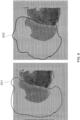
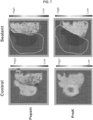
- a region where the analytes are prevented from interacting with a capture probe was contacted with a sealant (e.g., Covergrip TM Coverslip Sealant) (see FIG. 6 ).
- the black lines 601 and 602 surround the region in each biological sample to which the sealant was applied.
- the region not covered by a sealant i.e., the right side of each image
- the sealant dried for one hour at room temperature in order to form a hydrophobic seal over the second region.
- the biological sample was permeabilized to release the analytes from the region of interest.
- the tissues were washed and permeabilized by adding either Proteinase K or Pepsin, incubated at 37°C for at least 5 minutes and washed to remove the protease.
- the analytes from the region of interest hybridized to the capture probes.
- the sealant prevented permeabilization of the second region, thereby blocking the release of the analytes. It is appreciated that the sealant also directly prevented the release of the analytes from the second region in addition to preventing permeabilization.
- the analytes from the region of interest e.g., the first region of interest not covered by the sealant
- the extended capture probes were amplified and sequenced according to any one of the methods described herein. Subsequent sequence analysis was used to determine spatial information regarding the analyte captured from the tissue sample.
- the sealant prevented interaction between analytes from the second region with capture domains of the capture probe. See FIG. 7 , right "Sealant” images. However, without a sealant ( FIG 7 , left "Control” images), analytes were captured from the entire sample. Further, both pepsin and proteinase K were used to permeabilize the sample, and based on the images in FIG. 7 , each permeabilization reagent (1) detected analytes in the first region of interest, and (2) did not permeabilize the sample in the second region. This was evident by the very low UMI counts (0-500 counts per spot). In this experiment, a serial section of the same tissue sample was used as control.
- Table 1 reports the outcome of the experiments comparing conditions on control tissue (e.g., the whole tissue was assayed) versus tissues where a sealant was applied on a region of the tissue (e.g., a region of interest, and not the whole tissue, was assayed). Comparative results demonstrate the increase in resolution of the regions of interest from the tissues where a portion of the tissue was sealed versus the whole tissue where there was no sealant. Table 1. Sensitivity comparison between controls and sealant samples.
- Table 2 shows sequencing metrics for a tissue region of interest (e.g., a portion of the tissue was sealed with a sealant) compared to a whole tissue (e.g., the whole tissue was assayed).
- the results of these experiments report a higher sequencing saturation for sealant samples as compared to control samples when matched for spots corresponding to tissues where a sealant was applied and a region of interest was assayed versus a tissue where there was no sealant applied and the whole tissue was assayed.
- Condition sealant control sealant control Perm pepsin pepsin proK proK No.
- the results demonstrate the sealant does not have a negative effect on workflow and actually results in greater number of reads per spot, greater fraction of library complexity being captured during sequencing and greater number of unique molecular identifiers (UMIs) per spot for the region of interest not covered by a sealant as compared a biological sample where a whole tissue was assayed and not just a region of interest.
- UMIs unique molecular identifiers
- Example 2 Spatial analysis of spleen and liver biological samples using a sealant
- This example provides an exemplary method for analyzing an analyte in a biological sample (i.e., spleen and liver samples) at a region of interest where regions not of interest are sealed off using a sealant.
- a biological sample i.e., spleen and liver samples
- Liver and spleen samples were preserved by FFPE processing. Before addition of the sealant, the biological samples were deparaffinized and stained per established protocols. For example, FFPE tissue samples were prewarmed in a water bath (40°C), sectioned (10 ⁇ m), dried at 42°C for several hours, and placed in a desiccator at room temperature overnight. The dry, sectioned tissues were deparaffinized by baking at 60°C, moved through a series of xylene and EtOH washes, rinsed in water several times. Following rinsing, the deparaffinized tissues were stained with hematoxylin per established protocols. The stained tissues were imaged.
- the tissues were decrosslinked to remove formaldehyde crosslinks within the sample thereby making the analytes accessible for capture. Briefly, the tissue samples were incubated with an HCl solution for 1 minute, repeated twice for a total of 3 minutes. Following HCl incubations, the tissue sections were incubated at 70°C for 1 hour in TE pH 9.0. TE was removed and the tissues were incubation in 1x PBS-Tween for 15 minutes.
- An area on a substrate was identified as a region of interest.
- the area outside of the region of interest was covered with a sealant (i.e., COVERGRIP TM sealant or SALLY HANSEN TM BRAND nail polish).
- the COVERGRIP TM sealant or SALLY HANSEN TM nail polish were allowed to dry at room temperature for 30 minutes.
- the area outside of the region of interest can include part of the biological sample or no biological sample.
- the liver and spleen tissue sections were permeabilized to release the analytes from the region of interest.
- the tissues were washed and permeabilized by adding either proteinase K or pepsin, incubated at 37°C for at least 5 minutes and then washed to remove the proteinase K or pepsin.
- the analytes from the biological sample(s) hybridized to the capture probes.
- the sealant prevented permeabilization of the area outside of the region of interest, thereby blocking the release of the analytes in that sealed region.
- the sealant also prevented the release of the analytes from the area outside of the region of interest in addition to preventing permeabilization.
- the analytes from the biological sample(s) that hybridized to the capture probes were used as a template in a nucleic acid extension reaction that generated extended capture probes.
- the extended capture probes were amplified and sequenced according to any one of the methods described herein. Subsequent sequence analysis was used to determine spatial information regarding the analyte captured from the tissue sample region of interest. Control samples were also assayed, except there was no sealant applied to any part of the control tissue or slide.
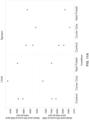
- FIG. 8 shows fraction of raw reads on target and unambiguously mapped (bottom panels), and fractions of spots under tissue (top panels) of liver (left panels) and spleen (right panels) tissue samples that were positioned on the substrate where an area outside and around the tissue sample (1) had no sealant applied and was unblocked (control), (2) an area around the tissue sample was blocked with cover grip (e.g., COVERGRIP TM ) sealant, and or (3) an area around the tissue sample was blocked with nail polish (e.g., SALLY HANSEN TM ).
- cover grip e.g., COVERGRIP TM
- nail polish e.g., SALLY HANSEN TM
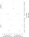
- FIG. 9 shows the fraction of reads in spots under the tissue (bottom panels), and fraction of targeted reads useable (top panels) of liver (left panels) and spleen (right panels) tissue samples were positioned on a substrate where an area outside of and around the tissue sample (1) had no sealant applied and was unblocked (control), (2) an area around the tissue sample was blocked with cover grip (e.g., COVERGRIP TM ) sealant, or (3) an area around the tissue sample was blocked with nail polish (e.g., SALLY HANSEN TM ).
- cover grip e.g., COVERGRIP TM
- nail polish e.g., SALLY HANSEN TM
- FIG. 9 shows an increase in fraction of target reads, particularly in the spleen samples, when the area outside of the tissue sample is blocked thus preventing interaction between a capture domain of a capture probe and an analyte from the tissue sample.
- the fraction of target reads in spots under the tissue substantially match the fraction of targeted reads usable for each of the samples and sample conditions. Fraction of reads in about 10% greater for the tissue sample when the area around the tissue sample is blocked with a sealant.
- FIG. 10 shows median panel genes detected at 10,000 panel reads per spot (bottom panels) and median panel reads detected at 2,500 panel reads per spot (top panels) of liver (left panels) and spleen (right panels) tissue samples that were positioned on a substrate where an area outside of and around the tissue sample 1) had no sealant applied and was unblocked (control), 2) an area around the tissue sample was blocked with cover grip (e.g., COVERGRIP TM ) sealant, or 3) an area around the tissue sample was blocked with nail polish (e.g., SALLY HANSEN TM ).
- FIG. 10 shows that there was not a significant difference in gene detection from the tissue samples whether the area outside of the sample was blocked or unblocked. This result demonstrates that the sealant(s) do not have a negative effect on the workflow.
- FIG. 11A shows median panel UMI counts at 10,000 panel reads per spot (bottom panels) and median panel UMI counts at 5,000 panel reads per spot for liver (left panels) and spleen (right panels) tissue samples that were positioned on a substrate where an area outside of the tissue sample (1) had no sealant applied and was unblocked (control), (2) an area around the tissue sample was blocked with cover grip (e.g., COVERGRIP TM ) sealant, or (3) an area around the tissue sample was blocked with nail polish (e.g., SALLY HANSEN TM ).
- cover grip e.g., COVERGRIP TM
- nail polish e.g., SALLY HANSEN TM
- FIG. 11B shows median panel UMI counts at 10,000 raw reads per spot (bottom panels) and median panel UMI counts at 1,000 raw reads per spot for liver (left panels) and spleen (right panels) tissue samples that were positioned on a substrate where an area outside of the tissue sample (1) had no sealant applied and was unblocked (control), (2) an area around the tissue sample was blocked with cover grip (e.g., COVERGRIP TM ) sealant, and or (3) an area around the tissue sample was blocked with nail polish (e.g., SALLY HANSEN TM ).
- cover grip e.g., COVERGRIP TM
- nail polish e.g., SALLY HANSEN TM
- FIG. 12A shows panel cDNA PCR duplication of 5,000 panel reads per spot (bottom panels) and panel cDNA PCR duplication of 1,000 panel reads per spot (top panels) for liver (left panels) and spleen (right panels) tissue samples that were positioned on a substrate where an area outside of the tissue sample (1) had no sealant applied and was unblocked (control), (2) an area around the tissue sample was blocked with cover grip (e.g., COVERGRIP TM ) sealant, or (3) an area around the tissue sample was blocked with nail polish (e.g., SALLY HANSEN TM ).
- cover grip e.g., COVERGRIP TM
- nail polish e.g., SALLY HANSEN TM
- FIGs. 12A and 12B demonstrate that despite roughly equivalent UMI counts for the controls comparative to the liver and spleen tissue samples, sequencing saturation is greater when the areas surrounding the tissue samples are blocked with a sealant.
- FIG. 13A shows UMI counts of spleen and liver tissue samples where an area outside of the tissue sample (1) had no sealant applied and was unblocked (control), (2) an area around the tissue sample was blocked with cover grip (e.g., COVERGRIP TM ) sealant, or (3) an area around the tissue sample was blocked with nail polish (e.g., SALLY HANSEN TM ).
- the top spleen control shows that one of the tissue samples moved off of the substrate during the assay (also discussed in connection with FIG. 8 ).
- the nail polish sealant of one of the 'liver-nail polish' tissue samples failed (also discussed in connection with FIG. 8 ).
- FIG. 13A shows UMI counts of spleen and liver tissue samples where an area outside of the tissue sample (1) had no sealant applied and was unblocked (control), (2) an area around the tissue sample was blocked with cover grip (e.g., COVERGRIP TM ) sealant, or (3) an area around the tissue sample was blocked with nail polish (e.
- FIG. 13A demonstrates non-specific binding of target analytes to capture probes outside of the tissue sample in the control samples. Conversely, FIG. 13A shows decreased non-specific binding of target analytes to capture probes outside of the tissue sample in the samples where a sealant was applied around the tissue area, regardless of sealant used. As shown in FIG. 13A , red patches indicate more UMI counts and the blue patches indicates fewer UMI counts. The samples where cover grip was applied as a sealant performed slightly better than the samples where nail polish was applied, however both sealants prevented the non-specific binding of target analytes to capture probes outside of the location under the tissue sample.
- FIG. 13B shows pictures of mRNA transcript clustering for spleen and liver tissue samples where, (1) no sealant was applied around the tissue sample (control), (2) the area around the tissue sample was blocked with cover grip (e.g., COVERGRIP TM ) sealant, or (3) the area around the tissue sample blocked with nail polish (e.g., SALLY HANSEN TM ).
- cover grip e.g., COVERGRIP TM
- nail polish e.g., SALLY HANSEN TM
- the experimental results demonstrate a sealant does not have a negative effect on the workflow for spatially capturing analytes from a biological sample. Further, that the cover grip sealant appeared to enhance assay resolution more than the nail polish sealant and the cover grip provided more consistent results. Blocking the surface of the substrate in areas outside or around the tissue sample improves sensitivity for analyte detection under the tissue sample when raw sequencing reads are observed. As such, it was determined that blocking the substrate in areas outside of the tissue sample can improve efficiency, save time, and money and can potentially impact sequencing depth for the spots under the tissue sample. The results were consistent with experimental results when a region of interest was defined and assayed after the area around that region of interest, a second region of the tissue sample and/or substrate was blocked.
- This assay is particularly useful for application to small samples as there is a larger area outside of the tissue sample that is uncovered by the smaller samples.
- the sealants described herein provide an improvement in resource conservation and a reduction and/or elimination of non-specific binding of analytes to unintended portions of the array during the assay, including regions of non-interest within a tissue sample.
- sealants can be used in an assay in an ad hoc manner without a negative impact on the workflow for spatially capturing analytes from a biological sample.
- Sealants can be applied to regions of non-interest outside of a tissue sample positioned on a substrate, and/or regions of non-interest within a tissue sample.
- the application of a sealant application to regions of non-interest on the substrate and/or the tissue can provide a customizable advantage without increasing a burden to the workflow.
Landscapes
- Chemical & Material Sciences (AREA)
- Life Sciences & Earth Sciences (AREA)
- Health & Medical Sciences (AREA)
- Organic Chemistry (AREA)
- Engineering & Computer Science (AREA)
- Immunology (AREA)
- Molecular Biology (AREA)
- Proteomics, Peptides & Aminoacids (AREA)
- Zoology (AREA)
- Wood Science & Technology (AREA)
- Biochemistry (AREA)
- Biotechnology (AREA)
- Microbiology (AREA)
- Analytical Chemistry (AREA)
- Physics & Mathematics (AREA)
- General Health & Medical Sciences (AREA)
- General Engineering & Computer Science (AREA)
- Biophysics (AREA)
- Genetics & Genomics (AREA)
- Bioinformatics & Cheminformatics (AREA)
- Biomedical Technology (AREA)
- Medicinal Chemistry (AREA)
- Chemical Kinetics & Catalysis (AREA)
- Hematology (AREA)
- Urology & Nephrology (AREA)
- General Chemical & Material Sciences (AREA)
- Cell Biology (AREA)
- Food Science & Technology (AREA)
- General Physics & Mathematics (AREA)
- Pathology (AREA)
- Measuring Or Testing Involving Enzymes Or Micro-Organisms (AREA)
Claims (14)
- Verfahren zum Fangen eines Nukleinsäureanalyten aus einer ersten Region von Interesse einer Gewebeprobe, das Verfahren umfassend:(a) Identifizieren der ersten Region von Interesse auf der Gewebeprobe und einer zweiten Region, wobei die zweite Region (i) eine Region auf der Gewebeprobe ist, (ii) eine die Gewebeprobe umgebende Region ist oder (iii) eine Region auf der Gewebeprobe und eine die Gewebeprobe umgebende Region umfasst;(b) Inkontaktbringen der Gewebeprobe mit einem Substrat, das eine Vielzahl von Fangsonden umfasst, wobei eine Fangsonde der Vielzahl der Fangsonden eine Nukleinsäure ist und einen räumlichen Barcode und eine Fangdomäne umfasst;(c) Inkontaktbringen der zweiten Region mit einem Dichtmittel, wobei das Dichtmittel aus der Gruppe ausgewählt ist, die besteht aus: einem Deckglas-Dichtmittel, einem flüssigen Deckglas, Nagellack, Flüssigkeit von einem hydrophoben Stift, einem Montagemedium, einem Gel, einem Klebstoff und einer Doppelschicht oder einer Kombination davon;(d) Permeabilisieren der Gewebeprobe unter Verwendung eines Permeabilisierungsmittels; und(e) Hybridisieren des Nukleinsäureanalyten aus der ersten Region von Interesse mit der Fangsonde, wodurch der Nukleinsäureanalyt aus der ersten Region von Interesse der Gewebeprobe gefangen wird.
- Verfahren nach Anspruch 1, ferner umfassend das Bestimmen (i) einer gesamten oder eines Teils einer Sequenz, die dem an der Fangsonde gebundenen Nukleinsäureanalyten oder eines Komplements davon entspricht, und (ii) der Sequenz, die dem räumlichen Barcode oder eines Komplements davon entspricht, und Verwenden der bestimmten Sequenzen von (i) und (ii) zum Bestimmen der Häufigkeit und/oder des Orts des Nukleinsäureanalyten in der Gewebeprobe.
- Verfahren nach Anspruch 1 oder 2, wobei das Identifizieren der ersten Region von Interesse in der Gewebeprobe und der zweiten Region das Färben der Gewebeprobe umfasst, wobei die Gewebeprobe optional unter Verwendung von Immunfluoreszenz, Immunhistochemie oder Hämatoxylin und Eosin gefärbt wird.
- Verfahren nach einem der Ansprüche 1 bis 3, wobei das Dichtungsmittel eine hydrophobe Dichtung erzeugt, die die zweite Region bedeckt, wodurch eine Wechselwirkung zwischen einem Nukleinsäureanalyten aus der zweiten Region und der Fangdomäne der Fangsonde verhindert wird.
- Verfahren nach einem der Ansprüche 1 bis 2 wobei die erste Region von Interesse und die zweite Region durch ein Gewebedetektion-Maschinenlernmodul identifiziert werden.
- Verfahren nach einem der vorhergehenden Ansprüche, wobei das Inkontaktbringen in (c) das Aufbringen des Dichtungsmittels unter Verwendung eines Verfahrens umfasst, das aus der Gruppe ausgewählt ist, die besteht aus: einer Pinselspitze, einem Tropfer, einer Pipette, einer mikrofluidischen Vorrichtung und einem Flüssigkeitshandhabungsinstrument, wobei das Aufbringen des Dichtungsmittels optional das Identifizieren der ersten Region von Interesse und der zweiten Region und das Aufbringen des Dichtungsmittels unter Verwendung einer Automatisierung auf die zweiten Region umfasst.
- Verfahren nach Anspruch 6, wobei die Automatisierung ein System umfasst, das ein computerimplementiertes Verfahren und das Flüssigkeitshandhabungsinstrument umfasst.
- Verfahren nach einem der vorhergehenden Ansprüche, ferner umfassend das Erlauben einer Zeitdauer, damit das Dichtungsmittel eine hydrophobe Dichtung bildet, wobei die Zeitdauer optional etwa 15 Minuten bis etwa 4 Stunden umfasst, wobei die Zeitdauer optional etwa 1 Stunde bis etwa 2 Stunden umfasst, wobei die Zeitdauer optional etwa 1 Stunde beträgt, umfasst.
- Verfahren nach einem der vorhergehenden Ansprüche, wobei das Permeabilisierungsmittel aus der Gruppe ausgewählt ist, die besteht aus: einem organischen Lösungsmittel, einem Detergens und einem Enzym oder einer Kombination davon, wobei das Permeabilisierungsmittel optional aus der Gruppe ausgewählt ist, die besteht aus: einer Endopeptidase, einer Protease, Natriumdodecylsulfat (SDS), Polyethylenglykol, tert.-Octylphenylether, Polysorbat 80, Polysorbat 20, N-Lauroylsarcosin-Natriumsalzlösung, Saponin, einem nichtionischen Tensid und Polyoxyethylensorbitol, wobei optional die Endopeptidase Pepsin oder Proteinase K ist.
- Verfahren nach einem der vorhergehenden Ansprüche, ferner umfassend das Entfernen des Dichtungsmittels von der Gewebeprobe, wobei das Entfernen optional umfasst:(i) Anheben, Abziehen, Auflösen, Verflüssigen oder Entnetzen des Dichtungsmittels von der Gewebeprobe; oder(ii) Inkontaktbringen der Gewebeprobe mit einem Entfernungsmittel, das aus der Gruppe ausgewählt ist, die besteht aus: einem Lösungsmittel, einer Säure, einer Base und einem Puffer oder einer Kombination davon, wobei das Entfernungsmittel optional phosphatgepufferte Kochsalzlösung (PBS) umfasst.
- Verfahren nach einem der vorhergehenden Ansprüche, wobei die Gewebeprobe eine frische Gewebeprobe, eine gefrorene Gewebeprobe oder eine formalinfixierte, paraffineingebettete (FFPE-) Gewebeprobe ist, wobei die FFPE-Gewebeprobe optional entnetzt ist.
- Verfahren nach einem der vorhergehenden Ansprüche, wobei die Gewebeprobe zuvor gefärbt wurde.
- Verfahren nach einem der vorhergehenden Ansprüche, wobei das Verfahren zu einer höheren Fangeffizienz von Nukleinsäureanalyten aus der Gewebeprobe im Vergleich zur Fangeffizienz von Nukleinsäureanalyten aus einer Gewebeprobe führt, die nicht mit einem Dichtungsmittel in Kontakt gebracht wird.
- Verfahren nach einem der vorhergehenden Ansprüche, wobei das Verfahren zu einer höheren Fangeffizienz von Nukleinsäureanalyten aus der ersten Region von Interesse der Gewebeprobe führt, verglichen mit der Fangeffizienz von Nukleinsäureanalyten aus der zweiten Region.
Applications Claiming Priority (2)
| Application Number | Priority Date | Filing Date | Title |
|---|---|---|---|
| US202163185035P | 2021-05-06 | 2021-05-06 | |
| PCT/US2022/028071 WO2022236054A1 (en) | 2021-05-06 | 2022-05-06 | Methods for increasing resolution of spatial analysis |
Publications (3)
| Publication Number | Publication Date |
|---|---|
| EP4320271A1 EP4320271A1 (de) | 2024-02-14 |
| EP4320271C0 EP4320271C0 (de) | 2025-03-19 |
| EP4320271B1 true EP4320271B1 (de) | 2025-03-19 |
Family
ID=81975441
Family Applications (1)
| Application Number | Title | Priority Date | Filing Date |
|---|---|---|---|
| EP22728711.7A Active EP4320271B1 (de) | 2021-05-06 | 2022-05-06 | Verfahren zur erhöhung der auflösung von räumlicher analyse |
Country Status (3)
| Country | Link |
|---|---|
| US (1) | US12365935B2 (de) |
| EP (1) | EP4320271B1 (de) |
| WO (1) | WO2022236054A1 (de) |
Families Citing this family (62)
| Publication number | Priority date | Publication date | Assignee | Title |
|---|---|---|---|---|
| US20190300945A1 (en) | 2010-04-05 | 2019-10-03 | Prognosys Biosciences, Inc. | Spatially Encoded Biological Assays |
| US10787701B2 (en) | 2010-04-05 | 2020-09-29 | Prognosys Biosciences, Inc. | Spatially encoded biological assays |
| GB201106254D0 (en) | 2011-04-13 | 2011-05-25 | Frisen Jonas | Method and product |
| DK3511423T4 (da) | 2012-10-17 | 2024-07-29 | Spatial Transcriptomics Ab | Fremgangsmåder og produkt til optimering af lokaliseret eller rumlig detektion af genekspression i en vævsprøve |
| CN118240918A (zh) | 2013-06-25 | 2024-06-25 | 普罗格诺西斯生物科学公司 | 采用微流控装置的空间编码生物分析 |
| EP3280820B1 (de) | 2015-04-10 | 2019-06-26 | Spatial Transcriptomics AB | Räumlich getrennte multiplex-nukleinsäureanalyse von biologischen proben |
| US11519033B2 (en) | 2018-08-28 | 2022-12-06 | 10X Genomics, Inc. | Method for transposase-mediated spatial tagging and analyzing genomic DNA in a biological sample |
| EP3894589A2 (de) | 2018-12-10 | 2021-10-20 | 10X Genomics, Inc. | Auflösung von räumlichen anordnungen unter verwendung von dekonvolution |
| US11649485B2 (en) | 2019-01-06 | 2023-05-16 | 10X Genomics, Inc. | Generating capture probes for spatial analysis |
| US11926867B2 (en) | 2019-01-06 | 2024-03-12 | 10X Genomics, Inc. | Generating capture probes for spatial analysis |
| WO2020243579A1 (en) | 2019-05-30 | 2020-12-03 | 10X Genomics, Inc. | Methods of detecting spatial heterogeneity of a biological sample |
| EP4055185A1 (de) | 2019-11-08 | 2022-09-14 | 10X Genomics, Inc. | Räumlich markierte analytfänger für analyt-multiplexing |
| WO2021092433A2 (en) | 2019-11-08 | 2021-05-14 | 10X Genomics, Inc. | Enhancing specificity of analyte binding |
| FI3891300T3 (fi) | 2019-12-23 | 2023-05-10 | 10X Genomics Inc | Menetelmät spatiaalista analyysiä varten rna-templatoitua ligaatiota käyttäen |
| WO2021133842A1 (en) | 2019-12-23 | 2021-07-01 | 10X Genomics, Inc. | Compositions and methods for using fixed biological samples in partition-based assays |
| US12365942B2 (en) | 2020-01-13 | 2025-07-22 | 10X Genomics, Inc. | Methods of decreasing background on a spatial array |
| US12405264B2 (en) | 2020-01-17 | 2025-09-02 | 10X Genomics, Inc. | Electrophoretic system and method for analyte capture |
| US11702693B2 (en) | 2020-01-21 | 2023-07-18 | 10X Genomics, Inc. | Methods for printing cells and generating arrays of barcoded cells |
| US11732299B2 (en) | 2020-01-21 | 2023-08-22 | 10X Genomics, Inc. | Spatial assays with perturbed cells |
| US20210230681A1 (en) | 2020-01-24 | 2021-07-29 | 10X Genomics, Inc. | Methods for spatial analysis using proximity ligation |
| US11821035B1 (en) | 2020-01-29 | 2023-11-21 | 10X Genomics, Inc. | Compositions and methods of making gene expression libraries |
| US12076701B2 (en) | 2020-01-31 | 2024-09-03 | 10X Genomics, Inc. | Capturing oligonucleotides in spatial transcriptomics |
| US12110541B2 (en) | 2020-02-03 | 2024-10-08 | 10X Genomics, Inc. | Methods for preparing high-resolution spatial arrays |
| US11898205B2 (en) | 2020-02-03 | 2024-02-13 | 10X Genomics, Inc. | Increasing capture efficiency of spatial assays |
| US11732300B2 (en) | 2020-02-05 | 2023-08-22 | 10X Genomics, Inc. | Increasing efficiency of spatial analysis in a biological sample |
| WO2021158925A1 (en) | 2020-02-07 | 2021-08-12 | 10X Genomics, Inc. | Quantitative and automated permeabilization performance evaluation for spatial transcriptomics |
| US11835462B2 (en) | 2020-02-11 | 2023-12-05 | 10X Genomics, Inc. | Methods and compositions for partitioning a biological sample |
| US12399123B1 (en) | 2020-02-14 | 2025-08-26 | 10X Genomics, Inc. | Spatial targeting of analytes |
| US12281357B1 (en) | 2020-02-14 | 2025-04-22 | 10X Genomics, Inc. | In situ spatial barcoding |
| US11891654B2 (en) | 2020-02-24 | 2024-02-06 | 10X Genomics, Inc. | Methods of making gene expression libraries |
| US11926863B1 (en) | 2020-02-27 | 2024-03-12 | 10X Genomics, Inc. | Solid state single cell method for analyzing fixed biological cells |
| US11768175B1 (en) | 2020-03-04 | 2023-09-26 | 10X Genomics, Inc. | Electrophoretic methods for spatial analysis |
| EP4139485B1 (de) | 2020-04-22 | 2023-09-06 | 10X Genomics, Inc. | Verfahren zur räumlichen analyse unter verwendung von gezieltem rna-abbau |
| CN116134308B (zh) | 2020-05-19 | 2025-05-27 | 10X基因组学有限公司 | 电泳盒和仪器 |
| EP4553168A3 (de) | 2020-05-22 | 2025-08-27 | 10x Genomics, Inc. | Räumliche analyse zur erkennung von sequenzvarianten |
| EP4414459B1 (de) | 2020-05-22 | 2025-09-03 | 10X Genomics, Inc. | Simultane räumlich-zeitliche messung der genexpression und der zellaktivität |
| US12265079B1 (en) | 2020-06-02 | 2025-04-01 | 10X Genomics, Inc. | Systems and methods for detecting analytes from captured single biological particles |
| EP4600376A3 (de) | 2020-06-02 | 2025-10-22 | 10X Genomics, Inc. | Räumliche transkriptomik für antigenrezeptoren |
| WO2021247543A2 (en) | 2020-06-02 | 2021-12-09 | 10X Genomics, Inc. | Nucleic acid library methods |
| US12031177B1 (en) | 2020-06-04 | 2024-07-09 | 10X Genomics, Inc. | Methods of enhancing spatial resolution of transcripts |
| WO2021252499A1 (en) | 2020-06-08 | 2021-12-16 | 10X Genomics, Inc. | Methods of determining a surgical margin and methods of use thereof |
| US12435363B1 (en) | 2020-06-10 | 2025-10-07 | 10X Genomics, Inc. | Materials and methods for spatial transcriptomics |
| EP4172362B1 (de) | 2020-06-25 | 2024-09-18 | 10X Genomics, Inc. | Räumliche analyse der dna-methylierung |
| US11761038B1 (en) | 2020-07-06 | 2023-09-19 | 10X Genomics, Inc. | Methods for identifying a location of an RNA in a biological sample |
| US11981960B1 (en) | 2020-07-06 | 2024-05-14 | 10X Genomics, Inc. | Spatial analysis utilizing degradable hydrogels |
| US12209280B1 (en) | 2020-07-06 | 2025-01-28 | 10X Genomics, Inc. | Methods of identifying abundance and location of an analyte in a biological sample using second strand synthesis |
| US11981958B1 (en) | 2020-08-20 | 2024-05-14 | 10X Genomics, Inc. | Methods for spatial analysis using DNA capture |
| WO2022061150A2 (en) | 2020-09-18 | 2022-03-24 | 10X Geonomics, Inc. | Sample handling apparatus and image registration methods |
| US11926822B1 (en) | 2020-09-23 | 2024-03-12 | 10X Genomics, Inc. | Three-dimensional spatial analysis |
| US11827935B1 (en) | 2020-11-19 | 2023-11-28 | 10X Genomics, Inc. | Methods for spatial analysis using rolling circle amplification and detection probes |
| EP4121555A1 (de) | 2020-12-21 | 2023-01-25 | 10X Genomics, Inc. | Verfahren, zusammensetzungen und systeme zur erfassung von sonden und/oder barcodes |
| ES2989523T3 (es) | 2021-02-19 | 2024-11-26 | 10X Genomics Inc | Método de uso de un dispositivo modular de soporte para ensayos |
| EP4524261A3 (de) | 2021-03-18 | 2025-05-14 | 10X Genomics, Inc. | Multiplex-erfassung von gen- und proteinexpression aus einer biologischen probe |
| EP4428246A3 (de) | 2021-04-14 | 2024-11-27 | 10X Genomics, Inc. | Verfahren zur messung der fehllokalisierung eines analyten |
| WO2022236054A1 (en) | 2021-05-06 | 2022-11-10 | 10X Genomics, Inc. | Methods for increasing resolution of spatial analysis |
| EP4582555A3 (de) | 2021-06-03 | 2025-10-22 | 10X Genomics, Inc. | Verfahren, zusammensetzungen, kits und systeme zur verbesserung der analyterfassung zur räumlichen analyse |
| EP4509614A3 (de) | 2021-09-01 | 2025-05-14 | 10X Genomics, Inc. | Verfahren, zusammensetzungen und kits zur blockierung einer erfassungssonde auf einer räumlichen anordnung |
| EP4419707A1 (de) | 2021-11-10 | 2024-08-28 | 10X Genomics, Inc. | Verfahren, zusammensetzungen und kits zur bestimmung der position eines analyten in einer biologischen probe |
| EP4305195A2 (de) | 2021-12-01 | 2024-01-17 | 10X Genomics, Inc. | Verfahren, zusammensetzungen und systeme für verbesserten in-situ-nachweis von analyten und raumanalyse |
| WO2023122033A1 (en) | 2021-12-20 | 2023-06-29 | 10X Genomics, Inc. | Self-test for pathology/histology slide imaging device |
| WO2024206603A1 (en) * | 2023-03-28 | 2024-10-03 | 10X Genomics, Inc. | Methods, compositions, and kits for reducing analyte mislocalization |
| WO2025093690A1 (en) * | 2023-11-02 | 2025-05-08 | Resolve Biosciences Gmbh | Method for preparing tissue sections |
Family Cites Families (706)
| Publication number | Priority date | Publication date | Assignee | Title |
|---|---|---|---|---|
| US4683202A (en) | 1985-03-28 | 1987-07-28 | Cetus Corporation | Process for amplifying nucleic acid sequences |
| US4965188A (en) | 1986-08-22 | 1990-10-23 | Cetus Corporation | Process for amplifying, detecting, and/or cloning nucleic acid sequences using a thermostable enzyme |
| US4683195A (en) | 1986-01-30 | 1987-07-28 | Cetus Corporation | Process for amplifying, detecting, and/or-cloning nucleic acid sequences |
| US4883867A (en) | 1985-11-01 | 1989-11-28 | Becton, Dickinson And Company | Detection of reticulocytes, RNA or DNA |
| US4800159A (en) | 1986-02-07 | 1989-01-24 | Cetus Corporation | Process for amplifying, detecting, and/or cloning nucleic acid sequences |
| US5525464A (en) | 1987-04-01 | 1996-06-11 | Hyseq, Inc. | Method of sequencing by hybridization of oligonucleotide probes |
| GB8810400D0 (en) | 1988-05-03 | 1988-06-08 | Southern E | Analysing polynucleotide sequences |
| US4988617A (en) | 1988-03-25 | 1991-01-29 | California Institute Of Technology | Method of detecting a nucleotide change in nucleic acids |
| US5130238A (en) | 1988-06-24 | 1992-07-14 | Cangene Corporation | Enhanced nucleic acid amplification process |
| US5512439A (en) | 1988-11-21 | 1996-04-30 | Dynal As | Oligonucleotide-linked magnetic particles and uses thereof |
| US5002882A (en) | 1989-04-27 | 1991-03-26 | New England Biolabs, Inc. | Method for producing the XmaI restriction endonuclease and methylase |
| EP0450060A1 (de) | 1989-10-26 | 1991-10-09 | Sri International | Dns-sequenzierung |
| US5494810A (en) | 1990-05-03 | 1996-02-27 | Cornell Research Foundation, Inc. | Thermostable ligase-mediated DNA amplifications system for the detection of genetic disease |
| US5455166A (en) | 1991-01-31 | 1995-10-03 | Becton, Dickinson And Company | Strand displacement amplification |
| WO1993004199A2 (en) | 1991-08-20 | 1993-03-04 | Scientific Generics Limited | Methods of detecting or quantitating nucleic acids and of producing labelled immobilised nucleic acids |
| US5474796A (en) | 1991-09-04 | 1995-12-12 | Protogene Laboratories, Inc. | Method and apparatus for conducting an array of chemical reactions on a support surface |
| US6872816B1 (en) | 1996-01-24 | 2005-03-29 | Third Wave Technologies, Inc. | Nucleic acid detection kits |
| US6759226B1 (en) | 2000-05-24 | 2004-07-06 | Third Wave Technologies, Inc. | Enzymes for the detection of specific nucleic acid sequences |
| ATE152831T1 (de) | 1991-09-16 | 1997-05-15 | Molecular Probes Inc | Dimere unsymmetrische cyaninfarbstoffe |
| US5321130A (en) | 1992-02-10 | 1994-06-14 | Molecular Probes, Inc. | Unsymmetrical cyanine dyes with a cationic side chain |
| US5308751A (en) | 1992-03-23 | 1994-05-03 | General Atomics | Method for sequencing double-stranded DNA |
| US5503980A (en) | 1992-11-06 | 1996-04-02 | Trustees Of Boston University | Positional sequencing by hybridization |
| US5410030A (en) | 1993-04-05 | 1995-04-25 | Molecular Probes, Inc. | Dimers of unsymmetrical cyanine dyes containing pyridinium moieties |
| US5658751A (en) | 1993-04-13 | 1997-08-19 | Molecular Probes, Inc. | Substituted unsymmetrical cyanine dyes with selected permeability |
| US5436134A (en) | 1993-04-13 | 1995-07-25 | Molecular Probes, Inc. | Cyclic-substituted unsymmetrical cyanine dyes |
| US5837832A (en) | 1993-06-25 | 1998-11-17 | Affymetrix, Inc. | Arrays of nucleic acid probes on biological chips |
| US6401267B1 (en) | 1993-09-27 | 2002-06-11 | Radoje Drmanac | Methods and compositions for efficient nucleic acid sequencing |
| SE9400522D0 (sv) | 1994-02-16 | 1994-02-16 | Ulf Landegren | Method and reagent for detecting specific nucleotide sequences |
| US5512462A (en) | 1994-02-25 | 1996-04-30 | Hoffmann-La Roche Inc. | Methods and reagents for the polymerase chain reaction amplification of long DNA sequences |
| US5677170A (en) | 1994-03-02 | 1997-10-14 | The Johns Hopkins University | In vitro transposition of artificial transposons |
| KR100230718B1 (ko) | 1994-03-16 | 1999-11-15 | 다니엘 엘. 캐시앙, 헨리 엘. 노르호프 | 등온 가닥 변위 핵산 증폭법 |
| US6015880A (en) | 1994-03-16 | 2000-01-18 | California Institute Of Technology | Method and substrate for performing multiple sequential reactions on a matrix |
| US5552278A (en) | 1994-04-04 | 1996-09-03 | Spectragen, Inc. | DNA sequencing by stepwise ligation and cleavage |
| US5807522A (en) | 1994-06-17 | 1998-09-15 | The Board Of Trustees Of The Leland Stanford Junior University | Methods for fabricating microarrays of biological samples |
| US5641658A (en) | 1994-08-03 | 1997-06-24 | Mosaic Technologies, Inc. | Method for performing amplification of nucleic acid with two primers bound to a single solid support |
| DE69528706T2 (de) | 1994-08-19 | 2003-06-12 | Pe Corp. (Ny), Foster City | Gekoppeltes ampflikation- und ligationverfahren |
| US5750341A (en) | 1995-04-17 | 1998-05-12 | Lynx Therapeutics, Inc. | DNA sequencing by parallel oligonucleotide extensions |
| US5648245A (en) | 1995-05-09 | 1997-07-15 | Carnegie Institution Of Washington | Method for constructing an oligonucleotide concatamer library by rolling circle replication |
| US6579695B1 (en) | 1995-10-13 | 2003-06-17 | President And Fellows Of Harvard College | Phosphopantetheinyl transferases and uses thereof |
| US5763175A (en) | 1995-11-17 | 1998-06-09 | Lynx Therapeutics, Inc. | Simultaneous sequencing of tagged polynucleotides |
| US5854033A (en) | 1995-11-21 | 1998-12-29 | Yale University | Rolling circle replication reporter systems |
| US6300063B1 (en) | 1995-11-29 | 2001-10-09 | Affymetrix, Inc. | Polymorphism detection |
| US5962271A (en) | 1996-01-03 | 1999-10-05 | Cloutech Laboratories, Inc. | Methods and compositions for generating full-length cDNA having arbitrary nucleotide sequence at the 3'-end |
| EP0880598A4 (de) | 1996-01-23 | 2005-02-23 | Affymetrix Inc | Verfahren zur analyse von nukleinsäure |
| US6875572B2 (en) | 1996-01-24 | 2005-04-05 | Third Wave Technologies, Inc. | Nucleic acid detection assays |
| US5985557A (en) | 1996-01-24 | 1999-11-16 | Third Wave Technologies, Inc. | Invasive cleavage of nucleic acids |
| US6913881B1 (en) | 1996-01-24 | 2005-07-05 | Third Wave Technologies, Inc. | Methods and compositions for detecting target sequences |
| US6852487B1 (en) | 1996-02-09 | 2005-02-08 | Cornell Research Foundation, Inc. | Detection of nucleic acid sequence differences using the ligase detection reaction with addressable arrays |
| EP1958955B1 (de) | 1996-02-09 | 2013-09-04 | Cornell Research Foundation, Inc. | Räumlich adressierbare Oligonukleotidarrays und eine Methode zu deren Herstellung |
| US6013440A (en) | 1996-03-11 | 2000-01-11 | Affymetrix, Inc. | Nucleic acid affinity columns |
| US5928906A (en) | 1996-05-09 | 1999-07-27 | Sequenom, Inc. | Process for direct sequencing during template amplification |
| EP1736554B1 (de) | 1996-05-29 | 2013-10-09 | Cornell Research Foundation, Inc. | Bestimmung von Unterschieden in der Nukleinsäuresequenz mittels einer Kombination von Ligasebestimmung und Polymerasekettenreaktion |
| US20050003431A1 (en) | 1996-08-16 | 2005-01-06 | Wucherpfennig Kai W. | Monovalent, multivalent, and multimeric MHC binding domain fusion proteins and conjugates, and uses therefor |
| US5925545A (en) | 1996-09-09 | 1999-07-20 | Wisconsin Alumni Research Foundation | System for in vitro transposition |
| US5965443A (en) | 1996-09-09 | 1999-10-12 | Wisconsin Alumni Research Foundation | System for in vitro transposition |
| GB9620209D0 (en) | 1996-09-27 | 1996-11-13 | Cemu Bioteknik Ab | Method of sequencing DNA |
| US6060240A (en) | 1996-12-13 | 2000-05-09 | Arcaris, Inc. | Methods for measuring relative amounts of nucleic acids in a complex mixture and retrieval of specific sequences therefrom |
| US5837466A (en) | 1996-12-16 | 1998-11-17 | Vysis, Inc. | Devices and methods for detecting nucleic acid analytes in samples |
| GB9626815D0 (en) | 1996-12-23 | 1997-02-12 | Cemu Bioteknik Ab | Method of sequencing DNA |
| EP2256133B1 (de) | 1997-01-08 | 2016-12-14 | Sigma-Aldrich Co. LLC | Biokonjugation von Makromolekülen |
| US6309824B1 (en) | 1997-01-16 | 2001-10-30 | Hyseq, Inc. | Methods for analyzing a target nucleic acid using immobilized heterogeneous mixtures of oligonucleotide probes |
| US6023540A (en) | 1997-03-14 | 2000-02-08 | Trustees Of Tufts College | Fiber optic sensor with encoded microspheres |
| US6327410B1 (en) | 1997-03-14 | 2001-12-04 | The Trustees Of Tufts College | Target analyte sensors utilizing Microspheres |
| US20020058250A1 (en) | 1997-03-21 | 2002-05-16 | Marshall, Gerstein & Borun | Extraction and utilisation of vntr alleles |
| AU6846798A (en) | 1997-04-01 | 1998-10-22 | Glaxo Group Limited | Method of nucleic acid sequencing |
| EP1591541B1 (de) | 1997-04-01 | 2012-02-15 | Illumina Cambridge Limited | Verfahren zur Vervielfältigung von Nukleinsäuren |
| US6143496A (en) | 1997-04-17 | 2000-11-07 | Cytonix Corporation | Method of sampling, amplifying and quantifying segment of nucleic acid, polymerase chain reaction assembly having nanoliter-sized sample chambers, and method of filling assembly |
| WO1999005295A1 (en) | 1997-07-25 | 1999-02-04 | Thomas Jefferson University | Composition and method for targeted integration into cells |
| AU8908198A (en) | 1997-08-15 | 1999-03-08 | Hyseq, Inc. | Methods and compositions for detection or quantification of nucleic acid species |
| WO1999019341A1 (en) | 1997-10-10 | 1999-04-22 | President & Fellows Of Harvard College | Replica amplification of nucleic acid arrays |
| US6054274A (en) | 1997-11-12 | 2000-04-25 | Hewlett-Packard Company | Method of amplifying the signal of target nucleic acid sequence analyte |
| AU2003200718B2 (en) | 1997-12-15 | 2006-10-19 | Somalogic, Inc. | Nucleic acid ligand diagnostic biochip |
| US6242246B1 (en) | 1997-12-15 | 2001-06-05 | Somalogic, Inc. | Nucleic acid ligand diagnostic Biochip |
| US6844158B1 (en) | 1997-12-22 | 2005-01-18 | Hitachi Chemical Co., Ltd. | Direct RT-PCR on oligonucleotide-immobilized PCR microplates |
| JP3662850B2 (ja) | 1998-06-24 | 2005-06-22 | イルミナ インコーポレイテッド | 微小球を有するアレイセンサーのデコード |
| CA2321821A1 (en) | 1998-06-26 | 2000-01-06 | Visible Genetics Inc. | Method for sequencing nucleic acids with reduced errors |
| US20040106110A1 (en) | 1998-07-30 | 2004-06-03 | Solexa, Ltd. | Preparation of polynucleotide arrays |
| US6787308B2 (en) | 1998-07-30 | 2004-09-07 | Solexa Ltd. | Arrayed biomolecules and their use in sequencing |
| US20030022207A1 (en) | 1998-10-16 | 2003-01-30 | Solexa, Ltd. | Arrayed polynucleotides and their use in genome analysis |
| JP4475488B2 (ja) | 1998-09-18 | 2010-06-09 | マイクロメット アーゲー | 単一の細胞のdna増幅 |
| US6159736A (en) | 1998-09-23 | 2000-12-12 | Wisconsin Alumni Research Foundation | Method for making insertional mutations using a Tn5 synaptic complex |
| AR021833A1 (es) | 1998-09-30 | 2002-08-07 | Applied Research Systems | Metodos de amplificacion y secuenciacion de acido nucleico |
| US6573043B1 (en) | 1998-10-07 | 2003-06-03 | Genentech, Inc. | Tissue analysis and kits therefor |
| AU772281B2 (en) | 1998-12-14 | 2004-04-22 | Li-Cor Inc. | A system and methods for nucleic acid sequencing of single molecules by polymerase synthesis |
| US6830884B1 (en) | 1998-12-15 | 2004-12-14 | Molecular Staging Inc. | Method of amplification |
| ATE440148T1 (de) | 1999-01-06 | 2009-09-15 | Callida Genomics Inc | Verbesserte sequenzierung mittels hybridisierung durch verwendung von sondengemischen |
| GB9901475D0 (en) | 1999-01-22 | 1999-03-17 | Pyrosequencing Ab | A method of DNA sequencing |
| DE69913092T2 (de) | 1999-01-27 | 2004-09-09 | Commissariat à l'Energie Atomique | Microassay zur Serienanalyse der Genexpression und Anwendungen davon |
| US6153389A (en) | 1999-02-22 | 2000-11-28 | Haarer; Brian K. | DNA additives as a mechanism for unambiguously marking biological samples |
| US20060275782A1 (en) | 1999-04-20 | 2006-12-07 | Illumina, Inc. | Detection of nucleic acid reactions on bead arrays |
| US20050191698A1 (en) | 1999-04-20 | 2005-09-01 | Illumina, Inc. | Nucleic acid sequencing using microsphere arrays |
| US6355431B1 (en) | 1999-04-20 | 2002-03-12 | Illumina, Inc. | Detection of nucleic acid amplification reactions using bead arrays |
| EP1196630B2 (de) | 1999-04-20 | 2018-10-17 | Illumina, Inc. | Detektion von nukleinsäurereaktionen auf kügelchen-arrays |
| US6534266B1 (en) | 1999-04-22 | 2003-03-18 | Albert Einstein College Of Medicine Of Yeshiva University | Assay of transcription sites by multi-fluor fish |
| US7276336B1 (en) | 1999-07-22 | 2007-10-02 | Agilent Technologies, Inc. | Methods of fabricating an addressable array of biopolymer probes |
| US20010055764A1 (en) | 1999-05-07 | 2001-12-27 | Empedocles Stephen A. | Microarray methods utilizing semiconductor nanocrystals |
| US6544732B1 (en) | 1999-05-20 | 2003-04-08 | Illumina, Inc. | Encoding and decoding of array sensors utilizing nanocrystals |
| CA2374390A1 (en) | 1999-05-20 | 2000-12-14 | Illumina, Inc. | Combinatorial decoding of random nucleic acid arrays |
| ATE409236T1 (de) | 1999-07-14 | 2008-10-15 | Packard Bioscience Company | Derivatisierte nukleinsäuren und deren verwendung |
| AU775043B2 (en) | 1999-08-02 | 2004-07-15 | Wisconsin Alumni Research Foundation | Mutant Tn5 transposase enzymes and method for their use |
| ATE318932T1 (de) | 1999-08-13 | 2006-03-15 | Univ Yale | Binär kodierte sequenzmarker |
| CA2382157C (en) | 1999-08-18 | 2012-04-03 | Illumina, Inc. | Compositions and methods for preparing oligonucleotide solutions |
| US6942968B1 (en) | 1999-08-30 | 2005-09-13 | Illumina, Inc. | Array compositions for improved signal detection |
| JP3929775B2 (ja) | 1999-09-13 | 2007-06-13 | ニューゲン テクノロジーズ, インコーポレイテッド | ポリヌクレオチド配列の線形等温増幅のための方法および組成物 |
| US6274320B1 (en) | 1999-09-16 | 2001-08-14 | Curagen Corporation | Method of sequencing a nucleic acid |
| US6291180B1 (en) | 1999-09-29 | 2001-09-18 | American Registry Of Pathology | Ultrasound-mediated high-speed biological reaction and tissue processing |
| AU7537200A (en) | 1999-09-29 | 2001-04-30 | Solexa Ltd. | Polynucleotide sequencing |
| EP1226115A4 (de) | 1999-10-04 | 2006-03-15 | Univ New Jersey Med | Carbamate und harnstoffe |
| EP1235932A2 (de) | 1999-10-08 | 2002-09-04 | Protogene Laboratories, Inc. | Verfahren und vorrichtung zur durchführung von hohen zahlen von reaktionen unter benutzung von arrays |
| WO2001042796A1 (en) | 1999-12-13 | 2001-06-14 | The Government Of The United States Of America, As Represented By The Secretary, Department Of Health & Human Services, The National Institutes Of Health | High-throughput tissue microarray technology and applications |
| US6248535B1 (en) | 1999-12-20 | 2001-06-19 | University Of Southern California | Method for isolation of RNA from formalin-fixed paraffin-embedded tissue specimens |
| GB0002389D0 (en) | 2000-02-02 | 2000-03-22 | Solexa Ltd | Molecular arrays |
| US7955794B2 (en) | 2000-09-21 | 2011-06-07 | Illumina, Inc. | Multiplex nucleic acid reactions |
| US7611869B2 (en) | 2000-02-07 | 2009-11-03 | Illumina, Inc. | Multiplexed methylation detection methods |
| US8076063B2 (en) | 2000-02-07 | 2011-12-13 | Illumina, Inc. | Multiplexed methylation detection methods |
| JP2003521252A (ja) | 2000-02-07 | 2003-07-15 | イルミナ インコーポレイテッド | ユニバーサルプライミングを用いる核酸検出方法 |
| US7582420B2 (en) | 2001-07-12 | 2009-09-01 | Illumina, Inc. | Multiplex nucleic acid reactions |
| DE60127939T2 (de) | 2000-02-07 | 2008-01-24 | Illumina, Inc., San Diego | Nukleinsäure-Nachweisverfahren mit universellem Priming |
| US7361488B2 (en) | 2000-02-07 | 2008-04-22 | Illumina, Inc. | Nucleic acid detection methods using universal priming |
| US20010026919A1 (en) | 2000-02-08 | 2001-10-04 | Alex Chenchik | Nucleic acid assays employing universal arrays |
| US6770441B2 (en) | 2000-02-10 | 2004-08-03 | Illumina, Inc. | Array compositions and methods of making same |
| CA2388738A1 (en) | 2000-02-15 | 2001-08-23 | Lynx Therapeutics, Inc. | Data analysis and display system for ligation-based dna sequencing |
| CA2403296A1 (en) | 2000-04-10 | 2001-10-18 | Benjamin F. Cravatt | Proteomic analysis using activity-based probe libraries |
| US6368801B1 (en) | 2000-04-12 | 2002-04-09 | Molecular Staging, Inc. | Detection and amplification of RNA using target-mediated ligation of DNA by RNA ligase |
| US6291187B1 (en) | 2000-05-12 | 2001-09-18 | Molecular Staging, Inc. | Poly-primed amplification of nucleic acid sequences |
| US6828098B2 (en) | 2000-05-20 | 2004-12-07 | The Regents Of The University Of Michigan | Method of producing a DNA library using positional amplification based on the use of adaptors and nick translation |
| US6511809B2 (en) | 2000-06-13 | 2003-01-28 | E. I. Du Pont De Nemours And Company | Method for the detection of an analyte by means of a nucleic acid reporter |
| US7439016B1 (en) | 2000-06-15 | 2008-10-21 | Digene Corporation | Detection of nucleic acids by type-specific hybrid capture method |
| US6323009B1 (en) | 2000-06-28 | 2001-11-27 | Molecular Staging, Inc. | Multiply-primed amplification of nucleic acid sequences |
| EP1975251A3 (de) | 2000-07-07 | 2009-03-25 | Visigen Biotechnologies, Inc. | Sequenzbestimmung in Echtzeit |
| GB0018120D0 (en) | 2000-07-24 | 2000-09-13 | Fermentas Ab | Nuclease |
| AU7861301A (en) | 2000-08-15 | 2002-02-25 | Discerna Ltd | Functional protein arrays |
| AU2001293163A1 (en) | 2000-09-27 | 2002-04-08 | Lynx Therapeutics, Inc. | Method for determining relative abundance of nucleic acid sequences |
| WO2002061129A2 (en) | 2000-11-15 | 2002-08-08 | Minerva Biotechnologies Corporation | Oligonucleotide identifiers |
| EP1343973B2 (de) | 2000-11-16 | 2020-09-16 | California Institute Of Technology | Vorrichtung und verfahren zur durchführung von assays und screening mit hohem durchsatz |
| AU2002227156A1 (en) | 2000-12-01 | 2002-06-11 | Visigen Biotechnologies, Inc. | Enzymatic nucleic acid synthesis: compositions and methods for altering monomer incorporation fidelity |
| AR031640A1 (es) | 2000-12-08 | 2003-09-24 | Applied Research Systems | Amplificacion isotermica de acidos nucleicos en un soporte solido |
| US20030017451A1 (en) | 2000-12-21 | 2003-01-23 | Hui Wang | Methods for detecting transcripts |
| JP4061043B2 (ja) | 2000-12-28 | 2008-03-12 | 株式会社ポストゲノム研究所 | invitro転写/翻訳系によるペプチド等の製造方法 |
| US7135296B2 (en) | 2000-12-28 | 2006-11-14 | Mds Inc. | Elemental analysis of tagged biologically active materials |
| EP1360490B1 (de) | 2001-01-23 | 2011-12-21 | President and Fellows of Harvard College | Nukleinsäure-programmierbare protein-arrays |
| DK1364065T3 (da) | 2001-01-25 | 2012-05-14 | Luminex Molecular Diagnostics Inc | Polynukleotider til anvendelse som markeringer og markeringskomplementer, fremstilling og anvendelse deraf |
| KR20020063359A (ko) | 2001-01-27 | 2002-08-03 | 일렉트론 바이오 (주) | 핵산 및 올리거뉴클레오티드의 상보적 이중결합의 특정서열에 특이적으로 반응하는 절단기법을 이용한 핵산 혼성분석 방법 및 장치 |
| US7253341B2 (en) | 2001-03-05 | 2007-08-07 | Yissum Research Development Company Of The Hebrew University Of Jerusalem | Denaturant stable and/or protease resistant, chaperone-like oligomeric proteins, polynucleotides encoding same, their uses and methods of increasing a specific activity thereof |
| US6573051B2 (en) | 2001-03-09 | 2003-06-03 | Molecular Staging, Inc. | Open circle probes with intramolecular stem structures |
| EP1368497A4 (de) | 2001-03-12 | 2007-08-15 | California Inst Of Techn | Verfahren und vorrichtung zur analyse von polynukleotidsequenzen durch asynchrone basenverlängerung |
| AU2002322457A1 (en) | 2001-06-28 | 2003-03-03 | Illumina, Inc. | Multiplex decoding of array sensors with microspheres |
| US7473767B2 (en) | 2001-07-03 | 2009-01-06 | The Institute For Systems Biology | Methods for detection and quantification of analytes in complex mixtures |
| US20040241688A1 (en) | 2001-07-19 | 2004-12-02 | Cuneyt Bukusoglu | Human tissue specific drug screening procedure |
| US20040091857A1 (en) | 2001-07-20 | 2004-05-13 | Nallur Girish N. | Gene expression profiling |
| GB0118031D0 (en) | 2001-07-24 | 2001-09-19 | Oxford Glycosciences Uk Ltd | Self assembled protein nucleic acid complexes and self assembled protein arrays |
| US20030148335A1 (en) | 2001-10-10 | 2003-08-07 | Li Shen | Detecting targets by unique identifier nucleotide tags |
| US6942972B2 (en) | 2001-10-24 | 2005-09-13 | Beckman Coulter, Inc. | Efficient synthesis of protein-oligonucleotide conjugates |
| EP1451365A4 (de) | 2001-11-13 | 2006-09-13 | Rubicon Genomics Inc | Dna-amplifikation und -sequenzierung mit durch zufallsfragmentierung erzeugten dna-molekülen |
| GB0127564D0 (en) | 2001-11-16 | 2002-01-09 | Medical Res Council | Emulsion compositions |
| US7057026B2 (en) | 2001-12-04 | 2006-06-06 | Solexa Limited | Labelled nucleotides |
| WO2003069333A1 (en) | 2002-02-14 | 2003-08-21 | Illumina, Inc. | Automated information processing in randomly ordered arrays |
| EP1540017A4 (de) | 2002-05-09 | 2006-05-24 | Us Genomics Inc | Verfahren zur analyse einer nukleinsäure |
| US20050202433A1 (en) | 2002-06-03 | 2005-09-15 | Van Beuningen Marinus Gerardus J. | Novel high density arrays and methods for analyte analysis |
| US7108976B2 (en) | 2002-06-17 | 2006-09-19 | Affymetrix, Inc. | Complexity management of genomic DNA by locus specific amplification |
| US20050118616A1 (en) | 2002-08-16 | 2005-06-02 | Kawashima Tadashi R. | Amplification of target nucleotide sequence without polymerase chain reaction |
| US7205128B2 (en) | 2002-08-16 | 2007-04-17 | Agilent Technologies, Inc. | Method for synthesis of the second strand of cDNA |
| EP3363809B1 (de) | 2002-08-23 | 2020-04-08 | Illumina Cambridge Limited | Modifizierte nukleotide zur polynukleotidsequenzierung |
| WO2004027025A2 (en) | 2002-09-20 | 2004-04-01 | New England Biolabs, Inc. | Helicase dependent amplification of nucleic acids |
| US7662594B2 (en) | 2002-09-20 | 2010-02-16 | New England Biolabs, Inc. | Helicase-dependent amplification of RNA |
| US20040259105A1 (en) | 2002-10-03 | 2004-12-23 | Jian-Bing Fan | Multiplex nucleic acid analysis using archived or fixed samples |
| US20040067492A1 (en) | 2002-10-04 | 2004-04-08 | Allan Peng | Reverse transcription on microarrays |
| GB0302058D0 (en) | 2003-01-29 | 2003-02-26 | Univ Cranfield | Replication of nucleic acid arrays |
| CA2513541A1 (en) | 2003-01-29 | 2004-08-19 | 454 Corporation | Method for preparing single-stranded dna libraries |
| AU2004214891B2 (en) | 2003-02-26 | 2010-01-07 | Complete Genomics, Inc. | Random array DNA analysis by hybridization |
| US7718403B2 (en) | 2003-03-07 | 2010-05-18 | Rubicon Genomics, Inc. | Amplification and analysis of whole genome and whole transcriptome libraries generated by a DNA polymerization process |
| ES2428941T3 (es) | 2003-03-10 | 2013-11-12 | Expression Pathology, Inc. | Preparación líquida de tejidos a partir de muestras biológicas, tejidos y células procesadas histopatológicamente |
| FR2852317B1 (fr) | 2003-03-13 | 2006-08-04 | Biopuces a sondes et leurs methodes d'utilisation | |
| CN1300333C (zh) | 2003-04-17 | 2007-02-14 | 中国人民解放军军事医学科学院放射与辐射医学研究所 | 炭疽菌诊断基因芯片的制备及应用 |
| US7083980B2 (en) | 2003-04-17 | 2006-08-01 | Wisconsin Alumni Research Foundation | Tn5 transposase mutants and the use thereof |
| WO2004104588A1 (en) | 2003-05-23 | 2004-12-02 | Epfl - Ecole Polytechnique Federale De Lausanne | Methods for protein labeling based on acyl carrier protein |
| US20040241660A1 (en) | 2003-05-30 | 2004-12-02 | Applera Corporation | Apparatus and method for hybridization |
| US7255994B2 (en) | 2003-06-10 | 2007-08-14 | Applera Corporation | Ligation assay |
| US20060216775A1 (en) | 2003-06-17 | 2006-09-28 | The Regents Of The University Of Califoenia | Compositions and methods for analysis and manipulation of enzymes in biosynthetic proteomes |
| US20050053980A1 (en) | 2003-06-20 | 2005-03-10 | Illumina, Inc. | Methods and compositions for whole genome amplification and genotyping |
| US20070128656A1 (en) | 2003-06-26 | 2007-06-07 | University Of South Florida | Direct Fluorescent Label Incorporation Via 1st Strand cDNA Synthesis |
| CA2530810A1 (en) | 2003-07-03 | 2005-01-27 | The Regents Of The University Of California | Genome mapping of functional dna elements and cellular proteins |
| CA2531105C (en) | 2003-07-05 | 2015-03-17 | The Johns Hopkins University | Method and compositions for detection and enumeration of genetic variations |
| ES2539784T3 (es) | 2003-09-02 | 2015-07-06 | Keygene N.V. | Procedimientos basados en OLA para la detección de secuencias de ácidos nucleicos objetivo |
| WO2005074417A2 (en) | 2003-09-03 | 2005-08-18 | Salk Institute For Biological Studies | Multiple antigen detection assays and reagents |
| JP5171037B2 (ja) | 2003-09-10 | 2013-03-27 | アルセア・ディーエックス,インク. | マイクロアレイを用いた発現プロファイリング |
| GB0321306D0 (en) | 2003-09-11 | 2003-10-15 | Solexa Ltd | Modified polymerases for improved incorporation of nucleotide analogues |
| US20050266417A1 (en) | 2003-09-12 | 2005-12-01 | Francis Barany | Methods for identifying target nucleic acid molecules |
| ATE447626T1 (de) | 2003-09-18 | 2009-11-15 | Nuevolution As | Methode zur gewinnung struktureller informationen kodierter moleküle und zur selektion von verbindungen |
| US20050064435A1 (en) | 2003-09-24 | 2005-03-24 | Xing Su | Programmable molecular barcodes |
| US20050136414A1 (en) | 2003-12-23 | 2005-06-23 | Kevin Gunderson | Methods and compositions for making locus-specific arrays |
| US20050147976A1 (en) | 2003-12-29 | 2005-07-07 | Xing Su | Methods for determining nucleotide sequence information |
| EP1709203A2 (de) | 2004-01-23 | 2006-10-11 | Lingvitae AS | Verbesserung von polynukleotid-ligationsreaktionen |
| US7378242B2 (en) | 2004-03-18 | 2008-05-27 | Atom Sciences, Inc. | DNA sequence detection by limited primer extension |
| KR100624420B1 (ko) | 2004-04-10 | 2006-09-19 | 삼성전자주식회사 | 마이크로어레이에 대한 정보가 스팟의 형태로 저장되어있는 마이크로어레이, 그를 제조하는 방법 및 그를이용하는 방법 |
| US20050260653A1 (en) | 2004-04-14 | 2005-11-24 | Joshua Labaer | Nucleic-acid programmable protein arrays |
| JP4592060B2 (ja) | 2004-04-26 | 2010-12-01 | キヤノン株式会社 | Pcr増幅反応装置、ならびに、該装置を利用するpcr増幅反応方法 |
| EP1756307A1 (de) | 2004-05-20 | 2007-02-28 | Trillion Genomics Limited | Verwendung massenmarkierter sonden zum nachweis von zielnukleinsäuren mittels massenspektrometrie |
| US7608434B2 (en) | 2004-08-04 | 2009-10-27 | Wisconsin Alumni Research Foundation | Mutated Tn5 transposase proteins and the use thereof |
| WO2006073504A2 (en) | 2004-08-04 | 2006-07-13 | President And Fellows Of Harvard College | Wobble sequencing |
| US20060041385A1 (en) | 2004-08-18 | 2006-02-23 | Bauer Kenneth D | Method of quantitating proteins and genes in cells using a combination of immunohistochemistry and in situ hybridization |
| US7776547B2 (en) | 2004-08-26 | 2010-08-17 | Intel Corporation | Cellular analysis using Raman surface scanning |
| CN100396790C (zh) | 2004-09-17 | 2008-06-25 | 北京大学 | 溶液识别、表面寻址蛋白质芯片及其制备和检测方法 |
| US7527970B2 (en) | 2004-10-15 | 2009-05-05 | The United States Of America As Represented By The Department Of Health And Human Services | Method of identifying active chromatin domains |
| EP2302053A1 (de) | 2004-11-12 | 2011-03-30 | Asuragen, Inc. | Verfahren und Zusammensetzungen, die miRNAs und miRNA-inhibitorischen Molekülen verbunden sind |
| US7183119B2 (en) | 2004-11-15 | 2007-02-27 | Eastman Kodak Company | Method for sensitive detection of multiple biological analytes |
| DE602005012319D1 (de) | 2004-11-22 | 2009-02-26 | Peter Birk Rasmussen | Matrizengerichtete split-and-mix-synthese von bibliotheken kleiner moleküle |
| US7579153B2 (en) | 2005-01-25 | 2009-08-25 | Population Genetics Technologies, Ltd. | Isothermal DNA amplification |
| EP1844162B1 (de) | 2005-02-01 | 2014-10-15 | Applied Biosystems, LLC | vERFAHREN ZUR SEQUENZBESTIMMUNG IN EINEM POLYNUKLEOTID |
| US7407757B2 (en) | 2005-02-10 | 2008-08-05 | Population Genetics Technologies | Genetic analysis by sequence-specific sorting |
| US7393665B2 (en) | 2005-02-10 | 2008-07-01 | Population Genetics Technologies Ltd | Methods and compositions for tagging and identifying polynucleotides |
| GB0504774D0 (en) | 2005-03-08 | 2005-04-13 | Lingvitae As | Method |
| US7727721B2 (en) | 2005-03-08 | 2010-06-01 | California Institute Of Technology | Hybridization chain reaction amplification for in situ imaging |
| US7601498B2 (en) | 2005-03-17 | 2009-10-13 | Biotium, Inc. | Methods of using dyes in association with nucleic acid staining or detection and associated technology |
| US7776567B2 (en) | 2005-03-17 | 2010-08-17 | Biotium, Inc. | Dimeric and trimeric nucleic acid dyes, and associated systems and methods |
| US7303880B2 (en) | 2005-03-18 | 2007-12-04 | Wisconsin Alumni Research Foundation | Microdissection-based methods for determining genomic features of single chromosomes |
| US8623628B2 (en) | 2005-05-10 | 2014-01-07 | Illumina, Inc. | Polymerases |
| WO2006124771A2 (en) | 2005-05-12 | 2006-11-23 | Panomics, Inc. | Multiplex branched-chain dna assays |
| US20060263789A1 (en) | 2005-05-19 | 2006-11-23 | Robert Kincaid | Unique identifiers for indicating properties associated with entities to which they are attached, and methods for using |
| US7601499B2 (en) | 2005-06-06 | 2009-10-13 | 454 Life Sciences Corporation | Paired end sequencing |
| EP3492602A1 (de) | 2005-06-15 | 2019-06-05 | Complete Genomics, Inc. | Einzelne molekülarrays für genetische und chemische analyse |
| US20070087360A1 (en) | 2005-06-20 | 2007-04-19 | Boyd Victoria L | Methods and compositions for detecting nucleotides |
| ES2511218T5 (es) | 2005-06-20 | 2017-12-07 | Advanced Cell Diagnostics, Inc. | Kits y productos para detección de ácidos nucleicos en células individuales y para identificación de células raras en grandes poblaciones de células heterogéneas |
| CN105039313B (zh) | 2005-06-23 | 2018-10-23 | 科因股份有限公司 | 用于多态性的高通量鉴定和检测的策略 |
| WO2007005649A2 (en) | 2005-06-30 | 2007-01-11 | Applera Corporation | Proximity probing of target proteins comprising restriction and/or extension field |
| US7883848B2 (en) | 2005-07-08 | 2011-02-08 | Olink Ab | Regulation analysis by cis reactivity, RACR |
| JP4822753B2 (ja) | 2005-07-11 | 2011-11-24 | 一般社団法人オンチップ・セロミクス・コンソーシアム | 細胞構成物質分取チップならびに細胞構成物質解析システム及びこれらを用いる細胞構成物質解析法 |
| US20070020640A1 (en) | 2005-07-21 | 2007-01-25 | Mccloskey Megan L | Molecular encoding of nucleic acid templates for PCR and other forms of sequence analysis |
| JP2007074967A (ja) | 2005-09-13 | 2007-03-29 | Canon Inc | 識別子プローブ及びそれを用いた核酸増幅方法 |
| CA2910861C (en) | 2005-09-29 | 2018-08-07 | Michael Josephus Theresia Van Eijk | High throughput screening of mutagenized populations |
| US7405281B2 (en) | 2005-09-29 | 2008-07-29 | Pacific Biosciences Of California, Inc. | Fluorescent nucleotide analogs and uses therefor |
| WO2007041689A2 (en) | 2005-10-04 | 2007-04-12 | President And Fellows Of Harvard College | Methods of site-specific labeling of molecules and molecules produced thereby |
| GB0522310D0 (en) | 2005-11-01 | 2005-12-07 | Solexa Ltd | Methods of preparing libraries of template polynucleotides |
| US20070116612A1 (en) | 2005-11-02 | 2007-05-24 | Biopath Automation, L.L.C. | Prefix tissue cassette |
| EP1951905B1 (de) | 2005-11-10 | 2012-04-11 | Affymetrix, Inc. | Nachweis von nukleinsäuren über amplifikation von stellvertreternukleinsäuren |
| WO2007120208A2 (en) | 2005-11-14 | 2007-10-25 | President And Fellows Of Harvard College | Nanogrid rolling circle dna sequencing |
| ATE527383T1 (de) | 2005-11-22 | 2011-10-15 | Stichting Dienst Landbouwkundi | Multiplex-nukleinsäurenachweis |
| WO2007060599A1 (en) | 2005-11-25 | 2007-05-31 | Koninklijke Philips Electronics N.V. | Magnetic biosensor for determination of enzymic activity |
| US8092784B2 (en) | 2005-11-30 | 2012-01-10 | Biotium, Inc. | Enzyme substrate comprising a functional dye and associated technology and methods |
| US7803751B2 (en) | 2005-12-09 | 2010-09-28 | Illumina, Inc. | Compositions and methods for detecting phosphomonoester |
| DE102005060738A1 (de) | 2005-12-16 | 2007-06-21 | Qiagen Gmbh | Verfahren zur Extraktion von Biomolekülen aus fixierten Geweben |
| ES2882401T3 (es) | 2005-12-22 | 2021-12-01 | Keygene Nv | Método de detección de polimorfismos basado en AFLP de alto rendimiento |
| DK1966394T3 (da) | 2005-12-22 | 2012-10-29 | Keygene Nv | Forbedrede strategier til transkriptprofilering under anvendelse af high throughput-sekventeringsteknologier |
| WO2007076128A2 (en) | 2005-12-23 | 2007-07-05 | Nanostring Technologies, Inc. | Nanoreporters and methods of manufacturing and use thereof |
| AU2006330834B2 (en) | 2005-12-23 | 2013-09-12 | Bruker Spatial Biology, Inc. | Compositions comprising oriented, immobilized macromolecules and methods for their preparation |
| EP1969146B1 (de) | 2006-01-04 | 2010-10-06 | Si Lok | Verfahren zur zuordnung von nukleinsäuren und zur identifikation fein strukturierter variationen in nukleinsäuren sowie hilfsmittel dafür |
| US7537897B2 (en) | 2006-01-23 | 2009-05-26 | Population Genetics Technologies, Ltd. | Molecular counting |
| WO2007092538A2 (en) | 2006-02-07 | 2007-08-16 | President And Fellows Of Harvard College | Methods for making nucleotide probes for sequencing and synthesis |
| SG170028A1 (en) | 2006-02-24 | 2011-04-29 | Callida Genomics Inc | High throughput genome sequencing on dna arrays |
| CN101432439B (zh) | 2006-02-24 | 2013-07-24 | 考利达基因组股份有限公司 | Dna阵列上的高通量基因组测序 |
| WO2007107710A1 (en) | 2006-03-17 | 2007-09-27 | Solexa Limited | Isothermal methods for creating clonal single molecule arrays |
| GB0605584D0 (en) | 2006-03-20 | 2006-04-26 | Olink Ab | Method for analyte detection using proximity probes |
| EP2963127B1 (de) | 2006-04-04 | 2017-08-16 | Keygene N.V. | Nachweis von Molekularmarkern mit hohem Durchsatz auf der Basis von Restriktionsfragmenten |
| US8383338B2 (en) | 2006-04-24 | 2013-02-26 | Roche Nimblegen, Inc. | Methods and systems for uniform enrichment of genomic regions |
| US20070254305A1 (en) | 2006-04-28 | 2007-11-01 | Nsabp Foundation, Inc. | Methods of whole genome or microarray expression profiling using nucleic acids prepared from formalin fixed paraffin embedded tissue |
| JP2009536525A (ja) | 2006-05-10 | 2009-10-15 | ディクステリティー ダイアグノーティクス | 化学反応性オリゴヌクレオチドプローブを使用した核酸標的の検出 |
| US7941279B2 (en) | 2006-05-22 | 2011-05-10 | Nanostring Technologies, Inc. | Systems and methods for analyzing nanoreporters |
| US20080132429A1 (en) | 2006-05-23 | 2008-06-05 | Uchicago Argonne | Biological microarrays with enhanced signal yield |
| US8362242B2 (en) | 2006-06-30 | 2013-01-29 | Dh Technologies Development Pte. Ltd. | Analyte determination utilizing mass tagging reagents comprising a non-encoded detectable label |
| US7595150B2 (en) | 2006-06-30 | 2009-09-29 | Searete Llc | Method of applying an elongated molecule to a surface |
| AT503862B1 (de) | 2006-07-05 | 2010-11-15 | Arc Austrian Res Centers Gmbh | Pathogen-identifizierung anhand eines 16s oder 18s-rrna mikroarray |
| CN101522915A (zh) | 2006-08-02 | 2009-09-02 | 加州理工学院 | 用于检测和/或分选靶标的方法和系统 |
| EP2076609A1 (de) | 2006-10-10 | 2009-07-08 | Illumina Inc. | Zusammensetzungen und verfahren zur repräsentativen auswahl von nukleinsäuren aus komplexen gemischen mittels hybridisierung |
| WO2008051530A2 (en) | 2006-10-23 | 2008-05-02 | Pacific Biosciences Of California, Inc. | Polymerase enzymes and reagents for enhanced nucleic acid sequencing |
| US20080108804A1 (en) | 2006-11-02 | 2008-05-08 | Kabushiki Kaisha Dnaform | Method for modifying RNAS and preparing DNAS from RNAS |
| WO2008069906A2 (en) | 2006-11-14 | 2008-06-12 | The Regents Of The University Of California | Digital expression of gene analysis |
| US9201063B2 (en) | 2006-11-16 | 2015-12-01 | General Electric Company | Sequential analysis of biological samples |
| US8262900B2 (en) | 2006-12-14 | 2012-09-11 | Life Technologies Corporation | Methods and apparatus for measuring analytes using large scale FET arrays |
| WO2008076406A2 (en) | 2006-12-14 | 2008-06-26 | Ion Torrent Systems Incorporated | Methods and apparatus for measuring analytes using large scale fet arrays |
| GB0625595D0 (en) | 2006-12-21 | 2007-01-31 | Oxford Gene Tech Ip Ltd | Sample analyser |
| CN101221182A (zh) | 2007-01-08 | 2008-07-16 | 山东司马特生物芯片有限公司 | 一种荧光蛋白质芯片血清肿瘤系列诊断新方法 |
| WO2008093098A2 (en) | 2007-02-02 | 2008-08-07 | Illumina Cambridge Limited | Methods for indexing samples and sequencing multiple nucleotide templates |
| WO2008098100A2 (en) | 2007-02-07 | 2008-08-14 | Perscitus Biosciences, Llc | Detection of molecule proximity |
| EP2130913A4 (de) | 2007-03-05 | 2010-09-01 | On Chip Cellomics Consortium | Chip zur zellkomponenten-probennahme, zellkomponenten-analysesystem und zellkomponenten-analyseverfahren unter verwendung davon |
| EP2145180B1 (de) | 2007-04-13 | 2013-12-04 | Sequenom, Inc. | Vergleichende sequenzanalyseverfahren und -systeme |
| CN101720359A (zh) | 2007-06-01 | 2010-06-02 | 454生命科学公司 | 从多重混合物中识别个别样本的系统和方法 |
| WO2008151127A1 (en) | 2007-06-04 | 2008-12-11 | President And Fellows Of Harvard College | Compounds and methods for chemical ligation |
| US7635566B2 (en) | 2007-06-29 | 2009-12-22 | Population Genetics Technologies Ltd. | Methods and compositions for isolating nucleic acid sequence variants |
| JP2009036694A (ja) | 2007-08-03 | 2009-02-19 | Tokyo Medical & Dental Univ | 空間分布を保った細胞内生体物質の解析方法 |
| WO2009032167A1 (en) | 2007-08-29 | 2009-03-12 | Illumina Cambridge | Method for sequencing a polynucleotide template |
| US9388457B2 (en) | 2007-09-14 | 2016-07-12 | Affymetrix, Inc. | Locus specific amplification using array probes |
| WO2009036525A2 (en) | 2007-09-21 | 2009-03-26 | Katholieke Universiteit Leuven | Tools and methods for genetic tests using next generation sequencing |
| EP2053132A1 (de) | 2007-10-23 | 2009-04-29 | Roche Diagnostics GmbH | Anreicherung und Sequenzanalyse von Genombereichen |
| US8518640B2 (en) | 2007-10-29 | 2013-08-27 | Complete Genomics, Inc. | Nucleic acid sequencing and process |
| US8906700B2 (en) | 2007-11-06 | 2014-12-09 | Ambergen, Inc. | Methods and compositions for phototransfer |
| US8592150B2 (en) | 2007-12-05 | 2013-11-26 | Complete Genomics, Inc. | Methods and compositions for long fragment read sequencing |
| WO2009086487A2 (en) * | 2007-12-28 | 2009-07-09 | Spring Bioscience Corporation | Liquid coverslip and method and device for applying and removing coverslips |
| EP2620511B1 (de) | 2008-01-17 | 2018-02-28 | Sequenom, Inc. | Einzelmolekül-Nukleinsäurensequenzanalyseverfahren |
| KR20090081260A (ko) | 2008-01-23 | 2009-07-28 | 삼성전자주식회사 | 마이크로 어레이 혼성화 검출 방법 |
| WO2009132028A1 (en) | 2008-04-21 | 2009-10-29 | Complete Genomics, Inc. | Array structures for nucleic acid detection |
| US20110105356A1 (en) | 2008-05-07 | 2011-05-05 | Derosier Chad F | Compositions and methods for providing substances to and from an array |
| EP2806037B1 (de) | 2008-05-13 | 2016-09-21 | Gen-Probe Incorporated | Inaktivierbare Target-Capture-Oligomere zur Verwendung bei der selektiven Hybridisierung und beim Fangen von Zielnukleinsäuresequenzen |
| US8093064B2 (en) | 2008-05-15 | 2012-01-10 | The Regents Of The University Of California | Method for using magnetic particles in droplet microfluidics |
| DE102008025656B4 (de) | 2008-05-28 | 2016-07-28 | Genxpro Gmbh | Verfahren zur quantitativen Analyse von Nukleinsäuren, Marker dafür und deren Verwendung |
| US20100120097A1 (en) | 2008-05-30 | 2010-05-13 | Board Of Regents, The University Of Texas System | Methods and compositions for nucleic acid sequencing |
| US20100035249A1 (en) | 2008-08-05 | 2010-02-11 | Kabushiki Kaisha Dnaform | Rna sequencing and analysis using solid support |
| AU2009281836B2 (en) | 2008-08-14 | 2015-02-19 | Bruker Spatial Biology, Inc. | Stable nanoreporters |
| EP2326732A4 (de) | 2008-08-26 | 2012-11-14 | Fluidigm Corp | Testverfahren für erhöhten durchsatz von proben und/oder targets |
| US8586310B2 (en) | 2008-09-05 | 2013-11-19 | Washington University | Method for multiplexed nucleic acid patch polymerase chain reaction |
| US8383345B2 (en) | 2008-09-12 | 2013-02-26 | University Of Washington | Sequence tag directed subassembly of short sequencing reads into long sequencing reads |
| US9080211B2 (en) | 2008-10-24 | 2015-07-14 | Epicentre Technologies Corporation | Transposon end compositions and methods for modifying nucleic acids |
| CA2742272C (en) | 2008-10-30 | 2018-05-29 | Sequenom, Inc. | Products and processes for multiplex nucleic acid identification |
| US9394567B2 (en) | 2008-11-07 | 2016-07-19 | Adaptive Biotechnologies Corporation | Detection and quantification of sample contamination in immune repertoire analysis |
| EP2370803A2 (de) | 2008-12-03 | 2011-10-05 | The United States Government As Represented By The Department Of Veterans Affairs | Druckunterstützte molekulare wiederherstellung wie von biomolekülen, druckunterstützte antigenrückgewinnung und druckunterstützte gewebehistologie |
| US8790873B2 (en) | 2009-01-16 | 2014-07-29 | Affymetrix, Inc. | DNA ligation on RNA template |
| WO2010088517A1 (en) | 2009-01-30 | 2010-08-05 | The U.S.A., As Represented By The Secretary, Department Of Health And Human Services | Methods and systems for purifying transferring and/or manipulating nucleic acids |
| KR101059565B1 (ko) | 2009-02-11 | 2011-08-26 | 어플라이드 프레시젼, 인코포레이티드 | 밝은 기준점 표지를 갖는 마이크로어레이 및 그로부터 광 데이터를 수집하는 방법 |
| US8481698B2 (en) | 2009-03-19 | 2013-07-09 | The President And Fellows Of Harvard College | Parallel proximity ligation event analysis |
| JP5985390B2 (ja) | 2009-04-02 | 2016-09-06 | フリューダイム・コーポレイション | 標的核酸にバーコードを付加するためのマルチプライマー増幅方法 |
| WO2010115100A1 (en) | 2009-04-03 | 2010-10-07 | L&C Diagment, Inc. | Multiplex nucleic acid detection methods and systems |
| CA2760439A1 (en) | 2009-04-30 | 2010-11-04 | Good Start Genetics, Inc. | Methods and compositions for evaluating genetic markers |
| US9085798B2 (en) | 2009-04-30 | 2015-07-21 | Prognosys Biosciences, Inc. | Nucleic acid constructs and methods of use |
| WO2011008502A2 (en) | 2009-06-29 | 2011-01-20 | California Institute Of Technology | Isolation of unknown rearranged t-cell receptors from single cells |
| GB0912909D0 (en) | 2009-07-23 | 2009-08-26 | Olink Genomics Ab | Probes for specific analysis of nucleic acids |
| WO2011014811A1 (en) | 2009-07-31 | 2011-02-03 | Ibis Biosciences, Inc. | Capture primers and capture sequence linked solid supports for molecular diagnostic tests |
| EP2464738A4 (de) | 2009-08-12 | 2013-05-01 | Nugen Technologies Inc | Verfahren, zusammensetzungen und kits zur erzeugung von substantiell nukleinsäurematrizenfreien nukleinsäureprodukten |
| WO2011038403A1 (en) | 2009-09-28 | 2011-03-31 | Yuling Luo | Methods of detecting nucleic acid sequences with high specificity |
| CA2775613C (en) | 2009-10-13 | 2018-06-12 | Nanostring Technologies, Inc. | Protein detection via nanoreporters |
| US9005891B2 (en) | 2009-11-10 | 2015-04-14 | Genomic Health, Inc. | Methods for depleting RNA from nucleic acid samples |
| US20120277113A1 (en) | 2009-11-18 | 2012-11-01 | Ruo-Pan Huang | Array-based proximity ligation association assays |
| CN102639716A (zh) | 2009-12-04 | 2012-08-15 | 株式会社日立制作所 | 使用二维cDNA文库的基因表达解析方法 |
| US20120202704A1 (en) | 2009-12-07 | 2012-08-09 | Illumina, Inc. | Multi-sample indexing for multiplex genotyping |
| DE112010004821T5 (de) | 2009-12-15 | 2012-10-04 | Agency For Science, Technology And Research | Prozessierung amplifizierter DNA-Fragmente zur Sequenzierung |
| US8835358B2 (en) | 2009-12-15 | 2014-09-16 | Cellular Research, Inc. | Digital counting of individual molecules by stochastic attachment of diverse labels |
| ES2719502T3 (es) | 2010-01-29 | 2019-07-10 | Advanced Cell Diagnostics Inc | Métodos de detección in situ de ácidos nucleicos |
| EP2354242A1 (de) | 2010-02-03 | 2011-08-10 | Epiontis GmbH | Assay zur Bestimmung des Typs und/oder des Zustands eines zellbasierten epigenetischen Musters und der chromatischen Struktur |
| US9714446B2 (en) | 2010-02-11 | 2017-07-25 | Nanostring Technologies, Inc. | Compositions and methods for the detection of small RNAs |
| WO2011127099A1 (en) | 2010-04-05 | 2011-10-13 | Prognosys Biosciences, Inc. | Spatially encoded biological assays |
| US10787701B2 (en) | 2010-04-05 | 2020-09-29 | Prognosys Biosciences, Inc. | Spatially encoded biological assays |
| US20190300945A1 (en) | 2010-04-05 | 2019-10-03 | Prognosys Biosciences, Inc. | Spatially Encoded Biological Assays |
| US20110245101A1 (en) | 2010-04-05 | 2011-10-06 | Prognosys Biosciences, Inc. | Co-localization affinity assays |
| US10240194B2 (en) | 2010-05-13 | 2019-03-26 | Gen9, Inc. | Methods for nucleotide sequencing and high fidelity polynucleotide synthesis |
| US8828688B2 (en) | 2010-05-27 | 2014-09-09 | Affymetrix, Inc. | Multiplex amplification methods |
| US9080210B2 (en) | 2010-06-09 | 2015-07-14 | Keygene N.V. | High throughput screening using combinatorial sequence barcodes |
| US11203786B2 (en) | 2010-08-06 | 2021-12-21 | Ariosa Diagnostics, Inc. | Detection of target nucleic acids using hybridization |
| CN110878345A (zh) | 2010-09-21 | 2020-03-13 | 安捷伦科技有限公司 | 通过分子计数提高等位基因调用的置信度 |
| US10669569B2 (en) | 2010-10-15 | 2020-06-02 | Navinci Diagnostics Ab | Dynamic range methods |
| WO2012058638A2 (en) | 2010-10-29 | 2012-05-03 | President And Fellows Of Harvard College | Nucleic acid nanostructure barcode probes |
| WO2012061832A1 (en) | 2010-11-05 | 2012-05-10 | Illumina, Inc. | Linking sequence reads using paired code tags |
| US20130184184A1 (en) | 2010-11-22 | 2013-07-18 | The University Of Chicago | Methods and/or Use of Oligonucleotide Conjugates Having Varied Degrees of Labeling for Assays and Detections |
| US20140121118A1 (en) | 2010-11-23 | 2014-05-01 | Opx Biotechnologies, Inc. | Methods, systems and compositions regarding multiplex construction protein amino-acid substitutions and identification of sequence-activity relationships, to provide gene replacement such as with tagged mutant genes, such as via efficient homologous recombination |
| US8551708B2 (en) | 2011-02-15 | 2013-10-08 | Leica Biosystems Newcastle Ltd. | Methods for localized in situ detection of mRNA |
| EP3736281A1 (de) | 2011-02-18 | 2020-11-11 | Bio-Rad Laboratories, Inc. | Zusammensetzungen und verfahren für molekulare etikettierung |
| WO2012129242A2 (en) | 2011-03-23 | 2012-09-27 | Pacific Biosciences Of California, Inc. | Isolation of polymerase-nucleic acid complexes and loading onto substrates |
| US20190360034A1 (en) | 2011-04-01 | 2019-11-28 | Centrillion Technology Holdings Corporation | Methods and systems for sequencing nucleic acids |
| EP2694709B1 (de) | 2011-04-08 | 2016-09-14 | Prognosys Biosciences, Inc. | Peptidkonstrukte und testsysteme dafür |
| GB201106254D0 (en) | 2011-04-13 | 2011-05-25 | Frisen Jonas | Method and product |
| US8946389B2 (en) | 2011-04-25 | 2015-02-03 | University Of Washington | Compositions and methods for multiplex biomarker profiling |
| EP2705165B1 (de) | 2011-05-04 | 2016-08-24 | HTG Molecular Diagnostics, Inc. | Verbesserungen an einem quantitativen nukleaseschutz-assay und einer quantitativen nukleaseschutz-sequenzierung |
| CN103635594B (zh) | 2011-05-09 | 2018-02-13 | 富鲁达公司 | 基于探针的核酸检测 |
| AU2012255121B2 (en) | 2011-05-17 | 2017-03-09 | Dxterity Diagnostics Incorporated | Methods and compositions for detecting target nucleic acids |
| WO2012159089A1 (en) | 2011-05-19 | 2012-11-22 | Sequenom, Inc. | Products and processes for multiplex nucleic acid identification |
| US9005935B2 (en) | 2011-05-23 | 2015-04-14 | Agilent Technologies, Inc. | Methods and compositions for DNA fragmentation and tagging by transposases |
| GB201108678D0 (en) | 2011-05-24 | 2011-07-06 | Olink Ab | Multiplexed proximity ligation assay |
| WO2013019960A1 (en) | 2011-08-03 | 2013-02-07 | Bio-Rad Laboratories, Inc. | Filtering small nucleic acids using permeabilized cells |
| WO2013022807A1 (en) | 2011-08-05 | 2013-02-14 | Unitaq Bio | Methods and compositions for detection of nucleic acids using 5'-nuclease cleavage and amplification |
| US10385475B2 (en) | 2011-09-12 | 2019-08-20 | Adaptive Biotechnologies Corp. | Random array sequencing of low-complexity libraries |
| CA2859660C (en) | 2011-09-23 | 2021-02-09 | Illumina, Inc. | Methods and compositions for nucleic acid sequencing |
| EP2766498B1 (de) | 2011-10-14 | 2019-06-19 | President and Fellows of Harvard College | Sequenzierung durch strukturanordnung |
| EP2788499B1 (de) | 2011-12-09 | 2016-01-13 | Illumina, Inc. | Expandierte wurzel für polymer-tags |
| EP4249605B1 (de) | 2011-12-22 | 2024-08-28 | President And Fellows Of Harvard College | Verfahren zum nachweis von analyten |
| EP2814986B1 (de) | 2012-02-14 | 2017-10-25 | Cornell University | Verfahren zur relativen quantifizierung einer nucleinsäuresequenz, expression oder kopieveränderungen anhand vom kombinierten nuclease-, ligations- und polymerasereaktionen |
| US10011871B2 (en) | 2012-02-17 | 2018-07-03 | Fred Hutchinson Cancer Research Center | Compositions and methods for accurately identifying mutations |
| NO2694769T3 (de) | 2012-03-06 | 2018-03-03 | ||
| CA2867293C (en) | 2012-03-13 | 2020-09-01 | Abhijit Ajit PATEL | Measurement of nucleic acid variants using highly-multiplexed error-suppressed deep sequencing |
| EP2828218B9 (de) | 2012-03-20 | 2021-04-07 | University Of Washington Through Its Center For Commercialization | Verfahren zur verminderung der fehlerrate einer massiv parallelen dna-sequenzierung unter verwendung einer duplex-konsens-sequenzierung |
| EP2647426A1 (de) | 2012-04-03 | 2013-10-09 | Max-Planck-Gesellschaft zur Förderung der Wissenschaften e.V. | Replikation von verteilten Nukleinsäuremolekülen mit Aufrechterhaltung ihrer relativen Verteilung durch hybridisierungsbasierte Bindung |
| WO2013184754A2 (en) | 2012-06-05 | 2013-12-12 | President And Fellows Of Harvard College | Spatial sequencing of nucleic acids using dna origami probes |
| US8895249B2 (en) | 2012-06-15 | 2014-11-25 | Illumina, Inc. | Kinetic exclusion amplification of nucleic acid libraries |
| CN110079585A (zh) | 2012-07-30 | 2019-08-02 | 株式会社日立制作所 | 基因表达解析方法、基因表达解析用设备及基因表达解析装置 |
| US10273541B2 (en) | 2012-08-14 | 2019-04-30 | 10X Genomics, Inc. | Methods and systems for processing polynucleotides |
| US10323279B2 (en) | 2012-08-14 | 2019-06-18 | 10X Genomics, Inc. | Methods and systems for processing polynucleotides |
| US10221442B2 (en) | 2012-08-14 | 2019-03-05 | 10X Genomics, Inc. | Compositions and methods for sample processing |
| CN104685071A (zh) | 2012-09-18 | 2015-06-03 | 凯杰有限公司 | 制备靶rna消除组合物的方法和试剂盒 |
| US9783841B2 (en) | 2012-10-04 | 2017-10-10 | The Board Of Trustees Of The Leland Stanford Junior University | Detection of target nucleic acids in a cellular sample |
| DK3511423T4 (da) | 2012-10-17 | 2024-07-29 | Spatial Transcriptomics Ab | Fremgangsmåder og produkt til optimering af lokaliseret eller rumlig detektion af genekspression i en vævsprøve |
| CA2889862C (en) | 2012-11-05 | 2021-02-16 | Rubicon Genomics, Inc. | Barcoding nucleic acids |
| CN104919057B (zh) | 2012-11-14 | 2018-09-25 | 欧凌科公司 | 基于rca的局部扩增方法 |
| SG11201504334WA (en) | 2012-12-10 | 2015-07-30 | Resolution Bioscience Inc | Methods for targeted genomic analysis |
| CN105102697A (zh) | 2013-02-08 | 2015-11-25 | 10X基因组学有限公司 | 多核苷酸条形码生成 |
| WO2014130576A1 (en) * | 2013-02-19 | 2014-08-28 | Biodot, Inc. | Automated fish analysis of tissue and cell samples using an isolating barrier for precise dispensing of probe and other reagents on regions of interest |
| US9512422B2 (en) | 2013-02-26 | 2016-12-06 | Illumina, Inc. | Gel patterned surfaces |
| US10138509B2 (en) | 2013-03-12 | 2018-11-27 | President And Fellows Of Harvard College | Method for generating a three-dimensional nucleic acid containing matrix |
| WO2014152397A2 (en) | 2013-03-14 | 2014-09-25 | The Broad Institute, Inc. | Selective purification of rna and rna-bound molecular complexes |
| US9273349B2 (en) | 2013-03-14 | 2016-03-01 | Affymetrix, Inc. | Detection of nucleic acids |
| US9330295B2 (en) | 2013-03-15 | 2016-05-03 | Brown University | Spatial sequencing/gene expression camera |
| US10983118B2 (en) | 2013-03-15 | 2021-04-20 | Arizona Board Of Regents On Behalf Of Arizona State University | Biosensor microarray compositions and methods |
| EP2971284A4 (de) | 2013-03-15 | 2017-01-18 | HTG Molecular Diagnostics, Inc. | Untertypisierung von lungenkarzinomen |
| EP3415626B1 (de) | 2013-03-15 | 2020-01-22 | Lineage Biosciences, Inc. | Verfahren und zusammensetzungen zum markieren und analysieren von proben |
| WO2014176435A2 (en) | 2013-04-25 | 2014-10-30 | Bergo Vladislav B | Microarray compositions and methods of their use |
| US10510435B2 (en) | 2013-04-30 | 2019-12-17 | California Institute Of Technology | Error correction of multiplex imaging analysis by sequential hybridization |
| SG11201508985VA (en) | 2013-05-23 | 2015-12-30 | Univ Leland Stanford Junior | Transposition into native chromatin for personal epigenomics |
| CN118240918A (zh) | 2013-06-25 | 2024-06-25 | 普罗格诺西斯生物科学公司 | 采用微流控装置的空间编码生物分析 |
| BR112015032512A8 (pt) | 2013-06-27 | 2020-01-07 | 10X Genomics Inc | método de implementação de código de barras de materiais de amostra |
| US20150000854A1 (en) | 2013-06-27 | 2015-01-01 | The Procter & Gamble Company | Sheet products bearing designs that vary among successive sheets, and apparatus and methods for producing the same |
| KR102536833B1 (ko) | 2013-08-28 | 2023-05-26 | 벡톤 디킨슨 앤드 컴퍼니 | 대량의 동시 단일 세포 분석 |
| JP6546177B2 (ja) | 2013-09-13 | 2019-07-17 | ザ ボード オブ トラスティーズ オブ ザ レランド スタンフォード ジュニア ユニバーシティー | 質量タグ及び二次イオン質量分析計を用いた組織の多重化イメージング |
| US10287622B2 (en) | 2013-11-07 | 2019-05-14 | Agilent Technologies, Inc. | Plurality of transposase adapters for DNA manipulations |
| US9834814B2 (en) | 2013-11-22 | 2017-12-05 | Agilent Technologies, Inc. | Spatial molecular barcoding of in situ nucleic acids |
| GB2520765A (en) | 2013-12-02 | 2015-06-03 | Vanadis Diagnostics Ab | Multiplex detection of nucleic acids |
| GB201401885D0 (en) | 2014-02-04 | 2014-03-19 | Olink Ab | Proximity assay with detection based on hybridisation chain reaction (HCR) |
| US10858698B2 (en) | 2014-03-25 | 2020-12-08 | President And Fellows Of Harvard College | Barcoded protein array for multiplex single-molecule interaction profiling |
| BR112016023625A2 (pt) | 2014-04-10 | 2018-06-26 | 10X Genomics, Inc. | dispositivos fluídicos, sistemas e métodos para encapsular e particionar reagentes, e aplicações dos mesmos |
| SG11201608585RA (en) | 2014-04-15 | 2016-11-29 | Illumina Inc | Modified transposases for improved insertion sequence bias and increased dna input tolerance |
| WO2015161173A1 (en) | 2014-04-18 | 2015-10-22 | William Marsh Rice University | Competitive compositions of nucleic acid molecules for enrichment of rare-allele-bearing species |
| US9909167B2 (en) | 2014-06-23 | 2018-03-06 | The Board Of Trustees Of The Leland Stanford Junior University | On-slide staining by primer extension |
| CA2953791A1 (en) | 2014-06-30 | 2016-01-07 | Illumina, Inc. | Methods and compositions using one-sided transposition |
| US10179932B2 (en) | 2014-07-11 | 2019-01-15 | President And Fellows Of Harvard College | Methods for high-throughput labelling and detection of biological features in situ using microscopy |
| US20160060687A1 (en) | 2014-07-18 | 2016-03-03 | Cdi Laboratories, Inc. | Methods and compositions to identify, quantify, and characterize target analytes and binding moieties |
| ES3014093T3 (en) | 2014-07-30 | 2025-04-16 | Harvard College | Method for determining nucleic acids |
| JP6806668B2 (ja) | 2014-08-19 | 2021-01-06 | プレジデント アンド フェローズ オブ ハーバード カレッジ | 核酸をプロービングおよびマッピングするためのrna誘導型システム |
| US11091810B2 (en) | 2015-01-27 | 2021-08-17 | BioSpyder Technologies, Inc. | Focal gene expression profiling of stained FFPE tissues with spatial correlation to morphology |
| US9856521B2 (en) | 2015-01-27 | 2018-01-02 | BioSpyder Technologies, Inc. | Ligation assays in liquid phase |
| US9957550B2 (en) | 2014-09-08 | 2018-05-01 | BioSpyder Technologies, Inc. | Attenuators |
| US9938566B2 (en) | 2014-09-08 | 2018-04-10 | BioSpyder Technologies, Inc. | Profiling expression at transcriptome scale |
| WO2016044313A1 (en) | 2014-09-16 | 2016-03-24 | The Board Of Trustees Of The Leland Stanford Junior University | Methods and compositions for the removal of aldehyde adducts and crosslinks from biomolecules |
| KR20170064540A (ko) | 2014-09-26 | 2017-06-09 | 투 포어 가이즈, 인코포레이티드 | 합성 프로브의 나노포어 감지에 의한 표적 서열 검출 |
| US20160108458A1 (en) | 2014-10-06 | 2016-04-21 | The Board Of Trustees Of The Leland Stanford Junior University | Multiplexed detection and quantification of nucleic acids in single-cells |
| ES2745814T3 (es) | 2014-10-17 | 2020-03-03 | Illumina Cambridge Ltd | Transposición conservadora de contigüidad |
| CN114807307A (zh) | 2014-10-29 | 2022-07-29 | 10X 基因组学有限公司 | 用于靶核酸测序的方法和组合物 |
| CA2967036A1 (en) | 2014-11-13 | 2016-05-19 | The Board Of Trustees Of The University Of Illinois | Bio-engineered hyper-functional "super" helicases |
| JP6674951B2 (ja) | 2014-11-21 | 2020-04-01 | ナノストリング テクノロジーズ,インコーポレイティド | 酵素不要及び増幅不要の配列決定 |
| PL3234187T3 (pl) | 2014-12-15 | 2021-10-25 | Illumina, Inc. | Sposób pojedynczego umieszczenia cząsteczki na podłożu |
| EP3234192B1 (de) | 2014-12-19 | 2021-07-14 | The Broad Institute, Inc. | Unbeeinflusste identifizierung von doppelstrangbrüchen und genomischer umordnung durch genomweite einsatzerfassungssequenzierung |
| US10066259B2 (en) | 2015-01-06 | 2018-09-04 | Good Start Genetics, Inc. | Screening for structural variants |
| MX381264B (es) | 2015-01-23 | 2025-03-12 | Mestek Inc | Pala con perfil alar y método de armado. |
| ES2906221T3 (es) | 2015-02-27 | 2022-04-13 | Becton Dickinson Co | Métodos para el marcado con códigos de barras de ácidos nucleicos para secuenciación |
| CN107208158B (zh) | 2015-02-27 | 2022-01-28 | 贝克顿迪金森公司 | 空间上可寻址的分子条形编码 |
| WO2016149422A1 (en) | 2015-03-16 | 2016-09-22 | The Broad Institute, Inc. | Encoding of dna vector identity via iterative hybridization detection of a barcode transcript |
| WO2016160844A2 (en) * | 2015-03-30 | 2016-10-06 | Cellular Research, Inc. | Methods and compositions for combinatorial barcoding |
| EP3280820B1 (de) | 2015-04-10 | 2019-06-26 | Spatial Transcriptomics AB | Räumlich getrennte multiplex-nukleinsäureanalyse von biologischen proben |
| US10059990B2 (en) | 2015-04-14 | 2018-08-28 | Massachusetts Institute Of Technology | In situ nucleic acid sequencing of expanded biological samples |
| US10724078B2 (en) | 2015-04-14 | 2020-07-28 | Koninklijke Philips N.V. | Spatial mapping of molecular profiles of biological tissue samples |
| US11408890B2 (en) | 2015-04-14 | 2022-08-09 | Massachusetts Institute Of Technology | Iterative expansion microscopy |
| CN107636169A (zh) * | 2015-04-17 | 2018-01-26 | 生捷科技控股公司 | 对生物分子进行空间概况分析的方法 |
| AU2016250529A1 (en) | 2015-04-20 | 2017-11-30 | Neogenomics Laboratories, Inc. | Method to increase sensitivity of next generation sequencing |
| CA3184363C (en) | 2015-04-21 | 2024-01-02 | Isolation Bio Inc. | High resolution systems, kits, apparatus, and methods for high throughput microbiology applications |
| WO2016187224A1 (en) | 2015-05-21 | 2016-11-24 | Becton, Dickinson And Company | Methods of amplifying nucleic acids and compositions for practicing the same |
| AU2016295158B2 (en) | 2015-07-17 | 2021-02-25 | Bruker Spatial Biology, Inc. | Simultaneous quantification of gene expression in a user-defined region of a cross-sectioned tissue |
| EP3325649B1 (de) | 2015-07-17 | 2024-01-03 | Nanostring Technologies, Inc. | Gleichzeitigen quantifizierung einer vielzahl von proteinen in einer benutzerdefinierten region eines gewebequerschnitts |
| EP4249604A3 (de) | 2015-07-24 | 2024-01-17 | The Johns Hopkins University | Zusammensetzungen und verfahren zur rna-analyse |
| CA2993463C (en) | 2015-07-27 | 2025-10-07 | Illumina Inc | Spatial mapping of nucleic acid sequence information |
| US10317321B2 (en) | 2015-08-07 | 2019-06-11 | Massachusetts Institute Of Technology | Protein retention expansion microscopy |
| CN108474029B (zh) | 2015-08-07 | 2021-07-23 | 麻省理工学院 | 通过扩展显微法的蛋白质和核酸的纳米级成像 |
| US11118216B2 (en) | 2015-09-08 | 2021-09-14 | Affymetrix, Inc. | Nucleic acid analysis by joining barcoded polynucleotide probes |
| EP3368668B1 (de) | 2015-10-28 | 2023-11-29 | Silicon Valley Scientific, Inc. | Verfahren und vorrichtung zur codierung von zellularen räumlichen positionsinformationen |
| ES2926495T3 (es) | 2015-12-04 | 2022-10-26 | 10X Genomics Inc | Métodos y composiciones para el análisis de ácidos nucleicos |
| CN108026568A (zh) | 2016-01-31 | 2018-05-11 | 阿费梅特里克斯公司 | 通过结合条形码标记的多核苷酸探针进行核酸分析 |
| US11254974B2 (en) | 2016-02-10 | 2022-02-22 | The Board Of Trustees Of The Leland Stanford Junior University | RNA fixation and detection in clarity-based hydrogel tissue |
| US10633648B2 (en) | 2016-02-12 | 2020-04-28 | University Of Washington | Combinatorial photo-controlled spatial sequencing and labeling |
| US20210207131A1 (en) | 2016-02-18 | 2021-07-08 | President And Fellows Of Harvard College | Multiplex Alteration of Cells Using a Pooled Nucleic Acid Library and Analysis Thereof |
| US20170241911A1 (en) | 2016-02-22 | 2017-08-24 | Miltenyi Biotec Gmbh | Automated analysis tool for biological specimens |
| EP4354140A3 (de) | 2016-02-26 | 2024-07-24 | The Board of Trustees of the Leland Stanford Junior University | Multiplexierte einzelmolekül-rna-visualisierung mit einem zwei-sonden-proximitätsligationssystem |
| SG11201807444PA (en) | 2016-03-10 | 2018-09-27 | Univ Leland Stanford Junior | Transposase-mediated imaging of the accessible genome |
| US10844426B2 (en) | 2016-03-17 | 2020-11-24 | President And Fellows Of Harvard College | Methods for detecting and identifying genomic nucleic acids |
| WO2017164936A1 (en) | 2016-03-21 | 2017-09-28 | The Broad Institute, Inc. | Methods for determining spatial and temporal gene expression dynamics in single cells |
| US20190135774A1 (en) | 2016-04-21 | 2019-05-09 | Cell Data Sciences, Inc. | Biomolecule processing from fixed biological samples |
| CN109415761B (zh) | 2016-04-25 | 2022-09-20 | 哈佛学院董事及会员团体 | 用于原位分子检测的杂交链反应方法 |
| CN119574873A (zh) | 2016-05-02 | 2025-03-07 | Encodia公司 | 采用核酸编码的大分子分析 |
| US20170327876A1 (en) | 2016-05-16 | 2017-11-16 | Nanostring Technologies, Inc. | Methods for detecting target nucleic acids in a sample |
| US10894990B2 (en) | 2016-05-17 | 2021-01-19 | Shoreline Biome, Llc | High throughput method for identification and sequencing of unknown microbial and eukaryotic genomes from complex mixtures |
| US10495554B2 (en) | 2016-05-25 | 2019-12-03 | The Board Of Trustees Of The Leland Stanford Junior University | Method and system for imaging and analysis of a biological specimen |
| US10640763B2 (en) | 2016-05-31 | 2020-05-05 | Cellular Research, Inc. | Molecular indexing of internal sequences |
| US11352667B2 (en) | 2016-06-21 | 2022-06-07 | 10X Genomics, Inc. | Nucleic acid sequencing |
| CA3029625A1 (en) | 2016-07-05 | 2018-01-11 | California Institute Of Technology | Fractional initiator hybridization chain reaction |
| EP4641166A3 (de) | 2016-07-27 | 2025-11-19 | The Board of Trustees of the Leland Stanford Junior University | Hochmultiplexierte fluoreszenzbildgebung |
| WO2018023068A1 (en) | 2016-07-29 | 2018-02-01 | New England Biolabs, Inc. | Methods and compositions for preventing concatemerization during template- switching |
| US12421540B2 (en) | 2016-08-01 | 2025-09-23 | California Institute Of Technology | Sequential probing of molecular targets based on pseudo-color barcodes with embedded error correction mechanism |
| CA3032649A1 (en) | 2016-08-01 | 2018-02-08 | California Institute Of Technology | Sequential probing of molecular targets based on pseudo-color barcodes with embedded error correction mechanism |
| AU2017321492A1 (en) | 2016-08-30 | 2019-03-14 | California Institute Of Technology | Immunohistochemistry via hybridization chain reaction |
| JP7057348B2 (ja) | 2016-08-31 | 2022-04-19 | プレジデント アンド フェローズ オブ ハーバード カレッジ | 蛍光in situ配列決定を用いた単一アッセイに生体分子の検出を組み合わせる方法 |
| CN109983125B (zh) | 2016-08-31 | 2024-06-04 | 哈佛学院董事及会员团体 | 生成用于通过荧光原位测序检测的核酸序列文库的方法 |
| WO2018057999A1 (en) | 2016-09-22 | 2018-03-29 | William Marsh Rice University | Molecular hybridization probes for complex sequence capture and analysis |
| KR102522023B1 (ko) | 2016-09-26 | 2023-04-17 | 셀룰러 리서치, 인크. | 바코딩된 올리고뉴클레오티드 서열을 갖는 시약을 이용한 단백질 발현의 측정 |
| WO2018064640A1 (en) | 2016-10-01 | 2018-04-05 | Berkeley Lights, Inc. | Dna barcode compositions and methods of in situ identification in a microfluidic device |
| US12168226B2 (en) | 2016-10-17 | 2024-12-17 | BioSkryb Genomics, Inc. | High resolution spatial genomic analysis of tissues and cell aggregates |
| CN109983126A (zh) | 2016-10-19 | 2019-07-05 | 10X基因组学有限公司 | 用于条形码化单个细胞或细胞群的核酸分子的方法和系统 |
| JP7161991B2 (ja) | 2016-11-02 | 2022-10-27 | アーチャーディーエックス, エルエルシー | 免疫レパートリーシーケンシングのための核酸サンプル調製の方法 |
| EP3985122A1 (de) | 2016-11-10 | 2022-04-20 | Takara Bio USA, Inc. | Verfahren zur herstellung von verstärkten doppelsträngigen desoxyribonukleinsäuren und zusammensetzungen und kits zur verwendung darin |
| GB201619458D0 (en) | 2016-11-17 | 2017-01-04 | Spatial Transcriptomics Ab | Method for spatial tagging and analysing nucleic acids in a biological specimen |
| JP6730525B2 (ja) | 2016-11-21 | 2020-07-29 | ナノストリング テクノロジーズ,インコーポレイティド | 化学組成物とそれを利用する方法 |
| KR102606620B1 (ko) | 2016-12-09 | 2023-11-28 | 얼티뷰, 인크. | 표지된 핵산 조영제를 사용한 다중 이미지형성을 위한 개선된 방법 |
| US10815525B2 (en) | 2016-12-22 | 2020-10-27 | 10X Genomics, Inc. | Methods and systems for processing polynucleotides |
| US10550429B2 (en) | 2016-12-22 | 2020-02-04 | 10X Genomics, Inc. | Methods and systems for processing polynucleotides |
| US10011872B1 (en) | 2016-12-22 | 2018-07-03 | 10X Genomics, Inc. | Methods and systems for processing polynucleotides |
| US20190177800A1 (en) | 2017-12-08 | 2019-06-13 | 10X Genomics, Inc. | Methods and compositions for labeling cells |
| GB2559319B (en) | 2016-12-23 | 2019-01-16 | Cs Genetics Ltd | Reagents and methods for the analysis of linked nucleic acids |
| US11492661B2 (en) | 2017-01-10 | 2022-11-08 | President And Fellows Of Harvard College | Multiplexed signal amplification |
| US10711269B2 (en) | 2017-01-18 | 2020-07-14 | Agilent Technologies, Inc. | Method for making an asymmetrically-tagged sequencing library |
| US10995361B2 (en) | 2017-01-23 | 2021-05-04 | Massachusetts Institute Of Technology | Multiplexed signal amplified FISH via splinted ligation amplification and sequencing |
| EP3545089B1 (de) | 2017-01-30 | 2022-03-09 | 10X Genomics, Inc. | Verfahren und systeme für eine einzelzellbarcodierung auf tröpfchenbasis |
| GB201701691D0 (en) | 2017-02-01 | 2017-03-15 | Illumina Inc | System and method with reflective fiducials |
| JP7248368B2 (ja) | 2017-03-01 | 2023-03-29 | ザ ボード オブ トラスティーズ オブ ザ レランド スタンフォード ジュニア ユニバーシティー | 高度に特異的な環状近接ライゲーションアッセイ |
| WO2018175779A1 (en) | 2017-03-22 | 2018-09-27 | The Board Of Trustees Of The Leland Stanford Junior University | Molecular profiling using proximity ligation-in situ hybridization |
| US20180312822A1 (en) | 2017-04-26 | 2018-11-01 | 10X Genomics, Inc. | Mmlv reverse transcriptase variants |
| US11739389B2 (en) | 2017-05-17 | 2023-08-29 | Microbio Pty Ltd | Biomarkers and uses thereof |
| US10544413B2 (en) | 2017-05-18 | 2020-01-28 | 10X Genomics, Inc. | Methods and systems for sorting droplets and beads |
| JP2020523992A (ja) | 2017-06-16 | 2020-08-13 | ザ ジョンズ ホプキンス ユニバーシティ | Gタンパク質共役受容体媒介症状を治療するための組成物および方法 |
| US11180804B2 (en) | 2017-07-25 | 2021-11-23 | Massachusetts Institute Of Technology | In situ ATAC sequencing |
| WO2019032760A1 (en) | 2017-08-10 | 2019-02-14 | Rootpath Genomics, Inc. | IMPROVED METHOD OF ANALYZING NUCLEIC ACID CONTENT FROM MULTIPLE BIOLOGICAL PARTICLES |
| US10821442B2 (en) | 2017-08-22 | 2020-11-03 | 10X Genomics, Inc. | Devices, systems, and kits for forming droplets |
| US10590244B2 (en) | 2017-10-04 | 2020-03-17 | 10X Genomics, Inc. | Compositions, methods, and systems for bead formation using improved polymers |
| US10837047B2 (en) | 2017-10-04 | 2020-11-17 | 10X Genomics, Inc. | Compositions, methods, and systems for bead formation using improved polymers |
| US11597965B2 (en) | 2017-10-06 | 2023-03-07 | 10X Genomics, Inc. | RNA templated ligation |
| US11753676B2 (en) | 2017-10-11 | 2023-09-12 | Expansion Technologies | Multiplexed in situ hybridization of tissue sections for spatially resolved transcriptomics with expansion microscopy |
| EP3625361A1 (de) | 2017-11-15 | 2020-03-25 | 10X Genomics, Inc. | Funktionalisierte gelperlen |
| US11788120B2 (en) * | 2017-11-27 | 2023-10-17 | The Trustees Of Columbia University In The City Of New York | RNA printing and sequencing devices, methods, and systems |
| CA3084554A1 (en) | 2017-11-29 | 2019-06-06 | Xgenomes Corp. | Sequencing of nucleic acids by emergence |
| AU2018378827B2 (en) | 2017-12-07 | 2023-04-13 | Massachusetts Institute Of Technology | Single cell analyses |
| AU2018379082A1 (en) | 2017-12-08 | 2020-06-11 | California Institute Of Technology | Multiplex labeling of molecules by sequential hybridization barcoding with rapid switching and rehybridzation of probes |
| WO2019126313A1 (en) | 2017-12-22 | 2019-06-27 | The University Of Chicago | Multiplex 5mc marker barcode counting for methylation detection in cell-free dna |
| CN111954720A (zh) | 2018-01-12 | 2020-11-17 | 克拉雷特生物科学有限责任公司 | 用于分析核酸的方法和组合物 |
| CN112005115A (zh) | 2018-02-12 | 2020-11-27 | 10X基因组学有限公司 | 表征来自单个细胞或细胞群体的多种分析物的方法 |
| WO2019165318A1 (en) | 2018-02-22 | 2019-08-29 | 10X Genomics, Inc. | Ligation mediated analysis of nucleic acids |
| SG11202009889VA (en) | 2018-04-06 | 2020-11-27 | 10X Genomics Inc | Systems and methods for quality control in single cell processing |
| EP3788146A4 (de) | 2018-05-02 | 2022-06-01 | The General Hospital Corporation | Hochauflösende auswertung der räumlichen makromolekülabundanz |
| JP7407128B2 (ja) | 2018-05-03 | 2023-12-28 | ベクトン・ディキンソン・アンド・カンパニー | ハイスループットマルチオミクスサンプル解析 |
| WO2019226659A1 (en) | 2018-05-23 | 2019-11-28 | Pacific Biosciences Of California, Inc. | Enrichment of dna comprising target sequence of interest |
| CN112601823A (zh) | 2018-06-12 | 2021-04-02 | 安可济控股有限公司 | 用于形成连接产物的方法和组合物 |
| JP7324785B2 (ja) * | 2018-06-21 | 2023-08-10 | ジェノミック ヘルス, インコーポレイテッド | 分析前基板処理のためのシステム及び方法 |
| CN108949924B (zh) | 2018-06-27 | 2021-10-08 | 中国科学院宁波工业技术研究院慈溪生物医学工程研究所 | 基因缺失突变的荧光检测试剂盒和荧光检测方法 |
| WO2020028194A1 (en) | 2018-07-30 | 2020-02-06 | Readcoor, Inc. | Methods and systems for sample processing or analysis |
| KR101981301B1 (ko) | 2018-08-10 | 2019-05-22 | 대신아이브(주) | 화재진압용 비행기 |
| US20210324457A1 (en) | 2018-08-28 | 2021-10-21 | Eswar Prasad Ramachandran Iyer | Methods for Generating Spatially Barcoded Arrays |
| US20240191286A1 (en) | 2018-08-28 | 2024-06-13 | 10X Genomics, Inc. | Method for transposase-mediated spatial tagging and analyzing genomic dna in a biological sample |
| US11519033B2 (en) | 2018-08-28 | 2022-12-06 | 10X Genomics, Inc. | Method for transposase-mediated spatial tagging and analyzing genomic DNA in a biological sample |
| WO2020047005A1 (en) | 2018-08-28 | 2020-03-05 | 10X Genomics, Inc. | Resolving spatial arrays |
| EP4464793A3 (de) | 2018-08-28 | 2025-02-19 | 10X Genomics, Inc. | Verfahren zur erzeugung räumlich strichcodierter arrays |
| TWI816881B (zh) | 2018-09-13 | 2023-10-01 | 大陸商恒翼生物醫藥(上海)股份有限公司 | 用於治療三陰性乳癌之組合療法 |
| US20220042090A1 (en) | 2018-09-14 | 2022-02-10 | Cold Spring Harbor Laboratory | PROGRAMMABLE RNA-TEMPLATED SEQUENCING BY LIGATION (rSBL) |
| EP3853802A4 (de) | 2018-09-17 | 2022-06-01 | Piggy LLC | Systeme, verfahren und computerprogramme zur bereitstellung von maximalem nutzen für benutzer im elektronischen handel |
| US11694779B2 (en) | 2018-09-17 | 2023-07-04 | Labsavvy Health, Llc | Systems and methods for automated reporting and education for laboratory test results |
| US11429718B2 (en) | 2018-09-17 | 2022-08-30 | Schneider Electric Systems Usa, Inc. | Industrial system event detection and corresponding response |
| US20230147726A1 (en) | 2018-09-28 | 2023-05-11 | Danmarks Tekniske Universitet | High throughput epitope identification and t cell receptor specificity determination using loadable detection molecules |
| EP3864173B1 (de) | 2018-10-10 | 2025-08-27 | 10x Genomics, Inc. | Oberflächenerfassung von zielnukleinsäuremolekülen |
| US11466310B2 (en) | 2018-10-19 | 2022-10-11 | Akoya Biosciences, Inc. | Detection of co-occurring receptor-coding nucleic acid segments |
| GB201818742D0 (en) | 2018-11-16 | 2019-01-02 | Cartana Ab | Method for detection of RNA |
| DK3887540T3 (da) | 2018-11-30 | 2023-09-18 | Illumina Inc | Analyse af multiple analytter under anvendelse af et enkelt assay |
| US12460253B2 (en) | 2018-12-04 | 2025-11-04 | Roche Sequencing Solutions, Inc. | Spatially oriented quantum barcoding of cellular targets |
| EP3894589A2 (de) | 2018-12-10 | 2021-10-20 | 10X Genomics, Inc. | Auflösung von räumlichen anordnungen unter verwendung von dekonvolution |
| US20230242976A1 (en) | 2018-12-10 | 2023-08-03 | 10X Genomics, Inc. | Imaging system hardware |
| US11459607B1 (en) | 2018-12-10 | 2022-10-04 | 10X Genomics, Inc. | Systems and methods for processing-nucleic acid molecules from a single cell using sequential co-partitioning and composite barcodes |
| US20210189475A1 (en) | 2018-12-10 | 2021-06-24 | 10X Genomics, Inc. | Imaging system hardware |
| US20220049293A1 (en) | 2018-12-10 | 2022-02-17 | 10X Genomics, Inc. | Methods for determining a location of a biological analyte in a biological sample |
| DE102018132378A1 (de) | 2018-12-17 | 2020-06-18 | Hamm Ag | Bodenbearbeitungsmaschine |
| US20200199565A1 (en) | 2018-12-20 | 2020-06-25 | New England Biolabs, Inc. | Proteinases with Improved Properties |
| SG11202102710SA (en) | 2018-12-21 | 2021-04-29 | Illumina Inc | Nuclease-based rna depletion |
| US20220267844A1 (en) | 2019-11-27 | 2022-08-25 | 10X Genomics, Inc. | Methods for determining a location of a biological analyte in a biological sample |
| US11649485B2 (en) | 2019-01-06 | 2023-05-16 | 10X Genomics, Inc. | Generating capture probes for spatial analysis |
| US20240026445A1 (en) | 2019-01-06 | 2024-01-25 | 10X Genomics, Inc. | Methods for determining a location of a biological analyte in a biological sample |
| US11926867B2 (en) | 2019-01-06 | 2024-03-12 | 10X Genomics, Inc. | Generating capture probes for spatial analysis |
| US20220119871A1 (en) | 2019-01-28 | 2022-04-21 | The Broad Institute, Inc. | In-situ spatial transcriptomics |
| WO2020167862A1 (en) | 2019-02-12 | 2020-08-20 | 10X Genomics, Inc. | Systems and methods for transfer of reagents between droplets |
| US20230143569A1 (en) | 2019-02-28 | 2023-05-11 | 10X Genomics, Inc. | Profiling of biological analytes with spatially barcoded oligonucleotide arrays |
| CN113747974A (zh) | 2019-02-28 | 2021-12-03 | 10X基因组学有限公司 | 用于提高液滴形成效率的装置、系统和方法 |
| WO2020176788A1 (en) | 2019-02-28 | 2020-09-03 | 10X Genomics, Inc. | Profiling of biological analytes with spatially barcoded oligonucleotide arrays |
| US20220145361A1 (en) | 2019-03-15 | 2022-05-12 | 10X Genomics, Inc. | Methods for using spatial arrays for single cell sequencing |
| WO2020190509A1 (en) | 2019-03-15 | 2020-09-24 | 10X Genomics, Inc. | Methods for using spatial arrays for single cell sequencing |
| US20220017951A1 (en) | 2019-03-22 | 2022-01-20 | 10X Genomics, Inc. | Three-dimensional spatial analysis |
| EP3887542A1 (de) | 2019-03-22 | 2021-10-06 | 10X Genomics, Inc. | Dreidimensionale räumliche analyse |
| WO2020206285A1 (en) | 2019-04-05 | 2020-10-08 | Board Of Regents, The University Of Texas System | Methods and applications for cell barcoding |
| US20200370095A1 (en) | 2019-05-24 | 2020-11-26 | Takara Bio Usa, Inc. | Spatial Analysis |
| WO2020243579A1 (en) | 2019-05-30 | 2020-12-03 | 10X Genomics, Inc. | Methods of detecting spatial heterogeneity of a biological sample |
| CN119351523A (zh) | 2019-05-31 | 2025-01-24 | 10X基因组学有限公司 | 检测目标核酸分子的方法 |
| EP3754028A1 (de) | 2019-06-18 | 2020-12-23 | Apollo Life Sciences GmbH | Verfahren zur signalcodierung von analyten in einer probe |
| US20220348988A1 (en) | 2019-09-30 | 2022-11-03 | Yale University | Deterministic barcoding for spatial omics sequencing |
| WO2021067514A1 (en) | 2019-10-01 | 2021-04-08 | 10X Genomics, Inc. | Systems and methods for identifying morphological patterns in tissue samples |
| US20210140982A1 (en) | 2019-10-18 | 2021-05-13 | 10X Genomics, Inc. | Identification of spatial biomarkers of brain disorders and methods of using the same |
| US12157124B2 (en) | 2019-11-06 | 2024-12-03 | 10X Genomics, Inc. | Imaging system hardware |
| WO2021092433A2 (en) | 2019-11-08 | 2021-05-14 | 10X Genomics, Inc. | Enhancing specificity of analyte binding |
| EP4055185A1 (de) | 2019-11-08 | 2022-09-14 | 10X Genomics, Inc. | Räumlich markierte analytfänger für analyt-multiplexing |
| WO2021097255A1 (en) | 2019-11-13 | 2021-05-20 | 10X Genomics, Inc. | Generating capture probes for spatial analysis |
| CN115004260A (zh) | 2019-11-18 | 2022-09-02 | 10X基因组学有限公司 | 用于组织分类的系统和方法 |
| CN117078725A (zh) | 2019-11-21 | 2023-11-17 | 10X基因组学有限公司 | 分析物的空间分析 |
| US20210199660A1 (en) | 2019-11-22 | 2021-07-01 | 10X Genomics, Inc. | Biomarkers of breast cancer |
| ES2980694T3 (es) | 2019-11-22 | 2024-10-02 | 10X Genomics Inc | Sistemas y métodos para el análisis espacial de analitos mediante alineación fiducial |
| EP4073241A2 (de) | 2019-12-11 | 2022-10-19 | 10X Genomics, Inc. | Varianten der reversen transcriptase |
| GB201918340D0 (en) | 2019-12-12 | 2020-01-29 | Cambridge Entpr Ltd | Spatial barcoding |
| WO2021133842A1 (en) | 2019-12-23 | 2021-07-01 | 10X Genomics, Inc. | Compositions and methods for using fixed biological samples in partition-based assays |
| FI3891300T3 (fi) | 2019-12-23 | 2023-05-10 | 10X Genomics Inc | Menetelmät spatiaalista analyysiä varten rna-templatoitua ligaatiota käyttäen |
| WO2021133845A1 (en) | 2019-12-23 | 2021-07-01 | 10X Genomics, Inc. | Reversible fixing reagents and methods of use thereof |
| US20210198741A1 (en) | 2019-12-30 | 2021-07-01 | 10X Genomics, Inc. | Identification of spatial biomarkers of heart disorders and methods of using the same |
| US20220348992A1 (en) | 2020-01-10 | 2022-11-03 | 10X Genomics, Inc. | Methods for determining a location of a target nucleic acid in a biological sample |
| EP4087945B1 (de) | 2020-01-10 | 2024-03-06 | 10X Genomics, Inc. | Verfahren zum bestimmen des standortes einer ziel-nukleinsäure in einer biologischen probe |
| US12365942B2 (en) | 2020-01-13 | 2025-07-22 | 10X Genomics, Inc. | Methods of decreasing background on a spatial array |
| US12405264B2 (en) | 2020-01-17 | 2025-09-02 | 10X Genomics, Inc. | Electrophoretic system and method for analyte capture |
| US20210222253A1 (en) | 2020-01-21 | 2021-07-22 | 10X Genomics, Inc. | Identification of biomarkers of glioblastoma and methods of using the same |
| US11732299B2 (en) | 2020-01-21 | 2023-08-22 | 10X Genomics, Inc. | Spatial assays with perturbed cells |
| US11702693B2 (en) | 2020-01-21 | 2023-07-18 | 10X Genomics, Inc. | Methods for printing cells and generating arrays of barcoded cells |
| US20210230681A1 (en) | 2020-01-24 | 2021-07-29 | 10X Genomics, Inc. | Methods for spatial analysis using proximity ligation |
| US11821035B1 (en) | 2020-01-29 | 2023-11-21 | 10X Genomics, Inc. | Compositions and methods of making gene expression libraries |
| US12076701B2 (en) | 2020-01-31 | 2024-09-03 | 10X Genomics, Inc. | Capturing oligonucleotides in spatial transcriptomics |
| US11898205B2 (en) | 2020-02-03 | 2024-02-13 | 10X Genomics, Inc. | Increasing capture efficiency of spatial assays |
| US12110541B2 (en) | 2020-02-03 | 2024-10-08 | 10X Genomics, Inc. | Methods for preparing high-resolution spatial arrays |
| US11732300B2 (en) | 2020-02-05 | 2023-08-22 | 10X Genomics, Inc. | Increasing efficiency of spatial analysis in a biological sample |
| WO2021158925A1 (en) | 2020-02-07 | 2021-08-12 | 10X Genomics, Inc. | Quantitative and automated permeabilization performance evaluation for spatial transcriptomics |
| US11835462B2 (en) | 2020-02-11 | 2023-12-05 | 10X Genomics, Inc. | Methods and compositions for partitioning a biological sample |
| CN115461473A (zh) | 2020-02-20 | 2022-12-09 | 加利福尼亚大学董事会 | 空间分辨单细胞rna测序方法 |
| US20230081381A1 (en) | 2020-02-20 | 2023-03-16 | 10X Genomics, Inc. | METHODS TO COMBINE FIRST AND SECOND STRAND cDNA SYNTHESIS FOR SPATIAL ANALYSIS |
| WO2021168287A1 (en) | 2020-02-21 | 2021-08-26 | 10X Genomics, Inc. | Methods and compositions for integrated in situ spatial assay |
| CN116157533A (zh) | 2020-02-21 | 2023-05-23 | 10X基因组学有限公司 | 使用杂交方法捕获遗传靶标 |
| US11891654B2 (en) | 2020-02-24 | 2024-02-06 | 10X Genomics, Inc. | Methods of making gene expression libraries |
| US11926863B1 (en) | 2020-02-27 | 2024-03-12 | 10X Genomics, Inc. | Solid state single cell method for analyzing fixed biological cells |
| US11768175B1 (en) | 2020-03-04 | 2023-09-26 | 10X Genomics, Inc. | Electrophoretic methods for spatial analysis |
| WO2021207610A1 (en) | 2020-04-10 | 2021-10-14 | 10X Genomics, Inc. | Cold protease treatment method for preparing biological samples |
| WO2021212042A1 (en) | 2020-04-16 | 2021-10-21 | 10X Genomics, Inc. | Compositions and methods for use with fixed samples |
| EP4139485B1 (de) | 2020-04-22 | 2023-09-06 | 10X Genomics, Inc. | Verfahren zur räumlichen analyse unter verwendung von gezieltem rna-abbau |
| CN115803454A (zh) | 2020-05-04 | 2023-03-14 | 10X基因组学有限公司 | 空间转录组学转移模式 |
| CN116134308B (zh) | 2020-05-19 | 2025-05-27 | 10X基因组学有限公司 | 电泳盒和仪器 |
| EP4414459B1 (de) | 2020-05-22 | 2025-09-03 | 10X Genomics, Inc. | Simultane räumlich-zeitliche messung der genexpression und der zellaktivität |
| EP4553168A3 (de) | 2020-05-22 | 2025-08-27 | 10x Genomics, Inc. | Räumliche analyse zur erkennung von sequenzvarianten |
| WO2021237056A1 (en) | 2020-05-22 | 2021-11-25 | 10X Genomics, Inc. | Rna integrity analysis in a biological sample |
| WO2021242834A1 (en) | 2020-05-26 | 2021-12-02 | 10X Genomics, Inc. | Method for resetting an array |
| US12265079B1 (en) | 2020-06-02 | 2025-04-01 | 10X Genomics, Inc. | Systems and methods for detecting analytes from captured single biological particles |
| WO2021247543A2 (en) | 2020-06-02 | 2021-12-09 | 10X Genomics, Inc. | Nucleic acid library methods |
| EP4600376A3 (de) | 2020-06-02 | 2025-10-22 | 10X Genomics, Inc. | Räumliche transkriptomik für antigenrezeptoren |
| US12157913B2 (en) | 2020-06-02 | 2024-12-03 | Becton, Dickinson And Company | Oligonucleotides and beads for 5 prime gene expression assay |
| US12031177B1 (en) | 2020-06-04 | 2024-07-09 | 10X Genomics, Inc. | Methods of enhancing spatial resolution of transcripts |
| WO2021252499A1 (en) | 2020-06-08 | 2021-12-16 | 10X Genomics, Inc. | Methods of determining a surgical margin and methods of use thereof |
| AU2021288090A1 (en) | 2020-06-10 | 2023-01-19 | 10X Genomics, Inc. | Fluid delivery methods |
| WO2021252591A1 (en) | 2020-06-10 | 2021-12-16 | 10X Genomics, Inc. | Methods for determining a location of an analyte in a biological sample |
| US20230279474A1 (en) | 2020-06-10 | 2023-09-07 | 10X Genomics, Inc. | Methods for spatial analysis using blocker oligonucleotides |
| EP4172362B1 (de) | 2020-06-25 | 2024-09-18 | 10X Genomics, Inc. | Räumliche analyse der dna-methylierung |
| US12209280B1 (en) | 2020-07-06 | 2025-01-28 | 10X Genomics, Inc. | Methods of identifying abundance and location of an analyte in a biological sample using second strand synthesis |
| US11761038B1 (en) | 2020-07-06 | 2023-09-19 | 10X Genomics, Inc. | Methods for identifying a location of an RNA in a biological sample |
| US11981960B1 (en) | 2020-07-06 | 2024-05-14 | 10X Genomics, Inc. | Spatial analysis utilizing degradable hydrogels |
| WO2022015913A1 (en) | 2020-07-17 | 2022-01-20 | The Regents Of The University Of Michigan | Materials and methods for localized detection of nucleic acids in a tissue sample |
| CN116323968A (zh) | 2020-07-31 | 2023-06-23 | 10X基因组学有限公司 | 用于空间分析的去交联化合物和使用方法 |
| EP4121557A4 (de) | 2020-08-06 | 2024-05-01 | Singular Genomics Systems, Inc. | Räumliche sequenzierung |
| US11981958B1 (en) | 2020-08-20 | 2024-05-14 | 10X Genomics, Inc. | Methods for spatial analysis using DNA capture |
| AU2021344340A1 (en) | 2020-09-15 | 2023-03-23 | 10X Genomics, Inc. | Methods of releasing an extended capture probe from a substrate and uses of the same |
| EP4214333A1 (de) | 2020-09-16 | 2023-07-26 | 10X Genomics, Inc. | Verfahren zur bestimmung der position eines analyten in einer biologischen probe unter verwendung mehrerer kavitäten |
| WO2022061152A2 (en) | 2020-09-18 | 2022-03-24 | 10X Genomics, Inc. | Sample handling apparatus and fluid delivery methods |
| WO2022061150A2 (en) | 2020-09-18 | 2022-03-24 | 10X Geonomics, Inc. | Sample handling apparatus and image registration methods |
| US11926822B1 (en) | 2020-09-23 | 2024-03-12 | 10X Genomics, Inc. | Three-dimensional spatial analysis |
| AU2021366701A1 (en) | 2020-10-22 | 2023-05-04 | 10X Genomics, Inc. | Methods for spatial analysis using rolling circle amplification |
| CN116829733A (zh) | 2020-11-06 | 2023-09-29 | 10X基因组学有限公司 | 用于将分析物结合至捕获探针的组合物和方法 |
| WO2022103712A1 (en) | 2020-11-13 | 2022-05-19 | 10X Genomics, Inc. | Nano-partitions (encapsulated nucleic acid processing enzymes) for cell-lysis and multiple reactions in partition-based assays |
| US20230407404A1 (en) | 2020-11-18 | 2023-12-21 | 10X Genomics, Inc. | Methods and compositions for analyzing immune infiltration in cancer stroma to predict clinical outcome |
| US11827935B1 (en) | 2020-11-19 | 2023-11-28 | 10X Genomics, Inc. | Methods for spatial analysis using rolling circle amplification and detection probes |
| WO2022132645A1 (en) | 2020-12-14 | 2022-06-23 | Chan Zuckerberg Biohub, Inc. | Spatial genomics with single cell resolution |
| EP4121555A1 (de) | 2020-12-21 | 2023-01-25 | 10X Genomics, Inc. | Verfahren, zusammensetzungen und systeme zur erfassung von sonden und/oder barcodes |
| WO2022147005A1 (en) | 2020-12-30 | 2022-07-07 | 10X Genomics, Inc. | Methods for analyte capture determination |
| WO2022147296A1 (en) | 2020-12-30 | 2022-07-07 | 10X Genomics, Inc. | Cleavage of capture probes for spatial analysis |
| US20220220544A1 (en) | 2021-01-08 | 2022-07-14 | Agilent Technologies, Inc. | Spatial nucleic acid detection using oligonucleotide microarrays |
| EP4450641A3 (de) | 2021-01-29 | 2024-11-13 | 10x Genomics, Inc. | Verfahren zur transposase-vermittelten räumlichen markierung und analyse genomischer dna in einer biologischen probe |
| ES2989523T3 (es) | 2021-02-19 | 2024-11-26 | 10X Genomics Inc | Método de uso de un dispositivo modular de soporte para ensayos |
| EP4524261A3 (de) | 2021-03-18 | 2025-05-14 | 10X Genomics, Inc. | Multiplex-erfassung von gen- und proteinexpression aus einer biologischen probe |
| MX2023010814A (es) | 2021-03-29 | 2023-09-27 | Illumina Inc | Metodos mejorados de preparacion de genotecas. |
| EP4428246A3 (de) | 2021-04-14 | 2024-11-27 | 10X Genomics, Inc. | Verfahren zur messung der fehllokalisierung eines analyten |
| US20220333192A1 (en) | 2021-04-20 | 2022-10-20 | 10X Genomics, Inc. | Methods and devices for spatial assessment of rna quality |
| US20240218432A1 (en) | 2021-04-20 | 2024-07-04 | 10X Genomics, Inc. | Methods for assessing sample quality prior to spatial analysis using templated ligation |
| WO2022226372A1 (en) | 2021-04-23 | 2022-10-27 | Ultima Genomics, Inc. | Systems and methods for spatial reference sequencing |
| WO2022236054A1 (en) | 2021-05-06 | 2022-11-10 | 10X Genomics, Inc. | Methods for increasing resolution of spatial analysis |
| CN117642515A (zh) | 2021-05-19 | 2024-03-01 | 马克思-德布鲁克-分子医学中心亥姆霍兹联合会 | 组织基因表达数据三维重建的方法和系统 |
| EP4582555A3 (de) | 2021-06-03 | 2025-10-22 | 10X Genomics, Inc. | Verfahren, zusammensetzungen, kits und systeme zur verbesserung der analyterfassung zur räumlichen analyse |
| WO2022271820A1 (en) | 2021-06-22 | 2022-12-29 | 10X Genomics, Inc. | Spatial detection of sars-cov-2 using templated ligation |
| US20240263218A1 (en) | 2021-07-13 | 2024-08-08 | 10X Genomics, Inc. | Methods for spatial analysis using targeted probe silencing |
| US20230014008A1 (en) | 2021-07-13 | 2023-01-19 | 10X Genomics, Inc. | Methods for improving spatial performance |
| US20230034216A1 (en) | 2021-07-28 | 2023-02-02 | 10X Genomics, Inc. | Multiplexed spatial capture of analytes |
| US20230031305A1 (en) | 2021-07-30 | 2023-02-02 | 10X Genomics, Inc. | Compositions and methods for analysis using nucleic acid probes and blocking sequences |
| US20230034039A1 (en) | 2021-08-02 | 2023-02-02 | 10X Genomics, Inc. | Methods of preserving a biological sample |
| US20230042817A1 (en) | 2021-08-04 | 2023-02-09 | 10X Genomics, Inc. | Analyte capture from an embedded biological sample |
| WO2023018799A1 (en) | 2021-08-12 | 2023-02-16 | 10X Genomics, Inc. | Methods, compositions and systems for identifying antigen-binding molecules |
| US20240376522A1 (en) | 2021-08-30 | 2024-11-14 | Becton, Dickinson And Company | Template switch oligonucleotide (tso) for mrna 5' analysis |
| EP4509614A3 (de) | 2021-09-01 | 2025-05-14 | 10X Genomics, Inc. | Verfahren, zusammensetzungen und kits zur blockierung einer erfassungssonde auf einer räumlichen anordnung |
| WO2023044071A1 (en) | 2021-09-17 | 2023-03-23 | 10X Genomics, Inc. | Systems and methods for image registration or alignment |
| WO2023076345A1 (en) | 2021-10-26 | 2023-05-04 | 10X Genomics, Inc. | Methods for spatial analysis using targeted rna capture |
| US20230135010A1 (en) | 2021-11-03 | 2023-05-04 | 10X Genomics, Inc. | Sequential analyte capture |
| EP4419707A1 (de) | 2021-11-10 | 2024-08-28 | 10X Genomics, Inc. | Verfahren, zusammensetzungen und kits zur bestimmung der position eines analyten in einer biologischen probe |
| EP4305195A2 (de) | 2021-12-01 | 2024-01-17 | 10X Genomics, Inc. | Verfahren, zusammensetzungen und systeme für verbesserten in-situ-nachweis von analyten und raumanalyse |
| US20230175045A1 (en) | 2021-12-03 | 2023-06-08 | 10X Genomics, Inc. | Method for transposase mediated spatial tagging and analyzing genomic dna in a biological sample |
| WO2023122033A1 (en) | 2021-12-20 | 2023-06-29 | 10X Genomics, Inc. | Self-test for pathology/histology slide imaging device |
| US20250101493A1 (en) | 2021-12-31 | 2025-03-27 | Illumina, Inc. | Spatial omics platforms and systems |
| US20250163501A1 (en) | 2022-02-01 | 2025-05-22 | 10X Genomics, Inc. | Methods, kits, compositions, and systems for spatial analysis |
| WO2023150171A1 (en) | 2022-02-01 | 2023-08-10 | 10X Genomics, Inc. | Methods, compositions, and systems for capturing analytes from glioblastoma samples |
| WO2023150163A1 (en) | 2022-02-01 | 2023-08-10 | 10X Genomics, Inc. | Methods, compositions, and systems for capturing analytes from lymphatic tissue |
| EP4476359A1 (de) | 2022-02-07 | 2024-12-18 | Becton, Dickinson and Company | Herstellung von nukleinsäuren zur weiteren analyse ihrer sequenz |
| WO2023172670A2 (en) | 2022-03-11 | 2023-09-14 | 10X Genomics, Inc. | Sample handling apparatus and fluid delivery methods |
| US20230304072A1 (en) | 2022-03-23 | 2023-09-28 | 10X Genomics, Inc. | Methods and compositions related to microsphere surface gelation |
| WO2023215552A1 (en) | 2022-05-06 | 2023-11-09 | 10X Genomics, Inc. | Molecular barcode readers for analyte detection |
| WO2023225519A1 (en) | 2022-05-17 | 2023-11-23 | 10X Genomics, Inc. | Modified transposons, compositions and uses thereof |
| WO2023229988A1 (en) | 2022-05-23 | 2023-11-30 | 10X Genomics, Inc. | Tissue sample mold |
| WO2023250077A1 (en) | 2022-06-22 | 2023-12-28 | 10X Genomics, Inc. | Methods, compositions, and systems for capturing probes and/or barcodes |
| US20230416850A1 (en) | 2022-06-22 | 2023-12-28 | 10X Genomics, Inc. | Methods, compositions, and systems for detecting exogenous nucleic acids |
| WO2024015578A1 (en) | 2022-07-15 | 2024-01-18 | 10X Genomics, Inc. | Methods for determining a location of a target nucleic acid in a biological sample |
| WO2024035844A1 (en) | 2022-08-12 | 2024-02-15 | 10X Genomics, Inc. | Methods for reducing capture of analytes |
| EP4573208A1 (de) | 2022-10-10 | 2025-06-25 | 10X Genomics, Inc. | In vitro transkription von räumlich aufgenommenen nukleinsäuren |
| WO2024086167A2 (en) | 2022-10-17 | 2024-04-25 | 10X Genomics, Inc. | Methods, compositions, and kits for determining the location of an analyte in a biological sample |
| WO2024086776A1 (en) | 2022-10-20 | 2024-04-25 | 10X Genomics, Inc. | Hybrid microbead arrays |
| WO2024102809A1 (en) | 2022-11-09 | 2024-05-16 | 10X Genomics, Inc. | Methods, compositions, and kits for determining the location of multiple analytes in a biological sample |
| WO2024137826A1 (en) | 2022-12-21 | 2024-06-27 | 10X Genomics, Inc. | Analysis of analytes and spatial gene expression |
| WO2024145224A1 (en) | 2022-12-29 | 2024-07-04 | 10X Genomics, Inc. | Compositions, methods, and systems for high resolution spatial analysis |
| US20240218427A1 (en) | 2022-12-29 | 2024-07-04 | 10X Genomics, Inc. | Methods, compositions, and systems for enhancing spatial analysis of analytes in a biological sample |
| WO2024145441A1 (en) | 2022-12-29 | 2024-07-04 | 10X Genomics, Inc. | Methods, compositions, and kits for determining a location of a target nucleic acid in a fixed biological sample |
| WO2024145445A1 (en) | 2022-12-30 | 2024-07-04 | 10X Genomics, Inc. | Methods of capturing target analytes |
| WO2024145491A1 (en) | 2022-12-30 | 2024-07-04 | 10X Genomics, Inc. | Methods, compositions, and kits for multiple barcoding and/or high-density spatial barcoding |
| WO2024206603A1 (en) | 2023-03-28 | 2024-10-03 | 10X Genomics, Inc. | Methods, compositions, and kits for reducing analyte mislocalization |
| WO2024220882A1 (en) | 2023-04-21 | 2024-10-24 | 10X Genomics, Inc. | Methods and compositions for detecting nucleic acids in a fixed biological sample |
| US20240385088A1 (en) | 2023-05-17 | 2024-11-21 | 10X Genomics, Inc. | Actuated sample handling system |
| WO2024238900A1 (en) | 2023-05-17 | 2024-11-21 | 10X Genomics, Inc. | Methods of reducing mislocalization of an analyte |
| WO2024254316A1 (en) | 2023-06-07 | 2024-12-12 | 10X Genomics, Inc. | Compositions and methods for identifying blood clots in biological samples |
| WO2025029605A1 (en) | 2023-07-28 | 2025-02-06 | 10X Genomics, Inc. | Methods and compositions for detecting genetic variants using capture probes and primer extension |
| WO2025029627A1 (en) | 2023-07-31 | 2025-02-06 | 10X Genomics, Inc. | Methods, compositions, and kits for spatial detection of genetic variants |
| WO2025043076A1 (en) | 2023-08-24 | 2025-02-27 | 10X Genomics, Inc. | Methods, kits, and compositions for spatial detection of genetic variants |
| US20250075261A1 (en) | 2023-08-28 | 2025-03-06 | 10X Genomics, Inc. | Spatial arrays containing capture probes with different release mechanisms |
-
2022
- 2022-05-06 WO PCT/US2022/028071 patent/WO2022236054A1/en not_active Ceased
- 2022-05-06 EP EP22728711.7A patent/EP4320271B1/de active Active
- 2022-10-21 US US17/971,174 patent/US12365935B2/en active Active
Also Published As
| Publication number | Publication date |
|---|---|
| US12365935B2 (en) | 2025-07-22 |
| EP4320271C0 (de) | 2025-03-19 |
| WO2022236054A1 (en) | 2022-11-10 |
| EP4320271A1 (de) | 2024-02-14 |
| US20230069046A1 (en) | 2023-03-02 |
Similar Documents
| Publication | Publication Date | Title |
|---|---|---|
| EP4320271B1 (de) | Verfahren zur erhöhung der auflösung von räumlicher analyse | |
| US12286673B2 (en) | Increasing efficiency of spatial analysis in a biological sample | |
| US11898205B2 (en) | Increasing capture efficiency of spatial assays | |
| US20230295722A1 (en) | Methods for printing cells and generating arrays of barcoded cells | |
| US20230034216A1 (en) | Multiplexed spatial capture of analytes | |
| EP4347879B1 (de) | Verfahren, zusammensetzungen, kits und systeme zur verbesserung der analyterfassung zur räumlichen analyse | |
| US20240401118A1 (en) | Methods of enhancing spatial resolution of transcripts | |
| US11795507B2 (en) | Methods for spatial analysis using RNA-templated ligation | |
| US20230081381A1 (en) | METHODS TO COMBINE FIRST AND SECOND STRAND cDNA SYNTHESIS FOR SPATIAL ANALYSIS | |
| EP4165207B1 (de) | Verfahren zur bestimmung der position eines analyten in einer biologischen probe | |
| US11981958B1 (en) | Methods for spatial analysis using DNA capture | |
| US20230279477A1 (en) | Methods for spatial analysis using targeted rna capture | |
| US20250257393A1 (en) | Methods and compositions for detecting nucleic acids in a fixed biological sample | |
| US20250215482A1 (en) | Methods, kits, and compositions for spatial detection of genetic variants | |
| US20250075261A1 (en) | Spatial arrays containing capture probes with different release mechanisms | |
| WO2025029605A1 (en) | Methods and compositions for detecting genetic variants using capture probes and primer extension | |
| WO2025072119A1 (en) | Methods, compositions, and kits for detecting spatial chromosomal interactions | |
| US20250146058A1 (en) | Methods, compositions, and kits for spatial detection of target nucleic acids | |
| US20250197938A1 (en) | Spatial analysis of genetic variants | |
| US20250369037A1 (en) | Methods, compositions, and kits for spatial detection of target nucleic acids | |
| US20250340933A1 (en) | Methods, compositions, and kits for determining the presence and/or location of an exogenous target nucleic acid in a biological sample |
Legal Events
| Date | Code | Title | Description |
|---|---|---|---|
| STAA | Information on the status of an ep patent application or granted ep patent |
Free format text: STATUS: UNKNOWN |
|
| STAA | Information on the status of an ep patent application or granted ep patent |
Free format text: STATUS: THE INTERNATIONAL PUBLICATION HAS BEEN MADE |
|
| PUAI | Public reference made under article 153(3) epc to a published international application that has entered the european phase |
Free format text: ORIGINAL CODE: 0009012 |
|
| STAA | Information on the status of an ep patent application or granted ep patent |
Free format text: STATUS: REQUEST FOR EXAMINATION WAS MADE |
|
| 17P | Request for examination filed |
Effective date: 20231106 |
|
| AK | Designated contracting states |
Kind code of ref document: A1 Designated state(s): AL AT BE BG CH CY CZ DE DK EE ES FI FR GB GR HR HU IE IS IT LI LT LU LV MC MK MT NL NO PL PT RO RS SE SI SK SM TR |
|
| STAA | Information on the status of an ep patent application or granted ep patent |
Free format text: STATUS: EXAMINATION IS IN PROGRESS |
|
| DAV | Request for validation of the european patent (deleted) | ||
| DAX | Request for extension of the european patent (deleted) | ||
| 17Q | First examination report despatched |
Effective date: 20240730 |
|
| GRAP | Despatch of communication of intention to grant a patent |
Free format text: ORIGINAL CODE: EPIDOSNIGR1 |
|
| STAA | Information on the status of an ep patent application or granted ep patent |
Free format text: STATUS: GRANT OF PATENT IS INTENDED |
|
| INTG | Intention to grant announced |
Effective date: 20241203 |
|
| GRAS | Grant fee paid |
Free format text: ORIGINAL CODE: EPIDOSNIGR3 |
|
| GRAA | (expected) grant |
Free format text: ORIGINAL CODE: 0009210 |
|
| STAA | Information on the status of an ep patent application or granted ep patent |
Free format text: STATUS: THE PATENT HAS BEEN GRANTED |
|
| AK | Designated contracting states |
Kind code of ref document: B1 Designated state(s): AL AT BE BG CH CY CZ DE DK EE ES FI FR GB GR HR HU IE IS IT LI LT LU LV MC MK MT NL NO PL PT RO RS SE SI SK SM TR |
|
| REG | Reference to a national code |
Ref country code: GB Ref legal event code: FG4D |
|
| REG | Reference to a national code |
Ref country code: CH Ref legal event code: EP |
|
| REG | Reference to a national code |
Ref country code: DE Ref legal event code: R096 Ref document number: 602022012017 Country of ref document: DE |
|
| REG | Reference to a national code |
Ref country code: IE Ref legal event code: FG4D |
|
| U01 | Request for unitary effect filed |
Effective date: 20250321 |
|
| U07 | Unitary effect registered |
Designated state(s): AT BE BG DE DK EE FI FR IT LT LU LV MT NL PT RO SE SI Effective date: 20250327 |
|
| U20 | Renewal fee for the european patent with unitary effect paid |
Year of fee payment: 4 Effective date: 20250527 |
|
| PG25 | Lapsed in a contracting state [announced via postgrant information from national office to epo] |
Ref country code: RS Free format text: LAPSE BECAUSE OF FAILURE TO SUBMIT A TRANSLATION OF THE DESCRIPTION OR TO PAY THE FEE WITHIN THE PRESCRIBED TIME-LIMIT Effective date: 20250619 |
|
| PG25 | Lapsed in a contracting state [announced via postgrant information from national office to epo] |
Ref country code: NO Free format text: LAPSE BECAUSE OF FAILURE TO SUBMIT A TRANSLATION OF THE DESCRIPTION OR TO PAY THE FEE WITHIN THE PRESCRIBED TIME-LIMIT Effective date: 20250619 |
|
| PG25 | Lapsed in a contracting state [announced via postgrant information from national office to epo] |
Ref country code: HR Free format text: LAPSE BECAUSE OF FAILURE TO SUBMIT A TRANSLATION OF THE DESCRIPTION OR TO PAY THE FEE WITHIN THE PRESCRIBED TIME-LIMIT Effective date: 20250319 |
|
| PG25 | Lapsed in a contracting state [announced via postgrant information from national office to epo] |
Ref country code: GR Free format text: LAPSE BECAUSE OF FAILURE TO SUBMIT A TRANSLATION OF THE DESCRIPTION OR TO PAY THE FEE WITHIN THE PRESCRIBED TIME-LIMIT Effective date: 20250620 |
|
| PG25 | Lapsed in a contracting state [announced via postgrant information from national office to epo] |
Ref country code: SM Free format text: LAPSE BECAUSE OF FAILURE TO SUBMIT A TRANSLATION OF THE DESCRIPTION OR TO PAY THE FEE WITHIN THE PRESCRIBED TIME-LIMIT Effective date: 20250319 |
|
| PG25 | Lapsed in a contracting state [announced via postgrant information from national office to epo] |
Ref country code: ES Free format text: LAPSE BECAUSE OF FAILURE TO SUBMIT A TRANSLATION OF THE DESCRIPTION OR TO PAY THE FEE WITHIN THE PRESCRIBED TIME-LIMIT Effective date: 20250319 |
|
| PG25 | Lapsed in a contracting state [announced via postgrant information from national office to epo] |
Ref country code: PL Free format text: LAPSE BECAUSE OF FAILURE TO SUBMIT A TRANSLATION OF THE DESCRIPTION OR TO PAY THE FEE WITHIN THE PRESCRIBED TIME-LIMIT Effective date: 20250319 |
|
| PG25 | Lapsed in a contracting state [announced via postgrant information from national office to epo] |
Ref country code: CZ Free format text: LAPSE BECAUSE OF FAILURE TO SUBMIT A TRANSLATION OF THE DESCRIPTION OR TO PAY THE FEE WITHIN THE PRESCRIBED TIME-LIMIT Effective date: 20250319 |
|
| PG25 | Lapsed in a contracting state [announced via postgrant information from national office to epo] |
Ref country code: SK Free format text: LAPSE BECAUSE OF FAILURE TO SUBMIT A TRANSLATION OF THE DESCRIPTION OR TO PAY THE FEE WITHIN THE PRESCRIBED TIME-LIMIT Effective date: 20250319 |
|
| PG25 | Lapsed in a contracting state [announced via postgrant information from national office to epo] |
Ref country code: IS Free format text: LAPSE BECAUSE OF FAILURE TO SUBMIT A TRANSLATION OF THE DESCRIPTION OR TO PAY THE FEE WITHIN THE PRESCRIBED TIME-LIMIT Effective date: 20250719 |