WO2017164936A1 - Methods for determining spatial and temporal gene expression dynamics in single cells - Google Patents
Methods for determining spatial and temporal gene expression dynamics in single cells Download PDFInfo
- Publication number
- WO2017164936A1 WO2017164936A1 PCT/US2016/059239 US2016059239W WO2017164936A1 WO 2017164936 A1 WO2017164936 A1 WO 2017164936A1 US 2016059239 W US2016059239 W US 2016059239W WO 2017164936 A1 WO2017164936 A1 WO 2017164936A1
- Authority
- WO
- WIPO (PCT)
- Prior art keywords
- cells
- nuclei
- seq
- cell
- rna
- Prior art date
Links
- 238000000034 method Methods 0.000 title claims abstract description 132
- 230000014509 gene expression Effects 0.000 title claims description 162
- 230000002123 temporal effect Effects 0.000 title claims description 4
- 210000004027 cell Anatomy 0.000 claims abstract description 402
- 210000001519 tissue Anatomy 0.000 claims abstract description 75
- 239000011324 bead Substances 0.000 claims abstract description 63
- 238000003559 RNA-seq method Methods 0.000 claims abstract description 42
- 238000002372 labelling Methods 0.000 claims abstract description 26
- 239000000839 emulsion Substances 0.000 claims abstract description 20
- 230000002441 reversible effect Effects 0.000 claims abstract description 10
- 108090000623 proteins and genes Proteins 0.000 claims description 241
- 210000004940 nucleus Anatomy 0.000 claims description 217
- 108091032973 (ribonucleotides)n+m Proteins 0.000 claims description 61
- 239000012530 fluid Substances 0.000 claims description 47
- 239000000523 sample Substances 0.000 claims description 46
- 238000006243 chemical reaction Methods 0.000 claims description 41
- 238000012163 sequencing technique Methods 0.000 claims description 30
- 239000003153 chemical reaction reagent Substances 0.000 claims description 29
- 239000000203 mixture Substances 0.000 claims description 26
- 108020004999 messenger RNA Proteins 0.000 claims description 21
- 230000002829 reductive effect Effects 0.000 claims description 20
- 102000004169 proteins and genes Human genes 0.000 claims description 19
- 239000004094 surface-active agent Substances 0.000 claims description 18
- 239000002299 complementary DNA Substances 0.000 claims description 17
- 239000011325 microbead Substances 0.000 claims description 17
- 150000002500 ions Chemical class 0.000 claims description 15
- 238000013507 mapping Methods 0.000 claims description 13
- 229940127073 nucleoside analogue Drugs 0.000 claims description 13
- 239000000725 suspension Substances 0.000 claims description 13
- 239000003814 drug Substances 0.000 claims description 12
- 229920000642 polymer Polymers 0.000 claims description 11
- 238000010186 staining Methods 0.000 claims description 10
- 239000002131 composite material Substances 0.000 claims description 9
- 239000012491 analyte Substances 0.000 claims description 8
- 229940124597 therapeutic agent Drugs 0.000 claims description 8
- 102000040650 (ribonucleotides)n+m Human genes 0.000 claims description 7
- 230000035800 maturation Effects 0.000 claims description 7
- 239000002777 nucleoside Substances 0.000 claims description 7
- 150000003833 nucleoside derivatives Chemical class 0.000 claims description 7
- FWBHETKCLVMNFS-UHFFFAOYSA-N 4',6-Diamino-2-phenylindol Chemical compound C1=CC(C(=N)N)=CC=C1C1=CC2=CC=C(C(N)=N)C=C2N1 FWBHETKCLVMNFS-UHFFFAOYSA-N 0.000 claims description 6
- 210000004492 nuclear pore Anatomy 0.000 claims description 6
- 238000011176 pooling Methods 0.000 claims description 6
- 238000009396 hybridization Methods 0.000 claims description 5
- 239000012472 biological sample Substances 0.000 claims description 4
- 244000052769 pathogen Species 0.000 claims description 4
- 230000001717 pathogenic effect Effects 0.000 claims description 4
- 230000003362 replicative effect Effects 0.000 claims description 4
- 239000010979 ruby Substances 0.000 claims description 4
- 229910001750 ruby Inorganic materials 0.000 claims description 4
- 238000013518 transcription Methods 0.000 claims description 4
- 230000035897 transcription Effects 0.000 claims description 4
- 101001092197 Homo sapiens RNA binding protein fox-1 homolog 3 Proteins 0.000 claims description 3
- 206010028980 Neoplasm Diseases 0.000 claims description 3
- 102100035530 RNA binding protein fox-1 homolog 3 Human genes 0.000 claims description 3
- 125000000129 anionic group Chemical group 0.000 claims description 3
- 239000004205 dimethyl polysiloxane Substances 0.000 claims description 3
- 235000013870 dimethyl polysiloxane Nutrition 0.000 claims description 3
- CXQXSVUQTKDNFP-UHFFFAOYSA-N octamethyltrisiloxane Chemical compound C[Si](C)(C)O[Si](C)(C)O[Si](C)(C)C CXQXSVUQTKDNFP-UHFFFAOYSA-N 0.000 claims description 3
- 238000004987 plasma desorption mass spectroscopy Methods 0.000 claims description 3
- 229920000435 poly(dimethylsiloxane) Polymers 0.000 claims description 3
- 238000012800 visualization Methods 0.000 claims description 3
- ZMZDMBWJUHKJPS-UHFFFAOYSA-M Thiocyanate anion Chemical compound [S-]C#N ZMZDMBWJUHKJPS-UHFFFAOYSA-M 0.000 claims description 2
- ZMZDMBWJUHKJPS-UHFFFAOYSA-N hydrogen thiocyanate Natural products SC#N ZMZDMBWJUHKJPS-UHFFFAOYSA-N 0.000 claims description 2
- 238000001943 fluorescence-activated cell sorting Methods 0.000 claims 2
- 235000010627 Phaseolus vulgaris Nutrition 0.000 claims 1
- 244000046052 Phaseolus vulgaris Species 0.000 claims 1
- PEDCQBHIVMGVHV-UHFFFAOYSA-N glycerol Substances OCC(O)CO PEDCQBHIVMGVHV-UHFFFAOYSA-N 0.000 claims 1
- 125000005842 heteroatom Chemical group 0.000 claims 1
- 238000011065 in-situ storage Methods 0.000 claims 1
- 101150085091 lat-2 gene Proteins 0.000 claims 1
- 210000002569 neuron Anatomy 0.000 abstract description 67
- 238000004458 analytical method Methods 0.000 abstract description 25
- 210000001320 hippocampus Anatomy 0.000 abstract description 18
- 230000008569 process Effects 0.000 abstract description 16
- 230000002103 transcriptional effect Effects 0.000 abstract description 12
- 230000004766 neurogenesis Effects 0.000 abstract description 9
- 210000000278 spinal cord Anatomy 0.000 abstract description 8
- 238000002955 isolation Methods 0.000 abstract description 7
- 210000001222 gaba-ergic neuron Anatomy 0.000 abstract description 6
- 230000001272 neurogenic effect Effects 0.000 abstract description 4
- 230000002062 proliferating effect Effects 0.000 abstract description 4
- 230000001404 mediated effect Effects 0.000 abstract description 3
- 210000001947 dentate gyrus Anatomy 0.000 description 56
- 108091034117 Oligonucleotide Proteins 0.000 description 41
- 239000003550 marker Substances 0.000 description 41
- 102000039446 nucleic acids Human genes 0.000 description 36
- 108020004707 nucleic acids Proteins 0.000 description 36
- 150000007523 nucleic acids Chemical class 0.000 description 36
- 108091006146 Channels Proteins 0.000 description 30
- CDEURGJCGCHYFH-DJLDLDEBSA-N 5-ethynyl-2'-deoxyuridine Chemical compound C1[C@H](O)[C@@H](CO)O[C@H]1N1C(=O)NC(=O)C(C#C)=C1 CDEURGJCGCHYFH-DJLDLDEBSA-N 0.000 description 29
- 239000013615 primer Substances 0.000 description 29
- 238000007901 in situ hybridization Methods 0.000 description 27
- 239000003921 oil Substances 0.000 description 26
- 238000009826 distribution Methods 0.000 description 24
- 238000003752 polymerase chain reaction Methods 0.000 description 21
- XLYOFNOQVPJJNP-UHFFFAOYSA-N water Chemical compound O XLYOFNOQVPJJNP-UHFFFAOYSA-N 0.000 description 21
- 108020004414 DNA Proteins 0.000 description 20
- 101150069775 PENK gene Proteins 0.000 description 20
- 210000004556 brain Anatomy 0.000 description 19
- 239000002773 nucleotide Substances 0.000 description 19
- 125000003729 nucleotide group Chemical group 0.000 description 19
- 241000894007 species Species 0.000 description 19
- 230000003371 gabaergic effect Effects 0.000 description 17
- 230000003321 amplification Effects 0.000 description 16
- 239000000872 buffer Substances 0.000 description 16
- 238000003199 nucleic acid amplification method Methods 0.000 description 16
- 235000018102 proteins Nutrition 0.000 description 16
- 230000001537 neural effect Effects 0.000 description 15
- 241001465754 Metazoa Species 0.000 description 14
- 241000699670 Mus sp. Species 0.000 description 14
- 238000001514 detection method Methods 0.000 description 14
- 230000037452 priming Effects 0.000 description 14
- 241000699666 Mus <mouse, genus> Species 0.000 description 13
- 239000002253 acid Substances 0.000 description 13
- 238000003556 assay Methods 0.000 description 13
- 230000006870 function Effects 0.000 description 13
- 230000015572 biosynthetic process Effects 0.000 description 12
- 238000010606 normalization Methods 0.000 description 12
- 238000003786 synthesis reaction Methods 0.000 description 12
- 238000005259 measurement Methods 0.000 description 11
- 239000000126 substance Substances 0.000 description 11
- 239000011159 matrix material Substances 0.000 description 10
- 239000000243 solution Substances 0.000 description 10
- 101100465384 Danio rerio pvalb2 gene Proteins 0.000 description 9
- 101100521444 Danio rerio pvalb7 gene Proteins 0.000 description 9
- 101150028973 PVALB gene Proteins 0.000 description 9
- 210000004565 granule cell Anatomy 0.000 description 9
- 210000001153 interneuron Anatomy 0.000 description 9
- 210000003757 neuroblast Anatomy 0.000 description 9
- 210000000535 oligodendrocyte precursor cell Anatomy 0.000 description 9
- 239000000047 product Substances 0.000 description 9
- 230000001105 regulatory effect Effects 0.000 description 9
- -1 (een.gt. Substances 0.000 description 8
- YBJHBAHKTGYVGT-ZKWXMUAHSA-N (+)-Biotin Chemical compound N1C(=O)N[C@@H]2[C@H](CCCCC(=O)O)SC[C@@H]21 YBJHBAHKTGYVGT-ZKWXMUAHSA-N 0.000 description 8
- 102000004190 Enzymes Human genes 0.000 description 8
- 108090000790 Enzymes Proteins 0.000 description 8
- AYFVYJQAPQTCCC-GBXIJSLDSA-N L-threonine Chemical compound C[C@@H](O)[C@H](N)C(O)=O AYFVYJQAPQTCCC-GBXIJSLDSA-N 0.000 description 8
- 238000000692 Student's t-test Methods 0.000 description 8
- 239000003599 detergent Substances 0.000 description 8
- 238000005516 engineering process Methods 0.000 description 8
- 229940088598 enzyme Drugs 0.000 description 8
- 210000001176 projection neuron Anatomy 0.000 description 8
- 238000012353 t test Methods 0.000 description 8
- 239000002202 Polyethylene glycol Substances 0.000 description 7
- 108010029485 Protein Isoforms Proteins 0.000 description 7
- 102000001708 Protein Isoforms Human genes 0.000 description 7
- 239000003795 chemical substances by application Substances 0.000 description 7
- NBVXSUQYWXRMNV-UHFFFAOYSA-N fluoromethane Chemical compound FC NBVXSUQYWXRMNV-UHFFFAOYSA-N 0.000 description 7
- 230000004547 gene signature Effects 0.000 description 7
- 239000008187 granular material Substances 0.000 description 7
- 125000005647 linker group Chemical group 0.000 description 7
- 229920001223 polyethylene glycol Polymers 0.000 description 7
- 238000003753 real-time PCR Methods 0.000 description 7
- 239000000758 substrate Substances 0.000 description 7
- 108010077544 Chromatin Proteins 0.000 description 6
- LFQSCWFLJHTTHZ-UHFFFAOYSA-N Ethanol Chemical compound CCO LFQSCWFLJHTTHZ-UHFFFAOYSA-N 0.000 description 6
- 108091023040 Transcription factor Proteins 0.000 description 6
- 102000040945 Transcription factor Human genes 0.000 description 6
- 101100071190 Vibrio parahaemolyticus serotype O3:K6 (strain RIMD 2210633) tdh2 gene Proteins 0.000 description 6
- 238000000137 annealing Methods 0.000 description 6
- 239000000090 biomarker Substances 0.000 description 6
- 230000001413 cellular effect Effects 0.000 description 6
- 210000003483 chromatin Anatomy 0.000 description 6
- 230000009089 cytolysis Effects 0.000 description 6
- 239000012634 fragment Substances 0.000 description 6
- 230000000971 hippocampal effect Effects 0.000 description 6
- 239000007788 liquid Substances 0.000 description 6
- 238000001531 micro-dissection Methods 0.000 description 6
- 230000009467 reduction Effects 0.000 description 6
- 238000011160 research Methods 0.000 description 6
- 238000010839 reverse transcription Methods 0.000 description 6
- 210000000130 stem cell Anatomy 0.000 description 6
- 101150061941 Dcx gene Proteins 0.000 description 5
- 101100310856 Drosophila melanogaster spri gene Proteins 0.000 description 5
- 101150014889 Gad1 gene Proteins 0.000 description 5
- 101150045775 Htr3a gene Proteins 0.000 description 5
- 108020005196 Mitochondrial DNA Proteins 0.000 description 5
- 230000008901 benefit Effects 0.000 description 5
- 238000004422 calculation algorithm Methods 0.000 description 5
- 238000013461 design Methods 0.000 description 5
- 238000005538 encapsulation Methods 0.000 description 5
- 238000002474 experimental method Methods 0.000 description 5
- 238000002347 injection Methods 0.000 description 5
- 239000007924 injection Substances 0.000 description 5
- 239000000463 material Substances 0.000 description 5
- 238000002844 melting Methods 0.000 description 5
- 230000008018 melting Effects 0.000 description 5
- 210000004498 neuroglial cell Anatomy 0.000 description 5
- 230000037361 pathway Effects 0.000 description 5
- 102000040430 polynucleotide Human genes 0.000 description 5
- 108091033319 polynucleotide Proteins 0.000 description 5
- 239000002157 polynucleotide Substances 0.000 description 5
- 238000012166 snRNA-seq Methods 0.000 description 5
- 238000001847 surface plasmon resonance imaging Methods 0.000 description 5
- 239000013598 vector Substances 0.000 description 5
- 241000894006 Bacteria Species 0.000 description 4
- 108091007460 Long intergenic noncoding RNA Proteins 0.000 description 4
- TWRXJAOTZQYOKJ-UHFFFAOYSA-L Magnesium chloride Chemical compound [Mg+2].[Cl-].[Cl-] TWRXJAOTZQYOKJ-UHFFFAOYSA-L 0.000 description 4
- 101150041769 OPRD1 gene Proteins 0.000 description 4
- 101150020367 SOX11 gene Proteins 0.000 description 4
- 101150058663 TRH gene Proteins 0.000 description 4
- JLCPHMBAVCMARE-UHFFFAOYSA-N [3-[[3-[[3-[[3-[[3-[[3-[[3-[[3-[[3-[[3-[[3-[[5-(2-amino-6-oxo-1H-purin-9-yl)-3-[[3-[[3-[[3-[[3-[[3-[[5-(2-amino-6-oxo-1H-purin-9-yl)-3-[[5-(2-amino-6-oxo-1H-purin-9-yl)-3-hydroxyoxolan-2-yl]methoxy-hydroxyphosphoryl]oxyoxolan-2-yl]methoxy-hydroxyphosphoryl]oxy-5-(5-methyl-2,4-dioxopyrimidin-1-yl)oxolan-2-yl]methoxy-hydroxyphosphoryl]oxy-5-(6-aminopurin-9-yl)oxolan-2-yl]methoxy-hydroxyphosphoryl]oxy-5-(6-aminopurin-9-yl)oxolan-2-yl]methoxy-hydroxyphosphoryl]oxy-5-(6-aminopurin-9-yl)oxolan-2-yl]methoxy-hydroxyphosphoryl]oxy-5-(6-aminopurin-9-yl)oxolan-2-yl]methoxy-hydroxyphosphoryl]oxyoxolan-2-yl]methoxy-hydroxyphosphoryl]oxy-5-(5-methyl-2,4-dioxopyrimidin-1-yl)oxolan-2-yl]methoxy-hydroxyphosphoryl]oxy-5-(4-amino-2-oxopyrimidin-1-yl)oxolan-2-yl]methoxy-hydroxyphosphoryl]oxy-5-(5-methyl-2,4-dioxopyrimidin-1-yl)oxolan-2-yl]methoxy-hydroxyphosphoryl]oxy-5-(5-methyl-2,4-dioxopyrimidin-1-yl)oxolan-2-yl]methoxy-hydroxyphosphoryl]oxy-5-(6-aminopurin-9-yl)oxolan-2-yl]methoxy-hydroxyphosphoryl]oxy-5-(6-aminopurin-9-yl)oxolan-2-yl]methoxy-hydroxyphosphoryl]oxy-5-(4-amino-2-oxopyrimidin-1-yl)oxolan-2-yl]methoxy-hydroxyphosphoryl]oxy-5-(4-amino-2-oxopyrimidin-1-yl)oxolan-2-yl]methoxy-hydroxyphosphoryl]oxy-5-(4-amino-2-oxopyrimidin-1-yl)oxolan-2-yl]methoxy-hydroxyphosphoryl]oxy-5-(6-aminopurin-9-yl)oxolan-2-yl]methoxy-hydroxyphosphoryl]oxy-5-(4-amino-2-oxopyrimidin-1-yl)oxolan-2-yl]methyl [5-(6-aminopurin-9-yl)-2-(hydroxymethyl)oxolan-3-yl] hydrogen phosphate Polymers Cc1cn(C2CC(OP(O)(=O)OCC3OC(CC3OP(O)(=O)OCC3OC(CC3O)n3cnc4c3nc(N)[nH]c4=O)n3cnc4c3nc(N)[nH]c4=O)C(COP(O)(=O)OC3CC(OC3COP(O)(=O)OC3CC(OC3COP(O)(=O)OC3CC(OC3COP(O)(=O)OC3CC(OC3COP(O)(=O)OC3CC(OC3COP(O)(=O)OC3CC(OC3COP(O)(=O)OC3CC(OC3COP(O)(=O)OC3CC(OC3COP(O)(=O)OC3CC(OC3COP(O)(=O)OC3CC(OC3COP(O)(=O)OC3CC(OC3COP(O)(=O)OC3CC(OC3COP(O)(=O)OC3CC(OC3COP(O)(=O)OC3CC(OC3COP(O)(=O)OC3CC(OC3COP(O)(=O)OC3CC(OC3COP(O)(=O)OC3CC(OC3CO)n3cnc4c(N)ncnc34)n3ccc(N)nc3=O)n3cnc4c(N)ncnc34)n3ccc(N)nc3=O)n3ccc(N)nc3=O)n3ccc(N)nc3=O)n3cnc4c(N)ncnc34)n3cnc4c(N)ncnc34)n3cc(C)c(=O)[nH]c3=O)n3cc(C)c(=O)[nH]c3=O)n3ccc(N)nc3=O)n3cc(C)c(=O)[nH]c3=O)n3cnc4c3nc(N)[nH]c4=O)n3cnc4c(N)ncnc34)n3cnc4c(N)ncnc34)n3cnc4c(N)ncnc34)n3cnc4c(N)ncnc34)O2)c(=O)[nH]c1=O JLCPHMBAVCMARE-UHFFFAOYSA-N 0.000 description 4
- DZBUGLKDJFMEHC-UHFFFAOYSA-N acridine Chemical compound C1=CC=CC2=CC3=CC=CC=C3N=C21 DZBUGLKDJFMEHC-UHFFFAOYSA-N 0.000 description 4
- 210000003484 anatomy Anatomy 0.000 description 4
- 238000003149 assay kit Methods 0.000 description 4
- 229960002685 biotin Drugs 0.000 description 4
- 235000020958 biotin Nutrition 0.000 description 4
- 239000011616 biotin Substances 0.000 description 4
- 230000000903 blocking effect Effects 0.000 description 4
- 238000004364 calculation method Methods 0.000 description 4
- 210000003169 central nervous system Anatomy 0.000 description 4
- 230000008859 change Effects 0.000 description 4
- 150000001875 compounds Chemical class 0.000 description 4
- 238000004925 denaturation Methods 0.000 description 4
- 230000036425 denaturation Effects 0.000 description 4
- UQLDLKMNUJERMK-UHFFFAOYSA-L di(octadecanoyloxy)lead Chemical compound [Pb+2].CCCCCCCCCCCCCCCCCC([O-])=O.CCCCCCCCCCCCCCCCCC([O-])=O UQLDLKMNUJERMK-UHFFFAOYSA-L 0.000 description 4
- 229940079593 drug Drugs 0.000 description 4
- 239000000975 dye Substances 0.000 description 4
- 230000002518 glial effect Effects 0.000 description 4
- 230000007775 late Effects 0.000 description 4
- 150000002632 lipids Chemical class 0.000 description 4
- 239000012139 lysis buffer Substances 0.000 description 4
- 238000002156 mixing Methods 0.000 description 4
- 210000004248 oligodendroglia Anatomy 0.000 description 4
- 239000002245 particle Substances 0.000 description 4
- 150000008300 phosphoramidites Chemical class 0.000 description 4
- 239000002243 precursor Substances 0.000 description 4
- BBEAQIROQSPTKN-UHFFFAOYSA-N pyrene Chemical compound C1=CC=C2C=CC3=CC=CC4=CC=C1C2=C43 BBEAQIROQSPTKN-UHFFFAOYSA-N 0.000 description 4
- PYWVYCXTNDRMGF-UHFFFAOYSA-N rhodamine B Chemical compound [Cl-].C=12C=CC(=[N+](CC)CC)C=C2OC2=CC(N(CC)CC)=CC=C2C=1C1=CC=CC=C1C(O)=O PYWVYCXTNDRMGF-UHFFFAOYSA-N 0.000 description 4
- 239000003161 ribonuclease inhibitor Substances 0.000 description 4
- 238000000926 separation method Methods 0.000 description 4
- 238000012549 training Methods 0.000 description 4
- 238000012546 transfer Methods 0.000 description 4
- 238000010200 validation analysis Methods 0.000 description 4
- 230000003612 virological effect Effects 0.000 description 4
- 241001655883 Adeno-associated virus - 1 Species 0.000 description 3
- 241000934653 Dero Species 0.000 description 3
- 101150106793 GAD2 gene Proteins 0.000 description 3
- 241000257303 Hymenoptera Species 0.000 description 3
- 108050003978 Semaphorin Proteins 0.000 description 3
- FAPWRFPIFSIZLT-UHFFFAOYSA-M Sodium chloride Chemical compound [Na+].[Cl-] FAPWRFPIFSIZLT-UHFFFAOYSA-M 0.000 description 3
- ATJFFYVFTNAWJD-UHFFFAOYSA-N Tin Chemical compound [Sn] ATJFFYVFTNAWJD-UHFFFAOYSA-N 0.000 description 3
- 238000013459 approach Methods 0.000 description 3
- 239000003086 colorant Substances 0.000 description 3
- 230000000295 complement effect Effects 0.000 description 3
- 238000011161 development Methods 0.000 description 3
- 230000018109 developmental process Effects 0.000 description 3
- 201000010099 disease Diseases 0.000 description 3
- 208000037265 diseases, disorders, signs and symptoms Diseases 0.000 description 3
- 230000000694 effects Effects 0.000 description 3
- YQGOJNYOYNNSMM-UHFFFAOYSA-N eosin Chemical compound [Na+].OC(=O)C1=CC=CC=C1C1=C2C=C(Br)C(=O)C(Br)=C2OC2=C(Br)C(O)=C(Br)C=C21 YQGOJNYOYNNSMM-UHFFFAOYSA-N 0.000 description 3
- 230000001605 fetal effect Effects 0.000 description 3
- 239000007850 fluorescent dye Substances 0.000 description 3
- 238000001802 infusion Methods 0.000 description 3
- 230000010354 integration Effects 0.000 description 3
- 238000007912 intraperitoneal administration Methods 0.000 description 3
- 239000002609 medium Substances 0.000 description 3
- 230000004048 modification Effects 0.000 description 3
- 238000012986 modification Methods 0.000 description 3
- 230000009207 neuronal maturation Effects 0.000 description 3
- 210000003463 organelle Anatomy 0.000 description 3
- 238000005192 partition Methods 0.000 description 3
- 238000011002 quantification Methods 0.000 description 3
- 239000011541 reaction mixture Substances 0.000 description 3
- 108020003175 receptors Proteins 0.000 description 3
- 102000005962 receptors Human genes 0.000 description 3
- 230000006798 recombination Effects 0.000 description 3
- 238000005215 recombination Methods 0.000 description 3
- 210000001525 retina Anatomy 0.000 description 3
- 150000003839 salts Chemical class 0.000 description 3
- 238000012216 screening Methods 0.000 description 3
- 230000035945 sensitivity Effects 0.000 description 3
- 238000005382 thermal cycling Methods 0.000 description 3
- 230000009466 transformation Effects 0.000 description 3
- 230000007704 transition Effects 0.000 description 3
- CMCBDXRRFKYBDG-UHFFFAOYSA-N 1-dodecoxydodecane Chemical compound CCCCCCCCCCCCOCCCCCCCCCCCC CMCBDXRRFKYBDG-UHFFFAOYSA-N 0.000 description 2
- HBEDSQVIWPRPAY-UHFFFAOYSA-N 2,3-dihydrobenzofuran Chemical compound C1=CC=C2OCCC2=C1 HBEDSQVIWPRPAY-UHFFFAOYSA-N 0.000 description 2
- SVTBMSDMJJWYQN-UHFFFAOYSA-N 2-methylpentane-2,4-diol Chemical compound CC(O)CC(C)(C)O SVTBMSDMJJWYQN-UHFFFAOYSA-N 0.000 description 2
- HATKUFQZJPLPGN-UHFFFAOYSA-N 2-phosphanylethane-1,1,1-tricarboxylic acid Chemical compound OC(=O)C(CP)(C(O)=O)C(O)=O HATKUFQZJPLPGN-UHFFFAOYSA-N 0.000 description 2
- QTBSBXVTEAMEQO-UHFFFAOYSA-M Acetate Chemical compound CC([O-])=O QTBSBXVTEAMEQO-UHFFFAOYSA-M 0.000 description 2
- QGZKDVFQNNGYKY-UHFFFAOYSA-N Ammonia Chemical compound N QGZKDVFQNNGYKY-UHFFFAOYSA-N 0.000 description 2
- 208000002267 Anti-neutrophil cytoplasmic antibody-associated vasculitis Diseases 0.000 description 2
- 239000004215 Carbon black (E152) Substances 0.000 description 2
- VEXZGXHMUGYJMC-UHFFFAOYSA-M Chloride anion Chemical compound [Cl-] VEXZGXHMUGYJMC-UHFFFAOYSA-M 0.000 description 2
- 101800001982 Cholecystokinin Proteins 0.000 description 2
- 102100025841 Cholecystokinin Human genes 0.000 description 2
- 102000016736 Cyclin Human genes 0.000 description 2
- 108050006400 Cyclin Proteins 0.000 description 2
- 108010014303 DNA-directed DNA polymerase Proteins 0.000 description 2
- 102000016928 DNA-directed DNA polymerase Human genes 0.000 description 2
- 241000702421 Dependoparvovirus Species 0.000 description 2
- 101100536354 Drosophila melanogaster tant gene Proteins 0.000 description 2
- KCXVZYZYPLLWCC-UHFFFAOYSA-N EDTA Chemical compound OC(=O)CN(CC(O)=O)CCN(CC(O)=O)CC(O)=O KCXVZYZYPLLWCC-UHFFFAOYSA-N 0.000 description 2
- 241000196324 Embryophyta Species 0.000 description 2
- 108010092674 Enkephalins Proteins 0.000 description 2
- 108700039887 Essential Genes Proteins 0.000 description 2
- LYCAIKOWRPUZTN-UHFFFAOYSA-N Ethylene glycol Chemical compound OCCO LYCAIKOWRPUZTN-UHFFFAOYSA-N 0.000 description 2
- URLZCHNOLZSCCA-VABKMULXSA-N Leu-enkephalin Chemical compound C([C@@H](C(=O)N[C@@H](CC(C)C)C(O)=O)NC(=O)CNC(=O)CNC(=O)[C@@H](N)CC=1C=CC(O)=CC=1)C1=CC=CC=C1 URLZCHNOLZSCCA-VABKMULXSA-N 0.000 description 2
- 241000408529 Libra Species 0.000 description 2
- 101100445364 Mus musculus Eomes gene Proteins 0.000 description 2
- 101100043050 Mus musculus Sox4 gene Proteins 0.000 description 2
- JOCBASBOOFNAJA-UHFFFAOYSA-N N-tris(hydroxymethyl)methyl-2-aminoethanesulfonic acid Chemical compound OCC(CO)(CO)NCCS(O)(=O)=O JOCBASBOOFNAJA-UHFFFAOYSA-N 0.000 description 2
- 108090000189 Neuropeptides Proteins 0.000 description 2
- 108020003217 Nuclear RNA Proteins 0.000 description 2
- 102000043141 Nuclear RNA Human genes 0.000 description 2
- 108090000854 Oxidoreductases Proteins 0.000 description 2
- 102000004316 Oxidoreductases Human genes 0.000 description 2
- 102000001675 Parvalbumin Human genes 0.000 description 2
- 108060005874 Parvalbumin Proteins 0.000 description 2
- KPKZJLCSROULON-QKGLWVMZSA-N Phalloidin Chemical compound N1C(=O)[C@@H]([C@@H](O)C)NC(=O)[C@H](C)NC(=O)[C@H](C[C@@](C)(O)CO)NC(=O)[C@H](C2)NC(=O)[C@H](C)NC(=O)[C@@H]3C[C@H](O)CN3C(=O)[C@@H]1CSC1=C2C2=CC=CC=C2N1 KPKZJLCSROULON-QKGLWVMZSA-N 0.000 description 2
- 229920003171 Poly (ethylene oxide) Polymers 0.000 description 2
- 206010036790 Productive cough Diseases 0.000 description 2
- 240000001987 Pyrus communis Species 0.000 description 2
- 101150064359 SLC6A1 gene Proteins 0.000 description 2
- 240000004808 Saccharomyces cerevisiae Species 0.000 description 2
- 102000014105 Semaphorin Human genes 0.000 description 2
- DBMJMQXJHONAFJ-UHFFFAOYSA-M Sodium laurylsulphate Chemical compound [Na+].CCCCCCCCCCCCOS([O-])(=O)=O DBMJMQXJHONAFJ-UHFFFAOYSA-M 0.000 description 2
- 241000949477 Toona ciliata Species 0.000 description 2
- XSQUKJJJFZCRTK-UHFFFAOYSA-N Urea Chemical compound NC(N)=O XSQUKJJJFZCRTK-UHFFFAOYSA-N 0.000 description 2
- 241000700605 Viruses Species 0.000 description 2
- 101100445365 Xenopus laevis eomes gene Proteins 0.000 description 2
- AYUNIORJHRXIBJ-HTLBVUBBSA-N [(3r,5s,6r,7s,8e,10s,11s,12e,14e)-6-hydroxy-5,11-dimethoxy-3,7,9,15-tetramethyl-16,20,22-trioxo-21-(prop-2-enylamino)-17-azabicyclo[16.3.1]docosa-1(21),8,12,14,18-pentaen-10-yl] carbamate Chemical compound N1C(=O)\C(C)=C\C=C\[C@H](OC)[C@@H](OC(N)=O)\C(C)=C\[C@H](C)[C@@H](O)[C@@H](OC)C[C@H](C)CC2=C(NCC=C)C(=O)C=C1C2=O AYUNIORJHRXIBJ-HTLBVUBBSA-N 0.000 description 2
- 239000012062 aqueous buffer Substances 0.000 description 2
- 239000007900 aqueous suspension Substances 0.000 description 2
- 238000003491 array Methods 0.000 description 2
- 210000001130 astrocyte Anatomy 0.000 description 2
- 230000004009 axon guidance Effects 0.000 description 2
- 238000004166 bioassay Methods 0.000 description 2
- 230000031018 biological processes and functions Effects 0.000 description 2
- 230000033228 biological regulation Effects 0.000 description 2
- 238000001574 biopsy Methods 0.000 description 2
- 210000004369 blood Anatomy 0.000 description 2
- 239000008280 blood Substances 0.000 description 2
- 210000001124 body fluid Anatomy 0.000 description 2
- 210000004958 brain cell Anatomy 0.000 description 2
- 238000010804 cDNA synthesis Methods 0.000 description 2
- 239000011575 calcium Substances 0.000 description 2
- 239000004202 carbamide Substances 0.000 description 2
- 150000001768 cations Chemical class 0.000 description 2
- 101150029087 cck gene Proteins 0.000 description 2
- 210000003710 cerebral cortex Anatomy 0.000 description 2
- 230000003196 chaotropic effect Effects 0.000 description 2
- 229940107137 cholecystokinin Drugs 0.000 description 2
- 230000004186 co-expression Effects 0.000 description 2
- 230000008602 contraction Effects 0.000 description 2
- 230000001276 controlling effect Effects 0.000 description 2
- 238000012937 correction Methods 0.000 description 2
- 230000000875 corresponding effect Effects 0.000 description 2
- ZYGHJZDHTFUPRJ-UHFFFAOYSA-N coumarin Chemical compound C1=CC=C2OC(=O)C=CC2=C1 ZYGHJZDHTFUPRJ-UHFFFAOYSA-N 0.000 description 2
- 230000001186 cumulative effect Effects 0.000 description 2
- 230000001351 cycling effect Effects 0.000 description 2
- 210000000805 cytoplasm Anatomy 0.000 description 2
- 210000004292 cytoskeleton Anatomy 0.000 description 2
- DIOQZVSQGTUSAI-UHFFFAOYSA-N decane Chemical compound CCCCCCCCCC DIOQZVSQGTUSAI-UHFFFAOYSA-N 0.000 description 2
- 230000001419 dependent effect Effects 0.000 description 2
- 238000007847 digital PCR Methods 0.000 description 2
- 238000010790 dilution Methods 0.000 description 2
- 239000012895 dilution Substances 0.000 description 2
- 238000002224 dissection Methods 0.000 description 2
- VHJLVAABSRFDPM-QWWZWVQMSA-N dithiothreitol Chemical compound SC[C@@H](O)[C@H](O)CS VHJLVAABSRFDPM-QWWZWVQMSA-N 0.000 description 2
- NLEBIOOXCVAHBD-QKMCSOCLSA-N dodecyl beta-D-maltoside Chemical compound O[C@@H]1[C@@H](O)[C@H](OCCCCCCCCCCCC)O[C@H](CO)[C@H]1O[C@@H]1[C@H](O)[C@@H](O)[C@H](O)[C@@H](CO)O1 NLEBIOOXCVAHBD-QKMCSOCLSA-N 0.000 description 2
- 238000011304 droplet digital PCR Methods 0.000 description 2
- 238000010828 elution Methods 0.000 description 2
- IINNWAYUJNWZRM-UHFFFAOYSA-L erythrosin B Chemical compound [Na+].[Na+].[O-]C(=O)C1=CC=CC=C1C1=C2C=C(I)C(=O)C(I)=C2OC2=C(I)C([O-])=C(I)C=C21 IINNWAYUJNWZRM-UHFFFAOYSA-L 0.000 description 2
- VYXSBFYARXAAKO-UHFFFAOYSA-N ethyl 2-[3-(ethylamino)-6-ethylimino-2,7-dimethylxanthen-9-yl]benzoate;hydron;chloride Chemical compound [Cl-].C1=2C=C(C)C(NCC)=CC=2OC2=CC(=[NH+]CC)C(C)=CC2=C1C1=CC=CC=C1C(=O)OCC VYXSBFYARXAAKO-UHFFFAOYSA-N 0.000 description 2
- 238000010195 expression analysis Methods 0.000 description 2
- GVEPBJHOBDJJJI-UHFFFAOYSA-N fluoranthrene Natural products C1=CC(C2=CC=CC=C22)=C3C2=CC=CC3=C1 GVEPBJHOBDJJJI-UHFFFAOYSA-N 0.000 description 2
- 108091006047 fluorescent proteins Proteins 0.000 description 2
- 102000034287 fluorescent proteins Human genes 0.000 description 2
- 238000013467 fragmentation Methods 0.000 description 2
- 238000006062 fragmentation reaction Methods 0.000 description 2
- 239000000499 gel Substances 0.000 description 2
- 230000000848 glutamatergic effect Effects 0.000 description 2
- KWIUHFFTVRNATP-UHFFFAOYSA-N glycine betaine Chemical compound C[N+](C)(C)CC([O-])=O KWIUHFFTVRNATP-UHFFFAOYSA-N 0.000 description 2
- 239000010931 gold Substances 0.000 description 2
- DCAYPVUWAIABOU-UHFFFAOYSA-N hexadecane Chemical compound CCCCCCCCCCCCCCCC DCAYPVUWAIABOU-UHFFFAOYSA-N 0.000 description 2
- 229930195733 hydrocarbon Natural products 0.000 description 2
- 150000002430 hydrocarbons Chemical class 0.000 description 2
- 238000003384 imaging method Methods 0.000 description 2
- JCYWCSGERIELPG-UHFFFAOYSA-N imes Chemical class CC1=CC(C)=CC(C)=C1N1C=CN(C=2C(=CC(C)=CC=2C)C)[C]1 JCYWCSGERIELPG-UHFFFAOYSA-N 0.000 description 2
- 238000004989 laser desorption mass spectroscopy Methods 0.000 description 2
- 230000033001 locomotion Effects 0.000 description 2
- 239000011777 magnesium Substances 0.000 description 2
- 229910001629 magnesium chloride Inorganic materials 0.000 description 2
- MYWUZJCMWCOHBA-VIFPVBQESA-N methamphetamine Chemical compound CN[C@@H](C)CC1=CC=CC=C1 MYWUZJCMWCOHBA-VIFPVBQESA-N 0.000 description 2
- 238000007479 molecular analysis Methods 0.000 description 2
- 238000010369 molecular cloning Methods 0.000 description 2
- 101150006061 neur gene Proteins 0.000 description 2
- 238000005457 optimization Methods 0.000 description 2
- 239000013612 plasmid Substances 0.000 description 2
- 230000035755 proliferation Effects 0.000 description 2
- XJMOSONTPMZWPB-UHFFFAOYSA-M propidium iodide Chemical compound [I-].[I-].C12=CC(N)=CC=C2C2=CC=C(N)C=C2[N+](CCC[N+](C)(CC)CC)=C1C1=CC=CC=C1 XJMOSONTPMZWPB-UHFFFAOYSA-M 0.000 description 2
- 238000000746 purification Methods 0.000 description 2
- 210000002763 pyramidal cell Anatomy 0.000 description 2
- 230000011664 signaling Effects 0.000 description 2
- IZTQOLKUZKXIRV-YRVFCXMDSA-N sincalide Chemical compound C([C@@H](C(=O)N[C@@H](CCSC)C(=O)NCC(=O)N[C@@H](CC=1C2=CC=CC=C2NC=1)C(=O)N[C@@H](CCSC)C(=O)N[C@@H](CC(O)=O)C(=O)N[C@@H](CC=1C=CC=CC=1)C(N)=O)NC(=O)[C@@H](N)CC(O)=O)C1=CC=C(OS(O)(=O)=O)C=C1 IZTQOLKUZKXIRV-YRVFCXMDSA-N 0.000 description 2
- 239000011780 sodium chloride Substances 0.000 description 2
- 239000001509 sodium citrate Substances 0.000 description 2
- KSAVQLQVUXSOCR-UHFFFAOYSA-M sodium lauroyl sarcosinate Chemical compound [Na+].CCCCCCCCCCCC(=O)N(C)CC([O-])=O KSAVQLQVUXSOCR-UHFFFAOYSA-M 0.000 description 2
- 239000007787 solid Substances 0.000 description 2
- 210000003802 sputum Anatomy 0.000 description 2
- 208000024794 sputum Diseases 0.000 description 2
- BGHCVCJVXZWKCC-UHFFFAOYSA-N tetradecane Chemical compound CCCCCCCCCCCCCC BGHCVCJVXZWKCC-UHFFFAOYSA-N 0.000 description 2
- 229920001169 thermoplastic Polymers 0.000 description 2
- 210000002700 urine Anatomy 0.000 description 2
- 239000007762 w/o emulsion Substances 0.000 description 2
- 238000005406 washing Methods 0.000 description 2
- DGVVWUTYPXICAM-UHFFFAOYSA-N β‐Mercaptoethanol Chemical compound OCCS DGVVWUTYPXICAM-UHFFFAOYSA-N 0.000 description 2
- GIANIJCPTPUNBA-QMMMGPOBSA-N (2s)-3-(4-hydroxyphenyl)-2-nitramidopropanoic acid Chemical compound [O-][N+](=O)N[C@H](C(=O)O)CC1=CC=C(O)C=C1 GIANIJCPTPUNBA-QMMMGPOBSA-N 0.000 description 1
- GUAHPAJOXVYFON-ZETCQYMHSA-N (8S)-8-amino-7-oxononanoic acid zwitterion Chemical compound C[C@H](N)C(=O)CCCCCC(O)=O GUAHPAJOXVYFON-ZETCQYMHSA-N 0.000 description 1
- MXBCYQUALCBQIJ-RYVPXURESA-N (8s,9s,10r,13s,14s,17r)-13-ethyl-17-ethynyl-11-methylidene-1,2,3,6,7,8,9,10,12,14,15,16-dodecahydrocyclopenta[a]phenanthren-17-ol;(8r,9s,13s,14s,17r)-17-ethynyl-13-methyl-7,8,9,11,12,14,15,16-octahydro-6h-cyclopenta[a]phenanthrene-3,17-diol Chemical compound OC1=CC=C2[C@H]3CC[C@](C)([C@](CC4)(O)C#C)[C@@H]4[C@@H]3CCC2=C1.C1CC[C@@H]2[C@H]3C(=C)C[C@](CC)([C@](CC4)(O)C#C)[C@@H]4[C@@H]3CCC2=C1 MXBCYQUALCBQIJ-RYVPXURESA-N 0.000 description 1
- DYUUGILMVYJEHY-UHFFFAOYSA-N 1-$l^{1}-oxidanyl-4,4,5,5-tetramethyl-3-oxido-2-phenylimidazol-3-ium Chemical compound CC1(C)C(C)(C)N([O])C(C=2C=CC=CC=2)=[N+]1[O-] DYUUGILMVYJEHY-UHFFFAOYSA-N 0.000 description 1
- DUFUXAHBRPMOFG-UHFFFAOYSA-N 1-(4-anilinonaphthalen-1-yl)pyrrole-2,5-dione Chemical compound O=C1C=CC(=O)N1C(C1=CC=CC=C11)=CC=C1NC1=CC=CC=C1 DUFUXAHBRPMOFG-UHFFFAOYSA-N 0.000 description 1
- MDLAAYDRRZXJIF-UHFFFAOYSA-N 1-[4,4-bis(4-fluorophenyl)butyl]-4-[4-chloro-3-(trifluoromethyl)phenyl]-4-piperidinol Chemical compound C1CC(O)(C=2C=C(C(Cl)=CC=2)C(F)(F)F)CCN1CCCC(C=1C=CC(F)=CC=1)C1=CC=C(F)C=C1 MDLAAYDRRZXJIF-UHFFFAOYSA-N 0.000 description 1
- ZTTARJIAPRWUHH-UHFFFAOYSA-N 1-isothiocyanatoacridine Chemical compound C1=CC=C2C=C3C(N=C=S)=CC=CC3=NC2=C1 ZTTARJIAPRWUHH-UHFFFAOYSA-N 0.000 description 1
- PRDFBSVERLRRMY-UHFFFAOYSA-N 2'-(4-ethoxyphenyl)-5-(4-methylpiperazin-1-yl)-2,5'-bibenzimidazole Chemical compound C1=CC(OCC)=CC=C1C1=NC2=CC=C(C=3NC4=CC(=CC=C4N=3)N3CCN(C)CC3)C=C2N1 PRDFBSVERLRRMY-UHFFFAOYSA-N 0.000 description 1
- JKTAIYGNOFSMCE-UHFFFAOYSA-N 2,3-di(nonyl)phenol Chemical class CCCCCCCCCC1=CC=CC(O)=C1CCCCCCCCC JKTAIYGNOFSMCE-UHFFFAOYSA-N 0.000 description 1
- AZQZJVVZBGCHSV-UHFFFAOYSA-N 2-(3,5-dimethylpyrazol-1-yl)-n-[2-(3,5-dimethylpyrazol-1-yl)ethyl]ethanamine Chemical compound N1=C(C)C=C(C)N1CCNCCN1C(C)=CC(C)=N1 AZQZJVVZBGCHSV-UHFFFAOYSA-N 0.000 description 1
- KRQUFUKTQHISJB-YYADALCUSA-N 2-[(E)-N-[2-(4-chlorophenoxy)propoxy]-C-propylcarbonimidoyl]-3-hydroxy-5-(thian-3-yl)cyclohex-2-en-1-one Chemical compound CCC\C(=N/OCC(C)OC1=CC=C(Cl)C=C1)C1=C(O)CC(CC1=O)C1CCCSC1 KRQUFUKTQHISJB-YYADALCUSA-N 0.000 description 1
- QKNYBSVHEMOAJP-UHFFFAOYSA-N 2-amino-2-(hydroxymethyl)propane-1,3-diol;hydron;chloride Chemical compound Cl.OCC(N)(CO)CO QKNYBSVHEMOAJP-UHFFFAOYSA-N 0.000 description 1
- CPBJMKMKNCRKQB-UHFFFAOYSA-N 3,3-bis(4-hydroxy-3-methylphenyl)-2-benzofuran-1-one Chemical compound C1=C(O)C(C)=CC(C2(C3=CC=CC=C3C(=O)O2)C=2C=C(C)C(O)=CC=2)=C1 CPBJMKMKNCRKQB-UHFFFAOYSA-N 0.000 description 1
- GOLORTLGFDVFDW-UHFFFAOYSA-N 3-(1h-benzimidazol-2-yl)-7-(diethylamino)chromen-2-one Chemical compound C1=CC=C2NC(C3=CC4=CC=C(C=C4OC3=O)N(CC)CC)=NC2=C1 GOLORTLGFDVFDW-UHFFFAOYSA-N 0.000 description 1
- VIIIJFZJKFXOGG-UHFFFAOYSA-N 3-methylchromen-2-one Chemical compound C1=CC=C2OC(=O)C(C)=CC2=C1 VIIIJFZJKFXOGG-UHFFFAOYSA-N 0.000 description 1
- NCGICGYLBXGBGN-UHFFFAOYSA-N 3-morpholin-4-yl-1-oxa-3-azonia-2-azanidacyclopent-3-en-5-imine;hydrochloride Chemical compound Cl.[N-]1OC(=N)C=[N+]1N1CCOCC1 NCGICGYLBXGBGN-UHFFFAOYSA-N 0.000 description 1
- LHYQAEFVHIZFLR-UHFFFAOYSA-L 4-(4-diazonio-3-methoxyphenyl)-2-methoxybenzenediazonium;dichloride Chemical compound [Cl-].[Cl-].C1=C([N+]#N)C(OC)=CC(C=2C=C(OC)C([N+]#N)=CC=2)=C1 LHYQAEFVHIZFLR-UHFFFAOYSA-L 0.000 description 1
- NZDXSXLYLMHYJA-UHFFFAOYSA-M 4-[(1,3-dimethylimidazol-1-ium-2-yl)diazenyl]-n,n-dimethylaniline;chloride Chemical compound [Cl-].C1=CC(N(C)C)=CC=C1N=NC1=[N+](C)C=CN1C NZDXSXLYLMHYJA-UHFFFAOYSA-M 0.000 description 1
- AXDJCCTWPBKUKL-UHFFFAOYSA-N 4-[(4-aminophenyl)-(4-imino-3-methylcyclohexa-2,5-dien-1-ylidene)methyl]aniline;hydron;chloride Chemical compound Cl.C1=CC(=N)C(C)=CC1=C(C=1C=CC(N)=CC=1)C1=CC=C(N)C=C1 AXDJCCTWPBKUKL-UHFFFAOYSA-N 0.000 description 1
- FWMNVWWHGCHHJJ-SKKKGAJSSA-N 4-amino-1-[(2r)-6-amino-2-[[(2r)-2-[[(2r)-2-[[(2r)-2-amino-3-phenylpropanoyl]amino]-3-phenylpropanoyl]amino]-4-methylpentanoyl]amino]hexanoyl]piperidine-4-carboxylic acid Chemical compound C([C@H](C(=O)N[C@H](CC(C)C)C(=O)N[C@H](CCCCN)C(=O)N1CCC(N)(CC1)C(O)=O)NC(=O)[C@H](N)CC=1C=CC=CC=1)C1=CC=CC=C1 FWMNVWWHGCHHJJ-SKKKGAJSSA-N 0.000 description 1
- SJQRQOKXQKVJGJ-UHFFFAOYSA-N 5-(2-aminoethylamino)naphthalene-1-sulfonic acid Chemical compound C1=CC=C2C(NCCN)=CC=CC2=C1S(O)(=O)=O SJQRQOKXQKVJGJ-UHFFFAOYSA-N 0.000 description 1
- ZWONWYNZSWOYQC-UHFFFAOYSA-N 5-benzamido-3-[[5-[[4-chloro-6-(4-sulfoanilino)-1,3,5-triazin-2-yl]amino]-2-sulfophenyl]diazenyl]-4-hydroxynaphthalene-2,7-disulfonic acid Chemical compound OC1=C(N=NC2=CC(NC3=NC(NC4=CC=C(C=C4)S(O)(=O)=O)=NC(Cl)=N3)=CC=C2S(O)(=O)=O)C(=CC2=C1C(NC(=O)C1=CC=CC=C1)=CC(=C2)S(O)(=O)=O)S(O)(=O)=O ZWONWYNZSWOYQC-UHFFFAOYSA-N 0.000 description 1
- HWQQCFPHXPNXHC-UHFFFAOYSA-N 6-[(4,6-dichloro-1,3,5-triazin-2-yl)amino]-3',6'-dihydroxyspiro[2-benzofuran-3,9'-xanthene]-1-one Chemical compound C=1C(O)=CC=C2C=1OC1=CC(O)=CC=C1C2(C1=CC=2)OC(=O)C1=CC=2NC1=NC(Cl)=NC(Cl)=N1 HWQQCFPHXPNXHC-UHFFFAOYSA-N 0.000 description 1
- WQZIDRAQTRIQDX-UHFFFAOYSA-N 6-carboxy-x-rhodamine Chemical compound OC(=O)C1=CC=C(C([O-])=O)C=C1C(C1=CC=2CCCN3CCCC(C=23)=C1O1)=C2C1=C(CCC1)C3=[N+]1CCCC3=C2 WQZIDRAQTRIQDX-UHFFFAOYSA-N 0.000 description 1
- XZIIFPSPUDAGJM-UHFFFAOYSA-N 6-chloro-2-n,2-n-diethylpyrimidine-2,4-diamine Chemical compound CCN(CC)C1=NC(N)=CC(Cl)=N1 XZIIFPSPUDAGJM-UHFFFAOYSA-N 0.000 description 1
- NALREUIWICQLPS-UHFFFAOYSA-N 7-imino-n,n-dimethylphenothiazin-3-amine;hydrochloride Chemical compound [Cl-].C1=C(N)C=C2SC3=CC(=[N+](C)C)C=CC3=NC2=C1 NALREUIWICQLPS-UHFFFAOYSA-N 0.000 description 1
- RZVAJINKPMORJF-UHFFFAOYSA-N Acetaminophen Chemical compound CC(=O)NC1=CC=C(O)C=C1 RZVAJINKPMORJF-UHFFFAOYSA-N 0.000 description 1
- 102100034064 Actin-like protein 6A Human genes 0.000 description 1
- 102000007469 Actins Human genes 0.000 description 1
- 108010085238 Actins Proteins 0.000 description 1
- 101150085032 Actl6b gene Proteins 0.000 description 1
- 239000012103 Alexa Fluor 488 Substances 0.000 description 1
- 241000586288 Alipes Species 0.000 description 1
- 101000928842 Arabidopsis thaliana 5'-adenylylsulfate reductase 3, chloroplastic Proteins 0.000 description 1
- CIWBSHSKHKDKBQ-JLAZNSOCSA-N Ascorbic acid Chemical compound OC[C@H](O)[C@H]1OC(=O)C(O)=C1O CIWBSHSKHKDKBQ-JLAZNSOCSA-N 0.000 description 1
- 241000566146 Asio Species 0.000 description 1
- 238000012935 Averaging Methods 0.000 description 1
- 101100227064 Bacillus subtilis ffp gene Proteins 0.000 description 1
- 101100272279 Beauveria bassiana Beas gene Proteins 0.000 description 1
- 238000010989 Bland-Altman Methods 0.000 description 1
- NLZUEZXRPGMBCV-UHFFFAOYSA-N Butylhydroxytoluene Chemical compound CC1=CC(C(C)(C)C)=C(O)C(C(C)(C)C)=C1 NLZUEZXRPGMBCV-UHFFFAOYSA-N 0.000 description 1
- 101150082143 CD24 gene Proteins 0.000 description 1
- 108091033409 CRISPR Proteins 0.000 description 1
- 238000010354 CRISPR gene editing Methods 0.000 description 1
- OYPRJOBELJOOCE-UHFFFAOYSA-N Calcium Chemical compound [Ca] OYPRJOBELJOOCE-UHFFFAOYSA-N 0.000 description 1
- 241000282465 Canis Species 0.000 description 1
- OKTJSMMVPCPJKN-UHFFFAOYSA-N Carbon Chemical compound [C] OKTJSMMVPCPJKN-UHFFFAOYSA-N 0.000 description 1
- 229910020139 CeAl2 Inorganic materials 0.000 description 1
- 241000511343 Chondrostoma nasus Species 0.000 description 1
- 241000272470 Circus Species 0.000 description 1
- 102000004405 Collectins Human genes 0.000 description 1
- 108090000909 Collectins Proteins 0.000 description 1
- SHZGCJCMOBCMKK-UHFFFAOYSA-N D-mannomethylose Natural products CC1OC(O)C(O)C(O)C1O SHZGCJCMOBCMKK-UHFFFAOYSA-N 0.000 description 1
- 102000053602 DNA Human genes 0.000 description 1
- 239000003155 DNA primer Substances 0.000 description 1
- 241000252212 Danio rerio Species 0.000 description 1
- XPDXVDYUQZHFPV-UHFFFAOYSA-N Dansyl Chloride Chemical compound C1=CC=C2C(N(C)C)=CC=CC2=C1S(Cl)(=O)=O XPDXVDYUQZHFPV-UHFFFAOYSA-N 0.000 description 1
- 108020005199 Dehydrogenases Proteins 0.000 description 1
- 108010053770 Deoxyribonucleases Proteins 0.000 description 1
- 102000016911 Deoxyribonucleases Human genes 0.000 description 1
- 102100037985 Dickkopf-related protein 3 Human genes 0.000 description 1
- 101710099550 Dickkopf-related protein 3 Proteins 0.000 description 1
- QRLVDLBMBULFAL-UHFFFAOYSA-N Digitonin Natural products CC1CCC2(OC1)OC3C(O)C4C5CCC6CC(OC7OC(CO)C(OC8OC(CO)C(O)C(OC9OCC(O)C(O)C9OC%10OC(CO)C(O)C(OC%11OC(CO)C(O)C(O)C%11O)C%10O)C8O)C(O)C7O)C(O)CC6(C)C5CCC4(C)C3C2C QRLVDLBMBULFAL-UHFFFAOYSA-N 0.000 description 1
- SHIBSTMRCDJXLN-UHFFFAOYSA-N Digoxigenin Natural products C1CC(C2C(C3(C)CCC(O)CC3CC2)CC2O)(O)C2(C)C1C1=CC(=O)OC1 SHIBSTMRCDJXLN-UHFFFAOYSA-N 0.000 description 1
- 101100291267 Drosophila melanogaster Miga gene Proteins 0.000 description 1
- 101100373011 Drosophila melanogaster wapl gene Proteins 0.000 description 1
- 206010013710 Drug interaction Diseases 0.000 description 1
- 239000006144 Dulbecco’s modified Eagle's medium Substances 0.000 description 1
- 238000002965 ELISA Methods 0.000 description 1
- 241000283074 Equus asinus Species 0.000 description 1
- QTANTQQOYSUMLC-UHFFFAOYSA-O Ethidium cation Chemical compound C12=CC(N)=CC=C2C2=CC=C(N)C=C2[N+](CC)=C1C1=CC=CC=C1 QTANTQQOYSUMLC-UHFFFAOYSA-O 0.000 description 1
- 241000490229 Eucephalus Species 0.000 description 1
- PNNNRSAQSRJVSB-SLPGGIOYSA-N Fucose Natural products C[C@H](O)[C@@H](O)[C@H](O)[C@H](O)C=O PNNNRSAQSRJVSB-SLPGGIOYSA-N 0.000 description 1
- 241000233866 Fungi Species 0.000 description 1
- 102000012276 GABA Plasma Membrane Transport Proteins Human genes 0.000 description 1
- 108091006228 GABA transporters Proteins 0.000 description 1
- 206010056740 Genital discharge Diseases 0.000 description 1
- 108010073178 Glucan 1,4-alpha-Glucosidase Proteins 0.000 description 1
- 102100022624 Glucoamylase Human genes 0.000 description 1
- 108010043121 Green Fluorescent Proteins Proteins 0.000 description 1
- 101150069432 Gria3 gene Proteins 0.000 description 1
- 108020005004 Guide RNA Proteins 0.000 description 1
- GVGLGOZIDCSQPN-PVHGPHFFSA-N Heroin Chemical compound O([C@H]1[C@H](C=C[C@H]23)OC(C)=O)C4=C5[C@@]12CCN(C)[C@@H]3CC5=CC=C4OC(C)=O GVGLGOZIDCSQPN-PVHGPHFFSA-N 0.000 description 1
- 102000008157 Histone Demethylases Human genes 0.000 description 1
- 108010074870 Histone Demethylases Proteins 0.000 description 1
- 102000003964 Histone deacetylase Human genes 0.000 description 1
- 108090000353 Histone deacetylase Proteins 0.000 description 1
- 108010033040 Histones Proteins 0.000 description 1
- 101001018294 Homo sapiens Microtubule-associated serine/threonine-protein kinase 3 Proteins 0.000 description 1
- 101000756400 Human T-cell leukemia virus 2 Protein Rex Proteins 0.000 description 1
- 206010020649 Hyperkeratosis Diseases 0.000 description 1
- 241000283160 Inia Species 0.000 description 1
- 241000764773 Inna Species 0.000 description 1
- 102100034343 Integrase Human genes 0.000 description 1
- 235000015164 Iris germanica var. florentina Nutrition 0.000 description 1
- 235000015265 Iris pallida Nutrition 0.000 description 1
- 244000050403 Iris x germanica Species 0.000 description 1
- 108010063296 Kinesin Proteins 0.000 description 1
- 102000010638 Kinesin Human genes 0.000 description 1
- 238000006165 Knowles reaction Methods 0.000 description 1
- XUJNEKJLAYXESH-REOHCLBHSA-N L-Cysteine Chemical compound SC[C@H](N)C(O)=O XUJNEKJLAYXESH-REOHCLBHSA-N 0.000 description 1
- ONIBWKKTOPOVIA-BYPYZUCNSA-N L-Proline Chemical compound OC(=O)[C@@H]1CCCN1 ONIBWKKTOPOVIA-BYPYZUCNSA-N 0.000 description 1
- SHZGCJCMOBCMKK-DHVFOXMCSA-N L-fucopyranose Chemical compound C[C@@H]1OC(O)[C@@H](O)[C@H](O)[C@@H]1O SHZGCJCMOBCMKK-DHVFOXMCSA-N 0.000 description 1
- 244000178827 Leea guineensis Species 0.000 description 1
- 241000985284 Leuciscus idus Species 0.000 description 1
- 102000003960 Ligases Human genes 0.000 description 1
- 108090000364 Ligases Proteins 0.000 description 1
- 101150040007 MYLK gene Proteins 0.000 description 1
- 108091027974 Mature messenger RNA Proteins 0.000 description 1
- 241000754269 Medon Species 0.000 description 1
- 240000000233 Melia azedarach Species 0.000 description 1
- ZRVUJXDFFKFLMG-UHFFFAOYSA-N Meloxicam Chemical compound OC=1C2=CC=CC=C2S(=O)(=O)N(C)C=1C(=O)NC1=NC=C(C)S1 ZRVUJXDFFKFLMG-UHFFFAOYSA-N 0.000 description 1
- 102100033251 Microtubule-associated serine/threonine-protein kinase 3 Human genes 0.000 description 1
- 241001413575 Moorea Species 0.000 description 1
- 108010085220 Multiprotein Complexes Proteins 0.000 description 1
- 102000007474 Multiprotein Complexes Human genes 0.000 description 1
- 102000016943 Muramidase Human genes 0.000 description 1
- 108010014251 Muramidase Proteins 0.000 description 1
- 101100400304 Mus musculus Map3k15 gene Proteins 0.000 description 1
- 101100412856 Mus musculus Rhod gene Proteins 0.000 description 1
- 101100421723 Mus musculus Smarca2 gene Proteins 0.000 description 1
- 101100096027 Mus musculus Smoc2 gene Proteins 0.000 description 1
- 101100317036 Mus musculus Vsir gene Proteins 0.000 description 1
- 101100373013 Mus musculus Wapl gene Proteins 0.000 description 1
- KWYHDKDOAIKMQN-UHFFFAOYSA-N N,N,N',N'-tetramethylethylenediamine Chemical compound CN(C)CCN(C)C KWYHDKDOAIKMQN-UHFFFAOYSA-N 0.000 description 1
- 108010062010 N-Acetylmuramoyl-L-alanine Amidase Proteins 0.000 description 1
- 101150079937 NEUROD1 gene Proteins 0.000 description 1
- 102000003797 Neuropeptides Human genes 0.000 description 1
- 101100099277 Neurospora crassa (strain ATCC 24698 / 74-OR23-1A / CBS 708.71 / DSM 1257 / FGSC 987) rgt-1 gene Proteins 0.000 description 1
- 238000010826 Nissl staining Methods 0.000 description 1
- 241001354782 Nitor Species 0.000 description 1
- 206010067482 No adverse event Diseases 0.000 description 1
- 241001282736 Oriens Species 0.000 description 1
- 240000007594 Oryza sativa Species 0.000 description 1
- 238000012408 PCR amplification Methods 0.000 description 1
- 101150056612 PPIA gene Proteins 0.000 description 1
- UOZODPSAJZTQNH-UHFFFAOYSA-N Paromomycin II Natural products NC1C(O)C(O)C(CN)OC1OC1C(O)C(OC2C(C(N)CC(N)C2O)OC2C(C(O)C(O)C(CO)O2)N)OC1CO UOZODPSAJZTQNH-UHFFFAOYSA-N 0.000 description 1
- 241000161214 Pelates Species 0.000 description 1
- 102000003992 Peroxidases Human genes 0.000 description 1
- 108010009711 Phalloidine Proteins 0.000 description 1
- BELBBZDIHDAJOR-UHFFFAOYSA-N Phenolsulfonephthalein Chemical compound C1=CC(O)=CC=C1C1(C=2C=CC(O)=CC=2)C2=CC=CC=C2S(=O)(=O)O1 BELBBZDIHDAJOR-UHFFFAOYSA-N 0.000 description 1
- 108091000080 Phosphotransferase Proteins 0.000 description 1
- ZYFVNVRFVHJEIU-UHFFFAOYSA-N PicoGreen Chemical compound CN(C)CCCN(CCCN(C)C)C1=CC(=CC2=[N+](C3=CC=CC=C3S2)C)C2=CC=CC=C2N1C1=CC=CC=C1 ZYFVNVRFVHJEIU-UHFFFAOYSA-N 0.000 description 1
- 229920001363 Polidocanol Polymers 0.000 description 1
- RVGRUAULSDPKGF-UHFFFAOYSA-N Poloxamer Chemical compound C1CO1.CC1CO1 RVGRUAULSDPKGF-UHFFFAOYSA-N 0.000 description 1
- 239000004698 Polyethylene Substances 0.000 description 1
- 239000004721 Polyphenylene oxide Substances 0.000 description 1
- ZLMJMSJWJFRBEC-UHFFFAOYSA-N Potassium Chemical compound [K] ZLMJMSJWJFRBEC-UHFFFAOYSA-N 0.000 description 1
- 241000288906 Primates Species 0.000 description 1
- 102100038931 Proenkephalin-A Human genes 0.000 description 1
- 108010009341 Protein Serine-Threonine Kinases Proteins 0.000 description 1
- 102000009516 Protein Serine-Threonine Kinases Human genes 0.000 description 1
- 101710138718 Protochlorophyllide reductase B, chloroplastic Proteins 0.000 description 1
- 241000220324 Pyrus Species 0.000 description 1
- 108010092799 RNA-directed DNA polymerase Proteins 0.000 description 1
- 108091030071 RNAI Proteins 0.000 description 1
- 101150114644 Rapgef3 gene Proteins 0.000 description 1
- 101100272998 Rattus norvegicus Cacna1i gene Proteins 0.000 description 1
- 101100014660 Rattus norvegicus Gimap8 gene Proteins 0.000 description 1
- 101100208039 Rattus norvegicus Trpv5 gene Proteins 0.000 description 1
- 108020004511 Recombinant DNA Proteins 0.000 description 1
- 241000084978 Rena Species 0.000 description 1
- 101710141795 Ribonuclease inhibitor Proteins 0.000 description 1
- 229940122208 Ribonuclease inhibitor Drugs 0.000 description 1
- 102100037968 Ribonuclease inhibitor Human genes 0.000 description 1
- 102000006382 Ribonucleases Human genes 0.000 description 1
- 108010083644 Ribonucleases Proteins 0.000 description 1
- 241000283984 Rodentia Species 0.000 description 1
- 238000012300 Sequence Analysis Methods 0.000 description 1
- MTCFGRXMJLQNBG-UHFFFAOYSA-N Serine Natural products OCC(N)C(O)=O MTCFGRXMJLQNBG-UHFFFAOYSA-N 0.000 description 1
- 101150038931 Slc12a5 gene Proteins 0.000 description 1
- 102000039471 Small Nuclear RNA Human genes 0.000 description 1
- 108020004688 Small Nuclear RNA Proteins 0.000 description 1
- 108091027967 Small hairpin RNA Proteins 0.000 description 1
- 108020004459 Small interfering RNA Proteins 0.000 description 1
- 101150061484 Smoc2 gene Proteins 0.000 description 1
- 108010052164 Sodium Channels Proteins 0.000 description 1
- 102000018674 Sodium Channels Human genes 0.000 description 1
- 241001333909 Soter Species 0.000 description 1
- 101150001619 St18 gene Proteins 0.000 description 1
- 101150092197 Stimate gene Proteins 0.000 description 1
- 108010090804 Streptavidin Proteins 0.000 description 1
- 102000003673 Symporters Human genes 0.000 description 1
- 108090000088 Symporters Proteins 0.000 description 1
- 241000375392 Tana Species 0.000 description 1
- 108010006785 Taq Polymerase Proteins 0.000 description 1
- 108091046869 Telomeric non-coding RNA Proteins 0.000 description 1
- 101100242191 Tetraodon nigroviridis rho gene Proteins 0.000 description 1
- 244000269722 Thea sinensis Species 0.000 description 1
- 241000767684 Thoe Species 0.000 description 1
- AYFVYJQAPQTCCC-UHFFFAOYSA-N Threonine Natural products CC(O)C(N)C(O)=O AYFVYJQAPQTCCC-UHFFFAOYSA-N 0.000 description 1
- 239000004473 Threonine Substances 0.000 description 1
- GSEJCLTVZPLZKY-UHFFFAOYSA-N Triethanolamine Chemical compound OCCN(CCO)CCO GSEJCLTVZPLZKY-UHFFFAOYSA-N 0.000 description 1
- 239000007983 Tris buffer Substances 0.000 description 1
- 230000021736 acetylation Effects 0.000 description 1
- 238000006640 acetylation reaction Methods 0.000 description 1
- 150000007513 acids Chemical class 0.000 description 1
- 230000009471 action Effects 0.000 description 1
- 230000004913 activation Effects 0.000 description 1
- 101150063416 add gene Proteins 0.000 description 1
- 230000032683 aging Effects 0.000 description 1
- WYTGDNHDOZPMIW-RCBQFDQVSA-N alstonine Natural products C1=CC2=C3C=CC=CC3=NC2=C2N1C[C@H]1[C@H](C)OC=C(C(=O)OC)[C@H]1C2 WYTGDNHDOZPMIW-RCBQFDQVSA-N 0.000 description 1
- 229910021529 ammonia Inorganic materials 0.000 description 1
- 230000036592 analgesia Effects 0.000 description 1
- 229940035676 analgesics Drugs 0.000 description 1
- XRCFXMGQEVUZFC-UHFFFAOYSA-N anisindione Chemical compound C1=CC(OC)=CC=C1C1C(=O)C2=CC=CC=C2C1=O XRCFXMGQEVUZFC-UHFFFAOYSA-N 0.000 description 1
- 239000000730 antalgic agent Substances 0.000 description 1
- RQNWIZPPADIBDY-UHFFFAOYSA-N arsenic atom Chemical compound [As] RQNWIZPPADIBDY-UHFFFAOYSA-N 0.000 description 1
- 150000001540 azides Chemical class 0.000 description 1
- 210000003719 b-lymphocyte Anatomy 0.000 description 1
- 230000001580 bacterial effect Effects 0.000 description 1
- AVVWPBAENSWJCB-DGPNFKTASA-N beta-D-galactofuranose Chemical compound OC[C@@H](O)[C@@H]1O[C@@H](O)[C@H](O)[C@H]1O AVVWPBAENSWJCB-DGPNFKTASA-N 0.000 description 1
- 229960003237 betaine Drugs 0.000 description 1
- 230000007321 biological mechanism Effects 0.000 description 1
- 230000008236 biological pathway Effects 0.000 description 1
- 229920001400 block copolymer Polymers 0.000 description 1
- 239000010839 body fluid Substances 0.000 description 1
- 238000009835 boiling Methods 0.000 description 1
- 210000002798 bone marrow cell Anatomy 0.000 description 1
- 229940087828 buprenex Drugs 0.000 description 1
- UAIXRPCCYXNJMQ-RZIPZOSSSA-N buprenorphine hydrochlorie Chemical compound [Cl-].C([C@]12[C@H]3OC=4C(O)=CC=C(C2=4)C[C@@H]2[C@]11CC[C@]3([C@H](C1)[C@](C)(O)C(C)(C)C)OC)C[NH+]2CC1CC1 UAIXRPCCYXNJMQ-RZIPZOSSSA-N 0.000 description 1
- 229910052791 calcium Inorganic materials 0.000 description 1
- 201000011510 cancer Diseases 0.000 description 1
- 125000002680 canonical nucleotide group Chemical group 0.000 description 1
- 239000002775 capsule Substances 0.000 description 1
- 230000021235 carbamoylation Effects 0.000 description 1
- 229910052799 carbon Inorganic materials 0.000 description 1
- 239000000969 carrier Substances 0.000 description 1
- 230000015556 catabolic process Effects 0.000 description 1
- 125000002091 cationic group Chemical group 0.000 description 1
- 238000004113 cell culture Methods 0.000 description 1
- 230000032823 cell division Effects 0.000 description 1
- 230000011748 cell maturation Effects 0.000 description 1
- 210000000170 cell membrane Anatomy 0.000 description 1
- 230000004663 cell proliferation Effects 0.000 description 1
- 241000902900 cellular organisms Species 0.000 description 1
- 230000005754 cellular signaling Effects 0.000 description 1
- 238000005119 centrifugation Methods 0.000 description 1
- 210000001175 cerebrospinal fluid Anatomy 0.000 description 1
- 239000013611 chromosomal DNA Substances 0.000 description 1
- 101150076615 ck gene Proteins 0.000 description 1
- 239000004927 clay Substances 0.000 description 1
- 238000010367 cloning Methods 0.000 description 1
- 239000005515 coenzyme Substances 0.000 description 1
- 238000004891 communication Methods 0.000 description 1
- 238000004624 confocal microscopy Methods 0.000 description 1
- 238000010276 construction Methods 0.000 description 1
- 238000011109 contamination Methods 0.000 description 1
- 230000002596 correlated effect Effects 0.000 description 1
- 210000003618 cortical neuron Anatomy 0.000 description 1
- ALEXXDVDDISNDU-JZYPGELDSA-N cortisol 21-acetate Chemical compound C1CC2=CC(=O)CC[C@]2(C)[C@@H]2[C@@H]1[C@@H]1CC[C@@](C(=O)COC(=O)C)(O)[C@@]1(C)C[C@@H]2O ALEXXDVDDISNDU-JZYPGELDSA-N 0.000 description 1
- 229960000956 coumarin Drugs 0.000 description 1
- 235000001671 coumarin Nutrition 0.000 description 1
- 238000004132 cross linking Methods 0.000 description 1
- 210000004748 cultured cell Anatomy 0.000 description 1
- UFULAYFCSOUIOV-UHFFFAOYSA-N cysteamine Chemical compound NCCS UFULAYFCSOUIOV-UHFFFAOYSA-N 0.000 description 1
- XUJNEKJLAYXESH-UHFFFAOYSA-N cysteine Natural products SCC(N)C(O)=O XUJNEKJLAYXESH-UHFFFAOYSA-N 0.000 description 1
- 235000018417 cysteine Nutrition 0.000 description 1
- 230000006240 deamidation Effects 0.000 description 1
- 230000007423 decrease Effects 0.000 description 1
- 235000005911 diet Nutrition 0.000 description 1
- 230000037213 diet Effects 0.000 description 1
- ORTYMGHCFWKXHO-UHFFFAOYSA-N diethadione Chemical compound CCC1(CC)COC(=O)NC1=O ORTYMGHCFWKXHO-UHFFFAOYSA-N 0.000 description 1
- 230000004069 differentiation Effects 0.000 description 1
- UVYVLBIGDKGWPX-KUAJCENISA-N digitonin Chemical compound O([C@@H]1[C@@H]([C@]2(CC[C@@H]3[C@@]4(C)C[C@@H](O)[C@H](O[C@H]5[C@@H]([C@@H](O)[C@@H](O[C@H]6[C@@H]([C@@H](O[C@H]7[C@@H]([C@@H](O)[C@H](O)CO7)O)[C@H](O)[C@@H](CO)O6)O[C@H]6[C@@H]([C@@H](O[C@H]7[C@@H]([C@@H](O)[C@H](O)[C@@H](CO)O7)O)[C@@H](O)[C@@H](CO)O6)O)[C@@H](CO)O5)O)C[C@@H]4CC[C@H]3[C@@H]2[C@@H]1O)C)[C@@H]1C)[C@]11CC[C@@H](C)CO1 UVYVLBIGDKGWPX-KUAJCENISA-N 0.000 description 1
- UVYVLBIGDKGWPX-UHFFFAOYSA-N digitonine Natural products CC1C(C2(CCC3C4(C)CC(O)C(OC5C(C(O)C(OC6C(C(OC7C(C(O)C(O)CO7)O)C(O)C(CO)O6)OC6C(C(OC7C(C(O)C(O)C(CO)O7)O)C(O)C(CO)O6)O)C(CO)O5)O)CC4CCC3C2C2O)C)C2OC11CCC(C)CO1 UVYVLBIGDKGWPX-UHFFFAOYSA-N 0.000 description 1
- QONQRTHLHBTMGP-UHFFFAOYSA-N digitoxigenin Natural products CC12CCC(C3(CCC(O)CC3CC3)C)C3C11OC1CC2C1=CC(=O)OC1 QONQRTHLHBTMGP-UHFFFAOYSA-N 0.000 description 1
- SHIBSTMRCDJXLN-KCZCNTNESA-N digoxigenin Chemical compound C1([C@@H]2[C@@]3([C@@](CC2)(O)[C@H]2[C@@H]([C@@]4(C)CC[C@H](O)C[C@H]4CC2)C[C@H]3O)C)=CC(=O)OC1 SHIBSTMRCDJXLN-KCZCNTNESA-N 0.000 description 1
- RXKJFZQQPQGTFL-UHFFFAOYSA-N dihydroxyacetone Chemical compound OCC(=O)CO RXKJFZQQPQGTFL-UHFFFAOYSA-N 0.000 description 1
- 238000007865 diluting Methods 0.000 description 1
- 125000002147 dimethylamino group Chemical group [H]C([H])([H])N(*)C([H])([H])[H] 0.000 description 1
- OOYIOIOOWUGAHD-UHFFFAOYSA-L disodium;2',4',5',7'-tetrabromo-4,5,6,7-tetrachloro-3-oxospiro[2-benzofuran-1,9'-xanthene]-3',6'-diolate Chemical compound [Na+].[Na+].O1C(=O)C(C(=C(Cl)C(Cl)=C2Cl)Cl)=C2C21C1=CC(Br)=C([O-])C(Br)=C1OC1=C(Br)C([O-])=C(Br)C=C21 OOYIOIOOWUGAHD-UHFFFAOYSA-L 0.000 description 1
- 238000010494 dissociation reaction Methods 0.000 description 1
- 230000005593 dissociations Effects 0.000 description 1
- 101150007302 dntt gene Proteins 0.000 description 1
- 230000003291 dopaminomimetic effect Effects 0.000 description 1
- 210000005110 dorsal hippocampus Anatomy 0.000 description 1
- 238000007877 drug screening Methods 0.000 description 1
- 238000002651 drug therapy Methods 0.000 description 1
- 230000009977 dual effect Effects 0.000 description 1
- 230000002900 effect on cell Effects 0.000 description 1
- 230000002255 enzymatic effect Effects 0.000 description 1
- 238000006911 enzymatic reaction Methods 0.000 description 1
- 125000001495 ethyl group Chemical group [H]C([H])([H])C([H])([H])* 0.000 description 1
- 230000005284 excitation Effects 0.000 description 1
- 230000002964 excitative effect Effects 0.000 description 1
- 239000013604 expression vector Substances 0.000 description 1
- 239000000194 fatty acid Substances 0.000 description 1
- 238000002397 field ionisation mass spectrometry Methods 0.000 description 1
- 230000009969 flowable effect Effects 0.000 description 1
- ZFKJVJIDPQDDFY-UHFFFAOYSA-N fluorescamine Chemical compound C12=CC=CC=C2C(=O)OC1(C1=O)OC=C1C1=CC=CC=C1 ZFKJVJIDPQDDFY-UHFFFAOYSA-N 0.000 description 1
- GNBHRKFJIUUOQI-UHFFFAOYSA-N fluorescein Chemical compound O1C(=O)C2=CC=CC=C2C21C1=CC=C(O)C=C1OC1=CC(O)=CC=C21 GNBHRKFJIUUOQI-UHFFFAOYSA-N 0.000 description 1
- MHMNJMPURVTYEJ-UHFFFAOYSA-N fluorescein-5-isothiocyanate Chemical compound O1C(=O)C2=CC(N=C=S)=CC=C2C21C1=CC=C(O)C=C1OC1=CC(O)=CC=C21 MHMNJMPURVTYEJ-UHFFFAOYSA-N 0.000 description 1
- 239000012595 freezing medium Substances 0.000 description 1
- 239000012520 frozen sample Substances 0.000 description 1
- ZZUFCTLCJUWOSV-UHFFFAOYSA-N furosemide Chemical compound C1=C(Cl)C(S(=O)(=O)N)=CC(C(O)=O)=C1NCC1=CC=CO1 ZZUFCTLCJUWOSV-UHFFFAOYSA-N 0.000 description 1
- 238000007429 general method Methods 0.000 description 1
- 238000012252 genetic analysis Methods 0.000 description 1
- 230000002068 genetic effect Effects 0.000 description 1
- 230000007614 genetic variation Effects 0.000 description 1
- RWSXRVCMGQZWBV-WDSKDSINSA-N glutathione Chemical compound OC(=O)[C@@H](N)CCC(=O)N[C@@H](CS)C(=O)NCC(O)=O RWSXRVCMGQZWBV-WDSKDSINSA-N 0.000 description 1
- 230000013595 glycosylation Effects 0.000 description 1
- 238000006206 glycosylation reaction Methods 0.000 description 1
- PCHJSUWPFVWCPO-UHFFFAOYSA-N gold Chemical compound [Au] PCHJSUWPFVWCPO-UHFFFAOYSA-N 0.000 description 1
- 229910052737 gold Inorganic materials 0.000 description 1
- ZRALSGWEFCBTJO-UHFFFAOYSA-O guanidinium Chemical compound NC(N)=[NH2+] ZRALSGWEFCBTJO-UHFFFAOYSA-O 0.000 description 1
- 238000003306 harvesting Methods 0.000 description 1
- 244000038280 herbivores Species 0.000 description 1
- 210000001879 hippocampal ca1 region Anatomy 0.000 description 1
- 210000003957 hippocampal ca2 region Anatomy 0.000 description 1
- 210000004408 hybridoma Anatomy 0.000 description 1
- 239000000017 hydrogel Substances 0.000 description 1
- WGCNASOHLSPBMP-UHFFFAOYSA-N hydroxyacetaldehyde Natural products OCC=O WGCNASOHLSPBMP-UHFFFAOYSA-N 0.000 description 1
- 238000005286 illumination Methods 0.000 description 1
- 210000002865 immune cell Anatomy 0.000 description 1
- 238000012744 immunostaining Methods 0.000 description 1
- 238000000126 in silico method Methods 0.000 description 1
- 238000010348 incorporation Methods 0.000 description 1
- 238000011534 incubation Methods 0.000 description 1
- 230000006698 induction Effects 0.000 description 1
- 208000015181 infectious disease Diseases 0.000 description 1
- 230000002401 inhibitory effect Effects 0.000 description 1
- 230000005764 inhibitory process Effects 0.000 description 1
- 230000003993 interaction Effects 0.000 description 1
- 230000003834 intracellular effect Effects 0.000 description 1
- 150000002540 isothiocyanates Chemical class 0.000 description 1
- 238000005304 joining Methods 0.000 description 1
- 239000003446 ligand Substances 0.000 description 1
- 238000007834 ligase chain reaction Methods 0.000 description 1
- 230000000670 limiting effect Effects 0.000 description 1
- XJENLUNLXRJLEZ-UHFFFAOYSA-M lissamine rhodamine Chemical compound [Na+].C=12C=C(C)C(N(CC)CC)=CC2=[O+]C=2C=C(N(CC)CC)C(C)=CC=2C=1C1=CC=C(S([O-])(=O)=O)C=C1S([O-])(=O)=O XJENLUNLXRJLEZ-UHFFFAOYSA-M 0.000 description 1
- 238000011068 loading method Methods 0.000 description 1
- 239000002932 luster Substances 0.000 description 1
- 230000002934 lysing effect Effects 0.000 description 1
- 229960000274 lysozyme Drugs 0.000 description 1
- 239000004325 lysozyme Substances 0.000 description 1
- 235000010335 lysozyme Nutrition 0.000 description 1
- 229920002521 macromolecule Polymers 0.000 description 1
- 238000002826 magnetic-activated cell sorting Methods 0.000 description 1
- 229940107698 malachite green Drugs 0.000 description 1
- 210000005171 mammalian brain Anatomy 0.000 description 1
- 210000004962 mammalian cell Anatomy 0.000 description 1
- 210000001161 mammalian embryo Anatomy 0.000 description 1
- 238000004519 manufacturing process Methods 0.000 description 1
- 238000007620 mathematical function Methods 0.000 description 1
- AWSBQWZZLBPUQH-UHFFFAOYSA-N mdat Chemical compound C1=C2CC(N)CCC2=CC2=C1OCO2 AWSBQWZZLBPUQH-UHFFFAOYSA-N 0.000 description 1
- 230000007246 mechanism Effects 0.000 description 1
- 229960001929 meloxicam Drugs 0.000 description 1
- 239000000155 melt Substances 0.000 description 1
- 229960003151 mercaptamine Drugs 0.000 description 1
- STEPQTYSZVCJPV-UHFFFAOYSA-N metazachlor Chemical compound CC1=CC=CC(C)=C1N(C(=O)CCl)CN1N=CC=C1 STEPQTYSZVCJPV-UHFFFAOYSA-N 0.000 description 1
- 230000011987 methylation Effects 0.000 description 1
- 238000007069 methylation reaction Methods 0.000 description 1
- 108091070501 miRNA Proteins 0.000 description 1
- 238000001000 micrograph Methods 0.000 description 1
- 108010029942 microperoxidase Proteins 0.000 description 1
- 238000000386 microscopy Methods 0.000 description 1
- 210000003470 mitochondria Anatomy 0.000 description 1
- 101150068100 mntR gene Proteins 0.000 description 1
- 238000003541 multi-stage reaction Methods 0.000 description 1
- 230000035772 mutation Effects 0.000 description 1
- HEGSGKPQLMEBJL-UHFFFAOYSA-N n-octyl beta-D-glucopyranoside Natural products CCCCCCCCOC1OC(CO)C(O)C(O)C1O HEGSGKPQLMEBJL-UHFFFAOYSA-N 0.000 description 1
- 239000002105 nanoparticle Substances 0.000 description 1
- 239000013642 negative control Substances 0.000 description 1
- 230000007472 neurodevelopment Effects 0.000 description 1
- VOFUROIFQGPCGE-UHFFFAOYSA-N nile red Chemical compound C1=CC=C2C3=NC4=CC=C(N(CC)CC)C=C4OC3=CC(=O)C2=C1 VOFUROIFQGPCGE-UHFFFAOYSA-N 0.000 description 1
- GUCVJGMIXFAOAE-UHFFFAOYSA-N niobium atom Chemical compound [Nb] GUCVJGMIXFAOAE-UHFFFAOYSA-N 0.000 description 1
- 239000012454 non-polar solvent Substances 0.000 description 1
- 238000009828 non-uniform distribution Methods 0.000 description 1
- 229920002113 octoxynol Polymers 0.000 description 1
- HEGSGKPQLMEBJL-RKQHYHRCSA-N octyl beta-D-glucopyranoside Chemical compound CCCCCCCCO[C@@H]1O[C@H](CO)[C@@H](O)[C@H](O)[C@H]1O HEGSGKPQLMEBJL-RKQHYHRCSA-N 0.000 description 1
- 125000002347 octyl group Chemical group [H]C([*])([H])C([H])([H])C([H])([H])C([H])([H])C([H])([H])C([H])([H])C([H])([H])C([H])([H])[H] 0.000 description 1
- 210000000956 olfactory bulb Anatomy 0.000 description 1
- 230000003287 optical effect Effects 0.000 description 1
- 230000008520 organization Effects 0.000 description 1
- PXBFMLJZNCDSMP-UHFFFAOYSA-N ortho-aminobenzoylamine Natural products NC(=O)C1=CC=CC=C1N PXBFMLJZNCDSMP-UHFFFAOYSA-N 0.000 description 1
- 230000003204 osmotic effect Effects 0.000 description 1
- 230000003647 oxidation Effects 0.000 description 1
- 238000007254 oxidation reaction Methods 0.000 description 1
- 239000012188 paraffin wax Substances 0.000 description 1
- AFAIELJLZYUNPW-UHFFFAOYSA-N pararosaniline free base Chemical compound C1=CC(N)=CC=C1C(C=1C=CC(N)=CC=1)=C1C=CC(=N)C=C1 AFAIELJLZYUNPW-UHFFFAOYSA-N 0.000 description 1
- 230000036961 partial effect Effects 0.000 description 1
- 230000007110 pathogen host interaction Effects 0.000 description 1
- 238000000059 patterning Methods 0.000 description 1
- 235000021017 pears Nutrition 0.000 description 1
- 239000008188 pellet Substances 0.000 description 1
- 230000035699 permeability Effects 0.000 description 1
- 108040007629 peroxidase activity proteins Proteins 0.000 description 1
- 125000002080 perylenyl group Chemical group C1(=CC=C2C=CC=C3C4=CC=CC5=CC=CC(C1=C23)=C45)* 0.000 description 1
- CSHWQDPOILHKBI-UHFFFAOYSA-N peryrene Natural products C1=CC(C2=CC=CC=3C2=C2C=CC=3)=C3C2=CC=CC3=C1 CSHWQDPOILHKBI-UHFFFAOYSA-N 0.000 description 1
- 230000000144 pharmacologic effect Effects 0.000 description 1
- 229960003531 phenolsulfonphthalein Drugs 0.000 description 1
- 125000001997 phenyl group Chemical group [H]C1=C([H])C([H])=C(*)C([H])=C1[H] 0.000 description 1
- 239000008363 phosphate buffer Substances 0.000 description 1
- 230000026731 phosphorylation Effects 0.000 description 1
- 238000006366 phosphorylation reaction Methods 0.000 description 1
- 102000020233 phosphotransferase Human genes 0.000 description 1
- 230000000704 physical effect Effects 0.000 description 1
- ONJQDTZCDSESIW-UHFFFAOYSA-N polidocanol Chemical compound CCCCCCCCCCCCOCCOCCOCCOCCOCCOCCOCCOCCOCCO ONJQDTZCDSESIW-UHFFFAOYSA-N 0.000 description 1
- 229960002226 polidocanol Drugs 0.000 description 1
- 229960000502 poloxamer Drugs 0.000 description 1
- 229920001983 poloxamer Polymers 0.000 description 1
- 229920000570 polyether Polymers 0.000 description 1
- 229920000573 polyethylene Polymers 0.000 description 1
- 229920000136 polysorbate Polymers 0.000 description 1
- 239000013641 positive control Substances 0.000 description 1
- 230000002980 postoperative effect Effects 0.000 description 1
- 239000011591 potassium Substances 0.000 description 1
- 229910052700 potassium Inorganic materials 0.000 description 1
- 108010074732 preproenkephalin Proteins 0.000 description 1
- 108090000765 processed proteins & peptides Proteins 0.000 description 1
- 102000004196 processed proteins & peptides Human genes 0.000 description 1
- 238000012545 processing Methods 0.000 description 1
- LFULEKSKNZEWOE-UHFFFAOYSA-N propanil Chemical compound CCC(=O)NC1=CC=C(Cl)C(Cl)=C1 LFULEKSKNZEWOE-UHFFFAOYSA-N 0.000 description 1
- 108020001580 protein domains Proteins 0.000 description 1
- AJMSJNPWXJCWOK-UHFFFAOYSA-N pyren-1-yl butanoate Chemical compound C1=C2C(OC(=O)CCC)=CC=C(C=C3)C2=C2C3=CC=CC2=C1 AJMSJNPWXJCWOK-UHFFFAOYSA-N 0.000 description 1
- 238000012175 pyrosequencing Methods 0.000 description 1
- 239000002096 quantum dot Substances 0.000 description 1
- 150000003254 radicals Chemical class 0.000 description 1
- 239000000941 radioactive substance Substances 0.000 description 1
- 239000011535 reaction buffer Substances 0.000 description 1
- 230000035484 reaction time Effects 0.000 description 1
- 230000008844 regulatory mechanism Effects 0.000 description 1
- 238000007634 remodeling Methods 0.000 description 1
- 238000012827 research and development Methods 0.000 description 1
- 230000004044 response Effects 0.000 description 1
- MYFATKRONKHHQL-UHFFFAOYSA-N rhodamine 123 Chemical compound [Cl-].COC(=O)C1=CC=CC=C1C1=C2C=CC(=[NH2+])C=C2OC2=CC(N)=CC=C21 MYFATKRONKHHQL-UHFFFAOYSA-N 0.000 description 1
- 229940043267 rhodamine b Drugs 0.000 description 1
- 238000005096 rolling process Methods 0.000 description 1
- 210000003296 saliva Anatomy 0.000 description 1
- 238000005070 sampling Methods 0.000 description 1
- 108700004121 sarkosyl Proteins 0.000 description 1
- 238000005204 segregation Methods 0.000 description 1
- 210000000582 semen Anatomy 0.000 description 1
- 210000001044 sensory neuron Anatomy 0.000 description 1
- 210000002966 serum Anatomy 0.000 description 1
- FESBVLZDDCQLFY-UHFFFAOYSA-N sete Chemical compound [Te]=[Se] FESBVLZDDCQLFY-UHFFFAOYSA-N 0.000 description 1
- 238000010008 shearing Methods 0.000 description 1
- 230000019491 signal transduction Effects 0.000 description 1
- 229920002545 silicone oil Polymers 0.000 description 1
- 239000004055 small Interfering RNA Substances 0.000 description 1
- 150000003384 small molecules Chemical class 0.000 description 1
- 239000011734 sodium Substances 0.000 description 1
- NLJMYIDDQXHKNR-UHFFFAOYSA-K sodium citrate Chemical compound O.O.[Na+].[Na+].[Na+].[O-]C(=O)CC(O)(CC([O-])=O)C([O-])=O NLJMYIDDQXHKNR-UHFFFAOYSA-K 0.000 description 1
- 235000011083 sodium citrates Nutrition 0.000 description 1
- PPASLZSBLFJQEF-RKJRWTFHSA-M sodium erythorbate Chemical compound [Na+].OC[C@@H](O)[C@H]1OC(=O)C(O)=C1[O-] PPASLZSBLFJQEF-RKJRWTFHSA-M 0.000 description 1
- 235000010352 sodium erythorbate Nutrition 0.000 description 1
- 229940045885 sodium lauroyl sarcosinate Drugs 0.000 description 1
- 238000000638 solvent extraction Methods 0.000 description 1
- 238000000527 sonication Methods 0.000 description 1
- 229940035044 sorbitan monolaurate Drugs 0.000 description 1
- 238000001179 sorption measurement Methods 0.000 description 1
- 238000001228 spectrum Methods 0.000 description 1
- 230000000087 stabilizing effect Effects 0.000 description 1
- 238000013179 statistical model Methods 0.000 description 1
- COIVODZMVVUETJ-UHFFFAOYSA-N sulforhodamine 101 Chemical compound OS(=O)(=O)C1=CC(S([O-])(=O)=O)=CC=C1C1=C(C=C2C3=C4CCCN3CCC2)C4=[O+]C2=C1C=C1CCCN3CCCC2=C13 COIVODZMVVUETJ-UHFFFAOYSA-N 0.000 description 1
- YBBRCQOCSYXUOC-UHFFFAOYSA-N sulfuryl dichloride Chemical class ClS(Cl)(=O)=O YBBRCQOCSYXUOC-UHFFFAOYSA-N 0.000 description 1
- 239000006228 supernatant Substances 0.000 description 1
- 238000001356 surgical procedure Methods 0.000 description 1
- 230000031068 symbiosis, encompassing mutualism through parasitism Effects 0.000 description 1
- 230000000946 synaptic effect Effects 0.000 description 1
- 230000005062 synaptic transmission Effects 0.000 description 1
- ZZIZZTHXZRDOFM-XFULWGLBSA-N tamsulosin hydrochloride Chemical compound [H+].[Cl-].CCOC1=CC=CC=C1OCCN[C@H](C)CC1=CC=C(OC)C(S(N)(=O)=O)=C1 ZZIZZTHXZRDOFM-XFULWGLBSA-N 0.000 description 1
- BIGSSBUECAXJBO-UHFFFAOYSA-N terrylene Chemical compound C12=C3C4=CC=C2C(C=25)=CC=CC5=CC=CC=2C1=CC=C3C1=CC=CC2=CC=CC4=C21 BIGSSBUECAXJBO-UHFFFAOYSA-N 0.000 description 1
- 238000012360 testing method Methods 0.000 description 1
- WGTODYJZXSJIAG-UHFFFAOYSA-N tetramethylrhodamine chloride Chemical compound [Cl-].C=12C=CC(N(C)C)=CC2=[O+]C2=CC(N(C)C)=CC=C2C=1C1=CC=CC=C1C(O)=O WGTODYJZXSJIAG-UHFFFAOYSA-N 0.000 description 1
- 239000010409 thin film Substances 0.000 description 1
- SRVJKTDHMYAMHA-WUXMJOGZSA-N thioacetazone Chemical compound CC(=O)NC1=CC=C(\C=N\NC(N)=S)C=C1 SRVJKTDHMYAMHA-WUXMJOGZSA-N 0.000 description 1
- KUUVQVSHGLHAKZ-UHFFFAOYSA-N thionine Chemical compound C=1C=CC=CSC=CC=1 KUUVQVSHGLHAKZ-UHFFFAOYSA-N 0.000 description 1
- ANRHNWWPFJCPAZ-UHFFFAOYSA-M thionine Chemical compound [Cl-].C1=CC(N)=CC2=[S+]C3=CC(N)=CC=C3N=C21 ANRHNWWPFJCPAZ-UHFFFAOYSA-M 0.000 description 1
- KUAZQDVKQLNFPE-UHFFFAOYSA-N thiram Chemical compound CN(C)C(=S)SSC(=S)N(C)C KUAZQDVKQLNFPE-UHFFFAOYSA-N 0.000 description 1
- 210000000115 thoracic cavity Anatomy 0.000 description 1
- 229950003937 tolonium Drugs 0.000 description 1
- HNONEKILPDHFOL-UHFFFAOYSA-M tolonium chloride Chemical compound [Cl-].C1=C(C)C(N)=CC2=[S+]C3=CC(N(C)C)=CC=C3N=C21 HNONEKILPDHFOL-UHFFFAOYSA-M 0.000 description 1
- 230000009261 transgenic effect Effects 0.000 description 1
- 238000011282 treatment Methods 0.000 description 1
- 229920000428 triblock copolymer Polymers 0.000 description 1
- HRXKRNGNAMMEHJ-UHFFFAOYSA-K trisodium citrate Chemical compound [Na+].[Na+].[Na+].[O-]C(=O)CC(O)(CC([O-])=O)C([O-])=O HRXKRNGNAMMEHJ-UHFFFAOYSA-K 0.000 description 1
- 235000019263 trisodium citrate Nutrition 0.000 description 1
- 229940038773 trisodium citrate Drugs 0.000 description 1
- GPRLSGONYQIRFK-MNYXATJNSA-N triton Chemical compound [3H+] GPRLSGONYQIRFK-MNYXATJNSA-N 0.000 description 1
- 238000010798 ubiquitination Methods 0.000 description 1
- 230000034512 ubiquitination Effects 0.000 description 1
- 238000009827 uniform distribution Methods 0.000 description 1
- 238000011144 upstream manufacturing Methods 0.000 description 1
- 210000005111 ventral hippocampus Anatomy 0.000 description 1
- 101150084623 vip gene Proteins 0.000 description 1
- 229960001600 xylazine Drugs 0.000 description 1
- BPICBUSOMSTKRF-UHFFFAOYSA-N xylazine Chemical compound CC1=CC=CC(C)=C1NC1=NCCCS1 BPICBUSOMSTKRF-UHFFFAOYSA-N 0.000 description 1
Classifications
-
- C—CHEMISTRY; METALLURGY
- C07—ORGANIC CHEMISTRY
- C07K—PEPTIDES
- C07K16/00—Immunoglobulins [IGs], e.g. monoclonal or polyclonal antibodies
- C07K16/08—Immunoglobulins [IGs], e.g. monoclonal or polyclonal antibodies against material from viruses
- C07K16/10—Immunoglobulins [IGs], e.g. monoclonal or polyclonal antibodies against material from viruses from RNA viruses
- C07K16/1036—Retroviridae, e.g. leukemia viruses
-
- C—CHEMISTRY; METALLURGY
- C12—BIOCHEMISTRY; BEER; SPIRITS; WINE; VINEGAR; MICROBIOLOGY; ENZYMOLOGY; MUTATION OR GENETIC ENGINEERING
- C12Q—MEASURING OR TESTING PROCESSES INVOLVING ENZYMES, NUCLEIC ACIDS OR MICROORGANISMS; COMPOSITIONS OR TEST PAPERS THEREFOR; PROCESSES OF PREPARING SUCH COMPOSITIONS; CONDITION-RESPONSIVE CONTROL IN MICROBIOLOGICAL OR ENZYMOLOGICAL PROCESSES
- C12Q1/00—Measuring or testing processes involving enzymes, nucleic acids or microorganisms; Compositions therefor; Processes of preparing such compositions
- C12Q1/68—Measuring or testing processes involving enzymes, nucleic acids or microorganisms; Compositions therefor; Processes of preparing such compositions involving nucleic acids
- C12Q1/6869—Methods for sequencing
-
- C—CHEMISTRY; METALLURGY
- C12—BIOCHEMISTRY; BEER; SPIRITS; WINE; VINEGAR; MICROBIOLOGY; ENZYMOLOGY; MUTATION OR GENETIC ENGINEERING
- C12N—MICROORGANISMS OR ENZYMES; COMPOSITIONS THEREOF; PROPAGATING, PRESERVING, OR MAINTAINING MICROORGANISMS; MUTATION OR GENETIC ENGINEERING; CULTURE MEDIA
- C12N15/00—Mutation or genetic engineering; DNA or RNA concerning genetic engineering, vectors, e.g. plasmids, or their isolation, preparation or purification; Use of hosts therefor
- C12N15/09—Recombinant DNA-technology
- C12N15/10—Processes for the isolation, preparation or purification of DNA or RNA
- C12N15/1034—Isolating an individual clone by screening libraries
- C12N15/1093—General methods of preparing gene libraries, not provided for in other subgroups
-
- C—CHEMISTRY; METALLURGY
- C12—BIOCHEMISTRY; BEER; SPIRITS; WINE; VINEGAR; MICROBIOLOGY; ENZYMOLOGY; MUTATION OR GENETIC ENGINEERING
- C12N—MICROORGANISMS OR ENZYMES; COMPOSITIONS THEREOF; PROPAGATING, PRESERVING, OR MAINTAINING MICROORGANISMS; MUTATION OR GENETIC ENGINEERING; CULTURE MEDIA
- C12N15/00—Mutation or genetic engineering; DNA or RNA concerning genetic engineering, vectors, e.g. plasmids, or their isolation, preparation or purification; Use of hosts therefor
- C12N15/09—Recombinant DNA-technology
- C12N15/10—Processes for the isolation, preparation or purification of DNA or RNA
- C12N15/1096—Processes for the isolation, preparation or purification of DNA or RNA cDNA Synthesis; Subtracted cDNA library construction, e.g. RT, RT-PCR
-
- C—CHEMISTRY; METALLURGY
- C12—BIOCHEMISTRY; BEER; SPIRITS; WINE; VINEGAR; MICROBIOLOGY; ENZYMOLOGY; MUTATION OR GENETIC ENGINEERING
- C12Q—MEASURING OR TESTING PROCESSES INVOLVING ENZYMES, NUCLEIC ACIDS OR MICROORGANISMS; COMPOSITIONS OR TEST PAPERS THEREFOR; PROCESSES OF PREPARING SUCH COMPOSITIONS; CONDITION-RESPONSIVE CONTROL IN MICROBIOLOGICAL OR ENZYMOLOGICAL PROCESSES
- C12Q1/00—Measuring or testing processes involving enzymes, nucleic acids or microorganisms; Compositions therefor; Processes of preparing such compositions
- C12Q1/68—Measuring or testing processes involving enzymes, nucleic acids or microorganisms; Compositions therefor; Processes of preparing such compositions involving nucleic acids
- C12Q1/6844—Nucleic acid amplification reactions
- C12Q1/686—Polymerase chain reaction [PCR]
-
- C—CHEMISTRY; METALLURGY
- C12—BIOCHEMISTRY; BEER; SPIRITS; WINE; VINEGAR; MICROBIOLOGY; ENZYMOLOGY; MUTATION OR GENETIC ENGINEERING
- C12Q—MEASURING OR TESTING PROCESSES INVOLVING ENZYMES, NUCLEIC ACIDS OR MICROORGANISMS; COMPOSITIONS OR TEST PAPERS THEREFOR; PROCESSES OF PREPARING SUCH COMPOSITIONS; CONDITION-RESPONSIVE CONTROL IN MICROBIOLOGICAL OR ENZYMOLOGICAL PROCESSES
- C12Q2537/00—Reactions characterised by the reaction format or use of a specific feature
- C12Q2537/10—Reactions characterised by the reaction format or use of a specific feature the purpose or use of
- C12Q2537/165—Mathematical modelling, e.g. logarithm, ratio
-
- C—CHEMISTRY; METALLURGY
- C12—BIOCHEMISTRY; BEER; SPIRITS; WINE; VINEGAR; MICROBIOLOGY; ENZYMOLOGY; MUTATION OR GENETIC ENGINEERING
- C12Q—MEASURING OR TESTING PROCESSES INVOLVING ENZYMES, NUCLEIC ACIDS OR MICROORGANISMS; COMPOSITIONS OR TEST PAPERS THEREFOR; PROCESSES OF PREPARING SUCH COMPOSITIONS; CONDITION-RESPONSIVE CONTROL IN MICROBIOLOGICAL OR ENZYMOLOGICAL PROCESSES
- C12Q2563/00—Nucleic acid detection characterized by the use of physical, structural and functional properties
- C12Q2563/179—Nucleic acid detection characterized by the use of physical, structural and functional properties the label being a nucleic acid
-
- C—CHEMISTRY; METALLURGY
- C12—BIOCHEMISTRY; BEER; SPIRITS; WINE; VINEGAR; MICROBIOLOGY; ENZYMOLOGY; MUTATION OR GENETIC ENGINEERING
- C12Q—MEASURING OR TESTING PROCESSES INVOLVING ENZYMES, NUCLEIC ACIDS OR MICROORGANISMS; COMPOSITIONS OR TEST PAPERS THEREFOR; PROCESSES OF PREPARING SUCH COMPOSITIONS; CONDITION-RESPONSIVE CONTROL IN MICROBIOLOGICAL OR ENZYMOLOGICAL PROCESSES
- C12Q2565/00—Nucleic acid analysis characterised by mode or means of detection
- C12Q2565/50—Detection characterised by immobilisation to a surface
- C12Q2565/514—Detection characterised by immobilisation to a surface characterised by the use of the arrayed oligonucleotides as identifier tags, e.g. universal addressable array, anti-tag or tag complement array
-
- C—CHEMISTRY; METALLURGY
- C12—BIOCHEMISTRY; BEER; SPIRITS; WINE; VINEGAR; MICROBIOLOGY; ENZYMOLOGY; MUTATION OR GENETIC ENGINEERING
- C12Q—MEASURING OR TESTING PROCESSES INVOLVING ENZYMES, NUCLEIC ACIDS OR MICROORGANISMS; COMPOSITIONS OR TEST PAPERS THEREFOR; PROCESSES OF PREPARING SUCH COMPOSITIONS; CONDITION-RESPONSIVE CONTROL IN MICROBIOLOGICAL OR ENZYMOLOGICAL PROCESSES
- C12Q2565/00—Nucleic acid analysis characterised by mode or means of detection
- C12Q2565/60—Detection means characterised by use of a special device
- C12Q2565/629—Detection means characterised by use of a special device being a microfluidic device
Landscapes
- Life Sciences & Earth Sciences (AREA)
- Chemical & Material Sciences (AREA)
- Health & Medical Sciences (AREA)
- Genetics & Genomics (AREA)
- Organic Chemistry (AREA)
- Engineering & Computer Science (AREA)
- Zoology (AREA)
- Wood Science & Technology (AREA)
- Bioinformatics & Cheminformatics (AREA)
- Biotechnology (AREA)
- General Engineering & Computer Science (AREA)
- Proteomics, Peptides & Aminoacids (AREA)
- Biochemistry (AREA)
- Molecular Biology (AREA)
- Biophysics (AREA)
- General Health & Medical Sciences (AREA)
- Biomedical Technology (AREA)
- Physics & Mathematics (AREA)
- Microbiology (AREA)
- Immunology (AREA)
- Plant Pathology (AREA)
- Crystallography & Structural Chemistry (AREA)
- Bioinformatics & Computational Biology (AREA)
- Analytical Chemistry (AREA)
- Chemical Kinetics & Catalysis (AREA)
- Virology (AREA)
- Hematology (AREA)
- Oncology (AREA)
- Medicinal Chemistry (AREA)
- Measuring Or Testing Involving Enzymes Or Micro-Organisms (AREA)
Abstract
Transcriptomes of individual neurons provide rich information about cell types and dynamic states. However, it is difficult to capture rare dynamic processes, such as adult neurogenesis, because isolation from dense adult tissue is challenging, and markers for each phase are limited. Here, Applicants developed Nuc-seq, Div-Seq, and Dronc-Seq. Div-seq combines Nuc-Seq, a scalable single nucleus RNA-Seq method, with EdU-mediated labeling of proliferating cells. Nuc-Seq can sensitively identify closely related cell types within the adult hippocampus. Div-Seq can track transcriptional dynamics of newborn neurons in an adult neurogenic region in the hippocampus. Dronc-Seq uses a microfluidic device to co-encapsulate individual nuclei in reverse emulsion aqueous droplets in an oil medium together with one uniquely barcoded mRNA-capture bead. Finally, Applicants found rare adult newborn GABAergic neurons in the spinal cord, a non-canonical neurogenic region. Taken together, Nuc-Seq, Div-Seq and Dronc-Seq allow for unbiased analysis of any complex tissue.
Description
METHODS E FXOPRR DEESSTIEORNM DIYNNINAGM SIPCAST INIA SLIN AGNLDE T CEEMLPLOSRAL GENE
[ CROSS-REFERENCE TO RELATED APPLICATIONS
f0
hiel0e0
red1
by M]
faurlclyh Th
i 2i
n1s
c, a
or 2p
p0p
o1li6c
rat.at
ed Tiohn
he c
er el
ena
intiimr bes
y c t
reoh
fne
etre b
ene
ntcsne
e ofift t ohfe U ab.So.ve p-riodvenistiiofineadl p arpipolriictyati aopnpl 6ic2a/3ti1o1n,1 a2r9e S [0T0A02T]EME TNhTis R iEnvGeAntR
M ioDnIN wGas FE mDaEdeRA wLitLhY g SoPvOerNnmSOenRtED RESEARCH haHs c1e0r5ta9i6n0 ri agnhdts D inK t0h9e7 i7n6v8en atwioanr.ded by the National Institut seuspp oofr Hte uanltdhe.r T ghrean gtov neurnmmbeenrst F [0I0E0L3D] OF T ThHeE pr IeNseVnEtN inTvIeOntN
subtype, cell state, spatial locatiioonn g anende draelvlyelo rpemlaetensta tlo sta mgeetsho odfs sin ogf determining cell type, sample, preferably a tissue sample. The p le cells obtained from a molecular barco resent invention also relates to a combination of nucleic acids frodmin ignd ainvdid eumalu nlsuicolne-ib ians aed hi mghic-trhorfoluuigdhicpsut to m iasnonlaetre., lyse, barcode, and prepare B [0A00C4K]GRO SiUngNlDe c OeFll T RHNEA INVENTION
tis -Seq has greatly extended our understanding of heterogeneous Hoswueesv,er i,nc sloumdieng ke tyhe dy CnaNmSic (1 p-r6o)c,es asneds t ihsat re oshccauprin ign the concept of cell type and state. neurogenesis, still remain challenging to study. Transcri dpetonmsees n oerfv ionudsiv tiidsusuales n,eu surocnhs a psro avdult rich information about cell types and dynamic states. Howeve ide dynamic processes, because isol r, it is difficult to capture rare RNA-Seq requires enzymatic tiastsiuoen d friossmoc diaentisoen, ad wuhltic thiss duaem isag cehsal tlenging. First, single cell compromises RNA integrity, and skews da he integrity of neurons, challenge is exace ta towards easily dissociated cell types. This (1). Second, rare cerbllast,e sduc ahs a asni amduallst n aegwe,bo rersntr nicetuirnogns th fios approach to fetal or young animals (7), are difficult to capture because they require enrichmeunntd u isnin tghe sp aedcuifltic m taogugsein hgip apnodc saomrtpinugs 1
f To h
hehr
tu e
esa
r,c
og teh p
neerh
oeas
us ies o
tis af th
su ne
eese d
adyn
nd faom
cric
ell im n
pope
pru
uor
lvo
aeg
tdene dseivsic persoce asnsd an mdet mhaordkser tso fo arll eoawch f pohras uend areers ltiamnditiendg. [0005] Citation or identifica atsio pnri oions.
that such document is available ofr a anrty to do thceum preensten int i tnhvisen atpiopnli.cation is not an admission S
f [0U0M06M]AR IYt i Os aFn T oHbjEec ItN oVENTION tior comprehensive f the present invention to provide for devices and methods to allow tyspseuse,s. su Itbt iysp aenso athnedr ana
c oebl
ljys
le sci
tts
a o o
teff
s th g
ineen
a per he e
esxetenp
rtre
o is
gns
evio
neenn
otui ion
sn s
ti ti
song
su idle
e.en c
Itteilfl
isys e
in a a onb
fdta
ur cin
thhe
ead
rra oc fr
bto
jermiczte he
o dtiefrfoergeennte coeulsl obvention to provide methods of determining the spatial location of cel f the present deject of the present invention to determine gene expression in l types. It is another ofv deeltoeprmmiennitnagl s gteangees e.x Tphrees psiroense onft s ininvgelneti co
s enlls ad ivna hntageously provi cdeelsl f poorp iumlaptrioovnesd b maseetdho odns thingle nuclei from cells and sequencing eterogeneous cell populations by isolating th at cells of a single cell type and d RNA molecules. Cells may further be stained, such nuecl perie.s Ietn its i annvoetnhteion to provide a d ev e e v l i o c p e m o e r n s t y a s l t s e t m age fo a r re hi d g e h te t r h m ro in u e g d h . p I u t t is an f a u l r y th si e s r o o f bj s e i c n t g o le f
[ m0aps based o r object of the present invention to provide for high resolution temporal
Se0q0,7 a] meth Toon
d s g
tte
ounde
ay e
na ax
ldp
yur
zle
etss
s ni
ieo
nun
grlo p
egro
nef
uni
cele
lss
eiis.
f irnom an re ucnebnitalsyed di mviadnner Applicants developed Div- advances. Here, Applicants developed Div-Seq, whic ing cells. Div-Seq relies on two nucleus RNA-Seq method, with EdU-mediated lab h combines Nuc-Seq, a scalable single show that Nuc-Seq can sensitively id eling of proliferating cells. Applicants first hippocampus. Applicants apply Di entify closely related cell types within the adult neurons in an adult neurogenic re v-Seq to track transcriptional dynamics of newborn newborn GABAergic neur gion in the hippocampus. Finally, Applicants find rare adult together, Nuc-Seq a ons in the spinal cord, a non-canonical neurogenic region. Taken
[0008] In one asnpde Div-Seq open the way for unbiased analysis of any complex tissue. one high resolut ct, the present invention provides for a method of producing at least heterogeneous popion map for visualizing different cell subtypes or cell states in a cell gene expressiounla dtiaotna o ofbt caeilnlsed co fmropmris tihneg: he pteerrfoogrmenienogu dsim poepnusliaotnioanlit oyf re cdeullcst;io pnro odnuc siinnggle a
2
f oi b
mfrs
e gt
tre s
ine
c,ets o
w if
hn cl
er tu
ehs
iete
n dr
thism o
eef
cn c
lusie
soll
tens
rasliy
aty a
re re m
indeut
acheo
ddd
im s c
eionm
ngsilp
oeri
n cs
aei
lln
ilg
ty g m
reene
deas
u eu ce cxr
edpinr seg
pss th
aicoe
en d
a dis
nas
dtiam ainladri atypp bleytiwngee an f siertsst colmlspr wisiitnhg a sco croinntgin gueonuess b traasjeedct oonry t;he pirro edxupcri in
c efo firrmsta stievte t
of gh
cee
ln c
uel
ssus
ter bte
syrs
of a co ce mm llep st ohri rosde a eontinuous trajectory of cells, wherein the inen
fosgsrimo an s
ati ae
vct
ero ossf th sembedded in close proximity in the dimensionali genes are uniquely expressed in cells mecaosnudrin segt t ohfe c dliussstiemrsila orfity ce bllestw oree cnon thtienu sous trtyaje recdtourcyed of sp caeclels; a bnyd p aro mdeutchiondg a cto lmeapsrtis oinnge m oetric, whereby visualization of a map of et of informative genes and applying a second [0f cells indicate cell subtypes or the second set of clusters or continuous trajectory su0b0ty9p]es or T cheells m heatvhiondg a m caeyll s ftu c
are
ttehlle s
brta
y p cteos
erm. t cissue sections comprising said cell subtypfeop
srrmis
usie
in pp
ng m
g Ra
prNoAin
be ig
sn s s t
pih
eteuci h s
fiyp
cba
frtial
oidri gzea lo
nticoanti (oInSH o)f o tnhe wh coellel
saell subtypes, whereby the spatial location of cell subtypes is vis es expressed in the
or
m
ce
p
l
l
l
e
s
.
h
T
a
h
v
e
in
m
g
e
a
th
c
o
e
d
ll
m
st
a
a
y
te
f
b
u
y
rth
co
er
m
c
p
o
a
m
rin
p
g
ris
g
e
en
m
e
a
e
p
g vene expression patterns in tissue samples, whex p
rp in
erbe g
yss th
tio e
hn s d p a a t t a ia f l o l r oc e a a t c iu
hoanliz
ce oe
lfd
l t t y h i
pen
e c a
te o ll bi l so aul nbo dtg myipca ar e k sl
[0is0u e spatial location of cell subtypes is
o 1a0l]ized i Pnr aod buiological sample.
inne set of clustcing the second set of clusters of cells may comprise producing more than
informative ers, wherein each set of clusters is produced by using the highest scoring
[0form gene and each successive cluster is produced by adding the next highest scoring
da0ta1,1 w]ative
here T g
inheen
ge me.
neeth exopdr mesasyio fnu ortfh oerne co cmelplr iisse no nromrmalaizliezdat tioon an oofth theer s uisnignlge n cell gene expression genes. The method may further comprise estimation of missed detec ot highly expressed an expectation-maximization algorithm is applied. Scori tion probability, wherein applying a Moran’s I analysis and/or a Manhattan disng informative genes may comprise reduct tance analysis. The dimensionality
[0012]ion m Tahye comprise PCA and/or tSNE.
a tumor fr heterogeneous population of cells may be derived from a section of a tissue or nervous tom a subject. The section may be obtained by microdissection. The tissue may be heterogeniesosuues. p Tohpeul naetirovnou osf t ciseslluse m maayyb bee i aso ploapteudla ftrioomn o tfhe ce blrlsai gnr,o swpinna inl c tiosrsdue or cu relttuinrae.. T Thhee
3
c i grown in tissue culture may be neurons. The cells grown in tissue cul
[mel e0x0mls p1u
re3n ture may be s]e cel
sion Tls
dah.
tea g meanye b eexp orbetsasiinoend d farotam m sainyg blee
m n oubctaleinie sdeq fureonmci snign.g Tlehe ce sliln sgelqeu neuncclienig s.e Tqhe gene RNayA c;o emxtprraicstei:ng tre nautcinlegi t fhreom he tthereog ceelnles;ou ssor ptionp
e gul saintigolne o nfuc cleelils in wtoith se apa rreaatgeen reta that stuaebnilciiznegs lixtracting RNA from the single nuclei; generating a cDNA library; and secqtuioen vessels; sobrary, whereby gene expression data from single cells is obtained. The single ncing the verstseedls in mtoa syin cgolem wpreilsles o mfi acr polfaltueid biycs F.A TChSe. T sihnegl seor ntiuncgle siin mglaey nu bcelei so inrtteod se ip n
nauclei may be torat ien reaction ch dividual [ n0u0a
c1m
le4b
i]ers o
sequ Tn
eh a
ne mc
cin siignr
,go
slfelu
aid niud
mciclee c
tih w nith a reagent that stabilizeh si soep
dq.u
RN ceon
Amc
;pin
erxigstri m
ancga
t:y
in tgr ceoam
ntuinpgris tehe a h meetethroogden oefo huisgh p-othpruoluatgihopnu otf si cnegllles enurcilcehi,in wgh tehreein nu tchleei su suspspeennsisoionn co bmyp FriAseCsS a o nru mclcelei; generating a suspension of isolated a aagrne ptore blocking polymer; optionally, sipplying the nuclei suspension to a reverse emulsion mic-activated cell sorting (MACS); un n i g q l u e el n y u b c a le r i c , od w e h d e c re a i p n tur s e in b g e le ad n i u n cl a e n i e a m re uls in io d n iv d id r u o a p l ; ly icrofluidic device configured for ext c r ompartmentalized with a single capture beads; generating a barcoded cDNA library; and seqaucting mRNA onto the barcoded end sequencing, whereby gene expression data from singlencing the library using paired- suspension may comprise 105-106 nuclei.104-105 nuclei mae nuclei is obtained. The nuclei blocking polymer may be a poloxamer. The reagent that syta bbe sequenced. The nuclear pore that comprises the ilizes RNA may be a reagent
[0015] In anoth perorp aesrpties of RNAlater.
temporally phased single-ect, the present invention provides for a method of producing a said sequencing libra cell sequencing library of at least one cell type or subtype, wherein stages comprisin ry comprises cells along a continuous trajectory of cell developmental or optionally g: treating more than one population of cells of a single cell type or subtype, analogue i a heterogeneous cell type, with a nucleoside analogue, wherein the nucleoside detectable msa irnkceorporated into replicating DNA and is configured for labeling with a one other p r; isolating a first population of cells at one time point and isolating at least isol opulation of cells at a later time point, optionally, isolating single nuclei from the DNaAte wdi pthop tuhleat dioentesct oafbl ceel mls;ar sktearin winigth tihne e nacuhcle
4 poospiduelat aionnalo ogfu ceel ilnsco orrpo sirnagteled n inutcole riep isloiclaatteedd
f
n thro
uem
cl set e
iaainc
ineh
tod po
s aenp
pdu
a/l
roat
ario
te un
rnes o
atf
cati c
ine
oell
nds,
v c w
eeslh
sleser
ls oein
;r a o t
nph
dtei so D
enN
qaulA
elyn, is
ci sno st
grat tii
hnn
eged
R th w o Neit
A sht fa th
rione
me dde
th ate
encdt
s/a
oob
rrle
t u mnsatarkineer;d so sirntignlge atpt dioifnfearlleyn,t s sotratgeeds s oinfg mlea ntuurcalteiio,n w.h Tehreeb tyre saitn
ce inggle m ceolrle g tehnaen e oxnpere pssion data is oebdta siinnegdle f coerl clsel olsr
bell type or subtype, or optionally a heterogeneous cell ty opulation of cells of a single
po performed in at least one subject. The subject maype b weit ah a nucleoside analogue may
nerpvuolautsio tinss oufe. ce Tlhlse m nearyvo coums tpisrissuee d misasyec btieon iso o
p lfat aed tis fsroume f trhoem b trhaei s muobujescet.. T Thhee t iissosulaeti mngay on bee tiopulation of cells may be a population of cells grown in tiss n, spinal cord or retina. The [0ssue culture may comprise neurons. The cells grown in tis ue culture. The cells grown in ex016] The gene expression data may be obtained fromsu sein cgullture may be immune cells. m napyre csosmiopnri dsaet:a t mreaaytin bge t ohbeta pionpeudla frtioomns si onfg cleel nlsu wclietih s aeq rueaencing. Tehe ce sliln sgelqeu neuncclienig s.e Tquheen gceinnge
fruclei from the cells; sorting single nuclei into s gent that stabilizes RNA; extracting
ex
o
p
m
re
t
s
h
s
e
io
s
n
in
d
g
a
l
t
e
a
n
f
u
r
c
o
l
m
ei;
s
g
in
e
g
n
l
e
e
ra
c
t
e
in
ll
g
s
a
is
cD
ob
N
ta
A
in
library;e apnadra steequ reeancctiinogn t vhees lsieblrsa;ry e,x wtrahcetrienbgy R gNenAe
c woemllpsri osfe a m piclaroteflu biydic FsA.C TSh.e T shinegl seor ntiuncgle siiend
mg.
ale Th neuc slienigl ient nouc selepiar mataey r beeac stioornted ve isnsteols si mngalye
[ m0icrof c y be sorted into individual chambers on a
nu0c1le7i]luidi
sequ Tenh c p
cehi
in si.
gn,g sleaid nu mcleetiho sedqu cencing may comprise a method of high-throughput single that stabilizes RNA; extract omprising: treating the population of cells with a reagent suspension compris ing nuclei; generating a suspension of isolated nuclei, wherein the suspension by FACesS a o nuclear pore blocking polymer; optionally, enriching the nuclei suspension to a reverse er magnetic-activated cell sorting (MACS); applying the nuclei single nuclei are in mulsion microfluidic device configured for single nuclei, wherein bead in an emulsi dividually compartmentalized with a single uniquely barcoded capture barcoded cDNA loibnr drop; extracting mRNA onto the barcoded capture beads; generating a gene expression d ary; and sequencing the library using paired-end sequencing, whereby 105-106 nuclei.10ata from single nuclei is obtained. The nuclei suspension may comprise comprise a poloxa4m-1e05 nuclei may be sequenced. The nuclear pore blocking polymer may properties of RNAlaterr.. The reagent that stabilizes RNA may comprise a reagent with the 5
[ v0
s puis0 robu1 dta8
yl] upizing T thhee t memetphoordal m paoysi ftuiortnhe orr c coemllp dreivseel porpomduencitnalg s atatg leea ostf o cnelels hi ogfh a re spsoelcuiftiiocn ce mllap typ foer, in ce or cell state during proliferation comprising: optionally, performing the method of clu a ing at least one high resolution map for visualizing different cell subtypes or cell states sis heterogeneous population of cells as described herein, whereby heterogeneous cells are stngtleered by cell type, subtype, or cell state; performing dimensionality reduction on the suate w cietll gene expression data from the stained cells of a single cell type, subtype or cell gebnteyspe in ihsi
ton
hlea etach population of cells or the stained single nuclei of a single cell type or m deidm ferom each population of cells; measuring the dissimilarity between sets of froetric, whereby an csoionnality reduced single cell gene expression data and applying a first memtho adn c eoamrlpyri tsiimnge s pcooit
rnin
intuous trajectory is visualized in the dimensionality reduced space w g to ge ane laster time point; producing a set of informative genes by a thherein the informative genes ar based on their expression across the continuous trajectory, ane dimensionality reduced spacee uniquely expressed in cells embedded in close proximity in did producing at least one set o,f optionally, wherein lowly expressed genes are filtered out; vi s s s u i a m li i z l a a t r i i o ty n b o e f tw th e e en se t t he of se c t lu o s f te i r n s f col
iru
nmsters of cells by a method comprising measuring the ta h t e ive di genes and applying a second metric, whereby expression profiles of cells based on a temmpeonrsailonality reduced space indicate the gene producing the set of clusters of cells may comprise p proodsiution or developmental stage. The e waherein the first set of clusters is produced by using the higcing more than one set of clusters, gech successive set of clusters is produced by adding theh neesxt scoring informative gene and whneer.e Tinhe ge mneeth eoxdpr mesasyio fnurt ohfer o cnoem cperlilse is n noormrmalailziazteidon to of a tnhoeth sient
rg hlighest scoring informative g uesi cnell gene expression data, anenes. The method may further comprise estimation of missed detectigon no pt highly expressed co expectation-maximization algorithm is applied. The scoring informroabability, wherein dimmepnrsisioena alpitpyly riendguc atio Mno mraany’s com Ip arnisaely PsiCsA an adn/or a Manhattan distancetiv aena gleynseiss. m Tahye [ c0ompris d/or tSNE. The nucleoside analogue may co0m1p9o]e E
site s Id
inU
ng a (
ln5
eo-e
ntuhth
ceyrn
lei aysl
sp-2
eqe′c- utd
e,eo
n tchx
ineyu
g pr
lri
ied
bsin
reane)
ryt.
c inovmepnrtiisoinng p:r moveirdgeinsg f oor a method for creating a capture microbead with a single nuclei in an emulsion droplet havnineg u aniquely barcoded RNA to 210 µm, wherein the single nuclei is blocked with a nuclear por deia bmloectekrin fgrom p 50 µm extracting mRNA onto on the RNA capture 6 microbead; performing a reverse transocrlyipmtieorn;
r ce
daa
ropc
pttui
leroen
ts m to
aincdr coo
cbn he
oev
lae
ledr
c;t
t o t
inrg co m
cnDvR
NeNrAsAe-aly to
tta rce f
hvir
ees
drtse st
be trra
aan
dnd
ss;c c
priD
rbN
eipnA
agri w th
ngitaht
ainn is
d d c R sreoo
qpv
ulael
ete
nsn
c atl
inny
gd l
a tihn
sek t ire
ned
galf t
etoer co bh
mreepa R okN sinAg unNiqAu-eS meqol leibcrualrayr, id ceonnttiafiineirnsg (U cMellI) b tahractodes that record the cell-of-origin of each RNA, aintde
c [0 distinguish among RNAs from the same cell.
co0
apm2
tup0
ro]
esi mte s Ining alneo-ctheellr s aesqpueecnt,ci tnhge li pbrreasreynt co inmvpernistiionng: p mroevrgidinesg f oonre a un miqeutheloyd b faorrco cdreedati RnNgA a 5 ex0tr µamcti tnog 2ic1r0ob µemad, w whitehre ain w tihteh s ain sginlegl neu nculecile isi i bnlo acnke edm wulistihon a n durocpleleatr h paovrein bglo ac dkiianmge ptoelry fmroemr; i cn solution m;R peNA onto on the RNA capture microbead; breaking droplets and pooling beads trDNA that isrf coorming a reverse transcription reaction to convert the RNA to first strand beaandssc;rib pirnegpa wriinthinva dleronptllyets lin ankded th teore tahfeter R bNreAak cinagptu drreop mletiscr aonbdea cdo;ll oercti cnognv ceDrNseAly-a rtteavcehresde b tharcodes that recgor adnd sequencing a single composite RNA-Seq library, containing cell [0at distinguish a the cell-of-origin of each RNA, and unique molecular identifiers (UMI) am 0 p 2 l 1 if ] icatio T n h eo
.e T mm
he thn
mogd R
esN
tho oA
dfs a o m fro
fpm
ali m fy th
pine
li g sa
fyi tm a n h g ee c cel
tDl
he N.A cD -a N tt A ac-h a e tt d ac b h e e a d ds be m a a d y s b m e ay tem b p e late switch ispplication. The method of amplifying the cDNA-attached beads may be ex Tp7on leinnteiaar coothermal amplification. The emulsion dr l Thmep oplet may be formed via co-encapsulation R driiasminegte RrNA capture microbead and composite single nuclei. The RNA may be mRNA. [0NA capture m ofic trhoebe emulsion droplet may be 50-100 µm or 70-90 µm. The diameter of the flu0o2r2e]scent T shigena nlu,c wlehiad
er os
ef m
by aany
iny be
di m fer
vidto
uhm
aold 10
nu d µ
celsm
ecir t
miobae 9
yd5 µ
b heem
frue.irnthe mra syort feudr.th Tehre b cee detectable by a nuclear localized fluorescent protein. The nuclear localized fluoresce lls may express a a fluorescent protein fused to a KASH protein domain. Then stin pgroletein nu may comprise immunostained with an antibody with specific affinity for an intranuclearc pleroit may be antibody may be specific for NeuN. The nuclei may be stained with a nuclear steaiinn.. T Thhe nuclear stain may comprise DAPI, Ruby red, t e
[002 rypan blue, Hoechst or propidium iodine.
comp3o] In another aspect, the present invention provides for an apparatus for creating a surfa site single nuclei sequencing library via a microfluidic system, comprising: a oil- channcetaln ftur itnhleert c coommpprriisseinsg a a res fiisltteorr; a annd in ale ctar
7 froirer an flu anidaly cthean cnoeml,pr wishinergei an fi slateidr a cnadrri aer ca frlruiiedr
f
c inluleidt f cohra mnnReNl,A wh cearpetiunre sa midic croarbreieards fl aunidd c lyhsainsn reelag oepntito cnoamllypr fiusirnthger a c foilmteprr aisneds a a c raersriisetror f;lu aind f wlhuann
hiedre ce
ihl
na,n w
enh
aee
clhsre hin
saav s
ideai ad
c c c
aaa
rrrr
irr
eiiee
rrr
f f f
lll
uuu
iii
ddd f c
cloh
hwan
aninnn
egel
ls t ohpe mrtieo
ein
rna
ge altly
a atn fu
a ardthj jue
usr
nt c
cao
tbm o co iolep
n; orris
a pe
nrs
de ad se r
atees
idrimst
jiunr
ne;
cd sa ti fi old carrier now b reaitneg; pon
byrenected to a mixer, which contains an outlet for drops. The analyte may comprise a nuclear n a b flloucidking polymer, an enzyme and a nuclei. The junction may be connected to said mixer anuicolenii.c Th sue c
r aa
fanr
carier channel with a constriction for droplet pinch-off. The analyte may be a g tlaynte may be a host pathogen cell or nuclei. The lysis reagent may comprise an seurapneindtiiunneiu hmavi tnhgiot,
acy s
lau
enc
nah
gtteh. a
f Ts sodium lauroyl sarcosine, or a chaotropic salt, such as m rohme 7 fi0lt0e0r– m 9a0y comprise square PDMS. The resistor may be 80m. The resistor may have a diameter of 500 µ0m, w. Tidhteh c ohfa 5n0ne - 75 µm and depth of 100 - 150 ha00 - 12,000 µm and width of 125 - 250 mm, and depth of 1ls0 m0a -y 15 h0av mem a. length of length of mivxeer a m diaaym heatevre o af 1 le2n5g µthm o.f T 7h0e0 c0h-a9n0n0e0l µ mmay a hnadve a a w didiameter of 75, 80, 85 T,h oer c 9h0an µnmel. m Tahye s m u ixer may have a wi . th of 80-140 µm or 75-90 µm. The QXrf2a0ct0an Dtro mpaleyt G been a d
era P t
tE h
ioG of
n O b 1 lio 2
l.c 5
Tk µ
he p m o clay T
rm h
riee e
rr. mixer may have a width of 70-90 µm. The oil- fl Tuihde m PaEyG be b wloack polymer may be BIORAD™
[0024] In another aspect, the present invention relates ttoer- agl myceethroold m oifx dture.
responsive to a therapeutic agent. In some embodiments, the invention provietermining cells determining cells responsive to a therapeutic agent comprising: (a) contacdtiens a method of interest with a therapeutic agent and a nucleoside analogue, wherein the nucleosg a tissue of is incorporated into replicating DNA and is configured for labeling with a detectide analogue (b) isolating the tissue of interest, optionally, isolating single nuclei froma tble marker; interest; (c) staining the nucleoside analogue incorporated into replicated DNhAe tissue of detectable marker within the tissue of interest or single nuclei isolated from the t wiith the interest, wherein the DNA is stained with the detectable marker; (d) sorting the stainessue of unstained cells or optionally, sorting the stained and/or unstained single nuclei in d and/or reaction vessels; (e) sequencing the RNA from the sorted single cells or optionatlol separate single nuclei, whereby single cell gene expression data is obtained; and (f) determy, sorted subtypes and/or cell subtypes of a specific developmental stage, whereby cells resining cell a therapeutic agent are determined. In some embodiments, the nucleoside anpaolnosgiuvee t io contacted with the tissue of interest before the 8 therapeutic agent. In some embodiments, thes
n tucleoside analogue is contacted with the tissue of interest between 2 and 14 days before the
ah
tude
mmra
oipne
r.isutic agent. In some embodiments, the nucleoside analogue and therapeutic agent is p Itner seodm ferom an implantable device. In some embodiments, the tissue of interest is a theerfo hremteirnogge dniemoe e
unm
ssbi poo
ondimients, the cell subtypes are determined by a method comprising: (a) method comprising mpa
eul
asltay reduction on the single cell gene expression data obtained from r utriionng o thfe c dells; (b) producing a first set of clusters of cells by a ineduced single cell gene expression disasimilarity between sets of genes in the dimensionality traje act doirmy;en (cs)io pnraoldituyci rnegdu ace sedt o spfa icnefor ata
mnd and
ati tvh a
eepplying a first metric, wherein the clusters are b g celnuessters comprise cells with a continuous cealslesd, w onhe threeiinr e txhpere isnsfioornm aactirvoess g tehnee fsirs atre se utn oifq culeulsyte b
erysxp o a
rf method comprising scoring genes p es cseellds or a continuous trajectory of clroximity in the dimensionality reduced space; and (d) producin ing a ctel lles embedded in close diusssitmerisla orifty ce blelstw oeren co tnhtein sueotu osf tr inajfeocrtmoraytiv oef g ceenlless b aynd a a mppeltyhiondg c aom sepa
crois
nstidn ogne
me m second set of v treiacs,u wrihng the ceislula sliuzbattyiopnes of o ar m cealpl o sfta thtees. se Icnon sdo smeet o efm clbuosdteimrs or continuous trajectory of cells indeirceabtye d anevde ulonpstmaienn ents, the cell subtypes of a specific etdal stage are determined by determining the cell subtype of cells that are stained determined by. I an m soemte embodiments, the cell subtypes of a specific developmental stage are cell gene expressionh doad comprising: (a) performing dimensionality reduction on the single within each populationt oaf f creolm the stained cells of a single cell type, subtype or cell state isolated from each populationl osf o cr the stained single nuclei of a single cell type or subtype in the dimensionality reduced singellls; (b) measuring the dissimilarity between sets of genes whereby a continuous trajectory ise vi csuelal gene expression data and applying a first metric, early time point to a later time point; (c)li pzreoddu in the dimensionality reduced space from an comprising scoring genes based on their expresscioinng a ac set of informative genes by a method the informative genes are uniquely expressed in cellsro ess the continuous trajectory, wherein dimensionality reduced space, optionally, wherein lowly emxbperdded in close proximity in the (d) producing at least one set of clusters of cells by a metehsosded c goemnes are filtered out; and dissimilarity between the set of informative genes and applying a sepcorinsding measuring the visualization of the set of clusters in the dimensionality reduced space in mdiectaric, whereby expression profiles of cells based on a temporal position or developmental stagtee. t Ihne s gene embodiments, producing the set of clusters of c 9ells in step (d) comprises producing moreo thmane
o
s inn
cfe
oorr s
imet
ngat o
iif
nvfe cl
or gu
mesnte
aters
iv a,
en w
gdh
e ee
nar
ece
.hin
In su th
sce
ocme f
esirss
eitvme se
b st
oe o
dtf
im of cl
en culs
tute
s,str
tes
hres is
m is pr
e po
thrd y
oou
ddcue
fcd
uerd b
the bry us
c ai
odn
mdg
pin th
rge
i th heig nheexstt s hciogrhiensgt o anfo ththee srin ugslieng cel nlo gten heig ehxlpyre esxsipornes dsaetda, g wehneerse.in In ge snoem eexp eremssbioodnim ofen o ses normalization tnse, cell is normalized to comprises estimation of missed detection probability, wherein an e the method further algorithm is applied. In some embodiments, scoring informa xpectation-maximization M dimoreanns’isona Ilit ayn raelydsuicstio ann cdo/omrpr aises M PaCnhAat atnand/o dri tsStaNnEc.e In a snoamltyiv
esei genes comprises applying a esm. In some embodiments, analogue compri bodiments, the nucleoside
[00 ses EdU (5-ethynyl-2′-deoxyuridine).
a 25] Accordingly, it is an object of the invention not to encompass within the invention pnroydu previously known product, process of making the product, or method of using the p
p inreviocutsl syuc khno twhant p Aropdpulcict,an ptrsoc reessse,r ovre m theetho ridg.ht It a isnd fur htehreerb nyot deidsc tlhoaste th ae d inisvcelnatiimoner do oefs a nnoyt en rtoe
adn
bud
le c t
mto
e o e
nrn
t mco
re em
qt u hp
ioa
rdss
em o w
efi
n uth
ts sii
onn
fg th
th te
ehe sc
U po
Srp
Poe
Td o
Oufc ( t t,he
35 w i
Uhniv
.c S hen
.C dti
.oo
§en
1s a
12 nn
,oy
ft p
ir mro
st ed
peu
atc
r tt
ah,
ge pr
ra woc
ph rie
)tsts
oe,
rn o
th dr
ee ms E ca
Prki O pintig
(o A n of rt a tnhde 83 of the EPC), such that Applicants reserve the right and hereby disclose a disclai icle previously described product, process of making the product, or method of usi mer of any It may be advantageous in the practice of the invention to be in complianc ng the product. EPC and Rule 28(b) and (c) EPC. All rights to explicitly disclaim any embe with Art.53(c) the subject of any granted patent(s) of applicant in the lineage of thi odiments that are other lineage or in any prior filed application of any third party s application or in any herein is t is explicitly reserved. Nothing
[0026] o I bte is co nnostetrued as a promise.
terms such a d that in this disclosure and particularly in the claims and/or paragraphs, attributed s "comprises", "comprised", "comprising" and the like can have the meaning and the lik teo; i atn idn t Uha.St. te Pramtesn stu lcahw a;s e. "gc.o,n tshiesytin cgan es mseenatnial "liync olfu"de asn"d, " "icnocnlsuidsetsd" e,ss "einnctilauldlyin og", h exavpelic tihtley m reecaitneidn,g bu astc erxibceludde to el tehmement isn th Ua.tS a.r Pea ftoeunntd la inw, th ee.g p.r,io thre ayrt a olrlo twhat fo arffe ecletm aents nof"t novel characteristic of the invent basic or [ b0y0,2 th7e] follo Twhiensge D anedta oiltehder D eemscbroio
ipdn
tiim.
onen.ts are disclosed or are obvious from and encompassed 10
B [ W
C0R byo0p2IE thi8e]F DE es O offfi tch TS eihC
seR u ppa pI otaP netT
ne
rtnI eq otO ur oN e pr O sa a
tt aepF nnp
dtl Ti acH ppa
aptEi
ylo D micnR eant fiiA tole
on p cI
uoN
bnltG
iaciS
antsio ant w leiathst co olnoer d drraawwiinngg(s e)x weciullte bde p inro cvoidloerd. [ li0m02it9 t]he in Tvehneti foonllo swolienlgy d toet tahieled sp deecsifcircipf
etmi toh
bne
o, n
d ge
iic
mvees
ensa
nt br
syy
d w fe
esae
cy.
rib of example, but not intended to conjunction w ed, may best be understood in
[0030] Figiuth the accompanying drawings.
(A) Single re 1A-1G. Single nuclei RNA-Seq (Nuc-Seq) identifies distinct cell types. n exeuamro isolated nuclei (right) are more uniform than enzymatically dissociated single pnlaels c.e Slcla bloed =ie 1s0 (µlemft.) O frvoemrv aiedwult o mf tohuese N burca-iSneq tis msueet.ho Sdho (wrignh at)r:e D imisasgeecste odf t riesspureesentative nuclei are isolated and sorted using FACS, and are then processed using the Sm is fixed, RNA-Seq protocol(34). (B) Nuc-Seq faithfully captures tissue RNA. Compari art-Seq2 populations of nuclei and RNA-seq on tissue samples from the DG brain reging Nuc-Seq on number of genes detected (TPM>3) per expression quantile (top) and distr on. Shown are spearman correlations across samples (bottom). (C) Nuc-Seq dete ibution of pairwise t l r o a c n u s s c . ri ( p D ts ) in Nu te c n -S i e n q div d i e d te u c a t l s n c u o c n le s i is ( te ro n w tly s). h R ig N h A er -S n e u q m r b e e a r d o c cts full-length, spliced foverage at the Ppia genomic UMI>=1) compared to published single neuron RNA-seq in adol geescnees (TPM/FPKM >3 or but lower number than in fetal neurons(6). (E) Major cell types nt (1) or adult (4) mice, reflected by 7 major cell clusters. Shown is a 2-D non-lin identified from Nuc-Seq data clusters of 1,188 nuclei isolated from adult hippocampu ear embedding with 7 distinct of marker genes specific for each of the seven clust s. (F) Heatmap shows the expression log-ratio>1, across all pairwise compari ers across single nuclei (t-test FDR<0.05, Identification of DG granule cell, sons). Top color bar matches cluster color in E. (G) cluster, expression of marker gen CA1, CA2, and CA3 pyramidal cell clusters. For each hippocampus from (13) (arrowheeasd is shown as: 1, ISH image in a coronal section of the histogram quantifying expressi indicates high expression levels of marker gene); 2, embedding of nuclei (as in on level across all nuclei in the relevant cluster; and 3, 2-D clusters. Scale = 400µm. E) showing relative expression level of the marker across all [ p0a031] Figure 2A-2H. Nuc-Seq and biSNE distinguish cell subtypes and transcription biSttNerEns 2.-D (A e)m Sbuebd-dcilnugst oefrs G oAfB GAAerBgAice nrguicclei in wteirthne 8ur sounbs-c ilduestnetrisfi.e Tdop by in bseiSrtN: tEh.e S GhAoBwAne irsgi ac
11
c cl in all other nuclei from Fig.1E. (B) Sub-clusters are characterized by a
noumster with
ReNuArboinna
in mtion of canonical marker genes. Heat map with averaged expression of canonical siatrukers (rows) across GABAergic sub-clusters (columns). (C) Double fluorescent i annd (B C).A C3o b-ie hybridization (dFISH) of marker genes validating the expression pattern shown
SxNprEes ssuiobn of genes indicated by arrowheads. Scale = 20µm. (D) Pyramidal CA1 (bottom) pyramidal n-ucclulesite wrs. Shown is a biSNE 2-D embedding of the CA1 (top) and CA3 (orange) within all othe ith 8 and 6 sub-clusters, respectively. Top insert: the CA1 cluster p (ionpcululadtiinogns su inbi CcuAlu1m a)n,dr
C CAA nu
33c
(.le
in Ti
co from Fig.1E. (E) Spatially resolved pyramidal neuron lpu:d Schematics of hippocampus coronal section with CA1 (right) and CA3 (left) pyramidal subi-ncglus the hilus) and DG. Bottom: Registration of CA1 expression patterns from ISH data. Sub-cltuesrs to subregions, using a map of landmark gene in (D). Scale = 200µm. (F) Example of valitderat aiossignments are numbered and color code as pyramidal sub-clusters. Predictions (left illustran of spatial assignments of CA1 and CA3 expression regions) match with Allen ISH datations; boxes showing predicted differential asterisk: low expression) in pairwise compariso (13) (right; arrowhead: high expression; t d w o o wn s ) ub a-c c r l o u s s s te e r a s. ch (G n ) eu D ro i n st a r l ib s u u t b io-n clu o s f te e r. xp B r o es x sin of genes differentially expressed between po l n ots of Penk (facing up) and Oprd1 (facing quantile (box), error bars(dashed lines), and outliers s (hrow the median (red), 75% and 25% cluster marker genes (Vip and Pvalb) with Penk or Opredd1, cr voasls). (H) dFISH of GABAergic expression across GABAergic sub-clusters. Co-expression idating their mutual exclusive Scale = 20µm of genes indicated by arrowheads.
[S0e0q3.2 (]A) Sc Fhieg.
muraetic 3sA o-3fF D:iv T-rSaenqsc mripettihoonda.l E ddyUnam isi icnsje of adult neurogenesis revealed by Div- DNA of dividing cells (8). After isolation, EdU labelceted to mice and incorporates into the captured by FACS for single nuclei RNA-Seq. (B) Schdem nuaclei are fluorescently tagged and dentate gyrus(7). Timing of EdU labeling (tan box) and nuctics of adult neurogenesis in the Seq captured cells expressing known markers of neurolneail is polation are marked. (C) Div- immature neurons. Box plots for the 2 days (2d) and 14 daysre (1cu4rds)or Es, neuroblasts and (excluding nuclei classified as non-neuronal). Boxplots shown as in Fig.dU labeled nuclei cells form a continuous trajectory. All panels show 2-D embedding of 2d la 2bGe.l (D) Newborn labeled nuclei and nuclei from unbiased survey. Nuclei are colored by souerdce nu (clei, 14d Eomes/Tbr2 marker gene expression (middle), or by Neurod1 marker expression (btooptt), by Trajectory directionality is chosen by the p 1o2sition of the 2d labeled neurons and knoomw)n.
m
o boa
rdtrtke
eormr
ed. g
a Le
len
ofe
ntg:s. R (
thuE
en)
tn D
riany
jegn
c aa
tovm
rei
yrca (g g
aseen
i ee
nx Dp erx
)ep
.s Msreios
idnsio
d llen
ev:e c
allu
h os
efte
at tr
mhse. Fo
ap ge onu
fer
rs c
un ilnus
ni ete
nar
gcs
ah a
v cr
ele
ruas s
gth
eeorw
ex on
pvre f
erro s tshm ioe t n no oupcl teoi g perenceusrs aolron cgell the (N tPraCj)ect toory n.eu Rreodbla lisntes (N mBa)r,k a the transcriptional switches from neurofn aalll proport nd from NB to immature neuron. Right: composiiotinosn o off genes assigned to five major biological pathways (F) Changes in the (bottom). For eac thhe com Popllyecxomb Complex (Prc2, top) and the BAF (SWI/SNF) complex e cxopmrpesasrieodn to of hu gmenaens N inPC NsP (C,
h( schematics of the complex is shown, and the heatmap of average NNPPC),, N abB, immature neurons and mature granule DG cells, and [ D0i0v3-3S]eq. (A F)ig Duirve-S 4Aeq-4 iDn: th Aed supltin naelws
cbou
ool
rdrnte
( G lo
SCAg
)B (
cAT
aeP
prM
tgui)
rc).
es ne oulirgoondse innd trhoecy sptein parle ccourd revealed by immature neurons. Shown is the distribution of cell types in the SC in non-Edrsor cells and and 7 days EdU labeled nuclei (bottom), assigned by clustering and marker U-labeled (top) Oligodendrocyte precursor cells, OPC; Astrocytes, ASC; Oligodendroc gene expression. Seq captured immature neurons expressing marker genes of immatuyrtees, ne OuDC. (B) Div- G (IN A ) B , A m e a r t g u i r c e ( n G e a u d ro 1 n , s D ( l M x1 N ) ) or an O d P O C P s C (S s o , x s 1 h 0 o ) w m n a a r s ke in r g F e ig n . es 2 . G B . o ( x C p ) l C ot e s ll f s o c r lu im st mr
er aot p un
rrs ie ( ma nSox11), re i u ly rons maturation state and secondarily by regio by immat n. All panels show biSNE 2D embedding of (top), buyre G arnid mature neurons from both the SC and the DG. Nuclei are colored by tissue Region speca3/Glur3 mature marker expression levels (middle), or by Sox11 (bottom). (D) immature neuifriocns gene expression. Heatmap shows the expression of genes specific to ratio>1, acro in the SC (left) and DG (right), across single nuclei (t-test FDR<0.05, log- [ r0ar0e34 c]ells. ( Fs
Aigs
)u a
Grll
ee p
ne 5a
tAirw
ic-5i
laEse
b: c
e No
linum
gcp- oSar
feiqso
GA inss)
B c.
Aoemrgpiactib inleter wneituhro gnesne utsiicn labeling for enrichment of Cre-mediated recombination of inverted transgenic cassette f g AAV expression vectors. loxP and lox2272 (Double-floxed Inverted Orientation, DI lanked by oppositely oriented KASH. Top: before recombination. Bottom: after cre drOi) sites drives expression of GFP- cortical neurons infected with pAAV-EF1a-DIO-GFP-KASvHen recombination. (B) Primary infected with pAAV-EF1a-Cre-WPRE-bGH-polyA (bottom).- (bCG)HpolyA alone (top) or co- in hippocampus of vGAT-Cre mice 14 d after viral delive Eryxpr oefssi poAnA oVf G-EFP-KASH GFPKASH- bGH-polyA into CA1/CA2 stratum pyramidale (s.p.). (D) GFP-KASHF1a la-bDeIlO- parvalbumin positive (arrow) and negative 1 (3arrowhead) interneurons in hippocampuse odf
v pGA ce shown in C. (ITR– inverted terminal repeat; GFP– green fluorescent
prtT-Cre mi
pAo
ost–ein
tra h;
nusm KA
crainSH
p gro–w Kthla hrosircmhot,n AeN poCl1y,ad Seynnyela Htioonm soilgongayl; n WucPleRaEr t–ra Wnsmooedmchburacnke H deopmataiitnis; v hiGruHs s otvraetru tional regulatory element, s.o.– stratum oriens, s.p.– stratum pyramidale, s.r.– vmiew r.adiatum, g.c.l.– granule cell layer). Scale bars: 50um (E) Nuc-Seq method subsequentl Dyis ssteocretedd i tissue is fixed in RNA-later for 24 hours at 4∘C (and can be centrifugation meth n -80∘C or further processed); nuclei are isolated using a gradient FSAeqC pSro toto aco slin [8g5le, n 4uo
1cdleu [3s8 p]e (rs wamelpll iens p kleaptets. at Pl 4a∘tCes o arre o pnro iccee)s,se rdes uussipnegn tdheed, Sm anadrt- sSoertqe2d R uNsiAng- [ f0035] Figure 6]A.-6H: Quality measurements of Nuc-Seq libraries. (A) Nuc-Seq detects nuulcl-lleeunsg atht, th sepl Picpeiad g terannosmcricip ltos.cu As.li Tgnompe trnatcsk: of ex ionndsi/viindturoanls, sp thliiccekd an redad thsin fr loinmes o (nase in single 1C). Grey bar: individual read, green line: gapped alignment. (B) Mapping rates of N Fig. data. Left: Box plots showing the mapping rates to the genome, transcriptome, and ruc-Seq box plots, the median (red), 75% and 25% quantile (box), error bars (dashed linResN)A. In o in u t t r l o ie n r s s a ( n r d ed ex d o o n ts s ). in R N ig u h c t-: S B eq ox lib p r l a o r t i s es s . h ( o C w ) in N g uc th -S e e r q at d io ete o c f ts th s e im n i u la m r b n e u r m o b f er re o a f ds ge m ne a s pp, ae c d an r tdo animal ages, 4 weeks, 3 months and 2 years old (detected gene defined as log(TPM+1)>1.o1ss (D) Average of dentate gyrus (DG) and CA2/3 Nuc-Seq data correlates between replicate). Scatter plots showing comparison between average of single nuclei across technical ans. biological replicates. Data is shown in log(TPM+1). Spearman correlation between rep d (R), top. (E) Average of Nuc-Seq data correlates with population samples. Scattelicates showing comparison between average of single nuclei (Y axis) to populations of 10r plots (X axis). (F) 3’ and 5’ bias. Top: Mean read coverage across highly expressed ge0ne nsuclei distance from the 3’ of the gene. Showing constant coverage with a decrease around 200 per from the 3’ end. Bottom: Mean read coverag 0bp p e throughout the transcripts, averaged per theerc ternatnasgceri opfto tmhee tr paenrsc Nriupct- lSeenqgt lhib (r3a’ry to.5 (H’)). ( NGu)c D-Siestqrib liubtriaornie osf a nruem ebnerric ohfe rdea fdosr m loanpgpe ndon to coding RNAs (lincRNAs). Heatmap showing differentially expressed genes between Nuc- S - q-evqa olune p <op 0u.0la5ti wonith of lo ngu-craletiio (c >o 1lu,m mnesa,n p liongk()T aPnMd t)i>s2su ien R atN lAea-sSte oqn (ec coolunmdintiso,n b,lu 2e1) s.a t-mtepslte FsD pR condition. Left: colorbar showing the classification of genes as lincRNAs (green) anedr 14
p msaerukdogene/predicted lincRNA (orange). Names of known nuclear localized lincRNAs a
detec6e
t]d ( re
[003 lef
s mor Ft)
ei.g guernees 7A th-a7nD s:in Cgolmep caerlilso RnN oAf-S Neuqc- aScerqos asn wdi sdieng elxep creesllsi RonNA ra-nSgeeq.. S (Aho)w Nnuc i-Seq d anisdtri pbeurti coenll o ffo nru Zmebiseerl o 2f0 g1e5ne (osn dleyte CcAte1d ( nYeu arxoins
7] across expression quantiles (X axis). A diffes,) lo
re [g
n5(
t0T
t]h,P
r TM
esa+
hsoi1c)
ld 2 >
(01
X1.61)
ax [i8 p
s6er
,] T, n
P au
Mncdleu
> Ts
1h.o fo
1mr
)s N weanucsS tehqe [8 s 2-016 in the calculation of number of genes detected for Zeisel 2015, which used unique mole used i sdeeqnuteifniceirng (U oMfI f)et caolu nnetus.ro Snc.A Enr:ro Srin bgalre: 7 ce5l%l s aenqdue 2n5ci%ng q oufan atdiluel.t ( nBe)ur Nounc;- SSceFqn d:e Steinctgslecular m coell genes than previously reported single nuclei RNA-Seq (ref) across wide expression ranre (C) Transcriptinal profiles between different cell types are more distinct in Nuc-Se ge. single cell RNA-Seq [50]. Plots showing Spearman correlation coefficients (Y ax q than in two subsets of averaged pyramidal neurons (Pyr) or between subsets of averageis) between neurons and averaged GABAergic interneurons (Int). A subset of neurons are fid pyramidal sampled from Nuc-Seq and single-cell RNA-Seq. Then Spearman correlationr isst randomly b si e g t n w i e fi e c n ant t l h y e im av p e r r o a v g e e d s c o o f mp th le e xi s ty ub c s o e m ts pa ( r s e e d e to M t a h te e ri p a r l e s vi a o n u d sly M re e p th o o r d te s d ). si ( n D g ) le N nu u c c c-a
le Slecqula hteads Seq [88]. Shown are rarefaction curves of previously reported two single nuclei RNiA RNA- libraries and curves of Nuc-Seq librari -Seq [ m0e0t3h7o]d. M FAig pulroet s 8hAow-8iBng: t Choem apvuetraate
gios
en.
laolg m (Xeth aoxdiss). v (Aer) Density MA plot normalization expression of all genes between two single nuclei. Highsu ds the log-ratio (Y axis) of TPM scale. Genes within the colored density region are used to censity region marked by a color libraries for normalization. (B) Illustration of fa alculate the scaling factor between expectation maximization algorithm alternates betwlse negative estimation method. An distribution per gene (top) and the probability of det een estimation of gene expression shows estimated distribution of expression of a ection (bottom) per cell. Top: histogram function. Bottom: each blue curve represent then example gene, PDF: probability density expressed at different levels in each cell. probability of successfully detecting genes [ I0d038] Figure 9A-9D: Validation of cell type classification based on Nuc-Seq data. (A) eentification of GABAergic, ependymal and glial clusters. For each cluster, marker gene gxepnree assciroonss is al slh nouwclnei in in t twhoe r welaeyvsa:n 1t, cl huisstteorgr (a
1tom
5p) q;u aanndti 2fy,i 2nDg e emxpbreedsdsiionng o lefv neulc olfei th (aes m inar Fkiegr.
1 cE
Sl
Chu)st sehrsow aginrege r weliathtiv thee e axnptirceispsaiotend l ceevlell ty opfes th beas medar okner th geen meic arcordoisssse acltled nu anclaetio.m (Bic)al N reugci-oSnesq. Ao3w)n w aitre the distributions of nuclei from each microdissection source (DG, CA1, CA2 and ( oCf) co Cmompaph
ruin
istoa e
ntiaoc
onh of the nuclei clusters identified as DG, CA1, and CA2 and CA3 combined. fa tlh pipeline for the validation of expression patterns using ISH. An example Nuc-Seq clusters toe it esx epxrpersesion pattern of the Smoc2 gene across CA1, CA2, CA3, and DG plot of 2D embedding of all nsusicon in the corresponding regions in ISH data. Top left: scatter a ClAl n3u,c alenid (N DuGc-S celuqs dteartsa). p Treospen rtil
ege
dhit (:as in Fig.1E) colored by the expression of Smoc2 across in the a a svcehrage Nuc-Seq expression levels in the CA1, CA2, expression in dark grey and low expression ienm liagthics of the hippocampus (gray scale, high of Smoc2 in Allen ISH [64] image. Bottom right:t th gerey). Bottom left: the expression pattern CA2, CA3, and DG regions presented in a schematics a ovfer tahgee h expression levels in the CA1, Distribution of correlation coefficients of average RNA-Seqipp exocampus (gray scale). (D) intensities per gene, across all differentially expressed genes betwepressions and ISH [64] and DG regions. Shown are all genes (blue) and lowly expressed (reend) th deef CinAed1, CA2, CA3 expression in all regions within bo as averaged [ n0u0c3le9i]. Top F inigsuerrte: th 1e0A gl-i1a0lD cl:us Nteurc tt
(-o S m eq 25 id % en q ti u f a ie n s til g e. lial cell types. (A) Clustering of glial are divided to five clusters by PbCluAe) w tithin all other nuclei from Fig.1E. The glial nuclei oligodendrocyte precursor cells (OPC), miScNrE: oligodendrocytes (ODC), astroglia (ASC), (B) Marker genes. Heatmap shows the expreosglia, and a sparse cluster of diverse cells (grey). < 0.05 with log-ratio > 1 across all pairwises cioomnp of marker genes (rows, t-test FDR q-value each of the five clusters in (A) (color bar, top, maarisons between sub-clusters) specific for nuclei (columns). (C) Identification of each glialtches cluster color in A) across the single cluster, a marker gene expression is shown in two wa syusb:-c Tluoster by marker genes. For each A) showing relative expression level of the marker gene acrpo:ss 2 aD embedding of nuclei (as in quantifying expression level of the marker gene nuclei in the relll nuclei. Bottom: histogram and the distribution across all other nuclei (dashed red line). ( evant cluster (colored bars) profiles match population RNA-Seq. Heat map showing the eDx)p Single nuclei transcriptional in the average of single nuclei (left) and in population RNA-Seq [r7e4ssion of top marker genes Pearson correlation (R) of each expression signature to the relevant]. Bottom: Bar plot of the [ B0i0S4N0E] take Fsig ausr ienp 1u1tA a-n11 exEp:r BesisSiNonE m alagtorirxith omf 1 g.
6e (nAe)s B (rioSwNsE) a aclgroosristh p
nmop
uc.u
l Tl
eioa
(ptio
o rn
ro.
cwel,ls le,f cto tloum rignhs)t:.
s n It generates a 2-D plot of nuclei by dimensionality reduction using PCA followed by tSNE
huon-linear embedding, and then scores each gene by their expression across the 2-D plot,
igchh s thcaotri gnegn,e wsh eexrperaesss tehdos ien e nxupcrleesis iend p inrox niumclietyi s ocnat ttheere 2d-D acr polsost t (hdear pklo btlu (eda prokin btlsu,e to ppo)in atrse
b ,
hoigtthom sc)o arrieng lo gwen sceosri (nhge.a Btmoattpo,m ge ronwes, r (irgohwts to) l aecfrto:s Nse axltl, i ntu tackleeis ( acno eluxmprness)s)i,on an mdat rreipxe oafts on thly
dimensionality reduction. BiSNE is followed by density clustering (colored, bottom le e
B foiuSnNdE b syub b-icSlNusEte.r NinPgC. D:e nnedurroognraalm pr oefcu arlslo nru ccelellis c,l OusDteCrs: a olloignogd wenitdhro ncuymtebse,r A oSfC s:ub a-ft). (B)
sctlusters OPC: oligodendrocyte precursor cell. (C) Expression of marker genes across 2-D emroglia, nuclei before and after biSNE. Shown i bedded G s a panel of the same tSNE 2-D embedding of the geAnBesA (ergic nuclei (from Fig.1E), with each panel colored by the expression of a marker embedddinegno otefd ce on the left). Top: using PCA-tSNE only. Bottom: using biSNE. (D) 2D thresholds (Toplls using genes selected by generalized linear model (GLM) with different GLM with less). st Crells are grouped in leftmost 2D embedding and denoted by group colors. d emiffbeerdendtin 2g-D of e cmelb ingent thresholds selects more genes (From left to right), and results in lsed uding without preserving cell grouping (from Left to Right). (E) 2-D with different threshsing genes selected by biSNE with different thresholds (Top). biSNE Left to R olds results in similar 2-D embedding preserving cell grouping (from
[004 ight).
clust1e] Figure 12A-12C: Transcriptional profiles of GABAergic interneurons. (A) biSNE transgreinng of GABAergic interneurons is independent of AAV infection or expression of (from Fieg..2 SAho)w diing tSNE 2-D embedding of the GABAergic nuclei clustered with biSNE mice (Fig.5) splaying untagged nuclei (blue) and Vgat-tagged nuclei (red) from Vgat-cre clusters . (B) Differentially expressed neuronal functional genes across GABAergic sub- value (Fig.2A). Average centered expression of differentially expressed (t-test FDR q- channe <ls 0. (0to5p w lietfht) l,og C-ar2ati +o c >h 1an inne alst l (etaospt o ringeht p)a,i rrewciespeto crosm (pbaortitsoomn b riegtwhte).en (C su)b D-ciflufesrteenr)t.ia Kll+ e y Nxepressed neuronal functional genes shown as in (B): synaptic transmission (top left), andur oothpeptides (middle left), sodium channels (bottom left), solute carriers (middle, rows), [ neuronaelr function (right, rows) across GABAergic sub-clusters (columns, Fi
f0lu0o4r2e]scent F RigNuAre in 13 siAtu-1 h3yEb:rid Viazalitdioantio (ndF oIfSH G
17)A oBfA Cearlgbi2c ( ignrteeernn)eu arnodn V siupbt (yg
rep.
de 2
)sA
..)
E (.Axp)re dsosuiobnles
o Hf
dFtr C
I3a
Salb
H (r2
oefd a
C)n.d
a E V
lbx2pipr (e a
gsr
rse
eieo l
nna
)srg
a oe
nfly
d C o
Pavlv
abe
l2rla
b ap
(rnp
ede
d) Hd.
.t Er S3c
xaal
pr ae
erse ba
si pr
oa:
nrst 2i0
oaflulm
Cy.
a o (
lbvB
2er)
alna dpF
dpI
PeS
vdH
a.l S o
bcf
aa C
rleeal
n bb
oa2
tr: (g o 2r v0e eue rlmn) ap. a p (Cnd) S
sic
hnale bar: 20um. (D) Quantification of dFISH images. Bar plots showing the percente odf. sogwlein and double labeled cells in FISH images for each pair of genes. (E) DAPI image molecg the entire view of hippocampus. Scale bar 100um. g.c.l.– granule cell layer; m.l.– pyraular layer; s.l.m.– stratum locunosum-moleculare; s.r.– stratum radiatum; p.l.– [c0lu0s4m
t3 al l
e]id
rs. Sh Fay
oiwgeur;
nr se.o
is 1.
a4–A s
bi-t
S1ra
N4tDum
E: 2 S o
-pr
Daitein
eas
ml.
b peadttdeirnng o off D tGhe g DraGnul gera cneulllse. n (A) DG granule cells sub- denoted by colors. (B) Differential genes between clusters tha uclei with 3 sub-clusters 2-D embedding of nuclei (as in A), each showing the relat have a distinct spatial pattern. expressed in the dorsal DG (top) or the ventral DG (bottom).tive expression level of a gene anatomy in a sagittal plane. DG marked in red. p.l.– pyrami (C) Schematics of hippocampal genes in the DG. ISH [64] sagittal image of the genes in (B)dal layer. (D) Spatial pattern of Bottom: V . Top: Dorsal expression pattern.
[0044] entral expression. Scale bar: 400um.
l sapnadtimalar mka g F
pen ig e u e r x e pr 1 e 5 ss A io -1 n 5 . E N : uc S le p i a s ti u a b l -c a l s u s s i t g e n rs m a e r n e t as m si e g t n h e o d d to (A b ) rai S n p r a e t g ia io l n a s s , s b ig y n c m om en p t ar u in si g ng a genes in each of o lfandmark gene expression from ISH data to the expression of the landmark creating a spat the sub-clusters. An example using the landmark gene Wfs1. Left to right: is quantified foiral landmark expression map from ISH data - Wfs1 Allen ISH [64] image data the dorsal- its intensity in 15 bin grid (dividing the CA1 region into five grid bins along clust ventral axis and three grid bins along medial-lateral axis). (B) Left to right: Sub- fitteder w eitxhp rreegssrieosnsio mna apnd - W binfsa1riz ReNdA (m-Sidedqle e)x tpore gsesnioenrat aecr ao pssro CfiAle1 (r pigyhrat)m oifda thle n puecrlecien (tlaegfte) o is W f Hifpsp1o expressing nuclei (greyscale) in each of the CA1 pyramidal sub-clusters. (C) structucraem apus spatial anatomy in a coronal sections. Left: The mouse hippocampus 3-D coro nd the coronal section (brown plane) used in this analysis. Right: Schematics of the hilunal section shown on the left. CA1 (including subiculum): orange; CA3 (including the (D)s R):eg girseterant;io DnG o:f d CaArk1 g pryerya.m Mid:al m seudbi-acll,u Lst:er lsat teora Cl,A D1: s duobr-sreagl,io Vn:s. v Lenetfrta:l c.o Srruebl:at Siounbi ocfu eluam. sub-cluster to CA1 sub-regions using landmark genes. Right: sub-cluster assignmencth (numbered arrows and color code). (E) Registration of CA3 pyramidal sub-clusters to CA3s 18
s reufbe-rrengions. Shown as in (D). Dentate gyrus in gray is included in the schematic for spatial
[ th0e04 C5e
A]ce.
1. T Foipgu lreeft: 16 SAch-1em6Cat:ic Sspa otfia tlh lean hdimppaorkca gmepnuess i mna CrkAin1g. ( tAhe) S CpAat1ial an ladnd sumbairckul guenes in ( poartatenrgne) in. D CAisp1la (ryiignhgt) f aonrd e aac hhea ltamnadpm sahrokw ginenge th aen q IuSaHntif [i6c4a]tio imna ogfe IS sHho iwntienngsi ittises e axcprrmess giroidn grid (left). Scale bar: 400um. (B) Expression of landmark genes across the CA1 pyross the sub-clusters. Heatmap showing the fractions of nuclei expressing each landmark amidal e waicthh e bxiSprNeEssi sounb- incltuenstseirty. ( pCre)d Eicxtpedre usssiionng i Nnutecn-sSietyq d oaft laa.n Ddimspalrakye gden iens h ienat IS mHa [64] c goerrneelsate ins plot (right). In box plot, the median (red), 75% and 25% quantile (box), erp (left) and box line ror bars (dashed [ th0e04s
C6).
A]3. T Fopig luerfte: 1 S7cAhe-m17aCtic:s S opfat tihael h lainpdpmocaarmkp guesne msa irnki CngA3 th.e (A C)A S3p gartiidal landmark genes in for each landmark gene an ISH [64] image showing its expression patt (green). Displaying a heatmap showing the quantification of ISH intensities across theern in CA3 (right) and 400um. (B) Expression of landmark genes across the CA3 pyramidal grid (left). Scale bar: s Mhoarwkiinngg t thhee f driafcfetiroennstia olfly nu ecxlperie esxsepdre lsasnindgm eaarkch g leannedsm inark CA ge3n.4e,s in ea s c u h b b -c iS lu N st E er s s u . b H -c e l a u tm ste a r p . (C) Expression intensity of landmark genes in ISH [64] corre 5, 6 sub-clusters (red box). predicted using Nuc-Seq data. Displayed in heat map (le lates with expression intensity the median (red), 75% and 25% quantile (box), error b ft) and box plot (right). In box plot, [ V0a0l4id7a]tion F oigfur sepa 1t8iaAl-1 a8sFsi:g Enxmaemnptsles in of C thAe1 C anAd1 CaArs
py3 (
ra pd
mraes
idh
die
acd
lte ldin
su se
bps
-a)
ct.
liualst eexrspre (sdseion. (A-B) CA1.1,...,CA1.8). Predictions (left illustrations; dark and light boxes show noted as differential expression regions) match well with Allen ISH [64] images (ri ing predicted high expression; asterisk: depletion) in pairwise comparison of genes ght; arrowhead: between two sub-clusters (genes and clusters labeled on top) differentially expressed example genes showing the entire view of dorsal-ventral CA1.. ( SCc) ISH image [64] of the image [64] of the example genes showing the entire view of dorsala-lve bar: 400um. (D) ISH 400um. (E) Restricted spatial expression pattern in ventral CA3 oentral CA3. Scale bar: Showing ISH [64] images. Top: entire view of CA3. Middle: view off re Cgoiol6a1 and Kcnq5. upper dashed box. Bottom: view of the region marked by the lower dnas mhaerdke bdo bxy. the Validation of spatial assignments in the CA3 p 1y9ramidal sub-clusters. Shown as in (A). (F)
[ c0 -S ustering of CA1 pyramidal neurons from published single
pe0 R]N Figure 19A-19C: Cl
exyl
prl48
armesisdA
ioaln ne
leeq data. (A) Nuc-Seq and biSNE improve cell sub-type classification of CA1 vuerolsns of co smppared to single neuron RNA-seq. Pairwise comparison of the p shyorawminidga tlhe ne reulraotnivse ( elxefptr,at
es dia
sal
iota la
n fn
lerdom
vmark genes across 2-D biSNE embedding of of CA1 el o single neurons RNA-seq [50]) and Nuc-Seq (right), not restricted to any sub-cluster in tf the gene (color scale). The expression of each gene is subclusters in Nuc-Seq data. biSNhEe single neuron data [50], but is restricted to distinct e pxapirre osfsi polnots p,a tthteern an tahteom 2-iDcal em rebgeiodndi wnghe orf identified differential genes that have localized e t thhee single neuron RNA-Seq data. On top of each (identified in ISH [64]) is marked on the lef expression pattern of this gene is restricted to CA1; vCA1: ventral CA1; Sub: Subiculumt., a (Bnd) t Ahe C gAene name on the right. dCA1: dorsal selected by biSNE and does not have localized expressio2n p laanttdemark gene Map3k15 is not the CA1 pyramidal neurons from the single cell RNA-Seq datran. i 2n-D the em 2-bD embedding of pyramidal neurons (Left: data from [64]) and nuclei (Right: data from Nuc-Seeqd)ding of CA1 relative expression level of the gene (as in A). (C) 2-D embedding of the C sAh1ow nienugro the s C h A ow 1P in y g r1 t : he C o A r 1 igin p a y l ra a m ss i i d g a n l m n e e n u t r t o o n 4 ty su p b e -c 1 lu ; st C er A s 2 i P dentified in [64] and denoted by colornss. CA1PyrInt: C yr2: CA1 pyramidal neuron type 2;
[0049] FigAu1re py 2r0aAm-i2d0aGl i:nt Eerxmpreedsisate; CA2Pyr2: CA2 pyramidal neuron.
nuclei form a continuum on expressioniosn of o Pfe Pnenk/Cck gene signatures in the DG. (A) DG in Fig.1E). Bottom left: DG cells form a conkt/iCnck gene signature. Top left: DG cluster (as containing Penk or Cck (in B). Cells are color codeudum by w Pheenn mapped only by the gene sets (B) Gene signatures expressed across granule cells in DkG/C,ck gene signature expression. expression (red labels, right). Absolute log(TPM) expression va mluaersked by Penk and Cck nuclei expressing Penk highly (left) or lowly (right). (C) Distribu . Dashed line separates Y axis) of Penk (dark grey, facing up) and Cck (light grey, factiion of expression (Box plot, clusters of GABAergic neurons and DG granule cells (X axis). Bnogx d polwon) across each sub- (red), 75% and 25% quantile (box), error bars(dashed lines), and outlietsrs show the median qPCR validation of two DG subpopulations differentially expressing Penk ( (red cross). (D) genes (X axis), respectively. Proportion of nuclei at each expression quadraY axis) and Cck Schematics of hippocampal anatomy in a sagittal plane. red structure reprent is marked. (E) in Fig.14. (F) Spatial pattern on the Penk and Col6a1 genes. ISH [64se]n itms DagGe,s s oame as coexpressed genes Penk and Col6a1 in the s 2a0gittal plane with view of the entire DG.f sc twaloe
b Fa
gear
nc:teo 4rs00
si su
ghm
noaw.
tun (G
re hsa)v (e In
hy kf
pner
eor
rwed
geno t t
mararegn
tes
ritc
csri ep
pnti
-ro
vicn
ahlue fdac
e, itnor
In ds
gifef r
nee
urgeu
itnl
ytaitai
Plnlayg
th e t
wxhpe
ar e
ye Pss Aen
ndk/
a gC
lyec
snk
iess g
). ienn
E te
dhge si
e Pg sennatures. dke/nCck transcription reg ote [ o0f05 P0e]nk a Fnidguru
Oela
p 2ti
r1o
dAn.
1.-21 (AD): C Fornotminue Tdop Dou toble B FoItStoHm v:al didFaItSeHs m Puteunakl/O expcrldu1siv (eg erexpression Htr3a/Vip/Pvalb showing expression en) and e s of Penk and Htr3a are partially overlapped. laxpressions of Oprd1 and Htr3a are mostly not overlapped. expressions of Penk and Vip are a bnr
adge Plyva olbve arrleap npoetd o.v eexrplarpespseido.n esx opfre Osspirodn1s a onfd O Vpridp1 a arend no Ptva olvber alraepp laerdg.el eyxp orveesrsliaopnpsed o.f S Pceanlke imra:ge 20um. s.r.– stratum radiatum; p.l.– pyramidal layer. (B) Quantification of dFISH each ps. Bar plots showing the percent of single and double labeled cells in FISH images for and thaeir lo owfe genes. (C) Allen ISH [64] image of Penk gene with view of the upper DG (top), 400um. (D) Arl DleGn ( IbSottom). Shows its expression pattern in the CA1 and DG. Scale bar: subiculum and its deplHetio [6n4 i]n image of Oprd1 gene (as in B) shows its expression in the
[0051] Figure 22A-22H: t Nhuec d-osersqal c ComAb1in anedd D wGi regions. Scale bar: 400um.
p mraorfkileerss a wdiuthlt E ndeUwb toagrngin pgre.c Luersfot:rs he aantmda npeu srhoonws.in (gA th labeling of dividing cells (Div-Seq)
4) n Cuells expressing immature neuronal marker genes: Sox4, Dcx, Sox11, and Cd24a. Right: 2-Dcl eemi expressing immature neuronal nuclei (from Fig.1E), clustered as in Fig.10 colored by the bedding of the glial cluster of These nuclei are marked in the 2-D embedding of glial-like c expression level of Sox11 gene. circle) (B) EdU labeled cells cluster separately from otheells as in Fig.10 (black dashed embedding of all nuclei including the EdU labeled nuclei erxt crealcls. Shown is a biSNE 2-D post labeling. Most labeled nuclei form a distinct cluster. (C) E ted after 2-day and 14-day subgranular zone (SGZ) region. Shown are EdU staining (GFdPU c labeling tagged cells in the staining (blue) of tissue slice two weeks post EdU injections. (Dlick chemistry) and DAPI labeled nuclei. Shown is a scatter plot of log GFP intensity (X axi)s) FA anCdS sorting of EdU intensity (Y axis) from FACS of nuclei isolated two days after EdU injecti the log ruby-dye no EdU injections (right). Both samples were treated with click chemistron (left) and with immature neuronal marker gene is expressed in GABAergic neurons. Byo axs p inlo Bt. (E) Dcx expression levels of the Dcx gene across mature granule neurons, immature neurson showing labboexl)e,d e)rr aonrd ba GrAs (BdAasehregdic li nneeusr)o,n ans.d I onut bloiex s (EdU ( rs p (l
2roet
1ds, d tohtes) m. (eFd)ia Mno (srted o)f, th 7e5% 14 a dnadys 2 E5%dU q luaabnetlielde
n
p ceu
relclle
cu tyi
rp a
soer
rse, i
c am
eslsm
li u
sga
,ntu
Oerde
PC t noe
; br
gyon
ra cnls
uu.
ls S
etehro
cienw
lgn
ls, ( ias
Ds th
G ine
; B di
A)st
s ar
tnibdu mtioanrk oefr 1 g4en deay esxp ErdeUssi loanb:el Oedlig noudcelenid aroccroystes Exp rocytes, ASC; Oligodendrocytes, ODC. (G) H paetatetrmesaspio onf o tfh ken eoxwpnre mssaiorkne orf g tehnees m aalroknegrs th aend tra rjeelcatteodry g menaetcsh (erosw thse), e sxopretecdted by dy thneaimri ecxsp.e Lceteftd: Datrn, along the neurogenesis trajectory (columns, running average along the trajectory). neuaro ignen loeg(TPM+1), color scale as in (A). Right: Heatmap of the same markers along the i tnrajencstciroinpsti,sis
o snh fo t
awra
ctij
onectory when using Div-Seq libraries at 2.5 days and 1 week post EdU rg a similar dynamic expression pattern. (H) Expression level of known the relative avera s across cell types, showing known regulators of each cell type. Shown are
[0052] Figurgee 2 e3xAp-r2e3sFsi:o Tnr laenvseclsrip (btiaorns)al ac arnodss ep cieglls.
and neuronal maturation. (A) Dynamically regulate enetic switch during adult neurogenesis of the running average expression (log(TPM+1)) ofd th TFs and chromatin regulator. Heatmap (columns). Genes are sorted by the cluster they weree regulators (rows) along the trajectory mark the transition from neuronal precursor cells (N identified in (as in Fig.3E). Red lines i H m e m at a m tu a r p e a n s eu in ro A ns. w ( i B th ) a E n xa a m dd p i l t e io s n o a f l d c y o n lu a m m n ic f e o x r p t r h eP
esCs e is
xo)
pn to
re p s a neuroblast (NB) and from NB to st i t o erns of families of regulators. genes in mature granule nuclei (DG cluster). Top: Sox familyn ge (nloegs(TPM+1)) of the same genes. Bottom: kinesin superfamily. (C) Examples of dynamic express. Middle: Cyclin (Cdk) of chromatin remodelers. Presented as in B. Top: Histone deacetylasion patterns of families Chromatin dehydrogenases. Bottom: histone demethylase protein familes (HDACs). Middle: switches in the BAF complex. Top: Schematics of the complex. Tyh.e (D po)s Titranscriptional component within the complex are denoted by colors, and below: the heatmaipon osf o afve each expression of complex component genes in rage cells NPCs, NB, immature, and mature granule DG signali (nlogg a(TsPM+1)). (E) Examples of families of actin/cytoskeleton and Semaphorin associated sesroiciated genes. Presented as in B. Top: Semaphorin genes. Middle-top: Rho- Bottom ne/threonine kinases. Middle-bottom: serine/threonine p21-activating kinases. to other: Microtubule Associated Serine/Threonine Kinase 3. (F) Comparison of Div-Seq data time cou drsaeta osets. Heat maps from right to left: Div-Seq data presented as in (B); RNASeq Single cells RfN iAn vitro derived neurons from hES cells, average of replicates per day [84]; pseudotime [79];-S Seinq of mouse adult neuronal stem cells and progenitors in the DG across (Tirosh et al. unpublishgeled); cell RNA-Seq of fetal human neuronal precursor cells, hNPCs
22
[ D0
exi0
pv53
r-eS]
sesqe.d ( (A F
t-)ig
te Mur
stae
Ftu 2
DrRa4tAi qo- -n24
va sE
liug:
en Ta p
<trua
0rn
.es
0.c
1 Sri
)hp
boti
ewon
twnal
ee isn tr
tho
hegr
e m eaxm
apture o
rsf
es gi noe
ranur
n oo
ufn
le ga
cel
en m
lelssat
( (u
orr
roa
awti
ngso)n d rieffveeraelnetdial blyy t Fhoex igm1m )a atnudre m naetuurreon (sCa (1lb41d ) la nbeeulrionngs; a grreey m baarkr,ed to ipn). re Kde.y O mthaerrk geersne osf o ifm imnaturee ( bDacrx,, to Spo)x a1n1d, black, including receptors, channels, axon guidance molecules a terest are marked in (Gat1 ). (B) Differential paralog expression may lead to functnd the GABA transporter s inedmuacpehdo irnin im pmatahtwuraey (.re Sdh)o awnnd m isatu three ( sbelumea)p nheourrionns s.ig (Cna)li Yng patihownaaly, sp heicgihalliizgahttiionng o gfen thees higher fraction of immature cells compared to 12 months oung (1-month-old) mice have a the distribution of maturation scores across granule and 2 years old animals. Shown is old mice (orange) and adolescent 1-month old m cells in 2-year old mice (red), 3-months Score defined as the difference in accumul ice (green), and immature neurons (gray). regulated genes between mature and i ated expression levels of up-regulated and down- neuronal precursor cells (NPCs), neu mmature neurons. (D) Gad1 and Gat1 expression in b o o u x tlie p r l s ot ( s r , ed the do m ts e ) d . i ( a E n ) ( F re I d S ) H , 7 o 5% arnodbl 2a5st% (N qBu)a,n itmilema (btuorxe), an edrro mrat buarres g (rdaansuhleed D lGine cse)l,ls a.n Ind throughout cells in DG, whereasf Gad1 (green) and Gad2 (red). Gad1 is widely expressed
[0054] Figure 25A-25E: (A G)a Wd2o irskf slpoawrse f.o Src maleic broafrl:u 1id0i0um.
(Dronc-Seq.) (B) Microfluidic device design generated c device for analyzing nuclei micrographs of droplet generation in Drop-Seq (lef using AutoCAD. (C) Bright-field cellular material (right). (D) Bioanalyzer t), and drops with barcode beads and lysed amplification. (E) Distribution of n trace of cDNA library after whole transcriptome
[0055] Figure 26A-26B: (Au)m Bbeiora onfa glyezneers ca trpatcuered for ~ 500 nuclei.
transcriptome amplification with FACS enrichment. (B) o Df cDNA library after whole captured fo istribution of number of genes
[0056] Fr ~ 500 nuclei with FACS enrichment.
transcriptomeig aumrepli 27A-27B: (A) Bioanalyzer trace of cDNA library after whole captured for ~ 5 fication without FACS enrichment. (B) Distribution of number of genes [ p0e0rf57] Figu0re0 n 2u8c:l Seic wheimthaotuict F rAepCreSse enntraictihomne onft. a Dronc-Seq device (left) and plates for
[005o8r]ming F Niguuc-rSeeq 29 (r:ig Sht).
device. The results weretaining and photograph of droplets obtained with a Dronc-Seq described in Example 6. obtained using the nuclei purification protocol (Method A) as
23
[ [0 ] u
W00059
h6o0le] F
tran Fi
sciggrure
iprte 3
om 301:
e: R
a RN
nNA
alA a
ys an
isna
,ally
sys
hsi
ois
ws u
i us
nsi
ginngg Do
in Dr
tergon
rnc
itc-S
y-Se
oeq
fq:: si
th sn
einggle
Rle n
NA nuuclceliei R RNNAA pr porfoilfiinlign.g. WTA: Dronc-Se population retrieved using [ o0f0 v6a1r] q.
ious s Fizigesu.r Ie 32: Schematic representation of Dronc-Seq devices for generating droplets
[0062] Fig n these designsc, both carrier fluid channels comprise a resistor.
a [0n0d6735] µm F driou
gpulr
ree
ets 3
3.3
4 In: S
: S thc
ceh
hse
eem
m da
aet
tsi
icig rn re
esp
p,r
r te
ehs
see
en cn
tata
artrti
iio
oenrn f o
oluf
fi ad D
D crohn
roac
nn- cnS
-eelsq d doev niocte cso fmorpr giesnee ara rteisnigsto 9r0., 80 dropl Seq device for generating 70 µm [ A0:0 t6h4e
e]ts.
flow F riagtuesre ar 3e52: P mhlotograph of a Dronc-Seq device generating 75 µm droplets. In run oilous /hr for aqueous suspensions (beads, resp nuclei), and 8 ml/hr for the nuclei), p ahnadse 1.6 I mnl r/uhnr f Bor: t thhee o filow rates are 1.25 ml/hr for aqueous suspensions (beads, resp
[0065] Figure 36: Specileosus phase.
remember the nucleus mixing experiments: Dronc-Seq allows to accurately a kn/mally azned sp neuccileesi a mti -of-origin of the RNA. A 70 µm Dronc-Seq device was used to
3x0i0nkg/m %l w usiitnhg hu Pmoiasnso (nH lEoKad2i9n3gT c coenlcle lnintrea)ti aonnds. mo Buesaed (sfr wozeerne b ursaeidn a tt 350 at 1:1 number ratio. Runs were performed in duplicate under two cond issue) depicted in Figure 35). Figure 37: Species mixing experim itions (A and B, as accurately remember the nucleus-of-origin of t ents: Dronc-Seq allows to used to analyze species mixing % using Poi he RNA. A 70 µm Dronc-Seq device was 350k/ml and nuclei at 300k sson loading concentrations. Beads were used at tissue) at 1:1 num /ml with human (HEK293T cell line) and mouse (frozen brain B, a ber ratio. Runs were performed in duplicate under two conditions (A and [ o0f0 c6s
e6 d
l]epict
ls of Fe
aidg in
huurme Fi
a 3g
n8u:re
ce R 3
lle5s lu).
inltes ( o2b.t)a:in pelodt with Dronc-Seq analysis of a frozen sample (1.), and Dronc-Seq. Figure 39: D s of #genes or #transcripts detected per nucleus for brain sam ronc-Seq analysis allows clustering of nuclei from frozen mouse [ c0o0lo6r7 i] pl
ndica Fe
teisg b
suy
thre R
e 4N
n0A
u:- m Dsi
brg
eon
rna
oct
f-uSr
gee
eqs.
ne rsesults obtained on human nuclei allows cell clustering (the human hippocampus. detected per sample). The cells were from a post-mortem
24
D [ D
a0E06T nn te Th D
m s but not limited to cyeu a
croc8A
t]ILE niv oar idmm ceE
eul tleS cPelTlI sOtatNe” O n,rmCR e s cu“I rFefe TrHs tEo I aN sVpeEcNifiTcI sOtaN
ti te of the cell, such a
eclhl, a as d aivcidvinatged ce nlle,u qruoines ocren it cmeluln,e or c ael cl,el rle dstuinrigng ce alnl,y s sutacghe ass of a t rheest cinelgl [ s0ta0t6le
e9.
s] and T mhaey te inrmclu“ddeev stealgoepsm oenfta dlev setalogpe”m reenfter fsro tmo a a s ntaegwe o bfo a cell that may include cell maturation of a progenitor undifferentiated cell rn cell to a mature cell, or stages in betwe , such as a stem cell, to a mature cell and all [ p0r0o7ce0s]s of r Tehdeen
uc tie.
nrgm tshe“d niummebnesrio onfa rlaitnydo rmedu vcatriioanb”les or un“ddeirm ceonnssioidner raetidounc,ti voina” ob retafeinrsi to the "uncorrelated" principle var ng a set [ n0o0n7n1e]gative Th neum tebremr an“maloetgriiacb”les re.fers to a mathematical function that associates a real number is zero o ous to distance with each pair of elements in a set such that the the o nly if the two elements are identical, the number is the same regardless of e glreemate r
en d
rt e
ts r
ha p in
nlu ts w
he t h h i
na c
ut h
m a t s h
bs ew
eorc t
aia o
stseod el
ci w em
atiet e h n
d w o ts nite ar
h m e
the t
em ak
ob e
te n
hr ,
er o a
mf nd tehme th
b p e
ea nm
rir u
of an b
thd er a a th ss ir o d ci e a l t e e m d e w n i t th is o e n q e ua p l a t i o r o o f r
[0072] The present invention may include barcod e pair and the third element. on any of the compositions or methods di ing. Barcoding may be performed based Compositions and methods for sclosed in patent publication WO 2014047561 A1, being bound by a labeling of agents, incorporated herein in its entirety. Not toge theory, amplified sequences from single cells or nuclei can be sequenced [ n0u0c7t
le3h
i]er and
c acid Th re
ses
eq to
uelrv
emed
nc“ b
ebaed
tas
hractod o
men
a” t
y ah
bse
e u bsa
uer
sdco
ed hdee
tore ais
ins
d,o
en rcei
tifa
fete
yrsd
th t woi
e ath
onry ea
ig uc
innh
aitq c
iue
nell
g, o
s nro nnu-ncaletui.rally occurring, fragment. Such barcodes may be sequences including but ource of a nucleic acid ID NO:1), AGTTGCTT (SEQ ID NO: 2), CCAGTTAG no (StE liQmited to, TTGAGCCT (SEQ (SEQ ID NO:4), GTATAACA (SEQ ID NO: 5) or CAGGAGCC ( ISDEQ NO ID: N 3)O,: ACCAACTG it is not necessary to understand the mechanism of an invention, 6). Although barcode sequence provides a high-quality individual r it is believed that the vector, labeling ligand, shRNA, sgRN ead of a barcode associated with a viral be sequenced together. A, cDNA, cell or nuclei, such that multiple species can
25
[ o07
pr0
hgyaln4
oi]
gsemn'ys D
i DN
nNA
thA b
at t toar
he icdo
medni
atn
iig
nfy is
g iot a
a als
lso
is b a
nel t
ooa
tnx
tgo
oinno
dgm
et ti
eoc
rm a m
in pe
eathr ctoilcd
au t
sslhaa
ifrt
ic s u
aps
tee
iocsine as b. sh
u Itto
t drt
oif g
ife
den
ernset
ti fi
frc
yo mma
an mrk
uoe in nlr kencul aanr s flaomwpelrein ign p telramntss” of P aro kcn.o Nwantl. cl Aascsaidfi.ca Stcioi.n. U K.Sr.eAss.1 et02 a(l.2,3“)U:8s3e69 o-f8 D37N4A (2 b0a0r5co).de Bsar toco iddeesnotw aifn ry sometimes used in an effort to identify unknown species or assess whether species should e c
4 taombined or separated. Koch H.,“Combining morphology and DNA barcoding resolves t bhee O2xonomy of Western Malagasy Liotrigona Moure, 1961” African Invertebrates 51(2): 413- n1e ( 42010); and Seberg et al.,“How many loci does it take to DNA barcode a crocus?” PLoS eve (2):e4598 (2009). Barcoding has been used, for example, for identifying plant leaves stomna wchen flowers or fruit are not available, identifying the diet of an animal based on suppleh contents or feces, and/or identifying products in commerce (for example, herbal DN ments or wood). Soininen et al.,“Analysing diet of small herbivores: the efficiency of comApos bitaircoding coupled with high-throughput pyrosequencing for deciphering the
[0075] on It o hfa csom bpeleenx p sulagngte mstixtures” Frontiers in Zoology 6:16 (2009).
s thtaend taaxrdaiz oefd in soter tehsatt h laarvgee l doactiab ta e
hs d
aets that a desirable locus for DNA barcoding should be a orf sequences for that locus can be developed. Most of CBOL Plant Working Group,“A DNAe b saerquencable without species-specific PCR primers. (2009). Further, these putative barcode code for land plants” PNAS 106(31):12794-12797 with current technology. Kress et al loci are believed short enough to be easily sequenced PNAS 105(8):2761-2762 (2008). C.,“DNA barcodes: Genes, genomics, and bioinformatics” between species in combination w onsequently, these loci would provide a large variation Lahaye et al.,“DNA barcoding ith a relatively small amount of variation within a species. 105(8): the floras of biodiversity hotspots” Proc Natl Acad Sci USA
[00 2923-2928 (2008).
euk7a6ry]ote c DeNA barcoding is based on a relatively simple concept. For example, most mutation r lls contain mitochondria, and mitochondrial DNA (mtDNA) has a relatively fast and, in prate, which results in significant variation in mtDNA sequences between species mitochondinciple, a comparatively small variance within species. A 648-bp region of the ‘barcode’.r Aiasl o cfytochrome c oxidase subunit 1 (CO1) gene was proposed as a potential over 58,000 spe 2009, databases of CO1 sequences included at least 620,000 specimens from J.,“A botancies of animals, larger than databases available for any other gene. Ausubel, 106(31):12569ic (2a0l09 m).acroscope” Proceedings of the National Academy of Sciences
26
[ m0 n io
a g] staerme ( fFoIrMS D)N, lAabo braartocorydi ingfor rmeqautiiorens m ianntaeggeramtieonnt s oyfste am ( fLieIlMdS i)n,f soerqmuaetncne
suna0
ban7
lmya7
s en Sto sfytw isiesm
sio tnools, workflow tracking to connect field data and laboratory data, database G freeenleyio auvsa Pilrao to
bl cols and pipeline automation for scaling up to eco-system scale projects. ean be used for the sequence analysis components, and the two plugins made Submission plugi tnhsro huagnhd the Moorea Biocode Project, the Biocode LIMS and Genbank datab le integration with the FIMS, the LIMS, workflow tracking and
[0078ase submission.
B Niartruerl]
el e Additionally, other barcoding designs and tools have been described (see e.g., 4t18 al., (2001) Proc. Natl Acad. Sci. USA 98, 12608−12613; Giaever, et al., (2002) Proc Natl A, 3c8a7−391; Winzeler et al., (1999) Science 285, 901−906; and Xu et al., (2009) provides a methdod Sc foir U S A. Feb 17;106(7):2289-94). In one embodiment, the invention be generated by a s preparing uniquely barcoded particles. Unique barcoded particles may [ p0 oo
o0ly7m9]erase In ch oanien ep
rmlit
eab p
cotidoinml m
sene
(Ptt,h
C so
Ridn.
)g,le e.g c.e,ll F oluri sdiinggmle C n.ucl Deiig aintaally psiosly ism peerarfsoerm ced by digital (digital PCR, DigitalPCR, dPCR, or dePCR) is a refinement of conventionahla pin reaction c ahciadins i rnecalcutdioinng m DeNthAo,d csD tNhaAt o carn RN beA u.s Tehde t koey di dreifcftelryen qcuean bteitfwye aennd dP clCoRna allnyd a trmadpiltiif olymerase oyn naucleic lies in that PCR carries out one reaction per single sample and dPCR carries out al si PnCglR reaction within samples separated into a large number of partitions wherein the reactio e carried out in each partition individually. A sample is partitioned so that individ ns are acid molecules within the sample are localized and concentrated within manuyal s nucleic regions. The capture or isolation of individual nucleic acid molecules may be effeparate micro well plates, capillaries, the dispersed phase of an emulsion, and arrays of ected in chambers, as well as on nucleic miniaturized
[0080] In a preferred e acid binding surfaces.
using microfluidics. Micr mbodiment, single cell or single nuclei analysis is performed fluids. Because mic ofluidics involves micro-scale devices that handle small volumes of fluid volumes, in rofluidics may accurately and reproducibly control and dispense small significant cos particular volumes less than 1 µl, application of microfluidics provides time-to-r t-savings. The use of microfluidics technology reduces cycle times, shortens techno esults, and increases throughput. Furthermore, incorporation of microfluidics conducltoegdy i ennh mainccreosdr soypslteetms. i Tntheegr aabtiiolinty an tod a cut
2o7nodmucattio rena.c Mtioicnrsof ilnui mdiiccr roeadcrotipolnests ar deep geenndersal olyn
b Peuibnlgic aabtiloen to No m.e 2r0g1e2 d0i2ff1e9r9e4n7t s aanmdp PlCeT flu piuds and different microdroplets. See, e.g., US Patent
[th0r0o8u1g]hput D scrorepelnet blication No.WO2014085802 A1.
s a microfluidics offers significant advantages for performing high- r keidlouhceerdtz, le sapdeiendgs tond sensitive assays. Droplets allow sample volumes to be significantly en caobnlcomitant reductions in cost. Manipulation and measurement at Compartmentalization ine dr uopplet tso in 1c0re8as samples to be screened in a single day. concentration of rare species and decrea es assay sensitivity by increasing the effective Dthrroopulgehtp muitc srcorfeluenidinicgs a cpopmlibcianes these powsinergfu thle fe taitmurees re tqou eirneadbl teo c ruerarcehnt dlyet iencatciocnes tshibrelesh hoilgdhs-. Guo et al., Lab Chip, tions, including single-cell and single-molecule assays. See, e.g., [ d0 1
is0c8o2n]tinuo Tuhse fl muiadni 20
spturela2
at,
mi1o2
sn, 2
, d o1
rf46
op fl- lu2
eti1
sd5
,s5.
p taorti fcolrems, d fliusipders sitorenasm,s etc o.f, f doersir peud configuration, delivery, product manufacture, analysis, and the like, is a relatively wreploses of fluid Microfluidic systems have been described in a variety of contexts, typical l-studied art. miniaturized laboratory (e.g., clinical) analysis. Other uses have be ly in the context of e 2 x 0 a 0 m 9/ p 0 l 0 e 0 , 5 W 25 O 4; 2 W 00 O 1/ 2 8 0 9 0 7 6 8 / 8 0 ; 40 W 5 O 54; 2 U 00 .S 6/ . 0 P 4 a 0 t 5 e 5 n 1 t A ; p U p . l S ic . at P io a n ten P t ub A li p c pe
aln
ti i c d
oae
ntisoc
Nnrib
o. Ped as well. For 2u 0 blication No. WO 2004/002627; U.S. Patent No.7,708,949; WO 2008/063227; U.S. Patent0 A7/p0p1l8i4489; Publication No.2008/0003142; WO 2004/091763; U.S. Patent Application Publicatication 2006/0163385; WO 2005/021151 ; U.S. Patent Application Publication No.2007/00o0n34 N4o. WO 2006/096571 ; U.S. Patent Application P 2; 2007/089541 ublication No.2009/0131543; WO U.S. Patent; A Upp.Sl. Patent Application Publication No.2007/0195127; WO 2007/081385; Application Publiiccaattion Publication No.2010/0137163; WO 2007/133710; U.S. Patent 2014/0256595; and WioOn 2 N0o1.1/02008/0014589; U.S. Patent Application Publication No. performed in droplets using meth7o9176. In a preferred embodiment single cell analysis is and publications is herein incorporadts according to WO 2014085802. Each of these patents
[0083] Single cells or nuclei mayed be by so reference in their entireties for all purposes.
and physical movement, such as micromrteadn into separate vessels by dilution of the sample controlled machine may control pipetting and sipulation devices or pipetting. A computer [ d0r0o8p4le]ts us Siningg ale m ceicllrsof olrui sdiincgl deev niuccel.ei T ohfe th siee
ng pp
lra
eersa
cet
enio
ltn
ls i.n ovren ntuiocnle mi iany s buech div dirdoepdle itnsto m single further labeled with a barcode. In this regar 2d8 reference is made to Macosko et al.,a 2y01 b5e,
“ DH
Trraoig
nph
slcely
rtsip” P
to Car
meall
ille o
cs 1l
A6 G1p,en
p 1li2m
e0d2e
t–- o1w
E2i
m1d4e
br a E
ynx
odpr
ni Kes
cl Ses
tii
enon
m et P
C aro
ell.f
l,il
s” 2in0g
C1e5 o
l,f
l 1“D In
61r,od
1piv
1leid
8tu
7 Bal
–1a2r Cc0oe
1dll
aisnllg U
th fsoin
erg
co Sni Nntega
nlneo-Cliteelrl d biosucnlods buyre a o tfhe eoacryh, o thfe w vhoilcuhm aere si hzeer oefin an in acloiqrpuoorta wteidth biny a re dfreorpelnecte m iany th beeir as en smtiraeltly a. ts and s Not being [ p0la0t8e5 f]ree e Snvinirgolenm ceelnlst. o Trh sein mgulelti n-uwcelleli a mssaayy b meo ddiululetesd (e i.ngt.o, p ala pthesy)si mcaaly m huavltei-w aneyll n p 1
ulma ftLebe orr a w coemllspo asnedd/o orf ch aa vmabrieertsy o off an dyiff seizreen otr m shaatepreia,l asr.ra Pnrgefeedrr iend an emyb poadttiemrnen otrs c oofnf tihgeur iantvioenn,ti aond b oef multi-well assay plates that use industry standard multi-well plate formats for the n are size, shape and configuration of the plate and wells. Examples of standard format number, 96-, 384-, 1536- and 9600-well plates, with the wells configured in two-dimensi s include Other formats include single well, two well, six well and twenty-four well a onal arrays. plates. Plate free environments of the present invention utilize a single polnd 6144 well containing compartmentalized cells or single nuclei. In one embodiment, ymerizable gel c v e is ll u s al o iz r e s d in i g n l t e he nu g c e l l e b i e m fo ay be by a mechanical punch. Single cells or sin egxltera nctuicolnei o mf saiyng blee [ p0r0o8te6i]ns and In nu ocnleeic em acbioddre
si sm a
ine p
gnu
lte,nc
c toh.
e ensure proper staining of intracellular and intranuclear bound by a theory, the hydroglelsl o mr nuclei are embedded in hydrogel droplets. Not being incorporates biomolecules and is permeaebsh provides a physical framework, chemically al., (2013). Structural and molecular le to macromolecules such as antibodies (Chung et 332-337). In one embodiment, to interrogation of intact biological systems. Nature 497, are cleared (Chung et al., further improve permeability and staining efficiency, lipids the porosity of the hydro 2013). Not being bound by a theory, the clearance of the lipids and measurement is important fgel allow for more efficient washing. This higher accuracy of of regulatory mechanism or the high multiplex measurements and computational inference p [0087] In one embodsi.ment, the nucleic acids of single cells or nuclei are crosslinked to mraeyve bnt loss of nucleic acids. Not being bound by a theory, leakage of mRNA from nuclei of celles p orre nvuecnlteeid in btyo c sreopsasrlainteki wnge.ll Ns ourcl dericop alceitds.s T cahne b coen rteevnetsrs oef c irnodsisv-ildinukaeld w aefltlesr o srep darration may then be sequenced. In one embodiment, crosslinking may be reversed by incubatioplets cross-linked sample in high salt (approximately 200 mM NaCl) at 65°C for at least 4h.ng the 29
[ s0 id i
ua0
niid88
fo b]
rmead he
o c T
rom nepar i
rinsv
-ueesn
n:t
if aio
or ln
mink pero
nru;v
c alenes
o itd a
ideen ntiu
occ
ralle
o so
let
iqd
gue
oe- nnuc o
cer
le f o
ool
tirig
d uo
esneuc o
se al
qse
u ati
en sd
cee
eq- ;uaed
anocr
Uinne
ngd
iq p b
ureiemad
Ming wherein o site; a I sdeeqnuteinficeer f wohric chap dtuifrfienrgs p foorlya edaecnhyl partiemdin mgR sNitAe;s o apntdion parlilmyin agn r oelvigeorsneuc tlreaontisd lecular cer redundant optionally at least one other oligonucleotide barcode which provides an a iption; and [ f0or identification. dditional substrate o ran08
ng th9
ee]
s f sruormf Ianc 4e an
t oof em d n
1 thb
0eoim
00 be nauden
cl it
es o
ot af
id m th
esoel ie i
ncuv
lele
nanrti
gt bo
han
.r,c the nucleotide or oligonucleotide sequences Ionde. In a further embodiment the barcode sequence for capturing polyadenylated mRN another embodiment, the oligonucleotide dT As and priming reverse transcription is an po0ly9 se oligo
[0 m0q]uenc
er. Ie
In.
n an an eomthbeordi emmebnt of the invention, the linker is a non-cleavable, straight-chain polymer. In a further odiment, the linker is a chemically-cleavable, straight-chain hydrocarbon poly embodiment, the linker is a non-cleavable, optionally substituted substituted hydromer. In another embodiment, the linker is a photolabile optionally In an embod carbon polymer. In another embodiment, the linker is a polyethylene glycol.
[ o0li0g9o1n]ucleo Ti
tihm
deen
e- int,
av t
deh
one
rtni loin
ednke
b pr
er ios
av a
disd P
,eEsG
wh a-C
er m3 t
einioxt PuE
sareG- id c2
bo4.
emapdrising a plurality of nucleotide- or sequence for use as a sequencing priming site; a unsi comprises: a linker; an identical oligonucleotide sequence; a Unique Molecular Idenform or near-uniform nucleotide or priming site; an oligonucleotide redudant sequen tifier (UMI) which differs for each priming reverse transcription; and option ce for capturing polyadenylated mRNAs and sequences, which provide substrates fo ally at least one additional oligonucleotide the uniform or near-uniform nu r downstream molecular-biological reactions; wherein priming sites on any on cleotide or oligonucleotide sequence is the same across all the
[0092] In an embode bead, but varies among the oligonucleotides on an individual bead. the surface of t iment of the invention, the nucleotide or oligonucleotide sequence on from 4 to 1000h ne bead is a molecular barcode. In a further embodiment the barcode ranges for captu ucleotides in length. In another embodiment, the oligonucleotide sequence sequence.ring polyadenylated mRNAs and priming reverse transcription is an oligo dT [ o0li0g9o3n]ucleo Itnide an seq eumenbcoedsi,m wehnitch o pfro tvhiede i fnovre
3 sn
0utbiosntr,ate thse for m dioxwtunrestre caommp mriosleescu alatr-b leiaosltog oicnael
r rea
pervc
imetri
iso
nens
g t.r faon I
rsncr a
Dipn
Ntoit
Aohner em
p oofly mb
mao
etdui
rarmee
ses mntR,
aN th do
ndAe
/os;r cw
saipnr
mtsut
ilre
aina
rgm
e s m
npzeo
ycl
miefc
eicul
s; paro or b
rtiioo
pnlo
rsg
im oic
ifal reactions are for n the transcriptome, t sreaqnusecnricpeto cmoem oprris geesno am oel.ig Inio a-dnT em sebqoudeinmceen.t o Ifn th aeno itnhveernt eimonb, the additiongal t ohlrioguognhuoculetot tihdee additional oligonucleotide sequence comprises a pr odiment of the invention, the invention, the additional oligonucleotide imer sequence. In an embodiment of the primer sequence sequence comprises a oligio-dT sequence and a [ c0o0m9p4r]ises: T ah le.
ink inervention provides an error-correcting barcode bead wherein said bead near-unif ; an identical sequence for use as a sequencing priming site; a uniform or base dupolircmate n;u acle Uontiidqeue or M oolliegcounluacrle Iodteindteifi seerqu wehnicceh w dhififcehrs co fomrp eraiscehs p artim leiansgt a sit neu;cleotide oligonucleotide redudant for capturing polyadenylated mRNAs and priming r and an transcripti everse [ h0y0b9r5id] o
ize to Inn.
th aen m eRmNbAod tihmereenbty o ffail thineg i tnov uenndtieorng,o t rheeve errsreor t-correcting barcode beads fail to [ b 0 o 0 u 9 n 6 d ] bead T s h a e nd in s v e e lf n-t c io o n also provides a kit which comprriasnesscr aip mtiioxntu.re of oligonucleotide
[0097] The inven rrecting barcode beads.
comprising: tion provides a method for creating a single-cell sequencing library emuls merging one uniquely barcoded RNA capture microbead with a single-cell in an t ion droplet having a diameter from 50 µm to 210 µm; lysing the cell thereby capturing pheerf RorNmAing on a t rheeve RrNseA tr caanpstcurripeti monicr roebacetaido;n b troea ckoinnvger dtro thpele ctsel alsn’d R pNooAlin tog f biersatd sst in solution; that is covalently linked to the RNA capture microbead; or conversely rev rand cDNA within droplets and thereafter breaking droplets and collectin erse transcribing preparing and sequencing a single composite RNA-Seq libra g cDNA-attached beads; record the cell-of-origin of each RNA, and molec ry, containing cell barcodes that from the same cell. ular barcodes that distinguish among RNAs [ a0 f0u9r8th]er em Inbo adnim em.
enbodiment the diameter of the emulsion droplet is between 50-210 µm. In from 10 µm to 95 µmt,. th Ien m ae ftuhrotdhe wrh eemrebiond tihmee dnita tmheete dria omfe ttheer m ofR tNheA e cmaupltsuiroen m dircorpolbeeta idss 9 i [ µ 0s
n0m
u0c9.
le9i]c acid Th seeq iunevnecneti coonm pprroivsiindges: p aer mfoertmhoidng fo
31 prol pyrneupcalreinogtid ae p slyunrtahleitsyis o ofn b tehaeds su wrfiathce u onfiq tuhee
p aura
rerleac stipli
olt
niyt o
s; inf
reto be in
pe aads
at pinlugr ta a
hleit pyo
po oo
ofl-a
l- sn
aud
nb- dss
-ep
stpslit
li w p
t phr
reo
orc
cee
eisns,
ss e s
fau
rocchh
m s t
auhbaste itn is ea scuhbj ceycctleed o tfo s dyinftfheeresinst t chhee bmeiacdasl [ p0h1o0s0p]horam Inidite an syn etmhebsoids.im Iennt ano otfher th eemb iondviemnetinon
tnywh
of theere f proomlyn 2u ccylecoletisd teo 20 sy0n cthyecsleiss. is synthesis is reverse direction phosphoramidi the invention the polynucleotide each subset is subjected to a diff te chemistry. In an embodiment of the invention, s isub rejepcetaetded to th ar different canonical neurecnleto ntiudcel.eo Itnid aen. e Imnb aondoimtheenrt e omfb thoedi imnveennt,ti eoanc thhe su mbesethto ids [ e0m1b0o1d]imen Itne
th ae
en, fo
di eu
amr,
mb o
etor
ed t
riwme
ofel
tnv
hte t
e ti
mhmees
RN c.
Aov caalepntutre bo mnidcro ibsea pdosly iest fhrylene glycol. In another embodiment, wherei om 10 µm to 95 µm. In an
[010 n the multiple steps is twelve steps.
u
s sun
yiiqu2e]ly ba Inrco ad feudrt mheRrN eAmb coadpitmureent m thicero mbeeathdos,d w fuhritchher ha csom ap urinsieqsue a b maercthoodde f aonrd p dreiapmareitnegr sy nt
nta
thb
hel
ese
si i s f
s t oo
hnr
e t m
bh e eic
ad sro
sufrl
afu
rai
ecdeic
sp o li f d
t te i
ihv
neic
to be
fes
oa u d co
r r i e nmp
a ar
cti ps
ooin
nog
sl-:
wan 1
it d)
h-s o p n l p
eiter n
o ff
fao
tsr
hhm
eioi
fng
o, u s r u recvhe trhseat p inho esapchhor caymcliedi otef C, G, or A); 2) repeating this process a large number of t canonical nucleotides (T, more than twelve, such that, in the latter, there imes, at least six, and optimally the surface of eac are more than 16 million unique barcodes on
[01 h bead in the pool.
to 9053] In an embodiment, the diameter of the mRNA capture microbeads is from 10 µm [ n0u1c0le4 µ
o]m.
tide- T ohre ol iingvoennuticolenot pidroev-aiddeosrn aed m beetahdosd w fhoerre siinm aul utannifeoorumsl,y ne parre-puanring a plurality of nucleotide or oligonucleotide sequence is synthesized upon a iform, or patterned numbers of different nucleotide or oligonucleotid ny individual bead while vast on different beads, comprising: for e sequences are simultaneously synthesized the beads into subsets ming a mixture comprising a plurality of beads; separating the beads ; extending the nucleotide or oligonucleotide sequence on the surface of beads in ( bc)y i andtodin ag si anngl iend ciovmidmuaoln nu pcoloelo;ti rdeepe vaitain cghe smteipcsal (b s)y,n (thc)es aisn;d p (odo)li mngul tthipele su tbsets of produce a combinatorially a thousand or more nucleotide or oligonucleoti imes to collecting the nucleotide- or oligonucleotide-adorned beads. de sequences; and
32
[ t0
sh
[0ye105] In an embodiment of the invention, the nucleotide or oligonucleotide sequence on 1n0t shu
6erface of the bead is a molecular barcode. In a further embodiment, the pool-and-split ]sis st Ienps an oc ecmurbo evdeimrye 2n-t1 o0f c thyecl iensv,e ranther than every cycle.
I enmb aondoitmheernt e omfb tohdeim inevnetn,ti tohne th bear pcooldyenu rcalntigoen, the barcode contains built-in error correction.
eotisde fr soymn 4 to 1000 nucleotides in length. In further embodiment, the polynucleotide synthesisthesis is phosphoramidite synthesis. In a chemistry. In an embodiment of the invention eac ish r seuvbesrese direction phosphoramidite n nuucclleeoottiiddee.s. I Inn an a e fmurbtohdeirm eemntb,o edaicmhe snutb,se otn ies s ourbj mecotered t sou abs deitfst i
fer rs
eenc seu
ti cvb
aejec
no ated
nic c to a different aocktail of two
[0107] The method provided by the invention contemplates a variety ofl e nmucbloedoitmideen. wherein the bead is a microbead, a nanopartic ts co le, or a macrobead. Similarly, the invention
[01n0te8m]plate Tsh teha it the oligonucleotide sequence is a dinucleotide or trinucleotide.
nucleotide- or olnivention provides a method for simultaneously preparing a thousand or more or oligonucleotidgonucleotide-adorned beads wherein a uniform or near-uniform nucleotide different nucleotidee s oequence is synthesized upon any individual bead while a plurality of b inetaod ssu,b csoemtsp;r eixsitnegn: r oligonucleotide sequences are simultaneously synthesized on different di forming a mixture comprising a plurality of beads; separating the beads by adding an individnug the nucleotide or oligonucleotide sequence on the surface of the beads into a single commonal p nucleotide via chemical synthesis; pooling the subsets of beads in (c) combinatorically large nuomobl;er repeating steps (b), (c) and (d) multiple times to produce a nucleotide- or oligonucleot of nucleotide or oligonucleotide sequences; and collecting the surface of the plurality ofi bdee-adorned beads; performing polynucleotide synthesis on the synthesis the beads are split inadtos in a pool-and-split synthesis, such that in each cycle of different chemical reactions; repea a plurality of subsets wherein each subset is subjected to
[0109] In an embodiment of thtein ignv theenti poono,l t-and-split synthesis multiple times.
the surface of the bead is a molecular barcohdee. nuc Ilneotide or oligonucleotide sequence on synthesis steps occur every 2 to 10 cycles, rather than e avner embodiment, the pool-and-split generated barcode contains built-in error correction. In anyot chyecle. In an embodiment, the ranges from 4 to 1000 nucleotides in length. In embodimre enmtb oofdim theent i,n tvhee barcode polynucleotide synthesis is phosphoramidite synthesis. In a further embodimnteinotn, t the polynucleotide synthesis is reverse direction phosphoramidite chemistry. In an embodimehe of the invention each subset is subjected to a 3 d3ifferent nucleotide. In a further embodimenntt,
[ s one or more subsets receive a cocktail of two nucleotides. In an embodiment, each subset is
w0u
h1b
e1je
r0c
e]ted to
in the T ah be di
ea mffe
der
ite
shn
aotd ca
m pn
icroo
rovn
biidc
eea
adl n
d, bu
aycle
n taho
netid
op ien.
avrteinclteio,n or co an mtemplates a variety of embodiments c [0o1n1te1m]plate Tsh teha intv theent oiolingo fnuurtchleeort pidroev siedqeuse annce a ipsp aar dai acrobead. Similarly, the invention tnuuscl feoorti cdreea otr trinucleotide.
sequencing library via a microfluidic system, comprising: an o ing a composite single-cell filter and two carrier fluid channels, wherein said carri il-surfactant inlet comprising a r seasidist coarr;ri aenr f ilnuliedt c fhoarn annel a fnuarltyhteer c coommpprriissiensg a a re fsilitsetr ande trw folu cidar crihearn fnleulid fu crhthaenrne closm, wprhiseeresin a and lysis reagent comprising a carrier fluid channeor; an inlet for mRNA capture microbeads fluid flowing therein at an adjustable and pred l; said carrier fluid channels have a carrier fluid channels merge at a junction; and said jetermined flow rate; wherein each said carrier droplet pinch-off followed by a mixer, which cunction being connected to a constriction for
[0112] In an embodiment of the apparatuso,n tnheect asn taoly atne o cuotmleptr fiosr drops.
genetically perturbed cell, a protein, a drug, an antibody, an enzyesm a chemical reagent, a o em rg b a o n d el i l m e e l n i t ke of t t h h e e m ap i p to a c ra h t o u n s d t r h io e n an o a r ly n te uc i l s eu a s c , e a ll. ce In ll a o f r ur a t n h y er c e o m m b b o i d n i ae
mt,i e o a
nn n
t, tu
thcleic acid, an he e re a o n f. In an mammalian cell. In another embodiment, the analyte of the apparatus is complex tisalyte is a further embodiment, the cell is a brain cell. In an embodiment of the invention, thesu ce. In a retina cell. In another embodiment the cell is a human bone marrow cell. In an embodeilmle is a the cell is a host-pathogen cell. In an embo nt,
[0 diment, the analyte is a nucleus from a cell.
su1rf1a3c]tant s Iunc an embodiment of the apparatus the lysis reagent comprises an anionic thiocyanate. Ihn a ans sodium lauroyl sarcosinate, or a chaotropic salt such as guanidinium the filter on the cel embodiment of the apparatus the filter is consists of square PDMS posts; with a separation olf ch 7a0nnel consists of such posts with sides ranging between 125-135 µm comprises square posts- o1f00 t mm between the posts. The filter on the oil-surfactant inlet separation of 25-30 µm betwweoen s tihzeesm; o anne with sides ranging between 75-100 µm and a and a separation of 10-15 µm. In an embdod thimee ontthe or with sides ranging between 40-50 µm having a length of 7000 - 9000 µm, width of 50 - 75f µ thme a apparatus the resistor is serpentine embodiment of the apparatus the channels have a lengthnd o dfep 8t0h00 of - 10102 - 150 mm. In an surfactant inlet, 5000- 7000 for analyte (cell) inlet, and 900 - 1200 µm,0 f0o0r t µhm for oil- microbead and lysis agent. All channels hav 3e4 a width of 125 - 250 mm, and depteh i onfle 1t0 f0or -
1 i . In another embodiment, the width of the cell channel is 125-250 µm and the depth
as50 mm
wnid 10
dth a0- o w15
fid0 µm. In an embodiment of the apparatus the mixer has a length of 7000-9000 µm, ththe o mfix 1e1r0 i-s140 µm with 35-45° zig-zigs every 150 µm. In an embodiment, the B thleoc akpp Paoralytumse trh,e s cuacrhrie a 1
rs25
f BluI µm. In an embodiment of the apparatus the oil-surfactant is PEG iOdR isAD™ QX200 Droplet Generation Oil. In an embodiment of [ f0o1ll1o4w]ing A ele mmiexntutsr:e co bmepardi-ssinpg wa
ec ate
if pr-gly
iclur oalliic
gteyro
on ol
uf m
c mi
leix
oct
tru
ior
dbe
ee.a bdasrc adoodrensed cr weaith combinations of the p orliogvoinduedcl;eot aiddedsiti oonna aln o inlidgiovniduucalelo bteidaed a bnadrc coadne the sreeqfuoerenc bees us wehdic thted
o di v e
fa b
feryy th
rent aiamteo mnethods og the identify those individual oligonucleotide molecules; additional oligonucleotid r help that create substrates for downstream molecular-biological reactions, such e sequences reverse transcription of mature mRNAs), specific sequences (for capturi as oligo-dT (for of the transcriptome, or priming for DNA polymerases and similar ennzgy specific portions sequences (for priming throughout the transcriptome or genome). In anm emesb)o, or random individual oligonucleotide molecules on the surface of any individual microb diment, the t [ h 0 r 1 e 1 e 5 o ] f the I s n e e a l n e o m th e e n r ts e , m an b d od th im e e th n i t r , d a e m le i m xt e u n r t e in c c o l m ud p e ri s si b n o g th a o p li l g u o ra-l d i T ty a o n f d m a i p ead contain all cr r i o m b er sequence. said microbeads comprise the following elements: at least one bead-specifi eads, wherein barcode obtainable by the process outlined; at least one additional ide c oligonucleotide barcode sequence, which varies among the oligonucleotides on anntifier oligonucleotide thereby assisting in the identification and of the bead specific oli individual bead, and optionally at least one additional oligonucleotide sequences, gonucleotide molecules; downstream molecular-biological reactions. In another emb which provide substrates for least one oligonucleotide sequences, which provide for s odiment the mixture comprises at biological reactions. In a further embodiment the d ubstrates for downstream molecular- are for reverse transcription of mature mRNAosw;nstream molecular biological reactions transcriptome, priming for DNA polymerases and/or sim capturing specific portions of the the transcriptome or genome. In a further embodiimlaern etnz tymes; or priming throughout oligonucleotide sequence comprising a oligio-dT sequence. Ihne a mixture the additional mixture further comprises the additional oligonucleotide sequenncother embodiment the sequence. In another embodiment the mixture further comperi cseosmp trhisees a add primer oligonucleotide sequence comprising a oligio-dT sequence and a primer sequence. itional 35
[ s0 s of the labeling substance which may be employed include labeling
cu1
rahb1st6a]
deimoiislnuc
omes Example toipn k
een
ssocwn to those skilled in the art, such as fluorescent dyes, enzymes, coenzymes, (een.gt., substances, and radioactive substances. Specific examples include u hmorbseerllaidfeisrhone p,er lucife 3r2aPs,e, 14 pCe,ro 1x25idI,as 3eH,, al aknadlin 1e31I p)h,o fslupohraetascsee,in β,-g rhaoladcatmosiindea,se d,a βn-sgylluc cohsloidraidsee,, biotin, and ruthoexniidase, glucoamylase, lysozyme, saccharide oxidase, microperoxidase, preferably, after adduitmio.n I onf a the case where biotin is employed as a labeling substance, peroxidase) is further added. biotin-labeled antibody, streptavidin bound to an enzyme (e.g., [ in0c1l1u7] Advantageously, the label is a fluorescent label. Examples of fluorescent labels 2,2'didseu,lfo bnut are not limited to, Atto dyes, 4-acetamido-4'-isothiocyanatostilbene- aminoethyl)iacm acid; acridine and derivatives: acridine, acridine isothiocyanate; 5-(2'- vinylsulfonyl)pihneonnyalp]nhathpahltehnael-im1-sulfonic acid (EDANS); 4-amino-N-[3- anthranilamide; BODIPY; Brilliide-3,5 disulfonate; N-(4-anilino-1-naphthyl)maleimide; methylcoumarin (AMC, Coumaanrtin Ye 1l2low; coumarin and derivatives; coumarin, 7-amino-4- 151); cyanine dyes; cyanosine; 0) 4,',67--daimaino-4-trifluoromethylcouluarin (Coumaran disiobtrhoimocoypaynraotgoaplhloeln-ysul)l-f4o-nmapehththyalcleoiunmarin (B;romo dpiey m
tr i
ho n
yg i
la dino-2-phenylindole (DAPI); 5'5''- elnloeltr Red); 7-diethylamino-3-(4'- diisothiocyanatodihydro-stilbene-2,2'-disulfonic acid; i 4am,4i'ne pentaacetate; 4,4'- disulfonic acid; 5-[dimethylamino]naphthalene-1-sulf -diisothiocyanatostilbene-2,2'- dimethylaminophenylazophenyl-4'-isothiocyanate onyl chloride (DNS, dansylchloride); 4- eosin isothiocyanate, erythrosin and derivatives; ( eDABITC); eosin and derivatives; eosin, ethidium; fluorescein and derivatives; 5-carboxy rythrosin B, erythrosin, isothiocyanate; yl)aminofluorescein (DTAF), 2',7'-difmlueothreoscein (FAM), 5-(4,6-dichlorotriazin-2- fluorescein, fluorescein isothiocyanate, QFITC, xy-4'5'-dichloro-6-carboxyfluorescein, Malachite Green isothiocyanate; 4-methylumbelli (fXeRITC); fluorescamine; IR144; IR1446; pararosaniline; Phenol Red; B-phycoerythrin; o roneortho cresolphthalein; nitrotyrosine; pyrene, pyrene butyrate, succinimidyl 1-pyrene-phthaldialdehyde; pyrene and derivatives: (Cibacron.TM. Brilliant Red 3B-A) rhodamine; a bnudtyr daeteriv qautantum dots; Reactive Red 4 (ROX), 6-carboxyrhodamine (R6G), lissamine rhodamine Biv seuslf:on 6-carboxy-X-rhodamine (Rhod), rhodamine B, rhodamine 123, rhodamine X isothiocyanaytel, c shullofroirde rhodamine sulforhodamine 101, sulfonyl chloride derivative of sulforhodamine 101 (hToedxaamsin Ree B, N,N,N',N' tetramethyl-6-carboxyrhodamine ( 3T6AMRA); tetramethyl rhodamine; tetramethdy);l
r Ch iu
[0yo
15d
1;a
8 Cm
]yin5e.5 i
T;s
h Cot
eyh7io
f;c
lu IRya
orDna
es 7t
c0e
en0 (
t;T IR
lRI
abDTC o
el 8)0;
m0 r
a;ib
y Loafl in
b Ja
eovl ata;
f B r
lulus
oeo
re;li
s pc
ch a
etid
nhc
tal po; te
r coyrb
teainni,nme s; ch
u acnelat
hd a ne
sa dpe
bhr
lutihv
eaalt
foives; Cy3; lu coyraensicne. p flruootereinsc,e cnytan pr foltueoinres ocren atny pro ptheointo,c gorneveenrt filbuloere psrcoetneitn p.r Cotoelionr,m reedtri fclu loarbeeslcinengt, protein, yelleonwt labeling and/or chemiluminescent labeling may further accomplish label bioluminescent may include energy transfer between molecules in the hybridizatio ing. Labeling further a wnhailcyhsi ms,a qyu benec fhaicnigli,ta otred el becytr doonu tbrlaen sstproarntd beedtw meaetnch d hoynborrid anid accepnto cro mmoplleecxul beys, p tehretu larbttaetrio onf label may be a perylene or a terrylen. In the alternativezation complexes. The fluorescent fluorescent b , the fluorescent label may be a [ la0b1e1l9 i]s ligh Inar
t-a acn co
tiv ad
ade
tv.
eadn atangde/oorus lig ehmtb coledaiment, the label may be light sensitive, wherein the cargo. The light-activated molecular ves the one or more linkers to release the molecular In another embodiment, the fluores cargo may be a major light-harvesting complex (LHCII).
[0120] In an advantageous emcbeondtim laebnetl, m aagyen intduce free radical formation.
m 20a1n2n)e.r T (hseee u,n ei.qgu.,e U laSb perlsov airsei,o anta lle paastte innt p aaprptl,ic nation s se m ri a a y l n b o e .6 u 1 n / i 7 q 0 u 3 el , y 88 l 4 ab fi e l l e e d d S i e n p a tem dy b n e a r m 21 ic , sequentially attaching two or more detectablucleic acid in nature, and may be generated by unique label may be associated with a separaet oligonucleotide tags to each other and each be an oligonucleotide that may be detected bye agent. A detectable oligonucleotide tag may by detecting non-nucleic acid detectable m sequencing of its nucleotide sequence and/or
[0121] The oligonucleotide tags may boei deteiteesct toab wleh bicyh v iit may be attached.
or by virtue of a non-nucleic acid detectable moiety th rtue of their nucleotide sequence, such as but not limited to a fluorophore, or by virtue oat is attached to the oligonucleotide sequence f a combination of their nucleotide [ and the nonnucleic acid detectable moiety.
n0o1n2o2l] In some embodiments, a detectable oligonucleotide tag may comprise one or more not liimgoitneudcl teoo,ti fdlueo dreotpehcotarbelse, m moiciertoipesa.rt Eicxlaemsp inlecslu odfi dnegte qcutaabnlteum mo dieottises ( mEmaypo indcolculdees,, b eutt a arle Nature 399:126-130, 1999), gold nanoparticles (Reichert et al., Anal. Chem.72:6025- ., 2000), microbeads (Lacoste et al., Proc. Natl. Acad. Sci. USA 97(17):9461-9466, 2600029, biotin, DNP (dinitrophenyl), fucose, digoxigenin, haptens, and other detectable moi 0), known to those skilled in the art. In some embodiments, the detectable moieties mayeti bees 37
t qhuantum dots. Methods for detecting such moieties are described herein and/or are known in
[ o0e
li1g2 a
o3rt
n].
ucleo Ttihduess, w dheictehct mabalye co omligporinsuec ulenoiqtiudee nu tacglesoti mdeay seq buee,nc beus,t ol airge not limited to, m nuacyle cootimdepr siesqeu detectable moieties, and oligonucleotides which may comonpuricsleeo btiodtehs u wnhiqicuhe
[0124] A unences and detectable moieties.
oligonu ique label may be produced by sequentially attaching two or more detectable plura cleotide tags to each other. The detectable tags may be present or provided in a s mo lity of detectable tags. The same or a different plurality of tags may be used as the auyrc bee o sfu ebadcihvi ddeetdec intatobl seu tbasget msa aynd be si pnagrlte o sfu abs uentsiq mueay la bbeel u.s Iend o atshe thre w soorudrsc,e a f polru erality of tags
[0125] In some embodiments, one or more other species may be associated waitchh tag. In particular, nucleic acids released by a lysed cell may be ligated to one o the tags. may include, for example, chromosomal DNA, RNA transcripts,r m tRorNeA tags. These mitochondrial DNA, or the like. Such nucleic acids may be sequenced, in a,dd mitRNA, sequencing the tags themselves, which may yield information about the nuclei ion to o d f ro t p h l e et c o e r lls c , el w l h w i a c s h can be associated with the tags, or the conditions that the ccor arceisdpo pnrodfiinlge [ d0e1li2v6e]ry of Th reeag iennv e
tex
snpto
ti o
oosned
in d t
deivs.ciribed herein enables high throughput and high resolution nucleic acids, protein dual emulsion droplets that may contain cells, organelles, generated by a micr s, etc. through the use of monodisperse aqueous droplets that are flowing oi ofluidic device as a water-in-oil emulsion. The droplets are carried in a organell l phase and stabilized by a surfactant. In one aspect single cells or single from ane asqoure soiunsgl seol mutoiolenc/udliesspe (rpsriootne.in Isn, R aN reAla,te DdN aAsp)ec atr,e m enucltaipplseul caetlelds o inrt mou ulntiipform droplets may take the place of single cells or single molecules. The aqueous drop le molecules ranging from 1 pL to 10 nL work as individual reactors. Disclosed embodlets of volume thousands of single cells in droplets which can be processed and anal iments provide
[0127] To utilize microdroplets for rapid large-scale chemicayzed in a single run. biological library identification, different species of mi l screening or complex specific chemical compounds or biological probe crodroplets, each containing the have to be generated and combined at the psr cells or molecular barcodes of interest, concentration, and order of combination. eferred conditions, e.g., mixing ratio, 38
[ c0 e
dh1a2n8n]el fro Emac shep sapreacteie isnl oeft d mroicprloeftlu isid inictr cohdauncnedls a.t P are cfoenrfalbuleyn,c dero ppolientt v inol aum measin ar mei cchroosfleunid biyc sleoswigenr such that one species is larger than others and moves at a different speed, usually 2 in0c0o7r/p0o1 t
r9h
a5an the other species, in the carrier fluid, as disclosed in U.S. Publication No. US te1d27 he arenidn International Publication No. WO 2007/089541, each of which are such that faster spec biy reference in their entirety. The channel width and length is selected channel prevent the faesst of droplets catch up to the slowest species. Size constraints of the i rneac ati tornaisn, o orf as dsraoypl deetstec ee
tnr
it meroinvging a d mroeprgleets z fornoem. p Masuslitni-gst tehpe c slhoewmeirca mlo rveiancgti dornosp,le btisoc rehseumltiicnagl different type are addedo tn chemistries often require a fixed reaction time before species of process multiple times witoh a a reaction. Multi-step reactions are achieved by repeating the merge point. Highly efficient s aencdond, third or more confluence points each with a separate when the frequencies of droplets fr precise reactions and analysis of reactions are achieved and the vdolumes of the species areo mmat tchhee in tloet p crhoavnidneel osp atrimei mzeadtc rheeadct tioon an co onpdtiitmioiznesd in ra tthio c d e [ d
b0o lup emy 1m2b b a 9i olt ]ne deirmineg Fro nts th il ,e deit a f f cs. lluo d
iw rop
dic o l f ets
dr toh m
pe a
le l y
tiq be screened or sorted within a fluidic system of the invention muaidy b ceontaining the droplets. For instance, in one set of the fluidic droplet into a first channel steered or sorted by directing the liquid surrounding pressure within a fluidic system, for, e ax second channel, etc. In another set of embodiments, portions of a channel, can be controlledample, within different channels or within different droplet can be directed toward a chan tnoel direct the flow of fluidic droplets. For example, a direction of flow (e.g., directed toward a b jurnction including multiple options for further downstream flow channels). Pressure withina onnceh, o or fork, in a channel defining optional channels can be controlled to direct the droplet serle mctoire of the optional downstream flow changes in pressure can be effected on the order of the tivmeley re into one of the channels, and to reach the junction, such that the downstream flow path of eqacuired for successive droplets independently controlled. In one arrangement, the expansion ahnd s/uccessive droplet can be reservoirs may be used to steer or sort a fluidic droplet into a choarnn coelntraction of liquid directed movement of the liquid containing the fluidic droplet. In another, e e.g., by causing expansion and/or contraction of the liquid reservoir may be combined wmithbo odtihmeent, the controlling devices and methods, e.g., as described herein. Non-limiting examples ofr flow- able to cause the expansion and/or contraction 3 o9f a liquid reservoir include pistons. devices
[ p01 ]
(o3d0 d3r
r)op bu
lerc Key elements for using microfluidic channels to process droplets include: (1) itnin
sgg
ii droplet of the correct volume, (2) producing droplets at the correct frequency and nng su tcohge ath weary a t first stream of sample droplets with a second stream of sample f sraemqupelenc dyro opfle tthse w sietcho andh
str sa
etrt
ae t
mahme frequency of the first stream of sample droplets matches the of o pf sample droplets. Preferably, bringing together a stream of [ th0e library droplets matches the frerqeumade library dropleets in such a way that the frequency of w die1
sl3
pl1
e k]
rsneodw pn M
h ianeth
se tho
feds
lu aidr ft.or
a O p
ndnreo
im mdu
mecti
ihn
scog
id d
bl iesreon c tpc
olye
ar gt o e riesf en o t refh frla a s ut r
ie ua d, dnm
ri
sofpol upclhem dr
t as vo s uop dsll iiuet snms. cge
lo hsy a
edt
dr ao ind rye
Ung
.au
Smlair
. Pc f
u frq boe liccuu
asency are tiinog of a US 2005/0172476 and International Publica n No. o tion No. WO 2004/002627. It is desirable for one lifbr tahrey s cpecies introduced at the confluence to be a pre-made library of droplets where the differento cntains a plurality of reaction conditions, e.g., a library may contain plurality of for screenionmgp tounds at a range of concentrations encapsulated as separate library elements plurality of dihffeeir effect on cells or enzymes, alternatively a library could be composed of a amplification ofr aen cto primer pairs encapsulated as different library elements for targeted d biifnfdeirnegnt a asnstaibyos.dy Th speec l
ini l e e s ct e io n n cap o s f u l l o at c e i d , a a s lte d r i n ff a e t r ively a library could contain a plurality of tr ent library elements to perform a plurality of achieved by pushing a preomduacdteio cnol of a library of reaction conditions onto a substrate is The drive fluid is a continuous fluidl.ection of library droplets out of a vial with a drive fluid. carrier fluid (e.g., a fluorocarbon oi Tl)h.e F drive fluid may comprise the same substance as the droplets is driven into an inlet channel ono ar m eixample, if a library consists of ten pico-liter of 10,000 pico-liters per second, then nominaclrlofluidic substrate with a drive fluid at a rate expected to enter the confluence point is 1000 pery se thcoen fdrequency at which the droplets are with oil between them that slowly drains. Over time the. c Haroriwever, in practice droplets pack droplets and the number density of the droplets (number/meLr) f ilnucidr drains from the library fixed rate of infusion for the drive fluid does not provide a uniform raeteas oefs. Hence, a simple droplets into the microfluidic channel in the substrate. Moreover, library introduction of the in the mean library droplet volume result in a shift in the frequency of dr-to-library variations the confluence point. Thus, the lack of uniformity of droplets that roepsuleltt introduction at variation and oil drainage provides another problem to be solved. For examples if fr tohme sample droplet volume is expected to be 10 pico-liters in the library, but varies from 9 to 1 n1om piicnoal liters from library-to-library then a 10,0004 p0ico-liter/second infusion rate will nominally-
p so nge in frequencies from 900 to 1,100 droplet per second. In short, sampl
fmdpuce a ra ency
voar
ar le variation in the composition of dispersed phase for droplets made on chip, a tende to
ria thtieon number density of library sev derorepllyet lsim toit t ihnecr eexatseent o tvoe wrh tiimche fr aenqdue lnicbireasry o-fto d-rloibprlaerty
m s
thaeyse b leim rseitl i
aina mean droplet volume
tibly matched at a confluence by simply using fixed infusion rates. In addition, combined. Coomnsbi anlseod h waive an impact on the extent to which volumes may be reproducibly channel dimensions, systth typical variations in pump flow rate precision and variations in r au m
nne beadsi fso.r T ahe m feotrheogdoi onfge
i fnascs are severely limited without a means to compensate on a run-to- ttasn not only illustrate a problem to be solved, but also demonstrate within a microfluidic chann taneous regulation of microfluidic control over microdroplets [ s0to1r3a2g]e, and C momanbiipnualtaiotinosn o ofel
f s.
durrofapcletatsnt t(os) m aanidnta oiinls t mheu ustni bqeu dee cvheelmopiceadl/ tboio facchielmitaitceal g/benioelroation, environment within each droplet of a diverse library. Therefore, the surfactant andgic oail coormmbiinngat pioronce mssus atnd (1 s)ub stsaebqiulieznet c dorollpelcettison ag anai l f dnsto urangceo,n (t2ro)l mledini cmoiazlees i c st tcreannscpeo drtu orifn agny th dero dprolept cn ontents to the oil phase and/or between droplets, and (3) maintain chemical and biological oenrtteness with contents of each droplet (e.g., no adsorption or reaction of encapsulated constinttusen att the oil-water interface, and no adverse effects on biological or chemical and stabils in the droplets). In addition to the requirements on the droplet library function materials aistyso, the surfactant-in-oil solution must be coupled with the fluid physics and or degrad ciated with the platform. Specifically, the oil solution must not swell, dissolve, of the oile the materials used to construct the microfluidic chip, and the physical properties conditions o (e.g., viscosity, boiling point, etc.) must be suited for the flow and operating [ s0u1rf3a3c]tants Df
mr t
uoh
spe
tle p
btl
esat
d ffoo
isrr
smm
ole.
vded in in oi tlh wei othilo tuhta stu irsfa ucsteadnt as ar tehe no cton sttainbuleou tos p phearmseit fo croa thleescence, so library. Surfactant molecules are amphiphilic--part of the molecule is oil soluble emulsion the molecule is water soluble. When a water-oil interface is formed at the, n aonzdzle pa ort of microfluidic chip for example in the inlet module described herein, surfactant molecules tf a are dissolved in the oil phase adsorb to the interface. The hydrophilic portion of the molehat resides inside the droplet and the fluorophilic portion of the molecule decorates the extcule of the droplet. The surface tension of a droplet is reduced when the interface is populeartior with surfactant, so the stability of an emuls 4io1n is improved. In addition to stabilizing tehde
d
d thr
reop sluertfsa acgtaanints sth cooualldes ncoetnc per,o tmheot seur tfraacntsapnotr sth ooful ednc baep isnuelratte tod t choem cpoonnteennttss o tfo e tahceh o dirlo oprle ot pl s. t ahnedr t [o mo0g1
aye3t4e
h]t vearry in A
in a d
c soir
mnogp
pll
leet
ex c l
ioib
tyllrea
frcr
otyimo mna
a (y
ssie b
nee
g, m
le e. lga
i.d
b,e
r U u
arSp
y e P of
laet a
men n
etu
nt Pm
tub
obelr
1i0c oaf
1t5i loib
lnra
ibr Nr
aoy
ry. e 2le
el0m
e1m0e
e0n
n0ts
t22 th4a1t) a.r Lei pboraorlieeds library element may be one or more given components at a fixed concentratiosn o.r more. Each may be, but is not limited to, cells, organelles, virus, bacteria, yeast, beads The element p coromtepionus,nds p.ol Tyhpeep etildemese,nt n mucalyeic con atcaiidns, an po idlyennuticflieeorti sduecsh a osr a sm laablell. m Thoelec,
tu a
erlemino acids, ms c "hdermopiclal library" or "droplet libraries" are also referred to herein as an "emulsion library" or " et libraries." These terms are used interchangeably throughout the specification emulsion
[0135] A cell library element may include, b .
pri ut is not limited to, hybridomas, B-cells, retimnaalry or ce hlulsm,a cnul btuorneed m cealrlro liwne),s, pe crainpcheerra clel bllso,o sdte mmo cneolnlsu,cl ceealrls c oelblt,a oinre adny fro omthe tris csuelel ( tey.pge., Cellular library elements are prepared by e . hund ncapsulating a number of cells from one to g Hiovwen reds of thousands in individual droplets. The number of cells encapsulated is usually ev beyr, P ioisson statistics from the number density of cells and volume of the droplet. al., "Contron some cases the number deviates from Poisson statistics as described in Edd et 8(8): 1 lled encapsulation of single-cells into monodisperse picolitre drops." Lab Chip, wit 262-1264, 2008. The discrete nature of cells allows for libraries to be prepared in mass brohke an p ulupra ilnittyo o infd cievliludluaarl v darroiapnletst a clalp psruelseesnt th inat a c soinngtalien st aatrt minogst m oendeia c aenldl. t Thehnes teha itn mdievdidiau is droplets capsules are then combined or pooled to form a library consisting of unique libral elements. Cell division subsequent to, or in some embodiments following, encapsulataiory produces a clo
Sequencing m vn
eaa
trl li
hioedtb n
[0136] A yra
s. oryf e
E al
xne
aam
mlye
ptn
let
es.
s m oafy ce blels c wonhtiecmhp alareted co fnotrem upselat wedith ar the foregoing Drop- however the invention contemplates a method for profiling host-peath moagmmalian cells, characterize the expression of host-pathogen interactions it is important to en cells. To pathogen in the same cell without multiple opportunities of pathogen infe grow the host and
[0137] A bead based library element ma ction.
m y contain one or more beads, of a given type and whayere al asoll c liobnratariyn e olethmeern rtesa cgoennttasi,n su dcifhfe arsen atn ttyib
4po
2edsi oesf, b eenazdysm, beust o thre ot shaemre p sroutreroinusn.d Iinng th mee cdaisae,
t sh
stuae
crht liinb
agra
s fr
gly
eunid el
ose
m.m
i Icnen
a tlhts
lye m
m caa
osy
deif o al
ifl
ed c b
,eel ylu p
elr
aae
srp
t la
oibre
rr bad
ari f
cer
tsom
er piare a
cpea s
lri
len
sdgl
, t ie
hn s
e mta
liar
bsti
rsn
ar fg
yro f
emlui
le ad
m c o
eor
nltlse hca
wtviioe
lln a
b oef va
p vraieritaynt osf, fro
dr1m
o3p8 a
le] varie
ts co Oty repared
[0 ntfat a
ie o
nnf s
in itt
g irsti
m dn
oeg
rsei frl
tau
hbid
alnes. t oone ha cveell ex oarc ntluycl oenie w cheell or nuclei per droplet with only a few yeast or bacteria, engineered to produce variants on n starting with a plurality of cells or Poisson statistics may be achieved to provide an e a protein. In some cases, variations from a drre more droplets with exactly one cell per droplenth aanndce fdew loa edxicnegpt oiofn dsro opfle etmsp sutych dr tohpatle tthse orer
[0o1p3l9e]ts con Etaining more than one cell.
contents, raxamples of droplet libraries are collections of droplets that have different Smaller dropnging from beads, cells, nuclei, small molecules, DNA, primers, antibodies. contemplatedlets may be in the order of femtoliter (fL) volume drops, which are especially fL. The larger w ditrho the droplet dispensors. The volume may range from about 5 to about 600 which correspondsp tloet as range in size from roughly 0.5 micron to 500 micron in diameter, 5 microns and as largeb aosut 1 pico liter to 1 nano liter. However, droplets may be as small as a mbiocurotn 1s m inicr doinam toet aebro (u1t01 5 0 00 microns. Preferably, the droplets are at less than 100 microns, to01 m0icrons in diameter. The most preferred size is about 20 to 40 libraries include osmotic pr 0 picoliters). The preferred properties examined of droplet
[0140] The droplets comespsruisreed ba wlaitnhcine, t uhneif eomrm size, and size ranges.
be contained within an immiscible oil which mulsion libraries of the present invention may some embodiments, the fluorosurfactant com ay comprise at least one fluorosurfactant. In block copolymer consisting of one or more peprised within immiscible fluorocarbon oil is a or more polyethylene glycol (PEG) blocks. Inrfluorinated polyether (PFPE) blocks and one triblock copolymer consisting of a PEG cente other embodiments, the fluorosurfactant is a by amide linking groups. The presence of the frlu bolock covalently bound to two PFPE blocks droplets in the library) is critical to mainta rosurfactant (similar to uniform size of the also essential for the subsequent use oin the stability and integrity of the droplets and is biological and chemical assays describedf the droplets within the library for the various etc.) and other surfactants that m herein. Fluids (e.g., aqueous fluids, immiscible oils, are described in greater de ay be utilized in the droplet libraries of the present invention [ p0lu1r4a1l]ity of T ahqeue poruessen dtro ipnta
levi
tel
sn hte
wiore
inin
thi p.
nro avnid imesm a
4i3sncib emleu olsilio (ne.g li.b,r falruyoro wchaircbhon m oaiyl) c wohmicphris meay a
c comprise at least one fluorosurfactant, wherein each droplet is uniform in size and may
i pno
rvmprise the same aqueous fluid and may comprise a different library element. The present oevnidtiionng also provides a method for forming the emulsion library which may comprise e flnucoarposcualrabtoin a
ng s
oi ein
lag
wclhe
hich l aibqueous fluid which may comprise different library elements, mraaryy co emlement into an aqueous droplet within an immiscible uniform in size and may compripsrise at least one fluorosurfactant, wherein each droplet is library element, and pooling thee aq tuheeou same aqueous fluid and may comprise a different w [0h1i4c2h] may F coorm epxraismep alte l,e ianst o onnee ty flpueo orofs eumrfs droplets within an immiscible fluorocarbon oil ualcstiaonnt, l tihereby forming an emulsion library.
cells or beads), may be pooled in a single sourcbrary, all different types of elements (e.g., initial pooling, the cells or beads are then encapesu contained in the same medium. After the droplets wherein each droplet with a different typlated in droplets to generate a library of element. The dilution of the initial solution enablees of bead or cell is a different library embodiments, the droplets formed will either contain a s the encapsulation process. In some anything, i.e., be empty. In other embodiments, the droingle cell or bead or will not contain c th o e pi s e a s m o e f t a yp l e ib o ra f r c y el e l le o m r b en e t a . d T . I h n e o c n e e lls ex o a r m b p e l a e d , s th b e ei c n e g lls en m cp
aaplestusla ftoerdm aerde w geilnle craolnlytai vnar miaunlttsip olne biopsy, and each cell type is encapsulated to be screened fy comprise cancer cells of a tissue drug therapies. Another example is that 1011 or 1015 difor genomic data or against different different plasmid spliced therein, are encapsulated ferent type of bacteria; each having a each library element grows into a clonal populati . One example is a bacterial library where
[0143] In another example, the emulsion liborna trhyat m saeycre ctes a variant on an enzyme. droplets within an immiscible fluorocarbon oil, whereinom apri ssie a plurality of aqueous encapsulated, such that there is a single molecule contained within ang dlre molecule may be droplets produced (e.g., 20, 25, 30, 35, 40, 45, 50, 55, 60 droplets, or oplet for every 20-60 Single molecules may be encapsulated by diluting the solution cont any integer in between). such a low concentration that the encapsulation of single molecualiensing the molecules to specific example, a LacZ plasmid DNA was encapsulated at a concentra itsion en oabled. In one two hours of incubation such that there was about one gene in 40 droplets, wfh 2e0re fM 10 after dropiloets were made at 10 kHz per second. Form µm dilu ation of these libraries rely on limiting [ le0a1s4t
t4 a]ns.
first T ahqeue poreusse dnrto ipnlveetn atniodn a atls leoas ptro av sied
4ceos
4n adn a eqmueuolsuison dr loibprlaerty w withhiinch a m flauyor coocmarpbroisne o aitl
w sehciocnhd m daroyp cleotmsp arriese un atif loeramst i onn seiz feluo anrodsu crofmacptarinste, a wh deifrfeeirnen thte aq atue leoausst f fliursidt a anndd t ahe di aftfe lereanst
l t
liibbrrary element. The present invention also provides a method for forming the emulsion
l ceoamsatry
pr a w
ise fihrich may comprise providing at least a first aqueous fluid which may comprise at astt le liabrary of elements, providing at least a second aqueous fluid which may first library inst a second library of elements, encapsulating each element of said at least may compriseto at least a first aqueous droplet within an immiscible fluorocarbon oil which l mibaryar cyom in at least one fluorosurfactant, encapsulating each element of said at least second ptoris at least a second aqueous droplet within an immiscible fluorocarbon oil which droplets a e at least one fluorosurfactant, wherein the at least first and the at least second element, arne uniform in size and comprise a different aqueous fluid and a different library within and pooling the at least first aqueous droplet and the at least second aqueous droplet thereby form immiscible fluorocarbon oil which may comprise at least one fluorosurfactant [ n0o1t4 l5im]ited Oi
tonneg
an o a
yfn s pk em
airltlu
ic ilnsi
ul to
ahn
re l
t aib
yrptra
e wry
oil.
fl s raemcopglne,iz aen tdha mte mtheothdosd asn adn sdy ssytesmtems osf of the invention are b P e ub u li s c e a d tio w n ith No a . n 2 y 0 t 1 y 2 p 0 e 12 o 2 f 7 o 1 r 4 g ) a . n I i n c, p i a n r o ti r c g u a l n a i r c, em or bo b d io im lo e g n i t c s al th m e o s l a e m cu p l l e e ( m se t
aeh
y,e
i e i
n.nve UntSion Pa mteany cg l , ud t accid target molecules. Nucleic acid molecules may be synthetic or derived frome na ntucleic oiocluorgriincagl s saomurpclees. co Innta oinnieng em ab voardiiemtyen otf, urally b o nthuecrle ciocm apcoidne mntos,le scuuclhes as m paryote biens, is loiplaidtesd an fdro nmon a temtperlaiatel, o nbutcalieniced a fcriodms. a Nnu acnleiimca al,ci pdla tnatr,g beat - ma c mteoriluemcu,le fusn mguasy, o bre a onbyta oitnheedr c freollmula arn oyrg caenlliuslmar I . Bnio cleorgtaicinal em sabmodpilmesen ftso,r th ues neuc ilneic th aecid pr taersgeentt m ionlvecules may be obtained from a single cell. preparat ention may include viral particles or from a bioionlso. Nucleic acid target molecules may be obtained directly from an organism or fluid, semi gical sample obtained from an organism, e.g., from blood, urine, cerebrospinal be used asn aal fluid, saliva, sputum, stool and tissue. Any tissue or body fluid specimen may may also be is source for nucleic acid for use in the invention. Nucleic acid target molecules or tissues olated from cultured cells, such as a primary cell culture or a cell line. The cells intracellu from which target nucleic acids are obtained may be infected with a virus or other specimenl,a ar c pDaNthAog leibnr. A sample may also be total RNA extracted from a biological tissue, or fixed tissue.ary, viral, or genomic DNA. Tissues may be freshly dissected, frozen
45
t [e0c146] Generally, nucleic acid may be extracted from a biological sample by a variety of
M sinahniques such as those described by Maniatis, et al., Molecular Cloning: A Laboratory
gnluea-l, Cold Spring Harbor, N.Y., pp.280-281 (1982). Nucleic acid molecules may be
exampstranded, double-stranded, or double-stranded with single-stranded regions (for
[ p0r1o4d7u]le, st
ce sui Nem
taubc- ll a
eeni fcd
ra a lo
gco
midp- en ost
tsbrtu
fac
oitnu
rer
ades
na f)
lr.
yosmis. b Tioalroggeitc naluc slaemicp alecsid typically may be fragmented to to desired length, using a variety of mechanical, chemical as may be fragmented or sheared m usainyg be a r manixdtoumrely o sfh oeanreed or vi mao sroeni rceasttiroicnt,io e.ng. e Cnzoyv nd
mareiss, m oerth aod tr,/or enzymatic methods. DNA a bnrsipef exposure to a DNase, or RNA may be fragmented by brief exposure to an RNase, heatos palsues o mra ngicnkeing enzyme. shearing. The RNA may be converted to cDNA. If fragmentation is employed, thseiu RmN, or by be converted to cDNA before or after fragmentation. In one embodiment, nucleic acidA fr may biological sample is fragmented by sonication. In another embodiment, nucleic acoidm i a fragmented by a hydroshear instrument. Generally, individual nucleic acid target molec s may be from about 40 bases to about 40 kb. Nucleic acid molecules may be single-strandueleds d lo o o u p b-l s e t-r s u t c ra tu n r d e e s d ) , or double-stranded with single-stranded regions (for example, stem- and, [ th0e14 p8re]senc Ae o bfi.
aol doegtiecragle snatm orpl seur afsac dteasnct.ri Tbhede c hoenreciennt mraatiyon be of h tohmeo dgeetenized or fractionated in be about 0.05% to about 10.0%. The concentration of the detergent mragent in the buffer may where the detergent remains soluble in the solution. In one embodimenty, t be up to an amount the detergent is between 0.1% to about 2%. The detergent, particularlhe concentration of nondenaturing, may act to solubilize the sample. Detergents may bey i aon micild one that is Examples of nonionic detergents include triton, such as the Triton™ X series or nonionic. 100 t-Oct-C6H4--(OCH2--CH2)xOH, x=9-10, Triton™ X-100R, Triton™ X- (1T1r4ito xn=™7- X- octyl glucoside, polyoxyethylene(9)dodecyl ether, digitonin, IGEPAL™ CA630 octyl 8), polyethylene glycol, n-octyl-beta-D-glucopyranoside (betaOG), n-dodecyl-bet phenyl polyethylene glycol sorbitan monolaurate, Tween™ 80 polyethylene gal,y Tcowlee sn™.20 monooleate, polidocanol, n-dodecyl beta-D-maltoside (DDM), NP-40 nonyloprhbeintayn p l moolyneot-hny-tleetnread gelcyyclol, et Ch1e2rE (8C (1o4cEta0e6th),yle oncteyl g-lbyectoal-t nh-iodgoldueccoyply mraonnooseidtheer) (,oc hteyxlae tthhiyolgeneglycol OTG), Emulgen, and polyoxyethylene 10 lauryl ether (C12E10). Examplesl oufco isoindiec, detergents (anionic or cationic) include deo 4x6ycholate, sodium dodecyl sulfate (SDS), N-
l aa
1lu
4s,oro
a by
nels uasrec ir
d 3-dos
[( ii
3nne
-c thh,
oe an
l pd
amur ce
iidfit
ocy
palt
rtooim
pne
yl st
)ch
dhy
iel
mmam
eethsm
y oo
lafn
m tihu
mem
o pb
nrreo
iosme]-nid
1te
- ipn (
rvC
oeT
pnatA
nioB
esn)
u,.
l s A
fu-oc z
nhw
a ai
testt Cerhioapnsic, z rweaigtteenrito mna 3y- also that urea may be added with or without another detergent or surfa . It is contemplated [ r0ed1u4c9i]ng a Lgyensitss. o Erx haommpolegseni ozfati sounch so rleudtiuocnis may further containc otatnhte.r agents, such as m ng agents include dithiothreitol (DTT), β- saeltrsca opft soueltfhuarnoouls, a DcTE, GSH, cysteine, cysteamine, tricarboxyethyl phosphine (TCEP), or [ f0ra1g5m0]ents S orize ve sreyleid
lc.
otniogn fr oafgm theent nsu.c Tlehiec n acuicdlseic m aacyid be fra pgemrfeonrmtsed ma tyo b reem poavreti very short fractions which may comprise a desired number of fragments using any suitabtiloned into known in the art. Suitable methods to limit the fragment size in each fragment aree method the art. In various embodiments of the invention, the fragment size is limited to k bnown in ab
co1o
m5u
p1t
l] 10 an
exes, Idn a
pr ab etween
[0 ono
teou
itt
nh 1
se0r0
w e K
imb
thb to o
rdr
ai l
nmon
sleang
tite
o,r.
n tahle m saomdipfle includes individual target proteins, protein Protein targets include peptides, and a ications, and protein/nucleic acid complexes. s nuactuhra alsly v-ioraclc cuarrpisnigd s poroutreins, and antib l o s d o ie in s. cl P u r d o e te e in nz t y ar m g e e s ts , h m o a r y m b o e ne s s y , n s t t h r e u t c ic tu o ra r l d c e o ri m ve p d on f e ro n m ts from biological sam ces. In one embodiment of the invention protein targets are isolated template nucleic a ples containing a variety of other components including lipids, non- obtained from ancids, and nucleic acids. In certain embodiments, protein targets may be targets may be animal, bacterium, fungus, cellular organism, and single cells. Protein the organis obtained directly from an organism or from a biological sample obtained from sa m, including bodily fluids such as blood, urine, cerebrospinal fluid, seminal fluid, lliva, sputum, stool and tissue. Protein targets may also be obtained from cell and tissue pyrsoatteeisn a cnodm bpiloexch ienmcliucdaels fr tawcotio onrs p.o Alynpe inpdtiidveid cuhaalin psr.o Steaimnp isle asn m isaoyla intecdlu pdoely pproetpetiindse w chain. A translational modifications including but not limited to phosphorylation, meith post oxidation, deamidation, glycosylation, ubiquitination, carbamylation, S-c thionine acetylation, and methylation. Protein/nucleic acid complexes in arboxymethylation, protein-nucl clude cross-linked or stable
[0152] Eexictra acctiidon co omrp ilseoxes.
translational modifi lation of individual proteins, protein complexes, proteins with known in the art. cations, and protein/nucleic acid complexes is performed using methods
47
[ a0q1u5e3o]us dro Mpleetthso tdhsat o afre th suerr ionuvnednetidon by in avno ilmvemi fsocrimblieng car sraiemrp flleuid d.ro Mpelethtso.d Tsh oef fo drrmopilnegts su acrhe
d 2r0o0p8l/e0t0s14 a5re shown for example in Link et al. (U.S. patent application numbers
U w.hSic.h pa rteent8 ap9p,l 2ic0a0t8io/0n0 n0u3m14b2e,r a 2n0d102/001107/20810337)1,6 A3)n,d Setrosnoen e ett a all.. ( (UU..SS.. P Paatt.. N Noo..77,,700481,,944891 a anndd
Technoloigsiseued as RE41,780) and European publication number EP2047910 to Raindance
en s Inc. The content of each of which is incorporated by reference herein in its
u [01tir4ety.
chltrr5
oamp]uarte The sample fluid may typically comprise an aqueous buffer solution, such as ogr wapahteyr), (e.g., 18 mega-ohm resistivity, obtained, for example by column (PBS) or acetate b 1u0ffe mr.M An Tris HCl and 1 mM EDTA (TE) buffer, phosphate buffer saline acid molecules can be u y liquid or buffer that is physiologically compatible with nucleic sample fluid. The carriesed. The carrier fluid may include one that is immiscible with the hexadecane), fluorocarbr fluid can be a non-polar solvent, decane (e.g., tetradecane or [ (0for example, minera on oil, silicone oil, an inert oil such as hydrocarbon, or another oil a fls 1
uo a 5 g 5
roe ]
snutsrfa w I
ch n
taic c
nh er
ts, r ta e i d nl anudc e o
emil o s b). tu o
hr d
ef i a m
rce en
ag tee ts
nn ,
ts t
si h o e n ths ca
a ( r
ts r u i
ar e
rf r
eac fl t u
sa i
on d
luts m
b)l. a
e S y u c
inr o fa n
oc t
it a
la i n n
rtes on
la c e
tain or
ve in m c tol o u r d e d
we a
a T d
tew itives, such reen, Span, applications, performance is improved by adding a second surfactant to the sam.p Iln some Surfactants can aid in controlling or optimizing droplet size, flow and uniformie fluid. example by reducing the shear force needed to extrude or inject droplets into an int ty, for channel. This can affect droplet volume and periodicity, or the rate or frequen ersecting droplets break off into an intersecting channel. Furthermore, the surfactant cy at which stabilize aqueous emulsions in f can serve to
[0156] In c luorinated oils from coalescing.
stabilizes the dertain embodiments, the droplets may be surrounded by a surfactant which surfacta roplets by reducing the surface tension at the aqueous oil interface. Preferred such nts that may be added to the carrier fluid include, but are not limited to, surfactants includ aisng sor sboirtbanit-abnas medon coarlabuorxaytelic ( aScpiadn es 2t0er)s, ( seo.grb.,it tahne " mSopnaonp"a slmuriftaactetan (tSs,pa Fnluk 4a0) C,h seomrbikitaa)n, m (eo.gn.o,s DteuaProantet K (Srpyatonx 6105)7 a FnSdL s,o FrbSiMta,n a mndo/nooro FlSeaHte). ( OStphaenr n 8o0n)-,li amnditin pger eflxuaomripnlaetsed of p noolyne-itohneris surfactants which may be used include polyoxyethylenated alkylphenols (for example, n c , p-dodecyl-, and dinonylphenols), po 4l8yoxyethylenated straight chain alcoohnoylsl-,
p
p cao
rorlby
poo
yxx
leyy
nleit
echy
g al
lce
yin
cda
ol ete
,sd
stoer p
rso
bi (ly
tfooo
l,rxypr
p eoxlao
ymp
oxpy
ylleee g
e,n
th gylylecly
nec
artyo
ells
d a,
sn p
odol
rb py
itoo
olx
lyyge
elsyth
tecy
resrle
,yn
pla
o et
lsetder ms oerfc napattuanrasl, f laotntyg a cchiadisn, efatctt.y) and alkanolamines (e.g., diethanolamine-fatty acid condensaytoesxy aentdhy [0 aa ilseonpero gplyancoolla emstienres-,
ch1a5n7 a
n]cid co
el so I thnnd
a ce
ten
trhts
eitnes
su e)m.
rfabodiments, the carrier fluid may be caused to flow through the outlet the fluorosurfact ctant in the carrier fluid coats the channel walls. In one embodiment, 1 T5h7e F soSlLv,en FtS aM, oarnt F cSaHn w beith pr aeqpuaereodus by am remaoctniinugm th heyd preorfxliodueri inna ate vdo plaotliyleet fhleuro DrinuaPtoendt s Kolrvyetnotx. surfactant cannd residual water and ammonia can be removed with a rotary evaporator. The which the then be dissolved (e.g., 2.5 wt %) in a fluorinated oil (e.g., Flourinert (3M)), [ d0e1s5cr8i] n
bed. A se
Tcr
htv
eive
das
it a
sicos
ln the
ose odf ca
in srari
vmer
enp fl
tlieui
on fd
il.
sui bdas reedse ornvo thires con tcoep ptr oofdu dcyena rmegient droplets is now combinatorial barcoding) via an on demand capability. The on dc reagent delivery (e.g., provided by one of a variety of technical capabilities for releasingemand feature may be primary droplet, as d delivery droplets to a [ le0n1g5t9h]s, and An ch aasnpneeclt e
g in sc
eo d ri
me b
ev e
te d lo h p e i r n e g in t . his device will be to determine the flow rates, channel containing random or ries. Once these design specifications are established, droplets mnerged with the“reac stipoencifihedam rbeearg”en dtro cpolmetbsin caotinotnaisni cnagn t bhee g saemneprlaetse/dce ollns/s duebmstarnatdes an od [ i c f th0t
e1e6re
ta0s
g]t.
s t By incorporating a plurality of unique tags into the additional droplets and joining the p o a solid support designed to be specific to the primary droplet, the conditions that tags craimna brey s derqoupelnettia islly ex lpigoasteedd t too m craeyate be a e sneqcoudenedce a rnedfle recctionrgde cdo.nd Fiotiron esxa amndpl oer,d neurc olefi scam aceid A . exlatemrnpaletisve olyf, a the dy tnagams cican la bbeel aidndged sy isntdeempe tnhdaetnt mlyay ap bpeend uesedd to to so bliidon siunpfpoormrt.ati Ncaolnly-lim reictoinrg information can be found at US Provisional Patent Application entitled“Compositions d Methods for Unique Labeling of Agents” filed September 21, 2012 and November 29, 20 a1n2d In this way, two or more droplets may be expo . whe sed to a variety of different conditions, a re each time a droplet is exposed to a condition, a nucleic acid encoding the condition is ddrodpeldet to su thceh d trhoapt,let ev eeanch if lig thateed dr toopgleetthser w oirt 4h to
9 di aff uenreiqnute h sisotloidrie ssup aproert la atsesroc cioamtedbin weidth, t thhee
c Noonnd-iltiimonitsin ogf e eaxcahm opfle tshe of dro mpelethtsod asre to rem evaainlu aavteailable through the different nucleic acids.
c foorn Ddirtoiopn response to exposure to a plurality of
[0 lest c Tana be found at US Provision1al Patent Application entitled“Systems and Methods
of1 m61o]lecul Aarppg
blgi
aircn
cag
oti”
doen fi
ssle
( od
ef S
.g thep
.,ete b
D dm
NiscAleor
os 2elid,
go d 2
ne0
uv1
ci2clee.
o mtidaeys, in fclluurdoeph uosere fso,r e tthe dynamic generation from or in concert with the controlled delivery of various compounds ocf.) i either independent molecules, siRNA, CRISPR guide RNAs, reagents, etc.). For examplen,te urnesiqt (drugs, small b oafr ccoomdepso cuannds be ca cnrea bteed ge inne ornaete adrr bayy o anfo ntohzezrle nso wzzhleile ar irnadyi.vid Buaarlc coodmesp/ocoumndpso ournd cuoe
sm mbo ofilecular innatteiroenss can then be merged with cell-containing dropl t compute ets. An electronic record in the form of a deliveredr. log file is kept to associate the barcode delivered with the downstream reagent(s) cells for app Tlihcis methodology makes it possible to efficiently screen a large population of pathways, e ations such as single-cell drug screening, controlled perturbation of regulatory perform studtice.s The device and techniques of the disclosed invention facilitate efforts to a cost effectiv that require data resolution at the single cell (or single molecule) level and in r aecsiodlsu,t pioronte dienlsiv e
,e erty m
c. o a
tf nn
h re e a r g . en D ts is to cl i o n s d e i d vid e u m a b l o e d m im ul e s n io ts n d p r r o o p v l i e d t e s t a ha h t i m gh ay t c h o ro n u ta g i h n p c u e t lls a , n n d uc h l i e g i h c by one in a microflurioduicgh c thhe use of monodisperse aqueous droplets that are generated one advantageous over prior art syipste amss a water-in-oil emulsion. Hence, the invention proves droplet treatments/combinations duri by being able to dynamically track individual cells and disclosed invention provides an abilnig life cycle experiments. Additional advantages of the with the further capability of maniputylat tion create a library of emulsion droplets on demand Disclosed embodiments may, thereby, provgid the droplets through the disclosed process(es). history of droplet deployment and applicatione i dnynamic tracking of the droplets and create a example embodiments, the methods disclosed here ai single cell based environment. In certain screening such as that disclosed in Datlinger et al. bnio mRaXyi be used to conduct pooled CRISPR
[0162] Droplet generation and deployment is producevd d vxi.ado ai.o drygn/a10.1101/083774.
and in a controlled fashion in accordance with disclosed embodimmeincts ind oefxi tng strategy invention. Disclosed embodiments of the microfluidic device described herein prhoev present capability of microdroplets that be processed, analyzed and sorted at a highly efficieides the several thousand droplets per second, providing a powerful platform which allonwts ra rtaep of screening of millions of distinct compounds 5,0 biological probes, proteins or cells eitheri idn
c aessllaular models of biological mechanisms of disease, or in biochemical, or pharmacological
[ f0o1r6y3s]. A plurality of biological assays as well as biological synthesis are
c moan6 th
yte4e
bm] p contemplated
[01 res ep ulastee Iennt i dd f (os an renven
e m,e e at r.dio ggvn i.n,an. g Uta
sSg
am Peoau
ptles
ent e
fl Pm
uuidbb
sloid
fcoaim
rtioe
connt,
n Nduo p
c.o
t 2l
in0y
g1m2e
a0r
n2a
y1se
t9 chain reactions (PCR) are y9p47). Methods of the invention type of biological assay. In certain embodiments, methods oef of chemical reaction or any merging sample fluids for conducting an amplification reactio the invention are used for r oeufter uss tiong pr poodluycmtieornas oef a cdhdaiintio rneaalc ctioopnie osr of ot ahe nruc tleecihcn aocliodgi seequnen icne a an ddro ipsle gte.n Aermalpllyif cicaartriioend Dieffenbach and Dveksler, PCR Primer, a Laboratory Mansua wl,e Cll known in the art (e.g., Plainview, N.Y. [1995]). The amplification reaction may be anoyld a Spring Harbor Press, known in the art that amplifies nucleic acid molecules, such as polymplification reaction nested polymerase chain reaction, polymerase chain reaction-si merase chain reaction, polymorphism, ligase chain reaction (Barany F. (1991) PNAS 88:ngle strand conformation PCR Methods and Applications 1:5-16), ligase detection reacti 189-193; Barany F. (1991) 8 p 8 o : l 1 y 8 m 9 o-r 1 p 9 h 3 i ) s , m, st t r r a a n n d scri d p i t s io p n lace b m as e e n d t am am p p li l f i i f c i a c t a i t o i n on sy a s n te d m, re n so
utn
cri l c (
etB
ii c oanrany F. (1991) PNAS ac fragments length amplification, rolling circle amplification, and hyper-branched ro id sequence-based
[0165] In certain embodiments, the amplification reactionlling circle amplification. reaction. Polymerase chain reaction (PCR) refers to methods by K. is the polymerase chain 4,683,195 and 4,683,202, hereby incorporated by reference) for B. Mullis (U.S. Pat. Nos. segment of a target sequence in a mixture of genomic DNA wit ihncreasing concentration of a The process for amplifying the target sequence includes ionutt cloning or purification. oligonucleotide primers to a DNA mixture containing a desired targreotducing an excess of a precise sequence of thermal cycling in the presence of a DNA polym seequence, followed by complementary to their respective strands of the double stranded ta rase. The primers are
[0166] To effect amplification, primers are annealed to theirrg ceotm speqleumenecnet.
within the target molecule. Following annealing, the primers are extended wi ary sequence so as to form a new pair of complementary strands. The steps of denattuhra ati poonlymerase annealing and polymerase extension may be repeated many times (i.e., dena,tu praritmier annealing and extension constitute one cycle; there may be numerous cycles) to obtain on, concentration of an amplified segment of 5 a1 desired target sequence. The length o af hi tghhe
a pm
1mpl
67eifri
[0ri ]se wdi ste
Mhg rm
eetse o
hpn
oet
dcstf
f to th
or eea pc d
ehe
rf osi
otr
rhe
med
irn, t
g aanrg
Pde
C tthRe ser ieq
nfuoe
drn
rec
o,e
p th i
leis
tss l dee
ant
regerthm
sh ii
osne (U w ad
n co b
fny
otrr roe
ell
xlaat
abiv
mlee
pl p p
eaorsitions of the ianm Leitnekr. et al. A pun.
bdS
le. Patent application numbers 2008/0014589, 2008/0003142, and 2010/0137163), icrsaon et al. (U.S. Pat. No.7,041,481 and which reissued as RE41,780) and European is incortion number EP2047910 to Raindance Technologies Inc. The content of each of which
[0168]pora Ttehde b fiyrs rte sfearmenpclee f hleuridein co innta itisn esn ntuircelty.
f Ilnui cder atraein fo ermmbeodd aimse dnetssc,ri tbheed d arboopvleet. Those dreoipcle atcsid w tiellm ipnlcaltuedse. D threop nluectsle oicf t ahceid fir testm spalmatpelse. thus digital PCR may be conducted.s T will include only a single nucleic acid template, and reaction. Such reagents generally i he second sample fluid contains reagents for the PCR and T, magnesium chloride, andnc folurde Taq polymerase, deoxynucleotides of type A, C, G aqueous buffer. The second fluid also iward and reverse primers, all suspended within an amplified target nucleic acid, th ncludes detectably labeled probes for detection of the partitioning of the reagents betweeen details of which are discussed below. This type of c fo e r rta t i h n e e P m C b R odi w m h e e n r t e s a , s th t e he fir s s e t c s o a n m d pl the two sample fluids is not the only possibility. In se a fluid will include some or all of the reagents necessary necessary for the PCR together with thmeple fluid will contain the balance of the reagents [ c0lo1n6i9n]g of a Pprpimroeprrsia mtea syeq bueen pcreepsa arnedd d biy de
rec ate
t vc
cati
hro
eimenty pr
ica oo
lfbe
sy ms.
netthheosdiss u isnicnlgud minegtho bdut not limited to the art (Narang et al., Methods Enzymol., 68:90 (1979); Brown et al., Metsho well known in 68:109 (1979)). Primers may also be obtained from commercial sources sucdhs a Esnzymol., Technologies, Amersham Pharmacia Biotech, Sigma, and Life Technologies. The p Orperon may have an identical melting temperature. The lengths of the primers may be extendiemers shortened at the 5' end or the 3' end to produce primers with desired melting temperatud or Also, the annealing position of each primer pair may be designed such that the sequenc res. length of the primer pairs yield the desired melting temperature. The simplest equ e and, determining the melting temperature of primers smaller than 25 base pairs is theation for R inuclleud (inTgd= b2u(tA n+oTt) l+im4(iGte+dC t)o). A Crroamyp Duetesirgn perrog Sroafmtwsar mea (yAr arlasyoit b Ienc. u)s,e Odlig toon duecsliegontid p W
eria Pmll rearcse, Sequence Design Software for Genetic Analysis (Olympus Optical Co.), NetPrimer,o anbe DNAsis from Hitachi Software Engineering. The TM (melting or annealing temperature) odf
52
e Inavchitro pgriemne Cr is calculated using software programs such as Oligo Design, available from
[ r0ea1g7e0n]ts in Aor
th dp
er.op selecton cdont faluiniding ac tchoerd niuncgle tioc a mciedtho isds the onf c thaeuse indve tonti merge with the PCR p mraogdnuecsiniugm a c dhrlooprliedte, th faotrw inacrldud aensd T raeqve prosely pmriemraesres,, d deeotexcytnucleotides ofo tnyp dee Asc,ri Cb,ed G a abnodv Te,, nucle ably labeled probes, and the target [ r0es1u7l1i
ti]c acid
ng in O.
anmcpeli mficixateidon d orfop thleets tar have been produced, the droplets are thermal cycled, t lhinees dr toop alemtspl airfey f tlhowed through a cgheatn nnuelcl ienic a a sceirdpe innt einaech pa dtrhop bleettw. Ienen ce hretaaitnin egm abnodd cimooelnintsg, adjusted to set e nucleic acid in the droplet. The width and depth of the channel may be between the residence time at each temperature, which may be controlled to anywhere [ r0ea1c7t2io] les
n. T Is
hn th
e c tea
hrn
rta a
eien se
t ec
emon
mbd
poed a
rainmd
tue m
rnetisn
z,u
o tnhte
ees.
s th arreee t ceomnperature zones are used for the amplification stranded nucleic acid (high temperatur trolled to result in denaturation of double and amplification of a s t ingle e zone), annealing of primers (low temper nautucrleeic zo anceisd)s, ( kinnotewrmne indi tahtee a termt fp stranded nucleic acid to produce double stranded
oerr counrdeu zcotinnegs) P.C TRhe r teeamctpioernast.u Sreese w foitrh einxa tmhepslee, z Sonamesb fraololk w eitth ainl. r (aMngoelesc wulealrl C Splorinnigng H,a Arbo Lra,b Noratory Manual, 3rd edition, Cold Spring Harbor Laboratory Press, Co temperatures ase fr. ld
[0173] In c otYalli.
on, 2
w e0
sm01
: 9b)
5o.
°dCim (eTnHts),, 5 th5e°C th (rTeLe), te 7m2°pCera (tTure zones are controlled to have flow through the channel at a controlled rate. The s M). The prepared sample droplets denaturation zone (TH) before thermal cyclin ample droplets first pass the initial ensure that nucleic acids within the sam g. The initial preheat is an extended zone to thermal cycling. The requirement ple droplet have denatured successfully before required is dependent on the ch for a preheat zone and the length of denaturation time high temperature zone, emistry being used in the reaction. The samples pass into the single stranded DN of approximately 95° C., where the sample is first separated into temperature, of a A in a process called denaturation. The sample then flows to the low which the pproximately 55° C., where the hybridization process takes place, during sample flow psri tmhreorsug ahnn theeal th tiord th mee cdoiummpl teemmepnetraartyur see,q oufe anpcpersox oifm tahteel sya 7m2p°lCe., t Fhiena plolyly,m aesra thsee
53
p t sos occurs when the primers are extended along the single strand of DNA with a
[hr
d0eoc
r1r
o7me p4le]stabl
ts pas Te
sh e
tenz
hr ny
oum
uce.
glehic ea acchid tshe urnmdaelrg coyc thlee a ssam theey th felormwa tlh croyuclgihng th aend ch cahnenmeli.c Tal reaction as the o ufnd ceyrcgloeess in the the sa dmeveic tehe irsm eaalsil cyyc allitnegred an bdy c ahnem exicteanlsi roenac otifon the arsma itl z poanseh
sese. t
s Total number thherou sagmhpl Ne a [0m1p7l5if]icatio Inn c oytchleers o efm thbeod ciommepnltest,e t thheerm teal device.
individual temperature zones for a PCmRpe rreaatcure zones are controlled to achieve two t Tehmep searmatuprlee z doronpelse atr oep ctioonntarolllyle fdlo tows ha tvhero tuegmhp aenrat iut
nrioesn. as I fnoll coewrtsa:in 95° eCmb (ToHdi)m aenndts 6,0° thCe (T twL)o. cycling. The preheat zone may be important for itial preheat zone before entering thermal ensure that double stranded nucleic acid in the some chemistry for activation and also to cycling reaction begins. In an exemplary e droplets is fully denatured before the thermal approximately 10 minutes preheat of the d mbodiment, the preheat dwell length results in
[0176] The sample droplet continuesr ionptolet tshe at h thigeh hi tgemhepr temperature.
95°C, where the sample is first separated into single straerature zone, of approximately d ap en p a ro tu x r i a m ti a o t n e . ly T 6 h 0 e ° s C a . m , w pl h e er t e he t n he f h lo y w br s id t i h z r a o t u io g n h p t r h o e ce d s e s v t i a c k e es tond
pl te
ahde D loNwA te imnp aer patruorcees zson cea,lle odf anneal to the complementary sequences of the sample. Finally ce, during which the primers when the primers are extended along the single strand of DN the polymerase process occurs The sample undergoes the same thermal cycling and chemicaAl w rith a thermostable enzyme. each thermal cycle of the complete device. The total number o eaction as it passes through altered by an e f cycles in the device is easily
[0177] Axtension of block length and tubing.
amplifi fter amplification, droplets may be flowed to a detection module for detection of methodcation products. The droplets may be individually analyzed and detected using any Generalsly k,n town in the art, such as detecting for the presence or amount of a reporter. The deteche detection module is in communication with one or more detection apparatuses. Examplest oiofn su aitpapbalrea dtuesteescti monay ap bpear oaptutisceasl i onrclu eldeect orpictaicla dle wteacvteogrusid oers, co mmicbrionsactoiopness, t dhieordeoesf. l , ciogmhtpu stteirmsu alantding sof dtwevaircee)s,, a (ned.g. c,om labsienrast)i,on psho thtoere mofu,lt wiphliiecrh t cuoboepse,ra atned to pr doecteescstor as s (ige.nga., representative of a characteristic, marker, or reporter, and to determine and direct thl measurement or the sorting action at a sor 5ti4ng module. Further description of detectione
m
E (Uo
u.d
rSu
o.le
pe ps
aant aen
pnd
ut m
b alipe
cpth
altio
icd
oas
nti no o
unf d
m ne
bute
emct
r Ebi
Penrg
2s a 2m0p0l8if/i0c0a1ti4o5n8 p9r,o 2d0u0ct8s/0 in00 d3r1o4p2le,ts a anrde s 2h0o1w0/n01 in37 L1i6n3k) et an ald. [017 047910 to Raindance Technologies Inc.
P liabtrean8
ryt] Pub Ilnica atnioonthe Nro. em 2b0o1d0i0m0e2n2t4,1 e4x).am Tphlees pr oefse anstsa iynsve anrteio EnL pIrSoAvid aesssay anso (tsheeer, e em.gu.,ls UioSn fluorocar wbhoinch may comprise a plurality of aqueous droplets within an immiscible uniform oil which may comprise at least one fluorosurfactant, wherein each droplet is l eexaasmt in size and may comprise at least a first antibody, and a single element linked to at p ale s,e ecaocnhd li abnratirbyo edlye,m wenhte mreainy c soaimdpr fiisrset a a dnidffe sreecnotn bdea adn,ti wbhoedrieeisn a eraech di bfefearden ist. at Itnac ohnede t do a number of antibodies and the bead is encapsulated within a droplet that contains a saifnfderent antibody in solution. These antibodies may then be allowed to form "ELISA of thwei dcrhoepsl,e"ts w mhiacyh b meay alt beere wda tsohe bde a snpdec pifriecpa forerd th feor a ant EibLoIdSyA co asnstaayin.e Fdu trhtheerer,in th teose m caoxnimtenits [ th0e results of the a ze p or 1
fes 7
ae 9
pn ]
ptli icnav I
te n
ionnti a
so n n ots of ( hs
sea
ery. me, e
ic e m
r.og b
f. o
l, d
u R im
idyica e
sn nt e ,
tot s a igl
al n .s,s B e- ayio ce m ll
inidc a
ir s
vo s
if a
dluu y
ai s di a c r s e 5 a , ls 0 o 21 c 5 o 0 n 1 te ( m 20 p 1 la 1 t ) ed fo a r s an pa o r v t e o r f vi t e h w e contemplated as an experiment that quantifies a ful cells). A single-cell assay may be when the interactions of that cell with its env nction or property of an individual cell isolated from the function or property u ironment may be controlled precisely or may be single-cell assays is largely pred nder examination. The research and development of that small subpopulati icated on the notion that genetic variation causes disease and compounds secret ons of cells represent the origin of the disease. Methods of assaying well as ed from cells, subcellular components, cell-cell or cell-drug interactions as inve methods of patterning individual cells are also contemplated within the present [ a0ls1o8n0t
co]ion
ntem Ipnla ottehder w eimthbinod thimee mnetst, chemical prototyping and synthetic chemical reactions are
[0181] In certain emb hods of the invention.
nucleoside analog odiments, nucleic acids are labeled with a nucleoside analogue. The filing of the pr ue may be any nucleoside analogue known in the art or developed after the by a lab esent invention that is incorporated into replicated DNA and can be detectable labelinge stle.p T ahfete lra ibneclo mrpaoyra btieon in incotorp DoNraAted wi itnhto a t dheete ncutacbleleos laidbeel a.n Inal porgeufeerr oerd m emaybo indcimluednets a,
55
t (h5e-e ltahbyenly ils-2 a′- fdlueoorxeysucreindtin laeb)e olr. B Inrd cUert (a5i-nb embodiments, the nucleoside analogue may be EdU
[ s0e1ct8i2o]n from In o onnee e omrb moodrim romo-2'-deoxyuridine).
ee tinst of the invention, the method comprises obtaining at least one m neertvheod tisss duees,c truibmeodr h teisrseuine., F anodrsu ee
cxoa s
mmam
bplpel,e tsh.e A tnisysu seui ctaanble be ti espsuitehe slaiummp,le m cuasncle b,e o urgsaedn t iinss tuhee, any standard means (e.g., biopsy,i cnoarteions thereof. Samples of tissue can be obtained by appreciated by a person of skill in the ar pt)u.n Actture, dissection, and the like, as will be h diesstcorliobgeidca hle srteaiinn,, h tiosto plroogdiuccael s atai hnissto claongi bcaell ayny sta le
sitnast one section may be labeled with a aendda section. As used in the invention including but not limited to, alcian blue, Fuchsin, haemardt stain as appreciated in the art, trichrome, toluidine blue, Wright's/Giemsa stain, and coomxyblin and eosin (H&E), Masson appreciated by a person of skill in the art, traditional histolog inations thereof. As will be least one other section may be labeled with at least onei fclal stains are not fluorescent. At produce a fluorescently labeled section. As used in the invenuorescently labeled reagent to of fluorescently labeled reagents comprises a number of rteioan described herein, the panel l f a lu b o e r le e d sce a n n tl t y ibo la d b ie e s l , ed fl a u p o t r a e m sc e e r n s, tly flu l o a r b e e s l c e e d ntl p y ep la ti b d e e l s e , d f o lu li orescgenentltys, l saubcehled as p flouloyrpeespcteidnetlsy, probes, DNA, RNA, cDNA, PNA, and the like), fluorgeosncuencltelyoti ldes (e.g. nucleic acid fluorescent chemicals (e.g., Hoechst 33342, propidium iodide,ab Delreadq- chemicals and fluorescently labeled phalloidin), and combinations thereof. Each fluores5c,en Nile Red, reagent is specific for at least one biomarker. As used herein, a“biomarker” ist aly labeled which provides a measure of cellular and/or tissue function. For example, and m woliethcoule limitation, a biomarker can be the measure of receptor expression levels, (e.g., estrogeunt r aemceoputnotr o erx apcretisvsiitoyn o lfev ae plsr,ot Heienr,2/ pnoeluyn euxcplreeostsidioen,) o;r tgraannescllrei,pt ainodn t fhaect loirke a;ct tihveat piohno;sp lohcation or status of a protein, etc. In one embodiment, a biomarker is a nucleic acid (e.g., DNAorylation including micro RNAs, snRNAs, mRNA, rRNA, etc.), a receptor, a cell membrane a,n RtiNgeAn, a , linke i.n Itnra ocenlelu elmarb aondtiimgeenn,t a onfd th eex itnravceenltliuolna,r a an ptaingeeln, of a fl suigonreaslicnegnt mlyo llaebceulleed, r aea pgroentetisn d,e atnedc the least about four different biomarkers. In another embodiment of the invention, a panetls o at fluorescently labeled reagents detects at least about four to about six, to about ten, to abof twelve different biomarkers or more. In a further embodiment, each fluorescently labeleudt 56
r feluaogreenstce hnatsly di laffbeerleendt r felaugoerescent properties, which are sufficient to distinguish the different
[ H0o1e8c3h]st dy Ae d seintegcltes b DioNmAa,rnkt
wes
hr in
i ca tnhe p proavniedle. a read-out of more than one feature. For example, i nduemntbifeired of b nyuc tlheei, H proeescehnscte d oyfec
a ihn is th aen t eisxsaumep slaem opfl ae b siuocmha arkse nr.u Acle nuusm sbizeer, o cfe flelat cuyrcelse c satnag bee, [ im0aging procedures are aut poptotic nuclei, etc. In one embodiment of the invention, the f orro1m84
mo o]
rnee r oor In
de m o
non
tsr,ee
p a e
rnm
eifmbo
erad
albsi.mo ly Fem
ona artt m e oe
xfd. oa t
umhe
sep.l ien
T,v
h inen
e oti
tino n
sen
su e,
em th
mbeo o
adyime
bee onr
ist, m
o tlho
aere
te odn ti
fess
ro ou am
mre m s
ao hrep
um alnes
ainm aare
suls i b as jero cela otned t. e certain embodiments tissues are isolated post mortem. In a particular embodiment, o In more tissue samples are isolated from a ne or
[0185] Me n animal at one or more time points.
method that mathods of dissecting tissues from any organism are well known in the art. One Capture y be utilized according to the present invention may be microdissection. Laser cells Microdissection (LCM) enables separation of clusters of cells or even individual d ce ir l e ls ct o
olyf
f i v in
ni t st
euer
rae
el s is
tzte f
fdro
ro t m om v a
fr e o r z i bfa
eyc
n tkhg
se er
cioru
ti in
odd
ne s n ot o if
fty m
ti ai
snll
su dio
e pn
bus
yri ot e yf
m. o Lt
be Che
dMr c
din i g sel uls
th s.e e d Th
m toe
in s c
aeolell
tr ce
atct
n se
smd
fe a r l cle
fi cll lm lsu , s ctaenrs b oef thermoplastic polymer. An example of a suitable thermoplastic polymer is ethyl e.g., a acetate (EVA). The general methods of LCM are well known. See, e.g., U.S.e Pnaet. v Ninyl 5,985,085; 5,859,699; and 5,843,657; as well as Suarez-Quian et al., "Laser Captuorse. M 33i5cr (o1d9i9ss9e)c;t Sioinm oofne Si entg alel., C "eLllasse frro-cmap Ctuormep mleixcr Todisissuseecst,i"o Bn:io oTpeecnhinngiqu thees, m Viocrl.o 2sc6o,p pages 328- to molecular analysis," TIG, Vol.14, pages 272-276 (1998); and Bonner et ailc frontier Capture Microdissection: Molecular Analysis of Tissue," Science, Vol.278, p ., "Laser (1997 ages 1481-1483 [ a0 )
c1q8u6ir].
ed di LreCcMtly i fsro am p trioscseuses s beycti wonhsic mho cuenlltsed an odn p golarstison slsid oefs b oirol ootgical tissue samples are the cells or tissue portions of interest (tissue targets) are l her solid surfaces. Once focused over the tissue targets. When the laser is fired, t ocated in the sample, a laser is the tissue targets melts, flows down and adheres to he thin-film located directly above now stabilized and ready for molecular analys the tissue targets. The tissue targets are [ a0n1im87a]ls, su Tchhe as p mreisceen.t T mheay an aimlsoal b mea pye erxfoprrmi
eses 5sd. 7 a o gnen toismsuee ed saitminpgle ssys itseomlat seudch fr aosm de tsrcarnisbgeedn iinc
“ LI
of.n, tu e vtiv
m ao
ol.r, in
c Nte
elar
ltr
s Bog
inioa
totteio
icn
mhmn oof
ul g
n Oen
occe
otm 1 fu9n
p.c
rso (2ti
m0o uei1n
s4 i th
ed)n. m The
ice m
e aa
inm
si ammaall miaany b braein xe uns
ioin
qgg
urea Cf ftR
r.I
e XS
queP
enR
no- ttC
lryaans
us9 sp” elda Snw intai pteiroch en c - f orloinmica al t ounmcoorl.og Tyhe re tsiessaurceh m. Tahye b t resear
ei lsabe mleday w eixthpr aes nsu acl teraonsisch techn
dgeen aena folorg iusoela intin ogrd teisrs tuoe i sspoelactiefic caellllys [ c0f mo1 a heent in8 d cv8e roe]velop bnitoiolonga Tm
ly, t ceec pa lhrla s lnct biiqtai
oucg loeee. gs o
y, of
gf th
en imem purensoelnotgy i,nv beinotciohnem eimstrpyl,oy csh,e umnilsetsrys, o mthoelrewciuslear in bdiioclaotgedy,, art. See Sambrook, omics and recombinant DNA, which are within the skill of the MANUAL, 2nd editio Fnri (tsch and Maniatis, MOLECULAR CLONING: A LABORATORY M. Ausubel, et al. eds.,19 (1899); CURRENT PROTOCOLS IN MOLECULAR BIOLOGY (F. Press, Inc.): PCR 2: A PRACT87)); the series METHODS IN ENZYMOLOGY (Academic Taylor eds. (1995)), Harl ICAL APPROACH (M.J. MacPherson, B.D. Hames and G.R. MANUAL, and ANIMAL CowEL aLnd CU LLaTne, eds. (1988) ANTIBODIES, A LABORATORY [ c 0 o 1 n 8 v 9 e ] ntiona T l h t e ech p n ra iq ct u i e c s e fo o r f g t e h n e er p at resenUtR iEnv (eRn.tIi.o Fnre esmhnpelyo,y es,d. u (1n9le8s7s)). otherwise indicated, and Jan van Deursen, TRA ion of genetically modified mice. See Marten H. Hofker (2011) NSGENIC MOUSE METHODS AND PROTOCOLS, 2nd edition [ s0h1o9u0l]. Although the present invention and its advantages have been described in detail, it withodut be de upnadrteirnsgtoo frdom tha tth vea sripoiurist c ahnadng secso,p seub osfti tthuetio innsve anntdio anlte arsat dioefnisne cdan in be th mea ad [ c pep heenrdeeind a0la
re1i9m
g1s
iv].
en fo Trh iellu psrtersaetinotn in pvuernptoisoens w onill be further illustrated in the following Examples which E ly and are not intended to limit the invention in any way. Examples
[n0x
u1a
c9m
le2p
u]le 1
s iso Alaptipolnic aanndts R hNavAe-S deeqv meloetpheodd m coemthpoadtsibl oef w piethrfo frremshin,g fro aze hni,gh o-rth froughput single- seq). The uniform shape and fixation of the isolated nuclei ( ixed tissue (Nuc- labeling (Fig.5) enables enrichment of rare ce Fig.1A) combined with nuclei sorting (FACS). The method was ll populations by fluorescent-activated cell addition of unbiased labeling wit fhur 5t-heetrhy dnevyel-l2o’p-eddeo fxoyru terimdipnoera (lE adnUal)y,s wish oicfh di ivsid inincgor cpeolrlsat beyd
58
i lnabtoel tehde n DuNclAei, o wfh diicvhid cianng b ceel rlesad (8il)y, a cnapd using Click-IT to fluorescently tag the isolated EdU
[1011)9,3 h]owev Eerarlier studies have shown tthuere fdea bsyib FilAitCyS of (F siign.g 5le) ( nDeuivr-osneaql). nuclei RNA-seq (9- c laorugled s bceal effec,t iitv welays u psreedvi foours sleyn usintcivleea crla wshseiftihcearti tohne o tfyp ceel aln tydp ceosm anpdlex stiatyte osf in nu thcelea CrN mSR oNnA a RNA i e. Furthermore, given the relative low total amount and non-uniform distribution of Applicnan ntesu trhounss f (inrsutc tleesi,te sdom nuac,l aexio RnNs,A an-Sde dqe (nNduricte-Ss)e,q a)n ianly bsuislk o.f C noumclpeair cianng i RntNroAdu pcreof bilieasse osf. b reumlk tissue and populations of nuclei from the hippocampus dentate gyrus (DG) showed withar tkhaeb plerev aigoruesem obesnet,rv watiitohns sim (9i)l)a.r D RifNfeAren ctoiamlp elxepxrietyssi aonnd a pnraolyfisliess s (hFoiwg.s 1 thBa,t i nnuc algeraere RmNenAt e innfroicrmheastio for long non-coding RNAs (Fig.6). Thus, nuclear RNA contains as much biologicaln bi as tissue RNA, suggesting nuclear RNA-Seq does not introduce substantial [ s0u1b9-r4e]gions Na
(es
Dxe
Gts,.
, A CpAp1li,c CanAts2 a annadly CzAed3) 1 m,68ic2ro sdinisgsleect neudcl feroim fro amdu flotu mri hceip,p inocclaumdpinagl a gneanteotmiical labeled and sorted GABAergic neurons nuclei that are of low abundance (~10% ofc taoltlay n l aveuerraogneal (F piogp.u 1laCti-oDn), ( w12it)h, F ciogm.p 5a).ra Nbluec- qSueaqlit dyet mecetteridcs 5, t1o0 s0in egxlpe-rceesslled (n goenn-neesu proern) nu RcNleAu-s on libraries (Fig.6) and better library complexity (1.9-fold on average) compared to pu Seq single neuron RNA-Seq data (1, 3, 4), across a wide range of expression levels (Fig blished 7). The range of transcripts detected was significantly improved compared to.1 tDh, Fig. previously analyzed single nuclei (9) (two nuclei, Fig.7). Finally, the complexity at of libraries were similar in young (1 month), adult (3 months), and old (2 years) m oif Nuc-Seq demonstrating robustness across animal ages. Thus, Nuc-Seq generated high qce (fig.6), exceeding the sensitivity of c uality data, Ex urrent single neuron RNA-seq.
[01a9mple 2
data5 a]nd g Tehe present invention also provides for novel methods to analyze the Nuc-Seq sensitievveelayle inerate high resolution maps (see materials and methods). Nuc-Seq analysis data r dde snetvifeiend m baojtohr m claujsotrer csel olf ty cpeellss a wnidth re dfiisnteindc stu gbe-ntyep eexs.p Crelsussiotenr p anatatleyrsniss ( oFfi Ng.u 1cE-S-Geq F , diisgt.in 8c and Fig.9) that clearly correspond to known cell types and major anatomical schemeti,o annsd in th tehire g heipnpeo ecxapmrepsussi.on Cl puasttteerrn idse gnlt
5oi
9btiaelsly w aegrere ceodn wsiistthen Atl wleinth B orauirn m Aitclraosd IiSssHec dtiaotna
( rA
celuclole
stvn
eerr I
weSdH
el f(
l-i1v3
coe),
rr k F
eni
log
atw.9
edn). e
w g Ilt
iitar
hla
p cti
uev
blle
lis s re
hu- ebc
d-ltuys
ppt
oeesrin (1g4 o)f, t ahned g alivaer naugceldei e (cxlpursetsesrio 7n isn a Fcirgo.s 1sE e.a Fcihg. s 1u0b)- [0196] Ap pulation RNA-Seq data (14) (Fig.10).
n1e2w), w clhuiscther ii plicants captured finer distinctions between closely related cell types using a mng algorithm, biSNE (biclustering on Stochastic Neighbor Embedding) (Fig.11- GABAergic nperuorved upon current methods (15) (Fig.11). The biSNE analysis partitioned the individual or pairso onfs c ianto eight sub-clusters (Fig.2A), each with unique expression of A RNppAlic iann stsitu va hliydbartieddiz tahtienonical interneuron marker genes, such as Pvalb and Htr3a (Fig.2B).
on ex (pdression patterns of GABAergic markers by double fluorescent sub-clusters by differential FISH) (Fig.2C, Fig.13). Applicants further characterized the channel Cacna1i is specific gene expression analysis, revealing for example that the calcium 12 and T ally expressed in Pvalb or Sst positive GABAergic neurons (Fig.
[ tr0a1n9s7c] abl
riptio Ne
nau S
lc2
p-S).
roefqiles also distinguished between spatial sub-regions with divergent CA1, CA3, and DG int.o b 8iS,N 6,E analysis of NucSeq data partitioned glutamatergic cells from of sub-cluster specific gene and 2 sub-clusters, respectively (Fig.2D and Fig.14). Analysis e cxohrriebsipto snpdateinaclley b reestwtriecetned h eipxppr e e x s p si r o e n ss p io a n tte h rs ig i h n li s g u h b t-e r d eg s io ev n e s r o a f l t k h n e o h w ip n po la c n am dm pu ar s k , in g d e i n c e a s tin t g ha a t Applicants then used the spatocampal sub-regions and sub-clusters of glutamatergic nuclei. clusters in CA1, CA3, and DiGal e toxp dression patterns (13) of these landmark genes to map sub- Notably, multiple sub-regions wereistinct spatial sub-regions (Fig.2E and Fig.15, 16, 17). clusters, indicating a gradual assigned different, yet partially overlapping, sets of sub- hippocampal sub-regions (Fig. transition of transcriptional profiles between neighboring in particular, a rare set (7%) of 2E). Other sub-regions were assigned to a single sub-cluster; 2E). To validate our mapping, s Apaprse neurons in the dorsal lateral outskirts of the CA1 (Fig. mapping, and confirmed their pprliecdaincts selected genes that were not used in the spatial hippocampus using the Allen ISH datasett (eFdig. e 2xFpression patterns in sub-regions of the neuron RNA-Seq in CA1 reported two cell clust aenrsd t Fig.18). Previous studies using single- (Fig.19), whereas our spatial mapping of Nuc-Shaetq do da ntaot c moartch spatial position (1) transcriptional transitions within CA1 and CA3 regions, adding to there gsponds to continual 17) that cellular diversity is not always partitioned into discrete sub- rowing evidence (16, [ a0n1d9/o8r] posi Atiopnpli (cTaanbtlses id Sen1t-iSfi3e)d. F moarrk eexram gepnlees
6,0 P theantk a,r weh sipcehci efinccaolldyt
es aysp
ase
nosc.
oiaptieodid w niethur coeplelp ttyipdee
( sE
oufbn
P-kce
elp
nuh
kstael
arin
ns)
d o,
Of a cn
ped
rldls its
1 ( iF r
niegc
G.e 2p
AGto
B)r
A. A O
erpp
gprd
icli1c na (
en1
ut8
rs),
o vna w
slie
bdr
yaete s
ddel
F te
Ihc
Seti
H mve
auly
ntdua e
tlx
hlpyr
ei ee
rxss
sce i
plud
astiin
avle m
e eutually exclusive xxpprreession pattern w foiuthnidn m thuetu hailplypo ecxacmlupsuivse b eyx IpSreHss (ioFnig o 2fH P,en Fkig i.n 2 a0- s2m1)a.ll In su DbsGet g orfan cuellels n (e1u6r2o/n6ss
7,si Aonpp pliactatenrtns and of Cck neuropeptide (Cholecystokinin) in all others, which Applicants v4) (Fig.20) quantitative PCR (Fig.20). Previous work showed that Enkephalin is secreatleidated by etyxptreac spelelcuilfairc s epxapcrees (s1i8o)n, a onfd P ietnsk si agnndal Oinpgrd m1a pyo ninotts re toqu piureta styivneap cteilcl c tyopnensec ation. Thus,d th teo ce thlle- involved in the Enkephalin signaling within the hippocampal circu nd spatial positions Example 3 itry.
[ m0e1t9h9o]d Ap Aplpicpalinctasn ctsall ne Dxitv- cSoemqb (inFeigd. N 3Auc).-S Ienq co wnittrhas Etd tUo c loambemlionngly of us deidvid gienngeti ccel llas, in a techniques (3, 19, 20), which might be limited to specific cell types and requires beling developmental stage marker genes (3, 19, 20), EdU tags newly synthesized DN cell types or cells at a given time window, allowing for unbiased isolation of nuclei of neA in dividing a a n d d ult th n e e i u r r p o r g o e g n e e n s y is w in ith the hi D gh G, te o m n p e o o r f al th r e es c o a l n u o ti n o i n c . al T n o e s u t r u o d g y en t i r c an s s i c te r s ip i t n io t nal dyunraamli sctesm du creinllgs (7), Applicants used Div-Seq to isolate nuclei at 2 and 14 daysh aeft meram cemllalian CNS representing neural precursor cells (NPC), neuroblasts, and immature neuro division, neurogenesis, respectively (7) (Fig.3B, Fig.22). Div-Seq enriched f nal stages of adult newborn cells (Fig.20). Expression of stage-specific marker genes coor a broad range of captured cells at distinct stages: 2-day labeled nuclei expressed nfirmed that Div-Seq neuroblast (Sox4) markers, whereas 7-day and 14-day nuclei expre NsPC (Tbr2/Eomes) and markers (Sox11 and Dcx) (Fig.3C). Of note, Dcx a commonlys uesded im mmaartkuere neuronal immature neurons was expressed in all mature GABAergic neurons in the hippocarm gene for 22), highlighting the limits of using single marker genes to identify cell type pus (Fig.
[ c0o2n0ti0n]uous C trluajsetcetroinrgy. a Tnhaleys oirsd oerf n oefu nrounclaeli li anloeangge t nhuec tlreaije pclatocreyd m thaetc n s.
heewdb tohren E ndeuUro lnasbe olning a u time, from 2-day to 14-day labeled nuclei, with partial overlap, and a few nuclei from our nneubiased survey of nuclei spread throughout (Fig.3D). Expression patterns of known corrreocgtelyne csaipstu greendes th aelo mngea tshuere tdra ejexcptroersysio renca opfit nuulcalteeid a tth aenir i knnteorwmned diyanteam timices p (3o,in 4t, o 2f17) a dnady
61
p porosct EdU injection (Fig.22), indicating that the trajectory indeed captured the maturation
[ A0p2p0e
l1s
ic]s.
ants T uosed fur bthiSerNE ch taorac idteerniztiefy th geene trsan wscirthipt dioynnaalm tircan esxitpiorensssio onf p naetwtebrnosrn al noenugrons, n eneurircohgeedne gsiesn tertaicjec ptoartyhw (Faiygs.3 iEn), e calcuhste creludst geern.es A bpypl tihceanirts ex fporuesnsdion tw poatte mrnajso,r an cd the o toersdtiend for transcriptional switches, involving hundreds of genes and aligning with the known tr ated from NPC, through neuroblasts, to immature neurons: (i) from proliferation (ce ansitions t too n neeuurroonnaall m diafftuerreantiation (consistent with previous reports (3)), and (ii) from dilfl-fecyrecnletia etxioitn) [0202] Applicatniotsn i (dFeingt.if 3iEe).
expression is coordinated w d transcription factors (TFs) and chromatin regulators whose Complex (Prc2), Applic ith these two transcriptional switches (Fig.23). For the Polycomb NPCs consistent with praenvtiso observed an expression switch between Ezh2 (expressed in (mammalian SWI/SNF) coumsp rleepxo,rts A (22)) and its paralog Ezh1 (Fig.3F); for the BAF Actl6a/Baf53a to its paralog Actl6b/Baf5p3pblic (a2nts observed an expression switch of (e.g. Smarca2/BAF190b, Fig.3F, Fig.23). Th3e)se an edx a late induction of BAF components s [0 in 2 g 0 l 3 e ] cell R D N iv A -S -S e e q q p o r f o m vi o d u e s s e a NP u C ni s qu (3 e ) a o n p d po h r u tu m n a i n ty N t Pp
oCr
pses
r (sion patterns are consistent with o2 f 4 il ) e (F th ig e .3 tr F a , n F sc ig.23).
underlying neuronal maturation. Applicants found differentially exp riptional program immature and mature DG granule neurons (t-test FDR q-valuer<e0s.s0e1d; g Feinges between differentially expressed splice isoforms), enriched for expected molecular pat.24, and value<0.01, Fig.24), such as semaphorin signaling (25) (Fig.24) and lipid metahways (q- supporting our gene signatures. Among the differentially expressed genes Ap bolism (26), the chloride/potassium symporter Kcc2, which is pivotal for the GABApl siwcaintcts found excitation to inhibition during neuronal maturation (27), selectively expressed i h from neurons as previously shown (27). Interestingly, immature neurons in DG expressn mature botnhsp GorAtaBtiAon p (rGoadtu1c,t Fioing. ( 2o4n)e, d oefsp tiwteo m GaAtuB genes for tra rinAg s toyn btehe ptraismear gielyne gsl,ut Gamada1te,r agsic s nheouwronns (1 (97)). and [ o0c2c04] Evidence from diverse mammalian systems suggests that adult neurogliogenesis metuhrosd in multiple non-canonical regions of the adult CNS (28). However, traditional newbors, such as FISH, are limited in their ability to identify and fully characterize rare progeninto crsel ilns. t Ihne p aadrutilctu slpairn,a clo cnotrradd (iSctCin)g g FivIeSH
6 r2is eev eiditehnecre to of o anl fyew gli maa creklelsr g (2e9n)es or su togg beostths t ghlaiat
c Aelps and neurons (30). To systematically investigate neurogliogenesis in the adult SC,
l wapl
bheiclliiants a
hncg pplied Div-Seq and found a clear signature for dividing cells 7 days after EdU the (F miga.23). Clustering analysis revealed a diverse population of newborn cells, in S Sooxx1101)), ( aFnigd. t 4hjo
Aerity (54%) represented oligodendrocyte precursor cells (OPCs) (expressing - sBecond largest population (29%) represented immature neurons (expressing mainly mature neur,o Fnisg (.23). Notebly, In the non-EdU labeled population Applicants found demonstrating the need7 fo0r% D)i avn-dSe gqlia (30%) with only 4% OPCs and no immature neurons, A nepwpbliocrannt nse duerotencste ind t ehxep SreCss aerde t GhAeB GAA t
eBo
rgA ca
ic pp
(rture these rare cell types. All newborn neurons soucpepssoirntg genes Gad1 and Gad2, suggesting that
[0205] Comparison of immature and mature neuirnogns pr ienv bious observations (30), Fig.4B). that cells cluster primarily by maturation state and secoonthda trhiely SC b and the DG revealed demonstrating genetic similarities between immature neurons indepenyde rnetgion (Fig.4C), within the CNS. However, focusing on immature neurons Applicants identifie odf d their origin expressed genes (Fig.4D) speci ifferentially r fic to the DG (e.g. Prox1) and the SC (e.g. Rex2), Despectively. In particular, Applicants found three transcription factors, Pbx3, Meis2, and s nh l
eo x
uw 1
roe c
gd o-expressed specifically in the SC but not in DG immature neurons. Previous reports en tehsaits i Pnbx t, Meis, and Dlx super-family factors interact (31) and promote adult (32); our data sughe subventricular zone/olfactory bulb and dopaminergic fate specification in the SC. Taken tgoegst that these factors may also play a regulatory role in adult neurogenesis and DG suggests thaetth ceor,m tmheo cno mmparison of RNA signatures of newborn neurons in the SC specifying factors to mediate adulolecular pathways cooperate with cell type-specific fate
[0206] In summary, Applicatn ntseu hroavgeene sshiosw ancro hsosw dif Nfeurent brain regions.
avenues in the study of neuronal diversity and rare dynamic pcr-oSceeqsse asnd Div-Seq open new Seq overcomes the harsh dissociation needed for single cell RNA i-nS tehqe, a ydeutlt r CNS. Nuc- information required to make fine distinctions between cell types and states. Cometains rich intra-nuclear tagging, our nuclei profiling method enables the study of rare cell pobined with as done in Div-Seq to capture proliferating cells. For increased sensitivity, Nuc/ pulations, be integrated with other techniques, for example, integration with droplet-basedDiv-Seq can may help to increase throughput, and the use of alternative labeling approac mhiecsrofluidics immunostaining of transcription factors (6) or a recently published fluorescent s“ufclhas as tagging of dividing cells (33) may broaden the range of cell types possible for investigatiohn” Div-Seq’s ability to clearly identify and char 6a3cterize rare cells in the spinal cord shows its.
s bi
tregn
an rseif
caia
rdc
iipln
tytl
io ay
np i
apm
llip
der
ydov
n t i s
aoed
m dicve
sen
,rs
tsi e
hetiv
e biit
aoy
glio c
ngo
gimca
baplra sr
iyd
ns,te to
fm t
ixsr
e,ad
d aint
andion
d ma
fal
ry m
oz be
eeth
n eo
tsd
ips
sse.
uc N
eiaulcly-S heeqlp afnudl D foirv- sStuedqy cinang b Oivoeprsayll, sa omurpl meseth oord asrc whiilvle he slapm opvleesrc,o amnde b tirmoaed-s cehnasiltlievnege ssam noptle os suc inhcl ausdi hnugm paonst b-miooprstieems. many other biolo nly in neuroscience, but in Example 4 gical systems as well. [ b0e2e0n7 d]evelo Mpeedthod for sequencing RNA from thousands of nuclei. Drop-seq has previously a $n6d00 c.os Tt: for generating single cell libraries. The major advantages are speed, numbers h Aep cpelllic naunmtsb cearn is ge ante leraatset l 1ib0rXar giersea ftreorm th aarnou wnads 1 p0o,s0s0ib0le ce wllisth pe prre dvaiyou ats a m teotthaold cso,s atn odf t ihneclu cdoest n poenr-b liiborlaorgyic iasl a vbaoriuatti 1o0n0 tXhat lo awriesers th fraonm th laotss o off p dreevnidoruitses m aentdho adxso.n Tsh aend di "sladvantages cytoplasm when these processes are sheared. For Nuc-seq, advantages inclu eakage" of physical stability and structural homogeneity. In addition, nuclear RNA de increased recently transcribed genes, which facilitates detection of transcr s are enriched for s A ti p m p u li l c u a s n . ts Th th e e c re u f r o re re nt d d e r v a e w lo b p a e c d k a is h t y h b a r t id the m n et u h m od b , er D o ro f nuclei thainpt cioanna ble ch parnogfielsed fo ilslo liwminitged a. its t nc-seq, that combines the strengths of [e0 8o
n2c0w
ap] pare
sulate Tn
ihts
ne.
di Dvirdoupa-lse cqell mse itnho red (Macosko et al., 2015) uses a microfluidic device to co- with one uniquely barcoded mRverse emulsion aqueous droplets in an oil medium together comprised of four parts: a c NA-capture bead. The oligonucleotides on the bead are each site for PCR and sequenc onstant sequence (identical on all primers) for use as a priming surface of any one beading; a“cell barcode” that is the same across all the primers on the sequence that enables , but different from the cell barcodes on all other beads; a random (UMI); and an oli reads from the same mRNA transcript to be identified computationally bead are co-encapsgo dT sequence for capturing polyadenylated mRNAs. Once the cell and is then broken, ulated, the cell lyses and its mRNA is captured on the bead. The emulsion reaction. and the mRNAs are reverse-transcribed, amplified, and sequenced in a single transcrip The barcodes are used to correct for PCR amplification bias and to infer each mou t’s cell of origin. Applicants used Drop-seq to profile 44,808 single cells from the clustseers re inticnlau,de wdh aicllh m waejroer c cleullst celraesdse bsy an adn, u fonrsu speevrevriasled cla msesethso,d mu inlttiopl 3e9 ce clelll ty tpyepses w.i Tthhine 64
s oe rom al
clfve
as nr
sea
ifwl of
ica tty t
ioph
nees cl
w ta
ahs
sastes
in A (
cpF
opig
mli.
pc 1
la,
en f
tets b veca M
aliuda
sac
etaesdko
70- im et
80m a
%ul.n,
oo 2
fh0i1
rset5
to)
inc.h T
alehme
cei acn
llaslallyy
a.si
re As
rpops
dloi pc par
hne
otd
tsic
ore bted
ceelipe m
tvoeark rs, thers sat numbers of the cells in the most heterogeneou o g cealnlgslion cells) was limited– for example becauses ga cnlgalsisoens ce (bllispo colamr,pr aimsea <cr1i%ne o afn adll r reettiinnaall distin,c lteiss than 500 cells are expected among 44,000 analyzed cells, too few to make finer Seq. Apopns. Applicants have therefore begun purifying these classes by FACS prior to Drop- number ofli tcyapnts recently profiled 13,000 bipolar cells, and have been able to double the [ is0o2l0a9te]d nuc Rleeicees
(n i
Stn t
tei sh
ntue in
edri eei
tstia
al dl
.,e dmat
20oa
1nse
2st
;t.r Hateendry th eet a fle.,as 2i0b1il2it)y. A opfpl siecqanutesnc hianvge pre-mRNAs from methods for both full-length and 3’ directed single-nuclei mRNA-seq of n ionw adapted these method robustly produces RNA libraries from single nuclei in the adult matmacmt nauclei. Our The improved RNA libraries, consistently detect 4,000-7,000 genes per cell, whlian brain. the required sequencing depth. Applicants developed and applied Nuc-Seq toil tehe reducing hippocampus. Applicants selectively tagged neuronal nuclei (Syn promoter) and dis msouse several hippocampal regions (CA1, CA2/ ected A Ap ntate Gyrus [DG]). Focusing on the D pppl 3, De
liccaannts G, i ts prepared Nuc-Seq libraries from 6 animals (~600 nuclei passing filter). cell types co developed new methods for normalization of single cells data and clustering of vs. neurons,m ipatible with Nuc-Seq data. Applicants showed that Nuc-Seq can classify glia Some of the dnihibitory vs. excitatory neurons, and neurons from different regions (Fig.2). Brain Atlas. fferentially expressed genes were confirmed by available ISH data in the Allen have molecu Mlarost excitingly, Nuc-Seq shows that inhibitory (GABAergic) neurons in the DG GABA-relat profiles characteristic of“newly born neurons”, consistent with the idea that also founde twd enzymes are transiently expressed during neuronal development. Applicants homogeneous;o Ap npelwica cnetsll types/states among DG neurons, which were believed to be re-assembled the transc are now validating this with protein staining. Finally, Applicants RNAs are similar to cyrtiopptolme from population libraries and show that our polyA nuclear splice isoforms and potentialasmic RNAs in structure (largely spliced), but also reveal new [ a0n2a1ly0s]is of Ap thpoliucsaanntdss de ovfelo isp li
oen
ldcR
at nN
eedwAs
n m.
ucicleroifl (uDidroicnc d-eSveicqe)s ( aFnidg. pr 2o5to,co 2l6s, th 2a7t) a.ll Fowurt Dherormp-osreeq Atphpelric naunctlsei h-abvaese rdec menotlleycu mlaard beio imlopgoyrt aapnptl picrao , o 6tg
5iorensss, w suicthh r aesve ars dero epmleutls vieornsi doenv oicfes sin ugseled-c feolrl
A NT
beuacA
d-sSC
,e- mqS,eq
o a.
le n T
ceo
uw d
lar de
bevve
ioilco
loep ro
g c D
yom prpn
oac
tot-icbS
oleeq
ls, w A
liytphpl
sis niuca
bcn
ulfet
fis
er s c
seo
)pma forbai
rtni toe
hnd
e, t a
i ah
nne
-d n
dr Duc
oprole
api
n-dS pree
sqp
ub reraatgieonnts pr (obtaorccoolde odf t 2h0e1 p5r)o ptoroctoolc.o Blsrie foflry h,i agsh in qu Naulict-yS geeqn,e Araptpiolinca onfts n ruecllyei o snu ospur recently publishseedqu (eSnwte pichhas eets a ol.f, Unlike Nuc-Seq, where Applicants next sort single nuensions from mouse hippocampus. Applicants use a new microfluidics device, following onclei using FACS, in Dronc-Seq b muRtN opAtim caipzteudre fdor o tnhe th seiz Der aonpd-S perqop beeratidess. o Nfo ntuabclleyi the design principles of Drop-Seq,
,. The nuclei are lysed in drops, and their nuclei, ensuring efficient capture is key. A comp given the smaller quantity of mRNA in higher capture but lower throughput than D lementary modality (Klein et al., 2015) has contamination due to‘sticky’ RNA from t rop-Seq. Finally, Applicants test for cross- the cross-species controls develope he lysed cytoplasms or leakage from nuclei using sorted through FACS prior to Dr d for Drop-Seq (Macosko et al., 2015). Nuclei can also be polymers called poloxa op-Seq encapsulation. Applicants can also use pore-blocking uSseeq D beronnchc-mSaerqki inng the hmipeprosc,a smucphal as bi Fo-lo6g8ic aanld s Fy-s1te2m7 ( aSnedng cuopmtapa erte a tlo.,2 th0e15 a)v.a Ailpapbllieca onfts N cuacn- retina, demons data. Applicants can also generate Nuc-Seq and Dronc-Seq data from the Exampl trating its generality. Discusse 5
[ u0s2in1g1]sion
cross A cpoprlrieclaantitosn cl wuhstielere tdak hiingh scoring biSNE genes into coexpressed gene signatures genes. Applicants found two s g into account of the proximity of cells expressing these granule cells. The DG cells ignatures with opposing expression patterns across the DG two modules (Fig.16), span a continuous spectrum of states for the expression of these (Cholecystokinin) expr with two neuropeptide genes Penk (Preproenkephalin) and Cck respectively). App essed in largely mutually exclusive cells (18% and 82% of DG cells, qPCR on an additlicants validated this expression pattern using qPCR and double-ISH. express either P ional 168 single nuclei from DG microdissection shows that all but two (Fig.20). In enk or Cck, but not both, at a 1:4 ratio, consistent with the Nuc-Seq result Col6a1 ) show si onvgeler molecule double-ISH, two members of the Penk module (Penk and cells sparse all co-expression in the same cells (Fig.16), and their expression marks expressed belytw seceantte threed P tehnrkou agnhdo Cutck th meo ednutilree e DxpGre.s Fsiinngall cye,ll tsh (et- gteesnt,es p- tvhaaltue ar FeD dRiff qer <en 0t.i0a1ll)y,
66
a hr
acye
tpive eritgn
yerioc
dmhe
eped
etr e
nidc fo
e tner
tst f)a.m
c Ao
tomtio
rsonnagl
(Cr teh a
bec
1 it
,nivfi
Jet
ury
nre ad re
n cl
doatmed
Bdmno p
fna
), uthpw
costa
nry
seias
sm a
ten rned
tg wu s
iltaetiozrusre (Mse (tph-ovdaslu)e are < se 0v.e0r5al, P siegnnkatu inres th reeg burlaaitned [ b3y7] n.eu Trhoinsal su agcgtievsittsy r aes npoovnesl state of granuleh c tehlels kn eoxwprnes rseinggula dtiosrtsin ocft Materials And Me es. [ c0o2n1s2tr]uct w Palsas am gidth
en ao
end
rds
ou vsir guisft p orofd Purcotifo.n W foorrm isaonla (tCioonlu omfb GiaABAergic neurons. EGFP-KASH p reAstAriVct-iEonF1 sai-tDesI,O a-nEdYF WPP-WREPR wEa-shG reHm-poovleydA us (iAngdd CgelanIe, res # University, NYC) inverted into t27056) using AscI and NcoI WPRE-hGH-polA was obtained from Addgene (#27056 ricition sites. pAAV-EF1a-Cre- WPRE-hGH-polyA was described [38]. Concentrated ad). The pAAV-hSyn-EGFP-KASH- a3nd low titer AAV1 particles in DMEM were producde annod-a tistseorecdiat aesd d veisrcursib 1e/d2 p (AreAviVou1s/2ly) [ [ e w08]
e2r1.
e3 a]ppro Svteedre boyta tchtiec M inIjTec Ctioonmm ofit AAV1/2 into the mouse brain. Stereotactic injections vGAT-Cre mice (Slc32a1tm2(cre)Lo tee on Animal Care (MIT CAC).12-16 week old male M Keettaambin 1e3( a2n)d:11905- m20g4/)k were anaesthewtizl,ed Th bey J ianctkraspoenri Ltoanbeoarlat (oir.py.,) # i0n1je6c9ti6o2n) o (Rfo 1s0s0i J m,g C/eklgl i.p.).1 ml of hi g Xylazine and pre-emptive analgesia was given (Buprenex, 1 mg/kg, polyA) was ingjeh titer AAV1/2 (≈ 4 x 10¹² Vg/ml of pAAV-EF1a-DIO-EYFP-WPRE-hGH- coordin cted into dorsal and/or ventral hippocampus. The following stereotactic dorsal/vaetnetsra wl:e -r2e.1 u5s)e,d v:e Dntorarslal de dnetnatteate gy gryursus (an (atenrtieorri/opro/psotesrtieorri:or -:3. -512.7; mediolateral: 0.6; dorsal/ventral: -3), dorsal CA1/2 (anterior/posterior: - ; mediolateral: 2.65; 1.35) and ventral CA1/2 (anterior/pos 1.7; mediolateral: 1.0; dorsal/ventral: - After each injecti terior: -3.52; mediolateral: 3.35; dorsal/ventral: -2.75). leakag on, the pipette was held in place for 5 minutes prior to retraction to prevent mg/kg)e. w Ferinea aldlym,in thisete irnecdis fioorn th wreaes d sauytsured and postoperative analgesics (Meloxicam, 1-2
[0214] Animal work st following surgery.
Division of Compar atement. All animal work was performed under the guidelines of 098-11, 09 ative Medicine (DCM), with protocols (0411-040-14, 0414-024-170911- Committee1 f1o-r0 A98n-i1m4a aln Cdar 0e9 (1C4A-0C91),-1 a7n)d a wpeprreov ceodns bisyte Mnta wssiatchh tuhseet Gtsui Idnesti ftourte C oarfe T aencdhn Uosleog oyf 67
L no a . b A or-a 3 t 1 o 2 ry 5- A 01 n ) im . als, National Research Council, 1996 (institutional animal welfare assurance
[

d0
foo2
ls1
le5
ow o]fed K beyt Iam 4%ine P/FXAyh
.lia Szt
aineh 3 wieetkys posdt v Nirail ilnjetctiioin,g a.nd M tiranscardially pifeirfudse bdy with l PthBSl a dnedhy sdercattieo,n 0s.0 w5%ere Tw boeielneg
2dit
0 fta
,ol sections of 30 µm were cut using vibratome (Leica, VT1000S) prH 26. m0in in sodium citrate buffer (10 mM tri-sodium citrate Brain sections were blocked in 5%) n aonrdm caololedat down to room temperature (RT) for 30 min. a #t5425) and 5% donkey serum (DS) (Sigma, # gDo966 ser)um (NGS) (Cell Signaling Technology, par R v T alb a u n m d in sta (S in i e g d ma w , i # th P3 c 0 h 8 ic 8 k , e 1 n :5 a 0 n 0 t ) i- i G n F 2 P .5% (Av N e G s S l3a a b n s in
d, # P
2GB
.5 FST (PBS, 0.15% Triton-X) for 1 h %P- D 10 S 2 i 0 n , 1:400) and mouse anti- Sections were washed 3 times in PBST and stained with secondary P aBntSibTo over night at 4∘C. 488 and 568, 1:1000) at RT for 1 h. After washing with PBST 3 timedsie,s se (Actlieoxnas Fluor mounted using VECTASHIELD HardSet Mount were LabogreartZo2ri,e Zs,en #H ing Medium with DAPI (Vector Ima 2-011250 S0o)ftw anad imaged using confocal microscopy (Zeiss LSM 710, Ax10 PFA.. Brain samples were dehrey)d.ra Ftoer Nissl staining, mice were perfused with PBS and 4% cutss N misisclro sstacionpineg an wda Asx pieorCfoarmm MedR ad
ms and paraffin embedded and 7µm sagittal sections were Zei c daemscerriab.ed elsewhere [39]. Images were taken with a 68
N
[ I.u DcsSse d0u2r1i- in6g]eqct.io
mic Fn
ror oef
dsish m
sleyou
c ds
tiie
osns hei
.cp
Mtepdoc
icr mam
oodup
issa
sel
ec b su
triab
oir
nneg
s siao
omn
fps,
dlee n
nsu
ta wcl
teeerie is
gy po
rll
uaa
sct
,eiodn
C iAn an
1 idc ae F
n cA tin
doCl CdS
A P s
2Bor
/S3 a rng
ed kept cold p pelarcfoedrm iendto un icdee-rc aol sdte RreNomAilcarteorsc (oApmeb asio dne,sc RrNibAedla etelsre,w #h7e0r2e0 [)40 a]n.d D sitsosreecdted subreggiioonnss w weerree Thoracic spinal cord of EdU injected mice were dissected in icecold P at 4∘C overnight. RNAlater at 4∘C overnight. Then samples were processed for nuclei isola BS and fixed in s wtoitrhed tw ino - m80o∘dCif.ic Nauticolnesi: w ReNreA isseol ianthedibi btoyr s (uCclroonsetec ghr,ad Rieenctom cebntrifugationtio ans i dmemscerdibiaetdel [y3 o8r] #2313A, 40 units/µl) was added to the resuspension buf inant Ribonuclease Inhibitor, filtered through a 35µm cell strainer (Falcon, #352 fer (final 1U/µl), and nuclei were with ruby dye (Thermo Fisher Scientific, Vyb 235) before sorting. Nuclei were labeled the resuspension buffer at a concentrati rant DyeCycle Ruby Stain, #V-10309) added to using Fluorescence Activated Cell Soon of 1:800. Nuclei were kept on ice until sorting Beckman Coulter MoFlo Astrios EQ Celrting (Harvard University, Bauer Core Facility, l o y n si F s S b C u , ff S e S r C (Q , a ia n g d e o n n , # fl 1 u 0 o 3 r 1 es 5 c 7 e 6 n ) t a c d h d l Sorter) into 96 well plates containing 5µl of TCL ae n d with 10% 2-Mercaptoethanol. FACS gating was set nuclei tagged by GFP-KASH or EdU-nGeFls to include only Ruby+ or Ruby+GFP+ nuclei (for negative control and a population well P). Each 96 well plate included an empty well as a II. Single nucleus RNA library construc otfio 5n0- a1n00 nuclei as a positive control.
[ C0o2u1l7te]r, Ag Seinncgoleurt nu RcNleAu-sC RleNanA X wPa,s #A fi6rs3t98 p7ud
)ri af sie
teq
2due
.2 un
Xscinin
bgg
ead RsN tAoC salmeapnle X vPolu bmeaeds rat (iBoe.c Skman nucleus derived cDNA libraries were generated following a modified Smart-seq2 ingle [41]. Briefly, beads were eluted into 4µl elution mix made of 1µl RT primer (10µm method dNTP mix (10 mM each, Thermo Fisher Scientific, #R0191), 1µl RNAse inhibitor dil), 1µl 1:10 in water (final 1U/µl), and 1µl H2O. Eluted samples were incubated at 72∘C f uted at and immediately placed on ice. Each sample was added with 7 µl reverse transcripor 3 min mix made of 0.75µl H2O, 0.1µl Maxima RNase-minus RT (Thermo Fisher Sticoienn (tRifiT) Maxima Reverse Transcriptase, #EP0752), 2µl 5x Maxima RT buffer, 2µl Betaine (Sigmc, Aldrich, 5M, #B0300), 0.9µl MgCl2 (Sigma Aldrich, 100mM, #M1028), 1µl TSO primer (10a µ anmd), fo 0l.l2o5wµeld R bNya 1s0e c iynchliebsit oofr ( (5400∘UC/µ fol)r.2 T mhein R,T 42 r∘eCac ftoiorn 2 w maisn) i,n tchuebna hteedat a itna 4c2t∘ivCat feodr a 9t070 m∘iCn 69
f (o
RPr
eC 1
aR5
dy) m
M mi
ixinx. S
(K ma
Aamdp
Pele
A os
Bf w
i 1oµer
syle
s H th
te2e
mOn
s, a
, 0m
#.K5pµli
Klfi
26 IeSd
0P w
2Ci
).Rth
Th p a
erni Pm a
Ced
Rrdi (t
r1io
e0n
ac µ o 1
tmf),41µ2l. p5oµllym KeArPasAe c HhiaFiin H reoatcSttiaornt ∘ eCxte fnosrio 3n m atin 7,2∘ 2C1 f coyrc 5les m oinf. ( P9C8∘RC pr foodr 15 sec, 67∘C for 20io snec w,a 7s2 p∘eCrfo formr 6ed m aisn) f,ol alonwds f:in 9a8l Agencourt AMPure XP, #A uct was purified using AMPure XP (Beckman Coulter, # A A g M ile 9 n 8 t 4 H 9) i . gh Pu S r e if n ie si d tiv c i D ty NA6
DN l3i A b8r8a0
Kr)i i e t
t, sw
# wic
5ee
0r 6 e an
7- ad
4n 6 a e
2llyu
6zte
)edd i
an on
dn T
q AE
ua g b
nilu
tef
if nf
ie ter
d 2 (1T
u0h
s0er
in Bm
gi p ooa Fnaislyhzeerr S (cAiegnitliefnict,, Fisher Scientific, Quant-iT PicoGreen dsDNA Assay Kit, icogreen (Thermo Synergy H4, wavelength at 485nm, 528nm #P11496) on a plate reader (Biotek, prepared using Nextera X with 20nm bandwidth). Sequencing libraries were nucleus cDNA libraries wTer keit s (eIqlluuemncinead, o #nFC an-1 I3l1lu-m10i2n4a) N aesx dteSsecqri 5b0ed0 p tore avnio auvselyra [g4e2] d. Single 632,169 reads. Sequences of primers used in single nu fcolleluosw RinNgA seq liuber epth of shown below (IDT: Integrated DNA Technologies). The nacryes c aornes stryunctthieotnic a.re
[0

as2 d1e8s]cribed Si [4g3l]. Imlalg dei iti d ll pikig. Cll di itd d h d pikd
[02 s were taken on dissociated cells.
mm1190] UCS SCequ geenncoimnge r weaidths i dneitfiaaull ptro pcaerasmsinegte.r Tso apnhdat t [h4e4] m woausse us geedn teo a anlingon reads to mouse mm10 and Ensemble GRCm38 merged using Cufflink [45]). Th tations (RefSeq using integrated genome brower (IGV) [46]. To e e alignment was visualized was run with default parameters o stimate gene expression, RSEM v1.27 [47] opti)o.n Ess -tqim–a ptehdre edx3p3r-eqsu n alignments created by Bowtie2 [48] (command line 512 saiolsn -n lev 2e -lse 9 w9e9r9e9 m99u9lti -pll 2ie5d -I by 11 -X0610 to00 ob -ata -inm t 2ra0n0sc -prip 4t - p-cehru mnkilmliobs (TPM) estimates for each gene, and TPM estimates were transformed to log- n log(TbPovMe+ 11.)1. G (ienne losg w(TePreM c+o1n)sid sceareled). d Aete lcitberd if their transformed expressionsp laecveel by eq tuaakling d or to ete acted genes or more than 8,0 ary was filtered out if it had less than 2,000 pop 0
]u.lations of sorted nuclei).3’0 an ddet 5e’ct beidas ge wnaess m (thearessuhreodld u sseitng by th aena RlyNsAis-S oefQ 1C, 2 p,a 4c,k aangd [49 e 70
[ m0 ]
h poi2 ouc2r0 pro
usl.-d
at Eis
ioas Bulk Nuc-Seq and Tissue RNA-Seq. Fresh dorsal and ventral DG tissue was nce
shcte
in sad
Nm f
upro
cl-em
Se w 4 adult female mice (11 - 13 weeks) and placed in RNA-later for 24 qa.s N cuuctle iin half and used as bulk tissue in RNA-Seq or bulk nuclei t (hQaItA aGtE thNe) l iansstte satdag oef o thfe th reesu isso i
plsolation was done as described for Nuc-Seq protocol, except eantisoin, nuclei were transferred to 300 ul RLT lysis buffer RNA from nuclei using the RNAeaosny b Mufifner. Applicants proceeded immediately to extract manufacturer’s protocol. For RNA extractionE flruote kit (QIAGEN, #74204) according to the R thLeT RN lyAsiesa bsyuf Mfeirn (EQluIAteG pEroNto),co aln.d Fo mre ecahcahn oicfa tlhlym
e 8 di b
nssulk tissue, the tissue was placed in 300ul uoccliated using tissue raptor followed by made in triplicates, using the SMARTseq2 protocol, asei and 8 tissue samples, libraries were with two modifications: (1) the number of PCR cy dcelsecsrib ined t fhor the Nuc-Seq protocol amplification stage was reduced to 14 cycles; (2) 1ul of the extracteed R whole transcriptome initial input to the protocol, replacing 1ul of water in the first RT mNiAx. w Laisb used as the sequenced on the NextSeq 500 to an average read depth of 3 million reads. Correlraatriies were calculated between each pair of samples. Number of genes detected was calculatedo fnosr were q 1 u .1 a . n D til i e ffe o r f e e n x ti p a r l e e ss x i p o r n es l s e i v o e n ls w b a y s c a o n u a n ly ti z n e g d th u e sin n g um st b u e d r e o n f t’ g s e t n -t e e s st w , i w th ith ex F p D re R ssi < on 0. l 0 o 1 g , (T lo P g M -r + e1a)c >h 1, and average expression across all nuc atio >
[0221] Com lei or tissue samples log(TPM+1) > 3.
correlation of avpearargiseodn si onf Nuc-Seq and single neuron RNA-Seq. For comparison of ‘CA1Pyr’ from the singleg nleeu neuron/nuclei of CA1 pyramidal neurons, the cells labeled as (referred to as snRNA-Seq CA1roPnyr R)N tA-Seq dataset [50] were subsampled to get a dataset nuclei from Nuc-Seq (referred to as Nhuact- hSaes the same number of cells as the CA1 pyramidal single neuron/nuclei, snRNA-Seq CA1Pyr aqnd CA1). To calculate correlations of averaged 10 times, each time to 10 cells with replacement, a Nnudc t-Seq CA1 were separately subsampled 20 were calculated. The Spearman correlation washe th aeveraged expressions of these 10 cells expressions of subsampled snRNA-Seq CA1Pyr data andn th coaslecu olfat sed on the 20 averaged data. The same procedure was repeated for averaged 20, 30, 40, 50u sbinsgamlpled Nuc-Seq CA1 comparison of correlation of averaged single neuron/nuclei of CA1 pyrea neuron/nuclei. For interneurons, the cells labeled as‘CA1Pyr’ and‘Int’ from the single neurmidal neurons and [50] were separatly subsampled, each to 100 cells to get two datasets, reofen RNA-Seq dataset Seq CA1Pyr and snRNA-Seq Int respectively. The CA1 and GABAergicrr neudcl teoi a fsro smnRNA- Seq were subsampled, each to 100 nuclei to g 7et1 two datasets, referred to as Nuc-Seq CA1 N auncd-
N
s snu
uRc
bsN-S
amAe-q
pSlee In
dqt
2 C repe
0As
ti1mPc
eyt
sri
,,ve
ea sl
cny
hR.N T
timAo
e-S ca
toelqcu
10 Ilnat
cte
e,l N courcr-eSlaetqion CsA o1f, a avnedra Ngeudc-S 1e0q si Inngtle we nreeur soenp/anruactleeliy, t ahveesreag 1e0d c eexllpsre wsesrioe ls with replacement, and the averaged expressions of n csal ocful sautbesda.m Tphleed Sp senaRrmNAan-S ceoqrre ClAat1ioPnyr w aansd th tehnos ceal ocful santRed between the 20 between 20 averaged expressions of subsampled Nuc-Seq NA-Seq Int, and T [0h2e2 s2a]me p Aroncaeldyusries w ofa nsu recpleeia cteluds ftoer averaged 20, 30, 40, 50 C sAin1gl aen ndeu throonse/nu ocfle Ni.uc-Seq Int. that nucl rs. Clustering analysis partitions nuclei into groups, such quality oefi th freo gmro tuhpein sagm cean g broeu mpe sahsaurreed m uosrein sgim thiela Driutynn th iannde nxuc [l5e1i] from different groups. The
w

thhee irnetr ^a^(g ^^r,o ^^u)p r dpista t th it -g p dit ce between group ^^ and ^^, and ^^′( ^^) represents s [0i223] Applicanntcse e oxf group ^^. di m m i e la n r s i i t o y na g l e m ne e r a a s t u e r s em o e b n spe t re
svc
pat
at c i toh
ena
[st t
52 th
]he
.a I t co
n lh
ti h eere
is on
cnt s
ase atru
, d l a oc
twtu
a-r
pde
oim in
ien trsaionnscarlip mtoamniefsol odf i cnell tshe of h higighh- group would lie on a continuous smoot nts for cells belonging to the same from different groups would lie on diffehre lnotw m-dainmifeonldsio stnraulct muraensi.fo Aldp,pl aincdan dtsat cao pnofiinnets d for cells used in calculating the Dunn index to the low-dimensional man istances distance ^^′( ^^) as ifold structure and define the
w dihsite
nar
tne
sc ^
be^,
e o ^
lof^,
n d ^
ga^
it,
na a
g pn
tod
oin ^^
tths a
e ^re
g^r a d
ona
udta
p ^ p
^^^,ont
. ai
Anpds
p _ b
li ^e
c^l
a ^o
n^n
t rg
seipn
drges teon tthse a g praotuhp co ^^n,n ^^e ^c^t ^i^n rgep ^^re asnednt ^s^ t thhreou pgahirw daiste p a coonfine ^^, ^^, ^ efine the distance ^^( ^^, ^^) similarly to ^^′( ^^) and
[022l4ic]ants H fierr ^
se ,
t, an
n A d
orp ^
mp ^
al t i o
lciza b
en e ts da
da d t
tae a
,s p c o
thri i
eb n
ne ts
A a bel
pp p o
lip n
ice g
al i
ni n n g
tse to
e osf the
tim te uni
achn o i n qu o e f s th t e o g o ro b u ta p i s n ^^ n a u n c d le ^^.
App i clusters. impac thte on lo twhe-d ciamlceunlsaitoionnal o sftr ^u^ ^c^ ^t^u. te false negatives and reduce their map r Nesex tto, A ap 2p-lDica snptasc pee,r wfohremre m ^o^ ^d^ ^i^fi aendd P _C ^^A ^^ i annd th teSN 2-ED [5 s3p]ac toe
72
r de
oipresent their high-dimensional counterparts. The mapping transforms each of the low f ll g aofmensional manifold structures to dense data clouds in the 2-D space, permitting groupin rer c de
aafts
fa b
ec sy
teet a
ds, d
b wen
yhsei e
lartey
rg t cus
ehle n st
ocr
isai
elneg t
ss i oe
nfch
t ^h^n′e(i ^q^ o)ue
r fio [
gr54
in d]
ailf.f T
heirh
gei
hnst n
d co
ien
ml-lli
e gn
nre
soa
iour
np m identify cell sub-clusters within each cell cluster by the biSNEasa
l ap a sr
lpepi ga vn oceg rer
it.y is hm F d p
iia .nf
Tafreti
lhlrc
yeu e,nltar P A al Cpny Apd u -ltiSc ^s^eful a′( Nn ^^)
a b Ets,
utiS
se fN
dinE
teor, a
e len
vvd
aelul d.e
a In
tensi
t ety
haec c
qhlu
u iatsete
lirtari
ytni oog
fn a
t,r
h te
ehe ap
cl Dp
usulinend i hnideeraxch wiictahll tyhe to d eeaficnhed ce llolc callus dtiesrtsan toce osb ^t^a′(in ^^) c cluasnte bres [0225] ;Normalization. Ea tering assignment.
exist among libraries due to inch library of single nuclei was prepared individually. Biases amplification efficiency dur evitable differences in lysis efficiency, priming rate at RT, in the final sequencing poing the initial PCR, the equalization for tagmentation, and ratios developed to mitigate biasoelsin,g i [55]. Although several experimental methods have been molecular identifiers, Applicantsn ncluding, for example, adding spike-in or using unique the amount of bias introduced afterot te that these methods would only help to reduce, at best, o sectcu orfs g beenfoerse t thhaatt a streep t.i Aghptplylic raengtusla a he initial PCR step, however significant amount of bias tssume that cells of the same type should highly express a example of such a gene set includeesd r and exhibit small "real" intercelluar variability. An housekeeping genes in dentritic cells tihbaotso wmal and cytoskeleton genes in stem cells or sequencing data [56]. However, these is no conassen previously used to normalize single cell that consist of both mature neurons, immature neusrous housekeeping gene set for brain cells Applicants developed a computational normalizations, and glia cells. To normalized cells, (MA) plot and density estimation (Fig.8). For a pairn of p creollcedure based on Bland-Altman cell with respect to another so that genes belonging to thiss, g oeur procedure normalizes one expressed on average. Using only a small set of highly expressedne set are not differentially opposed to using all genes [55] or genes within the middle qu aanndti llowly variable genes, as against noise, because measurements of highly expressed genes aree, re provides robustness noise, and lowly variable measurements unlikely to have been corruptedsi bstant to sampling addition, small intercellular variance enables simple statistical models y large noise. In model, to yield good estimates. Similar reasoning underlies previously de, such as Gaussian methods such as TMM [57], DESeq [58]. However, these methodssc arriebe dde nsoigrnmeadlized population RNA-Seq data, and Applicants empirically found that they not compatible w fiothr 73
s ni
[0en
2gg
2al
6tei]v ceesll c doa
Amta
ppm.
lio A
cn m
an too
ts sdii
fnf
igieled c DelElS deaqta n doirdm gailvieza ctoiomnp warhaibclhe t
t discuss the case of two cells, pa
aek
nre
dfso lr i
amnt
teao
rn a
Accec
p to
pou
li on
caut
nr o
t pf
sr mo shca
oesds wuivre heo. false g w fiern rs
s e to ter caalilzcul taote a d seiftfe orfe anrcbeistra arnyd si azvee.r Tagoe isdentify the set of genes for normalization, Applicants b pelotwt.e Tenhe an, gi gveenne p daeirns oifty ce ilnls, th ainsd d pislotrtib thue of
tio dni l s o t g
irsib tra
eusti n
to s
imn fo
a o rm
tefd d e i d
[f5fe e
9r x
]e p n r
ac e e s s si b o y n a le v v e e ra l g o e f s o ea n c a h n g M en A e densely plotted regions are selected. Appl nd genes within the most the log expression differences of selected giceannetss. c Tahlceu slaetceon ad sc caelliln igs n faocrtmoral aizse tdhe w aitvherage of to the first cell by dividing gene eexnpcrees osfio gnense o ^^f b tehtwee seenco twnd respect Specifically, the log expression differ o c ceelllls i bsy gi tvheen s bcyaling factor. ^^12_ ^^ = log( ^^2 ^^)− log( ^^1 ^^) ,
and the average of log expression of gene ^^ is given by
^^12_ ^^ = [ log( ^^1 ^^) + log( ^^2 ^^) ]/2.
w S here ^^ ^^ ^^ denotes the expression level of gene ^^ in cell ^^. Gene ^^ is selected into the gene set to ^^p , 7 i0f p ^^1e2r_c ^^e anntdile ^^ i1n2_ t ^^h ,e c MooArd pinloatt.e Tsh oef s gceanlieng ^^, f aarceto wri itsh oinbt tahiene rdeg biyon having density above the
[0227] Then the second cell is nor
 malized
groups,s each of whichiz ceo snitnaginles cells ^^’f ^^ d =iff ^^ ^^/ a ^s
malized
groups,s each of whichiz ceo snitnaginles cells ^^’f ^^ d =iff ^^ ^^/ a ^s
[0228] To normal ^t types, cells are first clustered into separiaot com ne perfoprlmie cells of a similar type. This step ensures that normalizat ed w fiotrh e oauchr g arsosuumpp steipoanra tthealyt. c Weliltshi anre ea ocfh t ghreou spa,m scea type. Then normalization is each cell with respect to multiple re ling factors are estimated for genes detected ference cells, which are chosen based on the number of Althiough any, p faorrti ecxualmarpl ree,fe creelnlsce ha cveinllg c nouumldbe bre of af gfeencetesd detected around the 80 percentile. varmouasliz daetigorne.es, using multiple reference cells reduces th bey ef efrercotne oofus the mseeas eurrroermse innts th to nor e 74
[ h0
Ca2 is ^^ v2
^ c^ e9 a aln n] Se
cd um
ul C b
at ^^e ^^ rpe d⊂ ocifically, for a given group of cells { ^^ | ^^∈ C ^^ and ^^∈ G}, a set of cells that . T C f
o ^ g ^ e } n
r. e
el T s
ah d
tee e s tected above 80 percentile are selected as reference cells { ^^ | ^^∈ ^^caling factor ^^ ^^ ^^ for each cell ^^ with respect to each reference cell ^^ optimization problem ^^ ^^ obtained with different reference cells, Applicants solve the
and scaling factors are estimated as

[ e0s2ti3m0a]ted fo Tro ea ncohrm garoliuzpe a cgegllrseg farotems, d wihffierent groups, Applicants use group scaling factors g ch are obtained by averaging all cells within a same fororu epac.h C gelrlosu fpro ^m^∈ a G sa,m thee g grroouupp a agregr neogramteal iisz ceadlc uusliantged th aesir group scaling factor. Specifically,
w exhperreess ^i^o ^^ ^n^ d leevneolte osf t gheene ex ^p^r iesi
e ns ceolnl ^^ l.e Mveullt oipfle ge rneefer ^^en ince g grorouupp ^ a^,g and ^^′ ^^ ^^ is the normalized [0stimation of group s gregates are selected for the co2n3s1id]er a m Coodmepl faorrisc
ooa
bnlin
se og
rf f
ve oa
ducrto
ex nrs
po.
rremssaiolinza letvioenl ^ m^ ^^ ^^e gtihvoedn t wruiteh e TxpMreMssio annd le DveElSeq. Applicants ^^ ^^ ^^ , ^^ ^^ ^^ = ^^ ^^・ ^^ ^^ ^^・ ^^ ^^ ^^ .
[ g0e2n3e2 ^]^ mea wsuhreerde i ^n^ ^^ c reepllre ^^s,e anntds t ^h^ ^^e ^^ r secparleing factor of cell ^^, ^^ ^^ ^^ represents the technical noise of cell ^^. Rewrite ^^ ^^ ^^ on log scale, log( ^^ ^^ ^^) sents the true expression level of gene ^^ measured in the ( ^^ ^^) + log( ^^ ^^ ^^) + log( ^^ ^^ ^^) . In our normalization, the e nxoprmreassliizoanti oofn s fealecctoterd is su obbsteatin oefd g b = log
enye asv Se ^r^a .ging, between cell ^^1 and ^^2, the differences in
75
am 2
veo 3
rd 3
ae ]
glei,n igt P A
foC s
llR ^^ ^
ow a ^ ^
sn ^ d fo
th s r
aatm ^^ p ∈ lin S g ^^ no is ise a ) s , su an m d ed ge t n o es b w e it l h o i g n n S or ^^ m a a re lly no d t i d st i r f i f b e u re te n d tia w lly ith ex z p e r r e o sse m d e o an n
[0234] In TMM normalization, the S ^^ is replaced by where ^^ ^^ ^^ and ^^ ^^ ^^ are ^^ ^^ℎ and ^^ ^^ℎ quantiles of ^^ ^^ ^^ . Applicants find the assumption that m no i r g m ht al n iz o e t d h b o y ld its tr g u e e om fo e r tri s c in m g e le an c a e c ll ro R ss N a A ll- c S e e l q ls, data. In DESeq normalization, ^^ ^^ ^^ is first

Assume that the median of ^^ ^^ ^^ can be replaced by th  f , 76
[
f
0
ac
2
t
3
o
5
r
]
^^ ^^ on
I
ly
t s
if
hows that the median of normalized ^^ ^^ ^^ is a good estimator for the scaling
[ n0e2
geg3
na6
etri]
avle w
ly m.e H
Aao
pspulre
iev
cme
aner,
tnst bse
p,c
r aa
osu
p dse
oissec siun
asgsleed c ienll th ReN nAex-tS seeqct diaotna, c thoensteai cnosn sduibtisotnanst miailg ahmt nooutnt ho olfd fa trlusee performa modified DESeq normalization, which gives comparable D mEeaSseuqre nn
doc
ere
xm t
pao
rlei o
szsau
itr
oio n
nno
l,rm
ev tha
eleliz
^^ ga
^^ ^^et
>oiomn
0.e m
Ttrhiecth
is mod
leeaa w
dnhe
s t an
ond ap mpleideidan to a sryent thaekteicn te osvte dra otan.ly In g thenee mso wdihfoiesde
[
f
0
al
2
s
3
e
7
n
]
egati
I
v
n
e,
th
a
i
n
s
d
fo
is
rm
as
u
s
l
u
at
m
io
e
n
d
,
t
t
o
he
be
ex
l
p
o
r
g
e
n
s
o
s
r
io
m
n
al
l
l
e
y
ve
d
l
is
^
t
^
r
^^
i
^^
b
f
u
o
te
r
d
{ ^
w
^ |
it
^
h
^ ^^ ^
z
^
e
>
ro
0
m
}
e
is
an
n
.
ot subjected to
median of ^^ ^^ ^^ for { ^^ | ^^ ^^ ^^ > 0} is Therefore, the
[0

ex 2 p 3 re 8 s ] sed a A m n o d ng fu al r l th c e e r lls a , s t s h u e m n e the th m at ed t i h an is a ro i b t ust meas g ure to f t i h nd t one su n c o h t g d e i n f e fe , rentially
[0239] Therefore, Applicants can obtain the scaling factor by

77
[ a0
trrae24
ns a0
cm]
ripplti E
sfi tehdsti
at fmr aoa
rmtio
e lo en
wxt of
lryem m
exei
plsyse
re sd
sm d
seae
dllte
w ic
int
lpio
lun
ntot m praotbearibaillsi.ty A.s Si snugclhe, n Aupclpeliic tarnantssc erxippteocmte th laitbr saormiees s Suucchh m failssseed ne dgeateticvteiosn ar inec dre be detected (false negatives). The probability of eatrsiemse fnotral lo twol vya erixopurses asneadly tsraens.sc Froiprts ex aanmdp lloew,e thre qyua inlivtyali ldibararies. normal distribution assumption underlying typically used Student’s te the s btiamtiostdiaclally e texsptre ussnejdus gtiefnieeds., s Iunch a adsd citeiloln t, false negatives confound the t-t iedset,nti lfeiacvaitniogn th oef such false negatives by ype specific markers. Previous studies accounted for These met combining estimation of cell quality and gene expression [55, 60]. Ho hods were based on parametric estimation of gene expression distribution. funwcteiovner., In di csotrnibtruatsiton to o thfe gseen mee etxhpordess,s Aiopnpl ciacnanntost d beeve rleoapdeidly a f Bittaeyd by a single parametric the likelihood of an observed zero measurement bein esian method to estimate b [0a2se4d1] on a n Oounr-p maertahmoedtr isic b eassteimda otnio tnw foo or g a missed detection. Our approach is b gseenreva etxiopnression distribution.
level. The higher a gene is expresse s: a) Detection rates depend on expression depend on library qua d, the more likely it can be detected. b) Detection rates Applic lity. Genes are more likely to be detected in libraries of high quality. • a p n r t i s o m r d o i d s e tr l ib th u e ti s o e n t s w : o di o s b tr s i e b r u v t a io ti n o s n o s f a e s
t xpression levels for each gene in cells of the same • s y
ca p
em e
llpling probabilities: detection probabilities at different expression levels for each [ d 0 is 2 t 4 ri 2 b ] ution F f o o r r e tw ac o h m ob u s tu er a v ll e y d e ^ x ^ ^^ ^ c ^ l = us 0 iv o e f h g y e p n o e t ^ h ^ e in se c s e t l h l a ^^, t A ^^ pplicants then estimate the posterior is not expressed in cell ^^. Specifica ^^ ^^ is a missed detection or that gene ^^ calculsasteedd as mixture lly, the distribution of expression level of gene ^^ is expre of two distributions. The first one is the probability that gene ^^ is not
w exhperreess ^i^o dne lneovteelss o thfe ge trnuee ^^ e gxipvreenss tihoant g leevneel. ^^ T ish eexp serecsosnedd one is a conditional distribution of
a KDE based method [59] using gene expre
. This distribution is estimated using
Combining two parts yields ssion levels ^^ ^^ ^^ from cells ^^, { ^^ | ^^ ^^ ^^ > 0 }.
78
^^ ^^( ^^) = ^^ ^^( ^^ = 0) + [ 1− ^^ ^^( ^^ = 0) ] ^^ ^^_ ^^ ^^ ^^( ^^) ,
[ p0
car2o43
pbtuarb]
eisli tthye) w fh
Poe
orre
is aso c x
nel d
sle
a ^m^n ioste
pl ms
ino t
gdh
pee
rloecd ex
e up
sssr
,iens
mgsion level. The detection probability (1 - dropout e ach gaenoimsmet urincd derislytriinbgut dioetnec ptaioranm steotecrhiazsetdic bityy ^^ ^^, as it
w
lik
h
e
e
l
r
i
e
ho
^
o
^
d
de
fu
n
n
o
c
te
ti
s
on
ex
i
p
s
r
g
e
i
s
v
s
e
io
n
n
by
level. Given observed data ^^ ^^ ^^ , the expected value of the log
[0244] In each iteration, the log likelihood function is maximized using gradient descent.
[
r
0
ec
2
t
4
if
5
i
]
er so
B
th
e
a
c
t
a
_
u
(
s
^
e
^,
_
^^
(
)
^^
i
,
s
^^
d
)
if
i
f
s
e
c
r
o
en
n
t
s
i
t
a
r
b
a
l
in
e
e
f
d
or
to
an
b
y
e
x
n
,
on-negative, its derivative is modified with a
[0246] Then the distribution of expression levels are updated by
79
w Aher m =
beptpwle
eicea ^
nn^t ps ( ^^ ^^ ^
erf i^
orpml 0e|
inm ^^ ^^
ge ^^n =
atned 0)
exp a d
eneno
cta et
txe
iops
nec th
stae
tetipo pnro
f-omba
rab
^xi
^,ilmity
anizda tht aiaot
mn ge
ax (nEe
imM ^
iz)^ is
ati aolg n
noo
srt
tiet ehx
pmpre
for tshs
sae
etd arc a in
hltie c nrgnelalte ^^s. m suamxmimairzyer st ^at ^i^s ^^ti ocfs E a[n ^d^]. d Tishtaen pcreosb taobi wliteyig ^h^t ( ^ z^ ^e^ ^^r =o 0 m| ^e^ ^a^ ^^s =ur 0em) iesn itns.co Trhpeor haitgedhe irn calculations th oef the more likely that an observed zero represents an truly unexpressed gen the probability, more Applicants weight the contribution of the zero. Conversely, the lo e in a cell, and the t choe higher the chance that it is false negative, and the lower Apwpleicra tnhe p wroebigabhitlit iyts, [ c0 tion
o2n
rr4trb
e7i
l]u ts ation S cop in
eefc a
fin
ifcic a
iean
nlall
tyy
,,si
an As.
dp cpolisciannet ssim wieliagrht summary statistics, Euclidean distance, Pearson
[0248] I. the weighted gene expressiointy m iena tnh:e w fohlelroewing ways.
[0249] II. the weighted Euclidean distance between two cells ^^, ^^:
[0250]
 III. the weighted Pearson correlation coefficient between two cells ^^, ^^:
c [0251] IV. the weighted cosine similarity is calculated in a similar way except no data [ tr0e
a2n
n5te
s2r
fo]ing.
rmat Vio.n t ohfe lin wee
× ^^ transformationai t
mrgh
a ctoe
rmd
ixb,i E ne
inu
sac
gtil
ioid
vensa on
bfy g deisnteasn,c ^e^ = be ^t^w ^^e,e wnhe trweo X c iesll asn ^ ^^^, × ^ ^^^ m uantdriexr, a and lin Aea isr a ^^ 80
[

li 0 n 2 e 5 a 3 r ] transf V o I r . m t a h t e io w n e o i f gh li t n e e d ar P c e o a m rso b n ina c t o i r o r n e s la o ti f o g n en c e o s ef a f s ic a i b en o t ve be is tw g e iv e e n n tw by o cells ^^, ^^ under a

[ c0o2rr5e4
[ l]ati VII. thie weighted cosine similarity is calculated similarly as the weighted li0n2e5a5r] on
comb V co
inI e
aII f
t. fi
io t c hne en
s o w t
fe e
gi x g c
eh e
nt p
eesd t n
a c o
so av d
ba a
or t
vi a a c
en e
ic n
se te
g b r
ive i
et n w g
ne .
beyn two genes under a linear transformation of
 [ p0e2rf56] PCA and tSNE. To project cells to two dime onsional space, Applicants first d thime leo
anrm
rsgieosn p
tsr
e,in
i wci
gehp
nea
vrleal m co
ueoms gtpo
ap sin
.ge
Annit
pfpic a
lan
icna
atly
n vs
tsairs
tiha (
enP
ncCeA
ca olf)
cu th to
laete d par
thtoaje
e ic
cst
o psrier
nsig
ee drinva
isel
td d
an acsat
e da
oeft teo
crem re
llindeudce bdas leidne oanr reduced dimensional space. Finally, Applicants use t-distributed Stochass on the PCA Embedding (tSNE) [53, 61, 62] with the cosine distance to further map t c ic ell Neighbor dimaenncseiosn.s T,h weh ceoresin Eeuc dliisdteaannce d disetpaenncedss o thfe c alonsgelley b pertowjeeecnted tw coell vsec rteoprrses deenftin thesir to cos twino dist e expraenscsions in the high dimensional space. It is preferred in our analysis oveed by gene dist ise i annvdar ciaonrrte ulantdioern r doitsattaionncea,l t breacnasufoserm itat iiso mnso,r seuc rohb auss PtC toA n.oise than Euclideran Eu dcislitdaenacn and it e 81
[ d0
sce2
ac5
lo7
em]
sp qousaidt Ii. e
roan w
tic (Sig
alVh
lyDte
w)d o P
itrC
h eA
tihg.
een T
nvh
uae
mlu P
beC
e dA
reco amnaplyossiistio ins p (EerVfoDr)m oend t uhseua clolvyar uisainncge s minagturilxa,r w vhaliuche 2 co5v,0a0ri0a,n icne o murat draixta., I int i of genes. Given large number of genes, more than osr cdoemrp touta gteiton parlin ccoisptalyl c toom diproecntelnyts p,er ofror tmhe S tVraDns oforr EmVatD on the large while accounting for weights, Applicants first center t ion matrix ^^, t oon g ceetn Ête,re wdh deraeta ^^ m ^^ ^^a istri txhe Ê e txopr he original data matrix ^^ across genes geests ^io^*n. A lepvpellic oafn gtsen ceal ^c^u inla cteel tlh ^e^. w Neeixgth,t Aedpp cloicvaanrits perform SVD on Ê under the linear transformation defined by the mat ance matrix ^^ ^^ or EVD on ^^ ^^ to g rix ^^*. Applicants then perform SVD
[0258] II. t et ^^.
dimensionalityS reNdEuc wtioitnh b caosseidne on di ast wanecigeh.t Aedpp colisciannets sim mioldarifitiyed. T thhe original tSNE to allow pero cts data in a non-linear way to low dimen e original tSNE technique b betwje
tweeeenn t nheeisgeh nbeoirginhgbo drainta sional space, such that Euclidean distances g d paotiant pso iinnts th,e or lo lowca dli dmisetnasnicoensa,l in sp thacee hi ogvher dailmle represent distances input to tSNE is a distance matrix, describing all pa nsional space. The space. In order to apply tSNE, irwise distances in the high dimensional cosine distance by exploring re Alaptipolnicsahniptss f biresttw teraennsf tohrem tw thoe m weeaigsuhrteesd o consi tnhee s cilmoilarity to points. Specifically, given a cell and its gsen neeig ehxbporers ysi iosn m mode sest data dimensional vector x, the measurements of it ealseudre amsents denoted by a ^^ y = ^^ (x + d) ,
w hyhpeorethe ^^si iss th aa stc xali anngd f yactor and d denotes the distance between x and y. Under the null Gaussian distribution wit ahre ze mroea msueraend a frnodm va twriaon cceell ^s^. o Ofu thre go saaml ies t tyope e,st dim iaste dr tahwen di from a magnitude |d|, given the measured angle ^^ between x and y. Geome stance on a hypersphere defined by radius |d|. The v trically, the vector d lies dimension ^^ (n-sphere) has the following properotileusme and surface area of a hypersphere of ^^ ^^ = ( ^^ + 1) ^^ ^^
d
 ( + 1)d
the volume element is
82
T
m ^^ ^ h ^ ^a^ e
p ^^1 o
p− pr
in( b
g ^^ a
b) bl
e. i
tw W ity
ehee o
nn f
|d ^ d
|^ ra
a ins w
d l i a n
^^r g
.ge d , i m n o a st n o -s f p d he l r i e e o p f er r p ad en iu d s ic | u d l | a w r i t t o h x a , n th an u g s le the ^^ re fr e o x m ist x s s a ca u l n e i s qu a e s
[02

We5lc9h]’s t-t Desitff for idtienlti gfeynineg e dxipfrfeersesniotinal alynd ex pparethssweady g aennalysis. Applicants use an adjusted the calculation of summary statisti es. Applicants applied weights in d ^^e bgerteweese onf c ferlelesd ionm g,ro uusped X in an Wde cleclhls’css
in t,-t s
geu
rsc
oth
u.p S aps
Ye,c saifmicpallely m, teoa fni,nd sa tmhepl seig vnairfiiacnancec,e a lenvdel ef offec gteivnee
[
 [ in02
0 m6u0l]tiple The false discovery rate (FDR) i l ltd f h diff tilly expressed gene de 2 n 6 s 1 it ] y bas D hy
ed epno
csit
lu thyes
ste cis
rl i u t
nse
gtsetri
minng
eg u
th asi
onn
ddg
[5 s Bee
4l ] encja
tt o imo p nini
ar o a
tfn
iti td
oh n e Ho
c nc
euh
ll mb
sber
eeg
mr p
b orfoc celudsutreers [.63 A].pplicants used a methiotyd ^^ s ^^e aanrcdh (e2s) c lalurgsteer di csteanntceers ^^ t ^^h fartom are po cinhta edded in the 2-D space. The dens sra ocfte hriigzheedr b dyens twityo, w quhainchtit aieres: c (e1n)ter hsig ohf o lothcearl
83
c
[ twlu
0ost qeursa.n Atitpiepsli,c ^a^ ^^n =ts ^^ unify the two quantities into a single metric by taking the product of the d ra e2
ns6
kce2
en]
dd ^ i ^ n c g T
lu oo
sr t d er e ser c .le
e Fc
nt ot ^^・ er c
rs alu ^ , a gs^ ^t^.e
nivr
de p n c
er ^enters, Applicants rank each data points by their ^^ ^^ in f^, or t m he th n e um cl b u e s r te o r f as d s e i s g i n re m d en clusters, Applicants select the top e ^^v( ^a^,lu ^^)at aend th ^e^′( q ^u^)a dlity of the clustering, Applicants calculatet t ahse d Desucnrnibe indd perxev foioru esalych [5 ^4^] w. To n ^^( ^^3), where ^^ef isin tehde a nsu lmocbaelr d oifst taontacle ds.a Ttah peo cl ith i iantcsu.lation of the Dunn index can be operated

[ s0e2le6c3te]d g Large scale comparison between RNA-Seq data and ISH data. Applicants
cluswteeresn i cn
en
el R e
lsN s
iA d
n-i
aS ff e e
cq re
om d n a ti
bt a
ia l p
n. ly
e Fdo e
Dr x
G ex re
aa s
nm se
dp d l CeA, be a tw
2 g ce e
lun en
sete i a
rs ny bipartition of DG, CA1, CA2, CA3 bet , s aenldec cteeldls if in it a is co dmifbfeinreendti CalAly1 e axnpdre CssAed3
84
c
c
l
Ge u
ell ster. Specifically, the differential expression was tested using the adjusted t-test between nse∈ ^^ i Cs1 s,e Cle1cte⊂d { ifDG, CA1, CA2, CA3} and cells∈ C2, C2 = {DG, CA1, CA2, CA3}∖C1.

[ B0
[I r2a6in4] Atlas Th (eW qeubasnitteif:ie 2d01 IS5H A dlaletan [ I64] with 200µm resolution was downloaded from Allen canltceurlnaetet]d. A asvai alvaberleag ferdom e:ne mrgoyuse l.ebvrea n
li s n t-i
fm tu
ora te p e.o fo
acrg r
h.) Brain Science. Allen Mouse Brain Atlas o Mfe tahne e DxpGre,ss CioAn1, lev CeAl2 o,f C ISAH3 data was Specifically, averaged energy level ^^G for grids in a region G is given by regions.
w
 A inhteensity fo ir t ghrid q ^^. Ttihfied Indipceesss foiorn D dGen,s CitAy1 f,or C grid ^^, and ^^ ^^ is the quantified expression
CApp2l,ic CanAts3 o rbetgaiionnesd f tworo e vaecchto grsen ee∈, o R4 comprisingA2 av,e CraAg3ed re egxipornesss airoen 7 l2ev6e,l 3s8 o2f, D 4G23,, C 4A631.,
Pears ne from RNA-Seq data, and another from ISH data.
gen on correlation coefficient was calculated between these two vectors for each selected
[
g
0
e
2
n
6e
A inhteensity fo ir t ghrid q ^^. Ttihfied Indipceesss foiorn D dGen,s CitAy1 f,or C grid ^^, and ^^ ^^ is the quantified expression
CApp2l,ic CanAts3 o rbetgaiionnesd f tworo e vaecchto grsen ee∈, o R4 comprisingA2 av,e CraAg3ed re egxipornesss airoen 7 l2ev6e,l 3s8 o2f, D 4G23,, C 4A631.,
Pears ne from RNA-Seq data, and another from ISH data.
gen on correlation coefficient was calculated between these two vectors for each selected
[
g
0
e
2
n
6e
e5.
s] that a B r i e SN no E t . e C xp e r l e ls ss p e o d si b t y ion d e is d t in proximity in the tSNE mapping coexpress a set of differheenrtef coerlel s thuebypo hpauvleat lioocnasl.iz Tehde esxea
p gl
reen ce
ssel
isls
on a.r pe Th
a cese set of genes could be used to distinguish and t ttoeerxnpsr ienss ted in the cells grouped in proximity,
[026l6] Statistics for scoring expr he tSNE mapping.
App ession patterns. Motivated by this observation, expreeisc
idsain
eot
nns
tif p u
ias
etdtee gr two different statistics to identify genes with significantly localized thes ennse isn to th celu tsSteNrE ce mllsa.pping and then perform PCA-tSNE using the union of
85
[
m
0
coa 2
op 6
rp 7
di ]
inngate p I
s,o .
ts M
hitei o o r
Mn a s n’s
ora ann I.
’d M
s I p o
foa ra irr n
gw ’s
eins I e [6
e ^^ d 5
ii ]
sst s
ga c
in o
vc r
ee es correlation between a measurement on a set of ns by of these mapping positions. Given tSNE

w be h t e w r e e en ^^ ^ c ^ ^^ el d l e ^^ n a o n te d s ^^ t i h n e th p e ai t r S w N is E e m si a m p i p la in ri g ty . A tr p a p n l s ic fo a r n m ts e o d b f ta ro in m ^^ ^ ^^ ^ ^^ ^^ f ^^ ro , m th ^ e ^ ^ Euclidean distances function, ^ ^^ using the Gaussian
[

≈02 s n, as ^^ ^^ ^^ the ^^ 6
p w 8]
ateti Applicants choose to set the minimal size of localized expressed patter egrhnt of a groeunned ^^ 6 is0% tes atendd b ^y^ ^^ ^^ c≈on 2v ^e^r wtinegig ^h^(t ^s^) a troou an zd s 1co3r.5e,%. The statistical significance of
[ g0e2n6e9s,] only M thoera innf’osr I uses gene expression levels in its calculation. When identifying marker
use aize m goednifeie edxp Mreo
m
ssria a
on t
n’ i s on
le I a
ve o b
ln ou
by b t
ai w n ta h
hr e
ri t
ez h
se e
hd r a
ol gde g
,n e e ne e i x s p e r x e p ss r i e o s n sed le o v r el n s o . t S is pe n c e i c f e ic s a s l a l r y y , . A A p p p p l l i i c c a a n n t t s binar s
Applicants then calculate the modified Moran’s I by,

86
[ e0
8x2p7re0s]sed. M Toor raend’usce I f iasls ae p golosbitaivle ms,e Aaspuprlei.ca Intts ha fislte brieadse osu tto gweanredss e gxepnreessse thda itn a mreor weid thealyn [ a 00
lt 2%
e7 rn 1 o
a]f cell
tive Is
tI.
o. t M he an E ha u t c t l a i n dea d n ist d a i n s c ta e nc a e nd in o q rd u e a r nti s f t y a i t n is g tic p s r . ox T im he ity m . a T n h h e att a a d n va d n i t s a t g a e nc o e f is an manhattan distance is that ^^ and ^^ coordinates can b using s ^^t,a ¯tizst iiscs d.e Afinsseudm aes t ah gei e tested independently using order nvoenrm saeltiz oefd ce zll ssuc thha tth eaxt^ p^ress gene j and their positions z on a coordinate ^ a^
=nd ^ ^^ ^ ^^ z − is ^ d ^ e ^ fined as the ordered list of ¯z, such that ^ ^^ ^^ < ^ ^^ ^^+1. The range ^^ ^^ is defined as ^^ ^^ significance1. le Avseslu ^^m oef ^ th^ ^a^t ca ^n ^^ b ies f aou vnedct uosrin ogf o i.rid.de.r s staamtipstliecss from a uniform distribution, the
w ro h b e u r s e tl P y D e F st i i s m t a h t e e p ^ r ^ ob i a n bility density function and CDF is the cumulative density function. T th th reob purestse enstciema otfor osu otlfie mrse,an th aend di vs o Gaussian distribution wi atrriibanuctieon [66 o]f. z is fitted using the
w prheedreefin Фed d tehnroetsehsol tdh,e ar CeD cFon osfide threed s otauntdlard normal distribution. Samples with ^^ ^^ < ^^, a
single ^^ v iers and are excluded from the estimation of ^^. A
obtained faolru ^e^ i asn cda ^l^cu cloaoterddin foarte esa,ch gene by taking the product of ^^ ^^ and ^^ ^^, the ^^ values
each gene in both coordina respectively. It measures the overall significance level of
[0272] Selection of sig t n es.
their significance. Genes rankifeicda hnitgh ge anrees l.ik Feolry e tach statistic, Applicants rank genes based on genes ranked low are more likely to be noises thoat b seu ipnpforersmsa ctliuvsete forirn cglustering cells, whereas use a cut off rank to select informative genes 8,7 chosen based on the stat sisetpicar oaftio eing.e Anvpaplluiceasn otsf
r
i leaan vnafroddo iar tmom matrices [67], which states that inclusion of a noisy row(gene) in a data matrix would nca a
eti r
ive
tedu
in rtoct
rowio
d(n
ug in the maximum eigenvalue gap of the matrix. Conversely, inclusion of an ceense) al wigonusl wdi ltehad to an increase in the maximum eigenvalue gap, as the m raanxkiemd,um Ap epigliecnavnatslue sta gratp w mitehas au vr deasria
ata thnece
m es
ax o
trtf
iexn stome other genes. Therefore, the change in the co an gtaeinnein bgei tnhge i tnformative. After genes are subsequent genes with lower rank incrementally. For each additioopn, r Aanpkpelidcan gtenes, and add change in the maximum eigenvalue ga s calculate the Ap p before and after adding the gene. Additionally, c thheap
nnlgic
sean
ele ints
ct t ah rean
cu mdo
tam
oxfil
fmy permute measurements of this gene across cells and calculate the raunmk, e bigeelonwval wuhe gap induced by adding this permuted gene. Applicants eigenvalue gap between adding a geinceh o trhe itrse p iser nmou dtifference in the change of the maximum also be formally tested using minimum hypergeomeedtr ciocu tnetsetrp [art. The selection cut-off can matrix ^^1, ^^−1 and a gene j, Applicants form a new matrix 68]. Specifically, for a data

[ e0ig2e7n3v]alues T chaen b deist arpibpurtoixoinma dteedns bity (Marchenko-Pastur distribution) of higher order
can btieon ap opfr tohxeim cuamteudla btiyve a d qiustardibrauttiic
y
on p a
,o al l
ny in
dn e o a
am r
rei f a u . nction [67], and its cumulative distribution func flitte Tdhe by sorted eigenvalues follow the inverse
The eigenvalue gap is approximated as

For permutation comparison, expression of gene ^^ is permuted,
88
w
_ peh th′−remre e u ~u
n_t ^ i′e^′ o id de ns n mn ooao fttt g sreis randomly permuted cell index. The eigenvalue gap ~_ ^^ is obtained for the eix
ngen ~
sif ^
si^
ecla 1
en, ^
ct^ . c
te fo A
dr b gut
yen de o
ifsff rank is chosen at ^^, if the change in the eigenvalue gap fe rraennkte sdta bteisltoiwcs. ^^ C. To combine top genes, Applicants take c boertwreelaetnio cnel Tlso e cxlpursetesrsi gnegn tehse isneto ge gneense, A sigpnpalitcuarnetss w coh l
mi u le ste
pu ta r
tk in
ei cn g
rg o
os i f n g
st ene signatures using cross coo arcrecloautinotn osf b tehe similarity scoring genes while taking account of the pr tween high the c oximity of cells expressing these genes, convert clusteorrirnegla.t Siopnec ciofeicfafilclyie,n stp taoti dalis ctraonscses c,o arrnedla ctliuosnte bre tthweeseen g geennees ^ u^s ainngd t ^-^S′ iNsE gi avnedn d beynsity
[ c0o2e7ff4i]cient. I At hpapslic baenetns e nmotpeidric thaallty th foeu rnadng theat o If i Is i pso nsiotitv [e−ly1, b 1], unlike Pearson’s correlation
positive bias may underestimate the strength of anti-correlatio
i
n
as
g
e
e
d
n
i
e
n
s
t
h
h
a
e
v
t
i
S
n
NE mapping. The
patterns. A scalar transformation of I that has the exact range [−1, 1] has beegn c poromppolseemde [n6t9a]r.y
[0

sc2al7e5
bs]
er qu oafd T
crh
ealte
lisc caalculation of spatial cross correlation has a computational complexity that num alnldy ^ w^i itsh th thee n nuummbberer of o gfe gneene and cells as of ^^( ^^2 ^^2), where ^^ is the genses are large, it becomes impractical to calc s u . l W ate h t e h n e t s h p e a n ti u a m l c ber of cells and the number of clunttesri insg ne gceensessar uys,in regqu tSirNinEg [ a70 li]n,e oanrl cyom thpele inxformation about kro nssea croersrtel naetiiognh. However, for poi 8i9ty as of ^^( ^^2 ^^ ^^). The data wibthor kn (knn dne)fi dnaetda
o
ef
n
cofei a cffi m
ie etric space can be organized using structures such as vantage point (VP) tree [71] for cnitent c aonmdp aut mateiotrnic.. Applicants develop a conversion between spatial correlation


ele
aitad
ul o
r cn
eel t
slh
esl,e
e t largest Eigen value gap. Applicants used top 15 PCs for all cells, top 11 cotpio 1n3 f PoCrs G fAorB DAGe granule cells, top 7 PCs and top 4 PCs before and after biSNE feature selection for CA1 pyrargmicida clel cles,lls to,p top 3 PCs and top 4 PCs before and after biSNE feature selection for CA3 pyramidal cells, and t 2op P 2Cs PC ansdop
o fo t
urn idm 5
sm PCs before and after F peorrfo GrmAeBdA. Terhgeic fir cstel rlosu,n adft ienrclu bdiSeNs aEll G feAatBurAeer sgelection, two r oaftu PreC nAe-utSroNnEal c weellrse. belon ic cells, and the second round includes cells rou ging to the GABAergic sub-clusters 3, 6, 7, and 8. The same 4 PCs were used in both [ im02n
p77ds
lem].
ente Cdo gmepnaerriasloizned of li bnieSNE and generalized linear model. Applicants used an in-house GABlsA.e PrCgiAc- ntSuNclEei e dmatbae.d Tdhirnegear
s d m
wifefoed
rereel
pnet (G
r sLM) [72, 73] to select highly variable genes in the leve feotrm ofe gde onnes th were chosen based on three significance sets osf genes. The cluster assignments were obtaineed nu ocnle tihe da PtaC uAs-intg each of the chosen correfpo cnodrrsel taote tdhe hi mgholsyt s vtarriniagbelnet g seingensifi icna tnhcee s laemveel. n A d
ucplpeliic daantats. E usaecdh bS
seiNSE
tN coE em
nt taobe
in sselding that sets o tehcet s tharmeee
90
n
[ a
m0nu a2dm r7 tb k8her of genes as that in the corresponding set selected by GLM. PCA-tSNE embedding e]e clus
r gen Vte
esarli a
wdsas
eti
rig
eonnm
fse
oun on
dfts
f g w
oleirare performed using each of sets of genes.
ea sub-types expression signatures. Differentially expressed D cliufsfteerresnt cioalm gbeinneeds. w Sepreear amvearnag ceodch of the glia sub-clusters and for the neuronal clusters.
rr across each glia cluster and averaged across all neuronal patterns and cell type specific buelation was calculated between these average expression [ p0ublished datansetitfi wcaatsio long o trfan nsufocrlmlk
eied RNA-Seq performed in the cerebral cortex [74]. The p id.entity based on a single marker gene. Applicants coe2rnf7
so9
tr] Ide
itmed in silico cell sorting based on Pvalb expression, and found that the sorted cells identifute a subset of the identified Pvalb interneurons. This demonstrates that cell type negativiceastion based on the expression level of a single marker gene can suffer from false Pvalb exp, if only because of“drop outs” in single cell RNA-Seq or Nuc-Seq. Fortunately, the enablingressing interneurons also share similarity in the expression of many other genes, [ ro th
0bust way teo r deecovery of genyes commonly expressed by Pvalb interneurons, providing a r theq 2
esu 8
ei 0 r ] e lasn adm s L p o
aa c
rkti a a lt gl iezr e r am ne t
ef ii se on
rne ce ienn ec ol ae fl t c m s
ha up sp be.c
ub o l
cf us
lu a te
ste f r e s
rw t
. A l o
papnld a
im na
caa t
nr o
tk m
s f g i
ie c
rsn a
te l s r
cr [ e n
e4 g
a2 i
te] o
d a an s d .
sp t L
ah o
te ca
ia e l
lx i
rp zin
erfe g
esrseino su
cn b l c e l v u e s l te o rs f dividing an anatomical region into a grid. Applicants manually scored the expressieo map by of known landmark genes [64] in this grid as not expressed, weakly, or highly exprenss levels these grids. Next, Applicants generated for each subcluster a“landmark profile” byed th in ppercentage of cells expressing each landmar e a k in this subcluster. Applicants developed a chealpr boyac ehxp silmoiitlianrg t ionf Soerumraatti [o4n2 f]ro tom in aflelr n wonh-elathnedrm aa grkiv geenn leasn.d Tmhaer tkec ghenniequ ise e lexvpererasgseeds t ihne e faacch t l t thliatt mheainry e gxepnreesss tihoant p aartete cron-r ceognutalaintesd in wfiotrhm tahteio lnan adbmouartk la gnednmesar akre ge mneeass [u4r2ed]. i Onu Nru acn-aStoemqi acnad a t l accgonment method is similar to Seurat in concept. Unlike Seurat, however, our method can manym smodate situations when far fewer landmark genes are available (a common situation in Seur ystem unlike the heavily-studied zebrafish embryo, on which Applicants demonstrated To realta).te A tphpeli scuabnctslu csatelcrsua tlote tdhe th reef peerrecnecneta mgeap o,f A inpfpelrirceadnt esxp evreaslsuitnegd c theells co inrr eealacthio snub bceltuwseter. eafcehre snucbec mluaspte.r A’sp lpalnicdamntasrk po psriotifoinleed an edach the en re of p
9 tr la
1hoefi slueb ocflustenrdsm toar tkhe g henigehsly in co erarcehlat peadrt pa orfts th oef
t lh
Naned m laonvma d,m Napr. ardk T kst gh g4ee e,n
n De ascc escn fru fo,or r Gmacy CpAc th of this spatial mapping is dependent on the quality of ISH images of 3e
3, r Z A
egbll
ita
obn
n20 b
a,ra
re Cin
Kalcb at
n1l
q,as
5 P.
,r T
Kssh
c1e
t2 s
d,elected landmark genes for CA1 region are 4, W Tftsn1,, R Cpohl35aa,1 M, Gasr1p,, P Glapgrl1101. The selected Loxl1, Grp, Pttgs2, Dkk3, St18, Mylk. App , Col6a1, Parrnkicndg, a g l i g v o e r n ith m m ar t k o licants used a supervised machine le
er fi ge a n n e d ^ b ^, in A ar p i p z l e ic e a x n p ts res r sion of marker genes. To obtain a training data set for a marker gene, and select cells expreasnskinegd t shuebc mluasrtkeerrs g beyne w aebigohted mean expression of the three subclusters as positive training samples. Applicants selevcete TdP cMells 8 n inot th eexp torepss ranked lowly (less than TPM 3) expressing the marker gene i ing or a fesa ntuergea dtiavtea t zra inin n the bottom ranked three subsclusters ainng L s1a-mrepgluelsa.ri Szpeedc Lif2ic-alollsys, s Auppppolircta vnetcsto urse m aalclh gineenes except marker genes as
w

pr h oblem u ∉ sing l k iblin , ear d pac ∊ ka t ge i [7 i 5 g ] ll. Applicants solved the unconstrained optimization
w

sehaerch us ding 5t fo th p lty p ter. Applicants performed coarse search followed by fine datap.l Teso, A prpepdliicct l
an w d
th c
se r
ut o
sh s
ee s
dr v
t t a
hh l
ee id
d m ati
eca o
ir n
sk for parameter ^^ that yielded the best accuracy for training sam ieorn g fuennecti iosn expressed in cells not included in the training
The fraction of cells expressing marker gene ^^ in a subcluster C is given by
[ P0e2a8rs1
to]
if n A
ie c d o e rr x ep
plp
ral
etiic
soa
snnt
ion cso i e p
nfr
tfe
eidct
nci
sie it ne
ytd
. b e
Tex
otwpr
te ee
ses
tnsi
w so
hun
eb t c o
hlf
eu r s atl
tel
hr e s m
s aa
unrk
bde
cl sr
uu g
sbe
te ren
rs geis
won in
es re u th
dsiis way and calculate quan r n iv g en fC by an t d he m s a e n le u c a t l e ly d
92
l aa
[0nn
2dd
8 rm
2e]parekat gee
Idn
n te
dhs
ee,
xi c A
nlup
gsp
ctlei
erc
lia
lnn
sgt
a.s
lo A e
npx
gpcl
aluicd
trae
and
jtes th
c ce
toorn la
ysin
osd
ntem
pna
rtlr
oyk
je o g
cbe
tetnae
dins
ce f
odro
nt tm
ihne P
uu saC
mmA
.e-t
T cS
olNu osE and biSNE steps, btteariinng.
c (TelSlsP) a.lo Cneglls a a gtiv thene t srtaajretc atonrdy, th Aep epnldica pnotisnt tsre oaft a th gei ivnednex trianjgec as a traveling sales th m e a r n an p k ro in b g le o m f Euclidean distances between cells on the projected tory are manually selected. The integers space are calculated, and normalized to
[

u0 h
ces2e8
lld3
s i i]
sn sh Liifn T
te-Kd,e sr dni
oi tgt
hhaatn th h b
eee
mutrw
aise
ntuiecn
all ( sLtaKrHt a)n sdol pvoeirn [ts76 i,s 7 s7et] t foor 0 T.S TPh.e T nhoerm oabltiaziended di osrtadnecriensg a oref
[0284] Pathway and upstreamy re sgeluelcatteodr s atnart cell is indexed the first.
cluster (animals of all ages) were mapped t alysis of DG states. The nuclei from the DG Cck gene signature. Applicants select o a continuum by the expression of the Penk and top 80 nuclei highly expre ed two discrete sets of nuclei to represent each state: the signature. To find dif ssing the Penk and the top 100 nuclei highly expressing the Cck adjusted t-t ferentially expressed genes between these groups, Applicants usedea a on snt Aneal gyrsoiu est (FDR q-value < 0.01, log-ratio > 1, and mean expression > 20 TPM in at l sp) (.IP UAp,-re Qgiualgaetend) a tnod d fionwdn- ernergiuclhaetded c gaennoensic waelre p aanthawlyazyesd, u dsiinsegas Inege annudity bi Poaltohgwicaay f l o u f n t c a t r i g o e n t s g , e ( n H e y s p , e H rg y e p o e m rg e e t o r m ic e p tr -value < 0.01), and to infer upstream regulators (by enrichment
[0285] Single nucleus q ic p-value < 0.01).
for RNA sequencing li uantitative PCR. Single nucleus RNA was purified as described reaction (Qu brary construction. Beads were eluted into 5µl qScript cDNA synthesis 3.75 anta Biosciences, #95047) made of 1µl qScript reaction mix, 0.25µl qScript RT, inactµ
bil water. The RT reactions were performed at 22∘C for 5 min, 42∘C for 90 min, and comvinaeted at 85∘C for 5 min. After completion of cDNA synthesis, 4µl samples were Mix (Thd with 6µl of quantitative PCR (qPCR) reaction mix made of 5µl Taqman 2x Master eac ermo Fisher Scientific, TaqMan Fast Advanced Master Mix 2X, #4444554), 0.5µl of Mmh
p0l12
e20
w1X2a8 Taqman probe (Thermo Fisher Scientific, custom TaqMan VIC probe Penk sam s7 s5p_lim1 #4448489, TaqMan FAM probe Cck Mm00446170_m1 #4331182). Each plate5∘ uCsin t to two technical replicates, and qPCR reactions were performed in 384 well of 9 fogr L 3ig shetcC wycitlher te 4m80pe IIra (tRuorech raem) apsin fogll ao s 9tw 34.8:∘ 5C0/∘sC an fodr 620 m∘Cin f,o 9r53∘0C s feocr w 20ith se tce,m 5p0e cryactuleres
r namm apnindg fo art V 2.I5C∘C at/s w.a Tvheele fnlugothre 5s3c3en-5c8e0 filters were selected for FAM at wavelength 465-510
[ (0IS2H86)] assay S,i mngiclee w meorleec puerlefus ined s wituith h Py nbmri.dization tissue assay. For in situ hybridization
p QauraanfftiinG-eenmebe Vddieewd,R aNnAd 7 IµSmH s Taigsistutael sBS and 4% PFA. Brain samples were dehydrated and
2e-cptlieoxns were cut. ISH assay was performed using proprietary probes designed for Penk, Col6a1 Assay Kit (Affymetrix, #QVT0012) with based on the manufacturer’s protocol for FFP, Gad1, and Gad2. The assay was optimized h weaasth p srleitdreesat fmoern 3t t fiomre 1s04 m minin in ea scthep w 5a,s phroteasEe d siagmesptlieosn w anitdh f tihxeati foonllo fworin 1g0 m moindi ifnic satteiopn 6s:, step 19 after applying fast blue su in step 17 after label probe 6-AP hybridization, in For double fluorescent bstrate, and in step 22 after label probe 1-AP hybridization. Brain samples were i in situ hybridization (dFISH) assay, mice were perfused with PBS. overnight. Co mmediately frozen in tissue freezing medium (O.C.T.) and kept in -80∘C sections wasr poenal sections were cut at 15µm at -15∘C. dFISH assay on O.C.T. embedded which combinrformed according to Affymetrix provided protocol for O.C.T. samples, # p Q ro V be T s 00 d 1 es 2 i ) gn a e ne
dds
f V Q
or ieu
Cwan
aRt
lb NiG
2Aene ViewRNA ISH Tissue 2-plex Assay Kit (Affymetrix, , ISH Cell Assay Kit (Affymetrix, #QVCM0001). Proprietary vendor (Affymetrix) and us Htr3a, Vip, Pvalb, Penk, and Oprd1 were purchased from the microscope and Hamamatsued. Images were taken using fluorescent microscopy (Zeiss background due to non-un ciafomrera C11440-22CU) and were processed in Matlab. Image strel(’disk’,25). The image brighm illumination was removed using Matlab function dynamic r tness and contrast were adjusted to obtain the maximum [ p0e2rf8o7r] an
med EgdeU. labeling for staining. Labeling of proliferating cells for staining in mice was Fi by intraperitoneal (i.p.) injection of EdU (5-ethynyl-2′-deoxyuridine) (Thermo Kesthaemri Sncei/eXnytilfaizci,n #eA 210 w04e4e)ks at p ao dsots Eed oUf 2 i0n0jmecgti/okng,. M anidce t wraenrseca sardciraiflilcyed pe bryfu ase ledth walit dhos Pe of followed by 4% PFA. Brain coronal sections of 30 µm were cut using vibratome (LeiBcaS V , 0.T51000S). Sections were washed twice in PBST with 3% BSA, permeabilized in PBS with was% p Teriton X-100 for 20 min, and washed three times in PBST with 3% BSA. EdU staining accordinrformed using Click-iT Edu Imaging Kit (Thermo Fisher Scientific, #C10086) follows:g 10 to0µ tlhe Cl micakn-iuTfa rcetuacretiro’sn p bruoftfoecro,l 8.0 B0rµilef Clyu,S COl4ic,k 1-i0T0µ rlea 1cXtio Cnli mckix-iT w raesac ptrieopnar beudff aesr
94
a wd
P [0Bedli 2Sltive, and Alexa Fluor 488 azide. Sections were incubated with 0.5ml reaction mix in 6 883 p
]%late BS fo
DAr
iv p 3
-o0
Sset min at room temperature covered in dark. Sections were washed twice in q r.ea Lcatiboenli,n fgoll porwoleidfe brayti mngou cnetilnlsg a innd m imaging.
i 2nt wraepeekrsito pnoesatl E (id.pU.) i innjjeeccttiioonn, o ffre EsdhU ti astsu ae do wsaes o mf 2ic0r0omdigi
s/c
ske
egc. for Div
te M -Seq was performed by dic ineto we Rre sacrificed 2 days and above.24 hours after dissection nuclei were isolated as described aboNveA-later as described 100µl resuspension buffer (with RNAse inhibitor), filtered and transferre and resuspended in E (TdhUerm staoin Finisghe wra Ssc pieenrftoifrimc,ed #C im10m08ed6i)at aeclyco urdsing Click-iT Edu Flow Cydto tmoe atry 15 as msaly tu Kbeit. foll ing to the manufacturer’s protocol with the is moawdieng changes: 500µl reaction buffer was added directly to the resuspention buffer (mix 1% BSA fo PlBloSw winags the manufacturer’s protocol), mixed well and left in RT for 30min; 3ml of were spun down fohr s 1o0lmutiinon was added to the resuspended nuclei and mixed well, then nuclei resuspension buffer with rub iny- 4d∘C, buffer was removed and nuclei were resuspended in 400µl
[0289] Clustering of adulty nee (w1:b8o0r0n) a cnedlls FA anCdS s roerctoends itmrumediately.
maturation trajectory. Applicants clustered EdU labeled nucclteinig to pseudotime along the l tahbee EleddU n luacbleelie.d T nhuecl PeCiA to-gteStNheEr f woiltlhow ae fdew by no dne-nEsditUy te h
l calbueslerdin nguc [l5e4i] to as g
asi e
dg t
in er
steidnc t with non-EdU th celu majority of Applicants performed second iteration of clustering using nuclei only from this clster. Then, clustering positioned these nuclei on a trajectory. Applicants used biSNE to scoreu aster. The genes differentially expressed along the trajectory (as described in Selection of sind select genes) and filtered out lowly expressed genes. The intercellular Euclidean distancgnificant tSNE embedding reflects the intercellular transcriptional divergence. The embeddinge on the labeled cells forms a trajectory-like distribution. The Euclidean distances along the t of EdU reflect transcriptional changes along the underlying biological process. Position rajectory on that trajectory should indicate how far the cell has progressed along the process of each cell position of each cell along the trajectory is correlated with the pseudotime of as. c Telhus, the biological process. There is also a considerable cell distribution that makes up the wlid in the the trajectory. The Euclidean distances orthogonal to the longitudinal axis of the trajectht of reflect transcriptional divergence due to other cellular variabilities or noises. In orde ory the position of each cell along the trajectory, Applicants need to distinguish the dris tto find along the trajectory from the distances orthogonal to the trajectory. Previous methods fances cell positions using minimal spanning tree 95 [78], or shortest possible route (travinedlli tnhge
s va neither of which take into account of the noise or other cellular nal sp oresam aiis n ebaln problem) [79], nsi
i. n Iit
gnies. An improved method [80] uses randomization heuristics to mitigate the effect of c c u o r n v trast to these methods, Applicants model the noise explicitly and find a shortest s mpianninminizges cu threvee f, a
ol al
lnong the trajectory (Occam’s razor). Applicants then project cells onto this odw finingd o tbhjeeicrti pvreoj feucntcetdio pno,sitions. Specifically, Applicants find a curve that
w

tehrm is th th fei tot t fl t G i i tht del the orthogonal distances, the second embedding of tthael c leenllg ^t^h. T ohf the spanning curve, and ^d^ ^^ge ar oen t thhee c reoloartdivinea atemsou onft t ohfe n toSiNseE a s cnodord thineat tersan osfc trhipeti pornoajelc ctehda e n ^
pg ^
oe r
ss ef
itio t l h ects the prior knowle
nat align with the trajectory. The ^^ ^^(^ ^^ ^^, ^^ ^^) are the cell ^^ along the trajectory. The ^ ^^ ^^ is gsi ovfen ce blyl ^^ on the curve, and ^ ^^ ^^ is the pseudotime of the
The ^^ ^^( ^^, ^^ ^^) is the b-spline function [81] given by
w
th
h
e
e
f
r
o
e
ll
^
o
^ ^
w
^
i
a
n
r
g
e
r
c
e
o
c
n
u
t
r
r
s
o
i
l
o
p
n
o
f
i
o
n
r
t
m
s,
u
a
l
n
a
d ^^ ^^, ^^( ^^) is the b-spline basis function of degree ^^ given by
w

un h iformly i spa k ced t b v etw t ee , n 0 d and i 1, t a h nd d a t g hir f th b-spline. Applicants use u d rv a e k is no fo t u v n e d ct b o y r searching for control points that minimize thed o obrjdeecrti bv-esp fulinncet.io Tnhe ^^, spanning c
[
D
0
i
2
jk
9
s
0
t
]
ra’s a
T
l
o
go
i
r
n
it
i
h
ti
m
al
)
iz
t
e
ha
t
t
he
fo
c
ll
u
o
r
w
ve
s
,
t
A
he
pp
t
l
r
i
a
c
j
a
e
n
c
t
t
s
or
c
y
a
.
lc
T
u
h
l
e
ate
s
d
mo
a
ot
s
h
m
e
o
d
o
s
th
h
e
o
d
rte
s
s
h
t
o
p
rt
a
e
t
s
h
t
c
p
o
a
n
th
tai
(
n
u
s
sin
1
g
6
96
p
i
s
dn o
ei i
st n ica t
eln c sp
t,o a n n tr n o i l ng po f i r n o t m s. t A he pp p l r i o ca g n e t n s it t o h r e c n el s l e s a t r o ch im ed m f a o t r ur th e e ne o u p r t o im ns a . l T c h o e n s t e ro p l o p i o n i t n s t w s e u r s e in u g se g d ra a d s i t e h n e t
w
 a coh
a coh
lore
nrere
gsp ^
tho^ ^^
en ids
tirn a
ag matrix that hoas the same size as ^^ ^^, and entries of ^^ ^^ are equal to 1 at column ^^ sm jec toto trhye, c Aopnptrlicla pnotsin cta ^^lc aunldat zeedro ru enlnseinwghe arvee.r Tagoe qsu oafnt gifeyne th eex epxrepsression of genes aloothed shortest path that follows the trajectory. Applican sions along the exopnrges tshioen tr pajaetctetornry. A bypp tlhieca averaged expression of the firstt tsw tohe pnoi snutbstr taoct oebdta tihne n eoxrpmreaslsizioend c chlose the top c nts clustered genes by their normalized expression pattern and geunsetser wsi wtheirne th foo
eun
sns
aden
m bs
eyus
cl h c
uiselu
treas
rrtecr
ahs
circ a
oaf
slt
sleyr
al c 5
ll0u0
Ksmt0e i
er iit
ane
ngrat
s i tho
teens
ra ftr o
ieoqf a nu K
sem
(nHce
ayan osf c plauisrtwerisineg c.o Tahsseig cnomnseenntsu osf [0ssignme s mming distance of the cluster ge2n9e1s] nt
betw Pea m
etnhawtriaxy). and regulator analysis of adult newborn cells. Differentially expressestd E . fo n u r n ic immature neurons and adult neurons were found using the adjusted t-te dhe ( d Hy p p a e th rg w e a o y m s e i t n ric dy p n-a v m al i u c e g < ene 0. c 0 l 1 u ) ste u r s s in a g nd th d e if M fer s e ig n D tia B ll / y GS e E xp A res r s e e s d ou s r ignatures were Hallmark pathways, REACTOME, KEGG, GO and BIOCARTA) [82].ce D (ycnoambining regulated TFs were defined as genes within the genes clusters that are mically category [83] to be involved in transcription regulation, DNA annotated by GO remodeling and modification. The gene list for the semap binding or chromatin from KEGG mouse axon guidance pathway (mmu horing signaling pathway was taken pathelwsa oyf. t Ahepp selitca onft usp defined a maturation signatu0re43 a6s0 t)h aen ldin tehaer IP coAm Sbeinmaatipohnor oifng ex spigrnesasliing t le on ov the i -regulated and down-regulated genes in mature granule cells compared reguelafitmm
need a gea
mnteu
asre
tu m g
raitnra
ioun
nsu tl
she
ce c
or ae
evlels
forarg in
eea r a
ced
hlual
gttirv m
aei
nu ec
lxe
ep.
Dre T
Gshsei noun av
cl oe
efra
i, thg
inee d r
aoe
dwla
ulnti-vreeg euxlaptreedss gioennes of w tahse u usepd- t moo dnth) and o t (3 months), adolescent (1
[029a2m]ically Cold
em (
xpp2
raer y
sie
ssa
eor
dn) m
g oefic
n De.
dyn esiv a-lSoenqg d thaeta n ienur toh o
9ge
7e DneGsis to trajtehcetror dyat taose ottsh.e Arp dpaltiacsaentsts, i cnocmlupdairnegd:
( (1)
R2N) s
A Rin
-Ng
SAle
eqS cee
oqlls
f f tiem RN
taeA
l h cuo-S
muerasq
ne o of
nf m
eu io
rnu
on vse
aitlr aodu
pre dl
cet
ur nirveu
seodron
r c na
eel
lulsr s
,otenm f c
hsNPre
Colmls a co
s (T hnEd
irS pr
oshegl ele
tsni
a [t
l8o
.4rs
u]n; in
p (u3 t)he si DngGle [7 c9e]l;l a Fdodrit eioacnhal o Dfiv t-hSeeq pu dbaltiash ceodlle RctNedA- aSte 7q da dyat pasoestts E AdUpp ilnicjeacnttison lo;g (5) tr Aallen brain atlbalsis IhSeHd) d;a (t4a). matrix. For the single hNPCs RNA-Seq, paired-end 75bp reads nsformed the expression human transcriptome (hg19) by Bowtie (version 1.4.1,with pa were mapped to the UCSC - (Ive 1rs -ioXn 210.020.3, -a us -ming 15 the ĺC oSp),tio annd e estxipmraetses-irosnpd lev anelds d oeff aaulll gernameset weresre -n e 0sti -mea 9t9e9d99 b9y9 R9S -lE 2M5 expression levels between Div-Seq and these datasets t parameters). To compare gene values per dataset across all genes and samp Applicants used the relative intensity Applicants selected genes that have kno les. For the comparison to the Allen ISH data, trajectory, which is spatially r wn restricted expression in the early stages of the expression levels acro estricted to the SGZ and the hillus regions of the DG. Relative
[0293] Differentiasls i tshoefo DrmGs s.ub Greengeion is were manually evaluated.
m RSapEpMed ( raesad dses (ccorimbepdare ind t“oSe oqthueern acliln other so is fo o r f m orm e s xp o r f e t s h s e io s n am le e v g el e s ne ( ) T w PM ere ) q a u n a d nt p if e ie rc d en u t sin o g f analysis to highly expres g reads initial processing”). Applicants restricted the expression level o sed isoforms only, e.g. genes that have at least two isoforms with differentially expref log(TPM) > 4 in at least 10% of the analyzed nuclei. Analysis of using t-test on t ssed isoforms between immature and mature granule neurons was done expressed i he isoform percentage. A pair of isoforms are considered differentially upregulatedf both are significant in the t-test (FDR < 0.01, log-ratio > 1) and one is
[0294] S ipnin immature neuron and the other is down regulated in the immature neuron. spinal cord anal cord analysis. All the 7 day EdU labeled and unlabeled cells from the “Analysis of nduc DG were clustered by PCA tSNE and density clustering as described in differenti lei clusters”. The identities of each clusters were determined based on cluste ally expressed genes and known marker genes. Immature and mature neurons were expresrseedd b gyen beisSN beEtw (etoepn i 2m PmCastu,r 2e,5 n2e2ur hoingsh in sc tohrein DgG g aenndes sp winitahl p co <rd 5 w.4eer-e5 c).al Dcuiflafeteredn utisailnly s g otnuede rengt’ison t-t teos bt,e w loitgh(T FPDMR) < > 02.0 a5n,d lo ign( trhaetio o)th >er 1, re agnidon th leog a(vTePraMge) e <x 3p.ression across samples in References
98
1. A a
2. r Se. nl le.v
v De Zae elal
.reis Pmdel ra b
ony et cis si
N eatg., tl al
Ale B
.- c,cr a Aealiln d s Su R s crNtr iv UeAuc
y-tsure. Cell types in the mouse cortex and hippocampus S oe
Afq
1 h.
1u S
2mci
,ae
7nnc
28 be
5ra 3
-i4
7n7
29 t,r 1
0a1n3
(2sc8
0r- 1i1p1
5to4m2e (2 d0i1v5e)r.sity at the single cell 3. J u.nd Sehrilnyin egt A ald.,ul Sti Nngeuler-oCgeelnles RisNA-Seq with Waterfall). Reveals Molecular Cascades 4. B tr. Tasic et al., Adult mous.e Ce clolr Sttiecmal C ceellll 17 ta,x 3o6n0o-3m7y2 ( r2e0v1e5a)l.ed by single cell 5. Da.n Uscsroiptomics. Nat Neurosci 19, 335-346 (2016).
single-csekllin RN etA a sl.e,qu Uennbciiansge.d N calta Nsseiufircoation of sensory neuron types by large-scale 6. E. R. Thomsen et al., Fixed singl sci 18, 145-153 (2015).
radial e-cell transcriptomic characterization of human 7. G an . d L s . i M gl
gn ii
inal
fi g c , d
a Hiv
nt .e
q Srs
uoit
eny
sg.
ti , N
o Aa
ndt
s. u M
Nlte
e nth
ue r uo
ord
nosg 1
7e3
0n,
,e 8si7s-9 in3 t (h2e0 m16a)m.malian brain: significant answers 8. D. L. Moore, G. A. Pilz, M. J. Arauzo 6-8B7r-a702 (2011).
for the segregation of age in mammalian nveou,r Yal. s Bteamrra cl,el Sls.. Je Sscsibenercgeer 3,4 A9, m 13ec3hanism (2015) 4-1338 9. R 11.0 V,.1 G.
98ri0n2d-b1e9r8g0 e7t al., RNA-sequencing from single nuclei. Proc Natl Acad Sci U S A 10. S tr.an Rsc.rip Ktroimshen oasfw pa (
om20
stmi13
o e)
rt.
tem al. n,e Using single nuclei for RNA-seq to capture the 11. L C.R SISwPieRc-hC eats9 a.l. N, Iant B viivootec inhtneorrur
lo 3g3ao
,tin
1os
0n. N
2- otnr
1fa
0 g6e P
(2eot
0 fo
1uc
5n 1
)ct1i,o 4n9 i9n-5 th2e4 m (2a0m1m6)a.lian brain using 12. H in.te Hrnue,ur Jo.ns G:a fnr,om P. c Jeolnlualsa,r I dnterneurons. Fast-spi.king, parvalbumin(+) GABAergic (20 esign to microcircuit function. Science 345, 1255263 13. E N.at S1
u.4)
re L.
4e4in5, e 1t6 a8l-.1,7 G6e (n2o0m07e-)w.ide atlas of gene expression in the adult mouse brain.
99
14. Y
( n2e.u Zrohnasn,g a entd a vl.a,s Acunla RrN ceAll-sse oqfue tncing transcriptome and splicing database of glia,
G.01 he cerebral cortex. J Neurosci 34, 11929-11947
15. Sen A4
on)
md.
eer Bsi,o W. Huber, Differential expression analysis for sequence count data. 16. M He.te Sr.og Ceenmeibl
tyr 1o1
ow,
fs R
Ck1
Ai0
1 e6t ( a2
Pyl0
r.a,10
m S)
ip.
daatlia Nleu Greonnes-.E Nxepurreosnsion Gradients Underlie Prominent 17. B hi.p Apo.c Satmrapnagle l,on Mg.it Pud.i Wnitter, E. S. Lein, E. I. Mo,se (r2,0 F1u6n)c.tional organization of the 18. B. P. Roques al axis. Nat Rev Neurosci 15, 655-669 (2014).
endogenous op,io Mids. a Cn.d c Faonunranbiein-Zoaidluss tkoi, all Mev.ia Wteu pramin,. I Nnahibiting the breakdown of 310 ( t Rev Drug Discov 11, 292- 19. S tr.an Zsh2
ga0
eo1
ni e2
ct). a ml.i,ce F:lu aor geescneentitc la toboellin fgor of th neew stbuodryn o dfen atdautelt g nraenuurle cells in GAD67-GFP (2010). ogenesis. PLoS One 5, 20. E D.or Lmloarnetn Ns-eBuorabla Sditlelma e Ctel alsl., th Satin Bgelec-oCmeell A Tcrtaivnastcerdip utopmonic Bsr Raienv Ienajls a Population of
17, 329-34 ury. Cell Stem Cell 21. M Br.a Sincsh:o Mutoe0
ln (
e,2
c M01.
ula.5
r R)
C.o Bnutirjoinlk b,y P S.m J.a Lllu Ncaosns-eCno,d Cin.g P. R FNitAzssi.m Fornos, New Neurons in Aging 22. J. Zhang et al., Ezh2 regulates a nt Neurosci 6, 25 (2012).
Neurosci 34, 51 dult hippocampal neurogenesis and memory. J 23. J re.m I.od Weluin egt c aolm.,8 9
p R4-e5g1u9lati (o2n01 o4f). dendritic development by neuron-specific chromatin 24. I. T. Andrew Venlteexicehse.r Neuron 56, 94-108 (2007). 25. J. A. Miller et al., C, Monasreiorv Seudva m, Moleicchuelallre M siognnjaet-uDreisess oefrot nhe,u Arovgive Regev. ( hippocampal subgranular zone of rodents and primates. Development 14n0e,s 4is633 in-46 t4h4e 26. M20
de.1
pe K3
nn).
doebnlto lciphog eten aels.,is M. Neatatubroeli 4c9 c3o,n 2t2r6o-l2 o3f0 a (2d0u1lt3) n.eural stem cell activity by Fasn- 100
27. S
28. a Dd.
.u G
Mlte
. b ert
Faei a
lnl
i..,
ci N G
aaA
ntouB
,r AeA
.43 regulates synaptic integration of newly generated neurons in the B9o,r 5d8e9y-,5 L9.3 B (2o006).
29. i Pn. t Jh.e nfanti, Noncanonical Sites of Adult Neurogenesis
H Moranmerm eatli aaln., B Prraoilnif.e Craotlidon Sp arnidng di Harb Perspect Biol 7, a018846 (2015).
30. intact adult rat spinal cord. J Neurosci 2f0fe,r 2e2n1ti8a-t2io2n28 of (2 p0ro0g0e)nitor cells throughout the
R m.ye Slihne-cshpteecri,fi Yc. T Z cievl,ls M ar.e S fochrwartz, New GABAergic in.terneurons supported by
228 med in intact adult spinal cord. Stem Cells 25, 2277-
31. C re.qu A2
i. (2
re R0
do0
ft7
otk)
ra.
nmoprm, Kal. l Jo.c Lomobur, C. L. Wladyka, A. K. Lucky, S. O'Gorman, Pbx3 is ( otion and dorsal horn development. Dev Biol 314, 23-39 32. M20
n Ne .0 eu A8 urro .) on P. na e l tr
55 v y
,e n
4r i s a
1u k
7s , G ol . ig B o . d P e o n t d te ro r, g D lia . l H c . e R ll ow fa i t t e ch a , c J q . u L i . si R ti u o b n en in ste t i h n e , D de lx v 1 el a o n p d in D g lx f 2 or c e o b n ra tr i o n l . 33. S. G. Ludovi-433 (2007).
Dermitzakis, Acle Txealnldeyre, D Jualyieenr, P Draednoiss, Ja Ibsaabuedlolen Stevant, Serge Nef, Emmanouil direct the differentiation of newborn neurons in the, m Soeuqsueen nteiaolco trrtaenxs.c Sricpitional waves 34. S. Plsi.c Nelalit e Mte atlh.o,d Ssm 1a0r,t- 1s0e9q62- f1o0r9 s8e (n2s0i ence Online. cel t1iv3e). full-length transcriptome profiling in single
S
337U
8.PPLE
. WM
L.. S LE
waN
isT
eocńA
h,R
, BY
et. P RrzEeFwElRocEkNa,C RE.S Przewlocki, Molecular brain research 12, 243 (1992).
39. C. A. Paul, B a.l. B, Nelatztu,r Je. b Bioetregcehrn-Sowloegeyn 3ey3,, 1 C0o2ld (2 S0p1r5i)n.g Harbor Protocols 2008, pdb
40. ( H2
(2.00
0 H8
09i)d.eo, T. Keiko, Y. Nobuyuki, M. Tsuyoshi, Journal of Visualized Experiments 41. S. Pic)e.lli, et al., Nature methods 10, 1096 (2013).
101
42. R
43 49.5 Sa (tija1, J. A. Farrell, D. Gennert, A. F. Schier, A. Regev, Nature biotechnology 33,
44.. C C.
45 . T M
. C. Tr.2
rap H0
apne5
nem).
elllp
l,el, K. Sugino, S. B. Nelson, Nature protocols 2,
, L 2924 (2007).
et. a Pla.,c Nhtaetru,r Se. b Lio.t Sealzberg, Bioinformatics 25, 1105 (2009).
46. J. T. R Vob.i Rnsuoontt,i e,t R a.l chnology 28, 511 (2010).
47. B. Li, . M, N. Sattuerwea brito,t Je.c Ah.no Tlhoogmys 2o9n,, 2 C4. ( N20.1 D1e)w.ey, Bioinformatics 26, 4
489. B (2 . D.0 93 4 . L1 Sa0 .n). DgemLeuacda,, S et. a Ll.., S Balizobinefrogr,m Naattiucrse 2 m8,e 1th5o3d0s ( 9, 357 (2012). 50. A. Zeisel, et al., Science 347, 1138 (2 2012). 51. J. C. Dunn, Journal of cybernetics 4, 90515). 5 3 2. S. T. Roweis, L. K. Saul, Science 290, (1974). 554.. L A.. V Raondr digerue Mz,aa Ate.n L,a Gio., H Sicnietonnc,e J 3o4u 2323 (2000). 4rn,a 1l4 o9f2 M (2a0c1h4ine Learning Research 9, 85 (2008). 55. A. K. Shalek, et al ). 56. A. K ., Nature (2014). 57. M. D.. Shalek, et al., Nature 498, 236 (2013). 58. M. I. L Roovbei,n Wso.n H, Dub.e Jr., M S.c ACarthy, G. K. Smyth, Bioinformatics 26, 139 (2010). 59. Z. I. Botev, J. F. Grotowsknid,e Drs., P G.e Knoromeese B,i eotl a 1l5.,, T 55h0e ( A20n1n4al)s.
( of Statistics 38, 2916 6601. P2 . E.0 .- V1 a..0 D K). .h Aarmchire,n ekto a,l L.,. N Saitlubreerstein, D. T. Scadden, Nature methods 11, 740 (2014). 62. E. Z. Macosko, biotechnology 31, 545 (2013). 63. Y (M.e Btheondjaomloigniic, e
al Yt
) p. al.
p H,
.2o C
8ce
9hll
–b 1
3er6
0g1
0,, 102
(1 J2o9urn (a2l01 o5f). the Royal Statistical Society. Series B 64. E. S. Lein, et al., Nature 445, 168 (9250)0.7).
102
6656.. P P.
(1. A
9 J9..
3 R M
).ooursasne,e Buwio,m Ce.tr Cikraou pxp,. J 1o7u–r2n3al (1 o9f5 t0h)e. American Statistical association 88, 1273 6678.. A E.. E Eddeenlm, Dan., L Nip.s Ro.n R, Sa.o Y, Aogcteav, N Zu.m Yearkichain 14, 233S (2 C0o0m5p). 69 ut Biol 3, e39 (2007). 770. L Y. Maruyama i, PLo 1.. P.. N V.an Yi Daneril M, osa a ,ar StX Oeni,v T phreep Jroinutrn aarlX oivf: M15a0c1h.i0n6e2 L6e0a (r2n0in1g5) R.esearch 15, 3221 (2014). 72. S. Anders, W. DA (1993), vol.93, pp.311–321. 73. P. Bre Huber, Genome Biology 11, R106 (2010). 7 nnecke, et al., Nature methods 10, 1093 (2013). 745.. Y R..- ZEh.a Fnagn,, e Kt. a-lW.,. T Cheha Jnogu,rn Ca.l-J of Neuroscience 34, 11929 (2014).
Learning Research . Hsieh, X.-R. Wang, C.-J. Lin, The Journal of Machine 7767.. K K.. H Heel 9, 1871 (2 ls . sggaaun 008) un,, E Muartohpeemanati Jcoaurnal of Operational Research 126, 106 (2000). 798.. J C.. S Thrianp,n eetl al,l. e,t C ael.l,l N staetmur cee bl lli Po 1tr 7eohr ,cg 3nam 60ol (omgiyng 32 C
80 ).,o 3m8p1u (t2a0ti1o4n).1, 119 (2009).
7
881. C S. C. Bendall, et al., Cell 157, 714 (202015
2.. A.. D Seub Broaomra,n Miaanth,e emta atli.c,s P orfo Cceoemdipnugtsat1i4
oo)n. (1978).
United f the National Academy of Sciences of the 83. M. Ash Sbutates of America 102, 15545 (2005). 84. V. Busskarmnepr,, e ett a all..,, M Noatluecreul gaern seytsictesm 2s5, b 2io5lo (2g0y0100). 85. J. J. Trombetta, et al., Current , 760 (2014). 86. B. T Protocols in Molecular Biology pp.4–22. 87. E. Ra.s Tich,o emts aeln.,, N etat aul.r,e N naetuurroesc mieenthcoed (s201136,) 8.7 (2016). 103
88. R (2.0 V13.) G.rindberg, et al., Proceedings of the National Academy of Sciences 110, 19802
E Nxuacmlepil peu 6
[ rif
T0h2i9s5 m]etho Ni :duc
mca
altei
yio
b pn
eu pr uir
sfo
eict
daotc
fio
Seq.
Ceafse oolns protocol: method A
R9r.e Snwce r Nuc-Seq or Div- iec Ihn L v,iv Hoe iindteenrrreoigcahti Mon, o Bfa gneenrjeee fu Anc,t Hion in the mammalian brain using CRISPR- Nat Biotechnol.2015 abib N, Li Y, Trombetta J, Sur M, Zhang F. ^ B^ Jan;33(1):102-6. doi: 10.1038/nbt.3055. Epub 2014 Oct 19.
uffer 1: Stk 2 F52l
(pl 5
+p) F0 p 4l l 75l
(+p) F (+ 6 pl 3320M S p)
5M CCl 11 M M 8125ll 12650ll 24l
1M Mg(A)2 1 M 75l 150l 327255l
0 l
00
01 E 8
4MDi
0TS pAFH 7101 5 pt 510 M
05M
0%0M 2525 0M 50 52050lll 1
l 510000 5
P P l 50 50 00l 1
11M
1%M
0ll 7 MM T
N b ll 1775550l
0ll
 H ll W20kiglti (6) 162l 324l 486l
Stk 5 1 630M C F
MgC(Al)2 11 M M 19 (+50p l 2 M l)pl 3 F (+ 0
00p 4 l
l)pl 01086MM b T PMiSFpt 11500 M00MM 3000ll 168000l
6 l
H2OM 36
i 40037ll0l 6180270l
l
50% Optipp 40l
1
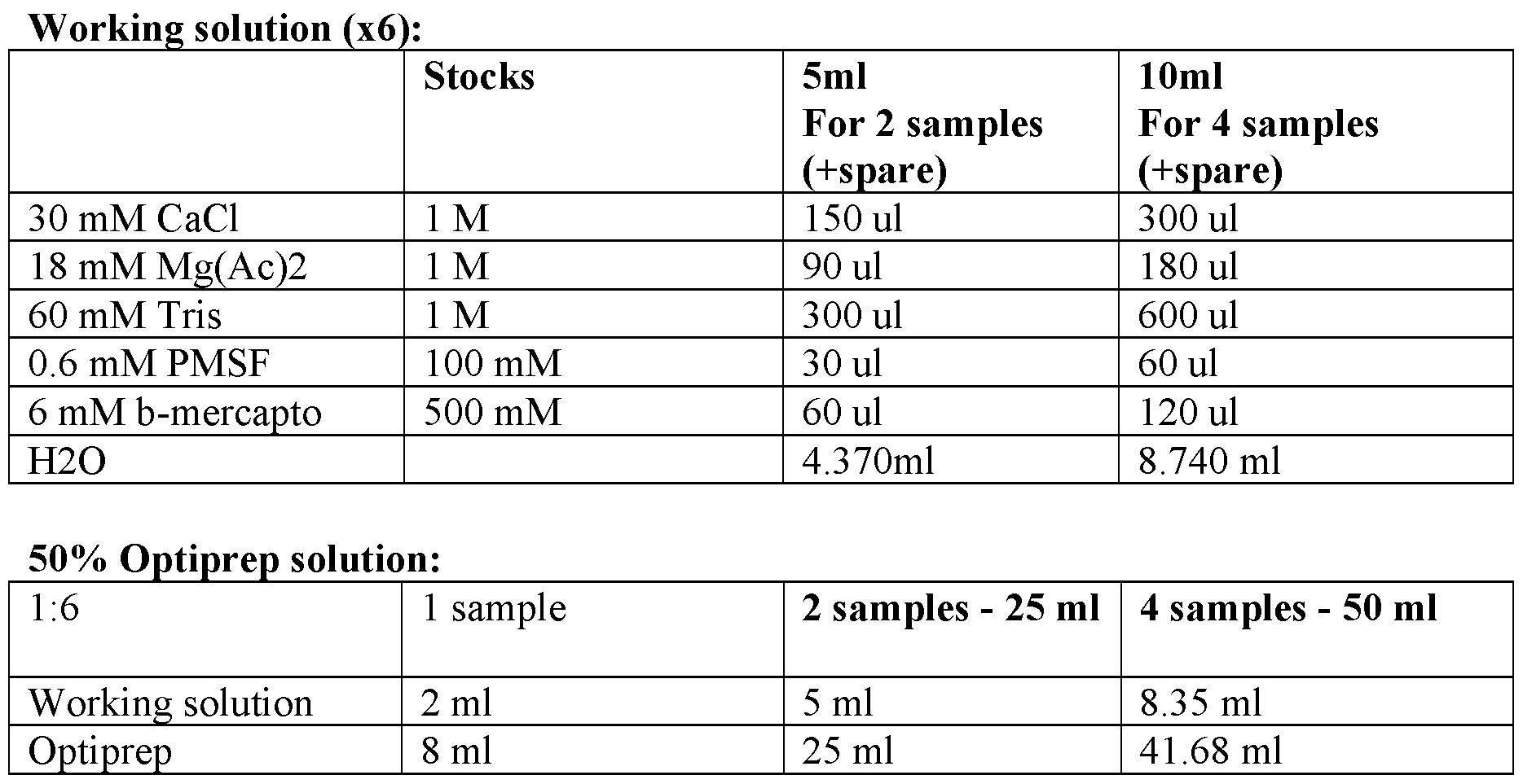
kpigplti 1 pl 2 pl 25l 4 pl 50l
O pti 28ll 525ll 8413568ll
104
2

B0% Optiprep solution:
ff Op 1tip p 158 lpl 429 pl 50 l
R p i bff R SB 42 l 21
2340MM S 1t
2M Mk 1 F 30
6ly MM M
p Kg
hCCll2 11 t l t 1M M 1 4l h 1024 p 50l 0lll pl
2 5

55 B %f gly 00% 650 l
1. Ma
Pro :kt
beig
2. All u ffrfeesrhs, b tuufbfeesrs, homogenizer should be on ice at all times
1to.co Dl:
woituhn bc)e, t hroanmsogenize tissue in 2 ml Buffer 1 + 0.1% NP40 (25 times with a, 25 times 2. K Riense homogfeenriz toer a w 1i5h m 3l m tulb Be
3. t uffer 1 to get final 5 ml, and collect in the same tube 4. Adep on ice 5 min
5. Kee d p 5 o m n l i 50% Optiprep, invert 10x (final volume 10 ml)
6. T Wrhile waicte 5 min
7. f
Opatinpsrfeepr s tohin
leg
ut l,y psraetpea troe t ahne u ullttrraacceennttrriifuuggee t tuubbee w caitrhef 1u0lly m oln of to 2p9% of t Ohept 1i0pr melp 2.9%
8. Centrifuge at 7ion, to form a gradient
9. Carefully remo5v0e0 s rpm 4c for 30 min
10. Add ~300 ul/hp bufufpeerr RnBat,ant
11. Resuspend carefu keep on ice 5-15 min
tuboeusn (tfeorsrt FaiAnC nSu,cl feillly by slow vortex & pipette 10x with a 1ml tip, then transfer to 12. C ite wri tthhr Rouugbhy a D myeem 1:b5r0an0e-1 t:o10 g0et0 b (ecthteerck pu forirty c)lumps in the
[0296 m] micr
e Nousccolpe before sorting)
This CthLoEdI i IsS pe
Oai
Lr ptAiucTur
Iliafi
Orc
Nlyat
P siuo
Rin
Ota p
TbrlOeot
C fooc
Oro
L Dl:
Fro method B
EZ NU RnOc-MSe Tq.ISSUE (using EZ PREP NUC-101, Sigma)
105
P 13r.o Dceoduunrcee f hoorm froogzeenni/zfeix teisdsu tiess inue 2ml of ice-cold Nuclei EZ lysis buffer (2
. t Rim
coine
llss
ee w
ct h ioit
nmh 5 times with a, 25 1 tho b
eg),e sn tr
aia
mznes
erf
t wer
ubit t
eho
.2 am 15l o mfl ic tueb-ceo.ld Nuclei EZ lysis buffer to get final 4 ml, and 32.. C Moixlle wctel tlh aen ndu sceleti on ice for 5 minutes.
t shuepe crlneaarta snutp ceornntaatia b
nny
st c f cre
yon
tomtri
pl efu
aascg
mhat
i ti
cuo
cbn
oe a
m atn 5
pd00
on see x
nt g for 5
cl m
aeinutes at 4 °C. Carefully aspirate t tshe an nducni b peel sleatv oedn f icoer. la Nteorte a:n Tahle
4. Resuspend. Add 1 ml cold Nuclei EZ lysis bu ysis or use. tip to complet ffer and mix by pipetting gently with a 1ml . C mix well ely suspend nuclei pellet. Add the remaining 3 ml of Nuclei EZ lysis buffer, 5 and set on ice for 5 minutes. 6. s cl
Ouo
ppl
telect
iornnaat w
la:nas
Wth ae
and
sd n
h. su
Rete te
shi
ue b
sp ny
eu c
nce
dlentrifugation as in step 3. Carefully aspirate the clear
iin p 4emllelt 0 o.0n ice. R Collect washed 1% PBS BSA or Resuspension buffer (RB*). 7. inehsiubsiptoenrd ca wreitfh n
ul ~u
ly5c0le
b0i
yµ b
sly
l R c
oweesn
vutpif
osr
rteu
eng
xsa
&itoionn
pi b a
pus
etftf i
eenr st
10 (e
xRp
wB 3
i*.
th) o ar 0.01%PBS BSA + RNAse C FACS, filter throug 1ml tip, then transfer to tubes (for 8. beofuonrterstain nucleih w ait mhe Rmubbryan Dey teo 1 g:e5t0 b0e-t1te:1r0 p0u0ri (tych.eck for clumps in the microscope • – Re e sorting).
• ^ bsausespde onnsi tohne o bruigffienral nuclei resuspention buffer from Swiech et al.2015:
23450 M^ St k F 10 l
25 24
M 16550lll
6

pht 21M
y l 100% 5 l
00 l [E
T0xample 7 Ch2
1i9
0s7
42 p]
4ro):toc Doilv- h itsSe
tp dq
s:e/r l
/wiavb
weedlli
w. fn
trhog
em pr
rm to
oht
feoc
is Co
hellirc.cko-mIT/u Es/deUn/h loabmeeli/nregfe prerontcoecso/plr (oTtohceormls/ocFeilsl-cahnedr-,ti Kssiute #- 106
acnelall-ypsriosl/iffleorwat-icoynt-oamssetry-protocol/cell-proliferation/standard-click-it-edu-flow-cytometry- B roeofmor-ete smtaprting the nauyc.lhetim islolation thaw reagents for Click-IT EdU labeling (in the dark, at Follow nuc e l rature).
e relusutesp ienns 1ei isolation protocol (as in Habib et al. Science.2016), at the last elution step io0n0u frlom of p resuspension buffer or PBS with 1% BSA. Follow steps for nuclei R For etiach sample preproatroec aol r.eaction mix (as in the Click-IT EdU labeling protocol): PBS DPBiS TBS 438 µL
C Fl SO4 t dy id 1 2 0 5 µ µ L L
R ti Bff Additi (10 bff diltd t 1 ith PBS) 50 µL
 Ttl ti l 500 µL
T Mix bfy in 5v0e0rtilng 10ti times.i t 15 l i l tb. Add 100l f th lei resuspension. I Andcdub 3a-te in the dark for 20-30minutes.
Spin at55m00lg of fo PrB 5Sm winituhte 1% BSA.
Resuspend with approprisa,t aen adm coaurenftu ollfy R reemsuosvpeen susipoenrnatant.
protocl) or 0.01%PBS BSA, add RNAse inhibitor. R beuffer (as in original nuclei isolation pipteetrte p 1u0x with a 1ml tip, then transfer to tubes (for FA s C u S sp , e f n il d ter ca th re ro fu u l g ly h a by m s e l m ow bra v n o e rt t e o x g & bet et E orlPutBeS i+nr
0 ti
.ht
0ey.
1% appropriate amouse of resuspention buffer (as in the nuclei isolation protocol) Counterstain n BucSAi.
b le with Ruby Dye 1:500-1:1000 (check for clumps in the microscope FeAfoCr
toSe s
co sorting).
pro lo).rt nuclei into 96 well plates for RNA-seq (following the sNuc-Seq SMART-Seq2 107
E
o [0x
f2a
R9m
N8p]le 8
A sam SMART-seq2 protocol Full-length mRNA-seq for single nuclei or small amounts Tissue, Cellples
●s & T N pr
-8isusculeei shoeupld be stored in RNA-later in 4c for 24hours and then moved to the Sorting ● Pr0ep (r ceemllos/vniuncgle thie sh RoNrtA ti-mlaete bre),fo orre im somrteindgia.t Uelsye f RroNzAense an frde seto reraegde antts -8.0c. ● S anodrt o spintigolnea cle RllNs/Anuscele inih inibtoit 96-well plate, 5 µL TCL in each well (with bME, ●● I Smnampe fdrieaetzeely o snpi dnry fo irce 1,- s2to mreionr
a,)
t 2 -58000° RPM Prep work● ● R 30 N m as i e n-u Z te A s P p w
t rio o r rk to su p r u f r a i c fi e c s a a ti n o d n, e l q e u t i R pm N e A n-t SPRI bead RNA Purific Tehmap s equilibrate to room
● wer caetlulr pelates on ice and spin down for 1 min, 2500 RPM
●atio
● L An ● Red
etd
m a 1
ot1
v ro µ
eoL
sm (
up t2
ee.
rm2Xp) fo RrN 1A0- mSPinRuIte bse,a pdlsac toe p ealacthe w onel ml,a mginxet for 5 minutes ● Wash in 10 natant
● Completely0 µL 80% EtOH three times
● Elute in 4 µL re Mmioxve #1 supernatant, let dry for 8-10 minutes on magnet Mix #1 ● Continue immediately
R g t 1 (µLp)l 96 (µL)pl 96 f B (µL) R dNTT pPi i ( (10 µM) 10 1152 120
 RDl 10 M h) 10 1152 120
4 Ui/µ (L1)0% RN Ihib fi l f 10 1152 120 108
H To2Otal 14..00 141650..28 142800
RT ● Incubate eluted plate at 72° for 3 min, immediately place on ice
Mix #2
HO Rgt 10p7l5 (µL) 968p6l (µL) 5 Mi RT bf 4
B Mgt f 20 2304
Cil (1 (5 M) 20 2304
TSO (1000M) 09 10368
RN I µhiMbi)t 10 1152
Mi R (40 U/µL) 025 288
 Ttl NHi RT (200 U/µL) 07100 18106524
●● A own)
( Rd
50Td
°f P 7
oRul
rO o
2Gf Mix #2 (mix well & spin d
mRinA,M 42:° I fnocru 2ba mte at 42° for 90 minutes, followed by 10 cycles of ○in), T threanns hfeerat p ilnaatect iinva otniolyn w ath 7e0n° m foarch 1i5ne m iisn.
4 ready at PCixR 2c
M # P3reamplification HO Rgt 1
Pi (10 1pl0 (µL) 96 pl 115 (µ2L)12 11 ISPCR t µMt R) 1056 KAPA HiFi H dyMi 105 576 528 Ttl tS 1245 116414208 114372
 804 109
●● A Cddc
PCR Pre-ampli ffoy
icra 2le 14 ti0 t
on sh µ
eeL Cc, P o le 7Cf a2R M n° f aix #
osr f 6o3
mll too
iw e
n)sa
,:c
f 9h
in8 w
a°e
l fl
eol
xrt 3en msiionn, 2 a1t 7 c2y°cl feosr o 5f ( m98in°. for 15 sec, 67° ● Puri○fy t Ahed PdC 2R u
0 µ pp
Lro AduMctPsu wth a 0.8X AMPure XP SPRI cleanup
○○ p pla ri ipceette pl e XP SPRI beads, let sit for 5 min W oaftfe s oun magnet for 6 min
○○ Pipaeshtte be oaffds su bp
pyer
er an
nda
adt
tia
ann
ngt
t 1 an0d0 r µeLpe farets wha 7s0h% EtOH and magnet switching ● Elut○e m raetmove all EtOH and let dry on magnet for 10 min
Pos erial in 20 µL TE
1t. - PCR B PioreA-naamplification QC Nex 2te.ra-XT P (ImCOo-glrzeeen– i tnes tth qeu pallaittye reader– QC & quant
Make NTA dified protocol)
●:
● 21..525 ul ul T sDam bupflefe (rd pileurte well
● 1.25ul AT d to 0.15-0.2 ng/ul per well)
P Inuctu cboavteer 1,0 ba mnign t aot m 55ixc &M
(“m spaikne d NowTAn” br piefly
Spin doiw rogram, hold at 10c)
Neutralzen:
S ● Add 1.25 NT buffer to neutralize
Inpciunb d
:aotew on
PCR n bench for 5 min
●
Cover & ba● 32..755 uull o NfP inMdex array primers (1.25 ul of each primer)
PCR (NTAn PgC tRo p mrioxg &ram spin down
1. Perform PCR usin,g f tohlelo fwolilnogw tihneg N peroxgtera XT protocol)
program“Nextera PCR” on machine Dr
11)a:m on a thermal cycler (with heated lid, 0
a b.7 C
c..92
15°
2° cC for
yc for 330 m siencuotnesds i ii.le
.9s
55° o
5°Cf: for 10 seconds
ii.72C for 30 seconds d. Ho ilvi
d. at 742°C for
°°C for 350 m siencuotnedss Pull
EuTlOl & tHog c C
P 7el
0teha
%en
)ru
: 2p.:5 ul from each well and SPRI clean twice (in each SPRI wash twice with ●● 12sntd c cleleaan
t N nuup: A Adddd 00.9.9xx b beeaaddss (,2 e4lu0te ul in sa 2m0p-2le5s, u 2l16ul beads), elute in 50 ul TE B Po p: Stioos
rAe/ aTe
tax
-pte
2er
0-ast QatCion : and quant pool with qubit
P RrTim perirs and oligos:
5′-b mer:
w Therieo-“ANA”G isC aAnyG bTaGseG aTnAdT“CVA”A isC eGithCeArG“AA”G,T“CA”C oTr3“0GV”N-3′ (SEQ ID NO: 7) 5'e Amplate switching oligo (TSO):
I AGCAGTGGTATCAACGCAGAGTACATrGrG+G (SEQ ID NO: 10)
5S′-A PCAR primer:
*:phosGphCoAroGthTiGoaGteT bAoTnCdAACGCAG*A*G*T-3′ (SEQ ID NO: 9)
.
[ is02 to99 b]e und Hearsvtionogd th thuast d theesc irnibveednt iinon de dteafiiln p *
erde *f be *rred embodiments of the present invention, it particular details set fort y the above paragraphs is not to be limited to possible without departinhg f irnom the th aeb sopvierit d oersc srciopptieon of a tshe m parneyse anptp inavreenntti voanr.iations thereof are
111
Claims
WHAT
differen 1 I A
t.S CL
cell su AIM
bty m p eE
etD
sh o o I
rdS:
ce o l f l s p ta ro te d s u i c n in a g he a t t er l o e g a e s n t e o o n u e s p h o i p g u h la r t e io so n lu o t f io ce n lls m c a o p m f p o r r isi v n i g s : ualizing
data obtained fr
(
o
a)
m the
p
h
e
e
r
t
f
e
o
r
r
o
m
g
i
e
n
n
g
eo
d
u
i
s
m
p
e
o
n
p
s
u
io
la
n
t
a
io
li
n
ty
of
re
c
d
e
u
ll
c
s
t
;
ion on single cell gene expression
measuring the ( dbi)ssimi producing a first set of clusters of cells by a method comprising
g reednuec eexdp srpeascsieo ann dda tthae a
l
cn a
ld rit
us a y
tpep b
rsl e y t
ci w
on e
mg e
p a n
ri f s
si e res ts
cte m of
llest g r e
wic n
it, e
h w s
ah in
ceore th
nitn e
in t d
uh i
oe m
u c e
sl n u sionality reduced single cell trsatjeercsto arrye; in a dimensionality
genes based o (nc) their p erxopdruecing a set of informative genes by a method comprising scoring
trajec
ssion across the first set of clusters of cells or a continuous
closet porroyx oimf citeyll isn, w thhee dreimine tnhseio innafolirtmya retidvuec geden sepsa acree; a unndiquely expressed in cells embedded in
t
i
r
n
a
f
j
o
e
r
c
m
to
a
r
t
y
ive
of
g
c
e
e (d
nll)s by p aro mdeutchiondg c aotm leparsistin ogne m seeacsounrding se tt of clusters of cells or continuous uesster asn odr a cpopnltyininug he dissimilarity between the set of second set of cl ou as t sreacjeocntdory m oeftr cice,lls w ihnedriecbayte v cieslula sluibzatytipoens o orf c aell m satapte osf. the
location 2. of the Th ceel mle stuhbotdyp aecsco orrdi cneglls to h calvaiinmg 1 a, c fuerther comprising mapping the spatial
hybridization (ISH) on whole tissu
ll state by performing RNA in situ
specific for genes expressed in the ceell s seucbttiyopness, c womheprreisbiyng the sa sipdat ciaelll lo scuabttiyopne os using probes
is visualized in a biological sample. f cell subtypes
locatio 3. The method according to claim 1, further comprising mapping the spatial
for eac
n
h c
o
e
f
ll
th
t
e
yp
c
e
el
t
l
o
s
l
u
a
b
n
t
d
y
m
pe
a
s
rk
o
g
r
e
c
n
e
e
ll
e
s
x
h
p
a
r
v
es
in
si
g
on
a
p
c
a
e
t
l
t
l
e
s
r
tate by comparing gene expression data
location of cell subtypes is visualized in a biological snasm inpl tei.ssue samples, whereby the spatial
of cells 4 i . n step T ( h d e ) m co e m th p o r d is a e c s co p r r d o i d n u g ci t n o g cl m ai o m re 1, th w a h n e o re n i e n s p e r t od o u f c c i l n u g st t e h r e s, se w c h o e n r d ei s n et e o a f ch clu se s t te o rs f 112
c
c
l
l
u
u
s
s
t
t
e
e
r
r
s
is
is
pro
p
d
ro
u
d
c
u
e
c
d
e
b
d
y
b
a
y
dd
u
in
si
g
n
t
g
he
th
n
e
ex
h
t
i
h
g
i
h
g
e
h
s
e
t
st
sc
s
o
co
ri
r
n
in
g
g
i
i
n
n
f
f
o
o
r
r
m
m
a
a
t
t
i
i
v
v
e
e g
g
e
e
n
n
e
e
.
and each successive
normali
5
z
.
ation o
T
f
h
t
e
he
m
si
e
n
t
g
h
l
o
e
d
ce
a
ll
cc
g
o
e
r
n
d
e
in
e
g
xpr
t
e
o
ssi
a
o
n
n
y
da
o
t
f
a, w
cl
h
ai
e
m
re
s
in
1
gen
to
e e
4
x
,
pre
f
s
u
s
r
i
t
o
h
n
er
of
c
o
o
n
m
e
p
c
r
e
is
l
ing
normalized to another using not highly expressed genes. l is
of misse 6 d . detec T ti h o e n m pr e o t b h a o b d il a it c y c , o w rd h i e n r g ei t n o a a n ny ex o p f ec c t l a a t i i m on s- 1 ma to xi 5 m , i f z u a r t t i h o e n r a c lg o o m r p it r h i m sin i g s a e p st p i l m ie a d t . ion
genes co
7
m
.
prise
T
s
h
a
e
pp
m
ly
e
i
t
n
h
g
od
a M
ac
o
co
ra
r
n
d
’
in
s
g
I a
to
na
a
ly
n
s
y
is
o
a
f
n
c
d
l
/
a
o
i
r
m
a
s
M
1
a
t
n
o
ha
6
t
,
ta
w
n
h
d
e
i
r
s
e
t
i
a
n
nc
s
e
co
a
r
n
i
a
n
l
g
ys
i
i
n
s
f
.
ormative
reductio
8
n
.
comp
T
ri
h
s
e
es
m
PC
et
A
ho
a
d
nd
a
/
c
o
c
r
o
t
r
S
d
N
in
E
g
.
to any of claims 1 to 7, wherein dimensionality
populat
9
io
.
n of c
T
el
h
ls
e
i
m
s d
e
e
th
ri
o
v
d
ed
ac
fr
c
o
o
m
rd
a
in
s
g
ec
t
t
o
io
a
n
n
o
y
f
o
a
f
ti
c
ss
la
u
i
e
m
o
s
r
1
a t
t
u
o
m
8
o
,
r
w
fr
h
o
e
m
re
a
in
su
t
b
h
j
e
ec
h
t.
eterogeneous
microdi 1 s 0 se . ction T . he method according to claim 9, wherein the section is obtained by 11. The method according to claim 9, wherein the tissue is nervous tissue. the brai 1 n 2 , . spina T l h co e r m d e o t r h r o e d ti a n c a c . ording to claim 11, wherein the nervous tissue is isolated from
populat
1
io
3
n
.
of c
T
el
h
ls
e
i
m
s a
et
p
h
o
o
p
d
ul
a
a
c
t
c
io
o
n
rd
o
i
f
ng
ce
t
l
o
ls
a
g
n
ro
y
w
o
n
f
in
cl
t
a
i
i
s
m
su
s
e
1
cu
t
l
o
tu
8
re
,
.
wherein the heterogeneous
compris
1
e
4
n
.
euro
T
n
h
s.
e method according to claim 13, wherein the cells grown in tissue culture
are imm
1
u
5
n
.
e cel
T
ls
h
.
e method according to claim 13, wherein the cells grown in tissue culture
data is o 1 b 6 t . ained T f h r e om me s t i h n o g d le a c c e c ll or s d e i q n u g en to cin an g y . of claims 1 to 15, wherein the gene expression 113
data is o
1
b
7
t
.
ained
T
f
h
r
e
om
me
s
t
i
h
n
o
g
d
le
a
n
c
u
c
c
o
l
r
e
d
i
i
s
n
e
g
qu
to
en
a
c
n
i
y
ng
o
.
f claims 1 to 15, wherein the gene expression
compris
1
e
8
s
.
:
The method according to claim 17, wherein single nuclei sequencing
stabilizes RNA ( ; a) treating the heterogeneous population of cells with a reagent that (b) extracting nuclei from the cells;
(c) sorting single nuclei into separate reaction vessels;
(d) extracting RNA from the single nuclei;
(e) generating a cDNA library; and
(f) sequencing the library,
whereby gene expression data from single cells is obtained. wells of 1 a 9. plate T b h y e F m A e C th S o . d according to claim 18, wherein single nuclei are sorted into single
reaction
2
v
0
e
.
ssels
T
c
h
o
e
m
m
p
e
r
t
i
h
se
o
s
d
m
ac
ic
c
r
o
o
r
f
d
l
i
u
n
i
g
di
t
c
o
s.
claim 18, wherein sorting single nuclei into separate
individu
2
a
1
l
.
cham
T
b
h
e
e
rs
m
o
e
n
th
a
o
m
d
ic
a
r
c
o
c
f
o
lu
rd
id
in
ic
g
ch
to
ip.
claim 20, wherein single nuclei are sorted into
compris 2 e 2 s . a me T th h o e d o m f e h th ig o h d -th a r c o c u o g r h d p in u g t si t n o gle cl n a u im clei 1 s 7 e , qu w e h n e c r in ei g n , sa si i n d g m le eth n o u d cl c e o i m s p e r q is u i e n n g c : ing (a) t Rreating the heterogeneous population of cells with a reagent that stabilizes ( (bc)) e gx N tr A coenmea
prc ;
at
riti
sinng
esg nu
a acle
nuc siu;
lesapre pnosiroen blo ocfki insgol paotelydm neru;clei, wherein the suspension
114
(d) o
ce
p
l
t
l
io
s
n
o
a
rt
l
i
l
n
y
g
, e
(
n
M
ri
A
ch
C
in
S
g
);
the nuclei suspension by FACS or magnetic-activated
(e) a cpp ion microfluidic device
e coonl
mfy
pigin saug
rtrmed the
en ftao n
lruc
ize sle
dinig s
wluesp
ith ne
auncsi reverse emuls
sloen to a
ini,gle wh uenrieqiunely sin bgalreco nduedcle ciap atruere in bdeaivdid iunal alny ( (f
(g)
h) e m x c i ) g seet u qnr
uea l erat o nti
cin n
ng d ing m ro g aR p th beaN ;
rA
lc o
ibordn
aerdto
y c t
uDhe
sN bAarcodedy; c aanpdture beads; whereby gene expression data from siningg p l
leaibi nrreadr
ucl-eein ids s oebqtuaeinnecdin.g,
105-106
2
n
3
u
.
clei.
The method according to claim 22, wherein the nuclei suspension comprises
sequenc
2
e
4
d
.
.
The method according to any of claims 22 to 23, wherein 104-105 nuclei are
blockin
2
g
5
p
.
olym
T
e
h
r
e
co
m
m
e
p
th
ri
o
s
d
es
a
a
cc
p
o
o
r
l
d
o
i
x
n
a
g
m
t
e
o
r.
any of claims 22 to 24, wherein the nuclear pore
RNA co
2
m
6.
prise
T
s
h
th
e
e
m
pr
e
o
th
p
o
e
d
rti
a
e
c
s
c
o
o
f
rd
R
i
N
ng
Al
t
a
o
te
c
r
l
.
aim 18 to 25, wherein the reagent that stabilizes
at le
27. A method of producing a temporally phased single-cell sequencing library of
contiansuto ounse tr caejellct toyrpye o ofr ce slulb dteyvpeel,op wmheernetianl s staaigdes se cqoumepnrciisningg l:ibrary comprises cells along a
subtype, or op (tiao)nally tr aea htientegro mgoenreeo tuhsan ce olne population of cells of a single cell type or
nucle
l type, with a nucleoside analogue, wherein the
a deteocstiadbele an maalorkgeure; is incorporated into replicating DNA and is configured for labeling with
least one other
(
p
b
o
)
pulat
i
i
s
o
o
n
la
o
ting a first population of cells at one time point and isolating at
the isolated populations of cfe cllesl;ls at a later time point, optionally, isolating single nuclei from
115
w
po
it
p
h
ul
t
a
h
t
e
io
d
n
et
o
e
f
c
c
ta (c
eb)
ll l s e , m w a s
hrt
eka
rei
erni
in wng
ti h t e h tih
Dne nucleoside analogue incorporated into replicated DNA N ea A ch is p s o ta p i u n l e a d tio w n it o h f t c h e e ll d s e o te r c s t i a n b g le le m n a u r c k le e i r; isolated from each
stained and/or u
(d
n
)
staine
s
d
or
s
t
i
i
n
n
g
g
le
th
n
e
uc
s
le
ta
i
i
i
n
n
e
t
d
o s
a
e
n
p
d
a
/
r
o
a
r
te
u
r
n
e
s
a
t
c
a
t
i
i
n
o
e
n
d
ve
c
s
e
s
ll
e
s
ls;
o
a
r
n
o
d
ptionally, sorting the
single nuclei, w
(
h
e)
ereby
s
s
e
i
q
n
u
g
e
le
nc
c
i
e
n
l
g
l g
th
e
e
ne
R
e
N
xp
A
re
f
s
r
s
o
i
m
on
t
d
h
a
e
ta
so
i
r
s
te
o
d
bt
s
a
i
i
n
n
g
e
l
d
e
f
c
o
e
r
ll
c
s
el
o
ls
r
a
optionally, sorted
of maturation. t different stages
popu 28. The method according to claim 27, wherein the treating more than one withl aat niuocnle oofsi cdeell asn oaflo agu sien igsle pe creflolrm tyepde i onr a stu lbeatyspte o,n oer su obpjteioctn.ally a heterogeneous cell type 29. The method according to claim 28, wherein the subject is a mouse. cells co 3 m 0 p . rises T d h i e ss m ec e t t i h o o n d o a f c a c t o i r s d su in e g fr t o o m cla th im e s s u 2 b 8 je o c r t. 29, wherein isolating one population of 31. The method according to claim 30, wherein the tissue is nervous tissue. the brai 3 n 2 , . spina T l h co e r m d e o t r h r o e d ti a n c a c . ording to claim 31, wherein the nervous tissue is isolated from
populat
3
io
3
n
.
of c
T
el
h
ls
e
g
m
ro
e
w
th
n
o
i
d
n t
a
i
c
ss
c
u
o
e
rd
c
i
u
n
l
g
tur
to
e.
claim 27, wherein the population of cells is a
compris
3
e
4
n
.
euro
T
n
h
s.
e method according to claim 33, wherein the cells grown in tissue culture
are imm
3
u
5
n
.
e cel
T
ls
h
.
e method according to claim 33, wherein the cells grown in tissue culture
data is o
3
b
6
t
.
ained
T
f
h
r
e
om
me
s
t
i
h
n
o
g
d
le
a
c
c
e
c
l
o
l
r
s
d
e
i
q
n
u
g
en
to
ci
a
n
n
g
y
.
of claims 27 to 35, wherein the gene expression
data is o 3 b 7 t . ained T f h r e om me s t i h n o g d le a n c u c c o l r e d i i s n e g q t u o en a c n i y n 1g o
1. f claims 27 to 35, wherein the gene expression 6
compris 3 e 8 s . : The method according to claim 37, wherein single nuclei sequencing (a) treating the populations of cells with a reagent that stabilizes RNA; (b) extracting nuclei from the cells;
(c) sorting single nuclei into separate reaction vessels;
(d) extracting RNA from the single nuclei;
(e) generating a cDNA library; and
(f) sequencing the library,
whereby gene expression data from single cells is obtained. wells of 3 a 9. plate T b h y e F m A e C th S o . d according to claim 38, wherein single nuclei are sorted into single
reaction
4
v
0
e
.
ssels
T
c
h
o
e
m
m
p
e
r
t
i
h
se
o
s
d
m
ac
ic
c
r
o
o
r
f
d
l
i
u
n
i
g
di
t
c
o
s.
claim 38, wherein sorting single nuclei into separate
individu
4
a
1
l
.
cham
T
b
h
e
e
rs
m
o
e
n
th
a
o
m
d
ic
a
r
c
o
c
f
o
lu
rd
id
in
ic
g
ch
to
ip.
claim 40, wherein single nuclei are sorted into
compris 4 e 2 s . a me T th h o e d o m f e h th ig o h d -th a r c o c u o g r h d p in u g t si t n o gle cl n a u im clei 3 s 7 e , qu w e h n e c r in ei g n , sa si i n d g m le eth n o u d cl c e o i m s p e r q is u i e n n g c : ing (a) treating the population of cells with a reagent that stabilizes RNA;
( (bc)) e gxetnrearcattiinngg nu aclei;
comprises a nuc su le s a p r e p n o si r o e n blo o c f ki isolated nuclei, wherein the suspension (d) o cepltlio snoartlilnyg, e (nMriAchCinSg); the nuclein sugs ppoelnysmioenr; by FACS or magnetic-activated (e) a c p o p n l f y ig in u g red the fo n r uc s le in i g s l u e sp n e u n c si l o ei n , to wh a e r r e e v in ers s e in e g m le uls n i u o c n le m i ic a r r o e flu in id d i i c vi d d e u v a i l c ly e 117
c eommuplartmentalized with a single uniquely barcoded capture bead in an
(
(
f
g
)
)
e
g
x
e
t
n
r
e
as
rci
ato
ti i nn
ng d
g mrop;
aRNA onto the barcoded capture beads; whe (rhe)by ge sneequ exenpcreinssgio th b
near
d lc
aibo
tardae
frd cDNA libreadry; and
ryom us siningg pleai nrucl-eein ids s oebqtuaeinnecdin.g,
105-106
4
n
3
u
.
clei.
The method according to claim 42, wherein the nuclei suspension comprises
sequenc
4
e
4
d
.
.
The method according to any of claims 42 to 43, wherein 104-105 nuclei are
blockin
4
g
5
p
.
olym
T
e
h
r
e
co
m
m
e
p
th
ri
o
s
d
es
a
a
cc
p
o
o
r
l
d
o
i
x
n
a
g
m
t
e
o
r.
any of claims 42 to 44, wherein the nuclear pore
RNA co
4
m
6.
prise
T
s
h
th
e
e
m
pr
e
o
th
p
o
e
d
rti
a
e
c
s
c
o
o
f
rd
R
i
N
ng
Al
t
a
o
te
c
r
l
.
aim 38 to 45, wherein the reagent that stabilizes
a
st
t
a
l
g
e
e
as
o
t 4
f o7
cn.
ee lls h o ig Te
fhh
a r s e m
ps e oe
clt
iuh
fto
ii c od
cn a
e mcc
ll ao
ty prd
p fi
eon
,rg
su v t
bios t u a
yan
ply
eiz of claims 27 to 46, further comprising producing oi r ng ce t l h l e st t a e t m e d p u o r r i a n l g p p o r s o it l i i o fe n ra o t r io c n el c l o d m e p v r e i l s o i p n m g: ental
whereby hetero
(
g
a
e
)
neou
o
s
p
c
t
e
io
ll
n
s
a
a
l
r
ly
e
,
c
p
lu
e
s
r
t
f
e
o
r
r
e
m
d
i
b
n
y
g
c
th
el
e
l t
m
yp
et
e
h
,
o
s
d
ub
a
t
c
y
c
p
o
e
r
,
d
o
in
r
g
ce
t
l
o
l s
a
t
n
at
y
e;
of claims 1 to 8,
data from the st (abi)ned c peellrsfo orfm ain signg dliem ceenlsli toynpael,it syu rbeduction on the single cell gene expression
cells or the stained single nuclei of a single cell typ
t
e
y
o
p
r
e
s
o
u
r
b
c
t
ell state within each population of
of cells; ype isolated from each population
reduced single ( cce)ll ge mneea esxuprriensgsi tohne d diassimilarity between sets of genes in the dimensionality
t
ta and applying a first metric, whereby a continuous
trimajeec ptoorinyt; is visualized in the dimensionality reduced space from an early time point to a later
118
g y a method comprising scoring
g
e
ree n
dn e
ue s
cs b
ed ar a
se se (d pa u d
cnei o q n
,u t) oepl h
ty ei u ion e r p ax er lp xo lyr pd
er
,s e
ws sc
esin hd iogn a er ien a s in c ce le r
ol ot wls s osf ly em t ihn moative genes b exb efo pe cr rded n
sse t
ed in
d i u
gn ou
en c s
eloss tr
ae aj
re p e
fr c o to
ilx r
tei y
rm ,
edit w y h
ou i e
tn re
; a t i h n
ne the informative d dimensionality
m
m
e
e
a
tr
s
i
u
c
r
,
in
w
g
he
th
re
e (
by de)is v s i i s m ua i pl l aro
iz rid
at t yuci
io bn
negt o w a
fet
te l
hnea
e ts
shte on seet se otf o ifnf colurmsteartisv oef g ceenlless b aynd a m apepthlyoidng co amp sericsoinngd indicat et of clusters in the dimensionality reduced space stage.e the gene expression profiles of cells based on a temporal position or developmental
cells in 4 s8te.p (d T)h ceom mpertihsoeds p arcocdourdciinngg m too crleai tmhan 47 o,n weh seerte oinf c plruosdtuercsi,ng wh the set of clusters of
clustteerrss i iss p prroodduucceedd b byy a udsdiningg th thee h nigeh
erein the first set of
clus xets htig schoersitn sgco inrifnogrm inaftoivrem gaetinvee a gnedne e.ach successive set of
normalli
4
z
9
iza .
etdio ton a o T
nf h
o t e
thhe m
er si e
un tho
sgilne d
g c ne a
ol c l c
t h g o
ie r
gn d
he in
ly e g to any of claims 47 to 48, further comprising norma x epxrpersessisoend d gaetna,es w.herein gene expression of one cell is
of misse 5 d 0. detec T ti h o e n m pr e o th b o a d bi a li c ty co , r w d h in e g re t i o n a a n n y e o x f pe c c la ta im tio s n 4-7 m t a o x 4 im 9, iz fu at r i t o h n er a c lg o o m ri p t r h i m sin i g s a e p st p i l m ie a d t . ion
genes co
5
m
1.
prise
T
s
h
a
e
pp
m
ly
e
i
t
n
h
g
od
a
a
M
cc
o
o
r
r
a
d
n
i
’
n
s
g
I
t
a
o
na
a
l
n
y
y
si
o
s
f
a
c
n
l
d
a
/
i
o
m
r
s
a
4
M
7
a
t
n
o
h
5
a
0
tt
,
a
w
n
h
d
e
is
re
ta
in
nc
s
e
co
an
ri
a
n
l
g
ys
i
i
n
s
f
.
ormative
reductio
5
n
2.
comp
T
ri
h
s
e
es
m
P
e
C
th
A
od
and
ac
/o
c
r
or
tS
di
N
n
E
g
.
to any of claims 47 to 51, wherein dimensionality
analogu 5 e 3 c . omp T ri h se e s m Ed et U ho ( d 5-e a t c h c y o n r y d l i-n 2 g ′-d to eo a x n y y ur o id f in c e l ) a . ims 27 to 52, wherein the nucleoside
compris
5
i
4
n
.
g:
A method for creating a composite single nuclei sequencing library
119
n nuucclleeii i isn b alnoc ekm
(a
edu ) l wsiiothn m
a dr e
no r
up g
cl i
le n
et g
a h o
ra n
pv e
oirn u
eg n
b a iquely barcoded RNA capture microbead with a single lo dcikaimnget peorl fyrmomer; 50 µm to 210 µm, wherein the single (b) extracting mRNA onto on the RNA capture microbead; first strand cD (NcA) tha pte irsfo cromvianlgen atly re lvinerkseed tr toan tshceription reaction to convert the mRNA to raettvaecrhseed tr baenasdcsr;ibing within droplets and thereafter b R re N a A kin c g ap d t r u o r p e le m ts ic a ro n b d e c a o d l ; le o c r ti c n o g n c v D er N se A ly-
containing cell (d b)arcod persep thaaritn rgeco arndd th seeq cueelln-coifn-gorig ain si onfg elea composite RNA-Seq library, identifiers (UMI) that distinguish among RNAs from the same ch ce R ll N . A, and unique molecular 55. A method for creating a composite single-cell sequencing library comprising: nu (a) merging one uniquely barcoded RNA captur
ucclleeii i isn b alnoc ekmedul wsiiothn a dr noupclleeta h e microbead with a single n ra pvoirneg b alo dcikaimnget peorl fyrmomer; 50 µm to 210 µm, wherein the single (b) extracting mRNA onto on the RNA capture microbead;
(c) breaking droplets and pooling beads in solution; strand cDNA th (da)t is co pvearfloenrmtlyin lgin ake redv teorse transcription reaction to convert the RNA to first trans the RNA capture microbead; or conversely reverse beadsc;ribing within droplets and thereafter breaking droplets and collecting cDNA-attached
containing cell (e b)arcod persep thaaritn rgeco arndd th seeq cueelln-coifn-gorig ain si ongle composite RNA-Seq library,
identifiers (UMI) that distinguish among RNAs from the s
f
a
e
m
a
e
ch
ce
R
ll
N
.
A, and unique molecular
attached
56
b
.
eads
T
is
h
t
e
em
m
p
e
l
t
a
h
t
o
e
d
sw
of
it
c
c
l
h
ai
a
m
m
s
p
5
li
4
fi
o
c
r
at
5
io
5
n
,
.
wherein the method of amplifying the cDNA-
120
attached 57 b . eads T is h T e 7 m l e in th e o ar d a o p f p c li l c a a im tio s n 5 . 4 or 55, wherein the method of amplifying the cDNA- attached 58 b . eads T is h e e x m po e n th e o n d tia o l f is c o la th im er s m 5 a 4 l o am r 5 p 5 li , fi w ca h t e i r o e n i . n the method of amplifying the cDNA- encapsu 5 l 9 at . ion c T o h m e p m ri e si t n h g od R o N f A cla c i a m pt s u 5 re 4 m or ic 5 r 5 o , b w ea h d er a e n i d n c th o e m e p m os u i l t s e io s n in d g r l o e p n l u e c t l i e s i. formed via co- 60. The method of claims 54 or 55, wherein the RNA is mRNA.
is 50-10 6 0 1. µm o T r h 7 e 0- m 90 et µ h m od . of claims 54 or 55, wherein the diameter of the emulsion droplet microbe 6 a 2 d . s is f T ro h m e m 10 et µ h m od to o 9 f 5 cl µ a m im . s 54 or 55, wherein the diameter of the RNA capture detectab 6 l 3 e . by a T fl h u e or m es e c th e o n d t s a ig c n c a o l r , d w in h g er t e o b a y n i y nd o i f vi c d l u a a im l n s u 1 c 7 lei to m 6 ay 2, b w e h fu e r r t e h in er n s u o c rt l e e d i . are further fluoresc 6 e 4 n . t pro T te h in e . method according to claim 63, wherein cells express a nuclear localized protein 6 c 5 o . mpris T e h s e a m fl e u t o h r o e d sc a e c n c t o p r r d o i t n e g in to fu c s l e a d im to 6 a 4 K , A w S he H re p i r n ot t e h i e n n d u o c m le a a i r n. localized fluorescent with an 6 a 6 n . tibod T y h w e i m th e s t p h e o c d if a ic cc a o f r f d in in it g y t f o or c a la n im int 6 ra 3 n , u w cl h e e a r r e p in ro s t i e n i g n l . e nuclei are immunostained 67. The method according to claim 66, wherein the antibody is specific for NeuN. stain. 68. The method according to claim 63, wherein nuclei are stained with a nuclear Ruby re 6 d 9 , . trypa T n h b e lu m e e , t H ho o d ec a h c s c t o o r r di p n r g op to id c iu la m im io 6 d 8 in , e w . herein the nuclear stain comprises DAPI, microflu 7 i 0 d . ic sy A st n em a , p c p o a m ra p tu r s isi f n o g r : creating a composite single nuclei sequencing library via a
121
carrier f
a
lu
o
id
il-
c
s
h
u
a
r
n
fa
n
c
e
t
l
a
f
n
u
t
rt
i
h
n
e
l
r
et
co
c
m
om
pr
p
i
r
s
i
e
s
s
in
a
g
re
a
sis
fi
t
l
o
t
r
e
;
r and a carrier fluid channel, wherein said
channel
a
,
n
w
i
h
n
e
l
r
e
e
t
in
fo
s
r
ai
a
d
n
ca
a
r
n
r
a
ie
ly
r
t
f
e
lu
c
i
o
d
n
c
f
h
ig
a
u
n
r
n
e
e
d
l o
fo
p
r
tio
n
n
u
a
c
l
l
l
e
y
i
f
c
u
o
rt
m
he
p
r
ri
c
s
o
in
m
g
p
a
ris
f
e
i
s
lte
a
r
re
a
s
n
i
d
sto
a
r;
carrier fluid
c reasrirsietor
a
r; f n lui i d nle c t h f a o n r ne m l, R w N h A er c e a in ptu s r a e id m c ic a r r o ri b e e r ad fl s ui a d nd ch ly a s n i n s e r l ea o g p e ti n o t n c a o ll m y p f r u is r i t n h g er a c f o i m lte p r ri a s n es d a a
predeter s m ai i d ne c d ar f r l i o e w r f r l a u te id ; channels have a carrier fluid flowing therein at an adjustable or wherein each said carrier fluid channels merge at a junction; and
said junction being connected to a mixer, which contains an outlet for drops. blockin 7 g 1 p . olym T e h r, e an ap e p n a z r y a m tu e s a o n f d c a la n i u m cle 7 i 0 . , wherein the analyte comprises a nuclear pore
a fluid c 7 a 2 r . rier c T h h an e n a e p l p w ar i a th tu a s c o o f n c s l t a r i i m cti 7 o 0 n , f w or h d er r e o i p n le s t a p id in j c u h n-c o t f io f. n is connected to said mixer by 73. The apparatus of claim 55(e), wherein the analyte is a nuclei. nuclei. 74. The apparatus of claim 55(e), wherein the analyte is a host pathogen cell or
surfacta 7 n 5 t, . suc T h h a e s a s p o p d a i r u a m tus la o u f r c o l y a l im sa 5 rc 5 o ( s e i ) n , e w , h o e r re a in c t h h a e ot l r y o s p is ic re s a a g lt e , nt su c c o h mp as ris g e u s a a n n anionic thiocyanate. idiunium 76. The apparatus of claim 70, wherein the filter comprises square PDMS. from 70 7 0 7 0 . – 90 T 0 h 0 e , w ap id p t a h ra o t f u 5 s 0 of - 7 c 5 la µ im m 7 a 0 n , d w d h e e p r t e h in of th 1 e 00 re -s 1 is 5 t 0 or m is m s . erpentine having a length 78. The apparatus of claim 77, wherein the resistor has a diameter of 50 µm. 122
8000 - 1 7 2 9 , . 000 µ T m he a a n p d pa w ra id tu th s o o f f 1 c 2 la 5 im - 2 7 5 0 0 , m w m he , r a e n in d t d h e e pt c h ha o n f n 1 e 0 l 0 s - ha 1 v 5 i 0 ng m a m l . ength of length of 80. The apparatus of claim 79, wherein the channel diameter is 125 µm. µm. 81. The apparatus of claim 79, wherein the channel diameter is 75, 80, 85, or 90
and a w 8 id 2 t . h of T 80 h-e 14 ap 0 p µ a m rat o u r s 7 o 5 f -9 c 0 la µ im m. 70, wherein the mixer has a length of 7000-9000 µm 83. The apparatus of claim 82, wherein the mixer width is 125 µm.
84. The apparatus of claim 82, wherein the mixer width is 70-90 µm.
85. The apparatus of claim 70, wherein the oil-surfactant is a PEG block polymer. QX200 8 D 6 r . oplet T G he en a e p r p at a i r o a n tu O s i o l. f claim 85, wherein the PEG block polymer is BIORAD™ 87. The apparatus of claim 70, wherein the carrier fluid is water-glycerol mixture. 88. A method of determining cells responsive to a therapeutic agent comprising: analogue, whe ( r a e ) in the co n n u ta c c le ti o n s g id a e ti a ssue of interest with a therapeutic agent and a nucleoside configured for labeling with a detectabnlaelo mguareke isr; incorporated into replicating DNA and is
the tissue of int
(
e
b
r
)
est;
isolating the tissue of interest, optionally, isolating single nuclei from
with the detect (ac) staining the nucleoside analogue inrcorporated into replicated DNA
tissue of interest,
b
w
le
he
m
re
a
i
r
n
ke
th
r
e
w
D
it
N
hi
A
n
i
t
s
he
sta
t
i
i
n
ss
e
u
d
e
w
o
i
f
th
in
th
te
e
r
d
e
e
st
te
o
ctab
si
l
n
e
g
m
le
ar
n
k
u
e
c
r
l
;
ei isolated from the
stained and/or u
(d
n
)
staine
s
d
or
s
t
i
i
n
n
g
g
le
th
n
e
uc
s
le
ta
i
i
i
n
n
e
t
d
o s
a
e
n
p
d
a
/
r
o
a
r
te
u
r
n
e
s
a
t
c
a
t
i
i
n
o
e
n
d
ve
c
s
e
s
ll
e
s
ls;
or optionally, sorting the
123
single nuclei, w
(
h
e)
ereby
s
s
e
i
q
n
u
g
e
le
nc
c
i
e
n
l
g
l g
t
e
h
n
e
e
R
e
N
xp
A
re
f
s
r
s
o
i
m
on
t
d
h
a
e
ta
so
is
rt
o
e
b
d
ta
s
i
i
n
n
e
g
d
le
; a
c
n
e
d
lls or optionally, sorted
developmental
(
s
f
t
)
age, w
d
h
e
e
te
r
r
e
m
by
in
c
i
e
n
l
g
ls r
c
e
e
s
l
p
l
on
s
s
u
iv
b
e
ty
t
p
o
e
a
s
th
a
e
n
ra
d
p
/o
e
r
utic
ce
a
l
g
l
en
s
t
u
a
b
r
t
e
yp
d
e
e
s
term
of
ine
a
d.
specific
contacte
8
d
9.
with
T
th
h
e
e
ti
m
ss
e
u
t
e
h
o
o
f
d
in
a
t
c
e
c
re
o
s
r
t
di
b
n
e
g
for
t
e
o
th
c
e
la
t
i
h
m
era
8
p
8
e
,
ut
w
ic
h
a
e
g
re
e
i
n
n
t.
the nucleoside analogue is
contacte
9
d
0.
with
T
th
h
e
e
ti
m
ss
e
u
t
e
h
o
o
f
d
in
a
t
c
e
c
re
o
s
r
t
di
b
n
e
g
tw
t
e
o
en
c
2
la
a
im
nd
8
1
9
4
,
da
w
y
h
s
e
b
re
ef
in
ore
th
th
e
e
n
th
u
e
c
r
le
a
o
p
s
e
i
u
d
t
e
ic
a
a
n
g
a
en
lo
t
g
.
ue is
therapeu 9 t 1 i . c age T nt h i e s a m d e m th in o i d ste a r c e c d or f d ro in m g a t n o im cl p a l i a m nta 8 b 8 l , e w de h v e i r c e e in or t l h o e ca n li u z c e l d eo in si j d e e cti a o n n a . logue and 92. The method according to claim 88, wherein the tissue of interest is a tumor. by a me 9 th 3 o . d co T m h p e ri m si e n t g h : od according to claim 88, wherein the cell subtypes are determined
data obtained fr
(
o
a)
m the
p
h
e
e
r
t
f
e
o
r
r
o
m
g
i
e
n
n
g
eo
d
u
im
s p
en
o
s
p
i
u
o
l
n
a
a
ti
l
o
it
n
y
o
re
f
d
c
u
e
c
ll
t
s
i
;
on on the single cell gene expression
measuring the (b) producing a first set of clusters of cells by a method comprising
gene expression
di
d
s
a
si
t
m
a a
il
n
a
d
rit
a
y
pp
b
l
e
y
t
i
w
n
e
g
e
a
n
f
s
i
e
rs
ts
t m
of
et
g
r
e
ic
n
,
es in the dimensionality reduced single cell
reduced space and the clusters comprise cells with w ah ceorenitnin tuhoeu cslu trsatjeercsto arrye; in a dimensionality
genes based o (nc) their p erxopdruecsisnigon a a scerto osfs in thfeorm firasttiv seet ge onfes cl buyste ars me otfhod comprising scoring
trajectory omf citeyll isn, w thhee dreimine tnhseio innafolirtmya ret
cells or a continuous
close proxi idvuec geden sepsa acree; a unndiquely expressed in cells embedded in
trajectory (d) producing at least one second set of clusters of cells or continuous
informative
of
g
c
e
e
n
l
e
ls
s
b
a
y
nd
a
a
m
pp
et
l
h
y
o
in
d
g
c
a
om
se
p
c
ri
o
s
n
in
d
g
m
m
e
e
tr
a
i
s
c
u
,
ri
w
n
h
g the dissimilarity between the set of
second set of clusters or continuous trajectory of cells inedriecbayte v cieslula sluibzatytipoens o orf c aell m satapte osf. the
124
d anevde ulonp
9
stm 4
aie .
nnetda.l T st h a e ge m a e r t e ho d d ete a r c m co in rd e i d ng by to de c t l e a r i m m in 9 in 3 g , w th h e e c re e i l n l s t u h b e ty c p e e ll o s f u c b e ty ll p s e t s ha o t f a a re s s p t e a c in if e i d c
develop
9
m
5
e
.
ntal
T
st
h
a
e
ge
m
a
e
r
t
e
h
d
o
e
d
te
a
r
c
m
c
i
o
n
r
e
d
d
in
b
g
y
t
a
o
m
c
e
la
th
im
od
9
c
4
o
,
m
w
p
h
ri
e
s
r
i
e
n
i
g
n
:
the cell subtypes of a specific
data from the st (aai)ned c peellrsfo orfm ain signg dliem ceenlsli toynpael,it syu rbetdyupcet oiorn ce on the single cell gene expression
c oefl clsel olsr; the stained single nuclei of a single cell type or subty
l
p
l
e
st
i
a
so
te
la
w
te
i
d
th
f
i
r
n
o
e
m
ac
e
h
ac
p
h
op
p
u
o
l
p
a
u
ti
l
o
a
n
tio
o
n
f
reduced single ( cbe)ll ge mneea esxuprriensgsi tohne d diastsaim anildar aitpy between sets of genes in the dimensionality
trajectory is visuali
plying a first metric, whereby a continuous
time point; zed in the dimensionality reduced space from an early time point to a later
genes based on (c) their p erxopdruescsiniogn a a scerots osf t ihneformative genes by a method comprising scoring g reednuecsed ar sepa ucneiq,u oepl continuous trajectory, wherein e th d e im i e n n fo si r o m n a a t l i i v ty e tyion eaxlplyre,s wsehder ienin c leolwlsly em exbperdedsseedd i gnen celoss aere p froilxteirmedity ou itn; a thnd measuring the ( ddi)ssimi plaroridtyuci bnegtw aete lnea tst one set of clusters of cells by a method comprising metric, whereby visualization of the s h e e t s o e f t c o l f us i t n e f r o s rm in ati th v e e g d e im ne e s ns a io n n d al a i p ty ply re in d g uc a ed second indicate the gene expression profiles of cells based on a tem space stage. poral position or developmental
cells in 9 s6te.p (d T)h ceom mpertihsoeds p arcocdourdciinngg m too crleai tmhan 95 o,n weh seerte oinf c plruosdtuercsi,ng wh theere sinet th oef c flusters of
clusters is produced by usinngg th thee h nigehxets htig schoersitn sgco inrifnogr
irst set of
clusters is produced by addi m inaftoivrem gaetinvee a gnedne e.ach successive set of
normali 9 z 7 a . tion o T f he method according to any of claims 88 to 96, further comprising normalized to ano tthheer si unsgilneg c neollt h giegnhely ex epxrpersessisoend d gaetna,es w.herein gene expression of one cell is 125
of misse 9 d 8. detec T ti h o e n m pr e o th b o a d bi a li c ty co , r w d h in e g re t i o n a a n n y e o x f pe c c la ta im tio s n 8-8 m t a o x 9 im 7, iz fu at r i t o h n er a c lg o o m ri p t r h i m sin i g s a e p st p i l m ie a d t . ion genes co 9 m 9. prise T s h a e pp m ly e i t n h g od a a M cc o o r r a d n i ’ n s g I t a o na a l n y y si o s f a c n l d a / i o m r s a 8 M 8 a t n o h 9 a 8 tt , a w n h d e is re ta in nc s e co an ri a n l g ys i i n s f . ormative reductio 1 n 00 co . mp T ri h s e es m P e C th A od and ac /o c r or tS di N n E g . to any of claims 88 to 99, wherein dimensionality analogu 1 e 0 c 1 o . mp T ri h se e s m E e d t U ho ( d 5- a e c th c y o n rd y i l n -2 g ′- t d o eo a x n y y ur o id f in cl e a ) i . ms 88 to 100, wherein the nucleoside
126
Priority Applications (1)
| Application Number | Priority Date | Filing Date | Title |
|---|---|---|---|
| US16/087,036 US20190218276A1 (en) | 2016-03-21 | 2016-10-27 | Methods for determining spatial and temporal gene expression dynamics in single cells |
Applications Claiming Priority (2)
| Application Number | Priority Date | Filing Date | Title |
|---|---|---|---|
| US201662311129P | 2016-03-21 | 2016-03-21 | |
| US62/311,129 | 2016-03-21 |
Publications (1)
| Publication Number | Publication Date |
|---|---|
| WO2017164936A1 true WO2017164936A1 (en) | 2017-09-28 |
Family
ID=59900721
Family Applications (1)
| Application Number | Title | Priority Date | Filing Date |
|---|---|---|---|
| PCT/US2016/059239 WO2017164936A1 (en) | 2016-03-21 | 2016-10-27 | Methods for determining spatial and temporal gene expression dynamics in single cells |
Country Status (2)
| Country | Link |
|---|---|
| US (1) | US20190218276A1 (en) |
| WO (1) | WO2017164936A1 (en) |
Cited By (67)
| Publication number | Priority date | Publication date | Assignee | Title |
|---|---|---|---|---|
| CN108960116A (en) * | 2018-06-28 | 2018-12-07 | 哈尔滨工程大学 | Based on the diesel engine high-frequency pressure oscillation method for extracting signal for improving wavelet package transforms |
| CN109523577A (en) * | 2018-10-29 | 2019-03-26 | 浙江大学 | Method is determined based on the subcellular structure motion profile of micro-image |
| WO2019071054A1 (en) | 2017-10-04 | 2019-04-11 | The Broad Institute, Inc. | Methods and compositions for altering function and structure of chromatin loops and/or domains |
| WO2019094984A1 (en) * | 2017-11-13 | 2019-05-16 | The Broad Institute, Inc. | Methods for determining spatial and temporal gene expression dynamics during adult neurogenesis in single cells |
| CN110222745A (en) * | 2019-05-24 | 2019-09-10 | 中南大学 | A kind of cell type identification method based on similarity-based learning and its enhancing |
| WO2019210268A2 (en) | 2018-04-27 | 2019-10-31 | The Broad Institute, Inc. | Sequencing-based proteomics |
| WO2019213660A2 (en) | 2018-05-04 | 2019-11-07 | The Broad Institute, Inc. | Compositions and methods for modulating cgrp signaling to regulate innate lymphoid cell inflammatory responses |
| WO2019217099A1 (en) * | 2018-05-08 | 2019-11-14 | President And Fellows Of Harvard College | Droplet-based single cell genomic dna sequencing |
| WO2019232542A2 (en) | 2018-06-01 | 2019-12-05 | Massachusetts Institute Of Technology | Methods and compositions for detecting and modulating microenvironment gene signatures from the csf of metastasis patients |
| WO2019241273A1 (en) | 2018-06-11 | 2019-12-19 | The Broad Institute, Inc. | Lineage tracing using mitochondrial genome mutations and single cell genomics |
| WO2020047453A1 (en) * | 2018-08-31 | 2020-03-05 | Ampel Biosolutions, Llc | Systems and methods for single-cell rna-seq data analysis |
| WO2020077135A1 (en) | 2018-10-10 | 2020-04-16 | Dana-Farber Cancer Institute, Inc. | Modulating resistance to bcl-2 inhibitors |
| WO2020077236A1 (en) | 2018-10-12 | 2020-04-16 | The Broad Institute, Inc. | Method for extracting nuclei or whole cells from formalin-fixed paraffin-embedded tissues |
| US10640763B2 (en) | 2016-05-31 | 2020-05-05 | Cellular Research, Inc. | Molecular indexing of internal sequences |
| US10669570B2 (en) | 2017-06-05 | 2020-06-02 | Becton, Dickinson And Company | Sample indexing for single cells |
| CN111247248A (en) * | 2018-06-04 | 2020-06-05 | 伊鲁米纳公司 | High throughput single cell transcriptome libraries and methods of making and using |
| WO2020139892A1 (en) * | 2018-12-28 | 2020-07-02 | University Of Pittsburgh - Of The Commonwealth System Of Higher Education | Methods and materials for single cell transcriptome-based development of aav vectors and promoters |
| WO2020160044A1 (en) | 2019-01-28 | 2020-08-06 | The Broad Institute, Inc. | In-situ spatial transcriptomics |
| WO2020172343A2 (en) | 2019-02-19 | 2020-08-27 | Massachusetts Institute Of Technology | Methods for treating injuries |
| WO2020186235A1 (en) | 2019-03-14 | 2020-09-17 | The Broad Institute, Inc. | Compositions and methods for modulating cgrp signaling to regulate intestinal innate lymphoid cells |
| WO2020186101A1 (en) | 2019-03-12 | 2020-09-17 | The Broad Institute, Inc. | Detection means, compositions and methods for modulating synovial sarcoma cells |
| WO2020191079A1 (en) | 2019-03-18 | 2020-09-24 | The Broad Institute, Inc. | Compositions and methods for modulating metabolic regulators of t cell pathogenicity |
| WO2020191069A1 (en) | 2019-03-18 | 2020-09-24 | The Broad Institute, Inc. | Modulation of type 2 immunity by targeting clec-2 signaling |
| US20200347449A1 (en) * | 2019-05-01 | 2020-11-05 | The Broad Institute, Inc. | Methods for determining spatial and temporal gene expression dynamics during adult neurogenesis in single cells |
| WO2020232271A1 (en) | 2019-05-14 | 2020-11-19 | The Broad Institute, Inc. | Compositions and methods for targeting multinucleated cells |
| WO2021030627A1 (en) | 2019-08-13 | 2021-02-18 | The General Hospital Corporation | Methods for predicting outcomes of checkpoint inhibition and treatment thereof |
| US10927419B2 (en) | 2013-08-28 | 2021-02-23 | Becton, Dickinson And Company | Massively parallel single cell analysis |
| US10941396B2 (en) | 2012-02-27 | 2021-03-09 | Becton, Dickinson And Company | Compositions and kits for molecular counting |
| WO2021127610A1 (en) | 2019-12-20 | 2021-06-24 | EDWARD Via COLLEGE OF OSTEOPATHIC MEDICINE | Cancer signatures, methods of generating cancer signatures, and uses thereof |
| US11072816B2 (en) | 2017-05-03 | 2021-07-27 | The Broad Institute, Inc. | Single-cell proteomic assay using aptamers |
| WO2021147069A1 (en) * | 2020-01-23 | 2021-07-29 | 青岛华大智造普惠科技有限公司 | Droplet microfluidics-based single cell sequencing and applications |
| CN113195718A (en) * | 2018-10-18 | 2021-07-30 | 新克赛特株式会社 | Methods and systems for target screening |
| EP3872190A1 (en) | 2020-02-26 | 2021-09-01 | Antibodies-Online GmbH | A method of using cut&run or cut&tag to validate crispr-cas targeting |
| USRE48913E1 (en) | 2015-02-27 | 2022-02-01 | Becton, Dickinson And Company | Spatially addressable molecular barcoding |
| US11319583B2 (en) | 2017-02-01 | 2022-05-03 | Becton, Dickinson And Company | Selective amplification using blocking oligonucleotides |
| US11332776B2 (en) | 2015-09-11 | 2022-05-17 | Becton, Dickinson And Company | Methods and compositions for library normalization |
| US11332736B2 (en) | 2017-12-07 | 2022-05-17 | The Broad Institute, Inc. | Methods and compositions for multiplexing single cell and single nuclei sequencing |
| EP4001429A1 (en) | 2020-11-16 | 2022-05-25 | Antibodies-Online GmbH | Analysis of crispr-cas binding and cleavage sites followed by high-throughput sequencing (abc-seq) |
| US11365409B2 (en) | 2018-05-03 | 2022-06-21 | Becton, Dickinson And Company | Molecular barcoding on opposite transcript ends |
| US11390914B2 (en) | 2015-04-23 | 2022-07-19 | Becton, Dickinson And Company | Methods and compositions for whole transcriptome amplification |
| US11414701B2 (en) | 2018-05-24 | 2022-08-16 | The Broad Institute, Inc. | Multimodal readouts for quantifying and sequencing nucleic acids in single cells |
| WO2022192419A2 (en) | 2021-03-09 | 2022-09-15 | Massachusetts Institute Of Technology | Methods of treating inflammatory bowel disease (ibd) with anti- tnf-blockade |
| US11460468B2 (en) | 2016-09-26 | 2022-10-04 | Becton, Dickinson And Company | Measurement of protein expression using reagents with barcoded oligonucleotide sequences |
| US11492660B2 (en) | 2018-12-13 | 2022-11-08 | Becton, Dickinson And Company | Selective extension in single cell whole transcriptome analysis |
| US11525157B2 (en) | 2016-05-31 | 2022-12-13 | Becton, Dickinson And Company | Error correction in amplification of samples |
| US11535882B2 (en) | 2015-03-30 | 2022-12-27 | Becton, Dickinson And Company | Methods and compositions for combinatorial barcoding |
| US11639517B2 (en) | 2018-10-01 | 2023-05-02 | Becton, Dickinson And Company | Determining 5′ transcript sequences |
| US11649497B2 (en) | 2020-01-13 | 2023-05-16 | Becton, Dickinson And Company | Methods and compositions for quantitation of proteins and RNA |
| US11661631B2 (en) | 2019-01-23 | 2023-05-30 | Becton, Dickinson And Company | Oligonucleotides associated with antibodies |
| US11661625B2 (en) | 2020-05-14 | 2023-05-30 | Becton, Dickinson And Company | Primers for immune repertoire profiling |
| US11739443B2 (en) | 2020-11-20 | 2023-08-29 | Becton, Dickinson And Company | Profiling of highly expressed and lowly expressed proteins |
| US11773436B2 (en) | 2019-11-08 | 2023-10-03 | Becton, Dickinson And Company | Using random priming to obtain full-length V(D)J information for immune repertoire sequencing |
| US11773441B2 (en) | 2018-05-03 | 2023-10-03 | Becton, Dickinson And Company | High throughput multiomics sample analysis |
| US11788120B2 (en) | 2017-11-27 | 2023-10-17 | The Trustees Of Columbia University In The City Of New York | RNA printing and sequencing devices, methods, and systems |
| US11793787B2 (en) | 2019-10-07 | 2023-10-24 | The Broad Institute, Inc. | Methods and compositions for enhancing anti-tumor immunity by targeting steroidogenesis |
| US11841371B2 (en) | 2018-03-13 | 2023-12-12 | The Broad Institute, Inc. | Proteomics and spatial patterning using antenna networks |
| US11844800B2 (en) | 2019-10-30 | 2023-12-19 | Massachusetts Institute Of Technology | Methods and compositions for predicting and preventing relapse of acute lymphoblastic leukemia |
| US11845986B2 (en) | 2016-05-25 | 2023-12-19 | Becton, Dickinson And Company | Normalization of nucleic acid libraries |
| US11897953B2 (en) | 2017-06-14 | 2024-02-13 | The Broad Institute, Inc. | Compositions and methods targeting complement component 3 for inhibiting tumor growth |
| US11932849B2 (en) | 2018-11-08 | 2024-03-19 | Becton, Dickinson And Company | Whole transcriptome analysis of single cells using random priming |
| US11932901B2 (en) | 2020-07-13 | 2024-03-19 | Becton, Dickinson And Company | Target enrichment using nucleic acid probes for scRNAseq |
| US11939622B2 (en) | 2019-07-22 | 2024-03-26 | Becton, Dickinson And Company | Single cell chromatin immunoprecipitation sequencing assay |
| WO2024076728A1 (en) | 2022-10-06 | 2024-04-11 | Dana-Farber Cancer Institute, Inc. | Cyclic nucleotides and uses thereof |
| US11957695B2 (en) | 2018-04-26 | 2024-04-16 | The Broad Institute, Inc. | Methods and compositions targeting glucocorticoid signaling for modulating immune responses |
| WO2024081869A1 (en) * | 2022-10-14 | 2024-04-18 | 10X Genomics, Inc. | Methods for analysis of biological samples |
| US11963966B2 (en) | 2017-03-31 | 2024-04-23 | Dana-Farber Cancer Institute, Inc. | Compositions and methods for treating ovarian tumors |
| US11970737B2 (en) | 2019-08-26 | 2024-04-30 | Becton, Dickinson And Company | Digital counting of individual molecules by stochastic attachment of diverse labels |
Families Citing this family (42)
| Publication number | Priority date | Publication date | Assignee | Title |
|---|---|---|---|---|
| DK2556171T3 (en) | 2010-04-05 | 2015-12-14 | Prognosys Biosciences Inc | Spatially CODED BIOLOGICAL ASSAYS |
| US10787701B2 (en) | 2010-04-05 | 2020-09-29 | Prognosys Biosciences, Inc. | Spatially encoded biological assays |
| DK3901281T3 (en) | 2015-04-10 | 2023-01-23 | Spatial Transcriptomics Ab | SPATIALLY SEPARATE, MULTIPLEX NUCLEIC ACID ANALYSIS OF BIOLOGICAL SAMPLES |
| CN109074430B (en) | 2016-05-26 | 2022-03-29 | 贝克顿迪金森公司 | Molecular marker counting adjustment method |
| CN117056774A (en) | 2016-11-08 | 2023-11-14 | 贝克顿迪金森公司 | Methods for cell marker classification |
| CN109952612B (en) * | 2016-11-08 | 2023-12-01 | 贝克顿迪金森公司 | Method for classifying expression profiles |
| CA3062985A1 (en) * | 2017-05-16 | 2018-11-22 | The Chinese University Of Hong Kong | Integrative single-cell and cell-free plasma rna analysis |
| US11519033B2 (en) | 2018-08-28 | 2022-12-06 | 10X Genomics, Inc. | Method for transposase-mediated spatial tagging and analyzing genomic DNA in a biological sample |
| WO2020123320A2 (en) | 2018-12-10 | 2020-06-18 | 10X Genomics, Inc. | Imaging system hardware |
| US11268091B2 (en) | 2018-12-13 | 2022-03-08 | Dna Script Sas | Direct oligonucleotide synthesis on cells and biomolecules |
| US11649485B2 (en) | 2019-01-06 | 2023-05-16 | 10X Genomics, Inc. | Generating capture probes for spatial analysis |
| US11926867B2 (en) | 2019-01-06 | 2024-03-12 | 10X Genomics, Inc. | Generating capture probes for spatial analysis |
| WO2020243579A1 (en) | 2019-05-30 | 2020-12-03 | 10X Genomics, Inc. | Methods of detecting spatial heterogeneity of a biological sample |
| CN110415770B (en) * | 2019-08-26 | 2022-05-17 | 南京大学 | Method for predicting chemical embryonic development toxicity based on dose-effect simplified transcriptome |
| EP4025711A2 (en) | 2019-11-08 | 2022-07-13 | 10X Genomics, Inc. | Enhancing specificity of analyte binding |
| CN110819706A (en) * | 2019-11-20 | 2020-02-21 | 苏州新格元生物科技有限公司 | Application of single cell sequencing in immune cell analysis |
| ES2946357T3 (en) | 2019-12-23 | 2023-07-17 | 10X Genomics Inc | Methods for spatial analysis using RNA template ligation |
| US11732299B2 (en) | 2020-01-21 | 2023-08-22 | 10X Genomics, Inc. | Spatial assays with perturbed cells |
| US11702693B2 (en) | 2020-01-21 | 2023-07-18 | 10X Genomics, Inc. | Methods for printing cells and generating arrays of barcoded cells |
| US11898205B2 (en) | 2020-02-03 | 2024-02-13 | 10X Genomics, Inc. | Increasing capture efficiency of spatial assays |
| US11732300B2 (en) | 2020-02-05 | 2023-08-22 | 10X Genomics, Inc. | Increasing efficiency of spatial analysis in a biological sample |
| US11891654B2 (en) | 2020-02-24 | 2024-02-06 | 10X Genomics, Inc. | Methods of making gene expression libraries |
| EP4242325A3 (en) | 2020-04-22 | 2023-10-04 | 10X Genomics, Inc. | Methods for spatial analysis using targeted rna depletion |
| CN111676279A (en) * | 2020-05-20 | 2020-09-18 | 东南大学 | Spatial transcription set construction method |
| WO2021237087A1 (en) | 2020-05-22 | 2021-11-25 | 10X Genomics, Inc. | Spatial analysis to detect sequence variants |
| WO2021236929A1 (en) | 2020-05-22 | 2021-11-25 | 10X Genomics, Inc. | Simultaneous spatio-temporal measurement of gene expression and cellular activity |
| WO2021242834A1 (en) | 2020-05-26 | 2021-12-02 | 10X Genomics, Inc. | Method for resetting an array |
| WO2021252499A1 (en) | 2020-06-08 | 2021-12-16 | 10X Genomics, Inc. | Methods of determining a surgical margin and methods of use thereof |
| EP4165207A1 (en) | 2020-06-10 | 2023-04-19 | 10X Genomics, Inc. | Methods for determining a location of an analyte in a biological sample |
| WO2021263111A1 (en) | 2020-06-25 | 2021-12-30 | 10X Genomics, Inc. | Spatial analysis of dna methylation |
| US11761038B1 (en) | 2020-07-06 | 2023-09-19 | 10X Genomics, Inc. | Methods for identifying a location of an RNA in a biological sample |
| US11926822B1 (en) | 2020-09-23 | 2024-03-12 | 10X Genomics, Inc. | Three-dimensional spatial analysis |
| US11827935B1 (en) | 2020-11-19 | 2023-11-28 | 10X Genomics, Inc. | Methods for spatial analysis using rolling circle amplification and detection probes |
| CN112560917A (en) * | 2020-12-07 | 2021-03-26 | 北京交通大学 | Visual positioning method and system |
| CN112509639B (en) * | 2020-12-10 | 2022-05-31 | 北京大学 | Single cell gene fusion detection method |
| EP4121555A1 (en) | 2020-12-21 | 2023-01-25 | 10X Genomics, Inc. | Methods, compositions, and systems for capturing probes and/or barcodes |
| CN113139455A (en) * | 2021-04-20 | 2021-07-20 | 武汉理工大学 | Urbanization level measurement method based on night light index |
| CN113322314B (en) * | 2021-06-04 | 2022-11-29 | 上海交通大学 | Novel tissue unicell space transcriptome technology |
| WO2023034489A1 (en) | 2021-09-01 | 2023-03-09 | 10X Genomics, Inc. | Methods, compositions, and kits for blocking a capture probe on a spatial array |
| WO2023130019A2 (en) * | 2021-12-31 | 2023-07-06 | Illumina, Inc. | Spatial omics platforms and systems |
| CN116072217B (en) * | 2022-11-02 | 2023-07-25 | 杭州联川基因诊断技术有限公司 | Single cell transcriptome data availability processing method, medium and equipment |
| CN117393046B (en) * | 2023-12-11 | 2024-03-19 | 山东大学 | Space transcriptome sequencing method, system, medium and equipment |
Family Cites Families (3)
| Publication number | Priority date | Publication date | Assignee | Title |
|---|---|---|---|---|
| WO2007050811A2 (en) * | 2005-10-27 | 2007-05-03 | The President And Fellows Of Harvard College | Methods and compositions for labeling nucleic acids |
| RU2725502C2 (en) * | 2013-06-17 | 2020-07-02 | Те Брод Инститьют Инк. | Delivery, construction and optimization of systems, methods and compositions for targeted action and modeling of diseases and disorders of postmitotic cells |
| CA2997906A1 (en) * | 2014-09-09 | 2016-03-17 | The Broad Institute, Inc. | A droplet-based method and apparatus for composite single-cell nucleic acid analysis |
-
2016
- 2016-10-27 US US16/087,036 patent/US20190218276A1/en active Pending
- 2016-10-27 WO PCT/US2016/059239 patent/WO2017164936A1/en active Application Filing
Non-Patent Citations (3)
| Title |
|---|
| HABIB ET AL.: "Div-Seq: Single-nucleus RNA-Seq reveals dynamics of rare adult newborn neurons", SCIENCE, vol. 3353, no. 6302, 28 July 2016 (2016-07-28), pages 925 - 928, XP055423079 * |
| SATIJA ET AL.: "Spatial Reconstruction of Single- Cell Gene Expression", NATURE BIOTECHNOL, vol. 33, no. 5, May 2015 (2015-05-01), pages 495 - 502, XP055423072 * |
| TREUTLEIN ET AL.: "Reconstructing lineage hierarchies of the distal lung epithelium using single cell RNA-seq", NATURE, vol. 509, no. 7500, 15 May 2014 (2014-05-15), pages 371 - 375, XP055423075 * |
Cited By (80)
| Publication number | Priority date | Publication date | Assignee | Title |
|---|---|---|---|---|
| US11634708B2 (en) | 2012-02-27 | 2023-04-25 | Becton, Dickinson And Company | Compositions and kits for molecular counting |
| US10941396B2 (en) | 2012-02-27 | 2021-03-09 | Becton, Dickinson And Company | Compositions and kits for molecular counting |
| US10954570B2 (en) | 2013-08-28 | 2021-03-23 | Becton, Dickinson And Company | Massively parallel single cell analysis |
| US11618929B2 (en) | 2013-08-28 | 2023-04-04 | Becton, Dickinson And Company | Massively parallel single cell analysis |
| US10927419B2 (en) | 2013-08-28 | 2021-02-23 | Becton, Dickinson And Company | Massively parallel single cell analysis |
| US11702706B2 (en) | 2013-08-28 | 2023-07-18 | Becton, Dickinson And Company | Massively parallel single cell analysis |
| USRE48913E1 (en) | 2015-02-27 | 2022-02-01 | Becton, Dickinson And Company | Spatially addressable molecular barcoding |
| US11535882B2 (en) | 2015-03-30 | 2022-12-27 | Becton, Dickinson And Company | Methods and compositions for combinatorial barcoding |
| US11390914B2 (en) | 2015-04-23 | 2022-07-19 | Becton, Dickinson And Company | Methods and compositions for whole transcriptome amplification |
| US11332776B2 (en) | 2015-09-11 | 2022-05-17 | Becton, Dickinson And Company | Methods and compositions for library normalization |
| US11845986B2 (en) | 2016-05-25 | 2023-12-19 | Becton, Dickinson And Company | Normalization of nucleic acid libraries |
| US11220685B2 (en) | 2016-05-31 | 2022-01-11 | Becton, Dickinson And Company | Molecular indexing of internal sequences |
| US10640763B2 (en) | 2016-05-31 | 2020-05-05 | Cellular Research, Inc. | Molecular indexing of internal sequences |
| US11525157B2 (en) | 2016-05-31 | 2022-12-13 | Becton, Dickinson And Company | Error correction in amplification of samples |
| US11460468B2 (en) | 2016-09-26 | 2022-10-04 | Becton, Dickinson And Company | Measurement of protein expression using reagents with barcoded oligonucleotide sequences |
| US11467157B2 (en) | 2016-09-26 | 2022-10-11 | Becton, Dickinson And Company | Measurement of protein expression using reagents with barcoded oligonucleotide sequences |
| US11782059B2 (en) | 2016-09-26 | 2023-10-10 | Becton, Dickinson And Company | Measurement of protein expression using reagents with barcoded oligonucleotide sequences |
| US11319583B2 (en) | 2017-02-01 | 2022-05-03 | Becton, Dickinson And Company | Selective amplification using blocking oligonucleotides |
| US11963966B2 (en) | 2017-03-31 | 2024-04-23 | Dana-Farber Cancer Institute, Inc. | Compositions and methods for treating ovarian tumors |
| US11072816B2 (en) | 2017-05-03 | 2021-07-27 | The Broad Institute, Inc. | Single-cell proteomic assay using aptamers |
| US10669570B2 (en) | 2017-06-05 | 2020-06-02 | Becton, Dickinson And Company | Sample indexing for single cells |
| US10676779B2 (en) | 2017-06-05 | 2020-06-09 | Becton, Dickinson And Company | Sample indexing for single cells |
| US11897953B2 (en) | 2017-06-14 | 2024-02-13 | The Broad Institute, Inc. | Compositions and methods targeting complement component 3 for inhibiting tumor growth |
| WO2019071054A1 (en) | 2017-10-04 | 2019-04-11 | The Broad Institute, Inc. | Methods and compositions for altering function and structure of chromatin loops and/or domains |
| WO2019094984A1 (en) * | 2017-11-13 | 2019-05-16 | The Broad Institute, Inc. | Methods for determining spatial and temporal gene expression dynamics during adult neurogenesis in single cells |
| US11788120B2 (en) | 2017-11-27 | 2023-10-17 | The Trustees Of Columbia University In The City Of New York | RNA printing and sequencing devices, methods, and systems |
| US11332736B2 (en) | 2017-12-07 | 2022-05-17 | The Broad Institute, Inc. | Methods and compositions for multiplexing single cell and single nuclei sequencing |
| US11841371B2 (en) | 2018-03-13 | 2023-12-12 | The Broad Institute, Inc. | Proteomics and spatial patterning using antenna networks |
| US11957695B2 (en) | 2018-04-26 | 2024-04-16 | The Broad Institute, Inc. | Methods and compositions targeting glucocorticoid signaling for modulating immune responses |
| WO2019210268A2 (en) | 2018-04-27 | 2019-10-31 | The Broad Institute, Inc. | Sequencing-based proteomics |
| US11365409B2 (en) | 2018-05-03 | 2022-06-21 | Becton, Dickinson And Company | Molecular barcoding on opposite transcript ends |
| US11773441B2 (en) | 2018-05-03 | 2023-10-03 | Becton, Dickinson And Company | High throughput multiomics sample analysis |
| WO2019213660A2 (en) | 2018-05-04 | 2019-11-07 | The Broad Institute, Inc. | Compositions and methods for modulating cgrp signaling to regulate innate lymphoid cell inflammatory responses |
| WO2019217099A1 (en) * | 2018-05-08 | 2019-11-14 | President And Fellows Of Harvard College | Droplet-based single cell genomic dna sequencing |
| US11649482B2 (en) | 2018-05-08 | 2023-05-16 | President And Fellows Of Harvard College | Droplet-based single cell genomic DNA sequencing |
| US11414701B2 (en) | 2018-05-24 | 2022-08-16 | The Broad Institute, Inc. | Multimodal readouts for quantifying and sequencing nucleic acids in single cells |
| WO2019232542A2 (en) | 2018-06-01 | 2019-12-05 | Massachusetts Institute Of Technology | Methods and compositions for detecting and modulating microenvironment gene signatures from the csf of metastasis patients |
| CN111247248A (en) * | 2018-06-04 | 2020-06-05 | 伊鲁米纳公司 | High throughput single cell transcriptome libraries and methods of making and using |
| WO2019241273A1 (en) | 2018-06-11 | 2019-12-19 | The Broad Institute, Inc. | Lineage tracing using mitochondrial genome mutations and single cell genomics |
| CN108960116B (en) * | 2018-06-28 | 2021-11-23 | 哈尔滨工程大学 | Diesel engine high-frequency pressure oscillation signal extraction method based on improved wavelet packet transformation |
| CN108960116A (en) * | 2018-06-28 | 2018-12-07 | 哈尔滨工程大学 | Based on the diesel engine high-frequency pressure oscillation method for extracting signal for improving wavelet package transforms |
| WO2020047453A1 (en) * | 2018-08-31 | 2020-03-05 | Ampel Biosolutions, Llc | Systems and methods for single-cell rna-seq data analysis |
| US11639517B2 (en) | 2018-10-01 | 2023-05-02 | Becton, Dickinson And Company | Determining 5′ transcript sequences |
| WO2020077135A1 (en) | 2018-10-10 | 2020-04-16 | Dana-Farber Cancer Institute, Inc. | Modulating resistance to bcl-2 inhibitors |
| WO2020077236A1 (en) | 2018-10-12 | 2020-04-16 | The Broad Institute, Inc. | Method for extracting nuclei or whole cells from formalin-fixed paraffin-embedded tissues |
| CN113195718A (en) * | 2018-10-18 | 2021-07-30 | 新克赛特株式会社 | Methods and systems for target screening |
| CN109523577B (en) * | 2018-10-29 | 2020-09-01 | 浙江大学 | Subcellular structure motion track determination method based on microscopic image |
| CN109523577A (en) * | 2018-10-29 | 2019-03-26 | 浙江大学 | Method is determined based on the subcellular structure motion profile of micro-image |
| US11932849B2 (en) | 2018-11-08 | 2024-03-19 | Becton, Dickinson And Company | Whole transcriptome analysis of single cells using random priming |
| US11492660B2 (en) | 2018-12-13 | 2022-11-08 | Becton, Dickinson And Company | Selective extension in single cell whole transcriptome analysis |
| JP2022516460A (en) * | 2018-12-28 | 2022-02-28 | ユニバーシティ オブ ピッツバーグ -オブ ザ コモンウェルス システム オブ ハイヤー エデュケイション | Methods and Materials for Single-Cell Transcriptome-Based Development of AAV Vectors and Promoters |
| WO2020139892A1 (en) * | 2018-12-28 | 2020-07-02 | University Of Pittsburgh - Of The Commonwealth System Of Higher Education | Methods and materials for single cell transcriptome-based development of aav vectors and promoters |
| JP7436488B2 (en) | 2018-12-28 | 2024-02-21 | ユニバーシティ オブ ピッツバーグ -オブ ザ コモンウェルス システム オブ ハイヤー エデュケイション | Methods and materials for single cell transcriptome-based development of AAV vectors and promoters |
| US11661631B2 (en) | 2019-01-23 | 2023-05-30 | Becton, Dickinson And Company | Oligonucleotides associated with antibodies |
| WO2020160044A1 (en) | 2019-01-28 | 2020-08-06 | The Broad Institute, Inc. | In-situ spatial transcriptomics |
| WO2020172343A2 (en) | 2019-02-19 | 2020-08-27 | Massachusetts Institute Of Technology | Methods for treating injuries |
| WO2020186101A1 (en) | 2019-03-12 | 2020-09-17 | The Broad Institute, Inc. | Detection means, compositions and methods for modulating synovial sarcoma cells |
| WO2020186235A1 (en) | 2019-03-14 | 2020-09-17 | The Broad Institute, Inc. | Compositions and methods for modulating cgrp signaling to regulate intestinal innate lymphoid cells |
| WO2020191079A1 (en) | 2019-03-18 | 2020-09-24 | The Broad Institute, Inc. | Compositions and methods for modulating metabolic regulators of t cell pathogenicity |
| WO2020191069A1 (en) | 2019-03-18 | 2020-09-24 | The Broad Institute, Inc. | Modulation of type 2 immunity by targeting clec-2 signaling |
| US20200347449A1 (en) * | 2019-05-01 | 2020-11-05 | The Broad Institute, Inc. | Methods for determining spatial and temporal gene expression dynamics during adult neurogenesis in single cells |
| WO2020232271A1 (en) | 2019-05-14 | 2020-11-19 | The Broad Institute, Inc. | Compositions and methods for targeting multinucleated cells |
| CN110222745A (en) * | 2019-05-24 | 2019-09-10 | 中南大学 | A kind of cell type identification method based on similarity-based learning and its enhancing |
| US11939622B2 (en) | 2019-07-22 | 2024-03-26 | Becton, Dickinson And Company | Single cell chromatin immunoprecipitation sequencing assay |
| WO2021030627A1 (en) | 2019-08-13 | 2021-02-18 | The General Hospital Corporation | Methods for predicting outcomes of checkpoint inhibition and treatment thereof |
| US11970737B2 (en) | 2019-08-26 | 2024-04-30 | Becton, Dickinson And Company | Digital counting of individual molecules by stochastic attachment of diverse labels |
| US11793787B2 (en) | 2019-10-07 | 2023-10-24 | The Broad Institute, Inc. | Methods and compositions for enhancing anti-tumor immunity by targeting steroidogenesis |
| US11844800B2 (en) | 2019-10-30 | 2023-12-19 | Massachusetts Institute Of Technology | Methods and compositions for predicting and preventing relapse of acute lymphoblastic leukemia |
| US11773436B2 (en) | 2019-11-08 | 2023-10-03 | Becton, Dickinson And Company | Using random priming to obtain full-length V(D)J information for immune repertoire sequencing |
| WO2021127610A1 (en) | 2019-12-20 | 2021-06-24 | EDWARD Via COLLEGE OF OSTEOPATHIC MEDICINE | Cancer signatures, methods of generating cancer signatures, and uses thereof |
| US11649497B2 (en) | 2020-01-13 | 2023-05-16 | Becton, Dickinson And Company | Methods and compositions for quantitation of proteins and RNA |
| WO2021147069A1 (en) * | 2020-01-23 | 2021-07-29 | 青岛华大智造普惠科技有限公司 | Droplet microfluidics-based single cell sequencing and applications |
| EP3872190A1 (en) | 2020-02-26 | 2021-09-01 | Antibodies-Online GmbH | A method of using cut&run or cut&tag to validate crispr-cas targeting |
| US11661625B2 (en) | 2020-05-14 | 2023-05-30 | Becton, Dickinson And Company | Primers for immune repertoire profiling |
| US11932901B2 (en) | 2020-07-13 | 2024-03-19 | Becton, Dickinson And Company | Target enrichment using nucleic acid probes for scRNAseq |
| EP4001429A1 (en) | 2020-11-16 | 2022-05-25 | Antibodies-Online GmbH | Analysis of crispr-cas binding and cleavage sites followed by high-throughput sequencing (abc-seq) |
| US11739443B2 (en) | 2020-11-20 | 2023-08-29 | Becton, Dickinson And Company | Profiling of highly expressed and lowly expressed proteins |
| WO2022192419A2 (en) | 2021-03-09 | 2022-09-15 | Massachusetts Institute Of Technology | Methods of treating inflammatory bowel disease (ibd) with anti- tnf-blockade |
| WO2024076728A1 (en) | 2022-10-06 | 2024-04-11 | Dana-Farber Cancer Institute, Inc. | Cyclic nucleotides and uses thereof |
| WO2024081869A1 (en) * | 2022-10-14 | 2024-04-18 | 10X Genomics, Inc. | Methods for analysis of biological samples |
Also Published As
| Publication number | Publication date |
|---|---|
| US20190218276A1 (en) | 2019-07-18 |
Similar Documents
| Publication | Publication Date | Title |
|---|---|---|
| WO2017164936A1 (en) | Methods for determining spatial and temporal gene expression dynamics in single cells | |
| EP3402902B1 (en) | Semi-permeable arrays for analyzing biological systems and methods of using same | |
| US20230083422A1 (en) | Methods and compositions for combinatorial barcoding | |
| US20220333185A1 (en) | Methods and compositions for whole transcriptome amplification | |
| JP7247261B2 (en) | Droplet-based methods and instruments for composite single-cell nucleic acid analysis | |
| USRE48913E1 (en) | Spatially addressable molecular barcoding | |
| Prakadan et al. | Scaling by shrinking: empowering single-cell'omics' with microfluidic devices | |
| EP3262189B1 (en) | Methods for barcoding nucleic acids for sequencing | |
| Wu et al. | The promise of single-cell RNA sequencing for kidney disease investigation | |
| US20180073073A1 (en) | Methods and compositions for labeling targets and haplotype phasing | |
| CN114174531A (en) | Profiling of biological analytes with spatially barcoded oligonucleotide arrays | |
| CN108424834A (en) | Micro-current controlled cell capture for high throughput analysis and analytical equipment | |
| CN103890191A (en) | Methods of amplifying whole genome of a single cell | |
| US20200157600A1 (en) | Methods and compositions for whole transcriptome amplification | |
| AU2017246638B2 (en) | Multiplexed analysis of neuron projections by sequencing | |
| Qu et al. | Single-cell RNA sequencing technology landscape in 2023 | |
| US20220042097A1 (en) | In-situ spatial transcriptomics and proteomics | |
| Strand | Transcriptomic identification of cell types in the lower urinary tract | |
| JP2024056705A (en) | Methods and compositions for combinatorial barcoding |
Legal Events
| Date | Code | Title | Description |
|---|---|---|---|
| NENP | Non-entry into the national phase |
Ref country code: DE |
|
| 121 | Ep: the epo has been informed by wipo that ep was designated in this application |
Ref document number: 16895721 Country of ref document: EP Kind code of ref document: A1 |
|
| 122 | Ep: pct application non-entry in european phase |
Ref document number: 16895721 Country of ref document: EP Kind code of ref document: A1 |