EP2967479B1 - Tomosynthesis-guided biopsy in prone - Google Patents
Tomosynthesis-guided biopsy in prone Download PDFInfo
- Publication number
- EP2967479B1 EP2967479B1 EP14770362.3A EP14770362A EP2967479B1 EP 2967479 B1 EP2967479 B1 EP 2967479B1 EP 14770362 A EP14770362 A EP 14770362A EP 2967479 B1 EP2967479 B1 EP 2967479B1
- Authority
- EP
- European Patent Office
- Prior art keywords
- imaging system
- breast
- biopsy
- arm assembly
- stage arm
- Prior art date
- Legal status (The legal status is an assumption and is not a legal conclusion. Google has not performed a legal analysis and makes no representation as to the accuracy of the status listed.)
- Active
Links
Images


Classifications
-
- A—HUMAN NECESSITIES
- A61—MEDICAL OR VETERINARY SCIENCE; HYGIENE
- A61N—ELECTROTHERAPY; MAGNETOTHERAPY; RADIATION THERAPY; ULTRASOUND THERAPY
- A61N5/00—Radiation therapy
- A61N5/10—X-ray therapy; Gamma-ray therapy; Particle-irradiation therapy
- A61N5/103—Treatment planning systems
- A61N5/1039—Treatment planning systems using functional images, e.g. PET or MRI
-
- A—HUMAN NECESSITIES
- A61—MEDICAL OR VETERINARY SCIENCE; HYGIENE
- A61B—DIAGNOSIS; SURGERY; IDENTIFICATION
- A61B90/00—Instruments, implements or accessories specially adapted for surgery or diagnosis and not covered by any of the groups A61B1/00 - A61B50/00, e.g. for luxation treatment or for protecting wound edges
- A61B90/10—Instruments, implements or accessories specially adapted for surgery or diagnosis and not covered by any of the groups A61B1/00 - A61B50/00, e.g. for luxation treatment or for protecting wound edges for stereotaxic surgery, e.g. frame-based stereotaxis
- A61B90/11—Instruments, implements or accessories specially adapted for surgery or diagnosis and not covered by any of the groups A61B1/00 - A61B50/00, e.g. for luxation treatment or for protecting wound edges for stereotaxic surgery, e.g. frame-based stereotaxis with guides for needles or instruments, e.g. arcuate slides or ball joints
-
- A—HUMAN NECESSITIES
- A61—MEDICAL OR VETERINARY SCIENCE; HYGIENE
- A61B—DIAGNOSIS; SURGERY; IDENTIFICATION
- A61B6/00—Apparatus or devices for radiation diagnosis; Apparatus or devices for radiation diagnosis combined with radiation therapy equipment
- A61B6/02—Arrangements for diagnosis sequentially in different planes; Stereoscopic radiation diagnosis
- A61B6/025—Tomosynthesis
-
- A—HUMAN NECESSITIES
- A61—MEDICAL OR VETERINARY SCIENCE; HYGIENE
- A61B—DIAGNOSIS; SURGERY; IDENTIFICATION
- A61B6/00—Apparatus or devices for radiation diagnosis; Apparatus or devices for radiation diagnosis combined with radiation therapy equipment
- A61B6/04—Positioning of patients; Tiltable beds or the like
- A61B6/0407—Supports, e.g. tables or beds, for the body or parts of the body
- A61B6/0414—Supports, e.g. tables or beds, for the body or parts of the body with compression means
-
- A—HUMAN NECESSITIES
- A61—MEDICAL OR VETERINARY SCIENCE; HYGIENE
- A61B—DIAGNOSIS; SURGERY; IDENTIFICATION
- A61B6/00—Apparatus or devices for radiation diagnosis; Apparatus or devices for radiation diagnosis combined with radiation therapy equipment
- A61B6/04—Positioning of patients; Tiltable beds or the like
- A61B6/0407—Supports, e.g. tables or beds, for the body or parts of the body
- A61B6/0435—Supports, e.g. tables or beds, for the body or parts of the body with means for imaging suspended breasts
-
- A—HUMAN NECESSITIES
- A61—MEDICAL OR VETERINARY SCIENCE; HYGIENE
- A61B—DIAGNOSIS; SURGERY; IDENTIFICATION
- A61B6/00—Apparatus or devices for radiation diagnosis; Apparatus or devices for radiation diagnosis combined with radiation therapy equipment
- A61B6/50—Apparatus or devices for radiation diagnosis; Apparatus or devices for radiation diagnosis combined with radiation therapy equipment specially adapted for specific body parts; specially adapted for specific clinical applications
- A61B6/502—Apparatus or devices for radiation diagnosis; Apparatus or devices for radiation diagnosis combined with radiation therapy equipment specially adapted for specific body parts; specially adapted for specific clinical applications for diagnosis of breast, i.e. mammography
-
- A—HUMAN NECESSITIES
- A61—MEDICAL OR VETERINARY SCIENCE; HYGIENE
- A61B—DIAGNOSIS; SURGERY; IDENTIFICATION
- A61B10/00—Instruments for taking body samples for diagnostic purposes; Other methods or instruments for diagnosis, e.g. for vaccination diagnosis, sex determination or ovulation-period determination; Throat striking implements
- A61B10/02—Instruments for taking cell samples or for biopsy
- A61B10/0233—Pointed or sharp biopsy instruments
-
- A—HUMAN NECESSITIES
- A61—MEDICAL OR VETERINARY SCIENCE; HYGIENE
- A61B—DIAGNOSIS; SURGERY; IDENTIFICATION
- A61B10/00—Instruments for taking body samples for diagnostic purposes; Other methods or instruments for diagnosis, e.g. for vaccination diagnosis, sex determination or ovulation-period determination; Throat striking implements
- A61B10/02—Instruments for taking cell samples or for biopsy
- A61B10/04—Endoscopic instruments, e.g. catheter-type instruments
- A61B2010/045—Needles
-
- A—HUMAN NECESSITIES
- A61—MEDICAL OR VETERINARY SCIENCE; HYGIENE
- A61B—DIAGNOSIS; SURGERY; IDENTIFICATION
- A61B6/00—Apparatus or devices for radiation diagnosis; Apparatus or devices for radiation diagnosis combined with radiation therapy equipment
- A61B6/12—Arrangements for detecting or locating foreign bodies
-
- A—HUMAN NECESSITIES
- A61—MEDICAL OR VETERINARY SCIENCE; HYGIENE
- A61B—DIAGNOSIS; SURGERY; IDENTIFICATION
- A61B6/00—Apparatus or devices for radiation diagnosis; Apparatus or devices for radiation diagnosis combined with radiation therapy equipment
- A61B6/44—Constructional features of apparatus for radiation diagnosis
- A61B6/4429—Constructional features of apparatus for radiation diagnosis related to the mounting of source units and detector units
- A61B6/4452—Constructional features of apparatus for radiation diagnosis related to the mounting of source units and detector units the source unit and the detector unit being able to move relative to each other
Definitions
- the subject matter of this disclosure is generally related to the medical field.
- Medical imaging technologies such as stereotactic x-ray, fluoroscopy, computer tomography, ultrasound, nuclear medicine and magnetic resonance imaging enable detection of small abnormalities in the body of a patient.
- the discovery of certain abnormalities may prompt performance of a biopsy procedure to obtain a tissue sample for lab analysis to help diagnose and treat patients suspected of having cancerous tumors, pre-malignant conditions or other diseases or disorders.
- the biopsy may be either an open surgical procedure or a percutaneous procedure.
- Percutaneous biopsy is often preferable to an open surgical biopsy in the case of small abnormalities located deep within the body because a percutaneous biopsy removes a relatively small amount of tissue.
- a biopsy needle can be used to remove individual cells or clusters of cells in the case of fine needle aspiration (FNA), and a core or fragment of tissue in the case of a core biopsy.
- FNA fine needle aspiration
- a biopsy gun and guidance system may be used to move the biopsy needle with precision along a planned path in order to obtain a suitable sample of the abnormality.
- US 2009/0080604 A1 describes a prone CT breast x-ray imaging system that can image a full breast to create a conventional 2D digital image in high resolution. Integrated biopsy capability permits biopsy of areas suspicious for malignancy.
- An example of a stereotactic guided lateral arm system is disclosed in U.S. Published Patent Application 2001/0087132 A1 , Sin 12/715,591 , titled NEEDLE BREAST BIOPSY SYSTEM AND METHOD FOR USE.
- the breast is placed in compression and multiple x-ray images are used to localize the abnormality and perform final adjustments of the needle guidance system.
- the biopsy needle may create undesirable artifacts in the images.
- a portion of the needle may reside in the path and consequently be imaged.
- Another technological challenge is accommodation of relatively thin breasts.
- a "side entry" may be the only practical option for biopsy of a thin breast under compression.
- the lateral arm may be detached and reattached in order to set up for such a procedure.
- various manual calculations may be required in order to prepare for the procedure and the breast platform or x-ray detector may interfere with the path of the biopsy gun due to space limitations.
- Tomotactic guidance is based on tomosynthesis imaging. As disclosed in U.S. Published Patent Application 2008/0045833 A1 , S/N 11/707,587 , titled BREAST BIOPSY AND NEEDLE LOCALIZATION USING TOMOSYNTHESSIS SYSTEMS, exposures at angles where the biopsy gun would cause artifacts to appear in the image can be skipped. In general, however, a breast biopsy system that would help solve some or all of these challenges would be desirable.
- Said apparatus includes in particular: a table for supporting a patient in a prone position; a tomosynthesis imaging system disposed below the table for imaging a breast of the patient; and a stage arm assembly which positions a biopsy needle to obtain a tissue sample from the portion of the patient imaged by the tomosynthesis imaging system.
- a method according to the invention is defined in claim 8.
- Said method includes in particular: positioning a patient on a table in a prone position; imaging a portion of the patient with a tomosynthesis imaging system disposed below the table; positioning a biopsy needle by configuring a stage arm assembly using information from the tomosynthesis imaging system; and obtaining a tissue sample from the portion of the patient imaged by the tomosynthesis imaging system.
- the table may have an aperture through which the breast undergoing biopsy extends with the patient in a prone position.
- Nonlimiting examples of known prone approaches for imaging and/or biopsy include PCT Publication No. WO 2012/112627 , U.S. Patent Application Publication Nos. 2009/0080604 and 2009/171244 , and U.S. Patent Nos. 6,480,565 , 6,987,331 , and 7,697,660 .
- the aperture may be disposed approximately midway along the length of the table so that the table may accommodate 180 degree repositioning of the patient.
- the stage arm assembly and imaging system may be independently rotatable for set up, e.g., each through a 180° range of arc, without being detached and reattached or using optional parts.
- Various linear adjustments may also be possible. Consequently, the breast of a patient in a prone position can be accessed through a range of 360 degrees in various planes via a combination of reversing the position of the patient and simple rotational and linear adjustments of the stage arm assembly and imaging system.
- Another advantage is accommodation of relatively thin breasts. Due to the relative size and location of cutting features of the biopsy needle it may be necessary or desirable to perform a "side entry" biopsy procedure relative to the axis of compression. Certain aspects may allow use of a relatively small x-ray receptor that enables enhanced geometry of other features in order to reduce the possibility of interference with the biopsy gun.
- the x-ray receptor and x-ray energy source may be mounted on a support structure such as a c-arm which maintains the source and receptor in alignment at a fixed distance during a scan or sweep such that both the detector and the receptor move arcuately, thereby allowing receptor size to be reduced.
- the detector may also be offset from a breast support platform by a distance on the order of several centimeters.
- Reduced receptor size and offset from the breast support platform allow reduction of the size of the surface supporting the breast. Reduction of the size of the supporting surface allows adjacent side-edge sections to be angled or curved away such that interference with the biopsy gun is avoided, thereby facilitating side entry biopsy of relatively thin breasts.
- stage arm and thus the gun mount and biopsy needle, may be oriented at a fixed inclination, e.g., 10°, relative to the plane in which the stage arm assembly is rotatable. Inclination of the stage arm allows a "zero degree" offset configuration in which the stage arm assembly is aligned with the imaging system.
- the stage arm can be positioned on an axis offset from that of the imaging system.
- the inclined biopsy gun and needle reside above or below rather than in the field of view of the imaging system so the images are free of biopsy needle artifacts.
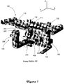
- a biopsy station 100 for performing tomotactic guided breast biopsy in prone may include a tomosynthesis imaging system 102 and a stage arm assembly 104 positioned below a biopsy table 106.
- the tomosynthesis imaging system and stage arm assembly are used for needle guidance.
- either or both the imaging system and stage arm assembly may be repositionable in one or more dimensions to facilitate the biopsy procedure.
- An example of a tomosynthesis imaging system is described in U.S. Patent No. 7,869,563 , which is hereby incorporated by reference, and sold commercially as Selenia® Dimensions® digital breast tomosynthesis system from Hologic, Inc. It should be noted, however, that the biopsy station is not limited to use with tomosynthesis imaging, and could utilize one or more of tomotactic, stereotactic, and other forms of guidance.
- the biopsy table 106 is supported by a footed base 108.
- the base is offset to one side of the table such that an area beneath the table is available for positioning both a portion of the body of the patient on which the biopsy is performed and equipment for performing the biopsy.
- the table 106 includes a rigid platform which may be cantilevered from the base 108, and which supports the patient during the biopsy procedure. The platform may be partially or wholly covered with padding for the comfort of the patient.
- the table may be contoured such that symmetrical distal end sections 110, 112 are elevated relative to a central section 114. Either of the elevated sections 110, 112 can help support the legs of the patient, thereby allowing 180 degree repositioning of the patient.
- the central section 114 supports the head, abdomen and hip of the patient.
- Transitions between the end sections and the central section are angled to provide comfortable head, abdomen and hip support.
- An aperture 116 in the central section 114 of the table enables a portion of the body of the patient to extend below the table when the patient is situated in a prone position. For example, the breast being biopsied may extend through the aperture. Other parts of the patient's body may also extend through the aperture, e.g., an arm, for enhanced comfort or positioning for the biopsy procedure.
- Some aspects of the table may be consistent with features described in International Application Number PCT/US11/61186 , titled TABLE FOR PERFORMING MEDICAL PROCEDURES, filed November 17, 2011, and U.S. Patent 5,289,520 , titled STEREOTACTIC MAMMOGRAPHY IMAGING SYSTEM WITH PRONE POSITION EXAMINATION TABLE AND CCD CAMERA, filed October 6, 1992.
- an equipment support platform 200 is cantilevered from the base 108 beneath the table in the Y-dimension.
- the equipment support platform may be statically or repositionably connected to the base, and may move in a coordinated manner with, or independent of, the table based on settings which can be changed by an operator.
- the support platform may be connected to the base via a Y-axis slide assembly which enables the support platform to move relative to the base in the Y-dimension.
- An X-axis slide assembly may enable the support platform to move relative to the base in the X-dimension. Range of motion may be approximately +/- 4 inches relative to a Z-axis defined by the center of the aperture.
- a handle connected to the support platform facilitates manual positioning of the platform by an operator.
- Slide lock features may be employed to secure the platform in a desired position.
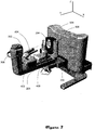
- the tomosynthesis imaging system 102 is mounted on the equipment support platform 200.
- the imaging system may include an x-ray energy source 202 and an x-ray energy receptor 204 (shown via cutaways in FIGS. 2 and 4 ).
- the source and receptor are aligned such that the receptor detects energy emitted by the source.
- the energy source 202 is positioned on a first upright portion of a support arm 206 such as a c-arm, and the energy receptor 204 is positioned on a second upright portion of the support arm.
- the support arm 206 helps maintain the receptor 204 and energy source 202 in alignment at a fixed distance, thereby mitigating or eliminating the need for focus adjustment.
- the support arm 206 is connected to the support platform 200 via a pivoting connector such as a bearing.
- the support arm moves under motor control such that the energy source 202 moves along an arc 300 (see FIG. 4 specifically) defined by a Z-axis of rotation 302 defined by a pivoting connector such as a bearing.
- the receptor 204 moves along an arc 301 characterized by a smaller radius than arc 300 because the pivoting connector via which the support arm is connected to the support platform is nearer to the second upright portion of the c-arm than the first upright portion of the c-arm.
- a handle 308 connected to the first upright portion facilitates manual rotational positioning of the support arm 206 within a 180 degree range of motion in the X-Y plane during set up by an operator.
- a path 306 of x-ray energy defined between the energy source 202 and receptor 204 in the X-Y plane can be reoriented within the X-Y plane with respect to the patient's breast through the 180 degree range of motion during set up.
- the biopsy needle is effectively positionable through 360 degrees in the X-Y plane relative to the breast.
- the biopsy gun stage arm assembly 104 is connected to the support platform 200 via a pivoting connector such as a bearing. Moreover, the stage arm assembly may pivot around a Z-axis which is coincident with Z-axis 302, and a multi-part bearing assembly may be utilized to enable independent rotational movement of the imaging system and the stage arm assembly. Optionally, the stage arm assembly may rotate about an axis offset from that of the imaging system.
- a rotatable support platform 400 associated with the stage arm assembly is disposed above the support arm 206.
- the stage arm assembly is rotatable through 180 degrees in the X-Y plane for manual set up by the operator.
- the stage arm assembly may be secured against rotational movement by a brake mechanism, e.g., to inhibit motion during a sweep or scan.
- a guidance module 402 with an interface and display mounted in a housing integral with or connected to the support platform displays tomosynthesis images and information about the relative locations of the targeted feature and the biopsy gun 404 to help position the biopsy gun and guide its path of travel such that the needle intersects with the target feature.
- a stage arm 406 is disposed on top of the guidance module 402 housing.
- a carriage slide assembly 408 is connected to the stage arm.
- a gun mount is connected to the carriage slide assembly.
- a biopsy gun 404 is mounted to the gun mount.
- the stage arm assembly may be oriented such that the operational path of travel of the biopsy gun needle intersects the Z-axis 302 about which the stage arm assembly and support arm rotate.
- the orientation of the stage arm assembly may be such that the operational path of travel of the biopsy gun needle intersects the Z-axis about which the stage arm assembly and support arm rotate at a particular point within the field of view of the tomosynthesis imaging system.
- the carriage slide assembly enables manual or motor-driven adjustment of the distance between the needle and the rotational Z-axis intersection point.
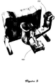
- the stage arm (and thus the gun mount and biopsy needle) may be oriented at a fixed inclination, e.g., 10°, relative to the X-Y plane in which the stage arm assembly is rotatable. Inclination of the stage arm allows a "zero degree" offset configuration in which the stage arm assembly is aligned with the imaging system as specifically shown in FIG. 2 .
- the stage arm assembly is offset from that of the imaging system, e.g., the stage arm assembly is not aligned with the imaging system.
- the inclined biopsy gun and needle do not reside in the field of view of the imaging system so the images are free of biopsy needle artifacts.
- Offset configurations in which the stage arm assembly is approximately orthogonal to the imaging system are specifically shown in FIGS. 3-7 .
- Some aspects of the stage arm assembly may be consistent with U.S. Patent Application 12/715,591 , titled NEEDLE BREAST BIOPSY SYSTEM AND METHOD FOR USE, filed March 2, 2010.
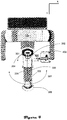
- a breast support assembly is provided to place the breast in compression.
- the breast support assembly includes a breast support platform 504 and compression paddle 502 connected to a rotatable platform 500.
- the platform 500 of the breast support assembly is connected to the support platform 200 via a pivoting connector such as a bearing.
- the stage arm assembly may pivot around a Z-axis which is coincident with Z-axis 302, and a multi-part bearing assembly may be utilized to enable independent rotational movement of the breast support assembly.
- the stage arm assembly pivots about an axis that is offset from Z-axis 302.
- the compression paddle is linearly movable toward and away from platform 504 in order to compress the breast against the foremost surface of the breast support platform 504 and release the breast from compression upon completion of the procedure.
- An aperture in the compression paddle allows a biopsy needle to traverse the compression paddle, e.g., in the zero degrees offset configuration.
- the breast support platform 504 may be integral to a protective cover 506 which encloses the receptor 204.
- a gap, e.g., 3 cm, between the foremost surface 700 (see FIG. 7 ) of the breast support platform and the receptor 204 allows the receptor to move during a scan or sweep without interfering with the stationary protective cover and breast support platform. Movement of the receptor during a scan or sweep and the gap enable use of a reduced size receptor. Use of the reduced size receptor enables use of a reduced size foremost surface 700.
- the breast support platform and/or protective cover may have side-edge sections 702, 704 adjacent to the foremost surface 700 which are angled, curved or otherwise formed away from surface 700 in order provide free space where the protective cover or breast support platform might otherwise interfere with the biopsy gun.
- side-edge sections 702, 704 adjacent to the foremost surface 700 which are angled, curved or otherwise formed away from surface 700 in order provide free space where the protective cover or breast support platform might otherwise interfere with the biopsy gun.
- use of a 15 cm width receptor and a corresponding size foremost surface allows a side-edge section geometry which facilitates biopsy of relatively thin breasts in the ninety degree offset configuration by avoiding interference between the breast support platform and/or protective cover and the biopsy gun and stage arm assembly.
- the present invention also facilitates access to previously inaccessible lesions, for example, such as those that may be situated in the axilla which prior conventional detectors would be unable to access.
- the stage arm assembly 104 and imaging system 102 are independently rotatable for set up, e.g., each through a 180 degree range of arc in the X-Y plane. More particularly, the furthest extent to which the stage arm assembly protrudes from the Z-axis of rotation is less than the minimum distance between the first upright portion of the support arm and the Z-axis of rotation. Consequently, the stage arm assembly can be rotated to either side of the receptor without interference. Similarly, the stage arm assembly can be rotated to either side of the breast support assembly without interference. (compare, e.g., FIG. 3 with FIG. 7 ) As specifically illustrated in FIG. 2 , the stage arm assembly and breast support assembly may also be optionally aligned.
- the patient In order to perform a biopsy procedure the patient is positioned on the table with one or both breasts and possibly one arm protruding through the aperture in step 800. As previously noted, the patient can be oriented in at least two different positions which are offset horizontally by 180 degrees in the X-Y plane.
- the breast which is the subject of the biopsy procedure may be approximately centered in the Z-axis about which the stage arm assembly and support arm rotate.
- the equipment support platform may then be moved in one or more dimensions orthogonal to the Z-axis to help center the breast in the axis of rotation in step 802.
- the imaging system orientation is then adjusted until a desired orientation is obtained for the procedure to be performed, as indicated in step 804.
- the imaging system may be moved rotationally about the Z-axis, and raised or lowered along the Z-axis.
- the stage arm assembly orientation is then adjusted for the procedure to be performed, as indicated in step 805.
- the stage arm assembly may be moved rotationally about the Z-axis and the location of the gun mount on the carriage slide assembly may be adjusted.
- the patient's breast is then immobilized between a compression paddle and the receptor in step 806.
- a tomosynthesis scan is performed by moving the x-ray energy source along an arc centered on the top surface of the receptor at step 808.
- the axis of rotation of the x-ray energy source can optionally be located about 3 cm above the compressed breast; the breast platform, or the top surface of the breast platform.
- Such an axis of rotation may reduce the amount of blurring in a sweep or movement of the x-ray energy source during a tomosynthesis scan.
- the energy source may be energized to emit a collimated x-ray beam, e.g., at every 1.07° of an arc of +/- 7.5°.
- the motion of the energy source can be continuous or discontinuous. If motion is continuous, a respective set of image data is accumulated over a small increment of continuous motion, e.g., a 0.1° to 0.5° arc of motion of source, although these non-limiting parameters are only an example.
- Different ranges of motion of the energy source can be used, and the motion of the source may be along an arc centered at a different axis, such as inside the immobilized breast, at the receptor, or elsewhere.
- the x-ray beam irradiates the breast, and radiation that has passed through the breast is received by the receptor.
- the receptor and associated electronics generate image data in digital form for each pixel of a rectangular grid of pixels at each predetermined discrete angular position of source.
- An associated three-dimensional image is generated and presented on the display.
- the image data is used to identify the precise location (final coordinates) of the previously detected feature of interest in step 810.
- Various fine-adjustment settings may be calculated and used to complete preparation of the stage arm assembly and biopsy gun in step 812.
- the needle is then actuated in order to obtain a tissue sample in step 814.
- Any biopsy system may work with the present invention.
- tubing couples the biopsy needle with a vacuum console and filter for capturing excised tissue samples.
- the stage arm assembly and other parts of the station may be reconfigured to obtain as many samples as required.
Landscapes
- Health & Medical Sciences (AREA)
- Life Sciences & Earth Sciences (AREA)
- Engineering & Computer Science (AREA)
- Medical Informatics (AREA)
- Biomedical Technology (AREA)
- Surgery (AREA)
- Veterinary Medicine (AREA)
- Public Health (AREA)
- Nuclear Medicine, Radiotherapy & Molecular Imaging (AREA)
- General Health & Medical Sciences (AREA)
- Pathology (AREA)
- Animal Behavior & Ethology (AREA)
- Heart & Thoracic Surgery (AREA)
- Molecular Biology (AREA)
- Radiology & Medical Imaging (AREA)
- Physics & Mathematics (AREA)
- Optics & Photonics (AREA)
- High Energy & Nuclear Physics (AREA)
- Biophysics (AREA)
- Oral & Maxillofacial Surgery (AREA)
- Dentistry (AREA)
- Apparatus For Radiation Diagnosis (AREA)
Priority Applications (1)
| Application Number | Priority Date | Filing Date | Title |
|---|---|---|---|
| EP18153706.9A EP3366217B1 (en) | 2013-03-15 | 2014-03-13 | Tomosynthesis-guided biopsy in prone |
Applications Claiming Priority (2)
| Application Number | Priority Date | Filing Date | Title |
|---|---|---|---|
| US201361787825P | 2013-03-15 | 2013-03-15 | |
| PCT/US2014/026164 WO2014151646A1 (en) | 2013-03-15 | 2014-03-13 | Tomosynthesis-guided biopsy in prone |
Related Child Applications (1)
| Application Number | Title | Priority Date | Filing Date |
|---|---|---|---|
| EP18153706.9A Division EP3366217B1 (en) | 2013-03-15 | 2014-03-13 | Tomosynthesis-guided biopsy in prone |
Publications (3)
| Publication Number | Publication Date |
|---|---|
| EP2967479A1 EP2967479A1 (en) | 2016-01-20 |
| EP2967479A4 EP2967479A4 (en) | 2016-10-26 |
| EP2967479B1 true EP2967479B1 (en) | 2018-01-31 |
Family
ID=51581003
Family Applications (2)
| Application Number | Title | Priority Date | Filing Date |
|---|---|---|---|
| EP14770362.3A Active EP2967479B1 (en) | 2013-03-15 | 2014-03-13 | Tomosynthesis-guided biopsy in prone |
| EP18153706.9A Revoked EP3366217B1 (en) | 2013-03-15 | 2014-03-13 | Tomosynthesis-guided biopsy in prone |
Family Applications After (1)
| Application Number | Title | Priority Date | Filing Date |
|---|---|---|---|
| EP18153706.9A Revoked EP3366217B1 (en) | 2013-03-15 | 2014-03-13 | Tomosynthesis-guided biopsy in prone |
Country Status (5)
| Country | Link |
|---|---|
| US (6) | US10092358B2 (enExample) |
| EP (2) | EP2967479B1 (enExample) |
| JP (1) | JP6388347B2 (enExample) |
| AU (1) | AU2014233687B2 (enExample) |
| WO (1) | WO2014151646A1 (enExample) |
Families Citing this family (32)
| Publication number | Priority date | Publication date | Assignee | Title |
|---|---|---|---|---|
| WO2007095330A2 (en) | 2006-02-15 | 2007-08-23 | Hologic Inc | Breast biopsy and needle localization using tomosynthesis systems |
| EP2485651B1 (en) | 2009-10-08 | 2020-12-23 | Hologic, Inc. | Needle breast biopsy system |
| US20120133600A1 (en) | 2010-11-26 | 2012-05-31 | Hologic, Inc. | User interface for medical image review workstation |
| CA2829349C (en) | 2011-03-08 | 2021-02-09 | Hologic, Inc. | System and method for dual energy and/or contrast enhanced breast imaging for screening, diagnosis and biopsy |
| JP2014534042A (ja) | 2011-11-27 | 2014-12-18 | ホロジック, インコーポレイテッドHologic, Inc. | マンモグラフィーおよび/またはトモシンセシス画像データを使用して2d画像を生成するためのシステムおよび方法 |
| US9805507B2 (en) | 2012-02-13 | 2017-10-31 | Hologic, Inc | System and method for navigating a tomosynthesis stack using synthesized image data |
| EP2967473B1 (en) | 2013-03-15 | 2020-02-19 | Hologic, Inc. | System and method for navigating a tomosynthesis stack including automatic focusing |
| JP6388347B2 (ja) * | 2013-03-15 | 2018-09-12 | ホロジック, インコーポレイテッドHologic, Inc. | 腹臥位におけるトモシンセシス誘導生検 |
| EP4278977A3 (en) | 2013-10-24 | 2024-02-21 | Hologic, Inc. | System and method for navigating x-ray guided breast biopsy |
| KR101588574B1 (ko) * | 2013-11-06 | 2016-01-26 | 주식회사 레이언스 | 입체정위 생검을 위한 생검 니들 가이딩 장치, 이를 구비한 영상 촬영 장치 및 이를 이용한 생검 채취 방법 |
| JP6506769B2 (ja) | 2014-02-28 | 2019-04-24 | ホロジック, インコーポレイテッドHologic, Inc. | トモシンセシス画像スラブを生成し表示するためのシステムおよび方法 |
| JP6611428B2 (ja) * | 2014-12-09 | 2019-11-27 | キヤノン株式会社 | マンモ断層撮像システム |
| JP6525768B2 (ja) * | 2015-06-30 | 2019-06-05 | キヤノン株式会社 | 乳房撮影装置 |
| EP3600047A1 (en) | 2017-03-30 | 2020-02-05 | Hologic, Inc. | System and method for hierarchical multi-level feature image synthesis and representation |
| CN110662489B (zh) | 2017-03-30 | 2024-08-02 | 豪洛捷公司 | 用于靶向对象增强以生成合成乳房组织图像的系统和方法 |
| CN110621233B (zh) | 2017-03-30 | 2023-12-12 | 豪洛捷公司 | 用于处理乳房组织图像数据的方法 |
| WO2018236565A1 (en) | 2017-06-20 | 2018-12-27 | Hologic, Inc. | METHOD AND SYSTEM FOR MEDICAL IMAGING WITH DYNAMIC SELF-LEARNING |
| NL2019124B1 (en) * | 2017-06-27 | 2019-01-07 | Sigmascreening B V | Mammography apparatus |
| US11439360B2 (en) * | 2017-08-16 | 2022-09-13 | Hologic, Inc. | Medical procedure draping system |
| CA3098900A1 (en) * | 2018-04-30 | 2019-11-07 | Memorial Sloan Kettering Cancer Center | Compression paddles for breast biopsies |
| US12121304B2 (en) | 2018-05-04 | 2024-10-22 | Hologic, Inc. | Introducer and localization wire visualization |
| CA3091593A1 (en) | 2018-05-04 | 2019-11-07 | Hologic, Inc. | Biopsy needle visualization |
| WO2020068851A1 (en) | 2018-09-24 | 2020-04-02 | Hologic, Inc. | Breast mapping and abnormality localization |
| US11883206B2 (en) | 2019-07-29 | 2024-01-30 | Hologic, Inc. | Personalized breast imaging system |
| US11694792B2 (en) | 2019-09-27 | 2023-07-04 | Hologic, Inc. | AI system for predicting reading time and reading complexity for reviewing 2D/3D breast images |
| JP7671776B2 (ja) | 2020-03-27 | 2025-05-02 | ホロジック, インコーポレイテッド | 複数の画像化モダリティにおける関心対象領域を識別するシステム及び方法 |
| US12505645B2 (en) | 2020-03-27 | 2025-12-23 | Hologic, Inc. | Systems and methods for correlating regions of interest in multiple imaging modalities |
| US11481038B2 (en) | 2020-03-27 | 2022-10-25 | Hologic, Inc. | Gesture recognition in controlling medical hardware or software |
| EP4214671A1 (en) | 2020-09-18 | 2023-07-26 | Hologic, Inc. | Correlating regions of interest |
| US20220164951A1 (en) | 2020-11-20 | 2022-05-26 | Hologic, Inc. | Systems and methods for using ai to identify regions of interest in medical images |
| US12254586B2 (en) | 2021-10-25 | 2025-03-18 | Hologic, Inc. | Auto-focus tool for multimodality image review |
| CN118318247A (zh) | 2021-11-29 | 2024-07-09 | 豪洛捷公司 | 用于将感兴趣的对象相关的系统和方法 |
Family Cites Families (500)
| Publication number | Priority date | Publication date | Assignee | Title |
|---|---|---|---|---|
| JP4054402B2 (ja) | 1997-04-25 | 2008-02-27 | 株式会社東芝 | X線断層撮影装置 |
| US3502878A (en) | 1967-09-22 | 1970-03-24 | Us Health Education & Welfare | Automatic x-ray apparatus for limiting the field size of a projected x-ray beam in response to film size and to source-to-film distance |
| US7050611B2 (en) | 2001-05-29 | 2006-05-23 | Mevis Breastcare Gmbh Co. Kg | Method and computer system for screening of medical cases |
| US3863073A (en) | 1973-04-26 | 1975-01-28 | Machlett Lab Inc | Automatic system for precise collimation of radiation |
| US3971950A (en) | 1975-04-14 | 1976-07-27 | Xerox Corporation | Independent compression and positioning device for use in mammography |
| US4160906A (en) | 1977-06-23 | 1979-07-10 | General Electric Company | Anatomically coordinated user dominated programmer for diagnostic x-ray apparatus |
| DE2838901C2 (de) | 1978-09-06 | 1986-11-06 | Siemens AG, 1000 Berlin und 8000 München | Katapultrasterlade |
| FR2512024A1 (fr) | 1981-08-27 | 1983-03-04 | Adir | Ethers tricycliques, leur preparation et les compositions pharmaceutiques les contenant |
| FR2549248B1 (fr) | 1983-06-24 | 1986-01-31 | Thomson Csf | Porte-cassette escamotable pour appareil d'examen radiologique et radiographique |
| DE3339775A1 (de) | 1983-11-03 | 1985-05-15 | Siemens AG, 1000 Berlin und 8000 München | Roentgendiagnostikgeraet mit strahlenfiltern |
| SE8306243L (sv) | 1983-11-14 | 1985-05-15 | Cytex Medicinteknik Ab | Lokaliseringsmetodik |
| JPS60129034A (ja) | 1983-12-16 | 1985-07-10 | 横河メディカルシステム株式会社 | X線断層撮像装置の操作卓 |
| US4559557A (en) | 1984-06-01 | 1985-12-17 | General Electric Company | Region-of-interest digital subtraction angiography |
| US4706269A (en) | 1985-03-11 | 1987-11-10 | Reina Leo J | Anti-scatter grid structure |
| US4773087A (en) | 1986-04-14 | 1988-09-20 | University Of Rochester | Quality of shadowgraphic x-ray images |
| USRE33634E (en) | 1986-09-23 | 1991-07-09 | Method and structure for optimizing radiographic quality by controlling X-ray tube voltage, current focal spot size and exposure time | |
| US4821727A (en) | 1986-10-30 | 1989-04-18 | Elscint Ltd. | Mammographic biopsy needle holder system |
| US4819258A (en) | 1986-11-28 | 1989-04-04 | Bennett X-Ray Corp. | Auto-setting of KV in an x-ray machine after selection of technic factors |
| US4907156A (en) | 1987-06-30 | 1990-03-06 | University Of Chicago | Method and system for enhancement and detection of abnormal anatomic regions in a digital image |
| US5051904A (en) | 1988-03-24 | 1991-09-24 | Olganix Corporation | Computerized dynamic tomography system |
| US5740270A (en) | 1988-04-08 | 1998-04-14 | Neuromedical Systems, Inc. | Automated cytological specimen classification system and method |
| DK654488A (da) | 1988-11-23 | 1990-05-24 | Nordisk Roentgen Tech App | Roentgenapparat |
| US5099846A (en) | 1988-12-23 | 1992-03-31 | Hardy Tyrone L | Method and apparatus for video presentation from a variety of scanner imaging sources |
| FR2645006A1 (fr) | 1989-03-29 | 1990-10-05 | Gen Electric Cgr | Mammographe equipe d'un dispositif de vue stereotaxiques integre et procede d'utilisation d'un tel mammographe |
| FR2646340A1 (fr) | 1989-04-28 | 1990-11-02 | Gen Electric Cgr | Porte-cassette adaptable en dimension et en position pour mammographie |
| EP0406455B1 (de) | 1989-07-03 | 1994-09-21 | Siemens Aktiengesellschaft | Röntgendiagnostikgerät für Mammographieaufnahmen |
| US5133020A (en) | 1989-07-21 | 1992-07-21 | Arch Development Corporation | Automated method and system for the detection and classification of abnormal lesions and parenchymal distortions in digital medical images |
| CA2014918A1 (en) | 1989-09-06 | 1991-03-06 | James A. Mcfaul | Scanning mammography system with improved skin line viewing |
| US4969174A (en) | 1989-09-06 | 1990-11-06 | General Electric Company | Scanning mammography system with reduced scatter radiation |
| US5078142A (en) | 1989-11-21 | 1992-01-07 | Fischer Imaging Corporation | Precision mammographic needle biopsy system |
| US5415169A (en) | 1989-11-21 | 1995-05-16 | Fischer Imaging Corporation | Motorized mammographic biopsy apparatus |
| US5240011A (en) | 1991-11-27 | 1993-08-31 | Fischer Imaging Corporation | Motorized biopsy needle positioner |
| WO1991007922A1 (en) | 1989-11-27 | 1991-06-13 | Bard International, Inc. | Puncture guide for computer tomography |
| US5199056A (en) | 1989-11-28 | 1993-03-30 | Darrah Carol J | Mammography compression paddle |
| US5481623A (en) | 1990-04-19 | 1996-01-02 | Fuji Photo Film Co., Ltd. | Apparatus for determining an image position on imaging media |
| FR2668359B1 (fr) | 1990-10-24 | 1998-02-20 | Gen Electric Cgr | Mammographe muni d'un porte-aiguille perfectionne. |
| US5409497A (en) | 1991-03-11 | 1995-04-25 | Fischer Imaging Corporation | Orbital aiming device for mammo biopsy |
| US5129911A (en) | 1991-03-11 | 1992-07-14 | Siczek Bernard W | Orbital aiming device |
| US5279309A (en) | 1991-06-13 | 1994-01-18 | International Business Machines Corporation | Signaling device and method for monitoring positions in a surgical operation |
| US5163075A (en) | 1991-08-08 | 1992-11-10 | Eastman Kodak Company | Contrast enhancement of electrographic imaging |
| US5941832A (en) | 1991-09-27 | 1999-08-24 | Tumey; David M. | Method and apparatus for detection of cancerous and precancerous conditions in a breast |
| US5289520A (en) | 1991-11-27 | 1994-02-22 | Lorad Corporation | Stereotactic mammography imaging system with prone position examination table and CCD camera |
| US5594769A (en) | 1991-11-27 | 1997-01-14 | Thermotrex Corporation | Method and apparatus for obtaining stereotactic mammographic guided needle breast biopsies |
| US5343390A (en) | 1992-02-28 | 1994-08-30 | Arch Development Corporation | Method and system for automated selection of regions of interest and detection of septal lines in digital chest radiographs |
| US5359637A (en) | 1992-04-28 | 1994-10-25 | Wake Forest University | Self-calibrated tomosynthetic, radiographic-imaging system, method, and device |
| US5386447A (en) * | 1992-09-23 | 1995-01-31 | Fischer Imaging Corporation | Mammographic screening and biopsy apparatus |
| US5596200A (en) | 1992-10-14 | 1997-01-21 | Primex | Low dose mammography system |
| FR2703237B1 (fr) | 1993-03-29 | 1995-05-19 | Ge Medical Syst Sa | Mammographe équipé d'un dispositif de prises en vues stéréotaxiques à détecteur numérique et procédé d'utilisation d'un tel mammographe . |
| US5491627A (en) | 1993-05-13 | 1996-02-13 | Arch Development Corporation | Method and system for the detection of microcalcifications in digital mammograms |
| US5878746A (en) | 1993-08-25 | 1999-03-09 | Lemelson; Jerome H. | Computerized medical diagnostic system |
| US5365562A (en) | 1993-09-20 | 1994-11-15 | Fischer Imaging Corporation | Digital imaging apparatus |
| US6075879A (en) | 1993-09-29 | 2000-06-13 | R2 Technology, Inc. | Method and system for computer-aided lesion detection using information from multiple images |
| US5526394A (en) | 1993-11-26 | 1996-06-11 | Fischer Imaging Corporation | Digital scan mammography apparatus |
| US5452367A (en) | 1993-11-29 | 1995-09-19 | Arch Development Corporation | Automated method and system for the segmentation of medical images |
| CA2113752C (en) | 1994-01-19 | 1999-03-02 | Stephen Michael Rooks | Inspection system for cross-sectional imaging |
| DE4414689C2 (de) | 1994-04-26 | 1996-08-29 | Siemens Ag | Röntgendiagnostikeinrichtung |
| US5499097A (en) | 1994-09-19 | 1996-03-12 | Neopath, Inc. | Method and apparatus for checking automated optical system performance repeatability |
| US5557097A (en) | 1994-09-20 | 1996-09-17 | Neopath, Inc. | Cytological system autofocus integrity checking apparatus |
| US5647025A (en) | 1994-09-20 | 1997-07-08 | Neopath, Inc. | Automatic focusing of biomedical specimens apparatus |
| AU3371395A (en) | 1994-09-20 | 1996-04-19 | Neopath, Inc. | Biological specimen analysis system processing integrity checking apparatus |
| US5553111A (en) | 1994-10-26 | 1996-09-03 | The General Hospital Corporation | Apparatus and method for improved tissue imaging |
| US5506877A (en) | 1994-11-23 | 1996-04-09 | The General Hospital Corporation | Mammography breast compression device and method |
| US5712890A (en) | 1994-11-23 | 1998-01-27 | Thermotrex Corp. | Full breast digital mammography device |
| US5657362A (en) | 1995-02-24 | 1997-08-12 | Arch Development Corporation | Automated method and system for computerized detection of masses and parenchymal distortions in medical images |
| US5660185A (en) | 1995-04-13 | 1997-08-26 | Neovision Corporation | Image-guided biopsy apparatus with enhanced imaging and methods |
| US5671288A (en) | 1995-05-31 | 1997-09-23 | Neopath, Inc. | Method and apparatus for assessing slide and specimen preparation quality |
| US6216540B1 (en) | 1995-06-06 | 2001-04-17 | Robert S. Nelson | High resolution device and method for imaging concealed objects within an obscuring medium |
| JPH10505286A (ja) | 1995-06-20 | 1998-05-26 | シン ング、ワン | 医療処置のための関節アーム |
| JP3617698B2 (ja) | 1995-07-17 | 2005-02-09 | 東芝医用システムエンジニアリング株式会社 | 診断支援装置 |
| US5642433A (en) | 1995-07-31 | 1997-06-24 | Neopath, Inc. | Method and apparatus for image contrast quality evaluation |
| US5642441A (en) | 1995-10-24 | 1997-06-24 | Neopath, Inc. | Separation apparatus and method for measuring focal plane |
| US5818898A (en) | 1995-11-07 | 1998-10-06 | Kabushiki Kaisha Toshiba | X-ray imaging apparatus using X-ray planar detector |
| US5693948A (en) | 1995-11-21 | 1997-12-02 | Loral Fairchild Corporation | Advanced CCD-based x-ray image sensor system |
| US5627869A (en) | 1995-11-22 | 1997-05-06 | Thermotrex Corporation | Mammography apparatus with proportional collimation |
| FI955636A0 (fi) | 1995-11-23 | 1995-11-23 | Planmed Oy | Foerfarande och system foer styrning av funktionerna av en mammografiaanordning |
| US5709206A (en) | 1995-11-27 | 1998-01-20 | Teboul; Michel | Imaging system for breast sonography |
| DE69627183T2 (de) | 1995-11-30 | 2004-01-29 | Chromavision Med Sys Inc | Verfahren zur automatischen bildanalyse biologischer proben |
| US5769086A (en) | 1995-12-06 | 1998-06-23 | Biopsys Medical, Inc. | Control system and method for automated biopsy device |
| JPH09198490A (ja) | 1996-01-22 | 1997-07-31 | Hitachi Medical Corp | 3次元離散データ投影装置 |
| JPH09238934A (ja) | 1996-03-11 | 1997-09-16 | Toshiba Medical Eng Co Ltd | 画像表示システム |
| DE19619913C2 (de) | 1996-05-17 | 2001-03-15 | Sirona Dental Systems Gmbh | Röntgendiagnostikgerät für Tomosynthese |
| DE19619924A1 (de) | 1996-05-17 | 1997-11-20 | Siemens Ag | Verfahren zur Erstellung von Tomosyntheseaufnahmen |
| DE19619925C2 (de) | 1996-05-17 | 1999-09-09 | Sirona Dental Systems Gmbh | Röntgendiagnostikgerät für Tomosynthese |
| DE19619915A1 (de) | 1996-05-17 | 1997-11-20 | Siemens Ag | Verfahren zur Erstellung von Tomosyntheseaufnahmen |
| US6067079A (en) | 1996-06-13 | 2000-05-23 | International Business Machines Corporation | Virtual pointing device for touchscreens |
| US5835079A (en) | 1996-06-13 | 1998-11-10 | International Business Machines Corporation | Virtual pointing device for touchscreens |
| US5841124A (en) | 1996-06-19 | 1998-11-24 | Neopath, Inc. | Cytological system autofocus integrity checking apparatus |
| US5872828A (en) | 1996-07-23 | 1999-02-16 | The General Hospital Corporation | Tomosynthesis system for breast imaging |
| JPH1033523A (ja) | 1996-07-24 | 1998-02-10 | Hitachi Medical Corp | X線ct装置 |
| US5776062A (en) | 1996-10-15 | 1998-07-07 | Fischer Imaging Corporation | Enhanced breast imaging/biopsy system employing targeted ultrasound |
| US5986662A (en) | 1996-10-16 | 1999-11-16 | Vital Images, Inc. | Advanced diagnostic viewer employing automated protocol selection for volume-rendered imaging |
| US6293282B1 (en) | 1996-11-05 | 2001-09-25 | Jerome Lemelson | System and method for treating select tissue in living being |
| JP3878259B2 (ja) | 1996-11-13 | 2007-02-07 | 東芝医用システムエンジニアリング株式会社 | 医用画像処理装置 |
| US6137527A (en) | 1996-12-23 | 2000-10-24 | General Electric Company | System and method for prompt-radiology image screening service via satellite |
| US5757880A (en) | 1997-01-08 | 1998-05-26 | Colomb; Denis | Apparatus, article of manufacture, and method for creation of an uncompressed image of compressed matter |
| US7117098B1 (en) | 1997-02-27 | 2006-10-03 | Cellomics, Inc. | Machine-readable storage medium for analyzing distribution of macromolecules between the cell membrane and the cell cytoplasm |
| US5999639A (en) | 1997-09-04 | 1999-12-07 | Qualia Computing, Inc. | Method and system for automated detection of clustered microcalcifications from digital mammograms |
| US6091981A (en) | 1997-09-16 | 2000-07-18 | Assurance Medical Inc. | Clinical tissue examination |
| US20030135115A1 (en) | 1997-11-24 | 2003-07-17 | Burdette Everette C. | Method and apparatus for spatial registration and mapping of a biopsy needle during a tissue biopsy |
| US6442288B1 (en) | 1997-12-17 | 2002-08-27 | Siemens Aktiengesellschaft | Method for reconstructing a three-dimensional image of an object scanned in the context of a tomosynthesis, and apparatus for tomosynthesis |
| JP3554172B2 (ja) | 1998-01-09 | 2004-08-18 | キヤノン株式会社 | 放射線撮影装置 |
| US6175117B1 (en) | 1998-01-23 | 2001-01-16 | Quanta Vision, Inc. | Tissue analysis apparatus |
| US6289235B1 (en) | 1998-03-05 | 2001-09-11 | Wake Forest University | Method and system for creating three-dimensional images using tomosynthetic computed tomography |
| US6081577A (en) | 1998-07-24 | 2000-06-27 | Wake Forest University | Method and system for creating task-dependent three-dimensional images |
| US6375352B1 (en) | 1999-10-01 | 2002-04-23 | General Electric Company | Apparatus and method for obtaining x-ray tomosynthesis data for mammography |
| US6141398A (en) | 1998-08-25 | 2000-10-31 | General Electric Company | Protocol driven image reconstruction, display, and processing in a multislice imaging system |
| US6101236A (en) | 1998-10-02 | 2000-08-08 | University Of Iowa Research Foundation | Iterative method and apparatus for x-ray computed tomographic fluoroscopy |
| AU2706500A (en) | 1998-11-25 | 2000-09-21 | Fischer Imaging Corporation | User interface system for mammographic imager |
| FR2786388B1 (fr) | 1998-11-27 | 2001-02-16 | Ge Medical Syst Sa | Procede de detection d'un tissu de nature determinee en radiologie numerique et son utilisation pour le reglage des parametres d'exposition |
| US6149301A (en) | 1998-12-30 | 2000-11-21 | General Electric Company | X-ray target centering apparatus for radiographic imaging system |
| JP2000200340A (ja) | 1999-01-06 | 2000-07-18 | Ge Yokogawa Medical Systems Ltd | 画像表示方法、画像表示装置およびct装置 |
| US6424332B1 (en) | 1999-01-29 | 2002-07-23 | Hunter Innovations, Inc. | Image comparison apparatus and method |
| NZ513856A (en) | 1999-01-29 | 2001-09-28 | American Superconducting Corp | Electric utility system with superconducting magnetic energy storage |
| US6233473B1 (en) | 1999-02-16 | 2001-05-15 | Hologic, Inc. | Determining body composition using fan beam dual-energy x-ray absorptiometry |
| US6272207B1 (en) | 1999-02-18 | 2001-08-07 | Creatv Microtech, Inc. | Method and apparatus for obtaining high-resolution digital X-ray and gamma ray images |
| US6256370B1 (en) | 2000-01-24 | 2001-07-03 | General Electric Company | Method and apparatus for performing tomosynthesis |
| US6689142B1 (en) | 1999-04-26 | 2004-02-10 | Scimed Life Systems, Inc. | Apparatus and methods for guiding a needle |
| US6292530B1 (en) | 1999-04-29 | 2001-09-18 | General Electric Company | Method and apparatus for reconstructing image data acquired by a tomosynthesis x-ray imaging system |
| DE19922346C2 (de) | 1999-05-14 | 2003-06-18 | Siemens Ag | Röntgendiagnostikeinrichtung für Tomosynthese oder Schichtung |
| US6243441B1 (en) | 1999-07-13 | 2001-06-05 | Edge Medical Devices | Active matrix detector for X-ray imaging |
| US20020173721A1 (en) | 1999-08-20 | 2002-11-21 | Novasonics, Inc. | User interface for handheld imaging devices |
| US6480565B1 (en) | 1999-11-18 | 2002-11-12 | University Of Rochester | Apparatus and method for cone beam volume computed tomography breast imaging |
| US6987831B2 (en) | 1999-11-18 | 2006-01-17 | University Of Rochester | Apparatus and method for cone beam volume computed tomography breast imaging |
| US6633674B1 (en) | 1999-11-24 | 2003-10-14 | General Electric Company | Picture archiving and communication system employing improved data compression |
| US6245028B1 (en) | 1999-11-24 | 2001-06-12 | Marconi Medical Systems, Inc. | Needle biopsy system |
| US6645520B2 (en) | 1999-12-16 | 2003-11-11 | Dermatrends, Inc. | Transdermal administration of nonsteroidal anti-inflammatory drugs using hydroxide-releasing agents as permeation enhancers |
| FR2803069B1 (fr) | 1999-12-28 | 2002-12-13 | Ge Medical Syst Sa | Procede et systeme de compensation de l'epaisseur d'un organe |
| US8352289B2 (en) | 1999-12-30 | 2013-01-08 | Dhi Computing, Inc. | Systems and methods for providing and maintaining electronic medical records |
| US6411836B1 (en) | 1999-12-30 | 2002-06-25 | General Electric Company | Method and apparatus for user preferences configuring in an image handling system |
| US6901156B2 (en) | 2000-02-04 | 2005-05-31 | Arch Development Corporation | Method, system and computer readable medium for an intelligent search workstation for computer assisted interpretation of medical images |
| US6744848B2 (en) | 2000-02-11 | 2004-06-01 | Brandeis University | Method and system for low-dose three-dimensional imaging of a scene |
| GB0006598D0 (en) | 2000-03-17 | 2000-05-10 | Isis Innovation | Three-dimensional reconstructions from images |
| US6351660B1 (en) | 2000-04-18 | 2002-02-26 | Litton Systems, Inc. | Enhanced visualization of in-vivo breast biopsy location for medical documentation |
| US6327336B1 (en) | 2000-06-05 | 2001-12-04 | Direct Radiography Corp. | Radiogram showing location of automatic exposure control sensor |
| US6683934B1 (en) | 2000-06-05 | 2004-01-27 | General Electric Company | Dual energy x-ray imaging system and method for radiography and mammography |
| US6389104B1 (en) | 2000-06-30 | 2002-05-14 | Siemens Corporate Research, Inc. | Fluoroscopy based 3-D neural navigation based on 3-D angiography reconstruction data |
| JP2002052018A (ja) | 2000-08-11 | 2002-02-19 | Canon Inc | 画像表示装置、画像表示方法および記憶媒体 |
| JP2002109510A (ja) | 2000-09-27 | 2002-04-12 | Fuji Photo Film Co Ltd | 異常陰影候補検出処理システム |
| EP1267722A1 (en) | 2000-10-20 | 2003-01-02 | Koninklijke Philips Electronics N.V. | Tomosynthesis in a limited angular range |
| WO2002069808A2 (en) | 2000-11-06 | 2002-09-12 | Suros Surgical Systems, Inc. | Biopsy apparatus |
| US6758824B1 (en) | 2000-11-06 | 2004-07-06 | Suros Surgical Systems, Inc. | Biopsy apparatus |
| US6468226B1 (en) | 2000-11-22 | 2002-10-22 | Mcintyre, Iv John J. | Remote tissue biopsy apparatus and associated methods |
| US7615008B2 (en) | 2000-11-24 | 2009-11-10 | U-Systems, Inc. | Processing and displaying breast ultrasound information |
| US7556602B2 (en) | 2000-11-24 | 2009-07-07 | U-Systems, Inc. | Breast cancer screening with adjunctive ultrasound mammography |
| US7597663B2 (en) | 2000-11-24 | 2009-10-06 | U-Systems, Inc. | Adjunctive ultrasound processing and display for breast cancer screening |
| US7103205B2 (en) | 2000-11-24 | 2006-09-05 | U-Systems, Inc. | Breast cancer screening with ultrasound image overlays |
| US6650928B1 (en) | 2000-11-27 | 2003-11-18 | Ge Medical Systems Global Technology Company, Llc | Color parametric and composite maps for CT perfusion |
| US6501819B2 (en) | 2000-12-18 | 2002-12-31 | Ge Medical Systems Global Technology Company, Llc | Medical diagnostic method and apparatus to control dual energy exposure techniques based on image information |
| FR2818116B1 (fr) | 2000-12-19 | 2004-08-27 | Ge Med Sys Global Tech Co Llc | Appareil de mammographie |
| US6463181B2 (en) | 2000-12-22 | 2002-10-08 | The United States Of America As Represented By The Secretary Of The Navy | Method for optimizing visual display of enhanced digital images |
| WO2002052507A1 (en) | 2000-12-22 | 2002-07-04 | Koninklijke Philips Electronics N.V. | Stereoscopic viewing of a region between clipping planes |
| WO2002065480A1 (en) | 2001-02-01 | 2002-08-22 | Creatv Microtech, Inc. | tNTI-SCATTER GRIDS AND COLLIMATOR DESIGNS, AND THEIR MOTION, FABRICATION AND ASSEMBLY |
| US7030861B1 (en) | 2001-02-10 | 2006-04-18 | Wayne Carl Westerman | System and method for packing multi-touch gestures onto a hand |
| US6486764B2 (en) | 2001-02-16 | 2002-11-26 | Delphi Technologies, Inc. | Rotary position sensor |
| US20020188466A1 (en) | 2001-04-18 | 2002-12-12 | Barrette Pierre Philip | Secure digital medical intellectual property (IP) distribution, market applications, and mobile devices |
| US6620111B2 (en) | 2001-04-20 | 2003-09-16 | Ethicon Endo-Surgery, Inc. | Surgical biopsy device having automatic rotation of the probe for taking multiple samples |
| US6954667B2 (en) | 2001-06-28 | 2005-10-11 | Chemimage Corporation | Method for Raman chemical imaging and characterization of calcification in tissue |
| US6611575B1 (en) | 2001-07-27 | 2003-08-26 | General Electric Company | Method and system for high resolution 3D visualization of mammography images |
| WO2003017244A1 (en) | 2001-08-17 | 2003-02-27 | Multidigit, Inc. | System and method for selecting actions based on the identification of user's fingers |
| WO2003020114A2 (en) | 2001-08-31 | 2003-03-13 | Analogic Corporation | Image positioning method and system for tomosynthesis in a digital x-ray radiography system |
| FR2829918A1 (fr) | 2001-09-25 | 2003-03-28 | Ge Med Sys Global Tech Co Llc | Appareil de mammographie |
| US20030072478A1 (en) | 2001-10-12 | 2003-04-17 | Claus Bernhard Erich Hermann | Reconstruction method for tomosynthesis |
| WO2003037046A2 (en) | 2001-10-19 | 2003-05-01 | Hologic, Inc. | Mammography system and method employing offset compression paddles, automatic collimation, and retractable anti-scatter grid |
| US6626849B2 (en) | 2001-11-01 | 2003-09-30 | Ethicon Endo-Surgery, Inc. | MRI compatible surgical biopsy device |
| EP1313066B1 (en) | 2001-11-19 | 2008-08-27 | STMicroelectronics S.r.l. | A method for merging digital images to obtain a high dynamic range digital image |
| US20030097055A1 (en) | 2001-11-21 | 2003-05-22 | Philips Medical Systems(Cleveland), Inc. | Method of reviewing tomographic scans with a large number of images |
| US6751285B2 (en) | 2001-11-21 | 2004-06-15 | General Electric Company | Dose management system for mammographic tomosynthesis |
| US6895077B2 (en) | 2001-11-21 | 2005-05-17 | University Of Massachusetts Medical Center | System and method for x-ray fluoroscopic imaging |
| JP4099984B2 (ja) | 2001-12-14 | 2008-06-11 | コニカミノルタホールディングス株式会社 | 異常陰影検出装置および画像出力装置 |
| US6978040B2 (en) | 2001-12-19 | 2005-12-20 | Canon Kabushiki Kaisha | Optical recovery of radiographic geometry |
| US6647092B2 (en) | 2002-01-18 | 2003-11-11 | General Electric Company | Radiation imaging system and method of collimation |
| SE524458C2 (sv) | 2002-03-01 | 2004-08-10 | Mamea Imaging Ab | Skyddsanordning vid en röntgenapparat |
| CN1639737A (zh) | 2002-03-06 | 2005-07-13 | 西门子共同研究公司 | 容积-容积融合的可视化 |
| US7346381B2 (en) | 2002-11-01 | 2008-03-18 | Ge Medical Systems Global Technology Company Llc | Method and apparatus for medical intervention procedure planning |
| US6878115B2 (en) | 2002-03-28 | 2005-04-12 | Ultrasound Detection Systems, Llc | Three-dimensional ultrasound computed tomography imaging system |
| US6819790B2 (en) | 2002-04-12 | 2004-11-16 | The University Of Chicago | Massive training artificial neural network (MTANN) for detecting abnormalities in medical images |
| US7218766B2 (en) | 2002-04-15 | 2007-05-15 | General Electric Company | Computer aided detection (CAD) for 3D digital mammography |
| US6707878B2 (en) | 2002-04-15 | 2004-03-16 | General Electric Company | Generalized filtered back-projection reconstruction in digital tomosynthesis |
| US20030194050A1 (en) | 2002-04-15 | 2003-10-16 | General Electric Company | Multi modality X-ray and nuclear medicine mammography imaging system and method |
| US6882700B2 (en) | 2002-04-15 | 2005-04-19 | General Electric Company | Tomosynthesis X-ray mammogram system and method with automatic drive system |
| US6752767B2 (en) | 2002-04-16 | 2004-06-22 | Vivant Medical, Inc. | Localization element with energized tip |
| US7139000B2 (en) | 2002-05-13 | 2006-11-21 | Ge Medical Systems Global Technology Company, Llc | Method, system and computer product for displaying axial images |
| US7295691B2 (en) | 2002-05-15 | 2007-11-13 | Ge Medical Systems Global Technology Company, Llc | Computer aided diagnosis of an image set |
| US11275405B2 (en) | 2005-03-04 | 2022-03-15 | Apple Inc. | Multi-functional hand-held device |
| US7599579B2 (en) | 2002-07-11 | 2009-10-06 | Ge Medical Systems Global Technology Company, Llc | Interpolated image filtering method and apparatus |
| US7450747B2 (en) | 2002-07-12 | 2008-11-11 | Ge Medical Systems Global Technology Company, Llc | System and method for efficiently customizing an imaging system |
| US20040036680A1 (en) | 2002-08-26 | 2004-02-26 | Mark Davis | User-interface features for computers with contact-sensitive displays |
| US6898331B2 (en) | 2002-08-28 | 2005-05-24 | Bae Systems Aircraft Controls, Inc. | Image fusion system and method |
| US6574304B1 (en) | 2002-09-13 | 2003-06-03 | Ge Medical Systems Global Technology Company, Llc | Computer aided acquisition of medical images |
| US6748044B2 (en) | 2002-09-13 | 2004-06-08 | Ge Medical Systems Global Technology Company, Llc | Computer assisted analysis of tomographic mammography data |
| US7260249B2 (en) | 2002-09-27 | 2007-08-21 | Confirma Incorporated | Rules-based approach for processing medical images |
| US7347829B2 (en) | 2002-10-07 | 2008-03-25 | Suros Surgical Systems, Inc. | Introduction system for minimally invasive surgical instruments |
| US6940943B2 (en) | 2002-10-07 | 2005-09-06 | General Electric Company | Continuous scan tomosynthesis system and method |
| US6825838B2 (en) | 2002-10-11 | 2004-11-30 | Sonocine, Inc. | 3D modeling system |
| US20040171933A1 (en) * | 2002-11-25 | 2004-09-02 | Milton Stoller | Mammography needle biopsy system and method |
| US7616801B2 (en) | 2002-11-27 | 2009-11-10 | Hologic, Inc. | Image handling and display in x-ray mammography and tomosynthesis |
| US7577282B2 (en) | 2002-11-27 | 2009-08-18 | Hologic, Inc. | Image handling and display in X-ray mammography and tomosynthesis |
| US6597762B1 (en) | 2002-11-27 | 2003-07-22 | Ge Medical Systems Global Technology Co., Llc | Method and apparatus of lesion detection and validation based on multiple reviews of a CT image |
| US8571289B2 (en) | 2002-11-27 | 2013-10-29 | Hologic, Inc. | System and method for generating a 2D image from a tomosynthesis data set |
| US10638994B2 (en) | 2002-11-27 | 2020-05-05 | Hologic, Inc. | X-ray mammography with tomosynthesis |
| US7831296B2 (en) | 2002-11-27 | 2010-11-09 | Hologic, Inc. | X-ray mammography with tomosynthesis |
| US7123684B2 (en) | 2002-11-27 | 2006-10-17 | Hologic, Inc. | Full field mammography with tissue exposure control, tomosynthesis, and dynamic field of view processing |
| US7760924B2 (en) | 2002-11-27 | 2010-07-20 | Hologic, Inc. | System and method for generating a 2D image from a tomosynthesis data set |
| US7406150B2 (en) | 2002-11-29 | 2008-07-29 | Hologic, Inc. | Distributed architecture for mammographic image acquisition and processing |
| US7110490B2 (en) | 2002-12-10 | 2006-09-19 | General Electric Company | Full field digital tomosynthesis method and apparatus |
| US7904824B2 (en) | 2002-12-10 | 2011-03-08 | Siemens Medical Solutions Usa, Inc. | Medical imaging programmable custom user interface system and method |
| EP1430835B1 (en) | 2002-12-17 | 2011-11-16 | Kabushiki Kaisha Toshiba | System for peripheral X-ray angiography |
| US7286634B2 (en) | 2002-12-23 | 2007-10-23 | Select Technologies, Llc | Method and apparatus for improving baggage screening examination |
| US7356113B2 (en) | 2003-02-12 | 2008-04-08 | Brandeis University | Tomosynthesis imaging system and method |
| JP4604451B2 (ja) | 2003-02-24 | 2011-01-05 | コニカミノルタホールディングス株式会社 | 医用画像処理装置及び悪性度の判定方法 |
| US6999553B2 (en) | 2003-03-04 | 2006-02-14 | Livingston Products, Inc. | Method and apparatus for x-ray mammography imaging |
| US7333644B2 (en) | 2003-03-11 | 2008-02-19 | Siemens Medical Solutions Usa, Inc. | Systems and methods for providing automatic 3D lesion segmentation and measurements |
| JP4495154B2 (ja) | 2003-05-06 | 2010-06-30 | ビクター ジョン ジュニア ヤナコーネ | 哺乳動物での動的な熱力学的過程を同定し分類してそれらを識別するシステム |
| JP4497837B2 (ja) | 2003-05-12 | 2010-07-07 | キヤノン株式会社 | 放射線画像撮影装置 |
| EP1636731A2 (en) | 2003-06-25 | 2006-03-22 | Siemens Medical Solutions USA, Inc. | Systems and methods for automated diagnosis and decision support for breast imaging |
| US6885724B2 (en) | 2003-08-22 | 2005-04-26 | Ge Medical Systems Global Technology Company, Llc | Radiographic tomosynthesis image acquisition utilizing asymmetric geometry |
| US8090164B2 (en) | 2003-08-25 | 2012-01-03 | The University Of North Carolina At Chapel Hill | Systems, methods, and computer program products for analysis of vessel attributes for diagnosis, disease staging, and surgical planning |
| US7424141B2 (en) | 2003-08-29 | 2008-09-09 | Agilent Technologies, Inc. | System and method for performing auto-focused tomosynthesis |
| US7578781B2 (en) | 2003-09-18 | 2009-08-25 | Wisconsin Alumni Research Foundation | Device for placement of needles and radioactive seeds in radiotherapy |
| CA2539107A1 (en) | 2003-09-19 | 2005-03-31 | Arcturus Bioscience, Inc. | Predicting breast cancer treatment outcome |
| JP2005110843A (ja) | 2003-10-06 | 2005-04-28 | Canon Inc | 放射線画像処理装置及び処理方法 |
| US7869862B2 (en) | 2003-10-15 | 2011-01-11 | Varian Medical Systems, Inc. | Systems and methods for functional imaging using contrast-enhanced multiple-energy computed tomography |
| US20050089205A1 (en) | 2003-10-23 | 2005-04-28 | Ajay Kapur | Systems and methods for viewing an abnormality in different kinds of images |
| US20050113680A1 (en) | 2003-10-29 | 2005-05-26 | Yoshihiro Ikeda | Cerebral ischemia diagnosis assisting apparatus, X-ray computer tomography apparatus, and apparatus for aiding diagnosis and treatment of acute cerebral infarct |
| JP2005149107A (ja) | 2003-11-14 | 2005-06-09 | Konica Minolta Medical & Graphic Inc | 医用画像管理システム |
| DE10353611B4 (de) | 2003-11-17 | 2013-01-17 | Siemens Aktiengesellschaft | Röntgendiagnostikgerät für Mammographieuntersuchungen |
| ATE551660T1 (de) | 2003-11-26 | 2012-04-15 | Koninkl Philips Electronics Nv | Optimierung des arbeitsablaufs für ein bilddarstellungsumfeld mit hohem durchsatz |
| US8265728B2 (en) | 2003-11-26 | 2012-09-11 | University Of Chicago | Automated method and system for the evaluation of disease and registration accuracy in the subtraction of temporally sequential medical images |
| US8768026B2 (en) | 2003-11-26 | 2014-07-01 | Hologic, Inc. | X-ray imaging with x-ray markers that provide adjunct information but preserve image quality |
| US7727151B2 (en) | 2003-11-28 | 2010-06-01 | U-Systems Inc. | Navigation among multiple breast ultrasound volumes |
| WO2005055803A2 (en) | 2003-12-03 | 2005-06-23 | The General Hospital Corporation D/B/A Massachusetts General Hospital | Multi-segment cone-beam reconstruction system and method for tomosynthesis imaging |
| US7653229B2 (en) | 2003-12-23 | 2010-01-26 | General Electric Company | Methods and apparatus for reconstruction of volume data from projection data |
| US20050135555A1 (en) | 2003-12-23 | 2005-06-23 | Claus Bernhard Erich H. | Method and system for simultaneously viewing rendered volumes |
| CN1933787A (zh) | 2004-01-23 | 2007-03-21 | 特拉克斯医疗有限公司 | 在体内目标位置上完成某些程序的方法和装置 |
| JP2005227350A (ja) | 2004-02-10 | 2005-08-25 | Olympus Corp | 電動レボルバ制御装置及び電動レボルバの制御用プログラム |
| US7298881B2 (en) | 2004-02-13 | 2007-11-20 | University Of Chicago | Method, system, and computer software product for feature-based correlation of lesions from multiple images |
| US7289825B2 (en) | 2004-03-15 | 2007-10-30 | General Electric Company | Method and system for utilizing wireless voice technology within a radiology workflow |
| US7142633B2 (en) | 2004-03-31 | 2006-11-28 | General Electric Company | Enhanced X-ray imaging system and method |
| US7699783B2 (en) | 2004-04-08 | 2010-04-20 | Techniscan, Inc. | Method for imaging and treating a breast |
| CN1953708B (zh) | 2004-05-14 | 2010-06-16 | 皇家飞利浦电子股份有限公司 | 用于诊断乳腺癌的系统和方法 |
| GB0411402D0 (en) | 2004-05-21 | 2004-06-23 | Tissuomics Ltd | Penetrating radiation measurements |
| US7835562B2 (en) | 2004-07-23 | 2010-11-16 | General Electric Company | Methods and apparatus for noise reduction filtering of images |
| EP1782168A4 (en) | 2004-07-23 | 2009-01-07 | Learning Tree Int Inc | SYSTEM AND METHOD FOR ELECTRONIC REPRESENTATIONS |
| FR2873835A1 (fr) | 2004-07-29 | 2006-02-03 | Gen Electric | Procede et dispositif d'imagerie aux rayons x avec produit de contraste permettant une visualisation amelioree |
| US7323692B2 (en) | 2004-08-10 | 2008-01-29 | Research Foundation Of State University Of New York | Flat-panel detector with avalanche gain |
| US20060088744A1 (en) * | 2004-09-15 | 2006-04-27 | Markoski Larry J | Electrochemical cells |
| US7725153B2 (en) | 2004-10-04 | 2010-05-25 | Hologic, Inc. | Estimating visceral fat by dual-energy x-ray absorptiometry |
| US20090226065A1 (en) | 2004-10-09 | 2009-09-10 | Dongqing Chen | Sampling medical images for virtual histology |
| US20060251301A1 (en) | 2004-11-02 | 2006-11-09 | Mcnamara Michael P Jr | Method and apparatus for determining correlation between spatial coordinates in breast |
| EP2602743B1 (en) | 2004-11-15 | 2014-11-05 | Hologic, Inc. | Matching geometry generation and display of mammograms and tomosynthesis images |
| EP3106094B1 (en) | 2004-11-26 | 2021-09-08 | Hologic, Inc. | Integrated multi-mode mammography/tomosynthesis x-ray system |
| US20060132508A1 (en) | 2004-12-16 | 2006-06-22 | Navid Sadikali | Multi-planar image viewing system and method |
| CA2634466A1 (en) | 2004-12-22 | 2006-06-29 | Bio-Tree Systems, Inc. | Medical imaging methods and apparatus for diagnosis and monitoring of diseases and uses therefor |
| US7616793B2 (en) | 2004-12-30 | 2009-11-10 | Hologic, Inc. | Medical image review workstation with integrated content-based resource retrieval |
| US9760214B2 (en) | 2005-02-23 | 2017-09-12 | Zienon, Llc | Method and apparatus for data entry input |
| US7859549B2 (en) | 2005-03-08 | 2010-12-28 | Agfa Inc. | Comparative image review system and method |
| US20060210131A1 (en) | 2005-03-15 | 2006-09-21 | Wheeler Frederick W Jr | Tomographic computer aided diagnosis (CAD) with multiple reconstructions |
| US8373652B2 (en) | 2005-04-06 | 2013-02-12 | Kabushiki Kaisha Toshiba | Image display apparatus and image display method |
| JP5038643B2 (ja) | 2005-04-06 | 2012-10-03 | 株式会社東芝 | 画像表示装置 |
| US10492749B2 (en) | 2005-05-03 | 2019-12-03 | The Regents Of The University Of California | Biopsy systems for breast computed tomography |
| DE102005022543A1 (de) | 2005-05-17 | 2006-11-23 | Siemens Ag | Mammographieverfahren und Mammographiegerät |
| JP3891442B2 (ja) | 2005-05-25 | 2007-03-14 | 株式会社日立メディコ | 3次元画像処理方法 |
| CA2610345C (en) | 2005-06-02 | 2013-12-24 | The Medipattern Corporation | System and method of computer-aided detection |
| US7606801B2 (en) | 2005-06-07 | 2009-10-20 | Varonis Inc. | Automatic management of storage access control |
| US7809175B2 (en) | 2005-07-01 | 2010-10-05 | Hologic, Inc. | Displaying and navigating computer-aided detection results on a review workstation |
| US20070014468A1 (en) | 2005-07-12 | 2007-01-18 | Gines David L | System and method for confidence measures for mult-resolution auto-focused tomosynthesis |
| US7245694B2 (en) | 2005-08-15 | 2007-07-17 | Hologic, Inc. | X-ray mammography/tomosynthesis of patient's breast |
| US7764820B2 (en) | 2005-08-24 | 2010-07-27 | The General Hospital Corporation | Multi-threshold peripheral equalization method and apparatus for digital mammography and breast tomosynthesis |
| US8081165B2 (en) | 2005-08-30 | 2011-12-20 | Jesterrad, Inc. | Multi-functional navigational device and method |
| DE202005013910U1 (de) | 2005-09-02 | 2005-11-24 | Siemens Ag | Mammographiegerät mit Gesichtsschild |
| US20070052700A1 (en) | 2005-09-07 | 2007-03-08 | Wheeler Frederick W | System and method for 3D CAD using projection images |
| US10008184B2 (en) | 2005-11-10 | 2018-06-26 | Hologic, Inc. | System and method for generating a 2D image using mammography and/or tomosynthesis image data |
| US7342233B2 (en) | 2005-11-18 | 2008-03-11 | Sectra Mamea Ab | Method and arrangement relating to x-ray imaging |
| US20070118400A1 (en) | 2005-11-22 | 2007-05-24 | General Electric Company | Method and system for gesture recognition to drive healthcare applications |
| US20070236490A1 (en) | 2005-11-25 | 2007-10-11 | Agfa-Gevaert | Medical image display and review system |
| US20150258271A1 (en) | 2005-12-01 | 2015-09-17 | Dr. Susan Love Research Foundation | Methods of assessing intraductal disease |
| JP2009522005A (ja) | 2005-12-29 | 2009-06-11 | ケアストリーム ヘルス インク | 時間横断的かつモダリティ横断的な医療診断 |
| US20070156451A1 (en) | 2006-01-05 | 2007-07-05 | Gering David T | System and method for portable display of relevant healthcare information |
| US7581399B2 (en) | 2006-01-05 | 2009-09-01 | United Technologies Corporation | Damped coil pin for attachment hanger hinge |
| WO2007095330A2 (en) | 2006-02-15 | 2007-08-23 | Hologic Inc | Breast biopsy and needle localization using tomosynthesis systems |
| FR2897461A1 (fr) | 2006-02-16 | 2007-08-17 | Gen Electric | Dispositif de rayonnement x et procede de traitement d'images |
| US20070223651A1 (en) | 2006-03-21 | 2007-09-27 | Wagenaar Douglas J | Dual modality mammography device |
| US7489761B2 (en) | 2006-03-27 | 2009-02-10 | Hologic, Inc. | Breast compression for digital mammography, tomosynthesis and other modalities |
| US8090174B2 (en) | 2006-04-12 | 2012-01-03 | Nassir Navab | Virtual penetrating mirror device for visualizing virtual objects in angiographic applications |
| US8471866B2 (en) | 2006-05-05 | 2013-06-25 | General Electric Company | User interface and method for identifying related information displayed in an ultrasound system |
| US7945083B2 (en) | 2006-05-25 | 2011-05-17 | Carestream Health, Inc. | Method for supporting diagnostic workflow from a medical imaging apparatus |
| FR2902218A1 (fr) | 2006-06-07 | 2007-12-14 | Gen Electric | Procede de traitement d'images de tomosynthese pour une detection de signes radiologiques |
| JP2007330334A (ja) | 2006-06-12 | 2007-12-27 | Toshiba Corp | X線撮影装置及びその方法 |
| US7974924B2 (en) | 2006-07-19 | 2011-07-05 | Mvisum, Inc. | Medical data encryption for communication over a vulnerable system |
| CN100444800C (zh) | 2006-07-25 | 2008-12-24 | 倪湘申 | 微创手术x线穿刺定位装置及方法 |
| US20090080602A1 (en) | 2006-08-03 | 2009-03-26 | Kenneth Brooks | Dedicated breast radiation imaging/therapy system |
| US20080043905A1 (en) * | 2006-08-21 | 2008-02-21 | Bamdad Hassanpourgol | Portable Prone Stereotactic Mammography System for Biopsies, Image Guided Lumpectomies, and Radiation Treatment |
| JP2008068032A (ja) | 2006-09-15 | 2008-03-27 | Toshiba Corp | 画像表示装置 |
| US20080139896A1 (en) | 2006-10-13 | 2008-06-12 | Siemens Medical Solutions Usa, Inc. | System and Method for Graphical Annotation of Anatomical Images Using a Touch Screen Display |
| EP2074595A1 (en) | 2006-10-17 | 2009-07-01 | Koninklijke Philips Electronics N.V. | Visualization of 3d images in combination with 2d projection images |
| JP4851296B2 (ja) * | 2006-10-26 | 2012-01-11 | 富士フイルム株式会社 | 放射線断層画像取得装置および放射線断層画像取得方法 |
| JP5285427B2 (ja) | 2006-10-26 | 2013-09-11 | 株式会社日立メディコ | 医療画像表示装置 |
| US20080114614A1 (en) | 2006-11-15 | 2008-05-15 | General Electric Company | Methods and systems for healthcare application interaction using gesture-based interaction enhanced with pressure sensitivity |
| US8280488B2 (en) | 2006-11-24 | 2012-10-02 | Huisman Henkjan J | Processing and displaying dynamic contrast-enhanced magnetic resonance imaging information |
| US7769219B2 (en) | 2006-12-11 | 2010-08-03 | Cytyc Corporation | Method for assessing image focus quality |
| US8051386B2 (en) | 2006-12-21 | 2011-11-01 | Sectra Ab | CAD-based navigation of views of medical image data stacks or volumes |
| US8044972B2 (en) | 2006-12-21 | 2011-10-25 | Sectra Mamea Ab | Synchronized viewing of tomosynthesis and/or mammograms |
| US8091045B2 (en) | 2007-01-07 | 2012-01-03 | Apple Inc. | System and method for managing lists |
| US10682107B2 (en) | 2007-01-31 | 2020-06-16 | Philips Digital Mammography Sweden Ab | Method and arrangement relating to x-ray imaging |
| US20080221479A1 (en) * | 2007-03-07 | 2008-09-11 | Ritchie Paul G | Integrated Imaging and Biopsy System with Integrated Utilities |
| JP4888165B2 (ja) | 2007-03-12 | 2012-02-29 | 富士ゼロックス株式会社 | 画像処理装置及びプログラム |
| US8155417B2 (en) | 2007-03-27 | 2012-04-10 | Hologic, Inc. | Post-acquisition adaptive reconstruction of MRI data |
| JP5656339B2 (ja) | 2007-03-28 | 2015-01-21 | Jsr株式会社 | タンパク質固定化担体およびその製造方法 |
| JP2008253401A (ja) | 2007-04-02 | 2008-10-23 | Toshiba Corp | データ管理システム |
| US7936341B2 (en) | 2007-05-30 | 2011-05-03 | Microsoft Corporation | Recognizing selection regions from multiple simultaneous inputs |
| US7889175B2 (en) | 2007-06-28 | 2011-02-15 | Panasonic Corporation | Touchpad-enabled remote controller and user interaction methods |
| US9427201B2 (en) | 2007-06-30 | 2016-08-30 | Accuray Incorporated | Non-invasive method for using 2D angiographic images for radiosurgical target definition |
| US20090008060A1 (en) * | 2007-07-05 | 2009-01-08 | Robinet Kevin J | Watertight Vehicle Airduct System |
| FR2919747B1 (fr) | 2007-08-02 | 2009-11-06 | Gen Electric | Procede et systeme d'affichage d'images de tomosynthese |
| US20090080604A1 (en) * | 2007-08-23 | 2009-03-26 | Fischer Medical Technologies, Inc. | Computed tomography breast imaging and biopsy system |
| EP2203803A1 (en) | 2007-09-14 | 2010-07-07 | Panasonic Avionics Corporation | Portable user control device and method for vehicle information systems |
| US7630533B2 (en) | 2007-09-20 | 2009-12-08 | Hologic, Inc. | Breast tomosynthesis with display of highlighted suspected calcifications |
| US8126226B2 (en) | 2007-09-20 | 2012-02-28 | General Electric Company | System and method to generate a selected visualization of a radiological image of an imaged subject |
| US7929743B2 (en) | 2007-10-02 | 2011-04-19 | Hologic, Inc. | Displaying breast tomosynthesis computer-aided detection results |
| US8107700B2 (en) | 2007-11-21 | 2012-01-31 | Merge Cad Inc. | System and method for efficient workflow in reading medical image data |
| FR2924010B1 (fr) | 2007-11-23 | 2010-11-26 | Gen Electric | Perfectionnements aux appareils de mammographie |
| US20090138280A1 (en) | 2007-11-26 | 2009-05-28 | The General Electric Company | Multi-stepped default display protocols |
| EP2231016B1 (en) | 2007-12-21 | 2016-04-06 | Koning Corporation | Apparatus of cone beam ct imaging |
| US20090167702A1 (en) | 2008-01-02 | 2009-07-02 | Nokia Corporation | Pointing device detection |
| JP5294654B2 (ja) | 2008-02-29 | 2013-09-18 | 富士フイルム株式会社 | 画像表示方法および装置 |
| JP5558672B2 (ja) | 2008-03-19 | 2014-07-23 | 株式会社東芝 | 画像処理装置及びx線コンピュータ断層撮影装置 |
| KR100977385B1 (ko) | 2008-04-10 | 2010-08-20 | 주식회사 팬택 | 위젯형 대기화면을 제어할 수 있는 이동 단말기 및 그를이용한 대기화면 제어 방법 |
| US20110178389A1 (en) | 2008-05-02 | 2011-07-21 | Eigen, Inc. | Fused image moldalities guidance |
| US20100177053A2 (en) | 2008-05-09 | 2010-07-15 | Taizo Yasutake | Method and apparatus for control of multiple degrees of freedom of a display |
| JP5224451B2 (ja) | 2008-06-03 | 2013-07-03 | 富士フイルム株式会社 | 投影画像作成装置、方法およびプログラム |
| US8031835B2 (en) * | 2008-08-07 | 2011-10-04 | Xcision Medical Systems Llc | Method and system for translational digital tomosynthesis mammography |
| US9848849B2 (en) | 2008-08-21 | 2017-12-26 | General Electric Company | System and method for touch screen control of an ultrasound system |
| US7991106B2 (en) | 2008-08-29 | 2011-08-02 | Hologic, Inc. | Multi-mode tomosynthesis/mammography gain calibration and image correction using gain map information from selected projection angles |
| KR20110063659A (ko) | 2008-09-04 | 2011-06-13 | 홀로직, 인크. | 통합된 다중 모드 마모그래피/토모신테시스 x선 시스템 및 방법 |
| JP2010068929A (ja) * | 2008-09-17 | 2010-04-02 | Fujifilm Corp | 乳房x線透過平面画像断層画像撮影装置 |
| US8284170B2 (en) | 2008-09-30 | 2012-10-09 | Apple Inc. | Touch screen device, method, and graphical user interface for moving on-screen objects without using a cursor |
| US20100088346A1 (en) | 2008-10-08 | 2010-04-08 | General Electric Company | Method and system for attaching objects to a data repository |
| US7940891B2 (en) * | 2008-10-22 | 2011-05-10 | Varian Medical Systems, Inc. | Methods and systems for treating breast cancer using external beam radiation |
| US20100131482A1 (en) | 2008-11-26 | 2010-05-27 | General Electric Company | Adaptive user interface systems and methods for healthcare applications |
| US8543415B2 (en) | 2008-11-26 | 2013-09-24 | General Electric Company | Mobile medical device image and series navigation |
| JP2012510672A (ja) | 2008-11-28 | 2012-05-10 | フジフイルム メディカル システムズ ユーエスエイ インコーポレイテッド | 画像表示にアクセスし、それを操作するためのアクティブ・オーバーレイ・システムおよび方法 |
| US9146663B2 (en) | 2008-12-08 | 2015-09-29 | Hologic, Inc. | Displaying computer-aided detection information with associated breast tomosynthesis image information |
| JP2010137004A (ja) | 2008-12-15 | 2010-06-24 | Fujifilm Corp | 放射線画像処理システム及び処理方法 |
| US8184890B2 (en) | 2008-12-26 | 2012-05-22 | Three Palm Software | Computer-aided diagnosis and visualization of tomosynthesis mammography data |
| US8942342B2 (en) | 2008-12-29 | 2015-01-27 | Analogic Corporation | Multi-modality image acquisition |
| CN101783129B (zh) | 2009-01-04 | 2012-12-05 | 虹软(杭州)科技有限公司 | 影像缩放装置以及影像缩放方法 |
| JP5355110B2 (ja) | 2009-01-27 | 2013-11-27 | キヤノン株式会社 | 診断支援装置及び診断支援方法 |
| JP2010188003A (ja) | 2009-02-19 | 2010-09-02 | Fujifilm Corp | 画像表示システム及び画像撮影表示システム |
| DE102009015007A1 (de) | 2009-03-26 | 2010-10-21 | Siemens Aktiengesellschaft | Verfahren zur Auswertung einer Zeitserie von zweidimensionalen Bildern einer Testbolusmessung und medizinische Bildaufnahmeeinrichtung |
| US8290227B2 (en) | 2009-03-27 | 2012-10-16 | Carestream Health, Inc. | Method and system for diagnostics support |
| JP5373450B2 (ja) | 2009-03-31 | 2013-12-18 | 富士フイルム株式会社 | 生検装置及び生検装置の動作方法 |
| US8300023B2 (en) | 2009-04-10 | 2012-10-30 | Qualcomm Incorporated | Virtual keypad generator with learning capabilities |
| US9113124B2 (en) | 2009-04-13 | 2015-08-18 | Linkedin Corporation | Method and system for still image capture from video footage |
| US8217357B2 (en) | 2009-04-13 | 2012-07-10 | Hologic, Inc. | Integrated breast X-ray and molecular imaging system |
| US8677282B2 (en) | 2009-05-13 | 2014-03-18 | International Business Machines Corporation | Multi-finger touch adaptations for medical imaging systems |
| KR101728088B1 (ko) | 2009-06-29 | 2017-04-18 | 톰슨 라이센싱 | 콘트라스트 개선 |
| CA2766485C (en) | 2009-07-16 | 2017-07-25 | Karim S. Karim | Multi-layer flat panel x-ray detector |
| WO2011007312A1 (en) | 2009-07-17 | 2011-01-20 | Koninklijke Philips Electronics N.V. | Multi-modality breast imaging |
| FR2948481B1 (fr) | 2009-07-27 | 2012-01-20 | Gen Electric | Procede d'imagerie pour la realisation d'une modelisation en triple energie, et dispositif pour la mise en oeuvre d'un tel procede |
| US8644644B2 (en) | 2009-09-14 | 2014-02-04 | Adobe Systems Incorporation | Methods and apparatus for blending images |
| JP5572440B2 (ja) | 2009-09-15 | 2014-08-13 | 富士フイルム株式会社 | 診断支援システム、診断支援プログラムおよび診断支援方法 |
| KR101616874B1 (ko) | 2009-09-23 | 2016-05-02 | 삼성전자주식회사 | 다중 영상 합성 방법 및 그 장치 |
| EP2486548B1 (en) | 2009-10-05 | 2017-11-22 | Koninklijke Philips N.V. | Interactive selection of a volume of interest in an image |
| EP2535829A3 (en) | 2009-10-07 | 2013-07-10 | Hologic, Inc. | Processing and displaying computer-aided detection information associated with breast x-ray images |
| EP2485651B1 (en) | 2009-10-08 | 2020-12-23 | Hologic, Inc. | Needle breast biopsy system |
| US20110110570A1 (en) | 2009-11-10 | 2011-05-12 | Avi Bar-Shalev | Apparatus and methods for generating a planar image |
| US9171130B2 (en) | 2009-11-24 | 2015-10-27 | Penrad Technologies, Inc. | Multiple modality mammography image gallery and clipping system |
| JP2011110175A (ja) | 2009-11-26 | 2011-06-09 | Konica Minolta Medical & Graphic Inc | 医用画像表示システム及びプログラム |
| US20140082542A1 (en) | 2010-07-19 | 2014-03-20 | Qview, Inc. | Viewing and correlating between breast ultrasound and mammogram or breast tomosynthesis images |
| US9019262B2 (en) | 2009-11-27 | 2015-04-28 | Hologic, Inc. | Systems and methods for tracking positions between imaging modalities and transforming a displayed three-dimensional image corresponding to a position and orientation of a probe |
| US20120014578A1 (en) | 2010-07-19 | 2012-01-19 | Qview Medical, Inc. | Computer Aided Detection Of Abnormalities In Volumetric Breast Ultrasound Scans And User Interface |
| US9289183B2 (en) | 2009-11-27 | 2016-03-22 | Qview Medical, Inc. | Interactive display of computer aided detection results in combination with quantitative prompts |
| WO2011073864A2 (en) | 2009-12-17 | 2011-06-23 | Koninklijke Philips Electronics N.V. | Reconstructing an object of interest |
| US8027582B2 (en) | 2009-12-21 | 2011-09-27 | Sony Corporation | Autofocus with confidence measure |
| US9451924B2 (en) | 2009-12-30 | 2016-09-27 | General Electric Company | Single screen multi-modality imaging displays |
| US9201627B2 (en) | 2010-01-05 | 2015-12-01 | Rovi Guides, Inc. | Systems and methods for transferring content between user equipment and a wireless communications device |
| WO2011091300A2 (en) | 2010-01-24 | 2011-07-28 | Mistretta Medical, Llc | System and method for implementation of 4d time-energy subtraction computed tomography |
| US8559590B2 (en) * | 2010-01-28 | 2013-10-15 | Varian Medical Systems, Inc. | Imaging breast cancerous lesions with microcalcifications |
| US8433120B2 (en) | 2010-02-25 | 2013-04-30 | Carestream Health, Inc. | Method for image processing of mammographic images |
| DE102010009295B4 (de) | 2010-02-25 | 2019-02-21 | Siemens Healthcare Gmbh | Verfahren zur Darstellung eines zu untersuchenden und/oder behandelnden Bereichs |
| JP5340213B2 (ja) | 2010-03-30 | 2013-11-13 | 富士フイルム株式会社 | 画像表示システム |
| US9678059B2 (en) | 2010-05-23 | 2017-06-13 | Technion Research & Development Foundation Ltd. | Detection, staging and grading of benign and malignant tumors |
| US20110310126A1 (en) | 2010-06-22 | 2011-12-22 | Emil Markov Georgiev | Method and system for interacting with datasets for display |
| WO2012001572A1 (en) | 2010-06-28 | 2012-01-05 | Koninklijke Philips Electronics N.V. | Medical tomosynthesis system |
| JP5654787B2 (ja) | 2010-06-30 | 2015-01-14 | 富士フイルム株式会社 | 放射線画像撮影表示方法およびシステム |
| CN102985946B (zh) | 2010-07-09 | 2016-08-24 | 皇家飞利浦电子股份有限公司 | 呼吸运动估计的自动逐点验证 |
| US9125570B2 (en) | 2010-07-16 | 2015-09-08 | The Board Of Trustees Of The Leland Stanford Junior University | Real-time tomosynthesis guidance for radiation therapy |
| KR101687971B1 (ko) | 2010-07-19 | 2016-12-21 | 삼성전자주식회사 | 유방 촬영 장치 및 그 방법 |
| US10282840B2 (en) | 2010-07-21 | 2019-05-07 | Armin Moehrle | Image reporting method |
| JP5650467B2 (ja) | 2010-08-27 | 2015-01-07 | 富士フイルム株式会社 | 放射線画像撮影システム |
| JP2012061196A (ja) | 2010-09-17 | 2012-03-29 | Fujifilm Corp | 断層画像表示方法および装置 |
| DE102010041920A1 (de) | 2010-10-04 | 2012-04-05 | Siemens Aktiengesellschaft | Verfahren zur Darstellung einer Konzentration eines Kontrastmittels in einem vorbestimmten Volumenabschnitt mittels Tomosynthese und entsprechendes Tomosynthesegerät |
| EP2624761B1 (en) * | 2010-10-05 | 2021-07-14 | Hologic, Inc. | Upright x-ray breast imaging with a ct mode, multiple tomosynthesis modes, and a mammography mode |
| JPWO2012063653A1 (ja) | 2010-11-12 | 2014-05-12 | 株式会社日立メディコ | 医用画像表示装置及び医用画像表示方法 |
| AU2011329836B2 (en) | 2010-11-18 | 2016-05-12 | Hologic Inc. | Table for performing medical procedures |
| US9146674B2 (en) | 2010-11-23 | 2015-09-29 | Sectra Ab | GUI controls with movable touch-control objects for alternate interactions |
| US20120127297A1 (en) | 2010-11-24 | 2012-05-24 | Baxi Vipul A | Digital microscopy with focus grading in zones distinguished for comparable image structures |
| US20120133600A1 (en) | 2010-11-26 | 2012-05-31 | Hologic, Inc. | User interface for medical image review workstation |
| JP5170226B2 (ja) | 2010-12-10 | 2013-03-27 | カシオ計算機株式会社 | 画像処理装置、画像処理方法、及びプログラム |
| WO2012082861A2 (en) | 2010-12-14 | 2012-06-21 | Hologic, Inc. | System and method for fusing three dimensional image data from a plurality of different imaging systems for use in diagnostic imaging |
| DE102011003137A1 (de) | 2011-01-25 | 2012-07-26 | Siemens Aktiengesellschaft | Bildgebungsverfahren mit einer verbesserten Darstellung eines Gewebebereichs |
| CN103582455B (zh) | 2011-02-14 | 2016-12-28 | 罗切斯特大学 | 基于锥形束乳房ct图像的计算机辅助检测和诊断的方法和装置 |
| FR2971412B1 (fr) | 2011-02-15 | 2014-01-17 | Gen Electric | Methode d'acquisition de la morphologie d'un sein. |
| IT1404617B1 (it) | 2011-02-25 | 2013-11-29 | I M S Internaz Medicoscientifica S R L | Apparecchiatura per la tomosintesi e la mammografia. |
| CA2829349C (en) | 2011-03-08 | 2021-02-09 | Hologic, Inc. | System and method for dual energy and/or contrast enhanced breast imaging for screening, diagnosis and biopsy |
| DE202011004071U1 (de) | 2011-03-16 | 2011-07-27 | Siemens Aktiengesellschaft | Kompressionsplatte für Tomosynthese |
| JP5355619B2 (ja) * | 2011-04-27 | 2013-11-27 | 富士フイルム株式会社 | 放射線画像撮影装置 |
| US8526763B2 (en) | 2011-05-27 | 2013-09-03 | Adobe Systems Incorporated | Seamless image composition |
| CN103649961B (zh) | 2011-06-27 | 2018-06-26 | 皇家飞利浦有限公司 | 对图像数据中的发现的解剖学标记的方法 |
| US9715751B2 (en) | 2011-07-13 | 2017-07-25 | Apple Inc. | Zooming to faces depicted in images |
| KR101477543B1 (ko) | 2011-07-22 | 2014-12-31 | 삼성전자주식회사 | 엑스선 촬영 장치 및 방법 |
| WO2013035026A1 (en) | 2011-09-07 | 2013-03-14 | Koninklijke Philips Electronics N.V. | Interactive live segmentation with automatic selection of optimal tomography slice |
| JP2014528347A (ja) | 2011-10-10 | 2014-10-27 | トラクトゥス・コーポレーション | ハンドヘルドイメージングデバイスを用いて組織を完全に検査するための方法、装置、およびシステム |
| JP5439453B2 (ja) | 2011-10-20 | 2014-03-12 | 株式会社東芝 | 画像表示装置 |
| DE102011087127B4 (de) | 2011-11-25 | 2015-11-19 | Siemens Aktiengesellschaft | Bestimmung von Aufnahmeparametern bei einer Dual-Energy Tomosynthese |
| JP2014534042A (ja) | 2011-11-27 | 2014-12-18 | ホロジック, インコーポレイテッドHologic, Inc. | マンモグラフィーおよび/またはトモシンセシス画像データを使用して2d画像を生成するためのシステムおよび方法 |
| US9317920B2 (en) | 2011-11-30 | 2016-04-19 | Rush University Medical Center | System and methods for identification of implanted medical devices and/or detection of retained surgical foreign objects from medical images |
| US9743893B2 (en) | 2011-12-21 | 2017-08-29 | Carestream Health, Inc. | Dental imaging with photon-counting detector |
| US8594407B2 (en) | 2012-02-03 | 2013-11-26 | Siemens Aktiengesellschaft | Plane-by-plane iterative reconstruction for digital breast tomosynthesis |
| US9805507B2 (en) | 2012-02-13 | 2017-10-31 | Hologic, Inc | System and method for navigating a tomosynthesis stack using synthesized image data |
| JP5745444B2 (ja) | 2012-03-05 | 2015-07-08 | 富士フイルム株式会社 | 医用画像表示装置および医用画像表示方法、並びに、医用画像表示プログラム |
| RU2014140480A (ru) | 2012-03-08 | 2016-04-27 | Конинклейке Филипс Н.В. | Интеллектуальный выбор ориентиров для повышения точности совмещения при слиянии изображений, полученных различными устройствами |
| WO2013136222A2 (en) | 2012-03-12 | 2013-09-19 | Koninklijke Philips N.V. | Providing image information of an object |
| JP5244250B1 (ja) | 2012-03-28 | 2013-07-24 | パナソニック株式会社 | 給電装置 |
| US8842806B2 (en) * | 2012-04-03 | 2014-09-23 | Carestream Health, Inc. | Apparatus and method for breast imaging |
| ITBO20120227A1 (it) | 2012-04-24 | 2013-10-25 | I M S Internaz Medico Scient Ifica S R L | Apparecchiatura per eseguire un esame sul seno di un paziente. |
| JP2013244211A (ja) | 2012-05-25 | 2013-12-09 | Toshiba Corp | 医用画像処理装置、医用画像処理方法及び制御プログラム |
| DE102012213910A1 (de) | 2012-08-06 | 2014-02-06 | Siemens Aktiengesellschaft | Steuerungsmodul und -verfahren zur Perspektivenbestimmung beim Rendern von medizinischen Bilddatensätzen |
| KR101479212B1 (ko) | 2012-09-05 | 2015-01-06 | 삼성전자 주식회사 | 엑스선 영상 장치 및 엑스선 영상 생성 방법 |
| US8983156B2 (en) | 2012-11-23 | 2015-03-17 | Icad, Inc. | System and method for improving workflow efficiences in reading tomosynthesis medical image data |
| GB201221069D0 (en) | 2012-11-23 | 2013-01-09 | Univ Reading | Control method |
| KR102154733B1 (ko) | 2013-01-16 | 2020-09-11 | 삼성전자주식회사 | 의료 영상을 이용하여 대상체에 악성 종양이 존재하는지 여부를 예측하는 장치 및 방법 |
| US9113781B2 (en) | 2013-02-07 | 2015-08-25 | Siemens Aktiengesellschaft | Method and system for on-site learning of landmark detection models for end user-specific diagnostic medical image reading |
| EP2967473B1 (en) | 2013-03-15 | 2020-02-19 | Hologic, Inc. | System and method for navigating a tomosynthesis stack including automatic focusing |
| US20140276061A1 (en) | 2013-03-15 | 2014-09-18 | Cubuus, Inc. | System and Method for Identification of Breast Ductal Network |
| JP6388347B2 (ja) | 2013-03-15 | 2018-09-12 | ホロジック, インコーポレイテッドHologic, Inc. | 腹臥位におけるトモシンセシス誘導生検 |
| US9129362B2 (en) | 2013-05-22 | 2015-09-08 | Siemens Aktiengesellschaft | Semantic navigation and lesion mapping from digital breast tomosynthesis |
| WO2014210430A1 (en) | 2013-06-27 | 2014-12-31 | Tractus Corporation | Systems and methods for tissue mapping |
| US9836872B2 (en) | 2013-06-28 | 2017-12-05 | Koninklijke Philips N.V. | Methods for generation of edge=preserving synthetic mammograms from tomosynthesis data |
| KR20150010515A (ko) | 2013-07-19 | 2015-01-28 | 삼성전자주식회사 | 의료 영상 획득 방법 및 장치 |
| US9668699B2 (en) | 2013-10-17 | 2017-06-06 | Siemens Healthcare Gmbh | Method and system for anatomical object detection using marginal space deep neural networks |
| EP4278977A3 (en) | 2013-10-24 | 2024-02-21 | Hologic, Inc. | System and method for navigating x-ray guided breast biopsy |
| US10978184B2 (en) | 2013-11-04 | 2021-04-13 | Terarecon, Inc. | Evolving contextual clinical data engine for medical information |
| KR20150074304A (ko) | 2013-12-23 | 2015-07-02 | 삼성전자주식회사 | 의료 영상 정보를 제공하는 방법 및 그 장치 |
| US10610182B2 (en) | 2014-01-15 | 2020-04-07 | Alara Systems, Inc | Converting low-dose to higher dose 3D tomosynthesis images through machine-learning processes |
| JP6283850B2 (ja) | 2014-01-31 | 2018-02-28 | 国立大学法人 熊本大学 | 画像処理方法、画像処理装置及びプログラム |
| JP6495003B2 (ja) | 2014-02-04 | 2019-04-03 | キヤノンメディカルシステムズ株式会社 | 医用画像処理装置、医用画像診断装置及び医用画像処理方法 |
| JP6506769B2 (ja) | 2014-02-28 | 2019-04-24 | ホロジック, インコーポレイテッドHologic, Inc. | トモシンセシス画像スラブを生成し表示するためのシステムおよび方法 |
| US20150331995A1 (en) | 2014-05-14 | 2015-11-19 | Tiecheng Zhao | Evolving contextual clinical data engine for medical data processing |
| US9460499B2 (en) | 2014-05-30 | 2016-10-04 | Shenzhen Mindray Bio-Medical Electronics Co., Ltd. | Systems and methods for selective enhancement of a region of interest in an image |
| US20160000399A1 (en) | 2014-07-02 | 2016-01-07 | General Electric Company | Method and apparatus for ultrasound needle guidance |
| US9569864B2 (en) | 2014-09-12 | 2017-02-14 | Siemens Aktiengesellschaft | Method and apparatus for projection image generation from tomographic images |
| JP6331922B2 (ja) | 2014-09-22 | 2018-05-30 | コニカミノルタ株式会社 | 医用画像システム及びプログラム |
| WO2016103094A1 (en) | 2014-12-24 | 2016-06-30 | Koninklijke Philips N.V. | Needle trajectory prediction for target biopsy |
| US20160210774A1 (en) | 2015-01-15 | 2016-07-21 | QT Ultrasound LLC | Breast density estimation |
| JP6383321B2 (ja) | 2015-04-08 | 2018-08-29 | 株式会社エクスメディオ | 診断支援システム |
| WO2016184746A1 (en) | 2015-05-18 | 2016-11-24 | Koninklijke Philips N.V. | Intra-procedural accuracy feedback for image-guided biopsy |
| US9922433B2 (en) | 2015-05-29 | 2018-03-20 | Moira F. Schieke | Method and system for identifying biomarkers using a probability map |
| JP2017000364A (ja) | 2015-06-09 | 2017-01-05 | コニカミノルタ株式会社 | 超音波診断装置、及び超音波画像処理方法 |
| US10275876B2 (en) | 2015-06-12 | 2019-04-30 | International Business Machines Corporation | Methods and systems for automatically selecting an implant for a patient |
| CN107787203B (zh) | 2015-06-25 | 2021-04-27 | 皇家飞利浦有限公司 | 图像配准 |
| US20180008236A1 (en) | 2015-10-08 | 2018-01-11 | Zmk Medical Technologies Inc. | 3d multi-parametric ultrasound imaging |
| CN105193447A (zh) | 2015-10-09 | 2015-12-30 | 汕头市超声仪器研究所有限公司 | 一种x射线与超声相结合的乳腺检查装置及其融合成像方法 |
| DE102015221998B4 (de) | 2015-11-09 | 2019-01-17 | Siemens Healthcare Gmbh | Verfahren zur Unterstützung eines Befunders bei der Ortsbeschreibung einer Zielstruktur in einer Brust, Vorrichtung und Computerprogramm |
| KR101822404B1 (ko) | 2015-11-30 | 2018-01-26 | 임욱빈 | Dnn 학습을 이용한 세포이상 여부 진단시스템 |
| EP3416560A4 (en) | 2015-12-28 | 2019-12-25 | Metritrack, Inc. | SYSTEM AND METHOD FOR CO-REGISTERING MEDICAL IMAGE DATA |
| JP6748434B2 (ja) | 2016-01-18 | 2020-09-02 | キヤノン株式会社 | 画像処理装置、推定方法、システム及びプログラム |
| KR20170091913A (ko) | 2016-02-02 | 2017-08-10 | 삼성전자주식회사 | 영상 서비스 제공 방법 및 장치 |
| MX2018009566A (es) | 2016-02-08 | 2019-05-30 | Imago Systems Inc | Sistema y metodo para la visualizacion y caracterizacion de objetos en imagenes. |
| KR102790307B1 (ko) | 2016-03-11 | 2025-04-01 | 매직 립, 인코포레이티드 | 콘볼루셔널 신경 네트워크들에서의 구조 학습 |
| TWI609674B (zh) | 2016-05-12 | 2018-01-01 | 太豪生醫股份有限公司 | 乳房影像對位方法與影像處理裝置 |
| DK3488414T3 (da) | 2016-07-19 | 2020-06-22 | Volpara Health Tech Limited | System og apparat til klinisk beslutningsoptimering |
| US9589374B1 (en) | 2016-08-01 | 2017-03-07 | 12 Sigma Technologies | Computer-aided diagnosis system for medical images using deep convolutional neural networks |
| US10276265B2 (en) | 2016-08-31 | 2019-04-30 | International Business Machines Corporation | Automated anatomically-based reporting of medical images via image annotation |
| US20180144244A1 (en) | 2016-11-23 | 2018-05-24 | Vital Images, Inc. | Distributed clinical workflow training of deep learning neural networks |
| EP3326535B1 (en) | 2016-11-25 | 2019-06-12 | ScreenPoint Medical | Displaying system for displaying digital breast tomosynthesis data |
| EP3600047A1 (en) | 2017-03-30 | 2020-02-05 | Hologic, Inc. | System and method for hierarchical multi-level feature image synthesis and representation |
| CN110621233B (zh) | 2017-03-30 | 2023-12-12 | 豪洛捷公司 | 用于处理乳房组织图像数据的方法 |
| CN110662489B (zh) | 2017-03-30 | 2024-08-02 | 豪洛捷公司 | 用于靶向对象增强以生成合成乳房组织图像的系统和方法 |
| WO2018236565A1 (en) | 2017-06-20 | 2018-12-27 | Hologic, Inc. | METHOD AND SYSTEM FOR MEDICAL IMAGING WITH DYNAMIC SELF-LEARNING |
| JP2020533039A (ja) | 2017-08-07 | 2020-11-19 | イマーゴ・システムズ,インコーポレーテッド | 画像中の物体の視覚化および特徴づけのためのシステムおよび方法 |
| US11361868B2 (en) | 2017-08-16 | 2022-06-14 | The Johns Hopkins University | Abnormal tissue detection via modal upstream data fusion |
| WO2019091807A1 (en) | 2017-11-08 | 2019-05-16 | Koninklijke Philips N.V. | Ultrasound system and method for correlation between ultrasound breast images and breast images of other imaging modalities |
| US10796430B2 (en) | 2018-04-24 | 2020-10-06 | General Electric Company | Multimodality 2D to 3D imaging navigation |
| US12121304B2 (en) | 2018-05-04 | 2024-10-22 | Hologic, Inc. | Introducer and localization wire visualization |
| CA3091593A1 (en) | 2018-05-04 | 2019-11-07 | Hologic, Inc. | Biopsy needle visualization |
| WO2020068851A1 (en) | 2018-09-24 | 2020-04-02 | Hologic, Inc. | Breast mapping and abnormality localization |
| EP3973539A1 (en) | 2019-07-31 | 2022-03-30 | Google LLC | System and method for interpretation of multiple medical images using deep learning |
| WO2021084060A1 (en) | 2019-11-01 | 2021-05-06 | Koninklijke Philips N.V. | Systems and methods for vascular imaging |
| US11817204B2 (en) | 2019-12-09 | 2023-11-14 | Case Western Reserve University | Specialized computer-aided diagnosis and disease characterization with a multi-focal ensemble of convolutional neural networks |
| AU2021206666A1 (en) | 2020-01-07 | 2022-07-21 | The Regents Of The University Of Michigan | Systems and methods for robotically-assisted histotripsy targeting based on MRI/CT scans taken prior to treatment |
| US11890142B2 (en) | 2020-01-13 | 2024-02-06 | GE Precision Healthcare LLC | System and methods for automatic lesion characterization |
| AU2021224768A1 (en) | 2020-02-21 | 2022-10-20 | Hologic, Inc. | Real-time AI for physical biopsy marker detection |
| US12505645B2 (en) | 2020-03-27 | 2025-12-23 | Hologic, Inc. | Systems and methods for correlating regions of interest in multiple imaging modalities |
| JP7671776B2 (ja) | 2020-03-27 | 2025-05-02 | ホロジック, インコーポレイテッド | 複数の画像化モダリティにおける関心対象領域を識別するシステム及び方法 |
| CN115334963A (zh) | 2020-03-27 | 2022-11-11 | 豪洛捷公司 | 产生组织成像生物标记的系统和方法 |
| CN112561908B (zh) | 2020-12-24 | 2021-11-23 | 北京医准智能科技有限公司 | 乳腺图像病灶匹配方法、装置及存储介质 |
| US12254586B2 (en) | 2021-10-25 | 2025-03-18 | Hologic, Inc. | Auto-focus tool for multimodality image review |
| CN118318247A (zh) | 2021-11-29 | 2024-07-09 | 豪洛捷公司 | 用于将感兴趣的对象相关的系统和方法 |
| US12191895B2 (en) | 2022-04-25 | 2025-01-07 | Airoha Technology Corp. | Bluetooth transmitter, bluetooth device, and transmitter capable of improving success rate of broadcast operation |
-
2014
- 2014-03-13 JP JP2016502068A patent/JP6388347B2/ja active Active
- 2014-03-13 AU AU2014233687A patent/AU2014233687B2/en active Active
- 2014-03-13 EP EP14770362.3A patent/EP2967479B1/en active Active
- 2014-03-13 EP EP18153706.9A patent/EP3366217B1/en not_active Revoked
- 2014-03-13 US US14/777,199 patent/US10092358B2/en active Active
- 2014-03-13 WO PCT/US2014/026164 patent/WO2014151646A1/en not_active Ceased
-
2018
- 2018-09-11 US US16/127,564 patent/US10456213B2/en active Active
-
2019
- 2019-09-19 US US16/576,510 patent/US11589944B2/en active Active
-
2023
- 2023-01-27 US US18/102,207 patent/US12064291B2/en active Active
-
2024
- 2024-07-11 US US18/769,702 patent/US12324707B2/en active Active
-
2025
- 2025-05-07 US US19/201,042 patent/US20250339231A1/en active Pending
Non-Patent Citations (1)
| Title |
|---|
| None * |
Also Published As
| Publication number | Publication date |
|---|---|
| US20190015173A1 (en) | 2019-01-17 |
| US20160022364A1 (en) | 2016-01-28 |
| US20230240785A1 (en) | 2023-08-03 |
| US11589944B2 (en) | 2023-02-28 |
| AU2014233687B2 (en) | 2018-12-06 |
| US20200093562A1 (en) | 2020-03-26 |
| EP2967479A4 (en) | 2016-10-26 |
| US20250009463A1 (en) | 2025-01-09 |
| EP2967479A1 (en) | 2016-01-20 |
| AU2014233687A1 (en) | 2015-08-13 |
| WO2014151646A1 (en) | 2014-09-25 |
| US12064291B2 (en) | 2024-08-20 |
| US10092358B2 (en) | 2018-10-09 |
| US10456213B2 (en) | 2019-10-29 |
| JP2016512122A (ja) | 2016-04-25 |
| JP6388347B2 (ja) | 2018-09-12 |
| US20250339231A1 (en) | 2025-11-06 |
| EP3366217B1 (en) | 2019-12-25 |
| US12324707B2 (en) | 2025-06-10 |
| EP3366217A1 (en) | 2018-08-29 |
Similar Documents
| Publication | Publication Date | Title |
|---|---|---|
| US12324707B2 (en) | Tomosynthesis-guided biopsy in prone | |
| US12193886B2 (en) | Needle breast biopsy system and method of use | |
| US10485491B2 (en) | Cone beam CT imaging and image-guided procedures | |
| US12059280B2 (en) | Methods and systems for dynamic collimation | |
| EP3544508A1 (en) | Controller for imaging apparatus | |
| GB2533921A (en) | Apparatus for performing a breast biopsy |
Legal Events
| Date | Code | Title | Description |
|---|---|---|---|
| PUAI | Public reference made under article 153(3) epc to a published international application that has entered the european phase |
Free format text: ORIGINAL CODE: 0009012 |
|
| 17P | Request for examination filed |
Effective date: 20151013 |
|
| AK | Designated contracting states |
Kind code of ref document: A1 Designated state(s): AL AT BE BG CH CY CZ DE DK EE ES FI FR GB GR HR HU IE IS IT LI LT LU LV MC MK MT NL NO PL PT RO RS SE SI SK SM TR |
|
| AX | Request for extension of the european patent |
Extension state: BA ME |
|
| DAX | Request for extension of the european patent (deleted) | ||
| A4 | Supplementary search report drawn up and despatched |
Effective date: 20160928 |
|
| RIC1 | Information provided on ipc code assigned before grant |
Ipc: A61B 6/03 20060101AFI20160922BHEP |
|
| RAP1 | Party data changed (applicant data changed or rights of an application transferred) |
Owner name: HOLOGIC INC. |
|
| GRAP | Despatch of communication of intention to grant a patent |
Free format text: ORIGINAL CODE: EPIDOSNIGR1 |
|
| STAA | Information on the status of an ep patent application or granted ep patent |
Free format text: STATUS: GRANT OF PATENT IS INTENDED |
|
| INTG | Intention to grant announced |
Effective date: 20170822 |
|
| GRAS | Grant fee paid |
Free format text: ORIGINAL CODE: EPIDOSNIGR3 |
|
| GRAA | (expected) grant |
Free format text: ORIGINAL CODE: 0009210 |
|
| STAA | Information on the status of an ep patent application or granted ep patent |
Free format text: STATUS: THE PATENT HAS BEEN GRANTED |
|
| AK | Designated contracting states |
Kind code of ref document: B1 Designated state(s): AL AT BE BG CH CY CZ DE DK EE ES FI FR GB GR HR HU IE IS IT LI LT LU LV MC MK MT NL NO PL PT RO RS SE SI SK SM TR |
|
| REG | Reference to a national code |
Ref country code: GB Ref legal event code: FG4D Ref country code: CH Ref legal event code: EP |
|
| REG | Reference to a national code |
Ref country code: AT Ref legal event code: REF Ref document number: 966606 Country of ref document: AT Kind code of ref document: T Effective date: 20180215 |
|
| REG | Reference to a national code |
Ref country code: IE Ref legal event code: FG4D |
|
| REG | Reference to a national code |
Ref country code: DE Ref legal event code: R096 Ref document number: 602014020473 Country of ref document: DE |
|
| REG | Reference to a national code |
Ref country code: FR Ref legal event code: PLFP Year of fee payment: 5 |
|
| REG | Reference to a national code |
Ref country code: NL Ref legal event code: MP Effective date: 20180131 |
|
| REG | Reference to a national code |
Ref country code: LT Ref legal event code: MG4D |
|
| REG | Reference to a national code |
Ref country code: AT Ref legal event code: MK05 Ref document number: 966606 Country of ref document: AT Kind code of ref document: T Effective date: 20180131 |
|
| PG25 | Lapsed in a contracting state [announced via postgrant information from national office to epo] |
Ref country code: HR Free format text: LAPSE BECAUSE OF FAILURE TO SUBMIT A TRANSLATION OF THE DESCRIPTION OR TO PAY THE FEE WITHIN THE PRESCRIBED TIME-LIMIT Effective date: 20180131 Ref country code: NO Free format text: LAPSE BECAUSE OF FAILURE TO SUBMIT A TRANSLATION OF THE DESCRIPTION OR TO PAY THE FEE WITHIN THE PRESCRIBED TIME-LIMIT Effective date: 20180430 Ref country code: ES Free format text: LAPSE BECAUSE OF FAILURE TO SUBMIT A TRANSLATION OF THE DESCRIPTION OR TO PAY THE FEE WITHIN THE PRESCRIBED TIME-LIMIT Effective date: 20180131 Ref country code: NL Free format text: LAPSE BECAUSE OF FAILURE TO SUBMIT A TRANSLATION OF THE DESCRIPTION OR TO PAY THE FEE WITHIN THE PRESCRIBED TIME-LIMIT Effective date: 20180131 Ref country code: LT Free format text: LAPSE BECAUSE OF FAILURE TO SUBMIT A TRANSLATION OF THE DESCRIPTION OR TO PAY THE FEE WITHIN THE PRESCRIBED TIME-LIMIT Effective date: 20180131 Ref country code: FI Free format text: LAPSE BECAUSE OF FAILURE TO SUBMIT A TRANSLATION OF THE DESCRIPTION OR TO PAY THE FEE WITHIN THE PRESCRIBED TIME-LIMIT Effective date: 20180131 |
|
| PG25 | Lapsed in a contracting state [announced via postgrant information from national office to epo] |
Ref country code: LV Free format text: LAPSE BECAUSE OF FAILURE TO SUBMIT A TRANSLATION OF THE DESCRIPTION OR TO PAY THE FEE WITHIN THE PRESCRIBED TIME-LIMIT Effective date: 20180131 Ref country code: BG Free format text: LAPSE BECAUSE OF FAILURE TO SUBMIT A TRANSLATION OF THE DESCRIPTION OR TO PAY THE FEE WITHIN THE PRESCRIBED TIME-LIMIT Effective date: 20180430 Ref country code: AT Free format text: LAPSE BECAUSE OF FAILURE TO SUBMIT A TRANSLATION OF THE DESCRIPTION OR TO PAY THE FEE WITHIN THE PRESCRIBED TIME-LIMIT Effective date: 20180131 Ref country code: PL Free format text: LAPSE BECAUSE OF FAILURE TO SUBMIT A TRANSLATION OF THE DESCRIPTION OR TO PAY THE FEE WITHIN THE PRESCRIBED TIME-LIMIT Effective date: 20180131 Ref country code: GR Free format text: LAPSE BECAUSE OF FAILURE TO SUBMIT A TRANSLATION OF THE DESCRIPTION OR TO PAY THE FEE WITHIN THE PRESCRIBED TIME-LIMIT Effective date: 20180501 Ref country code: RS Free format text: LAPSE BECAUSE OF FAILURE TO SUBMIT A TRANSLATION OF THE DESCRIPTION OR TO PAY THE FEE WITHIN THE PRESCRIBED TIME-LIMIT Effective date: 20180131 Ref country code: IS Free format text: LAPSE BECAUSE OF FAILURE TO SUBMIT A TRANSLATION OF THE DESCRIPTION OR TO PAY THE FEE WITHIN THE PRESCRIBED TIME-LIMIT Effective date: 20180531 Ref country code: SE Free format text: LAPSE BECAUSE OF FAILURE TO SUBMIT A TRANSLATION OF THE DESCRIPTION OR TO PAY THE FEE WITHIN THE PRESCRIBED TIME-LIMIT Effective date: 20180131 |
|
| PG25 | Lapsed in a contracting state [announced via postgrant information from national office to epo] |
Ref country code: AL Free format text: LAPSE BECAUSE OF FAILURE TO SUBMIT A TRANSLATION OF THE DESCRIPTION OR TO PAY THE FEE WITHIN THE PRESCRIBED TIME-LIMIT Effective date: 20180131 Ref country code: RO Free format text: LAPSE BECAUSE OF FAILURE TO SUBMIT A TRANSLATION OF THE DESCRIPTION OR TO PAY THE FEE WITHIN THE PRESCRIBED TIME-LIMIT Effective date: 20180131 Ref country code: IT Free format text: LAPSE BECAUSE OF FAILURE TO SUBMIT A TRANSLATION OF THE DESCRIPTION OR TO PAY THE FEE WITHIN THE PRESCRIBED TIME-LIMIT Effective date: 20180131 Ref country code: EE Free format text: LAPSE BECAUSE OF FAILURE TO SUBMIT A TRANSLATION OF THE DESCRIPTION OR TO PAY THE FEE WITHIN THE PRESCRIBED TIME-LIMIT Effective date: 20180131 |
|
| REG | Reference to a national code |
Ref country code: CH Ref legal event code: PL |
|
| REG | Reference to a national code |
Ref country code: DE Ref legal event code: R097 Ref document number: 602014020473 Country of ref document: DE |
|
| PG25 | Lapsed in a contracting state [announced via postgrant information from national office to epo] |
Ref country code: SM Free format text: LAPSE BECAUSE OF FAILURE TO SUBMIT A TRANSLATION OF THE DESCRIPTION OR TO PAY THE FEE WITHIN THE PRESCRIBED TIME-LIMIT Effective date: 20180131 Ref country code: DK Free format text: LAPSE BECAUSE OF FAILURE TO SUBMIT A TRANSLATION OF THE DESCRIPTION OR TO PAY THE FEE WITHIN THE PRESCRIBED TIME-LIMIT Effective date: 20180131 Ref country code: CZ Free format text: LAPSE BECAUSE OF FAILURE TO SUBMIT A TRANSLATION OF THE DESCRIPTION OR TO PAY THE FEE WITHIN THE PRESCRIBED TIME-LIMIT Effective date: 20180131 Ref country code: SK Free format text: LAPSE BECAUSE OF FAILURE TO SUBMIT A TRANSLATION OF THE DESCRIPTION OR TO PAY THE FEE WITHIN THE PRESCRIBED TIME-LIMIT Effective date: 20180131 Ref country code: MC Free format text: LAPSE BECAUSE OF FAILURE TO SUBMIT A TRANSLATION OF THE DESCRIPTION OR TO PAY THE FEE WITHIN THE PRESCRIBED TIME-LIMIT Effective date: 20180131 |
|
| PLBE | No opposition filed within time limit |
Free format text: ORIGINAL CODE: 0009261 |
|
| STAA | Information on the status of an ep patent application or granted ep patent |
Free format text: STATUS: NO OPPOSITION FILED WITHIN TIME LIMIT |
|
| REG | Reference to a national code |
Ref country code: BE Ref legal event code: MM Effective date: 20180331 |
|
| REG | Reference to a national code |
Ref country code: IE Ref legal event code: MM4A |
|
| PG25 | Lapsed in a contracting state [announced via postgrant information from national office to epo] |
Ref country code: LU Free format text: LAPSE BECAUSE OF NON-PAYMENT OF DUE FEES Effective date: 20180313 |
|
| 26N | No opposition filed |
Effective date: 20181102 |
|
| PG25 | Lapsed in a contracting state [announced via postgrant information from national office to epo] |
Ref country code: IE Free format text: LAPSE BECAUSE OF NON-PAYMENT OF DUE FEES Effective date: 20180313 |
|
| PG25 | Lapsed in a contracting state [announced via postgrant information from national office to epo] |
Ref country code: LI Free format text: LAPSE BECAUSE OF NON-PAYMENT OF DUE FEES Effective date: 20180331 Ref country code: CH Free format text: LAPSE BECAUSE OF NON-PAYMENT OF DUE FEES Effective date: 20180331 Ref country code: BE Free format text: LAPSE BECAUSE OF NON-PAYMENT OF DUE FEES Effective date: 20180331 Ref country code: SI Free format text: LAPSE BECAUSE OF FAILURE TO SUBMIT A TRANSLATION OF THE DESCRIPTION OR TO PAY THE FEE WITHIN THE PRESCRIBED TIME-LIMIT Effective date: 20180131 |
|
| PG25 | Lapsed in a contracting state [announced via postgrant information from national office to epo] |
Ref country code: MT Free format text: LAPSE BECAUSE OF NON-PAYMENT OF DUE FEES Effective date: 20180313 |
|
| PG25 | Lapsed in a contracting state [announced via postgrant information from national office to epo] |
Ref country code: TR Free format text: LAPSE BECAUSE OF FAILURE TO SUBMIT A TRANSLATION OF THE DESCRIPTION OR TO PAY THE FEE WITHIN THE PRESCRIBED TIME-LIMIT Effective date: 20180131 |
|
| PG25 | Lapsed in a contracting state [announced via postgrant information from national office to epo] |
Ref country code: PT Free format text: LAPSE BECAUSE OF FAILURE TO SUBMIT A TRANSLATION OF THE DESCRIPTION OR TO PAY THE FEE WITHIN THE PRESCRIBED TIME-LIMIT Effective date: 20180131 |
|
| PG25 | Lapsed in a contracting state [announced via postgrant information from national office to epo] |
Ref country code: CY Free format text: LAPSE BECAUSE OF FAILURE TO SUBMIT A TRANSLATION OF THE DESCRIPTION OR TO PAY THE FEE WITHIN THE PRESCRIBED TIME-LIMIT Effective date: 20180131 Ref country code: HU Free format text: LAPSE BECAUSE OF FAILURE TO SUBMIT A TRANSLATION OF THE DESCRIPTION OR TO PAY THE FEE WITHIN THE PRESCRIBED TIME-LIMIT; INVALID AB INITIO Effective date: 20140313 Ref country code: MK Free format text: LAPSE BECAUSE OF NON-PAYMENT OF DUE FEES Effective date: 20180131 |
|
| P01 | Opt-out of the competence of the unified patent court (upc) registered |
Effective date: 20230526 |
|
| PGFP | Annual fee paid to national office [announced via postgrant information from national office to epo] |
Ref country code: GB Payment date: 20260327 Year of fee payment: 13 |
|
| PGFP | Annual fee paid to national office [announced via postgrant information from national office to epo] |
Ref country code: DE Payment date: 20260327 Year of fee payment: 13 |
|
| PGFP | Annual fee paid to national office [announced via postgrant information from national office to epo] |
Ref country code: FR Payment date: 20260325 Year of fee payment: 13 |